EP0220858A2 - Monoclonal anti-human breast cancer antibodies, corresponding hybridomas, the production and use thereof - Google Patents
Monoclonal anti-human breast cancer antibodies, corresponding hybridomas, the production and use thereof Download PDFInfo
- Publication number
- EP0220858A2 EP0220858A2 EP86307735A EP86307735A EP0220858A2 EP 0220858 A2 EP0220858 A2 EP 0220858A2 EP 86307735 A EP86307735 A EP 86307735A EP 86307735 A EP86307735 A EP 86307735A EP 0220858 A2 EP0220858 A2 EP 0220858A2
- Authority
- EP
- European Patent Office
- Prior art keywords
- antibody
- monoclonal antibody
- breast cancer
- antibodies
- monoclonal
- Prior art date
- Legal status (The legal status is an assumption and is not a legal conclusion. Google has not performed a legal analysis and makes no representation as to the accuracy of the status listed.)
- Granted
Links
Images


Classifications
-
- A—HUMAN NECESSITIES
- A61—MEDICAL OR VETERINARY SCIENCE; HYGIENE
- A61K—PREPARATIONS FOR MEDICAL, DENTAL OR TOILETRY PURPOSES
- A61K49/00—Preparations for testing in vivo
- A61K49/001—Preparation for luminescence or biological staining
- A61K49/0013—Luminescence
- A61K49/0017—Fluorescence in vivo
- A61K49/005—Fluorescence in vivo characterised by the carrier molecule carrying the fluorescent agent
- A61K49/0058—Antibodies
-
- A—HUMAN NECESSITIES
- A61—MEDICAL OR VETERINARY SCIENCE; HYGIENE
- A61K—PREPARATIONS FOR MEDICAL, DENTAL OR TOILETRY PURPOSES
- A61K51/00—Preparations containing radioactive substances for use in therapy or testing in vivo
- A61K51/02—Preparations containing radioactive substances for use in therapy or testing in vivo characterised by the carrier, i.e. characterised by the agent or material covalently linked or complexing the radioactive nucleus
- A61K51/04—Organic compounds
- A61K51/08—Peptides, e.g. proteins, carriers being peptides, polyamino acids, proteins
- A61K51/10—Antibodies or immunoglobulins; Fragments thereof, the carrier being an antibody, an immunoglobulin or a fragment thereof, e.g. a camelised human single domain antibody or the Fc fragment of an antibody
- A61K51/1093—Antibodies or immunoglobulins; Fragments thereof, the carrier being an antibody, an immunoglobulin or a fragment thereof, e.g. a camelised human single domain antibody or the Fc fragment of an antibody conjugates with carriers being antibodies
-
- A—HUMAN NECESSITIES
- A61—MEDICAL OR VETERINARY SCIENCE; HYGIENE
- A61P—SPECIFIC THERAPEUTIC ACTIVITY OF CHEMICAL COMPOUNDS OR MEDICINAL PREPARATIONS
- A61P31/00—Antiinfectives, i.e. antibiotics, antiseptics, chemotherapeutics
- A61P31/12—Antivirals
-
- C—CHEMISTRY; METALLURGY
- C07—ORGANIC CHEMISTRY
- C07K—PEPTIDES
- C07K16/00—Immunoglobulins [IGs], e.g. monoclonal or polyclonal antibodies
- C07K16/18—Immunoglobulins [IGs], e.g. monoclonal or polyclonal antibodies against material from animals or humans
- C07K16/28—Immunoglobulins [IGs], e.g. monoclonal or polyclonal antibodies against material from animals or humans against receptors, cell surface antigens or cell surface determinants
- C07K16/30—Immunoglobulins [IGs], e.g. monoclonal or polyclonal antibodies against material from animals or humans against receptors, cell surface antigens or cell surface determinants from tumour cells
- C07K16/3015—Breast
-
- Y—GENERAL TAGGING OF NEW TECHNOLOGICAL DEVELOPMENTS; GENERAL TAGGING OF CROSS-SECTIONAL TECHNOLOGIES SPANNING OVER SEVERAL SECTIONS OF THE IPC; TECHNICAL SUBJECTS COVERED BY FORMER USPC CROSS-REFERENCE ART COLLECTIONS [XRACs] AND DIGESTS
- Y10—TECHNICAL SUBJECTS COVERED BY FORMER USPC
- Y10S—TECHNICAL SUBJECTS COVERED BY FORMER USPC CROSS-REFERENCE ART COLLECTIONS [XRACs] AND DIGESTS
- Y10S424/00—Drug, bio-affecting and body treating compositions
- Y10S424/804—Drug, bio-affecting and body treating compositions involving IgG3, IgG4, IgA, or IgY
-
- Y—GENERAL TAGGING OF NEW TECHNOLOGICAL DEVELOPMENTS; GENERAL TAGGING OF CROSS-SECTIONAL TECHNOLOGIES SPANNING OVER SEVERAL SECTIONS OF THE IPC; TECHNICAL SUBJECTS COVERED BY FORMER USPC CROSS-REFERENCE ART COLLECTIONS [XRACs] AND DIGESTS
- Y10—TECHNICAL SUBJECTS COVERED BY FORMER USPC
- Y10S—TECHNICAL SUBJECTS COVERED BY FORMER USPC CROSS-REFERENCE ART COLLECTIONS [XRACs] AND DIGESTS
- Y10S424/00—Drug, bio-affecting and body treating compositions
- Y10S424/806—Drug, bio-affecting and body treating compositions involving IgM
- Y10S424/807—Monoclonal
-
- Y—GENERAL TAGGING OF NEW TECHNOLOGICAL DEVELOPMENTS; GENERAL TAGGING OF CROSS-SECTIONAL TECHNOLOGIES SPANNING OVER SEVERAL SECTIONS OF THE IPC; TECHNICAL SUBJECTS COVERED BY FORMER USPC CROSS-REFERENCE ART COLLECTIONS [XRACs] AND DIGESTS
- Y10—TECHNICAL SUBJECTS COVERED BY FORMER USPC
- Y10S—TECHNICAL SUBJECTS COVERED BY FORMER USPC CROSS-REFERENCE ART COLLECTIONS [XRACs] AND DIGESTS
- Y10S435/00—Chemistry: molecular biology and microbiology
- Y10S435/8215—Microorganisms
- Y10S435/948—Microorganisms using viruses or cell lines
-
- Y—GENERAL TAGGING OF NEW TECHNOLOGICAL DEVELOPMENTS; GENERAL TAGGING OF CROSS-SECTIONAL TECHNOLOGIES SPANNING OVER SEVERAL SECTIONS OF THE IPC; TECHNICAL SUBJECTS COVERED BY FORMER USPC CROSS-REFERENCE ART COLLECTIONS [XRACs] AND DIGESTS
- Y10—TECHNICAL SUBJECTS COVERED BY FORMER USPC
- Y10S—TECHNICAL SUBJECTS COVERED BY FORMER USPC CROSS-REFERENCE ART COLLECTIONS [XRACs] AND DIGESTS
- Y10S530/00—Chemistry: natural resins or derivatives; peptides or proteins; lignins or reaction products thereof
- Y10S530/808—Materials and products related to genetic engineering or hybrid or fused cell technology, e.g. hybridoma, monoclonal products
-
- Y—GENERAL TAGGING OF NEW TECHNOLOGICAL DEVELOPMENTS; GENERAL TAGGING OF CROSS-SECTIONAL TECHNOLOGIES SPANNING OVER SEVERAL SECTIONS OF THE IPC; TECHNICAL SUBJECTS COVERED BY FORMER USPC CROSS-REFERENCE ART COLLECTIONS [XRACs] AND DIGESTS
- Y10—TECHNICAL SUBJECTS COVERED BY FORMER USPC
- Y10S—TECHNICAL SUBJECTS COVERED BY FORMER USPC CROSS-REFERENCE ART COLLECTIONS [XRACs] AND DIGESTS
- Y10S530/00—Chemistry: natural resins or derivatives; peptides or proteins; lignins or reaction products thereof
- Y10S530/861—Chemistry: natural resins or derivatives; peptides or proteins; lignins or reaction products thereof involving IgG3, IgG4, IgA, or IgY
Definitions
- This invention is in the fields of immunology and cancer diagnosis and therapy. More particularly it concerns murine monoclonal anti-human breast cancer antibodies, hybridomas that produce those antibodies, immunochemicals made from those antibodies, and diagnostic and therapeutic methods that use those immunochemicals.
- mice were Immunized and boosted with human milk fat globule proteins, breast cancer cell lines or breast cancer membrane extracts. Immune splenocytes were fused with mouse myeloma cells and hybridomas were selected based on some specificity of the culture media for breast or breast cancer antigens.
- the normal tissue reactivities of these prior antibodies are different than the normal tissue reactivities of the antibodies of the present invention.
- a principal aspect of the invention concerns murine monoclonal antibodies that:
- Preferred embodiments of -these antibodies are those designated 2G3, 9C6, 32A1, 33F8, 35E10, 41B4, 87H7, 106A10, 113F1, 120H7, 140A7, 200F9, 203E2, 219F3, 245E7, 254H9, 260F9, 266B2, 317G5, 369F10, 387H9, 421E8, 451C3, 452E12, 452F2, 454A12, 454C11, 457D7, 520C9, 650E2, 697B3, 741F8, 759E3, 788G6, and functional equivalents thereof.
- the murine x murine hybridomas that produce the above described antibodies and progeny of those hybridomas are other aspects of the invention.
- Another aspect of the invention relates to immunoimaging agents that are conjugates of
- Another aspect of the invention concerns methods of imaging breast tumors in a patient in need of such imaging by administering an imaging effective amount of an immunoimaging agent and detecting the immunoimaging agent in the patient with a suitable detecting device.
- the term "monoclonal antibody” means an antibody composition having a homogeneous antibody population. It is not intended to be limited as regards the source of the antibody or the manner in which it is made.
- progeny is intended to include all derivatives, issue, and offspring of the parent hybridoma that produce the monoclonal anti-human breast cancer antibody produced by the parent, regardless of generation or karyotypic identity.
- the term "functional equivalent” means a monoclonal antibody that: (a) binds to the same l antigen or epitope as an exemplified monoclonal antibody; (b) has- a breast tumor binding range of at least 0.25 or has a breast cancer cell line range of greater than or equal to 0.25; (c) has a selectivity equal to or less than 0.09;..(d) has a G or M isotype, and (e) when conjugated to an imaging moiety, produces a signal sufficient to image breast cancer tumors.
- the term "functional equivalent” as used herein includes five criteria. The first of these criteria, binding to the same antigen or epitope as an exemplified monoclonal antibody may be demonstrated by experiments which show crossblocking of an exemplified monoclonal antibody by the functionally equivalent monoclonal antibody. Crossblocking occurs as a result of an antibody binding to the same epitope on an antigen as that bound by one of the exemplified antibodies, or as a result of an antibody binding to a different epitope which is so closely situated on the same antigen that binding of an antibody to one epitope blocks the binding of an antibody to the second epitope. Crossblocking thus is one of the criteria by which one can determine that a functionally equivalent monoclonal antibody binds to the same antigen or epitope as an exemplified monoclonal antibody.
- So-called "sandwich” assays are another method for determining whether an antibody binds the same antigen or epitope.
- a first monoclonal antibody is bound to a support, for example, the surface of a titre plate well.
- a highly solubilized antigen preparation is added to the bound antibody.
- a second antibody having a detectable label, for example, a fluorescent dye, is added. If the second antibody binds to the antigen, a different epitope specificity or multiple copies of the same epitope on the same antigen is indicated. If the second antibody fails to bind, either the same epitope specificity or different antigen specificity is indicated.
- the results of both the crossblocking and sandwich assay are further defined by a second series of tests such as immune precipitation or Western blotting to show that the antigen bound by both antibodies has the same molecular weight.
- the antibody-producing fusion partners that are used to make the hybridomas of this invention are generated by immunizing mice with live human breast cancer cells or membrane extracts made therefrom.
- the mice are inoculated intraperitoneally with an immunogenic amount of the cells or extract and then boosted with similar amounts of the immunogen.
- Spleens are collected from the immunized mice a few days after the final boost and a cell suspension is prepared therefrom for use in the fusion.
- Hybridomas are prepared from the splenocytes and a murine tumor partner using the general somatic cell hybridization technique of Kohler, B. and Milstein, C., Nature (1975) 256:495-497 as modified by Buck, D. W., et al, In Vitro (1982) 18:377-381.
- Available murine myeloma lines such as those from the Salk Institute, Cell Distribution Center, San Diego, California, USA, may be used in the hybridization.
- the technique involves fusing the tumor cells and splenocytes using a fusogen such as polyethylene glycol.
- the cells are separated from the fusion medium and grown in a selective growth medium, such as HAT medium, to eliminate unhybridized parent cells.
- a selective growth medium such as HAT medium
- the hybridomas are expanded, if desired, and supernatants are assayed for anti-human breast cancer activity by conventional immunoassay procedures (e.g., radioimmunoassay, enzyme immunoassay, or fluorescence immunoassay) using the immunizing agent (breast cancer cells or membrane extract) as antigen.
- immunoassay procedures e.g., radioimmunoassay, enzyme immunoassay, or fluorescence immunoassay
- Positive clones are characterized further to determine whether they meet the criteria of the antibodies according to the invention.
- Hybridomas that produce such antibodies may be grown in vitro or in vivo using known procedures.
- the monoclonal antibodies may be isolated from the culture media or body fluids, as the case may be, by conventional immunoglobulin purification procedures such as ammonium sulfate precipitation, gel electrophoresis-, dialysis, chromatography, and ultrafiltration, if desired.
- the important characteristics of the monoclonal antibodies are (1) their immunoglobulin class, (2) their selectivity for human breast cancer cells, (3) the range of human breast cancer tumor cells .to which they bind, (4) the range of human breast tumor frozen sections to which they bind, and (5) their usefulness in making effective anti-human breast cancer immunoimaging agents.
- the selectivity and range of a given antibody is determined by testing it against panels of (1) human breast cancer tumor tissues, (2) human breast cancer cell lines, and (3) normal human tissue or cells of breast or other origin.
- approximately 22,000 growing hybridoma cultures were initially screened against the immunizing breast tumor membranes or cell line, a panel of seven normal tissue membranes, a fibroblast cell line and a breast tumor frozen section.
- Clones that reacted with the neoplastic materials, but not the normal materials, were identified in this initial screen and chosen for isotyping and additional screening for selectivity and range.
- the additional screening involved: sixteen normal tissue sections, five normal blood cell types, eleven nonbreast neoplasm sections, twenty-one breast cancer sections and fourteen breast cancer cell lines.
- breast range is defined as the number of breast tumor frozen sections stained divided by the number of breast tumor frozen sections tested.
- breast cancer "cell line range” is defined as the number of breast cancer cell lines stained divided by the number of breast cancer cell lines tested. Antibodies were deemed to be appropriate for breast cancer immunoimaging purposes if they have a selectivity equal to or less-than 0.09 and a breast tumor binding range of equal to or greater than 0.25 or a breast cancer ce>ll line binding range of equal to or greater than 0.25.
- Antibodies exhibiting acceptable selectivity and range may be conjugated to various imaging moieties such as radioisotopes or materials detectable by nuclear magnetic resonance imaging.
- imaging moieties such as radioisotopes or materials detectable by nuclear magnetic resonance imaging.
- a coupling agent such as a chelating agent may be used to link the imaging agent to the antibody.
- Antibodies of five of the thirty-three deposited hybridomas were found to recognize the same 200 K dalton antigen.
- Antibodies of four of the thirty-three bound to a 230 K dalton intracellular antigen.
- Three bind to one or more high molecular weight mucins (HMW) and two bound to transferrin receptors in the form of a 97 K dalton antigen. All antigen weights mentioned herein were determined by sodium dodecyl sulfate (SDS) polyacrylamide gel electrophoresis under reducing conditions using procedures known in the art.
- SDS sodium dodecyl sulfate
- the immunochemical derivatives of the monoclonal antibodies of this invention that are of prime importance are labeled with an imaging moiety such as radioisotopes, radiopaque substances or nuclear magnetic resonance detectable materials.
- an imaging moiety such as radioisotopes, radiopaque substances or nuclear magnetic resonance detectable materials.
- Such immunochemical derivatives, in which the imaging moiety provides a means for identifying immune complexes that invlude the labeled antibody may be used in imaging breast cancer tumors in vivo.
- imaging moieties may be directly bound to the monoclonal antibody or may be bound to the monoclonal antibody by means of a linking or chelating agent.
- Derivatives of the monoclonal antibody, labeled wth the imaging moieties can be made by a variety of methods well known in the art. Such labeled derivatives are also referred to herein as immunoimaging agents.
- Radioisotopes of iodine may be used to iodinate monoclonal antibodies using the solid phase oxidizing agent ,13,4,6-tetrachloro-3 «,6 «-diphenylglycouril (sold under the tradename Iodo-gen), or N-chloro-p-toluene sulfonamide (chloramine T).
- linkers used herein is intended to encompass chemical entities which may be bound to the imaging moiety and which also bind to the monoclonal antibody.
- Appropriate linkers may include those which bind to the monoclonal antibody and chelate radionuclides.
- Other linkers such as those which may selectively bind to the carbohydrate carrying regions of the monoclonal antibody, or those that are capable of binding free amino side groups of the protein region of the monoclonal antibody, such as amidinating or imidinating agents, and which can be covalently linked to the imaging moiety, are also included in the scope of the term linker as used herein.
- Appropriate linkers will have three characteristics. First, they must be capable of binding the imaging moiety which has the desired characteristic to be read for imaging. Secondly, the linker must not significantly affect the binding selectivity of the monoclonal antibody or substantially diminish its affinity for the antigen to be bound. Lastly, the linker must form a stable bond with the imaging moiety and the monoclonal antibody so that the imaging moiety and antibody will not be separated from one another.
- the particular linker and imaging moiety used to make the immunoimaging agents of the present invention will vary from antibody to antibody depending on the effect that a particular imaging moiety or imaging moiety and linker may have upon the binding characteristics of the monoclonal antibody for the target antigen.
- one monoclonal antibody may be iodinated with a radioisotope of iodine at tyrosine residues within the monoclonal antibody without significantly affecting affinity or selectivity of the monoclonal antibody
- the same treatment of a second monoclonal antibody according to the invention may significantly diminish the affinity or binding specificity of another monoclonal antibody.
- a different label or linker for example, one that binds to the antibody at a different amino acid residue, may be used without affecting selectivity or affinity in the second antibody.
- iodine radioisotopes can be linked to the second antibody, for example, using the method of Wood, F. T., et al. Analy. Biochem. 69:339 (1975) and the linker methyl-p-hydroxybenzimidate or the method of Bolton-Hunter , Bolton, A. E. and Hunter, W. M., Biochem. J. 133:529-539 (1973) and the linker N-succinimidyl-3-(4-hydroxyphenyl) propionate.
- a chelating agent such as diethylinetriaminepentaacetic acid anhydride which binds to lysine residues of the antibody, or ethylenetriaminetetraacetic acid may be used to label the antibody with 111-Indium (111-In), and could also be employed as an alternative means for linking the antibody to the imaging moiety. See for example, Goodwin, et al., "Chelate Conjugates of Monoclonal Antibodies for Imaging Lymphoid Structures in the Mouse', J. Nucl. Med.
- the antibody may be labeled with a moiety that is directly detectable such as fluorochromes, as well as moieties, such as enzymes, that must be reacted or derivatized to be detected.
- a moiety that is directly detectable such as fluorochromes, as well as moieties, such as enzymes, that must be reacted or derivatized to be detected.
- labels are fluorescein and its derivatives, rhodamine and its derivatives, dansyl groups, umbelliferone, luciferin, 2,3-dihydrophthalazinediones, horseradish peroxidase, alkaline phosphatase, lysozyme, and glucose-6-phosphate dehydrogenase.
- the antibodies may be tagged with such labels by known methods.
- coupling agents such as aldehydes, carbodiimides, dimaleimide, imidates, succinimides, bis-diazotized benzidine and the like may be used to tag the antibodies with the above-described fluorescent, chemiluminescent, and enzyme labels.
- the antibodies and labeled antibody may be used in a variety of immunoimaging or immunoassay procedures to detect the presence of breast cancer in a patient or monitor the status of such cancer in a patient already diagnosed to have it.
- the monoclonal antibody labeled with an imaging moiety will be administered parenterally, preferably intravenously or subcutaneously in an amount sufficient to accumulate at the tumor site and be detected by the detecting means of choice.
- the monoclonal antibody labeled with an imaging moiety will be administered with a suitable pharmaceutically acceptable carrier of the type well known to those skilled in the art. Such carriers do not affect the patient.
- the amount of monoclonal antibody to be administered will depend upon the amount of detectable imaging moiety attached to the monoclonal antibody and the residual binding efficiency of the monoclonal antibody after labeling with the imaging moiety.
- the residual binding efficiency of the monoclonal antibody labeled with the imaging moiety is determined in vitro using a tumor cell binding assay.
- radioimmunoreactivity which measures the residual binding efficiency of a radioisotope-labeled monoclonal antibody is determined by comparing specific binding of the radioisotope-labeled monoclonal to a fixed tissue culture of a known immunoreactive tumor cell line such as SKBR-3, MCF-7, and MX-1 with non-specific binding to a fixed cell line which does not specifically bind the monoclonal.
- the optimal radioimmunoreactivity of the labeled monoclonal is determined in this system by varying the concentration of the imaging agent available for binding to the monoclonal antibody while keeping the concentration of the monoclonal antibody constant.
- the labeled monoclonals are then tested in the fixed cell immunoassay described above by adding the labeled monoclonal to the fixed cells at conditions of antigen excess.
- the labeled monoclonal giving the highest detectable binding is determined and can be used initially for in vivo radioimmunoimaging.
- a quantitative immunoassay procedure When an in vitro immunoassay is used to monitor the status of a cancer patient, a quantitative immunoassay procedure must be used. In such monitoring, assays are carried out periodically and the results compared to determine whether the patient's tumor burden has increased or decreased.
- Common assay techniques that may be used include direct and indirect assays. Direct assays involve incubating a tissue sample or cells from the patient with a labeled antibody. If the sample includes breast cancer cells, the labeled antibody will bind to those cells. After washing the tissue or cells to remove unbound labeled antibody, the tissue sample is read for the presence of labeled immune complexes. In indirect assays the tissue or cell sample is incubated with unlabeled monoclonal antibody. The sample is then treated with a labeled antibody against the monoclonal antibody (e.g., a labeled antimurine antibody), washed, and read for the presence of labeled ternary complexes.
- kits will typically comprise: the antibody in labeled or unlabeled form in suitable containers, reagents for the incubations and washings, a labeled antimurine antibody if the kit is for an indirect assay, and substrates or derivatizing agents depending on the nature of the label.
- the antibody will also be distributed in kit form and will typically comprise the same types of components as mentioned above.
- the antibody may be supplied derivatized with an agent already bound to or chelated with the radioisotope to be used, or the monoclonal may be supplied derivatized with the binding or chelating agent only, and the radioisotope to be used may be supplied separately.
- the radioisotope to be used can be added just prior to use so that an optimal radioactivity level for imaging can be achieved at the time of administration of the radioimmunoimaging agent to the patient.
- Human breast cancer antigen controls and instructions may also be included if appropriate to the test.
- Human breast cancer cell lines were obtained from the Breast Cancer Task Force, the American Type Culture Collection (ATCC), and from Dr. Jorgen Fogh at Memorial Sloan Kettering. The cells were maintained and passaged as recommended by the Breast Cancer Task Force, the ATCC and Dr. Fogh.
- membrane extract containing 100 ⁇ g of protein (Lowry assay) or ten million live breast cancer cells were inoculated intra-peritoneally into five week old Balb/c mice. The mice were boosted identically twice at monthly intervals. Three days after the last boost, the spleens were removed for cell fusion.
- Somatic cell hybrids were prepared by the method of Buck, D. W., et al, supra, using the murine myeloma line Sp-2/0/Agl4. All hybrodima cell lines were cloned by limiting dilution. Half of the fusions employed splenocytes from mice immunized with breast cancer membrane extracts and half used splenocytes from mice immunized with live breast cancer cell lines. Eighty-three thousand four hundred twenty-four wells were generated from those fusions, of which 22,459 exhibited hybridoma growth.
- Hybridoma supernatant was assayed for reactive antibody in either a solid phase enzyme-linked immunosorbent assay (ELISA) with the immunizing breast cancer membrane extract or an indirect immunofluorescence assay with the immunizing breast cancer cell line.
- ELISA enzyme-linked immunosorbent assay
- 40 ⁇ l of 0.1 mg/ml breast cancer membrane protein were placed in polyvinyl chloride (PVC) microtiter wells for 12 hours at 4°C. The extract was aspirated and the wells washed with phosphate buffered saline (PBS) containing 1% bovine serum albumin (BSA). The wells were then incubated with 45 ⁇ l of a 1:10 dilution of hybridoma supernatant.
- PBS phosphate buffered saline
- BSA bovine serum albumin
- the diluent was media with 25 mM of a buffer, 10% bovine serum, and 0.1% sodium azide. After 30 minutes at room temperature, the wells were again washed and incubated 45 minutes at 37°C with a 1:200 dilution of peroxidase conjugated goat anti-mouse IgG. The diluent was PBS. The wells were then washed with PBS and reacted with 200 ill of 1,2-azino-di(3-ethylbenzthiazoline sulphonic acid) in 0.1 M sodium citrate buffer pH 4.2 for 30 minutes at room temperature. OpticaT density was measured at 405 nm on a MicroElisa Reader.
- the cells were washed three times, fixed in 1.5% formaldehyde in PBS for five minutes, chambers removed and rinsed in PBS.
- the slides were then mounted in a composition containing polyvinyl alcohol, glycerol, buffers and a preservative and examined with a fluorescence microscope.
- Hybridoma wells showing strong fluorescent binding to the breast cancer cells but no fluorescent binding to fibroblasts were saved.
- Five thousand one hundred fifty-six hybridoma wells revealed breast cancer reactivity in the initial screen.
- the 1101 hybridoma supernatants were tested on frozen sections of human breast carcinoma tissues. Six micron sections were attached to slides, fixed 10 minutes in acetone at 4°C, dried 10 minutes at room temperature, washed with PBS, blocked with horse serum and incubated 20 minutes at room temperature with 100 ⁇ l neat hybridoma supernatant. The slides were washed with PBS, and finally incubated 20 minutes at 37°C with a 1:50 dilution of peroxidase conjugated rabbit anti-mouse Ig, washed again with PBS, and finally incubated 7.5 minutes at 37°C with 0.5 mg/ml diaminobenzidine in 0.05 M Tris buffer pH 7.2 containing 0.01% hydrogen peroxide.
- the slides were stained with hematoxylin, dehydrated and mounted in a medium containing 35.9% methyl/n-butylmethacrylate copolymer, 7.1% butyl benzyl phthalate, and 0.3% 2,6-ditertbutyl-p-cresol.
- a medium containing 35.9% methyl/n-butylmethacrylate copolymer, 7.1% butyl benzyl phthalate, and 0.3% 2,6-ditertbutyl-p-cresol.
- One hundred twenty-four wells yielded breast cancer selective binding and were cloned.
- Immunoglobulin class and subclass of the monoclonal breast cancer selective antibodies were determined by an immunodot assay essentially the same as that described in McDougal et al. J. Immunol. Meth. 63:281-290 (1983). Antibodies were also internally labeled by growing 2-3 x 10 6 hybridoma cells for four hours in methionine-free medium containing 0.2 pCi 35 S methionine.
- 35 S-labeled antibodies were immunoprecipitated with fixed staphylococcus A cells, or with fixed staphylococcus A cells precoated with rabbit anti-mouse immunoglobulin, and the immunoprecipitates were analyzed by SDS-PAGE to determine antibody light and heavy chain mobility, lack of extra chains, and the ability of each antibody to bind staphylococcal protein A.
- mice were primed with 0.5 ml pristane intraperitoneally (ip) and after 10-14 days inoculated with one million log phase hybridoma cells in PBS. Ascites fluid was stored at -70°C and thawed and filtered through a 0.8 micron filter unit before further purification.
- IgG antibodies that bound staphylococcal protein A were purified by affinity chromatography on protein A-chromatographic resin containing either agarose, dextran and/or acrylamide with pH step gradient elution. IgG antibodies that did not bind protein A were precipitated by addition of ammonium suffate to 40% saturation at 0°C. or by binding to DEAE or Affigel" (Biorad, Richmond, California). Alternatively, IgG antibodies are purified by chromatography using a Sephacryl S-200 column, followed by DEAE cellulose as described.
- the precipitates were redissolved in PBS, dialysed to 20 nM Tris pH 7.2 and chromatographed on a 1.6 x 50 cm column of diethylaminoethyl cellulose (DEAE) eluting with a 1.5 liter 0-600 mM NaCl gradient at 4°C at a flow rate of 1 ml/min.
- DEAE diethylaminoethyl cellulose
- column fractions were monitored by SDS-PAGE and the purest antibody fractions were pooled, concentrated to 1-3 mg/ml, dialysed to PBS/0.02% NaN 3 , and stored at 4°C.
- IgM antibodies were purified by gel filtration material on a 2.6 x 40 cm column of Sephacryl S-300 or other gel filtration or resin containing agarose, dextran and/or acrylamide, eluting with PBS/0.01% sodium azide at room temperature at a flow rate of 1 ml/min.
- the purified antibodies were tested by immunoperoxidase section staining on sections of sixteen normal tissues, and by immunofluorescent cell sorting on five blood cell types. Immunoperoxidase staining was performed as above except that known dilutions of purified antibodies in PBS in the range of 1-40 wg/ml were used instead of hybridoma supernatants. The pure antibodies were first titrated to find the minimal concentration giving strong immunoperoxidase staining on breast cancer sections and then used at the concentration for the normal tissue tests.
- Peripheral bood cells (platelets, lymphocytes, red blood cells, granulocytes, and monocytes) were prepared by centrifugation using a medium which separates monocytes from polymorphonuclear leucocytes. The cells were reacted with antibody at the optimal concentration determined above for 30 minutes at 4°C, washed, reacted with a 1:50 dilution of fluorescein isothiocyanate-conjugated goat anti-mouse Ig for 30 minutes at 4°C, washed again and examined in a cell sorter. The wash buffer and diluents were PBS with 1% gelatin and 0.02% sodium azide.
- the cell sorter was equipped with a 76 micron nezzle and a one watt argon ion laser at 488 nm.
- An 80 mm confocal lens was used on the optical rail assembly for focusing.
- Other filters used were a 515 nm interference filter and a 515 nm absorbance filter (for scattered laser light) and a neutral density 1.5 filter for forward angle light scatter. Contour plots of log fluorescein fluorescence versus forward angle light scatter were used for sample analysis. No blood cell types showed detectable binding.
- the binding behaviors of the claimed antibodies are reported in Table I below.
- the following abbreviations are used to denote structures bound by the antibodies: Ac, acini; G, glands; T, tubules; D, ducts; L, lumen; W, sweat glands; E, epithelium; S, sebaceous glands; Gr, granulocytes; Mk, megakaryocytes; M, macrophage; Ly, lymphocytes; Bl, Basal layer; Fe, focal epithelium; A, alveolar lining cells; B, Bowman's capsule; Mu, muscle; and I, islets; H, hair follicles; U, glomeruli; and V, vessels/endothelial.
- the breast cancer selective antibodies were tested by immunoperoxidase staining on frozen section of 27 different breast tumors.
- the breast cancers used for section staining were all infiltrating intraductal carcinomas, so no correlation of antibody binding with histologic type of breast cancer could be made.
- no correlation between antibody binding and the nodal status or estrogen receptor status was found for the twelve tumors for which donor information was available.
- Antibodies reacted equally well with metastatic and primary breast tumors. The results of these tests for the claimed antibodies are reported in Table 2 below.
- Antibodies were further evaluated for range of breast cancer cell line recognition by immunofluorescence assays on 14 breast cancer cell lines. Table 3 below reports the results of these tests for the claimed antibodies.
- the tumor breast cancer range, breast cancer cell binding range, blood cell binding and selectivity characteristics for the monoclonal antibodies according to the invention are summarized in Table 5.
- Several of the claimed antibodies were iodinated and tested for binding to MCF-7, CAMA1, SKBR3 or ZR7530 cells.
- the antibodies were labeled with 125 I using chloramine T to a specific activity of approximately 10 ⁇ Ci/ ⁇ g.
- 100,000 cpm of two of the labeled antibodies in 0.5 ml fetal calf serum was serially absorbed with five aliquots of target cells for 15 minutes at 0°C (generally 4,000,000 cells per aliquot), and the remaining radioactivity in the supernatant after each absorption was determined.
- association constants For measurements of association constants known concentrations of labeled and unlabeled monoclonal antibodies were incubated with target cells in fetal calf serum for 15 minutes in ice. Aliquots of the cell/antibody mix were then counted in a gamma counter or filtered through Microfold filter plates (V & P Scientific) and the filters counted. To account for unbound antibody retained in liquid on the filters, controls containing the same concentrations of antibody but no cells were done in parallel. Association constants and antigen copy number per target are calculated from the affinity test results and are reported in Table 6 below.
- immunoprecipitation of the antgiens was carried -out according to the following method. Eight nm diameter polystyrene balls (Precision Plastic Ball Co.) were covered with 10% fuming nitric acid in glacial acetic acid and were incubated for three hours in a 50°C water bath. Following the acid treatment, the balls were rinsed three times with distilled water, covered with 1% sodium dithionite in 0.1 M NaOH and incubated three hours in a 50°C water bath.
- the balls were again rinsed three times with distilled water, covered with 0.1% 1-ethyl-3-(3-dimethylaminopropyl)-carbodiimide (EDAC), 0.2% suberic acid (suberic acid dissolved in dimethylfomamide) and incubated overnight at room temperature. The balls were rinsed three times with distilled water, and marked for identification.
- EDAC 1-ethyl-3-(3-dimethylaminopropyl)-carbodiimide
- suberic acid suberic acid dissolved in dimethylfomamide
- Purified monoclonal antibodies were diluted 0.2 mg/ml in 2-(N-morpholino)ethane sulfonic acid buffer, and the previously treated and marked polystyrene balls were placed in individual tubes and covered with 450 microliters diluted antibody and 50 microliters of fresh 1% EDAC. Tubes were capped and incubated at 25°C for 24 hours. Following this incubation, the balls were rinsed twice with PBS and were either used fresh or were stored for several days at 4°C before use.
- Freshly labeled target cell extracts were prepared from human breast cancer cell lines labeled with 125-1 by the lactoperoxidase method of Marchalonis, J., "An Enzymic Method for the Trace Iodination of Immunoglobulins and other Proteins", Biochem. J. 113:299-305 (1969), or with 35-S by growth in 35-S methionine.
- the labeled cells were dissolved in solubilization buffer (1% (v/v) Triton X-100, 150 mM NaCl, 5 M M EDTA, 25 mM Tris-HC1, pH 7.5).
- Antibody isotype was determined as follows: A grid of 5-mm squares is lightly drawn in pencil on the nitrocellulose sheet and 1- ml droplets of antiisotype sera (Litton Bionetics, Kensington, Maryland, rabbit antisera to mouse K , ⁇ , a, ⁇ 1, Y2a, ⁇ 2b, Y 3, and ⁇ chains) are applied so that each row of squares receives one spot of each heavy and light chain reagent. The sheet is incubated one hour at room temperature in a moist chamber, rinsed quickly in PBS-BSA, containing 1% (w/v), and left overnight in PBS-BSA at 4°C.
- Strips are cut apart with a scissors and may be stored at 4°C in PBS-BSA containing 0.02% sodium azide. Alternatively, strips may be air-dried and stored desiccated at 4°C.
- a series of small tubes is prepared containing 3 ml hybridoma culture supernatant or supernatant diluted with PBS-BSA. 1:10 dilutions are generally successful; and some supernatants can be diluted as much as 1:200.
- a nitrocellulose strip is incubated in each tube for one hour at room temperature. The strips are rinsed three times in PBS-BSA and incubated for one hour at room temperature in diluted rabbit anti-mouse-horseradish peroxidase.
- the strips are rinsed twice in PBS-BSA and twice in Tris buffer.
- the strips are placed in Tris buffer containing diaminobenzidine and hydrogen peroxide until sufficient color develops on the anti-isotype spots (usually 3-4 minutes).
- the antibody isotypes are indicated in Table 8.
- This example shows one method for the labeling of antibodies according to the invention with radioisotopes of iodine, either 125- iodine or 131-iodine using a method known to iodinate tyrosine residues.
- Monoclonal antibodies according to the invention may be labeled by the following micro method: 0.1 milligram of the purified monoclonal antibody is labeled with 10 millicurie amounts of 125 iodine as follows: A one inch, 21 gauge needle is inserted partially through the septum of a 3 ml vial and a 3.0 ml disposable syringe barrel packed with glass wool is attached to the needle. The monoclonal antibody in 0.1 N NaCl preferably not exceeding 0.2 ml in volume, is added with a tuberculin syringe equipped with a 20 gauge needle that has been prerinsed with borate buffer.
- the sodium 125- iodine solution preferably not exceeding 0.2 to 0.3 ml in volume is added with a syringe attached to an 18 gauge needle pre-rinsed with buffer. The mixture is agitated briefly to mix the protein and 125 iodine solutions. Final dilution of the iodine chloride is made by mixing 0.2 ml of 125-iodine chloride at approximately 1.25 x 10- 5 molar (M) with a specific activity of 10 millicuries per mole. After approximately one minute, an excess of 6.25% solution of human serum albumin or animal albumin such as bovine serum albumin is added to the solution.
- the labeled antibody is passed through an appropriate column to remove unbound radioactive iodine; an ion exchange resin or gel filtration medium such as Sephadex G-25 may be used.
- an ion exchange resin or gel filtration medium such as Sephadex G-25 may be used.
- the resin is rinsed with an additional 1 to 1.5 mls of the above-mentioned human albumin solution.
- the monoclonal antibodies according to the invention may also be iodinated by linking agents.
- This example describes the radioactive labeling of the monoclonal antibody with an iodinated imidination reagent.
- the imido ester methylparahydroxybenzimidate HCL (MPHBIM) is synthesized according to the method described by Wood et al., "The Radioactive Labeling of Protein With an Iodinated Imidination Reagent", Analytical Biochem. 69:339-349 (1975).
- Th,e MPHBIM is iodinated as follows: 3.7 ml of MPHBIM is dissolved in 1 ml of 50 millimolar mM sodium borate buffer pH 8.5 to obtain a 20 mM MPHBIM stock solution.
- the precipitate of the iodinated amino ester is collected by centrifugation at 10,000 rpm for five minutes, dissolved in two mls of 50 mM sodium borate buffer, pH 8.5 at 37°C.
- Iodination of the antibody is carried out as follows: Twenty milligrams of purified antibody is suspended in one ml of 4 mM iodinated linker, 50 mM sodium borate buffer at pH 9.5. The reaction is carried out at 37°C for a period of time sufficient to achieve the desired amount of binding. Under these conditions the radioactive label is incorporated onto the antibody at a rate of about 1-2% per hour with a maximum incorporation of approximately 30% of the iodine-125 label. Unreacted linker may be removed by dialysis against 0.15 molar sodium chloride containing a 5 mM sodium phosphate at a pH 7.4.
- the monoclonal antibodies may be labeled with 111-In using the chelating agent diethylenetriaminepentaacetic acid (DTPA) anhydride according to the method of Hnatowich et al., Science 220:613-615 (1983) which is herein incorporated by reference.
- Antibody 113F1 is prepared at 11 milligrams per ml dialysed into NaHC0 3 at pH 7.
- One mg DTPA cyclic anhydride was dissolved in 10 ml CHC1 3 . Forty ⁇ l of this solution was delivered into 5 m/glass test tube and the CHC1 3 was evaporated with a stream of N 2 .
- the control was a 100 microliter sample of 113F1 together with 4 microliters of one microgram/microliter DTPA, not the anhydride, and 5 microliters of 111-In.
- the DTPA anhydride labeled 113F1 protein peak contained 75% of the counts and the small molecule peak/fraction contained approximately 25% of the counts. In the control approximately 92% of the counts remained in the small molecule fraction.
- Monoclonal antibody 113F1 from the previous experiment was diluted in 50 millimolar NaHC0 3 pH 7 to concentrations of 100, 10 and 1 micrograms per 100 wl.
- One hundred ⁇ l of each dilution of 113F1 was added to 4 ⁇ g DTPA anhydride in a glass tube as in Example III.
- 111-In was diluted in 0.5 molar sodium acetate pH 5.8 to 100 microcuries per 10 ⁇ l.
- One hundred microcuries 111-In in 10 microliters solution was added to the tubes containing the antibody. The mixture was treated as in Example III. Seventy-six percent of the counts were found in the protein fraction of the one microgram per 100 microliter dilution and 86% of the counts were found in the 100 microgram per 100 microliter dilution.
- This example shows the labeling of another antibody according to the invention with 111-In.
- DTPA cyclic anhydride 1 mg was dissolved in 10 mls dry CHC1 3 . 40 ⁇ l of the dissolved DTPA anhydride was placed in a 5 ml glass tube and was evaporated with N 2 to yield about 4 ⁇ g DTPA cyclic anhydride coated on the inside of the tube.
- 111-In was diluted to a specific activity of 100 cpm/ ⁇ l in 0.5 sodium acetate pH 5.8. 10 ml of this 111-In solution was added to each tube. 10 ⁇ g DTPA in the non-anhydride form was added to tubes 2 and 4. The contents of each tube were run on a PD10 column equilibrated with NaHC0 3 as in Example III. Samples were collected and the protein and small molecule peaks were counted in a liquid scintillation counter using conventional methods. The results are shown in Table 9.
- This example shows that 111-In labeled monoclonal antibodies are efficiently bound by human breast tumor tissues.
- anti-carcinoembryonic antigen monoclonal antibody obtained from Medi-Physics, Emeryville, California
- anti-PAP anti-prostatic acid phosphatase antibody
- Human breast and colorectal tumor tissues were obtained immediately after surgery and were placed in fresh Eagles Minimal Essential Media (MEM) supplemented with 10% fetal calf serum, non-essential amino acids, glutamine, penicillin and streptomycin (MEM) for transportation. Fresh tissue was used within three hours of receipt while cryopreserved tissue was maintained in MEM at -70°C.
- the tissues were sectioned manually with surgical blades into 1.0 ⁇ 00.2 mm cubes and checked for size accuracy using an ocular micrometer. Using sterile techniques, the tissue cubes were transferred to a 96-well microtiter plate containing 200 ⁇ l of MEM and either 1.0 or 10 ⁇ g of 111-In-labeled antibody. The tissues were incubated from 1-24 hours at 37°C in a 5% C0 2 water-jacketed incubator. Following incubation, the media was carefully removed using an automatic pipetter with minimal disruption of the tissue, and fresh media was added. Following incubation for an additional 20 minutes, the media was again replaced and the tissue was incubated for another 20 minutes.
- tissue was removed and the tissue transferred to a dry tared weighing paper. Tissues were dried at 70°C for 20 minutes and were then weighed and placed in test tubes for counting in a NaI well counter. The results are reported as the percent of applied radioactivity in tissue per unit weight of dried tissue. Small differences in the size of each tissue cube were corrected.
- Radiolabeled anti-CEA and anti-PAP antibodies used as controls, were incubated with fresh and cryopreserved human colorectal tumor tissue which expresses CEA.
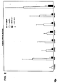
- Figure 1 shows the percent incorporation of radioactivity vs. incubation time at 37°C for both antibodies, each at 1 ⁇ g and 10 ⁇ g/well, for one tumor tissue.
- the anti-PAP antibody shows little incorporation at any time.
- the anti-CEA antibody by contrast shows about a 20 fold increased accumulation.
- the difference in radioactivity accumulation for the specific and non-specific antibodies is much less indicating saturation of the antigenic sites.
- a panel consisting of labeled anti-CEA and anti-PAP and the six anti-breast cancer tumor monoclonal antibodies was tested using two human breast tumor tissues. Replicate measurements of binding of the same-antibody in the same tissue show only small variations, while the variation in binding of the same antibody in different tissues or different antibodies in the same tissue is far larger, as shown in Figure 1.
- the antibody 113F1 showed only modest binding in one of the tumor tissues but showed the highest degree of binding in the other. Although as expected, the anti-CEA antibody showed the same degree of binding as the anti-PAP antibody in one of the breast tumor tissue, the former showed increased binding with respect to the latter in the other tumor tissue tested.
- Figure 1 (left panel) is presented the results of a repeat measurement of the same antibodies and the same tissue analyzed initially (solid bars) and three days later (cross hatched bars). Although there are slight differences in the level of uptake of individual antibodies, the order, ranked according to antibody accumulation, is unchanged.
- mice Nineteen 8 week old female nude mice were implanted with MX-1 tumors subcutaneously in the right dorsal flank. The mice were furnished with food and water ad libitum. At 14 days after implant when the subcutaneous tumors had reached a size of approximately 0.5 cm 3 , the water was replaced with water containing 0.1% KI.
- Monoclonal antibody 260F9 was labeled with 125-1 1,3,4,6Tetrachloro-3 ⁇ ,6 ⁇ -diphenyl glycouril (Iodogen") as follows. 10 ⁇ l of Iodogen" was placed in a sterile glass test tube and 1 UCi of 125-1 as NaI salt with a specific activity of about 17 Ci/mg (New England Nuclear) was added to the iodogen. Monoclonal antibody 260F9 in phosphate buffered saline, without azide was added to the Iodogen" 125-1 to label the antibody at a specific activity of 5 ⁇ Ci/ ⁇ g antibody.
- Iodogen 1,3,4,6Tetrachloro-3 ⁇ ,6 ⁇ -diphenyl glycouril
- Monoclonal antibody MOPC21 was labeled in the same manner and served as a control. Approximately 2 ⁇ g of labeled antibody containing approximately 10 wCi 1-125 was administered to each mouse. The labeled antibody was administered in a volume of approximately 0.1 ml PBS containing 1% BSA, via the mouse tail vein. Four days after administration of the labeled antibody the mice were exsanguinated by eye puncture. The blood was heparinized, centrifuged and the blood plasma was retained. The organs were disected, chopped into approximately 1 MM 3 pieces and were washed in saline to remove excess counts. The chopped tissue was weighed and the radio activity was measured in an LKB gamma counter.
- the tissues of six mice treated with iodinated MOPC21 served as controls.
- the tissues of 13 mice treated with 1-125 labeled 260F9 served as test tissues.
- Counts per minute per gram (cpm/gm) tissue or tumor were determined and an index of uptake was determined by the ratio of cpm/gm tumor to cpm/gram organ (T/0 ratio).
- Table 11 shows the T/0 ratio for 260F9 at 10 iCi 1-125 for MX-1 tumors in each animal tested.
- Table 12 shows the T/0 ratio for MOPC21 at 10 ⁇ Ci 1-125 for MX-1 tumors in each animal tested.
- Table 13 shows the mean and standard deviation for all tested animals.
- the monoclonal antibodies according to the invention after derivatization with a labeling moiety, have a number of uses.
- the immunoimaging monoclonal antibodies may be used in diagnosis of primary malignant breast tumors. Patients presenting with masses indicating a possibility of malignant breast tumors presently routinely undergo a series of diagnostic mammographic examinations.
- the immunoimaging antibodies according to the invention may be administered subcutaneously or intravenously to determine whether the mass is positive for uptake of the labeled antibody according to the invention. Accumulation of the labeled antibody would serve as an additional indication suggesting the need for a biopsy or more extensive surgical intervention.
- the labeled monoclonal antibodies according to the invention also have a clear use in assaying the clinical prognosis of patients who have had mastectomies or lumpectomies for removal of malignant breast tumors.
- the axillary lymph nodes of such patients are disected to determine the extent of dissemination of the malignancy.
- patients with positive nodes receive a course of adjuvant chemotherapy.
- Axillary node sampling is an invasive procedure requiring general anesthesia. It entails all the risks of any major surgical procedure including infection and reaction to anesthetics, and requires a significant post operative period of pain, recovery and healing.
- the monoclonal antibodies according to the invention and the derivatives thereof can be used as a non-invasive method for determining the nodal involvement of a breast malignancy and may serve as an adjunctive procedure to conventional nodal dissection or as a replacement therefor.
- radiolabeled monoclonal antibodies have been shown, at least in a preliminary manner in a number of clinical studies. McKenzie et al. "Immunoscintigraphy for Detection of Lymph Node Metastases From Breast Cancer" Lancet No. 8414:1245 (1984) have shown that subcutaneous interdigital injection of an 1-131 labeled monoclonal antibody specific for a human malignant breast tumor, can be used to confirm the presence of metastases in patients who were already suspected to have tumors involving axillary lymph nodes, and to detect tumors in lymph nodes where the presence of tumor had not been suspected.
- breast tumor localization with the labeled derivatives of monoclonal antibodies according to the invention by intravenous administration is also a clear alternative to the subcutaneous administration route.
- the radiolabeled monoclonal antibody is introduced into the patient in a solution appropriate for IV administration such as 0.15 M NaCl with 1% human serum albumin.
- the radiolabeled monoclonal antibody is injected preferably using a venous catheter in a volume of saline over an approximately 30 minute period.
- the patient is tested for allergy to the normal antibody of the animal from which the monoclonal producing hybridoma was produced.
- the monoclonal antibody is derivatized with a radioisotope of iodine
- the patient is pre-treated with Lugols iodine-solution to block thyroid uptake of 131-1, and premediated with promethazine and prednisolone before administration of the immunoimaging monoclonal antibody.
- Patients are scanned over a period of hours to days after administration of the immunimaging monoclonal antibody. Scanning methods for radioisotopic imaging including appropriate control procedures such as subtraction analysis with non- specific antibody are known to those skilled in the art of nuclear medicine and include computer assisted photoscanning and computer assisted tomoscintigraphy. 1.
- labeled monoclonal antibodies are clear to those skilled in the art of breast cancer patient management. Such uses include the use of the labeled monoclonal antibodies to monitor the response of metastatic tumors to therapy using various therapeutics including chemotherapeutics, immunotoxins, immunodrugs or lymphokines.
- the labeled monoclonal antibodies according to the invention may also be used to detect the presence of life endangering highly morbid metastases at a time prior to their symptomatic manifestation early enough to permit preventive or ameliorating radiotherapy.
- the most highly selective antibodies according to the invention may also be labeled to determine the distribution or localization or lack thereof of the monoclonal antibody in the normal tissues of patients thus providing a basis for identifying breast cancer specific monoclonal antibodies that may be advantageously used as components for antibody based therapeutics such as immunotoxins or immunodrugs for the treatment of malignant breast tumors.
- the monoclonal antibody-producing hybridomas listed below were deposited with the American Type Culture Collection (ATCC) or Invitro International Inc. (IVI) under the terms of the Budapest Treaty on the International Recognition of the Deposit of Microorganisms for the Purposes of Patent Procedure and the Regulations thereunder (Budapest Treaty). This assures maintenance of the viable culture for 30 years from date of deposit.
- the hybridomas will be made available by ATCC or IVI under the terms of the Budapest Treaty, and subject to an agreement between applicants and ATCC or IVI which assures unrestricted availablity upon issuance of the pertinent U.S. patent. Availability of the deposited strains is not to be construed as a license to practice the invention in contravention of the rights granted under the authority of any government in accordance with its patent laws.
- Each hybridoma designation listed in the left column corresponds to the monoclonal antibody producing the designated monoclonal antibody.
Abstract
Description
- This invention is in the fields of immunology and cancer diagnosis and therapy. More particularly it concerns murine monoclonal anti-human breast cancer antibodies, hybridomas that produce those antibodies, immunochemicals made from those antibodies, and diagnostic and therapeutic methods that use those immunochemicals.
- Since the mid-1970s, there have been numerous reports of murine monoclonal antibodies that interact with human breast cancer associated antigens. In these reported studies, mice were Immunized and boosted with human milk fat globule proteins, breast cancer cell lines or breast cancer membrane extracts. Immune splenocytes were fused with mouse myeloma cells and hybridomas were selected based on some specificity of the culture media for breast or breast cancer antigens. Taylor-Papadimitriou, J., et al, Int. J. Cancer (1981) 28:17-21; Yuan, D., et al, JNCI (1982) 68:719-728; Ciocca, D. R., et al, Cancer Res. (1982) 42:4256-4258. The normal tissue reactivities of these prior antibodies are different than the normal tissue reactivities of the antibodies of the present invention.
- A principal aspect of the invention concerns murine monoclonal antibodies that:
- (a) do not bind to blood cells;
- (b) have a breast tumor binding range of at least 0.25 or have a breast cancer cell line binding range of greater than or equal to 0.25;
- (c) have a selectivity equal to or less than 0.09;
- (d) have a G or M isotype, and
- (e) when conjugated to an imaging moiety, produce a signal sufficient to image breast cancer tumors.
- Preferred embodiments of -these antibodies are those designated 2G3, 9C6, 32A1, 33F8, 35E10, 41B4, 87H7, 106A10, 113F1, 120H7, 140A7, 200F9, 203E2, 219F3, 245E7, 254H9, 260F9, 266B2, 317G5, 369F10, 387H9, 421E8, 451C3, 452E12, 452F2, 454A12, 454C11, 457D7, 520C9, 650E2, 697B3, 741F8, 759E3, 788G6, and functional equivalents thereof.
- The murine x murine hybridomas that produce the above described antibodies and progeny of those hybridomas are other aspects of the invention.
- Another aspect of the invention relates to immunoimaging agents that are conjugates of
- (a) the above described monoclonal antibodies, and
- (b) a detectable imaging moiety.
- Another aspect of the invention concerns methods of imaging breast tumors in a patient in need of such imaging by administering an imaging effective amount of an immunoimaging agent and detecting the immunoimaging agent in the patient with a suitable detecting device.
- As used herein, the term "monoclonal antibody" means an antibody composition having a homogeneous antibody population. It is not intended to be limited as regards the source of the antibody or the manner in which it is made.
- As used herein with regard to the monoclonal antibody-producing hybridomas of the invention the term "progeny" is intended to include all derivatives, issue, and offspring of the parent hybridoma that produce the monoclonal anti-human breast cancer antibody produced by the parent, regardless of generation or karyotypic identity.
- As used herein with respect to the exemplified murine monoclonal anti-human breast cancer antibodies, the term "functional equivalent" means a monoclonal antibody that: (a) binds to the same l antigen or epitope as an exemplified monoclonal antibody; (b) has- a breast tumor binding range of at least 0.25 or has a breast cancer cell line range of greater than or equal to 0.25; (c) has a selectivity equal to or less than 0.09;..(d) has a G or M isotype, and (e) when conjugated to an imaging moiety, produces a signal sufficient to image breast cancer tumors.
- As described above, the term "functional equivalent" as used herein includes five criteria. The first of these criteria, binding to the same antigen or epitope as an exemplified monoclonal antibody may be demonstrated by experiments which show crossblocking of an exemplified monoclonal antibody by the functionally equivalent monoclonal antibody. Crossblocking occurs as a result of an antibody binding to the same epitope on an antigen as that bound by one of the exemplified antibodies, or as a result of an antibody binding to a different epitope which is so closely situated on the same antigen that binding of an antibody to one epitope blocks the binding of an antibody to the second epitope. Crossblocking thus is one of the criteria by which one can determine that a functionally equivalent monoclonal antibody binds to the same antigen or epitope as an exemplified monoclonal antibody.
- So-called "sandwich" assays are another method for determining whether an antibody binds the same antigen or epitope. In these assays, a first monoclonal antibody is bound to a support, for example, the surface of a titre plate well. After treatment to prevent nonspecific binding, a highly solubilized antigen preparation is added to the bound antibody. Subsequently, a second antibody, having a detectable label, for example, a fluorescent dye, is added. If the second antibody binds to the antigen, a different epitope specificity or multiple copies of the same epitope on the same antigen is indicated. If the second antibody fails to bind, either the same epitope specificity or different antigen specificity is indicated. The results of both the crossblocking and sandwich assay are further defined by a second series of tests such as immune precipitation or Western blotting to show that the antigen bound by both antibodies has the same molecular weight.
- The antibody-producing fusion partners that are used to make the hybridomas of this invention are generated by immunizing mice with live human breast cancer cells or membrane extracts made therefrom. The mice are inoculated intraperitoneally with an immunogenic amount of the cells or extract and then boosted with similar amounts of the immunogen. Spleens are collected from the immunized mice a few days after the final boost and a cell suspension is prepared therefrom for use in the fusion.
- Hybridomas are prepared from the splenocytes and a murine tumor partner using the general somatic cell hybridization technique of Kohler, B. and Milstein, C., Nature (1975) 256:495-497 as modified by Buck, D. W., et al, In Vitro (1982) 18:377-381. Available murine myeloma lines, such as those from the Salk Institute, Cell Distribution Center, San Diego, California, USA, may be used in the hybridization. Basically, the technique involves fusing the tumor cells and splenocytes using a fusogen such as polyethylene glycol. After the fusion the cells are separated from the fusion medium and grown in a selective growth medium, such as HAT medium, to eliminate unhybridized parent cells. The hybridomas are expanded, if desired, and supernatants are assayed for anti-human breast cancer activity by conventional immunoassay procedures (e.g., radioimmunoassay, enzyme immunoassay, or fluorescence immunoassay) using the immunizing agent (breast cancer cells or membrane extract) as antigen. Positive clones are characterized further to determine whether they meet the criteria of the antibodies according to the invention.
- Hybridomas that produce such antibodies may be grown in vitro or in vivo using known procedures. The monoclonal antibodies may be isolated from the culture media or body fluids, as the case may be, by conventional immunoglobulin purification procedures such as ammonium sulfate precipitation, gel electrophoresis-, dialysis, chromatography, and ultrafiltration, if desired.
- The important characteristics of the monoclonal antibodies are (1) their immunoglobulin class, (2) their selectivity for human breast cancer cells, (3) the range of human breast cancer tumor cells .to which they bind, (4) the range of human breast tumor frozen sections to which they bind, and (5) their usefulness in making effective anti-human breast cancer immunoimaging agents.
- The selectivity and range of a given antibody is determined by testing it against panels of (1) human breast cancer tumor tissues, (2) human breast cancer cell lines, and (3) normal human tissue or cells of breast or other origin. In selecting the claimed antibodies, approximately 22,000 growing hybridoma cultures were initially screened against the immunizing breast tumor membranes or cell line, a panel of seven normal tissue membranes, a fibroblast cell line and a breast tumor frozen section. Clones that reacted with the neoplastic materials, but not the normal materials, were identified in this initial screen and chosen for isotyping and additional screening for selectivity and range. The additional screening involved: sixteen normal tissue sections, five normal blood cell types, eleven nonbreast neoplasm sections, twenty-one breast cancer sections and fourteen breast cancer cell lines.
- For purposes of this patent application, specificity and selectivity are used interchangeably and are defined as the sum of the number of substructures stained in sixteen normal tissue frozen sections and the number of blood cell types bound, divided by the sum of the total number of substructures bound by any of the monoclonal antibodies in all the tissue on which the monoclonal antibodies were tested and five blood cell types tested.
- The term "tumor range" is defined as the number of breast tumor frozen sections stained divided by the number of breast tumor frozen sections tested. The term breast cancer "cell line range" is defined as the number of breast cancer cell lines stained divided by the number of breast cancer cell lines tested. Antibodies were deemed to be appropriate for breast cancer immunoimaging purposes if they have a selectivity equal to or less-than 0.09 and a breast tumor binding range of equal to or greater than 0.25 or a breast cancer ce>ll line binding range of equal to or greater than 0.25.
- Antibodies exhibiting acceptable selectivity and range may be conjugated to various imaging moieties such as radioisotopes or materials detectable by nuclear magnetic resonance imaging. In some cases a coupling agent, such as a chelating agent may be used to link the imaging agent to the antibody.
- Antibodies of five of the thirty-three deposited hybridomas were found to recognize the same 200 K dalton antigen. Antibodies of four of the thirty-three bound to a 230 K dalton intracellular antigen. Three bind to one or more high molecular weight mucins (HMW) and two bound to transferrin receptors in the form of a 97 K dalton antigen. All antigen weights mentioned herein were determined by sodium dodecyl sulfate (SDS) polyacrylamide gel electrophoresis under reducing conditions using procedures known in the art.
- Further details of the characterization of these antibodies are provided in the examples below.
- The immunochemical derivatives of the monoclonal antibodies of this invention that are of prime importance are labeled with an imaging moiety such as radioisotopes, radiopaque substances or nuclear magnetic resonance detectable materials. Such immunochemical derivatives, in which the imaging moiety provides a means for identifying immune complexes that invlude the labeled antibody may be used in imaging breast cancer tumors in vivo.
- Antibodies that exhibit either a breast cancer tumor binding range of at least 0.25 or a breast cancer cell line binding range of at least 0.25, and that also exhibit a selectivity equal to or less than 0.09 and do not bind to blood cells, were considered selective for breast cancer immunoimaging purposes, and may be conjugated to a detectable imaging moiety. Such imaging moieties may be directly bound to the monoclonal antibody or may be bound to the monoclonal antibody by means of a linking or chelating agent. Derivatives of the monoclonal antibody, labeled wth the imaging moieties, can be made by a variety of methods well known in the art. Such labeled derivatives are also referred to herein as immunoimaging agents.
- . Radioisotopes of iodine may be used to iodinate monoclonal antibodies using the solid phase oxidizing agent ,13,4,6-tetrachloro-3«,6«-diphenylglycouril (sold under the tradename Iodo-gen), or N-chloro-p-toluene sulfonamide (chloramine T).
- The term "linkers" used herein is intended to encompass chemical entities which may be bound to the imaging moiety and which also bind to the monoclonal antibody. Appropriate linkers may include those which bind to the monoclonal antibody and chelate radionuclides. Other linkers, such as those which may selectively bind to the carbohydrate carrying regions of the monoclonal antibody, or those that are capable of binding free amino side groups of the protein region of the monoclonal antibody, such as amidinating or imidinating agents, and which can be covalently linked to the imaging moiety, are also included in the scope of the term linker as used herein.
- Appropriate linkers will have three characteristics. First, they must be capable of binding the imaging moiety which has the desired characteristic to be read for imaging. Secondly, the linker must not significantly affect the binding selectivity of the monoclonal antibody or substantially diminish its affinity for the antigen to be bound. Lastly, the linker must form a stable bond with the imaging moiety and the monoclonal antibody so that the imaging moiety and antibody will not be separated from one another.
- The particular linker and imaging moiety used to make the immunoimaging agents of the present invention will vary from antibody to antibody depending on the effect that a particular imaging moiety or imaging moiety and linker may have upon the binding characteristics of the monoclonal antibody for the target antigen. Thus, while one monoclonal antibody may be iodinated with a radioisotope of iodine at tyrosine residues within the monoclonal antibody without significantly affecting affinity or selectivity of the monoclonal antibody, the same treatment of a second monoclonal antibody according to the invention may significantly diminish the affinity or binding specificity of another monoclonal antibody. A different label or linker, for example, one that binds to the antibody at a different amino acid residue, may be used without affecting selectivity or affinity in the second antibody. Thus, iodine radioisotopes can be linked to the second antibody, for example, using the method of Wood, F. T., et al. Analy. Biochem. 69:339 (1975) and the linker methyl-p-hydroxybenzimidate or the method of Bolton-Hunter , Bolton, A. E. and Hunter, W. M., Biochem. J. 133:529-539 (1973) and the linker N-succinimidyl-3-(4-hydroxyphenyl) propionate. A chelating agent such as diethylinetriaminepentaacetic acid anhydride which binds to lysine residues of the antibody, or ethylenetriaminetetraacetic acid may be used to label the antibody with 111-Indium (111-In), and could also be employed as an alternative means for linking the antibody to the imaging moiety. See for example, Goodwin, et al., "Chelate Conjugates of Monoclonal Antibodies for Imaging Lymphoid Structures in the Mouse', J. Nucl. Med. 26(5):493-502 (1985) and Meares et al., "Conjugation of Antibodies With Bifunctional Chelating Agents Bearing Isothiocyanate or Bromoacetamide Groups and Subsequent Additon of Metal Ions", Analy. Biochem. 142:68-78 (1984).
- Various moieties suitable for imaging are known. For example, monoclonal antibodies have been radiolabeled with a number of radionuclides suitable for imaging, including 131-iodine (1-131) and 1-123. Levin et al., "Localization of 1-131 Labeled Tumor Bearing Balb/c Mouse", J. Nuclear Medicine, 21:570-572 (1980); Farrands et al., "Radioimmunodetection of Human Colorectal Cancers by an AntiTumor Monoclonal Antibody", Lancet 397-399 (1982); Zimmer et al., "Radioimmunoimaging of Human Small Cell Lung Carcinoma With 1-131 Tumor Specific Monoclonal Antibody", Hybridoma, 4(1):1-11 (1985). The direct labeling of the monoclonal antibody with radioisotopes of iodine can be carried out according to the methods described in Contreras et al, Methods in. Enzymology (1973) 97:277.- Technetium-99 has been used as an imaging moiety; Khaw et al., "Monoclonal Antibody to Cardiac Myosin:Imaging of Experimental Myocardial Infraction", Hybridoma 3:11-23 (1984). 111-In has been applied as a label for antibodies, Krejack et al., "Covalent Attachment of Chelating Groups to Macromolecules", Biochem. Biophys. Res. Comm., 77:581-585 (1977); Hnatowich et al., "Radioactive Labeling of Antibody A Simple and Efficient Method", Science, 220:613-615 (1983) and Schienberg, D. A. et al., "Tumor Imaging With Radioactive Metal Chelates Conjugated to Monoclonal Antibodies", Science, 215:1511-1513 (1982).
- In order to initially assess the suitability of the antibody as one appropriate for imaging, the antibody may be labeled with a moiety that is directly detectable such as fluorochromes, as well as moieties, such as enzymes, that must be reacted or derivatized to be detected. Examples of such labels are fluorescein and its derivatives, rhodamine and its derivatives, dansyl groups, umbelliferone, luciferin, 2,3-dihydrophthalazinediones, horseradish peroxidase, alkaline phosphatase, lysozyme, and glucose-6-phosphate dehydrogenase. The antibodies may be tagged with such labels by known methods. For instance, coupling agents such as aldehydes, carbodiimides, dimaleimide, imidates, succinimides, bis-diazotized benzidine and the like may be used to tag the antibodies with the above-described fluorescent, chemiluminescent, and enzyme labels.
- The antibodies and labeled antibody may be used in a variety of immunoimaging or immunoassay procedures to detect the presence of breast cancer in a patient or monitor the status of such cancer in a patient already diagnosed to have it. When used for in vivo immunoimaging to detect the presence of a tumor, its location and dissemination in a patient's body and the progress of therapy to ameliorate the tumor load, the monoclonal antibody labeled with an imaging moiety will be administered parenterally, preferably intravenously or subcutaneously in an amount sufficient to accumulate at the tumor site and be detected by the detecting means of choice. Typically the monoclonal antibody labeled with an imaging moiety will be administered with a suitable pharmaceutically acceptable carrier of the type well known to those skilled in the art. Such carriers do not affect the patient. The amount of monoclonal antibody to be administered will depend upon the amount of detectable imaging moiety attached to the monoclonal antibody and the residual binding efficiency of the monoclonal antibody after labeling with the imaging moiety.
- The residual binding efficiency of the monoclonal antibody labeled with the imaging moiety is determined in vitro using a tumor cell binding assay. Generally, radioimmunoreactivity, which measures the residual binding efficiency of a radioisotope-labeled monoclonal antibody is determined by comparing specific binding of the radioisotope-labeled monoclonal to a fixed tissue culture of a known immunoreactive tumor cell line such as SKBR-3, MCF-7, and MX-1 with non-specific binding to a fixed cell line which does not specifically bind the monoclonal.
- The optimal radioimmunoreactivity of the labeled monoclonal is determined in this system by varying the concentration of the imaging agent available for binding to the monoclonal antibody while keeping the concentration of the monoclonal antibody constant. The labeled monoclonals are then tested in the fixed cell immunoassay described above by adding the labeled monoclonal to the fixed cells at conditions of antigen excess. The labeled monoclonal giving the highest detectable binding is determined and can be used initially for in vivo radioimmunoimaging.
- When an in vitro immunoassay is used to monitor the status of a cancer patient, a quantitative immunoassay procedure must be used. In such monitoring, assays are carried out periodically and the results compared to determine whether the patient's tumor burden has increased or decreased. Common assay techniques that may be used include direct and indirect assays. Direct assays involve incubating a tissue sample or cells from the patient with a labeled antibody. If the sample includes breast cancer cells, the labeled antibody will bind to those cells. After washing the tissue or cells to remove unbound labeled antibody, the tissue sample is read for the presence of labeled immune complexes. In indirect assays the tissue or cell sample is incubated with unlabeled monoclonal antibody. The sample is then treated with a labeled antibody against the monoclonal antibody (e.g., a labeled antimurine antibody), washed, and read for the presence of labeled ternary complexes.
- For in vitro diagnostic use the antibodies will typically be distributed in kit form. These kits will typically comprise: the antibody in labeled or unlabeled form in suitable containers, reagents for the incubations and washings, a labeled antimurine antibody if the kit is for an indirect assay, and substrates or derivatizing agents depending on the nature of the label. For in vivo imaging use the antibody will also be distributed in kit form and will typically comprise the same types of components as mentioned above. The antibody may be supplied derivatized with an agent already bound to or chelated with the radioisotope to be used, or the monoclonal may be supplied derivatized with the binding or chelating agent only, and the radioisotope to be used may be supplied separately. The radioisotope to be used can be added just prior to use so that an optimal radioactivity level for imaging can be achieved at the time of administration of the radioimmunoimaging agent to the patient. Human breast cancer antigen controls and instructions may also be included if appropriate to the test.
- The following examples provide a detailed description of the preparation, characterization, and use of representative monoclonal antibodies of this invention. These examples are not intended to limit the invention in any manner.
- Fresh postsurgical human breast cancer tissue and a variety of normal tissues were used to prepare membrane extracts by homogenization and discontinuous sucrose gradient centrifugation. Human breast cancer cell lines were obtained from the Breast Cancer Task Force, the American Type Culture Collection (ATCC), and from Dr. Jorgen Fogh at Memorial Sloan Kettering. The cells were maintained and passaged as recommended by the Breast Cancer Task Force, the ATCC and Dr. Fogh. For immunizations, either membrane extract containing 100 µg of protein (Lowry assay) or ten million live breast cancer cells were inoculated intra-peritoneally into five week old Balb/c mice. The mice were boosted identically twice at monthly intervals. Three days after the last boost, the spleens were removed for cell fusion.
- Somatic cell hybrids were prepared by the method of Buck, D. W., et al, supra, using the murine myeloma line Sp-2/0/Agl4. All hybrodima cell lines were cloned by limiting dilution. Half of the fusions employed splenocytes from mice immunized with breast cancer membrane extracts and half used splenocytes from mice immunized with live breast cancer cell lines. Eighty-three thousand four hundred twenty-four wells were generated from those fusions, of which 22,459 exhibited hybridoma growth.
- Hybridoma supernatant was assayed for reactive antibody in either a solid phase enzyme-linked immunosorbent assay (ELISA) with the immunizing breast cancer membrane extract or an indirect immunofluorescence assay with the immunizing breast cancer cell line. For the solid phase membrane ELISA, 40 µl of 0.1 mg/ml breast cancer membrane protein were placed in polyvinyl chloride (PVC) microtiter wells for 12 hours at 4°C. The extract was aspirated and the wells washed with phosphate buffered saline (PBS) containing 1% bovine serum albumin (BSA). The wells were then incubated with 45 µl of a 1:10 dilution of hybridoma supernatant. The diluent was media with 25 mM of a buffer, 10% bovine serum, and 0.1% sodium azide. After 30 minutes at room temperature, the wells were again washed and incubated 45 minutes at 37°C with a 1:200 dilution of peroxidase conjugated goat anti-mouse IgG. The diluent was PBS. The wells were then washed with PBS and reacted with 200 ill of 1,2-azino-di(3-ethylbenzthiazoline sulphonic acid) in 0.1 M sodium citrate buffer pH 4.2 for 30 minutes at room temperature. OpticaT density was measured at 405 nm on a MicroElisa Reader. For each experiment a positive control, anti-beta 2 microglobulin at 5-µg/ml, was reacted with normal human kidney membrane. This gave an optical density of 1.0 ± 0.1 (standard deviation). The background was 0 ± 0.1 optical density units (O.D.) using media without mouse monoclonal antibody. Well s that gave a reaction on the breast cancer membrane extract of greater than 0.7 O.D. were saved.
- For the indirect immunofluorescence cell line assay 100,000 breast cancer cells of the immunizing cell line were placed overnight with appropriate media in each chamber of a set of eight chambered slides. Similarly, 100,000 fibroblast cells from cell line CC95 were incubated overnight in chambered slide wells. The cells were washed with PBS containing 1% BSA. The wells, both breast cancer and fibroblast, were incubated for 30 minutes at 4°C with 1:10 dilutions of hybridoma supernatant. The cells were again washed and incubated 30 minutes at 4°C with a 1:50 dilution of fluorescein isothiocyanate (FITC)-conjugated goat F(ab')2 anti-mouse Ig. The cells were washed three times, fixed in 1.5% formaldehyde in PBS for five minutes, chambers removed and rinsed in PBS. The slides were then mounted in a composition containing polyvinyl alcohol, glycerol, buffers and a preservative and examined with a fluorescence microscope. Hybridoma wells showing strong fluorescent binding to the breast cancer cells but no fluorescent binding to fibroblasts were saved. Five thousand one hundred fifty-six hybridoma wells revealed breast cancer reactivity in the initial screen.
- Supernatants from the 5156 positive wells were then tested in solid phase ELISA with seven normal tissue membrane extracts (liver, lung, colon, stomach, kidney, tonsil, and spleen). Any well supernatant giving an ELISA O.D. greater than 0.3 was discarded. One thousand one hundred one of the supernatants were found to be unreactive with the normal tissue extracts.
- The 1101 hybridoma supernatants were tested on frozen sections of human breast carcinoma tissues. Six micron sections were attached to slides, fixed 10 minutes in acetone at 4°C, dried 10 minutes at room temperature, washed with PBS, blocked with horse serum and incubated 20 minutes at room temperature with 100 µl neat hybridoma supernatant. The slides were washed with PBS, and finally incubated 20 minutes at 37°C with a 1:50 dilution of peroxidase conjugated rabbit anti-mouse Ig, washed again with PBS, and finally incubated 7.5 minutes at 37°C with 0.5 mg/ml diaminobenzidine in 0.05 M Tris buffer pH 7.2 containing 0.01% hydrogen peroxide. The slides were stained with hematoxylin, dehydrated and mounted in a medium containing 35.9% methyl/n-butylmethacrylate copolymer, 7.1% butyl benzyl phthalate, and 0.3% 2,6-ditertbutyl-p-cresol. One hundred twenty-four wells yielded breast cancer selective binding and were cloned.
- Immunoglobulin class and subclass of the monoclonal breast cancer selective antibodies were determined by an immunodot assay essentially the same as that described in McDougal et al. J. Immunol. Meth. 63:281-290 (1983). Antibodies were also internally labeled by growing 2-3 x 10⁶ hybridoma cells for four hours in methionine-free medium containing 0.2 pCi 35S methionine. 35S-labeled antibodies were immunoprecipitated with fixed staphylococcus A cells, or with fixed staphylococcus A cells precoated with rabbit anti-mouse immunoglobulin, and the immunoprecipitates were analyzed by SDS-PAGE to determine antibody light and heavy chain mobility, lack of extra chains, and the ability of each antibody to bind staphylococcal protein A.
- The antibodies were expanded in vivo. Balb/c or F1 (C57B/6 x Balb/c) mice were primed with 0.5 ml pristane intraperitoneally (ip) and after 10-14 days inoculated with one million log phase hybridoma cells in PBS. Ascites fluid was stored at -70°C and thawed and filtered through a 0.8 micron filter unit before further purification.
- Some IgG antibodies that bound staphylococcal protein A were purified by affinity chromatography on protein A-chromatographic resin containing either agarose, dextran and/or acrylamide with pH step gradient elution. IgG antibodies that did not bind protein A were precipitated by addition of ammonium suffate to 40% saturation at 0°C. or by binding to DEAE or Affigel" (Biorad, Richmond, California). Alternatively, IgG antibodies are purified by chromatography using a Sephacryl S-200 column, followed by DEAE cellulose as described. The precipitates were redissolved in PBS, dialysed to 20 nM Tris pH 7.2 and chromatographed on a 1.6 x 50 cm column of diethylaminoethyl cellulose (DEAE) eluting with a 1.5 liter 0-600 mM NaCl gradient at 4°C at a flow rate of 1 ml/min. In each case, column fractions were monitored by SDS-PAGE and the purest antibody fractions were pooled, concentrated to 1-3 mg/ml, dialysed to PBS/0.02% NaN3, and stored at 4°C.
- IgM antibodies were purified by gel filtration material on a 2.6 x 40 cm column of Sephacryl S-300 or other gel filtration or resin containing agarose, dextran and/or acrylamide, eluting with PBS/0.01% sodium azide at room temperature at a flow rate of 1 ml/min.
- In order to evaluate their selectivity for breast cancer, the purified antibodies were tested by immunoperoxidase section staining on sections of sixteen normal tissues, and by immunofluorescent cell sorting on five blood cell types. Immunoperoxidase staining was performed as above except that known dilutions of purified antibodies in PBS in the range of 1-40 wg/ml were used instead of hybridoma supernatants. The pure antibodies were first titrated to find the minimal concentration giving strong immunoperoxidase staining on breast cancer sections and then used at the concentration for the normal tissue tests. Peripheral bood cells (platelets, lymphocytes, red blood cells, granulocytes, and monocytes) were prepared by centrifugation using a medium which separates monocytes from polymorphonuclear leucocytes. The cells were reacted with antibody at the optimal concentration determined above for 30 minutes at 4°C, washed, reacted with a 1:50 dilution of fluorescein isothiocyanate-conjugated goat anti-mouse Ig for 30 minutes at 4°C, washed again and examined in a cell sorter. The wash buffer and diluents were PBS with 1% gelatin and 0.02% sodium azide. The cell sorter was equipped with a 76 micron nezzle and a one watt argon ion laser at 488 nm. An 80 mm confocal lens was used on the optical rail assembly for focusing. Other filters used were a 515 nm interference filter and a 515 nm absorbance filter (for scattered laser light) and a neutral density 1.5 filter for forward angle light scatter. Contour plots of log fluorescein fluorescence versus forward angle light scatter were used for sample analysis. No blood cell types showed detectable binding.
- The binding behaviors of the claimed antibodies are reported in Table I below. The following abbreviations are used to denote structures bound by the antibodies: Ac, acini; G, glands; T, tubules; D, ducts; L, lumen; W, sweat glands; E, epithelium; S, sebaceous glands; Gr, granulocytes; Mk, megakaryocytes; M, macrophage; Ly, lymphocytes; Bl, Basal layer; Fe, focal epithelium; A, alveolar lining cells; B, Bowman's capsule; Mu, muscle; and I, islets; H, hair follicles; U, glomeruli; and V, vessels/endothelial.

- In order to determine how wide a range of breast cancers might be recognized by each antibody, the breast cancer selective antibodies were tested by immunoperoxidase staining on frozen section of 27 different breast tumors. The breast cancers used for section staining were all infiltrating intraductal carcinomas, so no correlation of antibody binding with histologic type of breast cancer could be made. In addition, no correlation between antibody binding and the nodal status or estrogen receptor status was found for the twelve tumors for which donor information was available. Antibodies reacted equally well with metastatic and primary breast tumors. The results of these tests for the claimed antibodies are reported in Table 2 below.


- Antibodies were further evaluated for range of breast cancer cell line recognition by immunofluorescence assays on 14 breast cancer cell lines. Table 3 below reports the results of these tests for the claimed antibodies.

- Finally, the antibodies were tested by immunoperoxidase staining on eleven non-breast malignancies. The results for the claimed antibodies are reported in Table 4 below.

- The tumor breast cancer range, breast cancer cell binding range, blood cell binding and selectivity characteristics for the monoclonal antibodies according to the invention are summarized in Table 5.

- Several of the claimed antibodies were iodinated and tested for binding to MCF-7, CAMA1, SKBR3 or ZR7530 cells. The antibodies were labeled with 125I using chloramine T to a specific activity of approximately 10 µCi/µg. To determine immunoradiochemical purity, 100,000 cpm of two of the labeled antibodies in 0.5 ml fetal calf serum was serially absorbed with five aliquots of target cells for 15 minutes at 0°C (generally 4,000,000 cells per aliquot), and the remaining radioactivity in the supernatant after each absorption was determined.
- For measurements of association constants known concentrations of labeled and unlabeled monoclonal antibodies were incubated with target cells in fetal calf serum for 15 minutes in ice. Aliquots of the cell/antibody mix were then counted in a gamma counter or filtered through Microfold filter plates (V & P Scientific) and the filters counted. To account for unbound antibody retained in liquid on the filters, controls containing the same concentrations of antibody but no cells were done in parallel. Association constants and antigen copy number per target are calculated from the affinity test results and are reported in Table 6 below.

- In order to identify the -antigens recognized by the monoclonal antibodies according to the invention, immunoprecipitation of the antgiens was carried -out according to the following method. Eight nm diameter polystyrene balls (Precision Plastic Ball Co.) were covered with 10% fuming nitric acid in glacial acetic acid and were incubated for three hours in a 50°C water bath. Following the acid treatment, the balls were rinsed three times with distilled water, covered with 1% sodium dithionite in 0.1 M NaOH and incubated three hours in a 50°C water bath. The balls were again rinsed three times with distilled water, covered with 0.1% 1-ethyl-3-(3-dimethylaminopropyl)-carbodiimide (EDAC), 0.2% suberic acid (suberic acid dissolved in dimethylfomamide) and incubated overnight at room temperature. The balls were rinsed three times with distilled water, and marked for identification.
- Purified monoclonal antibodies were diluted 0.2 mg/ml in 2-(N-morpholino)ethane sulfonic acid buffer, and the previously treated and marked polystyrene balls were placed in individual tubes and covered with 450 microliters diluted antibody and 50 microliters of fresh 1% EDAC. Tubes were capped and incubated at 25°C for 24 hours. Following this incubation, the balls were rinsed twice with PBS and were either used fresh or were stored for several days at 4°C before use.
- Freshly labeled target cell extracts were prepared from human breast cancer cell lines labeled with 125-1 by the lactoperoxidase method of Marchalonis, J., "An Enzymic Method for the Trace Iodination of Immunoglobulins and other Proteins", Biochem. J. 113:299-305 (1969), or with 35-S by growth in 35-S methionine. The labeled cells were dissolved in solubilization buffer (1% (v/v) Triton X-100, 150 mM NaCl, 5 MM EDTA, 25 mM Tris-HC1, pH 7.5). Four parts of labeled extract were mixed in a vessel with one part solubilization buffer containing 50 mg/ml bovine serum albumin, to give a final concentration of 10 mg/ml BSA. The balls coated with monoclonal antibody were added to the. vessel and were incubated four hours on ice with shaking. Labeled antigen was pipetted from the vessel and the balls-were rinsed four times with solubilization buffer. The balls were then removed, placed in individual tubes with 100 microliter Laemmli SDS gel sample buffer, and were incubated three minutes in boiling water. The balls. were removed and the samples were run on an SDS gel with appropriate standards.
- Immunoprecipitation tests on the antibodies indicated that five of them (454C11, 452F2, 520C9, 741F8, and 759E3) all bind a monomeric protein of about 200 K daltons found in cancerous breast tissue. Two of the five (520C9 and 741F8), are believed to recognize the same epitope on the 2Q0 K dalton protein. 454C11 and 759E3 bind a second epitope on the same antigen, and 452F2 binds a third epitope on the same antigen. Four of the antibodies (4184, 87H7, 452E12, 457D7) bound to a 230,000 dalton intracellular antigen. Seven antibodies (2G3, 200F9, 203E2, 245E7, 369F10, 697B3 and 788G6) bound to high molecular weight mucins (HMW). Two antibodies (51C3 and 454A12) bound to transferrin receptors in the form of a 97,000 dalton antigen. Neither 451C3 nor 454A12 blocked binding of transferrin to the receptor. The antigen binding characteristics of the monoclonal antibodies according to the invention are summarized in Table 7.

- Antibody isotype was determined as follows: A grid of 5-mm squares is lightly drawn in pencil on the nitrocellulose sheet and 1- ml droplets of antiisotype sera (Litton Bionetics, Kensington, Maryland, rabbit antisera to mouse K, λ, a, γ1, Y2a, γ2b, Y3, and µ chains) are applied so that each row of squares receives one spot of each heavy and light chain reagent. The sheet is incubated one hour at room temperature in a moist chamber, rinsed quickly in PBS-BSA, containing 1% (w/v), and left overnight in PBS-BSA at 4°C. Strips are cut apart with a scissors and may be stored at 4°C in PBS-BSA containing 0.02% sodium azide. Alternatively, strips may be air-dried and stored desiccated at 4°C. A series of small tubes is prepared containing 3 ml hybridoma culture supernatant or supernatant diluted with PBS-BSA. 1:10 dilutions are generally successful; and some supernatants can be diluted as much as 1:200. A nitrocellulose strip is incubated in each tube for one hour at room temperature. The strips are rinsed three times in PBS-BSA and incubated for one hour at room temperature in diluted rabbit anti-mouse-horseradish peroxidase. The strips are rinsed twice in PBS-BSA and twice in Tris buffer. The strips are placed in Tris buffer containing diaminobenzidine and hydrogen peroxide until sufficient color develops on the anti-isotype spots (usually 3-4 minutes). The antibody isotypes are indicated in Table 8.

- Samples of the hybridomas that produce the claimed monoclonal antibodies are deposited in the Collection of In Vitro International, 7885 Jackson Road, Suite 4, Ann Arbor, Michigan 48103, USA.
- This example shows one method for the labeling of antibodies according to the invention with radioisotopes of iodine, either 125- iodine or 131-iodine using a method known to iodinate tyrosine residues.
- Monoclonal antibodies according to the invention may be labeled by the following micro method: 0.1 milligram of the purified monoclonal antibody is labeled with 10 millicurie amounts of 125 iodine as follows: A one inch, 21 gauge needle is inserted partially through the septum of a 3 ml vial and a 3.0 ml disposable syringe barrel packed with glass wool is attached to the needle. The monoclonal antibody in 0.1 N NaCl preferably not exceeding 0.2 ml in volume, is added with a tuberculin syringe equipped with a 20 gauge needle that has been prerinsed with borate buffer. The sodium 125- iodine solution preferably not exceeding 0.2 to 0.3 ml in volume is added with a syringe attached to an 18 gauge needle pre-rinsed with buffer. The mixture is agitated briefly to mix the protein and 125 iodine solutions. Final dilution of the iodine chloride is made by mixing 0.2 ml of 125-iodine chloride at approximately 1.25 x 10⁻⁵ molar (M) with a specific activity of 10 millicuries per mole. After approximately one minute, an excess of 6.25% solution of human serum albumin or animal albumin such as bovine serum albumin is added to the solution. The labeled antibody is passed through an appropriate column to remove unbound radioactive iodine; an ion exchange resin or gel filtration medium such as Sephadex G-25 may be used. For a Sephadex column purification, after passage of the labeled antibody through the resin at the rate of about one ml per minute,-the resin is rinsed with an additional 1 to 1.5 mls of the above-mentioned human albumin solution.
- The monoclonal antibodies according to the invention may also be iodinated by linking agents. This example describes the radioactive labeling of the monoclonal antibody with an iodinated imidination reagent. The imido ester methylparahydroxybenzimidate HCL (MPHBIM) is synthesized according to the method described by Wood et al., "The Radioactive Labeling of Protein With an Iodinated Imidination Reagent", Analytical Biochem. 69:339-349 (1975). Th,e MPHBIM is iodinated as follows: 3.7 ml of MPHBIM is dissolved in 1 ml of 50 millimolar mM sodium borate buffer pH 8.5 to obtain a 20 mM MPHBIM stock solution. 1.0 ml of 40 mM sodium iodine followed by 10 microliters of sodium iodide-125 solution having a specific activity of approximately 2 millicuries per mole is added to one ml of the MPHBIM stock solution. One ml of 40 millimolar chloramine T is added with rapid mixing. The mixture is kept for approximately 15 minutes at 20-22°C and then 0.1 ml of 1.0 molar P-mercaptoethanol is added to reduce the chloramine T and residual iodine. The pH of the solution is subsequently lowered toward neutrality by adding 20 microliters of 1.0 molar acetic acid and a floculant white precipitate forms. Unreacted MPHBIM iodide and chloramine T remain soluble. The precipitate of the iodinated amino ester is collected by centrifugation at 10,000 rpm for five minutes, dissolved in two mls of 50 mM sodium borate buffer, pH 8.5 at 37°C. Iodination of the antibody is carried out as follows: Twenty milligrams of purified antibody is suspended in one ml of 4 mM iodinated linker, 50 mM sodium borate buffer at pH 9.5. The reaction is carried out at 37°C for a period of time sufficient to achieve the desired amount of binding. Under these conditions the radioactive label is incorporated onto the antibody at a rate of about 1-2% per hour with a maximum incorporation of approximately 30% of the iodine-125 label. Unreacted linker may be removed by dialysis against 0.15 molar sodium chloride containing a 5 mM sodium phosphate at a pH 7.4.
- The monoclonal antibodies may be labeled with 111-In using the chelating agent diethylenetriaminepentaacetic acid (DTPA) anhydride according to the method of Hnatowich et al., Science 220:613-615 (1983) which is herein incorporated by reference. Antibody 113F1, is prepared at 11 milligrams per ml dialysed into NaHC03 at pH 7. One mg DTPA cyclic anhydride was dissolved in 10 ml CHC13. Forty µl of this solution was delivered into 5 m/glass test tube and the CHC13 was evaporated with a stream of N2. One-hundred microliters of 113F1 (1.1 milligrams protein) was added to the tube containing 4 micrograms of anhydride and the tube was vortexed briefly. After one minute, 5 microliters of 111-In (having a specific activity of about 3.28 x 10¹⁰ cpm/ml) in 0.5 molar sodium acetate at pH 5.8 was added. Two PD10 (Pharmacia) columns were prepared with 20 milliliters of phosphate buffered
saline 1% bovine serum albumin. The samples were run on the PD10 columns with 2.2 milliliters of void volume. A 2.5 milliliter protein peak and 2.5 milliliters small molecule peak were found eluting with thePBS 1% BSA. The control was a 100 microliter sample of 113F1 together with 4 microliters of one microgram/microliter DTPA, not the anhydride, and 5 microliters of 111-In. The DTPA anhydride labeled 113F1 protein peak contained 75% of the counts and the small molecule peak/fraction contained approximately 25% of the counts. In the control approximately 92% of the counts remained in the small molecule fraction. - Monoclonal antibody 113F1 from the previous experiment was diluted in 50 millimolar NaHC03 pH 7 to concentrations of 100, 10 and 1 micrograms per 100 wl. One hundred µl of each dilution of 113F1 was added to 4 µg DTPA anhydride in a glass tube as in Example III. 111-In was diluted in 0.5 molar sodium acetate pH 5.8 to 100 microcuries per 10 µl. One hundred microcuries 111-In in 10 microliters solution was added to the tubes containing the antibody. The mixture was treated as in Example III. Seventy-six percent of the counts were found in the protein fraction of the one microgram per 100 microliter dilution and 86% of the counts were found in the 100 microgram per 100 microliter dilution.
- This example shows the labeling of another antibody according to the invention with 111-In.
- 1 mg DTPA cyclic anhydride was dissolved in 10 mls dry CHC13. 40 µl of the dissolved DTPA anhydride was placed in a 5 ml glass tube and was evaporated with N2 to yield about 4 µg DTPA cyclic anhydride coated on the inside of the tube.
- 100 µl of antibody 245E7 at a concentration of 15 mg/ml in 50 mM NaHC03 pH 7 was added to the DTPA cyclic anhydride. The tube was briefly vortexed and left to stand for about one minute forming the DTPA-245E7 complex.
- Five test samples were made up as follows:
- (1) 10 µl DTPA-245E7 complex in 90 µl NaHC03
- (2) 10 wl DTPA-245E7 complex in 90 µl NaHC03
- (3) 10 µl 245-E7 in 90 µl NaHC03
- (4) 10 µl 245E7 in 90 µl NaHC03
- (5) 10 µl 245E7 in 90 µl NaHC03 and 0.4 µg DTPA not anhydride form.
- 111-In was diluted to a specific activity of 100 cpm/µl in 0.5 sodium acetate pH 5.8. 10 ml of this 111-In solution was added to each tube. 10 µg DTPA in the non-anhydride form was added to
tubes 2 and 4. The contents of each tube were run on a PD10 column equilibrated with NaHC03 as in Example III. Samples were collected and the protein and small molecule peaks were counted in a liquid scintillation counter using conventional methods. The results are shown in Table 9.
- The results indicate that 77% of the 111-In bound to DTPA labeled 245E7. Subsequent addition of excess free DTPA does not remove indium from the DTPA-245E7 antibody complex. Sample 3 shows that indium does not non-specifically bind to the antibody in any appreciable amount; however, the indium appears to be retained on the column rather than eluting with the small molecule fraction. DTPA added before or after indium results in the indium eluting in the small molecule peak.
- This example shows that 111-In labeled monoclonal antibodies are efficiently bound by human breast tumor tissues.
- Six anti-breast cancer tumor monoclonal antibodies 113F1, 245E7, 260F9, 280D11, 2G3, 266B2, and a negative control MOPC21, were covalently linked to DTPA anhydride by the method described in Example III above. The antibody-DTPA complex was radiolabeled by chelation with 111-In at a specific activity of about 1 µ Ci/µg. The 111-In labeled antibody was purified when necessary on a 0.4 x 17 cm column of Sephadex G50 to a radiochemical purity of about 90%. Two non- breast specific antibodies, anti-carcinoembryonic antigen monoclonal antibody (anti-CEA) obtained from Medi-Physics, Emeryville, California and anti-prostatic acid phosphatase antibody (anti-PAP) obtained from New England Nuclear Corporation, Boston, Massachusetts) were labeled in the same manner as the anti-breast cancer tumor antibodies and served as positive binding controls. Human breast and colorectal tumor tissues were obtained immediately after surgery and were placed in fresh Eagles Minimal Essential Media (MEM) supplemented with 10% fetal calf serum, non-essential amino acids, glutamine, penicillin and streptomycin (MEM) for transportation. Fresh tissue was used within three hours of receipt while cryopreserved tissue was maintained in MEM at -70°C. The tissues were sectioned manually with surgical blades into 1.0 ± 00.2 mm cubes and checked for size accuracy using an ocular micrometer. Using sterile techniques, the tissue cubes were transferred to a 96-well microtiter plate containing 200 µl of MEM and either 1.0 or 10 µg of 111-In-labeled antibody. The tissues were incubated from 1-24 hours at 37°C in a 5% C02 water-jacketed incubator. Following incubation, the media was carefully removed using an automatic pipetter with minimal disruption of the tissue, and fresh media was added. Following incubation for an additional 20 minutes, the media was again replaced and the tissue was incubated for another 20 minutes. After this last wash, the media was removed and the tissue transferred to a dry tared weighing paper. Tissues were dried at 70°C for 20 minutes and were then weighed and placed in test tubes for counting in a NaI well counter. The results are reported as the percent of applied radioactivity in tissue per unit weight of dried tissue. Small differences in the size of each tissue cube were corrected.
- To establish thatthe accumulation of radiolabeled antibody in tumor tissue is due tcP specific binding rather than non-specific adsorption, radiolabeled anti-CEA and anti-PAP antibodies, used as controls, were incubated with fresh and cryopreserved human colorectal tumor tissue which expresses CEA. Figure 1 shows the percent incorporation of radioactivity vs. incubation time at 37°C for both antibodies, each at 1 µg and 10 µg/well, for one tumor tissue.
- At the 1 µg/well concentration, the anti-PAP antibody shows little incorporation at any time. The anti-CEA antibody by contrast shows about a 20 fold increased accumulation. At 10 µg/well, the difference in radioactivity accumulation for the specific and non- specific antibodies is much less indicating saturation of the antigenic sites.
- Specific binding of the anti-CEA control antibody was further demonstrated by a competitive binding study. Tumor tissue was preincubated with saturating levels (25 µg) of unlabeled anti-CEA antibody for 17 hours prior to the normal assay using 1 wg of labeled anti-CEA antibody. Control wells did not receive the unlabeled antibody. As shown in Table 10, in the case of the non-specific antibody, there was essentially no change in tissue accumulation of radioactivity with preincubation whereas in the case of the specific antibody, a large decrease in accumulation occurred in the tissue preincubated with the unlabeled anti-CEA antibody.

- A panel consisting of labeled anti-CEA and anti-PAP and the six anti-breast cancer tumor monoclonal antibodies was tested using two human breast tumor tissues. Replicate measurements of binding of the same-antibody in the same tissue show only small variations, while the variation in binding of the same antibody in different tissues or different antibodies in the same tissue is far larger, as shown in Figure 1.
- The antibody 113F1 showed only modest binding in one of the tumor tissues but showed the highest degree of binding in the other. Although as expected, the anti-CEA antibody showed the same degree of binding as the anti-PAP antibody in one of the breast tumor tissue, the former showed increased binding with respect to the latter in the other tumor tissue tested.
- Also in Figure 1 (left panel) is presented the results of a repeat measurement of the same antibodies and the same tissue analyzed initially (solid bars) and three days later (cross hatched bars). Although there are slight differences in the level of uptake of individual antibodies, the order, ranked according to antibody accumulation, is unchanged.
- Nineteen 8 week old female nude mice were implanted with MX-1 tumors subcutaneously in the right dorsal flank. The mice were furnished with food and water ad libitum. At 14 days after implant when the subcutaneous tumors had reached a size of approximately 0.5 cm3, the water was replaced with water containing 0.1% KI.
- Monoclonal antibody 260F9 was labeled with 125-1 1,3,4,6Tetrachloro-3α,6α-diphenyl glycouril (Iodogen") as follows. 10 µl of Iodogen" was placed in a sterile glass test tube and 1 UCi of 125-1 as NaI salt with a specific activity of about 17 Ci/mg (New England Nuclear) was added to the iodogen. Monoclonal antibody 260F9 in phosphate buffered saline, without azide was added to the Iodogen" 125-1 to label the antibody at a specific activity of 5 µCi/µg antibody.
- Monoclonal antibody MOPC21 was labeled in the same manner and served as a control. Approximately 2 µg of labeled antibody containing approximately 10 wCi 1-125 was administered to each mouse. The labeled antibody was administered in a volume of approximately 0.1 ml PBS containing 1% BSA, via the mouse tail vein. Four days after administration of the labeled antibody the mice were exsanguinated by eye puncture. The blood was heparinized, centrifuged and the blood plasma was retained. The organs were disected, chopped into approximately 1 MM3 pieces and were washed in saline to remove excess counts. The chopped tissue was weighed and the radio activity was measured in an LKB gamma counter.
- The tissues of six mice treated with iodinated MOPC21 served as controls. The tissues of 13 mice treated with 1-125 labeled 260F9 served as test tissues. Counts per minute per gram (cpm/gm) tissue or tumor were determined and an index of uptake was determined by the ratio of cpm/gm tumor to cpm/gram organ (T/0 ratio).
- Table 11 shows the T/0 ratio for 260F9 at 10 iCi 1-125 for MX-1 tumors in each animal tested. Table 12 shows the T/0 ratio for MOPC21 at 10 µCi 1-125 for MX-1 tumors in each animal tested. Table 13 shows the mean and standard deviation for all tested animals.


- Prior to exsanguination two mice treated with labeled 260F9 and one mouse treated with labeled MOPC21 according to Example VII were imaged using a Searle Pho-gamma camera with a pinhole collimnator. Raw data were collected and computor enhanced. The images of the treated and control mice are shown in Figure 3.
- In the first line of Figure 3, from left to right, are images of a tumor bearing mouse treated with 125-1-labeled 260F9 prior to surgical removal of the tumor, raw data; a computor-enhanced image of the same mouse; an image of of the mouse, post surgical, raw data; and a computer-enhanced image of the same mouse, post surgical. A prominent area of localization of detectable radiation is found in a position corresponding to the MX-1 tumor, on the dorsal right flank of the mouse treated with 125-I-labeled 260F9.
- In the second line of Figure 3 from left to right are images of a tumor bearing mouse treated with 125-1 labeled MOPC21, an antibody not specific for the tumor used as a control, raw data, and a computor enhanced image of the same mouse. There is no corresponding area of localization of detectable radiation on the dorsal right flank as in the mouse treated with 125-1 labeled 260F9. Furthermore, the distribution of label in the control mouse appears to correspond to the distribution of label in the post-surgical mouse treated with 125-I-labeled 260F9.
- In the third line of Figure 3, from left to right are images of a tumor bearing mouse treated with 125-1 labeled 260F9, pre- surgical, raw data, and a computer enhanced image of the same mouse. A prominent area of localization of detectable radiation is found on the right dorsal flank of the mouse in a position corresponding to the MX-1 tumor.
- In the fourth line of Figure 3, from left to right are images of the tumor bearing mouse from line 3, post surgical, raw data; a computor enhanced image of the same mouse, post surgical; an image of the tumor excised from the same mouse, raw data and a computer enhanced image of the same excised tumor. A significant amount of the labeled tumor-specify antibody is shown to have localized in the tumor. The amount of detectable tumor-specific antibody remaining in the mouse post surgically appears to be less than the amount of detectable antibody in the control mouse in line two in this limited sample.
- The monoclonal antibodies according to the invention, after derivatization with a labeling moiety, have a number of uses. The immunoimaging monoclonal antibodies may be used in diagnosis of primary malignant breast tumors. Patients presenting with masses indicating a possibility of malignant breast tumors presently routinely undergo a series of diagnostic mammographic examinations. In addition to mammography, the immunoimaging antibodies according to the invention may be administered subcutaneously or intravenously to determine whether the mass is positive for uptake of the labeled antibody according to the invention. Accumulation of the labeled antibody would serve as an additional indication suggesting the need for a biopsy or more extensive surgical intervention.
- The labeled monoclonal antibodies according to the invention also have a clear use in assaying the clinical prognosis of patients who have had mastectomies or lumpectomies for removal of malignant breast tumors. Conventionally, the axillary lymph nodes of such patients are disected to determine the extent of dissemination of the malignancy. Under current practice, patients with positive nodes receive a course of adjuvant chemotherapy. Axillary node sampling is an invasive procedure requiring general anesthesia. It entails all the risks of any major surgical procedure including infection and reaction to anesthetics, and requires a significant post operative period of pain, recovery and healing.
- The monoclonal antibodies according to the invention and the derivatives thereof, can be used as a non-invasive method for determining the nodal involvement of a breast malignancy and may serve as an adjunctive procedure to conventional nodal dissection or as a replacement therefor.
- The utility of radiolabeled monoclonal antibodies has been shown, at least in a preliminary manner in a number of clinical studies. McKenzie et al. "Immunoscintigraphy for Detection of Lymph Node Metastases From Breast Cancer" Lancet No. 8414:1245 (1984) have shown that subcutaneous interdigital injection of an 1-131 labeled monoclonal antibody specific for a human malignant breast tumor, can be used to confirm the presence of metastases in patients who were already suspected to have tumors involving axillary lymph nodes, and to detect tumors in lymph nodes where the presence of tumor had not been suspected. Using a Toshiba GCA402 gamma counter camera and a high energy parallel hole collimator-computerized equalization with an Informatek Simes 4 computer, at 24 hours post-injection, immunoscintigraphy was more sensitive than conventional clinical examination for the detection of metastases in draining nodes.
- Breast tumor localization with the labeled derivatives of monoclonal antibodies according to the invention by intravenous administration, is also a clear alternative to the subcutaneous administration route. In this method, the radiolabeled monoclonal antibody is introduced into the patient in a solution appropriate for IV administration such as 0.15 M NaCl with 1% human serum albumin. The radiolabeled monoclonal antibody is injected preferably using a venous catheter in a volume of saline over an approximately 30 minute period.
- In both the subcutaneous and intravenous administration methods, the patient is tested for allergy to the normal antibody of the animal from which the monoclonal producing hybridoma was produced. In general, if the monoclonal antibody is derivatized with a radioisotope of iodine, the patient is pre-treated with Lugols iodine-solution to block thyroid uptake of 131-1, and premediated with promethazine and prednisolone before administration of the immunoimaging monoclonal antibody. Patients are scanned over a period of hours to days after administration of the immunimaging monoclonal antibody. Scanning methods for radioisotopic imaging including appropriate control procedures such as subtraction analysis with non- specific antibody are known to those skilled in the art of nuclear medicine and include computer assisted photoscanning and computer assisted tomoscintigraphy. 1.
- Other clinical uses of the labeled monoclonal antibodies according to the invention are clear to those skilled in the art of breast cancer patient management. Such uses include the use of the labeled monoclonal antibodies to monitor the response of metastatic tumors to therapy using various therapeutics including chemotherapeutics, immunotoxins, immunodrugs or lymphokines.
- The labeled monoclonal antibodies according to the invention may also be used to detect the presence of life endangering highly morbid metastases at a time prior to their symptomatic manifestation early enough to permit preventive or ameliorating radiotherapy. The most highly selective antibodies according to the invention may also be labeled to determine the distribution or localization or lack thereof of the monoclonal antibody in the normal tissues of patients thus providing a basis for identifying breast cancer specific monoclonal antibodies that may be advantageously used as components for antibody based therapeutics such as immunotoxins or immunodrugs for the treatment of malignant breast tumors.
- These and other aspects of the invention will be apparent to those ordinarily skilled in the art to which this application pertains.
- The monoclonal antibody-producing hybridomas listed below were deposited with the American Type Culture Collection (ATCC) or Invitro International Inc. (IVI) under the terms of the Budapest Treaty on the International Recognition of the Deposit of Microorganisms for the Purposes of Patent Procedure and the Regulations thereunder (Budapest Treaty). This assures maintenance of the viable culture for 30 years from date of deposit. The hybridomas will be made available by ATCC or IVI under the terms of the Budapest Treaty, and subject to an agreement between applicants and ATCC or IVI which assures unrestricted availablity upon issuance of the pertinent U.S. patent. Availability of the deposited strains is not to be construed as a license to practice the invention in contravention of the rights granted under the authority of any government in accordance with its patent laws.
- Each hybridoma designation listed in the left column corresponds to the monoclonal antibody producing the designated monoclonal antibody.


Claims (18)
Priority Applications (1)
| Application Number | Priority Date | Filing Date | Title |
|---|---|---|---|
| AT86307735T ATE85652T1 (en) | 1985-10-07 | 1986-10-07 | MONOCLONAL ANTIBODIES TO HUMAN BREAST CANCER, CORRESPONDING HYBRIDOMAS, THEIR PRODUCTION AND USE. |
Applications Claiming Priority (4)
| Application Number | Priority Date | Filing Date | Title |
|---|---|---|---|
| US78507685A | 1985-10-07 | 1985-10-07 | |
| US785076 | 1985-10-07 | ||
| US06/786,948 US4938948A (en) | 1985-10-07 | 1985-10-11 | Method for imaging breast tumors using labeled monoclonal anti-human breast cancer antibodies |
| US786948 | 1985-10-11 |
Publications (3)
| Publication Number | Publication Date |
|---|---|
| EP0220858A2 true EP0220858A2 (en) | 1987-05-06 |
| EP0220858A3 EP0220858A3 (en) | 1988-08-03 |
| EP0220858B1 EP0220858B1 (en) | 1993-02-10 |
Family
ID=27120360
Family Applications (1)
| Application Number | Title | Priority Date | Filing Date |
|---|---|---|---|
| EP86307735A Expired - Lifetime EP0220858B1 (en) | 1985-10-07 | 1986-10-07 | Monoclonal anti-human breast cancer antibodies, corresponding hybridomas, the production and use thereof |
Country Status (13)
| Country | Link |
|---|---|
| US (1) | US4938948A (en) |
| EP (1) | EP0220858B1 (en) |
| JP (3) | JP2968474B2 (en) |
| AT (1) | ATE85652T1 (en) |
| AU (1) | AU6356986A (en) |
| CA (1) | CA1338706C (en) |
| DE (1) | DE3687736T2 (en) |
| DK (1) | DK478886A (en) |
| ES (1) | ES2053440T3 (en) |
| FI (1) | FI864037A (en) |
| IL (1) | IL80231A (en) |
| NO (1) | NO863974L (en) |
| NZ (1) | NZ217829A (en) |
Cited By (6)
| Publication number | Priority date | Publication date | Assignee | Title |
|---|---|---|---|---|
| EP0221561A2 (en) * | 1985-11-07 | 1987-05-13 | The Trustees Of Columbia University In The City Of New York | Antigen indicative of human breast cancer and assays based thereon |
| EP0268279A2 (en) * | 1986-11-19 | 1988-05-25 | Oncogen Limited Partnership | Hybridomas producing monoclonal antibodies to new mucin epitopes |
| US4863726A (en) * | 1987-05-29 | 1989-09-05 | Cetus Corporation | Combination therapy using immunotoxins with interleukin-2 |
| US4894227A (en) * | 1986-08-01 | 1990-01-16 | Cetus Corporation | Composition of immunotoxins with interleukin-2 |
| US5032521A (en) * | 1984-12-05 | 1991-07-16 | The Salk Institute For Biological Studies | Monoclonal antibody specific for a mammary tumor cell surface antigen |
| US6004761A (en) * | 1986-11-19 | 1999-12-21 | Sanofi | Method for detecting cancer using monoclonal antibodies to new mucin epitopes |
Families Citing this family (128)
| Publication number | Priority date | Publication date | Assignee | Title |
|---|---|---|---|---|
| US6054561A (en) * | 1984-02-08 | 2000-04-25 | Chiron Corporation | Antigen-binding sites of antibody molecules specific for cancer antigens |
| US5401638A (en) * | 1986-06-04 | 1995-03-28 | Oncogene Science, Inc. | Detection and quantification of neu related proteins in the biological fluids of humans |
| US6861511B1 (en) | 1986-06-04 | 2005-03-01 | Bayer Corporation | Detection and quantification of neu related proteins in the biological fluids of humans |
| US5084266A (en) * | 1988-02-03 | 1992-01-28 | The University Of Melbourne | Method for tumor imaging utilizing a labelled tumor specific antibody and a non-tumor reactive antibody |
| US5681543A (en) * | 1988-02-29 | 1997-10-28 | Shering Aktiengesellschaft | Polymer-bonded complexing agents and pharmaceutical agents containing them for MRI |
| US5849877A (en) * | 1990-10-29 | 1998-12-15 | Chiron Corporation | Antigen-binding sites of antibody molecules specific for cancer antigens |
| US5948647A (en) * | 1990-10-29 | 1999-09-07 | Chiron Corporation | Nucleic acids encoding antigen-binding sites specific for cancer antigens |
| US5811267A (en) * | 1990-10-29 | 1998-09-22 | Chiron Corporation | Isolated nucleic acid molecules encoding antigen binding sites of antibody molecules specific for cancer antigens |
| US5292524A (en) * | 1991-09-06 | 1994-03-08 | California Institute Of Technology | Blood platelet loaded with diagnostic or therapeutic-containing liposome or reconstituted Sendai virus |
| EP1997894B1 (en) * | 1992-02-06 | 2011-03-30 | Novartis Vaccines and Diagnostics, Inc. | Biosynthetic binding protein for cancer marker |
| US7255851B2 (en) * | 1994-07-01 | 2007-08-14 | The Board Of Trustees Of The Leland Stanford Junior University | Non-invasive localization of a light-emitting conjugate in a mammal |
| US20030086922A1 (en) * | 1994-12-02 | 2003-05-08 | David B. Ring | Method of promoting an immune response with a bispecific antibody |
| US6416960B1 (en) | 1996-08-08 | 2002-07-09 | Prolume, Ltd. | Detection and visualization of neoplastic tissues and other tissues |
| GB9906380D0 (en) * | 1999-03-19 | 1999-05-12 | Melvin William T | Monoclonal antibodies specific for cypibi |
| FR2811323B1 (en) * | 2000-07-07 | 2006-10-06 | Fuma Tech Gmbh | HYBRID MATERIAL, USE OF SAID HYBRID MATERIAL, AND METHOD OF MANUFACTURING THE SAME |
| DE10141937A1 (en) * | 2001-08-28 | 2003-03-27 | Alfred Schmidt | Labeling the aromatase |
| EP1438583B1 (en) | 2001-09-20 | 2009-09-16 | Board of Regents, The University of Texas System | Measuring circulating therapeutic antibody, antigen and antigen/antibody complexes using elisa assays |
| US20040166490A1 (en) * | 2002-12-17 | 2004-08-26 | Morris David W. | Novel therapeutic targets in cancer |
| US20040126762A1 (en) * | 2002-12-17 | 2004-07-01 | Morris David W. | Novel compositions and methods in cancer |
| US20060040262A1 (en) * | 2002-12-27 | 2006-02-23 | Morris David W | Novel compositions and methods in cancer |
| US20040180344A1 (en) * | 2003-03-14 | 2004-09-16 | Morris David W. | Novel therapeutic targets in cancer |
| US20040197778A1 (en) * | 2002-12-26 | 2004-10-07 | Sagres Discovery, Inc. | Novel compositions and methods in cancer |
| JP2005520543A (en) | 2002-03-21 | 2005-07-14 | サイグレス ディスカバリー, インコーポレイテッド | Novel compositions and methods in cancer |
| US6916474B2 (en) | 2002-07-15 | 2005-07-12 | Board Of Regents, The University Of Texas System | Antibodies with increased affinities for anthrax antigens |
| US7794716B2 (en) | 2002-07-25 | 2010-09-14 | Glenveigh Pharmaceuticals, Llc | Antibody composition and passive immunization against pregnancy-induced hypertension |
| US20070149449A1 (en) | 2003-02-14 | 2007-06-28 | Morris David W | Therapeutic targets in cancer |
| US20040170982A1 (en) * | 2003-02-14 | 2004-09-02 | Morris David W. | Novel therapeutic targets in cancer |
| US20070218071A1 (en) * | 2003-09-15 | 2007-09-20 | Morris David W | Novel therapeutic targets in cancer |
| US7767387B2 (en) * | 2003-06-13 | 2010-08-03 | Sagres Discovery, Inc. | Therapeutic targets in cancer |
| US20070281896A1 (en) * | 2003-09-30 | 2007-12-06 | Morris David W | Novel compositions and methods in cancer |
| CN1925871A (en) * | 2004-01-27 | 2007-03-07 | 南加州大学 | Polymer-bound antibody concer therapeutic agent |
| US20060024677A1 (en) | 2004-07-20 | 2006-02-02 | Morris David W | Novel therapeutic targets in cancer |
| CN101043891A (en) | 2004-09-18 | 2007-09-26 | 巴尔的摩马里兰大学 | Therapeutic agents targeting the nc ca-atp channel and methods of use thereof |
| AU2005287090A1 (en) | 2004-09-18 | 2006-03-30 | Department Of Veterans Affairs | Therapeutic agents targeting the NCca-ATP channel and methods of use thereof |
| RS61229B1 (en) | 2005-04-04 | 2021-01-29 | Biogen Ma Inc | Methods for evaluating an immune response to a therapeutic agent |
| WO2006110599A2 (en) | 2005-04-07 | 2006-10-19 | Novartis Vaccines And Diagnostics Inc. | Cacna1e in cancer diagnosis, detection and treatment |
| JP2008535494A (en) | 2005-04-07 | 2008-09-04 | サグレシュ ディスカバリー, インコーポレイテッド | Cancer-related gene (PRLR) |
| DE212006000071U1 (en) | 2005-11-18 | 2008-07-24 | Board of Regents, The University of Texas System, Austin | Quantification of fusion proteins and their activity as a result of chromosomal translocation |
| US20080003230A1 (en) | 2006-07-03 | 2008-01-03 | Adair Charles D | Composition for modulating the expression of cell adhesion molecules |
| ATE555128T1 (en) | 2006-11-30 | 2012-05-15 | Res Dev Foundation | IMPROVED IMMUNOLOBULIN LIBRARIES |
| WO2008089103A2 (en) | 2007-01-12 | 2008-07-24 | University Of Maryland, Baltimore | Targeting ncca-atp channel for organ protection following ischemic episode |
| KR100855299B1 (en) * | 2007-02-16 | 2008-08-29 | 건국대학교 산학협력단 | Monoclonal antibodies specific against il-32 antigens, hybridoma producing the monoclonal antibodies and diagnostic systems using the monoclonal antibodies |
| AU2008247819B2 (en) | 2007-05-01 | 2013-02-14 | Research Development Foundation | Immunoglobulin Fc libraries |
| WO2008157776A2 (en) * | 2007-06-21 | 2008-12-24 | Angelica Therapeutics, Inc. | Modified diphtheria toxins |
| JP5797403B2 (en) | 2007-08-03 | 2015-10-21 | エムユーエスシー ファウンデーション フォー リサーチ ディベロップメント | Human monoclonal antibody and method for producing the same |
| BRPI0906429B1 (en) | 2008-01-10 | 2021-08-03 | Research Development Foundation | METHOD OF IDENTIFYING AN E. CHAFFEENSIS INFECTION IN AN INDIVIDUAL, USE OF ONE OR MORE SYNTHETIC POLYPEPTIDE AND KIT |
| WO2009110944A1 (en) | 2008-02-29 | 2009-09-11 | Angelica Therapeutics, Inc. | Modified toxins |
| US20100082438A1 (en) * | 2008-10-01 | 2010-04-01 | Ronnie Jack Garmon | Methods and systems for customer performance scoring |
| AU2009313255B2 (en) | 2008-11-07 | 2014-08-07 | Research Development Foundation | Compositions and methods for the inhibition of Cripto/GRP78 complex formation and signaling |
| US20110287088A1 (en) | 2008-12-03 | 2011-11-24 | Research Development Foundation | Modulation of olfml-3 mediated angiogenesis |
| US8221753B2 (en) | 2009-09-30 | 2012-07-17 | Tracon Pharmaceuticals, Inc. | Endoglin antibodies |
| US9377458B2 (en) | 2009-10-11 | 2016-06-28 | Biogen Ma Inc. | Anti-VLA-4 related assays |
| CN102770529B (en) | 2009-11-17 | 2018-06-05 | Musc研究发展基金会 | For the human monoclonal antibodies of people's paranuclein |
| EP2637692A4 (en) | 2010-11-12 | 2014-09-10 | Scott & White Healthcare | Antibodies to tumor endothelial marker 8 |
| EP2673299B1 (en) | 2011-02-07 | 2017-05-10 | Research Development Foundation | Engineered immunoglobulin fc polypeptides |
| SI2694106T1 (en) | 2011-04-01 | 2018-04-30 | Immunogen, Inc. | Methods for increasing efficacy of folr1 cancer therapy |
| AU2012294814A1 (en) | 2011-08-05 | 2014-02-27 | Research Development Foundation | Improved methods and compositions for modulation of Olfml3 mediated angiogenesis |
| JP6317670B2 (en) | 2011-08-15 | 2018-04-25 | ザ・ユニバーシティ・オブ・シカゴThe University Of Chicago | Compositions and methods related to antibodies to staphylococcal protein A |
| US9567392B2 (en) | 2012-03-20 | 2017-02-14 | Biogen Ma Inc. | JCV neutralizing antibodies |
| CN104703622B (en) | 2012-04-26 | 2017-05-24 | 芝加哥大学 | Compositions and methods related to antibodies that neutralize coagulase activity during staphylococcus aureus disease |
| KR102122618B1 (en) | 2012-05-10 | 2020-06-29 | 메사츄세츠 인스티튜트 어브 테크놀로지 | Agents for influenza neutralization |
| KR102165384B1 (en) | 2012-08-07 | 2020-10-16 | 메사추세츠 인스티튜트 오브 테크놀로지 | Anti-dengue virus antibodies and uses thereof |
| UA115789C2 (en) | 2012-09-05 | 2017-12-26 | Трейкон Фармасутікалз, Інк. | Antibody formulations and uses thereof |
| WO2014066590A1 (en) | 2012-10-24 | 2014-05-01 | Research Development Foundation | Jam-c antibodies and methods for treatment of cancer |
| WO2014127211A1 (en) | 2013-02-15 | 2014-08-21 | Research Development Foundation | Deimmunized gelonin molecules and therapies |
| EP2968450A4 (en) | 2013-03-15 | 2016-10-26 | Angelica Therapeutics Inc | Modified toxins |
| EP2970909A4 (en) | 2013-03-15 | 2017-02-15 | The University of Chicago | Methods and compositions related to t-cell activity |
| WO2014151683A1 (en) | 2013-03-15 | 2014-09-25 | Bayer Healthcare Llc | Gla domains as targeting agents |
| BR112015022123B1 (en) | 2013-03-15 | 2022-08-09 | Intrinsic Lifesciences, Llc | ANTIBODIES, ANTIGEN-BINDING FRAGMENTS OF SPECIFICALLY BINDING HEPCIDIN OR A HEPCIDIN PEPTIDE, USE, CONTAINING MEDIUM AND KIT |
| US10526375B2 (en) | 2013-06-05 | 2020-01-07 | Massachusetts Institute Of Technology | Human Adaptation of H7 HA |
| US20160296609A1 (en) | 2013-06-28 | 2016-10-13 | Baylor Research Institute | Dendritic cell asgpr targeting immunotherapeutics for multiple sclerosis |
| EP3036005A4 (en) | 2013-08-21 | 2017-04-26 | The Board Of Regents Of The University Of Texas System | Compositions and methods for targeting connexin hemichannels |
| IL293871A (en) | 2013-08-30 | 2022-08-01 | Immunogen Inc | Antibodies and assays for detection of folate receptor 1 |
| WO2015070009A2 (en) | 2013-11-08 | 2015-05-14 | The Board Of Regents Of The University Of Texas System | Vh4 antibodies against gray matter neuron and astrocyte |
| JP7037884B2 (en) | 2014-01-13 | 2022-03-17 | ベイラー リサーチ インスティテュート | New vaccines for HPV and HPV-related diseases |
| MX2016009954A (en) | 2014-01-29 | 2017-02-23 | Dana Farber Cancer Inst Inc | Antibodies against the muc1-c/extracellular domain (muc1-c/ecd). |
| CN106413749B (en) | 2014-02-11 | 2022-04-05 | 麻省理工学院 | Novel full-spectrum anti-dengue antibodies |
| AU2015255883A1 (en) | 2014-05-08 | 2016-10-27 | Novodiax, Inc. | Direct immunohistochemistry assay |
| WO2015179435A1 (en) | 2014-05-19 | 2015-11-26 | Bayer Healthcare Llc | Optimized humanized monoclonal antibodies against activated protein c and uses thereof |
| EA038798B1 (en) | 2014-08-27 | 2021-10-21 | Мемориал Слоан-Кеттеринг Кэнсер Сентер | Anti-b7h3 antibodies and binded compounds and use |
| NZ730186A (en) | 2014-09-22 | 2020-04-24 | Intrinsic Lifesciences Llc | Humanized anti-hepcidin antibodies and uses thereof |
| JP2017537084A (en) | 2014-11-12 | 2017-12-14 | トラコン ファーマシューティカルズ、インコーポレイテッド | Anti-endoglin antibodies and uses thereof |
| US9926375B2 (en) | 2014-11-12 | 2018-03-27 | Tracon Pharmaceuticals, Inc. | Anti-endoglin antibodies and uses thereof |
| WO2016130539A2 (en) | 2015-02-09 | 2016-08-18 | Memorial Sloan Kettering Cancer Center | Multi-specific antibodies with affinity for human a33 antigen and dota metal complex and uses thereof |
| AU2016219511B2 (en) | 2015-02-09 | 2020-11-12 | Research Development Foundation | Engineered immunoglobulin Fc polypeptides displaying improved complement activation |
| US10479825B2 (en) | 2015-02-25 | 2019-11-19 | Vanderbilt University | Antibody-mediated neutralization of Marburg virus |
| US10526408B2 (en) | 2015-08-28 | 2020-01-07 | Research Development Foundation | Engineered antibody FC variants |
| AU2016354440B2 (en) | 2015-11-09 | 2023-09-28 | The Children's Hospital Of Philadelphia | Glypican 2 as a cancer marker and therapeutic target |
| CN108925136B (en) | 2015-12-02 | 2022-02-01 | 斯特赛恩斯公司 | Antibodies specific for glycosylated BTLA (B and T lymphocyte attenuating factor) |
| CN109415437B (en) | 2015-12-02 | 2022-02-01 | 斯特库伯株式会社 | Antibodies and molecules that immunospecifically bind to BTN1A1 and therapeutic uses thereof |
| JP2019507582A (en) | 2015-12-04 | 2019-03-22 | ボード・オブ・リージエンツ,ザ・ユニバーシテイ・オブ・テキサス・システム | SLC45A2 peptide for immunotherapy |
| US10889637B2 (en) | 2016-02-26 | 2021-01-12 | The Board Of Regents Of The University Of Texas System | Methods of treating an osteolytic tumor and spinal cord injury by administering connexin (Cx) 43 hemichannel-binding antibodies |
| CN109195991B (en) | 2016-03-29 | 2023-10-31 | 斯特库比股份有限公司 | Dual function antibodies specific for glycosylated PD-L1 and methods of use thereof |
| EP3436480A4 (en) | 2016-03-30 | 2019-11-27 | Musc Foundation for Research Development | Methods for treatment and diagnosis of cancer by targeting glycoprotein a repetitions predominant (garp) and for providing effective immunotherapy alone or in combination |
| WO2018005926A1 (en) | 2016-07-01 | 2018-01-04 | The General Hospital Corporation | Granzyme b directed imaging and therapy |
| KR20190031299A (en) | 2016-07-20 | 2019-03-25 | 주식회사 에스티큐브 | Methods of treating cancer using a combination of antibodies that bind to glycosylated PD-L1 |
| US11241511B2 (en) | 2016-12-29 | 2022-02-08 | The General Hospital Corporation | HER3 peptides for imaging and radiotherapy |
| CA3065301A1 (en) | 2017-05-31 | 2018-12-06 | Stcube & Co., Inc. | Antibodies and molecules that immunospecifically bind to btn1a1 and the therapeutic uses thereof |
| CN111051346A (en) | 2017-05-31 | 2020-04-21 | 斯特库伯株式会社 | Methods of treating cancer using antibodies and molecules that immunospecifically bind to BTN1a1 |
| US11542331B2 (en) | 2017-06-06 | 2023-01-03 | Stcube & Co., Inc. | Methods of treating cancer using antibodies and molecules that bind to BTN1A1 or BTN1A1-ligands |
| US10759865B2 (en) | 2017-08-22 | 2020-09-01 | Eyal Levit | Treatment of diabetes mellitus |
| BR112020003533A2 (en) | 2017-08-25 | 2020-11-17 | Five Prime Therapeutics, Inc. | b7-h4 antibodies and methods of using them |
| CA3074291A1 (en) | 2017-09-05 | 2019-03-14 | GLAdiator Biosciences, Inc. | Delivery of payloads to stem cells |
| WO2019055825A1 (en) | 2017-09-15 | 2019-03-21 | The Regents Of The University Of California | Inhibition of aminoacylase 3 (aa3) in the treatment of cancer |
| EP3694545A4 (en) | 2017-10-11 | 2021-12-01 | Board Of Regents, The University Of Texas System | Human pd-l1 antibodies and methods of use therefor |
| EP3735589A2 (en) | 2018-01-05 | 2020-11-11 | Vanderbilt University | Antibody-mediated neutralization of chikungunya virus |
| AU2019228600A1 (en) | 2018-03-02 | 2020-09-24 | Five Prime Therapeutics, Inc. | B7-H4 antibodies and methods of use thereof |
| US20210214445A1 (en) | 2018-03-23 | 2021-07-15 | Board Of Regents, The University Of Texas System | Dual specificity antibodies to pd-l1 and pd-l2 and methods of use therefor |
| TW202019480A (en) | 2018-09-13 | 2020-06-01 | 美國德州系統大學評議委員會 | Novel lilrb4 antibodies and uses thereof |
| EP4219550A1 (en) | 2018-11-02 | 2023-08-02 | Oklahoma Medical Research Foundation | Monoclonal antibodies to eltd1 and uses thereof |
| US20220089776A1 (en) | 2018-11-28 | 2022-03-24 | Oregon Health & Science University | Therapeutic factor xii antibody |
| US20230132386A9 (en) | 2019-02-13 | 2023-04-27 | Cytosite Biopharma Inc. | Granzyme b directed imaging and therapy |
| AU2020317009A1 (en) | 2019-07-19 | 2022-02-03 | The Board Of Trustees Of The Leland Stanford Junior University | Chimeric antigen receptors containing Glypican 2 binding domains |
| KR20220038415A (en) | 2019-07-26 | 2022-03-28 | 벤더르빌트 유니버시티 | Human monoclonal antibody to enterovirus D68 |
| WO2021062323A1 (en) | 2019-09-26 | 2021-04-01 | Stcube & Co. | Antibodies specific to glycosylated ctla-4 and methods of use thereof |
| JP2022552282A (en) | 2019-10-09 | 2022-12-15 | エスティーキューブ アンド カンパニー | Antibodies specific for glycosylated LAG3 and methods of use thereof |
| AU2021220847A1 (en) | 2020-02-11 | 2022-09-01 | Vanderbilt University | Human monoclonal antibodies to severe acute respiratory syndrome coronavirus 2 (SARS-CoV- 2) |
| CA3168337A1 (en) | 2020-02-17 | 2021-08-26 | Marie-Andree Forget | Methods for expansion of tumor infiltrating lymphocytes and use thereof |
| MX2022011892A (en) | 2020-03-26 | 2022-10-18 | Univ Vanderbilt | Human monoclonal antibodies to severe acute respiratory syndrome coronavirus 2 (sars-cov-2). |
| WO2021195385A1 (en) | 2020-03-26 | 2021-09-30 | Vanderbilt University | HUMAN MONOCLONAL ANTIBODIES TO SEVERE ACUTE RESPIRATORY SYNDROME CORONAVIRUS 2 (SARS-GoV-2) |
| GB202105804D0 (en) | 2020-11-20 | 2021-06-09 | Univ Cape Town | Use of microvirin in the identification of mycobacterium tuberculosis mannose-capped lipoarabinomannan |
| GB2603166A (en) | 2021-01-29 | 2022-08-03 | Thelper As | Therapeutic and Diagnostic Agents and Uses Thereof |
| EP4304660A1 (en) | 2021-03-08 | 2024-01-17 | ImmunoGen, Inc. | Methods for increasing efficacy of immunoconjugates targeting adam9 for the treatment of cancer |
| GB202201137D0 (en) | 2022-01-28 | 2022-03-16 | Thelper As | Therapeutic and diagnostic agents and uses thereof |
| WO2023192976A1 (en) | 2022-04-01 | 2023-10-05 | Board Of Regents, The University Of Texas System | Dual specificity antibodies to human pd-l1 and pd-l2 and methods of use therefor |
| GB202204813D0 (en) | 2022-04-01 | 2022-05-18 | Bradcode Ltd | Human monoclonal antibodies and methods of use thereof |
| WO2023239940A1 (en) | 2022-06-10 | 2023-12-14 | Research Development Foundation | Engineered fcriib selective igg1 fc variants and uses thereof |
| WO2024040020A1 (en) | 2022-08-15 | 2024-02-22 | Absci Corporation | Quantitative affinity activity specific cell enrichment |
Citations (5)
| Publication number | Priority date | Publication date | Assignee | Title |
|---|---|---|---|---|
| EP0118365A2 (en) * | 1983-03-04 | 1984-09-12 | Health Research, Inc. | Monoclonal antibodies to human breast carcinoma cells and their use in diagnosis and therapy |
| WO1985001442A1 (en) * | 1983-09-28 | 1985-04-11 | Summa Medical Corporation | Lectin composition and method for diagnosis of cancer |
| US4522918A (en) * | 1981-12-15 | 1985-06-11 | Jeffery Schlom | Process for producing monoclonal antibodies reactive with human breast cancer |
| EP0153114A2 (en) * | 1984-02-08 | 1985-08-28 | Cetus Corporation | Monoclonal anti-human breast cancer antibodies, their production and use, and hybridomas for producing the same and their preparation |
| EP0160446A2 (en) * | 1984-05-01 | 1985-11-06 | Ciba Corning Diagnostics Corp. | Breast tumor-associated antigen and monoclonal antibodies specific thereto |
Family Cites Families (2)
| Publication number | Priority date | Publication date | Assignee | Title |
|---|---|---|---|---|
| US4695538A (en) * | 1984-06-01 | 1987-09-22 | Sloan-Kettering Institute For Cancer Research | Human monoclonal antibodies to cell surface antigens |
| US4707438A (en) * | 1984-08-22 | 1987-11-17 | Tel Aviv University | Immunoassay for breast cancer employing monoclonal antibodies |
-
1985
- 1985-10-11 US US06/786,948 patent/US4938948A/en not_active Expired - Fee Related
-
1986
- 1986-09-11 CA CA000517987A patent/CA1338706C/en not_active Expired - Fee Related
- 1986-10-03 IL IL80231A patent/IL80231A/en not_active IP Right Cessation
- 1986-10-06 NO NO863974A patent/NO863974L/en unknown
- 1986-10-06 FI FI864037A patent/FI864037A/en not_active IP Right Cessation
- 1986-10-07 DK DK478886A patent/DK478886A/en not_active Application Discontinuation
- 1986-10-07 AT AT86307735T patent/ATE85652T1/en not_active IP Right Cessation
- 1986-10-07 NZ NZ217829A patent/NZ217829A/en unknown
- 1986-10-07 DE DE8686307735T patent/DE3687736T2/en not_active Expired - Fee Related
- 1986-10-07 ES ES86307735T patent/ES2053440T3/en not_active Expired - Lifetime
- 1986-10-07 EP EP86307735A patent/EP0220858B1/en not_active Expired - Lifetime
- 1986-10-07 AU AU63569/86A patent/AU6356986A/en not_active Abandoned
-
1996
- 1996-02-29 JP JP8043761A patent/JP2968474B2/en not_active Expired - Lifetime
-
1999
- 1999-06-08 JP JP11161738A patent/JP2000000091A/en active Pending
- 1999-06-08 JP JP11161739A patent/JP2000000092A/en active Pending
Patent Citations (5)
| Publication number | Priority date | Publication date | Assignee | Title |
|---|---|---|---|---|
| US4522918A (en) * | 1981-12-15 | 1985-06-11 | Jeffery Schlom | Process for producing monoclonal antibodies reactive with human breast cancer |
| EP0118365A2 (en) * | 1983-03-04 | 1984-09-12 | Health Research, Inc. | Monoclonal antibodies to human breast carcinoma cells and their use in diagnosis and therapy |
| WO1985001442A1 (en) * | 1983-09-28 | 1985-04-11 | Summa Medical Corporation | Lectin composition and method for diagnosis of cancer |
| EP0153114A2 (en) * | 1984-02-08 | 1985-08-28 | Cetus Corporation | Monoclonal anti-human breast cancer antibodies, their production and use, and hybridomas for producing the same and their preparation |
| EP0160446A2 (en) * | 1984-05-01 | 1985-11-06 | Ciba Corning Diagnostics Corp. | Breast tumor-associated antigen and monoclonal antibodies specific thereto |
Non-Patent Citations (2)
| Title |
|---|
| BIOLOGICAL ABSTRACTS, vol. 79, no. 4, April 1985, page 592, abstract no. 32706, Philadelphia, PA, US; L.D. PAPSIDERO et al.: "Expression of ductal carcinoma antigen in breast cancer sera as defined using monoclonal antibody F36/22"; & CANCER RES, 44(10): 4653-4657, 1984 * |
| CHEMICAL ABSTRACTS, vol. 101, no. 13, September 1984, page 484, abstract no. 108736n, Columbus, Ohio, US; J.W. GREINER et al.: "Enhanced expression of surface tumor-associated antigens on human breast and colon tumor cells after recombinant human leukocyte alpha-interferon treatment"; & CANCER RES. 1984, 44(8), 3208-14 * |
Cited By (10)
| Publication number | Priority date | Publication date | Assignee | Title |
|---|---|---|---|---|
| US5032521A (en) * | 1984-12-05 | 1991-07-16 | The Salk Institute For Biological Studies | Monoclonal antibody specific for a mammary tumor cell surface antigen |
| EP0221561A2 (en) * | 1985-11-07 | 1987-05-13 | The Trustees Of Columbia University In The City Of New York | Antigen indicative of human breast cancer and assays based thereon |
| EP0221561A3 (en) * | 1985-11-07 | 1989-08-09 | The Trustees Of Columbia University In The City Of New York | Antigen indicative of human breast cancer and assays based thereon |
| US4894227A (en) * | 1986-08-01 | 1990-01-16 | Cetus Corporation | Composition of immunotoxins with interleukin-2 |
| EP0268279A2 (en) * | 1986-11-19 | 1988-05-25 | Oncogen Limited Partnership | Hybridomas producing monoclonal antibodies to new mucin epitopes |
| EP0268279A3 (en) * | 1986-11-19 | 1991-07-03 | Oncogen Limited Partnership | Hybridomas producing monoclonal antibodies to new mucin epitopes |
| US5849876A (en) * | 1986-11-19 | 1998-12-15 | Sanofi | Hybridomas producing monoclonal antibodies to new mucin epitopes |
| US6004761A (en) * | 1986-11-19 | 1999-12-21 | Sanofi | Method for detecting cancer using monoclonal antibodies to new mucin epitopes |
| EP0268279B2 (en) † | 1986-11-19 | 2003-12-03 | Sanofi, Inc. | Hybridomas producing monoclonal antibodies to new mucin epitopes |
| US4863726A (en) * | 1987-05-29 | 1989-09-05 | Cetus Corporation | Combination therapy using immunotoxins with interleukin-2 |
Also Published As
| Publication number | Publication date |
|---|---|
| CA1338706C (en) | 1996-11-12 |
| JP2968474B2 (en) | 1999-10-25 |
| JP2000000091A (en) | 2000-01-07 |
| ES2053440T3 (en) | 1994-08-01 |
| JP2000000092A (en) | 2000-01-07 |
| ATE85652T1 (en) | 1993-02-15 |
| JPH08266273A (en) | 1996-10-15 |
| NO863974L (en) | 1987-04-08 |
| NZ217829A (en) | 1989-06-28 |
| NO863974D0 (en) | 1986-10-06 |
| FI864037A (en) | 1987-04-08 |
| DK478886D0 (en) | 1986-10-07 |
| EP0220858B1 (en) | 1993-02-10 |
| FI864037A0 (en) | 1986-10-06 |
| DK478886A (en) | 1987-04-08 |
| DE3687736D1 (en) | 1993-03-25 |
| IL80231A (en) | 1991-08-16 |
| US4938948A (en) | 1990-07-03 |
| EP0220858A3 (en) | 1988-08-03 |
| AU6356986A (en) | 1987-04-09 |
| DE3687736T2 (en) | 1993-05-27 |
Similar Documents
| Publication | Publication Date | Title |
|---|---|---|
| US4938948A (en) | Method for imaging breast tumors using labeled monoclonal anti-human breast cancer antibodies | |
| CA1215331A (en) | Monoclonal antibodies to human breast carcinoma cells and their use in diagnosis and therapy | |
| EP0192657B1 (en) | Monoclonal antibody directed to human ganglioside gd 2? | |
| US4472371A (en) | Radiolabeled antibody to anti-tumor associated antigen and process | |
| CA1264029A (en) | Detection of activated platelets with antibodies to cell surface antigens | |
| US5183756A (en) | Monoclonal antibody (D612) having selective reactivity for gastrointestinal caricinomas and method for employing the same | |
| EP0210970A2 (en) | Detection, imaging and therapy of renal cell carcinoma with monoclonal antibodies | |
| US5153118A (en) | Monoclonal antibodies having binding specificity to human prostate tumor-associated antigens and methods for employing the same | |
| CA2014304A1 (en) | Monoclonal antibody to novel anitgen associated with human tumors | |
| US5185432A (en) | Monoclonal antibodies and antigen for human non-small cell lung carcinoma and other certain human carcinomas | |
| Epenetos et al. | Antibody-guided radiolocalisation of tumours in patients with testicular or ovarian cancer using two radioiodinated monoclonal antibodies to placental alkaline phosphatase | |
| EP0352722A2 (en) | Adenocarcinoma antigen binding methods and reagents | |
| Wilbanks et al. | Localization of mammary tumors in vivo with 131i‐labeled fab fragments of antibodies against mouse mammary epithelial (mme) antigens | |
| KR920002166B1 (en) | Process for preparing a radio labeled monoclonal antibody | |
| JP2574269B2 (en) | Monoclonal antibodies having binding specificity for human prostate tumor associated antigen and methods of using them | |
| USRE32417E (en) | Radiolabeled antibody to anti-tumor associated antigen and process | |
| EP0282581A1 (en) | Monoclonal paratopic molecule directed to human ganglioside gd2 | |
| US5250297A (en) | Tumor-associated antigen, antibodies, compositions and uses therefor | |
| Bourguet et al. | Immunoscintigraphy of human lung squamous cell carcinoma using an iodine-131 labelled monoclonal antibody (Po66) | |
| US6004761A (en) | Method for detecting cancer using monoclonal antibodies to new mucin epitopes | |
| EP0424107A1 (en) | A novel tumor-associated antigen | |
| EP0243058B1 (en) | A tumor associated antigen | |
| JPS6296500A (en) | Anti-human mammary cancer monoclonal antibody | |
| Ceriani et al. | Localization of human breast tumors grafted in nude mice with a monoclonal antibody directed against a defined cell surface antigen of human mammary epithelial cells | |
| EP0397419A2 (en) | Novel tumor-associated antigen, antibodies, compositions and uses therefor |
Legal Events
| Date | Code | Title | Description |
|---|---|---|---|
| PUAI | Public reference made under article 153(3) epc to a published international application that has entered the european phase |
Free format text: ORIGINAL CODE: 0009012 |
|
| AK | Designated contracting states |
Kind code of ref document: A2 Designated state(s): AT BE CH DE ES FR GB GR IT LI NL SE |
|
| PUAL | Search report despatched |
Free format text: ORIGINAL CODE: 0009013 |
|
| AK | Designated contracting states |
Kind code of ref document: A3 Designated state(s): AT BE CH DE ES FR GB GR IT LI NL SE |
|
| 17P | Request for examination filed |
Effective date: 19881221 |
|
| 17Q | First examination report despatched |
Effective date: 19900917 |
|
| RAP1 | Party data changed (applicant data changed or rights of an application transferred) |
Owner name: CETUS ONCOLOGY CORPORATION |
|
| GRAA | (expected) grant |
Free format text: ORIGINAL CODE: 0009210 |
|
| AK | Designated contracting states |
Kind code of ref document: B1 Designated state(s): AT BE CH DE ES FR GB GR IT LI NL SE |
|
| REF | Corresponds to: |
Ref document number: 85652 Country of ref document: AT Date of ref document: 19930215 Kind code of ref document: T |
|
| REF | Corresponds to: |
Ref document number: 3687736 Country of ref document: DE Date of ref document: 19930325 |
|
| ET | Fr: translation filed | ||
| ITF | It: translation for a ep patent filed |
Owner name: BARZANO' E ZANARDO ROMA S.P.A. |
|
| REG | Reference to a national code |
Ref country code: GR Ref legal event code: FG4A Free format text: 3007840 |
|
| PLBE | No opposition filed within time limit |
Free format text: ORIGINAL CODE: 0009261 |
|
| STAA | Information on the status of an ep patent application or granted ep patent |
Free format text: STATUS: NO OPPOSITION FILED WITHIN TIME LIMIT |
|
| 26N | No opposition filed | ||
| REG | Reference to a national code |
Ref country code: ES Ref legal event code: FG2A Ref document number: 2053440 Country of ref document: ES Kind code of ref document: T3 |
|
| EAL | Se: european patent in force in sweden |
Ref document number: 86307735.0 |
|
| REG | Reference to a national code |
Ref country code: GB Ref legal event code: 732E |
|
| PGFP | Annual fee paid to national office [announced via postgrant information from national office to epo] |
Ref country code: GB Payment date: 19970926 Year of fee payment: 12 |
|
| NLS | Nl: assignments of ep-patents |
Owner name: CHIRON CORPORATION |
|
| REG | Reference to a national code |
Ref country code: FR Ref legal event code: TP |
|
| PGFP | Annual fee paid to national office [announced via postgrant information from national office to epo] |
Ref country code: FR Payment date: 19980917 Year of fee payment: 13 |
|
| PGFP | Annual fee paid to national office [announced via postgrant information from national office to epo] |
Ref country code: SE Payment date: 19980918 Year of fee payment: 13 |
|
| PGFP | Annual fee paid to national office [announced via postgrant information from national office to epo] |
Ref country code: NL Payment date: 19980921 Year of fee payment: 13 Ref country code: DE Payment date: 19980921 Year of fee payment: 13 Ref country code: AT Payment date: 19980921 Year of fee payment: 13 |
|
| REG | Reference to a national code |
Ref country code: CH Ref legal event code: PFA Free format text: CETUS ONCOLOGY CORPORATION TRANSFER- CHIRON CORPORATION Ref country code: CH Ref legal event code: NV Representative=s name: PATENTANWAELTE SCHAAD, BALASS, MENZL & PARTNER AG |
|
| PGFP | Annual fee paid to national office [announced via postgrant information from national office to epo] |
Ref country code: BE Payment date: 19981005 Year of fee payment: 13 |
|
| PG25 | Lapsed in a contracting state [announced via postgrant information from national office to epo] |
Ref country code: GB Free format text: LAPSE BECAUSE OF NON-PAYMENT OF DUE FEES Effective date: 19981007 |
|
| PGFP | Annual fee paid to national office [announced via postgrant information from national office to epo] |
Ref country code: ES Payment date: 19981014 Year of fee payment: 13 |
|
| PGFP | Annual fee paid to national office [announced via postgrant information from national office to epo] |
Ref country code: GR Payment date: 19981022 Year of fee payment: 13 |
|
| PGFP | Annual fee paid to national office [announced via postgrant information from national office to epo] |
Ref country code: CH Payment date: 19981026 Year of fee payment: 13 |
|
| REG | Reference to a national code |
Ref country code: ES Ref legal event code: PC2A |
|
| GBPC | Gb: european patent ceased through non-payment of renewal fee |
Effective date: 19981007 |
|
| PG25 | Lapsed in a contracting state [announced via postgrant information from national office to epo] |
Ref country code: AT Free format text: LAPSE BECAUSE OF NON-PAYMENT OF DUE FEES Effective date: 19991007 |
|
| PG25 | Lapsed in a contracting state [announced via postgrant information from national office to epo] |
Ref country code: ES Free format text: LAPSE BECAUSE OF NON-PAYMENT OF DUE FEES Effective date: 19991008 |
|
| PG25 | Lapsed in a contracting state [announced via postgrant information from national office to epo] |
Ref country code: SE Free format text: THE PATENT HAS BEEN ANNULLED BY A DECISION OF A NATIONAL AUTHORITY Effective date: 19991030 |
|
| PG25 | Lapsed in a contracting state [announced via postgrant information from national office to epo] |
Ref country code: LI Free format text: LAPSE BECAUSE OF NON-PAYMENT OF DUE FEES Effective date: 19991031 Ref country code: GR Free format text: LAPSE BECAUSE OF NON-PAYMENT OF DUE FEES Effective date: 19991031 Ref country code: CH Free format text: LAPSE BECAUSE OF NON-PAYMENT OF DUE FEES Effective date: 19991031 Ref country code: BE Free format text: LAPSE BECAUSE OF NON-PAYMENT OF DUE FEES Effective date: 19991031 |
|
| BERE | Be: lapsed |
Owner name: CHIRON CORP. Effective date: 19991031 |
|
| PG25 | Lapsed in a contracting state [announced via postgrant information from national office to epo] |
Ref country code: NL Free format text: LAPSE BECAUSE OF NON-PAYMENT OF DUE FEES Effective date: 20000501 |
|
| REG | Reference to a national code |
Ref country code: CH Ref legal event code: PL |
|
| EUG | Se: european patent has lapsed |
Ref document number: 86307735.0 |
|
| PG25 | Lapsed in a contracting state [announced via postgrant information from national office to epo] |
Ref country code: FR Free format text: LAPSE BECAUSE OF NON-PAYMENT OF DUE FEES Effective date: 20000630 |
|
| NLV4 | Nl: lapsed or anulled due to non-payment of the annual fee |
Effective date: 20000501 |
|
| PG25 | Lapsed in a contracting state [announced via postgrant information from national office to epo] |
Ref country code: DE Free format text: LAPSE BECAUSE OF NON-PAYMENT OF DUE FEES Effective date: 20000801 |
|
| REG | Reference to a national code |
Ref country code: FR Ref legal event code: ST |
|
| REG | Reference to a national code |
Ref country code: ES Ref legal event code: FD2A Effective date: 20001113 |
|
| PG25 | Lapsed in a contracting state [announced via postgrant information from national office to epo] |
Ref country code: IT Free format text: LAPSE BECAUSE OF NON-PAYMENT OF DUE FEES;WARNING: LAPSES OF ITALIAN PATENTS WITH EFFECTIVE DATE BEFORE 2007 MAY HAVE OCCURRED AT ANY TIME BEFORE 2007. THE CORRECT EFFECTIVE DATE MAY BE DIFFERENT FROM THE ONE RECORDED. Effective date: 20051007 |