EP0467714A1 - The class II protein of the outer membrane of neisseria meningitidis - Google Patents
The class II protein of the outer membrane of neisseria meningitidis Download PDFInfo
- Publication number
- EP0467714A1 EP0467714A1 EP91306618A EP91306618A EP0467714A1 EP 0467714 A1 EP0467714 A1 EP 0467714A1 EP 91306618 A EP91306618 A EP 91306618A EP 91306618 A EP91306618 A EP 91306618A EP 0467714 A1 EP0467714 A1 EP 0467714A1
- Authority
- EP
- European Patent Office
- Prior art keywords
- protein
- miep
- peptide
- preparing
- ompc
- Prior art date
- Legal status (The legal status is an assumption and is not a legal conclusion. Google has not performed a legal analysis and makes no representation as to the accuracy of the status listed.)
- Withdrawn
Links
- 108090000623 proteins and genes Proteins 0.000 title claims abstract description 118
- 102000004169 proteins and genes Human genes 0.000 title claims abstract description 110
- 241000588650 Neisseria meningitidis Species 0.000 title claims abstract description 48
- 239000012528 membrane Substances 0.000 title claims abstract description 23
- 108020004414 DNA Proteins 0.000 claims abstract description 52
- 230000001900 immune effect Effects 0.000 claims abstract description 22
- 230000002297 mitogenic effect Effects 0.000 claims abstract description 13
- 230000002766 immunoenhancing effect Effects 0.000 claims abstract description 5
- 108090000765 processed proteins & peptides Proteins 0.000 claims description 196
- 238000000034 method Methods 0.000 claims description 79
- 239000000427 antigen Substances 0.000 claims description 61
- 108091007433 antigens Proteins 0.000 claims description 60
- 102000036639 antigens Human genes 0.000 claims description 60
- 210000004027 cell Anatomy 0.000 claims description 60
- 102000004196 processed proteins & peptides Human genes 0.000 claims description 53
- 238000004519 manufacturing process Methods 0.000 claims description 31
- 229960005486 vaccine Drugs 0.000 claims description 30
- 238000006243 chemical reaction Methods 0.000 claims description 28
- 230000008569 process Effects 0.000 claims description 27
- 241000894006 Bacteria Species 0.000 claims description 19
- 230000001580 bacterial effect Effects 0.000 claims description 18
- 241000124008 Mammalia Species 0.000 claims description 17
- 239000012634 fragment Substances 0.000 claims description 17
- 230000015572 biosynthetic process Effects 0.000 claims description 16
- 230000000694 effects Effects 0.000 claims description 14
- 241000700605 Viruses Species 0.000 claims description 11
- 239000013566 allergen Substances 0.000 claims description 9
- 101710116435 Outer membrane protein Proteins 0.000 claims description 8
- 231100000614 poison Toxicity 0.000 claims description 8
- 108091028043 Nucleic acid sequence Proteins 0.000 claims description 7
- DBMJMQXJHONAFJ-UHFFFAOYSA-M Sodium laurylsulphate Chemical compound [Na+].CCCCCCCCCCCCOS([O-])(=O)=O DBMJMQXJHONAFJ-UHFFFAOYSA-M 0.000 claims description 7
- 239000002574 poison Substances 0.000 claims description 7
- 229940083575 sodium dodecyl sulfate Drugs 0.000 claims description 7
- 235000019333 sodium laurylsulphate Nutrition 0.000 claims description 7
- 241000588653 Neisseria Species 0.000 claims description 6
- 230000008878 coupling Effects 0.000 claims description 6
- 238000010168 coupling process Methods 0.000 claims description 6
- 238000005859 coupling reaction Methods 0.000 claims description 6
- 210000004962 mammalian cell Anatomy 0.000 claims description 6
- 231100000611 venom Toxicity 0.000 claims description 6
- 239000002435 venom Substances 0.000 claims description 6
- 210000001048 venom Anatomy 0.000 claims description 6
- 230000001413 cellular effect Effects 0.000 claims description 5
- 238000002264 polyacrylamide gel electrophoresis Methods 0.000 claims description 5
- 241000233866 Fungi Species 0.000 claims description 4
- 238000002415 sodium dodecyl sulfate polyacrylamide gel electrophoresis Methods 0.000 claims description 3
- 239000013599 cloning vector Substances 0.000 claims description 2
- 239000006174 pH buffer Substances 0.000 claims description 2
- 229920001184 polypeptide Polymers 0.000 claims description 2
- 210000005253 yeast cell Anatomy 0.000 claims description 2
- 102000007056 Recombinant Fusion Proteins Human genes 0.000 claims 7
- 108010008281 Recombinant Fusion Proteins Proteins 0.000 claims 7
- 241000588677 Neisseria meningitidis serogroup B Species 0.000 claims 2
- 230000002934 lysing effect Effects 0.000 claims 1
- 230000014509 gene expression Effects 0.000 abstract description 11
- 238000010367 cloning Methods 0.000 abstract description 7
- 239000000562 conjugate Substances 0.000 description 108
- 239000005017 polysaccharide Substances 0.000 description 95
- 229920001282 polysaccharide Polymers 0.000 description 94
- 150000004676 glycans Chemical class 0.000 description 93
- 235000018102 proteins Nutrition 0.000 description 92
- 229940024606 amino acid Drugs 0.000 description 89
- 235000001014 amino acid Nutrition 0.000 description 82
- 150000001413 amino acids Chemical class 0.000 description 80
- ZMXDDKWLCZADIW-UHFFFAOYSA-N N,N-Dimethylformamide Chemical compound CN(C)C=O ZMXDDKWLCZADIW-UHFFFAOYSA-N 0.000 description 63
- DTQVDTLACAAQTR-UHFFFAOYSA-N Trifluoroacetic acid Chemical compound OC(=O)C(F)(F)F DTQVDTLACAAQTR-UHFFFAOYSA-N 0.000 description 59
- LFQSCWFLJHTTHZ-UHFFFAOYSA-N Ethanol Chemical compound CCO LFQSCWFLJHTTHZ-UHFFFAOYSA-N 0.000 description 58
- 239000000243 solution Substances 0.000 description 57
- 241000725303 Human immunodeficiency virus Species 0.000 description 49
- 239000003153 chemical reaction reagent Substances 0.000 description 43
- 239000000203 mixture Substances 0.000 description 39
- 239000011347 resin Substances 0.000 description 39
- 229920005989 resin Polymers 0.000 description 39
- CURLTUGMZLYLDI-UHFFFAOYSA-N Carbon dioxide Chemical compound O=C=O CURLTUGMZLYLDI-UHFFFAOYSA-N 0.000 description 34
- 239000000499 gel Substances 0.000 description 34
- 239000000047 product Substances 0.000 description 34
- HEMHJVSKTPXQMS-UHFFFAOYSA-M Sodium hydroxide Chemical compound [OH-].[Na+] HEMHJVSKTPXQMS-UHFFFAOYSA-M 0.000 description 33
- ISWSIDIOOBJBQZ-UHFFFAOYSA-N Phenol Chemical compound OC1=CC=CC=C1 ISWSIDIOOBJBQZ-UHFFFAOYSA-N 0.000 description 32
- WEVYAHXRMPXWCK-UHFFFAOYSA-N Acetonitrile Chemical compound CC#N WEVYAHXRMPXWCK-UHFFFAOYSA-N 0.000 description 31
- 125000003396 thiol group Chemical group [H]S* 0.000 description 30
- XLYOFNOQVPJJNP-UHFFFAOYSA-N water Substances O XLYOFNOQVPJJNP-UHFFFAOYSA-N 0.000 description 29
- 239000000463 material Substances 0.000 description 27
- 238000003556 assay Methods 0.000 description 25
- 238000002360 preparation method Methods 0.000 description 25
- 230000028993 immune response Effects 0.000 description 24
- QTBSBXVTEAMEQO-UHFFFAOYSA-N Acetic acid Chemical compound CC(O)=O QTBSBXVTEAMEQO-UHFFFAOYSA-N 0.000 description 23
- 230000005875 antibody response Effects 0.000 description 23
- 102000001189 Cyclic Peptides Human genes 0.000 description 22
- 108010069514 Cyclic Peptides Proteins 0.000 description 22
- 239000000872 buffer Substances 0.000 description 22
- 230000002163 immunogen Effects 0.000 description 22
- 238000004458 analytical method Methods 0.000 description 21
- -1 29E polysaccharides Chemical class 0.000 description 20
- RTZKZFJDLAIYFH-UHFFFAOYSA-N Diethyl ether Chemical compound CCOCC RTZKZFJDLAIYFH-UHFFFAOYSA-N 0.000 description 20
- 241000699670 Mus sp. Species 0.000 description 20
- 239000002609 medium Substances 0.000 description 20
- 239000006228 supernatant Substances 0.000 description 19
- 239000002253 acid Substances 0.000 description 18
- 208000015181 infectious disease Diseases 0.000 description 18
- 239000000126 substance Substances 0.000 description 18
- 239000013598 vector Substances 0.000 description 18
- 229910002092 carbon dioxide Inorganic materials 0.000 description 17
- 239000000523 sample Substances 0.000 description 17
- 210000002966 serum Anatomy 0.000 description 17
- 108010050195 Haemophilus influenzae-type b polysaccharide-Neisseria meningitidis outer membrane protein conjugate vaccine Proteins 0.000 description 16
- 241000282412 Homo Species 0.000 description 16
- 238000011282 treatment Methods 0.000 description 16
- XUJNEKJLAYXESH-REOHCLBHSA-N L-Cysteine Chemical compound SC[C@H](N)C(O)=O XUJNEKJLAYXESH-REOHCLBHSA-N 0.000 description 15
- 125000004122 cyclic group Chemical group 0.000 description 15
- 239000008188 pellet Substances 0.000 description 15
- 208000030507 AIDS Diseases 0.000 description 14
- 208000031886 HIV Infections Diseases 0.000 description 14
- 241000606768 Haemophilus influenzae Species 0.000 description 14
- 240000004808 Saccharomyces cerevisiae Species 0.000 description 14
- 235000014680 Saccharomyces cerevisiae Nutrition 0.000 description 14
- 230000021615 conjugation Effects 0.000 description 14
- 239000000470 constituent Substances 0.000 description 14
- 238000007363 ring formation reaction Methods 0.000 description 14
- 125000006850 spacer group Chemical group 0.000 description 14
- BTBUEUYNUDRHOZ-UHFFFAOYSA-N Borate Chemical compound [O-]B([O-])[O-] BTBUEUYNUDRHOZ-UHFFFAOYSA-N 0.000 description 13
- 210000001744 T-lymphocyte Anatomy 0.000 description 13
- 125000000217 alkyl group Chemical group 0.000 description 13
- 230000000890 antigenic effect Effects 0.000 description 13
- 235000018417 cysteine Nutrition 0.000 description 13
- LOKCTEFSRHRXRJ-UHFFFAOYSA-I dipotassium trisodium dihydrogen phosphate hydrogen phosphate dichloride Chemical compound P(=O)(O)(O)[O-].[K+].P(=O)(O)([O-])[O-].[Na+].[Na+].[Cl-].[K+].[Cl-].[Na+] LOKCTEFSRHRXRJ-UHFFFAOYSA-I 0.000 description 13
- 238000004992 fast atom bombardment mass spectroscopy Methods 0.000 description 13
- 239000002953 phosphate buffered saline Substances 0.000 description 13
- 239000000843 powder Substances 0.000 description 13
- 210000004989 spleen cell Anatomy 0.000 description 13
- YMWUJEATGCHHMB-UHFFFAOYSA-N Dichloromethane Chemical compound ClCCl YMWUJEATGCHHMB-UHFFFAOYSA-N 0.000 description 12
- 239000001569 carbon dioxide Substances 0.000 description 12
- 239000012153 distilled water Substances 0.000 description 12
- 239000002158 endotoxin Substances 0.000 description 11
- 238000004128 high performance liquid chromatography Methods 0.000 description 11
- 238000003752 polymerase chain reaction Methods 0.000 description 11
- 238000003756 stirring Methods 0.000 description 11
- 108091003079 Bovine Serum Albumin Proteins 0.000 description 10
- KCXVZYZYPLLWCC-UHFFFAOYSA-N EDTA Chemical compound OC(=O)CN(CC(O)=O)CCN(CC(O)=O)CC(O)=O KCXVZYZYPLLWCC-UHFFFAOYSA-N 0.000 description 10
- 229940037003 alum Drugs 0.000 description 10
- 238000005119 centrifugation Methods 0.000 description 10
- 239000003795 chemical substances by application Substances 0.000 description 10
- 238000003776 cleavage reaction Methods 0.000 description 10
- 201000010099 disease Diseases 0.000 description 10
- 208000037265 diseases, disorders, signs and symptoms Diseases 0.000 description 10
- 125000002228 disulfide group Chemical group 0.000 description 10
- 239000000284 extract Substances 0.000 description 10
- 125000006239 protecting group Chemical group 0.000 description 10
- 150000003839 salts Chemical group 0.000 description 10
- 230000007017 scission Effects 0.000 description 10
- 239000002904 solvent Substances 0.000 description 10
- 229910052717 sulfur Inorganic materials 0.000 description 10
- 125000003088 (fluoren-9-ylmethoxy)carbonyl group Chemical group 0.000 description 9
- CSCPPACGZOOCGX-UHFFFAOYSA-N Acetone Chemical compound CC(C)=O CSCPPACGZOOCGX-UHFFFAOYSA-N 0.000 description 9
- 208000037357 HIV infectious disease Diseases 0.000 description 9
- LRQKBLKVPFOOQJ-YFKPBYRVSA-N L-norleucine Chemical compound CCCC[C@H]([NH3+])C([O-])=O LRQKBLKVPFOOQJ-YFKPBYRVSA-N 0.000 description 9
- NRFJZTXWLKPZAV-UHFFFAOYSA-N N-(2-oxo-3-thiolanyl)acetamide Chemical compound CC(=O)NC1CCSC1=O NRFJZTXWLKPZAV-UHFFFAOYSA-N 0.000 description 9
- 125000003277 amino group Chemical group 0.000 description 9
- 229960004753 citiolone Drugs 0.000 description 9
- 239000000706 filtrate Substances 0.000 description 9
- 208000033519 human immunodeficiency virus infectious disease Diseases 0.000 description 9
- 229920006008 lipopolysaccharide Polymers 0.000 description 9
- 239000003550 marker Substances 0.000 description 9
- 239000012071 phase Substances 0.000 description 9
- 125000002924 primary amino group Chemical group [H]N([H])* 0.000 description 9
- 238000000746 purification Methods 0.000 description 9
- 230000004044 response Effects 0.000 description 9
- 238000004007 reversed phase HPLC Methods 0.000 description 9
- 238000012360 testing method Methods 0.000 description 9
- 239000004475 Arginine Substances 0.000 description 8
- 238000005481 NMR spectroscopy Methods 0.000 description 8
- UIIMBOGNXHQVGW-UHFFFAOYSA-M Sodium bicarbonate Chemical compound [Na+].OC([O-])=O UIIMBOGNXHQVGW-UHFFFAOYSA-M 0.000 description 8
- FAPWRFPIFSIZLT-UHFFFAOYSA-M Sodium chloride Chemical compound [Na+].[Cl-] FAPWRFPIFSIZLT-UHFFFAOYSA-M 0.000 description 8
- HEDRZPFGACZZDS-MICDWDOJSA-N Trichloro(2H)methane Chemical compound [2H]C(Cl)(Cl)Cl HEDRZPFGACZZDS-MICDWDOJSA-N 0.000 description 8
- 229960000583 acetic acid Drugs 0.000 description 8
- 239000002671 adjuvant Substances 0.000 description 8
- ODKSFYDXXFIFQN-UHFFFAOYSA-N arginine Natural products OC(=O)C(N)CCCNC(N)=N ODKSFYDXXFIFQN-UHFFFAOYSA-N 0.000 description 8
- 125000003178 carboxy group Chemical group [H]OC(*)=O 0.000 description 8
- 230000001419 dependent effect Effects 0.000 description 8
- 238000000855 fermentation Methods 0.000 description 8
- 230000004151 fermentation Effects 0.000 description 8
- 230000036039 immunity Effects 0.000 description 8
- 230000003053 immunization Effects 0.000 description 8
- 230000001939 inductive effect Effects 0.000 description 8
- 238000011081 inoculation Methods 0.000 description 8
- 239000002054 inoculum Substances 0.000 description 8
- 210000004698 lymphocyte Anatomy 0.000 description 8
- 235000018977 lysine Nutrition 0.000 description 8
- BRMYZIKAHFEUFJ-UHFFFAOYSA-L mercury diacetate Chemical compound CC(=O)O[Hg]OC(C)=O BRMYZIKAHFEUFJ-UHFFFAOYSA-L 0.000 description 8
- 239000013612 plasmid Substances 0.000 description 8
- 239000007787 solid Substances 0.000 description 8
- 108010002350 Interleukin-2 Proteins 0.000 description 7
- KDXKERNSBIXSRK-YFKPBYRVSA-N L-lysine Chemical compound NCCCC[C@H](N)C(O)=O KDXKERNSBIXSRK-YFKPBYRVSA-N 0.000 description 7
- KDXKERNSBIXSRK-UHFFFAOYSA-N Lysine Natural products NCCCCC(N)C(O)=O KDXKERNSBIXSRK-UHFFFAOYSA-N 0.000 description 7
- 108091034117 Oligonucleotide Proteins 0.000 description 7
- 239000003875 Wang resin Substances 0.000 description 7
- 230000004913 activation Effects 0.000 description 7
- 125000003275 alpha amino acid group Chemical group 0.000 description 7
- 150000001412 amines Chemical class 0.000 description 7
- 230000036436 anti-hiv Effects 0.000 description 7
- 239000008346 aqueous phase Substances 0.000 description 7
- 150000001945 cysteines Chemical class 0.000 description 7
- 238000010828 elution Methods 0.000 description 7
- 238000000605 extraction Methods 0.000 description 7
- 239000011521 glass Substances 0.000 description 7
- 210000002443 helper t lymphocyte Anatomy 0.000 description 7
- 238000002649 immunization Methods 0.000 description 7
- 238000011065 in-situ storage Methods 0.000 description 7
- IJGRMHOSHXDMSA-UHFFFAOYSA-N nitrogen Substances N#N IJGRMHOSHXDMSA-UHFFFAOYSA-N 0.000 description 7
- 229920002401 polyacrylamide Polymers 0.000 description 7
- 239000011541 reaction mixture Substances 0.000 description 7
- 239000000725 suspension Substances 0.000 description 7
- 150000003573 thiols Chemical class 0.000 description 7
- 238000012546 transfer Methods 0.000 description 7
- HEDRZPFGACZZDS-UHFFFAOYSA-N Chloroform Chemical compound ClC(Cl)Cl HEDRZPFGACZZDS-UHFFFAOYSA-N 0.000 description 6
- RWSOTUBLDIXVET-UHFFFAOYSA-N Dihydrogen sulfide Chemical compound S RWSOTUBLDIXVET-UHFFFAOYSA-N 0.000 description 6
- IAZDPXIOMUYVGZ-UHFFFAOYSA-N Dimethylsulphoxide Chemical compound CS(C)=O IAZDPXIOMUYVGZ-UHFFFAOYSA-N 0.000 description 6
- 241000606790 Haemophilus Species 0.000 description 6
- KZSNJWFQEVHDMF-BYPYZUCNSA-N L-valine Chemical compound CC(C)[C@H](N)C(O)=O KZSNJWFQEVHDMF-BYPYZUCNSA-N 0.000 description 6
- 239000004472 Lysine Substances 0.000 description 6
- 241001465754 Metazoa Species 0.000 description 6
- OKKJLVBELUTLKV-UHFFFAOYSA-N Methanol Chemical compound OC OKKJLVBELUTLKV-UHFFFAOYSA-N 0.000 description 6
- KZSNJWFQEVHDMF-UHFFFAOYSA-N Valine Natural products CC(C)C(N)C(O)=O KZSNJWFQEVHDMF-UHFFFAOYSA-N 0.000 description 6
- 238000013019 agitation Methods 0.000 description 6
- 150000008064 anhydrides Chemical class 0.000 description 6
- 230000002788 anti-peptide Effects 0.000 description 6
- 210000003719 b-lymphocyte Anatomy 0.000 description 6
- UCMIRNVEIXFBKS-UHFFFAOYSA-N beta-alanine Chemical compound NCCC(O)=O UCMIRNVEIXFBKS-UHFFFAOYSA-N 0.000 description 6
- 238000010276 construction Methods 0.000 description 6
- XUJNEKJLAYXESH-UHFFFAOYSA-N cysteine Natural products SCC(N)C(O)=O XUJNEKJLAYXESH-UHFFFAOYSA-N 0.000 description 6
- 238000001914 filtration Methods 0.000 description 6
- 230000006870 function Effects 0.000 description 6
- 238000007306 functionalization reaction Methods 0.000 description 6
- 238000009396 hybridization Methods 0.000 description 6
- 150000002632 lipids Chemical class 0.000 description 6
- 125000000325 methylidene group Chemical group [H]C([H])=* 0.000 description 6
- 239000012038 nucleophile Substances 0.000 description 6
- 230000000269 nucleophilic effect Effects 0.000 description 6
- 229910052760 oxygen Inorganic materials 0.000 description 6
- 239000002244 precipitate Substances 0.000 description 6
- 239000013587 production medium Substances 0.000 description 6
- 230000035755 proliferation Effects 0.000 description 6
- KIDHWZJUCRJVML-UHFFFAOYSA-N putrescine Chemical compound NCCCCN KIDHWZJUCRJVML-UHFFFAOYSA-N 0.000 description 6
- 108091008146 restriction endonucleases Proteins 0.000 description 6
- 239000007790 solid phase Substances 0.000 description 6
- 241000894007 species Species 0.000 description 6
- HNKJADCVZUBCPG-UHFFFAOYSA-N thioanisole Chemical compound CSC1=CC=CC=C1 HNKJADCVZUBCPG-UHFFFAOYSA-N 0.000 description 6
- 238000000108 ultra-filtration Methods 0.000 description 6
- 239000004474 valine Substances 0.000 description 6
- FQVLRGLGWNWPSS-BXBUPLCLSA-N (4r,7s,10s,13s,16r)-16-acetamido-13-(1h-imidazol-5-ylmethyl)-10-methyl-6,9,12,15-tetraoxo-7-propan-2-yl-1,2-dithia-5,8,11,14-tetrazacycloheptadecane-4-carboxamide Chemical compound N1C(=O)[C@@H](NC(C)=O)CSSC[C@@H](C(N)=O)NC(=O)[C@H](C(C)C)NC(=O)[C@H](C)NC(=O)[C@@H]1CC1=CN=CN1 FQVLRGLGWNWPSS-BXBUPLCLSA-N 0.000 description 5
- DHBXNPKRAUYBTH-UHFFFAOYSA-N 1,1-ethanedithiol Chemical compound CC(S)S DHBXNPKRAUYBTH-UHFFFAOYSA-N 0.000 description 5
- 102100034035 Alcohol dehydrogenase 1A Human genes 0.000 description 5
- 108010077805 Bacterial Proteins Proteins 0.000 description 5
- UXVMQQNJUSDDNG-UHFFFAOYSA-L Calcium chloride Chemical compound [Cl-].[Cl-].[Ca+2] UXVMQQNJUSDDNG-UHFFFAOYSA-L 0.000 description 5
- 241000283707 Capra Species 0.000 description 5
- 102000012410 DNA Ligases Human genes 0.000 description 5
- 108010061982 DNA Ligases Proteins 0.000 description 5
- 102000016928 DNA-directed DNA polymerase Human genes 0.000 description 5
- 108010014303 DNA-directed DNA polymerase Proteins 0.000 description 5
- 241000588724 Escherichia coli Species 0.000 description 5
- 101000892220 Geobacillus thermodenitrificans (strain NG80-2) Long-chain-alcohol dehydrogenase 1 Proteins 0.000 description 5
- 101000780443 Homo sapiens Alcohol dehydrogenase 1A Proteins 0.000 description 5
- AYFVYJQAPQTCCC-GBXIJSLDSA-N L-threonine Chemical compound C[C@@H](O)[C@H](N)C(O)=O AYFVYJQAPQTCCC-GBXIJSLDSA-N 0.000 description 5
- PEEHTFAAVSWFBL-UHFFFAOYSA-N Maleimide Chemical compound O=C1NC(=O)C=C1 PEEHTFAAVSWFBL-UHFFFAOYSA-N 0.000 description 5
- NINIDFKCEFEMDL-UHFFFAOYSA-N Sulfur Chemical compound [S] NINIDFKCEFEMDL-UHFFFAOYSA-N 0.000 description 5
- ZMANZCXQSJIPKH-UHFFFAOYSA-N Triethylamine Chemical compound CCN(CC)CC ZMANZCXQSJIPKH-UHFFFAOYSA-N 0.000 description 5
- 239000011543 agarose gel Substances 0.000 description 5
- 150000001408 amides Chemical group 0.000 description 5
- 229940098773 bovine serum albumin Drugs 0.000 description 5
- 239000001110 calcium chloride Substances 0.000 description 5
- 229910001628 calcium chloride Inorganic materials 0.000 description 5
- 235000011148 calcium chloride Nutrition 0.000 description 5
- 238000000502 dialysis Methods 0.000 description 5
- 150000004985 diamines Chemical class 0.000 description 5
- 238000010790 dilution Methods 0.000 description 5
- 239000012895 dilution Substances 0.000 description 5
- 229960003983 diphtheria toxoid Drugs 0.000 description 5
- HNDVDQJCIGZPNO-UHFFFAOYSA-N histidine Natural products OC(=O)C(N)CC1=CN=CN1 HNDVDQJCIGZPNO-UHFFFAOYSA-N 0.000 description 5
- 239000012510 hollow fiber Substances 0.000 description 5
- 125000002887 hydroxy group Chemical group [H]O* 0.000 description 5
- 238000011534 incubation Methods 0.000 description 5
- AGPKZVBTJJNPAG-UHFFFAOYSA-N isoleucine Natural products CCC(C)C(N)C(O)=O AGPKZVBTJJNPAG-UHFFFAOYSA-N 0.000 description 5
- 229960000310 isoleucine Drugs 0.000 description 5
- 230000003472 neutralizing effect Effects 0.000 description 5
- 238000010647 peptide synthesis reaction Methods 0.000 description 5
- 230000001681 protective effect Effects 0.000 description 5
- 239000011780 sodium chloride Substances 0.000 description 5
- 239000011593 sulfur Substances 0.000 description 5
- QAPSNMNOIOSXSQ-YNEHKIRRSA-N 1-[(2r,4s,5r)-4-[tert-butyl(dimethyl)silyl]oxy-5-(hydroxymethyl)oxolan-2-yl]-5-methylpyrimidine-2,4-dione Chemical compound O=C1NC(=O)C(C)=CN1[C@@H]1O[C@H](CO)[C@@H](O[Si](C)(C)C(C)(C)C)C1 QAPSNMNOIOSXSQ-YNEHKIRRSA-N 0.000 description 4
- 229920001817 Agar Polymers 0.000 description 4
- 102000002260 Alkaline Phosphatase Human genes 0.000 description 4
- 108020004774 Alkaline Phosphatase Proteins 0.000 description 4
- 0 CC(*)C1OC1N** Chemical compound CC(*)C1OC1N** 0.000 description 4
- 102000014914 Carrier Proteins Human genes 0.000 description 4
- 108010078791 Carrier Proteins Proteins 0.000 description 4
- 241000282693 Cercopithecidae Species 0.000 description 4
- 108010060123 Conjugate Vaccines Proteins 0.000 description 4
- 102000004594 DNA Polymerase I Human genes 0.000 description 4
- 108010017826 DNA Polymerase I Proteins 0.000 description 4
- BWGNESOTFCXPMA-UHFFFAOYSA-N Dihydrogen disulfide Chemical compound SS BWGNESOTFCXPMA-UHFFFAOYSA-N 0.000 description 4
- WQZGKKKJIJFFOK-GASJEMHNSA-N Glucose Natural products OC[C@H]1OC(O)[C@H](O)[C@@H](O)[C@@H]1O WQZGKKKJIJFFOK-GASJEMHNSA-N 0.000 description 4
- AHLPHDHHMVZTML-BYPYZUCNSA-N L-Ornithine Chemical compound NCCC[C@H](N)C(O)=O AHLPHDHHMVZTML-BYPYZUCNSA-N 0.000 description 4
- ZDXPYRJPNDTMRX-VKHMYHEASA-N L-glutamine Chemical compound OC(=O)[C@@H](N)CCC(N)=O ZDXPYRJPNDTMRX-VKHMYHEASA-N 0.000 description 4
- AGPKZVBTJJNPAG-WHFBIAKZSA-N L-isoleucine Chemical compound CC[C@H](C)[C@H](N)C(O)=O AGPKZVBTJJNPAG-WHFBIAKZSA-N 0.000 description 4
- 241000282560 Macaca mulatta Species 0.000 description 4
- 241000699666 Mus <mouse, genus> Species 0.000 description 4
- JGFZNNIVVJXRND-UHFFFAOYSA-N N,N-Diisopropylethylamine (DIPEA) Chemical compound CCN(C(C)C)C(C)C JGFZNNIVVJXRND-UHFFFAOYSA-N 0.000 description 4
- AHLPHDHHMVZTML-UHFFFAOYSA-N Orn-delta-NH2 Natural products NCCCC(N)C(O)=O AHLPHDHHMVZTML-UHFFFAOYSA-N 0.000 description 4
- UTJLXEIPEHZYQJ-UHFFFAOYSA-N Ornithine Natural products OC(=O)C(C)CCCN UTJLXEIPEHZYQJ-UHFFFAOYSA-N 0.000 description 4
- 239000012980 RPMI-1640 medium Substances 0.000 description 4
- 241000606701 Rickettsia Species 0.000 description 4
- AYFVYJQAPQTCCC-UHFFFAOYSA-N Threonine Natural products CC(O)C(N)C(O)=O AYFVYJQAPQTCCC-UHFFFAOYSA-N 0.000 description 4
- 239000004473 Threonine Substances 0.000 description 4
- NERFNHBZJXXFGY-UHFFFAOYSA-N [4-[(4-methylphenyl)methoxy]phenyl]methanol Chemical compound C1=CC(C)=CC=C1COC1=CC=C(CO)C=C1 NERFNHBZJXXFGY-UHFFFAOYSA-N 0.000 description 4
- 238000002835 absorbance Methods 0.000 description 4
- 150000007513 acids Chemical class 0.000 description 4
- 230000003213 activating effect Effects 0.000 description 4
- 230000010933 acylation Effects 0.000 description 4
- 238000005917 acylation reaction Methods 0.000 description 4
- 239000008272 agar Substances 0.000 description 4
- RDOXTESZEPMUJZ-UHFFFAOYSA-N anisole Chemical compound COC1=CC=CC=C1 RDOXTESZEPMUJZ-UHFFFAOYSA-N 0.000 description 4
- 239000011324 bead Substances 0.000 description 4
- 230000001588 bifunctional effect Effects 0.000 description 4
- 230000000903 blocking effect Effects 0.000 description 4
- 210000004369 blood Anatomy 0.000 description 4
- 239000008280 blood Substances 0.000 description 4
- 238000006664 bond formation reaction Methods 0.000 description 4
- 229940041514 candida albicans extract Drugs 0.000 description 4
- 150000001875 compounds Chemical class 0.000 description 4
- 229940031670 conjugate vaccine Drugs 0.000 description 4
- ATDGTVJJHBUTRL-UHFFFAOYSA-N cyanogen bromide Chemical compound BrC#N ATDGTVJJHBUTRL-UHFFFAOYSA-N 0.000 description 4
- 238000010511 deprotection reaction Methods 0.000 description 4
- 238000012869 ethanol precipitation Methods 0.000 description 4
- 239000013604 expression vector Substances 0.000 description 4
- 210000002196 fr. b Anatomy 0.000 description 4
- 210000003918 fraction a Anatomy 0.000 description 4
- 230000012010 growth Effects 0.000 description 4
- 229910001385 heavy metal Inorganic materials 0.000 description 4
- BTIJJDXEELBZFS-QDUVMHSLSA-K hemin Chemical compound CC1=C(CCC(O)=O)C(C=C2C(CCC(O)=O)=C(C)\C(N2[Fe](Cl)N23)=C\4)=N\C1=C/C2=C(C)C(C=C)=C3\C=C/1C(C)=C(C=C)C/4=N\1 BTIJJDXEELBZFS-QDUVMHSLSA-K 0.000 description 4
- 125000004435 hydrogen atom Chemical group [H]* 0.000 description 4
- 229910000037 hydrogen sulfide Inorganic materials 0.000 description 4
- 210000000987 immune system Anatomy 0.000 description 4
- 230000005847 immunogenicity Effects 0.000 description 4
- 239000012678 infectious agent Substances 0.000 description 4
- 238000002347 injection Methods 0.000 description 4
- 239000007924 injection Substances 0.000 description 4
- PHTQWCKDNZKARW-UHFFFAOYSA-N isoamylol Chemical compound CC(C)CCO PHTQWCKDNZKARW-UHFFFAOYSA-N 0.000 description 4
- 125000005647 linker group Chemical group 0.000 description 4
- 229910052757 nitrogen Inorganic materials 0.000 description 4
- 229960003104 ornithine Drugs 0.000 description 4
- 230000003647 oxidation Effects 0.000 description 4
- 238000007254 oxidation reaction Methods 0.000 description 4
- 230000001590 oxidative effect Effects 0.000 description 4
- 125000001151 peptidyl group Chemical group 0.000 description 4
- 238000002953 preparative HPLC Methods 0.000 description 4
- 238000010791 quenching Methods 0.000 description 4
- 230000000171 quenching effect Effects 0.000 description 4
- 230000002441 reversible effect Effects 0.000 description 4
- 235000017557 sodium bicarbonate Nutrition 0.000 description 4
- 229910000030 sodium bicarbonate Inorganic materials 0.000 description 4
- 238000010561 standard procedure Methods 0.000 description 4
- UCSJYZPVAKXKNQ-HZYVHMACSA-N streptomycin Chemical compound CN[C@H]1[C@H](O)[C@@H](O)[C@H](CO)O[C@H]1O[C@@H]1[C@](C=O)(O)[C@H](C)O[C@H]1O[C@@H]1[C@@H](NC(N)=N)[C@H](O)[C@@H](NC(N)=N)[C@H](O)[C@H]1O UCSJYZPVAKXKNQ-HZYVHMACSA-N 0.000 description 4
- 238000003786 synthesis reaction Methods 0.000 description 4
- 150000003568 thioethers Chemical group 0.000 description 4
- 238000001665 trituration Methods 0.000 description 4
- 238000001262 western blot Methods 0.000 description 4
- 239000012138 yeast extract Substances 0.000 description 4
- DGVVWUTYPXICAM-UHFFFAOYSA-N β‐Mercaptoethanol Chemical compound OCCS DGVVWUTYPXICAM-UHFFFAOYSA-N 0.000 description 4
- MTCFGRXMJLQNBG-REOHCLBHSA-N (2S)-2-Amino-3-hydroxypropansäure Chemical compound OC[C@H](N)C(O)=O MTCFGRXMJLQNBG-REOHCLBHSA-N 0.000 description 3
- IFHAMTJNLDFVPU-LURJTMIESA-N (2s)-2-acetamido-4-(carboxymethylsulfanyl)butanoic acid Chemical compound CC(=O)N[C@H](C(O)=O)CCSCC(O)=O IFHAMTJNLDFVPU-LURJTMIESA-N 0.000 description 3
- KGKAYWMGPDWLQZ-UHFFFAOYSA-N 1,2-bis(bromomethyl)benzene Chemical compound BrCC1=CC=CC=C1CBr KGKAYWMGPDWLQZ-UHFFFAOYSA-N 0.000 description 3
- MDNSLPICAWKNAG-UHFFFAOYSA-N 2-(2,5-dioxopyrrol-1-yl)propanoic acid Chemical compound OC(=O)C(C)N1C(=O)C=CC1=O MDNSLPICAWKNAG-UHFFFAOYSA-N 0.000 description 3
- HMZKKJDOCRYTTH-UHFFFAOYSA-N 2-(2-aminoethylsulfanyl)acetic acid Chemical compound NCCSCC(O)=O HMZKKJDOCRYTTH-UHFFFAOYSA-N 0.000 description 3
- 238000010600 3H thymidine incorporation assay Methods 0.000 description 3
- XZKIHKMTEMTJQX-UHFFFAOYSA-N 4-Nitrophenyl Phosphate Chemical compound OP(O)(=O)OC1=CC=C([N+]([O-])=O)C=C1 XZKIHKMTEMTJQX-UHFFFAOYSA-N 0.000 description 3
- WFDIJRYMOXRFFG-UHFFFAOYSA-N Acetic anhydride Chemical compound CC(=O)OC(C)=O WFDIJRYMOXRFFG-UHFFFAOYSA-N 0.000 description 3
- UHOVQNZJYSORNB-UHFFFAOYSA-N Benzene Chemical compound C1=CC=CC=C1 UHOVQNZJYSORNB-UHFFFAOYSA-N 0.000 description 3
- 108091026890 Coding region Proteins 0.000 description 3
- 108020004705 Codon Proteins 0.000 description 3
- XEKOWRVHYACXOJ-UHFFFAOYSA-N Ethyl acetate Chemical compound CCOC(C)=O XEKOWRVHYACXOJ-UHFFFAOYSA-N 0.000 description 3
- PEDCQBHIVMGVHV-UHFFFAOYSA-N Glycerine Chemical compound OCC(O)CO PEDCQBHIVMGVHV-UHFFFAOYSA-N 0.000 description 3
- DHMQDGOQFOQNFH-UHFFFAOYSA-N Glycine Chemical compound NCC(O)=O DHMQDGOQFOQNFH-UHFFFAOYSA-N 0.000 description 3
- KFZMGEQAYNKOFK-UHFFFAOYSA-N Isopropanol Chemical compound CC(C)O KFZMGEQAYNKOFK-UHFFFAOYSA-N 0.000 description 3
- FFEARJCKVFRZRR-BYPYZUCNSA-N L-methionine Chemical compound CSCC[C@H](N)C(O)=O FFEARJCKVFRZRR-BYPYZUCNSA-N 0.000 description 3
- 108010052285 Membrane Proteins Proteins 0.000 description 3
- AXFZADXWLMXITO-UHFFFAOYSA-N N-acetylcysteamine Chemical compound CC(=O)NCCS AXFZADXWLMXITO-UHFFFAOYSA-N 0.000 description 3
- 239000000020 Nitrocellulose Substances 0.000 description 3
- 108700026244 Open Reading Frames Proteins 0.000 description 3
- 108020004511 Recombinant DNA Proteins 0.000 description 3
- MTCFGRXMJLQNBG-UHFFFAOYSA-N Serine Natural products OCC(N)C(O)=O MTCFGRXMJLQNBG-UHFFFAOYSA-N 0.000 description 3
- VMHLLURERBWHNL-UHFFFAOYSA-M Sodium acetate Chemical compound [Na+].CC([O-])=O VMHLLURERBWHNL-UHFFFAOYSA-M 0.000 description 3
- 239000004809 Teflon Substances 0.000 description 3
- 229920006362 Teflon® Polymers 0.000 description 3
- 239000007983 Tris buffer Substances 0.000 description 3
- JLCPHMBAVCMARE-UHFFFAOYSA-N [3-[[3-[[3-[[3-[[3-[[3-[[3-[[3-[[3-[[3-[[3-[[5-(2-amino-6-oxo-1H-purin-9-yl)-3-[[3-[[3-[[3-[[3-[[3-[[5-(2-amino-6-oxo-1H-purin-9-yl)-3-[[5-(2-amino-6-oxo-1H-purin-9-yl)-3-hydroxyoxolan-2-yl]methoxy-hydroxyphosphoryl]oxyoxolan-2-yl]methoxy-hydroxyphosphoryl]oxy-5-(5-methyl-2,4-dioxopyrimidin-1-yl)oxolan-2-yl]methoxy-hydroxyphosphoryl]oxy-5-(6-aminopurin-9-yl)oxolan-2-yl]methoxy-hydroxyphosphoryl]oxy-5-(6-aminopurin-9-yl)oxolan-2-yl]methoxy-hydroxyphosphoryl]oxy-5-(6-aminopurin-9-yl)oxolan-2-yl]methoxy-hydroxyphosphoryl]oxy-5-(6-aminopurin-9-yl)oxolan-2-yl]methoxy-hydroxyphosphoryl]oxyoxolan-2-yl]methoxy-hydroxyphosphoryl]oxy-5-(5-methyl-2,4-dioxopyrimidin-1-yl)oxolan-2-yl]methoxy-hydroxyphosphoryl]oxy-5-(4-amino-2-oxopyrimidin-1-yl)oxolan-2-yl]methoxy-hydroxyphosphoryl]oxy-5-(5-methyl-2,4-dioxopyrimidin-1-yl)oxolan-2-yl]methoxy-hydroxyphosphoryl]oxy-5-(5-methyl-2,4-dioxopyrimidin-1-yl)oxolan-2-yl]methoxy-hydroxyphosphoryl]oxy-5-(6-aminopurin-9-yl)oxolan-2-yl]methoxy-hydroxyphosphoryl]oxy-5-(6-aminopurin-9-yl)oxolan-2-yl]methoxy-hydroxyphosphoryl]oxy-5-(4-amino-2-oxopyrimidin-1-yl)oxolan-2-yl]methoxy-hydroxyphosphoryl]oxy-5-(4-amino-2-oxopyrimidin-1-yl)oxolan-2-yl]methoxy-hydroxyphosphoryl]oxy-5-(4-amino-2-oxopyrimidin-1-yl)oxolan-2-yl]methoxy-hydroxyphosphoryl]oxy-5-(6-aminopurin-9-yl)oxolan-2-yl]methoxy-hydroxyphosphoryl]oxy-5-(4-amino-2-oxopyrimidin-1-yl)oxolan-2-yl]methyl [5-(6-aminopurin-9-yl)-2-(hydroxymethyl)oxolan-3-yl] hydrogen phosphate Polymers Cc1cn(C2CC(OP(O)(=O)OCC3OC(CC3OP(O)(=O)OCC3OC(CC3O)n3cnc4c3nc(N)[nH]c4=O)n3cnc4c3nc(N)[nH]c4=O)C(COP(O)(=O)OC3CC(OC3COP(O)(=O)OC3CC(OC3COP(O)(=O)OC3CC(OC3COP(O)(=O)OC3CC(OC3COP(O)(=O)OC3CC(OC3COP(O)(=O)OC3CC(OC3COP(O)(=O)OC3CC(OC3COP(O)(=O)OC3CC(OC3COP(O)(=O)OC3CC(OC3COP(O)(=O)OC3CC(OC3COP(O)(=O)OC3CC(OC3COP(O)(=O)OC3CC(OC3COP(O)(=O)OC3CC(OC3COP(O)(=O)OC3CC(OC3COP(O)(=O)OC3CC(OC3COP(O)(=O)OC3CC(OC3COP(O)(=O)OC3CC(OC3CO)n3cnc4c(N)ncnc34)n3ccc(N)nc3=O)n3cnc4c(N)ncnc34)n3ccc(N)nc3=O)n3ccc(N)nc3=O)n3ccc(N)nc3=O)n3cnc4c(N)ncnc34)n3cnc4c(N)ncnc34)n3cc(C)c(=O)[nH]c3=O)n3cc(C)c(=O)[nH]c3=O)n3ccc(N)nc3=O)n3cc(C)c(=O)[nH]c3=O)n3cnc4c3nc(N)[nH]c4=O)n3cnc4c(N)ncnc34)n3cnc4c(N)ncnc34)n3cnc4c(N)ncnc34)n3cnc4c(N)ncnc34)O2)c(=O)[nH]c1=O JLCPHMBAVCMARE-UHFFFAOYSA-N 0.000 description 3
- SORGEQQSQGNZFI-UHFFFAOYSA-N [azido(phenoxy)phosphoryl]oxybenzene Chemical compound C=1C=CC=CC=1OP(=O)(N=[N+]=[N-])OC1=CC=CC=C1 SORGEQQSQGNZFI-UHFFFAOYSA-N 0.000 description 3
- 230000004520 agglutination Effects 0.000 description 3
- 229940024546 aluminum hydroxide gel Drugs 0.000 description 3
- SMYKVLBUSSNXMV-UHFFFAOYSA-K aluminum;trihydroxide;hydrate Chemical compound O.[OH-].[OH-].[OH-].[Al+3] SMYKVLBUSSNXMV-UHFFFAOYSA-K 0.000 description 3
- 239000003242 anti bacterial agent Substances 0.000 description 3
- 229940088710 antibiotic agent Drugs 0.000 description 3
- 239000012298 atmosphere Substances 0.000 description 3
- 229940000635 beta-alanine Drugs 0.000 description 3
- LLSDKQJKOVVTOJ-UHFFFAOYSA-L calcium chloride dihydrate Chemical compound O.O.[Cl-].[Cl-].[Ca+2] LLSDKQJKOVVTOJ-UHFFFAOYSA-L 0.000 description 3
- 229910052799 carbon Inorganic materials 0.000 description 3
- 150000004649 carbonic acid derivatives Chemical class 0.000 description 3
- 238000004587 chromatography analysis Methods 0.000 description 3
- KRKNYBCHXYNGOX-UHFFFAOYSA-N citric acid Chemical compound OC(=O)CC(O)(C(O)=O)CC(O)=O KRKNYBCHXYNGOX-UHFFFAOYSA-N 0.000 description 3
- 239000012043 crude product Substances 0.000 description 3
- 125000000151 cysteine group Chemical group N[C@@H](CS)C(=O)* 0.000 description 3
- 238000011161 development Methods 0.000 description 3
- 230000018109 developmental process Effects 0.000 description 3
- 230000029087 digestion Effects 0.000 description 3
- VHJLVAABSRFDPM-QWWZWVQMSA-N dithiothreitol Chemical compound SC[C@@H](O)[C@H](O)CS VHJLVAABSRFDPM-QWWZWVQMSA-N 0.000 description 3
- 238000001035 drying Methods 0.000 description 3
- 150000002148 esters Chemical class 0.000 description 3
- VFRSADQPWYCXDG-LEUCUCNGSA-N ethyl (2s,5s)-5-methylpyrrolidine-2-carboxylate;2,2,2-trifluoroacetic acid Chemical compound OC(=O)C(F)(F)F.CCOC(=O)[C@@H]1CC[C@H](C)N1 VFRSADQPWYCXDG-LEUCUCNGSA-N 0.000 description 3
- 239000012894 fetal calf serum Substances 0.000 description 3
- 238000004108 freeze drying Methods 0.000 description 3
- 239000008103 glucose Substances 0.000 description 3
- ZDXPYRJPNDTMRX-UHFFFAOYSA-N glutamine Natural products OC(=O)C(N)CCC(N)=O ZDXPYRJPNDTMRX-UHFFFAOYSA-N 0.000 description 3
- 229940047650 haemophilus influenzae Drugs 0.000 description 3
- 238000003306 harvesting Methods 0.000 description 3
- 230000007062 hydrolysis Effects 0.000 description 3
- 238000006460 hydrolysis reaction Methods 0.000 description 3
- 239000002955 immunomodulating agent Substances 0.000 description 3
- 229940121354 immunomodulator Drugs 0.000 description 3
- 230000002779 inactivation Effects 0.000 description 3
- 230000006698 induction Effects 0.000 description 3
- 229910052740 iodine Inorganic materials 0.000 description 3
- 238000002955 isolation Methods 0.000 description 3
- 239000007788 liquid Substances 0.000 description 3
- 238000011068 loading method Methods 0.000 description 3
- 230000001404 mediated effect Effects 0.000 description 3
- 229930182817 methionine Natural products 0.000 description 3
- 229920001220 nitrocellulos Polymers 0.000 description 3
- 125000001997 phenyl group Chemical group [H]C1=C([H])C([H])=C(*)C([H])=C1[H] 0.000 description 3
- 239000002504 physiological saline solution Substances 0.000 description 3
- 238000001556 precipitation Methods 0.000 description 3
- 239000000376 reactant Substances 0.000 description 3
- 238000003259 recombinant expression Methods 0.000 description 3
- 239000012465 retentate Substances 0.000 description 3
- 238000000926 separation method Methods 0.000 description 3
- 235000020183 skimmed milk Nutrition 0.000 description 3
- 239000011734 sodium Substances 0.000 description 3
- 239000001632 sodium acetate Substances 0.000 description 3
- 235000017281 sodium acetate Nutrition 0.000 description 3
- RSPCKAHMRANGJZ-UHFFFAOYSA-N thiohydroxylamine Chemical compound SN RSPCKAHMRANGJZ-UHFFFAOYSA-N 0.000 description 3
- 210000001519 tissue Anatomy 0.000 description 3
- 239000013638 trimer Substances 0.000 description 3
- 230000003612 virological effect Effects 0.000 description 3
- 238000005406 washing Methods 0.000 description 3
- YMXHPSHLTSZXKH-RVBZMBCESA-N (2,5-dioxopyrrolidin-1-yl) 5-[(3as,4s,6ar)-2-oxo-1,3,3a,4,6,6a-hexahydrothieno[3,4-d]imidazol-4-yl]pentanoate Chemical compound C([C@H]1[C@H]2NC(=O)N[C@H]2CS1)CCCC(=O)ON1C(=O)CCC1=O YMXHPSHLTSZXKH-RVBZMBCESA-N 0.000 description 2
- VHBNOXBGFQQNSF-YFKPBYRVSA-N (2r)-2-acetamido-3-(carboxymethylsulfanyl)propanoic acid Chemical compound CC(=O)N[C@H](C(O)=O)CSCC(O)=O VHBNOXBGFQQNSF-YFKPBYRVSA-N 0.000 description 2
- GQHTUMJGOHRCHB-UHFFFAOYSA-N 2,3,4,6,7,8,9,10-octahydropyrimido[1,2-a]azepine Chemical compound C1CCCCN2CCCN=C21 GQHTUMJGOHRCHB-UHFFFAOYSA-N 0.000 description 2
- GOJUJUVQIVIZAV-UHFFFAOYSA-N 2-amino-4,6-dichloropyrimidine-5-carbaldehyde Chemical group NC1=NC(Cl)=C(C=O)C(Cl)=N1 GOJUJUVQIVIZAV-UHFFFAOYSA-N 0.000 description 2
- IVLXQGJVBGMLRR-UHFFFAOYSA-N 2-aminoacetic acid;hydron;chloride Chemical compound Cl.NCC(O)=O IVLXQGJVBGMLRR-UHFFFAOYSA-N 0.000 description 2
- ZCYVEMRRCGMTRW-UHFFFAOYSA-N 7553-56-2 Chemical compound [I] ZCYVEMRRCGMTRW-UHFFFAOYSA-N 0.000 description 2
- 238000009010 Bradford assay Methods 0.000 description 2
- OKTJSMMVPCPJKN-UHFFFAOYSA-N Carbon Chemical compound [C] OKTJSMMVPCPJKN-UHFFFAOYSA-N 0.000 description 2
- 208000035473 Communicable disease Diseases 0.000 description 2
- 108090000695 Cytokines Proteins 0.000 description 2
- 102000004127 Cytokines Human genes 0.000 description 2
- XUJNEKJLAYXESH-UWTATZPHSA-N D-Cysteine Chemical compound SC[C@@H](N)C(O)=O XUJNEKJLAYXESH-UWTATZPHSA-N 0.000 description 2
- 150000008574 D-amino acids Chemical class 0.000 description 2
- 229930195710 D‐cysteine Natural products 0.000 description 2
- 241001131785 Escherichia coli HB101 Species 0.000 description 2
- ZHNUHDYFZUAESO-UHFFFAOYSA-N Formamide Chemical compound NC=O ZHNUHDYFZUAESO-UHFFFAOYSA-N 0.000 description 2
- 108010044091 Globulins Proteins 0.000 description 2
- 102000006395 Globulins Human genes 0.000 description 2
- 241000238631 Hexapoda Species 0.000 description 2
- 108060003951 Immunoglobulin Proteins 0.000 description 2
- 102100024319 Intestinal-type alkaline phosphatase Human genes 0.000 description 2
- 101710184243 Intestinal-type alkaline phosphatase Proteins 0.000 description 2
- QNAYBMKLOCPYGJ-REOHCLBHSA-N L-alanine Chemical compound C[C@H](N)C(O)=O QNAYBMKLOCPYGJ-REOHCLBHSA-N 0.000 description 2
- 150000008575 L-amino acids Chemical class 0.000 description 2
- ROHFNLRQFUQHCH-YFKPBYRVSA-N L-leucine Chemical compound CC(C)C[C@H](N)C(O)=O ROHFNLRQFUQHCH-YFKPBYRVSA-N 0.000 description 2
- COLNVLDHVKWLRT-QMMMGPOBSA-N L-phenylalanine Chemical compound OC(=O)[C@@H](N)CC1=CC=CC=C1 COLNVLDHVKWLRT-QMMMGPOBSA-N 0.000 description 2
- OUYCCCASQSFEME-QMMMGPOBSA-N L-tyrosine Chemical compound OC(=O)[C@@H](N)CC1=CC=C(O)C=C1 OUYCCCASQSFEME-QMMMGPOBSA-N 0.000 description 2
- GUBGYTABKSRVRQ-QKKXKWKRSA-N Lactose Natural products OC[C@H]1O[C@@H](O[C@H]2[C@H](O)[C@@H](O)C(O)O[C@@H]2CO)[C@H](O)[C@@H](O)[C@H]1O GUBGYTABKSRVRQ-QKKXKWKRSA-N 0.000 description 2
- ROHFNLRQFUQHCH-UHFFFAOYSA-N Leucine Natural products CC(C)CC(N)C(O)=O ROHFNLRQFUQHCH-UHFFFAOYSA-N 0.000 description 2
- TWRXJAOTZQYOKJ-UHFFFAOYSA-L Magnesium chloride Chemical compound [Mg+2].[Cl-].[Cl-] TWRXJAOTZQYOKJ-UHFFFAOYSA-L 0.000 description 2
- 241000283973 Oryctolagus cuniculus Species 0.000 description 2
- KDLHZDBZIXYQEI-UHFFFAOYSA-N Palladium Chemical compound [Pd] KDLHZDBZIXYQEI-UHFFFAOYSA-N 0.000 description 2
- 229930182555 Penicillin Natural products 0.000 description 2
- JGSARLDLIJGVTE-MBNYWOFBSA-N Penicillin G Chemical compound N([C@H]1[C@H]2SC([C@@H](N2C1=O)C(O)=O)(C)C)C(=O)CC1=CC=CC=C1 JGSARLDLIJGVTE-MBNYWOFBSA-N 0.000 description 2
- NBIIXXVUZAFLBC-UHFFFAOYSA-N Phosphoric acid Chemical compound OP(O)(O)=O NBIIXXVUZAFLBC-UHFFFAOYSA-N 0.000 description 2
- NQRYJNQNLNOLGT-UHFFFAOYSA-N Piperidine Chemical compound C1CCNCC1 NQRYJNQNLNOLGT-UHFFFAOYSA-N 0.000 description 2
- 229920001213 Polysorbate 20 Polymers 0.000 description 2
- 102000017033 Porins Human genes 0.000 description 2
- 108010013381 Porins Proteins 0.000 description 2
- ONIBWKKTOPOVIA-UHFFFAOYSA-N Proline Natural products OC(=O)C1CCCN1 ONIBWKKTOPOVIA-UHFFFAOYSA-N 0.000 description 2
- 229920005654 Sephadex Polymers 0.000 description 2
- 239000012507 Sephadex™ Substances 0.000 description 2
- VYPSYNLAJGMNEJ-UHFFFAOYSA-N Silicium dioxide Chemical compound O=[Si]=O VYPSYNLAJGMNEJ-UHFFFAOYSA-N 0.000 description 2
- 241000193998 Streptococcus pneumoniae Species 0.000 description 2
- WYURNTSHIVDZCO-UHFFFAOYSA-N Tetrahydrofuran Chemical compound C1CCOC1 WYURNTSHIVDZCO-UHFFFAOYSA-N 0.000 description 2
- IQFYYKKMVGJFEH-XLPZGREQSA-N Thymidine Chemical compound O=C1NC(=O)C(C)=CN1[C@@H]1O[C@H](CO)[C@@H](O)C1 IQFYYKKMVGJFEH-XLPZGREQSA-N 0.000 description 2
- 239000007984 Tris EDTA buffer Substances 0.000 description 2
- XSQUKJJJFZCRTK-UHFFFAOYSA-N Urea Chemical compound NC(N)=O XSQUKJJJFZCRTK-UHFFFAOYSA-N 0.000 description 2
- 230000032683 aging Effects 0.000 description 2
- 235000004279 alanine Nutrition 0.000 description 2
- 125000005210 alkyl ammonium group Chemical group 0.000 description 2
- AVKUERGKIZMTKX-NJBDSQKTSA-N ampicillin Chemical compound C1([C@@H](N)C(=O)N[C@H]2[C@H]3SC([C@@H](N3C2=O)C(O)=O)(C)C)=CC=CC=C1 AVKUERGKIZMTKX-NJBDSQKTSA-N 0.000 description 2
- 229960000723 ampicillin Drugs 0.000 description 2
- 238000000540 analysis of variance Methods 0.000 description 2
- 230000000840 anti-viral effect Effects 0.000 description 2
- 239000002518 antifoaming agent Substances 0.000 description 2
- 239000003443 antiviral agent Substances 0.000 description 2
- 229940121357 antivirals Drugs 0.000 description 2
- 239000007864 aqueous solution Substances 0.000 description 2
- QVGXLLKOCUKJST-UHFFFAOYSA-N atomic oxygen Chemical compound [O] QVGXLLKOCUKJST-UHFFFAOYSA-N 0.000 description 2
- 230000003190 augmentative effect Effects 0.000 description 2
- 230000009286 beneficial effect Effects 0.000 description 2
- FFBHFFJDDLITSX-UHFFFAOYSA-N benzyl N-[2-hydroxy-4-(3-oxomorpholin-4-yl)phenyl]carbamate Chemical compound OC1=C(NC(=O)OCC2=CC=CC=C2)C=CC(=C1)N1CCOCC1=O FFBHFFJDDLITSX-UHFFFAOYSA-N 0.000 description 2
- 125000001584 benzyloxycarbonyl group Chemical group C(=O)(OCC1=CC=CC=C1)* 0.000 description 2
- 238000011237 bivariate analysis Methods 0.000 description 2
- UDSAIICHUKSCKT-UHFFFAOYSA-N bromophenol blue Chemical compound C1=C(Br)C(O)=C(Br)C=C1C1(C=2C=C(Br)C(O)=C(Br)C=2)C2=CC=CC=C2S(=O)(=O)O1 UDSAIICHUKSCKT-UHFFFAOYSA-N 0.000 description 2
- 239000006227 byproduct Substances 0.000 description 2
- 210000004899 c-terminal region Anatomy 0.000 description 2
- VHRGRCVQAFMJIZ-UHFFFAOYSA-N cadaverine Chemical compound NCCCCCN VHRGRCVQAFMJIZ-UHFFFAOYSA-N 0.000 description 2
- 244000309466 calf Species 0.000 description 2
- 125000002915 carbonyl group Chemical group [*:2]C([*:1])=O 0.000 description 2
- PFKFTWBEEFSNDU-UHFFFAOYSA-N carbonyldiimidazole Chemical compound C1=CN=CN1C(=O)N1C=CN=C1 PFKFTWBEEFSNDU-UHFFFAOYSA-N 0.000 description 2
- 150000001768 cations Chemical class 0.000 description 2
- 238000001516 cell proliferation assay Methods 0.000 description 2
- 238000012790 confirmation Methods 0.000 description 2
- 238000001816 cooling Methods 0.000 description 2
- OPQARKPSCNTWTJ-UHFFFAOYSA-L copper(ii) acetate Chemical compound [Cu+2].CC([O-])=O.CC([O-])=O OPQARKPSCNTWTJ-UHFFFAOYSA-L 0.000 description 2
- 239000012531 culture fluid Substances 0.000 description 2
- UFULAYFCSOUIOV-UHFFFAOYSA-N cysteamine Chemical compound NCCS UFULAYFCSOUIOV-UHFFFAOYSA-N 0.000 description 2
- 239000003975 dentin desensitizing agent Substances 0.000 description 2
- 238000001212 derivatisation Methods 0.000 description 2
- 238000011026 diafiltration Methods 0.000 description 2
- ZBCBWPMODOFKDW-UHFFFAOYSA-N diethanolamine Chemical compound OCCNCCO ZBCBWPMODOFKDW-UHFFFAOYSA-N 0.000 description 2
- 239000003085 diluting agent Substances 0.000 description 2
- 239000006185 dispersion Substances 0.000 description 2
- 238000001962 electrophoresis Methods 0.000 description 2
- 239000003480 eluent Substances 0.000 description 2
- 201000002491 encephalomyelitis Diseases 0.000 description 2
- 230000002708 enhancing effect Effects 0.000 description 2
- 238000001704 evaporation Methods 0.000 description 2
- 230000008020 evaporation Effects 0.000 description 2
- 238000002474 experimental method Methods 0.000 description 2
- 239000012091 fetal bovine serum Substances 0.000 description 2
- 238000009472 formulation Methods 0.000 description 2
- 238000013467 fragmentation Methods 0.000 description 2
- 238000006062 fragmentation reaction Methods 0.000 description 2
- 125000000524 functional group Chemical group 0.000 description 2
- 229930182830 galactose Natural products 0.000 description 2
- BTCSSZJGUNDROE-UHFFFAOYSA-N gamma-aminobutyric acid Chemical compound NCCCC(O)=O BTCSSZJGUNDROE-UHFFFAOYSA-N 0.000 description 2
- 150000004820 halides Chemical class 0.000 description 2
- 125000005179 haloacetyl group Chemical group 0.000 description 2
- 229940025294 hemin Drugs 0.000 description 2
- 229910052739 hydrogen Inorganic materials 0.000 description 2
- 239000001257 hydrogen Substances 0.000 description 2
- 125000002883 imidazolyl group Chemical group 0.000 description 2
- 102000018358 immunoglobulin Human genes 0.000 description 2
- 229940072221 immunoglobulins Drugs 0.000 description 2
- 238000000099 in vitro assay Methods 0.000 description 2
- 239000002198 insoluble material Substances 0.000 description 2
- 239000011630 iodine Substances 0.000 description 2
- 150000002500 ions Chemical class 0.000 description 2
- 239000008101 lactose Substances 0.000 description 2
- 125000003588 lysine group Chemical group [H]N([H])C([H])([H])C([H])([H])C([H])([H])C([H])([H])C([H])(N([H])[H])C(*)=O 0.000 description 2
- 150000002669 lysines Chemical class 0.000 description 2
- 229920002521 macromolecule Polymers 0.000 description 2
- 229960003151 mercaptamine Drugs 0.000 description 2
- 230000002503 metabolic effect Effects 0.000 description 2
- 229910052976 metal sulfide Inorganic materials 0.000 description 2
- UZKWTJUDCOPSNM-UHFFFAOYSA-N methoxybenzene Substances CCCCOC=C UZKWTJUDCOPSNM-UHFFFAOYSA-N 0.000 description 2
- 244000005700 microbiome Species 0.000 description 2
- 239000003226 mitogen Substances 0.000 description 2
- 238000002156 mixing Methods 0.000 description 2
- 238000010369 molecular cloning Methods 0.000 description 2
- 238000000491 multivariate analysis Methods 0.000 description 2
- WNGZQMKZVLWGOD-UHFFFAOYSA-N n-(4-aminobutyl)-2-bromoacetamide Chemical compound NCCCCNC(=O)CBr WNGZQMKZVLWGOD-UHFFFAOYSA-N 0.000 description 2
- 238000006386 neutralization reaction Methods 0.000 description 2
- 239000012299 nitrogen atmosphere Substances 0.000 description 2
- 239000012434 nucleophilic reagent Substances 0.000 description 2
- 239000001301 oxygen Substances 0.000 description 2
- 229940049954 penicillin Drugs 0.000 description 2
- COLNVLDHVKWLRT-UHFFFAOYSA-N phenylalanine Natural products OC(=O)C(N)CC1=CC=CC=C1 COLNVLDHVKWLRT-UHFFFAOYSA-N 0.000 description 2
- UYWQUFXKFGHYNT-UHFFFAOYSA-N phenylmethyl ester of formic acid Natural products O=COCC1=CC=CC=C1 UYWQUFXKFGHYNT-UHFFFAOYSA-N 0.000 description 2
- NMHMNPHRMNGLLB-UHFFFAOYSA-N phloretic acid Chemical compound OC(=O)CCC1=CC=C(O)C=C1 NMHMNPHRMNGLLB-UHFFFAOYSA-N 0.000 description 2
- 239000008363 phosphate buffer Substances 0.000 description 2
- 239000000256 polyoxyethylene sorbitan monolaurate Substances 0.000 description 2
- 235000010486 polyoxyethylene sorbitan monolaurate Nutrition 0.000 description 2
- 150000004804 polysaccharides Polymers 0.000 description 2
- 230000037452 priming Effects 0.000 description 2
- 239000012460 protein solution Substances 0.000 description 2
- 238000011084 recovery Methods 0.000 description 2
- 230000001105 regulatory effect Effects 0.000 description 2
- 238000011218 seed culture Methods 0.000 description 2
- 238000013207 serial dilution Methods 0.000 description 2
- 238000001542 size-exclusion chromatography Methods 0.000 description 2
- 238000001179 sorption measurement Methods 0.000 description 2
- 239000007858 starting material Substances 0.000 description 2
- 239000011550 stock solution Substances 0.000 description 2
- 229940031000 streptococcus pneumoniae Drugs 0.000 description 2
- 229960005322 streptomycin Drugs 0.000 description 2
- 239000000758 substrate Substances 0.000 description 2
- QXKXDIKCIPXUPL-UHFFFAOYSA-N sulfanylidenemercury Chemical compound [Hg]=S QXKXDIKCIPXUPL-UHFFFAOYSA-N 0.000 description 2
- 125000000999 tert-butyl group Chemical group [H]C([H])([H])C(*)(C([H])([H])[H])C([H])([H])[H] 0.000 description 2
- 125000005207 tetraalkylammonium group Chemical group 0.000 description 2
- WROMPOXWARCANT-UHFFFAOYSA-N tfa trifluoroacetic acid Chemical compound OC(=O)C(F)(F)F.OC(=O)C(F)(F)F WROMPOXWARCANT-UHFFFAOYSA-N 0.000 description 2
- 230000009466 transformation Effects 0.000 description 2
- XFNJVJPLKCPIBV-UHFFFAOYSA-N trimethylenediamine Chemical compound NCCCN XFNJVJPLKCPIBV-UHFFFAOYSA-N 0.000 description 2
- 238000005199 ultracentrifugation Methods 0.000 description 2
- 238000002255 vaccination Methods 0.000 description 2
- 239000003981 vehicle Substances 0.000 description 2
- 125000006839 xylylene group Chemical group 0.000 description 2
- OGNSCSPNOLGXSM-UHFFFAOYSA-N (+/-)-DABA Natural products NCCC(N)C(O)=O OGNSCSPNOLGXSM-UHFFFAOYSA-N 0.000 description 1
- FHLXUWOHGKLDNF-UHFFFAOYSA-N (2-nitrophenyl) carbonochloridate Chemical compound [O-][N+](=O)C1=CC=CC=C1OC(Cl)=O FHLXUWOHGKLDNF-UHFFFAOYSA-N 0.000 description 1
- QWXZOFZKSQXPDC-NSHDSACASA-N (2s)-2-(9h-fluoren-9-ylmethoxycarbonylamino)propanoic acid Chemical compound C1=CC=C2C(COC(=O)N[C@@H](C)C(O)=O)C3=CC=CC=C3C2=C1 QWXZOFZKSQXPDC-NSHDSACASA-N 0.000 description 1
- BWLMCOKQHQOEEE-BYPYZUCNSA-N (2s)-2-amino-4-(carboxymethylsulfanyl)butanoic acid Chemical compound OC(=O)[C@@H](N)CCSCC(O)=O BWLMCOKQHQOEEE-BYPYZUCNSA-N 0.000 description 1
- VVQIIIAZJXTLRE-QMMMGPOBSA-N (2s)-2-amino-6-[(2-methylpropan-2-yl)oxycarbonylamino]hexanoic acid Chemical compound CC(C)(C)OC(=O)NCCCC[C@H](N)C(O)=O VVQIIIAZJXTLRE-QMMMGPOBSA-N 0.000 description 1
- ZTKZPMACNRDISZ-JTQLQIEISA-N (2s)-2-benzamido-4-(carboxymethylsulfanyl)butanoic acid Chemical compound OC(=O)CSCC[C@@H](C(O)=O)NC(=O)C1=CC=CC=C1 ZTKZPMACNRDISZ-JTQLQIEISA-N 0.000 description 1
- DVBUCBXGDWWXNY-SFHVURJKSA-N (2s)-5-(diaminomethylideneamino)-2-(9h-fluoren-9-ylmethoxycarbonylamino)pentanoic acid Chemical compound C1=CC=C2C(COC(=O)N[C@@H](CCCN=C(N)N)C(O)=O)C3=CC=CC=C3C2=C1 DVBUCBXGDWWXNY-SFHVURJKSA-N 0.000 description 1
- HVNXGOPARVAZNX-UHFFFAOYSA-N (4-nitrophenyl) 2-bromoacetate Chemical compound [O-][N+](=O)C1=CC=C(OC(=O)CBr)C=C1 HVNXGOPARVAZNX-UHFFFAOYSA-N 0.000 description 1
- FMGGHNGKHRCJLL-UHFFFAOYSA-N 1,2-bis(chloromethyl)benzene Chemical compound ClCC1=CC=CC=C1CCl FMGGHNGKHRCJLL-UHFFFAOYSA-N 0.000 description 1
- NWUYHJFMYQTDRP-UHFFFAOYSA-N 1,2-bis(ethenyl)benzene;1-ethenyl-2-ethylbenzene;styrene Chemical compound C=CC1=CC=CC=C1.CCC1=CC=CC=C1C=C.C=CC1=CC=CC=C1C=C NWUYHJFMYQTDRP-UHFFFAOYSA-N 0.000 description 1
- VYMPLPIFKRHAAC-UHFFFAOYSA-N 1,2-ethanedithiol Chemical compound SCCS VYMPLPIFKRHAAC-UHFFFAOYSA-N 0.000 description 1
- CYSGHNMQYZDMIA-UHFFFAOYSA-N 1,3-Dimethyl-2-imidazolidinon Chemical compound CN1CCN(C)C1=O CYSGHNMQYZDMIA-UHFFFAOYSA-N 0.000 description 1
- ZPGDWQNBZYOZTI-UHFFFAOYSA-N 1-(9h-fluoren-9-ylmethoxycarbonyl)pyrrolidine-2-carboxylic acid Chemical compound OC(=O)C1CCCN1C(=O)OCC1C2=CC=CC=C2C2=CC=CC=C21 ZPGDWQNBZYOZTI-UHFFFAOYSA-N 0.000 description 1
- ZQXIMYREBUZLPM-UHFFFAOYSA-N 1-aminoethanethiol Chemical compound CC(N)S ZQXIMYREBUZLPM-UHFFFAOYSA-N 0.000 description 1
- AVFZOVWCLRSYKC-UHFFFAOYSA-N 1-methylpyrrolidine Chemical compound CN1CCCC1 AVFZOVWCLRSYKC-UHFFFAOYSA-N 0.000 description 1
- 150000003923 2,5-pyrrolediones Chemical class 0.000 description 1
- GXVUZYLYWKWJIM-UHFFFAOYSA-N 2-(2-aminoethoxy)ethanamine Chemical compound NCCOCCN GXVUZYLYWKWJIM-UHFFFAOYSA-N 0.000 description 1
- CJVAQLSZGXLPLT-UHFFFAOYSA-N 2-(2-aminoethylsulfanyl)propanoic acid Chemical compound OC(=O)C(C)SCCN CJVAQLSZGXLPLT-UHFFFAOYSA-N 0.000 description 1
- SXGZJKUKBWWHRA-UHFFFAOYSA-N 2-(N-morpholiniumyl)ethanesulfonate Chemical compound [O-]S(=O)(=O)CC[NH+]1CCOCC1 SXGZJKUKBWWHRA-UHFFFAOYSA-N 0.000 description 1
- WFIYPADYPQQLNN-UHFFFAOYSA-N 2-[2-(4-bromopyrazol-1-yl)ethyl]isoindole-1,3-dione Chemical compound C1=C(Br)C=NN1CCN1C(=O)C2=CC=CC=C2C1=O WFIYPADYPQQLNN-UHFFFAOYSA-N 0.000 description 1
- JKMHFZQWWAIEOD-UHFFFAOYSA-N 2-[4-(2-hydroxyethyl)piperazin-1-yl]ethanesulfonic acid Chemical compound OCC[NH+]1CCN(CCS([O-])(=O)=O)CC1 JKMHFZQWWAIEOD-UHFFFAOYSA-N 0.000 description 1
- QKNYBSVHEMOAJP-UHFFFAOYSA-N 2-amino-2-(hydroxymethyl)propane-1,3-diol;hydron;chloride Chemical compound Cl.OCC(N)(CO)CO QKNYBSVHEMOAJP-UHFFFAOYSA-N 0.000 description 1
- MONMFXREYOKQTI-UHFFFAOYSA-N 2-bromopropanoic acid Chemical compound CC(Br)C(O)=O MONMFXREYOKQTI-UHFFFAOYSA-N 0.000 description 1
- PMUNIMVZCACZBB-UHFFFAOYSA-N 2-hydroxyethylazanium;chloride Chemical compound Cl.NCCO PMUNIMVZCACZBB-UHFFFAOYSA-N 0.000 description 1
- KIUMMUBSPKGMOY-UHFFFAOYSA-N 3,3'-Dithiobis(6-nitrobenzoic acid) Chemical compound C1=C([N+]([O-])=O)C(C(=O)O)=CC(SSC=2C=C(C(=CC=2)[N+]([O-])=O)C(O)=O)=C1 KIUMMUBSPKGMOY-UHFFFAOYSA-N 0.000 description 1
- IUTPJBLLJJNPAJ-UHFFFAOYSA-N 3-(2,5-dioxopyrrol-1-yl)propanoic acid Chemical compound OC(=O)CCN1C(=O)C=CC1=O IUTPJBLLJJNPAJ-UHFFFAOYSA-N 0.000 description 1
- FBNVBCFNEBWAKN-UHFFFAOYSA-N 3-(2,5-dioxopyrrol-1-yl)propanoyl 3-(2,5-dioxopyrrol-1-yl)propanoate Chemical compound O=C1C=CC(=O)N1CCC(=O)OC(=O)CCN1C(=O)C=CC1=O FBNVBCFNEBWAKN-UHFFFAOYSA-N 0.000 description 1
- VKYZDCTWJGBFDW-UHFFFAOYSA-N 4-chloro-2-methylaniline;hydron;chloride Chemical compound Cl.CC1=CC(Cl)=CC=C1N VKYZDCTWJGBFDW-UHFFFAOYSA-N 0.000 description 1
- AAAFQLPJNOITCL-SFHVURJKSA-N 9h-fluoren-9-ylmethyl n-[(2s)-1-oxo-3-phenylpropan-2-yl]carbamate Chemical compound C([C@@H](C=O)NC(=O)OCC1C2=CC=CC=C2C2=CC=CC=C21)C1=CC=CC=C1 AAAFQLPJNOITCL-SFHVURJKSA-N 0.000 description 1
- ITZMJCSORYKOSI-AJNGGQMLSA-N APGPR Enterostatin Chemical compound C[C@H](N)C(=O)N1CCC[C@H]1C(=O)NCC(=O)N1[C@H](C(=O)N[C@@H](CCCN=C(N)N)C(O)=O)CCC1 ITZMJCSORYKOSI-AJNGGQMLSA-N 0.000 description 1
- HRPVXLWXLXDGHG-UHFFFAOYSA-N Acrylamide Chemical compound NC(=O)C=C HRPVXLWXLXDGHG-UHFFFAOYSA-N 0.000 description 1
- 244000036975 Ambrosia artemisiifolia Species 0.000 description 1
- 235000003129 Ambrosia artemisiifolia var elatior Nutrition 0.000 description 1
- VHUUQVKOLVNVRT-UHFFFAOYSA-N Ammonium hydroxide Chemical compound [NH4+].[OH-] VHUUQVKOLVNVRT-UHFFFAOYSA-N 0.000 description 1
- 241000239290 Araneae Species 0.000 description 1
- FEZJJKXNPSEYEV-CIUDSAMLSA-N Arg-Gln-Ala Chemical compound [H]N[C@@H](CCCNC(N)=N)C(=O)N[C@@H](CCC(N)=O)C(=O)N[C@@H](C)C(O)=O FEZJJKXNPSEYEV-CIUDSAMLSA-N 0.000 description 1
- FLYANDHDFRGGTM-PYJNHQTQSA-N Arg-Ile-His Chemical compound CC[C@H](C)[C@@H](C(=O)N[C@@H](CC1=CN=CN1)C(=O)O)NC(=O)[C@H](CCCN=C(N)N)N FLYANDHDFRGGTM-PYJNHQTQSA-N 0.000 description 1
- ACRYGQFHAQHDSF-ZLUOBGJFSA-N Asn-Asn-Asn Chemical compound NC(=O)C[C@H](N)C(=O)N[C@@H](CC(N)=O)C(=O)N[C@@H](CC(N)=O)C(O)=O ACRYGQFHAQHDSF-ZLUOBGJFSA-N 0.000 description 1
- 241000711404 Avian avulavirus 1 Species 0.000 description 1
- 108090001008 Avidin Proteins 0.000 description 1
- 241000193738 Bacillus anthracis Species 0.000 description 1
- 108010071023 Bacterial Outer Membrane Proteins Proteins 0.000 description 1
- 241000588807 Bordetella Species 0.000 description 1
- 241000588832 Bordetella pertussis Species 0.000 description 1
- CPELXLSAUQHCOX-UHFFFAOYSA-M Bromide Chemical compound [Br-] CPELXLSAUQHCOX-UHFFFAOYSA-M 0.000 description 1
- 238000009631 Broth culture Methods 0.000 description 1
- 108010041397 CD4 Antigens Proteins 0.000 description 1
- 244000025254 Cannabis sativa Species 0.000 description 1
- 241000700199 Cavia porcellus Species 0.000 description 1
- 241000282552 Chlorocebus aethiops Species 0.000 description 1
- 241000710777 Classical swine fever virus Species 0.000 description 1
- 241000193155 Clostridium botulinum Species 0.000 description 1
- 241000193468 Clostridium perfringens Species 0.000 description 1
- 241000193449 Clostridium tetani Species 0.000 description 1
- 241000186227 Corynebacterium diphtheriae Species 0.000 description 1
- XDTMQSROBMDMFD-UHFFFAOYSA-N Cyclohexane Chemical compound C1CCCCC1 XDTMQSROBMDMFD-UHFFFAOYSA-N 0.000 description 1
- IWVNIQXKTIQXCT-SRVKXCTJSA-N Cys-Tyr-Asn Chemical compound C1=CC(=CC=C1C[C@@H](C(=O)N[C@@H](CC(=O)N)C(=O)O)NC(=O)[C@H](CS)N)O IWVNIQXKTIQXCT-SRVKXCTJSA-N 0.000 description 1
- VJDOAZKNBQCAGE-LMVFSUKVSA-N D-ribitol 5-phosphate Chemical compound OC[C@H](O)[C@H](O)[C@H](O)COP(O)(O)=O VJDOAZKNBQCAGE-LMVFSUKVSA-N 0.000 description 1
- RPNUMPOLZDHAAY-UHFFFAOYSA-N Diethylenetriamine Chemical compound NCCNCCN RPNUMPOLZDHAAY-UHFFFAOYSA-N 0.000 description 1
- 208000000655 Distemper Diseases 0.000 description 1
- 238000012286 ELISA Assay Methods 0.000 description 1
- 241000710945 Eastern equine encephalitis virus Species 0.000 description 1
- 208000031912 Endemic Flea-Borne Typhus Diseases 0.000 description 1
- 108010067770 Endopeptidase K Proteins 0.000 description 1
- 206010014824 Endotoxic shock Diseases 0.000 description 1
- 241000588914 Enterobacter Species 0.000 description 1
- 241000991587 Enterovirus C Species 0.000 description 1
- 102000004190 Enzymes Human genes 0.000 description 1
- 108090000790 Enzymes Proteins 0.000 description 1
- 206010014979 Epidemic typhus Diseases 0.000 description 1
- 241000588722 Escherichia Species 0.000 description 1
- 241000617590 Escherichia coli K1 Species 0.000 description 1
- 241000701959 Escherichia virus Lambda Species 0.000 description 1
- LYCAIKOWRPUZTN-UHFFFAOYSA-N Ethylene glycol Chemical compound OCCO LYCAIKOWRPUZTN-UHFFFAOYSA-N 0.000 description 1
- PIICEJLVQHRZGT-UHFFFAOYSA-N Ethylenediamine Chemical compound NCCN PIICEJLVQHRZGT-UHFFFAOYSA-N 0.000 description 1
- 241000282324 Felis Species 0.000 description 1
- 208000000666 Fowlpox Diseases 0.000 description 1
- 102100024637 Galectin-10 Human genes 0.000 description 1
- 101001011019 Gallus gallus Gallinacin-10 Proteins 0.000 description 1
- 101001011021 Gallus gallus Gallinacin-12 Proteins 0.000 description 1
- 201000000628 Gas Gangrene Diseases 0.000 description 1
- 108010010803 Gelatin Proteins 0.000 description 1
- FMVLWTYYODVFRG-BQBZGAKWSA-N Gly-Asn-Met Chemical compound CSCC[C@@H](C(=O)O)NC(=O)[C@H](CC(=O)N)NC(=O)CN FMVLWTYYODVFRG-BQBZGAKWSA-N 0.000 description 1
- PTIIBFKSLCYQBO-NHCYSSNCSA-N Gly-Lys-Ile Chemical compound CC[C@H](C)[C@@H](C(=O)O)NC(=O)[C@H](CCCCN)NC(=O)CN PTIIBFKSLCYQBO-NHCYSSNCSA-N 0.000 description 1
- 239000004471 Glycine Substances 0.000 description 1
- 229940033330 HIV vaccine Drugs 0.000 description 1
- RBOOOLVEKJHUNA-CIUDSAMLSA-N His-Cys-Asn Chemical compound [H]N[C@@H](CC1=CNC=N1)C(=O)N[C@@H](CS)C(=O)N[C@@H](CC(N)=O)C(O)=O RBOOOLVEKJHUNA-CIUDSAMLSA-N 0.000 description 1
- 101001002657 Homo sapiens Interleukin-2 Proteins 0.000 description 1
- 108010048209 Human Immunodeficiency Virus Proteins Proteins 0.000 description 1
- 241000598436 Human T-cell lymphotropic virus Species 0.000 description 1
- VEXZGXHMUGYJMC-UHFFFAOYSA-N Hydrochloric acid Chemical compound Cl VEXZGXHMUGYJMC-UHFFFAOYSA-N 0.000 description 1
- UFHFLCQGNIYNRP-UHFFFAOYSA-N Hydrogen Chemical compound [H][H] UFHFLCQGNIYNRP-UHFFFAOYSA-N 0.000 description 1
- 206010020751 Hypersensitivity Diseases 0.000 description 1
- 208000001953 Hypotension Diseases 0.000 description 1
- 101150083678 IL2 gene Proteins 0.000 description 1
- BSWLQVGEVFYGIM-ZPFDUUQYSA-N Ile-Gln-Arg Chemical compound CC[C@H](C)[C@@H](C(=O)N[C@@H](CCC(=O)N)C(=O)N[C@@H](CCCN=C(N)N)C(=O)O)N BSWLQVGEVFYGIM-ZPFDUUQYSA-N 0.000 description 1
- GQKSJYINYYWPMR-NGZCFLSTSA-N Ile-Gly-Pro Chemical compound CC[C@H](C)[C@@H](C(=O)NCC(=O)N1CCC[C@@H]1C(=O)O)N GQKSJYINYYWPMR-NGZCFLSTSA-N 0.000 description 1
- YNMQUIVKEFRCPH-QSFUFRPTSA-N Ile-Ile-Gly Chemical compound CC[C@H](C)[C@@H](C(=O)N[C@@H]([C@@H](C)CC)C(=O)NCC(=O)O)N YNMQUIVKEFRCPH-QSFUFRPTSA-N 0.000 description 1
- 206010061598 Immunodeficiency Diseases 0.000 description 1
- 241000588748 Klebsiella Species 0.000 description 1
- 229930182816 L-glutamine Natural products 0.000 description 1
- AEFLONBTGZFSGQ-VKHMYHEASA-N L-isoglutamine Chemical group NC(=O)[C@@H](N)CCC(O)=O AEFLONBTGZFSGQ-VKHMYHEASA-N 0.000 description 1
- 241000270322 Lepidosauria Species 0.000 description 1
- 241000589902 Leptospira Species 0.000 description 1
- SJNZALDHDUYDBU-IHRRRGAJSA-N Lys-Arg-Lys Chemical compound NCCCC[C@H](N)C(=O)N[C@@H](CCCN=C(N)N)C(=O)N[C@@H](CCCCN)C(O)=O SJNZALDHDUYDBU-IHRRRGAJSA-N 0.000 description 1
- 241000701076 Macacine alphaherpesvirus 1 Species 0.000 description 1
- 201000005505 Measles Diseases 0.000 description 1
- 102000018697 Membrane Proteins Human genes 0.000 description 1
- 208000005647 Mumps Diseases 0.000 description 1
- 241001529936 Murinae Species 0.000 description 1
- 206010028282 Murine typhus Diseases 0.000 description 1
- FXHOOIRPVKKKFG-UHFFFAOYSA-N N,N-Dimethylacetamide Chemical compound CN(C)C(C)=O FXHOOIRPVKKKFG-UHFFFAOYSA-N 0.000 description 1
- NQTADLQHYWFPDB-UHFFFAOYSA-N N-Hydroxysuccinimide Chemical class ON1C(=O)CCC1=O NQTADLQHYWFPDB-UHFFFAOYSA-N 0.000 description 1
- AHVYPIQETPWLSZ-UHFFFAOYSA-N N-methyl-pyrrolidine Natural products CN1CC=CC1 AHVYPIQETPWLSZ-UHFFFAOYSA-N 0.000 description 1
- 108091007491 NSP3 Papain-like protease domains Proteins 0.000 description 1
- 241000588652 Neisseria gonorrhoeae Species 0.000 description 1
- 206010028980 Neoplasm Diseases 0.000 description 1
- 239000004727 Noryl Substances 0.000 description 1
- 229920001207 Noryl Polymers 0.000 description 1
- 102000004316 Oxidoreductases Human genes 0.000 description 1
- 108090000854 Oxidoreductases Proteins 0.000 description 1
- 108010036616 P18-I10 peptide Proteins 0.000 description 1
- 238000012408 PCR amplification Methods 0.000 description 1
- 229910019142 PO4 Inorganic materials 0.000 description 1
- 208000002606 Paramyxoviridae Infections Diseases 0.000 description 1
- 241000606856 Pasteurella multocida Species 0.000 description 1
- 241001494479 Pecora Species 0.000 description 1
- 206010057249 Phagocytosis Diseases 0.000 description 1
- MHNBYYFXWDUGBW-RPTUDFQQSA-N Phe-Tyr-Thr Chemical compound C[C@H]([C@@H](C(=O)O)NC(=O)[C@H](CC1=CC=C(C=C1)O)NC(=O)[C@H](CC2=CC=CC=C2)N)O MHNBYYFXWDUGBW-RPTUDFQQSA-N 0.000 description 1
- 102000004160 Phosphoric Monoester Hydrolases Human genes 0.000 description 1
- 108090000608 Phosphoric Monoester Hydrolases Proteins 0.000 description 1
- 108010064851 Plant Proteins Proteins 0.000 description 1
- 208000008939 Pneumonic Pasteurellosis Diseases 0.000 description 1
- 239000004793 Polystyrene Substances 0.000 description 1
- 241000589516 Pseudomonas Species 0.000 description 1
- 206010037660 Pyrexia Diseases 0.000 description 1
- 206010037742 Rabies Diseases 0.000 description 1
- 241000725643 Respiratory syncytial virus Species 0.000 description 1
- 108010073443 Ribi adjuvant Proteins 0.000 description 1
- 241000606651 Rickettsiales Species 0.000 description 1
- GBFLZEXEOZUWRN-VKHMYHEASA-N S-carboxymethyl-L-cysteine Chemical compound OC(=O)[C@@H](N)CSCC(O)=O GBFLZEXEOZUWRN-VKHMYHEASA-N 0.000 description 1
- 229910006069 SO3H Inorganic materials 0.000 description 1
- 241000607142 Salmonella Species 0.000 description 1
- 241000531795 Salmonella enterica subsp. enterica serovar Paratyphi A Species 0.000 description 1
- 241000577483 Salmonella enterica subsp. enterica serovar Paratyphi B Species 0.000 description 1
- 206010040070 Septic Shock Diseases 0.000 description 1
- 238000012300 Sequence Analysis Methods 0.000 description 1
- YIUWWXVTYLANCJ-NAKRPEOUSA-N Ser-Ile-Arg Chemical compound [H]N[C@@H](CO)C(=O)N[C@@H]([C@@H](C)CC)C(=O)N[C@@H](CCCNC(N)=N)C(O)=O YIUWWXVTYLANCJ-NAKRPEOUSA-N 0.000 description 1
- 241000607720 Serratia Species 0.000 description 1
- 241000607768 Shigella Species 0.000 description 1
- BQCADISMDOOEFD-UHFFFAOYSA-N Silver Chemical compound [Ag] BQCADISMDOOEFD-UHFFFAOYSA-N 0.000 description 1
- 239000004133 Sodium thiosulphate Substances 0.000 description 1
- 108091081024 Start codon Proteins 0.000 description 1
- 241000193990 Streptococcus sp. 'group B' Species 0.000 description 1
- 241000473945 Theria <moth genus> Species 0.000 description 1
- NAXBBCLCEOTAIG-RHYQMDGZSA-N Thr-Arg-Lys Chemical compound NC(N)=NCCC[C@H](NC(=O)[C@@H](N)[C@H](O)C)C(=O)N[C@@H](CCCCN)C(O)=O NAXBBCLCEOTAIG-RHYQMDGZSA-N 0.000 description 1
- GZYNMZQXFRWDFH-YTWAJWBKSA-N Thr-Arg-Pro Chemical compound C[C@H]([C@@H](C(=O)N[C@@H](CCCN=C(N)N)C(=O)N1CCC[C@@H]1C(=O)O)N)O GZYNMZQXFRWDFH-YTWAJWBKSA-N 0.000 description 1
- BDGBHYCAZJPLHX-HJGDQZAQSA-N Thr-Lys-Asn Chemical compound [H]N[C@@H]([C@@H](C)O)C(=O)N[C@@H](CCCCN)C(=O)N[C@@H](CC(N)=O)C(O)=O BDGBHYCAZJPLHX-HJGDQZAQSA-N 0.000 description 1
- RTAQQCXQSZGOHL-UHFFFAOYSA-N Titanium Chemical compound [Ti] RTAQQCXQSZGOHL-UHFFFAOYSA-N 0.000 description 1
- 241000589886 Treponema Species 0.000 description 1
- 241000607598 Vibrio Species 0.000 description 1
- 241000607626 Vibrio cholerae Species 0.000 description 1
- 108010067390 Viral Proteins Proteins 0.000 description 1
- 241000710951 Western equine encephalitis virus Species 0.000 description 1
- 241000607734 Yersinia <bacteria> Species 0.000 description 1
- 241000607479 Yersinia pestis Species 0.000 description 1
- IOMLBTHPCVDRHM-UHFFFAOYSA-N [3-[(2,4-dimethylphenyl)carbamoyl]naphthalen-2-yl] dihydrogen phosphate Chemical compound CC1=CC(C)=CC=C1NC(=O)C1=CC2=CC=CC=C2C=C1OP(O)(O)=O IOMLBTHPCVDRHM-UHFFFAOYSA-N 0.000 description 1
- 230000002378 acidificating effect Effects 0.000 description 1
- 230000006978 adaptation Effects 0.000 description 1
- 238000005377 adsorption chromatography Methods 0.000 description 1
- 108010070944 alanylhistidine Proteins 0.000 description 1
- 230000001476 alcoholic effect Effects 0.000 description 1
- 125000003545 alkoxy group Chemical group 0.000 description 1
- 230000029936 alkylation Effects 0.000 description 1
- 238000005804 alkylation reaction Methods 0.000 description 1
- 208000026935 allergic disease Diseases 0.000 description 1
- 230000007815 allergy Effects 0.000 description 1
- 229910052782 aluminium Inorganic materials 0.000 description 1
- XAGFODPZIPBFFR-UHFFFAOYSA-N aluminium Chemical compound [Al] XAGFODPZIPBFFR-UHFFFAOYSA-N 0.000 description 1
- 229910000147 aluminium phosphate Inorganic materials 0.000 description 1
- 238000010976 amide bond formation reaction Methods 0.000 description 1
- 230000006229 amino acid addition Effects 0.000 description 1
- 125000005365 aminothiol group Chemical group 0.000 description 1
- 235000011114 ammonium hydroxide Nutrition 0.000 description 1
- BFNBIHQBYMNNAN-UHFFFAOYSA-N ammonium sulfate Chemical compound N.N.OS(O)(=O)=O BFNBIHQBYMNNAN-UHFFFAOYSA-N 0.000 description 1
- 229910052921 ammonium sulfate Inorganic materials 0.000 description 1
- 235000011130 ammonium sulphate Nutrition 0.000 description 1
- 230000003321 amplification Effects 0.000 description 1
- 210000004102 animal cell Anatomy 0.000 description 1
- 238000010171 animal model Methods 0.000 description 1
- 235000003484 annual ragweed Nutrition 0.000 description 1
- 230000003266 anti-allergic effect Effects 0.000 description 1
- 230000000844 anti-bacterial effect Effects 0.000 description 1
- 210000000612 antigen-presenting cell Anatomy 0.000 description 1
- 239000000010 aprotic solvent Substances 0.000 description 1
- 239000012736 aqueous medium Substances 0.000 description 1
- 239000007900 aqueous suspension Substances 0.000 description 1
- 125000000852 azido group Chemical group *N=[N+]=[N-] 0.000 description 1
- 125000003943 azolyl group Chemical group 0.000 description 1
- 239000003855 balanced salt solution Substances 0.000 description 1
- 230000004071 biological effect Effects 0.000 description 1
- 229960002685 biotin Drugs 0.000 description 1
- 239000011616 biotin Substances 0.000 description 1
- 230000000740 bleeding effect Effects 0.000 description 1
- 239000006161 blood agar Substances 0.000 description 1
- 210000001124 body fluid Anatomy 0.000 description 1
- KGBXLFKZBHKPEV-UHFFFAOYSA-N boric acid Chemical compound OB(O)O KGBXLFKZBHKPEV-UHFFFAOYSA-N 0.000 description 1
- 239000004327 boric acid Substances 0.000 description 1
- 239000012152 bradford reagent Substances 0.000 description 1
- 239000007853 buffer solution Substances 0.000 description 1
- 235000006263 bur ragweed Nutrition 0.000 description 1
- QVYARBLCAHCSFJ-UHFFFAOYSA-N butane-1,1-diamine Chemical compound CCCC(N)N QVYARBLCAHCSFJ-UHFFFAOYSA-N 0.000 description 1
- 208000014058 canine distemper Diseases 0.000 description 1
- 239000004202 carbamide Substances 0.000 description 1
- 125000004432 carbon atom Chemical group C* 0.000 description 1
- 230000015556 catabolic process Effects 0.000 description 1
- 239000003054 catalyst Substances 0.000 description 1
- 239000003729 cation exchange resin Substances 0.000 description 1
- 238000004113 cell culture Methods 0.000 description 1
- 239000006143 cell culture medium Substances 0.000 description 1
- 239000008004 cell lysis buffer Substances 0.000 description 1
- 230000004663 cell proliferation Effects 0.000 description 1
- 230000005859 cell recognition Effects 0.000 description 1
- 108091092356 cellular DNA Proteins 0.000 description 1
- 230000036755 cellular response Effects 0.000 description 1
- 230000008859 change Effects 0.000 description 1
- 238000012412 chemical coupling Methods 0.000 description 1
- FOCAUTSVDIKZOP-UHFFFAOYSA-N chloroacetic acid Chemical compound OC(=O)CCl FOCAUTSVDIKZOP-UHFFFAOYSA-N 0.000 description 1
- 229940106681 chloroacetic acid Drugs 0.000 description 1
- 239000007330 chocolate agar Substances 0.000 description 1
- 238000005352 clarification Methods 0.000 description 1
- 230000015271 coagulation Effects 0.000 description 1
- 238000005345 coagulation Methods 0.000 description 1
- 235000003488 common ragweed Nutrition 0.000 description 1
- 230000024203 complement activation Effects 0.000 description 1
- 230000008602 contraction Effects 0.000 description 1
- 239000003431 cross linking reagent Substances 0.000 description 1
- 239000012228 culture supernatant Substances 0.000 description 1
- 238000012258 culturing Methods 0.000 description 1
- 229940076286 cupric acetate Drugs 0.000 description 1
- 150000001944 cysteine derivatives Chemical class 0.000 description 1
- 230000016396 cytokine production Effects 0.000 description 1
- 230000009089 cytolysis Effects 0.000 description 1
- 230000034994 death Effects 0.000 description 1
- 238000007872 degassing Methods 0.000 description 1
- 230000037430 deletion Effects 0.000 description 1
- 238000012217 deletion Methods 0.000 description 1
- 229960003964 deoxycholic acid Drugs 0.000 description 1
- 239000008121 dextrose Substances 0.000 description 1
- 150000005690 diesters Chemical class 0.000 description 1
- 230000004069 differentiation Effects 0.000 description 1
- AIUDWMLXCFRVDR-UHFFFAOYSA-N dimethyl 2-(3-ethyl-3-methylpentyl)propanedioate Chemical compound CCC(C)(CC)CCC(C(=O)OC)C(=O)OC AIUDWMLXCFRVDR-UHFFFAOYSA-N 0.000 description 1
- 150000002009 diols Chemical group 0.000 description 1
- MKRTXPORKIRPDG-UHFFFAOYSA-N diphenylphosphoryl azide Chemical compound C=1C=CC=CC=1P(=O)(N=[N+]=[N-])C1=CC=CC=C1 MKRTXPORKIRPDG-UHFFFAOYSA-N 0.000 description 1
- 238000010494 dissociation reaction Methods 0.000 description 1
- 230000005593 dissociations Effects 0.000 description 1
- 238000004090 dissolution Methods 0.000 description 1
- 230000006334 disulfide bridging Effects 0.000 description 1
- 150000002019 disulfides Chemical class 0.000 description 1
- 239000003937 drug carrier Substances 0.000 description 1
- 239000000428 dust Substances 0.000 description 1
- 239000003995 emulsifying agent Substances 0.000 description 1
- 239000000839 emulsion Substances 0.000 description 1
- 201000005901 endemic typhus Diseases 0.000 description 1
- 239000003623 enhancer Substances 0.000 description 1
- 238000001976 enzyme digestion Methods 0.000 description 1
- 208000028104 epidemic louse-borne typhus Diseases 0.000 description 1
- 210000003743 erythrocyte Anatomy 0.000 description 1
- NMHBKSHIKGHTFR-UHFFFAOYSA-N ethyl n-(1h-imidazol-2-yl)carbamate Chemical class CCOC(=O)NC1=NC=CN1 NMHBKSHIKGHTFR-UHFFFAOYSA-N 0.000 description 1
- 210000003527 eukaryotic cell Anatomy 0.000 description 1
- 230000007717 exclusion Effects 0.000 description 1
- 238000010265 fast atom bombardment Methods 0.000 description 1
- YAGKRVSRTSUGEY-UHFFFAOYSA-N ferricyanide Chemical compound [Fe+3].N#[C-].N#[C-].N#[C-].N#[C-].N#[C-].N#[C-] YAGKRVSRTSUGEY-UHFFFAOYSA-N 0.000 description 1
- 239000012467 final product Substances 0.000 description 1
- 239000012530 fluid Substances 0.000 description 1
- 238000000260 fractional sublimation Methods 0.000 description 1
- 238000005194 fractionation Methods 0.000 description 1
- 229960003692 gamma aminobutyric acid Drugs 0.000 description 1
- 239000008273 gelatin Substances 0.000 description 1
- 229920000159 gelatin Polymers 0.000 description 1
- 235000019322 gelatine Nutrition 0.000 description 1
- 235000011852 gelatine desserts Nutrition 0.000 description 1
- 238000002523 gelfiltration Methods 0.000 description 1
- 238000010353 genetic engineering Methods 0.000 description 1
- 239000012362 glacial acetic acid Substances 0.000 description 1
- 108010032388 glycyl-prolyl-glycyl-arginyl-alanyl-phenylanine Proteins 0.000 description 1
- 239000001963 growth medium Substances 0.000 description 1
- 229940045808 haemophilus influenzae type b Drugs 0.000 description 1
- 125000001475 halogen functional group Chemical group 0.000 description 1
- 238000005984 hydrogenation reaction Methods 0.000 description 1
- 230000003301 hydrolyzing effect Effects 0.000 description 1
- 230000002209 hydrophobic effect Effects 0.000 description 1
- 230000036543 hypotension Effects 0.000 description 1
- 230000002519 immonomodulatory effect Effects 0.000 description 1
- 230000016784 immunoglobulin production Effects 0.000 description 1
- 230000002584 immunomodulator Effects 0.000 description 1
- 238000000338 in vitro Methods 0.000 description 1
- 239000003112 inhibitor Substances 0.000 description 1
- 230000003993 interaction Effects 0.000 description 1
- 238000010253 intravenous injection Methods 0.000 description 1
- PGLTVOMIXTUURA-UHFFFAOYSA-N iodoacetamide Chemical compound NC(=O)CI PGLTVOMIXTUURA-UHFFFAOYSA-N 0.000 description 1
- 238000001032 ion-exclusion chromatography Methods 0.000 description 1
- 238000010829 isocratic elution Methods 0.000 description 1
- 125000000741 isoleucyl group Chemical group [H]N([H])C(C(C([H])([H])[H])C([H])([H])C([H])([H])[H])C(=O)O* 0.000 description 1
- 108010045069 keyhole-limpet hemocyanin Proteins 0.000 description 1
- 230000005923 long-lasting effect Effects 0.000 description 1
- 125000001288 lysyl group Chemical group 0.000 description 1
- 229910001629 magnesium chloride Inorganic materials 0.000 description 1
- 238000003760 magnetic stirring Methods 0.000 description 1
- 238000013507 mapping Methods 0.000 description 1
- 238000001819 mass spectrum Methods 0.000 description 1
- 230000007246 mechanism Effects 0.000 description 1
- 238000002844 melting Methods 0.000 description 1
- 230000008018 melting Effects 0.000 description 1
- 125000001434 methanylylidene group Chemical group [H]C#[*] 0.000 description 1
- 230000004048 modification Effects 0.000 description 1
- 238000012986 modification Methods 0.000 description 1
- 125000002950 monocyclic group Chemical group 0.000 description 1
- 208000010805 mumps infectious disease Diseases 0.000 description 1
- 230000035772 mutation Effects 0.000 description 1
- 230000037125 natural defense Effects 0.000 description 1
- 208000004235 neutropenia Diseases 0.000 description 1
- QJGQUHMNIGDVPM-UHFFFAOYSA-N nitrogen group Chemical group [N] QJGQUHMNIGDVPM-UHFFFAOYSA-N 0.000 description 1
- 238000000655 nuclear magnetic resonance spectrum Methods 0.000 description 1
- 238000003199 nucleic acid amplification method Methods 0.000 description 1
- XURVRZSODRHRNK-UHFFFAOYSA-N o-quinodimethane Chemical group C=C1C=CC=CC1=C XURVRZSODRHRNK-UHFFFAOYSA-N 0.000 description 1
- 230000003287 optical effect Effects 0.000 description 1
- 150000007524 organic acids Chemical class 0.000 description 1
- 239000012074 organic phase Substances 0.000 description 1
- 239000003960 organic solvent Substances 0.000 description 1
- 229960005030 other vaccine in atc Drugs 0.000 description 1
- 125000000636 p-nitrophenyl group Chemical group [H]C1=C([H])C(=C([H])C([H])=C1*)[N+]([O-])=O 0.000 description 1
- 244000045947 parasite Species 0.000 description 1
- 239000000863 peptide conjugate Substances 0.000 description 1
- 210000005105 peripheral blood lymphocyte Anatomy 0.000 description 1
- 235000020030 perry Nutrition 0.000 description 1
- 230000008782 phagocytosis Effects 0.000 description 1
- 239000008194 pharmaceutical composition Substances 0.000 description 1
- NBIIXXVUZAFLBC-UHFFFAOYSA-K phosphate Chemical compound [O-]P([O-])([O-])=O NBIIXXVUZAFLBC-UHFFFAOYSA-K 0.000 description 1
- 239000010452 phosphate Substances 0.000 description 1
- 229920001523 phosphate polymer Polymers 0.000 description 1
- 235000021118 plant-derived protein Nutrition 0.000 description 1
- 239000013600 plasmid vector Substances 0.000 description 1
- 230000007096 poisonous effect Effects 0.000 description 1
- 229920002492 poly(sulfone) Polymers 0.000 description 1
- 229920002223 polystyrene Polymers 0.000 description 1
- 229920000915 polyvinyl chloride Polymers 0.000 description 1
- 239000004800 polyvinyl chloride Substances 0.000 description 1
- 239000011148 porous material Substances 0.000 description 1
- 230000017363 positive regulation of growth Effects 0.000 description 1
- 230000002516 postimmunization Effects 0.000 description 1
- 230000003389 potentiating effect Effects 0.000 description 1
- 239000003755 preservative agent Substances 0.000 description 1
- 230000002265 prevention Effects 0.000 description 1
- 150000003141 primary amines Chemical class 0.000 description 1
- 238000011027 product recovery Methods 0.000 description 1
- 230000002062 proliferating effect Effects 0.000 description 1
- 230000001737 promoting effect Effects 0.000 description 1
- 230000000069 prophylactic effect Effects 0.000 description 1
- 238000011321 prophylaxis Methods 0.000 description 1
- 235000004252 protein component Nutrition 0.000 description 1
- 239000002510 pyrogen Substances 0.000 description 1
- SBYHFKPVCBCYGV-UHFFFAOYSA-N quinuclidine Chemical compound C1CC2CCN1CC2 SBYHFKPVCBCYGV-UHFFFAOYSA-N 0.000 description 1
- 235000009736 ragweed Nutrition 0.000 description 1
- 230000035484 reaction time Effects 0.000 description 1
- 239000013643 reference control Substances 0.000 description 1
- 230000010076 replication Effects 0.000 description 1
- 238000011160 research Methods 0.000 description 1
- 238000012552 review Methods 0.000 description 1
- 239000012146 running buffer Substances 0.000 description 1
- 239000012723 sample buffer Substances 0.000 description 1
- 239000012047 saturated solution Substances 0.000 description 1
- 230000008117 seed development Effects 0.000 description 1
- 238000012163 sequencing technique Methods 0.000 description 1
- 239000013605 shuttle vector Substances 0.000 description 1
- 229910052709 silver Inorganic materials 0.000 description 1
- 239000004332 silver Substances 0.000 description 1
- 238000004513 sizing Methods 0.000 description 1
- 239000003998 snake venom Substances 0.000 description 1
- FHHPUSMSKHSNKW-SMOYURAASA-M sodium deoxycholate Chemical compound [Na+].C([C@H]1CC2)[C@H](O)CC[C@]1(C)[C@@H]1[C@@H]2[C@@H]2CC[C@H]([C@@H](CCC([O-])=O)C)[C@@]2(C)[C@@H](O)C1 FHHPUSMSKHSNKW-SMOYURAASA-M 0.000 description 1
- AKHNMLFCWUSKQB-UHFFFAOYSA-L sodium thiosulfate Chemical compound [Na+].[Na+].[O-]S([O-])(=O)=S AKHNMLFCWUSKQB-UHFFFAOYSA-L 0.000 description 1
- 235000019345 sodium thiosulphate Nutrition 0.000 description 1
- 238000010532 solid phase synthesis reaction Methods 0.000 description 1
- 230000007928 solubilization Effects 0.000 description 1
- 238000005063 solubilization Methods 0.000 description 1
- 230000003381 solubilizing effect Effects 0.000 description 1
- 238000001228 spectrum Methods 0.000 description 1
- 239000002708 spider venom Substances 0.000 description 1
- 230000003393 splenic effect Effects 0.000 description 1
- 241000189107 spotted fever group Species 0.000 description 1
- 230000001954 sterilising effect Effects 0.000 description 1
- 238000004659 sterilization and disinfection Methods 0.000 description 1
- 230000000638 stimulation Effects 0.000 description 1
- 125000004434 sulfur atom Chemical group 0.000 description 1
- 208000024891 symptom Diseases 0.000 description 1
- 150000003512 tertiary amines Chemical class 0.000 description 1
- 150000005622 tetraalkylammonium hydroxides Chemical class 0.000 description 1
- DZLFLBLQUQXARW-UHFFFAOYSA-N tetrabutylammonium Chemical class CCCC[N+](CCCC)(CCCC)CCCC DZLFLBLQUQXARW-UHFFFAOYSA-N 0.000 description 1
- YLQBMQCUIZJEEH-UHFFFAOYSA-N tetrahydrofuran Natural products C=1C=COC=1 YLQBMQCUIZJEEH-UHFFFAOYSA-N 0.000 description 1
- RTKIYNMVFMVABJ-UHFFFAOYSA-L thimerosal Chemical compound [Na+].CC[Hg]SC1=CC=CC=C1C([O-])=O RTKIYNMVFMVABJ-UHFFFAOYSA-L 0.000 description 1
- 229940033663 thimerosal Drugs 0.000 description 1
- 125000000101 thioether group Chemical group 0.000 description 1
- 238000006177 thiolation reaction Methods 0.000 description 1
- 230000009974 thixotropic effect Effects 0.000 description 1
- 239000010936 titanium Substances 0.000 description 1
- 229910052719 titanium Inorganic materials 0.000 description 1
- 238000004448 titration Methods 0.000 description 1
- 231100000331 toxic Toxicity 0.000 description 1
- 230000002588 toxic effect Effects 0.000 description 1
- 231100000765 toxin Toxicity 0.000 description 1
- 239000003053 toxin Substances 0.000 description 1
- 108700012359 toxins Proteins 0.000 description 1
- 230000001052 transient effect Effects 0.000 description 1
- 125000005208 trialkylammonium group Chemical group 0.000 description 1
- LENZDBCJOHFCAS-UHFFFAOYSA-N tris Chemical compound OCC(N)(CO)CO LENZDBCJOHFCAS-UHFFFAOYSA-N 0.000 description 1
- 239000003656 tris buffered saline Substances 0.000 description 1
- 125000002221 trityl group Chemical group [H]C1=C([H])C([H])=C([H])C([H])=C1C([*])(C1=C(C(=C(C(=C1[H])[H])[H])[H])[H])C1=C([H])C([H])=C([H])C([H])=C1[H] 0.000 description 1
- 210000004881 tumor cell Anatomy 0.000 description 1
- 229910052721 tungsten Inorganic materials 0.000 description 1
- 238000007492 two-way ANOVA Methods 0.000 description 1
- OUYCCCASQSFEME-UHFFFAOYSA-N tyrosine Natural products OC(=O)C(N)CC1=CC=C(O)C=C1 OUYCCCASQSFEME-UHFFFAOYSA-N 0.000 description 1
- 238000000825 ultraviolet detection Methods 0.000 description 1
- 241000701161 unidentified adenovirus Species 0.000 description 1
- 241000712461 unidentified influenza virus Species 0.000 description 1
- 150000003673 urethanes Chemical class 0.000 description 1
- 230000035899 viability Effects 0.000 description 1
- 230000029812 viral genome replication Effects 0.000 description 1
- 230000000007 visual effect Effects 0.000 description 1
- 238000011179 visual inspection Methods 0.000 description 1
- 239000011800 void material Substances 0.000 description 1
Images
Classifications
-
- C—CHEMISTRY; METALLURGY
- C07—ORGANIC CHEMISTRY
- C07K—PEPTIDES
- C07K14/00—Peptides having more than 20 amino acids; Gastrins; Somatostatins; Melanotropins; Derivatives thereof
- C07K14/195—Peptides having more than 20 amino acids; Gastrins; Somatostatins; Melanotropins; Derivatives thereof from bacteria
- C07K14/22—Peptides having more than 20 amino acids; Gastrins; Somatostatins; Melanotropins; Derivatives thereof from bacteria from Neisseriaceae (F)
-
- A—HUMAN NECESSITIES
- A61—MEDICAL OR VETERINARY SCIENCE; HYGIENE
- A61K—PREPARATIONS FOR MEDICAL, DENTAL OR TOILETRY PURPOSES
- A61K39/00—Medicinal preparations containing antigens or antibodies
-
- Y—GENERAL TAGGING OF NEW TECHNOLOGICAL DEVELOPMENTS; GENERAL TAGGING OF CROSS-SECTIONAL TECHNOLOGIES SPANNING OVER SEVERAL SECTIONS OF THE IPC; TECHNICAL SUBJECTS COVERED BY FORMER USPC CROSS-REFERENCE ART COLLECTIONS [XRACs] AND DIGESTS
- Y02—TECHNOLOGIES OR APPLICATIONS FOR MITIGATION OR ADAPTATION AGAINST CLIMATE CHANGE
- Y02A—TECHNOLOGIES FOR ADAPTATION TO CLIMATE CHANGE
- Y02A50/00—TECHNOLOGIES FOR ADAPTATION TO CLIMATE CHANGE in human health protection, e.g. against extreme weather
- Y02A50/30—Against vector-borne diseases, e.g. mosquito-borne, fly-borne, tick-borne or waterborne diseases whose impact is exacerbated by climate change
Definitions
- OMPC outer membrane protein complex
- LPS lipopolysaccharide
- OMPC has the property of immune enhancement, and when an antigen is chemically coupled to it, an increased antibody response to the antigen results.
- OMPC is currently used in vaccines for human infants against infectious agents such as Haemophilus influenzae , and renders the infants capable of mounting an IgG and memory immune response to polyribosyl ribitol phosphate (PRP) of H . influenzae , when PRP is chemically coupled to OMPC.
- PRP polyribosyl ribitol phosphate
- OMPC is a mixture of a variety of proteins and lipids, and it was not known which component or components of OMPC bestows the beneficial immune enhancing effect to the coupled antigens.
- LPS related reactions such as fever, endotoxic shock, hypotension, neutropenia, activation of the alternative complement pathway, intravascular coagulation, and possibly death.
- OMPC-antigen conjugates are quite heterogeneous in that the antigen may become conjugated to any of the protein moieties which make up OMPC.
- MIEP major immuno enhancing protein
- a further object of the present invention is to provide an efficient immunocarrier protein for the enhancement of an immune response to antigens, comprising either MIEP purified directly from the outer membrane of Neisseria meningitidis , or recombinant MIEP of Neisseria meningitidis produced in a recombinant host cell.
- Another object of the present invention is to provide a protein which possesses immune mitogenic activity, comprising either MIEP purified directly from the outer membrane Neisseria meningitidis , or recombinant MIEP of Neisseria meningitidis produced in a recombinant host cell.
- Another object of the present invention is to provide a protein which possesses the ability to induce the production of cytokines such as I1-2, comprising either MIEP purified directly from the outer membrane of Neisseria meningitidis , or recombinant MIEP of Neisseria meningitidis produced in a recombinant host cell.
- An additional object of the present invention is to provide vaccine compositions containing either the recombinant MIEP, or MIEP purified directly from the outer membrane of Neisseria meningitidis .
- the present invention relates to the Class II major immuno enhancing protein (MIEP) of the outer membrane of Neisseria meningitidis , in substantially pure form, free from other contaminating N . meninpitidis outer membrane proteins and LPS.
- MIEP Class II major immuno enhancing protein
- the MIEP of the present invention whether purified directly from the outer membrane of Neisseria menigitidis cells, or derived from a recombinant host cell producing recombinant MIEP of Neisseria meningitidis , possesses immunologic carrier cytokine (I1-2) inducing activity and mitogenic activity.
- the MIEP of the present invention when coupled to an antigen, is capable of immune enhancement in that the antibody response to the coupled antigen is augmented or the antigen is transformed to a T-dependent antigen which ensures that immunoglobulins of the IgG class are produced.
- the antigens which may be coupled to the MIEP of the present invention include viral proteins, bacterial proteins and polysacharides, synthetic peptides, other immunogenic antigens, and weak or non-immunogenic antigens.
- Figure 1 Antibody responses of adoptive transfer recipients receiving spleen cells primed separately with PRP-DT and MIEP, or OMPC, or IAA-OMPC, were measured by ELISA and RIA in blood samples taken on the indicated days post-immunization with PRP-OMPC.
- FIG. 2 Lymphocyte proliferation assay for mitogenic activity of MIEP, in vitro .
- the increase in 3H-thymidine incorporation into cellular DNA was measured following exposure of the cells to bovine serum albumin (BSA), PRP-OMPC, OMPC, MIEP, or CNBr.
- BSA bovine serum albumin
- PRP-OMPC PRP-OMPC
- OMPC OMPC
- MIEP MIEP
- CNBr CNBr
- Figure 3 PRP-MIEP conjugates were tested for immunogenicity in mice as well as infant rhesus monkeys. Antiboby responses were measured by ELISA and RIA.
- Figure 4 Induction of I1-2 production by mouse spleen cells in culture following exposure to MIEP as an example of the induction of cytokine production. The results were obtained using samples from the three day culture timepoints.
- hapten-carrier complex Injection of the hapten-carrier complex into an animal will result in the formation of antibodies by B-lymphocytes, some of which will be specific for, and bind to the hapten, and others which will be specific for, and bind to the carrier.
- An additional aspect of the carrier effect is that upon a subsequent exposure to the hapten-carrier complex, a vigourous antibody response to the hapten ensues. This is termed a memory, or anamnestic response.
- helper T-lymphocytes functions mediated by certain T-lymphocytes, called "helper T-lymphocytes".
- the carrier molecule stimulates the helper T-lymphocytes to assist, in some way, formation of anti-hapten IgG class antibody-producing B-lymphocytes and a memory response.
- Helper T-lymphocytes are normally involved in the production by B-lymphocytes, of antibodies specific for a certain type of antigens, termed "T-dependent” antigens, but not for other antigens termed "T-independent” antigens.
- a carrier molecule can convert a T-independent, weak or non-immunogenic hapten into a T-dependent, strongly antigenic molecule.
- a memory response will follow a subsequent exposure to the hapten-carrier complex and will consist primarily of IgG, which is characteristic of T-dependent antigens and not T-independent antigens.
- carrier molecules is not limited to use with T-independent antigens but can also be used with T-dependent antigens.
- the antibody response to a T-dependent antigen may be enhanced by coupling the antigen to a carrier, even if the antigen can, by itself, elicit an antibody response.
- Mitogens include plant proteins as well as bacterial products. Mitogens cause T and/or B-lymphocytes to proliferate, and can broadly enhance many aspects of the immune response including increased phagocytosis, increased resistance to infection, augmented tumor-immunity, and increased antibody production.
- I1-2 production by certain T-helper lymphocytes can cause the stimulation of growth and activity of other lymphocytes.
- I1-2 production by T-helper cells can be induced when the T cells are activated by certain substances or antigens presented by antigen presenting cells.
- the effects of I1-2 include, but are not limited to the progression to proliferation of T cells, and the proliferation and differentiation of B cells.
- infectious disease causing agents can, by themselves, elicit protective antibodies which can bind to and kill, render harmless, or cause to be killed or rendered harmless, the disease causing agent and its byproducts. Recuperation from these diseases usually results in long lasting immunity by virtue of protective antibodies generated against the highly antigenic components of the infectious agent.
- Protective antibodies are part of the natural defense mechanism of humans and many other animals, and are found in the blood as well as in other tissues and bodily fluids. It is the primary function of most vaccines to elicit protective antibodies against infectious agents and/or their byproducts, without causing disease.
- OMPC from N . meningitidis has been used successfully to induce antibody responses in humans when OMPC is chemically coupled to T-cell independent antigens, including bacterial polysaccharides.
- OMPC contains several bacterial outer membrane proteins as well as bacterial lipids.
- OMPC has a vesicular three dimensional structure.
- OMPC as an immunologic carrier was thought to depend on one or more of the bacterial membrane proteins, bacterial lipids, the vesicular three dimensional structure, or a combination of bacterial proteins, lipids, and vesicular structure.
- MIEP one of the proteins, MIEP, possesses the immunologic carrier and immune enhancement properties of OMPC vesicles, and is effective in purified form, free from other N . memingitidis membrane proteins and lipids, and in a non-vesicular three dimensional structure.
- MIEP when chemically coupled to bacterial polysaccharide, functions as well as OMPC in inducing an antibody response to the polysaccharide.
- MIEP is the Class II protein of the outer membrane of N . meningitidis .
- the Class II protein of N . meningitidis is a porin protein [Murakami, K., et al., (1989), Infection And Immunity, 57 , pp.2318-23]. Porins are found in the outer membrane of all Gram negative bacteria.
- any outer membrane protein from any Gram negative bacterium which has immunologic carrier and immune enhancement activity, is encompassed by the present invention.
- Gram negative bacteria include but are not limited to species of the genera Neisseria , Escherichia , Pseudomonas , Hemophilus , Salmonella , Shigella , Bordetella , Klebsiella , Serratia , Yersinia , Vibrio , and Enterobacter .
- MIEP may be employed to potentiate the antibody response to highly antigenic, weakly antigenic, and non-antigenic materials.
- the term "antigen” and "antigenic material” which are used interchangeably herein include one or more non-viable, immunogenic, weakly immunognic, non-immunogenic, or desensitizing (antiallergic) agents of bacterial, viral, or other origin.
- the antigen component may consist of a dried powder, an aqueous phase such as an aqueous solution, or an aqueous suspension and the like, including mixtures of the same, containing a non-viable, immunogenic, weakly immunogenic, non-immunogenic, or desensitizing agent or agents.
- the aqueous phase may conveniently be comprised of the antigenic material in a parenterally acceptable liquid.
- the aqueous phase may be in the form of a vaccine in which the antigen is dissolved in a balanced salt solution, physiological saline solution, phosphate buffered saline solution, tissue culture fluids, or other media in which an organism may have been grown.
- the aqueous phase also may contain preservatives and/or substances conventionally incorporated in vaccine preparations.
- Adjuvant emulsions containing MIEP conjugated antigen may be prepared employing techniques well known to the art.
- the antigen may be in the form of purified or partially purified antigen including but not limited to antigens derived from bacteria, viruses, mammalian cells, fungi, rickettsia; or the antigen may be an allergen including but not limited to pollens, dusts, danders, or extracts of the same; or the antigen may be in the form of a poison or a venom including but not limited to poisons or venoms derived from poisonous insects or reptiles.
- the antigen may also be in the form of a synthetic peptide, or a fragment of a larger polypeptide, or any subportion of a molecule or component derived from bacteria, mammalian cell, fungi, viruses, rickettsia, allergen, poison or venom.
- the antigens will be in the form in which their toxic or virulent properties have been reduced or destroyed and which when introduced into a suitable host will either induce active immunity by the production therein of antibodies against the specific proteins, peptides, microorganisms, extract, or products of microorganisms used in the preparation of the antigen, poisons, venoms, or, in the case of allergens, they will aid in alleviating the symptoms of the allergy due to the specific allergen.
- the antigens can be used either singly or in combination, for example, multiple bacterial antigens, multiple viral antigens, multiple mycoplasmal antigens, multiple rickettsial antigens, multiple bacterial or viral toxoids, multiple allergens, multiple proteins, multiple peptides or combinations of any of the foregoing products can be conjugated to MIEP.
- Antigens of particular importance are derived from bacteria including but not limited to B . pertussis , Leptospira pomona , and icterohaemorrhagiae , S . paratyphi A and B, C . diphtheriae , C . tetani , C . botulinum , C . perfringens , C . feseri , and other gas gangrene bacteria, B . anthracis , P . pestis , P . multocida , V . cholerae , Nesseria meningitidis , N .
- gonorrheae Hemophilus , influenzae , Treponema palidum , and the like; from mammalian cells including but not limited to tumor cells, virus infected cells, genetically engineered cells, cells grown in culture, cell or tissue extracts, and the like; from viruses including but not limited t human T lymphotropic virus (multiple types), human immunodeficiency virus (multiple variants and types), polio virus (multiple types), adeno virus (multiple types), parainfluenza virus (multiple types), measles, mumps, respiratory syncytial virus, influenza virus (various types), shipping fever virus (SF4), Western and Eastern equine encephalomyelitis virus, Japanese B.
- mammalian cells including but not limited to tumor cells, virus infected cells, genetically engineered cells, cells grown in culture, cell or tissue extracts, and the like
- viruses including but not limited t human T lymphotropic virus (multiple types), human immunodeficiency virus
- encephalomyelitis Russian Spring-Summer encephalomyelitis, hog cholera virus, Newcastle disease virus, fowl pox, rabies, feline and canine distemper and the like viruses, from rickettsiae including but not limited to epidemic and endemic typhus or other members of the spotted fever group, from various spider and snake venoms or any of the known allergens, including but not limited to those from ragweed, house dust, pollen extracts, grass pollens, and the like.
- the polysaccharides of this invention may be any bacterial polysaccharides with acid groups, but are not intended to be limited to any particular types.
- bacterial polysaccharides include Streptococcus pneumoniae (pneumococcal) types 6A, 6B, 10A, 11A, 18C, 19A, 19f, 20, 22F, and 23F, polysaccharides; Group B Streptococcus types Ia, Ib, II and III; Haemophilus influenzae serotype b polysaccharide; Neisseria meningitidis serogroups A, B, C, X, Y, W135 and 29E polysaccharides; and Escherichia coli K1, K12, K13, K92 and K100 polysaccharides.
- Particularly preferred polysaccharides are those capsular polysaccharides selected from the group consisting of H . influenzae serotype b polysaccharides, such as described in Rosenberg et al., J. Biol. Chem., 236, 2845-2849 (1961) and Zamenhof et al., J. Biol. Chem., 203, 695-704 (1953).
- Streptococcus pneumoniae (pneumococcal) type 6B or type 6A polysaccharide such as described in Robbins et al., Infection and Immunity, 26, No. 3 1116-1122 (Dec., 1979); pnemococcal type 19F polysaccharide, such as described C. J.
- MIEP can be purified from OMPC derived from cultures of N . meningitidis grown in the usual manner as described in U.S. Patent number 4,459,286 and U.S. Patent number 4,830,852.
- OMPC purification can be done according to the methods described in U.S. Patent number 4,271,147, 4,459,286, and 4,830,852.
- MIEP can also be obtained from recombinant DNA engineered host cells by expression of recombinant DNA encoding MIEP.
- the DNA encoding MIEP can be obtained from N . meningitidis cells [Murakami, K. et al., (1989), Infection And Immunity, 57 , pp. 2318], or the DNA can be produced synthetically using standard DNA synthysis techniques.
- DNA encoding MIEP can be expressed in recombinant host cells including but not limited to bacteria, yeast, insect, mammalian or other animal cells, yielding recombinant MIEP.
- the preferred methods of the present invention for obtaining MIEP are purification of MIEP from OMPC and recombinant DNA expression of DNA encoding MIEP derived from N . meningitidis , with purification from OMPC most preferred.
- MIEP Magnetic Activated Cell Sorting
- SDS sodium dodecylsulfate
- PAGE SDS polyacrylamide gel electrophoresis
- the MIEP was eluted from the gel, dialysed against a high pH buffer and concentrated.
- Standard methods of polyacrylamide gel electrophoresis can be utilized to purify MIEP from OMPC vesicles. Such methods are described in Molecular Cloning: A Laboratory Manual, Sambrook, J. et al., (1989), Cold Spring Harbor Laboratory Press, New York, and Current Protocols In Molecular Biology, (1987) Ausubel F.M. et al., editors, Wiley and Sons, New York.
- MIEP prepared in this manner is readily suitable for conjugation to antigens derived from bacteria, viruses, mammalian cells, rickettsia, allergens, poisons or venoms, fungi, peptides, proteins, polysaccharides, or any other antigen.
- Recombinant MIEP can be prepared by expression of genomic N . meningitidis DNA encoding MIEP in bacteria, for example E . coli or in yeast, for example S . cerevisiae .
- genomic DNA is extracted from N . meningitidis and prepared for cloning by either random fragmentation of high molecular weight DNA following the technique of Maniatis, T. et al., (1978), Cell, 15 , pp. 687, or by cleavage with a restriction endonuclease by the method of Smithies, et al., (1978), Science, 202 , pp. 1248.
- the genomic DNA is then incorporated into an appropriate cloning vector, for example lambda phage [see Sambrook, J. et al., (1989), Molecular Cloning, A Laboratory Manual. Cold Spring Harbor Press, New York].
- an appropriate cloning vector for example lambda phage [see Sambrook, J. et al., (1989), Molecular Cloning, A Laboratory Manual. Cold Spring Harbor Press, New York].
- PCR polymerase chain reaction
- PCR treatment requires a DNA oligonucleotide which can hybridize with specific DNA sequences in the genomic DNA.
- meningitidis genomic DNA can be determined from the amino acid sequence of MIEP or by reference to the determined DNA sequence for the Class II major membrane protein of N . meningitidis [Musakami, k. et al., (1989), Infection and Immunity, 57 , pp. 2318].
- Recombinant MIEP can be separated from other cellular proteins by use of an affinity column made with monoclonal or polyclonal antibodies specific for MIEP.
- affinity columns are made by adding the antibodies to Affigel-10 (Biorad), a gel support which is pre-activated with N-hydroxysuccinimide esters such that the antibodies form covalent linkages with the agarose gel bead support.
- the antibodies are then coupled to the gel via amide bonds with the spacer arm.
- the remaining activated esters are then quenched with 1M ethanolamine HCl (pH 8).
- the column is washed with water followed by 0.23 M glycine HCl (pH 2.6) to remove any non-conjugated antibody or extraneous protein.
- the column is then equilibrated in phosphate buffered saline (pH 7.3) and the cell culture supernatants or cell extracts containing MIEP are slowly passed through the column.
- the column is then washed with phosphate buffered saline until the optical density (A280) falls to background, then the protein is eluted with 0.23 M glycine-HCl (pH 2.6).
- the protein is then dialyzed against phosphate buffered saline.
- the conjugates of the present invention may be any stable polysaccharide-MIEP conjugates, or any other antigen-MIEP conjugates, including synthetic peptide antigens.
- the synthetic peptides may possess one or more antigenic determinants of any antigen including antigenic determinants from bacteria, rickettsia, viruses (including human immunodeficiency viruses), mammalian cells or other eukaryotic cells including parasites, toxins or poisons, or allergens.
- the antigen-MIEP conjugates are coupled through bigeneric spacers containing a thioether group and primary amine, which form hydrolytically-labile covalent bonds with the polysaccharide and the MIEP.
- Preferred conjugates according to this invention are those which may be represented by the formulae, Ps-A-E-S-B-Pro or Ps-A′-S-E′-B′-Pro, wherein Ps represents a poly-saccharide or any other antigen; Pro represents the bacterial protein MIEP; and A-E-S-B and A′-S-E′-B′ constitute bigeneric spacers which contain hydrolytically-stable covalent thioether bonds, and which form covalent bonds (such as hydro-lytically-labile ester or amide bonds) with the macromolecules, Pro and Ps.
- A-E-S-B S is sulfur;
- E is the transformation product of a thiophilic group which has been reacted with a thiol group, and is represented by wherein R is H or CH3, and p is 1 to 3;
- A is wherein W is O or NH, m is O to 4, n is O to 3, and Y is CH2,O,S,NR′, or CHCO2H, where R′ is H or C1- or C2-alkyl, such that if Y is CH2, then both m and n cannot equal zero, and if Y is O or S, then m is greater than 1 and n is greater than 1;
- B is wherein q is O to 2, Z is NH2, COOH, or H, where R′ and p are as defined above, and D is NR′,
- A′-S-E′-B′ S is sulfur;
- A′ is wherein a is 1 to 4, and R ⁇ is CH2, or where Y′ is
- the E-S-B and A′-S-E′-B′ are determinable and quantifiable, with this identification reflecting the covalency of the conjugate bond linking the side of the thioethersulfur which originates from the covalently-modified polysaccharide with the side of the spacer which originates from the functionalized protein.
- the conjugates, Ps-A-E-S-B-Pro may contain spacers whose components include derivatives of, inter alia : carbon dioxide, 1,4-butanediamine, and S-carboxymethyl-N-acetylhomocysteine; carbon dioxide, 1,5-pentanediamine, and S-carboxymethyl-N-acetylhomocysteine; carbon dioxide, 3-oxa-1,5-pentanediamine, and S-carboxymethyl-N-acetylhomocysteine; carbon dioxide, 1,4-butane-diamine, and S-carboxymethyl-N-acetylcysteine; carbon dioxide, 1,3-propanediamine, and S-carboxymethyl-N-benzoylhomocysteine; carbon dioxide, 3-aza-1,5-pentanediamine, and S-carboxymethyl-N-acetylcysteine; and carbon dioxide, 1,2-ethanediamine, g
- the conjugates, Ps-A′-S-E′-B′-Pro may contain spacers whose components include derivatives of, inter alia : carbon dioxide and S-carboxymethylcysteamine; carbon dioxide and S-( ⁇ -carboxyethyl)cysteamine; carbon dioxide and S-carboxymethylhomocysteamine; carbon dioxide, S-(succin-2-yl)cysteamine, and glycine; and carbon dioxide and S-carboxymethylcysteine.
- the polysaccharide is covalently-modified by (a) solubilizing it in a non-hydroxylic organic solvent, then (b) activating it with a bifunctional reagent, (c) reacting this activated polysaccharide with a bis-nucleophile, and finally, if necessary, further (d) functionalizing this modified polysaccharide by either reaction, (i) with a reagent generating electrophilic (e.g., thiolphilic) sites or, (ii) with a reagent generating thiol groups.
- a reagent generating electrophilic e.g., thiolphilic
- the protein is conversely either reacted (i) with a reagent generating thiol groups or (ii) with a reagent generating thiolphilic sites, then the covalently-modified polysaccharide and the functionalized protein are reacted together to form the stable covalently-bonded conjugate and the final mixture is purified to remove unreacted polysaccharides and proteins.
- the process of this invention also includes selection of a nucleophile or bis-nucleophile which will react with the activated polysaccharide to form a covalently-modified polysaccharide with pendant electrophilic sites or pendant thiol groups, thereby obviating the need to further functionalize the bis-nucleophile-modified polysaccharide prior to reacting the covalently-modified polysaccharide with the covalently-modified protein.
- the functionalization of the protein to either moiety form may be accomplished in more than one step according to the selection of reactants in these steps.
- the solid polysaccharide In the first step toward covalently-modifying the polysaccharide, the solid polysaccharide must be solubilized.
- Suitable solvents include dimethylformamide, dimethylsulfoxide, dimethylacetamide, formamide, N,N′-dimethylimidazolidinone, and other similar polar, aprotic solvents, preferably dimethylformamide.
- converting the polysaccharides e.g., the capsular polysaccharides of H . influenzae type b, which are a ribose-ribitol phosphate polymers), which have acid hydrogens, such as phosphoric acid mono- and diesters, into an appropriate salt form, causes the polysaccharides to become readily soluble in the above solvents.
- the polysaccharides e.g., the capsular polysaccharides of H . influenzae type b, which are a ribose-ribitol phosphate polymers
- acid hydrogens such as phosphoric acid mono- and diesters
- the acidic hydrogens in these macro-molecules may be replaced by large hydrophobic cations, such as tri- or tetra-(C1- to C5)alkyl-ammonium, 1-azabicyclo[2.2.2]octane,1,8-diazabicyclo [5.4.0]undec-7-ene or similar cations, particularly tri- or tetra-(C1- to C5)alkylammonium, and the resultant tri- or tetraalkylammonium or similar salts of phosphorylated polysaccharides readily dissolve in the above solvents at about 17°-50°C, while being stirred for from one minute to one hour.
- large hydrophobic cations such as tri- or tetra-(C1- to C5)alkyl-ammonium, 1-azabicyclo[2.2.2]octane,1,8-diazabicyclo [5.4.0]undec-7-ene or similar cations, particularly tri- or tetra-
- Applicants accomplish the solubilization of an intact, unhydrolyzed polysaccharide by passing the polysaccharide through a strong acid cation exchange resin, in the tetraalkylammonium form, or by careful neutralization of the polysaccharide with tetraalkyl-ammonium hydroxide, preferably by the former procedure, and thereby preserve the viability of the polysaccharide for immunogenic vaccine use.
- the solubilized polysaccharide is activated by reaction with a bifunctional reagent at about 0°-50°C, while stirring for ten minutes to one hour, with the crucial weight ratio of activating agent to polysaccharide in the range of 1:5 to 1:12.
- this activation has been accomplished by reaction of the polysaccharide with cyanogen bromide.
- derivatives activated with cyanogen bromide which has a "proclivity" for vicinal diols, have shown transient stability during dialysis against a phosphate buffer. Therefore, while activation with cyanogen bromide is still possible according to the present invention, this reagent is poorly utilized in activation of polysaccharides and is not preferred.
- preferred bifunctional reagents for activating the polysaccharide include carbonic acid derivatives, wherein R2 and R3 may be independently, azolyl, such as imidazolyl; halides; or phenyl esters, such as p-nitrophenyl, or polyhalophenyl.
- Carbonyldiimidazole a particularly preferred reagent, will react with the hydroxyl groups to form imidazolylurethanes of the polysaccharide, and arylchloroformates, including, for example, nitrophenylchloroformate, will produce mixed carbonates of the polysaccharide.
- arylchloroformates including, for example, nitrophenylchloroformate
- the resulting activated polysaccharide is very susceptible to nucleophilic reagents, such as amines, and is thereby transformed into the respective urethanes.
- the activated polysaccharide is reacted with a nucleophilic reagent, such as an amine, particularly diamines, for example, wherein m is O to 4, n is O to 3, and Y is CH2, O, S, NR′, CHCO2H, where R′ is H or a C1- or C2-alkyl, such that if Y is CH2, then both m and n cannot equal zero, and if Y is O or S, then m is greater than 1, and n is greater than 1, in a gross excess of amine (i.e., for example, a 50-to 100-fold molar excess of amine vs. activating agent used).
- the reaction is kept in an ice bath for from 15 minutes to one hour then kept for 15 minutes to one hour at about 17° to 40°C.
- An activated polysaccharide when reacted with a diamine, e.g., 1,4-butanediamine, would result in a urethane-form polysaccharide with pendant amines, which may then be further functionalized by acylating.
- Mixed carbonates will also readily react with diamines to result in pendant amine groups.
- the activated polysaccharide may be reacted with a nucleophile, such as a monohaloacetamide of a diaminoalkane, for example, 4-bromoacetamidobutylamine (see, W. B. Lawson et al ., Hoppe Seyler's Z . Physiol Chem ., 349 , 251 (1968)), to generate a covalently-modified polysaccharide with pendant electrophilic sites.
- a nucleophile such as a monohaloacetamide of a diaminoalkane, for example, 4-bromoacetamidobutylamine (see, W. B. Lawson et al ., Hoppe Seyler's Z . Physiol Chem ., 349 , 251 (1968)
- the activated polysaccharide may be reacted with an aminothiol, such as cysteamine (aminoethanethiol) or cysteine, examples of derivatives of which are well-known in the art of peptide synthesis, to produce a polysaccharide with pendant thiol groups.
- an aminothiol such as cysteamine (aminoethanethiol) or cysteine, examples of derivatives of which are well-known in the art of peptide synthesis
- the last step in preparing the polysaccharide, the further functionalization, if necessary, of the polysaccharide, may take the form of either reacting the nucleophile-functionalized polysaccharide with a reagent to generate electrophilic (i.e., thiophilic) sites, or with a reagent to generate thiol groups.
- Reagents suitable for use in generating electophilic sites include for example, those for acylating to ⁇ -haloacetyl or ⁇ -halopropionyl, derivative such as (wherein R is H or CH3; X is Cl, Br or I; and X′ is nitrophenoxy, dinitrophenoxy, pentachlorophenoxy, pentafluorophenoxy, halide, O-(N-hydroxysuccinimidyl) or azido), particularly chloroacetic acid or ⁇ -bromopropionic acid, with the reaction being run at a pH of 8 to 11 (maintained in this range by the addition of base, if necessary) and at a temperature of about 0° to 35°C, for ten minutes to one hour.
- ⁇ -haloacetyl or ⁇ -halopropionyl derivative such as (wherein R is H or CH3; X is Cl, Br or I; and X′ is nitrophenoxy, dinitrophenoxy, pent
- An amino-derivatized polysaccharide may be acylated with activated maleimido amino acids (see, 0. Keller et al , Helv . Chim . Acta ., 58 , 531 (1975)) to produce maleimido groups, wherein p is 1 to 3; with a 2-haloacetyling agent, such as p-nitrophenylbromoacetate; or with an ⁇ -haloketone carboxylic acid derivative, e.g., ( Ber ., 67 , 1204, (1934)) in order to produce appropriately functionalized polysaccharides susceptible to thiosubstitution.
- Reagents suitable for use in generating thiol groups include, for example, acylating reagents, such as thiolactones, e.g., wherein R4 is C1- to C4-alkyl or mono- or bicyclic aryl, such as C6H5 or C10H13, and p is 1 to 3; wherein m is 0 to 4, R5 is C1-to C4-alkyl or C6H5, and X′ is as defined above, followed by treatment with HSCH2CH2OH; or wherein m, R5 and X′ are as defined immediately above, then treatment with dithiothreitol.
- acylating reagents such as thiolactones, e.g., wherein R4 is C1- to C4-alkyl or mono- or bicyclic aryl, such as C6H5 or C10H13, and p is 1 to 3; wherein m is 0 to 4, R5 is C1-to
- Such reactions are carried out in a nitrogen atmosphere, at about 0° to 35°C and at a pH of 8 to 11 (with base added, as necessary, to keep th pH within this range), for one to twenty-four hours.
- an amino-derivatized polysaccharide may be reacted with to produce an appropriately-functionalized polysaccharide.
- Separate functionalization of the protein to be coupled to the polysaccharide involves reaction of the protein with one or more reagents to generate a thiol group, or reaction of the protein with one or more reagents to generate an electrophilic (i.e., thiophilic) center.
- the protein is reacted in one or two steps with one or more reagents to generate thiol groups, such as those acylating reagents used for generating thiol groups on polysaccharides, as discussed on pages 15-17 above.
- Thiolated proteins may also be prepared by aminating carboxy-activated proteins, such as those shown in Atassi et al ., Biochem et Biophys Acta , 67 0, 300, (1981), with aminothiols, to create the thiolated protein.
- a preferred embodiment of this process step involves the direct acylation of the pendant amino groups (i.e., lysyl groups) of the protein with N-acetylhomocysteinethiolactone at about 0° to 35°C and pH 8-11, for from five minutes to two hours, using equiweights of reactants.
- E′B′ is the conditions and method of preparing the functionalized protein are as discussed above for preparing the counterpart polysaccharide by reaction with activated maleimido acids.
- the protein is acylated with a reagent generating an electrophilic center, such acylating agents including, for example, and wherein X and X′ are as defined above; and wherein X′ is as defined above.
- Suitable proteins with electophilic centers also include, for example, those prepared by acylation of the pendant lysyl amino groups with a reagent, such as activated maleimido acids, for example, or by reacting the carboxy-activated protein with monohaloacetyl derivatives of diamines.
- the temperature is from 0° to 35°C for from five minutes to one hour and the pH is from 8 to 11.
- conjugate is then merely a matter of reacting any of the covalently-modified polysaccharides having pendant electrophilic centers with of the bacterial protein MIEP having pendant thiol groups at a pH of 7 to 9, in approximate equiweight ratios, in a nitrogen atmosphere, for from six to twenty-four hours at from about 17° to 40°C, to give a covalent conjugate.
- Examples of such reactions include: wherein an activated polysaccharide which has been reacted with 4-bromoacetamidobutylamine is reacted with a protein which has been reacted with N-acetyl-homocysteinethiolactone, to form a conjugate, and: (where Y ⁇ is a C2-C8alkyl radical), wherein an amino-derivatized polysaccharide which has been reacted with activated maleimido acids is reacted with a carboxy-activated protein which has been aminated with an aminothiol, to form a conjugate.
- any of the covalently-modified polysaccharides with pendant thiol groups may be reacted with the bacterial protein MIEP having pendant electrophilic centers to give a covalent conjugate.
- An example of such a reaction is: wherein an activated polysaccharide which has been reacted with an aminothiol is reacted with a carboxy-activated protein which has been reacted with monohaloacetyl derivatives of a diamine, to form a conjugate.
- n-acetylcysteamine a low molecular weight thiol, such as n-acetylcysteamine
- reaction of the conjugate with a low molecular weight thiol, such as n-acetylcysteamine will accomplish this purpose.
- a low molecular weight thiol such as n-acetylcysteamine
- n-acetylcysteamine also allows confirmation accounting of the haloacetyl moieties used (see Section D), because the S-carboxymethylcysteamine which is formed may be uniquely detected by the method of Spackman, Moore and Stein.
- conjugates are then centrifuged at about 100,000 x g using a fixed angle rotor for about two hours at about 1° to 20°C, or are submitted to any of a variety of other purification procedures, including gel permeation, ion exclusion chromatography, gradient centrifugation, or other differential adsorption chromatography, to remove non-covalently-bonded polysaccharides and proteins, using the covalency assay for the bigeneric spacer (see below) as a method of following the desired biological activity.
- the further separation of reagents may be accomplished by size-exclusion chromatography in a column, or in the case of very large, non-soluble proteins, separation may be accomplished by ultracentrifugation.
- mice In mice this requirement exists for secondary, as well as primary, antibody responses and is carrier-specific, i.e. a secondary antibody response occurs only if the T helper cells have previously been sensitized with the carrier protein used for the secondary immunization. Therefore, the ability of a mouse to make a secondary antibody response to a PRP-protein conjugate is dependent on the presence of primed T lymphocytes with specificity for the carrier protein.
- PRP-MIEP conjugates were tested for immunogenicity in mice as well as infant Rhesus monkeys.
- the immune response in both of these animal models share, with infant humans, a dificiency in their ability to generate antibody responses against T-independent antigens such as bacterial polysaccharides.
- T-independent antigens such as bacterial polysaccharides.
- These animals are commonly used as models for assessment of the immune response of infant humans to various antigens. results in the release of the aminodicarboxylic acid, and hydrolysis of results in the release of S-carboxymethylcysteamine, H2NCH2CH2SCH2CO2H by cleavage of the Ps-A-E-S-B-Pro molecule at peptide linkages and other hydrolytically-unstable bonds. Chromatographic methods, such as those of Spackman, Moore, and Stein, may then be conveniently applied and the ratio of amino acid constituents determined.
- T lymphocytes are incapable of recognizing polysaccharides but can provide help for anti-polysaccharide IgG antibody responses if the polysaccharide is covalently linked to a protein which the T cell is capable of recognizing.
- MIEP-peptide conjugates for example where the peptide is an HIV principal neutralizing determinant (PND) peptide, may be prepared.
- One method of making such conjugates includes the formation of a bigeneric spacer between activated MIEP and activated HIV PND peptides.
- the linker may include a polysaccharide moiety.
- the novel conjugate of this invention comprises MIEP, the major immuno enhancing protien of the outer membrane protein complex (OMPC) of Neisseria meningitidis b, covalently linked to HIV PND peptides.
- MIEP the major immuno enhancing protien of the outer membrane protein complex (OMPC) of Neisseria meningitidis b, covalently linked to HIV PND peptides.
- OMPC outer membrane protein complex
- the conjugates are prepared by the process of covalently coupling activated peptide to an activated protein.
- the peptide and protein components are separately activated to display either pendant electrophilic or nucleophilic groups so that covalent bonds will form between the peptide and the protein upon contact.
- covalent conjugate immunogens that result from the series of reactions described above may conveniently be thought of as a conjugate in which multiple peptide functionalities are built upon a foundation of MIEP.
- the conjugates of this invention may be administred to mammals in immunologically effective amounts, with or without additional immunomodulatory, antiviral, or antibacterial compounds, and are useful for inducing mammalian immune responses against the peptidyl portion of the conjugates, for inducing HIV-neutralizing antibodies in mammals, or for making vaccines for administration to humans to prevent contraction of HIV infection or disease including AIDS, or for administration to humans afflicted with HIV infection or disease including AIDS.
- the conjugate of the invention has the general structure: j (PEP-A-)-MIEP or pharmaceutically acceptable salts thereof, wherein: PEP is an HIV PND peptide, or a peptide capable of raising mammalian immune responses which recognize HIV PNDs; MIEP is an immunogenic protein of the outer membrane protein complex (OMPC) of Neisseria meningitidis b either recombinatly produced or purfied from OMPC; -A- is a covalent linkage, preferably a bigeneric spacer; j is the percentage by mass of peptide in the coconjugate, and is preferably between 1% and 50% of the total protein mass in the conjugate.
- OMPC outer membrane protein complex
- -A- is a covalent linkage, preferably a bigeneric spacer;
- j is the percentage by mass of peptide in the coconjugate, and is preferably between 1% and 50% of the total protein mass in the conjugate.
- the conjugate of the invention may be prepared by any of the common methods known in the art for preparation of peptide-protien conjugates, such as, for example, the bigeneric chemistry disclosed in U.S. patent 4,695,624 and Marburg et al. J.A.C.S. 108 , 5282 (1986), and in Applications USSN 362,179; 55,558; 555,974; 555,966 and 555,339.
- a process that utilizes the available nucleophilic functionalities, found in proteins such as the amino group of lysine, the imidazole group of histidine, or the hydroxyl groups of serine, threonine, or tyrosine is used.
- the number of available protien necleophilic sites may be determined by an appropriate assay which may comprise thiolation with N-acetyl homocysteine thiolactone, followed by Ellman Assay [Ellman, G.L., Arch Biochem Biophys ., 82 , 70 (1959)] for determination of total free sulfydryl groups and/or by alkylation with a bromoacetyl amino acid, assayable by amino acid analysis.
- the preferred process can be carried out in several ways in which the sequence, method of activation, and reaction of protein and peptide groups can be varied.
- the process may comprise the steps of:
- the immunogenic protein is the class II protein of the outer membrane protein complex (OMPC) of Neisseria meningitidis b, either purified from the bacterial membrane or produced by recombinant means.
- OMPC outer membrane protein complex
- the process comprises the steps of:
- MIEP ( I ) having nucleophilic groups, including free amino groups due to the presence of lysines or protein amino-termini, with a thiolating agent, preferably N-acetyl homocysteine thiolactone, to generate MIEP ( II ) having "m" moles of sulfhydryl groups available for reaction with a thiophile; a.ii. quantitating the number of available sulfhydryls appended to MIEP in step 1a.i. to determine the value of "m", preferably by Ellman assay [Ellman, G.L., Arch. Biochem. Biochem. Biophys ., 82 , 70 (1959)]; and b.
- a thiolating agent preferably N-acetyl homocysteine thiolactone
- step a. contacting the product of step a. with an excess, ( > m), of an HIV PND which has been previously derivatized so as to append an electrophilic group, preferably with a maleimido-alkanoic acid, and most preferably with maleimido-propionic acid (this derivatization is achieved by N-protecting all amino groups on the peptide that should not be derivatized, and reacting the free peptide amino groups with a bifunctional reagent, preferably maleimidoalkanoyloxysuccinimide, and most preferably maleimidopropionyloxysuccinimide), to generate the conjugate of this invention ( III ).
- a bifunctional reagent preferably maleimidoalkanoyloxysuccinimide, and most preferably maleimidopropionyloxysuccinimide
- the conjugate product may be purified by, for example, dialysis in a buffer having an ionic strength between 0.001M and 1M and a pH between 4 and 11, and most preferably in an aqueous medium having an ionic strength of between 0.01 and 0.1M and a pH of between 6 and 10.
- the process for making the conjugates of this invention may be applied to making any conjugate wherein a peptide-protein conjugate is desired and is particularly significant where enhanced immunogenicity of the peptide is required.
- the conjugates herein described may be included in compositions containing an inert carrier and are useful when appropriately formulated as a vaccine. This may include prior adsorption onto alum or combination with emulsifiers or adjuvants known in the art of vaccine formulation.
- Methods of using the covalent conjugate immunogens of this invention include: (a) use as a laboratory tool to characterize HIV PND peptide structure-function relationships; (b) use as an immunogen to raise HIV-neutralizing antibodies in a mammal which antibodies may be isolated and administered to a human so as to prevent infection by HIV, or to limit HIV proliferation post-infection, or to treat humans afflicted by HIV infection or disease including AIDS. (c) use as a vaccine to immunize humans against infection by HIV or to treat humans post-infection, or to boost an HIV-neutralizing immune response in a human afflicted with HIV infection or disease including AIDs.
- the conjugate is useful when administered to a mammal in an immunologically effective amount, to generate anti-PND peptide, anti-HIV, or HIV-neutralizing immune responses.
- the mammal may be boosted with additional conjugate to elevate the immune response.
- Antiserum is obtained from such a mammal by bleeding the mammal, centrifuging the blood to separate the cellular component from the serum, and isolating antibody proteins from the serum if necessary, according to methods known in the art.
- Such antiserum or antibody preparations may be used to characterize the efficacy of an HIV PND peptide in a conjugate in raising mammalian anti-PND peptide, anti-HIV, or HIV-neutralizing antibodies in a mammal.
- ELISA assays using the unconjugated peptide and the antiserum are useful in vitro assays for measuring the elicitation of anti-peptide antibodies.
- An in vitro assay for measuring the HIV-neutralizing ability of antiserum comprises incubating a preparation of live HIV with a preparation of the antiserum, then incubating the antiserum-treated HIV preparation with CD4 receptor bearing cells, and measuring the extent of cellular protection afforded by the antiserum.
- the conjugate is useful for inducing mammalian antibody responses as described in the previous paragraph, and such antibodies may be used to passively immunize humans to prevent HIV infection, or to limit HIV proliferation post-infection, or to treat humans afflicted with HIV infection or disease including AIDS.
- the conjugate is useful as a vaccine which may be administered to humans to prevent HIV infection or proliferation, or to humans suffering from HIV disease of HIV infection, including AIDS and related complexes, or to humans testing seropositive for the HIV virus.
- the conjugate may be administered in conjunction with other anti-HIV compounds, such as AZT, or more general anti-viral compounds, or in conjunction with other vaccines, antibiotics, or immunomodulators (see Table I below).
- the form of the immunogen within the vaccine takes various molecular configurations.
- a single molecular species of the antigenic conjugate III will often suffice as a useful and suitable antigen for the prevention or treatment of HIV disease including AIDS or ARC.
- Other antigens in the form of cocktails are also advantageous, and consist of a mixture of conjugates that differ by, for example, the mass ratio of peptide to total protein.
- the conjugates in a mixture may differ in the amino acid sequence of the PND.
- An immunological vector, carrier or adjuvant may be added as an immunological vehicle according to conventional immunological testing or practice.
- Adjuvants may or may not be added during the preparation of the vaccines of this invention.
- Alum is the typical and preferred adjuvant in human vaccines, especially in the form of a thixotropic, viscous, and homogeneous aluminum hydroxide gel.
- one embodiment of the present invention is the prophylactic vaccination of patients with a suspension of alum adjuvant as vehicle and a cocktail of conjugates as the selected set of immunogens or antigens.
- the vaccines of this invention may be effectively administered, whether at periods of pre-exposure or post-exposure, in combination with effective amounts of the AIDS antivirals, immuno-modulators, antibiotics, or vaccines of Table I [source: Market Letter , Nov. 30, 1987, p. 26-27; Genetic Engineering News , Jan. 1988, Vol. 8, p. 23.]
- AIDS antivirals AIDS antivirals
- immunomodulators antibiotics or vaccines
- the AIDS or HIV vaccines of this invention include vaccines to be used pre- or post-exposure to prevent or treat HIV infection or disease, and are capable of producing an immune response specific for the immunogen.
- the conjugates of this invention when used as a vaccine, are to be administered in immunologically effective amounts. Dosages of between 1 ⁇ g and 500 ⁇ g of conjugate protein, and preferably between 50 ⁇ g and 300 ⁇ g of conjugate protein are to be administered to a mammal to induce anti-peptide, anti-HIV, or HIV-neutralizing immune responses. About two weeks after the initial administration, a booster dose may be administered, and then again whenever serum antibody titers diminish.
- the conjugate should be administered intramuscularly or by any other convenient or efficacious route, at a concentration of between 10 ⁇ g/ml and 1 mg/ml, and preferably between 50 and 500 ⁇ g/ml, in a volume sufficient to make up the total required for immunological efficacy.
- the conjugate may be preadsorbed to aluminum hydroxide gel or to the Ribi adjuvant (GB 2220211A, US priority document 212,919 filed 29/06/1988) and suspended in a sterile physiological saline solution prior to injection.
- the protein moiety should behave as an immune enhancer. It is desirable, in the choice of protein, to avoid those that result in non-specific activation of the recipient's immune response (reactogenicity).
- OMPC outer membrane protein complex
- MIEP Class II major immune enhancing protein
- OMPC may be used herein essentially according to the Helting process, from which MIEP may be further purified [Murakami, K., et al ., Infection and Immunity , 57 , 2318 (1989)], to provide immune enhancement necessary to induce mammalian immune responses to HIV PND peptides.
- MIEP may be derived by dissociation of the isolated OMPC, or alternatively, produced through recombinant expression of the desired immunogenic portions of OMPC. Methods of preparing and using an OMPC subunit are disclosed in co-pending US application serial Nos. 555,329; 555,978; and 555,204 (Merck Case # ′s 18159, 18110, and 18160 respectively).
- the HIV PND peptides that may be used for making species of the conjugate of this invention may be linear or cyclic peptides.
- the linear peptides may be prepared by known solid phase peptide synthetic chemistry, by recombinant expression of DNA encoding desireable peptide sequences, or by fragmentation of isolated HIV proteins.
- Cyclic HIV PND peptides may be prepared by cyclization of linear peptides, for example (a) by oxidizing peptides containing at least two cysteines to generate disulfide bonded cycles; (b) by forming an amide bonded cycle; (c) by forming a thioether bonded cycle.
- conjugates of this invention are useful whenever a component peptide is an HIV PND or is capable of priming mammalian immune responses which recognize HIV PNDs.
- PND peptides both those known in the art and novel compounds disclosed herein are defined as peptidyl sequences capable of inducing an HIV-neutralizing immune response in a mammal, including the production of HIV-neutralizing antibodies.
- a major problem overcome by the instant invention is the HIV interisolate sequence variability.
- PND which occurs in the third hypervariable region of gp120 (see below)
- certain amino acids have been found to occur at given locations in a great many isolates, no strictly preserved primary sequence motif exists.
- This difficulty is surmounted by this invention because it allows conjugation of a cocktail of peptides having PND sequences from as many different HIV isolates as necessary to attain broad protection.
- a broadly protective cocktail of conjugates may be prepared by mixing conjugates, each of which is prepared separately with a peptide moiety providing protection against a single or several HIV isolates.
- the major neutralizing epitope within the 41-mer sequence, presented above, is comprised by the eight amino acids surrounding and including the -Gly Pro Gly- trimer [Javaherian et al ., PNAS USA 86 , 6768 (1989)].
- Table II a number of linear peptides of different length and composition that can be used to prepare the conjugates of this invention are presented.
- the name of the isolate containing a peptide having the sequence of the tabulated peptide is given, along with a name herein ascribed to that peptide for ease of reference.
- the letter r- on the left hand side of each peptide represents the possibility of linking the peptide to an immunogenic protein, such as the MIEP at that position.
- marker amino acids such as norleucine and ornithine may form part of r-.
- peptides conjugated as herein described to form the immunogen of this invention are any of the tabulated peptides or immunologically-equivalent variants on the theme suggested by these peptidyl sequences. The nature of the variations is considered next.
- the primary sequence of this HIV PND appears to have a conserved core amino acid sequence, comprised by the tetramer sequence -Gly Pro Gly Arg-, (SEQ ID: 18:) with increacing divergence on either side of this sequence among HIV isolates.
- Some isolates have sequences that diverge even within the tetramer, having -Gly Pro Gly Lys- (SEQ ID: 19:), -Gly Pro Gly Gln- (SEQ ID :20:), and even -Gly Leu Gly Gln- (SEQ ID: 21) core sequences. All of these possible sequences come within the scope of this disclosure as being peptide sequences that are advantageous for conjugation according to this invention.
- the length of the peptide is a significant factor in promoting cross reactive immune responses. That is, an immune response raised against a given peptidyl epitope may recognize similar epitopes from the same or different HIV isolate based on the number of amino acids in the epitope over and above the critical neutralizing epitope. In addition, the length of the peptide is also responsible for determining the probability of exposure to the immune system of the determinant responsible for generating an HIV-neutralizing response.
- the PND peptides may be locked into a given three-dimensional configuration.
- the 41-amino-acid PND of the HIV IIIB isolate represented above, is configured as a loop by the presence of the cysteine-to-cysteine disulfide bond.
- Disulfides may be labile under certain conditions and therefore may allow the loop to open and the peptide to exist in a linear form. Therefore, in addition to linear peptides, disulfide-bonded cyclic peptides and novel HIV PND peptides having nonlabile cyclic structures disclosed herein may all be utilized as the PEP component in the formation of the conjugates of this invention.
- the peptides that may be used in formation of these conjugates may be derived as fragments of natural proteins (gp120 for example), by recombinant expression of portions thereof, or by chemical synthesis according to known methods in the art.
- novel cyclic PNDs may be prepared synthetically according to the processes herein described.
- the sequences may contain both natural L-amino acids, or unusual or D-amino acids.
- the conjugation chemistry is sufficiently flexible so that the appropriate choice of peptide derivatization reagents allows for successful conjugation.
- Synthetic peptides have been prepared by a number of strategies conducted either in solution or on solid supports. Excellent texts covering the basic principles and techniques are: Principles of Peptide Synthesis , Bodanszky. M., Springer-Verlag (1984); Solid Phase Peptide Synthesis , Stewart J. M., Young, J. D., Pierce Chemical Company (2nd. ed. 1984); and The Peptides , Gross, E., Meienhofer, J., Academic Press, Inc., (1979). The processes described herein, however, are not limited to the disclosure of these texts.
- Synthetic cyclic peptides may be prepared in two phases.
- the linear peptide may be synthesized on a Milligen 9050 peptide or an ABI 431A synthesizer using 9-fluorenylmethyloxy-carbonyl (Fmoc) chemistry and side-chain-protected Fmoc-amino acid pentafluorophenyl esters which are known reagents or using derivatized Wang resin, Fmoc chemistry, and side-chain protected Fmoc-amino acid symmetrical anhydrides, prepared in situ, as reagents.
- Fmoc 9-fluorenylmethyloxy-carbonyl
- side-chain-protected Fmoc-amino acid pentafluorophenyl esters which are known reagents or using derivatized Wang resin, Fmoc chemistry, and side-chain protected Fmoc-amino acid symmetrical anhydrides, prepared in situ, as reagents
- the linear peptide may be cyclized, either in solution or with the peptide still attached to the solid phase resin.
- Cyclization may be accomplished by any technique known in the art, which may comprise, for example: a) incorporating cysteine residues into the linear peptide on either end of the sequence which is to form the loop and allowing disulfide bond formation under oxidizing conditions known in the art; b) preparing a cysteine containing peptide as in (a) but retaining the cysteines as free sulfhydryls (or as Acm protected thiols which are deprotected to the free sulfhydryls) and treating the peptide with o-xylylene dibromide or similar reagent, such as the diiodide, dichloride, or a dihalogenated straight or branched chain lower alkyl having between two and eight carbon atoms; such reagents react with the sulfur atoms of the cysteines to form a cyclic structure
- the PND peptides which may be conjugated after removal of appropriate protecting groups as necessary, according to this invention include those represented by the structure PEP, which includes the linear peptides of Table II above and cyclic peptides: wherein: r is the position of linkage between PEP and MIEP, optionally comprising a marker amino acid, if R1 is not a marker amino acid; R1 is:
- amino acids -R2-Gly Pro Gly Arg-R3- which form the "core" of the PND peptides, and go toward formation of the loop of a cyclic peptide, will be referred to as loop or core amino acids.
- the structure, PEP is linear, and encompasses all of the linear peptides of Table II.
- the amino acid sequences comprising R2 and R3 of PEP may be any combination of amino acids, including sequences surrounding the core -Gly Pro Gly Arg-tetramer in any of the sequences of Table II.
- the core amino acids represented by -R2-Gly Pro Gly Arg-R3- may be further defined as having the core amino acid structure: -X n X1X2-Gly Pro Gly Arg-X3X4X m - wherein: X1 is a constituent of R2 selected from:
- the cyclic peptides may be disulfide bonded structures or a cycle formed through a nonlabile bond or structure.
- nonlabile bond means a covalent linkage, other than a disulfide bond.
- These covalent linkages may be through a bridge structure, such as xylylene, through a lower alkyl, through -CH2-O-CH2, or through an amino acid amide-bonded bridge.
- conjugates of this invention are useful both as reagents to analyze the structure-function relationship of the PND epitope in raising anti-peptide, anti-HIV, HIV-neutralizing, and anti-AIDS immune responses in mammals, and as components for formulation of anti-HIV disease, including AIDS, vaccines.
- Synthetic products obtained may be characterized by fast-atom-bombardment mass spectrometry [FAB-MS], reverse phase HPLC, amino acid analysis, or nuclear magnetic resonance spectroscopy (NMR).
- FAB-MS fast-atom-bombardment mass spectrometry
- HPLC reverse phase HPLC
- amino acid analysis amino acid analysis
- NMR nuclear magnetic resonance spectroscopy
- Peptides containing cysteine residues on either side of the loop amino acids may be cyclized under oxidizing conditions to the disulfide-bonded cycles.
- Methods for achieving disulfide bonding are known in the art.
- An example of disulfide bonded peptides useful in this invention is given infra in Example 10, wherein cPND4 is produced and Example 18 wherein cPND33 is produced.
- Example 10 a process utilizing the Acm derivative of cysteine to generate disulfide bonded cPNDs is used, but other processes are equally applicable.
- the peptide containg two sulfhydryls is oxidized in dilute acid.
- the disulfide bonded peptides are preferred in the instant invention.
- the peptide has the structure (SEQ ID 18:): or pharmaceutically acceptable salts thereof, wherein: r is:
- Lower alkyl consists of straight or branched chain alkyls having from one to eight carbons unless otherwise specified.
- amino acids -R2-Gly Pro Gly Arg-R3- which go toward formation of the loop of a cyclic peptide, will be referred to as loop amino acids.
- the cyclic peptide having the structure (SEQ ID: 18:): is prepared by cyclizing a linear peptide having the structure: wherein: X1 is a constituent of R2 selected from:
- novel disulfide bonded cyclic peptides used in this invention may be prepared in essentially two phases: First the linear peptide is synthesized on a Milligen 9050 or an ABI-431A peptide synthesizer using 9-fluorenyl-methyloxycarbonyl (Fmoc) chemistry and appropriately side-chain protected Fmoc-amino acid pentafluoro-phenyl esters as reagents or using derivatized Wang resin, Fmoc chemistry, and side-chain protected Fmoc-amino acid symmetrical anhydrides, prepared in situ, as reagents.
- Fmoc 9-fluorenyl-methyloxycarbonyl
- the linear peptide is cyclized, either in solution or with the peptide still attached to the solid phase resin by incorporating cysteine residues into the linear peptide at either end of the sequence which is to form the loop, and oxidizing these to the disulfide.
- cyclization is accomplished by exposure of the peptide to (a) H2O2, (b) atmospheric oxygen, (c) aqueous CH3CN containing about 0.1 - 0.5% TFA, or (d) about 0.1M ferricyanide.
- the preferred method is exposure to atmospheric oxygen.
- Products obtained may be characterized by fast atom bombardment-mass spectrometry [FAB-MS], reverse phase HPLC, amino acid analysis or nuclear magnetic resonance spectroscopy (NMR).
- FAB-MS fast atom bombardment-mass spectrometry
- HPLC reverse phase HPLC
- amino acid analysis amino acid analysis
- NMR nuclear magnetic resonance spectroscopy
- peptides useful in this invention may be prepared as further described below in (i) and (ii):
- a linear peptide containing C1 and C2 on either side of the loop amino acids, where C1 and C2 are both cysteine or another amino acid containing free sulfhydryl groups in the side chain is prepared according to known synthetic procedures (see discussion supra).
- the sulfhydryl containing side chains, (-R8-SH) go toward making up the -R8-S-groups of the completed cyclic HIV PND structure shown above.
- Amino acids to be incorporated which have reactive side chains (R groups) are used in an appropriately R-group protected form.
- histidine is triphenylmethyl (Trt), or Boc protected
- arginine is 4-methoxy-2,3,6-trimethylphenyl sulfonyl (Mtr) protected.
- a resin is purchased with C2 in its Acm protected form already attached to the resin, for example, Fmoc-L-Cys(Acm)-O-Wang resin.
- the cysteine incorporated at the amino terminal side of the loop amino acids, C1 may also be the Acm derivative.
- Either C1 or C2 may be bound to additional amino acids, R1 or R5 respectively, which may be utilized in the formation of conjugates with carrier molecules or may serve as marker amino acids for subsequent amino acid analysis, such as when norleucine or ornithine is used.
- the sulfur of the acetamidomethylated cysteines are reacted, at room temperature for about 15 hours in a solvent compatible with the resin, as a 1-50% concentration of an organic acid, preferably about 10% acetic acid in anhydrous dimethylformamide (DMF), with about a four fold molar excess of a heavy metal salt, such as mercuric acetate [Hg(OAc)2] for each Acm group.
- a heavy metal salt such as mercuric acetate [Hg(OAc)2] for each Acm group.
- the resulting heavy metal thioether for example the mercuric acetate thioether of the peptide, PEP(S-HgOAc)
- PEP(S-HgOAc) mercuric acetate thioether of the peptide
- Addition of excess hydrogen sulfide in DMF yields insoluble metal sulfide, e.g.
- HgS mercuric sulfide
- HgS mercuric sulfide
- peptide with free sulfhydryl groups The free sulfhydryls are then oxidized by one of the aforementioned methods.
- the Acm protected thiols may be converted directly to the cyclic disulfide by treatment with iodine in a methanol/DMF solvent.
- One method however is the use of Cys(Acm) protection and Sasrin or Pepsyn KH resin, and cleavage of the linear, fully protected peptide from the resin with 1% TFA/CH2Cl2.
- Mercuric acetate/hydrogen sulphide then selectively converts Cys(Acm) to the free -SH group, and cyclization is effected on the otherwise protected peptide.
- the peptide may be maleimidated in situ , selectively on the N-terminus.
- Acid labile side chain protecting groups are cleaved with 98% TFA/2% thioanisole, and the cyclic peptide is isolated by HPLC.
- the preferred method is to cleave the peptide from the resin, and allow cyclization by one of the aforementioned methods.
- the most preferred method is to allow air oxidation for about one to fifty hours of between 10 and 40°C.
- a peptide (CPND 33) having the structure (SEQ ID: 22:): is conjugated to MIEP through either the amino terminal Nle or one of the internal lysines to generate one or a mixture of all of the structures:
- a linear peptide containing C1 and C2 on either side of the loop amino acids, where C1 and C2 are both cysteine or another sulfhydryl containing amino acid, is prepared according to known synthetic procedures (see discussion supra).
- C1 and C2 become part of the R6 and R7 groups of the PEP structure shown above.
- Amino acids to be incorporated which have reactive side chains (R groups) are used in an appropriately R-group-protected form.
- histidine is triphenylmethyl- (Trt) protected
- arginine may be 4-methoxy-2,3,6-trimethylphenyl sulfonyl (Mtr) protected.
- a resin is purchased with C2 in its Acm-protected form already attached to the resin, for example, Fmoc-L-Cys(Acm)-O-Wang resin.
- the cysteine incorporated at the amino terminal side of the loop amino acids, C1 may also be the Acm derivative.
- Either C1 or C2 may be bound to additional amino acids, R1 or R5 respectively, which may be utilized in the formation of conjugates with carrier molecules or may serve as marker amino acids for subsequent amino acid analysis, such as when norleucine or ornithine is used.
- the sulfur of the acetamidomethylated cysteines is reacted, at room temperature for about 15 hours in a solvent compatible with the resin, such as 10% acetic acid in anhydrous dimethylformamide (DMF), with about a four-fold molar excess of a heavy metal salt, such as mercuric acetate [Hg(OAc)2] for each Acm group.
- a heavy metal salt such as mercuric acetate [Hg(OAc)2] for each Acm group.
- the resulting heavy metal thioether for example the mercuric acetate thioether of the peptide, PEP(S-HgOAc)
- PEP(S-HgOAc) mercuric acetate thioether of the peptide
- Addition of excess hydrogen sulfide in DMF yields insoluble metal sulfide, e.g., mercuric sulfide (HgS), and the peptide with free sulf
- a large excess of tertiary amine, preferably triethylamine (NEt3) in DMF is added slowly.
- the reaction with the bis-sulfhydryl peptide moiety is allowed to proceed for about sixteen hours at room temperature, yielding the bridge group derivatized cyclic peptide bound to resin.
- Deprotection of acid sensitive side chain protecting groups and cleavage from the resin is achieved by treatment with 95% trifluoroacetic acid (TFA) in the presence of 4% 1,2-ethanedithiol and 1% thioanisole.
- TFA trifluoroacetic acid
- the dissolved cyclic peptide may then be separated from the resin by filtration.
- the filtrate is evaporated and the crude residual product is purified by high performance liquid chromatography (HPLC) according to known methods, for example by reverse phase HPLC.
- the preferred method is the use of Cys(Acm) protection and Sasrin or Pepsyn KH resin, and cleavage of the linear, fully protected peptide from the resin with 1% TFA/CH2Cl2.
- Mercuric acetate/hydrogen sulphide then selectively converts Cys(Acm) to the free -SH group, and cyclization is effected on the otherwise protected peptide.
- Acid labile side chain protecting groups are cleaved with 95% TFA/4% ethanedithiol/1% thioanisole, and the cyclic peptide is isolated by HPLC.
- Removal of excess reagents, such as unreacted xylylene dibromide, prior to acid cleavage of the protecting groups is conveniently achieved by, for example, a step gradient reverse phase HPLC run prior to more selective gradient elution.
- Cyclic HIV PND peptides prepared according to the method of this subsection include, but are not limited to, the sample cPNDs represented below.
- the methods of this subsection are generally applicable to small peptides, and particularly applicable to peptides of between 5 and 30 amino acids.
- An optimal ring size may include between 5 and 10 amino acids, including the -Gly-Pro-Gly- trimer, and this ring size is easily maintained by production of cycles from linear peptides having the appropriate number and combination of amino acids.
- Novel amide bonded cyclic HIV PND peptides may be prepared for conjugation in essentially two phases: First, the linear peptide is prepared, for example on an ABI-431A peptide synthesizer, by known solid phase peptide synthetic chemistry, for example using Fmoc chemistry and appropriately side-chain protected Fmoc-amino acids as reagents.
- the linear peptide is cleaved from the resin and cyclized in solution by allowing the free amino terminus of the peptide, the free amino group of an amino terminal isoglutamine, or a free ⁇ -amino or ⁇ -amino group of a lysine on one side of the loop amino acids to be amide bonded to a free carboxyl group on the carboxy-terminal side of the loop amino acids through DPPA, BOP, or similar reagent mediated peptide bond formation.
- Products obtained may be characterized by fast atom bombardment-mass spectrometry [FAB-MS], reverse phase HPLC, amino acid analysis, or nuclear magnetic resonance spectroscopy (NMR).
- FAB-MS fast atom bombardment-mass spectrometry
- HPLC reverse phase HPLC
- amino acid analysis amino acid analysis
- NMR nuclear magnetic resonance spectroscopy
- highly preferred embodiments of this invention are conjugates having covalent linkages from MIEP to an amide bonded cyclic HIV PND, prepared as described hereinabove.
- the PND is from a predominant isolate, such as the HIV IIIB or the HIV MN isolate
- a conjugate vaccine, or a mixture of such conjugate vaccines is highly advantageous for prophylaxis or treatment of AIDS or ARC.
- One or more of the conjugate vaccines of this invention may be used in mammalian species for either active or passive protection prophylactically or therapeutically against infectious agents such as, in the preferred embodiment of this invention, Haemophilus influenzae serotype B, or human immunodeficiency virus induced diseases.
- Active protection may be accomplished by injecting an effective quantity capable of producing measurable amounts of antibodies (e.g., about 1 microgram to about 50 ⁇ g, depending on the antigen) of an antigen (e.g. PRP, HIV PND peptides) in the MIEP-conjugate form of each of the conjugates being administered per dose.
- an adjuvant e.g., alum
- an adjuvant is also intended to be within the scope of this invention.
- Passive protection may be accomplished by injecting whole antiserum obtained from animals previously dosed with the MIEP-conjugate or conjugates, or globulin or other antibody-containing fractions of said antisera, with or without a pharmaceutically-acceptable carrier, such as sterile saline solution.
- a pharmaceutically-acceptable carrier such as sterile saline solution.
- Such globulin is obtained from whole antiserum by chromatography, salt or alcohol fractionation or electrophoresis.
- Passive protection may be accomplished by standard monoclonal antibody procedures or by immunizing suitable mammalian hosts.
- the conjugate is used for active immunogenic vaccination of humans, especially infants, children, or immunocompromised individuals.
- these conjugates may also be lyophilized in the presence of lactose (for example, at 20 ⁇ g/mL of PRP/4 mg/mL lactose) prior to use.
- a preferred dosage level is an amount of each of the MIEP-conjugates, or derivative thereof to be administered, corresponding to between approximately 2 to 20- ⁇ g of PRP, or about 1 microgram to 5 milligarams of peptide in the MIEP-conjugate form for conjugates of H. influenzae serotype B polysaccharide, or HIV PND peptide, in a single administration. If necessary, an additional one or two doses of the MIEP-conjugate, or derivative thereof, in a dosage comparable to that described above.
- a tube containing the lyophilized culture of Neisseria meningitidis obtained from Dr. M. Artenstein, Walter Reed Army Institute of Research (WRAIR), Washington, D.C.
- WRAIR Walter Reed Army Institute of Research
- BBL Eugonbroth
- the culture was streaked onto Mueller Hinton agar slants and incubated at 37°C with 5% CO2 for 36 hours, at which time the growth was harvested into 10% skim milk medium (Difco), and aliquots were frozen at -70°C.
- the identity of the organism was confirmed by agglutination with specific antiserum supplied by WRAIR, and typing serum supplied by Difco.
- a vial of the culture from the second passage was thawed and streaked onto 10 Columbia Sheep Blood agar plates (CBAB-BBL). The plates were incubated at 37°C with 5% CO2 for 18 hours after which time the growth was harvested into 100 mL of 10% skim milk medium, aliquots were taken in 0.5 mL amounts and frozen at -70°C. The organism was positively identified by agglutination with specific antiserum, sugar fermentation and gram stain.
- a vial of the culture from this passage was thawed, diluted with Mueller-Hinton Broth and streaked onto 40 Mueller-Hinton agar plates.
- the plates were incubated at 37°C with 6% CO2 for 18 hours after which time the growth harvested into 17 mL of 10% skim milk medium, aliquotted in 0.3 mL amounts and frozen at -70°C.
- the organism was positively identified by Gram stain, agglutination with specific antiserum and oxidase test.
- the inoculum was grown from one frozen vial of Neisseria memingitidis Group B, B-11 from above (passage 4). Ten Mueller-Hinton agar slants were inoculated, and six were harvested approximately 18 hours later, and used as an inoculum for 3 250 mL flasks of Gotschlich's yeast dialysate medium at pH 6.35. The O.D.660 was adjusted to 0.18 and incubated until the OD660 was between 1 and 1.8. 1 mL of this culture was used to inoculate each of 5 2L. Erlenmeyer flasks (each containing 1 liter of medium; see below) and incubated at 37°C in a shaker at 200 rpm. The O.D. was monitored at hourly intervals following inoculation. 4 liters of broth culture, at an O.D.660 of 1.28 resulted.
- phenol was added in a separate vessel, to which the cell broth was then transferred, yielding a final phenol concentration of about 0.5%.
- the material was held a room temperature with gentle stirring until the culture was no longer viable (about 24 hours).
- the phenol inactivated culture was concentrated to about 30 liters and diafiltered in sterile distilled water using 0.L micro-hollow fiber filters (ENKA).
- the extract was centrifuged at about 18,000 rpm in a Sharples continuous flow centrifuge at a flow rate of about 80 mL/minute, at about 4°C. The viscous supernatant was then collected and stored at 4°C. The extracted cell pellets were reextracted in TED buffer as described above. The supernatants were pooled and stored at 4°C.
- the pooled extract was transferred to a temperature regulated vessel attached to AG-Tech 0.1 micron polysulfone filters.
- the temperature of the extract was held at 25°C in the vessel throughout the concentration process.
- the sample was concentrated tenfold at an average transmembrane pressure of between 11 and 24 psi.
- the retentate from Step 3 was centrifuged at about 160,000 x g (35,000 rpm) at about 70°C in a continuous flow centrifuge at a flow rate between 300 to 500 mL/minute, and the supernatant was discarded.
- the OMPC pellet was suspended in TED Buffer (190 mL buffer; 20 mL/g pellet) Step 2 and Step 4 were repeated twice (skipping Step 3).
- the washed pellets from Step 4 were suspended in 100 mL distilled water with a glass rod and a Dounce homogenizer to insure complete suspension.
- the aqueous OMPC suspension was then filter sterilized by passage through a 0.22 ⁇ filter, and the TED buffer was replaced with water by diafiltration against sterile distilled water using a 0.1 ⁇ hollow fiber filter.
- a lyophilized tube of Haemophilus influenzae type b (cultured from Ross 768, received from State University of New York) was suspended in 1 mL of sterile Haemophilus inoculum medium (see below) and this suspension was spread on 9 Chocolate Agar slants (BBL).
- the pH of the inoculum medium was adjusted to 7.2 ⁇ 0.1 (a typical value was pH 7.23) and the medium solution was sterilized prior to use by autoclaving at 121°C for 25 minutes. After 20 hours incubation at 37°C in a candle jar, the growth from each plate was resuspended in 1-2 mL Haemophilus inoculum medium, and pairs of slants were pooled.
- the resuspended cells from each pair of slants was inoculated into three 250 mL Erlenmeyer flasks containing about 100 mL of Haemophilus Seed and Production medium.
- the 250 mL flasks were incubated at 37°C for about 3 hours until an OD660 of about 1.3 was reached. These cultures were used to inoculate the 2 liter flasks (below).
- the salts and soy peptone were dissolved in small volumes of hot, pyrogen-free water and brought to correct final volume with additional hot, pyrogen-free water.
- the fermenters or flasks were then sterilized by autoclaving for about 25 minutes at 121°C, and after cooling yeast extract diafiltrate (1), glucose (2), NAD (3), and hemin (4) were added aseptically to the flasks or fermenters prior to inoculation.
- the fermentation was maintained at 37°C with 100 rpm agitation, with the O.D.660 and pH levels measured about every two hours until the O.D.660 was stable during a two-hour period, at which time the fermentation was terminated (a typical final O.D.660 was about 1.2 after about 20 hours).
- the batch was centrifuged in a 4-inch bowl Sharples contiuous flow centrifuge at a flow rate adjusted to maintain product clarity (variable between 1.3 and 3.0 liters per minute). The supernatant obtained after centrifugation (15,000 rpm) was used for product recovery.
- the supernatant from two production fermentations was pooled and concentrated at 2 to 8°C in a Romicon XM-50 ultrafiltration unit with twenty 50 kd cut-off hollow fiber cartridges (4.5 m2 membrane area; 2.0 Lpm air flow and 20 psi). Concentration was such that after approximately 4.5 hours, about 1,900 liters had been concentrated to 57.4 liters. The filtrate was discarded.
- the resulting 48% ethanol-soluble-82% ethanol-insoluble precipitate was collected by centrifugation in a 4 inch Sharples continuous flow centrifuge at 15,000 rpm with a flow rate of about 0.24 liters per minute and the 82% ethanol supernatant was discarded.
- the crude product yield was about 1.4 kg of wet paste.
- the 1.4 kg grams of 82% ethanol-insoluble material was mixed in a Daymax dispersion vessel 2°-8°C with 24.3 liters of cold, distilled water. To this mixture, 24.3 liters of cold 2M CaCl2.2H2O was added, and the mixture was incubated at 4°C for 15 minutes. The vessel was then rinsed with 2 liters of 1 M CaCl2.2H2O, resulting in about 50 liters final volume.
- the 50 liters of CaCl2 extract was brought to 25% ethanol by adding 16.7 liters of 95% ethanol dropwise, with stirring, at 4°C over 30 minutes. After additional stirring for 17 hours, the mixture was collected by passage through a Sharples continuous flow centrifuge at 4°C. The supernatant was collected and the pellet was discarded.
- the 25% ethanol-soluble supernatant was brought to 38% ethanol by the addition of 13.9 liters of 95% ethanol, dropwise with stirring, over a 30 minute period.
- the mixture was then allowed to stand with agitation for one hour, then without agitation for 14 hours, to insure complete precipitation.
- the resulting mixture was then centrifuged in a 4 inch Sharples continuous flow centrifuge at 15,000 rpm (flow rate of 0.2 liters per minute) to collect the precipitated crude H . influenzae polysaccharide.
- the pellet from the centrifugation was transferred to a 1 gallon Waring Blender containing 2 to 3 liters of absolute ethanol and blended for 30 seconds at the highest speed. Blending was continued for 30 seconds on, and 30 seconds off, until a hard white powder resulted.
- the powder was collected on a Buchner funnel with a teflon filter disc and washed sequentially, in situ , with two 1 liter portions of absolute ethanol and two 2 liter portions of acetone. The material was then dried, in vacuo , at 4°C for 24 hours, resulting in about 337 g (dry weight) of product.
- the aqueous phase from the first phenol extraction above (12.2 liters) was diluted with 300 liters of cold, distilled water and diafiltered at 4°C on an Amicon DC-30 ultrafiltration apparatus using 3 H10P10, 10 kd cutoff cartridges, to remove the carryover phenol.
- the Amicon unit was rinsed and the rinse added to the retentate, such that the final volume was 31.5 liters.
- the ultrafiltrate was discarded.
- the resulting polysaccharide pellet was triturated in a 1 gallon Waring blender using 30 second pulses with 2 liters of absolute ethanol, collected on a Buchner funnel fitted with a teflon filter disc, and washed, in situ , with four 1 liter portions of absolute ethanol followed by two 1 liter portions of acetone. The sample was then dried in a tared dish, in vacuo , at 4°C for 20 hours. The yield was about 102 grams of dry powder. The yield from all phenol extractions was pooled resulting in a total of 212.6 grams of dry powder.
- the resulting paste was transferred to a 1-gallon Waring blender containing 2 liters of absolute ethanol and blended at the highest speed by 4 or 5 cycles of 30 seconds on, 30 seconds off, until a hard, white powder formed.
- This powder was collected on a Buchner funnel fitted with a Zitex teflon disc and rinsed sequentially, in situ , with two fresh 0.5 liter portions and one 1 liter portions of absolute ethanol, and with two 1 liter portion of acetone.
- the product was removed from the funnel and transferred to a tared dish for drying, in vacuo , at 4°C (for 25 hours). The final yield of the product was 79.1 grams dry weight.
- CTAB cetyltrimethylamonium bromide
- 80 ⁇ L of 1% cetyltrimethylamonium bromide (CTAB) in 0.7 M NaCl was then added, mixed thoroughly, and incubated at 65°C for 10 minutes.
- An equal volume (about 0.7 to 0.8 mL) of chloroform/isoamyl alcohol (at a ratio of 24:1, respectively) was added, mixed thoroughly and centrifuged at about 10,000 x g for about 5 minutes.
- the aqueous (upper) phase was transferred to a new tube and the organic phase was discarded.
- An equal volume of phenol/chloroform/isoamyl alcohol (at a ratio of 25:24:1, respectively) was added to the aqueous phase, mixed thoroughly, and centrifuged at 10,000 x g for about 5 minutes.
- the aqueous phase (upper) was transferred to a new tube and 0.6 volumes (about 420 ⁇ L) of isopropyl alcohol was added, mixed thoroughly, and the precipitated DNA was pelletted by centrifugation at 10,000 x g for 10 minutes. The supernatant was discarded, and the pellet was washed, with 70% ethanol. The DNA pellet was dried and resuspended in 100 ⁇ L of TE buffer, and represents N. meningitidis genomic DNA.
- DNA oligonucleotides Two DNA oligonucleotides were synthesized which correspond to the 5′ end of the MIEP gene and to the 3′ end of the MIEP gene [Murakami, E.C. et al., (1989), Infection and Immunity, 57 , pp.2318-23].
- the sequence of the DNA oligonucleotide specific for the 5′ end of the MIEP gene was: 5′-ACTAGTTGCAATGAAAAAATCCCTG-3′; and for the 3′ end of the MIEP gene was: 5′-GAATTCAGATTAGGAATTTGTT-3′.
- PCR polymerase chain reaction
- the amplified MIEP DNA was then digested with the restriction endonucleases SpeI and EcoRI .
- the 1.3 kilobase (kb) DNA fragment, containing the complete coding region of MIEP, was isolated by electrophoresis on a 1.5% agarose gel, and recovered from the gel by electroelution [Current Protocols in Molecular Biology, (1987), Ausubel, R.M., Brent, R., guitarist, R.E., Moore, D.D., Smith, J.A., Seidman, J.G. and Struhl, K., eds., Greene Publishing Assoc.]
- the plasmid vector pUC-19 was digested with SpeI and ECoRI .
- the gel purified SpeI - EcoRI MIEP DNA was ligated into the SpeI - EcoRI pUC-19 vector and was used to transform E. coli strain DH5.
- Transformants containing the pUC-19 vector with the 1.3 kbp MIEP DNA were identified by restriction endonuclease mapping, and the MIEP DNA was sequenced to ensure its identity.
- the Gal 10 promoter was isolated from plasmid YEp52 [Broach, et al., (1983) in Experimental Manipulation of Gene Expression, Inouye, M(Ed) Academic Press pp. 83-117] by gel purifying the 0.5 kilobase pair (Kbp) fragment obtained after cleavage with Sau 3A and Hind III .
- the ADH1 terminater was isolated from vector pGAP.tADH2 [Kniskern, et al., 91986), Gene, 46, pp. 135-141] by gel purifying the 0.35 Kbp fragment obtained by cleavage with Hind III and SpeI .
- the two fragments were ligated with T4 DNA ligase to the gel purified puc18 ⁇ Hind III vector (the Hind III site was eliminated by digesting pUC18 with Hind III , blunt-ending with the Klenow fragment of E. coli DNA polymerase I, and ligating with T4 DNA ligase) which had been digested with BamHI and SphI to create the parental vector pGallo-tADH1.
- This has a unique Hind III cloning site at the Gall0p. ADH1 t junction.
- Hind III cloning site of pGall0.t ADH1 was changed to a unique BamHI cloning site by digesting pGall0.tADH1 with Hind III , gel purifying the cut DNA, and ligating, using T4 DNA ligase, to the following Hind III - BamHI linker:
- the resulting plasmid, pGall0(B)t ADH1 has deleted the Hind III site and generated a unique BamHI cloning site.
- the Gall0p.tADH1 fragment was isolated from pGall0(B)t ADH1 by digestion with SmaI and SphI , blunt-ended with T4 DNA polymerase, and gel purified.
- the yeast shuttle vector pCl/1 [Brake et al., (1984), Proc. Nat'1. Acad. Sci. USA, 81 , pp.4642-4646] was digested with SpHI , blunt-ended with T4 DNA polymerase, anpurified. This fragment was ligated to the vector with T4 DNA ligase. The ligation reaction mixture was then used to transform E.
- a DNA fragment containing the complete coding region of MIEP was generated by digestion of pUC19.M1EP #7 with SpeI and EcoRI , gel purification of the MIEP DNA, and blunt-ended with T4 DNA polymerase.
- the yeast internal expression vector pCl/1.Gall0p(B) ADH1 t was disgested with Bam HI , dephosphorylated with calf intestinal alkaline phosphatase, and blunt-ended with T4 DNA polymerase. The DNA was gel purified to remove uncut vector.
- the 1.1 Kbp blunt-ended fragment of MIEP was ligated to the blunt-ended pcl/l.Gall0p(B) ADH1 t vector, and the ligation reaction mixture was used to transform competent E. coli DH5 cells to ampicillin resistance. Transformants were screened by hybridization to a 32P-labelled DNA oilgoncleotide:
- a DNA oligonucleotide containing a HindIII site, a conserved yeast 5′ nontranslated leader (NTL), a methionine start codon (ATG), the first 89 codons of the mature MIEP (beginning with Asp at position +20) and a KpnI site (at position +89) was generated using the polymerase chain reaction (PCR) technique.
- the PCR was performed as specified by the manufacturer (Perkin Elmer Cetus) using the plasmid pUC19M1EP42#7 as the template and the following DNA oligomers as primers: 5′CTAAGCTTAACAAAATGGACGTTACCTTGTACGGTACAATT3′, and 5′ACGGTACCGAAGCCGCCTTTCAAG3′.
- plasmid pUC19M1EP42#7 was digested with Kpnl and HindIII and the 3.4 Kbp vector fragment was agarose gel purified.
- the 280 bp PCR fragment was digested with Kpnl and HindIII , agarose gel purified, and ligated with the 3.4 Kbp vector fragment.
- Transformants of E. coli HB101 (BRL) were screened by DNA oilgonucleotide hybridization and the DNA from positive transformants was analyzed by restriction enzyme digestion. To ensure that no mutations were introduced during the PCR step, the 280 bp PCR generated DNA of the positive transformants was sequenced.
- the resulting plasmid contains a HindIII - EcoRI insert consisting of a yeast NTL, ATG codon, and the entire open reading frame (ORF) of MIEP beginning at the Asp codon (amino acid +20).
- the yeast MIEP expression vectors were constructed as follows.
- the pGAL10/pcl/l and pGAP/pCl/1 vectors [Vlasuk, G.P., et al., (1989) J.B.C., 264 , pp.12,106-12,112] were digested with BamHI , flush-ended with the Klenow fragment of DNA polymerase I, and dephosphorylated with calf intestinal alkaline phosphatase.
- Saccharomyces cerevisiae strain U9 (gall0pga14-) were transformed with plasmid pGall0/p/pCl/MIEP. Recombinant clones were isolated and examined for expression of MIEP. Clones were grown at 37°C with shaking in synthetic medium (leu-) containing 2% glucose (w/v) to an O.D.660 of about 6.0. Galactose was then added to 2% (w/v) to induce expression of MIEP from the Gall0 promoter. The cells were grown for an additional 45 hours following galactose induction to an O.D.660 of about 9.0. The cells were then harvested by centrifugation. The cell pellet was washed with distilled water and frozen.
- yeast cells were broken in H2O using glass beads (sodium dodecylsulfate (SDS) may be used at 2% during the breaking process). Cell debris was removed by centrifugation for 1 minute at 10,000 x g.
- SDS sodium dodecylsulfate
- the supernatant was mixed with sample running buffer, as described for polyacrylamide gel purification of MIEP.
- the samples were run at 35 mA, using OMPC as a reference control, until the dye part leaves the gel.
- Proteins were transferred onto 0.45 ⁇ pore size nitrocellulose paper, using a NOVEX transfer apparatus. After transfer the nitrocellulose paper was blocked with 5% bovine serum albumin in phosphate buffered saline for 1 hour, after which 15 mL of a 1:1000 dilution of rabbit anti-MIEP antiserum (generated from gel purified MIEP using standard procedures) was added. After overnight incubation at room temperature 15 mL of a 1:1000 of alkaline phosphatase conjugated goat anti-rabbit IgG was added. After 2 hours incubation the blot was developed using FAST RED TR SALT (Sigma) and Naphthol-AS-MX phosphate (Sigma).
- Plasmid pUC19-MIEP containing the 1.3 kilobase pair MIEP gene insert was digested with restriction endonucleases SpeI and EcoRI . The 1.1kbp fragment was isolated and purified on an agarose gel using standard techniques known in the art. Plasmid pTACSD, containing the two cistron TAC promoter and a unique ECORI site, was digested with ECORI . Blunt ends were formed on both the 1.3 kbp MIEP DNA and the pTACSD vector, using T4 DNA polymerase (Boehringer Mannheim) according to the manufacturer's directions.
- the blunt ended 1.3 kbp MIEP DNA was ligated into the blunt ended vector using T4 DNA ligase (Boehringer Mannheim) according to the manufacturer's directions.
- the ligated DNA was used to transform E . coli strain DH5aIQMAX (BRL) according to the manufacturer's directions.
- Transformed cells were plated onto agar plates containing 25 ug kantamycin/mL and 50 ug penicillin/mL, and incubated for about 15 hours at 37 C.
- a DNA oligonucleotide with a sequence homologous with MIEP was labelled with 32P and used to screen nitrocellulose filters containing lysed denatured colonies from the plates of transformants using standard DNA hybridization techniques. Colonies which were positive by hybridization were mapped using restriction endonucleases to determine the orientation of the MIEP gene.
- a verticle strip of the gel was cut out (about 1-2 cm wide) and stained with Coomassie/cupric acetate (0.1%). The strip was destained until the MIEP band (about 38 Kd) became visible. The strip was then placed into its original gel position and the MIEP area was excised from the remainder of the gel using a scalpel.
- the excised area was cut into cubes (about 5 mm) and eluted with 0.01 M TRIS-buffer, pH 0.1. After 2 cycles of elution the eluate was evaluated for purity by SDS-PAGE.
- the eluate was combined with a common pool of eluates and dialysed against borate-buffer (0.1 M boric acid, pH 11.5). After dialysis the eluted protein was concentrated using an Amicon stirred cell with YM10 membranes (10,000 molecular weight cutoff). The material was further purified by passage through a PD10 sizing column (Pharmacia, Piscataway, NJ), and was stored at room temperature in borate buffer.
- mice Male C3H/HeN mice (Charles River, Wilmington, MA) were immunized intraperitoneally (IP) with PRP covalently linked to OMPC (PRP-OMPC; comprising 2.5 ⁇ g PRP and 17 ⁇ g OMPC) or PRP coupled to DT (PRP-DT; containing 2.5-7.5 ⁇ g PRP and 1.8-5.4 ⁇ g DT) (Connaught Laboratories, Willowdale, ONT), suspended in 0.5 mL of 0.01 M phosphate-buffered saline (PBS).
- IP intraperitoneally
- a second group of male C3H-HeN mice received either 17 ⁇ g of MIEP, 17 ⁇ g of OMPC, or OMPC-IAA (OMPC derivatized with N-acetyl homocysteine thiolactone, and capped with iodoacetamide).
- Cell donors for adoptive transfer experiments were twice immunized IP, 21 days apart, and spleen cells were collected 10 days after the second immunization.
- Adoptive transfer recipients were male C3H/HeN mice given 500R total body gamma-irradiation from a 137Cs source and immediately reconstituted by intravenous injection of 8 x 10⁷ spleen cells from each of two syngeneic donors separately primed with PRP-DT, and OMPC, MIEP, or OMPC-IAA. Control mice received 8 x 10⁷ spleen cells from one donor mouse primed with PRP-OMPC and an equal number of spleen cells from an unprimed donor mouse.
- Reactive amines were introduced into purified H . influenzae PRP by treatment with carbonyldiimidazole and reaction with butanediamine as described by Marburg et al., U.S. Patent 4,882,317.
- This derivatized PRP was chromatographed on Sephadex G-25 in 0.1M sodium bicarbonate buffer, pH 8.4.
- N-hydroxysuccinimidobiotin (Pierce Chemical, Rockford, IL) in dimethylsulfoxide was added to the eluate to a final concentration of 0.3 mg/mL and reacted in the dark for 4 hours at ambient temperature (about 25-28°C).
- Plates were blotted without washing, 50 ⁇ g/well PRP-biotin in PBS at 15-40 ⁇ g/mL was added, and the plates were incubated for 1 hour. Plates were washed 3 times with TBS-T prior to sample addition. Samples were added in two-fold serial dilutions in blocking buffer, and incubated for 2 hours at ambient temperature and 100% humidity. The plates were then washed 3 times with TBS-T, and appropriate alkaline-phosphatase conjugated anti-immunoglobulins diluted in blocking buffer were added.
- the antibodies used were goat anti-mouse IgM (Jackson Immunoresearch, West Grove, PA), IgG (Fc) (Jackson Immunoresearch), IgG1 (gamma) (BRL, Gaithersburg, MD), IgG2a (gamma) (BRL), I gG2b (gamma) (Southern Biotechnology Associates, Birmingham, AL), 1gG3 (gamma) (Southern Biotechnology Associates), and goat anti-rabbit IgG (Jackson Immunoresearch).
- the experimental samples of serum to be tested for the amount of anti-PRP antibodies were diluted 1:2, 1:5, and 1:20, using fetal calf serum as the diluent. 25 ⁇ L of each diluted serum sample was transferred, in duplicate, to 0.5 mL RIA vials (Sarstedt). A solution of PRP labelled with 125I was diluted to yield between 300 and 800 counts per minute (cpm) per 50 ⁇ L, using phosphate buffered saline as the diluent. 50 ⁇ L of diluted 125I-PRP was transferred to each RIA vial, mixed thoroughly and incubated for about 15 hours at 4°C.
- a standard curve consisting of serial two-fold dilutions of an antiserum containing a known quantity of anti-PRP antibodies was prepared as described above and were assayed concomitantly with the experimental serum samples.
- the quantity of anti-PRP antibodies in the standard curve was between 14 ⁇ g/mL as the highest quantity of antibodies and 0.056 ⁇ g/mL as the lowest quantity of antibodies. All samples were run in duplicate.
- the average CPM of the duplicate samples was compared with the standard curve to calculate the amount of anti-PRP antibodies present in the experimental serum samples.
- mice given spleen cells primed separately with PRP-DT and either MIEP or OMPC or IAA-OMPC were comparable to those in mice given spleen cells primed with PRP-OMPC (p>0.12 for IgG1 antibody on days 6-13, and p>0.5 for IgG2a antibody on days 9-13).
- Stasticial analysis was done by two-was analysis of variance (ANOVA) [Lindeman, R.H. et al., Introduction to Bivariate and Multivariate Analysis, (1980), Scott Foresman, New York].
- the spleen cells were passed through a sterile, fine mesh screen to remove the stromal debris, and suspended in K medium [RPMI 1640 (GIBCO) plus 10% fetal calf serum (Armour), 2 mM Glutamine (GIBCO), 10 mM Hepes (GIBCO), 100 u/mL penicillin/100/ ⁇ g/mL streptomycin (GIBCO), and 50 ⁇ M ⁇ -mercaptoethanol (Biorad)].
- K medium RPMI 1640 (GIBCO) plus 10% fetal calf serum (Armour), 2 mM Glutamine (GIBCO), 10 mM Hepes (GIBCO), 100 u/mL penicillin/100/ ⁇ g/mL streptomycin (GIBCO), and 50 ⁇ M ⁇ -mercaptoethanol (Biorad)].
- red blood cell lysis buffer [90% 0.16 M NH4C1 (Fisher), 10% 0.7 TRIS-HC1 (Sigma), pH 7.2] at room temperature, 0.1 mL cells/mL buffer for two minutes.
- Cells were underlayered with 5 mL of fetal calf serum and centrifuged at 4,000 x g for 10 minutes, then washed with K medium two times and resuspended in K medium at 5 x 10⁶ cells/mL. These cells were plated (100 ⁇ L/well) into 96 well plates along with 100 ⁇ L of protein or peptide sample, in triplicate.
- the MIEP of N . meningitidis was purified as previously described in Example 7.
- Control proteins included bovine serum albumin, PRP-OMPC and OMPC itself, and lipopolysaccharide (endotoxin). All samples were diluted in K medium to concentrations of 1, 6.5, 13, 26, 52, 105, and 130 ⁇ g/mL, then plated as described above such that their final concentrations were one-half of their original concentrations. Triplicate wells were also incubated for each type of cell suspended in K medium only, to determine the baseline of cell proliferation.
- the wells were pulsed with 25 ⁇ L of 3H-thymidine (Amersham) containing 1 mCi/25 ⁇ L.
- the wells were harvested 16-18 hours later on a Skatron harvester, and counts per minute (CPM) was measured in a liquid scintillation counter.
- the net change in cpm was calculated by subtracting the mean number of cpm taken up per well by cells in K medium alone, from the mean of the experimental cpm.
- the stimulation index was determined by dividing the mean experimental cpm by the mean cpm of the control wells.
- MIEP as well as OMPC and PRP-OMPC vaccine resulted in proliferation of lymphocytes from previously vaccinated mice.
- This mitogenic activity did not appear to be due to lipopolysaccharide (LPS) since the MIEP was free of detectable LPS, measured by rabbit pyrogenicity assays, and the proliferative effect was greater than that which could have been caused by LPS present in amounts below the level of detectability on silver stained polyacrylamide gels.
- LPS lipopolysaccharide
- MIEP 10 mg of MIEP in 3 mL of 0.1 M borate buffer, pH 11.5, was mixed with 10 mg of ethylenediamine tetraacetic acid disodium salt (EDTA, Sigma chemicals) and 4 mg of dithiothreitol (Sigma Chemicals).
- EDTA ethylenediamine tetraacetic acid disodium salt
- dithiothreitol 4 mg of dithiothreitol
- the thiolated protein was then assayed for thiol content by Ellman's reagent (Sigma Chemicals) and the protein concentration was determined by Bradford reagent (Pierce Chemicals).
- Ellman's reagent Sigma Chemicals
- the protein concentration was determined by Bradford reagent (Pierce Chemicals).
- a 1.5 fold excess (wt/wt) of bromoacetylated H . influenzae serotype b PRP was added to the MIEP solution and the pH was adjusted to 9 - 9.5 with 1 N NaOH.
- the mixture was allowed to incubate under N2 for 6 to 8 hours at room temperature.
- 25 ⁇ L of N-acetylcysteamine (Chemical Dynamics) was added to the mixture, and was allowed to stand for 18 hours under N2 at room temperature.
- the conjugate solution was acidified to between pH 3 to 4 with 1 N HC1, and centrifuged at 10,000 x g for 10 minutes. 1 mL of the supernatant was applied directly onto a column of FPLC Superose 6B (1.6 x 50 cm, Pharmacia) and the conjugate was eluted with PBS. The void volume peak which contains the polysaccharide-protein conjugate (PRP-MIEP), was pooled. The conjugate solution was then filtered through a 0.22 ⁇ filter for sterilization.
- mice Male Balb/c mice (Charles River, Wilmington, MA) were immunized IP with PRP covalently conjugated to MIEP as described in Example 11, using 2.5 ⁇ g PRP in 0.5 mL of preformed alum. Control mice were immunized with equivalent amounts of PRP given as PRP-CRM (2.5 ⁇ g PRP/6.25 ⁇ g CRM; 1/4 of the human dose), PRP-DT (2.5 ⁇ g PRP/1.8 ⁇ g DT; 1/10 of the human dose), and PRP-OMPC (2.5 ⁇ g PRP/35 ⁇ g OMPC; 1/4 of the human dose).
- PRP-CRM 2.5 ⁇ g PRP/6.25 ⁇ g CRM; 1/4 of the human dose
- PRP-DT 2.5 ⁇ g PRP/1.8 ⁇ g DT; 1/10 of the human dose
- PRP-OMPC 2.5 ⁇ g PRP/35 ⁇ g OMPC; 1/4 of the human dose.
- Antibody responses were measured by the ELISA described in Example 9, which distinguishes the class and subclass of the immunoglobulin response. An RIA which quantitates the total anti-PRP antibody (see Example 9) was also used to evaluate the monkey response. Antibody responses of recipients of PRP-MIEP conjugates are shown in Figure 3.
- MIEP conjugates are capable of generating an immune response in infant Rhesus monkeys and mice, consisting of IgG anti-PRP antibody and a memory response. This is in contrast to the PRP-CRM and PRP-DT which do not elict measurable anti-PRP antibody.
- MIEP functions as an immunologic carrier protein for PRP and is capable of engendering an anti-PRP antibody response when covalently conjugated to the PRP antigen.
- Purified MIEP is therefore an effective immunologic carrier protein replacing the heterogeneous OMPC in construction of bacterial polysaccharide conjugate vaccines.
- the sample was then dialized against 4L of pH9.5 borate containing 10 mL, EDTA for 7 hr; against a fresh 4L for 22 hrs and finally against a pH 9.5 0.01M borate buffer containing 1.9 mg DTT for 16 hrs.
- This treatment afforded a solution that contained a total of 4.84 rmoles of thiol (by Ellman assay). This equates to 249 nanomoles SH/mg protein.
- a 10 mg sample of maleimidated cPND15 from Example 13 was dissolved in 1 mL of H2O and 50 ⁇ L of this was used for a maleimide assay by the reverse Ellman method, to reveal 5.4 ⁇ moles (total) of maleimide.
- a 0.9 mL (4.88 ⁇ moles) aliquot of the solution was added to the thiolated MIEP solution (pH 9.5), which immediately became turbid and after 3 hrs and 40 minutes no thiol titer (by Ellman assay) remained.
- the solution was dialyzed vs 4L of 0.1M pH9.5 borate containing 0.01 M EDTA for 6.5 hr followed by 4L of 0.1M pH 9.6 borate, 10 mM EDTA containing 1 mg dithiolthreitol for 17 hr.
- An Ellman assay indicated 2.27 ⁇ moles (total) of thiol which is equivalent to 205 nanomoles SH/mg protein.
- a linear PND peptide was prepared on Wang resin using an ABI-431A peptide synthesizer, starting from Fmoc-L-Cys(Acm)-O-Wang resin (0.61 meq/gram).
- Fmoc chemistry and Fmoc-Amino Acid symmetrical anhydrides (4X excess, prepared in situ) were used as reagents on a 0.25 mmole scale to generate 745 mg of the peptide:
- the Fmoc and other protecting groups were removed by treatment with 20% piperidine in DMF over 20 minutes, and the resin was washed and dried.
- the resin was cleaved from the disulfide bonded cyclic peptide by treatment with 95% TFA/4% ethane dithiol/1% thioanisole (1 ml) at room temperature for 6 hours.
- the solution was filtered, the resin washed with additional 100% TFA (3 x 1 ml), and the combined filtrate dried. Material that was insoluble in ether was removed by extraction (3 x 2 ml) and the solution redried.
- the linear peptide Cbz-Nle-Lys(Boc)-Gln-Arg(Mtr)-Gly-Pro-Gly-Arg(Mtr)-Ala -Phe was synthesized following solid-phase methods on an ABI 431A peptide synthesizer using 373 milligrams (0.1 mmoles) of commercially available Fmoc-Phenylalanyl-p- alkoxybenzyl alcohol resin. With the exception of norleucine, which was purchased in the benzyloxycarbonyl (Cbz) protected form, L-amino acids used were the fluorenylmethoxycarbonyl (Fmoc) derivatives having the appropriate acid-labile side chain protecting groups.
- the polypeptide-derivatized resin product was transferred to a sintered glass funnel, washed with dichloromethane, and dried, to yield 0.6 g of polypeptide-resin product.
- the peptide was cleaved from the resin by treatment with 6 ml of a 95:2:3 mixture of TFA:1,2 ethanediol:anisole for 16 hours.
- the reaction mixture was filtered through a sintered glass funnel, the resin washed with 10 ml TFA, and the filtrates combined.
- concentration to about 1 to 2 ml of yellow oil the linear peptide was recovered by trituration with 400 ml of diethyl ether, in 50 ml portions, and filtration on a sintered glass funnel. Dissolution with 100 ml 1% TFA followed by lyophilization yielded 298 mg of linear peptide.
- the peptide powder was dissolved in 800 ml DMF, neutralized with 0.42 ml diisopropylethylamine, and treated with 0.077 ml diphenylphosphorylazide. The solution was stirred in the dark for 70 hours at 4°C to allow formation of the cyclic lactam. After quenching by addition of 3 ml glacial acetic acid, the reaction mixture was concentrated to about 1 to 2 ml of oil, dissolved in 10% aqueous acetic acid, and lyophilized.
- cyclic peptide was purified by G-15 size exclusion chromatography using 5% acetic acid as the mobile phase. Fractions, monitored by UV detection, containing the peptide were pooled and lyophilized to yield 135 mg of dry cyclic peptide. All results obtained were consistent with the structure cPND15: which may also be represented as:
- cPND15 Deprotection of cPND15 was achieved by dissolving the cyclic peptide in 20 ml of 30% aqueous acetic acid and hydrogenation at 40 psi for 16 hours over 100 mg of 10% palladium on carbon. The reaction mixture was filtered over celite to remove the catalyst, and the filtrate was lyophilized. Reverse phase HPLC using a Vydac C18 semi-prep column was utilized to obtain 8.5 mg of pure deprotected cyclic peptide. This method of deprotection is applicable to all peptides synthesized as the benzyloxycarbonyl N-protected peptide, to yield the free hydrogen form of the peptide which may now be activated at the amino terminus in preparation for conjugation. The structure of the product was confirmed by FAB-MS, analytical HPLC and amino acid analysis, and all results were consistent with the structure cPND15: which may also be represented as
- Fmoc-Phe- Wang resin Two grams (0.6 meq/gram) of Fmoc-Phe- Wang resin was loaded on an ABI 431A synthesizer. Fmoc single coupling protocols were used to add Fmoc-Ala, Fmoc-Arg(Tos), Fmoc-Pro, Fmoc-Ile, Fmoc-His(Trt), Boc-Lys(Fmoc), and Cbz-Nle to produce 3.7 grams of linear peptide resin having the sequence: Boc-Lys(N ⁇ -Z-Nle)-His(Trt)-Ile-Gly-Pro-Gly-Arg(Tos)-Ala-Phe.
- the peptide was cleaved from the resin by treating with 95% TFA, 5% water for two hours.
- the resin was removed by filtration, the TFA removed from the filtrate by evaporation in vacuo , and the residue was triturated with diethyl ether.
- the precipitate was recovered by filtration and drying to yield 1.7 grams of linear peptide having the sequence: H-Lys(N ⁇ -Z-Nle)-His-Ile-Gly-Pro-Gly-Arg(Tos)-Ala-Phe.
- the peptide was treated with Boc-isoglutamine-ONp (0.71 grams, 2 nmoles,) and DIEA (0.35 ml, 2 mmoles) in DMF (10 ml) overnight at room temperature. The DMF was evapoiated, and the residue treated with diethyl ether. The precipitate was recovered by filtration and washed with ethyl acetate. The dried peptide (1.9 grams) was treated with TFA (100 ml) for 0.5 hours. The TFA was evaporated in vacuo , the residue triturated with diethyl ether and the precipitate was recovered by filtration and dried.
- the peptide was desalted on Sephadex G-10 in 10% aqueous acetic acid as the eluent. Peptide fractions were lyophilized to yield 1.2 grams (0.79 mmoles) of: H-isoGln-Lys(N ⁇ -Z-Nle)-His-Ile-Gly- Pro-Gly-Arg(Tos)-Ala-Phe
- the column was eluted isocratically for 10 minutes at a flow rate of 10 ml/min. with 25% MeCN/0.1% TFA, followed by gradient elution from 25 to 40% MeCN/0.1% TFA, over 20 minutes.
- the product eluting between 20 and 32 min was concentrated and lyophilized, yielding 7 mg of the trifluoroacetate salt of maleimidopropionyl-cPND15 as a white amorphous powder.
- FAB-MS revealed a major ion (M+H) at 1262. Titration for maleimide by Ellman assay quenching gave a concentration of 0.54 ⁇ moles per mg of the maleimidopropionyl-cPND15.
- FAB-MS gave a major ion (M+H) at 1356.
- NMR analysis gave a singlet at 6.93 ppm (maleimide H).
- Alum was used as an adjuvant during the inoculation series.
- the inoculum was prepared by dissolving the conjugate in physiologic saline at a final conjugate concentration of 300 ⁇ g/ml.
- Preformed alum aluminum hydroxide gel
- the conjugate was allowed to adsorb onto the alum gel for two hours at room temperature. Following adsorption, the gel with the conjugate was washed twice with physiologic saline and resuspended in saline to a protein concentration of 300 ⁇ g/ml.
- African green monkeys were individually inoculated with three 300 ⁇ g doses or three 100 ⁇ g doses of the conjugate adsorbed onto alum. Each dose was injected intramuscularly. The doses were delivered one month apart (week 0, 4, 8, 28). The animals were bled at intervals of two weeks. Serum samples were prepared from each bleed to assay for the development of specific antibodies as described in the subsequent examples.
- Each serum sample is analyzed by enzyme-linked immunoadsorbent assay (ELISA).
- ELISA enzyme-linked immunoadsorbent assay
- Polystyrene microtiter plates were coated with 0.5 ⁇ g per well of the synthetic peptide (not conjugated to MIEP) in phosphate-buffered physiological saline (PBS) at 4°C. Each well was then washed with PBS containing 0.05% TWEEN-20 (PBS-T).
- Test serum diluted serially in PBS-T, was added to the peptide-containing wells and allowed to react with the adsorbed peptide for one hour at 36°C.
- alkaline phosphatase-conjugated goat anti-human IgG was added to the test wells and was allowed to react for one hour at 36°C. The wells were then washed extensively in PBS-T. Each well received 0.1% p-nitrophenyl phosphate in 10% diethanolamine, pH 9.8, containing 0.5 mM MgCl2 ⁇ 6H2O. The ensuing reaction was allowed to proceed at room temperature for 30 minutes, at which time it was terminated by the addition of 3.0 N NaOH.
- the phosphatase enzyme mediates the breakdown of p-nitrophenyl phosphate into a molecular substance which absorbs light at a wavelength of 405 nm. Hence, there exists a direct relationship between the absorbance at 405 nm of light at the end of the ELISA reaction and the amount of peptide-bound antibody.
- Virus-neutralizing activity is determined with an assay described by Robertson et al ., J. Virol. Methods 20 : 195-202 (1988).
- the assay measures specific HIV-neutralizing activity in test serum.
- the assay is based on the observation that MT-4 cells, a human T-lymphoid cell line, are readily susceptible to infection with HIV and, after a period of virus replication, are killed as a result of the infection.
- test serum is treated at 56°C for 60 minutes prior to the assay. This treatment is required to eliminate non-specific inhibitors of HIV replication.
- Heat treated serum serially diluted in RPMI-1640 cell culture medium, is mixed with a standard infection dose of HIV. The dose is determined prior to the assay as containing the smallest quantity of virus required to kill all the MT-4 cells in the assay culture after a period of 7-8 days.
- the serum-virus mixture is allowed to interact for one hour at 37°C. It then is added to 1.0 x 10⁵ MT-4 cells suspended in RPMI-1640 growth medium supplemented with 10% fetal bovine serum. The cultures are incubated at 37°C in a 5% CO2 atmosphere for 7 days.
- a metabolic dye DTT
- DTT a metabolic dye which is yellow in color upon visual inspection.
- the dye is metabolically processed to a molecular species which yields a blue visual color.
- Neutralized HIV cannot replicate in the target MT-4 cells and therefore does not kill the cells. Hence, positive neutralization is assessed by the development of blue color following addition of the metabolic dye.
- the 26mer was assembled on the Milligen # 9050 synthesizer, starting from partially racemised Fmoc-L-Cys(Trt)-OPKA resin (Milligen batch B 090426, 0.081 meq/g), using 2.47 g (0.200 meq). The theoretical yield is 604 mg.
- the resin was mixed with an equal volume of glass beads (Sigma 150-212 ⁇ m). The mixture completely filled two 1 x 10 cm columns, connected in series. Reagents were Fmoc-Pfp ester (except for threonine, which was dHBt), using four fold molar excess in N-methyl pyrrolidine solvent.
- the linear 26 mer dithiol compound (35.0 mg) was dissolved in degassed distilled water (38 mL) at 23 °C to give a clear colorless solution at pH 2.73.
- the pH was adjusted to 8.5 with 0.1 N NH4OH, and the solution was covered with an atmosphere of nitrogen. An aliquot of the material was immediately run on analytical reverse phase HPLC and found to be undergoing oxidation as evidenced by the appearance of an early peak.
- a major early eluting peak and a later eluting peak (4:1) were separately collected and lyophilized to yield 6.1 mg of the early and 1.5 mg of the late eluting material.
- FAB-MS analysis of the early eluting material [M+H]+ 3019.7; [M+Na]+ 3042.5;
- FAB-MS analysis of the late eluting material [M+H]+ 3020.0;
- [M+Na]+ early material 3041.5; all of which corresponds to the correct mass for the cyclized cPND33.
- the later eluting material is the D-cysteine carboxy terminus diastereomer.
- the linear 26 mer prepared in (1) above (86 mg, 28.4 ⁇ moles) was dissolved in aqueous 0.1% TFA/20% acetonitrile (284 mL) at 23° C and the solution was allowed to stand open to the air.
- 3-Maleimidopropionic acid (226 mg) was covered with 5 mL of acetic anhydride and the mixture was heated at 130°C for 3.75 hr, and then aged over night at room temperatue. The solution was concentrated to an oil and the NMR spectrum (CDCl3) indicated a mixture of the homoanhydride and the mixed anhydride of acetic and maleimidopropionic acids.
- the starting acid shows the methylene adjacent to the carbonyl as a triplet centered at 2.68 ppm whereas in the anhydride these resonances appear at 2.81 ppm.
- Purification was effected by fractional sublimation, first at 70°C and 0.2 mm and then at 120°C and 0.2 mm.
- cPND33 (22.5 mg; at estimated 70% peptide is equivalent to 15.75 mg or 5.212 micromoles) was dissolved in 12.0 mL of a 0.1M pH 5.25 morpholinoethane sulfonic acid buffer and cooled in an ice bath. Analysis of this solution and progress of the reaction was followed by HPLC on a 25 cm ODS column using 25% aqueous acetonitrile: 0.1% trifluoroacetic acid (TFA) as eluent.
- TFA trifluoroacetic acid
- the lyophylizate was dissolved in 2.0 mL of 20% aqueous acetonitrile, filtered through a 0.2 micron filter and preparatively chromatographed in three 0.700 mL runs on a 21.2 mm x 25 cm Zorbax C-18 column.
- tail fractions were isolated by concentration and lyophilization to afford 8.9 mg of recovered starting material (penultimate fraction) and 9.6 mg of a product which had a mass spectrum (FAB) indicatiing a molecular weight of 3172 (i.e the mono-maleimidopropionyl derivative of cPND33).
- the product was further characterized by a sequence analysis looking for the absence of lysine (the absence of any sequence would imply terminal amino acylation). The results indicate that most but not all of the maleimidopropionyl moiety is bonded to the lysine closest to the carboxy terminus.
- CTLL cells (commerically available from the American Type Culture Collection designated ATCC TIB 214)[Gillis, S., and Smith, K. (1977) Nature, 268, pp.154-156] were maintained in RPMI 1640 (GIBCO), supplemented with 10% Defined Fetal Bovine Serum (HyClone), L-glutamine 2 ⁇ m (GIBCO), 100 u/mL Pennicillin, 100 ⁇ M Streptomycin (GIBCO), 2-Mercaptoethanol 20 ⁇ m(BioRad), 10% Rat T-Cell Polyclone (Collaborative Research, Inc.). CTLL cells were seeded every 2 to 3 days at 1-2 X 10⁴ cells/mL.
- Supernatants to be tested for IL-2 were prepared by culturing peripheral blood lymphocytes at 4 X 10⁶ cells/mL with MIEP at 50 ⁇ g/mL, PRP-OMPC at 50 ⁇ g/mL, Bovine Serum Albumin (Pierce) at 50 ⁇ g/mL, or medium alone in a 6-well plate (CoStar), containing equal volumes of each. Supernatants were harvested at intervals of 2, 3, or 4 days in culture and frozen until assay. The assay for IL-2 was performed as described by Gillis et al., J. Immunol . 120 pp.2027-2032 (1978).
- Figure 4 shows the result of this experiment demonstrating the ability of MIEP to induce an increase in Il-2 concentration.
- the results shown in Figure 4 were derived from the 3 day timepoint.
Abstract
The Class II major immuno-enhancing protein (MIEP) of Neisseria meningitidis, purified directly from the outer membrane of Neisseria meningitidis, or obtained through recombinant cloning and expression of DNA encoding the MIEP of Neisseria meningitidis, has immunologic carrier as well as immunologic enhancement and mitogenic properties.
Description
- The outer membrane protein complex (OMPC) of Neisseria meningitidis is used as an immunologic carrier in vaccines for human use. OMPC consists of vesicles containing a variety of proteins as well as membranous lipids, including lipopolysaccharide (LPS or endotoxin).
- OMPC has the property of immune enhancement, and when an antigen is chemically coupled to it, an increased antibody response to the antigen results. OMPC is currently used in vaccines for human infants against infectious agents such as Haemophilus influenzae, and renders the infants capable of mounting an IgG and memory immune response to polyribosyl ribitol phosphate (PRP) of H. influenzae, when PRP is chemically coupled to OMPC.
- OMPC is a mixture of a variety of proteins and lipids, and it was not known which component or components of OMPC bestows the beneficial immune enhancing effect to the coupled antigens. However, some potentially negative aspects of using OMPC in human vaccines include LPS related reactions such as fever, endotoxic shock, hypotension, neutropenia, activation of the alternative complement pathway, intravascular coagulation, and possibly death.
- Furthermore, OMPC-antigen conjugates are quite heterogeneous in that the antigen may become conjugated to any of the protein moieties which make up OMPC.
- It is an object of the present invention to provide substantially pure Class II, major immuno enhancing protein (MIEP) derived directly from the outer membrane of Neisseria meningtidis, free from other Neisseria meningitidis outer membrane components. It is another object of the present invention to provide substantially pure recombinant MIEP of the outer membrane of Neisseria meningitidis, produced in a recombinant host cell, completely free of all other Neisseria meningitidis proteins. A further object of the present invention is to provide an efficient immunocarrier protein for the enhancement of an immune response to antigens, comprising either MIEP purified directly from the outer membrane of Neisseria meningitidis, or recombinant MIEP of Neisseria meningitidis produced in a recombinant host cell. Another object of the present invention is to provide a protein which possesses immune mitogenic activity, comprising either MIEP purified directly from the outer membrane Neisseria meningitidis, or recombinant MIEP of Neisseria meningitidis produced in a recombinant host cell. Another object of the present invention is to provide a protein which possesses the ability to induce the production of cytokines such as I1-2, comprising either MIEP purified directly from the outer membrane of Neisseria meningitidis, or recombinant MIEP of Neisseria meningitidis produced in a recombinant host cell.
- An additional object of the present invention is to provide vaccine compositions containing either the recombinant MIEP, or MIEP purified directly from the outer membrane of Neisseria meningitidis. These and other objects will be apparent from the following description.
- The present invention relates to the Class II major immuno enhancing protein (MIEP) of the outer membrane of Neisseria meningitidis, in substantially pure form, free from other contaminating N. meninpitidis outer membrane proteins and LPS. The MIEP of the present invention, whether purified directly from the outer membrane of Neisseria menigitidis cells, or derived from a recombinant host cell producing recombinant MIEP of Neisseria meningitidis, possesses immunologic carrier cytokine (I1-2) inducing activity and mitogenic activity. The MIEP of the present invention, when coupled to an antigen, is capable of immune enhancement in that the antibody response to the coupled antigen is augmented or the antigen is transformed to a T-dependent antigen which ensures that immunoglobulins of the IgG class are produced. The antigens which may be coupled to the MIEP of the present invention include viral proteins, bacterial proteins and polysacharides, synthetic peptides, other immunogenic antigens, and weak or non-immunogenic antigens.
- Figure 1 - Antibody responses of adoptive transfer recipients receiving spleen cells primed separately with PRP-DT and MIEP, or OMPC, or IAA-OMPC, were measured by ELISA and RIA in blood samples taken on the indicated days post-immunization with PRP-OMPC.
- Figure 2 - Lymphocyte proliferation assay for mitogenic activity of MIEP, in vitro. The increase in ³H-thymidine incorporation into cellular DNA was measured following exposure of the cells to bovine serum albumin (BSA), PRP-OMPC, OMPC, MIEP, or CNBr.
- Figure 3 - PRP-MIEP conjugates were tested for immunogenicity in mice as well as infant rhesus monkeys. Antiboby responses were measured by ELISA and RIA.
- Figure 4 - Induction of I1-2 production by mouse spleen cells in culture following exposure to MIEP as an example of the induction of cytokine production. The results were obtained using samples from the three day culture timepoints.
- It is known that certain substances which by themselves elicit an immune response which consists of only IgM class antibodies and no memory, can be transformed into fully immunogenic antigens which elicit IgM and IgG anitbodies as well as memory, by chemical coupling to a strongly (T-cell dependent) antigenic substance. This immunologic phenomenon is termed the "carrier effect", while the weak or non-immunogenic moiety, and the strongly antigenic substance are termed "hapten" and "carrier", respectively.
- Injection of the hapten-carrier complex into an animal will result in the formation of antibodies by B-lymphocytes, some of which will be specific for, and bind to the hapten, and others which will be specific for, and bind to the carrier. An additional aspect of the carrier effect is that upon a subsequent exposure to the hapten-carrier complex, a vigourous antibody response to the hapten ensues. This is termed a memory, or anamnestic response.
- The carrier effect appears to involve functions mediated by certain T-lymphocytes, called "helper T-lymphocytes". The carrier molecule stimulates the helper T-lymphocytes to assist, in some way, formation of anti-hapten IgG class antibody-producing B-lymphocytes and a memory response.
- Helper T-lymphocytes are normally involved in the production by B-lymphocytes, of antibodies specific for a certain type of antigens, termed "T-dependent" antigens, but not for other antigens termed "T-independent" antigens. A carrier molecule can convert a T-independent, weak or non-immunogenic hapten into a T-dependent, strongly antigenic molecule. Furthermore, a memory response will follow a subsequent exposure to the hapten-carrier complex and will consist primarily of IgG, which is characteristic of T-dependent antigens and not T-independent antigens.
- The utility of carrier molecules is not limited to use with T-independent antigens but can also be used with T-dependent antigens. The antibody response to a T-dependent antigen may be enhanced by coupling the antigen to a carrier, even if the antigen can, by itself, elicit an antibody response.
- Certain other molecules have the ability to generally stimulate the overall immune system. These molecules are termed "mitogens" and include plant proteins as well as bacterial products. Mitogens cause T and/or B-lymphocytes to proliferate, and can broadly enhance many aspects of the immune response including increased phagocytosis, increased resistance to infection, augmented tumor-immunity, and increased antibody production.
- The production of I1-2 by certain T-helper lymphocytes can cause the stimulation of growth and activity of other lymphocytes. I1-2 production by T-helper cells can be induced when the T cells are activated by certain substances or antigens presented by antigen presenting cells. The effects of I1-2 include, but are not limited to the progression to proliferation of T cells, and the proliferation and differentiation of B cells.
- Many infectious disease causing agents can, by themselves, elicit protective antibodies which can bind to and kill, render harmless, or cause to be killed or rendered harmless, the disease causing agent and its byproducts. Recuperation from these diseases usually results in long lasting immunity by virtue of protective antibodies generated against the highly antigenic components of the infectious agent.
- Protective antibodies are part of the natural defense mechanism of humans and many other animals, and are found in the blood as well as in other tissues and bodily fluids. It is the primary function of most vaccines to elicit protective antibodies against infectious agents and/or their byproducts, without causing disease.
- OMPC from N. meningitidis has been used successfully to induce antibody responses in humans when OMPC is chemically coupled to T-cell independent antigens, including bacterial polysaccharides. OMPC contains several bacterial outer membrane proteins as well as bacterial lipids. In addition, OMPC has a vesicular three dimensional structure.
- The efficacy of OMPC as an immunologic carrier was thought to depend on one or more of the bacterial membrane proteins, bacterial lipids, the vesicular three dimensional structure, or a combination of bacterial proteins, lipids, and vesicular structure. Applicants have discovered that one of the proteins, MIEP, possesses the immunologic carrier and immune enhancement properties of OMPC vesicles, and is effective in purified form, free from other N. memingitidis membrane proteins and lipids, and in a non-vesicular three dimensional structure. Applicants have also discovered that MIEP, when chemically coupled to bacterial polysaccharide, functions as well as OMPC in inducing an antibody response to the polysaccharide. Applicants have further discovered that MIEP is the Class II protein of the outer membrane of N. meningitidis. The Class II protein of N. meningitidis is a porin protein [Murakami, K., et al., (1989), Infection And Immunity, 57, pp.2318-23]. Porins are found in the outer membrane of all Gram negative bacteria.
- While the present invention is exemplified by MIEP of N. meningitidis, it is readily apparent to those skilled in the art that any outer membrane protein from any Gram negative bacterium, which has immunologic carrier and immune enhancement activity, is encompassed by the present invention. Examples of Gram negative bacteria include but are not limited to species of the genera Neisseria, Escherichia, Pseudomonas, Hemophilus, Salmonella, Shigella, Bordetella, Klebsiella, Serratia, Yersinia, Vibrio, and Enterobacter.
- MIEP may be employed to potentiate the antibody response to highly antigenic, weakly antigenic, and non-antigenic materials. The term "antigen" and "antigenic material" which are used interchangeably herein include one or more non-viable, immunogenic, weakly immunognic, non-immunogenic, or desensitizing (antiallergic) agents of bacterial, viral, or other origin. The antigen component may consist of a dried powder, an aqueous phase such as an aqueous solution, or an aqueous suspension and the like, including mixtures of the same, containing a non-viable, immunogenic, weakly immunogenic, non-immunogenic, or desensitizing agent or agents.
- The aqueous phase may conveniently be comprised of the antigenic material in a parenterally acceptable liquid. For example, the aqueous phase may be in the form of a vaccine in which the antigen is dissolved in a balanced salt solution, physiological saline solution, phosphate buffered saline solution, tissue culture fluids, or other media in which an organism may have been grown. The aqueous phase also may contain preservatives and/or substances conventionally incorporated in vaccine preparations. Adjuvant emulsions containing MIEP conjugated antigen may be prepared employing techniques well known to the art.
- The antigen may be in the form of purified or partially purified antigen including but not limited to antigens derived from bacteria, viruses, mammalian cells, fungi, rickettsia; or the antigen may be an allergen including but not limited to pollens, dusts, danders, or extracts of the same; or the antigen may be in the form of a poison or a venom including but not limited to poisons or venoms derived from poisonous insects or reptiles. The antigen may also be in the form of a synthetic peptide, or a fragment of a larger polypeptide, or any subportion of a molecule or component derived from bacteria, mammalian cell, fungi, viruses, rickettsia, allergen, poison or venom. In all cases, the antigens will be in the form in which their toxic or virulent properties have been reduced or destroyed and which when introduced into a suitable host will either induce active immunity by the production therein of antibodies against the specific proteins, peptides, microorganisms, extract, or products of microorganisms used in the preparation of the antigen, poisons, venoms, or, in the case of allergens, they will aid in alleviating the symptoms of the allergy due to the specific allergen.
- The antigens can be used either singly or in combination, for example, multiple bacterial antigens, multiple viral antigens, multiple mycoplasmal antigens, multiple rickettsial antigens, multiple bacterial or viral toxoids, multiple allergens, multiple proteins, multiple peptides or combinations of any of the foregoing products can be conjugated to MIEP.
- Antigens of particular importance are derived from bacteria including but not limited to B. pertussis, Leptospira pomona, and icterohaemorrhagiae, S. paratyphi A and B, C. diphtheriae, C. tetani, C. botulinum, C. perfringens, C. feseri, and other gas gangrene bacteria, B. anthracis, P. pestis, P. multocida, V. cholerae, Nesseria meningitidis, N. gonorrheae, Hemophilus, influenzae, Treponema palidum, and the like; from mammalian cells including but not limited to tumor cells, virus infected cells, genetically engineered cells, cells grown in culture, cell or tissue extracts, and the like; from viruses including but not limited t human T lymphotropic virus (multiple types), human immunodeficiency virus (multiple variants and types), polio virus (multiple types), adeno virus (multiple types), parainfluenza virus (multiple types), measles, mumps, respiratory syncytial virus, influenza virus (various types), shipping fever virus (SF₄), Western and Eastern equine encephalomyelitis virus, Japanese B. encephalomyelitis, Russian Spring-Summer encephalomyelitis, hog cholera virus, Newcastle disease virus, fowl pox, rabies, feline and canine distemper and the like viruses, from rickettsiae including but not limited to epidemic and endemic typhus or other members of the spotted fever group, from various spider and snake venoms or any of the known allergens, including but not limited to those from ragweed, house dust, pollen extracts, grass pollens, and the like.
- The polysaccharides of this invention may be any bacterial polysaccharides with acid groups, but are not intended to be limited to any particular types. Examples of such bacterial polysaccharides include Streptococcus pneumoniae (pneumococcal) types 6A, 6B, 10A, 11A, 18C, 19A, 19f, 20, 22F, and 23F, polysaccharides; Group B Streptococcus types Ia, Ib, II and III; Haemophilus influenzae serotype b polysaccharide; Neisseria meningitidis serogroups A, B, C, X, Y, W135 and 29E polysaccharides; and Escherichia coli K1, K12, K13, K92 and K100 polysaccharides. Particularly preferred polysaccharides, however, are those capsular polysaccharides selected from the group consisting of H. influenzae serotype b polysaccharides, such as described in Rosenberg et al., J. Biol. Chem., 236, 2845-2849 (1961) and Zamenhof et al., J. Biol. Chem., 203, 695-704 (1953). Streptococcus pneumoniae (pneumococcal) type 6B or type 6A polysaccharide, such as described in Robbins et al., Infection and Immunity, 26, No. 3 1116-1122 (Dec., 1979); pnemococcal type 19F polysaccharide, such as described C. J. Lee et al., Reviews of Infectious Diseases, 3, No. 2, 323-331 (1981); and pneumococcal type 23F polysaccharide, such as described in O. Larm et al., Adv. Carbohyd Chem and Biochem., 33, 295-321, R. S. Tipson et al., ed., Academic Press 1976.
- MIEP can be purified from OMPC derived from cultures of N. meningitidis grown in the usual manner as described in U.S. Patent number 4,459,286 and U.S. Patent number 4,830,852. OMPC purification can be done according to the methods described in U.S. Patent number 4,271,147, 4,459,286, and 4,830,852.
- MIEP can also be obtained from recombinant DNA engineered host cells by expression of recombinant DNA encoding MIEP. The DNA encoding MIEP can be obtained from N. meningitidis cells [Murakami, K. et al., (1989), Infection And Immunity, 57, pp. 2318], or the DNA can be produced synthetically using standard DNA synthysis techniques. DNA encoding MIEP can be expressed in recombinant host cells including but not limited to bacteria, yeast, insect, mammalian or other animal cells, yielding recombinant MIEP. The preferred methods of the present invention for obtaining MIEP are purification of MIEP from OMPC and recombinant DNA expression of DNA encoding MIEP derived from N. meningitidis, with purification from OMPC most preferred.
- Purified MIEP was prepared from OMPC vesicles by sodium dodecylsulfate (SDS) lysis of the vesicles followed by SDS polyacrylamide gel electrophoresis (PAGE). The MIEP was eluted from the gel, dialysed against a high pH buffer and concentrated. Standard methods of polyacrylamide gel electrophoresis can be utilized to purify MIEP from OMPC vesicles. Such methods are described in Molecular Cloning: A Laboratory Manual, Sambrook, J. et al., (1989), Cold Spring Harbor Laboratory Press, New York, and Current Protocols In Molecular Biology, (1987) Ausubel F.M. et al., editors, Wiley and Sons, New York.
- Standard methods of eluting proteins from SDS-polyacrylamide gels are described in Hunkapiller, M.W., and Lujan, E., (1986), Purification Of Microgram Quantities Of Proteins By Polyacrylamide Gel Electrophoresis, in Methods of Protein Microcharacterization (J. Shively editor) Humanna Press, Clifton N.J., and Current Protocols In Molecular Biology (1987), Ausubel, F.M., et al., editors, Wiley and Sons, New York.
- MIEP prepared in this manner is readily suitable for conjugation to antigens derived from bacteria, viruses, mammalian cells, rickettsia, allergens, poisons or venoms, fungi, peptides, proteins, polysaccharides, or any other antigen.
- Recombinant MIEP can be prepared by expression of genomic N. meningitidis DNA encoding MIEP in bacteria, for example E. coli or in yeast, for example S. cerevisiae. To obtain genomic DNA encoding MIEP, genomic DNA is extracted from N. meningitidis and prepared for cloning by either random fragmentation of high molecular weight DNA following the technique of Maniatis, T. et al., (1978), Cell, 15, pp. 687, or by cleavage with a restriction endonuclease by the method of Smithies, et al., (1978), Science, 202, pp. 1248. The genomic DNA is then incorporated into an appropriate cloning vector, for example lambda phage [see Sambrook, J. et al., (1989), Molecular Cloning, A Laboratory Manual. Cold Spring Harbor Press, New York]. Alternatively, the polymerase chain reaction (PCR) technique (Perkin Elmer) can be used to amplify specific DNA sequences in the genomic DNA [Roux, et al., (1989), Biotechniques, 8, pp. 48]. PCR treatment requires a DNA oligonucleotide which can hybridize with specific DNA sequences in the genomic DNA. The DNA sequence of the DNA oligonucleotides which can hybridize to MIEP DNA in the N. meningitidis genomic DNA can be determined from the amino acid sequence of MIEP or by reference to the determined DNA sequence for the Class II major membrane protein of N. meningitidis [Musakami, k. et al., (1989), Infection and Immunity, 57, pp. 2318].
- Recombinant MIEP can be separated from other cellular proteins by use of an affinity column made with monoclonal or polyclonal antibodies specific for MIEP. These affinity columns are made by adding the antibodies to Affigel-10 (Biorad), a gel support which is pre-activated with N-hydroxysuccinimide esters such that the antibodies form covalent linkages with the agarose gel bead support. The antibodies are then coupled to the gel via amide bonds with the spacer arm. The remaining activated esters are then quenched with 1M ethanolamine HCl (pH 8). The column is washed with water followed by 0.23 M glycine HCl (pH 2.6) to remove any non-conjugated antibody or extraneous protein. The column is then equilibrated in phosphate buffered saline (pH 7.3) and the cell culture supernatants or cell extracts containing MIEP are slowly passed through the column. The column is then washed with phosphate buffered saline until the optical density (A₂₈₀) falls to background, then the protein is eluted with 0.23 M glycine-HCl (pH 2.6). The protein is then dialyzed against phosphate buffered saline.
- The conjugates of the present invention may be any stable polysaccharide-MIEP conjugates, or any other antigen-MIEP conjugates, including synthetic peptide antigens. The synthetic peptides may possess one or more antigenic determinants of any antigen including antigenic determinants from bacteria, rickettsia, viruses (including human immunodeficiency viruses), mammalian cells or other eukaryotic cells including parasites, toxins or poisons, or allergens. The antigen-MIEP conjugates are coupled through bigeneric spacers containing a thioether group and primary amine, which form hydrolytically-labile covalent bonds with the polysaccharide and the MIEP. Preferred conjugates according to this invention, however, are those which may be represented by the formulae, Ps-A-E-S-B-Pro or Ps-A′-S-E′-B′-Pro, wherein Ps represents a poly-saccharide or any other antigen; Pro represents the bacterial protein MIEP; and A-E-S-B and A′-S-E′-B′ constitute bigeneric spacers which contain hydrolytically-stable covalent thioether bonds, and which form covalent bonds (such as hydro-lytically-labile ester or amide bonds) with the macromolecules, Pro and Ps. In the spacer, A-E-S-B, S is sulfur; E is the transformation product of a thiophilic group which has been reacted with a thiol group, and is represented by

wherein R is H or CH₃, and p is 1 to 3; A is
wherein W is O or NH, m is O to 4, n is O to 3, and Y is CH₂,O,S,NR′, or CHCO₂H, where R′ is H or C₁- or C₂-alkyl, such that if Y is CH₂, then both m and n cannot equal zero, and if Y is O or S, then m is greater than 1 and n is greater than 1; and B is
wherein q is O to 2, Z is NH₂,
COOH, or H, where R′ and p are as defined above, and D is
NR′,
Then in the spacer, A′-S-E′-B′, S is sulfur; A′ is
wherein a is 1 to 4, and R˝ is CH2, or
where Y′ is NH₂ or NHCOR′, and W, p and R′ are as defined above, and E′ is the transformation product of a thiophilic group which has been reacted with a thiol group, and is represented by
wherein R is as defined above, and B′ is
or E′ is
B′ is
wherein p is 1 to 3. Further, of the bigeneric spacers, A-E-S-B and A′-S-E′-B′, the E-S-B and A′-S-E′ components are determinable and quantifiable, with this identification reflecting the covalency of the conjugate bond linking the side of the thioethersulfur which originates from the covalently-modified polysaccharide with the side of the spacer which originates from the functionalized protein. - Then the conjugates, Ps-A-E-S-B-Pro, according to this invention may contain spacers whose components include derivatives of, inter alia: carbon dioxide, 1,4-butanediamine, and S-carboxymethyl-N-acetylhomocysteine; carbon dioxide, 1,5-pentanediamine, and S-carboxymethyl-N-acetylhomocysteine; carbon dioxide, 3-oxa-1,5-pentanediamine, and S-carboxymethyl-N-acetylhomocysteine; carbon dioxide, 1,4-butane-diamine, and S-carboxymethyl-N-acetylcysteine; carbon dioxide, 1,3-propanediamine, and S-carboxymethyl-N-benzoylhomocysteine; carbon dioxide, 3-aza-1,5-pentanediamine, and S-carboxymethyl-N-acetylcysteine; and carbon dioxide, 1,2-ethanediamine, glycine, and S-(succin-2-yl)-N-acetylhomocysteine. The conjugates, Ps-A′-S-E′-B′-Pro, according to this invention, may contain spacers whose components include derivatives of, inter alia: carbon dioxide and S-carboxymethylcysteamine; carbon dioxide and S-(α-carboxyethyl)cysteamine; carbon dioxide and S-carboxymethylhomocysteamine; carbon dioxide, S-(succin-2-yl)cysteamine, and glycine; and carbon dioxide and S-carboxymethylcysteine.
- In the process of the present invention, the polysaccharide is covalently-modified by (a) solubilizing it in a non-hydroxylic organic solvent, then (b) activating it with a bifunctional reagent, (c) reacting this activated polysaccharide with a bis-nucleophile, and finally, if necessary, further (d) functionalizing this modified polysaccharide by either reaction, (i) with a reagent generating electrophilic (e.g., thiolphilic) sites or, (ii) with a reagent generating thiol groups. The protein is conversely either reacted (i) with a reagent generating thiol groups or (ii) with a reagent generating thiolphilic sites, then the covalently-modified polysaccharide and the functionalized protein are reacted together to form the stable covalently-bonded conjugate and the final mixture is purified to remove unreacted polysaccharides and proteins.
- The process of this invention also includes selection of a nucleophile or bis-nucleophile which will react with the activated polysaccharide to form a covalently-modified polysaccharide with pendant electrophilic sites or pendant thiol groups, thereby obviating the need to further functionalize the bis-nucleophile-modified polysaccharide prior to reacting the covalently-modified polysaccharide with the covalently-modified protein. Also, the functionalization of the protein to either moiety form may be accomplished in more than one step according to the selection of reactants in these steps.
- In the first step toward covalently-modifying the polysaccharide, the solid polysaccharide must be solubilized.
- Since the nucleophilic alcoholic hydroxyl groups of a polysaccharide cannot compete chemically for electrophilic reagents with the hydroxyls of water in an aqueous solution, the polysaccharide should be dissolved in non-aqueous (non-hydroxylic) solvents. Suitable solvents include dimethylformamide, dimethylsulfoxide, dimethylacetamide, formamide, N,N′-dimethylimidazolidinone, and other similar polar, aprotic solvents, preferably dimethylformamide.
- In addition to the use of these solvents, converting the polysaccharides (e.g., the capsular polysaccharides of H. influenzae type b, which are a ribose-ribitol phosphate polymers), which have acid hydrogens, such as phosphoric acid mono- and diesters, into an appropriate salt form, causes the polysaccharides to become readily soluble in the above solvents. The acidic hydrogens in these macro-molecules may be replaced by large hydrophobic cations, such as tri- or tetra-(C₁- to C₅)alkyl-ammonium, 1-azabicyclo[2.2.2]octane,1,8-diazabicyclo [5.4.0]undec-7-ene or similar cations, particularly tri- or tetra-(C₁- to C₅)alkylammonium, and the resultant tri- or tetraalkylammonium or similar salts of phosphorylated polysaccharides readily dissolve in the above solvents at about 17°-50°C, while being stirred for from one minute to one hour.
- Partially-hydrolyzed H. influenzae serotype B polysaccharide has been converted into the tetrabutyl-ammonium salt, then dissolved in dimethylsulfoxide (Egan et al., J. Amer Chem. Soc., 104, 2898 (1982)), but this product is no longer antigenic, and therefore useless for preparing vaccines. By contrast, Applicants accomplish the solubilization of an intact, unhydrolyzed polysaccharide by passing the polysaccharide through a strong acid cation exchange resin, in the tetraalkylammonium form, or by careful neutralization of the polysaccharide with tetraalkyl-ammonium hydroxide, preferably by the former procedure, and thereby preserve the viability of the polysaccharide for immunogenic vaccine use.
- Subsequent steps are then directed to overcoming the other significant physico-chemical limitation to making covalent bonds to polysaccharides, that being the lack of functional groups on the polysaccharides, other than hydroxyl groups, which are reactive enough with reagents commonly or practically used for functionalization of units with which bonding is desired. Activation of the polysaccharide to form an activated polysaccharide, reaction with bis-nucleophiles to form a nucleophile-functionalized polysaccharide, and functionalization with reagents generating either electrophilic sites or thiol groups, are all directed to covalently-modifying the polysaccharide and developing functional groups on the polysaccharide in preparation for conjugation.
- In the next step, the solubilized polysaccharide is activated by reaction with a bifunctional reagent at about 0°-50°C, while stirring for ten minutes to one hour, with the crucial weight ratio of activating agent to polysaccharide in the range of 1:5 to 1:12. In the past, this activation has been accomplished by reaction of the polysaccharide with cyanogen bromide. However, derivatives activated with cyanogen bromide, which has a "proclivity" for vicinal diols, have shown transient stability during dialysis against a phosphate buffer. Therefore, while activation with cyanogen bromide is still possible according to the present invention, this reagent is poorly utilized in activation of polysaccharides and is not preferred. Instead, preferred bifunctional reagents for activating the polysaccharide include carbonic acid derivatives,

wherein R² and R³ may be independently, azolyl, such as imidazolyl; halides; or phenyl esters, such as p-nitrophenyl, or polyhalophenyl. - Carbonyldiimidazole, a particularly preferred reagent, will react with the hydroxyl groups to form imidazolylurethanes of the polysaccharide, and arylchloroformates, including, for example, nitrophenylchloroformate, will produce mixed carbonates of the polysaccharide. In each case, the resulting activated polysaccharide is very susceptible to nucleophilic reagents, such as amines, and is thereby transformed into the respective urethanes.
- In the next stage, the activated polysaccharide is reacted with a nucleophilic reagent, such as an amine, particularly diamines, for example,

wherein m is O to 4, n is O to 3, and Y is CH₂, O, S, NR′, CHCO₂H, where R′ is H or a C₁- or C₂-alkyl, such that if Y is CH₂, then both m and n cannot equal zero, and if Y is O or S, then m is greater than 1, and n is greater than 1, in a gross excess of amine (i.e., for example, a 50-to 100-fold molar excess of amine vs. activating agent used). The reaction is kept in an ice bath for from 15 minutes to one hour then kept for 15 minutes to one hour at about 17° to 40°C. - An activated polysaccharide, when reacted with a diamine, e.g., 1,4-butanediamine, would result in a urethane-form polysaccharide with pendant amines, which may then be further functionalized by acylating. Mixed carbonates will also readily react with diamines to result in pendant amine groups.
- Alternatively, the activated polysaccharide may be reacted with a nucleophile, such as a monohaloacetamide of a diaminoalkane, for example, 4-bromoacetamidobutylamine (see, W. B. Lawson et al., Hoppe Seyler's Z. Physiol Chem., 349, 251 (1968)), to generate a covalently-modified polysaccharide with pendant electrophilic sites. Or, the activated polysaccharide may be reacted with an aminothiol, such as cysteamine (aminoethanethiol) or cysteine, examples of derivatives of which are well-known in the art of peptide synthesis, to produce a polysaccharide with pendant thiol groups. In both cases, no additional functionalization is necessary prior to coupling the covalently-modified polysaccharide to the modified bacterial "carrier" protein.
- The last step in preparing the polysaccharide, the further functionalization, if necessary, of the polysaccharide, may take the form of either reacting the nucleophile-functionalized polysaccharide with a reagent to generate electrophilic (i.e., thiophilic) sites, or with a reagent to generate thiol groups.
- Reagents suitable for use in generating electophilic sites, include for example, those for acylating to α-haloacetyl or α-halopropionyl, derivative such as

(wherein R is H or CH₃; X is Cl, Br or I; and X′ is nitrophenoxy, dinitrophenoxy, pentachlorophenoxy, pentafluorophenoxy, halide, O-(N-hydroxysuccinimidyl) or azido), particularly chloroacetic acid or α-bromopropionic acid, with the reaction being run at a pH of 8 to 11 (maintained in this range by the addition of base, if necessary) and at a temperature of about 0° to 35°C, for ten minutes to one hour. An amino-derivatized polysaccharide may be acylated with activated maleimido amino acids (see, 0. Keller et al, Helv. Chim. Acta.,58, 531 (1975)) to produce maleimido groups,
wherein p is 1 to 3; with a 2-haloacetyling agent, such as p-nitrophenylbromoacetate; or with an α-haloketone carboxylic acid derivative, e.g.,
(Ber., 67, 1204, (1934)) in order to produce appropriately functionalized polysaccharides susceptible to thiosubstitution. - Reagents suitable for use in generating thiol groups include, for example, acylating reagents, such as thiolactones, e.g.,

wherein R⁴ is C₁- to C₄-alkyl or mono- or bicyclic aryl, such as C₆H₅ or C₁₀H₁₃, and p is 1 to 3;
wherein m is 0 to 4, R⁵ is C₁-to C₄-alkyl or C₆H₅, and X′ is as defined above, followed by treatment with HSCH₂CH₂OH; or
wherein m, R⁵ and X′ are as defined immediately above, then treatment with dithiothreitol. Such reactions are carried out in a nitrogen atmosphere, at about 0° to 35°C and at a pH of 8 to 11 (with base added, as necessary, to keep th pH within this range), for one to twenty-four hours. For example, an amino-derivatized polysaccharide may be reacted with
to produce an appropriately-functionalized polysaccharide. - By these steps then, covalently-modified polysaccharides of the forms, Ps-A-E*- or Ps-A′-SH-, wherein E* is -CCHX or

and A, A′, R, X and p are as defined above, are produced. - Separate functionalization of the protein to be coupled to the polysaccharide, involves reaction of the protein with one or more reagents to generate a thiol group, or reaction of the protein with one or more reagents to generate an electrophilic (i.e., thiophilic) center.
- In preparation for conjugation with an electrophilic-functionalized polysaccharide, the protein is reacted in one or two steps with one or more reagents to generate thiol groups, such as those acylating reagents used for generating thiol groups on polysaccharides, as discussed on pages 15-17 above. Thiolated proteins may also be prepared by aminating carboxy-activated proteins, such as those shown in Atassi et al., Biochem et Biophys Acta, 670, 300, (1981), with aminothiols, to create the thiolated protein. A preferred embodiment of this process step involves the direct acylation of the pendant amino groups (i.e., lysyl groups) of the protein with N-acetylhomocysteinethiolactone at about 0° to 35°C and pH 8-11, for from five minutes to two hours, using equiweights of reactants.
- When E′B′ is
the conditions and method of preparing the functionalized protein are as discussed above for preparing the counterpart polysaccharide by reaction with activated maleimido acids. - In preparing for conjugation with a covalently-modified bacterial polysaccharide with pendant thiol groups, the protein is acylated with a reagent generating an electrophilic center, such acylating agents including, for example,

and
wherein X and X′ are as defined above; and
wherein X′ is as defined above. Suitable proteins with electophilic centers also include, for example, those prepared by acylation of the pendant lysyl amino groups with a reagent, such as activated maleimido acids, for example,
or by reacting the carboxy-activated protein with monohaloacetyl derivatives of diamines. In both preparation reactions, the temperature is from 0° to 35°C for from five minutes to one hour and the pH is from 8 to 11. - Formation of the conjugate is then merely a matter of reacting any of the covalently-modified polysaccharides having pendant electrophilic centers with of the bacterial protein MIEP having pendant thiol groups at a pH of 7 to 9, in approximate equiweight ratios, in a nitrogen atmosphere, for from six to twenty-four hours at from about 17° to 40°C, to give a covalent conjugate. Examples of such reactions include:

wherein an activated polysaccharide which has been reacted with 4-bromoacetamidobutylamine is reacted with a protein which has been reacted with N-acetyl-homocysteinethiolactone, to form a conjugate, and:
(where Y˝ is a C₂-C₈alkyl radical), wherein an amino-derivatized polysaccharide which has been reacted with activated maleimido acids is reacted with a carboxy-activated protein which has been aminated with an aminothiol, to form a conjugate. - Similarly, any of the covalently-modified polysaccharides with pendant thiol groups may be reacted with the bacterial protein MIEP having pendant electrophilic centers to give a covalent conjugate. An example of such a reaction is:
wherein an activated polysaccharide which has been reacted with an aminothiol is reacted with a carboxy-activated protein which has been reacted with monohaloacetyl derivatives of a diamine, to form a conjugate. - Should the electrophilic activity of an excess of haloacetyl groups need to be eliminated, reaction of the conjugate with a low molecular weight thiol, such as n-acetylcysteamine, will accomplish this purpose. Use of this reagent, n-acetylcysteamine, also allows confirmation accounting of the haloacetyl moieties used (see Section D), because the S-carboxymethylcysteamine which is formed may be uniquely detected by the method of Spackman, Moore and Stein.
- These conjugates are then centrifuged at about 100,000 x g using a fixed angle rotor for about two hours at about 1° to 20°C, or are submitted to any of a variety of other purification procedures, including gel permeation, ion exclusion chromatography, gradient centrifugation, or other differential adsorption chromatography, to remove non-covalently-bonded polysaccharides and proteins, using the covalency assay for the bigeneric spacer (see below) as a method of following the desired biological activity.
- The further separation of reagents may be accomplished by size-exclusion chromatography in a column, or in the case of very large, non-soluble proteins, separation may be accomplished by ultracentrifugation.
- Analysis of the conjugate to confirm the covalency, and hence the stability of the conjugate, is accomplished by hydrolyzing (preferably with 6N HCl at 110°C for 20 hours) the conjugate, then quantitatively analyzing for the amino acid of the hydrolytically-stable spacer containing the thioether bond and constituent amino acids of the protein. The contribution of the amino acids of the protein may be removed, if necessary, by comparison with the appropriate amino acid standard for the protein involved, with the remaining amino acid value reflecting the covalency of the conjugate, or the amino acid of the spacer may be designed to appear outside the amino acid standard of the protein in the analysis. The covalency assay is also useful to monitor purification procedures to mark the enhancement of concentration of the biologicallyactive components. In the above examples, hydrolysis of

results in the release of S-carboxymethylhomocysteine,
hydrolysis of - In mice this requirement exists for secondary, as well as primary, antibody responses and is carrier-specific, i.e. a secondary antibody response occurs only if the T helper cells have previously been sensitized with the carrier protein used for the secondary immunization. Therefore, the ability of a mouse to make a secondary antibody response to a PRP-protein conjugate is dependent on the presence of primed T lymphocytes with specificity for the carrier protein.
- Demonstration of the ability of MIEP to provide carrier priming for anti-PRP antibody responses was done in mice adoptively primed with PRP covalently linked to a heterologous carrier, diphtheria toxoid (DT). Adoptive transfer was used in order to determine whether the administration of lymphocytes primed with MIEP alone was sufficient to generate effective helper-T cell activity for anti-PRP antibody formation in response to PRP-OMPC. Comparable secondary anti-PRP antibody responses were elicited by PRP-OMPC when lymphocytes primed with MIEP or OMPC were transferred, indicating that the T cell recognition of OMPC resides in the MIEP moiety.
- PRP-MIEP conjugates were tested for immunogenicity in mice as well as infant Rhesus monkeys. The immune response in both of these animal models share, with infant humans, a dificiency in their ability to generate antibody responses against T-independent antigens such as bacterial polysaccharides. These animals are commonly used as models for assessment of the immune response of infant humans to various antigens.

results in the release of the aminodicarboxylic acid,
and hydrolysis of
results in the release of S-carboxymethylcysteamine, H₂NCH₂CH₂SCH₂CO₂H by cleavage of the Ps-A-E-S-B-Pro molecule at peptide linkages and other hydrolytically-unstable bonds. Chromatographic methods, such as those of Spackman, Moore, and Stein, may then be conveniently applied and the ratio of amino acid constituents determined. - Optimal production of IgG antibody requires collaboration of B and T lymphocytes with specificity for the aritigen of interest. T lymphocytes are incapable of recognizing polysaccharides but can provide help for anti-polysaccharide IgG antibody responses if the polysaccharide is covalently linked to a protein which the T cell is capable of recognizing.
- Likewise, MIEP-peptide conjugates, for example where the peptide is an HIV principal neutralizing determinant (PND) peptide, may be prepared. One method of making such conjugates includes the formation of a bigeneric spacer between activated MIEP and activated HIV PND peptides. The linker may include a polysaccharide moiety.
- The novel conjugate of this invention comprises MIEP, the major immuno enhancing protien of the outer membrane protein complex (OMPC) of Neisseria meningitidis b, covalently linked to HIV PND peptides.
- The conjugates are prepared by the process of covalently coupling activated peptide to an activated protein. The peptide and protein components are separately activated to display either pendant electrophilic or nucleophilic groups so that covalent bonds will form between the peptide and the protein upon contact.
- The covalent conjugate immunogens that result from the series of reactions described above may conveniently be thought of as a conjugate in which multiple peptide functionalities are built upon a foundation of MIEP.
- When the peptide components of the conjugate are capable of eliciting HIV neutralizing immune responses, the conjugates of this invention may be administred to mammals in immunologically effective amounts, with or without additional immunomodulatory, antiviral, or antibacterial compounds, and are useful for inducing mammalian immune responses against the peptidyl portion of the conjugates, for inducing HIV-neutralizing antibodies in mammals, or for making vaccines for administration to humans to prevent contraction of HIV infection or disease including AIDS, or for administration to humans afflicted with HIV infection or disease including AIDS.
- In a preferred embodiment, the conjugate of the invention has the general structure:

PEP is an HIV PND peptide, or a peptide capable of raising mammalian immune responses which recognize HIV PNDs;
MIEP is an immunogenic protein of the outer membrane protein complex (OMPC) of Neisseria meningitidis b either recombinatly produced or purfied from OMPC;
-A- is a covalent linkage, preferably a bigeneric spacer;
j is the percentage by mass of peptide in the coconjugate, and is preferably between 1% and 50% of the total protein mass in the conjugate. - The conjugate of the invention may be prepared by any of the common methods known in the art for preparation of peptide-protien conjugates, such as, for example, the bigeneric chemistry disclosed in U.S. patent 4,695,624 and Marburg et al. J.A.C.S. 108, 5282 (1986), and in Applications USSN 362,179; 55,558; 555,974; 555,966 and 555,339. In a preferred embodiment, a process that utilizes the available nucleophilic functionalities, found in proteins, such as the amino group of lysine, the imidazole group of histidine, or the hydroxyl groups of serine, threonine, or tyrosine is used. In practical terms, the number of available protien necleophilic sites may be determined by an appropriate assay which may comprise thiolation with N-acetyl homocysteine thiolactone, followed by Ellman Assay [Ellman, G.L., Arch Biochem Biophys., 82, 70 (1959)] for determination of total free sulfydryl groups and/or by alkylation with a bromoacetyl amino acid, assayable by amino acid analysis.
- The preferred process can be carried out in several ways in which the sequence, method of activation, and reaction of protein and peptide groups can be varied. The process may comprise the steps of:
-
- 1a. reacting the protein nucleophilic groups with a reagent, for example with N-acetyl homocysteine thiolactone, which generates thiol groups on the protein; and
- 1b. reacting the product of step 1a. with peptides previously derivatized so as to append an electrophilic group preferably comprising moleimide, on the peptide. A preferred embodiment of this invention, which may be prepared according to this process, has the structure:
- a) -lower alkyl-,
- b) -substituted lower alky-,
- c) -cycloalkyl-,
- d) -substituted cyloalkyl-,
- e) -phenyl-;
- a) -hydrogen,
- b) -lower alkyl, or
- c) -SO₃H; and
- Likewise, a preferred embodiment of the invention having the structure:

wherein all variables are as defined above, may be prepared byprocess 2, which comprises the steps of: - 2a. reacting the protein nucleophilic groups with a bifunctional electrophilic reagent, such as maleimidoalkanoic acid hydroxysuccinimide ester, so as to generate an electrophilic protein; and
- 2b. reacting the product of step 2a. with a peptide containing a nucleophile, such as a thiol group.
- A highly preferred embodiment of
process 1, is described in detail below and in Scheme A. According to the scheme, the immunogenic protein is the class II protein of the outer membrane protein complex (OMPC) of Neisseria meningitidis b, either purified from the bacterial membrane or produced by recombinant means. The process comprises the steps of: - a.i. reacting MIEP (I), having nucleophilic groups, including free amino groups due to the presence of lysines or protein amino-termini, with a thiolating agent, preferably N-acetyl homocysteine thiolactone, to generate MIEP (II) having "m" moles of sulfhydryl groups available for reaction with a thiophile; a.ii. quantitating the number of available sulfhydryls appended to MIEP in step 1a.i. to determine the value of "m", preferably by Ellman assay [Ellman, G.L., Arch. Biochem. Biochem. Biophys., 82, 70 (1959)]; and b. contacting the product of step a. with an excess, (>m), of an HIV PND which has been previously derivatized so as to append an electrophilic group, preferably with a maleimido-alkanoic acid, and most preferably with maleimido-propionic acid (this derivatization is achieved by N-protecting all amino groups on the peptide that should not be derivatized, and reacting the free peptide amino groups with a bifunctional reagent, preferably maleimidoalkanoyloxysuccinimide, and most preferably maleimidopropionyloxysuccinimide), to generate the conjugate of this invention (III).
- The conjugate product may be purified by, for example, dialysis in a buffer having an ionic strength between 0.001M and 1M and a pH between 4 and 11, and most preferably in an aqueous medium having an ionic strength of between 0.01 and 0.1M and a pH of between 6 and 10.

- The process described above and depicted in Scheme A may be modified so that MIEP is derivatized so as to be covalently linked to a thiophile, such as a derivative of maleimide, while the peptide is activated so as to be covalently linked to free sulfhydryls. This and other alternate processes, naturally fall within the scope of this disclosure, including variations on these processes, such as variations of sequence of reaction of activated species, or ratios of reactants.
- The process for making the conjugates of this invention may be applied to making any conjugate wherein a peptide-protein conjugate is desired and is particularly significant where enhanced immunogenicity of the peptide is required.
- The conjugates herein described may be included in compositions containing an inert carrier and are useful when appropriately formulated as a vaccine. This may include prior adsorption onto alum or combination with emulsifiers or adjuvants known in the art of vaccine formulation. Methods of using the covalent conjugate immunogens of this invention include: (a) use as a laboratory tool to characterize HIV PND peptide structure-function relationships; (b) use as an immunogen to raise HIV-neutralizing antibodies in a mammal which antibodies may be isolated and administered to a human so as to prevent infection by HIV, or to limit HIV proliferation post-infection, or to treat humans afflicted by HIV infection or disease including AIDS. (c) use as a vaccine to immunize humans against infection by HIV or to treat humans post-infection, or to boost an HIV-neutralizing immune response in a human afflicted with HIV infection or disease including AIDs.
- As a laboratory tool, the conjugate is useful when administered to a mammal in an immunologically effective amount, to generate anti-PND peptide, anti-HIV, or HIV-neutralizing immune responses. The mammal may be boosted with additional conjugate to elevate the immune response. Antiserum is obtained from such a mammal by bleeding the mammal, centrifuging the blood to separate the cellular component from the serum, and isolating antibody proteins from the serum if necessary, according to methods known in the art. Such antiserum or antibody preparations may be used to characterize the efficacy of an HIV PND peptide in a conjugate in raising mammalian anti-PND peptide, anti-HIV, or HIV-neutralizing antibodies in a mammal. ELISA assays using the unconjugated peptide and the antiserum are useful in vitro assays for measuring the elicitation of anti-peptide antibodies. An in vitro assay for measuring the HIV-neutralizing ability of antiserum comprises incubating a preparation of live HIV with a preparation of the antiserum, then incubating the antiserum-treated HIV preparation with CD4 receptor bearing cells, and measuring the extent of cellular protection afforded by the antiserum. These assays and the characteristics of antiserum produced by a given conjugate may be used to study the PND peptide stucture-function relationship.
- The conjugate is useful for inducing mammalian antibody responses as described in the previous paragraph, and such antibodies may be used to passively immunize humans to prevent HIV infection, or to limit HIV proliferation post-infection, or to treat humans afflicted with HIV infection or disease including AIDS.
- The conjugate is useful as a vaccine which may be administered to humans to prevent HIV infection or proliferation, or to humans suffering from HIV disease of HIV infection, including AIDS and related complexes, or to humans testing seropositive for the HIV virus. The conjugate may be administered in conjunction with other anti-HIV compounds, such as AZT, or more general anti-viral compounds, or in conjunction with other vaccines, antibiotics, or immunomodulators (see Table I below).
- The form of the immunogen within the vaccine takes various molecular configurations. A single molecular species of the antigenic conjugate III will often suffice as a useful and suitable antigen for the prevention or treatment of HIV disease including AIDS or ARC. Other antigens in the form of cocktails are also advantageous, and consist of a mixture of conjugates that differ by, for example, the mass ratio of peptide to total protein. In addition, the conjugates in a mixture may differ in the amino acid sequence of the PND.
- An immunological vector, carrier or adjuvant may be added as an immunological vehicle according to conventional immunological testing or practice.
- Adjuvants may or may not be added during the preparation of the vaccines of this invention. Alum is the typical and preferred adjuvant in human vaccines, especially in the form of a thixotropic, viscous, and homogeneous aluminum hydroxide gel. For example, one embodiment of the present invention is the prophylactic vaccination of patients with a suspension of alum adjuvant as vehicle and a cocktail of conjugates as the selected set of immunogens or antigens.
- The vaccines of this invention may be effectively administered, whether at periods of pre-exposure or post-exposure, in combination with effective amounts of the AIDS antivirals, immuno-modulators, antibiotics, or vaccines of Table I [source: Market Letter, Nov. 30, 1987, p. 26-27; Genetic Engineering News, Jan. 1988, Vol. 8, p. 23.]


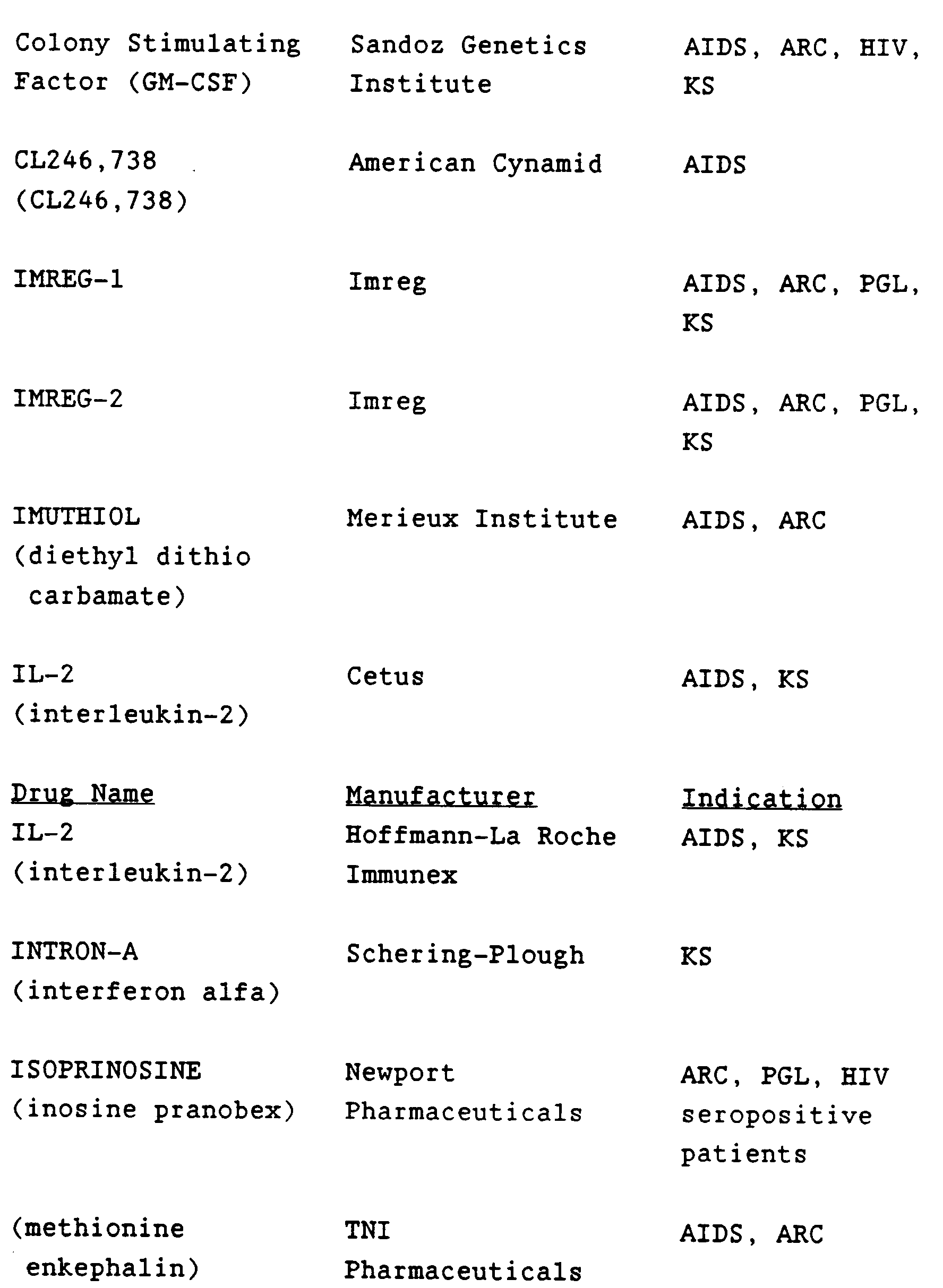
- It will be understood that the scope of combinations of the vaccines of this invention with AIDS antivirals, immunomodulators, antibiotics or vaccines is not limited to the list in the above Table, but includes in principle any combination with any pharmaceutical composition useful for the treatment of AIDS. The AIDS or HIV vaccines of this invention include vaccines to be used pre- or post-exposure to prevent or treat HIV infection or disease, and are capable of producing an immune response specific for the immunogen.
- The conjugates of this invention, when used as a vaccine, are to be administered in immunologically effective amounts. Dosages of between 1 µg and 500 µg of conjugate protein, and preferably between 50 µg and 300 µg of conjugate protein are to be administered to a mammal to induce anti-peptide, anti-HIV, or HIV-neutralizing immune responses. About two weeks after the initial administration, a booster dose may be administered, and then again whenever serum antibody titers diminish. The conjugate should be administered intramuscularly or by any other convenient or efficacious route, at a concentration of between 10 µg/ml and 1 mg/ml, and preferably between 50 and 500 µg/ml, in a volume sufficient to make up the total required for immunological efficacy. The conjugate may be preadsorbed to aluminum hydroxide gel or to the Ribi adjuvant (GB 2220211A, US priority document 212,919 filed 29/06/1988) and suspended in a sterile physiological saline solution prior to injection.
- The protein moiety should behave as an immune enhancer. It is desirable, in the choice of protein, to avoid those that result in non-specific activation of the recipient's immune response (reactogenicity). In U.S. Patent 4,695,624, Marburg et al. used the outer membrane protein complex (OMPC) derived from Neisseria meningitidis to prepare polysaccharide-protein conjugates. OMPC has proven to be suitable though other immunogenic proteins may be used. The instant invention utilizes the Class II major immune enhancing protein (MIEP) of OMPC.
- Various methods of purifying OMPC from the gram-negative bacteria have been devised [Frasch et al., J. Exp. Med. 140, 87 (1974); Frasch et al., J. Exp. Med. 147, 629 (1978); Zollinger et al., US Patent 4,707,543 (1987); Helting et al., Acta Path. Microbiol. Scand. Sect. C. 89, 69 (1981); Helting et al., US patent 4,271,147]. OMPC may be used herein essentially according to the Helting process, from which MIEP may be further purified [Murakami, K., et al., Infection and Immunity, 57, 2318 (1989)], to provide immune enhancement necessary to induce mammalian immune responses to HIV PND peptides. MIEP may be derived by dissociation of the isolated OMPC, or alternatively, produced through recombinant expression of the desired immunogenic portions of OMPC. Methods of preparing and using an OMPC subunit are disclosed in co-pending US application serial Nos. 555,329; 555,978; and 555,204 (Merck Case # ′s 18159, 18110, and 18160 respectively).
- The HIV PND peptides that may be used for making species of the conjugate of this invention may be linear or cyclic peptides. The linear peptides may be prepared by known solid phase peptide synthetic chemistry, by recombinant expression of DNA encoding desireable peptide sequences, or by fragmentation of isolated HIV proteins. Cyclic HIV PND peptides may be prepared by cyclization of linear peptides, for example (a) by oxidizing peptides containing at least two cysteines to generate disulfide bonded cycles; (b) by forming an amide bonded cycle; (c) by forming a thioether bonded cycle. Processes for making such peptides are described herein but this description should not be construed as being exhaustive or limiting. The conjugates of this invention are useful whenever a component peptide is an HIV PND or is capable of priming mammalian immune responses which recognize HIV PNDs.
- PND peptides, both those known in the art and novel compounds disclosed herein are defined as peptidyl sequences capable of inducing an HIV-neutralizing immune response in a mammal, including the production of HIV-neutralizing antibodies.
- A major problem overcome by the instant invention is the HIV interisolate sequence variability. For example, in the PND which occurs in the third hypervariable region of gp120 (see below), although certain amino acids have been found to occur at given locations in a great many isolates, no strictly preserved primary sequence motif exists. This difficulty is surmounted by this invention because it allows conjugation of a cocktail of peptides having PND sequences from as many different HIV isolates as necessary to attain broad protection. Alternatively, a broadly protective cocktail of conjugates may be prepared by mixing conjugates, each of which is prepared separately with a peptide moiety providing protection against a single or several HIV isolates.
- The amino acids found near or between amino acids 296 and 341 of gp120 have been shown to meet the criteria which define a PND. In the IIIB isolate of HIV, a 41-amino-acid sequence has been reported as follows (SEQ ID: 1:):
- -Ile Asn Cys Thr Arg Pro Asn Asn Asn Thr Arg Lys Ser Ile Arg Ile Gln Arg Gly Pro Gly Arg Ala Phe Val Thr Ile Gly Lys Ile Gly Asn Met Arg Gln Ala His Cys Asn Ile Ser-, with the two cysteines being disulfide bonded to each other to form a loop. The trimer -Gly Pro Gly- is exposed at the PND loop tip. Peptides from different HIV isolates from this same region of gp120 raise isolate-specific neutralizing antibodies when presented to goat and guinea pig immune systems as conjugates with keyhole-limpet hemocyanin. The major neutralizing epitope within the 41-mer sequence, presented above, is comprised by the eight amino acids surrounding and including the -Gly Pro Gly- trimer [Javaherian et al., PNAS USA 86, 6768 (1989)]. In Table II below a number of linear peptides of different length and composition that can be used to prepare the conjugates of this invention are presented. The name of the isolate containing a peptide having the sequence of the tabulated peptide is given, along with a name herein ascribed to that peptide for ease of reference. The letter r- on the left hand side of each peptide represents the possibility of linking the peptide to an immunogenic protein, such as the MIEP at that position. In addition, marker amino acids, such as norleucine and ornithine may form part of r-.


- This list is not an exhaustive list of possible PND sequences. Rather, it is provided as a suggestive and illustrative guide as to useful PND primary sequences. Therefore, peptides conjugated as herein described to form the immunogen of this invention are any of the tabulated peptides or immunologically-equivalent variants on the theme suggested by these peptidyl sequences. The nature of the variations is considered next.
- The primary sequence of this HIV PND appears to have a conserved core amino acid sequence, comprised by the tetramer sequence -Gly Pro Gly Arg-, (SEQ ID: 18:) with increacing divergence on either side of this sequence among HIV isolates. Some isolates have sequences that diverge even within the tetramer, having -Gly Pro Gly Lys- (SEQ ID: 19:), -Gly Pro Gly Gln- (SEQ ID :20:), and even -Gly Leu Gly Gln- (SEQ ID: 21) core sequences. All of these possible sequences come within the scope of this disclosure as being peptide sequences that are advantageous for conjugation according to this invention.
- The length of the peptide is a significant factor in promoting cross reactive immune responses. That is, an immune response raised against a given peptidyl epitope may recognize similar epitopes from the same or different HIV isolate based on the number of amino acids in the epitope over and above the critical neutralizing epitope. In addition, the length of the peptide is also responsible for determining the probability of exposure to the immune system of the determinant responsible for generating an HIV-neutralizing response.
- In order to maximize the probability of relevant epitope presentation, chemistry was developed whereby the PND peptides may be locked into a given three-dimensional configuration. It is known that the 41-amino-acid PND of the HIV IIIB isolate, represented above, is configured as a loop by the presence of the cysteine-to-cysteine disulfide bond. Disulfides, however, may be labile under certain conditions and therefore may allow the loop to open and the peptide to exist in a linear form. Therefore, in addition to linear peptides, disulfide-bonded cyclic peptides and novel HIV PND peptides having nonlabile cyclic structures disclosed herein
may all be utilized as the PEP component in the formation of the conjugates of this invention. - The peptides that may be used in formation of these conjugates may be derived as fragments of natural proteins (gp120 for example), by recombinant expression of portions thereof, or by chemical synthesis according to known methods in the art. In addition, novel cyclic PNDs may be prepared synthetically according to the processes herein described. The sequences may contain both natural L-amino acids, or unusual or D-amino acids. In addition, the conjugation chemistry is sufficiently flexible so that the appropriate choice of peptide derivatization reagents allows for successful conjugation.
- Synthetic peptides have been prepared by a number of strategies conducted either in solution or on solid supports. Excellent texts covering the basic principles and techniques are: Principles of Peptide Synthesis, Bodanszky. M., Springer-Verlag (1984); Solid Phase Peptide Synthesis, Stewart J. M., Young, J. D., Pierce Chemical Company (2nd. ed. 1984); and The Peptides, Gross, E., Meienhofer, J., Academic Press, Inc., (1979). The processes described herein, however, are not limited to the disclosure of these texts.
- Synthetic cyclic peptides may be prepared in two phases. First, the linear peptide may be synthesized on a Milligen 9050 peptide or an ABI 431A synthesizer using 9-fluorenylmethyloxy-carbonyl (Fmoc) chemistry and side-chain-protected Fmoc-amino acid pentafluorophenyl esters which are known reagents or using derivatized Wang resin, Fmoc chemistry, and side-chain protected Fmoc-amino acid symmetrical anhydrides, prepared in situ, as reagents.
- Second, the linear peptide may be cyclized, either in solution or with the peptide still attached to the solid phase resin. Cyclization may be accomplished by any technique known in the art, which may comprise, for example: a) incorporating cysteine residues into the linear peptide on either end of the sequence which is to form the loop and allowing disulfide bond formation under oxidizing conditions known in the art; b) preparing a cysteine containing peptide as in (a) but retaining the cysteines as free sulfhydryls (or as Acm protected thiols which are deprotected to the free sulfhydryls) and treating the peptide with o-xylylene dibromide or similar reagent, such as the diiodide, dichloride, or a dihalogenated straight or branched chain lower alkyl having between two and eight carbon atoms; such reagents react with the sulfur atoms of the cysteines to form a cyclic structure containing two nonlabile thioether bonds to the benzene or the alkyl; c) allowing a free group on one side of the loop amino acids to become amide bonded to a free carboxyl group on the other side of the loop amino acids through DPPA, BOP, or similar reagent mediated peptide bond formation. Each of these strategies is taken up in more detail below, after presentation of a generalized description of the cyclic peptides produced by these methods.
- Thus, without limiting the conjugate invention to the following peptides or methods of producing them, the PND peptides which may be conjugated after removal of appropriate protecting groups as necessary, according to this invention include those represented by the structure PEP, which includes the linear peptides of Table II above and cyclic peptides:

wherein:
r is the position of linkage between PEP and MIEP, optionally comprising a marker amino acid, if R¹ is not a marker amino acid;
R¹ is: - a) a bond, or
- b) a peptide of 1 to 5 amino acids, optionally including a marker amino acid which migrates at a position in the amino acid analysis spectrum which is isolated from the signal of the 20 naturally occuring amino acids; preferably norleucine, gamma aminobutyric acid, β-alanine, or ornithine;
- a) either a bond or a peptide of up to 17 amino acids if R³ is a peptide of at least 2 amino acids, or
- b) a peptide of between 2 to 17 amino acids, if R³ is a bond;
- a) either a bond or a peptide of up to 17 amino acids if R² is a peptide of at least 2 amino acids, or
- b) a peptide of between 2 to 17 amino acids, if R² is a bond;
- a) -NH-CH-CO-, with R⁷ bonded to the methine carbon, if R⁷ is R⁸, or
- b) a bond from R³ to R⁷ and R⁵, if R⁷ is carbonyl or -COCH₂CH₂CH(CONH₂)NHCO-;
- a) a peptide of one to five amino acids, optionally including a marker amino acid,
- b) -OH,
- c) -COOH,
- d) -CONH₂,
- e) -NH₂, or
- f) -absent;
- a) an amino acid side chain, selected from the side chain of any of the common L or D amino acids, (see table of Definitios and Abbreviations), if the optional bond (--------) to R⁷ is absent,
- b) -R⁸-S-S-, or -R⁸-S-R⁸-R⁹-R⁸-S-, if R⁷ is R⁸, or
- c) -R⁸-NH- if R⁷ is
- a) -R⁸-,
- b) -C=O, or
- a) R¹⁰, or
- b) xylylene
- a) lower alkyl, or
- b) -CH₂-O-CH₂-; and
- Hereinafter, amino acids -R²-Gly Pro Gly Arg-R³-, which form the "core" of the PND peptides, and go toward formation of the loop of a cyclic peptide, will be referred to as loop or core amino acids. When the optional bond between R⁶ and R⁷ is absent however, the structure, PEP, is linear, and encompasses all of the linear peptides of Table II.
- Whether the peptide is linear or cyclic, the amino acid sequences comprising R² and R³ of PEP may be any combination of amino acids, including sequences surrounding the core -Gly Pro Gly Arg-tetramer in any of the sequences of Table II. Thus, the core amino acids represented by -R²-Gly Pro Gly Arg-R³- may be further defined as having the core amino acid structure:
-XnX₁X₂-Gly Pro Gly Arg-X₃X₄Xm-
wherein:
X₁ is a constituent of R² selected from: - a) serine,
- b) proline,
- c) arginine,
- d) histidine,
- e) glutamine, or
- f) threonine;
- a) isoleucine,
- b) arginine,
- c) valine, or
- d) methionine;
- a) alanine,
- b) arginine, or
- c) valine;
- a) phenylalanine,
- b) isoleucine,
- c) valine, or
- d) leucine;
- The cyclic peptides may be disulfide bonded structures or a cycle formed through a nonlabile bond or structure. The term "nonlabile bond" means a covalent linkage, other than a disulfide bond. These covalent linkages may be through a bridge structure, such as xylylene, through a lower alkyl, through -CH₂-O-CH₂, or through an amino acid amide-bonded bridge. By altering the bridge structure and/or the number and combination of amino acids included in the peptide, the conformation of the loop structure of the cycle may be optimized, allowing for fine-tuning of the PND epitope presented to the immune system. For example, use of an o-xylylene bridge generates a "tighter" loop structure than when, for example, an eight carbon straight chain lower alkyl is used as the bridge. Thus, the conjugates of this invention are useful both as reagents to analyze the structure-function relationship of the PND epitope in raising anti-peptide, anti-HIV, HIV-neutralizing, and anti-AIDS immune responses in mammals, and as components for formulation of anti-HIV disease, including AIDS, vaccines.
- Synthetic products obtained may be characterized by fast-atom-bombardment mass spectrometry [FAB-MS], reverse phase HPLC, amino acid analysis, or nuclear magnetic resonance spectroscopy (NMR).
- Peptides containing cysteine residues on either side of the loop amino acids may be cyclized under oxidizing conditions to the disulfide-bonded cycles. Methods for achieving disulfide bonding are known in the art. An example of disulfide bonded peptides useful in this invention is given infra in Example 10, wherein cPND4 is produced and Example 18 wherein cPND33 is produced. In Example 10, a process utilizing the Acm derivative of cysteine to generate disulfide bonded cPNDs is used, but other processes are equally applicable. In Example 18, the peptide containg two sulfhydryls is oxidized in dilute acid. The disulfide bonded peptides are preferred in the instant invention.
- Thus, in a preferred embodiment of this invention, the peptide has the structure (SEQ ID 18:):

or pharmaceutically acceptable salts thereof, wherein:
r is: - a) hydrogen,
- b)
- a) a bond, or
- b) a peptide of 1 to 5 amino acids, optionally including a marker amino acid;
- a) -OH,
- b) a peptide of 1 to 5 amino acids, optionally including a marker amino acid, or
- c) -NH₂;
- Lower alkyl consists of straight or branched chain alkyls having from one to eight carbons unless otherwise specified. Hereinafter, amino acids -R²-Gly Pro Gly Arg-R³-, which go toward formation of the loop of a cyclic peptide, will be referred to as loop amino acids.
- In one embodiment of the invention, the cyclic peptide having the structure (SEQ ID: 18:):

X₁ is a constituent of R² selected from: - a) serine,
- b) proline,
- c) arginine,
- d) histidine,
- e) glutamine, which is preferred, or
- f) threonine;
- a) isoleucine, which is most preferred,
- b) arginine, which is preferred,
- c) valine, or
- d) methionine;
- a) alanine,
- b) arginine, or
- c) valine;
- a) phenylalanine,
- b) isoleucine,
- c) valine, or
- d) leucine;
- The novel disulfide bonded cyclic peptides used in this invention (and separately claimed in co-filed Merck case 18068IB, USSN , ) may be prepared in essentially two phases: First the linear peptide is synthesized on a Milligen 9050 or an ABI-431A peptide synthesizer using 9-fluorenyl-methyloxycarbonyl (Fmoc) chemistry and appropriately side-chain protected Fmoc-amino acid pentafluoro-phenyl esters as reagents or using derivatized Wang resin, Fmoc chemistry, and side-chain protected Fmoc-amino acid symmetrical anhydrides, prepared in situ, as reagents.
- Second, the linear peptide is cyclized, either in solution or with the peptide still attached to the solid phase resin by incorporating cysteine residues into the linear peptide at either end of the sequence which is to form the loop, and oxidizing these to the disulfide. In a preferred embodiment, cyclization is accomplished by exposure of the peptide to (a) H₂O₂, (b) atmospheric oxygen, (c) aqueous CH₃CN containing about 0.1 - 0.5% TFA, or (d) about 0.1M ferricyanide. The preferred method is exposure to atmospheric oxygen.
- Products obtained may be characterized by fast atom bombardment-mass spectrometry [FAB-MS], reverse phase HPLC, amino acid analysis or nuclear magnetic resonance spectroscopy (NMR).
- Thus, the peptides useful in this invention may be prepared as further described below in (i) and (ii):
- A linear peptide containing C¹ and C² on either side of the loop amino acids, where C¹ and C² are both cysteine or another amino acid containing free sulfhydryl groups in the side chain, is prepared according to known synthetic procedures (see discussion supra). In the completed cyclic PND, the sulfhydryl containing side chains, (-R⁸-SH), go toward making up the -R⁸-S-groups of the completed cyclic HIV PND structure shown above. Amino acids to be incorporated which have reactive side chains (R groups) are used in an appropriately R-group protected form. For example, histidine is triphenylmethyl (Trt), or Boc protected, and arginine is 4-methoxy-2,3,6-trimethylphenyl sulfonyl (Mtr) protected.
- Preferably, a resin is purchased with C² in its Acm protected form already attached to the resin, for example, Fmoc-L-Cys(Acm)-O-Wang resin. The cysteine incorporated at the amino terminal side of the loop amino acids, C¹, may also be the Acm derivative. Either C¹ or C² may be bound to additional amino acids, R¹ or R⁵ respectively, which may be utilized in the formation of conjugates with carrier molecules or may serve as marker amino acids for subsequent amino acid analysis, such as when norleucine or ornithine is used.
- The sulfur of the acetamidomethylated cysteines are reacted, at room temperature for about 15 hours in a solvent compatible with the resin, as a 1-50% concentration of an organic acid, preferably about 10% acetic acid in anhydrous dimethylformamide (DMF), with about a four fold molar excess of a heavy metal salt, such as mercuric acetate [Hg(OAc)₂] for each Acm group. The resulting heavy metal thioether, for example the mercuric acetate thioether of the peptide, PEP(S-HgOAc), is then washed and dried. Addition of excess hydrogen sulfide in DMF yields insoluble metal sulfide, e.g. mercuric sulfide (HgS), and the peptide with free sulfhydryl groups. The free sulfhydryls are then oxidized by one of the aforementioned methods. Alternatively, the Acm protected thiols may be converted directly to the cyclic disulfide by treatment with iodine in a methanol/DMF solvent.
- Essentially the same process described above for solid state cyclization applies with two main variants: If the peptide is cleaved (95% TFA/4% ethanedithiol/1% thioanisole) from a pepsyn KA resin, acid labile side chain protecting groups are also removed, including Cys(Trt) which provides the necessary free -SH function. If however, Cys(Acm) protection is used, then mercuric acetate/hydrogen sulfide cleavage to the free -SH group is required as an independent procedure, with the linear peptide either on or off the resin.
- One method however, is the use of Cys(Acm) protection and Sasrin or Pepsyn KH resin, and cleavage of the linear, fully protected peptide from the resin with 1% TFA/CH₂Cl₂. Mercuric acetate/hydrogen sulphide then selectively converts Cys(Acm) to the free -SH group, and cyclization is effected on the otherwise protected peptide. At this point, the peptide may be maleimidated in situ , selectively on the N-terminus. Acid labile side chain protecting groups are cleaved with 98% TFA/2% thioanisole, and the cyclic peptide is isolated by HPLC. The preferred method, however, is to cleave the peptide from the resin, and allow cyclization by one of the aforementioned methods. The most preferred method is to allow air oxidation for about one to fifty hours of between 10 and 40°C.
- Thus, in a particularly preferred embodiment of this invention, a peptide (CPND 33) having the structure (SEQ ID: 22:):

- e-1)


- e-2)


- e-3)

- e-4)

- A linear peptide containing C¹ and C² on either side of the loop amino acids, where C¹ and C² are both cysteine or another sulfhydryl containing amino acid, is prepared according to known synthetic procedures (see discussion supra). In the completed cylic PND, C¹ and C² become part of the R⁶ and R⁷ groups of the PEP structure shown above. Amino acids to be incorporated which have reactive side chains (R groups) are used in an appropriately R-group-protected form. For example, histidine is triphenylmethyl- (Trt) protected, arginine may be 4-methoxy-2,3,6-trimethylphenyl sulfonyl (Mtr) protected. [Principles of Peptide Synthesis, Bodanszky. M., Springer-Verlag (1984); Solid Phase Peptide Synthesis, Stewart J. M., Young, J. D., Pierce Chemical Company (2nd. ed. 1984); and The Peptides, Gross, E., Meienhofer, J., Academic Press, Inc., (1979)].
- Preferably, a resin is purchased with C² in its Acm-protected form already attached to the resin, for example, Fmoc-L-Cys(Acm)-O-Wang resin. The cysteine incorporated at the amino terminal side of the loop amino acids, C¹, may also be the Acm derivative. Either C¹ or C² may be bound to additional amino acids, R¹ or R⁵ respectively, which may be utilized in the formation of conjugates with carrier molecules or may serve as marker amino acids for subsequent amino acid analysis, such as when norleucine or ornithine is used.
- The sulfur of the acetamidomethylated cysteines is reacted, at room temperature for about 15 hours in a solvent compatible with the resin, such as 10% acetic acid in anhydrous dimethylformamide (DMF), with about a four-fold molar excess of a heavy metal salt, such as mercuric acetate [Hg(OAc)₂] for each Acm group. The resulting heavy metal thioether, for example the mercuric acetate thioether of the peptide, PEP(S-HgOAc), is then washed and dried. Addition of excess hydrogen sulfide in DMF yields insoluble metal sulfide, e.g., mercuric sulfide (HgS), and the peptide with free sulfhydryl groups.
- A mixture of about an equimolar amount, as compared with peptide, of o-xylylene dibromide or dichloride, a dibrominated or dichlorinated lower alkyl, 1,3-dihalogenenated -CH-O-CH-, or similar reagent which will provide a desirable bridge length, is added to the derivatized resin. A large excess of tertiary amine, preferably triethylamine (NEt₃) in DMF is added slowly. The reaction with the bis-sulfhydryl peptide moiety is allowed to proceed for about sixteen hours at room temperature, yielding the bridge group derivatized cyclic peptide bound to resin. Deprotection of acid sensitive side chain protecting groups and cleavage from the resin is achieved by treatment with 95% trifluoroacetic acid (TFA) in the presence of 4% 1,2-ethanedithiol and 1% thioanisole. The dissolved cyclic peptide may then be separated from the resin by filtration. The filtrate is evaporated and the crude residual product is purified by high performance liquid chromatography (HPLC) according to known methods, for example by reverse phase HPLC.
- Essentially the same process described above for solid state cyclization applies with two main variants: If the peptide is cleaved (95% TFA/4% ethanedithiol/1% thioanisole) from a pepsyn KA resin, acid labile side chain protecting groups are also removed, including Cys(Trt) which provides the necessary free -SH function. If however, Cys(Acm) protection is used, then mercuric acetate/hydrogen sulfide cleavage to the free -SH group is required as an independent procedure, with the linear peptide either on or off the resin.
- The preferred method however, is the use of Cys(Acm) protection and Sasrin or Pepsyn KH resin, and cleavage of the linear, fully protected peptide from the resin with 1% TFA/CH₂Cl₂. Mercuric acetate/hydrogen sulphide then selectively converts Cys(Acm) to the free -SH group, and cyclization is effected on the otherwise protected peptide. Acid labile side chain protecting groups are cleaved with 95% TFA/4% ethanedithiol/1% thioanisole, and the cyclic peptide is isolated by HPLC.
- Removal of excess reagents, such as unreacted xylylene dibromide, prior to acid cleavage of the protecting groups is conveniently achieved by, for example, a step gradient reverse phase HPLC run prior to more selective gradient elution.
- Cyclic HIV PND peptides prepared according to the method of this subsection include, but are not limited to, the sample cPNDs represented below. The methods of this subsection are generally applicable to small peptides, and particularly applicable to peptides of between 5 and 30 amino acids. An optimal ring size may include between 5 and 10 amino acids, including the -Gly-Pro-Gly- trimer, and this ring size is easily maintained by production of cycles from linear peptides having the appropriate number and combination of amino acids.
- Representative peptides resulting from the process described in this subsection b. parts (i). and (ii) are disclosed inapplication U.S.S.N. 555,227. The conjugate invention should, however, not be construed as being limited to use those particular embodiments of HIV cyclic PND peptides. Other linear HIV PND peptide sequences may be cyclized in essentially the same fashion used to provide those peptides. Series of peptides having divergent primary sequences could be generated and would be beneficial in this invention as long as they continue to elicit an anti-peptide, anti-HIV, or HIV-neutralizing immune response.
- Novel amide bonded cyclic HIV PND peptides may be prepared for conjugation in essentially two phases: First, the linear peptide is prepared, for example on an ABI-431A peptide synthesizer, by known solid phase peptide synthetic chemistry, for example using Fmoc chemistry and appropriately side-chain protected Fmoc-amino acids as reagents.
- Second, the linear peptide is cleaved from the resin and cyclized in solution by allowing the free amino terminus of the peptide, the free amino group of an amino terminal isoglutamine, or a free ε-amino or α-amino group of a lysine on one side of the loop amino acids to be amide bonded to a free carboxyl group on the carboxy-terminal side of the loop amino acids through DPPA, BOP, or similar reagent mediated peptide bond formation.
- Products obtained may be characterized by fast atom bombardment-mass spectrometry [FAB-MS], reverse phase HPLC, amino acid analysis, or nuclear magnetic resonance spectroscopy (NMR).
- Thus, highly preferred embodiments of this invention are conjugates having covalent linkages from MIEP to an amide bonded cyclic HIV PND, prepared as described hereinabove. Where the PND is from a predominant isolate, such as the HIV IIIB or the HIV MN isolate, a conjugate vaccine, or a mixture of such conjugate vaccines is highly advantageous for prophylaxis or treatment of AIDS or ARC. Examples of such preferred embodiments having the structure:


j is the percentage by mass of peptide in the conjugate, and is preferably between 1% and 50% of the total protein mass in the conjugate;
are useful for inducing anti-peptide immune responses in mammals, for inducing HIV-neutralizing antibodies in mammals, for formulating vaccines to prevent HIV-disease or infection, or for treating humans afflicted with HIV-disease or infection, including AIDS and ARC. - One or more of the conjugate vaccines of this invention may be used in mammalian species for either active or passive protection prophylactically or therapeutically against infectious agents such as, in the preferred embodiment of this invention, Haemophilus influenzae serotype B, or human immunodeficiency virus induced diseases. Active protection may be accomplished by injecting an effective quantity capable of producing measurable amounts of antibodies (e.g., about 1 microgram to about 50 µg, depending on the antigen) of an antigen (e.g. PRP, HIV PND peptides) in the MIEP-conjugate form of each of the conjugates being administered per dose. The use of an adjuvant (e.g., alum) is also intended to be within the scope of this invention. Passive protection may be accomplished by injecting whole antiserum obtained from animals previously dosed with the MIEP-conjugate or conjugates, or globulin or other antibody-containing fractions of said antisera, with or without a pharmaceutically-acceptable carrier, such as sterile saline solution. Such globulin is obtained from whole antiserum by chromatography, salt or alcohol fractionation or electrophoresis. Passive protection may be accomplished by standard monoclonal antibody procedures or by immunizing suitable mammalian hosts.
- In a preferred embodiment of this invention, the conjugate is used for active immunogenic vaccination of humans, especially infants, children, or immunocompromised individuals. For additional stability, these conjugates may also be lyophilized in the presence of lactose (for example, at 20 µg/mL of PRP/4 mg/mL lactose) prior to use.
- A preferred dosage level is an amount of each of the MIEP-conjugates, or derivative thereof to be administered, corresponding to between approximately 2 to 20-µg of PRP, or about 1 microgram to 5 milligarams of peptide in the MIEP-conjugate form for conjugates of H. influenzae serotype B polysaccharide, or HIV PND peptide, in a single administration. If necessary, an additional one or two doses of the MIEP-conjugate, or derivative thereof, in a dosage comparable to that described above.
- The invention is further defined by reference to the following examples, which are intended to be illustrative and not limiting.
- A tube containing the lyophilized culture of Neisseria meningitidis (obtained from Dr. M. Artenstein, Walter Reed Army Institute of Research (WRAIR), Washington, D.C.) was opened and Eugonbroth (BBL) was added. The culture was streaked onto Mueller Hinton agar slants and incubated at 37°C with 5% CO₂ for 36 hours, at which time the growth was harvested into 10% skim milk medium (Difco), and aliquots were frozen at -70°C. The identity of the organism was confirmed by agglutination with specific antiserum supplied by WRAIR, and typing serum supplied by Difco.
- A vial of the culture from the second passage was thawed and streaked onto 10 Columbia Sheep Blood agar plates (CBAB-BBL). The plates were incubated at 37°C with 5% CO₂ for 18 hours after which time the growth was harvested into 100 mL of 10% skim milk medium, aliquots were taken in 0.5 mL amounts and frozen at -70°C. The organism was positively identified by agglutination with specific antiserum, sugar fermentation and gram stain.
- A vial of the culture from this passage was thawed, diluted with Mueller-Hinton Broth and streaked onto 40 Mueller-Hinton agar plates. The plates were incubated at 37°C with 6% CO₂ for 18 hours after which time the growth harvested into 17 mL of 10% skim milk medium, aliquotted in 0.3 mL amounts and frozen at -70°C. The organism was positively identified by Gram stain, agglutination with specific antiserum and oxidase test.
- The inoculum was grown from one frozen vial of Neisseria memingitidis Group B, B-11 from above (passage 4). Ten Mueller-Hinton agar slants were inoculated, and six were harvested approximately 18 hours later, and used as an inoculum for 3 250 mL flasks of Gotschlich's yeast dialysate medium at pH 6.35. The O.D.₆₆₀ was adjusted to 0.18 and incubated until the OD₆₆₀ was between 1 and 1.8. 1 mL of this culture was used to inoculate each of 5 2L. Erlenmeyer flasks (each containing 1 liter of medium; see below) and incubated at 37°C in a shaker at 200 rpm. The O.D. was monitored at hourly intervals following inoculation. 4 liters of broth culture, at an O.D.₆₆₀ of 1.28 resulted.
- 70 Liter Seed Fermenter- Approximately 4 liters of seed culture was used to inoculate a sterile 70-liter fermenter containing about 40 liters of complete production medium (see below). The conditions for the 70-liter fermentation included 37°C, 185 rpm with 10 liters/minute air sparging and constant pH control at about pH 7.0 for about 2 hours. For this batch, the final O.D.₆₆₀ was 0.732 after 2 hours.
800-Liter Production Fermenter - Approximately 40 liters of seed culture were used to inoculate a sterile 800 liter fermenter containing 568.2 liters of complete production medium (see below). The batch was incubated at 37°C, 100 rpm with 60 liters/minute air sparging and constant pH control at pH 7.0. For this batch, the final O.D. was 5.58 thirteen hours after inoculation.
-

- 1280 g of Difco Yeast Extract was dissolved in 6.4 liters of distilled water. The solution was dialyzed in 2 Amicon DC-30 hollow fiber dialysis units with three H10SM cartridges. 384 g MgSO₄.7-H₂O and 3200 g dextrose were dissolved in the dialysate and the total volume brought to 15 liters with distilled water. The pH was adjusted to 7.4 with NaOH, sterilized by passage through a 0.22 µ filter, and transferred to the fermenter containing Fraction A.
- For the Erlenmeyer flasks: 1 liter of Fraction A and 25 mL of Fraction B were added and the pH was adjusted to 7.0-7.2 with NaOH.
- For the 70 liter fermenter: 41.8 liters of Fraction A and 900 mL of Fraction B were added and the pH was adjusted to 7.0-7.2 with NaOH.
- For the 800 liter fermenter: 553 liters of Fraction A and 15.0 liters of Fraction B were added and the pH was adjusted to 7.1-7.2 with NaOH.
- After the fermentation was completed, phenol was added in a separate vessel, to which the cell broth was then transferred, yielding a final phenol concentration of about 0.5%. The material was held a room temperature with gentle stirring until the culture was no longer viable (about 24 hours).
- After about 24 hours at 4°C, the 614.4 liters of inactivated culture fluid was centrifuged through Sharples continuous flow centrifuges. The weight of the cell paste after phenol treatment was 3.875 kg.
- The phenol inactivated culture was concentrated to about 30 liters and diafiltered in sterile distilled water using 0.L micro-hollow fiber filters (ENKA).
- An equal volume of 2X TED buffer [0.1 M TRIS 0.01 M EDTA Buffer, pH 8.5, with 0.5% sodium deoxycholate] was added to the concentrated diafiltered cells. The suspension was transferred to a temperature regulated tank for OMPC extraction at 56 °C with agitation for 30 minutes.
- The extract was centrifuged at about 18,000 rpm in a Sharples continuous flow centrifuge at a flow rate of about 80 mL/minute, at about 4°C. The viscous supernatant was then collected and stored at 4°C. The extracted cell pellets were reextracted in TED buffer as described above. The supernatants were pooled and stored at 4°C.
- The pooled extract was transferred to a temperature regulated vessel attached to AG-Tech 0.1 micron polysulfone filters. The temperature of the extract was held at 25°C in the vessel throughout the concentration process. The sample was concentrated tenfold at an average transmembrane pressure of between 11 and 24 psi.
- The retentate from
Step 3 was centrifuged at about 160,000 x g (35,000 rpm) at about 70°C in a continuous flow centrifuge at a flow rate between 300 to 500 mL/minute, and the supernatant was discarded. - The OMPC pellet was suspended in TED Buffer (190 mL buffer; 20 mL/g pellet)
Step 2 andStep 4 were repeated twice (skipping Step 3). - The washed pellets from
Step 4 were suspended in 100 mL distilled water with a glass rod and a Dounce homogenizer to insure complete suspension. The aqueous OMPC suspension was then filter sterilized by passage through a 0.22 µ filter, and the TED buffer was replaced with water by diafiltration against sterile distilled water using a 0.1 µ hollow fiber filter. - A lyophilized tube of Haemophilus influenzae type b, (cultured from Ross 768, received from State University of New York) was suspended in 1 mL of sterile Haemophilus inoculum medium (see below) and this suspension was spread on 9 Chocolate Agar slants (BBL). The pH of the inoculum medium was adjusted to 7.2 ± 0.1 (a typical value was pH 7.23) and the medium solution was sterilized prior to use by autoclaving at 121°C for 25 minutes. After 20 hours incubation at 37°C in a candle jar, the growth from each plate was resuspended in 1-2 mL Haemophilus inoculum medium, and pairs of slants were pooled.

- The resuspended cells from each pair of slants was inoculated into three 250 mL Erlenmeyer flasks containing about 100 mL of Haemophilus Seed and Production medium. The 250 mL flasks were incubated at 37°C for about 3 hours until an OD₆₆₀ of about 1.3 was reached. These cultures were used to inoculate the 2 liter flasks (below).
- 2 Liter non-baffled Erlenmeyer flasks- 5 mL of culture from "A stage" (above) were used to inoculate each of five two-liter flasks, each containing about 1.0 liter of complete Haemophilus seed and production medium (see below). The flasks were then incubated at 37°C on a rotary shaker at about 200 rpm for about 3 hours. A typical OD₆₆₀ value at the end of the incubation period was about 1.0.

- The salts and soy peptone were dissolved in small volumes of hot, pyrogen-free water and brought to correct final volume with additional hot, pyrogen-free water. The fermenters or flasks were then sterilized by autoclaving for about 25 minutes at 121°C, and after cooling yeast extract diafiltrate (1), glucose (2), NAD (3), and hemin (4) were added aseptically to the flasks or fermenters prior to inoculation.
- (1) Yeast extract diafiltrate: 100 g brewers' yeast extract (Amber) was dissolved in 1 liter distilled water and ultrafiltered using an Amicon DC-30 hollow fiber unit with H10 x 50 cartridges with a 50 kd cutoff. The filtrate was collected and sterilized by passage through a 0.22 µ filter.
- (2) Glucose was prepared as a sterile 25% solution in distilled water.
- (3) A stock solution of NAD containing 20 mg/mL was sterilized by passage through a (0.22 µ filter) and added aseptically just prior to inoculation.
- (4) A stock solution of Hemin 3X was prepared by dissolving 200 mg in 10 mL of 0.1 M NaOH and the volume adjusted with distilled, sterilized water to 100 mL. The solution was sterilized for 20 minutes at 121°C and added aseptically to the final medium prior to inoculation.
- 70 Liter Seed Fermenter- Three liters of the product of B Stage was used to inoculate a fermenter containing about 40 liters of Complete Haemophilus Seed And Production medium (prepared as described above) and 17 mL UCON B625 antifoam agent. The pH at inoculation was 7.4.
- 800 Liter Production Fermenter-Approximately 40 liters of the product of "C Stage" was used to inoculate an 800 liter fermenter containing 570 liters of Haemophilus Seed and Production medium (prepared as described above), scaled to the necessary volume, and 72 mL of UCON LB625 antifoam agent was added.
- The fermentation was maintained at 37°C with 100 rpm agitation, with the O.D.₆₆₀ and pH levels measured about every two hours until the O.D.₆₆₀ was stable during a two-hour period, at which time the fermentation was terminated (a typical final O.D.₆₆₀ was about 1.2 after about 20 hours).
- Approximately 600 liters of the batch was inactivated by harvesting into a "kill tank" containing 12 liters of 1% thimerosal.
- After 18 hours of inactivation at 4°C, the batch was centrifuged in a 4-inch bowl Sharples contiuous flow centrifuge at a flow rate adjusted to maintain product clarity (variable between 1.3 and 3.0 liters per minute). The supernatant obtained after centrifugation (15,000 rpm) was used for product recovery.
- The supernatant from two production fermentations was pooled and concentrated at 2 to 8°C in a Romicon XM-50 ultrafiltration unit with twenty 50 kd cut-off hollow fiber cartridges (4.5 m² membrane area; 2.0 Lpm air flow and 20 psi). Concentration was such that after approximately 4.5 hours, about 1,900 liters had been concentrated to 57.4 liters. The filtrate was discarded.
- To the 57.4 liters of ultrafiltration retentate, 53 liters of 95% ethanol was added dropwise over 1 hour with stirring at 4°C to a final concentration of 48% ethanol by volume. The mixture was stirred two additional hours at 4°C to insure complete precipitation, and the supernatant was collected following passage through a single 4-inch Sharples continuous flow centrifuge at 15,000 rpm at a flow rate of about 0.4 liters per minute. The pellet was discarded and the clarified fluid was brought to 82% ethanol with the addition of 40.7 liters of 95% ethanol over a one hour period. The mixture was stirred for three additional hours to insure complete precipitation.
- The resulting 48% ethanol-soluble-82% ethanol-insoluble precipitate was collected by centrifugation in a 4 inch Sharples continuous flow centrifuge at 15,000 rpm with a flow rate of about 0.24 liters per minute and the 82% ethanol supernatant was discarded. The crude product yield was about 1.4 kg of wet paste.
- The 1.4 kg grams of 82% ethanol-insoluble material, was mixed in a
Daymax dispersion vessel 2°-8°C with 24.3 liters of cold, distilled water. To this mixture, 24.3 liters of cold 2M CaCl₂.2H₂O was added, and the mixture was incubated at 4°C for 15 minutes. The vessel was then rinsed with 2 liters of 1 M CaCl₂.2H₂O, resulting in about 50 liters final volume. - The 50 liters of CaCl₂ extract was brought to 25% ethanol by adding 16.7 liters of 95% ethanol dropwise, with stirring, at 4°C over 30 minutes. After additional stirring for 17 hours, the mixture was collected by passage through a Sharples continuous flow centrifuge at 4°C. The supernatant was collected and the pellet was discarded.
- The 25% ethanol-soluble supernatant was brought to 38% ethanol by the addition of 13.9 liters of 95% ethanol, dropwise with stirring, over a 30 minute period. The mixture was then allowed to stand with agitation for one hour, then without agitation for 14 hours, to insure complete precipitation. The resulting mixture was then centrifuged in a 4 inch Sharples continuous flow centrifuge at 15,000 rpm (flow rate of 0.2 liters per minute) to collect the precipitated crude H. influenzae polysaccharide.
- The pellet from the centrifugation was transferred to a 1 gallon Waring Blender containing 2 to 3 liters of absolute ethanol and blended for 30 seconds at the highest speed. Blending was continued for 30 seconds on, and 30 seconds off, until a hard white powder resulted. The powder was collected on a Buchner funnel with a teflon filter disc and washed sequentially, in situ, with two 1 liter portions of absolute ethanol and two 2 liter portions of acetone. The material was then dried, in vacuo, at 4°C for 24 hours, resulting in about 337 g (dry weight) of product.
- About 168 grams of the dry material from the trituration step (about half of the total) was resuspended in 12 liters of 0.488 M sodium acetate, pH 6.9, with the aid of a Daymax dispersion vessel. The sodium acetate solution was immediately extracted with 4.48 liters of a fresh aqueous phenol solution made as follows: 590 mL of 0.488 M sodium acetate, pH 6.9, was added to each of eight 1.5 kg bottles of phenol (Mallinckrodt crystalline) in a 20 liter pressure vessel and mixed into suspension. Each phenol extract was centrifuged for 2.5 hours at 30,000 rpm and 4°C in the K2 Ultracentrifuge (Electronucleonics). The aqueous effluent was extracted three additional times with fresh aqueous phenol solution as described above. The phenol phases were discarded.
- The aqueous phase from the first phenol extraction above (12.2 liters) was diluted with 300 liters of cold, distilled water and diafiltered at 4°C on an Amicon DC-30 ultrafiltration apparatus using 3 H10P10, 10 kd cutoff cartridges, to remove the carryover phenol. The Amicon unit was rinsed and the rinse added to the retentate, such that the final volume was 31.5 liters. The ultrafiltrate was discarded.
- 0.81 liters of 2.0 M CaCl₂ was added to the 31.5 liters of dialysate from the previous step (final CaCl₂ concentration was 0.05 M) and the solution was brought to 82% ethanol with dropwise addition and rapid stirring over one hour, of 48.5 liters of 95% ethanol. After 4 hours of agitation, then standing for 12 hours at 4°C, the supernatant was siphoned off and the precipitate was collected by centrifugation in a 4 inch Sharples continuous flow centrifuge (15,000 rpm), at 4°C for 45 minutes. The resulting polysaccharide pellet was triturated in a 1 gallon Waring blender using 30 second pulses with 2 liters of absolute ethanol, collected on a Buchner funnel fitted with a teflon filter disc, and washed, in situ, with four 1 liter portions of absolute ethanol followed by two 1 liter portions of acetone. The sample was then dried in a tared dish, in vacuo, at 4°C for 20 hours. The yield was about 102 grams of dry powder. The yield from all phenol extractions was pooled resulting in a total of 212.6 grams of dry powder.
- The 212.6 grams of dry powder from above was dissolved in 82.9 liters of distilled water, to which was added 2.13 liters of 2 M CaCl₂.2H₂O, (0.05M CaCl₂), 2.5 mg polysaccharide/mL), and the mixture was brought 29% ethanol with the dropwise addition of 29.86 liters of 95% ethanol over 30 minutes. The mixture was clarified immediately by centrifugation in a K2 Ultracentrifuge containing a K3 titanium bowl and a K11 Noryl core (30,000 rpm and 150 mL per minute flow rate) at 4°C. The pellet was discarded and the supernatant was brought to 38% ethanol by the addition of 17.22 liters of 95% ethanol over 30 minutes with stirring. After stirring 30 additional minutes the mixture was allowed too stand without agitation at 4°C for 17 hours and the precipitate was collected using a 4 inch Sharples continuous flow centrifuge at 15,000 rpm (45 minutes was required).
- The resulting paste was transferred to a 1-gallon Waring blender containing 2 liters of absolute ethanol and blended at the highest speed by 4 or 5 cycles of 30 seconds on, 30 seconds off, until a hard, white powder formed. This powder was collected on a Buchner funnel fitted with a Zitex teflon disc and rinsed sequentially, in situ, with two fresh 0.5 liter portions and one 1 liter portions of absolute ethanol, and with two 1 liter portion of acetone. The product was removed from the funnel and transferred to a tared dish for drying, in vacuo, at 4°C (for 25 hours). The final yield of the product was 79.1 grams dry weight.
- Cloning of Genomic DNA Encoding MIEP.
- About 0.1 g of the phenol inactivated N. meningitidis cells (see Example 1) was placed in a fresh tube. The phenol inactivated cells were resuspended in 567 µL of TE buffer [10mM TRIS-HCl, 1mM EDTA, pH 8.0]. To the resuspended cells was added 30 µL of 10% SDS, and 3 µL of 20 mg/mL proteinase K (Sigma). The cells were mixed and incubated at 37°C for about 1 hour, after which 100 µL of 5 M NaCl was added and mixed thoroughly. 80 µL of 1% cetyltrimethylamonium bromide (CTAB) in 0.7 M NaCl was then added, mixed thoroughly, and incubated at 65°C for 10 minutes. An equal volume (about 0.7 to 0.8 mL) of chloroform/isoamyl alcohol (at a ratio of 24:1, respectively) was added, mixed thoroughly and centrifuged at about 10,000 x g for about 5 minutes. The aqueous (upper) phase was transferred to a new tube and the organic phase was discarded. An equal volume of phenol/chloroform/isoamyl alcohol (at a ratio of 25:24:1, respectively) was added to the aqueous phase, mixed thoroughly, and centrifuged at 10,000 x g for about 5 minutes. The aqueous phase (upper) was transferred to a new tube and 0.6 volumes (about 420 µL) of isopropyl alcohol was added, mixed thoroughly, and the precipitated DNA was pelletted by centrifugation at 10,000 x g for 10 minutes. The supernatant was discarded, and the pellet was washed, with 70% ethanol. The DNA pellet was dried and resuspended in 100 µL of TE buffer, and represents N. meningitidis genomic DNA.
- Two DNA oligonucleotides were synthesized which correspond to the 5′ end of the MIEP gene and to the 3′ end of the MIEP gene [Murakami, E.C. et al., (1989), Infection and Immunity, 57, pp.2318-23]. The sequence of the DNA oligonucleotide specific for the 5′ end of the MIEP gene was: 5′-ACTAGTTGCAATGAAAAAATCCCTG-3′; and for the 3′ end of the MIEP gene was: 5′-GAATTCAGATTAGGAATTTGTT-3′. These DNA oligonucleotides were used as primers for polymerase chain reaction (PCR) amplification of the MIEP gene using 10 nanograms of N. meningitidis genomic DNA. The PCR amplification step was performed according to the procedures supplied by the manufacturer (Perkin Elmer).
- The amplified MIEP DNA was then digested with the restriction endonucleases SpeI and EcoRI. The 1.3 kilobase (kb) DNA fragment, containing the complete coding region of MIEP, was isolated by electrophoresis on a 1.5% agarose gel, and recovered from the gel by electroelution [Current Protocols in Molecular Biology, (1987), Ausubel, R.M., Brent, R., Kingston, R.E., Moore, D.D., Smith, J.A., Seidman, J.G. and Struhl, K., eds., Greene Publishing Assoc.]
- The plasmid vector pUC-19 was digested with SpeI and ECoRI. The gel purified SpeI-EcoRI MIEP DNA was ligated into the SpeI-EcoRI pUC-19 vector and was used to transform E. coli strain DH5. Transformants containing the pUC-19 vector with the 1.3 kbp MIEP DNA were identified by restriction endonuclease mapping, and the MIEP DNA was sequenced to ensure its identity.
- The Gal 10 promoter was isolated from plasmid YEp52 [Broach, et al., (1983) in Experimental Manipulation of Gene Expression, Inouye, M(Ed) Academic Press pp. 83-117] by gel purifying the 0.5 kilobase pair (Kbp) fragment obtained after cleavage with Sau 3A and Hind III. The ADH1 terminater was isolated from vector pGAP.tADH2 [Kniskern, et al., 91986), Gene, 46, pp. 135-141] by gel purifying the 0.35 Kbp fragment obtained by cleavage with Hind III and SpeI. The two fragments were ligated with T4 DNA ligase to the gel purified puc18ΔHind III vector (the Hind III site was eliminated by digesting pUC18 with Hind III, blunt-ending with the Klenow fragment of E. coli DNA polymerase I, and ligating with T4 DNA ligase) which had been digested with BamHI and SphI to create the parental vector pGallo-tADH1. This has a unique Hind III cloning site at the Gall0p. ADH1t junction.
- The unique Hind III cloning site of pGall0.t ADH1 was changed to a unique BamHI cloning site by digesting pGall0.tADH1 with Hind III, gel purifying the cut DNA, and ligating, using T4 DNA ligase, to the following Hind III-BamHI linker:
- The resulting plasmid, pGall0(B)t ADH1, has deleted the Hind III site and generated a unique BamHI cloning site.
- The Gall0p.tADH1 fragment was isolated from pGall0(B)t ADH1 by digestion with SmaI and SphI, blunt-ended with T4 DNA polymerase, and gel purified. The yeast shuttle vector pCl/1 [Brake et al., (1984), Proc. Nat'1. Acad. Sci. USA, 81, pp.4642-4646] was digested with SpHI, blunt-ended with T4 DNA polymerase, anpurified. This fragment was ligated to the vector with T4 DNA ligase. The ligation reaction mixture was then used to transform E. coli HB101 cells to ampicillin resistance, and transformants were screened by hybridization to a single strand of the ³²P-labelled HindIII BamHI linker. The new vector construction, pcill.Gall0p(B) ADH1t was confirmed by digestion with HindIII and BamHI.
- A DNA fragment containing the complete coding region of MIEP was generated by digestion of pUC19.M1EP #7 with SpeI and EcoRI, gel purification of the MIEP DNA, and blunt-ended with T4 DNA polymerase.
- The yeast internal expression vector pCl/1.Gall0p(B) ADH1t was disgested with Bam HI, dephosphorylated with calf intestinal alkaline phosphatase, and blunt-ended with T4 DNA polymerase. The DNA was gel purified to remove uncut vector.
- The 1.1 Kbp blunt-ended fragment of MIEP was ligated to the blunt-ended pcl/l.Gall0p(B) ADH1t vector, and the ligation reaction mixture was used to transform competent E. coli DH5 cells to ampicillin resistance. Transformants were screened by hybridization to a ³²P-labelled DNA oilgoncleotide:
- 5′...AAGCTCGGATCCTAGTTGCAATG...3′,which was designed to be homologous with sequences overlapping the MIEP-vector junction. Preparations of DNA were made from hybridization positive transformants and digested with KpnI and SalI to verify that the MIEP fragment was in the correct orientation for expression from the Gall0 promoter. Further confirmation of the DNA construction was obtained by dideoxy sequencing from the Gall0 promoter into the MIEP coding region.
- Expression of MIEP by the transformants was detected by Western blot analysis. Recombinant MIEP produced in the transformants comigrated on polyacrylamide gels with MIEP purified from OMPC vesicles, and was immunologically reactive with antibodies specific for MIEP.
- A DNA oligonucleotide containing a HindIII site, a conserved
yeast 5′ nontranslated leader (NTL), a methionine start codon (ATG), the first 89 codons of the mature MIEP (beginning with Asp at position +20) and a KpnI site (at position +89) was generated using the polymerase chain reaction (PCR) technique. The PCR was performed as specified by the manufacturer (Perkin Elmer Cetus) using the plasmid pUC19M1EP42#7 as the template and the following DNA oligomers as primers:
5′CTAAGCTTAACAAAATGGACGTTACCTTGTACGGTACAATT3′, and 5′ACGGTACCGAAGCCGCCTTTCAAG3′. - To remove the 5′ region of the MIEP clone, plasmid pUC19M1EP42#7 was digested with Kpnl and HindIII and the 3.4 Kbp vector fragment was agarose gel purified. The 280 bp PCR fragment was digested with Kpnl and HindIII, agarose gel purified, and ligated with the 3.4 Kbp vector fragment. Transformants of E. coli HB101 (BRL) were screened by DNA oilgonucleotide hybridization and the DNA from positive transformants was analyzed by restriction enzyme digestion. To ensure that no mutations were introduced during the PCR step, the 280 bp PCR generated DNA of the positive transformants was sequenced. The resulting plasmid contains a HindIII - EcoRI insert consisting of a yeast NTL, ATG codon, and the entire open reading frame (ORF) of MIEP beginning at the Asp codon (amino acid +20).
- The yeast MIEP expression vectors were constructed as follows. The pGAL10/pcl/l and pGAP/pCl/1 vectors [Vlasuk, G.P., et al., (1989) J.B.C., 264, pp.12,106-12,112] were digested with BamHI, flush-ended with the Klenow fragment of DNA polymerase I, and dephosphorylated with calf intestinal alkaline phosphatase. These linear vectors were ligated with the Klenow treated and gel purified HindIII- EcoRI fragment described above, which contains the yeast NTL, ATG and ORF of MIEP are forming pGall0/pcl/MIEP and pGAP/pGAP/pCl/MIEP.
- Saccharomyces cerevisiae strain U9 (gall0pga14-) were transformed with plasmid pGall0/p/pCl/MIEP. Recombinant clones were isolated and examined for expression of MIEP. Clones were grown at 37°C with shaking in synthetic medium (leu-) containing 2% glucose (w/v) to an O.D.₆₆₀ of about 6.0. Galactose was then added to 2% (w/v) to induce expression of MIEP from the Gall0 promoter. The cells were grown for an additional 45 hours following galactose induction to an O.D.₆₆₀ of about 9.0. The cells were then harvested by centrifugation. The cell pellet was washed with distilled water and frozen.
- To determine whether the yeast was expressing MIEP, Western blot analysis was done. Twelve percent, 1 mm, 10 to 15 well Novex Laemmli gels are used. The yeast cells were broken in H₂O using glass beads (sodium dodecylsulfate (SDS) may be used at 2% during the breaking process). Cell debris was removed by centrifugation for 1 minute at 10,000 x g.
- The supernatant was mixed with sample running buffer, as described for polyacrylamide gel purification of MIEP. The samples were run at 35 mA, using OMPC as a reference control, until the dye part leaves the gel.
- Proteins were transferred onto 0.45 µ pore size nitrocellulose paper, using a NOVEX transfer apparatus. After transfer the nitrocellulose paper was blocked with 5% bovine serum albumin in phosphate buffered saline for 1 hour, after which 15 mL of a 1:1000 dilution of rabbit anti-MIEP antiserum (generated from
gel purified MIEP using standard procedures) was added. After overnight incubation at room temperature 15 mL of a 1:1000 of alkaline phosphatase conjugated goat anti-rabbit IgG was added. After 2 hours incubation the blot was developed using FAST RED TR SALT (Sigma) and Naphthol-AS-MX phosphate (Sigma). - Plasmid pUC19-MIEP containing the 1.3 kilobase pair MIEP gene insert, was digested with restriction endonucleases SpeI and EcoRI. The 1.1kbp fragment was isolated and purified on an agarose gel using standard techniques known in the art. Plasmid pTACSD, containing the two cistron TAC promoter and a unique ECORI site, was digested with ECORI. Blunt ends were formed on both the 1.3 kbp MIEP DNA and the pTACSD vector, using T4 DNA polymerase (Boehringer Mannheim) according to the manufacturer's directions. The blunt ended 1.3 kbp MIEP DNA was ligated into the blunt ended vector using T4 DNA ligase (Boehringer Mannheim) according to the manufacturer's directions. The ligated DNA was used to transform E. coli strain DH5aIQMAX (BRL) according to the manufacturer's directions. Transformed cells were plated onto agar plates containing 25 ug kantamycin/mL and 50 ug penicillin/mL, and incubated for about 15 hours at 37 C. A DNA oligonucleotide with a sequence homologous with MIEP was labelled with ³²P and used to screen nitrocellulose filters containing lysed denatured colonies from the plates of transformants using standard DNA hybridization techniques. Colonies which were positive by hybridization were mapped using restriction endonucleases to determine the orientation of the MIEP gene.
- Expression of MIEP by the transformants was detected by Western blot analysis. Recombinant MIEP produced in the transformants comigrated on polyacrylamide gels with MIEP purified from OMPC vesicles, and was immunologically reactive with antibodies specific for MIEP.
- Acrylamide/BIS (37.5:1) gels, 18 x 14 cm, 3 mm thick were used. The stacking gel was 4% polyacrylamide and the separating gel was 12% polyacrylamide. Approximately 5 µg of vesicle protein, or recombinant host cell protein, was used per gel. To 1 mL of OMPC vesicles was added 0.5 mL of sample buffer (4% glycerol, 300 mM DTT, 100 mM TRIS, 0.001% Bromophenol blue, pH 7.0). The mixture was heated to 105°C for 20 minutes and allowed to cool to room temperature before loading onto the gel. The gel was run at 200-400 milliamps, with cooling, until the Bromophenol blue reached the bottom of the gel. A verticle strip of the gel was cut out (about 1-2 cm wide) and stained with Coomassie/cupric acetate (0.1%). The strip was destained until the MIEP band (about 38 Kd) became visible. The strip was then placed into its original gel position and the MIEP area was excised from the remainder of the gel using a scalpel.
- The excised area was cut into cubes (about 5 mm) and eluted with 0.01 M TRIS-buffer, pH 0.1. After 2 cycles of elution the eluate was evaluated for purity by SDS-PAGE. The eluate was combined with a common pool of eluates and dialysed against borate-buffer (0.1 M boric acid, pH 11.5). After dialysis the eluted protein was concentrated using an Amicon stirred cell with YM10 membranes (10,000 molecular weight cutoff). The material was further purified by passage through a PD10 sizing column (Pharmacia, Piscataway, NJ), and was stored at room temperature in borate buffer.
- Male C3H/HeN mice (Charles River, Wilmington, MA) were immunized intraperitoneally (IP) with PRP covalently linked to OMPC (PRP-OMPC; comprising 2.5 µg PRP and 17 µg OMPC) or PRP coupled to DT (PRP-DT; containing 2.5-7.5 µg PRP and 1.8-5.4 µg DT) (Connaught Laboratories, Willowdale, ONT), suspended in 0.5 mL of 0.01 M phosphate-buffered saline (PBS). A second group of male C3H-HeN mice, received either 17 µg of MIEP, 17 µg of OMPC, or OMPC-IAA (OMPC derivatized with N-acetyl homocysteine thiolactone, and capped with iodoacetamide). Cell donors for adoptive transfer experiments were twice immunized IP, 21 days apart, and spleen cells were collected 10 days after the second immunization. Adoptive transfer recipients were male C3H/HeN mice given 500R total body gamma-irradiation from a ¹³⁷Cs source and immediately reconstituted by intravenous injection of 8 x 10⁷ spleen cells from each of two syngeneic donors separately primed with PRP-DT, and OMPC, MIEP, or OMPC-IAA. Control mice received 8 x 10⁷ spleen cells from one donor mouse primed with PRP-OMPC and an equal number of spleen cells from an unprimed donor mouse.
- Reactive amines were introduced into purified H. influenzae PRP by treatment with carbonyldiimidazole and reaction with butanediamine as described by Marburg et al., U.S. Patent 4,882,317. This derivatized PRP was chromatographed on Sephadex G-25 in 0.1M sodium bicarbonate buffer, pH 8.4. N-hydroxysuccinimidobiotin (Pierce Chemical, Rockford, IL) in dimethylsulfoxide was added to the eluate to a final concentration of 0.3 mg/mL and reacted in the dark for 4 hours at ambient temperature (about 25-28°C). Unreacted N-hydroxysuccinimido-biotin was removed by gel filtration over Sephadex G-25 in PBS. Costar (Cambridge, MA) polyvinyl chloride ELISA plates were coated with 50 µg/well of avidin (Pierce Chemical) at 10 µg/mL in 0.1 M sodium bicarbonate buffer, pH 9.5, overnight at ambient temperature and 100% humidity. Plates were washed 3 times with 0.05 M TRIS-buffered saline, pH 8.5, containing 0.05% Tween-20 (TBS-T), and blocked with TBS-T plus 0.1% gelatin (blocking buffer) at ambient temperature and 100% humidity for 1 hour. Plates were blotted without washing, 50 µg/well PRP-biotin in PBS at 15-40 µg/mL was added, and the plates were incubated for 1 hour. Plates were washed 3 times with TBS-T prior to sample addition. Samples were added in two-fold serial dilutions in blocking buffer, and incubated for 2 hours at ambient temperature and 100% humidity. The plates were then washed 3 times with TBS-T, and appropriate alkaline-phosphatase conjugated anti-immunoglobulins diluted in blocking buffer were added. The antibodies used were goat anti-mouse IgM (Jackson Immunoresearch, West Grove, PA), IgG (Fc) (Jackson Immunoresearch), IgG1 (gamma) (BRL, Gaithersburg, MD), IgG2a (gamma) (BRL), I gG2b (gamma) (Southern Biotechnology Associates, Birmingham, AL), 1gG3 (gamma) (Southern Biotechnology Associates), and goat anti-rabbit IgG (Jackson Immunoresearch). Plates were incubated for 2 hours at ambient temperature and 100% humidity, washed with blocking buffer, and substrate development was carried out using p-nitrophenyl phosphate (1 mg/mL in 1 M diethanolamine, Kirkegaard and Perry, Gaithersburg, MD). Dilutions were considered positive if the sample absorbance exceeded the mean absorbance plus 3 times the standard deviation of 8 reagent blanks, and the difference in absorbance between successive dilutions was 0.01 or greater. Endpoint titers were defined as the reciprocal of the highest dilution which gave a positive reaction in the ELISA as described above. Logarithms of reciprocal titers were compared between treatment groups by two-way analysis of variance [Lindeman, R.H. et al., (1980), Introduction to Bivariate and Multivariate Analysis, Scott Foresman (pub.), New York].
- The experimental samples of serum to be tested for the amount of anti-PRP antibodies were diluted 1:2, 1:5, and 1:20, using fetal calf serum as the diluent. 25 µL of each diluted serum sample was transferred, in duplicate, to 0.5 mL RIA vials (Sarstedt). A solution of PRP labelled with ¹²⁵I was diluted to yield between 300 and 800 counts per minute (cpm) per 50 µL, using phosphate buffered saline as the diluent. 50 µL of diluted ¹²⁵I-PRP was transferred to each RIA vial, mixed thoroughly and incubated for about 15 hours at 4°C. 75 µL of a saturated solution of ammonium sulfate at 4°C was added to each RIA vial, mixed thoroughly and incubated at 4°C for 1 hour. The RIA vials were then centrifuged for 10 minutes at 10,000 x g, the supernatant was discarded and the cpm in the pellet was measured in a gamma counter (LKB).
- A standard curve consisting of serial two-fold dilutions of an antiserum containing a known quantity of anti-PRP antibodies was prepared as described above and were assayed concomitantly with the experimental serum samples. The quantity of anti-PRP antibodies in the standard curve was between 14 µg/mL as the highest quantity of antibodies and 0.056 µg/mL as the lowest quantity of antibodies. All samples were run in duplicate.
- The average CPM of the duplicate samples was compared with the standard curve to calculate the amount of anti-PRP antibodies present in the experimental serum samples.
- Recipients of spleen cells primed separately with PRP-DT, and either MIEP or OMPC or IAA-OMPC, responded to immunization with PRP-OMPC by production of equivalent amounts of serum IgG1 and IgG2a anti-PRP antibody within 4 days (see Figure 1). Irradiated mice reconstituted with spleen cells which were carrier-primed with MIEP or OMPC or IAA-OMPC, had significantly higher IgG1 (p<0.001) and IgG2a (p<0.04) anti-PRP antibody titers after immunization with PRP-OMPC than control mice, given PRP-DT-primed but not OMPC-primed spleen cells. The serum antibody responses to immunization with PRP-OMPC in mice given spleen cells primed separately with PRP-DT and either MIEP or OMPC or IAA-OMPC were comparable to those in mice given spleen cells primed with PRP-OMPC (p>0.12 for IgG1 antibody on days 6-13, and p>0.5 for IgG2a antibody on days 9-13). No antibody response was seen when irradiated mice reconstituted with PRP-DT-primed and either MIEP or OMPC-primed spleen cells were immunized with PRP without a protein carrier. Stasticial analysis was done by two-was analysis of variance (ANOVA) [Lindeman, R.H. et al., Introduction to Bivariate and Multivariate Analysis, (1980), Scott Foresman, New York].
- These results demonstrate that MIEP functioned in mice as well as OMPC to induce a carrier T helper cell response for the generation of anti-PRP IgG antibodies.
- MIEP purified from N. meningitidis. OMPC was tested for mitogenic activity in a lymphocyte proliferation assay. Murine splenic lymphocytes were obtained from C3H/HeN, C3H/FeJ, C3H/HeJ, or Balb/c mice. The mice were either naive or had previously been vaccinated with PRP-OMPC. The spleen cells were passed through a sterile, fine mesh screen to remove the stromal debris, and suspended in K medium [RPMI 1640 (GIBCO) plus 10% fetal calf serum (Armour), 2 mM Glutamine (GIBCO), 10 mM Hepes (GIBCO), 100 u/mL penicillin/100/µg/mL streptomycin (GIBCO), and 50 µM β-mercaptoethanol (Biorad)]. Following pipetting to disrupt clumps of cells, the suspension was centrifuged at 300 x g for 5 minutes, and the pellet was resuspended in red blood cell lysis buffer [90% 0.16 M NH₄C1 (Fisher), 10% 0.7 TRIS-HC1 (Sigma), pH 7.2] at room temperature, 0.1 mL cells/mL buffer for two minutes. Cells were underlayered with 5 mL of fetal calf serum and centrifuged at 4,000 x g for 10 minutes, then washed with K medium two times and resuspended in K medium at 5 x 10⁶ cells/mL. These cells were plated (100 µL/well) into 96 well plates along with 100 µL of protein or peptide sample, in triplicate.
- The MIEP of N. meningitidis was purified as previously described in Example 7. Control proteins included bovine serum albumin, PRP-OMPC and OMPC itself, and lipopolysaccharide (endotoxin). All samples were diluted in K medium to concentrations of 1, 6.5, 13, 26, 52, 105, and 130 µg/mL, then plated as described above such that their final concentrations were one-half of their original concentrations. Triplicate wells were also incubated for each type of cell suspended in K medium only, to determine the baseline of cell proliferation.
- On
day - As shown in Figure 2, MIEP as well as OMPC and PRP-OMPC vaccine resulted in proliferation of lymphocytes from previously vaccinated mice. This mitogenic activity did not appear to be due to lipopolysaccharide (LPS) since the MIEP was free of detectable LPS, measured by rabbit pyrogenicity assays, and the proliferative effect was greater than that which could have been caused by LPS present in amounts below the level of detectability on silver stained polyacrylamide gels.
- Chemical conjugations were conducted according to the method disclosed in U.S. Patent number 4,882,317.
- 10 mg of MIEP in 3 mL of 0.1 M borate buffer, pH 11.5, was mixed with 10 mg of ethylenediamine tetraacetic acid disodium salt (EDTA, Sigma chemicals) and 4 mg of dithiothreitol (Sigma Chemicals). The protein solution was flushed thoroughly with N₂. 125 mg of N-acetylhomocysteinethiolactone (Aldrich Chemicals) was added to the MIEP solution, and the mixture was incubated at room temperature for 16 hours. It was then twice dialyzed under N₂ against 2 L of 0.1 M borate buffer, pH 9.5, containing 4 mM EDTA, for 24 hours at room temperature. The thiolated protein was then assayed for thiol content by Ellman's reagent (Sigma Chemicals) and the protein concentration was determined by Bradford reagent (Pierce Chemicals). For conjugation of MIEP to PRP, a 1.5 fold excess (wt/wt) of bromoacetylated H. influenzae serotype b PRP was added to the MIEP solution and the pH was adjusted to 9 - 9.5 with 1 N NaOH. The mixture was allowed to incubate under N₂ for 6 to 8 hours at room temperature. At the end of the reaction time, 25 µL of N-acetylcysteamine (Chemical Dynamics) was added to the mixture, and was allowed to stand for 18 hours under N₂ at room temperature. The conjugate solution was acidified to between
pH 3 to 4 with 1 N HC1, and centrifuged at 10,000 x g for 10 minutes. 1 mL of the supernatant was applied directly onto a column of FPLC Superose 6B (1.6 x 50 cm, Pharmacia) and the conjugate was eluted with PBS. The void volume peak which contains the polysaccharide-protein conjugate (PRP-MIEP), was pooled. The conjugate solution was then filtered through a 0.22 µ filter for sterilization. - Male Balb/c mice (Charles River, Wilmington, MA) were immunized IP with PRP covalently conjugated to MIEP as described in Example 11, using 2.5 µg PRP in 0.5 mL of preformed alum. Control mice were immunized with equivalent amounts of PRP given as PRP-CRM (2.5 µg PRP/6.25 µg CRM; 1/4 of the human dose), PRP-DT (2.5 µg PRP/1.8 µg DT; 1/10 of the human dose), and PRP-OMPC (2.5 µg PRP/35 µg OMPC; 1/4 of the human dose).
- Infant Rhesus monkeys, 6-13.5 weeks of age, were immunized with PRP-MIEP conjugates adsorbed onto alum. Each monkey received 0.25 mL of conjugate at two different sites of injection, for a total dose of 0.5 mL. The monkeys were immunized on
day 0, 28, and 56, and blood samples were taken every two to four weeks. - Antibody responses were measured by the ELISA described in Example 9, which distinguishes the class and subclass of the immunoglobulin response. An RIA which quantitates the total anti-PRP antibody (see Example 9) was also used to evaluate the monkey response. Antibody responses of recipients of PRP-MIEP conjugates are shown in Figure 3.
- The results show that PRP-MIEP conjugates are capable of generating an immune response in infant Rhesus monkeys and mice, consisting of IgG anti-PRP antibody and a memory response. This is in contrast to the PRP-CRM and PRP-DT which do not elict measurable anti-PRP antibody. Thus, MIEP functions as an immunologic carrier protein for PRP and is capable of engendering an anti-PRP antibody response when covalently conjugated to the PRP antigen. Purified MIEP is therefore an effective immunologic carrier protein replacing the heterogeneous OMPC in construction of bacterial polysaccharide conjugate vaccines.
- To 10.5 mL of a MIEP solution (1.85 mg/mL, 19.4 mg total) contained in a 50 mL flask was added 2.6 mL of a 0.1 M, pH 11 borate buffer. The pH was adjusted to 10.8 with 5N NaOH after addition of 37 mg EDTA and 11 mg dithiothreitol. Then 346 mg of N-acetylhomocysteine thiolactone was added and the pH again adjusted to 11 with 5N NaOH. This solution was degassed, the air replaced with nitrogen and the solution aged for 23 hours under an atmosphere of nitrogen.
- The sample was then dialized against 4L of pH9.5 borate containing 10 mL, EDTA for 7 hr; against a fresh 4L for 22 hrs and finally against a pH 9.5 0.01M borate buffer containing 1.9 mg DTT for 16 hrs. This treatment afforded a solution that contained a total of 4.84 rmoles of thiol (by Ellman assay). This equates to 249 nanomoles SH/mg protein.
- A 10 mg sample of maleimidated cPND15 from Example 13 was dissolved in 1 mL of H₂O and 50 µL of this was used for a maleimide assay by the reverse Ellman method, to reveal 5.4 µmoles (total) of maleimide. A 0.9 mL (4.88 µmoles) aliquot of the solution was added to the thiolated MIEP solution (pH 9.5), which immediately became turbid and after 3 hrs and 40 minutes no thiol titer (by Ellman assay) remained.
- The solution (14 mL) was dialyzed twice vs 4L of a pH 9.5, 0.01M borate buffer for 27.5 and 38 hrs respectively. An assay on 100 µL for amino acid composition gave the following results:
- nanamoles/0.1 mL sample:
- norleucine
- 15.9
- β-alanine
- 13.7
- lysine
- 48.8
- A Bradford protein assay on 100 µL showed 0.95 mg/mL. Using a molecular weight of 1111, this translates as 176.7 µg/mL of peptide. Thus the peptide to protein loading was 18.6%.
- To 6.5 mL of a MIEP solution (1.7 mg/mL) was added 1.5 mL of a pH 11, 0.1 M borate buffer and the pH adjusted to 11 with 5 µL of 5N NaOH. To this was added 21 mg of EDTA and 6.5 mg of DTT and solution was effected by tumbling for 15 min. Then 200 mg of N-acetylhomocysteine thiolactone was added, the solution degassed and the air replaced by N₂. After aging in the N₂ box for 1.5 hrs., the pH was adjusted to 10.66 with 5N NaOH, the degassing process repeated, and ageing continued for 20.5 hrs.
- The solution was dialyzed vs 4L of 0.1M pH9.5 borate containing 0.01 M EDTA for 6.5 hr followed by 4L of 0.1M pH 9.6 borate, 10 mM EDTA containing 1 mg dithiolthreitol for 17 hr. An Ellman assay indicated 2.27 µmoles (total) of thiol which is equivalent to 205 nanomoles SH/mg protein.
- To this thiolated protein solution was added 0.55 mL of maleimidated cPND31 from Example 14 (3.77 µmoles/mg, by reverse Ellman assay, 2.07 µmoles total). An instant turbidity was noted. An additional 0.5 mg of maleimidated cPND31 was added and the mixture was aged for 1 hour.
- To remove unconjugated peptide, the mixture was dialyzed in dialysis tubing, having a molecular weight exclusion limit of 12,000-14,000, vs 4L of pH 9.48 0.1M borate for 5.25 hours and vs 4L of pH 9.68 0.01M borate for 66 hrs. A total of 8 mL of solution remained from which 200 µL was removed for amino acid analysis:
- norleucine
- 22.8 nanomoles/200 µL.
- lysine 85.9
- nanomoles/200 µL.
- The solution was then dialyzed vs 200 mL of pH 7.07 0.1 M phosphate buffer which was 5 M in urea, affording a final volume of 6.5 mL. A Bradford protein assay revealed 1.26 mg protein/mL (8.2 mg total). Thus, 0.912 µmoles peptide (8 mL X 22.8 nanomoles/0.2 mL) at a molecular weight of 1204 = 1.1 mg of peptide (total). Therefore, in this case, a peptide to protein loading of 13% was achieved.
- A linear PND peptide was prepared on Wang resin using an ABI-431A peptide synthesizer, starting from Fmoc-L-Cys(Acm)-O-Wang resin (0.61 meq/gram). Fmoc chemistry and Fmoc-Amino Acid symmetrical anhydrides (4X excess, prepared in situ) were used as reagents on a 0.25 mmole scale to generate 745 mg of the peptide:

- A solution of iodine in 5% methanol/anhydrous DMF (1 ml) was added to the dried, derivatized Wang resin shown above and stirred at room temperature for 4 hours. The resin was filtered, washed with anhydrous DMF (5 x 2 ml), and finally resuspended in DMF (2 ml). Two drops of a 0.1 M solution of sodium thiosulphate in water were added, and stirred for a few seconds. The resin was washed with aqueous 95% DMF (3 x 2 ml), anhydrous DMF (2 ml), methylene chloride (3 x 2 ml), ether (3 x 2 ml) and dried.
- The Fmoc and other protecting groups were removed by treatment with 20% piperidine in DMF over 20 minutes, and the resin was washed and dried. The resin was cleaved from the disulfide bonded cyclic peptide by treatment with 95% TFA/4% ethane dithiol/1% thioanisole (1 ml) at room temperature for 6 hours. The solution was filtered, the resin washed with additional 100% TFA (3 x 1 ml), and the combined filtrate dried. Material that was insoluble in ether was removed by extraction (3 x 2 ml) and the solution redried.
- Preparative HPLC using two 2.12 x 25 cm Vydac C18 reverse phase columns in series and a gradient elution of 20 to 24% CH₃CN over 90′ allowed isolation of a sharp peak eluting at 36.66′ under these conditions. Analytical HPLC yielded a single peak upon co-chromatography of a known disulfide bonded cyclic standard with the product obtained from preparative HPLC. FAB-MS gave a [M+H]⁺ of 1171, which is consistent with the the disulfide bonded cyclic structure cPND4 (SEQ ID: 23:):

- The linear peptide Cbz-Nle-Lys(Boc)-Gln-Arg(Mtr)-Gly-Pro-Gly-Arg(Mtr)-Ala -Phe was synthesized following solid-phase methods on an ABI 431A peptide synthesizer using 373 milligrams (0.1 mmoles) of commercially available Fmoc-Phenylalanyl-p- alkoxybenzyl alcohol resin. With the exception of norleucine, which was purchased in the benzyloxycarbonyl (Cbz) protected form, L-amino acids used were the fluorenylmethoxycarbonyl (Fmoc) derivatives having the appropriate acid-labile side chain protecting groups. The polypeptide-derivatized resin product was transferred to a sintered glass funnel, washed with dichloromethane, and dried, to yield 0.6 g of polypeptide-resin product.
- The peptide was cleaved from the resin by treatment with 6 ml of a 95:2:3 mixture of TFA:1,2 ethanediol:anisole for 16 hours. The reaction mixture was filtered through a sintered glass funnel, the resin washed with 10 ml TFA, and the filtrates combined. Following concentration to about 1 to 2 ml of yellow oil, the linear peptide was recovered by trituration with 400 ml of diethyl ether, in 50 ml portions, and filtration on a sintered glass funnel. Dissolution with 100
ml 1% TFA followed by lyophilization yielded 298 mg of linear peptide. - The peptide powder was dissolved in 800 ml DMF, neutralized with 0.42 ml diisopropylethylamine, and treated with 0.077 ml diphenylphosphorylazide. The solution was stirred in the dark for 70 hours at 4°C to allow formation of the cyclic lactam. After quenching by addition of 3 ml glacial acetic acid, the reaction mixture was concentrated to about 1 to 2 ml of oil, dissolved in 10% aqueous acetic acid, and lyophilized.
- The cyclic peptide was purified by G-15 size exclusion chromatography using 5% acetic acid as the mobile phase. Fractions, monitored by UV detection, containing the peptide were pooled and lyophilized to yield 135 mg of dry cyclic peptide. All results obtained were consistent with the structure cPND15:
- Deprotection of cPND15 was achieved by dissolving the cyclic peptide in 20 ml of 30% aqueous acetic acid and hydrogenation at 40 psi for 16 hours over 100 mg of 10% palladium on carbon. The reaction mixture was filtered over celite to remove the catalyst, and the filtrate was lyophilized. Reverse phase HPLC using a Vydac C18 semi-prep column was utilized to obtain 8.5 mg of pure deprotected cyclic peptide. This method of deprotection is applicable to all peptides synthesized as the benzyloxycarbonyl N-protected peptide, to yield the free hydrogen form of the peptide which may now be activated at the amino terminus in preparation for conjugation. The structure of the product was confirmed by FAB-MS, analytical HPLC and amino acid analysis, and all results were consistent with the structure cPND15:


- Two grams (0.6 meq/gram) of Fmoc-Phe-Wang resin was loaded on an ABI 431A synthesizer. Fmoc single coupling protocols were used to add Fmoc-Ala, Fmoc-Arg(Tos), Fmoc-Pro, Fmoc-Ile, Fmoc-His(Trt), Boc-Lys(Fmoc), and Cbz-Nle to produce 3.7 grams of linear peptide resin having the sequence:
Boc-Lys(Nε-Z-Nle)-His(Trt)-Ile-Gly-Pro-Gly-Arg(Tos)-Ala-Phe. - The peptide was cleaved from the resin by treating with 95% TFA, 5% water for two hours. The resin was removed by filtration, the TFA removed from the filtrate by evaporation in vacuo, and the residue was triturated with diethyl ether. The precipitate was recovered by filtration and drying to yield 1.7 grams of linear peptide having the sequence:
H-Lys(Nε-Z-Nle)-His-Ile-Gly-Pro-Gly-Arg(Tos)-Ala-Phe. - The peptide was treated with Boc-isoglutamine-ONp (0.71 grams, 2 nmoles,) and DIEA (0.35 ml, 2 mmoles) in DMF (10 ml) overnight at room temperature. The DMF was evapoiated, and the residue treated with diethyl ether. The precipitate was recovered by filtration and washed with ethyl acetate. The dried peptide (1.9 grams) was treated with TFA (100 ml) for 0.5 hours. The TFA was evaporated in vacuo, the residue triturated with diethyl ether and the precipitate was recovered by filtration and dried.
- The peptide was desalted on Sephadex G-10 in 10% aqueous acetic acid as the eluent. Peptide fractions were lyophilized to yield 1.2 grams (0.79 mmoles) of:
H-isoGln-Lys(Nε-Z-Nle)-His-Ile-Gly- Pro-Gly-Arg(Tos)-Ala-Phe - Two batches (0.55 gm, 0.36 mmoles) of the peptide were separately dissolved in 1000 mL ice cold DMF and DIEA (0.16 mL, 0.9 mmoles) and DPPA (0.12 mL were added and the solutions were stirred overnight at room temperature. The DMF was evaporated in vacuo and the residues combined and solubilized in CHCl₃. The organic fraction was washed with 5% aqueous citric acid, then dried over MgSo₄ and evaporated to yield 0.78 gm of crude cyclic peptide. This material was treated with liquid HF (10 mL) containing anisole (1 mL) for two hours at 0°C. The HF was evaporated and the residue was purified by graidien elution on reveresed phase HPLC (Vydac C-18, 0-50% CH₃CN, over 50 minutes using 0.1 % aqueous TFA as the buffer) to give 250 mg of pure cPND31 (M+H=1204).

- 10 milligrams of cPND15 trifluoroacetate salt was dissolved in 0.3 ml of a 1:2 mixture of H₂O:MeCN. The solution was cooled in an ice bath and then 100 µL of 0.345 M NaHCO₃ solution, followed by 3.5 mg of maleimidopropionic acid N-hydroxysuccinimide ester, was added. The reaction was allowed to proceed with stirring for one hour, followed by quenching with 3 µL of trifluroacetic acid. The reaction mixture was filtered through a 0.2 micron filter, and the filter was washed with 0.2 ml of water. The combined filtrates were injected onto a 2.15 X 25 cm Vydac C18 reverse phase column. The column was eluted isocratically for 10 minutes at a flow rate of 10 ml/min. with 25% MeCN/0.1% TFA, followed by gradient elution from 25 to 40% MeCN/0.1% TFA, over 20 minutes. The product eluting between 20 and 32 min was concentrated and lyophilized, yielding 7 mg of the trifluoroacetate salt of maleimidopropionyl-cPND15 as a white amorphous powder. FAB-MS revealed a major ion (M+H) at 1262. Titration for maleimide by Ellman assay quenching gave a concentration of 0.54 µmoles per mg of the maleimidopropionyl-cPND15.
- Following the procedure of Example 13, 37.6 mg of the trifluoroacetate salt of cPND31 was reacted with 8.3 mg of maleimidopropionyl N-hydroxy-succinimide ester in 0.4 ml of a 0.322 M NaHCO₃ solution and 1.2 ml of 1:2 H₂O:MeCN, followed by quenching with 10.5 µl of TFA. Preparative HPLC (30% MeCN/0.1% TFA isocratic for 10 minutes followed by gradient elution from 30-50% MeCN over 5 min gave a product peak eluting between 18-25 min. The lyophilized product weighed 26 mg, and the maleimide titer was 0.57 µM/mg. FAB-MS gave a major ion (M+H) at 1356. Amino acid analysis gave Nle=460, β-alanine=420 and Lys=460 nmoles/mg. NMR analysis gave a singlet at 6.93 ppm (maleimide H).
- Alum was used as an adjuvant during the inoculation series. The inoculum was prepared by dissolving the conjugate in physiologic saline at a final conjugate concentration of 300 µg/ml. Preformed alum (aluminum hydroxide gel) was added to the solution to a final level of 500 µg/ml aluminum. The conjugate was allowed to adsorb onto the alum gel for two hours at room temperature. Following adsorption, the gel with the conjugate was washed twice with physiologic saline and resuspended in saline to a protein concentration of 300 µg/ml.
- African green monkeys were individually inoculated with three 300 µg doses or three 100 µg doses of the conjugate adsorbed onto alum. Each dose was injected intramuscularly. The doses were delivered one month apart (
week - Each serum sample is analyzed by enzyme-linked immunoadsorbent assay (ELISA). Polystyrene microtiter plates were coated with 0.5 µg per well of the synthetic peptide (not conjugated to MIEP) in phosphate-buffered physiological saline (PBS) at 4°C. Each well was then washed with PBS containing 0.05% TWEEN-20 (PBS-T). Test serum, diluted serially in PBS-T, was added to the peptide-containing wells and allowed to react with the adsorbed peptide for one hour at 36°C. After washing with PBS-T, alkaline phosphatase-conjugated goat anti-human IgG was added to the test wells and was allowed to react for one hour at 36°C. The wells were then washed extensively in PBS-T. Each well received 0.1% p-nitrophenyl phosphate in 10% diethanolamine, pH 9.8, containing 0.5 mM MgCl₂·6H₂O. The ensuing reaction was allowed to proceed at room temperature for 30 minutes, at which time it was terminated by the addition of 3.0 N NaOH.
- The greater the interaction of antibodies in the test serum with the peptide substrate, the greater is the amount of alkaline phosphatase bound onto the well. The phosphatase enzyme mediates the breakdown of p-nitrophenyl phosphate into a molecular substance which absorbs light at a wavelength of 405 nm. Hence, there exists a direct relationship between the absorbance at 405 nm of light at the end of the ELISA reaction and the amount of peptide-bound antibody.
- All the monkeys inoculated with the maleimidopropionyl-cPND15-MIEP and malemidopropinyl cPND31-MIEP conjugates developed antibodies specifically capable of binding the peptide.
- Virus-neutralizing activity is determined with an assay described by Robertson et al., J. Virol. Methods 20: 195-202 (1988). The assay measures specific HIV-neutralizing activity in test serum. The assay is based on the observation that MT-4 cells, a human T-lymphoid cell line, are readily susceptible to infection with HIV and, after a period of virus replication, are killed as a result of the infection.
- The test serum is treated at 56°C for 60 minutes prior to the assay. This treatment is required to eliminate non-specific inhibitors of HIV replication. Heat treated serum, serially diluted in RPMI-1640 cell culture medium, is mixed with a standard infection dose of HIV. The dose is determined prior to the assay as containing the smallest quantity of virus required to kill all the MT-4 cells in the assay culture after a period of 7-8 days. The serum-virus mixture is allowed to interact for one hour at 37°C. It then is added to 1.0 x 10⁵ MT-4 cells suspended in RPMI-1640 growth medium supplemented with 10% fetal bovine serum. The cultures are incubated at 37°C in a 5% CO₂ atmosphere for 7 days.
- At the end of the incubation period, a metabolic dye, DTT, is added to each culture. This dye is yellow in color upon visual inspection. In the presence of live cells, the dye is metabolically processed to a molecular species which yields a blue visual color. Neutralized HIV cannot replicate in the target MT-4 cells and therefore does not kill the cells. Hence, positive neutralization is assessed by the development of blue color following addition of the metabolic dye.
- The 26mer was assembled on the Milligen # 9050 synthesizer, starting from partially racemised Fmoc-L-Cys(Trt)-OPKA resin (Milligen batch B 090426, 0.081 meq/g), using 2.47 g (0.200 meq). The theoretical yield is 604 mg. The resin was mixed with an equal volume of glass beads (Sigma 150-212 µm). The mixture completely filled two 1 x 10 cm columns, connected in series. Reagents were Fmoc-Pfp ester (except for threonine, which was dHBt), using four fold molar excess in N-methyl pyrrolidine solvent. Side chain protection was: Tyr (tert-butyl); Lys (Boc); Arg (Mtr); His (Boc); Thr (tert-butyl); Cys (Trt). The protocol was modified to give double coupling with Lys7; Ile⁹: Ile¹¹; Gly¹²; Pro¹³; Gly¹⁴; Arg¹⁵; Phe¹⁷; Tyr¹⁸; Thr¹⁹; Thr²⁰; Ile²³; Ile²⁴. Acylation recycle times were extended from 30 to 60 mminutes for all units, except for Gly¹⁴ and Ala¹⁶, and to 90 minutes for Ile⁹ (2x); Ile¹¹ (2x); Ile²³ (2x) and Ile²⁴ (2x). The derivatized resin was maintained as the free terminal amine which was washed with CH₂Cl₂ and air-dried.
- The mixture of dry derivatized resin and glass beads was resuspended in 95% TFA, 4% ethane dithiol, 1% CH₃SPh (30 mL) at 23°C in a sealed flask, with gentle stirring on an oscillating tray for 8 hours. The bright yellow mixture was then filtered and the insolubles were thoroughly extracted with 100% TFA (3 x 20 mL). The combined dark orange filtrates were evaporated to give a pale tan, oily gum. On trituration with ether (20 mL) this material instantly became a colorless solid, which was transferred to a filter by triturating with additional ether (3 x 20 mL). After drying, the crude product was obtained as a fine colorless powder (583 mg).
- Analytical reverse phase HPLC [aqueous 0.1% TFA/22% CH₃CN, λ = 215 nm, A = 0.05, 2.0 mL/min.] on a 0.46 x 25.0 cm Vydac C₁₈ column of about a 50 µg sample, dissolved in 50 µL aqueous 0.1 % TFA/20% CH₃CN, 4 µL injected, revealed a major component (36.29′) and a later eluting minor component. These were separately collected after injection of a 30 mg and another 50 mg aliquot of the sample onto two 2.21 x 25.0 cm preparative Vydac C₁₈ columns in series [linear gradient over 60′: 0.1% TFA/23-27% CH₃CN, λ = 215 nm, A = 3.00, 10 mL/min]. A total of 35.2 mg of the earlier eluting material (44.45′) and 8.2 mg of the later eluting material was recovered following lyophilization. FAB-MS of the major product gave a [M+H]⁺ = 3022.1 and an [M+Na]⁺ = 3044.2, which is consistent with the calculated mass.
-

- The linear 26 mer dithiol compound (35.0 mg) was dissolved in degassed distilled water (38 mL) at 23 °C to give a clear colorless solution at pH 2.73. The pH was adjusted to 8.5 with 0.1 N NH₄OH, and the solution was covered with an atmosphere of nitrogen. An aliquot of the material was immediately run on analytical reverse phase HPLC and found to be undergoing oxidation as evidenced by the appearance of an early peak.
- With magnetic stirring, a freshly prepared solution of 0.01 M K₃Fe(CN)₆ was added by power driven hypodermic syringe at 23° C under nitrogen. Analysis of a small aliquot by HPLC revealed total conversion of starting material to an earlier elution time. The reaction mixture (pH 8.3) was mixed with 10% aqueous acetic acid and stirred to give a pH of 4.0. The solution was filtered to remove insoluble material, and the faintly yellow solution was evaporated and then lyophilized to give about 27.9 mg of a pale yellow powder. The material was dissolved in 0.1% TFA/20% CH₃CN and gradient eluted on a preparative HPLC. A major early eluting peak and a later eluting peak (4:1) were separately collected and lyophilized to yield 6.1 mg of the early and 1.5 mg of the late eluting material. FAB-MS analysis of the early eluting material: [M+H]⁺ 3019.7; [M+Na]⁺ 3042.5; FAB-MS analysis of the late eluting material: [M+H]⁺ 3020.0; [M+Na]⁺ early material = 3041.5; all of which corresponds to the correct mass for the cyclized cPND33. The later eluting material is the D-cysteine carboxy terminus diastereomer.
- Amino acid analysis of the products gave the predicted amino acid compositions for the cyclized products and confirmed that the later eluting material is the D-cysteine containing diastereomer.
- The linear 26 mer prepared in (1) above (86 mg, 28.4 µmoles) was dissolved in aqueous 0.1% TFA/20% acetonitrile (284 mL) at 23° C and the solution was allowed to stand open to the air.
- Cyclization was monitored by reverse phase HPLC and the sample was found to be almost completely converted to the early eluting material, with almost complete dissappearance of starting linear material, by t = 24 hours. The clear, colorless solution was evaporated to about 8 mL at which point an additional 10 mg sample prepared in the same way as the 86 mg, was added. The combined sample was evaporated to about 9 mL. The cloudy colorless solution was subjected to HPLC separation, in two separate runs, on two 2.12 x 25.0 cm Vydac C₁₈ columns in series. Two fractions were separately collected, an early eluting peak and a later eluting peak. Each peak was separately evaporated and lyophilized to yield 30.1 mg and 9.7 mg of the early and late materials respectively. The early eluting material was combined with other preparations of early eluting cyclized material to yield a total of 47.5 mg of a faintly bluish fluffy powder. Analytical HPLC of this material gave a single peak.
- 3-Maleimidopropionic acid (226 mg) was covered with 5 mL of acetic anhydride and the mixture was heated at 130°C for 3.75 hr, and then aged over night at room temperatue. The solution was concentrated to an oil and the NMR spectrum (CDCl₃) indicated a mixture of the homoanhydride and the mixed anhydride of acetic and maleimidopropionic acids. The starting acid shows the methylene adjacent to the carbonyl as a triplet centered at 2.68 ppm whereas in the anhydride these resonances appear at 2.81 ppm. Purification was effected by fractional sublimation, first at 70°C and 0.2 mm and then at 120°C and 0.2 mm. The latter fraction was removed from the apparatus by dissolving in CDCl₃, affording 34 mg of pure homoanhydride on evaporation of the solvent. This was recrystallized from CDCl₃ and cyclohexane affording material melting at 143-147°C. Calcd. for C₁₄ H₁₂N₂O₇: C,52,51;H,3.78;N,8.75.
Found: C,51.73;H3.67;N,8.16. 200 MHz NMR (CDCl₃):2.83 (2H,t)3.84 (2H,t),6.73 (2H,s). - cPND33 (22.5 mg; at estimated 70% peptide is equivalent to 15.75 mg or 5.212 micromoles) was dissolved in 12.0 mL of a 0.1M pH 5.25 morpholinoethane sulfonic acid buffer and cooled in an ice bath. Analysis of this solution and progress of the reaction was followed by HPLC on a 25 cm ODS column using 25% aqueous acetonitrile: 0.1% trifluoroacetic acid (TFA) as eluent. Maleimidopropionic acid anhydride (2.0 mg, 6.25 micromoles) was dissolved in 0.600 mL of dry tetrahydrofuran, and 0.5 mL of this solution (corresponding to 5.2 micromoles of anhydride) was added to the above peptide solution. After 30 sec., a 7 microliter aliquot was removed and evaluated by HPLC. This assay was repeated at 0.25, 0.50, 1.25, 2.25 and 3.0 hr. After 3.5 hr the solution was lyophilized. The lyophylizate was dissolved in 2.0 mL of 20% aqueous acetonitrile, filtered through a 0.2 micron filter and preparatively chromatographed in three 0.700 mL runs on a 21.2 mm x 25 cm Zorbax C-18 column. The following elution program was used: flow rate = 10 mL/min; isocratic elution with 25% aqueous acetonitrile/0.1% TFA (12 min); gradient to 28% acetonitrile (10 min); gradient to 35% acetonitrile (8 min). The tail fractions were isolated by concentration and lyophilization to afford 8.9 mg of recovered starting material (penultimate fraction) and 9.6 mg of a product which had a mass spectrum (FAB) indicatiing a molecular weight of 3172 (i.e the mono-maleimidopropionyl derivative of cPND33).
- The product was further characterized by a sequence analysis looking for the absence of lysine (the absence of any sequence would imply terminal amino acylation). The results indicate that most but not all of the maleimidopropionyl moiety is bonded to the lysine closest to the carboxy terminus.
- CTLL cells (commerically available from the American Type Culture Collection designated ATCC TIB 214)[Gillis, S., and Smith, K. (1977) Nature, 268, pp.154-156] were maintained in RPMI 1640 (GIBCO), supplemented with 10% Defined Fetal Bovine Serum (HyClone), L-
glutamine 2 µm (GIBCO), 100 u/mL Pennicillin, 100 µM Streptomycin (GIBCO), 2-Mercaptoethanol 20 µm(BioRad), 10% Rat T-Cell Polyclone (Collaborative Research, Inc.). CTLL cells were seeded every 2 to 3 days at 1-2 X 10⁴ cells/mL.
Supernatants to be tested for IL-2 were prepared by culturing peripheral blood lymphocytes at 4 X 10⁶ cells/mL with MIEP at 50 µg/mL, PRP-OMPC at 50 µg/mL, Bovine Serum Albumin (Pierce) at 50 µg/mL, or medium alone in a 6-well plate (CoStar), containing equal volumes of each. Supernatants were harvested at intervals of 2, 3, or 4 days in culture and frozen until assay. The assay for IL-2 was performed as described by Gillis et al., J. Immunol. 120 pp.2027-2032 (1978). Two-fold serial dilutions of supernatants were prepared, and 100 µL placed into wells containing 4,000 CTLL cells which had been starved for one hour in the above medium without Rat T-Cell Polyclone. All samples were prepared in triplicate in a 96-well plate (CoStar). Plates were incubated overnight at 37°C, 8% CO₂. Each well was pulsed with 1 µCi ³H-Thymidine (Amersham) for 10 hours, harvested onto a BetaPlate filtermate (Pharmacia/LKB), and read in a BetaPlate 1205 (LKB Instruments). An IL-2 standard curve was determined simultaneously using Recombinant Human IL-2 (Cellular Products, Inc.). Quantitative determinations of IL-2 were calculated using the methodology of Gillis et al., J. Immunol. 120: pp.2027-2032 (1978). Probit analysis using the Recombinant IL-2, defined 1 U/mL of IL-2 as 50% of maximal ³H-Thymidine incorporation. Test supernatants were also expressed as a percent of maximum ³H-Thymidine incorporation from which the U/mL of IL-2 could be determined. - Figure 4 shows the result of this experiment demonstrating the ability of MIEP to induce an increase in Il-2 concentration. The results shown in Figure 4 were derived from the 3 day timepoint.
- While the foregoing specification teaches the principles of the present invention, with examples provided for the purpose of illustration, it will be understood that the practice of the invention encompasses all the usual variations, adaptations, modifications, or deletions as come within the scope of the following claims and its equivalents.


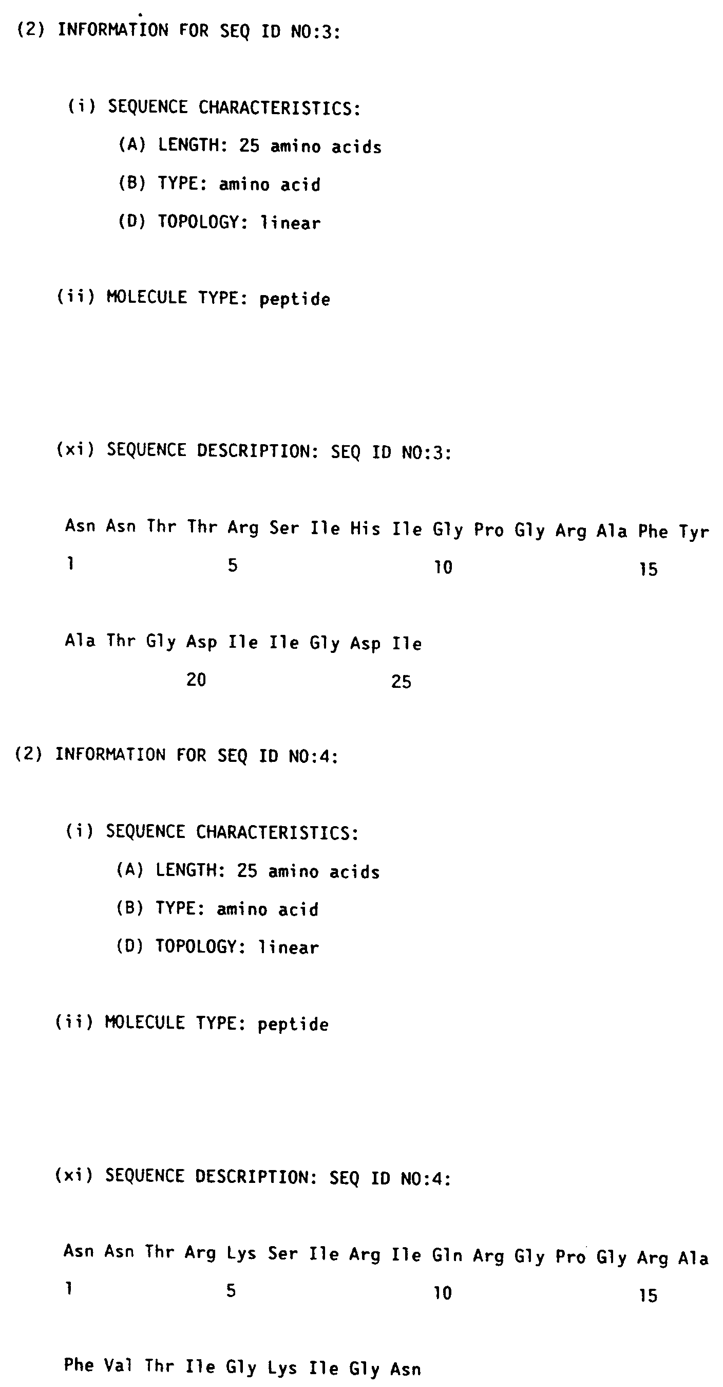

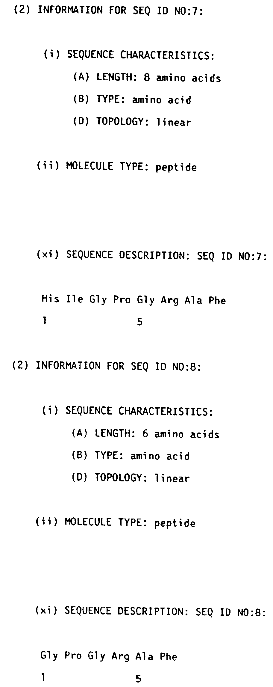

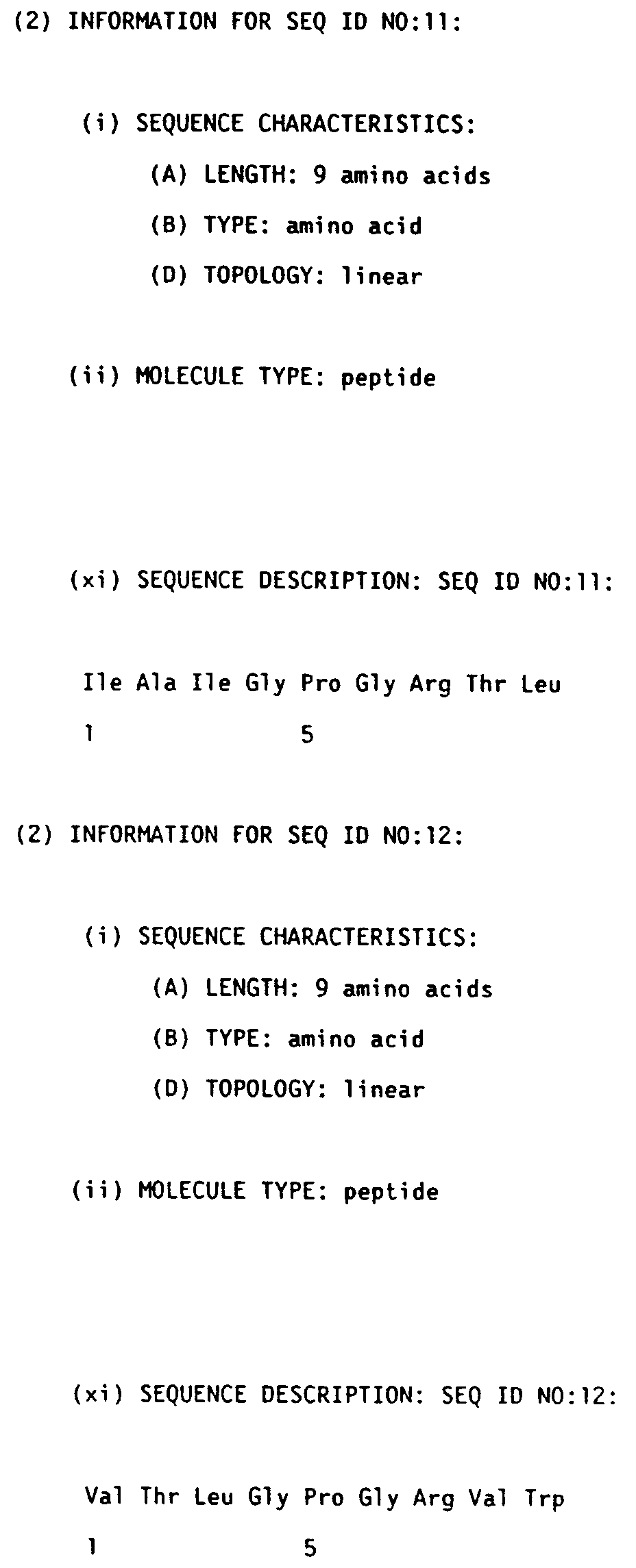


or pharmaceutically acceptable salts thereof, wherein:
PEP, MIEP, and j, are as defined supra;
-R- is:


R⁸ is a bond or lower alkyl of between one and eight carbons;
R⁹ is:
X₃ is a constituent of R³ selected from:

wherein W is preferably -(CH₂)₂- or -(CH₂)₃- or R⁶, where R⁶ is

wherein R⁷ is lower alkyl, lower alkoxy, or halo;
R¹ is:
R³ is: a peptide of 3 to 10 amino acids
R⁵ is:
X₃ is a constituent of R³ selected from:
X₂ is preferably Isoleucine.

Claims (36)
1.- A process for preparing a major immunoenhancing protein (MIEP) which possesses immunologic enhancement and mitogenic activity in mammals, which comprises:
a) lysing of the outer membrane of a Gram-negative bacterium vesicles by sodium dodecylsulfate (SDS) follwed by SDS polyacrylamide gel electrophoresis (PAGE); and
b) eluting the MIEP from the gel, dialysing against a high pH buffer and concentrating.
2.- A process for preparing the protein, according to claim 1, which possesses immunologic enhancement and mitogenic activity in adult and infant humans.
3.- A process for preparing the protein, according to claim 1, wherein the Gram-negative bacterium is of the genus Neisseria.
4.- A process for preparing the protein, according to claim 1, wherein the bacterium is Neisseria meningitidis.
5.- A process for preparing the protein, according to claim 1, which corresponds to the Class II protein of the outer membrane of the bacterium Neisseria meningitidis.
6.- A process for preparing the protein according to claim 1, which corresponds to the Class II protein of the outer membrane of Neisseria meningitidis. serogroup B.
7.- A process for preparing the protein, according to claim 1, which possesses immunological carrier activity.
8.- A process for preparing the protein according to claim 1, which possesses immunological mitogenic activity.
9.- A process for preparing a recombinant protein in substantially pure form, which possesses immunologic enhancement and mitogenic activity in mammals, which comprises:
a) extracting the genomic DNA from an outer membrane protein of a Gram negative bacterium;
b) closing the genomic DNA into an apprpriate cloning vector; or, alternatively,
using the polymerasa chain reaction technique to amplfy specific DNA sequences in genomic DNA.
using the polymerasa chain reaction technique to amplfy specific DNA sequences in genomic DNA.
c) expressing the DNA in a recombinant host cell; and
d) separating the recombinant MIEP from the other cellular proteins, using an affinity column.
10.- A process according to claim 9 wherein the recombinant host is either yeast cells or bacterial cells.
11.- A process for preparing the recombinant protein according to claim 10, which possesses immunologic enhancement and mitogenic activity in adult and infant humans.
12.- A process according to claim 11, wherein the Gram-negative bacterium is of the genus Neisseria.
13.- A process, according to claim 12, wherein the bacterium is Neisseria meningitidis.
14.- A process for preparing the recombinant protein, according to claim 13, which corresponds to the Class II protein of the outer membrane of the bacterium Neisseria meningitidis.
15.- A process for preparing the recombinant protein, according to claim 14, which corresponds to the Class II protein of the outer membrane of Neisseria meningitidis, serogroup B.
16.- A process for preparing the recombinant protein, according to claim 15, which possesses immunological carrier activity.
17.- A process for preparing the recombinant protein, according to claim 15, which possesses immunological mitogenic activity.
18.- A process for preparing a vaccine for use in mammals, in which mammals said vaccine will induce or anhance the formation of antibodies specific for said antigen, which comprises:
coupling the Class II protein of the outer membrane of Neisseria meningitidis serogroup B to an antigen.
coupling the Class II protein of the outer membrane of Neisseria meningitidis serogroup B to an antigen.
19.- A process for preparing the vaccine according to claim 18, wherein the antigens are derived from bacteria, viruses, mammalian cells, fungi, rockettsia, allergens, poisons or venoms, sythetic peptides, and polypeptide fragments.
20.- A process for preparing the vaccine, according to claim 18, wherein the Class II protein of the outer membrane of Neisseria meningitidis serogroup B is a recombinant protein produced in a recombinant host cell.
Applications Claiming Priority (10)
| Application Number | Priority Date | Filing Date | Title |
|---|---|---|---|
| US55597890A | 1990-07-19 | 1990-07-19 | |
| US55532990A | 1990-07-19 | 1990-07-19 | |
| US55520490A | 1990-07-19 | 1990-07-19 | |
| US555978 | 1990-07-19 | ||
| US555204 | 1990-07-19 | ||
| US63945791A | 1991-01-10 | 1991-01-10 | |
| US639457 | 1991-01-10 | ||
| US71527491A | 1991-06-19 | 1991-06-19 | |
| US555329 | 1991-06-19 | ||
| US715274 | 1991-06-19 |
Publications (1)
| Publication Number | Publication Date |
|---|---|
| EP0467714A1 true EP0467714A1 (en) | 1992-01-22 |
Family
ID=27541900
Family Applications (1)
| Application Number | Title | Priority Date | Filing Date |
|---|---|---|---|
| EP91306618A Withdrawn EP0467714A1 (en) | 1990-07-19 | 1991-07-19 | The class II protein of the outer membrane of neisseria meningitidis |
Country Status (2)
| Country | Link |
|---|---|
| EP (1) | EP0467714A1 (en) |
| IE (1) | IE912559A1 (en) |
Cited By (47)
| Publication number | Priority date | Publication date | Assignee | Title |
|---|---|---|---|---|
| EP0467699A2 (en) * | 1990-07-19 | 1992-01-22 | Merck & Co. Inc. | Cyclic HIV principal neutralizing determinant peptides |
| EP0467701A2 (en) * | 1990-07-19 | 1992-01-22 | Merck & Co. Inc. | Cyclic HIV principal neutralizing determinant peptides |
| EP0471453A2 (en) * | 1990-07-19 | 1992-02-19 | Merck & Co. Inc. | Cyclic HIV principal neutralizing determinant peptides |
| EP0519554A1 (en) * | 1991-06-19 | 1992-12-23 | Merck & Co. Inc. | Conjugates of the class II protein of the outer membrane of neisseria meningitidis and of HIV-1 related peptides |
| GB2271995A (en) * | 1992-10-15 | 1994-05-04 | Merck & Co Inc | HIV peptide conjugated via anionic spacer to protein |
| WO1994009822A1 (en) * | 1992-10-26 | 1994-05-11 | The United States Of America, As Represented By The Secretary, Department Of Health And Human Services | IMMUNIZATION AGAINST NEISSERIA GONORRHOEAE AND $i(NEISSERIA MENINGITIDIS) |
| WO1995003413A1 (en) * | 1993-07-23 | 1995-02-02 | The Rockefeller University | High level expression, purification and refolding of the neisseria meningitidis outer membrane group b porin proteins |
| GB2282378A (en) * | 1993-09-30 | 1995-04-05 | Merck & Co Inc | Conjugates of epitopes of HIV with a protein complex from Neisseria |
| US5747287A (en) * | 1995-04-28 | 1998-05-05 | North American Vaccine, Inc. | Method for the high level expression, purification and refolding of the outer membrane group B porin proteins from Neisseria meningitidis |
| US5763574A (en) * | 1994-10-14 | 1998-06-09 | Merck & Co., Inc. | HIV-specific synthetic antigens and their use |
| WO2000022430A2 (en) * | 1998-10-09 | 2000-04-20 | Chiron Corporation | Neisseria genomic sequences and methods of their use |
| WO2000043517A1 (en) * | 1999-01-22 | 2000-07-27 | Smithkline Beecham Biologicals S.A. | Basb055 polynucleotide and polypeptide from neisseria meningitidis. uses thereof |
| WO2000066791A1 (en) * | 1999-04-30 | 2000-11-09 | Chiron Corporation | Neisseria genomic sequences and methods of their use |
| EP1307224A2 (en) | 2000-07-31 | 2003-05-07 | GlaxoSmithKline Biologicals S.A. | Vaccines comprising outer membrane vesicles from gram negative bacteria |
| US6709660B1 (en) | 1998-01-14 | 2004-03-23 | Chrion S.R.L. | Meningococcal antigens |
| EP2261342A2 (en) | 1998-05-01 | 2010-12-15 | Novartis Vaccines and Diagnostics, Inc. | Neisseria meningitidis antigens and compositions |
| US7862827B2 (en) | 1999-05-19 | 2011-01-04 | Novartis Vaccines And Diagnostics Srl | Combination neisserial compositions |
| EP2278006A2 (en) | 1997-11-06 | 2011-01-26 | Novartis Vaccines and Diagnostics S.r.l. | Neisserial antigens |
| US8114960B2 (en) | 2000-02-28 | 2012-02-14 | Novartis Ag | Heterologous expression of Neisserial proteins |
| US8273360B2 (en) | 2000-01-17 | 2012-09-25 | Novartis Ag | Outer membrane vesicle (OMV) vaccine comprising N. meningitidis serogroup B outer membrane proteins |
| US8394390B2 (en) | 1999-10-29 | 2013-03-12 | Novartis Ag | Neisserial antigenic peptides |
| US8563007B1 (en) | 2001-10-11 | 2013-10-22 | Wyeth Holdings Corporation | Immunogenic compositions for the prevention and treatment of meningococcal disease |
| US8574597B2 (en) | 2006-12-22 | 2013-11-05 | Wyeth Llc | Immunogenic compositions for the prevention and treatment of meningococcal disease |
| US8663656B2 (en) | 2002-10-11 | 2014-03-04 | Novartis Ag | Polypeptide-vaccines for broad protection against hypervirulent meningococcal lineages |
| US8834888B2 (en) | 2009-03-24 | 2014-09-16 | Novartis Ag | Adjuvanting meningococcal factor H binding protein |
| US8840907B2 (en) | 2001-09-06 | 2014-09-23 | Novartis Vaccines And Diagnostics S.R.L. | Isolated protein and compositions comprising the protein |
| US8980286B2 (en) | 2002-11-22 | 2015-03-17 | Novartis Ag | Multiple variants of meningococcal protein NBM1870 |
| US8986710B2 (en) | 2012-03-09 | 2015-03-24 | Pfizer Inc. | Neisseria meningitidis compositions and methods thereof |
| USRE45587E1 (en) | 2001-07-26 | 2015-06-30 | Glaxosmithkline Biologicals Sa | Vaccines comprising aluminum adjuvants and histidine |
| US9156894B2 (en) | 2005-11-25 | 2015-10-13 | Glaxosmithkline Biologicals Sa | Chimeric, hybrid and tandem polypeptides of meningococcal NMB1870 |
| US9169301B2 (en) | 1999-04-30 | 2015-10-27 | Glaxosmithkline Biologicals Sa | Conserved Neisserial antigens |
| US9468673B2 (en) | 2008-02-21 | 2016-10-18 | Glaxosmithkline Biologicals Sa | Meningococcal fHBP polypeptides |
| US9556240B2 (en) | 2010-08-23 | 2017-01-31 | Wyeth Llc | Stable formulations of Neisseria meningitidis rLP2086 antigens |
| US9636393B2 (en) | 1999-11-29 | 2017-05-02 | Glaxosmithkline Biologicals Sa | Compositions comprising Neisseria meningitidis antigens from serogroups B and C |
| US9757443B2 (en) | 2010-09-10 | 2017-09-12 | Wyeth Llc | Non-lipidated variants of Neisseria meningitidis ORF2086 antigens |
| US9802987B2 (en) | 2013-03-08 | 2017-10-31 | Pfizer Inc. | Immunogenic fusion polypeptides |
| US9822150B2 (en) | 2013-09-08 | 2017-11-21 | Pfizer Inc. | Neisseria meningitidis compositions and methods thereof |
| WO2018096007A2 (en) | 2016-11-25 | 2018-05-31 | Glaxosmithkline Biologicals S.A. | Immunogenic conjugates and use thereof |
| WO2018096013A1 (en) | 2016-11-25 | 2018-05-31 | Glaxosmithkline Biologicals S.A. | nOMV-ANTIGEN CONJUGATES AND USE THEREOF |
| US10179167B2 (en) | 2010-09-10 | 2019-01-15 | Glaxosmithkline Biologicals S.A. | Developments in meningococcal outer membrane vesicles |
| US10183070B2 (en) | 2017-01-31 | 2019-01-22 | Pfizer Inc. | Neisseria meningitidis compositions and methods thereof |
| US10196429B2 (en) | 2012-03-09 | 2019-02-05 | Pfizer Inc. | Neisseria meningitidis composition and methods thereof |
| US10195264B2 (en) | 2004-04-22 | 2019-02-05 | Glaxosmithkline Biologicals S.A. | Immunising against meningococcal serogroup Y using proteins |
| US10376573B2 (en) | 2012-06-14 | 2019-08-13 | Glaxosmithkline Biologicals Sa | Vaccines for serogroup X meningococcus |
| US10478483B2 (en) | 2010-06-25 | 2019-11-19 | Glaxosmithkline Biologicals Sa | Combinations of meningococcal factor H binding proteins |
| EP3581201A1 (en) | 2018-06-15 | 2019-12-18 | GlaxoSmithKline Biologicals S.A. | Escherichia coli o157:h7 proteins and uses thereof |
| US10888611B2 (en) | 2015-02-19 | 2021-01-12 | Pfizer Inc. | Neisseria meningitidis compositions and methods thereof |
Citations (2)
| Publication number | Priority date | Publication date | Assignee | Title |
|---|---|---|---|---|
| US4459286A (en) * | 1983-01-31 | 1984-07-10 | Merck & Co., Inc. | Coupled H. influenzae type B vaccine |
| EP0372501A2 (en) * | 1988-12-07 | 1990-06-13 | BEHRINGWERKE Aktiengesellschaft | Synthetic antigens, method for their preparation and their use |
-
1991
- 1991-07-19 EP EP91306618A patent/EP0467714A1/en not_active Withdrawn
- 1991-07-19 IE IE255991A patent/IE912559A1/en unknown
Patent Citations (2)
| Publication number | Priority date | Publication date | Assignee | Title |
|---|---|---|---|---|
| US4459286A (en) * | 1983-01-31 | 1984-07-10 | Merck & Co., Inc. | Coupled H. influenzae type B vaccine |
| EP0372501A2 (en) * | 1988-12-07 | 1990-06-13 | BEHRINGWERKE Aktiengesellschaft | Synthetic antigens, method for their preparation and their use |
Non-Patent Citations (2)
| Title |
|---|
| CHEMICAL ABSTRACTS, vol. 113, No. 9, August 27, 1990, Columbus, Ohio, USA J.J. DONNELLY et al. "Mechanisms of enhancement of serum antibody by a Neisseria menigitidis outer membrane protein complex conjugate vaccine" page 575, left column, abstract-No. 76 078d * |
| INFECTION AND IMMUNITY, vol. 57, No. 8, August 1989, Washington D.C., USA K. MURAKAMI et al. "Cloning and Characterization of the Structural Gene for the Class 2 Protein of Neisseria meningitidis" pages 2318-2323 * |
Cited By (129)
| Publication number | Priority date | Publication date | Assignee | Title |
|---|---|---|---|---|
| EP0467701A2 (en) * | 1990-07-19 | 1992-01-22 | Merck & Co. Inc. | Cyclic HIV principal neutralizing determinant peptides |
| EP0471453A2 (en) * | 1990-07-19 | 1992-02-19 | Merck & Co. Inc. | Cyclic HIV principal neutralizing determinant peptides |
| EP0467699A3 (en) * | 1990-07-19 | 1993-02-24 | Merck & Co. Inc. | Cyclic hiv principal neutralizing determinant peptides |
| EP0467701A3 (en) * | 1990-07-19 | 1993-03-10 | Merck & Co. Inc. | Cyclic hiv principal neutralizing determinant peptides |
| EP0471453A3 (en) * | 1990-07-19 | 1993-03-10 | Merck & Co. Inc. | Cyclic hiv principal neutralizing determinant peptides |
| EP0467699A2 (en) * | 1990-07-19 | 1992-01-22 | Merck & Co. Inc. | Cyclic HIV principal neutralizing determinant peptides |
| EP0519554A1 (en) * | 1991-06-19 | 1992-12-23 | Merck & Co. Inc. | Conjugates of the class II protein of the outer membrane of neisseria meningitidis and of HIV-1 related peptides |
| GB2271995A (en) * | 1992-10-15 | 1994-05-04 | Merck & Co Inc | HIV peptide conjugated via anionic spacer to protein |
| AU683030B2 (en) * | 1992-10-26 | 1997-10-30 | United States Of America, As Represented By The Secretary, Department Of Health And Human Services, The | Immunization against (neisseria gonorrhoeae) and (neisseria meningitidis) |
| WO1994009822A1 (en) * | 1992-10-26 | 1994-05-11 | The United States Of America, As Represented By The Secretary, Department Of Health And Human Services | IMMUNIZATION AGAINST NEISSERIA GONORRHOEAE AND $i(NEISSERIA MENINGITIDIS) |
| US5879686A (en) * | 1993-07-23 | 1999-03-09 | North American Vaccine, Inc. | Method for the high level expression, purification and refolding of the outer membrane group B porin proteins from Neisseria meningitidis |
| US5439808A (en) * | 1993-07-23 | 1995-08-08 | North American Vaccine, Inc. | Method for the high level expression, purification and refolding of the outer membrane group B porin proteins from Neisseria meningitidis |
| US6013267A (en) * | 1993-07-23 | 2000-01-11 | North American Vaccine, Inc. | Method for the high level expression, purification and refolding of the outer membrane group B porin proteins from Neisseria meningitidis |
| WO1995003413A1 (en) * | 1993-07-23 | 1995-02-02 | The Rockefeller University | High level expression, purification and refolding of the neisseria meningitidis outer membrane group b porin proteins |
| GB2282378A (en) * | 1993-09-30 | 1995-04-05 | Merck & Co Inc | Conjugates of epitopes of HIV with a protein complex from Neisseria |
| US5763574A (en) * | 1994-10-14 | 1998-06-09 | Merck & Co., Inc. | HIV-specific synthetic antigens and their use |
| US5747287A (en) * | 1995-04-28 | 1998-05-05 | North American Vaccine, Inc. | Method for the high level expression, purification and refolding of the outer membrane group B porin proteins from Neisseria meningitidis |
| EP2278006A2 (en) | 1997-11-06 | 2011-01-26 | Novartis Vaccines and Diagnostics S.r.l. | Neisserial antigens |
| US6709660B1 (en) | 1998-01-14 | 2004-03-23 | Chrion S.R.L. | Meningococcal antigens |
| EP2278011A2 (en) | 1998-01-14 | 2011-01-26 | Novartis Vaccines and Diagnostics S.r.l. | Neisseria meningitidis antigens |
| EP2210945A2 (en) | 1998-01-14 | 2010-07-28 | Novartis Vaccines and Diagnostics S.r.l. | Neisseria meningitidis antigens |
| US7714121B2 (en) | 1998-01-14 | 2010-05-11 | Novartis Vaccines And Diagnostics Srl | Meningococcal antigens |
| EP2261351A2 (en) | 1998-05-01 | 2010-12-15 | Novartis Vaccines and Diagnostics, Inc. | Neisseria meningitidis antigens and compositions |
| EP2261348A2 (en) | 1998-05-01 | 2010-12-15 | Novartis Vaccines and Diagnostics, Inc. | Neisseria meningitidis antigens and compositions |
| US9139621B2 (en) | 1998-05-01 | 2015-09-22 | Glaxosmithkline Biologicals Sa | Neisseria meningitidis antigens and compositions |
| US9249196B2 (en) | 1998-05-01 | 2016-02-02 | Glaxosmithkline Biologicals Sa | Neisseria meningitidis antigens and compositions |
| US9249198B2 (en) | 1998-05-01 | 2016-02-02 | Glaxosmithkline Biologicals Sa | Neisseria meningitidis antigens and compositions |
| US9266929B2 (en) | 1998-05-01 | 2016-02-23 | Glaxosmithkline Biologicals Sa | Neisseria meningitidis antigens and compositions |
| US8524251B2 (en) | 1998-05-01 | 2013-09-03 | J. Craig Venter Institute, Inc. | Neisseria meningitidis antigens and compositions |
| EP2261352A2 (en) | 1998-05-01 | 2010-12-15 | Novartis Vaccines and Diagnostics, Inc. | Neisseria meningitidis antigens and compositions |
| EP2261342A2 (en) | 1998-05-01 | 2010-12-15 | Novartis Vaccines and Diagnostics, Inc. | Neisseria meningitidis antigens and compositions |
| EP2261338A2 (en) | 1998-05-01 | 2010-12-15 | Novartis Vaccines and Diagnostics, Inc. | Neisseria meningitidis antigens and compositions |
| EP2261345A2 (en) | 1998-05-01 | 2010-12-15 | Novartis Vaccines and Diagnostics, Inc. | Neisseria meningitidis antigens and compositions |
| EP2261340A2 (en) | 1998-05-01 | 2010-12-15 | Novartis Vaccines and Diagnostics, Inc. | Neisseria meningitidis antigens and compositions |
| EP2261356A2 (en) | 1998-05-01 | 2010-12-15 | Novartis Vaccines and Diagnostics, Inc. | Neisseria meningitidis antigens and compositions |
| EP2261343A2 (en) | 1998-05-01 | 2010-12-15 | Novartis Vaccines and Diagnostics, Inc. | Neisseria meningitidis antigens and compositions |
| EP2261341A2 (en) | 1998-05-01 | 2010-12-15 | Novartis Vaccines and Diagnostics, Inc. | Neisseria meningitidis antigens and compositions |
| EP2261354A2 (en) | 1998-05-01 | 2010-12-15 | Novartis Vaccines and Diagnostics, Inc. | Neisseria meningitidis antigens and compositions |
| EP2261346A2 (en) | 1998-05-01 | 2010-12-15 | Novartis Vaccines and Diagnostics, Inc. | Neisseria meningitidis antigens and compositions |
| EP2261349A2 (en) | 1998-05-01 | 2010-12-15 | Novartis Vaccines and Diagnostics, Inc. | Neisseria meningitidis antigens and compositions |
| EP2261344A2 (en) | 1998-05-01 | 2010-12-15 | Novartis Vaccines and Diagnostics, Inc. | Neisseria meningitidis antigens and compositions |
| EP2261350A2 (en) | 1998-05-01 | 2010-12-15 | Novartis Vaccines and Diagnostics, Inc. | Neisseria meningitidis antigens and compositions |
| EP2261347A2 (en) | 1998-05-01 | 2010-12-15 | Novartis Vaccines and Diagnostics, Inc. | Neisseria meningitidis antigens and compositions |
| EP2261355A2 (en) | 1998-05-01 | 2010-12-15 | Novartis Vaccines and Diagnostics, Inc. | Neisseria meningitidis antigens and compositions |
| EP2261353A2 (en) | 1998-05-01 | 2010-12-15 | Novartis Vaccines and Diagnostics, Inc. | Neisseria meningitidis antigens and compositions |
| EP2261339A2 (en) | 1998-05-01 | 2010-12-15 | Novartis Vaccines and Diagnostics, Inc. | Neisseria meningitidis antigens and compositions |
| EP2261357A2 (en) | 1998-05-01 | 2010-12-15 | Novartis Vaccines and Diagnostics, Inc. | Neisseria meningitidis antigens and compositions |
| EP1559795A2 (en) * | 1998-10-09 | 2005-08-03 | Chiron Corporation | Neisseria genomic sequences and methods of their use |
| WO2000022430A3 (en) * | 1998-10-09 | 2002-06-06 | Chiron Corp | Neisseria genomic sequences and methods of their use |
| EP1559795A3 (en) * | 1998-10-09 | 2005-11-09 | Chiron Corporation | Neisseria genomic sequences and methods of their use |
| WO2000022430A2 (en) * | 1998-10-09 | 2000-04-20 | Chiron Corporation | Neisseria genomic sequences and methods of their use |
| US7612192B2 (en) | 1998-10-09 | 2009-11-03 | Novartis Vaccines & Diagnostics, Inc. | Neisseria genomic sequences and methods of their use |
| WO2000043517A1 (en) * | 1999-01-22 | 2000-07-27 | Smithkline Beecham Biologicals S.A. | Basb055 polynucleotide and polypeptide from neisseria meningitidis. uses thereof |
| AU780308B2 (en) * | 1999-04-30 | 2005-03-17 | J. Craig Venter Institute, Inc. | Neisseria genomic sequences and methods of their use |
| US9169301B2 (en) | 1999-04-30 | 2015-10-27 | Glaxosmithkline Biologicals Sa | Conserved Neisserial antigens |
| WO2000066791A1 (en) * | 1999-04-30 | 2000-11-09 | Chiron Corporation | Neisseria genomic sequences and methods of their use |
| EP1605061A1 (en) * | 1999-04-30 | 2005-12-14 | Chiron Corporation | Neisseria genomic sequences and methods of their use |
| US9364528B1 (en) | 1999-05-19 | 2016-06-14 | Glaxosmithkline Biologicals Sa | Combination neisserial compositions |
| US9610342B2 (en) | 1999-05-19 | 2017-04-04 | Glaxosmithkline Biologicals Sa | Combination neisserial compositions |
| US7862827B2 (en) | 1999-05-19 | 2011-01-04 | Novartis Vaccines And Diagnostics Srl | Combination neisserial compositions |
| US8394390B2 (en) | 1999-10-29 | 2013-03-12 | Novartis Ag | Neisserial antigenic peptides |
| US8734812B1 (en) | 1999-10-29 | 2014-05-27 | Novartis Ag | Neisserial antigenic peptides |
| US9067987B2 (en) | 1999-10-29 | 2015-06-30 | Glaxosmithkline Biologicals Sa | Neisserial antigenic peptides |
| US9636393B2 (en) | 1999-11-29 | 2017-05-02 | Glaxosmithkline Biologicals Sa | Compositions comprising Neisseria meningitidis antigens from serogroups B and C |
| US8273360B2 (en) | 2000-01-17 | 2012-09-25 | Novartis Ag | Outer membrane vesicle (OMV) vaccine comprising N. meningitidis serogroup B outer membrane proteins |
| US8703914B2 (en) | 2000-02-28 | 2014-04-22 | Novartis Ag | Heterologous expression of neisserial proteins |
| US9267163B2 (en) | 2000-02-28 | 2016-02-23 | Glaxosmithkline Biologicals Sa | Hybrid expression of neisserial proteins |
| US8114960B2 (en) | 2000-02-28 | 2012-02-14 | Novartis Ag | Heterologous expression of Neisserial proteins |
| US9150898B2 (en) | 2000-02-28 | 2015-10-06 | Glaxosmithkline Biologicals Sa | Heterologous expression of Neisserial proteins |
| EP1307224A2 (en) | 2000-07-31 | 2003-05-07 | GlaxoSmithKline Biologicals S.A. | Vaccines comprising outer membrane vesicles from gram negative bacteria |
| USRE45587E1 (en) | 2001-07-26 | 2015-06-30 | Glaxosmithkline Biologicals Sa | Vaccines comprising aluminum adjuvants and histidine |
| US8980277B2 (en) | 2001-09-06 | 2015-03-17 | Novartis Ag | Hybrid and tandem expression of neisserial proteins |
| US9011869B2 (en) | 2001-09-06 | 2015-04-21 | Glaxosmithkline Biologicals Sa | Hybrid and tandem expression of Neisserial proteins |
| US8840907B2 (en) | 2001-09-06 | 2014-09-23 | Novartis Vaccines And Diagnostics S.R.L. | Isolated protein and compositions comprising the protein |
| US9056075B2 (en) | 2001-09-06 | 2015-06-16 | Glaxosmithkline Biologicals Sa | Methods of inducing an immune response with compositions comprising a Neisseria meningitidis 741 protein |
| US8563006B2 (en) | 2001-10-11 | 2013-10-22 | Wyeth Holdings Corporation | Immunogenic compositions for the prevention and treatment of meningococcal disease |
| US10300122B2 (en) | 2001-10-11 | 2019-05-28 | Wyeth Holdings Llc | Immunogenic compositions for the prevention and treatment of meningococcal disease |
| US11116829B2 (en) | 2001-10-11 | 2021-09-14 | Wyeth Holdings Llc | Immunogenic compositions for the prevention and treatment of meningococcal disease |
| US9168293B2 (en) | 2001-10-11 | 2015-10-27 | Wyeth Holdings Llc | Immunogenic compositions for the prevention and treatment of meningococcal disease |
| US8563007B1 (en) | 2001-10-11 | 2013-10-22 | Wyeth Holdings Corporation | Immunogenic compositions for the prevention and treatment of meningococcal disease |
| US9132182B2 (en) | 2001-10-11 | 2015-09-15 | Wyeth Holdings Llc | Immunogenic compositions for the prevention and treatment of meningococcal disease |
| US9623101B2 (en) | 2001-10-11 | 2017-04-18 | Wyeth Holdings Llc | Immunogenic compositions for the prevention and treatment of meningococcal disease |
| US9107873B2 (en) | 2001-10-11 | 2015-08-18 | Wyeth Holdings Llc | Immunogenic compositions for the prevention and treatment of meningococcal disease |
| US9757444B2 (en) | 2001-10-11 | 2017-09-12 | Wyeth Holdings Llc | Immunogenic compositions for the prevention and treatment of meningococcal disease |
| US8663656B2 (en) | 2002-10-11 | 2014-03-04 | Novartis Ag | Polypeptide-vaccines for broad protection against hypervirulent meningococcal lineages |
| US9550814B2 (en) | 2002-11-22 | 2017-01-24 | Glaxosmithkline Biologicals Sa | Multiple variants of meningococcal protein NMB1870 |
| US10328142B2 (en) | 2002-11-22 | 2019-06-25 | Glaxosmithkline Biologicals Sa | Multiple variants of meningococcal protein NMB1870 |
| US8980286B2 (en) | 2002-11-22 | 2015-03-17 | Novartis Ag | Multiple variants of meningococcal protein NBM1870 |
| US10195264B2 (en) | 2004-04-22 | 2019-02-05 | Glaxosmithkline Biologicals S.A. | Immunising against meningococcal serogroup Y using proteins |
| US9156894B2 (en) | 2005-11-25 | 2015-10-13 | Glaxosmithkline Biologicals Sa | Chimeric, hybrid and tandem polypeptides of meningococcal NMB1870 |
| US8574597B2 (en) | 2006-12-22 | 2013-11-05 | Wyeth Llc | Immunogenic compositions for the prevention and treatment of meningococcal disease |
| US9468673B2 (en) | 2008-02-21 | 2016-10-18 | Glaxosmithkline Biologicals Sa | Meningococcal fHBP polypeptides |
| US9579372B2 (en) | 2008-02-21 | 2017-02-28 | Glaxosmithkline Biologicals Sa | Meningococcal fHBP polypeptides |
| US8834888B2 (en) | 2009-03-24 | 2014-09-16 | Novartis Ag | Adjuvanting meningococcal factor H binding protein |
| US9572884B2 (en) | 2009-03-24 | 2017-02-21 | Glaxosmithkline Biologicals Sa | Adjuvanting meningococcal factor H binding protein |
| US10245311B2 (en) | 2009-03-24 | 2019-04-02 | Glaxosmithkline Biologicals Sa | Adjuvanting meningococcal factor H binding protein |
| US10568953B2 (en) | 2009-03-24 | 2020-02-25 | Glaxosmithkline Biologicals Sa | Adjuvanting meningococcal factor H binding protein |
| US10478483B2 (en) | 2010-06-25 | 2019-11-19 | Glaxosmithkline Biologicals Sa | Combinations of meningococcal factor H binding proteins |
| US9556240B2 (en) | 2010-08-23 | 2017-01-31 | Wyeth Llc | Stable formulations of Neisseria meningitidis rLP2086 antigens |
| US9757443B2 (en) | 2010-09-10 | 2017-09-12 | Wyeth Llc | Non-lipidated variants of Neisseria meningitidis ORF2086 antigens |
| US11077180B2 (en) | 2010-09-10 | 2021-08-03 | Wyeth Llc | Non-lipidated variants of Neisseria meningitidis ORF2086 antigens |
| US10179167B2 (en) | 2010-09-10 | 2019-01-15 | Glaxosmithkline Biologicals S.A. | Developments in meningococcal outer membrane vesicles |
| US10512681B2 (en) | 2010-09-10 | 2019-12-24 | Wyeth Llc | Non-lipidated variants of Neisseria meningitidis ORF2086 antigens |
| US9561269B2 (en) | 2012-03-09 | 2017-02-07 | Pfizer Inc. | Neisseria meningitidis compositions and methods thereof |
| US10196429B2 (en) | 2012-03-09 | 2019-02-05 | Pfizer Inc. | Neisseria meningitidis composition and methods thereof |
| US10550159B2 (en) | 2012-03-09 | 2020-02-04 | Pfizer Inc. | Neisseria meningitidis composition and methods thereof |
| US9724402B2 (en) | 2012-03-09 | 2017-08-08 | Pfizer Inc. | Neisseria meningitidis composition and methods thereof |
| US11472850B2 (en) | 2012-03-09 | 2022-10-18 | Pfizer Inc. | Neisseria meningitidis composition and methods thereof |
| US10829521B2 (en) | 2012-03-09 | 2020-11-10 | Pfizer Inc. | Neisseria meningitidis composition and methods thereof |
| US8986710B2 (en) | 2012-03-09 | 2015-03-24 | Pfizer Inc. | Neisseria meningitidis compositions and methods thereof |
| US10376573B2 (en) | 2012-06-14 | 2019-08-13 | Glaxosmithkline Biologicals Sa | Vaccines for serogroup X meningococcus |
| US9802987B2 (en) | 2013-03-08 | 2017-10-31 | Pfizer Inc. | Immunogenic fusion polypeptides |
| US9822150B2 (en) | 2013-09-08 | 2017-11-21 | Pfizer Inc. | Neisseria meningitidis compositions and methods thereof |
| US10899802B2 (en) | 2013-09-08 | 2021-01-26 | Pfizer Inc. | Neisseria meningitidis compositions and methods thereof |
| US11680087B2 (en) | 2013-09-08 | 2023-06-20 | Pfizer Inc. | Neisseria meningitidis compositions and methods thereof |
| US10888611B2 (en) | 2015-02-19 | 2021-01-12 | Pfizer Inc. | Neisseria meningitidis compositions and methods thereof |
| WO2018096013A1 (en) | 2016-11-25 | 2018-05-31 | Glaxosmithkline Biologicals S.A. | nOMV-ANTIGEN CONJUGATES AND USE THEREOF |
| CN110248681A (en) * | 2016-11-25 | 2019-09-17 | 葛兰素史密丝克莱恩生物有限公司 | NOMV- antigen conjugate and application thereof |
| BE1025443B1 (en) * | 2016-11-25 | 2019-02-28 | Glaxosmithkline Biologicals Sa | NOMV-ANTIGEN CONJUGATES AND THEIR USE |
| EP3799883A1 (en) | 2016-11-25 | 2021-04-07 | GlaxoSmithKline Biologicals S.A. | Nomv-antigen conjugated compounds and use thereof |
| BE1025443A1 (en) | 2016-11-25 | 2019-02-21 | Glaxosmithkline Biologicals Sa | NOMV-ANTIGEN CONJUGATES AND THEIR USE |
| BE1025210A1 (en) | 2016-11-25 | 2018-12-05 | Glaxosmithkline Biologicals Sa | IMMUNOGENIC CONJUGATES AND THEIR USE |
| WO2018096007A2 (en) | 2016-11-25 | 2018-05-31 | Glaxosmithkline Biologicals S.A. | Immunogenic conjugates and use thereof |
| US10543267B2 (en) | 2017-01-31 | 2020-01-28 | Pfizer Inc. | Neisseria meningitidis compositions and methods thereof |
| US10813989B2 (en) | 2017-01-31 | 2020-10-27 | Pfizer Inc. | Neisseria meningitidis compositions and methods thereof |
| US10183070B2 (en) | 2017-01-31 | 2019-01-22 | Pfizer Inc. | Neisseria meningitidis compositions and methods thereof |
| US11730800B2 (en) | 2017-01-31 | 2023-08-22 | Pfizer Inc. | Neisseria meningitidis compositions and methods thereof |
| WO2019238757A1 (en) | 2018-06-15 | 2019-12-19 | Glaxosmithkline Biologicals Sa | Escherichia coli o157:h7 proteins and uses thereof |
| EP3581201A1 (en) | 2018-06-15 | 2019-12-18 | GlaxoSmithKline Biologicals S.A. | Escherichia coli o157:h7 proteins and uses thereof |
Also Published As
| Publication number | Publication date |
|---|---|
| IE912559A1 (en) | 1992-01-29 |
Similar Documents
| Publication | Publication Date | Title |
|---|---|---|
| EP0467714A1 (en) | The class II protein of the outer membrane of neisseria meningitidis | |
| EP0161188B1 (en) | Covalently-modified bacterial polysaccharides, stable covalent conjugates of such polysaccharides and immunogenic proteins with bigeneric spacers, and methods of preparing such polysaccharides and conjugates and of confirming covalency | |
| RU2201421C2 (en) | Synthetic hiv peptide (variants), immunogenic composition for induction of immune response against hiv peptides, diagnostic set for assay of hiv-specific antibodies | |
| EP0186576B1 (en) | Covalently-modified neutral bacterial polysaccharides, stable covalent conjugates of such polysaccharides and immunogenic proteins, and methods of preparing such polysaccharides and conjugates | |
| US5371197A (en) | Protein-dimeric polysaccharide conjugate vaccine | |
| US5606030A (en) | Coconjugates of OMPC, HIV related peptides and anionic moieties | |
| NO843464L (en) | SYNTHETIC HEAT STABLE ENTEROTOXIN POLYPEPTIDE OF ESCHERICHIA COLI AND MULTIMATE THEREOF | |
| WO1993022343A1 (en) | Multiple antigen peptide system having adjuvant properties and vaccines prepared therefrom | |
| JP2008511306A (en) | Domains and epitopes of meningococcal protein NMB1870 | |
| CS275838B6 (en) | Method for production of hybridoms used for monoclonal antigen against hiv virus producing | |
| CN104507496B (en) | Including the immunogenic compound with the HIV GP41 peptides of CRM197 carrier protein couplets | |
| EP0519554A1 (en) | Conjugates of the class II protein of the outer membrane of neisseria meningitidis and of HIV-1 related peptides | |
| EP0468714A2 (en) | Peptide-polysaccharide-protein conjugate vaccines | |
| JPH03128399A (en) | Aids immune complex | |
| CA2050635A1 (en) | Class ii protein of the outer membrane of neisseria meningitidis having immunologic carrier and enhancement properties, and vaccines containing same | |
| IE912534A1 (en) | Cyclic hiv principal neutralizing determinant peptides | |
| EP0551689A2 (en) | Cyclic HIV principal neutralizing determinant (PNP) peptides | |
| CA2047043A1 (en) | Class ii protein of the outer membrane of neisseria meningitidis having immune enhancement properties | |
| JP2001505763A (en) | HIV P-17 peptide fragment, composition containing the same, and methods for producing and using the same | |
| CA2047030A1 (en) | Class ii protein of the outer membrane of neisseria meningitidis having immunologic carrier and enhancement properties | |
| KR100348183B1 (en) | Tandem Synthetic HIV-1 Peptides | |
| EP0547681A2 (en) | Synthetic peptides comprising a cyclic HIV principal neutralizing determinant and a lipopeptide | |
| EP0547689A2 (en) | Synthetic peptides comprising a cyclic HIV principal neutralizing determinant and a T-cell stimulatory epitope |
Legal Events
| Date | Code | Title | Description |
|---|---|---|---|
| PUAI | Public reference made under article 153(3) epc to a published international application that has entered the european phase |
Free format text: ORIGINAL CODE: 0009012 |
|
| AK | Designated contracting states |
Kind code of ref document: A1 Designated state(s): AT BE CH DE DK ES FR GB GR IT LI LU NL SE |
|
| 17P | Request for examination filed |
Effective date: 19920612 |
|
| STAA | Information on the status of an ep patent application or granted ep patent |
Free format text: STATUS: THE APPLICATION HAS BEEN WITHDRAWN |
|
| 18W | Application withdrawn |
Withdrawal date: 19930722 |