EP2112166A2 - Human monoclonal antibodies to CTLA-4 - Google Patents
Human monoclonal antibodies to CTLA-4 Download PDFInfo
- Publication number
- EP2112166A2 EP2112166A2 EP09007161A EP09007161A EP2112166A2 EP 2112166 A2 EP2112166 A2 EP 2112166A2 EP 09007161 A EP09007161 A EP 09007161A EP 09007161 A EP09007161 A EP 09007161A EP 2112166 A2 EP2112166 A2 EP 2112166A2
- Authority
- EP
- European Patent Office
- Prior art keywords
- antibody
- ctla
- antibodies
- human
- amino acid
- Prior art date
- Legal status (The legal status is an assumption and is not a legal conclusion. Google has not performed a legal analysis and makes no representation as to the accuracy of the status listed.)
- Granted
Links
Images


Classifications
-
- C—CHEMISTRY; METALLURGY
- C07—ORGANIC CHEMISTRY
- C07K—PEPTIDES
- C07K16/00—Immunoglobulins [IGs], e.g. monoclonal or polyclonal antibodies
- C07K16/18—Immunoglobulins [IGs], e.g. monoclonal or polyclonal antibodies against material from animals or humans
- C07K16/28—Immunoglobulins [IGs], e.g. monoclonal or polyclonal antibodies against material from animals or humans against receptors, cell surface antigens or cell surface determinants
- C07K16/2803—Immunoglobulins [IGs], e.g. monoclonal or polyclonal antibodies against material from animals or humans against receptors, cell surface antigens or cell surface determinants against the immunoglobulin superfamily
- C07K16/2818—Immunoglobulins [IGs], e.g. monoclonal or polyclonal antibodies against material from animals or humans against receptors, cell surface antigens or cell surface determinants against the immunoglobulin superfamily against CD28 or CD152
-
- A—HUMAN NECESSITIES
- A61—MEDICAL OR VETERINARY SCIENCE; HYGIENE
- A61P—SPECIFIC THERAPEUTIC ACTIVITY OF CHEMICAL COMPOUNDS OR MEDICINAL PREPARATIONS
- A61P29/00—Non-central analgesic, antipyretic or antiinflammatory agents, e.g. antirheumatic agents; Non-steroidal antiinflammatory drugs [NSAID]
-
- A—HUMAN NECESSITIES
- A61—MEDICAL OR VETERINARY SCIENCE; HYGIENE
- A61P—SPECIFIC THERAPEUTIC ACTIVITY OF CHEMICAL COMPOUNDS OR MEDICINAL PREPARATIONS
- A61P35/00—Antineoplastic agents
-
- A—HUMAN NECESSITIES
- A61—MEDICAL OR VETERINARY SCIENCE; HYGIENE
- A61P—SPECIFIC THERAPEUTIC ACTIVITY OF CHEMICAL COMPOUNDS OR MEDICINAL PREPARATIONS
- A61P37/00—Drugs for immunological or allergic disorders
-
- A—HUMAN NECESSITIES
- A61—MEDICAL OR VETERINARY SCIENCE; HYGIENE
- A61P—SPECIFIC THERAPEUTIC ACTIVITY OF CHEMICAL COMPOUNDS OR MEDICINAL PREPARATIONS
- A61P37/00—Drugs for immunological or allergic disorders
- A61P37/02—Immunomodulators
-
- A—HUMAN NECESSITIES
- A61—MEDICAL OR VETERINARY SCIENCE; HYGIENE
- A61P—SPECIFIC THERAPEUTIC ACTIVITY OF CHEMICAL COMPOUNDS OR MEDICINAL PREPARATIONS
- A61P37/00—Drugs for immunological or allergic disorders
- A61P37/02—Immunomodulators
- A61P37/04—Immunostimulants
-
- C—CHEMISTRY; METALLURGY
- C07—ORGANIC CHEMISTRY
- C07K—PEPTIDES
- C07K2317/00—Immunoglobulins specific features
- C07K2317/20—Immunoglobulins specific features characterized by taxonomic origin
- C07K2317/21—Immunoglobulins specific features characterized by taxonomic origin from primates, e.g. man
Definitions
- CDR-4 cytotoxic T-lymphocyte antigen 4
- Nucleotide sequences encoding and amino acid sequences comprising heavy and light chain immunoglobulin molecules, particularly contiguous heavy and light chain sequences spanning the complementarity determining regions (CDRs), specifically from within FR1 and/or CDR1 through CDR3 and/or within FR4, are provided. Further provided are antibodies having similar binding properties and antibodies (or other antagonists) having similar functionality as antibodies disclosed herein.
- T cells and B cells have been extensively studied and characterized in connection with the regulation of immune response. From these studies, the role of T cells appear, in many cases, to be particularly important in disease prevention and treatment.
- T cells possess very complex systems for controlling their interactions. Interactions between T cells utilize numerous receptors and soluble factors for the process. Thus, what effect any particular signal may have on the immune response generally varies and depends on the particular factors, receptors and counter-receptors that are involved in the pathway. The pathways for down-regulating responses are as important as those required for activation. Thymic education leading to T-cell tolerance is one mechanism for preventing an immune response to a particular antigen. Other mechanisms, such as secretion of suppressive cytokines, are also known.
- T cells receptor T cell receptor (TCR)
- TCR antigen receptor
- CD28 co-stimulatory surface molecules
- B7-1 (CD80) and B7-2 (CD86) proteins which are expressed on antigen-presenting cells such as dendritic cells, activated B-cells or monocytes that interact with T-cell CD28 or CTLA-4 to deliver a costimulatory signal.
- CD80 B7-1
- CD86 B7-2
- the role of costimulatory signaling was studied in experimental allergic encephalomyelitis (EAE) by Perrin et al. Immunol Res 14:189-99 (1995 ).
- EAE is an autoimmune disorder, induced by Th1 cells directed against myelin antigens that provides an in vivo model for studying the role of B7-mediated costimulation in the induction of a pathological immune response.
- a soluble fusion protein ligand for the B7 receptors as well as monoclonal antibodies specific for either CD80 or CD86, Perrin et al. demonstrated that B7 costimulation plays a prominent role in determining clinical disease outcome in EAE.
- B7 and CD28 are co-stimulatory signaling pathways that appear to be sufficient to trigger the maturation and proliferation of antigen specific T-cells.
- a variety of viruses and tumors may block T cell activation and proliferation, leading to insufficient activity or non-reactivity of the host's immune system to the infected or transformed cells.
- anergy may be at least partly responsible for the failure of the host to clear the pathogenic or tumorgenic cells.
- T-cells required two types of signals from the antigen presenting cell (APC) for activation and subsequent differentiation to effector function.
- APC antigen presenting cell
- B7-1 CD80
- B7-2 B7-2
- CTLA-4 Human CTLA-4 was identified and cloned shortly thereafter by Dariavach et al. Eur. J. Immunol. 18:1901-1905 (1988 ). The murine and human CTLA-4 molecules possess approximately 76% overall sequence homology and approach complete sequence identity in their cytoplasmic domains ( Dariavach et al. Eur. J. Immunol. 18:1901-1905 (1988 )). CTLA-4 is a member of the immunoglobulin (Ig) superfamily of proteins.
- Ig immunoglobulin
- the Ig superfamily is a group of proteins that share key structural features of either a variable (V) or constant (C) domain of Ig molecules.
- Members of the Ig superfamily include, but are not limited to, the immunoglobulins themselves, major histocompatibility complex (MHC) class molecules (i.e., MHC class I and II), and TCR molecules.
- MHC major histocompatibility complex
- CTLA-4 was a second receptor for B7.
- Harper et al. J Immunol 147:1037-44 (1991 ) demonstrated that the CTLA-4 and CD28 molecules are closely related in both mouse and human as to sequence, message expression, gene structure, and chromosomal location. See also Balzano et al. Int J Cancer Suppl 7:28-32 (1992 ). Further evidence of this role arose through functional studies. For example, Lenschow et al. Science 257:789-792 (1992 ) demonstrated that CTLA-4-Ig induced long term survival of pancreatic islet grafts.
- the blast cells also infiltrated liver, heart, lung, and pancreas tissue, and amounts of serum immunoglobulin were elevated and their T cells proliferated spontaneously and strongly when stimulated through the T cell receptor, however, they were sensitive to cell death induced by cross-linking of the Fas receptor and by gamma irradiation.
- CTLA-4 acts as a negative regulator of T cell activation and is vital for the control of lymphocyte homeostasis.
- Allison and Krummel Science 270:932-933 (1995 ) discussed the work of Waterhouse et al. as demonstrative that CTLA-4 acts to down regulate T-cell responsiveness or has an inhibitory signaling role in T-cell activation and development.
- CTLA-4 regulates the intensity of the autoimmune response in EAE, attenuating inflammatory cytokine production and clinical disease manifestations. See also Hurwitz et al. J Neuroimmunol 73:57-62 (1997 ) and Cepero et al. J Exp Med 188:199-204 (1998 ) (an anti-CTLA-4 hairpin ribozyme that specifically abrogates CTLA-4 expression after gene transfer into a murine T-cell model).
- CTLA-4 monoclonal antibodies assessed the functional effects of a panel of CTLA-4 monoclonal antibodies (mAbs) on resting human CD4+ T cells. Their results demonstrated that some CTLA-4 mAbs could inhibit proliferative responses of resting CD4+ cells and cell cycle transition from G0 to G1. The inhibitory effects of CTLA-4 were evident within 4 h, at a time when cell surface CTLA-4 expression remained undetectable. Other CTLA-4 mAbs, however, had no detectable inhibitory effects, indicating that binding of mAbs to CTLA-4 alone was not sufficient to mediate down-regulation of T cell responses.
- T cells exposed to tolerogenic antigen in the presence of both IL-12 and anti-CTLA-4 antibody were not anergized, and behaved identically to T cells which have encountered immunogenic antigen.
- CTLA-4 engagement which leads to a block in the ability of T cells to proliferate
- IL-12 a prototypic inflammatory cytokine
- the combination of IL-12 and anti-CTLA-4 antibody was sufficient to convert a normally tolerogenic stimulus to an immunogenic one.
- Certain tumors such as the SM1 mammary carcinoma, were refractory to anti-CTLA-4 immunotherapy.
- CTLA-4 blockade and a vaccine consisting of granulocyte-macrophage colony-stimulating factor-expressing SM1 cells, regression of parental SM1 tumors was observed, despite the ineffectiveness of either treatment alone.
- This combination therapy resulted in long-lasting immunity to SM1 and depended on both CD4(+) and CD8(+) T cells.
- CTLA-4 blockade acts at the level of a host-derived antigen-presenting cell.
- CTLA-4 can potently inhibit T cell activation in the absence of CD28, indicating that antagonism of a TCR-mediated signal is sufficient to explain the inhibitory effect of CTLA-4.
- Lin et al. J Exp Med 188:199-204 (1998 ) studied rejection of heart allografts in CD28-deficient mice. H-2(q) hearts were transplanted into allogeneic wild-type or CD28-deficient mice (H-2(b)). Graft rejection was delayed in CD28-deficient compared with wild-type mice. Treatment of wild-type recipients with CTLA-4-immunoglobulin (Ig), or with anti-B7-1 plus anti-B7-2 mAbs significantly prolonged allograft survival.
- CTLA-4-immunoglobulin (Ig) or with anti-B7-1 plus anti-B7-2 mAbs significantly prolonged allograft survival.
- CTLA-4-Ig In contrast, treatment of CD28-deficient mice with CTLA-4-Ig, anti-B7-1 plus anti-B7-2 mAbs, or a blocking anti-CTLA-4 mAb induced acceleration of allograft rejection.
- This increased rate of graft rejection was associated with more severe mononuclcar cell infiltration and enhanced levels of IFN-gamma and IL-6 transcripts in donor hearts of untreated wild-type and CTLA-4-Ig- or anti-CTLA-4 mAb-treated CD28-deficient mice.
- the negative regulatory role of CTLA-4 extends beyond its potential ability to prevent CD28 activation through ligand competition. Even in the absence of CD28, CTLA-4 plays an inhibitory role in the regulation of allograft rejection.
- CTLA-4 has been investigated.
- Alegre et al. J Immunol 157:4762-70 (1996 ) proposed that surface CTLA-4 is rapidly internalized, which may explain the low levels of expression generally detected on the cell surface. They concluded that both CD28 and IL-2 play important roles in the up-regulation of CTLA-4 expression.
- the cell surface accumulation of CTLA-4 appeared to be primarily regulated by its rapid endocytosis.
- Castan et al. Immunology 90:265-71 (1997 ) based on in situ immunohistological analyses of the expression of CTLA-4 suggested that germinal center T cells, which were CTLA-4 positive, could be important to immune regulation.
- CTLA-4 in view of the broad and pivotal role that CTLA-4 appears to possess in immune responsiveness, it would be desirable to generate antibodies to CTLA-4 that can be utilized effectively in immunotherapy. Moreover, it would be desirable to generate antibodies against CTLA-4 that can be utilized in chronic diseases in which repeat administrations of the antibodies are required.
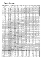
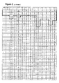
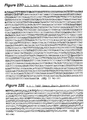
- an antibody that is capable of binding CTLA-4, comprising a heavy chain variable region amino acid sequence that comprises a contiguous amino acid sequence from within an FR1 sequence through an FR3 sequence that is encoded by a human V H 3-33 family gene and that comprises at least one of the amino acid substitutions in the CDR1 sequences, CDR2 sequences, or framework sequences shown in Figure 2 .
- the amino acid sequence comprises a sequence selected from the group consisting of SEQ ID NO: 1, SEQ ID NO:2, SEQ ID NO:3, SEQ ID NO:4, SEQ ID NO:5, SEQ ID NO:6, SEQ ID NO:8, SEQ ID NO:9, SEQ ID NO:10, SEQ ID NO:11, SEQ ID NO:12, SEQ ID NO:13, SEQ ID NO:63, SEQ ID NO:64, SEQ ID NO:66, SEQ ID NO:68, and SEQ ID NO:70.
- the antibody further comprises a light chain variable region amino acid sequence comprising a sequence selected from the group consisting of a sequence comprising SEQ ID NO:14, SEQ ID NO:15, SEQ ID NO:16, SEQ ID NO:17, SEQ ID NO:18, SEQ ID NO:19, SEQ ID NO:20, SEQ ID NO:21, SEQ ID NO:22, SEQ ID NO:23, SEQ ID NO:24, SEQ ID NO:25, SEQ ID NO:26, SEQ ID NO:65, SEQ ID NO:67, SEQ ID NO:69, and SEQ ID NO:71.
- an antibody comprising a heavy chain amino acid sequence comprising SEQ ID NO:1 and a light chain variable amino acid sequence comprising SEQ ID NO:14.
- an antibody comprising a heavy chain amino acid sequence comprising SEQ ID NO:2 and a light chain variable amino acid sequence comprising SEQ ID NO:15.
- an antibody comprising a heavy chain amino acid sequence comprising SEQ ID NO:4 and a light chain variable amino acid sequence comprising SEQ ID NO:17.
- an isolated human monoclonal antibody that is capable of binding to CTLA-4.
- antibody is capable of competing for binding with CTLA-4 with an antibody selected from the group consisting of 3.1.1, 4.1.1, 4.8.1, 4.10.2, 4.13.1, 4.14.3, 6.1.1, 11.2.1, 11.6.1, 11.7.1, 12.3.1.1, and 12.9.1.1.
- the antibody possesses a substantially similar binding specificity to CTLA-4 as an antibody selected from the group consisting of 3.1.1, 4.1.1, 4.8.1, 4.10.2, 4.13.1, 4.14.3, 6.1.1, 11.2.1, 11.6.1, 11.7.1, 12.3.1.1, and 12.9.1.1.
- the antibody is selected from the group consisting of 3.1.1, 4.1.1, 4.8.1, 4.10.2, 4.13.1, 4.14.3, 6.1.1, 11.2.1, 11.6.1, 11.7.1, 12.3.1.1, and 12.9.1.1.
- the antibody is not cross reactive with CTLA-4 from lower mammalian species, preferably the lower mammalian species comprises mouse, rat, and rabbit and more preferably mouse and rat.
- the antibody is cross reactive with CTLA-4 from primates, preferably the primates comprise cynomolgous and rhesus monkeys.
- the antibody possesses a selectivity for CTLA-4 over CD28, B7-2, CD44, and hIgG1 of greater than about 100:1 and preferably about 500:1 or greater.
- the binding affinity of the antibody is about 10 -9 M or greater and preferably about 10 -10 M or greater.
- the antibody inhibits binding between CTLA-4 and B7-2 with an IC 50 of lower than about 100 nM and preferably lower than about 0.38 nM.
- the antibody inhibits binding between CTLA-4 and B7-1 with an IC 50 of lower than about 100 nM or greater and preferably lower than about 0.50 nM.
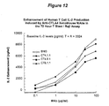
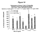
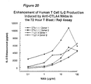
- the antibody enhances IL-2 production in a T cell blast/Raji assay by about 500 pg/ml or greater and preferably by about 3846 pg/ml or greater. In another preferred embodiment, the antibody enhances IFN- ⁇ production in a T cell blast/Raji assay by about 500 pg/ml or greater and preferably by about 1233 pg/ml or greater. In another preferred embodiment, the antibody enhances IL-2 production in a hPBMC or whole blood superantigen assay by about 500 pg/ml or greater. In another preferred embodiment, the antibody enhances IL-2 production in a hPBMC or whole blood superantigen assay by about 500 pg/ml or greater. In another preferred embodiment, the antibody enhances IL-2 production in a hPBMC or whole blood superantigen assay by about 500 pg/ml or preferably 1500 pg/ml or greater or by greater than about 30% or preferably 50% relative to control.
- a humanized antibody that possesses a substantially similar binding specificity to CTLA-4 as an antibody selected from the group consisting of 3.1.1, 4.1.1, 4.8.1, 4.10.2, 4.13.1, 4.14.3, 6.1.1, 11.2.1, 11.6.1, 11.7.1, 12.3.1.1, and 12.9.1.1.
- the antibody is not cross reactive with CTLA-4 from lower mammalian species, preferably the lower mammalian species comprises mouse, rat, and rabbit and preferably mouse and rat.
- the antibody is cross reactive with CTLA-4 from primates, preferably the primates comprise cynomolgous and rhesus monkeys.
- the antibody possesses a selectivity for CTLA-4 over CD28, B7-2, CD44, and hIgG1 of greater than about 100:1 and preferably about 500:1 or greater.
- the binding affinity of the antibody is about 10 -9 M or greater and preferably about 10 -10 M or greater.
- the antibody inhibits binding between CTLA-4 and B7-2 with an IC 50 of lower than about 100 nM and preferably lower than about 0.38 nM.
- the antibody inhibits binding between CTLA-4 and B7-1 with an IC 50 of lower than about 100 nM or greater and preferably lower than about 0.50 nM.
- the antibody enhances IL-2 production in a T cell blast/Raji assay by about 500 pg/ml or greater and preferably by about 3846 pg/ml or greater. In another preferred embodiment, the antibody enhances IFN- ⁇ production in a T cell blast/Raji assay by about 500 pg/ml or greater and preferably by about 1233 pg/ml or greater. In another preferred embodiment, the antibody induces IL-2 production in a hPBMC or whole blood superantigen assay by about 500 pg/ml or greater. In another preferred embodiment, the antibody enhances IL-2 production in a hPBMC or whole blood superantigen assay by about 500 pg/ml or preferably 1500 pg/ml or greater or by greater than about 30% or preferably 50% relative to control.
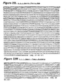
- an antibody that binds to CTLA-4 comprising a heavy chain amino acid sequence comprising human FR1, FR2, and FR3 sequences encoded by a human V H 3-33 gene family operably linked in frame with a CDR1, a CDR2, and a CDR3 sequence, the CDR1, CDR2, and CDR3 sequences being independently selected from the CDR1, CDR2, and CDR3 sequences illustrated in Figure 2 .
- the antibody of Claim 32 further comprising any of the somatic mutations to the FR1, FR2, and FR3 sequences as illustrated in Figure 2 .
- an antibody that binds to CTLA-4 comprising a heavy chain amino acid sequence comprising human FR1, FR2, and FR3 sequences encoded by a human V H 3-33 gene family operably linked in frame with a CDR1, a CDR2, and a CDR3 sequence, which antibody has the following properties: a binding affinity for CTLA-4 of about 10 -9 or greater; inhibits binding between CTLA-4 and B7-1 with an IC 50 of about 100 nM or lower; inhibits binding between CTLA-4 and B7-2 with an IC 50 of about 100 nM or lower, and enhances cytokine production in an assay of human T cells by 500 pg/ml or greater.
- an antibody that binds to CTLA-4 comprising a heavy chain amino acid sequence comprising FR1, FR2, and FR3 sequences operably linked in frame with a CDR1, a CDR2, and a CDR3 sequence independently selected from the CDR1, CDR2, and CDR3 sequences illustrated in Figures 2 and 3 , which antibody has the following properties: a binding affinity for CTLA-4 of about 10 -9 or greater; inhibits binding between CTLA-4 and B7-1 with an IC 50 of about 100 nM or lower; inhibits binding between CTLA-4 and B7-2 with an IC 50 of about 100 nM or lower; and enhances cytokine production in an assay of human T cells by 500 pg/ml or greater.
- a cell culture system for assaying T cell stimulation comprising a culture of human T cell blasts co-cultured with a Raji cell line.
- the T cell blasts are washed prior to culture with the Raji cell line.
- an assay for measuring T cell stimulation comprising: providing a culture of human T cell blasts and a Raji cell line; contacting the culture with an agent; and measuring cytokine production by the culture.
- a functional assay for screening a moiety for T cell stimulatory function comprising: providing a culture of human T cell blasts and a Raji cell line; contacting the culture with the moiety; and assessing cytokine production by the culture.
- a T cell stimulatory assay for CTLA-4 inhibitory function comprising contacting a culture comprising human T cell blasts and a Raji cell line with an agent and assessing cytokine production by the culture.
- a method for screening an agent for T cell stimulatory activity comprising: contacting the agent with a cell culture comprising human T cell blasts and a Raji cell line; and assessing cytokine production by the culture.
- the T cell blasts are washed prior to culture with the Raji cell line.
- the cytokine is IL-2 or IFN- ⁇ .
- cytokine production is measured in supernatant isolated from the culture.
- the agent is an antibody and preferably binds to CTLA-4.
- an assay for measuring T cell stimulation comprising: providing a population of human peripheral blood mononuclear cells or human whole blood stimulated with staphylococcus enterotoxin A; contacting the culture with an agent; and measuring cytokine production by the cell population.
- a functional assay for screening a moiety for T cell stimulatory function comprising: providing a population of human peripheral blood mononuclear cells or human whole blood stimulated with staphylococcus enterotoxin A; contacting the culture with the moiety; and assessing cytokine production by the cell population.
- a T cell stimulatory assay for CTLA-4 inhibitory function comprising contacting a population of human peripheral blood mononuclear cells or human whole blood stimulated with staphylococcus enterotoxin A with an agent and assessing cytokine production by the cell population.
- a method for screening an agent for T cell stimulatory activity comprising: contacting the agent with a population of human peripheral blood mononuclear cells or human whole blood stimulated with staphylococcus enterotoxin A; and assessing cytokine production by the cell population.
- the cytokine in a preferred embodiment, is IL-2. In another preferred embodiment, cytokine production is measured in supernatant isolated from the culture. In a preferred embodiment, the agent is an antibody and preferably binds to CTLA-4.
- nucleotide sequences encoding and amino acid sequences comprising heavy and light chain immunoglobulin molecules, particularly sequences corresponding to a contiguous heavy and light chain sequences from FR1 and CDR1 through CDR3 and FR4, are provided. Further provided are antibodies having similar binding properties and antibodies (or other antagonists) having similar functionality as antibodies disclosed herein. Hybridomas expressing such immunoglobulin molecules and monoclonal antibodies are also provided.
- reference sequence is a defined sequence used as a basis for a sequence comparison; a reference sequence may be a subset of a larger sequence, for example, as a segment of a full-length cDNA or gene sequence given in a sequence listing or may comprise a complete cDNA or gene sequence. Generally, a reference sequence is at least 18 nucleotides or 6 amino acids in length, frequently at least 24 nucleotides or 8 amino acids in length, and often at least 48 nucleotides or 16 amino acids in length.
- two polynucleotides or amino acid sequences may each (1) comprise a sequence (i.e., a portion of the complete polynucleotide or amino acid sequence) that is similar between the two molecules, and (2) may further comprise a sequence that is divergent between the two polynucleotides or amino acid sequences
- sequence comparisons between two (or more) molecules are typically performed by comparing sequences of the two molecules over a "comparison window" to identify and compare local regions of sequence similarity.
- a “comparison window”, as used herein, refers to a conceptual segment of at least 18 contiguous nucleotide positions or 6 amino acids wherein a polynucleotide sequence or amino acid sequence may be compared to a reference sequence of at least 18 contiguous nucleotides or 6 amino acid sequences and wherein the portion of the polynucleotide sequence in the comparison window may comprise additions, deletions, substitutions, and the like (i.e., gaps) of 20 percent or less as compared to the reference sequence (which does not comprise additions or deletions) for optimal alignment of the two sequences.
- Optimal alignment of sequences for aligning a comparison window may be conducted by the local homology algorithm of Smith and Waterman Adv. Appl. Math.
- sequence identity means that two polynucleotide or amino acid sequences are identical (i.e., on a nucleotide-by-nucleotide or residue-by-residue basis) over the comparison window.
- percentage of sequence identity is calculated by comparing two optimally aligned sequences over the window of comparison, determining the number of positions at which the identical nucleic acid base (e.g., A, T, C, G, U, or I) or residue occurs in both sequences to yield the number of matched positions, dividing the number of matched positions by the total number of positions in the comparison window (i.e., the window size), and multiplying the result by 100 to yield the percentage of sequence identity.
- substantially identical denotes a characteristic of a polynucleotide or amino acid sequence, wherein the polynucleotide or amino acid comprises a sequence that has at least 85 percent sequence identity, preferably at least 90 to 95 percent sequence identity, more usually at least 99 percent sequence identity as compared to a reference sequence over a comparison window of at least 18 nucleotide (6 amino acid) positions, frequently over a window of at least 24-48 nucleotide (8-16 amino acid) positions, wherein the percentage of sequence identity is calculated by comparing the reference sequence to the sequence which may include deletions or additions which total 20 percent or less of the reference sequence over the comparison window.
- the reference sequence may be a subset of a larger sequence.
- Examples of unconventional amino acids include: 4-hydroxyproline, ⁇ -carboxyglutamate, ⁇ -N,N,N-trimethyllysine, ⁇ -N-acetyllysine, O-phosphoserine, N-acetylserine, N-formylmethionine, 3-methylhistidine, 5-hydroxylysine, ⁇ -N-methylarginine, and other similar amino acids and imino acids (e.g., 4-hydroxyproline).
- the lefthand direction is the amino terminal direction and the righthand direction is the carboxy-terminal direction, in accordance with standard usage and convention.
- the lefthand end of single-stranded polynucleotide sequences is the 5' end; the lefthand direction of double-stranded polynucleotide sequences is referred to as the 5' direction.
- the direction of 5' to 3' addition of nascent RNA transcripts is referred to as the transcription direction; sequence regions on the DNA strand having the same sequence as the RNA and which are 5' to the 5' end of the RNA transcript are referred to as "upstream sequences"; sequence regions on the DNA strand having the same sequence as the RNA and which are 3' to the 3' end of the RNA transcript are referred to as "downstream sequences".
- the term "substantial identity” means that two peptide sequences, when optimally aligned, such as by the programs GAP or BESTFIT using default gap weights, share at least 80 percent sequence identity, preferably at least 90 percent sequence identity, more preferably at least 95 percent sequence identity, and most preferably at least 99 percent sequence identity.
- residue positions which are not identical differ by conservative amino acid substitutions.
- Conservative amino acid substitutions refer to the interchangeability of residues having similar side chains.
- a group of amino acids having aliphatic side chains is glycine, alanine, valine, leucine, and isoleucine; a group of amino acids having aliphatic-hydroxyl side chains is serine and threonine; a group of amino acids having amide-containing side chains is asparagine and glutamine; a group of amino acids having aromatic side chains is phenylalanine, tyrosine, and tryptophan; a group of amino acids having basic side chains is lysine, arginine, and histidine; and a group of amino acids having sulfur-containing side chains is cysteine and methionine.
- Preferred conservative amino acids substitution groups are: valine-leucine-isoleucine, phenylalanine-tyrosine, lysine-arginine, alanine-valine, glutamic-aspartic, and asparagine-glutamine.
- amino acid sequences of antibodies or immunoglobulin molecules are contemplated as being encompassed by the present invention, providing that the variations in the amino acid sequence maintain at least 75%, more preferably at least 80%, 90%, 95%, and most preferably 99%.
- conservative amino acid replacements are contemplated. Conservative replacements are those that take place within a family of amino acids that are related in their side chains.
- More preferred families are: serine and threonine are aliphatic-hydroxy family; asparagine and glutamine are an amide-containing family; alanine, valine, leucine and isoleucine are an aliphatic family; and phenylalanine, tryptophan, and tyrosine are an aromatic family.
- serine and threonine are aliphatic-hydroxy family
- asparagine and glutamine are an amide-containing family
- alanine, valine, leucine and isoleucine are an aliphatic family
- phenylalanine, tryptophan, and tyrosine are an aromatic family.
- Whether an amino acid change results in a functional peptide can readily be determined by assaying the specific activity of the polypeptide derivative. Assays are described in detail herein. Fragments or analogs of antibodies or immunoglobulin molecules can be readily prepared by those of ordinary skill in the art. Preferred amino- and carboxy-termini of fragments or analogs occur near boundaries of functional domains. Structural and functional domains can be identified by comparison of the nucleotide and/or amino acid sequence data to public or proprietary sequence databases. Preferably, computerized comparison methods are used to identify sequence motifs or predicted protein conformation domains that occur in other proteins of known structure and/or function. Methods to identify protein sequences that fold into a known three-dimensional structure are known. Bowie et al. Science 253:164 (1991 ). Thus, the foregoing examples demonstrate that those of skill in the art can recognize sequence motifs and structural conformations that may be used to define structural and functional domains in accordance with the invention.
- Preferred amino acid substitutions are those which: (1) reduce susceptibility to proteolysis, (2) reduce susceptibility to oxidation, (3) alter binding affinity for forming protein complexes, (4) alter binding affinities, and (4) confer or modify other physicochemical or functional properties of such analogs.
- Analogs can include various muteins of a sequence other than the naturally-occurring peptide sequence. For example, single or multiple amino acid substitutions (preferably conservative amino acid substitutions) may be made in the naturally-occurring sequence (preferably in the portion of the polypeptide outside the domain(s) forming intermolecular contacts.
- a conservative amino acid substitution should not substantially change the structural characteristics of the parent sequence (e.g., a replacement amino acid should not tend to break a helix that occurs in the parent sequence, or disrupt other types of secondary structure that characterizes the parent sequence).
- Examples of art-recognized polypeptide secondary and tertiary structures are described in Proteins, Structures and Molecular Principles (Creighton, Ed., W. H. Freeman and Company, New York (1984 )); Introduction to Protein Structure (C. Branden and J. Tooze, eds., Garland Publishing, New York, N.Y. (1991 )); and Thornton et at. Nature 354:105 (1991 ), which are each incorporated herein by reference.
- polypeptide fragment refers to a polypeptide that has an amino-terminal and/or carboxy-terminal deletion, but where the remaining amino acid sequence is identical to the corresponding positions in the naturally-occurring sequence deduced, for example, from a full-length cDNA sequence. Fragments typically are at least 5, 6, 8 or 10 amino acids long, preferably at least 14 amino acids long, more preferably at least 20 amino acids long, usually at least 50 amino acids long, and even more preferably at least 70 amino acids long.
- analog refers to polypeptides which are comprised of a segment of at least 25 amino acids that has substantial identity to a portion of a deduced amino acid sequence and which has at least one of the following properties: (1) specific binding to CTLA-4, under suitable binding conditions, (2) ability to block CTLA-4 binding with its receptors, or (3) ability to inhibit CTLA-4 expressing cell growth in vitro or in vivo.
- polypeptide analogs comprise a conservative amino acid substitution (or addition or deletion) with respect to the naturally-occurring sequence.
- Analogs typically are at least 20 amino acids long, preferably at least 50 amino acids long or longer, and can often be as long as a full-length naturally-occurring polypeptide.
- Peptide analogs are commonly used in the pharmaceutical industry as non-peptide drus with properties analogous to those of the template peptide. These types of non-peptide compound are termed "peptide mimetics" or "peptidomimetics”. Fauchere, J. Adv. Drug Res. 15:29 (1986 ); Veber and Freidinger TINS p.392 (1985 ); and Evans et al. J. Med. Chem. 30:1229 (1987 ), which are incorporated herein by reference. Such compounds are often developed with the aid of computerized molecular modeling. Peptide mimetics that are structurally similar to therapeutically useful peptides may be used to produce an equivalent therapeutic or prophylactic effect.
- a paradigm polypeptide i.e., a polypeptide that has a biochemical property or pharmacological activity
- Systematic substitution of one or more amino acids of a consensus sequence with a D-amino acid of the same type may be used to generate more stable peptides.
- constrained peptides comprising a consensus sequence or a substantially identical consensus sequence variation may be generated by methods known in the art ( Rizo and Gierasch Ann. Rev. Biochem. 61:387 (1992 ), incorporated herein by reference); for example, by adding internal cysteine residues capable of forming intramolecular disulfide bridges which cyclize the peptide.
- Antibody or “antibody peptide(s)” refer to an intact antibody, or a binding fragment thereof that competes with the intact antibody for specific binding. Binding fragments are produced by recombinant DNA techniques, or by enzymatic or chemical cleavage of intact antibodies. Binding fragments include Fab, Fab', F(ab') 2 , Fv, and single-chain antibodies. An antibody other than a "bispecific” or “bifunctional” antibody is understood to have each of its binding sites identical.
- An antibody substantially inhibits adhesion of a receptor to a counterreceptor when an excess of antibody reduces the quantity of receptor bound to counterreceptor by at least about 20%, 40%, 60% or 80%, and more usually greater than about 85% (as measured in an in vitro competitive binding assay).
- epitopic determinants includes any protein determinant capable of specific binding to an immunoglobulin or T-cell receptor.
- Epitopic determinants usually consist of chemically active surface groupings of molecules such as amino acids or sugar side chains and usually have specific three dimensional structural characteristics, as well as specific charge characteristics.
- An antibody is said to specifically bind an antigen when the dissociation constant is ⁇ 1 ⁇ M, preferably ⁇ 100 nM and most preferably ⁇ 10 nM.
- agent is used herein to denote a chemical compound, a mixture of chemical compounds, a biological macromolecule, or an extract made from biological materials.
- label refers to incorporation of a detectable marker, e.g., by incorporation of a radiolabeled amino acid or attachment to a polypeptide of biotinyl moieties that can be detected by marked avidin (e.g., streptavidin containing a fluorescent marker or enzymatic activity that can be detected by optical or colorimetric methods). In certain situations, the label or marker can also be therapeutic.
- marked avidin e.g., streptavidin containing a fluorescent marker or enzymatic activity that can be detected by optical or colorimetric methods.
- the label or marker can also be therapeutic.
- Various methods of labeling polypeptides and glycoproteins are known in the art and may be used. Examples of labels for polypeptides include, but are not limited to, the following: radioisotopes or radionuclides ( e .
- labels e.g ., 3 H, 14 C, 15 N, 35 S, 90 Y, 99 Tc, 111 In, 125 I, 131 I
- fluorescent labels e . g ., FITC, rhodamine, lanthanide phosphors
- enzymatic labels e . g ., horseradish peroxidase, ⁇ -galactosidase, luciferase, alkaline phosphatase
- chemiluminescent e.g., horseradish peroxidase, ⁇ -galactosidase, luciferase, alkaline phosphatase
- biotinyl groups e.g., predetermined polypeptide epitopes recognized by a secondary reporter (e.g., leucine zipper pair sequences, binding sites for secondary antibodies, metal binding domains, epitope tags).
- labels are attached by spacer arms of various lengths to reduce potential steric hindrance.
- pharmaceutical agent or drug refers to a chemical compound or composition capable of inducing a desired therapeutic effect when properly administered to a patient.
- Other chemistry terms herein are used according to conventional usage in the art, as exemplified by The McGraw-Hill Dictionary of Chemical Terms (Parker, S., Ed., McGraw-Hill, San Francisco (1985 )), incorporated herein by reference).
- anti-plastic agent is used herein to refer to agents that have the functional property of inhibiting a development or progression of a neoplasm in a human, particularly a malignant (cancerous) lesion, such as a carcinoma, sarcoma, lymphoma, or leukemia. Inhibition of metastasis is frequently a property of antineoplastic agents.
- substantially pure means an object species is the predominant species present (i.e., on a molar basis it is more abundant than any other individual species in the composition), and preferably a substantially purified fraction is a composition wherein the object species comprises at least about 50 percent (on a molar basis) of all macromolecular species present. Generally, a substantially pure composition will comprise more than about 80 percent of all macromolecular species present in the composition, more preferably more than about 85%, 90%, 95%, and 99%. Most preferably, the object species is purified to essential homogeneity (contaminant species cannot be detected in the composition by conventional detection methods) wherein the composition consists essentially of a single macromolecular species.
- patient includes human and veterinary subjects.
- the basic antibody structural unit is known to comprise a tetramer.
- Each tetramer is composed of two identical pairs of polypeptide chains, each pair having one "light” (about 25 kDa) and one "heavy” chain (about 50-70 kDa).
- the amino-terminal portion of each chain includes a variable region of about 100 to 110 or more amino acids primarily responsible for antigen recognition.
- the carboxy-terminal portion of each chain defines a constant region primarily responsible for effector function.
- Human light chains are classified as kappa and lambda light chains.
- Heavy chains are classified as mu, delta, gamma, alpha, or epsilon, and define the antibody's isotype as IgM, IgD, IgG, IgA, and IgE, respectively.
- the variable and constant regions are joined by a "J" region of about 12 or more amino acids, with the heavy chain also including a "D” region of about 10 more amino acids. See generally, Fundamental Immunology Ch. 7 (Paul, W., ed., 2nd ed. Raven Press, N.Y. (1989 )) (incorporated by reference in its entirety for all purposes).
- the variable regions of each light/heavy chain pair form the antibody binding site.
- an intact IgG antibody has two binding sites. Except in bifunctional or bispecific antibodies, the two binding sites are the same.
- the chains all exhibit the same general structure of relatively conserved framework regions (FR) joined by three hyper variable regions, also called complementarity determining regions or CDRs.
- the CDRs from the two chains of each pair are aligned by the framework regions, enabling binding to a specific epitope.
- both light and heavy chains comprise the domains FR1, CDR1, FR2, CDR2, FR3, CDR3 and FR4.
- the assignment of amino acids to each domain is in accordance with the definitions of Kabat Sequences of Proteins of Immunological Interest (National Institutes of Health, Bethesda, Md. (1987 and 1991 )), or Chothia & Lesk J. Mol. Biol. 196:901-917 (1987 ); Chothia et al. Nature 342:878-883 (1989 ).
- a bispecific or bifunctional antibody is an artificial hybrid antibody having two different heavy/light chain pairs and two different binding sites.
- Bispecific antibodies can be produced by a variety of methods including fusion of hybridomas or linking of Fab' fragments. See, e . g ., Songsivilai & Lachmarm Clin. Exp. Immunol. 79: 315-321 (1990 ), Kostelny et al. J. Immunol. 148:1547-1553 (1992 ).
- bispecific antibodies may be formed as "diabodies" ( Holliger et al.
- Human antibodies avoid certain of the problems associated with antibodies that possess murine or rat variable and/or constant regions.
- the presence of such murine or rat derived proteins can lead to the rapid clearance of the antibodies or can lead to the generation of an immune response against the antibody by a patient.
- murine or rat derived antibodies it has been postulated that one can develop humanized antibodies or generate fully human antibodies through the introduction of human antibody function into a rodent so that the rodent would produce antibodies having fully human sequences.
- the XenoMouseTM strains were engineered with yeast artificial chromosomes (YACs) containing 245 kb and 190 kb-sized germline configuration fragments of the human heavy chain locus and kappa light chain locus, respectively, which contained core variable and constant region sequences. Id.
- YACs yeast artificial chromosomes
- the human Ig containing YACs proved to be compatible with the mouse system for both rearrangement and expression of antibodies and were capable of substituting for the inactivated mouse Ig genes.
- minilocus In an alternative approach, others, including GenPharm International, Inc., have utilized a "minilocus" approach. In the minilocus approach, an exogenous Ig locus is mimicked through the inclusion of pieces (individual genes) from the Ig locus. Thus, one or more V H genes, one or more D H genes, one or more J H genes, a mu constant region, and a second constant region (preferably a gamma constant region) are formed into a construct for insertion into an animal. This approach is described in U.S. Patent No. 5,545,807 to Surani et al. and U.S. Patent Nos.
- the inventors of Surani et al. cited above and assigned to the Medical Research Counsel (the "MRC"), produced a transgenic mouse possessing an Ig locus through use of the minilocus approach.
- minilocus approach is the rapidity with which constructs including portions of the Ig locus can be generated and introduced into animals.
- a significant disadvantage of the minilocus approach is that, in theory, insufficient diversity is introduced through the inclusion of small numbers of V, D, and J genes. Indeed, the published work appears to support this concern. B-cell development and antibody production of animals produced through use of the minilocus approach appear stunted. Therefore, research surrounding the present invention has consistently been directed towards the introduction of large portions of the Ig locus in order to achieve greater diversity and in an effort to reconstitute the immune repertoire of the animals.
- HAMA Human anti-mouse antibody
- HACA human anti-chimeric antibody
- Ig cDNA for construction of chimeric immunoglobulin genes is known in the art ( Liu et al. P.N.A.S. 84:3439 (1987 ) and J.Immunol.139:3521 (1987 )).
- mRNA is isolated from a hybridoma or other cell producing the antibody and used to produce cDNA.
- the cDNA of interest may be amplified by the polymerase chain reaction using specific primers ( U.S. Pat. Nos. 4,683,195 and 4,683,202 ).
- a library is made and screened to isolate the sequence of interest.
- the DNA sequence encoding the variable region of the antibody is then fused to human constant region sequences.
- the sequences of human constant regions genes may be found in Kabat et al. (1991) Sequences of Proteins of Immunological Interest, N.I.H. publication no. 91-3242 .
- Human C region genes are readily available from known clones. The choice of isotype will be guided by the desired effector functions, such as complement fixation, or activity in antibody-dependent cellular cytotoxicity.
- Preferred isotypes are IgG1, IgG2, IgG3 and IgG4.
- Particularly preferred isotypes for antibodies of the invention are IgG2 and IgG4. Either of the human light chain constant regions, kappa or lambda, may be used.
- the chimeric, humanized antibody is then expressed by conventional methods.
- Antibody fragments such as Fv, F(ab') 2 and Fab may be prepared by cleavage of the intact protein, e.g. by protease or chemical cleavage.
- a truncated gene is designed.
- a chimeric gene encoding a portion of the F(ab') 2 fragment would include DNA sequences encoding the CH1 domain and hinge region of the H chain, followed by a translational stop codon to yield the truncated molecule.
- consensus sequences encoding the heavy and light chain J regions may be used to design oligonucleotides for use as primers to introduce useful restriction sites into the J region for subsequent linkage of V region segments to human C region segments.
- C region cDNA can be modified by site directed mutagenesis to place a restriction site at the analogous position in the human sequence.
- Expression vectors include plasmids, retroviruses, cosmids, YACs, EBV derived episomes, and the like.
- a convenient vector is one that encodes a functionally complete human CH or CL immunoglobulin sequence, with appropriate restriction sites engineered so that any VH or VL sequence can be easily inserted and expressed.
- splicing usually occurs between the splice donor site in the inserted J region and the splice acceptor site preceding the human C region, and also at the splice regions that occur within the human CH exons. Polyadenylation and transcription termination occur at native chromosomal sites downstream of the coding regions.
- the resulting chimeric antibody may be joined to any strong promoter, including retroviral LTRs, e.g. SV-40 early promoter, ( Okayama et al. Mol. Cell. Bio. 3:280 (1983 )), Rous sarcoma virus LTR ( Gorman et al. P.N.A.S. 79:6777 (1982 )), and moloney murine leukemia virus LTR ( Grosschedl et al. Cell 41:885 (1985 )); native 1g promoters, etc.
- retroviral LTRs e.g. SV-40 early promoter, ( Okayama et al. Mol. Cell. Bio. 3:280 (1983 )
- Rous sarcoma virus LTR Gorman et al. P.N.A.S. 79:6777 (1982 )
- moloney murine leukemia virus LTR Grosschedl et al. Cell 41:885 (1985 )
- native 1g promoters etc
- human antibodies or antibodies from other species can be generated through display-type technologies, including, without limitation, phage display, retroviral display, ribosomal display, and other techniques, using techniques well known in the art and the resulting molecules can be subjected to additional maturation, such as affinity maturation, as such techniques are well known in the art.
- Wright and Harris, supra . Hanes and Plucthau PNAS USA 94:4937-4942 (1997 ) (ribosomal display), Parmley and Smith Gene 73:305-318 (1988 ) (phage display), Scott TIBS 17:241-245 (1992 ), Cwirla et al. PNAS USA 87:6378-6382 (1990 ), Russel et al. Nucl.
- antibodies can be generated to CTLA-4 expressing cells, CTLA-4 itself, forms of CTLA-4, epitopes or peptides thereof, and expression libraries thereto (see e . g . U.S. Patent No. 5,703,057 ) which can thereafter be screened as described above for the activities described above.
- CTLA-4 expressing cells it is generally not desirable to kill CTLA-4 expressing cells. Rather, one generally desires to simply inhibit CTLA-4 binding with its ligands to mitigate T cell down regulation.
- One of the major mechanisms through which antibodies kill cells is through fixation of complement and participation in CDC.
- the constant region of an antibody plays an important role in connection with an antibody's ability to fix complement and participate in CDC.
- isotypes of antibodies that are capable of complement fixation and CDC, including, without limitation, the following: murine IgM, murine IgG2a, murine IgG2b, murine IgG3, human IgM, human IgG1, and human IgG3.
- isotypes that do not include, without limitation, human IgG2 and human IgG4.
- antibodies that are generated need not initially possess a particular desired isotype but, rather, the antibody as generated can possess any isotype and the antibody can be isotype switched thereafter using conventional techniques that are well known in the art.
- Such techniques include the use of direct recombinant techniques ( see e . g ., U.S. Patent No. 4,816,397 ), cell-cell fusion techniques ( see e . g ., U.S. Patent Application No. 08/730,639, filed October 11, 1996 ), among others.
- a myeloma or other cell line is prepared that possesses a heavy chain with any desired isotype and another myeloma or other cell line is prepared that possesses the light chain.
- Such cells can, thereafter, be fused and a cell line expressing an intact antibody can be isolated.
- the majority of the CTLA-4 antibodies discussed herein are human anti-CTLA-4 IgG2 antibody. Since such antibodies possess desired binding to the CTLA-4 molecule, any one of such antibodies can be readily isotype switched to generate a human IgG4 isotype, for example, while still possessing the same variable region (which defines the antibody's specificity and some of its affinity).
- antibody candidates are generated that meet desired "structural" attributes as discussed above, they can generally be provided with at least certain additional “functional” attributes that are desired through isotype switching.
- the design of other therapeutic modalities including other antibodies, other antagonists, or chemical moieties other than antibodies is facilitated.
- Such modalities include, without limitation, antibodies having similar binding activity or functionality, advanced antibody therapeutics, such as bispecific antibodies, immunotoxins, and radiolabeled therapeutics, generation of peptide therapeutics, gene therapies, particularly intrabodies, antisense therapeutics, and small molecules.
- advanced antibody therapeutics such as bispecific antibodies, immunotoxins, and radiolabeled therapeutics
- generation of peptide therapeutics such as bispecific antibodies, immunotoxins, and radiolabeled therapeutics
- gene therapies particularly intrabodies, antisense therapeutics, and small molecules.
- the effector function of the antibodies of the invention may be changed by isotype switching to an IgG1, IgG2, IgG3, IgG4, IgD, IgA, IgE, or IgM for various therapeutic uses.
- bispecific antibodies can be generated that comprise (i) two antibodies one with a specificity to CTLA-4 and another to a second molecule that are conjugated together, (ii) a single antibody that has one chain specific to CTLA-4 and a second chain specific to a second molecule, or (iii) a single chain antibody that has specificity to CTLA-4 and the other molecule.
- Such bispecific antibodies can be generated using techniques that are well known for example, in connection with (i) and (ii) see e . g ., Fanger et al. Immunol Methods 4:72-81 (1994 ) and Wright and Harris, supra. and in connection with (iii) see e . g ., Traunecker et al. Int. J. Cancer (Suppl.) 7:51-52 (1992 ).
- antibodies can be modified to act as immunotoxins utilizing techniques that are well known in the art. See e . g ., Vitetta Immunol Today 14:252 (1993 ). See also U.S. Patent No. 5,194,594 .
- modified antibodies can also be readily prepared utilizing techniques that are well known in the art. See e . g ., Junghans et al. in Cancer Chemotherapy and Biotherapy 655-686 (2d edition, Chafner and Longo, eds., Lippincott Raven (1996 )). See also U.S. Patent Nos.
- Each of immunotoxins and radiolabeled molecules would be likely to kill cells expressing CTLA-4, and particularly those cells in which the antibodies of the invention are effective.
- therapeutic peptides can be generated that are directed against CTLA-4.
- Design and screening of peptide therapeutics is discussed in connection with Houghten et al. Biotechniques 13:412-421 (1992 ), Houghten PNAS USA 82:5131-5135 (1985 ), Pinalla et al. Biotechniques 13:901-905 (1992 ), Blake and Litzi-Davis BioConjugate Chem. 3:510-513 (1992 ).
- Immunotoxins and radiolabeled molecules can also be prepared, and in a similar manner, in connection with peptidic moieties as discussed above in connection with antibodies.
- Important information related to the binding of an antibody to an antigen can be gleaned through phage display experimentation. Such experiments are generally accomplished through panning a phage library expressing random peptides for binding with the antibodies of the invention to determine if peptides can be isolated that bind. If successful, certain epitope information can be gleaned from the peptides that bind.
- phage libraries expressing random peptides can be purchased from New England Biolabs (7-mer and 12-mer libraries, Ph.D.-7 Peptide 7-mer Library Kit and Ph.D.-12 Peptide 12-mer Library Kit, respectively) based on a bacteriophage M13 system.
- Each of 7-mer and 12-mer libraries are panned or screened in accordance with the manufacturer's recommendations in which plates were coated with an antibody to capture the appropriate antibody (a goat anti-human IgG Fc for an IgG antibody for example) followed by washing.
- Bound phage are eluted with 0.2 M glycine-HCl, pH 2.2. After 3 rounds of selection/amplification at constant stringency (0.5% Tween), through use of DNA sequencing, one can characterize clones from the libraries that are reactive with one or more of the antibodies. Reactivity of the peptides can be determined by ELISA. For an additional discussion of epitope analysis of peptides see also Scott, J.K. and Smith, G.P.
- Genetic materials encoding an antibody of the invention may be included in a suitable expression system (whether viral, attenuated viral, non-viral, naked, or otherwise) and administered to a host for in vivo generation of the antibody in the host.
- Small molecule therapeutics can also be envisioned in accordance with the present invention.
- Drugs can be designed to modulate the activity of CTLA-4 based upon the present invention.
- Knowledge gleaned from the structure of the CTLA-4 molecule and its interactions with other molecules in accordance with the present invention, such as the antibodies of the invention, CD28, B7, B7-1, B7-2, and others can be utilized to rationally design additional therapeutic modalities.
- rational drug design techniques such as X-ray crystallography, computer-aided (or assisted) molecular modeling (CAMM), quantitative or qualitative structure-activity relationship (QSAR), and similar technologies can be utilized to focus drug discovery efforts.
- Rational design allows prediction of protein or synthetic structures which can interact with the molecule or specific forms thereof which can be used to modify or modulate the activity of CTLA-4. Such structures can be synthesized chemically or expressed in biological systems. This approach has been reviewed in Capsey et al. Genetically Engineered Human Therapeutic Drugs (Stockton Press, NY (1988 )). Indeed, the rational design of molecules (either peptides, peptidomimetics, small molecules, or the like) based upon known, or delineated, structure-activity relationships with other molecules (such as antibodies in accordance with the invention) has become generally routine. See, e.g ., Fry et al.
- combinatorial libraries can be designed and sythesized and used in screening programs, such as high throughput screening efforts.
- formulations include, for example, powders, pastes, ointments, jellies, waxes, oils, lipids, lipid (cationic or anionic) containing vesicles (such as Lipofectin TM ), DNA conjugates, anhydrous absorption pastes, oil-in-water and water-in-oil emulsions, emulsions carbowax (polyethylene glycols of various molecular weights), semi-solid gels, and semi-solid mixtures containing carbowax. Any of the foregoing mixtures may be appropriate in treatments and therapies in accordance with the present invention, provided that the active ingredient in the formulation is not inactivated by the formulation and the formulation is physiologically compatible and tolerable with the route of administration.
- Antibodies in accordance with the invention are preferably prepared through the utilization of a transgenic mouse that has a substantial portion of the human antibody producing genome inserted but that is rendered deficient in the production of endogenous, murine, antibodies. Such mice, then, are capable of producing human immunoglobulin molecules and antibodies and are deficient in the production of murine immunoglobulin molecules and antibodies. Technologies utilized for achieving the same are disclosed in the patents, applications, and references disclosed in the Background, herein. In particular, however, a preferred embodiment of transgenic production of mice and antibodies therefrom- is disclosed in U.S. Patent Application Serial No. 08/759,620, filed December 3, 1996 , the disclosure of which is hereby incorporated by reference. See also Mendez et al. Nature Genetics 15:146-156 (1997 ), the disclosure of which is hereby incorporated by reference.
- the antibodies derived from hybridoma cell lines discussed herein are designated 3.1.1, 4.1.1, 4.8.1, 4.10.2, 4.13.1, 4.14.3, 6.1.1, 11.2.1, 11.6.1, 11.7.1, 12.3.1.1, and 12.9.1.1.
- Each of the antibodies produced by the aforementioned cell lines are either fully human IgG2 or IgG4 heavy chains with human kappa light chains.
- antibodies in accordance with the invention possess very high affinities, typically possessing Kd's of from about 10 -9 through about 10 -11 M, when measured by either solid phase or solution phase.
- antibodies in accordance with the present invention can be expressed in cell lines other than hybridoma cell lines. Sequences encoding the cDNAs or genomic clones for the particular antibodies can be used for transformation of a suitable mammalian or nonmammalian host cells. Transformation can be by any known method for introducing polynucleotides into a host cell, including, for example packaging the polynucleotide in a virus (or into a viral vector) and transducing a host cell with the virus (or vector) or by transfection procedures known in the art, as exemplified by U.S. Patent Nos.
- Mammalian cell lines available as hosts for expression are well known in the art and include many immortalized cell lines available from the American Type Culture Collection (ATCC), including but not limited to Chinese hamster ovary (CHO) cells, NSO 0 , HeLa cells, baby hamster kidney (BHK) cells, monkey kidney cells (COS), human hepatocellular carcinoma cells (e.g., Hep G2), and a number of other cell lines.
- ATCC American Type Culture Collection
- Non-mammalian cells including but not limited to bacterial, yeast, insect, and plants can also be used to express recombinant antibodies.
- Site directed mutagenesis of the antibody CH2 domain to eliminate glycosylation may be preferred in order to prevent changes in either the immunogenicity, pharmacokinetic, and/or effector functions resulting from non-human glycosylation.
- the expression methods are selected by determining which system generates the highest expression levels and produce antibodies with constitutive CTLA-4 binding properties.
- antibodies of the invention can be enhanced using a number of known techniques.
- glutamine sythetase and DHFR gene expression systems are common approaches for enhancing expression under certain conditions.
- High expressing cell clones can be identified using conventional techniques, such as limited dilution cloning and Microdrop technology.
- the GS system is discussed in whole or part in connection with European Patent Nos. 0 216 846 , 0 256 055 , and 0 323 997 and European Patent Application No. 89303964.4 .
- Antibodies of the invention can also be produced transgenically through the generation of a mammal or plant that is transgenic for the immunoglobulin heavy and light chain sequences of interest and production of the antibody in a recoverable form therefrom.
- antibodies can be produced in, and recovered from, the milk of goats, cows, or other mammals. See, e.g. , U.S. Patent Nos. 5,827,690 , 5,756,687 , 5,750,172 , and 5,741,957 .
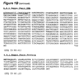
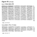
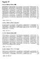
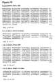
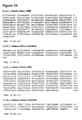
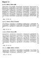
- Antibodies in accordance with the present invention have been analyzed structurally and functionally. In connection with the structures of the antibodies, amino acid sequences of the heavy and kappa light chains have been predicted based on cDNA sequences obtained through RT-PCR of the hybridomas. See Examples 3 and 4 and Figures 1-8 . N-terminal sequencing of the antibodies was also conducted in confirmation of the results discussed in Examples 3 and 4. See Example 5 and Figure 9 . Kinetic analyses of the antibodies were conducted to determine affinities. See Example 2.
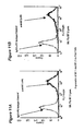
- Antibodies in accordance with the invention (and particularly the 4.1.1, 4.8.1, and 6.1.1 antibodies of the invention) have high affinities (4.1.1:1.63 X 10 10 1/M; 4.8.1:3.54 X 10 10 1/M; and 6.1.1:7.2 X 10 9 1/M). Further, antibodies were analyzed by isoelectric focusing (IEF), reducing gel electrophoresis (SDS-PAGE), size exclusion chromatography, liquid chromatography/mass spectroscopy, and mass spectroscopy and antibody production by the hybridomas was assessed. See Example 6 and Figure 10 .
- antibodies in accordance with the present invention proved to be potent inhibitors of CTLA-4 and its binding to its ligands of the B7 family of molecules.
- antibodies in accordance with the present invention were demonstrated to block CTLA-4 binding to either B7-1 or B7-2. See Example 7. Indeed, many of the antibodies in accordance with the invention possess nanomolar and subnanomolar IC 50 s with respect to inhibiting CTLA-4 binding to B7-1 and B7-2. Further, antibodies of the invention possess excellent selectivity for CTLA-4 as compared to CD28, CD44, B7-2, or hIgG1. See Example 8.
- Selectivity is a ratio that reflects the degree of preferential binding of a molecule with a first agent as compared to the molecules binding with a second, and optionally other molecules.
- selectivity refers to the degree of preferential binding of an antibody of the invention to CTLA-4 as compared to the antibody's binding to other molecules such as CD28, CD44, B7-2, or hIgG1.
- Selectivity values of antibodies of the invention greater than 500:1 are common.
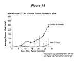
- Antibodies of the invention have also been demonstrated to induce or enhance expression of certain cytokines (such as IL-2 and IFN- ⁇ ) by cultured T cells in a T cell blast model. See Examples 9 and 10 and Figures 12-17 . Further, it is expected that antibodies of the invention will inhibit the growth of tumors in appropriate in vivo tumor models. The design of which models are discussed in Example 11 and 12.
- the 4.1.1, 4.8.1, and 6.1.1 antibodies of the invention possess highly desirable properties.
- Their structural characteristics, functions, or activities provide criteria that facilitate the design or selection of additional antibodies or other molecules as discussed above. Such criteria include one or more of the following:
- the desirable functional properties discussed above can often result from binding to and inhibition of CTLA4 by a molecule (i.e., antibody, antibody fragment, peptide, or small molecule) in a similar manner as an antibody of the invention (i.e., binding to the same or similar epitope of the CTLA4 molecule).
- the molecule may either be administered directly (i.e., direct administration to a patient of such molecules).
- the molecule may be "administered" indirectly (i.e., a peptide or the like that produces an immune response in a patient (similar to a vaccine) wherein the immune response includes the generation of antibodies that bind to the same or similar epitope or an antibody or fragment that is produced in situ after administration of genetic materials that encode such antibodies or fragments thereof which bind to the same or similar epitope).
- a peptide or the like that produces an immune response in a patient (similar to a vaccine) wherein the immune response includes the generation of antibodies that bind to the same or similar epitope or an antibody or fragment that is produced in situ after administration of genetic materials that encode such antibodies or fragments thereof which bind to the same or similar epitope.
- CTLA4 was bound to a BIAcore chip and a first antibody, under saturating conditions, was bound thereto and competition of subsequent secondary antibodies binding to CTLA4 was measured. This technique enabled generation of a rough map in to which families of antibodies can be classified.
- the 11.2.1 antibody of the invention binds to human, cynomologous, and marmoset CTLA4 at the about the same rate and has about the same relative off-rate for each of the three. This information further indicates that the 4.1.1 and 11.2.1 antibodies of the invention bind to different epitopes on CTLA4.
- each of the category B through D antibodies of the invention appear to possess similar functional properties and appear to have the potential to act as strong anti-CTLA4 therapeutic agents. Further, each of the molecules certain cross-competition in their binding for CTLA4. However, as will be observed from the above discussion, each of the molecules in the different categories appear to bind to separate conformational epitopes on CTLA4.
- the epitope information discussed above indicates that antibodies (or other molecules, as discussed above) that cross-compete with antibodies of the invention will likely have certain therapeutic potential in accordance with the present invention. Further, it is expected that antibodies (or other molecules, as discussed above) that cross-compete with antibodies of the invention (i.e., cross-compete with category B, C and/or D antibodies) will likely have certain additional therapeutic potential in accordance with the present invention.
- antibodies that cross-compete with antibodies of the invention (i.e., cross-compete with category B, C and/or D antibodies) and that (i) are not reduced in their binding to marmoset CTLA4 (similar to the 11.2.1 antibody) or (ii) are reduced in their binding to marmoset CTLA4 (similar to the 4.1.1 antibody) will likely have certain additional therapeutic potential in accordance with the present invention.
- Antibodies (or other molecules, as discussed above) that compete with categories A and E may also have certain therapeutic potential.
- Antibodies of the invention were prepared, selected, and assayed in accordance with the present Example.
- Three distinct immunogens were prepared for immunization of the XenoMouse TM mice: (i) a CTLA-4-IgG fusion protein, (ii) a CTLA-4 peptide, and (iii) 300.19 murine lymphoma cells transfected with a mutant of CTLA-4 (Y201V) that is constitutively expressed on the cell surface.
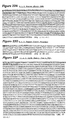
- the cDNA encoding the mature extracellular domain of CTLA-4 was PCR amplified from human thymus cDNA library (Clontech) using primers designed to published sequence ( Eur. J Immunol 18:1901-1905 (1988 )). The fragment was directionally subcloned into pSR5, a Sindbis virus expression plasmid (InVitrogen), between the human oncostatin M signal peptide and human IgG gamma 1 (IgG1) CH1/CH2/CH3 domains. The fusion protein does not contain a hinge domain but contains cysteine 120 in the extracellular domain of CTLA-4 to form a covalent dimer. The resulting vector was called CTLA-4-IgG1/pSR5.
- CTLA-4-IgG1 cDNA in the vector was sequence confirmed in both strands.
- the amino acid sequence the CTLA4-Ig protein is shown below.
- the mature extracellular domain for CD44 was PCR amplified from human lymphocyte library (Clontech) and subcloned into pSinRep5 to generate a control protein with the identical IgG1 tail.
- OM-CTLA4-IgG1 Fusion Protein Underlined: signal peptide
- Bold CTLA4 extracellular domain
- the cDNAs for mature extracellular domain of CD28 were PCR amplified from human lymphocyte library (Clontech) and then subcloned into pCDM8 ( J. Immunol. 151: 5261-71 (1993 )) to produce a human IgG1 fusion protein containing both thrombin cleavage and hinge regions.
- Marmoset, Cynomologous, and Rhesus CTLA4 were cloned from mRNA isolated from PHA stimulated PBMCs using standard techniques of degenerate PCR. Sequencing demonstrated that rhesus and cynomologous amino acid sequence were identical with three differences from mature human CTLA4 extracellular domain (S13N, I17T and L105M).
- Marmoset demonstrated ten amino acid differences from the mature human CTLA4 extracellular domain (V21A, V33I, A41T, A51G, 541, S71F, Q75K, T88M, L105M and G106S).
- Site directed mutagenesis was used to make single point mutations of all amino acids different in marmoset CTLA4 to map amino acids important for interation of the antibodies with human CTLA4-IgG.
- Mutations of human and marmoset CTLA-IgG for epitope mapping were generated by matchmaker site-directed mutagenesis (Promega).
- the IgG fusion proteins were produced by transient transfection of Cos7 cells and purified using standard Protein A techniques. Mutant CTLA4-IgG proteins were evaluated for binding to antibodies by immunoblotting and using BIAcore analyses.
- Recombinant Sindbis virus was generated by electroporating (Gibco) Baby Hamster Kidney cells with SP6 in vitro transcribed CTLA-4-IgG1/pSR5 mRNA and DH-26S helper mRNA as described by InVitrogen. Forty eight hours later recombinant virus was harvested and titered for optimal protein expression in Chinese hamster ovary cells (CHO-K1).
- CHO-K1 cells were cultured in suspension in DMEM/F12 (Gibco) containing 10% heat-inactivated fetal bovine serum (Gibco), non-essential amino acids (Gibco), 4mM glutamine (Gibco), penicillin/streptomycin (Gibco), 10mM Hepes pH 7.5 (Gibco).
- DMEM/F12 Gibco
- the CHO-K1 cells were resuspended at 1x10 7 cells/ml in DMEM/F12 and incubated with Sindbis virus for one hour at room temperature.
- DMHM/F12 fetal bovine serum depleted of bovine IgG using protein A sepharose (Pharmacia), non-essential amino acids, 4mM glutamine, 12.5mM Hepes pH 7.5, and penicillin/streptomycin. Forty eight hours post-infection cells were pelleted and conditioned media was harvested and supplemented with complete protease inhibitor tablets (Boehringer Mannheim), pH adjusted to 7.5, and filtered 0.2 ⁇ (Nalgene).
- FPLC Pharmacia
- CTLA-4-IgG1 migrated as a single band on SDS-PAGE using colloidal coomassie staining (Novex). Under non-reducing conditions CTLA-4-IgG1 was a dimer (100kDa), that reduced to a 50kDa monomer when treated with 50mM DTT. Amino acid sequencing of the purified CTLA-4-IgG1 in solution confirmed the N-terminus of CTLA-4 (MHVAQPAVVLAS), and that the oncostatin-M signal peptide was cleaved from the mature fusion protein .
- CTLA-4-IgG1 bound to immobilized B7.1-IgG in a concentration dependent manner and the binding was blocked by a hamster-anti-human anti-CTLA-4 antibody (BNI3: PharMingen).
- BNI3 hamster-anti-human anti-CTLA-4 antibody
- the sterile CTLA-4-IgG was endotoxin free and quantitated by OD280 using 1.4 as the extinction coefficient.
- the yield of purified CTLA-4-IgG ranged between 0.5-3mgs/liter of CHO-K1 cells.
- CTLA-4 peptide was prepared as described below:
- NMP N-Methylpyrrolidinone
- TFE 2,2,2-Trifluoroethanol
- DCM Dichloromethane
- FMOC Fluorenyl Methoxycarbonyl. All reagents were supplied by Perkin Elmer, with the following exceptions: TFE, Aldrich Chemical, FMOC-PAL-PEG resin, Perseptive Biosystems.
- Fmoc-Arg(PMC)-OH, FMOC-Asn(Trt)-OH, FMOC-Asp(tBu)-OH, FMOC-Cys(Trt)-OH, FMOC-Glu(tBu)-OH, FMOC-Gln(Trt)-OH, FMOC-His(Boc)-OH, FMOC-Lys(BOC)-OH, FMOC-Ser(tBu)-OH, FMOC-Thr(tBu)-OH and FMOC-Tyr(tBu)-OH were used for those amino acids requiring side chain protecting groups
- Peptide synthesis was performed on a Perkin-Elmer 431A, retrofitted with feedback monitoring via UV absorbance at 301nm (Perkin-Elmer Model 759A detector).
- the peptide sequence was assembled on a FMOC-PAL-PEG resin using conditional double coupling cycles. Forced double couplings were performed at cycles 10,11,18,19,20 and 28 through 33.
- the resin was washed with a 50% mixture of DCM and TFE at the completion of each acylation cycle, followed by capping of unreacted amino groups with acetic anhydride in NMP. Resin was removed from the reactor after completing cycle 49 and the remainder continued to completion.
- Peptide cleavage from the resin was performed using Reagent K ( King et al. International Journal of Protein and Peptide Research 36:255-266 (1990 )) for 6 hours on 415mg of resin affording 186mg crude CTLA-4 peptide.
- CTLA-4 cDNA was PCR amplified from human thymus cDNA library (Stratagene) and subcloned into pIRESneo (Clontech). A mutation of CTLA-4 that results in constitutive cell surface expression was introduced using MatchMaker Mutagenesis System (Promega). Mutation of tyrosine, Y201 to valine inhibits binding of the adaptin protein AP50 that is responsible for the rapid internalization of CTLA-4 ( Chuang et al. J. Immunol. 159:144-151 (1997 )).
- Mycoplasma-free 300.19 murine lymphoma cells were cultured in RPMI-1640 containing 10% fetal calf serum, non-essential amino acids, penicillin/streptomycin, 2mM glutamine, 12.5mM Hepes pH 7.5, and 25uM beta-mercaptoethanol. Cells were electroporated (3x10 6 /0.4ml serum free RPMI) in a 1ml chamber with 20ug CTLA-4-Y201V/pIRESneo using 200V/1180uF (Gibco CellPorator). Cells were rested for 10 minutes and then 8mls of prewarmed complete RPMI media.
- XenoMouse mice (8 to 10 weeks old) were immunized (i) subcutaneously at the base of tails with 1x10 7 300.19 cells that were transfected to express CTLA-4 as described above, resuspended in phosphate buffered saline (PBS) with complete Freund's adjuvant, or (ii) subcutaneously at the base of tail with (a) 10 ⁇ g the CTLA-4 fusion protein or (b) 10 ⁇ g CTLA-4 peptide, emulsified with complete Freund's adjuvant. In each case, the dose was repeated three or four times in incomplete Freund's adjuvant.
- PBS phosphate buffered saline
- mice received a final injection of the immunogen or cells in PBS.
- Spleen and/or lymph node lymphocytes from immunized mice were fused with the [murine non-secretory myeloma P3 cell line] and were subjected to HAT selection as previously described ( Galfre, G. and Milstein, C., "Preparation of monoclonal antibodies: strategies and procedures.” Methods Enzymol. 73:3-46 (1981 )).
- a large panel of hybridomas all secreting CTLA-4 specific human IgG 2 ⁇ or IgG 4 ⁇ (as detected below) antibodies were recovered.
- ELISA assay ELISA for determination of antigen-specific antibodies in mouse serum and in hybridoma supernatants was carried out as described ( Coligan et al., Unit 2.1, "Enzyme-linked immunosorbent assays," in Current protocols in immunology (1994 )) using CTLA-4-Ig fusion protein to capture the antibodies.
- CTLA-4-Ig fusion protein For animals that are immunized with the CTLA-4-Ig fusion protein, we additionally screen for non-specific reactivity against the human Ig portion of the fusion protein. This is accomplished using ELISA plates coated with human IgG1 as a negative control for specificity.
- Affinity measurement of purified human monoclonal antibodies, Fab fragments, or hybridoma supernatants by plasmon resonance was carried out using the BIAcore 2000 instrument, using general procedures outlined by the manufacturers.
- BIAcore sensorchip Three surfaces of the BIAcore sensorchip were immobililized with the CTLA-4-Ig fusion protein at a density ranging from approximately 390-900 using CTLA-4-Ig fusion protein at 20 or 50 ⁇ g/ml in 10 mM sodium acetate at pH 5.0 using the amine coupling kit supplied by the manufacturer (BIAcore, Inc.).
- the fourth surface of the BIAcore sensorchip was immobilized with IgG1 (900 RU) and was used as a negative control surface for non-specific binding.
- Kinetic analysis was performed at a flow rate of 25 or 50 microliters per minute and dissociation (kd or k off ) and association (ka or k on ) rates were determined using the software provided by the manufacturer (BIA evaluation 3.0) that allows for global fitting calculations.
- antibodies prepared in accordance with the invention possess high affinities and binding constants.
- Poly(A) + mRNA was isolated from approximately 2 X 10 5 hybridoma cells derived from immunized XenoMouse mice using a Fast-Track kit (Invitrogen). The generation of random primed cDNA was followed by PCR. Human V H or human V ⁇ farnily specific variable region primers ( Marks et al., "Oligonucleotide primers for polymerase chain reaction amplification of human immunoglobulin variable genes and design of family-specific oligonucleotide probes.” Eur. J. Immunol.
- PCR products were also cloned into pCRII using a TA cloning kit (Invitrogen) and both strands were sequenced using Prism dye-terminator sequencing kits and an ABI 377 sequencing machine. All sequences were analyzed by alignments to the "V BASE sequence directory" ( Tomlinson et al., MRC Centre for Protein Engineering, Cambridge, UK ) using MacVector and Geneworks software programs.
- each of the antibodies 4.1.1, 4.8.1, 11.2.1, and 6.1.1 were subjected to full length DNA sequences.
- Poly(A) + mRNA was isolated from approximately 4 X 10 6 hybridoma cells using mRNA Direct kit (Dynal). The mRNA was reverse transcribed using oligo-dT(18) and the Advantage RT/PCR kit (Clonetech).
- V Base The Variable region database (V Base) was used to design amplification primers beginning at the ATG start site of the heavy chain DP50 gene (5'-TATCTAAGCTTCTAGACTCGACCGCCACC ATG GAGTTTGGGCTGAGC TG-3') and to the stop cordon of the IgG2 constant region (5'-TTCTCTGATCAGAATTCCTATCATTTACCCGGAGACAGGGAGAGCT-3').
- An optimal Kozak sequence was added 5' to the ATG start site.
- the same method was used to design a primer to the ATG start site of the kappa chain A27 gene (5'-TCTTCAAGCTTGCCCGGGCCCGCCACC ATG GAAACCCCAGCGCAG -3') and the stop codon of the kappa constant region (5'-TTCTTTGATCAGAATTCTCACTAACACTCTCCCCTGTTGAAGC-3').
- the 012 cDNA was cloned by using a primer to the ATG start site (5'-TCTTCAAGCTTGCCCGGGCCCGCCACCATGGACATGAGGGTCCCCGC T-3) and the kappa constant region stop codon primer above.
- the heavy chain cDNAs were also cloned as genomic constructs by site directed mutagenesis to add an NheI site at the end of the variable J domain and subcloning an NheI-fragment containing the genomic IgG2 CH1/Hinge/CH2/CH3 regions.
- the point mutation to generate NheI site does not alter the amino acid sequence from germline.
- the primer pairs were used to amplify the cDNAs using Advantage High Fidelity PCR Kit (Clonetech). Sequence of the PCR was obtained by direct sequencing using dye-terminator sequencing kits and an ABI sequencing machine.
- the PCR product was cloned into pEE glutamine synthetase mammalian expression vectors (Lonza) and three clones were sequenced to confirm somatic mutations. For each clone, the sequence was verified on both strands in at least three reactions.
- An aglycosylated 4.1.1 antibody was generated by site directed mutagenesis of N294Q in the CH2 domain.
- Recombinant antibodies were produced by transient trasnfection of Cos7 cells in IgG depleted FCS and purified using standard Protein A sepharose techniques. Stable transfectants were generated by electroporation of murine NSO cells and selection in glutamine free media.
- Recombinant 4.1.1 with or without glycosylation exhibited identical specificity and affinity for CTLA4 in the in vitro ELISA and BIAcore assays.
- the DP-50 gene is also referred to as a V H 3-33 family gene. Only one antibody that was selected on the basis of CTLA-4 binding and preliminary in vitro functional assays showed a heavy chain gene utilization other than DP-50. That clone, 2.1.3, utilizes a DP-65 heavy chain variable region and is an IgG4 isotype. The DP-65 gene is also referred to as a V H 4-31 family gene. On the other hand, the clone, 4.9.1, which possesses a DP-47 heavy chain variable region binds to CTLA-4 but does not inhibit binding to B7-1 or B7-2.
- Bias is indicative of a preferred binding motif of the antibody-antigen interaction with respect to the combined properties of binding to the antigen and functional activity.
- Figure 1 provides nucleotide and predicted amino acid sequences of the heavy and kappa light chains of the clones 4.1.1 ( Figure 1A ), 4.8.1 ( Figure 1B ), 4.14.3 ( Figure 1C ), 6.1.1 ( Figure 1D ), 3.1.1 ( Figure 1E ), 4.10.2 ( Figure IF), 2.1.3 ( Figure 1G ), 4.13.1 ( Figure 1H ), 11.2.1 ( Figure 1I ), 11.6.1 ( Figure 1J ), 11.7.1 ( Figure 1K ), 12.3.1.1 ( Figure 1L ), and 12 .9.1.1 ( Figure 1M ).
- Figures 1A , 1B , and 1D extended sequences of the antibodies 4.1.1, 4.8.1, and 6.1.1 were obtained by full length cloning of the cDNAs as described above.
- the signal peptide sequence or the bases encoding the same
- Figure 2 provides a sequence alignment between the predicted heavy chain amino acid sequences from the clones 4.1.1, 4.8.1, 4.14.3, 6.1.1, 3.1.1, 4.10.2, 4.13.1, 11.2.1, 11.6.1, 11.7.1, 12.3.1.1, and 12.9.1.1 and the germline DP-50 (3-33) amino acid sequence. Differences between the DP-50 germline sequence and that of the sequence in the clones are indicated in bold. The Figure also shows the positions of the CDR1, CDR2, and CDR3 sequences of the antibodies as shaded.
- Figure 3 provides a sequence alignment between the predicted heavy chain amino acid sequence of the clone 2.1.3 and the germline DP-65 (4-31) amino acid sequence. Differences between the DP-65 germline sequence and that of the sequence in the clone are indicated in bold. The Figure also shows the positions of the CDR1, CDR2, and CDR3 sequences of the antibody as underlined.
- Figure 4 provides a sequence alignment between the predicted kappa light chain amino acid sequence of the clones 4.1.1, 4.8.1, 4.14.3, 6.1.1, 4.10.2, and 4.13.1 and the germline A27 amino acid sequence. Differences between the A27 germline sequence and that of the sequence in the clone are indicated in bold.
- the Figure also shows the positions of the CDR1, CDR2, and CDR3 sequences of the antibody as underlined. Apparent deletions in the CDR1s of clones 4.8.1, 4.14.3, and 6.1.1 are indicated with "0s”.
- Figure 5 provides a sequence alignment between the predicted kappa light chain amino acid sequence of the clones 3.1.1, 11.2.1, 11.6.1, and 11.7.1 and the germline 012 amino acid sequence. Differences between the 012 germline sequence and that of the sequence in the clone are indicated in bold. The Figure also shows the positions of the CDR1, CDR2, and CDR3 sequences of the antibody as underlined.
- Figure 6 provides a sequence alignment between the predicted kappa light chain amino acid sequence of the clone 2.1.3 and the germline A10/A26 amino acid sequence. Differences between the A10/A26 germline sequence and that of the sequence in the clone are indicated in bold. The Figure also shows the positions of the CDR1, CDR2, and CDR3 sequences of the antibody as underlined.
- Figure 7 provides a sequence alignment between the predicted kappa light chain amino acid sequence of the clone 12.3.1 and the germline A17 amino acid sequence. Differences between the A17 germline sequence and that of the sequence in the clone are indicated in bold. The Figure also shows the positions of the CDR1, CDR2, and CDR3 sequences of the antibody as underlined.
- Figure 8 provides a sequence alignment between the predicted kappa light chain amino acid sequence of the clone 12.9.1 and the germline A3/A19 amino acid sequence. Differences between the A3/A19 germline sequence and that of the sequence in the clone are indicated in bold. The Figure also shows the positions of the CDR1, CDR2, and CDR3 sequences of the antibody as underlined.
- Figure 22 provides a series of additional nucleotide and amino acid sequences of the following anti-CTLA-4 antibody chains:
- the light chains of these antibodies are derived from 5 different Vk genes.
- the A27 gene is the most heavily represented and is the source of 6 different light chains. Comparison of these 6 sequences reveals two noteworthy features. First, in three of them, 4.8.1, 4.14.3, and 6.1.1, contain deletions of one or two residues in CDR1, a rare event. Second, there is a strong prejudice against the germline serine at position six in CDR3 in that the serine has been replaced in every sequence. This suggests that a serine at this position is incompatible with CTLA4 binding.
- Figure 9 also provides the molecular weight of the heavy and light chains as determined by mass spectroscopy (MALDI).
- Figure 10 provides certain additional characterizing information about certain of the antibodies in accordance with the invention.
- data related to clones 3.1.1, 4.1.1, 4.8.1, 4.10.2, 4.14.3, and 6.1.1 is summarized. The following data is provided: Concentration, isoelectric focusing (IEF), SDS-PAGE, size exclusion chromatography, FACS, mass spectroscopy (MALDI), and light chain N-terminal sequences.
- Protein concentration was determined at 280 nm from a UV scan (200-350 nm), where 1.58 absorbance units at 280 nm equaled 1mg/ml.
- Isoelectric focusing was performed using Pharmacia IEF 3-9 pHast gels (cat# 17-0543-01). Samples were diluted in 10% glycerol to ⁇ 0.8 mg/ml and 1ul was loaded onto gel and then silver stained. The pI estimates were made by comparing stained bands to broad range (pH3-10) IEF standards (Pharmacia cat # 17-0471-01)
- SEC Size exclusion chromatography
- T cells human peripheral T cells were prepared and stimulated for 48 hours. T cells were washed once, resuspended in FACS buffer at 1x10 6 cells/100 ul and stained for CD3 surface expression with 10 ul of anti-CD3-FITC (Immunotech, Marseille, France) for 30 minutes at room temperature. Cells were washed twice, then fixed, permeabilized (Fix and Perm, Caltag), and stained for intracellular CTLA-4 expression with 10 ul anti-CD152-PE (Pharmingen). Flow cytometry was performed using a Becton Dickinson FACSort. Quadrants were set by analysis of relevant isotype control antibodies (Caltag).
- anti-CTLA-4 antibodies have been demonstrated to possess certain powerful immune modulation activities.
- the following experiments were carried out in order to determine if antibodies in accordance with the present invention possessed such activities.
- the experiments were designed to assess ability of the antibodies to inhibit the interaction between CTLA-4 and B7 molecules, be selective as between CTLA-4 and B7 molecules and CD28, and promote T cell cytokine production, including, but not limited to IL-2 and/or IFN- ⁇ expression.
- examination of cross-reactivity of antibodies of the invention with certain human tissues and CTLA-4 molecules in other species was undertaken.
- Needham, MA were pre-mixed for 15 minutes and then added to the B7-Ig coated plate (60 ul total volume) and incubated at RT for 1.5 hours. Plates were washed 3X and 50 ⁇ l of a 1 to 1000 dilution of HRP-labeled mouse anti-human IgG4 antibody (Zymed, San Francisco, CA, #05-3820) was added and incubated at RT for 1 hour. Plates were washed 3X and 50 ⁇ l TMB Microwell peroxidase substrate (Kirkegaard & Perry, Gaithersburg, MD, #50-76-04) was added and incubated at RT for 20 minutes, and then 50 ⁇ l 1N H 2 SO 4 was added to the plate.
- Table IIIA and IIIB The results from the assay are provided in Table IIIA and IIIB.
- Table IIIA results are shown for a variety of antibodies in accordance with the invention.
- Table IIIB results are shown comparing the 4.1.1 antibody of the invention with the 11.2.1 antibody of the invention from a separate experiment.
- TABLE IIIA Clone CTLA-4-Ig Isotype CTLA4B7.2 Comp. ELISA IC50 (nM) CTLA4B7.1 Comp.
- a 96-well FluroNUNC plate (Nunc Cat No.475515) was platecoated with four antigens: CTLA-4/Ig, CD44/Ig, CD28/Ig, and B7.2/Ig (antigens generated in-house).
- the antigens were platecoated overnight at +4°C at 1ug/ml 100ul/well in 0.1M sodium bicarbonate buffer, pH 9.6.
- the plate was then washed with PBST (PBS + 0.1 % Tween-20) three times using a NUNC plate washer.
- the plate was blocked with PBST+0.5%BSA at 150 ul/well.
- the plate was incubated at RT for 1 hour then washed with PBST three times.
- the anti-CTLA-4 antibodies of the invention were diluted in block at 1 ⁇ g/ml and were added to the plate.
- the plate was incubated at RT for 1 hour then washed with PBST three times.
- the wells that contained the antibodies of the invention were then treated with 100 ⁇ l/well anti-human IgG2-HRP (Southern Biotech Cat No.9070-05) at a 1:4000 dilution in block.
- one row was treated with anti-human IgG (Jackson Cat No. 209-035-088) to normalize for platecoating. This antibody was diluted to 1:5000 in block and added at 100 ul/well.
- one row was treated with anti-human CTLA-4-HRP (Pharmingen Cat No.
- Beads were diluted 1 to 10 with blocking buffer and 25 ⁇ l of the coated beads were added to 12x75 mm polypropylene tubes. All samples were tested in duplicate. 50 ⁇ l test antibody (1 ⁇ g/ml final concentration) or blocking buffer was added to the tubes and incubated for 30 minutes on the Origen 1.5 Analyzer carousel (IGEN International, Inc., Gaithersburg, MD) at RT, vortexing at 100 rpm. 25 ⁇ l of ruthenylated murine anti-human IgG1, IgG2 or IgG4 (Zymed, Inc.
- Freshly isolated human T cells were prepared by using Histopaque (Sigma, St. Louis, MO #A-70543) and T-kwik (Lympho-Kwik, One Lambda, Canoga Park, CA, #LK-50-T), and stimulated with PHA (1 ⁇ g/ml) (Purified Phytohemagglutinin, Murex Diagnostics Ltd. Dartford, England, #HA 16) in medium (RPMI 1640 containing L-glutamine, MEM non-essential amino acids, penicillin, streptomycin, 25 mM Hepes and 10% FBS) at a concentration of 1x10 6 cells/ml and incubated at 37°C for 2 days.
- the cells were washed and diluted in medium to 2x10 6 cells/ml.
- Raji cells (Burkitt lymphoma, Human ATCC No.: CCL 86 Class II American Type Culture Collection Rockville, MD) were treated with mitomycin C (Sigma St. Louis, MO, # M-4287) (25 ⁇ g/ml) for one hour at 37°C.
- the Raji cells were washed 4X in PBS and resuspended at 2x10 6 cells/ml.
- Human T cell blasts (5x10 5 /ml), Raji cells (5x10 5 /ml) and anti-CTLA-4 antibodies or an isotyped-matched control antibody at various concentrations were added to 96-well microtiter plates and the plates were incubated at 37°C for 72 hours. Total volume per well was 200 ⁇ l. Seventy-two hours post stimulation, the plates were spun down and supernatant removed and frozen for later determination of IL-2 (Quantikine IL-2 ELISA kit, R&D Systems, Minneapolis, MN, #D2050) and IFN- ⁇ (Quantikine IFN-g ELISA kit, R&D Systems).
- Cytokine enhancement was defined as the difference between cytokine levels in cultures containing an anti-CTLA-4 blocking mAb versus an isotype-matched control antibody.
- Raji cells were washed 1x with FACS buffer (PBS containing 2% heat inactivated FCS, 0.025% sodium azide). Cell pellets were resuspended in FACS buffer at 1x10 6 cells/100 ⁇ l and incubated with 10 ⁇ l of anti-CD80-PE (Becton Dickinson, San Jose, CA) or anti-CD86-PE (Pharmingen, San Diego, CA) for 30 minutes at room temperature. Cells were washed twice and resuspended in 1 ml FACS buffer. Flow cytometry was performed using a Becton Dickinson FACSort. Histogram markers were set by analysis of relevant isotype control antibodies (Caltag, Burlingame, CA).
- T-cell IL-2 upregulation In general, we have developed an assay that can be used for rapid determination of T-cell IL-2 upregulation. As will be appreciated, stimulation of T cells is B7 and CD28 dependent. Further, washed T blasts do not make detectable IL-2 and Raji cells do not make detectable IL-2 even when stimulated with LPS or PWM. However, in combination, the T blasts co-cultured with Raji cells can model B7, CTLA-4, and CD28 signaling events and the effects of antibodies thereon can be assessed.
- Figure 11 shows the expression of B7-1 and B7-2 on Raji cells using anti-CD80-PE and anti-CD86-PE mAbs using flow cytometry (FACs) as described in Example 6.
- Figure 12 shows the concentration dependent enhancement of IL-2 production in the T cell blast/Raji assay induced by CTLA-4 blocking antibodies (BNI3 (PharMingen) and the 4.1.1, 4.8.1, and 6.1.1 antibodies of the invention).
- Figure 13 shows the concentration dependent enhancement of IFN- ⁇ production in the T cell blast/Raji assay induced by CTLA-4 blocking antibodies (BNI3 (PharMingen) and the 4.1.1, 4.8.1, and 6.1.1 antibodies of the invention) (same donor T cells).
- CTLA-4 blocking antibodies BNI3 (PharMingen) and the 4.1.1, 4.8.1, and 6.1.1 antibodies of the invention
- Figure 14 shows the mean enhancement of IL-2 production in T cells from 6 donors induced by CTLA-4 blocking antibodies in the T cell blast/Raji assay. It is interesting to consider that the mAb, CT4.9.1, binds to CTLA4 but does not block B7 binding. Thus, simply binding to CTLA-4 is insufficient by itself to provide a functional antibody of the invention.
- Figure 15 shows the mean enhancement of IFN- ⁇ production in T cells from 6 donors induced by CTLA-4 blocking antibodies in the T cell blast/Raji assay.
- Figure 19 shows a comparison between the 4.1.1 and 11.2.1 antibodies of the invention at a concentration of 30 ⁇ g/ml in the 72 hour T cell blast/Raji assay as described in this Example 9 and the Superantigen assay described in Example 10.
- Figure 20 shows the concentration dependent enhancement of IL-2 production in the T cell blast/Raji assay induced by the 4.1.1 and 11.2.1 CTLA4 antibodies of the invention.
- Table IVc provides information related to mean enhancement and range of enhancement of cytokine response in the Raji and SEA assays of the invention.
- Each of the experiments included in the results are based on antibody at a dose of 30 ⁇ g/ml and measured at 72 hours. Numbers of donors used in the experiments as well as responses are shown.
- Human PBMC were prepared using Accuspin. Microtiter plates were precoated with an anti-CD3 antibody (leu4, Becton Dickinson) (60 ng/ml) and incubated for 2 hours at 37°C. hPBMC were added to the wells at 200,000 cells per well. Staphylcoccus enterotoxin A (SEA) (Sigma) was added to the wells at 100 ng/ml. Antibodies were added to the wells, usually at 30 ⁇ g/ml. Cells were then stimulated for 48, 72 or 96 hours. Plates were centrifuged at the desired time-point and supernatants were removed from the wells. Thereafter, supernatants were checked for IL-2 production using ELISA (R&D Systems).
- SEA Staphylcoccus enterotoxin A
- mice Female A/J mice (6-8 weeks old) were injected subcutaneously on the dorsal side of the neck with 0.2 ml of Sa1N tumor cells (1x10 6 ) (Baskar 1995).
- Anti-murine CTLA-4 or an isotype matched control antibody (PharMingen, San Diego, CA, 200 ug/animal) were injected intraperitioneally on days 0, 4, 7 and 14 following the injection of tumor cells.
- Tumor measurements were taken during the course of the 3-4 week experiments using a Starrett SPC Plus electronic caliper (Athol, MA) and tumor size was expressed as the surface area covered by tumor growth (mm 2 ).
- Figure 18 shows the inhibition of tumor growth with an anti-murine CTLA-4 antibody in a murine fibrosarcoma tumor model.
- animals treated with anti-CTLA-4 had a reduction in tumor growth as compared to animals treated with an isotype control antibody.
- anti-murine CTLA4 mAbs are capable of inhibiting growth of a fibrosarcoma in a mouse tumor model.
- a xenograft SCID mouse model was designed to test the eradication of established tumors and their derived metastases.
- SCID mice are provided with grafted human T cells and are implanted with patient-derived non-small cell lung cell (NSCL) or colorectal carcinoma (CC) cells. Implantation is made into the gonadal fat pads of SCID mice. The tumors are allowed to grow, and thereafter removed. The mice develop human-like tumor and liver metastases.
- NSCL non-small cell lung cell
- CC colorectal carcinoma
Abstract
Description
- The present application claims priority to
U.S. Provisional Patent Application, Serial No. 60/113,647, filed December 23, 1998 - In accordance with the present invention, there are provided fully human monoclonal antibodies against human cytotoxic T-lymphocyte antigen 4 (CTLA-4). Nucleotide sequences encoding and amino acid sequences comprising heavy and light chain immunoglobulin molecules, particularly contiguous heavy and light chain sequences spanning the complementarity determining regions (CDRs), specifically from within FR1 and/or CDR1 through CDR3 and/or within FR4, are provided. Further provided are antibodies having similar binding properties and antibodies (or other antagonists) having similar functionality as antibodies disclosed herein.
- Regulation of immune response in patients would provide a desirable treatment of many human diseases that could lead to a specificity of action that is rarely found through the use of conventional drugs. Both up-regulation and down-regulation of responses of the immune system would be possible. The roles of T cells and B cells have been extensively studied and characterized in connection with the regulation of immune response. From these studies, the role of T cells appear, in many cases, to be particularly important in disease prevention and treatment.
- T cells possess very complex systems for controlling their interactions. Interactions between T cells utilize numerous receptors and soluble factors for the process. Thus, what effect any particular signal may have on the immune response generally varies and depends on the particular factors, receptors and counter-receptors that are involved in the pathway. The pathways for down-regulating responses are as important as those required for activation. Thymic education leading to T-cell tolerance is one mechanism for preventing an immune response to a particular antigen. Other mechanisms, such as secretion of suppressive cytokines, are also known.
- Activation of T cells requires not only stimulation through the antigen receptor (T cell receptor (TCR)), but additional signaling through co-stimulatory surface molecules such as CD28. The ligands for CD28 are the B7-1 (CD80) and B7-2 (CD86) proteins, which are expressed on antigen-presenting cells such as dendritic cells, activated B-cells or monocytes that interact with T-cell CD28 or CTLA-4 to deliver a costimulatory signal. The role of costimulatory signaling was studied in experimental allergic encephalomyelitis (EAE) by Perrin et al. Immunol Res 14:189-99 (1995). EAE is an autoimmune disorder, induced by Th1 cells directed against myelin antigens that provides an in vivo model for studying the role of B7-mediated costimulation in the induction of a pathological immune response. Using a soluble fusion protein ligand for the B7 receptors, as well as monoclonal antibodies specific for either CD80 or CD86, Perrin et al. demonstrated that B7 costimulation plays a prominent role in determining clinical disease outcome in EAE.
- The interaction between B7 and CD28 is one of several co-stimulatory signaling pathways that appear to be sufficient to trigger the maturation and proliferation of antigen specific T-cells. Lack of co-stimulation, and the concomitant inadequacy of IL-2 production, prevent subsequent proliferation of the T cell and induce a state of non-reactivity termed "anergy". A variety of viruses and tumors may block T cell activation and proliferation, leading to insufficient activity or non-reactivity of the host's immune system to the infected or transformed cells. Among a number of possible T-cell disturbances, anergy may be at least partly responsible for the failure of the host to clear the pathogenic or tumorgenic cells.
- The use of the B7 protein to mediate anti-tumor immunity has been described in Chen et al. Cell 71:1093-1102 (1992) and Townsend and Allison Science 259:368 (1993). Schwartz Cell 71:1065 (1992) reviews the role of CD28, CTLA-4, and B7 in IL-2 production and immunotherapy. Harding et al. Nature 356:607-609 (1994) demonstrates that CD28 mediated signaling co-stimulates murine T cells and prevents the induction of anergy in T cell clones. See also
U.S. Patent Nos. 5,434,131 ,5,770,197 , and5,773,253 , and International Patent Application Nos.WO 93/00431 WO 95/01994 WO 95/03408 WO 95/24217 WO 95/33770 - From the foregoing, it was clear that T-cells required two types of signals from the antigen presenting cell (APC) for activation and subsequent differentiation to effector function. First, there is an antigen specific signal generated by interactions between the TCR on the T-cell and MHC molecules presenting peptides on the APC. Second, there is an antigen-independent signal that is mediated by the interaction of CD28 with members of the B7 family (B7-1 (CD80) or B7-2 (CD86)). Exactly where CTLA-4 fit into the milieu of immune responsiveness was initially evasive. Murine CTLA-4 was first identified and cloned by Brunet et al. Nature 328:267-270 (1987), as part of a quest for molecules that are preferentially expressed on cytotoxic T lymphocytes. Human CTLA-4 was identified and cloned shortly thereafter by Dariavach et al. Eur. J. Immunol. 18:1901-1905 (1988). The murine and human CTLA-4 molecules possess approximately 76% overall sequence homology and approach complete sequence identity in their cytoplasmic domains (Dariavach et al. Eur. J. Immunol. 18:1901-1905 (1988)). CTLA-4 is a member of the immunoglobulin (Ig) superfamily of proteins. The Ig superfamily is a group of proteins that share key structural features of either a variable (V) or constant (C) domain of Ig molecules. Members of the Ig superfamily include, but are not limited to, the immunoglobulins themselves, major histocompatibility complex (MHC) class molecules (i.e., MHC class I and II), and TCR molecules.
- In 1991, Linsley et al. J. Exp. Med. 174:561-569 (1991), proposed that CTLA-4 was a second receptor for B7. Similarly, Harper et al. J Immunol 147:1037-44 (1991) demonstrated that the CTLA-4 and CD28 molecules are closely related in both mouse and human as to sequence, message expression, gene structure, and chromosomal location. See also Balzano et al. Int J Cancer Suppl 7:28-32 (1992). Further evidence of this role arose through functional studies. For example, Lenschow et al. Science 257:789-792 (1992) demonstrated that CTLA-4-Ig induced long term survival of pancreatic islet grafts. Freeman et al. Science 262:907-909 (1993) examined the role of CTLA-4 in B7 deficient mice. Examination of the ligands for CTLA-4 are described in Lenschow et al. P.N.A.S. 90:11054-11058 (1993). Linsley et al. Science 257:792-795 (1992) describes immunosuppression in vivo by a soluble form of CTLA-4. Linsley et al. J Exp Med 176:1595-604 (1992) prepared antibodies that bound CTLA-4 and that were not cross-reactive with CD28 and concluded that CTLA-4 is coexpressed with CD28 on activated T lymphocytes and cooperatively regulates T cell adhesion and activation by B7. Kuchroo et al. Cell 80:707-18 (1995) demonstrated that the B7-1 and B7-2 costimulatory molecules differentially activated the Th1/Th2 developmental pathways. Yi-qun et al. Int Immunol 8:37-44 (1996) demonstrated that there are differential requirements for co-stimulatory signals from B7 family members by resting versus recently activated memory T cells towards soluble recall antigens. See also de Boer et al. Eur J Immunol 23:3120-5 (1993).
- Several groups proposed alternative or distinct receptor/ligand interactions for CTLA-4 as compared to CD28 and even proposed a third B-7 complex that was recognized by a
BB 1 antibody. See, for example, Hathcock et al. Science 262:905-7 (1993), Freeman et al. Science 262:907-9 (1993), Freeman et al. J Exp Med 178:2185-92 (1993), Lenschow et al. Proc Natl Acad Sci U S A 90:11054-8 (1993), Razi-Wolf et al. Proc Natl Acad Sci U S A 90:11182-6 (1993), and Boussiotis et al. Proc Natl Acad Sci U S A 90:11059-63 (1993). But, see, Freeman et al. J Immunol 161:2708-15 (1998) who discuss finding that BB1 antibody binds a molecule that is identical to the cell surface form of CD74 and, therefore, the BB1 mAb binds to a protein distinct from B7-1, and this epitope is also present on the B7-1 protein. Thus, this observation required the field to reconsider studies using BB1 mAb in the analysis of CD80 expression and function. - Beginning in 1993 and culminating in 1995, investigators began to further delineate the role of CTLA-4 in T-cell stimulation. First, through the use of monoclonal antibodies against CTLA-4, Walunas et al. Immunity 1:405-13 (1994) provided evidence that CTLA-4 can function as a negative regulator of T cell activation. Thereafter, Waterhouse et al. Science 270:985-988 (1995) demonstrated that mice deficient for CTLA-4 accumulated T cell blasts with up-regulated activation markers in their lymph nodes and spleens. The blast cells also infiltrated liver, heart, lung, and pancreas tissue, and amounts of serum immunoglobulin were elevated and their T cells proliferated spontaneously and strongly when stimulated through the T cell receptor, however, they were sensitive to cell death induced by cross-linking of the Fas receptor and by gamma irradiation. Waterhouse et al. concluded that CTLA-4 acts as a negative regulator of T cell activation and is vital for the control of lymphocyte homeostasis. In a comment in the same issue, Allison and Krummel Science 270:932-933 (1995), discussed the work of Waterhouse et al. as demonstrative that CTLA-4 acts to down regulate T-cell responsiveness or has an inhibitory signaling role in T-cell activation and development. Tivol et al. Immunity 3:541-7 (1995) also generated CTLA-4-deficient mice and demonstrated that such mice rapidly develop lymphoproliferative disease with multiorgan lymphocytic infiltration and tissue destruction, with particularly severe myocarditis and pancreatitis. They concluded that CTLA-4 plays a key role in down-regulating T cell activation and maintaining immunologic homeostasis. Also, Krummel and Allison J Exp Med 182:459-65 (1995) further clarified that CD28 and CTLA-4 have opposing effects on the response of T cells to stimulation. They generated an antibody to CTLA-4 and investigated the effects of its binding to CTLA-4 in a system using highly purified T cells. In their report, they showed that the presence of low levels of B7-2 on freshly explanted T cells can partially inhibit T cell proliferation, and this inhibition was mediated by interactions with CTLA-4. Cross-linking of CTLA-4 together with the TCR and CD28 strongly inhibits proliferation and IL-2 secretion by T cells. Finally, the results showed that CD28 and CTLA-4 deliver opposing signals that appear to be integrated by the T cell in determining the response to antigen. Thus, they concluded that the outcome of T cell antigen receptor stimulation is regulated by CD28 costimulatory signals, as well as inhibitory signals derived from CTLA-4. See also Krummel et al. Int Immunol 8:519-23 (1996) and
U.S. Patent No. 5,811,097 and International Patent Application No.WO 97/20574 - A variety of additional experiments have been conducted further elucidating the above function of CTLA-4. For example, Walunas et al. J Exp Med 183:2541-50 (1996), through the use of anti-CTLA-4 antibodies, suggested that CTLA-4 signaling does not regulate cell survival or responsiveness to IL-2, but does inhibit CD28-dependent IL-2 production. Also, Perrin et al. J Immunol 157:1333-6 (1996), demonstrated that anti-CTLA-4 antibodies in experimental allergic encephalomyelitis (EAE), exacerbated the disease and enhanced mortality. Disease exacerbation was associated with enhanced production of the encephalitogenic cytokines TNF-alpha, IFN-gamma and IL-2. Thus, they concluded that CTLA-4 regulates the intensity of the autoimmune response in EAE, attenuating inflammatory cytokine production and clinical disease manifestations. See also Hurwitz et al. J Neuroimmunol 73:57-62 (1997) and Cepero et al. J Exp Med 188:199-204 (1998) (an anti-CTLA-4 hairpin ribozyme that specifically abrogates CTLA-4 expression after gene transfer into a murine T-cell model).
- In addition, Blair et al. J Immunol 160:12-5 (1998) assessed the functional effects of a panel of CTLA-4 monoclonal antibodies (mAbs) on resting human CD4+ T cells. Their results demonstrated that some CTLA-4 mAbs could inhibit proliferative responses of resting CD4+ cells and cell cycle transition from G0 to G1. The inhibitory effects of CTLA-4 were evident within 4 h, at a time when cell surface CTLA-4 expression remained undetectable. Other CTLA-4 mAbs, however, had no detectable inhibitory effects, indicating that binding of mAbs to CTLA-4 alone was not sufficient to mediate down-regulation of T cell responses. Interestingly, while IL-2 production was shut off, inhibitory anti-CTLA-4 mAbs permitted induction and expression of the cell survival gene bcl-X(L). Consistent with this observation, cells remained viable and apoptosis was not detected after CTLA-4 ligation.
- In connection with anergy, Perez et al. Immunity 6:411-7 (1997) demonstrated that the induction of T cell anergy was prevented by blocking CTLA-4 and concluded that the outcome of antigen recognition by T cells is determined by the interaction of CD28 or CTLA-4 on the T cells with B7 molecules. Also, Van Parijs et al.. J Exp Med 186:1119-28 (1997) examined the role of
interleukin 12 and costimulators in T cell anergy in vivo and found that through inhibiting CTLA-4 engagement during anergy induction, T cell proliferation was blocked, and full Th1 differentiation was not promoted. However, T cells exposed to tolerogenic antigen in the presence of both IL-12 and anti-CTLA-4 antibody were not anergized, and behaved identically to T cells which have encountered immunogenic antigen. These results suggested that two processes contribute to the induction of anergy in vivo: CTLA-4 engagement, which leads to a block in the ability of T cells to proliferate, and the absence of a prototypic inflammatory cytokine, IL-12, which prevents the differentiation of T cells into Th1 effector cells. The combination of IL-12 and anti-CTLA-4 antibody was sufficient to convert a normally tolerogenic stimulus to an immunogenic one. - In connection with infections, McCoy et al. J Exp Med 186:183-7 (1997) demonstrated that anti-CTLA-4 antibodies greatly enhanced and accelerated the T cell immune response to Nippostrongylus brasiliensis, resulting in a profound reduction in adult worm numbers and early termination of parasite egg production. See also Murphy et al. J. Immunol. 161:4153-4160 (1998) (Leishmania donovani).
- In connection with cancer, Kwon et al. PNAS USA 94:8099-103 (1997) established a syngeneic murine prostate cancer model and examined two distinct manipulations intended to elicit an antiprostate cancer response through enhanced T cell costimulation: (i) provision of direct costimulation by prostate cancer cells transduced to express the B7.1 ligand and (ii) in vivo antibody-mediated blockade of T cell CTLA-4, which prevents T cell down-regulation. It was demonstrated that in vivo antibody-mediated blockade of CTLA-4 enhanced antiprostate cancer immune responses. Also, Yang et al. Cancer Res 57:4036-41 (1997) investigated whether the blockade of the CTLA-4 function leads to enhancement of antitumor T cell responses at various stages of tumor growth. Based on in vitro and in vivo results they found that CTLA-4 blockade in tumor-bearing individuals enhanced the capacity to generate antitumor T-cell responses, but the expression of such an enhancing effect was restricted to early stages of tumor growth in their model. Further, Hurwitz et al. Proc Natl Acad Sci U S A 95:10067-71 (1998) investigated the generation of a T cell-mediated antitumor response depends on T cell receptor engagement by major histocompatibility complex/antigen as well as CD28 ligation by B7. Certain tumors, such as the SM1 mammary carcinoma, were refractory to anti-CTLA-4 immunotherapy. Thus, through use of a combination of CTLA-4 blockade and a vaccine consisting of granulocyte-macrophage colony-stimulating factor-expressing SM1 cells, regression of parental SM1 tumors was observed, despite the ineffectiveness of either treatment alone. This combination therapy resulted in long-lasting immunity to SM1 and depended on both CD4(+) and CD8(+) T cells. The findings suggested that CTLA-4 blockade acts at the level of a host-derived antigen-presenting cell.
- In connection with diabetes, Luhder et al. J Exp Med 187:427-32 (1998) injected an anti-CTLA-4 mAb into a TCR transgenic mouse model of diabetes at different stages of disease. They found that engagement of CTLA-4 at the time when potentially diabetogenic T cells are first activated is a pivotal event; if engagement is permitted, invasion of the islets occurs, but remains quite innocuous for months. If not, insulitis is much more aggressive, and diabetes quickly ensues.
- In connection with vaccine immunization, Horspool et al. J Immunol 160:2706-14 (1998) found that intact anti-CTLA-4 mAb but not Fab fragments suppressed the primary humoral response to pCIA/beta gal without affecting recall responses, indicating CTLA-4 activation inhibited Ab production but not T cell priming. Blockade of the ligands for CD28 and CTLA-4, CD80 (B7-1) and CD86 (B7-2), revealed distinct and nonoverlapping function. Blockade of CD80 at initial immunization completely abrogated primary and secondary Ab responses, whereas blockade of CD86 suppressed primary but not secondary responses. Simultaneous blockade of CD80 + CD86 was less effective at suppressing Ab responses than either alone. Enhancement of costimulation via coinjection of B7-expressing plasmids augmented CTL responses but not Ab responses, and without evidence of Th1 to Th2 skewing. These findings suggest complex and distinct roles for CD28, CTLA-4, CD80, and CD86 in T cell costimulation following nucleic acid vaccination.
- In connection with allograft rejection, Markees et al. J Clin Invest 101:2446-55 (1998) found in a mouse model of skin allograft rejection that acceptance initially depended on the presence of IFN-gamma, CTLA-4, and CD4(+) T cells. Addition of anti-CTLA-4 or anti-IFN-gamma mAb to the protocol was associated with prompt graft rejection, whereas anti-IL-4 mAb had no effect.
- In connection with the role of CTLA-4 in relation to CD28, Fallarino et al. J Exp Med 188:205-10 (1998) generated TCR transgenic/recombinase activating gene 2-deficient/CD28-wild-type or CD28-deficient mice which were immunized with an antigen-expressing tumor. Primed T cells from both types of mice produced cytokines and proliferated in response to stimulator cells lacking B7 expression. However, whereas the response of CD28+/+ T cells was augmented by costimulation with B7-1, the response of the CD28-/- T cells was strongly inhibited. This inhibition was reversed by monoclonal antibody against B7-1 or CTLA-4. Thus, CTLA-4 can potently inhibit T cell activation in the absence of CD28, indicating that antagonism of a TCR-mediated signal is sufficient to explain the inhibitory effect of CTLA-4. Also, Lin et al. J Exp Med 188:199-204 (1998) studied rejection of heart allografts in CD28-deficient mice. H-2(q) hearts were transplanted into allogeneic wild-type or CD28-deficient mice (H-2(b)). Graft rejection was delayed in CD28-deficient compared with wild-type mice. Treatment of wild-type recipients with CTLA-4-immunoglobulin (Ig), or with anti-B7-1 plus anti-B7-2 mAbs significantly prolonged allograft survival. In contrast, treatment of CD28-deficient mice with CTLA-4-Ig, anti-B7-1 plus anti-B7-2 mAbs, or a blocking anti-CTLA-4 mAb induced acceleration of allograft rejection. This increased rate of graft rejection was associated with more severe mononuclcar cell infiltration and enhanced levels of IFN-gamma and IL-6 transcripts in donor hearts of untreated wild-type and CTLA-4-Ig- or anti-CTLA-4 mAb-treated CD28-deficient mice. Thus, the negative regulatory role of CTLA-4 extends beyond its potential ability to prevent CD28 activation through ligand competition. Even in the absence of CD28, CTLA-4 plays an inhibitory role in the regulation of allograft rejection.
- Also, further characterization of the expression of CTLA-4 has been investigated. For example, Alegre et al. J Immunol 157:4762-70 (1996) proposed that surface CTLA-4 is rapidly internalized, which may explain the low levels of expression generally detected on the cell surface. They concluded that both CD28 and IL-2 play important roles in the up-regulation of CTLA-4 expression. In addition, the cell surface accumulation of CTLA-4 appeared to be primarily regulated by its rapid endocytosis. Also, Castan et al. Immunology 90:265-71 (1997) based on in situ immunohistological analyses of the expression of CTLA-4, suggested that germinal center T cells, which were CTLA-4 positive, could be important to immune regulation.
- Accordingly, in view of the broad and pivotal role that CTLA-4 appears to possess in immune responsiveness, it would be desirable to generate antibodies to CTLA-4 that can be utilized effectively in immunotherapy. Moreover, it would be desirable to generate antibodies against CTLA-4 that can be utilized in chronic diseases in which repeat administrations of the antibodies are required.
-
-
Figure 1 provides a series of nucleotide and an amino acid sequences of heavy chain and kappa light chain immunoglobulin molecules in accordance with the invention: 4.1.1 (Figure 1A ), 4.8.1 (Figure 1B ), 4.14.3 (Figure 1C ), 6.1.1 (Figure 1D ), 3.1.1 (Figure 1E ), 4.10.2 (Figure 1F ), 2.1.3 (Figure 1G ), 4.13.1 (Figure 1H ), 11.2.1 (Figure 1I ), 11.6.1 (Figure 1J ), 11.7.1 (Figure 1K ), 12.3.1.1 (Figure 1L ), and12 .9.1.1 (Figure 1M ). -
Figure 2 provides a sequence alignment between the predicted heavy chain amino acid sequences from the clones 4.1.1, 4.8.1, 4.14.3, 6.1.1, 3.1.1, 4.10.2, 4.13.1, 11.2.1, 11.6.1, 11.7.1, 12.3.1.1, and 12.9.1.1 and the germline DP-50 (3-33) amino acid sequence. Differences between the DP-50 germline sequence and that of the sequence in the clones are indicated in bold. The Figure also shows the positions of the CDR1, CDR2, and CDR3 sequences of the antibodies as shaded. -
Figure 3 provides a sequence alignment between the predicted heavy chain amino acid sequence of the clone 2.1.3 and the germline DP-65 (4-31) amino acid sequence. Differences between the DP-65 germline sequence and that of the sequence in the clone are indicated in bold. The Figure also shows the positions of the CDR1, CDR2, and CDR3 sequences of the antibody as underlined. -
Figure 4 provides a sequence alignment between the predicted kappa light chain amino acid sequence of the clones 4.1.1, 4.8.1, 4.14.3, 6.1.1, 4.10.2, and 4.13.1 and the germline A27 amino acid sequence. Differences between the A27 germline sequence and that of the sequence in the clone are indicated in bold. The Figure also shows the positions of the CDR1, CDR2, and CDR3 sequences of the antibody as underlined. Apparent deletions in the CDR1s of clones 4.8.1, 4.14.3, and 6.1.1 are indicated with "0s". -
Figure 5 provides a sequence alignment between the predicted kappa light chain amino acid sequence of the clones 3.1.1, 11.2.1, 11.6.1, and 11.7.1 and thegermline 012 amino acid sequence. Differences between the 012 germline sequence and that of the sequence in the clone are indicated in bold. The Figure also shows the positions of the CDR1, CDR2, and CDR3 sequences of the antibody as underlined. -
Figure 6 provides a sequence alignment between the predicted kappa light chain amino acid sequence of the clone 2.1.3 and the germline A10/A26 amino acid sequence. Differences between the A10/A26 germline sequence and that of the sequence in the clone are indicated in bold. The Figure also shows the positions of the CDR1, CDR2, and CDR3 sequences of the antibody as underlined. -
Figure 7 provides a sequence alignment between the predicted kappa light chain amino acid sequence of the clone 12.3.1 and the germline A17 amino acid sequence. Differences between the A17 germline sequence and that of the sequence in the clone are indicated in bold. The Figure also shows the positions of the CDR1, CDR2, and CDR3 sequences of the antibody as underlined. -
Figure 8 provides a sequence alignment between the predicted kappa light chain amino acid sequence of the clone 12.9.1 and the germline A3/A19 amino acid sequence. Differences between the A3/A19 germline sequence and that of the sequence in the clone are indicated in bold. The Figure also shows the positions of the CDR1, CDR2, and CDR3 sequences of the antibody as underlined. -
Figure 9 provides a summary of N-terminal amino acid sequences generated through direct protein sequencing of the heavy and light chains of the antibodies. -
Figure 10 provides certain additional characterizing information about certain of the antibodies in accordance with the invention. InFigure 10A , data related to clones 3.1.1, 4.1.1, 4.8.1, 4.10.2, 4.14.3, and 6.1.1 is summarized. Data related to concentration, isoelectric focusing (IEF), SDS-PAGE, size exclusion chromatography, liquid chromatography/mass spectroscopy (LCMS), mass spectroscopy (MALDI), light chain N-terminal sequences is provided. Additional detailed information related to IEF is provided inFigure 10B ; related to SDS-PAGE is provided in 10C; and SEC of the 4.1.1 antibody in 10D. -
Figure 11 shows the expression of B7-1 and B7-2 on Raji cells using anti-CD80-PE and anti-CD86-PE mAbs. -
Figure 12 shows the concentration dependent enhancement of IL-2 production in the T cell blast/Raji assay induced by anti-CTLA-4 blocking antibodies (BNI3, 4.1.1, 4.8.1, and 6.1.1). -
Figure 13 shows the concentration dependent enhancement of IFN-γ production in the T cell blast/Raji assay induced by anti-CTLA-4 blocking antibodies (BNI3, 4.1.1, 4.8.1, and 6.1.1)(same donor T cells). -
Figure 14 shows the mean enhancement of IL-2 production in T cells from 6 donors induced by anti-CTLA-4 blocking antibodies in the T cell blast/Raji assay. -
Figure 15 shows the mean enhancement of IFN-γ production in T cells from 6 donors induced by anti-CTLA-4 blocking antibodies in the T cell blast/Raji assay. -
Figure 16 shows the enhancement of IL-2 production in hPBMC from 5 donors induced by anti-CTLA-4 blocking mAbs as measured at 72 hours after stimulation with SEA. -
Figure 17 shows the enhancement of IL-2 production in whole blood from 3 donors induced by anti-CTLA-4 blocking mAbs as measured at 72 and 96 hours after stimulation with SEA. -
Figure 18 shows the inhibition of tumor growth with an anti-murine CTLA-4 antibody in a murine fibrosarcoma tumor model. -
Figure 19 shows enhancement of IL-2 production induced by anti-CTLA4 antibodies (4.1.1 and 11.2.1) of the invention in a 72 hour T blast/Raji and Superantigen (whole blood and peripheral blood mononuclear cells from 6 donors) assays. -
Figure 20 shows dose dependent enhancement of IL-2 production induced by anti-CTLA4 antibodies (4.1.1 and 11.2.1) of the invention in a 72 hour T blast/Raji assay. -
Figure 21 shows dose dependent enhancement of IL-2 production induced by anti-CTLA4 antibodies (4.1.1 and 11.2.1) of the invention in a 72 hour Superantigen whole blood assay stimulated with 100 ng/ml superantigen. -
Figure 22 provides a series of additional nucleotide and amino acid sequences of the following anti-CTLA-4 antibody chains: full length 4.1.1 heavy chain (cDNA 22(a), genomic 22(b), and amino acid 22(c)), full length aglycosylated 4.1.1 heavy chain (cDNA 22(d) and amino acid 22(e)), 4.1.1 light chain (cDNA 22(f) and amino acid 22(g)), full length 4.8.1 heavy chain (cDNA 22(h) and amino acid 22(i)), 4.8.1 light chain (cDNA 22(j) and amino acid 22(k)), full length 6.1.1 heavy chain (cDNA 22(1) and amino acid 22(m)), 6.1.1 light chain (cDNA 22(n) and amino acid 22(o)), full length 11.2.1 heavy chain (cDNA 22(p) and amino acid 22(q)), and 11.2.1 light chain (cDNA 22 (r) and amino acid 22(s)). - In accordance with a first aspect of the present invention, there is provided an antibody that is capable of binding CTLA-4, comprising a heavy chain variable region amino acid sequence that comprises a contiguous amino acid sequence from within an FR1 sequence through an FR3 sequence that is encoded by a human VH3-33 family gene and that comprises at least one of the amino acid substitutions in the CDR1 sequences, CDR2 sequences, or framework sequences shown in
Figure 2 . In a preferred embodiment, the amino acid sequence comprises a sequence selected from the group consisting of SEQ ID NO: 1, SEQ ID NO:2, SEQ ID NO:3, SEQ ID NO:4, SEQ ID NO:5, SEQ ID NO:6, SEQ ID NO:8, SEQ ID NO:9, SEQ ID NO:10, SEQ ID NO:11, SEQ ID NO:12, SEQ ID NO:13, SEQ ID NO:63, SEQ ID NO:64, SEQ ID NO:66, SEQ ID NO:68, and SEQ ID NO:70. In another preferred embodiment, the antibody further comprises a light chain variable region amino acid sequence comprising a sequence selected from the group consisting of a sequence comprising SEQ ID NO:14, SEQ ID NO:15, SEQ ID NO:16, SEQ ID NO:17, SEQ ID NO:18, SEQ ID NO:19, SEQ ID NO:20, SEQ ID NO:21, SEQ ID NO:22, SEQ ID NO:23, SEQ ID NO:24, SEQ ID NO:25, SEQ ID NO:26, SEQ ID NO:65, SEQ ID NO:67, SEQ ID NO:69, and SEQ ID NO:71. - In accordance with a second aspect of the present invention, there is provided an antibody comprising a heavy chain amino acid sequence comprising SEQ ID NO:1 and a light chain variable amino acid sequence comprising SEQ ID NO:14.
- In accordance with a third aspect of the present invention, there is provided an antibody comprising a heavy chain amino acid sequence comprising SEQ ID NO:2 and a light chain variable amino acid sequence comprising SEQ ID NO:15.
- In accordance with a fourth aspect of the present invention, there is provided an antibody comprising a heavy chain amino acid sequence comprising SEQ ID NO:4 and a light chain variable amino acid sequence comprising SEQ ID NO:17.
- In accordance with a fifth aspect of the present invention, there is provided an isolated human monoclonal antibody that is capable of binding to CTLA-4. In a preferred embodiment, antibody is capable of competing for binding with CTLA-4 with an antibody selected from the group consisting of 3.1.1, 4.1.1, 4.8.1, 4.10.2, 4.13.1, 4.14.3, 6.1.1, 11.2.1, 11.6.1, 11.7.1, 12.3.1.1, and 12.9.1.1. In another preferred embodiment, the antibody possesses a substantially similar binding specificity to CTLA-4 as an antibody selected from the group consisting of 3.1.1, 4.1.1, 4.8.1, 4.10.2, 4.13.1, 4.14.3, 6.1.1, 11.2.1, 11.6.1, 11.7.1, 12.3.1.1, and 12.9.1.1. In another preferred embodiment, the antibody is selected from the group consisting of 3.1.1, 4.1.1, 4.8.1, 4.10.2, 4.13.1, 4.14.3, 6.1.1, 11.2.1, 11.6.1, 11.7.1, 12.3.1.1, and 12.9.1.1. In another preferred embodiment, the antibody is not cross reactive with CTLA-4 from lower mammalian species, preferably the lower mammalian species comprises mouse, rat, and rabbit and more preferably mouse and rat. In another preferred embodiment, the antibody is cross reactive with CTLA-4 from primates, preferably the primates comprise cynomolgous and rhesus monkeys. In another preferred embodiment, the antibody possesses a selectivity for CTLA-4 over CD28, B7-2, CD44, and hIgG1 of greater than about 100:1 and preferably about 500:1 or greater. In another preferred embodiment, the binding affinity of the antibody is about 10-9 M or greater and preferably about 10-10 M or greater. In another preferred embodiment, the antibody inhibits binding between CTLA-4 and B7-2 with an IC50 of lower than about 100 nM and preferably lower than about 0.38 nM. In another preferred embodiment, the antibody inhibits binding between CTLA-4 and B7-1 with an IC50 of lower than about 100 nM or greater and preferably lower than about 0.50 nM. In another preferred embodiment, the antibody enhances IL-2 production in a T cell blast/Raji assay by about 500 pg/ml or greater and preferably by about 3846 pg/ml or greater. In another preferred embodiment, the antibody enhances IFN-γ production in a T cell blast/Raji assay by about 500 pg/ml or greater and preferably by about 1233 pg/ml or greater. In another preferred embodiment, the antibody enhances IL-2 production in a hPBMC or whole blood superantigen assay by about 500 pg/ml or greater. In another preferred embodiment, the antibody enhances IL-2 production in a hPBMC or whole blood superantigen assay by about 500 pg/ml or preferably 1500 pg/ml or greater or by greater than about 30% or preferably 50% relative to control.
- In accordance with a sixth aspect of the present invention, there is provided a humanized antibody that possesses a substantially similar binding specificity to CTLA-4 as an antibody selected from the group consisting of 3.1.1, 4.1.1, 4.8.1, 4.10.2, 4.13.1, 4.14.3, 6.1.1, 11.2.1, 11.6.1, 11.7.1, 12.3.1.1, and 12.9.1.1. In a preferred embodiment, the antibody is not cross reactive with CTLA-4 from lower mammalian species, preferably the lower mammalian species comprises mouse, rat, and rabbit and preferably mouse and rat. In another preferred embodiment, the antibody is cross reactive with CTLA-4 from primates, preferably the primates comprise cynomolgous and rhesus monkeys. In another preferred embodiment, the antibody possesses a selectivity for CTLA-4 over CD28, B7-2, CD44, and hIgG1 of greater than about 100:1 and preferably about 500:1 or greater. In another preferred embodiment, the binding affinity of the antibody is about 10-9 M or greater and preferably about 10-10 M or greater. In another preferred embodiment, the antibody inhibits binding between CTLA-4 and B7-2 with an IC50 of lower than about 100 nM and preferably lower than about 0.38 nM. In another preferred embodiment, the antibody inhibits binding between CTLA-4 and B7-1 with an IC50 of lower than about 100 nM or greater and preferably lower than about 0.50 nM. In another preferred embodiment, the antibody enhances IL-2 production in a T cell blast/Raji assay by about 500 pg/ml or greater and preferably by about 3846 pg/ml or greater. In another preferred embodiment, the antibody enhances IFN-γ production in a T cell blast/Raji assay by about 500 pg/ml or greater and preferably by about 1233 pg/ml or greater. In another preferred embodiment, the antibody induces IL-2 production in a hPBMC or whole blood superantigen assay by about 500 pg/ml or greater. In another preferred embodiment, the antibody enhances IL-2 production in a hPBMC or whole blood superantigen assay by about 500 pg/ml or preferably 1500 pg/ml or greater or by greater than about 30% or preferably 50% relative to control.
- In accordance with a seventh aspect of the present invention, there is provided an antibody that binds to CTLA-4, comprising a heavy chain amino acid sequence comprising human FR1, FR2, and FR3 sequences encoded by a human VH 3-33 gene family operably linked in frame with a CDR1, a CDR2, and a CDR3 sequence, the CDR1, CDR2, and CDR3 sequences being independently selected from the CDR1, CDR2, and CDR3 sequences illustrated in
Figure 2 . In a preferred embodiment, the antibody ofClaim 32, further comprising any of the somatic mutations to the FR1, FR2, and FR3 sequences as illustrated inFigure 2 . - In accordance with an eighth aspect of the present invention, there is provided an antibody that binds to CTLA-4, comprising a heavy chain amino acid sequence comprising human FR1, FR2, and FR3 sequences encoded by a human VH 3-33 gene family operably linked in frame with a CDR1, a CDR2, and a CDR3 sequence, which antibody has the following properties: a binding affinity for CTLA-4 of about 10-9 or greater; inhibits binding between CTLA-4 and B7-1 with an IC50 of about 100 nM or lower; inhibits binding between CTLA-4 and B7-2 with an IC50 of about 100 nM or lower, and enhances cytokine production in an assay of human T cells by 500 pg/ml or greater.
- In accordance with a ninth aspect of the present invention, there is provided an antibody that binds to CTLA-4, comprising a heavy chain amino acid sequence comprising FR1, FR2, and FR3 sequences operably linked in frame with a CDR1, a CDR2, and a CDR3 sequence independently selected from the CDR1, CDR2, and CDR3 sequences illustrated in
Figures 2 and3 , which antibody has the following properties: a binding affinity for CTLA-4 of about 10-9 or greater; inhibits binding between CTLA-4 and B7-1 with an IC50 of about 100 nM or lower; inhibits binding between CTLA-4 and B7-2 with an IC50 of about 100 nM or lower; and enhances cytokine production in an assay of human T cells by 500 pg/ml or greater. - In accordance with a tenth aspect of the present invention, there is provided a cell culture system for assaying T cell stimulation, comprising a culture of human T cell blasts co-cultured with a Raji cell line. In a preferred embodiment, the T cell blasts are washed prior to culture with the Raji cell line.
- In accordance with an eleventh aspect of the present invention, there is provided an assay for measuring T cell stimulation, comprising: providing a culture of human T cell blasts and a Raji cell line; contacting the culture with an agent; and measuring cytokine production by the culture.
- In accordance with an twelfth aspect of the present invention, there is provided a functional assay for screening a moiety for T cell stimulatory function, comprising: providing a culture of human T cell blasts and a Raji cell line; contacting the culture with the moiety; and assessing cytokine production by the culture.
- In accordance with a thirteenth aspect of the present invention, there is provided a T cell stimulatory assay for CTLA-4 inhibitory function, comprising contacting a culture comprising human T cell blasts and a Raji cell line with an agent and assessing cytokine production by the culture.
- In accordance with a fourteenth aspect of the present invention, there is provided a method for screening an agent for T cell stimulatory activity, comprising: contacting the agent with a cell culture comprising human T cell blasts and a Raji cell line; and assessing cytokine production by the culture.
- In each of the tenth through the fourteenth aspects of the present invention, in a preferred embodiment, the T cell blasts are washed prior to culture with the Raji cell line. In another preferred embodiment, the cytokine is IL-2 or IFN-γ. In a preferred embodiment, cytokine production is measured in supernatant isolated from the culture. In a preferred embodiment, the agent is an antibody and preferably binds to CTLA-4.
- In accordance with a fifteenth aspect of the present invention, there is provided an assay for measuring T cell stimulation, comprising: providing a population of human peripheral blood mononuclear cells or human whole blood stimulated with staphylococcus enterotoxin A; contacting the culture with an agent; and measuring cytokine production by the cell population.
- In accordance with a sixteenth aspect of the present invention, there is provided a functional assay for screening a moiety for T cell stimulatory function, comprising: providing a population of human peripheral blood mononuclear cells or human whole blood stimulated with staphylococcus enterotoxin A; contacting the culture with the moiety; and assessing cytokine production by the cell population.
- In accordance with a seventeenth aspect of the present invention, there is provided a T cell stimulatory assay for CTLA-4 inhibitory function, comprising contacting a population of human peripheral blood mononuclear cells or human whole blood stimulated with staphylococcus enterotoxin A with an agent and assessing cytokine production by the cell population.
- In accordance with an eighteenth aspect of the present invention, there is provided a method for screening an agent for T cell stimulatory activity, comprising: contacting the agent with a population of human peripheral blood mononuclear cells or human whole blood stimulated with staphylococcus enterotoxin A; and assessing cytokine production by the cell population.
- In each of the fifteenth through the eighteenth aspects of the present invention, in a preferred embodiment, the cytokine is IL-2. In another preferred embodiment, cytokine production is measured in supernatant isolated from the culture. In a preferred embodiment, the agent is an antibody and preferably binds to CTLA-4.
- In accordance with the present invention, there are provided fully human monoclonal antibodies against human CTLA-4. Nucleotide sequences encoding and amino acid sequences comprising heavy and light chain immunoglobulin molecules, particularly sequences corresponding to a contiguous heavy and light chain sequences from FR1 and CDR1 through CDR3 and FR4, are provided. Further provided are antibodies having similar binding properties and antibodies (or other antagonists) having similar functionality as antibodies disclosed herein. Hybridomas expressing such immunoglobulin molecules and monoclonal antibodies are also provided.
- Unless otherwise defined herein, scientific and technical terms used in connection with the present invention shall have the meanings that are commonly understood by those of ordinary skill in the art. Further, unless otherwise required by context, singular terms shall include pluralities and plural terms shall include the singular. Generally, nomenclatures utilized in connection with, and techniques of, cell and tissue culture, molecular biology, and protein and oligo- or polynucleotide chemistry and hybridization described herein are those well known and commonly used in the art. Standard techniques are used for recombinant DNA, oligonucleotide synthesis, and tissue culture and transformation (e.g., electroporation, lipofection). Enzymatic reactions and purification techniques are performed according to manufacturer's specifications or as commonly accomplished in the art or as described herein. The foregoing techniques and procedures are generally performed according to conventional methods well known in the art and as described in various general and more specific references that are cited and discussed throughout the present specification. See e.g., Sambrook et al. Molecular Cloning: A Laboratory Manual (2d ed., Cold Spring Harbor Laboratory Press, Cold Spring Harbor, N.Y. (1989)), which is incorporated herein by reference. The nomenclatures utilized in connection with, and the laboratory procedures and techniques of, analytical chemistry, synthetic organic chemistry, and medicinal and pharmaceutical chemistry described herein are those well known and commonly used in the art. Standard techniques are used for chemical syntheses, chemical analyses, pharmaceutical preparation, formulation, and delivery, and treatment of patients.
- As utilized in accordance with the present disclosure, the following terms, unless otherwise indicated, shall be understood to have the following meanings:
- The term "isolated polynucleotide" as used herein shall mean a polynucleotide of genomic, cDNA, or synthetic origin or some combination thereof, which by virtue of its origin the "isolated polynucleotide" (1) is not associated with all or a portion of a polynucleotide in which the "isolated polynucleotide" is found in nature, (2) is operably linked to a polynucleotide which it is not linked to in nature, or (3) does not occur in nature as part of a larger sequence.
- The term "isolated protein" referred to herein means a protein of cDNA, recombinant RNA, or synthetic origin or some combination thereof, which by virtue of its origin, or source of derivation, the "isolated protein" (1) is not associated with proteins found in nature, (2) is free of other proteins from the same source, e.g. free of murine proteins, (3) is expressed by a cell from a different species, or (4) does not occur in nature.
- The term "polypeptide" as used herein as a generic term to refer to native protein, fragments, or analogs of a polypeptide sequence. Hence, native protein, fragments, and analogs are species of the polypeptide genus. Preferred polypeptides in accordance with the invention comprise the human heavy chain immunoglobulin molecules and the human kappa light chain immunoglobulin molecules represented in
Figure 1 , as well as antibody molecules formed by combinations comprising the heavy chain immunoglobulin molecules with light chain immunoglobulin molecules, such as the kappa light chain immunoglobulin molecules, and vice versa, as well as fragments and analogs thereof. - The term "naturally-occurring" as used herein as applied to an object refers to the fact that an object can be found in nature. For example, a polypeptide or polynucleotide sequence that is present in an organism (including viruses) that can be isolated from a source in nature and which has not been intentionally modified by man in the laboratory or otherwise is naturally-occurring.
- The term "operably linked" as used herein refers to positions of components so described are in a relationship permitting them to function in their intended manner. A control sequence "operably linked" to a coding sequence is ligated in such a way that expression of the coding sequence is achieved under conditions compatible with the control sequences.
- The term "control sequence" as used herein refers to polynucleotide sequences which are necessary to effect the expression and processing of coding sequences to which they are ligated. The nature of such control sequences differs depending upon the host organism; in prokaryotes, such control sequences generally include promoter, ribosomal binding site, and transcription termination sequence; in eukaryotes, generally, such control sequences include promoters and transcription termination sequence. The term "control sequences" is intended to include, at a minimum, all components whose presence is essential for expression and processing, and can also include additional components whose presence is advantageous, for example, leader sequences and fusion partner sequences.
- The term "polynucleotide" as referred to herein means a polymeric form of nucleotides of at least 10 bases in length, either ribonucleotides or deoxynucleotides or a modified form of either type of nucleotide. The term includes single and double stranded forms of DNA.
- The term "oligonucleotide" referred to herein includes naturally occurring, and modified nucleotides linked together by naturally occurring, and non-naturally occurring oligonucleotide linkages. Oligonucleotides are a polynucleotide subset generally comprising a length of 200 bases or fewer. Preferably oligonucleotides are 10 to 60 bases in length and most preferably 12, 13, 14, 15, 16, 17, 18, 19, or 20 to 40 bases in length. Oligonucleotides are usually single stranded, e.g. for probes; although oligonucleotides may be double stranded, e.g. for use in the construction of a gene mutant. Oligonucleotides of the invention can be either sense or antisense oligonucleotides.
- The term "naturally occurring nucleotides" referred to herein includes deoxyribonucleotides and ribonucleotides. The term "modified nucleotides" referred to herein includes nucleotides with modified or substituted sugar groups and the like. The term "oligonucleotide linkages" referred to herein includes oligonucleotides linkages such as phosphorothioate, phosphorodithioate, phosphoroselenoate, phosphorodiselenoate, phosphoroanilothioate, phoshoraniladate, phosphoroamidate, and the like. See e.g., LaPlanche et al. Nucl. Acids Res. 14:9081 (1986); Stec et al. J. Am. Chem. Soc. 106:6077 (1984); Stein et al. Nucl. Acids Res. 16:3209 (1988); Zon et al. Anti-Cancer Drug Design 6:539 (1991); Zon et al. Oligonucleotides and Analogues: A Practical Approach, pp. 87-108 (F. Eckstein, Ed., Oxford University Press, Oxford England (1991));
Stec et al. U.S. Patent No. 5,151,510 ; Uhlmann and Peyman Chemical Reviews 90:543 (1990), the disclosures of which are hereby incorporated by reference. An oligonucleotide can include a label for detection, if desired. - The term "selectively hybridize" referred to herein means to detectably and specifically bind. Polynucleotides, oligonucleotides and fragments thereof in accordance with the invention selectively hybridize to nucleic acid strands under hybridization and wash conditions that minimize appreciable amounts of detectable binding to nonspecific nucleic acids. High stringency conditions can be used to achieve selective hybridization conditions as known in the art and discussed herein. Generally, the nucleic acid sequence homology between the polynucleotides, oligonucleotides, and fragments of the invention and a nucleic acid sequence of interest will be at least 80%, and more typically with preferably increasing homologies of at least 85%, 90%, 95%, 99%, and 100%. Two amino acid sequences are homologous if there is a partial or complete identity between their sequences. For example, 85% homology means that 85% of the amino acids are identical when the two sequences are aligned for maximum matching. Gaps (in either of the two sequences being matched) are allowed in maximizing matching; gap lengths of 5 or less are preferred with 2 or less being more preferred. Alternatively and preferably, two protein sequences (or polypeptide sequences derived from them of at least 30 amino acids in length) are homologous, as this term is used herein, if they have an alignment score of at more than 5 (in standard deviation units) using the program ALIGN with the mutation data matrix and a gap penalty of 6 or greater. See Dayhoff, M.O., in Atlas of Protein Sequence and Structure, pp. 101-110 ()) and
Supplement 2 to this volume, pp. 1-10 - The following terms are used to describe the sequence relationships between two or more polynucleotide or amino acid sequences: "reference sequence", "comparison window", "sequence identity", "percentage of sequence identity", and "substantial identity". A "reference sequence" is a defined sequence used as a basis for a sequence comparison; a reference sequence may be a subset of a larger sequence, for example, as a segment of a full-length cDNA or gene sequence given in a sequence listing or may comprise a complete cDNA or gene sequence. Generally, a reference sequence is at least 18 nucleotides or 6 amino acids in length, frequently at least 24 nucleotides or 8 amino acids in length, and often at least 48 nucleotides or 16 amino acids in length. Since two polynucleotides or amino acid sequences may each (1) comprise a sequence (i.e., a portion of the complete polynucleotide or amino acid sequence) that is similar between the two molecules, and (2) may further comprise a sequence that is divergent between the two polynucleotides or amino acid sequences, sequence comparisons between two (or more) molecules are typically performed by comparing sequences of the two molecules over a "comparison window" to identify and compare local regions of sequence similarity. A "comparison window", as used herein, refers to a conceptual segment of at least 18 contiguous nucleotide positions or 6 amino acids wherein a polynucleotide sequence or amino acid sequence may be compared to a reference sequence of at least 18 contiguous nucleotides or 6 amino acid sequences and wherein the portion of the polynucleotide sequence in the comparison window may comprise additions, deletions, substitutions, and the like (i.e., gaps) of 20 percent or less as compared to the reference sequence (which does not comprise additions or deletions) for optimal alignment of the two sequences. Optimal alignment of sequences for aligning a comparison window may be conducted by the local homology algorithm of Smith and Waterman Adv. Appl. Math. 2:482 (1981), by the homology alignment algorithm of Needleman and Wunsch J. Mol. Biol. 48:443 (1970), by the search for similarity method of Pearson and Lipman Proc. Natl. Acad. Sci. (U.S.A.) 85:2444 (1988), by computerized implementations of these algorithms (GAP, BESTFIT, FASTA, and TFASTA in the Wisconsin Genetics Software Package Release 7.0, (Genetics Computer Group, 575 Science Dr., Madison, Wis.), Geneworks, or MacVector software packages), or by inspection, and the best alignment (i.e., resulting in the highest percentage of homology over the comparison window) generated by the various methods is selected.
- The term "sequence identity" means that two polynucleotide or amino acid sequences are identical (i.e., on a nucleotide-by-nucleotide or residue-by-residue basis) over the comparison window. The term "percentage of sequence identity" is calculated by comparing two optimally aligned sequences over the window of comparison, determining the number of positions at which the identical nucleic acid base (e.g., A, T, C, G, U, or I) or residue occurs in both sequences to yield the number of matched positions, dividing the number of matched positions by the total number of positions in the comparison window (i.e., the window size), and multiplying the result by 100 to yield the percentage of sequence identity. The terms "substantial identity" as used herein denotes a characteristic of a polynucleotide or amino acid sequence, wherein the polynucleotide or amino acid comprises a sequence that has at least 85 percent sequence identity, preferably at least 90 to 95 percent sequence identity, more usually at least 99 percent sequence identity as compared to a reference sequence over a comparison window of at least 18 nucleotide (6 amino acid) positions, frequently over a window of at least 24-48 nucleotide (8-16 amino acid) positions, wherein the percentage of sequence identity is calculated by comparing the reference sequence to the sequence which may include deletions or additions which total 20 percent or less of the reference sequence over the comparison window. The reference sequence may be a subset of a larger sequence.
- As used herein, the twenty conventional amino acids and their abbreviations follow conventional usage. See Immunology - A Synthesis (2nd Edition, E.S. Golub and D.R. Gren, Eds., Sinauer Associates, Sunderland, Mass. (1991)), which is incorporated herein by reference. Stereoisomers (e.g., D-amino acids) of the twenty conventional amino acids, unnatural amino acids such as α-, α-disubstituted amino acids, N-alkyl amino acids, lactic acid, and other unconventional amino acids may also be suitable components for polypeptides of the present invention. Examples of unconventional amino acids include: 4-hydroxyproline, γ -carboxyglutamate, ε-N,N,N-trimethyllysine, ε-N-acetyllysine, O-phosphoserine, N-acetylserine, N-formylmethionine, 3-methylhistidine, 5-hydroxylysine, σ-N-methylarginine, and other similar amino acids and imino acids (e.g., 4-hydroxyproline). In the polypeptide notation used herein, the lefthand direction is the amino terminal direction and the righthand direction is the carboxy-terminal direction, in accordance with standard usage and convention.
- Similarly, unless specified otherwise, the lefthand end of single-stranded polynucleotide sequences is the 5' end; the lefthand direction of double-stranded polynucleotide sequences is referred to as the 5' direction. The direction of 5' to 3' addition of nascent RNA transcripts is referred to as the transcription direction; sequence regions on the DNA strand having the same sequence as the RNA and which are 5' to the 5' end of the RNA transcript are referred to as "upstream sequences"; sequence regions on the DNA strand having the same sequence as the RNA and which are 3' to the 3' end of the RNA transcript are referred to as "downstream sequences".
- As applied to polypeptides, the term "substantial identity" means that two peptide sequences, when optimally aligned, such as by the programs GAP or BESTFIT using default gap weights, share at least 80 percent sequence identity, preferably at least 90 percent sequence identity, more preferably at least 95 percent sequence identity, and most preferably at least 99 percent sequence identity. Preferably, residue positions which are not identical differ by conservative amino acid substitutions. Conservative amino acid substitutions refer to the interchangeability of residues having similar side chains. For example, a group of amino acids having aliphatic side chains is glycine, alanine, valine, leucine, and isoleucine; a group of amino acids having aliphatic-hydroxyl side chains is serine and threonine; a group of amino acids having amide-containing side chains is asparagine and glutamine; a group of amino acids having aromatic side chains is phenylalanine, tyrosine, and tryptophan; a group of amino acids having basic side chains is lysine, arginine, and histidine; and a group of amino acids having sulfur-containing side chains is cysteine and methionine. Preferred conservative amino acids substitution groups are: valine-leucine-isoleucine, phenylalanine-tyrosine, lysine-arginine, alanine-valine, glutamic-aspartic, and asparagine-glutamine.
- As discussed herein, minor variations in the amino acid sequences of antibodies or immunoglobulin molecules are contemplated as being encompassed by the present invention, providing that the variations in the amino acid sequence maintain at least 75%, more preferably at least 80%, 90%, 95%, and most preferably 99%. In particular, conservative amino acid replacements are contemplated. Conservative replacements are those that take place within a family of amino acids that are related in their side chains. Genetically encoded amino acids are generally divided into families: (1) acidic=aspartate, glutamate; (2) basic=lysine, arginine, histidine; (3) nonpolar=alanine, valine, leucine, isoleucine, proline, phenylalanine, methionine, tryptophan; and (4) uncharged polar=glycine, asparagine, glutamine, cysteine, serine, threonine, tyrosine. More preferred families are: serine and threonine are aliphatic-hydroxy family; asparagine and glutamine are an amide-containing family; alanine, valine, leucine and isoleucine are an aliphatic family; and phenylalanine, tryptophan, and tyrosine are an aromatic family. For example, it is reasonable to expect that an isolated replacement of a leucine with an isoleucine or valine, an aspartate with a glutamate, a threonine with a serine, or a similar replacement of an amino acid with a structurally related amino acid will not have a major effect on the binding or properties of the resulting molecule, especially if the replacement does not involve an amino acid within a framework site. Whether an amino acid change results in a functional peptide can readily be determined by assaying the specific activity of the polypeptide derivative. Assays are described in detail herein. Fragments or analogs of antibodies or immunoglobulin molecules can be readily prepared by those of ordinary skill in the art. Preferred amino- and carboxy-termini of fragments or analogs occur near boundaries of functional domains. Structural and functional domains can be identified by comparison of the nucleotide and/or amino acid sequence data to public or proprietary sequence databases. Preferably, computerized comparison methods are used to identify sequence motifs or predicted protein conformation domains that occur in other proteins of known structure and/or function. Methods to identify protein sequences that fold into a known three-dimensional structure are known. Bowie et al. Science 253:164 (1991). Thus, the foregoing examples demonstrate that those of skill in the art can recognize sequence motifs and structural conformations that may be used to define structural and functional domains in accordance with the invention.
- Preferred amino acid substitutions are those which: (1) reduce susceptibility to proteolysis, (2) reduce susceptibility to oxidation, (3) alter binding affinity for forming protein complexes, (4) alter binding affinities, and (4) confer or modify other physicochemical or functional properties of such analogs. Analogs can include various muteins of a sequence other than the naturally-occurring peptide sequence. For example, single or multiple amino acid substitutions (preferably conservative amino acid substitutions) may be made in the naturally-occurring sequence (preferably in the portion of the polypeptide outside the domain(s) forming intermolecular contacts. A conservative amino acid substitution should not substantially change the structural characteristics of the parent sequence (e.g., a replacement amino acid should not tend to break a helix that occurs in the parent sequence, or disrupt other types of secondary structure that characterizes the parent sequence). Examples of art-recognized polypeptide secondary and tertiary structures are described in Proteins, Structures and Molecular Principles (Creighton, Ed., W. H. Freeman and Company, New York (1984)); Introduction to Protein Structure (C. Branden and J. Tooze, eds., Garland Publishing, New York, N.Y. (1991)); and Thornton et at. Nature 354:105 (1991), which are each incorporated herein by reference.
- The term "polypeptide fragment" as used herein refers to a polypeptide that has an amino-terminal and/or carboxy-terminal deletion, but where the remaining amino acid sequence is identical to the corresponding positions in the naturally-occurring sequence deduced, for example, from a full-length cDNA sequence. Fragments typically are at least 5, 6, 8 or 10 amino acids long, preferably at least 14 amino acids long, more preferably at least 20 amino acids long, usually at least 50 amino acids long, and even more preferably at least 70 amino acids long. The term "analog" as used herein refers to polypeptides which are comprised of a segment of at least 25 amino acids that has substantial identity to a portion of a deduced amino acid sequence and which has at least one of the following properties: (1) specific binding to CTLA-4, under suitable binding conditions, (2) ability to block CTLA-4 binding with its receptors, or (3) ability to inhibit CTLA-4 expressing cell growth in vitro or in vivo. Typically, polypeptide analogs comprise a conservative amino acid substitution (or addition or deletion) with respect to the naturally-occurring sequence. Analogs typically are at least 20 amino acids long, preferably at least 50 amino acids long or longer, and can often be as long as a full-length naturally-occurring polypeptide.
- Peptide analogs are commonly used in the pharmaceutical industry as non-peptide drus with properties analogous to those of the template peptide. These types of non-peptide compound are termed "peptide mimetics" or "peptidomimetics". Fauchere, J. Adv. Drug Res. 15:29 (1986); Veber and Freidinger TINS p.392 (1985); and Evans et al. J. Med. Chem. 30:1229 (1987), which are incorporated herein by reference. Such compounds are often developed with the aid of computerized molecular modeling. Peptide mimetics that are structurally similar to therapeutically useful peptides may be used to produce an equivalent therapeutic or prophylactic effect. Generally, peptidomimetics are structurally similar to a paradigm polypeptide (i.e., a polypeptide that has a biochemical property or pharmacological activity), such as human antibody, but have one or more peptide linkages optionally replaced by a linkage selected from the group consisting of: --CH2NH--, --CH2S--,-CH2-CH2--, --CH=CH--(cis and trans), --COCH2--, --CH(OH)CH2--, and - CH2SO-, by methods well known in the art. Systematic substitution of one or more amino acids of a consensus sequence with a D-amino acid of the same type (e.g., D-lysine in place of L-lysine) may be used to generate more stable peptides. In addition, constrained peptides comprising a consensus sequence or a substantially identical consensus sequence variation may be generated by methods known in the art (Rizo and Gierasch Ann. Rev. Biochem. 61:387 (1992), incorporated herein by reference); for example, by adding internal cysteine residues capable of forming intramolecular disulfide bridges which cyclize the peptide.
- "Antibody" or "antibody peptide(s)" refer to an intact antibody, or a binding fragment thereof that competes with the intact antibody for specific binding. Binding fragments are produced by recombinant DNA techniques, or by enzymatic or chemical cleavage of intact antibodies. Binding fragments include Fab, Fab', F(ab')2, Fv, and single-chain antibodies. An antibody other than a "bispecific" or "bifunctional" antibody is understood to have each of its binding sites identical. An antibody substantially inhibits adhesion of a receptor to a counterreceptor when an excess of antibody reduces the quantity of receptor bound to counterreceptor by at least about 20%, 40%, 60% or 80%, and more usually greater than about 85% (as measured in an in vitro competitive binding assay).
- The term "epitope" includes any protein determinant capable of specific binding to an immunoglobulin or T-cell receptor. Epitopic determinants usually consist of chemically active surface groupings of molecules such as amino acids or sugar side chains and usually have specific three dimensional structural characteristics, as well as specific charge characteristics. An antibody is said to specifically bind an antigen when the dissociation constant is ≤1 µM, preferably ≤ 100 nM and most preferably ≤ 10 nM.
- The term "agent" is used herein to denote a chemical compound, a mixture of chemical compounds, a biological macromolecule, or an extract made from biological materials.
- As used herein, the terms "label" or "labeled" refers to incorporation of a detectable marker, e.g., by incorporation of a radiolabeled amino acid or attachment to a polypeptide of biotinyl moieties that can be detected by marked avidin (e.g., streptavidin containing a fluorescent marker or enzymatic activity that can be detected by optical or colorimetric methods). In certain situations, the label or marker can also be therapeutic. Various methods of labeling polypeptides and glycoproteins are known in the art and may be used. Examples of labels for polypeptides include, but are not limited to, the following: radioisotopes or radionuclides (e.g., 3H, 14C, 15N, 35S, 90Y, 99Tc, 111In, 125I, 131I), fluorescent labels (e.g., FITC, rhodamine, lanthanide phosphors), enzymatic labels (e.g., horseradish peroxidase, β-galactosidase, luciferase, alkaline phosphatase), chemiluminescent, biotinyl groups, predetermined polypeptide epitopes recognized by a secondary reporter (e.g., leucine zipper pair sequences, binding sites for secondary antibodies, metal binding domains, epitope tags). In some embodiments, labels are attached by spacer arms of various lengths to reduce potential steric hindrance.
- The term "pharmaceutical agent or drug" as used herein refers to a chemical compound or composition capable of inducing a desired therapeutic effect when properly administered to a patient. Other chemistry terms herein are used according to conventional usage in the art, as exemplified by The McGraw-Hill Dictionary of Chemical Terms (Parker, S., Ed., McGraw-Hill, San Francisco (1985)), incorporated herein by reference).
- The term "antineoplastic agent" is used herein to refer to agents that have the functional property of inhibiting a development or progression of a neoplasm in a human, particularly a malignant (cancerous) lesion, such as a carcinoma, sarcoma, lymphoma, or leukemia. Inhibition of metastasis is frequently a property of antineoplastic agents.
- As used herein, "substantially pure" means an object species is the predominant species present (i.e., on a molar basis it is more abundant than any other individual species in the composition), and preferably a substantially purified fraction is a composition wherein the object species comprises at least about 50 percent (on a molar basis) of all macromolecular species present. Generally, a substantially pure composition will comprise more than about 80 percent of all macromolecular species present in the composition, more preferably more than about 85%, 90%, 95%, and 99%. Most preferably, the object species is purified to essential homogeneity (contaminant species cannot be detected in the composition by conventional detection methods) wherein the composition consists essentially of a single macromolecular species.
- The term patient includes human and veterinary subjects.
- The basic antibody structural unit is known to comprise a tetramer. Each tetramer is composed of two identical pairs of polypeptide chains, each pair having one "light" (about 25 kDa) and one "heavy" chain (about 50-70 kDa). The amino-terminal portion of each chain includes a variable region of about 100 to 110 or more amino acids primarily responsible for antigen recognition. The carboxy-terminal portion of each chain defines a constant region primarily responsible for effector function. Human light chains are classified as kappa and lambda light chains. Heavy chains are classified as mu, delta, gamma, alpha, or epsilon, and define the antibody's isotype as IgM, IgD, IgG, IgA, and IgE, respectively. Within light and heavy chains, the variable and constant regions are joined by a "J" region of about 12 or more amino acids, with the heavy chain also including a "D" region of about 10 more amino acids. See generally, Fundamental Immunology Ch. 7 (Paul, W., ed., 2nd ed. Raven Press, N.Y. (1989)) (incorporated by reference in its entirety for all purposes). The variable regions of each light/heavy chain pair form the antibody binding site.
- Thus, an intact IgG antibody has two binding sites. Except in bifunctional or bispecific antibodies, the two binding sites are the same.
- The chains all exhibit the same general structure of relatively conserved framework regions (FR) joined by three hyper variable regions, also called complementarity determining regions or CDRs. The CDRs from the two chains of each pair are aligned by the framework regions, enabling binding to a specific epitope. From N-terminal to C-terminal, both light and heavy chains comprise the domains FR1, CDR1, FR2, CDR2, FR3, CDR3 and FR4. The assignment of amino acids to each domain is in accordance with the definitions of Kabat Sequences of Proteins of Immunological Interest (National Institutes of Health, Bethesda, Md. (1987 and 1991)), or Chothia & Lesk J. Mol. Biol. 196:901-917 (1987); Chothia et al. Nature 342:878-883 (1989).
- A bispecific or bifunctional antibody is an artificial hybrid antibody having two different heavy/light chain pairs and two different binding sites. Bispecific antibodies can be produced by a variety of methods including fusion of hybridomas or linking of Fab' fragments. See, e.g., Songsivilai & Lachmarm Clin. Exp. Immunol. 79: 315-321 (1990), Kostelny et al. J. Immunol. 148:1547-1553 (1992). In addition, bispecific antibodies may be formed as "diabodies" (Holliger et al. "'Diabodies': small bivalent and bispecific antibody fragments" PNAS USA 90:6444-6448 (1993)) or "Janusins" (Traunecker et al. "Bispecific single chain molecules (Janusins) target cytotoxic lymphocytes on HIV infected cells" EMBO J 10:3655-3659 (1991) and Traunecker et al. "Janusin: new molecular design for bispecific reagents" Int J Cancer Suppl 7:51-52 (1992)). Production of bispecific antibodies can be a relatively labor intensive process compared with production of conventional antibodies and yields and degree of purity are generally lower for bispecific antibodies. Bispecific antibodies do not exist in the form of fragments having a single binding site (e.g., Fab, Fab', and Fv).
- Human antibodies avoid certain of the problems associated with antibodies that possess murine or rat variable and/or constant regions. The presence of such murine or rat derived proteins can lead to the rapid clearance of the antibodies or can lead to the generation of an immune response against the antibody by a patient. In order to avoid the utilization of murine or rat derived antibodies, it has been postulated that one can develop humanized antibodies or generate fully human antibodies through the introduction of human antibody function into a rodent so that the rodent would produce antibodies having fully human sequences.
- The ability to clone and reconstruct megabase-sized human loci in YACs and to introduce them into the mouse germline provides a powerful approach to elucidating the functional components of very large or crudely mapped loci as well as generating useful models of human disease. Furthermore, the utilization of such technology for substitution of mouse loci with their human equivalents could provide unique insights into the expression and regulation of human gene products during development, their communication with other systems, and their involvement in disease induction and progression.
- An important practical application of such a strategy is the "humanization" of the mouse humoral immune system. Introduction of human immunoglobulin (Ig) loci into mice in which the endogenous Ig genes have been inactivated offers the opportunity to study the mechanisms underlying programmed expression and assembly of antibodies as well as their role in B-cell development. Furthermore, such a strategy could provide an ideal source for production of fully human monoclonal antibodies (Mabs) an important milestone towards fulfilling the promise of antibody therapy in human disease. Fully human antibodies are expected to minimize the immunogenic and allergic responses intrinsic to mouse or mouse-derivatized Mabs and thus to increase the efficacy and safety of the administered antibodies. The use of fully human antibodies can be expected to provide a substantial advantage in the treatment of chronic and recurring human diseases, such as inflammation, autoimmunity, and cancer, which require repeated antibody administrations.
- One approach towards this goal was to engineer mouse strains deficient in mouse antibody production with large fragments of the human Ig loci in anticipation that such mice would produce a large repertoire of human antibodies in the absence of mouse antibodies. Large human Ig fragments would preserve the large variable gene diversity as well as the proper regulation of antibody production and expression. By exploiting the mouse machinery for antibody diversification and selection and the lack of immunological tolerance to human proteins, the reproduced human antibody repertoire in these mouse strains should yield high affinity antibodies against any antigen of interest, including human antigens. Using the hybridoma technology, antigen-specific human Mabs with the desired specificity could be readily produced and selected.
- This general strategy was demonstrated in connection with our generation of the first XenoMouse™ strains as published in 1994. See Green et al. Nature Genetics 7:13-21 (1994). The XenoMouse™ strains were engineered with yeast artificial chromosomes (YACs) containing 245 kb and 190 kb-sized germline configuration fragments of the human heavy chain locus and kappa light chain locus, respectively, which contained core variable and constant region sequences. Id. The human Ig containing YACs proved to be compatible with the mouse system for both rearrangement and expression of antibodies and were capable of substituting for the inactivated mouse Ig genes. This was demonstrated by their ability to induce B-cell development, to produce an adult-like human repertoire of fully human antibodies, and to generate antigen-specific human Mabs. These results also suggested that introduction of larger portions of the human Ig loci containing greater numbers of V genes, additional regulatory elements, and human Ig constant regions might recapitulate substantially the full repertoire that is characteristic of the human humoral response to infection and immunization. The work of Green et al. was recently extended to the introduction of greater than approximately 80% of the human antibody repertoire through introduction of megabase sized, germline configuration YAC fragments of the human heavy chain loci and kappa light chain loci, respectively, to produce XenoMouse™ mice. See Mendez et al. Nature Genetics 15:146-156 (1997), Green and Jakobovits J. Exp. Med. 188:483-495 (1998), and
U.S. Patent Application Serial No. 08/759,620, filed December 3, 1996 - Such approach is further discussed and delineated in
U.S. Patent Application Serial Nos. 07/466,008, filed January 12, 1990 07/610,515, filed November 8, 1990 ,07/919,297, filed July 24, 1992 ,07/922,649, filed July 30, 1992 , filed08/031,801, filed March 15,1993 ,08/112,848, filed August 27, 1993 ,08/234,145, filed April 28, 1994 ,08/376,279, filed January 20, 1995 ,08/430, 938, April 27, 1995 ,08/464,584, filed June 5, 1995 ,08/464,582, filed June 5, 1995 ,08/463,191, filed June 5, 1995 ,08/462,837, filed June 5, 1995 ,08/486,853, filed June 5, 1995 ,08/486,857, filed June 5, 1995 ,08/486,859, filed June 5, 1995 ,08/462,513, filed June 5, 1995 ,08/724,752, filed October 2, 1996 , and08/759,620, filed December 3, 1996 . See also Mendez et al. Nature Genetics 15:146-156 (1997) and Green and Jakobovits J. Exp. Med. 188:483-495 (1998). See also European Patent No.,EP 0 463 151 B1WO 94/02602, published February 3, 1994 WO 96/34096, published October 31, 1996 WO 98/24893, published June 11, 1998 - In an alternative approach, others, including GenPharm International, Inc., have utilized a "minilocus" approach. In the minilocus approach, an exogenous Ig locus is mimicked through the inclusion of pieces (individual genes) from the Ig locus. Thus, one or more VH genes, one or more DH genes, one or more JH genes, a mu constant region, and a second constant region (preferably a gamma constant region) are formed into a construct for insertion into an animal. This approach is described in
U.S. Patent No. 5,545,807 to Surani et al. andU.S. Patent Nos. 5,545,806 ,5,625,825 ,5,625,126 ,5,633,425 ,5,661,016 ,5,770,429 ,5,789,650 , and5,814,318 each to Lonberg and Kay ,U.S. Patent No. 5,591,669 to Krimpenfort and Berns ,U.S. Patent Nos. 5,612,205 ,5,721,367 ,5,789,215 to Berns et al. , andU.S. Patent No. 5,643,763 to Choi and Dunn , and GenPharm InternationalU.S. Patent Application Serial Nos. 07/574,748, filed August 29, 1990 07/575,962, filed August 31, 1990 ,0707/810,279, filed December 17, 1991 ,07/853,408, filed March 18, 1992 ,07/904,068, filed June 23, 1992 ,07/990,860, filed December 16, 1992 ,08/053,131, filed April 26, 1993 ,08/096,762, filed July 22, 1993 ,08/155,301, filed November 18, 1993 ,08/161,739, filed December 3, 1993 ,08/165,699, filed December 10, 1993 ,08/209,741, filed March 9, 1994 , the disclosures of which are hereby incorporated by reference. See also European Patent No.0 546 073 B1 , International Patent Application Nos.WO 92/03918 WO 92/22645 WO 92/22647 WO 92/22670 WO 93/12227 WO 94/00569 WO 94/25585 WO 96/14436 WO 97/13852 WO 98/24884 - The inventors of Surani et al., cited above and assigned to the Medical Research Counsel (the "MRC"), produced a transgenic mouse possessing an Ig locus through use of the minilocus approach. The inventors on the GenPharm International work, cited above, Lonberg and Kay, following the lead of the present inventors, proposed inactivation of the endogenous mouse Ig locus coupled with substantial duplication of the Surani et al. work.
- An advantage of the minilocus approach is the rapidity with which constructs including portions of the Ig locus can be generated and introduced into animals. Commensurately, however, a significant disadvantage of the minilocus approach is that, in theory, insufficient diversity is introduced through the inclusion of small numbers of V, D, and J genes. Indeed, the published work appears to support this concern. B-cell development and antibody production of animals produced through use of the minilocus approach appear stunted. Therefore, research surrounding the present invention has consistently been directed towards the introduction of large portions of the Ig locus in order to achieve greater diversity and in an effort to reconstitute the immune repertoire of the animals.
- Human anti-mouse antibody (HAMA) responses have led the industry to prepare chimeric or otherwise humanized antibodies. While chimeric antibodies have a human constant region and a murine variable region, it is expected that certain human anti-chimeric antibody (HACA) responses will be observed, particularly in chronic or multi-dose utilizations of the antibody. Thus, it would be desirable to provide fully human antibodies against CTLA-4 in order to vitiate concerns and/or effects of HAMA or HACA response.
- As was discussed above in connection with human antibody generation, there are advantages to producing antibodies with reduced immunogenicity. To a degree, this can be accomplished in connection with techniques of humanization and display techniques using appropriate libraries. It will be appreciated that murine antibodies or antibodies from other species can be humanized or primatized using techniques well known in the art. See e.g., Winter and Harris Immunol Today 14:43-46 (1993) and Wright et al. Crit. Reviews in Immunol. 12125-168 (1992). The antibody of interest may be engineered by recombinant DNA techniques to substitute the CH1, CH2, CH3, hinge domains, and/or the framework domain with the corresponding human sequence (see
WO 92/02190 U.S. Patent Nos. 5,530,101 ,5,585,089 ,5,693,761 ,5,693,792 ,5,714,350 , and5,777,085 ). Also, the use of Ig cDNA for construction of chimeric immunoglobulin genes is known in the art (Liu et al. P.N.A.S. 84:3439 (1987) and J.Immunol.139:3521 (1987)). mRNA is isolated from a hybridoma or other cell producing the antibody and used to produce cDNA. The cDNA of interest may be amplified by the polymerase chain reaction using specific primers (U.S. Pat. Nos. 4,683,195 and4,683,202 ). Alternatively, a library is made and screened to isolate the sequence of interest. The DNA sequence encoding the variable region of the antibody is then fused to human constant region sequences. The sequences of human constant regions genes may be found in Kabat et al. (1991) Sequences of Proteins of Immunological Interest, N.I.H. publication no. 91-3242. Human C region genes are readily available from known clones. The choice of isotype will be guided by the desired effector functions, such as complement fixation, or activity in antibody-dependent cellular cytotoxicity. Preferred isotypes are IgG1, IgG2, IgG3 and IgG4. Particularly preferred isotypes for antibodies of the invention are IgG2 and IgG4. Either of the human light chain constant regions, kappa or lambda, may be used. The chimeric, humanized antibody is then expressed by conventional methods. - Antibody fragments, such as Fv, F(ab')2 and Fab may be prepared by cleavage of the intact protein, e.g. by protease or chemical cleavage. Alternatively, a truncated gene is designed. For example, a chimeric gene encoding a portion of the F(ab')2 fragment would include DNA sequences encoding the CH1 domain and hinge region of the H chain, followed by a translational stop codon to yield the truncated molecule.
- In one approach, consensus sequences encoding the heavy and light chain J regions may be used to design oligonucleotides for use as primers to introduce useful restriction sites into the J region for subsequent linkage of V region segments to human C region segments. C region cDNA can be modified by site directed mutagenesis to place a restriction site at the analogous position in the human sequence.
- Expression vectors include plasmids, retroviruses, cosmids, YACs, EBV derived episomes, and the like. A convenient vector is one that encodes a functionally complete human CH or CL immunoglobulin sequence, with appropriate restriction sites engineered so that any VH or VL sequence can be easily inserted and expressed. In such vectors, splicing usually occurs between the splice donor site in the inserted J region and the splice acceptor site preceding the human C region, and also at the splice regions that occur within the human CH exons. Polyadenylation and transcription termination occur at native chromosomal sites downstream of the coding regions. The resulting chimeric antibody may be joined to any strong promoter, including retroviral LTRs, e.g. SV-40 early promoter, (Okayama et al. Mol. Cell. Bio. 3:280 (1983)), Rous sarcoma virus LTR (Gorman et al. P.N.A.S. 79:6777 (1982)), and moloney murine leukemia virus LTR (Grosschedl et al. Cell 41:885 (1985)); native 1g promoters, etc.
- Further, human antibodies or antibodies from other species can be generated through display-type technologies, including, without limitation, phage display, retroviral display, ribosomal display, and other techniques, using techniques well known in the art and the resulting molecules can be subjected to additional maturation, such as affinity maturation, as such techniques are well known in the art. Wright and Harris, supra., Hanes and Plucthau PNAS USA 94:4937-4942 (1997) (ribosomal display), Parmley and Smith Gene 73:305-318 (1988) (phage display), Scott TIBS 17:241-245 (1992), Cwirla et al. PNAS USA 87:6378-6382 (1990), Russel et al. Nucl. Acids Research 21:1081-1085 (1993), Hoganboom et al. Immunol. Reviews 130:43-68 (1992), Chiswell and McCafferty TIBTECH 10:80-84 (1992), and
U.S. Patent No. 5,733,743 . If display technologies are utilized to produce antibodies that are not human, such antibodies can be humanized as described above. - Using these techniques, antibodies can be generated to CTLA-4 expressing cells, CTLA-4 itself, forms of CTLA-4, epitopes or peptides thereof, and expression libraries thereto (see e.g.
U.S. Patent No. 5,703,057 ) which can thereafter be screened as described above for the activities described above. - As will be appreciated, it is generally not desirable to kill CTLA-4 expressing cells. Rather, one generally desires to simply inhibit CTLA-4 binding with its ligands to mitigate T cell down regulation. One of the major mechanisms through which antibodies kill cells is through fixation of complement and participation in CDC. The constant region of an antibody plays an important role in connection with an antibody's ability to fix complement and participate in CDC. Thus, generally one selects the isotype of an antibody to either provide the ability of complement fixation, or not. In the case of the present invention, generally, as mentioned above, it is generally not preferred to utilize an antibody that kills the cells. There are a number of isotypes of antibodies that are capable of complement fixation and CDC, including, without limitation, the following: murine IgM, murine IgG2a, murine IgG2b, murine IgG3, human IgM, human IgG1, and human IgG3. Those isotypes that do not include, without limitation, human IgG2 and human IgG4.
- It will be appreciated that antibodies that are generated need not initially possess a particular desired isotype but, rather, the antibody as generated can possess any isotype and the antibody can be isotype switched thereafter using conventional techniques that are well known in the art. Such techniques include the use of direct recombinant techniques (see e.g.,
U.S. Patent No. 4,816,397 ), cell-cell fusion techniques (see e.g.,U.S. Patent Application No. 08/730,639, filed October 11, 1996 - In the cell-cell fusion technique, a myeloma or other cell line is prepared that possesses a heavy chain with any desired isotype and another myeloma or other cell line is prepared that possesses the light chain. Such cells can, thereafter, be fused and a cell line expressing an intact antibody can be isolated.
- By way of example, the majority of the CTLA-4 antibodies discussed herein are human anti-CTLA-4 IgG2 antibody. Since such antibodies possess desired binding to the CTLA-4 molecule, any one of such antibodies can be readily isotype switched to generate a human IgG4 isotype, for example, while still possessing the same variable region (which defines the antibody's specificity and some of its affinity).
- Accordingly, as antibody candidates are generated that meet desired "structural" attributes as discussed above, they can generally be provided with at least certain additional "functional" attributes that are desired through isotype switching.
- In accordance with the present invention and based on the activity of the antibodies that are produced and characterized herein with respect to CTLA-4, the design of other therapeutic modalities including other antibodies, other antagonists, or chemical moieties other than antibodies is facilitated. Such modalities include, without limitation, antibodies having similar binding activity or functionality, advanced antibody therapeutics, such as bispecific antibodies, immunotoxins, and radiolabeled therapeutics, generation of peptide therapeutics, gene therapies, particularly intrabodies, antisense therapeutics, and small molecules. Furthermore, as discussed above, the effector function of the antibodies of the invention may be changed by isotype switching to an IgG1, IgG2, IgG3, IgG4, IgD, IgA, IgE, or IgM for various therapeutic uses.
- In connection with the generation of advanced antibody therapeutics, where complement fixation is a desirable attribute, it may be possible to sidestep the dependence on complement for cell killing through the use of bispecifics, immunotoxins, or radiolabels, for example.
- In connection with bispecific antibodies, bispecific antibodies can be generated that comprise (i) two antibodies one with a specificity to CTLA-4 and another to a second molecule that are conjugated together, (ii) a single antibody that has one chain specific to CTLA-4 and a second chain specific to a second molecule, or (iii) a single chain antibody that has specificity to CTLA-4 and the other molecule. Such bispecific antibodies can be generated using techniques that are well known for example, in connection with (i) and (ii) see e.g., Fanger et al. Immunol Methods 4:72-81 (1994) and Wright and Harris, supra. and in connection with (iii) see e.g., Traunecker et al. Int. J. Cancer (Suppl.) 7:51-52 (1992).
- In addition, "Kappabodies" (Ill et al. "Design and construction of a hybrid immunoglobulin domain with properties of both heavy and light chain variable regions" Protein Eng 10:949-57 (1997)), "Minibodies" (Martin et al. "The affinity-selection of a minibody polypeptide inhibitor of human interleukin-6" EMBO J 13:5303-9 (1994)), "Diabodies" (Holliger et al. "'Diabodies': small bivalent and bispecific antibody fragments" PNAS USA 90:6444-6448 (1993)), or "Janusins" (Traunecker et al. "Bispecific single chain molecules (Janusins) target cytotoxic lymphocytes on HIV infected cells" EMBO J 10:3655-3659 (1991) and Traunecker et al. "Janusin: new molecular design for bispecific reagents" Int J Cancer Suppl 7:51-52 (1992)) may also be prepared.
- In connection with immunotoxins, antibodies can be modified to act as immunotoxins utilizing techniques that are well known in the art. See e.g., Vitetta Immunol Today 14:252 (1993). See also
U.S. Patent No. 5,194,594 . In connection with the preparation of radiolabeled antibodies, such modified antibodies can also be readily prepared utilizing techniques that are well known in the art. See e.g., Junghans et al. in Cancer Chemotherapy and Biotherapy 655-686 (2d edition, Chafner and Longo, eds., Lippincott Raven (1996)). See alsoU.S. Patent Nos. 4,681,581 ,4,735,210 ,5,101,827 ,5,102,990 (RE 35,500 ),5,648,471 , and5,697,902 . Each of immunotoxins and radiolabeled molecules would be likely to kill cells expressing CTLA-4, and particularly those cells in which the antibodies of the invention are effective. - In connection with the generation of therapeutic peptides, through the utilization of structural information related to CTLA-4 and antibodies thereto, such as the antibodies of the invention (as discussed below in connection with small molecules) or screening of peptide libraries, therapeutic peptides can be generated that are directed against CTLA-4. Design and screening of peptide therapeutics is discussed in connection with Houghten et al. Biotechniques 13:412-421 (1992), Houghten PNAS USA 82:5131-5135 (1985), Pinalla et al. Biotechniques 13:901-905 (1992), Blake and Litzi-Davis BioConjugate Chem. 3:510-513 (1992). Immunotoxins and radiolabeled molecules can also be prepared, and in a similar manner, in connection with peptidic moieties as discussed above in connection with antibodies.
- Important information related to the binding of an antibody to an antigen can be gleaned through phage display experimentation. Such experiments are generally accomplished through panning a phage library expressing random peptides for binding with the antibodies of the invention to determine if peptides can be isolated that bind. If successful, certain epitope information can be gleaned from the peptides that bind.
- In general, phage libraries expressing random peptides can be purchased from New England Biolabs (7-mer and 12-mer libraries, Ph.D.-7 Peptide 7-mer Library Kit and Ph.D.-12 Peptide 12-mer Library Kit, respectively) based on a bacteriophage M13 system. The 7-mer library represents a diversity of approximately 2.0 x 10⁹ independent clones, which represents most, if not all, of the 207 = 1.28 x 10⁹ possible 7-mer sequences. The 12-mer library contains approximately 1.9 x 10⁹ independent clones and represents only a very small sampling of the potential sequence space of 2012 = 4.1 x 10¹⁵ 12-mer sequences. Each of 7-mer and 12-mer libraries are panned or screened in accordance with the manufacturer's recommendations in which plates were coated with an antibody to capture the appropriate antibody (a goat anti-human IgG Fc for an IgG antibody for example) followed by washing. Bound phage are eluted with 0.2 M glycine-HCl, pH 2.2. After 3 rounds of selection/amplification at constant stringency (0.5% Tween), through use of DNA sequencing, one can characterize clones from the libraries that are reactive with one or more of the antibodies. Reactivity of the peptides can be determined by ELISA. For an additional discussion of epitope analysis of peptides see also Scott, J.K. and Smith, G.P. Science 249:386-390 (1990); Cwirla et al. PNAS USA 87:6378-6382 (1990); Felici et al. J. Mol. Biol. 222:301-310 (1991), and Kuwabara et al. Nature Biotechnology 15:74-78 (1997).
- The design of gene and/or antisense therapeutics through conventional techniques is also facilitated through the present invention. Such modalities can be utilized for modulating the function of CTLA-4. In connection therewith the antibodies of the present invention facilitate design and use of functional assays related thereto. A design and strategy for antisense therapeutics is discussed in detail in International Patent Application No.
WO 94/29444 WO 97/38137 - Small molecule therapeutics can also be envisioned in accordance with the present invention. Drugs can be designed to modulate the activity of CTLA-4 based upon the present invention. Knowledge gleaned from the structure of the CTLA-4 molecule and its interactions with other molecules in accordance with the present invention, such as the antibodies of the invention, CD28, B7, B7-1, B7-2, and others can be utilized to rationally design additional therapeutic modalities. In this regard, rational drug design techniques such as X-ray crystallography, computer-aided (or assisted) molecular modeling (CAMM), quantitative or qualitative structure-activity relationship (QSAR), and similar technologies can be utilized to focus drug discovery efforts. Rational design allows prediction of protein or synthetic structures which can interact with the molecule or specific forms thereof which can be used to modify or modulate the activity of CTLA-4. Such structures can be synthesized chemically or expressed in biological systems. This approach has been reviewed in Capsey et al. Genetically Engineered Human Therapeutic Drugs (Stockton Press, NY (1988)). Indeed, the rational design of molecules (either peptides, peptidomimetics, small molecules, or the like) based upon known, or delineated, structure-activity relationships with other molecules (such as antibodies in accordance with the invention) has become generally routine. See, e.g., Fry et al. "Specific, irreversible inactivation of the epidermal growth factor receptor and erbB2, by a new class of tyrosine kinase inhibitor" Proc Natl Acad Sci U S A 95:12022-7 (1998); Hoffman et al. "A model of Cdc25 phosphatase catalytic domain and Cdk-interaction surface based on the presence of a rhodanese homology domain" J Mol Biol 282:195-208 (1998); Ginalski et al. "Modelling of active forms of protein kinases: p38-a case study" Acta Biochim Pol 44:557-64 (1997); Jouko et al. "Identification of csk tyrosine phosphorylation sites and a tyrosine residue important for kinase domain structure" Biochem J 322:927-35 (1997); Singh et al. "Structure-based design of a potent, selective, and irreversible inhibitor of the catalytic domain of the erbB receptor subfamily of protein tyrosine kinases" J Med Chem 40:1130-5 (1997); Mandel et al. "ABGEN: a knowledge-based automated approach for antibody structure modeling" Nat Biotechnol 14:323-8 (1996); Monfardini et al. "Rational design, analysis, and potential utility of GM-CSF antagonists" Proc Assoc Am Physicians 108:420-31 (1996); Furet et al. "Modelling study of protein kinase inhibitors: binding mode of staurosporine and origin of the selectivity of CGP 52411" J Comput Aided Mol Des 9:465-72 (1995).
- Further, combinatorial libraries can be designed and sythesized and used in screening programs, such as high throughput screening efforts.
- It will be appreciated that administration of therapeutic entities in accordance with the invention will be administered with suitable carriers, excipients, and other agents that are incorporated into formulations to provide improved transfer, delivery, tolerance, and the like. A multitude of appropriate formulations can be found in the formulary known to all pharmaceutical chemists: Remington's Pharmaceutical Sciences (15th ed, Mack Publishing Company, Easton, PA (1975)), particularly Chapter 87 by Blaug, Seymour, therein. These formulations include, for example, powders, pastes, ointments, jellies, waxes, oils, lipids, lipid (cationic or anionic) containing vesicles (such as Lipofectin™), DNA conjugates, anhydrous absorption pastes, oil-in-water and water-in-oil emulsions, emulsions carbowax (polyethylene glycols of various molecular weights), semi-solid gels, and semi-solid mixtures containing carbowax. Any of the foregoing mixtures may be appropriate in treatments and therapies in accordance with the present invention, provided that the active ingredient in the formulation is not inactivated by the formulation and the formulation is physiologically compatible and tolerable with the route of administration. See also Powell et al. "Compendium of excipients for parenteral formulations" PDA J Pharm Sci Technol. 52:238-311 (1998) and the citations therein for additional information related to excipients and carriers well known to pharmaceutical chemists.
- Antibodies in accordance with the invention are preferably prepared through the utilization of a transgenic mouse that has a substantial portion of the human antibody producing genome inserted but that is rendered deficient in the production of endogenous, murine, antibodies. Such mice, then, are capable of producing human immunoglobulin molecules and antibodies and are deficient in the production of murine immunoglobulin molecules and antibodies. Technologies utilized for achieving the same are disclosed in the patents, applications, and references disclosed in the Background, herein. In particular, however, a preferred embodiment of transgenic production of mice and antibodies therefrom- is disclosed in
U.S. Patent Application Serial No. 08/759,620, filed December 3, 1996 - Through use of such technology, we have produced fully human monoclonal antibodies to a variety of antigens. Essentially, we immunize XenoMouse™ lines of mice with an antigen of interest, recover lymphatic cells (such as B-cells) from the mice that express antibodies, fuse such recovered cells with a myeloid-type cell line to prepare immortal hybridoma cell lines, and such hybridoma cell lines are screened and selected to identify hybridoma cell lines that produce antibodies specific to the antigen of interest. We utilized these techniques in accordance with the present invention for the preparation of antibodies specific to CTLA-4. Herein, we describe the production of multiple hybridoma cell lines that produce antibodies specific to CTLA-4. Further, we provide a characterization of the antibodies produced by such cell lines, including nucleotide and amino acid sequence analyses of the heavy and light chains of such antibodies.
- The antibodies derived from hybridoma cell lines discussed herein are designated 3.1.1, 4.1.1, 4.8.1, 4.10.2, 4.13.1, 4.14.3, 6.1.1, 11.2.1, 11.6.1, 11.7.1, 12.3.1.1, and 12.9.1.1. Each of the antibodies produced by the aforementioned cell lines are either fully human IgG2 or IgG4 heavy chains with human kappa light chains. In general, antibodies in accordance with the invention possess very high affinities, typically possessing Kd's of from about 10-9 through about 10-11 M, when measured by either solid phase or solution phase.
- As will be appreciated, antibodies in accordance with the present invention can be expressed in cell lines other than hybridoma cell lines. Sequences encoding the cDNAs or genomic clones for the particular antibodies can be used for transformation of a suitable mammalian or nonmammalian host cells. Transformation can be by any known method for introducing polynucleotides into a host cell, including, for example packaging the polynucleotide in a virus (or into a viral vector) and transducing a host cell with the virus (or vector) or by transfection procedures known in the art, as exemplified by
U.S. Patent Nos. 4,399,216 ,4,912,040 ,4,740,461 , and4,959,455 (which patents are hereby incorporated herein by reference). The transformation procedure used depends upon the host to be transformed. Methods for introduction of heterologous polynucleotides into mammalian cells are well known in the art and include, but are not limited to, dextran-mediated transfection, calcium phosphate precipitation, polybrene mediated transfection, protoplast fusion, electroporation, particle bombardment, encapsulation of the polynucleotide(s) in liposomes, peptide conjugates, dendrimers, and direct microinjection of the DNA into nuclei. - Mammalian cell lines available as hosts for expression are well known in the art and include many immortalized cell lines available from the American Type Culture Collection (ATCC), including but not limited to Chinese hamster ovary (CHO) cells, NSO0, HeLa cells, baby hamster kidney (BHK) cells, monkey kidney cells (COS), human hepatocellular carcinoma cells (e.g., Hep G2), and a number of other cell lines. Non-mammalian cells including but not limited to bacterial, yeast, insect, and plants can also be used to express recombinant antibodies. Site directed mutagenesis of the antibody CH2 domain to eliminate glycosylation may be preferred in order to prevent changes in either the immunogenicity, pharmacokinetic, and/or effector functions resulting from non-human glycosylation. The expression methods are selected by determining which system generates the highest expression levels and produce antibodies with constitutive CTLA-4 binding properties.
- Further, expression of antibodies of the invention (or other moieties therefrom) from production cell lines can be enhanced using a number of known techniques. For example, the glutamine sythetase and DHFR gene expression systems are common approaches for enhancing expression under certain conditions. High expressing cell clones can be identified using conventional techniques, such as limited dilution cloning and Microdrop technology. The GS system is discussed in whole or part in connection with European Patent Nos.
0 216 846 ,0 256 055 , and0 323 997 and European Patent Application No.89303964.4 - Antibodies of the invention can also be produced transgenically through the generation of a mammal or plant that is transgenic for the immunoglobulin heavy and light chain sequences of interest and production of the antibody in a recoverable form therefrom. In connection with the transgenic production in mammals, antibodies can be produced in, and recovered from, the milk of goats, cows, or other mammals. See, e.g.,
U.S. Patent Nos. 5,827,690 ,5,756,687 ,5,750,172 , and5,741,957 . - Antibodies in accordance with the present invention have been analyzed structurally and functionally. In connection with the structures of the antibodies, amino acid sequences of the heavy and kappa light chains have been predicted based on cDNA sequences obtained through RT-PCR of the hybridomas. See Examples 3 and 4 and
Figures 1-8 . N-terminal sequencing of the antibodies was also conducted in confirmation of the results discussed in Examples 3 and 4. See Example 5 andFigure 9 . Kinetic analyses of the antibodies were conducted to determine affinities. See Example 2. Antibodies in accordance with the invention (and particularly the 4.1.1, 4.8.1, and 6.1.1 antibodies of the invention) have high affinities (4.1.1:1.63 X 10¹⁰ 1/M; 4.8.1:3.54 X 10¹⁰ 1/M; and 6.1.1:7.2 X 10⁹ 1/M). Further, antibodies were analyzed by isoelectric focusing (IEF), reducing gel electrophoresis (SDS-PAGE), size exclusion chromatography, liquid chromatography/mass spectroscopy, and mass spectroscopy and antibody production by the hybridomas was assessed. See Example 6 andFigure 10 . - In connection with functional analysis of antibodies in accordance with the present invention, such antibodies proved to be potent inhibitors of CTLA-4 and its binding to its ligands of the B7 family of molecules. For example, antibodies in accordance with the present invention were demonstrated to block CTLA-4 binding to either B7-1 or B7-2. See Example 7. Indeed, many of the antibodies in accordance with the invention possess nanomolar and subnanomolar IC50s with respect to inhibiting CTLA-4 binding to B7-1 and B7-2. Further, antibodies of the invention possess excellent selectivity for CTLA-4 as compared to CD28, CD44, B7-2, or hIgG1. See Example 8. Selectivity is a ratio that reflects the degree of preferential binding of a molecule with a first agent as compared to the molecules binding with a second, and optionally other molecules. Herein, selectivity refers to the degree of preferential binding of an antibody of the invention to CTLA-4 as compared to the antibody's binding to other molecules such as CD28, CD44, B7-2, or hIgG1. Selectivity values of antibodies of the invention greater than 500:1 are common. Antibodies of the invention have also been demonstrated to induce or enhance expression of certain cytokines (such as IL-2 and IFN-γ) by cultured T cells in a T cell blast model. See Examples 9 and 10 and
Figures 12-17 . Further, it is expected that antibodies of the invention will inhibit the growth of tumors in appropriate in vivo tumor models. The design of which models are discussed in Example 11 and 12. - The results demonstrated in accordance with the present invention indicate that antibodies of the present invention possess certain qualities that may make the present antibodies more efficacious than current therapeutic antibodies against CTLA-4.
- In particular, the 4.1.1, 4.8.1, and 6.1.1 antibodies of the invention possess highly desirable properties. Their structural characteristics, functions, or activities provide criteria that facilitate the design or selection of additional antibodies or other molecules as discussed above. Such criteria include one or more of the following:
- Ability to compete for binding to CTLA-4 with one or more of the antibodies of the invention;
- Similar binding specificity to CTLA-4 as one or more of the antibodies of the invention;
- A binding affinity for CTLA-4 of about 10-9 M or greater and preferably of about 10-10M or greater;
- Does not cross react with lower mammalian CTLA-4, including, preferably, mouse, rat, or rabbit and preferably mouse or rat CTLA-4;
- Cross reacts with primate CTLA-4, including, preferably, cynomolgous and rhesus CTLA-4;
- A selectivity for CTLA-4 over CD28, B7-2, CD44, or gIgG1 of at least about 100:1 or greater and preferably of about 300, 400, or 500:1 or greater;
- An IC50 in blocking CTLA-4 binding to B7-2 of about 100 nM or lower and preferably 5, 4, 3, 2, 1, 0.5, or 0.38 nM or lower;
- An IC50 in blocking CTLA-4 binding to B7-1 of about of about 100 nM or lower and preferably 5, 4, 3, 2, 1, 0.5, or 0.50 nM or lower;
- An enhancement of cytokine production in one or more in vitro assays, for example:
- An enhancement of IL-2 production in a T cell blast/Raji assay of about 500 pg/ml or greater and preferably 750, 1000, 1500, 2000, 3000, or 3846 pg/ml or greater;
- An enhancement of IFN-γ production in a T cell blast/Raji assay of about 500 pg/ml or greater and preferably 750, 1000, or 1233 pg/ml or greater; or
- An enhancement of IL-2 production in a hPBMC or whole blood superantigen assay of about 500 pg/ml or greater and preferably 750, 1000, 1200, or 1511 pg/ml or greater. Expressed another way, it is desirable that IL-2 production is enhanced by about 30, 35, 40, 45, 50 percent or more relative to control in the assay.
- It is expected that antibodies (or molecules designed or synthesized therefrom) having one or more of these properties will possess similar efficacy to the antibodies described in the present invention.
- The desirable functional properties discussed above can often result from binding to and inhibition of CTLA4 by a molecule (i.e., antibody, antibody fragment, peptide, or small molecule) in a similar manner as an antibody of the invention (i.e., binding to the same or similar epitope of the CTLA4 molecule). The molecule may either be administered directly (i.e., direct administration to a patient of such molecules). Or, alternatively, the molecule may be "administered" indirectly (i.e., a peptide or the like that produces an immune response in a patient (similar to a vaccine) wherein the immune response includes the generation of antibodies that bind to the same or similar epitope or an antibody or fragment that is produced in situ after administration of genetic materials that encode such antibodies or fragments thereof which bind to the same or similar epitope). Thus, it will be appreciated that the epitope on CTLA4 to which antibodies of the invention bind to can be useful in connection with the preparation and/or design of therapeutics in accordance with the invention. In drug design, negative information is often useful as well (i.e., the fact that an antibody which binds to CTLA4 does not appear to bind to an epitope that acts as an inhibitor of CTLA4 is useful). Thus, the epitope to which antibodies of the invention bind that do not lead to the desired functionality can also be very useful. Accordingly, also contemplated in accordance with the present invention are molecules (and particularly antibodies) that bind to the same or similar epitopes as antibodies of the invention.
- In addition to the fact that antibodies of the invention and the epitopes to which they bind are contemplated in accordance with the invention, we have conducted some preliminary epitope mapping studies of certain antibodies in accordance with the invention and particularly the 4.1.1 and the 11.2.1 antibodies of the invention.
- As a first step, we conducted BIAcore competition studies to generate a rough map of binding as between certain antibodies of the invention in connection with their ability to compete for binding to CTLA4. To this end, CTLA4 was bound to a BIAcore chip and a first antibody, under saturating conditions, was bound thereto and competition of subsequent secondary antibodies binding to CTLA4 was measured. This technique enabled generation of a rough map in to which families of antibodies can be classified.
- Through this process, we determined that the certain antibodies in accordance with the invention could be categorized as falling into the following epitopic categories:
Category Antibodies Competition for CTLA4 Binding A BO1M* Freely cross-compete with one another; cross- BO2M** compete with category B; some cross-competition with category D B 4.1.1 Freely cross-compete with one another; cross-compete with category A, C and D. 4.13.1 C 6.1.1 Freely cross-compete with one another; cross-compete with category B and category D 3.1.1 4.8.1 11.2.1 11.6.1 11.7.1 D 4.14.3 Cross-compete with category C and B; some cross-competition with category A E 4.9.1 BNI3 blocks 4.9.1 binding to CTLA4 but not the reverse BNI3*** (*) (**) Available from Biostride.
(***) Available from Pharmingen. - As a next step, we endeavored to determine if the antibodies of the invention recognized a linear epitope on CTLA4 under reducing and non-reducing conditions on Western blots. We observed that none of the 4.1.1, 3.1.1, 11.7.1, 11.6.1, or 11.2.1 antibodies of the invention appeared to recognize a reduced form of CTLA4 on Western blot. Accordingly, it appeared likely that the epitope to which each of these antibodies bound was not a linear epitope but more likely was a conformational epitope the structure of which may have been abrogated under reducing conditions.
- Therefore, we sought to determine whether we could leam about residues within the CTLA4 molecule that are important for binding of antibodies of the invention. One manner that we utilized was to conduct kinetic assessments of off-rates as between human CTLA4 and two highly conserved primate CTLA4 molecules (cynomologous and marmoset CTLA4). BIAcore studies demonstrated that the 4.1.1 antibody of the invention bound to human, cynomologous, and marmoset CTLA4 at the same rate. However, with respect to off-rates (affinity), the 4.1.1 antibody had the highest affinity (slowest off-rate) for human, a faster off-rate with cynomologous, and a much faster off-rate for marmoset. The 11.2.1 antibody of the invention, on the other hand, binds to human, cynomologous, and marmoset CTLA4 at the about the same rate and has about the same relative off-rate for each of the three. This information further indicates that the 4.1.1 and 11.2.1 antibodies of the invention bind to different epitopes on CTLA4.
- To further study the epitope to which the category B and C antibodies of the invention bind, we conducted certain site directed mutagenesis studies. Marmoset CTLA4 possesses two important changes at residues 105 and 106 relative to human CTLA4. Such differences are a leucine to methionine change at residue 105 and a glycine to serine change at residue 106. Accordingly, we mutated cDNA encoding human CTLA4 to encode a mutated CTLA4 having the L105M and G106S changes. The homologue replacement mutant CTLA4 did not effect binding of a B7.2-IgG1 fusion protein. Further, binding with the 11.2.1 antibody of the invention was not effected. However, such molecule was significantly inhibited in its ability to bind with the 4.1.1 antibody of the invention (similar to marmoset). Next, we mutated a cDNA encoding marmoset CTLA4 to create a mutant marmoset CTLA4 having a
S 1 06G change. Such change resulted in restoration of stable binding between the 4.1.1 antibody and the marmoset CTLA4 mutant. In addition, we mutated a cDNA encoding marmoset CTLA4 to create a mutant marmoset CTLA4 having a M105L change. Such change partially restored binding between the 4.1.1 antibody and the mutant CTLA4. - Each of the category B through D antibodies of the invention appear to possess similar functional properties and appear to have the potential to act as strong anti-CTLA4 therapeutic agents. Further, each of the molecules certain cross-competition in their binding for CTLA4. However, as will be observed from the above discussion, each of the molecules in the different categories appear to bind to separate conformational epitopes on CTLA4.
- From the foregoing, it will be appreciated that the epitope information discussed above indicates that antibodies (or other molecules, as discussed above) that cross-compete with antibodies of the invention will likely have certain therapeutic potential in accordance with the present invention. Further, it is expected that antibodies (or other molecules, as discussed above) that cross-compete with antibodies of the invention (i.e., cross-compete with category B, C and/or D antibodies) will likely have certain additional therapeutic potential in accordance with the present invention. Additionally, it is expected that antibodies (or other molecules, as discussed above) that cross-compete with antibodies of the invention (i.e., cross-compete with category B, C and/or D antibodies) and that (i) are not reduced in their binding to marmoset CTLA4 (similar to the 11.2.1 antibody) or (ii) are reduced in their binding to marmoset CTLA4 (similar to the 4.1.1 antibody) will likely have certain additional therapeutic potential in accordance with the present invention. Antibodies (or other molecules, as discussed above) that compete with categories A and E may also have certain therapeutic potential.
- The following examples, including the experiments conducted and results achieved are provided for illustrative purposes only and are not to be construed as limiting upon the present invention.
- Antibodies of the invention were prepared, selected, and assayed in accordance with the present Example.
- Antigen Preparation: Three distinct immunogens were prepared for immunization of the XenoMouse™ mice: (i) a CTLA-4-IgG fusion protein, (ii) a CTLA-4 peptide, and (iii) 300.19 murine lymphoma cells transfected with a mutant of CTLA-4 (Y201V) that is constitutively expressed on the cell surface.
- The cDNA encoding the mature extracellular domain of CTLA-4 was PCR amplified from human thymus cDNA library (Clontech) using primers designed to published sequence (Eur. J Immunol 18:1901-1905 (1988)). The fragment was directionally subcloned into pSR5, a Sindbis virus expression plasmid (InVitrogen), between the human oncostatin M signal peptide and human IgG gamma 1 (IgG1) CH1/CH2/CH3 domains. The fusion protein does not contain a hinge domain but contains cysteine 120 in the extracellular domain of CTLA-4 to form a covalent dimer. The resulting vector was called CTLA-4-IgG1/pSR5. The complete CTLA-4-IgG1 cDNA in the vector was sequence confirmed in both strands. The amino acid sequence the CTLA4-Ig protein is shown below. The mature extracellular domain for CD44 was PCR amplified from human lymphocyte library (Clontech) and subcloned into pSinRep5 to generate a control protein with the identical IgG1 tail.
OM-CTLA4-IgG1 Fusion Protein:Underlined: signal peptide
Bold: CTLA4 extracellular domain - The cDNAs for mature extracellular domain of CD28 were PCR amplified from human lymphocyte library (Clontech) and then subcloned into pCDM8 (J. Immunol. 151: 5261-71 (1993)) to produce a human IgG1 fusion protein containing both thrombin cleavage and hinge regions. Marmoset, Cynomologous, and Rhesus CTLA4 were cloned from mRNA isolated from PHA stimulated PBMCs using standard techniques of degenerate PCR. Sequencing demonstrated that rhesus and cynomologous amino acid sequence were identical with three differences from mature human CTLA4 extracellular domain (S13N, I17T and L105M). Marmoset demonstrated ten amino acid differences from the mature human CTLA4 extracellular domain (V21A, V33I, A41T, A51G, 541, S71F, Q75K, T88M, L105M and G106S). Site directed mutagenesis was used to make single point mutations of all amino acids different in marmoset CTLA4 to map amino acids important for interation of the antibodies with human CTLA4-IgG. Mutations of human and marmoset CTLA-IgG for epitope mapping were generated by matchmaker site-directed mutagenesis (Promega). The IgG fusion proteins were produced by transient transfection of Cos7 cells and purified using standard Protein A techniques. Mutant CTLA4-IgG proteins were evaluated for binding to antibodies by immunoblotting and using BIAcore analyses.
- Recombinant sindbis virus was generated by electroporating (Gibco) Baby Hamster Kidney cells with SP6 in vitro transcribed CTLA-4-IgG1/pSR5 mRNA and DH-26S helper mRNA as described by InVitrogen. Forty eight hours later recombinant virus was harvested and titered for optimal protein expression in Chinese hamster ovary cells (CHO-K1). CHO-K1 cells were cultured in suspension in DMEM/F12 (Gibco) containing 10% heat-inactivated fetal bovine serum (Gibco), non-essential amino acids (Gibco), 4mM glutamine (Gibco), penicillin/streptomycin (Gibco), 10mM Hepes pH 7.5 (Gibco). To produce CTLA-4-IgG, the CHO-K1 cells were resuspended at 1x10⁷ cells/ml in DMEM/F12 and incubated with sindbis virus for one hour at room temperature. Cells were then diluted to 1x10⁶/ml in DMHM/F12 containing 1% fetal bovine serum depleted of bovine IgG using protein A sepharose (Pharmacia), non-essential amino acids, 4mM glutamine, 12.5mM Hepes pH 7.5, and penicillin/streptomycin. Forty eight hours post-infection cells were pelleted and conditioned media was harvested and supplemented with complete protease inhibitor tablets (Boehringer Mannheim), pH adjusted to 7.5, and filtered 0.2µ (Nalgene). FPLC (Pharmacia) was used to affinity purify the fusion protein using a 5ml protein A HiTrap column (Pharmacia) at a 10ml/min flow rate. The column was washed with 30 bed volumes of PBS and eluted with 0.1M glycine/HCl pH 2.8 at 1ml/min. Fractions (1ml) were immediately neutralized to pH 7.5 with
Tris pH 9. The fractions containing CTLA-4-IgGl were identified by SDS-PAGE and then concentrated using centriplus 50 (Amicon) before applying tosepharose 200 column (Pharmacia) at 1ml/min using PBS as the solvent. Fractions containing CTLA-4-IgG1 were pooled, sterile filtered 0.2µ (Millipore), aliquoted and frozen at -80°C. CD44-IgG1 was expressed and purified using the same methods. CD28-IgG was purified from conditioned media from transiently transfected Cos7 cells. - The purified CTLA-4-IgG1 migrated as a single band on SDS-PAGE using colloidal coomassie staining (Novex). Under non-reducing conditions CTLA-4-IgG1 was a dimer (100kDa), that reduced to a 50kDa monomer when treated with 50mM DTT. Amino acid sequencing of the purified CTLA-4-IgG1 in solution confirmed the N-terminus of CTLA-4 (MHVAQPAVVLAS), and that the oncostatin-M signal peptide was cleaved from the mature fusion protein . The CTLA-4-IgG1 bound to immobilized B7.1-IgG in a concentration dependent manner and the binding was blocked by a hamster-anti-human anti-CTLA-4 antibody (BNI3: PharMingen). The sterile CTLA-4-IgG was endotoxin free and quantitated by OD280 using 1.4 as the extinction coefficient. The yield of purified CTLA-4-IgG ranged between 0.5-3mgs/liter of CHO-K1 cells.
- The following CTLA-4 peptide was prepared as described below:

- NMP, N-Methylpyrrolidinone; TFE, 2,2,2-Trifluoroethanol; DCM, Dichloromethane; FMOC, Fluorenyl Methoxycarbonyl. All reagents were supplied by Perkin Elmer, with the following exceptions: TFE, Aldrich Chemical, FMOC-PAL-PEG resin, Perseptive Biosystems. Fmoc-Arg(PMC)-OH, FMOC-Asn(Trt)-OH, FMOC-Asp(tBu)-OH, FMOC-Cys(Trt)-OH, FMOC-Glu(tBu)-OH, FMOC-Gln(Trt)-OH, FMOC-His(Boc)-OH, FMOC-Lys(BOC)-OH, FMOC-Ser(tBu)-OH, FMOC-Thr(tBu)-OH and FMOC-Tyr(tBu)-OH were used for those amino acids requiring side chain protecting groups
- Peptide synthesis was performed on a Perkin-Elmer 431A, retrofitted with feedback monitoring via UV absorbance at 301nm (Perkin-Elmer Model 759A detector). The peptide sequence was assembled on a FMOC-PAL-PEG resin using conditional double coupling cycles. Forced double couplings were performed at
cycles cycle 49 and the remainder continued to completion. Peptide cleavage from the resin was performed using Reagent K (King et al. International Journal of Protein and Peptide Research 36:255-266 (1990)) for 6 hours on 415mg of resin affording 186mg crude CTLA-4 peptide. - 25mg aliquots of the crude CTLA-4 peptide were dissolved in 5ml 6M Guanidine HCl/100mM K2PO3 at pH6.4 and eluted over a Pharmacia
Hi Load Superdex 75 16/60 column (16mm x 600mm, 120ml bed volume) with 2M Guanidine.HCl / 100mM K2PO3 at pH6.4 at 2 ml / min for 180 minutes collecting 5 ml fractions. The fractions were analyzed by loading 1.7µl of fractions onto a NuPAGE Laemeli gel running with MES running buffer and visualizing via Daichii silver stain protocol. Those fractions exhibiting a molecular weight of 12 KDa, as judged versus molecular weight standards, were pooled together and stored at 4°C. The combined fractions were analyzed by UV and gel electrophoresis. Amino acid sequencing was performed by absorbing a 100 microliter sample in a ProSorb cartridge (absorbed onto a PVDF membrane) and washing to remove the buffer salts. Sequencing was performed on an Applied Biosystems 420. The expected N-terminal sequence (M H V A Q P A V V L A) was observed. Immunoblotting demonstrated that the peptide was recognized by the BNI3 anti-human CTLA-4 (PharMingen). To desalt, an aliquot containing 648µg of material was placed in 3500 Da MWCO dialysis tubing and dialyzed against 0.1 % TFA / H2O at 4°C for 9 days with stirring. The entire contents of the dialysis bag was lyophilyzed to a powder. - The full length CTLA-4 cDNA was PCR amplified from human thymus cDNA library (Stratagene) and subcloned into pIRESneo (Clontech). A mutation of CTLA-4 that results in constitutive cell surface expression was introduced using MatchMaker Mutagenesis System (Promega). Mutation of tyrosine, Y201 to valine inhibits binding of the adaptin protein AP50 that is responsible for the rapid internalization of CTLA-4 (Chuang et al. J. Immunol. 159:144-151 (1997)). Mycoplasma-free 300.19 murine lymphoma cells were cultured in RPMI-1640 containing 10% fetal calf serum, non-essential amino acids, penicillin/streptomycin, 2mM glutamine, 12.5mM Hepes pH 7.5, and 25uM beta-mercaptoethanol. Cells were electroporated (3x10⁶/0.4ml serum free RPMI) in a 1ml chamber with 20ug CTLA-4-Y201V/pIRESneo using 200V/1180uF (Gibco CellPorator). Cells were rested for 10 minutes and then 8mls of prewarmed complete RPMI media. At 48 hours cells were diluted to 0.5 x10⁶/ml in complete RPMI media containing 1mg/ml G418 (Gibco). Resistant cells were expanded and shown to express CTLA-4 on the cell surface using the BNI3 antibody conjugated with phycoerythrin (PharMingen). High level expressing cells were isolated by sterile sorting.
- Immunization and hybridoma generation : XenoMouse mice (8 to 10 weeks old) were immunized (i) subcutaneously at the base of tails with 1x10⁷ 300.19 cells that were transfected to express CTLA-4 as described above, resuspended in phosphate buffered saline (PBS) with complete Freund's adjuvant, or (ii) subcutaneously at the base of tail with (a) 10 µg the CTLA-4 fusion protein or (b) 10 µg CTLA-4 peptide, emulsified with complete Freund's adjuvant. In each case, the dose was repeated three or four times in incomplete Freund's adjuvant. Four days before fusion, the mice received a final injection of the immunogen or cells in PBS. Spleen and/or lymph node lymphocytes from immunized mice were fused with the [murine non-secretory myeloma P3 cell line] and were subjected to HAT selection as previously described (Galfre, G. and Milstein, C., "Preparation of monoclonal antibodies: strategies and procedures." Methods Enzymol. 73:3-46 (1981)). A large panel of hybridomas all secreting CTLA-4 specific human IgG2κ or IgG4κ (as detected below) antibodies were recovered.
- ELISA assay : ELISA for determination of antigen-specific antibodies in mouse serum and in hybridoma supernatants was carried out as described (Coligan et al., Unit 2.1, "Enzyme-linked immunosorbent assays," in Current protocols in immunology (1994)) using CTLA-4-Ig fusion protein to capture the antibodies. For animals that are immunized with the CTLA-4-Ig fusion protein, we additionally screen for non-specific reactivity against the human Ig portion of the fusion protein. This is accomplished using ELISA plates coated with human IgG1 as a negative control for specificity.
- In a preferred ELISA assay, the following techniques are used:
- ELISA plates are coated with 100 µl/well of the antigen in plate coating buffer (0.1 M Carbonate Buffer, pH 9.6 and NaHCO3 (MW 84) 8.4g/L). Plates are then incubated at 4°C overnight. After incubation, coating buffer is removed and the plate is blocked with 200 µl/well blocking buffer (0.5% BSA, 0.1
% Tween 20, 0.01% Thimerosal in 1x PBS) and incubated at room temperature for 1 hour. Alternatively, the plates are stored in refrigerator with blocking buffer and plate sealers. Blocking buffer is removed and 50 µl/well of hybridoma supernatant, serum or other hybridoma supernatant (positive control) and HAT media or blocking buffer (negative control) is added. The plates are incubated at room temperature for 2 hours. After incubation, the plate is washed with washing buffer (1x PBS). The detecting antibody (i.e., mouse anti-human IgG2-HRP (SB, #9070-05) for IgG2 antibodies or mouse anti-human IgG4-HRP (SB #9200-05) for IgG4 antibodies) is added at 100µl/well (mouse anti-human IgG2-HRP @ 1:2000 or mouse anti-human IgG4-HRP @ 1:1000 (each diluted in blocking buffer)). The plates are incubated at room temperature for 1 hour and then washed with washing buffer. Thereafter, 1.00 µl/well of freshly prepared developing solution (10 ml Substrate buffer, 5 mg OPD (o-phenylenediamine, Sigma Cat No. P-7288), and 10µl 30% H2O2 (Sigma)) is added to the wells. The plates are allowed to develop 10-20 minutes, until negative control wells barely start to show color. Thereafter, 100 µl/well of stop solution (2 M H2SO4) is added and the plates are read on an ELISA plate reader atwavelength 490 nm. - Affinity measurement of purified human monoclonal antibodies, Fab fragments, or hybridoma supernatants by plasmon resonance was carried out using the
BIAcore 2000 instrument, using general procedures outlined by the manufacturers. - Kinetic analysis of the antibodies was carried out using antigens immobilized onto the sensor surface at a low density. Three surfaces of the BIAcore sensorchip were immobililized with the CTLA-4-Ig fusion protein at a density ranging from approximately 390-900 using CTLA-4-Ig fusion protein at 20 or 50 µg/ml in 10 mM sodium acetate at pH 5.0 using the amine coupling kit supplied by the manufacturer (BIAcore, Inc.). The fourth surface of the BIAcore sensorchip was immobilized with IgG1 (900 RU) and was used as a negative control surface for non-specific binding. Kinetic analysis was performed at a flow rate of 25 or 50 microliters per minute and dissociation (kd or koff) and association (ka or kon) rates were determined using the software provided by the manufacturer (BIA evaluation 3.0) that allows for global fitting calculations.
- In the following Table, affinity measurements for certain of the antibodies selected in this manner are provided:
TABLE I Solid Phase (by BIAcore) Hybridoma On-rates Ka
(M-1S-1x10⁶)Off-rates Kd
(S-1x10⁻⁴)Association Constant
KA (1/M)= ka/kdx1010Dissociation Constant
KD(M)= Kd/kax10-10Surface Density
[RU]Moab01 0.68 1.01 0.67 1.48 878.7 0.70 4.66 0.15 6.68 504.5 0.77 6.49 0.19 8.41 457.2 0.60 3.08 0.20 5.11 397.8 4.1.1 1.85 0.72 2.58 0.39 878.7 1.88 1.21 1.55 0.64 504.5 1.73 1.54 1.13 0.88 457.2 1.86 1.47 1.26 0.79 397.8 4.8.1 0.32 0.07 4.46 0.22 878.7 0.31 0.23 1.33 0.75 504.5 0.28 0.06 4.82 0.21 397.8 4.14.3 2.81 3.04 0.92 1.08 878.7 2.88 3.97 0.73 1.38 504.5 2.84 6.66 0.43 2.35 457.2 3.17 5.03 0.63 1.58 397.8 6.1.1 0.43 0.35 1.21 0.83 878.7 0.46 0.90 0.51 1.98 504.5 0.31 0.51 0.61 1.63 457.2 0.45 0.79 0.57 1.76 397.8 3.1.1 1.04 0.96 1.07 0.93 878.7 0.95 1.72 0.55 1.82 504.5 0.73 1.65 0.44 2.27 457.2 0.91 2.07 0.44 2.28 397.8 4.9.1 1.55 13.80 0.11 8.94 878.7 1.43 19.00 0.08 13.20 504.5 1.35 20.50 0.07 15.20 397.8 4.10.2 1.00 2.53 0.39 2.54 878.7 0.94 4.30 0.22 4.55 504.5 0.70 5.05 0.14 7.21 457.2 1.00 5.24 0.19 5.25 397.8 2.13 1.24 9.59 0.13 7.72 878.7 1.17 13.10 0.09 11.20 504.5 1.11 13.00 0.09 11.70 397.8 4.13.1 1.22 5.83 0.21 4.78 878.7 1.29 6.65 0.19 5.17 504.5 1.23 7.25 0.17 5.88 397.8 - As will be observed, antibodies prepared in accordance with the invention possess high affinities and binding constants.
- In the following discussion, structural information related to antibodies prepared in accordance with the invention is provided.
- In order to analyze structures of antibodies produced in accordance with the invention, we cloned genes encoding the heavy and light chain fragments out of the particular hybridoma. Gene cloning and sequencing was accomplished as follows:
- Poly(A)+ mRNA was isolated from approximately 2 X 10⁵ hybridoma cells derived from immunized XenoMouse mice using a Fast-Track kit (Invitrogen). The generation of random primed cDNA was followed by PCR. Human VH or human Vκ farnily specific variable region primers (Marks et al., "Oligonucleotide primers for polymerase chain reaction amplification of human immunoglobulin variable genes and design of family-specific oligonucleotide probes." Eur. J. Immunol. 21:985-991 (1991)) or a universal human VH primer, MG-30 (CAGGTGCAGCTGGAGCAGTCIGG) was used in conjunction with primers specific for the human Cγ2 constant region (MG-40d; 5'-GCTGAGGGAGTAGAGTCCTGAGGA-3') or Cκ constant region (hκP2; as previously described in Green et al., 1994). Sequences of human Mabs-derived heavy and kappa chain transcripts from hybridomas were obtained by direct sequencing of PCR products generated from poly(A+) RNA using the primers described above. PCR products were also cloned into pCRII using a TA cloning kit (Invitrogen) and both strands were sequenced using Prism dye-terminator sequencing kits and an ABI 377 sequencing machine. All sequences were analyzed by alignments to the "V BASE sequence directory" (Tomlinson et al., MRC Centre for Protein Engineering, Cambridge, UK) using MacVector and Geneworks software programs.
- Further, each of the antibodies 4.1.1, 4.8.1, 11.2.1, and 6.1.1 were subjected to full length DNA sequences. For such sequencing, Poly(A)+ mRNA was isolated from approximately 4 X 10⁶ hybridoma cells using mRNA Direct kit (Dynal). The mRNA was reverse transcribed using oligo-dT(18) and the Advantage RT/PCR kit (Clonetech). The Variable region database (V Base) was used to design amplification primers beginning at the ATG start site of the heavy chain DP50 gene (5'-TATCTAAGCTTCTAGACTCGACCGCCACCATGGAGTTTGGGCTGAGC TG-3') and to the stop cordon of the IgG2 constant region (5'-TTCTCTGATCAGAATTCCTATCATTTACCCGGAGACAGGGAGAGCT-3'). An optimal Kozak sequence (ACCGCCACC) was added 5' to the ATG start site. The same method was used to design a primer to the ATG start site of the kappa chain A27 gene (5'-TCTTCAAGCTTGCCCGGGCCCGCCACCATGGAAACCCCAGCGCAG -3') and the stop codon of the kappa constant region (5'-TTCTTTGATCAGAATTCTCACTAACACTCTCCCCTGTTGAAGC-3'). The 012 cDNA was cloned by using a primer to the ATG start site (5'-TCTTCAAGCTTGCCCGGGCCCGCCACCATGGACATGAGGGTCCCCGC T-3) and the kappa constant region stop codon primer above. The heavy chain cDNAs were also cloned as genomic constructs by site directed mutagenesis to add an NheI site at the end of the variable J domain and subcloning an NheI-fragment containing the genomic IgG2 CH1/Hinge/CH2/CH3 regions. The point mutation to generate NheI site does not alter the amino acid sequence from germline. The primer pairs were used to amplify the cDNAs using Advantage High Fidelity PCR Kit (Clonetech). Sequence of the PCR was obtained by direct sequencing using dye-terminator sequencing kits and an ABI sequencing machine. The PCR product was cloned into pEE glutamine synthetase mammalian expression vectors (Lonza) and three clones were sequenced to confirm somatic mutations. For each clone, the sequence was verified on both strands in at least three reactions. An aglycosylated 4.1.1 antibody was generated by site directed mutagenesis of N294Q in the CH2 domain. Recombinant antibodies were produced by transient trasnfection of Cos7 cells in IgG depleted FCS and purified using standard Protein A sepharose techniques. Stable transfectants were generated by electroporation of murine NSO cells and selection in glutamine free media. Recombinant 4.1.1 with or without glycosylation exhibited identical specificity and affinity for CTLA4 in the in vitro ELISA and BIAcore assays.
- The following Table sets forth the gene utilization evidenced by selected hybridoma clones of antibodies in accordance with the invention:

- As will be observed, antibodies in accordance with the present invention were generated with a strong bias towards the utilization of the DP-50 heavy chain variable region. The DP-50 gene is also referred to as a VH 3-33 family gene. Only one antibody that was selected on the basis of CTLA-4 binding and preliminary in vitro functional assays showed a heavy chain gene utilization other than DP-50. That clone, 2.1.3, utilizes a DP-65 heavy chain variable region and is an IgG4 isotype. The DP-65 gene is also referred to as a VH 4-31 family gene. On the other hand, the clone, 4.9.1, which possesses a DP-47 heavy chain variable region binds to CTLA-4 but does not inhibit binding to B7-1 or B7-2. In XenoMouse mice, there are more than 30 distinct functional heavy chain variable genes with which to generate antibodies. Bias, therefore, is indicative of a preferred binding motif of the antibody-antigen interaction with respect to the combined properties of binding to the antigen and functional activity.
- As will be appreciated, gene utilization analysis provides only a limited overview of antibody structure. As the B-cells in XenoMouse animals stocastically generate V-D-J heavy or V-J kappa light chain transcripts, there are a number of secondary processes that occur, including, without limitation, somatic hypermutation, n-additions, and CDR3 extensions. See, for example, Mendez et al. Nature Genetics 15:146-156 (1997) and
U.S. Patent Application Serial No. 08/759,620, filed December 3, 1996 -
Figure 1 provides nucleotide and predicted amino acid sequences of the heavy and kappa light chains of the clones 4.1.1 (Figure 1A ), 4.8.1 (Figure 1B ), 4.14.3 (Figure 1C ), 6.1.1 (Figure 1D ), 3.1.1 (Figure 1E ), 4.10.2 (Figure IF), 2.1.3 (Figure 1G ), 4.13.1 (Figure 1H ), 11.2.1 (Figure 1I ), 11.6.1 (Figure 1J ), 11.7.1 (Figure 1K ), 12.3.1.1 (Figure 1L ), and12 .9.1.1 (Figure 1M ). InFigures 1A ,1B , and1D , extended sequences of the antibodies 4.1.1, 4.8.1, and 6.1.1 were obtained by full length cloning of the cDNAs as described above. In such Figures, the signal peptide sequence (or the bases encoding the same) are indicated in bold and sequences utilized for the 5' PCR reaction are underlined. -
Figure 2 provides a sequence alignment between the predicted heavy chain amino acid sequences from the clones 4.1.1, 4.8.1, 4.14.3, 6.1.1, 3.1.1, 4.10.2, 4.13.1, 11.2.1, 11.6.1, 11.7.1, 12.3.1.1, and 12.9.1.1 and the germline DP-50 (3-33) amino acid sequence. Differences between the DP-50 germline sequence and that of the sequence in the clones are indicated in bold. The Figure also shows the positions of the CDR1, CDR2, and CDR3 sequences of the antibodies as shaded. -
Figure 3 provides a sequence alignment between the predicted heavy chain amino acid sequence of the clone 2.1.3 and the germline DP-65 (4-31) amino acid sequence. Differences between the DP-65 germline sequence and that of the sequence in the clone are indicated in bold. The Figure also shows the positions of the CDR1, CDR2, and CDR3 sequences of the antibody as underlined. -
Figure 4 provides a sequence alignment between the predicted kappa light chain amino acid sequence of the clones 4.1.1, 4.8.1, 4.14.3, 6.1.1, 4.10.2, and 4.13.1 and the germline A27 amino acid sequence. Differences between the A27 germline sequence and that of the sequence in the clone are indicated in bold. The Figure also shows the positions of the CDR1, CDR2, and CDR3 sequences of the antibody as underlined. Apparent deletions in the CDR1s of clones 4.8.1, 4.14.3, and 6.1.1 are indicated with "0s". -
Figure 5 provides a sequence alignment between the predicted kappa light chain amino acid sequence of the clones 3.1.1, 11.2.1, 11.6.1, and 11.7.1 and thegermline 012 amino acid sequence. Differences between the 012 germline sequence and that of the sequence in the clone are indicated in bold. The Figure also shows the positions of the CDR1, CDR2, and CDR3 sequences of the antibody as underlined. -
Figure 6 provides a sequence alignment between the predicted kappa light chain amino acid sequence of the clone 2.1.3 and the germline A10/A26 amino acid sequence. Differences between the A10/A26 germline sequence and that of the sequence in the clone are indicated in bold. The Figure also shows the positions of the CDR1, CDR2, and CDR3 sequences of the antibody as underlined. -
Figure 7 provides a sequence alignment between the predicted kappa light chain amino acid sequence of the clone 12.3.1 and the germline A17 amino acid sequence. Differences between the A17 germline sequence and that of the sequence in the clone are indicated in bold. The Figure also shows the positions of the CDR1, CDR2, and CDR3 sequences of the antibody as underlined. -
Figure 8 provides a sequence alignment between the predicted kappa light chain amino acid sequence of the clone 12.9.1 and the germline A3/A19 amino acid sequence. Differences between the A3/A19 germline sequence and that of the sequence in the clone are indicated in bold. The Figure also shows the positions of the CDR1, CDR2, and CDR3 sequences of the antibody as underlined. -
Figure 22 provides a series of additional nucleotide and amino acid sequences of the following anti-CTLA-4 antibody chains: - 4.1.1 :
- full length 4.1.1 heavy chain (cDNA 22(a), genomic 22(b), and amino acid 22(c));
- full length aglycosylated 4.1.1 heavy chain (cDNA 22(d) and amino acid 22(e));
- 4.1.1 light chain (cDNA 22(f) and amino acid 22(g));
- 4.8.1:
- full length 4.8.1 heavy chain (cDNA 22(h) and amino acid 22(i));
- 4.8.1 light chain (cDNA 22(j) and amino acid 22(k));
- 6.1.1 :
- full length 6.1.1 heavy chain (cDNA 22(1) and amino acid 22(m));
- 6.1.1 light chain (cDNA 22(n) and amino acid 22(o));
- 11.2.1 :
- full length 11.2.1 heavy chain (cDNA 22(p) and amino acid 22(q)); and
- 11.2.1 light chain (cDNA 22 (r) and amino acid 22(s)).
- Signal peptide sequences are shown in bold and large text. The open reading frames in the full length 4.1.1 genomic DNA sequence (
Fig. 22(b) ) are underlined. And, the mutations introduced to make the aglycosylated 4.1.1 heavy chain and the resulting change (N294Q) are shown in doble underline and bold text (cDNA (Fig. 22(b) and amino acid (Fig. 22(c) ). - In
Figure 2 , which provides a sequence alignment between the predicted heavy chain amino acid sequences from the clones 4.1.1, 4.8.1, 4.14.3, 6.1.1, 3.1.1, 4.10.2, 4.13.1, 1.1.2.1, 11.6.1, 11.7.1, 12.3.1.1, and 12.9.1.1 and the germline DP-50 (3-33) amino acid sequence, an interesting pattern emerges. In addition to the fact of the bias for heavy chain DP-50 in the majority of the clones, there is relatively limited hypermutation in the antibodies relative to the germline DP-50 gene. For example, clones 3.1.1 and 11.2.1 have no mutations. Moreover, the mutations in the other clones are generally conservative changes, involving substitutions of amino acids with similar properties to the amino acids in the germline. Mutations within many of the CDR1 and CRD2 sequences are particularly conservative in nature. Three of the heavy chains represented inFigure 2 , 4.10.2, 4.13.1, and 4.14.3, are clearly derived from a single recombination event (i.e., derive from an identical germinal center) and are nearly identical in sequence. If these three are considered as a single sequence, then, among the 10 different antibodies containing the DP50 heavy chain, in CDR1 and CDR2 there are 3 positions in which a nonpolar residue is replaced by another nonpolar residue, 12 in which a polar uncharged residue is replaced by another polar uncharged residue, and 1 in which a polar charged residue is replaced by another polar charged residue. Further, there are two positions in which two residues which are very similar structurally, glycine and alanine, are substituted for one another. The only mutations not strictly conservative involve 3 substitutions of a polar charged residue for a polar uncharged residue and one substitution of a nonpolar residue for a polar residue. - The light chains of these antibodies are derived from 5 different Vk genes. The A27 gene is the most heavily represented and is the source of 6 different light chains. Comparison of these 6 sequences reveals two noteworthy features. First, in three of them, 4.8.1, 4.14.3, and 6.1.1, contain deletions of one or two residues in CDR1, a rare event. Second, there is a strong prejudice against the germline serine at position six in CDR3 in that the serine has been replaced in every sequence. This suggests that a serine at this position is incompatible with CTLA4 binding.
- It will be appreciated that many of the above-identified amino acid substitutions exist in close proximity to or within a CDR. Such substitutions would appear to bear some effect upon the binding of the antibody to the CTLA-4 molecule. Further, such substitutions could have significant effect upon the affinity of the antibodies.
- In order to further verify the composition and structure of the antibodies in accordance with the invention identified above, we sequenced certain of the antibodies using a Perkin-Elmer sequencer. Both heavy and kappa light chains of the antibodies were isolated and purified through use of preparative gel electrophoresis and electroblotting techniques and thereafter directly sequenced as described in Example 6. A majority of the heavy the heavy chain sequences were blocked on their amino terminus. Therefore, such antibodies were first treated with pyroglutamate aminopeptidase and thereafter sequenced.
- The results from this experiment are shown in
Figure 9. Figure 9 also provides the molecular weight of the heavy and light chains as determined by mass spectroscopy (MALDI). -
Figure 10 provides certain additional characterizing information about certain of the antibodies in accordance with the invention. In the Figure, data related to clones 3.1.1, 4.1.1, 4.8.1, 4.10.2, 4.14.3, and 6.1.1 is summarized. The following data is provided: Concentration, isoelectric focusing (IEF), SDS-PAGE, size exclusion chromatography, FACS, mass spectroscopy (MALDI), and light chain N-terminal sequences. - Generally, the data was generated as follows:
- Protein concentration was determined at 280 nm from a UV scan (200-350 nm), where 1.58 absorbance units at 280 nm equaled 1mg/ml.
- SDS-PAGE was performed using the Novex NuPAGE electrophoresis system with a 10% NuPAGE gel and MES running buffer. Samples were prepared by diluting 3:1 with 4x NuPAGE sample buffer (+/-) beta-mercaptoethanol, heated and ∼ 5 ug of protein was loaded onto the gel. The gel was then stained with Brilliant Blue R staining solution (Sigma cat.# B-6529) and molecular size estimates were made by comparing stained bands to "Perfect Protein Markers" (Novagen cat# 69149-3).
- For N-terminal sequencing, samples were run as above on NuPAGE gels, transferred to Pro Blot immobilization membrane (Applied Biosystems) then stained with Coomassie Blue R-250. The stained protein bands were excised and subjected to sequence analysis by automated Edman degradation on an Applied Biosystems 494 Procise HT Sequencer.
- Isoelectric focusing (IEF) was performed using Pharmacia IEF 3-9 pHast gels (cat# 17-0543-01). Samples were diluted in 10% glycerol to ∼0.8 mg/ml and 1ul was loaded onto gel and then silver stained. The pI estimates were made by comparing stained bands to broad range (pH3-10) IEF standards (Pharmacia cat # 17-0471-01)
- Size exclusion chromatography (SEC) was carried in phosphate buffered saline (PBS) on the Pharmacia SMART system using the
Superdex 75 PC 3.2/30 column. Molecular size estimates were made by comparing peak retention time to the retention times of gel - For FACS studies, human peripheral T cells were prepared and stimulated for 48 hours. T cells were washed once, resuspended in FACS buffer at 1x10⁶ cells/100 ul and stained for CD3 surface expression with 10 ul of anti-CD3-FITC (Immunotech, Marseille, France) for 30 minutes at room temperature. Cells were washed twice, then fixed, permeabilized (Fix and Perm, Caltag), and stained for intracellular CTLA-4 expression with 10 ul anti-CD152-PE (Pharmingen). Flow cytometry was performed using a Becton Dickinson FACSort. Quadrants were set by analysis of relevant isotype control antibodies (Caltag).
- As was discussed above, anti-CTLA-4 antibodies have been demonstrated to possess certain powerful immune modulation activities. The following experiments were carried out in order to determine if antibodies in accordance with the present invention possessed such activities. In general, the experiments were designed to assess ability of the antibodies to inhibit the interaction between CTLA-4 and B7 molecules, be selective as between CTLA-4 and B7 molecules and CD28, and promote T cell cytokine production, including, but not limited to IL-2 and/or IFN-γ expression. Further, examination of cross-reactivity of antibodies of the invention with certain human tissues and CTLA-4 molecules in other species (e.g., mouse and primate) was undertaken.
- An in vitro assay was conducted to determine if antibodies in accordance with the present invention were capable of inhibiting the binding of CTLA-4 with either B7-1 or B7-2. As will be appreciated, antibodies of the invention that are capable of inhibiting the binding of CTLA-4 with B7 molecules would be expected to be candidates for immune regulation through the CTLA-4 pathway. In the assay, the following materials and methods were utilized:
- 3 nM B7.1-Ig(G1) or B7.2-Ig(G1) (Repligen, Inc. Needham, MA) in Dulbecco's PBS was coated on 96-well MaxiSorp plates (Nunc, Denmark, #439454) and incubated at 4°C overnight. On
day 2, B7-Ig was removed and plates were blocked with 1% BSA plus 0.05% Tween-20 in D-PBS for two hours. Plates were washed 3X with wash buffer (0.05% Tween-20 in D-PBS). Antibody at appropriate test concentrations and CTLA-4-Ig(G4) (0.3 nM final conc.) (Repligen, Inc. Needham, MA) were pre-mixed for 15 minutes and then added to the B7-Ig coated plate (60 ul total volume) and incubated at RT for 1.5 hours. Plates were washed 3X and 50 µl of a 1 to 1000 dilution of HRP-labeled mouse anti-human IgG4 antibody (Zymed, San Francisco, CA, #05-3820) was added and incubated at RT for 1 hour. Plates were washed 3X and 50 µl TMB Microwell peroxidase substrate (Kirkegaard & Perry, Gaithersburg, MD, #50-76-04) was added and incubated at RT for 20 minutes, and then 50 µl 1N H2SO4 was added to the plate. Plates were read at 450 nm using a Molecular Devices plate reader (Sunnyvale, CA). All samples were tested in duplicate. Maximal signal was defined as CTLA-4-Ig binding in the absence of test antibody. Non-specific binding was defined as absorbance in the absence of CTLA-4-Ig and test antibody. - The results from the assay are provided in Table IIIA and IIIB. In Table IIIA, results are shown for a variety of antibodies in accordance with the invention. In Table IIIB, results are shown comparing the 4.1.1 antibody of the invention with the 11.2.1 antibody of the invention from a separate experiment.
TABLE IIIA Clone
CTLA-4-IgIsotype CTLA4B7.2
Comp. ELISA
IC50 (nM)CTLA4B7.1
Comp. ELISA
IC50 (nM)CT3.1.1 IgG2 0.45 ± 0.07 (n=3) 0.63 ± 0.10 (n=2) CT4.1.1 IgG2 0.38 ± 0.06 (n=5) 0.50 ± 0.05 (n=2) CT4.8.1 IgG2 0.57 ± 0.03 (n=3) 0.17 ± 0.28 (n=2) CT4.9.1 IgG2 Non-competitive (n=3) non-competitive (n=2) CT4.10.2 IgG2 1.50 ± 0.37(n=3) 3.39 ± 0.31 (n=2) CT4.13.1 IgG2 0.49 ± 0.05 (n=3) 0.98 ± 0.11 (n=2) CT4.14.3 IgG2 0.69 ± 0.11 (n=3) 1.04 ± 0.15 (n=2) CT6.1.1 IgG2 0.39 ± 0.06 (n=3) 0.67 ± 0.07 (n=2) TABLE IIIB Clone
CTLA-4-IgIsotype CTLA4B7.2
Comp. ELISA
IC50 (nM)CTLA4B7.1
Comp. ELISA
IC50 (nM)CT4.1.1 IgG2 0.55 ± 0.08 (n=4) 0.87 ± 0.14 (n=2) CT11.2.1 IgG2 0.56 ± 0.05 (n=4) 0.81 ± 0.24 (n=2) - Another in vitro assay was conducted to determine the selectivity of antibodies of the invention with respect to CTLA-4 versus either CD28 or B7-2. The following materials and methods were utilized in connection with the experiments:
- A 96-well FluroNUNC plate (Nunc Cat No.475515) was platecoated with four antigens: CTLA-4/Ig, CD44/Ig, CD28/Ig, and B7.2/Ig (antigens generated in-house). The antigens were platecoated overnight at +4°C at 1ug/ml 100ul/well in 0.1M sodium bicarbonate buffer, pH 9.6. The plate was then washed with PBST (PBS + 0.1 % Tween-20) three times using a NUNC plate washer. The plate was blocked with PBST+0.5%BSA at 150 ul/well. The plate was incubated at RT for 1 hour then washed with PBST three times. Next the anti-CTLA-4 antibodies of the invention were diluted in block at 1 µg/ml and were added to the plate. The plate was incubated at RT for 1 hour then washed with PBST three times. The wells that contained the antibodies of the invention were then treated with 100 µl/well anti-human IgG2-HRP (Southern Biotech Cat No.9070-05) at a 1:4000 dilution in block. Also, one row was treated with anti-human IgG (Jackson Cat No. 209-035-088) to normalize for platecoating. This antibody was diluted to 1:5000 in block and added at 100 ul/well. Also, one row was treated with anti-human CTLA-4-HRP (Pharmingen Cat No. 345815/Custom HRP conjugated) as a positive control. This antibody was used at 0.05 ug/ml diluted in block. The plate was incubated at RT for 1 hour then washed with PBST three times. LBA chemiluminescent substrate (Pierce) was added at 100 µl/well and the plate was incubated on a plateshaker for 5 min. The plate was then read using a lumi-imager for a 2 min. exposure.
- M-450 Dynabeads (Dynal A.S, Oslo, Norway #140.02) were washed 3X with Na phosphate buffer, pH 7.4 and resuspended in Na phosphate buffer. 1.0 µg CTLA-4-Ig(G1), 1.0 µg CD28-Ig(G1) or 1.0 to 3.0 µg B7.2-Ig(G1) (Repligen, Inc. Needham, MA) were added to 100 µl of beads and incubated overnight on a rotator at 4°C. On
day 2 the beads were washed 3X in 1 % BSA plus 0.05% Tween-20 in Dulbecco's PBS and blocked for 30 minutes. Beads were diluted 1 to 10 with blocking buffer and 25 µl of the coated beads were added to 12x75 mm polypropylene tubes. All samples were tested in duplicate. 50 µl test antibody (1 µg/ml final concentration) or blocking buffer was added to the tubes and incubated for 30 minutes on the Origen 1.5 Analyzer carousel (IGEN International, Inc., Gaithersburg, MD) at RT, vortexing at 100 rpm. 25 µl of ruthenylated murine anti-human IgG1, IgG2 or IgG4 (Zymed, Inc. San Francisco, CA #05-3300, 05-3500 and 05-3800) (final concentration of 3 µg/ml in 100 µl total volume) was added to the tubes. Tubes were incubated for 30 minutes at RT on the carousel vortexing at 100 rpm. 200 µl of Origen assay buffer (IGEN International, Inc., Gaithersburg, MD #402-050-03) per tube was added and briefly vortexed and then the tubes were counted in the Origen Analyzer and ECL (electrochemiluminescence) units were determined for each tube. Normalization factors were determined to correct for differences in binding of fusion proteins to Dynabeads, and ECL units were corrected for non-specific binding before calculating selectivity ratios. - The results from the assays are provided in Tables IVA and IVB.
TABLE IVA Clone isotype CTLA4/CD28 ELISA CTLA4B7.2 ELISA CTLA4/CD44 ELISA CTLA4/CD28 IGEN CTLA4/B7.2 IGEN 3.1.1 IgG2 >500:1 (n=3) >500:1 (n=3) >500:1 (n=3) >500:1 (n=2) >500:1 (n=1)
195:1 (n=1)4.1.1 IgG2 >500:1 (n=3) >500:1 (n=2)
485:1 (n=1)>500:1 (n=3) >500:1 (n=1)
261:1(n=1)>500:1 (n=1)
107:1 (n=1)4.8.1 IgG2 >500: (n=3) >500:1 (n=2)
190:1 (n=1)>500:1 (n=3) >500:1 (n=2) >500:1 (n=2) 4.9.1 IgG2 >500:1 (n=2)
244:1 (n=1)>500:1 (n=2)
33:1 (n=1)>500:1 (n=3) >500:1 (n=1) >500:1 (n=1) 4.10.2 IgG2 >500:1 (n=3) >500:1 (n=3) >500:1 (n=3) >500:1 (n=1) >500:1 (n=1) 4.13.1 IgG2 >500:1 (n=2)
46:1 (n=1)>500:1 (n=3) >500:1 (n=3) >500:1 (n=1)
329:1 (n=1)>500:1 (n=2) 4.14.3 IgG2 >500:1 (n=2)
80:1 (n=1)>500:1 (n=2)
10:1 (n=1)>500:1 (n=2)
126:1 (n=1)>413:1 (n=1) >234:1 (n=1) 6.1.1 IgG2 >500:1 (n=2)
52:1 (n=1)>500:1 (n=3) >500:1 (n=3) >500:1 (n=2) >500:1 (n=2) TABLE IVB Clone Isotype CTLA4/CD28 ELISA CTLA4/B7.2 ELISA CTLA4/hlgG ELISA 4.1.1 IgG2 >500:1 (n=3) >500:1 (n=2) >500:1 (n=3) 11.2.1 IgG2 >500:1 (n=3) >500:1 (n=3) >500:1 (n=3) - In order to further define the activity of antibodies in accordance with the invention to act as immune regulators, we developed certain T-cell assays in order to quantify the enhancement of T-cell IL-2 production upon blockade of CTLA-4 signal with the antibodies. The following materials and methods were utilized in connection with the experiments:
- Freshly isolated human T cells were prepared by using Histopaque (Sigma, St. Louis, MO #A-70543) and T-kwik (Lympho-Kwik, One Lambda, Canoga Park, CA, #LK-50-T), and stimulated with PHA (1 µg/ml) (Purified Phytohemagglutinin, Murex Diagnostics Ltd. Dartford, England, #HA 16) in medium (RPMI 1640 containing L-glutamine, MEM non-essential amino acids, penicillin, streptomycin, 25 mM Hepes and 10% FBS) at a concentration of 1x10⁶ cells/ml and incubated at 37°C for 2 days. The cells were washed and diluted in medium to 2x10⁶ cells/ml. Raji cells (Burkitt lymphoma, Human ATCC No.: CCL 86 Class II American Type Culture Collection Rockville, MD) were treated with mitomycin C (Sigma St. Louis, MO, # M-4287) (25 µg/ml) for one hour at 37°C. The Raji cells were washed 4X in PBS and resuspended at 2x10⁶ cells/ml. Human T cell blasts (5x10⁵/ml), Raji cells (5x10⁵/ml) and anti-CTLA-4 antibodies or an isotyped-matched control antibody at various concentrations were added to 96-well microtiter plates and the plates were incubated at 37°C for 72 hours. Total volume per well was 200 µl. Seventy-two hours post stimulation, the plates were spun down and supernatant removed and frozen for later determination of IL-2 (Quantikine IL-2 ELISA kit, R&D Systems, Minneapolis, MN, #D2050) and IFN-γ (Quantikine IFN-g ELISA kit, R&D Systems). Cytokine enhancement was defined as the difference between cytokine levels in cultures containing an anti-CTLA-4 blocking mAb versus an isotype-matched control antibody. For flow cytometry experiments, Raji cells were washed 1x with FACS buffer (PBS containing 2% heat inactivated FCS, 0.025% sodium azide). Cell pellets were resuspended in FACS buffer at 1x10⁶ cells/100 µl and incubated with 10 µl of anti-CD80-PE (Becton Dickinson, San Jose, CA) or anti-CD86-PE (Pharmingen, San Diego, CA) for 30 minutes at room temperature. Cells were washed twice and resuspended in 1 ml FACS buffer. Flow cytometry was performed using a Becton Dickinson FACSort. Histogram markers were set by analysis of relevant isotype control antibodies (Caltag, Burlingame, CA).
- In general, we have developed an assay that can be used for rapid determination of T-cell IL-2 upregulation. As will be appreciated, stimulation of T cells is B7 and CD28 dependent. Further, washed T blasts do not make detectable IL-2 and Raji cells do not make detectable IL-2 even when stimulated with LPS or PWM. However, in combination, the T blasts co-cultured with Raji cells can model B7, CTLA-4, and CD28 signaling events and the effects of antibodies thereon can be assessed.
-
Figure 11 shows the expression of B7-1 and B7-2 on Raji cells using anti-CD80-PE and anti-CD86-PE mAbs using flow cytometry (FACs) as described in Example 6. -
Figure 12 shows the concentration dependent enhancement of IL-2 production in the T cell blast/Raji assay induced by CTLA-4 blocking antibodies (BNI3 (PharMingen) and the 4.1.1, 4.8.1, and 6.1.1 antibodies of the invention). -
Figure 13 shows the concentration dependent enhancement of IFN-γ production in the T cell blast/Raji assay induced by CTLA-4 blocking antibodies (BNI3 (PharMingen) and the 4.1.1, 4.8.1, and 6.1.1 antibodies of the invention) (same donor T cells). -
Figure 14 shows the mean enhancement of IL-2 production in T cells from 6 donors induced by CTLA-4 blocking antibodies in the T cell blast/Raji assay. It is interesting to consider that the mAb, CT4.9.1, binds to CTLA4 but does not block B7 binding. Thus, simply binding to CTLA-4 is insufficient by itself to provide a functional antibody of the invention. -
Figure 15 shows the mean enhancement of IFN-γ production in T cells from 6 donors induced by CTLA-4 blocking antibodies in the T cell blast/Raji assay. -
Figure 19 shows a comparison between the 4.1.1 and 11.2.1 antibodies of the invention at a concentration of 30µg/ml in the 72 hour T cell blast/Raji assay as described in this Example 9 and the Superantigen assay described in Example 10. -
Figure 20 shows the concentration dependent enhancement of IL-2 production in the T cell blast/Raji assay induced by the 4.1.1 and 11.2.1 CTLA4 antibodies of the invention. - The following Table IVc provides information related to mean enhancement and range of enhancement of cytokine response in the Raji and SEA assays of the invention. Each of the experiments included in the results are based on antibody at a dose of 30 µg/ml and measured at 72 hours. Numbers of donors used in the experiments as well as responses are shown.
TABLE IVC Assay mAb Cytokine Mean Enhancement pg/ml SEM Range Enhancement pg/ml n Donor Response T cell blast/Raji 4.1.1 IL-2 3329 408 0 to 8861 42 19 of 21 T cell blast/Raji 4.1.1 IFN-γ 3630 980 600 to 13939 17 13 of 13 T cell blast/Raji 11.2.1 IL-2 3509 488 369 to 6424 18 14 of 14 SEA (PBMC) 4.1.1 IL-2 2800 312 330 to 6699 42 17 of 17 SEA (PBMC) 11.2.1 IL-2 2438 366 147 to 8360 25 15 of 15 SEA (Whole Blood) 4.1.1 IL-2 6089 665 -168 to 18417 46 15 of 17 SEA (Whole Blood) 11.2.1 IL-2 6935 700 -111 to 11803 25 12 of 14 - We developed a second cellular assay in order to quantify the enhancement of T-cell IL-2 upregulation upon blockade of CTLA-4 signal with the antibodies. The following materials and methods were utilized in connection with the expeiments:
- Human PBMC were prepared using Accuspin. Microtiter plates were precoated with an anti-CD3 antibody (leu4, Becton Dickinson) (60 ng/ml) and incubated for 2 hours at 37°C. hPBMC were added to the wells at 200,000 cells per well. Staphylcoccus enterotoxin A (SEA) (Sigma) was added to the wells at 100 ng/ml. Antibodies were added to the wells, usually at 30 µg/ml. Cells were then stimulated for 48, 72 or 96 hours. Plates were centrifuged at the desired time-point and supernatants were removed from the wells. Thereafter, supernatants were checked for IL-2 production using ELISA (R&D Systems).
- Results from these experiments are shown in
Figures 16 ,17 , and21 . InFigure 16 , induction of IL-2 production in hPBMC from 5 donors was measured 72 hours after stimulation. InFigure 17 , results are shown from measurement of whole blood, analyzing the difference in induction of IL-2 production in the blood of 3 donors as measured at 72 and 96 hours after stimulation. - In
Figure 21 , the enhancement of IL-2 production in whole blood of 2 donors as measured at 72 hours after stimulation. - We have established an animal tumor model for the in vivo analysis of anti-murine-CTLA-4 antibodies in inhibiting tumor growth. In the model, a murine fibrosarcoma tumor is grown and the animals are treated with anti-murine-CTLA-4 antibodies. The materials and methods for establishment of the model are provided below:
- Female A/J mice (6-8 weeks old) were injected subcutaneously on the dorsal side of the neck with 0.2 ml of Sa1N tumor cells (1x10⁶) (Baskar 1995). Anti-murine CTLA-4 or an isotype matched control antibody (PharMingen, San Diego, CA, 200 ug/animal) were injected intraperitioneally on
days -
Figure 18 shows the inhibition of tumor growth with an anti-murine CTLA-4 antibody in a murine fibrosarcoma tumor model. As shown inFigure 18 , animals treated with anti-CTLA-4 had a reduction in tumor growth as compared to animals treated with an isotype control antibody. Accordingly, anti-murine CTLA4 mAbs are capable of inhibiting growth of a fibrosarcoma in a mouse tumor model. - It is expected that antibodies that are cross-reactive with murine CTLA-4 would perform similarly in the model. However, of the antibodies of the invention that have been checked for cross-reactivity, none are cross-reactive with murine CTLA-4.
- In order to further investigate the activity of antibodies in accordance with the invention, a xenograft SCID mouse model was designed to test the eradication of established tumors and their derived metastases. In the model, SCID mice are provided with grafted human T cells and are implanted with patient-derived non-small cell lung cell (NSCL) or colorectal carcinoma (CC) cells. Implantation is made into the gonadal fat pads of SCID mice. The tumors are allowed to grow, and thereafter removed. The mice develop human-like tumor and liver metastases. Such a model is described in Bumpers et al J. Surgical Res. 61:282-288 (1996).
- It is expected that antibodies of the invention will inhibit growth of tumors formed in such mice.
- All references cited herein, including patents, patent applications, papers, text books, and the like, and the references cited therein, to the extent that they are not already, are hereby incorporated herein by reference in their entirety. In addition, the following references are also incorporated by reference herein in their entirety, including the references cited in such references:
- Alegre et al. J Immunol 157:4762-70 (1996)
- Allison and Krummel Science 270:932-933 (1995)
- Balzano et al. Int J Cancer Suppl 7:28-32 (1992)
- Blair et al. J Immunol 160:12-5 (1998)
- Blake and Litzi-Davis BioConjugate Chem. 3:510-513 (1992)
- Boussiotis et al. Proc Natl Acad Sci U S A 90:11059-63 (1993)
- Bowie et al. Science 253:164 (1991)
- Bruggeman et al. PNAS USA 86:6709-6713 (1989)
- Bruggeman, M. and Neuberger, M.S. in Methods: A companion to Methods in Enzymology 2:159-165 (Lerner et al. eds. Academic Press (1991))
- Bruggemann et al., "Human antibody production in transgenic mice: expression from 100 kb of the human IgH locus." Eur. J. Immunol. 21:1323-1326 (1991)
- Bruggemann, M. and Neuberger, M.S. "Strategies for expressing human antibody repertoires in transgenic mice." Immunology Today 17:391-397 (1996)
- Brunet et al. Nature 328:267-270 (1987)
- Bumpers et al J. Surgical Res. 61:282-288 (1996)
- Capsey et al. Genetically Engineered Human Therapeutic Drugs (Stockton Press, NY (1988))
- Castan et al. Immunology 90:265-71 (1997)
- Cepero et al. J Exp Med 188:199-204 (1998)
- Chen et al. "Immunoglobulin gene rearrangement in B-cell deficient mice generated by targeted deletion of the JH locus" International Immunology 5:647-656 (1993)
- Chen et al. Cell 71:1093-1102 (1992)
- Chen et al. Human Gene Therapy 5:595-601 (1994)
- Chiswell and McCafferty TIBTECH 10:80-84 (1992)
- Choi et al. "Transgenic mice containing a human heavy chain immunoglobulin gene fragment cloned in a yeast artificial chromosome" Nature Genetics 4:117-123 (1993)
- Chothia & Lesk J. Mol. Biol. 196:901-917 (1987)
- Chothia et al. Nature 342:878-883 (1989)
- Chuang et al. J. Immunol. 159:144-151(1997)
- Coligan et al., Unit 2.1, "Enzyme-linked immunosorbent assays," in Current protocols in irnmunology (1994)
- Cwirla et al. PNAS USA 87:6378-6382 (1990)
- Dariavach et al. Eur. J. Immunol. 18:1901-1905 (1988)
Dayhoff, M.O., in Atlas of Protein Sequence and Structure, pp. 101-110 ()) andSupplement 2 to this volume, pp. 1-10
de Boer et al. Eur J Immunol 23:3120-5 (1993) Eckstein, Ed., Oxford University Press, Oxford England (1991)) - Evans et al. J. Med. Chem. 30:1229 (1987)
- Fallarino et al. J Exp Med 188:205-10 (1998)
- Fanger et al. Immunol Methods 4:72-81 (1994)
- Fauchere, J. Adv. Drug Res. 15:29 (1986)
- Fishwild et al., "High-avidity human IgGγ monoclonal antibodies from a novel strain of minilocus transgenic mice." Nature Biotech. 14:845-851 (1996).
- Freeman et al. J Exp Med 178:2185-92 (1993)
- Freeman et al. J Immunol 161:2708-15 (1998)
- Freeman et al. Science 262:907-9 (1993)
- Fundamental Immunology Ch. 7 (Paul, W., ed., 2nd ed. Raven Press, N.Y. (1989))
- Galfre, G. and Milstein, C., "Preparation of monoclonal antibodies: strategies and procedures." Methods Enzymol. 73:3-46 (1981)
- Gorman et al. P.N.A.S. 79:6777 (1982)
- Green and Jakobovits J. Exp. Med. 188:483-495 (1998)
- Green et al. Nature Genetics 7:13-21 (1994)
- Grosschedl et al. Cell 41:885 (1985)
- Hanes and Plucthau PNAS USA 94:4937-4942 (1997)
- Harding et al. Nature 356:607-609 (1994)
- Harper et al. J Immunol 147:1037-44 (1991)
- Hathcock et al. Science 262:905-7 (1993)
- Hoganboom et al. Immunol. Reviews 130:43-68 (1992)
- Horspool et al. J Immunol 160:2706-14 (1998)
- Houghten et al. Biotechniques 13:412-421 (1992)
- Houghten PNAS USA 82:5131-5135 (1985)
- Hurwitz et al. J Neuroimmunol 73:57-62 (1997)
- Hurwitz et al. Proc Natl Acad Sci U S A 95:10067-71 (1998)
- Immunology - A Synthesis (2nd Edition, E.S. Golub and D.R. Gren, Eds., Sinauer Associates, Sunderland, Mass. (1991))
- Introduction to Protein Structure (C. Branden and J. Tooze, eds., Garland Publishing, New York, N.Y. (1991))
- Jakobovits et al., "Germ-line transmission and expression of a human-derived yeast artificial-chromosome." Nature 362:255-258 (1993)
- Jakobovits, A. et al., "Analysis of homozygous mutant chimeric mice: Deletion of the immunoglobulin heavy-chain joining region blocks B-cell development and antibody production." Proc. Natl. Acad. Sci. USA 90:2551-2555 (1993)
- Jakobovits, A., "Humanizing the mouse genome." Current Biology 4:761-763 (1994)
- Jakobovits, A., "Production of fully human antibodies by transgenic mice." Current Opinion in Biotechnology 6:561-566 (1995)
- Junghans et al. in Cancer Chemotherapy and Biotherapy 655-686 (2d edition, Chafner and Longo, eds., Lippincott Raven (1996))
- Kabat et al. (1991) Sequences of Proteins of Immunological Interest, N.I.H. publication no. 91-3242
- Kabat Sequences of Proteins of Immunological Interest (National Institutes of Health, Bethesda, Md. (1987 and 1991))
- Kostelny et al. J. Immunol. 148:1547-1553 (1992)
- Krummel and Allison J Exp Med 182:459-65 (1995)
- Krummel et al. Int Immunol 8:519-23 (1996)
- Kuchroo et al. Cell 80:707-18 (1995)
- Kwon et al. PNAS USA 94:8099-103 (1997)
- LaPlanche et al. Nucl. Acids Res. 14:9081 (1986)
- Lenschow et al. Proc Natl Acad Sci USA 90:11054-8 (1993)
- Lenschow et al. Science 257:789-792 (1992)
- Lin et al. J Exp Med 188:199-204 (1998)
- Linsley et al. J Exp Med 176:1595-604 (1992)
- Linsley et al. J. Exp. Med. 174:561-569 (1991)
- Linsley et al. Science 257:792-795 (1992)
- Liu et al. J.Immunol.139:3521 (1987)
- Liu et al. P.N.A.S. 84:3439 (1987)
- Lonberg et al., "Antigen-specific human antibodies from mice comprising four distinct genetic modifications." Nature 368:856-859 (1994).
- Luhder et al. J Exp Med 187:427-32 (1998)
- Marasco Gene Therapy 4:11-15 (1997)
- Markees et al. J Clin Invest 101:2446-55 (1998)
- Marks et al., "Oligonucleotide primers for polymerase chain reaction amplification of human immunoglobulin variable genes and design of family-specific oligonucleotide probes." Eur. J. Immunol. 21:985-991 (1991)
- McCoy et al. J Exp Med 186:183-7 (1997)
- Mendez et al. Nature Genetics 15:146-156 (1997)
- Needleman and Wunsch J. Mol. Biol. 48:443 (1970)
- Okayama et al. Mol. Cell. Bio. 3:280 (1983)
- Parmley and Smith Gene 73:305-318 (1988)
- Pearson and Lipman Proc. Natl. Acad. Sci. (U.S.A.) 85:2444 (1988)
- Perez et al. Immunity 6:411-7 (1997)
- Perrin et al. Immunol Res 14:189-99 (1995)
- Perrin et al. J Immunol 157:1333-6 (1996)
- Pinalla et al. Biotechniques 13:901-905 (1992)
- Proteins, Structures and Molecular Principles (Creighton, Ed., W. H. Freeman and Company, New York (1984))
- Razi-Wolf et al. Proc Natl Acad Sci U S A 90:11182-6 (1993)
- Remington's Pharmaceutical Sciences (15th ed, Mack Publishing Company, Easton, PA (1975)), particularly Chapter 87 by Blaug, Seymour
- Rizo and Gierasch Ann. Rev. Biochem. 61:387 (1992)
- Russel et al. Nucl. Acids Research 21:1081-1085 (1993)
- Schwartz Cell 71:1065 (1992)
- Scott TIBS 17:241-245 (1992)
- Smith and Waterman Adv. Appl. Math. 2:482 (1981)
- Songsivilai & Lachmann Clin. Exp. Immunol. 79: 315-321 (1990)
- Stec et al. J. Am. Chem. Soc. 106:6077 (1984)
- Stein et al. Nucl. Acids Res. 16:3209 (1988)
- Taylor et al., "A transgenic mouse that expresses a diversity of human sequence heavy and light chain immunoglobulins." Nucleic Acids Research 20:6287-6295 (1992)
- Taylor et al., "Human immunoglobulin transgenes undergo rearrangement, somatic mutation and class switching in mice that lack endogenous IgM." International Irnmunology 6:579-591 (1994)
- The McGraw-Hill Dictionary of Chemical Terms (Parker, S., Ed., McGraw-Hill, San Francisco (1985))
- Thornton et at. Nature 354:105 (1991)
- Tivol et al. Immunity 3:541-7 (1995)
- Townsend and Allison Science 259:368 (1993)
- Traunecker et al. Int. J. Cancer (Suppl.) 7:51-52 (1992)
- Tuaillon et al. "Analysis of direct and inverted DJH rearrangements in a human Ig heavy chain transgenic minilocus" J. Immunol. 154:6453-6465 (1995)
- Tuaillon et al., "Human immunoglobulin heavy-chain minilocus recombination in transgenic mice: gene-segment use in µ and γ transcripts." Proc. Natl. Acad. Sci. USA 90:3720-3724 (1993)
- Uhlmann and Peyman Chemical Reviews 90:543 (1990)
- Van Parijs et al. J Exp Med 186:1119-28 (1997)
- Veber and Freidinger TINS p.392 (1985)
- Vitetta Immunol Today 14:252 (1993)
- Walunas et al. Immunity 1:405-13 (1994)
- Walunas et al. J Exp Med 183:2541-50 (1996)
- Waterhouse et al. Science 270:985-988 (1995)
- Winter and Harris Immunol Today 14:43-46 (1993)
- Wright et al. Crit. Reviews in Immunol. 12125-168 (1992)
- Yang et al. Cancer Res 57:4036-41 (1997)
- Yi-qun et al. Int Immunol 8:37-44 (1996)
- Zon et al. Anti-Cancer Drug Design 6:539 (1991)
- Zon et al. Oligonucleotides and Analogues: A Practical Approach, pp. 87-108 (F. Eckstein, Ed., Oxford University Press, Oxford England (1991))
- Fry et al. "Specific, irreversible inactivation of the epidermal growth factor receptor and erbB2, by a new class of tyrosine kinase inhibitor" Proc Natl Acad Sci U S A 95:12022-7 (1998)
- Hoffman et al. "A model of Cdc25 phosphatase catalytic domain and Cdk-interaction surface based on the presence of a rhodanese homology domain" J Mol Biol 282:195-208 (1998)
- Ginalski et al. "Modelling of active forms of protein kinases: p38-a case study" Acta Biochim Pol 44:557-64 (1997)
- Jouko et al. "Identification of csk tyrosine phosphorylation sites and a tyrosine residue important for kinase domain structure" Biochem J 322:927-35 (1997)
- Singh et al. "Structure-based design of a potent, selective, and irreversible inhibitor of the catalytic domain of the erbB receptor subfamily of protein tyrosine kinases" J Med Chem 40:1130-5 (1997)
- Mandel et al. "ABGEN: a knowledge-based automated approach for antibody structure modeling" Nat Biotechnol 14:323-8 (1996)
- Monfardini et al. "Rational design, analysis, and potential utility of GM-CSF antagonists" Proc Assoc Am Physicians 108:420-31 (1996)
- Furet et al. "Modelling study of protein kinase inhibitors: binding mode of staurosporine and origin of the selectivity of CGP 52411" J Comput Aided Mol Des 9:465-72 (1995)
- Ill et al. "Design and construction of a hybrid immunoglobulin domain with properties of both heavy and light chain variable regions" Protein Eng 10:949-57 (1997)
- Martin et al. "The affinity-selection of a minibody polypeptide inhibitor of human interleukin-6" EMBO J 13:5303-9 (1994)
-
U.S. Patent Application Serial No. 07/466,008, filed January 12, 1990 -
U.S. Patent Application Serial No. 07/574,748, filed August 29, 1990 -
U.S. Patent Application Serial No. 07/575,962, filed August 31, 1990 -
U.S. Patent Application Serial No. 07/610,515, filed November 8, 1990 -
U.S. Patent Application Serial No. 07/810,279, filed December 17, 1991 -
U.S. Patent Application Serial No. 07/853,408, filed March 18, 1992 -
U.S. Patent Application Serial No. 07/904,068, filed June 23, 1992 -
U.S. Patent Application Serial No. 07/919,297, filed July 24, 1992 -
U.S. Patent Application Serial No. 07/922,649, filed July 30, 1992 -
U.S. Patent Application Serial No. 07/990,860, filed December 16, 1992 -
U.S. Patent Application Serial No. 08/031,801, filed March 15,1993 -
U.S. Patent Application Serial No. 08/053,131, filed April 26, 1993 -
U.S. Patent Application Serial No. 08/096,762, filed July 22, 1993 -
U.S. Patent Application Serial No. 08/112,848, filed August 27, 1993 -
U.S. Patent Application Serial No. 08/155,301, filed November 18, 1993 -
U.S. Patent Application Serial No. 08/161,739, filed December 3, 1993 -
U.S. Patent Application Serial No. 08/165,699, filed December 10, 1993 -
U.S. Patent Application Serial No. 08/209,741, filed March 9, 1994 -
U.S. Patent Application Serial No. 08/234,145, filed April 28, 1994 -
U.S. Patent Application Serial No. 08/724,752, filed October 2, 1996 -
U.S. Patent Application Serial No. 08/730,639, filed October 11, 1996 -
U.S. Patent Application Serial No. 08/759,620, filed December 3, 1996 -
U.S. Patent Application Serial No. 08/759,620, filed December 3, 1996 -
U.S. Patent No. 4,399,216 -
U.S. Patent No. 4,681,581 -
U.S. Patent No. 4,683,195 -
U.S. Patent No. 4,683,202 -
U.S. Patent No. 4,73 5,210 -
U.S. Patent No. 4,740,461 -
U.S. Patent No. 4,816,397 -
U.S. Patent No. 4,912,040 -
U.S. Patent No. 4,959,455 -
U.S. Patent No. 5,101,827 -
U.S. Patent No. 5,102,990 (RE 35,500 ) -
U.S. Patent No. 5,151,510 -
U.S. Patent No. 5,194,594 -
U.S. Patent No. 5,434,131 -
U.S. Patent No. 5,530,101 -
U.S. Patent No. 5,545,806 -
U.S. Patent No. 5,545,807 -
U.S. Patent No. 5,585,089 -
U.S. Patent No. 5,591,669 -
U.S. Patent No. 5,612,205 -
U.S. Patent No. 5,625,126 -
U.S. Patent No. 5,625,825 -
U.S. Patent No. 5,633,425 -
U.S. Patent No. 5,643,763 -
U.S. Patent No. 5,648,471 -
U.S. Patent No. 5,661,016 -
U.S. Patent No. 5,693,761 -
U.S. Patent No. 5,693,792 -
U.S. Patent No. 5,697,902 -
U.S. Patent No. 5,703,057 -
U.S. Patent No. 5,714,350 -
U.S. Patent No. 5,721,367 -
U.S. Patent No. 5,733,743 -
U.S. Patent No. 5,770,197 -
U.S. Patent No. 5,770,429 -
U.S. Patent No. 5,773,253 -
U.S. Patent No. 5,777,085 -
U.S. Patent No. 5,789,215 -
U.S. Patent No. 5,789,650 -
U.S. Patent No. 5,811,097 - European Patent No.
EP 0 546 073 B1 - European Patent No.
EP 0 463 151 B1 - International Patent Application No.
WO 92/02190 - International Patent Application No.
WO 92/03918 - International Patent Application No.
WO 92/22645 - International Patent Application No.
WO 92/22647 - International Patent Application No.
WO 92/22670 - International Patent Application No.
WO 93/00431 - International Patent Application No.
WO 93/12227 - International Patent Application No.
WO 94/00569 - International Patent Application No.
WO 94/02602, published February 3, 1994 - International Patent Application No.
WO 94/25585 - International Patent Application No.
WO 94/29444 - International Patent Application No.
WO 95/01994 - International Patent Application No.
WO 95/03408 - International Patent Application No.
WO 95/24217 - International Patent Application No.
WO 95/33770 - International Patent Application No.
WO 96/14436 - International Patent Application No.
WO 96/34096, published October 31, 1996 - International Patent Application No.
WO 97/13852 - International Patent Application No.
WO 97/20574 - International Patent Application No.
WO 97/38137 - International Patent Application No.
WO 98/24884 - International Patent Application No.
WO 98/24893, published June 11, 1998 - The foregoing description and Examples detail certain preferred embodiments of the invention and describes the best mode contemplated by the inventors. It will be appreciated, however, that no matter how detailed the foregoing may appear in text, the invention may be practiced in many ways and the invention should be construed in accordance with the appended claims and any equivalents thereof.
-
- 1. An antibody that is capable of binding CTLA-4, comprising a heavy chain variable region amino acid sequence that comprises a contiguous amino acid sequence from within an FR1 sequence through an FR3 sequence that is encoded by a human VH3-33 family gene and that comprises at least one of the amino acid substitutions in the CDR1 sequences, CDR2 sequences, or framework sequences shown in
Figure 2 . - 2. The antibody of
Item 1, wherein the amino acid sequence comprises a sequence selected from the group consisting of SEQ ID NO:1, SEQ ID NO:2, SEQ ID NO:3, SEQ ID NO:4, SEQ ID NO:5, SEQ ID NO:6, SEQ ID NO:8, SEQ ID NO:9, SEQ ID NO:10, SEQ ID NO:11, SEQ ID NO:12, SEQ ID NO:13, SEQ ID NO:63, SEQ ID NO:64, SEQ ID NO:66, SEQ ID NO:68, and SEQ ID NO:70. - 3. The antibody of
Item 1, further comprising a light chain variable region amino acid sequence comprising a sequence selected from the group consisting of a sequence comprising SEQ ID NO:14, SEQ ID NO:15, SEQ ID NO:16, SEQ ID NO:17, SEQ ID NO:18, SEQ ID NO:19, SEQ ID NO:20, SEQ ID NO:21, SEQ ID NO:22, SEQ ID NO:23, SEQ ID NO:24, SEQ ID NO:25, SEQ ID NO:26, SEQ ID NO:65, SEQ ID NO:67, SEQ ID NO:69, and SEQ ID NO:71. - 4. The antibody of
Item 2, further comprising a light chain variable region amino acid sequence comprising a sequence selected from the group consisting of a sequence comprising SEQ ID NO:14, SEQ ID NO:15, SEQ ID NO:16, SEQ ID NO:17, SEQ ID NO:18, SEQ ID NO:19, SEQ ID NO:20, SEQ ID NO:21, SEQ ID NO:22, SEQ ID NO:23, SEQ ID NO:24, SEQ ID NO:25, SEQ ID NO:26, SEQ ID NO:65, SEQ ID NO:67, SEQ ID NO:69, and SEQ ID NO:71. - 5. An antibody comprising a heavy chain amino acid sequence comprising SEQ ID NO:1 and a light chain variable amino acid sequence comprising SEQ ID NO:14.
- 6. An antibody comprising a heavy chain amino acid sequence comprising SEQ ID NO:2 and a light chain variable amino acid sequence comprising SEQ ID NO:15.
- 7. An antibody comprising a heavy chain amino acid sequence comprising SEQ ID NO:4 and a light chain variable amino acid sequence comprising SEQ ID NO:17.
- 8. A isolated human monoclonal antibody that is capable of binding to CTLA-4.
- 9. The antibody of
Item 8, wherein the antibody is capable of competing for binding with CTLA-4 with an antibody selected from the group consisting of 3.1.1, 4.1.1, 4.8.1, 4.10.2, 4.13.1, 4.14.3, 6.1.1, 11.2.1, 11.6.1, 11.7.1, 12.3.1.1, and 12.9.1.1. - 10. The antibody of
Item 8, wherein the antibody possesses a substantially similar binding specificity to CTLA-4 as an antibody selected from the group consisting of 3.1.1, 4.1.1, 4.8.1, 4.10.2, 4.13.1, 4.14.3, 6.1.1, 11.2.1, 11.6.1, 11.7.1, 12.3.1.1, and 12.9.1.1. - 11. The antibody of
Item 8, wherein the antibody is selected from the group consisting of 3.1.1, 4.1.1, 4.8.1, 4.10.2, 4.13.1, 4.14.3, 6.1.1, 11.2.1, 11.6.1, 11.7.1, 12.3.1.1, and 12.9.1.1. - 12. The antibody of
Item 8, wherein the antibody is not cross reactive with CTLA-4 from lower mammalian species. - 13. The antibody of
Item 12, wherein the lower mammalian species comprises mouse, rat, and rabbit. - 14. The antibody of
Item 8, wherein the antibody is cross reactive with CTLA-4 from primates. - 15. The antibody of
Item 14, wherein the primates comprise cynomolgous and rhesus monkeys. - 16. The antibody of
Item 8, wherein the antibody possesses a selectivity for CTLA-4 over CD28, B7-2, CD44, and hIgG1 of greater than about 100:1. - 17. The antibody of
Item 16, wherein the selectivity is about 500:1 or greater. - 18. The antibody of
Item 8, wherein a binding affinity of the antibody is about 10-9 M or greater. - 19. The antibody of
Item 18, wherein a binding affinity of the antibody is about 10-10 M or greater. - 20. The antibody of
Item 8, wherein the antibody inhibits binding between CTLA-4 and B7-2 with an IC50 of lower than about 100 nM. - 21. The antibody of
Item 20, wherein the antibody inhibits binding between CTLA-4 and B7-2 with an IC50 of lower than about 0.38 nM. - 22. The antibody of
Item 8, wherein the antibody inhibits binding between CTLA-4 and B7-1 with an IC50 of lower than about 100 nM or greater. - 23. The antibody of
Item 22, wherein the antibody inhibits binding between CTLA-4 and B7-1 with an IC50 of lower than about 0.50 nM. - 24. The antibody of
Item 8, wherein the antibody enhances IL-2 production in a T cell blast/Raji assay by about 500 pg/ml or greater. - 25. The antibody of
Item 24, wherein the antibody enhances IL-2 production in a T cell blast/Raji assay by about 3846 pg/ml or greater. - 26. The antibody of
Item 8, wherein the antibody enhances IFN-γ production in a T cell blast/Raji assay by about 500 pg/ml or greater. - 27. The antibody of
Item 26, wherein the antibody enhances IFN-γ production in a T cell blast/Raji assay by about 1233 pg/ml or greater. - 28. The antibody of
Item 8, wherein the antibody induces IL-2 production in a hPBMC or whole blood superantigen assay by about 500 pg/ml or greater. - 29. The antibody of
Item 26, wherein the antibody induces IL-2 production in a hPBMC or whole blood superantigen assay by about 1500 pg/ml or greater - 30. The antibody of
Item 26, wherein the antibody induces IL-2 production in a hPBMC or whole blood superantigen assay by greater than about 30% relative to control. - 31. The antibody of
Item 26, wherein the antibody induces IL-2 production in a hPBMC or whole blood superantigen assay by greater than about 50% relative to control. - 32. A humanized antibody that possesses a substantially similar binding specificity to CTLA-4 as an antibody selected from the group consisting of 3.1.1, 4.1.1, 4.8.1, 4.10.2, 4.13.1, 4.14.3, 6.1.1, 11.2.1, 11.6.1, 11.7.1, 12.3.1.1, and 12.9.1.1.
- 33. The antibody of
Item 32, wherein the antibody is not cross reactive with CTLA-4 from lower mammalian species. - 34. The antibody of
Item 33, wherein the lower mammalian species comprises mouse, rat, and rabbit. - 35. The antibody of Item . 32, wherein the antibody is cross reactive with CTLA-4 from primates.
- 36. The antibody of
Item 35, wherein the primates comprise cynomolgous and rhesus monkeys. - 37. The antibody of
Item 32, wherein the antibody possesses a selectivity for CTLA-4 over CD28, B7-2, CD44, and hIgG1 of greater than about 100:1. - 38. The antibody of
Item 37, wherein the selectivity is about 500:1 or greater. - 39. The antibody of
Item 32, wherein a binding affinity of the antibody is about 10-9 M or greater. - 40. The antibody of
Item 39, wherein a binding affinity of the antibody is about 10-10 M or greater. - 41. The antibody of
Item 32, wherein the antibody inhibits binding between CTLA-4 and B7-2 with an IC50 of lower than about 100 nM. - 42. The antibody of
Item 41, wherein the antibody inhibits binding between CTLA-4 and B7-2 with an IC50 of lower than about 0.38 nM. - 43. The antibody of
Item 32, wherein the antibody inhibits binding between CTLA-4 and B7-1 with an IC50 of lower than about 100 nM or greater. - 44. The antibody of
Item 43, wherein the antibody inhibits binding between CTLA-4 and B7-1 with an IC50 of lower than about 0.50 nM. - 45. The antibody of
Item 32, wherein the antibody enhances IL-2 production in a T cell blast/Raji assay by about 500 pg/ml or greater. - 46. The antibody of
Item 45, wherein the antibody enhances IL-2 production in a T cell blast/Raji assay by about 3846 pg/ml or greater. - 47. The antibody of
Item 32, wherein the antibody enhances IFN-γ production in a T cell blast/Raji assay by about 500 pg/ml or greater. - 48. The antibody of
Item 47, wherein the antibody enhances IFN-γ production in a T cell blast/Raji assay by about 1233 pg/ml or greater. - 49. The antibody of
Item 32, wherein the antibody enhances IL-2 production in a hPBMC or whole blood superantigen assay by about 500 pg/ml or greater. - 50. The antibody of
Item 49, wherein the antibody enhances IL-2 production in a hPBMC or whole blood superantigen assay by about 1500 pg/ml or greater - 51. The antibody of
Item 32, wherein the antibody enhances IL-2 production in a hPBMC or whole blood superantigen assay by greater than about 30% relative to control. - 52. The antibody of
Item 50, wherein the antibody enhances IL-2 production in a hPBMC or whole blood superantigen assay by greater than about 50% relative to control. - 53. An antibody that binds to CTLA-4, comprising a heavy chain amino acid sequence comprising human FR1, FR2, and FR3 sequences encoded by a human VH 3-33 gene family operably linked in frame with a CDR1, a CDR2, and a CDR3 sequence, the CDR1, CDR2, and CDR3 sequences being independently selected from the CDR1, CDR2, and CDR3 sequences illustrated in
Figure 2 . - 54. The antibody of
Item 32, further comprising any of the somatic mutations to the FR1, FR2, and FR3 sequences as illustrated inFigure 2 . - 55. An antibody that binds to CTLA-4, comprising a heavy chain amino acid sequence comprising human FR1, FR2, and FR3 sequences encoded by a human VH 3-33 gene family operably linked in frame with a CDR1, a CDR2, and a CDR3 sequence, which antibody has the following properties:
- a binding affinity for CTLA-4 of about 10-9 or greater;
- inhibits binding between CTLA-4 and B7-1 with an IC50 of about 100 nM or lower;
- inhibits binding between CTLA-4 and B7-2 with an IC50 of about 100 nM or lower; and
- enhances cytokine production in an assay of human T cells by 500 pg/ml or greater.
- 56. An antibody that binds to CTLA-4, comprising a heavy chain amino acid sequence comprising FR1, FR2, and FR3 sequences operably linked in frame with a CDR1, a CDR2, and a CDR3 sequence independently selected from the CDR1, CDR2, and CDR3 sequences illustrated in
Figures 2 and3 , which antibody has the following properties:- a binding affinity for CTLA-4 of about 10-9 or greater;
- inhibits binding between CTLA-4 and B7-1 with an IC50 of about 100 nM or lower;
- inhibits binding between CTLA-4 and B7-2 with an IC50 of about 100 nM or lower; and
- enhances cytokine production in an assay of human T cells by 500 pg/ml or greater.
- 57. A cell culture system for assaying T cell stimulation, comprising a culture of human T cell blasts co-cultured with a Raji cell line.
- 58. The cell culture system of Item 57, wherein the T cell blasts are washed prior to culture with the Raji cell line.
- 59. An assay for measuring T cell stimulation, comprising:
- providing a culture of human T cell blasts and a Raji cell line; contacting the culture with an agent; and
- measuring cytokine production by the culture.
- 60. The assay of Item 59, wherein the T cell blasts are washed prior to culture with the Raji cell line.
- 61. The assay of Item 59, wherein the cytokine is IL-2.
- 62. The assay of Item 59, wherein the cytokine is IFN-γ.
- 63. The assay of Item 59, wherein cytokine production is measured in supernatant isolated from the culture.
- 64. The assay of Item 59, wherein the agent is an antibody.
- 65. The assay of Item 59, wherein the antibody binds to CTLA-4.
- 66. A functional assay for screening a moiety for T cell stimulatory function, comprising:
- providing a culture of human T cell blasts and a Raji cell line;
- contacting the culture with the moiety; and
- assessing cytokine production by the culture.
- 67. The assay of
Item 66, wherein the T cell blasts are washed prior to culture with the Raji cell line. - 68. The assay of
Item 66, wherein the cytokine is IL-2. - 69. The assay of
Item 66, wherein the cytokine is IFN-γ. - 70. The assay of
Item 66, wherein cytokine production is assessed in supernatant isolated from the culture. - 71. The assay of
Item 66, wherein the moiety is an antibody. - 72. The assay of
Item 66, wherein the antibody binds to CTLA-4. - 73. A T cell stimulatory assay for CTLA-4 inhibitory function, comprising contacting a culture comprising human T cell blasts and a Raji cell line with an agent and assessing cytokine production by the culture.
- 74. The assay of Item 73, wherein the T cell blasts are washed prior to culture with the Raji cell line.
- 75. The assay of Item 73, wherein the cytokine is IL-2.
- 76. The assay of Item 73, wherein the cytokine is IFN-γ.
- 77. The assay of Item 73, wherein cytokine production is assessed in supernatant isolated from the culture.
- 78. The assay of Item 73, wherein the agent is an antibody.
- 79. The assay of Item 73, wherein the antibody binds to CTLA-4.
- 80. A method for screening an agent for T cell stimulatory activity, comprising:
- contacting the agent with a cell culture comprising human T cell blasts and a Raji cell line; and
- assessing cytokine production by the culture.
- 81. The method of
Item 80, wherein the T cell blasts are washed prior to culture with the Raji cell line. - 82. The method of
Item 80, wherein the cytokine is IL-2. - 83. The method of
Item 80, wherein the cytokine is IFN-γ. - 84. The method of
Item 80, wherein cytokine production is assessed in supernatant isolated from the culture. - 85. The method of
Item 80, wherein the agent is an antibody that binds to CTLA-4. - 86. An assay for measuring T cell stimulation, comprising:
- providing a population of human peripheral blood mononuclear cells or human whole blood stimulated with staphylococcus enterotoxin A;
- contacting the culture with an agent; and
- measuring cytokine production by the cell population.
- 87. The assay of Item 86, wherein the cytokine is IL-2.
- 88. The assay of Item 86, wherein cytokine production is measured in supernatant isolated from the cell population.
- 89. The assay of Item 86, wherein the agent is an antibody.
- 90. The assay of Item 86, wherein the antibody binds to CTLA-4.
- 91. A functional assay for screening a moiety for T cell stimulatory function, comprising:
- providing a population of human peripheral blood mononuclear cells or human whole blood stimulated with staphylococcus enterotoxin A;
- contacting the culture with the moiety; and
- assessing cytokine production by the cell population.
- 92. The assay of Item 91, wherein the cytokine is IL-2.
- 93. The assay of Item 91, wherein cytokine production is assessed in supernatant isolated from the cell population.
- 94. The assay of Item 91, wherein the moiety is an antibody .
- 95. The assay of Item 91, wherein the antibody binds to CTLA-4.
- 96. A T cell stimulatory assay for CTLA-4 inhibitory function, comprising contacting a population of human peripheral blood mononuclear cells or human whole blood stimulated with staphylococcus enterotoxin A with an agent and assessing cytokine production by the cell population.
- 97. The assay of
Item 96, wherein the cytokine is IL-2. - 98. The assay of
Item 96, wherein cytokine production is assessed in supernatant isolated from the cell population. - 99. The assay of
Item 96, wherein the agent is an antibody. - 100. The assay of
Item 96, wherein the antibody binds to CTLA-4. - 101. A method for screening an agent for T cell stimulatory activity, comprising:
- contacting the agent with a population of human peripheral blood mononuclear cells or human whole blood stimulated with staphylococcus enterotoxin A; and
- assessing cytokine production by the cell population.
- 102. The method of Item 101, wherein the cytokine is IL-2.
- 103. The method of Item 101, wherein cytokine production is assessed in supernatant isolated from the cell population.
- 104. The method of Item 101, wherein the agent is an antibody that binds to CTLA-4.


Claims (19)
- An antibody that binds to CTLA-4, or an antigen binding fragment thereof, wherein said antibody or fragment competes for binding to CTLA-4 with antibody 11.2.1 and has the following properties:a) a binding affinity for CTLA-4 of 10-9 M or greater;b) inhibits binding between CTLA-4 and B7-1 with an IC50 of 100nM or lower;c) inhibits binding between CTLA-4 and B7-2 with an IC50 of 100 nM or lower; andd) enhances cytokine production in an assay of human T cells by 500 pg/ml or greater.
- The antibody or antigen binding fragment of claim 1 which binds to the same or similar epitope as antibody 11.2.1.
- The antibody or antigen binding fragment of claim 1 or 2, which competes for binding to CLTA-4 with antibody 3.1.1, 4.1.1, 4.8.1, 4.13.1, 4.14.3, 6.1.1, 11.6.1 or 11.7.1.
- The antibody or antigen binding fragment thereof according to any one of claims 1 to 3, wherein said antibody 11.2.1 comprises a heavy chain amino acid sequence comprising the 11.2.1 CDR1, CDR2 and CDR3 amino acid sequences as shown in Figure 2.
- The antibody or antigen binding fragment according to any one of claims 1 to 4, wherein said antibody 11.2.1 comprises a light chain amino acid sequence comprising the 11.2.1 CDR1, CDR2 and CDR3 amino acid sequences as shown in Figure 5.
- The antibody or antigen binding fragment thereof according to any one of claims 1 to 3, wherein said antibody 11.2.1 comprises a heavy chain comprising the 11.2.1 amino acid sequence as shown in Figure 2.
- The antibody or antigen binding fragment according to any one of claims 1 to 3, wherein said antibody 11.2.1 comprises a light chain comprising the 11.2.1 amino acid sequence of SEQ ID No. 22.
- The antibody or antigen binding fragment according to any one of claims 1 to 7, wherein said antibody 11.2.1 comprises a heavy chain comprising the amino acid sequence of antibody 11.2.1 as shown in Figure 2 and a light chain comprising the amino acid sequence of antibody 11.2.1 as shown in Figure 5.
- A pharmaceutical composition comprising an antibody or antigen binding fragment according to any one of claims 1 to 8 and a pharmaceutically acceptable carrier.
- A cell line that produces an antibody or antigen binding fragment according to any one of claims 1 to 8.
- The cell line of claim 10 that is a mammalian cell line.
- The cell line of claim 11 that is a CHO cell line or an NSO cell line.
- A nucleic acid that encodes the light and/or heavy chain of an antibody or antigen binding fragment as defined in any one of claims 1 to 8.
- A pharmaceutical composition, comprising an antibody or antigen binding fragment according to any one of claims 1 to 8 and a pharmaceutically acceptable carrier, for treating cancer.
- A method for production of an antibody or antigen binding fragment according to any one of claims 1 to 8 comprising expressing said antibody or antigen-binding fragment in a host cell line and recovering said antibody or antigen binding fragment.
- The method according to claim 15, wherein said host cell line is a mammalian cell line.
- The method according to claim 16, wherein said mammalian cell line is a CHO cell line or an NSO cell line.
- An antibody or antigen binding fragment according to any one of claims 1 to 8 for use in treating cancer.
- Use of an antibody or antigen binding fragment according to any one of claims 1 to 8 in the manufacture of a medicament for treating cancer.
Priority Applications (2)
| Application Number | Priority Date | Filing Date | Title |
|---|---|---|---|
| SI9931084T SI2112166T1 (en) | 1998-12-23 | 1999-12-23 | Human monoclonal antibodies to ctla-4 |
| EP18200101.6A EP3553085A1 (en) | 1998-12-23 | 1999-12-23 | Human momoclonal antibodies to ctla-4 |
Applications Claiming Priority (3)
| Application Number | Priority Date | Filing Date | Title |
|---|---|---|---|
| US11364798P | 1998-12-23 | 1998-12-23 | |
| PCT/US1999/030895 WO2000037504A2 (en) | 1998-12-23 | 1999-12-23 | Human monoclonal antibodies to ctla-4 |
| EP99966644A EP1141028B1 (en) | 1998-12-23 | 1999-12-23 | Human monoclonal antibodies to ctla-4 |
Related Parent Applications (2)
| Application Number | Title | Priority Date | Filing Date |
|---|---|---|---|
| EP99966644.9 Division | 1999-12-23 | ||
| EP99966644A Division EP1141028B1 (en) | 1998-12-23 | 1999-12-23 | Human monoclonal antibodies to ctla-4 |
Related Child Applications (2)
| Application Number | Title | Priority Date | Filing Date |
|---|---|---|---|
| EP18200101.6A Division EP3553085A1 (en) | 1998-12-23 | 1999-12-23 | Human momoclonal antibodies to ctla-4 |
| EP18200101.6A Division-Into EP3553085A1 (en) | 1998-12-23 | 1999-12-23 | Human momoclonal antibodies to ctla-4 |
Publications (3)
| Publication Number | Publication Date |
|---|---|
| EP2112166A2 true EP2112166A2 (en) | 2009-10-28 |
| EP2112166A3 EP2112166A3 (en) | 2013-03-06 |
| EP2112166B1 EP2112166B1 (en) | 2018-11-21 |
Family
ID=22350712
Family Applications (3)
| Application Number | Title | Priority Date | Filing Date |
|---|---|---|---|
| EP18200101.6A Withdrawn EP3553085A1 (en) | 1998-12-23 | 1999-12-23 | Human momoclonal antibodies to ctla-4 |
| EP99966644A Expired - Lifetime EP1141028B1 (en) | 1998-12-23 | 1999-12-23 | Human monoclonal antibodies to ctla-4 |
| EP09007161.4A Expired - Lifetime EP2112166B1 (en) | 1998-12-23 | 1999-12-23 | Human monoclonal antibodies to ctla-4 |
Family Applications Before (2)
| Application Number | Title | Priority Date | Filing Date |
|---|---|---|---|
| EP18200101.6A Withdrawn EP3553085A1 (en) | 1998-12-23 | 1999-12-23 | Human momoclonal antibodies to ctla-4 |
| EP99966644A Expired - Lifetime EP1141028B1 (en) | 1998-12-23 | 1999-12-23 | Human monoclonal antibodies to ctla-4 |
Country Status (41)
| Country | Link |
|---|---|
| EP (3) | EP3553085A1 (en) |
| JP (2) | JP3793693B2 (en) |
| KR (3) | KR100617337B1 (en) |
| CN (1) | CN1328571B (en) |
| AP (1) | AP1590A (en) |
| AT (1) | ATE458008T1 (en) |
| AU (1) | AU772676B2 (en) |
| BG (1) | BG65899B1 (en) |
| BR (2) | BRPI9916853B8 (en) |
| CA (1) | CA2356215C (en) |
| CR (2) | CR6425A (en) |
| CU (1) | CU23292B7 (en) |
| CY (1) | CY1121451T1 (en) |
| CZ (2) | CZ303703B6 (en) |
| DE (1) | DE69942037D1 (en) |
| DK (2) | DK1141028T3 (en) |
| EA (1) | EA006972B1 (en) |
| EE (1) | EE05483B1 (en) |
| ES (2) | ES2340745T3 (en) |
| GE (1) | GEP20053594B (en) |
| HK (1) | HK1041274B (en) |
| HR (2) | HRP20010551B1 (en) |
| HU (2) | HU1300750D0 (en) |
| ID (1) | ID29991A (en) |
| IL (2) | IL143797A0 (en) |
| IS (2) | IS2798B (en) |
| LT (1) | LT2112166T (en) |
| MX (1) | MXPA01006422A (en) |
| NO (2) | NO332618B1 (en) |
| NZ (1) | NZ512553A (en) |
| OA (1) | OA11917A (en) |
| PL (2) | PL210435B1 (en) |
| PT (2) | PT1141028E (en) |
| RS (1) | RS51309B (en) |
| SG (2) | SG143018A1 (en) |
| SI (2) | SI1141028T1 (en) |
| SK (2) | SK288274B6 (en) |
| TR (2) | TR200101831T2 (en) |
| UA (1) | UA76936C2 (en) |
| WO (1) | WO2000037504A2 (en) |
| ZA (1) | ZA200105742B (en) |
Cited By (2)
| Publication number | Priority date | Publication date | Assignee | Title |
|---|---|---|---|---|
| US10449251B2 (en) | 2014-08-01 | 2019-10-22 | Akeso Biopharma, Inc. | Anti-CTLA4 monoclonal antibody or its antigen binding fragments, pharmaceutical compositions and uses |
| US11479608B2 (en) | 2016-08-23 | 2022-10-25 | Akeso Biopharma, Inc. | Anti-CTLA4 antibodies |
Families Citing this family (447)
| Publication number | Priority date | Publication date | Assignee | Title |
|---|---|---|---|---|
| US7605238B2 (en) * | 1999-08-24 | 2009-10-20 | Medarex, Inc. | Human CTLA-4 antibodies and their uses |
| CA2589418A1 (en) * | 1999-08-24 | 2001-03-01 | Medarex, Inc. | Human ctla-4 antibodies and their uses |
| KR20120011898A (en) | 2000-02-10 | 2012-02-08 | 아보트 러보러터리즈 | Antibodies that bind human interleukin-18 and methods of making and using |
| AU2002231736A1 (en) | 2000-12-22 | 2002-07-08 | Max-Planck-Gesellschaft Zur Forderung Der Wissenschaften E.V. | Use of repulsive guidance molecule (rgm) and its modulators |
| AU2002231368C1 (en) | 2001-01-05 | 2018-08-16 | Amgen Fremont Inc. | Antibodies to insulin-like growth factor I receptor |
| RS20110024A (en) | 2001-04-26 | 2011-08-31 | Biogen Idec Ma Inc. | Crypto blocking antibodies and their use |
| JP2005519580A (en) | 2001-05-16 | 2005-07-07 | アルバート アインシュタイン カレッジ オブ メディシン オブ イエシバ ユニバーシティ | Human anti-pneumococcal antibody derived from non-human animals |
| IL149701A0 (en) * | 2001-05-23 | 2002-11-10 | Pfizer Prod Inc | Use of anti-ctla-4 antibodies |
| DE60235989D1 (en) | 2001-06-26 | 2010-05-27 | Amgen Fremont Inc | ANTIBODIES AGAINST OPGL |
| AU2006228095B2 (en) * | 2001-10-11 | 2010-11-04 | Amgen Inc. | Angiopoietin-2 specific binding agents |
| US7521053B2 (en) | 2001-10-11 | 2009-04-21 | Amgen Inc. | Angiopoietin-2 specific binding agents |
| AR039067A1 (en) | 2001-11-09 | 2005-02-09 | Pfizer Prod Inc | ANTIBODIES FOR CD40 |
| MXPA04009418A (en) | 2002-03-29 | 2005-06-08 | Schering Corp | Human monoclonal antibodies to interleukin-5 and methods and compositions comprising same. |
| NZ536420A (en) | 2002-04-12 | 2008-04-30 | Medarex Inc | Methods of treatment using CTLA-4 antibodies |
| AU2003260871A1 (en) * | 2002-09-30 | 2004-04-19 | Pfizer Products Inc. | Hybridomas producing high levels of human sequence antibody |
| DE10303974A1 (en) | 2003-01-31 | 2004-08-05 | Abbott Gmbh & Co. Kg | Amyloid β (1-42) oligomers, process for their preparation and their use |
| US7465446B2 (en) | 2003-05-30 | 2008-12-16 | Medarex, Inc. | Surrogate therapeutic endpoint for anti-CTLA4-based immunotherapy of disease |
| WO2005019270A2 (en) * | 2003-08-14 | 2005-03-03 | Dyax Corp. | Endotheliase-2 ligands |
| AR045563A1 (en) * | 2003-09-10 | 2005-11-02 | Warner Lambert Co | ANTIBODIES DIRECTED TO M-CSF |
| US20050100965A1 (en) | 2003-11-12 | 2005-05-12 | Tariq Ghayur | IL-18 binding proteins |
| MX350383B (en) | 2004-01-09 | 2017-09-04 | Pfizer | ANTIBODIES TO MAdCAM. |
| CN1964739A (en) * | 2004-03-26 | 2007-05-16 | 辉瑞产品公司 | Uses of anti-CTLA-4 antibodies |
| US7494779B2 (en) | 2004-06-14 | 2009-02-24 | Li-Te Chin | Method for producing human antibodies to human CD152 with properties of agonist, antagonist, or inverse agonist |
| DK2287195T3 (en) | 2004-07-01 | 2019-08-19 | Innate Pharma | PAN-KIR2DL NK-RECEPTOR ANTIBODIES AND USE IN DIAGNOSTICS AND THERAPY |
| CA2573821A1 (en) | 2004-07-16 | 2006-01-26 | Pfizer Products Inc. | Combination treatment for non-hematologic malignancies using an anti-igf-1r antibody |
| WO2006056464A2 (en) * | 2004-11-26 | 2006-06-01 | Pieris Ag | Compound with affinity for the cytotoxic t lymphocyte-associated antigen (ctla-4) |
| AU2014240252B2 (en) * | 2005-03-08 | 2016-10-06 | Pfizer Products Inc | Anti-CTLA-4 Antibody Compositions |
| AU2012200203B2 (en) * | 2005-03-08 | 2014-07-03 | Pfizer Products Inc. | Anti-CTLA-4 Antibody Compositions |
| US9487581B2 (en) * | 2005-03-08 | 2016-11-08 | Pfizer Inc. | Anti-CTLA-4 antibody compositions |
| AU2006227879A1 (en) * | 2005-03-23 | 2006-09-28 | Pfizer Products Inc. | Therapy of prostate cancer with CTLA4 antibodies and hormonal therapy |
| JP2006265155A (en) * | 2005-03-23 | 2006-10-05 | Link Genomics Kk | Cancer immunotherapy |
| DK1866339T3 (en) | 2005-03-25 | 2013-09-02 | Gitr Inc | GTR-binding molecules and their applications |
| AP2007004243A0 (en) | 2005-04-25 | 2007-12-31 | Pfizer | Antibodies to myostatin |
| CA2604357C (en) | 2005-04-26 | 2012-01-17 | Pfizer Inc. | P-cadherin antibodies |
| WO2006117910A1 (en) | 2005-04-28 | 2006-11-09 | Mochida Pharmaceutical Co., Ltd. | Monoclonal antibody against platelet membrane glycoprotein vi |
| JP5224707B2 (en) * | 2005-04-28 | 2013-07-03 | 持田製薬株式会社 | Anti-platelet membrane glycoprotein VI monoclonal antibody |
| EP1896582A4 (en) | 2005-05-09 | 2009-04-08 | Ono Pharmaceutical Co | Human monoclonal antibodies to programmed death 1(pd-1) and methods for treating cancer using anti-pd-1 antibodies alone or in combination with other immunotherapeutics |
| LT2397156T (en) | 2005-06-08 | 2017-02-27 | Dana-Farber Cancer Institute, Inc. | Methods and compositions for the treatment of persistent infections and cancer by inhibiting the programmed cell death 1 (PD-1)pathway |
| NZ565311A (en) * | 2005-07-07 | 2009-10-30 | Pfizer | Anti-ctla-4 antibody and cpg-motif-containing synthetic oligodeoxynucleotide combination therapy for cancer treatment |
| CN100443503C (en) * | 2005-07-18 | 2008-12-17 | 四川大学华西医院 | Humanized CTLA-4 single chain antibody and human perforin path formed peptide P34 recombinant immunotoxin |
| EP2500355A3 (en) | 2005-08-19 | 2012-10-24 | Abbott Laboratories | Dual variable domain immunoglobulin and uses thereof |
| SG2014010029A (en) | 2005-08-19 | 2014-08-28 | Abbott Lab | Dual variable domain immunoglobin and uses thereof |
| AU2006297571B2 (en) | 2005-09-07 | 2012-03-15 | Amgen Fremont Inc. | Human monoclonal antibodies to activin receptor-like kinase-1 |
| US7700567B2 (en) | 2005-09-29 | 2010-04-20 | Supergen, Inc. | Oligonucleotide analogues incorporating 5-aza-cytosine therein |
| US8906864B2 (en) | 2005-09-30 | 2014-12-09 | AbbVie Deutschland GmbH & Co. KG | Binding domains of proteins of the repulsive guidance molecule (RGM) protein family and functional fragments thereof, and their use |
| CN101300024A (en) | 2005-11-08 | 2008-11-05 | 米德列斯公司 | Tnf-alpha blocker treatment for enterocolitis associated with immunostimulatory therapeutic antibody therapy |
| AU2006319358B2 (en) | 2005-11-30 | 2012-01-19 | AbbVie Deutschland GmbH & Co. KG | Anti-Abeta globulomer antibodies, antigen-binding moieties thereof, corresponding hybridomas, nucleic acids, vectors, host cells, methods of producing said antibodies, compositions comprising said antibodies, uses of said antibodies and methods of using said antibodies |
| RS53270B2 (en) | 2005-11-30 | 2018-05-31 | Abbvie Deutschland | Monoclonal antibodies against amyloid beta protein and uses thereof |
| JP2009518446A (en) | 2005-12-07 | 2009-05-07 | メダレックス インコーポレーティッド | CTLA-4 antibody dose escalation regimen |
| JP5093097B2 (en) | 2006-03-03 | 2012-12-05 | 小野薬品工業株式会社 | Extracellular domain multimers of cell surface functional molecules |
| EP2013348A4 (en) | 2006-03-30 | 2009-09-02 | Univ California | Methods and compositions for localized secretion of anti-ctla-4 antibodies |
| US7919079B2 (en) * | 2006-03-31 | 2011-04-05 | Biosante Pharmaceuticals, Inc. | Cancer immunotherapy compositions and methods of use |
| MX2008012754A (en) | 2006-04-07 | 2009-04-27 | Us Gov Health & Human Serv | Antibody compositions and methods for treatment of neoplastic disease. |
| ES2613957T3 (en) | 2006-08-04 | 2017-05-29 | Medimmune Limited | Antibodies against ERBB2 |
| MY188368A (en) | 2006-09-08 | 2021-12-06 | Abbott Lab | Interleukin-13 binding proteins |
| US8455626B2 (en) | 2006-11-30 | 2013-06-04 | Abbott Laboratories | Aβ conformer selective anti-aβ globulomer monoclonal antibodies |
| EP2486928A1 (en) | 2007-02-27 | 2012-08-15 | Abbott GmbH & Co. KG | Method for the treatment of amyloidoses |
| CA2680549A1 (en) | 2007-03-12 | 2008-09-18 | Alan D. D'andrea | Prognostic, diagnostic, and cancer therapeutic uses of fanci and fanci modulating agents |
| WO2008123999A2 (en) | 2007-04-02 | 2008-10-16 | Amgen Fremont Inc. | Anti-ige antibodies |
| EP2175884B8 (en) | 2007-07-12 | 2017-02-22 | GITR, Inc. | Combination therapies employing gitr binding molecules |
| BRPI0817233A2 (en) | 2007-09-28 | 2012-11-06 | Intrexon Corp | therapeutic constructs of trca gene and bireactors for the expression of biotherapeutic molecules, and uses thereof |
| DK2222697T3 (en) | 2007-11-01 | 2013-03-11 | Perseid Therapeutics Llc | Immunosuppressive polypeptides and nucleic acids |
| PE20091269A1 (en) | 2007-12-14 | 2009-09-09 | Medarex Inc | BINDING MOLECULES TO THE HUMAN OX40 RECEIVER |
| US8449886B2 (en) | 2008-01-08 | 2013-05-28 | Bristol-Myers Squibb Company | Combination of anti-CTLA4 antibody with tubulin modulating agents for the treatment of proliferative diseases |
| WO2009100140A1 (en) * | 2008-02-04 | 2009-08-13 | Medarex, Inc. | Anti-clta-4 antibodies with reduced blocking of binding of ctla-4 to b7 and uses thereof |
| JO2913B1 (en) | 2008-02-20 | 2015-09-15 | امجين إنك, | Antibodies directed to angiopoietin-1 and angiopoietin-2 and uses thereof |
| US8962803B2 (en) | 2008-02-29 | 2015-02-24 | AbbVie Deutschland GmbH & Co. KG | Antibodies against the RGM A protein and uses thereof |
| EP2282769A4 (en) | 2008-04-29 | 2012-04-25 | Abbott Lab | Dual variable domain immunoglobulins and uses thereof |
| ES2579554T3 (en) | 2008-05-09 | 2016-08-12 | Abbvie Deutschland Gmbh & Co Kg | Antibodies for the recipient of advanced glycation terminal products (RAGE) and uses thereof |
| CA2725666A1 (en) | 2008-06-03 | 2009-12-10 | Abbott Laboratories | Dual variable domain immunoglobulins and uses thereof |
| RU2010153580A (en) | 2008-06-03 | 2012-07-20 | Эбботт Лэборетриз (Us) | IMMUNOGLOBULINS WITH TWO VARIABLE DOMAINS AND THEIR APPLICATION |
| JP5674654B2 (en) | 2008-07-08 | 2015-02-25 | アッヴィ・インコーポレイテッド | Prostaglandin E2 double variable domain immunoglobulin and use thereof |
| CN104829718A (en) | 2008-07-08 | 2015-08-12 | 艾伯维公司 | Prostaglandin E2 binding proteins and uses thereof |
| WO2010014784A2 (en) | 2008-08-01 | 2010-02-04 | Bristol-Myers Squibb Company | Combination of anti-ctla4 antibody with diverse therapeutic regimens for the synergistic treatment of proliferative diseases |
| AR072999A1 (en) | 2008-08-11 | 2010-10-06 | Medarex Inc | HUMAN ANTIBODIES THAT JOIN GEN 3 OF LYMPHOCYTARY ACTIVATION (LAG-3) AND THE USES OF THESE |
| ES2658117T3 (en) | 2008-08-18 | 2018-03-08 | Pfizer Inc. | Anti-CCR2 antibodies |
| KR101012267B1 (en) * | 2008-08-29 | 2011-02-08 | 주식회사 세이프로드 | Traffic lane delineator construction method |
| US8475790B2 (en) | 2008-10-06 | 2013-07-02 | Bristol-Myers Squibb Company | Combination of CD137 antibody and CTLA-4 antibody for the treatment of proliferative diseases |
| EP2387627B1 (en) | 2009-01-15 | 2016-03-30 | Adaptive Biotechnologies Corporation | Adaptive immunity profiling and methods for generation of monoclonal antibodies |
| US8835610B2 (en) | 2009-03-05 | 2014-09-16 | Abbvie Inc. | IL-17 binding proteins |
| US8283162B2 (en) | 2009-03-10 | 2012-10-09 | Abbott Laboratories | Antibodies relating to PIVKAII and uses thereof |
| ES2629167T3 (en) | 2009-07-20 | 2017-08-07 | Bristol-Myers Squibb Company | Combination of anti-CTLA4 antibody with etoposide for the synergistic treatment of proliferative diseases |
| PE20121647A1 (en) | 2009-08-29 | 2012-12-31 | Abbvie Inc | THERAPEUTIC BINDING PROTEINS TO DLL4 |
| TW201119673A (en) | 2009-09-01 | 2011-06-16 | Abbott Lab | Dual variable domain immunoglobulins and uses thereof |
| AU2010308030B2 (en) | 2009-10-12 | 2014-05-29 | Pfizer Inc. | Cancer treatment |
| TW201119676A (en) | 2009-10-15 | 2011-06-16 | Abbott Lab | Dual variable domain immunoglobulins and uses thereof |
| UY32979A (en) | 2009-10-28 | 2011-02-28 | Abbott Lab | IMMUNOGLOBULINS WITH DUAL VARIABLE DOMAIN AND USES OF THE SAME |
| WO2011053707A1 (en) | 2009-10-31 | 2011-05-05 | Abbott Laboratories | Antibodies to receptor for advanced glycation end products (rage) and uses thereof |
| CA2780069C (en) | 2009-12-08 | 2018-07-17 | Abbott Gmbh & Co. Kg | Monoclonal antibodies against the rgm a protein for use in the treatment of retinal nerve fiber layer degeneration |
| SG10201501562VA (en) | 2010-03-02 | 2015-04-29 | Abbvie Inc | Therapeutic dll4 binding proteins |
| MX336196B (en) | 2010-04-15 | 2016-01-11 | Abbvie Inc | Amyloid-beta binding proteins. |
| HUE033063T2 (en) | 2010-05-14 | 2017-11-28 | Abbvie Inc | Il-1 binding proteins |
| US20130064831A1 (en) | 2010-05-17 | 2013-03-14 | Bristol-Myers Squibb Company | Immunotherapeutic dosing regimens and combinations thereof |
| US20120009196A1 (en) | 2010-07-08 | 2012-01-12 | Abbott Laboratories | Monoclonal antibodies against hepatitis c virus core protein |
| UY33492A (en) | 2010-07-09 | 2012-01-31 | Abbott Lab | IMMUNOGLOBULINS WITH DUAL VARIABLE DOMAIN AND USES OF THE SAME |
| US9120862B2 (en) | 2010-07-26 | 2015-09-01 | Abbott Laboratories | Antibodies relating to PIVKA-II and uses thereof |
| CA2807014A1 (en) | 2010-08-03 | 2012-02-09 | Abbvie Inc. | Dual variable domain immunoglobulins and uses thereof |
| WO2012024187A1 (en) | 2010-08-14 | 2012-02-23 | Abbott Laboratories | Amyloid-beta binding proteins |
| US9505829B2 (en) | 2010-08-19 | 2016-11-29 | Zoetis Belgium S.A. | Anti-NGF antibodies and their use |
| CA2809433A1 (en) | 2010-08-26 | 2012-03-01 | Abbvie Inc. | Dual variable domain immunoglobulins and uses thereof |
| EP2646552B1 (en) * | 2010-12-02 | 2017-07-05 | Pieris Pharmaceuticals GmbH | Muteins of human lipocalin 2 with affinity for ctla-4 |
| MX348152B (en) | 2010-12-14 | 2017-06-02 | Nat Univ Singapore | Human monoclonal antibody with specificity for dengue virus serotype 1 e protein and uses thereof. |
| RU2627171C2 (en) | 2010-12-21 | 2017-08-03 | Эббви Инк. | Il-1 alpha and beta bispecific immunoglobulins with double variable domains and their application |
| US20120275996A1 (en) | 2010-12-21 | 2012-11-01 | Abbott Laboratories | IL-1 Binding Proteins |
| GB201103955D0 (en) | 2011-03-09 | 2011-04-20 | Antitope Ltd | Antibodies |
| WO2012142164A1 (en) | 2011-04-12 | 2012-10-18 | The United States Of America, As Represented By The Secretary, Department Of Health & Human Services | Human monoclonal antibodies that bind insulin-like growth factor (igf) i and ii |
| MX2014000531A (en) | 2011-07-13 | 2014-12-05 | Abbvie Inc | Methods and compositions for treating asthma using anti-il-13 antibodies. |
| WO2013013029A1 (en) | 2011-07-19 | 2013-01-24 | The Board Of Trustees Of The University Of Illinois | Anti-clta4, anti-glut2 protein for the treatment of type 1 diabetes |
| MX359314B (en) | 2011-08-30 | 2018-09-25 | Astex Pharmaceuticals Inc Star | Decitabine derivative formulations. |
| WO2013063114A1 (en) | 2011-10-24 | 2013-05-02 | Abbvie Inc. | Immunobinders directed against tnf |
| AR088513A1 (en) | 2011-10-24 | 2014-06-18 | Abbvie Inc | IMMUNO LINKERS AGAINST SCLEROSTINE |
| CN104271599A (en) | 2011-11-08 | 2015-01-07 | 辉瑞公司 | Methods of treating inflammatory disorders using anti-M-CSF antibodies |
| CA2855570A1 (en) | 2011-12-14 | 2013-06-20 | AbbVie Deutschland GmbH & Co. KG | Composition and method for the diagnosis and treatment of iron-related disorders |
| US10118958B2 (en) | 2011-12-14 | 2018-11-06 | AbbVie Deutschland GmbH & Co. KG | Composition and method for the diagnosis and treatment of iron-related disorders |
| BR112014015851A2 (en) | 2011-12-30 | 2019-09-24 | Abbvie Inc | double specific binding proteins directed against il-13 and / or il-17 |
| ES2676725T3 (en) | 2012-01-27 | 2018-07-24 | AbbVie Deutschland GmbH & Co. KG | Composition and method for the diagnosis and treatment of diseases associated with the degeneration of neurites |
| US20150110779A1 (en) | 2012-03-15 | 2015-04-23 | Bristol-Myers Squibb Company | Methods for predicting gastrointestinal immune-related adverse events (gi-irae) in patients treated with modulation of the co-stimulatory pathway |
| US20150079100A1 (en) | 2012-03-23 | 2015-03-19 | Bristol-Myers Squibb Company | Methods of treatments using ctla-4 antibodies |
| US9534059B2 (en) | 2012-04-13 | 2017-01-03 | Children's Medical Center Corporation | TIKI inhibitors |
| PE20142406A1 (en) | 2012-05-04 | 2015-01-23 | Pfizer | ANTIGENS ASSOCIATED WITH PROSTATE AND VACCINE-BASED IMMUNOTHERAPY REGIMES |
| US20150118244A1 (en) | 2012-05-10 | 2015-04-30 | Bristol-Myers Squibb Company | Anti-tumor antibodies as predictive or prognostic biomarkers of efficacy and survival in ipilimumab-treated patients |
| AU2013263076B2 (en) | 2012-05-15 | 2017-08-31 | Bristol-Myers Squibb Company | Cancer immunotherapy by disrupting PD-1/PD-L1 signaling |
| JP6629069B2 (en) | 2012-06-06 | 2020-01-15 | ゾエティス・エルエルシー | Canine anti-NGF antibody and method thereof |
| AR091649A1 (en) | 2012-07-02 | 2015-02-18 | Bristol Myers Squibb Co | OPTIMIZATION OF ANTIBODIES THAT FIX THE LYMPHOCYTE ACTIVATION GEN 3 (LAG-3) AND ITS USES |
| US9670276B2 (en) | 2012-07-12 | 2017-06-06 | Abbvie Inc. | IL-1 binding proteins |
| CA2882745C (en) * | 2012-08-23 | 2022-03-29 | Agensys, Inc. | Antibody drug conjugates (adc) that bind to 158p1d7 proteins |
| SG11201503412RA (en) | 2012-11-01 | 2015-05-28 | Abbvie Inc | Anti-vegf/dll4 dual variable domain immunoglobulins and uses thereof |
| US9550986B2 (en) | 2012-12-21 | 2017-01-24 | Abbvie Inc. | High-throughput antibody humanization |
| AU2014223348B2 (en) | 2013-03-01 | 2018-12-13 | Astex Pharmaceuticals, Inc. | Drug combinations |
| JP6739329B2 (en) | 2013-03-14 | 2020-08-12 | アボット・ラボラトリーズAbbott Laboratories | HCV core lipid binding domain monoclonal antibody |
| MX2015012922A (en) | 2013-03-14 | 2016-04-04 | Ren Liu | Cancer treatment using antibodies that bing cell surface grp78. |
| MX2015012824A (en) | 2013-03-14 | 2016-06-24 | Abbott Lab | Hcv ns3 recombinant antigens and mutants thereof for improved antibody detection. |
| BR112015023239A8 (en) | 2013-03-14 | 2018-04-17 | Abbott Lab | hcv antibody-antigen combination assay and methods and compositions for use thereof |
| US9469686B2 (en) | 2013-03-15 | 2016-10-18 | Abbott Laboratories | Anti-GP73 monoclonal antibodies and methods of obtaining the same |
| CN105324396A (en) | 2013-03-15 | 2016-02-10 | 艾伯维公司 | Dual specific binding proteins directed against il-1 beta and il-17 |
| JP6581572B2 (en) | 2013-06-07 | 2019-09-25 | デューク ユニバーシティ | Complement factor H inhibitor |
| SI3444271T1 (en) | 2013-08-08 | 2022-03-31 | Cytune Pharma | Il-15 and il-15ralpha sushi domain based modulokines |
| JP6794255B2 (en) | 2013-08-08 | 2020-12-02 | サイチューン ファーマ | Combination pharmaceutical composition |
| ES2947575T3 (en) | 2013-09-20 | 2023-08-11 | Bristol Myers Squibb Co | Combination of anti-LAG-3 antibodies and anti-PD-1 antibodies to treat tumors |
| ES2715890T3 (en) | 2013-11-01 | 2019-06-06 | Pfizer | Expression vectors of prostate associated antigens |
| MX2016005925A (en) | 2013-11-05 | 2016-11-28 | Cognate Bioservices Inc | Combinations of checkpoint inhibitors and therapeutics to treat cancer. |
| US20160264670A1 (en) | 2013-11-06 | 2016-09-15 | Bristol-Myers Squibb Company | Immunotherapeutic dosing regimens and combinations thereof |
| CA2936611A1 (en) | 2014-01-13 | 2015-07-16 | Pieris Pharmaceuticals Gmbh | Multi-specific polypeptide useful for localized tumor immunomodulation |
| NZ726513A (en) | 2014-05-28 | 2023-07-28 | Memorial Sloan Kettering Cancer Center | Anti-gitr antibodies and methods of use thereof |
| RS59643B1 (en) | 2014-06-06 | 2020-01-31 | Bristol Myers Squibb Co | Antibodies against glucocorticoid-induced tumor necrosis factor receptor (gitr) and uses thereof |
| HUE043847T2 (en) | 2014-08-28 | 2019-09-30 | Halozyme Inc | Combination therapy with a hyaluronan-degrading enzyme and an immune checkpoint inhibitor |
| AU2015318018B2 (en) | 2014-09-16 | 2017-05-11 | Synermore Biologics (Suzhou) Co. Ltd. | Anti-EGFR antibody and uses of same |
| ES2908056T3 (en) | 2014-10-10 | 2022-04-27 | Idera Pharmaceuticals Inc | Cancer treatment by TLR9 agonists with checkpoint inhibitors |
| NZ746680A (en) | 2014-10-14 | 2020-07-31 | Halozyme Inc | Compositions of adenosine deaminase-2 (ada2), variants thereof and methods of using same |
| CN107074976B (en) * | 2014-11-04 | 2020-10-09 | 北京韩美药品有限公司 | Recombinant fusion protein capable of blocking B7/CD28 and IL6/IL6R/GP130 signal channel simultaneously |
| WO2016074580A1 (en) * | 2014-11-14 | 2016-05-19 | 中国科学院上海生命科学研究院 | Reagent for improving survival rate and activity of cd4 positive t-lymphocytes and application thereof |
| CN113773388A (en) | 2014-11-21 | 2021-12-10 | 百时美施贵宝公司 | anti-CD 73 antibodies and uses thereof |
| CN107250157B (en) | 2014-11-21 | 2021-06-29 | 百时美施贵宝公司 | Antibodies comprising modified heavy chain constant regions |
| SG11201704343SA (en) | 2014-12-04 | 2017-06-29 | Bristol Myers Squibb Co | Combination of anti-cs1 and anti-pd1 antibodies to treat cancer (myeloma) |
| CN104387453A (en) * | 2014-12-08 | 2015-03-04 | 深圳市同康生物医药有限公司 | Dendritic cell targeted peptide, coding gene and application |
| WO2016094881A2 (en) | 2014-12-11 | 2016-06-16 | Abbvie Inc. | Lrp-8 binding proteins |
| TWI708786B (en) | 2014-12-23 | 2020-11-01 | 美商必治妥美雅史谷比公司 | Antibodies to tigit |
| WO2016109310A1 (en) | 2014-12-31 | 2016-07-07 | Checkmate Pharmaceuticals, Llc | Combination tumor immunotherapy |
| US10983128B2 (en) | 2015-02-05 | 2021-04-20 | Bristol-Myers Squibb Company | CXCL11 and SMICA as predictive biomarkers for efficacy of anti-CTLA4 immunotherapy |
| US10202453B2 (en) * | 2015-02-13 | 2019-02-12 | Sorrento Therapeutics, Inc. | Antibody therapeutics that bind CTLA4 |
| US20180133327A1 (en) | 2015-03-16 | 2018-05-17 | Amal Therapeutics Sa | Cell Penetrating Peptides and Complexes Comprising the Same |
| US10376535B2 (en) | 2015-03-26 | 2019-08-13 | University Of Rochester | Therapy for malignant disease |
| TWI738646B (en) | 2015-04-07 | 2021-09-11 | 日商賽多利克公司 | Medical composition |
| CA2984975A1 (en) | 2015-05-06 | 2016-11-10 | Snipr Technologies Limited | Altering microbial populations & modifying microbiota |
| WO2016191397A1 (en) | 2015-05-22 | 2016-12-01 | Td2 Inc. | Benzamide and active compound compositions and methods of use |
| PE20180193A1 (en) | 2015-05-29 | 2018-01-26 | Abbvie Inc | ANTI-CD40 ANTIBODIES AND THEIR USES |
| HUE061253T2 (en) | 2015-05-29 | 2023-06-28 | Bristol Myers Squibb Co | Antibodies against ox40 and uses thereof |
| TW201710286A (en) | 2015-06-15 | 2017-03-16 | 艾伯維有限公司 | Binding proteins against VEGF, PDGF, and/or their receptors |
| MX2017016502A (en) | 2015-06-29 | 2018-03-12 | Univ Rockefeller | Antibodies to cd40 with enhanced agonist activity. |
| HRP20211478T1 (en) | 2015-06-29 | 2021-12-24 | Bristol-Myers Squibb Company | Immunotherapeutic dosing regimens comprising pomalidomide and an anti-cs1 antibody for treating cancer |
| WO2017004538A1 (en) | 2015-07-02 | 2017-01-05 | Otsuka Pharmaceutical Co., Ltd. | Lyophilized pharmaceutical compositions |
| CN111375066B (en) | 2015-07-16 | 2023-04-25 | 百欧肯治疗有限公司 | Compositions and methods for treating cancer |
| KR20180036974A (en) | 2015-07-16 | 2018-04-10 | 바이오엑셀 테라퓨틱스 인코포레이티드 | A novel approach to the treatment of cancer using immunomodulation |
| US20180222990A1 (en) | 2015-08-04 | 2018-08-09 | Glaxosmithkline Intellectual Property Development Limited | Combination Treatments and Uses and Methods Thereof |
| US20180230431A1 (en) | 2015-08-07 | 2018-08-16 | Glaxosmithkline Intellectual Property Development Limited | Combination Therapy |
| SI3370733T1 (en) | 2015-11-02 | 2021-11-30 | Board Of Regents The University Of Texas System | Methods of cd40 activation and immune checkpoint blockade |
| JP2019503985A (en) | 2015-11-03 | 2019-02-14 | グリコミメティクス, インコーポレイテッド | Monoclonal antibodies, methods and compositions for the production of hematopoietic stem cells, and methods of using them |
| CN108884159A (en) | 2015-11-07 | 2018-11-23 | 茂体外尔公司 | The composition use for cancer treatment blocked comprising tumor suppressor gene treatment and immunologic test point |
| CN108738324B (en) | 2015-11-19 | 2022-06-21 | 百时美施贵宝公司 | Anti-glucocorticoid-induced tumor necrosis factor receptor (GITR) antibodies and uses thereof |
| CA3007233A1 (en) | 2015-12-02 | 2017-06-08 | Agenus Inc. | Antibodies and methods of use thereof |
| CR20180318A (en) | 2015-12-14 | 2018-09-19 | Macrogenics Inc | BISPECIFIC MOLECULES THAT HAVE IMMUNORREACTIVITY WITH PD-1 AND CTLA-4, AND METHODS OF USE OF THE SAME |
| MA43587A (en) | 2016-01-10 | 2018-11-14 | Modernatx Inc | THERAPEUTIC RNA CODING FOR ANTI-CTLA-4 ANTIBODIES |
| AU2017228470A1 (en) | 2016-03-04 | 2018-08-30 | Bristol-Myers Squibb Company | Combination therapy with anti-CD73 antibodies |
| WO2017160599A1 (en) | 2016-03-14 | 2017-09-21 | The United States Of America, As Represented By The Secretary, Department Of Health And Human Services | Use of cd300b antagonists to treat sepsis and septic shock |
| EP3429693B1 (en) | 2016-03-15 | 2023-08-23 | Mersana Therapeutics, Inc. | Napi2b-targeted antibody-drug conjugates and methods of use thereof |
| JP7189021B2 (en) | 2016-03-16 | 2022-12-13 | アマル セラピューティクス エスエー | Combination of an immune checkpoint modulator and a conjugate comprising a cell penetrating peptide, a cargo and a TLR peptide agonist for use in medicine |
| EP3436480A4 (en) | 2016-03-30 | 2019-11-27 | Musc Foundation for Research Development | Methods for treatment and diagnosis of cancer by targeting glycoprotein a repetitions predominant (garp) and for providing effective immunotherapy alone or in combination |
| WO2017173334A1 (en) | 2016-04-01 | 2017-10-05 | Checkmate Pharmaceuticals, Inc. | Fc receptor-mediated drug delivery |
| MA44723A (en) | 2016-04-18 | 2019-02-27 | Celldex Therapeutics Inc | HUMAN CD40 BINDING AGONIST ANTIBODIES AND THEIR USES |
| WO2017189976A1 (en) | 2016-04-29 | 2017-11-02 | Board Of Regents, The University Of Texas System | Targeted measure of transcriptional activity related to hormone receptors |
| US20190298824A1 (en) | 2016-05-04 | 2019-10-03 | The United States Of America, As Represented By The Secretary, Department Of Health And Human Serv | Albumin-binding immunomodulatory compositions and methods of use thereof |
| AU2017268399B2 (en) | 2016-05-18 | 2023-01-12 | Modernatx, Inc. | mRNA combination therapy for the treatment of cancer |
| WO2017210181A1 (en) | 2016-05-30 | 2017-12-07 | Geovax Inc. | Compositions and methods for generating an immune response to hepatitis b virus |
| WO2017210335A1 (en) | 2016-06-01 | 2017-12-07 | Bristol-Myers Squibb Company | Imaging methods using 18f-radiolabeled biologics |
| GB201609811D0 (en) | 2016-06-05 | 2016-07-20 | Snipr Technologies Ltd | Methods, cells, systems, arrays, RNA and kits |
| TWI784957B (en) | 2016-06-20 | 2022-12-01 | 英商克馬伯有限公司 | Immunocytokines |
| CN109983121A (en) | 2016-06-30 | 2019-07-05 | 昂克诺斯公司 | The pseudotyping oncolytic virus of therapeutical peptide delivers |
| PE20190418A1 (en) | 2016-07-14 | 2019-03-19 | Bristol Myers Squibb Co | ANTIBODIES AGAINST PROTEIN 3 CONTAINING THE MUCIN AND IMMUNOGLOBULIN T-LYMPHOCYTE DOMAIN (TIM3) AND THEIR USES |
| NL2017270B1 (en) * | 2016-08-02 | 2018-02-09 | Aduro Biotech Holdings Europe B V | New anti-hCTLA-4 antibodies |
| WO2018025221A1 (en) | 2016-08-04 | 2018-02-08 | Glaxosmithkline Intellectual Property Development Limited | Anti-icos and anti-pd-1 antibody combination therapy |
| IL265479B (en) | 2016-09-21 | 2022-09-01 | Amal Therapeutics Sa | Fusion comprising a cell penetrating peptide, a multi epitope and a tlr peptide agonist for treatment of cancer |
| KR102530297B1 (en) | 2016-09-27 | 2023-05-10 | 더 보드 오브 리젠츠 오브 더 유니버시티 오브 텍사스 시스템 | Methods for Augmenting Immune Checkpoint Blockade Therapy by Modifying the Microbiome |
| MX2019003473A (en) | 2016-10-03 | 2019-10-15 | Abbott Lab | Improved methods of assessing uch-l1 status in patient samples. |
| JP6976322B2 (en) * | 2016-10-10 | 2021-12-08 | クラウン バイオサイエンス,インコーポレイテッド(タイツァン) | New anti-CTLA4 antibody |
| EP3527216B1 (en) | 2016-10-11 | 2024-02-14 | NEC Corporation | A medicine comprising a toll-like receptor agonist, lag-3 protein, a hsp70-derived peptide and a gpc3-derived peptide |
| CN116672456A (en) | 2016-10-12 | 2023-09-01 | 得克萨斯州大学系统董事会 | Methods and compositions for TUSC2 immunotherapy |
| US20190315865A1 (en) | 2016-10-28 | 2019-10-17 | Bristol-Myers Squibb Company | Methods of treating urothelial carcinoma using an anti-pd-1 antibody |
| US11124570B2 (en) | 2016-11-08 | 2021-09-21 | Qilu Puget Sound Biotherapeutics Corporation | Anti-PD1 and anti-CTLA4 antibodies |
| EP3538112A4 (en) | 2016-11-09 | 2020-09-02 | Musc Foundation for Research Development | Cd38-nad+ regulated metabolic axis in anti-tumor immunotherapy |
| EP3538152A4 (en) | 2016-11-09 | 2020-09-30 | Agenus Inc. | Anti-ox40 antibodies, anti-gitr antibodies, and methods of use thereof |
| US10780080B2 (en) | 2016-11-23 | 2020-09-22 | Translational Drug Development, Llc | Benzamide and active compound compositions and methods of use |
| US11135307B2 (en) | 2016-11-23 | 2021-10-05 | Mersana Therapeutics, Inc. | Peptide-containing linkers for antibody-drug conjugates |
| WO2018099539A1 (en) | 2016-11-29 | 2018-06-07 | Horst Lindhofer | Combination of t-cell redirecting multifunctional antibodies with immune checkpoint modulators and uses thereof |
| WO2018100535A1 (en) | 2016-12-01 | 2018-06-07 | Glaxosmithkline Intellectual Property Development Limited | Combination therapy |
| EP3548069A1 (en) | 2016-12-01 | 2019-10-09 | GlaxoSmithKline Intellectual Property Development Limited | Combination therapy |
| WO2018111902A1 (en) | 2016-12-12 | 2018-06-21 | Multivir Inc. | Methods and compositions comprising viral gene therapy and an immune checkpoint inhibitor for treatment and prevention of cancer and infectious diseases |
| WO2018110515A1 (en) | 2016-12-12 | 2018-06-21 | 第一三共株式会社 | Combination of antibody-drug conjugate and immune checkpoint inhibitor |
| TWI674261B (en) | 2017-02-17 | 2019-10-11 | 美商英能腫瘤免疫股份有限公司 | Nlrp3 modulators |
| CN110545844A (en) * | 2017-02-21 | 2019-12-06 | 瑞美德生物医药科技有限公司 | Cancer treatment using antibodies that bind cytotoxic T lymphocyte antigen-4 (CTLA-4) |
| JP7162398B2 (en) | 2017-02-24 | 2022-10-28 | ボード オブ リージェンツ ザ ユニヴァーシティ オブ テキサス システム | Early pancreatic cancer detection assay |
| EP3366703B1 (en) | 2017-02-28 | 2019-04-03 | Ralf Kleef | Immune checkpoint therapy with hyperthermia |
| US20180271996A1 (en) | 2017-02-28 | 2018-09-27 | Mersana Therapeutics, Inc. | Combination therapies of her2-targeted antibody-drug conjugates |
| WO2018167778A1 (en) | 2017-03-12 | 2018-09-20 | Yeda Research And Development Co. Ltd. | Methods of diagnosing and prognosing cancer |
| WO2018167780A1 (en) | 2017-03-12 | 2018-09-20 | Yeda Research And Development Co. Ltd. | Methods of prognosing and treating cancer |
| CA3052513A1 (en) | 2017-03-23 | 2018-09-27 | Abbott Laboratories | Methods for aiding in the diagnosis and determination of the extent of traumatic brain injury in a human subject using the early biomarker ubiquitin carboxy-terminal hydrolase l1 |
| KR20190133213A (en) | 2017-03-31 | 2019-12-02 | 브리스톨-마이어스 스큅 컴퍼니 | How to treat a tumor |
| TWI788340B (en) | 2017-04-07 | 2023-01-01 | 美商必治妥美雅史谷比公司 | Anti-icos agonist antibodies and uses thereof |
| JP7344797B2 (en) | 2017-04-15 | 2023-09-14 | アボット・ラボラトリーズ | Methods to aid in hyperacute diagnosis and determination of traumatic brain injury in human subjects using early biomarkers |
| CN110621341A (en) | 2017-04-26 | 2019-12-27 | 百时美施贵宝公司 | Antibody production method that minimizes disulfide bond reduction |
| CN110603449A (en) | 2017-04-28 | 2019-12-20 | 雅培实验室 | Method for determining traumatic brain injury using early biomarkers from at least two samples of the same human subject for aiding hyperacute diagnosis |
| US10865238B1 (en) | 2017-05-05 | 2020-12-15 | Duke University | Complement factor H antibodies |
| JP7173993B2 (en) * | 2017-05-19 | 2022-11-17 | ウーシー バイオロジクス(シャンハイ)カンパニー リミテッド | A novel monoclonal antibody against cytotoxic T-lymphocyte-associated protein 4 (CTLA-4) |
| CN116478289A (en) * | 2017-05-19 | 2023-07-25 | 上海药明生物技术有限公司 | Novel CTLA-4 monoclonal antibody |
| CN110651190A (en) | 2017-05-25 | 2020-01-03 | 雅培实验室 | Method for using early biomarkers to help determine whether to perform imaging on a human subject who has suffered or may have suffered a head injury |
| CN116333129A (en) | 2017-05-25 | 2023-06-27 | 百时美施贵宝公司 | Antibodies comprising modified heavy chain constant regions |
| KR20200010500A (en) | 2017-05-30 | 2020-01-30 | 브리스톨-마이어스 스큅 컴퍼니 | A composition comprising a combination of anti-LAG-3 antibodies, PD-1 pathway inhibitors, and immunotherapy agents |
| EP3631454B1 (en) | 2017-05-30 | 2023-09-13 | Bristol-Myers Squibb Company | Treatment of lag-3 positive tumors |
| MX2019012076A (en) | 2017-05-30 | 2019-12-09 | Bristol Myers Squibb Co | Compositions comprising an anti-lag-3 antibody or an anti-lag-3 antibody and an anti-pd-1 or anti-pd-l1 antibody. |
| AU2018275235A1 (en) | 2017-05-30 | 2019-10-31 | Abbott Laboratories | Methods for aiding in diagnosing and evaluating a mild traumatic brain injury in a human subject using cardiac troponin I and early biomarkers |
| KR20200015717A (en) | 2017-06-09 | 2020-02-12 | 프로비던스 헬스 앤드 서비시즈 - 오레곤 | Utilization of CD39 and CD103 for Identification of Human Tumor Reactive T Cells for Cancer Treatment |
| EP3649474A1 (en) | 2017-07-03 | 2020-05-13 | Abbott Laboratories | Improved methods for measuring ubiquitin carboxy-terminal hydrolase l1 levels in blood |
| PE20200616A1 (en) | 2017-07-14 | 2020-03-11 | Pfizer | ANTIBODIES AGAINST MADCAM |
| NZ761519A (en) | 2017-07-14 | 2023-07-28 | Innate Tumor Immunity Inc | Nlrp3 modulators |
| TWI799432B (en) * | 2017-07-27 | 2023-04-21 | 美商再生元醫藥公司 | Anti-ctla-4 antibodies and uses thereof |
| RU2020108580A (en) | 2017-08-03 | 2021-09-03 | Оцука Фармасьютикал Ко., Лтд. | MEDICINAL COMPOUNDS AND METHODS OF ITS PURIFICATION |
| WO2019036855A1 (en) | 2017-08-21 | 2019-02-28 | Adagene Inc. | Anti-cd137 molecules and use thereof |
| WO2019075090A1 (en) | 2017-10-10 | 2019-04-18 | Tilos Therapeutics, Inc. | Anti-lap antibodies and uses thereof |
| US20200239577A1 (en) | 2017-10-15 | 2020-07-30 | Bristol-Myers Squibb Company | Methods of treating tumor |
| CN111542544A (en) | 2017-11-01 | 2020-08-14 | 百时美施贵宝公司 | Immunostimulatory antibodies for the treatment of cancer |
| US11638760B2 (en) | 2017-11-27 | 2023-05-02 | Mersana Therapeutics, Inc. | Pyrrolobenzodiazepine antibody conjugates |
| CN108003238B (en) * | 2017-11-30 | 2021-02-02 | 常州费洛斯药业科技有限公司 | Fully human monoclonal antibody or antibody fragment capable of specifically recognizing CTLA-4, and method and application thereof |
| WO2019113525A2 (en) | 2017-12-09 | 2019-06-13 | Abbott Laboratories | Methods for aiding in the diagnosis and evaluation of a subject who has sustained an orthopedic injury and that has or may have sustained an injury to the head, such as mild traumatic brain injury (tbi), using glial fibrillary acidic protein (gfap) and/or ubiquitin carboxy-terminal hydrolase l1 (uch-l1) |
| WO2019112860A1 (en) | 2017-12-09 | 2019-06-13 | Abbott Laboratories | Methods for aiding in diagnosing and evaluating a traumatic brain injury in a human subject using a combination of gfap and uch-l1 |
| EP4295915A2 (en) * | 2017-12-20 | 2023-12-27 | Harbour Biomed (Shanghai) Co., Ltd | Antibodies binding ctla-4 and uses thereof |
| CN111757757A (en) | 2017-12-21 | 2020-10-09 | 梅尔莎纳医疗公司 | Pyrrolobenzodiazepine antibody conjugates |
| KR20200103761A (en) | 2017-12-27 | 2020-09-02 | 브리스톨-마이어스 스큅 컴퍼니 | Anti-CD40 antibody and uses thereof |
| JP2021508477A (en) | 2017-12-29 | 2021-03-11 | オンコラス, インコーポレイテッド | Oncolytic virus delivery of therapeutic polypeptides |
| CN111886255A (en) | 2018-01-12 | 2020-11-03 | 百时美施贵宝公司 | anti-TIM 3 antibodies and uses thereof |
| JP2021511344A (en) | 2018-01-22 | 2021-05-06 | ブリストル−マイヤーズ スクイブ カンパニーBristol−Myers Squibb Company | Compositions and Methods for Treating Cancer |
| WO2019148445A1 (en) | 2018-02-02 | 2019-08-08 | Adagene Inc. | Precision/context-dependent activatable antibodies, and methods of making and using the same |
| US20210030703A1 (en) | 2018-03-12 | 2021-02-04 | INSERM (Institut National de la Santé et de la Recherche Médicale) | Use of caloric restriction mimetics for potentiating chemo-immunotherapy for the treatment of cancers |
| WO2019177690A1 (en) | 2018-03-12 | 2019-09-19 | Zoetis Services Llc | Anti-ngf antibodies and methods thereof |
| WO2019179388A1 (en) * | 2018-03-19 | 2019-09-26 | Wuxi Biologics (Shanghai) Co., Ltd. | Novel anti-ctla-4 antibody polypeptide |
| WO2020036635A2 (en) | 2018-03-19 | 2020-02-20 | Multivir Inc. | Methods and compositions comprising tumor suppressor gene therapy and cd122/cd132 agonists for the treatment of cancer |
| TW201945393A (en) | 2018-03-21 | 2019-12-01 | 美商戊瑞治療有限公司 | Antibodies binding to VISTA at acidic pH |
| TW202003565A (en) | 2018-03-23 | 2020-01-16 | 美商必治妥美雅史谷比公司 | Antibodies against MICA and/or MICB and uses thereof |
| US10760075B2 (en) | 2018-04-30 | 2020-09-01 | Snipr Biome Aps | Treating and preventing microbial infections |
| BR112020019418A2 (en) | 2018-03-25 | 2021-02-17 | Snipr Biome Aps. | treatment and prevention of microbial infections |
| WO2019191676A1 (en) | 2018-03-30 | 2019-10-03 | Bristol-Myers Squibb Company | Methods of treating tumor |
| WO2019204665A1 (en) | 2018-04-18 | 2019-10-24 | Xencor, Inc. | Pd-1 targeted heterodimeric fusion proteins containing il-15/il-15ra fc-fusion proteins and pd-1 antigen binding domains and uses thereof |
| CN112105645A (en) | 2018-04-18 | 2020-12-18 | Xencor股份有限公司 | IL-15/IL-15RA heterodimer FC fusion protein and application thereof |
| MX2020011234A (en) | 2018-04-25 | 2020-11-11 | Innate Tumor Immunity Inc | Nlrp3 modulators. |
| EP3810109A4 (en) | 2018-05-31 | 2022-03-16 | Peloton Therapeutics, Inc. | Compositions and methods for inhibiting cd73 |
| SG11202100096XA (en) | 2018-07-09 | 2021-02-25 | Five Prime Therapeutics Inc | Antibodies binding to ilt4 |
| CA3104536A1 (en) | 2018-07-11 | 2020-01-16 | Bristol-Myers Squibb Company | Antibodies binding to vista at acidic ph |
| CA3176812A1 (en) | 2018-07-11 | 2020-01-16 | Actym Therapeutics, Inc. | Engineered immunostimulatory bacterial strains and uses thereof |
| EP3823673A4 (en) | 2018-07-20 | 2022-05-11 | Surface Oncology, Inc. | Anti-cd112r compositions and methods |
| AU2019321470A1 (en) | 2018-08-16 | 2021-04-01 | Innate Tumor Immunity, Inc. | Substitued 4-amino-1H-Imidazo[4,5-C]Quinoline Compounds And Improved Methods For Their Preparation |
| WO2020037091A1 (en) | 2018-08-16 | 2020-02-20 | Innate Tumor Immunity, Inc. | Imidazo[4,5-c]quinoline derived nlrp3-modulators |
| EP3837015B1 (en) | 2018-08-16 | 2024-02-14 | Innate Tumor Immunity, Inc. | Imidazo[4,5-c]quinoline derived nlrp3-modulators |
| EP3617230A1 (en) | 2018-09-03 | 2020-03-04 | BioInvent International AB | Novel antibodies and nucleotide sequences, and uses thereof |
| WO2020048942A1 (en) | 2018-09-04 | 2020-03-12 | INSERM (Institut National de la Santé et de la Recherche Médicale) | Methods and pharmaceutical compositions for enhancing cytotoxic t lymphocyte-dependent immune responses |
| JP2022511337A (en) | 2018-09-19 | 2022-01-31 | インサーム (インスティテュート ナショナル デ ラ サンテ エ デ ラ ルシェルシェ メディカル) | Methods and Pharmaceutical Compositions for the Treatment of Cancers Resistant to Immune Checkpoint Treatment |
| CA3098765A1 (en) | 2018-09-21 | 2020-03-26 | Innovent Biologics (Suzhou) Co., Ltd. | Novel interleukin-2 and use thereof |
| CN112105633B (en) | 2018-09-21 | 2024-03-12 | 信达生物制药(苏州)有限公司 | Novel interleukin 2 and use thereof |
| US20220040183A1 (en) | 2018-10-01 | 2022-02-10 | INSERM (Institut National de la Santé et de la Recherche Médicale) | Use of inhibitors of stress granule formation for targeting the regulation of immune responses |
| WO2020072821A2 (en) | 2018-10-03 | 2020-04-09 | Xencor, Inc. | Il-12 heterodimeric fc-fusion proteins |
| WO2020076969A2 (en) | 2018-10-10 | 2020-04-16 | Tilos Therapeutics, Inc. | Anti-lap antibody variants and uses thereof |
| MA53862A (en) | 2018-10-12 | 2022-01-19 | Xencor Inc | FC FUSION PROTEINS OF IL-15/IL-15RALPHA TARGETTING PD-1 AND USES IN COMBINATION THERAPIES INVOLVING THE SAME |
| KR20210081384A (en) | 2018-10-23 | 2021-07-01 | 브리스톨-마이어스 스큅 컴퍼니 | how to treat a tumor |
| CA3117050A1 (en) | 2018-10-29 | 2020-05-07 | Mersana Therapeutics, Inc. | Cysteine engineered antibody-drug conjugates with peptide-containing linkers |
| PE20211284A1 (en) | 2018-11-16 | 2021-07-19 | Bristol Myers Squibb Co | ANTI-NKG2A ANTIBODIES AND USES OF THEM |
| US20220033848A1 (en) | 2018-11-19 | 2022-02-03 | Board Of Regents, The University Of Texas System | A modular, polycistronic vector for car and tcr transduction |
| WO2020112781A1 (en) | 2018-11-28 | 2020-06-04 | Bristol-Myers Squibb Company | Antibodies comprising modified heavy constant regions |
| US20220018828A1 (en) | 2018-11-28 | 2022-01-20 | Inserm (Institut National De La Santé Et La Recherche Médicale | Methods and kit for assaying lytic potential of immune effector cells |
| AU2019386140A1 (en) | 2018-11-28 | 2021-06-24 | Board Of Regents, The University Of Texas System | Multiplex genome editing of immune cells to enhance functionality and resistance to suppressive environment |
| US20220033778A1 (en) | 2018-11-29 | 2022-02-03 | Board Of Regents, The University Of Texas System | Methods for ex vivo expansion of natural killer cells and use thereof |
| US20220018835A1 (en) | 2018-12-07 | 2022-01-20 | INSERM (Institut National de la Santé et de la Recherche Médicale | Use of cd26 and cd39 as new phenotypic markers for assessing maturation of foxp3+ t cells and uses thereof for diagnostic purposes |
| US10952996B2 (en) | 2018-12-11 | 2021-03-23 | Theravance Biopharma R&D Ip, Llc | ALK5 inhibitors |
| EP3897624A1 (en) | 2018-12-17 | 2021-10-27 | Institut National de la Santé et de la Recherche Médicale (INSERM) | Use of sulconazole as a furin inhibitor |
| CA3124773A1 (en) | 2018-12-28 | 2020-07-02 | Transgene | M2-defective poxvirus |
| ES2930151T3 (en) | 2019-01-14 | 2022-12-07 | Innate Tumor Immunity Inc | NLRP3 heterocyclic modulators, for use in cancer treatment |
| CN113286786A (en) | 2019-01-14 | 2021-08-20 | 先天肿瘤免疫公司 | NLRP3 modulators |
| JP7373571B2 (en) | 2019-01-14 | 2023-11-02 | イネイト・テューマー・イミュニティ・インコーポレイテッド | Substituted quinazolines as NLRP3 modulators for use in cancer therapy |
| KR20210114983A (en) | 2019-01-14 | 2021-09-24 | 인네이트 튜머 이뮤니티, 인코포레이티드 | NLRP3 modulators |
| SG11202107606VA (en) | 2019-01-15 | 2021-08-30 | Inst Nat Sante Rech Med | Mutated interleukin-34 (il-34) polypeptides and uses thereof in therapy |
| WO2020169472A2 (en) | 2019-02-18 | 2020-08-27 | INSERM (Institut National de la Santé et de la Recherche Médicale) | Methods of inducing phenotypic changes in macrophages |
| KR20220007850A (en) | 2019-03-13 | 2022-01-19 | 이더알엔에이 이뮤노테라피스 엔브이 | mRNA vaccine |
| JP2022527177A (en) | 2019-03-28 | 2022-05-31 | ブリストル-マイヤーズ スクイブ カンパニー | How to treat a tumor |
| EP3946628A1 (en) | 2019-03-28 | 2022-02-09 | Bristol-Myers Squibb Company | Methods of treating tumor |
| US20220177978A1 (en) | 2019-04-02 | 2022-06-09 | INSERM (Institut National de la Santé et de la Recherche Médicale) | Methods of predicting and preventing cancer in patients having premalignant lesions |
| US20220160692A1 (en) | 2019-04-09 | 2022-05-26 | INSERM (Institut National de la Santé et de la Recherche Médicale) | Use of sk2 inhibitors in combination with immune checkpoint blockade therapy for the treatment of cancer |
| US20220220480A1 (en) | 2019-04-17 | 2022-07-14 | INSERM (Institut National de la Santé et de la Recherche Médicale) | Methods and compositions for treatment of nlrp3 inflammasome mediated il-1beta dependent disorders |
| CA3138348A1 (en) | 2019-05-09 | 2020-11-12 | FUJIFILM Cellular Dynamics, Inc. | Methods for the production of hepatocytes |
| CA3139162A1 (en) | 2019-05-17 | 2020-11-26 | Cancer Prevention Pharmaceuticals, Inc. | Methods for treating familial adenomatous polyposis |
| KR20220016157A (en) | 2019-05-30 | 2022-02-08 | 브리스톨-마이어스 스큅 컴퍼니 | Cell localization signatures and combination therapies |
| KR20220016155A (en) | 2019-05-30 | 2022-02-08 | 브리스톨-마이어스 스큅 컴퍼니 | Methods of Identifying Suitable Subjects for Immuno-Oncology (I-O) Therapy |
| EP3976831A1 (en) | 2019-05-30 | 2022-04-06 | Bristol-Myers Squibb Company | Multi-tumor gene signatures for suitability to immuno-oncology therapy |
| US20220298225A1 (en) | 2019-06-03 | 2022-09-22 | The University Of Chicago | Methods and compositions for treating cancer with collagen binding drug carriers |
| AU2020286523A1 (en) | 2019-06-03 | 2022-02-03 | The University Of Chicago | Methods and compositions for treating cancer with cancer-targeted adjuvants |
| US11246906B2 (en) | 2019-06-11 | 2022-02-15 | Alkermes Pharma Ireland Limited | Compositions and methods for subcutaneous administration of cancer immunotherapy |
| KR20220061946A (en) | 2019-06-27 | 2022-05-13 | 이더알엔에이 이뮤노테라피스 엔브이 | combination therapy |
| CA3138188A1 (en) | 2019-07-02 | 2021-01-07 | Fred Hutchinson Cancer Research Center | Recombinant ad35 vectors and related gene therapy improvements |
| EP3999539A1 (en) | 2019-07-15 | 2022-05-25 | Intervet International B.V. | Caninized antibodies to human and canine ctla-4 |
| CN114174337A (en) | 2019-07-15 | 2022-03-11 | 英特维特国际股份有限公司 | Caninized antibodies against canine CTLA-4 |
| JPWO2021024742A1 (en) * | 2019-08-06 | 2021-02-11 | ||
| WO2021024020A1 (en) | 2019-08-06 | 2021-02-11 | Astellas Pharma Inc. | Combination therapy involving antibodies against claudin 18.2 and immune checkpoint inhibitors for treatment of cancer |
| CN114761386A (en) | 2019-09-17 | 2022-07-15 | 比亚尔R&D投资股份公司 | Substituted N-heterocyclic carboxamides as acid ceramidase inhibitors and their use as pharmaceuticals |
| KR20220100860A (en) | 2019-09-17 | 2022-07-18 | 비알 - 알&디 인베스트먼츠, 에스.에이. | Substituted, saturated and unsaturated N-heterocyclic carboxamides and related compounds for use in the treatment of diseases |
| US20220411388A1 (en) | 2019-09-17 | 2022-12-29 | Bial - R&D Investments, S.A. | Substituted imidazole carboxamides and their use in the treatment of medical disorders |
| EP4031575A1 (en) | 2019-09-19 | 2022-07-27 | Bristol-Myers Squibb Company | Antibodies binding to vista at acidic ph |
| KR20220066334A (en) | 2019-09-22 | 2022-05-24 | 브리스톨-마이어스 스큅 컴퍼니 | Quantitative spatial profiling for LAG-3 antagonist therapy |
| BR112022004302A2 (en) | 2019-09-25 | 2022-06-21 | Surface Oncology Inc | Anti-il-27 antibodies and uses thereof |
| EP3800201A1 (en) | 2019-10-01 | 2021-04-07 | INSERM (Institut National de la Santé et de la Recherche Médicale) | Cd28h stimulation enhances nk cell killing activities |
| WO2021067863A2 (en) | 2019-10-03 | 2021-04-08 | Xencor, Inc. | Targeted il-12 heterodimeric fc-fusion proteins |
| WO2021064184A1 (en) | 2019-10-04 | 2021-04-08 | INSERM (Institut National de la Santé et de la Recherche Médicale) | Methods and pharmaceutical composition for the treatment of ovarian cancer, breast cancer or pancreatic cancer |
| TW202128757A (en) | 2019-10-11 | 2021-08-01 | 美商建南德克公司 | Pd-1 targeted il-15/il-15ralpha fc fusion proteins with improved properties |
| EP4049675A4 (en) | 2019-10-25 | 2023-11-22 | Daiichi Sankyo Company, Limited | Combination of anti-garp antibody and immunoregulator |
| US20220380765A1 (en) | 2019-11-02 | 2022-12-01 | Board Of Regents, The University Of Texas System | Targeting nonsense-mediated decay to activate p53 pathway for the treatment of cancer |
| WO2021092220A1 (en) | 2019-11-06 | 2021-05-14 | Bristol-Myers Squibb Company | Methods of identifying a subject with a tumor suitable for a checkpoint inhibitor therapy |
| WO2021092221A1 (en) | 2019-11-06 | 2021-05-14 | Bristol-Myers Squibb Company | Methods of identifying a subject with a tumor suitable for a checkpoint inhibitor therapy |
| US20220411499A1 (en) | 2019-11-08 | 2022-12-29 | Bristol-Myers Squibb Company | LAG-3 Antagonist Therapy for Melanoma |
| EP4061809A1 (en) | 2019-11-22 | 2022-09-28 | Theravance Biopharma R&D IP, LLC | Substituted 1,5-naphthyridines or quinolines as alk5 inhibitors |
| JPWO2021106978A1 (en) | 2019-11-27 | 2021-06-03 | ||
| WO2021113644A1 (en) | 2019-12-05 | 2021-06-10 | Multivir Inc. | Combinations comprising a cd8+ t cell enhancer, an immune checkpoint inhibitor and radiotherapy for targeted and abscopal effects for the treatment of cancer |
| IL294085A (en) | 2019-12-19 | 2022-08-01 | Bristol Myers Squibb Co | Combinations of dgk inhibitors and checkpoint antagonists |
| US20230058982A1 (en) | 2019-12-27 | 2023-02-23 | Chugai Seiyaku Kabushiki Kaisha | Anti-ctla-4 antibody and use thereof |
| JP2023509516A (en) | 2020-01-07 | 2023-03-08 | ボード オブ リージェンツ,ザ ユニバーシティ オブ テキサス システム | Improved human methylthioadenosine/adenosine-depleting enzyme variants for cancer therapy |
| EP4087842A1 (en) | 2020-01-10 | 2022-11-16 | Innate Tumor Immunity, Inc. | Nlrp3 modulators |
| IL295387A (en) | 2020-02-05 | 2022-10-01 | Larimar Therapeutics Inc | Tat peptide binding proteins and uses thereof |
| JP2023514152A (en) | 2020-02-06 | 2023-04-05 | ブリストル-マイヤーズ スクイブ カンパニー | IL-10 and its uses |
| CA3168337A1 (en) | 2020-02-17 | 2021-08-26 | Marie-Andree Forget | Methods for expansion of tumor infiltrating lymphocytes and use thereof |
| US20230045494A1 (en) | 2020-02-28 | 2023-02-09 | Orega Biotech | Combination therapies based on ctla4 and il-17b inhibitors |
| AU2021357520A1 (en) | 2020-03-05 | 2022-09-29 | Neotx Therapeutics Ltd. | Methods and compositions for treating cancer with immune cells |
| US20230140384A1 (en) | 2020-03-09 | 2023-05-04 | Bristol-Myers Squibb Company | Antibodies to cd40 with enhanced agonist activity |
| TW202204339A (en) | 2020-03-31 | 2022-02-01 | 美商施萬生物製藥研發 Ip有限責任公司 | Substituted pyrimidines and methods of use |
| EP4132971A1 (en) | 2020-04-09 | 2023-02-15 | Merck Sharp & Dohme LLC | Affinity matured anti-lap antibodies and uses thereof |
| CA3175523A1 (en) | 2020-04-13 | 2021-10-21 | Antti Virtanen | Methods, complexes and kits for detecting or determining an amount of a .beta.-coronavirus antibody in a sample |
| CA3175499A1 (en) | 2020-04-21 | 2021-10-28 | University Of Rochester | Inhibitors of human epididymus protein 4 |
| IL297442A (en) | 2020-04-22 | 2022-12-01 | Iovance Biotherapeutics Inc | Systems and methods for coordinating manufacturing of cells for patient-specific immunotherapy |
| US20230192867A1 (en) | 2020-05-15 | 2023-06-22 | Bristol-Myers Squibb Company | Antibodies to garp |
| US20230212256A1 (en) | 2020-05-21 | 2023-07-06 | Board Of Regents, The University Of Texas System | T cell receptors with vgll1 specificity and uses thereof |
| CN115768885A (en) | 2020-06-03 | 2023-03-07 | Mv生物治疗股份有限公司 | Combinations of ATP hydrolase and immune checkpoint modulators and uses thereof |
| WO2021247836A1 (en) | 2020-06-03 | 2021-12-09 | Board Of Regents, The University Of Texas System | Methods for targeting shp-2 to overcome resistance |
| EP4161653A1 (en) | 2020-06-03 | 2023-04-12 | Bionecure Therapeutics, Inc. | Trophoblast cell-surface antigen-2 (trop-2) antibodies |
| WO2021253041A1 (en) | 2020-06-10 | 2021-12-16 | Theravance Biopharma R&D Ip, Llc | Naphthyridine derivatives useful as alk5 inhibitors |
| US20230293530A1 (en) | 2020-06-24 | 2023-09-21 | Yeda Research And Development Co. Ltd. | Agents for sensitizing solid tumors to treatment |
| AU2021302958A1 (en) | 2020-06-30 | 2023-02-16 | Mendus B.V. | Use of leukemia-derived cells in ovarian cancer vaccines |
| WO2022003156A1 (en) | 2020-07-02 | 2022-01-06 | Oncurious Nv | Ccr8 non-blocking binders |
| EP4178678A1 (en) | 2020-07-07 | 2023-05-17 | Cancure, LLC | Mic antibodies and binding agents and methods of using the same |
| IL298993A (en) | 2020-07-07 | 2023-02-01 | BioNTech SE | Therapeutic rna for hpv-positive cancer |
| EP4185322A2 (en) | 2020-07-24 | 2023-05-31 | Amgen Inc. | Immunogens derived from sars-cov2 spike protein |
| US20230295146A1 (en) | 2020-07-24 | 2023-09-21 | The University Of Rochester | Inhibitors of interleukin-1 receptor-associated kinases 1 and 4 |
| EP4193149A1 (en) | 2020-08-04 | 2023-06-14 | Abbott Laboratories | Improved methods and kits for detecting sars-cov-2 protein in a sample |
| CN112359052B (en) * | 2020-08-20 | 2023-01-03 | 山东兴瑞生物科技有限公司 | Encoding gene of anti-EpCAM chimeric antigen receptor, preparation method, plasmid with encoding gene, immune cell and application of encoding gene |
| MX2023002332A (en) | 2020-08-28 | 2023-03-21 | Bristol Myers Squibb Co | Lag-3 antagonist therapy for hepatocellular carcinoma. |
| JP2023538955A (en) | 2020-08-31 | 2023-09-12 | ブリストル-マイヤーズ スクイブ カンパニー | Cellular localization signatures and immunotherapy |
| EP4232019A1 (en) | 2020-10-23 | 2023-08-30 | Bristol-Myers Squibb Company | Lag-3 antagonist therapy for lung cancer |
| CA3201499A1 (en) | 2020-11-13 | 2022-05-19 | Catamaran Bio, Inc. | Genetically modified natural killer cells and methods of use thereof |
| EP4251645A1 (en) | 2020-11-25 | 2023-10-04 | Catamaran Bio, Inc. | Cellular therapeutics engineered with signal modulators and methods of use thereof |
| WO2023102384A1 (en) | 2021-11-30 | 2023-06-08 | Abbott Laboratories | Use of one or more biomarkers to determine traumatic brain injury (tbi) in a subject having received a head computerized tomography scan that is negative for a tbi |
| US20220170948A1 (en) | 2020-12-01 | 2022-06-02 | Abbott Laboratories | Use of one or more biomarkers to determine traumatic brain injury (tbi) in a human subject having received a head computerized tomography scan that is negative for a tbi |
| WO2022120179A1 (en) | 2020-12-03 | 2022-06-09 | Bristol-Myers Squibb Company | Multi-tumor gene signatures and uses thereof |
| US20220218614A1 (en) | 2020-12-04 | 2022-07-14 | Tidal Therapeutics, Inc. | Ionizable cationic lipids and lipid nanoparticles, and methods of synthesis and use thereof |
| EP4259658A1 (en) * | 2020-12-10 | 2023-10-18 | Wuxi Biologics Ireland Limited | An antibody against p-cadherin and uses thereof |
| WO2022130206A1 (en) | 2020-12-16 | 2022-06-23 | Pfizer Inc. | TGFβr1 INHIBITOR COMBINATION THERAPIES |
| TW202245808A (en) | 2020-12-21 | 2022-12-01 | 德商拜恩迪克公司 | Therapeutic rna for treating cancer |
| WO2022135666A1 (en) | 2020-12-21 | 2022-06-30 | BioNTech SE | Treatment schedule for cytokine proteins |
| WO2022135667A1 (en) | 2020-12-21 | 2022-06-30 | BioNTech SE | Therapeutic rna for treating cancer |
| CA3206304A1 (en) | 2020-12-24 | 2022-06-30 | Vib Vzw | Human ccr8 binders |
| US20240052044A1 (en) | 2020-12-24 | 2024-02-15 | Vib Vzw | Non-blocking human ccr8 binders |
| EP4267621A1 (en) | 2020-12-24 | 2023-11-01 | Vib Vzw | Murine cross-reactive human ccr8 binders |
| CA3201348A1 (en) | 2020-12-28 | 2022-07-07 | Masano HUANG | Antibody compositions and methods of use thereof |
| EP4267172A1 (en) | 2020-12-28 | 2023-11-01 | Bristol-Myers Squibb Company | Subcutaneous administration of pd1/pd-l1 antibodies |
| EP4271998A1 (en) | 2020-12-30 | 2023-11-08 | Abbott Laboratories | Methods for determining sars-cov-2 antigen and anti-sars-cov-2 antibody in a sample |
| EP4281116A1 (en) | 2021-01-19 | 2023-11-29 | William Marsh Rice University | Bone-specific delivery of polypeptides |
| JP2024506249A (en) | 2021-01-22 | 2024-02-13 | メンドゥス・ベスローテン・フェンノートシャップ | Methods of tumor vaccination |
| KR20230157388A (en) | 2021-03-12 | 2023-11-16 | 멘두스 비.브이. | Methods of vaccination and uses of CD47 blockade |
| EP4314068A1 (en) | 2021-04-02 | 2024-02-07 | The Regents Of The University Of California | Antibodies against cleaved cdcp1 and uses thereof |
| EP4070799A1 (en) | 2021-04-08 | 2022-10-12 | Nuvamid SA | Compositions for the improvement of sport performance |
| WO2022217022A1 (en) | 2021-04-10 | 2022-10-13 | Profoundbio Us Co. | Folr1 binding agents, conjugates thereof and methods of using the same |
| EP4079311A1 (en) | 2021-04-20 | 2022-10-26 | Nuvamid SA | Nmn and derivatives for its use in the treatment of depression and/or anxiety in patients having a form of parkinsonism |
| WO2022223622A1 (en) | 2021-04-20 | 2022-10-27 | Institut Curie | Compositions and methods for use in immunotherapy |
| EP4079310A1 (en) | 2021-04-20 | 2022-10-26 | Nuvamid SA | Nmn and derivatives for its use in the treatment of alpha-synucleinopathies |
| CA3216459A1 (en) | 2021-04-23 | 2022-10-27 | Profoundbio Us Co. | Anti-cd70 antibodies, conjugates thereof and methods of using the same |
| CA3216320A1 (en) | 2021-05-18 | 2022-11-24 | Abbott Laboratories | Methods of evaluating brain injury in a pediatric subject |
| WO2022251359A1 (en) | 2021-05-26 | 2022-12-01 | Theravance Biopharma R&D Ip, Llc | Bicyclic inhibitors of alk5 and methods of use |
| CA3218590A1 (en) | 2021-06-07 | 2022-12-15 | Providence Health & Services - Oregon | Cxcr5, pd-1, and icos expressing tumor reactive cd4 t cells and their use |
| WO2022266034A1 (en) | 2021-06-14 | 2022-12-22 | Abbott Laboratories | Methods of diagnosing or aiding in diagnosis of brain injury caused by acoustic energy, electromagnetic energy, an over pressurization wave, and/or blast wind |
| IL308633A (en) | 2021-06-25 | 2024-01-01 | Chugai Pharmaceutical Co Ltd | Use of anti-ctla-4 antibody |
| CN117616123A (en) | 2021-06-25 | 2024-02-27 | 中外制药株式会社 | anti-CTLA-4 antibodies |
| WO2023280790A1 (en) | 2021-07-05 | 2023-01-12 | INSERM (Institut National de la Santé et de la Recherche Médicale) | Gene signatures for predicting survival time in patients suffering from renal cell carcinoma |
| WO2023285552A1 (en) | 2021-07-13 | 2023-01-19 | BioNTech SE | Multispecific binding agents against cd40 and cd137 in combination therapy for cancer |
| CA3228262A1 (en) | 2021-08-04 | 2023-02-09 | The Regents Of The University Of Colorado, A Body Corporate | Lat activating chimeric antigen receptor t cells and methods of use thereof |
| CA3229448A1 (en) | 2021-08-23 | 2023-03-02 | Immunitas Therapeutics, Inc. | Anti-cd161 antibodies and uses thereof |
| CA3230038A1 (en) | 2021-08-31 | 2023-03-09 | Hongwei Zhang | Methods and systems of diagnosing brain injury |
| AU2022354059A1 (en) | 2021-09-30 | 2024-03-28 | Abbott Laboratories | Methods and systems of diagnosing brain injury |
| WO2023060136A1 (en) | 2021-10-05 | 2023-04-13 | Cytovia Therapeutics, Llc | Natural killer cells and methods of use thereof |
| TW202333802A (en) | 2021-10-11 | 2023-09-01 | 德商拜恩迪克公司 | Therapeutic rna for lung cancer |
| WO2023068382A2 (en) | 2021-10-20 | 2023-04-27 | Takeda Pharmaceutical Company Limited | Compositions targeting bcma and methods of use thereof |
| WO2023076880A1 (en) | 2021-10-25 | 2023-05-04 | Board Of Regents, The University Of Texas System | Foxo1-targeted therapy for the treatment of cancer |
| CA3224890A1 (en) | 2021-10-29 | 2023-05-04 | Bristol-Myers Squibb Company | Lag-3 antagonist therapy for hematological cancer |
| WO2023092099A1 (en) | 2021-11-19 | 2023-05-25 | Ardeagen Corporation | Gpc3 binding agents, conjugates thereof and methods of using the same |
| WO2023097024A1 (en) * | 2021-11-24 | 2023-06-01 | Dana-Farber Cancer Institute, Inc. | Antibodies against ctla-4 and methods of use thereof |
| WO2023114978A1 (en) | 2021-12-17 | 2023-06-22 | Abbott Laboratories | Systems and methods for determining uch-l1, gfap, and other biomarkers in blood samples |
| TW202332687A (en) | 2021-12-23 | 2023-08-16 | 比利時商eTheRNA免疫治療公司 | Immunostimulatory mrna compositions and uses thereof |
| US20230213536A1 (en) | 2021-12-28 | 2023-07-06 | Abbott Laboratories | Use of biomarkers to determine sub-acute traumatic brain injury (tbi) in a subject having received a head computerized tomography (ct) scan that is negative for a tbi or no head ct scan |
| WO2023147371A1 (en) | 2022-01-26 | 2023-08-03 | Bristol-Myers Squibb Company | Combination therapy for hepatocellular carcinoma |
| WO2023150652A1 (en) | 2022-02-04 | 2023-08-10 | Abbott Laboratories | Lateral flow methods, assays, and devices for detecting the presence or measuring the amount of ubiquitin carboxy-terminal hydrolase l1 and/or glial fibrillary acidic protein in a sample |
| WO2023164638A1 (en) | 2022-02-25 | 2023-08-31 | Bristol-Myers Squibb Company | Combination therapy for colorectal carcinoma |
| WO2023168404A1 (en) | 2022-03-04 | 2023-09-07 | Bristol-Myers Squibb Company | Methods of treating a tumor |
| WO2023170606A1 (en) | 2022-03-08 | 2023-09-14 | Alentis Therapeutics Ag | Use of anti-claudin-1 antibodies to increase t cell availability |
| WO2023178192A1 (en) | 2022-03-15 | 2023-09-21 | Compugen Ltd. | Il-18bp antagonist antibodies and their use in monotherapy and combination therapy in the treatment of cancer |
| WO2023178329A1 (en) | 2022-03-18 | 2023-09-21 | Bristol-Myers Squibb Company | Methods of isolating polypeptides |
| WO2023196987A1 (en) | 2022-04-07 | 2023-10-12 | Bristol-Myers Squibb Company | Methods of treating tumor |
| WO2023211972A1 (en) | 2022-04-28 | 2023-11-02 | Medical University Of South Carolina | Chimeric antigen receptor modified regulatory t cells for treating cancer |
| WO2023213763A1 (en) | 2022-05-02 | 2023-11-09 | Transgene | Poxvirus encoding a binding agent comprising an anti- pd-l1 sdab |
| WO2023213764A1 (en) | 2022-05-02 | 2023-11-09 | Transgene | Fusion polypeptide comprising an anti-pd-l1 sdab and a member of the tnfsf |
| WO2023228095A1 (en) | 2022-05-24 | 2023-11-30 | Daiichi Sankyo Company, Limited | Dosage regimen of an anti-cdh6 antibody-drug conjugate |
| WO2023235847A1 (en) | 2022-06-02 | 2023-12-07 | Bristol-Myers Squibb Company | Antibody compositions and methods of use thereof |
| WO2023240156A1 (en) | 2022-06-08 | 2023-12-14 | Tidal Therapeutics, Inc. | Ionizable cationic lipids and lipid nanoparticles, and methods of synthesis and use thereof |
| WO2024006876A1 (en) | 2022-06-29 | 2024-01-04 | Abbott Laboratories | Magnetic point-of-care systems and assays for determining gfap in biological samples |
| WO2024003353A1 (en) | 2022-07-01 | 2024-01-04 | Transgene | Fusion protein comprising a surfactant-protein-d and a member of the tnfsf |
| WO2024023750A1 (en) | 2022-07-28 | 2024-02-01 | Astrazeneca Uk Limited | Combination of antibody-drug conjugate and bispecific checkpoint inhibitor |
| WO2024028794A1 (en) | 2022-08-02 | 2024-02-08 | Temple Therapeutics BV | Methods for treating endometrial and ovarian hyperproliferative disorders |
| WO2024052356A1 (en) | 2022-09-06 | 2024-03-14 | Institut National de la Santé et de la Recherche Médicale | Inhibitors of the ceramide metabolic pathway for overcoming immunotherapy resistance in cancer |
| WO2024059708A1 (en) | 2022-09-15 | 2024-03-21 | Abbott Laboratories | Biomarkers and methods for differentiating between mild and supermild traumatic brain injury |
Citations (71)
| Publication number | Priority date | Publication date | Assignee | Title |
|---|---|---|---|---|
| US3180193A (en) | 1963-02-25 | 1965-04-27 | Benedict David | Machines for cutting lengths of strip material |
| US4399216A (en) | 1980-02-25 | 1983-08-16 | The Trustees Of Columbia University | Processes for inserting DNA into eucaryotic cells and for producing proteinaceous materials |
| EP0216846A1 (en) | 1985-04-01 | 1987-04-08 | Celltech Ltd | Transformed myeloma cell-line and a process for the expression of a gene coding for a eukaryotic polypeptide employing same. |
| US4681581A (en) | 1983-12-05 | 1987-07-21 | Coates Fredrica V | Adjustable size diaper and folding method therefor |
| US4683202A (en) | 1985-03-28 | 1987-07-28 | Cetus Corporation | Process for amplifying nucleic acid sequences |
| US4683195A (en) | 1986-01-30 | 1987-07-28 | Cetus Corporation | Process for amplifying, detecting, and/or-cloning nucleic acid sequences |
| EP0256055A1 (en) | 1986-01-23 | 1988-02-24 | Celltech Ltd | Recombinant dna sequences, vectors containing them and method for the use thereof. |
| US4735210A (en) | 1985-07-05 | 1988-04-05 | Immunomedics, Inc. | Lymphographic and organ imaging method and kit |
| US4740461A (en) | 1983-12-27 | 1988-04-26 | Genetics Institute, Inc. | Vectors and methods for transformation of eucaryotic cells |
| US4816397A (en) | 1983-03-25 | 1989-03-28 | Celltech, Limited | Multichain polypeptides or proteins and processes for their production |
| EP0323997A1 (en) | 1987-07-23 | 1989-07-19 | Celltech Limited | Recombinant dna expression vectors |
| EP0338841A1 (en) | 1988-04-18 | 1989-10-25 | Celltech Limited | Recombinant DNA methods, vectors and host cells |
| US4912040A (en) | 1986-11-14 | 1990-03-27 | Genetics Institute, Inc. | Eucaryotic expression system |
| US4959455A (en) | 1986-07-14 | 1990-09-25 | Genetics Institute, Inc. | Primate hematopoietic growth factors IL-3 and pharmaceutical compositions |
| WO1992002190A1 (en) | 1990-08-09 | 1992-02-20 | Silverman Harvey N | Method and system for whitening teeth |
| WO1992003918A1 (en) | 1990-08-29 | 1992-03-19 | Genpharm International, Inc. | Transgenic non-human animals capable of producing heterologous antibodies |
| US5101827A (en) | 1985-07-05 | 1992-04-07 | Immunomedics, Inc. | Lymphographic and organ imaging method and kit |
| US5102990A (en) | 1989-08-09 | 1992-04-07 | Rhomed Incorporated | Direct radiolabeling of antibodies and other proteins with technetium or rhenium |
| US5151510A (en) | 1990-04-20 | 1992-09-29 | Applied Biosystems, Inc. | Method of synethesizing sulfurized oligonucleotide analogs |
| WO1992022645A1 (en) | 1991-06-14 | 1992-12-23 | Genpharm International, Inc. | Transgenic immunodeficient non-human animals |
| WO1992022670A1 (en) | 1991-06-12 | 1992-12-23 | Genpharm International, Inc. | Early detection of transgenic embryos |
| WO1993000431A1 (en) | 1991-06-27 | 1993-01-07 | Bristol-Myers Squibb Company | Ctl4a receptor, fusion proteins containing it and uses thereof |
| US5194594A (en) | 1990-09-07 | 1993-03-16 | Techniclone, Inc. | Modified antibodies |
| WO1993012227A1 (en) | 1991-12-17 | 1993-06-24 | Genpharm International, Inc. | Transgenic non-human animals capable of producing heterologous antibodies |
| WO1994000569A1 (en) | 1992-06-18 | 1994-01-06 | Genpharm International, Inc. | Methods for producing transgenic non-human animals harboring a yeast artificial chromosome |
| WO1994002602A1 (en) | 1992-07-24 | 1994-02-03 | Cell Genesys, Inc. | Generation of xenogeneic antibodies |
| US5313193A (en) | 1990-06-29 | 1994-05-17 | Thomson-Csf | Object-marking means, method for making such means and reading device |
| WO1994025585A1 (en) | 1993-04-26 | 1994-11-10 | Genpharm International, Inc. | Transgenic non-human animals capable of producing heterologous antibodies |
| WO1994029444A1 (en) | 1993-06-04 | 1994-12-22 | The Government Of The United States Of America, Represented By The Secretary, Department Of Health And Human Services | Method for treating kaposi's sarcoma with antisense oligonucleotides |
| WO1995001994A1 (en) | 1993-07-09 | 1995-01-19 | Synergen, Inc. | Recombinant ctla4 polypeptides and methods for making the same |
| WO1995003408A1 (en) | 1993-07-26 | 1995-02-02 | Dana-Farber Cancer Institute | B7-2: ctl a4/cd 28 counter receptor |
| WO1995024217A1 (en) | 1994-03-08 | 1995-09-14 | Dana-Farber Cancer Institute | Methods for modulating t cell unresponsiveness |
| WO1995033770A1 (en) | 1994-06-03 | 1995-12-14 | Repligen Corporation | Ligands for induction of antigen specific apoptosis in t cells |
| WO1996014436A1 (en) | 1994-11-04 | 1996-05-17 | Genpharm International, Inc. | Method for making recombinant yeast artificial chromosomes |
| EP0463151B1 (en) | 1990-01-12 | 1996-06-12 | Cell Genesys, Inc. | Generation of xenogeneic antibodies |
| US5530101A (en) | 1988-12-28 | 1996-06-25 | Protein Design Labs, Inc. | Humanized immunoglobulins |
| US5545807A (en) | 1988-10-12 | 1996-08-13 | The Babraham Institute | Production of antibodies from transgenic animals |
| US5545806A (en) | 1990-08-29 | 1996-08-13 | Genpharm International, Inc. | Ransgenic non-human animals for producing heterologous antibodies |
| WO1996034096A1 (en) | 1995-04-28 | 1996-10-31 | Abgenix, Inc. | Human antibodies derived from immunized xenomice |
| US5591669A (en) | 1988-12-05 | 1997-01-07 | Genpharm International, Inc. | Transgenic mice depleted in a mature lymphocytic cell-type |
| US5612205A (en) | 1990-08-29 | 1997-03-18 | Genpharm International, Incorporated | Homologous recombination in mammalian cells |
| WO1997013852A1 (en) | 1995-10-10 | 1997-04-17 | Genpharm International, Inc. | Transgenic non-human animals capable of producing heterologous antibodies |
| US5625126A (en) | 1990-08-29 | 1997-04-29 | Genpharm International, Inc. | Transgenic non-human animals for producing heterologous antibodies |
| US5625825A (en) | 1993-10-21 | 1997-04-29 | Lsi Logic Corporation | Random number generating apparatus for an interface unit of a carrier sense with multiple access and collision detect (CSMA/CD) ethernet data network |
| US5633425A (en) | 1990-08-29 | 1997-05-27 | Genpharm International, Inc. | Transgenic non-human animals capable of producing heterologous antibodies |
| WO1997020574A1 (en) | 1995-12-04 | 1997-06-12 | The Regents Of The University Of California | Blockade of t lymphocyte down-regulation associated with ctla-4 signaling |
| US5648471A (en) | 1987-12-03 | 1997-07-15 | Centocor, Inc. | One vial method for labeling antibodies with Technetium-99m |
| US5661016A (en) | 1990-08-29 | 1997-08-26 | Genpharm International Inc. | Transgenic non-human animals capable of producing heterologous antibodies of various isotypes |
| WO1997038137A1 (en) | 1996-04-04 | 1997-10-16 | Mercator Genetics, Inc. | Hereditary hemochromatosis gene |
| US5693792A (en) | 1992-02-18 | 1997-12-02 | Otsuga Kagaku Kabushiki Kaisha | β-lactam and cephem compounds and processes for their production |
| US5697902A (en) | 1985-07-05 | 1997-12-16 | Immunomedics, Inc. | Method for imaging and treating organs and tissues |
| US5703057A (en) | 1995-04-07 | 1997-12-30 | Board Of Regents The University Of Texas System | Expression library immunization |
| US5714350A (en) | 1992-03-09 | 1998-02-03 | Protein Design Labs, Inc. | Increasing antibody affinity by altering glycosylation in the immunoglobulin variable region |
| US5733743A (en) | 1992-03-24 | 1998-03-31 | Cambridge Antibody Technology Limited | Methods for producing members of specific binding pairs |
| US5741957A (en) | 1989-12-01 | 1998-04-21 | Pharming B.V. | Transgenic bovine |
| US5750172A (en) | 1987-06-23 | 1998-05-12 | Pharming B.V. | Transgenic non human mammal milk |
| US5756687A (en) | 1993-03-09 | 1998-05-26 | Genzyme Transgenics Corporation | Isolation of components of interest from milk |
| WO1998024893A2 (en) | 1996-12-03 | 1998-06-11 | Abgenix, Inc. | TRANSGENIC MAMMALS HAVING HUMAN IG LOCI INCLUDING PLURAL VH AND Vλ REGIONS AND ANTIBODIES PRODUCED THEREFROM |
| WO1998024884A1 (en) | 1996-12-02 | 1998-06-11 | Genpharm International | Transgenic non-human animals capable of producing heterologous antibodies |
| US5770197A (en) | 1991-06-27 | 1998-06-23 | Bristol-Myers Squibb Company | Methods for regulating the immune response using B7 binding molecules and IL4-binding molecules |
| US5773253A (en) | 1993-01-22 | 1998-06-30 | Bristol-Myers Squibb Company | MYPPPY variants of CTL A4 and uses thereof |
| US5777085A (en) | 1991-12-20 | 1998-07-07 | Protein Design Labs, Inc. | Humanized antibodies reactive with GPIIB/IIIA |
| US5789215A (en) | 1991-08-20 | 1998-08-04 | Genpharm International | Gene targeting in animal cells using isogenic DNA constructs |
| US5789650A (en) | 1990-08-29 | 1998-08-04 | Genpharm International, Inc. | Transgenic non-human animals for producing heterologous antibodies |
| US5814318A (en) | 1990-08-29 | 1998-09-29 | Genpharm International Inc. | Transgenic non-human animals for producing heterologous antibodies |
| US5827690A (en) | 1993-12-20 | 1998-10-27 | Genzyme Transgenics Corporatiion | Transgenic production of antibodies in milk |
| US8165699B2 (en) | 2005-03-14 | 2012-04-24 | Omron Corporation | Programmable controller system |
| US8209741B2 (en) | 2007-09-17 | 2012-06-26 | Microsoft Corporation | Human performance in human interactive proofs using partial credit |
| US9676293B2 (en) | 2013-04-23 | 2017-06-13 | Calsonic Kansei Corporation | Driving force controller for electric vehicle |
| US11284893B2 (en) | 2019-04-02 | 2022-03-29 | Covidien Lp | Stapling device with articulating tool assembly |
| US11364798B2 (en) | 2017-12-28 | 2022-06-21 | Kabushiki Kaisha Tokai-Rika-Denki-Seisakusho | Shift device |
Family Cites Families (5)
| Publication number | Priority date | Publication date | Assignee | Title |
|---|---|---|---|---|
| EP0822830B1 (en) * | 1995-04-27 | 2008-04-02 | Amgen Fremont Inc. | Human anti-IL-8 antibodies, derived from immunized xenomice |
| JP2000516594A (en) * | 1996-07-26 | 2000-12-12 | スミスクライン・ビーチャム・コーポレイション | Improved treatment of immune cell-mediated systemic diseases |
| AU6703198A (en) * | 1997-03-21 | 1998-10-20 | Brigham And Women's Hospital | Immunotherapeutic ctla-4 binding peptides |
| US6235883B1 (en) * | 1997-05-05 | 2001-05-22 | Abgenix, Inc. | Human monoclonal antibodies to epidermal growth factor receptor |
| CA2589418A1 (en) * | 1999-08-24 | 2001-03-01 | Medarex, Inc. | Human ctla-4 antibodies and their uses |
-
1999
- 1999-12-23 SI SI9931041T patent/SI1141028T1/en unknown
- 1999-12-23 DK DK99966644.9T patent/DK1141028T3/en active
- 1999-12-23 LT LTEP09007161.4T patent/LT2112166T/en unknown
- 1999-12-23 BR BRPI9916853A patent/BRPI9916853B8/en unknown
- 1999-12-23 PT PT99966644T patent/PT1141028E/en unknown
- 1999-12-23 JP JP2000589573A patent/JP3793693B2/en not_active Expired - Lifetime
- 1999-12-23 IL IL14379799A patent/IL143797A0/en active IP Right Grant
- 1999-12-23 SG SG200305551-4A patent/SG143018A1/en unknown
- 1999-12-23 DE DE69942037T patent/DE69942037D1/en not_active Expired - Lifetime
- 1999-12-23 UA UA2001075213A patent/UA76936C2/en unknown
- 1999-12-23 CN CN99813642.5A patent/CN1328571B/en not_active Expired - Lifetime
- 1999-12-23 SG SG200803369-8A patent/SG156547A1/en unknown
- 1999-12-23 WO PCT/US1999/030895 patent/WO2000037504A2/en active Application Filing
- 1999-12-23 TR TR2001/01831T patent/TR200101831T2/en unknown
- 1999-12-23 DK DK09007161.4T patent/DK2112166T3/en active
- 1999-12-23 SK SK5035-2011A patent/SK288274B6/en not_active IP Right Cessation
- 1999-12-23 ID IDW00200101614A patent/ID29991A/en unknown
- 1999-12-23 KR KR1020017007965A patent/KR100617337B1/en active IP Right Grant
- 1999-12-23 PT PT90071614T patent/PT2112166T/en unknown
- 1999-12-23 OA OA00100166A patent/OA11917A/en unknown
- 1999-12-23 PL PL349266A patent/PL210435B1/en unknown
- 1999-12-23 MX MXPA01006422A patent/MXPA01006422A/en active IP Right Grant
- 1999-12-23 HU HUP1300750 patent/HU1300750D0/en not_active Application Discontinuation
- 1999-12-23 CZ CZ20110342A patent/CZ303703B6/en not_active IP Right Cessation
- 1999-12-23 EE EEP200100336A patent/EE05483B1/en unknown
- 1999-12-23 SK SK914-2001A patent/SK288057B6/en not_active IP Right Cessation
- 1999-12-23 AT AT99966644T patent/ATE458008T1/en active
- 1999-12-23 GE GE4453A patent/GEP20053594B/en unknown
- 1999-12-23 BR BR9916853-7A patent/BR9916853A/en not_active IP Right Cessation
- 1999-12-23 HU HU0104604A patent/HU229566B1/en unknown
- 1999-12-23 TR TR2002/00735T patent/TR200200735T2/en unknown
- 1999-12-23 RS YUP-455/01A patent/RS51309B/en unknown
- 1999-12-23 EA EA200100698A patent/EA006972B1/en not_active IP Right Cessation
- 1999-12-23 CZ CZ20012349A patent/CZ302706B6/en not_active IP Right Cessation
- 1999-12-23 ES ES99966644T patent/ES2340745T3/en not_active Expired - Lifetime
- 1999-12-23 EP EP18200101.6A patent/EP3553085A1/en not_active Withdrawn
- 1999-12-23 KR KR1020057012533A patent/KR100856446B1/en active IP Right Grant
- 1999-12-23 AP APAP/P/2001/002213A patent/AP1590A/en active
- 1999-12-23 CA CA2356215A patent/CA2356215C/en not_active Expired - Lifetime
- 1999-12-23 PL PL384501A patent/PL214003B1/en unknown
- 1999-12-23 ES ES09007161T patent/ES2706547T3/en not_active Expired - Lifetime
- 1999-12-23 EP EP99966644A patent/EP1141028B1/en not_active Expired - Lifetime
- 1999-12-23 NZ NZ512553A patent/NZ512553A/en not_active IP Right Cessation
- 1999-12-23 SI SI9931084T patent/SI2112166T1/en unknown
- 1999-12-23 EP EP09007161.4A patent/EP2112166B1/en not_active Expired - Lifetime
- 1999-12-23 AU AU22149/00A patent/AU772676B2/en not_active Expired
- 1999-12-23 KR KR1020077015213A patent/KR100849443B1/en active IP Right Grant
-
2001
- 2001-06-17 IL IL143797A patent/IL143797A/en not_active IP Right Cessation
- 2001-06-22 IS IS5974A patent/IS2798B/en unknown
- 2001-06-22 NO NO20013147A patent/NO332618B1/en not_active IP Right Cessation
- 2001-06-25 CU CU20010147A patent/CU23292B7/en not_active IP Right Cessation
- 2001-07-12 ZA ZA200105742A patent/ZA200105742B/en unknown
- 2001-07-18 CR CR6425A patent/CR6425A/en unknown
- 2001-07-20 BG BG105722A patent/BG65899B1/en unknown
- 2001-07-20 HR HRP20010551AA patent/HRP20010551B1/en not_active IP Right Cessation
-
2002
- 2002-04-15 HK HK02102809.5A patent/HK1041274B/en not_active IP Right Cessation
-
2004
- 2004-09-28 JP JP2004281376A patent/JP2005087215A/en active Pending
-
2008
- 2008-05-13 CR CR9972A patent/CR9972A/xx unknown
-
2011
- 2011-03-03 IS IS8950A patent/IS8950A/en unknown
-
2012
- 2012-03-30 NO NO20120398A patent/NO337037B1/en not_active IP Right Cessation
-
2013
- 2013-01-29 HR HRP20130077AA patent/HRP20130077A2/en not_active Application Discontinuation
-
2018
- 2018-12-19 CY CY20181101358T patent/CY1121451T1/en unknown
Patent Citations (84)
| Publication number | Priority date | Publication date | Assignee | Title |
|---|---|---|---|---|
| US3180193A (en) | 1963-02-25 | 1965-04-27 | Benedict David | Machines for cutting lengths of strip material |
| US4399216A (en) | 1980-02-25 | 1983-08-16 | The Trustees Of Columbia University | Processes for inserting DNA into eucaryotic cells and for producing proteinaceous materials |
| US4816397A (en) | 1983-03-25 | 1989-03-28 | Celltech, Limited | Multichain polypeptides or proteins and processes for their production |
| US4681581A (en) | 1983-12-05 | 1987-07-21 | Coates Fredrica V | Adjustable size diaper and folding method therefor |
| US4740461A (en) | 1983-12-27 | 1988-04-26 | Genetics Institute, Inc. | Vectors and methods for transformation of eucaryotic cells |
| US4683202A (en) | 1985-03-28 | 1987-07-28 | Cetus Corporation | Process for amplifying nucleic acid sequences |
| US4683202B1 (en) | 1985-03-28 | 1990-11-27 | Cetus Corp | |
| EP0216846A1 (en) | 1985-04-01 | 1987-04-08 | Celltech Ltd | Transformed myeloma cell-line and a process for the expression of a gene coding for a eukaryotic polypeptide employing same. |
| US4735210A (en) | 1985-07-05 | 1988-04-05 | Immunomedics, Inc. | Lymphographic and organ imaging method and kit |
| US5101827A (en) | 1985-07-05 | 1992-04-07 | Immunomedics, Inc. | Lymphographic and organ imaging method and kit |
| US5697902A (en) | 1985-07-05 | 1997-12-16 | Immunomedics, Inc. | Method for imaging and treating organs and tissues |
| EP0256055A1 (en) | 1986-01-23 | 1988-02-24 | Celltech Ltd | Recombinant dna sequences, vectors containing them and method for the use thereof. |
| US4683195B1 (en) | 1986-01-30 | 1990-11-27 | Cetus Corp | |
| US4683195A (en) | 1986-01-30 | 1987-07-28 | Cetus Corporation | Process for amplifying, detecting, and/or-cloning nucleic acid sequences |
| US4959455A (en) | 1986-07-14 | 1990-09-25 | Genetics Institute, Inc. | Primate hematopoietic growth factors IL-3 and pharmaceutical compositions |
| US4912040A (en) | 1986-11-14 | 1990-03-27 | Genetics Institute, Inc. | Eucaryotic expression system |
| US5750172A (en) | 1987-06-23 | 1998-05-12 | Pharming B.V. | Transgenic non human mammal milk |
| EP0323997A1 (en) | 1987-07-23 | 1989-07-19 | Celltech Limited | Recombinant dna expression vectors |
| US5648471A (en) | 1987-12-03 | 1997-07-15 | Centocor, Inc. | One vial method for labeling antibodies with Technetium-99m |
| EP0338841A1 (en) | 1988-04-18 | 1989-10-25 | Celltech Limited | Recombinant DNA methods, vectors and host cells |
| US5545807A (en) | 1988-10-12 | 1996-08-13 | The Babraham Institute | Production of antibodies from transgenic animals |
| US5591669A (en) | 1988-12-05 | 1997-01-07 | Genpharm International, Inc. | Transgenic mice depleted in a mature lymphocytic cell-type |
| US5693761A (en) | 1988-12-28 | 1997-12-02 | Protein Design Labs, Inc. | Polynucleotides encoding improved humanized immunoglobulins |
| US5585089A (en) | 1988-12-28 | 1996-12-17 | Protein Design Labs, Inc. | Humanized immunoglobulins |
| US5530101A (en) | 1988-12-28 | 1996-06-25 | Protein Design Labs, Inc. | Humanized immunoglobulins |
| US5102990A (en) | 1989-08-09 | 1992-04-07 | Rhomed Incorporated | Direct radiolabeling of antibodies and other proteins with technetium or rhenium |
| USRE35500E (en) | 1989-08-09 | 1997-05-06 | Aberlyn Capital Management Limited Partnership | Direct radiolabeling of antibodies and other proteins with technetium or rhenium |
| US5741957A (en) | 1989-12-01 | 1998-04-21 | Pharming B.V. | Transgenic bovine |
| EP0463151B1 (en) | 1990-01-12 | 1996-06-12 | Cell Genesys, Inc. | Generation of xenogeneic antibodies |
| US5151510A (en) | 1990-04-20 | 1992-09-29 | Applied Biosystems, Inc. | Method of synethesizing sulfurized oligonucleotide analogs |
| US5313193A (en) | 1990-06-29 | 1994-05-17 | Thomson-Csf | Object-marking means, method for making such means and reading device |
| WO1992002190A1 (en) | 1990-08-09 | 1992-02-20 | Silverman Harvey N | Method and system for whitening teeth |
| EP0546073A1 (en) | 1990-08-29 | 1993-06-16 | Genpharm Int | Transgenic non-human animals capable of producing heterologous antibodies. |
| EP0546073B1 (en) | 1990-08-29 | 1997-09-10 | GenPharm International, Inc. | production and use of transgenic non-human animals capable of producing heterologous antibodies |
| US5661016A (en) | 1990-08-29 | 1997-08-26 | Genpharm International Inc. | Transgenic non-human animals capable of producing heterologous antibodies of various isotypes |
| US5721367A (en) | 1990-08-29 | 1998-02-24 | Pharming B.V. | Homologous recombination in mammalian cells |
| US5633425A (en) | 1990-08-29 | 1997-05-27 | Genpharm International, Inc. | Transgenic non-human animals capable of producing heterologous antibodies |
| WO1992003918A1 (en) | 1990-08-29 | 1992-03-19 | Genpharm International, Inc. | Transgenic non-human animals capable of producing heterologous antibodies |
| US5625126A (en) | 1990-08-29 | 1997-04-29 | Genpharm International, Inc. | Transgenic non-human animals for producing heterologous antibodies |
| US5770429A (en) | 1990-08-29 | 1998-06-23 | Genpharm International, Inc. | Transgenic non-human animals capable of producing heterologous antibodies |
| US5789650A (en) | 1990-08-29 | 1998-08-04 | Genpharm International, Inc. | Transgenic non-human animals for producing heterologous antibodies |
| US5545806A (en) | 1990-08-29 | 1996-08-13 | Genpharm International, Inc. | Ransgenic non-human animals for producing heterologous antibodies |
| US5612205A (en) | 1990-08-29 | 1997-03-18 | Genpharm International, Incorporated | Homologous recombination in mammalian cells |
| US5814318A (en) | 1990-08-29 | 1998-09-29 | Genpharm International Inc. | Transgenic non-human animals for producing heterologous antibodies |
| US5194594A (en) | 1990-09-07 | 1993-03-16 | Techniclone, Inc. | Modified antibodies |
| WO1992022670A1 (en) | 1991-06-12 | 1992-12-23 | Genpharm International, Inc. | Early detection of transgenic embryos |
| WO1992022647A1 (en) | 1991-06-12 | 1992-12-23 | Genpharm International, Inc. | Early detection of transgenic emryros |
| WO1992022645A1 (en) | 1991-06-14 | 1992-12-23 | Genpharm International, Inc. | Transgenic immunodeficient non-human animals |
| US5770197A (en) | 1991-06-27 | 1998-06-23 | Bristol-Myers Squibb Company | Methods for regulating the immune response using B7 binding molecules and IL4-binding molecules |
| WO1993000431A1 (en) | 1991-06-27 | 1993-01-07 | Bristol-Myers Squibb Company | Ctl4a receptor, fusion proteins containing it and uses thereof |
| US5434131A (en) | 1991-06-27 | 1995-07-18 | Bristol Myers Squibb Co. | Chimeric CTLA4 receptor and methods for its use |
| US5789215A (en) | 1991-08-20 | 1998-08-04 | Genpharm International | Gene targeting in animal cells using isogenic DNA constructs |
| WO1993012227A1 (en) | 1991-12-17 | 1993-06-24 | Genpharm International, Inc. | Transgenic non-human animals capable of producing heterologous antibodies |
| US5777085A (en) | 1991-12-20 | 1998-07-07 | Protein Design Labs, Inc. | Humanized antibodies reactive with GPIIB/IIIA |
| US5693792A (en) | 1992-02-18 | 1997-12-02 | Otsuga Kagaku Kabushiki Kaisha | β-lactam and cephem compounds and processes for their production |
| US5714350A (en) | 1992-03-09 | 1998-02-03 | Protein Design Labs, Inc. | Increasing antibody affinity by altering glycosylation in the immunoglobulin variable region |
| US5733743A (en) | 1992-03-24 | 1998-03-31 | Cambridge Antibody Technology Limited | Methods for producing members of specific binding pairs |
| WO1994000569A1 (en) | 1992-06-18 | 1994-01-06 | Genpharm International, Inc. | Methods for producing transgenic non-human animals harboring a yeast artificial chromosome |
| WO1994002602A1 (en) | 1992-07-24 | 1994-02-03 | Cell Genesys, Inc. | Generation of xenogeneic antibodies |
| US5773253A (en) | 1993-01-22 | 1998-06-30 | Bristol-Myers Squibb Company | MYPPPY variants of CTL A4 and uses thereof |
| US5756687A (en) | 1993-03-09 | 1998-05-26 | Genzyme Transgenics Corporation | Isolation of components of interest from milk |
| WO1994025585A1 (en) | 1993-04-26 | 1994-11-10 | Genpharm International, Inc. | Transgenic non-human animals capable of producing heterologous antibodies |
| WO1994029444A1 (en) | 1993-06-04 | 1994-12-22 | The Government Of The United States Of America, Represented By The Secretary, Department Of Health And Human Services | Method for treating kaposi's sarcoma with antisense oligonucleotides |
| WO1995001994A1 (en) | 1993-07-09 | 1995-01-19 | Synergen, Inc. | Recombinant ctla4 polypeptides and methods for making the same |
| WO1995003408A1 (en) | 1993-07-26 | 1995-02-02 | Dana-Farber Cancer Institute | B7-2: ctl a4/cd 28 counter receptor |
| US5625825A (en) | 1993-10-21 | 1997-04-29 | Lsi Logic Corporation | Random number generating apparatus for an interface unit of a carrier sense with multiple access and collision detect (CSMA/CD) ethernet data network |
| US5827690A (en) | 1993-12-20 | 1998-10-27 | Genzyme Transgenics Corporatiion | Transgenic production of antibodies in milk |
| WO1995024217A1 (en) | 1994-03-08 | 1995-09-14 | Dana-Farber Cancer Institute | Methods for modulating t cell unresponsiveness |
| WO1995033770A1 (en) | 1994-06-03 | 1995-12-14 | Repligen Corporation | Ligands for induction of antigen specific apoptosis in t cells |
| WO1996014436A1 (en) | 1994-11-04 | 1996-05-17 | Genpharm International, Inc. | Method for making recombinant yeast artificial chromosomes |
| US5643763A (en) | 1994-11-04 | 1997-07-01 | Genpharm International, Inc. | Method for making recombinant yeast artificial chromosomes by minimizing diploid doubling during mating |
| US5703057A (en) | 1995-04-07 | 1997-12-30 | Board Of Regents The University Of Texas System | Expression library immunization |
| WO1996034096A1 (en) | 1995-04-28 | 1996-10-31 | Abgenix, Inc. | Human antibodies derived from immunized xenomice |
| US5811097A (en) | 1995-07-25 | 1998-09-22 | The Regents Of The University Of California | Blockade of T lymphocyte down-regulation associated with CTLA-4 signaling |
| WO1997013852A1 (en) | 1995-10-10 | 1997-04-17 | Genpharm International, Inc. | Transgenic non-human animals capable of producing heterologous antibodies |
| WO1997020574A1 (en) | 1995-12-04 | 1997-06-12 | The Regents Of The University Of California | Blockade of t lymphocyte down-regulation associated with ctla-4 signaling |
| WO1997038137A1 (en) | 1996-04-04 | 1997-10-16 | Mercator Genetics, Inc. | Hereditary hemochromatosis gene |
| WO1998024884A1 (en) | 1996-12-02 | 1998-06-11 | Genpharm International | Transgenic non-human animals capable of producing heterologous antibodies |
| WO1998024893A2 (en) | 1996-12-03 | 1998-06-11 | Abgenix, Inc. | TRANSGENIC MAMMALS HAVING HUMAN IG LOCI INCLUDING PLURAL VH AND Vλ REGIONS AND ANTIBODIES PRODUCED THEREFROM |
| US8165699B2 (en) | 2005-03-14 | 2012-04-24 | Omron Corporation | Programmable controller system |
| US8209741B2 (en) | 2007-09-17 | 2012-06-26 | Microsoft Corporation | Human performance in human interactive proofs using partial credit |
| US9676293B2 (en) | 2013-04-23 | 2017-06-13 | Calsonic Kansei Corporation | Driving force controller for electric vehicle |
| US11364798B2 (en) | 2017-12-28 | 2022-06-21 | Kabushiki Kaisha Tokai-Rika-Denki-Seisakusho | Shift device |
| US11284893B2 (en) | 2019-04-02 | 2022-03-29 | Covidien Lp | Stapling device with articulating tool assembly |
Non-Patent Citations (181)
| Title |
|---|
| "Fundamental Immunology", 1989, RAVEN PRESS |
| "Immunology - A Synthesis", 1991, SINAUER ASSOCIATES |
| "Introduction to Protein Structure", 1991, GARLAND PUBLISHING |
| "Kabat Sequences of Proteins of Immunological Interest", 1987, NATIONAL INSTITUTES OF HEALTH |
| "Proteins, Structures and Molecular Principles", 1984, W. H. FREEMAN AND COMPANY |
| "The McGraw-Hill Dictionary of Chemical Terms", 1985, MCGRAW-HILL |
| ALEGRE ET AL., J IMMUNOL, vol. 157, 1996, pages 4762 - 70 |
| ALEGRE ET AL., JLMMUNOL, vol. 157, 1996, pages 4762 - 70 |
| ALLISON; KRUMMEL, SCIENCE, vol. 270, 1995, pages 932 - 933 |
| BALZANO ET AL., INT J CANCER, vol. 7, 1992, pages 28 - 32 |
| BLAIR ET AL., J IMMUNOL, vol. 160, 1998, pages 12 - 5 |
| BLAIR ET AL., JLMMUNOL, vol. 160, 1998, pages 12 - 5 |
| BLAKE; LITZI-DAVIS, BIOCONJUGATE CHEM., vol. 3, 1992, pages 510 - 513 |
| BLAUG; SEYMOUR: "Remington's Pharmaceutical Sciences", 1975, MACK PUBLISHING COMPANY |
| BOUSSIOTIS ET AL., PROC NATL ACAD SCI USA, vol. 90, 1993, pages 11059 - 63 |
| BOWIE ET AL., SCIENCE, vol. 253, 1991, pages 164 |
| BRUGGEMAN ET AL., PNAS USA, vol. 86, 1989, pages 6709 - 6713 |
| BRUGGEMAN, M.; NEUBERGER, M.S. ET AL.: "Methods: A companion to Methods in Enzymology", vol. 2, 1991, ACADEMIC PRESS, pages: 159 - 165 |
| BRUGGEMANN ET AL.: "Human antibody production in transgenic mice: expression from 100 kb of the human IgH locus", EUR. J. IMMUNOL., vol. 21, 1991, pages 1323 - 1326, XP000944561, DOI: doi:10.1002/eji.1830210535 |
| BRUGGEMANN, M.; NEUBERGER, M.S.: "Strategies for expressing human antibody repertoires in transgenic mice", IMMUNOLOGY TODAY, vol. 17, 1996, pages 391 - 397, XP022384204, DOI: doi:10.1016/0167-5699(96)10025-6 |
| BRUNET ET AL., NATURE, vol. 328, 1987, pages 267 - 270 |
| BUMPERS ET AL., J. SURGICAL RES, vol. 61, 1996, pages 282 - 288 |
| BUMPERS ET AL., J. SURGICAL RES., vol. 61, 1996, pages 282 - 288 |
| CAPSEY ET AL.: "Genetically Engineered Human Therapeutic Drugs", 1988, STOCKTON PRESS |
| CASTAN ET AL., IMMUNOLOGY, vol. 90, 1997, pages 265 - 71 |
| CEPERO ET AL., J EXP MED, vol. 188, 1998, pages 199 - 204 |
| CEPERO ET AL., JEXP MED, vol. 188, 1998, pages 199 - 204 |
| CHEN ET AL., CELL, vol. 71, 1992, pages 1093 - 1102 |
| CHEN ET AL., HUMAN GENE THERAPY, vol. 5, 1994, pages 595 - 601 |
| CHEN ET AL.: "Immunoglobulin gene rearrangement in B-cell deficient mice generated by targeted deletion of the JH locus", INTERNATIONAL IMMUNOLOGY, vol. 5, 1993, pages 647 - 656 |
| CHISWELL; MCCAFFERTY, TIBTECH, vol. 10, 1992, pages 80 - 84 |
| CHOI ET AL.: "Transgenic mice containing a human heavy chain immunoglobulin gene fragment cloned in a yeast artificial chromosome", NATURE GENETICS, vol. 4, 1993, pages 117 - 123, XP000602303, DOI: doi:10.1038/ng0693-117 |
| CHOTHIA ET AL., NATURE, vol. 342, 1989, pages 878 - 883 |
| CHOTHIA; LESK, J MOL. BIOL., vol. 196, 1987, pages 901 - 917 |
| CHOTHIA; LESK, J. MOL. BIOL., vol. 196, 1987, pages 901 - 917 |
| CHUANG ET AL., J. IMMUNOL., vol. 159, 1997, pages 144 - 151 |
| COLIGAN ET AL.: "Enzyme-linked immunosorbent assays", CURRENT PROTOCOLS IN IMMUNOLOGY, 1994 |
| CWIRLA ET AL., PNAS USA, vol. 87, 1990, pages 6378 - 6382 |
| DARIAVACH ET AL., EUR. J. IMMUNOL., vol. 18, 1988, pages 1901 - 1905 |
| DAYHOFF, M.O.: "Atlas of Protein Sequence and Structure", vol. 5, 2, 1972, NATIONAL BIOMEDICAL RESEARCH FOUNDATION, pages: 101 - 110 |
| DE BOER ET AL., EUR J LMNUNOL, vol. 23, 1993, pages 3120 - 5 |
| DE BOER ET AL., EURJLMMUNOL, vol. 23, 1993, pages 3120 - 5 |
| EUR. J IMMUNOL, vol. 18, 1988, pages 1901 - 1905 |
| EVANS ET AL., J. MED. CHEM., vol. 30, 1987, pages 1229 |
| FALLARINO ET AL., J EXP MED, vol. 188, 1998, pages 205 - 10 |
| FALLARINO ET AL., JEXP MED, vol. 188, 1998, pages 205 - 10 |
| FANGER ET AL., 1MMUNOL METHODS, vol. 4, 1994, pages 72 - 81 |
| FANGER ET AL., IMMUNOL METHODS, vol. 4, 1994, pages 72 - 81 |
| FAUCHERE, J. ADV. DRUG RES., vol. 15, 1986, pages 29 |
| FELICI ET AL., J. MOL. BIOL., vol. 222, 1991, pages 301 - 310 |
| FISHWILD ET AL.: "High-avidity human IgGy monoclonal antibodies from a novel strain of minilocus transgenic mice", NATURE BIOTECH., vol. 14, 1996, pages 845 - 851 |
| FREEMAN ET AL., J EXP MED, vol. 178, 1993, pages 2185 - 92 |
| FREEMAN ET AL., J LMMUNOL, vol. 161, 1998, pages 2708 - 15 |
| FREEMAN ET AL., SCIENCE, vol. 262, 1993, pages 907 - 9 |
| FREEMAN ET AL., SCIENCE, vol. 262, 1993, pages 907 - 909 |
| FRY ET AL.: "Specific, irreversible inactivation of the epidermal growth factor receptor and erbB2, by a new class of tyrosine kinase inhibitor", PROC NATL ACAD SCI U S A, vol. 95, 1998, pages 12022 - 7, XP002246246, DOI: doi:10.1073/pnas.95.20.12022 |
| FRY ET AL.: "Specific, irreversible inactivation of the epidermal growth factor receptor and erbB2, by a new class of tyrosine kinase inhibitor", PROC NATL ACAD SCI USA, vol. 95, 1998, pages 12022 - 7, XP002246246, DOI: doi:10.1073/pnas.95.20.12022 |
| FURET ET AL.: "Modelling study of protein kinase inhibitors: binding mode of staurosporine and origin of the selectivity of CGP 52411", J COMPUT AIDED MOL DES, vol. 9, 1995, pages 465 - 72, XP009037077, DOI: doi:10.1007/BF00124317 |
| GALFRE, G.; MILSTEIN, C.: "Preparation of monoclonal antibodies: strategies and procedures", METHODS ENZVMOL., vol. 73, 1981, pages 3 - 46, XP009100436, DOI: doi:10.1016/0076-6879(81)73054-4 |
| GALFRE, G.; MILSTEIN, C.: "Preparation of monoclonal antibodies: strategies and procedures", METHODS ENZYMOL., vol. 73, 1981, pages 3 - 46, XP009100436, DOI: doi:10.1016/0076-6879(81)73054-4 |
| GINALSKI ET AL.: "Modelling of active forms of protein kinases: p38-a case study", ACTA BIOCHIM POL, vol. 44, 1997, pages 557 - 64 |
| GORMAN ET AL., P.N.A.S., vol. 79, 1982, pages 6777 |
| GREEN ET AL., NATURE GENETICS, vol. 7, 1994, pages 13 - 21 |
| GREEN; JAKOBOVITS, J. EXP. MED., vol. 188, 1998, pages 483 - 495 |
| GROSSCHEDL ET AL., CELL, vol. 41, 1985, pages 885 |
| HANES; PLUCTHAU, PNAS USA, vol. 94, 1997, pages 4937 - 4942 |
| HARDING ET AL., NATURE, vol. 356, 1994, pages 607 - 609 |
| HARPER ET AL., J IMMUNOL, vol. 147, 1991, pages 1037 - 44 |
| HATHCOCK ET AL., SCIENCE, vol. 262, 1993, pages 905 - 7 |
| HOFFMAN ET AL.: "A model of Cdc25 phosphatase catalytic domain and- Cdk-interaction surface based on the presence of a rhodanese homology domain", J MOL BIOL, vol. 282, 1998, pages 195 - 208, XP004464148, DOI: doi:10.1006/jmbi.1998.1998 |
| HOFFMAN ET AL.: "A model of Cdc25 phosphatase catalytic domain and Cdk-interaction surface based on the presence of a rhodanese homology domain", J MOL BIOL, vol. 282, 1998, pages 195 - 208, XP004464148, DOI: doi:10.1006/jmbi.1998.1998 |
| HOGANBOOM ET AL., IMMUNOL. REVIEWS, vol. 130, 1992, pages 43 - 68 |
| HOLLIGER ET AL.: "'Diabodies': small bivalent and bispecific antibody fragments", PNAS USA, vol. 90, 1993, pages 6444 - 6448, XP002713413, DOI: doi:10.1073/pnas.90.14.6444 |
| HOLLIGER ET AL.: "Diabodies': small bivalent and bispecific antibody fragments", PNAS USA, vol. 90, 1993, pages 6444 - 6448, XP002713413, DOI: doi:10.1073/pnas.90.14.6444 |
| HORSPOOL ET AL., J IMMUNOL, vol. 160, 1998, pages 2706 - 14 |
| HOUGHTEN ET AL., BIOIECHNIQUES, vol. 13, 1992, pages 412 - 421 |
| HOUGHTEN ET AL., BIOTECHNIQUES, vol. 13, 1992, pages 412 - 421 |
| HOUGHTEN, PNAS USA, vol. 82, 1985, pages 5131 - 5135 |
| HURWITZ ET AL., J NEUROIMMUNOL, vol. 73, 1997, pages 57 - 62 |
| HURWITZ ET AL., JNEUROIMMUNOL, vol. 73, 1997, pages 57 - 62 |
| HURWITZ ET AL., PROC NATL ACAD SCI USA, vol. 95, 1998, pages 10067 - 71 |
| IIL ET AL.: "Design and construction of a hybrid immunoglobulin domain with properties of both heavy and light chain variable regions", PROTEIN ENG, vol. 10, 1997, pages 949 - 57 |
| ILL ET AL.: "Design and construction of a hybrid immunoglobulin domain with properties of both heavy and light chain variable regions", PROTEIN ENG, vol. 10, 1997, pages 949 - 57, XP002110047, DOI: doi:10.1093/protein/10.8.949 |
| J. LMMUNOL., vol. 151, 1993, pages 5261 - 71 |
| J.IMMUNOL., vol. 139, 1987, pages 3521 |
| JAKOBOVITS ET AL.: "Germ-line transmission and expression of a human-derived yeast artificial-chromosome", NATURE, vol. 362, 1993, pages 255 - 258, XP002025628, DOI: doi:10.1038/362255a0 |
| JAKOBOVITS, A. ET AL.: "Analysis of homozygous mutant chimeric mice: Deletion of the immunoglobulin heavy-chain joining region blocks B-cell development and antibody production", PROC. NATL. ACAD. SCI. USA, vol. 90, 1993, pages 2551 - 2555, XP000673147, DOI: doi:10.1073/pnas.90.6.2551 |
| JAKOBOVITS, A.: "Humanizing the mouse genome", CURRENT BIOLOGY, vol. 4, 1994, pages 761 - 763, XP024248830, DOI: doi:10.1016/S0960-9822(00)00172-X |
| JAKOBOVITS, A.: "Production of fully human antibodies by transgenic mice", CURRENT OPINION IN BIOTECHNOLOGY, vol. 6, 1995, pages 561 - 566, XP055000023, DOI: doi:10.1016/0958-1669(95)80093-X |
| JOUKO ET AL.: "Identification of csk tyrosine phosphorylation sites and a tyrosine residue important for kinase domain structure", BIOCHEM J, vol. 322, 1997, pages 927 - 35 |
| JUNGHANS ET AL.: "Cancer Chemotherapy and Biotherapy", 1996, LIPPINCOTT RAVEN, pages: 655 - 686 |
| KABAT ET AL.: "Sequences of Proteins of Immunological Interest", 1991, N.I.H. PUBLICATION |
| KABAT: "Sequences of Proteins of Immunological Interest", 1987, NATIONAL INSTITUTES OF HEALTH |
| KING ET AL., INTERNATIONAL JOURNAL OF PROTEIN AND PEPTIDE RESEARCH, vol. 36, 1990, pages 255 - 266 |
| KOSTELNY ET AL., J. IMMUNOL., vol. 148, 1992, pages 1547 - 1553 |
| KOSTELNY ET AL., J. LMMUNOL., vol. 148, 1992, pages 1547 - 1553 |
| KRUMMEL ET AL., INT IMMUNOL, vol. 8, 1996, pages 519 - 23 |
| KRUMMEL; ALLISON, J EXP MED, vol. 182, 1995, pages 459 - 65 |
| KRUMMEL; ALLISON, JEXP MED, vol. 182, 1995, pages 459 - 65 |
| KUCHROO ET AL., CELL, vol. 80, 1995, pages 707 - 18 |
| KUWABARA ET AL., NATURE BIOTECHNOLOGY, vol. 15, 1997, pages 74 - 78 |
| KWON ET AL., PNAS USA, vol. 94, 1997, pages 8099 - 103 |
| LAPLANCHE ET AL., NUCL. ACIDS RES., vol. 14, 1986, pages 9081 |
| LENSCHOW ET AL., P.N.A.S., vol. 90, 1993, pages 11054 - 11058 |
| LENSCHOW ET AL., PROC NAIL ACAD SCI USA, vol. 90, 1993, pages 11054 - 8 |
| LENSCHOW ET AL., PROC NATL ACAD SCI USA, vol. 90, 1993, pages 11054 - 8 |
| LENSCHOW ET AL., SCIENCE, vol. 257, 1992, pages 789 - 792 |
| LIN ET AL., J EXP MED, vol. 188, 1998, pages 199 - 204 |
| LIN ET AL., JEXP MED, vol. 188, 1998, pages 199 - 204 |
| LINSLEY ET AL., J EXP MED, vol. 176, 1992, pages 1595 - 604 |
| LINSLEY ET AL., J. EXP. MED., vol. 174, 1991, pages 561 - 569 |
| LINSLEY ET AL., JEXP MED, vol. 176, 1992, pages 1595 - 604 |
| LINSLEY ET AL., SCIENCE, vol. 257, 1992, pages 792 - 795 |
| LIU ET AL., J.LMMUNOL., vol. 139, 1987, pages 3521 |
| LIU ET AL., P.N.A.S., vol. 84, 1987, pages 3439 |
| LONBERG ET AL.: "Antigen-specific human antibodies from mice comprising four distinct genetic modifications", NATURE, vol. 368, 1994, pages 856 - 859, XP002626115, DOI: doi:10.1038/368856a0 |
| LUHDER ET AL., J EXP MED, vol. 187, 1998, pages 427 - 32 |
| LUHDER ET AL., JEXP MED, vol. 187, 1998, pages 427 - 32 |
| MANDEL ET AL.: "ABGEN: a knowledge-based automated approach for antibody structure modeling", NAT BIOTECHNOL, vol. 14, 1996, pages 323 - 8 |
| MARASCO, GENE THERAPY, vol. 4, 1997, pages 11 - 15 |
| MARKEES ET AL., J CLIN INVEST, vol. 101, 1998, pages 2446 - 55 |
| MARKS ET AL.: "Oligonucleotide primers for polymerase chain reaction amplification of human immunoglobulin variable genes and design of family-specific oligonucleotide probes", EUR. J. IMMUNOL., vol. 21, 1991, pages 985 - 991, XP000573176, DOI: doi:10.1002/eji.1830210419 |
| MARTIN ET AL.: "The affinity-selection of a minibody polypeptide inhibitor of human interleukin-6", EMBO J, vol. 13, 1994, pages 5303 - 9, XP002076260 |
| MCCOY ET AL., JEXP MED, vol. 186, 1997, pages 183 - 7 |
| MENDEZ ET AL., NATURE GENETICS, vol. 15, 1997, pages 146 - 156 |
| MENDEZ ET AL., NATURE, vol. 15, 1997, pages 146 - 156 |
| MONFARDINI ET AL.: "Rational design, analysis, and potential utility of GM-CSF antagonists", PROC ASSOC AM PHYSICIANS, vol. 108, 1996, pages 420 - 31, XP009087991 |
| MURPHY ET AL., J. LMMUNOL., vol. 161, 1998, pages 4153 - 4160 |
| NEEDLEMAN; WUNSCH, J. MOL. BIOL., vol. 48, 1970, pages 443 |
| OKAYAMA ET AL., MOL. CELL. BIO., vol. 3, 1983, pages 280 |
| PARMLEY; SMITH, GENE, vol. 73, 1988, pages 305 - 318 |
| PEARSON; LIPMAN, PROC. NATL. ACAD. SCI. (U.S.A.), vol. 85, 1988, pages 2444 |
| PEARSON; LIPMAN, PROC. NATL. ACAD. SCI., vol. 85, 1988, pages 2444 |
| PEREZ ET AL., IMMUNITY, vol. 6, 1997, pages 411 - 7 |
| PERRIN ET AL., IMMUNOL RES, vol. 14, 1995, pages 189 - 99 |
| PERRIN ET AL., JLMMUNOL, vol. 157, 1996, pages 1333 - 6 |
| PINALLA ET AL., BIOTECHNIQUES, vol. 13, 1992, pages 901 - 905 |
| POWELL ET AL.: "Compendium of excipients for parenteral formulations", PDA J PHARM SCI TECHNOL., vol. 52, 1998, pages 238 - 311, XP009119027 |
| RAZI-WOLF ET AL., PROC NATL ACAD SCI USA, vol. 90, 1993, pages 11182 - 6 |
| RIZO; GIERASCH, ANN. REV. BIOCHEM., vol. 61, 1992, pages 387 |
| RUSSEL ET AL., NUCL. ACIDS RESEARCH, vol. 21, 1993, pages 1081 - 1085 |
| RUSSEL ET AL., NUCL. ACIDS RESEARCH, vol. 21, 1993, pages L081 - 1085 |
| SAMBROOK ET AL.: "Molecular Cloning: A Laboratory Manual", 1989, COLD SPRING HARBOR LABORATORY PRESS |
| SCHWARTZ, CELL, vol. 71, 1992, pages 1065 |
| SCOTT, J.K.; SMITH, G.P., SCIENCE, vol. 249, 1990, pages 386 - 390 |
| SCOTT, TIBS, vol. 17, 1992, pages 241 - 245 |
| SINGH ET AL.: "Structure-based design of a potent, selective, and irreversible inhibitor of the catalytic domain of the erbB receptor subfamily of protein tyrosine kinases", J MED CHEM, vol. 40, 1997, pages 1130 - 5, XP002925150, DOI: doi:10.1021/jm960380s |
| SMITH; WATERMAN, ADV. APPL. MATH., vol. 2, 1981, pages 482 |
| SONGSIVILAI; LACHMANN, CLIN. ERP. IMMUNOL., vol. 79, 1990, pages 315 - 321 |
| SONGSIVILAI; LACHMANN, CLIN. EXP. LMMUNOL., vol. 79, 1990, pages 315 - 321 |
| STEC ET AL., J. AM. CHEM. SOC., vol. 106, 1984, pages 6077 |
| STEIN ET AL., NUCL. ACIDS RES., vol. 16, 1988, pages 3209 |
| TAYLOR ET AL.: "A transgenic mouse that expresses a diversity of human sequence heavy and light chain immunoglobulins", NUCLEIC ACIDS RESEARCH, vol. 20, 1992, pages 6287 - 6295 |
| TAYLOR ET AL.: "Human immunoglobulin transgenes undergo rearrangement, somatic mutation and class switching in mice that lack endogenous IgM.", INTERNATIONAL IMMUNOLOGY, vol. 6, 1994, pages 579 - 591, XP000944310 |
| THORNTON, NATURE, vol. 354, 1991, pages 105 |
| TIVOL ET AL., IMMUNITY, vol. 3, 1995, pages 541 - 7 |
| TOWNSEND; ALLISON, SCIENCE, vol. 259, 1993, pages 368 |
| TRAUNECKER ET AL., INT. J. CANCER, vol. 7, 1992, pages 51 - 52 |
| TRAUNECKER ET AL.: "Bispecific single chain molecules (Janusins) target cytotoxic lymphocytes on HIV infected cells", EMBO J, vol. 10, 1991, pages 3655 - 3659, XP000232579 |
| TRAUNECKER ET AL.: "Janusin: new molecular design for bispecific reagents", INT J CANCER, vol. 7, 1992, pages 51 - 52, XP009171574 |
| TRAUNECKER ET AL.: "Janusin: new molecular design for bispecific reagents", LILT J CANCER, vol. 7, 1992, pages 51 - 52, XP009171574 |
| TUAILLON ET AL.: "Analysis of direct and inverted DJH rearrangements in a human Ig heavy chain transgenic minilocus", J. LMMUNO/., vol. 154, 1995, pages 6453 - 6465 |
| TUAILLON ET AL.: "Human immunoglobulin heavy-chain minilocus recombination in transgenic mice: gene-segment use in p and y transcripts", PROC. NATL. ACAD. SCI. USA, vol. 90, 1993, pages 3720 - 3724, XP002625290, DOI: doi:10.1073/pnas.90.8.3720 |
| UHLMANN; PEYMAN, CHEMICAL REVIEWS, vol. 90, 1990, pages 543 |
| VAN PARIJS ET AL., JEXP MED, vol. 186, 1997, pages 1119 - 28 |
| VAN PARIJS, J EXP MED, vol. 186, 1997, pages 1119 - 28 |
| VEBER; FREIDINGER, 77NS, 1985, pages 392 |
| VEBER; FREIDINGER, TINS, 1985, pages 392 |
| VITETTA, IMMUNOL TODAY, vol. 14, 1993, pages 252 |
| VITETTA, LMMUNOL TODAY, vol. 14, 1993, pages 252 |
| WALUNAS ET AL., IMMUNITY, vol. 1, 1994, pages 405 - 13 |
| WALUNAS ET AL., J EXP MED, vol. 183, 1996, pages 2541 - 50 |
| WALUNAS ET AL., JEXP MED, vol. 183, 1996, pages 2541 - 50 |
| WATERHOUSE ET AL., SCIENCE, vol. 270, 1995, pages 985 - 988 |
| WINTER; HARRIS, IMMUNOL TODAY, vol. 14, 1993, pages 43 - 46 |
| WRIGHT ET AL., CRIT. REVIEWS IN IMMUNOL., 1992, pages 12125 - 168 |
| WRIGHT ET AL., CRIT. REVIEWS IN LMMUNOL., 1992, pages 12125 - 168 |
| YANG ET AL., CANCER RES, vol. 57, 1997, pages 4036 - 41 |
| YI-QUN ET AL., INT IMMUNOL, vol. 8, 1996, pages 37 - 44 |
| ZON ET AL., ANTI-CANCER DRUG DESIGN, vol. 6, 1991, pages 539 |
| ZON ET AL.: "Oligonucleotides and Analogues: A Practical Approach", 1991, OXFORD UNIVERSITY PRESS, pages: 87 - 108 |
Cited By (3)
| Publication number | Priority date | Publication date | Assignee | Title |
|---|---|---|---|---|
| US10449251B2 (en) | 2014-08-01 | 2019-10-22 | Akeso Biopharma, Inc. | Anti-CTLA4 monoclonal antibody or its antigen binding fragments, pharmaceutical compositions and uses |
| US11291720B2 (en) | 2014-08-01 | 2022-04-05 | Akeso Biopharma, Inc. | Anti-CTLA4 monoclonal antibody or its antigen binding fragments, pharmaceutical compositions and uses |
| US11479608B2 (en) | 2016-08-23 | 2022-10-25 | Akeso Biopharma, Inc. | Anti-CTLA4 antibodies |
Also Published As
Similar Documents
| Publication | Publication Date | Title |
|---|---|---|
| US9963508B2 (en) | Human monoclonal antibodies to CTLA-4 | |
| US7824679B2 (en) | Human monoclonal antibodies to CTLA-4 | |
| EP2112166B1 (en) | Human monoclonal antibodies to ctla-4 | |
| CA2619682A1 (en) | Human monoclonal antibodies to ctla-4 |
Legal Events
| Date | Code | Title | Description |
|---|---|---|---|
| PUAI | Public reference made under article 153(3) epc to a published international application that has entered the european phase |
Free format text: ORIGINAL CODE: 0009012 |
|
| AC | Divisional application: reference to earlier application |
Ref document number: 1141028 Country of ref document: EP Kind code of ref document: P |
|
| AK | Designated contracting states |
Kind code of ref document: A2 Designated state(s): AT BE CH CY DE DK ES FI FR GB GR IE IT LI LU MC NL PT SE |
|
| RIN1 | Information on inventor provided before grant (corrected) |
Inventor name: HANKE, JEFFREY HERBERT Inventor name: GILMAN, STEVEN CHRISTOPHER Inventor name: NEVEU, MARK JOSEPH Inventor name: CORVALAN, JOSE RAMON Inventor name: MUELLER, EILEEN ELLIOT Inventor name: HANSON, DOUGLAS CHARLES Inventor name: DAVIS, C. GEOFFREY |
|
| RAP1 | Party data changed (applicant data changed or rights of an application transferred) |
Owner name: AMGEN FREMONT INC. Owner name: PFIZER INC. |
|
| PUAL | Search report despatched |
Free format text: ORIGINAL CODE: 0009013 |
|
| AK | Designated contracting states |
Kind code of ref document: A3 Designated state(s): AT BE CH CY DE DK ES FI FR GB GR IE IT LI LU MC NL PT SE |
|
| AX | Request for extension of the european patent |
Extension state: AL LT LV MK RO SI |
|
| RIC1 | Information provided on ipc code assigned before grant |
Ipc: A61P 35/00 20060101ALI20130129BHEP Ipc: A61K 39/395 20060101ALI20130129BHEP Ipc: C07K 16/28 20060101AFI20130129BHEP |
|
| 17P | Request for examination filed |
Effective date: 20130906 |
|
| RAX | Requested extension states of the european patent have changed |
Extension state: LV Payment date: 20130906 Extension state: SI Payment date: 20130906 Extension state: AL Payment date: 20130906 Extension state: LT Payment date: 20130906 Extension state: RO Payment date: 20130906 Extension state: MK Payment date: 20130906 |
|
| RBV | Designated contracting states (corrected) |
Designated state(s): AT BE CH CY DE DK ES FI FR GB GR IE IT LI LU MC NL PT SE |
|
| 17Q | First examination report despatched |
Effective date: 20151112 |
|
| GRAP | Despatch of communication of intention to grant a patent |
Free format text: ORIGINAL CODE: EPIDOSNIGR1 |
|
| INTG | Intention to grant announced |
Effective date: 20180604 |
|
| GRAS | Grant fee paid |
Free format text: ORIGINAL CODE: EPIDOSNIGR3 |
|
| GRAA | (expected) grant |
Free format text: ORIGINAL CODE: 0009210 |
|
| AC | Divisional application: reference to earlier application |
Ref document number: 1141028 Country of ref document: EP Kind code of ref document: P |
|
| AK | Designated contracting states |
Kind code of ref document: B1 Designated state(s): AT BE CH CY DE DK ES FI FR GB GR IE IT LI LU MC NL PT SE |
|
| AX | Request for extension of the european patent |
Extension state: AL LT LV MK RO SI |
|
| REG | Reference to a national code |
Ref country code: CH Ref legal event code: EP |
|
| REG | Reference to a national code |
Ref country code: IE Ref legal event code: FG4D |
|
| REG | Reference to a national code |
Ref country code: DE Ref legal event code: R096 Ref document number: 69945904 Country of ref document: DE |
|
| REG | Reference to a national code |
Ref country code: AT Ref legal event code: REF Ref document number: 1067403 Country of ref document: AT Kind code of ref document: T Effective date: 20181215 |
|
| PGFP | Annual fee paid to national office [announced via postgrant information from national office to epo] |
Ref country code: NL Payment date: 20181130 Year of fee payment: 20 |
|
| REG | Reference to a national code |
Ref country code: NL Ref legal event code: FP |
|
| REG | Reference to a national code |
Ref country code: SE Ref legal event code: TRGR |
|
| REG | Reference to a national code |
Ref country code: PT Ref legal event code: SC4A Ref document number: 2112166 Country of ref document: PT Date of ref document: 20190130 Kind code of ref document: T Free format text: AVAILABILITY OF NATIONAL TRANSLATION Effective date: 20190121 |
|
| PGFP | Annual fee paid to national office [announced via postgrant information from national office to epo] |
Ref country code: LU Payment date: 20181204 Year of fee payment: 20 Ref country code: DE Payment date: 20181204 Year of fee payment: 20 Ref country code: AT Payment date: 20181212 Year of fee payment: 20 Ref country code: SE Payment date: 20181207 Year of fee payment: 20 Ref country code: IE Payment date: 20181123 Year of fee payment: 20 |
|
| REG | Reference to a national code |
Ref country code: DK Ref legal event code: T3 Effective date: 20190129 |
|
| PGFP | Annual fee paid to national office [announced via postgrant information from national office to epo] |
Ref country code: GB Payment date: 20181127 Year of fee payment: 20 Ref country code: CH Payment date: 20181204 Year of fee payment: 20 Ref country code: BE Payment date: 20181212 Year of fee payment: 20 |
|
| REG | Reference to a national code |
Ref country code: CH Ref legal event code: NV Representative=s name: VOSSIUS AND PARTNER PATENTANWAELTE RECHTSANWAE, CH |
|
| REG | Reference to a national code |
Ref country code: ES Ref legal event code: FG2A Ref document number: 2706547 Country of ref document: ES Kind code of ref document: T3 Effective date: 20190329 |
|
| PGFP | Annual fee paid to national office [announced via postgrant information from national office to epo] |
Ref country code: IT Payment date: 20190122 Year of fee payment: 20 Ref country code: FR Payment date: 20190326 Year of fee payment: 20 Ref country code: FI Payment date: 20190109 Year of fee payment: 20 Ref country code: ES Payment date: 20190118 Year of fee payment: 20 |
|
| PGFP | Annual fee paid to national office [announced via postgrant information from national office to epo] |
Ref country code: GR Payment date: 20190220 Year of fee payment: 20 Ref country code: DK Payment date: 20190111 Year of fee payment: 20 |
|
| REG | Reference to a national code |
Ref country code: GR Ref legal event code: EP Ref document number: 20190400530 Country of ref document: GR Effective date: 20190509 |
|
| PGFP | Annual fee paid to national office [announced via postgrant information from national office to epo] |
Ref country code: CY Payment date: 20181219 Year of fee payment: 20 Ref country code: PT Payment date: 20190123 Year of fee payment: 20 Ref country code: MC Payment date: 20190403 Year of fee payment: 20 |
|
| REG | Reference to a national code |
Ref country code: DE Ref legal event code: R097 Ref document number: 69945904 Country of ref document: DE |
|
| PLBE | No opposition filed within time limit |
Free format text: ORIGINAL CODE: 0009261 |
|
| STAA | Information on the status of an ep patent application or granted ep patent |
Free format text: STATUS: NO OPPOSITION FILED WITHIN TIME LIMIT |
|
| 26N | No opposition filed |
Effective date: 20190822 |
|
| REG | Reference to a national code |
Ref country code: DE Ref legal event code: R071 Ref document number: 69945904 Country of ref document: DE |
|
| REG | Reference to a national code |
Ref country code: CH Ref legal event code: PL |
|
| REG | Reference to a national code |
Ref country code: DK Ref legal event code: EUP Effective date: 20191223 |
|
| REG | Reference to a national code |
Ref country code: FI Ref legal event code: MAE |
|
| REG | Reference to a national code |
Ref country code: GB Ref legal event code: PE20 Expiry date: 20191222 Ref country code: NL Ref legal event code: MK Effective date: 20191222 |
|
| REG | Reference to a national code |
Ref country code: SE Ref legal event code: EUG |
|
| REG | Reference to a national code |
Ref country code: BE Ref legal event code: MK Effective date: 20191223 |
|
| PG25 | Lapsed in a contracting state [announced via postgrant information from national office to epo] |
Ref country code: GB Free format text: LAPSE BECAUSE OF EXPIRATION OF PROTECTION Effective date: 20191222 |
|
| REG | Reference to a national code |
Ref country code: IE Ref legal event code: MK9A |
|
| REG | Reference to a national code |
Ref country code: AT Ref legal event code: MK07 Ref document number: 1067403 Country of ref document: AT Kind code of ref document: T Effective date: 20191223 Ref country code: AT Ref legal event code: UEP Ref document number: 1067403 Country of ref document: AT Kind code of ref document: T Effective date: 20181121 |
|
| PG25 | Lapsed in a contracting state [announced via postgrant information from national office to epo] |
Ref country code: PT Free format text: LAPSE BECAUSE OF EXPIRATION OF PROTECTION Effective date: 20200108 Ref country code: IE Free format text: LAPSE BECAUSE OF EXPIRATION OF PROTECTION Effective date: 20191223 |
|
| REG | Reference to a national code |
Ref country code: ES Ref legal event code: FD2A Effective date: 20200805 |
|
| PG25 | Lapsed in a contracting state [announced via postgrant information from national office to epo] |
Ref country code: ES Free format text: LAPSE BECAUSE OF EXPIRATION OF PROTECTION Effective date: 20191224 |