EP2298900A1 - Compositions and methods for treating intracellular diseases - Google Patents
Compositions and methods for treating intracellular diseases Download PDFInfo
- Publication number
- EP2298900A1 EP2298900A1 EP10180627A EP10180627A EP2298900A1 EP 2298900 A1 EP2298900 A1 EP 2298900A1 EP 10180627 A EP10180627 A EP 10180627A EP 10180627 A EP10180627 A EP 10180627A EP 2298900 A1 EP2298900 A1 EP 2298900A1
- Authority
- EP
- European Patent Office
- Prior art keywords
- cells
- antigen
- vector
- vector construct
- hepatitis
- Prior art date
- Legal status (The legal status is an assumption and is not a legal conclusion. Google has not performed a legal analysis and makes no representation as to the accuracy of the status listed.)
- Ceased
Links
Images


Classifications
-
- C—CHEMISTRY; METALLURGY
- C07—ORGANIC CHEMISTRY
- C07K—PEPTIDES
- C07K14/00—Peptides having more than 20 amino acids; Gastrins; Somatostatins; Melanotropins; Derivatives thereof
- C07K14/005—Peptides having more than 20 amino acids; Gastrins; Somatostatins; Melanotropins; Derivatives thereof from viruses
-
- A—HUMAN NECESSITIES
- A61—MEDICAL OR VETERINARY SCIENCE; HYGIENE
- A61K—PREPARATIONS FOR MEDICAL, DENTAL OR TOILETRY PURPOSES
- A61K39/00—Medicinal preparations containing antigens or antibodies
- A61K39/12—Viral antigens
-
- A—HUMAN NECESSITIES
- A61—MEDICAL OR VETERINARY SCIENCE; HYGIENE
- A61K—PREPARATIONS FOR MEDICAL, DENTAL OR TOILETRY PURPOSES
- A61K39/00—Medicinal preparations containing antigens or antibodies
- A61K39/12—Viral antigens
- A61K39/29—Hepatitis virus
-
- A—HUMAN NECESSITIES
- A61—MEDICAL OR VETERINARY SCIENCE; HYGIENE
- A61K—PREPARATIONS FOR MEDICAL, DENTAL OR TOILETRY PURPOSES
- A61K39/00—Medicinal preparations containing antigens or antibodies
- A61K39/12—Viral antigens
- A61K39/29—Hepatitis virus
- A61K39/292—Serum hepatitis virus, hepatitis B virus, e.g. Australia antigen
-
- A—HUMAN NECESSITIES
- A61—MEDICAL OR VETERINARY SCIENCE; HYGIENE
- A61P—SPECIFIC THERAPEUTIC ACTIVITY OF CHEMICAL COMPOUNDS OR MEDICINAL PREPARATIONS
- A61P31/00—Antiinfectives, i.e. antibiotics, antiseptics, chemotherapeutics
-
- A—HUMAN NECESSITIES
- A61—MEDICAL OR VETERINARY SCIENCE; HYGIENE
- A61P—SPECIFIC THERAPEUTIC ACTIVITY OF CHEMICAL COMPOUNDS OR MEDICINAL PREPARATIONS
- A61P31/00—Antiinfectives, i.e. antibiotics, antiseptics, chemotherapeutics
- A61P31/12—Antivirals
-
- A—HUMAN NECESSITIES
- A61—MEDICAL OR VETERINARY SCIENCE; HYGIENE
- A61P—SPECIFIC THERAPEUTIC ACTIVITY OF CHEMICAL COMPOUNDS OR MEDICINAL PREPARATIONS
- A61P31/00—Antiinfectives, i.e. antibiotics, antiseptics, chemotherapeutics
- A61P31/12—Antivirals
- A61P31/14—Antivirals for RNA viruses
-
- A—HUMAN NECESSITIES
- A61—MEDICAL OR VETERINARY SCIENCE; HYGIENE
- A61P—SPECIFIC THERAPEUTIC ACTIVITY OF CHEMICAL COMPOUNDS OR MEDICINAL PREPARATIONS
- A61P31/00—Antiinfectives, i.e. antibiotics, antiseptics, chemotherapeutics
- A61P31/12—Antivirals
- A61P31/14—Antivirals for RNA viruses
- A61P31/18—Antivirals for RNA viruses for HIV
-
- A—HUMAN NECESSITIES
- A61—MEDICAL OR VETERINARY SCIENCE; HYGIENE
- A61P—SPECIFIC THERAPEUTIC ACTIVITY OF CHEMICAL COMPOUNDS OR MEDICINAL PREPARATIONS
- A61P31/00—Antiinfectives, i.e. antibiotics, antiseptics, chemotherapeutics
- A61P31/12—Antivirals
- A61P31/20—Antivirals for DNA viruses
-
- A—HUMAN NECESSITIES
- A61—MEDICAL OR VETERINARY SCIENCE; HYGIENE
- A61K—PREPARATIONS FOR MEDICAL, DENTAL OR TOILETRY PURPOSES
- A61K39/00—Medicinal preparations containing antigens or antibodies
- A61K2039/51—Medicinal preparations containing antigens or antibodies comprising whole cells, viruses or DNA/RNA
- A61K2039/525—Virus
- A61K2039/5256—Virus expressing foreign proteins
-
- A—HUMAN NECESSITIES
- A61—MEDICAL OR VETERINARY SCIENCE; HYGIENE
- A61K—PREPARATIONS FOR MEDICAL, DENTAL OR TOILETRY PURPOSES
- A61K39/00—Medicinal preparations containing antigens or antibodies
- A61K2039/51—Medicinal preparations containing antigens or antibodies comprising whole cells, viruses or DNA/RNA
- A61K2039/53—DNA (RNA) vaccination
-
- A—HUMAN NECESSITIES
- A61—MEDICAL OR VETERINARY SCIENCE; HYGIENE
- A61K—PREPARATIONS FOR MEDICAL, DENTAL OR TOILETRY PURPOSES
- A61K39/00—Medicinal preparations containing antigens or antibodies
- A61K2039/545—Medicinal preparations containing antigens or antibodies characterised by the dose, timing or administration schedule
-
- A—HUMAN NECESSITIES
- A61—MEDICAL OR VETERINARY SCIENCE; HYGIENE
- A61K—PREPARATIONS FOR MEDICAL, DENTAL OR TOILETRY PURPOSES
- A61K39/00—Medicinal preparations containing antigens or antibodies
- A61K2039/555—Medicinal preparations containing antigens or antibodies characterised by a specific combination antigen/adjuvant
- A61K2039/55511—Organic adjuvants
- A61K2039/55522—Cytokines; Lymphokines; Interferons
-
- A—HUMAN NECESSITIES
- A61—MEDICAL OR VETERINARY SCIENCE; HYGIENE
- A61K—PREPARATIONS FOR MEDICAL, DENTAL OR TOILETRY PURPOSES
- A61K39/00—Medicinal preparations containing antigens or antibodies
- A61K2039/555—Medicinal preparations containing antigens or antibodies characterised by a specific combination antigen/adjuvant
- A61K2039/55511—Organic adjuvants
- A61K2039/55566—Emulsions, e.g. Freund's adjuvant, MF59
-
- A—HUMAN NECESSITIES
- A61—MEDICAL OR VETERINARY SCIENCE; HYGIENE
- A61K—PREPARATIONS FOR MEDICAL, DENTAL OR TOILETRY PURPOSES
- A61K39/00—Medicinal preparations containing antigens or antibodies
- A61K2039/57—Medicinal preparations containing antigens or antibodies characterised by the type of response, e.g. Th1, Th2
-
- A—HUMAN NECESSITIES
- A61—MEDICAL OR VETERINARY SCIENCE; HYGIENE
- A61K—PREPARATIONS FOR MEDICAL, DENTAL OR TOILETRY PURPOSES
- A61K48/00—Medicinal preparations containing genetic material which is inserted into cells of the living body to treat genetic diseases; Gene therapy
-
- C—CHEMISTRY; METALLURGY
- C12—BIOCHEMISTRY; BEER; SPIRITS; WINE; VINEGAR; MICROBIOLOGY; ENZYMOLOGY; MUTATION OR GENETIC ENGINEERING
- C12N—MICROORGANISMS OR ENZYMES; COMPOSITIONS THEREOF; PROPAGATING, PRESERVING, OR MAINTAINING MICROORGANISMS; MUTATION OR GENETIC ENGINEERING; CULTURE MEDIA
- C12N2710/00—MICROORGANISMS OR ENZYMES; COMPOSITIONS THEREOF; PROPAGATING, PRESERVING, OR MAINTAINING MICROORGANISMS; MUTATION OR GENETIC ENGINEERING; CULTURE MEDIA dsDNA viruses
- C12N2710/00011—Details
- C12N2710/10011—Adenoviridae
- C12N2710/10311—Mastadenovirus, e.g. human or simian adenoviruses
- C12N2710/10341—Use of virus, viral particle or viral elements as a vector
- C12N2710/10343—Use of virus, viral particle or viral elements as a vector viral genome or elements thereof as genetic vector
-
- C—CHEMISTRY; METALLURGY
- C12—BIOCHEMISTRY; BEER; SPIRITS; WINE; VINEGAR; MICROBIOLOGY; ENZYMOLOGY; MUTATION OR GENETIC ENGINEERING
- C12N—MICROORGANISMS OR ENZYMES; COMPOSITIONS THEREOF; PROPAGATING, PRESERVING, OR MAINTAINING MICROORGANISMS; MUTATION OR GENETIC ENGINEERING; CULTURE MEDIA
- C12N2730/00—Reverse transcribing DNA viruses
- C12N2730/00011—Details
- C12N2730/10011—Hepadnaviridae
- C12N2730/10111—Orthohepadnavirus, e.g. hepatitis B virus
- C12N2730/10122—New viral proteins or individual genes, new structural or functional aspects of known viral proteins or genes
-
- C—CHEMISTRY; METALLURGY
- C12—BIOCHEMISTRY; BEER; SPIRITS; WINE; VINEGAR; MICROBIOLOGY; ENZYMOLOGY; MUTATION OR GENETIC ENGINEERING
- C12N—MICROORGANISMS OR ENZYMES; COMPOSITIONS THEREOF; PROPAGATING, PRESERVING, OR MAINTAINING MICROORGANISMS; MUTATION OR GENETIC ENGINEERING; CULTURE MEDIA
- C12N2730/00—Reverse transcribing DNA viruses
- C12N2730/00011—Details
- C12N2730/10011—Hepadnaviridae
- C12N2730/10111—Orthohepadnavirus, e.g. hepatitis B virus
- C12N2730/10134—Use of virus or viral component as vaccine, e.g. live-attenuated or inactivated virus, VLP, viral protein
-
- C—CHEMISTRY; METALLURGY
- C12—BIOCHEMISTRY; BEER; SPIRITS; WINE; VINEGAR; MICROBIOLOGY; ENZYMOLOGY; MUTATION OR GENETIC ENGINEERING
- C12N—MICROORGANISMS OR ENZYMES; COMPOSITIONS THEREOF; PROPAGATING, PRESERVING, OR MAINTAINING MICROORGANISMS; MUTATION OR GENETIC ENGINEERING; CULTURE MEDIA
- C12N2740/00—Reverse transcribing RNA viruses
- C12N2740/00011—Details
- C12N2740/10011—Retroviridae
- C12N2740/13011—Gammaretrovirus, e.g. murine leukeamia virus
- C12N2740/13041—Use of virus, viral particle or viral elements as a vector
- C12N2740/13043—Use of virus, viral particle or viral elements as a vector viral genome or elements thereof as genetic vector
-
- C—CHEMISTRY; METALLURGY
- C12—BIOCHEMISTRY; BEER; SPIRITS; WINE; VINEGAR; MICROBIOLOGY; ENZYMOLOGY; MUTATION OR GENETIC ENGINEERING
- C12N—MICROORGANISMS OR ENZYMES; COMPOSITIONS THEREOF; PROPAGATING, PRESERVING, OR MAINTAINING MICROORGANISMS; MUTATION OR GENETIC ENGINEERING; CULTURE MEDIA
- C12N2770/00—MICROORGANISMS OR ENZYMES; COMPOSITIONS THEREOF; PROPAGATING, PRESERVING, OR MAINTAINING MICROORGANISMS; MUTATION OR GENETIC ENGINEERING; CULTURE MEDIA ssRNA viruses positive-sense
- C12N2770/00011—Details
- C12N2770/24011—Flaviviridae
- C12N2770/24211—Hepacivirus, e.g. hepatitis C virus, hepatitis G virus
- C12N2770/24234—Use of virus or viral component as vaccine, e.g. live-attenuated or inactivated virus, VLP, viral protein
-
- C—CHEMISTRY; METALLURGY
- C12—BIOCHEMISTRY; BEER; SPIRITS; WINE; VINEGAR; MICROBIOLOGY; ENZYMOLOGY; MUTATION OR GENETIC ENGINEERING
- C12N—MICROORGANISMS OR ENZYMES; COMPOSITIONS THEREOF; PROPAGATING, PRESERVING, OR MAINTAINING MICROORGANISMS; MUTATION OR GENETIC ENGINEERING; CULTURE MEDIA
- C12N2770/00—MICROORGANISMS OR ENZYMES; COMPOSITIONS THEREOF; PROPAGATING, PRESERVING, OR MAINTAINING MICROORGANISMS; MUTATION OR GENETIC ENGINEERING; CULTURE MEDIA ssRNA viruses positive-sense
- C12N2770/00011—Details
- C12N2770/24011—Flaviviridae
- C12N2770/24311—Pestivirus, e.g. bovine viral diarrhea virus
- C12N2770/24322—New viral proteins or individual genes, new structural or functional aspects of known viral proteins or genes
-
- Y—GENERAL TAGGING OF NEW TECHNOLOGICAL DEVELOPMENTS; GENERAL TAGGING OF CROSS-SECTIONAL TECHNOLOGIES SPANNING OVER SEVERAL SECTIONS OF THE IPC; TECHNICAL SUBJECTS COVERED BY FORMER USPC CROSS-REFERENCE ART COLLECTIONS [XRACs] AND DIGESTS
- Y02—TECHNOLOGIES OR APPLICATIONS FOR MITIGATION OR ADAPTATION AGAINST CLIMATE CHANGE
- Y02A—TECHNOLOGIES FOR ADAPTATION TO CLIMATE CHANGE
- Y02A50/00—TECHNOLOGIES FOR ADAPTATION TO CLIMATE CHANGE in human health protection, e.g. against extreme weather
- Y02A50/30—Against vector-borne diseases, e.g. mosquito-borne, fly-borne, tick-borne or waterborne diseases whose impact is exacerbated by climate change
Definitions
- the present invention relates generally to compositions and methods for treating a wide variety of intracellular diseases, including for example, viral, parasitic and certain bacterial diseases.
- hepatitis which is a systemic disease that predominantly affects the liver. Briefly, this disease is typified by the initial onset of symptoms such as anorexia, nausea, vomiting, fatigue, malaise, arthralgias, myalgias, and headaches, followed by the onset of jaundice. The disease may also be characterized by increased serum levels of the aminotransferases AST and ALT. Quantification of these enzymes in serum indicates the extent of liver damage.
- HBV hepatitis A virus
- HBV hepatitis B virus
- NANB non-A, non-B agents
- hepatitis C blood-borne
- E enterically transmitted
- hepatitis D HBV-associated delta agent
- hepatitis There are two general clinical categories of hepatitis, acute hepatitis and chronic hepatitis. Symptoms for acute hepatitis range from asymptomatic and non-apparent to fatal infections. The disease may be subclinical and persistent, or rapidly progress to chronic liver disease with cirrhosis, and in some cases, to hepatocellular carcinoma. Acute hepatitis B infection in adult Caucasians in the United States progresses to chronic hepatitis B in about 5% to 10% of the cases. In the remainder of the cases, approximately 65% are asymptomatic. In the Far East, infection is usually perinatal, and 50% to 90% progress to the chronic state. It is likely that the different rates of progression are linked to the age at infection rather than genetic differences in the hosts.
- hepatitis C This virus (designated "hepatitis C”) has no homology with HBV, retroviruses, or other hepatitis viruses.
- Hepatitis C appears to be the major cause of post-transfusion and sporadic non-A, non-B (NANB) hepatitis worldwide, and plays a major role in the development of chronic liver disease, including hepatocellular carcinoma ( Kuo et al., Science 244:362-364, 1989 ; Choo et al., British Medical Bulletin 46(2):423-441, 1990 ).
- hepatocellular carcinoma Kuo et al., Science 244:362-364, 1989 ; Choo et al., British Medical Bulletin 46(2):423-441, 1990 .
- at least one-half will develop chronic hepatitis C.
- the present invention is directed toward methods of treating intracellular bacterial, parasitic and viral infections.
- intracellular infections include bacteria infections such as legionella, tuberculosis and chlamydia, parasitic infections such as rickettsia, leshmaniasis or malaria, and viral infections like HBV, HCV, HSV HIV and FIV.
- methods for treating intracellular infections within warm-blooded animals, comprising the step of administering to a warm-blooded animal a vector construct which directs the expression of at least one immunogenic portion of an antigen derived from an intracellular pathogen, and also administering to the warm-blooded animal a protein which comprises the afore-mentioned immunogenic portion of the antigen, such that an immune response is generated.
- a protein which comprises the afore-mentioned immunogenic portion of the antigen, such that an immune response is generated.
- an immunomodulatory cofactor may also be administered.
- the protein may be administered either prior to, at the same time as, or subsequent to administration of the vector construct.
- the intracellular pathogen is a virus, and the antigen a viral antigen.
- viral antigens include those obtained from a virus selected from the group consisting of hepatitis, feline immunodeficiency virus, and HIV.
- the antigen is a hepatitis B antigen such as HBeAg, HBcAg and HBsAg ( e.g ., S, pre-S1, or pre-S2), ORF 5, ORF 6, the HBV pol antigen, or a heptatis C antigen such as the core antigen C, E1, E2/NS1, NS2, NS3, NS4 and NS5.
- HBeAg and HBcAg e.g ., S, pre-S1, or pre-S2
- ORF 6 e.g ., ORF 6
- heptatis C antigen such as the core antigen C, E1, E2/NS1, NS2, NS3, NS4 and NS5.
- several antigens may be
- vector constructs which direct the expression of an immunogenic portion of the polyprotein antigen, or co-expresses this antigen with an immunomodulatory cofactor.
- pharmaceutical compositions comprising these recombinant viruses in combination with a pharmaceutically acceptable carrier or diluent.
- the vector construct is carried by a recombinant retrovirus, an alphavirus, adeno-associated virus or parvovirus.
- the vector construct may be a nucleic acid expression vector (e.g ., a DNA vector or a eukaryotic layered vector initiation system).
- the vector construct, or nucleic acids which encode the relevant immunogenic portion may be administered to a patient directly, for example by transfection methods such as lipofection, direct DNA injection, microprojectile bombardment, liposomes, CaPO 4 , or DNA ligand, or indirectly ( e.g ., ex vivo to a selected population of cells).
- compositions comprising a vector construct which directs the expression of at least one immunogenic portion of an antigen derived from an intracellular pathogen, a protein which comprises an immunogenic portion of said antigen, and optionally, a pharmaceutically acceptable carrier or diluent.
- compositions may further comprise an immunomodudulatory cofactor.
- the intracellular pathogen may be, for example, a viral, parasitic, or bacterial.
- viral antigens include those obtained from a virus selected from the group consisting of hepatitis, feline immunodeficiency virus, and HIV.
- the antigen is a hepatitis B antigen such as HBeAg, HBcAg and HBsAg, or a heptatis C antigen such as the core antigen C, E1, E2/NS1, NS2, NS3, NS4 and NS5.
- Immunogenic portion refers to a portion of the respective antigen which is capable, under the appropriate conditions, of causing an immune response (i.e ., cell-mediated or humoral).
- "Portions” may be of variable size, but are preferably at least 9 amino acids long, and may include the entire antigen. Representative assays which may be utilized to determine immunogenicity (e.g ., cell-mediated immune response), are described in more detail below, as well as in Example 15Ai.
- Cell mediated immune responses may be mediated through Major Histocompatability Complex (“MHC”) class I presentation, MHC Class II presentation, or both.
- MHC Major Histocompatability Complex
- Immunomodulatory cofactor refers to factors which, when manufactured by one or more of the cells involved in an immune response, or, which when added exogenously to the cells, causes the immune response to be different in quality or potency from that which would have occurred in the absence of the cofactor.
- the quality or potency of a response may be measured by a variety of assays known to one of skill in the art including, for example, in vitro assays which measure cellular proliferation (e.g. , 3 H thymidine uptake), and in vitro cytotoxic assays ( e.g ., which measure 51 Cr release) ( see, Warner et al., AIDS Res. and Human Retroviruses 7:645-655, 1991 ).
- Immunomodulatory cofactors may be active both in vivo and ex vivo.
- Representative examples of such cofactors include cytokines, such as interleukins 2, 4, 6, and 12 (among others), alpha interferons, beta interferons, gamma interferons, GM-CSF, G-CSF, and tumor necrosis factors (TNFs).
- Other immunomodulatory cofactors include, for example, CD3, ICAM-1, ICAM-2, LFA-1, LFA-3, MHC class I molecules, MHC class II molecules, B7, ⁇ 2 -microglobulin, chaperones, or analogs thereof.
- Vector construct refers to an assembly which is capable of directing the expression of the sequence(s) or gene(s) of interest.
- the vector construct must include promoter elements and preferably includes a signal that directs polyadenylation.
- the vector construct must include a sequence which, when transcribed, is operably linked to the sequence(s) or gene(s) of interest and acts as a translation initiation sequence.
- the vector construct also includes a selectable marker such as Neo, SV 2 Neo, TK, hygromycin, phleomycin, histidinol, puromycin N-acetyl transferase, or DHFR, as well as one or more restriction sites and a translation termination sequence.
- the vector construct if the vector construct is placed into a retrovirus, the vector construct must include a packaging signal and long terminal repeats (LTRs) appropriate to the retrovirus used (if these are not already present).
- LTRs long terminal repeats
- Retroviral vector construct refers to an assembly which is, within preferred embodiments of the invention, capable of directing the expression of a sequence(s) or gene(s) of interest.
- the retrovector construct should include a 5' LTR, a tRNA binding site, a packaging signal, one or more heterologous sequences, an origin of second strand DNA synthesis and a 3' LTR.
- heterologous sequences may be included within the vector construct, including for example, sequences which encode a protein (e.g ., cytotoxic protein, disease-associated antigen, immune accessory molecule, or replacement protein), or which are useful as a molecule itself ( e.g ., as a ribozyme or antisense sequence).
- the heterologous sequence may merely be a "stuffer” or “filler” sequence, which is of a size sufficient to allow production of viral particles containing the RNA genome.
- the heterologous sequence is at least 1, 2, 3, 4, 5, 6, 7 or 8 kB in length.
- the retrovector construct may also include transcriptional promoter/enhancer or locus defining element(s), or other elements which control gene expression by means such as alternate splicing, nuclear RNA export, post-translational modification of messenger, or post-transcriptional modification of protein.
- the retrovector construct may also include selectable markers such as Neo, TK, hygromycin, phleomycin, histidinol, or DHFR, as well as one or more specific restriction sites and a translation termination sequence.
- Nucleic Acid Expression Vector refers to an assembly which is capable of directing the expression of a sequence or gene of interest.
- the nucleic acid expression vector must include a promoter which, when transcribed, is operably linked to the sequence(s) or gene(s) of interest, as well as a polyadenylation sequence.
- the nucleic acid expression vectors described herein may be contained within a plasmid construct.
- the plasmid construct may also include a bacterial origin of replication, one or more selectable markers, a signal which allows the plasmid construct to exist as single-stranded DNA (e.g ., a M13 origin of replication), a multiple cloning site, and a "mammalian" origin of replication (e.g ., a SV40 or adenovirus origin of replication).
- the present invention is directed towards methods and compositions for treating intracellular infections within warm-blooded animals, comprising the step of administering to a warm-blooded animal a vector construct which directs the expression of at least one immunogenic portion of an antigen derived from an intracellular pathogen, and also administering to the warm-blooded animal a protein which comprises the afore-mentioned immunogenic portion of the antigen, such that an immune response is generated.
- the ability to recognize and defend against foreign pathogens is central to the function of the immune system. This system, through immune recognition, is capable of distinguishing "self' from "nonself' (foreign), which is essential to ensure that defensive mechanisms are directed towards invading entities rather than against host tissues.
- the methods which are described in greater detail below provide an effective means of inducing potent class I-restricted protective and therapeutic CTL responses, as well as humoral responses.
- methods for treating intracellular infections within warm-blooded animals comprising the step of administering to a warm-blooded animal a vector construct which directs the expression of at least one immunogenic portion of an antigen derived from an intracellular pathogen, and also administering to the warm-blooded animal a protein which comprises the afore-mentioned immunogenic portion of the antigen, such that an immune response is generated.
- intracellular diseases include intracellular bacterial, parasitic and viral infections.
- the aforementioned methods may be utilized for treating or preventing bacterial diseases, including for example, mycobacterial diseases such as tuberculosis, and chlamydia.
- mycobacterial diseases such as tuberculosis, and chlamydia.
- suitable mycobacterial antigens include Mycobacteria tuberculosis antigens from the fibronectin-binding antigen complex (Ag 85) ( see e.g., Launois et al., Infection and Immunity 62(9):3679-3687, 1994 ).
- Particularly preferred immunogenic portions of the fibronectin-binding complex include amino acids 41-80 and 241-295, which have powerful and promiscuous T-cell stimulatory properties.
- such antigens may be utilized for treatment of diseases within the M. tuberculosis complex (e.g., M. bovis, and M. bovis BCG), but other related mycobacteria as well (e.g., M. leprae ).
- M. tuberculosis complex e.g., M. bovis, and M. bovis BCG
- mycobacteria e.g., M. leprae
- the aforementioned methods may be utilized for treating or preventing bacterial diseases such as chlamydia.
- Chlamydia trachomatis servars A, B, and C are the causative agents of trachoma, the world's leading cause of preventable blindness.
- suitable antigens include the chlamydial major outer membrane protein ("MOMP"; Westbay et al., Infect. Immun. 63:1391-1393, 1995 ; Su and Caldwell, Vaccine 11:1159-1166, 1993 ; Allen and Stephens, Eur. J. Immunol. 23:1169-1172, 1993 ; Su and Caldwell, J. Exp. Med. 175:227035, 1992 ; Su et al., J. Exp. Med. 172:203-212, 1990 ; and Guagliardi et al., Infect. Immun 57:1561-1567, 1989 ).
- MOMP chlamydial major outer membrane protein
- the aforementioned methods may be utilized for treating or preventing parasitic infections such as, for example, malaria.
- suitable antigens include the circumsporozoite protein of Plasmodium falciparum.
- methods for treating viral infections within warm-blooded animals, comprising the step of administering to a warm-blooded animal a vector construct which directs the expression of at least one immunogenic portion of an antigen derived from a virus, and also administering to the warm-blooded animal a protein which comprises the afore-mentioned immunogenic portion of the antigen, such that an immune response is generated.
- viruses include HIV, and hepatitis.
- HIV gag and env genes particularly preferred antigens include the HIV gag and env genes.
- Suitable immunogenic portions may be readily identified by synthesis of relevant epitopes, and analysis utilizing a wide variety of techniques ( Manca et al. Eur. J. Immunol. 25:1217-1223, 1995 ; Sarobe et al., J. Acquir. Immune Defic. Syndr. 7: 635-40, 1994 ; Shirai et al., J. Immunol. 152:549-56, 1994 ; Manca et al., Int. Immunol. 5:1109-1117, 1993 ; Ahlers et al., J. Immunol.
- methods for treating and/or preventing hepatitis infections within warm-blooded animals, comprising the step of administering to a warm-blooded animal a vector construct which directs the expression of at least one immunogenic portion of an antigen derived from hepatitis, and also administering to the warm-blooded animal a protein which comprises the afore-mentioned immunogenic portion of the antigen, such that an immune response is generated.
- the hepatitis virus is a hepatitis B or hepatitis C virus.
- the hepatitis B genome is comprised of circular DNA of about 3.2 kilobases in length, and has been well characterized ( Tiollais et al., Science 213:406-411, 1981 ; Tiollais et al., Nature 317:489-495, 1985 ; and Ganem and Varmus, Ann. Rev. Biochem. 56:651-693, 1987 ; see also EP 0 278,940 , EP 0 241,021 . WO 88/10301 , and U.S. Patent Nos. 4,696,898 and 5,024,938 , which are hereby incorporated by reference).
- the hepatitis B virus presents several different antigens, including among others, three HB “S” antigens (HBsAgs), an HBc antigen (HBcAg), an HBe antigen (HBeAg), and an HBx antigen (HBxAg) (see Blum et al., "The Molecular Biology of Hepatitis B Virus," TIG 5(5):154-158, 1989 ).
- HBeAg results from proteolytic cleavage of P22 precore intermediate and is secreted from the cell.
- HBeAg is found in serum as a 17 kD protein.
- the HBcAg is a protein of 183 amino acids
- the HBxAg is a protein of 145 to 154 amino acids, depending on subtype.
- HBsAg synthesized in animal cells is glycosylated, assembled and secreted into the cell supernatant ( Tiollais et al., Nature 317:489-495, 1985 ).
- Three different env proteins are encoded by the S region of the HBV genome, which contains three translation start codons ( Heerman et al., J. Virol 52:396-402, 1984 ; Tiollais et al., Nature 317:489-495, 1985 ).
- the large, middle, and major env proteins initiate translation at the first, second and third ATG and the synthesis proceeds to the end of the ORF.
- the preS 1 , preS 2 and the S gene segments of this ORF are located between the first and second ATG, the second and third ATG, and the third ATG and the end of the ORF, respectively.
- the three segments encode 119, 55 or 226 amino acids, respectively.
- the preS 2 product binds pHSA ( Machida et al., Gastroenterology 86:910-918, 1984 ; Michel et al., Proc. Natl. Acad. Sci. USA 81:7708-7712, 1985 ; Persing et al., Proc. Natl. Acad. Sci. USA 82:3440-3444, 1985 ).
- hepatocytes express a receptor for HSA it has been suggested that pHSA may act as an intermediate receptor, binding to middle S protein and to hepatocyte, resulting virus attachment ( Michel, et al., Proc. Natl. Acad. Sci. USA 81:7708-7712,1985 ).
- the major and large env proteins are either non-glycosylated (p24, p39) or are glycosylated at a site within the S region (gp27, gp42).
- the middle env protein is glycosylated at a site within the pre-S 2 region (gp33) and may also be glycosylated in the S region (gp36).
- various immunogenic portions of the above described antigens may be combined in order to present an immune response when administered by one of the vector constructs described herein.
- particular combinations of antigens may be preferred for administration in particular geographic regions.
- epitopes that are found in all human hepatitis B virus S samples are defined as determinant " a ".
- mutually exclusive subtype determinants however have also been identified by two-dimensional double immunodiffusion ( Ouchterlony, Progr Allergy 5:1, 1958 ).
- the immunological variability is due to single nucleotide substitution in two areas of the hepatitis B virus S open reading frame resulting in the following amino acid changes: (1) exchange of lysine-122 to arginine in the hepatitis B virus S open reading frame causes a subtype shift from d to y , and (2) exchange of arginine-160 to lysine causes the shift from subtype r to w .
- subtype ayw is predominant, whereas in the U.S. and northern Europe the subtype adw 2 is more abundant ( Molecular Biology of the Hepatitis B Virus, McLachlan (ed.), CRC Press, 1991 ).
- a vector for administration which is appropriate to the particular hepatitis B virus subtype which is prevalent in the geographical region of administration.
- Subtypes of a particular region may be determined by two-dimensional double immunodiffusion or, preferably, by sequencing the S open reading frame of HBV virus isolated from individuals within that region.
- HBV pol also presented by HBV are pol ("HBV pol"), ORF 5, and ORF 6 antigens.
- HBV pol the polymerase open reading frame of HBV encodes reverse transcriptase activity found in virions and core-like particles in infected liver.
- the polymerase protein consists of at least two domains: the amino terminal domain encodes the protein that primes reverse transcription, and the carboxyl terminal domain which encodes reverse transcriptase and RNase H activity.
- Immunogenic portions of HBV pol may be determined utilizing methods described herein ( e.g ., below and in Examples 15Ai and 16), utilizing vector constructs described below, and administered in order to generate an immune response within a warm-blooded animal.
- HBV antigens such as ORF 5 and ORF 6, ( Miller et al., Hepatology 9:322-327, 1989 ), may be expressed utilizing vector constructs as described herein.
- Representative examples of vector constructs utilizing ORF 5 and ORF 6 are set forth below in Examples 51 and 5J.
- ATCC No. 45020 contains the total genomic DNA of hepatitis B (extracted from purified Dane particles) (see Figure 3 of Blum et al., TIG 5(5):154-158, 1989 ) in the Bam HI site of pBR322 ( Moriarty et al., Proc. Natl. Acad. Sci. USA 78:2606-2610, 1911 ). (Note that, as described in Example 2A and as shown in Figure 2 , correctable errors occur in the sequence of ATCC No. 45020.)
- At least one immunogenic portion of a hepatitis B antigen is incorporated into a vector construct.
- the immunogenic portion(s) which are incorporated into the vector construct may be of varying length, although it is generally preferred that the portions be at least 9 amino acids long, and may include the entire antigen. Immunogenicity of a particular sequence is often difficult to predict, although T cell epitopes may be predicted utilizing the HLA A2.1 motif described by Falk et al. (Nature 351:290, 1991 ). From this analysis, peptides may be synthesized and used as targets in an in vitro cytotoxic assay, such as that described in Example 15Ai.
- ELISA which detects the presence of antibodies against the newly introduced vector
- assays which test for T helper cells such as gamma-interferon assays, IL-2 production assays, and proliferation assays (Examples 15B and 15C).
- Immunogenic portions may also be selected by other methods.
- the HLA A2.1/K b transgenic mouse has been shown to be useful as a model for human T-cell recognition of viral antigens. Briefly, in the influenza and hepatitis B viral systems, the murine T-cell receptor repertoire recognizes the same antigenic determinants recognized by human T-cells. In both systems, the CTL response generated in the HLA A2.1/K b transgenic mouse is directed toward virtually the same epitope as those recognized by human CTLs of the HLA A2.1 haplotype ( Vitiello et al., J. Exp. Med. 173:1007-1015, 1991 ; Vitiello et al., Abstract of Molecular Biology of Hepatitis B Virus Symposia, 1992 ).
- immunogenic portions for incorporation into vector constructs include HBeAg, HBcAg, and HBsAgs as described in greater detail below in the Examples 5A, 5B and 5G, respectively.
- Additional immunogenic portions of the hepatitis B virus may be obtained by truncating the coding sequence at various locations including, for example, the following sites: Bst UI, Ssp I, Ppu M1, and Msp I ( Valenzuela et al., Nature 280:815-19, 1979 ; Valenzuela et al., Animal Virus Genetics: ICN/UCLA Symp. Mol. Cell Biol., 1980, B. N. Fields and R. Jaenisch (eds.), pp. 57-70, New York: Academic ).
- T cell epitopes include aa 50-69 (PHHTALRQAILCWGELMTLA; SEQUENCE ID NO. 84) within the core molecule is recognized by 95% of patients with acute HBV infection and different HLA haplotypes, and peptides 1-20 (MDIDPYKEFGATVELLSFLP; SEQUENCE ID NO. 85) and 117-131 (EYLVSFGVWIRTPPA; SEQUENCE ID NO. 86) of the core antigen can also induce T cell proliferation.
- PHYTALRQAILCWGELMTLA SEQUENCE ID NO. 84
- MDIDPYKEFGATVELLSFLP SEQUENCE ID NO. 85
- EYLVSFGVWIRTPPA SEQUENCE ID NO. 86
- hepatitis C (non-A, non-B (NANB) hepatitis) is a common disease that accounts for more than 90% of the cases of hepatitis that develop after transfusion ( Choo et al., Science 244:359-362, 1989 ).
- NANB hepatitis C virus HCV
- the genomic RNA of HCV has recently been determined to have a sequence of 9379 nucleotides ( Choo et al., Proc. Natl. Acad. Sci. USA 88:2451-2455, 1991 ; Choo et al., Brit. Med. Bull. 46(2):423-441, 1990 ; Okamoto et al., J. Gen. Vir. 72:2697-2704, 1991 ; see also Genbank Accession No. M67463, Intelligenetics (Mountain View, California). This sequence expresses a polyprotein precursor of 3011 amino acids, which has significant homology to proteins of the flavivirus family.
- the polyprotein precursor is cleaved to yield several different viral proteins, including C (nucleocapsid protein) E1, E2/NS1, and non-structural proteins NS2, NS3, NS4, and NS5 ( Houghton et al., Hepatology 14:381-388, 1991 ).
- At least one immunogenic portion of a hepatitis C antigen is incorporated into a vector construct.
- Preferred immunogenic portion(s) of hepatitis C may be found in the C and NS3-NS4 regions since these regions are the most conserved among various types of hepatitis C virus ( Houghton et al., Hepatology 14:381-388, 1991 ).
- Particularly preferred immunogenic portions may be determined by a variety of methods. For example, as noted above for the hepatitis B virus, identification of immunogenic portions of the polyprotein may be predicted based upon amino acid sequence. Briefly, various computer programs which are known to those of ordinary skill in the art may be utilized to predict CTL epitopes.
- CTL epitopes for the HLA A2.1 haplotype may be predicted utilizing the HLA A2.1 motif described by Falk et al. (Nature 351:290, 1991 ). From this analysis, peptides are synthesized and used as targets in an in vitro cytotoxic assay, such as that described in Example 15A.
- Preferred immunogenic portions may also be selected in the following manner. Briefly, blood samples from a patient with HCV are analyzed with antibodies to individual HCV polyprotein regions (e.g ., HCV core, E1, E2/SNI and NS2-NS5 regions), in order to determine which antigenic fragments are present in the patient's serum. In patients treated with alpha interferon to give temporary remission, some antigenic determinants will disappear and be supplanted by endogenous antibodies to the antigen. Such antigens are useful as immunogenic portions within the context of the present invention ( Hayata et al., Hepatology 13:1022-1028, 1991 ; Davis et al., N. Eng. J. Med. 321:1501-1506, 1989 ; see also Choo et al., Proc. Natl. Acad. Sci. USA 88:2451-2455, 1991 ).
- HCV polyprotein regions e.g ., HCV core, E1, E2/SNI and NS2-NS5 regions
- immunogenic portions of antigens have been provided herein for the treatment and/or prevention of a wide variety of intracellular diseases, that the invention should not be so limited.
- further additional immunogenic portions may be determined by a variety of methods.
- preferred immunogenic portions may be predicted based upon amino acid sequence.
- various computer programs which are known to those of ordinary skill in the art may be utilized to predict CTL epitopes.
- CTL epitopes for the HLA A2.1 haplotype may be predicted utilizing the HLA A2.1 motif described by Falk et al. (Nature 351:290, 1991 ).
- peptides are synthesized and used to identify CTL epitopes.
- these peptides are tested on individuals with acute hepatitis B infection or on HLA A2.1 or HLA A2.1/K b transgenic mice.
- Effector cells from individuals with acute hepatitis B infection are stimulated in vitro with transduced autologous (Example 11Aiii) LCL and tested on autologous LCLs coated with the peptide.
- the chromium release assay is performed as described in Example 15Aiv, except that peptide is added at a final concentration of 1-100 ⁇ g/ml to non-transduced Na 2 51 CrO4-labeled LCL along with effector cells.
- the reaction is incubated 4-6 hours and a standard chromium release assay performed as described in Example 12A i.
- Effector cells from HLA A2.1 or HLA A2.1/K b transgenic mice are harvested and CTL assays performed as described in Example 15Aii.
- the peptide is added at a final concentration of 1-10 ug/ml to non-transduced Na 2 51 CrO 4 -labeled ELA A2/K b cells. These peptide coated cells are utilized as targets in a CTL assay.
- Another method that may also be utilized to predict immunogenic portions is to determine which portion has the property of CTL induction in mice utilizing retroviral vectors ( see , Warner et al., AIDS Res. and Human Retroviruses 7:645-655, 1991 ). As noted within Warner et al., CTL induction in mice may be utilized to predict cellular immunogenicity in humans. Preferred immunogenic portions may also be deduced by determining which fragments of the polyprotein antigen or peptides are capable of inducing lysis by autologous patient lymphocytes of target cells (e.g ., autologous EBV-transformed lymphocytes) expressing the fragments after vector transduction of the corresponding genes (Example 16).
- target cells e.g ., autologous EBV-transformed lymphocytes
- immunogenic portions also includes antigens which have been modified in order to render them more immunogenic.
- suitble methods for modifying an immunogen include: adding amino acid sequences that correspond to T helper epitopes; promoting cellular uptake by adding hydrophobic residues; by forming particulate structures; or any combination of these ( see generally, Hart, op. cit., Milich et al., Proc. Natl. Acad. Sci. USA 85:1610-1614, 1988 ; Willis, Nature 340:323-324, 1989 ; Griffiths et al., J. Virol. 65:450-456, 1991 ).
- a monomeric non-particulate form of Hepatitis B virus core protein can be utilized to prime T-help for CTL prior to administration of the vector construct. This is shown in Example 14Ai.
- an immunogenic portion has been selected, it is also generally preferable to ensure that it is non-tumorigenic. This may be accomplished by a variety of methods, including for example by truncation, point mutation, addition of premature stop codons, or phosphorylation site alteration. Antigens or modified forms thereof may also be tested for tumorigenicity utilizing the above-described methods.
- a vector construct may express (either separately or as one construct) all or immunogenic portions of HBcAg, HBeAg, HBsAgs, HBxAg as well as immunogenic portions of HCV antigens as discussed below.
- Immunogenic portion(s) of the above-discussed antigens can be produced in a number of known ways ( Ellis and Gerety, J. Med. Virol. 31:54-58, 1990 ), including chemical synthesis ( Bergot et al., Applied Biosystems Peptide Synthesizer User Bulletin No. 16, 1986 , Applied Biosystems, Foster City, California) and DNA expression in recombinant systems, such as the insect-derived baculovirus system ( Doerfler, Current Topics in Immunology 131:51-68, 1986 ), mammalian-derived systems (such as CHO cells) ( Berman et al., J. Virol.
- yeast-derived systems McAleer et al., Nature 307:178-180
- prokaryotic systems Burrel et al., Nature 279:43-47,1979 ).
- the proteins or peptides may then be purified by conventional means and delivered by a number of methods to induce cell-mediated responses, including class I and class II responses.
- these methods include the use of adjuvants of various types, such as ISCOMS ( Morein, Immunology Letters 25:281-284, 1990 ; Takahashi et al., Nature 344:873-875m, 1990 ), liposomes ( Gergoriadis et al., Vaccine 5:145-151, 1987 ), lipid conjugation ( Deres et al., Nature 342:561-564, 1989 ), coating of the peptide on autologous cells ( Staerz et al., Nature 329:449-451, 1987 ), pinosomes ( Moore et al., Cell 54:777-785, 1988 ), alum, complete or incomplete Freund's adjuvants ( Hart et al., Proc.
- ISCOMS Morein, Immunology Letters 25:281-284, 1990
- the proteins or peptides corresponding to the immunogenic portion(s) discussed above can be encapsulated for oral administration to elicit an immune response in enteric capsules ( Channock et al., J. Amer. Med. Assoc. 195:445-452, 1966 ) or other suitable carriers, such as poly (DL-lactide-co-glycolate) spheres ( Eldridge et al. in Proceedings of the International Conference on Advances in AIDS Vaccine Development, DAIDS, NIAID, U.S. Dept of Health & Human Services, 1991 ) for gastrointestinal release.
- enteric capsules Channock et al., J. Amer. Med. Assoc. 195:445-452, 1966
- suitable carriers such as poly (DL-lactide-co-glycolate) spheres ( Eldridge et al. in Proceedings of the International Conference on Advances in AIDS Vaccine Development, DAIDS, NIAID, U.S. Dept of Health & Human
- the vector construct may also co-express an immunomodulatory cofactor, such as alpha interferon ( Finter et al., Drugs 42(5):749-765, 1991 ; U.S. Patent No. 4,892,743 ; U.S. Patent No. 4,966,843 ; WO 85/02862 ; Nagata et al., Nature 284:316-320, 1980 ; Familletti et al., Methods in Enz. 78:387-394, 1981 ; Twu et al., Proc. Natl Acad. Sci.
- an immunomodulatory cofactor such as alpha interferon ( Finter et al., Drugs 42(5):749-765, 1991 ; U.S. Patent No. 4,892,743 ; U.S. Patent No. 4,966,843 ; WO 85/02862 ; Nagata et al., Nature 284:316-320, 1980 ; Familletti et al., Methods in
- IL-4 Tepper et al., Cell 57:503-512, 1989 ; Golumbek et al., Science 254:713-716, 1991 ; U.S. Patent No. 5,017,691
- IL-6 Brakenhof et al., J. Immunol.
- ICAM-1 Altman et al., Nature 338:512-514, 1989
- ICAM-2 Altman et al., Nature 338:512-514, 1989
- ICAM-2 LFA-1, LFA-3
- MHC class I molecules MHC class II molecules
- ⁇ 2 -microglobulin chaperones
- CD3, B7 MHC linked transporter proteins or analogs thereof
- Peripheral blood lymphocytes are restimulated in vitro with autologous or HLA matched cells (e.g ., EBV transformed cells) that have been transduced with a vector construct which directs the expression of an immunogenic portion of a hepatitis antigen and the immunomodulatory cofactor.
- HLA matched cells e.g ., EBV transformed cells
- These stimulated PBLs are then used as effectors in a CTL assay with the HLA matched transduced cells as targets.
- the immunomodulatory cofactor gamma interferon is particularly preferred.
- Another example of an immunomodulatory cofactor is the B7 costimulatory factor.
- activation of the full functional activity of T cells requires two signals.
- One signal is provided by interaction of the antigen-specific T cell receptor with peptides which are bound to major histocompatibility complex (MHC) molecules, and the second signal, referred to as costimulation, is delivered to the T cell by antigen presenting cells.
- MHC major histocompatibility complex
- costimulation is delivered to the T cell by antigen presenting cells.
- the second signal is required for interleukin-2 (IL-2) production by T cells, and appears to involve interaction of the B7 molecule on antigen-presenting cells with CD28 and CTLA-4 receptors on T lymphocytes ( Linsley et al., J. Exp. Med 173:721-730, 1991a , and J. Exp. Med.
- B7 may be introduced into cells in order to generate efficient antigen presenting cells which prime CD8 + T cells. These CD8 + T cells can kill cells that are not expressing B7 because costimulation is no longer required for further CTL function.
- Vectors that express both the costimulatory B7 factor, and, for example, an immunogenic HBV core protein, may be made utilizing methods which are described herein. Cells transduced with these vectors will become more effective antigen presenting cells. The HBV core-specific CTL response will be augmented from the fully activated CD8 + T cell via the costimulatory ligand B7.
- a particular preferred embodiment is shown in Example 6Ci and 6Cii.
- one or more immunomodulatory cofactors may be included within and coexpressed by a vector construct (or separately administered) in order to shift the balance between a T H1 and T H2 -mediated response.
- T helper cells are divided into two mutually exclusive sets known as T helper 1 (T H1 ) or T helper 2 (T H2 ).
- T H1 cells secrete IL-2, IL-12, IL-15, ⁇ -IFN and TNF ⁇ , help B cells to differentiate and secrete IgG 2 a, and help CTLs to proliferate.
- T H2 cells secrete IL-4, IL-5, IL-6, IL-9, and IL-10, and help B cells to differentiate and secrete IgE or IgG 1 .
- T H1 cytokine gene e.g ., an IL-2, IL-12, IL-15, ⁇ -IFN or TNF ⁇ gene
- a particularly preferred embodiment is shown in Example 6E.
- Molecules which encode the above-described immunomodulatory cofactors may be obtained from a variety of sources.
- plasmids which contain these sequences may be obtained from a depository such as the American Type Culture Collection (ATCC, Rockville, Maryland), or from commercial sources such as British Bio-technology Limited (Cowley, Oxford England).
- Representative examples include BBG 12 (containing the GM-CSF gene coding for the mature protein of 127 amino acids), BBG 6 (which contains sequences encoding gamma interferon), ATCC No. 39656 (which contains sequences encoding TNF), ATCC No. 20663 (which contains sequences encoding alpha interferon), ATCC Nos.
- 31902, 31902 and 39517 which contains sequences encoding beta interferon
- ATCC Nos. 39405, 39452, 39516, 39626 and 39673 which contains sequences encoding Interleukin-2
- ATCC No. 57592 which contains sequences encoding Interleukin-4
- ATCC 67153 which contains sequences encoding Interleukin-6
- sequences which encode immunomodulatory cofactors may be readily obtained from cells which express or contain sequences which encode these cofactors.
- primers are prepared on either side of the desired sequence, which is subsequently amplified by PCR (see U.S. Patent Nos. 4,683,202 , 4,683,195 and 4,800,159 ) ( see also PCR Technology: Principles and Applications for DNA Amplification, Erlich (ed.), Stockton Press, 1989 ).
- PCR see U.S. Patent Nos. 4,683,202 , 4,683,195 and 4,800,159
- PCR Technology Principles and Applications for DNA Amplification, Erlich (ed.), Stockton Press, 1989 .
- a double-stranded DNA is denatured by heating in the presence of heat stable Taq polymerase, sequence specific DNA primers, ATP, CTP, GTP and TTP. Double-stranded DNA is produced when synthesis is complete. This cycle may be repeated many times, resulting in a factorial amplification of the desired DNA.
- Sequences which encode immunomodulatory cofactors may also be synthesized, for example, on an Applied Biosystems Inc. DNA synthesizer (e.g ., ABI DNA synthesizer model 392 (Foster City, California)). Such sequences may also be linked together through complementary ends, followed by PCR amplification (Vent polymerase, New England Biomedical, Beverly, Massachusetts) to form long double-stranded DNA molecules ( Foguet et al., Biotechniques 13:674-675, 1992 ).
- DNA synthesizer e.g ., ABI DNA synthesizer model 392 (Foster City, California)
- Such sequences may also be linked together through complementary ends, followed by PCR amplification (Vent polymerase, New England Biomedical, Beverly, Massachusetts) to form long double-stranded DNA molecules ( Foguet et al., Biotechniques 13:674-675, 1992 ).
- vector constructs may be prepared in order to express a gene which is (or becomes) lethal in the presence of another agent.
- cells which express the HSV-1 thymidine kinase gene become sensitive to gancyclovir, whereas normal cells are unaffected.
- vector constructs may be prepared in order to express a gene such as the Herpes Simplex Virus (HSV-1) thymidine kinase gene (see generally, WO 95/14091 ). The length of time the therapeutic gene(s) is expressed within the patent after administration of the vector construct may thus be limited by the administration of gancyclovir.
- HSV-1 thymidine kinase gene see generally, WO 95/14091 .
- the length of time the therapeutic gene(s) is expressed within the patent after administration of the vector construct may thus be limited by the administration of gancyclovir.
- a representative vector construct is described in more detail below in Example 5K.
- genes which encode these proteins are placed into a vector construct which directs their expression.
- such vectors encode only these genes, and no selectable marker.
- Vectors encoding and leading to expression of a specific antigen and immunomodulatory cofactor may be readily constructed by those skilled in the art.
- suitable vectors include retroviral vectors, alphaviruses vectors, and a wide variety of other viral and non-viral vectors.
- retroviral vector constructs are provided which are constructed to carry or express the selected immunogenic portion of an antigen of interest.
- Numerous retroviral gene delivery vehicles may be utilized within the context of the present invention, including for example EP 0,415,731 ; WO 90/07936 ; WO 91/0285 , WO 9403622 ; WO 9325698 ; WO 9325234 ; U.S. Patent No. 5,219,740 ; WO 9311230 ; WO 9310218 ; Vile and Hart, Cancer Res. 53:3860-3864, 1993 ; Vile and Hart, Cancer Res. 53:962-967, 1993 ; Ram et al., Cancer Res.
- Retroviral gene delivery vehicles of the present invention may be readily constructed from a wide variety of retroviruses, including for example, B, C, and D type retroviruses as well as spumaviruses and lentiviruses (see RNA Tumor Viruses, Second Edition, Cold Spring Harbor Laboratory, 1985 ). Briefly, viruses are often classified according to their morphology as seen under electron microscopy. Type "B” retroviruses appear to have an eccentric core, while type “C” retroviruses have a central core. Type “D” retroviruses have a morphology intermediate between type B and type C retroviruses.
- retroviruses include those set forth below in Figures 17A , B and C (see RNA Tumor Viruses at pages 2-7 ), as well as a variety of xenotropic retroviruses (e.g ., NZB-X1, NZB-X2 and NZB 9-1 (see O'Neill et al., J. Vir. 53:100-106, 1985 )) and polytropic retroviruses (e.g ., MCF and MCF-MLV (see Kelly et al., J. Vir. 45(1):291-298, 1983 )).
- xenotropic retroviruses e.g ., NZB-X1, NZB-X2 and NZB 9-1
- polytropic retroviruses e.g ., MCF and MCF-MLV (see Kelly et al., J. Vir. 45(1):291-298, 1983 )
- retroviruses may be readily obtained from depositories or collections such as the American Type Culture
- retroviruses for the preparation or construction of retroviral gene delivery vehicles of the present invention include retroviruses selected from the group consisting of Avian Leukosis Virus, Bovine Leukemia Virus, Murine Leukemia Virus, Mink-Cell Focus-Inducing Virus, Murine Sarcoma Virus, Reticuloendotheliosis virus and Rous Sarcoma Virus.
- retroviruses selected from the group consisting of Avian Leukosis Virus, Bovine Leukemia Virus, Murine Leukemia Virus, Mink-Cell Focus-Inducing Virus, Murine Sarcoma Virus, Reticuloendotheliosis virus and Rous Sarcoma Virus.
- Particularly preferred Murine Leukemia Viruses include 4070A and 1504A ( Hartley and Rowe, J. Virol. 19:19-25, 1976 ), Abelson (ATCC No. VR-999), Friend (ATCC No. VR-245), Graffi, Gross (ATCC No.
- Rous Sarcoma Viruses include Bratislava, Bryan high titer (e.g ., ATCC Nos. VR-334, VR-657, VR-726, VR-659, and VR-728), Bryan standard, Carr-Zilber, Engelbreth-Holm, Harris, Plague ( e.g ., ATCC Nos. VR-772, and 45033), and Schmidt-Ruppin ( e.g . ATCC Nos. VR-724, VR-725, VR-354).
- retroviral gene delivery vehicles Any of the above retroviruses may be readily utilized in order to assemble or construct retroviral gene delivery vehicles given the disclosure provided herein, and standard recombinant techniques (e.g ., Sambrook et al, Molecular Cloning: A Laboratory Manual, 2d ed., Cold Spring Harbor Laboratory Press, 1989 ; Kunkle, PNAS 82:488, 1985 ).
- portions of the retroviral gene delivery vehicles may be derived from different retroviruses.
- retrovector LTRs may be derived from a Murine Sarcoma Virus, a tRNA binding site from a Rous Sarcoma Virus, a packaging signal from a Murine Leukemia Virus, and an origin of second strand synthesis from an Avian Leukosis Virus.
- retrovector constructs comprising a 5' LTR, a tRNA binding site, a packaging signal, one or more heterologous sequences, an origin of second strand DNA synthesis and a 3' LTR, wherein the vector construct lacks gag/pol or env coding sequences.
- LTRs Long Terminal Repeats
- U5 Long Terminal Repeats
- U3 Three elements, designated U5, R and U3.
- These elements contam a variety of signals which are responsible for the biological activity of a retrovirus, including for example, promoter and enhancer elements which are located within U3. LTRs may be readily identified in the provirus due to their precise duplication at either end of the genome.
- a 5' LTR should be understood to include a 5' promoter element and sufficient LTR sequence to allow reverse transcription and integration of the DNA form of the vector.
- the 3' LTR should be understood to include a polyadenylation signal, and sufficient LTR sequence to allow reverse transcription and integration of the DNA form of the vector.
- the tRNA binding site and origin of second strand DNA synthesis are also important for a retrovirus to be biologically active, and may be readily identified by one of skill in the art.
- retroviral tRNA binds to a tRNA binding site by Watson-Crick base pairing, and is carried with the retrovirus genome into a viral particle.
- the tRNA is then utilized as a primer for DNA synthesis by reverse transcriptase.
- the tRNA binding site may be readily identified based upon its location just downstream from the 5' LTR.
- the origin of second strand DNA synthesis is, as its name implies, important for the second strand DNA synthesis of a retrovirus. This region, which is also referred to as the poly-purine tract, is located just upstream of the 3' LTR.
- certain preferred retrovector constructs which are provided herein also comprise a packaging signal, as well as one or more heterologous sequences, each of which is discussed in more detail below.
- retrovector constructs which lack both gag / pol and env coding sequences.
- the phrase "lacks gag / pol or env coding sequences" should be understood to mean that the retrovector does not contain at least 20, preferably at least 15, more preferably at least 10, and most preferably at least 8 consecutive nucleotides which are found in gag / pol or env genes, and in particular, within gag / pol or env expression cassettes that are used to construct packaging cell lines for the retrovector construct.
- retrovector constructs comprising a 5' LTR, a tRNA binding site, a packaging signal, an origin of second strand DNA synthesis and a 3' LTR, wherein the retrovector construct does not contain a retroviral nucleic acid sequence upstream of the 5' LTR.
- the phrase "does not contain a retroviral nucleic acid sequence upstream of the 5' LTR" should be understood to mean that the retrovector does not contain at least 20, preferably at least 15, more preferably at least 10, and most preferably at least 8 consecutive nucleotides which are found in a retrovirus, and more specifically, in a retrovirus which is homologous to the retrovector construct.
- the retrovector constructs do not contain a env coding sequence upstream of the 5' LTR.
- retrovector constructs comprising a 5' LTR, a tRNA binding site, a packaging signal, an origin of second strand DNA synthesis and a 3' LTR, wherein the retrovector construct does not contain a retroviral packaging signal sequence downstream of the 3' LTR.
- packaging signal sequence should be understood to mean a sequence sufficient to allow packaging of the RNA genome.
- Packaging cell lines suitable for use with the above described retrovector constructs may be readily prepared (see U.S. Serial No. 08/437,465 ; see also U.S. Serial No. 07/800,921 ), and utilized lo create producer cell lines (also termed vector cell lines or "VCLs") for the production of recombinant vector particles.
- VCLs vector cell lines
- alphavirus vectors or eukaryotic layered vector initiation systems may be utilized to delivery the immunogenic portions of an antigen of interest to the warm-blooded animal.
- Representative examples of such vectors are described within U.S. Application Serial Nos. 08/405,827 and 08/628,594 ).
- Sindbis virus which is the prototypic member of the alphavirus genus of the Togavirus family will be discussed.
- the unsegmented genomic RNA (49S RNA) of Sindbis virus is approximately 11,703 nucleotides in length, contains a 5' cap and a 3' poly-adenylated tail, and displays positive polarity.
- Infectious enveloped Sindbis virus is produced by assembly of the viral nucleocapsid proteins onto the viral genomic RNA in the cytoplasm and budding through the cell membrane embedded with viral encoded glycoproteins.
- genomic 49S RNA serves as template for synthesis of the complementary negative strand.
- This negative strand in turn serves as template for genomic RNA and an internally initiated 26S subgenomic RNA.
- the Sindbis viral nonstructural proteins are translated from the genomic RNA while structural proteins are translated from the subgenomic 26S RNA. All viral genes are expressed as a polyprotein and processed into individual proteins by post translational proteolytic cleavage.
- the packaging sequence resides within the nonstructural coding region, therefore only the genomic 49S RNA is packaged into virions.
- Sindbis vector systems may be constructed and utilized within the present invention. Representative examples of such systems include those described within U.S. Patent Nos. 5,091,309 and 5,217,879 .
- Sindbis vector constructs comprising a 5' sequence which is capable of initiating transcription of a Sindbis virus, a nucleotide sequence encoding Sindbis non-structural proteins, a viral junction region, and a Sindbis RNA polymerase recognition sequence.
- the viral junction region has been modified such that viral transcription of the subgenomic fragment is reduced.
- Sindbis vector constructs comprising a 5' sequence which is capable of initiating transcription of a Sindbis virus, a nucleotide sequence encoding Sindbis non-structural proteins, a first viral junction region which has been inactivated such that viral transcription of the subgenomic fragment is prevented, a second viral junction region which has been modified such that viral transcription of the subgenomic fragment is reduced, and a Sindbis RNA polymerase recognition sequence.
- Sindbis cDNA vector constructs are provided comprising the above-described vector constructs, in addition to a 5' promoter which is capable of initiating the synthesis of viral RNA from cDNA, and a 3' sequence which controls transcription termination.
- the vector constructs described above contain no Sindbis structural proteins in the vector constructs the selected heterologous sequence may be located downstream from the viral junction region; in the vector constructs described above having a second viral junction, the selected heterologous sequence may be located downstream from the second viral junction region, where the heterologous sequence is located downstream, the vector construct may comprise a polylinker located between the viral junction region and said heterologous sequence, and preferably the polylinker does not contain a wild-type Sindbis virus restriction endonuclease recognition sequence.
- Sindbis vector constructs as well as numerous similar vector constructs, may be readily prepared essentially as described in U.S. Serial No. 08/198,450 , which is incorporated herein by reference in its entirety.
- viral vectors systems may also be utilized within the context of the present invention.
- Representative examples of such gene delivery vehicles include poliovirus ( Evans et al., Nature 339:385-388, 1989 ; and Sabin, J. Biol. Standardization 1:115-118, 1973 ); rhinovirus; pox viruses, such as canary pox virus or vaccinia virus ( Fisher-Hoch et al., PNAS 86:317-321, 1989 ; Flexner et al Ann. N. Y Acad. Sci. 569:86-103, 1989 ; Flexner et al., Vaccine 8:17-21, 1990 ; U.S. Patent Nos.
- the gene delivery vehicle can be a eukaryotic layered vector initiation system ( see U.S. Application No. 08/404,796 or 08/405,827 ).
- non-viral gene delivery vehicles may likewise be utilized within the context of the present invention.
- Representative examples of such gene delivery vehicles include direct delivery of nucleic acid expression vectors, naked DNA alone ( WO 90/11092 ), polycation condensed DNA linked or unlinked to killed adenovirus ( Curiel et al., Hum. Gene Ther. 3:147-154, 1992 ), DNA ligand linked to a ligand with or without one of the high affinity pairs described above ( Wu et al., J. of Biol. Chem 264:16985-16987, 1989 ), and certain eukaryotic cells (e.g ., producer cells - see U.S. Serial Nos. 07/800,921 and 08/437,465 ).
- the present invention provides methods are for treating intracellular infections within warm-blooded animals, comprising the steps of administering to a warm-blooded animal a vector construct which directs the expression of at least one immunogenic portion of an antigen derived from an intracellular pathogen, and also administering to the warm-blooded animal a protein which comprises the afore-mentioned immunogenic portion of the antigen, such that an immune response is generated.
- methods for administering vector constructs may be readily accomplished by either direct in vivo, or, ex vivo delivery.
- suitable methods include, for example, intradermally (“i.d.”), intracranially (“i.c.”), intraperitoneally (“i.p.”), intrathecally ("i.t.”), intravenously (“i.v.”), subcutaneously (“s.c.”), intramuscularly (“i.m.”).
- Other methods include, for example, transfection of cells by various physical methods, such as lipofection ( Felgner et al., Proc. Natl. Acad. Sci.
- the protein which comprises the immunogenic portion of the antigen(s) of interest is administered prior to administration of the vector construct.
- the amount of antigen that is present in vivo may be insufficient in order to elicit a high level of Th-priming.
- a synthetic immunogenic portion of the antigen may be administered in order to enhance the Th-priming event prior to administration of the vector construct (e.g ., retroviral vector).
- blood from the warm-blooded animal e.g ., human
- Representative methods for accomplishing such assays are described in more detail in Maruyama et al., J. Clinical Invest 91:2586-2595, 1993 ; Maruyama et al., Gastro 105:1141-1151, 1993 .
- compositions comprising a vector construct which directs the expression of at least one immunogenic portion of an antigen derived from an intracellular pathogen, a protein which comprises an immunogenic portion of said antigen, and optionally, a pharmaceutically acceptable carrier or diluent.
- a wide variety of vector constructs may be utilized within the context of the present invention, including for example recombinant retroviruses or recombinant virus selected from the group consisting of parvovirus, adeno-associated virus, and alphaviruses.
- composition may be prepared either as a liquid solution, or as a solid form ( e.g ., lyophilized) which is suspended in a solution prior to administration.
- the composition may be prepared with suitable carriers or diluents for either injection, oral, or rectal administration.
- the vector construct is a recombinant virus
- the virus is utilized at a concentration ranging from 0.25% to 25%, and preferably about 5% to 20% before formulation.
- the recombinant virus will constitute about 1 ⁇ g of material per dose, with about 10 times this amount material (10 ⁇ g) as copurified contaminants.
- the composition is prepared in 0.1-1.0 ml of aqueous solution formulated as described below.
- compositions provided herein may be formulated along with an adjuvant.
- suitable adjuvants include MF-59, aluminumhydroxide ("Alumn”), MAP (Multiple Antigen Peptides) and the like.
- compositions are nontoxic to recipients at the dosages and concentrations employed.
- Representative examples of carriers or diluents for injectable solutions include water, isotonic saline solutions which are preferably buffered at a physiological pH (such as phosphate-buffered saline or Tris-buffered saline), mannitol, dextrose, glycerol, and ethanol, as well as polypeptides or proteins such as human serum albumin.
- a particularly preferred composition comprises a vector or recombinant virus in 10 mg/ml mannitol. 1 mg/ml HSA, 20mM Tris, pH 7.2 and 150 mM NaCl.
- the recombinant vector since the recombinant vector represents approximately 1 ⁇ g of material, it may be less than 1% of high molecular weight material, and less than 1/100,000 of the total material (including water).
- This composition is stable at -70°C for at least six months.
- the composition may be injected intravenously (i.v.) or subcutaneously (s.c.), although it is generally preferable to inject it intramuscularly (i.m.).
- the individual doses normally used are 10 7 to 10 9 c.f.u. (colony forming units of neomycin resistance titered on HT1080 cells). These are administered at one to four week intervals for three or four doses initially. Subsequent booster shots may be given as one or two doses after 6-12 months, and thereafter annually.
- Oral formulations may also be employed with carriers or diluents such as cellulose, lactose, mannitol, poly (DL-lactide-co-glycolate) spheres, and/or carbohydrates such as starch.
- the composition may take the form of, for example, a tablet, gel capsule, pill, solution, or suspension, and additionally may be formulated for sustained release.
- preparation of a suppository may be accomplished with traditional carriers such as polyalkalene glucose, or a triglyceride.
- the vector construct may direct expression of an immunomodulatory cofactor in addition to at least one immunogenic portion of a hepatitis antigen. If the vector construct, however, does not express an immunomodulatory cofactor which is a cytokine, this cytokine may be included in the above-described compositions, or may be administered separately (concurrently or subsequently) with the above-described compositions.
- the immunomodulatory cofactor is preferably administered according to standard protocols and dosages as prescribed in The Physician's Desk Reference.
- alpha interferon may be administered at a dosage of 1-5 million units/day for 2-4 months, and IL-2 at a dosage of 10,000-100,000 units/kg of body weight, 1-3 times/day, for 2-12 weeks.
- Gamma interferon may be administered at dosages of 150,000-1,500,000 units 2-3 times/week for 2-12 weeks.
- a 1.8 Kb BamH I fragment containing the entire precore/core coding region of hepatitis B is obtained from plasmid pAM6 (ATCC No 45020) and ligated into the BamH I site of KS II + (Stratagene, La Jolla, California). This plasmid is designated KS II + HBpc/c, Figure 1 .
- Xho I linkers are added to the Stu I site of precore/core in KS II + HBpc/c (at nucleotide sequence 1704), followed by cleavage with Hinc II (at nucleotide sequence 2592).
- the resulting 877 base pair Xho I-Hinc II precore/core fragment is cloned into the Xho I/Hinc II site of SK II + .
- This plasmid is designated SK + HBe, Figure 1 .
- the precore/core gene in plasmid KS II + HB pc/c is sequenced to determine if the precore/core coding region is correct. This sequence was found to have a single base-pair deletion which causes a frame shift at codon 79 that results in two consecutive in-frame TAG stop codons at codons 84 and 85, Figure 2 . This deletion is corrected by PCR overlap extension ( Ho et al., Gene 77:51-59, 1989 ) of the precore/core coding region in plasmid SK + HBe. Four oligonucleotide primers are used for the 3 PCR reactions performed to correct the deletion.
- the first reaction utilizes the plasmid KS II + HB pc/c as the template and as two primers.
- the sense primer sequence corresponds to the nucleotide sequence 1855 to 1827 of the adw strain and contains two Xho I restriction sites at the 5' end.
- Genbank Intelligenics, Inc., Mountain View, California.
- the second primer sequence corresponds to the anti-sense nucleotide sequence 2158 to 2130 of the adw strain of hepatitis B virus, and includes codons 79, 84 and 85.
- the second reaction also utilizes the plasmid KS II + HB pc/c as the template and two primers.
- the sense primer corresponds to nucleotide sequence 2130 to 2158 of the adw strain, and includes codons 79, 84 and 85.
- the second primer corresponds to the anti-sense nucleotide sequence from SK + plasmid polylinker and contains a Cla I site 135 bp downstream of the stop codon of the HBV precore/core coding region.
- the third reaction also utilizes two primers and the products of the first and second PCR reactions.
- the sense primer corresponds to nucleotide sequence 5 to 27 of the adw strain, and contains two Xho I restriction sites at the 5' end.
- the second primer sequence corresponds to the anti-sense nucleotide sequence from the SK + plasmid polylinker and contains a Cla I site 135 bp downstream of the stop codon of the HBV precore/core coding region.
- the first PCR reaction corrects the deletion in the antisense strand and the second reaction corrects the deletion in the sense strands.
- PCR reactions one and two correct the mutation from CC to CCA which occurs in codon 79 and a base pair substitution from TCA to TCT in codon 81 ( see Figure 2 ).
- Primer 1 contains two consecutive Xho I sites 10 bp upstream of the ATG codon of HBV e coding region and primer 4 contains a Cla I site 135 bp downstream of the stop codon of HBV precore/core coding region.
- the products of the first and second PCR reactions are extended in a third PCR reaction to generate one complete HBV precore/core coding region with the correct sequence ( Figure 3 ).
- the PCR reactions are performed using the following cycling conditions: The sample is initially heated to 94°C for 2 minutes. This step, called the melting step, separates the double-stranded DNA into single strands for synthesis. The sample is then heated at 56°C for 30 seconds. This step, called the annealing step, permits the primers to anneal to the single stranded DNA produced in the first step. The sample is then heated at 72°C for 30 seconds. This step, called the extension step, synthesizes the complementary strand of the single stranded DNA produced in the first step. A second melting step is performed at 94°C for 30 seconds, followed by an annealing step at 56°C for 30 seconds which is followed by an extension step at 72°C for 30 seconds. This procedure is then repeated for 35 cycles resulting in the amplification of the desired DNA product,
- the PCR reaction product is purified by gel electrophoresis and transferred onto NA 45 paper (Schleicher and Schuell, Keene, New Hampshire).
- NA 45 paper Schoencher and Schuell, Keene, New Hampshire
- the desired 787 bp DNA fragment is eluted from the NA 45 paper by incubating for 30 minutes at 65°C in 400 ⁇ l high salt buffer (1.5 M NaCl, 20mM Tris, pH 8.0, and 0.1mM EDTA).
- 500 ⁇ l of phenol:chloroform:isoamyl alcohol (25:24:1) is added to the solution.
- the mixture is vortexed and then centrifuged 14,000 rpm for 5 minutes in a Brinkmann Eppendorf centrifuge (5415L).
- the aqueous phase, containing the desired DNA fragment, is transferred to a fresh 1.5 ml microfuge tube and 1.0 ml of 100% EtOH is added. This solution is incubated on dry ice for 5 minutes, and then centrifuged for 20 minutes at 10,000 rpm. The supernatant is decanted, and the pellet is rinsed with 500 ⁇ l of 70% EtOH. The pellet is dried by centrifugation at 10,000 rpm under vacuum, in a Savant Speed-Vac concentrator, and then resuspended in 10 ⁇ l deionized H2O. One microliter of the PCR product is analyzed by 1.5% agarose gel electrophoresis.
- the 787 Xho I-Cla I precore/core PCR amplified fragment is cloned into the Xho I-Cla I site of SK + plasmid.
- This plasmid is designated SK + HBe-c.
- E. coli DH5 alpha, Bethesda Research Labs, Gaithersburg, Maryland
- the plasmid is then isolated and purified, essentially as described by Bimboim et al. (Nuc. Acid Res. 7:1513, 1979 ; see also Molecular Cloning: A Laboratory Manual, Sambrook et al. (eds.), Cold Spring Harbor Press, 1989 ).
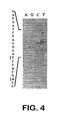
- the SK + HB e-c plasmid is analyzed to confirm the sequence of the precore/core gene ( Figure 4 ).
- the single base pair deletion in plasmid SK + HBe is corrected by PCR overlap extension as described in Example 2A.
- Four oligonucleotide primers are used for the PCR reactions performed to correct the mutation.
- the first reaction utilizes the plasmid KS II + HB pc/c as the template and two primers.
- the sense primer corresponds to the nucleotide sequence for the T-7 promoter of SK + HBe plasmid.
- the second primer corresponds to the anti-sense sequence 2158 to 2130 of the adw strain, and includes codons 79, 84 and 85.
- the second reaction utilizes the plasmid KS II + HB pc/c as the template and two primers.
- the anti-sense primer corresponds to the nucleotide sequence for the T-3 promoter present in SK + HBe plasmid.
- the second primer corresponds to the sense nucleotide sequence 2130 to 2158 of the adw strain, and includes codons 79, 84 and 85.
- the third reaction utilizes two primers and the products of the first and second PCR reactions.
- the anti-sense primer corresponds to the nucleotide sequence for the T-3 promoter present in SK + HBe plasmid.
- the second primer corresponds to the sense sequence of the T-7 promoter present in the SK + HBe plasmid.
- the PCR product from the third reaction yields the correct sequence for HBV precore/core coding region.
- a primer is designed to introduce the Xho I restriction site upstream of the ATG start codon of the core coding region, and eliminate the 29 amino acid leader sequence of the HBV precore coding region.
- the HBV core coding region is produced using the PCR product from the third reaction and the following two primers.
- the sense primer corresponds to the nucleotide sequence 1885 to 1905 of the adw strain and contains two Xho I sites at the 5' end.
- the second primer corresponds to the anti-sense nucleotide sequence for the T-3 promoter present in the SK + HBe plasmid.
- the approximately 600 bp PCR product from the fourth PCR reaction contains the HBV core coding region and novel Xho I restriction sites at the 5' end and Cla I restriction sites at the 3' end that was present in the multicloning site of SK + HBe plasmid.
- the solution is transferred into a fresh 1.5 ml microfuge tube. Fifty microliters of 3 M sodium acetate is added to this solution followed by 500 ⁇ l of chloroform:isoamyl alcohol (24:1). The mixture is vortexed and then centrifuged at 14,000 rpm for 5 minutes. The aqueous phase is transferred to a fresh microfuge tube and 1.0 ml 100% EtOH is added. This solution is incubated at -20°C for 4.5 hours, and then centrifuged at 10,000 rpm for 20 minutes. The supernatant is decanted, and the pellet rinsed with 500 ⁇ l of 70% EtOH.
- the pellet is dried by centrifugation at 10,000 rpm under vacuum and then resuspended in 10 ⁇ l deionized H2O.
- One microliter of the PCR product is analyzed by electrophoresis in a 1.5% agarose gel.
- a 200 ⁇ l sample of serum is obtained from a patient with chronic non-A, non-B hepatitis and the viral RNA is prepared by the procedure of Cristiano et al., Hepatology 14:51-55, 1991 .
- the 200 ⁇ l of serum is mixed with 550 ⁇ l of extraction buffer consisting of 4.2 M guanidinium isothiocyanate (Fluka Chemical Corp., St. Louis, Missouri), 0.5% sodium lauryl sarkosate and 25 mM Tris HCL, pH 8.0, and extracted once with phenol:chloroform (1:1), and once with chloroform.
- the aqueous phase is precipitated with an equal volume of isopropyl alcohol and centrifuged at 14,000 rpm for 5 minutes.
- the resulting pellet containing the viral RNA is washed with 70% ethanol and resuspended in 200 ⁇ l of RNase-free deionized H 2 O.
- RNasin 40,000 U/ml
- This mixture contains the HCV RNA and is the template for the following reverse transcriptase reaction.
- cDNA CYCLE kit Invitrogen, San Diego, California
- a full-length first strand cDNA is generated from the isolated viral mRNA.
- Seven microliters of the reverse transcription reaction above (100 ng of full-length first strand cDNA) is amplified by PCR in a total volume of 100 ⁇ l of reaction mixture containing 10 ⁇ l of 10 X PCR buffer (vial C16), 2 ⁇ l of 25 mM dNTPs (vial C11), 5% DMSO, 4 U of Taq DNA polymerase (Cetus, Los Angeles, California) and 2 1M of each of the two primers.
- the sense primer corresponds to the nucleotide sequence 316 to 335 and is the nucleotide sequence for the 5' region of the hepatitis C virus core open reading frame and includes the ATG start codon.
- the second primer corresponds to the anti-sense nucleotide sequence 1172 to 1153 present in the hepatitis C virus envelope open reading frame.
- the reaction mixture is placed into a PCR Gene AMP System 9600 (Perkin-Elmer, Cetus, Los Angeles, California.).
- the PCR program regulates the temperature of the reaction vessel first at 95°C for 1 minute, then at 60°C for 2 minutes. and finally at 72°C for 2 minutes. This cycle is repeated 40 times. Following the 40th cycle, the final cycle regulates the reaction vessel at 95°C for 1 minute, then at 67°C for 2 minutes, and finally at 72°C for 7 minutes.
- the HCV core open reading frame from the 5' region upstream from the ATG start codon to the beginning of the HCV E1 open reading frame is amplified.
- the nucleotide numbering sequence is according to the HCV-J strain ( Kato et al., Proc. Natl. Acad. Sci. USA 87:9524-9528, 1990 ).
- the product from the first PCR reaction is amplified in a second PCR reaction.
- the second PCR amplification is performed with the sense primer that corresponds to the nucleotide sequence 329 to 367 (and is the nucleotide sequence for the 5' end of the hepatitis C virus core open reading frame).
- the 5' end of the sense primer contains two consecutive Xho I restriction sites.
- the primer also contains a number of nucleotide changes introduced in the area of the initiator ATG start codon to conform to appropriate rules for translation initiation ( Kozak, Mol. Biol. 196:947-950, 1987 ).
- the anti-sense primer is designed to contain two consecutive stop codons in frame with HCV core gene.
- the 5' end of the primer contains two consecutive Hind III restriction sites.
- This primer corresponds to the nucleotide sequence 902 to 860, and is the junction between the hepatitis C virus core and E1 open reading frame.
- the 570 bp PCR-amplified product from the second reaction is then ligated into the pCR II vector (Invitrogen, San Diego, California) and transformed into frozen competent E. coli cells. After verification by DNA sequencing this construct is designated pCR II Xh-H HCV core.
- the product from the first PCR reaction is also amplified in a third PCR reaction.
- the 5' end of the sense primer contains two consecutive Hind III restriction sites.
- This primer also contains nucleotide changes to conform to the Kozak rules for translation initiation and corresponds to the nucleotide sequence 329 to 367 of the HCV-J sequence (and is the nucleotide sequence for the 5' end of the hepatitis C virus core open reading frame).
- the anti-sense primer is designed to contain two stop codons in frame with the HCV core gene, and two consecutive Xho I restriction sites at the 5' end of the primer. This primer corresponds to the anti-sense nucleotide sequence 902 to 860, and is the junction between hepatitis C virus core and the E1 reading frame.
- the 570 bp PCR amplified product from the third reaction is ligated into the pCR II vector. After verification by DNA sequencing this construct is designated pCR II H-Xh HCV core.
- the hepatitis C virus NS3/NS4 sequence is isolated from 200 ⁇ l of serum obtained from a patient with chronic non-A, non-B hepatitis as described in Example 2C.
- the viral RNA is reverse transcribed by the cDNA CYCLE Kit (Invitrogen, San Diego, California), and amplified by PCR. In the first PCR reaction, the HCV NS3/NS4 open reading frame is amplified.
- the first PCR amplification is performed with two primers.
- the sense primer corresponds to the nucleotide sequence 3088 to 3106 of the hepatitis C virus NS2 open reading frame.
- the second primer corresponds to the anti-sense nucleotide sequence 6530 to 6511 of the hepatitis C virus NS5 open reading frame.
- the product from the first PCR reaction is amplified in a second PCR reaction.
- the 5' end of the sense primer contains two consecutive Xho I restriction sites.
- This primer also contains nucleotide changes to conform to the Kozak rules for translation initiation and corresponds to the nucleotide sequence 3348 to 3385 of the 5' region of the NS3 open reading frame of the HCV-J sequence.
- This primer corresponds to the nucleotide sequence 6368 to 6328 of the 3' region of the NS4 open reading frame of the HCV-J sequence.
- This primer contains two consecutive stop codons ir frame with HCV NS4 NS4 gene and two consecutive Hind III sites at its 5' end.
- the 3020 bp PCR product from the second PCR reaction is ligated into the pCR II plasmid, verified by DNA sequencing and designated pCR II Xh-H HCV NS3/NS4.
- Jurkat cells are resuspended at 1 x 10⁶ cells/ml to a total volume of 158 ml in T75 flasks.
- Phytohemagglutinin (PHA; Sigma, St. Louis, MO)
- PHA Phytohemagglutinin
- cells are harvested in three 50 ml centrifuge tubes (Coming, Coming, NY). The three pellets are combined in 50 ml PBS, centrifuged at 3,000 rpm for 5 minutes and supernatant decanted. This procedure is repeated.
- Poly A + mRNA is isolated using the Micro-Fast Track mRNA Isolation Kit, version 1.2 (Invitrogen, San Diego, California). The isolated intact mRNA is used as the template to generate full-length first strand cDNA by the cDNA CYCLE kit with the following primer.
- This oligonucleotide corresponds to the anti-sense nucleotide sequence of the IL-2 mRNA, 25 base pairs downstream of the stop codon.
- the product from the reverse transcription reaction is amplified in two separate reactions.
- the first PCR amplification is performed with the sense primer that corresponds to three bp upstream of the ATG start codon.
- This primer contains a Hind III site at its 5' end and contains the 5' region of the IL-2 open reading frame including the ATG start codon.
- the anti-sense primer is complementary to the 3' region of IL-2 open reading frame and starts three bp downstream of the TGA stop codon. This primer contains an Xho I site at the 5' end of the primer.
- the 467 bp PCR product from the first PCR reaction is ligated into the pCR II plasmid, verified by DNA sequencing and designated pCR II H-Xh IL-2.
- the product from the reverse transcription reaction is amplified in a second PCR reaction.
- the second PCR amplification is performed with the sense primer that corresponds to three bp upstream of the ATG start codon.
- This primer contains a Xho I site at its 5' end and the 5' region of the IL-2 open reading frame including the ATG start codon.
- the anti-sense primer is complementary to the 3' region of IL-2 open reading frame and starts three bp downstream of the TGA stop codon. This primer contains an Apa I site at the 5' end of the primer.
- the 467 bp PCR product from the second PCR reaction is ligated into the pCR II plasmid, verified by DNA sequencing and transformed into frozen competent E. coli cells.
- This vector construct is designated pCR II Xh-A IL-2.
- Raji cells are suspended at 1 x 10⁶ cells/ml to a total volume of 158 ml in five T75 flasks and incubated overnight at 37°C, 5% CO2. On the following day, cells are harvested in three 50 ml centrifuge tubes. Cell pellets are combined in 50 ml PBS, centrifuged at 2,000 rpm for 10 minutes and supernatant decanted. This procedure is repeated. Poly A + mRNA is isolated as described in Example 2E.
- the isolated intact mRNA is used as the template to generate full-length first strand cDNA using the cDNA CYCLE kit, followed by two separate PCR amplification reactions essentially as described in Example 2E, except that 1 ⁇ l of oligo dT (vial C5) is used as the primer.
- the nucleotide numbering system is obtained from Freeman et al. (J. Immunol. 143:2714-2722, 1989 ).
- the first PCR amplification is performed with two primers.
- the sense primer corresponds to the nucleotide sequence 315 to 353 of B7.
- This primer contains the 5' region of the B7 open reading frame including the ATG start codon and has two Hind III restriction sites at the 5' end.
- the second primer corresponds to the anti-sense nucleotide sequence 1187 to 1149 of B7.
- This primer is complementary to the 3' region of the B7 open reading frame ending at the TAA stop codon and contains two Xho I restriction sites at the 5' end.
- the 868 bp PCR product from the first PCR reaction is ligated into the pCR II plasmid, verified by DNA sequencing and transformed into frozen competent E. coli cells.
- This vector construct is designated PCR II H-Xh-B7 and verified by DNA sequencing.
- the second PCR amplification is performed with two primers.
- the sense primer corresponds to the nucleotide sequence 315 to 353 of B7.
- This primer contains the 5' region of the B7 open reading frame including the ATG start codon and has two Xho I sites at its 5' end.
- the second primer corresponds to the anti-sense nucleotide sequence 1187 to 1149 of B7.
- This primer is complementary to the 3' region of the B7 open reading frame ending at the TAA stop codon and contains two Apa I restriction sites at the 5' end.
- the 868 bp PCR product from the second PCR reaction is ligated into the pCR II plasmid, verified by DNA sequencing and transformed into frozen competent E. coli cells.
- This vector construct is designated pCR II Xh-A-B7 and verified by DNA sequencing.
- GM-CSF The synthesis of GM-CSF is performed following the protocol of Foguet and Lubbert (Biotechniques 13:674-675, 1992 ). Briefly, ten overlapping oligonucleotides, 53 to 106 nucleotides in length, are synthesized. The first oligonucleotide is the sense sequence of human GM-CSF from nucleotide sequence number 29 to 86 containing two Hind III cleavage sites at the 5' end.
- the second oligonucleotide is the sense sequence of human GM-CSF from the nucleotide sequence numbers 29 to 86 containing two Xho I sites at the 5' end.
- the third oligonucleotide is the anti-sense sequence of human GM-CSF from nucleotide sequence number 145 to 70.
- the fourth oligonucleotide is the sense sequence of human GM-CSF from nucleotide number 131 to 191.
- the fifth oligonucleotide is the anti-sense sequence of human GM-CSF from nucleotide number 282 to 176.
- the sixth oligonucleotide is the sense sequence of human GM-CSF from nucleotide number 256 to 346.
- the seventh oligonucleotide sequence is the anti-sense sequence of human GM-CSF from nucleotide number 389 to 331.
- the eighth oligonucleotide is the sense sequence of human GM-CSF from nucleotide number 372 to 431.
- the ninth oligonucleotide sequence is the anti-sense sequence of human GM-CSF from nucleotide number 520 to 416 containing two Xho I restriction sites at the 5' end.
- the tenth oligonucleotide sequence is identical to oligonucleotide number nine except that it contains two Xba I restriction sites at the 5' terminus instead of Xho I restriction sites.
- oligonucleotides except for oligonucleotide Sequence ID Nos. 29, 36, 37 and 47 are phosphorylated. Ligation is performed by mixing 8 pmol of each oligonucleotide and 7.5 ⁇ l 10X Sequenase Buffer (US Biochemical, Cleveland, Ohio) to a final volume of 75 ⁇ l with sterile distilled deionized H 2 O. The reaction is heated for 5 minutes at 70°C, followed by 5 minutes at 48°C. Two microliters of dNTP mix (2.5mM each dNTP) and 10 U Sequenase are added and incubated for 30 minutes at 37°C. To inactivate the Sequenase, the ligation reaction is heated for 10 minutes at 70°C ( Current Protocols in Molecular Biology, F.M. Asubel et al., 8.2.8-8.2.13, 1988 ).
- PCR reaction One microliter of the ligation mixture is used in a PCR reaction with Vent polymerase (New England Biolabs, Beverly, Massachusetts) and the two oligonucleotides Sequence ID Nos. 29 and 36 as primers.
- the PCR product is ligated into the pCR II vector and transformed into frozen competent E. coli cells. This construct is designated pCR II H-Xh GM-CSF and verified by DNA sequencing.
- PCR product is ligated into the pCR II vector and transformed into frozen competent E. coli cells. This construct is designated pCR II Xh-Xb GM-CSF and verified by DNA sequencing.
- the Pre-S2 open reading frame (including S) is PCR amplified with two primers and the pAM 6 plasmid (ATCC No. 45020) as the template.
- the sense primer corresponds to the nucleotides 3178 to 31 of the adw strain of hepatitis B virus, and includes the 5' region of the Pre-S2 open reading frame and the ATG start codon. The 5' end of this primer contains two consecutive Xho I restriction sites.
- the second primer corresponds to the anti-sense nucleotide sequence 907 to 859, and is complementary to the 3' region of the Pre-S2 open reading frame.
- the 5' end of this primer contains two Cla I sites.
- the 957 bp PCR product is ligated into the pCR II plasmid, verified by DNA sequencing and designated pCR II HB-Pre-S2.
- the PCR amplification is performed with two primers and the pAM 6 plasmid (ATCC 40202) as the template.
- the sense primer corresponds to the nucleotides 2309 to 2370 of the adw strain of hepatitis B virus, and includes the 5' region of the polymerase open reading frame with nucleotide changes to conform to the Kozak rules for translation.
- the 5' end of this primer contains two consecutive Xho I restriction sites.
- the second primer corresponds to the anti-sense nucleotide sequence 1645 to 1594, and is complementary to the 3' region of the polymerase open reading frame and includes the TGA stop codon. The 5' end of this primer contains two Cla I sites.
- the 2564 bp PCR product is ligated into the pCR II plasmid, verified by DNA sequencing and designated pCR II HB-pol.
- the PCR amplification is performed with two primers and the pAM 6 plasmid (ATCC 45020) as the template.
- the sense primer corresponds to the nucleotides 1432 to 1482 of the ⁇ dw strain of hepatitis B virus, and includes the 5' region of the ORF5 open reading frame with nucleotide changes to conform to the Kozak rules for translation. The 5' end of this primer contains two consecutive Xho I restriction sites.
- the second primer corresponds to the anti-sense nucleotide sequence 1697 to 1648, and contains two Cla I sites at the 5' end. This primer is complementary to the 3' region of the ORF 5 open rading frame and includes the TAA stop codon.
- the 293 bp PCR product is ligated into the pCR II plasmid, verified by DNA sequencing and designated pCR II HB-ORF 5.
- the PCR amplification is performed with two primers and the pAM 6 plasmid (ATCC 45020) as the template.
- the sense primer corresponds to the nucleotides 1844 to 1788 of the adw strain of hepatitis B virus and includes the 5' region of the ORF6 open reading frame with nucleotide changes to conform to the Kozak rules for translation.
- the 5' end of this primer contains two consecutive Xho I restriction sites.
- the second primer corresponds to the anti-sense nucleotide sequence 1188 to 1240, and contains two Cla I sites at the 5' end. This primer is complementary to the 3' region of the ORF 6 open reading frame and includes the TAA stop codon.
- the 687 bp PCR product is ligated into the pCR II plasmid, verified by DNA sequencing and designated pCR II HB-ORF 6.
- the IRES from encephalomyocarditis virus is amplified by PCR from the pCITE-2a(+) plasmid (Novagen, Madison, WI) with two primers.
- the nucleotide sequence of the sense primer containing a Acc I restriction endonuclease site is:
- the nucleotide sequence of the antisense primer containing a Cla I restriction endonuclease site is:
- This 500 base pair PCR product is purified by electrophoresis through an 1.5% agarose gel and purified by Gene Clean II (Bio 101, Vista, CA) as described in Example 5B.
- PBMC peripheral blood mononucleocytes
- the sense primer corresponds to the nucleotides in the 5' region of the p40 subunit of IL-12 open reading frame and additionally contains Bgl II restriction endonuclease sites at the 5' end.
- the nucleotide sequence of this primer is:
- the second primer corresponds to the antisense sequences 3' region of the p40 subunit of IL-12 open reading frame and additionally contains the Hpa I restriction endonuclease site at the 5' end.
- the nucleotide sequence of this primer is:
- the product of this PCR reaction is a Bgl II-Hpa I 1140 bp fragment encoding the p40 subunit of IL-12.
- RNA from the stimulated PBMC is extracted and the IL-12 p35 subunit nucleotide sequence is amplified by PCR as described in Example 2C.
- the sense primer corresponds to the nucleotides in the 5' region of the p35 subunit of IL-12 open reading frame.
- the nucleotide sequence of this primer is:
- the second primer corresponds to the antisense sequences 3' region of the p35 subunit of IL-12 open reading frame.
- the nucleotide sequence of this primer is:
- the PCR amplified product is then ligated into the pCR II vector (Invitrogen, San Diego, CA)n and transformed into frozen competent E. coli cells. After verification by DNA sequencing, this construct is designated pCR II p35.
- PCR overlap extension was used where the termination codon of HBcore is deleted and fused in frame with the 11th amino acid of the neomycin phosphotransferase open reading frame.
- the KT-HB C plasmid is used as the template along with four oligonucleotides primers are used for the 3 PCR reactions performed to generate the fusion HBcore/neo R construct.
- the first reaction utilizes two primers.
- the sense primer sequence corresponds to the Xho I restriction site at the 5' end of HB core gene.
- the second primer sequence corresponds to the anti-sense nucleotide sequence 2457 to 2441 and 23 base pairs of neomycin phosphotransferase gene coding for codons 11-17.
- the second reaction also utilizes the KT-HBc plasmid as the template and two primers.
- the sense primer corresponds to nucleotide sequence 2440 to 2457 and 23 base pairs of neomycin phosphotransferase gene coding for codons 11-17.
- the second primer corresponds to the anti-sense nucleotide sequence of the neomycin phosphotransferase gene.
- the products of the first and second PCR reactions are extended in a third reaction to generate the F HBcore/neo R construct.
- Two primers are also utilized in the third reaction.
- the sense primer corresponds to the Xho I restriction site at the 5' end of the HBcore gene.
- the second primer sequence corresponds to the anti-sense nucleotide sequence of the neomycin phosphotransferase gene.
- the PCR product from the third reaction yields the fusion HBcore/neo R construct.
- the solution is transferred to a fresh 1.5 ml microfuge tube. Fifty microliters of 3 M sodium acetate is added to this solution followed by 500 ul of chloroform:isoamy alcohol (24:1). The mixture is vortexed and then centrifuged at 14,000 rpm for 5 minutes. The aqueous phase is transferred to a fresh microfuge tube and 1.0 ml 100% EtOH is added. This solution is incubated at -20°C for 4.5 hours, and then centrifuged at 10,000 rpm for 20 minutes. The supernatant is decanted, and the pellet rinsed with 500 ul of 70% EtOH. The pellet is dried by centrifugation at 10,000 rpm under vacuum and then resuspended in 10 ul deionized H 2 O. One microliter of the PCR product is analyzed by electrophoresis in a 1.0% agarose gel.
- This PCR product is digested with Xho I and Pst I restriction endonucleases, electrophoresed through a 1.0% agarose gel and the DNA is purified from the gel slice by Geneclean II (Bio 101, Vista, California).
- This Xho I-Pst I PCR product is inserted into the respective sites of pBluescript KS+II (Stratgene, La Jolla, California). This construct is designated KSII+ Xh-Pst HB Fcore/neo R , and is verified by DNA sequencing.
- a 642 bp Nco I-Taq I fragment containing the hepatitis B virus X open reading frame is obtained from the pAM6 plasmid ( adw ) (ATCC 45020), blunted by Klenow fragment, and ligated into the Hinc II site of SK + (Stratagene, La Jolla, California).
- E. coli (DH5 alpha, Bethesda Research Labs, Gaithersburg, Maryland) is transformed with the ligation reaction and propagated. Miniprep DNA is then isolated and purified, essentially as described by Bimboim et al. (Nuc. Acid Res. 7:1513, 1979 ; Molecular Cloning: A Laboratory Manual, Sambrook et al. (eds.), Cold Spring Harbor Press, 1989 ).
- miniprep DNAs are digested with the diagnostic restriction enzyme, Bam HI. Inserts in the correct orientation yield two fragments of 3.0 Kb and 0.6 Kb in size. Inserts in the incorrect orientation yield two fragments of 3.6 Kb and 0.74 Kb.
- a clone in the correct orientation is selected and designated SK-X Ag.
- TAG is inserted via a Nhe I (nonsense codon) linker (NEB#1060, New England BioLabs, Beverly, Massachusetts). This linker provides nonsense codons in all three reading frames.
- SK-XAg is cleaved with Stu I (nucleotide 1704) which linearizes the plasmid.
- the Nhe I (nonsense codon) linkers are first phosphorylated in the following reaction.
- One OD 260 of linkers are dissolved in 100 ⁇ l TE (10mM Tris-HCl pH 7.6, 1mM EDTA).
- linkers 1.0-2.0
- 10X buffer (0.66 M Tris-HCl pH 7.6, 10 mM ATP, 10 mM spermidine, 0.1 M MgCl 2 , 150 mM DTT, 2 mg/ml BSA), 6 ⁇ l H 2 O and 2 U of T4 DNA kinase and incubated for 1 hour at 37°C.
- This reaction mixture is then added to 0.4 ⁇ g of linearized SK XAg plasmid (Example 3A) in 10 ⁇ l of the above buffer with 10 units of T4 DNA ligase. The reaction is incubated at 22°C for 6 hours and stopped with 1 ⁇ l of 0.5 M EDTA.
- the reaction is then extracted with phenol:chloroform (a 1:1 ratio), and the DNA is precipitated with ethanol.
- the DNA is recovered by centrifugation at 14,000 rpm for 5 minutes at room temperature.
- the pellet is then dried and dissolved in 90 ⁇ l of TE (10 mM Tris-HCl pH 7.6, 1 mM EDTA).
- Ten microliters of 10X NeB2 buffer (New England Biolabs, Beverly, Massachusetts) is added to the DNA and then digested with 20 U of Nhe I.
- the plasmid is purified from excess linkers by 1.0% agarose gel electrophoresis, and isolated by Geneclean II (Bio 101, Vista, California).
- the DNA is self-ligated and transformed onto competent E. coli. Clones are then screened for the presence of the diagnostic restriction site, Nhe I; these clones will contain the truncated X gene. A clone is selected and designated SK-TXAg.
- the N2R5 construct is mutated by site-directed in vitro mutagenesis to change the ATG start codon to ATT preventing gag expression. This mutagenized fragment is 200 bp in length and flanked by Pst I restriction sites.
- the Pst I-Pst I mutated fragment is purified from the SK + plasmid and inserted into the Pst I site of N2 MoMLV 5' LTR in plasmid pUC31 to replace the non-mutated 200 bp fragment.
- the plasmid pUC31 is derived from pUC19 (Stratagene, La Jolla, California) in which additional restriction sites Xho I, Bgl II, BssH II and Nco I are inserted between the EcoR I and Sac I sites of the polylinker. This construct is designated pUC31/N2R5gM.
- a 1.0 Kb MoMLV 3' LTR EcoR I-EcoR I fragment from N2 is cloned into plasmid SK + resulting in a construct designated N2R3-.
- a 1.0 Kb Cla 1-Hind III fragment is purified from this construct.
- the Cla I-Cla I dominant selectable marker gene fragment from pAFVXM retroviral vector ( Kriegler et al., Cell 38:483, 1984 ; St. Louis et al., PNAS 85:3150-3154,1988 ), comprising a SV40 early promoter driving expression of the neomycin phosphotransferase gene, is cloned into the SK + plasmid.
- This construct is designated SK + SV 2 -neo A 1.3 Kb Cla I-BstB I gene fragment is purified from the SK + SV 2 -neo plasmid.
- the KT-3 retroviral vector is constructed by a three part ligation in which the Xho I-Cla I fragment containing the gene of interest and the 1.0 Kb MoMLV 3' LTR Cla I-Hind III fragment are inserted into the Xho I-Hind III site of pUC31/N2R5gM plasmid.
- the 1.3 Kb Cla I-BstB I neo gene fragment from the pAFVXM retroviral vector is then inserted into the Cla I site of this plasmid in the sense orientation.
- the KT-1 retroviral backbone vector is constructed essentially as described for KT-3 in Example 4A, with the exception that the dominant selectable marker gene, neo, is not inserted into the expression vector. Specifically, in a three part ligation, the Xho I-Cla I fragment containing the gene of interest and the 1.0 Kb MoMLV 3' LTR Cla I-Hind III fragment are inserted into the Xho I-Hind III site of pUC31/N2R5gM plasmid.
- the JMR-2 vector is comprised of the KT-3 retrovector with a polylinker containing a Xho I, Bam HI, Srf I, and Not I restriction endonuclease sites at the Xho I site of KT-3 flanking the 5' end of the encephalomyocarditis virus IRES (Novagen, Madison, WI) and a polylinker containing a Cla I, Bgl II, Age I, Hpa I, Mlu I, and Sal I restriction endonuclease sites flanking the 3' end of the encephalomyocarditis virus IRES.
- the JMR-2 vector is prepared by inserting a linker containing the Xho I, Bam HI, Srf I, Not I, Cla I and Sal I restriction endonuclease sites at the Xho I restriction endonuclease sites of the KT-3 backbone as described in Example 3B.
- This construct is designated KT3-L1 and verified by DNA sequencing.
- the sense and antisense strands are synthesized by standard methods of DNA synthesis.
- the sense strand sequence is:
- the antisense strand sequence is:
- oligonucleotides are annealed at 37°C, generating 5' overhangs. These double stranded linkers are then hybridized to compatible overhangs of the Xho I digested KT-3 backbone by annealing at 37°C, followed by ligation.
- the IRES from encephalomyocarditis virus is excised by digestion with Acc I and Cla I restriction endonucleases of the PCR amplified product (Example 2C) and ligated into the Cla I sites of the KT3-L1 and designated KT3-L1-IRES.
- the correct orientation of the IRES is determined after analysis with AvrII endonuclease digestion.
- the multicloning site containing Cla I, Bgl II, Age I, Hpa I, Mlu I, and Sal I restriction endonucleases is then inserted into the Cla I-Sal I sites of KT3-L1-IRES.
- the resulting vector is designated JMR-2.
- the sense and antisense strands are synthesized by standard methods of DNA synthesis.
- the sense strand sequence is:
- the antisense strand sequence is:
- oligonucleotides are annealed at 37°C, generating 5' overhangs. These double stranded linkers are then hybridized to compatible overhangs of the Cla I-Sal I digested KT3-L1-IRES backbone by annealing at 37°C, followed by ligation.
- Plasmid pSCV6 (as described in U.S. Patent Serial No. 07/800,921 ) may be utilized to generate pCMV-HBc. Briefly, the pSCV6 plasmid is derived from the pBluescript SK- backbone (Stratagene, La Jolla, California) with the CMV IE promoter followed by a polylinker site allowing insertion of a gene of interest and ending with a SV40 poly A Signal.
- the adenovirus vector backbone, the pAdM1 plasmid is obtained from Quantum Biotechnologies (Montreal, Canada).
- the Ad5delta Eldelta E3 plasmid ( Gluzman et al., in Eucaryotic Viral Vectors, pp. 187-192, Cold Spring Harbor, 1982 ) is also obtained from Quantum Biotechnologies.
- the PCR product from Example 2B is digested with Xho I and Cla I restriction endonucleases, electrophoresed through an 1.5% agarose gel and the DNA is purified from the gel slice by Geneclean II (Bio 101, Vista, California).
- This Xho I-Cla I HBV core PCR product is inserted into the Xho I and Cla I sites of the KT-3 retroviral vector backbone.
- the construct is designated KT-HBc.
- HBV core fragment (Xho I-Cla I) from KT-HBc is inserted into the respective sites of pBluescript KS + II (Stratagene, La Jolla, California). This construct is designated KS + II HBc, and is verified by DNA sequencing.
- the Xho I-Hind III fragment from pCR II Xh-H HCV core from Example 2C is inserted into the respective sites of pSP72. This construct is designated pSP72 Xh-H HCc.
- the Xho I-Cla I fragment from pSP72 Xh-H HCc is then excised and inserted into the KT-3 backbone. This construct is designated KT-HCc.
- the Xho I-Hind III fragment from pCR II Xh-H HCV NS3/NS4 from Example 2D is inserted into the respective sites of pSP72.
- This construct is designated pSP72 Xh-H HCV NS3/NS4.
- the Xho I-Cla I fragment from pSP72 Xh-H HCV NS3/NS4 is then excised and inserted into the KT-3 backbone. This construct is designated KT-HCV NS3/NS4.
- KT-HB-X The Xho I-Cla I fragment from SK-X Ag is excised and inserted into the respective sites of the KT-3 backbone. This construct is designated KT-HB-X.
- KT HB-TX The Xho I-Cla I fragment from SK-TX Ag is excised and inserted into the respective sites of the KT-3 backbone. This construct is designated KT HB-TX.
- KT-HB-Pre-S2 The Xho I-Cla I fragment from pCR II HB-Pre-S2 from Example 2H is excised and inserted into the respective sites of the KT3 backbone. This construct is designated KT-HB-Pre-S2.
- KT-HB-pol The Xho 1-Cla I fragment from pCR II HB-pol from Example 2I is excised and inserted into the respective sites of the KT3 backbone. This construct is designated KT-HB-pol.
- KT-HB-ORF 5 The Xho I-Cla I fragment from pCR II HB-ORF5 from Example 2J is excised and inserted into the respective sites of the KT3 backbone. This construct is designated KT-HB-ORF 5.
- KT-HB-ORF 6 The Xho I-Cla I fragment from pCR II HB-ORF6 from Example 2K is excised and inserted into the respective sites of the KT3 backbone. This construct is designated KT-HB-ORF 6.
- the Xho I to Cla I HBV e from KT-HBe-c is ligated into the Xho I/Cla I site of the KT-1 retroviral vector backbone (Example 4B) resulting in a vector designated KT-1/HBe-c.
- a 2.0 Kb Cla I/Cla I fragment containing the HSV-1 thymidine kinase gene driven by the thymidine kinase promoter is derived from pSP72-TK/Cla, and ligated into the Cla I/Cla I site of KT-1/HBe-c in the sense orientation. Orientation is determined by Sma I digestion. This construct is designated KT-HBe/TK.
- pSP72-TK/Cla is derived as follows. The fragment containing the HSV-1 thymidine kinase promoter and gene is excised by Xho I and Bam HI digestion from PrTKdeltaA. (PrTKdeltaA is described in Example 4 of PCT WO 91/02805 .) This fragment is isolated from a 1% agarose gel with NA45 paper as described in Example 2A and inserted into the Xho I and Bam HI sites of pSP72 plasmid. This plasmid is designated pSP72-TK.
- pSP72TK is linearized with Xho I, blunted with Klenow and ligated with Cla I linkers (New England BioLabs, Beverly, Massachusetts) as described in Example 3B. This plasmid is self-ligated and transformed onto competent E. coli. A clone is selected and designated pSP72-TK/Cla.
- the Xho I to Cla I HBV core fragment from KT-HBc (Example 5B) is ligated into the Xho I/Cla I site of the KT-1 retroviral vector backbone (Example 4B) resulting in vectors designated KT-1/HBc.
- a 2.0 Kb Cla I/Cla I fragment containing the HSV-1 thymidine kinase gene driven by the thymidine kinase promoter is derived from pSP72-TK/Cla and ligated into the Cla I/Cla I site of KT-1/HBc in the sense orientation. This construct is designated KT-HBc/TK.
- the HBV core fragment is obtained from the construct KT-HBc (Example 5B) by digestion of the plasmid with Xho I and Cla I restriction enzymes and isolation from a 1% agarose gel with NA45 paper as described in Example 2A. The fragment is blunted with Klenow and ligated into the Sma I site of the plasmid pSC6. Orientation of the HBcore gene is determined by Eco RI/SSpI double digest. This plasmid is designated pCMV-HBc. E. coli (DH5 alpha, Bethesda Research Labs, Gaithersburg, Maryland) is transformed with the pCMV-HBc plasmid and propagated to generate plasmid DNA.
- the plasmid DNA is then isolated and purified by cesium chloride banding and ethanol precipitation essentially as described by Birnboim et al. (Nuc. Acid Res. 7:1513, 1979 ; see also " Molecular Cloning: A Laboratory Manual," Sambrook et al. (eds.), Cold Spring harbor Press, 1989 ).
- the DNA is resuspended in 0.9% sterile phosphate-buffered saline at a final concentration of 2 mg/ml.
- the Xho I to Cla I HBV e fragment is obtained from KT-HBe-c (Example 5A) by digestion of the plasmid with Xho I and Cla I restriction enzymes and isolation on a 1% agarose gel and NA45 paper as described in Example 2A. The fragment is blunted with Klenow fragment.
- the adenovirus vector backbone, the pAdM1 plasmid is cleaved with Bam HI and blunted with Klenow fragment.
- the blunted HBV e fragment is ligated into the blunted Bam HI site of pAdM1 plasmid. Orientation of the HBV e gene is determined by Eco RI/Ssp I double digestion. This plasmid is designated pAdM1-HBe.
- KT-HBc from Example 5 is digested with Cla I, Pst I and Hind III, and the 1.6 kb Pst 1-Hind III fragment containing the neomycin phosphotransferase gene and the 3' LTR is isolated.
- the Xho I-Pst I fragment containing the HB Fcore/neo R fragment and the Pst I-Hind III neo/3' LTR fragment are inserted into the Xho I-Hind III sites of KT-HBc.
- This vector construct is designated KT-HB Fcore/neo R .
- pGEM 5Z + BIP 5' (Peter Samow, University of Colorado, Health Sciences Center, Denver, human immunoglobulin heavy chain binding protein) is digested with Sac I and Sph I.
- the 250 bp BIP fragment is isolate by 1.5% agarose gel electrophoresis and subcloned into the respective sites of pSP72.
- the vector construct is designated pSP72 BIP.
- Hind III-Xho I GM-CSF fragment is excised from pCR II H-Xh GM-CSF of Example 2G, and subcloned into the Hind III-Xho I sites of pSP72 BIP. This construct is designated pSP72 BIP-GM-CSF.
- the construct pSP72 BIP GM-CSF is cleaved at the Xho I site and blunted by Klenow fragment, followed by cleavage with Cla I.
- the KT-1 backbone is cleaved by Cla I and blunted with Klenow fragment followed by cleavage with Xho I restriction endonuclease.
- the Xho I-Cla I fragment from SK + HBe-c, Example 2A, and the Cla I-blunted Xho I BIP-GM-CSF fragment is ligated into the Xho I-blunted Cla I sites of the KT-1 retroviral backbone.
- This construct is designated KT-HBe-c/BIP-GM-CSF.
- the 4.7 Kb CMV EnvR Pst-RI fragment is isolated from pAF/CMV/EnvR ( U.S. Patent Application No. 07/395,932 ), and inserted into the Pst I and Eco RI sites of pUC 18. This construct is designated pUC 18 CMV EnvR.
- HIV-1 IIIB CAR is subcloned as a Sau 3A fragment from pAF/CMV/Env R into the BamH I site of pBluescript II KS + (Stratagene, La Jolla, California) to generate pBluescript II KS + /CAR.
- the CAR fragment is excised from pBluescript II KS + /CAR as a Xba I-Cla I fragment.
- the Xho I- Xba I HIV-1 IIIB gag/pol fragment is excised from SK + gag/pol SD delta ( U.S. Patent Application No. 07/395,932 ).
- the plasmid backbone containing the CMV promoter is excised from pUC18 CMV/EnvR with Xho I and Cla I.
- the Xho I-Xba I HIV IIIB gag-pol fragment and the Xba I-Cla I CAR fragment is inserted into the Xho I - Cla I sites of the pUC 18 CMV/EnvR backbone to generate pUC 18 CMV gag/pol/CAR.
- the Hind 111-Xho I fragment containing the CMV IE promoter from pUC 18 CMV-gag/pol/CAR is subcloned into the respective sites of pCDNA II. This construct is designated pCDNA II CMV.
- the Xho I-Xba I GM-CSF PCR product is subcloned from the pCR II Xh-Xb GM-CSF of Example 2G and inserted into the respective sites within pCDNA II-CMV. This construct is designated pCDNA II CMV-GM-CSF.
- the pCDNA II CMV GM-CSF construct is cleaved at the Xba I site, blunted by Klenow fragment, followed by cleavage with Hind III.
- the KT-1 backbone is cleaved by Cla I and blunted with Klenow fragment followed by cleavage with Xho I.
- the Xho I-Hind III fragment from SK + HBe-c, Example 2A, and the Hind III-blunted Xba I CMV-GM-CSF fragment is ligated into the Xho I-blunted Cla I sites of the KT-1 retroviral backbone.
- This vector construct is designated KT-HBe-c/CMV-GM-CSF.
- the Hind III-Xho I IL-2 sequence is excised from pCR II H-Xh IL-2 of Example 2E, and subcloned into the Hind III-Xho I sites of pSP72 BIP. This construct is designated pSP72 BIP IL-2.
- the Xho I-Hind III hepatitis C virus core sequence, Example 2C is excised from pCRII Xh-H HCV C core and subcloned into the respective sites of pSP72. This construct is designated pSP72 Xh-H HCV core.
- the construct pSP72 BIP-IL2 is cleaved at the Xho I site, blunted by Klenow fragment followed by cleavage with Eco RI.
- the Xho I-Eco RI HCV core fragment is isolated from pSP72 Xh-H HCV core.
- the KT-1 backbone is cleaved by Cla I and blunted with Klenow fragment followed by cleavage with Xho I.
- the Xho I-Eco RI HCV core fragment and the Eco RI-blunted Xho I BIP-IL2 fragment is ligated into the Xho I-blunted Cla I sites of the KT-1 retroviral backbone.
- This vector construct is designated KT-HCV core/BIP-IL2.
- the Xho I-Apa I IL-2 fragment is excised from pCR II Xh-A IL-2 of Example 2E, and subcloned into the respective sites of pCDNA II-CMV promoter. This construct is designated pC DNA II CMV-IL-2.
- the KT-1 backbone is cleaved by Cla I and blunted with Klenow fragment followed by cleavage with Xho I.
- the construct pCDNA II CMV-IL-2 is cleaved at the Apa I site, blunted by Klenow fragment and followed by cleavage with Hind III restriction endonuclease.
- the Xho I-Hind III HCV core fragment from pCR II Xh-H HCV core from Example 2C and the Hind III-blunted Apa I CMV IL-2 fragment is ligated into the Xho I-blunted Cla I sites of the KT-1 retroviral backbone.
- This vector construct is designated KT-HCV core/CMV IL-2.
- Hind III-Xho I B7 sequence is excised from pCR II H-Xh B7 of Example 2F, and subcloned into the Hind III-Xho I sites of pSP72 BIP. This construct is designated pSP72 H-Xh BIP-B7.
- the construct pSP72 H-Xh BIP-B7 is cleaved at the Xho I site, blunted by Klenow fragment followed by cleavage with Cla I.
- the Xho I-Cla I HBV core fragment is isolated from KS II + HBc, Example 5B.
- the KT-1 backbone is cleaved by Cla I and blunted with Klenow fragment followed by cleavage with Xho I.
- the Xho I-Cla I HBV core fragment and the Cla I-blunted Xho I BIP-B7 fragment is ligated into the Xho I-blunted Cla I sites of the KT-1 retroviral backbone.
- This vector construct is designated KT-HBV core/BIP-B7 (see Figure 8 ).
- the Xho I-Apa I B7 sequence is excised from pCR II Xh-A B7 of Example 2F, and subcloned into the respective sites of pCDA II-CMV promoter. This construct is designated pCDNA II CMV-B7.
- the KT-1 backbone is cleaved by Cla I and blunted with Klenow fragment followed by cleavage with Xho I.
- the construct pCDNA II CMV-B7 is cleaved at the Apa I site blunted by Klenow fragment and followed by cleavage with Hind III restriction endonuclease.
- the Xho I-Hind III HBV core fragment from KS II + HBc, and the Hind III-blunted Apa I CMV B7 fragment is ligated into the Xho I-blunted Cla I sites of the KT-1 retroviral backbone.
- This vector construct is designated KT-HBc/CMU-B7.
- Hind III-Xho I HCV core PCR product is subcloned from the pCR II H-Xh HCV core, Example 2C, and inserted into the respective sites within pSP72-BIP. This construct is designated pSP72 BIP-HCV core.
- the construct pSP72 BIP-HCV core is cleaved at the Xho I site, blunted by Klenow fragment, followed by cleavage with Cla I.
- the KT-1 backbone is cleaved by Cla I and blunted with Klenow fragment followed by cleavage with Xho I.
- the Xho I-Cla I HBV e fragment from SK + HBe-c, Example 2A, and the Cla I-blunted Xho I BIP HCV core fragment is ligated into the Xho I-blunted Cla I sites of the KT-1 retroviral backbone.
- This vector construct is designated KT-HBV e/BIP HCV core.
- the Xho I-Xba I HCV core fragment from pSP72 Xh-H HCV core (Example 6B i) is inserted into the respective sites of pCDNA II CMV plasmid. This construct is designated pCDNA II CMV HCV core.
- the construct pCDNA II CMV HCV core is cleaved at the Xba I site. blunted by Klenow fragment, followed by cleavage with Hind III.
- the KT-1 backbone is cleaved by Cla I and blunted with Klenow fragment followed by cleavage with Xho I.
- the Xho I-Hind III HBV e sequence from SK + HBe-c, Example 2A the Hind III-blunted Xba I CMV HCV core fragment is ligated into the Xho I-blunted Cla I sites of the KT-1 retroviral backbone.
- This vector construct is designated KT-HBVe/CMV HCV core.
- the Xho I-Not I HBV core fragment is isolated from KS II+ HBc.
- Example 5B and subcloned into the Xho I-Not I sites of the JMR-2. This construct is designated JMR-2 HBc.
- the Bgl II-Hpa I p40 PCR fragment is then subcloned into the respective sites of JMR-2 HBc. This construct is designated JMR-2 HBc/p40.
- the Xho I-Nsi I p35 fragment is then excised from pCR II p35 and subcloned into the respective sites of pCDA II-CMV, Example 6Ai. This construct is designated pCDNA II CMV-p35.
- the pCDNA II CMV-p35 is excised with Nsi I and blunt ended with T4 DNA polymerase (New England Biolabs, Beverly, MA).
- the Nsi 1-blunt ended CMV p35 fragment is ligated into the Sal I-blunt ended sites of the JMR-2 HBc/p40. Restriction digests are used to confirm that only one fragment is inserted in the correct orientation.
- This retrovector construct is designated JMR-2 HBc/p40/CMV-p35.
- HX cells ( WO 92/05266 ) are seeded at 5 x 10⁵ cells on a 10 cm tissue culture dish on day 1 with Dulbecco's Modified Eagle Medium (DMEM) and 10% Fetal Bovine Serum (FBS). On day 2, the media is replaced with 5.0 ml fresh media 4 hours prior to transfection.
- DMEM Dulbecco's Modified Eagle Medium
- FBS Fetal Bovine Serum
- DA ( WO 92/05266 ) cells are seeded at 5x10 5 cells/10 cm tissue culture dish in 10 ml DMEM and 10% FBS, 4 ⁇ g/ml polybrene (Sigma, St. Louis, Missouri) on day 1.
- FBS 4 ⁇ g/ml polybrene
- 3.0 ml, 1.0 ml and 0.2 ml of the freshly collected virus containing DX media is added to the cells.
- the cells are incubated with the virus overnight at 37°C.
- the media is removed and 1.0 ml DMEM, 10% FBS with 800 ⁇ g/ml G418 is added to the plate. Only cells that have been transduced with the vector and contain the neo selectable marker will survive.
- a G418 resistant pool is generated over a period of a week.
- the pool is tested for expression as described (Example 12A).
- the pool of cells is dilution cloned by removing the cells from the plate and counting the cell suspension, diluting the cells suspension down to 10 cells/ml and adding 0.1 ml to each well (1 cell/well) of a 96 well plate (Coming, Coming, NY). Cells are incubated for 14 days at 37°C, 10% CO2. Twenty-four clones are selected and expanded up to 24 well plates, 6 well plates then 10 cm plates at which time the clones are assayed for expression and the supernatants are collected and assayed for viral titer.
- the packaging cell line HX ( WO 92/05266 ), is transduced with vector generated from the DA vector producing cell line in the same manner as described for transduction of the DA cells from HX supernatant.
- the infection procedure as noted above is used. However, instead of adding G418 to the cells on day 3, the cells are cloned by limiting dilution as explained above. Fifty clones are expanded for expression as explained above, and titer assayed as described in Example 8.
- DA cells ( WO 92/05266 ) are seeded at 5 x 10 5 cells on a 10 cm tissue culture dish on day 1 with Dulbecco's Modified Eagle Medium (DMEM) and 10% irradiated (2.5 megarads minimum) fetal bovine serum (FBS). On day 2, the media is replaced with 5.0 ml fresh media 4 hours prior to transfection.
- DMEM Dulbecco's Modified Eagle Medium
- FBS fetal bovine serum
- a standard calcium phosphate-DNA coprecipitation is performed by mixing 60 ⁇ l 2.0 M CaCl 2 , 10 ⁇ g MLP-G plasmid, 10 ⁇ g KT-HBe-c or KT-HBc retroviral vector plasmid, and deionized water to a volume of 400 11.
- DA cells ( WO 92/05266 ) are seeded at 5 x 10 5 cells on a 10 cm tissue culture dish in 10 ml DMEM and 10% FBS, 4 ⁇ g/ml polybrene (Sigma, St. Louis. Missouri) on day 1.
- DMEM fetal bovine serum
- FBS 4 ⁇ g/ml polybrene
- 2.0 ml 1.0 ml or 0.5 ml of the freshly collected and filtered G-pseudotyped virus containing supernatant is added to the cells.
- the cells are incubated with the virus overnight at 37°C.
- the medium is removed and 10 ml DMEM, 10% irradiated FBS with 800 ⁇ g/ml G418 is added to the plate.
- a G418 resistant pool is generated over the period of 1-2 weeks. The pool is tested for expression as described in Example 12A. The pool of cells is dilution cloned by removing the cells from the plate, counting the cell suspension, diluting the cell suspension down to 10 cells/ml and adding 0.1 ml to each well (1 cell/well) of a 96-well plate (Coming, Coming, New York). Cells are incubated for 2 weeks at 37°C, 10% CO 2 . Twenty-four clones are selected and expanded up to 24-well plates, then 6-well plates, and finally 10 cm plates, at which time the clones are assayed for expression and the supernatants are collected and assayed for viral titer.
- the titer of the individual clones is determined by infection of HT1080 cells, (ATCC CCL 121). On day 1, 5x10 5 HT1080 cells are plated on each well of a 6 well microtiter plate in 3.0 ml DMEM, 10% FBS and 4 ⁇ g/ml polybrene. On day 2, the supernatant from each clone is serially diluted 10 fold and used to infect the HT1080 cells in 1.0 ml aliquots. The media is replaced with fresh DMEM, 10% FBS media, and the cells are incubated with the vector overnight at 37°C, 10% CO 2 .
- transduced cells On day 3, selection of transduced cells is performed by replacing the media with fresh DMEM, 10% FBS media containing 800 ⁇ g/ml G418. Cells are incubated at 37°C, 10% CO 2 for 14 days at which time G418 resistant colonies are scored at each dilution to determine the viral titer of each clone as colony forming units/ml (cfu/ml).
- the titers of the HBVcore and HBVe producer cell lines are: DAcore-6A3 3x10 6 cfu/ml DAcore-10 1x10 6 cfu/ml DAHBe 4-7 3x10 6 cfu/ml
- the multivalent vectors do not contain a selectable marker, such as the neomycin gene
- a selectable marker such as the neomycin gene
- 1.0 ml of vector supernatant is diluted five fold to a final dilution of 10-9 ml.
- One milliliter of each dilution is then used to transduce 5 x 10⁵ HT1080 cells (ATCC No. CCL 121) essentially as noted in Example 7B.
- DNA is extracted from each dish 7 days later as described by Willis (J. Biol. Chem. 259:7842-7849, 1984 ).
- the HBV e/core is amplified by PCR using the following PCR primers obtained from Genset (Paris, France).
- the PCR amplification for HBV e/core is performed with the sense primer that corresponds to the nucleotide sequence 1865 to 1889 of the adw clone.
- This primer corresponds to the anti-sense nucleotide sequence 2430 to 2409 of the adw clone.
- the PCR amplification for hepatitis C core is performed with the sense primer that corresponds to the nucleotide sequence 328 to 342 of the HCV-J clone.
- This primer corresponds to the anti-sense nucleotide sequence 907 to 892 of the HCV-J clone.
- the PCR amplification for hepatitis C NS3/NS4 is performed with the sense primer that corresponds to the nucleotide sequence 4876 to 4896 of the HCV-J clone.
- This primer corresponds to the anti-sense nucleotide sequence 6321 to 6302 of the HCV-J clone.
- PCR products are analyzed by Southern blot analysis with the appropriate 32 P-labeled probes ( Sambrook et al., Molecular Cloning, a Laboratory Manual, 2nd ed., Cold Spring Harbor Laboratory, Cold Spring Harbor, NY, 1989 ). Signal is expected in all of the lower dilutions and gradually decrease at higher dilutions. The last dilution where a signal is visible yields the infectious U/ml of the vector.
- PCR may also be utilized to determine the titer of vectors that do not contain selectable markers. Briefly, 1.0 ml of vector supernatant is used to transduced 5 x 10 5 HT1080 cells in 6 well plates. The transduced cells are grown to confluency and the cells counted. The concentration of cells is adjusted to 5 x 10 5 cells/ml. The cells are centrifuged at 3000 rpm for 5 minutes at room temperature. The supernatant is discarded and cells lysed with 1 ml of RIPA buffer (10 mM Tris, pH 7.4, 1 % NP40, 0.1% SDS, 150 mM NaCL). The cells are resuspended and transferred to an eppendorf tube.
- RIPA buffer 10 mM Tris, pH 7.4, 1 % NP40, 0.1% SDS, 150 mM NaCL.
- Cells are centrifuged for 10 seconds at maximum speed in a microfuge at room temperature. The supernatant is discarded and 10 ⁇ l proteinase K (10 mg/ml, Stratagene, La Jolla, CA) is added to the cells. This mixture is incubated for 60 minutes at 37°C. TE (10 mM Tris, pH 7.6, 1 mM EDTA) is added to the incubated cells to a final concentration of 1 x 10 7 nuclei/ml. This solution is boiled at 100°C for 10 minutes.
- a standard curve is created using a clone of HT1080 cells that contains one proviral copy of the vector.
- This positive control of DNA is mixed with the negative control DNA (HT1080) in the following positive:negative control ratios: 100:1, 3:1, 1:1, 1:3, and 1:100.
- Approximately 2.5 ⁇ l of sample DNA is placed in a reaction vessel.
- 5 ⁇ l primer mix containing primer DNAs (aliquoted at 100 ng/ ⁇ l), and 0.25 ⁇ l Amplitaq (Cetus, Los Angeles, CA) is added to each reaction vessel containing sample DNA.
- PCR mixture Approximately 10 ⁇ l of the PCR mixture is mixed with 10 ⁇ l of gel loading buffer containing 25% glycerol, 75% TE and bromophenol blue and loaded onto a 1% agarose gel. Electrophoresis is performed in 1X TBE (0.045 M Tris-borate, 0.001 M EDTA, pH 8.0) running buffer at 130 volts for 30 minutes. Following electrophoresis, the DNA is transferred onto Duralon-UV (Stratagene, San Diego, CA) as described (Sambrook et al. (eds.), Cold Spring Harbor Press, 1989). The Duralon-UV membrane is removed from the transfer apparatus, wrapped in Saran Wrap and signals are quantitated using Ambis phosphorimager (Ambis, San Diego, CA). The titer value of each sample is determined by comparison to a standard curve described above and presented as % of positive control.
- the extended S + L- assay determines if replication competent, infectious virus is present in the supernatant of the cell line of interest.
- the assay is based on the empirical observation that infectious retroviruses generate foci on the indicator cell line MiCl 1 (ATCC CCL 64.1).
- the MiCl 1 cell line is derived from the Mv1Lu mink cell line (ATCC CCL 64) by transduction with Murine Sarcoma Virus (MSV). It is a non-producer, non-transformed, revertant clone containing a murine sarcoma provirus that forms sarcoma (S + ) indicating the presence of the MSV genome but does not cause leukemia (L-) indicating the absence of replication competent virus. Infection of MiCl 1 cells with replication competent retrovirus "activates" the MSV genome to trigger "transformation" which results in foci formation.
- Mv1Lu cells are seeded at 1x10 5 cells per well (one well per sample to be tested) of a 6 well plate in 2 ml DMEM, 10% FBS and 8 ⁇ g/ml polybrene. Mv1Lu cells are plated in the same manner for positive and negative controls on separate 6 well plates. The cells are incubated overnight at 37°C, 10% CO 2 . On day 2, 1.0 ml of test supernatant is added to the Mv1Lu cells. The negative control plates are incubated with 1.0 ml of media.
- the positive control consists of three dilutions (200 focus forming units, (ffu), 20 ffu and 2 ffu each in 1.0 ml media) of MA virus ( Miller et al., Molec. and Cell. Biol. 5:431-437, 1985 ) which is added to the cells in the positive control wells. The cells are incubated overnight. On day 3 the media is aspirated and 3.0 ml of fresh DMEM and 10% FBS is added to the cells. The cells are allowed to grow to confluency and are split 1:10 on day 6 and day 10, amplifying any replication competent retrovirus. On day 13 the media on the Mv1Lu cells is aspirated and 2.0 ml DMEM and 10% FBS is added to the cells.
- the MiCl 1 cells are seeded at 1x10 5 cells per well in 2.0 ml DMEM, 10% FBS and 8 ⁇ g/ml polybrene.
- the supernatant from the Mv1Lu cells is transferred to the corresponding well of the MiCl 1 cells and incubated overnight at 37°C, 10% CO 2 .
- the media is aspirated and 3.0 ml of fresh DMEM and 10% FBS is added to the cells.
- the cells are examined under the microscope at 10X power for focus formation (appearing as clustered, refractile cells that overgrow the monolayer and remain attached) on the monolayer of cells.
- the test article is determined to be contaminated with replication competent retrovirus if foci appear on the MiCl 1 cells.
- HBV core producer cell lines DA core-1, DA core-10, and HBVe producer cell line DA HBe 4-7 are not contaminated with replication competent retroviruses.
- One microgram of pAdM1-HBe linearized with Cla I is mixed with one microgram of Cla I-cut Ad5delta e1 delta E3 ( Gluzman et al., in Eucaryotic Viral Vectors, pp. 187-192, Cold Spring Harbor, 1982 ) viral DNA.
- This DNA mixture is transfected onto 5-6 x 10 5 293 cells (ATCC CRL 1573) in 60 mm diameter dishes using 7 ul of Lipofectamine (BRL, Gaithersburg, Maryland) in 0.8 ml of Opti-MEM I Reduced Serum Medium (BRL, Gaithersburg, Maryland).
- DMEM media with 20% FBS is added after 5 hours and DMEM media with 10% FBS is replenished the following day.
- the culture After the appearance of c.p.e. (8-10 days), the culture is harvested and the viral lysates are subjected to two rounds of plaque purification. Individual viral plaques are picked and amplified by infecting 293 cells in 6 well plates (at a cell density of 1 x 10 5 cells per well). Recombinants are identified by southern analysis of viral DNA extracted from infected cells by the Hirt procedure (Hirt, 1967). After positive identification, the recombinant virus is subjected to two additional rounds of plaque purification. Titers are determined by plaque assay as described in Graham et al., J. Gen. Virol. 36:59-72, 1977 .
- Viral stocks are prepared by infecting confluent 293 cells (2 x 10 6 cells) in 60 mm diameter dishes at a multiplicity of 20 pfu/cell. All viral preparations are purified by CsCl density centrifugation ( Graham and Van der Elb, Virol. 52:456-457, 1973 ), dialyzed, and stored in 10 mM Tris-HCl (pH 7.4), 1 mM MgCl 2 at 4°C for immediate use, or stored with the addition of 10% glycerol at -70°C.
- the murine fibroblast cell lines BC10ME (ATCC No. TIB85) B16 and L-M(TK - ) (ATCC No. CCL 1.3) are grown in DMEM containing 4500 mg/L glucose, 584 mg/L L-glutamine (Irvine Scientific, Santa Ana, California) and 10% FBS (Gemini, Calabasas, California).
- the BC10ME, B16, and L-M(TK-) fibroblast cell lines are plated at 1x10 5 cells each in a 10 cm dish in DMEM, 10% FBS and 4 ⁇ g/ml polybrene. Each is transduced with 1.0 ml of the retroviral vector having a vector titer of approximately 105 cfu/ml. Clones are selected in DMEM, 10% FBS and 800 ⁇ g/ml G418 as described in Example 7B.
- the EL4 (ATCC No. TIB 39) cells and EL4/A2/K b cells are transduced by co-culture with the DA producer cells. Specifically, 1.0 x 10⁶ EL4 cells or 1.0 x 10⁶ EL4/A2/K b are added to 1x10 6 irradiated (10,000 rads) DA (vector titer of approximately 105-106) producer cells in RPMI 1640 (Irvine Scientific, Santa Ana, California), 10% FBS, and 4 ⁇ g/ml polybrene (Sigma, St. Louis, Missouri) on day 1.
- 1.0 x 10⁶ irradiated (10,000 rad) DA producer cells are added to the co-culture.
- selection of the transduced EL4 or EL4/A2/KB cells is initiated with 800 ⁇ g/ml G418.
- the pool is dilution cloned as described in Example 7A.
- BC10ME, B16, L-M(TK - ), EL-4 cells transduced by multivalent vectors are not selected in G418; they are cloned by limiting dilution as in Example 7A and assayed for expression as described in Example 12A.
- PBMC Peripheral blood mononuclear cells
- PBMC Peripheral blood mononuclear cells
- LCL Lymphoblastoid cell lines
- Transduction of LCL is accomplished by co-culturing 1 x 10 6 LCL cells with 1 x 10 6 irradiated (10,000 rads) DA/HBe or DA/HB core producer cells in a 6 cm plate containing 4.0 ml of medium and 4.0 ug/ml polybrene.
- the culture medium consists of RPMI 1640, 20% heat inactivated fetal bovine serum (Hyclone, Logan, Utah), 5.0 mM sodium pyruvate, 5.0 mM non-essential amino acids and 2 mM L-glutamine.
- the LCL suspension cells are removed and cocultured with 1 x 10 6 irradiated (10,000 rads) DA/HBe or DA/HB core producer cells as in the first transduction.
- Transduced LCL cells are selected by adding 800 ugm/ml G418 and cloned by limiting dilution.
- Lymphoblastoid cell lines are established for each patient by infecting (transforming) their B-cells with fresh Epstein-Barr virus (EBV) taken from the supernatant of a 3-week-old culture of B95-8, EBV transformed marmoset leukocytes (ATCC CRL 1612). Three weeks after EBV-transformation, the LCL are transduced with retroviral vector expressing HBV core or e antigen. Transduction of LCL is accomplished by co-culturing 1.0 x 10⁶ LCL cells with 1.0 x 10⁶ irradiated (10,000 rads) HX producer cells in a 6 cm plate containing 4.0 ml of medium and 4.0 ⁇ g/ml polybrene.
- EBV Epstein-Barr virus
- the culture medium consists of RPMI 1640, 20% heat inactivated fetal bovine serum (Hyclone, Logan, Utah), 5.0 mM sodium pyruvate and 5.0 mM non-essential amino acids.
- the LCL suspension cells are removed and 1 x 10⁶ cells are again co-cultured for another 6-18 hours in a fresh plate containing 1.0 x 10⁶ irradiated (10,000 rads) HX producer cells.
- Transduced LCL cells are selected by adding 800 ⁇ g/ml G418 and cloned to obtain high expression clones.
- the Jurkat A2/K b cells L.
- LCLs transduced by multivalent vectors Jurkat A2/K b and EL4 A2/K b cells, are not selected in G418; they are cloned by limiting dilution as in Example 7A and assayed for expression as in Example 12A.
- L-M(TK-) cells are seeded at 5 x 10 5 cells on a 10 cm tissue culture dish on day 1 with Dulbecco's Modified Eagle Medium (DMEM) and 10% fetal bovine serum (FBS). On day 2, the media is replaced with 5.0 ml fresh media 4 hours prior to transfection.
- DMEM Dulbecco's Modified Eagle Medium
- FBS fetal bovine serum
- a standard calcium phosphate-DNA coprecipitation is performed by mixing 60 ⁇ l 2.0 M CaCl 2 , 10 ⁇ g CMV-HBc plasmid, and deionized water to a volume of 400 11.
- Subconfluent monolayers of L-M(TK-) cells (approximately 10 6 cells) growing in 35 mm (diameter) dishes are infected with recombinant HBe adenovirus vectors at a multiplicity of 100 pfu/cell.
- HBe adenovirus vectors At a multiplicity of 100 pfu/cell.
- the virus inocula is removed and DMEM supplemented with 2% FBS is added.
- Cell lysates from cells transduced by KT-HBe-c or KT-HBc are made by washing 1.0 x 10 7 cultured cells with PBS, resuspending the cells to a total volume of 600 ⁇ l on PBS, and sonicating for two 5-second periods at a setting of 30 in a Branson sonicator, Model 350, (Fisher, Pittsburgh, Pennsylvania) or by freeze thawing three times. Lysates are clarified by centrifugation at 10,000 rpm for 5 minutes.
- Core antigen and precore antigen in cell lysates and secreted e antigen in culture supernatant are assayed using the Abbott HBe, rDNA EIA kit (Abbott Laboratories Diagnostic Division, Chicago, Illinois).
- Another sensitive EIA assay for precore antigen in cell lysates and secreted e antigen in culture supernatant is performed using the Incstar ETI-EB kit, (Incstar Corporation, Stillwater, Minnesota).
- a standard curve is generated from dilutions of recombinant hepatitis B core and e antigen obtained from Biogen (Geneva, Switzerland).
- HBV e antigen is expressed in transduced cell lines
- 38-750 ng/ml of HBV core antigen is expressed in transduced cell lines ( Figure 5 ).
- Proteins are separated according to their molecular weight (MW) by means of SDS polyacrylamide gel electrophoresis. Proteins are then transferred from the gel to a IPVH Immobilon-P membrane (Millipore Corp., Bedford, Massachusetts). The Hoefer HSI TTE transfer apparatus (Hoefer Scientific Instruments, California) is used to transfer proteins from the gel to the membrane. The membrane is then probed with polyclonal antibodies from patient serum that reacts specifically with the expressed protein. The bound antibody is detected using 125 I-labeled protein A, which allows visualization of the transduced protein by autoradiography.
- MW molecular weight
- Characterization of the precore/core and e antigens expressed by transduced cells is performed by immunoprecipitation followed by Western blot analysis. Specifically, 0.5-1.0 ml of cell lysate in PBS or culture supernatant is mixed with polyclonal rabbit anti-hepatitis B core antigen (DAKO Corporation, Carpinteria, California) bound to G-Sepharose (Pharmacia LKB, Uppsala, Sweden) and incubated overnight at 4°C. Samples are washed twice in 20 mM Tris-HCl, pH 8.0, 100 mM NaCl, 10 mM EDTA and boiled in sample loading buffer with 0.5% beta 2-mercaptoethanol.
- Proteins are first resolved by SDS polyacrylamide gel electrophoresis, and then transferred to Immobilon (Millipore Corp., Bedford, Maine) and probed with the DAKO polyclonal rabbit anti-hepatitis core antigen, followed by 125 I-protein A.
- Tumor formation in nude mice is a particularly sensitive method for determining tumorigenicity.
- Nude mice do not possess mature T-cells, and therefore lack a functional cellular immune system, providing a useful in vivo model in which to test the tumorigenic potential of cells.
- Normal non-tumorigenic cells do not display uncontrolled growth properties if injected into nude mice.
- tumorigenic cells will proliferate and generate tumors in nude mice.
- the vector construct is administered by intramuscular and intraperitoneal injection into nude mice. The mice are visually examined for a period of 4 to 16 weeks after injection in order to determine tumor growth. The mice may also be sacrificed and autopsied in order to determine whether tumors are present ( Giovanella et al., J. Natl.
- Tumorigenicity has shown to be closely correlated with the property of transformation.
- One assay which may be utilized to determine transformation is colony formation of cells plated in soft agar ( MacPherson et al., Vir. 23:291-294, 1964 ). Briefly, one property of normal non-transformed cells is anchorage dependent growth. Normal non-transformed cells will stop proliferating when they are in semi-solid agar support medium, whereas transformed cells will continue to proliferate and form colonies in soft agar.
- HT1080 (ATCC CCL 121), a neoplastic cell line derived from human fibrosarcoma and known to cause tumors in 100% of nude mice, is used as the assay positive control.
- WI-38 (ATCC CCL 75), a diploid embryonic human lung cell line which is not tumorigenic in nude mice, is used as the assay negative control.
- WI-38 cell lines are transduced with the vector construct as described in Example 11Ai.
- Duplicate samples of each of the transduced cell lines, HT1080, and WI-38, are cultured in agar. Briefly, a lower layer of 5.0 ml 0.8% Bactoagar (Difco, Detroit, Michigan) in DMEM 17% FBS is set on 60 mm tissue culture plates. This is overlaid with 2.0 ml 0.3% Bactoagar in the same medium with the cells suspended at a concentration of 5 x 10⁵ cells/ml. To reduce background clumps, each cell line is strained through a 70 lm nylon mesh before suspending in the agar solution.
- the plates are incubated at 37°C in a humidified atmosphere of 5% CO2 for 14 days. Within 24 hours of plating, representative plates of each cell line are examined for cell clumps present at the time of plating. On day 13, the plates are stained with 1.0 ml INT vital stain (Sigma, St Louis, Missouri) and on day 14, they are scanned for colonies of 150 lm in diameter using a 1 mm eyepiece reticle.
- the plating efficiencies for each cell line are calculated as b/a x 100, where b equals the sum of colonies on all plates, and a equals the total number of cells plates.
- a non-transformed cell line is one which has a plating efficiency of lower than or equal to 0.001 %. Therefore, a transformed cell line will have a plating efficiency of greater than 0.001% ( Risser et al., Virol. 59:477-489, 1974 ).
- a monomeric, non-particulate form of Hepatitis B virus core protein might be useful for priming T-help for CTL.
- Six- to eight-week old female C3H/He CR, HLA A2.1, HLA A2.1/K b mice are primed with 10 ⁇ g of monomeric HBV core emulsified in complete Freund's adjuvant. Two to three weeks later, the mice are injected with either formulated HBV e or HBV core retroviral vector (Example 14A iv a).
- BALB/c C57BL/6 amd C3H/He mice are injected with a suspension of recombinant HBV e or recombinant HBV core/Adjuvax.
- the antigen-Adjuvax suspension is prepared by adding 1.0 ml of antigen solution in PBS per mg of dry Adjuvax powder (Alpha-Beta Technology, Inc., Worchester, Massachusetts).
- the antigenAdjuvax mixture is hydrated by drawing it into a syringe with an 18 gauge needle and making multiple (8-10) passages of the suspension through the needle and syringe. Mice are injected two or more times with 0.2 ml of the antigen-Adjuvax preparation.
- mice are bled and serum is tested for antibody specific for HBV e antigen and HBV core antigen.
- serum is tested for antibody specific for HBV e antigen and HBV core antigen.
- spleens are removed and splenocytes are restimulated in vitro with irradiated (10,000 rads) syngeneic cells expressing HBV e or HBV core antigen. Effectors are tested for HBV e/core-specific CTL activity in a standard 51 Cr release assay (see Example 15A i).
- mice Six- to eight-week old female BALB/c, C57BL/6, C3H/He mice (Charles River Laboratories, Charles River, MA) are injected intraperitoneally (i.p.) with 1 x 10 7 irradiated (10,000 rads at room temperature) syngeneic cells expressing the antigen. Four injections are given at one week intervals. After each injection sera is removed by retro-orbital bleeds for detection of antibody induction as described in Example 15B. Seven days after the last injection, animals are sacrificed, and the splenocytes removed for the chromium release CTL assays as described in Example 15Ai.
- mice Six- to eight-week-old female BALB/c, C57BL/6, C3H/He (Charles River Laboratories, Charles River, MA), HLA A2.1 (V. Engelhard, Charlottesville, Virginia) or HLA A2.1/K b (L. Sherman, Scripps Institute, San Diego, California) mice are injected intramuscularly (i.m.) at two sites, or intradermally at the base of the tail with 0.1 ml of formulated HBV core, HBV e, or HB Fcore/neo R retroviral vector. Two, four, or six injections are given at one week intervals. After each injection, sera is removed by retro-orbital bleeds for detection of antibody induction as described in Example 15B. Fourteen days after the last injection, the animals are sacrificed. Chromium release CTL assays are then performed essentially as described in Example 15A i.
- HLA A2.1 or HLA A2.1/Kb are also injected intramuscularly with 0.05 ml of formulated HBV core retroviral vector and 0.05 ml of 25,000 units of either murine ⁇ -interferon (m ⁇ -IFN), or murine IL-2. Two to three injections are given at one week intervals. Fourteen days after the last injection, the animals are sacrificed. Chromium release CTL assays are then performed essentially as described in Example 15 A.i.
- mice Female C3H, HLA A2.1, or HLA A2.1/K b mice at 5-6 weeks of age are injected into the tibialis anterior muscle of both legs. Each leg receives 50 ul of sterile 0.9% sterile phosphate-buffered saline (PBS) pH 7.3 containing 100 ugm of DNA with a 27-gauge needle and a TB syringe. Three injections are given at 3-week intervals. Animals are sacrificed and spleens harvested 4 weeks after the last injection.
- PBS sterile 0.9% sterile phosphate-buffered saline
- HLA A2.1 or HL A2.1/K b mice are injected intravenously or intraperitoneally with 5 x 10 7 pfu of recombinant HBe adenovirus. Seven days after the injection, the animals are sacrificed for chromium release CTL assays as described in Example 15Ai.
- the data generated in the mouse and macaque systems is used to determine the protocol of administration of vector in chimpanzees chronically infected with hepatitis B virus. Based on the induction of HBV-specific CTLs in mice and macaques, the subjects in chimpanzee trials (White Sands Research Center, Alamorgordo, NM, Southwest Foundation for Biomedical Research, San Antonio, Texas) will receive four doses of formulated HB Fcore/neo R vector at 14 day intervals. The dosage will be 10 9 cfu/ml of formulated HB Fcore/neo R vector given in four 0.5 ml injections i.m. on each injection day.
- Blood samples will be drawn during treatment in order to measure serum alanine aminotransferase (ALT) levels, the presence of antibodies directed against the hepatitis B e antigen, HBV DNA levels and to assess safety and tolerability of the treatment.
- the hepatitis B e antigen and antibodies to HB e antigen is detected by Abbott HB e rDNA EIA kit as described in Example 12A.
- Efficacy of the induction of CTLs against hepatitis B core or e antigen can be determined as in Example 15Aiv.
- the dosage and inoculation schedule is determined for administration of the vector to subjects in human trials. These subjects are monitored for serum ALT levels, presence of hepatitis B e antigen, the presence of antibodies directed against the hepatitis B e antigen, and HBV DNA levels, essentially as described above. Induction of human CTLs against hepatitis B core or e antigen is determined as in Example 15A iv.
- mice Six- to eight-week-old female Balb/C, C57B1/6 and C3H mice (Harlan Sprague-Dawley, Indianapolis, Indiana) are injected twice intraperitoneally (i.p.) at 1 week intervals with 1 x 10 7 irradiated (10,000 rads at room temperature) vector transduced BC10ME, B16 and L-M(TK - ) cells respectively. Animals are sacrificed after administration of vector or transduced syngeneic cells. Splenocytes (3 x 10 6 /ml) are harvested and cultured in vitro with their respective irradiated transduced cells (6 x 10 4 /ml) in T-25 flasks (Coming, Coming, New York).
- Culture medium consists of RPMI 1640, 5% heat-inactivated fetal bovine serum, 1 mM sodium pyruvate, 50 ⁇ g/ml gentamycin and 10- 5 M b 2-mercaptoethanol (Sigma, St. Louis, Missouri). Effector cells are harvested 4-7 days later and tested using various cffector:target cell ratios in 96 well microtiter plates (Coming, Coming, New York) in a standard chromium release assay. Targets are the HB core or HBC transduced L-M(TK - ) cells whereas the non-transduced L-M(TK - ) cell lines are used as a control for background lysis.
- the effectors may be in vitro stimulated multiple times, such as, for example, on day 8-12 after the primary in vitro stimulation. More specifically, 10 7 effector cells are mixed with 6 x 10 5 irradiated (10,000 rads) stimulator cells, and 2 x 10 7 irradiated (3,000 rads) "filler" cells (prepared as described below) in 10 ml of "complete" RPMI medium. (RPMI containing: 5% heat inactivated Fetal Bovine Serum. 2 mM L-glutamine, 1 mM Sodium Pyruvate, 1X Non Essential Amino Acids, and 5 x 10 5 M ⁇ -mercaptoethanol).
- RPMI containing: 5% heat inactivated Fetal Bovine Serum. 2 mM L-glutamine, 1 mM Sodium Pyruvate, 1X Non Essential Amino Acids, and 5 x 10 5 M ⁇ -mercaptoethanol).
- Friller cells are prepared from naive syngeneic mouse spleen cells resuspended in RPMI, irradiated with 3,000 rads at room temperature. Splenocytes are washed with RPMI, centrifuged at 3,000 rpm for 5 minutes at room temperature, and the pellet is resuspended in RPMI. The resuspended cells are treated with 1.0 ml Tris-Ammonium Chloride (100 ml of 0.17 M Tris Base, pH 7.65, plus 900 ml of 0.155 M NH 4 Cl; final solution is adjusted to a pH of 7.2) at 37°C for 3-5 minutes.
- the secondary in vitro restimulation is then cultured for 5-7 days before testing in a CTL assay. Any subsequent restimulations are cultured as described above with the addition of 2-10 U of recombinant human IL-2 (200 U/ml, catalog #799068, Boehringer Mannheim, W. Germany).
- the ⁇ - gal /neo-transduced targets are generated as follows.
- the plasmid pSP65 containing the bacterial ⁇ - gal gene is obtained, and the 3.1 Kb ⁇ -gal gene isolated as a Xba I-Sma I fragment and inserted into pC15CAT ( Anya et al., Science 229:69-73, 1985 ) digested with Xba I-Sma I.
- the ⁇ -gal gene is resected as a 3.1 Kb Sal I-Sma I fragment and inserted into the N2 IIIB gag / prot retroviral vector backbone at the Xho I and the blunted Cla I-site. This plasmid is designated CB- ⁇ gal.
- N 2 III B gag / prot The construction of N 2 III B gag / prot is described below.
- the major splice donor (SD) site of HIV-1 gag gene is removed by changing GT to AC by PCR of pSLCATdelBgl II (a vector that expresses gag / pol, tat, and rev, derived from a clone of HIV-1 IIIB called HXB2).
- pSLCATdelBgl II a vector that expresses gag / pol, tat, and rev, derived from a clone of HIV-1 IIIB called HXB2
- a Sac I site is also created upstream of the SD delta site so that a 780 bp Sac I-Spe I SD delta gag fragment could be purified.
- the 1.5 Kb Spe I-Eco RV gag-prot-RT fragment and the 780 bp Sac I-Spe I SD delta gag fragment are inserted into pUC18 Sac I-Sma I site.
- the resulting 2.3 Kb Sac I-blunt-Bam HI SD delta gag-prot-RT fragment is isolated from this pUC18 vector.
- a SK + gag-prot-RT expression vector is produced by a three-part ligation in which the 239 bp Xho I-Ssp I 5' rev DNA fragment and the 2.3 Kb Sac I-blunt-Bam HI SD delta gag-prot-RT fragment are inserted into the Xho I-Byl II 4.2 Kb rrel 3' rev in SK + vector fragment.
- the resulting construct is designated SK + gag-prot-RT / rre / rev.
- N2-based gag-prot-RT expression vector is produced by a two-part ligation in which the 3.8 Kb Xho I-Cla I gag-prot-RT / rre / rev fragment, from SK + gag-prot-RT-rrelrev, is inserted into the Xho I-Cla I site of fragment of N2 IIIB env, which contains the LTR's.
- N2 IIIB env is a derivative of pAF/Env r /ISV 2 neo with a modified 5' end based on the N2 recombinant retrovirus ( Armentano et al., J. Virol. 61:1647-1650, 1987 ; Eglitas et al., Science 230:1395-1398, 1985 ).
- a Cla I-Cla I dominant selectable marker gene fragment from pAFVXM retroviral vector ( Kriegler et al., Cell 38:483, 1984 ; St. Louis et al., PNAS (USA) 85:3150-3154, 1988 ) composed of a SV40 early promoter driving expression of the neomycin phosphotransferase gene, is cloned into plasmid SK + . From this, a 1.3 Kb Cla I-Bst BI neo gene fragment is inserted into the N2-based gag / prot / RT expression vector at the Cla I site to facilitate isolation of infected and transfected cell lines. This vector was called N2 IIIB gag / prot.
- Infectious retroviral particles are produced through the generation of a stable producer cell line by transfection of CB- ⁇ gal plasmid as described in Example 7A.
- the stable producer cell line utilized in these studies is derived from the DA cell line ( WO 92/05266 ), and is designated DA-b gal.
- DA-b gal is then used to generate retroviral vector expressing b gal / neo.
- the C3H (H-2k) cell line LMTK- is transduced with b gal / neo vector as described in Example 11Ai. Clones are screened for b gal expression and the highest expressing clone is chosen for use as a negative "neo" control in CTL assays.
- HBV core formulated vector induces CTL responses against HBV core and HBV e antigen in C3H/He CR mice (see Figure 9 ).
- Effector cells obtained from C3H/He CR mice (H2 k ) primed by i.m. administration of HBV core formulated vector are tested for their cytolytic activity using HB cAg retrovector-transduced LMTK-cells (H-2 k ), HBcAg retrovector-transduced BL/6 cells (H-2 b ), or HBcAg retrovector-transduced BC10ME cells (H-2 d ) as targets in a chromium release assay.
- Results in Figure 10 show that the effectors induced by immunization with HBcAg retrovector are H-2 k MCH class I restricted because the effectors kill targets that present HBcAg in the context of H-2 k but do not kill targets that present HBcAg in the context of H-2 b or H-2 d .
- the stimulated effector cells by administered formulated HB core vector were depleted of CD4 cells or CD8 cells by treatment with either anti-CD4 or anti-CD8 antibodies conjugated to magnetic beads.
- Stimulated effector cells depleted of CD4 cells are isolated by immunomagnetic separation using Dynabeads (Dynal Inc., Skoyen, NO) as follows:
- Stimulated effector cells depleted of CD8 cells are isolated by immunomagnetic separation using Dynabeads (Dynal Inc., Skoyen, NO) as follows:
- splenocytes (3 x 10 6 /ml) cultured in vitro with irradiated (10,000 rads) transduced Jurkat A2/K b cells or with peptide coated Jurkat A2/K b cells (6 x 10 4 /ml) in flasks (T-25, Coming, Coming, New York).
- the remainder of the chromium release assay is performed as described in Example 15Ai. where the targets are transduced and non-transduced EL4 A2/K b and Jurkat A2/K b cells. Non-transduced cell lines are utilized as negative controls.
- the targets may also be peptide coated EL4 A2/K b cells as described in Example 16.
- PBMCs peripheral blood mononuclear cells
- the PBMCs are stimulated in vitro with their autologous transduced LCL at a stimulator:effector ratio of 10:1 for 7-10 days.
- Culture medium consists of RPMI 1640 with 5% heat-inactivated fetal bovine serum (Hyclone, Logan, Utah), 1 mM sodium pyruvate, 10 mM HEPES, 2 mM L-glutamine, and 50 ugm/ml gentamycin.
- the resulting stimulated CTL effectors are tested for CTL activity against transduced and non-transduced autologous LCL in the standard chromium release assay.
- Human PBMC are separated by Ficoll (Sigma, St. Louis, Missouri) gradient centrifugation. Specifically, cells are centrifuged at 3,000 rpm at room temperature for 5 minutes. The PBMCs are restimulated in vitro with their autologous transduced LCL, Example 10B, at a stimulator:effector ratio of 10:1 for 10 days.
- Culture medium consists of RPMI 1640 with prescreened lots of 5% heat-inactivated fetal bovine serum, 1 mM sodium pyruvate and 50 ⁇ g/ml gentamycin.
- the resulting stimulated CTL effectors are tested for CTL activity using transduced autologous LCL or HLA matched cells as targets in the standard chromium release assay, Example 12Ai. Since most patients have immunity to EBV, the non-transduced EBV-transformed B-cells (LCL) used as negative controls, will also be recognized as targets by EBV-specific CTL along with the transduced LCL. In order to reduce the high background due to killing of labeled target cells by EBV-specific CTL, it is necessary to add unlabeled non-transduced LCL to labeled target cells at a ratio of 50:1.
- Humoral immune responses in mice specific for HBV core and e antigens are detected by ELISA.
- the ELISA protocol utilizes 100 ⁇ g/well of recombinant HBV core and recombinant HBV e antigen (Biogen, Geneva, Switzerland) to coat 96-well plates.
- Sera from mice immunized with cells or direct vector expressing HBV core or HBV e antigen are then serially diluted in the antigen-coated wells and incubated for 1 to 2 hours at room temperature. After incubation, a mixture of rabbit anti-mouse IgG1, IgG2a, IgG2b, and IgG3 with equivalent titers is added to the wells.
- HRP Horseradish peroxidase
- IgG antibody to HBV core and e antigens can be induced in mice, Figures 7 .
- the antibody titer is expressed as the reciprocal for the dilution required to yield 3 times the CD reading of pre-immunication sera.
- the isotype(s) of the humoral response in mice that have been immunized with HBV core or e retrovector are detected by an ELISA assay described above, with the following modification: sera from mice are serially diluted into the wells of a 96 well titer plate in which the wells have been coated with either recombinant core or e protein as described previously.
- the specific isotype is determined by incubation with one of the following rabbit anti-mouse antisera: IgG1, IgG2a, IgG2b, or IgG3.
- the assay is developed as previously described.
- IgG2a antibody is preferentially induced in C3H/He (CR) mice immunicaed with formulated HBV core vector (6A3), and IgG1 antibody is preferentially induced in C3H/He CR mice immunized with formulated HBV3 vector (5A2) (see Figure 12 ).
- Antigen induced T-helper activity resulting from two or three injections of direct vector preparations expressing HBV core or e antigen is measured in vitro. Specifically, splenocytes from immunized mice are restimulated in vitro at a predetermined ratio with cells expressing HBV core or e antigen or with cells not expressing HBV core or e antigen as a negative control. After five days at 37°C and 5% CO 2 in RPMI 1640 culture medium containing 5% FBS, 1.0 mM sodium pyruvate and 10- 5 beta 2-mercaptoethanol, the supernatant is tested for IL-2 activity.
- IL-2 is secreted specifically by T-helper cells stimulated by HBV core or e antigen, and its activity is measured using the CTL clone, CTLL-2 (ATCC TIB 214). Briefly, the CTLL-2 clone is dependent on IL-2 for growth and will not proliferate in the absence of IL-2. CTLL-2 cells are added to serial dilutions of supernatant test samples in a 96-well plate and incubated at 37°C and 5%, CO2 for 3 days. Subsequently, 0.5 ICi 3H-thymidine is added to the CTLL-2 3H-thymidine is incorporated only if the CTLL-2 cells proliferate.
- CTLL-2 ATCC TIB 214
- IL-2 IL-2 in a sample is determined from a standard curve generated from a recombinant IL-2 standard obtained from Boehringer Mannheim (Indianapolis, Indiana).
- T H1 and T H2 T-lymphocyte helper cells
- T H1 and T H2 T-lymphocyte helper cells
- One method for measuring the type of T H induced is to determine the predominant isotype of the humoral immune response (see Example 15B) and thereby indirectly determine the type of T H response produced.
- Yet another direct method for defining the type of TH induction is to measure the cytokine secretion pattern directly from the CD4 + selected population of splenocytes restimulated in vitro, by reverse transcriptase-polymerase chain reaction (RT-PCR).
- RT-PCR reverse transcriptase-polymerase chain reaction
- CD4 + T cell populations are isolated by immunomagnetic separation using Dynabeads (Dynal Inc., Skoyen, NO) as follows: (a) restimulated spenocytes are incubated 30 minutes at 4°C with monoclonal rat anti-L3T4 (Collaborative Biomedical Products, Becton Dickinson Labware, Bedford ME), (b) cells are washed twice with DMEM containing 10% FCS and resuspended to 1x cells/ml in medium, (c) approximately 75ml/1x10 7 cells/ml of prewashed Dynabeads coated with sheep anti-rat IgG (Dynal Inc., cat #M-450) are added to the cells, and CD4 + cells are recovered magnetically.
- Dynabeads Dynabeads
- RNA from the selected CD4 + population is isolated by the method of Chomczynski and Sacchi (Anal. Biochem. 162:156, 1987 ).
- CD4 + selected cells are added to a 4M guanidinium buffer and samples are treated with DNase 1 (Promega, Madison, WI) for 30 minutes at 37°C.
- cDNA is synthesized from 1 mg of RNA isolated from the CD4+ selected cells.
- RNA in 13 ml DEPC-treated H 2 O is primed by adding 1 ml (0.5 mg/ml) oligo-dT primer, 1 ml of Avian Myeloblastosis Virus (AMV) reverse transcriptase and 0.5 mM dNTP to the RNA and incubating at 42°C for 1 hour.
- AMV Avian Myeloblastosis Virus
- RNA Approximately 20 ml of primed RNA is mixed with 30 ml of PCR reaction mixture containing 50 mM Tris-HCl, pH 9.0, 50 mM KCl, 2.5 mM MgCl 2 , 0.1 % (w/v) gelatin, 0.2 mM dNTP, 25 pM 5' and 3' oligonucleotide primers, and 2.5 U Taq polymerase (Promega, Madison WI). Aliquots are amplified in a DNA Thermocycler (Perkin-Elmer Corp., Norwalk, CT). A 40 cycle program is used in which each cycle consists of denaturation step at 94°C for 1 min.
- cytokine specific primer pairs 5' and 3', are as follows:
- PCR products are electrophoresed and transferred to Hybond-N nylon membranes (Amersham Corp., Arlington Heights, IL). Alternatively, PCR products are directly applied to nylon membranes by slot blotting.
- An oligonucleotide complementary to sequences within the region flanked by the PCR amplification primers is labeled at the 5' end by T4 polynucleotide kinase (Boehringer Mannheim Biochemicals, Indianapolis, IN) and 32 P- ⁇ ATP (7000 Ci/mM, ICN, Costa Mesa, CA) for use as a radioactive probe.
- Membranes are hybridized with radioactive probes and scanned using an AMBIS radioanalytic imaging system (Automated Microbiology Systems Inc., San Diego, CA).
- Retroviral vectors expressing nucleotide sequences encoding HBcAg (designated "HBc(3A4)"), HBeAg (designated "HBe(5A2)"), and a fusion protein comprising HBcAg fused in-frame to a neomycin phosphoryltransferase truncated deletion mutant (designated "HBc/neo(6A3)”) were constructed using the KT-3 retroviral backbone as described in Example 5.
- the HBc(3A4) construct directs the expression of a 183 amino acid HBV core protein found in HBV capsid assembly.
- the HBe(5A2) retrovirus directs the expression of the mature, secreted form of the HBV e antigen lacking N- and C-terminal amino acid sequences found in the naturally occurring pre-protein but possessing the 9 amino acid secretory signal sequence.
- the HBc/neo (6A3) retrovirus directs expression of the 183 amino acid HBV capsid antigen fused to a neomycin phosphoryltransferase.
- the titre of vectors ranged from 1 x 10 8 to 1 x 10 9 cfu/ml. All i.m. retroviral vector immunizations were-100 ⁇ l per muscle, and all s.c. or foot pad immunizations were 50 ⁇ l (per foot pad). This resulted in a total volume of 200 ⁇ l for i.m. administration and 100 ⁇ l for foot pad administration.
- helper T cell function was analyzed by measuring antigen-specific antibody titers in immune sera using enzyme-linked immunosorbent assays (ELISA) as previously described ( Milich et al., J. Immunol. 141:3617, 1988 ; Milich et al., J. Immunol.
- ELISA enzyme-linked immunosorbent assays
- H-2 b mice produced low but detectable HBc/eAg antibody titers 4-6 weeks post-inoculation with the retroviral vectors 3A4 and 5A2, but not 6A3.
- H-2 k mice failed to elaborate measurable titers in response to vectors 3A4 or 5A2, but did respond to inoculation with 6A3.
- primary antibody responses were poor (titer ⁇ 1:1000) and secondary responses titered out at dilution of ⁇ 1:10,000.
- H-2 k mice were injected in vivo with either rHBcAg ( Schodel et al., J. Biol. Chem. 268:1332, 1993 ) or one of the retroviral vectors 3A4 and 6A3 discussed above, and secondary in vitro T cell responses to HBV antigens were evaluated.
- rHBcAg (1-7.6 x 10 8 cfu/ml) emulsified in complete Freund's adjuvant was injected subcutaneously into the hind footpads of 3-4 C3H/He mice; other groups of mice received between 2 x 10 7 -1.5 x 10 8 cfu retroviral vector 3A4 (directing HBcAg expression) or 6A3 (directing HBcAg/neo fusion protein expression) intramuscularly. Groups receiving retroviral vectors were boosted with the same vector five days later.
- HBV antigens rHBcAg ( Schodel et al., J. Biol. Chem. 268:11332-7, 1993 ); the truncated HBcAg referred to as HBeAg-7.2 ( Milich et al., J. Immunol. 141:3617-3624, 1988 ); or p111-130, a synthetic peptide comprising HBc/eAg amino acids 111-130, which define a helper T cell epitope.
- T cell proliferation was measured by 3 H-thymidine (TdR; Amersham. UK) incorporation into cellular DNA following addition of 1 ⁇ Ci/well TdR to microwells for the final 16 hours of culture.
- TdR 3 H-thymidine
- culture supernatants were removed after 24 h for IL-2 assays and at 48 h for IL-4 and IFN- ⁇ assays.
- IL-2 was quantified by measuring proliferation of the IL-2-sensitive cell line NK'A
- IL-4 was quantified by measuring proliferation of the IL-4-sensitive cell line CT4.S. ( Milich et al., J. Virol. 69:2776, 1995 ; Milich et al., Proc. Natl. Acad. Sci.
- IFN- ⁇ was determined using a sandwich enzyme-linked immunoassay from Pharmagen (San Diego, CA) ( Milich et al., J. Virol. 69:2776, 1995 ; Milich et al., Proc. Natl. Acad. Sci. USA 92:6847, 1995 ).
- Results are providedin Figure 15 .
- in vivo priming of T cells with rHBcAg was more efficient than priming with either retroviral vector, as assessed by the potencies of secondary in vitro T cell proliferative responses to all three of the HBV antigens.
- T cell proliferation was blocked by addition of monoclonal anti-CD4 antibody (GK1.5, ATCC TIB207), but not by addition of monoclonal anti-CD8 (2.43, ATCC TIB210), to the assay cultures, showing that the phenotype of proliferating T cells was CD4+/CD8-.
- spleen T cells from 5A2-primed B10 (H-2 b ) mice were cultured in Click's medium for 36 h in the absence or presence of 5 ⁇ g/ml rHBcAg.
- Total mRNA was extracted from cells using TRIzol Reagent (GibcoBRL, Gaithersburg, MD) and reverse transcribed to cDNA using M-MLV reverse transcriptase (GibcoBRL) and oligo(dT)12-18 primers according to the manufacturer's instructions.
- the cDNA was amplified with Taq polymerase (Promega, Madison, WI) using cytokine-specific primers according to the manufacturer's instructions (Stratagene, La Jolla, CA) and amplified products were detected in ethidium bromide stained gels following electrophoresis of PCR reaction products.
- RT-PCR showed that B10 spleen T cells primed in vivo with retroviral vector 5A2 and restimulated in vitro with rHBcAg expressed genes encoding the cytokines IL-2 and IL-4. Therefore the 5A2 vector primed Th2 cells, consistent with the observation that 5A2 induced helper T cell function for humoral immunity. (Example 16A, supra. ) Additionally, this result shows cross-reactivity at the T cell level between rHBcAg and the HBeAg product of 5A2.
- mice Groups of two to three B10 (H-2 b ) mice were primed with 100 ⁇ g of peptide 129-140 emulsified in incomplete Freund's adjuvant nine days prior to immunization with the retroviral vectors (3A4, 5A2, or 6A3) encoding HBV antigens.
- Non-peptide primed, vector-immunized B 10 mice were used as controls.
- Immune sera were separated from test blood samples taken from the retro-orbital plexus at weekly intervals.
- Antibodies specific for the HBV antigen encoded by the vector used for immunization were detected by ELISA using a commercially available assay (Sorin Biomedica, Saluggia, Italy).
- mice immunized only once with any of the three retroviral vectors alone produced little or no antigen-specific antibody.
- mice that were first primed with the 129-140 peptide and then immunized with either the 3A4 or 5A2 HBV antigen-encoding retroviral vectors produced readily detectable and stably maintained antibody titers.
- Immunization of 129-140-primed mice with vector 5A2 promoted the most pronounced antibody titer, a reflection of the increased bioavailability of that vector's secretory HBV antigen product relative to the intracellular localization of the 3A4 HBV antigen.
Applications Claiming Priority (3)
| Application Number | Priority Date | Filing Date | Title |
|---|---|---|---|
| US2526796P | 1996-09-17 | 1996-09-17 | |
| EP06023002A EP1860192A1 (en) | 1996-09-17 | 1997-09-16 | Compositions and methods for treating intracellular diseases |
| EP97942519A EP0935659A1 (en) | 1996-09-17 | 1997-09-16 | Compositions and methods for treating intracellular diseases |
Related Parent Applications (2)
| Application Number | Title | Priority Date | Filing Date |
|---|---|---|---|
| EP97942519.6 Division | 1998-03-26 | ||
| EP06023002.6 Division | 2006-11-06 |
Publications (1)
| Publication Number | Publication Date |
|---|---|
| EP2298900A1 true EP2298900A1 (en) | 2011-03-23 |
Family
ID=21825032
Family Applications (3)
| Application Number | Title | Priority Date | Filing Date |
|---|---|---|---|
| EP97942519A Ceased EP0935659A1 (en) | 1996-09-17 | 1997-09-16 | Compositions and methods for treating intracellular diseases |
| EP10180627A Ceased EP2298900A1 (en) | 1996-09-17 | 1997-09-16 | Compositions and methods for treating intracellular diseases |
| EP06023002A Withdrawn EP1860192A1 (en) | 1996-09-17 | 1997-09-16 | Compositions and methods for treating intracellular diseases |
Family Applications Before (1)
| Application Number | Title | Priority Date | Filing Date |
|---|---|---|---|
| EP97942519A Ceased EP0935659A1 (en) | 1996-09-17 | 1997-09-16 | Compositions and methods for treating intracellular diseases |
Family Applications After (1)
| Application Number | Title | Priority Date | Filing Date |
|---|---|---|---|
| EP06023002A Withdrawn EP1860192A1 (en) | 1996-09-17 | 1997-09-16 | Compositions and methods for treating intracellular diseases |
Country Status (5)
| Country | Link |
|---|---|
| US (2) | US7763589B2 (es) |
| EP (3) | EP0935659A1 (es) |
| JP (2) | JP2001500738A (es) |
| CA (1) | CA2266656A1 (es) |
| WO (1) | WO1998012332A1 (es) |
Cited By (1)
| Publication number | Priority date | Publication date | Assignee | Title |
|---|---|---|---|---|
| US9169304B2 (en) | 2012-05-01 | 2015-10-27 | Pfenex Inc. | Process for purifying recombinant Plasmodium falciparum circumsporozoite protein |
Families Citing this family (29)
| Publication number | Priority date | Publication date | Assignee | Title |
|---|---|---|---|---|
| WO1998012332A1 (en) * | 1996-09-17 | 1998-03-26 | Chiron Corporation | Compositions and methods for treating intracellular diseases |
| US7638679B2 (en) * | 2000-06-27 | 2009-12-29 | Novartis Vaccines And Diagnostics, Inc. | Tolerance and chronic hepatitis C virus |
| WO2002013855A2 (en) * | 2000-08-17 | 2002-02-21 | Tripep Ab | Vaccines containing ribavirin and methods of use thereof |
| US7022830B2 (en) * | 2000-08-17 | 2006-04-04 | Tripep Ab | Hepatitis C virus codon optimized non-structural NS3/4A fusion gene |
| US6858590B2 (en) * | 2000-08-17 | 2005-02-22 | Tripep Ab | Vaccines containing ribavirin and methods of use thereof |
| US6680059B2 (en) * | 2000-08-29 | 2004-01-20 | Tripep Ab | Vaccines containing ribavirin and methods of use thereof |
| EP1583500A4 (en) * | 2002-11-13 | 2008-02-13 | Us Navy | METHODS AND COMPOSITIONS FOR INDUCING IMMUNE RESPONSES AND PROTEIN IMMUNITY BY PRIMARY IMMUNIZATION WITH ALPHAVIRUS REPLICON VACCINES |
| US8080642B2 (en) * | 2003-05-16 | 2011-12-20 | Vical Incorporated | Severe acute respiratory syndrome DNA compositions and methods of use |
| JP5331340B2 (ja) * | 2004-05-18 | 2013-10-30 | バイカル インコーポレイテッド | インフルエンザウィルスワクチン組成物、及びその使用方法 |
| CA2618429A1 (en) * | 2005-05-25 | 2007-03-22 | Tripep Ab | A hepatitis c virus non-structural ns3/4a fusion gene |
| ES2302402B1 (es) * | 2005-06-16 | 2009-05-08 | Proyecto De Biomedicina Cima, S.L. | Uso de una citoquina de la familia de interleuquina-6 en la preparacion de una composicion para administracion combinada con interferon-alfa. |
| CA2663298C (en) | 2006-09-12 | 2015-11-24 | Alphavax, Inc. | Alphavirus replicon particles as immunological adjuvants |
| WO2008033966A2 (en) * | 2006-09-12 | 2008-03-20 | Alphavax, Inc. | Alphavirus replicon particles matched to protein antigens as immunological adjuvants |
| EP2099485B1 (en) * | 2006-11-03 | 2018-03-21 | AlphaVax, Inc. | Alphavirus and alphavirus replicon particle formulations and methods |
| EP2185195A2 (en) | 2007-08-16 | 2010-05-19 | Tripep Ab | Immunogen platform |
| WO2009130588A2 (en) * | 2008-04-22 | 2009-10-29 | Tripep Ab | Immunogen platform |
| EP2853269B1 (en) | 2008-05-19 | 2019-05-01 | Advaxis, Inc. | Dual delivery system for heterologous antigens comprising a recombinant Listeria strain attenuated by mutation of dal/dat and deletion of ActA comprising a nucleic acid molecule encoding an listeriolysin O - prostate specific anigen fusion protein |
| US9650639B2 (en) | 2008-05-19 | 2017-05-16 | Advaxis, Inc. | Dual delivery system for heterologous antigens |
| US9017660B2 (en) | 2009-11-11 | 2015-04-28 | Advaxis, Inc. | Compositions and methods for prevention of escape mutation in the treatment of Her2/neu over-expressing tumors |
| US10016617B2 (en) | 2009-11-11 | 2018-07-10 | The Trustees Of The University Of Pennsylvania | Combination immuno therapy and radiotherapy for the treatment of Her-2-positive cancers |
| US9226958B2 (en) | 2010-10-01 | 2016-01-05 | University Of Georgia Research Foundation, Inc. | Use of Listeria vaccine vectors to reverse vaccine unresponsiveness in parasitically infected individuals |
| PL2672992T3 (pl) | 2011-02-11 | 2020-11-02 | The Trustees Of The University Of Pennsylvania | Cząsteczka kwasu nukleinowego kodująca białko rdzeniowe wirusa zapalenia wątroby typu B i szczepionka je zawierająca |
| US9238679B2 (en) | 2011-02-11 | 2016-01-19 | The Trustees Of The University Of Pennslyvania | Nucleic acid molecule encoding hepatitis B virus core protein and surface antigen protein and vaccine comprising the same |
| WO2012125551A1 (en) | 2011-03-11 | 2012-09-20 | Advaxis | Listeria-based adjuvants |
| SG11201405605VA (en) | 2012-03-12 | 2014-10-30 | Advaxis Inc | SUPPRESSOR CELL FUNCTION INHIBITION FOLLOWING <i>LISTERIA</i> VACCINE TREATMENT |
| AU2015289533B2 (en) | 2014-07-18 | 2021-04-01 | Advaxis, Inc. | Combination of a PD-1 antagonist and a Listeria-based vaccine for treating prostate cancer |
| MA41644A (fr) | 2015-03-03 | 2018-01-09 | Advaxis Inc | Compositions à base de listeria comprenant un système d'expression de minigènes codant pour des peptides, et leurs procédés d'utilisation |
| JP2019511255A (ja) | 2016-01-11 | 2019-04-25 | バーンダリ,インク. | マイクロニードル組成物およびそれを使用する方法 |
| WO2019060115A1 (en) | 2017-09-19 | 2019-03-28 | Advaxis, Inc. | COMPOSITIONS AND METHODS FOR LYOPHILIZATION OF BACTERIA OR LISTERIA STRAINS |
Citations (47)
| Publication number | Priority date | Publication date | Assignee | Title |
|---|---|---|---|---|
| WO1985002862A1 (en) | 1983-12-23 | 1985-07-04 | Monash University | PRODUCTION OF HUMAN INTERFERON-alpha |
| WO1985004188A1 (en) | 1984-03-21 | 1985-09-26 | Research Corporation | Recombinant dna molecules |
| US4603112A (en) | 1981-12-24 | 1986-07-29 | Health Research, Incorporated | Modified vaccinia virus |
| US4683202A (en) | 1985-03-28 | 1987-07-28 | Cetus Corporation | Process for amplifying nucleic acid sequences |
| US4683195A (en) | 1986-01-30 | 1987-07-28 | Cetus Corporation | Process for amplifying, detecting, and/or-cloning nucleic acid sequences |
| US4696898A (en) | 1984-01-16 | 1987-09-29 | Integrated Genetics, Inc. | Vector encoding hepatitis B surface antigen |
| EP0241021A2 (en) | 1986-04-08 | 1987-10-14 | Green Cross Corporation | Method of producing HBsAg containing amino acid sequence encoded by the late pre S region of HB virus and the said HBsAg |
| US4727138A (en) | 1981-10-19 | 1988-02-23 | Genentech, Inc. | Human immune interferon |
| US4738927A (en) | 1982-03-31 | 1988-04-19 | Ajinomoto Co. Inc. | Gene coded for interleukin-2 polypeptide, recombinant DNA carrying the said gene, a living cell line possessing the recombinant DNA, and method for producing interleukin-2 using the said cell |
| GB2200651A (en) | 1987-02-07 | 1988-08-10 | Al Sumidaie Ayad Mohamed Khala | A method of obtaining a retrovirus-containing fraction from retrovirus-containing cells |
| EP0278940A2 (en) | 1987-01-30 | 1988-08-17 | Smithkline Biologicals S.A. | Hepatitis B virus surface antigens and hybrid antigens containing them |
| US4769330A (en) | 1981-12-24 | 1988-09-06 | Health Research, Incorporated | Modified vaccinia virus and methods for making and using the same |
| US4777127A (en) | 1985-09-30 | 1988-10-11 | Labsystems Oy | Human retrovirus-related products and methods of diagnosing and treating conditions associated with said retrovirus |
| WO1988010301A1 (en) | 1987-06-22 | 1988-12-29 | Mccormick And Jones Engineering Ltd. | Hepatitis b surface antigen vaccine |
| US4800159A (en) | 1986-02-07 | 1989-01-24 | Cetus Corporation | Process for amplifying, detecting, and/or cloning nucleic acid sequences |
| US4810643A (en) | 1985-08-23 | 1989-03-07 | Kirin- Amgen Inc. | Production of pluripotent granulocyte colony-stimulating factor |
| WO1989001973A2 (en) | 1987-09-02 | 1989-03-09 | Applied Biotechnology, Inc. | Recombinant pox virus for immunization against tumor-associated antigens |
| EP0345242A2 (fr) | 1988-06-03 | 1989-12-06 | Smithkline Biologicals S.A. | Expression de protéines gag de rétrovirus dans les cellules eucaryotes |
| US4892743A (en) | 1983-12-21 | 1990-01-09 | Schering Corporation | Novel hybrid interferon species |
| WO1990006370A1 (en) | 1988-12-01 | 1990-06-14 | The Trustees Of The University Of North Carolina | Synthetic interleukin-6 |
| WO1990007936A1 (en) | 1989-01-23 | 1990-07-26 | Chiron Corporation | Recombinant therapies for infection and hyperproliferative disorders |
| WO1990011092A1 (en) | 1989-03-21 | 1990-10-04 | Vical, Inc. | Expression of exogenous polynucleotide sequences in a vertebrate |
| US4966843A (en) | 1982-11-01 | 1990-10-30 | Cetus Corporation | Expression of interferon genes in Chinese hamster ovary cells |
| WO1991000285A1 (en) | 1989-06-23 | 1991-01-10 | Celltech Limited | Tri-aza macrocycles and processes for their preparation |
| EP0415731A2 (en) | 1989-08-30 | 1991-03-06 | The Wellcome Foundation Limited | Novel entities for cancer therapy |
| WO1991002805A2 (en) | 1989-08-18 | 1991-03-07 | Viagene, Inc. | Recombinant retroviruses delivering vector constructs to target cells |
| US4999291A (en) | 1985-08-23 | 1991-03-12 | Amgen Inc. | Production of human pluripotent granulocyte colony-stimulating factor |
| US5017691A (en) | 1986-07-03 | 1991-05-21 | Schering Corporation | Mammalian interleukin-4 |
| US5017487A (en) | 1985-04-04 | 1991-05-21 | Hoffmann-La Roche Inc. | Vaccinia DNA |
| US5024938A (en) | 1982-08-20 | 1991-06-18 | Juridical Foundation The Chemo-Sero-Therapeutic Research Institute | Recombinant DNA inserted with hepatitis B virus gene, mammalian cells transformed with cloned viral DNA, and production of hepatitis B virus proteins |
| EP0440219A1 (en) | 1990-02-02 | 1991-08-07 | SCHWEIZERISCHES SERUM- & IMPFINSTITUT BERN | cDNA corresponding to the genome of negative-strand RNA viruses, and process for the production of infectious negative-strand RNA viruses |
| US5091309A (en) | 1986-01-16 | 1992-02-25 | Washington University | Sindbis virus vectors |
| WO1992005266A2 (en) | 1990-09-21 | 1992-04-02 | Viagene, Inc. | Packaging cells |
| WO1992006693A1 (en) | 1990-10-22 | 1992-04-30 | Fox Chase Cancer Center | Dna construct for providing rna therapy |
| WO1992011028A1 (en) * | 1990-12-19 | 1992-07-09 | Epitope, Inc. | Hiv reverse transcriptase vaccine |
| US5166057A (en) | 1989-08-28 | 1992-11-24 | The Mount Sinai School Of Medicine Of The City University Of New York | Recombinant negative strand rna virus expression-systems |
| WO1993009191A1 (en) | 1991-10-29 | 1993-05-13 | E.I. Du Pont De Nemours And Company | Process for cathodic electrodeposition of a clear coating over a conductive paint layer |
| WO1993010218A1 (en) | 1991-11-14 | 1993-05-27 | The United States Government As Represented By The Secretary Of The Department Of Health And Human Services | Vectors including foreign genes and negative selective markers |
| US5217879A (en) | 1989-01-12 | 1993-06-08 | Washington University | Infectious Sindbis virus vectors |
| WO1993011230A1 (en) | 1991-12-02 | 1993-06-10 | Dynal As | Modified mammalian stem cell blocking viral replication |
| US5219740A (en) | 1987-02-13 | 1993-06-15 | Fred Hutchinson Cancer Research Center | Retroviral gene transfer into diploid fibroblasts for gene therapy |
| WO1993025234A1 (en) | 1992-06-08 | 1993-12-23 | The Regents Of The University Of California | Methods and compositions for targeting specific tissue |
| WO1993025698A1 (en) | 1992-06-10 | 1993-12-23 | The United States Government As Represented By The | Vector particles resistant to inactivation by human serum |
| WO1994003622A1 (en) | 1992-07-31 | 1994-02-17 | Imperial College Of Science, Technology & Medicine | D-type retroviral vectors, based on mpmv |
| US5288641A (en) | 1984-06-04 | 1994-02-22 | Arch Development Corporation | Herpes Simplex virus as a vector |
| WO1995014091A2 (en) | 1993-11-18 | 1995-05-26 | Chiron Viagene, Inc. | Compositions and methods for utilizing conditionally lethal genes |
| US8198450B2 (en) | 2006-06-30 | 2012-06-12 | Novartis Ag | Quinolinone derivatives and their pharmaceutical compositions |
Family Cites Families (64)
| Publication number | Priority date | Publication date | Assignee | Title |
|---|---|---|---|---|
| AU579148B2 (en) | 1984-03-09 | 1988-11-17 | Scripps Clinic And Research Foundation | Synthetic hepatitis b virus vaccine including both t cell and b cell determinants |
| US4599231A (en) | 1984-03-09 | 1986-07-08 | Scripps Clinic And Research Foundation | Synthetic hepatitis B virus vaccine including both T cell and B cell determinants |
| US4599230A (en) | 1984-03-09 | 1986-07-08 | Scripps Clinic And Research Foundation | Synthetic hepatitis B virus vaccine including both T cell and B cell determinants |
| US4683136A (en) | 1985-03-06 | 1987-07-28 | Scripps Clinic And Research Foundation | Proteinaceous antigens with conformation-independent and conformation-dependent determinants |
| JPH01500515A (ja) | 1986-06-20 | 1989-02-23 | スクリップス クリニック アンド リサーチ ファウンデーション | B型肝炎ウィルス表面抗原のプレs領域のt及びb細胞エピトープ |
| US4818527A (en) | 1986-12-09 | 1989-04-04 | Scripps Clinic And Research Foundation | T cell epitopes of the hepatitis B virus nucleocapsid protein |
| US4882145A (en) | 1986-12-09 | 1989-11-21 | Scripps Clinic And Research Foundation | T cell epitopes of the hepatitis B virus nucleocapsid protein |
| US5143726A (en) | 1986-12-09 | 1992-09-01 | The Scripps Research Institute | T cell epitopes of the hepatitis B virus nucleocapsid protein |
| US5166320A (en) | 1987-04-22 | 1992-11-24 | University Of Connecticut | Carrier system and method for the introduction of genes into mammalian cells |
| US5532143A (en) | 1987-08-07 | 1996-07-02 | The Medical Research Council | Isolated DNA molecules for intergration site independent gene expression in mammalian host cells |
| US5662896A (en) | 1988-03-21 | 1997-09-02 | Chiron Viagene, Inc. | Compositions and methods for cancer immunotherapy |
| US5716826A (en) | 1988-03-21 | 1998-02-10 | Chiron Viagene, Inc. | Recombinant retroviruses |
| US5591624A (en) | 1988-03-21 | 1997-01-07 | Chiron Viagene, Inc. | Retroviral packaging cell lines |
| US5703055A (en) * | 1989-03-21 | 1997-12-30 | Wisconsin Alumni Research Foundation | Generation of antibodies through lipid mediated DNA delivery |
| PT97632A (pt) | 1990-05-11 | 1992-03-31 | Scripps Clinic Res | Processo de obtencao de polipeptidos relacionados com a proteina do antigenio de superficie do virus da hepatite b e de vacinas que os contem |
| US5753258A (en) * | 1990-10-19 | 1998-05-19 | University Of Florida | Artificial viral envelopes |
| US5462734A (en) * | 1990-11-02 | 1995-10-31 | Wisconsin Alumni Research Foundation | Bovine herpesvirus vaccine and method of using same |
| US5843463A (en) * | 1990-12-21 | 1998-12-01 | Antexbiologics, Inc. | Adhesin-oligosaccharide conjugate vaccine for Haemophilus influenzae |
| US5863542A (en) * | 1991-03-07 | 1999-01-26 | Virogenetics Corporation | Recombinant attenuated ALVAC canaryopox virus containing heterologous HIV or SIV inserts |
| US5612201A (en) * | 1991-05-23 | 1997-03-18 | Ludwig Institute For Cancer Research | Isolated nucleic acid molecules useful in determining expression of a tumor rejection antigen precursor |
| EP0615453B1 (en) | 1991-11-29 | 1997-05-14 | Chiron Viagene, Inc. | Anti-cancer immunotherapeutic vector constructs |
| AU6172194A (en) | 1993-02-08 | 1994-08-29 | Paravax, Inc. | Defective sindbis virus vectors that express (toxoplasma gondii) p30 antigens |
| AU6816194A (en) * | 1993-04-20 | 1994-11-08 | Robinson, William S. | Methods and materials for treatment of individuals infected with intracellular infectious agents |
| EP1004670B1 (en) * | 1993-04-27 | 2012-04-11 | Innogenetics N.V. | Sequences of hepatitis C virus genotypes and their use as therapeutic and diagnostic agents |
| US5447859A (en) | 1993-07-16 | 1995-09-05 | Viagene | Method for the purification or removal of retroviruses using sulfated cellulose |
| US5755602A (en) * | 1993-08-04 | 1998-05-26 | Klukowski; Slawomir | Nautical propulsion device |
| ES2196018T3 (es) * | 1993-08-11 | 2003-12-16 | Wyeth Corp | Vacunas de adenovirus recombinantes. |
| US5679647A (en) | 1993-08-26 | 1997-10-21 | The Regents Of The University Of California | Methods and devices for immunizing a host against tumor-associated antigens through administration of naked polynucleotides which encode tumor-associated antigenic peptides |
| US6015686A (en) * | 1993-09-15 | 2000-01-18 | Chiron Viagene, Inc. | Eukaryotic layered vector initiation systems |
| ATE437942T1 (de) * | 1993-09-15 | 2009-08-15 | Novartis Vaccines & Diagnostic | Rekombinante alphavirus-vektoren |
| WO1995030019A1 (en) | 1994-04-29 | 1995-11-09 | Pharmacia & Upjohn Company | Feline immunodeficiency virus vaccine |
| US5814617A (en) * | 1994-10-07 | 1998-09-29 | The United States Of America As Represented By The Secretary Of The Navy | Protective 17 KDA malaria hepatic and erythrocytic stage immunogen and gene |
| US5736524A (en) * | 1994-11-14 | 1998-04-07 | Merck & Co.,. Inc. | Polynucleotide tuberculosis vaccine |
| AU4743196A (en) * | 1994-12-30 | 1996-07-24 | Chiron Corporation | Non-traumatic administration of gene delivery vehicles |
| AU4608296A (en) * | 1994-12-30 | 1996-07-31 | Chiron Corporation | Methods and compositions for treatment of solid tumors in vivo |
| US5681746A (en) | 1994-12-30 | 1997-10-28 | Chiron Viagene, Inc. | Retroviral delivery of full length factor VIII |
| IL117483A (en) | 1995-03-17 | 2008-03-20 | Bernard Brodeur | MENINGITIDIS NEISSERIA shell protein is resistant to proteinase K. |
| US6689757B1 (en) * | 1996-02-12 | 2004-02-10 | M.L. Laboratories Plc | Methods for vaccination and vaccines therefor |
| WO1997044446A1 (en) * | 1996-05-24 | 1997-11-27 | University Of Maryland At Baltimore | Dna vaccines for eliciting a mucosal immune response |
| SE9602280D0 (sv) | 1996-06-10 | 1996-06-10 | Pharmatrix Ab | Immunstimulerande lipidformulering |
| WO1998012332A1 (en) * | 1996-09-17 | 1998-03-26 | Chiron Corporation | Compositions and methods for treating intracellular diseases |
| US6261570B1 (en) * | 1997-05-20 | 2001-07-17 | The United States Of America As Represented By The Secretary Of The Army | Live attenuated virus vaccines for western equine encephalitis virus, eastern equine encephalitis virus, and venezuelan equine encephalitis virus IE and IIIA variants |
| CN1263854C (zh) | 1997-11-06 | 2006-07-12 | 启龙股份公司 | 奈瑟球菌抗原 |
| DE69804671T2 (de) * | 1997-12-16 | 2002-11-21 | Chiron Corp | Verwendung von mikropartikeln mit submikron öl/wasser emulsionen |
| SG152917A1 (en) | 1998-01-14 | 2009-06-29 | Chiron Srl | Neisseria meningitidis antigens |
| EP2261339B1 (en) | 1998-05-01 | 2017-03-22 | GlaxoSmithKline Biologicals SA | Neisseria meningitidis antigens and compositions |
| EP1079857B1 (en) | 1998-05-29 | 2006-11-29 | Novartis Vaccines and Diagnostics, Inc. | Combination meningitidis b/c vaccines |
| AU5677399A (en) | 1998-08-20 | 2000-03-14 | Wistar Institute Of Anatomy And Biology, The | Methods of augmenting mucosal immunity through systemic priming and mucosal boosting |
| EP1144998A3 (en) | 1998-10-09 | 2002-08-07 | Chiron Corporation | Neisseria genomic sequences and methods of their use |
| AU2221600A (en) | 1998-12-31 | 2000-07-31 | Chiron Corporation | Improved expression of hiv polypeptides and production of virus-like particles |
| ES2246826T3 (es) | 1999-01-28 | 2006-03-01 | Stichting Biomedical Primate Research Center | Composicion y procedimiento para obtener inmunizacion especifica contra uno o mas antigenos usando diferentes vectores recombinantes. |
| WO2000050075A2 (en) | 1999-02-26 | 2000-08-31 | Chiron S.P.A. | Enhancement of bactericidal activity of neisseria antigens with oligonucleotides containing cg motifs |
| WO2000066741A2 (en) | 1999-04-30 | 2000-11-09 | Chiron S.P.A. | Conserved neisserial antigens |
| WO2000066791A1 (en) | 1999-04-30 | 2000-11-09 | Chiron Corporation | Neisseria genomic sequences and methods of their use |
| EP2258851A1 (en) | 1999-05-19 | 2010-12-08 | Novartis Vaccines and Diagnostics S.r.l. | Combination neisserial compositions |
| GB9911683D0 (en) | 1999-05-19 | 1999-07-21 | Chiron Spa | Antigenic peptides |
| GB9916529D0 (en) | 1999-07-14 | 1999-09-15 | Chiron Spa | Antigenic peptides |
| RU2002117308A (ru) | 1999-11-29 | 2004-03-10 | Чирон Спа (It) | 85 кДа АНТИГЕН NEISSERIA |
| GB9928196D0 (en) | 1999-11-29 | 2000-01-26 | Chiron Spa | Combinations of B, C and other antigens |
| RU2279889C2 (ru) | 2000-01-17 | 2006-07-20 | Чирон С.Р.Л. | ВАКЦИНА ВЕЗИКУЛ НАРУЖНЫХ МЕМБРАН (OMV), СОДЕРЖАЩАЯ БЕЛКИ НАРУЖНОЙ МЕБРАНЫ N.Meningitidis СЕРОЛОГИЧЕСКОЙ ГРУППЫ В |
| CA2689666C (en) | 2000-02-28 | 2015-02-24 | Novartis Vaccines And Diagnostics S.R.L. | Heterologous expression of neisserial proteins |
| WO2001081609A2 (en) | 2000-03-22 | 2001-11-01 | Chiron Corporation | Compositions and methods for generating an immune response utilizing alphavirus-based vector systems |
| GB0029919D0 (en) | 2000-12-07 | 2001-01-24 | Chiron Spa | Helicobacter pylori prime & boost vaccination |
| DE60239317D1 (de) * | 2001-01-12 | 2011-04-14 | Novartis Vaccines & Diagnostic | Nukleinsäure mukosale immunisierung |
-
1997
- 1997-09-16 WO PCT/US1997/016453 patent/WO1998012332A1/en active Application Filing
- 1997-09-16 EP EP97942519A patent/EP0935659A1/en not_active Ceased
- 1997-09-16 CA CA002266656A patent/CA2266656A1/en not_active Abandoned
- 1997-09-16 EP EP10180627A patent/EP2298900A1/en not_active Ceased
- 1997-09-16 EP EP06023002A patent/EP1860192A1/en not_active Withdrawn
- 1997-09-16 JP JP10514832A patent/JP2001500738A/ja not_active Withdrawn
-
1999
- 1999-12-17 US US09/466,035 patent/US7763589B2/en not_active Expired - Fee Related
-
2010
- 2010-02-12 JP JP2010029555A patent/JP2010116417A/ja active Pending
- 2010-03-11 US US12/722,167 patent/US20100203074A1/en not_active Abandoned
Patent Citations (50)
| Publication number | Priority date | Publication date | Assignee | Title |
|---|---|---|---|---|
| US4727138A (en) | 1981-10-19 | 1988-02-23 | Genentech, Inc. | Human immune interferon |
| US4762791A (en) | 1981-10-19 | 1988-08-09 | Genentech, Inc. | Human immune interferon |
| US4769330A (en) | 1981-12-24 | 1988-09-06 | Health Research, Incorporated | Modified vaccinia virus and methods for making and using the same |
| US4603112A (en) | 1981-12-24 | 1986-07-29 | Health Research, Incorporated | Modified vaccinia virus |
| US4738927A (en) | 1982-03-31 | 1988-04-19 | Ajinomoto Co. Inc. | Gene coded for interleukin-2 polypeptide, recombinant DNA carrying the said gene, a living cell line possessing the recombinant DNA, and method for producing interleukin-2 using the said cell |
| US5024938A (en) | 1982-08-20 | 1991-06-18 | Juridical Foundation The Chemo-Sero-Therapeutic Research Institute | Recombinant DNA inserted with hepatitis B virus gene, mammalian cells transformed with cloned viral DNA, and production of hepatitis B virus proteins |
| US4966843A (en) | 1982-11-01 | 1990-10-30 | Cetus Corporation | Expression of interferon genes in Chinese hamster ovary cells |
| US4892743A (en) | 1983-12-21 | 1990-01-09 | Schering Corporation | Novel hybrid interferon species |
| WO1985002862A1 (en) | 1983-12-23 | 1985-07-04 | Monash University | PRODUCTION OF HUMAN INTERFERON-alpha |
| US4696898A (en) | 1984-01-16 | 1987-09-29 | Integrated Genetics, Inc. | Vector encoding hepatitis B surface antigen |
| WO1985004188A1 (en) | 1984-03-21 | 1985-09-26 | Research Corporation | Recombinant dna molecules |
| US5288641A (en) | 1984-06-04 | 1994-02-22 | Arch Development Corporation | Herpes Simplex virus as a vector |
| US4683202A (en) | 1985-03-28 | 1987-07-28 | Cetus Corporation | Process for amplifying nucleic acid sequences |
| US4683202B1 (es) | 1985-03-28 | 1990-11-27 | Cetus Corp | |
| US5017487A (en) | 1985-04-04 | 1991-05-21 | Hoffmann-La Roche Inc. | Vaccinia DNA |
| US4999291A (en) | 1985-08-23 | 1991-03-12 | Amgen Inc. | Production of human pluripotent granulocyte colony-stimulating factor |
| US4810643A (en) | 1985-08-23 | 1989-03-07 | Kirin- Amgen Inc. | Production of pluripotent granulocyte colony-stimulating factor |
| US4777127A (en) | 1985-09-30 | 1988-10-11 | Labsystems Oy | Human retrovirus-related products and methods of diagnosing and treating conditions associated with said retrovirus |
| US5091309A (en) | 1986-01-16 | 1992-02-25 | Washington University | Sindbis virus vectors |
| US4683195B1 (es) | 1986-01-30 | 1990-11-27 | Cetus Corp | |
| US4683195A (en) | 1986-01-30 | 1987-07-28 | Cetus Corporation | Process for amplifying, detecting, and/or-cloning nucleic acid sequences |
| US4800159A (en) | 1986-02-07 | 1989-01-24 | Cetus Corporation | Process for amplifying, detecting, and/or cloning nucleic acid sequences |
| EP0241021A2 (en) | 1986-04-08 | 1987-10-14 | Green Cross Corporation | Method of producing HBsAg containing amino acid sequence encoded by the late pre S region of HB virus and the said HBsAg |
| US5017691A (en) | 1986-07-03 | 1991-05-21 | Schering Corporation | Mammalian interleukin-4 |
| EP0278940A2 (en) | 1987-01-30 | 1988-08-17 | Smithkline Biologicals S.A. | Hepatitis B virus surface antigens and hybrid antigens containing them |
| GB2200651A (en) | 1987-02-07 | 1988-08-10 | Al Sumidaie Ayad Mohamed Khala | A method of obtaining a retrovirus-containing fraction from retrovirus-containing cells |
| US5219740A (en) | 1987-02-13 | 1993-06-15 | Fred Hutchinson Cancer Research Center | Retroviral gene transfer into diploid fibroblasts for gene therapy |
| WO1988010301A1 (en) | 1987-06-22 | 1988-12-29 | Mccormick And Jones Engineering Ltd. | Hepatitis b surface antigen vaccine |
| WO1989001973A2 (en) | 1987-09-02 | 1989-03-09 | Applied Biotechnology, Inc. | Recombinant pox virus for immunization against tumor-associated antigens |
| EP0345242A2 (fr) | 1988-06-03 | 1989-12-06 | Smithkline Biologicals S.A. | Expression de protéines gag de rétrovirus dans les cellules eucaryotes |
| WO1990006370A1 (en) | 1988-12-01 | 1990-06-14 | The Trustees Of The University Of North Carolina | Synthetic interleukin-6 |
| US5217879A (en) | 1989-01-12 | 1993-06-08 | Washington University | Infectious Sindbis virus vectors |
| WO1990007936A1 (en) | 1989-01-23 | 1990-07-26 | Chiron Corporation | Recombinant therapies for infection and hyperproliferative disorders |
| WO1990011092A1 (en) | 1989-03-21 | 1990-10-04 | Vical, Inc. | Expression of exogenous polynucleotide sequences in a vertebrate |
| WO1991000285A1 (en) | 1989-06-23 | 1991-01-10 | Celltech Limited | Tri-aza macrocycles and processes for their preparation |
| WO1991002805A2 (en) | 1989-08-18 | 1991-03-07 | Viagene, Inc. | Recombinant retroviruses delivering vector constructs to target cells |
| US5166057A (en) | 1989-08-28 | 1992-11-24 | The Mount Sinai School Of Medicine Of The City University Of New York | Recombinant negative strand rna virus expression-systems |
| EP0415731A2 (en) | 1989-08-30 | 1991-03-06 | The Wellcome Foundation Limited | Novel entities for cancer therapy |
| EP0440219A1 (en) | 1990-02-02 | 1991-08-07 | SCHWEIZERISCHES SERUM- & IMPFINSTITUT BERN | cDNA corresponding to the genome of negative-strand RNA viruses, and process for the production of infectious negative-strand RNA viruses |
| WO1992005266A2 (en) | 1990-09-21 | 1992-04-02 | Viagene, Inc. | Packaging cells |
| WO1992006693A1 (en) | 1990-10-22 | 1992-04-30 | Fox Chase Cancer Center | Dna construct for providing rna therapy |
| WO1992011028A1 (en) * | 1990-12-19 | 1992-07-09 | Epitope, Inc. | Hiv reverse transcriptase vaccine |
| WO1993009191A1 (en) | 1991-10-29 | 1993-05-13 | E.I. Du Pont De Nemours And Company | Process for cathodic electrodeposition of a clear coating over a conductive paint layer |
| WO1993010218A1 (en) | 1991-11-14 | 1993-05-27 | The United States Government As Represented By The Secretary Of The Department Of Health And Human Services | Vectors including foreign genes and negative selective markers |
| WO1993011230A1 (en) | 1991-12-02 | 1993-06-10 | Dynal As | Modified mammalian stem cell blocking viral replication |
| WO1993025234A1 (en) | 1992-06-08 | 1993-12-23 | The Regents Of The University Of California | Methods and compositions for targeting specific tissue |
| WO1993025698A1 (en) | 1992-06-10 | 1993-12-23 | The United States Government As Represented By The | Vector particles resistant to inactivation by human serum |
| WO1994003622A1 (en) | 1992-07-31 | 1994-02-17 | Imperial College Of Science, Technology & Medicine | D-type retroviral vectors, based on mpmv |
| WO1995014091A2 (en) | 1993-11-18 | 1995-05-26 | Chiron Viagene, Inc. | Compositions and methods for utilizing conditionally lethal genes |
| US8198450B2 (en) | 2006-06-30 | 2012-06-12 | Novartis Ag | Quinolinone derivatives and their pharmaceutical compositions |
Non-Patent Citations (187)
| Title |
|---|
| "Molecular Biology of the Hepatitis B Virus", 1991, CRC PRESS |
| "Molecular Cloning: A Laboratory Manual", 1989, COLD SPRING HARBOR PRESS |
| "RNA Tumor Viruses", 1985, COLD SPRING HARBOR LABORATORY |
| ACSADI ET AL., NATURE, vol. 352, 1991, pages 815 - 818 |
| AHLERS ET AL., J. IMMUNOL., vol. 150, 1993, pages 5647 - 65 |
| ALLEN; STEPHENS, EUR. J. IMMUNOL., vol. 23, 1993, pages 1169 - 1172 |
| ALLISON; BYARS: "Vaccines", vol. 87, 1987, COLD SPRING HARBOR LABORATORY, pages: 56 - 59 |
| ALTER: "Viral hepatitis and liver disease", 1988, ALAN R. LISS, pages: 537 - 42 |
| ALTMAN ET AL., NATURE, vol. 338, 1989, pages 512 - 514 |
| ANYA ET AL., SCIENCE, vol. 229, 1985, pages 69 - 73 |
| ARMENTANO ET AL., J. VIR., vol. 61, 1987, pages 1647 - 1650 |
| ARMENTANO ET AL., J. VIROL., vol. 61, 1987, pages 1647 - 1650 |
| BABA ET AL., J. NEUROSURG., vol. 79, 1993, pages 729 - 735 |
| BANCROFT ET AL., J IMMUNOL., vol. 109, 1972, pages 842 |
| BERGOT ET AL.: "Applied Biosystems Peptide Synthesizer User Bulletin No. 16", APPLIED BIOSYSTEMS, 1986 |
| BERKNER, BIOTECHNIQUES, vol. 6, 1988, pages 616 - 627 |
| BERMAN ET AL., J. VIROL., vol. 63, 1989, pages 3489 - 3498 |
| BIMBOIM ET AL., NUC. ACID RES., vol. 7, 1979, pages 1513 |
| BIRNBOIM ET AL., NUC. ACID RES., vol. 7, 1979, pages 1513 |
| BLUM ET AL., TIG, vol. 5, no. 5, 1989, pages 154 - 158 |
| BLUM ET AL.: "The Molecular Biology of Hepatitis B Virus", TIG, vol. 5, no. 5, 1989, pages 154 - 158 |
| BRAKENHOF ET AL., J. IMMUNOL., vol. 139, 1987, pages 4116 - 4121 |
| BURREL ET AL., NATURE, vol. 279, 1979, pages 43 - 47 |
| CAILLAUD ET AL., EUR. J. NEUROSCI., vol. 5, no. 10, 1993, pages 1287 - 1291 |
| CALDWELL, VACCINE, vol. 11, 1993, pages 1159 - 1166 |
| CHANNOCK ET AL., J. AMER. MED. ASSOC., vol. 195, 1966, pages 445 - 452 |
| CHOMCZYNSKI; SACCHI, ANAL. BIOCHEM., vol. 162, 1987, pages 156 |
| CHOO ET AL., BRIT. MED. BULL., vol. 46, no. 2, 1990, pages 423 - 441 |
| CHOO ET AL., BRITISH MEDICAL BULLETIN, vol. 46, no. 2, 1990, pages 423 - 441 |
| CHOO ET AL., PROC. NATL. ACAD. SCI. IJSA, vol. 88, 1991, pages 2451 - 2455 |
| CHOO ET AL., PROC. NATL. ACAD. SCI. USA, vol. 88, 1991, pages 2451 - 2455 |
| CHOO ET AL., SCIENCE, vol. 244, 1989, pages 359 - 362 |
| COUROUCE ET AL., BIBL. HAEMATOL., vol. 42, 1976, pages 1 - 158 |
| CRISTIANO ET AL., HEPATOLOGY, vol. 14, 1991, pages 51 - 55 |
| CURIEL ET AL., HUM. GENE THER., vol. 3, 1992, pages 147 - 154 |
| CURIEL ET AL., HUMAN GENE AND THERAPY, vol. 3, 1992, pages 147 - 154 |
| DAVIS ET AL., N. ENG. J. MED., vol. 321, 1989, pages 1501 - 1506 |
| DAVIS ET AL., NEW ENG. J. MED., vol. 32, no. 22, 1989, pages 1501 - 1506 |
| DAVIS ET AL., NEW ENG. J. MED., vol. 321, no. 22, 1989, pages 1501 - 1506 |
| DERES ET AL., NATURE, vol. 342, 1989, pages 561 - 564 |
| DOERFLER, CURRENT TOPICS IN IMMUNOLOGY, vol. 131, 1986, pages 51 - 68 |
| DUBENSKY ET AL., PNAS, vol. 8J, 1984, pages 7529 - 7533 |
| EARNER ET AL., AIDS RES. AND HUMAN RETROVIRUSES, vol. 7, 1991, pages 645 - 655 |
| EGLITAS ET AL., SCIENCE, vol. 230, 1985, pages 1395 - 1398 |
| ELLIS; GERETY, J. MED. VIROL., vol. 31, 1990, pages 54 - 58 |
| EVANS ET AL., NATURE, vol. 339, 1989, pages 385 - 388 |
| F.M. ASUBEL ET AL., CURRENT PROTOCOLS IN MOLECULAR BIOLOGY, 1988, pages 8.2.8 - 8.2.13 |
| FAKTOR ET AL., ONCOGENE, vol. 5, 1990, pages 867 - 872 |
| FALK ET AL., NATURE, vol. 351, 1991, pages 290 |
| FAMILLETTI ET AL., METHODS IN ENZ., vol. 78, 1981, pages 387 - 394 |
| FELGNER ET AL., PROC. NATL. ACAD. SCI. USA, vol. 84, 1989, pages 7413 - 7417 |
| FERRARI ET AL., J. CLIN. INVEST., vol. 88, 1991, pages 214 - 222 |
| FINTER ET AL., DRUGS, vol. 42, no. 5, 1991, pages 749 - 765 |
| FISHER-HOCH ET AL., PNAS, vol. 86, 1989, pages 317 - 321 |
| FLEXNER ET AL., ANN. N Y ACAD SCI., vol. 569, 1989, pages 86 - 103 |
| FLEXNER ET AL., VACCINE, vol. 8, 1990, pages 17 - 21 |
| FLOTTE ET AL., PNAS, vol. 90, no. 22, 1993, pages 10613 - 10617 |
| FOG,UET ET AL., BIOTECHNIQUES, vol. 13, 1992, pages 674 - 675 |
| FOGUET; LUBBERT, BIOTECHNIQUES, vol. J3, 1992, pages 674 - 675 |
| FREEMAN ET AL., J. LMMUNOL., vol. 143, 1989, pages 2714 - 2722 |
| FURESZ ET AL.: "Abnormal Cells, New Products and Risk", 1985, TISSUE CULTURE ASSOCIATION, article "Tumorigenicity testing of cell lines considered for production of biological drugs" |
| GANEM; VARMUS, ANN. REV. BIOCHEM., vol. 56, 1987, pages 651 - 693 |
| GANSBACHER ET AL., CANCER RESEARCH, vol. 50, 1990, pages 7820 - 7825 |
| GANSBACHER ET AL., J. EXP. MED, vol. 172, 1990, pages 1217 - 1224 |
| GERGORIADIS ET AL., VACCINE, vol. 5, 1987, pages 145 - 151 |
| GIOVANELLA ET AL., J. NATL. CANCER INST., vol. 48, 1972, pages 1531 - 1533 |
| GLUZMAN ET AL.: "Eucaryotic Viral Vectors", 1982, COLD SPRING HARBOR, pages: 187 - 192 |
| GOLUMBEK ET AL., SCIENCE, vol. 254, 1991, pages 713 - 716 |
| GRAHAM ET AL., J. GEN. VIROL., vol. 36, 1977, pages 59 - 72 |
| GRAHAM; VAN DER ELB, VIROL., vol. 52, 1973, pages 456 - 457 |
| GRIFFITHS ET AL., J. VIROL., vol. 65, 1991, pages 450 - 456 |
| GUAGLIARDI ET AL., INFECT. IMMUN, vol. 57, 1989, pages 1561 - 1567 |
| GUZMAN ET AL., CIR. RES., vol. 73, no. 6, 1993, pages 1202 - 1207 |
| GUZMAN ET AL., CIRCULATION, vol. 88, no. 6, 1993, pages 2838 - 48 |
| HART ET AL., PROC. NATL. ACAD. SCI. USA, vol. 88, 1991, pages 9448 - 9452 |
| HARTLEY; ROWE, J. VIROL., vol. 19, 1976, pages 19 - 25 |
| HAYATA ET AL., HEPATOLOGY, vol. 13, 1991, pages 1022 - 1028 |
| HEERMAN ET AL., 7. VIROL, vol. 52, 1984, pages 396 - 402 |
| HO ET AL., GENE, vol. 77, 1989, pages 51 - 59 |
| HOSMALIN ET AL., J. IMMUNOL., vol. 146, 1991, pages 1667 - 73 |
| HOUGHTON ET AL., HEPATOLOGY, vol. 14, 1991, pages 381 - 388 |
| HU ET AL: "IMMUNIZATION WITH HIV-1 GP160 ENHANCES HIV-SPECIFIC IMMUNE RESPONSES ELICITED BY VACCINIA-HIV RECOMBINANT VIRUS", 6TH INTERNATIONAL CONFERENCE ON AIDS, 1990, SAN FRANCISCO, pages 205, XP002053214 * |
| J. EXP. MED., vol. 174, 1991, pages 561 - 570 |
| J. VIROLOGY, vol. 78, 2004, pages 13163 - 13172 * |
| JACYNA ET AL., BRITISH MED. BULL., vol. 46, 1990, pages 368 - 382 |
| JAFFE ET AL., NAT. GENET., vol. 1, no. 5, 1992, pages 372 - 378 |
| JAYARAMAN ET AL., J. IMMUNOLOGY, vol. 144, 1990, pages 942 - 951 |
| KARUPIAH ET AL., J IMMUNOLOGY, vol. 144, 1990, pages 290 - 298 |
| KASS-EISLER ET AL., PNAS, vol. 90, no. 24, 1993, pages 11498 - 502 |
| KATO ET AL., PROC. NATL. ACAD. SCI. USA, vol. 87, 1990, pages 9524 - 9528 |
| KELLY ET AL., J. VIR., vol. 45, no. 1, 1983, pages 291 - 298 |
| KIT, ADV. EXP. MED. BIOL., vol. 215, 1989, pages 219 - 236 |
| KOLLS ET AL., PNAS, vol. 91, no. 1, 1994, pages 215 - 219 |
| KOZAK, MOL. BIOL., vol. 196, 1987, pages 947 - 950 |
| KRIEGLER ET AL., CELL, vol. 38, 1984, pages 483 |
| KUNDU; MERIGAN, AIDS, vol. 6, 1992, pages 643 - 9 |
| KUNKLE, PNAS, vol. 82, 1985, pages 488 |
| KUO ET AL., SCIENCE, vol. 244, 1989, pages 362 - 364 |
| LASARTE ET AL., CELL IMMUNOL., vol. 141, 1992, pages 211 - 8 |
| LAUNOIS ET AL., INFECTION AND IMMUNITY, vol. 62, no. 9, 1994, pages 3679 - 3687 |
| LEBOUVIER, J. INFECT, vol. 123, 1971, pages 671 |
| LEVENBOOK ET AL., J. BIOL. STD., vol. 13, 1985, pages 135 - 141 |
| LEVRERO ET AL., GENE, vol. 101, no. 2, 1991, pages 195 - 202 |
| LI ET AL., HUM. GENE THER., vol. 4, no. 4, 1993, pages 403 - 409 |
| LINSLEY ET AL., J. EXP. MED., vol. J73, 1991, pages 721 - 730 |
| LITVIN ET AL., ADVANCES IN AIDS VACCINE DEVELOPMENT, 30 August 1992 (1992-08-30) |
| LUYTJES ET AL., CELL, vol. 59, 1989, pages 1107 - 1113 |
| MACHIDA ET AL., GASTROENTEROLOGY, vol. 86, 1984, pages 910 - 918 |
| MACPHERSON ET AL., VIR., vol. 23, 1964, pages 291 - 294 |
| MAIO ET AL., CAN. IMMUNOL. IMMUNOTHER., vol. 30, 1989, pages 34 - 42 |
| MANCA ET AL., EUR. J. IMMUNOL., vol. 25, 1995, pages 1217 - 1223 |
| MANCA ET AL., INT. IMMUNOL., vol. 5, 1993, pages 1109 - 1117 |
| MARUYAMA ET AL., GASTRO, vol. 105, 1993, pages 1141 - 1151 |
| MARUYAMA ET AL., J. CLINICAL INVEST, vol. 91, 1993, pages 2586 - 2595 |
| MCALEER, NATURE, vol. 307, pages 178 - 180 |
| MCMICHEAL ET AL., N. ENG. ./ MED., vol. 309, 1983, pages 13 - 17 |
| MENDELSON ET AL., VIROL., vol. 166, 1988, pages 154 - 165 |
| MICHEL ET AL., PROC. NATL. ACAD. SCI. USA, vol. 81, 1985, pages 7708 - 7712 |
| MILICH ET AL., J IMMUNOL., vol. 141, 1988, pages 3617 - 3624 |
| MILICH ET AL., J. IMMUNOL., vol. 141, 1988, pages 3617 |
| MILICH ET AL., J. VIROL., vol. 69, 1995, pages 2776 |
| MILICH ET AL., PROC. NATL. ACAD. SCI. USA, vol. 85, 1988, pages 1610 - 1614 |
| MILICH ET AL., PROC. NATL. ACAD. SCI. USA, vol. 92, 1995, pages 6847 |
| MILICH, J. IMMUNOL., vol. 139, 1987, pages 1223 |
| MILLER ET AL., HEPATOLOGY, vol. 9, 1989, pages 322 - 327 |
| MILLER ET AL., MOLEC. AND CELL. BIOL., vol. 5, 1985, pages 431 - 437 |
| MOORE ET AL., CELL, vol. 54, 1988, pages 777 - 785 |
| MOREIN, IMMUNOLOGY LETTERS, vol. 25, 1990, pages 281 - 284 |
| MORIARTY ET AL., PROC. NALL. ACAD. SCI. USA, vol. 78, 1991, pages 2606 - 2610 |
| MULLIGAN ET AL., NATURE, vol. 277, 1979, pages 108 - 114 |
| MUNROE, S.S. ET AL., J VIR., vol. 67, 1993, pages 3611 - 3614 |
| NAGATA ET AL., NATURE, vol. 284, 1980, pages 316 - 320 |
| NUC. ACID RES., vol. 7, 1979, pages 1513 |
| OKAMOTO ET AL., J. GEN. VIR., vol. 72, 1991, pages 2697 - 2704 |
| O'NEILL ET AL., J. VIR., vol. 53, 1985, pages 100 - 106 |
| OUCHTERLONY, PROGR ALLERGY, vol. 5, 1958, pages 1 |
| OVERBAUGH ET AL., SCIENCE, vol. 239, 1988, pages 906 - 910 |
| PERRILLO ET AL., NEW ENG. J MED., vol. 323, 1990, pages 295 - 301 |
| PERSING ET AL., PROC. NATL. ACAD. SCI. USA, vol. 82, 1985, pages 3440 - 3444 |
| PLOS ONE, vol. 7, 2012, pages e43730 * |
| POWIS ET AL., NATURE, vol. 354, 1991, pages 528 - 531 |
| POZNANSKY, J. VIROL., vol. 65, 1991, pages 532 - 536 |
| RADFORD ET AL., THE AMERICAN SOCIETY OF HEPATOLOGY, 1991 |
| RAM ET AL., CANCER RES., vol. 53, 1993, pages 83 - 88 |
| RISSER ET AL., VIROL., vol. 59, 1974, pages 477 - 489 |
| ROSENFELD ET AL., SCIENCE, vol. 252, 1991, pages 431 - 434 |
| SABIN, J. BIOI. STANDARDIZATION, vol. 1, 1973, pages 115 - 118 |
| SAMBROOK ET AL.: "Molecular Cloning, a Laboratory Manual", 1989, COLD SPRING HARBOR LABORATORY |
| SAMBROOK ET AL.: "Molecular Cloning: A Laboratory Manual", 1989, COLD SPRING HARBOR LABORATORY PRESS |
| SAMULSKI ET AL., J. VIR., vol. 63, 1989, pages 3822 - 3828 |
| SAROBE ET AL., ACQUIR. IMMUNE DEFIC. SYNDR., vol. 7, 1994, pages 635 - 40 |
| SCHODEL ET AL., J BIOL. CHEM., vol. 268, 1993, pages 1332 |
| SCHODEL ET AL., J. BIOL. CHEM., vol. 268, 1993, pages 11332 - 7 |
| SCHODEL ET AL., J. BIOL. CHENZ., vol. 268, 1993, pages 1332 |
| SEIF ET AL., J VIROL., vol. 65, 1991, pages 664 - 671 |
| SHIRAI ET AL., J. LMMUNOL., vol. 152, 1994, pages 549 - 56 |
| ST. LOUIS ET AL., PNAS (USA), vol. 85, 1988, pages 3150 - 3154 |
| ST. LOUIS ET AL., PNAS, vol. 85, 1988, pages 3150 - 3154 |
| STAERZ ET AL., NATURE, vol. 329, 1987, pages 449 - 451 |
| STEVENS ET AL., NATURE, vol. 334A, 1988, pages 255 |
| SU ET AL., J. EXP. MED., vol. 172, 1990, pages 203 - 212 |
| SU; CALDWELL, J. EXP. MED., vol. 175, 1992, pages 227035 |
| TAKAHASHI ET AL., NATURE, vol. 344, 1990, pages 873 - 875 |
| TAKAMIYA ET AL., J. NEUROSCI. RES., vol. 33, 1992, pages 493 - 503 |
| TEPPER ET AL., CELL, vol. 57, 1989, pages 503 - 512 |
| TIOLLAIS ET AL., NATURE, vol. 317, 1985, pages 489 - 495 |
| TIOLLAIS ET AL., SCIENCE, vol. 213, 1981, pages 406 - 411 |
| TWU ET AL., PROC. NATL ACAD. SCI. USA, vol. 86, 1989, pages 2046 - 2050 |
| VALENZUELA ET AL., NATURE, vol. 280, 1979, pages 815 - 19 |
| VALENZUELA ET AL.: "Animal Virus Genetics: ICN/UCLA Symp. Mol. Cell Biol.", 1980, NEW YORK: ACADEMIC, pages: 57 - 70 |
| VILE; HART, CANCER RES., vol. 53, 1993, pages 3860 - 3864 |
| VILE; HART, CANCER RES., vol. 53, 1993, pages 962 - 967 |
| VINCENT ET AL., NAT. GENET., vol. 5, no. 2, 1993, pages 130 - 134 |
| VITIELLO ET AL., ABSTRACT OF MOLECULAR BIOLOGY OF HEPATITIS B VIRUS SYMPOSIA, 1992 |
| VITIELLO ET AL., J. EXP. MED., vol. 173, 1991, pages 1007 - 1015 |
| WANG ET AL., PNAS, vol. 84, 1987, pages 7851 - 7855 |
| WARNER ET AL., AIDS RES. AND HUMAN RETROVIRUSES, vol. 7, 1991, pages 645 - 655 |
| WATANABE ET AL., PNAS, vol. 86, 1989, pages 9456 - 9460 |
| WEBER ET AL., J. EXP. MED., vol. 166, 1987, pages 1716 - 1733 |
| WESTBAY ET AL., INFECT. IMMUN., vol. 63, 1995, pages 1391 - 1393 |
| WILLIAMS ET AL., PNAS, vol. 88, 1991, pages 2726 - 2730 |
| WILLIS, J. BIOL. CHEM., vol. 259, 1984, pages 7842 - 7849 |
| WILLIS, NATURE, vol. 340, 1989, pages 323 - 324 |
| WU ET AL., J. BIOL. CHEM., vol. 264, 1989, pages 16985 - 16987 |
| WU ET AL., J. OF BIOL. CHEM, vol. 264, 1989, pages 16985 - 16987 |
| YAP ET AL., NATURE, vol. 273, 1978, pages 238 - 239 |
| ZABNER ET AL., CELL, vol. 75, no. 2, 1993, pages 207 - 216 |
Cited By (2)
| Publication number | Priority date | Publication date | Assignee | Title |
|---|---|---|---|---|
| US9169304B2 (en) | 2012-05-01 | 2015-10-27 | Pfenex Inc. | Process for purifying recombinant Plasmodium falciparum circumsporozoite protein |
| US9849177B2 (en) | 2012-05-01 | 2017-12-26 | Pfenex Inc. | Process for purifying recombinant plasmodium falciparum circumsporozoite protein |
Also Published As
| Publication number | Publication date |
|---|---|
| EP1860192A1 (en) | 2007-11-28 |
| JP2001500738A (ja) | 2001-01-23 |
| US7763589B2 (en) | 2010-07-27 |
| WO1998012332A1 (en) | 1998-03-26 |
| US20100203074A1 (en) | 2010-08-12 |
| US20020165172A1 (en) | 2002-11-07 |
| CA2266656A1 (en) | 1998-03-26 |
| EP0935659A1 (en) | 1999-08-18 |
| JP2010116417A (ja) | 2010-05-27 |
Similar Documents
| Publication | Publication Date | Title |
|---|---|---|
| US7763589B2 (en) | Compositions and methods for treating intracellular diseases | |
| US6297048B1 (en) | Hepatitis therapeutics | |
| US6020167A (en) | Composition used as a therapeutic agent against chronic viral hepatic diseases | |
| EP0625204B1 (en) | Hepatitis therapeutics | |
| EP0534618B1 (en) | Peptides for inducing cytotoxic T lymphocyte responses to hepatitis B virus | |
| WO1998028004A1 (en) | Hepatitis delta particle containing a fusion protein immunogen | |
| EP0800579B1 (en) | Combination gene delivery vehicles | |
| Sällberg et al. | Characterization of humoral and CD4+ cellular responses after genetic immunization with retroviral vectors expressing different forms of the hepatitis B virus core and e antigens | |
| AU4315689A (en) | Defective hepadnaviruses and producer cell line for vaccines and treatment of liver diseases and disorders | |
| Davis | DNA-based immunization against hepatitis B: experience with animal models | |
| AU701747B2 (en) | Hepatitis virus vaccines | |
| WO1996020731A1 (en) | Direct administration of gene delivery vehicles at multiple sites | |
| Waring | Characterization of hepatitis B virus surface and core antigens using a baculovirus expression system: Potential as carriers for foreign epitopes. |
Legal Events
| Date | Code | Title | Description |
|---|---|---|---|
| PUAI | Public reference made under article 153(3) epc to a published international application that has entered the european phase |
Free format text: ORIGINAL CODE: 0009012 |
|
| AC | Divisional application: reference to earlier application |
Ref document number: 0935659 Country of ref document: EP Kind code of ref document: P Ref document number: 1860192 Country of ref document: EP Kind code of ref document: P |
|
| AK | Designated contracting states |
Kind code of ref document: A1 Designated state(s): AT BE CH DE DK ES FI FR GB GR IE IT LI LU MC NL PT SE |
|
| 17P | Request for examination filed |
Effective date: 20110923 |
|
| 17Q | First examination report despatched |
Effective date: 20120703 |
|
| STAA | Information on the status of an ep patent application or granted ep patent |
Free format text: STATUS: THE APPLICATION HAS BEEN REFUSED |
|
| 18R | Application refused |
Effective date: 20140929 |