EP2361933A2 - Antibodies against interleukin-1 beta - Google Patents
Antibodies against interleukin-1 beta Download PDFInfo
- Publication number
- EP2361933A2 EP2361933A2 EP10183582A EP10183582A EP2361933A2 EP 2361933 A2 EP2361933 A2 EP 2361933A2 EP 10183582 A EP10183582 A EP 10183582A EP 10183582 A EP10183582 A EP 10183582A EP 2361933 A2 EP2361933 A2 EP 2361933A2
- Authority
- EP
- European Patent Office
- Prior art keywords
- antibody
- binding agent
- antibodies
- targeted binding
- amino acid
- Prior art date
- Legal status (The legal status is an assumption and is not a legal conclusion. Google has not performed a legal analysis and makes no representation as to the accuracy of the status listed.)
- Withdrawn
Links
Images


Classifications
-
- C—CHEMISTRY; METALLURGY
- C07—ORGANIC CHEMISTRY
- C07K—PEPTIDES
- C07K16/00—Immunoglobulins [IGs], e.g. monoclonal or polyclonal antibodies
- C07K16/18—Immunoglobulins [IGs], e.g. monoclonal or polyclonal antibodies against material from animals or humans
- C07K16/24—Immunoglobulins [IGs], e.g. monoclonal or polyclonal antibodies against material from animals or humans against cytokines, lymphokines or interferons
- C07K16/244—Interleukins [IL]
- C07K16/245—IL-1
-
- A—HUMAN NECESSITIES
- A61—MEDICAL OR VETERINARY SCIENCE; HYGIENE
- A61P—SPECIFIC THERAPEUTIC ACTIVITY OF CHEMICAL COMPOUNDS OR MEDICINAL PREPARATIONS
- A61P19/00—Drugs for skeletal disorders
- A61P19/08—Drugs for skeletal disorders for bone diseases, e.g. rachitism, Paget's disease
- A61P19/10—Drugs for skeletal disorders for bone diseases, e.g. rachitism, Paget's disease for osteoporosis
-
- A—HUMAN NECESSITIES
- A61—MEDICAL OR VETERINARY SCIENCE; HYGIENE
- A61P—SPECIFIC THERAPEUTIC ACTIVITY OF CHEMICAL COMPOUNDS OR MEDICINAL PREPARATIONS
- A61P29/00—Non-central analgesic, antipyretic or antiinflammatory agents, e.g. antirheumatic agents; Non-steroidal antiinflammatory drugs [NSAID]
-
- A—HUMAN NECESSITIES
- A61—MEDICAL OR VETERINARY SCIENCE; HYGIENE
- A61P—SPECIFIC THERAPEUTIC ACTIVITY OF CHEMICAL COMPOUNDS OR MEDICINAL PREPARATIONS
- A61P35/00—Antineoplastic agents
- A61P35/02—Antineoplastic agents specific for leukemia
-
- A—HUMAN NECESSITIES
- A61—MEDICAL OR VETERINARY SCIENCE; HYGIENE
- A61P—SPECIFIC THERAPEUTIC ACTIVITY OF CHEMICAL COMPOUNDS OR MEDICINAL PREPARATIONS
- A61P35/00—Antineoplastic agents
- A61P35/04—Antineoplastic agents specific for metastasis
-
- A—HUMAN NECESSITIES
- A61—MEDICAL OR VETERINARY SCIENCE; HYGIENE
- A61P—SPECIFIC THERAPEUTIC ACTIVITY OF CHEMICAL COMPOUNDS OR MEDICINAL PREPARATIONS
- A61P9/00—Drugs for disorders of the cardiovascular system
- A61P9/10—Drugs for disorders of the cardiovascular system for treating ischaemic or atherosclerotic diseases, e.g. antianginal drugs, coronary vasodilators, drugs for myocardial infarction, retinopathy, cerebrovascula insufficiency, renal arteriosclerosis
-
- A—HUMAN NECESSITIES
- A61—MEDICAL OR VETERINARY SCIENCE; HYGIENE
- A61K—PREPARATIONS FOR MEDICAL, DENTAL OR TOILETRY PURPOSES
- A61K39/00—Medicinal preparations containing antigens or antibodies
- A61K2039/505—Medicinal preparations containing antigens or antibodies comprising antibodies
-
- C—CHEMISTRY; METALLURGY
- C07—ORGANIC CHEMISTRY
- C07K—PEPTIDES
- C07K2317/00—Immunoglobulins specific features
- C07K2317/20—Immunoglobulins specific features characterized by taxonomic origin
- C07K2317/21—Immunoglobulins specific features characterized by taxonomic origin from primates, e.g. man
-
- C—CHEMISTRY; METALLURGY
- C07—ORGANIC CHEMISTRY
- C07K—PEPTIDES
- C07K2317/00—Immunoglobulins specific features
- C07K2317/30—Immunoglobulins specific features characterized by aspects of specificity or valency
- C07K2317/34—Identification of a linear epitope shorter than 20 amino acid residues or of a conformational epitope defined by amino acid residues
-
- C—CHEMISTRY; METALLURGY
- C07—ORGANIC CHEMISTRY
- C07K—PEPTIDES
- C07K2317/00—Immunoglobulins specific features
- C07K2317/50—Immunoglobulins specific features characterized by immunoglobulin fragments
- C07K2317/56—Immunoglobulins specific features characterized by immunoglobulin fragments variable (Fv) region, i.e. VH and/or VL
- C07K2317/565—Complementarity determining region [CDR]
-
- C—CHEMISTRY; METALLURGY
- C07—ORGANIC CHEMISTRY
- C07K—PEPTIDES
- C07K2317/00—Immunoglobulins specific features
- C07K2317/50—Immunoglobulins specific features characterized by immunoglobulin fragments
- C07K2317/56—Immunoglobulins specific features characterized by immunoglobulin fragments variable (Fv) region, i.e. VH and/or VL
- C07K2317/567—Framework region [FR]
-
- C—CHEMISTRY; METALLURGY
- C07—ORGANIC CHEMISTRY
- C07K—PEPTIDES
- C07K2317/00—Immunoglobulins specific features
- C07K2317/70—Immunoglobulins specific features characterized by effect upon binding to a cell or to an antigen
- C07K2317/76—Antagonist effect on antigen, e.g. neutralization or inhibition of binding
-
- C—CHEMISTRY; METALLURGY
- C07—ORGANIC CHEMISTRY
- C07K—PEPTIDES
- C07K2317/00—Immunoglobulins specific features
- C07K2317/90—Immunoglobulins specific features characterized by (pharmaco)kinetic aspects or by stability of the immunoglobulin
- C07K2317/92—Affinity (KD), association rate (Ka), dissociation rate (Kd) or EC50 value
-
- Y—GENERAL TAGGING OF NEW TECHNOLOGICAL DEVELOPMENTS; GENERAL TAGGING OF CROSS-SECTIONAL TECHNOLOGIES SPANNING OVER SEVERAL SECTIONS OF THE IPC; TECHNICAL SUBJECTS COVERED BY FORMER USPC CROSS-REFERENCE ART COLLECTIONS [XRACs] AND DIGESTS
- Y10—TECHNICAL SUBJECTS COVERED BY FORMER USPC
- Y10S—TECHNICAL SUBJECTS COVERED BY FORMER USPC CROSS-REFERENCE ART COLLECTIONS [XRACs] AND DIGESTS
- Y10S424/00—Drug, bio-affecting and body treating compositions
- Y10S424/801—Drug, bio-affecting and body treating compositions involving antibody or fragment thereof produced by recombinant dna technology
-
- Y—GENERAL TAGGING OF NEW TECHNOLOGICAL DEVELOPMENTS; GENERAL TAGGING OF CROSS-SECTIONAL TECHNOLOGIES SPANNING OVER SEVERAL SECTIONS OF THE IPC; TECHNICAL SUBJECTS COVERED BY FORMER USPC CROSS-REFERENCE ART COLLECTIONS [XRACs] AND DIGESTS
- Y10—TECHNICAL SUBJECTS COVERED BY FORMER USPC
- Y10S—TECHNICAL SUBJECTS COVERED BY FORMER USPC CROSS-REFERENCE ART COLLECTIONS [XRACs] AND DIGESTS
- Y10S530/00—Chemistry: natural resins or derivatives; peptides or proteins; lignins or reaction products thereof
- Y10S530/867—Chemistry: natural resins or derivatives; peptides or proteins; lignins or reaction products thereof involving immunoglobulin or antibody produced via recombinant dna technology
Definitions
- the invention relates to targeted binding agents, such as monoclonal antibodies and fragments thereof, with binding affinity for interleukin-1ß(IL-1ß) and uses of such antibodies. More specifically, the invention relates to fully human monoclonal antibodies directed to IL-1ß and uses of these antibodies.
- the normal immune system is under a balance in which proinflammatory and anti-inflammatory cells and molecules are carefully regulated to promote normal host immune defense without the destruction of host's tissues. Once this careful regulatory balance is disturbed, nonspecific stimulation and activation can lead to increased amounts of potent destructive immunological and inflammatory molecules being produced and released. Thus, excess production of proinflammatory cytokines or production of cytokines in the wrong biological context, are associated with morbidity and mortality in a wide range of diseases.
- Cytokines are pluripotent polypeptides that act by binding to specific cellular receptors. Their secretion is important in determining the duration and intensity of an immune response. Cytokines have pleiotropic effects and mediate a number of symptoms associated with inflammation.
- IL-1ß is involved in a wide variety of biological pathways, and is a potent molecule, able to induce its effects by triggering as few as one or two receptors per cell. As a signaling agent, IL-1ß is effective at very low concentrations, even in the femtomolar range. IL-1ß was first noted for inducing fever, augmenting lymphocyte responses, and stimulating the acute-phase response. IL-1ß has a known role in inducing an inflammatory reaction in response to infection.
- Embodiments of the invention relate to targeted binding agents that specifically bind to interleukin-1ß (IL-1ß) and neutralize IL-1ß activity.
- the targeted binding agent is a fully human antibody, or binding fragment thereof, that neutralizes interleukin-1ß (IL-1ß) activity.
- the fully human antibody or binding fragment neutralizes interleukin-1ß (IL-1ß) and binds to IL-1ß with a K D of 400 pM, 100 pM, 10 pM, 1 pM, 500 fM, 300 fM, 200fM, 50fm or less.
- the antibody has an IgG2 isotype, while in other embodiments the antibody is an IgG4 isotype.
- the antibody is isotype switched from one isotype to another.
- the antibody is in association with a pharmaceutically acceptable carrier or diluent.
- the antibody binds to a particular epitope of IL-1 ⁇ , such as amino acids 1-34 of the N terminal domain.
- the targeted binding agent binds to IL-1 ⁇ in part via an arginine at the fourth amino acid of the mature IL-1 ⁇ polypeptide.
- the targeted binding agent binds to IL-1 ⁇ in part via an arginine at the eleventh amino acid of the mature IL-1 ⁇ polypeptide.
- the targeted binding agent is an antibody which comprises a heavy chain amino acid sequence having a complementarity determining region (CDR) with the same sequence as a CDR of SEQ ID NO: 74.
- the antibody further comprises a light chain amino acid sequence having a CDR with the same sequence as a CDR of SEQ ID NO: 76.
- the antibody comprises a light chain polypeptide having the sequence of SEQ ID NO: 76.
- the antibody comprises a heavy chain polypeptide having the sequence of SEQ ID NO: 74.
- the antibody is antibody 5.5.1. In other embodiments, the antibody is antibody 9.5.2.
- Another embodiment of the invention is an antibody that competes for binding with any of the antibodies described above.
- Still another embodiment is an isolated polynucleotide that encodes a heavy chain variable domain of an antibody, wherein the heavy chain variable domain comprises a complementarity determining region from the amino acid sequence of SEQ ID NO: 74.
- Another embodiment is an isolated polynucleotide that encodes a light chain variable domain of an antibody, wherein the light chain variable domain comprises a complementarity determining region from the amino acid sequence of SEQ ID NO: 76.
- the invention includes a vector comprising a polynucleotide described above. In other embodiments, the invention includes a host cell comprising one of the above described vectors.
- a targeted binding agent that neutralizes the biological activity of IL-1ß for the manufacture of a medicament for the treatment of an IL-1ß related disorder.
- the targeted binding agent is an antibody.
- the antagonist is particularly suitable for use in antagonizing IL-1ß in patients with a tumor which is dependent alone, or in part, on IL-1ß activity.
- the antagonist is particularly suitable for use in antagonizing IL-1ß in patients with an IL-1ß related disorder selected from the group consisting of: inflammatory disorders, cachexia and chronic fatigue syndrome, osteoporosis, atherosclerosis, pain related disorders, congestive heart failure, leukemias, multiple myelomas, tumor growth and metastatic spreading.
- Another aspect of the invention is the use of a targeted binding agent that neutralizes the biological activity of IL-1ß, wherein the targeted binding agent comprises a neutralizing fully human monoclonal antibody that binds to amino acids 1-34 of the N-terminal domain of IL-1ß.
- Another aspect of the invention is a method of effectively treating an animal suffering from an IL-1ß related disorder, the method comprising: selecting an animal in need of treatment for an IL-1ß related disorder; and administering to the animal a therapeutically effective dose of a targeted binding agent that neutralizes the biological activity of IL-1ß.
- the treatable IL-1ß related disorder is selected from the group consisting of inflammatory disorders, cachexia and chronic fatigue syndrome, osteoporosis, atherosclerosis, pain related disorders, congestive heart failure, leukemias, multiple myelomas, tumor growth and metastatic spreading.
- the targeted binding agent comprises a neutralizing fully human monoclonal antibody that binds to amino acids 1-34 of the N-terminal domain of IL-1ß.
- Yet another embodiment of the invention is a method of effectively treating an animal suffering from an IL-1ß related disorder.
- the method includes selecting an animal in need of treatment for an IL-1ß related disorder, and administering to the animal a therapeutically effective dose of a neutralizing fully human monoclonal antibody that binds to interleukin-1ß (IL-1ß) with a K D of 200 fm or less.
- the treatable IL-1ß related disorder is an inflammatory disorder such as cachexia and chronic fatigue syndrome, osteoporosis, atherosclerosis, pain related disorders, congestive heart failure, leukemia, multiple myeloma, tumor growth or metastatic spreading.
- Still another embodiment of the invention is a polynucleotide that encodes a polypeptide from at least one chain of a fully human monoclonal antibody that binds to interleukin-1ß (IL-1ß) with a K D of 200 fM or less.
- the polynucleotide encodes the heavy chain of the monoclonal antibody, and the nucleotide sequence has the sequence of SEQ ID NO: 73.
- the polynucleotide encodes the light chain of the monoclonal antibody, and the nucleotide sequence has the sequence of SEQ ID NO: 75.
- the invention includes a vector comprising a polynucleotide described above. In other embodiments, the invention includes a host cell comprising one of the above described vectors.
- IL-1ß interleukin-1ß
- FIG. 1 Another embodiment of the invention is a targeted binding agent, such as an antibody against IL-1ß, for use in the treatment of an IL-1ß related disorder, such as inflammation.
- Another aspect of the invention is the use of an antagonist of the biological activity of IL-1ß for the manufacture of a medicament for the treatment of disease-related IL-1ß activity.
- the antagonist is an antibody.
- the antagonist is particularly suitable for use in antagonizing IL-1ß in patients with a tumor which is dependent alone, or in part, on IL-1ß activity.
- Yet another embodiment includes methods for treating diseases or conditions associated with the expression of IL-1ß in a patient, by administering to the patient an effective amount of an anti-IL-1ß antibody in combination with additional antibodies or chemotherapeutic drug or radiation therapy.
- an anti-IL-1ß antibody for example, a monoclonal, oligoclonal or polyclonal mixture of IL-1ß antibodies that block inflammation can be administered in combination with a drug shown to inhibit inflammation directly.
- the method can be performed in vivo and the patient is preferably a human patient.
- the method concerns the treatment of inflammatory disorders such as cachexia and chronic fatigue syndrome, osteoporosis, atherosclerosis.
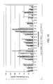
- FIG. 1A is a bar graph displaying the percent of IL-6 production induced by IL-1 ⁇ (4 pM) in MRC-5 cells in the presence of various amounts of the given antibodies.
- FIG. 1B is a bar graph displaying the percent of IL-6 production induced by IL-1 ⁇ (4 pM) in MRC-5 cells in the presence of various amounts of the given antibodies.
- FIG. 1C is a bar graph displaying the percent of IL-6 production induced by IL-1 ⁇ (4 pM) in MRC-5 cells in the presence of various amounts of the given antibodies.
- FIG. 1D is a bar graph displaying the percent of IL-6 production induced by IL-1 ⁇ (4 pM) in MRC-5 cells in the presence of various amounts of the given antibodies.
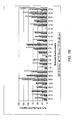
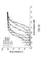
- FIG. 2A is a graph depicting the percent inhibition of IL-1 ⁇ -induced IL-6 production in MRC-5 cells for various antibodies.
- FIG. 2B is a graph depicting the percent inhibition of IL1 ⁇ -induced IL-6 production in MRC-5 cells for various antibodies.
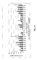
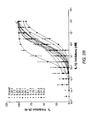
- FIG. 3 is a graph depicting the percent inhibition of IL1 ⁇ -induced IL-8 production in human whole blood for various antibodies.
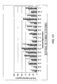
- FIG. 4 is a graph depicting the percent inhibition of IL-6 production for Ab 9.5.2, Ab 5.5.1, and anakinra (KINERETTM) in vivo. Upward triangles represent 9.5.2 IgG4, and downward triangles represent 5.5.1 IgG4.
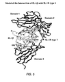
- FIG. 5 is a structural model depicting the interaction of IL-1 beta with a receptor.
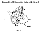
- FIG. 6 is a structural model depicting the areas of antibody 9.5.2 and antibody 5.5.1 interaction with IL-1 beta.
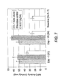
- FIG. 7 is a graph depicting myeloperoxidase (MPO) activity in the lungs of BALB/C mice treated with either IL-1 ⁇ alone or in combination with mAb 9.5.2 or an isotype control.
- MPO myeloperoxidase
- Interleukin-1ß is a pro-inflammatory cytokine that plays a major role in a wide range of diseases, including inflammatory diseases.
- targeted binding agents such as monoclonal antibodies, that bind to and neutralize the activity of IL-1ß.
- the targeted binding agent is a fully human monoclonal antibody that specifically binds to IL-1ß.
- the antibodies bind to IL-1ß with a particularly high affinity.
- the antibodies are highly potent, either in vitro, in vivo, or under both situations.
- treatment with such antibodies can result in inhibition of IL-6 production and/or IL-8 production in vitro, in vivo, or under both situations.
- the disclosed antibodies are more potent, more selective, have a longer half-life, or some combination thereof, than recombinant IL-1 receptor antagonists (IL-1Ra) or anakinra (e.g., KINERETTM).
- IL-1Ra recombinant IL-1 receptor antagonists
- anakinra e.g., KINERETTM
- anakinra prevents the binding of IL-1 to its receptor via a mechanism of receptor antagonism.
- anakinra In order for anakinra to be effective, it has to compete with IL-1 at the level of all receptors, which are ubiquitous and numerous.
- anakinra has a short circulating half-life (4-6 hours) in humans.
- Antibody 9.5.2 potently neutralizes IL-1 ⁇ dependent effects in vitro and in vivo.
- 9.5.2 mAb inhibits IL-1 ⁇ -induced IL-6 production by MRC-5 cells and IL-8 production in whole blood.
- mice 9.5.2 mAb inhibited IL-1 ⁇ -induced IL-6 and MPO production.
- a further embodiment of the invention is an antibody that cross-competes for binding to IL-1 ⁇ with the fully human antibodies of the invention, preferably an antibody comprising a heavy chain amino acid sequence having one of the CDR sequences shown in Table 25 and a light chain amino acid sequence having one of the CDR sequences shown in Table 26.
- a further embodiment of the invention is an antibody that binds to the same epitope on IL-1 ⁇ as a fully human antibodies of the invention, preferably an antibody comprising a heavy chain amino acid sequence having one of the CDR sequences shown in Table 25 and a light chain amino acid sequence having one of the CDR sequences shown in Table 26.
- Embodiments of the invention include the specific IL-1ß antibodies listed below in Table 1.
- This table reports the identification number (“mAb ID No.") of each IL-1ß antibody, along with the SEQ ID number of the corresponding heavy chain and light chain for the nucleic acid and amino acid sequences.
- the mAb ID No. is used to identify the various antibodies.
- mAb ID Nos. begin with the same first two sets of numbers (e.g., 9.5 .2 and 9.5 ) this denotes that the antibodies are clones and are thus identical.
- the complete sequences can be found in the sequence listing and a comparison of the sequences can be found in Table 25 and Table 26.
- Standard techniques are used for recombinant DNA, oligonucleotide synthesis, and tissue culture and transformation (e.g., electroporation, lipofection). Enzymatic reactions and purification techniques are performed according to manufacturer's specifications or as commonly accomplished in the art or as described herein. The foregoing techniques and procedures are generally performed according to conventional methods well known in the art and as described in various general and more specific references that are cited and discussed throughout the present specification. See e.g., Sambrook et al. Molecular Cloning: A Laboratory Manual (3rd ed., Cold Spring Harbor Laboratory Press, Cold Spring Harbor, N.Y. (2001 )), which is incorporated herein by reference.
- IL-1B refers to the molecule interleukin-1ß.
- precursors of IL-1ß such as pro-IL-1ß.
- An example of a mature form of IL-1B is shown in SEQ ID NO: 77.
- An "IL-1 beta antibody” is an antibody that binds to IL-1 beta. This can also be referred to as an anti-IL-1 beta, or, somewhat redundantly, an anti-IL-1 beta antibody.
- These antibodies can also be referred to with their “mAb ID No,” shown above in Table 1.
- Antibodies can be named with either numerals separated by periods or numerals separated by dashes, nothing is implied by this difference.
- neutralizing when referring to an antibody relates to the ability of an antibody to eliminate, or significantly reduce, the activity of a target antigen. Accordingly, a “neutralizing" IL-1ß antibody is capable of eliminating or significantly reducing the activity of IL-1ß.
- a neutralizing IL-1ß antibody may, for example, act by blocking the binding of IL-1ß to a type I IL-1 receptor ("IL-1R"). By blocking this binding, the IL-1ß mediated signal transduction is significantly, or completely, eliminated.
- IL-1R type I IL-1 receptor
- a neutralizing antibody against IL-1ß inhibits IL-1ß related disorders.
- the neutralizing antibody prevents the IL-1ß molecule from binding to the type II IL-1 receptor.
- the type II receptor is also known as a decoy receptor.
- a neutralizing antibody that prevents IL-1ß from binding to the type II receptor, but still allows IL-1ß to bind to the type I receptor would result in an effective increase in IL-1ß activity.
- the IL-1 receptor shall refer to the type I receptor.
- the antibody can be neutralizing for any and all functions of the protein.
- an IL-1ß antibody may alter the production of IL-6, IL-8, or both.
- Antibodies can have differing levels of potency for different assays.
- Contemplated potencies include any effective potency, for example, IC 50 s of less than 14 nM to 1 nM, 1 nM to 500pM, 500pM to 1pM for IL-6 inhibition; less than 2.3 nM to 100 pM, 100 pM to 70 pM, or 70 to 4 pM for IL-8 inhibition; and less than 51-8, 8-5, or 5 pmoles/mouse for in vivo IL-6 production.
- IC 50 s of less than 14 nM to 1 nM, 1 nM to 500pM, 500pM to 1pM for IL-6 inhibition; less than 2.3 nM to 100 pM, 100 pM to 70 pM, or 70 to 4 pM for IL-8 inhibition; and less than 51-8, 8-5, or 5 pmoles/mouse for in vivo IL-6 production.
- isolated polynucleotide shall mean a polynucleotide that has been isolated from its naturally occurring environment. Such polynucleotides may be genomic, cDNA, or synthetic. Isolated polynucleotides preferably are not associated with all or a portion of the polynucleotides they associate with in nature. The isolated polynucleotides may be operably linked to another polynucleotide to which it is not linked in nature. In addition, isolated polynucleotides preferably do not occur in nature as part of a larger sequence.
- isolated protein means a protein that has been isolated from its naturally occurring environment. Such proteins may be derived from genomic DNA, cDNA, recombinant DNA, recombinant RNA, or synthetic origin or some combination thereof, which by virtue of its origin, or source of derivation, the "isolated protein” (1) is not associated with proteins found in nature, (2) is free of other proteins from the same source, e.g. free of murine proteins, (3) is expressed by a cell from a different species, or (4) does not occur in nature.
- polypeptide is used herein as a generic term to refer to native protein, fragments, or analogs of a polypeptide sequence.
- native protein, fragments, and analogs are species of the polypeptide genus.
- Preferred polypeptides in accordance with the invention comprise the human heavy chain immunoglobulin molecules and the human kappa light chain immunoglobulin molecules, the human heavy chain immunoglobulin molecules and the human lambda light chain immunoglobulin molecules, as well as antibody molecules formed by combinations comprising the heavy chain immunoglobulin molecules with light chain immunoglobulin molecules, such as the kappa or lambda light chain immunoglobulin molecules, and vice versa, as well as fragments and analogs thereof.
- Preferred polypeptides in accordance with the invention may also comprise solely the human heavy chain immunoglobulin molecules or fragments thereof.
- naturally-occurring refers to the fact that an object can be found in nature.
- a polypeptide or polynucleotide sequence that is present in an organism (including viruses) that can be isolated from a source in nature and which has not been intentionally modified by man in the laboratory or otherwise is naturally-occurring.
- operably linked refers to positions of components so described that are in a relationship permitting them to function in their intended manner.
- a control sequence "operably linked" to a coding sequence is connected in such a way that expression of the coding sequence is achieved under conditions compatible with the control sequences.
- control sequence refers to polynucleotide sequences that are necessary either to effect or to affect the expression and processing of coding sequences to which they are connected. The nature of such control sequences differs depending upon the host organism; in prokaryotes, such control sequences generally include promoter, ribosomal binding site, and transcription termination sequence; in eukaryotes, generally, such control sequences may include promoters, introns and transcription termination sequence.
- control sequences is intended to include, at a minimum, all components whose presence is essential for expression and processing, and can also include additional components whose presence is advantageous, for example, leader sequences and fusion partner sequences.
- polynucleotide as referred to herein means a polymeric form of nucleotides of at least 10 bases in length, either ribonucleotides or deoxynucleotides or a modified form of either type of nucleotide.
- the term includes single- and double-stranded forms of DNA.
- oligonucleotide includes naturally occurring, and modified nucleotides linked together by naturally occurring, and non-naturally occurring linkages. Oligonucleotides are a polynucleotide subset generally comprising a length of 200 bases or fewer. Preferably, oligonucleotides are 10 to 60 bases in length and most preferably 12, 13, 14, 15, 16, 17, 18, 19, or 20 to 40 bases in length. Oligonucleotides are usually single stranded, e.g. for probes; although oligonucleotides may be double stranded, e.g. for use in the construction of a gene mutant. Oligonucleotides can be either sense or antisense oligonucleotides.
- nucleotides includes deoxyribonucleotides and ribonucleotides.
- modified nucleotides includes nucleotides with modified or substituted sugar groups and the like.
- oligonucleotide linkages includes oligonucleotides linkages such as phosphorothioate, phosphorodithioate, phosphoroselenoate, phosphorodiselenoate, phosphoroanilothioate, phosphoraniladate, phosphoroamidate, and the like. See e.g., LaPlanche et al. Nucl. Acids Res.
- oligonucleotide can include a label for detection, if desired.
- the term "selectively hybridize” referred to herein means to detectably and specifically bind.
- Polynucleotides, oligonucleotides and fragment thereof selectively hybridize to nucleic acid strands under hybridization and wash conditions that minimize appreciable amounts of detectable binding to nonspecific nucleic acids.
- High stringency conditions can be used to achieve selective hybridization conditions as known in the art and discussed herein.
- the nucleic acid sequence homology between the polynucleotides, oligonucleotides, or antibody fragments and a nucleic acid sequence of interest will be at least 80%, and more typically with preferably increasing homologies of at least 85%, 90%, 95%, 99%, and 100%.
- Two amino acid sequences are "homologous" if there is a partial or complete identity between their sequences. For example, 85% homology means that 85% of the amino acids are identical when the two sequences are aligned for maximum matching. Gaps (in either of the two sequences being matched) are allowed in maximizing matching; gap lengths of 5 or less are preferred with 2 or less being more preferred. Alternatively and preferably, two protein sequences (or polypeptide sequences derived from them of at least about 30 amino acids in length) are homologous, as this term is used herein, if they have an alignment score of, or more than, 5 (in standard deviation units) using the program ALIGN with the mutation data matrix and a gap penalty of 6 or greater.
- a polynucleotide sequence is homologous (i.e., is identical, not strictly evolutionarily related) to all or a portion of a reference polynucleotide sequence, or that a polypeptide sequence is identical to a reference polypeptide sequence.
- the term “complementary to” is used herein to mean that the complementary sequence is homologous to all or a portion of a reference polynucleotide sequence.
- the nucleotide sequence "TATAC” corresponds to a reference sequence “TATAC” and is complementary to a reference sequence "GTATA”.
- a “reference sequence” is a defined sequence used as-a basis for a sequence comparison.
- a reference sequence may be a subset of a larger sequence, for example, as a segment of a full-length cDNA or gene sequence given in a sequence listing or may comprise a complete cDNA or gene sequence.
- a reference sequence is at least 18 nucleotides or 6 amino acids in length, frequently at least 24 nucleotides or 8 amino acids in length, and often at least 48 nucleotides or 16 amino acids in length.
- two polynucleotides or amino acid sequences may each (1) comprise a sequence (i.e., a portion of the complete polynucleotide or amino acid sequence) that is similar between the two molecules, and (2) may further comprise a sequence that is divergent between the two polynucleotides or amino acid sequences
- sequence comparisons between two (or more) molecules are typically performed by comparing sequences of the two molecules over a "comparison window" to identify and compare local regions of sequence similarity.
- a “comparison window”, as used herein, refers to a conceptual segment of at least about 18 contiguous nucleotide positions or about 6 amino acids wherein the polynucleotide sequence or amino acid sequence is compared to a reference sequence of at least 18 contiguous nucleotides or 6 amino acid sequences and wherein the portion of the polynucleotide sequence in the comparison window may include additions, deletions, substitutions, and the like (i.e., gaps) of 20 percent or less as compared to the reference sequence (which does not comprise additions or deletions) for optimal alignment of the two sequences.
- Optimal alignment of sequences for aligning a comparison window may be conducted by the local homology algorithm of Smith and Waterman Adv. Appl. Math.
- sequence identity means that two polynucleotide or amino acid sequences are identical (i.e., on a nucleotide-by-nucleotide or residue-by-residue basis) over the comparison window.
- percentage of sequence identity is calculated by comparing two optimally aligned sequences over the window of comparison, determining the number of positions at which the identical nucleic acid base (e.g., A, T, C, G, U, or I) or amino acid residue occurs in both sequences to yield the number of matched positions, dividing the number of matched positions by the total number of positions in the comparison window (i.e., the window size), and multiplying the result by 100 to yield the percentage of sequence identity.
- substantially identical denotes a characteristic of a polynucleotide or amino acid sequence, wherein the polynucleotide or amino acid comprises a sequence that has at least 85 percent sequence identity, preferably at least 90 to 95 percent sequence identity, more preferably at least 99 percent sequence identity, as compared to a reference sequence over a comparison window of at least 18 nucleotide (6 amino acid) positions, frequently over a window of at least 24-48 nucleotide (8-16 amino acid) positions, wherein the percentage of sequence identity is calculated by comparing the reference sequence to the sequence which may include deletions or additions which total 20 percent or less of the reference sequence over the comparison window.
- the reference sequence may be a subset of a larger sequence.
- Examples of unconventional amino acids include: 4-hydroxyproline, ⁇ -carboxyglutamate, ⁇ -N,N,N-trimethyllysine, ⁇ -N-acetyllysine, O-phosphoserine, N-acetylserine, N-formylmethionine, 3-methylhistidine, 5-hydroxylysine, ⁇ -N-methylarginine, and other similar amino acids and imino acids (e.g ., 4-hydroxyproline).
- the left-hand direction is the amino terminal direction and the right-hand direction is the carboxy-terminal direction, in accordance with standard usage and convention. Additionally, the short hand notation for amino acids and amino acid substitutions is also used.
- amino acid, amino acid position, amino acid represents the wild-type amino acid, the position of that amino acid, and the residue that the amino acid has been replaced with.
- A472Y means that the original alanine at position 472 has been replaced with a tryptophan.
- the left-hand end of single-stranded polynucleotide sequences is the 5' end; the left-hand direction of double-stranded polynucleotide sequences is referred to as the 5' direction.
- the direction of 5' to 3' addition of nascent RNA transcripts is referred to as the transcription direction; sequence regions on the DNA strand having the same sequence as the RNA and which are 5' to the 5' end of the RNA transcript are referred to as "upstream sequences"; sequence regions on the DNA strand having the same sequence as the RNA and which are 3' to the 3' end of the RNA transcript are referred to as "downstream sequences".
- the term "substantial identity” means that two peptide sequences, when optimally aligned, such as by the programs GAP or BESTFIT using default gap weights, share at least 80 percent sequence identity, preferably at least 90 percent sequence identity, more preferably at least 95 percent sequence identity, and most preferably at least 99 percent sequence identity.
- residue positions that are not identical differ by conservative amino acid substitutions.
- Conservative amino acid substitutions refer to the interchangeability of residues having similar side chains.
- a group of amino acids having aliphatic side chains is glycine, alanine, valine, leucine, and isoleucine; a group of amino acids having aliphatic-hydroxyl side chains is serine and threonine; a group of amino acids having amide-containing side chains is asparagine and glutamine; a group of amino acids having aromatic side chains is phenylalanine, tyrosine, and tryptophan; a group of amino acids having basic side chains is lysine, arginine, and histidine; and a group of amino acids having sulfur-containing side chains is cysteine and methionine.
- Preferred conservative amino acids substitution groups are: valine-leucine-isoleucine, phenylalanine-tyrosine, lysine-arginine, alanine-valine, glutamic-aspartic, and asparagine-glutamine.
- amino acid sequences of antibodies or immunoglobulin molecules are contemplated as being encompassed by the present invention, providing that the variations in the amino acid sequence maintain at least 75%, more preferably at least 80%, 90%, 95%, and most preferably 99% sequence identity to the antibodies or immunoglobulin molecules described herein.
- conservative amino acid replacements are contemplated. Conservative replacements are those that take place within a family of amino acids that have related side chains.
- More preferred families are: serine and threonine, which form an aliphatic hydroxy family; asparagine and glutamine, which form an amide-containing family; alanine, valine, leucine and isoleucine, which form an aliphatic family; and phenylalanine, tryptophan, and tyrosine, which form an aromatic family.
- Structural and functional domains can be identified by comparison of the nucleotide and/or amino acid sequence data to public or proprietary sequence databases.
- computerized comparison methods are used to identify sequence motifs or predicted protein conformation domains that occur in other proteins of known structure and/or function. Methods to identify protein sequences that fold into a known three-dimensional structure are known. Bowie et al. Science 253:164 (1991 ).
- sequence motifs and structural conformations that may be used to define structural and functional domains in accordance with the antibodies described herein.
- Preferred amino acid substitutions are those which: (1) reduce susceptibility to proteolysis, (2) reduce susceptibility to oxidation, (3) alter binding affinity for forming protein complexes, (4) alter binding affinities, and (4) confer or modify other physicochemical or functional properties of such analogs.
- Analogs can include various muteins of a sequence other than the naturally-occurring peptide sequence. For example, single or multiple amino acid substitutions (preferably conservative amino acid substitutions) may be made in the naturally-occurring sequence (preferably in the portion of the polypeptide outside the domain(s) forming intermolecular contacts.
- a conservative amino acid substitution should not substantially change the structural characteristics of the parent sequence (e.g., a replacement amino acid should not tend to break a helix that occurs in the parent sequence, or disrupt other types of secondary structure that characterizes the parent sequence).
- Examples of art-recognized polypeptide secondary and tertiary structures are described in Proteins, Structures and Molecular Principles (Creighton, Ed., W. H. Freeman and Company, New York (1984 )); Introduction to Protein Structure (C. Branden and J. Tooze, eds., Garland Publishing, New York, N.Y. (1991 )); and Thornton et at. Nature 354:105 (1991 ), which are each incorporated herein by reference.
- polypeptide fragment refers to a polypeptide that has an amino-terminal and/or carboxy-terminal deletion, but where the remaining amino acid sequence is identical to the corresponding positions in the naturally-occurring sequence deduced, for example, from a full-length cDNA sequence. Fragments typically are at least 5, 6, 8 or 10 amino acids long, preferably at least 14 amino acids long, more preferably at least 20 amino acids long, usually at least 50 amino acids long, and even more preferably at least 70 amino acids long.
- analog refers to polypeptides which are comprised of a segment of at least 25 amino acids that has substantial identity to a portion of a deduced amino acid sequence and which has at least one of the following properties: (1) specific binding to a IL-1ß, under suitable binding conditions, (2) ability to block appropriate IL-1ß binding, or (3) ability to inhibit IL-1ß activity.
- polypeptide analogs comprise a conservative amino acid substitution (or addition or deletion) with respect to the naturally-occurring sequence.
- Analogs typically are at least 20 amino acids long, preferably at least 50 amino acids long or longer, and can often be as long as a full-length naturally-occurring polypeptide.
- Peptide analogs are commonly used in the pharmaceutical industry as non-peptide drugs with properties analogous to those of the template peptide. These types of non-peptide compound are termed "peptide mimetics" or "peptidomimetics”. Fauchere, J. Adv. Drug Res. 15:29 (1986 ); Veber and Freidinger TINS p.392 (1985 ); and Evans et al. J. Med. Chem. 30:1229 (1987 ), which are incorporated herein by reference. Such compounds are often developed with the aid of computerized molecular modeling. Peptide mimetics that are structurally similar to therapeutically useful peptides may be used to produce an equivalent therapeutic or prophylactic effect.
- a paradigm polypeptide i.e., a polypeptide that has a biochemical property or pharmacological activity
- Systematic substitution of one or more amino acids of a consensus sequence with a D-amino acid of the same type may be used to generate more stable peptides.
- constrained peptides comprising a consensus sequence or a substantially identical consensus sequence variation may be generated by methods known in the art ( Rizo and Gierasch Ann. Rev. Biochem. 61:387 (1992 ), incorporated herein by reference); for example, by adding internal cysteine residues capable of forming intramolecular disulfide bridges which cyclize the peptide.
- an antibody refers to a polypeptide or group of polypeptides which are comprised of at least one binding domain, where an antibody binding domain is formed from the folding of variable domains of an antibody molecule to form three-dimensional binding spaces with an internal surface shape and charge distribution complementary to the features of an antigenic determinant of an antigen.
- An antibody typically has a tetrameric form, comprising two identical pairs of polypeptide chains, each pair having one "light” and one "heavy” chain. The variable regions of each light/heavy chain pair form an antibody binding site.
- unit dose refers to an amount of a substance sufficient to achieve a desired result in a particular subject.
- unit doses can vary depending upon the particular substance in the unit dose, who will be taking the substance, and what the desired result will be.
- a “targeted binding agent” is an antibody, or binding fragment thereof, that preferentially binds to a target site.
- the targeted binding agent is specific for only one target site. In other embodiments, the targeted binding agent is specific for more than one target site.
- the targeted binding agent may be a monoclonal antibody and the target site may be an epitope.
- Binding fragments of an antibody are produced by recombinant DNA techniques, or by enzymatic or chemical cleavage of intact antibodies. Binding fragments include Fab, Fab', F(ab') 2 , Fv, and single-chain antibodies.
- An antibody other than a "bispecific” or “bifunctional” antibody is understood to have each of its binding sites identical.
- An antibody substantially inhibits adhesion of a receptor to a counterreceptor when an excess of antibody reduces the quantity of receptor bound to counterreceptor by at least about 20%, 40%, 60% or 80%, and more usually greater than about 85% (as measured in an in vitro competitive binding assay).
- a "complete” antibody refers to an antibody that has all of the parts that make up an antibody, as defined by the definition of "antibody,” above. Of course, variants or insubstantial modifications of the antibody can result in antibodies that are smaller than the full antibody sequence.
- epitopic determinants includes any protein determinant capable of specific binding to an immunoglobulin or T-cell receptor.
- Epitopic determinants usually consist of chemically active surface groupings of molecules such as amino acids or sugar side chains and can, but not always, have specific three-dimensional structural characteristics, as well as specific charge characteristics.
- An antibody is said to bind an antigen when the dissociation constant (K D or K d ) is less than or equal to 1 ⁇ M, 100 nM, 10 nM, 1nM, 100 pM, 10 pM, 1 pM, 100 nM, 10 nM, 1 nM, 500 fM, 100 fM, 10fM, or less.
- Antibodies that compete for binding with the herein disclosed antibodies are also contemplated. Competition can be direct, for the entire epitope, or a fraction of the epitope, or competition can be indirect, where binding of the antibody prevents the binding of the herein disclosed antibodies.
- the terms “selectively bind” or “specifically bind” are used herein to denote that the antibody will bind to one substance more strongly than it will bind to another substance. It is not meant to denote that the antibody will only bind to one substance. When binding only occurs between a single substance and the antibody, the antibody is said to "exclusively” bind to the substance.
- agent is used herein to denote a chemical compound, a mixture of chemical compounds, a biological macromolecule, or an extract made from biological materials.
- IL-1ß polypeptide refers to a portion of an IL-1ß polypeptide that has a biological or an immunological activity of a native IL-1ß polypeptide.
- Biological when used herein refers to a biological function that results from the activity of the native IL-1ß polypeptide.
- a preferred IL-1ß biological activity includes, for example, IL-1ß induced inflammatory disorders.
- mammal when used herein refers to any animal that is considered a mammal. Preferably, the mammal is human.
- Digestion of antibodies with the enzyme, papain results in two identical antigen-binding fragments, known also as "Fab” fragments, and a "Fc” fragment, having no antigen-binding activity but having the ability to crystallize.
- Digestion of antibodies with the enzyme, pepsin results in the a F(ab') 2 fragment in which the two arms of the antibody molecule remain linked and comprise two-antigen binding sites.
- the F(ab') 2 fragment has the ability to crosslink antigen.
- Fv when used herein refers to the minimum fragment of an antibody that retains both antigen-recognition and antigen-binding sites.
- Fab when used herein refers to a fragment of an antibody that comprises the constant domain of the light chain and the CH1 domain of the heavy chain.
- mAb refers to monoclonal antibody.
- Liposome when used herein refers to a small vesicle that may be useful for delivery of drugs that may include the IL-1ß polypeptide of the invention or antibodies to such an IL-1ß polypeptide to a mammal.
- Label refers to the addition of a detectable moiety to a polypeptide, for example, a radiolabel, fluorescent label, enzymatic label chemiluminescent labeled or a biotinyl group.
- Radioisotopes or radionuclides may include 3 H, 14 C, 15 N, 35 S, 90 Y, 99 Tc, 111 In, 125 I, 131 I, fluorescent labels may include rhodamine, lanthanide phosphors or FITC and enzymatic labels may include horseradish peroxidase, ⁇ -galactosidase, luciferase, alkaline phosphatase.
- pharmaceutical agent or drug refers to a chemical compound or composition capable of inducing a desired therapeutic effect when properly administered to a patient.
- Other chemistry terms herein are used according to conventional usage in the art, as exemplified by The McGraw-Hill Dictionary of Chemical Terms (Parker, S., Ed., McGraw-Hill, San Francisco (1985 )), incorporated herein by reference.
- substantially pure means an object species is the predominant species present (i.e., on a molar basis it is more abundant than any other individual species in the composition), and preferably a substantially purified fraction is a composition wherein the object species comprises at least about 50 percent (on a molar basis) of all macromolecular species present. Generally, a substantially pure composition will comprise more than about 80 percent of all macromolecular species present in the composition, more preferably more than about 85%, 90%, 95%, and 99%. Most preferably, the object species is purified to essential homogeneity (contaminant species cannot be detected in the composition by conventional detection methods) wherein the composition consists essentially of a single macromolecular species.
- patient includes human and veterinary subjects.
- Human antibodies avoid some of the problems associated with antibodies that possess murine or rat variable and/or constant regions.
- the presence of such murine or rat derived proteins can lead to the rapid clearance of the antibodies or can lead to the generation of an immune response against the antibody by a patient.
- fully human antibodies can be generated through the introduction of functional human antibody loci into a rodent, other mammal or animal so that the rodent, other mammal or animal produces fully human antibodies.
- One method for generating fully human antibodies is through the use of XENOMOUSE ® strains of mice that have been engineered to contain 245 kb and 190 kb-sized germline configuration fragments of the human heavy chain locus and kappa light chain locus.
- Other XenoMouse strains of mice contain 980 kb and 800 kb-sized germline configuration fragments of the human heavy chain locus and kappa light chain locus.
- Still other XenoMouse strains of mice contain 980 kb and 800 kb-sized germline configuration fragments of the human heavy chain locus and kappa light chain locus plus a 740 kb-sized germline configured complete human lambda light chain locus.
- Patent Publication 2003/0217373, filed November 20, 2002 and U.S. Patent Nos. 6,833,268 , 6,162,963 , 6,150,584 , 6,114,598 , 6,075,181 , and 5,939,598 and Japanese Patent Nos. 3 068 180 B2 , 3 068 506 B2 , and 3 068 507 B2 .
- European Patent No., EP 0 463 151 B1 grant published June 12, 1996, International Patent Application No., WO 94/02602, published February 3, 1994 , International Patent Application No., WO 96/34096, published October 31, 1996 , WO 98/24893, published June 11, 1998 , WO 00/76310, published December 21, 2000 .
- the disclosures of each of the above-cited patents, applications, and references are hereby incorporated by reference in their entirety.
- minilocus In an alternative approach, others, including GenPharm International, Inc., have utilized a "minilocus" approach. In the minilocus approach, an exogenous Ig locus is mimicked through the inclusion of pieces (individual genes) from the Ig locus. Thus, one or more V H genes, one or more D H genes, one or more J H genes, a mu constant region, and a second constant region (preferably a gamma constant region) are formed into a construct for insertion into an animal. This approach is described in U.S. Patent No. 5,545,807 to Surani et al . and U.S. Patent Nos.
- Kirin has also demonstrated the generation of human antibodies from mice in which, through microcell fusion, large pieces of chromosomes, or entire chromosomes, have been introduced. See European Patent Application Nos. 773 288 and 843 961 , the disclosures of which are hereby incorporated by reference. Additionally, KMTM- mice, which are the result of cross-breeding of Kirin's Tc mice with Medarex's minilocus (Humab) mice have been generated. These mice possess the HC transchromosome of the Kirin mice and the kappa chain transgene of the Genpharm mice ( Ishida et al., Cloning Stem Cells, (2002) 4:91-102 ).
- Human antibodies can also be derived by in vitro methods. Suitable examples include, but are not limited to, phage display (CAT, Morphosys, Dyax, Biosite/Medarex, Xoma, Symphogen, Alexion (formerly Proliferon), Affimed) ribosome display (CAT), yeast display, and the like.
- mice were prepared through the utilization of the XENOMOUSE ® technology, as described below. Such mice, then, are capable of producing human immunoglobulin molecules and antibodies and are deficient in the production of murine immunoglobulin molecules and antibodies. Technologies utilized for achieving the same are disclosed in the patents, applications, and references disclosed in the background section herein. In particular, however, a preferred embodiment of transgenic production of mice and antibodies therefrom is disclosed in U.S. Patent Application Serial No. 08/759,620, filed December 3, 1996 and International Patent Application Nos. WO 98/24893, published June 11, 1998 and WO 00/76310, published December 21, 2000 , the disclosures of which are hereby incorporated by reference. See also Mendez et al. Nature Genetics 15:146-156 (1997 ), the disclosure of which is hereby incorporated by reference.
- XENOMOUSE ® lines of mice are immunized with an antigen of interest (e.g . IL-1ß), lymphatic cells (such as B-cells) are recovered from the mice that expressed antibodies, and the recovered cell lines are fused with a myeloid-type cell line to prepare immortal hybridoma cell lines.
- IL-1ß an antigen of interest
- lymphatic cells such as B-cells
- myeloid-type cell line to prepare immortal hybridoma cell lines.
- These hybridoma cell lines are screened and selected to identify hybridoma cell lines that produced antibodies specific to the antigen of interest.
- Provided herein are methods for the production of multiple hybridoma cell lines that produce antibodies specific to IL-1ß. Further, provided herein are characterization of the antibodies produced by such cell lines, including nucleotide and amino acid sequence analyses of the heavy and light chains of such antibodies.
- B cells can be directly assayed.
- CD19+ B cells can be isolated from hyperimmune XENOMOUSE® mice and allowed to proliferate and differentiate into antibody-secreting plasma cells.
- Antibodies from the cell supernatants are then screened by ELISA for reactivity against the IL-1ß immunogen.
- the supernatants might also be screened for immunoreactivity against fragments of IL-1ß to further map the different antibodies for binding to domains of functional interest on IL-1ß.
- the antibodies may also be screened against other related human interleukins and against the rat, the mouse, and non-human primate, such as cynomolgus monkey, orthologues of IL-1ß, the last to determine species cross-reactivity.
- B cells from wells containing antibodies of interest may be immortalized by fusion to make hybridomas either from individual or from pooled wells, or immortalized by infection with EBV or transfection by known immortalizing genes and then plating in suitable medium.
- single plasma cells secreting antibodies with the desired specificities are then isolated using an IL-1ß-specific hemolytic plaque assay ( Babcook et al., Proc. Natl. Acad. Sci. USA 93:7843-48 (1996 )).
- Cells targeted for lysis are preferably sheep red blood cells (SRBCs) coated with the IL-1ß antigen.
- SRBCs sheep red blood cells
- a plaque In the presence of a B-cell culture containing plasma cells secreting the immunoglobulin of interest and complement, the formation of a plaque indicates specific IL-1ß-mediated lysis of the sheep red blood cells surrounding the plasma cell of interest.
- the single antigen-specific plasma cell in the center of the plaque can be isolated and the genetic information that encodes the specificity of the antibody is isolated from the single plasma cell.
- RT-PCR reverse-transcriptase followed by PCR
- Such cloned DNA can then be further inserted into a suitable expression vector, preferably a vector cassette such as a pcDNA, more preferably such a pcDNA vector containing the constant domains of immunoglobulin heavy and light chain.
- a suitable expression vector preferably a vector cassette such as a pcDNA, more preferably such a pcDNA vector containing the constant domains of immunoglobulin heavy and light chain.
- the generated vector can then be transfected into host cells, e.g., HEK293 cells, CHO cells, and cultured in conventional nutrient media modified as appropriate for inducing promoters, selecting transformants, or amplifying the genes encoding the desired sequences.
- antibodies produced by the fused hybridomas were human IgG4 heavy chains with fully human kappa or lambda light chains.
- Antibodies described herein possess human IgG4 heavy chains as well as IgG2 heavy chains.
- Antibodies can also be of other human isotypes, including IgG1.
- the antibodies possessed high affinities, typically possessing a K D of from about 10 -6 through about 10 -13 M or below, when measured by solid phase and solution phase techniques.
- Antibodies possessing a K D of no more than 10 -11 M are preferred to inhibit the activity of IL-1ß.
- anti-IL-1ß antibodies can be expressed in cell lines other than hybridoma cell lines. Sequences encoding particular antibodies can be used to transform a suitable mammalian host cell. Transformation can be by any known method for introducing polynucleotides into a host cell, including, for example packaging the polynucleotide in a virus (or into a viral vector) and transducing a host cell with the virus (or vector) or by transfection procedures known in the art, as exemplified by U.S. Patent Nos. 4,399,216 , 4,912,040 , 4,740,461 , and 4,959,455 (which patents are hereby incorporated herein by reference). The transformation procedure used depends upon the host to be transformed.
- Methods for introducing heterologous polynucleotides into mammalian cells include dextran-mediated transfection, calcium phosphate precipitation, polybrene mediated transfection, protoplast fusion, electroporation, encapsulation of the polynucleotide(s) in liposomes, and direct microinjection of the DNA into nuclei.
- Mammalian cell lines available as hosts for expression are well known in the art and include many immortalized cell lines available from the American Type Culture Collection (ATCC), including but not limited to Chinese hamster ovary (CHO) cells, HeLa cells, baby hamster kidney (BHK) cells, monkey kidney cells (COS), human hepatocellular carcinoma cells (e.g., Hep G2), and a number of other cell lines.
- ATCC American Type Culture Collection
- Cell lines of particular preference are selected through determining which cell lines have high expression levels and produce antibodies with constitutive IL-1ß binding properties.
- Anti-IL-1ß antibodies are useful in the detection of IL-1ß in patient samples and accordingly are useful as diagnostics for disease states as described herein. In addition, based on their ability to significantly neutralize IL-1ß activity (as demonstrated in the Examples below), anti-IL-1ß antibodies have therapeutic effects in treating symptoms and conditions resulting from IL-1ß.
- the antibodies and methods herein relate to the treatment of symptoms resulting from IL-1ß induced disorders or IL-1ß related disorders. Further embodiments involve using the antibodies and methods described herein to treat IL-1ß-related disorders.
- IL-1ß-related disorders can include inflammatory disorders, such as immune-mediated inflammatory disorders (IMID), which are inflammatory conditions caused and sustained by an antigen-specific, pathological immune response.
- IMID immune-mediated inflammatory disorders
- IMID immunosensive diseases
- diseases such as asthma, hay fever, and urticaria
- diseases caused by immune complexes e.g., systemic lupus erythematosus, glomerulonephritis, pemphigus
- vasculitis such as Wegener's granulomatosis and Kawasaki's syndrome
- different types of connective tissue disorders inflammatory bowel disease (e.g., Crohn's disease and ulcerative colitis); insulin-dependent diabetes; multiple sclerosis; psoriasis; uveitis; retinitis; graft rejection; and graft-versus host-disease.
- IL-1ß-related disorders can also include the pathogenesis of systemic inflammatory disorders, e.g., sepsis or familial mediterranean fever and the Muckle-Wells syndrome.
- tissue inflammation in infectious, ischemic, hemorrhagic, and traumatic conditions e.g., fasciitis, stroke, infarction of the myocardium and other organs (e.g., lung and intestine), ARDS; hepatitis, (e.g., infectious and non-infectious, acute and chronic); acute and chronic pancreatitis; reperfusion injuries; radiation injuries; vascular restenosis of different types (e.g., coronary restenosis); and orthopedic and dental injuries ranging from muscle strain, to ligament sprain, to periodontal disease.
- IL-1ß related disorders further can include the pathogenesis of systemic disturbances of less obvious inflammatory nature, such as cachexia, chronic fatigue syndrome, anorexia and sleep and mental alterations (e.g., learning impairment), osteoarthritis, osteoporosis, atherosclerosis, organ fibrosis ( e.g ., lung and liver fibrosis), Alzheimer's disease, Parkinson's syndromes, amyelolateroschlerosis, and various myopathies, which are considered chiefly degenerative in nature but whose pathogenesis includes inflammatory components.
- IL-1ß related disorders can include hyperalgesia of various types and cancer-related pain.
- IL-1ß related disorders can include congestive heart failure, independently of primary heart disease.
- IL-1ß related disorders can include cancer, blood malignancies, e.g., leukemias and multiple myelomas; the development of a number of solid tumors, tumor growth, and metastatic spreading.
- the antibodies disclosed herein can be used to not only identify the above disorders, but to also treat, cure, or prevent such disorders.
- methods and compositions for the detection, treatment, prevention, etc. of such disorders involving the herein disclosed antibodies are contemplated for the above disorders and related disorders.
- the above list can also serve as examples of treatable IL-1ß related disorders.
- the general production of antibodies can involve the immunization of the animal with IL-1ß, antibody generation (hybridoma, electrocell fusion), confirming human IgG IL-1ß antibodies, producing antibodies for the assays, inhibiting IL-1ß induced IL-6 production, running the top neutralizers on a BIACORE device to determine affinity, cloning the leads and sequencing them, determining the potency of inhibition by the antibodies, assessing the K D , epitope mapping, mAb production, and in vivo testing.
- Embodiments of the invention include sterile pharmaceutical formulations and medicaments of anti-IL-1ß antibodies that are useful as treatments for diseases. Such formulations can inhibit the binding of IL-1ß to its receptor, thereby effectively treating pathological conditions where, e.g., serum IL-1ß is abnormally elevated.
- Anti-IL-1ß antibodies preferably possess adequate affinity to potently neutralize IL-1ß, and preferably have an adequate duration of action to allow for infrequent dosing in humans. A prolonged duration of action will allow for less frequent and more convenient dosing schedules by alternate parenteral routes such as subcutaneous or intramuscular injection.
- Sterile formulations can be created, for example, by filtration through sterile filtration membranes, prior to or following lyophilization and reconstitution of the antibody.
- the antibody ordinarily will be stored in lyophilized form or in solution.
- Therapeutic antibody compositions generally are placed into a container having a sterile access port, for example, an intravenous solution bag or vial having an adapter that allows retrieval of the formulation, such as a stopper pierceable by a hypodermic injection needle.
- the targeted binding agents described herein are useful in the preparation of medicaments for the treatment of IL-1ß related disorders, such as inflammation.
- the route of antibody administration is in accord with known methods, e.g., injection or infusion by intravenous, intraperitoneal, intracerebral, intramuscular, intraocular, intraarterial, intrathecal, inhalation or intralesional routes, or by sustained release systems as noted below.
- the antibody is preferably administered continuously by infusion or by bolus injection.
- an effective amount of antibody to be employed therapeutically will depend, for example, upon the therapeutic objectives, the route of administration, and the condition of the patient. Accordingly, it is preferred that the therapist titer the dosage and modify the route of administration as required to obtain the optimal therapeutic effect. Typically, the clinician will administer antibody until a dosage is reached that achieves the desired effect. The progress of this therapy is easily monitored by conventional assays or by the assays described herein.
- Antibodies can be prepared in a mixture with a pharmaceutically acceptable carrier.
- This therapeutic composition can be administered intravenously or through the nose or lung, preferably as a liquid or powder aerosol (lyophilized).
- the composition may also be administered parenterally or subcutaneously as desired.
- the therapeutic composition should be sterile, pyrogen-free and in a parenterally acceptable solution having due regard for pH, isotonicity, and stability. These conditions are known to those skilled in the art.
- dosage formulations of the compounds described herein are prepared for storage or administration by mixing the compound having the desired degree of purity with physiologically acceptable carriers, excipients, or stabilizers.
- Such materials are nontoxic to the recipients at the dosages and concentrations employed, and include buffers such as TRIS HC1, phosphate, citrate, acetate and other organic acid salts; antioxidants such as ascorbic acid; low molecular weight (less than about ten residues) peptides such as polyarginine, proteins, such as serum albumin, gelatin, or immunoglobulins; hydrophilic polymers such as polyvinylpyrrolidinone; amino acids such as glycine, glutamic acid, aspartic acid, or arginine; monosaccharides, disaccharides, and other carbohydrates including cellulose or its derivatives, glucose, mannose, or dextrins; chelating agents such as EDTA; sugar alcohols such as mannitol or sorbitol; counterions such as sodium and/or nonionic surfactants such as TWEEN, PLURONICS or polyethyleneglycol.
- buffers such as TRIS HC1, phosphate, citrate, acetate and other
- Sterile compositions for injection can be formulated according to conventional pharmaceutical practice as described in Remington: The Science and Practice of Pharmacy (20th ed, Lippincott Williams & Wilkens Publishers (2003 )). For example, dissolution or suspension of the active compound in a vehicle such as water or naturally occurring vegetable oil like sesame, peanut, or cottonseed oil or a synthetic fatty vehicle like ethyl oleate or the like may be desired. Buffers, preservatives, antioxidants, and the like can be incorporated according to accepted pharmaceutical practice.
- sustained-release preparations include semipermeable matrices-of solid-hydrophobic-polymers containing the polypeptide which matrices are in the form of shaped articles, films or microcapsules.
- sustained-release matrices include polyesters, hydrogels (e.g., poly(2-hydroxyethylmethacrylate) as described by Langer et al., J. Biomed Mater. Res., (1981) 15:167-277 and Langer, Chem. Tech., (1982) 12:98-105 , or poly(vinylalcohol)), polylactides ( U.S. Pat. No.
- Sustained-released compositions also include preparations of crystals of the antibody suspended in suitable formulations capable of maintaining crystals in suspension. These preparations when injected subcutaneously or intraperitonealy can produce a sustained release effect.
- Other compositions also include liposomally entrapped antibodies. Liposomes containing such antibodies are prepared by methods known per se: DE 3,218,121 ; Epstein et al., Proc. Natl. Acad. Sci. USA, (1985) 82:3688-3692 ; Hwang et al., Proc. Natl. Acad. Sci.
- the dosage of the antibody formulation for a given patient will be determined by the attending physician taking into consideration various factors known to modify the action of drugs including severity and type of disease, body weight, sex, diet, time and route of administration, other medications and other relevant clinical factors.
- Therapeutically effective dosages may be determined by either in vitro or in vivo methods.
- an effective amount of the antibodies, described herein, to be employed therapeutically will depend, for example, upon the therapeutic objectives, the route of administration, and the condition of the patient. Accordingly, it is preferred for the therapist to titer the dosage and modify the route of administration as required to obtain the optimal therapeutic effect.
- a typical daily dosage might range from about 0.001 mg/kg to up to 100 mg/kg or more, depending on the factors mentioned above.
- the clinician will administer the therapeutic antibody until a dosage is reached that achieves the desired effect. - The .progress of this therapy is easily monitored by conventional assays or as described herein.
- compositions and methods herein will be administered with suitable carriers, excipients, and other agents that are incorporated into formulations to provide improved transfer, delivery, tolerance, and the like.
- suitable carriers, excipients, and other agents that are incorporated into formulations to provide improved transfer, delivery, tolerance, and the like.
- formulations include, for example, powders, pastes, ointments, jellies, waxes, oils, lipids, lipid (cationic or anionic) containing vesicles (such as LipofectinTM), DNA conjugates, anhydrous absorption pastes, oil-in-water and water-in-oil emulsions, emulsions carbowax (polyethylene glycols of various molecular weights), semi-solid gels, and semi-solid mixtures containing carbowax.
- any of the foregoing mixtures may be appropriate in treatments and therapies in accordance with the present invention, provided that the active ingredient in the formulation is not inactivated by the formulation and the formulation is physiologically compatible and tolerable with the route of administration. See also Baldrick P. "Pharmaceutical excipient development: the need for preclinical guidance.” Regul. Toxicol. Pharmacol. 32(2):210-8 (2000 ), Wang W. "Lyophilization and development of solid protein pharmaceuticals.” Int. J. Pharm. 203(1-2):1-60 (2000 ), Charman WN "Lipids, lipophilic drugs, and oral drug delivery-some emerging concepts.” J Pharm Sci .89(8):967-78 (2000 ), Powell et al.
- rhIL-1b Recombinant human IL-lbeta obtained from R&D Systems, Inc. (Minneapolis, MN Cat. No. 201-LB/CF) was used as an antigen (shown in SEQ ID NO: 77).
- Monoclonal antibodies against IL-1 ⁇ were developed by sequentially immunizing XenoMouse® mice ( U.S. Patent No: 6,833,268, Issued December 24, 2004 to Green et al. , hereby incorporated by reference in its entirety) (XenoMouse strains XMG1(3B3L3), XMG2(XMG2L3) and XMG4 (3C-1, 3C-1L3, XMG4Lstrain), Abgenix, Inc. Fremont, CA). XenoMouse animals were immunized via footpad route for all injections. The total volume of each injection was 50 ⁇ l per mouse, 25 ⁇ l per footpad.
- Cohort 1 (10 3B3L3 mice), Cohort 2 (10 3C-1L3 mice), and Cohort 3 (10 XMG2L3 mice)
- the initial immunization was with 10 ⁇ g of rhIL-1b admixed 1:1 (v/v) with TITERMAX GOLD® (Sigma, Oakville, ON) per mouse.
- the subsequent four boosts were made with 10 ⁇ g of rhIL-1b admixed 1:1 (v/v) with 100 ⁇ g alum gel (Sigma, Oakville, ON) in pyrogen-free D-PBS.
- the fifth boost consisted of 10 ⁇ g of rhIL-1b admixed 1:1 (v/v) with TITERMAX GOLD®.
- the sixth injection consisted of 10 ⁇ g of rhIL-1b admixed 1:1 v/v with 100 ⁇ g alum gel.
- a final boost was made with 10 ⁇ g rhIL-1b in pyrogen-free DPBS, without adjuvant.
- the XenoMouse mice were immunized on days 0, 3, 6, 8, 12, 15, 19, 22, and 25 for this protocol and fusions were performed on day 29.
- the next 10 boosts were with 10 ⁇ g rhIL-1bin pyrogen-free DPBS, admixed with 25 ⁇ g of Adju-Phos (aluminum phosphate gel, Catalog # 1452-250, batch #8937, HCI Biosector) and 10 ⁇ g CpG (15 ⁇ l of ImmunEasy Mouse Adjuvant, catalog # 303101; lot #11553042; Qiagen) per mouse.
- Adju-Phos aluminum phosphate gel, Catalog # 1452-250, batch #8937, HCI Biosector
- 10 ⁇ g CpG 15 ⁇ l of ImmunEasy Mouse Adjuvant, catalog # 303101; lot #11553042; Qiagen
- IL-1 ⁇ antibody titers in the serum from immunized XenoMouse mice were determined by ELISA. Briefly, rhIL-1 beta (1 ⁇ g/ml) was coated onto Costar Labcoat Universal Binding Polystyrene 96-well plates (Coming, Acton, MA) overnight at 4°C in Antigen Coating Buffer (0.1 M Carbonate Buffer, pH 9.6 NaHCO 3 (MW 84) 8.4 g/L). The next day, the plates were washed three times with washing buffer (0.05% Tween 20 in 1x PBS) using a Biotek plate washer.
- Antigen Coating Buffer 0.1 M Carbonate Buffer, pH 9.6 NaHCO 3 (MW 84) 8.4 g/L
- the plates were then blocked with 200 ⁇ l/well blocking buffer (0.5% BSA, 0.1% Tween 20, 0.01% Thimerosal in 1x PBS) and incubated at room temperature for 1 h. After the one-hour blocking, the plates were washed three times with washing buffer using a Biotek plate washer. Sera from either IL-1 beta immunized XenoMouse mice or na ⁇ ve XenoMouse animals were titrated in 0.5% BSA/PBS buffer at 1:3 dilutions in duplicate from a 1:100 initial dilution. The last well was left blank. These plates were incubated at room temperature for 2 h, and the plates were then washed three times with washing buffer using a Biotek plate washer.
- a goat anti-human IgG Fc-specific horseradish peroxidase (HRP, Pierce, Rockford, IL) conjugated antibody was added at a final concentration of 1 ⁇ g/ml and incubated for 1 hour at room temperature.
- the plates were washed three times with washing buffer using a Biotek plate washer. After washing, the plates were developed with the addition of TMB chromogenic substrate (BioFx BSTP-0100-01) for 10-20 min or until negative control wells start to show color. Then the ELISA was stopped by the addition of Stop Solution (650 nM Stop reagent for TMB (BioFx BSTP-0100-01), reconstituted with 100 ml H 2 O per bottle).
- the specific titer of each XenoMouse animal was determined from the optical density at 650 nm and is shown in Tables 2-10 below.
- the titer value is the reciprocal of the greatest dilution of sera with an OD reading two-fold that of background. Therefore, the higher the number, the greater was the humoral immune response to IL-1B.
- N946-4 300 100 N947-2 325 2,500 N995-3 325 8,100 N995-5 275 4,500 0001-4 650 20,000 0001-6 600 23,000 0002-2 175 1,800 0003-5 60 7,500 0003-6 20 4,000 0005-3 1,500 21,500 NC(h) ⁇ 100 ⁇ 100 NC(m) negative negative PC(m) Sensitivity 0.4 ng/ml 0.4 ng/ml NC(h) 3c-5 KLH gp1; bip L487-9; Bleed 4/16/01 NC(m) D39.2.1 Mab (11-8); 1 ug/ml anti-h11-1b mAb; Cat MAB601 PC(m) Lot GY179121; R&D Systems; start from 1 ug/ml TABLE 3 Group 2, fp, 3b-3L3, 10 mice Mouse ID After 4 inj.
- N636-9 60 1,300 N642-5 55 9,000 N646-7 2,400 16,000 N711-5 50 500 N714-5 5,000 45,000 N716-2 140 2,000 N716-4 7,500 14,000 N729-5 ⁇ 100 ⁇ 100 N733-7 1,500 20,000 N736-7 2,500 35,000 NC(h) ⁇ 100 ⁇ 100 NC(m) negative negative PC(m) Sensitivity 0.4 ng/ml 0.4 ng/ml NC(h) 3b-3 Mn gpl; fp L955-7; bleed 5/11/01 NC(m) D39.2.1 Mab (11-8) 1 ug/ml anti-h11-1b mAb; Cat MAB601 PC(m) Lot GY179121; R&D Systems; start from 1 ug/ml TABLE4 Group 3, fp, xmg2L3, 9 mice Mouse ID After 4 inj.
- N701-5 2,100 70,000 N751-3 6,000 100,000 N751-4 22,000 125,000 N751-6 7,000 65,000 N763-1 2,500 67,000 N769-4 21,000 200,000 N770-1 175 68,000 N773-2 800 25,000 N774-2 750 72,900 NC(h) 3,000 3,000 NC(m) negative negative PC(m) Sensitivity 0.4 ng/ml 0.4 ng/ml NC(h) xmg2 KLH gpl; fp L627-3; Fusion 1/9/01 NC(m) D39.1.1 Mab (11-8); 1 ug/ml anti-h11-1b mAb; Cat MAB601 PC(m) Lot GY179121; R&D Systems; start from 1ug/ml TABLE 5 Group 4, fp, xm3C-1, 5 mice Mouse ID After 6 inj.
- NC was LX015 gp2; fp, xm3C-1; and PC was IL-1B (xmg2L3); gp3, fp, (+)1:100.
- lymph nodes were harvested and pooled from each cohort.
- the lymphoid cells were dissociated by grinding in DMEM to release the cells from the tissues, and the cells were suspended in DMEM. The cells were counted, and 0.9 ml DMEM per 100 million lymphocytes was added to the cell pellet to resuspend the cells gently but completely.
- DMEM fetal calf serum
- the cells were labeled by incubating the cells with the magnetic beads at 4°C for 15 minutes.
- the magnetically-labeled cell suspension containing up to 10 8 positive cells (or up to 2x10 9 total cells) was loaded onto a LS+ column and the column washed with DMEM. The total effluent was collected as the CD90-negative fraction (most of these cells were expected to be B cells).
- the fusion was performed by mixing washed enriched B cells from above and nonsecretory myeloma P3X63Ag8.653 cells purchased from ATCC, cat.# CRL 1580 (Kearney et al., J. Immunol. 123, 1979, 1548-1550) at a ratio of 1:1.
- the cell mixture was gently pelleted by centrifugation at 800 g. After complete removal of the supernatant, the cells were treated with 2-4 mL of Pronase solution (CalBiochem, cat. # 53702; 0.5 mg/mL in PBS) for no more than 2 minutes.
- Electro-cell fusion was performed using a fusion generator, model ECM2001, Genetronic, Inc., San Diego, CA.
- the fusion chamber size used was 2.0.mL, using the following instrument settings: alignment condition: voltage: 50 V, time: 50 s; membrane breaking at: voltage: 3000 V, time: 30 ⁇ sec; post-fusion holding time: 3 sec.
- Hybridoma Culture Medium (DMEM (JRH Biosciences), 15% FBS (Hyclone), supplemented with L-glutamine, pen/strep, OPI (oxaloacetate, pyruvate, bovine insulin) (all from Sigma) and IL-6 (Boehringer Mannheim)).
- DMEM Hybridoma Culture Medium
- FBS Hyclone
- OPI oxaloacetate, pyruvate, bovine insulin
- IL-6 Boehringer Mannheim
- Hybridoma Selection Medium based on a final plating of 5x10 6 B cells total per 96-well plate and 200 ⁇ L per well. The cells were mixed gently and pipetted into 96-well plates and allowed to grow. On day 7 or 10, one-half the medium was removed, and the cells were re-fed with Hybridoma Selection Medium.
- the ELISA plates (Fisher, Cat. No. 12-565-136) were coated with 50 ⁇ L/well of IL-1b (1 ⁇ g/mL) in Coating Buffer (0.1 M Carbonate Buffer, pH 9.6, NaHCO 3 8.4 g/L), then incubated at 4°C overnight. After incubation, the plates were washed with Washing Buffer (0.05% Tween 20 in PBS) three times.
- Coating Buffer 0.1 M Carbonate Buffer, pH 9.6, NaHCO 3 8.4 g/L
- Blocking Buffer (0.5% BSA, 0.1% Tween 20, 0.01% Thimerosal in 1x PBS) were added and the plates were incubated at room temperature for 1 h. After incubation, the plates were washed with Washing Buffer three times. Aliquots (50 ⁇ L/well) of hybridoma supernatants and positive and negative controls were added, and the plates were incubated at room temperature for 2 h.
- the positive control used throughout was serum from the relevant hIL-1b immunized XenoMouse mouse and the negative control was serum from the KLH-immunized relevant strain of XenoMouse mouse. After incubation, the plates were washed three times with Washing Buffer.
- the old culture supernatants from the positive hybridoma cells growth wells based on primary screen were removed and the IL-1B positive hybridoma cells were suspended with fresh hybridoma culture medium and were transferred to 24-well plates. After 2 days in culture, these supernatants were ready for a secondary confirmation screen.
- the positives in the first screening were screened in direct ELISA (described as above) and Sandwich ELISA, and three sets of detective system for each method, one set for hIgG detection, one set for human lambda light chain detection (goat anti-hIg lambda-HRP, Southern Biotechnology, Cat. No.
- the plates were incubated at room temperature for 1 h. After incubation, the plates were washed with Washing Buffer three times. 50 ⁇ L/well of hybridoma supernatants (the positives from the first screening) and positive and negative controls were added, and the plates were incubated at room temperature for 2 h. The remaining procedures were identical to the three sets of direct ELISA described above.
- All positive hits from the ELISA assay were counter screened for binding to IL-1 ⁇ by ELISA in order to exclude those that cross-react with IL-1 ⁇ .
- the ELISA plates (Fisher, Cat. No. 12-565-136) were coated with 50 ⁇ L/well of recombinant hIL-1 ⁇ (R&D cat# 200-LA) in Coating Buffer (0.1 M Carbonate Buffer, pH 9.6, NaHCO 3 8.4 g/L), then incubated at 4°C overnight. The remaining procedures were identical to the descriptions above.
- 343 hybridoma supernatants containing IL-1 ⁇ specific monoclonal antibodies were screened for their ability to neutralize IL-1 ⁇ induced IL-6 production in MRC-5 cells (lung fibroblast cells).
- MRC-5 cells lung fibroblast cells
- 96 well flat-bottom plates were seeded with 5000 MRC-5 cells per well in 100 ⁇ l of MEM, 1 % FBS. The plates were incubated for 18-20 hours at 37°C + 5% CO 2 to allow cell adherence. Following cell adherence, media was removed from cells and replaced with 100 ⁇ l of hybridoma supernatant samples diluted 1:2.5 in MEM, 1% FBS. 100 ⁇ l of recombinant IL-1 ⁇ (R&D Systems cat.
- FIGs. 1A-1D are bar graph displays of varying dilutions of the antibodies.
- the label-free surface plasmon resonance (SPR), or Biacore was utilized to measure the antibody affinity to the antigen.
- SPR surface plasmon resonance
- Biacore chip was prepared using routine amine coupling. All of the hybridoma cell supernatants were diluted two-fold in HBS-P running buffer containing 100 ⁇ g/ml BSA and 10 mg/mL carboxymethyldextran except for mAbs 8.59 and 9.9 which were not diluted. Each mAb was captured on a separate surface using a 180-second contact time, and a 5-minute wash for stabilization of the mAb baseline.
- IL-1 ⁇ was injected at 118 nM at 25 °C over all surfaces for 90 seconds, followed by a 5-minute dissociation.
- Double-referenced binding data was prepared by subtracting the signal from a control flow cell and subtracting the baseline drift of a buffer injection just prior to the IL-1 ⁇ injection. Data were fit globally to a 1:1 interaction model to determine the binding kinetics.
- the kinetic analysis results of IL-1 ⁇ binding at 25°C are listed in Table 13 below. The mAbs are ranked from highest to lowest affinity. TABLE 13 Sample Amt.
- a high-density goat anti-human antibody surface over a CM5 Biacore chip was prepared using routine amine coupling. All of the hybridoma cell supernatants were diluted two-fold in HBS-P running buffer containing 100 ⁇ g/ml BSA and 10 mg/mL carboxymethyldextran. Each mAb was captured on a separate surface using a 120-second contact time, and a 5-minute wash for stabilization of the mAb baseline.
- Cynomolgus monkey IL-1 ⁇ was injected at 117 nM at 25 °C over all surfaces for 90 seconds, followed by a 5-minute dissociation. Double-referenced binding data was prepared by subtracting the signal from a control flow cell and subtracting the baseline drift of a buffer injection just prior to the IL-1 ⁇ injection. Data were fit globally to a 1:1 interaction model to determine the binding kinetics. The kinetic analysis results of cynomolgus IL-1 ⁇ binding at 25°C are listed in Table 14, below. The mAbs are ranked from highest to lowest affinity. TABLE 14 Sample Amt.
- mAbs Nine of six purified mAbs (9.5.2, 5.5.1, 8.18.1, 6.20.1, 6.33.1, and 9.19.1) were amine coupled on a different flow cell surface of a CM5 Biacore chip and tested for their binding affinity to human IL-1 ⁇ . All mAbs were diluted into 10 mM sodium acetate, pH 4.0 for immobilization. The running buffer and sample preparation buffer for all experiments were degassed HBS-P containing 100 ⁇ g/mL BSA. All experiments were run at 23°C with a flow rate of 100 ⁇ L/min. With the exception of mAb 9.5.2, serially diluted (2-fold) IL-1 ⁇ samples were randomly injected in triplicate for 90 seconds with several buffer injections interspersed for double referencing. Regeneration conditions and dissociation times varied (see below). A Biacore 2000 biosensor instrument was used for all high resolution experiments.
- a CM5 chip was prepared with mAb 9.5.2 covalently immobilized using standard amine coupling chemistry on flow cells 1, 2, and 4 with flow cell 3 serving as a control (immobilization levels for 9.5.2 on Fc1, 2, and 4 were 1650, 1370, and 652 RU, respectively).
- An IL-1 ⁇ solution with a final concentration of 55 pM (250 mL) was prepared using glass serological pipettes and volumetric glassware. IL-1 ⁇ at a concentration of 55 pM was injected directly from the buffer pump reservoir at 100 ⁇ L/min in cycle 1 for 18.1 hrs followed by pumping running buffer (HBS-P, 100 ⁇ g/ml BSA, pH 7.4) to follow the dissociation reaction for 24.8 hours across all four flow cells.
- HBS-P running buffer
- the sensorgram was run for one hour by flowing running buffer in order to establish a pre-injection baseline. Before the next cycle the surface was regenerated with two 35 ⁇ l pulses of 146 mM phosphoric acid, pH ⁇ 1.5.
- MAb 5.5.1 was diluted to 14 ⁇ g/mL to immobilize 762 RU on one flow cell.
- the serially diluted IL-1ß concentration range was 4.9 - 0.31 nM.
- Dissociation data was recorded over 15 minutes. The surface was regenerated after each cycle with a 21 second pulse of 10 mM glycine-HCl, pH 2.5, followed by a 15 second injection of the same glycine solution.
- MAb 6.33.1 was diluted to 7.5 ⁇ g/mL to immobilize 694 RU on one flow cell.
- the serially diluted IL-1 ⁇ concentration range was 29.4 - 0.92 nM.
- Dissociation data was recorded over 20 minutes. The surface was regenerated after each cycle with two 21 second pulses of 10 mM glycine-HCl, pH 2.0.
- MAb 9.19.1 was diluted to 12.6 ⁇ g/mL to immobilize 977, 763, and 817 RU on three different flow cells, respectively.
- the IL-1 ⁇ concentration range was 19.6 - 0.61 nM.
- Dissociation data was recorded over 15 minutes. The surfaces were regenerated after each cycle with one 21 second pulse of 10 mM glycine-HCl, pH 2.0, and one 6 second injection of 146 mM phosphoric acid, pH 1.5.
- MAb 6.20.1 was diluted to 17.6 ⁇ g/mL to immobilize 907 RU on one flow cell.
- the IL-1 ⁇ concentration range was 29.4 - 0.92 nM.
- Dissociation data was recorded over 20 minutes. The surface was regenerated after each cycle with one 12 second pulse of 10 mM glycine-HCl, pH 2.0.
- MAb 8.18.1 was diluted to 18.5 ⁇ g/mL to immobilize 572 RU on one flow cell.
- the IL-1 ⁇ concentration range was 58.7 - 0.92 nM.
- Dissociation data was recorded over 20 minutes. The surface was regenerated after each cycle with one 12 second pulse of 10 mM glycine-HCl, pH 2.0.
- the K D of mAb 9.5.2 (IgG4 isotype) binding to human IL-1 ⁇ was determined using KinExA technology.
- KinExA KinExA 3000 instrument was utilized.
- 50 mg of azlactone beads were coupled with IL-1 ⁇ ( ⁇ 34 ⁇ g) in 50 mM sodium carbonate buffer, pH 9.0 overnight at 4 °C.
- the beads were then incubated in blocking buffer for one to two hours at ⁇ 22 °C in order to block any remaining reactive azlactone groups present on the surface of the beads. After blocking, the beads were transferred to a standard KinExA bead vial and placed on the instrument.
- K D -controlled-titration Twelve solutions containing a nominal mAb binding site concentration of 667 fM were titrated with increasing concentrations of IL-1 ⁇ in hepes buffered saline, 0.005% polysorbate 20 (P-20), 100 ug/ml bovine serum albumin, BSA, pH 7.4 (HBS-P buffer). Each solution had a total volume of 50 ml and was allowed to equilibrate for 8 days at ⁇ 22 °C. The titration solutions were prepared using volumetric glassware and the IL-1 ⁇ concentrations varied from 99.5 pM to 1.94 fM.
- the instrument method used for the analysis of these solutions consisted of a bead packing step in which the beads were packed into a glass capillary, and the equilibrated solutions were flowed through the bead column at 0.25 ml/min for 80 min (20 ml) in duplicate. Subsequently, a fluorescently labeled cy-5 goat anti-human (heavy + light chain (H+L) specific) polyclonal antibody at 13.6 nM was flowed through the bead pack for 2 min at 0.5 ml/min to label the free mAb binding site captured on the beads. The fluorescence emission from the bead pack was measured at 670 nm with excitation at 620 nm.
- the resulting fluorescence measurements were converted into %free mAb binding site versus total antigen concentration using the accompanying KinExA software package (version 1.0.3).
- the resulting K D -controlled titration curve was fit with the KinExA software to a 1:1 equilibrium isotherm with a drift correction factor included.
- the value of the 9.5.2 antibody (IgG4) K D that fit the data optimally was 40 fM with low and high 95% confidence limits at 4.9 fM and 114 fM, respectively.
- MAb-controlled titrations Two mAb-controlled titrations were performed in a similar fashion to the K D -controlled titration. Twelve solutions containing a nominal mAb binding site concentration of 5.33 pM (titration A) and 102 pM (titration B) were titrated with increasing concentrations of IL-1 ⁇ in HBS-P buffer. Each solution had a total volume of 2.5 and 50 ml for titrations B and A, respectively. The IL-1 ⁇ concentrations varied from 398 pM to 8 fM in both titrations.
- the solutions were allowed to equilibrate for ⁇ 1 day for titration B and ⁇ 8 days for titration A at room temperature before quantitation of free mAb binding site in each of the solutions on the KinExA 3000 instrument.
- the instrument method used for the analysis of these solutions consisted of a bead packing step in which the beads were packed into a glass capillary, the equilibrated solutions were flowed through the bead column at 0.25 ml/min for 2 min (0.5 ml) for titration B and for 40 min.
- the resulting fluorescence measurements were converted into %free mAb binding site versus total antigen concentration as described above and the mAb-controlled titration data were fit in a triple curve analysis (simultaneous fitting of both the K D -controlled and the two mAb-controlled titration curves) to a 1:1 equilibrium isotherm with drift correction included.
- the fitted values for the K D and active mAb binding site concentration from the triple titration curve analysis yielded values of 181 fM (with low and high 95% confidence limits of 60.0 and 341 fM) and 6.33 pM (with low and high 95% confidence limits of 5.36 and 7.47 pM for titration A), respectively.
- the K D , 181 fM, resulting from the triple curve analysis is more accurate than the fit from the single K D -controlled titration curve since it comes from the more rigorous global analysis of three titration curves.
- a "direct" kinetic methodology was used in order to determine the kinetic association rate constant, kon, of IL-1ß binding to 9.5.2 (IgG4 isotype).
- Azlactone beads were prepared as described above for the equilibrium titrations. All IL-1ß and mAb 9.5.2 solutions were prepared in degassed HBS-P buffer. A 25 ml solution containing IL-1ß at an initial concentration of 238.8 pM was mixed rapidly with a 25 ml solution of 9.5.2 initially at 200 pM mAb binding site to make a 50 ml solution with final concentrations of IL-1ß and mAb 9.5.2 of 119.4 pM and 99.9 pM binding site, respectively.
- the resultant kon 3.4 X 10⁶ M-1s-1 with a 95% confidence interval of 2.8-4.0 X 10⁶ M-1s-1.
- konX KD the dissociation rate constant, koff, was calculated as 6.1 X 10⁻⁷ s-1.
- the K D of mAb 9.5.2 binding to human IL-1 ⁇ was also determined using KinExA technology a 9.5.2 monoclonal antibody that was class-switched from an IgG4 isotype to an IgG2 isotype. Methods for class switching antibodies are known in the art and discussed in Example 10 below. First, 50 mg of azlactone beads were coupled with IL-1 ⁇ ( ⁇ 34 ⁇ g) in 50 mM sodium carbonate buffer, pH 9.0 overnight at 4 °C.
- the beads were centrifuged and washed once with blocking buffer (1 M Tris buffer, pH 8.3, 10 mg/ml BSA) and centrifuged again, and then incubated in blocking buffer for one to two hours at ⁇ 22 °C in order to block any remaining reactive azlactone groups present on the surface of the beads. After blocking, the beads were transferred to a standard KinExA bead vial and placed on the instrument.
- blocking buffer 1 M Tris buffer, pH 8.3, 10 mg/ml BSA
- K D -controlled titration Twelve solutions containing a nominal mAb binding site concentration of 680 fM were titrated with increasing concentrations of IL-1 ⁇ in HBS-P buffer. Each solution had a total volume of 50 ml and was allowed to equilibrate for 8 days at ⁇ 22 °C. The titration solutions were prepared using volumetric glassware and the IL-1 ⁇ concentrations varied from 99.5 pM to 1.94 fM. The instrument method used for the analysis of these solutions consisted of a bead packing step in which the beads were packed into a glass capillary, and the equilibrated solutions were flowed through the bead column at 0.25 ml/min for 80 min (20 ml) in duplicate.
- a fluorescently labeled cy-5 goat anti-human (H+L specific) polyclonal antibody at 13.6 nM was flowed through the bead pack for 2 min at 0.5 ml/min to label the free mAb binding site captured on the beads.
- the fluorescence emission from the bead pack was measured as previously described.
- the resulting fluorescence measurements were converted into %free mAb binding site versus total antigen concentration using the accompanying KinExA software package (version 1.0.3).
- the resulting K D -controlled titration curve was fit with the KinExA software to a 1:1 1 equilibrium isotherm with a drift correction factor included.
- the value of the K D that fit the data optimally. was 41 fM with low and high 95% confidence limits at 11 fM and 83 fM, respectively.
- MAb-controlled titrations Two mAb-controlled titrations were performed in a similar fashion to the K D -controlled titration. Twelve solutions containing a nominal mAb binding site concentration of 4.98 pM (titration A) and 102 pM (titration B) were titrated with increasing concentrations of IL-1 ⁇ in HBS-P buffer. Each solution had a total volume of 2.5 and 50 ml for titrations B and A, respectively. The IL-1 ⁇ concentrations varied from 398 pM to 8 fM in both titrations.
- the solutions were allowed to equilibrate for 18 hours for titration B and ⁇ 8 days for titration A at room temperature before quantitation of free mAb binding site in each of the solutions on the KinExA 3000 instrument.
- the instrument method used for the analysis of these solutions consisted of a bead packing step in which the beads were packed into a glass capillary, the equilibrated solutions were flowed through the bead column at 0.25 ml/min for 2 min (0.5 ml) for titration B and for 40 min.
- the fitted values for the K D and active mAb binding site concentration from the triple titration curve analysis yielded values of 204 fM (with low and high 95% confidence limits of 83 and 369 fM) and 81.7 pM, respectively (with low and high 95% confidence limits of 67.1 and 104 pM for titration B).
- the K D , 204 fM, resulting from the triple curve analysis is more accurate than the fit from the single K D -controlled titration curve since it comes from the more rigorous global analysis of three titration curves.
- the K D of mAb 5.5.1 binding to human IL-1 ⁇ was determined using KinExA technology. Firstly, 50 mg of azlactone beads were coupled with IL-1 ⁇ ( ⁇ 17 ⁇ g) in 50 mM sodium carbonate buffer, pH 9.0 overnight at 4 °C. Secondly, after conjugation of IL-1 ⁇ to the beads, the beads were centrifuged and washed once with blocking buffer (1 M Tris buffer, pH 8.3, 10 mg/ml BSA) and centrifuged again, and then incubated in blocking buffer for one to two hours at ⁇ 22 °C in order to block any remaining reactive azlactone groups present on the surface of the beads. After blocking, the beads were transferred to a standard KinExA bead vial and placed on the instrument.
- K D -controlled titration Twelve solutions containing a nominal mAb binding site concentration of 21.3 pM were titrated with increasing concentrations of IL-1 ⁇ in HBS-P buffer. Each solution had a total volume of 20 ml and was allowed to equilibrate for 1 day at ⁇ 23 °C. The titration solutions were prepared using volumetric glassware and the IL-1 ⁇ concentrations varied from 4.97 nM to 97 fM. The instrument method used for the analysis of these solutions consisted of a bead packing step in which the beads were packed into a glass capillary, and the equilibrated solutions were flowed through the bead column at 0.25 ml/min for 20 min (5 ml) in triplicate.
- a fluorescently labeled cy-5 goat anti-human (H+L) polyclonal antibody at 3.4 nM was flowed through the bead pack for 2 min at 0.5 ml/min to label the free mAb binding site captured on the beads.
- the fluorescence emission from the bead pack was measured as stated previously.
- the resulting fluorescence measurements were converted into %free mAb binding site versus total antigen concentration as standardly done with the accompanying KinExA software package (version 1.0.3).
- the resulting K D -controlled titration curve was fit with the KinExA software to a 1:1 equilibrium isotherm with a drift correction factor included.
- the value of the K D that fit the data optimally was 19 pM with low and high 95% confidence limits at 16 pM and 23 pM, respectively.
- MAb-controlled titration The mAb-controlled titration was performed in a similar fashion to the K D -controlled titration. Twelve solutions containing a nominal mAb binding site concentration of 511 pM were titrated with increasing concentrations of It-1 ⁇ in HBS-P. Each solution had a total volume of 2 mL. The IL-1 ⁇ concentrations varied from 4.97 nM to 97 fM as in the K D -controlled titration. The solutions were allowed to equilibrate for 5 hours before quantitation of free mAb binding site in triplicate for each of the solutions on the KinExA 3000 instrument.
- the instrument method used for the analysis of these solutions consisted of a bead packing step in which the beads were packed into a glass capillary, the equilibrated solutions were flowed through the bead column at 0.25 ml/min for 1 min (0.25 ml), and subsequently, a fluorescently labeled cy-5 goat anti-human (H+L) polyclonal antibody at 3.4 nM was flowed through the bead pack for 2 min at 0.5 ml/min. The fluorescence emission from the bead pack was measured as described previously.
- the resulting fluorescence measurements were converted into %free mAb binding site versus total antigen concentration as described above and the mAb-controlled titration data were fit in a dual curve analysis (simultaneous fitting of both the K D -controlled and mAb-controlled titration curves) to a 1:1 equilibrium isotherm with drift correction included.
- the fitted values for the K D and active mAb binding site concentration from the dual titration curve analysis yielded values of 20 pM (with low and high 95% confidence limits of 18 and 24 pM) and 13 pM (with low and high 95% confidence limits of 11 and 14 for the K D -controlled curve), respectively.
- the K D resulting from the dual curve analysis is more accurate than the fit from the single K D -controlled titration curve analysis.
- the K D of mAb 9.5.2 binding to cynomolgus IL-1 ⁇ was determined using KinExA technology.
- 50 mg of azlactone beads were coupled with IL-1 ⁇ ( ⁇ 17 ⁇ g) in 50 mM sodium carbonate buffer, pH 9.0 overnight at 4 °C.
- K D -controlled titration Twelve solutions containing a nominal mAb binding site concentration of 4.98 pM were titrated with increasing concentrations of cynomolgus IL-1 ⁇ in HBS-P buffer. Each solution had a total volume of 25 ml and was allowed to equilibrate for 3 days at ⁇ 23 °C. The titration solutions were prepared using volumetric glassware and the cynomolgus IL-1 ⁇ concentrations varied from 12.0 nM to 234 fM.
- the instrument method used for the analysis of these solutions consisted of a bead packing step in which the beads were packed into a glass capillary, and the equilibrated solutions were flowed through the bead column at 0.25 ml/min for 20 min (5 ml) in triplicate. Subsequently, a fluorescently labeled cy-5 goat anti-human (H+L specific) polyclonal antibody at 3.4 nM was flowed through the bead pack for 2 min at 0.5 ml/min to label the free mAb binding site captured on the beads. The fluorescence emission from the bead pack was measured as before.
- the resulting fluorescence measurements were converted into %free mAb binding site versus total antigen concentration with the accompanying KinExA software package (version 1.0.3).
- the resulting K D -controlled titration curve was fit with the KinExA software to a 1:1 equilibrium isotherm with a drift correction factor included.
- the value of the K D that fit the data optimally was 14 pM with low and high 95% confidence limits at 12 pM and 17 pM, respectively.
- MAb-controlled titration The mAb-controlled titration was performed in a similar fashion to the K D -controlled titration. Twelve solutions containing a nominal mAb binding site concentration of 997 pM were titrated with increasing concentrations of cynomolgus IL-1 ⁇ in HBS-P. Each solution had a total volume of 1.5 mL. The cynomolgus IL-1 ⁇ concentrations varied from 98.6 nM to 1.93 pM. The solutions were allowed to equilibrate for 2 hours before quantitation of free mAb binding site in triplicate for each of the solutions on the KinExA 3000 instrument.
- the instrument method used for the analysis of these solutions consisted of a bead packing step in which the beads were packed into a glass capillary, the equilibrated solutions were flowed through the bead column at 0.25 ml/min for 1.2 min (0.300 ml) in triplicate, and subsequently, a fluorescently labeled cy-5 goat anti-human (H+L specific) polyclonal antibody at 1.4 nM was flowed through the bead pack for 2 min at 0.5 ml/min. The fluorescence emission from the bead pack was performed as previously described.
- the resulting fluorescence measurements were converted into %free mAb binding site versus total antigen concentration as described above and the mAb-controlled titration data was fit in a dual curve analysis (simultaneous fitting of both the K D -controlled and mAb-controlled titration curves) to a 1:1 equilibrium isotherm with drift correction included.
- the fitted values for the K D and active mAb binding site concentration from the dual titration curve analysis yielded values of 13 pM (with low and high 95% confidence limits of 11 and 16 pM) and 2.00 nM (with low and high 95% confidence limits of 1.8 and 2.2 for the mAb-controlled curve), respectively.
- the K D resulting from the dual curve analysis is more accurate than the fit from the single K D -controlled titration curve analysis.
- the 16 Purified IL-1 ⁇ antibodies were tested for potency in a MRC-5 assay.
- IgG1 ⁇ and IgG2 ⁇ versions of the 9.5.2 IgG4 ⁇ antibody were tested.
- the 9.5.2 IgG4 ⁇ antibody was class-switched in vitro to IgG1 ⁇ and IgG2 ⁇ using molecular biology techniques of ordinary skill in the art. Briefly, the 9.5.2 hybridoma was lysed and RT-PCR was performed using oligonucleotide primers to enable recovery of cDNAs for the complete VH and V ⁇ coding regions.
- VH and V ⁇ cDNAs were molecularly cloned in a plasmid vector in the correct translational reading frame with genes for C ⁇ 1 or C ⁇ 2 for VH and C ⁇ for V ⁇ and sequenced to confirm identity with the original sequences.
- the vectors were then transfected into mammalian cells for recombinant production of intact IgG ⁇ antibody.
- Antibody was purified from tissue culture supernatant by protein A chromatography.
- 96 well flat-bottom plates were seeded with 5000 MRC-5 cells per well in 100 ⁇ l in MEM, 1 % FBS. The plates were incubated for 18-20 hours at 37°C + 5% CO 2 to allow cell adherence. Media was removed from cells and replaced with 100 ⁇ l of IL-1 ⁇ inhibitors or isotype matched controls (final concentrations of 300 nM to 0.00003 nM, titrated 1:3), and 100 ⁇ l of IL-1 ⁇ (R&D Systems) (4 pM final concentration) in MEM, 1 % FBS. The conditions of the assay were antigen limiting, e.g., the concentration of IL-1 ⁇ exceeded the KD of 9.5.2 mAb.
- a solution of human IL-1 ⁇ (R&D Systems) was prepared in RPMI-1640, 2 mM Glutamine, 1 % Penicillin-streptomycin, and added to the plates at a final concentration of 100 pM, which is antigen-limiting condition for mAb 9.5.2.
- Anti-IL-1 ⁇ test samples and isotype matched controls were at final concentrations of 100 nM to 0.0003 nM (titrated 1:3) within the plates. Plates were incubated for six hours at 37°C + 5 % CO 2 .
- Whole blood cells were lysed with 0.5 % Triton X-100 (Sigma) and lysates were assayed for human IL-8 production by Duoset ELISA (R&D Systems).
- IL-1 ⁇ antibodies were used to block the production of IL-6 induced in mice by human IL-1 ⁇ .
- IL-1 ⁇ engenders many acute biological actions, including the induction of IL-6.
- Eight to 10 mice per group were used.
- injection of human IL-1 ⁇ into mice caused a rapid rise in serum IL-6 levels that peaked at 2 hours after injection.
- mice were injected intraperitoneally with 100 ng/mouse of human IL-1 ⁇ .
- IL-6 levels were measured 2 hours after IL-1 ⁇ administration using a commercial ELISA kit (R&D System).
- the antibodies against IL-1ß showed a dose dependent inhibition of IL-6, demonstrating that they were capable of neutralizing the activity of IL-1ß in vivo.
- Chothia, et al. have described antibody structure in terms of "canonical classes" for the hypervariable regions of each immunoglobulin chain ( J Mol Biol. 1987 Aug 20; 196(4):901-17 ).
- the atomic structures of the Fab and VL fragments of a variety of immunoglobulins were analyzed to determine the relationship between their amino acid sequences and the three-dimensional structures of their antigen binding sites.
- Chothia, et al. found that there were relatively few residues that, through their packing, hydrogen bonding or the ability to assume unusual phi, psi or omega conformations, were primarily responsible for the main-chain conformations of the hypervariable regions. These residues were found to occur at sites within the hypervariable regions and in the conserved ß-sheet framework.
- the CDRs of each antibody described above were analyzed to determine their canonical class.
- canonical classes have only been assigned for CDR1 and CDR2 of the antibody heavy chain, along with CDR1, CDR2 and CDR3 of the antibody light chain.
- Table 20 summarizes the results of the analysis.
- the Canonical Class data is in the form of *HCDR1-HCDR2-LCDR1-LCDR2-LCDR3, wherein "HCDR” refers to the heavy chain CDR and "LCDR” refers to the light chain CDR.
- a canonical class of 1-3-2-1-5 refers to an antibody that has a HCDR1 that falls into canonical class 1, a HCDR2 that falls into canonical class 3, a LCDR1 that falls into canonical class 2, a LCDR2 that falls into canonical class 1, and a LCDR3 that falls into canonical class 5.
- the canonical class assignment is marked with a letter s and a number, such as "s9", meaning the CDR is of size 9.
- Canonical classes noted with 9F, 10A, and 10B represent new structure examples of size 9, 10, and 10 respectively. There is no established canonical class number for these structure examples yet.
- One candidate, 9.5.2, has canonical class 1-4-9-1-s9, and there is no other antibody sharing the same structure.
- the most commonly seen structure is 1-3-6-1-10B(*); 6 out of 21 sequences had this combination.
- the L3 canonical class here is 10B, meaning unclassified cluster example B for CDR length 10.
- Table 22 is an analysis of the number of antibodies per class. The number of antibodies having the particular canonical class designated in the left column is shown in the right column. TABLE 22 H1-H2-L1-L2-L3 Count 1-1-9-1-5* 1 1-2-9-1-9F* 3 1-3-2-1-1 1 1-3-6-1-10B 3 1-3-6-1-10B* 3 1-3-8*-1-1 1 1-3-9-1-5* 2 1-4-9-1-s9 1 3-1-9*-1-9F 1 3-1-9-1-10A* 2 3-s18-4-1-1 1 1 Total 19
- This example compares the activity of antibodies described herein to anakinra, a known interleukin-1 receptor antagonist.
- 9.5.2 was an IgG4 mAb that was class switched to IgG2 and IgG1 isotypes.
- the IL-1ß epitope for this antibody resides in the N-terminal residues 1-34 of the IL-1 ⁇ molecules. Arg4 was identified as a key residue for this antibody.
- 9.5.2 potently neutralized IL-1 ⁇ in vitro as demonstrated through the inhibition of IL-1 ⁇ -induced IL-6 production by MRC-5 cells and IL-8 production by whole blood (protocols as shown in the previous examples and results are shown in Table 23 below).
- 9.5.2 inhibited IL-1 ⁇ -induced IL-6 production, as shown in Table 23.
- 9.5.2 displayed in vitro and in vivo potencies superior to anakinra (Table 23). Because the concentration of IL-1 ⁇ used in the in vitro assays was antigen limiting ([IL-1 ⁇ ]>K D ), the actual potency can be higher.
- Protein A covalently bound to a PS20 Protein chip array (Ciphergen, Inc.) was used to capture mAbs 9.5.2 and 5.5.1.
- the mAbs were incubated with purified HIS-tagged mature IL-1 ⁇ , and the antibody-antigen complex then was digested to completion with a high concentration of Asp-N.
- the mass of the digestion product of IL-1 ⁇ retained on the chip via binding to the mAb was determined by SELDI.
- the SELDI mass spectroscopy results demonstrated the presence of a 4256 D fragment after on-chip proteolytic digestion of the mAb-IL-1 ⁇ complex. This corresponded to the mass of a HIS-tag plus amino acids 1-34 of IL-1 ⁇ . This demonstrates that each of these three antibodies bound to the epitope 1-34 of IL-1 ⁇ . Accordingly, some embodiments of the invention relate to antibodies that specifically bind to amino acids 1-34 of IL-1 ⁇ .
- FIG. 5 A structural model of IL-1 ⁇ interacting with IL-1R type 1 was obtained and is displayed in FIG. 5 .
- Important residues for IL-1 ⁇ binding and signaling via IL-1R type I include R4, K16, H30, Q48, E51, K92, K103, and E105.
- Arg4 has previously been shown to be part of the receptor trigger site on IL-1 ⁇ along with K92, K93, and K94.
- FIG. 6 A structural model of the IL-1 ⁇ with each of the two epitopes from mAb 9.5.2 and mAb 5.5.1 was generated through the use of these site directed mutants. The resulting model, demonstrating distinct but overlapping epitopes, is shown in FIG. 6 .
- the H30A mutant retained binding to both of the antibodies, suggesting that it was not essential for mAb neutralizing activity.
- mAb 5.5.1 both the R4A and the R11A mutants abrogated binding.
- mAb 9.5.2 only the R4A mutant prevented binding.
- 9.5.2 and 5.5.1 have different, partially overlapping epitopes, while sharing the R4 residue. This suggests that R4 is likely relevant as a neutralizing residue in the mAbs' epitopes.
- antigens concentrating on R4 can be useful in developing further antibodies with desirable characteristics.
- antigens concentrating on R11 can be useful in developing further antibodies with desirable characteristics.
- IL-1 ⁇ antibodies that bind via R4, R11, H30, K103 or E105, key residues alone or in combination can have useful neutralizing characteristics.
- antibodies that bind to these various residues e.g., denoted in Table 24), either individually or in various combinations, are contemplated.
- Anti-IL-1 beta antibody 9.5.2 was tested for its ability to inhibit IL-1 ⁇ induced myeloperoxidase (MPO) production as an indirect measurement of neutrophil infiltration in the lungs of BALB/C mice.
- MPO myeloperoxidase
- mice 13 week old male BALB/C mice were administered 10 mg/kg of 9.5.2 antibody or isotype control intravenously (IV) on day -1; or 5 mg/kg IV on day ⁇ 1 and 5 mg/kg intranasally (IN) on day 0. Approximately 24 hours post day ⁇ 1 administration and 2 hrs post day 0 administration, mice received 1 ⁇ g of recombinant human IL-1 ⁇ intranasally in PBS. Additional groups of mice receiving IL-1 ⁇ alone and PBS alone were included as controls. After 3 hours, the right lung from each mouse was collected and weighed. Samples were processed and tested for the activity of MPO as described per Bai et al. (Immunology 2005 Feb 114(2):246-254 ). Average MPO units per gram of lung were calculated and plotted for each group as shown in FIG. 7 .
- antibody 9.5.2 provided a substantial in vivo reduction in MPO activity in comparison to the control.
- variable heavy chains and the variable light chains of the antibodies were sequenced to determine their DNA sequences.
- the complete sequence information for the anti-IL-1ß antibodies is provided in the sequence listing with nucleotide and amino acid sequences for each gamma and kappa chain combination.
- the variable heavy sequences were analyzed to determine the VH family, the D-region sequence and the J-region sequence.
- the sequences were then translated to determine the primary amino acid sequence and compared to the germline VH, D and J-region sequences to assess somatic hypermutations.
- Table 25 is a table comparing the antibody heavy chain regions to their cognate germ line heavy chain region.
- Table 26 is a table comparing the antibody kappa light chain regions to their cognate germ line light chain region.
- variable (V) regions of immunoglobulin chains are encoded by multiple germ line DNA segments, which are joined into functional variable regions (V H DJ H or V K J K ) during B-cell ontogeny.
- V H DJ H or V K J K functional variable regions
- the molecular and genetic diversity of the antibody response to IL-1ß was studied in detail. These assays revealed several points specific to anti- IL-1ß antibodies. TABLE 25. Heavy Chain Analysis Chain Name SEQ ID NO: V D J FR1 CDR1 FR2 CDR2 FR3 CDR3 FR4 78 Germline YFDY 6.20.1 14 VH1-18 N.A.
- a human patient exhibiting an IL-1ß related disorder is injected one time weekly with an effective amount of IL-1ß antibody, such as 9.5.2. At periodic times during the treatment, the patient is monitored to determine whether the symptoms of the IL-1ß related disorder has subsided. Following treatment, it is found that patients undergoing treatment with the IL-1ß antibody have reduced symptoms relating to IL-1ß related disorders, in comparison to patients that are not treated.
- a human patient exhibiting symptoms of arthritis is injected weekly with an effective amount of IL-1ß antibody, such as 9.5.2.
- an effective amount of IL-1ß antibody such as 9.5.2.
- the human patient is monitored to determine whether the arthritis condition has subsided. Following treatment, it is found that the patient receiving the treatment with the IL-1ß antibodies has reduced symptoms in comparison to arthritis patients not receiving the treatment.
- a human patient exhibiting symptoms of osteoporosis is injected weekly with an effective amount of IL-1ß antibody, such as 9.5.2. At periodic times during the treatment, the human patient is monitored to determine whether the osteoporosis condition has subsided. Following treatment, it is found that the patient receiving the treatment with the IL-1ß antibodies has reduced symptoms in comparison to osteoporosis patients not receiving the treatment.
- IL-1ß antibody such as 9.5.2.
- An Enzyme-Linked Immunosorbent Assay for the detection of IL-1ß antigen in a sample can used to diagnose patients exhibiting high levels of IL-1ß production.
- wells of a microtiter plate such as a 96-well microtiter plate or a 384-well microtiter plate, are adsorbed for several hours with a first fully human monoclonal antibody directed against IL-1ß.
- the immobilized antibody serves as a capture antibody for any of the antigen that may be present in a test sample.
- the wells are rinsed and treated with a blocking agent such as milk protein or albumin to prevent nonspecific adsorption of the analyte.
- test sample suspected of containing the antigen or with a solution containing a standard amount of the antigen.
- a sample may be, for example, a serum sample from a subject suspected of having levels of circulating antigen considered to be diagnostic of a pathology.
- the wells After rinsing away the test sample or standard, the wells are treated with a second fully human monoclonal IL-1ß antibody that is labeled by conjugation with biotin. A monoclonal or mouse or other species origin can also be used.
- the labeled IL-1ß antibody serves as a detecting antibody.
- the wells After rinsing away excess second antibody, the wells are treated with avidin-conjugated horseradish peroxidase (HRP) and a suitable chromogenic substrate. The concentration of the antigen in the test samples is determined by comparison with a standard curve developed from the standard samples.
- HRP horseradish peroxidase
- This ELISA assay provides a highly specific and very sensitive assay for the detection of the IL-1ß antigen in a test sample.
- a sandwich ELISA can quantify IL-1ß levels in human serum.
- Two fully human monoclonal IL-1ß antibodies from the sandwich ELISA recognize different epitopes on the IL-1ß molecule.
- monoclonal antibodies of mouse or other species origin may be used.
- the ELISA is performed as follows: 50 ⁇ L of capture IL-1ß antibody in coating buffer (0.1 M NaHCO 3 , pH 9.6) at a concentration of 2 ⁇ g/mL is coated on ELISA plates (Fisher). After incubation at 4°C overnight, the plates are treated with 200 ⁇ L of blocking buffer (0.5% BSA, 0.1 % Tween 20, 0.01% Thimerosal in PBS) for 1 hour at 25°C.
- the plates are washed (3x) using 0.05% Tween 20 in PBS (washing buffer, WB). Normal or patient sera (Clinomics, Bioreclaimation) are diluted in blocking buffer containing 50% human serum. The plates are incubated with serum samples overnight at 4°C, washed with WB, and then incubated with 100 ⁇ L/well of biotinylated detection IL-1ß antibody for 1 hour at 25°C. After washing, the plates are incubated with HRP-Streptavidin for 15 minutes, washed as before, and then treated with 100 ⁇ L/well of o-phenylenediamine in H 2 O 2 (Sigma developing solution) for color generation.
- the reaction is stopped with 50 ⁇ L/well of H 2 SO 4 (2M) and analyzed using an ELISA plate reader at 492 nm. Concentration of IL-1ß antigen in serum samples is calculated by comparison to dilutions of purified IL-1ß antigen using a four parameter curve fitting program.
Abstract
Description
- This application claims benefit to
U.S. Provisional Application Serial No. 60/647,643, filed January 26, 2005 U.S. Provisional Application Serial No. 60/753,800, filed December 22, 2005 - . The invention relates to targeted binding agents, such as monoclonal antibodies and fragments thereof, with binding affinity for interleukin-1ß(IL-1ß) and uses of such antibodies. More specifically, the invention relates to fully human monoclonal antibodies directed to IL-1ß and uses of these antibodies.
- The normal immune system is under a balance in which proinflammatory and anti-inflammatory cells and molecules are carefully regulated to promote normal host immune defense without the destruction of host's tissues. Once this careful regulatory balance is disturbed, nonspecific stimulation and activation can lead to increased amounts of potent destructive immunological and inflammatory molecules being produced and released. Thus, excess production of proinflammatory cytokines or production of cytokines in the wrong biological context, are associated with morbidity and mortality in a wide range of diseases.
- Cytokines are pluripotent polypeptides that act by binding to specific cellular receptors. Their secretion is important in determining the duration and intensity of an immune response. Cytokines have pleiotropic effects and mediate a number of symptoms associated with inflammation.
- IL-1ß is involved in a wide variety of biological pathways, and is a potent molecule, able to induce its effects by triggering as few as one or two receptors per cell. As a signaling agent, IL-1ß is effective at very low concentrations, even in the femtomolar range. IL-1ß was first noted for inducing fever, augmenting lymphocyte responses, and stimulating the acute-phase response. IL-1ß has a known role in inducing an inflammatory reaction in response to infection.
- Embodiments of the invention relate to targeted binding agents that specifically bind to interleukin-1ß (IL-1ß) and neutralize IL-1ß activity. In one embodiment of the invention, the targeted binding agent is a fully human antibody, or binding fragment thereof, that neutralizes interleukin-1ß (IL-1ß) activity. In one aspect, the fully human antibody or binding fragment neutralizes interleukin-1ß (IL-1ß) and binds to IL-1ß with a KD of 400 pM, 100 pM, 10 pM, 1 pM, 500 fM, 300 fM, 200fM, 50fm or less. In some embodiments, the antibody has an IgG2 isotype, while in other embodiments the antibody is an IgG4 isotype. In some embodiments, the antibody is isotype switched from one isotype to another. In some embodiments, the antibody is in association with a pharmaceutically acceptable carrier or diluent.
- In some embodiments, the antibody binds to a particular epitope of IL-1β, such as amino acids 1-34 of the N terminal domain. In other embodiments, the targeted binding agent binds to IL-1β in part via an arginine at the fourth amino acid of the mature IL-1β polypeptide. In some embodiments, the targeted binding agent binds to IL-1β in part via an arginine at the eleventh amino acid of the mature IL-1β polypeptide.
- In some embodiments, the targeted binding agent is an antibody which comprises a heavy chain amino acid sequence having a complementarity determining region (CDR) with the same sequence as a CDR of SEQ ID NO: 74. In some embodiments, the antibody further comprises a light chain amino acid sequence having a CDR with the same sequence as a CDR of SEQ ID NO: 76. In some embodiments, the antibody comprises a light chain polypeptide having the sequence of SEQ ID NO: 76. In some embodiments, the antibody comprises a heavy chain polypeptide having the sequence of SEQ ID NO: 74. In some embodiments, the antibody is antibody 5.5.1. In other embodiments, the antibody is antibody 9.5.2.
- Another embodiment of the invention is an antibody that competes for binding with any of the antibodies described above.
- Still another embodiment is an isolated polynucleotide that encodes a heavy chain variable domain of an antibody, wherein the heavy chain variable domain comprises a complementarity determining region from the amino acid sequence of SEQ ID NO: 74. Another embodiment is an isolated polynucleotide that encodes a light chain variable domain of an antibody, wherein the light chain variable domain comprises a complementarity determining region from the amino acid sequence of SEQ ID NO: 76. In some embodiments, the invention includes a vector comprising a polynucleotide described above. In other embodiments, the invention includes a host cell comprising one of the above described vectors.
- Another aspect of the invention is the use of a targeted binding agent that neutralizes the biological activity of IL-1ß for the manufacture of a medicament for the treatment of an IL-1ß related disorder. In one embodiment, the targeted binding agent is an antibody. In particular embodiments the antagonist is particularly suitable for use in antagonizing IL-1ß in patients with a tumor which is dependent alone, or in part, on IL-1ß activity. In particular embodiments the antagonist is particularly suitable for use in antagonizing IL-1ß in patients with an IL-1ß related disorder selected from the group consisting of: inflammatory disorders, cachexia and chronic fatigue syndrome, osteoporosis, atherosclerosis, pain related disorders, congestive heart failure, leukemias, multiple myelomas, tumor growth and metastatic spreading.
- Another aspect of the invention is the use of a targeted binding agent that neutralizes the biological activity of IL-1ß, wherein the targeted binding agent comprises a neutralizing fully human monoclonal antibody that binds to amino acids 1-34 of the N-terminal domain of IL-1ß.
- Another aspect of the invention is a method of effectively treating an animal suffering from an IL-1ß related disorder, the method comprising: selecting an animal in need of treatment for an IL-1ß related disorder; and administering to the animal a therapeutically effective dose of a targeted binding agent that neutralizes the biological activity of IL-1ß. In some embodiments, the treatable IL-1ß related disorder is selected from the group consisting of inflammatory disorders, cachexia and chronic fatigue syndrome, osteoporosis, atherosclerosis, pain related disorders, congestive heart failure, leukemias, multiple myelomas, tumor growth and metastatic spreading. In some embodiments of the above described method, the targeted binding agent comprises a neutralizing fully human monoclonal antibody that binds to amino acids 1-34 of the N-terminal domain of IL-1ß.
- Yet another embodiment of the invention is a method of effectively treating an animal suffering from an IL-1ß related disorder. The method includes selecting an animal in need of treatment for an IL-1ß related disorder, and administering to the animal a therapeutically effective dose of a neutralizing fully human monoclonal antibody that binds to interleukin-1ß (IL-1ß) with a KD of 200 fm or less. In some embodiments, the treatable IL-1ß related disorder is an inflammatory disorder such as cachexia and chronic fatigue syndrome, osteoporosis, atherosclerosis, pain related disorders, congestive heart failure, leukemia, multiple myeloma, tumor growth or metastatic spreading.
- Still another embodiment of the invention is a polynucleotide that encodes a polypeptide from at least one chain of a fully human monoclonal antibody that binds to interleukin-1ß (IL-1ß) with a KD of 200 fM or less. In some embodiments, the polynucleotide encodes the heavy chain of the monoclonal antibody, and the nucleotide sequence has the sequence of SEQ ID NO: 73. In some embodiments, the polynucleotide encodes the light chain of the monoclonal antibody, and the nucleotide sequence has the sequence of SEQ ID NO: 75. In some embodiment, the invention includes a vector comprising a polynucleotide described above. In other embodiments, the invention includes a host cell comprising one of the above described vectors.
- Further embodiments of the invention include the use of an antibody in the preparation of a medicament for the treatment of an IL-1ß related disorder in an animal, wherein said monoclonal antibody specifically binds to interleukin-1ß (IL-1ß). Treatable IL-1ß related disorders can include inflammatory disorders, cachexia and chronic fatigue syndrome, osteoporosis, atherosclerosis, pain related disorders, congestive heart failure, leukemias, multiple myelomas, tumor growth and metastatic spreading. Another embodiment of the invention is a targeted binding agent, such as an antibody against IL-1ß, for use in the treatment of an IL-1ß related disorder, such as inflammation.
- Another aspect of the invention is the use of an antagonist of the biological activity of IL-1ß for the manufacture of a medicament for the treatment of disease-related IL-1ß activity. In one embodiment, the antagonist is an antibody. In particular embodiments the antagonist is particularly suitable for use in antagonizing IL-1ß in patients with a tumor which is dependent alone, or in part, on IL-1ß activity.
- Yet another embodiment includes methods for treating diseases or conditions associated with the expression of IL-1ß in a patient, by administering to the patient an effective amount of an anti-IL-1ß antibody in combination with additional antibodies or chemotherapeutic drug or radiation therapy. For example, a monoclonal, oligoclonal or polyclonal mixture of IL-1ß antibodies that block inflammation can be administered in combination with a drug shown to inhibit inflammation directly. The method can be performed in vivo and the patient is preferably a human patient. In a preferred embodiment, the method concerns the treatment of inflammatory disorders such as cachexia and chronic fatigue syndrome, osteoporosis, atherosclerosis.
-
FIG. 1A is a bar graph displaying the percent of IL-6 production induced by IL-1β (4 pM) in MRC-5 cells in the presence of various amounts of the given antibodies. -
FIG. 1B is a bar graph displaying the percent of IL-6 production induced by IL-1β (4 pM) in MRC-5 cells in the presence of various amounts of the given antibodies. -
FIG. 1C is a bar graph displaying the percent of IL-6 production induced by IL-1β (4 pM) in MRC-5 cells in the presence of various amounts of the given antibodies. -
FIG. 1D is a bar graph displaying the percent of IL-6 production induced by IL-1β (4 pM) in MRC-5 cells in the presence of various amounts of the given antibodies. -
FIG. 2A is a graph depicting the percent inhibition of IL-1β-induced IL-6 production in MRC-5 cells for various antibodies. -
FIG. 2B is a graph depicting the percent inhibition of IL1β-induced IL-6 production in MRC-5 cells for various antibodies. -
FIG. 3 is a graph depicting the percent inhibition of IL1β-induced IL-8 production in human whole blood for various antibodies. -
FIG. 4 is a graph depicting the percent inhibition of IL-6 production for Ab 9.5.2, Ab 5.5.1, and anakinra (KINERET™) in vivo. Upward triangles represent 9.5.2 IgG4, and downward triangles represent 5.5.1 IgG4. -
FIG. 5 is a structural model depicting the interaction of IL-1 beta with a receptor. -
FIG. 6 is a structural model depicting the areas of antibody 9.5.2 and antibody 5.5.1 interaction with IL-1 beta. -
FIG. 7 is a graph depicting myeloperoxidase (MPO) activity in the lungs of BALB/C mice treated with either IL-1β alone or in combination with mAb 9.5.2 or an isotype control. - Interleukin-1ß (IL-1β is a pro-inflammatory cytokine that plays a major role in a wide range of diseases, including inflammatory diseases. Disclosed are targeted binding agents, such as monoclonal antibodies, that bind to and neutralize the activity of IL-1ß. In one embodiment, the targeted binding agent is a fully human monoclonal antibody that specifically binds to IL-1ß. In some embodiments the antibodies bind to IL-1ß with a particularly high affinity. In some embodiments the antibodies are highly potent, either in vitro, in vivo, or under both situations. In some embodiments, treatment with such antibodies can result in inhibition of IL-6 production and/or IL-8 production in vitro, in vivo, or under both situations.
- In some embodiments, the disclosed antibodies are more potent, more selective, have a longer half-life, or some combination thereof, than recombinant IL-1 receptor antagonists (IL-1Ra) or anakinra (e.g., KINERET™). This can be advantageous as the therapeutic efficacy of anakinra may be limited by its biological and pharmacokinetic properties. For instance, anakinra prevents the binding of IL-1 to its receptor via a mechanism of receptor antagonism. In order for anakinra to be effective, it has to compete with IL-1 at the level of all receptors, which are ubiquitous and numerous. Moreover, anakinra has a short circulating half-life (4-6 hours) in humans.
- As described in detail below, a panel of fully human IL-1β monoclonal antibodies (mAbs) was generated and examined. One example of such an antibody is termed herein "9.5.2". Antibody 9.5.2 is a high-affinity (KD = 204 fM for IgG2 and 181 fM for IgG4) IgG2λ mAb that binds to N-terminal residues 1-34 of the IL-1β molecule. Antibody 9.5.2 potently neutralizes IL-1β dependent effects in vitro and in vivo. 9.5.2 mAb inhibits IL-1β-induced IL-6 production by MRC-5 cells and IL-8 production in whole blood. In mice, 9.5.2 mAb inhibited IL-1β-induced IL-6 and MPO production. The 9.5.2 mAb had in vitro and in vivo potencies superior to anakinra. This established that blockade of IL-1β with a mAb is a valid neutralizing approach that can be useful in the treatment of inflammatory diseases.
- A further embodiment of the invention is an antibody that cross-competes for binding to IL-1β with the fully human antibodies of the invention, preferably an antibody comprising a heavy chain amino acid sequence having one of the CDR sequences shown in Table 25 and a light chain amino acid sequence having one of the CDR sequences shown in Table 26. A further embodiment of the invention is an antibody that binds to the same epitope on IL-1β as a fully human antibodies of the invention, preferably an antibody comprising a heavy chain amino acid sequence having one of the CDR sequences shown in Table 25 and a light chain amino acid sequence having one of the CDR sequences shown in Table 26.
- Further embodiments, features, and the like regarding IL-1β antibodies are provided in detail below.
- Embodiments of the invention include the specific IL-1ß antibodies listed below in Table 1. This table reports the identification number ("mAb ID No.") of each IL-1ß antibody, along with the SEQ ID number of the corresponding heavy chain and light chain for the nucleic acid and amino acid sequences. The mAb ID No. is used to identify the various antibodies. When mAb ID Nos. begin with the same first two sets of numbers (e.g., 9.5.2 and 9.5) this denotes that the antibodies are clones and are thus identical. The complete sequences can be found in the sequence listing and a comparison of the sequences can be found in Table 25 and Table 26.
TABLE 1 mAb ID No.: Sequence SEQ ID NO: 4.20.1 Nucleotide sequence encoding the variable region of the heavy chain 1 Amino acid sequence encoding the variable region of the heavy chain 2 Nucleotide sequence encoding the variable region of the light chain 3 Amino acid sequence encoding the variable region of the light chain 4 5.36.1 Nucleotide sequence encoding the variable region of the heavy chain 5 Amino acid sequence encoding the variable region of the heavy chain 6 Nucleotide sequence encoding the variable region of the light chain 7 Amino acid sequence encoding the variable region of the light chain 8 5.5.1 Nucleotide sequence encoding the variable region of the heavy chain 9 Amino acid sequence encoding the variable region of the heavy chain 10 Nucleotide sequence encoding the variable region of the light chain 11 Amino acid sequence encoding the variable region of the light chain 12 6.20.1 Nucleotide sequence encoding the variable region of the heavy chain 13 Amino acid sequence encoding the variable region of the heavy chains 14 Nucleotide sequence encoding the variable region of the light chain 15 Amino acid sequence encoding the variable region of the light chain 16 6.26.1 Nucleotide sequence encoding the variable region of the heavy chain 17 Amino acid sequence encoding the variable region of the heavy chain 18 Nucleotide sequence encoding the variable region of the light chain 19 Amino acid sequence encoding the variable region of the light chain 20 6.33.1 Nucleotide sequence encoding the variable region of the heavy chain 21 Amino acid sequence encoding the variable region of the heavy chain 22 Nucleotide sequence encoding the variable region of the light chain 23 Amino acid sequence encoding the variable region of the light chain 24 6.34.1 Nucleotide sequence encoding the variable region of the heavy chain 25 Amino acid sequence encoding the variable region of the heavy chain 26 Nucleotide sequence encoding the variable region of the light chain 27 Amino acid sequence encoding the variable region of the light chain 28 6.7.1 Nucleotide sequence encoding the variable region of the heavy chain 29 Amino acid sequence encoding the variable region of the heavy chain 30 Nucleotide sequence encoding the variable region of the light chain 31 Amino acid sequence encoding the variable region of the light chain 32 8.18.1 Nucleotide sequence encoding the variable region of the heavy chain 33 Amino acid sequence encoding the variable region of the heavy chain 34 Nucleotide sequence encoding the variable region of the light chain 35 Amino acid sequence encoding the variable region of the light chain 36 8.50.1 Nucleotide sequence encoding the variable region of the heavy chain 37 Amino acid sequence encoding the variable region of the heavy chain 38 Nucleotide sequence encoding the variable region of the light chain 39 Amino acid sequence encoding the variable region of the light chain 40 8.59.1 Nucleotide sequence encoding the variable region of the heavy chain 41 Amino acid sequence encoding the variable region of the heavy chain 42 Nucleotide sequence encoding the variable region of the light chain 43 Amino acid sequence encoding the variable region of the light chain 44 8.6.1 Nucleotide sequence encoding the variable region of the heavy chain 45 Amino acid sequence encoding the variable region of the heavy chain 46 Nucleotide sequence encoding the variable region of the light chain 47 Amino acid sequence encoding the variable region of the light chain 48 9.11.1 Nucleotide sequence encoding the variable region of the heavy chain 49 Amino acid sequence encoding the variable region of the heavy chain 50 Nucleotide sequence encoding the variable region of the light chain 51 Amino acid sequence encoding the variable region of the light chain 52 9.19.1 Nucleotide sequence encoding the variable region of the heavy chain 53 Amino acid sequence encoding the variable region of the heavy chain 54 Nucleotide sequence encoding the variable region of the light chain 55 Amino acid sequence encoding the variable region of the light chain 56 9.26.1 Nucleotide sequence encoding the variable region of the heavy chain 57 Amino acid sequence encoding the variable region of the heavy chain 58 Nucleotide sequence encoding the variable region of the light chain 59 Amino acid sequence encoding the variable region of the light chain 60 9.2.1 Nucleotide sequence encoding the variable region of the heavy chain 61 Amino acid sequence encoding the variable region of the heavy chain 62 Nucleotide sequence encoding the variable region of the light chain 63 Amino acid sequence encoding the variable region of the light chain 64 9.31.1 Nucleotide sequence encoding the variable region of the heavy chain 65 Amino acid sequence encoding the variable region of the heavy chain 66 Nucleotide sequence encoding the variable region of the light chain 67 Amino acid sequence encoding the variable region of the light chain 68 9.54.1 Nucleotide sequence encoding the variable region of the heavy chain 69 Amino acid sequence encoding the variable region of the heavy chain 70 Nucleotide sequence encoding the variable region of the light chain 71 Amino acid sequence encoding the variable region of the light chain 72 9.5.2 Nucleotide sequence encoding the variable region of the heavy chain 73 Amino acid sequence encoding the variable region of the heavy chain 74 Nucleotide sequence encoding the variable region of the light chain 75 Amino acid sequence encoding the variable region of the light chain 76 - Unless otherwise defined, scientific and technical terms used herein shall have the meanings that are commonly understood by those of ordinary skill in the art. Further, unless otherwise required by context, singular terms shall include pluralities and plural terms shall include the singular. Generally, nomenclatures utilized in connection with, and techniques of, cell and tissue culture, molecular biology, and protein and oligo- or polynucleotide chemistry and hybridization described herein are those well known and commonly used in the art.
- Standard techniques are used for recombinant DNA, oligonucleotide synthesis, and tissue culture and transformation (e.g., electroporation, lipofection). Enzymatic reactions and purification techniques are performed according to manufacturer's specifications or as commonly accomplished in the art or as described herein. The foregoing techniques and procedures are generally performed according to conventional methods well known in the art and as described in various general and more specific references that are cited and discussed throughout the present specification. See e.g., Sambrook et al. Molecular Cloning: A Laboratory Manual (3rd ed., Cold Spring Harbor Laboratory Press, Cold Spring Harbor, N.Y. (2001)), which is incorporated herein by reference. The nomenclatures utilized in connection with, and the laboratory procedures and techniques of, analytical chemistry, synthetic organic chemistry, and medicinal and pharmaceutical chemistry described herein are those well known and commonly used in the art. Standard techniques are used for chemical syntheses, chemical analyses, pharmaceutical preparation, formulation, and delivery, and treatment of patients.
- As utilized in accordance with the present disclosure, the following terms, unless otherwise indicated, shall be understood to have the following meanings:
- The terms "IL-1B," "IL-1b," "IL-1ß," "IL-1Beta," "IL-1ß," and similar such terms refer to the molecule interleukin-1ß. In some embodiments, included in this definition are precursors of IL-1ß, such as pro-IL-1ß. An example of a mature form of IL-1B is shown in SEQ ID NO: 77. An "IL-1 beta antibody" is an antibody that binds to IL-1 beta. This can also be referred to as an anti-IL-1 beta, or, somewhat redundantly, an anti-IL-1 beta antibody. These antibodies can also be referred to with their "mAb ID No," shown above in Table 1. Thus, "mAb 9.5.2," "9.5.2," or "9.5.2 mAb," where appropriate, refer to the antibody. Antibodies can be named with either numerals separated by periods or numerals separated by dashes, nothing is implied by this difference.
- The term "neutralizing" when referring to an antibody relates to the ability of an antibody to eliminate, or significantly reduce, the activity of a target antigen. Accordingly, a "neutralizing" IL-1ß antibody is capable of eliminating or significantly reducing the activity of IL-1ß. A neutralizing IL-1ß antibody may, for example, act by blocking the binding of IL-1ß to a type I IL-1 receptor ("IL-1R"). By blocking this binding, the IL-1ß mediated signal transduction is significantly, or completely, eliminated. Ideally, a neutralizing antibody against IL-1ß inhibits IL-1ß related disorders. In another embodiment, the neutralizing antibody prevents the IL-1ß molecule from binding to the type II IL-1 receptor. The type II receptor is also known as a decoy receptor. Thus, a neutralizing antibody that prevents IL-1ß from binding to the type II receptor, but still allows IL-1ß to bind to the type I receptor would result in an effective increase in IL-1ß activity. Unless denoted otherwise, the IL-1 receptor shall refer to the type I receptor. As will be appreciated by one of skill in the art, the antibody can be neutralizing for any and all functions of the protein. Thus, for example, an IL-1ß antibody may alter the production of IL-6, IL-8, or both. Antibodies can have differing levels of potency for different assays. Contemplated potencies include any effective potency, for example, IC50s of less than 14 nM to 1 nM, 1 nM to 500pM, 500pM to 1pM for IL-6 inhibition; less than 2.3 nM to 100 pM, 100 pM to 70 pM, or 70 to 4 pM for IL-8 inhibition; and less than 51-8, 8-5, or 5 pmoles/mouse for in vivo IL-6 production.
- The term "isolated polynucleotide" as used herein shall mean a polynucleotide that has been isolated from its naturally occurring environment. Such polynucleotides may be genomic, cDNA, or synthetic. Isolated polynucleotides preferably are not associated with all or a portion of the polynucleotides they associate with in nature. The isolated polynucleotides may be operably linked to another polynucleotide to which it is not linked in nature. In addition, isolated polynucleotides preferably do not occur in nature as part of a larger sequence.
- The term "isolated protein" referred to herein means a protein that has been isolated from its naturally occurring environment. Such proteins may be derived from genomic DNA, cDNA, recombinant DNA, recombinant RNA, or synthetic origin or some combination thereof, which by virtue of its origin, or source of derivation, the "isolated protein" (1) is not associated with proteins found in nature, (2) is free of other proteins from the same source, e.g. free of murine proteins, (3) is expressed by a cell from a different species, or (4) does not occur in nature.
- The term "polypeptide" is used herein as a generic term to refer to native protein, fragments, or analogs of a polypeptide sequence. Hence, native protein, fragments, and analogs are species of the polypeptide genus. Preferred polypeptides in accordance with the invention comprise the human heavy chain immunoglobulin molecules and the human kappa light chain immunoglobulin molecules, the human heavy chain immunoglobulin molecules and the human lambda light chain immunoglobulin molecules, as well as antibody molecules formed by combinations comprising the heavy chain immunoglobulin molecules with light chain immunoglobulin molecules, such as the kappa or lambda light chain immunoglobulin molecules, and vice versa, as well as fragments and analogs thereof. Preferred polypeptides in accordance with the invention may also comprise solely the human heavy chain immunoglobulin molecules or fragments thereof.
- The term "naturally-occurring" as used herein as applied to an object refers to the fact that an object can be found in nature. For example, a polypeptide or polynucleotide sequence that is present in an organism (including viruses) that can be isolated from a source in nature and which has not been intentionally modified by man in the laboratory or otherwise is naturally-occurring.
- The term "operably linked" as used herein refers to positions of components so described that are in a relationship permitting them to function in their intended manner. For -example, a control sequence "operably linked" to a coding sequence is connected in such a way that expression of the coding sequence is achieved under conditions compatible with the control sequences.
- The term "control sequence" as used herein refers to polynucleotide sequences that are necessary either to effect or to affect the expression and processing of coding sequences to which they are connected. The nature of such control sequences differs depending upon the host organism; in prokaryotes, such control sequences generally include promoter, ribosomal binding site, and transcription termination sequence; in eukaryotes, generally, such control sequences may include promoters, introns and transcription termination sequence. The term "control sequences" is intended to include, at a minimum, all components whose presence is essential for expression and processing, and can also include additional components whose presence is advantageous, for example, leader sequences and fusion partner sequences.
- The term "polynucleotide" as referred to herein means a polymeric form of nucleotides of at least 10 bases in length, either ribonucleotides or deoxynucleotides or a modified form of either type of nucleotide. The term includes single- and double-stranded forms of DNA.
- The term "oligonucleotide" referred to herein includes naturally occurring, and modified nucleotides linked together by naturally occurring, and non-naturally occurring linkages. Oligonucleotides are a polynucleotide subset generally comprising a length of 200 bases or fewer. Preferably, oligonucleotides are 10 to 60 bases in length and most preferably 12, 13, 14, 15, 16, 17, 18, 19, or 20 to 40 bases in length. Oligonucleotides are usually single stranded, e.g. for probes; although oligonucleotides may be double stranded, e.g. for use in the construction of a gene mutant. Oligonucleotides can be either sense or antisense oligonucleotides.
- The term "naturally occurring nucleotides" referred to herein includes deoxyribonucleotides and ribonucleotides. The term "modified nucleotides" referred to herein includes nucleotides with modified or substituted sugar groups and the like. The term "oligonucleotide linkages" referred to herein includes oligonucleotides linkages such as phosphorothioate, phosphorodithioate, phosphoroselenoate, phosphorodiselenoate, phosphoroanilothioate, phosphoraniladate, phosphoroamidate, and the like. See e.g., LaPlanche et al. Nucl. Acids Res. 14:9081 (1986); Stec et al. J. Am. Chem. Soc. 106:6077 (1984); Stein et al. Nucl. Acids Res. 16:3209 (1988); Zon et al. Anti-Cancer Drug Design 6:539 (1991); Zon et al. Oligonucleotides and Analogues: A Practical Approach, pp. 87-108 (F. Eckstein, Ed., Oxford University Press, Oxford England (1991));
Stec et al. U.S. Patent No. 5,151,510 ; Uhlmann and Peyman Chemical Reviews 90:543 (1990), the disclosures of which are hereby incorporated by reference. An oligonucleotide can include a label for detection, if desired. - The term "selectively hybridize" referred to herein means to detectably and specifically bind. Polynucleotides, oligonucleotides and fragment thereof selectively hybridize to nucleic acid strands under hybridization and wash conditions that minimize appreciable amounts of detectable binding to nonspecific nucleic acids. High stringency conditions can be used to achieve selective hybridization conditions as known in the art and discussed herein. Generally, the nucleic acid sequence homology between the polynucleotides, oligonucleotides, or antibody fragments and a nucleic acid sequence of interest will be at least 80%, and more typically with preferably increasing homologies of at least 85%, 90%, 95%, 99%, and 100%.
- Two amino acid sequences are "homologous" if there is a partial or complete identity between their sequences. For example, 85% homology means that 85% of the amino acids are identical when the two sequences are aligned for maximum matching. Gaps (in either of the two sequences being matched) are allowed in maximizing matching; gap lengths of 5 or less are preferred with 2 or less being more preferred. Alternatively and preferably, two protein sequences (or polypeptide sequences derived from them of at least about 30 amino acids in length) are homologous, as this term is used herein, if they have an alignment score of, or more than, 5 (in standard deviation units) using the program ALIGN with the mutation data matrix and a gap penalty of 6 or greater. See Dayhoff, M.O., in Atlas of Protein Sequence and Structure, pp. 101-110 ()) and
Supplement 2 to this volume, pp. 1-10. The two sequences or parts thereof are more preferably homologous if their amino acids are greater than or equal to 50% identical when optimally aligned using the ALIGN program. - The term "corresponds to" is used herein to mean that a polynucleotide sequence is homologous (i.e., is identical, not strictly evolutionarily related) to all or a portion of a reference polynucleotide sequence, or that a polypeptide sequence is identical to a reference polypeptide sequence.
- In contradistinction, the term "complementary to" is used herein to mean that the complementary sequence is homologous to all or a portion of a reference polynucleotide sequence. For illustration, the nucleotide sequence "TATAC" corresponds to a reference sequence "TATAC" and is complementary to a reference sequence "GTATA".
- The following terms are used to describe the sequence relationships between two or more polynucleotide or amino acid sequences: "reference sequence", "comparison window", "sequence identity", "percentage of sequence identity", and "substantial identity" A "reference sequence" is a defined sequence used as-a basis for a sequence comparison. A reference sequence may be a subset of a larger sequence, for example, as a segment of a full-length cDNA or gene sequence given in a sequence listing or may comprise a complete cDNA or gene sequence. Generally, a reference sequence is at least 18 nucleotides or 6 amino acids in length, frequently at least 24 nucleotides or 8 amino acids in length, and often at least 48 nucleotides or 16 amino acids in length. Since two polynucleotides or amino acid sequences may each (1) comprise a sequence (i.e., a portion of the complete polynucleotide or amino acid sequence) that is similar between the two molecules, and (2) may further comprise a sequence that is divergent between the two polynucleotides or amino acid sequences, sequence comparisons between two (or more) molecules are typically performed by comparing sequences of the two molecules over a "comparison window" to identify and compare local regions of sequence similarity. A "comparison window", as used herein, refers to a conceptual segment of at least about 18 contiguous nucleotide positions or about 6 amino acids wherein the polynucleotide sequence or amino acid sequence is compared to a reference sequence of at least 18 contiguous nucleotides or 6 amino acid sequences and wherein the portion of the polynucleotide sequence in the comparison window may include additions, deletions, substitutions, and the like (i.e., gaps) of 20 percent or less as compared to the reference sequence (which does not comprise additions or deletions) for optimal alignment of the two sequences. Optimal alignment of sequences for aligning a comparison window may be conducted by the local homology algorithm of Smith and Waterman Adv. Appl. Math. 2:482 (1981), by the homology alignment algorithm of Needleman and Wunsch J. Mol. Biol. 48:443 (1970), by the search for similarity method of Pearson and Lipman Proc. Natl. Acad. Sci. (U.S.A.) 85:2444 (1988), by computerized implementations of these algorithms (GAP, BESTFIT, FASTA, and TFASTA in the Wisconsin Genetics Software Package Release 7.0, (Genetics Computer Group, 575 Science Dr., Madison, Wis.), GENEWORKS™, or MACVECTOR® software packages), or by inspection, and the best alignment (i.e., resulting in the highest percentage of homology over the comparison window) generated by the various methods is selected.
- The term "sequence identity" means that two polynucleotide or amino acid sequences are identical (i.e., on a nucleotide-by-nucleotide or residue-by-residue basis) over the comparison window. The term "percentage of sequence identity" is calculated by comparing two optimally aligned sequences over the window of comparison, determining the number of positions at which the identical nucleic acid base (e.g., A, T, C, G, U, or I) or amino acid residue occurs in both sequences to yield the number of matched positions, dividing the number of matched positions by the total number of positions in the comparison window (i.e., the window size), and multiplying the result by 100 to yield the percentage of sequence identity. The terms "substantial identity" as used herein denotes a characteristic of a polynucleotide or amino acid sequence, wherein the polynucleotide or amino acid comprises a sequence that has at least 85 percent sequence identity, preferably at least 90 to 95 percent sequence identity, more preferably at least 99 percent sequence identity, as compared to a reference sequence over a comparison window of at least 18 nucleotide (6 amino acid) positions, frequently over a window of at least 24-48 nucleotide (8-16 amino acid) positions, wherein the percentage of sequence identity is calculated by comparing the reference sequence to the sequence which may include deletions or additions which total 20 percent or less of the reference sequence over the comparison window. The reference sequence may be a subset of a larger sequence.
- As used herein, the twenty conventional amino acids and their abbreviations follow conventional usage. See Immunology - A Synthesis (2nd Edition, E.S. Golub and D.R. Gren, Eds., Sinauer Associates, Sunderland, Mass. (1991)), which is incorporated herein by reference. Stereoisomers (e.g., D-amino acids) of the twenty conventional amino acids, unnatural amino acids such as α-, α-disubstituted amino acids, N-alkyl amino acids, lactic acid, and other unconventional amino acids may also be suitable components for polypeptides of the present invention. Examples of unconventional amino acids include: 4-hydroxyproline, γ-carboxyglutamate, ε-N,N,N-trimethyllysine, ε-N-acetyllysine, O-phosphoserine, N-acetylserine, N-formylmethionine, 3-methylhistidine, 5-hydroxylysine, σ-N-methylarginine, and other similar amino acids and imino acids (e.g., 4-hydroxyproline). In the polypeptide notation used herein, the left-hand direction is the amino terminal direction and the right-hand direction is the carboxy-terminal direction, in accordance with standard usage and convention. Additionally, the short hand notation for amino acids and amino acid substitutions is also used. As such, "amino acid, amino acid position, amino acid" represents the wild-type amino acid, the position of that amino acid, and the residue that the amino acid has been replaced with. Thus, A472Y means that the original alanine at position 472 has been replaced with a tryptophan.
- Similarly, unless specified otherwise, the left-hand end of single-stranded polynucleotide sequences is the 5' end; the left-hand direction of double-stranded polynucleotide sequences is referred to as the 5' direction. The direction of 5' to 3' addition of nascent RNA transcripts is referred to as the transcription direction; sequence regions on the DNA strand having the same sequence as the RNA and which are 5' to the 5' end of the RNA transcript are referred to as "upstream sequences"; sequence regions on the DNA strand having the same sequence as the RNA and which are 3' to the 3' end of the RNA transcript are referred to as "downstream sequences".
- As applied to polypeptides, the term "substantial identity" means that two peptide sequences, when optimally aligned, such as by the programs GAP or BESTFIT using default gap weights, share at least 80 percent sequence identity, preferably at least 90 percent sequence identity, more preferably at least 95 percent sequence identity, and most preferably at least 99 percent sequence identity. Preferably, residue positions that are not identical differ by conservative amino acid substitutions. Conservative amino acid substitutions refer to the interchangeability of residues having similar side chains. For example, a group of amino acids having aliphatic side chains is glycine, alanine, valine, leucine, and isoleucine; a group of amino acids having aliphatic-hydroxyl side chains is serine and threonine; a group of amino acids having amide-containing side chains is asparagine and glutamine; a group of amino acids having aromatic side chains is phenylalanine, tyrosine, and tryptophan; a group of amino acids having basic side chains is lysine, arginine, and histidine; and a group of amino acids having sulfur-containing side chains is cysteine and methionine. Preferred conservative amino acids substitution groups are: valine-leucine-isoleucine, phenylalanine-tyrosine, lysine-arginine, alanine-valine, glutamic-aspartic, and asparagine-glutamine.
- As discussed herein, minor variations in the amino acid sequences of antibodies or immunoglobulin molecules are contemplated as being encompassed by the present invention, providing that the variations in the amino acid sequence maintain at least 75%, more preferably at least 80%, 90%, 95%, and most preferably 99% sequence identity to the antibodies or immunoglobulin molecules described herein. In particular, conservative amino acid replacements are contemplated. Conservative replacements are those that take place within a family of amino acids that have related side chains. Genetically encoded amino acids are generally divided into families: (1) acidic=aspartate, glutamate; (2) basic=lysine, arginine, histidine; (3) non-polar=alanine, valine, leucine, isoleucine, proline, phenylalanine, methionine, tryptophan; and (4) uncharged polar=glycine, asparagine, glutamine, cysteine, serine, threonine, tyrosine. More preferred families are: serine and threonine, which form an aliphatic hydroxy family; asparagine and glutamine, which form an amide-containing family; alanine, valine, leucine and isoleucine, which form an aliphatic family; and phenylalanine, tryptophan, and tyrosine, which form an aromatic family. For example, it is reasonable to expect that an isolated replacement of a leucine with an isoleucine or valine, an aspartate with a glutamate, a threonine with a serine, or a similar replacement of an amino acid with a structurally related amino acid will not have a major effect on the binding function or properties of the resulting molecule, especially if the replacement does not involve an amino acid within a framework site. Whether an amino acid change results in a functional peptide can readily be determined by assaying the specific activity of the polypeptide derivative. Assays are described in detail herein. Fragments or analogs of antibodies or immunoglobulin molecules can be readily prepared by those of ordinary skill in the art. Preferred amino- and carboxy-termini of fragments or analogs occur near boundaries of functional domains. Structural and functional domains can be identified by comparison of the nucleotide and/or amino acid sequence data to public or proprietary sequence databases. Preferably, computerized comparison methods are used to identify sequence motifs or predicted protein conformation domains that occur in other proteins of known structure and/or function. Methods to identify protein sequences that fold into a known three-dimensional structure are known. Bowie et al. Science 253:164 (1991). Thus, the foregoing examples demonstrate that those of skill in the art can recognize sequence motifs and structural conformations that may be used to define structural and functional domains in accordance with the antibodies described herein.
- Preferred amino acid substitutions are those which: (1) reduce susceptibility to proteolysis, (2) reduce susceptibility to oxidation, (3) alter binding affinity for forming protein complexes, (4) alter binding affinities, and (4) confer or modify other physicochemical or functional properties of such analogs. Analogs can include various muteins of a sequence other than the naturally-occurring peptide sequence. For example, single or multiple amino acid substitutions (preferably conservative amino acid substitutions) may be made in the naturally-occurring sequence (preferably in the portion of the polypeptide outside the domain(s) forming intermolecular contacts. A conservative amino acid substitution should not substantially change the structural characteristics of the parent sequence (e.g., a replacement amino acid should not tend to break a helix that occurs in the parent sequence, or disrupt other types of secondary structure that characterizes the parent sequence). Examples of art-recognized polypeptide secondary and tertiary structures, are described in Proteins, Structures and Molecular Principles (Creighton, Ed., W. H. Freeman and Company, New York (1984)); Introduction to Protein Structure (C. Branden and J. Tooze, eds., Garland Publishing, New York, N.Y. (1991)); and Thornton et at. Nature 354:105 (1991), which are each incorporated herein by reference.
- The term "polypeptide fragment" as used herein refers to a polypeptide that has an amino-terminal and/or carboxy-terminal deletion, but where the remaining amino acid sequence is identical to the corresponding positions in the naturally-occurring sequence deduced, for example, from a full-length cDNA sequence. Fragments typically are at least 5, 6, 8 or 10 amino acids long, preferably at least 14 amino acids long, more preferably at least 20 amino acids long, usually at least 50 amino acids long, and even more preferably at least 70 amino acids long. The term "analog" as used herein refers to polypeptides which are comprised of a segment of at least 25 amino acids that has substantial identity to a portion of a deduced amino acid sequence and which has at least one of the following properties: (1) specific binding to a IL-1ß, under suitable binding conditions, (2) ability to block appropriate IL-1ß binding, or (3) ability to inhibit IL-1ß activity. Typically, polypeptide analogs comprise a conservative amino acid substitution (or addition or deletion) with respect to the naturally-occurring sequence. Analogs typically are at least 20 amino acids long, preferably at least 50 amino acids long or longer, and can often be as long as a full-length naturally-occurring polypeptide.
- Peptide analogs are commonly used in the pharmaceutical industry as non-peptide drugs with properties analogous to those of the template peptide. These types of non-peptide compound are termed "peptide mimetics" or "peptidomimetics". Fauchere, J. Adv. Drug Res. 15:29 (1986); Veber and Freidinger TINS p.392 (1985); and Evans et al. J. Med. Chem. 30:1229 (1987), which are incorporated herein by reference. Such compounds are often developed with the aid of computerized molecular modeling. Peptide mimetics that are structurally similar to therapeutically useful peptides may be used to produce an equivalent therapeutic or prophylactic effect. Generally, peptidomimetics are structurally similar to a paradigm polypeptide (i.e., a polypeptide that has a biochemical property or pharmacological activity), such as human antibody, but have one or more peptide linkages optionally replaced by a linkage selected from the group consisting of: --CH2NH--, --CH2S--, --CH2-CH2--, --CH=CH--(cis and trans),-COCH2--, --CH(OH)CH2--, and -CH2SO--, by methods well known in the art. Systematic substitution of one or more amino acids of a consensus sequence with a D-amino acid of the same type (e.g., D-lysine in place of L-lysine) may be used to generate more stable peptides. In addition, constrained peptides comprising a consensus sequence or a substantially identical consensus sequence variation may be generated by methods known in the art (Rizo and Gierasch Ann. Rev. Biochem. 61:387 (1992), incorporated herein by reference); for example, by adding internal cysteine residues capable of forming intramolecular disulfide bridges which cyclize the peptide.
- As used herein, the term "antibody" refers to a polypeptide or group of polypeptides which are comprised of at least one binding domain, where an antibody binding domain is formed from the folding of variable domains of an antibody molecule to form three-dimensional binding spaces with an internal surface shape and charge distribution complementary to the features of an antigenic determinant of an antigen. An antibody typically has a tetrameric form, comprising two identical pairs of polypeptide chains, each pair having one "light" and one "heavy" chain. The variable regions of each light/heavy chain pair form an antibody binding site.
- As used herein, the term "unit dose" refers to an amount of a substance sufficient to achieve a desired result in a particular subject. Thus, unit doses can vary depending upon the particular substance in the unit dose, who will be taking the substance, and what the desired result will be.
- As used herein, a "targeted binding agent" is an antibody, or binding fragment thereof, that preferentially binds to a target site. In one embodiment, the targeted binding agent is specific for only one target site. In other embodiments, the targeted binding agent is specific for more than one target site. In one embodiment, the targeted binding agent may be a monoclonal antibody and the target site may be an epitope.
- "Binding fragments" of an antibody are produced by recombinant DNA techniques, or by enzymatic or chemical cleavage of intact antibodies. Binding fragments include Fab, Fab', F(ab')2, Fv, and single-chain antibodies. An antibody other than a "bispecific" or "bifunctional" antibody is understood to have each of its binding sites identical. An antibody substantially inhibits adhesion of a receptor to a counterreceptor when an excess of antibody reduces the quantity of receptor bound to counterreceptor by at least about 20%, 40%, 60% or 80%, and more usually greater than about 85% (as measured in an in vitro competitive binding assay). A "complete" antibody refers to an antibody that has all of the parts that make up an antibody, as defined by the definition of "antibody," above. Of course, variants or insubstantial modifications of the antibody can result in antibodies that are smaller than the full antibody sequence.
- The term "epitope" includes any protein determinant capable of specific binding to an immunoglobulin or T-cell receptor. Epitopic determinants usually consist of chemically active surface groupings of molecules such as amino acids or sugar side chains and can, but not always, have specific three-dimensional structural characteristics, as well as specific charge characteristics. An antibody is said to bind an antigen when the dissociation constant (KD or Kd) is less than or equal to 1 µM, 100 nM, 10 nM, 1nM, 100 pM, 10 pM, 1 pM, 100 nM, 10 nM, 1 nM, 500 fM, 100 fM, 10fM, or less. Antibodies that compete for binding with the herein disclosed antibodies are also contemplated. Competition can be direct, for the entire epitope, or a fraction of the epitope, or competition can be indirect, where binding of the antibody prevents the binding of the herein disclosed antibodies.
- The terms "selectively bind" or "specifically bind" are used herein to denote that the antibody will bind to one substance more strongly than it will bind to another substance. It is not meant to denote that the antibody will only bind to one substance. When binding only occurs between a single substance and the antibody, the antibody is said to "exclusively" bind to the substance.
- The term "agent" is used herein to denote a chemical compound, a mixture of chemical compounds, a biological macromolecule, or an extract made from biological materials.
- "Active" or "activity" in regard to an IL-1ß polypeptide refers to a portion of an IL-1ß polypeptide that has a biological or an immunological activity of a native IL-1ß polypeptide. "Biological" when used herein refers to a biological function that results from the activity of the native IL-1ß polypeptide. A preferred IL-1ß biological activity includes, for example, IL-1ß induced inflammatory disorders.
- "Mammal" when used herein refers to any animal that is considered a mammal. Preferably, the mammal is human.
- Digestion of antibodies with the enzyme, papain, results in two identical antigen-binding fragments, known also as "Fab" fragments, and a "Fc" fragment, having no antigen-binding activity but having the ability to crystallize. Digestion of antibodies with the enzyme, pepsin, results in the a F(ab')2 fragment in which the two arms of the antibody molecule remain linked and comprise two-antigen binding sites. The F(ab')2 fragment has the ability to crosslink antigen.
- "Fv" when used herein refers to the minimum fragment of an antibody that retains both antigen-recognition and antigen-binding sites.
- "Fab" when used herein refers to a fragment of an antibody that comprises the constant domain of the light chain and the CH1 domain of the heavy chain.
- The term "mAb" refers to monoclonal antibody.
- "Liposome" when used herein refers to a small vesicle that may be useful for delivery of drugs that may include the IL-1ß polypeptide of the invention or antibodies to such an IL-1ß polypeptide to a mammal.
- "Label" or "labeled" as used herein refers to the addition of a detectable moiety to a polypeptide, for example, a radiolabel, fluorescent label, enzymatic label chemiluminescent labeled or a biotinyl group. Radioisotopes or radionuclides may include 3H, 14C, 15N, 35S, 90Y, 99Tc, 111In, 125I, 131I, fluorescent labels may include rhodamine, lanthanide phosphors or FITC and enzymatic labels may include horseradish peroxidase, β-galactosidase, luciferase, alkaline phosphatase.
- The term "pharmaceutical agent or drug" as used herein refers to a chemical compound or composition capable of inducing a desired therapeutic effect when properly administered to a patient. Other chemistry terms herein are used according to conventional usage in the art, as exemplified by The McGraw-Hill Dictionary of Chemical Terms (Parker, S., Ed., McGraw-Hill, San Francisco (1985)), incorporated herein by reference.
- As used herein, "substantially pure" means an object species is the predominant species present (i.e., on a molar basis it is more abundant than any other individual species in the composition), and preferably a substantially purified fraction is a composition wherein the object species comprises at least about 50 percent (on a molar basis) of all macromolecular species present. Generally, a substantially pure composition will comprise more than about 80 percent of all macromolecular species present in the composition, more preferably more than about 85%, 90%, 95%, and 99%. Most preferably, the object species is purified to essential homogeneity (contaminant species cannot be detected in the composition by conventional detection methods) wherein the composition consists essentially of a single macromolecular species.
- The term "patient" includes human and veterinary subjects.
- Human antibodies avoid some of the problems associated with antibodies that possess murine or rat variable and/or constant regions. The presence of such murine or rat derived proteins can lead to the rapid clearance of the antibodies or can lead to the generation of an immune response against the antibody by a patient. In order to avoid the utilization of murine or rat derived antibodies, fully human antibodies can be generated through the introduction of functional human antibody loci into a rodent, other mammal or animal so that the rodent, other mammal or animal produces fully human antibodies.
- One method for generating fully human antibodies is through the use of XENOMOUSE® strains of mice that have been engineered to contain 245 kb and 190 kb-sized germline configuration fragments of the human heavy chain locus and kappa light chain locus. Other XenoMouse strains of mice contain 980 kb and 800 kb-sized germline configuration fragments of the human heavy chain locus and kappa light chain locus. Still other XenoMouse strains of mice contain 980 kb and 800 kb-sized germline configuration fragments of the human heavy chain locus and kappa light chain locus plus a 740 kb-sized germline configured complete human lambda light chain locus. See Mendez et al. Nature Genetics 15:146-156 (1997) and Green and Jakobovits J. Exp. Med. 188:483-495 (1998). The XENOMOUSE® strains are available from Abgenix, Inc. (Fremont, CA).
- The production of the XENOMOUSE® is further discussed and delineated in
U.S. Patent Application Serial Nos. 07/466,008, filed January 12, 1990 07/610,515, filed November 8, 1990 ,07/919,297, filed July 24, 1992 ,07/922,649, filed July 30, 1992 , filed08/031,801, filed March 15,1993 ,08/112,848, filed August 27, 1993 ,08/234,145, filed April 28, 1994 ,08/376,279, filed January 20, 1995 ,08/430, 938, April 27, 1995 ,08/464,584, filed June 5, 1995 ,08/464,582, filed June 5, 1995 ,08/463,191, filed June 5, 1995 ,08/462,837, filed June 5, 1995 ,08/486,853, filed June 5, 1995 ,08/486,857, filed June 5, 1995 ,08/486,859, filed June 5, 1995 ,08/462,513, filed June 5, 1995 ,08/724,752, filed October 2, 1996 , and08/759,620, filed December 3, 1996 ,U.S. Patent Publication 2003/0217373, filed November 20, 2002 , andU.S. Patent Nos. 6,833,268 ,6,162,963 ,6,150,584 ,6,114,598 ,6,075,181 , and5,939,598 and Japanese Patent Nos.3 068 180 B2 3 068 506 B2 3 068 507 B2 EP 0 463 151 B1WO 94/02602, published February 3, 1994 WO 96/34096, published October 31, 1996 WO 98/24893, published June 11, 1998 WO 00/76310, published December 21, 2000 - In an alternative approach, others, including GenPharm International, Inc., have utilized a "minilocus" approach. In the minilocus approach, an exogenous Ig locus is mimicked through the inclusion of pieces (individual genes) from the Ig locus. Thus, one or more VH genes, one or more DH genes, one or more JH genes, a mu constant region, and a second constant region (preferably a gamma constant region) are formed into a construct for insertion into an animal. This approach is described in
U.S. Patent No. 5,545,807 to Surani et al . andU.S. Patent Nos. 5,545,806 ,5,625,825 ,5,625,126 ,5,633,425 ,5,661,016 ,5,770,429 ,5,789,650 ,5,814,318 ,5,877,397 ,5,874,299 , and6,255,458 each to Lonberg and Kay,U.S. Patent No. 5,591,669 and6,023.010 to Krimpenfort and Berns ,U.S. Patent Nos. 5,612,205 ,5,721,367 , and5,789,215 to Berns et al. , andU.S. Patent No. 5,643,763 to Choi and Dunn , and GenPharm InternationalU.S. Patent Application Serial Nos. 07/574,748, filed August 29, 1990 07/575,962, filed August 31, 1990 ,07/810,279, filed December 17, 1991 ,07/853,408, filed March 18, 1992 ,07/904,068, filed June 23, 1992 ,07/990,860, filed December 16, 1992 ,08/053,131, filed April 26, 1993 ,08/096,762, filed July 22, 1993 ,08/155,301, filed November 18, 1993 ,08/161,739, filed December 3, 1993 ,08/165,699, filed December 10, 1993 ,08/209,741, filed March 9, 1994 , the disclosures of which are hereby incorporated by reference. See also European Patent No.0 546 073 B1 , International Patent Application Nos.WO 92/03918 WO 92/22645 WO 92/22647 WO 92/22670 WO 93/12227 WO 94/00569 WO 94/25585 WO 96/14436 WO 97/13852 WO 98/24884 U.S. Patent No. 5,981,175 , the disclosures of which are hereby incorporated by reference in their entirety. See further Taylor et al., 1992, Chen et al., 1993, Tuaillon et al., 1993, Choi et al., 1993, Lonberg et al., (1994), Taylor et al., (1994), and Tuaillon et al., (1995), Fishwild et al., (1996), the disclosures of which are hereby incorporated by reference in their entirety. - Kirin has also demonstrated the generation of human antibodies from mice in which, through microcell fusion, large pieces of chromosomes, or entire chromosomes, have been introduced. See European Patent Application Nos.
773 288 843 961 - Human antibodies can also be derived by in vitro methods. Suitable examples include, but are not limited to, phage display (CAT, Morphosys, Dyax, Biosite/Medarex, Xoma, Symphogen, Alexion (formerly Proliferon), Affimed) ribosome display (CAT), yeast display, and the like.
- Antibodies, as described herein, were prepared through the utilization of the XENOMOUSE® technology, as described below. Such mice, then, are capable of producing human immunoglobulin molecules and antibodies and are deficient in the production of murine immunoglobulin molecules and antibodies. Technologies utilized for achieving the same are disclosed in the patents, applications, and references disclosed in the background section herein. In particular, however, a preferred embodiment of transgenic production of mice and antibodies therefrom is disclosed in
U.S. Patent Application Serial No. 08/759,620, filed December 3, 1996 WO 98/24893, published June 11, 1998 WO 00/76310, published December 21, 2000 - Through the use of such technology, fully human monoclonal antibodies to a variety of antigens have been produced. Essentially, XENOMOUSE® lines of mice are immunized with an antigen of interest (e.g. IL-1ß), lymphatic cells (such as B-cells) are recovered from the mice that expressed antibodies, and the recovered cell lines are fused with a myeloid-type cell line to prepare immortal hybridoma cell lines. These hybridoma cell lines are screened and selected to identify hybridoma cell lines that produced antibodies specific to the antigen of interest. Provided herein are methods for the production of multiple hybridoma cell lines that produce antibodies specific to IL-1ß. Further, provided herein are characterization of the antibodies produced by such cell lines, including nucleotide and amino acid sequence analyses of the heavy and light chains of such antibodies.
- Alternatively, instead of being fused to myeloma cells to generate hybridomas, B cells can be directly assayed. For example, CD19+ B cells can be isolated from hyperimmune XENOMOUSE® mice and allowed to proliferate and differentiate into antibody-secreting plasma cells. Antibodies from the cell supernatants are then screened by ELISA for reactivity against the IL-1ß immunogen. The supernatants might also be screened for immunoreactivity against fragments of IL-1ß to further map the different antibodies for binding to domains of functional interest on IL-1ß. The antibodies may also be screened against other related human interleukins and against the rat, the mouse, and non-human primate, such as cynomolgus monkey, orthologues of IL-1ß, the last to determine species cross-reactivity. B cells from wells containing antibodies of interest may be immortalized by fusion to make hybridomas either from individual or from pooled wells, or immortalized by infection with EBV or transfection by known immortalizing genes and then plating in suitable medium. Alternatively, single plasma cells secreting antibodies with the desired specificities are then isolated using an IL-1ß-specific hemolytic plaque assay (Babcook et al., Proc. Natl. Acad. Sci. USA 93:7843-48 (1996)). Cells targeted for lysis are preferably sheep red blood cells (SRBCs) coated with the IL-1ß antigen.
- In the presence of a B-cell culture containing plasma cells secreting the immunoglobulin of interest and complement, the formation of a plaque indicates specific IL-1ß-mediated lysis of the sheep red blood cells surrounding the plasma cell of interest. The single antigen-specific plasma cell in the center of the plaque can be isolated and the genetic information that encodes the specificity of the antibody is isolated from the single plasma cell. Using reverse-transcriptase followed by PCR (RT-PCR), the DNA encoding the heavy and light chain variable regions of the antibody can be cloned. Such cloned DNA can then be further inserted into a suitable expression vector, preferably a vector cassette such as a pcDNA, more preferably such a pcDNA vector containing the constant domains of immunoglobulin heavy and light chain. The generated vector can then be transfected into host cells, e.g., HEK293 cells, CHO cells, and cultured in conventional nutrient media modified as appropriate for inducing promoters, selecting transformants, or amplifying the genes encoding the desired sequences.
- In general, antibodies produced by the fused hybridomas were human IgG4 heavy chains with fully human kappa or lambda light chains. Antibodies described herein possess human IgG4 heavy chains as well as IgG2 heavy chains. Antibodies can also be of other human isotypes, including IgG1. The antibodies possessed high affinities, typically possessing a KD of from about 10-6 through about 10-13 M or below, when measured by solid phase and solution phase techniques. Antibodies possessing a KD of no more than 10-11 M are preferred to inhibit the activity of IL-1ß.
- As will be appreciated, anti-IL-1ß antibodies can be expressed in cell lines other than hybridoma cell lines. Sequences encoding particular antibodies can be used to transform a suitable mammalian host cell. Transformation can be by any known method for introducing polynucleotides into a host cell, including, for example packaging the polynucleotide in a virus (or into a viral vector) and transducing a host cell with the virus (or vector) or by transfection procedures known in the art, as exemplified by
U.S. Patent Nos. 4,399,216 ,4,912,040 ,4,740,461 , and4,959,455 (which patents are hereby incorporated herein by reference). The transformation procedure used depends upon the host to be transformed. Methods for introducing heterologous polynucleotides into mammalian cells are well known in the art and include dextran-mediated transfection, calcium phosphate precipitation, polybrene mediated transfection, protoplast fusion, electroporation, encapsulation of the polynucleotide(s) in liposomes, and direct microinjection of the DNA into nuclei. - Mammalian cell lines available as hosts for expression are well known in the art and include many immortalized cell lines available from the American Type Culture Collection (ATCC), including but not limited to Chinese hamster ovary (CHO) cells, HeLa cells, baby hamster kidney (BHK) cells, monkey kidney cells (COS), human hepatocellular carcinoma cells (e.g., Hep G2), and a number of other cell lines. Cell lines of particular preference are selected through determining which cell lines have high expression levels and produce antibodies with constitutive IL-1ß binding properties.
- Anti-IL-1ß antibodies are useful in the detection of IL-1ß in patient samples and accordingly are useful as diagnostics for disease states as described herein. In addition, based on their ability to significantly neutralize IL-1ß activity (as demonstrated in the Examples below), anti-IL-1ß antibodies have therapeutic effects in treating symptoms and conditions resulting from IL-1ß. In specific embodiments, the antibodies and methods herein relate to the treatment of symptoms resulting from IL-1ß induced disorders or IL-1ß related disorders. Further embodiments involve using the antibodies and methods described herein to treat IL-1ß-related disorders. IL-1ß-related disorders can include inflammatory disorders, such as immune-mediated inflammatory disorders (IMID), which are inflammatory conditions caused and sustained by an antigen-specific, pathological immune response. Among these disorders are various types of arthritis, such as rheumatoid arthritis and juvenile rheumatoid arthritis, ankylosing spondylitis, Still's disease, and Behcet's disease. Other IMID are allergic diseases, such as asthma, hay fever, and urticaria; diseases caused by immune complexes, e.g., systemic lupus erythematosus, glomerulonephritis, pemphigus; vasculitis, such as Wegener's granulomatosis and Kawasaki's syndrome; different types of connective tissue disorders; inflammatory bowel disease (e.g., Crohn's disease and ulcerative colitis); insulin-dependent diabetes; multiple sclerosis; psoriasis; uveitis; retinitis; graft rejection; and graft-versus host-disease. IL-1ß-related disorders can also include the pathogenesis of systemic inflammatory disorders, e.g., sepsis or familial mediterranean fever and the Muckle-Wells syndrome. Also included are tissue inflammation in infectious, ischemic, hemorrhagic, and traumatic conditions, e.g., fasciitis, stroke, infarction of the myocardium and other organs (e.g., lung and intestine), ARDS; hepatitis, (e.g., infectious and non-infectious, acute and chronic); acute and chronic pancreatitis; reperfusion injuries; radiation injuries; vascular restenosis of different types (e.g., coronary restenosis); and orthopedic and dental injuries ranging from muscle strain, to ligament sprain, to periodontal disease. IL-1ß related disorders further can include the pathogenesis of systemic disturbances of less obvious inflammatory nature, such as cachexia, chronic fatigue syndrome, anorexia and sleep and mental alterations (e.g., learning impairment), osteoarthritis, osteoporosis, atherosclerosis, organ fibrosis (e.g., lung and liver fibrosis), Alzheimer's disease, Parkinson's syndromes, amyelolateroschlerosis, and various myopathies, which are considered chiefly degenerative in nature but whose pathogenesis includes inflammatory components. IL-1ß related disorders can include hyperalgesia of various types and cancer-related pain. IL-1ß related disorders can include congestive heart failure, independently of primary heart disease. IL-1ß related disorders can include cancer, blood malignancies, e.g., leukemias and multiple myelomas; the development of a number of solid tumors, tumor growth, and metastatic spreading. As will be appreciated by one of skill in the art, in some embodiments, the antibodies disclosed herein can be used to not only identify the above disorders, but to also treat, cure, or prevent such disorders. As such, methods and compositions for the detection, treatment, prevention, etc. of such disorders involving the herein disclosed antibodies are contemplated for the above disorders and related disorders. The above list can also serve as examples of treatable IL-1ß related disorders.
- In some embodiments, the general production of antibodies can involve the immunization of the animal with IL-1ß, antibody generation (hybridoma, electrocell fusion), confirming human IgG IL-1ß antibodies, producing antibodies for the assays, inhibiting IL-1ß induced IL-6 production, running the top neutralizers on a BIACORE device to determine affinity, cloning the leads and sequencing them, determining the potency of inhibition by the antibodies, assessing the KD, epitope mapping, mAb production, and in vivo testing.
- Embodiments of the invention include sterile pharmaceutical formulations and medicaments of anti-IL-1ß antibodies that are useful as treatments for diseases. Such formulations can inhibit the binding of IL-1ß to its receptor, thereby effectively treating pathological conditions where, e.g., serum IL-1ß is abnormally elevated. Anti-IL-1ß antibodies preferably possess adequate affinity to potently neutralize IL-1ß, and preferably have an adequate duration of action to allow for infrequent dosing in humans. A prolonged duration of action will allow for less frequent and more convenient dosing schedules by alternate parenteral routes such as subcutaneous or intramuscular injection.
- Sterile formulations can be created, for example, by filtration through sterile filtration membranes, prior to or following lyophilization and reconstitution of the antibody. The antibody ordinarily will be stored in lyophilized form or in solution. Therapeutic antibody compositions generally are placed into a container having a sterile access port, for example, an intravenous solution bag or vial having an adapter that allows retrieval of the formulation, such as a stopper pierceable by a hypodermic injection needle. Accordingly, the targeted binding agents described herein are useful in the preparation of medicaments for the treatment of IL-1ß related disorders, such as inflammation.
- The route of antibody administration is in accord with known methods, e.g., injection or infusion by intravenous, intraperitoneal, intracerebral, intramuscular, intraocular, intraarterial, intrathecal, inhalation or intralesional routes, or by sustained release systems as noted below. The antibody is preferably administered continuously by infusion or by bolus injection.
- An effective amount of antibody to be employed therapeutically will depend, for example, upon the therapeutic objectives, the route of administration, and the condition of the patient. Accordingly, it is preferred that the therapist titer the dosage and modify the route of administration as required to obtain the optimal therapeutic effect. Typically, the clinician will administer antibody until a dosage is reached that achieves the desired effect. The progress of this therapy is easily monitored by conventional assays or by the assays described herein.
- Antibodies, as described herein, can be prepared in a mixture with a pharmaceutically acceptable carrier. This therapeutic composition can be administered intravenously or through the nose or lung, preferably as a liquid or powder aerosol (lyophilized). The composition may also be administered parenterally or subcutaneously as desired. When administered systemically, the therapeutic composition should be sterile, pyrogen-free and in a parenterally acceptable solution having due regard for pH, isotonicity, and stability. These conditions are known to those skilled in the art. Briefly, dosage formulations of the compounds described herein are prepared for storage or administration by mixing the compound having the desired degree of purity with physiologically acceptable carriers, excipients, or stabilizers. Such materials are nontoxic to the recipients at the dosages and concentrations employed, and include buffers such as TRIS HC1, phosphate, citrate, acetate and other organic acid salts; antioxidants such as ascorbic acid; low molecular weight (less than about ten residues) peptides such as polyarginine, proteins, such as serum albumin, gelatin, or immunoglobulins; hydrophilic polymers such as polyvinylpyrrolidinone; amino acids such as glycine, glutamic acid, aspartic acid, or arginine; monosaccharides, disaccharides, and other carbohydrates including cellulose or its derivatives, glucose, mannose, or dextrins; chelating agents such as EDTA; sugar alcohols such as mannitol or sorbitol; counterions such as sodium and/or nonionic surfactants such as TWEEN, PLURONICS or polyethyleneglycol.
- Sterile compositions for injection can be formulated according to conventional pharmaceutical practice as described in Remington: The Science and Practice of Pharmacy (20th ed, Lippincott Williams & Wilkens Publishers (2003)). For example, dissolution or suspension of the active compound in a vehicle such as water or naturally occurring vegetable oil like sesame, peanut, or cottonseed oil or a synthetic fatty vehicle like ethyl oleate or the like may be desired. Buffers, preservatives, antioxidants, and the like can be incorporated according to accepted pharmaceutical practice.
- Suitable examples of sustained-release preparations include semipermeable matrices-of solid-hydrophobic-polymers containing the polypeptide which matrices are in the form of shaped articles, films or microcapsules. Examples of sustained-release matrices include polyesters, hydrogels (e.g., poly(2-hydroxyethylmethacrylate) as described by Langer et al., J. Biomed Mater. Res., (1981) 15:167-277 and Langer, Chem. Tech., (1982) 12:98-105, or poly(vinylalcohol)), polylactides (
U.S. Pat. No. 3,773,919 ,EP 58,481 EP 133,988 - While polymers such as ethylene-vinyl acetate and lactic acid-glycolic acid enable release of molecules for over 100 days, certain hydrogels release proteins for shorter time periods. When encapsulated proteins remain in the body for a long time, they may denature or aggregate as a result of exposure to moisture at 37°C, resulting in a loss of biological activity and possible changes in immunogenicity. Rational strategies can be devised for protein stabilization depending on the mechanism involved. For example, if the aggregation mechanism is discovered to be intermolecular S-S bond formation through disulfide interchange, stabilization may be achieved by modifying sulfhydryl residues, lyophilizing from acidic solutions, controlling moisture content, using appropriate additives, and developing specific polymer matrix compositions.
- Sustained-released compositions also include preparations of crystals of the antibody suspended in suitable formulations capable of maintaining crystals in suspension. These preparations when injected subcutaneously or intraperitonealy can produce a sustained release effect. Other compositions also include liposomally entrapped antibodies. Liposomes containing such antibodies are prepared by methods known per se:
DE 3,218,121 ; Epstein et al., Proc. Natl. Acad. Sci. USA, (1985) 82:3688-3692; Hwang et al., Proc. Natl. Acad. Sci. USA, (1980) 77:4030-4034;EP 52,322 EP 36,676 EP 88,046 EP 143,949 142,641 83-118008 U.S. Pat. Nos. 4,485,045 and4,544,545 ; andEP 102,324 - The dosage of the antibody formulation for a given patient will be determined by the attending physician taking into consideration various factors known to modify the action of drugs including severity and type of disease, body weight, sex, diet, time and route of administration, other medications and other relevant clinical factors. Therapeutically effective dosages may be determined by either in vitro or in vivo methods.
- An effective amount of the antibodies, described herein, to be employed therapeutically will depend, for example, upon the therapeutic objectives, the route of administration, and the condition of the patient. Accordingly, it is preferred for the therapist to titer the dosage and modify the route of administration as required to obtain the optimal therapeutic effect. A typical daily dosage might range from about 0.001 mg/kg to up to 100 mg/kg or more, depending on the factors mentioned above. Typically, the clinician will administer the therapeutic antibody until a dosage is reached that achieves the desired effect. - The .progress of this therapy is easily monitored by conventional assays or as described herein.
- It will be appreciated that administration of therapeutic entities in accordance with the compositions and methods herein will be administered with suitable carriers, excipients, and other agents that are incorporated into formulations to provide improved transfer, delivery, tolerance, and the like. These formulations include, for example, powders, pastes, ointments, jellies, waxes, oils, lipids, lipid (cationic or anionic) containing vesicles (such as Lipofectin™), DNA conjugates, anhydrous absorption pastes, oil-in-water and water-in-oil emulsions, emulsions carbowax (polyethylene glycols of various molecular weights), semi-solid gels, and semi-solid mixtures containing carbowax. Any of the foregoing mixtures may be appropriate in treatments and therapies in accordance with the present invention, provided that the active ingredient in the formulation is not inactivated by the formulation and the formulation is physiologically compatible and tolerable with the route of administration. See also Baldrick P. "Pharmaceutical excipient development: the need for preclinical guidance." Regul. Toxicol. Pharmacol. 32(2):210-8 (2000), Wang W. "Lyophilization and development of solid protein pharmaceuticals." Int. J. Pharm. 203(1-2):1-60 (2000), Charman WN "Lipids, lipophilic drugs, and oral drug delivery-some emerging concepts." J Pharm Sci .89(8):967-78 (2000), Powell et al. "Compendium of excipients for parenteral formulations" PDA J Pharm Sci Technol. 52:238-311 (1998) and the citations therein for additional information related to formulations, excipients and carriers well known to pharmaceutical chemists.
- The following examples, including the experiments conducted and results achieved are provided for illustrative purposes only and are not to be construed as limiting upon the teachings herein.
- Recombinant human IL-lbeta (rhIL-1b) obtained from R&D Systems, Inc. (Minneapolis, MN Cat. No. 201-LB/CF) was used as an antigen (shown in SEQ ID NO: 77). Monoclonal antibodies against IL-1β were developed by sequentially immunizing XenoMouse® mice (
U.S. Patent No: 6,833,268, Issued December 24, 2004 to Green et al. , hereby incorporated by reference in its entirety) (XenoMouse strains XMG1(3B3L3), XMG2(XMG2L3) and XMG4 (3C-1, 3C-1L3, XMG4Lstrain), Abgenix, Inc. Fremont, CA). XenoMouse animals were immunized via footpad route for all injections. The total volume of each injection was 50 µl per mouse, 25 µl per footpad. - For Cohort 1 (10 3B3L3 mice), Cohort 2 (10 3C-1L3 mice), and Cohort 3 (10 XMG2L3 mice), the initial immunization was with 10 µg of rhIL-1b admixed 1:1 (v/v) with TITERMAX GOLD® (Sigma, Oakville, ON) per mouse. The subsequent four boosts were made with 10 µg of rhIL-1b admixed 1:1 (v/v) with 100 µg alum gel (Sigma, Oakville, ON) in pyrogen-free D-PBS. The fifth boost consisted of 10 µg of rhIL-1b admixed 1:1 (v/v) with TITERMAX GOLD®. The sixth injection consisted of 10 µg of rhIL-1b admixed 1:1 v/v with 100 µg alum gel. A final boost was made with 10 µg rhIL-1b in pyrogen-free DPBS, without adjuvant. The XenoMouse mice were immunized on
days - For Cohort 4 (5 XM3C-1 mice), Cohort 5 (5 3C-1L3 mice), Cohort 6 (5 XMG4L mice), Cohort 7 (5 XM3C-1 mice), Cohort 8 (5 3C-1L3 mice), and Cohort 9 (5 XMG4L mice), the first injection was with 10 ug rhIL-1b in pyrogen-free Dulbecco's PBS (DPBS) admixed 1:1 (v/v) with TITERMAX GOLD® (Sigma, Oakville, ON) per mouse. The next 10 boosts were with 10 µg rhIL-1bin pyrogen-free DPBS, admixed with 25 µg of Adju-Phos (aluminum phosphate gel, Catalog # 1452-250, batch #8937, HCI Biosector) and 10 µg CpG (15 µl of ImmunEasy Mouse Adjuvant, catalog # 303101; lot #11553042; Qiagen) per mouse. A final boost consisted of 10 µg rhIL-lb in pyrogen-free DPBS, without adjuvant. From Cohort 4 to
Cohort 6, the XenoMouse mice were immunized ondays days 0, 4, 7, 11, 17, 21, 38, 42, 46,.50, 53, and 57 for this protocol and fusions were performed on day 61. The two bleeds were made through Retro-Orbital Bleed procedure on day 26 after the sixth boost and day 45 after the eighth boost. - IL-1β antibody titers in the serum from immunized XenoMouse mice were determined by ELISA. Briefly, rhIL-1 beta (1 µg/ml) was coated onto Costar Labcoat Universal Binding Polystyrene 96-well plates (Coming, Acton, MA) overnight at 4°C in Antigen Coating Buffer (0.1 M Carbonate Buffer, pH 9.6 NaHCO3 (MW 84) 8.4 g/L). The next day, the plates were washed three times with washing buffer (0.05
% Tween 20 in 1x PBS) using a Biotek plate washer. The plates were then blocked with 200 µl/well blocking buffer (0.5% BSA, 0.1% Tween 20, 0.01% Thimerosal in 1x PBS) and incubated at room temperature for 1 h. After the one-hour blocking, the plates were washed three times with washing buffer using a Biotek plate washer. Sera from either IL-1 beta immunized XenoMouse mice or naïve XenoMouse animals were titrated in 0.5% BSA/PBS buffer at 1:3 dilutions in duplicate from a 1:100 initial dilution. The last well was left blank. These plates were incubated at room temperature for 2 h, and the plates were then washed three times with washing buffer using a Biotek plate washer. A goat anti-human IgG Fc-specific horseradish peroxidase (HRP, Pierce, Rockford, IL) conjugated antibody was added at a final concentration of 1 µg/ml and incubated for 1 hour at room temperature. The plates were washed three times with washing buffer using a Biotek plate washer. After washing, the plates were developed with the addition of TMB chromogenic substrate (BioFx BSTP-0100-01) for 10-20 min or until negative control wells start to show color. Then the ELISA was stopped by the addition of Stop Solution (650 nM Stop reagent for TMB (BioFx BSTP-0100-01), reconstituted with 100 ml H2O per bottle). The specific titer of each XenoMouse animal was determined from the optical density at 650 nm and is shown in Tables 2-10 below. The titer value is the reciprocal of the greatest dilution of sera with an OD reading two-fold that of background. Therefore, the higher the number, the greater was the humoral immune response to IL-1B.TABLE 2 Group 1, fp, 3c-1L3, 10 miceMouse ID After 4 inj. After 6 inj. N946-4 300 100 N947-2 325 2,500 N995-3 325 8,100 N995-5 275 4,500 0001-4 650 20,000 0001-6 600 23,000 0002-2 175 1,800 0003-5 60 7,500 0003-6 20 4,000 0005-3 1,500 21,500 NC(h) <100 <100 NC(m) negative negative PC(m) Sensitivity 0.4 ng/ml 0.4 ng/ml NC(h) 3c-5 KLH gp1; bip L487-9; Bleed 4/16/01 NC(m) D39.2.1 Mab (11-8); 1 ug/ml anti-h11-1b mAb; Cat MAB601 PC(m) Lot GY179121; R&D Systems; start from 1 ug/ml TABLE 3 Group 2, fp, 3b-3L3, 10 miceMouse ID After 4 inj. After 6 inj. N636-9 60 1,300 N642-5 55 9,000 N646-7 2,400 16,000 N711-5 50 500 N714-5 5,000 45,000 N716-2 140 2,000 N716-4 7,500 14,000 N729-5 <100 <100 N733-7 1,500 20,000 N736-7 2,500 35,000 NC(h) <100 <100 NC(m) negative negative PC(m) Sensitivity 0.4 ng/ml 0.4 ng/ml NC(h) 3b-3 Mn gpl; fp L955-7; bleed 5/11/01 NC(m) D39.2.1 Mab (11-8) 1 ug/ml anti-h11-1b mAb; Cat MAB601 PC(m) Lot GY179121; R&D Systems; start from 1 ug/ ml TABLE4 Group 3, fp, xmg2L3, 9 mice Mouse ID After 4 inj. After 6 inj. N701-5 2,100 70,000 N751-3 6,000 100,000 N751-4 22,000 125,000 N751-6 7,000 65,000 N763-1 2,500 67,000 N769-4 21,000 200,000 N770-1 175 68,000 N773-2 800 25,000 N774-2 750 72,900 NC(h) 3,000 3,000 NC(m) negative negative PC(m) Sensitivity 0.4 ng/ml 0.4 ng/ml NC(h) xmg2 KLH gpl; fp L627-3; Fusion 1/9/01NC(m) D39.1.1 Mab (11-8); 1 ug/ml anti-h11-1b mAb; Cat MAB601 PC(m) Lot GY179121; R&D Systems; start from 1ug/ml TABLE 5 Group 4, fp, xm3C-1, 5 mice Mouse ID After 6 inj. After 8 inj. P382-7 50 85 P382-8 40 275 P382-3 55 275 P382-4 75 50 P382-6 75 90 NC 200 200 PC 800 800 TABLE 6 Group 5, fp, xm3C-1L3, 5 miceMouse ID After 6 inj. After 8 inj. P375-1 50 250 P375-2 35 400 P376-6 65 425 P420-1 55 150 P420-2 40 750 NC 175 175 PC 1,000 1,000 TABLE 7 Group 6, fp, xmg4L, 5 miceMouse ID After 6 inj. After 8 inj. P528-8 30 30 P531-3 10 900 P531-4 15 100 P531-5 250 100 P531-6 85 70 NC 85 85 PC 1,300 1,300 TABLE 8 Group 7, fp, xm3C-1, 5 mice Mouse ID After 6 inj. After 8 inj. P525-1 55 No Bleed P527-2 55 50 P527-3 30 55 P527-4 50 200 P527-5 80 55 NC 175 175 PC 1,000 1,000 TABLE 9 Group 8, fp, xm3C-1L3, 5 miceMouse ID After 4 inj. After 6 inj. P420-4 100 225 P447-1 40 1,400 P447-2 30 600 P447-3 95 20 P-447-4 80 25 NC 150 150 PC 850 850 TABLE 10 Group 9, fp, xmg4L, 5 mice Mouse ID After 4 inj. After 6 inj. P378-3 20 20 P380-7 15 45 P456-5 55 300 P529-9 40 40 P530-6 40 95 NC 175 175 PC 850 850 - For all datasets (
groups 3 through 9), NC was LX015 gp2; fp, xm3C-1; and PC was IL-1B (xmg2L3); gp3, fp, (+)1:100. - Immunized mice were sacrificed and the lymph nodes were harvested and pooled from each cohort. The lymphoid cells were dissociated by grinding in DMEM to release the cells from the tissues, and the cells were suspended in DMEM. The cells were counted, and 0.9 ml DMEM per 100 million lymphocytes was added to the cell pellet to resuspend the cells gently but completely. Using 100 µl of CD90+ magnetic beads per 100 million cells, the cells were labeled by incubating the cells with the magnetic beads at 4°C for 15 minutes. The magnetically-labeled cell suspension containing up to 108 positive cells (or up to 2x10⁹ total cells) was loaded onto a LS+ column and the column washed with DMEM. The total effluent was collected as the CD90-negative fraction (most of these cells were expected to be B cells).
- The fusion was performed by mixing washed enriched B cells from above and nonsecretory myeloma P3X63Ag8.653 cells purchased from ATCC, cat.# CRL 1580 (Kearney et al., J. Immunol. 123, 1979, 1548-1550) at a ratio of 1:1. The cell mixture was gently pelleted by centrifugation at 800 g. After complete removal of the supernatant, the cells were treated with 2-4 mL of Pronase solution (CalBiochem, cat. # 53702; 0.5 mg/mL in PBS) for no more than 2 minutes. Then 3-5 ml of FBS was added to stop the enzyme activity and the suspension was adjusted to 40 mL total volume using electro cell fusion solution, ECFS (0.3 M Sucrose, Sigma, Cat# S7903, 0.1 mM Magnesium Acetate, Sigma, Cat# M2545, 0.1 mM Calcium Acetate, Sigma, Cat# C4705). The supernatant was removed after centrifugation and the cells were resuspended in 40 mL ECFS. This wash step was repeated and the cells again were resuspended in ECFS to a concentration of 2x10⁶ cells/mL.
- Electro-cell fusion was performed using a fusion generator, model ECM2001, Genetronic, Inc., San Diego, CA. The fusion chamber size used was 2.0.mL, using the following instrument settings: alignment condition: voltage: 50 V, time: 50 s; membrane breaking at: voltage: 3000 V, time: 30 µsec; post-fusion holding time: 3 sec.
- After ECF, the cell suspensions were carefully removed from the fusion chamber under sterile conditions and transferred into a sterile tube containing the same volume of Hybridoma Culture Medium (DMEM (JRH Biosciences), 15% FBS (Hyclone), supplemented with L-glutamine, pen/strep, OPI (oxaloacetate, pyruvate, bovine insulin) (all from Sigma) and IL-6 (Boehringer Mannheim)). The cells were incubated for 15-30 minutes at 37°C, and then centrifuged at 400 g for five minutes. The cells were gently resuspended in a small volume of Hybridoma Selection Medium (Hybridoma Culture Medium supplemented with 0.5x HA (Sigma, cat. # A9666)), and the volume was adjusted appropriately with more Hybridoma Selection Medium, based on a final plating of 5x10⁶ B cells total per 96-well plate and 200 µL per well. The cells were mixed gently and pipetted into 96-well plates and allowed to grow. On
day 7 or 10, one-half the medium was removed, and the cells were re-fed with Hybridoma Selection Medium. - After 14 days of culture, hybridoma supernatants were screened for IL-1B-specific monoclonal antibodies. In the primary screen, the ELISA plates (Fisher, Cat. No. 12-565-136) were coated with 50 µL/well of IL-1b (1 µg/mL) in Coating Buffer (0.1 M Carbonate Buffer, pH 9.6, NaHCO3 8.4 g/L), then incubated at 4°C overnight. After incubation, the plates were washed with Washing Buffer (0.05
% Tween 20 in PBS) three times. 200 µL/well Blocking Buffer (0.5% BSA, 0.1% Tween 20, 0.01% Thimerosal in 1x PBS) were added and the plates were incubated at room temperature for 1 h. After incubation, the plates were washed with Washing Buffer three times. Aliquots (50 µL/well) of hybridoma supernatants and positive and negative controls were added, and the plates were incubated at room temperature for 2 h. The positive control used throughout was serum from the relevant hIL-1b immunized XenoMouse mouse and the negative control was serum from the KLH-immunized relevant strain of XenoMouse mouse. After incubation, the plates were washed three times with Washing Buffer. 100 µL/well of detection antibody goat anti-huIgGfc-HRP (Caltag, Cat. No. H10507, using concentration was 1:2000 dilution) was added and the plates were incubated at room temperature for 1 hour. After incubation, the plates were washed three times with Washing Buffer. 100 µl/well of TMB (BioFX Lab. Cat. No. TMSK-0100-01) was added, and the plates were allowed to develop for about 10 minutes (until negative control wells barely started to show color). 50 µl/well stop solution (TMB Stop Solution (BioFX Lab. Cat. No. STPR-0100-01) was then added and the plates were read on an ELISA plate reader at a wavelength of 450 nm. - The old culture supernatants from the positive hybridoma cells growth wells based on primary screen were removed and the IL-1B positive hybridoma cells were suspended with fresh hybridoma culture medium and were transferred to 24-well plates. After 2 days in culture, these supernatants were ready for a secondary confirmation screen. In the secondary confirmation screen, the positives in the first screening were screened in direct ELISA (described as above) and Sandwich ELISA, and three sets of detective system for each method, one set for hIgG detection, one set for human lambda light chain detection (goat anti-hIg lambda-HRP, Southern Biotechnology, Cat. No. 2070-05) and the other set for human Ig kappa light chain detection (goat anti-hIg kappa-HRP, Southern Biotechnology, Cat. No. 2060-05) in order to demonstrate fully human composition for both IgG and Ig kappa or IgG and Ig lambda or IgG and Ig kappa plus lambda. The three sets of direct ELISA procedures were identical to the descriptions above except the three different detection antibodies were used separately. The Streptavidin pre-coated plates (Cat # M-5432, Sigma) were used for the Sandwich ELISAs. Blocking Buffer (100 µL/well) containing 1 µg/mL of rhIL-1b was added to the Streptavidin pre-coated plates. The plates were incubated at room temperature for 1 h. After incubation, the plates were washed with Washing Buffer three times. 50 µL/well of hybridoma supernatants (the positives from the first screening) and positive and negative controls were added, and the plates were incubated at room temperature for 2 h. The remaining procedures were identical to the three sets of direct ELISA described above.
- All positive hits from the ELISA assay were counter screened for binding to IL-1α by ELISA in order to exclude those that cross-react with IL-1α. The ELISA plates (Fisher, Cat. No. 12-565-136) were coated with 50 µL/well of recombinant hIL-1α (R&D cat# 200-LA) in Coating Buffer (0.1 M Carbonate Buffer, pH 9.6, NaHCO3 8.4 g/L), then incubated at 4°C overnight. The remaining procedures were identical to the descriptions above.
- There were 614 fully human IgG/kappa or IgG/lambda IL-1b specific monoclonal antibodies that were generated. The number of antibodies resulting from this process is summarized in Table 11 for each fusion.
TABLE 11 Assay Fusion # # hlgG positive Primary screen fusion 1 (3B-3L3) 171 fusion 2 (3C-1 L3) 120 fusion 3(xgm2L3) 447 fusion 4(3C-1) 82 fusion 5(3C-1L3) 85 fusion 6(xgm4L) 170 fusion 7(3C-1) 2 fusion 8(3C-1 L3) 99 fusion 9(xgm4L) 204 fusion 10(3C-1) 5 fusion 11(3C-1L3) 17 fusion 12(xgm4L) 16 Second screen Fusion # higG/hkappa hipG/hlamdba hlgG/hkappa/hlambda fusion 1 (3B-3L3) 51 38 7 fusion 2 (3C-1 L3) 24 22 0 fusion 3(xgm2L3) 60 43 12 fusion 4(3C-1) 24 N/A N/A fusion 5(3C-1L3) 22 16 4 fusion 6(xgm4L) 1 108 1 fusion 7(3C-1) 2 N/A N/A fusion 8(3C-1L3) 17 36 4 fusion 9(xgm4L) 1 160 1 fusion 10(3C-1) 4 0 0 fusion 11(3C-1L3) 3 4 0 fusion 12(xgm4L) 0 7 0 - All fully human IgG/kappa or IgG/lambda IL-1β specific monoclonal antibodies were screened for binding to mouse IL-1β by ELISA in order to identify the species cross-reactivity. The ELISA plates (Fisher, Cat. No. 12-565-136) were coated with 50 µL/well of recombinant mIL-1b (R&D System, Recombinant Mouse IL-1β/IL-1F2, Carrier Free, Cat# 401-ML/CF) or cynomolgus IL-1β (R&D system, Recombinant Rhesus Macaque IL-1β/IL-1F2, Carrier Free Cat# 1318-RL/CF), 2 µg/mL (obtained from R&D Systems, Cat. # 293-AN-025/CF) in Coating Buffer (0.1 M Carbonate Buffer, pH 9.6, NaHCO3 8.4 g/L), then incubated at 4°C overnight. The remaining procedures were identical to the descriptions above. There were no fully human IgG/kappa or IgG/lambda IL-1β specific monoclonal antibodies that were mouse species cross-reactive.
- 343 hybridoma supernatants containing IL-1β specific monoclonal antibodies were screened for their ability to neutralize IL-1β induced IL-6 production in MRC-5 cells (lung fibroblast cells). 96 well flat-bottom plates were seeded with 5000 MRC-5 cells per well in 100 µl of MEM, 1 % FBS. The plates were incubated for 18-20 hours at 37°C + 5% CO2 to allow cell adherence. Following cell adherence, media was removed from cells and replaced with 100 µl of hybridoma supernatant samples diluted 1:2.5 in MEM, 1% FBS. 100 µl of recombinant IL-1β (R&D Systems cat. # 201-LB) was added to a final concentration of 4 pM, resulting in a 1:5 final dilution of supernatant samples in the plate. Wells containing IL-1β alone and supernatant alone were included as controls. Plates were then incubated at 37°C + 5% CO2 for an additional 24 hours. Supernatants were collected and assayed for human IL-6 levels by ELISA. Percent IL-6 production in each well was calculated compared to an IL-1β alone control (100 % production). Samples with the ability to inhibit IL-6 production by 35% or greater we considered positive. Total number positive supernatants from each fusion are shown in Table 12 below.
TABLE 12 Group # Total # Total # positive Fusion 4 (3C-1) 23 2 Fusion 5 (3C-1L3) 42 13 Fusion 6 (xgm4L) 96 17 Fusion 7 (3C-1) 2 0 Fusion 8 (3C-1L3) 65 24 Fusion 9 (xgm4L) 96 33 Fusion 10(3C-1) 4 0 Fusion 11 (3C-1L3) 8 1 Fusion 12 (xgm4L) 7 0 - The 90 positive hybridoma supernatants containing IL-1β antibodies were re-screened for their ability to neutralize IL-1β induced IL-6 production in MRC-5 cells at 1:5, 1:10 and 1:20 final dilution of supernatant samples in the plate. Results are shown in the
FIGs. 1A-1D. FIGs. 1A-1D are bar graph displays of varying dilutions of the antibodies. - The label-free surface plasmon resonance (SPR), or Biacore, was utilized to measure the antibody affinity to the antigen. For this purpose, a high-density goat anti- human antibody surface over a CMS, Biacore chip was prepared using routine amine coupling. All of the hybridoma cell supernatants were diluted two-fold in HBS-P running buffer containing 100 µg/ml BSA and 10 mg/mL carboxymethyldextran except for mAbs 8.59 and 9.9 which were not diluted. Each mAb was captured on a separate surface using a 180-second contact time, and a 5-minute wash for stabilization of the mAb baseline.
- IL-1β was injected at 118 nM at 25 °C over all surfaces for 90 seconds, followed by a 5-minute dissociation. Double-referenced binding data was prepared by subtracting the signal from a control flow cell and subtracting the baseline drift of a buffer injection just prior to the IL-1β injection. Data were fit globally to a 1:1 interaction model to determine the binding kinetics. The kinetic analysis results of IL-1β binding at 25°C are listed in Table 13 below. The mAbs are ranked from highest to lowest affinity.
TABLE 13 Sample Amt. Captured (RU) ka (M-1s-1) kd(s-1) KD (pM) 9.19 572 5.6 X 10⁵ 1.5 X 10⁻⁴ 268 5.5 514 1.8 X 10⁶ 5.0 X 10⁻⁴ 278 9.100 230 6.1 X 10⁵ 1.7 X 10⁻⁴ 279 9.11 651 6.9 X 10⁵ 2.0 X 10⁻⁴ 290 6.33 508 5.0 X 10⁵ 1.6 X 10⁻⁴ 320 6.7 325 1.2 X 10⁶ 5.6 X 10⁻⁴ 350 9.54 359 2.3 X 10⁶ 8.5 X 10⁻⁴ 370 6.20 711 5.9 X 10⁵ 2.6 X 10⁻⁴ 441 6.26 686 9.2 X 10⁵ 4.4 X 10⁻⁴ 478 9.56 332 9.2 X 10⁵ 4.9 X 10⁻⁴ 533 5.12 499 5.9 X 10⁵ 3.3 X 10⁻⁴ 559 9.22 422 5.4 X 10⁵ 3.3 X 10⁻⁴ 611 8.18 323 3.5 X 10⁵ 2.2 X 10⁻⁴ 629 5.36 800 5.9 X 10⁵ 4.2 X 10⁻⁴ 712 9.2 500 9.9 x 10⁵ 7.4 X 10⁻⁴ 747 6.34 779 8.1 X 10⁵ 6.1 X 10⁻⁴ 753 9.26 375 4.1 X 10⁵ 3.5 X 10⁻⁴ 854 5.25 268 6.9 X 10⁵ 6.0 X 10⁻⁴ 870 9.47 268 3.3 X 10⁶ 3.0 X 10⁻³ 909 * 9.85 74 5.8 X 10⁵ 6.0 X 10⁻⁴ 1034 9.58 428 2.1 X 10⁶ 2.2 X 10⁻³ 1048 * 8.64 403 4.2 X 10⁵ 4.7 X 10⁻⁴ 1119 8.26 402 6.9 X 10⁵ 7.9 X 10⁻⁴ 1145 8.6 304 5.6 X 10⁵ 6.9 X 10⁻⁴ 1230 5.32 269 8.0 X 10⁵ 9.9 X 10⁻⁴ 1237 9.45 77 1.1 X 10⁶ 1.4 X 10⁻³ 1273 5.35 259 3.8 X 10⁵ 5.0 X 10⁻⁴ 1316 6.39 651 3.7 X 10⁵ 4.9 X 10⁻⁴ 1324 8.1 360 3.0 X 10⁵ 4.1 X 10⁻⁴ 1370 6.80 494 4.0 X 10⁵ 5.5 X 10⁻⁴ 1375 8.4 431 1.1 X 10⁶ 1.6 X 10⁻³. 1455 9.94 299 4.6 X 10⁵ 7.3 X 10⁻⁴ 1587 9.5 305 1.1 X 10⁶ 1.8 X 10⁻³ 1640 * 6.65 357 4.2 X 10⁵ 7.0 X 10⁻⁴ 1667 9.71 307 3.9 X 10⁵ 6.5 X 10⁻⁴ 1667 9.72 325 3.7 X 10⁵ 6.6 X 10⁻⁴ 1784 6.24 649 1.2 X 10⁶ 2.2 X 10⁻³ 1833 5.24 482 4.3 X 10⁵ 8.1 X 10⁻⁴ 1880 8.59 139 1.9 X 10⁶ 3.6 X 10⁻³ 1895 9.95 408 3.2 X 10⁵ 6.3 X 10⁻⁴ 1969 6.85 1160 3.3 X 10⁵ 6.5 X 10⁻⁴ 1970 5.2 380 5.0 X 10⁵ 1.0 X 10⁻³ 2000 9.74 41 3.5 X 10⁵ 7.5 X 10⁻⁴ 2140 9.55 260 3.1 X 10⁵ 6.8 X 10⁻⁴ 2194 8.59 169 1.2 X 10⁶ 2.6 X 10⁻³ 2200 9.48 457 2.9 X 10⁵ 6.4 X 10⁻⁴ 2207 9.42 396 3.1 X 10⁵ 6.9 X 10⁻⁴ 2226 9.76 490 4.4 X 10⁵ 1.0 X 10⁻³ 2273 8.11 749 2.9 X 10⁵ 7.1 X 10⁻⁴ 2448 9.82 893 3.8 X 10⁵ 9.7 X 10⁻⁴ 2553 9.12 526 3.1 X 10⁵ 8.0 X 10⁻⁴ 2580 6.61 896 3.2 X 10⁵ 8.3 X 10⁻⁴ 2594 9.70 112 6.1 X 10⁵ 1.6 X 10⁻³ 2623 8.42 279 3.5 X 10⁵ 9.4 X 10⁻⁴ 2686 9.3 275 1.6 X 10⁵ 4.3 X 10⁻⁴ 2687 9.32 576 4.4 X 10⁵ 1.2 X 10⁻³ 2727 5.38 593 5.6 X 10⁵ 1.6 X 10⁻³ 2857 11.5 468 4.9 X 10⁵ 1.4 X 10⁻³ 2857 8.62 736 4.4 X 10⁵ 1.4 X 10⁻³ 3182 6.27 653 2.5 X 10⁵ 8.1 X 10⁻⁴ 3240 9.16 565 2.0 X 10⁶ 6.7 X 10⁻³ 3350 5.23 379 1.1 X 10⁶ 3.8 X 10⁻³ 3455 6.2 360 4.7 X 10⁵ 1.7 X 10⁻³ 3617 8.7 392 4.2 X 10⁵ 1.6 X 10⁻³ 3809 6.58 904 1.0 X 10⁶ 4.2 X 10⁻³ 4200 9.39 389 4.6 X 10⁵ 2.0 X 10⁻³ 4348 8.44 412 4.7 X 10⁵ 2.1 X 10⁻³ 4468 4.20 242 4.6 X 10⁶ 2.3 X 10⁻³ 5000 5.37 981 5.8 X 10⁵ 2.9 X 10⁻³ 5000 4.14 652 2.9 X 10⁵ 1.5 X 10⁻³ 5170 6.45 946 1.8 X 10⁶ 1.0 X 10⁻² 5555 9.31 570 6.6 X 10⁵ 3.8 X 10⁻³ 5760 5.20 798 1.8 X 10⁶ 1.1 X 10⁻² 6111 8.5 477 1.2 X 10⁶ 7.4 X 10⁻³ 6167 8.63 616 1.3 X 10⁶ 8.2 X 10⁻³ 6307 5.14 598 3.8 X 10⁵ 2.4 X 10⁻³ 6316 6.15 759 1.5 X 10⁶ 9.7 X 10⁻³ 6467 8.14 406 3.5 X 10⁵ 2.3 X 10⁻³ 6571 9.89 456 6.8 X 10⁵ 4.7 X 10⁻³ 6912 9.38 296 2.0 X 10⁶ 1.4 X 10⁻² 7000 4.11 596 3.3 X 10⁵ 2.5 X 10⁻³ 7580 8.33 615 2.1 X 10⁶ 1.6 X 10⁻² 7619 8.52 741 2.1 X 10⁶ 1.7 X 10⁻² 8095 8.9 838 1.4 X 10⁶ 1.2 X 10⁻² 8571 6.57 1160 3.1 X 10⁵ 2.7 X 10⁻³ 8710 8.50 473 3.0 X 10⁶ 2.7 X 10⁻² 9000 8.61 591 4.7 X 10⁵ 5.3 X 10⁻³ 1.13 X 10⁴ 8.17 657 2.7 X 10⁶ 3.2 X 10⁻² 1.18 X 10⁴ 8.21 895 1.5 X 206 1.9 X 10⁻² 1.27 X 10⁴ 8.55 1010 1.6 X 10⁶ 2.1 X 10⁻² 1.31 X 10⁴ 8.58 624 4.0 X 10⁵ 5.5 X 10⁻³ 1.37 X 10⁴ 9.27 222 2.5 X 10⁵ 3.7 X 10⁻³ 1.48 X 10⁴ 9.57 454 1.1 X 10⁶ 1.7 X 10⁻² 1.55 X 10⁴ 8.10 718 1.3 X 10⁶ 2.4 X 10⁻² 1.85 X 10⁴ 8.24 878 1.6 X 10⁶ 3.1 X 10⁻² 1.94 X 10⁴ 9.9 454 7.3 X 10⁵ 1.5 X 10⁻² 2.05 X 10⁴ 4.5 530 9.3 X 10⁵ 3.0 X 10⁻² 3.25 X 10⁴ - The asterisks next to the KD results for mAbs 9.5, 9.58, and 9.47 indicate that these KD's may not be as reliable as the other KDs owing to the poor fit of the sensorgrams of these mAbs to a 1:1 interaction model.
- For this purpose, a high-density goat anti-human antibody surface over a CM5 Biacore chip was prepared using routine amine coupling. All of the hybridoma cell supernatants were diluted two-fold in HBS-P running buffer containing 100 µg/ml BSA and 10 mg/mL carboxymethyldextran. Each mAb was captured on a separate surface using a 120-second contact time, and a 5-minute wash for stabilization of the mAb baseline.
- Cynomolgus monkey IL-1β was injected at 117 nM at 25 °C over all surfaces for 90 seconds, followed by a 5-minute dissociation. Double-referenced binding data was prepared by subtracting the signal from a control flow cell and subtracting the baseline drift of a buffer injection just prior to the IL-1β injection. Data were fit globally to a 1:1 interaction model to determine the binding kinetics. The kinetic analysis results of cynomolgus IL-1β binding at 25°C are listed in Table 14, below. The mAbs are ranked from highest to lowest affinity.
TABLE 14 Sample Amt. Captured (RU) ka (M-1s-1) kd (s-1) KD(nM) 9.19 486 2.6 X 10⁵ 1.2 X 10⁻⁴ 0.5 6.33 421 2.3 X 10⁵ 1.8 X 10⁻⁴ 0.8 9.11 583 3.3 X 10⁵ 2.7 X 10⁻⁴ 0.8 8.18 255 1.1 X 10⁵ 9.2 X 10⁻⁵ 0.8 9.5 220 2.2 X 10⁵ 2.3 X 10⁻⁴ 1.0 6.26 564 2.7 X 10⁵ 3.1 X 10⁻⁴ 1.1 9.26 263 1.5 X 10⁵ 2.0 X 10⁻⁴ 1.3 9.54 284 1.8 X 10⁵ 3.5 X 10⁻⁴ 1.9 8.50 384 4.7 X 10⁵ 1.2 X 10⁻³ 2.5 8.59 63 4.6 X 10⁵ 1.5 X 10⁻³ 3.3 5.36 771 2.1 X 10⁵ 7.2 X 10⁻⁴ 3.4 9.2 423 3.1 X 10⁵ 1.4 X 10⁻³ 4.5* 5.5 438 2.4 X 10⁵ 1.1 X 10⁻³ 4.6 9.74 28 9.0 X 10⁴ 4.3 X 10⁴ 4.8 9.31 442 . 3.0 X 10⁵ 1.5 X 10⁻³ 5.0 8.6 262 1.6 X 10⁵ 8.5 X 10⁻⁴ 5.3 4.20 115 8.0 X 10⁵ 5.0 X 10⁻³ 6.3 6.7 41 3.2 X 10⁵ 2.5 X 10⁻³ 7.8 6.20 635 2.5 X 10⁵ 6.1 X 10⁻³ 24.4 6.34 772 3.2 X 10⁵ 1.8 X 10⁻² 56.2 - The binding and neutralization characteristics of some of these antibodies were determined and are summarized in Table 15. The method for determining the characteristics are discussed in greater detail in Example 4, above. The amino acid and nucleic acid sequences for each of the antibodies were determined by standard means and is provided in the sequence listing provided herewith.
TABLE 15 Antibody ID ka (M-1s-1) kd (s-1) KD (pM) KD (pM) Neutralization (% IL-6 Production) Medium Resolution Low Res 1:5 dilut. 1:10 dilut. 1:20 dilut. 9.19 6.40E+05 4.00E-05 63 268 5 10 15 9.5 2.60E+06 3.50E-04 130* 1640* 0 0 0 9.11 1.60E+06 2.30E-04 140 290 4 3 10 6.33 9.70E+05 1.40E-04 140 320 13 22 22 9.54 2.50E+06 3.50E-04 140 370 5 21 36 6.20 1.50E+06 2.60E-04 170 441 4 6 11 5.5 3.10E+06 5.70E-04 180 278 1 3 6 6.26 1.90E+06 3.80E-04 200 478 9 8 22 9.100 5.30E+05 1.50E-04 280 279 39 37 36 9.2 1.20E+06 3.50E-04 290 747 4 8 16 6.7 1.30E+06 4.20E-04 320 350 0 17 22 8.18 4.90E+05 2.20E-04 450 629 2 3 9 5.36 1.00E+06 5.80E-04 580 712 6 8 18 8.6 6.60E+05 5.40E-04 820 1230 6 8 23 6.34 N.D. N.D. N.D. 753 10 7 20 9.26 N.D. N.D. N.D. 854 12 27 27 9.31 N.D. N.D. N.D. 5760 6 23 13 9.74 N.D. N.D. N.D. 2140 5 24 28 9.56 N.D. N.D. N.D. 533 35 39 76 5.12 N.D. N.D. N.D. 559 29 48 62 9.22 N.D. N.D. N.D. 611 16 24 51 5.25 N.D. N.D. N.D. 870 30 36 39 9.47 N.D. N.D. N.D. 909 10 47 86 9.85 N.D. N.D. N.D. 1034 24 63 80 - Each of six purified mAbs (9.5.2, 5.5.1, 8.18.1, 6.20.1, 6.33.1, and 9.19.1) were amine coupled on a different flow cell surface of a CM5 Biacore chip and tested for their binding affinity to human IL-1β. All mAbs were diluted into 10 mM sodium acetate, pH 4.0 for immobilization. The running buffer and sample preparation buffer for all experiments were degassed HBS-P containing 100 µg/mL BSA. All experiments were run at 23°C with a flow rate of 100 µL/min. With the exception of mAb 9.5.2, serially diluted (2-fold) IL-1β samples were randomly injected in triplicate for 90 seconds with several buffer injections interspersed for double referencing. Regeneration conditions and dissociation times varied (see below). A Biacore 2000 biosensor instrument was used for all high resolution experiments.
- A CM5 chip was prepared with mAb 9.5.2 covalently immobilized using standard amine coupling chemistry on
flow cells flow cell 3 serving as a control (immobilization levels for 9.5.2 on Fc1, 2, and 4 were 1650, 1370, and 652 RU, respectively). An IL-1β solution with a final concentration of 55 pM (250 mL) was prepared using glass serological pipettes and volumetric glassware. IL-1β at a concentration of 55 pM was injected directly from the buffer pump reservoir at 100 µL/min incycle 1 for 18.1 hrs followed by pumping running buffer (HBS-P, 100 µg/ml BSA, pH 7.4) to follow the dissociation reaction for 24.8 hours across all four flow cells. Before the antigen injection was started, the sensorgram was run for one hour by flowing running buffer in order to establish a pre-injection baseline. Before the next cycle the surface was regenerated with two 35 µl pulses of 146 mM phosphoric acid, pH ∼1.5. - In the second Biacore cycle, running buffer was flowed as if an actual injection of IL-1β was taking place. Running buffer was flowed across all the surfaces for ∼50 hours to simulate the time course of the association and dissociation phases for IL-1β performed in cycle 1 (1 hr for baseline stabilization, 18.1 hrs for association and 24.8 hours for dissociation). All data were processed in the program Scrubber and double-referenced (only one blank sensorgram, from
cycle 2, was available for double referencing) and the data were fit in CLAMP 2000. When the sensorgrams from bothflow cells 1 & 2 were fit globally using a 1:1 interaction model with a term for mass transport (6.6 X 10⁸ RU*M-1S-1) the binding parameters shown in Table 16 resulted. - This "long association and dissociation" Biacore methodology gives a KD for the IL-1β/mAb 9.5.2 interaction that is ∼ 7.5-fold less tight than that observed with KinExA technology (see Example 9 below). This discrepancy is most likely owing to the fact that concentrations near the true KD of approximately 200 fM cannot be flowed across the mAb surface because no signal can be observed at this low of an IL-1β concentration.
- MAb 5.5.1 was diluted to 14 µg/mL to immobilize 762 RU on one flow cell. The serially diluted IL-1ß concentration range was 4.9 - 0.31 nM. Dissociation data was recorded over 15 minutes. The surface was regenerated after each cycle with a 21 second pulse of 10 mM glycine-HCl, pH 2.5, followed by a 15 second injection of the same glycine solution.
- MAb 6.33.1 was diluted to 7.5 µg/mL to immobilize 694 RU on one flow cell. The serially diluted IL-1β concentration range was 29.4 - 0.92 nM. Dissociation data was recorded over 20 minutes. The surface was regenerated after each cycle with two 21 second pulses of 10 mM glycine-HCl, pH 2.0.
- MAb 9.19.1 was diluted to 12.6 µg/mL to immobilize 977, 763, and 817 RU on three different flow cells, respectively. The IL-1β concentration range was 19.6 - 0.61 nM. Dissociation data was recorded over 15 minutes. The surfaces were regenerated after each cycle with one 21 second pulse of 10 mM glycine-HCl, pH 2.0, and one 6 second injection of 146 mM phosphoric acid, pH 1.5.
- MAb 6.20.1 was diluted to 17.6 µg/mL to immobilize 907 RU on one flow cell. The IL-1β concentration range was 29.4 - 0.92 nM. Dissociation data was recorded over 20 minutes. The surface was regenerated after each cycle with one 12 second pulse of 10 mM glycine-HCl, pH 2.0.
- MAb 8.18.1 was diluted to 18.5 µg/mL to immobilize 572 RU on one flow cell. The IL-1β concentration range was 58.7 - 0.92 nM. Dissociation data was recorded over 20 minutes. The surface was regenerated after each cycle with one 12 second pulse of 10 mM glycine-HCl, pH 2.0.
- The data for all six mAbs were globally fit to a 1:1 interaction model with mass transport using CLAMP. The resulting binding constants are shown in Table 16 below. The asterisk next to the results listed for mAb 6.20.1 indicates this data showed complex kinetics.
TABLE 16 Sample ka (M-1s-1) kd (s-1) KD (pM) 9.5.2 7.1 X 10⁵ 1.1 X 10⁻⁶ 1.5 5.5.1 1.57 X 10⁷ 7.04 X 10⁻⁴ 44.7 6.33.1 1.10 X 10⁶ 2.59 X 10⁻⁴ 236 9.19.1 7.27 X 10⁵ 1.90 X 10⁻⁴ 262 6.20.1 1.24 X 106* 3.63 X 10-4* 293 * 8.18.1 4.85 X 10⁵ 1.88 X 10⁻⁴ 388 - In addition to Biacore measurements, the KD of mAb 9.5.2 (IgG4 isotype) binding to human IL-1β was determined using KinExA technology. For this purpose, a KinExA 3000 instrument was utilized. First, 50 mg of azlactone beads were coupled with IL-1β (∼34 µg) in 50 mM sodium carbonate buffer, pH 9.0 overnight at 4 °C. Second, after conjugation of IL-1β to the beads, the beads were centrifuged and washed once with blocking buffer (1 M Tris buffer, pH 8.3, 10 mg/ml BSA) and centrifuged again. The beads were then incubated in blocking buffer for one to two hours at ∼22 °C in order to block any remaining reactive azlactone groups present on the surface of the beads. After blocking, the beads were transferred to a standard KinExA bead vial and placed on the instrument.
- K D -controlled-titration: Twelve solutions containing a nominal mAb binding site concentration of 667 fM were titrated with increasing concentrations of IL-1β in hepes buffered saline, 0.005% polysorbate 20 (P-20), 100 ug/ml bovine serum albumin, BSA, pH 7.4 (HBS-P buffer). Each solution had a total volume of 50 ml and was allowed to equilibrate for 8 days at ∼22 °C. The titration solutions were prepared using volumetric glassware and the IL-1β concentrations varied from 99.5 pM to 1.94 fM. The instrument method used for the analysis of these solutions consisted of a bead packing step in which the beads were packed into a glass capillary, and the equilibrated solutions were flowed through the bead column at 0.25 ml/min for 80 min (20 ml) in duplicate. Subsequently, a fluorescently labeled cy-5 goat anti-human (heavy + light chain (H+L) specific) polyclonal antibody at 13.6 nM was flowed through the bead pack for 2 min at 0.5 ml/min to label the free mAb binding site captured on the beads. The fluorescence emission from the bead pack was measured at 670 nm with excitation at 620 nm. The resulting fluorescence measurements were converted into %free mAb binding site versus total antigen concentration using the accompanying KinExA software package (version 1.0.3). The resulting KD-controlled titration curve was fit with the KinExA software to a 1:1 equilibrium isotherm with a drift correction factor included. The value of the 9.5.2 antibody (IgG4) KD that fit the data optimally was 40 fM with low and high 95% confidence limits at 4.9 fM and 114 fM, respectively.
- MAb-controlled titrations: Two mAb-controlled titrations were performed in a similar fashion to the KD-controlled titration. Twelve solutions containing a nominal mAb binding site concentration of 5.33 pM (titration A) and 102 pM (titration B) were titrated with increasing concentrations of IL-1β in HBS-P buffer. Each solution had a total volume of 2.5 and 50 ml for titrations B and A, respectively. The IL-1β concentrations varied from 398 pM to 8 fM in both titrations. The solutions were allowed to equilibrate for ∼1 day for titration B and ∼8 days for titration A at room temperature before quantitation of free mAb binding site in each of the solutions on the KinExA 3000 instrument. The instrument method used for the analysis of these solutions consisted of a bead packing step in which the beads were packed into a glass capillary, the equilibrated solutions were flowed through the bead column at 0.25 ml/min for 2 min (0.5 ml) for titration B and for 40 min. (10 ml) for titration A in triplicate, and subsequently, a fluorescently labeled cy-5 goat anti-human (H+L specific) polyclonal antibody at 3.4 nM (for titrations A & B) was flowed through the bead pack for 2 min at 0.5 ml/min. The fluorescence emission from the bead pack was measured as previously described above. The resulting fluorescence measurements were converted into %free mAb binding site versus total antigen concentration as described above and the mAb-controlled titration data were fit in a triple curve analysis (simultaneous fitting of both the KD-controlled and the two mAb-controlled titration curves) to a 1:1 equilibrium isotherm with drift correction included. The fitted values for the KD and active mAb binding site concentration from the triple titration curve analysis yielded values of 181 fM (with low and high 95% confidence limits of 60.0 and 341 fM) and 6.33 pM (with low and high 95% confidence limits of 5.36 and 7.47 pM for titration A), respectively. The KD, 181 fM, resulting from the triple curve analysis is more accurate than the fit from the single KD-controlled titration curve since it comes from the more rigorous global analysis of three titration curves.
- A "direct" kinetic methodology was used in order to determine the kinetic association rate constant, kon, of IL-1ß binding to 9.5.2 (IgG4 isotype). Azlactone beads were prepared as described above for the equilibrium titrations. All IL-1ß and mAb 9.5.2 solutions were prepared in degassed HBS-P buffer. A 25 ml solution containing IL-1ß at an initial concentration of 238.8 pM was mixed rapidly with a 25 ml solution of 9.5.2 initially at 200 pM mAb binding site to make a 50 ml solution with final concentrations of IL-1ß and mAb 9.5.2 of 119.4 pM and 99.9 pM binding site, respectively. For quantitation of free mAb as a function of time, 0.5 ml of the final solution above was flowed through the bead pack at a flow rate of 0.25 ml/min for 2 min (1 mL) and then detected using a 2 min. flow through of a 3.4 nM fluorescently labeled cy-5 labeled goat anti-human pAb (H+L). The first time point in the exponential decay was at 464 sec and after that a point was collected every 804 sec (∼13.5 min) as equilibrium was approached over 1 hr. The resulting monophasic exponential curve was fit in the provided KinExA software (version 1.0.3) to a single exponential function that describes a 1:1 interaction. The resultant kon = 3.4 X 10⁶ M-1s-1 with a 95% confidence interval of 2.8-4.0 X 10⁶ M-1s-1. By multiplying konX KD the dissociation rate constant, koff, was calculated as 6.1 X 10⁻⁷ s-1.
- The KD of mAb 9.5.2 binding to human IL-1β was also determined using KinExA technology a 9.5.2 monoclonal antibody that was class-switched from an IgG4 isotype to an IgG2 isotype. Methods for class switching antibodies are known in the art and discussed in Example 10 below. First, 50 mg of azlactone beads were coupled with IL-1β (∼34 µg) in 50 mM sodium carbonate buffer, pH 9.0 overnight at 4 °C. Second, after conjugation of IL-1β to the beads, the beads were centrifuged and washed once with blocking buffer (1 M Tris buffer, pH 8.3, 10 mg/ml BSA) and centrifuged again, and then incubated in blocking buffer for one to two hours at ∼22 °C in order to block any remaining reactive azlactone groups present on the surface of the beads. After blocking, the beads were transferred to a standard KinExA bead vial and placed on the instrument.
- K D -controlled titration: Twelve solutions containing a nominal mAb binding site concentration of 680 fM were titrated with increasing concentrations of IL-1β in HBS-P buffer. Each solution had a total volume of 50 ml and was allowed to equilibrate for 8 days at ∼22 °C. The titration solutions were prepared using volumetric glassware and the IL-1β concentrations varied from 99.5 pM to 1.94 fM. The instrument method used for the analysis of these solutions consisted of a bead packing step in which the beads were packed into a glass capillary, and the equilibrated solutions were flowed through the bead column at 0.25 ml/min for 80 min (20 ml) in duplicate. Subsequently, a fluorescently labeled cy-5 goat anti-human (H+L specific) polyclonal antibody at 13.6 nM was flowed through the bead pack for 2 min at 0.5 ml/min to label the free mAb binding site captured on the beads. The fluorescence emission from the bead pack was measured as previously described. The resulting fluorescence measurements were converted into %free mAb binding site versus total antigen concentration using the accompanying KinExA software package (version 1.0.3). The resulting KD-controlled titration curve was fit with the KinExA software to a 1:1 1 equilibrium isotherm with a drift correction factor included. The value of the KD that fit the data optimally.was 41 fM with low and high 95% confidence limits at 11 fM and 83 fM, respectively.
- MAb-controlled titrations: Two mAb-controlled titrations were performed in a similar fashion to the KD-controlled titration. Twelve solutions containing a nominal mAb binding site concentration of 4.98 pM (titration A) and 102 pM (titration B) were titrated with increasing concentrations of IL-1β in HBS-P buffer. Each solution had a total volume of 2.5 and 50 ml for titrations B and A, respectively. The IL-1β concentrations varied from 398 pM to 8 fM in both titrations. The solutions were allowed to equilibrate for 18 hours for titration B and ∼8 days for titration A at room temperature before quantitation of free mAb binding site in each of the solutions on the KinExA 3000 instrument. The instrument method used for the analysis of these solutions consisted of a bead packing step in which the beads were packed into a glass capillary, the equilibrated solutions were flowed through the bead column at 0.25 ml/min for 2 min (0.5 ml) for titration B and for 40 min. (10 ml) for titration A in triplicate, and subsequently, a fluorescently labeled cy-5 goat anti-human (H+L specific) polyclonal antibody at 3.4 nM (for titrations A & B) was flowed through the bead pack for 2 min at 0.5 ml/min. The fluorescence emission was measured as previously described. The resulting fluorescence measurements were converted into %free mAb binding site versus total antigen concentration as described above and the mAb-controlled titration data were fit in a triple curve analysis (simultaneous fitting of both the KD-controlled and the two mAb-controlled titration curves) to a 1:1 equilibrium isotherm with drift correction included. The fitted values for the KD and active mAb binding site concentration from the triple titration curve analysis yielded values of 204 fM (with low and high 95% confidence limits of 83 and 369 fM) and 81.7 pM, respectively (with low and high 95% confidence limits of 67.1 and 104 pM for titration B). The KD, 204 fM, resulting from the triple curve analysis is more accurate than the fit from the single KD-controlled titration curve since it comes from the more rigorous global analysis of three titration curves.
- The KD of mAb 5.5.1 binding to human IL-1β was determined using KinExA technology. Firstly, 50 mg of azlactone beads were coupled with IL-1β (∼17 µg) in 50 mM sodium carbonate buffer, pH 9.0 overnight at 4 °C. Secondly, after conjugation of IL-1β to the beads, the beads were centrifuged and washed once with blocking buffer (1 M Tris buffer, pH 8.3, 10 mg/ml BSA) and centrifuged again, and then incubated in blocking buffer for one to two hours at ∼22 °C in order to block any remaining reactive azlactone groups present on the surface of the beads. After blocking, the beads were transferred to a standard KinExA bead vial and placed on the instrument.
- K D -controlled titration: Twelve solutions containing a nominal mAb binding site concentration of 21.3 pM were titrated with increasing concentrations of IL-1β in HBS-P buffer. Each solution had a total volume of 20 ml and was allowed to equilibrate for 1 day at ∼23 °C. The titration solutions were prepared using volumetric glassware and the IL-1β concentrations varied from 4.97 nM to 97 fM. The instrument method used for the analysis of these solutions consisted of a bead packing step in which the beads were packed into a glass capillary, and the equilibrated solutions were flowed through the bead column at 0.25 ml/min for 20 min (5 ml) in triplicate. Subsequently, a fluorescently labeled cy-5 goat anti-human (H+L) polyclonal antibody at 3.4 nM was flowed through the bead pack for 2 min at 0.5 ml/min to label the free mAb binding site captured on the beads. The fluorescence emission from the bead pack was measured as stated previously. The resulting fluorescence measurements were converted into %free mAb binding site versus total antigen concentration as standardly done with the accompanying KinExA software package (version 1.0.3). The resulting KD-controlled titration curve was fit with the KinExA software to a 1:1 equilibrium isotherm with a drift correction factor included. The value of the KD that fit the data optimally was 19 pM with low and high 95% confidence limits at 16 pM and 23 pM, respectively.
- MAb-controlled titration: The mAb-controlled titration was performed in a similar fashion to the KD-controlled titration. Twelve solutions containing a nominal mAb binding site concentration of 511 pM were titrated with increasing concentrations of It-1β in HBS-P. Each solution had a total volume of 2 mL. The IL-1β concentrations varied from 4.97 nM to 97 fM as in the KD-controlled titration. The solutions were allowed to equilibrate for 5 hours before quantitation of free mAb binding site in triplicate for each of the solutions on the KinExA 3000 instrument. The instrument method used for the analysis of these solutions consisted of a bead packing step in which the beads were packed into a glass capillary, the equilibrated solutions were flowed through the bead column at 0.25 ml/min for 1 min (0.25 ml), and subsequently, a fluorescently labeled cy-5 goat anti-human (H+L) polyclonal antibody at 3.4 nM was flowed through the bead pack for 2 min at 0.5 ml/min. The fluorescence emission from the bead pack was measured as described previously. The resulting fluorescence measurements were converted into %free mAb binding site versus total antigen concentration as described above and the mAb-controlled titration data were fit in a dual curve analysis (simultaneous fitting of both the KD-controlled and mAb-controlled titration curves) to a 1:1 equilibrium isotherm with drift correction included. The fitted values for the KD and active mAb binding site concentration from the dual titration curve analysis yielded values of 20 pM (with low and high 95% confidence limits of 18 and 24 pM) and 13 pM (with low and high 95% confidence limits of 11 and 14 for the KD-controlled curve), respectively. As always, the KD resulting from the dual curve analysis is more accurate than the fit from the single KD-controlled titration curve analysis.
- The KD of mAb 9.5.2 binding to cynomolgus IL-1β was determined using KinExA technology. First, 50 mg of azlactone beads were coupled with IL-1β (∼17 µg) in 50 mM sodium carbonate buffer, pH 9.0 overnight at 4 °C. Second, after conjugation of IL-1β to the beads, the beads were centrifuged and washed once with blocking buffer (1 M Tris buffer, pH 8.3, 10 mg/ml BSA) and centrifuged again, and then incubated in blocking buffer for one to two hours at ∼23 °C in order to block any remaining reactive azlactone groups present on the surface of the beads. After blocking, the beads were transferred to a standard KinExA bead vial and placed on the instrument.
- K D -controlled titration: Twelve solutions containing a nominal mAb binding site concentration of 4.98 pM were titrated with increasing concentrations of cynomolgus IL-1β in HBS-P buffer. Each solution had a total volume of 25 ml and was allowed to equilibrate for 3 days at ∼23 °C. The titration solutions were prepared using volumetric glassware and the cynomolgus IL-1β concentrations varied from 12.0 nM to 234 fM. The instrument method used for the analysis of these solutions consisted of a bead packing step in which the beads were packed into a glass capillary, and the equilibrated solutions were flowed through the bead column at 0.25 ml/min for 20 min (5 ml) in triplicate. Subsequently, a fluorescently labeled cy-5 goat anti-human (H+L specific) polyclonal antibody at 3.4 nM was flowed through the bead pack for 2 min at 0.5 ml/min to label the free mAb binding site captured on the beads. The fluorescence emission from the bead pack was measured as before. The resulting fluorescence measurements were converted into %free mAb binding site versus total antigen concentration with the accompanying KinExA software package (version 1.0.3). The resulting KD-controlled titration curve was fit with the KinExA software to a 1:1 equilibrium isotherm with a drift correction factor included. The value of the KD that fit the data optimally was 14 pM with low and high 95% confidence limits at 12 pM and 17 pM, respectively.
- MAb-controlled titration: The mAb-controlled titration was performed in a similar fashion to the KD-controlled titration. Twelve solutions containing a nominal mAb binding site concentration of 997 pM were titrated with increasing concentrations of cynomolgus IL-1β in HBS-P. Each solution had a total volume of 1.5 mL. The cynomolgus IL-1β concentrations varied from 98.6 nM to 1.93 pM. The solutions were allowed to equilibrate for 2 hours before quantitation of free mAb binding site in triplicate for each of the solutions on the KinExA 3000 instrument. The instrument method used for the analysis of these solutions consisted of a bead packing step in which the beads were packed into a glass capillary, the equilibrated solutions were flowed through the bead column at 0.25 ml/min for 1.2 min (0.300 ml) in triplicate, and subsequently, a fluorescently labeled cy-5 goat anti-human (H+L specific) polyclonal antibody at 1.4 nM was flowed through the bead pack for 2 min at 0.5 ml/min. The fluorescence emission from the bead pack was performed as previously described. The resulting fluorescence measurements were converted into %free mAb binding site versus total antigen concentration as described above and the mAb-controlled titration data was fit in a dual curve analysis (simultaneous fitting of both the KD-controlled and mAb-controlled titration curves) to a 1:1 equilibrium isotherm with drift correction included. The fitted values for the KD and active mAb binding site concentration from the dual titration curve analysis yielded values of 13 pM (with low and high 95% confidence limits of 11 and 16 pM) and 2.00 nM (with low and high 95% confidence limits of 1.8 and 2.2 for the mAb-controlled curve), respectively. As always, the KD resulting from the dual curve analysis is more accurate than the fit from the single KD-controlled titration curve analysis.
- The 16 Purified IL-1β antibodies were tested for potency in a MRC-5 assay. In addition, IgG1λ and IgG2λ versions of the 9.5.2 IgG4λ antibody were tested. The 9.5.2 IgG4λ antibody was class-switched in vitro to IgG1λ and IgG2λ using molecular biology techniques of ordinary skill in the art. Briefly, the 9.5.2 hybridoma was lysed and RT-PCR was performed using oligonucleotide primers to enable recovery of cDNAs for the complete VH and Vλ coding regions. The VH and Vλ cDNAs were molecularly cloned in a plasmid vector in the correct translational reading frame with genes for Cγ1 or Cγ2 for VH and Cλ for Vλ and sequenced to confirm identity with the original sequences. The vectors were then transfected into mammalian cells for recombinant production of intact IgGλ antibody. Antibody was purified from tissue culture supernatant by protein A chromatography.
- 96 well flat-bottom plates were seeded with 5000 MRC-5 cells per well in 100 µl in MEM, 1 % FBS. The plates were incubated for 18-20 hours at 37°C + 5% CO2 to allow cell adherence. Media was removed from cells and replaced with 100 µl of IL-1β inhibitors or isotype matched controls (final concentrations of 300 nM to 0.00003 nM, titrated 1:3), and 100 µl of IL-1β (R&D Systems) (4 pM final concentration) in MEM, 1 % FBS. The conditions of the assay were antigen limiting, e.g., the concentration of IL-1β exceeded the KD of 9.5.2 mAb. Wells containing no IL-1β and IL-1β alone were included as control wells. Plates were further incubated at 37°C + 5% CO2 for 24 hours. Supernatants were collected and assayed for human IL-6 levels by Duoset ELISA (R&D Systems). Percent IL-6 production in each well was calculated compared to IL-1β alone control wells (100 % production). Values were plotted as IL-1β inhibitor concentration vs. percent inhibition of IL-6 production and are displayed in
FIG. 2A ,FIG. 2B , and Table 17.TABLE 17 EC50 (nM) KINERET
(Anakinra)0.077 ± 0.022 9.5.2 IgG4 0.004 ± 0.000 9.5.2 IgG2 0.004 ± 0.003 9.5.2 IgG1 0.001 ± 0.000 5.5.1 0.216 ± 0.015 8.18.1 0.536 ± 0.043 6.20.1 0.595 ± 0.216 6.26.4 0.591 ± 0.169 6.33.1 1.06 ± 0.478 8.6.3 1.587 ± 0.386 9.11.3 2.67 ± 1.165 9.19.1 2.911 1.586 5.36.3 3.154 ± 0.289 6.34.1 4.082 ± 0.181 9.2.1 5.468 ± 2.981 9.26.3 5.681 ± 1.558 6.7.2 6.57 ± 0.436 9.54.2 11.595 ± 0.813 9.31.3 14.29 ± 4.964 - Whole blood assays were performed to evaluate the effects of the 3 anti-IL-1βs selected in the MRC-5 assay on IL-1β-induced IL-8 production. Titrations of anti-IL-1β antibodies and isotype matched controls were prepared in RPMI-1640, 2 mM Glutamine, 1 % Penicillin-streptomycin, and transferred to 96 well round-bottom plates. Human whole blood was collected in EDTA tubes, treated with 20 U/ml of heparin, and transferred to plates containing test samples and controls. A solution of human IL-1β (R&D Systems) was prepared in RPMI-1640, 2 mM Glutamine, 1 % Penicillin-streptomycin, and added to the plates at a final concentration of 100 pM, which is antigen-limiting condition for mAb 9.5.2. Wells containing no IL-1β and IL-1β alone were included as control wells. Anti-IL-1β test samples and isotype matched controls were at final concentrations of 100 nM to 0.0003 nM (titrated 1:3) within the plates. Plates were incubated for six hours at 37°C + 5 % CO2. Whole blood cells were lysed with 0.5 % Triton X-100 (Sigma) and lysates were assayed for human IL-8 production by Duoset ELISA (R&D Systems).
- Percent IL-8 production in each well was calculated compared to IL-1β alone control wells (100 % production). Values were plotted as IL-1β inhibitor concentration vs. percent inhibition of IL-8 production and are shown in
FIG. 3 , and Table 18.TABLE 18 EC50 (nM) KINERET
(anakinra)0.984 ± 0.223 9.5.2 IgG4 0.135 ± 0.017 9.5.2 IgG2 0.069 ± 0.003 9.5.2 IgG1 0.106 ± 0.017 5.5.1 1.667 ± 0.377 8.18.1 2.289 ± 0.453 - To test the ability of IL-1β antibodies to inhibit IL-1β in vivo, IL-1β antibodies were used to block the production of IL-6 induced in mice by human IL-1β. IL-1β engenders many acute biological actions, including the induction of IL-6. Eight to 10 mice per group were used. As initially established in time-course experiments, injection of human IL-1β into mice caused a rapid rise in serum IL-6 levels that peaked at 2 hours after injection. Based on the results of other experiments aimed to define the dose and the route of administration of IL-1β, mice were injected intraperitoneally with 100 ng/mouse of human IL-1β. IL-6 levels were measured 2 hours after IL-1β administration using a commercial ELISA kit (R&D System). Dose-response experiments were performed by injecting IL-1β antibodies (0.01-75 µg/mouse, IV) at the same time as IL-1β (100 ng/mouse, IP). Control mice received 100 ng/mouse of saline before receiving IL-1β. The percent of IL-6 production in the treated mice were then compared to the control group (100% production). Values were plotted as IL-1β inhibitor dose (pmoles/mouse) vs. percent of IL-6 inhibition and are displayed in
FIG. 4 and Table 19. InFIG. 4 , the upward triangles are Ab 9.5.2, the downward triangles are Ab 5.5.1, and the squares denote KINERET (anakinra).TABLE 19 In Vivo Potency EC50 (pmoles/mouse) Kineret 222 ± 37 5.5.1 51 ± 1 9.5.2 IgG4 5 ± 3 9.5.2 IgG2 8 9.5.2 IgG1 5 - As shown, the antibodies against IL-1ß showed a dose dependent inhibition of IL-6, demonstrating that they were capable of neutralizing the activity of IL-1ß in vivo.
- Chothia, et al. have described antibody structure in terms of "canonical classes" for the hypervariable regions of each immunoglobulin chain (J Mol Biol. 1987 ). The atomic structures of the Fab and VL fragments of a variety of immunoglobulins were analyzed to determine the relationship between their amino acid sequences and the three-dimensional structures of their antigen binding sites. Chothia, et al. found that there were relatively few residues that, through their packing, hydrogen bonding or the ability to assume unusual phi, psi or omega conformations, were primarily responsible for the main-chain conformations of the hypervariable regions. These residues were found to occur at sites within the hypervariable regions and in the conserved ß-sheet framework. By examining sequences of immunoglobulins having unknown structure, Chothia, et al. show that many immunoglobulins have hypervariable regions that are similar in size to one of the known structures and additionally contained identical residues at the sites responsible for the observed conformation.
- Their discovery implied that these hypervariable regions have conformations close to those in the known structures. For five of the hypervariable regions, the repertoire of conformations appeared to be limited to a relatively small number of discrete structural classes. These commonly occurring main-chain conformations of the hypervariable regions were termed "canonical structures." Further work by Chothia, et al. (Nature 1989 Dec 21-28; 342(6252):877-83) and others (Martin, et al. J Mol Biol. 1996 Nov 15; 263(5):800-15) confirmed that there is a small repertoire of main-chain conformations for at least five of the six hypervariable regions of antibodies.
- The CDRs of each antibody described above were analyzed to determine their canonical class. As is known, canonical classes have only been assigned for CDR1 and CDR2 of the antibody heavy chain, along with CDR1, CDR2 and CDR3 of the antibody light chain. The table below (Table 20) summarizes the results of the analysis. The Canonical Class data is in the form of *HCDR1-HCDR2-LCDR1-LCDR2-LCDR3, wherein "HCDR" refers to the heavy chain CDR and "LCDR" refers to the light chain CDR. Thus, for example, a canonical class of 1-3-2-1-5 refers to an antibody that has a HCDR1 that falls into
canonical class 1, a HCDR2 that falls intocanonical class 3, a LCDR1 that falls intocanonical class 2, a LCDR2 that falls intocanonical class 1, and a LCDR3 that falls intocanonical class 5. - Assignments were made to a particular canonical class where there was 70% or greater identity of the amino acids in the antibody with the amino acids defined for each canonical class. The amino acids defined for each antibody can be found, for example, in the articles by Chothia, et al. referred to above. Table 20 and Table 21 report the canonical class data for each of the IL-1ß antibodies. Where there was less than 70% identity, the canonical class assignment is marked with an asterisk ("*") to indicate that the best estimate of the proper canonical class was made, based on the length of each CDR and the totality of the data. Where there was no matching canonical class with the same CDR length, the canonical class assignment is marked with a letter s and a number, such as "s9", meaning the CDR is of size 9. Canonical classes noted with 9F, 10A, and 10B represent new structure examples of
size TABLE 20 Antibody (sorted) H1-H2-L1-L2-L3 H3length 4_20_1 1-3-2-1-1 9 5_36_1 3-s18-4-1-1 16 5_5_1 1-3-6-1-10B* 10 6_20_1 1-2-9-1-9F* 9 6_26_1 3-1-9*-1-9F 14 6_33_1 1-3-6-1-10B* 10 6_34_1 1-2-9-1-9F* 9 6_7_1 1-2-9-1-9F* 9 8_18_1 3-1-9-1-10A* 10 8_50_1 1-1-9-1-5* 13 8_59_1 1-3-8*-1-1 10 8_6_1 3-1-9-1-10A* 10 9_11_1 1-3-6-1-106* 10 9_19_1 1-3-6-1-10B 12 9_2_1 1-3-6-1-10B 12 9_26_1 1-3-6-1-10B 12 9_31_1 1-3-9-1-5* 13 9_5_2 1-4-9-1-s9 17 9 54 1 1-3-9-1-5* 17 TABLE 21 H1-H2-L1-L2-L3 Antibody (sorted) H3length 8_50_1 1-1-9-1-5* 13 6_20_1 1-2-9-1-9F* 9 6_34_1 1-2-9-1-9F* 9 6_7_1 1-2-9-1-9F* 9 4_20_1 1-3-2-1-1 9 9_19_1 1-3-6-1-10B 12 9_2_1 1-3-6-1-10B 12 9_26_1 1-3-6-1-10B 12 5_5_1 1-3-6-1-10B* 10 6_33_1 1-3-6-1-10B* 10 9_11_1 1-3-6-1-10B* 10 8_59_1 1-3-8*-1-1 10 9_54_1 1-3-9-1-5* 17 9_31_1 1-3-9-1-5* 13 9_5_2 1-4-9-1-s9 17 6_26_1 3-1-9*-1-9F 14 8_18_1 3-1-9-1-10A* 10 8_6_1 3-1-9-1-10A* 10 5_36_1 3-s18-4-1-1 16 - One candidate, 9.5.2, has canonical class 1-4-9-1-s9, and there is no other antibody sharing the same structure. The most commonly seen structure is 1-3-6-1-10B(*); 6 out of 21 sequences had this combination. The L3 canonical class here is 10B, meaning unclassified cluster example B for
CDR length 10. - Table 22 is an analysis of the number of antibodies per class. The number of antibodies having the particular canonical class designated in the left column is shown in the right column.
TABLE 22 H1-H2-L1-L2-L3 Count 1-1-9-1-5* 1 1-2-9-1-9F* 3 1-3-2-1-1 1 1-3-6-1- 10B 3 1-3-6-1-10B* 3 1-3-8*-1-1 1 1-3-9-1-5* 2 1-4-9-1- s9 1 3-1-9*-1- 9F 1 3-1-9-1-10A* 2 3-s18-4-1-1 1 Total 19 - This example compares the activity of antibodies described herein to anakinra, a known interleukin-1 receptor antagonist.
- Characterization of the antibody from clone 9.5.2 revealed that the antibody displayed a high-affinity (KD = 204 fM for IgG2 and 181 fM for IgG4) to IL-1ß. 9.5.2 was an IgG4 mAb that was class switched to IgG2 and IgG1 isotypes. The IL-1ß epitope for this antibody resides in the N-terminal residues 1-34 of the IL-1β molecules. Arg4 was identified as a key residue for this antibody.
- 9.5.2 potently neutralized IL-1β in vitro as demonstrated through the inhibition of IL-1β-induced IL-6 production by MRC-5 cells and IL-8 production by whole blood (protocols as shown in the previous examples and results are shown in Table 23 below). In mice, 9.5.2 inhibited IL-1β-induced IL-6 production, as shown in Table 23. 9.5.2 displayed in vitro and in vivo potencies superior to anakinra (Table 23). Because the concentration of IL-1β used in the in vitro assays was antigen limiting ([IL-1β]>KD), the actual potency can be higher. This example demonstrates that blockade of IL-1β with a mAb is a valid approach to the neutralization of IL-1 function and thus represents a therapeutically valid approach to inflammatory diseases.
TABLE 23 In vitro Potency (EC50 pM) In vivo Potency (EC50 pmoles/mouse) IL-6 (MRC-5) IL-8 (Whole Blood) IL-6 9.5.2 IgG4 4 ± 0 135 ± 17 5 ± 3 9.5.2 IgG2 4 ± 3 69 ± 3 8 9.5.2 IgG1 1 ± 0 106 ± 17 5 Anakinra 77 ± 22 984 ± 223 222 ± 37 - Nineteen fully-human antibodies from XenoMouse mice, 9.5.2, 6.33.1, 9.54.2, 6.26.4, 8.50.1, 8.59.2, 9.31.1, 9.2.1, 9.11.3, 5.5.1, 5.36.3, 8.18.1, 8.6.3, 6.20.1, 4.20.2, 6.7.2, 6.34.1, 9.19.1, 9.26.3, were characterized to determine their binding epitopes on IL-1β. It was discovered that none of the antibodies bound to IL-1β when the IL-1β was bound to a solid PVDF membrane support. From this, it was concluded that the mAbs bind IL-1β in solution only via a conformational epitope. The epitope to which an antibody binds can be determined through a variety of ways. For example, SELDI mass spectroscopy was used to determine the epitopes for mAb 9.5.2, mAb 5.5.1 and mAb 8.59.2.
- Protein A covalently bound to a PS20 Protein chip array (Ciphergen, Inc.) was used to capture mAbs 9.5.2 and 5.5.1. The mAbs were incubated with purified HIS-tagged mature IL-1β, and the antibody-antigen complex then was digested to completion with a high concentration of Asp-N. The mass of the digestion product of IL-1β retained on the chip via binding to the mAb was determined by SELDI.
- For all three antibodies, 9.5.2, 5.5.1 and 8.59.2, the SELDI mass spectroscopy results demonstrated the presence of a 4256 D fragment after on-chip proteolytic digestion of the mAb-IL-1β complex. This corresponded to the mass of a HIS-tag plus amino acids 1-34 of IL-1β. This demonstrates that each of these three antibodies bound to the epitope 1-34 of IL-1β. Accordingly, some embodiments of the invention relate to antibodies that specifically bind to amino acids 1-34 of IL-1β.
- In addition to determining the general epitope that an antibody binds to, particular residues in IL-1β that were involved in forming an interaction with IL-
1R type 1 were determined. As will be appreciated by one of skill in the art, the ability to target residues or epitopes on IL-1β that interact with the receptor can allow for the formation or selection of antibodies that bind to IL-1β at these epitopes or residues and thus perform with superior neutralizing ability. - A structural model of IL-1β interacting with IL-
1R type 1 was obtained and is displayed inFIG. 5 . Important residues for IL-1β binding and signaling via IL-1R type I include R4, K16, H30, Q48, E51, K92, K103, and E105. Arg4 has previously been shown to be part of the receptor trigger site on IL-1β along with K92, K93, and K94. - To further examine how the antibody binds to IL-1β, site-directed mutants were made in key residues for function, e.g., R4, R11, and H30, within the amino terminal 1-34 amino acids of IL-1β, which contains the mAbs' epitopes, as shown in Example 15 above. Also mutated were K92, K94, K103 and E105. Abrogation of binding to a mutant form of IL-1β (abrogation indicated via a "X" in Table 24) identifies that residue as important in the epitope for binding. A diversity of residues and combinations thereof for binding of neutralizing antibodies against IL-1β is identified in Table 24. A structural model of the IL-1β with each of the two epitopes from mAb 9.5.2 and mAb 5.5.1 was generated through the use of these site directed mutants. The resulting model, demonstrating distinct but overlapping epitopes, is shown in
FIG. 6 . The H30A mutant retained binding to both of the antibodies, suggesting that it was not essential for mAb neutralizing activity. For mAb 5.5.1, both the R4A and the R11A mutants abrogated binding. In contrast, for mAb 9.5.2, only the R4A mutant prevented binding. Thus, 9.5.2 and 5.5.1 have different, partially overlapping epitopes, while sharing the R4 residue. This suggests that R4 is likely relevant as a neutralizing residue in the mAbs' epitopes.TABLE 24 ANTIBODY R4 R11 H30 K92 K94 K103 E105 6.20.1 X X X X 8.6.3 X X 9.54.2 X X 6.7.2 X X X X 5.36.3 X X X X 6.34.1 X X X X 8.18.1 X X 6.26.4 X 9.5.2 X 8.50.1 X 9.19.1 9.26.3 9.31.1 X 9.2.1 X 9.11.3 X 6.33.1 X 8.59.2 X 4.20.2 X X 5.5.1 X X - Thus, antigens concentrating on R4 can be useful in developing further antibodies with desirable characteristics. Similarly, antigens concentrating on R11 can be useful in developing further antibodies with desirable characteristics. In turn, IL-1β antibodies that bind via R4, R11, H30, K103 or E105, key residues alone or in combination, can have useful neutralizing characteristics. In some embodiments, antibodies that bind to these various residues (e.g., denoted in Table 24), either individually or in various combinations, are contemplated.
- Anti-IL-1 beta antibody 9.5.2 was tested for its ability to inhibit IL-1β induced myeloperoxidase (MPO) production as an indirect measurement of neutrophil infiltration in the lungs of BALB/C mice.
- Briefly, 13 week old male BALB/C mice were administered 10 mg/kg of 9.5.2 antibody or isotype control intravenously (IV) on day -1; or 5 mg/kg IV on day― 1 and 5 mg/kg intranasally (IN) on
day 0. Approximately 24 hours post day ―1 administration and 2 hrs postday 0 administration, mice received 1 µg of recombinant human IL-1β intranasally in PBS. Additional groups of mice receiving IL-1β alone and PBS alone were included as controls. After 3 hours, the right lung from each mouse was collected and weighed. Samples were processed and tested for the activity of MPO as described per Bai et al. (Immunology 2005 Feb 114(2):246-254). Average MPO units per gram of lung were calculated and plotted for each group as shown inFIG. 7 . - As shown in
Figure 7 , antibody 9.5.2 provided a substantial in vivo reduction in MPO activity in comparison to the control. - The variable heavy chains and the variable light chains of the antibodies were sequenced to determine their DNA sequences. The complete sequence information for the anti-IL-1ß antibodies is provided in the sequence listing with nucleotide and amino acid sequences for each gamma and kappa chain combination. The variable heavy sequences were analyzed to determine the VH family, the D-region sequence and the J-region sequence. The sequences were then translated to determine the primary amino acid sequence and compared to the germline VH, D and J-region sequences to assess somatic hypermutations.
- Table 25 is a table comparing the antibody heavy chain regions to their cognate germ line heavy chain region. Table 26 is a table comparing the antibody kappa light chain regions to their cognate germ line light chain region.
- The variable (V) regions of immunoglobulin chains are encoded by multiple germ line DNA segments, which are joined into functional variable regions (VHDJH or VKJK) during B-cell ontogeny. The molecular and genetic diversity of the antibody response to IL-1ß was studied in detail. These assays revealed several points specific to anti- IL-1ß antibodies.
TABLE 25. Heavy Chain Analysis Chain Name SEQ ID NO: V D J FR1 CDR1 FR2 CDR2 FR3 CDR3 FR4 78 Germline 


YFDY 
6.20.1 14 VH1-18 N.A. JH4B 


6.34.1 26 " " " 


6.7.1 30 " " " 


79 Germline 


9.31.1 66 VH3-11 D1-26 JH2 


80 Germline 


FDY 
4.20.1 2 VH3-33 N.A. JH4B 


81 Germline 


9.26.1 58 VH3-33 N.A. JH6B 


82 Germline 


9.54.1 70 VH3-33 D3-9 JH6B 


83 Germline 


9.19.1 54 VH3-33 D4-17 JH6B 


9.2.1 62 " " " 


84 Germline 


5.5.1 10 VH3-33 D6-13 JH4B 


9.11.1 50 " " " 


85 Germline 


SSGWFDY 
6.33.1 22 VH3-33 D6-19 JH4B 


8.59.1 42 " " " 


86 Germline 


9.5.2 74 VH3-49 D6-6 JH6B 


87 Germline 


6.26.1 18 VH4-39 D6-6 JH6B 


88 Germline 


8.50.1 38 VH4-4 D6-13 JH2 


89 Germline 


YYGMDV 
8.18.1 34 VH4-61 N.A. JH6B 


8.6.1 46 " " " 


90 Germline 


5.36.1 6 VH6-1 D6-13 JH6B 


TABLE 26. Light Chain Analysis Chain Name SEQ ID NO: V J FR1 CDR1 FR2 CDR2 FR3 CDR3 FR4 91 Germline 


EVSNRFS 


5.36.1 8 A2 JK5 


EASYRFS 


92 Germline 
RASQSIGSSLH 
YASQSFS 


4.20.1 4 A26 JK3 
RASQSIGSSLH 
FASQSFS 


93 Germline 


GASSRAT 


8.59.1 44 A27 JK1 


GASSWAT 


94 Germline 


EVSNRPS 


9.19.1 56 V1-4 JL2 


EVSNRPS 


9.26.1 60 " " 


EVSNRPS 


9.2.1 64 " " 


EVSNRPS 


95 Germline 


EGSKRPS 


6.33.1 24 V1-7 JL2 


EVSKRPS 


96 Germline 


EGSKRPS 


5.5.1 12 V1-7 JL3 


EVSKRPS 


9.11.1 52 " " 


EVSKRPS 


97 Germline 
SGDKLGDKYAC 
QDSKRPS 


6.20.1 16 V2-1 JL2 


QDRKRPS 


6.26.1 20 " " 
SGDKLGNKYVC 
QDSRRPS 


6.34.1 28 " " 
SGDKLGDKYAC 
QDSKRPS 


6.7.1 32 " " 
SGDKLGDKYAC 
QDSKRPS 


8.18.1 36 " " 
SGDKLGDKFAC 
RDNKRPS 


8.6.1 48 " " 
SGDKLGDKFAC 
QDTKRPS 


9.5.2 76 " " 
SGDKLGDKFAC 
QDTKRPS 


98 Germline 
QGDSLRSYYAS 
GKNNRPS 


9.54.1 72 V2-13 JL2 
QGDILRTYYAS 
GKNDRPS 


99 Germline 
SGDALPKKYAY 
EDSKRPS 


8.50.1 40 V2-7 JL2 
SGDALPKKYAY 
EDSKRPS 


100 Germline 
SGDALPKKYAY 
EDSKRPS 


9.31.1 68 V2-7 JL3 
SGDALPIKYAY 
EDSKRPS 


- A human patient exhibiting an IL-1ß related disorder is injected one time weekly with an effective amount of IL-1ß antibody, such as 9.5.2. At periodic times during the treatment, the patient is monitored to determine whether the symptoms of the IL-1ß related disorder has subsided. Following treatment, it is found that patients undergoing treatment with the IL-1ß antibody have reduced symptoms relating to IL-1ß related disorders, in comparison to patients that are not treated.
- A human patient exhibiting symptoms of arthritis is injected weekly with an effective amount of IL-1ß antibody, such as 9.5.2. At periodic times during the treatment, the human patient is monitored to determine whether the arthritis condition has subsided. Following treatment, it is found that the patient receiving the treatment with the IL-1ß antibodies has reduced symptoms in comparison to arthritis patients not receiving the treatment.
- A human patient exhibiting symptoms of osteoporosis is injected weekly with an effective amount of IL-1ß antibody, such as 9.5.2. At periodic times during the treatment, the human patient is monitored to determine whether the osteoporosis condition has subsided. Following treatment, it is found that the patient receiving the treatment with the IL-1ß antibodies has reduced symptoms in comparison to osteoporosis patients not receiving the treatment.
- An Enzyme-Linked Immunosorbent Assay (ELISA) for the detection of IL-1ß antigen in a sample can used to diagnose patients exhibiting high levels of IL-1ß production. In the assay, wells of a microtiter plate, such as a 96-well microtiter plate or a 384-well microtiter plate, are adsorbed for several hours with a first fully human monoclonal antibody directed against IL-1ß. The immobilized antibody serves as a capture antibody for any of the antigen that may be present in a test sample. The wells are rinsed and treated with a blocking agent such as milk protein or albumin to prevent nonspecific adsorption of the analyte.
- Subsequently the wells are treated with a test sample suspected of containing the antigen, or with a solution containing a standard amount of the antigen. Such a sample may be, for example, a serum sample from a subject suspected of having levels of circulating antigen considered to be diagnostic of a pathology.
- After rinsing away the test sample or standard, the wells are treated with a second fully human monoclonal IL-1ß antibody that is labeled by conjugation with biotin. A monoclonal or mouse or other species origin can also be used. The labeled IL-1ß antibody serves as a detecting antibody. After rinsing away excess second antibody, the wells are treated with avidin-conjugated horseradish peroxidase (HRP) and a suitable chromogenic substrate. The concentration of the antigen in the test samples is determined by comparison with a standard curve developed from the standard samples.
- This ELISA assay provides a highly specific and very sensitive assay for the detection of the IL-1ß antigen in a test sample.
- A sandwich ELISA can quantify IL-1ß levels in human serum. Two fully human monoclonal IL-1ß antibodies from the sandwich ELISA, recognize different epitopes on the IL-1ß molecule. Alternatively, monoclonal antibodies of mouse or other species origin may be used. The ELISA is performed as follows: 50 µL of capture IL-1ß antibody in coating buffer (0.1 M NaHCO3, pH 9.6) at a concentration of 2 µg/mL is coated on ELISA plates (Fisher). After incubation at 4°C overnight, the plates are treated with 200 µL of blocking buffer (0.5% BSA, 0.1
% Tween 20, 0.01% Thimerosal in PBS) for 1 hour at 25°C. The plates are washed (3x) using 0.05% Tween 20 in PBS (washing buffer, WB). Normal or patient sera (Clinomics, Bioreclaimation) are diluted in blocking buffer containing 50% human serum. The plates are incubated with serum samples overnight at 4°C, washed with WB, and then incubated with 100 µL/well of biotinylated detection IL-1ß antibody for 1 hour at 25°C. After washing, the plates are incubated with HRP-Streptavidin for 15 minutes, washed as before, and then treated with 100 µL/well of o-phenylenediamine in H2O2 (Sigma developing solution) for color generation. The reaction is stopped with 50 µL/well of H2SO4 (2M) and analyzed using an ELISA plate reader at 492 nm. Concentration of IL-1ß antigen in serum samples is calculated by comparison to dilutions of purified IL-1ß antigen using a four parameter curve fitting program. - All references cited herein, including patents, patent applications, papers, text books, and the like, and the references cited therein, to the extent that they are not already, are hereby incorporated herein by reference in their entirety.
- The foregoing written specification is considered to be sufficient to enable one skilled in the art to practice the invention. The foregoing description and examples detail certain preferred embodiments of the invention and describe the best mode contemplated by the inventors. It will be appreciated, however, that no matter how detailed the foregoing may appear in text, the invention may be practiced in many ways and the invention should be construed in accordance with the appended claims and any equivalents thereof.


Claims (15)
- A targeted binding agent that neutralizes interleukin-1β (IL-1β) activity wherein the targeted binding agent comprises:- a heavy chain amino acid sequence comprising the complementarity determining regions (CDRs) with the same sequence as the CDRs of SEQ ID NO: 10; and- a light chain amino acid sequence comprising the complementarity determining regions (CDRs) with the same sequence as the CDRs of SEQ ID NO: 12.
- The targeted binding agent according to claim 1, wherein the targeted binding agent comprises:- a heavy chain comprising SEQ ID NO: 10; and- a light chain comprising SEQ ID NO: 12.
- The targeted binding agent of claim 1 or 2, wherein the targeted binding agent is an antibody comprising a light chain polypeptide having the amino acid sequence of SEQ ID NO: 12.
- The targeted binding agent of any one of claims 1 to 3, wherein the targeted binding agent is an antibody comprising a heavy chain polypeptide having the amino acid sequence of SEQ ID NO: 10.
- The targeted binding agent of any one of claims 0 to 4, wherein said targeted binding agent binds to and neutralizes IL-1β with a KD of 400 pM or less.
- The targeted binding agent of any one of claims 0 to 5, wherein said targeted binding agent binds to and neutralizes IL-1β with a KD of 100 pM or less.
- The targeted binding agent of any one of claims 1 to 6, wherein said targeted binding agent comprises a neutralizing fully human monoclonal antibody.
- The targeted binding agent of any one of claims 1 to 7, wherein said targeted binding agent is in association with a pharmaceutically acceptable carrier.
- An isolated polynucleotide that encodes:- a heavy chain variable domain of an antibody, wherein said heavy chain variable domain comprises CDRs with the same sequence as the CDRs of SEQ ID NO: 10, and- a light chain variable domain of an antibody, wherein said light chain variable domain comprises CDRS with the same sequence as the CDRs of SEQ ID NO: 12.
- A vector comprising the isolated polynucleotide of Claim 9.
- A host cell comprising the vector of Claim 10.
- Use of the targeted binding agent of Claims 0 to 8 in the preparation of a medicament for the treatment of an IL-1β related disorder.
- The use of Claim 12, wherein said IL-1β related disorder is selected from the group consisting of: inflammatory disorders, cachexia and chronic fatigue syndrome, osteoporosis, atherosclerosis, pain related disorders, congestive heart failure, leukemias, multiple myelomas, tumor growth and metastatic spreading.
- A targeted binding agent of Claims 0 to 8 for use in the treatment of an IL-1β related disorder.
- The targeted binding agent of Claim 14, wherein said IL-1β related disorder is selected from the group consisting of: inflammatory disorders, cachexia and chronic fatigue syndrome, osteoporosis, atherosclerosis, pain related disorders, congestive heart failure, leukemias, multiple myelomas, tumor growth and metastatic spreading.
Applications Claiming Priority (3)
| Application Number | Priority Date | Filing Date | Title |
|---|---|---|---|
| US64764305P | 2005-01-26 | 2005-01-26 | |
| US75380005P | 2005-12-22 | 2005-12-22 | |
| EP06718993A EP1851245B1 (en) | 2005-01-26 | 2006-01-19 | Antibodies against interleukin-1 beta |
Related Parent Applications (3)
| Application Number | Title | Priority Date | Filing Date |
|---|---|---|---|
| EP06718993A Division-Into EP1851245B1 (en) | 2005-01-26 | 2006-01-19 | Antibodies against interleukin-1 beta |
| EP06718993.6 Division | 2006-01-19 | ||
| WOPCT/US2006/002011 Previously-Filed-Application | 2006-01-19 |
Publications (2)
| Publication Number | Publication Date |
|---|---|
| EP2361933A2 true EP2361933A2 (en) | 2011-08-31 |
| EP2361933A3 EP2361933A3 (en) | 2012-05-02 |
Family
ID=36740960
Family Applications (2)
| Application Number | Title | Priority Date | Filing Date |
|---|---|---|---|
| EP10183582A Withdrawn EP2361933A3 (en) | 2005-01-26 | 2006-01-19 | Antibodies against interleukin-1 beta |
| EP06718993A Not-in-force EP1851245B1 (en) | 2005-01-26 | 2006-01-19 | Antibodies against interleukin-1 beta |
Family Applications After (1)
| Application Number | Title | Priority Date | Filing Date |
|---|---|---|---|
| EP06718993A Not-in-force EP1851245B1 (en) | 2005-01-26 | 2006-01-19 | Antibodies against interleukin-1 beta |
Country Status (8)
| Country | Link |
|---|---|
| US (2) | US7566772B2 (en) |
| EP (2) | EP2361933A3 (en) |
| AR (1) | AR052889A1 (en) |
| AU (1) | AU2006208286A1 (en) |
| CA (1) | CA2590164A1 (en) |
| ES (1) | ES2395953T3 (en) |
| TW (1) | TW200637873A (en) |
| WO (1) | WO2006081139A2 (en) |
Families Citing this family (65)
| Publication number | Priority date | Publication date | Assignee | Title |
|---|---|---|---|---|
| KR101531400B1 (en) * | 2003-06-27 | 2015-06-26 | 암젠 프레몬트 인코포레이티드 | Antibodies directed to the deletion mutants of epidermal growth factor receptor and uses thereof |
| AR045563A1 (en) | 2003-09-10 | 2005-11-02 | Warner Lambert Co | ANTIBODIES DIRECTED TO M-CSF |
| KR101502920B1 (en) | 2005-06-21 | 2015-03-17 | 조마 (유에스) 엘엘씨 | IL-1β Binding antibodies and fragments thereof |
| CL2007002567A1 (en) | 2006-09-08 | 2008-02-01 | Amgen Inc | ISOLATED PROTEINS FROM LINK TO ACTIVINE TO HUMAN. |
| CL2007002668A1 (en) | 2006-09-20 | 2008-05-09 | Amgen Inc | ANTIGEN UNION PROTEIN THAT JOINS THE HUMAN GLUCAGON RECEIVER; NUCLEIC ACID THAT CODIFIES IT; METHOD OF PRODUCTION; PHARMACEUTICAL COMPOSITION THAT UNDERSTANDS IT; AND ITS USE TO TREAT OR PREVENT TYPE 2 DIABETES. |
| WO2008077145A2 (en) | 2006-12-20 | 2008-06-26 | Xoma Technology Ltd. | Treatment of il-1-beta related diseases |
| JP2010528086A (en) * | 2007-05-29 | 2010-08-19 | ノバルティス アーゲー | New indications for anti-IL-1 therapy |
| WO2009067800A1 (en) * | 2007-11-27 | 2009-06-04 | Viventia Biotech Inc. | Antibodies against a cancer-associated epitope of variant nfkbib and uses thereof |
| PL2391650T3 (en) | 2007-12-20 | 2015-03-31 | Xoma Us Llc | Methods for the treatment of gout |
| EP2282769A4 (en) * | 2008-04-29 | 2012-04-25 | Abbott Lab | Dual variable domain immunoglobulins and uses thereof |
| CA2725666A1 (en) | 2008-06-03 | 2009-12-10 | Abbott Laboratories | Dual variable domain immunoglobulins and uses thereof |
| RU2010153580A (en) | 2008-06-03 | 2012-07-20 | Эбботт Лэборетриз (Us) | IMMUNOGLOBULINS WITH TWO VARIABLE DOMAINS AND THEIR APPLICATION |
| JP5674654B2 (en) | 2008-07-08 | 2015-02-25 | アッヴィ・インコーポレイテッド | Prostaglandin E2 double variable domain immunoglobulin and use thereof |
| UA112050C2 (en) * | 2008-08-04 | 2016-07-25 | БАЄР ХЕЛСКЕР ЛЛСі | THERAPEUTIC COMPOSITION CONTAINING MONOCLONAL ANTIBODY AGAINST TISSUE FACTOR INHIBITOR (TFPI) |
| EP2341936A4 (en) | 2008-09-05 | 2012-07-25 | Xoma Technology Ltd | METHODS FOR TREATING OR PREVENTING IL-1ß RELATED DISEASES |
| JP5763625B2 (en) | 2009-05-29 | 2015-08-12 | ゾーマ (ユーエス) リミテッド ライアビリティ カンパニー | Cardiovascular use of IL-1β antibodies and binding fragments thereof |
| TW201119673A (en) * | 2009-09-01 | 2011-06-16 | Abbott Lab | Dual variable domain immunoglobulins and uses thereof |
| AU2010298036B2 (en) | 2009-09-25 | 2015-05-21 | Xoma Technology Ltd. | Screening methods |
| TW201119676A (en) * | 2009-10-15 | 2011-06-16 | Abbott Lab | Dual variable domain immunoglobulins and uses thereof |
| UY32979A (en) * | 2009-10-28 | 2011-02-28 | Abbott Lab | IMMUNOGLOBULINS WITH DUAL VARIABLE DOMAIN AND USES OF THE SAME |
| SI2542257T1 (en) | 2010-03-01 | 2018-01-31 | Bayer Healthcare Llc | Optimized monoclonal antibodies against tissue factor pathway inhibitor (tfpi) |
| MX337586B (en) * | 2010-05-07 | 2016-03-11 | Xoma Us Llc | Methods for the treatment of il-1î² related conditions. |
| CA2807014A1 (en) * | 2010-08-03 | 2012-02-09 | Abbvie Inc. | Dual variable domain immunoglobulins and uses thereof |
| CA2809433A1 (en) | 2010-08-26 | 2012-03-01 | Abbvie Inc. | Dual variable domain immunoglobulins and uses thereof |
| US9085621B2 (en) | 2010-09-10 | 2015-07-21 | Apexigen, Inc. | Anti-IL-1β antibodies |
| EP2794657B1 (en) | 2011-12-19 | 2017-10-11 | Xoma (Us) Llc | Methods for treating acne |
| BR112014015851A2 (en) | 2011-12-30 | 2019-09-24 | Abbvie Inc | double specific binding proteins directed against il-13 and / or il-17 |
| SG10201708562VA (en) | 2012-02-13 | 2017-12-28 | Agency Science Tech & Res | IL-1β Neutralizing Human Monoclonal Antibodies |
| SG11201503412RA (en) | 2012-11-01 | 2015-05-28 | Abbvie Inc | Anti-vegf/dll4 dual variable domain immunoglobulins and uses thereof |
| BR112015009892B1 (en) * | 2012-11-05 | 2022-12-27 | Delenex Therapeutics Ag | ANTIBODY AGAINST IL-1 BETA, ITS PRODUCTION METHOD AND USE, ISOLATED NUCLEIC ACID SEQUENCE, VECTOR, COSMETIC, DIAGNOSTIC OR PHARMACEUTICAL COMPOSITION, AND KIT |
| UY35148A (en) | 2012-11-21 | 2014-05-30 | Amgen Inc | HETERODIMERIC IMMUNOGLOBULINS |
| ES2895848T3 (en) | 2012-12-17 | 2022-02-22 | Cell Medica Inc | Antibodies against IL-1 beta |
| CA2899889A1 (en) | 2013-02-01 | 2014-08-07 | Santa Maria Biotherapeutics, Inc. | Administration of an anti-activin-a compound to a subject |
| CN105324396A (en) | 2013-03-15 | 2016-02-10 | 艾伯维公司 | Dual specific binding proteins directed against il-1 beta and il-17 |
| WO2014144817A2 (en) | 2013-03-15 | 2014-09-18 | Amgen Inc. | Inhibitory polypeptides specific to wnt inhibitors |
| WO2015035215A1 (en) | 2013-09-05 | 2015-03-12 | Amgen Inc. | Fc-containing molecules exhibiting predictable, consistent, and reproducible glycoform profiles |
| CA2943857C (en) | 2014-03-31 | 2023-07-11 | Universitetet I Tromso - Norges Arktiske Universitet | Antibodies against hpa-1a |
| CA2949237C (en) | 2014-05-16 | 2022-08-23 | Amgen Inc. | Assay for detecting th1 and th2 cell populations |
| US10307480B2 (en) | 2014-11-06 | 2019-06-04 | Scholar Rock, Inc. | Anti-pro/latent-myostatin antibodies and uses thereof |
| WO2016094881A2 (en) | 2014-12-11 | 2016-06-16 | Abbvie Inc. | Lrp-8 binding proteins |
| JP2018518491A (en) * | 2015-06-12 | 2018-07-12 | アレクトル エルエルシー | Anti-CD33 antibody and method of use thereof |
| TW201710286A (en) | 2015-06-15 | 2017-03-16 | 艾伯維有限公司 | Binding proteins against VEGF, PDGF, and/or their receptors |
| EP3331563B1 (en) | 2015-08-05 | 2023-04-19 | Janssen Biotech, Inc. | Anti-cd154 antibodies and methods of using them |
| EP3350220B1 (en) | 2015-09-15 | 2021-05-19 | Scholar Rock, Inc. | Anti-pro/latent-myostatin antibodies and uses thereof |
| BR112018006360A2 (en) * | 2015-09-30 | 2018-10-09 | Janssen Biotech Inc | agonistic antibodies that specifically bind to human cd40 and methods of use |
| CN109071645A (en) | 2016-01-08 | 2018-12-21 | 供石公司 | Anti- Promyostatin/latent flesh amicine antibody and its application method |
| EP4218804A3 (en) | 2016-06-13 | 2023-09-13 | Scholar Rock, Inc. | Use of myostatin inhibitors and combination therapies |
| SG10201606949QA (en) * | 2016-08-19 | 2018-03-28 | Singapore Health Serv Pte Ltd | Immunosuppressive composition for use in treating immunological disorders |
| EP4218817A3 (en) | 2017-01-06 | 2023-09-06 | Scholar Rock, Inc. | Methods for treating metabolic diseases by inhibiting myostatin activation |
| CA3056011A1 (en) | 2017-03-14 | 2018-09-20 | Amgen Inc. | Control of total afucosylated glycoforms of antibodies produced in cell culture |
| CN106928354B (en) * | 2017-04-25 | 2020-07-17 | 长春金赛药业有限责任公司 | anti-I L-1 β monoclonal antibody and application thereof |
| CN110662765B (en) | 2017-08-03 | 2023-09-29 | 艾利妥 | anti-CD 33 antibodies and methods of use thereof |
| MA52186A (en) | 2018-03-26 | 2021-02-17 | Amgen Inc | TOTAL AFUCOSYLATED GLYCOFORMS OF ANTIBODIES PRODUCED IN CELL CULTURE |
| US20220064282A1 (en) * | 2018-11-07 | 2022-03-03 | Zeda Biopharmaceuticals, Inc. | Antibody binding to human il-1 beta, preparation method therefor and use thereof |
| KR102087743B1 (en) * | 2019-06-10 | 2020-03-12 | 을지대학교 산학협력단 | Antibody that specifically binds to IL-1beta and uses thereof |
| WO2021062372A1 (en) | 2019-09-26 | 2021-04-01 | Amgen Inc. | Methods of producing antibody compositions |
| CR20220221A (en) * | 2019-11-18 | 2022-07-11 | Janssen Biotech Inc | Anti-cd79 chimeric antigen receptors, car-t cells and uses thereof |
| EP4142739A2 (en) * | 2020-04-27 | 2023-03-08 | Twist Bioscience Corporation | Variant nucleic acid libraries for coronavirus |
| WO2021247892A1 (en) | 2020-06-04 | 2021-12-09 | Amgen Inc. | Assessment of cleaning procedures of a biotherapeutic manufacturing process |
| JP2023548767A (en) | 2020-10-15 | 2023-11-21 | アムジエン・インコーポレーテツド | Relative unpaired glycans in antibody production methods |
| WO2022150809A1 (en) * | 2021-01-05 | 2022-07-14 | Immunome, Inc. | Antibody cocktail against sars-cov-2 spike protein |
| AR126089A1 (en) | 2021-06-07 | 2023-09-13 | Amgen Inc | USE OF FUCOSIDASE TO CONTROL THE LEVEL OF AFFUCOSYLATION OF GLUCOSYLATED PROTEINS |
| WO2023059607A1 (en) | 2021-10-05 | 2023-04-13 | Amgen Inc. | Fc-gamma receptor ii binding and glycan content |
| WO2023215725A1 (en) | 2022-05-02 | 2023-11-09 | Fred Hutchinson Cancer Center | Compositions and methods for cellular immunotherapy |
| CN114989299B (en) * | 2022-06-21 | 2023-05-26 | 北京索莱宝科技有限公司 | Composition of monoclonal antibody, application of composition, reagent, kit and method for detecting human interleukin 1 beta |
Citations (59)
| Publication number | Priority date | Publication date | Assignee | Title |
|---|---|---|---|---|
| US3180193A (en) | 1963-02-25 | 1965-04-27 | Benedict David | Machines for cutting lengths of strip material |
| US3773919A (en) | 1969-10-23 | 1973-11-20 | Du Pont | Polylactide-drug mixtures |
| EP0036676A1 (en) | 1978-03-24 | 1981-09-30 | The Regents Of The University Of California | Method of making uniformly sized liposomes and liposomes so made |
| EP0052322A2 (en) | 1980-11-10 | 1982-05-26 | Gersonde, Klaus, Prof. Dr. | Method of preparing lipid vesicles by ultrasonic treatment, the use of this method and apparatus for its application |
| EP0058481A1 (en) | 1981-02-16 | 1982-08-25 | Zeneca Limited | Continuous release pharmaceutical compositions |
| JPS58118008A (en) | 1982-01-06 | 1983-07-13 | Nec Corp | Data processor |
| US4399216A (en) | 1980-02-25 | 1983-08-16 | The Trustees Of Columbia University | Processes for inserting DNA into eucaryotic cells and for producing proteinaceous materials |
| EP0088046A2 (en) | 1982-02-17 | 1983-09-07 | Ciba-Geigy Ag | Lipids in the aqueous phase |
| DE3218121A1 (en) | 1982-05-14 | 1983-11-17 | Leskovar, Peter, Dr.-Ing., 8000 München | Pharmaceutical compositions for tumour treatment |
| EP0102324A2 (en) | 1982-07-29 | 1984-03-07 | Ciba-Geigy Ag | Lipids and surfactants in an aqueous medium |
| US4485045A (en) | 1981-07-06 | 1984-11-27 | Research Corporation | Synthetic phosphatidyl cholines useful in forming liposomes |
| EP0133988A2 (en) | 1983-08-02 | 1985-03-13 | Hoechst Aktiengesellschaft | Regulating peptide-containing pharmaceutical preparations with retarded release, and process for their preparation |
| EP0142641A2 (en) | 1983-09-26 | 1985-05-29 | Udo Dr. Ehrenfeld | Means and product for the diagnosis and therapy of tumours and for the treatment of weaknesses of the cellular and humoral immune system |
| EP0143949A1 (en) | 1983-11-01 | 1985-06-12 | TERUMO KABUSHIKI KAISHA trading as TERUMO CORPORATION | Pharmaceutical composition containing urokinase |
| US4544545A (en) | 1983-06-20 | 1985-10-01 | Trustees University Of Massachusetts | Liposomes containing modified cholesterol for organ targeting |
| US4740461A (en) | 1983-12-27 | 1988-04-26 | Genetics Institute, Inc. | Vectors and methods for transformation of eucaryotic cells |
| US4912040A (en) | 1986-11-14 | 1990-03-27 | Genetics Institute, Inc. | Eucaryotic expression system |
| US4959455A (en) | 1986-07-14 | 1990-09-25 | Genetics Institute, Inc. | Primate hematopoietic growth factors IL-3 and pharmaceutical compositions |
| JPH0368180B2 (en) | 1986-01-22 | 1991-10-25 | Shisutemu Mentenansu Kk | |
| JPH0368507B2 (en) | 1982-03-29 | 1991-10-28 | Tokyo Shibaura Electric Co | |
| JPH0368506B2 (en) | 1982-03-29 | 1991-10-28 | Tokyo Shibaura Electric Co | |
| WO1992003918A1 (en) | 1990-08-29 | 1992-03-19 | Genpharm International, Inc. | Transgenic non-human animals capable of producing heterologous antibodies |
| US5151510A (en) | 1990-04-20 | 1992-09-29 | Applied Biosystems, Inc. | Method of synethesizing sulfurized oligonucleotide analogs |
| WO1992022645A1 (en) | 1991-06-14 | 1992-12-23 | Genpharm International, Inc. | Transgenic immunodeficient non-human animals |
| WO1992022647A1 (en) | 1991-06-12 | 1992-12-23 | Genpharm International, Inc. | Early detection of transgenic emryros |
| WO1993012227A1 (en) | 1991-12-17 | 1993-06-24 | Genpharm International, Inc. | Transgenic non-human animals capable of producing heterologous antibodies |
| WO1994000569A1 (en) | 1992-06-18 | 1994-01-06 | Genpharm International, Inc. | Methods for producing transgenic non-human animals harboring a yeast artificial chromosome |
| WO1994002602A1 (en) | 1992-07-24 | 1994-02-03 | Cell Genesys, Inc. | Generation of xenogeneic antibodies |
| US5313193A (en) | 1990-06-29 | 1994-05-17 | Thomson-Csf | Object-marking means, method for making such means and reading device |
| WO1994025585A1 (en) | 1993-04-26 | 1994-11-10 | Genpharm International, Inc. | Transgenic non-human animals capable of producing heterologous antibodies |
| WO1996014436A1 (en) | 1994-11-04 | 1996-05-17 | Genpharm International, Inc. | Method for making recombinant yeast artificial chromosomes |
| EP0463151B1 (en) | 1990-01-12 | 1996-06-12 | Cell Genesys, Inc. | Generation of xenogeneic antibodies |
| US5545807A (en) | 1988-10-12 | 1996-08-13 | The Babraham Institute | Production of antibodies from transgenic animals |
| US5545806A (en) | 1990-08-29 | 1996-08-13 | Genpharm International, Inc. | Ransgenic non-human animals for producing heterologous antibodies |
| WO1996034096A1 (en) | 1995-04-28 | 1996-10-31 | Abgenix, Inc. | Human antibodies derived from immunized xenomice |
| US5591669A (en) | 1988-12-05 | 1997-01-07 | Genpharm International, Inc. | Transgenic mice depleted in a mature lymphocytic cell-type |
| US5612205A (en) | 1990-08-29 | 1997-03-18 | Genpharm International, Incorporated | Homologous recombination in mammalian cells |
| WO1997013852A1 (en) | 1995-10-10 | 1997-04-17 | Genpharm International, Inc. | Transgenic non-human animals capable of producing heterologous antibodies |
| US5625825A (en) | 1993-10-21 | 1997-04-29 | Lsi Logic Corporation | Random number generating apparatus for an interface unit of a carrier sense with multiple access and collision detect (CSMA/CD) ethernet data network |
| US5625126A (en) | 1990-08-29 | 1997-04-29 | Genpharm International, Inc. | Transgenic non-human animals for producing heterologous antibodies |
| EP0773288A2 (en) | 1995-08-29 | 1997-05-14 | Kirin Beer Kabushiki Kaisha | Chimeric animal and method for producing the same |
| US5633425A (en) | 1990-08-29 | 1997-05-27 | Genpharm International, Inc. | Transgenic non-human animals capable of producing heterologous antibodies |
| US5661016A (en) | 1990-08-29 | 1997-08-26 | Genpharm International Inc. | Transgenic non-human animals capable of producing heterologous antibodies of various isotypes |
| WO1998024884A1 (en) | 1996-12-02 | 1998-06-11 | Genpharm International | Transgenic non-human animals capable of producing heterologous antibodies |
| WO1998024893A2 (en) | 1996-12-03 | 1998-06-11 | Abgenix, Inc. | TRANSGENIC MAMMALS HAVING HUMAN IG LOCI INCLUDING PLURAL VH AND Vλ REGIONS AND ANTIBODIES PRODUCED THEREFROM |
| US5789650A (en) | 1990-08-29 | 1998-08-04 | Genpharm International, Inc. | Transgenic non-human animals for producing heterologous antibodies |
| US5789215A (en) | 1991-08-20 | 1998-08-04 | Genpharm International | Gene targeting in animal cells using isogenic DNA constructs |
| US5814318A (en) | 1990-08-29 | 1998-09-29 | Genpharm International Inc. | Transgenic non-human animals for producing heterologous antibodies |
| US5874299A (en) | 1990-08-29 | 1999-02-23 | Genpharm International, Inc. | Transgenic non-human animals capable of producing heterologous antibodies |
| US5877397A (en) | 1990-08-29 | 1999-03-02 | Genpharm International Inc. | Transgenic non-human animals capable of producing heterologous antibodies of various isotypes |
| US5981175A (en) | 1993-01-07 | 1999-11-09 | Genpharm Internation, Inc. | Methods for producing recombinant mammalian cells harboring a yeast artificial chromosome |
| US6075181A (en) | 1990-01-12 | 2000-06-13 | Abgenix, Inc. | Human antibodies derived from immunized xenomice |
| US6150584A (en) | 1990-01-12 | 2000-11-21 | Abgenix, Inc. | Human antibodies derived from immunized xenomice |
| US6162963A (en) | 1990-01-12 | 2000-12-19 | Abgenix, Inc. | Generation of Xenogenetic antibodies |
| WO2000076310A1 (en) | 1999-06-10 | 2000-12-21 | Abgenix, Inc. | Transgenic animals for producing specific isotypes of human antibodies via non-cognate switch regions |
| US6255458B1 (en) | 1990-08-29 | 2001-07-03 | Genpharm International | High affinity human antibodies and human antibodies against digoxin |
| US20030217373A1 (en) | 2001-11-30 | 2003-11-20 | Green Larry L. | Transgenic animals bearing human Ig-lambda light chain genes |
| US9676293B2 (en) | 2013-04-23 | 2017-06-13 | Calsonic Kansei Corporation | Driving force controller for electric vehicle |
| US11284893B2 (en) | 2019-04-02 | 2022-03-29 | Covidien Lp | Stapling device with articulating tool assembly |
Family Cites Families (30)
| Publication number | Priority date | Publication date | Assignee | Title |
|---|---|---|---|---|
| EP0569687B1 (en) | 1984-05-18 | 2002-08-21 | New England Medical Center Hospitals, Inc. | Human IL-1 cDNA sequences encoding biologically-active human IL-1 proteins |
| US5484887A (en) | 1984-06-19 | 1996-01-16 | Immunex Corporation | Homogeneous interleukin 1 |
| US4935343A (en) | 1986-08-08 | 1990-06-19 | Syntex (U.S.A.) Inc. | Monoclonal antibodies for interleukin-1β |
| DK590387A (en) | 1986-11-13 | 1988-05-14 | Otsuka Pharma Co Ltd | ANTIBODIES AGAINST INTERLEUKIN-1 |
| FR2615622B1 (en) | 1987-05-19 | 1994-05-06 | Ire Medgenix Sa | PLASMATIC ASSAY OF MONOKINES |
| KR0148009B1 (en) | 1988-05-27 | 1998-08-01 | 그래고리 비. 아보트 | Interleukin-1 inhibitors |
| JP2554528B2 (en) | 1988-09-19 | 1996-11-13 | 大塚製薬株式会社 | Corticotropin secretory test |
| EP0364778B1 (en) | 1988-10-01 | 1996-03-13 | Otsuka Pharmaceutical Co., Ltd. | Antibody against interleukin-1beta |
| FR2640146B1 (en) | 1988-12-08 | 1993-12-24 | Commissariat A Energie Atomique | ANTI-INTERLEUKIN MONOCLONAL ANTIBODIES 1 (ALPHA) AND 1 (BETA), THEIR PRODUCTION METHOD AND APPLICATIONS OF SAID ANTIBODIES TO DETECTION OF INTERLEUKINS 1 (ALPHA) AND 1 (BETA) AND THERAPEUTICS |
| ATE147634T1 (en) | 1988-12-19 | 1997-02-15 | American Cyanamid Co | PRODUCTS FOR TREATING ENDOTOXIN SHOCK IN A MAMMAL |
| WO1992010210A1 (en) | 1990-12-14 | 1992-06-25 | E.I. Du Pont De Nemours And Company | Inhibition of angiogenesis by il-1 |
| EP0591420A1 (en) | 1991-06-28 | 1994-04-13 | New England Deaconess Hospital | A differentiated megakaryocyte line producing novel megakaryocyte differentiation factors |
| TW318189B (en) | 1991-12-06 | 1997-10-21 | American Cyanamid Co | |
| US5985838A (en) * | 1993-04-29 | 1999-11-16 | Vertex Pharmaceuticals, Inc. | Peptide analogs as irreversible interleukin-1β protease inhibitors |
| WO1995001997A1 (en) | 1993-07-09 | 1995-01-19 | Smithkline Beecham Corporation | RECOMBINANT AND HUMANIZED IL-1β ANTIBODIES FOR TREATMENT OF IL-1 MEDIATED INFLAMMATORY DISORDERS IN MAN |
| US5686246A (en) | 1995-08-03 | 1997-11-11 | Kornman; Kenneth S. | Detecting genetic predisposition to periodontal disease |
| GB9517538D0 (en) | 1995-08-26 | 1995-10-25 | Therapeutic Antibodies Inc | Production and therapeutic combinations, of antibodies |
| US6406710B1 (en) | 1996-01-16 | 2002-06-18 | Nikos Panayotatos | Protein occlusion for delivery of small molecules |
| US6407218B1 (en) | 1997-11-10 | 2002-06-18 | Cytimmune Sciences, Inc. | Method and compositions for enhancing immune response and for the production of in vitro mabs |
| JP2002511259A (en) * | 1998-04-09 | 2002-04-16 | ジェンセット | 5 'ESTs and encoded human proteins |
| US6251598B1 (en) | 1998-10-30 | 2001-06-26 | Interleukin Genetics, Inc. | Methods for diagnosing sepsis |
| AU778975B2 (en) | 1999-02-10 | 2004-12-23 | Interleukin Genetics, Inc. | Therapeutics and diagnostics based on an IL-1B mutation |
| GB0001448D0 (en) | 2000-01-21 | 2000-03-08 | Novartis Ag | Organic compounds |
| SK1152003A3 (en) | 2000-06-29 | 2003-07-01 | Abbott Lab | Dual specificity antibodies and methods of making and using |
| GB0020685D0 (en) | 2000-08-22 | 2000-10-11 | Novartis Ag | Organic compounds |
| US20030026806A1 (en) * | 2000-10-27 | 2003-02-06 | Amgen Inc. | Antibodies and other selective IL-1 binding agents that allow binding to IL-1 receptor but not activation thereof |
| EP1423432A4 (en) * | 2001-07-26 | 2006-01-11 | Lilly Co Eli | Interleukin-1 beta antibodies |
| US7541033B2 (en) | 2003-01-24 | 2009-06-02 | Applied Molecular Evolution, Inc. | Humanized anti-IL-1β antibodies |
| GB0303337D0 (en) * | 2003-02-13 | 2003-03-19 | Celltech R&D Ltd | Biological products |
| GB0312481D0 (en) * | 2003-05-30 | 2003-07-09 | Celltech R&D Ltd | Antibodies |
-
2006
- 2006-01-19 US US11/335,907 patent/US7566772B2/en not_active Expired - Fee Related
- 2006-01-19 EP EP10183582A patent/EP2361933A3/en not_active Withdrawn
- 2006-01-19 EP EP06718993A patent/EP1851245B1/en not_active Not-in-force
- 2006-01-19 ES ES06718993T patent/ES2395953T3/en active Active
- 2006-01-19 CA CA002590164A patent/CA2590164A1/en not_active Abandoned
- 2006-01-19 AU AU2006208286A patent/AU2006208286A1/en not_active Abandoned
- 2006-01-19 WO PCT/US2006/002011 patent/WO2006081139A2/en active Application Filing
- 2006-01-25 AR ARP060100271A patent/AR052889A1/en unknown
- 2006-01-26 TW TW095103137A patent/TW200637873A/en unknown
-
2009
- 2009-06-19 US US12/456,660 patent/US7964193B2/en not_active Expired - Fee Related
Patent Citations (69)
| Publication number | Priority date | Publication date | Assignee | Title |
|---|---|---|---|---|
| US3180193A (en) | 1963-02-25 | 1965-04-27 | Benedict David | Machines for cutting lengths of strip material |
| US3773919A (en) | 1969-10-23 | 1973-11-20 | Du Pont | Polylactide-drug mixtures |
| EP0036676A1 (en) | 1978-03-24 | 1981-09-30 | The Regents Of The University Of California | Method of making uniformly sized liposomes and liposomes so made |
| US4399216A (en) | 1980-02-25 | 1983-08-16 | The Trustees Of Columbia University | Processes for inserting DNA into eucaryotic cells and for producing proteinaceous materials |
| EP0052322A2 (en) | 1980-11-10 | 1982-05-26 | Gersonde, Klaus, Prof. Dr. | Method of preparing lipid vesicles by ultrasonic treatment, the use of this method and apparatus for its application |
| EP0058481A1 (en) | 1981-02-16 | 1982-08-25 | Zeneca Limited | Continuous release pharmaceutical compositions |
| US4485045A (en) | 1981-07-06 | 1984-11-27 | Research Corporation | Synthetic phosphatidyl cholines useful in forming liposomes |
| JPS58118008A (en) | 1982-01-06 | 1983-07-13 | Nec Corp | Data processor |
| EP0088046A2 (en) | 1982-02-17 | 1983-09-07 | Ciba-Geigy Ag | Lipids in the aqueous phase |
| JPH0368506B2 (en) | 1982-03-29 | 1991-10-28 | Tokyo Shibaura Electric Co | |
| JPH0368507B2 (en) | 1982-03-29 | 1991-10-28 | Tokyo Shibaura Electric Co | |
| DE3218121A1 (en) | 1982-05-14 | 1983-11-17 | Leskovar, Peter, Dr.-Ing., 8000 München | Pharmaceutical compositions for tumour treatment |
| EP0102324A2 (en) | 1982-07-29 | 1984-03-07 | Ciba-Geigy Ag | Lipids and surfactants in an aqueous medium |
| US4544545A (en) | 1983-06-20 | 1985-10-01 | Trustees University Of Massachusetts | Liposomes containing modified cholesterol for organ targeting |
| EP0133988A2 (en) | 1983-08-02 | 1985-03-13 | Hoechst Aktiengesellschaft | Regulating peptide-containing pharmaceutical preparations with retarded release, and process for their preparation |
| EP0142641A2 (en) | 1983-09-26 | 1985-05-29 | Udo Dr. Ehrenfeld | Means and product for the diagnosis and therapy of tumours and for the treatment of weaknesses of the cellular and humoral immune system |
| EP0143949A1 (en) | 1983-11-01 | 1985-06-12 | TERUMO KABUSHIKI KAISHA trading as TERUMO CORPORATION | Pharmaceutical composition containing urokinase |
| US4740461A (en) | 1983-12-27 | 1988-04-26 | Genetics Institute, Inc. | Vectors and methods for transformation of eucaryotic cells |
| JPH0368180B2 (en) | 1986-01-22 | 1991-10-25 | Shisutemu Mentenansu Kk | |
| US4959455A (en) | 1986-07-14 | 1990-09-25 | Genetics Institute, Inc. | Primate hematopoietic growth factors IL-3 and pharmaceutical compositions |
| US4912040A (en) | 1986-11-14 | 1990-03-27 | Genetics Institute, Inc. | Eucaryotic expression system |
| US5545807A (en) | 1988-10-12 | 1996-08-13 | The Babraham Institute | Production of antibodies from transgenic animals |
| US6023010A (en) | 1988-12-05 | 2000-02-08 | Genpharm International | Transgenic non-human animals depleted in a mature lymphocytic cell-type |
| US5591669A (en) | 1988-12-05 | 1997-01-07 | Genpharm International, Inc. | Transgenic mice depleted in a mature lymphocytic cell-type |
| US6162963A (en) | 1990-01-12 | 2000-12-19 | Abgenix, Inc. | Generation of Xenogenetic antibodies |
| US6150584A (en) | 1990-01-12 | 2000-11-21 | Abgenix, Inc. | Human antibodies derived from immunized xenomice |
| US6114598A (en) | 1990-01-12 | 2000-09-05 | Abgenix, Inc. | Generation of xenogeneic antibodies |
| US6075181A (en) | 1990-01-12 | 2000-06-13 | Abgenix, Inc. | Human antibodies derived from immunized xenomice |
| US5939598A (en) | 1990-01-12 | 1999-08-17 | Abgenix, Inc. | Method of making transgenic mice lacking endogenous heavy chains |
| EP0463151B1 (en) | 1990-01-12 | 1996-06-12 | Cell Genesys, Inc. | Generation of xenogeneic antibodies |
| US5151510A (en) | 1990-04-20 | 1992-09-29 | Applied Biosystems, Inc. | Method of synethesizing sulfurized oligonucleotide analogs |
| US5313193A (en) | 1990-06-29 | 1994-05-17 | Thomson-Csf | Object-marking means, method for making such means and reading device |
| US5545806A (en) | 1990-08-29 | 1996-08-13 | Genpharm International, Inc. | Ransgenic non-human animals for producing heterologous antibodies |
| US5789650A (en) | 1990-08-29 | 1998-08-04 | Genpharm International, Inc. | Transgenic non-human animals for producing heterologous antibodies |
| US6255458B1 (en) | 1990-08-29 | 2001-07-03 | Genpharm International | High affinity human antibodies and human antibodies against digoxin |
| WO1992003918A1 (en) | 1990-08-29 | 1992-03-19 | Genpharm International, Inc. | Transgenic non-human animals capable of producing heterologous antibodies |
| US5877397A (en) | 1990-08-29 | 1999-03-02 | Genpharm International Inc. | Transgenic non-human animals capable of producing heterologous antibodies of various isotypes |
| US5612205A (en) | 1990-08-29 | 1997-03-18 | Genpharm International, Incorporated | Homologous recombination in mammalian cells |
| US5874299A (en) | 1990-08-29 | 1999-02-23 | Genpharm International, Inc. | Transgenic non-human animals capable of producing heterologous antibodies |
| US5814318A (en) | 1990-08-29 | 1998-09-29 | Genpharm International Inc. | Transgenic non-human animals for producing heterologous antibodies |
| US5625126A (en) | 1990-08-29 | 1997-04-29 | Genpharm International, Inc. | Transgenic non-human animals for producing heterologous antibodies |
| US5770429A (en) | 1990-08-29 | 1998-06-23 | Genpharm International, Inc. | Transgenic non-human animals capable of producing heterologous antibodies |
| US5633425A (en) | 1990-08-29 | 1997-05-27 | Genpharm International, Inc. | Transgenic non-human animals capable of producing heterologous antibodies |
| US5721367A (en) | 1990-08-29 | 1998-02-24 | Pharming B.V. | Homologous recombination in mammalian cells |
| US5661016A (en) | 1990-08-29 | 1997-08-26 | Genpharm International Inc. | Transgenic non-human animals capable of producing heterologous antibodies of various isotypes |
| EP0546073B1 (en) | 1990-08-29 | 1997-09-10 | GenPharm International, Inc. | production and use of transgenic non-human animals capable of producing heterologous antibodies |
| WO1992022647A1 (en) | 1991-06-12 | 1992-12-23 | Genpharm International, Inc. | Early detection of transgenic emryros |
| WO1992022670A1 (en) | 1991-06-12 | 1992-12-23 | Genpharm International, Inc. | Early detection of transgenic embryos |
| WO1992022645A1 (en) | 1991-06-14 | 1992-12-23 | Genpharm International, Inc. | Transgenic immunodeficient non-human animals |
| US5789215A (en) | 1991-08-20 | 1998-08-04 | Genpharm International | Gene targeting in animal cells using isogenic DNA constructs |
| WO1993012227A1 (en) | 1991-12-17 | 1993-06-24 | Genpharm International, Inc. | Transgenic non-human animals capable of producing heterologous antibodies |
| WO1994000569A1 (en) | 1992-06-18 | 1994-01-06 | Genpharm International, Inc. | Methods for producing transgenic non-human animals harboring a yeast artificial chromosome |
| WO1994002602A1 (en) | 1992-07-24 | 1994-02-03 | Cell Genesys, Inc. | Generation of xenogeneic antibodies |
| US5981175A (en) | 1993-01-07 | 1999-11-09 | Genpharm Internation, Inc. | Methods for producing recombinant mammalian cells harboring a yeast artificial chromosome |
| WO1994025585A1 (en) | 1993-04-26 | 1994-11-10 | Genpharm International, Inc. | Transgenic non-human animals capable of producing heterologous antibodies |
| US5625825A (en) | 1993-10-21 | 1997-04-29 | Lsi Logic Corporation | Random number generating apparatus for an interface unit of a carrier sense with multiple access and collision detect (CSMA/CD) ethernet data network |
| US5643763A (en) | 1994-11-04 | 1997-07-01 | Genpharm International, Inc. | Method for making recombinant yeast artificial chromosomes by minimizing diploid doubling during mating |
| WO1996014436A1 (en) | 1994-11-04 | 1996-05-17 | Genpharm International, Inc. | Method for making recombinant yeast artificial chromosomes |
| WO1996034096A1 (en) | 1995-04-28 | 1996-10-31 | Abgenix, Inc. | Human antibodies derived from immunized xenomice |
| EP0773288A2 (en) | 1995-08-29 | 1997-05-14 | Kirin Beer Kabushiki Kaisha | Chimeric animal and method for producing the same |
| EP0843961A1 (en) | 1995-08-29 | 1998-05-27 | Kirin Beer Kabushiki Kaisha | Chimeric animal and method for constructing the same |
| WO1997013852A1 (en) | 1995-10-10 | 1997-04-17 | Genpharm International, Inc. | Transgenic non-human animals capable of producing heterologous antibodies |
| WO1998024884A1 (en) | 1996-12-02 | 1998-06-11 | Genpharm International | Transgenic non-human animals capable of producing heterologous antibodies |
| WO1998024893A2 (en) | 1996-12-03 | 1998-06-11 | Abgenix, Inc. | TRANSGENIC MAMMALS HAVING HUMAN IG LOCI INCLUDING PLURAL VH AND Vλ REGIONS AND ANTIBODIES PRODUCED THEREFROM |
| WO2000076310A1 (en) | 1999-06-10 | 2000-12-21 | Abgenix, Inc. | Transgenic animals for producing specific isotypes of human antibodies via non-cognate switch regions |
| US6833268B1 (en) | 1999-06-10 | 2004-12-21 | Abgenix, Inc. | Transgenic animals for producing specific isotypes of human antibodies via non-cognate switch regions |
| US20030217373A1 (en) | 2001-11-30 | 2003-11-20 | Green Larry L. | Transgenic animals bearing human Ig-lambda light chain genes |
| US9676293B2 (en) | 2013-04-23 | 2017-06-13 | Calsonic Kansei Corporation | Driving force controller for electric vehicle |
| US11284893B2 (en) | 2019-04-02 | 2022-03-29 | Covidien Lp | Stapling device with articulating tool assembly |
Non-Patent Citations (40)
| Title |
|---|
| "Immunology - A Synthesis", 1991, SINAUER ASSOCIATES |
| "Introduction to Protein Structure", 1991, GARLAND PUBLISHING |
| "Proteins, Structures and...-. Molecular Prificiples", 1984, W. H. FREEMAN AND COMPANY |
| "Remington: The Science' and Practice of Pharmacy", 2003, LIPPINCOTT WILLIAMS & WILKENS PUBLISHERS |
| "The McGraw-Hill Dictionary of Chemical Terms", 1985, MCGRAW-HILL |
| BABCOOK ET AL., PROC. NATL. ACAD. SCI. USA, vol. 93, 1996, pages 7843 - 48 |
| BAI ET AL., IMMUNOLOGY, vol. 4, no. 2, 11 February 2005 (2005-02-11), pages 246 - 254 |
| BALDRICK P.: "Pharmaceutical excipient development: the need for preclinical guidance", REGUL.. TOXICOL PHARMACOL., vol. 32, no. 2, 2000, pages 210 - 8 |
| BOWIE ET AL., SCIENCE, vol. 253, 1991, pages 164 |
| CHARMAN WN: "Lipids, lipophilic drugs, and oral drug delivery-some emerging concepts", J PHARM SCI ., vol. 89, no. 8, 2000, pages 967 - 78 |
| CHOTHIA ET AL., NATURE, vol. 342, no. 6252, 21 December 1989 (1989-12-21), pages 877 - 83 |
| DAYHOFF, M.O.: "Atlas of Protein Sequence and Structure", vol. 5, 1972, NATIONAL BIOMEDICAL RESEARCH FOUNDATION, pages: 101 - 110 |
| EPSTEIN ET AL., PROC. NATL. ACAD. SCI. USA, vol. 82, 1985, pages 3688 - 3692 |
| EVANS ET AL., J. MED. CHEM., vol. 30, 1987, pages 1229 |
| FAUCHERE, J. ADV. DRUG RES., vol. 15, 1986, pages 29 |
| GREEN, JAKOBOVITS, J. EXP. MED., vol. 188, 1998, pages 483 - 495 |
| HWANG ET AL., PROC. NATL. ACAD. SCI. USA, vol. 77, 1980, pages 4030 - 4034 |
| ISHIDA ET AL., CLONING STEM CELLS, vol. 4, 2002, pages 91 - 102 |
| J MOL BIOL., vol. 196, no. 4, 20 August 1987 (1987-08-20), pages 901 - 17 |
| KEARNEY ET AL., J. IMMUNOL., vol. 123, 1979, pages 1548 - 1550 |
| LANGER ET AL., J. BIORNED MATER. RES., vol. 15, 1981, pages 167 - 277 |
| LANGER, CHEM. TECH., vol. 12, 1982, pages 98 - 105 |
| LAPLANCHE ET AL., NUCL. ACIDS RES., vol. 14, 1986, pages 9081 |
| MARTIN ET AL., J MOL BIOL., vol. 263, no. 5, 15 November 1996 (1996-11-15), pages 800 - 15 |
| MENDEZ ET AL., NATURE GENETICS, vol. 15, 1997, pages 146 - 156 |
| NEEDLEMAN, WUNSCH, J. MOL. BIOL., vol. 48, 1970, pages 443 |
| PEARSON, LIPMAN, PROC. NATL. ACAD. SCI. (U.S.A.), vol. 85, 1988, pages 2444 |
| POWELL ET AL.: "Compendium of excipients for parenteral formulations", PDA JPHARM SCI TECHNOL., vol. 52, 1998, pages 238 - 311 |
| RIZO, GIERASCH, ANN. REV. BIOCHEIN., vol. 61, 1992, pages 387 |
| SAMBROOK ET AL.: "Molecular Cloning: A Laboratory Manual", 2001, COLD SPRING HARBOR LABORATORY PRESS |
| SIDMAN ET AL., BIOPOLYNZERS, vol. 22, 1983, pages 547 - 556 |
| SMITH, WATERMAN, ADV. APPL. MATH., vol. 2, 1981, pages 482 |
| STEC ET AL., J. AM. CHEM. SOC., vol. 106, 1984, pages 6077 |
| STEIN ET AL., NUCL. ACIDS RES., vol. 16, 1988, pages 3209 |
| THORNTON, NATURE, vol. 354, 1991, pages 105 |
| UHLMANN, PEYMAN, CHEMICAL REVIEWS, vol. 90, 1990, pages 543 |
| VEBER, FREIDINGER, TINS, 1985, pages 392 |
| WANG W.: "Lyophilization and development of solid protein pharmaceuticals", INT. J. PHARM., vol. 203, no. 1-2, 2000, pages 1 - 60 |
| ZON ET AL., ANTI-CANCER DRUG DESIGN, vol. 6, 1991, pages 539 |
| ZON ET AL.: "Oligonucleotides and Analogues: A Practical Approach", 1991, OXFORD UNIVERSITY PRESS, pages: 87 - 108 |
Also Published As
| Publication number | Publication date |
|---|---|
| AR052889A1 (en) | 2007-04-11 |
| AU2006208286A1 (en) | 2006-08-03 |
| WO2006081139A3 (en) | 2007-02-15 |
| US20100183616A1 (en) | 2010-07-22 |
| WO2006081139A2 (en) | 2006-08-03 |
| US7964193B2 (en) | 2011-06-21 |
| EP2361933A3 (en) | 2012-05-02 |
| US20070065439A1 (en) | 2007-03-22 |
| EP1851245A2 (en) | 2007-11-07 |
| EP1851245A4 (en) | 2008-05-14 |
| US7566772B2 (en) | 2009-07-28 |
| CA2590164A1 (en) | 2006-08-03 |
| ES2395953T3 (en) | 2013-02-18 |
| EP1851245B1 (en) | 2012-10-10 |
| TW200637873A (en) | 2006-11-01 |
Similar Documents
| Publication | Publication Date | Title |
|---|---|---|
| EP1851245B1 (en) | Antibodies against interleukin-1 beta | |
| EP1578799B1 (en) | Antibodies directed to tumor necrosis factor and uses thereof | |
| EP1824886B1 (en) | Fully human monoclonal antibodies to il-13 | |
| US7202343B2 (en) | Antibodies directed to monocyte chemo-attractant protein-1 (MCP-1) and uses thereof | |
| KR20090056969A (en) | Targeted binding agents directed to pdgfr-alpha and uses thereof | |
| US20220281966A1 (en) | Anti-il13 antigen binding proteins | |
| AU2012213934B2 (en) | Antibodies against interleukin-1 beta |
Legal Events
| Date | Code | Title | Description |
|---|---|---|---|
| PUAI | Public reference made under article 153(3) epc to a published international application that has entered the european phase |
Free format text: ORIGINAL CODE: 0009012 |
|
| 17P | Request for examination filed |
Effective date: 20101015 |
|
| AC | Divisional application: reference to earlier application |
Ref document number: 1851245 Country of ref document: EP Kind code of ref document: P |
|
| AK | Designated contracting states |
Kind code of ref document: A2 Designated state(s): AT BE BG CH CY CZ DE DK EE ES FI FR GB GR HU IE IS IT LI LT LU LV MC NL PL PT RO SE SI SK TR |
|
| PUAL | Search report despatched |
Free format text: ORIGINAL CODE: 0009013 |
|
| AK | Designated contracting states |
Kind code of ref document: A3 Designated state(s): AT BE BG CH CY CZ DE DK EE ES FI FR GB GR HU IE IS IT LI LT LU LV MC NL PL PT RO SE SI SK TR |
|
| RIC1 | Information provided on ipc code assigned before grant |
Ipc: C07K 16/00 20060101AFI20120327BHEP Ipc: A61K 39/00 20060101ALI20120327BHEP Ipc: A61K 39/395 20060101ALI20120327BHEP |
|
| RAP1 | Party data changed (applicant data changed or rights of an application transferred) |
Owner name: AMGEN FREMONT INC. |
|
| STAA | Information on the status of an ep patent application or granted ep patent |
Free format text: STATUS: THE APPLICATION IS DEEMED TO BE WITHDRAWN |
|
| 18D | Application deemed to be withdrawn |
Effective date: 20121103 |