EP2366716A2 - Combinatorial therapy involving alpha5beta1 antagonists - Google Patents
Combinatorial therapy involving alpha5beta1 antagonists Download PDFInfo
- Publication number
- EP2366716A2 EP2366716A2 EP11156484A EP11156484A EP2366716A2 EP 2366716 A2 EP2366716 A2 EP 2366716A2 EP 11156484 A EP11156484 A EP 11156484A EP 11156484 A EP11156484 A EP 11156484A EP 2366716 A2 EP2366716 A2 EP 2366716A2
- Authority
- EP
- European Patent Office
- Prior art keywords
- antibody
- vegf
- antagonist
- antibodies
- disease
- Prior art date
- Legal status (The legal status is an assumption and is not a legal conclusion. Google has not performed a legal analysis and makes no representation as to the accuracy of the status listed.)
- Withdrawn
Links
- 239000005557 antagonist Substances 0.000 title claims abstract description 174
- 238000002560 therapeutic procedure Methods 0.000 title description 28
- 108010019530 Vascular Endothelial Growth Factors Proteins 0.000 claims abstract description 155
- 206010028980 Neoplasm Diseases 0.000 claims abstract description 141
- 208000037265 diseases, disorders, signs and symptoms Diseases 0.000 claims abstract description 102
- 238000000034 method Methods 0.000 claims abstract description 102
- 201000010099 disease Diseases 0.000 claims abstract description 81
- 201000011510 cancer Diseases 0.000 claims abstract description 67
- 239000000203 mixture Substances 0.000 claims abstract description 57
- 230000033115 angiogenesis Effects 0.000 claims abstract description 41
- 230000002401 inhibitory effect Effects 0.000 claims abstract description 32
- 230000002159 abnormal effect Effects 0.000 claims abstract description 25
- 239000000556 agonist Substances 0.000 claims abstract description 25
- 230000008728 vascular permeability Effects 0.000 claims abstract description 18
- 102000005789 Vascular Endothelial Growth Factors Human genes 0.000 claims abstract description 11
- 210000004027 cell Anatomy 0.000 claims description 166
- 230000027455 binding Effects 0.000 claims description 125
- 241000282414 Homo sapiens Species 0.000 claims description 105
- 108090000623 proteins and genes Proteins 0.000 claims description 62
- 238000011282 treatment Methods 0.000 claims description 57
- 102000004169 proteins and genes Human genes 0.000 claims description 45
- 210000004408 hybridoma Anatomy 0.000 claims description 44
- 150000007523 nucleic acids Chemical class 0.000 claims description 37
- 102000039446 nucleic acids Human genes 0.000 claims description 33
- 108020004707 nucleic acids Proteins 0.000 claims description 33
- 239000003814 drug Substances 0.000 claims description 31
- 208000023275 Autoimmune disease Diseases 0.000 claims description 14
- 229940124597 therapeutic agent Drugs 0.000 claims description 13
- 230000029663 wound healing Effects 0.000 claims description 11
- 208000022873 Ocular disease Diseases 0.000 claims description 8
- 108010067060 Immunoglobulin Variable Region Proteins 0.000 claims description 7
- 102000017727 Immunoglobulin Variable Region Human genes 0.000 claims description 7
- 239000013604 expression vector Substances 0.000 claims description 6
- 238000004113 cell culture Methods 0.000 claims description 5
- 239000003937 drug carrier Substances 0.000 claims description 5
- 238000012258 culturing Methods 0.000 claims description 2
- 230000001737 promoting effect Effects 0.000 claims 1
- 108091008605 VEGF receptors Proteins 0.000 abstract description 20
- 102100033177 Vascular endothelial growth factor receptor 2 Human genes 0.000 abstract description 13
- 102000009524 Vascular Endothelial Growth Factor A Human genes 0.000 description 153
- 108010073929 Vascular Endothelial Growth Factor A Proteins 0.000 description 153
- 108090000765 processed proteins & peptides Proteins 0.000 description 138
- 102000004196 processed proteins & peptides Human genes 0.000 description 132
- 229920001184 polypeptide Polymers 0.000 description 128
- 239000000427 antigen Substances 0.000 description 46
- 108091007433 antigens Proteins 0.000 description 44
- 102000036639 antigens Human genes 0.000 description 44
- 229940127089 cytotoxic agent Drugs 0.000 description 44
- 235000018102 proteins Nutrition 0.000 description 38
- 239000002246 antineoplastic agent Substances 0.000 description 36
- 108060003951 Immunoglobulin Proteins 0.000 description 35
- 239000003795 chemical substances by application Substances 0.000 description 35
- 102000018358 immunoglobulin Human genes 0.000 description 35
- 230000010056 antibody-dependent cellular cytotoxicity Effects 0.000 description 33
- LOKCTEFSRHRXRJ-UHFFFAOYSA-I dipotassium trisodium dihydrogen phosphate hydrogen phosphate dichloride Chemical compound P(=O)(O)(O)[O-].[K+].P(=O)(O)([O-])[O-].[Na+].[Na+].[Cl-].[K+].[Cl-].[Na+] LOKCTEFSRHRXRJ-UHFFFAOYSA-I 0.000 description 33
- 239000002953 phosphate buffered saline Substances 0.000 description 33
- 125000003275 alpha amino acid group Chemical group 0.000 description 32
- 108010087819 Fc receptors Proteins 0.000 description 31
- 102000009109 Fc receptors Human genes 0.000 description 31
- 238000003556 assay Methods 0.000 description 31
- 239000012634 fragment Substances 0.000 description 30
- 230000006870 function Effects 0.000 description 30
- 230000000694 effects Effects 0.000 description 29
- 230000014509 gene expression Effects 0.000 description 27
- -1 alpha5beta1 Proteins 0.000 description 26
- 210000001519 tissue Anatomy 0.000 description 25
- 230000001684 chronic effect Effects 0.000 description 24
- 239000012636 effector Substances 0.000 description 24
- 238000004519 manufacturing process Methods 0.000 description 24
- 241001465754 Metazoa Species 0.000 description 22
- 241000699670 Mus sp. Species 0.000 description 22
- 208000027418 Wounds and injury Diseases 0.000 description 22
- 208000035475 disorder Diseases 0.000 description 21
- 206010052428 Wound Diseases 0.000 description 20
- 231100000599 cytotoxic agent Toxicity 0.000 description 19
- 102100037362 Fibronectin Human genes 0.000 description 18
- 108010067306 Fibronectins Proteins 0.000 description 18
- 239000002254 cytotoxic agent Substances 0.000 description 18
- 230000012010 growth Effects 0.000 description 18
- 235000001014 amino acid Nutrition 0.000 description 17
- 238000000338 in vitro Methods 0.000 description 17
- 230000001965 increasing effect Effects 0.000 description 17
- 102000005962 receptors Human genes 0.000 description 17
- 102000008394 Immunoglobulin Fragments Human genes 0.000 description 16
- 108010021625 Immunoglobulin Fragments Proteins 0.000 description 16
- 108020003175 receptors Proteins 0.000 description 16
- 208000011580 syndromic disease Diseases 0.000 description 16
- 208000015914 Non-Hodgkin lymphomas Diseases 0.000 description 15
- 229940024606 amino acid Drugs 0.000 description 15
- 125000000539 amino acid group Chemical group 0.000 description 15
- 239000011324 bead Substances 0.000 description 15
- 229940002612 prodrug Drugs 0.000 description 15
- 239000000651 prodrug Substances 0.000 description 15
- 230000004927 fusion Effects 0.000 description 14
- 206010009944 Colon cancer Diseases 0.000 description 13
- 102100026120 IgG receptor FcRn large subunit p51 Human genes 0.000 description 13
- 241000124008 Mammalia Species 0.000 description 13
- 230000001154 acute effect Effects 0.000 description 13
- 238000001727 in vivo Methods 0.000 description 13
- 230000004044 response Effects 0.000 description 13
- 241000699666 Mus <mouse, genus> Species 0.000 description 12
- FAPWRFPIFSIZLT-UHFFFAOYSA-M Sodium chloride Chemical compound [Na+].[Cl-] FAPWRFPIFSIZLT-UHFFFAOYSA-M 0.000 description 12
- 230000001363 autoimmune Effects 0.000 description 12
- 238000009472 formulation Methods 0.000 description 12
- 208000002780 macular degeneration Diseases 0.000 description 12
- 206010006187 Breast cancer Diseases 0.000 description 11
- 208000026310 Breast neoplasm Diseases 0.000 description 11
- 108020004414 DNA Proteins 0.000 description 11
- 102000009484 Vascular Endothelial Growth Factor Receptors Human genes 0.000 description 11
- 150000001413 amino acids Chemical class 0.000 description 11
- 206010003246 arthritis Diseases 0.000 description 11
- 229960000397 bevacizumab Drugs 0.000 description 11
- 239000000872 buffer Substances 0.000 description 11
- 239000003112 inhibitor Substances 0.000 description 11
- 239000000243 solution Substances 0.000 description 11
- XLYOFNOQVPJJNP-UHFFFAOYSA-N water Chemical compound O XLYOFNOQVPJJNP-UHFFFAOYSA-N 0.000 description 11
- 206010018364 Glomerulonephritis Diseases 0.000 description 10
- 241000283973 Oryctolagus cuniculus Species 0.000 description 10
- 208000029742 colonic neoplasm Diseases 0.000 description 10
- 230000004540 complement-dependent cytotoxicity Effects 0.000 description 10
- UWKQSNNFCGGAFS-XIFFEERXSA-N irinotecan Chemical compound C1=C2C(CC)=C3CN(C(C4=C([C@@](C(=O)OC4)(O)CC)C=4)=O)C=4C3=NC2=CC=C1OC(=O)N(CC1)CCC1N1CCCCC1 UWKQSNNFCGGAFS-XIFFEERXSA-N 0.000 description 10
- 239000002502 liposome Substances 0.000 description 10
- 239000000463 material Substances 0.000 description 10
- 230000001404 mediated effect Effects 0.000 description 10
- 206010061289 metastatic neoplasm Diseases 0.000 description 10
- 230000004048 modification Effects 0.000 description 10
- 238000012986 modification Methods 0.000 description 10
- 238000002823 phage display Methods 0.000 description 10
- 108091003079 Bovine Serum Albumin Proteins 0.000 description 9
- 102100032817 Integrin alpha-5 Human genes 0.000 description 9
- 102100024952 Protein CBFA2T1 Human genes 0.000 description 9
- 206010064930 age-related macular degeneration Diseases 0.000 description 9
- 229940120638 avastin Drugs 0.000 description 9
- 230000015572 biosynthetic process Effects 0.000 description 9
- 231100000433 cytotoxic Toxicity 0.000 description 9
- 230000001472 cytotoxic effect Effects 0.000 description 9
- 229940079593 drug Drugs 0.000 description 9
- 241000894007 species Species 0.000 description 9
- 230000004083 survival effect Effects 0.000 description 9
- 230000001225 therapeutic effect Effects 0.000 description 9
- AOJJSUZBOXZQNB-TZSSRYMLSA-N Doxorubicin Chemical compound O([C@H]1C[C@@](O)(CC=2C(O)=C3C(=O)C=4C=CC=C(C=4C(=O)C3=C(O)C=21)OC)C(=O)CO)[C@H]1C[C@H](N)[C@H](O)[C@H](C)O1 AOJJSUZBOXZQNB-TZSSRYMLSA-N 0.000 description 8
- 101000808011 Homo sapiens Vascular endothelial growth factor A Proteins 0.000 description 8
- 206010029164 Nephrotic syndrome Diseases 0.000 description 8
- 206010035226 Plasma cell myeloma Diseases 0.000 description 8
- 206010046851 Uveitis Diseases 0.000 description 8
- 230000004071 biological effect Effects 0.000 description 8
- 210000004204 blood vessel Anatomy 0.000 description 8
- 238000002512 chemotherapy Methods 0.000 description 8
- 230000000295 complement effect Effects 0.000 description 8
- 150000001875 compounds Chemical class 0.000 description 8
- 210000002889 endothelial cell Anatomy 0.000 description 8
- 239000001963 growth medium Substances 0.000 description 8
- 102000058223 human VEGFA Human genes 0.000 description 8
- 238000011534 incubation Methods 0.000 description 8
- 229960004768 irinotecan Drugs 0.000 description 8
- 239000003446 ligand Substances 0.000 description 8
- 239000002609 medium Substances 0.000 description 8
- 239000003094 microcapsule Substances 0.000 description 8
- 239000002245 particle Substances 0.000 description 8
- 239000003053 toxin Substances 0.000 description 8
- 231100000765 toxin Toxicity 0.000 description 8
- 108700012359 toxins Proteins 0.000 description 8
- 101000935043 Homo sapiens Integrin beta-1 Proteins 0.000 description 7
- 108010041014 Integrin alpha5 Proteins 0.000 description 7
- 102100025304 Integrin beta-1 Human genes 0.000 description 7
- 102100029193 Low affinity immunoglobulin gamma Fc region receptor III-A Human genes 0.000 description 7
- 241001529936 Murinae Species 0.000 description 7
- 206010034277 Pemphigoid Diseases 0.000 description 7
- 201000004681 Psoriasis Diseases 0.000 description 7
- NKANXQFJJICGDU-QPLCGJKRSA-N Tamoxifen Chemical compound C=1C=CC=CC=1C(/CC)=C(C=1C=CC(OCCN(C)C)=CC=1)/C1=CC=CC=C1 NKANXQFJJICGDU-QPLCGJKRSA-N 0.000 description 7
- 239000002671 adjuvant Substances 0.000 description 7
- 208000010668 atopic eczema Diseases 0.000 description 7
- 230000004663 cell proliferation Effects 0.000 description 7
- 239000012091 fetal bovine serum Substances 0.000 description 7
- 230000001900 immune effect Effects 0.000 description 7
- 230000002757 inflammatory effect Effects 0.000 description 7
- 230000001394 metastastic effect Effects 0.000 description 7
- 238000000159 protein binding assay Methods 0.000 description 7
- 210000002966 serum Anatomy 0.000 description 7
- 150000003384 small molecules Chemical class 0.000 description 7
- 230000009870 specific binding Effects 0.000 description 7
- 210000002536 stromal cell Anatomy 0.000 description 7
- 239000000126 substance Substances 0.000 description 7
- 238000006467 substitution reaction Methods 0.000 description 7
- 230000004614 tumor growth Effects 0.000 description 7
- 230000002792 vascular Effects 0.000 description 7
- IJGRMHOSHXDMSA-UHFFFAOYSA-N Atomic nitrogen Chemical compound N#N IJGRMHOSHXDMSA-UHFFFAOYSA-N 0.000 description 6
- 108091026890 Coding region Proteins 0.000 description 6
- 241000699800 Cricetinae Species 0.000 description 6
- 102000004190 Enzymes Human genes 0.000 description 6
- 108090000790 Enzymes Proteins 0.000 description 6
- GHASVSINZRGABV-UHFFFAOYSA-N Fluorouracil Chemical compound FC1=CNC(=O)NC1=O GHASVSINZRGABV-UHFFFAOYSA-N 0.000 description 6
- ZHNUHDYFZUAESO-UHFFFAOYSA-N Formamide Chemical compound NC=O ZHNUHDYFZUAESO-UHFFFAOYSA-N 0.000 description 6
- 108010073807 IgG Receptors Proteins 0.000 description 6
- 108010054477 Immunoglobulin Fab Fragments Proteins 0.000 description 6
- 102000001706 Immunoglobulin Fab Fragments Human genes 0.000 description 6
- 208000022559 Inflammatory bowel disease Diseases 0.000 description 6
- 206010029113 Neovascularisation Diseases 0.000 description 6
- 108091028043 Nucleic acid sequence Proteins 0.000 description 6
- 206010060862 Prostate cancer Diseases 0.000 description 6
- 208000000236 Prostatic Neoplasms Diseases 0.000 description 6
- 210000001744 T-lymphocyte Anatomy 0.000 description 6
- 230000000172 allergic effect Effects 0.000 description 6
- 229940034982 antineoplastic agent Drugs 0.000 description 6
- 230000001580 bacterial effect Effects 0.000 description 6
- 230000008901 benefit Effects 0.000 description 6
- 210000004899 c-terminal region Anatomy 0.000 description 6
- 230000010261 cell growth Effects 0.000 description 6
- 229920001577 copolymer Polymers 0.000 description 6
- 125000000151 cysteine group Chemical class N[C@@H](CS)C(=O)* 0.000 description 6
- 230000007423 decrease Effects 0.000 description 6
- 238000005516 engineering process Methods 0.000 description 6
- 229940088598 enzyme Drugs 0.000 description 6
- 229960002949 fluorouracil Drugs 0.000 description 6
- 229940072221 immunoglobulins Drugs 0.000 description 6
- 208000015181 infectious disease Diseases 0.000 description 6
- 230000005764 inhibitory process Effects 0.000 description 6
- 208000014674 injury Diseases 0.000 description 6
- 238000005259 measurement Methods 0.000 description 6
- 125000002496 methyl group Chemical group [H]C([H])([H])* 0.000 description 6
- 108010068617 neonatal Fc receptor Proteins 0.000 description 6
- 210000003819 peripheral blood mononuclear cell Anatomy 0.000 description 6
- 230000035755 proliferation Effects 0.000 description 6
- 238000000746 purification Methods 0.000 description 6
- 230000010076 replication Effects 0.000 description 6
- 206010039073 rheumatoid arthritis Diseases 0.000 description 6
- 239000011780 sodium chloride Substances 0.000 description 6
- 238000011269 treatment regimen Methods 0.000 description 6
- WOVKYSAHUYNSMH-RRKCRQDMSA-N 5-bromodeoxyuridine Chemical compound C1[C@H](O)[C@@H](CO)O[C@H]1N1C(=O)NC(=O)C(Br)=C1 WOVKYSAHUYNSMH-RRKCRQDMSA-N 0.000 description 5
- 108091023037 Aptamer Proteins 0.000 description 5
- 206010003445 Ascites Diseases 0.000 description 5
- 208000031212 Autoimmune polyendocrinopathy Diseases 0.000 description 5
- 241000283707 Capra Species 0.000 description 5
- 108090000397 Caspase 3 Proteins 0.000 description 5
- 102100029855 Caspase-3 Human genes 0.000 description 5
- 102000047934 Caspase-3/7 Human genes 0.000 description 5
- 108700037887 Caspase-3/7 Proteins 0.000 description 5
- 208000005590 Choroidal Neovascularization Diseases 0.000 description 5
- 206010060823 Choroidal neovascularisation Diseases 0.000 description 5
- 208000015943 Coeliac disease Diseases 0.000 description 5
- 208000001333 Colorectal Neoplasms Diseases 0.000 description 5
- 208000011231 Crohn disease Diseases 0.000 description 5
- 102000004127 Cytokines Human genes 0.000 description 5
- 108090000695 Cytokines Proteins 0.000 description 5
- LFQSCWFLJHTTHZ-UHFFFAOYSA-N Ethanol Chemical compound CCO LFQSCWFLJHTTHZ-UHFFFAOYSA-N 0.000 description 5
- 241000724791 Filamentous phage Species 0.000 description 5
- 108010000817 Leuprolide Proteins 0.000 description 5
- 239000002202 Polyethylene glycol Substances 0.000 description 5
- 241000700159 Rattus Species 0.000 description 5
- 108020004511 Recombinant DNA Proteins 0.000 description 5
- 108010053099 Vascular Endothelial Growth Factor Receptor-2 Proteins 0.000 description 5
- 206010047115 Vasculitis Diseases 0.000 description 5
- 230000001594 aberrant effect Effects 0.000 description 5
- 230000001588 bifunctional effect Effects 0.000 description 5
- 230000008859 change Effects 0.000 description 5
- 238000001514 detection method Methods 0.000 description 5
- 238000001943 fluorescence-activated cell sorting Methods 0.000 description 5
- 239000003102 growth factor Substances 0.000 description 5
- 230000036541 health Effects 0.000 description 5
- 208000026278 immune system disease Diseases 0.000 description 5
- 238000003364 immunohistochemistry Methods 0.000 description 5
- 210000000265 leukocyte Anatomy 0.000 description 5
- 108020004999 messenger RNA Proteins 0.000 description 5
- 230000035772 mutation Effects 0.000 description 5
- 201000000050 myeloid neoplasm Diseases 0.000 description 5
- 230000009826 neoplastic cell growth Effects 0.000 description 5
- 208000002154 non-small cell lung carcinoma Diseases 0.000 description 5
- 201000008482 osteoarthritis Diseases 0.000 description 5
- 239000013610 patient sample Substances 0.000 description 5
- 229920001223 polyethylene glycol Polymers 0.000 description 5
- 230000002062 proliferating effect Effects 0.000 description 5
- 230000005180 public health Effects 0.000 description 5
- 230000002285 radioactive effect Effects 0.000 description 5
- 238000003156 radioimmunoprecipitation Methods 0.000 description 5
- 201000000306 sarcoidosis Diseases 0.000 description 5
- 238000001356 surgical procedure Methods 0.000 description 5
- 201000000596 systemic lupus erythematosus Diseases 0.000 description 5
- 208000029729 tumor suppressor gene on chromosome 11 Diseases 0.000 description 5
- 229960005486 vaccine Drugs 0.000 description 5
- 239000013598 vector Substances 0.000 description 5
- 201000009030 Carcinoma Diseases 0.000 description 4
- 206010009900 Colitis ulcerative Diseases 0.000 description 4
- 206010052358 Colorectal cancer metastatic Diseases 0.000 description 4
- 206010055665 Corneal neovascularisation Diseases 0.000 description 4
- 208000007342 Diabetic Nephropathies Diseases 0.000 description 4
- 102000001301 EGF receptor Human genes 0.000 description 4
- 108010073385 Fibrin Proteins 0.000 description 4
- 102000009123 Fibrin Human genes 0.000 description 4
- BWGVNKXGVNDBDI-UHFFFAOYSA-N Fibrin monomer Chemical compound CNC(=O)CNC(=O)CN BWGVNKXGVNDBDI-UHFFFAOYSA-N 0.000 description 4
- 108010049003 Fibrinogen Proteins 0.000 description 4
- 102000008946 Fibrinogen Human genes 0.000 description 4
- DHMQDGOQFOQNFH-UHFFFAOYSA-N Glycine Chemical compound NCC(O)=O DHMQDGOQFOQNFH-UHFFFAOYSA-N 0.000 description 4
- 102000003745 Hepatocyte Growth Factor Human genes 0.000 description 4
- 108090000100 Hepatocyte Growth Factor Proteins 0.000 description 4
- 108010091358 Hypoxanthine Phosphoribosyltransferase Proteins 0.000 description 4
- 102000006496 Immunoglobulin Heavy Chains Human genes 0.000 description 4
- 108010019476 Immunoglobulin Heavy Chains Proteins 0.000 description 4
- 108010042918 Integrin alpha5beta1 Proteins 0.000 description 4
- 208000029523 Interstitial Lung disease Diseases 0.000 description 4
- WHUUTDBJXJRKMK-VKHMYHEASA-N L-glutamic acid Chemical compound OC(=O)[C@@H](N)CCC(O)=O WHUUTDBJXJRKMK-VKHMYHEASA-N 0.000 description 4
- FBOZXECLQNJBKD-ZDUSSCGKSA-N L-methotrexate Chemical compound C=1N=C2N=C(N)N=C(N)C2=NC=1CN(C)C1=CC=C(C(=O)N[C@@H](CCC(O)=O)C(O)=O)C=C1 FBOZXECLQNJBKD-ZDUSSCGKSA-N 0.000 description 4
- 206010058467 Lung neoplasm malignant Diseases 0.000 description 4
- KDXKERNSBIXSRK-UHFFFAOYSA-N Lysine Natural products NCCCCC(N)C(O)=O KDXKERNSBIXSRK-UHFFFAOYSA-N 0.000 description 4
- 239000004472 Lysine Substances 0.000 description 4
- 108091007491 NSP3 Papain-like protease domains Proteins 0.000 description 4
- 208000005225 Opsoclonus-Myoclonus Syndrome Diseases 0.000 description 4
- 206010033128 Ovarian cancer Diseases 0.000 description 4
- 206010061535 Ovarian neoplasm Diseases 0.000 description 4
- 206010035664 Pneumonia Diseases 0.000 description 4
- 229920001213 Polysorbate 20 Polymers 0.000 description 4
- 201000001263 Psoriatic Arthritis Diseases 0.000 description 4
- 208000036824 Psoriatic arthropathy Diseases 0.000 description 4
- 208000006265 Renal cell carcinoma Diseases 0.000 description 4
- 208000007135 Retinal Neovascularization Diseases 0.000 description 4
- 241000283984 Rodentia Species 0.000 description 4
- CDBYLPFSWZWCQE-UHFFFAOYSA-L Sodium Carbonate Chemical compound [Na+].[Na+].[O-]C([O-])=O CDBYLPFSWZWCQE-UHFFFAOYSA-L 0.000 description 4
- 201000006704 Ulcerative Colitis Diseases 0.000 description 4
- 238000009098 adjuvant therapy Methods 0.000 description 4
- 230000003527 anti-angiogenesis Effects 0.000 description 4
- 238000003782 apoptosis assay Methods 0.000 description 4
- 230000006907 apoptotic process Effects 0.000 description 4
- 208000006673 asthma Diseases 0.000 description 4
- 230000037396 body weight Effects 0.000 description 4
- 206010009887 colitis Diseases 0.000 description 4
- 238000012875 competitive assay Methods 0.000 description 4
- 238000004590 computer program Methods 0.000 description 4
- 201000000159 corneal neovascularization Diseases 0.000 description 4
- 238000004132 cross linking Methods 0.000 description 4
- 230000001086 cytosolic effect Effects 0.000 description 4
- 230000006378 damage Effects 0.000 description 4
- 230000003247 decreasing effect Effects 0.000 description 4
- 239000003405 delayed action preparation Substances 0.000 description 4
- 230000001419 dependent effect Effects 0.000 description 4
- 208000033679 diabetic kidney disease Diseases 0.000 description 4
- 238000012377 drug delivery Methods 0.000 description 4
- 206010014599 encephalitis Diseases 0.000 description 4
- 238000002474 experimental method Methods 0.000 description 4
- 229950003499 fibrin Drugs 0.000 description 4
- 229940012952 fibrinogen Drugs 0.000 description 4
- 210000002950 fibroblast Anatomy 0.000 description 4
- 230000002538 fungal effect Effects 0.000 description 4
- 239000000499 gel Substances 0.000 description 4
- 238000003384 imaging method Methods 0.000 description 4
- 230000016784 immunoglobulin production Effects 0.000 description 4
- 230000001976 improved effect Effects 0.000 description 4
- 208000027866 inflammatory disease Diseases 0.000 description 4
- 238000002347 injection Methods 0.000 description 4
- 239000007924 injection Substances 0.000 description 4
- 102000006495 integrins Human genes 0.000 description 4
- 108010044426 integrins Proteins 0.000 description 4
- 230000003993 interaction Effects 0.000 description 4
- 125000005647 linker group Chemical group 0.000 description 4
- 208000014018 liver neoplasm Diseases 0.000 description 4
- 201000005202 lung cancer Diseases 0.000 description 4
- 208000020816 lung neoplasm Diseases 0.000 description 4
- 238000009115 maintenance therapy Methods 0.000 description 4
- 230000036210 malignancy Effects 0.000 description 4
- 239000011159 matrix material Substances 0.000 description 4
- 229960000485 methotrexate Drugs 0.000 description 4
- 239000004005 microsphere Substances 0.000 description 4
- 201000008383 nephritis Diseases 0.000 description 4
- 208000033808 peripheral neuropathy Diseases 0.000 description 4
- 239000013612 plasmid Substances 0.000 description 4
- 239000000256 polyoxyethylene sorbitan monolaurate Substances 0.000 description 4
- 235000010486 polyoxyethylene sorbitan monolaurate Nutrition 0.000 description 4
- 229960003876 ranibizumab Drugs 0.000 description 4
- 230000009467 reduction Effects 0.000 description 4
- 230000002829 reductive effect Effects 0.000 description 4
- 208000015347 renal cell adenocarcinoma Diseases 0.000 description 4
- HXCHCVDVKSCDHU-PJKCJEBCSA-N s-[(2r,3s,4s,6s)-6-[[(2r,3s,4s,5r,6r)-5-[(2s,4s,5s)-5-(ethylamino)-4-methoxyoxan-2-yl]oxy-4-hydroxy-6-[[(2s,5z,9r,13e)-9-hydroxy-12-(methoxycarbonylamino)-13-[2-(methyltrisulfanyl)ethylidene]-11-oxo-2-bicyclo[7.3.1]trideca-1(12),5-dien-3,7-diynyl]oxy]-2-m Chemical compound C1[C@H](OC)[C@@H](NCC)CO[C@H]1O[C@H]1[C@H](O[C@@H]2C\3=C(NC(=O)OC)C(=O)C[C@@](C/3=C/CSSSC)(O)C#C\C=C/C#C2)O[C@H](C)[C@@H](NO[C@@H]2O[C@H](C)[C@@H](SC(=O)C=3C(=C(OC)C(O[C@H]4[C@@H]([C@H](OC)[C@@H](O)[C@H](C)O4)O)=C(I)C=3C)OC)[C@@H](O)C2)[C@@H]1O HXCHCVDVKSCDHU-PJKCJEBCSA-N 0.000 description 4
- 239000000523 sample Substances 0.000 description 4
- 238000009097 single-agent therapy Methods 0.000 description 4
- 239000001509 sodium citrate Substances 0.000 description 4
- NLJMYIDDQXHKNR-UHFFFAOYSA-K sodium citrate Chemical compound O.O.[Na+].[Na+].[Na+].[O-]C(=O)CC(O)(CC([O-])=O)C([O-])=O NLJMYIDDQXHKNR-UHFFFAOYSA-K 0.000 description 4
- 206010041823 squamous cell carcinoma Diseases 0.000 description 4
- 239000000758 substrate Substances 0.000 description 4
- WYWHKKSPHMUBEB-UHFFFAOYSA-N tioguanine Chemical compound N1C(N)=NC(=S)C2=C1N=CN2 WYWHKKSPHMUBEB-UHFFFAOYSA-N 0.000 description 4
- 230000008733 trauma Effects 0.000 description 4
- GPRLSGONYQIRFK-MNYXATJNSA-N triton Chemical compound [3H+] GPRLSGONYQIRFK-MNYXATJNSA-N 0.000 description 4
- 241001515965 unidentified phage Species 0.000 description 4
- 230000003966 vascular damage Effects 0.000 description 4
- 230000004304 visual acuity Effects 0.000 description 4
- NMWKYTGJWUAZPZ-WWHBDHEGSA-N (4S)-4-[[(4R,7S,10S,16S,19S,25S,28S,31R)-31-[[(2S)-2-[[(1R,6R,9S,12S,18S,21S,24S,27S,30S,33S,36S,39S,42R,47R,53S,56S,59S,62S,65S,68S,71S,76S,79S,85S)-47-[[(2S)-2-[[(2S)-4-amino-2-[[(2S)-2-[[(2S)-2-[[(2S)-2-[[(2S)-2-[[(2S)-2-amino-3-methylbutanoyl]amino]-3-methylbutanoyl]amino]-3-hydroxypropanoyl]amino]-3-(1H-imidazol-4-yl)propanoyl]amino]-3-phenylpropanoyl]amino]-4-oxobutanoyl]amino]-3-carboxypropanoyl]amino]-18-(4-aminobutyl)-27,68-bis(3-amino-3-oxopropyl)-36,71,76-tribenzyl-39-(3-carbamimidamidopropyl)-24-(2-carboxyethyl)-21,56-bis(carboxymethyl)-65,85-bis[(1R)-1-hydroxyethyl]-59-(hydroxymethyl)-62,79-bis(1H-imidazol-4-ylmethyl)-9-methyl-33-(2-methylpropyl)-8,11,17,20,23,26,29,32,35,38,41,48,54,57,60,63,66,69,72,74,77,80,83,86-tetracosaoxo-30-propan-2-yl-3,4,44,45-tetrathia-7,10,16,19,22,25,28,31,34,37,40,49,55,58,61,64,67,70,73,75,78,81,84,87-tetracosazatetracyclo[40.31.14.012,16.049,53]heptaoctacontane-6-carbonyl]amino]-3-methylbutanoyl]amino]-7-(3-carbamimidamidopropyl)-25-(hydroxymethyl)-19-[(4-hydroxyphenyl)methyl]-28-(1H-imidazol-4-ylmethyl)-10-methyl-6,9,12,15,18,21,24,27,30-nonaoxo-16-propan-2-yl-1,2-dithia-5,8,11,14,17,20,23,26,29-nonazacyclodotriacontane-4-carbonyl]amino]-5-[[(2S)-1-[[(2S)-1-[[(2S)-3-carboxy-1-[[(2S)-1-[[(2S)-1-[[(1S)-1-carboxyethyl]amino]-4-methyl-1-oxopentan-2-yl]amino]-4-methyl-1-oxopentan-2-yl]amino]-1-oxopropan-2-yl]amino]-1-oxopropan-2-yl]amino]-3-(1H-imidazol-4-yl)-1-oxopropan-2-yl]amino]-5-oxopentanoic acid Chemical compound CC(C)C[C@H](NC(=O)[C@H](CC(C)C)NC(=O)[C@H](CC(O)=O)NC(=O)[C@H](C)NC(=O)[C@H](Cc1c[nH]cn1)NC(=O)[C@H](CCC(O)=O)NC(=O)[C@@H]1CSSC[C@H](NC(=O)[C@@H](NC(=O)[C@@H]2CSSC[C@@H]3NC(=O)[C@H](Cc4ccccc4)NC(=O)[C@H](CCC(N)=O)NC(=O)[C@@H](NC(=O)[C@H](Cc4c[nH]cn4)NC(=O)[C@H](CO)NC(=O)[C@H](CC(O)=O)NC(=O)[C@@H]4CCCN4C(=O)[C@H](CSSC[C@H](NC(=O)[C@@H](NC(=O)CNC(=O)[C@H](Cc4c[nH]cn4)NC(=O)[C@H](Cc4ccccc4)NC3=O)[C@@H](C)O)C(=O)N[C@@H](CCCNC(N)=N)C(=O)N[C@@H](Cc3ccccc3)C(=O)N[C@@H](CC(C)C)C(=O)N[C@@H](C(C)C)C(=O)N[C@@H](CCC(N)=O)C(=O)N[C@@H](CCC(O)=O)C(=O)N[C@@H](CC(O)=O)C(=O)N[C@@H](CCCCN)C(=O)N3CCC[C@H]3C(=O)N[C@@H](C)C(=O)N2)NC(=O)[C@H](CC(O)=O)NC(=O)[C@H](CC(N)=O)NC(=O)[C@H](Cc2ccccc2)NC(=O)[C@H](Cc2c[nH]cn2)NC(=O)[C@H](CO)NC(=O)[C@@H](NC(=O)[C@@H](N)C(C)C)C(C)C)[C@@H](C)O)C(C)C)C(=O)N[C@@H](Cc2c[nH]cn2)C(=O)N[C@@H](CO)C(=O)NCC(=O)N[C@@H](Cc2ccc(O)cc2)C(=O)N[C@@H](C(C)C)C(=O)NCC(=O)N[C@@H](C)C(=O)N[C@@H](CCCNC(N)=N)C(=O)N1)C(=O)N[C@@H](C)C(O)=O NMWKYTGJWUAZPZ-WWHBDHEGSA-N 0.000 description 3
- VVIAGPKUTFNRDU-UHFFFAOYSA-N 6S-folinic acid Natural products C1NC=2NC(N)=NC(=O)C=2N(C=O)C1CNC1=CC=C(C(=O)NC(CCC(O)=O)C(O)=O)C=C1 VVIAGPKUTFNRDU-UHFFFAOYSA-N 0.000 description 3
- STQGQHZAVUOBTE-UHFFFAOYSA-N 7-Cyan-hept-2t-en-4,6-diinsaeure Natural products C1=2C(O)=C3C(=O)C=4C(OC)=CC=CC=4C(=O)C3=C(O)C=2CC(O)(C(C)=O)CC1OC1CC(N)C(O)C(C)O1 STQGQHZAVUOBTE-UHFFFAOYSA-N 0.000 description 3
- 208000030507 AIDS Diseases 0.000 description 3
- 206010001052 Acute respiratory distress syndrome Diseases 0.000 description 3
- 208000003120 Angiofibroma Diseases 0.000 description 3
- 208000037157 Azotemia Diseases 0.000 description 3
- WVDDGKGOMKODPV-UHFFFAOYSA-N Benzyl alcohol Chemical compound OCC1=CC=CC=C1 WVDDGKGOMKODPV-UHFFFAOYSA-N 0.000 description 3
- 201000004569 Blindness Diseases 0.000 description 3
- 241000283690 Bos taurus Species 0.000 description 3
- 206010055113 Breast cancer metastatic Diseases 0.000 description 3
- 108090000994 Catalytic RNA Proteins 0.000 description 3
- 102000053642 Catalytic RNA Human genes 0.000 description 3
- 108010001857 Cell Surface Receptors Proteins 0.000 description 3
- 206010008609 Cholangitis sclerosing Diseases 0.000 description 3
- 208000002691 Choroiditis Diseases 0.000 description 3
- 208000006344 Churg-Strauss Syndrome Diseases 0.000 description 3
- 206010011017 Corneal graft rejection Diseases 0.000 description 3
- 206010011715 Cyclitis Diseases 0.000 description 3
- UHDGCWIWMRVCDJ-CCXZUQQUSA-N Cytarabine Chemical compound O=C1N=C(N)C=CN1[C@H]1[C@@H](O)[C@H](O)[C@@H](CO)O1 UHDGCWIWMRVCDJ-CCXZUQQUSA-N 0.000 description 3
- 206010011878 Deafness Diseases 0.000 description 3
- 206010012442 Dermatitis contact Diseases 0.000 description 3
- 206010012689 Diabetic retinopathy Diseases 0.000 description 3
- KCXVZYZYPLLWCC-UHFFFAOYSA-N EDTA Chemical compound OC(=O)CN(CC(O)=O)CCN(CC(O)=O)CC(O)=O KCXVZYZYPLLWCC-UHFFFAOYSA-N 0.000 description 3
- 108060006698 EGF receptor Proteins 0.000 description 3
- 241000196324 Embryophyta Species 0.000 description 3
- 201000009273 Endometriosis Diseases 0.000 description 3
- 208000018428 Eosinophilic granulomatosis with polyangiitis Diseases 0.000 description 3
- 108090000386 Fibroblast Growth Factor 1 Proteins 0.000 description 3
- 102100031706 Fibroblast growth factor 1 Human genes 0.000 description 3
- 238000012413 Fluorescence activated cell sorting analysis Methods 0.000 description 3
- 208000010412 Glaucoma Diseases 0.000 description 3
- 239000004471 Glycine Substances 0.000 description 3
- 208000024869 Goodpasture syndrome Diseases 0.000 description 3
- 208000030836 Hashimoto thyroiditis Diseases 0.000 description 3
- 241000282412 Homo Species 0.000 description 3
- 101001012157 Homo sapiens Receptor tyrosine-protein kinase erbB-2 Proteins 0.000 description 3
- 241000725303 Human immunodeficiency virus Species 0.000 description 3
- 102100029098 Hypoxanthine-guanine phosphoribosyltransferase Human genes 0.000 description 3
- 206010021245 Idiopathic thrombocytopenic purpura Diseases 0.000 description 3
- 208000010159 IgA glomerulonephritis Diseases 0.000 description 3
- 206010021263 IgA nephropathy Diseases 0.000 description 3
- 102000013463 Immunoglobulin Light Chains Human genes 0.000 description 3
- 108010065825 Immunoglobulin Light Chains Proteins 0.000 description 3
- 206010061218 Inflammation Diseases 0.000 description 3
- FFEARJCKVFRZRR-BYPYZUCNSA-N L-methionine Chemical compound CSCC[C@H](N)C(O)=O FFEARJCKVFRZRR-BYPYZUCNSA-N 0.000 description 3
- 239000005551 L01XE03 - Erlotinib Substances 0.000 description 3
- 206010025280 Lymphocytosis Diseases 0.000 description 3
- 101710125418 Major capsid protein Proteins 0.000 description 3
- 206010027476 Metastases Diseases 0.000 description 3
- 208000034578 Multiple myelomas Diseases 0.000 description 3
- 241000699660 Mus musculus Species 0.000 description 3
- 206010030113 Oedema Diseases 0.000 description 3
- 108700020796 Oncogene Proteins 0.000 description 3
- 208000010191 Osteitis Deformans Diseases 0.000 description 3
- 208000027868 Paget disease Diseases 0.000 description 3
- KDLHZDBZIXYQEI-UHFFFAOYSA-N Palladium Chemical compound [Pd] KDLHZDBZIXYQEI-UHFFFAOYSA-N 0.000 description 3
- 206010061902 Pancreatic neoplasm Diseases 0.000 description 3
- 201000011152 Pemphigus Diseases 0.000 description 3
- 241000721454 Pemphigus Species 0.000 description 3
- 102100030086 Receptor tyrosine-protein kinase erbB-2 Human genes 0.000 description 3
- 206010063837 Reperfusion injury Diseases 0.000 description 3
- 206010038848 Retinal detachment Diseases 0.000 description 3
- 230000018199 S phase Effects 0.000 description 3
- 206010039705 Scleritis Diseases 0.000 description 3
- 206010039710 Scleroderma Diseases 0.000 description 3
- 208000034189 Sclerosis Diseases 0.000 description 3
- DBMJMQXJHONAFJ-UHFFFAOYSA-M Sodium laurylsulphate Chemical compound [Na+].CCCCCCCCCCCCOS([O-])(=O)=O DBMJMQXJHONAFJ-UHFFFAOYSA-M 0.000 description 3
- 206010042033 Stevens-Johnson syndrome Diseases 0.000 description 3
- 108010090804 Streptavidin Proteins 0.000 description 3
- 208000006011 Stroke Diseases 0.000 description 3
- 201000009594 Systemic Scleroderma Diseases 0.000 description 3
- 206010042953 Systemic sclerosis Diseases 0.000 description 3
- 208000031981 Thrombocytopenic Idiopathic Purpura Diseases 0.000 description 3
- 208000024770 Thyroid neoplasm Diseases 0.000 description 3
- 102000004887 Transforming Growth Factor beta Human genes 0.000 description 3
- 108090001012 Transforming Growth Factor beta Proteins 0.000 description 3
- 102400001320 Transforming growth factor alpha Human genes 0.000 description 3
- 101800004564 Transforming growth factor alpha Proteins 0.000 description 3
- 206010052779 Transplant rejections Diseases 0.000 description 3
- 206010067584 Type 1 diabetes mellitus Diseases 0.000 description 3
- 208000025865 Ulcer Diseases 0.000 description 3
- 206010047663 Vitritis Diseases 0.000 description 3
- 239000002253 acid Substances 0.000 description 3
- 208000007502 anemia Diseases 0.000 description 3
- 239000002870 angiogenesis inducing agent Substances 0.000 description 3
- 238000010171 animal model Methods 0.000 description 3
- 229940041181 antineoplastic drug Drugs 0.000 description 3
- 238000013459 approach Methods 0.000 description 3
- 210000001367 artery Anatomy 0.000 description 3
- 201000008937 atopic dermatitis Diseases 0.000 description 3
- 201000003710 autoimmune thrombocytopenic purpura Diseases 0.000 description 3
- 239000012148 binding buffer Substances 0.000 description 3
- 210000004369 blood Anatomy 0.000 description 3
- 239000008280 blood Substances 0.000 description 3
- DQXBYHZEEUGOBF-UHFFFAOYSA-N but-3-enoic acid;ethene Chemical compound C=C.OC(=O)CC=C DQXBYHZEEUGOBF-UHFFFAOYSA-N 0.000 description 3
- 229930195731 calicheamicin Natural products 0.000 description 3
- 150000001720 carbohydrates Chemical group 0.000 description 3
- 230000012292 cell migration Effects 0.000 description 3
- 239000003153 chemical reaction reagent Substances 0.000 description 3
- 208000037976 chronic inflammation Diseases 0.000 description 3
- 230000006020 chronic inflammation Effects 0.000 description 3
- 239000000356 contaminant Substances 0.000 description 3
- STQGQHZAVUOBTE-VGBVRHCVSA-N daunorubicin Chemical compound O([C@H]1C[C@@](O)(CC=2C(O)=C3C(=O)C=4C=CC=C(C=4C(=O)C3=C(O)C=21)OC)C(C)=O)[C@H]1C[C@H](N)[C@H](O)[C@H](C)O1 STQGQHZAVUOBTE-VGBVRHCVSA-N 0.000 description 3
- 206010012601 diabetes mellitus Diseases 0.000 description 3
- 238000003745 diagnosis Methods 0.000 description 3
- 239000003085 diluting agent Substances 0.000 description 3
- 230000003292 diminished effect Effects 0.000 description 3
- 238000010494 dissociation reaction Methods 0.000 description 3
- 230000005593 dissociations Effects 0.000 description 3
- 229960004679 doxorubicin Drugs 0.000 description 3
- 201000002491 encephalomyelitis Diseases 0.000 description 3
- 239000003623 enhancer Substances 0.000 description 3
- 150000002148 esters Chemical class 0.000 description 3
- 239000005038 ethylene vinyl acetate Substances 0.000 description 3
- 201000001155 extrinsic allergic alveolitis Diseases 0.000 description 3
- 230000001497 fibrovascular Effects 0.000 description 3
- 239000007850 fluorescent dye Substances 0.000 description 3
- 239000011888 foil Substances 0.000 description 3
- 235000008191 folinic acid Nutrition 0.000 description 3
- 239000011672 folinic acid Substances 0.000 description 3
- VVIAGPKUTFNRDU-ABLWVSNPSA-N folinic acid Chemical compound C1NC=2NC(N)=NC(=O)C=2N(C=O)C1CNC1=CC=C(C(=O)N[C@@H](CCC(O)=O)C(O)=O)C=C1 VVIAGPKUTFNRDU-ABLWVSNPSA-N 0.000 description 3
- 108020001507 fusion proteins Proteins 0.000 description 3
- 102000037865 fusion proteins Human genes 0.000 description 3
- 210000004602 germ cell Anatomy 0.000 description 3
- 208000005017 glioblastoma Diseases 0.000 description 3
- 201000010536 head and neck cancer Diseases 0.000 description 3
- 208000014829 head and neck neoplasm Diseases 0.000 description 3
- 208000016354 hearing loss disease Diseases 0.000 description 3
- 230000002949 hemolytic effect Effects 0.000 description 3
- 208000006454 hepatitis Diseases 0.000 description 3
- 206010073071 hepatocellular carcinoma Diseases 0.000 description 3
- 229940088597 hormone Drugs 0.000 description 3
- 239000005556 hormone Substances 0.000 description 3
- 208000013653 hyalitis Diseases 0.000 description 3
- 239000000017 hydrogel Substances 0.000 description 3
- 208000022098 hypersensitivity pneumonitis Diseases 0.000 description 3
- 229940124452 immunizing agent Drugs 0.000 description 3
- 229940127121 immunoconjugate Drugs 0.000 description 3
- 238000002513 implantation Methods 0.000 description 3
- 230000008595 infiltration Effects 0.000 description 3
- 238000001764 infiltration Methods 0.000 description 3
- 230000004054 inflammatory process Effects 0.000 description 3
- 208000017169 kidney disease Diseases 0.000 description 3
- 229960001691 leucovorin Drugs 0.000 description 3
- 208000032839 leukemia Diseases 0.000 description 3
- GFIJNRVAKGFPGQ-LIJARHBVSA-N leuprolide Chemical compound CCNC(=O)[C@@H]1CCCN1C(=O)[C@H](CCCNC(N)=N)NC(=O)[C@H](CC(C)C)NC(=O)[C@@H](CC(C)C)NC(=O)[C@@H](NC(=O)[C@H](CO)NC(=O)[C@H](CC=1C2=CC=CC=C2NC=1)NC(=O)[C@H](CC=1N=CNC=1)NC(=O)[C@H]1NC(=O)CC1)CC1=CC=C(O)C=C1 GFIJNRVAKGFPGQ-LIJARHBVSA-N 0.000 description 3
- 229960004338 leuprorelin Drugs 0.000 description 3
- 239000007788 liquid Substances 0.000 description 3
- 201000007270 liver cancer Diseases 0.000 description 3
- 206010025135 lupus erythematosus Diseases 0.000 description 3
- 210000004698 lymphocyte Anatomy 0.000 description 3
- 208000027202 mammary Paget disease Diseases 0.000 description 3
- 239000003550 marker Substances 0.000 description 3
- 238000000691 measurement method Methods 0.000 description 3
- 230000007246 mechanism Effects 0.000 description 3
- 201000001441 melanoma Diseases 0.000 description 3
- 239000012528 membrane Substances 0.000 description 3
- GLVAUDGFNGKCSF-UHFFFAOYSA-N mercaptopurine Chemical compound S=C1NC=NC2=C1NC=N2 GLVAUDGFNGKCSF-UHFFFAOYSA-N 0.000 description 3
- 229930182817 methionine Natural products 0.000 description 3
- KKZJGLLVHKMTCM-UHFFFAOYSA-N mitoxantrone Chemical compound O=C1C2=C(O)C=CC(O)=C2C(=O)C2=C1C(NCCNCCO)=CC=C2NCCNCCO KKZJGLLVHKMTCM-UHFFFAOYSA-N 0.000 description 3
- 201000006417 multiple sclerosis Diseases 0.000 description 3
- 208000001491 myopia Diseases 0.000 description 3
- 230000004379 myopia Effects 0.000 description 3
- 239000002105 nanoparticle Substances 0.000 description 3
- 201000003142 neovascular glaucoma Diseases 0.000 description 3
- 230000003472 neutralizing effect Effects 0.000 description 3
- 229910052757 nitrogen Inorganic materials 0.000 description 3
- 201000002528 pancreatic cancer Diseases 0.000 description 3
- 208000012111 paraneoplastic syndrome Diseases 0.000 description 3
- 239000008188 pellet Substances 0.000 description 3
- 239000008194 pharmaceutical composition Substances 0.000 description 3
- 229920001200 poly(ethylene-vinyl acetate) Polymers 0.000 description 3
- 229920000642 polymer Polymers 0.000 description 3
- 238000003752 polymerase chain reaction Methods 0.000 description 3
- 239000000047 product Substances 0.000 description 3
- 208000002574 reactive arthritis Diseases 0.000 description 3
- 230000004264 retinal detachment Effects 0.000 description 3
- 108091092562 ribozyme Proteins 0.000 description 3
- 208000010157 sclerosing cholangitis Diseases 0.000 description 3
- 238000013207 serial dilution Methods 0.000 description 3
- 208000017572 squamous cell neoplasm Diseases 0.000 description 3
- 229960001603 tamoxifen Drugs 0.000 description 3
- ZRKFYGHZFMAOKI-QMGMOQQFSA-N tgfbeta Chemical compound C([C@H](NC(=O)[C@H](C(C)C)NC(=O)CNC(=O)[C@H](CCC(O)=O)NC(=O)[C@H](CCCNC(N)=N)NC(=O)[C@H](CC(N)=O)NC(=O)[C@H](CC(C)C)NC(=O)[C@H]([C@@H](C)O)NC(=O)[C@H](CCC(O)=O)NC(=O)[C@H]([C@@H](C)O)NC(=O)[C@H](CC(C)C)NC(=O)CNC(=O)[C@H](C)NC(=O)[C@H](CO)NC(=O)[C@H](CCC(N)=O)NC(=O)[C@@H](NC(=O)[C@H](C)NC(=O)[C@H](C)NC(=O)[C@@H](NC(=O)[C@H](CC(C)C)NC(=O)[C@@H](N)CCSC)C(C)C)[C@@H](C)CC)C(=O)N[C@@H]([C@@H](C)O)C(=O)N[C@@H](C(C)C)C(=O)N[C@@H](CC=1C=CC=CC=1)C(=O)N[C@@H](C)C(=O)N1[C@@H](CCC1)C(=O)N[C@@H]([C@@H](C)O)C(=O)N[C@@H](CC(N)=O)C(=O)N[C@@H](CCC(O)=O)C(=O)N[C@@H](C)C(=O)N[C@@H](CC=1C=CC=CC=1)C(=O)N[C@@H](CCCNC(N)=N)C(=O)N[C@@H](C)C(=O)N[C@@H](CC(C)C)C(=O)N1[C@@H](CCC1)C(=O)N1[C@@H](CCC1)C(=O)N[C@@H](CCCNC(N)=N)C(=O)N[C@@H](CCC(O)=O)C(=O)N[C@@H](CCCNC(N)=N)C(=O)N[C@@H](CO)C(=O)N[C@@H](CCCNC(N)=N)C(=O)N[C@@H](CC(C)C)C(=O)N[C@@H](CC(C)C)C(O)=O)C1=CC=C(O)C=C1 ZRKFYGHZFMAOKI-QMGMOQQFSA-N 0.000 description 3
- 210000004881 tumor cell Anatomy 0.000 description 3
- 231100000397 ulcer Toxicity 0.000 description 3
- 210000003556 vascular endothelial cell Anatomy 0.000 description 3
- YBJHBAHKTGYVGT-ZKWXMUAHSA-N (+)-Biotin Chemical compound N1C(=O)N[C@@H]2[C@H](CCCCC(=O)O)SC[C@@H]21 YBJHBAHKTGYVGT-ZKWXMUAHSA-N 0.000 description 2
- XMQUEQJCYRFIQS-YFKPBYRVSA-N (2s)-2-amino-5-ethoxy-5-oxopentanoic acid Chemical compound CCOC(=O)CC[C@H](N)C(O)=O XMQUEQJCYRFIQS-YFKPBYRVSA-N 0.000 description 2
- FDKXTQMXEQVLRF-ZHACJKMWSA-N (E)-dacarbazine Chemical compound CN(C)\N=N\c1[nH]cnc1C(N)=O FDKXTQMXEQVLRF-ZHACJKMWSA-N 0.000 description 2
- BJHCYTJNPVGSBZ-YXSASFKJSA-N 1-[4-[6-amino-5-[(Z)-methoxyiminomethyl]pyrimidin-4-yl]oxy-2-chlorophenyl]-3-ethylurea Chemical compound CCNC(=O)Nc1ccc(Oc2ncnc(N)c2\C=N/OC)cc1Cl BJHCYTJNPVGSBZ-YXSASFKJSA-N 0.000 description 2
- NFGXHKASABOEEW-UHFFFAOYSA-N 1-methylethyl 11-methoxy-3,7,11-trimethyl-2,4-dodecadienoate Chemical compound COC(C)(C)CCCC(C)CC=CC(C)=CC(=O)OC(C)C NFGXHKASABOEEW-UHFFFAOYSA-N 0.000 description 2
- HZAXFHJVJLSVMW-UHFFFAOYSA-N 2-Aminoethan-1-ol Chemical compound NCCO HZAXFHJVJLSVMW-UHFFFAOYSA-N 0.000 description 2
- LSBDFXRDZJMBSC-UHFFFAOYSA-N 2-phenylacetamide Chemical class NC(=O)CC1=CC=CC=C1 LSBDFXRDZJMBSC-UHFFFAOYSA-N 0.000 description 2
- VPFUWHKTPYPNGT-UHFFFAOYSA-N 3-(3,4-dihydroxyphenyl)-1-(5-hydroxy-2,2-dimethylchromen-6-yl)propan-1-one Chemical compound OC1=C2C=CC(C)(C)OC2=CC=C1C(=O)CCC1=CC=C(O)C(O)=C1 VPFUWHKTPYPNGT-UHFFFAOYSA-N 0.000 description 2
- FPQQSJJWHUJYPU-UHFFFAOYSA-N 3-(dimethylamino)propyliminomethylidene-ethylazanium;chloride Chemical compound Cl.CCN=C=NCCCN(C)C FPQQSJJWHUJYPU-UHFFFAOYSA-N 0.000 description 2
- FWBHETKCLVMNFS-UHFFFAOYSA-N 4',6-Diamino-2-phenylindol Chemical compound C1=CC(C(=N)N)=CC=C1C1=CC2=CC=C(C(N)=N)C=C2N1 FWBHETKCLVMNFS-UHFFFAOYSA-N 0.000 description 2
- AOJJSUZBOXZQNB-VTZDEGQISA-N 4'-epidoxorubicin Chemical compound O([C@H]1C[C@@](O)(CC=2C(O)=C3C(=O)C=4C=CC=C(C=4C(=O)C3=C(O)C=21)OC)C(=O)CO)[C@H]1C[C@H](N)[C@@H](O)[C@H](C)O1 AOJJSUZBOXZQNB-VTZDEGQISA-N 0.000 description 2
- QFVHZQCOUORWEI-UHFFFAOYSA-N 4-[(4-anilino-5-sulfonaphthalen-1-yl)diazenyl]-5-hydroxynaphthalene-2,7-disulfonic acid Chemical compound C=12C(O)=CC(S(O)(=O)=O)=CC2=CC(S(O)(=O)=O)=CC=1N=NC(C1=CC=CC(=C11)S(O)(=O)=O)=CC=C1NC1=CC=CC=C1 QFVHZQCOUORWEI-UHFFFAOYSA-N 0.000 description 2
- TVZGACDUOSZQKY-LBPRGKRZSA-N 4-aminofolic acid Chemical compound C1=NC2=NC(N)=NC(N)=C2N=C1CNC1=CC=C(C(=O)N[C@@H](CCC(O)=O)C(O)=O)C=C1 TVZGACDUOSZQKY-LBPRGKRZSA-N 0.000 description 2
- 206010001257 Adenoviral conjunctivitis Diseases 0.000 description 2
- 108010088751 Albumins Proteins 0.000 description 2
- 102000009027 Albumins Human genes 0.000 description 2
- 208000032671 Allergic granulomatous angiitis Diseases 0.000 description 2
- 206010001889 Alveolitis Diseases 0.000 description 2
- 208000035939 Alveolitis allergic Diseases 0.000 description 2
- 208000024827 Alzheimer disease Diseases 0.000 description 2
- 244000036975 Ambrosia artemisiifolia Species 0.000 description 2
- 235000003129 Ambrosia artemisiifolia var elatior Nutrition 0.000 description 2
- 206010002198 Anaphylactic reaction Diseases 0.000 description 2
- 206010002556 Ankylosing Spondylitis Diseases 0.000 description 2
- 239000004475 Arginine Substances 0.000 description 2
- BFYIZQONLCFLEV-DAELLWKTSA-N Aromasine Chemical compound O=C1C=C[C@]2(C)[C@H]3CC[C@](C)(C(CC4)=O)[C@@H]4[C@@H]3CC(=C)C2=C1 BFYIZQONLCFLEV-DAELLWKTSA-N 0.000 description 2
- 206010003210 Arteriosclerosis Diseases 0.000 description 2
- CIWBSHSKHKDKBQ-JLAZNSOCSA-N Ascorbic acid Chemical compound OC[C@H](O)[C@H]1OC(=O)C(O)=C1O CIWBSHSKHKDKBQ-JLAZNSOCSA-N 0.000 description 2
- 201000002909 Aspergillosis Diseases 0.000 description 2
- 208000036641 Aspergillus infections Diseases 0.000 description 2
- 208000004300 Atrophic Gastritis Diseases 0.000 description 2
- 208000032116 Autoimmune Experimental Encephalomyelitis Diseases 0.000 description 2
- 206010003827 Autoimmune hepatitis Diseases 0.000 description 2
- 208000010839 B-cell chronic lymphocytic leukemia Diseases 0.000 description 2
- 108010006654 Bleomycin Proteins 0.000 description 2
- GAGWJHPBXLXJQN-UORFTKCHSA-N Capecitabine Chemical compound C1=C(F)C(NC(=O)OCCCCC)=NC(=O)N1[C@H]1[C@H](O)[C@H](O)[C@@H](C)O1 GAGWJHPBXLXJQN-UORFTKCHSA-N 0.000 description 2
- 101710132601 Capsid protein Proteins 0.000 description 2
- 102000000844 Cell Surface Receptors Human genes 0.000 description 2
- 241000282693 Cercopithecidae Species 0.000 description 2
- 206010008342 Cervix carcinoma Diseases 0.000 description 2
- 208000031976 Channelopathies Diseases 0.000 description 2
- 206010008909 Chronic Hepatitis Diseases 0.000 description 2
- 101710094648 Coat protein Proteins 0.000 description 2
- 108010047041 Complementarity Determining Regions Proteins 0.000 description 2
- CMSMOCZEIVJLDB-UHFFFAOYSA-N Cyclophosphamide Chemical compound ClCCN(CCCl)P1(=O)NCCCO1 CMSMOCZEIVJLDB-UHFFFAOYSA-N 0.000 description 2
- 238000000116 DAPI staining Methods 0.000 description 2
- 108010092160 Dactinomycin Proteins 0.000 description 2
- 208000006313 Delayed Hypersensitivity Diseases 0.000 description 2
- 208000016192 Demyelinating disease Diseases 0.000 description 2
- 201000004624 Dermatitis Diseases 0.000 description 2
- 206010012434 Dermatitis allergic Diseases 0.000 description 2
- 206010012438 Dermatitis atopic Diseases 0.000 description 2
- 206010012688 Diabetic retinal oedema Diseases 0.000 description 2
- 206010051392 Diapedesis Diseases 0.000 description 2
- 206010013774 Dry eye Diseases 0.000 description 2
- 239000006144 Dulbecco’s modified Eagle's medium Substances 0.000 description 2
- 238000012286 ELISA Assay Methods 0.000 description 2
- 206010014733 Endometrial cancer Diseases 0.000 description 2
- 206010014759 Endometrial neoplasm Diseases 0.000 description 2
- HTIJFSOGRVMCQR-UHFFFAOYSA-N Epirubicin Natural products COc1cccc2C(=O)c3c(O)c4CC(O)(CC(OC5CC(N)C(=O)C(C)O5)c4c(O)c3C(=O)c12)C(=O)CO HTIJFSOGRVMCQR-UHFFFAOYSA-N 0.000 description 2
- 229930189413 Esperamicin Natural products 0.000 description 2
- 201000003542 Factor VIII deficiency Diseases 0.000 description 2
- 108010021468 Fc gamma receptor IIA Proteins 0.000 description 2
- 108010021472 Fc gamma receptor IIB Proteins 0.000 description 2
- 102000018233 Fibroblast Growth Factor Human genes 0.000 description 2
- 108050007372 Fibroblast Growth Factor Proteins 0.000 description 2
- 102100024785 Fibroblast growth factor 2 Human genes 0.000 description 2
- 108090000379 Fibroblast growth factor 2 Proteins 0.000 description 2
- 108090000368 Fibroblast growth factor 8 Proteins 0.000 description 2
- WQZGKKKJIJFFOK-GASJEMHNSA-N Glucose Natural products OC[C@H]1OC(O)[C@H](O)[C@@H](O)[C@@H]1O WQZGKKKJIJFFOK-GASJEMHNSA-N 0.000 description 2
- SXRSQZLOMIGNAQ-UHFFFAOYSA-N Glutaraldehyde Chemical compound O=CCCCC=O SXRSQZLOMIGNAQ-UHFFFAOYSA-N 0.000 description 2
- 201000005569 Gout Diseases 0.000 description 2
- 206010018634 Gouty Arthritis Diseases 0.000 description 2
- 102000004269 Granulocyte Colony-Stimulating Factor Human genes 0.000 description 2
- 108010017080 Granulocyte Colony-Stimulating Factor Proteins 0.000 description 2
- 208000002250 Hematologic Neoplasms Diseases 0.000 description 2
- 208000035186 Hemolytic Autoimmune Anemia Diseases 0.000 description 2
- 208000009292 Hemophilia A Diseases 0.000 description 2
- 208000032843 Hemorrhage Diseases 0.000 description 2
- 101000840258 Homo sapiens Immunoglobulin J chain Proteins 0.000 description 2
- 101000994369 Homo sapiens Integrin alpha-5 Proteins 0.000 description 2
- 101000878605 Homo sapiens Low affinity immunoglobulin epsilon Fc receptor Proteins 0.000 description 2
- 206010020751 Hypersensitivity Diseases 0.000 description 2
- 208000000038 Hypoparathyroidism Diseases 0.000 description 2
- 201000009794 Idiopathic Pulmonary Fibrosis Diseases 0.000 description 2
- 102000009786 Immunoglobulin Constant Regions Human genes 0.000 description 2
- 108010009817 Immunoglobulin Constant Regions Proteins 0.000 description 2
- 108700005091 Immunoglobulin Genes Proteins 0.000 description 2
- 102100029571 Immunoglobulin J chain Human genes 0.000 description 2
- 108090000723 Insulin-Like Growth Factor I Proteins 0.000 description 2
- 102100037852 Insulin-like growth factor I Human genes 0.000 description 2
- 238000012695 Interfacial polymerization Methods 0.000 description 2
- 102100026236 Interleukin-8 Human genes 0.000 description 2
- 108090001007 Interleukin-8 Proteins 0.000 description 2
- 206010022941 Iridocyclitis Diseases 0.000 description 2
- 208000007766 Kaposi sarcoma Diseases 0.000 description 2
- 208000008839 Kidney Neoplasms Diseases 0.000 description 2
- ODKSFYDXXFIFQN-BYPYZUCNSA-P L-argininium(2+) Chemical compound NC(=[NH2+])NCCC[C@H]([NH3+])C(O)=O ODKSFYDXXFIFQN-BYPYZUCNSA-P 0.000 description 2
- HNDVDQJCIGZPNO-YFKPBYRVSA-N L-histidine Chemical compound OC(=O)[C@@H](N)CC1=CN=CN1 HNDVDQJCIGZPNO-YFKPBYRVSA-N 0.000 description 2
- ROHFNLRQFUQHCH-YFKPBYRVSA-N L-leucine Chemical compound CC(C)C[C@H](N)C(O)=O ROHFNLRQFUQHCH-YFKPBYRVSA-N 0.000 description 2
- KDXKERNSBIXSRK-YFKPBYRVSA-N L-lysine Chemical compound NCCCC[C@H](N)C(O)=O KDXKERNSBIXSRK-YFKPBYRVSA-N 0.000 description 2
- OUYCCCASQSFEME-QMMMGPOBSA-N L-tyrosine Chemical compound OC(=O)[C@@H](N)CC1=CC=C(O)C=C1 OUYCCCASQSFEME-QMMMGPOBSA-N 0.000 description 2
- JLERVPBPJHKRBJ-UHFFFAOYSA-N LY 117018 Chemical compound C1=CC(O)=CC=C1C1=C(C(=O)C=2C=CC(OCCN3CCCC3)=CC=2)C2=CC=C(O)C=C2S1 JLERVPBPJHKRBJ-UHFFFAOYSA-N 0.000 description 2
- 201000010743 Lambert-Eaton myasthenic syndrome Diseases 0.000 description 2
- 206010024229 Leprosy Diseases 0.000 description 2
- ROHFNLRQFUQHCH-UHFFFAOYSA-N Leucine Natural products CC(C)CC(N)C(O)=O ROHFNLRQFUQHCH-UHFFFAOYSA-N 0.000 description 2
- 201000001779 Leukocyte adhesion deficiency Diseases 0.000 description 2
- 102100038007 Low affinity immunoglobulin epsilon Fc receptor Human genes 0.000 description 2
- 208000016604 Lyme disease Diseases 0.000 description 2
- 206010025323 Lymphomas Diseases 0.000 description 2
- 230000027311 M phase Effects 0.000 description 2
- 241000282560 Macaca mulatta Species 0.000 description 2
- 239000004907 Macro-emulsion Substances 0.000 description 2
- 108700018351 Major Histocompatibility Complex Proteins 0.000 description 2
- 229930126263 Maytansine Natural products 0.000 description 2
- 208000006395 Meigs Syndrome Diseases 0.000 description 2
- 206010027139 Meigs' syndrome Diseases 0.000 description 2
- 102000018697 Membrane Proteins Human genes 0.000 description 2
- 108010052285 Membrane Proteins Proteins 0.000 description 2
- 201000009906 Meningitis Diseases 0.000 description 2
- 206010027406 Mesothelioma Diseases 0.000 description 2
- 208000003250 Mixed connective tissue disease Diseases 0.000 description 2
- 206010028424 Myasthenic syndrome Diseases 0.000 description 2
- NWIBSHFKIJFRCO-WUDYKRTCSA-N Mytomycin Chemical compound C1N2C(C(C(C)=C(N)C3=O)=O)=C3[C@@H](COC(N)=O)[C@@]2(OC)[C@@H]2[C@H]1N2 NWIBSHFKIJFRCO-WUDYKRTCSA-N 0.000 description 2
- 206010028665 Myxoedema Diseases 0.000 description 2
- NQTADLQHYWFPDB-UHFFFAOYSA-N N-Hydroxysuccinimide Chemical compound ON1C(=O)CCC1=O NQTADLQHYWFPDB-UHFFFAOYSA-N 0.000 description 2
- 206010061309 Neoplasm progression Diseases 0.000 description 2
- 206010029240 Neuritis Diseases 0.000 description 2
- 206010029260 Neuroblastoma Diseases 0.000 description 2
- 101710141454 Nucleoprotein Proteins 0.000 description 2
- 229910019142 PO4 Inorganic materials 0.000 description 2
- 229930012538 Paclitaxel Natural products 0.000 description 2
- 208000030852 Parasitic disease Diseases 0.000 description 2
- 208000034038 Pathologic Neovascularization Diseases 0.000 description 2
- 240000007711 Peperomia pellucida Species 0.000 description 2
- 208000005228 Pericardial Effusion Diseases 0.000 description 2
- 206010057249 Phagocytosis Diseases 0.000 description 2
- ISWSIDIOOBJBQZ-UHFFFAOYSA-N Phenol Natural products OC1=CC=CC=C1 ISWSIDIOOBJBQZ-UHFFFAOYSA-N 0.000 description 2
- 102100035194 Placenta growth factor Human genes 0.000 description 2
- 108010038512 Platelet-Derived Growth Factor Proteins 0.000 description 2
- 102000010780 Platelet-Derived Growth Factor Human genes 0.000 description 2
- 108010051742 Platelet-Derived Growth Factor beta Receptor Proteins 0.000 description 2
- 102100026547 Platelet-derived growth factor receptor beta Human genes 0.000 description 2
- 102100039277 Pleiotrophin Human genes 0.000 description 2
- 208000002151 Pleural effusion Diseases 0.000 description 2
- 206010065159 Polychondritis Diseases 0.000 description 2
- 208000003971 Posterior uveitis Diseases 0.000 description 2
- 208000004210 Pressure Ulcer Diseases 0.000 description 2
- 241000288906 Primates Species 0.000 description 2
- 101710083689 Probable capsid protein Proteins 0.000 description 2
- 108010029485 Protein Isoforms Proteins 0.000 description 2
- 102000001708 Protein Isoforms Human genes 0.000 description 2
- 102100024924 Protein kinase C alpha type Human genes 0.000 description 2
- 101710109947 Protein kinase C alpha type Proteins 0.000 description 2
- 108091030071 RNAI Proteins 0.000 description 2
- 238000011529 RT qPCR Methods 0.000 description 2
- 101100372762 Rattus norvegicus Flt1 gene Proteins 0.000 description 2
- 208000015634 Rectal Neoplasms Diseases 0.000 description 2
- 208000033464 Reiter syndrome Diseases 0.000 description 2
- 206010038389 Renal cancer Diseases 0.000 description 2
- 208000013616 Respiratory Distress Syndrome Diseases 0.000 description 2
- 201000000582 Retinoblastoma Diseases 0.000 description 2
- 206010038923 Retinopathy Diseases 0.000 description 2
- 206010038933 Retinopathy of prematurity Diseases 0.000 description 2
- 206010039085 Rhinitis allergic Diseases 0.000 description 2
- 108010039491 Ricin Proteins 0.000 description 2
- 206010061934 Salivary gland cancer Diseases 0.000 description 2
- 229920002684 Sepharose Polymers 0.000 description 2
- BQCADISMDOOEFD-UHFFFAOYSA-N Silver Chemical compound [Ag] BQCADISMDOOEFD-UHFFFAOYSA-N 0.000 description 2
- 206010041067 Small cell lung cancer Diseases 0.000 description 2
- 208000032383 Soft tissue cancer Diseases 0.000 description 2
- 206010068771 Soft tissue neoplasm Diseases 0.000 description 2
- 206010072148 Stiff-Person syndrome Diseases 0.000 description 2
- 208000005718 Stomach Neoplasms Diseases 0.000 description 2
- FOCVUCIESVLUNU-UHFFFAOYSA-N Thiotepa Chemical compound C1CN1P(N1CC1)(=S)N1CC1 FOCVUCIESVLUNU-UHFFFAOYSA-N 0.000 description 2
- IQFYYKKMVGJFEH-XLPZGREQSA-N Thymidine Chemical compound O=C1NC(=O)C(C)=CN1[C@@H]1O[C@H](CO)[C@@H](O)C1 IQFYYKKMVGJFEH-XLPZGREQSA-N 0.000 description 2
- 102100031372 Thymidine phosphorylase Human genes 0.000 description 2
- 108700023160 Thymidine phosphorylases Proteins 0.000 description 2
- 206010044223 Toxic epidermal necrolysis Diseases 0.000 description 2
- 231100000087 Toxic epidermal necrolysis Toxicity 0.000 description 2
- 239000007983 Tris buffer Substances 0.000 description 2
- 108060008682 Tumor Necrosis Factor Proteins 0.000 description 2
- 102000000852 Tumor Necrosis Factor-alpha Human genes 0.000 description 2
- XSQUKJJJFZCRTK-UHFFFAOYSA-N Urea Chemical compound NC(N)=O XSQUKJJJFZCRTK-UHFFFAOYSA-N 0.000 description 2
- 208000006105 Uterine Cervical Neoplasms Diseases 0.000 description 2
- 206010047124 Vasculitis necrotising Diseases 0.000 description 2
- JXLYSJRDGCGARV-WWYNWVTFSA-N Vinblastine Natural products O=C(O[C@H]1[C@](O)(C(=O)OC)[C@@H]2N(C)c3c(cc(c(OC)c3)[C@]3(C(=O)OC)c4[nH]c5c(c4CCN4C[C@](O)(CC)C[C@H](C3)C4)cccc5)[C@@]32[C@H]2[C@@]1(CC)C=CCN2CC3)C JXLYSJRDGCGARV-WWYNWVTFSA-N 0.000 description 2
- 206010047741 Vulval cancer Diseases 0.000 description 2
- 208000000208 Wet Macular Degeneration Diseases 0.000 description 2
- 238000002835 absorbance Methods 0.000 description 2
- 150000007513 acids Chemical class 0.000 description 2
- RJURFGZVJUQBHK-UHFFFAOYSA-N actinomycin D Natural products CC1OC(=O)C(C(C)C)N(C)C(=O)CN(C)C(=O)C2CCCN2C(=O)C(C(C)C)NC(=O)C1NC(=O)C1=C(N)C(=O)C(C)=C2OC(C(C)=CC=C3C(=O)NC4C(=O)NC(C(N5CCCC5C(=O)N(C)CC(=O)N(C)C(C(C)C)C(=O)OC4C)=O)C(C)C)=C3N=C21 RJURFGZVJUQBHK-UHFFFAOYSA-N 0.000 description 2
- 230000003213 activating effect Effects 0.000 description 2
- 230000004913 activation Effects 0.000 description 2
- 239000004480 active ingredient Substances 0.000 description 2
- 239000013543 active substance Substances 0.000 description 2
- 230000002411 adverse Effects 0.000 description 2
- 238000001042 affinity chromatography Methods 0.000 description 2
- 201000010105 allergic rhinitis Diseases 0.000 description 2
- 230000004075 alteration Effects 0.000 description 2
- 229960003437 aminoglutethimide Drugs 0.000 description 2
- ROBVIMPUHSLWNV-UHFFFAOYSA-N aminoglutethimide Chemical compound C=1C=C(N)C=CC=1C1(CC)CCC(=O)NC1=O ROBVIMPUHSLWNV-UHFFFAOYSA-N 0.000 description 2
- 229960003896 aminopterin Drugs 0.000 description 2
- 206010002026 amyotrophic lateral sclerosis Diseases 0.000 description 2
- 238000004458 analytical method Methods 0.000 description 2
- 230000036783 anaphylactic response Effects 0.000 description 2
- 208000003455 anaphylaxis Diseases 0.000 description 2
- YBBLVLTVTVSKRW-UHFFFAOYSA-N anastrozole Chemical compound N#CC(C)(C)C1=CC(C(C)(C#N)C)=CC(CN2N=CN=C2)=C1 YBBLVLTVTVSKRW-UHFFFAOYSA-N 0.000 description 2
- 239000004037 angiogenesis inhibitor Substances 0.000 description 2
- 230000002491 angiogenic effect Effects 0.000 description 2
- 235000003484 annual ragweed Nutrition 0.000 description 2
- 201000004612 anterior uveitis Diseases 0.000 description 2
- 239000003242 anti bacterial agent Substances 0.000 description 2
- 229940088710 antibiotic agent Drugs 0.000 description 2
- 208000002399 aphthous stomatitis Diseases 0.000 description 2
- ODKSFYDXXFIFQN-UHFFFAOYSA-N arginine Natural products OC(=O)C(N)CCCNC(N)=N ODKSFYDXXFIFQN-UHFFFAOYSA-N 0.000 description 2
- 208000011775 arteriosclerosis disease Diseases 0.000 description 2
- 238000011717 athymic nude mouse Methods 0.000 description 2
- 201000000448 autoimmune hemolytic anemia Diseases 0.000 description 2
- 208000027625 autoimmune inner ear disease Diseases 0.000 description 2
- 210000003719 b-lymphocyte Anatomy 0.000 description 2
- 239000008228 bacteriostatic water for injection Substances 0.000 description 2
- 230000033228 biological regulation Effects 0.000 description 2
- OYVAGSVQBOHSSS-UAPAGMARSA-O bleomycin A2 Chemical compound N([C@H](C(=O)N[C@H](C)[C@@H](O)[C@H](C)C(=O)N[C@@H]([C@H](O)C)C(=O)NCCC=1SC=C(N=1)C=1SC=C(N=1)C(=O)NCCC[S+](C)C)[C@@H](O[C@H]1[C@H]([C@@H](O)[C@H](O)[C@H](CO)O1)O[C@@H]1[C@H]([C@@H](OC(N)=O)[C@H](O)[C@@H](CO)O1)O)C=1N=CNC=1)C(=O)C1=NC([C@H](CC(N)=O)NC[C@H](N)C(N)=O)=NC(N)=C1C OYVAGSVQBOHSSS-UAPAGMARSA-O 0.000 description 2
- 238000006664 bond formation reaction Methods 0.000 description 2
- 229940098773 bovine serum albumin Drugs 0.000 description 2
- 235000006263 bur ragweed Nutrition 0.000 description 2
- 235000014633 carbohydrates Nutrition 0.000 description 2
- YCIMNLLNPGFGHC-UHFFFAOYSA-N catechol Chemical compound OC1=CC=CC=C1O YCIMNLLNPGFGHC-UHFFFAOYSA-N 0.000 description 2
- 210000003169 central nervous system Anatomy 0.000 description 2
- 201000005667 central retinal vein occlusion Diseases 0.000 description 2
- 201000010881 cervical cancer Diseases 0.000 description 2
- 239000002738 chelating agent Substances 0.000 description 2
- 238000006243 chemical reaction Methods 0.000 description 2
- HVYWMOMLDIMFJA-DPAQBDIFSA-N cholesterol Chemical compound C1C=C2C[C@@H](O)CC[C@]2(C)[C@@H]2[C@@H]1[C@@H]1CC[C@H]([C@H](C)CCCC(C)C)[C@@]1(C)CC2 HVYWMOMLDIMFJA-DPAQBDIFSA-N 0.000 description 2
- 208000016644 chronic atrophic gastritis Diseases 0.000 description 2
- 208000024376 chronic urticaria Diseases 0.000 description 2
- 229960004316 cisplatin Drugs 0.000 description 2
- DQLATGHUWYMOKM-UHFFFAOYSA-L cisplatin Chemical compound N[Pt](N)(Cl)Cl DQLATGHUWYMOKM-UHFFFAOYSA-L 0.000 description 2
- 238000005354 coacervation Methods 0.000 description 2
- 235000003488 common ragweed Nutrition 0.000 description 2
- 230000006957 competitive inhibition Effects 0.000 description 2
- 230000024203 complement activation Effects 0.000 description 2
- 230000021615 conjugation Effects 0.000 description 2
- 208000010247 contact dermatitis Diseases 0.000 description 2
- 239000003431 cross linking reagent Substances 0.000 description 2
- 201000003278 cryoglobulinemia Diseases 0.000 description 2
- 235000018417 cysteine Nutrition 0.000 description 2
- 230000009089 cytolysis Effects 0.000 description 2
- OPTASPLRGRRNAP-UHFFFAOYSA-N cytosine Chemical compound NC=1C=CNC(=O)N=1 OPTASPLRGRRNAP-UHFFFAOYSA-N 0.000 description 2
- 229960003901 dacarbazine Drugs 0.000 description 2
- 229960000975 daunorubicin Drugs 0.000 description 2
- 238000012217 deletion Methods 0.000 description 2
- 230000037430 deletion Effects 0.000 description 2
- 201000001981 dermatomyositis Diseases 0.000 description 2
- 201000011190 diabetic macular edema Diseases 0.000 description 2
- 238000002405 diagnostic procedure Methods 0.000 description 2
- 230000029087 digestion Effects 0.000 description 2
- 238000010790 dilution Methods 0.000 description 2
- 239000012895 dilution Substances 0.000 description 2
- 239000000539 dimer Substances 0.000 description 2
- XBDQKXXYIPTUBI-UHFFFAOYSA-N dimethylselenoniopropionate Natural products CCC(O)=O XBDQKXXYIPTUBI-UHFFFAOYSA-N 0.000 description 2
- 230000003828 downregulation Effects 0.000 description 2
- 230000002124 endocrine Effects 0.000 description 2
- 208000030172 endocrine system disease Diseases 0.000 description 2
- 201000003914 endometrial carcinoma Diseases 0.000 description 2
- 206010014801 endophthalmitis Diseases 0.000 description 2
- 208000021373 epidemic keratoconjunctivitis Diseases 0.000 description 2
- 229960001904 epirubicin Drugs 0.000 description 2
- 229960001433 erlotinib Drugs 0.000 description 2
- AAKJLRGGTJKAMG-UHFFFAOYSA-N erlotinib Chemical compound C=12C=C(OCCOC)C(OCCOC)=CC2=NC=NC=1NC1=CC=CC(C#C)=C1 AAKJLRGGTJKAMG-UHFFFAOYSA-N 0.000 description 2
- VJJPUSNTGOMMGY-MRVIYFEKSA-N etoposide Chemical compound COC1=C(O)C(OC)=CC([C@@H]2C3=CC=4OCOC=4C=C3[C@@H](O[C@H]3[C@@H]([C@@H](O)[C@@H]4O[C@H](C)OC[C@H]4O3)O)[C@@H]3[C@@H]2C(OC3)=O)=C1 VJJPUSNTGOMMGY-MRVIYFEKSA-N 0.000 description 2
- 230000005284 excitation Effects 0.000 description 2
- 238000001914 filtration Methods 0.000 description 2
- 238000002509 fluorescent in situ hybridization Methods 0.000 description 2
- 201000005206 focal segmental glomerulosclerosis Diseases 0.000 description 2
- 231100000854 focal segmental glomerulosclerosis Toxicity 0.000 description 2
- OVBPIULPVIDEAO-LBPRGKRZSA-N folic acid Chemical compound C=1N=C2NC(N)=NC(=O)C2=NC=1CNC1=CC=C(C(=O)N[C@@H](CCC(O)=O)C(O)=O)C=C1 OVBPIULPVIDEAO-LBPRGKRZSA-N 0.000 description 2
- CHPZKNULDCNCBW-UHFFFAOYSA-N gallium nitrate Chemical compound [Ga+3].[O-][N+]([O-])=O.[O-][N+]([O-])=O.[O-][N+]([O-])=O CHPZKNULDCNCBW-UHFFFAOYSA-N 0.000 description 2
- 206010017758 gastric cancer Diseases 0.000 description 2
- 230000009368 gene silencing by RNA Effects 0.000 description 2
- 229960002989 glutamic acid Drugs 0.000 description 2
- 231100000888 hearing loss Toxicity 0.000 description 2
- 230000010370 hearing loss Effects 0.000 description 2
- 201000011066 hemangioma Diseases 0.000 description 2
- 208000007475 hemolytic anemia Diseases 0.000 description 2
- 108010037896 heparin-binding hemagglutinin Proteins 0.000 description 2
- 230000002440 hepatic effect Effects 0.000 description 2
- 231100000283 hepatitis Toxicity 0.000 description 2
- 229940022353 herceptin Drugs 0.000 description 2
- 238000009396 hybridization Methods 0.000 description 2
- 229920001600 hydrophobic polymer Polymers 0.000 description 2
- 229920003063 hydroxymethyl cellulose Polymers 0.000 description 2
- 229940031574 hydroxymethyl cellulose Drugs 0.000 description 2
- 208000003532 hypothyroidism Diseases 0.000 description 2
- FDGQSTZJBFJUBT-UHFFFAOYSA-N hypoxanthine Chemical compound O=C1NC=NC2=C1NC=N2 FDGQSTZJBFJUBT-UHFFFAOYSA-N 0.000 description 2
- 229960001101 ifosfamide Drugs 0.000 description 2
- HOMGKSMUEGBAAB-UHFFFAOYSA-N ifosfamide Chemical compound ClCCNP1(=O)OCCCN1CCCl HOMGKSMUEGBAAB-UHFFFAOYSA-N 0.000 description 2
- 150000002463 imidates Chemical class 0.000 description 2
- 230000028993 immune response Effects 0.000 description 2
- 230000002637 immunotoxin Effects 0.000 description 2
- 239000002596 immunotoxin Substances 0.000 description 2
- 229940051026 immunotoxin Drugs 0.000 description 2
- 231100000608 immunotoxin Toxicity 0.000 description 2
- 230000008676 import Effects 0.000 description 2
- 230000006872 improvement Effects 0.000 description 2
- 238000011065 in-situ storage Methods 0.000 description 2
- 230000002458 infectious effect Effects 0.000 description 2
- 208000000509 infertility Diseases 0.000 description 2
- 230000036512 infertility Effects 0.000 description 2
- 231100000535 infertility Toxicity 0.000 description 2
- 230000002452 interceptive effect Effects 0.000 description 2
- 229940096397 interleukin-8 Drugs 0.000 description 2
- XKTZWUACRZHVAN-VADRZIEHSA-N interleukin-8 Chemical compound C([C@H](NC(=O)[C@H](CC(O)=O)NC(=O)[C@H](CC=1C2=CC=CC=C2NC=1)NC(=O)[C@@H](NC(C)=O)CCSC)C(=O)N[C@@H](CC(O)=O)C(=O)N[C@@H](CC(O)=O)C(=O)N[C@@H](CC(C)C)C(=O)N[C@@H](CC(N)=O)C(=O)N[C@@H](CC=1C=CC=CC=1)C(=O)N[C@@H]([C@@H](C)O)C(=O)NCC(=O)N[C@@H](CCSC)C(=O)N1[C@H](CCC1)C(=O)N1[C@H](CCC1)C(=O)N[C@@H](C)C(=O)N[C@H](CC(O)=O)C(=O)N[C@H](CCC(O)=O)C(=O)N[C@H](CC(O)=O)C(=O)N[C@H](CC=1C=CC(O)=CC=1)C(=O)N[C@H](CO)C(=O)N1[C@H](CCC1)C(N)=O)C1=CC=CC=C1 XKTZWUACRZHVAN-VADRZIEHSA-N 0.000 description 2
- 208000036971 interstitial lung disease 2 Diseases 0.000 description 2
- 238000002955 isolation Methods 0.000 description 2
- 238000005304 joining Methods 0.000 description 2
- 206010023332 keratitis Diseases 0.000 description 2
- 201000010982 kidney cancer Diseases 0.000 description 2
- HPJKCIUCZWXJDR-UHFFFAOYSA-N letrozole Chemical compound C1=CC(C#N)=CC=C1C(N1N=CN=C1)C1=CC=C(C#N)C=C1 HPJKCIUCZWXJDR-UHFFFAOYSA-N 0.000 description 2
- 201000002364 leukopenia Diseases 0.000 description 2
- RGLRXNKKBLIBQS-XNHQSDQCSA-N leuprolide acetate Chemical compound CC(O)=O.CCNC(=O)[C@@H]1CCCN1C(=O)[C@H](CCCNC(N)=N)NC(=O)[C@H](CC(C)C)NC(=O)[C@@H](CC(C)C)NC(=O)[C@@H](NC(=O)[C@H](CO)NC(=O)[C@H](CC=1C2=CC=CC=C2NC=1)NC(=O)[C@H](CC=1N=CNC=1)NC(=O)[C@H]1NC(=O)CC1)CC1=CC=C(O)C=C1 RGLRXNKKBLIBQS-XNHQSDQCSA-N 0.000 description 2
- 230000002197 limbic effect Effects 0.000 description 2
- 150000002632 lipids Chemical class 0.000 description 2
- 229940087857 lupron Drugs 0.000 description 2
- 230000000527 lymphocytic effect Effects 0.000 description 2
- RLSSMJSEOOYNOY-UHFFFAOYSA-N m-cresol Chemical compound CC1=CC=CC(O)=C1 RLSSMJSEOOYNOY-UHFFFAOYSA-N 0.000 description 2
- 230000003211 malignant effect Effects 0.000 description 2
- 208000015486 malignant pancreatic neoplasm Diseases 0.000 description 2
- 238000013507 mapping Methods 0.000 description 2
- WKPWGQKGSOKKOO-RSFHAFMBSA-N maytansine Chemical compound CO[C@@H]([C@@]1(O)C[C@](OC(=O)N1)([C@H]([C@@H]1O[C@@]1(C)[C@@H](OC(=O)[C@H](C)N(C)C(C)=O)CC(=O)N1C)C)[H])\C=C\C=C(C)\CC2=CC(OC)=C(Cl)C1=C2 WKPWGQKGSOKKOO-RSFHAFMBSA-N 0.000 description 2
- HAWPXGHAZFHHAD-UHFFFAOYSA-N mechlorethamine Chemical compound ClCCN(C)CCCl HAWPXGHAZFHHAD-UHFFFAOYSA-N 0.000 description 2
- 229960004961 mechlorethamine Drugs 0.000 description 2
- 210000004379 membrane Anatomy 0.000 description 2
- 229960001428 mercaptopurine Drugs 0.000 description 2
- 230000009401 metastasis Effects 0.000 description 2
- 208000037819 metastatic cancer Diseases 0.000 description 2
- 239000004530 micro-emulsion Substances 0.000 description 2
- 230000005012 migration Effects 0.000 description 2
- 238000013508 migration Methods 0.000 description 2
- 229960001156 mitoxantrone Drugs 0.000 description 2
- 201000005328 monoclonal gammopathy of uncertain significance Diseases 0.000 description 2
- 210000001616 monocyte Anatomy 0.000 description 2
- 208000037890 multiple organ injury Diseases 0.000 description 2
- 208000003786 myxedema Diseases 0.000 description 2
- 239000002088 nanocapsule Substances 0.000 description 2
- 210000000822 natural killer cell Anatomy 0.000 description 2
- QZGIWPZCWHMVQL-UIYAJPBUSA-N neocarzinostatin chromophore Chemical compound O1[C@H](C)[C@H](O)[C@H](O)[C@@H](NC)[C@H]1O[C@@H]1C/2=C/C#C[C@H]3O[C@@]3([C@@H]3OC(=O)OC3)C#CC\2=C[C@H]1OC(=O)C1=C(O)C=CC2=C(C)C=C(OC)C=C12 QZGIWPZCWHMVQL-UIYAJPBUSA-N 0.000 description 2
- 201000001119 neuropathy Diseases 0.000 description 2
- 230000007823 neuropathy Effects 0.000 description 2
- 210000000440 neutrophil Anatomy 0.000 description 2
- 208000005963 oophoritis Diseases 0.000 description 2
- 201000005737 orchitis Diseases 0.000 description 2
- 210000000056 organ Anatomy 0.000 description 2
- 201000008968 osteosarcoma Diseases 0.000 description 2
- 210000001672 ovary Anatomy 0.000 description 2
- 230000002018 overexpression Effects 0.000 description 2
- 229960001756 oxaliplatin Drugs 0.000 description 2
- DWAFYCQODLXJNR-BNTLRKBRSA-L oxaliplatin Chemical compound O1C(=O)C(=O)O[Pt]11N[C@@H]2CCCC[C@H]2N1 DWAFYCQODLXJNR-BNTLRKBRSA-L 0.000 description 2
- 229960001592 paclitaxel Drugs 0.000 description 2
- 208000008443 pancreatic carcinoma Diseases 0.000 description 2
- 230000001575 pathological effect Effects 0.000 description 2
- 230000037361 pathway Effects 0.000 description 2
- AQIXEPGDORPWBJ-UHFFFAOYSA-N pentan-3-ol Chemical compound CCC(O)CC AQIXEPGDORPWBJ-UHFFFAOYSA-N 0.000 description 2
- 230000010412 perfusion Effects 0.000 description 2
- 230000008782 phagocytosis Effects 0.000 description 2
- 239000000546 pharmaceutical excipient Substances 0.000 description 2
- NBIIXXVUZAFLBC-UHFFFAOYSA-K phosphate Chemical compound [O-]P([O-])([O-])=O NBIIXXVUZAFLBC-UHFFFAOYSA-K 0.000 description 2
- 239000010452 phosphate Substances 0.000 description 2
- BASFCYQUMIYNBI-UHFFFAOYSA-N platinum Chemical compound [Pt] BASFCYQUMIYNBI-UHFFFAOYSA-N 0.000 description 2
- 229920000747 poly(lactic acid) Polymers 0.000 description 2
- 229920000728 polyester Polymers 0.000 description 2
- 229920002338 polyhydroxyethylmethacrylate Polymers 0.000 description 2
- 229920002451 polyvinyl alcohol Polymers 0.000 description 2
- 208000017805 post-transplant lymphoproliferative disease Diseases 0.000 description 2
- 230000003389 potentiating effect Effects 0.000 description 2
- 238000002360 preparation method Methods 0.000 description 2
- 201000000742 primary sclerosing cholangitis Diseases 0.000 description 2
- QELSKZZBTMNZEB-UHFFFAOYSA-N propylparaben Chemical compound CCCOC(=O)C1=CC=C(O)C=C1 QELSKZZBTMNZEB-UHFFFAOYSA-N 0.000 description 2
- RXWNCPJZOCPEPQ-NVWDDTSBSA-N puromycin Chemical compound C1=CC(OC)=CC=C1C[C@H](N)C(=O)N[C@H]1[C@@H](O)[C@H](N2C3=NC=NC(=C3N=C2)N(C)C)O[C@@H]1CO RXWNCPJZOCPEPQ-NVWDDTSBSA-N 0.000 description 2
- 208000009954 pyoderma gangrenosum Diseases 0.000 description 2
- 238000003127 radioimmunoassay Methods 0.000 description 2
- 238000001959 radiotherapy Methods 0.000 description 2
- 235000009736 ragweed Nutrition 0.000 description 2
- 229960004622 raloxifene Drugs 0.000 description 2
- GZUITABIAKMVPG-UHFFFAOYSA-N raloxifene Chemical compound C1=CC(O)=CC=C1C1=C(C(=O)C=2C=CC(OCCN3CCCCC3)=CC=2)C2=CC=C(O)C=C2S1 GZUITABIAKMVPG-UHFFFAOYSA-N 0.000 description 2
- 102000027426 receptor tyrosine kinases Human genes 0.000 description 2
- 108091008598 receptor tyrosine kinases Proteins 0.000 description 2
- 230000007115 recruitment Effects 0.000 description 2
- 206010038038 rectal cancer Diseases 0.000 description 2
- 201000001275 rectum cancer Diseases 0.000 description 2
- 238000011160 research Methods 0.000 description 2
- GHMLBKRAJCXXBS-UHFFFAOYSA-N resorcinol Chemical compound OC1=CC=CC(O)=C1 GHMLBKRAJCXXBS-UHFFFAOYSA-N 0.000 description 2
- 208000004644 retinal vein occlusion Diseases 0.000 description 2
- 238000012552 review Methods 0.000 description 2
- 201000009410 rhabdomyosarcoma Diseases 0.000 description 2
- 201000003804 salivary gland carcinoma Diseases 0.000 description 2
- 150000003839 salts Chemical class 0.000 description 2
- 230000003248 secreting effect Effects 0.000 description 2
- 229940095743 selective estrogen receptor modulator Drugs 0.000 description 2
- 239000000333 selective estrogen receptor modulator Substances 0.000 description 2
- 201000006476 shipyard eye Diseases 0.000 description 2
- 229910052709 silver Inorganic materials 0.000 description 2
- 239000004332 silver Substances 0.000 description 2
- 201000009890 sinusitis Diseases 0.000 description 2
- 201000000849 skin cancer Diseases 0.000 description 2
- 208000017520 skin disease Diseases 0.000 description 2
- 208000000587 small cell lung carcinoma Diseases 0.000 description 2
- 229910000029 sodium carbonate Inorganic materials 0.000 description 2
- 238000002415 sodium dodecyl sulfate polyacrylamide gel electrophoresis Methods 0.000 description 2
- 239000007787 solid Substances 0.000 description 2
- 238000009987 spinning Methods 0.000 description 2
- 230000006641 stabilisation Effects 0.000 description 2
- 238000011105 stabilization Methods 0.000 description 2
- 239000003381 stabilizer Substances 0.000 description 2
- 238000010186 staining Methods 0.000 description 2
- 238000010561 standard procedure Methods 0.000 description 2
- 238000011146 sterile filtration Methods 0.000 description 2
- 201000011549 stomach cancer Diseases 0.000 description 2
- PVYJZLYGTZKPJE-UHFFFAOYSA-N streptonigrin Chemical compound C=1C=C2C(=O)C(OC)=C(N)C(=O)C2=NC=1C(C=1N)=NC(C(O)=O)=C(C)C=1C1=CC=C(OC)C(OC)=C1O PVYJZLYGTZKPJE-UHFFFAOYSA-N 0.000 description 2
- 239000013589 supplement Substances 0.000 description 2
- 230000020382 suppression by virus of host antigen processing and presentation of peptide antigen via MHC class I Effects 0.000 description 2
- 238000002198 surface plasmon resonance spectroscopy Methods 0.000 description 2
- 238000013268 sustained release Methods 0.000 description 2
- 239000012730 sustained-release form Substances 0.000 description 2
- 230000009885 systemic effect Effects 0.000 description 2
- 230000008685 targeting Effects 0.000 description 2
- RCINICONZNJXQF-MZXODVADSA-N taxol Chemical compound O([C@@H]1[C@@]2(C[C@@H](C(C)=C(C2(C)C)[C@H](C([C@]2(C)[C@@H](O)C[C@H]3OC[C@]3([C@H]21)OC(C)=O)=O)OC(=O)C)OC(=O)[C@H](O)[C@@H](NC(=O)C=1C=CC=CC=1)C=1C=CC=CC=1)O)C(=O)C1=CC=CC=C1 RCINICONZNJXQF-MZXODVADSA-N 0.000 description 2
- RCINICONZNJXQF-XAZOAEDWSA-N taxol® Chemical compound O([C@@H]1[C@@]2(CC(C(C)=C(C2(C)C)[C@H](C([C@]2(C)[C@@H](O)C[C@H]3OC[C@]3(C21)OC(C)=O)=O)OC(=O)C)OC(=O)[C@H](O)[C@@H](NC(=O)C=1C=CC=CC=1)C=1C=CC=CC=1)O)C(=O)C1=CC=CC=C1 RCINICONZNJXQF-XAZOAEDWSA-N 0.000 description 2
- 238000012360 testing method Methods 0.000 description 2
- 238000011287 therapeutic dose Methods 0.000 description 2
- 125000003396 thiol group Chemical group [H]S* 0.000 description 2
- 229960001196 thiotepa Drugs 0.000 description 2
- 206010043554 thrombocytopenia Diseases 0.000 description 2
- 201000002510 thyroid cancer Diseases 0.000 description 2
- 229960003087 tioguanine Drugs 0.000 description 2
- 230000001988 toxicity Effects 0.000 description 2
- 231100000419 toxicity Toxicity 0.000 description 2
- 230000026683 transduction Effects 0.000 description 2
- 238000010361 transduction Methods 0.000 description 2
- 238000012546 transfer Methods 0.000 description 2
- 230000009261 transgenic effect Effects 0.000 description 2
- 238000011830 transgenic mouse model Methods 0.000 description 2
- 238000002054 transplantation Methods 0.000 description 2
- LENZDBCJOHFCAS-UHFFFAOYSA-N tris Chemical compound OCC(N)(CO)CO LENZDBCJOHFCAS-UHFFFAOYSA-N 0.000 description 2
- 230000005751 tumor progression Effects 0.000 description 2
- 208000035408 type 1 diabetes mellitus 1 Diseases 0.000 description 2
- 208000001072 type 2 diabetes mellitus Diseases 0.000 description 2
- OUYCCCASQSFEME-UHFFFAOYSA-N tyrosine Natural products OC(=O)C(N)CC1=CC=C(O)C=C1 OUYCCCASQSFEME-UHFFFAOYSA-N 0.000 description 2
- 230000006444 vascular growth Effects 0.000 description 2
- 210000003462 vein Anatomy 0.000 description 2
- 229960003048 vinblastine Drugs 0.000 description 2
- JXLYSJRDGCGARV-XQKSVPLYSA-N vincaleukoblastine Chemical compound C([C@@H](C[C@]1(C(=O)OC)C=2C(=CC3=C([C@]45[C@H]([C@@]([C@H](OC(C)=O)[C@]6(CC)C=CCN([C@H]56)CC4)(O)C(=O)OC)N3C)C=2)OC)C[C@@](C2)(O)CC)N2CCC2=C1NC1=CC=CC=C21 JXLYSJRDGCGARV-XQKSVPLYSA-N 0.000 description 2
- 229960004528 vincristine Drugs 0.000 description 2
- OGWKCGZFUXNPDA-XQKSVPLYSA-N vincristine Chemical compound C([N@]1C[C@@H](C[C@]2(C(=O)OC)C=3C(=CC4=C([C@]56[C@H]([C@@]([C@H](OC(C)=O)[C@]7(CC)C=CCN([C@H]67)CC5)(O)C(=O)OC)N4C=O)C=3)OC)C[C@@](C1)(O)CC)CC1=C2NC2=CC=CC=C12 OGWKCGZFUXNPDA-XQKSVPLYSA-N 0.000 description 2
- OGWKCGZFUXNPDA-UHFFFAOYSA-N vincristine Natural products C1C(CC)(O)CC(CC2(C(=O)OC)C=3C(=CC4=C(C56C(C(C(OC(C)=O)C7(CC)C=CCN(C67)CC5)(O)C(=O)OC)N4C=O)C=3)OC)CN1CCC1=C2NC2=CC=CC=C12 OGWKCGZFUXNPDA-UHFFFAOYSA-N 0.000 description 2
- GBABOYUKABKIAF-IELIFDKJSA-N vinorelbine Chemical compound C1N(CC=2C3=CC=CC=C3NC=22)CC(CC)=C[C@H]1C[C@]2(C(=O)OC)C1=CC([C@]23[C@H]([C@@]([C@H](OC(C)=O)[C@]4(CC)C=CCN([C@H]34)CC2)(O)C(=O)OC)N2C)=C2C=C1OC GBABOYUKABKIAF-IELIFDKJSA-N 0.000 description 2
- 229960002066 vinorelbine Drugs 0.000 description 2
- 201000005102 vulva cancer Diseases 0.000 description 2
- 239000011534 wash buffer Substances 0.000 description 2
- HDTRYLNUVZCQOY-UHFFFAOYSA-N α-D-glucopyranosyl-α-D-glucopyranoside Natural products OC1C(O)C(O)C(CO)OC1OC1C(O)C(O)C(O)C(CO)O1 HDTRYLNUVZCQOY-UHFFFAOYSA-N 0.000 description 1
- NNJPGOLRFBJNIW-HNNXBMFYSA-N (-)-demecolcine Chemical compound C1=C(OC)C(=O)C=C2[C@@H](NC)CCC3=CC(OC)=C(OC)C(OC)=C3C2=C1 NNJPGOLRFBJNIW-HNNXBMFYSA-N 0.000 description 1
- FXYPGCIGRDZWNR-UHFFFAOYSA-N (2,5-dioxopyrrolidin-1-yl) 3-[[3-(2,5-dioxopyrrolidin-1-yl)oxy-3-oxopropyl]disulfanyl]propanoate Chemical compound O=C1CCC(=O)N1OC(=O)CCSSCCC(=O)ON1C(=O)CCC1=O FXYPGCIGRDZWNR-UHFFFAOYSA-N 0.000 description 1
- WDQLRUYAYXDIFW-RWKIJVEZSA-N (2r,3r,4s,5r,6r)-4-[(2s,3r,4s,5r,6r)-3,5-dihydroxy-4-[(2r,3r,4s,5s,6r)-3,4,5-trihydroxy-6-(hydroxymethyl)oxan-2-yl]oxy-6-[[(2r,3r,4s,5s,6r)-3,4,5-trihydroxy-6-(hydroxymethyl)oxan-2-yl]oxymethyl]oxan-2-yl]oxy-6-(hydroxymethyl)oxane-2,3,5-triol Chemical compound O[C@@H]1[C@@H](CO)O[C@@H](O)[C@H](O)[C@H]1O[C@H]1[C@H](O)[C@@H](O[C@H]2[C@@H]([C@@H](O)[C@H](O)[C@@H](CO)O2)O)[C@H](O)[C@@H](CO[C@H]2[C@@H]([C@@H](O)[C@H](O)[C@@H](CO)O2)O)O1 WDQLRUYAYXDIFW-RWKIJVEZSA-N 0.000 description 1
- MMHDBUJXLOFTLC-WOYTXXSLSA-N (2s)-2-[[(2r)-2-[[(2s)-2-[[(2s)-2-[[(2s)-1-acetylpyrrolidine-2-carbonyl]amino]-3-(1h-imidazol-5-yl)propanoyl]amino]-3-hydroxypropanoyl]amino]-3-sulfanylpropanoyl]amino]butanediamide Chemical compound CC(=O)N1CCC[C@H]1C(=O)N[C@H](C(=O)N[C@@H](CO)C(=O)N[C@@H](CS)C(=O)N[C@@H](CC(N)=O)C(N)=O)CC1=CN=CN1 MMHDBUJXLOFTLC-WOYTXXSLSA-N 0.000 description 1
- YXTKHLHCVFUPPT-YYFJYKOTSA-N (2s)-2-[[4-[(2-amino-5-formyl-4-oxo-1,6,7,8-tetrahydropteridin-6-yl)methylamino]benzoyl]amino]pentanedioic acid;(1r,2r)-1,2-dimethanidylcyclohexane;5-fluoro-1h-pyrimidine-2,4-dione;oxalic acid;platinum(2+) Chemical compound [Pt+2].OC(=O)C(O)=O.[CH2-][C@@H]1CCCC[C@H]1[CH2-].FC1=CNC(=O)NC1=O.C1NC=2NC(N)=NC(=O)C=2N(C=O)C1CNC1=CC=C(C(=O)N[C@@H](CCC(O)=O)C(O)=O)C=C1 YXTKHLHCVFUPPT-YYFJYKOTSA-N 0.000 description 1
- FLWWDYNPWOSLEO-HQVZTVAUSA-N (2s)-2-[[4-[1-(2-amino-4-oxo-1h-pteridin-6-yl)ethyl-methylamino]benzoyl]amino]pentanedioic acid Chemical compound C=1N=C2NC(N)=NC(=O)C2=NC=1C(C)N(C)C1=CC=C(C(=O)N[C@@H](CCC(O)=O)C(O)=O)C=C1 FLWWDYNPWOSLEO-HQVZTVAUSA-N 0.000 description 1
- CGMTUJFWROPELF-YPAAEMCBSA-N (3E,5S)-5-[(2S)-butan-2-yl]-3-(1-hydroxyethylidene)pyrrolidine-2,4-dione Chemical compound CC[C@H](C)[C@@H]1NC(=O)\C(=C(/C)O)C1=O CGMTUJFWROPELF-YPAAEMCBSA-N 0.000 description 1
- TVIRNGFXQVMMGB-OFWIHYRESA-N (3s,6r,10r,13e,16s)-16-[(2r,3r,4s)-4-chloro-3-hydroxy-4-phenylbutan-2-yl]-10-[(3-chloro-4-methoxyphenyl)methyl]-6-methyl-3-(2-methylpropyl)-1,4-dioxa-8,11-diazacyclohexadec-13-ene-2,5,9,12-tetrone Chemical compound C1=C(Cl)C(OC)=CC=C1C[C@@H]1C(=O)NC[C@@H](C)C(=O)O[C@@H](CC(C)C)C(=O)O[C@H]([C@H](C)[C@@H](O)[C@@H](Cl)C=2C=CC=CC=2)C/C=C/C(=O)N1 TVIRNGFXQVMMGB-OFWIHYRESA-N 0.000 description 1
- XRBSKUSTLXISAB-XVVDYKMHSA-N (5r,6r,7r,8r)-8-hydroxy-7-(hydroxymethyl)-5-(3,4,5-trimethoxyphenyl)-5,6,7,8-tetrahydrobenzo[f][1,3]benzodioxole-6-carboxylic acid Chemical compound COC1=C(OC)C(OC)=CC([C@@H]2C3=CC=4OCOC=4C=C3[C@H](O)[C@@H](CO)[C@@H]2C(O)=O)=C1 XRBSKUSTLXISAB-XVVDYKMHSA-N 0.000 description 1
- IEUUDEWWMRQUDS-UHFFFAOYSA-N (6-azaniumylidene-1,6-dimethoxyhexylidene)azanium;dichloride Chemical compound Cl.Cl.COC(=N)CCCCC(=N)OC IEUUDEWWMRQUDS-UHFFFAOYSA-N 0.000 description 1
- XRBSKUSTLXISAB-UHFFFAOYSA-N (7R,7'R,8R,8'R)-form-Podophyllic acid Natural products COC1=C(OC)C(OC)=CC(C2C3=CC=4OCOC=4C=C3C(O)C(CO)C2C(O)=O)=C1 XRBSKUSTLXISAB-UHFFFAOYSA-N 0.000 description 1
- AESVUZLWRXEGEX-DKCAWCKPSA-N (7S,9R)-7-[(2S,4R,5R,6R)-4-amino-5-hydroxy-6-methyloxan-2-yl]oxy-6,9,11-trihydroxy-9-(2-hydroxyacetyl)-4-methoxy-8,10-dihydro-7H-tetracene-5,12-dione iron(3+) Chemical compound [Fe+3].COc1cccc2C(=O)c3c(O)c4C[C@@](O)(C[C@H](O[C@@H]5C[C@@H](N)[C@@H](O)[C@@H](C)O5)c4c(O)c3C(=O)c12)C(=O)CO AESVUZLWRXEGEX-DKCAWCKPSA-N 0.000 description 1
- JXVAMODRWBNUSF-KZQKBALLSA-N (7s,9r,10r)-7-[(2r,4s,5s,6s)-5-[[(2s,4as,5as,7s,9s,9ar,10ar)-2,9-dimethyl-3-oxo-4,4a,5a,6,7,9,9a,10a-octahydrodipyrano[4,2-a:4',3'-e][1,4]dioxin-7-yl]oxy]-4-(dimethylamino)-6-methyloxan-2-yl]oxy-10-[(2s,4s,5s,6s)-4-(dimethylamino)-5-hydroxy-6-methyloxan-2 Chemical compound O([C@@H]1C2=C(O)C=3C(=O)C4=CC=CC(O)=C4C(=O)C=3C(O)=C2[C@@H](O[C@@H]2O[C@@H](C)[C@@H](O[C@@H]3O[C@@H](C)[C@H]4O[C@@H]5O[C@@H](C)C(=O)C[C@@H]5O[C@H]4C3)[C@H](C2)N(C)C)C[C@]1(O)CC)[C@H]1C[C@H](N(C)C)[C@H](O)[C@H](C)O1 JXVAMODRWBNUSF-KZQKBALLSA-N 0.000 description 1
- INAUWOVKEZHHDM-PEDBPRJASA-N (7s,9s)-6,9,11-trihydroxy-9-(2-hydroxyacetyl)-7-[(2r,4s,5s,6s)-5-hydroxy-6-methyl-4-morpholin-4-yloxan-2-yl]oxy-4-methoxy-8,10-dihydro-7h-tetracene-5,12-dione;hydrochloride Chemical compound Cl.N1([C@H]2C[C@@H](O[C@@H](C)[C@H]2O)O[C@H]2C[C@@](O)(CC=3C(O)=C4C(=O)C=5C=CC=C(C=5C(=O)C4=C(O)C=32)OC)C(=O)CO)CCOCC1 INAUWOVKEZHHDM-PEDBPRJASA-N 0.000 description 1
- RCFNNLSZHVHCEK-IMHLAKCZSA-N (7s,9s)-7-(4-amino-6-methyloxan-2-yl)oxy-6,9,11-trihydroxy-9-(2-hydroxyacetyl)-4-methoxy-8,10-dihydro-7h-tetracene-5,12-dione;hydrochloride Chemical compound [Cl-].O([C@H]1C[C@@](O)(CC=2C(O)=C3C(=O)C=4C=CC=C(C=4C(=O)C3=C(O)C=21)OC)C(=O)CO)C1CC([NH3+])CC(C)O1 RCFNNLSZHVHCEK-IMHLAKCZSA-N 0.000 description 1
- NOPNWHSMQOXAEI-PUCKCBAPSA-N (7s,9s)-7-[(2r,4s,5s,6s)-4-(2,3-dihydropyrrol-1-yl)-5-hydroxy-6-methyloxan-2-yl]oxy-6,9,11-trihydroxy-9-(2-hydroxyacetyl)-4-methoxy-8,10-dihydro-7h-tetracene-5,12-dione Chemical compound N1([C@H]2C[C@@H](O[C@@H](C)[C@H]2O)O[C@H]2C[C@@](O)(CC=3C(O)=C4C(=O)C=5C=CC=C(C=5C(=O)C4=C(O)C=32)OC)C(=O)CO)CCC=C1 NOPNWHSMQOXAEI-PUCKCBAPSA-N 0.000 description 1
- FPVKHBSQESCIEP-UHFFFAOYSA-N (8S)-3-(2-deoxy-beta-D-erythro-pentofuranosyl)-3,6,7,8-tetrahydroimidazo[4,5-d][1,3]diazepin-8-ol Natural products C1C(O)C(CO)OC1N1C(NC=NCC2O)=C2N=C1 FPVKHBSQESCIEP-UHFFFAOYSA-N 0.000 description 1
- IEXUMDBQLIVNHZ-YOUGDJEHSA-N (8s,11r,13r,14s,17s)-11-[4-(dimethylamino)phenyl]-17-hydroxy-17-(3-hydroxypropyl)-13-methyl-1,2,6,7,8,11,12,14,15,16-decahydrocyclopenta[a]phenanthren-3-one Chemical compound C1=CC(N(C)C)=CC=C1[C@@H]1C2=C3CCC(=O)C=C3CC[C@H]2[C@H](CC[C@]2(O)CCCO)[C@@]2(C)C1 IEXUMDBQLIVNHZ-YOUGDJEHSA-N 0.000 description 1
- LKJPYSCBVHEWIU-KRWDZBQOSA-N (R)-bicalutamide Chemical compound C([C@@](O)(C)C(=O)NC=1C=C(C(C#N)=CC=1)C(F)(F)F)S(=O)(=O)C1=CC=C(F)C=C1 LKJPYSCBVHEWIU-KRWDZBQOSA-N 0.000 description 1
- AGNGYMCLFWQVGX-AGFFZDDWSA-N (e)-1-[(2s)-2-amino-2-carboxyethoxy]-2-diazonioethenolate Chemical compound OC(=O)[C@@H](N)CO\C([O-])=C\[N+]#N AGNGYMCLFWQVGX-AGFFZDDWSA-N 0.000 description 1
- FJQZXCPWAGYPSD-UHFFFAOYSA-N 1,3,4,6-tetrachloro-3a,6a-diphenylimidazo[4,5-d]imidazole-2,5-dione Chemical compound ClN1C(=O)N(Cl)C2(C=3C=CC=CC=3)N(Cl)C(=O)N(Cl)C12C1=CC=CC=C1 FJQZXCPWAGYPSD-UHFFFAOYSA-N 0.000 description 1
- FONKWHRXTPJODV-DNQXCXABSA-N 1,3-bis[2-[(8s)-8-(chloromethyl)-4-hydroxy-1-methyl-7,8-dihydro-3h-pyrrolo[3,2-e]indole-6-carbonyl]-1h-indol-5-yl]urea Chemical compound C1([C@H](CCl)CN2C(=O)C=3NC4=CC=C(C=C4C=3)NC(=O)NC=3C=C4C=C(NC4=CC=3)C(=O)N3C4=CC(O)=C5NC=C(C5=C4[C@H](CCl)C3)C)=C2C=C(O)C2=C1C(C)=CN2 FONKWHRXTPJODV-DNQXCXABSA-N 0.000 description 1
- WNXJIVFYUVYPPR-UHFFFAOYSA-N 1,3-dioxolane Chemical compound C1COCO1 WNXJIVFYUVYPPR-UHFFFAOYSA-N 0.000 description 1
- VILFTWLXLYIEMV-UHFFFAOYSA-N 1,5-difluoro-2,4-dinitrobenzene Chemical compound [O-][N+](=O)C1=CC([N+]([O-])=O)=C(F)C=C1F VILFTWLXLYIEMV-UHFFFAOYSA-N 0.000 description 1
- VPBYZLCHOKSGRX-UHFFFAOYSA-N 1-[2-chloro-4-(6,7-dimethoxyquinazolin-4-yl)oxyphenyl]-3-propylurea Chemical compound C1=C(Cl)C(NC(=O)NCCC)=CC=C1OC1=NC=NC2=CC(OC)=C(OC)C=C12 VPBYZLCHOKSGRX-UHFFFAOYSA-N 0.000 description 1
- CPKVUHPKYQGHMW-UHFFFAOYSA-N 1-ethenylpyrrolidin-2-one;molecular iodine Chemical compound II.C=CN1CCCC1=O CPKVUHPKYQGHMW-UHFFFAOYSA-N 0.000 description 1
- 125000003287 1H-imidazol-4-ylmethyl group Chemical group [H]N1C([H])=NC(C([H])([H])[*])=C1[H] 0.000 description 1
- YBBNVCVOACOHIG-UHFFFAOYSA-N 2,2-diamino-1,4-bis(4-azidophenyl)-3-butylbutane-1,4-dione Chemical compound C=1C=C(N=[N+]=[N-])C=CC=1C(=O)C(N)(N)C(CCCC)C(=O)C1=CC=C(N=[N+]=[N-])C=C1 YBBNVCVOACOHIG-UHFFFAOYSA-N 0.000 description 1
- BTOTXLJHDSNXMW-POYBYMJQSA-N 2,3-dideoxyuridine Chemical compound O1[C@H](CO)CC[C@@H]1N1C(=O)NC(=O)C=C1 BTOTXLJHDSNXMW-POYBYMJQSA-N 0.000 description 1
- BOMZMNZEXMAQQW-UHFFFAOYSA-N 2,5,11-trimethyl-6h-pyrido[4,3-b]carbazol-2-ium-9-ol;acetate Chemical compound CC([O-])=O.C[N+]1=CC=C2C(C)=C(NC=3C4=CC(O)=CC=3)C4=C(C)C2=C1 BOMZMNZEXMAQQW-UHFFFAOYSA-N 0.000 description 1
- 150000003923 2,5-pyrrolediones Chemical class 0.000 description 1
- JKMHFZQWWAIEOD-UHFFFAOYSA-N 2-[4-(2-hydroxyethyl)piperazin-1-yl]ethanesulfonic acid Chemical compound OCC[NH+]1CCN(CCS([O-])(=O)=O)CC1 JKMHFZQWWAIEOD-UHFFFAOYSA-N 0.000 description 1
- FZDFGHZZPBUTGP-UHFFFAOYSA-N 2-[[2-[bis(carboxymethyl)amino]-3-(4-isothiocyanatophenyl)propyl]-[2-[bis(carboxymethyl)amino]propyl]amino]acetic acid Chemical compound OC(=O)CN(CC(O)=O)C(C)CN(CC(O)=O)CC(N(CC(O)=O)CC(O)=O)CC1=CC=C(N=C=S)C=C1 FZDFGHZZPBUTGP-UHFFFAOYSA-N 0.000 description 1
- FXYZDFSNBBOHTA-UHFFFAOYSA-N 2-[amino(morpholin-4-ium-4-ylidene)methyl]guanidine;chloride Chemical compound Cl.NC(N)=NC(=N)N1CCOCC1 FXYZDFSNBBOHTA-UHFFFAOYSA-N 0.000 description 1
- BGFTWECWAICPDG-UHFFFAOYSA-N 2-[bis(4-chlorophenyl)methyl]-4-n-[3-[bis(4-chlorophenyl)methyl]-4-(dimethylamino)phenyl]-1-n,1-n-dimethylbenzene-1,4-diamine Chemical compound C1=C(C(C=2C=CC(Cl)=CC=2)C=2C=CC(Cl)=CC=2)C(N(C)C)=CC=C1NC(C=1)=CC=C(N(C)C)C=1C(C=1C=CC(Cl)=CC=1)C1=CC=C(Cl)C=C1 BGFTWECWAICPDG-UHFFFAOYSA-N 0.000 description 1
- FBUTXZSKZCQABC-UHFFFAOYSA-N 2-amino-1-methyl-7h-purine-6-thione Chemical compound S=C1N(C)C(N)=NC2=C1NC=N2 FBUTXZSKZCQABC-UHFFFAOYSA-N 0.000 description 1
- QCXJFISCRQIYID-IAEPZHFASA-N 2-amino-1-n-[(3s,6s,7r,10s,16s)-3-[(2s)-butan-2-yl]-7,11,14-trimethyl-2,5,9,12,15-pentaoxo-10-propan-2-yl-8-oxa-1,4,11,14-tetrazabicyclo[14.3.0]nonadecan-6-yl]-4,6-dimethyl-3-oxo-9-n-[(3s,6s,7r,10s,16s)-7,11,14-trimethyl-2,5,9,12,15-pentaoxo-3,10-di(propa Chemical compound C[C@H]1OC(=O)[C@H](C(C)C)N(C)C(=O)CN(C)C(=O)[C@@H]2CCCN2C(=O)[C@H](C(C)C)NC(=O)[C@H]1NC(=O)C1=C(N=C2C(C(=O)N[C@@H]3C(=O)N[C@H](C(N4CCC[C@H]4C(=O)N(C)CC(=O)N(C)[C@@H](C(C)C)C(=O)O[C@@H]3C)=O)[C@@H](C)CC)=C(N)C(=O)C(C)=C2O2)C2=C(C)C=C1 QCXJFISCRQIYID-IAEPZHFASA-N 0.000 description 1
- VNBAOSVONFJBKP-UHFFFAOYSA-N 2-chloro-n,n-bis(2-chloroethyl)propan-1-amine;hydrochloride Chemical compound Cl.CC(Cl)CN(CCCl)CCCl VNBAOSVONFJBKP-UHFFFAOYSA-N 0.000 description 1
- XBBVURRQGJPTHH-UHFFFAOYSA-N 2-hydroxyacetic acid;2-hydroxypropanoic acid Chemical compound OCC(O)=O.CC(O)C(O)=O XBBVURRQGJPTHH-UHFFFAOYSA-N 0.000 description 1
- AOPRXJXHLWYPQR-UHFFFAOYSA-N 2-phenoxyacetamide Chemical class NC(=O)COC1=CC=CC=C1 AOPRXJXHLWYPQR-UHFFFAOYSA-N 0.000 description 1
- YIMDLWDNDGKDTJ-QLKYHASDSA-N 3'-deamino-3'-(3-cyanomorpholin-4-yl)doxorubicin Chemical compound N1([C@H]2C[C@@H](O[C@@H](C)[C@H]2O)O[C@H]2C[C@@](O)(CC=3C(O)=C4C(=O)C=5C=CC=C(C=5C(=O)C4=C(O)C=32)OC)C(=O)CO)CCOCC1C#N YIMDLWDNDGKDTJ-QLKYHASDSA-N 0.000 description 1
- UAIUNKRWKOVEES-UHFFFAOYSA-N 3,3',5,5'-tetramethylbenzidine Chemical compound CC1=C(N)C(C)=CC(C=2C=C(C)C(N)=C(C)C=2)=C1 UAIUNKRWKOVEES-UHFFFAOYSA-N 0.000 description 1
- NDMPLJNOPCLANR-UHFFFAOYSA-N 3,4-dihydroxy-15-(4-hydroxy-18-methoxycarbonyl-5,18-seco-ibogamin-18-yl)-16-methoxy-1-methyl-6,7-didehydro-aspidospermidine-3-carboxylic acid methyl ester Natural products C1C(CC)(O)CC(CC2(C(=O)OC)C=3C(=CC4=C(C56C(C(C(O)C7(CC)C=CCN(C67)CC5)(O)C(=O)OC)N4C)C=3)OC)CN1CCC1=C2NC2=CC=CC=C12 NDMPLJNOPCLANR-UHFFFAOYSA-N 0.000 description 1
- XMTQQYYKAHVGBJ-UHFFFAOYSA-N 3-(3,4-DICHLOROPHENYL)-1,1-DIMETHYLUREA Chemical compound CN(C)C(=O)NC1=CC=C(Cl)C(Cl)=C1 XMTQQYYKAHVGBJ-UHFFFAOYSA-N 0.000 description 1
- PWMYMKOUNYTVQN-UHFFFAOYSA-N 3-(8,8-diethyl-2-aza-8-germaspiro[4.5]decan-2-yl)-n,n-dimethylpropan-1-amine Chemical compound C1C[Ge](CC)(CC)CCC11CN(CCCN(C)C)CC1 PWMYMKOUNYTVQN-UHFFFAOYSA-N 0.000 description 1
- BIGBDMFRWJRLGJ-UHFFFAOYSA-N 3-benzyl-1,5-didiazoniopenta-1,4-diene-2,4-diolate Chemical compound [N-]=[N+]=CC(=O)C(C(=O)C=[N+]=[N-])CC1=CC=CC=C1 BIGBDMFRWJRLGJ-UHFFFAOYSA-N 0.000 description 1
- CLPFFLWZZBQMAO-UHFFFAOYSA-N 4-(5,6,7,8-tetrahydroimidazo[1,5-a]pyridin-5-yl)benzonitrile Chemical compound C1=CC(C#N)=CC=C1C1N2C=NC=C2CCC1 CLPFFLWZZBQMAO-UHFFFAOYSA-N 0.000 description 1
- XXJWYDDUDKYVKI-UHFFFAOYSA-N 4-[(4-fluoro-2-methyl-1H-indol-5-yl)oxy]-6-methoxy-7-[3-(1-pyrrolidinyl)propoxy]quinazoline Chemical compound COC1=CC2=C(OC=3C(=C4C=C(C)NC4=CC=3)F)N=CN=C2C=C1OCCCN1CCCC1 XXJWYDDUDKYVKI-UHFFFAOYSA-N 0.000 description 1
- DODQJNMQWMSYGS-QPLCGJKRSA-N 4-[(z)-1-[4-[2-(dimethylamino)ethoxy]phenyl]-1-phenylbut-1-en-2-yl]phenol Chemical compound C=1C=C(O)C=CC=1C(/CC)=C(C=1C=CC(OCCN(C)C)=CC=1)/C1=CC=CC=C1 DODQJNMQWMSYGS-QPLCGJKRSA-N 0.000 description 1
- QFCXANHHBCGMAS-UHFFFAOYSA-N 4-[[4-(4-chloroanilino)furo[2,3-d]pyridazin-7-yl]oxymethyl]-n-methylpyridine-2-carboxamide Chemical compound C1=NC(C(=O)NC)=CC(COC=2C=3OC=CC=3C(NC=3C=CC(Cl)=CC=3)=NN=2)=C1 QFCXANHHBCGMAS-UHFFFAOYSA-N 0.000 description 1
- NLPWSMKACWGINL-UHFFFAOYSA-N 4-azido-2-hydroxybenzoic acid Chemical compound OC(=O)C1=CC=C(N=[N+]=[N-])C=C1O NLPWSMKACWGINL-UHFFFAOYSA-N 0.000 description 1
- 102100033051 40S ribosomal protein S19 Human genes 0.000 description 1
- LGZKGOGODCLQHG-CYBMUJFWSA-N 5-[(2r)-2-hydroxy-2-(3,4,5-trimethoxyphenyl)ethyl]-2-methoxyphenol Chemical compound C1=C(O)C(OC)=CC=C1C[C@@H](O)C1=CC(OC)=C(OC)C(OC)=C1 LGZKGOGODCLQHG-CYBMUJFWSA-N 0.000 description 1
- CTNPALGJUAXMMC-PMFHANACSA-N 5-[(z)-(5-fluoro-2-oxo-1h-indol-3-ylidene)methyl]-n-[(2s)-2-hydroxy-3-morpholin-4-ylpropyl]-2,4-dimethyl-1h-pyrrole-3-carboxamide Chemical compound C([C@@H](O)CNC(=O)C=1C(C)=C(\C=C/2C3=CC(F)=CC=C3NC\2=O)NC=1C)N1CCOCC1 CTNPALGJUAXMMC-PMFHANACSA-N 0.000 description 1
- IDPUKCWIGUEADI-UHFFFAOYSA-N 5-[bis(2-chloroethyl)amino]uracil Chemical compound ClCCN(CCCl)C1=CNC(=O)NC1=O IDPUKCWIGUEADI-UHFFFAOYSA-N 0.000 description 1
- NMUSYJAQQFHJEW-KVTDHHQDSA-N 5-azacytidine Chemical compound O=C1N=C(N)N=CN1[C@H]1[C@H](O)[C@H](O)[C@@H](CO)O1 NMUSYJAQQFHJEW-KVTDHHQDSA-N 0.000 description 1
- FHIDNBAQOFJWCA-UAKXSSHOSA-N 5-fluorouridine Chemical compound O[C@@H]1[C@H](O)[C@@H](CO)O[C@H]1N1C(=O)NC(=O)C(F)=C1 FHIDNBAQOFJWCA-UAKXSSHOSA-N 0.000 description 1
- WYXSYVWAUAUWLD-SHUUEZRQSA-N 6-azauridine Chemical compound O[C@@H]1[C@H](O)[C@@H](CO)O[C@H]1N1C(=O)NC(=O)C=N1 WYXSYVWAUAUWLD-SHUUEZRQSA-N 0.000 description 1
- 229960005538 6-diazo-5-oxo-L-norleucine Drugs 0.000 description 1
- YCWQAMGASJSUIP-YFKPBYRVSA-N 6-diazo-5-oxo-L-norleucine Chemical compound OC(=O)[C@@H](N)CCC(=O)C=[N+]=[N-] YCWQAMGASJSUIP-YFKPBYRVSA-N 0.000 description 1
- ZCYVEMRRCGMTRW-UHFFFAOYSA-N 7553-56-2 Chemical compound [I] ZCYVEMRRCGMTRW-UHFFFAOYSA-N 0.000 description 1
- ZGXJTSGNIOSYLO-UHFFFAOYSA-N 88755TAZ87 Chemical compound NCC(=O)CCC(O)=O ZGXJTSGNIOSYLO-UHFFFAOYSA-N 0.000 description 1
- HDZZVAMISRMYHH-UHFFFAOYSA-N 9beta-Ribofuranosyl-7-deazaadenin Natural products C1=CC=2C(N)=NC=NC=2N1C1OC(CO)C(O)C1O HDZZVAMISRMYHH-UHFFFAOYSA-N 0.000 description 1
- OONFNUWBHFSNBT-HXUWFJFHSA-N AEE788 Chemical compound C1CN(CC)CCN1CC1=CC=C(C=2NC3=NC=NC(N[C@H](C)C=4C=CC=CC=4)=C3C=2)C=C1 OONFNUWBHFSNBT-HXUWFJFHSA-N 0.000 description 1
- 208000002008 AIDS-Related Lymphoma Diseases 0.000 description 1
- 102100028187 ATP-binding cassette sub-family C member 6 Human genes 0.000 description 1
- 108010066676 Abrin Proteins 0.000 description 1
- 208000024893 Acute lymphoblastic leukemia Diseases 0.000 description 1
- 206010001076 Acute sinusitis Diseases 0.000 description 1
- 208000026872 Addison Disease Diseases 0.000 description 1
- 206010062269 Adrenalitis Diseases 0.000 description 1
- 201000010000 Agranulocytosis Diseases 0.000 description 1
- 108700028369 Alleles Proteins 0.000 description 1
- 206010027654 Allergic conditions Diseases 0.000 description 1
- 201000004384 Alopecia Diseases 0.000 description 1
- 206010001766 Alopecia totalis Diseases 0.000 description 1
- 208000024985 Alport syndrome Diseases 0.000 description 1
- CEIZFXOZIQNICU-UHFFFAOYSA-N Alternaria alternata Crofton-weed toxin Natural products CCC(C)C1NC(=O)C(C(C)=O)=C1O CEIZFXOZIQNICU-UHFFFAOYSA-N 0.000 description 1
- 206010001881 Alveolar proteinosis Diseases 0.000 description 1
- 206010001935 American trypanosomiasis Diseases 0.000 description 1
- 206010002091 Anaesthesia Diseases 0.000 description 1
- 206010061424 Anal cancer Diseases 0.000 description 1
- 206010002412 Angiocentric lymphomas Diseases 0.000 description 1
- 102000009840 Angiopoietins Human genes 0.000 description 1
- 108010009906 Angiopoietins Proteins 0.000 description 1
- 108090000644 Angiozyme Proteins 0.000 description 1
- 208000002267 Anti-neutrophil cytoplasmic antibody-associated vasculitis Diseases 0.000 description 1
- 208000003343 Antiphospholipid Syndrome Diseases 0.000 description 1
- 108020000948 Antisense Oligonucleotides Proteins 0.000 description 1
- 206010002961 Aplasia Diseases 0.000 description 1
- 208000032467 Aplastic anaemia Diseases 0.000 description 1
- 101100067974 Arabidopsis thaliana POP2 gene Proteins 0.000 description 1
- 108010078554 Aromatase Proteins 0.000 description 1
- 208000022211 Arteriovenous Malformations Diseases 0.000 description 1
- 206010003267 Arthritis reactive Diseases 0.000 description 1
- DCXYFEDJOCDNAF-UHFFFAOYSA-N Asparagine Natural products OC(=O)C(N)CC(N)=O DCXYFEDJOCDNAF-UHFFFAOYSA-N 0.000 description 1
- 206010003487 Aspergilloma Diseases 0.000 description 1
- 101000669426 Aspergillus restrictus Ribonuclease mitogillin Proteins 0.000 description 1
- 206010003571 Astrocytoma Diseases 0.000 description 1
- 206010003591 Ataxia Diseases 0.000 description 1
- 206010003594 Ataxia telangiectasia Diseases 0.000 description 1
- 201000001320 Atherosclerosis Diseases 0.000 description 1
- 208000037260 Atherosclerotic Plaque Diseases 0.000 description 1
- 206010003645 Atopy Diseases 0.000 description 1
- 206010003694 Atrophy Diseases 0.000 description 1
- 101710192393 Attachment protein G3P Proteins 0.000 description 1
- 102100022717 Atypical chemokine receptor 1 Human genes 0.000 description 1
- 241000972773 Aulopiformes Species 0.000 description 1
- 206010003805 Autism Diseases 0.000 description 1
- 208000020706 Autistic disease Diseases 0.000 description 1
- 206010071576 Autoimmune aplastic anaemia Diseases 0.000 description 1
- 206010064539 Autoimmune myocarditis Diseases 0.000 description 1
- 206010055128 Autoimmune neutropenia Diseases 0.000 description 1
- 108090001008 Avidin Proteins 0.000 description 1
- NOWKCMXCCJGMRR-UHFFFAOYSA-N Aziridine Chemical class C1CN1 NOWKCMXCCJGMRR-UHFFFAOYSA-N 0.000 description 1
- 108091008875 B cell receptors Proteins 0.000 description 1
- 108010008014 B-Cell Maturation Antigen Proteins 0.000 description 1
- 102000006942 B-Cell Maturation Antigen Human genes 0.000 description 1
- 208000003950 B-cell lymphoma Diseases 0.000 description 1
- 230000003844 B-cell-activation Effects 0.000 description 1
- MLDQJTXFUGDVEO-UHFFFAOYSA-N BAY-43-9006 Chemical compound C1=NC(C(=O)NC)=CC(OC=2C=CC(NC(=O)NC=3C=C(C(Cl)=CC=3)C(F)(F)F)=CC=2)=C1 MLDQJTXFUGDVEO-UHFFFAOYSA-N 0.000 description 1
- 241000894006 Bacteria Species 0.000 description 1
- 208000035143 Bacterial infection Diseases 0.000 description 1
- 108010081589 Becaplermin Proteins 0.000 description 1
- 208000009137 Behcet syndrome Diseases 0.000 description 1
- 208000006373 Bell palsy Diseases 0.000 description 1
- VGGGPCQERPFHOB-MCIONIFRSA-N Bestatin Chemical compound CC(C)C[C@H](C(O)=O)NC(=O)[C@@H](O)[C@H](N)CC1=CC=CC=C1 VGGGPCQERPFHOB-MCIONIFRSA-N 0.000 description 1
- 102100027314 Beta-2-microglobulin Human genes 0.000 description 1
- DWRXFEITVBNRMK-UHFFFAOYSA-N Beta-D-1-Arabinofuranosylthymine Natural products O=C1NC(=O)C(C)=CN1C1C(O)C(O)C(CO)O1 DWRXFEITVBNRMK-UHFFFAOYSA-N 0.000 description 1
- 208000008439 Biliary Liver Cirrhosis Diseases 0.000 description 1
- 208000033222 Biliary cirrhosis primary Diseases 0.000 description 1
- 229940122361 Bisphosphonate Drugs 0.000 description 1
- 208000033932 Blackfan-Diamond anemia Diseases 0.000 description 1
- 206010005003 Bladder cancer Diseases 0.000 description 1
- 201000006474 Brain Ischemia Diseases 0.000 description 1
- 208000003174 Brain Neoplasms Diseases 0.000 description 1
- MBABCNBNDNGODA-LTGLSHGVSA-N Bullatacin Natural products O=C1C(C[C@H](O)CCCCCCCCCC[C@@H](O)[C@@H]2O[C@@H]([C@@H]3O[C@H]([C@@H](O)CCCCCCCCCC)CC3)CC2)=C[C@H](C)O1 MBABCNBNDNGODA-LTGLSHGVSA-N 0.000 description 1
- KGGVWMAPBXIMEM-JQFCFGFHSA-N Bullatacinone Natural products O=C(C[C@H]1C(=O)O[C@H](CCCCCCCCCC[C@H](O)[C@@H]2O[C@@H]([C@@H]3O[C@@H]([C@@H](O)CCCCCCCCCC)CC3)CC2)C1)C KGGVWMAPBXIMEM-JQFCFGFHSA-N 0.000 description 1
- KGGVWMAPBXIMEM-ZRTAFWODSA-N Bullatacinone Chemical compound O1[C@@H]([C@@H](O)CCCCCCCCCC)CC[C@@H]1[C@@H]1O[C@@H]([C@H](O)CCCCCCCCCC[C@H]2OC(=O)[C@H](CC(C)=O)C2)CC1 KGGVWMAPBXIMEM-ZRTAFWODSA-N 0.000 description 1
- COVZYZSDYWQREU-UHFFFAOYSA-N Busulfan Chemical compound CS(=O)(=O)OCCCCOS(C)(=O)=O COVZYZSDYWQREU-UHFFFAOYSA-N 0.000 description 1
- 125000001433 C-terminal amino-acid group Chemical group 0.000 description 1
- 102100031168 CCN family member 2 Human genes 0.000 description 1
- 201000007155 CD40 ligand deficiency Diseases 0.000 description 1
- 201000002829 CREST Syndrome Diseases 0.000 description 1
- 101100381481 Caenorhabditis elegans baz-2 gene Proteins 0.000 description 1
- 101100170173 Caenorhabditis elegans del-1 gene Proteins 0.000 description 1
- 208000004434 Calcinosis Diseases 0.000 description 1
- UXVMQQNJUSDDNG-UHFFFAOYSA-L Calcium chloride Chemical compound [Cl-].[Cl-].[Ca+2] UXVMQQNJUSDDNG-UHFFFAOYSA-L 0.000 description 1
- KLWPJMFMVPTNCC-UHFFFAOYSA-N Camptothecin Natural products CCC1(O)C(=O)OCC2=C1C=C3C4Nc5ccccc5C=C4CN3C2=O KLWPJMFMVPTNCC-UHFFFAOYSA-N 0.000 description 1
- 206010065305 Cancer in remission Diseases 0.000 description 1
- 101710158575 Cap-specific mRNA (nucleoside-2'-O-)-methyltransferase Proteins 0.000 description 1
- GAGWJHPBXLXJQN-UHFFFAOYSA-N Capecitabine Natural products C1=C(F)C(NC(=O)OCCCCC)=NC(=O)N1C1C(O)C(O)C(C)O1 GAGWJHPBXLXJQN-UHFFFAOYSA-N 0.000 description 1
- 208000020119 Caplan syndrome Diseases 0.000 description 1
- 101710169873 Capsid protein G8P Proteins 0.000 description 1
- OKTJSMMVPCPJKN-UHFFFAOYSA-N Carbon Chemical compound [C] OKTJSMMVPCPJKN-UHFFFAOYSA-N 0.000 description 1
- OKTJSMMVPCPJKN-NJFSPNSNSA-N Carbon-14 Chemical compound [14C] OKTJSMMVPCPJKN-NJFSPNSNSA-N 0.000 description 1
- SHHKQEUPHAENFK-UHFFFAOYSA-N Carboquone Chemical compound O=C1C(C)=C(N2CC2)C(=O)C(C(COC(N)=O)OC)=C1N1CC1 SHHKQEUPHAENFK-UHFFFAOYSA-N 0.000 description 1
- 208000017897 Carcinoma of esophagus Diseases 0.000 description 1
- 208000031229 Cardiomyopathies Diseases 0.000 description 1
- 208000024172 Cardiovascular disease Diseases 0.000 description 1
- AOCCBINRVIKJHY-UHFFFAOYSA-N Carmofur Chemical compound CCCCCCNC(=O)N1C=C(F)C(=O)NC1=O AOCCBINRVIKJHY-UHFFFAOYSA-N 0.000 description 1
- DLGOEMSEDOSKAD-UHFFFAOYSA-N Carmustine Chemical compound ClCCNC(=O)N(N=O)CCCl DLGOEMSEDOSKAD-UHFFFAOYSA-N 0.000 description 1
- 208000025985 Central nervous system inflammatory disease Diseases 0.000 description 1
- 208000018152 Cerebral disease Diseases 0.000 description 1
- 206010008120 Cerebral ischaemia Diseases 0.000 description 1
- 208000024699 Chagas disease Diseases 0.000 description 1
- JWBOIMRXGHLCPP-UHFFFAOYSA-N Chloditan Chemical compound C=1C=CC=C(Cl)C=1C(C(Cl)Cl)C1=CC=C(Cl)C=C1 JWBOIMRXGHLCPP-UHFFFAOYSA-N 0.000 description 1
- XCDXSSFOJZZGQC-UHFFFAOYSA-N Chlornaphazine Chemical compound C1=CC=CC2=CC(N(CCCl)CCCl)=CC=C21 XCDXSSFOJZZGQC-UHFFFAOYSA-N 0.000 description 1
- MKQWTWSXVILIKJ-LXGUWJNJSA-N Chlorozotocin Chemical compound OC[C@@H](O)[C@@H](O)[C@H](O)[C@H](C=O)NC(=O)N(N=O)CCCl MKQWTWSXVILIKJ-LXGUWJNJSA-N 0.000 description 1
- 206010008748 Chorea Diseases 0.000 description 1
- 208000006332 Choriocarcinoma Diseases 0.000 description 1
- 206010008874 Chronic Fatigue Syndrome Diseases 0.000 description 1
- 208000008818 Chronic Mucocutaneous Candidiasis Diseases 0.000 description 1
- 208000006545 Chronic Obstructive Pulmonary Disease Diseases 0.000 description 1
- 206010009137 Chronic sinusitis Diseases 0.000 description 1
- 208000032544 Cicatrix Diseases 0.000 description 1
- KRKNYBCHXYNGOX-UHFFFAOYSA-K Citrate Chemical compound [O-]C(=O)CC(O)(CC([O-])=O)C([O-])=O KRKNYBCHXYNGOX-UHFFFAOYSA-K 0.000 description 1
- 102100026735 Coagulation factor VIII Human genes 0.000 description 1
- 208000027932 Collagen disease Diseases 0.000 description 1
- 206010053138 Congenital aplastic anaemia Diseases 0.000 description 1
- 206010010619 Congenital rubella infection Diseases 0.000 description 1
- 206010056370 Congestive cardiomyopathy Diseases 0.000 description 1
- 108091035707 Consensus sequence Proteins 0.000 description 1
- 241000699802 Cricetulus griseus Species 0.000 description 1
- 239000004971 Cross linker Substances 0.000 description 1
- 108700032819 Croton tiglium crotin II Proteins 0.000 description 1
- 229930188224 Cryptophycin Natural products 0.000 description 1
- 208000014311 Cushing syndrome Diseases 0.000 description 1
- 206010011686 Cutaneous vasculitis Diseases 0.000 description 1
- PMATZTZNYRCHOR-CGLBZJNRSA-N Cyclosporin A Chemical compound CC[C@@H]1NC(=O)[C@H]([C@H](O)[C@H](C)C\C=C\C)N(C)C(=O)[C@H](C(C)C)N(C)C(=O)[C@H](CC(C)C)N(C)C(=O)[C@H](CC(C)C)N(C)C(=O)[C@@H](C)NC(=O)[C@H](C)NC(=O)[C@H](CC(C)C)N(C)C(=O)[C@H](C(C)C)NC(=O)[C@H](CC(C)C)N(C)C(=O)CN(C)C1=O PMATZTZNYRCHOR-CGLBZJNRSA-N 0.000 description 1
- 229930105110 Cyclosporin A Natural products 0.000 description 1
- 108010036949 Cyclosporine Proteins 0.000 description 1
- 201000003883 Cystic fibrosis Diseases 0.000 description 1
- FBPFZTCFMRRESA-FSIIMWSLSA-N D-Glucitol Natural products OC[C@H](O)[C@H](O)[C@@H](O)[C@H](O)CO FBPFZTCFMRRESA-FSIIMWSLSA-N 0.000 description 1
- FBPFZTCFMRRESA-KVTDHHQDSA-N D-Mannitol Chemical compound OC[C@@H](O)[C@@H](O)[C@H](O)[C@H](O)CO FBPFZTCFMRRESA-KVTDHHQDSA-N 0.000 description 1
- FBPFZTCFMRRESA-JGWLITMVSA-N D-glucitol Chemical compound OC[C@H](O)[C@@H](O)[C@H](O)[C@H](O)CO FBPFZTCFMRRESA-JGWLITMVSA-N 0.000 description 1
- WQZGKKKJIJFFOK-QTVWNMPRSA-N D-mannopyranose Chemical compound OC[C@H]1OC(O)[C@@H](O)[C@@H](O)[C@@H]1O WQZGKKKJIJFFOK-QTVWNMPRSA-N 0.000 description 1
- 239000012624 DNA alkylating agent Substances 0.000 description 1
- WEAHRLBPCANXCN-UHFFFAOYSA-N Daunomycin Natural products CCC1(O)CC(OC2CC(N)C(O)C(C)O2)c3cc4C(=O)c5c(OC)cccc5C(=O)c4c(O)c3C1 WEAHRLBPCANXCN-UHFFFAOYSA-N 0.000 description 1
- 206010011985 Decubitus ulcer Diseases 0.000 description 1
- NNJPGOLRFBJNIW-UHFFFAOYSA-N Demecolcine Natural products C1=C(OC)C(=O)C=C2C(NC)CCC3=CC(OC)=C(OC)C(OC)=C3C2=C1 NNJPGOLRFBJNIW-UHFFFAOYSA-N 0.000 description 1
- 208000001490 Dengue Diseases 0.000 description 1
- 206010012310 Dengue fever Diseases 0.000 description 1
- 108010002156 Depsipeptides Proteins 0.000 description 1
- 206010012468 Dermatitis herpetiformis Diseases 0.000 description 1
- 239000004375 Dextrin Substances 0.000 description 1
- 229920001353 Dextrin Polymers 0.000 description 1
- 208000032131 Diabetic Neuropathies Diseases 0.000 description 1
- 206010012646 Diabetic blindness Diseases 0.000 description 1
- 206010056340 Diabetic ulcer Diseases 0.000 description 1
- AUGQEEXBDZWUJY-ZLJUKNTDSA-N Diacetoxyscirpenol Chemical compound C([C@]12[C@]3(C)[C@H](OC(C)=O)[C@@H](O)[C@H]1O[C@@H]1C=C(C)CC[C@@]13COC(=O)C)O2 AUGQEEXBDZWUJY-ZLJUKNTDSA-N 0.000 description 1
- AUGQEEXBDZWUJY-UHFFFAOYSA-N Diacetoxyscirpenol Natural products CC(=O)OCC12CCC(C)=CC1OC1C(O)C(OC(C)=O)C2(C)C11CO1 AUGQEEXBDZWUJY-UHFFFAOYSA-N 0.000 description 1
- 201000004449 Diamond-Blackfan anemia Diseases 0.000 description 1
- 201000003066 Diffuse Scleroderma Diseases 0.000 description 1
- BWGNESOTFCXPMA-UHFFFAOYSA-N Dihydrogen disulfide Chemical compound SS BWGNESOTFCXPMA-UHFFFAOYSA-N 0.000 description 1
- 201000010046 Dilated cardiomyopathy Diseases 0.000 description 1
- 102000016607 Diphtheria Toxin Human genes 0.000 description 1
- 108010053187 Diphtheria Toxin Proteins 0.000 description 1
- 206010061818 Disease progression Diseases 0.000 description 1
- 208000021866 Dressler syndrome Diseases 0.000 description 1
- ZQZFYGIXNQKOAV-OCEACIFDSA-N Droloxifene Chemical compound C=1C=CC=CC=1C(/CC)=C(C=1C=C(O)C=CC=1)\C1=CC=C(OCCN(C)C)C=C1 ZQZFYGIXNQKOAV-OCEACIFDSA-N 0.000 description 1
- 208000003556 Dry Eye Syndromes Diseases 0.000 description 1
- 208000001708 Dupuytren contracture Diseases 0.000 description 1
- 229930193152 Dynemicin Natural products 0.000 description 1
- 238000002965 ELISA Methods 0.000 description 1
- 208000019878 Eales disease Diseases 0.000 description 1
- 208000005235 Echovirus Infections Diseases 0.000 description 1
- 206010060742 Endocrine ophthalmopathy Diseases 0.000 description 1
- 101710202200 Endolysin A Proteins 0.000 description 1
- AFMYMMXSQGUCBK-UHFFFAOYSA-N Endynamicin A Natural products C1#CC=CC#CC2NC(C=3C(=O)C4=C(O)C=CC(O)=C4C(=O)C=3C(O)=C3)=C3C34OC32C(C)C(C(O)=O)=C(OC)C41 AFMYMMXSQGUCBK-UHFFFAOYSA-N 0.000 description 1
- SAMRUMKYXPVKPA-VFKOLLTISA-N Enocitabine Chemical compound O=C1N=C(NC(=O)CCCCCCCCCCCCCCCCCCCCC)C=CN1[C@H]1[C@@H](O)[C@H](O)[C@@H](CO)O1 SAMRUMKYXPVKPA-VFKOLLTISA-N 0.000 description 1
- 208000004232 Enteritis Diseases 0.000 description 1
- 206010014950 Eosinophilia Diseases 0.000 description 1
- 206010014952 Eosinophilia myalgia syndrome Diseases 0.000 description 1
- 206010014954 Eosinophilic fasciitis Diseases 0.000 description 1
- 206010015084 Episcleritis Diseases 0.000 description 1
- OBMLHUPNRURLOK-XGRAFVIBSA-N Epitiostanol Chemical compound C1[C@@H]2S[C@@H]2C[C@]2(C)[C@H]3CC[C@](C)([C@H](CC4)O)[C@@H]4[C@@H]3CC[C@H]21 OBMLHUPNRURLOK-XGRAFVIBSA-N 0.000 description 1
- 206010015108 Epstein-Barr virus infection Diseases 0.000 description 1
- 241000283073 Equus caballus Species 0.000 description 1
- 206010015150 Erythema Diseases 0.000 description 1
- 206010015218 Erythema multiforme Diseases 0.000 description 1
- 206010015226 Erythema nodosum Diseases 0.000 description 1
- 206010015251 Erythroblastosis foetalis Diseases 0.000 description 1
- 241000588724 Escherichia coli Species 0.000 description 1
- 208000030644 Esophageal Motility disease Diseases 0.000 description 1
- JOYRKODLDBILNP-UHFFFAOYSA-N Ethyl urethane Chemical compound CCOC(N)=O JOYRKODLDBILNP-UHFFFAOYSA-N 0.000 description 1
- 208000004332 Evans syndrome Diseases 0.000 description 1
- 208000006168 Ewing Sarcoma Diseases 0.000 description 1
- 101710082714 Exotoxin A Proteins 0.000 description 1
- 102000010834 Extracellular Matrix Proteins Human genes 0.000 description 1
- 108010037362 Extracellular Matrix Proteins Proteins 0.000 description 1
- 101150021185 FGF gene Proteins 0.000 description 1
- 208000027445 Farmer Lung Diseases 0.000 description 1
- 241000282326 Felis catus Species 0.000 description 1
- 208000028387 Felty syndrome Diseases 0.000 description 1
- 108010046335 Ferredoxin-NADP Reductase Proteins 0.000 description 1
- 208000001640 Fibromyalgia Diseases 0.000 description 1
- 201000008808 Fibrosarcoma Diseases 0.000 description 1
- 229920001917 Ficoll Polymers 0.000 description 1
- PXGOKWXKJXAPGV-UHFFFAOYSA-N Fluorine Chemical compound FF PXGOKWXKJXAPGV-UHFFFAOYSA-N 0.000 description 1
- 102000016970 Follistatin Human genes 0.000 description 1
- 108010014612 Follistatin Proteins 0.000 description 1
- 206010016952 Food poisoning Diseases 0.000 description 1
- 208000019331 Foodborne disease Diseases 0.000 description 1
- 206010017533 Fungal infection Diseases 0.000 description 1
- 230000037057 G1 phase arrest Effects 0.000 description 1
- GYHNNYVSQQEPJS-UHFFFAOYSA-N Gallium Chemical compound [Ga] GYHNNYVSQQEPJS-UHFFFAOYSA-N 0.000 description 1
- 208000036495 Gastritis atrophic Diseases 0.000 description 1
- 206010017993 Gastrointestinal neoplasms Diseases 0.000 description 1
- 206010051066 Gastrointestinal stromal tumour Diseases 0.000 description 1
- 108010010803 Gelatin Proteins 0.000 description 1
- 108700004714 Gelonium multiflorum GEL Proteins 0.000 description 1
- 208000007465 Giant cell arteritis Diseases 0.000 description 1
- 201000010915 Glioblastoma multiforme Diseases 0.000 description 1
- 206010018366 Glomerulonephritis acute Diseases 0.000 description 1
- 206010018367 Glomerulonephritis chronic Diseases 0.000 description 1
- 206010018372 Glomerulonephritis membranous Diseases 0.000 description 1
- 239000004366 Glucose oxidase Substances 0.000 description 1
- 108010015776 Glucose oxidase Proteins 0.000 description 1
- JZNWSCPGTDBMEW-UHFFFAOYSA-N Glycerophosphorylethanolamin Natural products NCCOP(O)(=O)OCC(O)CO JZNWSCPGTDBMEW-UHFFFAOYSA-N 0.000 description 1
- 102100021181 Golgi phosphoprotein 3 Human genes 0.000 description 1
- 108010069236 Goserelin Proteins 0.000 description 1
- BLCLNMBMMGCOAS-URPVMXJPSA-N Goserelin Chemical compound C([C@@H](C(=O)N[C@H](COC(C)(C)C)C(=O)N[C@@H](CC(C)C)C(=O)N[C@@H](CCCN=C(N)N)C(=O)N1[C@@H](CCC1)C(=O)NNC(N)=O)NC(=O)[C@H](CO)NC(=O)[C@H](CC=1C2=CC=CC=C2NC=1)NC(=O)[C@H](CC=1NC=NC=1)NC(=O)[C@H]1NC(=O)CC1)C1=CC=C(O)C=C1 BLCLNMBMMGCOAS-URPVMXJPSA-N 0.000 description 1
- 206010018691 Granuloma Diseases 0.000 description 1
- 206010072579 Granulomatosis with polyangiitis Diseases 0.000 description 1
- 208000003807 Graves Disease Diseases 0.000 description 1
- 208000015023 Graves' disease Diseases 0.000 description 1
- 108010051696 Growth Hormone Proteins 0.000 description 1
- 208000012766 Growth delay Diseases 0.000 description 1
- 208000035895 Guillain-Barré syndrome Diseases 0.000 description 1
- HVLSXIKZNLPZJJ-TXZCQADKSA-N HA peptide Chemical compound C([C@@H](C(=O)N[C@@H](CC(O)=O)C(=O)N[C@@H](C(C)C)C(=O)N1[C@@H](CCC1)C(=O)N[C@@H](CC(O)=O)C(=O)N[C@@H](CC=1C=CC(O)=CC=1)C(=O)N[C@@H](C)C(O)=O)NC(=O)[C@H]1N(CCC1)C(=O)[C@@H](N)CC=1C=CC(O)=CC=1)C1=CC=C(O)C=C1 HVLSXIKZNLPZJJ-TXZCQADKSA-N 0.000 description 1
- 239000007995 HEPES buffer Substances 0.000 description 1
- 208000031886 HIV Infections Diseases 0.000 description 1
- 208000037357 HIV infectious disease Diseases 0.000 description 1
- 208000008899 Habitual abortion Diseases 0.000 description 1
- 206010018910 Haemolysis Diseases 0.000 description 1
- 208000031071 Hamman-Rich Syndrome Diseases 0.000 description 1
- 208000001204 Hashimoto Disease Diseases 0.000 description 1
- 208000018565 Hemochromatosis Diseases 0.000 description 1
- 201000004331 Henoch-Schoenlein purpura Diseases 0.000 description 1
- 206010019617 Henoch-Schonlein purpura Diseases 0.000 description 1
- 206010019755 Hepatitis chronic active Diseases 0.000 description 1
- 208000009889 Herpes Simplex Diseases 0.000 description 1
- 208000007514 Herpes zoster Diseases 0.000 description 1
- LYCVKHSJGDMDLM-LURJTMIESA-N His-Gly Chemical compound OC(=O)CNC(=O)[C@@H](N)CC1=CN=CN1 LYCVKHSJGDMDLM-LURJTMIESA-N 0.000 description 1
- 201000002563 Histoplasmosis Diseases 0.000 description 1
- 101000678879 Homo sapiens Atypical chemokine receptor 1 Proteins 0.000 description 1
- 101000777550 Homo sapiens CCN family member 2 Proteins 0.000 description 1
- 101000911390 Homo sapiens Coagulation factor VIII Proteins 0.000 description 1
- 101100118549 Homo sapiens EGFR gene Proteins 0.000 description 1
- 101000595923 Homo sapiens Placenta growth factor Proteins 0.000 description 1
- 101000610605 Homo sapiens Tumor necrosis factor receptor superfamily member 10A Proteins 0.000 description 1
- 108010001336 Horseradish Peroxidase Proteins 0.000 description 1
- VSNHCAURESNICA-UHFFFAOYSA-N Hydroxyurea Chemical compound NC(=O)NO VSNHCAURESNICA-UHFFFAOYSA-N 0.000 description 1
- 208000004454 Hyperalgesia Diseases 0.000 description 1
- 208000035154 Hyperesthesia Diseases 0.000 description 1
- 206010020631 Hypergammaglobulinaemia benign monoclonal Diseases 0.000 description 1
- 206010020772 Hypertension Diseases 0.000 description 1
- 206010020850 Hyperthyroidism Diseases 0.000 description 1
- 206010020880 Hypertrophy Diseases 0.000 description 1
- 206010051151 Hyperviscosity syndrome Diseases 0.000 description 1
- 206010058359 Hypogonadism Diseases 0.000 description 1
- 206010021067 Hypopituitarism Diseases 0.000 description 1
- UGQMRVRMYYASKQ-UHFFFAOYSA-N Hypoxanthine nucleoside Natural products OC1C(O)C(CO)OC1N1C(NC=NC2=O)=C2N=C1 UGQMRVRMYYASKQ-UHFFFAOYSA-N 0.000 description 1
- MPBVHIBUJCELCL-UHFFFAOYSA-N Ibandronate Chemical compound CCCCCN(C)CCC(O)(P(O)(O)=O)P(O)(O)=O MPBVHIBUJCELCL-UHFFFAOYSA-N 0.000 description 1
- XDXDZDZNSLXDNA-TZNDIEGXSA-N Idarubicin Chemical compound C1[C@H](N)[C@H](O)[C@H](C)O[C@H]1O[C@@H]1C2=C(O)C(C(=O)C3=CC=CC=C3C3=O)=C3C(O)=C2C[C@@](O)(C(C)=O)C1 XDXDZDZNSLXDNA-TZNDIEGXSA-N 0.000 description 1
- XDXDZDZNSLXDNA-UHFFFAOYSA-N Idarubicin Natural products C1C(N)C(O)C(C)OC1OC1C2=C(O)C(C(=O)C3=CC=CC=C3C3=O)=C3C(O)=C2CC(O)(C(C)=O)C1 XDXDZDZNSLXDNA-UHFFFAOYSA-N 0.000 description 1
- 208000016300 Idiopathic chronic eosinophilic pneumonia Diseases 0.000 description 1
- 208000031814 IgA Vasculitis Diseases 0.000 description 1
- DGAQECJNVWCQMB-PUAWFVPOSA-M Ilexoside XXIX Chemical class C[C@@H]1CC[C@@]2(CC[C@@]3(C(=CC[C@H]4[C@]3(CC[C@@H]5[C@@]4(CC[C@@H](C5(C)C)OS(=O)(=O)[O-])C)C)[C@@H]2[C@]1(C)O)C)C(=O)O[C@H]6[C@@H]([C@H]([C@@H]([C@H](O6)CO)O)O)O.[Na+] DGAQECJNVWCQMB-PUAWFVPOSA-M 0.000 description 1
- 208000029462 Immunodeficiency disease Diseases 0.000 description 1
- 102000018071 Immunoglobulin Fc Fragments Human genes 0.000 description 1
- 108010091135 Immunoglobulin Fc Fragments Proteins 0.000 description 1
- 102000012745 Immunoglobulin Subunits Human genes 0.000 description 1
- 108010079585 Immunoglobulin Subunits Proteins 0.000 description 1
- 208000004575 Infectious Arthritis Diseases 0.000 description 1
- 102100025390 Integrin beta-2 Human genes 0.000 description 1
- 108010022222 Integrin beta1 Proteins 0.000 description 1
- 102000012355 Integrin beta1 Human genes 0.000 description 1
- 102000014150 Interferons Human genes 0.000 description 1
- 108010050904 Interferons Proteins 0.000 description 1
- 108091029795 Intergenic region Proteins 0.000 description 1
- 206010022557 Intermediate uveitis Diseases 0.000 description 1
- 208000000209 Isaacs syndrome Diseases 0.000 description 1
- 208000012528 Juvenile dermatomyositis Diseases 0.000 description 1
- 208000011200 Kawasaki disease Diseases 0.000 description 1
- 208000009319 Keratoconjunctivitis Sicca Diseases 0.000 description 1
- DCXYFEDJOCDNAF-REOHCLBHSA-N L-asparagine Chemical compound OC(=O)[C@@H](N)CC(N)=O DCXYFEDJOCDNAF-REOHCLBHSA-N 0.000 description 1
- ZDXPYRJPNDTMRX-VKHMYHEASA-N L-glutamine Chemical compound OC(=O)[C@@H](N)CCC(N)=O ZDXPYRJPNDTMRX-VKHMYHEASA-N 0.000 description 1
- 239000005517 L01XE01 - Imatinib Substances 0.000 description 1
- 239000005511 L01XE05 - Sorafenib Substances 0.000 description 1
- 208000018142 Leiomyosarcoma Diseases 0.000 description 1
- 208000004554 Leishmaniasis Diseases 0.000 description 1
- 229920001491 Lentinan Polymers 0.000 description 1
- 108010092277 Leptin Proteins 0.000 description 1
- 102000016267 Leptin Human genes 0.000 description 1
- 208000034624 Leukocytoclastic Cutaneous Vasculitis Diseases 0.000 description 1
- 208000032514 Leukocytoclastic vasculitis Diseases 0.000 description 1
- 208000001244 Linear IgA Bullous Dermatosis Diseases 0.000 description 1
- 208000004883 Lipoid Nephrosis Diseases 0.000 description 1
- 201000009324 Loeffler syndrome Diseases 0.000 description 1
- GQYIWUVLTXOXAJ-UHFFFAOYSA-N Lomustine Chemical compound ClCCN(N=O)C(=O)NC1CCCCC1 GQYIWUVLTXOXAJ-UHFFFAOYSA-N 0.000 description 1
- 102100029204 Low affinity immunoglobulin gamma Fc region receptor II-a Human genes 0.000 description 1
- 102100029205 Low affinity immunoglobulin gamma Fc region receptor II-b Human genes 0.000 description 1
- 101710099301 Low affinity immunoglobulin gamma Fc region receptor III-A Proteins 0.000 description 1
- 206010025102 Lung infiltration Diseases 0.000 description 1
- 108010064548 Lymphocyte Function-Associated Antigen-1 Proteins 0.000 description 1
- 208000031422 Lymphocytic Chronic B-Cell Leukemia Diseases 0.000 description 1
- 206010025312 Lymphoma AIDS related Diseases 0.000 description 1
- 101710156564 Major tail protein Gp23 Proteins 0.000 description 1
- 229930195725 Mannitol Natural products 0.000 description 1
- 208000025205 Mantle-Cell Lymphoma Diseases 0.000 description 1
- VJRAUFKOOPNFIQ-UHFFFAOYSA-N Marcellomycin Natural products C12=C(O)C=3C(=O)C4=C(O)C=CC(O)=C4C(=O)C=3C=C2C(C(=O)OC)C(CC)(O)CC1OC(OC1C)CC(N(C)C)C1OC(OC1C)CC(O)C1OC1CC(O)C(O)C(C)O1 VJRAUFKOOPNFIQ-UHFFFAOYSA-N 0.000 description 1
- IVDYZAAPOLNZKG-KWHRADDSSA-N Mepitiostane Chemical compound O([C@@H]1[C@]2(CC[C@@H]3[C@@]4(C)C[C@H]5S[C@H]5C[C@@H]4CC[C@H]3[C@@H]2CC1)C)C1(OC)CCCC1 IVDYZAAPOLNZKG-KWHRADDSSA-N 0.000 description 1
- 206010066453 Mesangioproliferative glomerulonephritis Diseases 0.000 description 1
- 102100030335 Midkine Human genes 0.000 description 1
- 108010092801 Midkine Proteins 0.000 description 1
- 208000019695 Migraine disease Diseases 0.000 description 1
- 206010049567 Miller Fisher syndrome Diseases 0.000 description 1
- VFKZTMPDYBFSTM-KVTDHHQDSA-N Mitobronitol Chemical compound BrC[C@@H](O)[C@@H](O)[C@H](O)[C@H](O)CBr VFKZTMPDYBFSTM-KVTDHHQDSA-N 0.000 description 1
- 229930192392 Mitomycin Natural products 0.000 description 1
- ZOKXTWBITQBERF-AKLPVKDBSA-N Molybdenum Mo-99 Chemical compound [99Mo] ZOKXTWBITQBERF-AKLPVKDBSA-N 0.000 description 1
- 244000302512 Momordica charantia Species 0.000 description 1
- 235000009811 Momordica charantia Nutrition 0.000 description 1
- 208000010190 Monoclonal Gammopathy of Undetermined Significance Diseases 0.000 description 1
- 208000024599 Mooren ulcer Diseases 0.000 description 1
- 206010028080 Mucocutaneous candidiasis Diseases 0.000 description 1
- 208000005647 Mumps Diseases 0.000 description 1
- 101000994368 Mus musculus Integrin alpha-5 Proteins 0.000 description 1
- 101000808007 Mus musculus Vascular endothelial growth factor A Proteins 0.000 description 1
- 208000021642 Muscular disease Diseases 0.000 description 1
- 208000000112 Myalgia Diseases 0.000 description 1
- 101710135898 Myc proto-oncogene protein Proteins 0.000 description 1
- 102100038895 Myc proto-oncogene protein Human genes 0.000 description 1
- 208000031888 Mycoses Diseases 0.000 description 1
- 208000003926 Myelitis Diseases 0.000 description 1
- 208000009525 Myocarditis Diseases 0.000 description 1
- 201000002481 Myositis Diseases 0.000 description 1
- OVBPIULPVIDEAO-UHFFFAOYSA-N N-Pteroyl-L-glutaminsaeure Natural products C=1N=C2NC(N)=NC(=O)C2=NC=1CNC1=CC=C(C(=O)NC(CCC(O)=O)C(O)=O)C=C1 OVBPIULPVIDEAO-UHFFFAOYSA-N 0.000 description 1
- GKXJWSZPLIKUPS-IUNAMMOKSA-N N-[(2Z,6Z)-2,6-bis(hydroxyimino)cyclohexylidene]hydroxylamine Chemical compound O\N=C1\CCC\C(=N\O)C1=NO GKXJWSZPLIKUPS-IUNAMMOKSA-N 0.000 description 1
- ZDZOTLJHXYCWBA-VCVYQWHSSA-N N-debenzoyl-N-(tert-butoxycarbonyl)-10-deacetyltaxol Chemical compound O([C@H]1[C@H]2[C@@](C([C@H](O)C3=C(C)[C@@H](OC(=O)[C@H](O)[C@@H](NC(=O)OC(C)(C)C)C=4C=CC=CC=4)C[C@]1(O)C3(C)C)=O)(C)[C@@H](O)C[C@H]1OC[C@]12OC(=O)C)C(=O)C1=CC=CC=C1 ZDZOTLJHXYCWBA-VCVYQWHSSA-N 0.000 description 1
- 125000000729 N-terminal amino-acid group Chemical group 0.000 description 1
- WTBIAPVQQBCLFP-UHFFFAOYSA-N N.N.N.CC(O)=O.CC(O)=O.CC(O)=O.CC(O)=O.CC(O)=O Chemical compound N.N.N.CC(O)=O.CC(O)=O.CC(O)=O.CC(O)=O.CC(O)=O WTBIAPVQQBCLFP-UHFFFAOYSA-N 0.000 description 1
- 208000002454 Nasopharyngeal Carcinoma Diseases 0.000 description 1
- 206010061306 Nasopharyngeal cancer Diseases 0.000 description 1
- 206010051606 Necrotising colitis Diseases 0.000 description 1
- 206010065673 Nephritic syndrome Diseases 0.000 description 1
- 208000012902 Nervous system disease Diseases 0.000 description 1
- 208000025966 Neurological disease Diseases 0.000 description 1
- 206010072359 Neuromyotonia Diseases 0.000 description 1
- SYNHCENRCUAUNM-UHFFFAOYSA-N Nitrogen mustard N-oxide hydrochloride Chemical compound Cl.ClCC[N+]([O-])(C)CCCl SYNHCENRCUAUNM-UHFFFAOYSA-N 0.000 description 1
- KGTDRFCXGRULNK-UHFFFAOYSA-N Nogalamycin Natural products COC1C(OC)(C)C(OC)C(C)OC1OC1C2=C(O)C(C(=O)C3=C(O)C=C4C5(C)OC(C(C(C5O)N(C)C)O)OC4=C3C3=O)=C3C=C2C(C(=O)OC)C(C)(O)C1 KGTDRFCXGRULNK-UHFFFAOYSA-N 0.000 description 1
- 238000000636 Northern blotting Methods 0.000 description 1
- 206010029888 Obliterative bronchiolitis Diseases 0.000 description 1
- 206010030155 Oesophageal carcinoma Diseases 0.000 description 1
- 201000010133 Oligodendroglioma Diseases 0.000 description 1
- 108091034117 Oligonucleotide Proteins 0.000 description 1
- 108020005187 Oligonucleotide Probes Proteins 0.000 description 1
- 229930187135 Olivomycin Natural products 0.000 description 1
- 102000043276 Oncogene Human genes 0.000 description 1
- 239000007977 PBT buffer Substances 0.000 description 1
- 241000609499 Palicourea Species 0.000 description 1
- VREZDOWOLGNDPW-MYVCAWNPSA-N Pancratistatin Natural products O=C1N[C@H]2[C@H](O)[C@H](O)[C@H](O)[C@H](O)[C@@H]2c2c1c(O)c1OCOc1c2 VREZDOWOLGNDPW-MYVCAWNPSA-N 0.000 description 1
- VREZDOWOLGNDPW-ALTGWBOUSA-N Pancratistatin Chemical compound C1=C2[C@H]3[C@@H](O)[C@H](O)[C@@H](O)[C@@H](O)[C@@H]3NC(=O)C2=C(O)C2=C1OCO2 VREZDOWOLGNDPW-ALTGWBOUSA-N 0.000 description 1
- 206010033645 Pancreatitis Diseases 0.000 description 1
- 206010033661 Pancytopenia Diseases 0.000 description 1
- 108090000526 Papain Proteins 0.000 description 1
- 208000004788 Pars Planitis Diseases 0.000 description 1
- 208000008071 Parvoviridae Infections Diseases 0.000 description 1
- 206010057343 Parvovirus infection Diseases 0.000 description 1
- 241001494479 Pecora Species 0.000 description 1
- 208000026433 Pemphigus erythematosus Diseases 0.000 description 1
- 208000027086 Pemphigus foliaceus Diseases 0.000 description 1
- 108010057150 Peplomycin Proteins 0.000 description 1
- 102000057297 Pepsin A Human genes 0.000 description 1
- 108090000284 Pepsin A Proteins 0.000 description 1
- 208000008469 Peptic Ulcer Diseases 0.000 description 1
- 108010067902 Peptide Library Proteins 0.000 description 1
- 206010034620 Peripheral sensory neuropathy Diseases 0.000 description 1
- 208000031845 Pernicious anaemia Diseases 0.000 description 1
- 241000009328 Perro Species 0.000 description 1
- 206010048734 Phakomatosis Diseases 0.000 description 1
- NBIIXXVUZAFLBC-UHFFFAOYSA-N Phosphoric acid Chemical compound OP(O)(O)=O NBIIXXVUZAFLBC-UHFFFAOYSA-N 0.000 description 1
- 101100413173 Phytolacca americana PAP2 gene Proteins 0.000 description 1
- KMSKQZKKOZQFFG-HSUXVGOQSA-N Pirarubicin Chemical compound O([C@H]1[C@@H](N)C[C@@H](O[C@H]1C)O[C@H]1C[C@@](O)(CC=2C(O)=C3C(=O)C=4C=CC=C(C=4C(=O)C3=C(O)C=21)OC)C(=O)CO)[C@H]1CCCCO1 KMSKQZKKOZQFFG-HSUXVGOQSA-N 0.000 description 1
- 108010082093 Placenta Growth Factor Proteins 0.000 description 1
- 208000007452 Plasmacytoma Diseases 0.000 description 1
- 241000276498 Pollachius virens Species 0.000 description 1
- 206010036030 Polyarthritis Diseases 0.000 description 1
- 208000007048 Polymyalgia Rheumatica Diseases 0.000 description 1
- 206010036105 Polyneuropathy Diseases 0.000 description 1
- 206010036242 Post vaccination syndrome Diseases 0.000 description 1
- 206010036297 Postpartum hypopituitarism Diseases 0.000 description 1
- HFVNWDWLWUCIHC-GUPDPFMOSA-N Prednimustine Chemical compound O=C([C@@]1(O)CC[C@H]2[C@H]3[C@@H]([C@]4(C=CC(=O)C=C4CC3)C)[C@@H](O)C[C@@]21C)COC(=O)CCCC1=CC=C(N(CCCl)CCCl)C=C1 HFVNWDWLWUCIHC-GUPDPFMOSA-N 0.000 description 1
- 206010036631 Presenile dementia Diseases 0.000 description 1
- 208000012654 Primary biliary cholangitis Diseases 0.000 description 1
- 206010036697 Primary hypothyroidism Diseases 0.000 description 1
- 108010012809 Progranulins Proteins 0.000 description 1
- 102000019204 Progranulins Human genes 0.000 description 1
- ONIBWKKTOPOVIA-UHFFFAOYSA-N Proline Natural products OC(=O)C1CCCN1 ONIBWKKTOPOVIA-UHFFFAOYSA-N 0.000 description 1
- XBDQKXXYIPTUBI-UHFFFAOYSA-M Propionate Chemical compound CCC([O-])=O XBDQKXXYIPTUBI-UHFFFAOYSA-M 0.000 description 1
- 239000004365 Protease Substances 0.000 description 1
- 101710149951 Protein Tat Proteins 0.000 description 1
- 102000004022 Protein-Tyrosine Kinases Human genes 0.000 description 1
- 108090000412 Protein-Tyrosine Kinases Proteins 0.000 description 1
- 208000010362 Protozoan Infections Diseases 0.000 description 1
- 241000589517 Pseudomonas aeruginosa Species 0.000 description 1
- 201000004613 Pseudoxanthoma elasticum Diseases 0.000 description 1
- 201000002154 Pterygium Diseases 0.000 description 1
- 206010037575 Pustular psoriasis Diseases 0.000 description 1
- 206010037596 Pyelonephritis Diseases 0.000 description 1
- 206010037649 Pyogenic granuloma Diseases 0.000 description 1
- 239000012980 RPMI-1640 medium Substances 0.000 description 1
- 206010071141 Rasmussen encephalitis Diseases 0.000 description 1
- 208000004160 Rasmussen subacute encephalitis Diseases 0.000 description 1
- 208000012322 Raynaud phenomenon Diseases 0.000 description 1
- 208000021329 Refractory celiac disease Diseases 0.000 description 1
- 208000007400 Relapsing-Remitting Multiple Sclerosis Diseases 0.000 description 1
- PLXBWHJQWKZRKG-UHFFFAOYSA-N Resazurin Chemical compound C1=CC(=O)C=C2OC3=CC(O)=CC=C3[N+]([O-])=C21 PLXBWHJQWKZRKG-UHFFFAOYSA-N 0.000 description 1
- 206010038748 Restrictive cardiomyopathy Diseases 0.000 description 1
- 208000017442 Retinal disease Diseases 0.000 description 1
- 208000025747 Rheumatic disease Diseases 0.000 description 1
- OWPCHSCAPHNHAV-UHFFFAOYSA-N Rhizoxin Natural products C1C(O)C2(C)OC2C=CC(C)C(OC(=O)C2)CC2CC2OC2C(=O)OC1C(C)C(OC)C(C)=CC=CC(C)=CC1=COC(C)=N1 OWPCHSCAPHNHAV-UHFFFAOYSA-N 0.000 description 1
- NSFWWJIQIKBZMJ-YKNYLIOZSA-N Roridin A Chemical compound C([C@]12[C@]3(C)[C@H]4C[C@H]1O[C@@H]1C=C(C)CC[C@@]13COC(=O)[C@@H](O)[C@H](C)CCO[C@H](\C=C\C=C/C(=O)O4)[C@H](O)C)O2 NSFWWJIQIKBZMJ-YKNYLIOZSA-N 0.000 description 1
- 241000710799 Rubella virus Species 0.000 description 1
- 239000012721 SDS lysis buffer Substances 0.000 description 1
- 101100123851 Saccharomyces cerevisiae (strain ATCC 204508 / S288c) HER1 gene Proteins 0.000 description 1
- 206010039491 Sarcoma Diseases 0.000 description 1
- 206010040047 Sepsis Diseases 0.000 description 1
- 206010040070 Septic Shock Diseases 0.000 description 1
- 108010022999 Serine Proteases Proteins 0.000 description 1
- 102000012479 Serine Proteases Human genes 0.000 description 1
- 208000000097 Sertoli-Leydig cell tumor Diseases 0.000 description 1
- 108010071390 Serum Albumin Proteins 0.000 description 1
- 102000007562 Serum Albumin Human genes 0.000 description 1
- 208000032384 Severe immune-mediated enteropathy Diseases 0.000 description 1
- 201000009895 Sheehan syndrome Diseases 0.000 description 1
- 201000010001 Silicosis Diseases 0.000 description 1
- 241000700584 Simplexvirus Species 0.000 description 1
- 229920000519 Sizofiran Polymers 0.000 description 1
- 208000021386 Sjogren Syndrome Diseases 0.000 description 1
- 208000000453 Skin Neoplasms Diseases 0.000 description 1
- 108020004459 Small interfering RNA Proteins 0.000 description 1
- VMHLLURERBWHNL-UHFFFAOYSA-M Sodium acetate Chemical compound [Na+].CC([O-])=O VMHLLURERBWHNL-UHFFFAOYSA-M 0.000 description 1
- 102100038803 Somatotropin Human genes 0.000 description 1
- 238000002105 Southern blotting Methods 0.000 description 1
- 241000269319 Squalius cephalus Species 0.000 description 1
- 231100000168 Stevens-Johnson syndrome Toxicity 0.000 description 1
- 229930006000 Sucrose Natural products 0.000 description 1
- CZMRCDWAGMRECN-UGDNZRGBSA-N Sucrose Chemical compound O[C@H]1[C@H](O)[C@@H](CO)O[C@@]1(CO)O[C@@H]1[C@H](O)[C@@H](O)[C@H](O)[C@@H](CO)O1 CZMRCDWAGMRECN-UGDNZRGBSA-N 0.000 description 1
- 206010061373 Sudden Hearing Loss Diseases 0.000 description 1
- QAOWNCQODCNURD-UHFFFAOYSA-L Sulfate Chemical compound [O-]S([O-])(=O)=O QAOWNCQODCNURD-UHFFFAOYSA-L 0.000 description 1
- NINIDFKCEFEMDL-UHFFFAOYSA-N Sulfur Chemical compound [S] NINIDFKCEFEMDL-UHFFFAOYSA-N 0.000 description 1
- 241000282898 Sus scrofa Species 0.000 description 1
- 208000027522 Sydenham chorea Diseases 0.000 description 1
- 206010042742 Sympathetic ophthalmia Diseases 0.000 description 1
- BXFOFFBJRFZBQZ-QYWOHJEZSA-N T-2 toxin Chemical compound C([C@@]12[C@]3(C)[C@H](OC(C)=O)[C@@H](O)[C@H]1O[C@H]1[C@]3(COC(C)=O)C[C@@H](C(=C1)C)OC(=O)CC(C)C)O2 BXFOFFBJRFZBQZ-QYWOHJEZSA-N 0.000 description 1
- 101001051488 Takifugu rubripes Neural cell adhesion molecule L1 Proteins 0.000 description 1
- 206010043189 Telangiectasia Diseases 0.000 description 1
- CGMTUJFWROPELF-UHFFFAOYSA-N Tenuazonic acid Natural products CCC(C)C1NC(=O)C(=C(C)/O)C1=O CGMTUJFWROPELF-UHFFFAOYSA-N 0.000 description 1
- 208000018656 Terrien marginal degeneration Diseases 0.000 description 1
- 108090000190 Thrombin Proteins 0.000 description 1
- 206010043561 Thrombocytopenic purpura Diseases 0.000 description 1
- 201000007023 Thrombotic Thrombocytopenic Purpura Diseases 0.000 description 1
- 201000009365 Thymic carcinoma Diseases 0.000 description 1
- 206010043781 Thyroiditis chronic Diseases 0.000 description 1
- 206010043784 Thyroiditis subacute Diseases 0.000 description 1
- 101710183280 Topoisomerase Proteins 0.000 description 1
- IWEQQRMGNVVKQW-OQKDUQJOSA-N Toremifene citrate Chemical compound OC(=O)CC(O)(C(O)=O)CC(O)=O.C1=CC(OCCN(C)C)=CC=C1C(\C=1C=CC=CC=1)=C(\CCCl)C1=CC=CC=C1 IWEQQRMGNVVKQW-OQKDUQJOSA-N 0.000 description 1
- 206010044248 Toxic shock syndrome Diseases 0.000 description 1
- 231100000650 Toxic shock syndrome Toxicity 0.000 description 1
- 201000005485 Toxoplasmosis Diseases 0.000 description 1
- 101710150448 Transcriptional regulator Myc Proteins 0.000 description 1
- 208000003441 Transfusion reaction Diseases 0.000 description 1
- HDTRYLNUVZCQOY-WSWWMNSNSA-N Trehalose Natural products O[C@@H]1[C@@H](O)[C@@H](O)[C@@H](CO)O[C@@H]1O[C@@H]1[C@H](O)[C@@H](O)[C@@H](O)[C@@H](CO)O1 HDTRYLNUVZCQOY-WSWWMNSNSA-N 0.000 description 1
- UMILHIMHKXVDGH-UHFFFAOYSA-N Triethylene glycol diglycidyl ether Chemical compound C1OC1COCCOCCOCCOCC1CO1 UMILHIMHKXVDGH-UHFFFAOYSA-N 0.000 description 1
- YZCKVEUIGOORGS-NJFSPNSNSA-N Tritium Chemical compound [3H] YZCKVEUIGOORGS-NJFSPNSNSA-N 0.000 description 1
- 102000004243 Tubulin Human genes 0.000 description 1
- 108090000704 Tubulin Proteins 0.000 description 1
- 102100040113 Tumor necrosis factor receptor superfamily member 10A Human genes 0.000 description 1
- 208000006391 Type 1 Hyper-IgM Immunodeficiency Syndrome Diseases 0.000 description 1
- 206010070517 Type 2 lepra reaction Diseases 0.000 description 1
- 206010064996 Ulcerative keratitis Diseases 0.000 description 1
- 208000007097 Urinary Bladder Neoplasms Diseases 0.000 description 1
- 208000024780 Urticaria Diseases 0.000 description 1
- 101150117115 V gene Proteins 0.000 description 1
- 238000011167 VEGF-binding assay Methods 0.000 description 1
- 208000000558 Varicose Ulcer Diseases 0.000 description 1
- 108010073925 Vascular Endothelial Growth Factor B Proteins 0.000 description 1
- 108010073923 Vascular Endothelial Growth Factor C Proteins 0.000 description 1
- 108010053096 Vascular Endothelial Growth Factor Receptor-1 Proteins 0.000 description 1
- 102000016549 Vascular Endothelial Growth Factor Receptor-2 Human genes 0.000 description 1
- 102100038217 Vascular endothelial growth factor B Human genes 0.000 description 1
- 102100038232 Vascular endothelial growth factor C Human genes 0.000 description 1
- 102100033178 Vascular endothelial growth factor receptor 1 Human genes 0.000 description 1
- 206010047112 Vasculitides Diseases 0.000 description 1
- 102100026383 Vasopressin-neurophysin 2-copeptin Human genes 0.000 description 1
- 240000001866 Vernicia fordii Species 0.000 description 1
- 241000251539 Vertebrata <Metazoa> Species 0.000 description 1
- 241000863480 Vinca Species 0.000 description 1
- 208000010011 Vitamin A Deficiency Diseases 0.000 description 1
- 206010047642 Vitiligo Diseases 0.000 description 1
- 208000033559 Waldenström macroglobulinemia Diseases 0.000 description 1
- 208000013058 Weber syndrome Diseases 0.000 description 1
- 208000008383 Wilms tumor Diseases 0.000 description 1
- 208000006110 Wiskott-Aldrich syndrome Diseases 0.000 description 1
- 201000001696 X-linked hyper IgM syndrome Diseases 0.000 description 1
- FHNFHKCVQCLJFQ-NJFSPNSNSA-N Xenon-133 Chemical compound [133Xe] FHNFHKCVQCLJFQ-NJFSPNSNSA-N 0.000 description 1
- VRGWBRLULZUWAJ-XFFXIZSCSA-N [(2s)-2-[(1r,3z,5s,8z,12z,15s)-5,17-dihydroxy-4,8,12,15-tetramethyl-16-oxo-18-bicyclo[13.3.0]octadeca-3,8,12,17-tetraenyl]propyl] acetate Chemical compound C1\C=C(C)/CC\C=C(C)/CC[C@H](O)\C(C)=C/C[C@@H]2C([C@@H](COC(C)=O)C)=C(O)C(=O)[C@]21C VRGWBRLULZUWAJ-XFFXIZSCSA-N 0.000 description 1
- SPJCRMJCFSJKDE-ZWBUGVOYSA-N [(3s,8s,9s,10r,13r,14s,17r)-10,13-dimethyl-17-[(2r)-6-methylheptan-2-yl]-2,3,4,7,8,9,11,12,14,15,16,17-dodecahydro-1h-cyclopenta[a]phenanthren-3-yl] 2-[4-[bis(2-chloroethyl)amino]phenyl]acetate Chemical compound O([C@@H]1CC2=CC[C@H]3[C@@H]4CC[C@@H]([C@]4(CC[C@@H]3[C@@]2(C)CC1)C)[C@H](C)CCCC(C)C)C(=O)CC1=CC=C(N(CCCl)CCCl)C=C1 SPJCRMJCFSJKDE-ZWBUGVOYSA-N 0.000 description 1
- IFJUINDAXYAPTO-UUBSBJJBSA-N [(8r,9s,13s,14s,17s)-17-[2-[4-[4-[bis(2-chloroethyl)amino]phenyl]butanoyloxy]acetyl]oxy-13-methyl-6,7,8,9,11,12,14,15,16,17-decahydrocyclopenta[a]phenanthren-3-yl] benzoate Chemical compound C([C@@H]1[C@@H](C2=CC=3)CC[C@]4([C@H]1CC[C@@H]4OC(=O)COC(=O)CCCC=1C=CC(=CC=1)N(CCCl)CCCl)C)CC2=CC=3OC(=O)C1=CC=CC=C1 IFJUINDAXYAPTO-UUBSBJJBSA-N 0.000 description 1
- IHGLINDYFMDHJG-UHFFFAOYSA-N [2-(4-methoxyphenyl)-3,4-dihydronaphthalen-1-yl]-[4-(2-pyrrolidin-1-ylethoxy)phenyl]methanone Chemical compound C1=CC(OC)=CC=C1C(CCC1=CC=CC=C11)=C1C(=O)C(C=C1)=CC=C1OCCN1CCCC1 IHGLINDYFMDHJG-UHFFFAOYSA-N 0.000 description 1
- XZSRRNFBEIOBDA-CFNBKWCHSA-N [2-[(2s,4s)-4-[(2r,4s,5s,6s)-4-amino-5-hydroxy-6-methyloxan-2-yl]oxy-2,5,12-trihydroxy-7-methoxy-6,11-dioxo-3,4-dihydro-1h-tetracen-2-yl]-2-oxoethyl] 2,2-diethoxyacetate Chemical compound O([C@H]1C[C@](CC2=C(O)C=3C(=O)C4=CC=CC(OC)=C4C(=O)C=3C(O)=C21)(O)C(=O)COC(=O)C(OCC)OCC)[C@H]1C[C@H](N)[C@H](O)[C@H](C)O1 XZSRRNFBEIOBDA-CFNBKWCHSA-N 0.000 description 1
- 108010023617 abarelix Proteins 0.000 description 1
- AIWRTTMUVOZGPW-HSPKUQOVSA-N abarelix Chemical compound C([C@@H](C(=O)N[C@H](CC(N)=O)C(=O)N[C@@H](CC(C)C)C(=O)N[C@@H](CCCCNC(C)C)C(=O)N1[C@@H](CCC1)C(=O)N[C@H](C)C(N)=O)N(C)C(=O)[C@H](CO)NC(=O)[C@@H](CC=1C=NC=CC=1)NC(=O)[C@@H](CC=1C=CC(Cl)=CC=1)NC(=O)[C@@H](CC=1C=C2C=CC=CC2=CC=1)NC(C)=O)C1=CC=C(O)C=C1 AIWRTTMUVOZGPW-HSPKUQOVSA-N 0.000 description 1
- 229960002184 abarelix Drugs 0.000 description 1
- ZOZKYEHVNDEUCO-XUTVFYLZSA-N aceglatone Chemical compound O1C(=O)[C@H](OC(C)=O)[C@@H]2OC(=O)[C@@H](OC(=O)C)[C@@H]21 ZOZKYEHVNDEUCO-XUTVFYLZSA-N 0.000 description 1
- 229950002684 aceglatone Drugs 0.000 description 1
- 108010011755 acetyl-prolyl-histidyl-seryl-cysteinyl-asparaginamide Proteins 0.000 description 1
- 230000021736 acetylation Effects 0.000 description 1
- 238000006640 acetylation reaction Methods 0.000 description 1
- 239000003929 acidic solution Substances 0.000 description 1
- 230000002378 acidificating effect Effects 0.000 description 1
- 229930183665 actinomycin Natural products 0.000 description 1
- RJURFGZVJUQBHK-IIXSONLDSA-N actinomycin D Chemical compound C[C@H]1OC(=O)[C@H](C(C)C)N(C)C(=O)CN(C)C(=O)[C@@H]2CCCN2C(=O)[C@@H](C(C)C)NC(=O)[C@H]1NC(=O)C1=C(N)C(=O)C(C)=C2OC(C(C)=CC=C3C(=O)N[C@@H]4C(=O)N[C@@H](C(N5CCC[C@H]5C(=O)N(C)CC(=O)N(C)[C@@H](C(C)C)C(=O)O[C@@H]4C)=O)C(C)C)=C3N=C21 RJURFGZVJUQBHK-IIXSONLDSA-N 0.000 description 1
- 230000009471 action Effects 0.000 description 1
- 208000037855 acute anterior uveitis Diseases 0.000 description 1
- 208000026816 acute arthritis Diseases 0.000 description 1
- 231100000851 acute glomerulonephritis Toxicity 0.000 description 1
- 201000004073 acute interstitial pneumonia Diseases 0.000 description 1
- 238000007792 addition Methods 0.000 description 1
- 239000000654 additive Substances 0.000 description 1
- 239000000853 adhesive Substances 0.000 description 1
- 230000001070 adhesive effect Effects 0.000 description 1
- 229950004955 adozelesin Drugs 0.000 description 1
- BYRVKDUQDLJUBX-JJCDCTGGSA-N adozelesin Chemical compound C1=CC=C2OC(C(=O)NC=3C=C4C=C(NC4=CC=3)C(=O)N3C[C@H]4C[C@]44C5=C(C(C=C43)=O)NC=C5C)=CC2=C1 BYRVKDUQDLJUBX-JJCDCTGGSA-N 0.000 description 1
- 210000004100 adrenal gland Anatomy 0.000 description 1
- 229940009456 adriamycin Drugs 0.000 description 1
- 208000011341 adult acute respiratory distress syndrome Diseases 0.000 description 1
- 201000000028 adult respiratory distress syndrome Diseases 0.000 description 1
- 238000001261 affinity purification Methods 0.000 description 1
- 108010081667 aflibercept Proteins 0.000 description 1
- 230000002776 aggregation Effects 0.000 description 1
- 238000004220 aggregation Methods 0.000 description 1
- 150000001299 aldehydes Chemical class 0.000 description 1
- 108700025316 aldesleukin Proteins 0.000 description 1
- 125000000217 alkyl group Chemical group 0.000 description 1
- 229940045714 alkyl sulfonate alkylating agent Drugs 0.000 description 1
- 150000008052 alkyl sulfonates Chemical class 0.000 description 1
- 229940100198 alkylating agent Drugs 0.000 description 1
- 239000002168 alkylating agent Substances 0.000 description 1
- SHGAZHPCJJPHSC-YCNIQYBTSA-N all-trans-retinoic acid Chemical compound OC(=O)\C=C(/C)\C=C\C=C(/C)\C=C\C1=C(C)CCCC1(C)C SHGAZHPCJJPHSC-YCNIQYBTSA-N 0.000 description 1
- 208000002029 allergic contact dermatitis Diseases 0.000 description 1
- 208000026935 allergic disease Diseases 0.000 description 1
- 208000030961 allergic reaction Diseases 0.000 description 1
- 231100000360 alopecia Toxicity 0.000 description 1
- 208000004631 alopecia areata Diseases 0.000 description 1
- HDTRYLNUVZCQOY-LIZSDCNHSA-N alpha,alpha-trehalose Chemical compound O[C@@H]1[C@@H](O)[C@H](O)[C@@H](CO)O[C@@H]1O[C@@H]1[C@H](O)[C@@H](O)[C@H](O)[C@@H](CO)O1 HDTRYLNUVZCQOY-LIZSDCNHSA-N 0.000 description 1
- 108010001818 alpha-sarcin Proteins 0.000 description 1
- 229960000473 altretamine Drugs 0.000 description 1
- 230000009435 amidation Effects 0.000 description 1
- 238000007112 amidation reaction Methods 0.000 description 1
- 150000001412 amines Chemical group 0.000 description 1
- 229960002749 aminolevulinic acid Drugs 0.000 description 1
- 229960001220 amsacrine Drugs 0.000 description 1
- XCPGHVQEEXUHNC-UHFFFAOYSA-N amsacrine Chemical compound COC1=CC(NS(C)(=O)=O)=CC=C1NC1=C(C=CC=C2)C2=NC2=CC=CC=C12 XCPGHVQEEXUHNC-UHFFFAOYSA-N 0.000 description 1
- 206010002022 amyloidosis Diseases 0.000 description 1
- 230000037005 anaesthesia Effects 0.000 description 1
- 201000007538 anal carcinoma Diseases 0.000 description 1
- 229960002932 anastrozole Drugs 0.000 description 1
- BBDAGFIXKZCXAH-CCXZUQQUSA-N ancitabine Chemical compound N=C1C=CN2[C@@H]3O[C@H](CO)[C@@H](O)[C@@H]3OC2=N1 BBDAGFIXKZCXAH-CCXZUQQUSA-N 0.000 description 1
- 229950000242 ancitabine Drugs 0.000 description 1
- 239000003098 androgen Substances 0.000 description 1
- 229940030486 androgens Drugs 0.000 description 1
- 230000006427 angiogenic response Effects 0.000 description 1
- 210000004102 animal cell Anatomy 0.000 description 1
- 230000002280 anti-androgenic effect Effects 0.000 description 1
- 229940046836 anti-estrogen Drugs 0.000 description 1
- 230000001833 anti-estrogenic effect Effects 0.000 description 1
- 230000000340 anti-metabolite Effects 0.000 description 1
- 230000000118 anti-neoplastic effect Effects 0.000 description 1
- 230000000259 anti-tumor effect Effects 0.000 description 1
- 239000000051 antiandrogen Substances 0.000 description 1
- 229940030495 antiandrogen sex hormone and modulator of the genital system Drugs 0.000 description 1
- 210000000628 antibody-producing cell Anatomy 0.000 description 1
- 230000000890 antigenic effect Effects 0.000 description 1
- 239000013059 antihormonal agent Substances 0.000 description 1
- 229940100197 antimetabolite Drugs 0.000 description 1
- 239000002256 antimetabolite Substances 0.000 description 1
- 229940045687 antimetabolites folic acid analogs Drugs 0.000 description 1
- 239000003963 antioxidant agent Substances 0.000 description 1
- 235000006708 antioxidants Nutrition 0.000 description 1
- 239000000074 antisense oligonucleotide Substances 0.000 description 1
- 238000012230 antisense oligonucleotides Methods 0.000 description 1
- 230000001640 apoptogenic effect Effects 0.000 description 1
- 239000007864 aqueous solution Substances 0.000 description 1
- 150000008209 arabinosides Chemical class 0.000 description 1
- 229940078010 arimidex Drugs 0.000 description 1
- 229940087620 aromasin Drugs 0.000 description 1
- 239000003886 aromatase inhibitor Substances 0.000 description 1
- 229940046844 aromatase inhibitors Drugs 0.000 description 1
- 206010003119 arrhythmia Diseases 0.000 description 1
- 230000006793 arrhythmia Effects 0.000 description 1
- 230000005744 arteriovenous malformation Effects 0.000 description 1
- 206010003230 arteritis Diseases 0.000 description 1
- 201000009361 ascariasis Diseases 0.000 description 1
- 235000010323 ascorbic acid Nutrition 0.000 description 1
- 229960005070 ascorbic acid Drugs 0.000 description 1
- 239000011668 ascorbic acid Substances 0.000 description 1
- 229960001230 asparagine Drugs 0.000 description 1
- 235000009582 asparagine Nutrition 0.000 description 1
- 125000000613 asparagine group Chemical group N[C@@H](CC(N)=O)C(=O)* 0.000 description 1
- 239000012131 assay buffer Substances 0.000 description 1
- 230000001977 ataxic effect Effects 0.000 description 1
- 230000037444 atrophy Effects 0.000 description 1
- 208000001974 autoimmune enteropathy Diseases 0.000 description 1
- 208000010928 autoimmune thyroid disease Diseases 0.000 description 1
- 201000004982 autoimmune uveitis Diseases 0.000 description 1
- 230000005784 autoimmunity Effects 0.000 description 1
- 229960003005 axitinib Drugs 0.000 description 1
- 229960002756 azacitidine Drugs 0.000 description 1
- VSRXQHXAPYXROS-UHFFFAOYSA-N azanide;cyclobutane-1,1-dicarboxylic acid;platinum(2+) Chemical compound [NH2-].[NH2-].[Pt+2].OC(=O)C1(C(O)=O)CCC1 VSRXQHXAPYXROS-UHFFFAOYSA-N 0.000 description 1
- 229950011321 azaserine Drugs 0.000 description 1
- 150000001541 aziridines Chemical class 0.000 description 1
- 208000022362 bacterial infectious disease Diseases 0.000 description 1
- 229960000686 benzalkonium chloride Drugs 0.000 description 1
- 229960001950 benzethonium chloride Drugs 0.000 description 1
- UREZNYTWGJKWBI-UHFFFAOYSA-M benzethonium chloride Chemical compound [Cl-].C1=CC(C(C)(C)CC(C)(C)C)=CC=C1OCCOCC[N+](C)(C)CC1=CC=CC=C1 UREZNYTWGJKWBI-UHFFFAOYSA-M 0.000 description 1
- 235000019445 benzyl alcohol Nutrition 0.000 description 1
- CADWTSSKOVRVJC-UHFFFAOYSA-N benzyl(dimethyl)azanium;chloride Chemical compound [Cl-].C[NH+](C)CC1=CC=CC=C1 CADWTSSKOVRVJC-UHFFFAOYSA-N 0.000 description 1
- 108010081355 beta 2-Microglobulin Proteins 0.000 description 1
- WQZGKKKJIJFFOK-VFUOTHLCSA-N beta-D-glucose Chemical compound OC[C@H]1O[C@@H](O)[C@H](O)[C@@H](O)[C@@H]1O WQZGKKKJIJFFOK-VFUOTHLCSA-N 0.000 description 1
- IQFYYKKMVGJFEH-UHFFFAOYSA-N beta-L-thymidine Natural products O=C1NC(=O)C(C)=CN1C1OC(CO)C(O)C1 IQFYYKKMVGJFEH-UHFFFAOYSA-N 0.000 description 1
- 229940064804 betadine Drugs 0.000 description 1
- 229960000997 bicalutamide Drugs 0.000 description 1
- 230000000975 bioactive effect Effects 0.000 description 1
- 230000003115 biocidal effect Effects 0.000 description 1
- 239000013060 biological fluid Substances 0.000 description 1
- 230000008827 biological function Effects 0.000 description 1
- 239000012472 biological sample Substances 0.000 description 1
- 238000001574 biopsy Methods 0.000 description 1
- 229960002685 biotin Drugs 0.000 description 1
- 235000020958 biotin Nutrition 0.000 description 1
- 239000011616 biotin Substances 0.000 description 1
- 208000015440 bird fancier lung Diseases 0.000 description 1
- HUTDDBSSHVOYJR-UHFFFAOYSA-H bis[(2-oxo-1,3,2$l^{5},4$l^{2}-dioxaphosphaplumbetan-2-yl)oxy]lead Chemical compound [Pb+2].[Pb+2].[Pb+2].[O-]P([O-])([O-])=O.[O-]P([O-])([O-])=O HUTDDBSSHVOYJR-UHFFFAOYSA-H 0.000 description 1
- 229950008548 bisantrene Drugs 0.000 description 1
- 150000004663 bisphosphonates Chemical class 0.000 description 1
- 229950006844 bizelesin Drugs 0.000 description 1
- 201000000053 blastoma Diseases 0.000 description 1
- 230000000740 bleeding effect Effects 0.000 description 1
- 229960001561 bleomycin Drugs 0.000 description 1
- 230000000903 blocking effect Effects 0.000 description 1
- 230000036772 blood pressure Effects 0.000 description 1
- 210000001218 blood-brain barrier Anatomy 0.000 description 1
- 210000001124 body fluid Anatomy 0.000 description 1
- 239000010839 body fluid Substances 0.000 description 1
- 210000000988 bone and bone Anatomy 0.000 description 1
- 210000000133 brain stem Anatomy 0.000 description 1
- 201000007293 brain stem infarction Diseases 0.000 description 1
- 201000003848 bronchiolitis obliterans Diseases 0.000 description 1
- 208000023367 bronchiolitis obliterans with obstructive pulmonary disease Diseases 0.000 description 1
- 206010006451 bronchitis Diseases 0.000 description 1
- 229960005520 bryostatin Drugs 0.000 description 1
- MJQUEDHRCUIRLF-TVIXENOKSA-N bryostatin 1 Chemical compound C([C@@H]1CC(/[C@@H]([C@@](C(C)(C)/C=C/2)(O)O1)OC(=O)/C=C/C=C/CCC)=C\C(=O)OC)[C@H]([C@@H](C)O)OC(=O)C[C@H](O)C[C@@H](O1)C[C@H](OC(C)=O)C(C)(C)[C@]1(O)C[C@@H]1C\C(=C\C(=O)OC)C[C@H]\2O1 MJQUEDHRCUIRLF-TVIXENOKSA-N 0.000 description 1
- MUIWQCKLQMOUAT-AKUNNTHJSA-N bryostatin 20 Natural products COC(=O)C=C1C[C@@]2(C)C[C@]3(O)O[C@](C)(C[C@@H](O)CC(=O)O[C@](C)(C[C@@]4(C)O[C@](O)(CC5=CC(=O)O[C@]45C)C(C)(C)C=C[C@@](C)(C1)O2)[C@@H](C)O)C[C@H](OC(=O)C(C)(C)C)C3(C)C MUIWQCKLQMOUAT-AKUNNTHJSA-N 0.000 description 1
- MBABCNBNDNGODA-LUVUIASKSA-N bullatacin Chemical compound O1[C@@H]([C@@H](O)CCCCCCCCCC)CC[C@@H]1[C@@H]1O[C@@H]([C@H](O)CCCCCCCCCC[C@@H](O)CC=2C(O[C@@H](C)C=2)=O)CC1 MBABCNBNDNGODA-LUVUIASKSA-N 0.000 description 1
- 229960002092 busulfan Drugs 0.000 description 1
- LRHPLDYGYMQRHN-UHFFFAOYSA-N butyl alcohol Substances CCCCO LRHPLDYGYMQRHN-UHFFFAOYSA-N 0.000 description 1
- 125000000484 butyl group Chemical group [H]C([*])([H])C([H])([H])C([H])([H])C([H])([H])[H] 0.000 description 1
- 108700002839 cactinomycin Proteins 0.000 description 1
- 229950009908 cactinomycin Drugs 0.000 description 1
- 239000001110 calcium chloride Substances 0.000 description 1
- 235000011148 calcium chloride Nutrition 0.000 description 1
- 229910001628 calcium chloride Inorganic materials 0.000 description 1
- 229950009823 calusterone Drugs 0.000 description 1
- IVFYLRMMHVYGJH-PVPPCFLZSA-N calusterone Chemical compound C1C[C@]2(C)[C@](O)(C)CC[C@H]2[C@@H]2[C@@H](C)CC3=CC(=O)CC[C@]3(C)[C@H]21 IVFYLRMMHVYGJH-PVPPCFLZSA-N 0.000 description 1
- 229940088954 camptosar Drugs 0.000 description 1
- 229940127093 camptothecin Drugs 0.000 description 1
- VSJKWCGYPAHWDS-FQEVSTJZSA-N camptothecin Chemical compound C1=CC=C2C=C(CN3C4=CC5=C(C3=O)COC(=O)[C@]5(O)CC)C4=NC2=C1 VSJKWCGYPAHWDS-FQEVSTJZSA-N 0.000 description 1
- 229960004117 capecitabine Drugs 0.000 description 1
- 239000004202 carbamide Substances 0.000 description 1
- 229910052799 carbon Inorganic materials 0.000 description 1
- 125000006297 carbonyl amino group Chemical group [H]N([*:2])C([*:1])=O 0.000 description 1
- 229960004562 carboplatin Drugs 0.000 description 1
- 229960002115 carboquone Drugs 0.000 description 1
- 125000003178 carboxy group Chemical group [H]OC(*)=O 0.000 description 1
- 229930188550 carminomycin Natural products 0.000 description 1
- XREUEWVEMYWFFA-CSKJXFQVSA-N carminomycin Chemical compound C1[C@H](N)[C@H](O)[C@H](C)O[C@H]1O[C@@H]1C2=C(O)C(C(=O)C3=C(O)C=CC=C3C3=O)=C3C(O)=C2C[C@@](O)(C(C)=O)C1 XREUEWVEMYWFFA-CSKJXFQVSA-N 0.000 description 1
- XREUEWVEMYWFFA-UHFFFAOYSA-N carminomycin I Natural products C1C(N)C(O)C(C)OC1OC1C2=C(O)C(C(=O)C3=C(O)C=CC=C3C3=O)=C3C(O)=C2CC(O)(C(C)=O)C1 XREUEWVEMYWFFA-UHFFFAOYSA-N 0.000 description 1
- 229960003261 carmofur Drugs 0.000 description 1
- 229960005243 carmustine Drugs 0.000 description 1
- 239000000969 carrier Substances 0.000 description 1
- 229950001725 carubicin Drugs 0.000 description 1
- 229950007509 carzelesin Drugs 0.000 description 1
- BBZDXMBRAFTCAA-AREMUKBSSA-N carzelesin Chemical compound C1=2NC=C(C)C=2C([C@H](CCl)CN2C(=O)C=3NC4=CC=C(C=C4C=3)NC(=O)C3=CC4=CC=C(C=C4O3)N(CC)CC)=C2C=C1OC(=O)NC1=CC=CC=C1 BBZDXMBRAFTCAA-AREMUKBSSA-N 0.000 description 1
- 108010047060 carzinophilin Proteins 0.000 description 1
- 229960002412 cediranib Drugs 0.000 description 1
- 229960000590 celecoxib Drugs 0.000 description 1
- RZEKVGVHFLEQIL-UHFFFAOYSA-N celecoxib Chemical compound C1=CC(C)=CC=C1C1=CC(C(F)(F)F)=NN1C1=CC=C(S(N)(=O)=O)C=C1 RZEKVGVHFLEQIL-UHFFFAOYSA-N 0.000 description 1
- 230000021164 cell adhesion Effects 0.000 description 1
- 239000006143 cell culture medium Substances 0.000 description 1
- 230000006369 cell cycle progression Effects 0.000 description 1
- 230000022534 cell killing Effects 0.000 description 1
- 239000013592 cell lysate Substances 0.000 description 1
- 238000001516 cell proliferation assay Methods 0.000 description 1
- 230000006041 cell recruitment Effects 0.000 description 1
- 230000001413 cellular effect Effects 0.000 description 1
- 208000010353 central nervous system vasculitis Diseases 0.000 description 1
- 208000025434 cerebellar degeneration Diseases 0.000 description 1
- 206010008118 cerebral infarction Diseases 0.000 description 1
- 201000008191 cerebritis Diseases 0.000 description 1
- PIQCTGMSNWUMAF-UHFFFAOYSA-N chembl522892 Chemical compound C1CN(C)CCN1C1=CC=C(NC(=N2)C=3C(NC4=CC=CC(F)=C4C=3N)=O)C2=C1 PIQCTGMSNWUMAF-UHFFFAOYSA-N 0.000 description 1
- 239000013043 chemical agent Substances 0.000 description 1
- 238000012412 chemical coupling Methods 0.000 description 1
- 238000009104 chemotherapy regimen Methods 0.000 description 1
- 201000010415 childhood type dermatomyositis Diseases 0.000 description 1
- 229960004630 chlorambucil Drugs 0.000 description 1
- JCKYGMPEJWAADB-UHFFFAOYSA-N chlorambucil Chemical compound OC(=O)CCCC1=CC=C(N(CCCl)CCCl)C=C1 JCKYGMPEJWAADB-UHFFFAOYSA-N 0.000 description 1
- 229950008249 chlornaphazine Drugs 0.000 description 1
- 229960001480 chlorozotocin Drugs 0.000 description 1
- 235000012000 cholesterol Nutrition 0.000 description 1
- 201000004709 chorioretinitis Diseases 0.000 description 1
- 230000002759 chromosomal effect Effects 0.000 description 1
- 208000023819 chronic asthma Diseases 0.000 description 1
- 201000009323 chronic eosinophilic pneumonia Diseases 0.000 description 1
- 208000030949 chronic idiopathic urticaria Diseases 0.000 description 1
- 230000012085 chronic inflammatory response Effects 0.000 description 1
- 208000032852 chronic lymphocytic leukemia Diseases 0.000 description 1
- 208000025302 chronic primary adrenal insufficiency Diseases 0.000 description 1
- 208000027157 chronic rhinosinusitis Diseases 0.000 description 1
- 206010072757 chronic spontaneous urticaria Diseases 0.000 description 1
- 229960001265 ciclosporin Drugs 0.000 description 1
- 238000003776 cleavage reaction Methods 0.000 description 1
- ACSIXWWBWUQEHA-UHFFFAOYSA-N clodronic acid Chemical compound OP(O)(=O)C(Cl)(Cl)P(O)(O)=O ACSIXWWBWUQEHA-UHFFFAOYSA-N 0.000 description 1
- 229960002286 clodronic acid Drugs 0.000 description 1
- 230000004186 co-expression Effects 0.000 description 1
- 208000008609 collagenous colitis Diseases 0.000 description 1
- 210000001072 colon Anatomy 0.000 description 1
- 201000010989 colorectal carcinoma Diseases 0.000 description 1
- 238000002648 combination therapy Methods 0.000 description 1
- 238000011284 combination treatment Methods 0.000 description 1
- LGZKGOGODCLQHG-UHFFFAOYSA-N combretastatin Natural products C1=C(O)C(OC)=CC=C1CC(O)C1=CC(OC)=C(OC)C(OC)=C1 LGZKGOGODCLQHG-UHFFFAOYSA-N 0.000 description 1
- 238000004891 communication Methods 0.000 description 1
- 230000001447 compensatory effect Effects 0.000 description 1
- 230000004154 complement system Effects 0.000 description 1
- 239000012141 concentrate Substances 0.000 description 1
- 230000001268 conjugating effect Effects 0.000 description 1
- 208000018631 connective tissue disease Diseases 0.000 description 1
- 208000029078 coronary artery disease Diseases 0.000 description 1
- 210000004246 corpus luteum Anatomy 0.000 description 1
- 239000007822 coupling agent Substances 0.000 description 1
- 229940111134 coxibs Drugs 0.000 description 1
- 230000005574 cross-species transmission Effects 0.000 description 1
- 108010089438 cryptophycin 1 Proteins 0.000 description 1
- PSNOPSMXOBPNNV-VVCTWANISA-N cryptophycin 1 Chemical compound C1=C(Cl)C(OC)=CC=C1C[C@@H]1C(=O)NC[C@@H](C)C(=O)O[C@@H](CC(C)C)C(=O)O[C@H]([C@H](C)[C@@H]2[C@H](O2)C=2C=CC=CC=2)C/C=C/C(=O)N1 PSNOPSMXOBPNNV-VVCTWANISA-N 0.000 description 1
- 108010090203 cryptophycin 8 Proteins 0.000 description 1
- PSNOPSMXOBPNNV-UHFFFAOYSA-N cryptophycin-327 Natural products C1=C(Cl)C(OC)=CC=C1CC1C(=O)NCC(C)C(=O)OC(CC(C)C)C(=O)OC(C(C)C2C(O2)C=2C=CC=CC=2)CC=CC(=O)N1 PSNOPSMXOBPNNV-UHFFFAOYSA-N 0.000 description 1
- 208000004921 cutaneous lupus erythematosus Diseases 0.000 description 1
- HPXRVTGHNJAIIH-UHFFFAOYSA-N cyclohexanol Chemical compound OC1CCCCC1 HPXRVTGHNJAIIH-UHFFFAOYSA-N 0.000 description 1
- 239000003255 cyclooxygenase 2 inhibitor Substances 0.000 description 1
- 229960004397 cyclophosphamide Drugs 0.000 description 1
- 229930182912 cyclosporin Natural products 0.000 description 1
- 229960000684 cytarabine Drugs 0.000 description 1
- 229940104302 cytosine Drugs 0.000 description 1
- 239000000824 cytostatic agent Substances 0.000 description 1
- 230000001085 cytostatic effect Effects 0.000 description 1
- 210000001151 cytotoxic T lymphocyte Anatomy 0.000 description 1
- 230000003013 cytotoxicity Effects 0.000 description 1
- 231100000135 cytotoxicity Toxicity 0.000 description 1
- 239000002619 cytotoxin Substances 0.000 description 1
- 229960000640 dactinomycin Drugs 0.000 description 1
- 231100000895 deafness Toxicity 0.000 description 1
- 230000006240 deamidation Effects 0.000 description 1
- 230000002950 deficient Effects 0.000 description 1
- 230000007850 degeneration Effects 0.000 description 1
- 208000001309 degenerative myopia Diseases 0.000 description 1
- 230000004340 degenerative myopia Effects 0.000 description 1
- 230000003413 degradative effect Effects 0.000 description 1
- 229960005052 demecolcine Drugs 0.000 description 1
- 208000025729 dengue disease Diseases 0.000 description 1
- 230000002951 depilatory effect Effects 0.000 description 1
- 238000001212 derivatisation Methods 0.000 description 1
- 229950003913 detorubicin Drugs 0.000 description 1
- 238000011161 development Methods 0.000 description 1
- 230000018109 developmental process Effects 0.000 description 1
- 229960000633 dextran sulfate Drugs 0.000 description 1
- 235000019425 dextrin Nutrition 0.000 description 1
- 201000010064 diabetes insipidus Diseases 0.000 description 1
- 238000000502 dialysis Methods 0.000 description 1
- 229930191339 dianthin Natural products 0.000 description 1
- WVYXNIXAMZOZFK-UHFFFAOYSA-N diaziquone Chemical compound O=C1C(NC(=O)OCC)=C(N2CC2)C(=O)C(NC(=O)OCC)=C1N1CC1 WVYXNIXAMZOZFK-UHFFFAOYSA-N 0.000 description 1
- 229950002389 diaziquone Drugs 0.000 description 1
- 125000005442 diisocyanate group Chemical group 0.000 description 1
- 238000004141 dimensional analysis Methods 0.000 description 1
- 206010013023 diphtheria Diseases 0.000 description 1
- 150000002016 disaccharides Chemical class 0.000 description 1
- 230000008034 disappearance Effects 0.000 description 1
- 230000005750 disease progression Effects 0.000 description 1
- 208000037765 diseases and disorders Diseases 0.000 description 1
- ZWIBGKZDAWNIFC-UHFFFAOYSA-N disuccinimidyl suberate Chemical compound O=C1CCC(=O)N1OC(=O)CCCCCCC(=O)ON1C(=O)CCC1=O ZWIBGKZDAWNIFC-UHFFFAOYSA-N 0.000 description 1
- VSJKWCGYPAHWDS-UHFFFAOYSA-N dl-camptothecin Natural products C1=CC=C2C=C(CN3C4=CC5=C(C3=O)COC(=O)C5(O)CC)C4=NC2=C1 VSJKWCGYPAHWDS-UHFFFAOYSA-N 0.000 description 1
- 239000003534 dna topoisomerase inhibitor Substances 0.000 description 1
- AMRJKAQTDDKMCE-UHFFFAOYSA-N dolastatin Chemical compound CC(C)C(N(C)C)C(=O)NC(C(C)C)C(=O)N(C)C(C(C)C)C(OC)CC(=O)N1CCCC1C(OC)C(C)C(=O)NC(C=1SC=CN=1)CC1=CC=CC=C1 AMRJKAQTDDKMCE-UHFFFAOYSA-N 0.000 description 1
- 229930188854 dolastatin Natural products 0.000 description 1
- ZWAOHEXOSAUJHY-ZIYNGMLESA-N doxifluridine Chemical compound O[C@@H]1[C@H](O)[C@@H](C)O[C@H]1N1C(=O)NC(=O)C(F)=C1 ZWAOHEXOSAUJHY-ZIYNGMLESA-N 0.000 description 1
- 229950005454 doxifluridine Drugs 0.000 description 1
- 229950004203 droloxifene Drugs 0.000 description 1
- NOTIQUSPUUHHEH-UXOVVSIBSA-N dromostanolone propionate Chemical compound C([C@@H]1CC2)C(=O)[C@H](C)C[C@]1(C)[C@@H]1[C@@H]2[C@@H]2CC[C@H](OC(=O)CC)[C@@]2(C)CC1 NOTIQUSPUUHHEH-UXOVVSIBSA-N 0.000 description 1
- 229950004683 drostanolone propionate Drugs 0.000 description 1
- 238000009510 drug design Methods 0.000 description 1
- 230000009977 dual effect Effects 0.000 description 1
- 229960005501 duocarmycin Drugs 0.000 description 1
- VQNATVDKACXKTF-XELLLNAOSA-N duocarmycin Chemical compound COC1=C(OC)C(OC)=C2NC(C(=O)N3C4=CC(=O)C5=C([C@@]64C[C@@H]6C3)C=C(N5)C(=O)OC)=CC2=C1 VQNATVDKACXKTF-XELLLNAOSA-N 0.000 description 1
- 229930184221 duocarmycin Natural products 0.000 description 1
- AFMYMMXSQGUCBK-AKMKHHNQSA-N dynemicin a Chemical compound C1#C\C=C/C#C[C@@H]2NC(C=3C(=O)C4=C(O)C=CC(O)=C4C(=O)C=3C(O)=C3)=C3[C@@]34O[C@]32[C@@H](C)C(C(O)=O)=C(OC)[C@H]41 AFMYMMXSQGUCBK-AKMKHHNQSA-N 0.000 description 1
- 201000011191 dyskinesia of esophagus Diseases 0.000 description 1
- 210000004728 ear cartilage Anatomy 0.000 description 1
- FSIRXIHZBIXHKT-MHTVFEQDSA-N edatrexate Chemical compound C=1N=C2N=C(N)N=C(N)C2=NC=1CC(CC)C1=CC=C(C(=O)N[C@@H](CCC(O)=O)C(O)=O)C=C1 FSIRXIHZBIXHKT-MHTVFEQDSA-N 0.000 description 1
- 229950006700 edatrexate Drugs 0.000 description 1
- 230000002526 effect on cardiovascular system Effects 0.000 description 1
- VLCYCQAOQCDTCN-UHFFFAOYSA-N eflornithine Chemical compound NCCCC(N)(C(F)F)C(O)=O VLCYCQAOQCDTCN-UHFFFAOYSA-N 0.000 description 1
- 229960002759 eflornithine Drugs 0.000 description 1
- 229940121647 egfr inhibitor Drugs 0.000 description 1
- XOPYFXBZMVTEJF-PDACKIITSA-N eleutherobin Chemical compound C(/[C@H]1[C@H](C(=CC[C@@H]1C(C)C)C)C[C@@H]([C@@]1(C)O[C@@]2(C=C1)OC)OC(=O)\C=C\C=1N=CN(C)C=1)=C2\CO[C@@H]1OC[C@@H](O)[C@@H](O)[C@@H]1OC(C)=O XOPYFXBZMVTEJF-PDACKIITSA-N 0.000 description 1
- XOPYFXBZMVTEJF-UHFFFAOYSA-N eleutherobin Natural products C1=CC2(OC)OC1(C)C(OC(=O)C=CC=1N=CN(C)C=1)CC(C(=CCC1C(C)C)C)C1C=C2COC1OCC(O)C(O)C1OC(C)=O XOPYFXBZMVTEJF-UHFFFAOYSA-N 0.000 description 1
- 229950000549 elliptinium acetate Drugs 0.000 description 1
- 230000013020 embryo development Effects 0.000 description 1
- 201000008184 embryoma Diseases 0.000 description 1
- 231100001129 embryonic lethality Toxicity 0.000 description 1
- 206010014665 endocarditis Diseases 0.000 description 1
- 230000002357 endometrial effect Effects 0.000 description 1
- 201000010048 endomyocardial fibrosis Diseases 0.000 description 1
- 230000002708 enhancing effect Effects 0.000 description 1
- JOZGNYDSEBIJDH-UHFFFAOYSA-N eniluracil Chemical compound O=C1NC=C(C#C)C(=O)N1 JOZGNYDSEBIJDH-UHFFFAOYSA-N 0.000 description 1
- 229950010213 eniluracil Drugs 0.000 description 1
- 229950011487 enocitabine Drugs 0.000 description 1
- 108010028531 enomycin Proteins 0.000 description 1
- 230000002255 enzymatic effect Effects 0.000 description 1
- 210000003979 eosinophil Anatomy 0.000 description 1
- 230000002327 eosinophilic effect Effects 0.000 description 1
- 108060002566 ephrin Proteins 0.000 description 1
- 102000012803 ephrin Human genes 0.000 description 1
- 206010015037 epilepsy Diseases 0.000 description 1
- 229950002973 epitiostanol Drugs 0.000 description 1
- 229930013356 epothilone Natural products 0.000 description 1
- 150000003883 epothilone derivatives Chemical class 0.000 description 1
- 231100000321 erythema Toxicity 0.000 description 1
- 201000005619 esophageal carcinoma Diseases 0.000 description 1
- 229950002017 esorubicin Drugs 0.000 description 1
- ITSGNOIFAJAQHJ-BMFNZSJVSA-N esorubicin Chemical compound O([C@H]1C[C@@](O)(CC=2C(O)=C3C(=O)C=4C=CC=C(C=4C(=O)C3=C(O)C=21)OC)C(=O)CO)[C@H]1C[C@H](N)C[C@H](C)O1 ITSGNOIFAJAQHJ-BMFNZSJVSA-N 0.000 description 1
- LJQQFQHBKUKHIS-WJHRIEJJSA-N esperamicin Chemical compound O1CC(NC(C)C)C(OC)CC1OC1C(O)C(NOC2OC(C)C(SC)C(O)C2)C(C)OC1OC1C(\C2=C/CSSSC)=C(NC(=O)OC)C(=O)C(OC3OC(C)C(O)C(OC(=O)C=4C(=CC(OC)=C(OC)C=4)NC(=O)C(=C)OC)C3)C2(O)C#C\C=C/C#C1 LJQQFQHBKUKHIS-WJHRIEJJSA-N 0.000 description 1
- 229960001842 estramustine Drugs 0.000 description 1
- FRPJXPJMRWBBIH-RBRWEJTLSA-N estramustine Chemical compound ClCCN(CCCl)C(=O)OC1=CC=C2[C@H]3CC[C@](C)([C@H](CC4)O)[C@@H]4[C@@H]3CCC2=C1 FRPJXPJMRWBBIH-RBRWEJTLSA-N 0.000 description 1
- 229940011871 estrogen Drugs 0.000 description 1
- 239000000262 estrogen Substances 0.000 description 1
- 239000000328 estrogen antagonist Substances 0.000 description 1
- QSRLNKCNOLVZIR-KRWDZBQOSA-N ethyl (2s)-2-[[2-[4-[bis(2-chloroethyl)amino]phenyl]acetyl]amino]-4-methylsulfanylbutanoate Chemical compound CCOC(=O)[C@H](CCSC)NC(=O)CC1=CC=C(N(CCCl)CCCl)C=C1 QSRLNKCNOLVZIR-KRWDZBQOSA-N 0.000 description 1
- 229960005237 etoglucid Drugs 0.000 description 1
- 229960005420 etoposide Drugs 0.000 description 1
- 210000003527 eukaryotic cell Anatomy 0.000 description 1
- 238000011156 evaluation Methods 0.000 description 1
- 238000001704 evaporation Methods 0.000 description 1
- 230000007717 exclusion Effects 0.000 description 1
- 229960000255 exemestane Drugs 0.000 description 1
- 230000001747 exhibiting effect Effects 0.000 description 1
- 210000002744 extracellular matrix Anatomy 0.000 description 1
- 229950011548 fadrozole Drugs 0.000 description 1
- 229940043168 fareston Drugs 0.000 description 1
- 208000022195 farmer lung disease Diseases 0.000 description 1
- 229940087476 femara Drugs 0.000 description 1
- 238000000855 fermentation Methods 0.000 description 1
- 230000004151 fermentation Effects 0.000 description 1
- 208000001031 fetal erythroblastosis Diseases 0.000 description 1
- 210000003754 fetus Anatomy 0.000 description 1
- 238000009093 first-line therapy Methods 0.000 description 1
- 235000019688 fish Nutrition 0.000 description 1
- 229960000961 floxuridine Drugs 0.000 description 1
- ODKNJVUHOIMIIZ-RRKCRQDMSA-N floxuridine Chemical compound C1[C@H](O)[C@@H](CO)O[C@H]1N1C(=O)NC(=O)C(F)=C1 ODKNJVUHOIMIIZ-RRKCRQDMSA-N 0.000 description 1
- XRECTZIEBJDKEO-UHFFFAOYSA-N flucytosine Chemical compound NC1=NC(=O)NC=C1F XRECTZIEBJDKEO-UHFFFAOYSA-N 0.000 description 1
- 229960004413 flucytosine Drugs 0.000 description 1
- 229960000390 fludarabine Drugs 0.000 description 1
- GIUYCYHIANZCFB-FJFJXFQQSA-N fludarabine phosphate Chemical compound C1=NC=2C(N)=NC(F)=NC=2N1[C@@H]1O[C@H](COP(O)(O)=O)[C@@H](O)[C@@H]1O GIUYCYHIANZCFB-FJFJXFQQSA-N 0.000 description 1
- 239000012530 fluid Substances 0.000 description 1
- GNBHRKFJIUUOQI-UHFFFAOYSA-N fluorescein Chemical compound O1C(=O)C2=CC=CC=C2C21C1=CC=C(O)C=C1OC1=CC(O)=CC=C21 GNBHRKFJIUUOQI-UHFFFAOYSA-N 0.000 description 1
- 238000013534 fluorescein angiography Methods 0.000 description 1
- 239000011737 fluorine Substances 0.000 description 1
- 229910052731 fluorine Inorganic materials 0.000 description 1
- 150000002222 fluorine compounds Chemical class 0.000 description 1
- 229960002074 flutamide Drugs 0.000 description 1
- MKXKFYHWDHIYRV-UHFFFAOYSA-N flutamide Chemical compound CC(C)C(=O)NC1=CC=C([N+]([O-])=O)C(C(F)(F)F)=C1 MKXKFYHWDHIYRV-UHFFFAOYSA-N 0.000 description 1
- 235000019152 folic acid Nutrition 0.000 description 1
- 239000011724 folic acid Substances 0.000 description 1
- 229960000304 folic acid Drugs 0.000 description 1
- 150000002224 folic acids Chemical class 0.000 description 1
- 230000003325 follicular Effects 0.000 description 1
- 201000003444 follicular lymphoma Diseases 0.000 description 1
- 229960004783 fotemustine Drugs 0.000 description 1
- YAKWPXVTIGTRJH-UHFFFAOYSA-N fotemustine Chemical compound CCOP(=O)(OCC)C(C)NC(=O)N(CCCl)N=O YAKWPXVTIGTRJH-UHFFFAOYSA-N 0.000 description 1
- 239000012737 fresh medium Substances 0.000 description 1
- ZZUFCTLCJUWOSV-UHFFFAOYSA-N furosemide Chemical compound C1=C(Cl)C(S(=O)(=O)N)=CC(C(O)=O)=C1NCC1=CC=CO1 ZZUFCTLCJUWOSV-UHFFFAOYSA-N 0.000 description 1
- VRGWBRLULZUWAJ-UHFFFAOYSA-N fusaproliferin Natural products C1C=C(C)CCC=C(C)CCC(O)C(C)=CCC2C(C(COC(C)=O)C)=C(O)C(=O)C21C VRGWBRLULZUWAJ-UHFFFAOYSA-N 0.000 description 1
- 230000005021 gait Effects 0.000 description 1
- 229910052733 gallium Inorganic materials 0.000 description 1
- 229940044658 gallium nitrate Drugs 0.000 description 1
- 230000002496 gastric effect Effects 0.000 description 1
- 201000011243 gastrointestinal stromal tumor Diseases 0.000 description 1
- 238000001502 gel electrophoresis Methods 0.000 description 1
- 239000008273 gelatin Substances 0.000 description 1
- 229920000159 gelatin Polymers 0.000 description 1
- 235000019322 gelatine Nutrition 0.000 description 1
- 235000011852 gelatine desserts Nutrition 0.000 description 1
- 238000002523 gelfiltration Methods 0.000 description 1
- 229960005277 gemcitabine Drugs 0.000 description 1
- SDUQYLNIPVEERB-QPPQHZFASA-N gemcitabine Chemical compound O=C1N=C(N)C=CN1[C@H]1C(F)(F)[C@H](O)[C@@H](CO)O1 SDUQYLNIPVEERB-QPPQHZFASA-N 0.000 description 1
- 229940020967 gemzar Drugs 0.000 description 1
- 238000003500 gene array Methods 0.000 description 1
- 238000001415 gene therapy Methods 0.000 description 1
- 230000000762 glandular Effects 0.000 description 1
- 239000011521 glass Substances 0.000 description 1
- 230000002518 glial effect Effects 0.000 description 1
- 239000008103 glucose Substances 0.000 description 1
- 229940116332 glucose oxidase Drugs 0.000 description 1
- 235000019420 glucose oxidase Nutrition 0.000 description 1
- 125000000291 glutamic acid group Chemical group N[C@@H](CCC(O)=O)C(=O)* 0.000 description 1
- ZDXPYRJPNDTMRX-UHFFFAOYSA-N glutamine Natural products OC(=O)C(N)CCC(N)=O ZDXPYRJPNDTMRX-UHFFFAOYSA-N 0.000 description 1
- 125000000404 glutamine group Chemical group N[C@@H](CCC(N)=O)C(=O)* 0.000 description 1
- 150000004676 glycans Chemical class 0.000 description 1
- 229930182470 glycoside Natural products 0.000 description 1
- 230000002710 gonadal effect Effects 0.000 description 1
- 229960002913 goserelin Drugs 0.000 description 1
- 210000003714 granulocyte Anatomy 0.000 description 1
- 239000000122 growth hormone Substances 0.000 description 1
- 229940093915 gynecological organic acid Drugs 0.000 description 1
- 230000003779 hair growth Effects 0.000 description 1
- 201000009277 hairy cell leukemia Diseases 0.000 description 1
- 230000035876 healing Effects 0.000 description 1
- 201000002222 hemangioblastoma Diseases 0.000 description 1
- 208000014951 hematologic disease Diseases 0.000 description 1
- 210000003958 hematopoietic stem cell Anatomy 0.000 description 1
- 201000001505 hemoglobinuria Diseases 0.000 description 1
- 230000008588 hemolysis Effects 0.000 description 1
- 208000003215 hereditary nephritis Diseases 0.000 description 1
- UUVWYPNAQBNQJQ-UHFFFAOYSA-N hexamethylmelamine Chemical compound CN(C)C1=NC(N(C)C)=NC(N(C)C)=N1 UUVWYPNAQBNQJQ-UHFFFAOYSA-N 0.000 description 1
- HNDVDQJCIGZPNO-UHFFFAOYSA-N histidine Natural products OC(=O)C(N)CC1=CN=CN1 HNDVDQJCIGZPNO-UHFFFAOYSA-N 0.000 description 1
- 238000001794 hormone therapy Methods 0.000 description 1
- 210000005260 human cell Anatomy 0.000 description 1
- 208000033519 human immunodeficiency virus infectious disease Diseases 0.000 description 1
- 229920001477 hydrophilic polymer Polymers 0.000 description 1
- 125000002887 hydroxy group Chemical group [H]O* 0.000 description 1
- 229960001330 hydroxycarbamide Drugs 0.000 description 1
- 238000012872 hydroxylapatite chromatography Methods 0.000 description 1
- 230000033444 hydroxylation Effects 0.000 description 1
- 238000005805 hydroxylation reaction Methods 0.000 description 1
- 208000026095 hyper-IgM syndrome type 1 Diseases 0.000 description 1
- 206010020718 hyperplasia Diseases 0.000 description 1
- 230000003463 hyperproliferative effect Effects 0.000 description 1
- 230000009610 hypersensitivity Effects 0.000 description 1
- 201000006362 hypersensitivity vasculitis Diseases 0.000 description 1
- 230000001631 hypertensive effect Effects 0.000 description 1
- 230000001969 hypertrophic effect Effects 0.000 description 1
- 230000002267 hypothalamic effect Effects 0.000 description 1
- 230000002989 hypothyroidism Effects 0.000 description 1
- 229940015872 ibandronate Drugs 0.000 description 1
- 229960000908 idarubicin Drugs 0.000 description 1
- 208000013397 idiopathic acute eosinophilic pneumonia Diseases 0.000 description 1
- 229960003685 imatinib mesylate Drugs 0.000 description 1
- YLMAHDNUQAMNNX-UHFFFAOYSA-N imatinib methanesulfonate Chemical compound CS(O)(=O)=O.C1CN(C)CCN1CC1=CC=C(C(=O)NC=2C=C(NC=3N=C(C=CN=3)C=3C=NC=CC=3)C(C)=CC=2)C=C1 YLMAHDNUQAMNNX-UHFFFAOYSA-N 0.000 description 1
- 210000000987 immune system Anatomy 0.000 description 1
- 230000003053 immunization Effects 0.000 description 1
- 238000002649 immunization Methods 0.000 description 1
- 238000003018 immunoassay Methods 0.000 description 1
- 230000005847 immunogenicity Effects 0.000 description 1
- 208000015446 immunoglobulin a vasculitis Diseases 0.000 description 1
- 238000001114 immunoprecipitation Methods 0.000 description 1
- 238000012744 immunostaining Methods 0.000 description 1
- 239000003018 immunosuppressive agent Substances 0.000 description 1
- 229940125721 immunosuppressive agent Drugs 0.000 description 1
- 238000009169 immunotherapy Methods 0.000 description 1
- DBIGHPPNXATHOF-UHFFFAOYSA-N improsulfan Chemical compound CS(=O)(=O)OCCCNCCCOS(C)(=O)=O DBIGHPPNXATHOF-UHFFFAOYSA-N 0.000 description 1
- 229950008097 improsulfan Drugs 0.000 description 1
- 238000005462 in vivo assay Methods 0.000 description 1
- 201000001371 inclusion conjunctivitis Diseases 0.000 description 1
- 229910052738 indium Inorganic materials 0.000 description 1
- APFVFJFRJDLVQX-UHFFFAOYSA-N indium atom Chemical compound [In] APFVFJFRJDLVQX-UHFFFAOYSA-N 0.000 description 1
- 201000006747 infectious mononucleosis Diseases 0.000 description 1
- 208000030603 inherited susceptibility to asthma Diseases 0.000 description 1
- 231100000546 inhibition of ovulation Toxicity 0.000 description 1
- 229940047124 interferons Drugs 0.000 description 1
- 238000001990 intravenous administration Methods 0.000 description 1
- 239000011630 iodine Substances 0.000 description 1
- 229910052740 iodine Inorganic materials 0.000 description 1
- 201000004614 iritis Diseases 0.000 description 1
- 208000028867 ischemia Diseases 0.000 description 1
- 208000012947 ischemia reperfusion injury Diseases 0.000 description 1
- 230000000302 ischemic effect Effects 0.000 description 1
- 210000001503 joint Anatomy 0.000 description 1
- 208000018937 joint inflammation Diseases 0.000 description 1
- 230000000366 juvenile effect Effects 0.000 description 1
- 230000035984 keratolysis Effects 0.000 description 1
- 230000002147 killing effect Effects 0.000 description 1
- 201000005264 laryngeal carcinoma Diseases 0.000 description 1
- 201000010901 lateral sclerosis Diseases 0.000 description 1
- 239000002523 lectin Substances 0.000 description 1
- 229940115286 lentinan Drugs 0.000 description 1
- 229940039781 leptin Drugs 0.000 description 1
- NRYBAZVQPHGZNS-ZSOCWYAHSA-N leptin Chemical compound O=C([C@H](CO)NC(=O)[C@H](CC(C)C)NC(=O)[C@H](CC(O)=O)NC(=O)[C@H](CC(C)C)NC(=O)[C@H](CCC(N)=O)NC(=O)[C@H](CC=1C2=CC=CC=C2NC=1)NC(=O)[C@H](CC(C)C)NC(=O)[C@@H](NC(=O)[C@H](CC(O)=O)NC(=O)[C@H](CCC(N)=O)NC(=O)[C@H](CC(C)C)NC(=O)[C@H](CO)NC(=O)CNC(=O)[C@H](CCC(N)=O)NC(=O)[C@@H](N)CC(C)C)CCSC)N1CCC[C@H]1C(=O)NCC(=O)N[C@@H](CS)C(O)=O NRYBAZVQPHGZNS-ZSOCWYAHSA-N 0.000 description 1
- 229960003881 letrozole Drugs 0.000 description 1
- 231100001022 leukopenia Toxicity 0.000 description 1
- 230000000670 limiting effect Effects 0.000 description 1
- 208000029631 linear IgA Dermatosis Diseases 0.000 description 1
- 238000012417 linear regression Methods 0.000 description 1
- 229960002247 lomustine Drugs 0.000 description 1
- YROQEQPFUCPDCP-UHFFFAOYSA-N losoxantrone Chemical compound OCCNCCN1N=C2C3=CC=CC(O)=C3C(=O)C3=C2C1=CC=C3NCCNCCO YROQEQPFUCPDCP-UHFFFAOYSA-N 0.000 description 1
- 229950008745 losoxantrone Drugs 0.000 description 1
- 239000006210 lotion Substances 0.000 description 1
- 229940076783 lucentis Drugs 0.000 description 1
- HWYHZTIRURJOHG-UHFFFAOYSA-N luminol Chemical compound O=C1NNC(=O)C2=C1C(N)=CC=C2 HWYHZTIRURJOHG-UHFFFAOYSA-N 0.000 description 1
- 210000004072 lung Anatomy 0.000 description 1
- 201000005249 lung adenocarcinoma Diseases 0.000 description 1
- RVFGKBWWUQOIOU-NDEPHWFRSA-N lurtotecan Chemical compound O=C([C@]1(O)CC)OCC(C(N2CC3=4)=O)=C1C=C2C3=NC1=CC=2OCCOC=2C=C1C=4CN1CCN(C)CC1 RVFGKBWWUQOIOU-NDEPHWFRSA-N 0.000 description 1
- 229950002654 lurtotecan Drugs 0.000 description 1
- 210000001165 lymph node Anatomy 0.000 description 1
- 208000006116 lymphomatoid granulomatosis Diseases 0.000 description 1
- 125000003588 lysine group Chemical group [H]N([H])C([H])([H])C([H])([H])C([H])([H])C([H])([H])C([H])(N([H])[H])C(*)=O 0.000 description 1
- 210000002540 macrophage Anatomy 0.000 description 1
- 238000012423 maintenance Methods 0.000 description 1
- 201000004792 malaria Diseases 0.000 description 1
- 210000004962 mammalian cell Anatomy 0.000 description 1
- 239000000594 mannitol Substances 0.000 description 1
- 235000010355 mannitol Nutrition 0.000 description 1
- MQXVYODZCMMZEM-ZYUZMQFOSA-N mannomustine Chemical compound ClCCNC[C@@H](O)[C@@H](O)[C@H](O)[C@H](O)CNCCCl MQXVYODZCMMZEM-ZYUZMQFOSA-N 0.000 description 1
- 229950008612 mannomustine Drugs 0.000 description 1
- 230000008774 maternal effect Effects 0.000 description 1
- 229960004296 megestrol acetate Drugs 0.000 description 1
- RQZAXGRLVPAYTJ-GQFGMJRRSA-N megestrol acetate Chemical compound C1=C(C)C2=CC(=O)CC[C@]2(C)[C@@H]2[C@@H]1[C@@H]1CC[C@@](C(C)=O)(OC(=O)C)[C@@]1(C)CC2 RQZAXGRLVPAYTJ-GQFGMJRRSA-N 0.000 description 1
- 229960001924 melphalan Drugs 0.000 description 1
- SGDBTWWWUNNDEQ-LBPRGKRZSA-N melphalan Chemical compound OC(=O)[C@@H](N)CC1=CC=C(N(CCCl)CCCl)C=C1 SGDBTWWWUNNDEQ-LBPRGKRZSA-N 0.000 description 1
- 201000008350 membranous glomerulonephritis Diseases 0.000 description 1
- 231100000855 membranous nephropathy Toxicity 0.000 description 1
- 229950009246 mepitiostane Drugs 0.000 description 1
- 229910052751 metal Inorganic materials 0.000 description 1
- 239000002184 metal Chemical class 0.000 description 1
- 208000011575 metastatic malignant neoplasm Diseases 0.000 description 1
- VJRAUFKOOPNFIQ-TVEKBUMESA-N methyl (1r,2r,4s)-4-[(2r,4s,5s,6s)-5-[(2s,4s,5s,6s)-5-[(2s,4s,5s,6s)-4,5-dihydroxy-6-methyloxan-2-yl]oxy-4-hydroxy-6-methyloxan-2-yl]oxy-4-(dimethylamino)-6-methyloxan-2-yl]oxy-2-ethyl-2,5,7,10-tetrahydroxy-6,11-dioxo-3,4-dihydro-1h-tetracene-1-carboxylat Chemical compound O([C@H]1[C@@H](O)C[C@@H](O[C@H]1C)O[C@H]1[C@H](C[C@@H](O[C@H]1C)O[C@H]1C[C@]([C@@H](C2=CC=3C(=O)C4=C(O)C=CC(O)=C4C(=O)C=3C(O)=C21)C(=O)OC)(O)CC)N(C)C)[C@H]1C[C@H](O)[C@H](O)[C@H](C)O1 VJRAUFKOOPNFIQ-TVEKBUMESA-N 0.000 description 1
- YCXSYMVGMXQYNT-UHFFFAOYSA-N methyl 3-[(4-azidophenyl)disulfanyl]propanimidate Chemical compound COC(=N)CCSSC1=CC=C(N=[N+]=[N-])C=C1 YCXSYMVGMXQYNT-UHFFFAOYSA-N 0.000 description 1
- DFTAZNAEBRBBKP-UHFFFAOYSA-N methyl 4-sulfanylbutanimidate Chemical compound COC(=N)CCCS DFTAZNAEBRBBKP-UHFFFAOYSA-N 0.000 description 1
- 239000004292 methyl p-hydroxybenzoate Substances 0.000 description 1
- 235000010270 methyl p-hydroxybenzoate Nutrition 0.000 description 1
- 230000011987 methylation Effects 0.000 description 1
- 238000007069 methylation reaction Methods 0.000 description 1
- 229960002216 methylparaben Drugs 0.000 description 1
- HPNSFSBZBAHARI-UHFFFAOYSA-N micophenolic acid Natural products OC1=C(CC=C(C)CCC(O)=O)C(OC)=C(C)C2=C1C(=O)OC2 HPNSFSBZBAHARI-UHFFFAOYSA-N 0.000 description 1
- 244000005700 microbiome Species 0.000 description 1
- 208000008275 microscopic colitis Diseases 0.000 description 1
- 206010063344 microscopic polyangiitis Diseases 0.000 description 1
- 206010027599 migraine Diseases 0.000 description 1
- 238000010232 migration assay Methods 0.000 description 1
- 229960005485 mitobronitol Drugs 0.000 description 1
- 230000002297 mitogenic effect Effects 0.000 description 1
- 229960003539 mitoguazone Drugs 0.000 description 1
- MXWHMTNPTTVWDM-NXOFHUPFSA-N mitoguazone Chemical compound NC(N)=N\N=C(/C)\C=N\N=C(N)N MXWHMTNPTTVWDM-NXOFHUPFSA-N 0.000 description 1
- VFKZTMPDYBFSTM-GUCUJZIJSA-N mitolactol Chemical compound BrC[C@H](O)[C@@H](O)[C@@H](O)[C@H](O)CBr VFKZTMPDYBFSTM-GUCUJZIJSA-N 0.000 description 1
- 229950010913 mitolactol Drugs 0.000 description 1
- 229960004857 mitomycin Drugs 0.000 description 1
- 229960000350 mitotane Drugs 0.000 description 1
- 238000002156 mixing Methods 0.000 description 1
- 108010010621 modeccin Proteins 0.000 description 1
- 239000003607 modifier Substances 0.000 description 1
- 239000003068 molecular probe Substances 0.000 description 1
- 239000000178 monomer Substances 0.000 description 1
- 150000002772 monosaccharides Chemical class 0.000 description 1
- 208000005264 motor neuron disease Diseases 0.000 description 1
- 208000001725 mucocutaneous lymph node syndrome Diseases 0.000 description 1
- 208000010805 mumps infectious disease Diseases 0.000 description 1
- 208000029766 myalgic encephalomeyelitis/chronic fatigue syndrome Diseases 0.000 description 1
- 206010028417 myasthenia gravis Diseases 0.000 description 1
- 229960000951 mycophenolic acid Drugs 0.000 description 1
- HPNSFSBZBAHARI-RUDMXATFSA-N mycophenolic acid Chemical compound OC1=C(C\C=C(/C)CCC(O)=O)C(OC)=C(C)C2=C1C(=O)OC2 HPNSFSBZBAHARI-RUDMXATFSA-N 0.000 description 1
- 208000025113 myeloid leukemia Diseases 0.000 description 1
- 230000002107 myocardial effect Effects 0.000 description 1
- 208000010125 myocardial infarction Diseases 0.000 description 1
- NJSMWLQOCQIOPE-OCHFTUDZSA-N n-[(e)-[10-[(e)-(4,5-dihydro-1h-imidazol-2-ylhydrazinylidene)methyl]anthracen-9-yl]methylideneamino]-4,5-dihydro-1h-imidazol-2-amine Chemical compound N1CCN=C1N\N=C\C(C1=CC=CC=C11)=C(C=CC=C2)C2=C1\C=N\NC1=NCCN1 NJSMWLQOCQIOPE-OCHFTUDZSA-N 0.000 description 1
- 201000011216 nasopharynx carcinoma Diseases 0.000 description 1
- 229930014626 natural product Natural products 0.000 description 1
- 229940086322 navelbine Drugs 0.000 description 1
- 230000002956 necrotizing effect Effects 0.000 description 1
- 208000004995 necrotizing enterocolitis Diseases 0.000 description 1
- 239000013642 negative control Substances 0.000 description 1
- 210000005170 neoplastic cell Anatomy 0.000 description 1
- 230000001613 neoplastic effect Effects 0.000 description 1
- 201000009925 nephrosclerosis Diseases 0.000 description 1
- 208000009928 nephrosis Diseases 0.000 description 1
- 231100001027 nephrosis Toxicity 0.000 description 1
- 230000001537 neural effect Effects 0.000 description 1
- 208000007538 neurilemmoma Diseases 0.000 description 1
- 208000002040 neurosyphilis Diseases 0.000 description 1
- 238000011587 new zealand white rabbit Methods 0.000 description 1
- 229960002653 nilutamide Drugs 0.000 description 1
- XWXYUMMDTVBTOU-UHFFFAOYSA-N nilutamide Chemical compound O=C1C(C)(C)NC(=O)N1C1=CC=C([N+]([O-])=O)C(C(F)(F)F)=C1 XWXYUMMDTVBTOU-UHFFFAOYSA-N 0.000 description 1
- 229960001420 nimustine Drugs 0.000 description 1
- VFEDRRNHLBGPNN-UHFFFAOYSA-N nimustine Chemical compound CC1=NC=C(CNC(=O)N(CCCl)N=O)C(N)=N1 VFEDRRNHLBGPNN-UHFFFAOYSA-N 0.000 description 1
- 229950009266 nogalamycin Drugs 0.000 description 1
- KGTDRFCXGRULNK-JYOBTZKQSA-N nogalamycin Chemical compound CO[C@@H]1[C@@](OC)(C)[C@@H](OC)[C@H](C)O[C@H]1O[C@@H]1C2=C(O)C(C(=O)C3=C(O)C=C4[C@@]5(C)O[C@H]([C@H]([C@@H]([C@H]5O)N(C)C)O)OC4=C3C3=O)=C3C=C2[C@@H](C(=O)OC)[C@@](C)(O)C1 KGTDRFCXGRULNK-JYOBTZKQSA-N 0.000 description 1
- 229940085033 nolvadex Drugs 0.000 description 1
- 201000004071 non-specific interstitial pneumonia Diseases 0.000 description 1
- 239000002736 nonionic surfactant Substances 0.000 description 1
- 231100000252 nontoxic Toxicity 0.000 description 1
- 230000003000 nontoxic effect Effects 0.000 description 1
- 239000002777 nucleoside Substances 0.000 description 1
- 238000011580 nude mouse model Methods 0.000 description 1
- 230000000414 obstructive effect Effects 0.000 description 1
- 208000028780 ocular motility disease Diseases 0.000 description 1
- 239000002751 oligonucleotide probe Substances 0.000 description 1
- 238000002515 oligonucleotide synthesis Methods 0.000 description 1
- CZDBNBLGZNWKMC-MWQNXGTOSA-N olivomycin Chemical class O([C@@H]1C[C@@H](O[C@H](C)[C@@H]1O)OC=1C=C2C=C3C[C@H]([C@@H](C(=O)C3=C(O)C2=C(O)C=1)O[C@H]1O[C@@H](C)[C@H](O)[C@@H](OC2O[C@@H](C)[C@H](O)[C@@H](O)C2)C1)[C@H](OC)C(=O)[C@@H](O)[C@@H](C)O)[C@H]1C[C@H](O)[C@H](OC)[C@H](C)O1 CZDBNBLGZNWKMC-MWQNXGTOSA-N 0.000 description 1
- 229950011093 onapristone Drugs 0.000 description 1
- 230000000771 oncological effect Effects 0.000 description 1
- 238000011275 oncology therapy Methods 0.000 description 1
- 150000007524 organic acids Chemical class 0.000 description 1
- 235000005985 organic acids Nutrition 0.000 description 1
- LXCFILQKKLGQFO-UHFFFAOYSA-N p-hydroxybenzoic acid methyl ester Natural products COC(=O)C1=CC=C(O)C=C1 LXCFILQKKLGQFO-UHFFFAOYSA-N 0.000 description 1
- 229910052763 palladium Inorganic materials 0.000 description 1
- VREZDOWOLGNDPW-UHFFFAOYSA-N pancratistatine Natural products C1=C2C3C(O)C(O)C(O)C(O)C3NC(=O)C2=C(O)C2=C1OCO2 VREZDOWOLGNDPW-UHFFFAOYSA-N 0.000 description 1
- 229940055729 papain Drugs 0.000 description 1
- 235000019834 papain Nutrition 0.000 description 1
- 230000036961 partial effect Effects 0.000 description 1
- 229960000639 pazopanib Drugs 0.000 description 1
- 201000001976 pemphigus vulgaris Diseases 0.000 description 1
- 208000030940 penile carcinoma Diseases 0.000 description 1
- 201000008174 penis carcinoma Diseases 0.000 description 1
- 229960002340 pentostatin Drugs 0.000 description 1
- FPVKHBSQESCIEP-JQCXWYLXSA-N pentostatin Chemical compound C1[C@H](O)[C@@H](CO)O[C@H]1N1C(N=CNC[C@H]2O)=C2N=C1 FPVKHBSQESCIEP-JQCXWYLXSA-N 0.000 description 1
- QIMGFXOHTOXMQP-GFAGFCTOSA-N peplomycin Chemical compound N([C@H](C(=O)N[C@H](C)[C@@H](O)[C@H](C)C(=O)N[C@@H]([C@H](O)C)C(=O)NCCC=1SC=C(N=1)C=1SC=C(N=1)C(=O)NCCCN[C@@H](C)C=1C=CC=CC=1)[C@@H](O[C@H]1[C@H]([C@@H](O)[C@H](O)[C@H](CO)O1)O[C@@H]1[C@H]([C@@H](OC(N)=O)[C@H](O)[C@@H](CO)O1)O)C=1NC=NC=1)C(=O)C1=NC([C@H](CC(N)=O)NC[C@H](N)C(N)=O)=NC(N)=C1C QIMGFXOHTOXMQP-GFAGFCTOSA-N 0.000 description 1
- 229950003180 peplomycin Drugs 0.000 description 1
- 229940111202 pepsin Drugs 0.000 description 1
- 208000011906 peptic ulcer disease Diseases 0.000 description 1
- 208000008494 pericarditis Diseases 0.000 description 1
- 201000006195 perinatal necrotizing enterocolitis Diseases 0.000 description 1
- 208000029308 periodic paralysis Diseases 0.000 description 1
- 210000005105 peripheral blood lymphocyte Anatomy 0.000 description 1
- 230000002093 peripheral effect Effects 0.000 description 1
- 201000002628 peritoneum cancer Diseases 0.000 description 1
- 230000035699 permeability Effects 0.000 description 1
- 108010076042 phenomycin Proteins 0.000 description 1
- WTJKGGKOPKCXLL-RRHRGVEJSA-N phosphatidylcholine Chemical compound CCCCCCCCCCCCCCCC(=O)OC[C@H](COP([O-])(=O)OCC[N+](C)(C)C)OC(=O)CCCCCCCC=CCCCCCCCC WTJKGGKOPKCXLL-RRHRGVEJSA-N 0.000 description 1
- 150000008104 phosphatidylethanolamines Chemical class 0.000 description 1
- 230000026731 phosphorylation Effects 0.000 description 1
- 238000006366 phosphorylation reaction Methods 0.000 description 1
- 230000004962 physiological condition Effects 0.000 description 1
- 229960000952 pipobroman Drugs 0.000 description 1
- NJBFOOCLYDNZJN-UHFFFAOYSA-N pipobroman Chemical compound BrCCC(=O)N1CCN(C(=O)CCBr)CC1 NJBFOOCLYDNZJN-UHFFFAOYSA-N 0.000 description 1
- NUKCGLDCWQXYOQ-UHFFFAOYSA-N piposulfan Chemical compound CS(=O)(=O)OCCC(=O)N1CCN(C(=O)CCOS(C)(=O)=O)CC1 NUKCGLDCWQXYOQ-UHFFFAOYSA-N 0.000 description 1
- 229950001100 piposulfan Drugs 0.000 description 1
- 229960001221 pirarubicin Drugs 0.000 description 1
- 239000013600 plasmid vector Substances 0.000 description 1
- 229920003023 plastic Polymers 0.000 description 1
- 239000004033 plastic Substances 0.000 description 1
- 229910052697 platinum Inorganic materials 0.000 description 1
- 150000003057 platinum Chemical class 0.000 description 1
- 230000008488 polyadenylation Effects 0.000 description 1
- 201000006292 polyarteritis nodosa Diseases 0.000 description 1
- 208000030428 polyarticular arthritis Diseases 0.000 description 1
- 208000005987 polymyositis Diseases 0.000 description 1
- 230000007824 polyneuropathy Effects 0.000 description 1
- 229920001451 polypropylene glycol Polymers 0.000 description 1
- 208000006473 polyradiculopathy Diseases 0.000 description 1
- 229920001282 polysaccharide Polymers 0.000 description 1
- 239000005017 polysaccharide Substances 0.000 description 1
- 239000001267 polyvinylpyrrolidone Substances 0.000 description 1
- 229920000036 polyvinylpyrrolidone Polymers 0.000 description 1
- 235000013855 polyvinylpyrrolidone Nutrition 0.000 description 1
- 239000011148 porous material Substances 0.000 description 1
- 201000011461 pre-eclampsia Diseases 0.000 description 1
- 239000002243 precursor Substances 0.000 description 1
- 229960004694 prednimustine Drugs 0.000 description 1
- XOFYZVNMUHMLCC-ZPOLXVRWSA-N prednisone Chemical compound O=C1C=C[C@]2(C)[C@H]3C(=O)C[C@](C)([C@@](CC4)(O)C(=O)CO)[C@@H]4[C@@H]3CCC2=C1 XOFYZVNMUHMLCC-ZPOLXVRWSA-N 0.000 description 1
- 229960004618 prednisone Drugs 0.000 description 1
- 239000003755 preservative agent Substances 0.000 description 1
- 230000002265 prevention Effects 0.000 description 1
- 201000009395 primary hyperaldosteronism Diseases 0.000 description 1
- 206010063401 primary progressive multiple sclerosis Diseases 0.000 description 1
- CPTBDICYNRMXFX-UHFFFAOYSA-N procarbazine Chemical compound CNNCC1=CC=C(C(=O)NC(C)C)C=C1 CPTBDICYNRMXFX-UHFFFAOYSA-N 0.000 description 1
- 229960000624 procarbazine Drugs 0.000 description 1
- 208000037821 progressive disease Diseases 0.000 description 1
- 230000000750 progressive effect Effects 0.000 description 1
- 229940087463 proleukin Drugs 0.000 description 1
- 201000007914 proliferative diabetic retinopathy Diseases 0.000 description 1
- 229930185346 proliferin Natural products 0.000 description 1
- 230000000644 propagated effect Effects 0.000 description 1
- 230000000069 prophylactic effect Effects 0.000 description 1
- 238000011321 prophylaxis Methods 0.000 description 1
- 235000019260 propionic acid Nutrition 0.000 description 1
- 235000010232 propyl p-hydroxybenzoate Nutrition 0.000 description 1
- 239000004405 propyl p-hydroxybenzoate Substances 0.000 description 1
- 229960003415 propylparaben Drugs 0.000 description 1
- 208000023558 pseudoxanthoma elasticum (inherited or acquired) Diseases 0.000 description 1
- WOLQREOUPKZMEX-UHFFFAOYSA-N pteroyltriglutamic acid Chemical compound C=1N=C2NC(N)=NC(=O)C2=NC=1CNC1=CC=C(C(=O)NC(CCC(=O)NC(CCC(=O)NC(CCC(O)=O)C(O)=O)C(O)=O)C(O)=O)C=C1 WOLQREOUPKZMEX-UHFFFAOYSA-N 0.000 description 1
- 201000003489 pulmonary alveolar proteinosis Diseases 0.000 description 1
- 230000002685 pulmonary effect Effects 0.000 description 1
- 201000009732 pulmonary eosinophilia Diseases 0.000 description 1
- 208000005069 pulmonary fibrosis Diseases 0.000 description 1
- 238000007388 punch biopsy Methods 0.000 description 1
- 150000003212 purines Chemical class 0.000 description 1
- 229950010131 puromycin Drugs 0.000 description 1
- 201000004537 pyelitis Diseases 0.000 description 1
- 150000003230 pyrimidines Chemical class 0.000 description 1
- 238000010791 quenching Methods 0.000 description 1
- 230000000171 quenching effect Effects 0.000 description 1
- IUVKMZGDUIUOCP-BTNSXGMBSA-N quinbolone Chemical compound O([C@H]1CC[C@H]2[C@H]3[C@@H]([C@]4(C=CC(=O)C=C4CC3)C)CC[C@@]21C)C1=CCCC1 IUVKMZGDUIUOCP-BTNSXGMBSA-N 0.000 description 1
- 206010061928 radiculitis Diseases 0.000 description 1
- BMKDZUISNHGIBY-UHFFFAOYSA-N razoxane Chemical compound C1C(=O)NC(=O)CN1C(C)CN1CC(=O)NC(=O)C1 BMKDZUISNHGIBY-UHFFFAOYSA-N 0.000 description 1
- 229960000460 razoxane Drugs 0.000 description 1
- 230000008707 rearrangement Effects 0.000 description 1
- 239000002464 receptor antagonist Substances 0.000 description 1
- 229940044551 receptor antagonist Drugs 0.000 description 1
- 238000003259 recombinant expression Methods 0.000 description 1
- 238000011084 recovery Methods 0.000 description 1
- 230000022983 regulation of cell cycle Effects 0.000 description 1
- 230000008439 repair process Effects 0.000 description 1
- 230000003362 replicative effect Effects 0.000 description 1
- 230000001850 reproductive effect Effects 0.000 description 1
- 210000001525 retina Anatomy 0.000 description 1
- 230000002207 retinal effect Effects 0.000 description 1
- 229930002330 retinoic acid Natural products 0.000 description 1
- 238000011268 retreatment Methods 0.000 description 1
- 201000003068 rheumatic fever Diseases 0.000 description 1
- OWPCHSCAPHNHAV-LMONGJCWSA-N rhizoxin Chemical compound C/C([C@H](OC)[C@@H](C)[C@@H]1C[C@H](O)[C@]2(C)O[C@@H]2/C=C/[C@@H](C)[C@]2([H])OC(=O)C[C@@](C2)(C[C@@H]2O[C@H]2C(=O)O1)[H])=C\C=C\C(\C)=C\C1=COC(C)=N1 OWPCHSCAPHNHAV-LMONGJCWSA-N 0.000 description 1
- PYWVYCXTNDRMGF-UHFFFAOYSA-N rhodamine B Chemical compound [Cl-].C=12C=CC(=[N+](CC)CC)C=C2OC2=CC(N(CC)CC)=CC=C2C=1C1=CC=CC=C1C(O)=O PYWVYCXTNDRMGF-UHFFFAOYSA-N 0.000 description 1
- 229950004892 rodorubicin Drugs 0.000 description 1
- 238000005096 rolling process Methods 0.000 description 1
- MBABCNBNDNGODA-WPZDJQSSSA-N rolliniastatin 1 Natural products O1[C@@H]([C@@H](O)CCCCCCCCCC)CC[C@H]1[C@H]1O[C@@H]([C@H](O)CCCCCCCCCC[C@@H](O)CC=2C(O[C@@H](C)C=2)=O)CC1 MBABCNBNDNGODA-WPZDJQSSSA-N 0.000 description 1
- IMUQLZLGWJSVMV-UOBFQKKOSA-N roridin A Natural products CC(O)C1OCCC(C)C(O)C(=O)OCC2CC(=CC3OC4CC(OC(=O)C=C/C=C/1)C(C)(C23)C45CO5)C IMUQLZLGWJSVMV-UOBFQKKOSA-N 0.000 description 1
- 201000004700 rosacea Diseases 0.000 description 1
- VHXNKPBCCMUMSW-FQEVSTJZSA-N rubitecan Chemical compound C1=CC([N+]([O-])=O)=C2C=C(CN3C4=CC5=C(C3=O)COC(=O)[C@]5(O)CC)C4=NC2=C1 VHXNKPBCCMUMSW-FQEVSTJZSA-N 0.000 description 1
- 235000019515 salmon Nutrition 0.000 description 1
- 229930182947 sarcodictyin Natural products 0.000 description 1
- 231100000241 scar Toxicity 0.000 description 1
- 230000036573 scar formation Effects 0.000 description 1
- 230000037390 scarring Effects 0.000 description 1
- 230000037387 scars Effects 0.000 description 1
- 238000013391 scatchard analysis Methods 0.000 description 1
- 201000004409 schistosomiasis Diseases 0.000 description 1
- 206010039667 schwannoma Diseases 0.000 description 1
- 230000007017 scission Effects 0.000 description 1
- 210000003786 sclera Anatomy 0.000 description 1
- 238000012216 screening Methods 0.000 description 1
- 238000007423 screening assay Methods 0.000 description 1
- 230000028327 secretion Effects 0.000 description 1
- 201000005572 sensory peripheral neuropathy Diseases 0.000 description 1
- 201000001223 septic arthritis Diseases 0.000 description 1
- 208000013223 septicemia Diseases 0.000 description 1
- 125000003607 serino group Chemical group [H]N([H])[C@]([H])(C(=O)[*])C(O[H])([H])[H] 0.000 description 1
- 239000004017 serum-free culture medium Substances 0.000 description 1
- 208000007056 sickle cell anemia Diseases 0.000 description 1
- 230000019491 signal transduction Effects 0.000 description 1
- 229950001403 sizofiran Drugs 0.000 description 1
- 201000008261 skin carcinoma Diseases 0.000 description 1
- 210000001626 skin fibroblast Anatomy 0.000 description 1
- 206010040882 skin lesion Diseases 0.000 description 1
- 231100000444 skin lesion Toxicity 0.000 description 1
- 239000011734 sodium Substances 0.000 description 1
- 229910052708 sodium Inorganic materials 0.000 description 1
- 239000001632 sodium acetate Substances 0.000 description 1
- 235000017281 sodium acetate Nutrition 0.000 description 1
- FQENQNTWSFEDLI-UHFFFAOYSA-J sodium diphosphate Chemical compound [Na+].[Na+].[Na+].[Na+].[O-]P([O-])(=O)OP([O-])([O-])=O FQENQNTWSFEDLI-UHFFFAOYSA-J 0.000 description 1
- 239000001488 sodium phosphate Substances 0.000 description 1
- 229910000162 sodium phosphate Inorganic materials 0.000 description 1
- 239000012064 sodium phosphate buffer Substances 0.000 description 1
- 229940048086 sodium pyrophosphate Drugs 0.000 description 1
- 229960003787 sorafenib Drugs 0.000 description 1
- 239000000600 sorbitol Substances 0.000 description 1
- 229950006315 spirogermanium Drugs 0.000 description 1
- 210000000952 spleen Anatomy 0.000 description 1
- 210000004989 spleen cell Anatomy 0.000 description 1
- ICXJVZHDZFXYQC-UHFFFAOYSA-N spongistatin 1 Natural products OC1C(O2)(O)CC(O)C(C)C2CCCC=CC(O2)CC(O)CC2(O2)CC(OC)CC2CC(=O)C(C)C(OC(C)=O)C(C)C(=C)CC(O2)CC(C)(O)CC2(O2)CC(OC(C)=O)CC2CC(=O)OC2C(O)C(CC(=C)CC(O)C=CC(Cl)=C)OC1C2C ICXJVZHDZFXYQC-UHFFFAOYSA-N 0.000 description 1
- SFVFIFLLYFPGHH-UHFFFAOYSA-M stearalkonium chloride Chemical compound [Cl-].CCCCCCCCCCCCCCCCCC[N+](C)(C)CC1=CC=CC=C1 SFVFIFLLYFPGHH-UHFFFAOYSA-M 0.000 description 1
- 239000011550 stock solution Substances 0.000 description 1
- 238000003860 storage Methods 0.000 description 1
- 229960001052 streptozocin Drugs 0.000 description 1
- ZSJLQEPLLKMAKR-GKHCUFPYSA-N streptozocin Chemical compound O=NN(C)C(=O)N[C@H]1[C@@H](O)O[C@H](CO)[C@@H](O)[C@@H]1O ZSJLQEPLLKMAKR-GKHCUFPYSA-N 0.000 description 1
- 208000011834 subacute cutaneous lupus erythematosus Diseases 0.000 description 1
- 201000007497 subacute thyroiditis Diseases 0.000 description 1
- 238000007920 subcutaneous administration Methods 0.000 description 1
- 239000005720 sucrose Substances 0.000 description 1
- 235000000346 sugar Nutrition 0.000 description 1
- 150000008163 sugars Chemical class 0.000 description 1
- 125000000472 sulfonyl group Chemical group *S(*)(=O)=O 0.000 description 1
- 229910052717 sulfur Inorganic materials 0.000 description 1
- 239000011593 sulfur Substances 0.000 description 1
- 239000006228 supernatant Substances 0.000 description 1
- 239000004094 surface-active agent Substances 0.000 description 1
- 208000024891 symptom Diseases 0.000 description 1
- 210000002437 synoviocyte Anatomy 0.000 description 1
- 238000003786 synthesis reaction Methods 0.000 description 1
- 208000006379 syphilis Diseases 0.000 description 1
- 208000002025 tabes dorsalis Diseases 0.000 description 1
- 229940063683 taxotere Drugs 0.000 description 1
- 229910052713 technetium Inorganic materials 0.000 description 1
- GKLVYJBZJHMRIY-UHFFFAOYSA-N technetium atom Chemical compound [Tc] GKLVYJBZJHMRIY-UHFFFAOYSA-N 0.000 description 1
- 208000009056 telangiectasis Diseases 0.000 description 1
- 206010043207 temporal arteritis Diseases 0.000 description 1
- NRUKOCRGYNPUPR-QBPJDGROSA-N teniposide Chemical compound COC1=C(O)C(OC)=CC([C@@H]2C3=CC=4OCOC=4C=C3[C@@H](O[C@H]3[C@@H]([C@@H](O)[C@@H]4O[C@@H](OC[C@H]4O3)C=3SC=CC=3)O)[C@@H]3[C@@H]2C(OC3)=O)=C1 NRUKOCRGYNPUPR-QBPJDGROSA-N 0.000 description 1
- 229960001278 teniposide Drugs 0.000 description 1
- 210000001550 testis Anatomy 0.000 description 1
- 229960005353 testolactone Drugs 0.000 description 1
- BPEWUONYVDABNZ-DZBHQSCQSA-N testolactone Chemical compound O=C1C=C[C@]2(C)[C@H]3CC[C@](C)(OC(=O)CC4)[C@@H]4[C@@H]3CCC2=C1 BPEWUONYVDABNZ-DZBHQSCQSA-N 0.000 description 1
- 235000019818 tetrasodium diphosphate Nutrition 0.000 description 1
- 239000001577 tetrasodium phosphonato phosphate Substances 0.000 description 1
- 229910052716 thallium Inorganic materials 0.000 description 1
- BKVIYDNLLOSFOA-UHFFFAOYSA-N thallium Chemical compound [Tl] BKVIYDNLLOSFOA-UHFFFAOYSA-N 0.000 description 1
- 208000001644 thecoma Diseases 0.000 description 1
- 150000003568 thioethers Chemical class 0.000 description 1
- CNHYKKNIIGEXAY-UHFFFAOYSA-N thiolan-2-imine Chemical compound N=C1CCCS1 CNHYKKNIIGEXAY-UHFFFAOYSA-N 0.000 description 1
- RYYWUUFWQRZTIU-UHFFFAOYSA-K thiophosphate Chemical compound [O-]P([O-])([O-])=S RYYWUUFWQRZTIU-UHFFFAOYSA-K 0.000 description 1
- 125000000341 threoninyl group Chemical group [H]OC([H])(C([H])([H])[H])C([H])(N([H])[H])C(*)=O 0.000 description 1
- 229960004072 thrombin Drugs 0.000 description 1
- 229940104230 thymidine Drugs 0.000 description 1
- 206010043778 thyroiditis Diseases 0.000 description 1
- 208000005057 thyrotoxicosis Diseases 0.000 description 1
- YFTWHEBLORWGNI-UHFFFAOYSA-N tiamiprine Chemical compound CN1C=NC([N+]([O-])=O)=C1SC1=NC(N)=NC2=C1NC=N2 YFTWHEBLORWGNI-UHFFFAOYSA-N 0.000 description 1
- 229950011457 tiamiprine Drugs 0.000 description 1
- 239000003104 tissue culture media Substances 0.000 description 1
- 208000037816 tissue injury Diseases 0.000 description 1
- 238000004448 titration Methods 0.000 description 1
- 229940044693 topoisomerase inhibitor Drugs 0.000 description 1
- 229960000303 topotecan Drugs 0.000 description 1
- UCFGDBYHRUNTLO-QHCPKHFHSA-N topotecan Chemical compound C1=C(O)C(CN(C)C)=C2C=C(CN3C4=CC5=C(C3=O)COC(=O)[C@]5(O)CC)C4=NC2=C1 UCFGDBYHRUNTLO-QHCPKHFHSA-N 0.000 description 1
- 229960005026 toremifene Drugs 0.000 description 1
- XFCLJVABOIYOMF-QPLCGJKRSA-N toremifene Chemical compound C1=CC(OCCN(C)C)=CC=C1C(\C=1C=CC=CC=1)=C(\CCCl)C1=CC=CC=C1 XFCLJVABOIYOMF-QPLCGJKRSA-N 0.000 description 1
- 206010044325 trachoma Diseases 0.000 description 1
- 238000013518 transcription Methods 0.000 description 1
- 230000035897 transcription Effects 0.000 description 1
- 208000016367 transient hypogammaglobulinemia of infancy Diseases 0.000 description 1
- 238000013519 translation Methods 0.000 description 1
- 229960000575 trastuzumab Drugs 0.000 description 1
- 229950001353 tretamine Drugs 0.000 description 1
- IUCJMVBFZDHPDX-UHFFFAOYSA-N tretamine Chemical compound C1CN1C1=NC(N2CC2)=NC(N2CC2)=N1 IUCJMVBFZDHPDX-UHFFFAOYSA-N 0.000 description 1
- 229960001727 tretinoin Drugs 0.000 description 1
- PXSOHRWMIRDKMP-UHFFFAOYSA-N triaziquone Chemical compound O=C1C(N2CC2)=C(N2CC2)C(=O)C=C1N1CC1 PXSOHRWMIRDKMP-UHFFFAOYSA-N 0.000 description 1
- 229960004560 triaziquone Drugs 0.000 description 1
- 229930013292 trichothecene Natural products 0.000 description 1
- 150000003327 trichothecene derivatives Chemical class 0.000 description 1
- 229960001670 trilostane Drugs 0.000 description 1
- KVJXBPDAXMEYOA-CXANFOAXSA-N trilostane Chemical compound OC1=C(C#N)C[C@]2(C)[C@H]3CC[C@](C)([C@H](CC4)O)[C@@H]4[C@@H]3CC[C@@]32O[C@@H]31 KVJXBPDAXMEYOA-CXANFOAXSA-N 0.000 description 1
- NOYPYLRCIDNJJB-UHFFFAOYSA-N trimetrexate Chemical compound COC1=C(OC)C(OC)=CC(NCC=2C(=C3C(N)=NC(N)=NC3=CC=2)C)=C1 NOYPYLRCIDNJJB-UHFFFAOYSA-N 0.000 description 1
- 229960001099 trimetrexate Drugs 0.000 description 1
- 229950000212 trioxifene Drugs 0.000 description 1
- RYFMWSXOAZQYPI-UHFFFAOYSA-K trisodium phosphate Chemical compound [Na+].[Na+].[Na+].[O-]P([O-])([O-])=O RYFMWSXOAZQYPI-UHFFFAOYSA-K 0.000 description 1
- 229910052722 tritium Inorganic materials 0.000 description 1
- 229960000875 trofosfamide Drugs 0.000 description 1
- UMKFEPPTGMDVMI-UHFFFAOYSA-N trofosfamide Chemical compound ClCCN(CCCl)P1(=O)OCCCN1CCCl UMKFEPPTGMDVMI-UHFFFAOYSA-N 0.000 description 1
- 229950010147 troxacitabine Drugs 0.000 description 1
- RXRGZNYSEHTMHC-BQBZGAKWSA-N troxacitabine Chemical compound O=C1N=C(N)C=CN1[C@H]1O[C@@H](CO)OC1 RXRGZNYSEHTMHC-BQBZGAKWSA-N 0.000 description 1
- HDZZVAMISRMYHH-LITAXDCLSA-N tubercidin Chemical compound C1=CC=2C(N)=NC=NC=2N1[C@@H]1O[C@@H](CO)[C@H](O)[C@H]1O HDZZVAMISRMYHH-LITAXDCLSA-N 0.000 description 1
- 201000008827 tuberculosis Diseases 0.000 description 1
- 230000002229 tumoristatic effect Effects 0.000 description 1
- 229940121358 tyrosine kinase inhibitor Drugs 0.000 description 1
- 239000005483 tyrosine kinase inhibitor Substances 0.000 description 1
- 150000004917 tyrosine kinase inhibitor derivatives Chemical class 0.000 description 1
- 229950009811 ubenimex Drugs 0.000 description 1
- 210000003606 umbilical vein Anatomy 0.000 description 1
- 229960001055 uracil mustard Drugs 0.000 description 1
- 201000005112 urinary bladder cancer Diseases 0.000 description 1
- 230000002485 urinary effect Effects 0.000 description 1
- 210000001635 urinary tract Anatomy 0.000 description 1
- VBEQCZHXXJYVRD-GACYYNSASA-N uroanthelone Chemical compound C([C@@H](C(=O)N[C@H](C(=O)N[C@@H](CS)C(=O)N[C@@H](CC(N)=O)C(=O)N[C@@H](CS)C(=O)N[C@H](C(=O)N[C@@H]([C@@H](C)CC)C(=O)NCC(=O)N[C@@H](CC=1C=CC(O)=CC=1)C(=O)N[C@@H](CO)C(=O)NCC(=O)N[C@@H](CC(O)=O)C(=O)N[C@@H](CCCNC(N)=N)C(=O)N[C@@H](CS)C(=O)N[C@@H](CCC(N)=O)C(=O)N[C@@H]([C@@H](C)O)C(=O)N[C@@H](CCCNC(N)=N)C(=O)N[C@@H](CC(O)=O)C(=O)N[C@@H](CC(C)C)C(=O)N[C@@H](CCCNC(N)=N)C(=O)N[C@@H](CC=1C2=CC=CC=C2NC=1)C(=O)N[C@@H](CC=1C2=CC=CC=C2NC=1)C(=O)N[C@@H](CCC(O)=O)C(=O)N[C@@H](CC(C)C)C(=O)N[C@@H](CCCNC(N)=N)C(O)=O)C(C)C)[C@@H](C)O)NC(=O)[C@H](CO)NC(=O)[C@H](CC(O)=O)NC(=O)[C@H](CC(C)C)NC(=O)[C@H](CO)NC(=O)[C@H](CCC(O)=O)NC(=O)[C@@H](NC(=O)[C@H](CC=1NC=NC=1)NC(=O)[C@H](CCSC)NC(=O)[C@H](CS)NC(=O)[C@@H](NC(=O)CNC(=O)CNC(=O)[C@H](CC(N)=O)NC(=O)[C@H](CC(C)C)NC(=O)[C@H](CS)NC(=O)[C@H](CC=1C=CC(O)=CC=1)NC(=O)CNC(=O)[C@H](CC(O)=O)NC(=O)[C@H](CC=1C=CC(O)=CC=1)NC(=O)[C@H](CO)NC(=O)[C@H](CO)NC(=O)[C@H]1N(CCC1)C(=O)[C@H](CS)NC(=O)CNC(=O)[C@H]1N(CCC1)C(=O)[C@H](CC=1C=CC(O)=CC=1)NC(=O)[C@H](CO)NC(=O)[C@@H](N)CC(N)=O)C(C)C)[C@@H](C)CC)C1=CC=C(O)C=C1 VBEQCZHXXJYVRD-GACYYNSASA-N 0.000 description 1
- 206010046766 uterine cancer Diseases 0.000 description 1
- 208000012991 uterine carcinoma Diseases 0.000 description 1
- 210000004291 uterus Anatomy 0.000 description 1
- 210000001745 uvea Anatomy 0.000 description 1
- 229960000241 vandetanib Drugs 0.000 description 1
- 230000006492 vascular dysfunction Effects 0.000 description 1
- 210000005166 vasculature Anatomy 0.000 description 1
- 230000004862 vasculogenesis Effects 0.000 description 1
- 229950000578 vatalanib Drugs 0.000 description 1
- YCOYDOIWSSHVCK-UHFFFAOYSA-N vatalanib Chemical compound C1=CC(Cl)=CC=C1NC(C1=CC=CC=C11)=NN=C1CC1=CC=NC=C1 YCOYDOIWSSHVCK-UHFFFAOYSA-N 0.000 description 1
- 229960004355 vindesine Drugs 0.000 description 1
- UGGWPQSBPIFKDZ-KOTLKJBCSA-N vindesine Chemical compound C([C@@H](C[C@]1(C(=O)OC)C=2C(=CC3=C([C@]45[C@H]([C@@]([C@H](O)[C@]6(CC)C=CCN([C@H]56)CC4)(O)C(N)=O)N3C)C=2)OC)C[C@@](C2)(O)CC)N2CCC2=C1N=C1[C]2C=CC=C1 UGGWPQSBPIFKDZ-KOTLKJBCSA-N 0.000 description 1
- CILBMBUYJCWATM-PYGJLNRPSA-N vinorelbine ditartrate Chemical compound OC(=O)[C@H](O)[C@@H](O)C(O)=O.OC(=O)[C@H](O)[C@@H](O)C(O)=O.C1N(CC=2C3=CC=CC=C3NC=22)CC(CC)=C[C@H]1C[C@]2(C(=O)OC)C1=CC([C@]23[C@H]([C@@]([C@H](OC(C)=O)[C@]4(CC)C=CCN([C@H]34)CC2)(O)C(=O)OC)N2C)=C2C=C1OC CILBMBUYJCWATM-PYGJLNRPSA-N 0.000 description 1
- 230000009385 viral infection Effects 0.000 description 1
- 230000000007 visual effect Effects 0.000 description 1
- 230000004393 visual impairment Effects 0.000 description 1
- 208000006542 von Hippel-Lindau disease Diseases 0.000 description 1
- 229960001771 vorozole Drugs 0.000 description 1
- XLMPPFTZALNBFS-INIZCTEOSA-N vorozole Chemical compound C1([C@@H](C2=CC=C3N=NN(C3=C2)C)N2N=CN=C2)=CC=C(Cl)C=C1 XLMPPFTZALNBFS-INIZCTEOSA-N 0.000 description 1
- 238000005406 washing Methods 0.000 description 1
- 230000003442 weekly effect Effects 0.000 description 1
- 238000001262 western blot Methods 0.000 description 1
- 229940053867 xeloda Drugs 0.000 description 1
- 229950009268 zinostatin Drugs 0.000 description 1
- 229960000641 zorubicin Drugs 0.000 description 1
- FBTUMDXHSRTGRV-ALTNURHMSA-N zorubicin Chemical compound O([C@H]1C[C@@](O)(CC=2C(O)=C3C(=O)C=4C=CC=C(C=4C(=O)C3=C(O)C=21)OC)C(\C)=N\NC(=O)C=1C=CC=CC=1)[C@H]1C[C@H](N)[C@H](O)[C@H](C)O1 FBTUMDXHSRTGRV-ALTNURHMSA-N 0.000 description 1
- 150000003952 β-lactams Chemical class 0.000 description 1
Images


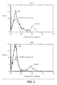

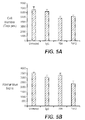
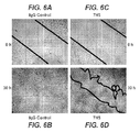

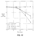


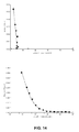
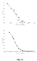

Classifications
-
- A—HUMAN NECESSITIES
- A61—MEDICAL OR VETERINARY SCIENCE; HYGIENE
- A61P—SPECIFIC THERAPEUTIC ACTIVITY OF CHEMICAL COMPOUNDS OR MEDICINAL PREPARATIONS
- A61P1/00—Drugs for disorders of the alimentary tract or the digestive system
- A61P1/04—Drugs for disorders of the alimentary tract or the digestive system for ulcers, gastritis or reflux esophagitis, e.g. antacids, inhibitors of acid secretion, mucosal protectants
-
- A—HUMAN NECESSITIES
- A61—MEDICAL OR VETERINARY SCIENCE; HYGIENE
- A61K—PREPARATIONS FOR MEDICAL, DENTAL OR TOILETRY PURPOSES
- A61K39/00—Medicinal preparations containing antigens or antibodies
- A61K39/395—Antibodies; Immunoglobulins; Immune serum, e.g. antilymphocytic serum
- A61K39/39533—Antibodies; Immunoglobulins; Immune serum, e.g. antilymphocytic serum against materials from animals
- A61K39/3955—Antibodies; Immunoglobulins; Immune serum, e.g. antilymphocytic serum against materials from animals against proteinaceous materials, e.g. enzymes, hormones, lymphokines
-
- A—HUMAN NECESSITIES
- A61—MEDICAL OR VETERINARY SCIENCE; HYGIENE
- A61K—PREPARATIONS FOR MEDICAL, DENTAL OR TOILETRY PURPOSES
- A61K39/00—Medicinal preparations containing antigens or antibodies
- A61K39/395—Antibodies; Immunoglobulins; Immune serum, e.g. antilymphocytic serum
-
- A—HUMAN NECESSITIES
- A61—MEDICAL OR VETERINARY SCIENCE; HYGIENE
- A61K—PREPARATIONS FOR MEDICAL, DENTAL OR TOILETRY PURPOSES
- A61K45/00—Medicinal preparations containing active ingredients not provided for in groups A61K31/00 - A61K41/00
- A61K45/06—Mixtures of active ingredients without chemical characterisation, e.g. antiphlogistics and cardiaca
-
- A—HUMAN NECESSITIES
- A61—MEDICAL OR VETERINARY SCIENCE; HYGIENE
- A61K—PREPARATIONS FOR MEDICAL, DENTAL OR TOILETRY PURPOSES
- A61K51/00—Preparations containing radioactive substances for use in therapy or testing in vivo
- A61K51/02—Preparations containing radioactive substances for use in therapy or testing in vivo characterised by the carrier, i.e. characterised by the agent or material covalently linked or complexing the radioactive nucleus
- A61K51/04—Organic compounds
- A61K51/08—Peptides, e.g. proteins, carriers being peptides, polyamino acids, proteins
- A61K51/10—Antibodies or immunoglobulins; Fragments thereof, the carrier being an antibody, an immunoglobulin or a fragment thereof, e.g. a camelised human single domain antibody or the Fc fragment of an antibody
- A61K51/1093—Antibodies or immunoglobulins; Fragments thereof, the carrier being an antibody, an immunoglobulin or a fragment thereof, e.g. a camelised human single domain antibody or the Fc fragment of an antibody conjugates with carriers being antibodies
-
- A—HUMAN NECESSITIES
- A61—MEDICAL OR VETERINARY SCIENCE; HYGIENE
- A61P—SPECIFIC THERAPEUTIC ACTIVITY OF CHEMICAL COMPOUNDS OR MEDICINAL PREPARATIONS
- A61P11/00—Drugs for disorders of the respiratory system
-
- A—HUMAN NECESSITIES
- A61—MEDICAL OR VETERINARY SCIENCE; HYGIENE
- A61P—SPECIFIC THERAPEUTIC ACTIVITY OF CHEMICAL COMPOUNDS OR MEDICINAL PREPARATIONS
- A61P13/00—Drugs for disorders of the urinary system
- A61P13/12—Drugs for disorders of the urinary system of the kidneys
-
- A—HUMAN NECESSITIES
- A61—MEDICAL OR VETERINARY SCIENCE; HYGIENE
- A61P—SPECIFIC THERAPEUTIC ACTIVITY OF CHEMICAL COMPOUNDS OR MEDICINAL PREPARATIONS
- A61P15/00—Drugs for genital or sexual disorders; Contraceptives
-
- A—HUMAN NECESSITIES
- A61—MEDICAL OR VETERINARY SCIENCE; HYGIENE
- A61P—SPECIFIC THERAPEUTIC ACTIVITY OF CHEMICAL COMPOUNDS OR MEDICINAL PREPARATIONS
- A61P17/00—Drugs for dermatological disorders
-
- A—HUMAN NECESSITIES
- A61—MEDICAL OR VETERINARY SCIENCE; HYGIENE
- A61P—SPECIFIC THERAPEUTIC ACTIVITY OF CHEMICAL COMPOUNDS OR MEDICINAL PREPARATIONS
- A61P17/00—Drugs for dermatological disorders
- A61P17/02—Drugs for dermatological disorders for treating wounds, ulcers, burns, scars, keloids, or the like
-
- A—HUMAN NECESSITIES
- A61—MEDICAL OR VETERINARY SCIENCE; HYGIENE
- A61P—SPECIFIC THERAPEUTIC ACTIVITY OF CHEMICAL COMPOUNDS OR MEDICINAL PREPARATIONS
- A61P17/00—Drugs for dermatological disorders
- A61P17/06—Antipsoriatics
-
- A—HUMAN NECESSITIES
- A61—MEDICAL OR VETERINARY SCIENCE; HYGIENE
- A61P—SPECIFIC THERAPEUTIC ACTIVITY OF CHEMICAL COMPOUNDS OR MEDICINAL PREPARATIONS
- A61P19/00—Drugs for skeletal disorders
- A61P19/02—Drugs for skeletal disorders for joint disorders, e.g. arthritis, arthrosis
-
- A—HUMAN NECESSITIES
- A61—MEDICAL OR VETERINARY SCIENCE; HYGIENE
- A61P—SPECIFIC THERAPEUTIC ACTIVITY OF CHEMICAL COMPOUNDS OR MEDICINAL PREPARATIONS
- A61P19/00—Drugs for skeletal disorders
- A61P19/06—Antigout agents, e.g. antihyperuricemic or uricosuric agents
-
- A—HUMAN NECESSITIES
- A61—MEDICAL OR VETERINARY SCIENCE; HYGIENE
- A61P—SPECIFIC THERAPEUTIC ACTIVITY OF CHEMICAL COMPOUNDS OR MEDICINAL PREPARATIONS
- A61P25/00—Drugs for disorders of the nervous system
-
- A—HUMAN NECESSITIES
- A61—MEDICAL OR VETERINARY SCIENCE; HYGIENE
- A61P—SPECIFIC THERAPEUTIC ACTIVITY OF CHEMICAL COMPOUNDS OR MEDICINAL PREPARATIONS
- A61P25/00—Drugs for disorders of the nervous system
- A61P25/06—Antimigraine agents
-
- A—HUMAN NECESSITIES
- A61—MEDICAL OR VETERINARY SCIENCE; HYGIENE
- A61P—SPECIFIC THERAPEUTIC ACTIVITY OF CHEMICAL COMPOUNDS OR MEDICINAL PREPARATIONS
- A61P25/00—Drugs for disorders of the nervous system
- A61P25/08—Antiepileptics; Anticonvulsants
-
- A—HUMAN NECESSITIES
- A61—MEDICAL OR VETERINARY SCIENCE; HYGIENE
- A61P—SPECIFIC THERAPEUTIC ACTIVITY OF CHEMICAL COMPOUNDS OR MEDICINAL PREPARATIONS
- A61P27/00—Drugs for disorders of the senses
- A61P27/02—Ophthalmic agents
-
- A—HUMAN NECESSITIES
- A61—MEDICAL OR VETERINARY SCIENCE; HYGIENE
- A61P—SPECIFIC THERAPEUTIC ACTIVITY OF CHEMICAL COMPOUNDS OR MEDICINAL PREPARATIONS
- A61P27/00—Drugs for disorders of the senses
- A61P27/02—Ophthalmic agents
- A61P27/06—Antiglaucoma agents or miotics
-
- A—HUMAN NECESSITIES
- A61—MEDICAL OR VETERINARY SCIENCE; HYGIENE
- A61P—SPECIFIC THERAPEUTIC ACTIVITY OF CHEMICAL COMPOUNDS OR MEDICINAL PREPARATIONS
- A61P29/00—Non-central analgesic, antipyretic or antiinflammatory agents, e.g. antirheumatic agents; Non-steroidal antiinflammatory drugs [NSAID]
-
- A—HUMAN NECESSITIES
- A61—MEDICAL OR VETERINARY SCIENCE; HYGIENE
- A61P—SPECIFIC THERAPEUTIC ACTIVITY OF CHEMICAL COMPOUNDS OR MEDICINAL PREPARATIONS
- A61P31/00—Antiinfectives, i.e. antibiotics, antiseptics, chemotherapeutics
- A61P31/04—Antibacterial agents
-
- A—HUMAN NECESSITIES
- A61—MEDICAL OR VETERINARY SCIENCE; HYGIENE
- A61P—SPECIFIC THERAPEUTIC ACTIVITY OF CHEMICAL COMPOUNDS OR MEDICINAL PREPARATIONS
- A61P31/00—Antiinfectives, i.e. antibiotics, antiseptics, chemotherapeutics
- A61P31/10—Antimycotics
-
- A—HUMAN NECESSITIES
- A61—MEDICAL OR VETERINARY SCIENCE; HYGIENE
- A61P—SPECIFIC THERAPEUTIC ACTIVITY OF CHEMICAL COMPOUNDS OR MEDICINAL PREPARATIONS
- A61P31/00—Antiinfectives, i.e. antibiotics, antiseptics, chemotherapeutics
- A61P31/12—Antivirals
-
- A—HUMAN NECESSITIES
- A61—MEDICAL OR VETERINARY SCIENCE; HYGIENE
- A61P—SPECIFIC THERAPEUTIC ACTIVITY OF CHEMICAL COMPOUNDS OR MEDICINAL PREPARATIONS
- A61P33/00—Antiparasitic agents
-
- A—HUMAN NECESSITIES
- A61—MEDICAL OR VETERINARY SCIENCE; HYGIENE
- A61P—SPECIFIC THERAPEUTIC ACTIVITY OF CHEMICAL COMPOUNDS OR MEDICINAL PREPARATIONS
- A61P35/00—Antineoplastic agents
-
- A—HUMAN NECESSITIES
- A61—MEDICAL OR VETERINARY SCIENCE; HYGIENE
- A61P—SPECIFIC THERAPEUTIC ACTIVITY OF CHEMICAL COMPOUNDS OR MEDICINAL PREPARATIONS
- A61P35/00—Antineoplastic agents
- A61P35/02—Antineoplastic agents specific for leukemia
-
- A—HUMAN NECESSITIES
- A61—MEDICAL OR VETERINARY SCIENCE; HYGIENE
- A61P—SPECIFIC THERAPEUTIC ACTIVITY OF CHEMICAL COMPOUNDS OR MEDICINAL PREPARATIONS
- A61P37/00—Drugs for immunological or allergic disorders
-
- A—HUMAN NECESSITIES
- A61—MEDICAL OR VETERINARY SCIENCE; HYGIENE
- A61P—SPECIFIC THERAPEUTIC ACTIVITY OF CHEMICAL COMPOUNDS OR MEDICINAL PREPARATIONS
- A61P37/00—Drugs for immunological or allergic disorders
- A61P37/02—Immunomodulators
-
- A—HUMAN NECESSITIES
- A61—MEDICAL OR VETERINARY SCIENCE; HYGIENE
- A61P—SPECIFIC THERAPEUTIC ACTIVITY OF CHEMICAL COMPOUNDS OR MEDICINAL PREPARATIONS
- A61P37/00—Drugs for immunological or allergic disorders
- A61P37/02—Immunomodulators
- A61P37/06—Immunosuppressants, e.g. drugs for graft rejection
-
- A—HUMAN NECESSITIES
- A61—MEDICAL OR VETERINARY SCIENCE; HYGIENE
- A61P—SPECIFIC THERAPEUTIC ACTIVITY OF CHEMICAL COMPOUNDS OR MEDICINAL PREPARATIONS
- A61P37/00—Drugs for immunological or allergic disorders
- A61P37/08—Antiallergic agents
-
- A—HUMAN NECESSITIES
- A61—MEDICAL OR VETERINARY SCIENCE; HYGIENE
- A61P—SPECIFIC THERAPEUTIC ACTIVITY OF CHEMICAL COMPOUNDS OR MEDICINAL PREPARATIONS
- A61P43/00—Drugs for specific purposes, not provided for in groups A61P1/00-A61P41/00
-
- A—HUMAN NECESSITIES
- A61—MEDICAL OR VETERINARY SCIENCE; HYGIENE
- A61P—SPECIFIC THERAPEUTIC ACTIVITY OF CHEMICAL COMPOUNDS OR MEDICINAL PREPARATIONS
- A61P5/00—Drugs for disorders of the endocrine system
- A61P5/14—Drugs for disorders of the endocrine system of the thyroid hormones, e.g. T3, T4
-
- A—HUMAN NECESSITIES
- A61—MEDICAL OR VETERINARY SCIENCE; HYGIENE
- A61P—SPECIFIC THERAPEUTIC ACTIVITY OF CHEMICAL COMPOUNDS OR MEDICINAL PREPARATIONS
- A61P7/00—Drugs for disorders of the blood or the extracellular fluid
-
- A—HUMAN NECESSITIES
- A61—MEDICAL OR VETERINARY SCIENCE; HYGIENE
- A61P—SPECIFIC THERAPEUTIC ACTIVITY OF CHEMICAL COMPOUNDS OR MEDICINAL PREPARATIONS
- A61P7/00—Drugs for disorders of the blood or the extracellular fluid
- A61P7/04—Antihaemorrhagics; Procoagulants; Haemostatic agents; Antifibrinolytic agents
-
- A—HUMAN NECESSITIES
- A61—MEDICAL OR VETERINARY SCIENCE; HYGIENE
- A61P—SPECIFIC THERAPEUTIC ACTIVITY OF CHEMICAL COMPOUNDS OR MEDICINAL PREPARATIONS
- A61P7/00—Drugs for disorders of the blood or the extracellular fluid
- A61P7/06—Antianaemics
-
- A—HUMAN NECESSITIES
- A61—MEDICAL OR VETERINARY SCIENCE; HYGIENE
- A61P—SPECIFIC THERAPEUTIC ACTIVITY OF CHEMICAL COMPOUNDS OR MEDICINAL PREPARATIONS
- A61P9/00—Drugs for disorders of the cardiovascular system
-
- A—HUMAN NECESSITIES
- A61—MEDICAL OR VETERINARY SCIENCE; HYGIENE
- A61P—SPECIFIC THERAPEUTIC ACTIVITY OF CHEMICAL COMPOUNDS OR MEDICINAL PREPARATIONS
- A61P9/00—Drugs for disorders of the cardiovascular system
- A61P9/06—Antiarrhythmics
-
- A—HUMAN NECESSITIES
- A61—MEDICAL OR VETERINARY SCIENCE; HYGIENE
- A61P—SPECIFIC THERAPEUTIC ACTIVITY OF CHEMICAL COMPOUNDS OR MEDICINAL PREPARATIONS
- A61P9/00—Drugs for disorders of the cardiovascular system
- A61P9/10—Drugs for disorders of the cardiovascular system for treating ischaemic or atherosclerotic diseases, e.g. antianginal drugs, coronary vasodilators, drugs for myocardial infarction, retinopathy, cerebrovascula insufficiency, renal arteriosclerosis
-
- C—CHEMISTRY; METALLURGY
- C07—ORGANIC CHEMISTRY
- C07K—PEPTIDES
- C07K16/00—Immunoglobulins [IGs], e.g. monoclonal or polyclonal antibodies
-
- C—CHEMISTRY; METALLURGY
- C07—ORGANIC CHEMISTRY
- C07K—PEPTIDES
- C07K16/00—Immunoglobulins [IGs], e.g. monoclonal or polyclonal antibodies
- C07K16/18—Immunoglobulins [IGs], e.g. monoclonal or polyclonal antibodies against material from animals or humans
- C07K16/22—Immunoglobulins [IGs], e.g. monoclonal or polyclonal antibodies against material from animals or humans against growth factors ; against growth regulators
-
- C—CHEMISTRY; METALLURGY
- C07—ORGANIC CHEMISTRY
- C07K—PEPTIDES
- C07K16/00—Immunoglobulins [IGs], e.g. monoclonal or polyclonal antibodies
- C07K16/18—Immunoglobulins [IGs], e.g. monoclonal or polyclonal antibodies against material from animals or humans
- C07K16/28—Immunoglobulins [IGs], e.g. monoclonal or polyclonal antibodies against material from animals or humans against receptors, cell surface antigens or cell surface determinants
- C07K16/2839—Immunoglobulins [IGs], e.g. monoclonal or polyclonal antibodies against material from animals or humans against receptors, cell surface antigens or cell surface determinants against the integrin superfamily
- C07K16/2842—Immunoglobulins [IGs], e.g. monoclonal or polyclonal antibodies against material from animals or humans against receptors, cell surface antigens or cell surface determinants against the integrin superfamily against integrin beta1-subunit-containing molecules, e.g. CD29, CD49
-
- G—PHYSICS
- G01—MEASURING; TESTING
- G01N—INVESTIGATING OR ANALYSING MATERIALS BY DETERMINING THEIR CHEMICAL OR PHYSICAL PROPERTIES
- G01N33/00—Investigating or analysing materials by specific methods not covered by groups G01N1/00 - G01N31/00
- G01N33/48—Biological material, e.g. blood, urine; Haemocytometers
- G01N33/50—Chemical analysis of biological material, e.g. blood, urine; Testing involving biospecific ligand binding methods; Immunological testing
- G01N33/74—Chemical analysis of biological material, e.g. blood, urine; Testing involving biospecific ligand binding methods; Immunological testing involving hormones or other non-cytokine intercellular protein regulatory factors such as growth factors, including receptors to hormones and growth factors
-
- A—HUMAN NECESSITIES
- A61—MEDICAL OR VETERINARY SCIENCE; HYGIENE
- A61K—PREPARATIONS FOR MEDICAL, DENTAL OR TOILETRY PURPOSES
- A61K39/00—Medicinal preparations containing antigens or antibodies
- A61K2039/505—Medicinal preparations containing antigens or antibodies comprising antibodies
-
- A—HUMAN NECESSITIES
- A61—MEDICAL OR VETERINARY SCIENCE; HYGIENE
- A61K—PREPARATIONS FOR MEDICAL, DENTAL OR TOILETRY PURPOSES
- A61K39/00—Medicinal preparations containing antigens or antibodies
- A61K2039/505—Medicinal preparations containing antigens or antibodies comprising antibodies
- A61K2039/507—Comprising a combination of two or more separate antibodies
-
- C—CHEMISTRY; METALLURGY
- C07—ORGANIC CHEMISTRY
- C07K—PEPTIDES
- C07K2317/00—Immunoglobulins specific features
- C07K2317/70—Immunoglobulins specific features characterized by effect upon binding to a cell or to an antigen
- C07K2317/73—Inducing cell death, e.g. apoptosis, necrosis or inhibition of cell proliferation
-
- C—CHEMISTRY; METALLURGY
- C07—ORGANIC CHEMISTRY
- C07K—PEPTIDES
- C07K2317/00—Immunoglobulins specific features
- C07K2317/70—Immunoglobulins specific features characterized by effect upon binding to a cell or to an antigen
- C07K2317/76—Antagonist effect on antigen, e.g. neutralization or inhibition of binding
-
- C—CHEMISTRY; METALLURGY
- C07—ORGANIC CHEMISTRY
- C07K—PEPTIDES
- C07K2317/00—Immunoglobulins specific features
- C07K2317/90—Immunoglobulins specific features characterized by (pharmaco)kinetic aspects or by stability of the immunoglobulin
- C07K2317/92—Affinity (KD), association rate (Ka), dissociation rate (Kd) or EC50 value
Definitions
- the present invention relates to the use of VEGF antagonists and alpha5beta1 antagonists for treating cancer and inhibiting angiogenesis and/or inhibited vascular permability, including in abnormal angiogenesis in diseases.
- the present invention also relates to use of a VEGFR agonists and alpha5beta1 agonists to promote angiogenesis and vascular permeability.
- the present invention also relates to anti-alpha5beta1 antibodies, compositions and kits comprising them and methods of making and using them.
- VEGF-A important role in pathological and non-pathological angiogenesis is well established.
- Loss of a single VEGF-A allele gave rise to an embryonic lethality in mice ( Carmeliet, P., et al., (1996) Nature 380:435-439 ; Ferrara, N et al., (1996) Nature 380:439-442 ).
- VEGF is also known as a vascular permeability factor due to its ability to induce vascular leakage ( Senger, D.R. et al., (1995) Science 219:983-985 ; Dvorak, H.F., et al., (1995) Am. J. Pathol. 146:1029-1039 ).
- VEGF-A is involved in the developmental, reproductive and bone angiogenesis in addition to other non-pathological angiogenesis.
- VEGF-A binds to two receptor tyrosine kinases (RTK), VEGFR-1 (Flt-1) and VEGFR-2 (KDR, Flk-1).
- VEGFR-2 is generally thought to be the major mediator of the mitogenic, angiogenic and permeability-enhancing effects of VEGF-A.
- FDA US Food and Drug Administration
- bevacizumab a humanized anti-VEGF (vascular endothelial growth factor)-A monoclonal antibody, for the treatment of metastatic colorectal cancer in combination with 5-fluorouracil (FU)-based chemotherapy regimens.
- FU 5-fluorouracil
- the FDA approved pegaptinib an aptamer that blocks the 165 amino-acid isoform of VEGF-A, for the treatment of the wet (neovascular) form of age-related macular degeneration (AMD).
- VEGF antagonists eventually succumb to their disease. Consequently there exists a need to develop new medicaments and treatments for treating diseases that are no longer responsive or are only partially responsive to VEGF antagonist therapies. There also exist a need to develop alternative and/or better therapies for treating cancer and diseases worsened, caused by or effected by abnormal angiogenesis.
- the present invention relates to medicaments and methods of treating patients who would benefit from decreased angiogenesis, who are suffering from abnormal angiogenesis and/or who are suffering from a neoplasia.
- the present invention provides a method for inhibiting angiogenesis and/or vascular permeability in a subject comprising the step of administering to the subject a therapeutically effective amount of a VEGF antagonist and an alpha5beta1 antagonist concurrently or sequentially.
- the present invention provides a method for treating a subject suffering from a disease, wherein the subject had been responsive to treatment for the disease with a VEGF antagonist but is partially or no longer responsive to the VEGF antagonist, comprising the step of administering to the subject a therapeutically effective amount of an alpha5beta antagonist.
- the present invention provides a method for treating a subject suffering from a disease, wherein the disease has been resistant or refractory to an alpha5beta antagonist therapy, alone or in combination with chemotherapy, comprising the step of administering to the subj ect a therapeutically effective amount of a VEGF antagonist.
- the present invention also relates to new anti-alpha5beta1 antibodies, kits and compositions comprising them, and methods of making or using them.
- the new anti-alpha5beta1 antibody is the 7H5 antibody or the 7H12 antibody described herein, or a humanized or chimeric form thereof.
- the 7H5 antibody or the 7H12 antibody, or humanized or chimeric form thereof can be in the form of a Fab, Fab', a F(ab)' 2 , single-chain Fv (scFv), an Fv fragment; a diabody, multi-specific antibody and a linear antibody.
- the new anti-alpha5betal antibodies can be conjugated to another entity such as, but not limited to, a therapeutic agent or a fluorescent dye or other marker to detect alpha5beta1 in patients or in patient samples.
- a therapeutic agent or a fluorescent dye or other marker to detect alpha5beta1 in patients or in patient samples.
- Such new alpha5beta1 antibodies can be used in a variety of therapeutic and diagnostic methods.
- such anti-alpha5beta1 antibodies can be used in treating abnormal angiogenesis, neoplasia, ocular diseases and autoimmune diseases.
- Such antibodies can be used for detecting alpha5beta1 protein in patients or patient samples by contacting such antibodies to alpha5beta1 protein in patients or in patient samples and determining qualitatively or quantitatively the anti-alpha5beta1 antibody bound to the alpha5betal protein.
- the present invention provides a method of treating cancer in a subject comprising the step of administering a VEGF antagonist and an alpha5beta1 antagonist concurrently or sequentially.
- the cancer is responsive to VEGF antagonist therapies.
- a method of treating age related macular degeneration (AMD), including wet age-related macular degeneration, in a subject suffering from AMD comprising the step of administering a therapeutically effective amount of a VEGF antagonist and an alpha5beta1 antagonist concurrently or sequentially.
- AMD age related macular degeneration
- a method of treating an autoimmune disease in a subject comprising the step of administering a therapeutically effective amount of a VEGF antagonist and an alpha5beta1 antagonist concurrently or sequentially is provided.
- the subject to be treated may be administered the VEGF antagonist initially and subsequently treated with the alpha5beta1 antagonist.
- the subject is treated with the VEGF antagonist and the alpha5beta1 antagonist simultaneously.
- the subject is treated with the VEGF antagonist until the subject is unresponsive to VEGF antagonist treatment and then the subject is treated with an alpha5beta1 antagonist.
- the subject is treated with the VEGF antagonist when the cancer is non-invasive or early stage and treated with the alpha5beta1 antagonist when the cancer is invasive.
- subject being treated with the alpha5beta1 antagonist has elevated alpha5betal levels in a diseased tissue compared to tissue from a subject not suffering from the disease.
- the method can further include the step of detecting alpha5beta1 in the subject, e.g., in a diseased tissue after treatment with a VEGF antagonist.
- the invasive cancer is a metastasized cancer.
- the early stage cancer is a cancer treated by adjuvant therapy (e.g., chemotherapy or surgical removal).
- the subject is suffering from a disease having abnormal angiogenesis.
- the disease is selected from the group consisting of a cancer, an immune disease or an ocular disease.
- the disease is selected from the group consisting of a solid tumor, a metastatic tumor, a soft tissue tumor, a disease having ocular neovascularisation, an inflammatory disease having abnormal angiogenesis, a disease arising after transplantation into the subject and a disease having abnormal proliferation of fibrovascular tissue.
- the cancer is selected from the group consisting of breast cancer (including metastatic breast cancer), cervical cancer, colorectal cancer (including metastatic colorectal cancer), lung cancer (including non-small cell lung cancer), non-Hodgkins lymphoma (NHL), chronic lymphocytic leukemia, renal cell cancer, prostate cancer including homone refractory prostate cancer, liver cancer, head and neck cancer, melanoma, ovarian cancer, mesothelioma, soft tissue cancer, gastrointestinal stromal tumor, glioblastoma multiforme and multiple myeloma.
- breast cancer including metastatic breast cancer
- cervical cancer including metastatic colorectal cancer
- lung cancer including non-small cell lung cancer
- NHL non-Hodgkins lymphoma
- chronic lymphocytic leukemia renal cell cancer
- prostate cancer including homone refractory prostate cancer, liver cancer, head and neck cancer, melanoma, ovarian cancer, mesothelioma, soft tissue cancer,
- the disease is selected from the group consisting of retinopathy, age-induced macular degeneration (e.g., wet AMD), diabetic macular edema, rubeosis; psoriasis, an inflammatory renal disease, haemolytic uremic syndrome, diabetic nephropathy (e.g., proliferative diabetic retinopathy), arthritis (e.g., psoriatic arthritis, osteoarthritis, rheumatoid arthritis), inflammatory bowel disease, chronic inflammation, chronic retinal detachment, chronic uveitis, chronic vitritis, corneal graft rejection, corneal neovascularization, corneal graft neovascularization, Crohn's disease, myopia, ocular neovascular disease, Pagets disease, pemphigoid, polyarteritis, post-laser radial keratotomy, retinal neovascularization, Sogrens syndrome, ulcerative
- the subject to be treated with an alpha5betal antagonist is suffering from a relapse after VEGF antagonist treatment or has become refractory to VEGF antagonist treatment.
- the subject to be treated with an alpha5beta1 antagonist and a VEGF antagonist is suffering from a metastatic cancer or has previously been treated with adjuvant therapy.
- the candidate patient is relapsed, refractory or resistant to a chemotherapeutic agents such as irinotecan.
- diseases include but are not limited to, metastatic colorectal cancer, relapsed metastatic colorectal cancer, metastatic breast cancer, relapsed metastatic breast cancer, metastatic HER2+ breast cancer, adjuvant breast cancer, adjuvant HER2+ breast cancer, metastatic pancreatic cancer, adjuvant colon cancer, adjuvant non-small cell lung cancer, adjuvant rectal cancer, adjuvant non small cell lung cancer, metastatic non small cell lung cancer, metastatic ovarian cancer, metastatic renal cell cancer and adjuvant renal cell cancer.
- the subject suffering from a disease described herein is administered a maintenance therapy after treatment for the disease with a VEGF antagonist, wherein the maintenance therapy is an alpha5beta1 antagonist alone or sequentially or concurrently with a VEGF antagonist.
- the VEGF antagonist can be selected from the group consisting of an antibody, an immunoadhesin, a peptibody, a small molecule and a nucleic acid that hybridizes to a nucleic acid molecule encoding VEGF under stringent conditions (e.g., ribozyme, siRNA and aptamer).
- the VEGF antagonist is an antibody.
- the antibody is a monoclonal antibody.
- the anti-VEGF antibody is capable of being competitively inhibited from binding to human VEGF by the Avastin® antibody.
- the anti-VEGF antibody is human, humanized or chimeric.
- the anti-VEGF antibody is the Avastin® antibody.
- the anti-VEGF antibody is selected from the group consisting of a Fab, Fab', a F(ab)' 2 , single-chain Fv (scFv), an Fv fragment; a diabody and a linear antibody.
- the VEGF antagonist is a bispecific antibody that binds VEGF and alpha5beta1 and is an alpha5beta1 antagonist.
- the alpha5beta1 antagonist can be selected from the group consisting of an antibody, an immunoadhesin, a peptibody, a small molecule and a nucleic acid that hybridizes to a nucleic acid molecule encoding alpha5beta1 under stringent conditions.
- the alpha5beta1 antagonist is an antibody.
- the antibody is a monoclonal antibody.
- the monoclonal antibody is a chimeric antibody such as the anti-human alpha5betal antibody known as M200 or F200.
- the anti-alpha5beta1 antibody comprises the VH sequence of SEQ ID NO: 1 and the VL sequence of SEQ ID NO:2. According to another embodiment, the anti-alpha5beta1 antibody comprises the sequence of SEQ ID NO:3 and the sequence of SEQ ID NO:4. According to another embodiment, the anti-alpha5beta1 antibody comprises the sequence of SEQ ID NO:4 and the sequence of SEQ ID NO:5. According to one preferred embodiment, the anti-alpha5betal antibody is capable of being competitively inhibited from binding to human alpha5betal by the 7H5 antibody or the 7H12 antibody. According to one preferred embodiment, the anti-alpha5betal antibody is human, humanized or chimeric.
- the anti- alpha5beta1 antibody is the 7H5 antibody, the 7H12 antibody, or a chimeric or humanized antibody thereof.
- the anti-alpha5beta1 antibody is selected from the group consisting of a Fab, Fab', a F(ab)' 2 , single-chain Fv (scFv), an Fv fragment; a diabody and a linear antibody.
- the alpha5beta1 antagonist is a bispecific antibody that binds VEGF and alpha5beta1 and is a VEGF antagonist.
- the anti-alpha5beta1 antagonist has an altered effector function.
- an anti-alpha5betal antibody is altered to decrease or prevent antibody dependent cellular cytotoxicity (ADCC) or complement dependent cytotoxicity (CDC) activity (e.g., by altering the nucleic acid sequence encoding the Fc portion of the antibody).
- ADCC antibody dependent cellular cytotoxicity
- CDC complement dependent cytotoxicity
- the anti-alpha5betal antibody has been altered to improve its half-life in humans (e.g., by altering the nucleic acid sequence encoding the Fc portion of the antibody).
- the VEGF antagonist or the alpha5betal antagonist is conjugated to a cytotoxic agent or a chemotherapeutic agent.
- the cytotoxic agent is a radioactive isotope or a toxin.
- the present invention provides compositions comprising a VEGF antagonist, an alpha5betal antagonist and a pharmaceutically acceptable carrier.
- the present invention also provides articles of manufacture comprising instructions for detecting alpha5beta1 in a subject who has been treated with a VEGF antagonist.
- the present invention also relates to use of VEGFR agonists and alpha5beta1 agonists to promote angiogenesis and vascular permeability and compositions comprising VEGF agonists and alpha5betal agonists and a pharmaceutically acceptable carrier.
- VEGFR agonists and alpha5betal agonists combinatorial therapies can used in treating a variety of diseases that would benefit from increased angiogenesis and vascular permeability, including, for example, wound healing such as in treating chronic wounds, acute wounds and normal wounds.
- stromal cell recruitement can bring other vascular growth factors to diseased sites that could compensate for the loss of VEGF activity in patients treated with VEGF antagonist therapies.
- Targeting a5b1-expressing stromal cells with an anti-a5b1 antibody may result in the reduction of the stromal cells, thereby reducing the production of potential compensatory vascular growth factors.
- inhibiting endothelial-extracellular matrix interactions, and particularly inhibiting alpha5betal binding interactions will potentiate VEGF antagonist therapies by inhibiting the return of angiogenesis along extracellular matrix tracks left by regressing vessels due to VEGF antagonist therapy. Therefore, treatment with alpha5beta1 antagonists concurrently or after any VEGF antagonist treatment may inhibit vessel recovery from that VEGF antagonist treatment and, consequently, return of neovascular growth.
- Alpha5beta1 or " ⁇ 5 ⁇ 1” or “ ⁇ 5 ⁇ 1” is an integrin comprising two different proteins (i.e., subunits Alpha5 and beta1).
- Alpha5beta1 has been shown to bind to fibronectin, L1-CAM and fibrinogen.
- Alpha5Beta1 integrin has also been called Very Late Activation-5, VLA-5, alpha5beta1, CD49e/CD29, fibronectin receptor, FNR and GPIc-IIa.
- the alpha5betal is a human alpha5beta1.
- Alpha5 also known as CD49e, alpha5, integrin alpha5 subunit, VLA-5 alpha subunit, IC subunit of GPIc-IIa and FNR alpha chain has four isoforms generated by alternative splicing (A-D). They vary within their cytoplasmic domains. Amino acid sequences for human isforms of alpha5 can be found at, e.g., Genbank accession numbers: X07979, U33879, U33882 and U33880, respectively.
- Beta1 also called CD29, beta1 Platelet GPIIa; VLA-beta chain; beta-1 integrin chain, CD29; FNRB; MDF2; VLAB; GPIIA; MSK12 and VLASB.
- Amino acid sequences for human Beta1 can be found, e.g., at Genbank Accession No. X06256.
- VEGF refers to the 165-amino acid human vascular endothelial cell growth factor and related 121-, 189-, and 206- amino acid human vascular endothelial cell growth factors, as described by Leung et al. Science, 246:1306 (1989 ), and Houck et al. Mol. Endocrin., 5:1806 (1991 ), together with the naturally occurring allelic and processed forms thereof.
- VEGF also refers to VEGFs from non-human species such as mouse, rat or primate. Sometimes the VEGF from a specific species are indicated by terms such as hVEGF for human VEGF, mVEGF for murine VEGF, and etc.
- VEGF is also used to refer to truncated forms of the polypeptide comprising amino acids 8 to 109 or 1 to 109 of the 165-amino acid human vascular endothelial cell growth factor. Reference to any such forms of VEGF may be identified in the present application, e.g., by "VEGF (8-109),” “VEGF (1-109)” or “VEGF 165 .”
- the amino acid positions for a "truncated" native VEGF are numbered as indicated in the native VEGF sequence. For example, amino acid position 17 (methionine) in truncated native VEGF is also position 17 (methionine) in native VEGF.
- the truncated native VEGF has binding affinity for the KDR and Flt-1 receptors comparable to native VEGF.
- the VEGF is a human VEGF.
- VEGF antagonist refers to a molecule capable of neutralizing, blocking, inhibiting, abrogating, reducing or interfering with VEGF activities including its binding to VEGF or one or more VEGF receptors or the nucleic acid encoding them.
- the VEGF antagonist binds VEGF or a VEGF receptor.
- VEGF antagonists include anti-VEGF antibodies and antigen-binding fragments thereof, polypeptides that bind VEGF and VEGF receptors and block ligand-receptor interaction (e.g., immunoadhesins, peptibodies), anti-VEGF receptor antibodies and VEGF receptor antagonists such as small molecule inhibitors of the VEGFR tyrosine kinases, aptamers that bind VEGF and nucleic acids that hybridize under stringent conditions to nucleic acid sequences that encode VEGF or VEGF receptor (e.g., RNAi).
- the VEGF antagonist binds to VEGF and inhibits VEGF-induced endothelial cell proliferation in vitro.
- the VEGF antagonist binds to VEGF or a VEGF receptor with greater affinity than a non-VEGF or non-VEGF receptor.
- the VEG antagonist binds to VEGF or a VEGF receptor with a Kd of between 1uM and 1pM.
- the VEGF antagonist binds to VEGF or a VEGF receptor between 500nM and 1pM.
- the VEGF antagonist is selected from the group consisting of a polypeptide such as an antibody, a peptibody, an immunoadhesin, a small molecule or an aptamer.
- the antibody is an anti-VEGF antibody such as the AVASTIN ® antibody or an anti-VEGF receptor antibody such as an anti-VEGFR2 or an anti-VEGFR3 antibody.
- VEGF antagonists include: VEGF-Trap, Mucagen, PTK787, SU11248, AG-013736, Bay 439006 (sorafenib), ZD-6474, CP632, CP-547632, AZD-2171, CDP-171, SU-14813, CHIR-258, AEE-788, SB786034, BAY579352, CDP-791, EG-3306, GW-786034, RWJ-417975/CT6758 and KRN-633.
- an "anti-VEGF antibody” is an antibody that binds to VEGF with sufficient affinity and specificity.
- the anti-VEGF antibody of the invention can be used as a therapeutic agent in targeting and interfering with diseases or conditions wherein the VEGF activity is involved.
- An anti-VEGF antibody will usually not bind to other VEGF homologues such as VEGF-B or VEGF-C, nor other growth factors such as PlGF, PDGF or bFGF.
- a preferred anti-VEGF antibody is a monoclonal antibody that binds to the same epitope as the monoclonal anti-VEGF antibody A4.6.1 produced by hybridoma ATCC HB 10709.
- the anti-VEGF antibody is a recombinant humanized anti-VEGF monoclonal antibody generated according to Presta et al. (1997) Cancer Res. 57:4593-4599 , including but not limited to the antibody known as bevacizumab (BV; Avastin®).
- anti-VEGF antibodies that can be used include, but are not limited to the antibodies disclosed in WO 2005/012359 .
- the anti-VEGF antibody comprises the variable heavy and variable light region of any one of the antibodies disclosed in Figures 24, 25, 26, 27 and 29 of WO 2005/012359 (e.g., G6, G6-23, G6-31, G6-23.1, G6-23.2, B20, B20-4 and B20.4.1).
- the anti-VEGF antibody known as ranibizumab is the VEGF antagonist administered for ocular disease such as diabetic neuropathy and AMD.
- Bevacizumab also known as “rhuMAb VEGF” or “Avastin®”
- rhuMAb VEGF a recombinant humanized anti-VEGF monoclonal antibody generated according to Presta et al. (1997) Cancer Res. 57:4593-4599 . It comprises mutated human IgG1 framework regions and antigen-binding complementarity-determining regions from the murine anti-hVEGF monoclonal antibody A.4.6.1 that blocks binding of human VEGF to its receptors.
- Approximately 93% of the amino acid sequence of Bevacizumab, including most of the framework regions, is derived from human IgG1, and about 7% of the sequence is derived from the murine antibody A4.6.1.
- Bevacizumab has a molecular mass of about 149,000 daltons and is glycosylated.
- Other anti-VEGF antibodies include the antibodies described in United States Patent No. 6884879 and WO 2005/044853 .
- the anti-VEGF antibody Ranibizumab or the LUCENTIS® antibody or rhuFab V2 is a humanized, affinity-matured anti-human VEGF Fab fragment.
- Ranibizumab is produced by standard recombinant technology methods in Escherichia coli expression vector and bacterial fermentation. Ranibizumab is not glycosylated and has a molecular mass of-48,000 daltons. See WO98/45331 and US20030190317 .
- Alpha5Beta1 antagonist refers to any molecule that inhibits the biological activity of alpha5betal. According to one preferred embodiment, the antagonist molecule specifically binds alpha5beta1. Accoridng to one preferred embodiment, the antagonist molecule binds to alpha5. According to one preferred embodiment, an alpha5beta1 antagonist preferentially binds alpha5beta1 with greater affinity relative to a non-alpha5betal integrin.
- the antagonist is selected from the group consisting of a polypeptide such as an antibody, a peptibody or an immunoadhesin, a small molecule or aptamer that inhibits the binding of alpha5beta1 to its ligand (particularly, fibronectin) or a nucleic acid that hybridizes under stringent conditions to to a nucleic acid molecule encoding alpha5beta1 (e.g., RNAi that interferes with alpha5 expression).
- a polypeptide such as an antibody, a peptibody or an immunoadhesin
- a small molecule or aptamer that inhibits the binding of alpha5beta1 to its ligand (particularly, fibronectin) or a nucleic acid that hybridizes under stringent conditions to to a nucleic acid molecule encoding alpha5beta1 (e.g., RNAi that interferes with alpha5 expression).
- a biological activity of alpha5beta1 can be any one, combination or all of the effects selected from the group consisting of (1) binding to fibronectin, (2) enhancing cell migration on fibronectin, (3) increasing survival of cells comprising alpha5beta1 in the presence of fibronectin, (4) increasing proliferation of cells comprising alpha5beta1 in the presence of fibronectin, and (5) increasing tube formation of cells comprising alpha5betal in the presence of fibronectin.
- anti-alpha5betal antagonist antibodies examples include M200 and F200 ( WO 2004/089988A2 ), the 7H5 antibody and the 7H12 antibody described herein, and chimeric, fully human and humanized antibodies thereof.
- M200 and F200 antibodies can be derived from the variable heavy and variable light chains of the mouse anti-human alpha5betal antibody, IIA1 (Pharmingen, San Diego, Ca).
- alpha5beta1 small molecule inhibitors examples include Ac-PHSCN-NH2 ( WO-9822617A1 ) and (S)-2-[(2,4,6-trimethylphenyl)sulfonyl]amino-3-[7-benzyloxycarbonyl-8-(2-pyridinylaminomethyl)-1-oxa-2,7-diazaspiro-(4,4)-non-2-en-3-yl] carbonylamino] propionic acid.
- the anti5beta1 antagonist binds alpha5betal and not alphaVbeta3 or alphaVbeta5 or alphaVbeta1.
- the alpha5beta1 antagonist binds to alpha5beta1 with a Kd of between 1 uM and 1pM. According to another preferred embodiment, the alpha5beta1 antagonist binds to alpha5 with a Kd between 500nM and 1pM.
- the alpha5betal antibody is an antibody that can compete with the 7H5 antibody or the 7H12 antibody for binding to alpha5beta1 in a competitive binding assay. According to another preferred embodiment, the antibody is an antibody that can be competitively inhibited from binding to alpha5betal by the antibody produced from the hybridoma deposited as Alpha5/beta1 7H5.4.2.8 (ATCC No. PTA-7421) or the hyridoma deposited as Alpha5/beta1 7H12.5.1.4 (ATCC No. PTA-7420) on March 7, 2006.
- VEGFR agonist refers to a molecule that can activate a VEGF receptor or increase its expression.
- VEGFR agonists include, but are not limited to, e.g., ligand agonists of a VEGFR, VEGF variants, antibodies and active fragments.
- Alpha5Beta1 agonist refers to a molecule that can activate alpha5Beta1 or increase its expression.
- Alpha5Beta1 agonists include, but are not limited to, e.g., ligand agonists of alpha5betal.
- Molecules such as antibodies, characterized by binding to overlapping or the similar areas on a target can be identified by competitive inhibition/binding assays.
- HUVEC or other cells expressing alpha5beta1 are used in a competitive inhibition assay and FACS is used to evaluate binding localities of two anti-alpha5beta1 antibodies relative to each other.
- HUVEC cells can be washed in conical tube and spun 5 min @ 1000 rpm. The pellet is typically washed two times. Then, the cells can be resuspended, counted and kept on ice until use. 100ul of a first anti-alpha5beta1 antibody (e.g., start at a 1ug/ml concentration or lower concentration) can be added to the well.
- a first anti-alpha5beta1 antibody e.g., start at a 1ug/ml concentration or lower concentration
- 100 ⁇ l (e.g., 20 x 10 ⁇ 5 cells) of cells can be added into per well and incubated on ice for 30 min.
- 100 ⁇ l of a biotinylated anti-alpha5beta1 antibody (5 ⁇ g/ml stock) can be added to each well and incubated on ice for 30 min.
- the cells arc then washed and pelleted for 5 min. @ 1000 rpm.
- the supernatant is aspirated.
- a 2nd antibody R-Phycoerythrin conjugated streptavidin Jackson 016-110-084
- the plate can be wrapped in foil and incubated on ice 30 min. Following the incubation, the pellet can be washed and pelleted 5 min. @ 1000 rpm.
- the pellet can be resuspended ant transferred to micro titertubes for FACS analysis.
- angiogenic factor or agent is a growth factor which stimulates the development of blood vessels, e.g., promote angiogenesis, endothelial cell growth, stabiliy of blood vessels, and/or vasculogenesis, etc.
- angiogenic factors include, but are not limited to, e.g., VEGF and members of the VEGF family, P1GF, PDGF family, fibroblast growth factor family (FGFs), TIE ligands (Angiopoietins), ephrins, Del-1, fibroblast growth factors: acidic (aFGF) and basic (bFGF), Follistatin, Granulocyte colony-stimulating factor (G-CSF), Hepatocyte growth factor (HGF) /scatter factor (SF), Interleukin-8 (IL-8), Leptin, Midkine, Placental growth factor, Platelet-derived endothelial cell growth factor (PD-ECGF), Platelet-derived growth factor, especially PDGF-
- IGF-I insulin-like growth factor-I
- VIGF VIGF
- EGF epidermal growth factor
- CTGF CTGF and members of its family
- TGF-alpha and TGF-beta TGF-beta
- the "Kd" or "Kd value" for an anti-VEGF antibody according to this invention is in one preferred embodiment measured by a radiolabeled VEGF binding assay (RIA) performed with the Fab version of the antibody and a VEGF molecule as described by the following assay that measures solution binding affinity of Fabs for VEGF by equilibrating Fab with a minimal concentration of ( 125 I)-labeled VEGF(109) in the presence of a titration series of unlabeled VEGF, then capturing bound VEGF with an anti-Fab antibody-coated plate ( Chen, et al., (1999) J. Mol Biol 293:865-881 ).
- RIA radiolabeled VEGF binding assay
- microtiter plates (Dynex) are coated overnight with 5 ug/ml of a capturing anti-Fab antibody (Cappel Labs) in 50 mM sodium carbonate (pH 9.6), and subsequently blocked with 2% (w/v) bovine serum albumin in PBS for two to five hours at room temperature (approximately 23°C).
- a non-adsorbant plate (Nunc #269620) 100 pM or 26 pM [ 125 I]VEGF(109) are mixed with serial dilutions of a Fab of interest, e.g., Fab-12 ( Presta et al., (1997) Cancer Res. 57:4593-4599 ).
- the Fab of interest is then incubated overnight; however, the incubation may continue for 65 hours to insure that equilibrium is reached. Thereafter, the mixtures are transferred to the capture plate for incubation at room temperature for one hour. The solution is then removed and the plate washed eight times with 0.1% Tween-20 in PBS. When the plates had dried, 150 ul/well of scintillant (MicroScint-20; Packard) is added, and the plates are counted on a Topcount gamma counter (Packard) for ten minutes. Concentrations of each Fab that give less than or equal to 20% of maximal binding are chosen for use in competitive binding assays.
- the Kd or Kd value is measured by using surface plasmon resonance assays using a BIAcore TM -2000 or a BIAcore TM -3000 (BIAcore, Inc., Piscataway, NJ) at 25°C with immobilized hVEGF (8-109) CM5 chips art ⁇ 1 response units (RU). Briefly, carboxymethylated dextran biosensor chips (CM5, BIAcore Inc.) are activated with N -ethyl- N' -(3-dimethylaminopropyl)-carbodiimide hydrochloride (EDC) and N-hydroxysuccinimide (NHS) according to the supplier's instructions.
- CM5 carboxymethylated dextran biosensor chips
- EDC N -ethyl- N' -(3-dimethylaminopropyl)-carbodiimide hydrochloride
- NHS N-hydroxysuccinimide
- Human VEGF is diluted with 10mM sodium acetate, pH 4.8, into 5ug/ml ( ⁇ 0.2uM) before injection at a flow rate of 5ul/minute to achieve approximately 10 response units (RU) of coupled protein.
- 1M ethanolamine is injected to block unreacted groups.
- two-fold serial dilutions of Fab (0.78 nM to 500 nM) are injected in PBS with 0.05% Tween 20 (PBST) at 25°C at a flow rate of approximately 25ul/min.
- association rates (k on ) and dissociation rates (k off ) are calculated using a simple one-to-one Langmuir binding model (BIAcore Evaluation Software version 3.2) by simultaneous fitting the association and dissociation sensorgram.
- the equilibrium dissociation constant (Kd) was calculated as the ratio k off /k on . See, e.g., Chen, Y., et al., (1999) J Mol Biol 293:865-881 .
- a subject to be treated is a mammal (e.g., human, non-human primate, rat, mouse, cow, horse, pig, sheep, goat, dog, cat, etc.).
- the subject may be a clinical patient, a clinical trial volunteer, an experimental animal, etc.
- the subject may be suspected of having or at risk for having a cancer, an immune disease, or any other disease having abnormal angiogenesis, be diagnosed with a cancer, immune disease, or any other diease having abnormal angiogenesis.
- Many diagnostic methods for cancer, immune disease or any other disease exhibiting abnormal angiogenesis and the clinical delineation of those diseases are known in the art.
- the subj ect to be treated according to this invention is a human.
- abnormal angiogenesis occurs when new blood vessels grow either excessively or inappropriately (e.g., the location, timing or onset of the angiogenesis being undesired from a medical standpoint) in a diseased state or such that it causes a diseased state.
- angiogenesis occurs when there is new blood vessel growth that contributes to the worsening of the diseased state or cause of a diseased state, such as in cancer, especially vascularized solid tumors and metastatic tumors (including colon, lung cancer (especially small-cell lung cancer), or prostate cancer), diseases caused by ocular neovascularisation, especially diabetic blindness, retinopathies, primarily diabetic retinopathy or age-induced macular degeneration, choroidal neovascularization (CNV), diabetic macular edema, pathological myopia, von Hippel-Lindau disease, histoplasmosis of the eye, Central Retinal Vein Occlusion (CRVO), corneal neovascularization, retinal neovascularization and rubeosis; psoriasis, psoriatic arthritis, haemangioblastoma such as haemangioma; inflammatory renal diseases, such as glomerulonephritis, especially mesan
- the new blood vessels can feed the diseased tissues, destroy normal tissues, and in the case of cancer, the new vessels can allow tumor cells to escape into the circulation and lodge in other organs (tumor metastases).
- the present invention contemplates treating those patients that are at risk of developing the above-mentioned illnesses.
- fibrovascular tissue acne rosacea, acquired immune deficiency syndrome, artery occlusion, atopic keratitis, bacterial ulcers, Bechets disease, blood borne tumors, carotid obstructive disease, choroidal neovascularization, chronic inflammation, chronic retinal detachment, chronic uveitis, chronic vitritis, contact lens overwear, corneal graft rejection, corneal neovascularization, corneal graft neovascularization, Crohn's disease, Eales disease, epidemic keratoconjunctivitis, fungal ulcers, Herpes simplex infections, Herpes zoster infections, hyperviscosity syndromes, Kaposi's sarcoma, leukemia, lipid degeneration, Lyme's disease, marginal keratolysis, Mooren ulcer, Mycobacteria infections other than lepro
- Anti-angiogenesis therapies are useful in the general treatment of graft rejection, lung inflammation, nephrotic syndrome, preeclampsia, pericardial effusion, such as that associated with pericarditis, and pleural effusion, diseases and disorders characterized by undesirable vascular permeability, e.g., edema associated with brain tumors, ascites associated with malignancies, Meigs' syndrome, lung inflammation, nephrotic syndrome, pericardial effusion, pleural effusion, permeability associated with cardiovascular diseases such as the condition following myocardial infarctions and strokes and the like.
- angiogenesis-dependent diseases include angiofibroma (abnormal blood of vessels which are prone to bleeding), neovascular glaucoma (growth of blood vessels in the eye), arteriovenous malformations (abnormal communication between arteries and veins), nonunion fractures (fractures that will not heal), atherosclerotic plaques (hardening of the arteries), pyogenic granuloma (common skin lesion composed of blood vessels), scleroderma (a form of connective tissue disease), hemangioma (tumor composed of blood vessels), trachoma (leading cause of blindness in the third world), hemophilic joints, vascular adhesions and hypertrophic scars (abnormal scar formation).
- angiofibroma abnormal blood of vessels which are prone to bleeding
- neovascular glaucoma growth of blood vessels in the eye
- arteriovenous malformations abnormal communication between arteries and veins
- nonunion fractures fractures that will not heal
- Treatment refers to both therapeutic treatment and prophylactic or preventative measures. Those in need of treatment include those already with the disorder as well as those in which the disorder is to be prevented.
- recurrence refers to the return of a cancer or disease after clinical assessment of the disappearance of disease. A diagnosis of distant metastasis or local recurrence can be considered a relapse.
- refractory or “resistant” refers to a cancer or disease that has not responded to treatment.
- adjuvant therapy refers to treatment given after the primary therapy, usually surgery.
- adjuvant therapy for cancer or disease may include immune therapy, chemotherapy, radiation therapy or hormone therapy.
- maintenance therapy refers to scheduled retreatment that is given to help maintain a previous treatment's effects. Maintenance therapy is often given to help keep cancer in remission or prolong a response to a specific therapy regardless of disease progression.
- invasive cancer refers to cancer that has spread beyond the layer of tissue in which it started into the normal surrounding tissues. Invasive cancers may or may not be metastatic.
- non-invasive cancer refers to a very early cancer or a cancer that has not spread beyond the tissue of origin.
- progression-free survival in oncology refers to the length of time during and after treatment that a cancer does not grow. Progression-free survival includes the amount of time patients have experienced a complete response or a partial response, as well as the amount of time patients have experienced stable disease.
- progressive disease in oncology can refer to a tumor growth of more than 20 percent since treatment began - either due to an increase in mass or a spread in the tumor.
- a “disorder” is any condition that would benefit from treatment with the antibody.
- This includes chronic and acute disorders or diseases including those pathological conditions which predispose the mammal to the disorder in question.
- disorders to be treated herein include malignant and benign tumors; non-leukemias and lymphoid malignancies; neuronal, glial, astrocytal, hypothalamic and other glandular, macrophagal, epithelial, stromal and blastocoelic disorders; and inflammatory, angiogenic and immunologic disorders.
- cancer and “cancerous” refer to or describe the physiological condition in mammals that is typically characterized by unregulated cell growth.
- Examples of cancer include but are not limited to, carcinoma, lymphoma, blastoma, sarcoma, and leukemia.
- cancers include squamous cell cancer, glioblastoma, cervical cancer, ovarian cancer, liver cancer, bladder cancer, hepatoma, breast cancer, colon cancer, colorectal cancer, endometrial carcinoma, salivary gland carcinoma, kidney cancer, renal cancer, prostate cancer, vulval cancer, thyroid cancer, hepatic carcinoma, head and neck cancer, rectal cancer, colorectal cancer, lung cancer including small-cell lung cancer, non-small cell lung cancer, adenocarcinoma of the lung and squamous carcinoma of the lung, squamous cell cancer (e.g.
- epithelial squamous cell cancer prostate cancer
- cancer of the peritoneum hepatocellular cancer
- gastric or stomach cancer including gastrointestinal cancer, pancreatic cancer, glioblastoma, retinoblastoma, astrocytoma, thecomas, arrhenoblastomas, hepatoma, hematologic malignancies including non-Hodgkins lymphoma (NHL), multiple myeloma and acute hematologic malignancies, endometrial or uterine carcinoma, endometriosis, fibrosarcomas, choriocarcinoma, salivary gland carcinoma, vulval cancer, thyroid cancer, esophageal carcinomas, hepatic carcinoma, anal carcinoma, penile carcinoma, nasopharyngeal carcinoma, laryngeal carcinomas, Kaposi's sarcoma, melanoma, skin carcinomas, Schwannoma, oligodendroglioma, neuroblasto
- Tumor refers to all neoplastic cell growth and proliferation, whether malignant or benign, and all pre-cancerous and cancerous cells and tissues.
- anti-neoplastic composition refers to a composition useful in treating cancer comprising at least one active therapeutic agent, e.g., "anti-cancer agent.”
- therapeutic agents include, but are limited to, e.g., chemotherapeutic agents, growth inhibitory agents, cytotoxic agents, agents used in radiation therapy, anti-angiogenesis agents, apoptotic agents, anti-tubulin agents, and other-agents to treat cancer, such as anti-HER-2 antibodies, anti-CD20 antibodies, an epidermal growth factor receptor (EGFR) antagonist (e.g., a tyrosine kinase inhibitor), HER1/EGFR inhibitor (e.g., erlotinib (Tarceva TM ), platelet derived growth factor inhibitors (e.g., Gleevec TM (Imatinib Mesylate)), a COX-2 inhibitor (e.g., celecoxib), interferon
- EGFR epidermal growth factor receptor
- HER1/EGFR inhibitor
- a “growth inhibitory agent” when used herein refers to a compound or composition which inhibits growth of a cell in vitro and/or in vivo.
- the growth inhibitory agent may be one which significantly reduces the percentage of cells in S phase.
- growth inhibitory agents include agents that block cell cycle progression (at a place other than S phase), such as agents that induce G1 arrest and M-phase arrest.
- Classical M-phase blockers include the vincas (vincristine and vinblastine), TAXOL®, and topo II inhibitors such as doxorubicin, epirubicin, daunorubicin, etoposide, and bleomycin.
- DNA alkylating agents such as tamoxifen, prednisone, dacarbazine, mechlorethamine, cisplatin, methotrexate, 5-fluorouracil, and ara-C.
- DNA alkylating agents such as tamoxifen, prednisone, dacarbazine, mechlorethamine, cisplatin, methotrexate, 5-fluorouracil, and ara-C.
- cytotoxic agent refers to a substance that inhibits or prevents the function of cells and/or causes destruction of cells.
- the term is intended to include radioactive isotopes (e.g ., I 131 , I 125 , Y 90 and Re 186 ), chemotherapeutic agents, and toxins such as enzymatically active toxins of bacterial, fungal, plant or animal origin, or fragments thereof.
- chemotherapeutic agent is a chemical compound useful in the treatment of cancer.
- chemotherapeutic agents include is a chemical compound useful in the treatment of cancer.
- chemotherapeutic agents include alkylating agents such as thiotepa and CYTOXAN® cyclosphosphamide; alkyl sulfonates such as busulfan, improsulfan and piposulfan; aziridines such as benzodopa, carboquone, meturedopa, and uredopa; ethylenimines and methylamelamines including altretamine, triethylenemelamine, trietylenephosphoramide, triethiylenethiophosphoramide and trimethylolomelamine; acetogenins (especially bullatacin and bullatacinone); a camptothecin (including the synthetic analogue topotecan); bryostatin; callystatin; CC-1065 (including its adozelesin
- calicheamicin especially calicheamicin gamma1I and calicheamicin omegaI1 (see, e.g., Agnew, Chem Intl. Ed. Engl., 33: 183-186 (1994 )); dynemicin, including dynemicin A; bisphosphonates, such as clodronate; an esperamicin; as well as neocarzinostatin chromophore and related chromoprotein enediyne antiobiotic chromophores), aclacinomysins, actinomycin, authramycin, azaserine, bleomycins, cactinomycin, carabicin, carminomycin, carzinophilin, chromomycinis, dactinomycin, daunorubicin, detorubicin, 6-diazo-5-oxo-L-norleucine, ADRIAMYCIN® doxorubicin (
- anti-hormonal agents that act to regulate or inhibit hormone action on tumors
- SERMs selective estrogen receptor modulators
- tamoxifen including NOLVADEX® tamoxifen
- raloxifene including NOLVADEX® tamoxifen
- droloxifene 4-hydroxytamoxifen
- trioxifene keoxifene
- LY117018 onapristone
- aromatase inhibitors that inhibit the enzyme aromatase, which regulates estrogen production in the adrenal glands, such as, for example, 4(5)-imidazoles, aminoglutethimide, MEGASE® megestrol acetate, AROMASIN® exemestane, formestanie, fadrozole, RIVISOR® vorozole, FEMARA® letrozole, and ARIMIDEX® anastrozole
- anti-androgens such as flutamide, nil
- prodrug refers to a precursor or derivative form of a pharmaceutically active substance (e.g., small molecule) that is less cytotoxic to diseased cells compared to the parent drug and is capable of being enzymatically activated or converted into the more active parent form.
- a pharmaceutically active substance e.g., small molecule
- the prodrugs of this invention include, but are not limited to, phosphate-containing prodrugs, thiophosphate-containing prodrugs, sulfate-containing prodrugs, peptide-containing prodrugs, D-amino acid-modified prodrugs, glycosylated prodrugs, ⁇ -lactam-containing prodrugs, optionally substituted phenoxyacetamide-containing prodrugs or optionally substituted phenylacetamide-containing prodrugs, 5-fluorocytosine and other 5-fluorouridine prodrugs which can be converted into the more active cytotoxic free drug.
- cytotoxic drugs that can be derivatized into a prodrug form for use in this invention include, but are not limited to, those chemotherapeutic agents described above.
- Isolated when used to describe the various polypeptides disclosed herein, means polypeptide that has been identified and separated and/or recovered from a cell or cell culture from which it was expressed. Contaminant components of its natural environment are materials that would typically interfere with diagnostic or therapeutic uses for the polypeptide, and can include enzymes, hormones, and other proteinaceous or non-proteinaceous solutes.
- the polypeptide will be purified (1) to a degree sufficient to obtain at least 15 residues ofN-terminal or internal amino acid sequence by use of a spinning cup sequenator, or (2) to homogeneity by SDS-PAGE under non-reducing or reducing conditions using Coomassie blue or, preferably, silver stain.
- Isolated polypeptide includes polypeptide in situ within recombinant cells, since at least one component of the polypeptide natural environment will not be present. Ordinarily, however, isolated polypeptide will be prepared by at least one purification step.
- an "isolated" polypeptide-encoding nucleic acid or other polypeptide-encoding nucleic acid is a nucleic acid molecule that is identified and separated from at least one contaminant nucleic acid molecule with which it is ordinarily associated in the natural source of the polypeptide-encoding nucleic acid.
- An isolated polypeptide-encoding nucleic acid molecule is other than in the form or setting in which it is found in nature. Isolated polypeptide-encoding nucleic acid molecules therefore are distinguished from the specific polypeptide-encoding nucleic acid molecule as it exists in natural cells.
- an isolated polypeptide-encoding nucleic acid molecule includes polypeptide-encoding nucleic acid molecules contained in cells that ordinarily express the polypeptide where, for example, the nucleic acid molecule is in a chromosomal location different from that of natural cells.
- control sequences refers to DNA sequences necessary for the expression of an operably linked coding sequence in a particular host organism.
- the control sequences that are suitable for prokaryotes include a promoter, optionally an operator sequence, and a ribosome binding site.
- Eukaryotic cells are known to utilize promoters, polyadenylation signals, and enhancers.
- Nucleic acid is "operably linked" when it is placed into a functional relationship with another nucleic acid sequence.
- DNA for a presequence or secretory leader is operably linked to DNA for a polypeptide if it is expressed as a preprotein that participates in the secretion of the polypeptide;
- a promoter or enhancer is operably linked to a coding sequence if it affects the transcription of the sequence; or
- a ribosome binding site is operably linked to a coding sequence if it is positioned so as to facilitate translation.
- "operably linked” means that the DNA sequences being linked are contiguous, and, in the case of a secretory leader, contiguous and in reading phase. However, enhancers do not have to be contiguous. Linking is accomplished by ligation at convenient restriction sites. If such sites do not exist, the synthetic oligonucleotide adaptors or linkers are used in accordance with conventional practice.
- “Stringent conditions” or “high stringency conditions”, as defined herein, can be identified by those that: (1) employ low ionic strength and high temperature for washing, for example 0.015 M sodium chloride/0.0015 M sodium citrate/0.1% sodium dodecyl sulfate at 50C; (2) employ during hybridization a denaturing agent, such as formamide, for example, 50% (v/v) formamide with 0.1% bovine serum albumin/0.1 % Ficoll/0.1% polyvinylpyrrolidone/50mM sodium phosphate buffer at pH 6.5 with 750 mM sodium chloride, 75 mM sodium citrate at 42C; or (3) overnight hybridization in a solution that employs 50% formamide, 5 x SSC (0.75 M NaCl, 0.075 M sodium citrate), 50 mM sodium phosphate (pH 6.8), 0.1 % sodium pyrophosphate, 5 x Denhardt's solution, sonicated salmon sperm DNA (50 ⁇ g/ml), 0.1% S
- Percent (%) amino acid sequence identity with respect to the polypeptide sequences identified herein is defined as the percentage of amino acid residues in a candidate sequence that are identical with the amino acid residues in the polypeptide being compared, after aligning the sequences and introducing gaps, if necessary, to achieve the maximum percent sequence identity, and not considering any conservative substitutions as part of the sequence identity. Alignment for purposes of determining percent amino acid sequence identity can be achieved in various ways that are within the skill in the art, for instance, using publicly available computer software such as BLAST, BLAST-2, ALIGN or Megalign (DNASTAR) software. Those skilled in the art can determine appropriate parameters for measuring alignment, including any algorithms needed to achieve maximal alignment over the full length of the sequences being compared.
- % amino acid sequence identity values are generated using the sequence comparison computer program ALIGN-2.
- the ALIGN-2 sequence comparison computer program was authored by Genentech, Inc. and the source code (Table 1) has been filed with user documentation in the U.S. Copyright Office, Washington D.C., 20559, where it is registered under U.S. Copyright Registration No. TXU510087.
- the ALIGN-2 program is publicly available through Genentech, Inc., South San Francisco, California.
- the ALIGN-2 program should be compiled for use on a UNIX operating system, preferably digital UNIX V4.0D. All sequence comparison parameters are set by the ALIGN-2 program and do not vary.
- amino acid sequences described herein are contiguous amino acid sequences unless otherwise specified.
- immunoadhcsin designates antibody-like molecules that combine the binding specificity of a heterologous protein (an “adhesin”) with the effector functions of immunoglobulin constant domains.
- the immunoadhesins comprise a fusion of an amino acid sequence with the desired binding specificity that is other than the antigen recognition and binding site of an antibody (i.e., is “heterologous"), and an immunoglobulin constant domain sequence.
- the adhesin part of an immunoadhesin molecule typically is a contiguous amino acid sequence comprising at least the binding site of a receptor or a ligand - such as a VEGFR or a fibronectin ligand.
- the immunoglobulin constant domain sequence in the immunoadhesin can be obtained from any immunoglobulin, such as IgG-1, IgG-2, IgG-3, or IgG-4 subtypes, IgA (including IgA-1 and IgA-2), IgE, IgD, or IgM.
- immunoglobulin such as IgG-1, IgG-2, IgG-3, or IgG-4 subtypes, IgA (including IgA-1 and IgA-2), IgE, IgD, or IgM.
- Peptibodies which often comprise a sequence derived from phage display selection of sequences that specifically bind a target fused to an Fc portion of an immunoglobulin, can be considered immunadhesins herein.
- antibody is used in the broadest sense and specifically covers, for example, single monoclonal antibodies (including agonist, antagonist, and neutralizing antibodies), antibody compositions with polyepitopic specificity, polyclonal antibodies, single chain anti- antibodies, and fragments of antibodies (see below) as long as they specifically bind a native polypeptide and/or exhibit a biological activity or immunological activity of this invention.
- the antibody binds to an oligomeric form of a target protein, e.g., a trimeric form.
- the antibody specifically binds to a protein, which binding can be inhibited by a monoclonal antibody of this invention (e.g., a deposited antibody of this invention, etc.).
- a functional fragment or analog of an antibody is a compound having a qualitative biological activity in common with an antibody to which it is being referred.
- a functional fragment or analog of an antibody of this invention can be one which can specifically bind to VEGF or alpha5beta1.
- the antibody can prevent or substantially reduce the ability of a VEGF to induce cell proliferation.
- an "isolated antibody” is one which has been identified and separated and/or recovered from a component of its natural environment. Contaminant components of its natural environment are materials which would interfere with diagnostic or therapeutic uses for the antibody, and can include enzymes, hormones, and other proteinaceous or nonproteinaceous solutes.
- the antibody will be purified (1) to greater than 95% by weight of antibody as determined by the Lowry method, and most preferably more than 99% by weight, (2) to a degree sufficient to obtain at least 15 residues ofN-terminal or internal amino acid sequence by use of a spinning cup sequenator, or (3) to homogeneity by SDS-PAGE under reducing or nonreducing conditions using Coomassie blue or, preferably, silver stain.
- Isolated antibody includes the antibody in situ within recombinant cells since at least one component of the antibody's natural environment will not be present. Ordinarily, however, isolated antibody will be prepared by at least one purification step.
- the basic 4-chain antibody unit is a heterotetrameric glycoprotcin composed of two identical light (L) chains and two identical heavy (H) chains (an IgM antibody consists of 5 of the basic heterotetramer unit along with an additional polypeptide called J chain, and therefore contain 10 antigen binding sites, while secreted IgA antibodies can polymerize to form polyvalent assemblages comprising 2-5 of the basic 4-chain units along with J chain).
- the 4-chain unit is generally about 150,000 daltons.
- Each L chain is linked to a H chain by one covalent disulfide bond, while the two H chains are linked to each other by one or more disulfide bonds depending on the H chain isotype.
- Each H and L chain also has regularly spaced intrachain disulfide bridges.
- Each H chain has at the N-terminus, a variable domain (V H ) followed by three constant domains (C H ) for each of the ⁇ and ⁇ chains and four C H domains for ⁇ and ⁇ isotypes.
- Each L chain has at the N-terminus, a variable domain (V L ) followed by a constant domain (C L ) at its other end.
- the V L is aligned with the V H and the C L is aligned with the first constant domain of the heavy chain (C H 1). Particular amino acid residues are believed to form an interface between the light chain and heavy chain variable domains.
- the pairing of a V H and V L together forms a single antigen-binding site.
- immunoglobulins can be assigned to different classes or isotypes. There are five classes of immunoglobulins: IgA, IgD, IgE, IgG, and IgM, having heavy chains designated ⁇ , ⁇ , ⁇ , ⁇ , and ⁇ , respectively.
- the ⁇ and ⁇ classes are further divided into subclasses on the basis of relatively minor differences in C H sequence and function, e.g., humans express the following subclasses: IgG1, IgG2, IgG3, IgG4, IgA1, and IgA2.
- variable refers to the fact that certain segments of the variable domains differ extensively in sequence among antibodies.
- the V domain mediates antigen binding and define specificity of a particular antibody for its particular antigen.
- variability is not evenly distributed across the 110-amino acid span of the variable domains.
- the V regions consist of relatively invariant stretches called framework regions (FRs) of 15-30 amino acids separated by shorter regions of extreme variability called “hypervariable regions” that are each 9-12 amino acids long.
- FRs framework regions
- hypervariable regions that are each 9-12 amino acids long.
- the variable domains of native heavy and light chains each comprise four FRs, largely adopting a beta-sheet configuration, connected by three hypervariable regions, which form loops connecting, and in some cases forming part of, the beta-sheet structure.
- the hypervariable regions in each chain are held together in close proximity by the FRs and, with the hypervariable regions from the other chain, contribute to the formation of the antigen-binding site of antibodies (see Kabat et al., Sequences of Proteins of Immunological interest, 5th Ed. Public Health Service, National Institutes of Health, Bethesda, MD. (1991 )).
- the constant domains are not involved directly in binding an antibody to an antigen, but exhibit various effector functions, such as participation of the antibody in antibody dependent cellular cytotoxicity (ADCC).
- hypervariable region when used herein refers to the amino acid residues of an antibody which are responsible for antigen-binding.
- the hypervariable region generally comprises amino acid residues from a "complementarity determining region" or "CDR" (e.g. around about residues 24-34 (L1), 50-56 (L2) and 89-97 (L3) in the V L , and around about 31-35B (H1), 50-65 (H2) and 95-102 (H3) in the V H (in one embodiment, H1 is around about 31-35); Kabat et al., Sequences of Proteins of Immunological Interest , 5th Ed. Public Health Service, National Institutes of Health, Bethesda, MD.
- residues from a "hypervariable loop” e.g. residues 26-32 (L1), 50-52 (L2) and 91-96 (L3) in the V L , and 26-32 (H1), 53-55 (H2) and 96-101 (H3) in the V H ; Chothia and Lesk J. Mol. Biol. 196:901-917 (1987 )).
- the term "monoclonal antibody” as used herein refers to an antibody obtained from a population of substantially homogeneous antibodies, i.e., the individual antibodies comprising the population are identical except for possible naturally occurring mutations that can be present in minor amounts. Monoclonal antibodies are highly specific, being directed against a single antigenic site. Furthermore, in contrast to polyclonal antibody preparations which include different antibodies directed against different determinants (epitopes), each monoclonal antibody is directed against a single determinant on the antigen. In addition to their specificity, the monoclonal antibodies are advantageous in that they can be synthesized uncontaminated by other antibodies. The modifier "monoclonal" is not to be construed as requiring production of the antibody by any particular method.
- the monoclonal antibodies useful in the present invention can be prepared by the hybridoma methodology first described by Kohler et al., Nature, 256:495 (1975 ), or can be made using recombinant DNA methods in bacterial, eukaryotic animal or plant cells (see, e.g., U.S. Patent No. 4,816,567 ).
- the "monoclonal antibodies” can also be isolated from phage antibody libraries using the techniques described in Clackson et al., Nature, 352:624-628 (1991 ), Marks et al., J. Mol. Biol. , 222:581-597 (1991 ), and the Examples below, for example.
- the monoclonal antibodies herein include "chimeric" antibodies in which a portion of the heavy and/or light chain is identical with or homologous to corresponding sequences in antibodies derived from a particular species or belonging to a particular antibody class or subclass, while the remainder of the chain(s) is identical with or homologous to corresponding sequences in antibodies derived from another species or belonging to another antibody class or subclass, as well as fragments of such antibodies, so long as they exhibit a biological activity of this invention (see U.S. Patent No. 4,816,567 ; and Morrison et al., Proc. Natl. Acad. Sci. USA, 81:6851-6855 (1984 )).
- Chimeric antibodies of interest herein include "primatized" antibodies comprising variable domain antigen-binding sequences derived from a non-human primate (e.g . Old World Monkey, Ape etc), and human constant region sequences.
- an “intact” antibody is one which comprises an antigen-binding site as well as a C L and at least heavy chain constant domains, C H 1, C H 2 and C H 3.
- the constant domains can be native sequence constant domains ( e.g . human native sequence constant domains) or amino acid sequence variant thereof.
- the intact antibody has one or more effector functions.
- Antibody fragments comprise a portion of an intact antibody, preferably the antigen binding or variable region of the intact antibody.
- antibody fragments include Fab, Fab', F(ab') 2 , and Fv fragments; diabodies; linear antibodies (see U.S. Patent No. 5,641,870 , Example 2; Zapata et al., Protein Eng. 8(10): 1057-1062 [1995 ]); single-chain antibody molecules; and multispecific antibodies formed from antibody fragments.
- linear antibodies generally refers to the antibodies described in Zapata et al., Protein Eng., 8(10):1057-1062 (1995 ). Briefly, these antibodies comprise a pair of tandem Fd segments (VH-CH1-VH-CH1) which, together with complementary light chain polypeptides, form a pair of antigen binding regions. Linear antibodies can be bispecific or monospecific.
- Papain digestion of antibodies produces two identical antigen-binding fragments, called “Fab” fragments, and a residual "Fc” fragment, a designation reflecting the ability to crystallize readily.
- the Fab fragment consists of an entire L chain along with the variable region domain of the H chain (V H ), and the first constant domain of one heavy chain (C H 1).
- Each Fab fragment is monovalent with respect to antigen binding, i.e., it has a single antigen-binding site.
- Pepsin treatment of an antibody yields a single large F(ab') 2 fragment which roughly corresponds to two disulfide linked Fab fragments having divalent antigen-binding activity and is still capable of cross-linking antigen.
- Fab' fragments differ from Fab fragments by having additional few residues at the carboxy terminus of the C H 1 domain including one or more cysteines from the antibody hinge region.
- Fab'-SH is the designation herein for Fab' in which the cysteine residue(s) of the constant domains bear a free thiol group.
- F(ab') 2 antibody fragments originally were produced as pairs of Fab' fragments which have hinge cysteines between them. Other chemical couplings of antibody fragments are also known.
- the Fc fragment comprises the carboxy-terminal portions of both H chains held together by di sulfides.
- the effector functions of antibodies arc determined by sequences in the Fc region, which region is also the part recognized by Fc receptors (FcR) found on certain types of cells.
- Fv is the minimum antibody fragment which contains a complete antigen-recognition and - binding site. This fragment consists of a dimer of one heavy- and one light-chain variable region domain in tight, non-covalent association. From the folding of these two domains emanate six hypervariable loops (3 loops each from the H and L chain) that contribute the amino acid residues for antigen binding and confer antigen binding specificity to the antibody. However, even a single variable domain (or half of an Fv comprising only three CDRs specific for an antigen) has the ability to recognize and bind antigen, although at a lower affinity than the entire binding site.
- Single-chain Fv also abbreviated as “sFv” or “scFv” are antibody fragments that comprise the V H and V L antibody domains connected into a single polypeptide chain.
- the sFv polypeptide further comprises a polypeptide linker between the V H and V L domains which enables the sFv to form the desired structure for antigen binding.
- diabodies refers to small antibody fragments prepared by constructing sFv fragments (see preceding paragraph) with short linkers (about 5-10 residues) between the V H and V L domains such that inter-chain but not intra-chain pairing of the V domains is achieved, resulting in a bivalent fragment, i.e., fragment having two antigen-binding sites.
- Bispecific diabodies are heterodimers of two "crossover" sFv fragments in which the V H and V L domains of the two antibodies are present on different polypeptide chains.
- Diabodies are described more fully in, for example, EP 404,097 ; WO 93/11161 ; and Hollinger et al., Proc. Natl. Acad. Sci. USA, 90:6444-6448 (1993 ).
- Humanized forms of non-human (e.g., rodent) antibodies are chimeric antibodies that contain minimal sequence derived from the non-human antibody.
- humanized antibodies are human immunoglobulins (recipient antibody) in which residues from a hypervariable region of the recipient are replaced by residues from a hypervariable region of a non-human species (donor antibody) such as mouse, rat, rabbit or non-human primate having the desired antibody specificity, affinity, and capability.
- donor antibody such as mouse, rat, rabbit or non-human primate having the desired antibody specificity, affinity, and capability.
- framework region (FR) residues of the human immunoglobulin are replaced by corresponding non-human residues.
- humanized antibodies can comprise residues that are not found in the recipient antibody or in the donor antibody. These modifications are made to further refine antibody performance.
- the humanized antibody will comprise substantially all of at least one, and typically two, variable domains, in which all or substantially all of the hypervariable loops correspond to those of a non-human immunoglobulin and all or substantially all of the FRs are those of a human immunoglobulin sequence.
- the humanized antibody optionally also will comprise at least a portion of an immunoglobulin constant region (Fc), typically that of a human immunoglobulin.
- Fc immunoglobulin constant region
- a “species-dependent antibody” is an antibody which has a stronger binding affinity for an antigen from a first mammalian species than it has for a homologue of that antigen from a second mammalian species.
- the species-dependent antibody "bind specifically" to a human antigen (i.e., has a binding affinity (Kd) value of no more than about 1 x 10 -7 M, preferably no more than about 1 x 10 -8 and most preferably no more than about 1 x 10 -9 M) but has a binding affinity for a homologue of the antigen from a second non-human mammalian species which is at least about 50 fold, or at least about 500 fold, or at least about 1000 fold, weaker than its binding affinity for the human antigen.
- the species-dependent antibody can be of any of the various types of antibodies as defined above, but preferably is a humanized or human antibody.
- the extent of binding of the polypeptide, antibody, antagonist or composition to a "non-target” protein will be less than about 10% of the binding of the polypeptide, antibody, antagonist or composition to its particular target protein as determined by fluorescence activated cell sorting (FACS) analysis or radioimmunoprecipitation (RIA).
- FACS fluorescence activated cell sorting
- RIA radioimmunoprecipitation
- Specific binding can be measured, for example, by determining binding of a molecule compared to binding of a control molecule, which generally is a molecule of similar structure that does not have binding activity. For example, specific binding can be determined by competition with a control molecule that is similar to the target, for example, an excess of non-labeled target. In this case, specific binding is indicated if the binding of the labeled target to a probe is competitively inhibited by excess unlabeled target.
- telomere binding or “specifically binds to” or is “specific for” a particular polypeptide or an epitope on a particular polypeptide target as used herein can be exhibited, for example, by a molecule having a Kd for the target of at least about 10 -4 M, alternatively at least about 10 -5 M, alternatively at least about 10 -6 M, alternatively at least about 10 -7 M, alternatively at least about 10 -8 M, alternatively at least about 10 -9 M, alternatively at least about 10 -10 M, alternatively at least about 10 -11 M, alternatively at least about 10 -12 M, or greater.
- the term "specific binding” refers to binding where a molecule binds to a particular polypeptide or epitope on a particular polypeptide without substantially binding to any other polypeptide or polypeptide epitope.
- Antibody effector functions refer to those biological activities attributable to the Fc region (a native sequence Fc region or amino acid sequence variant Fc region) of an antibody, and vary with the antibody isotype. Examples of antibody effector functions include: C1q binding and complement dependent cytotoxicity; Fc receptor binding; antibody-dependent cell-mediated cytotoxicity (ADCC); phagocytosis; down regulation of cell surface receptors; and B cell activation.
- a "native sequence Fc region” comprises an amino acid sequence identical to the amino acid sequence of an Fc region found in nature. Examples of Fc sequences are described in, for example, but not limited to, Kabat et al., Sequences of Immunological Interest. 5th Ed. Public Health Service, National Institutes of Health, Bethesda, Md. (1991 )).
- a “variant Fc region” comprises an amino acid sequence which differs from that of a native sequence Fc region by virtue of at least one "amino acid modification" as herein defined.
- the variant Fc region has at least one amino acid substitution compared to a native sequence Fc region or to the Fc region of a parent polypeptide, e.g . from about one to about ten amino acid substitutions, and preferably from about one to about five amino acid substitutions in a native sequence Fc region or in the Fc region of the parent polypeptide.
- the variant Fc region herein will possess at least about 80% homology, at least about 85% homology, at least about 90% homology, at least about 95% homology or at least about 99% homology with a native sequence Fc region. According to another embodiment, the variant Fc region herein will possess at least about 80% homology, at least about 85% homology, at least about 90% homology, at least about 95% homology or at least about 99% homology with an Fc region of a parent polypeptide.
- Percent (%) amino acid sequence identity or “homology” with respect to the polypeptide and antibody sequences identified herein is defined as the percentage of amino acid residues in a candidate sequence that are identical with the amino acid residues in the polypeptide being compared, after aligning the sequences considering any conservative substitutions as part of the sequence identity. Alignment for purposes of determining percent amino acid sequence identity can be achieved in various ways that are within the skill in the art, for instance, using publicly available computer software such as BLAST, BLAST-2, ALIGN or Megalign (DNASTAR) software. Those skilled in the art can determine appropriate parameters for measuring alignment, including any algorithms needed to achieve maximal alignment over the full length of the sequences being compared.
- % amino acid sequence identity values are generated using the sequence comparison computer program ALIGN-2.
- the ALIGN-2 sequence comparison computer program was authored by Genentech, Inc. and the source code has been filed with user documentation in the U.S. Copyright Office, Washington D.C., 20559, where it is registered under U.S. Copyright Registration No. TXU510087.
- the ALIGN-2 program is publicly available through Genentech, Inc., South San Francisco, California.
- the ALIGN-2 program should be compiled for use on a UNIX operating system, preferably digital UNIX V4.0D. All sequence comparison parameters are set by the ALIGN-2 program and do not vary.
- Fc region-comprising polypeptide refers to a polypeptide, such as an antibody or immunoadhesin (see definitions below), which comprises an Fc region.
- the C-terminal lysine (residue 447 according to the EU numbering system) of the Fc region may be removed, for example, during purification of the polypeptide or by recombinantly engineering the nucleic acid encoding the polypeptide.
- a composition comprising polypeptides, including antibodies, having an Fc region according to this invention can comprise polypeptides populations with all K447 residues removed, polypeptide populations with no K447 residues removed or polypeptide populations having a mixture of polypeptides with and without the K447 residue.
- the Kabat numbering system is generally used when referring to a residue in the variable domain (approximately, residues 1-107 of the light chain and residues 1-113 of the heavy chain) (e.g, Kabat et al., Sequences of Immunological Interest. 5th Ed. Public Health Service, National Institutes of Health, Bethesda, Md. (1991 )).
- the "EU numbering system” or "EU index” is generally used when referring to a residue in an immunoglobulin heavy chain constant region (e.g., the EU index reported in Kabat et al., Sequences of Proteins of Immunological Interest, 5th Ed.
- references to residues numbers in the variable domain of antibodies means residue numbering by the Kabat numbering system. Unless stated otherwise herein, references to residue numbers in the constant domain of antibodies means residue numbering by the EU numbering system.
- an FcR of this invention is one that binds an IgG antibody (a gamma receptor) and includes includes receptors of the Fc ⁇ RI, Fc ⁇ RII, and Fc ⁇ RIII subclasses, including allelic variants and alternatively spliced forms of these receptors.
- Fc ⁇ RII receptors include Fc ⁇ RIIA (an “activating receptor”) and Fc ⁇ RIIB (an “inhibiting receptor”), which have similar amino acid sequences that differ primarily in the cytoplasmic domains thereof.
- Activating receptor Fc ⁇ RIIA contains an immunoreceptor tyrosine-based activation motif (ITAM) in its cytoplasmic domain.
- Inhibiting receptor Fc ⁇ RIIB contains an immunoreceptor tyrosine-based inhibition motif (ITIM) in its cytoplasmic domain.
- ITAM immunoreceptor tyrosine-based activation motif
- ITIM immunoreceptor tyrosine-based inhibition motif
- FcR neonatal receptor
- FcRn refers to the neonatal Fc receptor (FcRn).
- FcRn is structurally similar to major histocompatibility complex (MHC) and consists of an ⁇ -chain noncovalently bound to ⁇ 2-microglobulin.
- MHC major histocompatibility complex
- FcRn plays a role in the passive delivery of immunoglobulin IgGs from mother to young and the regulation of serum IgG levels.
- FcRn can act as a salvage receptor, binding and transporting pinocytosed IgGs in intact form both within and across cells, and rescuing them from a default degradative pathway.
- CH1 domain of a human IgG Fc region usually extends from about amino acid 118 to about amino acid 215 (EU numbering system).
- Hinge region is generally defined as stretching from Glu216 to Pro230 of human IgG1 ( Burton, Molec. Immunol.22:161-206 (1985 )). Hinge regions of other IgG isotypes may be aligned with the IgG1 sequence by placing the first and last cysteine residues forming inter-heavy chain S-S bonds in the same positions.
- the "lower hinge region" of an Fc region is normally defined as the stretch of residues immediately C-terminal to the hinge region, i.e. residues 233 to 239 of the Fc region.
- FcR binding was generally attributed to amino acid residues in the lower hinge region of an IgG Fc region.
- the "CH2 domain" of a human IgG Fc region usually extends from about amino acid 231 to about amino acid 340.
- the CH2 domain is unique in that it is not closely paired with another domain. Rather, two N-linked branched carbohydrate chains are interposed between the two CH2 domains of an intact native IgG molecule. It has been speculated that the carbohydrate may provide a substitute for the domain-domain pairing and help stabilize the CH2 domain.
- CH3 domain (also referred to as “C2" or “H3” domain) comprises the stretch of residues C-terminal to a CH2 domain in an Fc region ( i.e. from about amino acid residue 341 to the C-terminal end of an antibody sequence, typically at amino acid residue 446 or 447 of an IgG)
- a “functional Fc region” possesses an “effector function” of a native sequence Fc region.
- effector functions include C1q binding; complement dependent cytotoxicity; Fc receptor binding; antibody-dependent cell-mediated cytotoxicity (ADCC); phagocytosis; down regulation of cell surface receptors ( e.g . B cell receptor; BCR), etc.
- ADCC antibody-dependent cell-mediated cytotoxicity
- phagocytosis down regulation of cell surface receptors (e.g . B cell receptor; BCR), etc.
- Such effector functions generally require the Fc region to be combined with a binding domain (e.g . an antibody variable domain) and can be assessed using various assays as herein disclosed, for example.
- C1q is a polypeptide that includes a binding site for the Fc region of an immunoglobulin. C1q together with two serine proteases, C1r and C1s, forms the complex C1, the first component of the complement dependent cytotoxicity (CDC) pathway. Human C1q can be purchased commercially from, e.g. Quidel, San Diego, CA.
- binding domain refers to the region of a polypeptide that binds to another molecule.
- the binding domain can comprise a portion of a polypeptide chain thereof (e.g. the alpha chain thereof) which is responsible for binding an Fc region.
- One useful binding domain is the extracellular domain of an FcR alpha chain.
- An antibody or peptibodywith a variant IgG Fc with "altered" FcR binding affinity or ADCC activity is one which has either enhanced or diminished FcR binding activity (e.g, FcyR or FcRn) and/or ADCC activity compared to a parent polypeptide or to a polypeptide comprising a native sequence Fc region.
- the variant Fc which "exhibits increased binding" to an FcR binds at least one FcR with higher affinity (e.g., lower apparent Kd or IC50 value) than the parent polypeptide or a native sequence IgG Fc.
- the improvement in binding compared to a parent polypeptide is about 3 fold, preferably about 5, 10, 25, 50, 60, 100, 150, 200, up to 500 fold, or about 25% to 1000% improvement in binding.
- the polypeptide variant which "exhibits decreased binding" to an FcR binds at least one FcR with lower affinity (e.g, higher apparent Kd or higher IC50 value) than a parent polypeptide.
- the decrease in binding compared to a parent polypeptide may be about 40% or more decrease in binding.
- ADCC antibody-dependent cell-mediated cytotoxicity
- FcRs Fc receptors
- cytotoxic cells e.g. Natural Killer (NK) cells, neutrophils, and macrophages
- NK cells Natural Killer cells
- neutrophils neutrophils
- macrophages e.g., neutrophils, and macrophages
- the antibodies “arm” the cytotoxic cells and are absolutely required for such killing.
- the primary cells for mediating ADCC, NK cells express Fc ⁇ RIII only, whereas monocytes express Fc ⁇ RI, Fc ⁇ RII and Fc ⁇ RIII.
- ADCC activity of a molecule of interest is summarized in Table 3 on page 464 of Ravetch and Kinet, Annu. Rev. Immunol 9:457-92 (1991 ).
- an in vitro ADCC assay such as that described in US Patent No. 5,500,362 or 5,821,337 or in the Examples below may be performed.
- Useful effector cells for such assays include peripheral blood mononuclear cells (PBMC) and Natural Killer (NK) cells.
- PBMC peripheral blood mononuclear cells
- NK Natural Killer
- ADCC activity of the molecule of interest may be assessed in vivo, e.g., in a animal model such as that disclosed in Clynes et al. PNAS (USA) 95:652-656 (1998 ).
- the polypeptide comprising a variant Fc region which "exhibits increased ADCC" or mediates antibody-dependent cell-mediated cytotoxicity (ADCC) in the presence of human effector cells more effectively than a polypeptide having wild type IgG Fc or a parent polypeptide is one which in vitro or in vivo is substantially more effective at mediating ADCC, when the amounts of polypeptide with variant Fc region and the polypeptide with wild type Fc region (or the parent polypeptide) in the assay are essentially the same.
- such variants will be identified using the in vitro ADCC assay as herein disclosed, but other assays or methods for determining ADCC activity, e.g. in an animal model etc, are contemplated.
- the preferred variant is from about 5 fold to about 100 fold, e.g. from about 25 to about 50 fold, more effective at mediating ADCC than the wild type Fc (or parent polypeptide).
- “Complement dependent cytotoxicity” or “CDC” refers to the lysis of a target cell in the presence of complement. Activation of the classical complement pathway is initiated by the binding of the first component of the complement system (C1q) to antibodies (of the appropriate subclass) which are bound to their cognate antigen.
- C1q first component of the complement system
- a CDC assay e.g. as described in Gazzano-Santoro et al., J. Immunol. Methods 202:163 (1996 ), may be performed.
- Human effector cells arc leukocytes which express one or more FcRs and perform effector functions. According to one embodiment, the cells express at least Fc ⁇ RIII and perform ADCC effector function. Examples of human leukocytes which mediate ADCC include peripheral blood mononuclear cells (PBMC), natural killer (NK) cells, monocytes, cytotoxic T cells and neutrophils; with PBMCs and NK cells being preferred.
- PBMC peripheral blood mononuclear cells
- NK natural killer cells
- monocytes cytotoxic T cells and neutrophils
- the effector cells may be isolated from a native source thereof, e.g. from blood or PBMCs as described herein.
- Binding to human FcRn in vivo and serum half life of human FcRn high affinity binding polypeptides can be assayed, e.g, in transgenic mice or transfected human cell lines expressing human FcRn, or in primates administered with the Fc variant polypeptides.
- the anti-alpha5beta1 antibodies of the invention having a variant IgG Fc exhibits increased binding affinity for human FcRn over a polypeptide having wild-type IgG Fc, by at least 2 fold, at least 5 fold, at least 10 fold, at least 50 fold, at least 60 fold, at least 70 fold, at least 80 fold, at least 100 fold, at least 125 fold, at least 150 fold.
- the binding affinity for human FcRn is increased about 170 fold.
- the EC50 or apparent Kd (at pH 6.0) of the polypeptide is less than 1uM, more preferably less than or equal to 100 nM, more preferably less than or equal to 10 nM. In one embodiment, for increased binding affinity to Fc ⁇ RIII (F158; i.e. low-affinity isotype) the EC50 or apparent Kd less is than or equal to 10 nM, and for Fc ⁇ RIII (V158; high-affinity isotype) the EC50 or apparent Kd is less than or equal to 3 nM.
- a reduction in binding of an antibody to a Fc receptor relative to a control antibody may be considered significant relative to the control antibody if the ratio of the values of the absorbances at the midpoints of the test antibody and control antibody binding curves (e.g, A 450 nm(antibody) / A 450 nm(control Ab) ) is less than or equal to 40%.
- an increase in binding of an antibody to a Fc receptor relative to a control antibody may be considered significant relative to the control antibody if the ratio of the values of the absorbances at the midpoints of the test antibody and control antibody binding curves (e.g, A 450nm(antibody) / A 450nm(control Ab) ) is greater than or equal to 125%. See, e.g., Example 16.
- a “parent polypeptide” or “parent antibody” is a polypeptide or antibody comprising an amino acid sequence from which the variant polypeptide or antibody arose and against which the variant polypeptide or antibody is being compared.
- the parent polypeptide or parent antibody lacks one or more of the Fc region modifications disclosed herein and differs in effector function compared to a polypeptide variant as herein disclosed.
- the parent polypeptide may comprise a native sequence Fc region or an Fc region with pre-existing amino acid sequence modifications (such as additions, deletions and/or substitutions).
- Antibodies of this invention can be derived from phage display.
- library refers to a plurality of antibody or antibody fragment sequences, or the nucleic acids that encode these sequences, the sequences being different in the combination of variant amino acids that are introduced into these sequences according to the methods of the invention.
- Phage display is a technique by which variant polypeptides are displayed as fusion proteins to at least a portion of coat protein on the surface of phage, e.g ., filamentous phage, particles.
- a utility of phage display lies in the fact that large libraries of randomized protein variants can be rapidly and efficiently sorted for those sequences that bind to a target antigen with high affinity. Display of peptide and protein libraries on phage has been used for screening millions of polypeptides for ones with specific binding properties. Polyvalent phage display methods have been used for displaying small random peptides and small proteins through fusions to either gene III or gene VIII of filamentous phage. Wells and Lowman, Curr. Opin. Struct.
- a protein or peptide library is fused to a gene III or a portion thereof, and expressed at low levels in the presence of wild type gene III protein so that phage particles display one copy or none of the fusion proteins.
- Avidity effects are reduced relative to polyvalent phage so that sorting is on the basis of intrinsic ligand affinity, and phagemid vectors are used, which simplify DNA manipulations. Lowman and Wells, Methods: A companion to Methods in Enzymology, 3:205-0216 (1991 ).
- a "phagemid” is a plasmid vector having a bacterial origin of replication, e.g ., Co1E1, and a copy of an intergenic region of a bacteriophage.
- the phagemid may be used on any known bacteriophage, including filamentous bacteriophage and lambdoid bacteriophage.
- the plasmid will also generally contain a selectable marker for antibiotic resistance. Segments of DNA cloned into these vectors can be propagated as plasmids. When cells harboring these vectors are provided with all genes necessary for the production of phage particles, the mode of replication of the plasmid changes to rolling circle replication to generate copies of one strand of the plasmid DNA and package phage particles.
- the phagemid may form infectious or non-infectious phage particles.
- This term includes phagemids which contain a phage coat protein gene or fragment thereof linked to a heterologous polypeptide gene as a gene fusion such that the heterologous polypeptide is displayed on the surface of the phage particle.
- phage vector means a double stranded replicative form of a bacteriophage containing a heterologous gene and capable of replication.
- the phage vector has a phage origin of replication allowing phage replication and phage particle formation.
- the phage is preferably a filamentous bacteriophage, such as an M13, fl, fd, Pf3 phage or a derivative thereof, or a lambdoid phage, such as lambda, 21, phi80, phi81, 82, 424, 434, etc., or a derivative thereof.
- Covalent modifications of polypeptides such as peptibodies, immmoadesins, antibodies and short peptides are included within the scope of this invention.
- One type of covalent modification includes reacting targeted amino acid residues of a polypeptide with an organic derivatizing agent that is capable of reacting with selected side chains or the N- or C- terminal residues of the polypeptide.
- Derivatization with bifunctional agents is useful, for instance, for crosslinking the polypeptide to a water-insoluble support matrix or surface for use in the method for purifying antibodies, and vice-versa.
- crosslinking agents include, e.g ., 1,1-bis(diazoacetyl)-2-phenylethane, glutaraldehyde, N-hydroxysuccinimide esters, for example, esters with 4-azidosalicylic acid, homobifunctional imidoesters, including disuccinimidyl esters such as 3,3'-dithiobis(succinimidyl-propionate), bifunctional maleimides such as bis-N-maleimido-1,8-octane and agents such as methyl-3-[(p-azidophenyl)dithio]propioimidate.
- 1,1-bis(diazoacetyl)-2-phenylethane glutaraldehyde
- N-hydroxysuccinimide esters for example, esters with 4-azidosalicylic acid
- homobifunctional imidoesters including disuccinimidyl esters such as 3,3'-dithiobis
- toxins include conjugation of toxins to the antagonists such as maytansine and maytansinoids, calicheamicin and other cytotoxic agents.
- Another type of covalent modification of the polypeptide comprises linking the polypeptide to one of a variety of nonproteinaceous polymers, e.g., polyethylene glycol (PEG), polypropylene glycol, or polyoxyalkylenes, in the manner set forth in U.S. Patent Nos. 4,640,835 ; 4,496,689 ; 4,301,144 ; 4,670,417 ; 4,791,192 or 4,179,337 .
- PEG polyethylene glycol
- polypropylene glycol polypropylene glycol
- polyoxyalkylenes polyoxyalkylenes
- polypeptide of the present invention can also be modified if advantageous in a way to form a chimeric molecule comprising the polypeptide fused to another, heterologous polypeptide or amino acid sequence (e.g., immunoadhesins or peptibodies).
- heterologous polypeptide or amino acid sequence e.g., immunoadhesins or peptibodies.
- such a chimeric molecule comprises a fusion of the polypeptide with a protein transduction domain which targets the polypeptide for delivery to various tissues and more particularly across the brain blood barrier, using, for example, the protein transduction domain of human immunodeficiency virus TAT protein ( Schwarze et al., 1999, Science 285: 1569-72 ).
- such a chimeric molecule comprises a fusion of the polypeptide with a tag polypeptide which provides an epitope to which an anti-tag antibody can selectively bind.
- the epitope tag is generally placed at the amino- or carboxyl- terminus of the polypeptide. The presence of such epitope-tagged forms of the polypeptide can be detected using an antibody against the tag polypeptide. Also, provision of the epitope tag enables the polypeptide to be readily purified by affinity purification using an anti-tag antibody or another type of affinity matrix that binds to the epitope tag.
- tag polypeptides and their respective antibodies are known in the art.
- poly-histidine poly-His
- poly-histidine-glycine poly-His-gly
- flu HA tag polypeptide and its antibody 12CA5 [ Field et al., Mol. Cell. Biol., 8:2159-2165 (1988 )]
- the c-myc tag and the 8F9, 3C7, 6E10, G4, B7 and 9E10 antibodies thereto [ Evan et al., Molecular and Cellular Biology, 5:3610-3616 (1985 )]
- Herpes Simplex virus glycoprotein D (gD) tag and its antibody [ Paborsky et al., Protein Engineering, 3(6) :547-553 (1990 )].
- tag polypeptides include the Flag-peptide [ Hopp et al., BioTechnology,6:1204-1210 (1988 )]; the KT3 epitope peptide [ Martin et al., Science, 255:192-194 (1992 )]; an ⁇ -tubulin epitope peptide [ Skinner et al., J. Biol. Chem., 266:15163-15166 (1991 )]; and the T7 gene 10 protein peptide tag [ Lutz-Freyermuth et al., Proc. Natl. Acad. Sci. USA, 87:6393-6397 (1990 )].
- the chimeric molecule can comprise a fusion of the polypeptide with an immunoglobulin or a particular region of an immunoglobulin.
- an immunoglobulin e.g., an "immunoadhesin”
- Ig fusions of this invention include polypeptides that comprise approximately or only residues 94-243, residues 33-53 or residues 33-52 of human in place of at least one variable region within an Ig molecule.
- the immunoglobulin fusion includes the hinge, CH2 and CH3, or the hinge, CH1, CH2 and CH3 regions of an IgG1 molecule.
- a cancer is relapse tumor growth or relapse cancer cell growth where the number of cancer cells has not been significantly reduced, or has increased, or tumor size has not been significantly reduced, or has increased, or fails any further reduction in size or in number of cancer cells.
- the determination of whether the cancer cells are relapse tumor growth or relapse cancer cell growth can be made either in vivo or in vitro by any method known in the art for assaying the effectiveness of treatment on cancer cells.
- a tumor resistant to anti-VEGF treatment is an example of a relapse tumor growth.
- an “effective amount” of a polypeptide, antibody, antagonist or composition as disclosed herein is an amount sufficient to carry out a specifically stated purpose.
- An “effective amount” can be determined empirically and by known methods relating to the stated purpose.
- the term "therapeutically effective amount” refers to an amount of an antibody, polypeptide or antagonist of this invention effective to "treat" a disease or disorder in a mammal (aka patient).
- the therapeutically effective amount of the drug can reduce the number of cancer cells; reduce the tumor size or weight; inhibit (i.e., slow to some extent and preferably stop) cancer cell infiltration into peripheral organs; inhibit (i.e., slow to some extent and preferably stop) tumor metastasis; inhibit, to some extent, tumor growth; and/or relieve to some extent one or more of the symptoms associated with the cancer.
- the drug can prevent growth and/or kill existing cancer cells, it can be cytostatic and/or cytotoxic.
- the therapeutically effective amount is a growth inhibitory amount.
- the therapeutically effective amount is an amount that extends the survival of a patient.
- the therapeutically effective amount is an amount that improves progression free survival of a patient.
- the term "effective amount” or “therapeutically effective amount” refers to an amount of a drug effective to accelerate or improve wound healing in a subject.
- a therapeutic dose is a dose which exhibits a therapeutic effect on the patient and a sub-therapeutic dose is a dose which does not exhibit a therapeutic effect on the patient treated.
- a "chronic wound” refers a wound that does not heal. See, e.g., Lazarus et al., Definitions and guidelines for assessment ofwounds and evaluation of healing, Arch. Dermatol. 130:489-93 (1994 ).
- Chronic wounds include, but are not limited to, e.g., arterial ulcers, diabetic ulcers, pressure ulcers, venous ulcers, etc.
- An acute wound can develop into a chronic wound.
- Acute wounds include, but are not limited to, wounds caused by, e.g., thermal injury, trauma, surgery, excision of extensive skin cancer, deep fungal and bacterial infections, vasculitis, scleroderma, pemphigus, toxic epidermal necrolysis, etc. See, e.g., Buford, Wound Healing and Pressure Sores, HealingWell.com, published on: October 24, 2001.
- a "normal wound” refers a wound that undergoes normal wound healing repair.
- a “growth inhibitory amount” of a polypeptide, antibody, antagonist or composition of this invention is an amount capable of inhibiting the growth of a cell, especially tumor, e.g., cancer cell, either in vitro or in vivo.
- a “growth inhibitory amount” of a polypeptide, antibody, antagonist or composition of this invention for purposes of inhibiting neoplastic cell growth can be determined empirically and by known methods or by examples provided herein.
- a "cytotoxic amount” of a polypeptide, antibody, antagonist or composition of this invention is an amount capable of causing the destruction of a cell, especially tumor, e.g., cancer cell, either in vitro or in vivo.
- a "cytotoxic amount” of a polypeptide, antibody, antagonist or composition of this invention for purposes of inhibiting neoplastic cell growth can be determined empirically and by methods known in the art.
- autoimmune disease herein is a disease or disorder arising from and directed against an individual's own tissues or a co-segregate or manifestation thereof or resulting condition therefrom.
- autoimmune diseases or disorders include, but are not limited to arthritis (rheumatoid arthritis such as acute arthritis, chronic rheumatoid arthritis, gouty arthritis, acute gouty arthritis, chronic inflammatory arthritis, degenerative arthritis, infectious arthritis, Lyme arthritis, proliferative arthritis, psoriatic arthritis, vertebral arthritis, and juvenile-onset rheumatoid arthritis, osteoarthritis, arthritis chronica progrediente, arthritis deformans, polyarthritis chronica primaria, reactive arthritis, and ankylosing spondylitis), inflammatory hyperproliferative skin diseases, psoriasis such as plaque psoriasis, gutatte psoriasis, pustular psoriasis, and psoriasis of the nails, dermatitis including contact
- Cancer treatments can be evaluated by, e.g., but not limited to, tumor regression, tumor weight or size shrinkage, time to progression, duration of survival, progression free survival, overall response rate, duration of response, quality of life, protein expression and/or activity.
- the anti-angiogenic agents described herein target the tumor vasculature and not necessarily the neoplastic cells themselves, they represent a unique class of anticancer drugs, and therefore can require unique measures and definitions of clinical responses to drugs.
- tumor shrinkage of greater than 50% in a 2-dimensional analysis is the standard cut-off for declaring a response.
- the alpha5betal antagonists and VEGF antagonists of the invention may cause inhibition of metastatic spread without shrinkage of the primary tumor, or may simply exert a tumouristatic effect. Accordingly, approaches to determining efficacy of the therapy can be employed, including for example, measurement of plasma or urinary markers of angiogenesis and measurement of response through radiological imaging.
- the antibodies of the invention will be administered at a dosage that is efficacious for the treatment of that indication while minimizing toxicity and side effects.
- the therapeutically effective dosage can be, e.g., in the range of 50mg/dose to 2.5g/m2.
- the dosage administered is about 250mg/m2 to about 400 mg/m2 or 500 mg/m2.
- the dosage is about 250-375mg/m2.
- the dosage range is 275-375 mg/m2.
- Treatments for age-related macular degeneration can be evaluated by, but it is not limited to, the reducing in the rate of or the prevention of further vision loss.
- efficacy in vivo can, for example, be measured by one or more of the following: assessing the mean change in the best corrected visual acuity (BCVA) from baseline to a desired time, assessing the proportion of subj ects who lose fewer than 15 letters in visual acuity at a desired time compared with baseline, assessing the proportion of subj ects who gain greater than or equal to 15 letters in visual acuity at a desired time compared with baseline, assessing the proportion of subjects with a visual-acuity Snellen equivalent of 20/2000 or worse at desired time, assessing the NEI Visual Functioning Questionnaire, assessing the size of CNV and amount of leakage of CNV at a desired time, as assessed by fluorescein angiography, etc.
- detecting is intended to include determining the presence or absence of a substance or quantifying the amount of a substance.
- the term thus refers to the use of the materials, compositions, and methods of the present invention for qualitative and quantitative determinations. In general, the particular technique used for detection is not critical for practice of the invention.
- detecting may include: observing the presence or absence of alpha5 gene product, mRNA molecules, or an alpha5 polypeptide; a change in the levels of an alpha5 polypeptide or amount bound to a target; a change in biological function/activity of an alpha5 polypeptide.
- "detecting” may include detecting wild type alpha5 levels (e.g., mRNA or polypeptide levels). Detecting may include quantifying a change (increase or decrease) of any value between 10% and 90%, or of any value between 30% and 60%, or over 100%, when compared to a control. Detecting may include quantifying a change of any value between 2-fold to 10-fold, inclusive, or more e.g., 100-fold.
- label when used herein refers to a detectable compound or composition which is conjugated directly or indirectly to the antibody.
- the label may itself be detectable by itself (e.g., radioisotope labels or fluorescent labels) or, in the case of an enzymatic label, may catalyze chemical alteration of a substrate compound or composition which is detectable.
- the anti-alpha5beta1 antibody is produced by a hybridoma selected from the group consisting of the hybridoma deposited as Alpha5/beta1 7H5.4.2.8 (ATCC No. PTA-7421) and the hyridoma deposited as Alpha5/beta1 7H12.5.1.4 (ATCC No. PTA-7420) in the ATCC on March 7, 2006.
- the antibody is produced by a hybridoma selected from the group consisting of the hybridoma deposited as Alpha5/beta1 7H5.4.2.8 (ATCC No. PTA-7421) and the hyridoma deposited as Alpha5/beta1 7H12.5.1.4 (ATCC No. PTA-7420) in the ATCC on March 7, 2006.
- the antibody comprises the variable heavy (VH) and variable light (VL) domain sequence of the antibody produced by the hybridoma deposited as Alpha5/beta1 7H5.4.2.8 (ATCC No. PTA-7421) in the ATCC on March 7, 2006.
- antibody comprises the variable heavy (VH) and variable light (VL) domain sequence of the antibody produced by the hybridoma deposited as Alpha5/beta1 7H12.5.1.4 (ATCC No. PTA-7420) in the ATCC on March 7, 2006. Human or chimeric forms of the antibodies of the deposited hybridomas are also contemplated.
- VH variable heavy
- VL variable light
- the antibody binds a human alpha5betal with a Kd between 500nM and 1pM. According to another embodment, the antibody does not bind alphaVbeta3 or alphaVbeta5 or alphaVbeta1. According to another embodiment, the antibody comprises a Fc sequence of a human IgG, e.g., human IgG1 or human IgG4. In another embodiment, a Fc sequence has been altered or otherwise changed so that it that lacks antibody dependent cellular cytotoxicity (ADCC) effector function, often related to their binding to Fc receptors (FcRs). There are many examples of changes or mutations to Fc sequences that can alter effector function.
- ADCC antibody dependent cellular cytotoxicity
- the antibody can be in the form of a Fab, Fab', a F(ab)' 2 , single-chain Fv (scFv), an Fv fragment; a diabody and a linear antibody.
- the antibody can be a multi-specific antibody that binds to alpha5beta1 and is an alpha5beta1 antagonist, but also binds one or more other targets and inhibits their function (e.g., VEGF).
- the antibody can be conjugated to a therapeutic agent (e.g., cytotoxic agent, a radioisotope and a chemotherapeutic agent) or a label for detecting alpha5beta1 in patient samples or in vivo by imaging (e.g., radioisotope, fluorescent dye and enzyme).
- a therapeutic agent e.g., cytotoxic agent, a radioisotope and a chemotherapeutic agent
- a label for detecting alpha5beta1 in patient samples or in vivo by imaging e.g., radioisotope, fluorescent dye and enzyme
- Nucleic acid molecules encoding the anti-alpha5beta1 antibodies, expression vectors comprising nucleic acid molecules encoding one or both variable domains, and cells comprising the nucleic acid molecules are also contemplated. These antibodies can be used in the therapies described herein and to detect alpha5beta1 protein in patient samples (e.g., FACS, immunohistochemistry (IHC), ELISA assays) or in patients.
- patient samples e.g., FACS, immunohistochemistry (IHC), ELISA assays
- New combinations for inhibiting angiogenesis and/or vascular permeability in a subject suffering from a disease which combinations comprising a VEGF antagonist and an alpha5beta1 antagonist.
- the VEGF antagonist and the alpha5beta1 antagonist can be administered in concurrent or sequential treatment cycles.
- Such combinatorial treatments are useful for treating disease, including those diseases having abnormal angiogenesis and/or vascular permeability and would benefit from an anti-angiogenesis therapy.
- diseases include, but are not limited to, cancer, ocular disease, and autoimmune disease.
- the subject can be treated with the VEGF antagonist and subsequently administered the alpha5beta1 antagonist, e.g., treating with the VEGF antagonist until the subject is unresponsive to VEGF antagonist treatment and then treating the subject is treated with an alpha5beta1 antagonist.
- the subject is treated with the VEGF antagonist when the cancer is non-invasive and then treated with the alpha5beta1 antagonist when the cancer is invasive.
- Combinations further comprising a therapeutic agent are contemplated.
- a therapeutic agent e.g., an anti-neoplastic agent, a chemotherapeutic agent, a growth inhibitory agent and a cytotoxic agent
- a therapeutic agent e.g., an anti-neoplastic agent, a chemotherapeutic agent, a growth inhibitory agent and a cytotoxic agent
- chemotherapy e.g., irinotecan
- alpha5beta1 antagonists e.g., irinotecan
- patients who have been treated with chemotherapy and VEGF antagonists can benefit from alpha5beta1 antagonist therapy.
- the anti-VEGF antibody is the Avastin® antibody.
- the anti-alpha5beta1 antibody is an anti-alpha5betal antibody described herein.
- Kits comprising a VEGF antagonist, an alpha5betal antagonist and, optionally, a chemotherapeutic agent are contemplated.
- Therapeutic formulations of the antibodies used in accordance with the present invention are prepared for storage by mixing an antibody having the desired degree of purity with optional pharmaceutically acceptable carriers, excipients or stabilizers ( Remington's Pharmaceutical Sciences 16th edition, Osol, A. Ed. (1980 )), in the form of lyophilized formulations or aqueous solutions.
- Acceptable carriers, excipients, or stabilizers are nontoxic to recipients at the dosages and concentrations employed, and include buffers such as phosphate, citrate, and other organic acids; antioxidants including ascorbic acid and methionine; preservatives (such as octadecyldimethylbenzyl ammonium chloride; hexamethonium chloride; benzalkonium chloride, benzethonium chloride; phenol, butyl or benzyl alcohol; alkyl parabens such as methyl or propyl paraben; catechol; resorcinol; cyclohexanol; 3-pentanol; and m-cresol); low molecular weight (less than about 10 residues) polypeptides; proteins, such as serum albumin, gelatin, or immunoglobulins; hydrophilic polymers such as olyvinylpyrrolidone; amino acids such as glycine, glutamine, asparagine, histidine
- Zn-protein complexes Zn-protein complexes
- non-ionic surfactants such as TWEENTM, PLURONICSTM or polyethylene glycol (PEG).
- Exemplary antibody formulations are described in WO98/56418 , expressly incorporated herein by reference.
- Lyophilized formulations adapted for subcutaneous administration are described in WO97/04801 . Such lyophilized formulations may be reconstituted with a suitable diluent to a high protein concentration and the reconstituted formulation may be administered subcutaneously to the mammal to be treated herein.
- the formulation herein may also contain more than one active compound as necessary for the particular indication being treated, preferably those with complementary activities that do not adversely affect each other.
- a cytotoxic agent, chemotherapeutic agent, cytokine or immunosuppressive agent e.g. one which acts on T cells, such as cyclosporin or an antibody that binds T cells, e.g. one which binds LFA-1).
- the effective amount of such other agents depends on the amount of antibody present in the formulation, the type of disease or disorder or treatment, and other factors discussed above. These are generally used in the same dosages and with administration routes as described herein or about from 1 to 99% of the heretofore employed dosages.
- the active ingredients may also be entrapped in microcapsules prepared, for example, by coacervation techniques or by interfacial polymerization, for example, hydroxymethylcellulose or gelatin-microcapsules and poly-(methylmethacylate) microcapsules, respectively, in colloidal drug delivery systems (for example, liposomes, albumin microspheres, microemulsions, nano-particles and nanocapsules) or in macroemulsions.
- colloidal drug delivery systems for example, liposomes, albumin microspheres, microemulsions, nano-particles and nanocapsules
- Sustained-release preparations may be prepared. Suitable examples of sustained-release preparations include semi-permeable matrices of solid hydrophobic polymers containing the antagonist, which matrices are in the form of shaped articles, e.g. films, or microcapsules. Examples of sustained-release matrices include polyesters, hydrogels (for example, poly(2-hydroxyethylmethacrylate), or poly(vinylalcohol)), polylactides ( U.S. Pat. No.
- copolymers of L-glutamic acid and ethyl-L-glutamate non-degradable ethylene-vinyl acetate, degradable lactic acid-glycolic acid copolymers such as the LUPRON DEPOTTM (injectable microspheres composed of lactic acid-glycolic acid copolymer and leuprolide acetate), and poly-D-(-)-3-hydroxybutyric acid.
- LUPRON DEPOTTM injectable microspheres composed of lactic acid-glycolic acid copolymer and leuprolide acetate
- poly-D-(-)-3-hydroxybutyric acid poly-D-(-)-3-hydroxybutyric acid.
- the formulations to be used for in vivo administration must be sterile. This is readily accomplished by filtration through sterile filtration membranes.
- the article of manufacture can comprise a container and a label or package insert on or associated with the container.
- Suitable containers include, for example, bottles, vials, syringes, etc.
- the containers may be formed from a variety of materials such as glass or plastic.
- the container holds a composition which is effective for treating the condition and may have a sterile access port (for example the container may be an intravenous solution bag or a vial having a stopper pierceable by a hypodermic injection needle).
- At least one active agent in the composition is a VEGF antagonist or an alpha5beta1 antagonist or an VEGF agonist or an alpha5beta1 agonist of the invention.
- the label or package insert indicates that the composition is used for treating the particular condition.
- the label or package insert will further comprise instructions for administering the antibody composition to the patient.
- Articles of manufacture and kits comprising combinatorial therapies described herein are also contemplated.
- Package insert refers to instructions customarily included in commercial packages of therapeutic products, that contain information about the indications, usage, dosage, administration, contraindications and/or warnings concerning the use of such therapeutic products.
- the package insert indicates that the composition is used for treating non-Hodgkins' lymphoma.
- the article of manufacture may further comprise a second container comprising a pharmaceutically-acceptable buffer, such as bacteriostatic water for injection (BWFI), phosphate-buffered saline, Ringer's solution and dextrose solution. It may further include other materials desirable from a commercial and user standpoint, including other buffers, diluents, filters, needles, and syringes.
- a pharmaceutically-acceptable buffer such as bacteriostatic water for injection (BWFI), phosphate-buffered saline, Ringer's solution and dextrose solution.
- Kits are also provided that are useful for various purposes , e.g., for isolation or detection of alpha5betal and/or VEGF in patients, optionally in combination with the articles of manufacture.
- the kit can contain an anti-alpha5beta1 antibody coupled to beads (e.g., sepharose beads). Kits can be provided which contain the antibodies for detection and quantitation of alpha5beta1 and/or VEGF in vitro, e.g. in an ELISA or a Western blot.
- the kit comprises a container and a label or package insert on or associated with the container.
- the container holds a composition comprising at least one anti-alpha5betal antibody of the invention.
- Additional containers may be included that contain, e.g., diluents and buffers, control antibodies.
- the label or package insert may provide a description of the composition as well as instructions for the intended in vitro or diagnostic use.
- Monoclonal antibodies can be prepared, e.g., using hybridoma methods, such as those described by Kohler and Milstein, Nature , 256 :495 (1975) or can be made by recombinant DNA methods ( US Patent No. 4,816,567 ) or can be produced by the methods described herein in the Example section.
- a hybridoma method a mouse, hamster, or other appropriate host animal is typically immunized with an immunizing agent to elicit lymphocytes that produce or are capable of producing antibodies that will specifically bind to the immunizing agent. Alternatively, the lymphocytes can be immunized in vitro.
- the immunizing agent will typically include a polypeptide or a fusion protein of the protein of interest or a composition comprising the protein.
- PBLs peripheral blood lymphocytes
- spleen cells or lymph node cells are used if non-human mammalian sources are desired.
- the lymphocytes are then fused with an immortalized cell line using a suitable fusing agent, such as polyethylene glycol, to form a hybridoma cell.
- a suitable fusing agent such as polyethylene glycol
- rat or mouse myeloma cell lines are employed.
- the hybridoma cells can be cultured in a suitable culture medium that preferably contains one or more substances that inhibit the growth or survival of the unfused, immortalized cells.
- a suitable culture medium that preferably contains one or more substances that inhibit the growth or survival of the unfused, immortalized cells.
- the culture medium for the hybridomas typically will include hypoxanthine, aminopterin, and thymidine (“HAT medium”), which substances prevent the growth of HGPRT-deficient cells.
- Preferred immortalized cell lines are those that fuse efficiently, support stable high-level expression of antibody by the selected antibody-producing cells, and are sensitive to a medium such as HAT medium. More preferred immortalized cell lines are murine myeloma lines, which can be obtained, for instance, from the Salk Institute Cell Distribution Center, San Diego, California and the American Type Culture Collection, Manassas, Virginia. Human myeloma and mouse-human heteromyeloma cell lines also have been described for the production of human monoclonal antibodies. Kozbor, J. Immunol., 133:3001 (1984 ); Brön et al., Monoclonal Antibody Production Techniques and Applications (Marcel Dekker, Inc.: New York, 1987) pp. 51-63 .
- the culture medium in which the hybridoma cells are cultured can then be assayed for the presence of monoclonal antibodies directed against the polypeptide.
- the binding specificity of monoclonal antibodies produced by the hybridoma cells can be determined by immunoprecipitation or by an in vitro binding assay, such as radioimmunoassay (RIA) or enzyme-linked immunoabsorbent assay (ELISA). Such techniques and assays are known in the art.
- the binding affinity of the monoclonal antibody can, for example, be determined by the Scatchard analysis of Munson and Pollard, Anal. Biochem., 107:220 (1980 ).
- the clones can be subcloned by limiting dilution procedures and grown by standard methods. Goding, supra. Suitable culture media for this purpose include, for example, Dulbecco's Modified Eagle's Medium and RPMI-1640 medium. Alternatively, the hybridoma cells can be grown in vivo as ascites in a mammal.
- the monoclonal antibodies secreted by the subclones can be isolated or purified from the culture medium or ascites fluid by conventional immunoglobulin purification procedures such as, for example, protein A-Sepharose, hydroxylapatite chromatography, gel electrophoresis, dialysis, or affinity chromatography.
- the monoclonal antibodies can also be made by recombinant DNA methods, such as those described in U.S. Patent No. 4,816,567 .
- DNA encoding the monoclonal antibodies of the invention can be readily isolated and sequenced using conventional procedures (e.g., by using oligonucleotide probes that are capable of binding specifically to genes encoding the heavy and light chains of murine antibodies).
- the hybridoma cells of the invention serve as a preferred source of such DNA.
- the DNA can be placed into expression vectors, which are then transfected into host cells such as simian COS cells, Chinese hamster ovary (CHO) cells, or myeloma cells that do not otherwise produce immunoglobulin protein, to obtain the synthesis of monoclonal antibodies in the recombinant host cells.
- the DNA also can be modified, for example, by substituting the coding sequence for human heavy- and light-chain constant domains in place of the homologous murine sequences ( U.S. Patent No. 4,816,567; Morrison et al ., supra ) or by covalently joining to the immunoglobulin coding sequence all or part of the coding sequence for a non-immunoglobulin polypeptide.
- non-immunoglobulin polypeptide can be substituted for the constant domains of an antibody of the invention, or can be substituted for the variable domains of one antigen-combining site of an antibody of the invention to create a chimeric bivalent antibody.
- the antibodies can be monovalent antibodies.
- Methods for preparing monovalent antibodies are known in the art. For example, one method involves recombinant expression of immunoglobulin light chain and modified heavy chain.
- the heavy chain is truncated generally at any point in the Fc region so as to prevent heavy-chain crosslinking.
- the relevant cysteine residues are substituted with another amino acid residue or are deleted so as to prevent crosslinking.
- In vitro methods are also suitable for preparing monovalent antibodies. Digestion of antibodies to produce fragments thereof, particularly Fab fragments, can be accomplished using, but not limited to, techniques known in the art.
- the antibodies can be humanized antibodies or human antibodies.
- Humanized forms of non-human (e.g., murine) antibodies are chimeric immunoglobulins, immunoglobulin chains, or fragments thereof (such as Fv, Fab, Fab', F(ab') 2 , or other antigen-binding subsequences of antibodies) that typically contain minimal sequence derived from non-human immunoglobulin.
- Humanized antibodies include human immunoglobulins (recipient antibody) in which residues from a CDR of the recipient are replaced by residues from a CDR of a non-human species (donor antibody) such as mouse, rat, or rabbit having the desired specificity, affinity, and capacity.
- Fv framework residues of the human immunoglobulin are replaced by corresponding non-human residues.
- Humanized antibodies can also comprise residues that are found neither in the recipient antibody nor in the imported CDR or framework sequences.
- the humanized antibody can comprise substantially all of at least one, and typically two, variable domains, in which all or substantially all of the CDR regions correspond to those of a non-human immunoglobulin, and all or substantially all of the FR regions are those of a human immunoglobulin consensus sequence.
- the humanized antibody preferably also will comprise at least a portion of an immunoglobulin constant region (Fc), typically that of a human immunoglobulin.
- Fc immunoglobulin constant region
- a humanized antibody has one or more amino acid residues introduced into it from a source that is non-human. These non-human amino acid residues are often referred to as "import" residues, which are typically taken from an “import” variable domain.
- humanization can be essentially performed following the method of Winter and coworkers ( Jones et al., Nature, 321: 522-525 (1986 ); Riechmann et al., Nature, 332: 323-327 (1988 ); Verhoeyen et al., Science, 239: 1534-1536 (1988 )), by substituting rodent CDRs or CDR sequences for the corresponding sequences of a human antibody.
- rodent CDRs or CDR sequences for the corresponding sequences of a human antibody.
- such "humanized" antibodies are antibodies ( U.S. Patent No. 4,816,567 ), wherein substantially less than an intact human variable domain has been substituted by the corresponding sequence from a non-human species.
- humanized antibodies are typically human antibodies in which some CDR residues and possibly some FR residues are substituted by residues from analogous sites in rodent antibodies.
- human antibodies can be generated.
- transgenic animals e.g., mice
- transgenic animals e.g., mice
- JH antibody heavy-chain joining region
- transfer of the human germ-line immunoglobulin gene array into such germ-line mutant mice will result in the production of human antibodies upon antigen challenge. See, e.g., Jakobovits et al., Proc. Natl. Acad. Sci.
- human antibodies can be made by introducing human immunoglobulin loci into transgenic animals, e.g., mice in which the endogenous immunoglobulin genes have been partially or completely inactivated.
- phage display technology McCafferty et al., Nature 348:552-553 [1990 ]
- V domain gene repertoires from uniminunized donors.
- antibody V domain sequences are cloned in-frame into either a major or minor coat protein gene of a filamentous bacteriophage, such as M13 or fd, and displayed as functional antibody fragments on the surface of the phage particle.
- Phage display can be performed in a variety of formats, e.g., as described below in the Examples section or as reviewed in, e.g., Johnson, Kevin S.
- V-gene segments can be used for phage display.
- Clackson et al. Nature, 352:624-628 (1991 ) isolated a diverse array of anti-oxazolonc antibodies from a small random combinatorial library of V genes derived from the spleens of immunized mice.
- a repertoire of V genes from unimmunized human donors can be constructed and antibodies to a diverse array of antigens (including self-antigens) can be isolated essentially following the techniques described by Marks et al., J Mol. Biol. 222:581-597 (1991 ), or Griffith et al., EMBO J. 12:725-734 (1993 ). See, also, U.S. Patent Nos. 5,565,332 and 5,573,905 .
- human antibodies may also be generated by in vitro activated B cells (see U.S. Patents 5,567,610 and 5,229,275 ).
- Human antibodies can also be produced using various techniques known in the art, including phage display libraries. Hoogenboom and Winter, J. Mol. Biol., 227: 381 (1991 ); Marks et al., J. Mol. Biol., 222: 581 (1991 ). The techniques of Cole et al . and Boerner et al . are also available for the preparation of human monoclonal antibodies. Cole et al., Monoclonal Antibodies and Cancer Therapy, Alan R. Liss, p. 77 (1985 ) and Boerner et al., J. Immunol., 147(1): 86-95 (1991 ).
- Multi-specific antibodies are monoclonal, preferably human or humanized, antibodies that have binding specificities for two or more different antigens (e.g., bispecific antibodies have binding specificities for at least two antigens).
- one of the binding specificities can be for the alpha5betal antibody, the other one can be for any other antigen.
- the other antigen is a cell-surface protein or receptor or receptor subunit.
- the cell-surface protein can be a natural killer (NK) cell receptor.
- a bispecific antibody of this invention can bind alpha5beta1 and bind a VEGF.
- bispecific antibodies Examples of methods for making bispecific antibodies have been described. Traditionally, the recombinant production of bispecific antibodies is based on the co-expression of two immunoglobulin heavy-chain/light-chain pairs, where the two heavy chains have different specificities. Milstein and Cuello, Nature, 305: 537-539 (1983 ). Because of the random assortment of immunoglobulin heavy and light chains, these hybridomas (quadromas) produce a potential mixture of ten different antibody molecules, of which only one has the correct bispecific structure. The purification of the correct molecule is usually accomplished by affinity chromatography steps. Similar procedures are disclosed in WO 93/08829, published 13 May 1993 , and in Traunecker et al., EMBO J., 10: 3655-3659 (1991 ).
- Antibody variable domains with the desired binding specificities can be fused to immunoglobulin constant-domain sequences.
- the fusion preferably is with an immunoglobulin heavy-chain constant domain, comprising at least part of the hinge, CH2, and CH3 regions. It is preferred to have the first heavy-chain constant region (CH1) containing the site necessary for light-chain binding present in at least one of the fusions.
- DNAs encoding the immunoglobulin heavy-chain fusions and, if desired, the immunoglobulin light chain are inserted into separate expression vectors, and are co-transfected into a suitable host organism.
- bispecific antibodies have been produced using leucine zippers.
- the leucine zipper peptides from the Fos and Jun proteins were linked to the Fab' portions of two different antibodies by gene fusion.
- the antibody homodimers were reduced at the hinge region to form monomers and then re-oxidized to form the antibody heterodimers. This method can also be utilized for the production of antibody homodimers.
- the fragments comprise a VH connected to a VL by a linker which is too short to allow pairing between the two domains on the same chain. Accordingly, the VH and VL domains of one fragment are forced to pair with the complementary VL and VH domains of another fragment, thereby forming two antigen-binding sites.
- Another strategy for making bispecific antibody fragments by the use of single-chain Fv (sFv) dimers has also been reported. See Gruber et al., J. Immunol., 152:5368 (1994 ).
- Antibodies with more than two valencies are contemplated.
- trispecific antibodies can be prepared. Tutt et al. J. Immunol. 147: 60 (1991 ).
- Heteroconjugate antibodies are composed of two covalently joined antibodies. Such antibodies have, for example, been proposed to target immune-system cells to unwanted cells ( U.S. Patent No. 4,676,980 ), and for treatment of HIV infection. WO 91/00360 ; WO 92/200373 ; EP 03089 . It is contemplated that the antibodies can be prepared in vitro using known methods in synthetic protein chemistry, including those involving crosslinking agents. For example, immunotoxins can be constructed using a disulfide-exchange reaction or by forming a thioether bond. Examples of suitable reagents for this purpose include iminothiolate and methyl-4-mercaptobutyrimidate and those disclosed, for example, in U.S. Patent No. 4,676,980 .
- cysteine residue(s) can be introduced into the Fc region, thereby allowing interchain disulfide bond formation in this region.
- the homodimeric antibody thus generated can have improved internalization capability and/or increased complement-mediated cell killing and antibody-dependent cellular cytotoxicity (ADCC). See, Caron et al., J. Exp. Med., 176: 1191-1195 (1992 ) and Shopes, J. Immunol., 148: 2918-2922 (1992 ).
- Homodimeric antibodies with enhanced anti-tumor activity can also be prepared using heterobifunctional cross-linkers as described in Wolff et al., Cancer Research, 53: 2560-2565 (1993 ).
- an antibody can be engineered that has dual Fc regions and can thereby have enhanced complement lysis and ADCC capabilities. See, Stevenson et al., Anti-Cancer Drug Design, 3: 219-230 (1989 ).
- an antibody of this invention has at least one altered effector function selected from the group consisting of ADCC, CDC, and improved FcRn binding compared to a native IgG or a parent antibody. Examples of several useful specific mutations are described in, e.g., Shields, RL et al. (2001) JBC 276(6)6591-6604 ; Presta, L.G., (2002) Biochemical Society Transactions 30(4):487-490 ; and WO publication WO00/42072 .
- the Fc receptor mutation is a substitution at least one position selected from the group consisting of: 238, 239, 246, 248, 249, 252, 254, 255, 256, 258, 265, 267, 268, 269, 270, 272, 276, 278, 280, 283, 285, 286, 289, 290, 292, 293, 294, 295, 296, 298, 301, 303, 305, 307, 309, 312, 315, 320, 322, 324, 326, 327, 329, 330, 331, 332, 333, 334, 335, 337, 338, 340, 360, 373, 376, 378, 382, 388, 389, 398, 414, 416, 419, 430, 434, 435, 437, 438 or 439 of the Fc region, wherein the numbering of the residues in the Fc region is according to the EU numbering system.
- the invention also pertains to immunoconjugates comprising an antibody conjugated to a cytotoxic agent such as a chemotherapeutic agent, toxin (e.g., an enzymatically active toxin of bacterial, fungal, plant, or animal origin, or fragments thereof), or a radioactive isotope (i.e ., a radioconjugate).
- a cytotoxic agent such as a chemotherapeutic agent, toxin (e.g., an enzymatically active toxin of bacterial, fungal, plant, or animal origin, or fragments thereof), or a radioactive isotope (i.e ., a radioconjugate).
- Enzymatically active toxins and fragments thereof that can be used include diphtheria A chain, nonbinding active fragments of diphtheria toxin, exotoxin A chain (from Pseudomonas aeruginosa ), ricin A chain, abrin A chain, modeccin A chain, alpha-sarcin, Aleurites fordii proteins, dianthin proteins, Phytolaca americana proteins (PAPI, PAPII, and PAP-S), momordica charantia inhibitor, curcin, crotin, sapaonaria officinalis inhibitor, gelonin, mitogellin, restrictocin, phenomycin, enomycin, and the tricothecenes.
- a variety of radionuclides are available for the production of radioconjugated antibodies. Examples include 212 Bi, 131 I, 131 In, 90 Y, and 186 Re.
- Conjugates of the antibody and cytotoxic agent are made using a variety of bifunctional protein-coupling agents such as N-succinimidyl-3-(2-pyridyldithiol) propionate (SPDP), iminothiolane (IT), bifunctional derivatives of imidoesters (such as dimethyl adipimidate HCl), active esters (such as disuccinimidyl suberate), aldehydes (such as glutaraldehyde), bis-azido compounds (such as bis (p-azidobenzoyl) hexanediamine), bis-diazonium derivatives (such as bis-(p-diazoniumbenzoyl)-ethylenediamine), diisocyanates (such as tolyene 2,6-diisocyanate), and bis-active fluorine compounds (such as 1,5-difluoro-2,4-dinitrobenzene).
- SPDP N-succinimidyl-3-
- a ricin immunotoxin can be prepared as described in Vitetta et al., Science, 238: 1098 (1987 ).
- Carbon-14-labeled 1-isothiocyanatobenzyl-3-methyldiethylene triaminepentaacetic acid (MX-DTPA) is an exemplary chelating agent for conjugation of radionucleotide to the antibody. See, WO94/11026
- the antibody in another embodiment, can be conjugated to a "receptor" (such as streptavidin) for utilization in tumor pretargeting wherein the antibody-receptor conjugate is administered to the patient, followed by removal of unbound conjugate from the circulation using a clearing agent and then administration of a "ligand” (e.g., avidin) that is conjugated to a cytotoxic agent (e.g., a radionucleotide).
- a receptor such as streptavidin
- the antibodies disclosed herein can also be formulated as immunoliposomes.
- Liposomes containing the antibody are prepared by methods known in the art, such as described in Epstein et al., Proc. Natl. Acad. Sci. USA, 82: 3688 (1985 ); Hwang et al., Proc. Natl. Acad. Sci. USA, 77: 4030 (1980 ); and U.S. Pat. Nos. 4,485,045 and 4,544,545 .
- Liposomes with enhanced circulation time are disclosed in U.S. Patent No. 5,013,556 .
- Particularly useful liposomes can be generated by the reverse-phase evaporation method with a lipid composition comprising phosphatidylcholine, cholesterol, and PEG-derivatized phosphatidylethanolamine (PEG-PE). Liposomes are extruded through filters of defined pore size to yield liposomes with the desired diameter.
- Fab' fragments of the antibody of the present invention can be conjugated to the liposomes as described in Martin et al., J. Biol. Chem., 257: 286-288 (1982 ) via a disulfide-interchange reaction.
- a chemotherapeutic agent such as Doxorubicin is optionally contained within the liposome. See, Gabizon et al., J. National Cancer Inst., 81(19): 1484 (1989 ).
- Antibodies specifically binding a polypeptide identified herein, as well as other molecules identified by the screening assays disclosed hereinbefore, can be administered for the treatment of various disorders as noted above and below in the form of pharmaceutical compositions.
- Lipofectins or liposomes can be used to deliver the polypeptides and antibodies or compositions of this invention into cells. Where antibody fragments are used, the smallest inhibitory fragment that specifically binds to the binding domain of the target protein is preferred. For example, based upon the variable-region sequences of an antibody, peptide molecules can be designed that retain the ability to bind the target protein sequence. Such peptides can be synthesized chemically and/or produced by recombinant DNA technology. See, e.g ., Marasco et al., Proc. Natl. Acad. Sci. USA, 90: 7889-7893 (1993 ).
- the formulation herein can also contain more than one active compound as necessary for the particular indication being treated, preferably those with complementary activities that do not adversely affect each other.
- the composition can comprise an agent that enhances its function, such as, for example, a cytotoxic agent, chemotherapeutic agent, or growth-inhibitory agent.
- cytotoxic agent such as, for example, a cytotoxic agent, chemotherapeutic agent, or growth-inhibitory agent.
- chemotherapeutic agent chemotherapeutic agent
- growth-inhibitory agent Such molecules are suitably present in combination in amounts that are effective for the purpose intended.
- the active ingredients can also be entrapped in microcapsules prepared, for example, by coacervation techniques or by interfacial polymerization, for example, hydroxymethylcellulose or gelatin-microcapsules and poly-(methylmethacylate) microcapsules, respectively, in colloidal drug delivery systems (for example, liposomes, albumin microspheres, microemulsions, nano-particles, and nanocapsules) or in macroemulsions.
- colloidal drug delivery systems for example, liposomes, albumin microspheres, microemulsions, nano-particles, and nanocapsules
- the formulations to be used for in vivo administration must be sterile. This is readily accomplished by filtration through sterile filtration membranes.
- sustained-release preparations can be prepared. Suitable examples of sustained-release preparations include semipermeable matrices of solid hydrophobic polymers containing the antibody, which matrices are in the form of shaped articles, e.g ., films, or microcapsules. Examples of sustained-release matrices include polyesters, hydrogels (for example, poly(2-hydroxyethylmethacrylate), or poly(vinylalcohol)), polylactides ( U.S. Pat. No.
- copolymers of L-glutamic acid and ⁇ ethyl-L-glutamate copolymers of L-glutamic acid and ⁇ ethyl-L-glutamate, non-degradable ethylene-vinyl acetate, degradable lactic acid-glycolic acid copolymers such as the LUPRON DEPOT TM (injectable microspheres composed of lactic acid-glycolic acid copolymer and leuprolide acetate), and poly-D-(-)-3-hydroxybutyric acid. While polymers such as ethylene-vinyl acetate and lactic acid-glycolic acid enable release of molecules for over 100 days, certain hydrogels release proteins for shorter time periods.
- encapsulated antibodies When encapsulated antibodies remain in the body for a long time, they can denature or aggregate as a result of exposure to moisture at 37°C, resulting in a loss of biological activity and possible changes in immunogenicity. Rational strategies can be devised for stabilization depending on the mechanism involved. For example, if the aggregation mechanism is discovered to be intermolecular S-S bond formation through thio-disulfide interchange, stabilization can be achieved by modifying sulfhydryl residues, lyophilizing from acidic solutions, controlling moisture content, using appropriate additives, and developing specific polymer matrix compositions.
- Labeled antibodies, and derivatives and analogs thereof, which specifically bind to a polypeptide can be used for diagnostic purposes to detect, diagnose, or monitor diseases and/or disorders associated with the expression, aberrant expression and/or activity of a polypeptide of the invention.
- the antibodies of this invention can be used in diagnostic assays or imaging assays that involve injection of the antibody into the subject.
- the invention provides for the detection of aberrant expression of a VEGF or alpha5beta1 polypeptide, comprising (a) assaying the expression of the polypeptide in cells (e.g., tissue) or body fluid of an individual using one or more antibodies of this invention and (b) comparing the level of gene expression with a standard gene expression level, whereby an increase or decrease in the assayed gene expression level compared to the standard expression level is indicative of aberrant expression.
- Antibodies of the invention can be used to assay protein levels in a biological sample using classical immunohistological methods known to those of skill in the art (e.g., see Jalkanen, et al., J. Cell. Biol. 101:976-985 (1985 ); Jalkanen, et al., J. Cell. Biol. 105:3087-3096 (1987 )).
- Other antibody-based methods useful for detecting protein gene expression include immunoassays, such as the enzyme linked immunosorbent assay (ELISA) and the radioimmunoassay (RIA).
- ELISA enzyme linked immunosorbent assay
- RIA radioimmunoassay
- Suitable antibody assay labels include enzyme labels, such as, glucose oxidase; radioisotopes, such as iodine ( 131 I, 125 I, 123 I, 121 I), carbon ( 14 C), sulfur ( 35 S), tritium ( 3 H), indium ( 115m In, 113m In, 112 In, 111 In) and technetium ( 99 Tc, 99m Tc), thallium ( 201 Ti), gallium ( 68 Ga, 67 Ga), palladium ( 103 Pd), molybdenum ( 99 Mo), xenon ( 133 Xe), fluorine ( 18 F), 153 Sm 177 Lu, 159 Gd, 149 Pm, 140 La, 175 Yb, 166 Ho, 90 Y, 47 Sc, 186 Re, 188 Re, 142 Pr, 105 Rh, 97 Ru; luminol; and fluorescent labels, such as fluorescein and rhodamine, and biotin.
- enzyme labels such as, glucose
- Such techniques include, but are not limited to, the use of bifunctional conjugating agents (see e.g., U.S. Pat. Nos. 5,756,065 ; 5,714,631 ; 5,696,239 ; 5,652,361 ; 5,505,931 ; 5,489,425 ; 5,435,990 ; 5,428,139 ; 5,342,604 ; 5,274,119 ; 4,994,560 ; and 5,808,003 ; the contents of each ofwhich are hereby incorporated by reference in its entirety).
- Diagnosis of a disease or disorder associated with expression or aberrant expression of VEGF and/or alpha5beta1 in an animal can comprise the step of detecting alpha5beta1 and/or VEGF molecules in the mammal.
- diagnosis comprises: (a) administering (for example, parenterally, subcutaneously, or intraperitoneally) to a mammal an effective amount of a labeled anti-alpha5beta1 antibody (b) waiting for a time interval following the administering for permitting the labeled molecule to preferentially concentrate at sites in the subject where the alpha5betal molecule is expressed (and for unbound labeled molecule to be cleared to background level); (c) determining background level; and (d) detecting the labeled molecule in the subject, such that detection of labeled molecule above the background level indicates that the subject has a particular disease or disorder associated with expression or aberrant expression of alpha5beta1. Background level can be determined by various methods including, comparing the amount of labeled molecule detected to a standard value previously determined for a particular system.
- alpha5beta1 polypeptide expression or overexpression is determined in a diagnostic or prognostic assay after administration of a VEGF antagonist therapeutic agent by evaluating levels of alpha5beta1 present on the surface of a cell (e.g., via an immunohistochemistry assay using anti-alpha5betal antibodies).
- alpha5beta1 polypeptide-encoding nucleic acid or mRNA in the cell can measure levels of alpha5beta1 polypeptide-encoding nucleic acid or mRNA in the cell, e.g., via fluorescent in situ hybridization using a nucleic acid based probe corresponding to an alpha5betal-encoding nucleic acid or the complement thereof; (FISH; see WO98/45479 published October, 1998 ), Southern blotting, Northern blotting, or polymerase chain reaction (PCR) techniques, such as real time quantitative PCR (RT-PCR).
- FISH see WO98/45479 published October, 1998
- PCR polymerase chain reaction
- RT-PCR real time quantitative PCR
- alpha5beta1 overexpression by measuring shed antigen in a biological fluid such as serum, e.g., using antibody-based assays (see also, e.g., U.S. Patent No.
- the present invention uses standard procedures of recombinant DNA technology, such as those described hereinabove and in the following textbooks: Sambrook et al., supra; Ausubel et al., Current Protocols in Molecular Biology (Green Publishing Associates and Wiley Interscience, N.Y., 1989 ); Innis et al., PCR Protocols: A Guide to Methods and Applications (Academic Press, Inc.: N.Y., 1990 ); Harlow et al., Antibodies: A Laboratory Manual (Cold Spring Harbor Press: Cold Spring Harbor, 1988 ); Gait, Oligonucleotide Synthesis (IRL Press: Oxford, 1984 ); Freshney, Animal Cell Culture, 1987 ; Coligan et al., Current Protocols in Immunology, 1991 .
- Sections of HT-29 human colorectal carcinoma xenografts that had been treated with anti-VEGF antibody B20-4.1 monotherapy in athymic mice were stained for anti-alpha5beta1 expression.
- B20-4.1 monotherapy yielded a median time to endpoint (TTE) that corresponded to little or no activity.
- the tumors had been measured twice weekly for the duration of 58 days. Animals were euthanized when their tumors reached the endpoint volume of 1000 mm3 or on Day 58, whichever came first, and the (TTE) was calculated for each mouse.
- Treatment outcome had been determined from percent tumor growth delay (%TGD), defined as the percent increase in median TTE of treated versus control mice, with differences deemed significant at 0.01 ⁇ P ⁇ 0.05, and highly significant at P ⁇ 0.01 using Logrank analysis.
- the median TTE value of the control group was 20.6 days.
- Treatment with B20-4.1 monotherapy yielded a median TTE of 20.1 days that corresponded to no activity.
- Figure 1 shows tumor sections stained with anti-alpha5beta1 antibody. An increased stromal cell recruitment was observed after anti-VEGF treatment. These stromal cells are positive for integrin a5b1 (light green stain).
- mice were injected with purified human alpha5beta1 (Chemicon CC1027). Plasmacytoma cells expressing anti-alpha5beta1 antibodies were isolated and transformed into hybridoma cell lines. Two hybridoma cell lines designated 7H5.4.2.8 and 7H12.5.1.4 were deposited with the ATCC. See above.
- the antibody produced from the 7H5.4.2.8 hybridoma is a mIgG2a Kappa antibody (also referred to herein as the "7H5 antibody”).
- the antibody produced from the 7H12.5.1.4 hybridoma is a mIgG2b Kappa antibody (also referred to herein as the "7H12 antibody”).
- Tissue cultures containing growing human umbilical vein endothelial cells were washed twice with PBS.
- the cells were detached from the culture flask with 3-4ml of a 5mM EDTA/PBS solution. Fresh culture media was added to the cells and mixed. An aliquot of the cells in the mixture was counted. Cells were centrifuged and washed with wash buffer (50 mM Tris, 150 mM NaCl, pH7.5) one time.
- wash buffer 50 mM Tris, 150 mM NaCl, pH7.5
- the cell concentration was adjusted so that the cells could be seeded at 25ul per well onto 96-well MSD high binding plates at 25,000 cells/well or 4,000 cells/well on a 384 well plate (Cat #L11XB-1 or #L11XB-2, respectively, Meso Scale Diagnostics, LLC). The cells were incubated 1 hour at room temperature on the plate to allow capture. To block the well, 25ul of stock buffer (30% fetal bovine serum (FBS) in TBS (50mum Tris, 150mM NaCl) + 1mM CaCl2/1mM MgC12, pH7.5) was added to the well and incubated at room temperature to for 30 minutes to 1 hour.
- FBS fetal bovine serum
- Anti-alpha5beta1 antibodies serially diluted with assay buffer (TBS with 1mMCaCl2/1mM MgC12, pH7.2 + 2-4% FBS) to have a variety of antibody concentrations.
- TBS assay buffer
- the wells were washed twice with wash buffer then blotted dry. 25uls of antibody dilution were added to a well and then incubated on ice for one your. The wells were washed three times with TBS.
- xmuFc-sulfo-tag is a goat-anti-murine IgG: R23-AC-5
- MSD-SA-tag R32-21-AD-5
- the wells were washed three times with TBS.
- 150ul of 2X read buffer was added to each well (4X MSD read buffer, dil to 2X with dH2O, cat # R92TD-1 (surfactant free)).
- the consequent electrochemiluminescent (ECL) signal was measured by photodiodes and is quantified as a relative light unit using an MSD reader (default 6000 protocol).
- Figure 2 shows the results of the HUVEC direct binding assay.
- the EC50 of the 7H5 antibody was 0.22nM.
- the EC50 of the 7H12 antibody was 0.38nM.
- 7H12 or 7H5 antibodies were incubated with either RAJI cells (a cell ine that doesn't express alpha5betal mRNA) or HUVEC cells (a cell line that expresses high levels of alpha5beta1 mRNA) in 100ul. The bound cells were detected using a fluorescently conjugated second antibody.
- Figure 3 shows via FACS analysis that 7H12 and 7H5 binds to HUVEC cells and not RAJI cells. Using the same techniques with rabbit synoviocyte (HIG-82) or rhesus monkey cells (CL-160 macaca mulatta fibroblasts or CRL-1780 retina endothelial cells), we observed 7H12 and 7H5 binding to rabbit and monkey cells.
- RAJI cells a cell ine that doesn't express alpha5betal mRNA
- HUVEC cells a cell line that expresses high levels of alpha5beta1 mRNA
- Fibronectin (Sigma F1141 (bovine) or Roche 1080938 (human)) was diluted to 1ug/ml in a sodium carbonate buffer. 100 ⁇ l of the fibronectin solution was added per well of a NUNC maxisorp 96 well plate and left to bind overnight at 4°C (NUNC 96well flat bottom Immuno plates, MaxiSorp N/Ster 439454 (VWR 62409-002)). The wells were then washed with phosphate buffered saline (PBS) and blocked with 1% BSA (Sigma A9418) for at least 30min. The plates were then washed three times with PBS.
- PBS phosphate buffered saline
- BSA Sigma A9418
- HUVEC cells were added into each well and incubated with various concentrations of 7H5 or 7H12 in growth media containing 1.4mM MgC12 and 1.4mM CaC12. The incubation mixture was then added the fibronectin coated plate. Approximately 20,000 cells in the same growth media were added to each control well when no inhibitory antibody was added.
- the plates were spun 5min at 140g to synchronize contact of cells with substrate.
- the cells were incubated in a CO 2 incubator for various lengths of time (from 0 to 120 min). The length of time of the incubation varied for each cell line.
- the plates were then washed three times in PBS. All liquid was removed from the wells wells and frozen at -80°C. The plates were then thawed at room temperature. CyQuant buffer (Molecular Probes CyQuant C7026) was added to the wells and the plate was incubated at room temperature for 10min. The OD reading was measured.
- Figure 4 shows that the IC50 of the 7H5 antibody was 0.85ug/ml (3.44nM) and the IC50 of the 7H12 antibody was 0.7 ug/ml (4.38 nM).
- 96 well plates were coated with with fibronectin (1 ⁇ g/ml) overnight. The plate was then washed with PBS. 3000 - 5000 endothelial cells (EC) cells were added per 96 well and allowed to attach to the well completely. Anti-alpha5 antibodies were added (including isotype controls). 3 wells are used for each condition. Cells are then incubated with the antibodies for 1 - 24 hours. Anti-integrin alpha5beta1 antibodies were tested at several concentrations (e.g., 0 ⁇ g/ml, 4 ⁇ g/ml, 16 ⁇ g/ml, 60 ⁇ g/ml, 120 ⁇ g/ml).
- HUVEC cells were grown in EGM2 + all supplements from Clonetics (Cat # CC- 4176) on 5 ⁇ g/ml Fibronectin coated 24 well plates until confluency. Cells in the center of each well were then scared by a 2 ⁇ l pipette tip, and cells removed by the scarring were washed away. Cell culture medium with either control antibody, 7H5, or 7H12 were added to different wells. All tested antibodies were used at 20 ⁇ g/ml. Cells were then allowed to grow for 1 to 2 days. The wounded areas are monitored.
- Figure 6 shows a photograph of HUVEC migration on 5ug/ml fibronectin with 20 ug/ml anti-alpha5 antibodies (7H5) in ECM-2 at 0 hours and 30 hours.
- Figure 7 is a graph of the % migration at 30hours for cells treated with the 7H5 or 7H12 antibodies.
- 96 well plates were coated with fibronectin (1 ⁇ g/ml) overnight. The plates were washed with PBS. Then, 3000 - 5000 HUVEC cells were plated per 96 well and grown overnight in complete medium (EBM-2 media (Cambrex CC-3156) with the EGM-2 SingleQuots (Cambrex CC-4176). If 2H-11 mouse endothelial cells will be use for the apoptosis assay, then the media is 50/50 media with 10% FBS.
- Anti-integrin alpha5beta1 antibodies were generally tested at the following concentrations: 0 ⁇ g/ml, 4 ⁇ g/ml, 16 ⁇ g/ml, 60 ⁇ g/ml and 120 ⁇ g/ml.
- the cells were fixed with 4% PFA, blocked in 10% goat serum (PBS with 0.2% Triton) for 1-2 hours and then stained with a monoclonal antibody that specifically recognizes the activated form of Caspase 3 (e.g., rabbit anti-active Caspase-3 antibody from BioVision, 1:50 diluted in PBS with 0.2% Triton and 5% goat serum).
- the anti-caspase 3 antibody and fixed cells were incubated overnight at 4°C. The next day, the cells are washed 3 times with PBS and incubated with an Alexa-594-conjugated anti-rabbit (1:800) secondary antibody for 4 hours at room temperature in the dark.
- Caspase 3/7 activity assay were conducted using the 7H5 and 7H12 antibodies (Apo-One Caspase-3/7 Assay from Promega, see Technical Bulletin No. 295 for a standard 96-well assay instructions).
- 96 well plates were coated with fibronectin (1 ⁇ g/ml) overnight. The plates were washed with PBS. Then, 3000 - 5000 HUVEC cells were plated per 96 well and grown overnight in complete medium (EBM-2 media (Cambrex CC-3156) with the EGM-2 SingleQuots (Cambrex CC-4176). If 2H-1 mouse endothelial cells are to be used for the apoptosis assay, then the media is 50/50 media with 10% FBS.
- Anti-integrin alpha5beta1 antibodies were generally tested at the following concentrations: 0 ⁇ g/ml, 4 ⁇ g/ml, 16 ⁇ g/ml, 60 ⁇ g/ml and 120 ⁇ g/ml.
- Figure 9 shows that 7H5 and 7H12 does not actively induce apoptosis.
- Anti-alpha5beta1 antibodies can be assessed for their ability to inhibit tube formation.
- the following is an example of a tube formation assay based on the HUVEC sprouting and tube formation assay described in Nakatsu et al. (2003) Microvascular Research 66 (2003) 102-112 .
- HUVEC cells can be mixed with detran-coated Cytodex 3 microcarriers (Amersham Pharmacia Biogech, Piscataway, NJ) at a concentration of 400 HUVEC per bead In 1 ml of EGF-2 medium. Beads with cells can be shaken gently every 20 minutes for 4 hours at 37C and 5% CO2. After incubating, the beads with cells can be transferred to a 25-cm2 tissue culture flask (BD Biosciences, Bedford, MA) and left for 12-16 H In 5ml of EGM-2 at 37C and 5% CO2.
- beads with cells can be washed three times with 1 ml of EGM-2 and resuspended at a concentration of 200 cell-coated beads/ml in 2.5 mg/ml of fibrinogen (Sigma, St. Louis, MO).
- fibrinogen Sigma, St. Louis, MO
- Five hundred microliters of fibrinogen/bead solution can be added to 0.625 units of thrombin (Sigma) in one well of a 24 well tissue culture plate.
- the fibrinogen/bead solution can clot for 5 minutes at room temperature and then at 37C and 5% CO2 for 20 minutes.
- One milliliter of EGM-2 (which contains 2% FBS) can be added to each well and equilbrated with the fibrin clot for 30 minutes at 37C and 5% CO2.
- the media will be removed from the well and replaced with 1 ml of fresh media. Approximately, twenty thousand skin fibroblast cells (Detroit 551, ATCC, Rockville, MD) can be plated on top of the clot. The media can be changed every other day. Bead assays can be monitored 7 days.
- HUVEC -coated beads can be cultured in fibrin gels with or without 500ul of anti-alpha5betal antibodies (7H5 and 7H12) on top of the gel for 2-3 days and then transferred to the stage of a Nicon Eclipse TE300, equiped with multidimensional axes and maintained at 37C and 5%CO2 for 72h.
- Images can be captured from multiple beads every 20 minutes using Metamorph software. Quantitation of vessels in vitro can be accomplished uisng a high-resolutions images of beads (e.g., IX70 Olympus microscope with a 4x objective).
- the number of sprouts per bead can be determined compared to a control (untreated), wherein sprout can be defined as a vessel of length equal to the diameter of the bead.
- Sprout length can be measured by arbitrary units.
- Concurrent and sequential administration of alpha5beta1 antagonist therapy and VEGF antagonist therapies can be evaluated in xenograft/allograft tumor models.
- the models Preferrably, the models have little or no response to VEGF antagonist monotherapy.
- the therapies can be administered typically intraperitoneally, subcutaneously or intravenously.
- anti-VEGF antibody may be administered at 10mg/kg once a week or 5mg/kg twice a week.
- the amount of alpha5beta1 antagonist, such as an antibody, to be administered can be estimated based on its affinity and activity.
- the VEGF antagonist and the alpha5beta1 antagonist can be administered on a simultaneous schedule for 5-6 weeks.
- the VEGF antagonist and the alpha5beta1 antagonist can be administered on sequentially (e.g., anti-VEGF antibody for three weeks followed by dosing with anti-alpha5beta1 antibody for three weeks).
- Treatment efficacy can be evaluated based on, among other things, tumor progression, tumor perfusion, tumor vascular density, morphology and/or survival.
- Tumor progression can be measured by e.g., tumor volume and/or tumor weights.
- FITC-lectin perfusion, as well as vascular marker staining can be used to evaluate vascular changes concomitant with neoplastic progression.
- Example 12 MDA-MB231 human breast tumor model
- HRLN female nude mice were injected with 5x10 6 MDA-MB231 human breast cancer cells subcutaneously in the flank.
- HRLN is a strain name. Tumors were allowed to grow until they reached an average size of 80 - 120 cubic mm. Tumor bearing mice were then divided into 4 groups and treatment began when the average tumor volume per group was ⁇ 100 cubic mm.
- Tumor volumes were measured twice a week during the study. Tumor volume measurement was carried out using a standard caliper measurement method.
- New Zealand White rabbits were weighed and anesthetized with isofluorane.
- the hair was clipped from the inner surface and along the edges of both ear pinnae. Any remaining hair was removed from the surgical sites with depilatory lotion.
- the surgical sites were cleaned with betadine scrub followed by alcohol rinse.
- a circular 8mm punch biopsy instrument was used to produce one wound to the depth of the ear cartilage in each ear.
- the underlying pcrichondrium was removed with a periosteal elevator and a fine scissors.
- Opsite® adhesive bandage was placed over each wound, and the rabbit allowed to recover from anesthesia.
- Opsite® dressings were removed daily, wounds were inspected, treatments applied topically, and fresh dressing applied. Wound gap was calculated by measuring wound diameter on days 0 (immediately following surgery), 7, 10, 14, and 18.
- mice HRLN female nu/nu mice were injected with 1 mm3 HT29 tumor fragments (colon tumor) subcutaneously in their flanks. Tumors were allowed to grow until they reach an average size of 80 - 120 cubic mm before treatment with therapies. Tumor bearing mice were then divided into 4 groups: Group # of mice Treatment Regimen 1 Treatment Regimen 2 Agent mg/kg Route Schedule Agent mg/kg Route Schedule 1 10 Control 10 IP qwk x 7 PBS - IP b1wk x 7 2 10 B20-4.1 10 IP qwk x end PBS - IP 2x/wk to end 3 10 B20-4.1 10 IP qwk x end 10E7 10 IP 2x/wk to end 4 10 PBS - IP - 10E7 10 IP 2x/wk to end
- Tumor volume measurement was carried out twice a week using a standard caliper measurement method.
- the hamster anti-mouse integrin alpha5 mab, known as 10E7 was generated at Genentech.
- the control IgG was an anti ragweed monoclonal antibody.
- Body weight was measured 5 times over 2 days then twice per week (biwk) to the end of the study.
- the endpoint of the experiment was a tumor volume of 1gms or 90 days, whichever came first. Some responders were followed longer. When the endpoint was reached, the animals were euthanized.
- the dosing volume was 10mL/kg (o.200ml/20g mouse), which volume was adjusted for body weight.
- tissues at the site of tumor implantation were collected at endpoint and preserved in formulin followed by 70% EtOH for later study. All samples to be frozen were placed in a cryomold, wrapped in foil & snap frozen on liquid nitrogen.
- HCT116 tumor cells colon tumor cells
- Tumor bearing mice were then divided into 4 groups: Group # of mice Treatment Regimen 1 Treatment Regimen 2 Agent mg/kg Route Schedule Agent mg/kg Route Schedule 1 10 PBS - IP biwk x 7 - - IP - 2 10 10E7 10 IP biwk x 7 - - IP - 3 10 PBS - IP biwk x 7 irinotecan 100 IP qwk x 3 4 10 10E7 10 IP biwk x 7 irinotecan 100 IP qwk x 3
- Tumor volume measurement was carried out twice a week using a standard caliper measurement method.
- the hamster anti-mouse integrin alpha5 mab, known as 10E7 was generated at Genentech.
- Body weight was measured 5 times over 2 days then twice per week (biwk) to the end of the study.
- the endpoint of the experiment was a tumor volume of 1.5 gms or 60 days, whichever came first. Some responders were followed longer. When the endpoint was reached, the animals were euthanized.
- the dosing volume was 10mL/kg (o.200m1/20g mouse), which volume was adjusted for body weight.
- 10E7 was administered 30 minutes before irinotecan administration.
- tissues at the site of tumor implantation were collected at endpoint and preserved in formulin followed by 70% EtOH for later study. All samples to be frozen were placed in a cryomold, wrapped in foil & snap frozen on liquid nitrogen.
- vascular damage can be caused by a VEGF antagonist, such as the AVASTIN® antibody.
- a VEGF antagonist such as the AVASTIN® antibody.
- the chemotherapeutic agent in this model did not cause significant vascular damage.
- the anti-alpha5beta1 antibodies were iodinated using the Iodogen method, and radiolabelled antibody was purified from free 125 I-Na by gel filtration using a PD-10 column.
- R9ab cells a rabbit fibroblast cell line (purchased from ATCC, No.CCL-193) were seeded at ⁇ 50,000 per well in 24 well plates and incubated for 48 hr in 5% CO2 at 37°C. The cells were washed three times with binding buffer (50:50 DMEM/F12 media containing 2% FBS and 50 mM HEPES, pH7.2) and then incubated on ice for 15 minutes.
- the washed cells were incubated for 4 hours on ice with approximately 50pM of 121 I-anti-alpha5beta1 monoclonal antibody containing decreasing concentrations of unlabeled anti-alpha5beta1 monoclonal antibody serially diluted from 0.5 uM in binding buffer for 13 concentrations assayed in triplicate.
- the cells were washed three times with binding buffer and then solubilized with 200ul of SDS lysis buffer (1%SDS, 8M urea, 100mM glycine, pH 3.0).
- SDS lysis buffer 1%SDS, 8M urea, 100mM glycine, pH 3.0.
- the cell lysates were counted on a Wallac Wizard 1470 gamma counter.
- Figure 16 shows that bound h7H5.v1 was competed by increasing amounts of cold m7H5.
- the m7H5 competition curve was nearly identical to the h7H5.v1 competition curve (data not shown).
- Cold m7H12 also competed with biotin-h7H5.v1 for binding to alpha5beta1, indicating that the h7H5.v1 and m7H12 binding epitopes on alpha5beta1 are overlapping.
- the control antibody did not compete with bound h7H5.v1.
Abstract
Description
- The present invention relates to the use of VEGF antagonists and alpha5beta1 antagonists for treating cancer and inhibiting angiogenesis and/or inhibited vascular permability, including in abnormal angiogenesis in diseases. The present invention also relates to use of a VEGFR agonists and alpha5beta1 agonists to promote angiogenesis and vascular permeability. The present invention also relates to anti-alpha5beta1 antibodies, compositions and kits comprising them and methods of making and using them.
- The important role of VEGF-A in pathological and non-pathological angiogenesis is well established. Administration of VEGF in in vivo models induces a potent angiogenic response (Plouet, J et al., (1989) EMBO J. 8:3801-3808; Leung, D.W., et al., (1989) Science 246:1306-1309). Loss of a single VEGF-A allele gave rise to an embryonic lethality in mice (Carmeliet, P., et al., (1996) Nature 380:435-439; Ferrara, N et al., (1996) Nature 380:439-442). VEGF is also known as a vascular permeability factor due to its ability to induce vascular leakage (Senger, D.R. et al., (1995) Science 219:983-985; Dvorak, H.F., et al., (1995) Am. J. Pathol. 146:1029-1039). Thus, VEGF-A is involved in the developmental, reproductive and bone angiogenesis in addition to other non-pathological angiogenesis.
- VEGF-A binds to two receptor tyrosine kinases (RTK), VEGFR-1 (Flt-1) and VEGFR-2 (KDR, Flk-1). VEGFR-2 is generally thought to be the major mediator of the mitogenic, angiogenic and permeability-enhancing effects of VEGF-A. In February 2004, the US Food and Drug Administration (FDA) approved bevacizumab, a humanized anti-VEGF (vascular endothelial growth factor)-A monoclonal antibody, for the treatment of metastatic colorectal cancer in combination with 5-fluorouracil (FU)-based chemotherapy regimens. Subsequently, the FDA approved pegaptinib, an aptamer that blocks the 165 amino-acid isoform of VEGF-A, for the treatment of the wet (neovascular) form of age-related macular degeneration (AMD).
- Despite these advances, many patients treated with VEGF antagonists eventually succumb to their disease. Consequently there exists a need to develop new medicaments and treatments for treating diseases that are no longer responsive or are only partially responsive to VEGF antagonist therapies. There also exist a need to develop alternative and/or better therapies for treating cancer and diseases worsened, caused by or effected by abnormal angiogenesis.
- The present invention relates to medicaments and methods of treating patients who would benefit from decreased angiogenesis, who are suffering from abnormal angiogenesis and/or who are suffering from a neoplasia. According to one embodiment, the present invention provides a method for inhibiting angiogenesis and/or vascular permeability in a subject comprising the step of administering to the subject a therapeutically effective amount of a VEGF antagonist and an alpha5beta1 antagonist concurrently or sequentially. According to another embodiment, the present invention provides a method for treating a subject suffering from a disease, wherein the subject had been responsive to treatment for the disease with a VEGF antagonist but is partially or no longer responsive to the VEGF antagonist, comprising the step of administering to the subject a therapeutically effective amount of an alpha5beta antagonist. According to another embodiment, the present invention provides a method for treating a subject suffering from a disease, wherein the disease has been resistant or refractory to an alpha5beta antagonist therapy, alone or in combination with chemotherapy, comprising the step of administering to the subj ect a therapeutically effective amount of a VEGF antagonist.
- The present invention also relates to new anti-alpha5beta1 antibodies, kits and compositions comprising them, and methods of making or using them. According to one embodiment, the new anti-alpha5beta1 antibody is the 7H5 antibody or the 7H12 antibody described herein, or a humanized or chimeric form thereof. According to another specific embodiment, the 7H5 antibody or the 7H12 antibody, or humanized or chimeric form thereof, can be in the form of a Fab, Fab', a F(ab)'2, single-chain Fv (scFv), an Fv fragment; a diabody, multi-specific antibody and a linear antibody. According to another embodiment, the new anti-alpha5betal antibodies can be conjugated to another entity such as, but not limited to, a therapeutic agent or a fluorescent dye or other marker to detect alpha5beta1 in patients or in patient samples. Such new alpha5beta1 antibodies can be used in a variety of therapeutic and diagnostic methods. For example, such anti-alpha5beta1 antibodies can be used in treating abnormal angiogenesis, neoplasia, ocular diseases and autoimmune diseases. Such antibodies can be used for detecting alpha5beta1 protein in patients or patient samples by contacting such antibodies to alpha5beta1 protein in patients or in patient samples and determining qualitatively or quantitatively the anti-alpha5beta1 antibody bound to the alpha5betal protein.
- According to yet another embodiment, the present invention provides a method of treating cancer in a subject comprising the step of administering a VEGF antagonist and an alpha5beta1 antagonist concurrently or sequentially is provided. According to one preferred embodiment, the cancer is responsive to VEGF antagonist therapies. In another embodiment, a method of treating age related macular degeneration (AMD), including wet age-related macular degeneration, in a subject suffering from AMD comprising the step of administering a therapeutically effective amount of a VEGF antagonist and an alpha5beta1 antagonist concurrently or sequentially. In yet another embodiment, a method of treating an autoimmune disease in a subject comprising the step of administering a therapeutically effective amount of a VEGF antagonist and an alpha5beta1 antagonist concurrently or sequentially is provided.
- In one embodiment, the subject to be treated may be administered the VEGF antagonist initially and subsequently treated with the alpha5beta1 antagonist. In another embodiment, the subject is treated with the VEGF antagonist and the alpha5beta1 antagonist simultaneously. According to another embodiment, the subject is treated with the VEGF antagonist until the subject is unresponsive to VEGF antagonist treatment and then the subject is treated with an alpha5beta1 antagonist. In one particular embodiment, the subject is treated with the VEGF antagonist when the cancer is non-invasive or early stage and treated with the alpha5beta1 antagonist when the cancer is invasive. In another embodiment, subject being treated with the alpha5beta1 antagonist has elevated alpha5betal levels in a diseased tissue compared to tissue from a subject not suffering from the disease. In this instance, the method can further include the step of detecting alpha5beta1 in the subject, e.g., in a diseased tissue after treatment with a VEGF antagonist. According to one embodiment, the invasive cancer is a metastasized cancer. According to another embodiment, the early stage cancer is a cancer treated by adjuvant therapy (e.g., chemotherapy or surgical removal).
- In one preferred embodiment, the the subject is suffering from a disease having abnormal angiogenesis. According to another embodiment, the disease is selected from the group consisting of a cancer, an immune disease or an ocular disease. According to one preferred embodiment, the disease is selected from the group consisting of a solid tumor, a metastatic tumor, a soft tissue tumor, a disease having ocular neovascularisation, an inflammatory disease having abnormal angiogenesis, a disease arising after transplantation into the subject and a disease having abnormal proliferation of fibrovascular tissue. According to another preferred embodiment, the cancer is selected from the group consisting of breast cancer (including metastatic breast cancer), cervical cancer, colorectal cancer (including metastatic colorectal cancer), lung cancer (including non-small cell lung cancer), non-Hodgkins lymphoma (NHL), chronic lymphocytic leukemia, renal cell cancer, prostate cancer including homone refractory prostate cancer, liver cancer, head and neck cancer, melanoma, ovarian cancer, mesothelioma, soft tissue cancer, gastrointestinal stromal tumor, glioblastoma multiforme and multiple myeloma. According to another preferred embodiment, the disease is selected from the group consisting of retinopathy, age-induced macular degeneration (e.g., wet AMD), diabetic macular edema, rubeosis; psoriasis, an inflammatory renal disease, haemolytic uremic syndrome, diabetic nephropathy (e.g., proliferative diabetic retinopathy), arthritis (e.g., psoriatic arthritis, osteoarthritis, rheumatoid arthritis), inflammatory bowel disease, chronic inflammation, chronic retinal detachment, chronic uveitis, chronic vitritis, corneal graft rejection, corneal neovascularization, corneal graft neovascularization, Crohn's disease, myopia, ocular neovascular disease, Pagets disease, pemphigoid, polyarteritis, post-laser radial keratotomy, retinal neovascularization, Sogrens syndrome, ulcerative colitis, graft rejection, lung inflammation, nephrotic syndrome, edema, ascites associated with malignancies, stroke, angiofibroma and neovascular glaucoma. In one embodiment, the subject is further administered a therapeutic agent selected from the group consisting of an anti-neoplastic agent, a chemotherapeutic agent and a cytotoxic agent.
- According to one preferred embodiment of this invention, the subject to be treated with an alpha5betal antagonist is suffering from a relapse after VEGF antagonist treatment or has become refractory to VEGF antagonist treatment. According to another embodiment, the subject to be treated with an alpha5beta1 antagonist and a VEGF antagonist is suffering from a metastatic cancer or has previously been treated with adjuvant therapy. In one embodiment, the candidate patient is relapsed, refractory or resistant to a chemotherapeutic agents such as irinotecan. Examples of such diseases, include but are not limited to, metastatic colorectal cancer, relapsed metastatic colorectal cancer, metastatic breast cancer, relapsed metastatic breast cancer, metastatic HER2+ breast cancer, adjuvant breast cancer, adjuvant HER2+ breast cancer, metastatic pancreatic cancer, adjuvant colon cancer, adjuvant non-small cell lung cancer, adjuvant rectal cancer, adjuvant non small cell lung cancer, metastatic non small cell lung cancer, metastatic ovarian cancer, metastatic renal cell cancer and adjuvant renal cell cancer.
- According to one embodiment, the subject suffering from a disease described herein is administered a maintenance therapy after treatment for the disease with a VEGF antagonist, wherein the maintenance therapy is an alpha5beta1 antagonist alone or sequentially or concurrently with a VEGF antagonist.
- According to one preferred embodiment, the VEGF antagonist can be selected from the group consisting of an antibody, an immunoadhesin, a peptibody, a small molecule and a nucleic acid that hybridizes to a nucleic acid molecule encoding VEGF under stringent conditions (e.g., ribozyme, siRNA and aptamer). According to one preferred embodiment, the VEGF antagonist is an antibody. According to another embodiment, the antibody is a monoclonal antibody. According to one preferred embodiment, the anti-VEGF antibody is capable of being competitively inhibited from binding to human VEGF by the Avastin® antibody. According to another embodiment, the anti-VEGF antibody is human, humanized or chimeric. According one specific embodiment, the anti-VEGF antibody is the Avastin® antibody. According to another embodiment, the anti-VEGF antibody is selected from the group consisting of a Fab, Fab', a F(ab)'2, single-chain Fv (scFv), an Fv fragment; a diabody and a linear antibody. According to another embodiment, the VEGF antagonist is a bispecific antibody that binds VEGF and alpha5beta1 and is an alpha5beta1 antagonist.
- According to one preferred embodiment, the alpha5beta1 antagonist can be selected from the group consisting of an antibody, an immunoadhesin, a peptibody, a small molecule and a nucleic acid that hybridizes to a nucleic acid molecule encoding alpha5beta1 under stringent conditions. According to one preferred embodiment, the alpha5beta1 antagonist is an antibody. According to another embodiment, the antibody is a monoclonal antibody. According to further embodiment, the monoclonal antibody is a chimeric antibody such as the anti-human alpha5betal antibody known as M200 or F200. According to one embodiment, the anti-alpha5beta1 antibody comprises the VH sequence of SEQ ID NO: 1 and the VL sequence of SEQ ID NO:2. According to another embodiment, the anti-alpha5beta1 antibody comprises the sequence of SEQ ID NO:3 and the sequence of SEQ ID NO:4. According to another embodiment, the anti-alpha5beta1 antibody comprises the sequence of SEQ ID NO:4 and the sequence of SEQ ID NO:5. According to one preferred embodiment, the anti-alpha5betal antibody is capable of being competitively inhibited from binding to human alpha5betal by the 7H5 antibody or the 7H12 antibody. According to one preferred embodiment, the anti-alpha5betal antibody is human, humanized or chimeric. According one specific embodiment, the anti- alpha5beta1 antibody is the 7H5 antibody, the 7H12 antibody, or a chimeric or humanized antibody thereof. According to another embodiment, the anti-alpha5beta1 antibody is selected from the group consisting of a Fab, Fab', a F(ab)'2, single-chain Fv (scFv), an Fv fragment; a diabody and a linear antibody. According to another embodiment, the alpha5beta1 antagonist is a bispecific antibody that binds VEGF and alpha5beta1 and is a VEGF antagonist. According to yet another embodiment, the anti-alpha5beta1 antagonist has an altered effector function. According one embodiment, an anti-alpha5betal antibody is altered to decrease or prevent antibody dependent cellular cytotoxicity (ADCC) or complement dependent cytotoxicity (CDC) activity (e.g., by altering the nucleic acid sequence encoding the Fc portion of the antibody). According to yet another embodiment, the anti-alpha5betal antibody has been altered to improve its half-life in humans (e.g., by altering the nucleic acid sequence encoding the Fc portion of the antibody).
- According to one embodiment, the VEGF antagonist or the alpha5betal antagonist is conjugated to a cytotoxic agent or a chemotherapeutic agent. According to another embodiment, the cytotoxic agent is a radioactive isotope or a toxin.
- The present invention provides compositions comprising a VEGF antagonist, an alpha5betal antagonist and a pharmaceutically acceptable carrier. The present invention also provides articles of manufacture comprising instructions for detecting alpha5beta1 in a subject who has been treated with a VEGF antagonist.
- The present invention also relates to use of VEGFR agonists and alpha5beta1 agonists to promote angiogenesis and vascular permeability and compositions comprising VEGF agonists and alpha5betal agonists and a pharmaceutically acceptable carrier. The VEGFR agonists and alpha5betal agonists combinatorial therapies can used in treating a variety of diseases that would benefit from increased angiogenesis and vascular permeability, including, for example, wound healing such as in treating chronic wounds, acute wounds and normal wounds.
-
-
Figure 1 shows the increase recruitment of alpha5beta1-expressing stromal cells following treatment of HT29 xenograft tumors with the anti-VEGF antibody, B20-4.1. -
Figure 2 is a graph showing 7H5 and 7H12 antibodies binding to HUVEC cells in a direct binding assay. -
Figure 3 shows 7H5 and 7H12 antibodies binding to HUVEC but not RAJI cells by FACS analysis. -
Figure 4 is a graph showing HUVEC adhesion to fibronectin in the presence of purified 7H5 and 7H12 monoclonal antibodies. -
Figure 5 is (A) a bar graph showing the effect of 7H5 and 7H12 on HUVEC cell proliferation by total cell count and (B) a bar graph showing the effect of 7H5 and 7H12 on HUVEC cell proliferation by Alamar blue staining in another assay. -
Figure 6 are photographs of HLJVEC cell migration after treatment with 7H5 at 0h and 30h compared to a negative control (IgG). -
Figure 7 is a bar graph quantitatively showing HUVEC cell migration after treatment with 7H5 and 7H12. -
Figure 8 is a bar graph showing the percentage of HUVEC cells expressing activated caspase -3 in an apoptosis assay after treatment with 7H5 and 7H12. -
Figure 9 is a bar graph showingHUVEC Caspase 3/7 activity after treatment with 7H5 and 7H12. -
Figure 10 is a graph showing 7H12 and/or Bevacizumab activity in a rabbit ear wound healing model. -
Figure 11 shows the results of mice treated with anti-VEGF antibody +/- anti-alpha5beta1 antibody in a breast cancer model as (A) a graph showing the group median tumor volume of treated mice or (B) a Kaplan-Meier plot showing the percentage of animals remaining in the study as a function of time. Animals were removed from the study when their tumors reached or exceeded 1500mm3. -
Figure 12 shows the results of mice treated with anti-VEGF antibody +/- anti-alpha5beta1 antibody in a colon cancer model as (A) a graph showing the group median tumor volume of treated mice or (B) a Kaplan-Meier plot showing the percentage of animals remaining in the study as a function of time. Animals were removed from the study when their tumors reached or exceeded 1500mm3. -
Figure 13 shows the results of mice treated with anti-alpha5beta1 antibody or a chemotherapeutic agent in a colon cancer model as (A) a graph showing the group median tumor volume of treated mice or (B) a Kaplan-Meier plot showing the percentage of animals remaining in the study as a function of time. Animals were removed from the study when their tumors reached or exceeded 1500mm3. -
Figure 14 shows a scatchard plot of 125 I-7H5 binding to alpha5beta1 on R9ab, a rabbit fibroblast cell line. -
Figure 15 shows a scatchard plot of 121I-7H12 binding to alpha5beta1 on R9ab, a rabbit fibroblast cell line. -
Figure 16 shows the results of anti-Integrin alpha5beta1 IgG epitope mapping/competitive binding assays with various anti-alpha5beta1 antibodies. - Without being bound by theory, we propose that increased stromal cell recruitement can bring other vascular growth factors to diseased sites that could compensate for the loss of VEGF activity in patients treated with VEGF antagonist therapies. Targeting a5b1-expressing stromal cells with an anti-a5b1 antibody may result in the reduction of the stromal cells, thereby reducing the production of potential compensatory vascular growth factors. Alternatively, or additionally, we propose that inhibiting endothelial-extracellular matrix interactions, and particularly inhibiting alpha5betal binding interactions, will potentiate VEGF antagonist therapies by inhibiting the return of angiogenesis along extracellular matrix tracks left by regressing vessels due to VEGF antagonist therapy. Therefore, treatment with alpha5beta1 antagonists concurrently or after any VEGF antagonist treatment may inhibit vessel recovery from that VEGF antagonist treatment and, consequently, return of neovascular growth.
- "Alpha5beta1" or "α5β1" or "α5β1" is an integrin comprising two different proteins (i.e., subunits Alpha5 and beta1). Alpha5beta1 has been shown to bind to fibronectin, L1-CAM and fibrinogen. Alpha5Beta1 integrin has also been called Very Late Activation-5, VLA-5, alpha5beta1, CD49e/CD29, fibronectin receptor, FNR and GPIc-IIa. According to a preferred embodiment, the alpha5betal is a human alpha5beta1.
- "Alpha5" also known as CD49e, alpha5, integrin alpha5 subunit, VLA-5 alpha subunit, IC subunit of GPIc-IIa and FNR alpha chain has four isoforms generated by alternative splicing (A-D). They vary within their cytoplasmic domains. Amino acid sequences for human isforms of alpha5 can be found at, e.g., Genbank accession numbers: X07979, U33879, U33882 and U33880, respectively.
- "Beta1" also called CD29, beta1 Platelet GPIIa; VLA-beta chain; beta-1 integrin chain, CD29; FNRB; MDF2; VLAB; GPIIA; MSK12 and VLASB. Amino acid sequences for human Beta1 can be found, e.g., at Genbank Accession No. X06256.
- The term "VEGF" or "VEGF" as used herein refers to the 165-amino acid human vascular endothelial cell growth factor and related 121-, 189-, and 206- amino acid human vascular endothelial cell growth factors, as described by Leung et al. Science, 246:1306 (1989), and Houck et al. Mol. Endocrin., 5:1806 (1991), together with the naturally occurring allelic and processed forms thereof. The term "VEGF" also refers to VEGFs from non-human species such as mouse, rat or primate. Sometimes the VEGF from a specific species are indicated by terms such as hVEGF for human VEGF, mVEGF for murine VEGF, and etc. The term "VEGF" is also used to refer to truncated forms of the polypeptide comprising
amino acids 8 to 109 or 1 to 109 of the 165-amino acid human vascular endothelial cell growth factor. Reference to any such forms of VEGF may be identified in the present application, e.g., by "VEGF (8-109)," "VEGF (1-109)" or "VEGF165." The amino acid positions for a "truncated" native VEGF are numbered as indicated in the native VEGF sequence. For example, amino acid position 17 (methionine) in truncated native VEGF is also position 17 (methionine) in native VEGF. The truncated native VEGF has binding affinity for the KDR and Flt-1 receptors comparable to native VEGF. According to a preferred embodiment, the VEGF is a human VEGF. - A "VEGF antagonist" refers to a molecule capable of neutralizing, blocking, inhibiting, abrogating, reducing or interfering with VEGF activities including its binding to VEGF or one or more VEGF receptors or the nucleic acid encoding them. Preferrably, the VEGF antagonist binds VEGF or a VEGF receptor. VEGF antagonists include anti-VEGF antibodies and antigen-binding fragments thereof, polypeptides that bind VEGF and VEGF receptors and block ligand-receptor interaction (e.g., immunoadhesins, peptibodies), anti-VEGF receptor antibodies and VEGF receptor antagonists such as small molecule inhibitors of the VEGFR tyrosine kinases, aptamers that bind VEGF and nucleic acids that hybridize under stringent conditions to nucleic acid sequences that encode VEGF or VEGF receptor (e.g., RNAi). According to one preferred embodiment, the VEGF antagonist binds to VEGF and inhibits VEGF-induced endothelial cell proliferation in vitro. According to one preferred embodiment, the VEGF antagonist binds to VEGF or a VEGF receptor with greater affinity than a non-VEGF or non-VEGF receptor. According to one preferred embodiment, the VEG antagonist binds to VEGF or a VEGF receptor with a Kd of between 1uM and 1pM. According to another preferred embodiment, the VEGF antagonist binds to VEGF or a VEGF receptor between 500nM and 1pM.
- According a preferred embodiment, the VEGF antagonist is selected from the group consisting of a polypeptide such as an antibody, a peptibody, an immunoadhesin, a small molecule or an aptamer. In a preferred embodiment, the antibody is an anti-VEGF antibody such as the AVASTIN® antibody or an anti-VEGF receptor antibody such as an anti-VEGFR2 or an anti-VEGFR3 antibody. Other examples of VEGF antagonists include: VEGF-Trap, Mucagen, PTK787, SU11248, AG-013736, Bay 439006 (sorafenib), ZD-6474, CP632, CP-547632, AZD-2171, CDP-171, SU-14813, CHIR-258, AEE-788, SB786034, BAY579352, CDP-791, EG-3306, GW-786034, RWJ-417975/CT6758 and KRN-633.
- An "anti-VEGF antibody" is an antibody that binds to VEGF with sufficient affinity and specificity. Preferably, the anti-VEGF antibody of the invention can be used as a therapeutic agent in targeting and interfering with diseases or conditions wherein the VEGF activity is involved. An anti-VEGF antibody will usually not bind to other VEGF homologues such as VEGF-B or VEGF-C, nor other growth factors such as PlGF, PDGF or bFGF. A preferred anti-VEGF antibody is a monoclonal antibody that binds to the same epitope as the monoclonal anti-VEGF antibody A4.6.1 produced by hybridoma ATCC HB 10709. More preferably the anti-VEGF antibody is a recombinant humanized anti-VEGF monoclonal antibody generated according to Presta et al. (1997) Cancer Res. 57:4593-4599, including but not limited to the antibody known as bevacizumab (BV; Avastin®). According to another embodiment, anti-VEGF antibodies that can be used include, but are not limited to the antibodies disclosed in
WO 2005/012359 . According to one embodiment, the anti-VEGF antibody comprises the variable heavy and variable light region of any one of the antibodies disclosed in Figures 24, 25, 26, 27 and 29 ofWO 2005/012359 (e.g., G6, G6-23, G6-31, G6-23.1, G6-23.2, B20, B20-4 and B20.4.1). In another preferred embodiment, the anti-VEGF antibody known as ranibizumab is the VEGF antagonist administered for ocular disease such as diabetic neuropathy and AMD. - The anti-VEGF antibody "Bevacizumab (BV)", also known as "rhuMAb VEGF" or "Avastin®", is a recombinant humanized anti-VEGF monoclonal antibody generated according to Presta et al. (1997) Cancer Res. 57:4593-4599. It comprises mutated human IgG1 framework regions and antigen-binding complementarity-determining regions from the murine anti-hVEGF monoclonal antibody A.4.6.1 that blocks binding of human VEGF to its receptors. Approximately 93% of the amino acid sequence of Bevacizumab, including most of the framework regions, is derived from human IgG1, and about 7% of the sequence is derived from the murine antibody A4.6.1. Bevacizumab has a molecular mass of about 149,000 daltons and is glycosylated. Other anti-VEGF antibodies include the antibodies described in United States Patent No.
6884879 andWO 2005/044853 . - The anti-VEGF antibody Ranibizumab or the LUCENTIS® antibody or rhuFab V2 is a humanized, affinity-matured anti-human VEGF Fab fragment. Ranibizumab is produced by standard recombinant technology methods in Escherichia coli expression vector and bacterial fermentation. Ranibizumab is not glycosylated and has a molecular mass of-48,000 daltons. See
WO98/45331 US20030190317 . - "Alpha5Beta1 antagonist" refers to any molecule that inhibits the biological activity of alpha5betal. According to one preferred embodiment, the antagonist molecule specifically binds alpha5beta1. Accoridng to one preferred embodiment, the antagonist molecule binds to alpha5. According to one preferred embodiment, an alpha5beta1 antagonist preferentially binds alpha5beta1 with greater affinity relative to a non-alpha5betal integrin. According one preferred embodiment, the antagonist is selected from the group consisting of a polypeptide such as an antibody, a peptibody or an immunoadhesin, a small molecule or aptamer that inhibits the binding of alpha5beta1 to its ligand (particularly, fibronectin) or a nucleic acid that hybridizes under stringent conditions to to a nucleic acid molecule encoding alpha5beta1 (e.g., RNAi that interferes with alpha5 expression). A biological activity of alpha5beta1 can be any one, combination or all of the effects selected from the group consisting of (1) binding to fibronectin, (2) enhancing cell migration on fibronectin, (3) increasing survival of cells comprising alpha5beta1 in the presence of fibronectin, (4) increasing proliferation of cells comprising alpha5beta1 in the presence of fibronectin, and (5) increasing tube formation of cells comprising alpha5betal in the presence of fibronectin.
- Examples of anti-alpha5betal antagonist antibodies include M200 and F200 (
WO 2004/089988A2 ), the 7H5 antibody and the 7H12 antibody described herein, and chimeric, fully human and humanized antibodies thereof. For example, M200 and F200 antibodies can be derived from the variable heavy and variable light chains of the mouse anti-human alpha5betal antibody, IIA1 (Pharmingen, San Diego, Ca). Examples of alpha5beta1 small molecule inhibitors include Ac-PHSCN-NH2 (WO-9822617A1 - "VEGFR agonist" refers to a molecule that can activate a VEGF receptor or increase its expression. VEGFR agonists include, but are not limited to, e.g., ligand agonists of a VEGFR, VEGF variants, antibodies and active fragments.
- "Alpha5Beta1 agonist" refers to a molecule that can activate alpha5Beta1 or increase its expression. Alpha5Beta1 agonists include, but are not limited to, e.g., ligand agonists of alpha5betal.
- Molecules, such as antibodies, characterized by binding to overlapping or the similar areas on a target can be identified by competitive inhibition/binding assays.
- In one embodiment, HUVEC or other cells expressing alpha5beta1 are used in a competitive inhibition assay and FACS is used to evaluate binding localities of two anti-alpha5beta1 antibodies relative to each other. For example, HUVEC cells can be washed in conical tube and spun 5 min @ 1000 rpm. The pellet is typically washed two times. Then, the cells can be resuspended, counted and kept on ice until use. 100ul of a first anti-alpha5beta1 antibody (e.g., start at a 1ug/ml concentration or lower concentration) can be added to the well. Next, 100 µl (e.g., 20 x 10∧5 cells) of cells can be added into per well and incubated on ice for 30 min. Next, 100µl of a biotinylated anti-alpha5beta1 antibody (5µg/ml stock) can be added to each well and incubated on ice for 30 min. The cells arc then washed and pelleted for 5 min. @ 1000 rpm. The supernatant is aspirated. A 2nd antibody R-Phycoerythrin conjugated streptavidin (Jackson 016-110-084) is added to the well (100 µl @ 1:1000). Next, the plate can be wrapped in foil and incubated on
ice 30 min. Following the incubation, the pellet can be washed and pelleted 5 min. @ 1000 rpm. The pellet can be resuspended ant transferred to micro titertubes for FACS analysis. - An "angiogenic factor or agent" is a growth factor which stimulates the development of blood vessels, e.g., promote angiogenesis, endothelial cell growth, stabiliy of blood vessels, and/or vasculogenesis, etc. For example, angiogenic factors, include, but are not limited to, e.g., VEGF and members of the VEGF family, P1GF, PDGF family, fibroblast growth factor family (FGFs), TIE ligands (Angiopoietins), ephrins, Del-1, fibroblast growth factors: acidic (aFGF) and basic (bFGF), Follistatin, Granulocyte colony-stimulating factor (G-CSF), Hepatocyte growth factor (HGF) /scatter factor (SF), Interleukin-8 (IL-8), Leptin, Midkine, Placental growth factor, Platelet-derived endothelial cell growth factor (PD-ECGF), Platelet-derived growth factor, especially PDGF-BB or PDGFR-beta, Pleiotrophin (PTN), Progranulin, Proliferin, Transforming growth factor-alpha (TGF-alpha), Transforming growth factor-beta (TGF-beta), Tumor necrosis factor-alpha (TNF-alpha), Vascular endothelial growth factor (VEGF)/vascular permeability factor (VPF), etc. It would also include factors that accelerate wound healing, such as growth hormone, insulin-like growth factor-I (IGF-I), VIGF, epidermal growth factor (EGF), CTGF and members of its family, and TGF-alpha and TGF-beta. See, e.g., Klagsbrun and D'Amore, Annu. Rev. Physiol., 53:217-39 (1991); Streit and Detmar, Oncogene, 22:3172-3179 (2003); Ferrara & Alitalo, Nature Medicine 5(12):1359-1364 (1999); Tonini et al., Oncogene, 22:6549-6556 (2003) (e.g., Table 1 listing known angiogenic factors); and, Sato Int. J. Clin. Oncol., 8:200-206 (2003).
- The "Kd" or "Kd value" for an anti-VEGF antibody according to this invention is in one preferred embodiment measured by a radiolabeled VEGF binding assay (RIA) performed with the Fab version of the antibody and a VEGF molecule as described by the following assay that measures solution binding affinity of Fabs for VEGF by equilibrating Fab with a minimal concentration of (125I)-labeled VEGF(109) in the presence of a titration series of unlabeled VEGF, then capturing bound VEGF with an anti-Fab antibody-coated plate (Chen, et al., (1999) J. Mol Biol 293:865-881 ). To establish conditions for the assay, microtiter plates (Dynex) are coated overnight with 5 ug/ml of a capturing anti-Fab antibody (Cappel Labs) in 50 mM sodium carbonate (pH 9.6), and subsequently blocked with 2% (w/v) bovine serum albumin in PBS for two to five hours at room temperature (approximately 23°C). In a non-adsorbant plate (Nunc #269620), 100 pM or 26 pM [125I]VEGF(109) are mixed with serial dilutions of a Fab of interest, e.g., Fab-12 (Presta et al., (1997) Cancer Res. 57:4593-4599). The Fab of interest is then incubated overnight; however, the incubation may continue for 65 hours to insure that equilibrium is reached. Thereafter, the mixtures are transferred to the capture plate for incubation at room temperature for one hour. The solution is then removed and the plate washed eight times with 0.1% Tween-20 in PBS. When the plates had dried, 150 ul/well of scintillant (MicroScint-20; Packard) is added, and the plates are counted on a Topcount gamma counter (Packard) for ten minutes. Concentrations of each Fab that give less than or equal to 20% of maximal binding are chosen for use in competitive binding assays. According to another embodiment the Kd or Kd value is measured by using surface plasmon resonance assays using a BIAcore™-2000 or a BIAcore™-3000 (BIAcore, Inc., Piscataway, NJ) at 25°C with immobilized hVEGF (8-109) CM5 chips art~1 response units (RU). Briefly, carboxymethylated dextran biosensor chips (CM5, BIAcore Inc.) are activated with N-ethyl-N'-(3-dimethylaminopropyl)-carbodiimide hydrochloride (EDC) and N-hydroxysuccinimide (NHS) according to the supplier's instructions. Human VEGF is diluted with 10mM sodium acetate, pH 4.8, into 5ug/ml (~0.2uM) before injection at a flow rate of 5ul/minute to achieve approximately 10 response units (RU) of coupled protein. Following the injection of human VEGF, 1M ethanolamine is injected to block unreacted groups. For kinetics measurements, two-fold serial dilutions of Fab (0.78 nM to 500 nM) are injected in PBS with 0.05% Tween 20 (PBST) at 25°C at a flow rate of approximately 25ul/min. Association rates (kon) and dissociation rates (koff) are calculated using a simple one-to-one Langmuir binding model (BIAcore Evaluation Software version 3.2) by simultaneous fitting the association and dissociation sensorgram. The equilibrium dissociation constant (Kd) was calculated as the ratio koff/kon. See, e.g., Chen, Y., et al., (1999) J Mol Biol 293:865-881. If the on-rate exceeds 106 M-1 S-1 by the surface plasmon resonance assay above, then the on-rate is can be determined by using a fluorescent quenching technique that measures the increase or decrease in fluorescence emission intensity (excitation = 295 nm; emission = 340 nm, 16 nm band-pass) at 25°C of a 20nM anti-VEGF antibody (Fab form) in PBS, pH 7.2, in the presence of increasing concentrations of human VEGF short form (8-109) or mouse VEGF as measured in a spectrometer, such as a stop-flow equipped spectrophometer (Aviv Instruments) or a 8000-series SLM-Aminco spectrophotometer (ThermoSpectronic) with a stirred cuvette. Similar binding assays can be performed for determining the Kd of an anti-alpha5beta1 Fab or antibody using alpha5beta1 as the target.
- As used herein, a subject to be treated is a mammal (e.g., human, non-human primate, rat, mouse, cow, horse, pig, sheep, goat, dog, cat, etc.). The subject may be a clinical patient, a clinical trial volunteer, an experimental animal, etc. The subject may be suspected of having or at risk for having a cancer, an immune disease, or any other disease having abnormal angiogenesis, be diagnosed with a cancer, immune disease, or any other diease having abnormal angiogenesis. Many diagnostic methods for cancer, immune disease or any other disease exhibiting abnormal angiogenesis and the clinical delineation of those diseases are known in the art. According to one preferred embodiment, the subj ect to be treated according to this invention is a human.
- The term abnormal angiogenesis occurs when new blood vessels grow either excessively or inappropriately (e.g., the location, timing or onset of the angiogenesis being undesired from a medical standpoint) in a diseased state or such that it causes a diseased state. Excessive, inappropriate or uncontrolled angiogenesis occurs when there is new blood vessel growth that contributes to the worsening of the diseased state or cause of a diseased state, such as in cancer, especially vascularized solid tumors and metastatic tumors (including colon, lung cancer (especially small-cell lung cancer), or prostate cancer), diseases caused by ocular neovascularisation, especially diabetic blindness, retinopathies, primarily diabetic retinopathy or age-induced macular degeneration, choroidal neovascularization (CNV), diabetic macular edema, pathological myopia, von Hippel-Lindau disease, histoplasmosis of the eye, Central Retinal Vein Occlusion (CRVO), corneal neovascularization, retinal neovascularization and rubeosis; psoriasis, psoriatic arthritis, haemangioblastoma such as haemangioma; inflammatory renal diseases, such as glomerulonephritis, especially mesangioproliferative glomerulonephritis, haemolytic uremic syndrome, diabetic nephropathy or hypertensive nephrosclerosis; various imflammatory diseases, such as arthritis, especially rheumatoid arthritis, inflammatory bowel disease, psorsasis, sarcoidosis, arterial arteriosclerosis and diseases occurring after transplants, endometriosis or chronic asthma and more than 70 other conditions. The new blood vessels can feed the diseased tissues, destroy normal tissues, and in the case of cancer, the new vessels can allow tumor cells to escape into the circulation and lodge in other organs (tumor metastases). The present invention contemplates treating those patients that are at risk of developing the above-mentioned illnesses.
- Other patients that are candidates for receiving the antibodies or polypeptides of this invention have, or are at risk for developing, abnormal proliferation of fibrovascular tissue, acne rosacea, acquired immune deficiency syndrome, artery occlusion, atopic keratitis, bacterial ulcers, Bechets disease, blood borne tumors, carotid obstructive disease, choroidal neovascularization, chronic inflammation, chronic retinal detachment, chronic uveitis, chronic vitritis, contact lens overwear, corneal graft rejection, corneal neovascularization, corneal graft neovascularization, Crohn's disease, Eales disease, epidemic keratoconjunctivitis, fungal ulcers, Herpes simplex infections, Herpes zoster infections, hyperviscosity syndromes, Kaposi's sarcoma, leukemia, lipid degeneration, Lyme's disease, marginal keratolysis, Mooren ulcer, Mycobacteria infections other than leprosy, myopia, ocular neovascular disease, optic pits, Osler-Weber syndrome (Osler-Weber-Rendu, osteoarthritis, Pagets disease, pars planitis, pemphigoid, phylectenulosis, polyarteritis, post-laser complications, protozoan infections, pseudoxanthoma elasticum, pterygium keratitis sicca, radial keratotomy, retinal neovascularization, retinopathy of prematurity, retrolental fibroplasias, sarcoid, scleritis, sickle cell anemia, Sogrens syndrome, solid tumors, Stargarts disease, Steven's Johnson disease, superior limbic keratitis, syphilis, systemic lupus, Terrien's marginal degeneration, toxoplasmosis, trauma, tumors of Ewing sarcoma, tumors of neuroblastoma, tumors of osteosarcoma, tumors of retinoblastoma, tumors of rhabdomyosarcoma, ulcerative colitis, vein occlusion, Vitamin A deficiency and Wegeners sarcoidosis, undesired angiogenesis associated with diabetes, parasitic diseases, abnormal wound healing, hypertrophy following surgery, injury or trauma, inhibition of hair growth, inhibition of ovulation and corpus luteum formation, inhibition of implantation and inhibition of embryo development in the uterus.
- Anti-angiogenesis therapies are useful in the general treatment of graft rejection, lung inflammation, nephrotic syndrome, preeclampsia, pericardial effusion, such as that associated with pericarditis, and pleural effusion, diseases and disorders characterized by undesirable vascular permeability, e.g., edema associated with brain tumors, ascites associated with malignancies, Meigs' syndrome, lung inflammation, nephrotic syndrome, pericardial effusion, pleural effusion, permeability associated with cardiovascular diseases such as the condition following myocardial infarctions and strokes and the like.
- Other angiogenesis-dependent diseases according to this invention include angiofibroma (abnormal blood of vessels which are prone to bleeding), neovascular glaucoma (growth of blood vessels in the eye), arteriovenous malformations (abnormal communication between arteries and veins), nonunion fractures (fractures that will not heal), atherosclerotic plaques (hardening of the arteries), pyogenic granuloma (common skin lesion composed of blood vessels), scleroderma (a form of connective tissue disease), hemangioma (tumor composed of blood vessels), trachoma (leading cause of blindness in the third world), hemophilic joints, vascular adhesions and hypertrophic scars (abnormal scar formation).
- "Treatment" refers to both therapeutic treatment and prophylactic or preventative measures. Those in need of treatment include those already with the disorder as well as those in which the disorder is to be prevented.
- The terms "recurrence," "relapse" or "relapsed" refers to the return of a cancer or disease after clinical assessment of the disappearance of disease. A diagnosis of distant metastasis or local recurrence can be considered a relapse.
- The term "refractory" or "resistant" refers to a cancer or disease that has not responded to treatment.
- The term "adjuvant therapy" refers to treatment given after the primary therapy, usually surgery. Adjuvant therapy for cancer or disease may include immune therapy, chemotherapy, radiation therapy or hormone therapy.
- The term "maintenance therapy" refers to scheduled retreatment that is given to help maintain a previous treatment's effects. Maintenance therapy is often given to help keep cancer in remission or prolong a response to a specific therapy regardless of disease progression.
- The term "invasive cancer" refers to cancer that has spread beyond the layer of tissue in which it started into the normal surrounding tissues. Invasive cancers may or may not be metastatic.
- The term "non-invasive cancer" refers to a very early cancer or a cancer that has not spread beyond the tissue of origin.
- The term "progression-free survival" in oncology refers to the length of time during and after treatment that a cancer does not grow. Progression-free survival includes the amount of time patients have experienced a complete response or a partial response, as well as the amount of time patients have experienced stable disease.
- The term "progressive disease" in oncology can refer to a tumor growth of more than 20 percent since treatment began - either due to an increase in mass or a spread in the tumor.
- A "disorder" is any condition that would benefit from treatment with the antibody. For example, mammals who suffer from or need prophylaxis against abnormal angiogenesis (excessive, inappropriate or uncontrolled angiogenesis) or vascular permeability. This includes chronic and acute disorders or diseases including those pathological conditions which predispose the mammal to the disorder in question. Non-limiting examples of disorders to be treated herein include malignant and benign tumors; non-leukemias and lymphoid malignancies; neuronal, glial, astrocytal, hypothalamic and other glandular, macrophagal, epithelial, stromal and blastocoelic disorders; and inflammatory, angiogenic and immunologic disorders.
- The terms "cancer" and "cancerous" refer to or describe the physiological condition in mammals that is typically characterized by unregulated cell growth. Examples of cancer include but are not limited to, carcinoma, lymphoma, blastoma, sarcoma, and leukemia. More particular examples of such cancers include squamous cell cancer, glioblastoma, cervical cancer, ovarian cancer, liver cancer, bladder cancer, hepatoma, breast cancer, colon cancer, colorectal cancer, endometrial carcinoma, salivary gland carcinoma, kidney cancer, renal cancer, prostate cancer, vulval cancer, thyroid cancer, hepatic carcinoma, head and neck cancer, rectal cancer, colorectal cancer, lung cancer including small-cell lung cancer, non-small cell lung cancer, adenocarcinoma of the lung and squamous carcinoma of the lung, squamous cell cancer (e.g. epithelial squamous cell cancer), prostate cancer, cancer of the peritoneum, hepatocellular cancer, gastric or stomach cancer including gastrointestinal cancer, pancreatic cancer, glioblastoma, retinoblastoma, astrocytoma, thecomas, arrhenoblastomas, hepatoma, hematologic malignancies including non-Hodgkins lymphoma (NHL), multiple myeloma and acute hematologic malignancies, endometrial or uterine carcinoma, endometriosis, fibrosarcomas, choriocarcinoma, salivary gland carcinoma, vulval cancer, thyroid cancer, esophageal carcinomas, hepatic carcinoma, anal carcinoma, penile carcinoma, nasopharyngeal carcinoma, laryngeal carcinomas, Kaposi's sarcoma, melanoma, skin carcinomas, Schwannoma, oligodendroglioma, neuroblastomas, rhabdomyosarcoma, osteogenic sarcoma, leiomyosarcomas, urinary tract carcinomas, thyroid carcinomas, Wilm's tumor, as well as B-cell lymphoma (including low grade/follicular non-Hodgkin's lymphoma (NHL); small lymphocytic (SL) NHL; intermediate grade/follicular NHL; intermediate grade diffuse NHL; high grade immunoblastic NHL; high grade lymphoblastic NHL; high grade small non-cleaved cell NHL; bulky disease NHL; mantle cell lymphoma; AIDS-related lymphoma; and Waldenstrom's Macroglobulinemia); chronic lymphocytic leukemia (CLL); acute lymphoblastic leukemia (ALL); Hairy cell leukemia; chronic myeloblastic leukemia; and post-transplant lymphoproliferative disorder (PTLD), as well as abnormal vascular proliferation associated with phakomatoses, and Meigs' syndrome.
- "Tumor", as used herein, refers to all neoplastic cell growth and proliferation, whether malignant or benign, and all pre-cancerous and cancerous cells and tissues.
- The term "anti-neoplastic composition" or "anti-neoplastic agent" refers to a composition useful in treating cancer comprising at least one active therapeutic agent, e.g., "anti-cancer agent." Examples of therapeutic agents (anti-cancer agents) include, but are limited to, e.g., chemotherapeutic agents, growth inhibitory agents, cytotoxic agents, agents used in radiation therapy, anti-angiogenesis agents, apoptotic agents, anti-tubulin agents, and other-agents to treat cancer, such as anti-HER-2 antibodies, anti-CD20 antibodies, an epidermal growth factor receptor (EGFR) antagonist (e.g., a tyrosine kinase inhibitor), HER1/EGFR inhibitor (e.g., erlotinib (Tarceva™), platelet derived growth factor inhibitors (e.g., Gleevec™ (Imatinib Mesylate)), a COX-2 inhibitor (e.g., celecoxib), interferons, cytokines, antagonists (e.g., neutralizing antibodies) that bind to one or more of the following targets ErbB2, ErbB3, ErbB4, PDGFR-beta, BAFF, BR3, APRIL, BCMA or VEGF receptor(s), TRAIL/Apo2, and other bioactive and organic chemical agents, etc. Combinations thereof are also contemplated in this invention.
- A "growth inhibitory agent" when used herein refers to a compound or composition which inhibits growth of a cell in vitro and/or in vivo. Thus, the growth inhibitory agent may be one which significantly reduces the percentage of cells in S phase. Examples of growth inhibitory agents include agents that block cell cycle progression (at a place other than S phase), such as agents that induce G1 arrest and M-phase arrest. Classical M-phase blockers include the vincas (vincristine and vinblastine), TAXOL®, and topo II inhibitors such as doxorubicin, epirubicin, daunorubicin, etoposide, and bleomycin. Those agents that arrest G1 also spill over into S-phase arrest, for example, DNA alkylating agents such as tamoxifen, prednisone, dacarbazine, mechlorethamine, cisplatin, methotrexate, 5-fluorouracil, and ara-C. Further information can be found in The Molecular Basis of Cancer, Mendelsohn and Israel, eds., .
- The term "cytotoxic agent" as used herein refers to a substance that inhibits or prevents the function of cells and/or causes destruction of cells. The term is intended to include radioactive isotopes (e.g., I131, I125, Y90 and Re186), chemotherapeutic agents, and toxins such as enzymatically active toxins of bacterial, fungal, plant or animal origin, or fragments thereof.
- A "chemotherapeutic agent" is a chemical compound useful in the treatment of cancer. Examples of chemotherapeutic agents include is a chemical compound useful in the treatment of cancer. Examples of chemotherapeutic agents include alkylating agents such as thiotepa and CYTOXAN® cyclosphosphamide; alkyl sulfonates such as busulfan, improsulfan and piposulfan; aziridines such as benzodopa, carboquone, meturedopa, and uredopa; ethylenimines and methylamelamines including altretamine, triethylenemelamine, trietylenephosphoramide, triethiylenethiophosphoramide and trimethylolomelamine; acetogenins (especially bullatacin and bullatacinone); a camptothecin (including the synthetic analogue topotecan); bryostatin; callystatin; CC-1065 (including its adozelesin, carzelesin and bizelesin synthetic analogues); cryptophycins (particularly cryptophycin 1 and cryptophycin 8); dolastatin; duocarmycin (including the synthetic analogues, KW-2189 and CB1-TM1); eleutherobin; pancratistatin; a sarcodictyin; spongistatin; nitrogen mustards such as chlorambucil, chlornaphazine, cholophosphamide, estramustine, ifosfamide, mechlorethamine, mechlorethamine oxide hydrochloride, melphalan, novembichin, phenesterine, prednimustine, trofosfamide, uracil mustard; nitrosureas such as carmustine, chlorozotocin, fotemustine, lomustine, nimustine, and ranimnustine; antibiotics such as the enediyne antibiotics (e. g., calicheamicin, especially calicheamicin gamma1I and calicheamicin omegaI1 (see, e.g., Agnew, Chem Intl. Ed. Engl., 33: 183-186 (1994)); dynemicin, including dynemicin A; bisphosphonates, such as clodronate; an esperamicin; as well as neocarzinostatin chromophore and related chromoprotein enediyne antiobiotic chromophores), aclacinomysins, actinomycin, authramycin, azaserine, bleomycins, cactinomycin, carabicin, carminomycin, carzinophilin, chromomycinis, dactinomycin, daunorubicin, detorubicin, 6-diazo-5-oxo-L-norleucine, ADRIAMYCIN® doxorubicin (including morpholino-doxorubicin, cyanomorpholino-doxorubicin, 2-pyrrolino-doxorubicin and deoxydoxorubicin), epirubicin, esorubicin, idarubicin, marcellomycin, mitomycins such as mitomycin C, mycophenolic acid, nogalamycin, olivomycins, peplomycin, potfiromycin, puromycin, quelamycin, rodorubicin, streptonigrin, streptozocin, tubercidin, ubenimex, zinostatin, zorubicin; anti-metabolites such as methotrexate and 5-fluorouracil (5-FU); folic acid analogues such as denopterin, methotrexate, pteropterin, trimetrexate; purine analogs such as fludarabine, 6-mercaptopurine, thiamiprine, thioguanine; pyrimidine analogs such as ancitabine, azacitidine, 6-azauridine, carmofur, cytarabine, dideoxyuridine, doxifluridine, enocitabine, floxuridine; androgens such as calusterone, dromostanolone propionate, epitiostanol, mepitiostane, testolactone; anti- adrenals such as aminoglutethimide, mitotane, trilostane; folic acid replenisher such as frolinic acid; aceglatone; aldophosphamide glycoside; aminolevulinic acid; eniluracil; amsacrine; bestrabucil; bisantrene; edatraxate; defofamine; demecolcine; diaziquone; elfornithine; elliptinium acetate; an epothilone; etoglucid; gallium nitrate; hydroxyurea; lentinan; lonidainine; maytansinoids such as maytansine and ansamitocins; mitoguazone; mitoxantrone; mopidanmol; nitraerine; pentostatin; phenamet; pirarubicin; losoxantrone; podophyllinic acid; 2- ethylhydrazide; procarbazine; PSK® polysaccharide complex (JHS Natural Products, Eugene, OR); razoxane; rhizoxin; sizofiran; spirogermanium; tenuazonic acid; triaziquone; 2,2',2"-trichlorotriethylamine; trichothecenes (especially T-2 toxin, verracurin A, roridin A and anguidine); urethan; vindesine; dacarbazine; mannomustine; mitobronitol; mitolactol; pipobroman; gacytosinc; arabinoside ("Ara-C"); cyclophosphamide; thiotepa; taxoids, e.g., TAXOL® paclitaxel (Bristol- Myers Squibb Oncology, Princeton, N.J.), ABRAXANETM Cremophor-free, albumin-engineered nanoparticle formulation of paclitaxel (American Pharmaceutical Partners, Schaumberg, Illinois), and TAXOTERE® doxetaxel (Rhône- Poulenc Rorer, Antony, France); chloranbucil; GEMZAR® gemcitabine; 6- thioguanine; mercaptopurine; methotrexate; platinum analogs such as cisplatin and carboplatin; vinblastine; platinum; etoposide (VP-16); ifosfamide; mitoxantrone; vincristine; NAVELBINE® vinorelbine; novantrone; teniposide; edatrexate; daunomycin; aminopterin; xeloda; ibandronate; irinotecan (Camptosar, CPT-11) (including the treatment regimen of irinotecan with 5-FU and leucovorin); topoisomerase inhibitor RFS 2000; difluorometlhylornithine (DMFO); retinoids such as retinoic acid; capecitabine; combretastatin; leucovorin (LV); oxaliplatin, including the oxaliplatin treatment regimen (FOLFOX); inhibitors of PKC-alpha, Raf, H-Ras and EGFR (e.g., erlotinib (TarcevaTM)) that reduce cell proliferation and pharmaceutically acceptable salts, acids or derivatives of any of the above.
- Also included in this definition are anti-hormonal agents that act to regulate or inhibit hormone action on tumors such as anti-estrogens and selective estrogen receptor modulators (SERMs), including, for example, tamoxifen (including NOLVADEX® tamoxifen), raloxifene, droloxifene, 4-hydroxytamoxifen, trioxifene, keoxifene, LY117018, onapristone, and FARESTON· toremifene; aromatase inhibitors that inhibit the enzyme aromatase, which regulates estrogen production in the adrenal glands, such as, for example, 4(5)-imidazoles, aminoglutethimide, MEGASE® megestrol acetate, AROMASIN® exemestane, formestanie, fadrozole, RIVISOR® vorozole, FEMARA® letrozole, and ARIMIDEX® anastrozole; and anti-androgens such as flutamide, nilutamide, bicalutamide, leuprolide, and goserelin; as well as troxacitabine (a 1,3-dioxolane nucleoside cytosine analog); antisense oligonucleotides, particularly those which inhibit expression of genes in signaling pathways implicated in abherant cell proliferation, such as, for example, PKC-alpha, Raf and H-Ras; ribozymes such as a VEGF expression inhibitor (e.g., ANGIOZYME® ribozyme) and a HER2 expression inhibitor; vaccines such as gene therapy vaccines, for example, ALLOVECTIN® vaccine, LEUVECTIN® vaccine, and VAXID® vaccine; PROLEUKIN® rIL-2; LURTOTECAN® topoisomerase 1 inhibitor; ABARELIX® rmRH; Vinorelbine and Esperamicins (see
U.S. Pat. No. 4,675,187 ), and pharmaceutically acceptable salts, acids or derivatives of any of the above. - The term "prodrug" as used in this application refers to a precursor or derivative form of a pharmaceutically active substance (e.g., small molecule) that is less cytotoxic to diseased cells compared to the parent drug and is capable of being enzymatically activated or converted into the more active parent form. See, e.g., Wilman, "Prodrugs in Cancer Chemotherapy" Biochemical Society Transactions, 14, pp. 375-382, 615th Meeting Belfast (1986) and Stella et al., "Prodrugs: A Chemical Approach to Targeted Drug Delivery," Directed Drug Delivery, Borchardt et al., (ed.), pp. 247-267, Humana Press (1985). The prodrugs of this invention include, but are not limited to, phosphate-containing prodrugs, thiophosphate-containing prodrugs, sulfate-containing prodrugs, peptide-containing prodrugs, D-amino acid-modified prodrugs, glycosylated prodrugs, β-lactam-containing prodrugs, optionally substituted phenoxyacetamide-containing prodrugs or optionally substituted phenylacetamide-containing prodrugs, 5-fluorocytosine and other 5-fluorouridine prodrugs which can be converted into the more active cytotoxic free drug. Examples of cytotoxic drugs that can be derivatized into a prodrug form for use in this invention include, but are not limited to, those chemotherapeutic agents described above.
- "Isolated," when used to describe the various polypeptides disclosed herein, means polypeptide that has been identified and separated and/or recovered from a cell or cell culture from which it was expressed. Contaminant components of its natural environment are materials that would typically interfere with diagnostic or therapeutic uses for the polypeptide, and can include enzymes, hormones, and other proteinaceous or non-proteinaceous solutes. In preferred embodiments, the polypeptide will be purified (1) to a degree sufficient to obtain at least 15 residues ofN-terminal or internal amino acid sequence by use of a spinning cup sequenator, or (2) to homogeneity by SDS-PAGE under non-reducing or reducing conditions using Coomassie blue or, preferably, silver stain. Isolated polypeptide includes polypeptide in situ within recombinant cells, since at least one component of the polypeptide natural environment will not be present. Ordinarily, however, isolated polypeptide will be prepared by at least one purification step.
- An "isolated" polypeptide-encoding nucleic acid or other polypeptide-encoding nucleic acid is a nucleic acid molecule that is identified and separated from at least one contaminant nucleic acid molecule with which it is ordinarily associated in the natural source of the polypeptide-encoding nucleic acid. An isolated polypeptide-encoding nucleic acid molecule is other than in the form or setting in which it is found in nature. Isolated polypeptide-encoding nucleic acid molecules therefore are distinguished from the specific polypeptide-encoding nucleic acid molecule as it exists in natural cells. However, an isolated polypeptide-encoding nucleic acid molecule includes polypeptide-encoding nucleic acid molecules contained in cells that ordinarily express the polypeptide where, for example, the nucleic acid molecule is in a chromosomal location different from that of natural cells.
- The term "control sequences" refers to DNA sequences necessary for the expression of an operably linked coding sequence in a particular host organism. The control sequences that are suitable for prokaryotes, for example, include a promoter, optionally an operator sequence, and a ribosome binding site. Eukaryotic cells are known to utilize promoters, polyadenylation signals, and enhancers.
- Nucleic acid is "operably linked" when it is placed into a functional relationship with another nucleic acid sequence. For example, DNA for a presequence or secretory leader is operably linked to DNA for a polypeptide if it is expressed as a preprotein that participates in the secretion of the polypeptide; a promoter or enhancer is operably linked to a coding sequence if it affects the transcription of the sequence; or a ribosome binding site is operably linked to a coding sequence if it is positioned so as to facilitate translation. Generally, "operably linked" means that the DNA sequences being linked are contiguous, and, in the case of a secretory leader, contiguous and in reading phase. However, enhancers do not have to be contiguous. Linking is accomplished by ligation at convenient restriction sites. If such sites do not exist, the synthetic oligonucleotide adaptors or linkers are used in accordance with conventional practice.
- "Stringent conditions" or "high stringency conditions", as defined herein, can be identified by those that: (1) employ low ionic strength and high temperature for washing, for example 0.015 M sodium chloride/0.0015 M sodium citrate/0.1% sodium dodecyl sulfate at 50C; (2) employ during hybridization a denaturing agent, such as formamide, for example, 50% (v/v) formamide with 0.1% bovine serum albumin/0.1 % Ficoll/0.1% polyvinylpyrrolidone/50mM sodium phosphate buffer at pH 6.5 with 750 mM sodium chloride, 75 mM sodium citrate at 42C; or (3) overnight hybridization in a solution that employs 50% formamide, 5 x SSC (0.75 M NaCl, 0.075 M sodium citrate), 50 mM sodium phosphate (pH 6.8), 0.1 % sodium pyrophosphate, 5 x Denhardt's solution, sonicated salmon sperm DNA (50 µg/ml), 0.1% SDS, and 10% dextran sulfate at 42C, with a 10 minute wash at 42C in 0.2 x SSC (sodium chloride/sodium citrate) followed by a 10 minute high-stringency wash consisting of 0.1 x SSC containing EDTA at 55C.
- "Percent (%) amino acid sequence identity" with respect to the polypeptide sequences identified herein is defined as the percentage of amino acid residues in a candidate sequence that are identical with the amino acid residues in the polypeptide being compared, after aligning the sequences and introducing gaps, if necessary, to achieve the maximum percent sequence identity, and not considering any conservative substitutions as part of the sequence identity. Alignment for purposes of determining percent amino acid sequence identity can be achieved in various ways that are within the skill in the art, for instance, using publicly available computer software such as BLAST, BLAST-2, ALIGN or Megalign (DNASTAR) software. Those skilled in the art can determine appropriate parameters for measuring alignment, including any algorithms needed to achieve maximal alignment over the full length of the sequences being compared. For purposes herein, however, % amino acid sequence identity values are generated using the sequence comparison computer program ALIGN-2. The ALIGN-2 sequence comparison computer program was authored by Genentech, Inc. and the source code (Table 1) has been filed with user documentation in the U.S. Copyright Office, Washington D.C., 20559, where it is registered under U.S. Copyright Registration No. TXU510087. The ALIGN-2 program is publicly available through Genentech, Inc., South San Francisco, California. The ALIGN-2 program should be compiled for use on a UNIX operating system, preferably digital UNIX V4.0D. All sequence comparison parameters are set by the ALIGN-2 program and do not vary.
- The amino acid sequences described herein are contiguous amino acid sequences unless otherwise specified.
- As used herein, the term "immunoadhcsin" designates antibody-like molecules that combine the binding specificity of a heterologous protein (an "adhesin") with the effector functions of immunoglobulin constant domains. Structurally, the immunoadhesins comprise a fusion of an amino acid sequence with the desired binding specificity that is other than the antigen recognition and binding site of an antibody (i.e., is "heterologous"), and an immunoglobulin constant domain sequence. The adhesin part of an immunoadhesin molecule typically is a contiguous amino acid sequence comprising at least the binding site of a receptor or a ligand - such as a VEGFR or a fibronectin ligand. The immunoglobulin constant domain sequence in the immunoadhesin can be obtained from any immunoglobulin, such as IgG-1, IgG-2, IgG-3, or IgG-4 subtypes, IgA (including IgA-1 and IgA-2), IgE, IgD, or IgM. Peptibodies, which often comprise a sequence derived from phage display selection of sequences that specifically bind a target fused to an Fc portion of an immunoglobulin, can be considered immunadhesins herein.
- The term "antibody" is used in the broadest sense and specifically covers, for example, single monoclonal antibodies (including agonist, antagonist, and neutralizing antibodies), antibody compositions with polyepitopic specificity, polyclonal antibodies, single chain anti- antibodies, and fragments of antibodies (see below) as long as they specifically bind a native polypeptide and/or exhibit a biological activity or immunological activity of this invention. According to one embodiment, the antibody binds to an oligomeric form of a target protein, e.g., a trimeric form. According to another embodiment, the antibody specifically binds to a protein, which binding can be inhibited by a monoclonal antibody of this invention (e.g., a deposited antibody of this invention, etc.). The phrase "functional fragment or analog" of an antibody is a compound having a qualitative biological activity in common with an antibody to which it is being referred. For example, a functional fragment or analog of an antibody of this invention can be one which can specifically bind to VEGF or alpha5beta1. In one embodiment, the antibody can prevent or substantially reduce the ability of a VEGF to induce cell proliferation.
- An "isolated antibody" is one which has been identified and separated and/or recovered from a component of its natural environment. Contaminant components of its natural environment are materials which would interfere with diagnostic or therapeutic uses for the antibody, and can include enzymes, hormones, and other proteinaceous or nonproteinaceous solutes. In preferred embodiments, the antibody will be purified (1) to greater than 95% by weight of antibody as determined by the Lowry method, and most preferably more than 99% by weight, (2) to a degree sufficient to obtain at least 15 residues ofN-terminal or internal amino acid sequence by use of a spinning cup sequenator, or (3) to homogeneity by SDS-PAGE under reducing or nonreducing conditions using Coomassie blue or, preferably, silver stain. Isolated antibody includes the antibody in situ within recombinant cells since at least one component of the antibody's natural environment will not be present. Ordinarily, however, isolated antibody will be prepared by at least one purification step.
- The basic 4-chain antibody unit is a heterotetrameric glycoprotcin composed of two identical light (L) chains and two identical heavy (H) chains (an IgM antibody consists of 5 of the basic heterotetramer unit along with an additional polypeptide called J chain, and therefore contain 10 antigen binding sites, while secreted IgA antibodies can polymerize to form polyvalent assemblages comprising 2-5 of the basic 4-chain units along with J chain). In the case of IgGs, the 4-chain unit is generally about 150,000 daltons. Each L chain is linked to a H chain by one covalent disulfide bond, while the two H chains are linked to each other by one or more disulfide bonds depending on the H chain isotype. Each H and L chain also has regularly spaced intrachain disulfide bridges. Each H chain has at the N-terminus, a variable domain (VH) followed by three constant domains (CH) for each of the α and γ chains and four CH domains for µ and ε isotypes. Each L chain has at the N-terminus, a variable domain (VL) followed by a constant domain (CL) at its other end. The VL is aligned with the VH and the CL is aligned with the first constant domain of the heavy chain (CH1). Particular amino acid residues are believed to form an interface between the light chain and heavy chain variable domains. The pairing of a VH and VL together forms a single antigen-binding site. For the structure and properties of the different classes of antibodies, see, e.g., Basic and Clinical Immunology, 8th edition, Daniel P. Stites, Abba I. Terr and Tristram G. Parslow (eds.), Appleton & Lange, Norwalk, CT, 1994, page 71 and Chapter 6.
- The L chain from any vertebrate species can be assigned to one of two clearly distinct types, called kappa and lambda, based on the amino acid sequences of their constant domains. Depending on the amino acid sequence of the constant domain of their heavy chains (CH), immunoglobulins can be assigned to different classes or isotypes. There are five classes of immunoglobulins: IgA, IgD, IgE, IgG, and IgM, having heavy chains designated α, δ, γ, ε, and µ, respectively. The γ and α classes are further divided into subclasses on the basis of relatively minor differences in CH sequence and function, e.g., humans express the following subclasses: IgG1, IgG2, IgG3, IgG4, IgA1, and IgA2.
- The term "variable" refers to the fact that certain segments of the variable domains differ extensively in sequence among antibodies. The V domain mediates antigen binding and define specificity of a particular antibody for its particular antigen. However, the variability is not evenly distributed across the 110-amino acid span of the variable domains. Instead, the V regions consist of relatively invariant stretches called framework regions (FRs) of 15-30 amino acids separated by shorter regions of extreme variability called "hypervariable regions" that are each 9-12 amino acids long. The variable domains of native heavy and light chains each comprise four FRs, largely adopting a beta-sheet configuration, connected by three hypervariable regions, which form loops connecting, and in some cases forming part of, the beta-sheet structure. The hypervariable regions in each chain are held together in close proximity by the FRs and, with the hypervariable regions from the other chain, contribute to the formation of the antigen-binding site of antibodies (see Kabat et al., Sequences of Proteins of Immunological interest, 5th Ed. Public Health Service, National Institutes of Health, Bethesda, MD. (1991)). The constant domains are not involved directly in binding an antibody to an antigen, but exhibit various effector functions, such as participation of the antibody in antibody dependent cellular cytotoxicity (ADCC).
- The term "hypervariable region" when used herein refers to the amino acid residues of an antibody which are responsible for antigen-binding. The hypervariable region generally comprises amino acid residues from a "complementarity determining region" or "CDR" (e.g. around about residues 24-34 (L1), 50-56 (L2) and 89-97 (L3) in the VL, and around about 31-35B (H1), 50-65 (H2) and 95-102 (H3) in the VH (in one embodiment, H1 is around about 31-35); Kabat et al., Sequences of Proteins of Immunological Interest, 5th Ed. Public Health Service, National Institutes of Health, Bethesda, MD. (1991)) and/or those residues from a "hypervariable loop" (e.g. residues 26-32 (L1), 50-52 (L2) and 91-96 (L3) in the VL, and 26-32 (H1), 53-55 (H2) and 96-101 (H3) in the VH; Chothia and Lesk J. Mol. Biol. 196:901-917 (1987)).
- The term "monoclonal antibody" as used herein refers to an antibody obtained from a population of substantially homogeneous antibodies, i.e., the individual antibodies comprising the population are identical except for possible naturally occurring mutations that can be present in minor amounts. Monoclonal antibodies are highly specific, being directed against a single antigenic site. Furthermore, in contrast to polyclonal antibody preparations which include different antibodies directed against different determinants (epitopes), each monoclonal antibody is directed against a single determinant on the antigen. In addition to their specificity, the monoclonal antibodies are advantageous in that they can be synthesized uncontaminated by other antibodies. The modifier "monoclonal" is not to be construed as requiring production of the antibody by any particular method. For example, the monoclonal antibodies useful in the present invention can be prepared by the hybridoma methodology first described by Kohler et al., Nature, 256:495 (1975), or can be made using recombinant DNA methods in bacterial, eukaryotic animal or plant cells (see, e.g.,
U.S. Patent No. 4,816,567 ). The "monoclonal antibodies" can also be isolated from phage antibody libraries using the techniques described in Clackson et al., Nature, 352:624-628 (1991), Marks et al., J. Mol. Biol. , 222:581-597 (1991), and the Examples below, for example. - The monoclonal antibodies herein include "chimeric" antibodies in which a portion of the heavy and/or light chain is identical with or homologous to corresponding sequences in antibodies derived from a particular species or belonging to a particular antibody class or subclass, while the remainder of the chain(s) is identical with or homologous to corresponding sequences in antibodies derived from another species or belonging to another antibody class or subclass, as well as fragments of such antibodies, so long as they exhibit a biological activity of this invention (see
U.S. Patent No. 4,816,567 ; and Morrison et al., Proc. Natl. Acad. Sci. USA, 81:6851-6855 (1984)). Chimeric antibodies of interest herein include "primatized" antibodies comprising variable domain antigen-binding sequences derived from a non-human primate (e.g. Old World Monkey, Ape etc), and human constant region sequences. - An "intact" antibody is one which comprises an antigen-binding site as well as a CL and at least heavy chain constant domains,
C H1,C H2 andC H3. The constant domains can be native sequence constant domains (e.g. human native sequence constant domains) or amino acid sequence variant thereof. Preferably, the intact antibody has one or more effector functions. - "Antibody fragments" comprise a portion of an intact antibody, preferably the antigen binding or variable region of the intact antibody. Examples of antibody fragments include Fab, Fab', F(ab')2, and Fv fragments; diabodies; linear antibodies (see
U.S. Patent No. 5,641,870 , Example 2; Zapata et al., Protein Eng. 8(10): 1057-1062 [1995]); single-chain antibody molecules; and multispecific antibodies formed from antibody fragments. - The expression "linear antibodies" generally refers to the antibodies described in Zapata et al., Protein Eng., 8(10):1057-1062 (1995). Briefly, these antibodies comprise a pair of tandem Fd segments (VH-CH1-VH-CH1) which, together with complementary light chain polypeptides, form a pair of antigen binding regions. Linear antibodies can be bispecific or monospecific.
- Papain digestion of antibodies produces two identical antigen-binding fragments, called "Fab" fragments, and a residual "Fc" fragment, a designation reflecting the ability to crystallize readily. The Fab fragment consists of an entire L chain along with the variable region domain of the H chain (VH), and the first constant domain of one heavy chain (CH1). Each Fab fragment is monovalent with respect to antigen binding, i.e., it has a single antigen-binding site. Pepsin treatment of an antibody yields a single large F(ab')2 fragment which roughly corresponds to two disulfide linked Fab fragments having divalent antigen-binding activity and is still capable of cross-linking antigen. Fab' fragments differ from Fab fragments by having additional few residues at the carboxy terminus of the
C H1 domain including one or more cysteines from the antibody hinge region. Fab'-SH is the designation herein for Fab' in which the cysteine residue(s) of the constant domains bear a free thiol group. F(ab')2 antibody fragments originally were produced as pairs of Fab' fragments which have hinge cysteines between them. Other chemical couplings of antibody fragments are also known. - The Fc fragment comprises the carboxy-terminal portions of both H chains held together by di sulfides. The effector functions of antibodies arc determined by sequences in the Fc region, which region is also the part recognized by Fc receptors (FcR) found on certain types of cells.
- "Fv" is the minimum antibody fragment which contains a complete antigen-recognition and - binding site. This fragment consists of a dimer of one heavy- and one light-chain variable region domain in tight, non-covalent association. From the folding of these two domains emanate six hypervariable loops (3 loops each from the H and L chain) that contribute the amino acid residues for antigen binding and confer antigen binding specificity to the antibody. However, even a single variable domain (or half of an Fv comprising only three CDRs specific for an antigen) has the ability to recognize and bind antigen, although at a lower affinity than the entire binding site.
- "Single-chain Fv" also abbreviated as "sFv" or "scFv" are antibody fragments that comprise the VH and VL antibody domains connected into a single polypeptide chain. Preferably, the sFv polypeptide further comprises a polypeptide linker between the VH and VL domains which enables the sFv to form the desired structure for antigen binding. For a review of sFv, see Pluckthun in The Pharmacology of Monoclonal Antibodies, vol. 113, Rosenburg and Moore eds., Springer-Verlag, New York, pp. 269-315 (1994); Borrebaeck 1995, infra.
- The term "diabodies" refers to small antibody fragments prepared by constructing sFv fragments (see preceding paragraph) with short linkers (about 5-10 residues) between the VH and VL domains such that inter-chain but not intra-chain pairing of the V domains is achieved, resulting in a bivalent fragment, i.e., fragment having two antigen-binding sites. Bispecific diabodies are heterodimers of two "crossover" sFv fragments in which the VH and VL domains of the two antibodies are present on different polypeptide chains. Diabodies are described more fully in, for example,
EP 404,097 WO 93/11161 - "Humanized" forms of non-human (e.g., rodent) antibodies are chimeric antibodies that contain minimal sequence derived from the non-human antibody. For the most part, humanized antibodies are human immunoglobulins (recipient antibody) in which residues from a hypervariable region of the recipient are replaced by residues from a hypervariable region of a non-human species (donor antibody) such as mouse, rat, rabbit or non-human primate having the desired antibody specificity, affinity, and capability. In some instances, framework region (FR) residues of the human immunoglobulin are replaced by corresponding non-human residues. Furthermore, humanized antibodies can comprise residues that are not found in the recipient antibody or in the donor antibody. These modifications are made to further refine antibody performance. In general, the humanized antibody will comprise substantially all of at least one, and typically two, variable domains, in which all or substantially all of the hypervariable loops correspond to those of a non-human immunoglobulin and all or substantially all of the FRs are those of a human immunoglobulin sequence. The humanized antibody optionally also will comprise at least a portion of an immunoglobulin constant region (Fc), typically that of a human immunoglobulin. For further details, see Jones et al., Nature 321:522-525 (1986); Riechmann et al., Nature 332:323-329 (1988); and Presta, Curr. Op. Struct. Biol. 2:593-596 (1992).
- A "species-dependent antibody" is an antibody which has a stronger binding affinity for an antigen from a first mammalian species than it has for a homologue of that antigen from a second mammalian species. Normally, the species-dependent antibody "bind specifically" to a human antigen (i.e., has a binding affinity (Kd) value of no more than about 1 x 10⁻⁷ M, preferably no more than about 1 x 10⁻⁸ and most preferably no more than about 1 x 10⁻⁹ M) but has a binding affinity for a homologue of the antigen from a second non-human mammalian species which is at least about 50 fold, or at least about 500 fold, or at least about 1000 fold, weaker than its binding affinity for the human antigen. The species-dependent antibody can be of any of the various types of antibodies as defined above, but preferably is a humanized or human antibody.
- In such embodiments, the extent of binding of the polypeptide, antibody, antagonist or composition to a "non-target" protein will be less than about 10% of the binding of the polypeptide, antibody, antagonist or composition to its particular target protein as determined by fluorescence activated cell sorting (FACS) analysis or radioimmunoprecipitation (RIA). With regard to the binding of a polypeptide, antibody, antagonist or composition to a target molecule, the term "specific binding" or "specifically binds to" or is "specific for" a particular polypeptide or an epitope on a particular polypeptide target means binding that is measurably different from a non-specific interaction. Specific binding can be measured, for example, by determining binding of a molecule compared to binding of a control molecule, which generally is a molecule of similar structure that does not have binding activity. For example, specific binding can be determined by competition with a control molecule that is similar to the target, for example, an excess of non-labeled target. In this case, specific binding is indicated if the binding of the labeled target to a probe is competitively inhibited by excess unlabeled target. The term "specific binding" or "specifically binds to" or is "specific for" a particular polypeptide or an epitope on a particular polypeptide target as used herein can be exhibited, for example, by a molecule having a Kd for the target of at least about 10-4 M, alternatively at least about 10-5 M, alternatively at least about 10-6 M, alternatively at least about 10-7 M, alternatively at least about 10-8 M, alternatively at least about 10-9 M, alternatively at least about 10-10 M, alternatively at least about 10-11 M, alternatively at least about 10-12 M, or greater. In one embodiment, the term "specific binding" refers to binding where a molecule binds to a particular polypeptide or epitope on a particular polypeptide without substantially binding to any other polypeptide or polypeptide epitope.
- Antibody "effector functions" refer to those biological activities attributable to the Fc region (a native sequence Fc region or amino acid sequence variant Fc region) of an antibody, and vary with the antibody isotype. Examples of antibody effector functions include: C1q binding and complement dependent cytotoxicity; Fc receptor binding; antibody-dependent cell-mediated cytotoxicity (ADCC); phagocytosis; down regulation of cell surface receptors; and B cell activation. A "native sequence Fc region" comprises an amino acid sequence identical to the amino acid sequence of an Fc region found in nature. Examples of Fc sequences are described in, for example, but not limited to, Kabat et al., Sequences of Immunological Interest. 5th Ed. Public Health Service, National Institutes of Health, Bethesda, Md. (1991)).
- A "variant Fc region" comprises an amino acid sequence which differs from that of a native sequence Fc region by virtue of at least one "amino acid modification" as herein defined. Preferably, the variant Fc region has at least one amino acid substitution compared to a native sequence Fc region or to the Fc region of a parent polypeptide, e.g. from about one to about ten amino acid substitutions, and preferably from about one to about five amino acid substitutions in a native sequence Fc region or in the Fc region of the parent polypeptide. In one embodiment, the variant Fc region herein will possess at least about 80% homology, at least about 85% homology, at least about 90% homology, at least about 95% homology or at least about 99% homology with a native sequence Fc region. According to another embodiment, the variant Fc region herein will possess at least about 80% homology, at least about 85% homology, at least about 90% homology, at least about 95% homology or at least about 99% homology with an Fc region of a parent polypeptide.
- "Percent (%) amino acid sequence identity" or "homology" with respect to the polypeptide and antibody sequences identified herein is defined as the percentage of amino acid residues in a candidate sequence that are identical with the amino acid residues in the polypeptide being compared, after aligning the sequences considering any conservative substitutions as part of the sequence identity. Alignment for purposes of determining percent amino acid sequence identity can be achieved in various ways that are within the skill in the art, for instance, using publicly available computer software such as BLAST, BLAST-2, ALIGN or Megalign (DNASTAR) software. Those skilled in the art can determine appropriate parameters for measuring alignment, including any algorithms needed to achieve maximal alignment over the full length of the sequences being compared. For purposes herein, however, % amino acid sequence identity values are generated using the sequence comparison computer program ALIGN-2. The ALIGN-2 sequence comparison computer program was authored by Genentech, Inc. and the source code has been filed with user documentation in the U.S. Copyright Office, Washington D.C., 20559, where it is registered under U.S. Copyright Registration No. TXU510087. The ALIGN-2 program is publicly available through Genentech, Inc., South San Francisco, California. The ALIGN-2 program should be compiled for use on a UNIX operating system, preferably digital UNIX V4.0D. All sequence comparison parameters are set by the ALIGN-2 program and do not vary.
- The term "Fc region-comprising polypeptide" refers to a polypeptide, such as an antibody or immunoadhesin (see definitions below), which comprises an Fc region. The C-terminal lysine (residue 447 according to the EU numbering system) of the Fc region may be removed, for example, during purification of the polypeptide or by recombinantly engineering the nucleic acid encoding the polypeptide. Accordingly, a composition comprising polypeptides, including antibodies, having an Fc region according to this invention can comprise polypeptides populations with all K447 residues removed, polypeptide populations with no K447 residues removed or polypeptide populations having a mixture of polypeptides with and without the K447 residue.
- Throughout the present specification and claims, the Kabat numbering system is generally used when referring to a residue in the variable domain (approximately, residues 1-107 of the light chain and residues 1-113 of the heavy chain) (e.g, Kabat et al., Sequences of Immunological Interest. 5th Ed. Public Health Service, National Institutes of Health, Bethesda, Md. (1991)). The "EU numbering system" or "EU index" is generally used when referring to a residue in an immunoglobulin heavy chain constant region (e.g., the EU index reported in Kabat et al., Sequences of Proteins of Immunological Interest, 5th Ed. Public Health Service, National Institutes of Health, Bethesda, MD (1991) expressly incorporated herein by reference). Unless stated otherwise herein, references to residues numbers in the variable domain of antibodies means residue numbering by the Kabat numbering system. Unless stated otherwise herein, references to residue numbers in the constant domain of antibodies means residue numbering by the EU numbering system.
- The terms "Fc receptor" or "FcR" are used to describe a receptor that binds to the Fc region of an antibody. In one embodiment, an FcR of this invention is one that binds an IgG antibody (a gamma receptor) and includes includes receptors of the FcγRI, FcγRII, and FcγRIII subclasses, including allelic variants and alternatively spliced forms of these receptors. FcγRII receptors include FcγRIIA (an "activating receptor") and FcγRIIB (an "inhibiting receptor"), which have similar amino acid sequences that differ primarily in the cytoplasmic domains thereof. Activating receptor FcγRIIA contains an immunoreceptor tyrosine-based activation motif (ITAM) in its cytoplasmic domain. Inhibiting receptor FcγRIIB contains an immunoreceptor tyrosine-based inhibition motif (ITIM) in its cytoplasmic domain. (see review M. in Daëron, Annu. Rev. Immunol. 15:203-234 (1997)). The term includes allotypes, such as FcγRIIIA allotypes: FcγRIIIA-Phe158, FcγRIIIA-Va1158, FcγRIIA-R131 and/or FcγRIIA-H131. FcRs are reviewed in Ravetch and Kinet, Annu. Rev. Immunol 9:457-92 (1991); Capel et al., Immunomethods 4:25-34 (1994); and de Haas et al., J Lab. Clin. Med. 126:330-41 (1995). Other FcRs, including those to be identified in the future, are encompassed by the term "FcR" herein. The term also includes the neonatal receptor, FcRn, which is responsible for the transfer of maternal IgGs to the fetus (Guyer et al., J. Immunol. 117:587 (1976) and Kim et al., J. Immunol . 24:249 (1994)).
- The term "FcRn" refers to the neonatal Fc receptor (FcRn). FcRn is structurally similar to major histocompatibility complex (MHC) and consists of an α-chain noncovalently bound to β2-microglobulin. The multiple functions of the neonatal Fc receptor FcRn are reviewed in Ghetie and Ward (2000) Annu. Rev. Immunol. 18, 739-766. FcRn plays a role in the passive delivery of immunoglobulin IgGs from mother to young and the regulation of serum IgG levels. FcRn can act as a salvage receptor, binding and transporting pinocytosed IgGs in intact form both within and across cells, and rescuing them from a default degradative pathway.
-
WO00/42072 (Presta - The "CH1 domain" of a human IgG Fc region (also referred to as "C1" of "H1" domain) usually extends from about amino acid 118 to about amino acid 215 (EU numbering system).
- "Hinge region" is generally defined as stretching from Glu216 to Pro230 of human IgG1 (Burton, Molec. Immunol.22:161-206 (1985)). Hinge regions of other IgG isotypes may be aligned with the IgG1 sequence by placing the first and last cysteine residues forming inter-heavy chain S-S bonds in the same positions.
- The "lower hinge region" of an Fc region is normally defined as the stretch of residues immediately C-terminal to the hinge region, i.e. residues 233 to 239 of the Fc region. In previous reports, FcR binding was generally attributed to amino acid residues in the lower hinge region of an IgG Fc region.
- The "CH2 domain" of a human IgG Fc region (also referred to as "C2" of "H2" domain) usually extends from about amino acid 231 to about amino acid 340. The CH2 domain is unique in that it is not closely paired with another domain. Rather, two N-linked branched carbohydrate chains are interposed between the two CH2 domains of an intact native IgG molecule. It has been speculated that the carbohydrate may provide a substitute for the domain-domain pairing and help stabilize the CH2 domain. Burton, Molec. Immunol.22:161-206 (1985).
- The "CH3 domain" (also referred to as "C2" or "H3" domain) comprises the stretch of residues C-terminal to a CH2 domain in an Fc region (i.e. from about amino acid residue 341 to the C-terminal end of an antibody sequence, typically at amino acid residue 446 or 447 of an IgG)
- A "functional Fc region" possesses an "effector function" of a native sequence Fc region. Exemplary "effector functions" include C1q binding; complement dependent cytotoxicity; Fc receptor binding; antibody-dependent cell-mediated cytotoxicity (ADCC); phagocytosis; down regulation of cell surface receptors (e.g. B cell receptor; BCR), etc. Such effector functions generally require the Fc region to be combined with a binding domain (e.g. an antibody variable domain) and can be assessed using various assays as herein disclosed, for example.
- "C1q" is a polypeptide that includes a binding site for the Fc region of an immunoglobulin. C1q together with two serine proteases, C1r and C1s, forms the complex C1, the first component of the complement dependent cytotoxicity (CDC) pathway. Human C1q can be purchased commercially from, e.g. Quidel, San Diego, CA.
- The term "binding domain" refers to the region of a polypeptide that binds to another molecule. In the case of an FcR, the binding domain can comprise a portion of a polypeptide chain thereof (e.g. the alpha chain thereof) which is responsible for binding an Fc region. One useful binding domain is the extracellular domain of an FcR alpha chain.
- An antibody or peptibodywith a variant IgG Fc with "altered" FcR binding affinity or ADCC activity is one which has either enhanced or diminished FcR binding activity (e.g, FcyR or FcRn) and/or ADCC activity compared to a parent polypeptide or to a polypeptide comprising a native sequence Fc region. The variant Fc which "exhibits increased binding" to an FcR binds at least one FcR with higher affinity (e.g., lower apparent Kd or IC50 value) than the parent polypeptide or a native sequence IgG Fc. According to some embodiments, the improvement in binding compared to a parent polypeptide is about 3 fold, preferably about 5, 10, 25, 50, 60, 100, 150, 200, up to 500 fold, or about 25% to 1000% improvement in binding. The polypeptide variant which "exhibits decreased binding" to an FcR, binds at least one FcR with lower affinity (e.g, higher apparent Kd or higher IC50 value) than a parent polypeptide. The decrease in binding compared to a parent polypeptide may be about 40% or more decrease in binding.
- "Antibody-dependent cell-mediated cytotoxicity" or "ADCC" refers to a form of cytotoxicity in which secreted Ig bound to Fc receptors (FcRs) present on certain cytotoxic cells (e.g. Natural Killer (NK) cells, neutrophils, and macrophages) enable these cytotoxic effector cells to bind specifically to an antigen-bearing target cell and subsequently kill the target cell with cytotoxins. The antibodies "arm" the cytotoxic cells and are absolutely required for such killing. The primary cells for mediating ADCC, NK cells, express FcγRIII only, whereas monocytes express FcγRI, FcγRII and FcγRIII. FcR expression on hematopoietic cells is summarized in Table 3 on page 464 of Ravetch and Kinet, Annu. Rev. Immunol 9:457-92 (1991). To assess ADCC activity of a molecule of interest, an in vitro ADCC assay, such as that described in
US Patent No. 5,500,362 or5,821,337 or in the Examples below may be performed. Useful effector cells for such assays include peripheral blood mononuclear cells (PBMC) and Natural Killer (NK) cells. Alternatively, or additionally, ADCC activity of the molecule of interest may be assessed in vivo, e.g., in a animal model such as that disclosed in Clynes et al. PNAS (USA) 95:652-656 (1998). - The polypeptide comprising a variant Fc region which "exhibits increased ADCC" or mediates antibody-dependent cell-mediated cytotoxicity (ADCC) in the presence of human effector cells more effectively than a polypeptide having wild type IgG Fc or a parent polypeptide is one which in vitro or in vivo is substantially more effective at mediating ADCC, when the amounts of polypeptide with variant Fc region and the polypeptide with wild type Fc region (or the parent polypeptide) in the assay are essentially the same. Generally, such variants will be identified using the in vitro ADCC assay as herein disclosed, but other assays or methods for determining ADCC activity, e.g. in an animal model etc, are contemplated. In one embodiment, the preferred variant is from about 5 fold to about 100 fold, e.g. from about 25 to about 50 fold, more effective at mediating ADCC than the wild type Fc (or parent polypeptide).
- "Complement dependent cytotoxicity" or "CDC" refers to the lysis of a target cell in the presence of complement. Activation of the classical complement pathway is initiated by the binding of the first component of the complement system (C1q) to antibodies (of the appropriate subclass) which are bound to their cognate antigen. To assess complement activation, a CDC assay, e.g. as described in Gazzano-Santoro et al., J. Immunol. Methods 202:163 (1996), may be performed.
- Polypeptide variants with altered Fc region amino acid sequences and increased or decreased C1q binding capability are described in
US patent No. 6,194,551B1 andWO99/51642 - "Human effector cells" arc leukocytes which express one or more FcRs and perform effector functions. According to one embodiment, the cells express at least FcγRIII and perform ADCC effector function. Examples of human leukocytes which mediate ADCC include peripheral blood mononuclear cells (PBMC), natural killer (NK) cells, monocytes, cytotoxic T cells and neutrophils; with PBMCs and NK cells being preferred. The effector cells may be isolated from a native source thereof, e.g. from blood or PBMCs as described herein.
- Methods of measuring binding to FcRn are known (see, e.g., Ghetie 1997, Hinton 2004) as well as described in the Examples below. Binding to human FcRn in vivo and serum half life of human FcRn high affinity binding polypeptides can be assayed, e.g, in transgenic mice or transfected human cell lines expressing human FcRn, or in primates administered with the Fc variant polypeptides. In one embodiment, specifically the anti-alpha5beta1 antibodies of the invention having a variant IgG Fc exhibits increased binding affinity for human FcRn over a polypeptide having wild-type IgG Fc, by at least 2 fold, at least 5 fold, at least 10 fold, at least 50 fold, at least 60 fold, at least 70 fold, at least 80 fold, at least 100 fold, at least 125 fold, at least 150 fold. In a specific embodiment, the binding affinity for human FcRn is increased about 170 fold.
- For binding affinity to FcRn, in one embodiment, the EC50 or apparent Kd (at pH 6.0) of the polypeptide is less than 1uM, more preferably less than or equal to 100 nM, more preferably less than or equal to 10 nM. In one embodiment, for increased binding affinity to FcγRIII (F158; i.e. low-affinity isotype) the EC50 or apparent Kd less is than or equal to 10 nM, and for FcγRIII (V158; high-affinity isotype) the EC50 or apparent Kd is less than or equal to 3 nM. According to another embodiment, a reduction in binding of an antibody to a Fc receptor relative to a control antibody (e.g., the Herceptin® antibody) may be considered significant relative to the control antibody if the ratio of the values of the absorbances at the midpoints of the test antibody and control antibody binding curves (e.g, A 450 nm(antibody)/A 450 nm(control Ab)) is less than or equal to 40%. According to another embodiment, an increase in binding of an antibody to a Fc receptor relative to a control antibody (e.g., the Herceptin® antibody) may be considered significant relative to the control antibody if the ratio of the values of the absorbances at the midpoints of the test antibody and control antibody binding curves (e.g, A 450nm(antibody)/A 450nm(control Ab)) is greater than or equal to 125%. See, e.g., Example 16.
- A "parent polypeptide" or "parent antibody" is a polypeptide or antibody comprising an amino acid sequence from which the variant polypeptide or antibody arose and against which the variant polypeptide or antibody is being compared. Typically the parent polypeptide or parent antibody lacks one or more of the Fc region modifications disclosed herein and differs in effector function compared to a polypeptide variant as herein disclosed. The parent polypeptide may comprise a native sequence Fc region or an Fc region with pre-existing amino acid sequence modifications (such as additions, deletions and/or substitutions).
- Antibodies of this invention can be derived from phage display. As used herein, "library" refers to a plurality of antibody or antibody fragment sequences, or the nucleic acids that encode these sequences, the sequences being different in the combination of variant amino acids that are introduced into these sequences according to the methods of the invention.
- "Phage display" is a technique by which variant polypeptides are displayed as fusion proteins to at least a portion of coat protein on the surface of phage, e.g., filamentous phage, particles. A utility of phage display lies in the fact that large libraries of randomized protein variants can be rapidly and efficiently sorted for those sequences that bind to a target antigen with high affinity. Display of peptide and protein libraries on phage has been used for screening millions of polypeptides for ones with specific binding properties. Polyvalent phage display methods have been used for displaying small random peptides and small proteins through fusions to either gene III or gene VIII of filamentous phage. Wells and Lowman, Curr. Opin. Struct. Biol., 3:355-362 (1992), and references cited therein. In a monovalent phage display, a protein or peptide library is fused to a gene III or a portion thereof, and expressed at low levels in the presence of wild type gene III protein so that phage particles display one copy or none of the fusion proteins. Avidity effects are reduced relative to polyvalent phage so that sorting is on the basis of intrinsic ligand affinity, and phagemid vectors are used, which simplify DNA manipulations. Lowman and Wells, Methods: A companion to Methods in Enzymology, 3:205-0216 (1991).
- A "phagemid" is a plasmid vector having a bacterial origin of replication, e.g., Co1E1, and a copy of an intergenic region of a bacteriophage. The phagemid may be used on any known bacteriophage, including filamentous bacteriophage and lambdoid bacteriophage. The plasmid will also generally contain a selectable marker for antibiotic resistance. Segments of DNA cloned into these vectors can be propagated as plasmids. When cells harboring these vectors are provided with all genes necessary for the production of phage particles, the mode of replication of the plasmid changes to rolling circle replication to generate copies of one strand of the plasmid DNA and package phage particles. The phagemid may form infectious or non-infectious phage particles. This term includes phagemids which contain a phage coat protein gene or fragment thereof linked to a heterologous polypeptide gene as a gene fusion such that the heterologous polypeptide is displayed on the surface of the phage particle.
- The term "phage vector" means a double stranded replicative form of a bacteriophage containing a heterologous gene and capable of replication. The phage vector has a phage origin of replication allowing phage replication and phage particle formation. The phage is preferably a filamentous bacteriophage, such as an M13, fl, fd, Pf3 phage or a derivative thereof, or a lambdoid phage, such as lambda, 21, phi80, phi81, 82, 424, 434, etc., or a derivative thereof.
- Covalent modifications of polypeptides such as peptibodies, immmoadesins, antibodies and short peptides are included within the scope of this invention. One type of covalent modification includes reacting targeted amino acid residues of a polypeptide with an organic derivatizing agent that is capable of reacting with selected side chains or the N- or C- terminal residues of the polypeptide. Derivatization with bifunctional agents is useful, for instance, for crosslinking the polypeptide to a water-insoluble support matrix or surface for use in the method for purifying antibodies, and vice-versa. Commonly used crosslinking agents include, e.g., 1,1-bis(diazoacetyl)-2-phenylethane, glutaraldehyde, N-hydroxysuccinimide esters, for example, esters with 4-azidosalicylic acid, homobifunctional imidoesters, including disuccinimidyl esters such as 3,3'-dithiobis(succinimidyl-propionate), bifunctional maleimides such as bis-N-maleimido-1,8-octane and agents such as methyl-3-[(p-azidophenyl)dithio]propioimidate.
- Other modifications include deamidation of glutaminyl and asparaginyl residues to the corresponding glutamyl and aspartyl residues, respectively, hydroxylation of proline and lysine, phosphorylation of hydroxyl groups of seryl or threonyl residues, methylation of the α-amino groups of lysine, arginine, and histidine side chains [T.E. Creighton, Proteins: Structure and Molecular Properties, W.H. Freeman & Co., San Francisco, pp. 79-86 (1983)], acetylation of the N-terminal amine, and amidation of any C-terminal carboxyl group.
- Other modifications include the conjugation of toxins to the antagonists such as maytansine and maytansinoids, calicheamicin and other cytotoxic agents.
- Another type of covalent modification of the polypeptide comprises linking the polypeptide to one of a variety of nonproteinaceous polymers, e.g., polyethylene glycol (PEG), polypropylene glycol, or polyoxyalkylenes, in the manner set forth in
U.S. Patent Nos. 4,640,835 ;4,496,689 ;4,301,144 ;4,670,417 ;4,791,192 or4,179,337 . - The polypeptide of the present invention can also be modified if advantageous in a way to form a chimeric molecule comprising the polypeptide fused to another, heterologous polypeptide or amino acid sequence (e.g., immunoadhesins or peptibodies).
- In one embodiment, such a chimeric molecule comprises a fusion of the polypeptide with a protein transduction domain which targets the polypeptide for delivery to various tissues and more particularly across the brain blood barrier, using, for example, the protein transduction domain of human immunodeficiency virus TAT protein (Schwarze et al., 1999, Science 285: 1569-72).
- In another embodiment, such a chimeric molecule comprises a fusion of the polypeptide with a tag polypeptide which provides an epitope to which an anti-tag antibody can selectively bind. The epitope tag is generally placed at the amino- or carboxyl- terminus of the polypeptide. The presence of such epitope-tagged forms of the polypeptide can be detected using an antibody against the tag polypeptide. Also, provision of the epitope tag enables the polypeptide to be readily purified by affinity purification using an anti-tag antibody or another type of affinity matrix that binds to the epitope tag. Various tag polypeptides and their respective antibodies are known in the art. Examples include poly-histidine (poly-His) or poly-histidine-glycine (poly-His-gly) tags; the flu HA tag polypeptide and its antibody 12CA5 [ Field et al., Mol. Cell. Biol., 8:2159-2165 (1988)]; the c-myc tag and the 8F9, 3C7, 6E10, G4, B7 and 9E10 antibodies thereto [Evan et al., Molecular and Cellular Biology, 5:3610-3616 (1985)]; and the Herpes Simplex virus glycoprotein D (gD) tag and its antibody [Paborsky et al., Protein Engineering, 3(6) :547-553 (1990)]. Other tag polypeptides include the Flag-peptide [Hopp et al., BioTechnology,6:1204-1210 (1988)]; the KT3 epitope peptide [Martin et al., Science, 255:192-194 (1992)]; an α-tubulin epitope peptide [Skinner et al., J. Biol. Chem., 266:15163-15166 (1991)]; and the
T7 gene 10 protein peptide tag [Lutz-Freyermuth et al., Proc. Natl. Acad. Sci. USA, 87:6393-6397 (1990)]. - In an alternative embodiment, the chimeric molecule can comprise a fusion of the polypeptide with an immunoglobulin or a particular region of an immunoglobulin. For a bivalent form of the chimeric molecule (e.g., an "immunoadhesin"), such a fusion could be to the Fc region of an IgG molecule. Ig fusions of this invention include polypeptides that comprise approximately or only residues 94-243, residues 33-53 or residues 33-52 of human in place of at least one variable region within an Ig molecule. In a particularly preferred embodiment, the immunoglobulin fusion includes the hinge, CH2 and CH3, or the hinge, CH1, CH2 and CH3 regions of an IgG1 molecule. For the production of immunoglobulin fusions see also,
U.S. Patent No. 5,428,130 issued June 27, 1995 . - The invention provides methods and compositions for inhibiting or preventing relapse tumor growth or relapse cancer cell growth. In various embodiments, a cancer is relapse tumor growth or relapse cancer cell growth where the number of cancer cells has not been significantly reduced, or has increased, or tumor size has not been significantly reduced, or has increased, or fails any further reduction in size or in number of cancer cells. The determination of whether the cancer cells are relapse tumor growth or relapse cancer cell growth can be made either in vivo or in vitro by any method known in the art for assaying the effectiveness of treatment on cancer cells. A tumor resistant to anti-VEGF treatment is an example of a relapse tumor growth.
- An "effective amount" of a polypeptide, antibody, antagonist or composition as disclosed herein is an amount sufficient to carry out a specifically stated purpose. An "effective amount" can be determined empirically and by known methods relating to the stated purpose.
- The term "therapeutically effective amount" refers to an amount of an antibody, polypeptide or antagonist of this invention effective to "treat" a disease or disorder in a mammal (aka patient). In the case of cancer, the therapeutically effective amount of the drug can reduce the number of cancer cells; reduce the tumor size or weight; inhibit (i.e., slow to some extent and preferably stop) cancer cell infiltration into peripheral organs; inhibit (i.e., slow to some extent and preferably stop) tumor metastasis; inhibit, to some extent, tumor growth; and/or relieve to some extent one or more of the symptoms associated with the cancer. To the extent the drug can prevent growth and/or kill existing cancer cells, it can be cytostatic and/or cytotoxic. In one embodiment, the therapeutically effective amount is a growth inhibitory amount. In another embodiment, the therapeutically effective amount is an amount that extends the survival of a patient. In another embodiment, the therapeutically effective amount is an amount that improves progression free survival of a patient.
- In the case of wound healing, the term "effective amount" or "therapeutically effective amount" refers to an amount of a drug effective to accelerate or improve wound healing in a subject. A therapeutic dose is a dose which exhibits a therapeutic effect on the patient and a sub-therapeutic dose is a dose which does not exhibit a therapeutic effect on the patient treated.
- A "chronic wound" refers a wound that does not heal. See, e.g., Lazarus et al., Definitions and guidelines for assessment ofwounds and evaluation of healing, Arch. Dermatol. 130:489-93 (1994). Chronic wounds include, but are not limited to, e.g., arterial ulcers, diabetic ulcers, pressure ulcers, venous ulcers, etc. An acute wound can develop into a chronic wound. Acute wounds include, but are not limited to, wounds caused by, e.g., thermal injury, trauma, surgery, excision of extensive skin cancer, deep fungal and bacterial infections, vasculitis, scleroderma, pemphigus, toxic epidermal necrolysis, etc. See, e.g., Buford, Wound Healing and Pressure Sores, HealingWell.com, published on: October 24, 2001. A "normal wound" refers a wound that undergoes normal wound healing repair.
- A "growth inhibitory amount" of a polypeptide, antibody, antagonist or composition of this invention is an amount capable of inhibiting the growth of a cell, especially tumor, e.g., cancer cell, either in vitro or in vivo. A "growth inhibitory amount" of a polypeptide, antibody, antagonist or composition of this invention for purposes of inhibiting neoplastic cell growth can be determined empirically and by known methods or by examples provided herein.
- A "cytotoxic amount" of a polypeptide, antibody, antagonist or composition of this invention is an amount capable of causing the destruction of a cell, especially tumor, e.g., cancer cell, either in vitro or in vivo. A "cytotoxic amount" of a polypeptide, antibody, antagonist or composition of this invention for purposes of inhibiting neoplastic cell growth can be determined empirically and by methods known in the art.
- An "autoimmune disease" herein is a disease or disorder arising from and directed against an individual's own tissues or a co-segregate or manifestation thereof or resulting condition therefrom. Examples of autoimmune diseases or disorders include, but are not limited to arthritis (rheumatoid arthritis such as acute arthritis, chronic rheumatoid arthritis, gouty arthritis, acute gouty arthritis, chronic inflammatory arthritis, degenerative arthritis, infectious arthritis, Lyme arthritis, proliferative arthritis, psoriatic arthritis, vertebral arthritis, and juvenile-onset rheumatoid arthritis, osteoarthritis, arthritis chronica progrediente, arthritis deformans, polyarthritis chronica primaria, reactive arthritis, and ankylosing spondylitis), inflammatory hyperproliferative skin diseases, psoriasis such as plaque psoriasis, gutatte psoriasis, pustular psoriasis, and psoriasis of the nails, dermatitis including contact dermatitis, chronic contact dermatitis, allergic dermatitis, allergic contact dermatitis, dermatitis herpetiformis, and atopic dermatitis, x-linked hyper IgM syndrome, urticaria such as chronic idiopathic urticaria, including chronic autoimmune urticaria, polymyositis/dermatomyositis, juvenile dermatomyositis, toxic epidermal necrolysis, scleroderma (including systemic scleroderma), sclerosis such as systemic sclerosis, multiple sclerosis (MS) such as spino-optical MS, primary progressive MS, and relapsing remitting MS, progressive systemic sclerosis, atherosclerosis, arteriosclerosis, sclerosis disseminata, and ataxic sclerosis, inflammatory bowel disease (IBD) (for example, Crohn's disease, colitis such as ulcerative colitis, colitis ulcerosa, microscopic colitis, collagenous colitis, colitis polyposa, necrotizing enterocolitis, and transmural colitis, and autoimmune inflammatory bowel disease), pyoderma gangrenosum, erythema nodosum, primary sclerosing cholangitis, episcleritis), respiratory distress syndrome, including adult or acute respiratory distress syndrome (ARDS), meningitis, inflammation of all or part of the uvea, iritis, choroiditis, an autoimmune hematological disorder, rheumatoid spondylitis, sudden hearing loss, IgE-mediated diseases such as anaphylaxis and allergic and atopic rhinitis, encephalitis such as Rasmussen's encephalitis and limbic and/or brainstem encephalitis, uveitis, such as anterior uveitis, acute anterior uveitis, granulomatous uveitis, nongranulomatous uveitis, phacoantigenic uveitis, posterior uveitis, or autoimmune uveitis, glomerulonephritis (GN) with and without nephrotic syndrome such as chronic or acute glomerulonephritis such as primary GN, immune-mediated GN, membranous GN (membranous nephropathy), idiopathic membranous GN, membranous proliferative GN (MPGN), including Type I and Type II, and rapidly progressive GN, allergic conditions, allergic reaction, eczema including allergic or atopic eczema, asthma such as asthma bronchiale, bronchial asthma, and auto-immune asthma, conditions involving infiltration of T cells and chronic inflammatory responses, chronic pulmonary inflammatory disease, autoimmune myocarditis, leukocyte adhesion deficiency, systemic lupus erythematosus (SLE) or systemic lupus erythematodes such as cutaneous SLE, subacute cutaneous lupus erythematosus, neonatal lupus syndrome (NLE), lupus erythematosus disseminatus, lupus (including nephritis, cerebritis, pediatric, non-renal, discoid, alopecia), juvenile onset (Type I) diabetes mellitus, including pediatric insulin-dependent diabetes mellitus (IDDM), adult onset diabetes mellitus (Type II diabetes), autoimmune diabetes, idiopathic diabetes insipidus, immune responses associated with acute and delayed hypersensitivity mediated by cytokines and T-lymphocytes, tuberculosis, sarcoidosis, granulomatosis including lymphomatoid granulomatosis, Wegener's granulomatosis, agranulocytosis, vasculitides, including vasculitis (including large vessel vasculitis (including polymyalgia rheumatica and giant cell (Takayasu's) arteritis), medium vessel vasculitis (including Kawasaki's disease and polyarteritis nodosa), microscopic polyarteritis, CNS vasculitis, necrotizing, cutaneous, or hypersensitivity vasculitis, systemic necrotizing vasculitis, and ANCA-associated vasculitis , such as Churg-Strauss vasculitis or syndrome (CSS)), temporal arteritis, aplastic anemia, autoimmune aplastic anemia, Coombs positive anemia, Diamond Blackfan anemia, hemolytic anemia or immune hemolytic anemia including autoimmune hemolytic anemia (AIHA), pernicious anemia (anemia perniciosa), Addison's disease, pure red cell anemia or aplasia (PRCA), Factor VIII deficiency, hemophilia A, autoimmune neutropenia, pancytopenia, leukopenia, diseases involving leukocyte diapedesis, CNS inflammatory disorders, multiple organ injury syndrome such as those secondary to septicemia, trauma or hemorrhage, antigen-antibody complex- mediated diseases, anti-glomerular basement membrane disease, anti-phospholipid antibody syndrome, allergic neuritis, Bechet's or Behcet's disease, Castleman's syndrome, Goodpasture's syndrome, Rcynaud's syndrome, Sjogren's syndrome, Stevens-Johnson syndrome, pemphigoid such as pemphigoid bullous and skin pemphigoid, pemphigus (including pemphigus vulgaris, pemphigus foliaceus, pemphigus mucus-membrane pemphigoid, and pemphigus erythematosus), autoimmune polyendocrinopathies, Reiter's disease or syndrome, immune complex nephritis, antibody-mediated nephritis, chronic neuropathy such as IgM polyneuropathies or IgM-mediated neuropathy, thrombocytopenia (as developed by myocardial infarction patients, for example), including thrombotic thrombocytopenic purpura (TTP) and autoimmune or immune-mediated thrombocytopenia such as idiopathic thrombocytopenic purpura (ITP) including chronic or acute ITP, autoimmune disease of the testis and ovary including autoimune orchitis and oophoritis, primary hypothyroidism, hypoparathyroidism, autoimmune endocrine diseases including thyroiditis such as autoimmune thyroiditis, Hashimoto's disease, chronic thyroiditis (Hashimoto's thyroiditis), or subacute thyroiditis, autoimmune thyroid disease, idiopathic hypothyroidism, Grave's disease, polyglandular syndromes such as autoimmune polyglandular syndromes (or polyglandular endocrinopathy syndromes), paraneoplastic syndromes, including neurologic paraneoplastic syndromes such as Lambert-Eaton myasthenic syndrome or Eaton-Lambert syndrome, stiff-man or stiff-person syndrome, encephalomyelitis such as allergic encephalomyelitis or encephalomyelitis allergica and experimental allergic encephalomyelitis (EAE), myasthenia gravis, cerebellar degeneration, neuromyotonia, opsoclonus or opsoclonus myoclonus syndrome (OMS), and sensory neuropathy, Sheehan's syndrome, autoimmune hepatitis, chronic hepatitis, lupoid hepatitis, giant cell hepatitis, chronic active hepatitis or autoimmune chronic active hepatitis, lymphoid interstitial pneumonitis, bronchiolitis obliterans (non-transplant) vs NSIP, Guillain-Barré syndrome, Berger's disease (IgA nephropathy), idiopathic IgA nephropathy, linear IgA dermatosis, primary biliary cirrhosis, pneumonocirrhosis, autoimmune enteropathy syndrome, Celiac disease, Coeliac disease, celiac sprue (gluten enteropathy), refractory sprue, idiopathic sprue, cryoglobulinemia, amylotrophic lateral sclerosis (ALS; Lou Gehrig's disease), coronary artery disease, autoimmune inner ear disease (AIED), or autoimmune hearing loss, opsoclonus myoclonus syndrome (OMS), polychondritis such as refractory or relapsed polychondritis, pulmonary alveolar proteinosis, amyloidosis, scleritis, a non-cancerous lymphocytosis, a primary lymphocytosis, which includes monoclonal B cell lymphocytosis (e.g., benign monoclonal gammopathy and monoclonal garnmopathy of undetermined significance, MGUS), peripheral neuropathy, paraneoplastic syndrome, channelopathies such as epilepsy, migraine, arrhythmia, muscular disorders, deafness, blindness, periodic paralysis, and channelopathies of the CNS, autism, inflammatory myopathy, focal segmental glomerulosclerosis (FSGS), endocrine ophthalmopathy, uveoretinitis, chorioretinitis, autoimmune hepatological disorder, fibromyalgia, multiple endocrine failure, Schmidt's syndrome, adrenalitis, gastric atrophy, presenile dementia, demyelinating diseases such as autoimmune demyelinating diseases, diabetic nephropathy, Dressler's syndrome, alopecia areata, CREST syndrome (calcinosis, Raynaud's phenomenon, esophageal dysmotility, sclerodactyly, and telangiectasia), male and female autoimmune infertility, mixed connective tissue disease, Chagas' disease, rheumatic fever, recurrent abortion, farmer's lung, erythema multiforme, post-cardiotomy syndrome, Cushing's syndrome, bird-fancier's lung, allergic granulomatous angiitis, benign lymphocytic angiitis, Alport's syndrome, alveolitis such as allergic alveolitis and fibrosing alveolitis, interstitial lung disease, transfusion reaction, leprosy, malaria, leishmaniasis, kypanosomiasis, schistosomiasis, ascariasis, aspergillosis, Sampter's syndrome, Caplan's syndrome, dengue, endocarditis, endomyocardial fibrosis, diffuse interstitial pulmonary fibrosis, interstitial lung fibrosis, idiopathic pulmonary fibrosis, cystic fibrosis, endophthalmitis, erythema elevatum et diutinum, erythroblastosis fetalis, eosinophilic faciitis, Shulman's syndrome, Felty's syndrome, flariasis, cyclitis such as chronic cyclitis, heterochronic cyclitis, iridocyclitis, or Fuch's cyclitis, Henoch-Schonlein purpura, human immunodeficiency virus (HIV) infection, echovirus infection, cardiomyopathy, Alzheimer's disease, parvovirus infection, rubella virus infection, post-vaccination syndromes, congenital rubella infection, Epstein-Barr virus infection, mumps, Evan's syndrome, autoimmune gonadal failure, Sydenham's chorea, post-streptococcal nephritis, thromboangitis ubiterans, thyrotoxicosis, tabes dorsalis, chorioiditis, giant cell polymyalgia, endocrine ophthamopathy, chronic hypersensitivity pneumonitis, keratoconjunctivitis sicca, epidemic keratoconjunctivitis, idiopathic nephritic syndrome, minimal change nephropathy, benign familial and ischemia-reperfusion injury, retinal autoimmunity, joint inflammation, bronchitis, chronic obstructive airway disease, silicosis, aphthae, aphthous stomatitis, arteriosclerotic disorders, aspermiogenese, autoimmune hemolysis, Boeck's disease, cryoglobulinemia, Dupuytren's contracture, endophthalmia phacoanaphylactica, enteritis allergica, erythema nodosum leprosum, idiopathic facial paralysis, chronic fatigue syndrome, febris rheumatica, Hamman-Rich's disease, sensoneural hearing loss, haemoglobinuria paroxysmatica, hypogonadism, ileitis regionalis, leucopenia, mononucleosis infectiosa, traverse myelitis, primary idiopathic myxedema, nephrosis, ophthalmia symphatica, orchitis granulomatosa, pancreatitis, polyradiculitis acuta, pyoderma gangrenosum, Quervain's thyreoiditis, acquired spenic atrophy, infertility due to antispermatozoan antobodies, non-malignant thymoma, vitiligo, SCID and Epstein-Barr virus- associated diseases, acquired immune deficiency syndrome (AIDS), parasitic diseases such as Lesihmania, toxic-shock syndrome, food poisoning, conditions involving infiltration of T cells, leukocyte-adhesion deficiency, immune responses associated with acute and delayed hypersensitivity mediated by cytokines and T-lymphocytes, diseases involving leukocyte diapedesis, multiple organ injury syndrome, antigen-antibody complex-mediated diseases, antiglomerular basement membrane disease, allergic neuritis, autoimmune polyendocrinopathies, oophoritis, primary myxedema, autoimmune atrophic gastritis, sympathetic ophthalmia, rheumatic diseases, mixed connective tissue disease, nephrotic syndrome, insulitis, polyendocrine failure, peripheral neuropathy, autoimmune polyglandular syndrome type I, adult-onset idiopathic hypoparathyroidism (AOIH), alopecia totalis, dilated cardiomyopathy, epidermolisis bullosa acquisita (EBA), hemochromatosis, myocarditis, nephrotic syndrome, primary sclerosing cholangitis, purulent or nonpurulent sinusitis, acute or chronic sinusitis, ethmoid, frontal, maxillary, or sphcnoid sinusitis, an cosinophil-related disorder such as cosinophilia, pulmonary infiltration eosinophilia, eosinophilia-myalgia syndrome, Loffler's syndrome, chronic eosinophilic pneumonia, tropical pulmonary eosinophilia, bronchopneumonic aspergillosis, aspergilloma, or granulomas containing eosinophils, anaphylaxis, seronegative spondyloarthritides, polyendocrine autoimmune disease, sclerosing cholangitis, sclera, episclera, chronic mucocutaneous candidiasis, Bruton's syndrome, transient hypogammaglobulinemia of infancy, Wiskott-Aldrich syndrome, ataxia telangiectasia, autoimmune disorders associated with collagen disease, rheumatism, neurological disease, ischemic re-perfusion disorder, reduction in blood pressure response, vascular dysfunction, antgiectasis, tissue injury, cardiovascular ischemia, hyperalgesia, cerebral ischemia, and disease accompanying vascularization, allergic hypersensitivity disorders, glomerulonephritides, reperfusion injury, reperfusion injury of myocardial or other tissues, dermatoses with acute inflammatory components, acute purulent meningitis or other central nervous system inflammatory disorders, granulocyte transfusion-associated syndromes, cytokine-induced toxicity, acute serious inflammation, chronic intractable inflammation, pyelitis, pneumonocirrhosis, diabetic retinopathy, diabetic large-artery disorder, endarterial hyperplasia, peptic ulcer, valvulitis, and endometriosis.
- Cancer treatments can be evaluated by, e.g., but not limited to, tumor regression, tumor weight or size shrinkage, time to progression, duration of survival, progression free survival, overall response rate, duration of response, quality of life, protein expression and/or activity. Because the anti-angiogenic agents described herein target the tumor vasculature and not necessarily the neoplastic cells themselves, they represent a unique class of anticancer drugs, and therefore can require unique measures and definitions of clinical responses to drugs. For example, tumor shrinkage of greater than 50% in a 2-dimensional analysis is the standard cut-off for declaring a response. However, the alpha5betal antagonists and VEGF antagonists of the invention may cause inhibition of metastatic spread without shrinkage of the primary tumor, or may simply exert a tumouristatic effect. Accordingly, approaches to determining efficacy of the therapy can be employed, including for example, measurement of plasma or urinary markers of angiogenesis and measurement of response through radiological imaging.
- Depending on the indication to be treated and factors relevant to the dosing that a physician of skill in the field would be familiar with, the antibodies of the invention will be administered at a dosage that is efficacious for the treatment of that indication while minimizing toxicity and side effects. For the treatment of a cancer, an autoimmune disease or an immunodeficiency disease, the therapeutically effective dosage can be, e.g., in the range of 50mg/dose to 2.5g/m2. In one embodiment, the dosage administered is about 250mg/m2 to about 400 mg/m2 or 500 mg/m2. In another embodiment, the dosage is about 250-375mg/m2. In yet another embodiment, the dosage range is 275-375 mg/m2.
- Treatments for age-related macular degeneration (AMD) can be evaluated by, but it is not limited to, the reducing in the rate of or the prevention of further vision loss. For AMD therapy, efficacy in vivo can, for example, be measured by one or more of the following: assessing the mean change in the best corrected visual acuity (BCVA) from baseline to a desired time, assessing the proportion of subj ects who lose fewer than 15 letters in visual acuity at a desired time compared with baseline, assessing the proportion of subj ects who gain greater than or equal to 15 letters in visual acuity at a desired time compared with baseline, assessing the proportion of subjects with a visual-acuity Snellen equivalent of 20/2000 or worse at desired time, assessing the NEI Visual Functioning Questionnaire, assessing the size of CNV and amount of leakage of CNV at a desired time, as assessed by fluorescein angiography, etc.
- The term "detecting" is intended to include determining the presence or absence of a substance or quantifying the amount of a substance. The term thus refers to the use of the materials, compositions, and methods of the present invention for qualitative and quantitative determinations. In general, the particular technique used for detection is not critical for practice of the invention.
- For example, "detecting" according to the invention may include: observing the presence or absence of alpha5 gene product, mRNA molecules, or an alpha5 polypeptide; a change in the levels of an alpha5 polypeptide or amount bound to a target; a change in biological function/activity of an alpha5 polypeptide. In some embodiments, "detecting" may include detecting wild type alpha5 levels (e.g., mRNA or polypeptide levels). Detecting may include quantifying a change (increase or decrease) of any value between 10% and 90%, or of any value between 30% and 60%, or over 100%, when compared to a control. Detecting may include quantifying a change of any value between 2-fold to 10-fold, inclusive, or more e.g., 100-fold.
- The word "label" when used herein refers to a detectable compound or composition which is conjugated directly or indirectly to the antibody. The label may itself be detectable by itself (e.g., radioisotope labels or fluorescent labels) or, in the case of an enzymatic label, may catalyze chemical alteration of a substrate compound or composition which is detectable.
- New antibodies that can bind human alpha5beta1 and competitively inhibit the binding of an anti-alpha5beta1 antibody to human alpha5beta1 are provided herein. According to one embodiment, the anti-alpha5beta1 antibody is produced by a hybridoma selected from the group consisting of the hybridoma deposited as Alpha5/beta1 7H5.4.2.8 (ATCC No. PTA-7421) and the hyridoma deposited as Alpha5/beta1 7H12.5.1.4 (ATCC No. PTA-7420) in the ATCC on March 7, 2006. According to another embodiment, the antibody is produced by a hybridoma selected from the group consisting of the hybridoma deposited as Alpha5/beta1 7H5.4.2.8 (ATCC No. PTA-7421) and the hyridoma deposited as Alpha5/beta1 7H12.5.1.4 (ATCC No. PTA-7420) in the ATCC on March 7, 2006. According to yet another embodiment, the antibody comprises the variable heavy (VH) and variable light (VL) domain sequence of the antibody produced by the hybridoma deposited as Alpha5/beta1 7H5.4.2.8 (ATCC No. PTA-7421) in the ATCC on March 7, 2006. In another embodiment, antibody comprises the variable heavy (VH) and variable light (VL) domain sequence of the antibody produced by the hybridoma deposited as Alpha5/beta1 7H12.5.1.4 (ATCC No. PTA-7420) in the ATCC on March 7, 2006. Human or chimeric forms of the antibodies of the deposited hybridomas are also contemplated.
- According to one embodiment, the antibody binds a human alpha5betal with a Kd between 500nM and 1pM. According to another embodment, the antibody does not bind alphaVbeta3 or alphaVbeta5 or alphaVbeta1. According to another embodiment, the antibody comprises a Fc sequence of a human IgG, e.g., human IgG1 or human IgG4. In another embodiment, a Fc sequence has been altered or otherwise changed so that it that lacks antibody dependent cellular cytotoxicity (ADCC) effector function, often related to their binding to Fc receptors (FcRs). There are many examples of changes or mutations to Fc sequences that can alter effector function. For example,
WO00/42072 (Presta - Nucleic acid molecules encoding the anti-alpha5beta1 antibodies, expression vectors comprising nucleic acid molecules encoding one or both variable domains, and cells comprising the nucleic acid molecules are also contemplated. These antibodies can be used in the therapies described herein and to detect alpha5beta1 protein in patient samples (e.g., FACS, immunohistochemistry (IHC), ELISA assays) or in patients.
- New combinations for inhibiting angiogenesis and/or vascular permeability in a subject suffering from a disease, which combinations comprising a VEGF antagonist and an alpha5beta1 antagonist. The VEGF antagonist and the alpha5beta1 antagonist can be administered in concurrent or sequential treatment cycles. Such combinatorial treatments are useful for treating disease, including those diseases having abnormal angiogenesis and/or vascular permeability and would benefit from an anti-angiogenesis therapy. Such diseases include, but are not limited to, cancer, ocular disease, and autoimmune disease. Alternatively, the subject can be treated with the VEGF antagonist and subsequently administered the alpha5beta1 antagonist, e.g., treating with the VEGF antagonist until the subject is unresponsive to VEGF antagonist treatment and then treating the subject is treated with an alpha5beta1 antagonist. According to one embodiment, the subject is treated with the VEGF antagonist when the cancer is non-invasive and then treated with the alpha5beta1 antagonist when the cancer is invasive. Some patients who experience elevated alpha5beta1 levels naturally or in response to VEGF antagonist therapy, compared to non-diseased patients or control, can be especially responsive to this combination treatment. Combinations further comprising a therapeutic agent (e.g., an anti-neoplastic agent, a chemotherapeutic agent, a growth inhibitory agent and a cytotoxic agent) are contemplated. For example, patients who are to be treated with chemotherapy (e.g., irinotecan) and alpha5beta1 antagonists, or who have been treated with chemotherapy and alpha5beta1 antagonists, can benefit from VEGF antagonist therapy. Alternatively, patients who have been treated with chemotherapy and VEGF antagonists can benefit from alpha5beta1 antagonist therapy. In one preferred embodiment, the anti-VEGF antibody is the Avastin® antibody. In another preferred embodiment, the anti-alpha5beta1 antibody is an anti-alpha5betal antibody described herein. Kits comprising a VEGF antagonist, an alpha5betal antagonist and, optionally, a chemotherapeutic agent are contemplated.
- Therapeutic formulations of the antibodies used in accordance with the present invention are prepared for storage by mixing an antibody having the desired degree of purity with optional pharmaceutically acceptable carriers, excipients or stabilizers (Remington's Pharmaceutical Sciences 16th edition, Osol, A. Ed. (1980)), in the form of lyophilized formulations or aqueous solutions. Acceptable carriers, excipients, or stabilizers are nontoxic to recipients at the dosages and concentrations employed, and include buffers such as phosphate, citrate, and other organic acids; antioxidants including ascorbic acid and methionine; preservatives (such as octadecyldimethylbenzyl ammonium chloride; hexamethonium chloride; benzalkonium chloride, benzethonium chloride; phenol, butyl or benzyl alcohol; alkyl parabens such as methyl or propyl paraben; catechol; resorcinol; cyclohexanol; 3-pentanol; and m-cresol); low molecular weight (less than about 10 residues) polypeptides; proteins, such as serum albumin, gelatin, or immunoglobulins; hydrophilic polymers such as olyvinylpyrrolidone; amino acids such as glycine, glutamine, asparagine, histidine, arginine, or lysine; monosaccharides, disaccharides, and other carbohydrates including glucose, mannose, or dextrins; chelating agents such as EDTA; sugars such as sucrose, mannitol, trehalose or sorbitol; salt-forming counter-ions such as sodium; metal complexes (e.g. Zn-protein complexes); and/or non-ionic surfactants such as TWEEN™, PLURONICS™ or polyethylene glycol (PEG). Exemplary antibody formulations are described in
WO98/56418 WO97/04801 - The formulation herein may also contain more than one active compound as necessary for the particular indication being treated, preferably those with complementary activities that do not adversely affect each other. For example, it may be desirable to further provide a cytotoxic agent, chemotherapeutic agent, cytokine or immunosuppressive agent (e.g. one which acts on T cells, such as cyclosporin or an antibody that binds T cells, e.g. one which binds LFA-1). The effective amount of such other agents depends on the amount of antibody present in the formulation, the type of disease or disorder or treatment, and other factors discussed above. These are generally used in the same dosages and with administration routes as described herein or about from 1 to 99% of the heretofore employed dosages.
- The active ingredients may also be entrapped in microcapsules prepared, for example, by coacervation techniques or by interfacial polymerization, for example, hydroxymethylcellulose or gelatin-microcapsules and poly-(methylmethacylate) microcapsules, respectively, in colloidal drug delivery systems (for example, liposomes, albumin microspheres, microemulsions, nano-particles and nanocapsules) or in macroemulsions. Such techniques are disclosed in Remington's pharmaceutical Sciences 16th edition, Osol, A. Ed. (1980).
- Sustained-release preparations may be prepared. Suitable examples of sustained-release preparations include semi-permeable matrices of solid hydrophobic polymers containing the antagonist, which matrices are in the form of shaped articles, e.g. films, or microcapsules. Examples of sustained-release matrices include polyesters, hydrogels (for example, poly(2-hydroxyethylmethacrylate), or poly(vinylalcohol)), polylactides (
U.S. Pat. No. 3,773,919 ), copolymers of L-glutamic acid and ethyl-L-glutamate, non-degradable ethylene-vinyl acetate, degradable lactic acid-glycolic acid copolymers such as the LUPRON DEPOT™ (injectable microspheres composed of lactic acid-glycolic acid copolymer and leuprolide acetate), and poly-D-(-)-3-hydroxybutyric acid. - The formulations to be used for in vivo administration must be sterile. This is readily accomplished by filtration through sterile filtration membranes.
- Another embodiment of the invention is an article of manufacture containing materials useful for the treatment of tumors, ocular dieasese or autoimmune diseases and related conditions. The article of manufacture can comprise a container and a label or package insert on or associated with the container. Suitable containers include, for example, bottles, vials, syringes, etc. The containers may be formed from a variety of materials such as glass or plastic. Generally, the container holds a composition which is effective for treating the condition and may have a sterile access port (for example the container may be an intravenous solution bag or a vial having a stopper pierceable by a hypodermic injection needle). At least one active agent in the composition is a VEGF antagonist or an alpha5beta1 antagonist or an VEGF agonist or an alpha5beta1 agonist of the invention. The label or package insert indicates that the composition is used for treating the particular condition. The label or package insert will further comprise instructions for administering the antibody composition to the patient. Articles of manufacture and kits comprising combinatorial therapies described herein are also contemplated.
- Package insert refers to instructions customarily included in commercial packages of therapeutic products, that contain information about the indications, usage, dosage, administration, contraindications and/or warnings concerning the use of such therapeutic products. In one embodiment, the package insert indicates that the composition is used for treating non-Hodgkins' lymphoma.
- Additionally, the article of manufacture may further comprise a second container comprising a pharmaceutically-acceptable buffer, such as bacteriostatic water for injection (BWFI), phosphate-buffered saline, Ringer's solution and dextrose solution. It may further include other materials desirable from a commercial and user standpoint, including other buffers, diluents, filters, needles, and syringes.
- Kits are also provided that are useful for various purposes , e.g., for isolation or detection of alpha5betal and/or VEGF in patients, optionally in combination with the articles of manufacture. For isolation and purification of alpha5beta1, the kit can contain an anti-alpha5beta1 antibody coupled to beads (e.g., sepharose beads). Kits can be provided which contain the antibodies for detection and quantitation of alpha5beta1 and/or VEGF in vitro, e.g. in an ELISA or a Western blot. As with the article of manufacture, the kit comprises a container and a label or package insert on or associated with the container. For example, the container holds a composition comprising at least one anti-alpha5betal antibody of the invention. Additional containers may be included that contain, e.g., diluents and buffers, control antibodies. The label or package insert may provide a description of the composition as well as instructions for the intended in vitro or diagnostic use.
- Monoclonal antibodies can be prepared, e.g., using hybridoma methods, such as those described by Kohler and Milstein, Nature, 256:495 (1975) or can be made by recombinant DNA methods (
US Patent No. 4,816,567 ) or can be produced by the methods described herein in the Example section. In a hybridoma method, a mouse, hamster, or other appropriate host animal is typically immunized with an immunizing agent to elicit lymphocytes that produce or are capable of producing antibodies that will specifically bind to the immunizing agent. Alternatively, the lymphocytes can be immunized in vitro. - The immunizing agent will typically include a polypeptide or a fusion protein of the protein of interest or a composition comprising the protein. Generally, either peripheral blood lymphocytes ("PBLs") are used if cells of human origin are desired, or spleen cells or lymph node cells are used if non-human mammalian sources are desired. The lymphocytes are then fused with an immortalized cell line using a suitable fusing agent, such as polyethylene glycol, to form a hybridoma cell. Goding, Monoclonal Antibodies: Principles and Practice (New York: Academic Press, 1986), pp. 59-103. Immortalized cell lines arc usually transformed mammalian cells, particularly myeloma cells of rodent, bovine, and human origin. Usually, rat or mouse myeloma cell lines are employed. The hybridoma cells can be cultured in a suitable culture medium that preferably contains one or more substances that inhibit the growth or survival of the unfused, immortalized cells. For example, if the parental cells lack the enzyme hypoxanthine guanine phosphoribosyl transferase (HGPRT or HPRT), the culture medium for the hybridomas typically will include hypoxanthine, aminopterin, and thymidine ("HAT medium"), which substances prevent the growth of HGPRT-deficient cells.
- Preferred immortalized cell lines are those that fuse efficiently, support stable high-level expression of antibody by the selected antibody-producing cells, and are sensitive to a medium such as HAT medium. More preferred immortalized cell lines are murine myeloma lines, which can be obtained, for instance, from the Salk Institute Cell Distribution Center, San Diego, California and the American Type Culture Collection, Manassas, Virginia. Human myeloma and mouse-human heteromyeloma cell lines also have been described for the production of human monoclonal antibodies. Kozbor, J. Immunol., 133:3001 (1984); Brodeur et al., Monoclonal Antibody Production Techniques and Applications (Marcel Dekker, Inc.: New York, 1987) pp. 51-63.
- The culture medium in which the hybridoma cells are cultured can then be assayed for the presence of monoclonal antibodies directed against the polypeptide. The binding specificity of monoclonal antibodies produced by the hybridoma cells can be determined by immunoprecipitation or by an in vitro binding assay, such as radioimmunoassay (RIA) or enzyme-linked immunoabsorbent assay (ELISA). Such techniques and assays are known in the art. The binding affinity of the monoclonal antibody can, for example, be determined by the Scatchard analysis of Munson and Pollard, Anal. Biochem., 107:220 (1980).
- After the desired hybridoma cells are identified, the clones can be subcloned by limiting dilution procedures and grown by standard methods. Goding, supra. Suitable culture media for this purpose include, for example, Dulbecco's Modified Eagle's Medium and RPMI-1640 medium. Alternatively, the hybridoma cells can be grown in vivo as ascites in a mammal.
- The monoclonal antibodies secreted by the subclones can be isolated or purified from the culture medium or ascites fluid by conventional immunoglobulin purification procedures such as, for example, protein A-Sepharose, hydroxylapatite chromatography, gel electrophoresis, dialysis, or affinity chromatography.
- The monoclonal antibodies can also be made by recombinant DNA methods, such as those described in
U.S. Patent No. 4,816,567 . DNA encoding the monoclonal antibodies of the invention can be readily isolated and sequenced using conventional procedures (e.g., by using oligonucleotide probes that are capable of binding specifically to genes encoding the heavy and light chains of murine antibodies). The hybridoma cells of the invention serve as a preferred source of such DNA. Once isolated, the DNA can be placed into expression vectors, which are then transfected into host cells such as simian COS cells, Chinese hamster ovary (CHO) cells, or myeloma cells that do not otherwise produce immunoglobulin protein, to obtain the synthesis of monoclonal antibodies in the recombinant host cells. The DNA also can be modified, for example, by substituting the coding sequence for human heavy- and light-chain constant domains in place of the homologous murine sequences (U.S. Patent No. 4,816,567; Morrison et al ., supra) or by covalently joining to the immunoglobulin coding sequence all or part of the coding sequence for a non-immunoglobulin polypeptide. Such a non-immunoglobulin polypeptide can be substituted for the constant domains of an antibody of the invention, or can be substituted for the variable domains of one antigen-combining site of an antibody of the invention to create a chimeric bivalent antibody. - The antibodies can be monovalent antibodies. Methods for preparing monovalent antibodies are known in the art. For example, one method involves recombinant expression of immunoglobulin light chain and modified heavy chain. The heavy chain is truncated generally at any point in the Fc region so as to prevent heavy-chain crosslinking. Alternatively, the relevant cysteine residues are substituted with another amino acid residue or are deleted so as to prevent crosslinking.
- In vitro methods are also suitable for preparing monovalent antibodies. Digestion of antibodies to produce fragments thereof, particularly Fab fragments, can be accomplished using, but not limited to, techniques known in the art.
- The antibodies can be humanized antibodies or human antibodies. Humanized forms of non-human (e.g., murine) antibodies are chimeric immunoglobulins, immunoglobulin chains, or fragments thereof (such as Fv, Fab, Fab', F(ab')2, or other antigen-binding subsequences of antibodies) that typically contain minimal sequence derived from non-human immunoglobulin. Humanized antibodies include human immunoglobulins (recipient antibody) in which residues from a CDR of the recipient are replaced by residues from a CDR of a non-human species (donor antibody) such as mouse, rat, or rabbit having the desired specificity, affinity, and capacity. In some instances, Fv framework residues of the human immunoglobulin are replaced by corresponding non-human residues. Humanized antibodies can also comprise residues that are found neither in the recipient antibody nor in the imported CDR or framework sequences. In general, the humanized antibody can comprise substantially all of at least one, and typically two, variable domains, in which all or substantially all of the CDR regions correspond to those of a non-human immunoglobulin, and all or substantially all of the FR regions are those of a human immunoglobulin consensus sequence. The humanized antibody preferably also will comprise at least a portion of an immunoglobulin constant region (Fc), typically that of a human immunoglobulin. Jones et al., Nature, 321: 522-525 (1986); Riechmann et al., Nature, 332: 323-329 (1988); Presta, Curr. Op. Struct. Biol., 2:593-596 (1992).
- Some methods for humanizing non-human antibodies are described in the art and below in the Examples. Generally, a humanized antibody has one or more amino acid residues introduced into it from a source that is non-human. These non-human amino acid residues are often referred to as "import" residues, which are typically taken from an "import" variable domain. According to one embodiment, humanization can be essentially performed following the method of Winter and coworkers (Jones et al., Nature, 321: 522-525 (1986); Riechmann et al., Nature, 332: 323-327 (1988); Verhoeyen et al., Science, 239: 1534-1536 (1988)), by substituting rodent CDRs or CDR sequences for the corresponding sequences of a human antibody. Accordingly, such "humanized" antibodies are antibodies (
U.S. Patent No. 4,816,567 ), wherein substantially less than an intact human variable domain has been substituted by the corresponding sequence from a non-human species. In practice, humanized antibodies are typically human antibodies in which some CDR residues and possibly some FR residues are substituted by residues from analogous sites in rodent antibodies. - As an alternative to humanization, human antibodies can be generated. For example, it is now possible to produce transgenic animals (e.g., mice) that are capable, upon immunization, of producing a full repertoire of human antibodies in the absence of endogenous immunoglobulin production. For example, it has been described that the homozygous deletion of the antibody heavy-chain joining region (JH) gene in chimeric and germ-line mutant mice results in complete inhibition of endogenous antibody production. Transfer of the human germ-line immunoglobulin gene array into such germ-line mutant mice will result in the production of human antibodies upon antigen challenge. See, e.g., Jakobovits et al., Proc. Natl. Acad. Sci. USA, 90:2551 (1993); Jakobovits et al., Nature, 362:255-258 (1993); Bruggemann et al., Year in Immuno., 7:33 (1993);
U.S. Patent Nos. 5,545,806 ,5,569,825 ,5,591,669 (all ofGenPharm);5,545,807 ; andWO 97/17852 U.S. Patent Nos. 5,545,807 ;5,545,806 ;5,569,825 ;5,625,126 ;5,633,425 ; and5,661,016 , and in the following scientific publications: Marks et al., Bio/Technology, 10: 779-783 (1992); Lonberg et al., Nature, 368: 856-859 (1994); Morrison, Nature, 368: 812-813 (1994); Fishwild et al., Nature Biotechnology, 14: 845-851 (1996); Neuberger, Nature Biotechnology, 14: 826 (1996); Lonberg and Huszar, Intern. Rev. Immunol., 13: 65-93 (1995). - Alternatively, phage display technology (McCafferty et al., Nature 348:552-553 [1990]) can be used to produce human antibodies and antibody fragments in vitro, from immunoglobulin variable (V) domain gene repertoires from uniminunized donors. According to one embodiment of this technique, antibody V domain sequences are cloned in-frame into either a major or minor coat protein gene of a filamentous bacteriophage, such as M13 or fd, and displayed as functional antibody fragments on the surface of the phage particle. Phage display can be performed in a variety of formats, e.g., as described below in the Examples section or as reviewed in, e.g., Johnson, Kevin S. and Chiswell, David J., Current Opinion in Structural Biology 3:564-571 (1993). Several sources of V-gene segments can be used for phage display. Clackson et al., Nature, 352:624-628 (1991) isolated a diverse array of anti-oxazolonc antibodies from a small random combinatorial library of V genes derived from the spleens of immunized mice. A repertoire of V genes from unimmunized human donors can be constructed and antibodies to a diverse array of antigens (including self-antigens) can be isolated essentially following the techniques described by Marks et al., J Mol. Biol. 222:581-597 (1991), or Griffith et al., EMBO J. 12:725-734 (1993). See, also,
U.S. Patent Nos. 5,565,332 and5,573,905 . - As discussed above, human antibodies may also be generated by in vitro activated B cells (see
U.S. Patents 5,567,610 and5,229,275 ). - Human antibodies can also be produced using various techniques known in the art, including phage display libraries. Hoogenboom and Winter, J. Mol. Biol., 227: 381 (1991); Marks et al., J. Mol. Biol., 222: 581 (1991). The techniques of Cole et al. and Boerner et al. are also available for the preparation of human monoclonal antibodies. Cole et al., Monoclonal Antibodies and Cancer Therapy, Alan R. Liss, p. 77 (1985) and Boerner et al., J. Immunol., 147(1): 86-95 (1991).
- Multi-specific antibodies are monoclonal, preferably human or humanized, antibodies that have binding specificities for two or more different antigens (e.g., bispecific antibodies have binding specificities for at least two antigens). For example, one of the binding specificities can be for the alpha5betal antibody, the other one can be for any other antigen. According to one preferred embodiment, the other antigen is a cell-surface protein or receptor or receptor subunit. For example, the cell-surface protein can be a natural killer (NK) cell receptor. Thus, according to one embodiment, a bispecific antibody of this invention can bind alpha5beta1 and bind a VEGF.
- Examples of methods for making bispecific antibodies have been described. Traditionally, the recombinant production of bispecific antibodies is based on the co-expression of two immunoglobulin heavy-chain/light-chain pairs, where the two heavy chains have different specificities. Milstein and Cuello, Nature, 305: 537-539 (1983). Because of the random assortment of immunoglobulin heavy and light chains, these hybridomas (quadromas) produce a potential mixture of ten different antibody molecules, of which only one has the correct bispecific structure. The purification of the correct molecule is usually accomplished by affinity chromatography steps. Similar procedures are disclosed in
WO 93/08829, published 13 May 1993 - Antibody variable domains with the desired binding specificities (antibody-antigen combining sites) can be fused to immunoglobulin constant-domain sequences. The fusion preferably is with an immunoglobulin heavy-chain constant domain, comprising at least part of the hinge, CH2, and CH3 regions. It is preferred to have the first heavy-chain constant region (CH1) containing the site necessary for light-chain binding present in at least one of the fusions. DNAs encoding the immunoglobulin heavy-chain fusions and, if desired, the immunoglobulin light chain, are inserted into separate expression vectors, and are co-transfected into a suitable host organism. For further details of generating bispecific antibodies, see, for example, Suresh et al., Methods in Enzymology, 121: 210 (1986).
- Various techniques for making and isolating bispecific antibody fragments directly from recombinant cell culture have also been described. For example, bispecific antibodies have been produced using leucine zippers. Kostelny et al., J. Immunol., 148(5):1547-1553 (1992). The leucine zipper peptides from the Fos and Jun proteins were linked to the Fab' portions of two different antibodies by gene fusion. The antibody homodimers were reduced at the hinge region to form monomers and then re-oxidized to form the antibody heterodimers. This method can also be utilized for the production of antibody homodimers. The "diabody" technology described by Hollinger et al., Proc. Natl. Acad. Sci. USA, 90:6444-6448 (1993) has provided an alternative mechanism for making bispecific antibody fragments. The fragments comprise a VH connected to a VL by a linker which is too short to allow pairing between the two domains on the same chain. Accordingly, the VH and VL domains of one fragment are forced to pair with the complementary VL and VH domains of another fragment, thereby forming two antigen-binding sites. Another strategy for making bispecific antibody fragments by the use of single-chain Fv (sFv) dimers has also been reported. See Gruber et al., J. Immunol., 152:5368 (1994).
- Antibodies with more than two valencies are contemplated. For example, trispecific antibodies can be prepared. Tutt et al. J. Immunol. 147: 60 (1991).
- Heteroconjugate antibodies are composed of two covalently joined antibodies. Such antibodies have, for example, been proposed to target immune-system cells to unwanted cells (
U.S. Patent No. 4,676,980 ), and for treatment of HIV infection.WO 91/00360 WO 92/200373 EP 03089 U.S. Patent No. 4,676,980 . - It can be desirable to modify the antibody of the invention with respect to effector function, so as to enhance, e.g., the effectiveness of the antibody in treating cancer. For example, cysteine residue(s) can be introduced into the Fc region, thereby allowing interchain disulfide bond formation in this region. The homodimeric antibody thus generated can have improved internalization capability and/or increased complement-mediated cell killing and antibody-dependent cellular cytotoxicity (ADCC). See, Caron et al., J. Exp. Med., 176: 1191-1195 (1992) and Shopes, J. Immunol., 148: 2918-2922 (1992). Homodimeric antibodies with enhanced anti-tumor activity can also be prepared using heterobifunctional cross-linkers as described in Wolff et al., Cancer Research, 53: 2560-2565 (1993). Alternatively, an antibody can be engineered that has dual Fc regions and can thereby have enhanced complement lysis and ADCC capabilities. See, Stevenson et al., Anti-Cancer Drug Design, 3: 219-230 (1989).
- Mutations or alterations in the Fc region sequences can be made to improve FcR binding (e.g., FcgammaR, FcRn). According to one embodiment, an antibody of this invention has at least one altered effector function selected from the group consisting of ADCC, CDC, and improved FcRn binding compared to a native IgG or a parent antibody. Examples of several useful specific mutations are described in, e.g., Shields, RL et al. (2001) JBC 276(6)6591-6604; Presta, L.G., (2002) Biochemical Society Transactions 30(4):487-490; and
WO publication WO00/42072 - According to one embodiment, the Fc receptor mutation is a substitution at least one position selected from the group consisting of: 238, 239, 246, 248, 249, 252, 254, 255, 256, 258, 265, 267, 268, 269, 270, 272, 276, 278, 280, 283, 285, 286, 289, 290, 292, 293, 294, 295, 296, 298, 301, 303, 305, 307, 309, 312, 315, 320, 322, 324, 326, 327, 329, 330, 331, 332, 333, 334, 335, 337, 338, 340, 360, 373, 376, 378, 382, 388, 389, 398, 414, 416, 419, 430, 434, 435, 437, 438 or 439 of the Fc region, wherein the numbering of the residues in the Fc region is according to the EU numbering system.
- The invention also pertains to immunoconjugates comprising an antibody conjugated to a cytotoxic agent such as a chemotherapeutic agent, toxin (e.g., an enzymatically active toxin of bacterial, fungal, plant, or animal origin, or fragments thereof), or a radioactive isotope (i.e., a radioconjugate).
- Chemotherapeutic agents useful in the generation of such immunoconjugates have been described above. Enzymatically active toxins and fragments thereof that can be used include diphtheria A chain, nonbinding active fragments of diphtheria toxin, exotoxin A chain (from Pseudomonas aeruginosa), ricin A chain, abrin A chain, modeccin A chain, alpha-sarcin, Aleurites fordii proteins, dianthin proteins, Phytolaca americana proteins (PAPI, PAPII, and PAP-S), momordica charantia inhibitor, curcin, crotin, sapaonaria officinalis inhibitor, gelonin, mitogellin, restrictocin, phenomycin, enomycin, and the tricothecenes. A variety of radionuclides are available for the production of radioconjugated antibodies. Examples include 212Bi, 131I, 131In, 90Y, and 186Re.
- Conjugates of the antibody and cytotoxic agent are made using a variety of bifunctional protein-coupling agents such as N-succinimidyl-3-(2-pyridyldithiol) propionate (SPDP), iminothiolane (IT), bifunctional derivatives of imidoesters (such as dimethyl adipimidate HCl), active esters (such as disuccinimidyl suberate), aldehydes (such as glutaraldehyde), bis-azido compounds (such as bis (p-azidobenzoyl) hexanediamine), bis-diazonium derivatives (such as bis-(p-diazoniumbenzoyl)-ethylenediamine), diisocyanates (such as
tolyene 2,6-diisocyanate), and bis-active fluorine compounds (such as 1,5-difluoro-2,4-dinitrobenzene). For example, a ricin immunotoxin can be prepared as described in Vitetta et al., Science, 238: 1098 (1987). Carbon-14-labeled 1-isothiocyanatobenzyl-3-methyldiethylene triaminepentaacetic acid (MX-DTPA) is an exemplary chelating agent for conjugation of radionucleotide to the antibody. See,WO94/11026 - In another embodiment, the antibody can be conjugated to a "receptor" (such as streptavidin) for utilization in tumor pretargeting wherein the antibody-receptor conjugate is administered to the patient, followed by removal of unbound conjugate from the circulation using a clearing agent and then administration of a "ligand" (e.g., avidin) that is conjugated to a cytotoxic agent (e.g., a radionucleotide).
- The antibodies disclosed herein can also be formulated as immunoliposomes. Liposomes containing the antibody are prepared by methods known in the art, such as described in Epstein et al., Proc. Natl. Acad. Sci. USA, 82: 3688 (1985); Hwang et al., Proc. Natl. Acad. Sci. USA, 77: 4030 (1980); and
U.S. Pat. Nos. 4,485,045 and4,544,545 . Liposomes with enhanced circulation time are disclosed inU.S. Patent No. 5,013,556 . - Particularly useful liposomes can be generated by the reverse-phase evaporation method with a lipid composition comprising phosphatidylcholine, cholesterol, and PEG-derivatized phosphatidylethanolamine (PEG-PE). Liposomes are extruded through filters of defined pore size to yield liposomes with the desired diameter. Fab' fragments of the antibody of the present invention can be conjugated to the liposomes as described in Martin et al., J. Biol. Chem., 257: 286-288 (1982) via a disulfide-interchange reaction. A chemotherapeutic agent (such as Doxorubicin) is optionally contained within the liposome. See, Gabizon et al., J. National Cancer Inst., 81(19): 1484 (1989).
- Antibodies specifically binding a polypeptide identified herein, as well as other molecules identified by the screening assays disclosed hereinbefore, can be administered for the treatment of various disorders as noted above and below in the form of pharmaceutical compositions.
- Lipofectins or liposomes can be used to deliver the polypeptides and antibodies or compositions of this invention into cells. Where antibody fragments are used, the smallest inhibitory fragment that specifically binds to the binding domain of the target protein is preferred. For example, based upon the variable-region sequences of an antibody, peptide molecules can be designed that retain the ability to bind the target protein sequence. Such peptides can be synthesized chemically and/or produced by recombinant DNA technology. See, e.g., Marasco et al., Proc. Natl. Acad. Sci. USA, 90: 7889-7893 (1993).
- The formulation herein can also contain more than one active compound as necessary for the particular indication being treated, preferably those with complementary activities that do not adversely affect each other. Alternatively, or in addition, the composition can comprise an agent that enhances its function, such as, for example, a cytotoxic agent, chemotherapeutic agent, or growth-inhibitory agent. Such molecules are suitably present in combination in amounts that are effective for the purpose intended.
- The active ingredients can also be entrapped in microcapsules prepared, for example, by coacervation techniques or by interfacial polymerization, for example, hydroxymethylcellulose or gelatin-microcapsules and poly-(methylmethacylate) microcapsules, respectively, in colloidal drug delivery systems (for example, liposomes, albumin microspheres, microemulsions, nano-particles, and nanocapsules) or in macroemulsions. Such techniques are disclosed in Remington's Pharmaceutical Sciences, supra.
- The formulations to be used for in vivo administration must be sterile. This is readily accomplished by filtration through sterile filtration membranes.
- Sustained-release preparations can be prepared. Suitable examples of sustained-release preparations include semipermeable matrices of solid hydrophobic polymers containing the antibody, which matrices are in the form of shaped articles, e.g., films, or microcapsules. Examples of sustained-release matrices include polyesters, hydrogels (for example, poly(2-hydroxyethylmethacrylate), or poly(vinylalcohol)), polylactides (
U.S. Pat. No. 3,773,919 ), copolymers of L-glutamic acid and γ ethyl-L-glutamate, non-degradable ethylene-vinyl acetate, degradable lactic acid-glycolic acid copolymers such as the LUPRON DEPOT™ (injectable microspheres composed of lactic acid-glycolic acid copolymer and leuprolide acetate), and poly-D-(-)-3-hydroxybutyric acid. While polymers such as ethylene-vinyl acetate and lactic acid-glycolic acid enable release of molecules for over 100 days, certain hydrogels release proteins for shorter time periods. When encapsulated antibodies remain in the body for a long time, they can denature or aggregate as a result of exposure to moisture at 37°C, resulting in a loss of biological activity and possible changes in immunogenicity. Rational strategies can be devised for stabilization depending on the mechanism involved. For example, if the aggregation mechanism is discovered to be intermolecular S-S bond formation through thio-disulfide interchange, stabilization can be achieved by modifying sulfhydryl residues, lyophilizing from acidic solutions, controlling moisture content, using appropriate additives, and developing specific polymer matrix compositions. - Labeled antibodies, and derivatives and analogs thereof, which specifically bind to a polypeptide can be used for diagnostic purposes to detect, diagnose, or monitor diseases and/or disorders associated with the expression, aberrant expression and/or activity of a polypeptide of the invention. According to one preferred embodiment, the antibodies of this invention can be used in diagnostic assays or imaging assays that involve injection of the antibody into the subject. The invention provides for the detection of aberrant expression of a VEGF or alpha5beta1 polypeptide, comprising (a) assaying the expression of the polypeptide in cells (e.g., tissue) or body fluid of an individual using one or more antibodies of this invention and (b) comparing the level of gene expression with a standard gene expression level, whereby an increase or decrease in the assayed gene expression level compared to the standard expression level is indicative of aberrant expression.
- Antibodies of the invention can be used to assay protein levels in a biological sample using classical immunohistological methods known to those of skill in the art (e.g., see Jalkanen, et al., J. Cell. Biol. 101:976-985 (1985); Jalkanen, et al., J. Cell. Biol. 105:3087-3096 (1987)). Other antibody-based methods useful for detecting protein gene expression include immunoassays, such as the enzyme linked immunosorbent assay (ELISA) and the radioimmunoassay (RIA). Suitable antibody assay labels are known in the art and include enzyme labels, such as, glucose oxidase; radioisotopes, such as iodine (131I, 125I, 123I, 121I), carbon (14C), sulfur (35S), tritium (3H), indium (115mIn, 113m In, 112 In, 111 In) and technetium (99 Tc, 99m Tc), thallium (201 Ti), gallium (68 Ga, 67 Ga), palladium (103 Pd), molybdenum (99 Mo), xenon (133 Xe), fluorine (18 F), 153 Sm 177 Lu, 159 Gd, 149 Pm, 140 La, 175 Yb, 166 Ho, 90 Y, 47 Sc, 186 Re, 188 Re, 142 Pr, 105 Rh, 97 Ru; luminol; and fluorescent labels, such as fluorescein and rhodamine, and biotin.
- Techniques known in the art may be applied to label antibodies of the invention. Such techniques include, but are not limited to, the use of bifunctional conjugating agents (see e.g.,
U.S. Pat. Nos. 5,756,065 ;5,714,631 ;5,696,239 ;5,652,361 ;5,505,931 ;5,489,425 ;5,435,990 ;5,428,139 ;5,342,604 ;5,274,119 ;4,994,560 ; and5,808,003 ; the contents of each ofwhich are hereby incorporated by reference in its entirety). - Diagnosis of a disease or disorder associated with expression or aberrant expression of VEGF and/or alpha5beta1 in an animal, preferably a mammal and most preferably a human can comprise the step of detecting alpha5beta1 and/or VEGF molecules in the mammal. In one embodiment, after administering a VEGF antagonist, diagnosis comprises: (a) administering (for example, parenterally, subcutaneously, or intraperitoneally) to a mammal an effective amount of a labeled anti-alpha5beta1 antibody (b) waiting for a time interval following the administering for permitting the labeled molecule to preferentially concentrate at sites in the subject where the alpha5betal molecule is expressed (and for unbound labeled molecule to be cleared to background level); (c) determining background level; and (d) detecting the labeled molecule in the subject, such that detection of labeled molecule above the background level indicates that the subject has a particular disease or disorder associated with expression or aberrant expression of alpha5beta1. Background level can be determined by various methods including, comparing the amount of labeled molecule detected to a standard value previously determined for a particular system.
- According to one specific embodiment, alpha5beta1 polypeptide expression or overexpression is determined in a diagnostic or prognostic assay after administration of a VEGF antagonist therapeutic agent by evaluating levels of alpha5beta1 present on the surface of a cell (e.g., via an immunohistochemistry assay using anti-alpha5betal antibodies). Alternatively, or additionally, one can measure levels of alpha5beta1 polypeptide-encoding nucleic acid or mRNA in the cell, e.g., via fluorescent in situ hybridization using a nucleic acid based probe corresponding to an alpha5betal-encoding nucleic acid or the complement thereof; (FISH; see
WO98/45479 published October, 1998 U.S. Patent No. 4,933,294 issued June 12, 1990 ;WO91/05264 published April 18, 1991 U.S. Patent 5,401,638 issued March 28, 1995 ; and Sias et al., J. Immunol. Methods 132:73-80 (1990)). Aside from the above assays, various in vivo assays are available to the skilled practitioner. For example, one can expose cells within the body of the mammal to an antibody which is optionally labeled with a detectable label, e.g., a radioactive isotope, and binding of the antibody to cells in the mammal can be evaluated, e.g., by external scanning for radioactivity or by analyzing a biopsy taken from a mammal previously exposed to the antibody. - All publications (including patents and patent applications) cited herein are hereby incorporated in their entirety by reference, including specifically, United States Provisional Application No.
60/784,704, filed March 21, 2006 60/785,330, filed March 22, 2006 60/871,743, filed December 22, 2006 - The following DNA sequences were deposited under the terms of the Budapest Treaty with the American Type Culture Collection (ATCC), 10801 University Blvd., Manassas, VA 20110-2209, USA as described below:
Material Deposit No. Deposit Date Alpha5/beta1 7H5.4.2.8 PTA-7421 March 7, 2006 Alpha5/beta1 7H12.5.1.4 PTA-7420 March 7, 2006 - The deposits herein were made under the provisions of the Budapest Treaty on the International Recognition of the Deposit of Microorganisms for the Purpose of Patent Procedure and the Regulations thereunder (Budapest Treaty). This assures maintenance of a viable culture of the deposits for 30 years from the date of deposit. The deposits will be made available by ATCC under the terms of the Budapest Treaty, and subject to an agreement between Genentech, Inc. and ATCC, which assures permanent and unrestricted availability of the progeny of the culture of the deposits to the public upon issuance of the pertinent U.S. patent or upon laying open to the public of any U.S. or foreign patent application, whichever comes first, and assures availability of the progeny to one determined by the U.S. Commissioner of Patents and Trademarks to be entitled thereto according to 35 U.S.C. 122 and the Commissioner's rules pursuant to thereto (including 37 C.F.R. 1.14 with particular reference to 886 OG 638).
- The assignee of the present application has agreed that if a culture of the materials on deposits should die or be lost or destroyed when cultivated under suitable conditions, the materials will be promptly replaced on notification with another of the same. Availability of the deposited material is not to be construed as a license to practice the invention in contravention of the rights granted under the authority of any government in accordance with its patent laws.
- Commercially available reagents referred to in the Examples were used according to manufacturer's instructions unless otherwise indicated. The source of those cells identified in the following Examples, and throughout the specification, by ATCC accession numbers is the American Type Culture Collection, Manassas, VA. Unless otherwise noted, the present invention uses standard procedures of recombinant DNA technology, such as those described hereinabove and in the following textbooks: Sambrook et al., supra; Ausubel et al., Current Protocols in Molecular Biology (Green Publishing Associates and Wiley Interscience, N.Y., 1989); Innis et al., PCR Protocols: A Guide to Methods and Applications (Academic Press, Inc.: N.Y., 1990); Harlow et al., Antibodies: A Laboratory Manual (Cold Spring Harbor Press: Cold Spring Harbor, 1988); Gait, Oligonucleotide Synthesis (IRL Press: Oxford, 1984); Freshney, Animal Cell Culture, 1987; Coligan et al., Current Protocols in Immunology, 1991.
- Throughout this specification and claims, the word "comprise," or variations such as "comprises" or "comprising," will be understood to imply the inclusion of a stated integer or group of integers but not the exclusion of any other integer or group of integers.
- The foregoing written description is considered to be sufficient to enable one skilled in the art to practice the invention. The following Examples are offered for illustrative purposes only, and are not intended to limit the scope of the present invention in any way. Indeed, various modifications of the invention in addition to those shown and described herein will become apparent to those skilled in the art from the foregoing description and fall within the scope of the appended claims.
- Sections of HT-29 human colorectal carcinoma xenografts that had been treated with anti-VEGF antibody B20-4.1 monotherapy in athymic mice were stained for anti-alpha5beta1 expression. Compared to a control group treated with a control antibody (anti-ragweed antibody) in this study, B20-4.1 monotherapy yielded a median time to endpoint (TTE) that corresponded to little or no activity. The tumors had been measured twice weekly for the duration of 58 days. Animals were euthanized when their tumors reached the endpoint volume of 1000 mm3 or on Day 58, whichever came first, and the (TTE) was calculated for each mouse. Treatment outcome had been determined from percent tumor growth delay (%TGD), defined as the percent increase in median TTE of treated versus control mice, with differences deemed significant at 0.01 ≤ P ≤ 0.05, and highly significant at P < 0.01 using Logrank analysis. The median TTE value of the control group was 20.6 days. Treatment with B20-4.1 monotherapy yielded a median TTE of 20.1 days that corresponded to no activity.
-
Figure 1 shows tumor sections stained with anti-alpha5beta1 antibody. An increased stromal cell recruitment was observed after anti-VEGF treatment. These stromal cells are positive for integrin a5b1 (light green stain). - Mice were injected with purified human alpha5beta1 (Chemicon CC1027). Plasmacytoma cells expressing anti-alpha5beta1 antibodies were isolated and transformed into hybridoma cell lines. Two hybridoma cell lines designated 7H5.4.2.8 and 7H12.5.1.4 were deposited with the ATCC. See above. The antibody produced from the 7H5.4.2.8 hybridoma is a mIgG2a Kappa antibody (also referred to herein as the "7H5 antibody"). The antibody produced from the 7H12.5.1.4 hybridoma is a mIgG2b Kappa antibody (also referred to herein as the "7H12 antibody").
- Tissue cultures containing growing human umbilical vein endothelial cells (HUVEC) were washed twice with PBS. The cells were detached from the culture flask with 3-4ml of a 5mM EDTA/PBS solution. Fresh culture media was added to the cells and mixed. An aliquot of the cells in the mixture was counted. Cells were centrifuged and washed with wash buffer (50 mM Tris, 150 mM NaCl, pH7.5) one time. The cell concentration was adjusted so that the cells could be seeded at 25ul per well onto 96-well MSD high binding plates at 25,000 cells/well or 4,000 cells/well on a 384 well plate (Cat #L11XB-1 or #L11XB-2, respectively, Meso Scale Diagnostics, LLC). The cells were incubated 1 hour at room temperature on the plate to allow capture. To block the well, 25ul of stock buffer (30% fetal bovine serum (FBS) in TBS (50mum Tris, 150mM NaCl) + 1mM CaCl2/1mM MgC12, pH7.5) was added to the well and incubated at room temperature to for 30 minutes to 1 hour.
- Anti-alpha5beta1 antibodies serially diluted with assay buffer (TBS with 1mMCaCl2/1mM MgC12, pH7.2 + 2-4% FBS) to have a variety of antibody concentrations. The wells were washed twice with wash buffer then blotted dry. 25uls of antibody dilution were added to a well and then incubated on ice for one your. The wells were washed three times with TBS.
- 25 uls of a 0.5 ug/ml xmuFc-sulfo-tag solution were added to each well and incubated on ice for 45 muntes to one hour. xmuFc-sulfo-tag is a goat-anti-murine IgG: R23-AC-5, MSD-SA-tag: R32-21-AD-5, on ice for 45 mins to 1hr. The wells were washed three times with TBS. 150ul of 2X read buffer was added to each well (4X MSD read buffer, dil to 2X with dH2O, cat # R92TD-1 (surfactant free)). The consequent electrochemiluminescent (ECL) signal was measured by photodiodes and is quantified as a relative light unit using an MSD reader (
default 6000 protocol).Figure 2 shows the results of the HUVEC direct binding assay. The EC50 of the 7H5 antibody was 0.22nM. The EC50 of the 7H12 antibody was 0.38nM. - 7H12 or 7H5 antibodies were incubated with either RAJI cells (a cell ine that doesn't express alpha5betal mRNA) or HUVEC cells (a cell line that expresses high levels of alpha5beta1 mRNA) in 100ul. The bound cells were detected using a fluorescently conjugated second antibody.
Figure 3 shows via FACS analysis that 7H12 and 7H5 binds to HUVEC cells and not RAJI cells. Using the same techniques with rabbit synoviocyte (HIG-82) or rhesus monkey cells (CL-160 macaca mulatta fibroblasts or CRL-1780 retina endothelial cells), we observed 7H12 and 7H5 binding to rabbit and monkey cells. - Fibronectin (Sigma F1141 (bovine) or Roche 1080938 (human)) was diluted to 1ug/ml in a sodium carbonate buffer. 100 µl of the fibronectin solution was added per well of a NUNC maxisorp 96 well plate and left to bind overnight at 4°C (NUNC 96well flat bottom Immuno plates, MaxiSorp N/Ster 439454 (VWR 62409-002)). The wells were then washed with phosphate buffered saline (PBS) and blocked with 1% BSA (Sigma A9418) for at least 30min. The plates were then washed three times with PBS. 20,000 HUVEC cells were added into each well and incubated with various concentrations of 7H5 or 7H12 in growth media containing 1.4mM MgC12 and 1.4mM CaC12. The incubation mixture was then added the fibronectin coated plate. Approximately 20,000 cells in the same growth media were added to each control well when no inhibitory antibody was added.
- The plates were spun 5min at 140g to synchronize contact of cells with substrate. The cells were incubated in a CO2 incubator for various lengths of time (from 0 to 120 min). The length of time of the incubation varied for each cell line. The plates were then washed three times in PBS. All liquid was removed from the wells wells and frozen at -80°C. The plates were then thawed at room temperature. CyQuant buffer (Molecular Probes CyQuant C7026) was added to the wells and the plate was incubated at room temperature for 10min. The OD reading was measured.
Figure 4 shows that the IC50 of the 7H5 antibody was 0.85ug/ml (3.44nM) and the IC50 of the 7H12 antibody was 0.7 ug/ml (4.38 nM). - 96 well plates were coated with with fibronectin (1 µg/ml) overnight. The plate was then washed with PBS. 3000 - 5000 endothelial cells (EC) cells were added per 96 well and allowed to attach to the well completely. Anti-alpha5 antibodies were added (including isotype controls). 3 wells are used for each condition. Cells are then incubated with the antibodies for 1 - 24 hours. Anti-integrin alpha5beta1 antibodies were tested at several concentrations (e.g., 0µg/ml, 4µg/ml, 16µg/ml, 60µg/ml, 120µg/ml).
- Cells are then labeled with BrdU by incubating them with 2µl of BrdU stock solution (25mg/ml in PBS) into 1ml tissue culture medium (EGM2 + all supplements from Clonetics (Cat # CC- 4176). After this incubation cells were fixed with 4% PFA, treated with IN HCl for 20min, washed several times with PBS, and then blocked in 10% goat serum (PBS with 0.2% Triton) for 1-2 hours. Cells were then stained with a monoclonal antibody against BrdU (BD Cat # 347580 1:40) PBS with 0.2% Triton and 5% goat serum) and incubated overnight at 4°C. Next day, the cells were washed 3 times with PBS and incubated with an Alexa-594-conjugated anti-rabbit (1:800) secondary antibody for 4 hours at room temperature in the dark. The wells are washed again and incubated with DAPI (1: 10,000 in PBS) for 10 min. After a final wash with PBS, the total cell number per well was counted by taking a picture of the DAPI staining at 5 X. Cells that were positive for BrdU in the same fields are photographed using the red filter. Proliferation is evaluated as the percentage of cells positive for BrdU in the field. The results were then analyzed using Excel.
Figure 5a shows the HUVEC total cell count at 32 hours after a starting cell number of 5000.Figure 5b shows the HUVEC total cell count at 24 hours in antibody concentration 20ug/ml. - HUVEC cells were grown in EGM2 + all supplements from Clonetics (Cat # CC- 4176) on 5 µg/ml Fibronectin coated 24 well plates until confluency. Cells in the center of each well were then scared by a 2µl pipette tip, and cells removed by the scarring were washed away. Cell culture medium with either control antibody, 7H5, or 7H12 were added to different wells. All tested antibodies were used at 20µg/ml. Cells were then allowed to grow for 1 to 2 days. The wounded areas are monitored.
Figure 6 shows a photograph of HUVEC migration on 5ug/ml fibronectin with 20 ug/ml anti-alpha5 antibodies (7H5) in ECM-2 at 0 hours and 30 hours.Figure 7 is a graph of the % migration at 30hours for cells treated with the 7H5 or 7H12 antibodies. - 96 well plates were coated with fibronectin (1µg/ml) overnight. The plates were washed with PBS. Then, 3000 - 5000 HUVEC cells were plated per 96 well and grown overnight in complete medium (EBM-2 media (Cambrex CC-3156) with the EGM-2 SingleQuots (Cambrex CC-4176). If 2H-11 mouse endothelial cells will be use for the apoptosis assay, then the media is 50/50 media with 10% FBS.
- The next day, one set of wells was changed to serum free media and incubated for 4-6 h to starve the cells and put them in a non-proliferating state. The other set of cells were kept in complete media and represented actively proliferating cells. After 4 - 6 hours, antibodies were added (including isotype controls). Generally, 3 wells are used for each condition. The cells were then incubated with the antibodies for 1 - 48 hours. Anti-integrin alpha5beta1 antibodies were generally tested at the following concentrations: 0µg/ml, 4µg/ml, 16µg/ml, 60µg/ml and 120 µg/ml.
- After this incubation, the cells were fixed with 4% PFA, blocked in 10% goat serum (PBS with 0.2% Triton) for 1-2 hours and then stained with a monoclonal antibody that specifically recognizes the activated form of Caspase 3 (e.g., rabbit anti-active Caspase-3 antibody from BioVision, 1:50 diluted in PBS with 0.2% Triton and 5% goat serum). The anti-caspase 3 antibody and fixed cells were incubated overnight at 4°C. The next day, the cells are washed 3 times with PBS and incubated with an Alexa-594-conjugated anti-rabbit (1:800) secondary antibody for 4 hours at room temperature in the dark. The wells were washed again and incubated with DAPI (1: 10,000 in PBS) for 10 min. After a final wash with PBS, the total cell number per well is counted by taking a picture of the DAPI staining at 5 X. Cells that are positive for activated caspase3 in the same fields were photographed using the red filter. Apoptosis was evaluated as the percentage of cells positive for activated caspase-3. The results were then analyzed using Excel.
Figure 8 shows that 7H5 and 7H12 does not actively induce apoptosis. -
Caspase 3/7 activity assay were conducted using the 7H5 and 7H12 antibodies (Apo-One Caspase-3/7 Assay from Promega, see Technical Bulletin No. 295 for a standard 96-well assay instructions). - Generally, 96 well plates were coated with fibronectin (1µg/ml) overnight. The plates were washed with PBS. Then, 3000 - 5000 HUVEC cells were plated per 96 well and grown overnight in complete medium (EBM-2 media (Cambrex CC-3156) with the EGM-2 SingleQuots (Cambrex CC-4176). If 2H-1 mouse endothelial cells are to be used for the apoptosis assay, then the media is 50/50 media with 10% FBS.
- The next day, one set of wells was changed to scrum free media and incubated for 4-6 h to starve the cells and put them in a non-proliferating state. The other set of cells were kept in complete media and represented actively proliferating cells. After 4 - 6 hours, antibodies were added (including isotype controls). Generally, 3 wells are used for each condition. The cells were then incubated with the antibodies for 24 - 48 hours. Anti-integrin alpha5beta1 antibodies were generally tested at the following concentrations: 0µg/ml, 4µg/ml, 16µg/ml, 60µg/ml and 120 µg/ml.
- After this
incubation 100 µl of the Apo-One Caspase3/7 reagent was added to each well, and the plate was gently mixed using a plate shaker at 300rpm for 30 seconds. The plate was then incubated at room temperature for 1 to 8 hours then using a plate reader. The fluorescence of each well at an excitation wavelength of 485 nm and an emission of 530 nm was measured. - Fluorescent signal (RLU) resulting from the cleavage of the
Casepase 3/7 substrate indicated apoptosis.Figure 9 shows that 7H5 and 7H12 does not actively induce apoptosis. - Anti-alpha5beta1 antibodies can be assessed for their ability to inhibit tube formation. The following is an example of a tube formation assay based on the HUVEC sprouting and tube formation assay described in Nakatsu et al. (2003) Microvascular Research 66 (2003) 102-112 .
- Generally, HUVEC cells can be mixed with detran-coated
Cytodex 3 microcarriers (Amersham Pharmacia Biogech, Piscataway, NJ) at a concentration of 400 HUVEC per bead In 1 ml of EGF-2 medium. Beads with cells can be shaken gently every 20 minutes for 4 hours at 37C and 5% CO2. After incubating, the beads with cells can be transferred to a 25-cm2 tissue culture flask (BD Biosciences, Bedford, MA) and left for 12-16 H In 5ml of EGM-2 at 37C and 5% CO2. The following day, beads with cells can be washed three times with 1 ml of EGM-2 and resuspended at a concentration of 200 cell-coated beads/ml in 2.5 mg/ml of fibrinogen (Sigma, St. Louis, MO). Five hundred microliters of fibrinogen/bead solution can be added to 0.625 units of thrombin (Sigma) in one well of a 24 well tissue culture plate. The fibrinogen/bead solution can clot for 5 minutes at room temperature and then at 37C and 5% CO2 for 20 minutes. One milliliter of EGM-2 (which contains 2% FBS) can be added to each well and equilbrated with the fibrin clot for 30 minutes at 37C and 5% CO2. The media will be removed from the well and replaced with 1 ml of fresh media. Approximately, twenty thousand skin fibroblast cells (Detroit 551, ATCC, Rockville, MD) can be plated on top of the clot. The media can be changed every other day. Bead assays can be monitored 7 days. - HUVEC -coated beads can be cultured in fibrin gels with or without 500ul of anti-alpha5betal antibodies (7H5 and 7H12) on top of the gel for 2-3 days and then transferred to the stage of a Nicon Eclipse TE300, equiped with multidimensional axes and maintained at 37C and 5%CO2 for 72h. The final antibody concentration to be used can be calculated by taking into account the fibrin gel volume, i.e., final antibody concentration = total antibody weight/ medium volume + fibrin gel volume. Images can be captured from multiple beads every 20 minutes using Metamorph software. Quantitation of vessels in vitro can be accomplished uisng a high-resolutions images of beads (e.g., IX70 Olympus microscope with a 4x objective). The number of sprouts per bead can be determined compared to a control (untreated), wherein sprout can be defined as a vessel of length equal to the diameter of the bead. Sprout length can be measured by arbitrary units.
- Concurrent and sequential administration of alpha5beta1 antagonist therapy and VEGF antagonist therapies can be evaluated in xenograft/allograft tumor models. Preferrably, the models have little or no response to VEGF antagonist monotherapy. The following are examples of models that can be used: (a) Fo5 allograft in athymic nude mice (breast tumor derived from the mmtv-Her2 transgenic mice) (Finkle, D., et al., (2004) Clin. Cancer Res. 10:2499-2511); (b) HT29 xenograft in athymic nude mice (human colorectal line); and (c) RIP-TbAg (pancreatic tumors in a Tg model). The therapies can be administered typically intraperitoneally, subcutaneously or intravenously. For example, anti-VEGF antibody may be administered at 10mg/kg once a week or 5mg/kg twice a week. The amount of alpha5beta1 antagonist, such as an antibody, to be administered can be estimated based on its affinity and activity. In one experiment, the VEGF antagonist and the alpha5beta1 antagonist can be administered on a simultaneous schedule for 5-6 weeks. Alternatively or additionally, the VEGF antagonist and the alpha5beta1 antagonist can be administered on sequentially (e.g., anti-VEGF antibody for three weeks followed by dosing with anti-alpha5beta1 antibody for three weeks).
- Treatment efficacy can be evaluated based on, among other things, tumor progression, tumor perfusion, tumor vascular density, morphology and/or survival. Tumor progression can be measured by e.g., tumor volume and/or tumor weights. FITC-lectin perfusion, as well as vascular marker staining can be used to evaluate vascular changes concomitant with neoplastic progression.
- HRLN female nude mice were injected with 5x10⁶ MDA-MB231 human breast cancer cells subcutaneously in the flank. (HRLN is a strain name). Tumors were allowed to grow until they reached an average size of 80 - 120 cubic mm. Tumor bearing mice were then divided into 4 groups and treatment began when the average tumor volume per group was ~ 100 cubic mm.
- Tumor volumes were measured twice a week during the study. Tumor volume measurement was carried out using a standard caliper measurement method. The hamster anti-mouse integrin alpha5 mab, known as 10E7, was generated at Gcncntcch. The endpoint of the experiment was reached when the tumor was 1.5gms or 60 days had passed, whichever came first. Responders may have been followed longer in some cases. Animals were euthanized when the endpoint was reached.
- The treatment details are described below:
- (1) Control group: anti ragweed control mab injected (10 mg/kg, interperitoneally (ip), once a week)
- (2) Anti-VEGF single agent group: anti-VEGF mab B20.4.1 injected (10 mg/kg, ip, once a week)
- (3) Combination group: B20.41. (10 mg/kg, ip, once a week) plus hamster anti mouse integrin alpha5 mab 10E7 (10 mg/kg, ip, twice a week)
- (4) Anti-integrin alpha5 single agent group: hamster anti-mouse integrin alpha5 mab 10E7 injected (10 mg/kg, ip, twice a week)
- Control group data:

- Anti-VEGF single agent group data:

- Anti-VEGF and anti-Alpha5Beta1 data:

- Anti-integrin alpha5 single agent group data:


- This preliminary data shows early signs of anti-alpha5 + anti-VEGF combinatorial activity.
- Following the endpoint of the study, the mean tumor volumes for each group were calculated (
Figure 11A ). Kaplan-Meier plots were also constructed to show the percentage of animals remaining in the study as a function of time (Figure 11B ). The data shows that anti-integrin α5β1 antibody enhances the efficacy of anti-VEGF in a breast cancer model. - New Zealand White rabbits were weighed and anesthetized with isofluorane. In each rabbit, the hair was clipped from the inner surface and along the edges of both ear pinnae. Any remaining hair was removed from the surgical sites with depilatory lotion. The surgical sites were cleaned with betadine scrub followed by alcohol rinse. Using aseptic technique, a circular 8mm punch biopsy instrument was used to produce one wound to the depth of the ear cartilage in each ear. The underlying pcrichondrium was removed with a periosteal elevator and a fine scissors. Opsite® adhesive bandage was placed over each wound, and the rabbit allowed to recover from anesthesia.
- Opsite® dressings were removed daily, wounds were inspected, treatments applied topically, and fresh dressing applied. Wound gap was calculated by measuring wound diameter on days 0 (immediately following surgery), 7, 10, 14, and 18.
- Treatment groups were:
- Bevacizumab (anti-VEGF antibody) 100ug in 30ul to each wound daily (n=4)
- 7H12 (anti-alpha5beta1 antibody) 100ug in 30ul to each wound daily (n=4)
- Bevacizumab 100ug in 15ul + 7H12 100ug in 15ul to each wound daily (n=4)
- Trastuzumab (ant-HER2 antibody) 100ug in 30ul to each would daily (n=3)
- The data shows that anti-VEGF and anti-alpha5beta1 combinatorial therapies have a striking effect in this angiogenesis model versus single agents alone (
FIG.10 ). - HRLN female nu/nu mice were injected with 1 mm3 HT29 tumor fragments (colon tumor) subcutaneously in their flanks. Tumors were allowed to grow until they reach an average size of 80 - 120 cubic mm before treatment with therapies. Tumor bearing mice were then divided into 4 groups:
Group # of mice Treatment Regimen 1 Treatment Regimen 2Agent mg/kg Route Schedule Agent mg/ kg Route Schedule 1 10 Control 10 IP qwk x 7 PBS - IP b1wk x 7 2 10 B20-4.1 10 IP qwk x end PBS - IP 2x/wk to end 3 10 B20-4.1 10 IP qwk x end 10E7 10 IP 2x/wk to end 4 10 PBS - IP - 10E7 10 IP 2x/wk to end - Tumor volume measurement was carried out twice a week using a standard caliper measurement method. The hamster anti-mouse integrin alpha5 mab, known as 10E7, was generated at Genentech. The control IgG was an anti ragweed monoclonal antibody. Body weight was measured 5 times over 2 days then twice per week (biwk) to the end of the study. The endpoint of the experiment was a tumor volume of 1gms or 90 days, whichever came first. Some responders were followed longer. When the endpoint was reached, the animals were euthanized. The dosing volume was 10mL/kg (o.200ml/20g mouse), which volume was adjusted for body weight. For animals showing complete regression (CR), tissues at the site of tumor implantation were collected at endpoint and preserved in formulin followed by 70% EtOH for later study. All samples to be frozen were placed in a cryomold, wrapped in foil & snap frozen on liquid nitrogen.
- Following the endpoint of the study, the mean tumor volumes for each group were calculated (
Figure 12A ). Kaplan-Meier plots were also constructed to show the percentage of animals remaining in the study as a function of time (Figure 12B ). The data shows that anti-integrin α5β1 antibody enhances the efficacy of anti-VEGF in a colon cancer model. - HRLN female nu/nu mice were injected with 5 x 10⁶ HCT116 tumor cells (colon tumor cells) subcutaneously in their flanks. Tumors were allowed to grow until they reach an average size of 80 - 120 cubic mm before treatment with therapies. Tumor bearing mice were then divided into 4 groups:
Group # of mice Treatment Regimen 1 Treatment Regimen 2Agent mg/kg Route Schedule Agent mg/ kg Route Schedule 1 10 PBS - IP biwk x 7 - - IP - 2 10 10E7 10 IP biwk x 7 - - IP - 3 10 PBS - IP biwk x 7 irinotecan 100 IP qwk x 3 4 10 10E7 10 IP biwk x 7 irinotecan 100 IP qwk x 3 - Tumor volume measurement was carried out twice a week using a standard caliper measurement method. The hamster anti-mouse integrin alpha5 mab, known as 10E7, was generated at Genentech. Body weight was measured 5 times over 2 days then twice per week (biwk) to the end of the study. The endpoint of the experiment was a tumor volume of 1.5 gms or 60 days, whichever came first. Some responders were followed longer. When the endpoint was reached, the animals were euthanized. The dosing volume was 10mL/kg (o.200m1/20g mouse), which volume was adjusted for body weight. 10E7 was administered 30 minutes before irinotecan administration. For animals showing complete regression (CR), tissues at the site of tumor implantation were collected at endpoint and preserved in formulin followed by 70% EtOH for later study. All samples to be frozen were placed in a cryomold, wrapped in foil & snap frozen on liquid nitrogen.
- Following the endpoint of the study, the mean tumor volumes for each group were calculated (
Figure 13A ). Kaplan-Meier plots were also constructed to show the percentage of animals remaining in the study as a function of time (Figure 13B ). The data shows that an anti-integrin alpha5beta1 antibody does not enhance the efficacy the activity of a chemotherapeutic agent (irinotecan) in a colon cancer model, but also it does not hinder the activity of the chemotherapeutic agent. This observation is consistent with our belief that vascular damage should occur before anti-alpha5beta1 therapy can be significantly useful in anti-angiogenesis, in general, and in particular anti-angiogenesis in an oncological setting. Such vascular damage can be caused by a VEGF antagonist, such as the AVASTIN® antibody. By itself, the chemotherapeutic agent in this model did not cause significant vascular damage. One could envision the use of all of these agents (VEGF antagonist/alpha5beta1 antagonist/chemotherapeutic agent) simultaneously or sequentially, so that an VEGF antagonist is present to cause vascular damage. - The anti-alpha5beta1 antibodies were iodinated using the Iodogen method, and radiolabelled antibody was purified from free 125I-Na by gel filtration using a PD-10 column. R9ab cells, a rabbit fibroblast cell line (purchased from ATCC, No.CCL-193) were seeded at ~50,000 per well in 24 well plates and incubated for 48 hr in 5% CO2 at 37°C. The cells were washed three times with binding buffer (50:50 DMEM/F12 media containing 2% FBS and 50 mM HEPES, pH7.2) and then incubated on ice for 15 minutes. The washed cells were incubated for 4 hours on ice with approximately 50pM of 121I-anti-alpha5beta1 monoclonal antibody containing decreasing concentrations of unlabeled anti-alpha5beta1 monoclonal antibody serially diluted from 0.5 uM in binding buffer for 13 concentrations assayed in triplicate. The cells were washed three times with binding buffer and then solubilized with 200ul of SDS lysis buffer (1%SDS, 8M urea, 100mM glycine, pH 3.0). The cell lysates were counted on a Wallac Wizard 1470 gamma counter. The binding data was evaluated using Genentech's program NewLigand, which uses the curve fitting algorithm of Munson and Robard (Munson, P. and Robard, D. (1980) Anal. Biochem. 107: 220-239) to determine the binding affinity of the antibody and concentration of binding sites.
Figures 14 and15 show that the 7H5 antibody has a Kd of 0.10 nM and the 7H12 antibody has a Kd of 0.30 nM, respectively, in these binding assays. - Three-fold serial dilutions of anti-integrin α5β1 IgGs were first incubated with 96-well Nunc Maxisorp plate coated human integrin α5β1 antigen (1ug/ml; R&D) in PBST buffer (PBS and 0.5% (w/v) BSA and 0.05% (v/v) Tween20) for 1-2 hr at room temperature, following by adding 0.3nM biotinylated h7H5.v1 hIgG1 (an antibody variant of 7H5 generated by Genentech, Inc.), which was determined first by sub-maximal binding signal (50-70%), for 15 minutes. Then the plate was washed with PBT buffer (PBS and 0.05% (v/v) Tween20) for 5 times. The bound biotinylated h7H5.v1 hIgG1 detected with streptavidin horseradish peroxidase conjugate (Pierce) diluted 1:2500 in PBST buffer, developed with 3, 3', 5, 5'-tetramethylbenzidine (TMB, Kirkegaard & Perry Labs, Gaithersburg, MD) substrate for approximately 5 min, quenched with 1.0 M H3PO4, and read spectrophotometrically at 450 nm. The curves were fit with a four-parameter non-linear regression curve-fitting program (Kaleidagraph, Synergy Software).
-
Figure 16 shows that bound h7H5.v1 was competed by increasing amounts of cold m7H5. In fact, the m7H5 competition curve was nearly identical to the h7H5.v1 competition curve (data not shown). Cold m7H12 also competed with biotin-h7H5.v1 for binding to alpha5beta1, indicating that the h7H5.v1 and m7H12 binding epitopes on alpha5beta1 are overlapping. The control antibody, on the other hand, did not compete with bound h7H5.v1. - The following numbered paragraphs, which form part of the description and not the claims, define further aspects and embodiments of the invention:
- 1. An antibody that can bind human alpha5beta1 and competitively inhibit the binding of an anti-alpha5beta1 antibody to human alpha5beta1, wherein the anti-alpha5beta1 antibody is produced by a hybridoma selected from the group consisting of the hybridoma deposited as Alpha5/beta1 7H5.4.2.8 (ATCC No. PTA-7421) and the hybridoma deposited as Alpha5/beta1 7H12.5.1.4 (ATCC No. PTA-7420) in the ATCC on March 7, 2006.
- 2. The antibody according to 1, wherein the antibody is produced by a hybridoma selected from the group consisting of the hybridoma deposited as Alpha5/beta1 7H5.4.2.8 (ATCC No. PTA-7421) and the hyridoma deposited as Alpha5/beta1 7H12.5.1.4 (ATCC No. PTA-7420) in the ATCC on March 7, 2006.
- 3. The antibody according to 1, wherein the antibody comprises the variable heavy (VH) and variable light (VL) domain sequence of the antibody produced by the hybridoma deposited as Alpha5/beta1 7H5.4.2.8 (ATCC No. PTA-7421) in the ATCC on March 7, 2006.
- 4. The antibody according to 1, wherein the antibody comprises the variable heavy (VH) and variable light (VL) domain sequence of the antibody produced by the hybridoma deposited as Alpha5/beta1 7H12.5.1.4 (ATCC No. PTA-7420) in the ATCC on March 7, 2006.
- 5. The antibody according to 1, wherein the antibody is a humanized or chimeric antibody of the anti-alpha5beta1 antibody produced by a hybridoma selected from the group consisting of the hybridoma deposited as Alpha5/beta1 7H5.4.2.8 (ATCC No. PTA-7421) and the hyridoma deposited as Alpha5/beta1 7H12.5.1.4 (ATCC No. PTA-7420) in the ATCC on March 7, 2006.
- 6. The antibody according any one of 1-5, wherein the antibody binds a human alpha5beta1 with a Kd between binds to alpha5 with a Kd between 500nM and 1 pM.
- 7. The antibody according to 1, wherein the antibody does not bind alphaVbeta3 or alphaVbeta5 or alphaVbeta1.
- 8. The antibody according to any one of 1 and 3-5, wherein the antibody comprises a Fc sequence of a human IgG.
- 9. The antibody according to any one of 1 to 3-5, wherein the antibody comprises a Fc sequence of a human IgG1 or human IgG4.
- 10. The antibody according to 8, wherein the antibody comprises a Fc sequence that lacks antibody dependent cellular cytotoxicity (ADCC) effector function.
- 11. The antibody according to any one of 1 to 3-5, which antibody is selected from the group consisting of a Fab, Fab', a F(ab)'2, single-chain Fv (scFv), an Fv fragment; a diabody and a linear antibody.
- 12. The antibody according to any one of 1 to 3-5, wherein the antibody is a multi-specific antibody.
- 13. The antibody according to any one of 1 to 3-5 conjugated to a therapeutic agent.
- 14. The antibody according to 13, wherein the therapeutic agent is selected from the group consisting of a cytotoxic agent, a radioisotope and a chemotherapeutic agent.
- 15. The antibody according to any one of 1 to 3-5 conjugated to a label.
- 16. The antibody according to 15, wherein the label is selected from the group consisting of a radioisotope, fluorescent dye and enzyme.
- 17. A isolated nucleic acid molecule that encodes the variable heavy chain domain (VH) or the variable light chain domain (VL) or both VH and VL domains of any one of the antibodies of 1-5.
- 18. An expression vector encoding the nucleic acid molecule of 17.
- 19. A cell comprising the nucleic acid molecule of 17.
- 20. The cell according to 19 wherein the cell is the hybridoma deposited as Alpha5/beta1 7H5.4.2.8 (ATCC No. PTA-7421) or the hyridoma deposited as Alpha5/beta1 7H12.5.1.4 (ATCC No. PTA-7420) in the ATCC on March 7, 2006.
- 21. A method of producing an antibody comprising culturing the cell of 19 or 20 and recovering the antibody from the cell culture.
- 22. A composition comprising the antibody of any one of 1 and 3-5, and a pharmaceutically acceptable carrier.
- 23. A method of detecting alpha5beta1 protein in sample from a patient by contacting the antibody according to any one of 1-5 to the sample and detecting the anti-alpha5beta1 antibody bound to the alpha5beta1 protein.
- 24. The method according to 23, wherein the antibody is used an immunohistochemistry assay (IHC) or in an ELISA assay.
- 25. Use of the antibody according to any one of 1 and 3-5, in the manufacture of a medicament for inhibiting angiogenesis and/or vascular permeability in a subject.
- 26. Use of the antibody according to any one of 1 and 3-5, in the manufacture of a medicament for treating a disease in a subject, wherein the disease has abnormal angiogenesis or vascular permeability.
- 27. A method for inhibiting angiogenesis and/or vascular permeability in a subject suffering from a disease comprising administering a VEGF antagonist and an alpha5beta1 antagonist.
- 28. A method for treating cancer in a subj ect comprising a VEGF antagonist and an alpha5beta1 antagonist comprising administering a VEGF antagonist and an Alpha5Beta1 antagonist.
- 29. A method for treating an ocular disease in a subject comprising administering a VEGF antagonist and an alpha5beta1 antagonist.
- 30. A method for treating an autoimmune disease in a subject comprising administering an VEGF antagonist and an alpha5beta1 antagonist.
- 31. The method according to any one of 27 to 30, wherein the subject is administered the VEGF antagonist and subsequently administered the alpha5beta1 antagonist.
- 32. The method according to any one of 27 to 30, wherein the subject is administered the VEGF antagonist and the alpha5beta1 antagonist concurrently.
- 33. The method according to 27 wherein the subject is suffering from a disease having abnormal angiogenesis or vascular permeability.
- 34. The method according to 27 wherein the disease is selected from the group consisting of a tumor, an immune disease or an ocular disease.
- 35. The method according to any one of 27 to 30, where the subject is treated with the VEGF antagonist until the subject is unresponsive to VEGF antagonist treatment and then the subject is treated with an alpha5beta1 antagonist.
- 36. The method according to 28, wherein the subject is treated with the VEGF antagonist when the cancer is non-invasive and treated with the alpha5beta1 antagonist when the cancer is invasive.
- 37. The method according to 27, wherein the subject has elevated alpha5beta1 levels in a diseased tissue compared to tissue from a subject not suffering from the disease.
- 38. The method of any one of 27-30, wherein the subject is further administered a therapeutic agent selected from the group consisting of an anti-neoplastic agent, a chemotherapeutic agent, a growth inhibitory agent and a cytotoxic agent.
- 39. The method according to any one of 27-30, wherein the anti-VEGF antibody can be competitively inhibited from binding to human VEGF by the Avastin® antibody.
- 40. The method according to 42, wherein the anti-VEGF antibody is the Avastin® antibody.
- 41. The method according to any of 27-30, wherein the alpha5beta1 antagonist is conjugated to a cytotoxic agent.
- 42. The method according to 41, wherein the cytotoxic agent is a radioactive isotope, chemotherapeutic agent or a toxin.
- 43. The method according to any one of 27-30, wherein the VEGF antagonist is an antibody.
- 44. The method according to any one of 27-30, wherein the alpha5beta1 antagonist is an antibody.
- 45. The method according to 43, wherein the anti-VEGF antibody is a humanized or a human antibody.
- 46. The method according to 44, wherein the anti-alpha5beta1 antibody is a humanized or a human antibody.
- 47. A composition comprising a VEGF antagonist, an alpha5beta1 antagonist and a pharmaceutically acceptable inhibitor.
- 48. A kit comprising instructions for detecting alpha5betal in a subject who has been treated with a VEGF antagonist,.
- 49. Use of a composition according to 47 in the manufacture of a medicament for inhibiting angiogenesis and/or vascular permeability in a subj ect suffering from a disease.
- 50. The use according to 49, wherein the disease is selected from the group consisting of cancer, ocular disease, autoimmune disease in a subject.
- 51. The use according to 49, wherein the disease is selected from the group consisting of a solid tumor, a metastatic tumor, a soft tissue tumor, a disease having ocular neovascularisation, an inflammatory disease having abnormal angiogenesis, a disease arising after a transplantation in to the subject and a disease having abnormal proliferation of fibrovascular tissue.
- 52. The use according to 50, wherein the cancer is selected from the group consisting of breast cancer, cerivical cancer, colorectal cancer, lung cancer, non-Hodgkins lymphoma (NHL), renal cell cancer, prostate cancer, liver cancer, head and neck cancer, melanoma, ovarian cancer, mesothelioma, soft tissue cancer, and multiple myeloma.
- 53. The use according to 49, wherein the disease is selected from the group consisting ofretinopathy, age-induced macular degeneration, rubeosis; psoriasis, psoriatic arthritis, an inflammatory renal disease, haemolytic uremic syndrome, diabetic nephropathy, arthritis, inflammatory bowel disease, chronic inflammation, chronic retinal detachment, chronic uveitis, chronic vitritis, corneal graft rejection, corneal neovascularization, corneal graft neovascularization, Crohn's disease, myopia, ocular neovascular disease, osteoarthritis, Pagets disease, pemphigoid, polyarteritis, post-laser radial keratotomy, retinal neovascularization, Sogrens syndrome, ulcerative colitis, graft rejection, lung inflammation, nephrotic syndrome, edema, ascites associated with malignancies, stroke, angiofibroma and neovascular glaucoma.
- 54. Use of an alpha5beta antagonist in the manufacture of a medicament for treating an subject suffering from a disease, wherein the subject had been responsive to treatment for the disease with a VEGF antagonist but is partially or no longer responsive to the VEGF antagonist.
- 55. The use according to 54, wherein the subject has elevated alpha5beta1 levels in a diseased tissue compared to tissue from a subject not suffering from the disease.
- 56. The use according to 54, wherein the subject is further administered a therapeutic agent selected from the group consisting of an anti-neoplastic agent, a chemotherapeutic agent, a growth inhibitory agent and a cytotoxic agent.
- 57. The use of a VEGF antagonist in the manufacture of a medicament for treating an subject suffering from a disease, wherein the subject had been responsive to treatment for the disease with a alpha5beta1 antagonist but is partially or no longer responsive to the alpha5beta1 antagonist.
- 58. A composition comprising a VEGF agonist and an alpha5beta1 agonist.
- 59. A use of the composition according to 58 in the manufacture of a medicament.
- 60. The use according to 59, wherein the medicament promotes wound healing.


Claims (15)
- An antibody that can bind human alpha5beta1 and competitively inhibit the binding of an anti-alpha5beta1 antibody to human alpha5beta1, wherein the anti-alpha5beta1 antibody is produced by a hybridoma selected from the group consisting of the hybridoma deposited as Alpha5/beta1 7H5.4.2.8 (ATCC No. PTA-7421) and the hybridoma deposited as Alpha5/beta1 7H12.5.1.4 (ATCC No. PTA-7420) in the ATCC on March 7, 2006.
- The antibody of claim 1, wherein:(i) the antibody comprises the variable heavy (VH) and variable light (VL) domain sequence of the anti-alpha5beta1 antibody that is produced by a hybridoma selected from the group consisting of the hybridoma deposited as Alpha5/beta1 7H5.4.2.8 (ATCC No. PTA-7421) and the hybridoma deposited as Alpha5/beta1 7H12.5.1.4 (ATCC No. PTA-7420) in the ATCC on March 7, 2006; or.(ii) the antibody is produced by a hybridoma selected from the group consisting of the hybridoma deposited as Alpha5/beta1 7H5.4.2.8 (ATCC No. PTA-7421) and the hybridoma deposited as Alpha5/beta1 7H12.5.1.4 (ATCC No. PTA-7420) in the ATCC on March 7, 2006.
- The antibody of claim 1 or claim 2, wherein the antibody is a humanized or chimeric antibody.
- The antibody of any one of claims 1-3, wherein the antibody binds a human alpha5beta1 or alpha5 with a Kd between 500nM and 1pM.
- The antibody of any one of claims 1-4, wherein the antibody is selected from the group consisting of a Fab, Fab', a F(ab)'2, single-chain Fv (scFv), an Fv fragment; a diabody and a linear antibody.
- The antibody of any one of claims 1-5 conjugated to:(i) a therapeutic agent; or(ii) a label.
- An isolated nucleic acid molecule that encodes the variable heavy chain domain (VH) or the variable light chain domain (VL) or both VH and VL domains of the antibody of any one of claim 1-5, optionally included in an expression vector.
- A cell comprising the nucleic acid molecule of claim 7.
- A method of producing an antibody comprising culturing the cell of claim 8 and recovering the antibody from the cell culture.
- A method of detecting alpha5beta1 protein in sample from a patient by contacting the antibody according to any one of claims 1-6 to the sample and detecting the anti-alpha5beta1 antibody bound to the alpha5beta1 protein.
- The antibody of any one of claims 1-6 for use in (i) inhibiting angiogenesis and/or vascular permeability in a subject, or (ii) treating a disease in a subject, wherein the disease has abnormal angiogenesis or vascular permeability.
- The antibody of claim 11, wherein the subject had been responsive to treatment for the disease with a VEGF antagonist but is partially or no longer responsive to the VEGF antagonist.
- A VEGF antagonist and/or an alpha5beta1 antagonist for use in a method for(A) inhibiting angiogenesis and/or vascular permeability in a subject suffering from a disease, or(B) treating cancer in a subject, or(C) treating an ocular disease in a subject, or(D) treating an autoimmune disease in a subject,by administering a VEGF antagonist and an alpha5beta1 antagonist.
- The VEGF antagonist and/or alpha5beta1 antagonist of claim 13, wherein the VEGF antagonist and/or the alpha5beta1 antagonist is an antibody.
- A composition comprising either:(i) a VEGF antagonist, an alpha5beta1 antagonist, and a pharmaceutically acceptable carrier; or.(ii) a VEGF agonist and an alpha5beta1 agonist; or(iii). a VEGF agonist and an alpha5beta1 agonist, for use in promoting wound healing.
Applications Claiming Priority (4)
| Application Number | Priority Date | Filing Date | Title |
|---|---|---|---|
| US78470406P | 2006-03-21 | 2006-03-21 | |
| US78533006P | 2006-03-22 | 2006-03-22 | |
| US87174306P | 2006-12-22 | 2006-12-22 | |
| EP07868188.9A EP1989231B1 (en) | 2006-03-21 | 2007-03-21 | Combinatorial therapy involving alpha5beta1 antagonists |
Related Parent Applications (2)
| Application Number | Title | Priority Date | Filing Date |
|---|---|---|---|
| EP07868188.9 Division | 2007-03-21 | ||
| EP07868188.9A Division-Into EP1989231B1 (en) | 2006-03-21 | 2007-03-21 | Combinatorial therapy involving alpha5beta1 antagonists |
Publications (2)
| Publication Number | Publication Date |
|---|---|
| EP2366716A2 true EP2366716A2 (en) | 2011-09-21 |
| EP2366716A3 EP2366716A3 (en) | 2011-11-16 |
Family
ID=39402298
Family Applications (2)
| Application Number | Title | Priority Date | Filing Date |
|---|---|---|---|
| EP07868188.9A Active EP1989231B1 (en) | 2006-03-21 | 2007-03-21 | Combinatorial therapy involving alpha5beta1 antagonists |
| EP11156484A Withdrawn EP2366716A3 (en) | 2006-03-21 | 2007-03-21 | Combinatorial therapy involving alpha5beta1 antagonists |
Family Applications Before (1)
| Application Number | Title | Priority Date | Filing Date |
|---|---|---|---|
| EP07868188.9A Active EP1989231B1 (en) | 2006-03-21 | 2007-03-21 | Combinatorial therapy involving alpha5beta1 antagonists |
Country Status (27)
| Country | Link |
|---|---|
| US (3) | US20090220504A1 (en) |
| EP (2) | EP1989231B1 (en) |
| JP (2) | JP5298007B2 (en) |
| KR (1) | KR101454491B1 (en) |
| AR (1) | AR060040A1 (en) |
| AU (1) | AU2007319757B2 (en) |
| BR (1) | BRPI0709338A2 (en) |
| CA (1) | CA2646611A1 (en) |
| CR (1) | CR10311A (en) |
| DK (1) | DK1989231T3 (en) |
| EC (1) | ECSP088835A (en) |
| ES (1) | ES2544957T3 (en) |
| HR (1) | HRP20150884T1 (en) |
| HU (1) | HUE025989T2 (en) |
| IL (1) | IL193926A (en) |
| MA (1) | MA30384B1 (en) |
| MX (1) | MX2008011905A (en) |
| MY (1) | MY159375A (en) |
| NO (1) | NO20084396L (en) |
| NZ (3) | NZ615012A (en) |
| PL (1) | PL1989231T3 (en) |
| PT (1) | PT1989231E (en) |
| RS (1) | RS54156B1 (en) |
| RU (1) | RU2012129350A (en) |
| TW (1) | TWI397535B (en) |
| UA (1) | UA100969C2 (en) |
| WO (1) | WO2008060645A2 (en) |
Families Citing this family (24)
| Publication number | Priority date | Publication date | Assignee | Title |
|---|---|---|---|---|
| RS54156B1 (en) | 2006-03-21 | 2015-12-31 | Genentech Inc. | Combinatorial therapy involving alpha5beta1 antagonists |
| EP2032605A2 (en) * | 2006-05-24 | 2009-03-11 | Bayer Schering Pharma Aktiengesellschaft | HIGH AFFINITY HUMAN AND HUMANIZED ANTI-alpha5ß1 INTEGRIN FUNCTION BLOCKING ANTIBODIES WITH REDUCED IMMUNOGENICITY |
| CA2694644A1 (en) * | 2007-07-27 | 2009-02-05 | Vanitha Ramakrishnan | Pharmaceutical combinations |
| KR20100065162A (en) | 2007-09-26 | 2010-06-15 | 제넨테크, 인크. | Novel antibodies |
| EP2641612A1 (en) | 2008-02-05 | 2013-09-25 | Bristol-Myers Squibb Company | Alpha 5 - beta 1 antibodies and their uses |
| CA2748158A1 (en) | 2008-12-23 | 2010-07-01 | Astrazeneca Ab | Targeted binding agents directed to .alpha.5.beta.1 and uses thereof |
| CN103755808B (en) * | 2009-03-25 | 2016-02-10 | 霍夫曼-拉罗奇有限公司 | Anti-alpha 5 beta 1 antibodies and application thereof |
| CN102253787B (en) * | 2010-05-21 | 2014-12-10 | 宸鸿科技(厦门)有限公司 | Inductive touch sensor and touch point sensing method thereof |
| KR20140030153A (en) * | 2011-03-23 | 2014-03-11 | 더 리젠츠 오브 더 유니버시티 오브 캘리포니아 | Methods and compositions for improving antiangiogenic therapy with anti- integrins |
| US20150017091A1 (en) * | 2011-08-18 | 2015-01-15 | Cornell University | Detection and treatment of metastatic disease |
| MX2019001355A (en) | 2012-05-10 | 2023-01-17 | Bioatla Llc | Multi-specific monoclonal antibodies. |
| PT2914291T (en) * | 2012-11-02 | 2022-05-05 | Bioverativ Usa Inc | Anti-complement c1s antibodies and uses thereof |
| US9840553B2 (en) | 2014-06-28 | 2017-12-12 | Kodiak Sciences Inc. | Dual PDGF/VEGF antagonists |
| EP3397276A4 (en) | 2015-12-30 | 2019-12-18 | Kodiak Sciences Inc. | Antibodies and conjugates thereof |
| EP3551047A1 (en) | 2016-12-07 | 2019-10-16 | Progenity, Inc. | Gastrointestinal tract detection methods, devices and systems |
| EP3554539B9 (en) | 2016-12-14 | 2023-10-04 | Biora Therapeutics, Inc. | Treatment of a disease of the gastrointestinal tract with an integrin inhibitor |
| IL310376A (en) | 2018-01-26 | 2024-03-01 | Univ California | Methods and compositions for treatment of angiogenic disorders using anti-vegf agents |
| US20230033021A1 (en) | 2018-06-20 | 2023-02-02 | Progenity, Inc. | Treatment of a disease of the gastrointestinal tract with an integrin inhibitor |
| WO2020106750A1 (en) | 2018-11-19 | 2020-05-28 | Progenity, Inc. | Methods and devices for treating a disease with biotherapeutics |
| EP4041312A4 (en) | 2019-10-10 | 2023-12-20 | Kodiak Sciences Inc. | Methods of treating an eye disorder |
| BR112022010113A2 (en) | 2019-11-25 | 2022-09-06 | Univ California | LONG-ACTING VEGF INHIBITORS FOR INTRAOCULAR NEOVASCULARIZATION |
| WO2021119482A1 (en) | 2019-12-13 | 2021-06-17 | Progenity, Inc. | Ingestible device for delivery of therapeutic agent to the gastrointestinal tract |
| CA3218656A1 (en) * | 2021-05-11 | 2022-11-17 | Lisa Ryner | Alpha 5 beta 1 integrin binding agents and uses thereof |
| US11723955B1 (en) | 2022-05-13 | 2023-08-15 | Allgenesis Biotherapeutics Inc. | VEGFR fusion protein pharmaceutical composition |
Citations (62)
| Publication number | Priority date | Publication date | Assignee | Title |
|---|---|---|---|---|
| US3773919A (en) | 1969-10-23 | 1973-11-20 | Du Pont | Polylactide-drug mixtures |
| EP0003089A1 (en) | 1978-01-06 | 1979-07-25 | Bernard David | Drier for silkscreen printed sheets |
| US4179337A (en) | 1973-07-20 | 1979-12-18 | Davis Frank F | Non-immunogenic polypeptides |
| US4301144A (en) | 1979-07-11 | 1981-11-17 | Ajinomoto Company, Incorporated | Blood substitute containing modified hemoglobin |
| US4485045A (en) | 1981-07-06 | 1984-11-27 | Research Corporation | Synthetic phosphatidyl cholines useful in forming liposomes |
| US4496689A (en) | 1983-12-27 | 1985-01-29 | Miles Laboratories, Inc. | Covalently attached complex of alpha-1-proteinase inhibitor with a water soluble polymer |
| US4544545A (en) | 1983-06-20 | 1985-10-01 | Trustees University Of Massachusetts | Liposomes containing modified cholesterol for organ targeting |
| US4640835A (en) | 1981-10-30 | 1987-02-03 | Nippon Chemiphar Company, Ltd. | Plasminogen activator derivatives |
| US4670417A (en) | 1985-06-19 | 1987-06-02 | Ajinomoto Co., Inc. | Hemoglobin combined with a poly(alkylene oxide) |
| US4676980A (en) | 1985-09-23 | 1987-06-30 | The United States Of America As Represented By The Secretary Of The Department Of Health And Human Services | Target specific cross-linked heteroantibodies |
| US4791192A (en) | 1986-06-26 | 1988-12-13 | Takeda Chemical Industries, Ltd. | Chemically modified protein with polyethyleneglycol |
| US4816567A (en) | 1983-04-08 | 1989-03-28 | Genentech, Inc. | Recombinant immunoglobin preparations |
| US4933294A (en) | 1984-01-30 | 1990-06-12 | Icrf Patents Limited | Method of detecting truncated epidermal growth factor receptors |
| EP0404097A2 (en) | 1989-06-22 | 1990-12-27 | BEHRINGWERKE Aktiengesellschaft | Bispecific and oligospecific, mono- and oligovalent receptors, production and applications thereof |
| WO1991000360A1 (en) | 1989-06-29 | 1991-01-10 | Medarex, Inc. | Bispecific reagents for aids therapy |
| US4994560A (en) | 1987-06-24 | 1991-02-19 | The Dow Chemical Company | Functionalized polyamine chelants and radioactive rhodium complexes thereof for conjugation to antibodies |
| WO1991005264A1 (en) | 1989-09-29 | 1991-04-18 | Oncogenetics Partners | Detection and quantification of neu related proteins in the biological fluids of humans |
| US5013556A (en) | 1989-10-20 | 1991-05-07 | Liposome Technology, Inc. | Liposomes with enhanced circulation time |
| WO1992020373A1 (en) | 1991-05-14 | 1992-11-26 | Repligen Corporation | Heteroconjugate antibodies for treatment of hiv infection |
| WO1993008829A1 (en) | 1991-11-04 | 1993-05-13 | The Regents Of The University Of California | Compositions that mediate killing of hiv-infected cells |
| WO1993011161A1 (en) | 1991-11-25 | 1993-06-10 | Enzon, Inc. | Multivalent antigen-binding proteins |
| US5229275A (en) | 1990-04-26 | 1993-07-20 | Akzo N.V. | In-vitro method for producing antigen-specific human monoclonal antibodies |
| US5274119A (en) | 1988-07-01 | 1993-12-28 | The Dow Chemical Company | Vicinal diols |
| WO1994011026A2 (en) | 1992-11-13 | 1994-05-26 | Idec Pharmaceuticals Corporation | Therapeutic application of chimeric and radiolabeled antibodies to human b lymphocyte restricted differentiation antigen for treatment of b cell lymphoma |
| US5342604A (en) | 1988-10-31 | 1994-08-30 | The Dow Chemical Company | Complexes possessing ortho ligating functionality |
| US5401638A (en) | 1986-06-04 | 1995-03-28 | Oncogene Science, Inc. | Detection and quantification of neu related proteins in the biological fluids of humans |
| US5428130A (en) | 1989-02-23 | 1995-06-27 | Genentech, Inc. | Hybrid immunoglobulins |
| US5428139A (en) | 1991-12-10 | 1995-06-27 | The Dow Chemical Company | Bicyclopolyazamacrocyclophosphonic acid complexes for use as radiopharmaceuticals |
| US5435990A (en) | 1988-06-24 | 1995-07-25 | The Dow Chemical Company | Macrocyclic congugates and their use as diagnostic and therapeutic agents |
| US5489425A (en) | 1987-06-24 | 1996-02-06 | The Dow Chemical Company | Functionalized polyamine chelants |
| US5500362A (en) | 1987-01-08 | 1996-03-19 | Xoma Corporation | Chimeric antibody with specificity to human B cell surface antigen |
| US5505931A (en) | 1993-03-04 | 1996-04-09 | The Dow Chemical Company | Acid cleavable compounds, their preparation and use as bifunctional acid-labile crosslinking agents |
| US5545807A (en) | 1988-10-12 | 1996-08-13 | The Babraham Institute | Production of antibodies from transgenic animals |
| US5545806A (en) | 1990-08-29 | 1996-08-13 | Genpharm International, Inc. | Ransgenic non-human animals for producing heterologous antibodies |
| US5565332A (en) | 1991-09-23 | 1996-10-15 | Medical Research Council | Production of chimeric antibodies - a combinatorial approach |
| US5567610A (en) | 1986-09-04 | 1996-10-22 | Bioinvent International Ab | Method of producing human monoclonal antibodies and kit therefor |
| US5569825A (en) | 1990-08-29 | 1996-10-29 | Genpharm International | Transgenic non-human animals capable of producing heterologous antibodies of various isotypes |
| US5573905A (en) | 1992-03-30 | 1996-11-12 | The Scripps Research Institute | Encoded combinatorial chemical libraries |
| US5591669A (en) | 1988-12-05 | 1997-01-07 | Genpharm International, Inc. | Transgenic mice depleted in a mature lymphocytic cell-type |
| WO1997004801A1 (en) | 1995-07-27 | 1997-02-13 | Genentech, Inc. | Stabile isotonic lyophilized protein formulation |
| US5625126A (en) | 1990-08-29 | 1997-04-29 | Genpharm International, Inc. | Transgenic non-human animals for producing heterologous antibodies |
| WO1997017852A1 (en) | 1995-11-15 | 1997-05-22 | Hoechst Schering Agrevo Gmbh | Synergetic herbicidal mixtures |
| US5633425A (en) | 1990-08-29 | 1997-05-27 | Genpharm International, Inc. | Transgenic non-human animals capable of producing heterologous antibodies |
| US5641870A (en) | 1995-04-20 | 1997-06-24 | Genentech, Inc. | Low pH hydrophobic interaction chromatography for antibody purification |
| US5652361A (en) | 1988-06-24 | 1997-07-29 | The Dow Chemical Company | Macrocyclic ligands and complexes |
| US5661016A (en) | 1990-08-29 | 1997-08-26 | Genpharm International Inc. | Transgenic non-human animals capable of producing heterologous antibodies of various isotypes |
| US5696239A (en) | 1988-10-31 | 1997-12-09 | The Dow Chemical Company | Conjugates possessing ortho ligating functionality and complexes thereof |
| US5756065A (en) | 1988-06-24 | 1998-05-26 | The Dow Chemical Company | Macrocyclic tetraazacyclododecane conjugates and their use as diagnostic and therapeutic agents |
| WO1998022617A1 (en) | 1996-11-21 | 1998-05-28 | The Regents Of The University Of Michigan | Invasion-inducing agents and invasion-inhibitors for use in wound healing and cancer |
| US5808003A (en) | 1989-05-26 | 1998-09-15 | Perimmune Holdings, Inc. | Polyaminocarboxylate chelators |
| US5821337A (en) | 1991-06-14 | 1998-10-13 | Genentech, Inc. | Immunoglobulin variants |
| WO1998045479A1 (en) | 1997-04-04 | 1998-10-15 | Albany Medical College | Method for assessing prostate cancer |
| WO1998045331A2 (en) | 1997-04-07 | 1998-10-15 | Genentech, Inc. | Anti-vegf antibodies |
| WO1998056418A1 (en) | 1997-06-13 | 1998-12-17 | Genentech, Inc. | Stabilized antibody formulation |
| WO1999051642A1 (en) | 1998-04-02 | 1999-10-14 | Genentech, Inc. | Antibody variants and fragments thereof |
| WO2000042072A2 (en) | 1999-01-15 | 2000-07-20 | Genentech, Inc. | Polypeptide variants with altered effector function |
| US6194551B1 (en) | 1998-04-02 | 2001-02-27 | Genentech, Inc. | Polypeptide variants |
| US20030190317A1 (en) | 1997-04-07 | 2003-10-09 | Genentech, Inc. | Anti-VEGF antibodies |
| WO2004089988A2 (en) | 2003-04-03 | 2004-10-21 | Protein Design Labs, Inc | Inhibitors of integrin alpha5beta1 and their use for the control of tissue granulation |
| WO2005012359A2 (en) | 2003-08-01 | 2005-02-10 | Genentech, Inc. | Anti-vegf antibodies |
| US6884879B1 (en) | 1997-04-07 | 2005-04-26 | Genentech, Inc. | Anti-VEGF antibodies |
| WO2005044853A2 (en) | 2003-11-01 | 2005-05-19 | Genentech, Inc. | Anti-vegf antibodies |
Family Cites Families (43)
| Publication number | Priority date | Publication date | Assignee | Title |
|---|---|---|---|---|
| US5540933A (en) * | 1985-05-31 | 1996-07-30 | La Jolla Cancer Research Foundation | Isolation and use of fibronectin receptor |
| EP0330506A3 (en) * | 1988-02-26 | 1990-06-20 | Dana Farber Cancer Institute | Vla proteins |
| US20020146410A1 (en) | 1992-05-22 | 2002-10-10 | Helmut Eckert | Monoclonal antibodies and their use |
| US5627263A (en) * | 1993-11-24 | 1997-05-06 | La Jolla Cancer Research Foundation | Integrin-binding peptides |
| US5981478A (en) | 1993-11-24 | 1999-11-09 | La Jolla Cancer Research Foundation | Integrin-binding peptides |
| ATE244306T1 (en) * | 1994-12-20 | 2003-07-15 | Merck Patent Gmbh | MONOCLONAL ANTIBODY AGAINST THE ALPHA-V INTEGRIN |
| ATE322508T1 (en) | 1998-01-23 | 2006-04-15 | Merck Patent Gmbh | USE OF ANTIBODY 271.14D9.F8 (DSM ACC2331) TO INHIBIT THE BINDING BETWEEN ALPHAVBETA6 INTEGRIN AND FIBRONECTIN IN VITRO |
| CA2328414C (en) | 1998-05-08 | 2014-04-15 | The Regents Of The University Of California | Methods for detecting and inhibiting angiogenesis |
| US6852318B1 (en) * | 1998-05-08 | 2005-02-08 | The Regents Of The University Of California | Methods for detecting and inhibiting angiogenesis |
| GB9909392D0 (en) | 1999-04-24 | 1999-06-23 | Imp Cancer Res Tech | Treatment, imaging and diagnosis of disease |
| AU6902200A (en) | 1999-08-11 | 2001-03-05 | Eos Biotechnology, Inc. | Novel methods of diagnosis of angiogenesis, compositions and methods of screening for angiogenesis modulators |
| US20020015970A1 (en) * | 1999-08-11 | 2002-02-07 | Richard Murray | Novel methods of diagnosis of angiogenesis, compositions, and methods of screening for angiogenesis modulators |
| US20020019330A1 (en) * | 1999-08-11 | 2002-02-14 | Richard Murray | Novel methods of diagnosis of angiogenesis, compositions, and methods of screening for angiogenesis modulators |
| US20040009494A1 (en) * | 1999-08-11 | 2004-01-15 | Richard Murray | Novel methods of diagnosis of angiogenesis and other conditions, compositions, and the methods of screening for modulators |
| US20030152926A1 (en) * | 1999-08-11 | 2003-08-14 | Eos Biotechnology, Inc. | Novel methods of diagnosis of angiogenesis, compositions and methods of screening for angiogenesis modulators |
| US20040259152A1 (en) * | 2000-02-22 | 2004-12-23 | Richard Murray | Novel methods of diagnosing and screening for modulators of tissue remodeling and treating related diseases |
| EP1418943A1 (en) | 2001-02-14 | 2004-05-19 | Protein Design Labs, Inc. | Methods of diagnosis of angiogenesis, compositions and methods of screening for angiogenesis modulators |
| JP2005527180A (en) | 2001-04-18 | 2005-09-15 | プロテイン デザイン ラブス, インコーポレイテッド | Lung cancer diagnosis method, composition of lung cancer modifier and screening method |
| WO2002098358A2 (en) | 2001-06-04 | 2002-12-12 | Eos Biotechnology, Inc. | Methods of diagnosis and treatment of androgen-dependent prostate cancer, prostate cancer undergoing androgen-withdrawal, and androgen-independent prostate cancer |
| WO2002102235A2 (en) | 2001-06-18 | 2002-12-27 | Eos Biotechnology Inc. | Methods of diagnosis of ovarian cancer, compositions and methods of screening for modulators of ovarian cancer |
| US7189507B2 (en) * | 2001-06-18 | 2007-03-13 | Pdl Biopharma, Inc. | Methods of diagnosis of ovarian cancer, compositions and methods of screening for modulators of ovarian cancer |
| US20040076955A1 (en) | 2001-07-03 | 2004-04-22 | Eos Biotechnology, Inc. | Methods of diagnosis of bladder cancer, compositions and methods of screening for modulators of bladder cancer |
| US20040033495A1 (en) * | 2001-08-03 | 2004-02-19 | Eos Biotechnology, Inc. | Methods of diagnosis of angiogenesis, compositions and methods of screening for angiogenesis modulators |
| US20030124579A1 (en) * | 2001-09-05 | 2003-07-03 | Eos Biotechnology, Inc. | Methods of diagnosis of ovarian cancer, compositions and methods of screening for modulators of ovarian cancer |
| CA2459219A1 (en) | 2001-09-17 | 2003-03-27 | Protein Design Labs, Inc. | Methods of diagnosis of cancer compositions and methods of screening for modulators of cancer |
| US20070042360A1 (en) * | 2001-09-17 | 2007-02-22 | Eos Biotechnology, Inc. | Methods of diagnosis of cancer, compositions and methods of screening for modulators of cancer |
| US20030232350A1 (en) * | 2001-11-13 | 2003-12-18 | Eos Biotechnology, Inc. | Methods of diagnosis of cancer, compositions and methods of screening for modulators of cancer |
| WO2003042661A2 (en) | 2001-11-13 | 2003-05-22 | Protein Design Labs, Inc. | Methods of diagnosis of cancer, compositions and methods of screening for modulators of cancer |
| AU2003256307A1 (en) | 2002-06-25 | 2004-01-06 | The Regents Of The University Of California | Methods for inhibiting angiogenesis, cell migration, cell adhesion, and cell survival |
| US20040253606A1 (en) * | 2002-11-26 | 2004-12-16 | Protein Design Labs, Inc. | Methods of detecting soft tissue sarcoma, compositions and methods of screening for soft tissue sarcoma modulators |
| RU2346701C2 (en) | 2002-11-26 | 2009-02-20 | ПиДиЭл БАЙОФАРМА ИНК. | CHIMERIC AND HUMANISED INTEGRIN α5β1 ANTIBODIES MODULATING ANGIOGENESIS |
| US7276589B2 (en) * | 2002-11-26 | 2007-10-02 | Pdl Biopharma, Inc. | Chimeric and humanized antibodies to α5β1 integrin that modulate angiogenesis |
| US7285268B2 (en) * | 2002-11-26 | 2007-10-23 | Pdl Biopharma, Inc. | Chimeric and humanized antibodies to α5β1 integrin that modulate angiogenesis |
| PL224786B1 (en) * | 2003-01-22 | 2017-01-31 | Glycart Biotechnology Ag | Fusion constructs and use of same to produce antibodies with increased fc receptor binding affinity and effector function |
| US7194111B1 (en) * | 2003-07-10 | 2007-03-20 | The United States Of America As Represented By The Secretary Of The Navy | Hyperspectral remote sensing systems and methods using covariance equalization |
| ATE531388T1 (en) * | 2004-03-24 | 2011-11-15 | Abbott Biotherapeutics Corp | USE OF ANTI-ALPHA5BETA1 ANTIBODIES TO INHIBIT CANCER CELL PROLIFERATION |
| US20060008415A1 (en) * | 2004-06-25 | 2006-01-12 | Protein Design Labs, Inc. | Stable liquid and lyophilized formulation of proteins |
| MX2007007011A (en) * | 2004-12-09 | 2007-09-21 | Johnson & Johnson | Anti-integrin immunoconjugates, methods and uses. |
| US20090137601A1 (en) * | 2005-11-23 | 2009-05-28 | Astrazeneca Ab | L-Phenylalanine Derivatives |
| EP1957476A1 (en) * | 2005-11-23 | 2008-08-20 | AstraZeneca AB | L-alanine derivatives |
| WO2007091046A1 (en) | 2006-02-09 | 2007-08-16 | Astrazeneca Ab | Chemical compounds |
| RS54156B1 (en) | 2006-03-21 | 2015-12-31 | Genentech Inc. | Combinatorial therapy involving alpha5beta1 antagonists |
| EP2032605A2 (en) | 2006-05-24 | 2009-03-11 | Bayer Schering Pharma Aktiengesellschaft | HIGH AFFINITY HUMAN AND HUMANIZED ANTI-alpha5ß1 INTEGRIN FUNCTION BLOCKING ANTIBODIES WITH REDUCED IMMUNOGENICITY |
-
2007
- 2007-03-21 RS RS20150520A patent/RS54156B1/en unknown
- 2007-03-21 PL PL07868188T patent/PL1989231T3/en unknown
- 2007-03-21 CA CA002646611A patent/CA2646611A1/en not_active Abandoned
- 2007-03-21 MX MX2008011905A patent/MX2008011905A/en active IP Right Grant
- 2007-03-21 ES ES07868188.9T patent/ES2544957T3/en active Active
- 2007-03-21 EP EP07868188.9A patent/EP1989231B1/en active Active
- 2007-03-21 MY MYPI20083711A patent/MY159375A/en unknown
- 2007-03-21 EP EP11156484A patent/EP2366716A3/en not_active Withdrawn
- 2007-03-21 NZ NZ615012A patent/NZ615012A/en not_active IP Right Cessation
- 2007-03-21 WO PCT/US2007/064572 patent/WO2008060645A2/en active Application Filing
- 2007-03-21 TW TW096109770A patent/TWI397535B/en not_active IP Right Cessation
- 2007-03-21 US US12/293,382 patent/US20090220504A1/en not_active Abandoned
- 2007-03-21 DK DK07868188.9T patent/DK1989231T3/en active
- 2007-03-21 JP JP2009501728A patent/JP5298007B2/en not_active Expired - Fee Related
- 2007-03-21 AU AU2007319757A patent/AU2007319757B2/en not_active Ceased
- 2007-03-21 NZ NZ598594A patent/NZ598594A/en not_active IP Right Cessation
- 2007-03-21 NZ NZ571068A patent/NZ571068A/en not_active IP Right Cessation
- 2007-03-21 AR ARP070101156A patent/AR060040A1/en unknown
- 2007-03-21 UA UAA200812103A patent/UA100969C2/en unknown
- 2007-03-21 BR BRPI0709338-1A patent/BRPI0709338A2/en not_active IP Right Cessation
- 2007-03-21 PT PT78681889T patent/PT1989231E/en unknown
- 2007-03-21 KR KR1020087025532A patent/KR101454491B1/en not_active IP Right Cessation
- 2007-03-21 HU HUE07868188A patent/HUE025989T2/en unknown
-
2008
- 2008-09-04 IL IL193926A patent/IL193926A/en not_active IP Right Cessation
- 2008-09-22 CR CR10311A patent/CR10311A/en unknown
- 2008-10-20 MA MA31314A patent/MA30384B1/en unknown
- 2008-10-20 NO NO20084396A patent/NO20084396L/en not_active Application Discontinuation
- 2008-10-21 EC EC2008008835A patent/ECSP088835A/en unknown
-
2011
- 2011-06-28 US US13/171,289 patent/US8350010B2/en not_active Expired - Fee Related
-
2012
- 2012-07-11 RU RU2012129350/10A patent/RU2012129350A/en not_active Application Discontinuation
- 2012-12-07 US US13/708,761 patent/US20140004038A1/en not_active Abandoned
-
2013
- 2013-03-13 JP JP2013050683A patent/JP2013155178A/en not_active Withdrawn
-
2015
- 2015-08-19 HR HRP20150884TT patent/HRP20150884T1/en unknown
Patent Citations (63)
| Publication number | Priority date | Publication date | Assignee | Title |
|---|---|---|---|---|
| US3773919A (en) | 1969-10-23 | 1973-11-20 | Du Pont | Polylactide-drug mixtures |
| US4179337A (en) | 1973-07-20 | 1979-12-18 | Davis Frank F | Non-immunogenic polypeptides |
| EP0003089A1 (en) | 1978-01-06 | 1979-07-25 | Bernard David | Drier for silkscreen printed sheets |
| US4301144A (en) | 1979-07-11 | 1981-11-17 | Ajinomoto Company, Incorporated | Blood substitute containing modified hemoglobin |
| US4485045A (en) | 1981-07-06 | 1984-11-27 | Research Corporation | Synthetic phosphatidyl cholines useful in forming liposomes |
| US4640835A (en) | 1981-10-30 | 1987-02-03 | Nippon Chemiphar Company, Ltd. | Plasminogen activator derivatives |
| US4816567A (en) | 1983-04-08 | 1989-03-28 | Genentech, Inc. | Recombinant immunoglobin preparations |
| US4544545A (en) | 1983-06-20 | 1985-10-01 | Trustees University Of Massachusetts | Liposomes containing modified cholesterol for organ targeting |
| US4496689A (en) | 1983-12-27 | 1985-01-29 | Miles Laboratories, Inc. | Covalently attached complex of alpha-1-proteinase inhibitor with a water soluble polymer |
| US4933294A (en) | 1984-01-30 | 1990-06-12 | Icrf Patents Limited | Method of detecting truncated epidermal growth factor receptors |
| US4670417A (en) | 1985-06-19 | 1987-06-02 | Ajinomoto Co., Inc. | Hemoglobin combined with a poly(alkylene oxide) |
| US4676980A (en) | 1985-09-23 | 1987-06-30 | The United States Of America As Represented By The Secretary Of The Department Of Health And Human Services | Target specific cross-linked heteroantibodies |
| US5401638A (en) | 1986-06-04 | 1995-03-28 | Oncogene Science, Inc. | Detection and quantification of neu related proteins in the biological fluids of humans |
| US4791192A (en) | 1986-06-26 | 1988-12-13 | Takeda Chemical Industries, Ltd. | Chemically modified protein with polyethyleneglycol |
| US5567610A (en) | 1986-09-04 | 1996-10-22 | Bioinvent International Ab | Method of producing human monoclonal antibodies and kit therefor |
| US5500362A (en) | 1987-01-08 | 1996-03-19 | Xoma Corporation | Chimeric antibody with specificity to human B cell surface antigen |
| US4994560A (en) | 1987-06-24 | 1991-02-19 | The Dow Chemical Company | Functionalized polyamine chelants and radioactive rhodium complexes thereof for conjugation to antibodies |
| US5489425A (en) | 1987-06-24 | 1996-02-06 | The Dow Chemical Company | Functionalized polyamine chelants |
| US5756065A (en) | 1988-06-24 | 1998-05-26 | The Dow Chemical Company | Macrocyclic tetraazacyclododecane conjugates and their use as diagnostic and therapeutic agents |
| US5652361A (en) | 1988-06-24 | 1997-07-29 | The Dow Chemical Company | Macrocyclic ligands and complexes |
| US5435990A (en) | 1988-06-24 | 1995-07-25 | The Dow Chemical Company | Macrocyclic congugates and their use as diagnostic and therapeutic agents |
| US5274119A (en) | 1988-07-01 | 1993-12-28 | The Dow Chemical Company | Vicinal diols |
| US5545807A (en) | 1988-10-12 | 1996-08-13 | The Babraham Institute | Production of antibodies from transgenic animals |
| US5696239A (en) | 1988-10-31 | 1997-12-09 | The Dow Chemical Company | Conjugates possessing ortho ligating functionality and complexes thereof |
| US5714631A (en) | 1988-10-31 | 1998-02-03 | The Dow Chemical Company | Conjugates possessing ortho ligating functionality |
| US5342604A (en) | 1988-10-31 | 1994-08-30 | The Dow Chemical Company | Complexes possessing ortho ligating functionality |
| US5591669A (en) | 1988-12-05 | 1997-01-07 | Genpharm International, Inc. | Transgenic mice depleted in a mature lymphocytic cell-type |
| US5428130A (en) | 1989-02-23 | 1995-06-27 | Genentech, Inc. | Hybrid immunoglobulins |
| US5808003A (en) | 1989-05-26 | 1998-09-15 | Perimmune Holdings, Inc. | Polyaminocarboxylate chelators |
| EP0404097A2 (en) | 1989-06-22 | 1990-12-27 | BEHRINGWERKE Aktiengesellschaft | Bispecific and oligospecific, mono- and oligovalent receptors, production and applications thereof |
| WO1991000360A1 (en) | 1989-06-29 | 1991-01-10 | Medarex, Inc. | Bispecific reagents for aids therapy |
| WO1991005264A1 (en) | 1989-09-29 | 1991-04-18 | Oncogenetics Partners | Detection and quantification of neu related proteins in the biological fluids of humans |
| US5013556A (en) | 1989-10-20 | 1991-05-07 | Liposome Technology, Inc. | Liposomes with enhanced circulation time |
| US5229275A (en) | 1990-04-26 | 1993-07-20 | Akzo N.V. | In-vitro method for producing antigen-specific human monoclonal antibodies |
| US5633425A (en) | 1990-08-29 | 1997-05-27 | Genpharm International, Inc. | Transgenic non-human animals capable of producing heterologous antibodies |
| US5569825A (en) | 1990-08-29 | 1996-10-29 | Genpharm International | Transgenic non-human animals capable of producing heterologous antibodies of various isotypes |
| US5661016A (en) | 1990-08-29 | 1997-08-26 | Genpharm International Inc. | Transgenic non-human animals capable of producing heterologous antibodies of various isotypes |
| US5545806A (en) | 1990-08-29 | 1996-08-13 | Genpharm International, Inc. | Ransgenic non-human animals for producing heterologous antibodies |
| US5625126A (en) | 1990-08-29 | 1997-04-29 | Genpharm International, Inc. | Transgenic non-human animals for producing heterologous antibodies |
| WO1992020373A1 (en) | 1991-05-14 | 1992-11-26 | Repligen Corporation | Heteroconjugate antibodies for treatment of hiv infection |
| US5821337A (en) | 1991-06-14 | 1998-10-13 | Genentech, Inc. | Immunoglobulin variants |
| US5565332A (en) | 1991-09-23 | 1996-10-15 | Medical Research Council | Production of chimeric antibodies - a combinatorial approach |
| WO1993008829A1 (en) | 1991-11-04 | 1993-05-13 | The Regents Of The University Of California | Compositions that mediate killing of hiv-infected cells |
| WO1993011161A1 (en) | 1991-11-25 | 1993-06-10 | Enzon, Inc. | Multivalent antigen-binding proteins |
| US5428139A (en) | 1991-12-10 | 1995-06-27 | The Dow Chemical Company | Bicyclopolyazamacrocyclophosphonic acid complexes for use as radiopharmaceuticals |
| US5573905A (en) | 1992-03-30 | 1996-11-12 | The Scripps Research Institute | Encoded combinatorial chemical libraries |
| WO1994011026A2 (en) | 1992-11-13 | 1994-05-26 | Idec Pharmaceuticals Corporation | Therapeutic application of chimeric and radiolabeled antibodies to human b lymphocyte restricted differentiation antigen for treatment of b cell lymphoma |
| US5505931A (en) | 1993-03-04 | 1996-04-09 | The Dow Chemical Company | Acid cleavable compounds, their preparation and use as bifunctional acid-labile crosslinking agents |
| US5641870A (en) | 1995-04-20 | 1997-06-24 | Genentech, Inc. | Low pH hydrophobic interaction chromatography for antibody purification |
| WO1997004801A1 (en) | 1995-07-27 | 1997-02-13 | Genentech, Inc. | Stabile isotonic lyophilized protein formulation |
| WO1997017852A1 (en) | 1995-11-15 | 1997-05-22 | Hoechst Schering Agrevo Gmbh | Synergetic herbicidal mixtures |
| WO1998022617A1 (en) | 1996-11-21 | 1998-05-28 | The Regents Of The University Of Michigan | Invasion-inducing agents and invasion-inhibitors for use in wound healing and cancer |
| WO1998045479A1 (en) | 1997-04-04 | 1998-10-15 | Albany Medical College | Method for assessing prostate cancer |
| WO1998045331A2 (en) | 1997-04-07 | 1998-10-15 | Genentech, Inc. | Anti-vegf antibodies |
| US20030190317A1 (en) | 1997-04-07 | 2003-10-09 | Genentech, Inc. | Anti-VEGF antibodies |
| US6884879B1 (en) | 1997-04-07 | 2005-04-26 | Genentech, Inc. | Anti-VEGF antibodies |
| WO1998056418A1 (en) | 1997-06-13 | 1998-12-17 | Genentech, Inc. | Stabilized antibody formulation |
| WO1999051642A1 (en) | 1998-04-02 | 1999-10-14 | Genentech, Inc. | Antibody variants and fragments thereof |
| US6194551B1 (en) | 1998-04-02 | 2001-02-27 | Genentech, Inc. | Polypeptide variants |
| WO2000042072A2 (en) | 1999-01-15 | 2000-07-20 | Genentech, Inc. | Polypeptide variants with altered effector function |
| WO2004089988A2 (en) | 2003-04-03 | 2004-10-21 | Protein Design Labs, Inc | Inhibitors of integrin alpha5beta1 and their use for the control of tissue granulation |
| WO2005012359A2 (en) | 2003-08-01 | 2005-02-10 | Genentech, Inc. | Anti-vegf antibodies |
| WO2005044853A2 (en) | 2003-11-01 | 2005-05-19 | Genentech, Inc. | Anti-vegf antibodies |
Non-Patent Citations (113)
| Title |
|---|
| "Remington's Pharmaceutical Sciences", 1980 |
| AGNEW, CHEM INTL. ED. ENGL., vol. 33, 1994, pages 183 - 186 |
| AUSUBEL ET AL.: "Current Protocols in Molecular Biology", 1989, GREEN PUBLISHING ASSOCIATES AND WILEY INTERSCIENCE |
| BOERNER ET AL., J. IMMUNOL., vol. 147, no. 1, 1991, pages 86 - 95 |
| BRODEUR ET AL.: "Monoclonal Antibody Production Techniques and Applications", 1987, MARCEL DEKKER, INC., pages: 51 - 63 |
| BRUGGEMANN ET AL., YEAR IN IMMUNO., vol. 7, 1993, pages 33 |
| BUFORD, WOUND HEALING AND PRESSURE SORES, 24 October 2001 (2001-10-24), Retrieved from the Internet <URL:HealingWell.com> |
| BURTON, MOLEC. IMMUNOL., vol. 22, 1985, pages 161 - 206 |
| CAPEL ET AL., IMMUNOMETHODS, vol. 4, 1994, pages 25 - 34 |
| CARMELIET, P. ET AL., NATURE, vol. 380, 1996, pages 435 - 439 |
| CARON ET AL., J. EXP. MED., vol. 176, 1992, pages 1191 - 1195 |
| CHEN ET AL., MOL BIOL, vol. 293, 1999, pages 865 - 881 |
| CHEN, Y. ET AL., MOL BIOL, vol. 293, 1999, pages 865 - 881 |
| CHOTHIA; LESK, J. MOL. BIOL., vol. 196, 1987, pages 901 - 917 |
| CLACKSON ET AL., NATURE, vol. 352, 1991, pages 624 - 628 |
| CLYNES ET AL., PNAS (USA), vol. 95, 1998, pages 652 - 656 |
| COLE ET AL.: "Monoclonal Antibodies and Cancer Therapy", 1985, ALAN R. LISS, pages: 77 |
| COLIGAN ET AL., CURRENT PROTOCOLS IN IMMUNOLOGY, 1991 |
| DAERON, ANNU. REV. IMMUNOL., vol. 15, 1997, pages 203 - 234 |
| DE HAAS ET AL., J LAB. CLIN. MED., vol. 126, 1995, pages 330 - 41 |
| DETMAR, ONCOGENE, vol. 22, 2003, pages 3172 - 3179 |
| DVORAK, H.F. ET AL., AM. J. PATHOL., vol. 146, 1995, pages 1029 - 1039 |
| EPSTEIN ET AL., PROC. NATL. ACAD. SCI. USA, vol. 82, 1985, pages 3688 |
| EVAN ET AL., MOLECULAR AND CELLULAR BIOLOGY., vol. 5, 1985, pages 3610 - 3616 |
| FERRARA, N ET AL., NATURE, vol. 380, 1996, pages 439 - 442 |
| FERRARA; ALITALO, NATURE MEDICINE, vol. 5, no. 12, 1999, pages 1359 - 1364 |
| FIELD ET AL., MOL. CELL. BIOL., vol. 8, 1988, pages 2159 - 2165 |
| FINKLE, D. ET AL., CLIN. CANCER RES., vol. 10, 2004, pages 2499 - 2511 |
| FISHWILD ET AL., NATURE BIOTECHNOLOGY, vol. 14, 1996, pages 845 - 851 |
| FRESHNEY, ANIMAL CELL CULTURE, 1987 |
| GABIZON ET AL., J. NATIONAL CANCER INST., vol. 81, no. 19, 1989, pages 1484 |
| GAIT: "Oligonucleotide Synthesis", 1984, IRL PRESS |
| GAZZANO-SANTORO ET AL., J. IMMUNOL. METHODS, vol. 202, 1996, pages 163 |
| GHETIE; WARD, ANNU. REV. IMMUNOL., vol. 18, 2000, pages 739 - 766 |
| GNIBER ET AL., J. IMMUNOL., vol. 152, 1994, pages 5368 |
| GODING: "Monoclonal Antibodies: Principles and Practice", 1986, ACADEMIC PRESS, pages: 59 - 103 |
| GRIFFITH ET AL., EMBO J., vol. 12, 1993, pages 725 - 734 |
| GUYER ET AL., J IMMUNOL., vol. 117, 1976, pages 587 |
| HARLOW ET AL.: "Antibodies: A Laboratory Manual", 1988, COLD SPRING HARBOR PRESS: COLD SPRING HARBOR |
| HOLLINGER ET AL., PROC. NATL. ACAD. SCI. USA, vol. 90, 1993, pages 6444 - 6448 |
| HOOGENBOOM; WINTER, J. MOL. BIOL., vol. 227, 1991, pages 381 |
| HOPP ET AL., BIOTECHNOLOGV,, vol. .6., 1988, pages 1204 - 1210 |
| HOUCK ET AL., MOL. ENDOCRIN., vol. 5, 1991, pages 1806 |
| HWANG ET AL., PROC. NATL. ACAD. SCI. USA, vol. 77, 1980, pages 4030 |
| IDUSOGIE ET AL., J IMMUNOL., vol. 164, 2000, pages 4178 - 4184 |
| INNIS ET AL.: "PCR Protocols: A Guide to Methods and Applications", 1990, ACADEMIC PRESS, INC. |
| JAKOBOVITS ET AL., NATURE, vol. 362, 1993, pages 255 - 258 |
| JAKOBOVITS ET AL., PROC. NATL. ACAD. SCI. USA, vol. 90, 1993, pages 2551 |
| JALKANEN ET AL., J. CELL. BIOL., vol. 101, 1985, pages 976 - 985 |
| JALKANEN ET AL., J. CELL. BIOL., vol. 105, 1987, pages 3087 - 3096 |
| JOHNSON, KEVIN S.; CHISWELL, DAVID J., CURRENT OPINION IN STRUCTURAL BIOLOGY, vol. 3, 1993, pages 564 - 571 |
| JONES ET AL., NATURE, vol. 321, 1986, pages 522 - 525 |
| JONES, NATURE, vol. 321, 1986, pages 522 - 525 |
| KABAT ET AL.: "Sequences of Immunological Interest.", 1991, PUBLIC HEALTH SERVICE, NATIONAL INSTITUTES OF HEALTH |
| KABAT ET AL.: "Sequences of Proteins of Immunological Interest", 1991, PUBLIC HEALTH SERVICE, NATIONAL INSTITUTES OF HEALTH |
| KABAT ET AL.: "Sequences of Proteins oflmmunological Interest", 1991, PUBLIC HEALTH SERVICE, NATIONAL INSTITUTES OF HEALTH |
| KIM ET AL., IMMUNOL., vol. 24, 1994, pages 249 |
| KLAGSBRUN; D'AMORE, ANNU. REV. PHYSIOL., vol. 53, 1991, pages 217 - 39 |
| KOHLER ET AL., NATURE, vol. 256, 1975, pages 495 |
| KOHLER; MILSTEIN, NATURE, vol. 256, 1975, pages 495 |
| KOSTELNY ET AL., J. IMMUNOL., vol. 148, no. 5, 1992, pages 1547 - 1553 |
| KOZBOR, J. IMMUNOL., vol. 133, 1984, pages 3001 |
| LAZARUS ET AL.: "Definitions and guidelines for assessment ofwounds and evaluation of healing", ARCH. DERMATOL., vol. 130, 1994, pages 489 - 93 |
| LEUNG ET AL., SCIENCE, vol. 246, 1989, pages 1306 |
| LEUNG, D.W. ET AL., SCIENCE, vol. 246, 1989, pages 1306 - 1309 |
| LONBERG ET AL., NATURE, vol. 368, 1994, pages 856 - 859 |
| LONBERG; HUSZAR, INTERN. REV. IMMUNOL., vol. 13, 1995, pages 65 - 93 |
| LOWMAN; WELLS, METHODS: A COMPANION TO METHODS IN ENZYMOLOGY, vol. 3, 1991, pages 205 - 0216 |
| LUTZ-FREYENNUTH ET AL., PROC. NATL. ACAD. SCI. USA, vol. 87, 1990, pages 6393 - 6397 |
| MARASCO ET AL., PROC. NATL. ACAD. SCI. USA, vol. 90, 1993, pages 7889 - 7893 |
| MARKS ET AL., BIO/TECHNOLOGY, vol. 10, 1992, pages 779 - 783 |
| MARKS ET AL., J MOL. BIOL., vol. 222, 1991, pages 581 - 597 |
| MARKS ET AL., J. MOL. BIOL., vol. 222, 1991, pages 581 |
| MARKS ET AL., J. MOL. BIOL., vol. 222, 1991, pages 581 - 597 |
| MARTIN ET AL., J. BIOL. CHEM., vol. 257, 1982, pages 286 - 288 |
| MARTIN ET AL., SCIENCE, vol. 255, 1992, pages 192 - 194 |
| MCCAFFERTY ET AL., NATURE, vol. 348, 1990, pages 552 - 553 |
| MILSTEIN; CUELLO, NATURE, vol. 305, 1983, pages 537 - 539 |
| MORRISON ET AL., PROC. NATL. ACAD. SCI. USA, vol. 81, 1984, pages 6851 - 6855 |
| MORRISON, NATURE, vol. 368, 1994, pages 812 - 813 |
| MUNSON, P.; ROBARD, D., ANAL. BIOCHEM., vol. 107, 1980, pages 220 - 239 |
| MUNSON; POLLARD, ANAL. BIOCHEM., vol. 107, 1980, pages 220 |
| NAKATSU ET AL., MICROVASCULAR RESEARCH, vol. 66, 2003, pages 102 - 112 |
| NEUBERGER, NATURE BIOTECHNOLOGY, vol. 14, 1996, pages 826 |
| PABORSKY ET AL., PROTEIN ENGINEERING, vol. 3, no. 6, 1990, pages 547 - 553 |
| PLOUET, J ET AL., EMBO J., vol. 8, 1989, pages 3801 - 3808 |
| PLUCKTHUN: "The Pharmacology of Monoclonal Antibodies", vol. 113, 1994, SPRINGER-VERLAG, pages: 269 - 315 |
| PRESTA ET AL., CANCER RES., vol. 57, 1997, pages 4593 - 4599 |
| PRESTA, CURR. OP. STRUCT. BIOL., vol. 2, 1992, pages 593 - 596 |
| PRESTA, L.G., BIOCHEMICAL SOCIETY TRANSACTIONS, vol. 30, no. 4, 2002, pages 487 - 490 |
| RAVETCH; KINET, ANNU. REV. IMMUNOL, vol. 9, 1991, pages 457 - 92 |
| RIECHMANN ET AL., NATURE, vol. 332, 1988, pages 323 - 327 |
| RIECHMANN ET AL., NATURE, vol. 332, 1988, pages 323 - 329 |
| SATO, INT. ./ CLIN. ONCOL., vol. 8, 2003, pages 200 - 206 |
| SCHWARZE ET AL., SCIENCE, vol. 285, 1999, pages 1569 - 72 |
| SENGER, D.R. ET AL., SCIENCE, vol. 219, 1995, pages 983 - 985 |
| SHIELDS ET AL., J. BIOL. CHEM., vol. 9, no. 2, 2001, pages 6591 - 6604 |
| SHIELDS, J. BIOL. CHEM., vol. 9, no. 2, 2001, pages 6591 - 6604 |
| SHIELDS, RL ET AL., JBC, vol. 276, no. 6, 2001, pages 6591 - 6604 |
| SHOPES, J. IMMUNOL., vol. 148, 1992, pages 2918 - 2922 |
| SIAS ET AL., J. IMMUNOL. METHODS, vol. 132, 1990, pages 73 - 80 |
| SKINNER ET AL., J. BIOL. CHEM., vol. 266, 1991, pages 15163 - 15166 |
| STEVENSON ET AL., ANTI-CANCER DRUG DESIGN., vol. 3, 1989, pages 219 - 230 |
| SURCSH ET AL., METHODS IN ENZYMOLOGY, vol. 121, 1986, pages 210 |
| T.E. CREIGHTON: "Proteins: Structure and Molecular Properties", 1983, W.H. FREEMAN & CO., pages: 79 - 86 |
| TONINI ET AL., ONCOGENE, vol. 22, 2003, pages 6549 - 6556 |
| TRAUNECKER ET AL., EMBO J., vol. 10, 1991, pages 3655 - 3659 |
| TUTT ET AL., IMMUNOL., vol. 147, 1991, pages 60 |
| VERHOEYEN ET AL., SCIENCE, vol. 239, 1988, pages 1534 - 1536 |
| VITETTA ET AL., SCIENCE, vol. 238, 1987, pages 1098 |
| WELLS; LOWMAN, CURRO OPIN. STRUCT. BIOL., vol. 3, 1992, pages 355 - 362 |
| WOLFF ET AL., CANCER RESEARCH, vol. 53, 1993, pages 2560 - 2565 |
| ZAPATA ET AL., PROTEIN ENG., vol. 8, no. 10, 1995, pages 1057 - 1062 |
Also Published As
Similar Documents
| Publication | Publication Date | Title |
|---|---|---|
| EP1989231B1 (en) | Combinatorial therapy involving alpha5beta1 antagonists | |
| EP2200700B1 (en) | Novel antibodies | |
| EP2411411B1 (en) | Novel anti-alpha5beta1 antibodies and uses thereof | |
| RU2465282C2 (en) | Combined therapy with using alpha5beta1 antagonists |
Legal Events
| Date | Code | Title | Description |
|---|---|---|---|
| PUAI | Public reference made under article 153(3) epc to a published international application that has entered the european phase |
Free format text: ORIGINAL CODE: 0009012 |
|
| 17P | Request for examination filed |
Effective date: 20110321 |
|
| AC | Divisional application: reference to earlier application |
Ref document number: 1989231 Country of ref document: EP Kind code of ref document: P |
|
| AK | Designated contracting states |
Kind code of ref document: A2 Designated state(s): AT BE BG CH CY CZ DE DK EE ES FI FR GB GR HU IE IS IT LI LT LU LV MC MT NL PL PT RO SE SI SK TR |
|
| AX | Request for extension of the european patent |
Extension state: AL BA HR MK RS |
|
| PUAL | Search report despatched |
Free format text: ORIGINAL CODE: 0009013 |
|
| RIC1 | Information provided on ipc code assigned before grant |
Ipc: A61K 39/395 20060101ALI20111005BHEP Ipc: A61P 17/02 20060101ALI20111005BHEP Ipc: G01N 33/74 20060101ALN20111005BHEP Ipc: C07K 16/28 20060101AFI20111005BHEP Ipc: A61P 35/00 20060101ALI20111005BHEP Ipc: C07K 16/22 20060101ALN20111005BHEP |
|
| AK | Designated contracting states |
Kind code of ref document: A3 Designated state(s): AT BE BG CH CY CZ DE DK EE ES FI FR GB GR HU IE IS IT LI LT LU LV MC MT NL PL PT RO SE SI SK TR |
|
| AX | Request for extension of the european patent |
Extension state: AL BA HR MK RS |
|
| REG | Reference to a national code |
Ref country code: HK Ref legal event code: DE Ref document number: 1162525 Country of ref document: HK |
|
| 17Q | First examination report despatched |
Effective date: 20130419 |
|
| STAA | Information on the status of an ep patent application or granted ep patent |
Free format text: STATUS: THE APPLICATION IS DEEMED TO BE WITHDRAWN |
|
| 18D | Application deemed to be withdrawn |
Effective date: 20160714 |
|
| REG | Reference to a national code |
Ref country code: HK Ref legal event code: WD Ref document number: 1162525 Country of ref document: HK |