EP2468772A2 - Antibodies to EGFL7 and methods for their use - Google Patents
Antibodies to EGFL7 and methods for their use Download PDFInfo
- Publication number
- EP2468772A2 EP2468772A2 EP11195410A EP11195410A EP2468772A2 EP 2468772 A2 EP2468772 A2 EP 2468772A2 EP 11195410 A EP11195410 A EP 11195410A EP 11195410 A EP11195410 A EP 11195410A EP 2468772 A2 EP2468772 A2 EP 2468772A2
- Authority
- EP
- European Patent Office
- Prior art keywords
- antibody
- egfl7
- seq
- sequence
- antibodies
- Prior art date
- Legal status (The legal status is an assumption and is not a legal conclusion. Google has not performed a legal analysis and makes no representation as to the accuracy of the status listed.)
- Withdrawn
Links
Images


Classifications
-
- C—CHEMISTRY; METALLURGY
- C07—ORGANIC CHEMISTRY
- C07K—PEPTIDES
- C07K16/00—Immunoglobulins [IGs], e.g. monoclonal or polyclonal antibodies
- C07K16/18—Immunoglobulins [IGs], e.g. monoclonal or polyclonal antibodies against material from animals or humans
- C07K16/22—Immunoglobulins [IGs], e.g. monoclonal or polyclonal antibodies against material from animals or humans against growth factors ; against growth regulators
-
- A—HUMAN NECESSITIES
- A61—MEDICAL OR VETERINARY SCIENCE; HYGIENE
- A61P—SPECIFIC THERAPEUTIC ACTIVITY OF CHEMICAL COMPOUNDS OR MEDICINAL PREPARATIONS
- A61P17/00—Drugs for dermatological disorders
- A61P17/06—Antipsoriatics
-
- A—HUMAN NECESSITIES
- A61—MEDICAL OR VETERINARY SCIENCE; HYGIENE
- A61P—SPECIFIC THERAPEUTIC ACTIVITY OF CHEMICAL COMPOUNDS OR MEDICINAL PREPARATIONS
- A61P19/00—Drugs for skeletal disorders
- A61P19/02—Drugs for skeletal disorders for joint disorders, e.g. arthritis, arthrosis
-
- A—HUMAN NECESSITIES
- A61—MEDICAL OR VETERINARY SCIENCE; HYGIENE
- A61P—SPECIFIC THERAPEUTIC ACTIVITY OF CHEMICAL COMPOUNDS OR MEDICINAL PREPARATIONS
- A61P27/00—Drugs for disorders of the senses
-
- A—HUMAN NECESSITIES
- A61—MEDICAL OR VETERINARY SCIENCE; HYGIENE
- A61P—SPECIFIC THERAPEUTIC ACTIVITY OF CHEMICAL COMPOUNDS OR MEDICINAL PREPARATIONS
- A61P27/00—Drugs for disorders of the senses
- A61P27/02—Ophthalmic agents
-
- A—HUMAN NECESSITIES
- A61—MEDICAL OR VETERINARY SCIENCE; HYGIENE
- A61P—SPECIFIC THERAPEUTIC ACTIVITY OF CHEMICAL COMPOUNDS OR MEDICINAL PREPARATIONS
- A61P27/00—Drugs for disorders of the senses
- A61P27/02—Ophthalmic agents
- A61P27/06—Antiglaucoma agents or miotics
-
- A—HUMAN NECESSITIES
- A61—MEDICAL OR VETERINARY SCIENCE; HYGIENE
- A61P—SPECIFIC THERAPEUTIC ACTIVITY OF CHEMICAL COMPOUNDS OR MEDICINAL PREPARATIONS
- A61P35/00—Antineoplastic agents
-
- A—HUMAN NECESSITIES
- A61—MEDICAL OR VETERINARY SCIENCE; HYGIENE
- A61P—SPECIFIC THERAPEUTIC ACTIVITY OF CHEMICAL COMPOUNDS OR MEDICINAL PREPARATIONS
- A61P35/00—Antineoplastic agents
- A61P35/04—Antineoplastic agents specific for metastasis
-
- A—HUMAN NECESSITIES
- A61—MEDICAL OR VETERINARY SCIENCE; HYGIENE
- A61P—SPECIFIC THERAPEUTIC ACTIVITY OF CHEMICAL COMPOUNDS OR MEDICINAL PREPARATIONS
- A61P37/00—Drugs for immunological or allergic disorders
- A61P37/02—Immunomodulators
- A61P37/06—Immunosuppressants, e.g. drugs for graft rejection
-
- A—HUMAN NECESSITIES
- A61—MEDICAL OR VETERINARY SCIENCE; HYGIENE
- A61P—SPECIFIC THERAPEUTIC ACTIVITY OF CHEMICAL COMPOUNDS OR MEDICINAL PREPARATIONS
- A61P43/00—Drugs for specific purposes, not provided for in groups A61P1/00-A61P41/00
-
- A—HUMAN NECESSITIES
- A61—MEDICAL OR VETERINARY SCIENCE; HYGIENE
- A61P—SPECIFIC THERAPEUTIC ACTIVITY OF CHEMICAL COMPOUNDS OR MEDICINAL PREPARATIONS
- A61P9/00—Drugs for disorders of the cardiovascular system
-
- A—HUMAN NECESSITIES
- A61—MEDICAL OR VETERINARY SCIENCE; HYGIENE
- A61P—SPECIFIC THERAPEUTIC ACTIVITY OF CHEMICAL COMPOUNDS OR MEDICINAL PREPARATIONS
- A61P9/00—Drugs for disorders of the cardiovascular system
- A61P9/10—Drugs for disorders of the cardiovascular system for treating ischaemic or atherosclerotic diseases, e.g. antianginal drugs, coronary vasodilators, drugs for myocardial infarction, retinopathy, cerebrovascula insufficiency, renal arteriosclerosis
-
- A—HUMAN NECESSITIES
- A61—MEDICAL OR VETERINARY SCIENCE; HYGIENE
- A61K—PREPARATIONS FOR MEDICAL, DENTAL OR TOILETRY PURPOSES
- A61K39/00—Medicinal preparations containing antigens or antibodies
- A61K2039/505—Medicinal preparations containing antigens or antibodies comprising antibodies
-
- A—HUMAN NECESSITIES
- A61—MEDICAL OR VETERINARY SCIENCE; HYGIENE
- A61K—PREPARATIONS FOR MEDICAL, DENTAL OR TOILETRY PURPOSES
- A61K39/00—Medicinal preparations containing antigens or antibodies
- A61K2039/505—Medicinal preparations containing antigens or antibodies comprising antibodies
- A61K2039/507—Comprising a combination of two or more separate antibodies
-
- C—CHEMISTRY; METALLURGY
- C07—ORGANIC CHEMISTRY
- C07K—PEPTIDES
- C07K2317/00—Immunoglobulins specific features
- C07K2317/50—Immunoglobulins specific features characterized by immunoglobulin fragments
- C07K2317/56—Immunoglobulins specific features characterized by immunoglobulin fragments variable (Fv) region, i.e. VH and/or VL
- C07K2317/565—Complementarity determining region [CDR]
-
- C—CHEMISTRY; METALLURGY
- C07—ORGANIC CHEMISTRY
- C07K—PEPTIDES
- C07K2317/00—Immunoglobulins specific features
- C07K2317/70—Immunoglobulins specific features characterized by effect upon binding to a cell or to an antigen
- C07K2317/73—Inducing cell death, e.g. apoptosis, necrosis or inhibition of cell proliferation
Definitions
- the present invention relates generally to compositions and methods that are useful for modulating vascular development. Specifically, the present invention relates to antibodies that bind to the Epidermal Growth Factor-Like Domain 7 (EGFL7) polypeptide. The present invention further relates to the diagnosis and treatment of conditions and diseases associated with angiogenesis.
- EGFL7 Epidermal Growth Factor-Like Domain 7
- vascular supply is a fundamental requirement for many physiological and pathological processes. Actively growing tissues such as embryos and tumors require adequate blood supply. They satisfy this need by producing pro-angiogenic factors, which promote new blood vessel formation via a process called angiogenesis.
- Vascular tube formation is a complex but orderly biological event involving all or many of the following steps: a) endothelial cells (ECs) proliferate from existing ECs or differentiate from progenitor cells; b) ECs migrate and coalesce to form cord-like structures; c) vascular cords then undergo tubulogenesis to form vessels with a central lumen; d) existing cords or vessels send out sprouts to form secondary vessels; e) primitive vascular plexus undergo further remodeling and reshaping; and f) peri-endothelial cells are recruited to encase the endothelial tubes, providing maintenance and modulatory functions to the vessels; such cells including pericytes for small capillaries, smooth muscle cells for larger vessels, and myocardial cells in the heart.
- ECs endothelial cells
- ECs endothelial cells proliferate from existing ECs or differentiate from progenitor cells
- b) ECs migrate and coalesce to form cord-like structures
- angiogenesis is implicated in the pathogenesis of a variety of disorders. These include solid tumors and metastasis, atherosclerosis, retrolental fibroplasia, hemangiomas, chronic inflammation, intraocular neovascular diseases such as proliferative retinopathies, e.g., diabetic retinopathy, age-related macular degeneration (AMD), neovascular glaucoma, immune rejection of transplanted corneal tissue and other tissues, rheumatoid arthritis, and psoriasis.
- proliferative retinopathies e.g., diabetic retinopathy, age-related macular degeneration (AMD), neovascular glaucoma
- AMD age-related macular degeneration
- neovascular glaucoma immune rejection of transplanted corneal tissue and other tissues
- rheumatoid arthritis rheumatoid arthritis
- Garner A. "Vascular diseases," In: Pathobiology of Ocular Disease. A Dynamic Approach, Garner A., Klintworth GK, eds., 2nd Edition (Marcel Dekker, NY, 1994), pp 1625-1710 .
- angiogenesis appears to be crucial for the transition from hyperplasia to neoplasia, and for providing nourishment for the growth and metastasis of the tumor.
- Folkman et al. Nature 339:58 (1989 ).
- the neovascularization allows the tumor cells to acquire a growth advantage and proliferative autonomy compared to the normal cells.
- a tumor usually begins as a single aberrant cell which can proliferate only to a size of a few cubic millimeters due to the distance from available capillary beds, and it can stay ⁇ dormant' without further growth and dissemination for a long period of time.
- vascular endothelial growth factor has been identified as the key factor involved in stimulating angiogenesis and in inducing vascular permeability. Ferrara et al., Endocr. Rev. 18:4-25 (1997 ). The finding that the loss of even a single VEGF allele results in embryonic lethality points to an irreplaceable role played by this factor in the development and differentiation of the vascular system. Furthermore, VEGF has been shown to be a key mediator of neovascularization associated with tumors and intraocular disorders.
- VEGF vascular endothelial growth factor
- Anti-VEGF neutralizing antibodies suppress the growth of a variety of human tumor cell lines in nude mice ( Kim et al., Nature 362:841-44 (1993 ); Warren et al., J. Clin. Invest. 95:1789-97 (1995 ); Borgström et al., Cancer Res. 56:4032-39 (1996 ); Melnyk et al., Cancer Res. 56:921-24 (1996 )) and also inhibit intraocular angiogenesis in models of ischemic retinal disorders ( Adamis et al., Arch. Ophthalmol. 114:66-71 (1996 )).
- anti-VEGF monoclonal antibodies or other inhibitors of VEGF action are promising candidates for the treatment of tumors and various intraocular neovascular disorders.
- Such antibodies are described, for example, in EP 817,648, published January 14, 1998 ; and in WO 98/45331 and WO 98/45332, both published October 15, 1998 .
- One anti-VEGF antibody, bevacizumab has been approved by the FDA for use in combination with a chemotherapy regimen to treat metastatic colorectal cancer (CRC). And bevacizumab is being investigated in many ongoing clinical trials for treating various cancer indications.
- ECM extracellular matrix
- the invention is in part based on the identification of antibodies against EGFL7 with properties that indicate that they are particularly advantageous for therapy.
- the invention provides antibodies produced by the hybridomas anti-EGFL7 mumab 4F11.1.8, anti-EGFL7 mumab 10G9.1.6, and anti-EGFL7 mumab 18F7.1.8.
- the invention provides an anti-EGFL7 antibody comprising one or more complementarity determining regions (CDRs) selected from the group consisting of: (a) 4F11 CDR-L1 sequence KASQSVDYDGDSYMS (SEQ ID NO: 5); (b) 4F11 CDR-L2 sequence GASNLES (SEQ ID NO: 6); (c) 4F11 CDR-L3 sequence QQNNEDPYT (SEQ ID NO: 7); (d) 4F11 CDR-H1 sequence TYGMS (SEQ ID NO: 8); (e) 4F11 CDR-H2 sequence WINTHSGVPTYADDFKG (SEQ ID NO: 9); and (f) 4F11 CDR-H3 sequence LGSSA (SEQ ID NO: 10).
- CDRs complementarity determining regions
- the light chain of said antibody comprises at least one, at least two or all three of the CDR sequences selected from: KASQSVDYDGDSYMS (SEQ ID NO: 5), GASNLES (SEQ ID NO: 6), and QQNNEDPYT (SEQ ID NO: 7).
- the heavy chain of said antibody comprises at least one, at least two or all three of the CDR sequences selected from: TYGMS (SEQ ID NO: 8), WINTHSGVPTYADDFKG (SEQ ID NO: 9), and LGSSA (SEQ ID NO: 10).
- the light chain of said antibody comprises at least one, at least two or all three of the CDR sequences selected from: KASQSVDYDGDSYMS (SEQ ID NO: 5), GASNLES (SEQ ID NO: 6), and QQNNEDPYT (SEQ ID NO: 7); and the heavy chain of said antibody comprises at least one, at least two or all three of the CDR sequences selected from: TYGMS (SEQ ID NO: 8), WINTHSGVPTYADDFKG (SEQ ID NO: 9), and LGSSA (SEQ ID NO: 10).
- the light chain of the antibody comprises the sequence: DIVLTQSPASLAVSLGQRATISCKASQSVDYDGDSYMSWYQQKPGQPPKLLIYGASNLESGIPARFSGSG SGTDFTLNIHPVEEEDAATYYCQQNNEDPYTFGGGTKVEIKR (SEQ ID NO: 1).
- the heavy chain of the antibody comprises the sequence: QIQLVQSGPELKKPGETVKISCKASGHTFTTYGMSWVKQAPGKGLKWMGWINTHSGVPTYADDFKGR FAFSLETSASTAHLQINNLKNEDTATYFCARLGSSAVDYWGQGTTVTVSS (SEQ ID NO: 2).
- the invention provides an anti-EGFL7 antibody comprising one or more complementarity determining regions (CDRs) selected from the group consisting of: (a) 10G9 CDR-L1 sequence RSSQSLVHTNGITYLH (SEQ ID NO: 11); (b) 10G9 CDR-L2 sequence KVSNRFS (SEQ ID NO: 12); (c) 10G9 CDR-L3 sequence SQSTHVPLT (SEQ ID NO: 13); (d) 10G9 CDR-H1 sequence DYYMNSDYYMN (SEQ ID NO: 14); (e) 10G9 CDR-H2 sequence DINPKNGGTTYNQKFKG (SEQ ID NO: 15); and (f) 10G9 CDR-H3 sequence ALGVFDY (SEQ ID NO: 16).
- CDRs complementarity determining regions
- the light chain of said antibody comprises at least one, at least two or all three of the CDR sequences selected from: RSSQSLVHTNGITYLH (SEQ ID NO: 11), KVSNRFS (SEQ ID NO: 12), and SQSTHVPLT (SEQ ID NO: 13).
- the heavy chain of said antibody comprises at least one, at least two or all three of the CDR sequences selected from: DYYMNSDYYMN (SEQ ID NO: 14), DINPKNGGTTYNQKFKG (SEQ ID NO: 15), and ALGVFDY (SEQ ID NO: 16).
- the light chain of said antibody comprises at least one, at least two or all three of the CDR sequences selected from: RSSQSLVHTNGITYLH (SEQ ID NO: 11), KVSNRFS (SEQ ID NO: 12), and SQSTHVPLT (SEQ ID NO: 13); and the heavy chain of said antibody comprises at least one, at least two or all three of the CDR sequences selected from: DYYMNSDYYMN (SEQ ID NO: 14), DINPKNGGTTYNQKFKG (SEQ ID NO: 15), and ALGVFDY (SEQ ID NO: 16).
- the light chain of the antibody comprises the sequence: DIVMTQTPLSLPVSLGDQASISCRSSQSLVHTNGITYLHWYLQKPGQSPKLLIYKVSNRFSGVPDRFSGSG SGTDFTLKISRVEAEDLGVYFCSQSTHVPLTFGAGTKVEIKR (SEQ ID NO: 3).
- the heavy chain of the antibody comprises the sequence: EVQLQQSGPELVKPGASVKISCKASGYTFSDY YMNSDYYMNWVKQSHGKSLEWIGDINPKNGGTTYNQKFKGKATLTVDKSSSTAYMELRSLTSEDSAV YYCAREADYDPIYYAMDYWGQGTTLTVSA (SEQ ID NO: 4).
- the invention provides anti-EGFL7 antibodies that specifically binds to a polypeptide comprising one of the following amino acid sequences: CCP, TIY, and ACS.
- the invention provides isolated antibodies that binds to the same epitope on human EGFL7 as other antibodies of the invention.
- the invention provides isolated antibodies that compete for EGFL7 binding with other antibodies of the invention.
- the antibody of the invention is a monoclonal antibody. In some embodiments, the antibody of the invention is a chimeric antibody, a humanized antibody, an affinity matured antibody, a human antibody, or a bispecific antibody. In some embodiments, the antibody is an antibody fragment.
- the invention provides a pharmaceutical composition comprising an anti-EGFL7 antibody of the invention.
- the pharmaceutical composition further comprises an anti-angiogenic agent.
- the anti-angiogenic agent is bevacizumab or ranibizumab.
- the invention provides a polynucleotide encoding an antibody of the invention.
- the invention provides vectors comprising these polynucleotides.
- the vectors are expression vectors.
- the invention provides host cells, including prokaryotic and eukaryotic cells (including mammalian cells), comprising such vectors.
- the invention provides a method for making an anti-EGFL7 antibody comprising (a) expressing an expression vector in a suitable host cell, and (b) recovering the antibody.
- the invention provides a method for reducing or inhibiting angiogenesis in a subject having a pathological condition associated with angiogenesis, comprising administering to the subject an effective amount of the anti-EGFL7 antibody of the invention or a pharmaceutical composition comprising an anti-EGFL7 antibody of the invention.
- the pathological condition is a neoplasm, e.g. a carcinoma.
- the pathological condition is associated with the eye, e.g. an intraocular neovascular disease.
- an anti-angiogenic agent is administered to the subject in addition to an anti-EGFL7 antibody of the invention.
- the anti-angiogenic agent is an antagonist of vascular endothelial growth factor (VEGF), e.g. an anti-VEGF antibody (including bevacizumab and ranibizumab).
- VEGF vascular endothelial growth factor
- the anti-angiogenic agent is administered prior to or subsequent to the administration of the anti-EGFL7 antibody. In some embodiments, the anti-angiogenic agent is administered concurrently with the anti-EGFL7 antibody.
- the invention provides a method of enhancing the efficacy of an anti-angiogenic agent in a subject having a pathological condition associated with angiogenesis, comprising administering to the subject an anti-EGFL7 antibody of the invention or the pharmaceutical composition comprising an anti-EGFL7 antibody of the invention.
- the pathological condition is a neoplasm, e.g. a carcinoma.
- the pathological condition is associated with the eye, e.g. an intraocular neovascular disease.
- an anti-angiogenic agent is administered to the subject in addition to an anti-EGFL7 antibody of the invention.
- the anti-angiogenic agent is an antagonist of vascular endothelial growth factor (VEGF), e.g. an anti-VEGF antibody (including bevacizumab and ranibizumab).
- VEGF vascular endothelial growth factor
- the anti-angiogenic agent is administered prior to or subsequent to the administration of the anti-EGFL7 antibody.
- the anti-angiogenic agent is administered concurrently with the anti-EGFL7 antibody.
- other treatments are also administered, e.g. a corticosteroid or photodynamic therapy.
- the invention herein provides anti-EGFL7 antibodies, which are useful for, e.g., treatment or prevention of disease states associated with expression and/or activity of EGFL7, such as increased expression and/or activity or undesired expression and/or activity.
- the antibodies of the invention are used to treat a tumor, a cancer, and/or a cell proliferative disorder.
- the anti-EGFL7 antibodies of the invention are useful as reagents for detection and/or isolation of EGFL7, such as detention of EGFL7 in various tissues and cell type.
- the invention further provides methods of making anti- EGFL7 antibodies, polynucleotides encoding anti-EGFL7 antibodies, and cells comprising polynucleotides encoding anti- EGFL7 antibodies.
- an “isolated” antibody is one which has been identified and separated and/or recovered from a component of its natural environment. Contaminant components of its natural environment are materials which would interfere with diagnostic or therapeutic uses for the antibody, and may include enzymes, hormones, and other proteinaceous or nonproteinaceous solutes.
- the antibody will be purified (1) to greater than 95% by weight of antibody as determined by the Lowry method, and sometimes more than 99% by weight, (2) to a degree sufficient to obtain at least 15 residues of N-terminal or internal amino acid sequence by use of a spinning cup sequenator, or (3) to homogeneity by SDS-PAGE under reducing or nonreducing conditions using Coomassie blue or silver stain.
- Isolated antibody includes the antibody in situ within recombinant cells since at least one component of the antibody's natural environment will not be present. Ordinarily, however, isolated antibody will be prepared by at least one purification step.
- an "isolated" nucleic acid molecule is a nucleic acid molecule that is identified and separated from at least one contaminant nucleic acid molecule with which it is ordinarily associated in the natural source of the antibody nucleic acid.
- An isolated nucleic acid molecule is other than in the form or setting in which it is found in nature. Isolated nucleic acid molecules therefore are distinguished from the nucleic acid molecule as it exists in natural cells.
- an isolated nucleic acid molecule includes a nucleic acid molecule contained in cells that ordinarily express the antibody where, for example, the nucleic acid molecule is in a chromosomal location different from that of natural cells.
- variable domain residue numbering as in Kabat or "amino acid position numbering as in Kabat,” and variations thereof, refers to the numbering system used for heavy chain variable domains or light chain variable domains of the compilation of antibodies in Kabat et al., Sequences of Proteins of Immunological Interest, 5th Ed. Public Health Service, National Institutes of Health, Bethesda, MD. (1991 ). Using this numbering system, the actual linear amino acid sequence may contain fewer or additional amino acids corresponding to a shortening of, or insertion into, a FR or CDR of the variable domain.
- a heavy chain variable domain may include a single amino acid insert (residue 52a according to Kabat) after residue 52 of H2 and inserted residues (e.g. residues 82a, 82b, and 82c, etc. according to Kabat) after heavy chain FR residue 82.
- the Kabat numbering of residues may be determined for a given antibody by alignment at regions of homology of the sequence of the antibody with a "standard" Kabat numbered sequence.
- substantially similar denotes a sufficiently high degree of similarity between two numeric values (generally one associated with an antibody of the invention and the other associated with a reference/comparator antibody) such that one of skill in the art would consider the difference between the two values to be of little or no biological and/or statistical significance within the context of the biological characteristic measured by said values (e.g., Kd values).
- the difference between said two values is generally less than about 50%, about 40%, about 30%, about 20%, or about 10% as a function of the value for the reference/comparator antibody.
- Binding affinity generally refers to the strength of the sum total of noncovalent interactions between a single binding site of a molecule (e.g., an antibody) and its binding partner (e.g., an antigen). Unless indicated otherwise, as used herein, "binding affinity” refers to intrinsic binding affinity which reflects a 1:1 interaction between members of a binding pair (e.g., antibody and antigen).
- the affinity of a molecule X for its partner Y can generally be represented by the dissociation constant (Kd). Affinity can be measured by common methods known in the art, including those described herein. Low-affinity antibodies generally bind antigen slowly and tend to dissociate readily, whereas high-affinity antibodies generally bind antigen faster and tend to remain bound longer. A variety of methods of measuring binding affinity are known in the art, any of which can be used for purposes of the present invention. Specific illustrative embodiments are described in the following.
- the "Kd" or "Kd value” according to this invention is measured by a radiolabeled antigen binding assay (RIA) performed with the Fab version of an antibody of interest and its antigen as described by the following assay that measures solution binding affinity of Fabs for antigen by equilibrating Fab with a minimal concentration of ( 125 I)-labeled antigen in the presence of a titration series of unlabeled antigen, then capturing bound antigen with an anti-Fab antibody-coated plate ( Chen, et al., J. Mol. Biol. 293:865-81 (1999 )).
- RIA radiolabeled antigen binding assay
- microtiter plates (Dynex) are coated overnight with 5 ⁇ g/ml of a capturing anti-Fab antibody (Cappel Labs) in 50 mM sodium carbonate (pH 9.6), and subsequently blocked with 2% (w/v) bovine serum albumin in PBS for two to five hours at room temperature (approximately 23°C).
- a non-adsorbant plate (Nunc #269620) 100 pM or 26 pM [ 125 I]-antigen are mixed with serial dilutions of a Fab of interest (e.g., consistent with assessment of an anti-VEGF antibody, Fab-12, in Presta et al., Cancer Res.
- the Fab of interest is then incubated overnight; however, the incubation may continue for a longer period (e.g., 65 hours) to insure that equilibrium is reached. Thereafter, the mixtures are transferred to the capture plate for incubation at room temperature (e.g., for one hour). The solution is then removed and the plate washed eight times with 0.1 % Tween-20 in PBS. When the plates have dried, 150 ⁇ l/well of scintillant (MicroScint-20; Packard) is added, and the plates are counted on a Topcount gamma counter (Packard) for ten minutes.
- a Topcount gamma counter Packard
- Concentrations of each Fab that give less than or equal to 20% of maximal binding are chosen for use in competitive binding assays.
- the Kd or Kd value is measured by using surface plasmon resonance assays using a BIAcoreTM-2000 or a BIAcoreTM-3000 (BIAcore, Inc., Piscataway, NJ) at 25°C with immobilized antigen CM5 chips at ⁇ 10 response units (RU).
- carboxymethylated dextran biosensor chips (CM5, BIAcore Inc.) are activated with N-ethyl-N'- (3-dimethylaminopropyl)-carbodiimide hydrochloride (EDC) and N-hydroxysuccinimide (NHS) according to the supplier's instructions.
- EDC N-ethyl-N'- (3-dimethylaminopropyl)-carbodiimide hydrochloride
- NHS N-hydroxysuccinimide
- Antigen is diluted with 10mM sodium acetate, pH 4.8, into 5 ⁇ g/ml ( ⁇ 0.2 ⁇ M) before injection at a flow rate of 5 ⁇ l/minute to achieve approximately 10 response units (RU) of coupled protein.
- 1M ethanolamine is injected to block unreacted groups.
- CM5 chips ⁇ 10 response units (RU).
- CM5 carboxymethylated dextran biosensor chips
- EDC N-ethyl-N'- (3-dimethylaminopropyl)-carbodiimide hydrochloride
- NHS N-hydroxysuccinimide
- Antigen is diluted with 10mM sodium acetate, pH 4.8, into 5 ⁇ g/ml ( ⁇ 0.2 ⁇ M) before injection at a flow rate of 5 ⁇ l/minute to achieve approximately 10 response units (RU) of coupled protein. Following the injection of antigen, 1M ethanolamine is injected to block unreacted groups. For kinetics measurements, two-fold serial dilutions of Fab (0.78 nM to 500 nM) are injected in PBS with 0.05% Tween 20 (PBST) at 25°C at a flow rate of approximately 25 ⁇ l/min.
- PBST Tween 20
- association rates (k on ) and dissociation rates (k off ) are calculated using a simple one-to-one Langmuir binding model (BIAcore Evaluation Software version 3.2) by simultaneous fitting the association and dissociation sensorgram.
- the equilibrium dissociation constant (Kd) was calculated as the ratio k off /k on . See, e.g., Chen, Y., et al., J. Mol. Biol. 293:865-81 (1999 ).
- vector is intended to refer to a nucleic acid molecule capable of transporting another nucleic acid to which it has been linked.
- plasmid refers to a circular double stranded DNA loop into which additional DNA segments may be ligated.
- phage vector Another type of vector is a viral vector, wherein additional DNA segments may be ligated into the viral genome.
- viral vector is capable of autonomous replication in a host cell into which they are introduced (e.g., bacterial vectors having a bacterial origin of replication and episomal mammalian vectors).
- vectors e.g., non-episomal mammalian vectors
- vectors can be integrated into the genome of a host cell upon introduction into the host cell, and thereby are replicated along with the host genome.
- certain vectors are capable of directing the expression of genes to which they are operatively linked.
- Such vectors are referred to herein as "recombinant expression vectors” (or simply, “recombinant vectors”).
- expression vectors of utility in recombinant DNA techniques are often in the form of plasmids.
- plasmid and “vector” may be used interchangeably as the plasmid is the most commonly used form of vector.
- Polynucleotide or “nucleic acid,” as used interchangeably herein, refer to polymers of nucleotides of any length, and include DNA and RNA.
- the nucleotides can be deoxyribonucleotides, ribonucleotides, modified nucleotides or bases, and/or their analogs, or any substrate that can be incorporated into a polymer by DNA or RNA polymerase, or by a synthetic reaction.
- a polynucleotide may comprise modified nucleotides, such as methylated nucleotides and their analogs. If present, modification to the nucleotide structure may be imparted before or after assembly of the polymer.
- the sequence of nucleotides may be interrupted by non-nucleotide components.
- a polynucleotide may be further modified after synthesis, such as by conjugation with a label.
- Other types of modifications include, for example, "caps,” substitution of one or more of the naturally occurring nucleotides with an analog, internucleotide modifications such as, for example, those with uncharged linkages (e.g., methyl phosphonates, phosphotriesters, phosphoamidates, carbamates, etc.) and with charged linkages (e.g., phosphorothioates, phosphorodithioates, etc.), those containing pendant moieties, such as, for example, proteins (e.g., nucleases, toxins, antibodies, signal peptides, poly-L-lysine, etc.), those with intercalators (e.g., acridine, psoralen, etc.), those containing chelators (e.g., metals, radio
- any of the hydroxyl groups ordinarily present in the sugars may be replaced, for example, by phosphonate groups, phosphate groups, protected by standard protecting groups, or activated to prepare additional linkages to additional nucleotides, or may be conjugated to solid or semi-solid supports.
- the 5' and 3' terminal OH can be phosphorylated or substituted with amines or organic capping group moieties of from 1 to 20 carbon atoms.
- Other hydroxyls may also be derivatized to standard protecting groups.
- Polynucleotides can also contain analogous forms of ribose or deoxyribose sugars that are generally known in the art, including, for example, 2'-O-methyl-, 2'-O-allyl, 2'-fluoro- or 2'-azido-ribose, carbocyclic sugar analogs, alpha-anomeric sugars, epimeric sugars such as arabinose, xyloses or lyxoses, pyranose sugars, furanose sugars, sedoheptuloses, acyclic analogs and a basic nucleoside analogs such as methyl riboside.
- One or more phosphodiester linkages may be replaced by alternative linking groups.
- linking groups include, but are not limited to, embodiments wherein phosphate is replaced by P(O)S ("thioate”), P(S)S ("dithioate”), (O)NR 2 ("amidate"), P(O)R, P(O)OR', CO or CH 2 ("formacetal”), in which each R or R' is independently H or substituted or unsubstituted alkyl (1-20 C) optionally containing an ether (-O-) linkage, aryl, alkenyl, cycloalkyl, cycloalkenyl or araldyl. Not all linkages in a polynucleotide need be identical. The preceding description applies to all polynucleotides referred to herein, including RNA and DNA.
- Oligonucleotide generally refers to short, generally single stranded, generally synthetic polynucleotides that are generally, but not necessarily, less than about 200 nucleotides in length.
- oligonucleotide and “polynucleotide” are not mutually exclusive. The description above for polynucleotides is equally and fully applicable to oligonucleotides.
- EGFL7 (interchangeably termed “Epidermal Growth Factor-Like 7"), as used herein, refers, unless specifically or contextually indicated otherwise, to any native or variant (whether native or synthetic) EGFL7 polypeptide as described, e.g., in WO 2005/117968 , the disclosure of which is incorporated herein in its entirety for all purposes.
- native sequence specifically encompasses naturally occurring truncated or secreted forms (e.g., an extracellular domain sequence), naturally occurring variant forms (e.g., alternatively spliced forms) and naturally-occurring allelic variants.
- wild type EGFL7 generally refers to a polypeptide comprising the amino acid sequence of a naturally occurring EGFL7 protein.
- wild type EGFL7 sequence generally refers to an amino acid sequence found in a naturally occurring EGFL7.
- antibody and “immunoglobulin” are used interchangeably in the broadest sense and include monoclonal antibodies (e.g., full length or intact monoclonal antibodies), polyclonal antibodies, multivalent antibodies, multispecific antibodies (e.g., bispecific antibodies so long as they exhibit the desired biological activity) and may also include certain antibody fragments (as described in greater detail herein).
- An antibody can be human, humanized and/or affinity matured.
- variable refers to the fact that certain portions of the variable domains differ extensively in sequence among antibodies and are used in the binding and specificity of each particular antibody for its particular antigen. However, the variability is not evenly distributed throughout the variable domains of antibodies. It is concentrated in three segments called complementarity-determining regions (CDRs) or hypervariable regions both in the light-chain and the heavy-chain variable domains. The more highly conserved portions of variable domains are called the framework (FR).
- CDRs complementarity-determining regions
- FR framework
- the variable domains of native heavy and light chains each comprise four FR regions, largely adopting a ⁇ -sheet configuration, connected by three CDRs, which form loops connecting, and in some cases forming part of, the ⁇ -sheet structure.
- the CDRs in each chain are held together in close proximity by the FR regions and, with the CDRs from the other chain, contribute to the formation of the antigen-binding site of antibodies (see Kabat et al., Sequences of Proteins of Immunological Interest, Fifth Edition, National Institute of Health, Bethesda, MD (1991 )).
- the constant domains are not involved directly in binding an antibody to an antigen, but exhibit various effector functions, such as participation of the antibody in antibody-dependent cellular toxicity.
- Papain digestion of antibodies produces two identical antigen-binding fragments, called “Fab” fragments, each with a single antigen-binding site, and a residual "Fc” fragment, whose name reflects its ability to crystallize readily. Pepsin treatment yields an F(ab')2 fragment that has two antigen-combining sites and is still capable of cross-linking antigen.
- Fv is the minimum antibody fragment which contains a complete antigen-recognition and -binding site.
- this region consists of a dimer of one heavy- and one light-chain variable domain in tight, non-covalent association.
- one heavy- and one light-chain variable domain can be covalently linked by a flexible peptide linker such that the light and heavy chains can associate in a "dimeric" structure analogous to that in a two-chain Fv species. It is in this configuration that the three CDRs of each variable domain interact to define an antigen-binding site on the surface of the VH-VL dimer.
- the six CDRs confer antigen-binding specificity to the antibody.
- the Fab fragment also contains the constant domain of the light chain and the first constant domain (CH1) of the heavy chain.
- Fab' fragments differ from Fab fragments by the addition of a few residues at the carboxy terminus of the heavy chain CH1 domain including one or more cysteines from the antibody hinge region.
- Fab'-SH is the designation herein for Fab' in which the cysteine residue(s) of the constant domains bear a free thiol group.
- F(ab')2 antibody fragments originally were produced as pairs of Fab' fragments which have hinge cysteines between them. Other chemical couplings of antibody fragments are also known.
- the "light chains" of antibodies (immunoglobulins) from any vertebrate species can be assigned to one of two clearly distinct types, called kappa ( ⁇ ) and lambda ( ⁇ ), based on the amino acid sequences of their constant domains.
- immunoglobulins can be assigned to different classes. There are five major classes of immunoglobulins: IgA, IgD, IgE, IgG, and IgM, and several of these can be further divided into subclasses (isotypes), e.g., IgG1, IgG2, IgG3, IgG4, IgA1, and IgA2.
- the heavy-chain constant domains that correspond to the different classes of immunoglobulins are called ⁇ , ⁇ , ⁇ , ⁇ , and ⁇ , respectively.
- the subunit structures and three-dimensional configurations of different classes of immunoglobulins are well known.
- Antibody fragments comprise only a portion of an intact antibody, wherein the portion preferably retains at least one, preferably most or all, of the functions normally associated with that portion when present in an intact antibody.
- antibody fragments include Fab, Fab', F(ab')2, and Fv fragments; diabodies; linear antibodies; single-chain antibody molecules; and multispecific antibodies formed from antibody fragments.
- an antibody fragment comprises an antigen binding site of the intact antibody and thus retains the ability to bind antigen.
- an antibody fragment for example one that comprises the Fc region, retains at least one of the biological functions normally associated with the Fc region when present in an intact antibody, such as FcRn binding, antibody half life modulation, ADCC function and complement binding.
- an antibody fragment is a monovalent antibody that has an in vivo half life substantially similar to an intact antibody.
- such an antibody fragment may comprise on antigen binding arm linked to an Fc sequence capable of conferring in vivo stability to the fragment.
- hypervariable region refers to the regions of an antibody variable domain which are hypervariable in sequence and/or form structurally defined loops.
- antibodies comprise six hypervariable regions; three in the VH (H1, H2, H3), and three in the VL (L1, L2, L3).
- a number of hypervariable region delineations are in use and are encompassed herein.
- the Kabat Complementarity Determining Regions are based on sequence variability and are the most commonly used ( Kabat et al., Sequences of Proteins of Immunological Interest, 5th Ed. Public Health Service, National Institutes of Health, Bethesda, MD. (1991 )).
- Chothia refers instead to the location of the structural loops ( Chothia & Lesk J. Mol. Biol. 196:901-17 (1987 )).
- the AbM hypervariable regions represent a compromise between the Kabat CDRs and Chothia structural loops, and are used by Oxford Molecular's AbM antibody modeling software.
- the "contact" hypervariable regions are based on an analysis of the available complex crystal structures.
- Hypervariable regions may comprise "extended hypervariable regions” as follows: 24-36 (L1), 46-56 (L2) and 89-97 (L3) in the VL and 26-35 (H1), 49-65 or 50 to 65 (H2) and 93-102 (H3) in the VH.
- the variable domain residues are numbered according to Kabat et al., supra for each of these definitions.
- Framework or "FR” residues are those variable domain residues other than the hypervariable region residues as herein defined.
- Humanized forms of non-human (e.g., murine) antibodies are chimeric antibodies that contain minimal sequence derived from non-human immunoglobulin.
- humanized antibodies are human immunoglobulins (recipient antibody) in which residues from a hypervariable region of the recipient are replaced by residues from a hypervariable region of a non-human species (donor antibody) such as mouse, rat, rabbit or nonhuman primate having the desired specificity, affinity, and capacity.
- donor antibody such as mouse, rat, rabbit or nonhuman primate having the desired specificity, affinity, and capacity.
- framework region (FR) residues of the human immunoglobulin are replaced by corresponding non-human residues.
- humanized antibodies may comprise residues that are not found in the recipient antibody or in the donor antibody. These modifications are made to further refine antibody performance.
- the humanized antibody will comprise substantially all of at least one, and typically two, variable domains, in which all or substantially all of the hypervariable loops correspond to those of a non-human immunoglobulin and all or substantially all of the FRs are those of a human immunoglobulin sequence.
- the humanized antibody optionally will also comprise at least a portion of an immunoglobulin constant region (Fc), typically that of a human immunoglobulin.
- Fc immunoglobulin constant region
- “Chimeric” antibodies have a portion of the heavy and/or light chain identical with or homologous to corresponding sequences in antibodies derived from a particular species or belonging to a particular antibody class or subclass, while the remainder of the chain(s) is identical with or homologous to corresponding sequences in antibodies derived from another species or belonging to another antibody class or subclass, as well as fragments of such antibodies, so long as they exhibit the desired biological activity ( U. S. Patent No. 4,816,567 ; and Morrison et al., Proc. Natl. Acad. Sci. USA 81:6851-6855 (1984 )). Humanized antibody as used herein is a subset of chimeric antibodies.
- Single-chain Fv or “scFv” antibody fragments comprise the VH and VL domains of antibody, wherein these domains are present in a single polypeptide chain.
- the scFv polypeptide further comprises a polypeptide linker between the VH and VL domains which enables the scFv to form the desired structure for antigen binding.
- Plückthun in The Pharmacology of Monoclonal Antibodies, vol. 113, Rosenburg and Moore eds., Springer-Verlag, New York, pp. 269-315 (1994 ).
- an "antigen” is a predetermined antigen to which an antibody can selectively bind.
- the target antigen may be polypeptide, carbohydrate, nucleic acid, lipid, hapten or other naturally occurring or synthetic compound. Generally, the target antigen is a polypeptide.
- epitope is the portion of the antigen to which the antibody selectively binds.
- the epitope is generally a peptide portion of about 4-10 amino acids.
- diabodies refers to small antibody fragments with two antigen-binding sites, which fragments comprise a heavy-chain variable domain (VH) connected to a light-chain variable domain (VL) in the same polypeptide chain (VH - VL).
- VH heavy-chain variable domain
- VL light-chain variable domain
- a "human antibody” is one which possesses an amino acid sequence which corresponds to that of an antibody produced by a human and/or has been made using any of the techniques for making human antibodies as disclosed herein. This definition of a human antibody specifically excludes a humanized antibody comprising non-human antigen-binding residues.
- affinity matured antibody is one with one or more alterations in one or more CDRs thereof which result in an improvement in the affinity of the antibody for antigen, compared to a parent antibody which does not possess those alteration(s).
- Preferred affinity matured antibodies will have nanomolar or even picomolar affinities for the target antigen.
- Affinity matured antibodies are produced by procedures known in the art. Marks et al. Bio/Technology 10:779-83 (1992 ) describes affinity maturation by VH and VL domain shuffling. Random mutagenesis of CDR and/or framework residues is described by: Barbas et al., Proc Nat. Acad. Sci. USA 91:3809-13 (1994 ); Schier et al.
- Antibody effector functions refer to those biological activities attributable to the Fc region (a native sequence Fc region or amino acid sequence variant Fc region) of an antibody, and vary with the antibody isotype. Examples of antibody effector functions include: C1q binding and complement dependent cytotoxicity; Fc receptor binding; antibody-dependent cell-mediated cytotoxicity (ADCC); phagocytosis; down regulation of cell surface receptors (e.g. B cell receptor); and B cell activation.
- ADCC antibody-dependent cell-mediated cytotoxicity
- FcRs Fc receptors
- cytotoxic cells e.g. Natural Killer (NK) cells, neutrophils, and macrophages
- NK cells Natural Killer cells
- neutrophils neutrophils
- macrophages e.g., neutrophils, and macrophages
- the antibodies “arm” the cytotoxic cells and are absolutely required for such killing.
- the primary cells for mediating ADCC, NK cells express Fc ⁇ RIII only, whereas monocytes express Fc ⁇ RI, Fc ⁇ RII and Fc ⁇ RIII.
- FcR expression on hematopoietic cells is summarized in Table 3 on page 464 of Ravetch & Kinet, Annu. Rev. Immunol. 9:457-92 (1991 ).
- an in vitro ADCC assay such as that describeed in US Patent No. 5,500,362 or 5,821,337 or Presta U.S. Patent No. 6,737,056 may be performed.
- Useful effector cells for such assays include peripheral blood mononuclear cells (PBMC) and Natural Killer (NK) cells.
- ADCC activity of the molecule of interest may be assessed in vivo, e.g., in a animal model such as that disclosed in Clynes et al., Proc. Natl. Acad. Sci. USA 95:652-56 (1998 ).
- Human effector cells are leukocytes which express one or more FcRs and perform effector functions. Preferably, the cells express at least Fc ⁇ RIII and perform ADCC effector function. Examples of human leukocytes which mediate ADCC include peripheral blood mononuclear cells (PBMC), natural killer (NK) cells, monocytes, cytotoxic T cells and neutrophils; with PBMCs and NK cells being preferred.
- PBMC peripheral blood mononuclear cells
- NK natural killer cells
- monocytes cytotoxic T cells and neutrophils
- the effector cells may be isolated from a native source, e.g. from blood.
- Fc receptor or “FcR” describes a receptor that binds to the Fc region of an antibody.
- the preferred FcR is a native sequence human FcR.
- a preferred FcR is one which binds an IgG antibody (a gamma receptor) and includes receptors of the Fc ⁇ RI, Fc ⁇ RII, and Fc ⁇ RIII subclasses, including allelic variants and alternatively spliced forms of these receptors.
- Fc ⁇ RII receptors include Fc ⁇ RIIA (an “activating receptor”) and Fc ⁇ RIIB (an “inhibiting receptor”), which have similar amino acid sequences that differ primarily in the cytoplasmic domains thereof.
- Activating receptor Fc ⁇ RIIA contains an immunoreceptor tyrosine-based activation motif (ITAM) in its cytoplasmic domain.
- Inhibiting receptor Fc ⁇ RIIB contains an immunoreceptor tyrosine-based inhibition motif (ITIM) in its cytoplasmic domain.
- ITAM immunoreceptor tyrosine-based activation motif
- ITIM immunoreceptor tyrosine-based inhibition motif
- FcR FcR
- the term also includes the neonatal receptor, FcRn, which is responsible for the transfer of maternal IgGs to the fetus ( Guyer et al., J. Immunol. 117:587 (1976 ) and Kim et al., J. Immunol. 24:249 (1994 )) and regulates homeostasis of immunoglobulins.
- WO 00/42072 (Presta) describes antibody variants with improved or diminished binding to FcRs. The content of that patent publication is specifically incorporated herein by reference. See, also, Shields et al. J. Biol. Chem. 9(2): 6591-6604 (2001 ).
- Binding to human FcRn in vivo and serum half life of human FcRn high affinity binding polypeptides can be assayed, e.g., in transgenic mice or transfected human cell lines expressing human FcRn, or in primates administered with the Fc variant polypeptides.
- “Complement dependent cytotoxicity” or “CDC” refers to the lysis of a target cell in the presence of complement. Activation of the classical complement pathway is initiated by the binding of the first component of the complement system (C1q) to antibodies (of the appropriate subclass) which are bound to their cognate antigen.
- C1q first component of the complement system
- a CDC assay e.g. as described in Gazzano-Santoro et al., J. Immunol. Methods 202:163 (1996 ), may be performed.
- blocking antibody or an “antagonist” antibody is one which inhibits or reduces biological activity of the antigen it binds.
- Preferred blocking antibodies or antagonist antibodies substantially or completely inhibit the biological activity of the antigen.
- a “disorder” or “disease” is any condition that would benefit from treatment with a substance/molecule or method of the invention. This includes chronic and acute disorders or diseases including those pathological conditions which predispose the mammal to the disorder in question.
- disorders to be treated herein include malignant and benign tumors; carcinoma, blastoma, and sarcoma.
- cell proliferative disorder and “proliferative disorder” refer to disorders that are associated with some degree of abnormal cell proliferation.
- the cell proliferative disorder is cancer.
- Tumor refers to all neoplastic cell growth and proliferation, whether malignant or benign, and all pre-cancerous and cancerous cells and tissues.
- cancer refers to all neoplastic cell growth and proliferation, whether malignant or benign, and all pre-cancerous and cancerous cells and tissues.
- cancer refers to all neoplastic cell growth and proliferation, whether malignant or benign, and all pre-cancerous and cancerous cells and tissues.
- cancer cancer
- cancer cancer
- cancer cancer
- cancer cancer
- cancer cancer
- cancer cancer
- cancer and “cancerous” refer to or describe the physiological condition in mammals that is typically characterized by unregulated cell growth/proliferation.
- examples of cancer include, but are not limited to, carcinoma, lymphoma, blastoma, sarcoma, and leukemia.
- cancers include squamous cell cancer, small-cell lung cancer, pituitary cancer, esophageal cancer, astrocytoma, soft tissue sarcoma, non-small cell lung cancer, adenocarcinoma of the lung, squamous carcinoma of the lung, cancer of the peritoneum, hepatocellular cancer, gastrointestinal cancer, pancreatic cancer, glioblastoma, cervical cancer, ovarian cancer, liver cancer, bladder cancer, hepatoma, breast cancer, colon cancer, colorectal cancer, endometrial or uterine carcinoma, salivary gland carcinoma, kidney cancer, liver cancer, prostate cancer, vulval cancer, thyroid cancer, hepatic carcinoma, brain cancer, endometrial cancer, testis cancer, cholangiocarcinoma, gallbladder carcinoma, gastric cancer, melanoma, and various types of head and neck cancer.
- Dysregulation of angiogenesis can lead to many disorders that can be treated by compositions and methods of the invention. These disorders include both non-neoplastic and neoplastic conditions.
- Neoplastic conditions include but are not limited those described above.
- Non-neoplastic disorders include but are not limited to undesired or aberrant hypertrophy, arthritis, rheumatoid arthritis (RA), psoriasis, psoriatic plaques, sarcoidosis, atherosclerosis, atherosclerotic plaques, diabetic and other proliferative retinopathies including retinopathy of prematurity, retrolental fibroplasia, neovascular glaucoma, age-related macular degeneration, diabetic macular edema, corneal neovascularization, corneal graft neovascularization, corneal graft rejection, retinal/choroidal neovascularization, neovascularization of the angle (rubeosis), ocular
- wasting disorder refers to a disorder caused by undesirable and/or unhealthy loss of weight or loss of body cell mass.
- wasting disease can result in undesired loss of body weight, including both the fat and the fat-free compartments.
- Wasting diseases can be the result of inadequate intake of food and/or metabolic changes related to illness and/or the aging process.
- Cachexia is additionally characterized by hypermetabolism and hypercatabolism.
- cachexia and wasting disease are frequently used interchangeably to refer to wasting conditions, there is at least one body of research which differentiates cachexia from wasting syndrome as a loss of fat-free mass, and particularly, body cell mass ( Mayer, J. Nutr. 129(1S Suppl.):256S-59S (1999 )).
- Sarcopenia yet another such disorder which can affect the aging individual, is typically characterized by loss of muscle mass. End stage wasting disease as described above can develop in individuals suffering from either cachexia or sarcopenia.
- treatment refers to clinical intervention in an attempt to alter the natural course of the individual or cell being treated, and can be performed either for prophylaxis or during the course of clinical pathology. Desirable effects of treatment include preventing occurrence or recurrence of disease, alleviation of symptoms, diminishment of any direct or indirect pathological consequences of the disease, decreasing the rate of disease progression, amelioration or palliation of the disease state, and remission or improved prognosis.
- antibodies of the invention are used to delay development of a disease or disorder.
- mammals include, but are not limited to, humans, domestic and farm animals, and zoo, sports, or pet animals, such as dogs, horses, cats, cows, etc.
- an “effective amount” refers to an amount effective, at dosages and for periods of time necessary, to achieve the desired therapeutic or prophylactic result.
- a “therapeutically effective amount" of a substance/molecule of the invention, agonist or antagonist may vary according to factors such as the disease state, age, sex, and weight of the individual, and the ability of the substance/molecule, agonist or antagonist to elicit a desired response in the individual.
- a therapeutically effective amount is also one in which any toxic or detrimental effects of the substance/molecule, agonist or antagonist are outweighed by the therapeutically beneficial effects.
- prophylactically effective amount refers to an amount effective, at dosages and for periods of time necessary, to achieve the desired prophylactic result. As a prophylactic dose is used in subjects prior to or at an earlier stage of disease, the prophylactically effective amount typically, but not necessarily, will be less than the therapeutically effective amount.
- cytotoxic agent refers to a substance that inhibits or prevents the function of cells and/or causes destruction of cells.
- the term is intended to include radioactive isotopes (e.g., At 211 , I 131 , I 125 , Y 90 , Re 186 , Re 188 , Sm 153 , Bi 212 , P 32 and radioactive isotopes of Lu), chemotherapeutic agents e.g.
- methotrexate adriamicin, vinca alkaloids (vincristine, vinblastine, etoposide), doxorubicin, melphalan, mitomycin C, chlorambucil, daunorubicin or other intercalating agents, enzymes and fragments thereof such as nucleolytic enzymes, antibiotics, and toxins such as small molecule toxins or enzymatically active toxins of bacterial, fungal, plant or animal origin, including fragments and/or variants thereof, and the various antitumor or anticancer agents disclosed below. Other cytotoxic agents are described below.
- a tumoricidal agent causes destruction of tumor cells.
- chemotherapeutic agent is a chemical compound useful in the treatment of cancer.
- examples of chemotherapeutic agents include alkylating agents such as thiotepa and CYTOXAN® cyclosphosphamide; alkyl sulfonates such as busulfan, improsulfan and piposulfan; aziridines such as benzodopa, carboquone, meturedopa, and uredopa; ethylenimines and methylamelamines including altretamine, triethylenemelamine, trietylenephosphoramide, triethiylenethiophosphoramide and trimethylolomelamine; acetogenins (especially bullatacin and bullatacinone); delta-9-tetrahydrocannabinol (dronabinol, MARINOL®); beta-lapachone; lapachol; colchicines; betulinic acid; a camptothecin (including the synthetic analogue topote
- calicheamicin especially calicheamicin gamma1I and calicheamicin omegaI1 (see, e.g., Agnew, Chem Intl. Ed. Engl. 33: 183-186 (1994 )); dynemicin, including dynemicin A; an esperamicin; as well as neocarzinostatin chromophore and related chromoprotein enediyne antiobiotic chromophores), aclacinomysins, actinomycin, authramycin, azaserine, bleomycins, cactinomycin, carabicin, carminomycin, carzinophilin, chromomycinis, dactinomycin, daunorubicin, detorubicin, 6-diazo-5-oxo-L-norleucine, ADRIAMYCIN® doxorubicin (including morpholino-doxorubicin,
- anti-hormonal agents that act to regulate, reduce, block, or inhibit the effects of hormones that can promote the growth of cancer, and are often in the form of systemic, or whole-body treatment. They may be hormones themselves.
- SERMs selective estrogen receptor modulators
- tamoxifen including NOLVADEX® tamoxifen
- EVISTA® raloxifene droloxifene
- 4-hydroxytamoxifen trioxifene, keoxifene, LY117018, onapristone, and FARESTON® toremifene
- anti-progesterones anti-progesterones
- estrogen receptor down-regulators ETDs
- agents that function to suppress or shut down the ovaries for example, leutinizing hormone-releasing hormone (LHRH) agonists such as LUPRON® and ELIGARD® leuprolide acetate, goserelin acetate, buserelin acetate and
- LHRH leutinizing
- chemotherapeutic agents includes bisphosphonates such as clodronate (for example, BONEFOS® or OSTAC®), DIDROCAL® etidronate, NE-58095, ZOMETA® zoledronic acid/zoledronate, FOSAMAX® alendronate, AREDIA® pamidronate, SKELID® tiludronate, or ACTONEL® risedronate; as well as troxacitabine (a 1,3-dioxolane nucleoside cytosine analog); antisense oligonucleotides, particularly those that inhibit expression of genes in signaling pathways implicated in abherant cell proliferation, such as, for example, PKC-alpha, Raf, H-Ras, and epidermal growth factor receptor (EGF-R); vaccines such as THERATOPE® vaccine and gene therapy vaccines, for example, ALLOVECTIN® vaccine, LEUVECTIN® vaccine, and VAXID® vaccine; LURTOTECAN
- a “growth inhibitory agent” when used herein refers to a compound or composition which inhibits growth of a cell (such as a cell expressing EGFL7) either in vitro or in vivo.
- the growth inhibitory agent may be one which significantly reduces the percentage of cells (such as a cell expressing EGFL7) in S phase.
- growth inhibitory agents include agents that block cell cycle progression (at a place other than S phase), such as agents that induce G1 arrest and M-phase arrest.
- Classical M-phase blockers include the vincas (vincristine and vinblastine), taxanes, and topoisomerase II inhibitors such as doxorubicin, epirubicin, daunorubicin, etoposide, and bleomycin.
- DNA alkylating agents such as tamoxifen, prednisone, dacarbazine, mechlorethamine, cisplatin, methotrexate, 5-fluorouracil, and ara-C.
- DNA alkylating agents such as tamoxifen, prednisone, dacarbazine, mechlorethamine, cisplatin, methotrexate, 5-fluorouracil, and ara-C.
- DNA alkylating agents such as tamoxifen, prednisone, dacarbazine, mechlorethamine, cisplatin, methotrexate, 5-fluorouracil, and ara-C.
- Docetaxel (TAXOTERE®, Rhone-Poulenc Rorer), derived from the European yew, is a semisynthetic analogue of pacfitaxel (TAXOL®, Bristol-Myers Squibb). Paclitaxel and docetaxel promote the assembly of microtubules from tubulin dimers and stabilize microtubules by preventing depolymerization, which results in the inhibition of mitosis in cells.
- Doxorubicin is an anthracycline antibiotic.
- the full chemical name of doxorubicin is (8S-cis)-10-[(3-amino-2,3,6-trideoxy- ⁇ -L-lyxo-hexapyranosyl)oxy]-7,8,9,10-tetrahydro-6,8,11-trihydroxy-8-(hydroxyacetyl)-1-methoxy-5,12-naphthacenedione.
- Fc region-comprising polypeptide refers to a polypeptide, such as an antibody or immunoadhesin (see definitions herein), which comprises an Fc region.
- the C-terminal lysine (residue 447 according to the EU numbering system) of the Fc region may be removed, for example, during purification of the polypeptide or by recombinant engineering the nucleic acid encoding the polypeptide.
- a composition comprising a polypeptide having an Fc region according to this invention can comprise polypeptides with K447, with all K447 removed, or a mixture of polypeptides with and without the K447 residue.
- compositions of the invention and methods of making same
- compositions including pharmaceutical compositions, comprising an anti-EGFL7 antibody; and polynucleotides comprising sequences encoding an anti-EGFL7 antibody.
- compositions comprise one or more antibodies that bind to EGFL7 and/or one or more polynucleotides comprising sequences encoding one or more antibodies that bind to EGFL7.
- suitable carriers such as pharmaceutically acceptable excipients including buffers, which are well known in the art.
- the invention also encompasses isolated antibody and polynucleotide embodiments.
- the invention also encompasses substantially pure antibody and polynucleotide embodiments.
- the anti-EGFL7 antibodies of the invention are preferably monoclonal. Also encompassed within the scope of the invention are Fab, Fab', Fab'-SH and F(ab')2 fragments of the anti-EGFL7 antibodies provided herein. These antibody fragments can be created by traditional means, such as enzymatic digestion, or may be generated by recombinant techniques. Such antibody fragments may be chimeric or humanized. These fragments are useful for the diagnostic and therapeutic purposes set forth below.
- Monoclonal antibodies are obtained from a population of substantially homogeneous antibodies, i.e., the individual antibodies comprising the population are identical except for possible naturally occurring mutations that may be present in minor amounts.
- the modifier "monoclonal" indicates the character of the antibody as not being a mixture of discrete antibodies.
- the anti-EGFL7 monoclonal antibodies of the invention can be made using the hybridoma method first described by Kohler et al., Nature 256:495 (1975 ), or may be made by recombinant DNA methods ( U.S. Patent No. 4,816,567 ).
- a mouse or other appropriate host animal such as a hamster
- a hamster is immunized to elicit lymphocytes that produce or are capable of producing antibodies that will specifically bind to the protein used for immunization.
- Antibodies to EGFL7 generally are raised in animals by multiple subcutaneous (sc) or intraperitoneal (ip) injections of EGFL7 and an adjuvant.
- EGFL7 may be prepared using methods well-known in the art, some of which are further described herein. For example, recombinant production of EGFL7 is described below.
- animals are immunized with a derivative of EGFL7 that contains the extracellular domain (ECD) of EGFL7 fused to the Fc portion of an immunoglobulin heavy chain.
- animals are immunized with an EGFL7-IgG1 fusion protein.
- Animals ordinarily are immunized against immunogenic conjugates or derivatives of EGFL7 with monophosphoryl lipid A (MPL)/trehalose dicrynomycolate (TDM) (Ribi Immunochem. Research, Inc., Hamilton, MT) and the solution is injected intradermally at multiple sites. Two weeks later the animals are boosted. 7 to 14 days later animals are bled and the serum is assayed for anti-EGFL7 titer. Animals are boosted until titer plateaus.
- MPL monophosphoryl lipid A
- TDM trehalose dicrynomycolate
- lymphocytes may be immunized in vitro. Lymphocytes then are fused with myeloma cells using a suitable fusing agent, such as polyethylene glycol, to form a hybridoma cell ( Goding, Monoclonal Antibodies: Principles and Practice, pp.59-103 (Academic Press, 1986 )).
- a suitable fusing agent such as polyethylene glycol
- the hybridoma cells thus prepared are seeded and grown in a suitable culture medium that preferably contains one or more substances that inhibit the growth or survival of the unfused, parental myeloma cells.
- a suitable culture medium that preferably contains one or more substances that inhibit the growth or survival of the unfused, parental myeloma cells.
- the culture medium for the hybridomas typically will include hypoxanthine, aminopterin, and thymidine (HAT medium), which substances prevent the growth of HGPRT-deficient cells.
- Preferred myeloma cells are those that fuse efficiently, support stable high-level production of antibody by the selected antibody-producing cells, and are sensitive to a medium such as HAT medium.
- preferred myeloma cell lines are murine myeloma lines, such as those derived from MOPC-21 and MPC-11 mouse tumors available from the Salk Institute Cell Distribution Center, San Diego, California USA, and SP-2 or X63-Ag8-653 cells available from the American Type Culture Collection, Rockville, Maryland USA.
- Human myeloma and mouse-human heteromyeloma cell lines also have been described for the production of human monoclonal antibodies ( Kozbor, J. Immunol., 133:3001 (1984 ); Brodeur et al., Monoclonal Antibody Production Techniques and Applications, pp. 51-63 (Marcel Dekker, Inc., New York, 1987 )).
- Culture medium in which hybridoma cells are growing is assayed for production of monoclonal antibodies directed against EGFL7.
- the binding specificity of monoclonal antibodies produced by hybridoma cells is determined by immunoprecipitation or by an in vitro binding assay, such as radioimmunoassay (RIA) or enzyme-linked immunoadsorbent assay (ELISA).
- RIA radioimmunoassay
- ELISA enzyme-linked immunoadsorbent assay
- the binding affinity of the monoclonal antibody can, for example, be determined by the Scatchard analysis of Munson et al., Anal. Biochem. 107:220 (1980 ).
- the clones may be subcloned by limiting dilution procedures and grown by standard methods ( Goding, Monoclonal Antibodies: Principles and Practice, pp. 59-103 (Academic Press, 1986 )). Suitable culture media for this purpose include, for example, D-MEM or RPMI-1640 medium.
- the hybridoma cells may be grown in vivo as ascites tumors in an animal.
- the monoclonal antibodies secreted by the subclones are suitably separated from the culture medium, ascites fluid, or serum by conventional immunoglobulin purification procedures such as, for example, protein A-Sepharose, hydroxylapatite chromatography, gel electrophoresis, dialysis, or affinity chromatography.
- the anti-EGFL7 antibodies of the invention can be made by using combinatorial libraries to screen for synthetic antibody clones with the desired activity or activities.
- synthetic antibody clones are selected by screening phage libraries containing phage that display various fragments of antibody variable region (Fv) fused to phage coat protein. Such phage libraries are panned by affinity chromatography against the desired antigen. Clones expressing Fv fragments capable of binding to the desired antigen are adsorbed to the antigen and thus separated from the non-binding clones in the library. The binding clones are then eluted from the antigen, and can be further enriched by additional cycles of antigen adsorption/elution.
- Fv antibody variable region
- Some anti-EGFL7 antibodies of the invention can be obtained by designing a suitable antigen screening procedure to select for the phage clone of interest followed by construction of a full length anti-EGFL7 antibody clone using the Fv sequences from the phage clone of interest and suitable constant region (Fc) sequences described in Kabat et al., Sequences of Proteins of Immunological Interest, Fifth Edition, NIH Publication 91-3242, Bethesda MD (1991), vols. 1-3 .
- the antigen-binding domain of an antibody is formed from two variable (V) regions of about 110 amino acids, one each from the light (VL) and heavy (VH) chains, that both present three hypervariable loops or complementarity-determining regions (CDRs).
- V variable
- VH variable
- CDRs complementarity-determining regions
- Variable domains can be displayed functionally on phage, either as single-chain Fv (scFv) fragments, in which VH and VL are covalently linked through a short, flexible peptide, or as Fab fragments, in which they are each fused to a constant domain and interact non-covalently, as described in Winter et al., Ann. Rev. Immunol. 12: 433-55 (1994 ).
- scFv encoding phage clones and Fab encoding phage clones are collectively referred to as "Fv phage clones" or "Fv clones.”
- Repertoires ofVH and VL genes can be separately cloned by polymerase chain reaction (PCR) and recombined randomly in phage libraries, which can then be searched for antigen-binding clones as described in Winter et al., Ann. Rev. Immunol. 12: 433-55 (1994 ).
- Libraries from immunized sources provide high-affinity antibodies to the immunogen without the requirement of constructing hybridomas.
- the naive repertoire can be cloned to provide a single source of human antibodies to a wide range of non-self and also self antigens without any immunization as described by Griffiths et al., EMBO J. 12: 725-34 (1993 ).
- naive libraries can also be made synthetically by cloning the unrearranged V-gene segments from stem cells, and using PCR primers containing random sequence to encode the highly variable CDR3 regions and to accomplish rearrangement in vitro as described by Hoogenboom & Winter, J. Mol. Biol. 227:381-88 (1992 ).
- Filamentous phage is used to display antibody fragments by fusion to the minor coat protein pIII.
- the antibody fragments can be displayed as single chain Fv fragments, in which VH and VL domains are connected on the same polypeptide chain by a flexible polypeptide spacer, e.g. as described by Marks et al., J. Mol. Biol. 222:581-597 (1991 ), or as Fab fragments, in which one chain is fused to pIII and the other is secreted into the bacterial host cell periplasm where assembly of a Fab-coat protein structure which becomes displayed on the phage surface by displacing some of the wild type coat proteins, e.g. as described in Hoogenboom et al., Nucl. Acids Res. 19:4133-37 (1991 ).
- nucleic acids encoding antibody gene fragments are obtained from immune cells harvested from humans or animals. If a library biased in favor of anti-EGFL7 clones is desired, the subject is immunized with EGFL7 to generate an antibody response, and spleen cells and/or circulating B cells or other peripheral blood lymphocytes (PBLs) are recovered for library construction.
- a human antibody gene fragment library biased in favor of anti-human EGFL7 clones is obtained by generating an anti-human EGFL7 antibody response in transgenic mice carrying a functional human immunoglobulin gene array (and lacking a functional endogenous antibody production system) such that EGFL7 immunization gives rise to B cells producing human antibodies against EGFL7. The generation of human antibody-producing transgenic mice is described below.
- Additional enrichment for anti-EGFL7 reactive cell populations can be obtained by using a suitable screening procedure to isolate B cells expressing EGFL7-specific membrane bound antibody, e.g., by cell separation with EGFL7 affinity chromatography or adsorption of cells to fluorochrome-labeled EGFL7 followed by flow-activated cell sorting (FACS).
- FACS flow-activated cell sorting
- spleen cells and/or B cells or other PBLs from an unimmunized donor provides a better representation of the possible antibody repertoire, and also permits the construction of an antibody library using any animal (human or non-human) species in which EGFL7 is not antigenic.
- stem cells are harvested from the subject to provide nucleic acids encoding unrearranged antibody gene segments.
- the immune cells of interest can be obtained from a variety of animal species, such as human, mouse, rat, lagomorpha, luprine, canine, feline, porcine, bovine, equine, and avian species, etc.
- Nucleic acid encoding antibody variable gene segments are recovered from the cells of interest and amplified.
- the desired DNA can be obtained by isolating genomic DNA or mRNA from lymphocytes followed by polymerase chain reaction (PCR) with primers matching the 5' and 3' ends of rearranged VH and VL genes as described in Orlandi et al., Proc. Natl. Acad. Sci. USA 86:3833-37 (1989 ), thereby making diverse V gene repertoires for expression.
- the V genes can be amplified from cDNA and genomic DNA, with back primers at the 5' end of the exon encoding the mature V-domain and forward primers based within the J-segment as described in Orlandi et al. (1989) and in Ward et al., Nature 341:544-46 (1989 ).
- back primers can also be based in the leader exon as described in Jones et al., Biotechnol. 9:88-89 (1991 ), and forward primers within the constant region as described in Sastry et al., Proc. Natl. Acad. Sci. USA 86:5728-32 (1989 ).
- degeneracy can be incorporated in the primers as described in Orlandi et al. (1989) or Sastry et al. (1989).
- the library diversity is maximized by using PCR primers targeted to each V-gene family in order to amplify all available VH and VL arrangements present in the immune cell nucleic acid sample, e.g. as described in the method of Marks et al., J. Mol. Biol. 222:581-97 (1991 ) or as described in the method of Orum et al., Nucl. Acids Res. 21:4491-4498 (1993 ).
- rare restriction sites can be introduced within the PCR primer as a tag at one end as described in Orlandi et al. (1989), or by further PCR amplification with a tagged primer as described in Clackson et al., Nature 352:624-628 (1991 ).
- Repertoires of synthetically rearranged V genes can be derived in vitro from V gene segments.
- Most of the human VH-gene segments have been cloned and sequenced (reported in Tomlinson et al., J. Mol. Biol. 227:776-98 (1992 )), and mapped (reported in Matsuda et al., Nature Genet. 3:88-94 (1993 ); these cloned segments (including all the major conformations of the H1 and H2 loop) can be used to generate diverse VH gene repertoires with PCR primers encoding H3 loops of diverse sequence and length as described in Hoogenboom & Winter, J. Mol. Biol. 227:381-88 (1992 ).
- VH repertoires can also be made with all the sequence diversity focused in a long H3 loop of a single length as described in Barbas et al., Proc. Natl. Acad. Sci. USA 89:4457-61 (1992 ).
- Human V ⁇ and V ⁇ segments have been cloned and sequenced (reported in Williams & Winter, Eur. J. Immunol. 23:1456-61 (1993 )) and can be used to make synthetic light chain repertoires.
- Synthetic V gene repertoires based on a range of VH and VL folds, and L3 and H3 lengths, will encode antibodies of considerable structural diversity.
- germline V-gene segments can be rearranged in vitro according to the methods of Hoogenboom & Winter, J. Mol. Biol. 227:381-88 (1992 ).
- Repertoires of antibody fragments can be constructed by combining VH and VL gene repertoires together in several ways. Each repertoire can be created in different vectors, and the vectors recombined in vitro, e.g., as described in Hogrefe et al., Gene 128:119-26 (1993 ), or in vivo by combinatorial infection, e.g., the loxP system described in Waterhouse et al., Nucl. Acids Res. 21:2265-66 (1993 ). The in vivo recombination approach exploits the two-chain nature of Fab fragments to overcome the limit on library size imposed by E. coli transformation efficiency.
- Naive VH and VL repertoires are cloned separately, one into a phagemid and the other into a phage vector.
- the two libraries are then combined by phage infection of phagemid-containing bacteria so that each cell contains a different combination and the library size is limited only by the number of cells present (about 10 12 clones).
- Both vectors contain in vivo recombination signals so that the VH and VL genes are recombined onto a single replicon and are co-packaged into phage virions.
- These huge libraries provide large numbers of diverse antibodies of good affinity (Kd -1 of about 10 -8 M).
- the repertoires may be cloned sequentially into the same vector, e.g. as described in Barbas et al., Proc. Natl. Acad. Sci. USA 88:7978-82 (1991 ), or assembled together by PCR and then cloned, e.g. as described in Clackson et al., Nature 352:624-28 (1991 ).
- PCR assembly can also be used to join VH and VL DNAs with DNA encoding a flexible peptide spacer to form single chain Fv (scfv) repertoires.
- in cell PCR assembly is used to combine VH and VL genes within lymphocytes by PCR and then clone repertoires of linked genes as described in Embleton et al., Nucl. Acids Res. 20:3831-37 (1992 ).
- the antibodies produced by naive libraries can be of moderate affinity (Kd -1 of about 10 6 to 10 7 M -1 ), but affinity maturation can also be mimicked in vitro by constructing and reselecting from secondary libraries as described in Winter et al. (1994), supra.
- mutation can be introduced at random in vitro by using error-prone polymerase (reported in Leung et al., Technique 1:11-15 (1989 )) in the method of Hawkins et al., J. Mol. Biol. 226:889-96 (1992 ) or in the method of Gram et al., Proc. Natl. Acad. Sci USA 89:3576-80 (1992 ).
- affinity maturation can be performed by randomly mutating one or more CDRs, e.g. using PCR with primers carrying random sequence spanning the CDR of interest, in selected individual Fv clones and screening for higher affinity clones.
- WO 96/07754 (published 14 March 1996 ) described a method for inducing mutagenesis in a complementarity determining region of an immunoglobulin light chain to create a library of light chain genes.
- Another effective approach is to recombine the VH or VL domains selected by phage display with repertoires of naturally occurring V domain variants obtained from unimmunized donors and screen for higher affinity in several rounds of chain reshuffling as described in Marks et al., Biotechnol. 10:779-83 (1992 ). This technique allows the production of antibodies and antibody fragments with affinities in the 10 -9 M range.
- Nucleic acid sequence encoding an EGFL7 can be designed using the amino acid sequence of the desired region of EGFL7. Alternatively, the cDNA sequence (or fragments thereof) may be used. Additional EGFL7 sequences are further disclosed in, e.g., NM_022963, and Xie et al., Cytokine 11:729-35 (1999 ). DNAs encoding EGFL7 can be prepared by a variety of methods known in the art. These methods include, but are not limited to, chemical synthesis by any of the methods described in Engels et al., Agnew. Chew. Int. Ed. Engl.
- codons preferred by the expression host cell are used in the design of the EGFL7-encoding DNA.
- DNA encoding EGFL7 can be isolated from a genomic or cDNA library.
- the DNA molecule is operably linked to an expression control sequence in an expression vector, such as a plasmid, wherein the control sequence is recognized by a host cell transformed with the vector.
- an expression vector such as a plasmid
- plasmid vectors contain replication and control sequences which are derived from species compatible with the host cell.
- the vector ordinarily carries a replication site, as well as sequences which encode proteins that are capable of providing phenotypic selection in transformed cells.
- Suitable vectors for expression in prokaryotic and eukaryotic host cells are known in the art and some are further described herein. Eukaryotic organisms, such as yeasts, or cells derived from multicellular organisms, such as mammals, may be used.
- the DNA encoding EGFL7 is operably linked to a secretory leader sequence resulting in secretion of the expression product by the host cell into the culture medium.
- secretory leader sequences include stII, ecotin, lamB, herpes GD, lpp, alkaline phosphatase, invertase, and alpha factor.
- secretory leader sequences include stII, ecotin, lamB, herpes GD, lpp, alkaline phosphatase, invertase, and alpha factor.
- secretory leader sequences include stII, ecotin, lamB, herpes GD, lpp, alkaline phosphatase, invertase, and alpha factor.
- 36 amino acid leader sequence of protein A Abrahmsen et al., EMBO J. 4:3901 (1985 )).
- Host cells are transfected and preferably transformed with the above-described expression or cloning vectors of this invention and cultured in conventional nutrient media modified as appropriate for inducing promoters, selecting transformants, or amplifying the genes encoding the desired sequences.
- Transfection refers to the taking up of an expression vector by a host cell whether or not any coding sequences are in fact expressed. Numerous methods of transfection are known to the ordinarily skilled artisan, for example, CaPO 4 precipitation and electroporation. Successful transfection is generally recognized when any indication of the operation of this vector occurs within the host cell. Methods for transfection are well known in the art, and some are further described herein.
- Transformation means introducing DNA into an organism so that the DNA is replicable, either as an extrachromosomal element or by chromosomal integrant. Depending on the host cell used, transformation is done using standard techniques appropriate to such cells. Methods for transformation are well known in the art, and some are further described herein.
- Prokaryotic host cells used to produce EGFL7 can be cultured as described generally in Sambrook et al., supra.
- the mammalian host cells used to produce EGFL7 can be cultured in a variety of media, which is well known in the art and some of which is described herein.
- the host cells referred to in this disclosure encompass cells in in vitro culture as well as cells that are within a host animal.
- EGFL7 Purification of EGFL7 may be accomplished using art-recognized methods.
- the purified EGFL7 can be attached to a suitable matrix such as agarose beads, acrylamide beads, glass beads, cellulose, various acrylic copolymers, hydroxyl methacrylate gels, polyacrylic and polymethacrylic copolymers, nylon, neutral and ionic carriers, and the like, for use in the affinity chromatographic separation of phage display clones.
- a suitable matrix such as agarose beads, acrylamide beads, glass beads, cellulose, various acrylic copolymers, hydroxyl methacrylate gels, polyacrylic and polymethacrylic copolymers, nylon, neutral and ionic carriers, and the like.
- Attachment of the EGFL7 protein to the matrix can be accomplished by the methods described in Meth. Enzymol. vol. 44 (1976 ).
- a commonly employed technique for attaching protein ligands to polysaccharide matrices, e.g. agarose, dextran or cellulose involves activation of the carrier with cyanogen halides and subsequent
- EGFL7 can be used to coat the wells of adsorption plates, expressed on host cells affixed to adsorption plates or used in cell sorting, or conjugated to biotin for capture with streptavidin-coated beads, or used in any other art-known method for panning phage display libraries.
- the phage library samples are contacted with immobilized EGFL7 under conditions suitable for binding of at least a portion of the phage particles with the adsorbent. Normally, the conditions, including pH, ionic strength, temperature and the like are selected to mimic physiological conditions.
- the phages bound to the solid phase are washed and then eluted by acid, e.g. as described in Barbas et al., Proc. Natl. Acad. Sci USA 88:7978-82 (1991 ), or by alkali, e.g. as described in Marks et al., J. Mol. Biol. 222:581-97 (1991 ), or by EGFL7 antigen competition, e.g.
- Phages can be enriched 20-1,000-fold in a single round of selection. Moreover, the enriched phages can be grown in bacterial culture and subjected to further rounds of selection.
- the efficiency of selection depends on many factors, including the kinetics of dissociation during washing, and whether multiple antibody fragments on a single phage can simultaneously engage with antigen.
- Antibodies with fast dissociation kinetics (and weak binding affinities) can be retained by use of short washes, multivalent phage display and high coating density of antigen in solid phase. The high density not only stabilizes the phage through multivalent interactions, but favors rebinding of phage that has dissociated.
- phage antibodies of different affinities can be selected between phage antibodies of different affinities, even with affinities that differ slightly, for EGFL7.
- random mutation of a selected antibody e.g. as performed in some of the affinity maturation techniques described above
- a selected antibody is likely to give rise to many mutants, most binding to antigen, and a few with higher affinity.
- rare high affinity phage could be competed out.
- phages can be incubated with excess biotinylated EGFL7, but with the biotinylated EGFL7 at a concentration of lower molarity than the target molar affinity constant for EGFL7.
- the high affinity-binding phages can then be captured by streptavidin-coated paramagnetic beads.
- streptavidin-coated paramagnetic beads Such "equilibrium capture” allows the antibodies to be selected according to their affinities of binding, with sensitivity that permits isolation of mutant clones with as little as two-fold higher affinity from a great excess of phages with lower affinity.
- Conditions used in washing phages bound to a solid phase can also be manipulated to discriminate on the basis of dissociation kinetics.
- Anti-EGFL7 clones may also be activity selected.
- DNA encoding the hybridoma-derived monoclonal antibodies or phage display Fv clones of the invention is readily isolated and sequenced using conventional procedures (e.g. by using oligonucleotide primers designed to specifically amplify the heavy and light chain coding regions of interest from hybridoma or phage DNA template).
- the DNA can be placed into expression vectors, which are then transfected into host cells such as E. coli cells, simian COS cells, Chinese hamster ovary (CHO) cells, or myeloma cells that do not otherwise produce immunoglobulin protein, to obtain the synthesis of the desired monoclonal antibodies in the recombinant host cells.
- host cells such as E. coli cells, simian COS cells, Chinese hamster ovary (CHO) cells, or myeloma cells that do not otherwise produce immunoglobulin protein, to obtain the synthesis of the desired monoclonal antibodies in the recombinant host cells.
- DNA encoding the Fv clones of the invention can be combined with known DNA sequences encoding heavy chain and/or light chain constant regions (e.g. the appropriate DNA sequences can be obtained from Kabat et al., supra ) to form clones encoding full or partial length heavy and/or light chains.
- constant regions of any isotype can be used for this purpose, including IgG, IgM, IgA, IgD, and IgE constant regions, and that such constant regions can be obtained from any human or animal species.
- a Fv clone derived from the variable domain DNA of one animal (such as human) species and then fused to constant region DNA of another animal species to form coding sequence(s) for "hybrid," full length heavy chain and/or light chain is included in the definition of "chimeric” and "hybrid” antibody as used herein.
- a Fv clone derived from human variable DNA is fused to human constant region DNA to form coding sequence(s) for all human, full or partial length heavy and/or light chains.
- DNA encoding anti-EGFL7 antibody derived from a hybridoma of the invention can also be modified, for example, by substituting the coding sequence for human heavy- and light-chain constant domains in place of homologous murine sequences derived from the hybridoma clone (e.g. as in the method of Morrison et al., Proc. Natl. Acad. Sci. USA 81:6851-55 (1984 )).
- DNA encoding a hybridoma or Fv clone-derived antibody or fragment can be further modified by covalently joining to the immunoglobulin coding sequence all or part of the coding sequence for a non-immunoglobulin polypeptide. In this manner, "chimeric" or “hybrid” antibodies are prepared that have the binding specificity of the Fv clone or hybridoma clone-derived antibodies of the invention.
- the present invention encompasses antibody fragments. In certain circumstances there are advantages of using antibody fragments, rather than whole antibodies. The smaller size of the fragments allows for rapid clearance, and may lead to improved access to solid tumors.
- F(ab')2 fragments can be isolated directly from recombinant host cell culture.
- Fab and F(ab')2 fragment with increased in vivo half-life comprising a salvage receptor binding epitope residues are described in U. S. Pat. No. 5,869,046 .
- Other techniques for the production of antibody fragments will be apparent to the skilled practitioner.
- the antibody of choice is a single chain Fv fragment (scFv). See WO 93/16185 ; U.S. Pat. Nos.
- Fv and sFv are the only species with intact combining sites that are devoid of constant regions; thus, they are suitable for reduced nonspecific binding during in vivo use.
- sFv fusion proteins may be constructed to yield fusion of an effector protein at either the amino or the carboxy terminus of an sFv. See Antibody Engineering, ed. Borrebaeck, W.H. Freeman and Company (1992 ).
- the antibody fragment may also be a "linear antibody," e.g., as described in U.S. Pat. No. 5,641,870 for example. Such linear antibody fragments may be monospecific or bispecific.
- the present invention encompasses humanized antibodies.
- a humanized antibody can have one or more amino acid residues introduced into it from a source which is non-human. These non-human amino acid residues are often referred to as "import" residues, which are typically taken from an "import” variable domain.
- Humanization can be essentially performed following the method of Winter and co-workers ( Jones et al., Nature 321:522-25 ; Riechmann et al., Nature 332:323-27 (1988 ); Verhoeyen et al., Science 239:1534-36 (1988 )), by substituting hypervariable region sequences for the corresponding sequences of a human antibody.
- humanized antibodies are chimeric antibodies ( U.S. Patent No. 4,816,567 ) wherein substantially less than an intact human variable domain has been substituted by the corresponding sequence from a non-human species.
- humanized antibodies are typically human antibodies in which some hypervariable region residues and possibly some FR residues are substituted by residues from analogous sites in rodent antibodies.
- variable domains both light and heavy
- sequence of the variable domain of a rodent antibody is screened against the entire library of known human variable-domain sequences.
- the human sequence which is closest to that of the rodent is then accepted as the human framework for the humanized antibody ( Sims et al., J. Immunol. 151:2296 (1993 ); Chothia et al., J. Mol. Biol, 196:901 (1987).
- Another method uses a particular framework derived from the consensus sequence of all human antibodies of a particular subgroup of light or heavy chains.
- the same framework may be used for several different humanized antibodies ( Carter et al., Proc. Natl. Acad. Sci. USA 89:4285 (1992 ); Presta et al., J. Immunol. 151:2623 (1993 ).
- humanized antibodies are prepared by a process of analysis of the parental sequences and various conceptual humanized products using three-dimensional models of the parental and humanized sequences.
- Three-dimensional immunoglobulin models are commonly available and are familiar to those skilled in the art.
- Computer programs are available which illustrate and display probable three-dimensional conformational structures of selected candidate immunoglobulin sequences. Inspection of these displays permits analysis of the likely role of the residues in the functioning of the candidate immunoglobulin sequence, i.e., the analysis of residues that influence the ability of the candidate immunoglobulin to bind its antigen.
- FR residues can be selected and combined from the recipient and import sequences so that the desired antibody characteristic, such as increased affinity for the target antigen(s), is achieved.
- the hypervariable region residues are directly and most substantially involved in influencing antigen binding.
- Human anti-EGFL7 antibodies of the invention can be constructed by combining Fv clone variable domain sequence(s) selected from human-derived phage display libraries with known human constant domain sequences(s) as described above.
- human monoclonal anti-EGFL7 antibodies of the invention can be made by the hybridoma method.
- Human myeloma and mouse-human heteromyeloma cell lines for the production of human monoclonal antibodies have been described, for example, by Kozbor J. Immunol. 133:3001 (1984 ); Brön et al., Monoclonal Antibody Production Techniques and Applications, pp. 51-63 (Marcel Dekker, Inc., New York, 1987 ); and Boerner et al., J. Immunol. 147:86 (1991 ).
- transgenic animals e.g. mice
- transgenic animals e.g. mice
- JH antibody heavy-chain joining region
- transfer of the human germ-line immunoglobulin gene array in such germ-line mutant mice will result in the production of human antibodies upon antigen challenge.
- Jakobovits et al. Proc. Natl. Acad. Sci USA 90:2551 (1993 ); Jakobovits et al., Nature 362:255 (1993 ); Bruggermann et al., Year in Immunol. 7:33 (1993 ).
- Gene shuffling can also be used to derive human antibodies from non-human, e.g. rodent, antibodies, where the human antibody has similar affinities and specificities to the starting non-human antibody.
- this method which is also called "epitope imprinting" either the heavy or light chain variable region of a non-human antibody fragment obtained by phage display techniques as described above is replaced with a repertoire of human V domain genes, creating a population of non-human chain/human chain scFv or Fab chimeras.
- Bispecific antibodies are monoclonal, preferably human or humanized, antibodies that have binding specificities for at least two different antigens. In the present case, one of the binding specificities is for EGFL7 and the other is for any other antigen. Exemplary bispecific antibodies may bind to two different epitopes of the EGFL7 protein. Bispecific antibodies may also be used to localize cytotoxic agents to cells which express EGFL7. These antibodies possess an EGFL7-binding arm and an arm which binds the cytotoxic agent (e.g. saporin, anti-interferon- ⁇ , vinca alkaloid, ricin A chain, methotrexate or radioactive isotope hapten). Bispecific antibodies can be prepared as full length antibodies or antibody fragments (e.g. F(ab')2 bispecific antibodies).
- cytotoxic agent e.g. saporin, anti-interferon- ⁇ , vinca alkaloid, ricin A chain, methotrexate or radioactive isotope hapten.
- bispecific antibodies are known in the art. Traditionally, the recombinant production of bispecific antibodies is based on the co-expression of two immunoglobulin heavy chain-light chain pairs, where the two heavy chains have different specificities ( Milstein & Cuello, Nature 305:537 (1983 )). Because of the random assortment of immunoglobulin heavy and light chains, these hybridomas (quadromas) produce a potential mixture of 10 different antibody molecules, of which only one has the correct bispecific structure. The purification of the correct molecule, which is usually done by affinity chromatography steps, is rather cumbersome, and the product yields are low. Similar procedures are disclosed in WO 93/08829 published May 13, 1993 , and in Traunecker et al., EMBO J. 10:3655 (1991 ).
- antibody variable domains with the desired binding specificities are fused to immunoglobulin constant domain sequences.
- the fusion preferably is with an immunoglobulin heavy chain constant domain, comprising at least part of the hinge, CH2, and CH3 regions. It is preferred to have the first heavy-chain constant region (CH1), containing the site necessary for light chain binding, present in at least one of the fusions.
- DNAs encoding the immunoglobulin heavy chain fusions and, if desired, the immunoglobulin light chain are inserted into separate expression vectors, and are co-transfected into a suitable host organism.
- the bispecific antibodies are composed of a hybrid immunoglobulin heavy chain with a first binding specificity in one arm, and a hybrid immunoglobulin heavy chain-light chain pair (providing a second binding specificity) in the other arm. It was found that this asymmetric structure facilitates the separation of the desired bispecific compound from unwanted immunoglobulin chain combinations, as the presence of an immunoglobulin light chain in only one half of the bispecific molecule provides for a facile way of separation. This approach is disclosed in WO 94/04690 . For further details of generating bispecific antibodies see, for example, Suresh et al., Meth. Enzymol. 121:210 (1986 ).
- the interface between a pair of antibody molecules can be engineered to maximize the percentage of heterodimers which are recovered from recombinant cell culture.
- the preferred interface comprises at least a part of the CH3 domain of an antibody constant domain.
- one or more small amino acid side chains from the interface of the first antibody molecule are replaced with larger side chains (e.g. tyrosine or tryptophan).
- Compensatory "cavities" of identical or similar size to the large side chain(s) are created on the interface of the second antibody molecule by replacing large amino acid side chains with smaller ones (e.g. alanine or threonine). This provides a mechanism for increasing the yield of the heterodimer over other unwanted end-products such as homodimers.
- Bispecific antibodies include cross-linked or "heteroconjugate" antibodies.
- one of the antibodies in the heteroconjugate can be coupled to avidin, the other to biotin.
- Such antibodies have, for example, been proposed to target immune system cells to unwanted cells ( US Patent No. 4,676,980 ), and for treatment of HIV infection ( WO 91/00360 and WO 92/00373 ).
- Heteroconjugate antibodies may be made using any convenient cross-linking methods. Suitable cross-linking agents are well known in the art, and are disclosed in US Patent No. 4,676,980 , along with a number of cross-linking techniques.
- bispecific antibodies can be prepared using chemical linkage.
- Brennan et al., Science 229:81 (1985 ) describe a procedure wherein intact antibodies are proteolytically cleaved to generate F(ab')2 fragments. These fragments are reduced in the presence of the dithiol complexing agent sodium arsenite to stabilize vicinal dithiols and prevent intermolecular disulfide formation.
- the Fab' fragments generated are then converted to thionitrobenzoate (TNB) derivatives.
- One of the Fab'-TNB derivatives is then reconverted to the Fab'-thiol by reduction with mercaptoethylamine and is mixed with an equimolar amount of the other Fab'-TNB derivative to form the bispecific antibody.
- the bispecific antibodies produced can be used as agents for the selective immobilization of enzymes.
- bispecific antibodies have been produced using leucine zippers.
- the leucine zipper peptides from the Fos and Jun proteins were linked to the Fab' portions of two different antibodies by gene fusion.
- the antibody homodimers were reduced at the hinge region to form monomers and then re-oxidized to form the antibody heterodimers. This method can also be utilized for the production of antibody homodimers.
- the fragments comprise a heavy-chain variable domain (VH) connected to a light-chain variable domain (VL) by a linker which is too short to allow pairing between the two domains on the same chain. Accordingly, the VH and VL domains of one fragment are forced to pair with the complementary VL and VH domains of another fragment, thereby forming two antigen-binding sites.
- VH and VL domains of one fragment are forced to pair with the complementary VL and VH domains of another fragment, thereby forming two antigen-binding sites.
- sFv single-chain Fv
- Antibodies with more than two valencies also are contemplated.
- trispecific antibodies can be prepared. Tutt et al. J. Immunol. 147:60 (1991 ).
- a multivalent antibody may be internalized (and/or catabolized) faster than a bivalent antibody by a cell expressing an antigen to which the antibodies bind.
- the antibodies of the present invention can be multivalent antibodies (which are other than of the IgM class) with three or more antigen binding sites (e.g. tetravalent antibodies), which can be readily produced by recombinant expression of nucleic acid encoding the polypeptide chains of the antibody.
- the multivalent antibody can comprise a dimerization domain and three or more antigen binding sites.
- the preferred dimerization domain comprises (or consists of) an Fc region or a hinge region. In this scenario, the antibody will comprise an Fc region and three or more antigen binding sites amino-terminal to the Fe region.
- the preferred multivalent antibody herein comprises (or consists of) three to about eight, but preferably four, antigen binding sites.
- the multivalent antibody comprises at least one polypeptide chain (and preferably two polypeptide chains), wherein the polypeptide chain(s) comprise two or more variable domains.
- the polypeptide chain(s) may comprise VD1-(X1)n -VD2-(X2)n -Fc, wherein VD1 is a first variable domain, VD2 is a second variable domain, Fc is one polypeptide chain of an Fc region, X1 and X2 represent an amino acid or polypeptide, and n is 0 or 1.
- the polypeptide chain(s) may comprise: VH-CH1-flexible linker-VH-CH1-Fc region chain; or VH-CH1-VH-CH1-Fc region chain.
- the multivalent antibody herein preferably further comprises at least two (and preferably four) light chain variable domain polypeptides.
- the multivalent antibody herein may, for instance, comprise from about two to about eight light chain variable domain polypeptides.
- the light chain variable domain polypeptides contemplated here comprise a light chain variable domain and, optionally, further comprise a CL domain.
- amino acid sequence modification(s) of the antibodies described herein are contemplated.
- Amino acid sequence variants of the antibody are prepared by introducing appropriate nucleotide changes into the antibody nucleic acid, or by peptide synthesis. Such modifications include, for example, deletions from, and/or insertions into and/or substitutions of, residues within the amino acid sequences of the antibody. Any combination of deletion, insertion, and substitution is made to arrive at the final construct, provided that the final construct possesses the desired characteristics.
- the amino acid alterations may be introduced in the subject antibody amino acid sequence at the time that sequence is made.
- a useful method for identification of certain residues or regions of the antibody that are preferred locations for mutagenesis is called "alanine scanning mutagenesis" as described by Cunningham & Wells, Science 244:1081-85 (1989 ).
- a residue or group of target residues are identified (e.g., charged residues such as arg, asp, his, lys, and glu) and replaced by a neutral or negatively charged amino acid (most preferably alanine or polyalanine) to affect the interaction of the amino acids with antigen.
- Those amino acid locations demonstrating functional sensitivity to the substitutions then are refined by introducing further or other variants at, or for, the sites of substitution.
- the site for introducing an amino acid sequence variation is predetermined, the nature of the mutation per se need not be predetermined. For example, to analyze the performance of a mutation at a given site, ala scanning or random mutagenesis is conducted at the target codon or region and the expressed immunoglobulins are screened for the desired activity.
- Amino acid sequence insertions include amino- and/or carboxyl-terminal fusions ranging in length from one residue to polypeptides containing a hundred or more residues, as well as intrasequence insertions of single or multiple amino acid residues.
- terminal insertions include an antibody with an N-terminal methionyl residue or the antibody fused to a cytotoxic polypeptide.
- Other insertional variants of the antibody molecule include the fusion to the N- or C-terminus of the antibody to an enzyme (e.g. for ADEPT) or a polypeptide which increases the serum half-life of the antibody.
- Another type of amino acid variant of the antibody alters the original glycosylation pattern of the antibody. Such altering includes deleting one or more carbohydrate moieties found in the antibody, and/or adding one or more glycosylation sites that are not present in the antibody.
- N-linked refers to the attachment of the carbohydrate moiety to the side chain of an asparagine residue.
- the tripeptide sequences asparagine-X-serine and asparagine-X-threonine, where X is any amino acid except proline, are the recognition sequences for enzymatic attachment of the carbohydrate moiety to the asparagine side chain.
- O-linked glycosylation refers to the attachment of one of the sugars N-aceylgalactosamine, galactose, or xylose to a hydroxyamino acid, most commonly serine or threonine, although 5-hydroxyproline or 5-hydroxylysine may also be used.
- glycosylation sites to the antibody is conveniently accomplished by altering the amino acid sequence such that it contains one or more of the above-described tripeptide sequences (for N-linked glycosylation sites).
- the alteration may also be made by the addition of, or substitution by, one or more serine or threonine residues to the sequence of the original antibody (for O-linked glycosylation sites).
- the carbohydrate attached thereto may be altered.
- antibodies with a mature carbohydrate structure that lacks fucose attached to an Fc region of the antibody are described in US Pat Appl No US 2003/0157108 (Presta, L. ). See also US 2004/0093621 (Kyowa Hakko Kogyo Co., Ltd).
- Antibodies with a bisecting N-acetylglucosamine (GlcNAc) in the carbohydrate attached to an Fc region of the antibody are referenced in WO 2003/011878, Jean-Mairet et al. and US Patent No. 6,602,684, Uma ⁇ a et al.
- Antibodies with at least one galactose residue in the oligosaccharide attached to an Fc region of the antibody are reported in WO 97/30087, Patel et al. See, also, WO 98/58964 (Raju, S. ) and WO 99/22764 (Raju, S. ) concerning antibodies with altered carbohydrate attached to the Fc region thereof. See also US 2005/0123546 (Uma ⁇ a et al.) on antigen-binding molecules with modified glycosylation.
- the preferred glycosylation variant herein comprises an Fc region, wherein a carbohydrate structure attached to the Fc region lacks fucose.
- Such variants have improved ADCC function.
- the Fc region further comprises one or more amino acid substitutions therein which further improve ADCC, for example, substitutions at positions 298, 333, and/or 334 of the Fc region (Eu numbering of residues).
- Examples of publications related to "defucosylated” or “fucose-deficient” antibodies include: US 2003/0157108 ; WO 2000/61739 ; WO 2001/29246 ; US 2003/0115614 ; US 2002/0164328 ; US 2004/0093621 ; US 2004/0132140 ; US 2004/0110704 ; US 2004/0110282 ; US 2004/0109865 ; WO 2003/085119 ; WO 2003/084570 ; WO 2005/035586 ; WO 2005/035778 ; WO2005/053742 ; Okazaki et al., J. Mol. Biol.
- variants are an amino acid substitution variant. These variants have at least one amino acid residue in the antibody molecule replaced by a different residue.
- the sites of greatest interest for substitutional mutagenesis include the hypervariable regions, but FR alterations are also contemplated. Conservative substitutions are shown in Table 1 under the heading of "preferred substitutions.” If such substitutions result in a change in biological activity, then more substantial changes, denominated "exemplary substitutions" in Table 1, or as further described below in reference to amino acid classes, may be introduced and the products screened.
- Substantial modifications in the biological properties of the antibody are accomplished by selecting substitutions that differ significantly in their effect on maintaining (a) the structure of the polypeptide backbone in the area of the substitution, for example, as a sheet or helical conformation, (b) the charge or hydrophobicity of the molecule at the target site, or (c) the bulk of the side chain.
- Naturally occurring residues are divided into groups based on common side-chain properties:
- Non-conservative substitutions will entail exchanging a member of one of these classes for another class.
- substitutional variant involves substituting one or more hypervariable region residues of a parent antibody (e.g. a humanized or human antibody).
- a parent antibody e.g. a humanized or human antibody
- the resulting variant(s) selected for further development will have improved biological properties relative to the parent antibody from which they are generated.
- a convenient way for generating such substitutional variants involves affinity maturation using phage display. Briefly, several hypervariable region sites (e.g. 6-7 sites) are mutated to generate all possible amino acid substitutions at each site.
- the antibodies thus generated are displayed from filamentous phage particles as fusions to the gene III product of M13 packaged within each particle. The phage-displayed variants are then screened for their biological activity (e.g. binding affinity) as herein disclosed.
- alanine scanning mutagenesis can be performed to identify hypervariable region residues contributing significantly to antigen binding.
- Nucleic acid molecules encoding amino acid sequence variants of the antibody are prepared by a variety of methods known in the art. These methods include, but are not limited to, isolation from a natural source (in the case of naturally occurring amino acid sequence variants) or preparation by oligonucleotide-mediated (or site-directed) mutagenesis, PCR mutagenesis, and cassette mutagenesis of an earlier prepared variant or a non-variant version of the antibody.
- the Fc region variant may comprise a human Fc region sequence (e.g., a human IgG1, IgG2, IgG3 or IgG4 Fc region) comprising an amino acid modification (e.g. a substitution) at one or more amino acid positions including that of a hinge cysteine.
- a human Fc region sequence e.g., a human IgG1, IgG2, IgG3 or IgG4 Fc region
- an amino acid modification e.g. a substitution
- an antibody used in methods of the invention may comprise one or more alterations as compared to the wild type counterpart antibody, e.g. in the Fc region. These antibodies would nonetheless retain substantially the same characteristics required for therapeutic utility as compared to their wild type counterpart. For example, it is thought that certain alterations can be made in the Fc region that would result in altered (i.e., either improved or diminished) Clq binding and/or Complement Dependent Cytotoxicity (CDC), e.g., as described in WO 99/51642 . See also Duncan & Winter, Nature 322:738-40 (1988 ); US Patent No. 5,648,260 ; US Patent No.
- the antibodies of the present invention can be further modified to contain additional nonproteinaceous moieties that are known in the art and readily available.
- the moieties suitable for derivatization of the antibody are water soluble polymers.
- water soluble polymers include, but are not limited to, polyethylene glycol (PEG), copolymers of ethylene glycol/propylene glycol, carboxymethylcellulose, dextran, polyvinyl alcohol, polyvinyl pyrrolidone, poly-1, 3-dioxolane, poly-1,3,6-trioxane, ethylene/maleic anhydride copolymer, polyaminoacids (either homopolymers or random copolymers), and dextran or poly(n-vinyl pyrrolidone)polyethylene glycol, propropylene glycol homopolymers, prolypropylene oxide/ethylene oxide co-polymers, polyoxyethylated polyols (e.g., glycerol), polyvin
- PEG
- Polyethylene glycol propionaldehyde may have advantages in manufacturing due to its stability in water.
- the polymer may be of any molecular weight, and may be branched or unbranched.
- the number of polymers attached to the antibody may vary, and if more than one polymers are attached, they can be the same or different molecules. In general, the number and/or type of polymers used for derivatization can be determined based on considerations including, but not limited to, the particular properties or functions of the antibody to be improved, whether the antibody derivative will be used in a therapy under defined conditions, etc.
- the antibodies of the invention bind EGFL7, and in some embodiments, may modulate one or more aspects of EGFL7-associated effects, including but not limited to disruption of any biologically relevant EGFL7 biological pathway, and/or treatment and/or prevention of a tumor, cell proliferative disorder or a cancer; and/or treatment or prevention of a disorder associated with EGFL7 expression and/or activity (such as increased EGFL7 expression and/or activity).
- the antibodies of the invention may be screened for their ability to block adhesion of HUVEC cells to EGFL7, and migration of HUVEC on EGFL7 protein coated plates, as described herein.
- the purified antibodies can be further characterized by a series of assays including, but not limited to, N-terminal sequencing, amino acid analysis, non-denaturing size exclusion high pressure liquid chromatography (HPLC), mass spectrometry, ion exchange chromatography and papain digestion.
- assays including, but not limited to, N-terminal sequencing, amino acid analysis, non-denaturing size exclusion high pressure liquid chromatography (HPLC), mass spectrometry, ion exchange chromatography and papain digestion.
- the antibodies produced herein are analyzed for their biological activity.
- the antibodies of the present invention are tested for their antigen binding activity.
- the antigen binding assays that are known in the art and can be used herein include without limitation any direct or competitive binding assays using techniques such as western blots, radioimmunoassays, ELISA (enzyme linked immunosorbent assay), "sandwich” immunoassays, immunoprecipitation assays, fluorescent immunoassays, and protein A immunoassays. Illustrative antigen binding assay are provided below in the Examples section.
- the invention provides an anti-EGFL7 antibody that competes with an antibody comprising a light chain variable domain comprising a sequence selected from SEQ ID NO: 1 and SEQ ID NO: 3 and a heavy chain variable domain comprising a sequence selected from SEQ ID NO: 2 and SEQ ID NO: 4 for binding to EGFL7.
- Such competitor antibodies can be obtained by screening anti-EGFL7 hybridoma supernatants for binding to immobilized EGFL7 in competition with labeled antibody comprising a light chain variable domain comprising a sequence selected from SEQ ID NO: 1 and SEQ ID NO: 3 and a heavy chain variable domain comprising a sequence selected from SEQ ID NO: 2 and SEQ ID NO: 4.
- Such competitor antibodies include antibodies that recognize an EGFL7 epitope that is the same as or overlaps with the EGFL7 epitope recognized by the antibody.
- a hybridoma supernatant containing competitor antibody will reduce the amount of bound, labeled antibody detected in the subject competition binding mixture as compared to the amount of bound, labeled antibody detected in a control binding mixture containing irrelevant (or no) antibody. Any of the competition binding assays described herein are suitable for use in the foregoing procedure.
- Anti-EGFL7 antibodies of the invention possessing the properties described herein can be obtained by screening anti-EGFL7 hybridoma clones for the desired properties by any convenient method. For example, if an anti-EGFL7 monoclonal antibody that competes or does not compete for EGFL7 binding with an antibody comprising a light chain variable domain comprising a sequence selected from SEQ ID NO: 1 and SEQ ID NO: 3 and a heavy chain variable domain comprising a sequence selected from SEQ ID NO: 2 and SEQ ID NO: 4 is desired, the candidate antibody can be tested in a binding competition assay. Competition assays are well known in the art.
- the present invention contemplates altered antibodies that possess some but not all effector functions, which make it a desired candidate for many applications in which the half life of the antibody in vivo is important yet certain effector functions (such as complement and ADCC) are unnecessary or deleterious.
- the Fc activities of the produced immunoglobulin are measured to ensure that only the desired properties are maintained.
- In vitro and/or in vivo cytotoxicity assays can be conducted to confirm the reduction/depletion of CDC and/or ADCC activities.
- Fc receptor (FcR) binding assays can be conducted to ensure that the antibody lacks Fc ⁇ R binding (hence likely lacking ADCC activity), but retains FcRn binding ability.
- NK cells express FcRIII only, whereas monocytes express FcRI, FcRII and FcRIII.
- FcR expression on hematopoietic cells is summarized in Table 3 on page 464 of Ravetch & Kinet, Annu. Rev. Immunol. 9:457-92 (1991 ).
- An example of an in vitro assay to assess ADCC activity of a molecule of interest is described in US Patent No. 5,500,362 or 5,821,337 .
- Useful effector cells for such assays include peripheral blood mononuclear cells (PBMC) and Natural Killer (NK) cells.
- ADCC activity of the molecule of interest may be assessed in vivo, e.g., in a animal model such as that disclosed in Clynes et al. Proc. Natl. Acad. Sci. USA 95:652-656 (1998 ).
- C1q binding assays may also be carried out to confirm that the antibody is unable to bind C1q and hence lacks CDC activity.
- a CDC assay e.g. as described in Gazzano-Santoro et al., J. Immunol. Meth. 202:163 (1996 ), may be performed.
- FcRn binding and in vivo clearance/half life determinations can also be performed using methods known in the art.
- the invention provides altered antibodies that possess increased effector functions and/or increased half-life.
- the nucleic acid encoding it is isolated and inserted into a replicable vector for further cloning (amplification of the DNA) or for expression.
- DNA encoding the antibody is readily isolated and sequenced using conventional procedures (e.g., by using oligonucleotide probes that are capable of binding specifically to genes encoding the heavy and light chains of the antibody).
- Many vectors are available. The choice of vector depends in part on the host cell to be used. Generally, preferred host cells are of either prokaryotic or eukaryotic (generally mammalian) origin. It will be appreciated that constant regions of any isotype can be used for this purpose, including IgG, IgM, IgA, IgD, and IgE constant regions, and that such constant regions can be obtained from any human or animal species.
- Polynucleotide sequences encoding polypeptide components of the antibody of the invention can be obtained using standard recombinant techniques. Desired polynucleotide sequences may be isolated and sequenced from antibody producing cells such as hybridoma cells. Alternatively, polynucleotides can be synthesized using nucleotide synthesizer or PCR techniques. Once obtained, sequences encoding the polypeptides are inserted into a recombinant vector capable of replicating and expressing heterologous polynucleotides in prokaryotic hosts. Many vectors that are available and known in the art can be used for the purpose of the present invention.
- Selection of an appropriate vector will depend mainly on the size of the nucleic acids to be inserted into the vector and the particular host cell to be transformed with the vector.
- Each vector contains various components, depending on its function (amplification or expression of heterologous polynucleotide, or both) and its compatibility with the particular host cell in which it resides.
- the vector components generally include, but are not limited to: an origin of replication, a selection marker gene, a promoter, a ribosome binding site (RBS), a signal sequence, the heterologous nucleic acid insert and a transcription termination sequence.
- plasmid vectors containing replicon and control sequences which are derived from species compatible with the host cell are used in connection with these hosts.
- the vector ordinarily carries a replication site, as well as marking sequences which are capable of providing phenotypic selection in transformed cells.
- E. coli is typically transformed using pBR322, a plasmid derived from an E. coli species.
- pBR322 contains genes encoding ampicillin (Amp) and tetracycline (Tet) resistance and thus provides easy means for identifying transformed cells.
- pBR322 its derivatives, or other microbial plasmids or bacteriophage may also contain, or be modified to contain, promoters which can be used by the microbial organism for expression of endogenous proteins.
- promoters which can be used by the microbial organism for expression of endogenous proteins. Examples of pBR322 derivatives used for expression of particular antibodies are described in detail in Carter et al., U.S. Patent No. 5,648,237 .
- phage vectors containing replicon and control sequences that are compatible with the host microorganism can be used as transforming vectors in connection with these hosts.
- bacteriophage such as ⁇ GEMTM-11 may be utilized in making a recombinant vector which can be used to transform susceptible host cells such as E. coli LE392.
- the expression vector of the invention may comprise two or more promoter-cistron pairs, encoding each of the polypeptide components.
- a promoter is an untranslated regulatory sequence located upstream (5') to a cistron that modulates its expression.
- Prokaryotic promoters typically fall into two classes, inducible and constitutive. Inducible promoter is a promoter that initiates increased levels of transcription of the cistron under its control in response to changes in the culture condition, e.g. the presence or absence of a nutrient or a change in temperature.
- the selected promoter can be operably linked to cistron DNA encoding the light or heavy chain by removing the promoter from the source DNA via restriction enzyme digestion and inserting the isolated promoter sequence into the vector of the invention.
- Both the native promoter sequence and many heterologous promoters may be used to direct amplification and/or expression of the target genes.
- heterologous promoters are utilized, as they generally permit greater transcription and higher yields of expressed target gene as compared to the native target polypeptide promoter.
- Promoters suitable for use with prokaryotic hosts include the phoA promoter, the ⁇ -galactamase and lactose promoter systems, a tryptophan ( trp ) promoter system and hybrid promoters such as the tac or the trc promoter.
- trp tryptophan
- other promoters that are functional in bacteria such as other known bacterial or phage promoters
- Their nucleotide sequences have been published, thereby enabling a skilled worker operably to ligate them to cistrons encoding the target light and heavy chains ( Siebenlist et al., Cell 20:269 (1980 )) using linkers or adaptors to supply any required restriction sites.
- each cistron within the recombinant vector comprises a secretion signal sequence component that directs translocation of the expressed polypeptides across a membrane.
- the signal sequence may be a component of the vector, or it may be a part of the target polypeptide DNA that is inserted into the vector.
- the signal sequence selected for the purpose of this invention should be one that is recognized and processed (i.e. cleaved by a signal peptidase) by the host cell.
- the signal sequence is substituted by a prokaryotic signal sequence selected, for example, from the group consisting of the alkaline phosphatase, penicillinase, Ipp, or heat-stable enterotoxin II (STII) leaders, LamB, PhoE, PelB, OmpA and MBP.
- STII heat-stable enterotoxin II
- LamB, PhoE, PelB, OmpA and MBP are STII signal sequences or variants thereof.
- the production of the immunoglobulins according to the invention can occur in the cytoplasm of the host cell, and therefore does not require the presence of secretion signal sequences within each cistron.
- immunoglobulin light and heavy chains are expressed, folded and assembled to form functional immunoglobulins within the cytoplasm.
- Certain host strains e.g., the E. coli trxB - strains
- Prokaryotic host cells suitable for expressing antibodies of the invention include Archaebacteria and Eubacteria, such as Gram-negative or Gram-positive organisms.
- useful bacteria include Escherichia (e.g., E. coli), Bacilli (e.g., B. subtilis), Enterobacteria, Pseudomonas species (e.g., P. aeruginosa ), Salmonella typhimurium, Serratia marcescans, Klebsiella, Proteus, Shigella, Rhizobia, Vitreoscilla, or Paracoccus.
- Gram-negative cells are used.
- E. coli cells are used as hosts for the invention. Examples of E.
- coli strains include strain W3110 ( Bachmann, Cellular and Molecular Biology, vol. 2 (Washington, D.C.: American Society for Microbiology, 1987), pp. 1190-1219 ; ATCC® Deposit No. 27,325) and derivatives thereof, including strain 33D3 having genotype W3110 ⁇ fhuA ( ⁇ tonA) ptr3 lac Iq lacL8 ⁇ ompT ⁇ (nmpc-fepE) degP41 kanR ( U.S. Pat. No. 5,639,635 ).
- Other strains and derivatives thereof such as E. coli 294 (ATCC® 31,446), E. coli B, E. coli ⁇ 1776 (ATCC® 31,537) and E.
- coli RV308 (ATCC® 31,608) are also suitable. These examples are illustrative rather than limiting. Methods for constructing derivatives of any of the above-mentioned bacteria having defined genotypes are known in the art and described in, for example, Bass et al., Proteins 8:309-14 (1990 ). It is generally necessary to select the appropriate bacteria taking into consideration replicability of the replicon in the cells of a bacterium.
- E. coli, Serratia, or Salmonella species can be suitably used as the host when well known plasmids such as pBR322, pBR325, pACYC177, or pKN410 are used to supply the replicon.
- the host cell should secrete minimal amounts of proteolytic enzymes, and additional protease inhibitors may desirably be incorporated in the cell culture.
- Host cells are transformed with the above-described expression vectors and cultured in conventional nutrient media modified as appropriate for inducing promoters, selecting transformants, or amplifying the genes encoding the desired sequences.
- Transformation means introducing DNA into the prokaryotic host so that the DNA is replicable, either as an extrachromosomal element or by chromosomal integrant.
- transformation is done using standard techniques appropriate to such cells.
- the calcium treatment employing calcium chloride is generally used for bacterial cells that contain substantial cell-wall barriers.
- Another method for transformation employs polyethylene glycol/DMSO.
- Yet another technique used is electroporation.
- Prokaryotic cells used to produce the polypeptides of the invention are grown in media known in the art and suitable for culture of the selected host cells.
- suitable media include Luria broth (LB) plus necessary nutrient supplements.
- the media also contains a selection agent, chosen based on the construction of the expression vector, to selectively permit growth of prokaryotic cells containing the expression vector. For example, ampicillin is added to media for growth of cells expressing ampicillin resistant gene.
- any necessary supplements besides carbon, nitrogen, and inorganic phosphate sources may also be included at appropriate concentrations introduced alone or as a mixture with another supplement or medium such as a complex nitrogen source.
- the culture medium may contain one or more reducing agents selected from the group consisting of glutathione, cysteine, cystamine, thioglycollate, dithioerythritol and dithiothreitol.
- the prokaryotic host cells are cultured at suitable temperatures.
- temperature ranges from about 20°C to about 39°C are generally used, typically from about 25°C to about 37°C, e.g. about 30°C.
- the pH of the medium may be any pH ranging from about 5 to about 9, depending mainly on the host organism.
- the pH is generally from about 6.8 to about 7.4, and typically about 7.0.
- an inducible promoter is used in the expression vector of the invention, protein expression is induced under conditions suitable for the activation of the promoter.
- phoA promoters are used for controlling transcription of the polypeptides.
- the transformed host cells are cultured in a phosphate-limiting medium for induction.
- the phosphate-limiting medium is generally the C.R.A.P medium (see, e.g., Simmons et al., J. Immunol. Meth. 263:133-47 (2002 )).
- a variety of other inducers may be used, according to the vector construct employed, as is known in the art.
- the expressed polypeptides of the present invention are secreted into and recovered from the periplasm of the host cells.
- Protein recovery typically involves disrupting the microorganism, generally by such means as osmotic shock, sonication or lysis. Once cells are disrupted, cell debris or whole cells may be removed by centrifugation or filtration. The proteins may be further purified, for example, by affinity resin chromatography. Alternatively, proteins can be transported into the culture media and isolated therein. Cells may be removed from the culture and the culture supernatant being filtered and concentrated for further purification of the proteins produced. The expressed polypeptides can be further isolated and identified using commonly known methods such as polyacrylamide gel electrophoresis (PAGE) and Western blot assay.
- PAGE polyacrylamide gel electrophoresis
- antibody production is conducted in large quantity by a fermentation process.
- Various large-scale fed-batch fermentation procedures are available for production of recombinant proteins.
- Large-scale fermentations have at least 1,000 liters of capacity, preferably about 1,000 to 100,000 liters of capacity. These fermentors use agitator impellers to distribute oxygen and nutrients, especially glucose (the usual carbon/energy source).
- Small scale fermentation refers generally to fermentation in a fermentor that is no more than approximately 100 liters in volumetric capacity, and can range from about 1 liter to about 100 liters.
- induction of protein expression is typically initiated after the cells have been grown under suitable conditions to a desired density, e.g., an OD 550 of about 180-220, at which stage the cells are in the early stationary phase.
- a desired density e.g., an OD 550 of about 180-220
- inducers may be used, according to the vector construct employed, as is known in the art and described above. Cells may be grown for shorter periods prior to induction. Cells are usually induced for about 12-50 hours, although longer or shorter induction time may be used.
- various fermentation conditions can be modified.
- additional vectors overexpressing chaperone proteins such as Dsb proteins (DsbA, DsbB, DsbC, DsbD and or DsbG) or FkpA (a peptidylprolyl cis,trans-isomerase with chaperone activity) can be used to co-transform the host prokaryotic cells.
- the chaperone proteins have been demonstrated to facilitate the proper folding and solubility of heterologous proteins produced in bacterial host cells. Chen et al., J. Biol. Chem.
- certain host strains deficient for proteolytic enzymes can be used for the present invention.
- host cell strains may be modified to effect genetic mutation(s) in the genes encoding known bacterial proteases such as Protease III, OmpT, DegP, Tsp, Protease I, Protease Mi, Protease V, Protease VI and combinations thereof.
- E. coli protease-deficient strains are available and described in, for example, Joly et al. (1998), supra; Georgiou et al., U.S. Patent No. 5,264,365 ; Georgiou et al., U.S. Patent No. 5,508,192 ; Hara et al., Microbial Drug Resistance 2:63-72 (1996 ).
- E. coli strains deficient for proteolytic enzymes and transformed with plasmids overexpressing one or more chaperone proteins are used as host cells in the expression system of the invention.
- Standard protein purification methods known in the art can be employed.
- the following procedures are exemplary of suitable purification procedures: fractionation on immunoaffinity or ion-exchange columns, ethanol precipitation, reverse phase HPLC, chromatography on silica or on a cation-exchange resin such as DEAE, chromatofocusing, SDS-PAGE, ammonium sulfate precipitation, and gel filtration using, for example, Sephadex G-75.
- Protein A immobilized on a solid phase is used for immunoaffinity purification of the full length antibody products of the invention.
- Protein A is a 41kD cell wall protein from Staphylococcus aureas which binds with a high affinity to the Fc region of antibodies. Lindmark et al., J. Immunol. Meth. 62:1-13 (1983 ).
- the solid phase to which Protein A is immobilized is preferably a column comprising a glass or silica surface, more preferably a controlled pore glass column or a silicic acid column. In some applications, the column has been coated with a reagent, such as glycerol, in an attempt to prevent nonspecific adherence of contaminants.
- the preparation derived from the cell culture as described above is applied onto the Protein A immobilized solid phase to allow specific binding of the antibody of interest to Protein A.
- the solid phase is then washed to remove contaminants non-specifically bound to the solid phase.
- the antibody of interest is recovered from the solid phase by elution.
- the vector components generally include, but are not limited to, one or more of the following: a signal sequence, an origin of replication, one or more marker genes, an enhancer element, a promoter, and a transcription termination sequence.
- a vector for use in a eukaryotic host cell may also contain a signal sequence or other polypeptide having a specific cleavage site at the N-terminus of the mature protein or polypeptide of interest.
- the heterologous signal sequence selected preferably is one that is recognized and processed (i.e., cleaved by a signal peptidase) by the host cell.
- mammalian signal sequences as well as viral secretory leaders, for example, the herpes simplex gD signal are available.
- the DNA for such precursor region is ligated in reading frame to DNA encoding the antibody.
- an origin of replication component is not needed for mammalian expression vectors.
- the SV40 origin may typically be used only because it contains the early promoter.
- Selection genes may contain a selection gene, also termed a selectable marker.
- Typical selection genes encode proteins that (a) confer resistance to antibiotics or other toxins, e.g., ampicillin, neomycin, methotrexate, or tetracycline, (b) complement auxotrophic deficiencies, where relevant, or (c) supply critical nutrients not available from complex media.
- One example of a selection scheme utilizes a drug to arrest growth of a host cell. Those cells that are successfully transformed with a heterologous gene produce a protein conferring drug resistance and thus survive the selection regimen. Examples of such dominant selection use the drugs neomycin, mycophenolic acid and hygromycin.
- Suitable selectable markers for mammalian cells are those that enable the identification of cells competent to take up the antibody nucleic acid, such as DHFR, thymidine kinase, metallothionein-I and -II, preferably primate metallothionein genes, adenosine deaminase, ornithine decarboxylase, etc.
- cells transformed with the DHFR selection gene are first identified by culturing all of the transformants in a culture medium that contains methotrexate (Mtx), a competitive antagonist of DHFR.
- Mtx methotrexate
- An appropriate host cell when wild-type DHFR is employed is the Chinese hamster ovary (CHO) cell line deficient in DHFR activity (e.g., ATCC® CRL-9096).
- host cells particularly wild-type hosts that contain endogenous DHFR transformed or co-transformed with DNA sequences encoding an antibody, wild-type DHFR protein, and another selectable marker such as aminoglycoside 3'-phosphotransferase (APH) can be selected by cell growth in medium containing a selection agent for the selectable marker such as an aminoglycosidic antibiotic, e.g., kanamycin, neomycin, or G418. See U.S. Patent No. 4,965,199 .
- APH aminoglycoside 3'-phosphotransferase
- Expression and cloning vectors usually contain a promoter that is recognized by the host organism and is operably linked to the antibody polypeptide nucleic acid.
- Promoter sequences are known for eukaryotes. Virtually all eukaryotic genes have an AT-rich region located approximately 25 to 30 bases upstream from the site where transcription is initiated. Another sequence found 70 to 80 bases upstream from the start of transcription of many genes is a CNCAAT region (SEQ ID NO: 19) where N may be any nucleotide.
- SEQ ID NO: 19 Another sequence found 70 to 80 bases upstream from the start of transcription of many genes is a CNCAAT region (SEQ ID NO: 19) where N may be any nucleotide.
- AATAAA sequence SEQ ID NO: 20
- All of these sequences are suitably inserted into eukaryotic expression vectors.
- Antibody polypeptide transcription from vectors in mammalian host cells is controlled, for example, by promoters obtained from the genomes of viruses such as polyoma virus, fowlpox virus, adenovirus (such as Adenovirus 2), bovine papilloma virus, avian sarcoma virus, cytomegalovirus, a retrovirus, hepatitis-B virus and Simian Virus 40 (SV40), from heterologous mammalian promoters, e.g., the actin promoter or an immunoglobulin promoter, from heat-shock promoters, provided such promoters are compatible with the host cell systems.
- viruses such as polyoma virus, fowlpox virus, adenovirus (such as Adenovirus 2), bovine papilloma virus, avian sarcoma virus, cytomegalovirus, a retrovirus, hepatitis-B virus and Simian Virus 40 (SV40), from hetero
- the early and late promoters of the SV40 virus are conveniently obtained as an SV40 restriction fragment that also contains the SV40 viral origin of replication.
- the immediate early promoter of the human cytomegalovirus is conveniently obtained as a Hind III E restriction fragment.
- a system for expressing DNA in mammalian hosts using the bovine papilloma virus as a vector is disclosed in U.S. Patent No. 4,419,446 . A modification of this system is described in U. S. Patent No. 4,601,978 .
- the Rous Sarcoma Virus long terminal repeat can be used as the promoter.
- Enhancer sequences are now known from mammalian genes (globin, elastase, albumin, ⁇ -fetoprotein, and insulin). Typically, however, one will use an enhancer from a eukaryotic cell virus. Examples include the SV40 enhancer on the late side of the replication origin (bp 100-270), the cytomegalovirus early promoter enhancer, the polyoma enhancer on the late side of the replication origin, and adenovirus enhancers.
- the enhancer may be spliced into the vector at a position 5' or 3' to the antibody polypeptide-encoding sequence, but is preferably located at a site 5' from the promoter.
- Expression vectors used in eukaryotic host cells will typically also contain sequences necessary for the termination of transcription and for stabilizing the mRNA. Such sequences are commonly available from the 5' and, occasionally 3', untranslated regions of eukaryotic or viral DNAs or cDNAs. These regions contain nucleotide segments transcribed as polyadenylated fragments in the untranslated portion of the mRNA encoding an antibody.
- One useful transcription termination component is the bovine growth hormone polyadenylation region. See WO 94/11026 and the expression vector disclosed therein.
- Suitable host cells for cloning or expressing the DNA in the vectors herein include higher eukaryote cells described herein, including vertebrate host cells. Propagation of vertebrate cells in culture (tissue culture) has become a routine procedure. Examples of useful mammalian host cell lines are monkey kidney CV1 line transformed by SV40 (COS-7, ATCC® CRL 1651); human embryonic kidney line (293 or 293 cells subcloned for growth in suspension culture, Graham et al., J. Gen Virol. 36:59 (1977 )); baby hamster kidney cells (BHK, ATCC® CCL 10); Chinese hamster ovary cells/-DHFR (CHO, Urlaub et al., Proc. Natl. Acad. Sci.
- SV40 monkey kidney CV1 line transformed by SV40
- human embryonic kidney line (293 or 293 cells subcloned for growth in suspension culture, Graham et al., J. Gen Virol. 36:59 (1977 )
- mice sertoli cells TM4, Mather, Biol. Reprod. 23:243-51 (1980 )
- monkey kidney cells CV1 ATCC® CCL 70); African green monkey kidney cells (VERO-76, ATCC® CRL-1587); human cervical carcinoma cells (HELA, ATCC® CCL 2); canine kidney cells (MDCK, ATCC® CCL 34); buffalo rat liver cells (BRL 3A, ATCC® CRL 1442); human lung cells (W138, ATCC® CCL 75); human liver cells (Hep G2, HB 8065); mouse mammary tumor (MMT 060562, ATCC® CCL51); TRI cells ( Mather et al., Annals N. Y. Acad. Sci. 383:44-68 (1982 )); MRC 5 cells; FS4 cells; and a human hepatoma line (Hep G2).
- Host cells are transformed with the above-described expression or cloning vectors for antibody production and cultured in conventional nutrient media modified as appropriate for inducing promoters, selecting transformants, or amplifying the genes encoding the desired sequences.
- the host cells used to produce an antibody of this invention may be cultured in a variety of media.
- Commercially available media such as Ham's F10 (Sigma), Minimal Essential Medium ((MEM), (Sigma), RPMI-1640 (Sigma), and Dulbecco's Modified Eagle's Medium ((DMEM), Sigma) are suitable for culturing the host cells.
- any of these media may be supplemented as necessary with hormones and/or other growth factors (such as insulin, transferrin, or epidermal growth factor), salts (such as sodium chloride, calcium, magnesium, and phosphate), buffers (such as HEPES), nucleotides (such as adenosine and thymidine), antibiotics (such as GENTAMYCINTM drug), trace elements (defined as inorganic compounds usually present at final concentrations in the micromolar range), and glucose or an equivalent energy source. Any other necessary supplements may also be included at appropriate concentrations that would be known to those skilled in the art.
- the culture conditions such as temperature, pH, and the like, are those previously used with the host cell selected for expression, and will be apparent to the ordinarily skilled artisan.
- the antibody can be produced intracellularly, or directly secreted into the medium. If the antibody is produced intracellularly, as a first step, the particulate debris, either host cells or lysed fragments, are removed, for example, by centrifugation or ultrafiltration. Where the antibody is secreted into the medium, supernatants from such expression systems are generally first concentrated using a commercially available protein concentration filter, for example, an Amicon or Millipore Pellicon ultrafiltration unit. A protease inhibitor such as PMSF may be included in any of the foregoing steps to inhibit proteolysis and antibiotics may be included to prevent the growth of adventitious contaminants.
- a protease inhibitor such as PMSF may be included in any of the foregoing steps to inhibit proteolysis and antibiotics may be included to prevent the growth of adventitious contaminants.
- the antibody composition prepared from the cells can be purified using, for example, hydroxylapatite chromatography, gel electrophoresis, dialysis, and affinity chromatography, with affinity chromatography being the preferred purification technique.
- affinity chromatography is the preferred purification technique.
- the suitability of protein A as an affinity ligand depends on the species and isotype of any immunoglobulin Fc domain that is present in the antibody.
- Protein A can be used to purify antibodies that are based on human ⁇ 1, ⁇ 2, or ⁇ 4 heavy chains ( Lindmark et al., J. Immunol. Meth. 62:1-13 (1983 )).
- Protein G is recommended for all mouse isotypes and for human ⁇ 3 ( Guss et al., EMBO J. 5:1567-75 (1986 )).
- the matrix to which the affinity ligand is attached is most often agarose, but other matrices are available.
- Mechanically stable matrices such as controlled pore glass or poly(styrenedivinyl)benzene allow for faster flow rates and shorter processing times than can be achieved with agarose.
- the antibody comprises a CH3 domain
- the Bakerbond ABXTM resin J. T. Baker, Phillipsburg, NJ is useful for purification.
- the mixture comprising the antibody of interest and contaminants may be subjected to low pH hydrophobic interaction chromatography using an elution buffer at a pH between about 2.5-4.5, preferably performed at low salt concentrations (e.g., from about 0-0.25M salt).
- the invention also provides immunoconjugates (also interchangeably termed “antibody-drug conjugates” or "ADC”), comprising any of the anti-EGFL7 antibodies described herein conjugated to a cytotoxic agent such as a chemotherapeutic agent, a drug, a growth inhibitory agent, a toxin (e.g., an enzymatically active toxin of bacterial, fungal, plant, or animal origin, or fragments thereof), or a radioactive isotope (i.e., a radioconjugate).
- a cytotoxic agent such as a chemotherapeutic agent, a drug, a growth inhibitory agent, a toxin (e.g., an enzymatically active toxin of bacterial, fungal, plant, or animal origin, or fragments thereof), or a radioactive isotope (i.e., a radioconjugate).
- Toxins used in antibody-toxin conjugates include bacterial toxins such as diphtheria toxin, plant toxins such as ricin, small molecule toxins such as geldanamycin ( Mandler et al, J. Nat. Cancer Inst. 92(19):1573-81 (2000 ); Mandler et al., Bioorganic & Med. Chem. Letters 10:1025-28 (2000 ); Mandler et al., Bioconjugate Chem. 13:786-91 (2002 )), maytansinoids ( EP 1391213 ; Liu et al., Proc. Natl. Acad. Sci.
- the toxins may effect their cytotoxic and cytostatic effects by mechanisms including tubulin binding, DNA binding, or topoisomerase inhibition. Some cytotoxic drugs tend to be inactive or less active when conjugated to large antibodies or protein receptor ligands.
- ZEVALINTM is an antibody-radioisotope conjugate composed of a murine IgG1 kappa monoclonal antibody directed against the CD20 antigen found on the surface of normal and malignant B lymphocytes and 111 In or 90 Y radioisotope bound by a thiourea linker-chelator ( Wiseman et al., Eur. Jour. Nucl. Med. 27(7):766-77 (2000 ); Wiseman et al., Blood 99(12):4336-42 (2002 ); Witzig et al., J. Clin. Oncol.
- ZEVALIN has activity against B-cell non-Hodgkin's Lymphoma (NHL), administration results in severe and prolonged cytopenias in most patients.
- MYLOTARGTM (gemtuzumab ozogamicin, Wyeth Pharmaceuticals), an antibody drug conjugate composed of a hu CD33 antibody linked to calicheamicin, was approved in 2000 for the treatment of acute myeloid leukemia by injection ( Drugs of the Future 25(7):686 (2000 ); US Patent Nos.
- Cantuzumab mertansine (Immunogen, Inc.), an antibody drug conjugate composed of the huC242 antibody linked via the disulfide linker SPP to the maytansinoid drug moiety, DM1 is advancing into Phase II trials for the treatment of cancers that express CanAg, such as colon, pancreatic, gastric, and others.
- MLN-2704 (Millennium Pharm., BZL Biologics, Immunogen Inc.), an antibody drug conjugate composed of the anti-prostate specific membrane antigen (PSMA) monoclonal antibody linked to the maytansinoid drug moiety, DM1, is under development for the potential treatment of prostate tumors.
- PSMA anti-prostate specific membrane antigen
- AE auristatin E
- MMAE monomethylauristatin
- synthetic analogs of dolastatin were conjugated to chimeric monoclonal antibodies cBR96 (specific to Lewis Y on carcinomas) and cAC10 (specific to CD30 on hematological malignancies) ( Doronina et al., Nature Biotech. 21 (7):778-784 (2003 )) and are under therapeutic development.
- Enzymatically active toxins and fragments thereof that can be used include diphtheria A chain, nonbinding active fragments of diphtheria toxin, exotoxin A chain (from Pseudomonas aeruginosa ), ricin A chain, abrin A chain, modeccin A chain, alpha-sarcin, Aleurites fordii proteins, dianthin proteins, Phytolaca americana proteins (PAPI, PAPII, and PAP-S), Momordica charantia inhibitor, curcin, crotin, sapaonaria officinalis inhibitor, gelonin, mitogellin, restrictocin, phenomycin, enomycin, and the tricothecenes.
- radionuclides are available for the production of radioconjugated antibodies. Examples include 212 Bi, 131 I, 131 In, 90 Y, and 186 Re.
- Conjugates of the antibody and cytotoxic agent are made using a variety of bifunctional protein-coupling agents such as N-succinimidyl-3-(2-pyridyldithiol) propionate (SPDP), iminothiolane (IT), bifunctional derivatives of imidoesters (such as dimethyl adipimidate HC1), active esters (such as disuccinimidyl suberate), aldehydes (such as glutaraldehyde), bis-azido compounds (such as bis (p-azidobenzoyl) hexanediamine), bis-diazonium derivatives (such as bis-(p-diazoniumbenzoyl)-ethylenediamine), diisocyanates (such as toluene 2,6-diisocyanate), and bis-active fluorine compounds (such as 1,5-difluoro-2,4-dinitrobenzene).
- SPDP N-succinimidyl-3
- a ricin immunotoxin can be prepared as described in Vitetta et al., Science 238: 1098 (1987 ).
- Carbon-14-labeled 1-isothiocyanatobenzyl-3-methyldiethylene triaminepentaacetic acid (MX-DTPA) is an exemplary chelating agent for conjugation of radionucleotide to the antibody. See WO 94/11026 .
- Conjugates of an antibody and one or more small molecule toxins such as a calicheamicin, maytansinoids, dolastatins, aurostatins, a trichothecene, and CC1065, and the derivatives of these toxins that have toxin activity, are also contemplated herein.
- the immunoconjugate comprises an antibody (full length or fragments) of the invention conjugated to one or more maytansinoid molecules.
- Maytansinoids are mitototic inhibitors which act by inhibiting tubulin polymerization. Maytansine was first isolated from the east African shrub Maytenus serrata ( U. S. Patent No. 3,896,111 ). Subsequently, it was discovered that certain microbes also produce maytansinoids, such as maytansinol and C-3 maytansinol esters ( U.S. Patent No. 4,151,042 ). Synthetic maytansinol and derivatives and analogues thereof are disclosed, for example, in U.S. Patent Nos.
- Maytansinoid drug moieties are attractive drug moieties in antibody drug conjugates because they are:
- Immunoconjugates containing maytansinoids, methods of making same, and their therapeutic use are disclosed, for example, in U. S. Patent Nos. 5,208,020 , 5,416,064 and European Patent EP 0 425 235 B1 , the disclosures of which are hereby expressly incorporated by reference.
- Liu et al., Proc. Natl. Acad. Sci. USA 93:8618-23 (1996 ) described immunoconjugates comprising a maytansinoid designated DM1 linked to the monoclonal antibody C242 directed against human colorectal cancer.
- the conjugate was found to be highly cytotoxic towards cultured colon cancer cells, and showed antitumor activity in an in vivo tumor growth assay.
- the drug conjugate achieved a degree of cytotoxicity similar to the free maytansinoid drug, which could be increased by increasing the number of maytansinoid molecules per antibody molecule.
- the A7-maytansinoid conjugate showed low systemic cytotoxicity in mice.
- Antibody-maytansinoid conjugates are prepared by chemically linking an antibody to a maytansinoid molecule without significantly diminishing the biological activity of either the antibody or the maytansinoid molecule. See, e.g., U.S. Patent No. 5,208,020 (the disclosure of which is hereby expressly incorporated by reference). An average of 3-4 maytansinoid molecules conjugated per antibody molecule has shown efficacy in enhancing cytotoxicity of target cells without negatively affecting the function or solubility of the antibody, although even one molecule of toxin/antibody would be expected to enhance cytotoxicity over the use of naked antibody. Maytansinoids are well known in the art and can be synthesized by known techniques or isolated from natural sources.
- Suitable maytansinoids are disclosed, for example, in U.S. Patent No. 5,208,020 and in the other patents and nonpatent publications referred to hereinabove.
- Preferred maytansinoids are maytansinol and maytansinol analogues modified in the aromatic ring or at other positions of the maytansinol molecule, such as various maytansinol esters.
- Antibody-maytansinoid conjugates comprising the linker component SMCC may be prepared as disclosed in US2005/0169933A1 .
- the linking groups include disulfide groups, thioether groups, acid labile groups, photolabile groups, peptidase labile groups, or esterase labile groups, as disclosed in the above-identified patents, disulfide and thioether groups being preferred. Additional linking groups are described and exemplified herein.
- Conjugates of the antibody and maytansinoid may be made using a variety of bifunctional protein coupling agents such as N-succinimidyl-3-(2-pyridyldithio) propionate (SPDP), succinimidyl-4-(N-maleimidomethyl) cyclohexane-1-carboxylate (SMCC), iminothiolane (IT), bifunctional derivatives of imidoesters (such as dimethyl adipimidate HC1), active esters (such as disuccinimidyl suberate), aldehydes (such as glutaraldehyde), bis-azido compounds (such as bis (p-azidobenzoyl) hexanediamine), bis-diazonium derivatives (such as bis-(p-diazoniumbenzoyl)-ethylenediamine), diisocyanates (such as toluene 2,6-diisocyanate), and bis-active fluorine compounds (
- Particularly preferred coupling agents include N-succinimidyl-3-(2-pyridyldithio) propionate (SPDP) ( Carlsson et al., Biochem. J. 173:723-37 (1978 )) and N-succinimidyl-4-(2-pyridylthio)pentanoate (SPP) to provide for a disulfide linkage.
- SPDP N-succinimidyl-3-(2-pyridyldithio) propionate
- SPP N-succinimidyl-4-(2-pyridylthio)pentanoate
- the linker may be attached to the maytansinoid molecule at various positions, depending on the type of the link.
- an ester linkage may be formed by reaction with a hydroxyl group using conventional coupling techniques. The reaction may occur at the C-3 position having a hydroxyl group, the C-14 position modified with hydroxymethyl, the C-15 position modified with a hydroxyl group, and the C-20 position having a hydroxyl group.
- the linkage is formed at the C-3 position of maytansinol or a maytansinol analogue.
- the immunoconjugate comprises an antibody of the invention conjugated to dolastatins or dolostatin peptidic analogs and derivatives, the auristatins ( US Patent Nos. 5,635,483 ; 5,780,588 ).
- Dolastatins and auristatins have been shown to interfere with microtubule dynamics, GTP hydrolysis, and nuclear and cellular division ( Woyke et al., Antimicrob. Agents Chemother. 45(12):3580-3584 (2001 )) and have anticancer ( US 5,663,149 ) and antifungal activity ( Pettit et al., Antimicrob. Agents Chemother. 42:2961-2965 (1998 )).
- the dolastatin or auristatin drug moiety may be attached to the antibody through the N (amino) terminus or the C (carboxyl) terminus of the peptidic drug moiety ( WO 02/88172 ).
- Exemplary auristatin embodiments include the N-terminus linked monomethylauristatin drug moieties DE and DF, disclosed in "Monomethylvaline Compounds Capable of Conjugation to Ligands", US2005/0238649 , the disclosure of which is expressly incorporated by reference in its entirety.
- peptide-based drug moieties can be prepared by forming a peptide bond between two or more amino acids and/or peptide fragments.
- Such peptide bonds can be prepared, for example, according to the liquid phase synthesis method (see E. Schröder and K. Lübke, "The Peptides," volume 1, pp 76-136, 1965, Academic Press ) that is well known in the field of peptide chemistry.
- the auristatin/dolastatin drug moieties may be prepared according to the methods of: US 5,635,483 ; US 5,780,588 ; Pettit et al., J. Am. Chem. Soc.
- the immunoconjugate comprises an antibody of the invention conjugated to one or more calicheamicin molecules.
- the calicheamicin family of antibiotics are capable of producing doublestranded DNA breaks at sub-picomolar concentrations.
- For the preparation of conjugates of the calicheamicin family see U.S. patents 5,712,374 , 5,714,586 , 5,739,116 , 5,767,285 , 5,770,701 , 5,770,710 , 5,773,001, 5,877,296 (all to American Cyanamid Company).
- Structural analogues of calicheamicin which may be used include, but are not limited to, ⁇ 1I, ⁇ 2I, ⁇ 3I, N-acetyl- ⁇ 1I, PSAG and ⁇ I1 ( Hinman et al., Cancer Research 53:3336-42 (1993 ), Lode et al., Cancer Research 58:2925-28 (1998 ) and the aforementioned U.S. patents to American Cyanamid).
- Another anti-tumor drug that the antibody can be conjugated is QFA which is an antifolate.
- QFA is an antifolate.
- Both calicheamicin and QFA have intracellular sites of action and do not readily cross the plasma membrane. Therefore, cellular uptake of these agents through antibody mediated internalization greatly enhances their cytotoxic effects.
- Enzymatically active toxins and fragments thereof which can be used include diphtheria A chain, nonbinding active fragments of diphtheria toxin, exotoxin A chain (from Pseudomonas aeruginosa), ricin A chain, abrin A chain, modeccin A chain, alpha-sarcin, Aleurites fordii proteins, dianthin proteins, Phytolaca americana proteins (PAPI, PAPII, and PAP-S), Momordica charantia inhibitor, curcin, crotin, sapaonaria officinalis inhibitor, gelonin, mitogellin, restrictocin, phenomycin, enomycin and the tricothecenes. See, for example, WO 93/21232, published October 28, 1993 .
- the present invention further contemplates an immunoconjugate formed between an antibody and a compound with nucleolytic activity (e.g., a ribonuclease or a DNA endonuclease such as a deoxyribonuclease; DNase).
- a compound with nucleolytic activity e.g., a ribonuclease or a DNA endonuclease such as a deoxyribonuclease; DNase.
- the antibody may comprise a highly radioactive atom.
- radioactive isotopes are available for the production of radioconjugated antibodies. Examples include At 211 , I 131 , I 125 , Y 90 , Re 186 , Re 188 , Sm 153 , Bi 212 , P 32 , Pb 212 and radioactive isotopes of Lu.
- the conjugate When used for detection, it may comprise a radioactive atom for scintigraphic studies, for example tc 99m or I 123 , or a spin label for nuclear magnetic resonance (NMR) imaging (also known as magnetic resonance imaging, mri), such as iodine-123 again, iodine-131, indium-111, fluorine-19, carbon-13, nitrogen-15, oxygen-17, gadolinium, manganese or iron.
- NMR nuclear magnetic resonance
- the radio- or other labels may be incorporated in the conjugate in known ways.
- the peptide may be biosynthesized or may be synthesized by chemical amino acid synthesis using suitable amino acid precursors involving, for example, fluorine-19 in place of hydrogen.
- Labels such as tc 99m or I 123 , Re 186 , Re 188 and In 111 can be attached via a cysteine residue in the peptide.
- Yttrium-90 can be attached via a lysine residue.
- the IODOGEN method Fraker et al., Biochem. Biophys. Res. Commun. 80: 49-57 (1978 ) can be used to incorporate iodine-123.
- " Monoclonal Antibodies in Immunoscintigraphy" (Chatal, CRC Press 1989 ) describes other methods in detail.
- Conjugates of the antibody and cytotoxic agent may be made using a variety of bifunctional protein coupling agents such as N-succinimidyl-3-(2-pyridyldithio) propionate (SPDP), succinimidyl-4-(N-maleimidomethyl) cyclohexane-1-carboxylate (SMCC), iminothiolane (IT), bifunctional derivatives of imidoesters (such as dimethyl adipimidate HCl), active esters (such as disuccinimidyl suberate), aldehydes (such as glutaraldehyde), bis-azido compounds (such as bis (p-azidobenzoyl) hexanediamine), bis-diazonium derivatives (such as bis-(p-diazoniumbenzoyl)-ethylenediamine), diisocyanates (such as toluene 2,6-diisocyanate), and bis-active fluorine compounds (such as
- a ricin immunotoxin can be prepared as described in Vitetta et al., Science 238:1098 (1987 ).
- Carbon-14-labeled 1-isothiocyanatobenzyl-3-methyldiethylene triaminepentaacetic acid (MX-DTPA) is an exemplary chelating agent for conjugation of radionucleotide to the antibody. See WO 94/11026 .
- the linker may be a "cleavable linker" facilitating release of the cytotoxic drug in the cell.
- an acid-labile linker for example, an acid-labile linker, peptidase-sensitive linker, photolabile linker, dimethyl linker or disulfide-containing linker ( Chari et al., Cancer Research 52:127-31 (1992 ); U. S. Patent No. 5,208,020 ) may be used.
- the compounds of the invention expressly contemplate, but are not limited to, ADC prepared with cross-linker reagents: BMPS, EMCS, GMBS, HBVS, LC-SMCC, MBS, MPBH, SBAP, SIA, SIAB, SMCC, SMPB, SMPH, sulfo-EMCS, sulfo-GMBS, sulfo-KMUS, sulfo-MBS, sulfo-SIAB, sulfo-SMCC, and sulfo-SMPB, and SVSB (succinimidyl-(4-vinylsulfone)benzoate) which are commercially available (e.g., from Pierce Biotechnology, Inc., Rockford, IL., U.S.A). See pages 467-498, 2003-2004 Applications Handbook and Catalog .
- an antibody (Ab) is conjugated to one or more drug moieties (D), e.g. about 1 to about 20 drug moieties per antibody, through a linker (L).
- the ADC of Formula I may be prepared by several routes, employing organic chemistry reactions, conditions, and reagents known to those skilled in the art, including: (1) reaction of a nucleophilic group of an antibody with a bivalent linker reagent, to form Ab-L, via a covalent bond, followed by reaction with a drug moiety D; and (2) reaction of a nucleophilic group of a drug moiety with a bivalent linker reagent, to form D-L, via a covalent bond, followed by reaction with the nucleophilic group of an antibody. Additional methods for preparing ADC are described herein. Ab(LD)p I
- the linker may be composed of one or more linker components.
- exemplary linker components include 6-maleimidocaproyl ("MC”), maleimidopropanoyl ("MP”), valine-citrulline (“val-cit”), alanine-phenylalanine (“ala-phe”), p-aminobenzyloxycarbonyl (“PAB”), N-Succinimidyl 4-(2-pyridylthio) pentanoate (“SPP”), N-Succinimidyl 4-(N-maleimidomethyl) cyclohexane-1 carboxylate (“SMCC'), and N-Succinimidyl (4-iodo-acetyl) aminobenzoate (“SIAB”). Additional linker components are known in the art and some are described herein. See also “Monomethylvaline Compounds Capable of Conjugation to Ligands", US2005/0238649 , the contents of which are hereby incorporated by reference in its
- the linker may comprise amino acid residues.
- Exemplary amino acid linker components include a dipeptide, a tripeptide, a tetrapeptide or a pentapeptide.
- Exemplary dipeptides include: valine-citrulline (vc or val-cit), alanine-phenylalanine (af or ala-phe).
- Exemplary tripeptides include: glycine-valine-citrulline (gly-val-cit) and glycine-glycine-glycine (gly-gly-gly).
- Amino acid residues which comprise an amino acid linker component include those occurring naturally, as well as minor amino acids and non-naturally occurring amino acid analogs, such as citrulline.
- Amino acid linker components can be designed and optimized in their selectivity for enzymatic cleavage by a particular enzymes, for example, a tumor-associated protease, cathepsin B, C and D, or a plasmin protease.
- Nucleophilic groups on antibodies include, but are not limited to: (i) N-terminal amine groups, (ii) side chain amine groups, e.g. lysine, (iii) side chain thiol groups, e.g. cysteine, and (iv) sugar hydroxyl or amino groups where the antibody is glycosylated.
- Amine, thiol, and hydroxyl groups are nucleophilic and capable of reacting to form covalent bonds with electrophilic groups on linker moieties and linker reagents including: (i) active esters such as NHS esters, HOBt esters, haloformates, and acid halides; (ii) alkyl and benzyl halides such as haloacetamides; (iii) aldehydes, ketones, carboxyl, and maleimide groups. Certain antibodies have reducible interchain disulfides, i.e. cysteine bridges. Antibodies may be made reactive for conjugation with linker reagents by treatment with a reducing agent such as DTT (dithiothreitol).
- a reducing agent such as DTT (dithiothreitol).
- Each cysteine bridge will thus form, theoretically, two reactive thiol nucleophiles.
- Additional nucleophilic groups can be introduced into antibodies through the reaction of lysines with 2-iminothiolane (Traut's reagent) resulting in conversion of an amine into a thiol.
- Reactive thiol groups may be introduced into the antibody (or fragment thereof) by introducing one, two, three, four, or more cysteine residues (e.g., preparing mutant antibodies comprising one or more non-native cysteine amino acid residues).
- Antibody drug conjugates of the invention may also be produced by modification of the antibody to introduce electrophilic moieties, which can react with nucleophilic substituents on the linker reagent or drug.
- the sugars of glycosylated antibodies may be oxidized, e.g. with periodate oxidizing reagents, to form aldehyde or ketone groups which may react with the amine group of linker reagents or drug moieties.
- the resulting imine Schiff base groups may form a stable linkage, or may be reduced, e.g. by borohydride reagents to form stable amine linkages.
- reaction of the carbohydrate portion of a glycosylated antibody with either glactose oxidase or sodium meta-periodate may yield carbonyl (aldehyde and ketone) groups in the protein that can react with appropriate groups on the drug (Hermanson, Bioconjugate Techniques).
- proteins containing N-terminal serine or threonine residues can react with sodium meta-periodate, resulting in production of an aldehyde in place of the first amino acid ( Geoghegan & Stroh, Bioconjugate Chem. 3:138-46 (1992 ); US 5,362,852 ).
- Such aldehyde can be reacted with a drug moiety or linker nucleophile.
- nucleophilic groups on a drug moiety include, but are not limited to: amine, thiol, hydroxyl, hydrazide, oxime, hydrazine, thiosemicarbazone, hydrazine carboxylate, and arylhydrazide groups capable of reacting to form covalent bonds with electrophilic groups on linker moieties and linker reagents including: (i) active esters such as NHS esters, HOBt esters, haloformates, and acid halides; (ii) alkyl and benzyl halides such as haloacetamides; (iii) aldehydes, ketones, carboxyl, and maleimide groups.
- a fusion protein comprising the antibody and cytotoxic agent may be made, e.g., by recombinant techniques or peptide synthesis.
- the length of DNA may comprise respective regions encoding the two portions of the conjugate either adjacent one another or separated by a region encoding a linker peptide which does not destroy the desired properties of the conjugate.
- the antibody may be conjugated to a "receptor” (such as streptavidin) for utilization in tumor pre-targeting wherein the antibody-receptor conjugate is administered to the patient, followed by removal of unbound conjugate from the circulation using a clearing agent and then administration of a "ligand” (e.g., avidin) which is conjugated to a cytotoxic agent (e.g., a radionucleotide).
- a "receptor” such as streptavidin
- a ligand e.g., avidin
- cytotoxic agent e.g., a radionucleotide
- Therapeutic formulations comprising an antibody of the invention are prepared for storage by mixing the antibody having the desired degree of purity with optional physiologically acceptable carriers, excipients or stabilizers ( Remington: The Science and Practice of Pharmacy 20th edition (2000 )), in the form of aqueous solutions, lyophilized or other dried formulations.
- Acceptable carriers, excipients, or stabilizers are nontoxic to recipients at the dosages and concentrations employed, and include buffers such as phosphate, citrate, histidine and other organic acids; antioxidants including ascorbic acid and methionine; preservatives (such as octadecyldimethylbenzyl ammonium chloride; hexamethonium chloride; benzalkonium chloride, benzethonium chloride; phenol, butyl or benzyl alcohol; alkyl parabens such as methyl or propyl paraben; catechol; resorcinol; cyclohexanol; 3-pentanol; and m-cresol); low molecular weight (less than about 10 residues) polypeptides; proteins, such as serum albumin, gelatin, or immunoglobulins; hydrophilic polymers such as polyvinylpyrrolidone; amino acids such as glycine, glutamine, asparagine, hist
- the formulation herein may also contain more than one active compound as necessary for the particular indication being treated, preferably those with complementary activities that do not adversely affect each other.
- Such molecules are suitably present in combination in amounts that are effective for the purpose intended.
- the active ingredients may also be entrapped in microcapsule prepared, for example, by coacervation techniques or by interfacial polymerization, for example, hydroxymethylcellulose or gelatin-microcapsule and poly-(methylmethacylate) microcapsule, respectively, in colloidal drug delivery systems (for example, liposomes, albumin microspheres, microemulsions, nano-particles and nanocapsules) or in macroemulsions.
- colloidal drug delivery systems for example, liposomes, albumin microspheres, microemulsions, nano-particles and nanocapsules
- the formulations to be used for in vivo administration must be sterile. This is readily accomplished by filtration through sterile filtration membranes.
- Sustained-release preparations may be prepared. Suitable examples of sustained-release preparations include semipermeable matrices of solid hydrophobic polymers containing the immunoglobulin of the invention, which matrices are in the form of shaped articles, e.g., films, or microcapsule. Examples of sustained-release matrices include polyesters, hydrogels (for example, poly(2-hydroxyethyl-methacrylate), or poly(vinylalcohol)), polylactides ( U.S. Pat. No.
- copolymers of L-glutamic acid and ⁇ ethyl-L-glutamate copolymers of L-glutamic acid and ⁇ ethyl-L-glutamate, non-degradable ethylene-vinyl acetate, degradable lactic acid-glycolic acid copolymers such as the LUPRON DEPOTTM (injectable microspheres composed of lactic acid-glycolic acid copolymer and leuprolide acetate), and poly-D-(-)-3-hydroxybutyric acid. While polymers such as ethylene-vinyl acetate and lactic acid-glycolic acid enable release of molecules for over 100 days, certain hydrogels release proteins for shorter time periods.
- encapsulated immunoglobulins When encapsulated immunoglobulins remain in the body for a long time, they may denature or aggregate as a result of exposure to moisture at 37°C, resulting in a loss of biological activity and possible changes in immunogenicity. Rational strategies can be devised for stabilization depending on the mechanism involved. For example, if the aggregation mechanism is discovered to be intermolecular S-S bond formation through thio-disulfide interchange, stabilization may be achieved by modifying sulfhydryl residues, lyophilizing from acidic solutions, controlling moisture content, using appropriate additives, and developing specific polymer matrix compositions.
- An antibody of the present invention may be used in, for example, in vitro , ex vivo and in vivo therapeutic methods.
- the invention provides methods for reducing or inhibiting angiogenesis in a subject having a pathological condition associated with angiogenesis, comprising administering to the subject an effective amount of an anti-EGFL7 antibody of the invention.
- These conditions include, e.g., neoplasms, (including carcinomas) and certain eye conditions.
- the cancer amendable for treatment by the present invention include, but not limited to, carcinoma, lymphoma, blastoma, sarcoma, and leukemia or lymphoid malignancies. More particular examples of such cancers include breast cancer, colon cancer, rectal cancer, colorectal cancer, kidney or renal cancer, lung cancer including small-cell lung cancer, non-small cell lung cancer, adenocarcinoma of the lung and squamous carcinoma of the lung, squamous cell cancer (e.g.
- epithelial squamous cell cancer cervical cancer, ovarian cancer, prostate cancer, liver cancer, bladder cancer, cancer of the peritoneum, hepatocellular cancer, gastric or stomach cancer including gastrointestinal cancer, pancreatic cancer, head and neck cancer, glioblastoma, retinoblastoma, astrocytoma, thecomas, arrhenoblastomas, hepatoma, hematologic malignancies including non-Hodgkins lymphoma (NHL), multiple myeloma and acute hematologic malignancies, endometrial or uterine carcinoma, endometriosis, fibrosarcomas, choriocarcinoma, salivary gland carcinoma, vulval cancer, thyroid cancer, esophageal carcinomas, hepatic carcinoma, anal carcinoma, penile carcinoma, nasopharyngeal carcinoma, laryngeal carcinomas, Kaposi's sarcoma, melanoma, skin carcinomas,
- the cancer is selected from the group consisting of breast cancer, colorectal cancer, non-small cell lung cancer, non-Hodgkins lymphoma (NHL), renal cancer, prostate cancer, liver cancer, head and neck cancer, melanoma, ovarian cancer, mesothelioma, and multiple myeloma. More preferably, the cancer is colorectal cancer.
- the cancerous conditions amendible for treatment of the invention include metastatic cancers.
- the present invention is particularly suitable for the treatment of vascularized tumors.
- the eye conditions amenable for treatment by the present invention include ocular neovascular diseases, including, but not limited to, age-related macular degeneration, diabetic macular edema, proliferative diabetic retinopathy, central retinal vein occlusion with cystoid macular edema, branch retinal vein occlusion with cystoid macular edema, rubeosis irides, pathologic myopia/CNV, Von Hippel Lindau Syndrome, pterygium, POHS (Histoplasmosis)/CNV, choroidal hemangiomas, retinopathy of prematurity (ROP), radiation retinopathy, intraocular tumors (e.g.
- ocular neovascular diseases including, but not limited to, age-related macular degeneration, diabetic macular edema, proliferative diabetic retinopathy, central retinal vein occlusion with cystoid macular edema, branch retinal vein
- melanoma melanoma
- retinoblastoma metastases
- cavernous hemangiomas of the orbit polypoidal choroidopathy
- idiopathic juxtafoveal telangiectasis Eales' Disease
- cavernous hemangiomas of the orbit orbital lymphangiomas
- capillary hemangiomas of the eyelid corneal graft vascularization
- corneal graft neovascularization corneal graft neovascularization
- Coats Disease and wound healing problems associated with glaucoma surgery.
- the antibodies of the invention can bind antigen from other species. Accordingly, the antibodies of the invention can be used to bind specific antigen activity, e.g., in a cell culture containing the antigen, in human subjects or in other mammalian subjects having the antigen with which an antibody of the invention cross-reacts (e.g. chimpanzee, baboon, marmoset, cynomolgus and rhesus, pig or mouse). In some embodiments, the antibody of the invention can be used for inhibiting antigen activities by contacting the antibody with the antigen such that antigen activity is inhibited.
- the antigen is a human protein molecule.
- an antibody of the invention can be used in a method for binding an antigen in a subject suffering from a disorder associated with increased antigen expression and/or activity, comprising administering to the subject an antibody of the invention such that the antigen in the subject is bound.
- the antigen is a human protein molecule and the subject is a human subject.
- the subject can be a mammal expressing the antigen with which an antibody of the invention binds.
- the subject can be a mammal into which the antigen has been introduced (e.g., by administration of the antigen or by expression of an antigen transgene).
- An antibody of the invention can be administered to a human subject for therapeutic purposes.
- an antibody of the invention can be administered to a non-human mammal expressing an antigen with which the immunoglobulin cross-reacts (e.g., a primate, pig or mouse) for veterinary purposes or as an animal model of human disease.
- an antigen with which the immunoglobulin cross-reacts e.g., a primate, pig or mouse
- animal models may be useful for evaluating the therapeutic efficacy of antibodies of the invention (e.g., testing of dosages and time courses of administration).
- the antibodies of the invention can be used to treat, inhibit, delay progression of, prevent/delay recurrence of, ameliorate, or prevent diseases, disorders or conditions associated with expression and/or activity of one or more antigen molecules.
- an immunoconjugate comprising an antibody conjugated with one or more cytotoxic agent(s) is administered to the patient.
- the immunoconjugate and/or antigen to which it is bound is/are internalized by the cell, resulting in increased therapeutic efficacy of the immunoconjugate in killing the target cell to which it binds.
- the cytotoxic agent targets or interferes with nucleic acid in the target cell.
- the cytotoxic agent targets or interferes with microtubule polymerization.
- cytotoxic agents include any of the chemotherapeutic agents noted herein (such as a maytansinoid, auristatin, dolastatin, or a calicheamicin), a radioactive isotope, or a ribonuclease or a DNA endonuclease.
- Antibodies of the invention can be used either alone or in combination with other compositions in a therapy.
- an antibody of the invention may be co-administered with chemotherapeutic agent(s) (including cocktails of chemotherapeutic agents), other cytotoxic agent(s), anti-angiogenic agent(s), cytokines, and/or growth inhibitory agent(s).
- chemotherapeutic agent(s) including cocktails of chemotherapeutic agents
- other cytotoxic agent(s) include anti-angiogenic agent(s), cytokines, and/or growth inhibitory agent(s).
- Such combined therapies noted above include combined administration (where the two or more agents are included in the same or separate formulations), and separate administration, in which case, administration of the antibody of the invention can occur prior to, and/or following, administration of the adjunct therapy or therapies.
- the antibody of the invention (and adjunct therapeutic agent) is/are administered by any suitable means, including parenteral, subcutaneous, intraperitoneal, intrapulmonary, and intranasal, and, if desired for local treatment, intralesional administration.
- Parenteral infusions include intramuscular, intravenous, intraarterial, intraperitoneal, or subcutaneous administration.
- the antibody is suitably administered by pulse infusion, particularly with declining doses of the antibody. Dosing can be by any suitable route, e.g. by injections, such as intravenous or subcutaneous injections, depending in part on whether the administration is brief or chronic.
- the antibody composition of the invention will be formulated, dosed, and administered in a fashion consistent with good medical practice.
- Factors for consideration in this context include the particular disorder being treated, the particular mammal being treated, the clinical condition of the individual patient, the cause of the disorder, the site of delivery of the agent, the method of administration, the scheduling of administration, and other factors known to medical practitioners.
- the antibody need not be, but is optionally formulated with one or more agents currently used to prevent or treat the disorder in question.
- the effective amount of such other agents depends on the amount of antibodies of the invention present in the formulation, the type of disorder or treatment, and other factors discussed above. These are generally used in the same dosages and with administration routes as used hereinbefore or about from 1 to 99% of the heretofore employed dosages.
- an antibody of the invention when used alone or in combination with other agents will depend on the type of disease to be treated, the type of antibody, the severity and course of the disease, whether the antibody is administered for preventive or therapeutic purposes, previous therapy, the patient's clinical history and response to the antibody, and the discretion of the attending physician.
- the antibody is suitably administered to the patient at one time or over a series of treatments.
- about 1 gg/kg to 15 mg/kg (e.g. 0.1mg/kg-10mg/kg) of antibody is an initial candidate dosage for administration to the patient, whether, for example, by one or more separate administrations, or by continuous infusion.
- One typical daily dosage might range from about 1 ⁇ g/kg to 100 mg/kg or more, depending on the factors mentioned above. For repeated administrations over several days or longer, depending on the condition, the treatment is sustained until a desired suppression of disease symptoms occurs.
- One exemplary dosage of the antibody would be in the range from about 0.05mg/kg to about 10mg/kg.
- one or more doses of about 0.5mg/kg, 2.0mg/kg, 4.0mg/kg or 10mg/kg (or any combination thereof) may be administered to the patient.
- Such doses may be administered intermittently, e.g. every week or every three weeks (e.g. such that the patient receives from about two to about twenty, e.g. about six doses of the antibody).
- An initial higher loading dose, followed by one or more lower doses may be administered.
- An exemplary dosing regimen comprises administering an initial loading dose of about 4 mg/kg, followed by a weekly maintenance dose of about 2 mg/kg of the antibody.
- other dosage regimens may be useful. The progress of this therapy is easily monitored by conventional techniques and assays.
- the anti-EGFL7 antibodies of the invention are useful in assays detecting EGFL7 expression (such as diagnostic or prognostic assays) in specific cells or tissues wherein the antibodies are labeled as described below and/or are immobilized on an insoluble matrix.
- the invention provides methods for detection of EGFL7, the methods comprising detecting EGFL7-anti-EGFL7 antibody complex in the sample.
- detection includes qualitative and/or quantitative detection (measuring levels) with or without reference to a control.
- the invention provides methods for diagnosing a disorder associated with EGFL7 expression and/or activity, the methods comprising detecting EGFL7-anti-EGFL7 antibody complex in a biological sample from a patient having or suspected of having the disorder.
- the EGFL7 expression is increased expression or abnormal (undesired) expression.
- the invention provides any of the anti-EGFL7 antibodies described herein, wherein the anti-EGFL7 antibody comprises a detectable label.
- the invention provides a complex of any of the anti-EGFL7 antibodies described herein and EGFL7.
- the complex is in vivo or in vitro.
- the complex comprises a cancer cell.
- the anti-EGFL7 antibody is detectably labeled.
- Anti-EGFL7 antibodies can be used for the detection of EGFL7 in any one of a number of well known detection assay methods.
- a biological sample may be assayed for EGFL7 by obtaining the sample from a desired source, admixing the sample with anti-EGFL7 antibody to allow the antibody to form antibody/EGFL7 complex with any EGFL7 present in the mixture, and detecting any antibody/ EGFL7 complex present in the mixture.
- the biological sample may be prepared for assay by methods known in the art which are suitable for the particular sample.
- the methods of admixing the sample with antibodies and the methods of detecting antibody/ EGFL7 complex are chosen according to the type of assay used. Such assays include immunohistochemistry, competitive and sandwich assays, and steric inhibition assays.
- Analytical methods for EGFL7 all use one or more of the following reagents: labeled EGFL7 analogue, immobilized EGFL7 analogue, labeled anti-EGFL7 antibody, immobilized anti-EGFL7 antibody and steric conjugates.
- the labeled reagents also are known as "tracers.”
- the label used is any detectable functionality that does not interfere with the binding of EGFL7 and anti-EGFL7 antibody.
- Numerous labels are known for use in immunoassay, examples including moieties that may be detected directly, such as fluorochrome, chemiluminescent, and radioactive labels, as well as moieties, such as enzymes, that must be reacted or derivatized to be detected.
- radioisotopes 32 P, 14 C, 125 I, 3 H, and 131 I examples include the radioisotopes 32 P, 14 C, 125 I, 3 H, and 131 I, fluorophores such as rare earth chelates or fluorescein and its derivatives, rhodamine and its derivatives, dansyl, umbelliferone, luceriferases, e.g., firefly luciferase and bacterial luciferase ( U.S. Pat. No.
- luciferin 2,3-dihydrophthalazinediones, horseradish peroxidase (HRP), alkaline phosphatase, ⁇ -galactosidase, glucoamylase, lysozyme, saccharide oxidases, e.g., glucose oxidase, galactose oxidase, and glucose-6-phosphate dehydrogenase, heterocyclic oxidases such as uricase and xanthine oxidase, coupled with an enzyme that employs hydrogen peroxide to oxidize a dye precursor such as HRP, lactoperoxidase, or microperoxidase, biotin/avidin, spin labels, bacteriophage labels, stable free radicals, and the like.
- HRP horseradish peroxidase
- lactoperoxidase lactoperoxidase
- microperoxidase biotin/avidin
- spin labels bacteriophage labels,
- coupling agents such as dialdehydes, carbodiimides, dimaleimides, bis-imidates, bis-diazotized benzidine, and the like may be used to tag the antibodies with the above-described fluorescent, chemiluminescent, and enzyme labels. See, for example, U.S. Pat. Nos. 3,940,475 (fluorimetry) and 3,645,090 (enzymes); Hunter et al., Nature 144: 945 (1962 ); David et al., Biochemistry 13: 1014-21 (1974 ); Pain et al., J. Immunol. Meth.
- Preferred labels herein are enzymes such as horseradish peroxidase and alkaline phosphatase.
- the conjugation of such label, including the enzymes, to the antibody is a standard manipulative procedure for one of ordinary skill in immunoassay techniques. See, for example, O'Sullivan et al., "Methods for the Preparation of Enzyme-antibody Conjugates for Use in Enzyme Immunoassay," in Methods Enzymol., ed. J.J. Langone and H. Van Vunakis, Vol. 73 (Academic Press, New York, New York, 1981), pp. 147-166 .
- Immobilization of reagents is required for certain assay methods. Immobilization entails separating the anti-EGFL7 antibody from any EGFL7 that remains free in solution. This conventionally is accomplished by either insolubilizing the anti-EGFL7 antibody or EGFL7 analogue before the assay procedure, as by adsorption to a water-insoluble matrix or surface ( Bennich et al.., U. S. 3,720,760 ), by covalent coupling (for example, using glutaraldehyde cross-linking), or by insolubilizing the anti-EGFL7 antibody or EGFL7 analogue afterward, e.g., by immunoprecipitation.
- insolubilizing the anti-EGFL7 antibody or EGFL7 analogue before the assay procedure, as by adsorption to a water-insoluble matrix or surface ( Bennich et al.., U. S. 3,720,760 ), by covalent coupling (for example, using glutaraldeh
- Immunohistochemistry techniques utilize an antibody to probe and visualize cellular antigens in situ, generally by chromogenic or fluorescent methods.
- tissue or cell sample from a mammal (typically a human patient) may be used.
- the sample can be obtained by a variety of procedures known in the art including, but not limited to surgical excision, aspiration or biopsy.
- the tissue may be fresh or frozen.
- the sample is fixed and embedded in paraffin or the like.
- the tissue sample may be fixed (i.e. preserved) by conventional methodology.
- fixative is determined by the purpose for which the sample is to be histologically stained or otherwise analyzed.
- length of fixation depends upon the size of the tissue sample and the fixative used.
- IHC may be performed in combination with additional techniques such as morphological staining and/or fluorescence in-situ hybridization.
- Two general methods of IHC are available; direct and indirect assays.
- binding of antibody to the target antigen e.g., EGFL7 is determined directly.
- This direct assay uses a labeled reagent, such as a fluorescent tag or an enzyme-labeled primary antibody, which can be visualized without further antibody interaction.
- a labeled primary antibody binds to the antigen and then a labeled secondary antibody binds to the primary antibody.
- a chromogenic or fluorogenic substrate is added to provide visualization of the antigen. Signal amplification occurs because several secondary antibodies may react with different epitopes on the primary antibody.
- the primary and/or secondary antibody used for immunohistochemistry typically will be labeled with a detectable moiety.
- Numerous labels are available which can be generally grouped into the following categories:
- the tissue section is exposed to primary antibody for a sufficient period of time and under suitable conditions such that the primary antibody binds to the target protein antigen in the tissue sample.
- Appropriate conditions for achieving this can be determined by routine experimentation.
- the extent of binding of antibody to the sample is determined by using any one of the detectable labels discussed above.
- the label is an enzymatic label (e.g. HRPO) which catalyzes a chemical alteration of the chromogenic substrate such as 3,3'-diaminobenzidine chromogen.
- the enzymatic label is conjugated to antibody which binds specifically to the primary antibody (e.g. the primary antibody is rabbit polyclonal antibody and secondary antibody is goat anti-rabbit antibody).
- Specimens thus prepared may be mounted and coverslipped. Slide evaluation is then determined, e.g. using a microscope, and staining intensity criteria, routinely used in the art, may be employed.
- Dose-response curves with known amounts of EGFL7 are prepared and compared with the test results to quantitatively determine the amount of EGFL7 present in the test sample. These assays are called ELISA systems when enzymes are used as the detectable markers.
- a conjugate of an enzyme with the EGFL7 is prepared and used such that when anti-EGFL7 antibody binds to the EGFL7 the presence of the anti-EGFL7 antibody modifies the enzyme activity.
- the EGFL7 or its immunologically active fragments are conjugated with a bifunctional organic bridge to an enzyme such as peroxidase. Conjugates are selected for use with anti-EGFL7 antibody so that binding of the anti-EGFL7 antibody inhibits or potentiates the enzyme activity of the label. This method per se is widely practiced under the name of EMIT.
- Steric conjugates are used in steric hindrance methods for homogeneous assay. These conjugates are synthesized by covalently linking a low-molecular-weight hapten to a small EGFL7 fragment so that antibody to hapten is substantially unable to bind the conjugate at the same time as anti-EGFL7 antibody. Under this assay procedure the EGFL7 present in the test sample will bind anti-EGFL7 antibody, thereby allowing anti-hapten to bind the conjugate, resulting in a change in the character of the conjugate hapten, e.g., a change in fluorescence when the hapten is a fluorophore.
- Sandwich assays particularly are useful for the determination of EGFL7 or anti-EGFL7 antibodies.
- an immobilized anti-EGFL7 antibody is used to adsorb test sample EGFL7, the test sample is removed as by washing, the bound EGFL7 is used to adsorb a second, labeled anti-EGFL7 antibody and bound material is then separated from residual tracer. The amount of bound tracer is directly proportional to test sample EGFL7.
- sandwich assays the test sample is not separated before adding the labeled anti-EGFL7.
- a sequential sandwich assay using an anti-EGFL7 monoclonal antibody as one antibody and a polyclonal anti-EGFL7 antibody as the other is useful in testing samples for EGFL7.
- an article of manufacture containing materials useful for the treatment, prevention and/or diagnosis of the disorders described above comprises a container and a label or package insert on or associated with the container.
- Suitable containers include, for example, bottles, vials, syringes, etc.
- the containers may be formed from a variety of materials such as glass or plastic.
- the container holds a composition which is by itself or when combined with another composition(s) effective for treating, preventing and/or diagnosing the condition and may have a sterile access port (for example the container may be an intravenous solution bag or a vial having a stopper pierceable by a hypodermic injection needle).
- At least one active agent in the composition is an antibody of the invention.
- the label or package insert indicates that the composition is used for treating the condition of choice, such as asthman.
- the article of manufacture may comprise (a) a first container with a composition contained therein, wherein the composition comprises an antibody of the invention; and (b) a second container with a composition contained therein.
- the article of manufacture in this embodiment of the invention may further comprise a package insert indicating that the first and second antibody compositions can be used to treat a particular condition, e.g. asthma.
- the article of manufacture may further comprise a second (or third) container comprising a pharmaceutically-acceptable buffer, such as bacteriostatic water for injection (BWFI), phosphate-buffered saline, Ringer's solution and dextrose solution. It may further include other materials desirable from a commercial and user standpoint, including other buffers, diluents, filters, needles, and syringes.
- BWFI bacteriostatic water for injection
- phosphate-buffered saline such as bac
- EGFL7 was identified and cloned in an effort to discover novel human secreted and transmembrane proteins, particularly those involved in the regulation of vascular development. Details of the cloning and expression of human EGFL7 are described in, for example, patent application US2003/0224948A1 (in which EGFL7 is identified as PRO1449).
- GenBank® accession number for human EGFL7 is NM_016215.
- Egfl7 homozygous knockout mice were immunized with E. coli produced His6- tagged recombinant human and mouse EGFL7 proteins, diluted in Ribi adjuvant (Corixia, Hamilton, MT) twice a week, via footpad, eight doses.
- B cells from lymph nodes were harvested from ten mice demonstrating high serum titers and were fused with mouse myeloma cells (X63.Ag8.653; American Type Culture Collection® (ATCC®)). After 10-14 days, the supernatants were screened for antibody production by direct ELISA using recombinant EGFL7 protein. Positives were subcloned twice to achieve monoclonality.
- hybridoma cells were injected i.p. into pristine-primed BALB/c mice, or cultured in integra bioreactor.
- the ascites fluids or culture supernatants were purified by protein A affinity chromatography (Pharmacia Fast Protein Liquid Chromatography; Pharmacia, Uppsala, Sweden).
- Three of these monoclonal antibodies designated 4F11, 10G9, and 18F7 were chosen for further analysis. These monoclonal antibodies were deposited with the ATCC® in January 2006.
- Monoclonal antibodies block HUVEC cell adhesion and migration
- EGFL7 coated on culture plates promotes human umbilical vein endothelial cell (HUVEC) adhesion, although the strength of adhesion was significantly weaker than other cell-adhesion molecules such as fibronectin and collagen ( Parker et al., Nature 428:754-58 (2004 )). Accordingly, we performed experiments to determine whether Mabs 4F11, 10G9, and 18F7 could block cell adhesion to EGFL7-coated plates. Plates were coated with 5 ⁇ g/ml fibronectin (Roche) and recombinant human EGFL7 produces in E. coli ).
- UUVEC umbilical vein endothelial cell
- HUVECs (Cambrex) were plated at a density of 5 x 10 5 /cm 2 in EGM2 medium (Cambrex) and centrifuged for 5 min at 140g to synchronize cell attachment, and then incubated.
- EGM2 medium EGM2 medium
- HUVEGs in EGM2 medium were pre-incubated with the 0.5, 5 or 50 ⁇ g/ml concentrations of antibody in 50 mM Tris / 125 mM NaCl, pH 8.6 before plating.
- Mabs 4F11, 10G9, and 18F7 blocked cell adhesion to human or mouse EGFL7 protein coated plates in a concentration-dependent manner and none of the Mabs blocked cell adhesion to fibronectin-coated plates, confirming that blockade was specific to EGFL7.
- HUVEC human EGFL7
- the variable light (VL) and variable heavy (VH) domains of 4F11 and 10g9 were amplified using RT-PCR with the following degenerate primers:
- the forward primers were specific for the N-terminal amino acid sequences of the VL and VH regions of the two antibodies. Respectively, the LC and HC reverse primers were designed to anneal to a region in the constant light (CL) and constant heavy domain 1 (CH1), which is identical for the two antibodies and highly conserved across species.
- Amplified VL was cloned into a pRK mammalian cell expression vector ( Shields et al., J. Biol. Chem. 276:659-04 (2000 )). Amplified VH was inserted to a pRK mammalian cell expression vector. The polynucleotide sequence of the inserts was determined using routine sequencing methods. The sequences of the 4F11 light and heavy chains (SEQ ID NOs: 1 and 2, respectively) and the 10G9 light and heavy chains (SEQ ID NOs: 3 and 4, respectively) are shown in FIGs. 1 TO 4.
- Mab 4F11, 10G9, and 18F7 were determined to be isotype IgG2b using standard methods.
- Binding affinity of the Mabs for both human and mouse EGFL7 was determined by surface plasmon resonance using Pharmacia BIAcore® 3000 (BIAcore AB, Uppsala, Sweden) at room temperature (see, e.g., Morton et al., Meth. Enzymol. 295:268-94 (1998 )).
- Anti-EGFL7 antibodies were immobilized to the sensor chip (CM5) through primary amine groups.
- the carboxymethylated sensor chip surface matrix was activated by injecting 20 ⁇ l of a mixture of 0.025 M N-hydroxysuccinimide and 0.1 M N-ethyl-N'(dimethylaminopropyl) carbodiimide at 5 ⁇ l/min.
- the binding surface was regenerated by injecting 20 ⁇ l of 10 mM glycine•HCl (pH 1.5). Flow cell one, which was activated but did not have antibody immobilized, was used as a reference cell. There was no significant non-specific binding of poly-His-tagged EGFL7 to flow cell one.
- apparent binding affinity data were analyzed using a 1:1 binding model using global fitting. The association and dissociation rate constants were fitted simultaneously (BIAevaluation software). The results of these experiments using Mab stocks in 50 mM Tris / 125 mM NaCl are shown in Table 2. Table 2.
- Mab 10G9 and 18F7 both recognize an epitope that comprises the sequence RTIY (SEQ ID NO 33).
- anti-EGFL7 Mabs were tested for their ability to inhibit tumor growth in vivo in several models.
- Each animal was treated with anti-ragweed Mab (control); B20.4 alone; 18F7 alone; or B20.4 plus 4F11, 10G9, or 18F7.
- H1299 model HRLN female nu/nu mice were injected with 1 x 10 7 H1299 tumor cells subcutaneously in the flank; and for the MDA-MB231 model, HRLN female nu/nu mice were injected with 5 x 10 6 MDA-MB231 tumor cells subcutaneously in the flank.
- antibody treatments were begun when the average tumor size reached 100 mm 3 (corresponding to day 0 in FIGS. 6-9 ).
- the anti-ragweed control Mab and B20.4 were administered at 10 mg/kg, once per week and 4F11, 10G9, and 18F7 were administered at 10 mg/kg, twice per week (identified with arrows below the x-axis in FIGS 6 , 8 , and 9 ).
- LS174T colon
- BXPC3 prostate
- HCT116 Colon
- MV-522 NSCLC
- SKMES NSCLC
- Colon26 Colon
- MDA-MB231 Breast
- MCF7 Breast
- H1299 NSCLC
- SW620 Cold
- LL Lung
- Fo5 Breast
- 4T1 Breast
- HT29 Colon
- SW480 Cold
- 786-0 Renal
Abstract
Description
- The present invention relates generally to compositions and methods that are useful for modulating vascular development. Specifically, the present invention relates to antibodies that bind to the Epidermal Growth Factor-Like Domain 7 (EGFL7) polypeptide. The present invention further relates to the diagnosis and treatment of conditions and diseases associated with angiogenesis.
- Development of a vascular supply is a fundamental requirement for many physiological and pathological processes. Actively growing tissues such as embryos and tumors require adequate blood supply. They satisfy this need by producing pro-angiogenic factors, which promote new blood vessel formation via a process called angiogenesis. Vascular tube formation is a complex but orderly biological event involving all or many of the following steps: a) endothelial cells (ECs) proliferate from existing ECs or differentiate from progenitor cells; b) ECs migrate and coalesce to form cord-like structures; c) vascular cords then undergo tubulogenesis to form vessels with a central lumen; d) existing cords or vessels send out sprouts to form secondary vessels; e) primitive vascular plexus undergo further remodeling and reshaping; and f) peri-endothelial cells are recruited to encase the endothelial tubes, providing maintenance and modulatory functions to the vessels; such cells including pericytes for small capillaries, smooth muscle cells for larger vessels, and myocardial cells in the heart. Hanahan, Science 277:48-50 (1997); Hogan & Kolodziej, Nat. Rev. Genet. 3:513-23 (2002); Lubarsky & Krasnow, Cell 112:19-28 (2003).
- It is now well established that angiogenesis is implicated in the pathogenesis of a variety of disorders. These include solid tumors and metastasis, atherosclerosis, retrolental fibroplasia, hemangiomas, chronic inflammation, intraocular neovascular diseases such as proliferative retinopathies, e.g., diabetic retinopathy, age-related macular degeneration (AMD), neovascular glaucoma, immune rejection of transplanted corneal tissue and other tissues, rheumatoid arthritis, and psoriasis. Folkman et al., J. Biol. Chem. 267:10931-34 (1992); Klagsbrun et al., Annu. Rev. Physiol. 53:217-39 (1991); and Garner A., "Vascular diseases," In: Pathobiology of Ocular Disease. A Dynamic Approach, Garner A., Klintworth GK, eds., 2nd Edition (Marcel Dekker, NY, 1994), pp 1625-1710.
- In the case of tumor growth, angiogenesis appears to be crucial for the transition from hyperplasia to neoplasia, and for providing nourishment for the growth and metastasis of the tumor. Folkman et al., Nature 339:58 (1989). The neovascularization allows the tumor cells to acquire a growth advantage and proliferative autonomy compared to the normal cells. A tumor usually begins as a single aberrant cell which can proliferate only to a size of a few cubic millimeters due to the distance from available capillary beds, and it can stay `dormant' without further growth and dissemination for a long period of time. Some tumor cells then switch to the angiogenic phenotype to activate endothelial cells, which proliferate and mature into new capillary blood vessels. These newly formed blood vessels not only allow for continued growth of the primary tumor, but also for the dissemination and recolonization of metastatic tumor cells. Accordingly, a correlation has been observed between density of microvessels in tumor sections and patient survival in breast cancer as well as in several other tumors. Weidner et al., N. Engl. J. Med. 324:1-6 (1991); Horak et al., Lancet 340:1120-24 (1992); Macchiarini et al., Lancet 340:145-46 (1992). The precise mechanisms that control the angiogenic switch is not well understood, but it is believed that neovascularization of tumor mass results from the net balance of a multitude of angiogenesis stimulators and inhibitors (Folkman, Nat. Med. 1(1):27-31 (1995)).
- The process of vascular development is tightly regulated. To date, a significant number of molecules, mostly secreted factors produced by surrounding cells, have been shown to regulate EC differentiation, proliferation, migration and coalescence into cord-like structures. For example, vascular endothelial growth factor (VEGF) has been identified as the key factor involved in stimulating angiogenesis and in inducing vascular permeability. Ferrara et al., Endocr. Rev. 18:4-25 (1997). The finding that the loss of even a single VEGF allele results in embryonic lethality points to an irreplaceable role played by this factor in the development and differentiation of the vascular system. Furthermore, VEGF has been shown to be a key mediator of neovascularization associated with tumors and intraocular disorders. Ferrara et al., Endocr. Rev. supra. The VEGF mRNA is overexpressed by the majority of human tumors examined. Berkman et al., J. Clin. Invest. 91:153-59 (1993); Brown et al., Human Pathol. 26:86-91 (1995); Brown et al., Cancer Res. 53:4727-35 (1993); Mattern et al., Brit. J. Cancer 73:931-34 (1996); Dvorak et al., Am. J. Pathol. 146:1029-39 (1995).
- Also, the concentration levels of VEGF in eye fluids are highly correlated to the presence of active proliferation of blood vessels in patients with diabetic and other ischemia-related retinopathies. Aiello et al., N. Engl. J. Med. 331:1480-87 (1994). Furthermore, studies have demonstrated the localization of VEGF in choroidal neovascular membranes in patients affected by AMD. Lopez et al., Invest. Ophthalmol. Vis. Sci. 37:855-68 (1996).
- Anti-VEGF neutralizing antibodies suppress the growth of a variety of human tumor cell lines in nude mice (Kim et al., Nature 362:841-44 (1993); Warren et al., J. Clin. Invest. 95:1789-97 (1995); Borgström et al., Cancer Res. 56:4032-39 (1996); Melnyk et al., Cancer Res. 56:921-24 (1996)) and also inhibit intraocular angiogenesis in models of ischemic retinal disorders (Adamis et al., Arch. Ophthalmol. 114:66-71 (1996)). Therefore, anti-VEGF monoclonal antibodies or other inhibitors of VEGF action are promising candidates for the treatment of tumors and various intraocular neovascular disorders. Such antibodies are described, for example, in
EP 817,648, published January 14, 1998 WO 98/45331WO 98/45332, both published October 15, 1998 - It is known that extracellular matrix (ECM) plays an important role during the process of angiogenesis. Madri, Transpl. Immunol. 5:179-83 (1997). ECs are surrounded by provisional ECM during their migration, and adhere to newly synthesized vascular basement membranes after forming a lumen. In addition to providing a scaffold during capillary morphogenesis, the ECM has been shown to exert complex local controls on the functions of ECs. For example, the ECM is able to regulate the availability of soluble angiogenic mediators to ECs and specify the nature and type of interactions with integrin and cellular adhesion molecules. It has also been suggested that EC survival is regulated by cooperation between growth factor receptors and integrins, which are in turn governed by the composition of the local ECM. Stupack & Cheresh, Oncogene 22:9022-29 (2003).
- Despite the many advances in the field of angiogenesis, some of the steps during vessel tube formation are still poorly defined. Particularly, little is known about how tubulogenesis is regulated -- how vascular cords progress to become tubes, and what factors regulate this transition. In view of the role of angiogenesis in many diseases and disorders, it is desirable to have a means of reducing or inhibiting one or more of the biological effects causing these processes. It is also desirable to have a means of assaying for the presence of pathogenic polypeptides in normal and diseased conditions, and especially cancer. There also exists the need for compositions and methods to can enhance the efficacy of existing anti-angiogenesis therapies.
- The invention is in part based on the identification of antibodies against EGFL7 with properties that indicate that they are particularly advantageous for therapy.
- In one aspect, the invention provides antibodies produced by the hybridomas anti-EGFL7 mumab 4F11.1.8, anti-EGFL7 mumab 10G9.1.6, and anti-EGFL7 mumab 18F7.1.8.
- In one aspect, the invention provides an anti-EGFL7 antibody comprising one or more complementarity determining regions (CDRs) selected from the group consisting of: (a) 4F11 CDR-L1 sequence KASQSVDYDGDSYMS (SEQ ID NO: 5); (b) 4F11 CDR-L2 sequence GASNLES (SEQ ID NO: 6); (c) 4F11 CDR-L3 sequence QQNNEDPYT (SEQ ID NO: 7); (d) 4F11 CDR-H1 sequence TYGMS (SEQ ID NO: 8); (e) 4F11 CDR-H2 sequence WINTHSGVPTYADDFKG (SEQ ID NO: 9); and (f) 4F11 CDR-H3 sequence LGSSA (SEQ ID NO: 10). In some embodiments, the light chain of said antibody comprises at least one, at least two or all three of the CDR sequences selected from: KASQSVDYDGDSYMS (SEQ ID NO: 5), GASNLES (SEQ ID NO: 6), and QQNNEDPYT (SEQ ID NO: 7). In some embodiments, the heavy chain of said antibody comprises at least one, at least two or all three of the CDR sequences selected from: TYGMS (SEQ ID NO: 8), WINTHSGVPTYADDFKG (SEQ ID NO: 9), and LGSSA (SEQ ID NO: 10). In some embodiments, the light chain of said antibody comprises at least one, at least two or all three of the CDR sequences selected from: KASQSVDYDGDSYMS (SEQ ID NO: 5), GASNLES (SEQ ID NO: 6), and QQNNEDPYT (SEQ ID NO: 7); and the heavy chain of said antibody comprises at least one, at least two or all three of the CDR sequences selected from: TYGMS (SEQ ID NO: 8), WINTHSGVPTYADDFKG (SEQ ID NO: 9), and LGSSA (SEQ ID NO: 10). In some embodiments, the light chain of the antibody comprises the sequence: DIVLTQSPASLAVSLGQRATISCKASQSVDYDGDSYMSWYQQKPGQPPKLLIYGASNLESGIPARFSGSG SGTDFTLNIHPVEEEDAATYYCQQNNEDPYTFGGGTKVEIKR (SEQ ID NO: 1). In some embodiments, the heavy chain of the antibody comprises the sequence: QIQLVQSGPELKKPGETVKISCKASGHTFTTYGMSWVKQAPGKGLKWMGWINTHSGVPTYADDFKGR FAFSLETSASTAHLQINNLKNEDTATYFCARLGSSAVDYWGQGTTVTVSS (SEQ ID NO: 2).
- In one aspect, the invention provides an anti-EGFL7 antibody comprising one or more complementarity determining regions (CDRs) selected from the group consisting of: (a) 10G9 CDR-L1 sequence RSSQSLVHTNGITYLH (SEQ ID NO: 11); (b) 10G9 CDR-L2 sequence KVSNRFS (SEQ ID NO: 12); (c) 10G9 CDR-L3 sequence SQSTHVPLT (SEQ ID NO: 13); (d) 10G9 CDR-H1 sequence DYYMNSDYYMN (SEQ ID NO: 14); (e) 10G9 CDR-H2 sequence DINPKNGGTTYNQKFKG (SEQ ID NO: 15); and (f) 10G9 CDR-H3 sequence ALGVFDY (SEQ ID NO: 16). In some embodiments, the light chain of said antibody comprises at least one, at least two or all three of the CDR sequences selected from: RSSQSLVHTNGITYLH (SEQ ID NO: 11), KVSNRFS (SEQ ID NO: 12), and SQSTHVPLT (SEQ ID NO: 13). In some embodiments, the heavy chain of said antibody comprises at least one, at least two or all three of the CDR sequences selected from: DYYMNSDYYMN (SEQ ID NO: 14), DINPKNGGTTYNQKFKG (SEQ ID NO: 15), and ALGVFDY (SEQ ID NO: 16). In some embodiments, the light chain of said antibody comprises at least one, at least two or all three of the CDR sequences selected from: RSSQSLVHTNGITYLH (SEQ ID NO: 11), KVSNRFS (SEQ ID NO: 12), and SQSTHVPLT (SEQ ID NO: 13); and the heavy chain of said antibody comprises at least one, at least two or all three of the CDR sequences selected from: DYYMNSDYYMN (SEQ ID NO: 14), DINPKNGGTTYNQKFKG (SEQ ID NO: 15), and ALGVFDY (SEQ ID NO: 16). In some embodiments, the light chain of the antibody comprises the sequence: DIVMTQTPLSLPVSLGDQASISCRSSQSLVHTNGITYLHWYLQKPGQSPKLLIYKVSNRFSGVPDRFSGSG SGTDFTLKISRVEAEDLGVYFCSQSTHVPLTFGAGTKVEIKR (SEQ ID NO: 3). In some embodiments, the heavy chain of the antibody comprises the sequence: EVQLQQSGPELVKPGASVKISCKASGYTFSDY YMNSDYYMNWVKQSHGKSLEWIGDINPKNGGTTYNQKFKGKATLTVDKSSSTAYMELRSLTSEDSAV YYCAREADYDPIYYAMDYWGQGTTLTVSA (SEQ ID NO: 4).
- In some embodiments, the invention provides anti-EGFL7 antibodies that specifically binds to a polypeptide comprising one of the following amino acid sequences: CCP, TIY, and ACS. In some embodiments, the invention provides isolated antibodies that binds to the same epitope on human EGFL7 as other antibodies of the invention. In some embodiments, the invention provides isolated antibodies that compete for EGFL7 binding with other antibodies of the invention.
- In some embodiments, the antibody of the invention is a monoclonal antibody. In some embodiments, the antibody of the invention is a chimeric antibody, a humanized antibody, an affinity matured antibody, a human antibody, or a bispecific antibody. In some embodiments, the antibody is an antibody fragment.
- In some embodiments, the invention provides a pharmaceutical composition comprising an anti-EGFL7 antibody of the invention. In some embodiments, the pharmaceutical composition further comprises an anti-angiogenic agent. In some embodiments, the anti-angiogenic agent is bevacizumab or ranibizumab.
- In some embodiments, the invention provides a polynucleotide encoding an antibody of the invention. In some embodiments, the invention provides vectors comprising these polynucleotides. In some embodiments, the vectors are expression vectors. In some embodiments, the invention provides host cells, including prokaryotic and eukaryotic cells (including mammalian cells), comprising such vectors. In some embodiments, the invention provides a method for making an anti-EGFL7 antibody comprising (a) expressing an expression vector in a suitable host cell, and (b) recovering the antibody.
- In some embodiments, the invention provides a method for reducing or inhibiting angiogenesis in a subject having a pathological condition associated with angiogenesis, comprising administering to the subject an effective amount of the anti-EGFL7 antibody of the invention or a pharmaceutical composition comprising an anti-EGFL7 antibody of the invention. In some embodiments, the pathological condition is a neoplasm, e.g. a carcinoma. In some embodiments, the pathological condition is associated with the eye, e.g. an intraocular neovascular disease. In some embodiments, an anti-angiogenic agent is administered to the subject in addition to an anti-EGFL7 antibody of the invention. In some embodiments, the anti-angiogenic agent is an antagonist of vascular endothelial growth factor (VEGF), e.g. an anti-VEGF antibody (including bevacizumab and ranibizumab). In some embodiments, the anti-angiogenic agent is administered prior to or subsequent to the administration of the anti-EGFL7 antibody. In some embodiments, the anti-angiogenic agent is administered concurrently with the anti-EGFL7 antibody.
- In some embodiments, the invention provides a method of enhancing the efficacy of an anti-angiogenic agent in a subject having a pathological condition associated with angiogenesis, comprising administering to the subject an anti-EGFL7 antibody of the invention or the pharmaceutical composition comprising an anti-EGFL7 antibody of the invention. In some embodiments, the pathological condition is a neoplasm, e.g. a carcinoma. In some embodiments, the pathological condition is associated with the eye, e.g. an intraocular neovascular disease. In some embodiments, an anti-angiogenic agent is administered to the subject in addition to an anti-EGFL7 antibody of the invention. In some embodiments, the anti-angiogenic agent is an antagonist of vascular endothelial growth factor (VEGF), e.g. an anti-VEGF antibody (including bevacizumab and ranibizumab). In some embodiments, the anti-angiogenic agent is administered prior to or subsequent to the administration of the anti-EGFL7 antibody. In some embodiments, the anti-angiogenic agent is administered concurrently with the anti-EGFL7 antibody. In some embodiments, other treatments are also administered, e.g. a corticosteroid or photodynamic therapy.
-
-
FIGURE 1 shows the amino acid sequence of the Mab 4F11 light chain variable domain (SEQ ID NO: 1) and HuKI (SEQ ID NO: 17). -
FIGURE 2 shows the amino acid sequence of the Mab 4F11 heavy chain variable domain (SEQ ID NO: 2) and HuIII (SEQ ID NO: 18). -
FIGURE 3 shows the amino acid sequence of the Mab 10G9 light chain variable domain (SEQ ID NO: 3) and HuKI (SEQ ID NO: 17). -
FIGURE 4 shows the amino acid sequence of the Mab 10G9 heavy chain variable domain (SEQ ID NO: 4) and HuIII (SEQ ID NO: 18). -
FIGURE 5 illustrates the domains of full-length EGFL7 and truncated forms thereof used to map antibody binding sites. -
FIGURE 6 shows in vivo tumor volume in a human lung cancer transfected xeno-mouse model (NSCLC; H1299) over the course of treatment with anti-VEGF and the anti-EGFL7 antibodies of the invention. -
FIGURE 7 shows survival in an in vivo human lung cancer transfected xeno-mouse model (NSCLC; H1299) over the course of treatment with anti-VEGF and the anti-EGFL7 antibodies of the invention. -
FIGURE 8 shows in vivo tumor volume in a human breast cancer transfected xeno-mouse model (MDA-MB231) over the course of treatment with anti-VEGF and the anti-EGFL7 antibodies of the invention. -
FIGURE 9 shows in vivo tumor volume in a human breast cancer transfected xeno-mouse model (MDA-MB231) over the course of treatment with anti-VEGF and the anti-EGFL7 antibody Mab 18F7 of the invention. - The invention herein provides anti-EGFL7 antibodies, which are useful for, e.g., treatment or prevention of disease states associated with expression and/or activity of EGFL7, such as increased expression and/or activity or undesired expression and/or activity. In some embodiments, the antibodies of the invention are used to treat a tumor, a cancer, and/or a cell proliferative disorder.
- In another aspect, the anti-EGFL7 antibodies of the invention are useful as reagents for detection and/or isolation of EGFL7, such as detention of EGFL7 in various tissues and cell type.
- The invention further provides methods of making anti- EGFL7 antibodies, polynucleotides encoding anti-EGFL7 antibodies, and cells comprising polynucleotides encoding anti- EGFL7 antibodies.
- The techniques and procedures described or referenced herein are generally well understood and commonly employed using conventional methodology by those skilled in the art, such as, for example, the widely utilized methodologies described in Sambrook et al., Molecular Cloning: A Laboratory Manual 3rd. edition (2001) Cold Spring Harbor Laboratory Press, Cold Spring Harbor, N.Y. CURRENT PROTOCOLS IN MOLECULAR BIOLOGY (F. M. Ausubel, et al. eds., (2003)); the series METHODS IN ENZYMOLOGY (Academic Press, Inc.): PCR 2: A PRACTICAL APPROACH (M. J. MacPherson, B. D. Hames and G. R. Taylor eds. (1995)), Harlow and Lane, eds. (1988) ANTIBODIES, A LABORATORY MANUAL, and ANIMAL CELL CULTURE (R. I. Freshney, ed. (1987)).
- An "isolated" antibody is one which has been identified and separated and/or recovered from a component of its natural environment. Contaminant components of its natural environment are materials which would interfere with diagnostic or therapeutic uses for the antibody, and may include enzymes, hormones, and other proteinaceous or nonproteinaceous solutes. In some embodiments, the antibody will be purified (1) to greater than 95% by weight of antibody as determined by the Lowry method, and sometimes more than 99% by weight, (2) to a degree sufficient to obtain at least 15 residues of N-terminal or internal amino acid sequence by use of a spinning cup sequenator, or (3) to homogeneity by SDS-PAGE under reducing or nonreducing conditions using Coomassie blue or silver stain. Isolated antibody includes the antibody in situ within recombinant cells since at least one component of the antibody's natural environment will not be present. Ordinarily, however, isolated antibody will be prepared by at least one purification step.
- An "isolated" nucleic acid molecule is a nucleic acid molecule that is identified and separated from at least one contaminant nucleic acid molecule with which it is ordinarily associated in the natural source of the antibody nucleic acid. An isolated nucleic acid molecule is other than in the form or setting in which it is found in nature. Isolated nucleic acid molecules therefore are distinguished from the nucleic acid molecule as it exists in natural cells. However, an isolated nucleic acid molecule includes a nucleic acid molecule contained in cells that ordinarily express the antibody where, for example, the nucleic acid molecule is in a chromosomal location different from that of natural cells.
- The term "variable domain residue numbering as in Kabat" or "amino acid position numbering as in Kabat," and variations thereof, refers to the numbering system used for heavy chain variable domains or light chain variable domains of the compilation of antibodies in Kabat et al., Sequences of Proteins of Immunological Interest, 5th Ed. Public Health Service, National Institutes of Health, Bethesda, MD. (1991). Using this numbering system, the actual linear amino acid sequence may contain fewer or additional amino acids corresponding to a shortening of, or insertion into, a FR or CDR of the variable domain. For example, a heavy chain variable domain may include a single amino acid insert (residue 52a according to Kabat) after
residue 52 of H2 and inserted residues (e.g. residues 82a, 82b, and 82c, etc. according to Kabat) after heavychain FR residue 82. The Kabat numbering of residues may be determined for a given antibody by alignment at regions of homology of the sequence of the antibody with a "standard" Kabat numbered sequence. - The phrase "substantially similar," or "substantially the same," as used herein, denotes a sufficiently high degree of similarity between two numeric values (generally one associated with an antibody of the invention and the other associated with a reference/comparator antibody) such that one of skill in the art would consider the difference between the two values to be of little or no biological and/or statistical significance within the context of the biological characteristic measured by said values (e.g., Kd values). The difference between said two values is generally less than about 50%, about 40%, about 30%, about 20%, or about 10% as a function of the value for the reference/comparator antibody.
- "Binding affinity" generally refers to the strength of the sum total of noncovalent interactions between a single binding site of a molecule (e.g., an antibody) and its binding partner (e.g., an antigen). Unless indicated otherwise, as used herein, "binding affinity" refers to intrinsic binding affinity which reflects a 1:1 interaction between members of a binding pair (e.g., antibody and antigen). The affinity of a molecule X for its partner Y can generally be represented by the dissociation constant (Kd). Affinity can be measured by common methods known in the art, including those described herein. Low-affinity antibodies generally bind antigen slowly and tend to dissociate readily, whereas high-affinity antibodies generally bind antigen faster and tend to remain bound longer. A variety of methods of measuring binding affinity are known in the art, any of which can be used for purposes of the present invention. Specific illustrative embodiments are described in the following.
- In one embodiment, the "Kd" or "Kd value" according to this invention is measured by a radiolabeled antigen binding assay (RIA) performed with the Fab version of an antibody of interest and its antigen as described by the following assay that measures solution binding affinity of Fabs for antigen by equilibrating Fab with a minimal concentration of (125I)-labeled antigen in the presence of a titration series of unlabeled antigen, then capturing bound antigen with an anti-Fab antibody-coated plate (Chen, et al., J. Mol. Biol. 293:865-81 (1999)). To establish conditions for the assay, microtiter plates (Dynex) are coated overnight with 5 µg/ml of a capturing anti-Fab antibody (Cappel Labs) in 50 mM sodium carbonate (pH 9.6), and subsequently blocked with 2% (w/v) bovine serum albumin in PBS for two to five hours at room temperature (approximately 23°C). In a non-adsorbant plate (Nunc #269620), 100 pM or 26 pM [125I]-antigen are mixed with serial dilutions of a Fab of interest (e.g., consistent with assessment of an anti-VEGF antibody, Fab-12, in Presta et al., Cancer Res. 57:4593-99 (1997)). The Fab of interest is then incubated overnight; however, the incubation may continue for a longer period (e.g., 65 hours) to insure that equilibrium is reached. Thereafter, the mixtures are transferred to the capture plate for incubation at room temperature (e.g., for one hour). The solution is then removed and the plate washed eight times with 0.1 % Tween-20 in PBS. When the plates have dried, 150 µl/well of scintillant (MicroScint-20; Packard) is added, and the plates are counted on a Topcount gamma counter (Packard) for ten minutes. Concentrations of each Fab that give less than or equal to 20% of maximal binding are chosen for use in competitive binding assays. According to another embodiment the Kd or Kd value is measured by using surface plasmon resonance assays using a BIAcore™-2000 or a BIAcore™-3000 (BIAcore, Inc., Piscataway, NJ) at 25°C with immobilized antigen CM5 chips at ∼ 10 response units (RU). Briefly, carboxymethylated dextran biosensor chips (CM5, BIAcore Inc.) are activated with N-ethyl-N'- (3-dimethylaminopropyl)-carbodiimide hydrochloride (EDC) and N-hydroxysuccinimide (NHS) according to the supplier's instructions. Antigen is diluted with 10mM sodium acetate, pH 4.8, into 5µg/ml (∼0.2µM) before injection at a flow rate of 5µl/minute to achieve approximately 10 response units (RU) of coupled protein. Following the injection of antigen, 1M ethanolamine is injected to block unreacted groups. For kinetics measurements, two-fold serial dilutions of Fab (0.78 nM to 500 nM) are injected in PBS with 0.05% Tween 20 (PBST) at 25°C at a flow rate of approximately 25µl/min. Association rates (kon) and dissociation rates (koff) are calculated using a simple one-to-one Langmuir binding model (BIAcore Evaluation Software version 3.2) by simultaneous fitting the association and dissociation sensorgram. The equilibrium dissociation constant (Kd) is calculated as the ratio koff/kon. See, e.g., Chen, Y., et al., J. Mol. Biol. 293:865-881 (1999). If the on-rate exceeds 106 M-1 S-1 by the surface plasmon resonance assay above, then the on-rate can be determined by using a fluorescent quenching technique that measures the increase or decrease in fluorescence emission intensity (excitation = 295 nm; emission = 340 nm, 16 nm band-pass) at 25°C of a 20nM anti-antigen antibody (Fab form) in PBS, pH 7.2, in the presence of increasing concentrations of antigen as measured in a spectrometer, such as a stop-flow equipped spectrophometer (Aviv Instruments) or a 8000-series SLM-Aminco spectrophotometer (ThermoSpectronic) with a stir red cuvette.
- An "on-rate" or "rate of association" or "association rate" or "kon" according to this invention can also be determined with the same surface plasmon resonance technique described above using a BIAcore™-2000 or a BIAcore™-3000 (BIAcore, Inc., Piscataway, NJ) at 25°C with immobilized antigen CM5 chips at ∼ 10 response units (RU). Briefly, carboxymethylated dextran biosensor chips (CM5, BIAcore Inc.) are activated with N-ethyl-N'- (3-dimethylaminopropyl)-carbodiimide hydrochloride (EDC) and N-hydroxysuccinimide (NHS) according to the supplier's instructions. Antigen is diluted with 10mM sodium acetate, pH 4.8, into 5µg/ml (∼0.2µM) before injection at a flow rate of 5µl/minute to achieve approximately 10 response units (RU) of coupled protein. Following the injection of antigen, 1M ethanolamine is injected to block unreacted groups. For kinetics measurements, two-fold serial dilutions of Fab (0.78 nM to 500 nM) are injected in PBS with 0.05% Tween 20 (PBST) at 25°C at a flow rate of approximately 25µl/min. Association rates (kon) and dissociation rates (koff) are calculated using a simple one-to-one Langmuir binding model (BIAcore Evaluation Software version 3.2) by simultaneous fitting the association and dissociation sensorgram. The equilibrium dissociation constant (Kd) was calculated as the ratio koff/kon. See, e.g., Chen, Y., et al., J. Mol. Biol. 293:865-81 (1999). However, if the on-rate exceeds 106 M-1 S-1 by the surface plasmon resonance assay above, then the on-rate is generally determined by using a fluorescent quenching technique that measures the increase or decrease in fluorescence emission intensity (excitation = 295 nm; emission = 340 nm, 16 nm band-pass) at 25°C of a 20nM anti-antigen antibody (Fab form) in PBS, pH 7.2, in the presence of increasing concentrations of antigen as measured in a a spectrometer, such as a stop-flow equipped spectrophometer (Aviv Instruments) or a 8000-series SLM-Aminco spectrophotometer (ThermoSpectronic) with a stirred cuvette.
- The term "vector," as used herein, is intended to refer to a nucleic acid molecule capable of transporting another nucleic acid to which it has been linked. One type of vector is a "plasmid," which refers to a circular double stranded DNA loop into which additional DNA segments may be ligated. Another type of vector is a phage vector. Another type of vector is a viral vector, wherein additional DNA segments may be ligated into the viral genome. Certain vectors are capable of autonomous replication in a host cell into which they are introduced (e.g., bacterial vectors having a bacterial origin of replication and episomal mammalian vectors). Other vectors (e.g., non-episomal mammalian vectors) can be integrated into the genome of a host cell upon introduction into the host cell, and thereby are replicated along with the host genome. Moreover, certain vectors are capable of directing the expression of genes to which they are operatively linked. Such vectors are referred to herein as "recombinant expression vectors" (or simply, "recombinant vectors"). In general, expression vectors of utility in recombinant DNA techniques are often in the form of plasmids. In the present specification, "plasmid" and "vector" may be used interchangeably as the plasmid is the most commonly used form of vector.
- "Polynucleotide" or "nucleic acid," as used interchangeably herein, refer to polymers of nucleotides of any length, and include DNA and RNA. The nucleotides can be deoxyribonucleotides, ribonucleotides, modified nucleotides or bases, and/or their analogs, or any substrate that can be incorporated into a polymer by DNA or RNA polymerase, or by a synthetic reaction. A polynucleotide may comprise modified nucleotides, such as methylated nucleotides and their analogs. If present, modification to the nucleotide structure may be imparted before or after assembly of the polymer. The sequence of nucleotides may be interrupted by non-nucleotide components. A polynucleotide may be further modified after synthesis, such as by conjugation with a label. Other types of modifications include, for example, "caps," substitution of one or more of the naturally occurring nucleotides with an analog, internucleotide modifications such as, for example, those with uncharged linkages (e.g., methyl phosphonates, phosphotriesters, phosphoamidates, carbamates, etc.) and with charged linkages (e.g., phosphorothioates, phosphorodithioates, etc.), those containing pendant moieties, such as, for example, proteins (e.g., nucleases, toxins, antibodies, signal peptides, poly-L-lysine, etc.), those with intercalators (e.g., acridine, psoralen, etc.), those containing chelators (e.g., metals, radioactive metals, boron, oxidative metals, etc.), those containing alkylators, those with modified linkages (e.g., alpha anomeric nucleic acids, etc.), as well as unmodified forms of the polynucleotide(s). Further, any of the hydroxyl groups ordinarily present in the sugars may be replaced, for example, by phosphonate groups, phosphate groups, protected by standard protecting groups, or activated to prepare additional linkages to additional nucleotides, or may be conjugated to solid or semi-solid supports. The 5' and 3' terminal OH can be phosphorylated or substituted with amines or organic capping group moieties of from 1 to 20 carbon atoms. Other hydroxyls may also be derivatized to standard protecting groups. Polynucleotides can also contain analogous forms of ribose or deoxyribose sugars that are generally known in the art, including, for example, 2'-O-methyl-, 2'-O-allyl, 2'-fluoro- or 2'-azido-ribose, carbocyclic sugar analogs, alpha-anomeric sugars, epimeric sugars such as arabinose, xyloses or lyxoses, pyranose sugars, furanose sugars, sedoheptuloses, acyclic analogs and a basic nucleoside analogs such as methyl riboside. One or more phosphodiester linkages may be replaced by alternative linking groups. These alternative linking groups include, but are not limited to, embodiments wherein phosphate is replaced by P(O)S ("thioate"), P(S)S ("dithioate"), (O)NR2 ("amidate"), P(O)R, P(O)OR', CO or CH2 ("formacetal"), in which each R or R' is independently H or substituted or unsubstituted alkyl (1-20 C) optionally containing an ether (-O-) linkage, aryl, alkenyl, cycloalkyl, cycloalkenyl or araldyl. Not all linkages in a polynucleotide need be identical. The preceding description applies to all polynucleotides referred to herein, including RNA and DNA.
- "Oligonucleotide," as used herein, generally refers to short, generally single stranded, generally synthetic polynucleotides that are generally, but not necessarily, less than about 200 nucleotides in length. The terms "oligonucleotide" and "polynucleotide" are not mutually exclusive. The description above for polynucleotides is equally and fully applicable to oligonucleotides.
- The term "EGFL7" (interchangeably termed "Epidermal Growth Factor-Like 7"), as used herein, refers, unless specifically or contextually indicated otherwise, to any native or variant (whether native or synthetic) EGFL7 polypeptide as described, e.g., in
WO 2005/117968 , the disclosure of which is incorporated herein in its entirety for all purposes. The term "native sequence" specifically encompasses naturally occurring truncated or secreted forms (e.g., an extracellular domain sequence), naturally occurring variant forms (e.g., alternatively spliced forms) and naturally-occurring allelic variants. The term "wild type EGFL7" generally refers to a polypeptide comprising the amino acid sequence of a naturally occurring EGFL7 protein. The term "wild type EGFL7 sequence" generally refers to an amino acid sequence found in a naturally occurring EGFL7. - The terms "antibody" and "immunoglobulin" are used interchangeably in the broadest sense and include monoclonal antibodies (e.g., full length or intact monoclonal antibodies), polyclonal antibodies, multivalent antibodies, multispecific antibodies (e.g., bispecific antibodies so long as they exhibit the desired biological activity) and may also include certain antibody fragments (as described in greater detail herein). An antibody can be human, humanized and/or affinity matured.
- The term "variable" refers to the fact that certain portions of the variable domains differ extensively in sequence among antibodies and are used in the binding and specificity of each particular antibody for its particular antigen. However, the variability is not evenly distributed throughout the variable domains of antibodies. It is concentrated in three segments called complementarity-determining regions (CDRs) or hypervariable regions both in the light-chain and the heavy-chain variable domains. The more highly conserved portions of variable domains are called the framework (FR). The variable domains of native heavy and light chains each comprise four FR regions, largely adopting a β-sheet configuration, connected by three CDRs, which form loops connecting, and in some cases forming part of, the β-sheet structure. The CDRs in each chain are held together in close proximity by the FR regions and, with the CDRs from the other chain, contribute to the formation of the antigen-binding site of antibodies (see Kabat et al., Sequences of Proteins of Immunological Interest, Fifth Edition, National Institute of Health, Bethesda, MD (1991)). The constant domains are not involved directly in binding an antibody to an antigen, but exhibit various effector functions, such as participation of the antibody in antibody-dependent cellular toxicity.
- Papain digestion of antibodies produces two identical antigen-binding fragments, called "Fab" fragments, each with a single antigen-binding site, and a residual "Fc" fragment, whose name reflects its ability to crystallize readily. Pepsin treatment yields an F(ab')2 fragment that has two antigen-combining sites and is still capable of cross-linking antigen.
- "Fv" is the minimum antibody fragment which contains a complete antigen-recognition and -binding site. In a two-chain Fv species, this region consists of a dimer of one heavy- and one light-chain variable domain in tight, non-covalent association. In a single-chain Fv species, one heavy- and one light-chain variable domain can be covalently linked by a flexible peptide linker such that the light and heavy chains can associate in a "dimeric" structure analogous to that in a two-chain Fv species. It is in this configuration that the three CDRs of each variable domain interact to define an antigen-binding site on the surface of the VH-VL dimer. Collectively, the six CDRs confer antigen-binding specificity to the antibody. However, even a single variable domain (or half of an Fv comprising only three CDRs specific for an antigen) has the ability to recognize and bind antigen, although at a lower affinity than the entire binding site.
- The Fab fragment also contains the constant domain of the light chain and the first constant domain (CH1) of the heavy chain. Fab' fragments differ from Fab fragments by the addition of a few residues at the carboxy terminus of the heavy chain CH1 domain including one or more cysteines from the antibody hinge region. Fab'-SH is the designation herein for Fab' in which the cysteine residue(s) of the constant domains bear a free thiol group. F(ab')2 antibody fragments originally were produced as pairs of Fab' fragments which have hinge cysteines between them. Other chemical couplings of antibody fragments are also known.
- The "light chains" of antibodies (immunoglobulins) from any vertebrate species can be assigned to one of two clearly distinct types, called kappa (κ) and lambda (λ), based on the amino acid sequences of their constant domains.
- Depending on the amino acid sequence of the constant domain of their heavy chains, immunoglobulins can be assigned to different classes. There are five major classes of immunoglobulins: IgA, IgD, IgE, IgG, and IgM, and several of these can be further divided into subclasses (isotypes), e.g., IgG1, IgG2, IgG3, IgG4, IgA1, and IgA2. The heavy-chain constant domains that correspond to the different classes of immunoglobulins are called α, δ, ε, γ, and µ, respectively. The subunit structures and three-dimensional configurations of different classes of immunoglobulins are well known.
- "Antibody fragments" comprise only a portion of an intact antibody, wherein the portion preferably retains at least one, preferably most or all, of the functions normally associated with that portion when present in an intact antibody. Examples of antibody fragments include Fab, Fab', F(ab')2, and Fv fragments; diabodies; linear antibodies; single-chain antibody molecules; and multispecific antibodies formed from antibody fragments. In one embodiment, an antibody fragment comprises an antigen binding site of the intact antibody and thus retains the ability to bind antigen. In another embodiment, an antibody fragment, for example one that comprises the Fc region, retains at least one of the biological functions normally associated with the Fc region when present in an intact antibody, such as FcRn binding, antibody half life modulation, ADCC function and complement binding. In one embodiment, an antibody fragment is a monovalent antibody that has an in vivo half life substantially similar to an intact antibody. For example, such an antibody fragment may comprise on antigen binding arm linked to an Fc sequence capable of conferring in vivo stability to the fragment.
- The term "hypervariable region," "HVR," or "HV," when used herein, refers to the regions of an antibody variable domain which are hypervariable in sequence and/or form structurally defined loops. Generally, antibodies comprise six hypervariable regions; three in the VH (H1, H2, H3), and three in the VL (L1, L2, L3). A number of hypervariable region delineations are in use and are encompassed herein. The Kabat Complementarity Determining Regions (CDRs) are based on sequence variability and are the most commonly used (Kabat et al., Sequences of Proteins of Immunological Interest, 5th Ed. Public Health Service, National Institutes of Health, Bethesda, MD. (1991)). Chothia refers instead to the location of the structural loops (Chothia & Lesk J. Mol. Biol. 196:901-17 (1987)). The AbM hypervariable regions represent a compromise between the Kabat CDRs and Chothia structural loops, and are used by Oxford Molecular's AbM antibody modeling software. The "contact" hypervariable regions are based on an analysis of the available complex crystal structures.
- Hypervariable regions may comprise "extended hypervariable regions" as follows: 24-36 (L1), 46-56 (L2) and 89-97 (L3) in the VL and 26-35 (H1), 49-65 or 50 to 65 (H2) and 93-102 (H3) in the VH. The variable domain residues are numbered according to Kabat et al., supra for each of these definitions.
- "Framework" or "FR" residues are those variable domain residues other than the hypervariable region residues as herein defined.
- "Humanized" forms of non-human (e.g., murine) antibodies are chimeric antibodies that contain minimal sequence derived from non-human immunoglobulin. For the most part, humanized antibodies are human immunoglobulins (recipient antibody) in which residues from a hypervariable region of the recipient are replaced by residues from a hypervariable region of a non-human species (donor antibody) such as mouse, rat, rabbit or nonhuman primate having the desired specificity, affinity, and capacity. In some instances, framework region (FR) residues of the human immunoglobulin are replaced by corresponding non-human residues. Furthermore, humanized antibodies may comprise residues that are not found in the recipient antibody or in the donor antibody. These modifications are made to further refine antibody performance. In general, the humanized antibody will comprise substantially all of at least one, and typically two, variable domains, in which all or substantially all of the hypervariable loops correspond to those of a non-human immunoglobulin and all or substantially all of the FRs are those of a human immunoglobulin sequence. The humanized antibody optionally will also comprise at least a portion of an immunoglobulin constant region (Fc), typically that of a human immunoglobulin. For further details, see Jones et al., Nature 321:522-25 (1986); Riechmann et al., Nature 332:323-29 (1988); and Presta, Curr. Op. Struct. Biol. 2:593-96 (1992). See also the following review articles and references cited therein: Vaswani & Hamilton, Ann. Allergy, Asthma & Immunol. 1:105-15 (1998); Harris, Biochem. Soc. Transactions 23:1035-38 (1995); Hurle & Gross, Curr. Op. Biotech. 5:428-33 (1994).
- "Chimeric" antibodies (immunoglobulins) have a portion of the heavy and/or light chain identical with or homologous to corresponding sequences in antibodies derived from a particular species or belonging to a particular antibody class or subclass, while the remainder of the chain(s) is identical with or homologous to corresponding sequences in antibodies derived from another species or belonging to another antibody class or subclass, as well as fragments of such antibodies, so long as they exhibit the desired biological activity (
U. S. Patent No. 4,816,567 ; and Morrison et al., Proc. Natl. Acad. Sci. USA 81:6851-6855 (1984)). Humanized antibody as used herein is a subset of chimeric antibodies. - "Single-chain Fv" or "scFv" antibody fragments comprise the VH and VL domains of antibody, wherein these domains are present in a single polypeptide chain. Generally, the scFv polypeptide further comprises a polypeptide linker between the VH and VL domains which enables the scFv to form the desired structure for antigen binding. For a review of scFv see Plückthun, in The Pharmacology of Monoclonal Antibodies, vol. 113, Rosenburg and Moore eds., Springer-Verlag, New York, pp. 269-315 (1994).
- An "antigen" is a predetermined antigen to which an antibody can selectively bind. The target antigen may be polypeptide, carbohydrate, nucleic acid, lipid, hapten or other naturally occurring or synthetic compound. Generally, the target antigen is a polypeptide.
- An "epitope" is the portion of the antigen to which the antibody selectively binds. For a polypeptide antigen, the epitope is generally a peptide portion of about 4-10 amino acids.
- The term "diabodies" refers to small antibody fragments with two antigen-binding sites, which fragments comprise a heavy-chain variable domain (VH) connected to a light-chain variable domain (VL) in the same polypeptide chain (VH - VL). By using a linker that is too short to allow pairing between the two domains on the same chain, the domains are forced to pair with the complementary domains of another chain and create two antigen-binding sites. Diabodies are described more fully in, for example,
EP 404,097 WO 93/11161 - A "human antibody" is one which possesses an amino acid sequence which corresponds to that of an antibody produced by a human and/or has been made using any of the techniques for making human antibodies as disclosed herein. This definition of a human antibody specifically excludes a humanized antibody comprising non-human antigen-binding residues.
- An "affinity matured" antibody is one with one or more alterations in one or more CDRs thereof which result in an improvement in the affinity of the antibody for antigen, compared to a parent antibody which does not possess those alteration(s). Preferred affinity matured antibodies will have nanomolar or even picomolar affinities for the target antigen. Affinity matured antibodies are produced by procedures known in the art. Marks et al. Bio/Technology 10:779-83 (1992) describes affinity maturation by VH and VL domain shuffling. Random mutagenesis of CDR and/or framework residues is described by: Barbas et al., Proc Nat. Acad. Sci. USA 91:3809-13 (1994); Schier et al. Gene 169:147-55 (1995); Yelton et al., J. Immunol. 155:1994-2004 (1995); Jackson et al., J. Immunol. 154(7):3310-19 (1995); and Hawkins et al., J. Mol. Biol. 226:889-96 (1992).
- Antibody "effector functions" refer to those biological activities attributable to the Fc region (a native sequence Fc region or amino acid sequence variant Fc region) of an antibody, and vary with the antibody isotype. Examples of antibody effector functions include: C1q binding and complement dependent cytotoxicity; Fc receptor binding; antibody-dependent cell-mediated cytotoxicity (ADCC); phagocytosis; down regulation of cell surface receptors (e.g. B cell receptor); and B cell activation.
- "Antibody-dependent cell-mediated cytotoxicity" or "ADCC" refers to a form of cytotoxicity in which secreted Ig bound onto Fc receptors (FcRs) present on certain cytotoxic cells (e.g. Natural Killer (NK) cells, neutrophils, and macrophages) enable these cytotoxic effector cells to bind specifically to an antigen-bearing target cell and subsequently kill the target cell with cytotoxins. The antibodies "arm" the cytotoxic cells and are absolutely required for such killing. The primary cells for mediating ADCC, NK cells, express FcγRIII only, whereas monocytes express FcγRI, FcγRII and FcγRIII. FcR expression on hematopoietic cells is summarized in Table 3 on page 464 of Ravetch & Kinet, Annu. Rev. Immunol. 9:457-92 (1991). To assess ADCC activity of a molecule of interest, an in vitro ADCC assay, such as that describeed in
US Patent No. 5,500,362 or5,821,337 or PrestaU.S. Patent No. 6,737,056 may be performed. Useful effector cells for such assays include peripheral blood mononuclear cells (PBMC) and Natural Killer (NK) cells. Alternatively, or additionally, ADCC activity of the molecule of interest may be assessed in vivo, e.g., in a animal model such as that disclosed in Clynes et al., Proc. Natl. Acad. Sci. USA 95:652-56 (1998). - "Human effector cells" are leukocytes which express one or more FcRs and perform effector functions. Preferably, the cells express at least FcγRIII and perform ADCC effector function. Examples of human leukocytes which mediate ADCC include peripheral blood mononuclear cells (PBMC), natural killer (NK) cells, monocytes, cytotoxic T cells and neutrophils; with PBMCs and NK cells being preferred. The effector cells may be isolated from a native source, e.g. from blood.
- "Fc receptor" or "FcR" describes a receptor that binds to the Fc region of an antibody. The preferred FcR is a native sequence human FcR. Moreover, a preferred FcR is one which binds an IgG antibody (a gamma receptor) and includes receptors of the FcγRI, FcγRII, and FcγRIII subclasses, including allelic variants and alternatively spliced forms of these receptors. FcγRII receptors include FcγRIIA (an "activating receptor") and FcγRIIB (an "inhibiting receptor"), which have similar amino acid sequences that differ primarily in the cytoplasmic domains thereof. Activating receptor FcγRIIA contains an immunoreceptor tyrosine-based activation motif (ITAM) in its cytoplasmic domain. Inhibiting receptor FcγRIIB contains an immunoreceptor tyrosine-based inhibition motif (ITIM) in its cytoplasmic domain. (see review M. in Daëron, Annu. Rev. Immunol. 15:203-34 (1997)). FcRs are reviewed in Ravetch and Kinet, Annu. Rev. Immunol. 9:457-92 (1991); Capel et al., Immunomethods 4:25-34 (1994); and de Haas et al., J. Lab. Clin. Med. 126:330-41 (1995). Other FcRs, including those to be identified in the future, are encompassed by the term "FcR" herein. The term also includes the neonatal receptor, FcRn, which is responsible for the transfer of maternal IgGs to the fetus (Guyer et al., J. Immunol. 117:587 (1976) and Kim et al., J. Immunol. 24:249 (1994)) and regulates homeostasis of immunoglobulins.
-
WO 00/42072 - Methods of measuring binding to FcRn are known (see, e.g., Ghetie and Ward Immunol. Today 18:592-8 (1997)). Binding to human FcRn in vivo and serum half life of human FcRn high affinity binding polypeptides can be assayed, e.g., in transgenic mice or transfected human cell lines expressing human FcRn, or in primates administered with the Fc variant polypeptides.
- "Complement dependent cytotoxicity" or "CDC" refers to the lysis of a target cell in the presence of complement. Activation of the classical complement pathway is initiated by the binding of the first component of the complement system (C1q) to antibodies (of the appropriate subclass) which are bound to their cognate antigen. To assess complement activation, a CDC assay, e.g. as described in Gazzano-Santoro et al., J. Immunol. Methods 202:163 (1996), may be performed.
- Polypeptide variants with altered Fc region amino acid sequences and increased or decreased C1q binding capability are described in
US patent No. 6,194,551B1 andWO 99/51642 - A "blocking" antibody or an "antagonist" antibody is one which inhibits or reduces biological activity of the antigen it binds. Preferred blocking antibodies or antagonist antibodies substantially or completely inhibit the biological activity of the antigen.
- A "disorder" or "disease" is any condition that would benefit from treatment with a substance/molecule or method of the invention. This includes chronic and acute disorders or diseases including those pathological conditions which predispose the mammal to the disorder in question. Non-limiting examples of disorders to be treated herein include malignant and benign tumors; carcinoma, blastoma, and sarcoma.
- The terms "cell proliferative disorder" and "proliferative disorder" refer to disorders that are associated with some degree of abnormal cell proliferation. In one embodiment, the cell proliferative disorder is cancer.
- "Tumor," as used herein, refers to all neoplastic cell growth and proliferation, whether malignant or benign, and all pre-cancerous and cancerous cells and tissues. The terms "cancer," "cancerous," "cell proliferative disorder," "proliferative disorder," and "tumor" are not mutually exclusive as referred to herein.
- The terms "cancer" and "cancerous" refer to or describe the physiological condition in mammals that is typically characterized by unregulated cell growth/proliferation. Examples of cancer include, but are not limited to, carcinoma, lymphoma, blastoma, sarcoma, and leukemia. More particular examples of such cancers include squamous cell cancer, small-cell lung cancer, pituitary cancer, esophageal cancer, astrocytoma, soft tissue sarcoma, non-small cell lung cancer, adenocarcinoma of the lung, squamous carcinoma of the lung, cancer of the peritoneum, hepatocellular cancer, gastrointestinal cancer, pancreatic cancer, glioblastoma, cervical cancer, ovarian cancer, liver cancer, bladder cancer, hepatoma, breast cancer, colon cancer, colorectal cancer, endometrial or uterine carcinoma, salivary gland carcinoma, kidney cancer, liver cancer, prostate cancer, vulval cancer, thyroid cancer, hepatic carcinoma, brain cancer, endometrial cancer, testis cancer, cholangiocarcinoma, gallbladder carcinoma, gastric cancer, melanoma, and various types of head and neck cancer. Dysregulation of angiogenesis can lead to many disorders that can be treated by compositions and methods of the invention. These disorders include both non-neoplastic and neoplastic conditions. Neoplastic conditions include but are not limited those described above. Non-neoplastic disorders include but are not limited to undesired or aberrant hypertrophy, arthritis, rheumatoid arthritis (RA), psoriasis, psoriatic plaques, sarcoidosis, atherosclerosis, atherosclerotic plaques, diabetic and other proliferative retinopathies including retinopathy of prematurity, retrolental fibroplasia, neovascular glaucoma, age-related macular degeneration, diabetic macular edema, corneal neovascularization, corneal graft neovascularization, corneal graft rejection, retinal/choroidal neovascularization, neovascularization of the angle (rubeosis), ocular neovascular disease, vascular restenosis, arteriovenous malformations (AVM), meningioma, hemangioma, angiofibroma, thyroid hyperplasias (including Grave's disease), corneal and other tissue transplantation, chronic inflammation, lung inflammation, acute lung injury/ARDS, sepsis, primary pulmonary hypertension, malignant pulmonary effusions, cerebral edema (e.g., associated with acute stroke/ closed head injury/ trauma), synovial inflammation, pannus formation in RA, myositis ossificans, hypertropic bone formation, osteoarthritis (OA), refractory ascites, polycystic ovarian disease, endometriosis, 3rd spacing of fluid diseases (pancreatitis, compartment syndrome, burns, bowel disease), uterine fibroids, premature labor, chronic inflammation such as IBD (Crohn's disease and ulcerative colitis), renal allograft rejection, inflammatory bowel disease, nephrotic syndrome, undesired or aberrant tissue mass growth (non-cancer), hemophilic joints, hypertrophic scars, inhibition of hair growth, Osler-Weber syndrome, pyogenic granuloma retrolental fibroplasias, scleroderma, trachoma, vascular adhesions, synovitis, dermatitis, preeclampsia, ascites, pericardial effusion (such as that associated with pericarditis), and pleural effusion.
- The term "wasting" disorder (e.g., wasting syndrome, cachexia, sarcopenia) refers to a disorder caused by undesirable and/or unhealthy loss of weight or loss of body cell mass. In the elderly as well as in AIDS and cancer patients, wasting disease can result in undesired loss of body weight, including both the fat and the fat-free compartments. Wasting diseases can be the result of inadequate intake of food and/or metabolic changes related to illness and/or the aging process. Cancer patients and AIDS patients, as well as patients following extensive surgery or having chronic infections, immunologic diseases, hyperthyroidism, Crohn's disease, psychogenic disease, chronic heart failure or other severe trauma, frequently suffer from wasting disease which is sometimes also referred to as cachexia, a metabolic and, sometimes, an eating disorder. Cachexia is additionally characterized by hypermetabolism and hypercatabolism. Although cachexia and wasting disease are frequently used interchangeably to refer to wasting conditions, there is at least one body of research which differentiates cachexia from wasting syndrome as a loss of fat-free mass, and particularly, body cell mass (Mayer, J. Nutr. 129(1S Suppl.):256S-59S (1999)). Sarcopenia, yet another such disorder which can affect the aging individual, is typically characterized by loss of muscle mass. End stage wasting disease as described above can develop in individuals suffering from either cachexia or sarcopenia.
- As used herein, "treatment" refers to clinical intervention in an attempt to alter the natural course of the individual or cell being treated, and can be performed either for prophylaxis or during the course of clinical pathology. Desirable effects of treatment include preventing occurrence or recurrence of disease, alleviation of symptoms, diminishment of any direct or indirect pathological consequences of the disease, decreasing the rate of disease progression, amelioration or palliation of the disease state, and remission or improved prognosis. In some embodiments, antibodies of the invention are used to delay development of a disease or disorder.
- An "individual," "subject," or "patient" is a vertebrate, e.g. a mammal, including especially a human. Mammals include, but are not limited to, humans, domestic and farm animals, and zoo, sports, or pet animals, such as dogs, horses, cats, cows, etc.
- An "effective amount" refers to an amount effective, at dosages and for periods of time necessary, to achieve the desired therapeutic or prophylactic result.
- A "therapeutically effective amount" of a substance/molecule of the invention, agonist or antagonist may vary according to factors such as the disease state, age, sex, and weight of the individual, and the ability of the substance/molecule, agonist or antagonist to elicit a desired response in the individual. A therapeutically effective amount is also one in which any toxic or detrimental effects of the substance/molecule, agonist or antagonist are outweighed by the therapeutically beneficial effects.
- A "prophylactically effective amount" refers to an amount effective, at dosages and for periods of time necessary, to achieve the desired prophylactic result. As a prophylactic dose is used in subjects prior to or at an earlier stage of disease, the prophylactically effective amount typically, but not necessarily, will be less than the therapeutically effective amount.
- The term "cytotoxic agent" as used herein refers to a substance that inhibits or prevents the function of cells and/or causes destruction of cells. The term is intended to include radioactive isotopes (e.g., At211, I131, I125, Y90, Re186, Re188, Sm153, Bi212, P32 and radioactive isotopes of Lu), chemotherapeutic agents e.g. methotrexate, adriamicin, vinca alkaloids (vincristine, vinblastine, etoposide), doxorubicin, melphalan, mitomycin C, chlorambucil, daunorubicin or other intercalating agents, enzymes and fragments thereof such as nucleolytic enzymes, antibiotics, and toxins such as small molecule toxins or enzymatically active toxins of bacterial, fungal, plant or animal origin, including fragments and/or variants thereof, and the various antitumor or anticancer agents disclosed below. Other cytotoxic agents are described below. A tumoricidal agent causes destruction of tumor cells.
- A "chemotherapeutic agent" is a chemical compound useful in the treatment of cancer. Examples of chemotherapeutic agents include alkylating agents such as thiotepa and CYTOXAN® cyclosphosphamide; alkyl sulfonates such as busulfan, improsulfan and piposulfan; aziridines such as benzodopa, carboquone, meturedopa, and uredopa; ethylenimines and methylamelamines including altretamine, triethylenemelamine, trietylenephosphoramide, triethiylenethiophosphoramide and trimethylolomelamine; acetogenins (especially bullatacin and bullatacinone); delta-9-tetrahydrocannabinol (dronabinol, MARINOL®); beta-lapachone; lapachol; colchicines; betulinic acid; a camptothecin (including the synthetic analogue topotecan (HYCAMTIN®), CPT-11 (irinotecan, CAMPTOSAR®), acetylcamptothecin, scopolectin, and 9-aminocamptothecin); bryostatin; callystatin; CC-1065 (including its adozelesin, carzelesin and bizelesin synthetic analogues); podophyllotoxin; podophyllinic acid; teniposide; cryptophycins (particularly cryptophycin 1 and cryptophycin 8); dolastatin; duocarmycin (including the synthetic analogues, KW-2189 and CB1-TM1); eleutherobin; pancratistatin; a sarcodictyin; spongistatin; nitrogen mustards such as chlorambucil, chlornaphazine, cholophosphamide, estramustine, ifosfamide, mechlorethamine, mechlorethamine oxide hydrochloride, melphalan, novembichin, phenesterine, prednimustine, trofosfamide, uracil mustard; nitrosureas such as carmustine, chlorozotocin, fotemustine, lomustine, nimustine, and ranimnustine; antibiotics such as the enediyne antibiotics (e. g., calicheamicin, especially calicheamicin gamma1I and calicheamicin omegaI1 (see, e.g., Agnew, Chem Intl. Ed. Engl. 33: 183-186 (1994)); dynemicin, including dynemicin A; an esperamicin; as well as neocarzinostatin chromophore and related chromoprotein enediyne antiobiotic chromophores), aclacinomysins, actinomycin, authramycin, azaserine, bleomycins, cactinomycin, carabicin, carminomycin, carzinophilin, chromomycinis, dactinomycin, daunorubicin, detorubicin, 6-diazo-5-oxo-L-norleucine, ADRIAMYCIN® doxorubicin (including morpholino-doxorubicin, cyanomorpholino-doxorubicin, 2-pyrrolino-doxorubicin and deoxydoxorubicin), epirubicin, esorubicin, idarubicin, marcellomycin, mitomycins such as mitomycin C, mycophenolic acid, nogalamycin, olivomycins, peplomycin, potfiromycin, puromycin, quelamycin, rodorubicin, streptonigrin, streptozocin, tubercidin, ubenimex, zinostatin, zorubicin; anti-metabolites such as methotrexate and 5-fluorouracil (5-FU); folic acid analogues such as denopterin, methotrexate, pteropterin, trimetrexate; purine analogs such as fludarabine, 6-mercaptopurine, thiamiprine, thioguanine; pyrimidine analogs such as ancitabine, azacitidine, 6-azauridine, carmofur, cytarabine, dideoxyuridine, doxifluridine, enocitabine, floxuridine; androgens such as calusterone, dromostanolone propionate, epitiostanol, mepitiostane, testolactone; anti- adrenals such as aminoglutethimide, mitotane, trilostane; folic acid replenisher such as frolinic acid; aceglatone; aldophosphamide glycoside; aminolevulinic acid; eniluracil; amsacrine; bestrabucil; bisantrene; edatraxate; defofamine; demecolcine; diaziquone; elfornithine; elliptinium acetate; an epothilone; etoglucid; gallium nitrate; hydroxyurea; lentinan; lonidainine; maytansinoids such as maytansine and ansamitocins; mitoguazone; mitoxantrone; mopidanmol; nitraerine; pentostatin; phenamet; pirarubicin; losoxantrone; 2-ethylhydrazide; procarbazine; PSK® polysaccharide complex (JHS Natural Products, Eugene, OR); razoxane; rhizoxin; sizofiran; spirogermanium; tenuazonic acid; triaziquone; 2,2',2"-trichlorotriethylamine; trichothecenes (especially T-2 toxin, verracurin A, roridin A and anguidine); urethan; vindesine (ELDISINE®, FILDESIN®); dacarbazine; mannomustine; mitobronitol; mitolactol; pipobroman; gacytosine; arabinoside ("Ara-C"); thiotepa; taxoids, e.g., TAXOL® paclitaxel (Bristol-Myers Squibb Oncology, Princeton, N.J.), ABRAXANE™ Cremophor-free, albumin-engineered nanoparticle formulation of paclitaxel (American Pharmaceutical Partners, Schaumberg, Illinois), and TAXOTERE® doxetaxel (Rhône-Poulenc Rorer, Antony, France); chloranbucil; gemcitabine (GEMZAR®); 6-thioguanine; mercaptopurine; methotrexate; platinum analogs such as cisplatin and carboplatin; vinblastine (VELBAN®); platinum; etoposide (VP-16); ifosfamide; mitoxantrone; vincristine (ONCOVIN®); oxaliplatin; leucovovin; vinorelbine (NAVELBINE®); novantrone; edatrexate; daunomycin; aminopterin; ibandronate; topoisomerase inhibitor RFS 2000; difluorometlhylornithine (DMFO); retinoids such as retinoic acid; capecitabine (XELODA®); pharmaceutically acceptable salts, acids or derivatives of any of the above; as well as combinations of two or more of the above such as CHOP, an abbreviation for a combined therapy of cyclophosphamide, doxorubicin, vincristine, and prednisolone, and FOLFOX, an abbreviation for a treatment regimen with oxaliplatin (ELOXATIN™) combined with 5-FU and leucovovin.
- Also included in this definition are anti-hormonal agents that act to regulate, reduce, block, or inhibit the effects of hormones that can promote the growth of cancer, and are often in the form of systemic, or whole-body treatment. They may be hormones themselves. Examples include anti-estrogens and selective estrogen receptor modulators (SERMs), including, for example, tamoxifen (including NOLVADEX® tamoxifen), EVISTA® raloxifene, droloxifene, 4-hydroxytamoxifen, trioxifene, keoxifene, LY117018, onapristone, and FARESTON® toremifene; anti-progesterones; estrogen receptor down-regulators (ERDs); agents that function to suppress or shut down the ovaries, for example, leutinizing hormone-releasing hormone (LHRH) agonists such as LUPRON® and ELIGARD® leuprolide acetate, goserelin acetate, buserelin acetate and tripterelin; other antiandrogens such as flutamide, nilutamide and bicalutamide; and aromatase inhibitors that inhibit the enzyme aromatase, which regulates estrogen production in the adrenal glands, such as, for example, 4(5)-imidazoles, aminoglutethimide, MEGASE® megestrol acetate, AROMASIN® exemestane, formestanie, fadrozole, RIVISOR® vorozole, FEMARA® letrozole, and ARIMIDEX® anastrozole. In addition, such definition of chemotherapeutic agents includes bisphosphonates such as clodronate (for example, BONEFOS® or OSTAC®), DIDROCAL® etidronate, NE-58095, ZOMETA® zoledronic acid/zoledronate, FOSAMAX® alendronate, AREDIA® pamidronate, SKELID® tiludronate, or ACTONEL® risedronate; as well as troxacitabine (a 1,3-dioxolane nucleoside cytosine analog); antisense oligonucleotides, particularly those that inhibit expression of genes in signaling pathways implicated in abherant cell proliferation, such as, for example, PKC-alpha, Raf, H-Ras, and epidermal growth factor receptor (EGF-R); vaccines such as THERATOPE® vaccine and gene therapy vaccines, for example, ALLOVECTIN® vaccine, LEUVECTIN® vaccine, and VAXID® vaccine; LURTOTECAN® topoisomerase 1 inhibitor; ABARELIX® rmRH; lapatinib ditosylate (an ErbB-2 and EGFR dual tyrosine kinase small-molecule inhibitor also known as GW572016); and pharmaceutically acceptable salts, acids or derivatives of any of the above.
- A "growth inhibitory agent" when used herein refers to a compound or composition which inhibits growth of a cell (such as a cell expressing EGFL7) either in vitro or in vivo. Thus, the growth inhibitory agent may be one which significantly reduces the percentage of cells (such as a cell expressing EGFL7) in S phase. Examples of growth inhibitory agents include agents that block cell cycle progression (at a place other than S phase), such as agents that induce G1 arrest and M-phase arrest. Classical M-phase blockers include the vincas (vincristine and vinblastine), taxanes, and topoisomerase II inhibitors such as doxorubicin, epirubicin, daunorubicin, etoposide, and bleomycin. Those agents that arrest G1 also spill over into S-phase arrest, for example, DNA alkylating agents such as tamoxifen, prednisone, dacarbazine, mechlorethamine, cisplatin, methotrexate, 5-fluorouracil, and ara-C. Further information can be found in The Molecular Basis of Cancer, Mendelsohn and Israel, eds., . The taxanes (paclitaxel and docetaxel) are anticancer drugs both derived from the yew tree. Docetaxel (TAXOTERE®, Rhone-Poulenc Rorer), derived from the European yew, is a semisynthetic analogue of pacfitaxel (TAXOL®, Bristol-Myers Squibb). Paclitaxel and docetaxel promote the assembly of microtubules from tubulin dimers and stabilize microtubules by preventing depolymerization, which results in the inhibition of mitosis in cells.
- "Doxorubicin" is an anthracycline antibiotic. The full chemical name of doxorubicin is (8S-cis)-10-[(3-amino-2,3,6-trideoxy-α-L-lyxo-hexapyranosyl)oxy]-7,8,9,10-tetrahydro-6,8,11-trihydroxy-8-(hydroxyacetyl)-1-methoxy-5,12-naphthacenedione.
- The term "Fc region-comprising polypeptide" refers to a polypeptide, such as an antibody or immunoadhesin (see definitions herein), which comprises an Fc region. The C-terminal lysine (residue 447 according to the EU numbering system) of the Fc region may be removed, for example, during purification of the polypeptide or by recombinant engineering the nucleic acid encoding the polypeptide. Accordingly, a composition comprising a polypeptide having an Fc region according to this invention can comprise polypeptides with K447, with all K447 removed, or a mixture of polypeptides with and without the K447 residue.
- This invention encompasses compositions, including pharmaceutical compositions, comprising an anti-EGFL7 antibody; and polynucleotides comprising sequences encoding an anti-EGFL7 antibody. As used herein, compositions comprise one or more antibodies that bind to EGFL7 and/or one or more polynucleotides comprising sequences encoding one or more antibodies that bind to EGFL7. These compositions may further comprise suitable carriers, such as pharmaceutically acceptable excipients including buffers, which are well known in the art.
- The invention also encompasses isolated antibody and polynucleotide embodiments. The invention also encompasses substantially pure antibody and polynucleotide embodiments.
- The anti-EGFL7 antibodies of the invention are preferably monoclonal. Also encompassed within the scope of the invention are Fab, Fab', Fab'-SH and F(ab')2 fragments of the anti-EGFL7 antibodies provided herein. These antibody fragments can be created by traditional means, such as enzymatic digestion, or may be generated by recombinant techniques. Such antibody fragments may be chimeric or humanized. These fragments are useful for the diagnostic and therapeutic purposes set forth below.
- Monoclonal antibodies are obtained from a population of substantially homogeneous antibodies, i.e., the individual antibodies comprising the population are identical except for possible naturally occurring mutations that may be present in minor amounts. Thus, the modifier "monoclonal" indicates the character of the antibody as not being a mixture of discrete antibodies.
- The anti-EGFL7 monoclonal antibodies of the invention can be made using the hybridoma method first described by Kohler et al., Nature 256:495 (1975), or may be made by recombinant DNA methods (
U.S. Patent No. 4,816,567 ). - In the hybridoma method, a mouse or other appropriate host animal, such as a hamster, is immunized to elicit lymphocytes that produce or are capable of producing antibodies that will specifically bind to the protein used for immunization. Antibodies to EGFL7 generally are raised in animals by multiple subcutaneous (sc) or intraperitoneal (ip) injections of EGFL7 and an adjuvant. EGFL7 may be prepared using methods well-known in the art, some of which are further described herein. For example, recombinant production of EGFL7 is described below. In one embodiment, animals are immunized with a derivative of EGFL7 that contains the extracellular domain (ECD) of EGFL7 fused to the Fc portion of an immunoglobulin heavy chain. In one embodiment, animals are immunized with an EGFL7-IgG1 fusion protein. Animals ordinarily are immunized against immunogenic conjugates or derivatives of EGFL7 with monophosphoryl lipid A (MPL)/trehalose dicrynomycolate (TDM) (Ribi Immunochem. Research, Inc., Hamilton, MT) and the solution is injected intradermally at multiple sites. Two weeks later the animals are boosted. 7 to 14 days later animals are bled and the serum is assayed for anti-EGFL7 titer. Animals are boosted until titer plateaus.
- Alternatively, lymphocytes may be immunized in vitro. Lymphocytes then are fused with myeloma cells using a suitable fusing agent, such as polyethylene glycol, to form a hybridoma cell (Goding, Monoclonal Antibodies: Principles and Practice, pp.59-103 (Academic Press, 1986)).
- The hybridoma cells thus prepared are seeded and grown in a suitable culture medium that preferably contains one or more substances that inhibit the growth or survival of the unfused, parental myeloma cells. For example, if the parental myeloma cells lack the enzyme hypoxanthine guanine phosphoribosyl transferase (HGPRT or HPRT), the culture medium for the hybridomas typically will include hypoxanthine, aminopterin, and thymidine (HAT medium), which substances prevent the growth of HGPRT-deficient cells.
- Preferred myeloma cells are those that fuse efficiently, support stable high-level production of antibody by the selected antibody-producing cells, and are sensitive to a medium such as HAT medium. Among these, preferred myeloma cell lines are murine myeloma lines, such as those derived from MOPC-21 and MPC-11 mouse tumors available from the Salk Institute Cell Distribution Center, San Diego, California USA, and SP-2 or X63-Ag8-653 cells available from the American Type Culture Collection, Rockville, Maryland USA. Human myeloma and mouse-human heteromyeloma cell lines also have been described for the production of human monoclonal antibodies (Kozbor, J. Immunol., 133:3001 (1984); Brodeur et al., Monoclonal Antibody Production Techniques and Applications, pp. 51-63 (Marcel Dekker, Inc., New York, 1987)).
- Culture medium in which hybridoma cells are growing is assayed for production of monoclonal antibodies directed against EGFL7. Preferably, the binding specificity of monoclonal antibodies produced by hybridoma cells is determined by immunoprecipitation or by an in vitro binding assay, such as radioimmunoassay (RIA) or enzyme-linked immunoadsorbent assay (ELISA).
- The binding affinity of the monoclonal antibody can, for example, be determined by the Scatchard analysis of Munson et al., Anal. Biochem. 107:220 (1980).
- After hybridoma cells are identified that produce antibodies of the desired specificity, affinity, and/or activity, the clones may be subcloned by limiting dilution procedures and grown by standard methods (Goding, Monoclonal Antibodies: Principles and Practice, pp. 59-103 (Academic Press, 1986)). Suitable culture media for this purpose include, for example, D-MEM or RPMI-1640 medium. In addition, the hybridoma cells may be grown in vivo as ascites tumors in an animal.
- The monoclonal antibodies secreted by the subclones are suitably separated from the culture medium, ascites fluid, or serum by conventional immunoglobulin purification procedures such as, for example, protein A-Sepharose, hydroxylapatite chromatography, gel electrophoresis, dialysis, or affinity chromatography.
- The anti-EGFL7 antibodies of the invention can be made by using combinatorial libraries to screen for synthetic antibody clones with the desired activity or activities. In principle, synthetic antibody clones are selected by screening phage libraries containing phage that display various fragments of antibody variable region (Fv) fused to phage coat protein. Such phage libraries are panned by affinity chromatography against the desired antigen. Clones expressing Fv fragments capable of binding to the desired antigen are adsorbed to the antigen and thus separated from the non-binding clones in the library. The binding clones are then eluted from the antigen, and can be further enriched by additional cycles of antigen adsorption/elution. Some anti-EGFL7 antibodies of the invention can be obtained by designing a suitable antigen screening procedure to select for the phage clone of interest followed by construction of a full length anti-EGFL7 antibody clone using the Fv sequences from the phage clone of interest and suitable constant region (Fc) sequences described in Kabat et al., Sequences of Proteins of Immunological Interest, Fifth Edition, NIH Publication 91-3242, Bethesda MD (1991), vols. 1-3.
- The antigen-binding domain of an antibody is formed from two variable (V) regions of about 110 amino acids, one each from the light (VL) and heavy (VH) chains, that both present three hypervariable loops or complementarity-determining regions (CDRs). Variable domains can be displayed functionally on phage, either as single-chain Fv (scFv) fragments, in which VH and VL are covalently linked through a short, flexible peptide, or as Fab fragments, in which they are each fused to a constant domain and interact non-covalently, as described in Winter et al., Ann. Rev. Immunol. 12: 433-55 (1994). As used herein, scFv encoding phage clones and Fab encoding phage clones are collectively referred to as "Fv phage clones" or "Fv clones."
- Repertoires ofVH and VL genes can be separately cloned by polymerase chain reaction (PCR) and recombined randomly in phage libraries, which can then be searched for antigen-binding clones as described in Winter et al., Ann. Rev. Immunol. 12: 433-55 (1994). Libraries from immunized sources provide high-affinity antibodies to the immunogen without the requirement of constructing hybridomas. Alternatively, the naive repertoire can be cloned to provide a single source of human antibodies to a wide range of non-self and also self antigens without any immunization as described by Griffiths et al., EMBO J. 12: 725-34 (1993). Finally, naive libraries can also be made synthetically by cloning the unrearranged V-gene segments from stem cells, and using PCR primers containing random sequence to encode the highly variable CDR3 regions and to accomplish rearrangement in vitro as described by Hoogenboom & Winter, J. Mol. Biol. 227:381-88 (1992).
- Filamentous phage is used to display antibody fragments by fusion to the minor coat protein pIII. The antibody fragments can be displayed as single chain Fv fragments, in which VH and VL domains are connected on the same polypeptide chain by a flexible polypeptide spacer, e.g. as described by Marks et al., J. Mol. Biol. 222:581-597 (1991), or as Fab fragments, in which one chain is fused to pIII and the other is secreted into the bacterial host cell periplasm where assembly of a Fab-coat protein structure which becomes displayed on the phage surface by displacing some of the wild type coat proteins, e.g. as described in Hoogenboom et al., Nucl. Acids Res. 19:4133-37 (1991).
- In general, nucleic acids encoding antibody gene fragments are obtained from immune cells harvested from humans or animals. If a library biased in favor of anti-EGFL7 clones is desired, the subject is immunized with EGFL7 to generate an antibody response, and spleen cells and/or circulating B cells or other peripheral blood lymphocytes (PBLs) are recovered for library construction. In a preferred embodiment, a human antibody gene fragment library biased in favor of anti-human EGFL7 clones is obtained by generating an anti-human EGFL7 antibody response in transgenic mice carrying a functional human immunoglobulin gene array (and lacking a functional endogenous antibody production system) such that EGFL7 immunization gives rise to B cells producing human antibodies against EGFL7. The generation of human antibody-producing transgenic mice is described below.
- Additional enrichment for anti-EGFL7 reactive cell populations can be obtained by using a suitable screening procedure to isolate B cells expressing EGFL7-specific membrane bound antibody, e.g., by cell separation with EGFL7 affinity chromatography or adsorption of cells to fluorochrome-labeled EGFL7 followed by flow-activated cell sorting (FACS).
- Alternatively, the use of spleen cells and/or B cells or other PBLs from an unimmunized donor provides a better representation of the possible antibody repertoire, and also permits the construction of an antibody library using any animal (human or non-human) species in which EGFL7 is not antigenic. For libraries incorporating in vitro antibody gene construction, stem cells are harvested from the subject to provide nucleic acids encoding unrearranged antibody gene segments. The immune cells of interest can be obtained from a variety of animal species, such as human, mouse, rat, lagomorpha, luprine, canine, feline, porcine, bovine, equine, and avian species, etc.
- Nucleic acid encoding antibody variable gene segments (including VH and VL segments) are recovered from the cells of interest and amplified. In the case of rearranged VH and VL gene libraries, the desired DNA can be obtained by isolating genomic DNA or mRNA from lymphocytes followed by polymerase chain reaction (PCR) with primers matching the 5' and 3' ends of rearranged VH and VL genes as described in Orlandi et al., Proc. Natl. Acad. Sci. USA 86:3833-37 (1989), thereby making diverse V gene repertoires for expression. The V genes can be amplified from cDNA and genomic DNA, with back primers at the 5' end of the exon encoding the mature V-domain and forward primers based within the J-segment as described in Orlandi et al. (1989) and in Ward et al., Nature 341:544-46 (1989). However, for amplifying from cDNA, back primers can also be based in the leader exon as described in Jones et al., Biotechnol. 9:88-89 (1991), and forward primers within the constant region as described in Sastry et al., Proc. Natl. Acad. Sci. USA 86:5728-32 (1989). To maximize complementarity, degeneracy can be incorporated in the primers as described in Orlandi et al. (1989) or Sastry et al. (1989). Preferably, the library diversity is maximized by using PCR primers targeted to each V-gene family in order to amplify all available VH and VL arrangements present in the immune cell nucleic acid sample, e.g. as described in the method of Marks et al., J. Mol. Biol. 222:581-97 (1991) or as described in the method of Orum et al., Nucl. Acids Res. 21:4491-4498 (1993). For cloning of the amplified DNA into expression vectors, rare restriction sites can be introduced within the PCR primer as a tag at one end as described in Orlandi et al. (1989), or by further PCR amplification with a tagged primer as described in Clackson et al., Nature 352:624-628 (1991).
- Repertoires of synthetically rearranged V genes can be derived in vitro from V gene segments. Most of the human VH-gene segments have been cloned and sequenced (reported in Tomlinson et al., J. Mol. Biol. 227:776-98 (1992)), and mapped (reported in Matsuda et al., Nature Genet. 3:88-94 (1993); these cloned segments (including all the major conformations of the H1 and H2 loop) can be used to generate diverse VH gene repertoires with PCR primers encoding H3 loops of diverse sequence and length as described in Hoogenboom & Winter, J. Mol. Biol. 227:381-88 (1992). VH repertoires can also be made with all the sequence diversity focused in a long H3 loop of a single length as described in Barbas et al., Proc. Natl. Acad. Sci. USA 89:4457-61 (1992). Human Vκ and Vλ segments have been cloned and sequenced (reported in Williams & Winter, Eur. J. Immunol. 23:1456-61 (1993)) and can be used to make synthetic light chain repertoires. Synthetic V gene repertoires, based on a range of VH and VL folds, and L3 and H3 lengths, will encode antibodies of considerable structural diversity. Following amplification of V-gene encoding DNAs, germline V-gene segments can be rearranged in vitro according to the methods of Hoogenboom & Winter, J. Mol. Biol. 227:381-88 (1992).
- Repertoires of antibody fragments can be constructed by combining VH and VL gene repertoires together in several ways. Each repertoire can be created in different vectors, and the vectors recombined in vitro, e.g., as described in Hogrefe et al., Gene 128:119-26 (1993), or in vivo by combinatorial infection, e.g., the loxP system described in Waterhouse et al., Nucl. Acids Res. 21:2265-66 (1993). The in vivo recombination approach exploits the two-chain nature of Fab fragments to overcome the limit on library size imposed by E. coli transformation efficiency. Naive VH and VL repertoires are cloned separately, one into a phagemid and the other into a phage vector. The two libraries are then combined by phage infection of phagemid-containing bacteria so that each cell contains a different combination and the library size is limited only by the number of cells present (about 1012 clones). Both vectors contain in vivo recombination signals so that the VH and VL genes are recombined onto a single replicon and are co-packaged into phage virions. These huge libraries provide large numbers of diverse antibodies of good affinity (Kd-1 of about 10-8 M).
- Alternatively, the repertoires may be cloned sequentially into the same vector, e.g. as described in Barbas et al., Proc. Natl. Acad. Sci. USA 88:7978-82 (1991), or assembled together by PCR and then cloned, e.g. as described in Clackson et al., Nature 352:624-28 (1991). PCR assembly can also be used to join VH and VL DNAs with DNA encoding a flexible peptide spacer to form single chain Fv (scfv) repertoires. In yet another technique, "in cell PCR assembly" is used to combine VH and VL genes within lymphocytes by PCR and then clone repertoires of linked genes as described in Embleton et al., Nucl. Acids Res. 20:3831-37 (1992).
- The antibodies produced by naive libraries (either natural or synthetic) can be of moderate affinity (Kd-1 of about 106 to 107 M-1), but affinity maturation can also be mimicked in vitro by constructing and reselecting from secondary libraries as described in Winter et al. (1994), supra. For example, mutation can be introduced at random in vitro by using error-prone polymerase (reported in Leung et al., Technique 1:11-15 (1989)) in the method of Hawkins et al., J. Mol. Biol. 226:889-96 (1992) or in the method of Gram et al., Proc. Natl. Acad. Sci USA 89:3576-80 (1992). Additionally, affinity maturation can be performed by randomly mutating one or more CDRs, e.g. using PCR with primers carrying random sequence spanning the CDR of interest, in selected individual Fv clones and screening for higher affinity clones.
WO 96/07754 (published 14 March 1996 - Nucleic acid sequence encoding an EGFL7 can be designed using the amino acid sequence of the desired region of EGFL7. Alternatively, the cDNA sequence (or fragments thereof) may be used. Additional EGFL7 sequences are further disclosed in, e.g., NM_022963, and Xie et al., Cytokine 11:729-35 (1999). DNAs encoding EGFL7 can be prepared by a variety of methods known in the art. These methods include, but are not limited to, chemical synthesis by any of the methods described in Engels et al., Agnew. Chew. Int. Ed. Engl. 28:716-34 (1989), such as the triester, phosphite, phosphoramidite and H-phosphonate methods. In one embodiment, codons preferred by the expression host cell are used in the design of the EGFL7-encoding DNA. Alternatively, DNA encoding EGFL7 can be isolated from a genomic or cDNA library.
- Following construction of the DNA molecule encoding EGFL7, the DNA molecule is operably linked to an expression control sequence in an expression vector, such as a plasmid, wherein the control sequence is recognized by a host cell transformed with the vector. In general, plasmid vectors contain replication and control sequences which are derived from species compatible with the host cell. The vector ordinarily carries a replication site, as well as sequences which encode proteins that are capable of providing phenotypic selection in transformed cells. Suitable vectors for expression in prokaryotic and eukaryotic host cells are known in the art and some are further described herein. Eukaryotic organisms, such as yeasts, or cells derived from multicellular organisms, such as mammals, may be used.
- Optionally, the DNA encoding EGFL7 is operably linked to a secretory leader sequence resulting in secretion of the expression product by the host cell into the culture medium. Examples of secretory leader sequences include stII, ecotin, lamB, herpes GD, lpp, alkaline phosphatase, invertase, and alpha factor. Also suitable for use herein is the 36 amino acid leader sequence of protein A (Abrahmsen et al., EMBO J. 4:3901 (1985)).
- Host cells are transfected and preferably transformed with the above-described expression or cloning vectors of this invention and cultured in conventional nutrient media modified as appropriate for inducing promoters, selecting transformants, or amplifying the genes encoding the desired sequences.
- Transfection refers to the taking up of an expression vector by a host cell whether or not any coding sequences are in fact expressed. Numerous methods of transfection are known to the ordinarily skilled artisan, for example, CaPO4 precipitation and electroporation. Successful transfection is generally recognized when any indication of the operation of this vector occurs within the host cell. Methods for transfection are well known in the art, and some are further described herein.
- Transformation means introducing DNA into an organism so that the DNA is replicable, either as an extrachromosomal element or by chromosomal integrant. Depending on the host cell used, transformation is done using standard techniques appropriate to such cells. Methods for transformation are well known in the art, and some are further described herein.
- Prokaryotic host cells used to produce EGFL7 can be cultured as described generally in Sambrook et al., supra.
- The mammalian host cells used to produce EGFL7 can be cultured in a variety of media, which is well known in the art and some of which is described herein.
- The host cells referred to in this disclosure encompass cells in in vitro culture as well as cells that are within a host animal.
- Purification of EGFL7 may be accomplished using art-recognized methods.
- The purified EGFL7 can be attached to a suitable matrix such as agarose beads, acrylamide beads, glass beads, cellulose, various acrylic copolymers, hydroxyl methacrylate gels, polyacrylic and polymethacrylic copolymers, nylon, neutral and ionic carriers, and the like, for use in the affinity chromatographic separation of phage display clones. Attachment of the EGFL7 protein to the matrix can be accomplished by the methods described in Meth. Enzymol. vol. 44 (1976). A commonly employed technique for attaching protein ligands to polysaccharide matrices, e.g. agarose, dextran or cellulose, involves activation of the carrier with cyanogen halides and subsequent coupling of the peptide ligand's primary aliphatic or aromatic amines to the activated matrix.
- Alternatively, EGFL7 can be used to coat the wells of adsorption plates, expressed on host cells affixed to adsorption plates or used in cell sorting, or conjugated to biotin for capture with streptavidin-coated beads, or used in any other art-known method for panning phage display libraries.
- The phage library samples are contacted with immobilized EGFL7 under conditions suitable for binding of at least a portion of the phage particles with the adsorbent. Normally, the conditions, including pH, ionic strength, temperature and the like are selected to mimic physiological conditions. The phages bound to the solid phase are washed and then eluted by acid, e.g. as described in Barbas et al., Proc. Natl. Acad. Sci USA 88:7978-82 (1991), or by alkali, e.g. as described in Marks et al., J. Mol. Biol. 222:581-97 (1991), or by EGFL7 antigen competition, e.g. in a procedure similar to the antigen competition method of Clackson et al., Nature 352:624-28 (1991). Phages can be enriched 20-1,000-fold in a single round of selection. Moreover, the enriched phages can be grown in bacterial culture and subjected to further rounds of selection.
- The efficiency of selection depends on many factors, including the kinetics of dissociation during washing, and whether multiple antibody fragments on a single phage can simultaneously engage with antigen. Antibodies with fast dissociation kinetics (and weak binding affinities) can be retained by use of short washes, multivalent phage display and high coating density of antigen in solid phase. The high density not only stabilizes the phage through multivalent interactions, but favors rebinding of phage that has dissociated. The selection of antibodies with slow dissociation kinetics (and good binding affinities) can be promoted by use of long washes and monovalent phage display as described in Bass et al., Proteins 8:309-14 (1990) and in
WO 92/09690 - It is possible to select between phage antibodies of different affinities, even with affinities that differ slightly, for EGFL7. However, random mutation of a selected antibody (e.g. as performed in some of the affinity maturation techniques described above) is likely to give rise to many mutants, most binding to antigen, and a few with higher affinity. With limiting EGFL7, rare high affinity phage could be competed out. To retain all the higher affinity mutants, phages can be incubated with excess biotinylated EGFL7, but with the biotinylated EGFL7 at a concentration of lower molarity than the target molar affinity constant for EGFL7. The high affinity-binding phages can then be captured by streptavidin-coated paramagnetic beads. Such "equilibrium capture" allows the antibodies to be selected according to their affinities of binding, with sensitivity that permits isolation of mutant clones with as little as two-fold higher affinity from a great excess of phages with lower affinity. Conditions used in washing phages bound to a solid phase can also be manipulated to discriminate on the basis of dissociation kinetics. Anti-EGFL7 clones may also be activity selected.
- DNA encoding the hybridoma-derived monoclonal antibodies or phage display Fv clones of the invention is readily isolated and sequenced using conventional procedures (e.g. by using oligonucleotide primers designed to specifically amplify the heavy and light chain coding regions of interest from hybridoma or phage DNA template). Once isolated, the DNA can be placed into expression vectors, which are then transfected into host cells such as E. coli cells, simian COS cells, Chinese hamster ovary (CHO) cells, or myeloma cells that do not otherwise produce immunoglobulin protein, to obtain the synthesis of the desired monoclonal antibodies in the recombinant host cells. Review articles on recombinant expression in bacteria of antibody-encoding DNA include Skerra et al., Curr. Opinion in Immunol. 5: 256 (1993) and Plückthun, Immunol. Rev. 130:151 (1992).
- DNA encoding the Fv clones of the invention can be combined with known DNA sequences encoding heavy chain and/or light chain constant regions (e.g. the appropriate DNA sequences can be obtained from Kabat et al., supra) to form clones encoding full or partial length heavy and/or light chains. It will be appreciated that constant regions of any isotype can be used for this purpose, including IgG, IgM, IgA, IgD, and IgE constant regions, and that such constant regions can be obtained from any human or animal species. A Fv clone derived from the variable domain DNA of one animal (such as human) species and then fused to constant region DNA of another animal species to form coding sequence(s) for "hybrid," full length heavy chain and/or light chain is included in the definition of "chimeric" and "hybrid" antibody as used herein. In a preferred embodiment, a Fv clone derived from human variable DNA is fused to human constant region DNA to form coding sequence(s) for all human, full or partial length heavy and/or light chains.
- DNA encoding anti-EGFL7 antibody derived from a hybridoma of the invention can also be modified, for example, by substituting the coding sequence for human heavy- and light-chain constant domains in place of homologous murine sequences derived from the hybridoma clone (e.g. as in the method of Morrison et al., Proc. Natl. Acad. Sci. USA 81:6851-55 (1984)). DNA encoding a hybridoma or Fv clone-derived antibody or fragment can be further modified by covalently joining to the immunoglobulin coding sequence all or part of the coding sequence for a non-immunoglobulin polypeptide. In this manner, "chimeric" or "hybrid" antibodies are prepared that have the binding specificity of the Fv clone or hybridoma clone-derived antibodies of the invention.
- The present invention encompasses antibody fragments. In certain circumstances there are advantages of using antibody fragments, rather than whole antibodies. The smaller size of the fragments allows for rapid clearance, and may lead to improved access to solid tumors.
- Various techniques have been developed for the production of antibody fragments. Traditionally, these fragments were derived via proteolytic digestion of intact antibodies (see, e.g., Morimoto et al., J. Biochem. Biophys. Meth. 24:107-17 (1992); and Brennan et al., Science 229:81 (1985)). However, these fragments can now be produced directly by recombinant host cells. Fab, Fv and ScFv antibody fragments can all be expressed in and secreted from E. coli, thus allowing the facile production of large amounts of these fragments. Antibody fragments can be isolated from the antibody phage libraries discussed above. Alternatively, Fab'-SH fragments can be directly recovered from E. coli and chemically coupled to form F(ab')2 fragments (Carter et al., Bio/Technology 10:163-67 (1992)). According to another approach, F(ab')2 fragments can be isolated directly from recombinant host cell culture. Fab and F(ab')2 fragment with increased in vivo half-life comprising a salvage receptor binding epitope residues are described in
U. S. Pat. No. 5,869,046 . Other techniques for the production of antibody fragments will be apparent to the skilled practitioner. In other embodiments, the antibody of choice is a single chain Fv fragment (scFv). SeeWO 93/16185 U.S. Pat. Nos. 5,571,894 ; and5,587,458 . Fv and sFv are the only species with intact combining sites that are devoid of constant regions; thus, they are suitable for reduced nonspecific binding during in vivo use. sFv fusion proteins may be constructed to yield fusion of an effector protein at either the amino or the carboxy terminus of an sFv. See Antibody Engineering, ed. Borrebaeck, W.H. Freeman and Company (1992). The antibody fragment may also be a "linear antibody," e.g., as described inU.S. Pat. No. 5,641,870 for example. Such linear antibody fragments may be monospecific or bispecific. - The present invention encompasses humanized antibodies. Various methods for humanizing non-human antibodies are known in the art. For example, a humanized antibody can have one or more amino acid residues introduced into it from a source which is non-human. These non-human amino acid residues are often referred to as "import" residues, which are typically taken from an "import" variable domain. Humanization can be essentially performed following the method of Winter and co-workers (Jones et al., Nature 321:522-25; Riechmann et al., Nature 332:323-27 (1988); Verhoeyen et al., Science 239:1534-36 (1988)), by substituting hypervariable region sequences for the corresponding sequences of a human antibody. Accordingly, such "humanized" antibodies are chimeric antibodies (
U.S. Patent No. 4,816,567 ) wherein substantially less than an intact human variable domain has been substituted by the corresponding sequence from a non-human species. In practice, humanized antibodies are typically human antibodies in which some hypervariable region residues and possibly some FR residues are substituted by residues from analogous sites in rodent antibodies. - The choice of human variable domains, both light and heavy, to be used in making the humanized antibodies is very important to reduce antigenicity. According to the so-called "best-fit" method, the sequence of the variable domain of a rodent antibody is screened against the entire library of known human variable-domain sequences. The human sequence which is closest to that of the rodent is then accepted as the human framework for the humanized antibody (Sims et al., J. Immunol. 151:2296 (1993); Chothia et al., J. Mol. Biol, 196:901
(1987). Another method uses a particular framework derived from the consensus sequence of all human antibodies of a particular subgroup of light or heavy chains. The same framework may be used for several different humanized antibodies (Carter et al., Proc. Natl. Acad. Sci. USA 89:4285 (1992); Presta et al., J. Immunol. 151:2623 (1993). - It is further important that antibodies be humanized with retention of high affinity for the antigen and other favorable biological properties. To achieve this goal, according to one method, humanized antibodies are prepared by a process of analysis of the parental sequences and various conceptual humanized products using three-dimensional models of the parental and humanized sequences. Three-dimensional immunoglobulin models are commonly available and are familiar to those skilled in the art. Computer programs are available which illustrate and display probable three-dimensional conformational structures of selected candidate immunoglobulin sequences. Inspection of these displays permits analysis of the likely role of the residues in the functioning of the candidate immunoglobulin sequence, i.e., the analysis of residues that influence the ability of the candidate immunoglobulin to bind its antigen. In this way, FR residues can be selected and combined from the recipient and import sequences so that the desired antibody characteristic, such as increased affinity for the target antigen(s), is achieved. In general, the hypervariable region residues are directly and most substantially involved in influencing antigen binding.
- Human anti-EGFL7 antibodies of the invention can be constructed by combining Fv clone variable domain sequence(s) selected from human-derived phage display libraries with known human constant domain sequences(s) as described above. Alternatively, human monoclonal anti-EGFL7 antibodies of the invention can be made by the hybridoma method. Human myeloma and mouse-human heteromyeloma cell lines for the production of human monoclonal antibodies have been described, for example, by Kozbor J. Immunol. 133:3001 (1984); Brodeur et al., Monoclonal Antibody Production Techniques and Applications, pp. 51-63 (Marcel Dekker, Inc., New York, 1987); and Boerner et al., J. Immunol. 147:86 (1991).
- It is now possible to produce transgenic animals (e.g. mice) that are capable, upon immunization, of producing a full repertoire of human antibodies in the absence of endogenous immunoglobulin production. For example, it has been described that the homozygous deletion of the antibody heavy-chain joining region (JH) gene in chimeric and germ-line mutant mice results in complete inhibition of endogenous antibody production. Transfer of the human germ-line immunoglobulin gene array in such germ-line mutant mice will result in the production of human antibodies upon antigen challenge. See, e.g., Jakobovits et al., Proc. Natl. Acad. Sci USA 90:2551 (1993); Jakobovits et al., Nature 362:255 (1993); Bruggermann et al., Year in Immunol. 7:33 (1993).
- Gene shuffling can also be used to derive human antibodies from non-human, e.g. rodent, antibodies, where the human antibody has similar affinities and specificities to the starting non-human antibody. According to this method, which is also called "epitope imprinting," either the heavy or light chain variable region of a non-human antibody fragment obtained by phage display techniques as described above is replaced with a repertoire of human V domain genes, creating a population of non-human chain/human chain scFv or Fab chimeras. Selection with antigen results in isolation of a non-human chain/human chain chimeric scFv or Fab wherein the human chain restores the antigen binding site destroyed upon removal of the corresponding non-human chain in the primary phage display clone, i.e. the epitope governs (imprints) the choice of the human chain partner. When the process is repeated in order to replace the remaining non-human chain, a human antibody is obtained (see
PCT WO 93/06213 published April 1, 1993 - Bispecific antibodies are monoclonal, preferably human or humanized, antibodies that have binding specificities for at least two different antigens. In the present case, one of the binding specificities is for EGFL7 and the other is for any other antigen. Exemplary bispecific antibodies may bind to two different epitopes of the EGFL7 protein. Bispecific antibodies may also be used to localize cytotoxic agents to cells which express EGFL7. These antibodies possess an EGFL7-binding arm and an arm which binds the cytotoxic agent (e.g. saporin, anti-interferon-α, vinca alkaloid, ricin A chain, methotrexate or radioactive isotope hapten). Bispecific antibodies can be prepared as full length antibodies or antibody fragments (e.g. F(ab')2 bispecific antibodies).
- Methods for making bispecific antibodies are known in the art. Traditionally, the recombinant production of bispecific antibodies is based on the co-expression of two immunoglobulin heavy chain-light chain pairs, where the two heavy chains have different specificities (Milstein & Cuello, Nature 305:537 (1983)). Because of the random assortment of immunoglobulin heavy and light chains, these hybridomas (quadromas) produce a potential mixture of 10 different antibody molecules, of which only one has the correct bispecific structure. The purification of the correct molecule, which is usually done by affinity chromatography steps, is rather cumbersome, and the product yields are low. Similar procedures are disclosed in
WO 93/08829 published May 13, 1993 - According to a different and more preferred approach, antibody variable domains with the desired binding specificities (antibody-antigen combining sites) are fused to immunoglobulin constant domain sequences. The fusion preferably is with an immunoglobulin heavy chain constant domain, comprising at least part of the hinge, CH2, and CH3 regions. It is preferred to have the first heavy-chain constant region (CH1), containing the site necessary for light chain binding, present in at least one of the fusions. DNAs encoding the immunoglobulin heavy chain fusions and, if desired, the immunoglobulin light chain, are inserted into separate expression vectors, and are co-transfected into a suitable host organism. This provides for great flexibility in adjusting the mutual proportions of the three polypeptide fragments in embodiments when unequal ratios of the three polypeptide chains used in the construction provide the optimum yields. It is, however, possible to insert the coding sequences for two or all three polypeptide chains in one expression vector when the expression of at least two polypeptide chains in equal ratios results in high yields or when the ratios are of no particular significance.
- In some embodiments of this approach, the bispecific antibodies are composed of a hybrid immunoglobulin heavy chain with a first binding specificity in one arm, and a hybrid immunoglobulin heavy chain-light chain pair (providing a second binding specificity) in the other arm. It was found that this asymmetric structure facilitates the separation of the desired bispecific compound from unwanted immunoglobulin chain combinations, as the presence of an immunoglobulin light chain in only one half of the bispecific molecule provides for a facile way of separation. This approach is disclosed in
WO 94/04690 - According to another approach, the interface between a pair of antibody molecules can be engineered to maximize the percentage of heterodimers which are recovered from recombinant cell culture. The preferred interface comprises at least a part of the CH3 domain of an antibody constant domain. In this method, one or more small amino acid side chains from the interface of the first antibody molecule are replaced with larger side chains (e.g. tyrosine or tryptophan). Compensatory "cavities" of identical or similar size to the large side chain(s) are created on the interface of the second antibody molecule by replacing large amino acid side chains with smaller ones (e.g. alanine or threonine). This provides a mechanism for increasing the yield of the heterodimer over other unwanted end-products such as homodimers.
- Bispecific antibodies include cross-linked or "heteroconjugate" antibodies. For example, one of the antibodies in the heteroconjugate can be coupled to avidin, the other to biotin. Such antibodies have, for example, been proposed to target immune system cells to unwanted cells (
US Patent No. 4,676,980 ), and for treatment of HIV infection (WO 91/00360 WO 92/00373 US Patent No. 4,676,980 , along with a number of cross-linking techniques. - Techniques for generating bispecific antibodies from antibody fragments have also been described in the literature. For example, bispecific antibodies can be prepared using chemical linkage. Brennan et al., Science 229:81 (1985) describe a procedure wherein intact antibodies are proteolytically cleaved to generate F(ab')2 fragments. These fragments are reduced in the presence of the dithiol complexing agent sodium arsenite to stabilize vicinal dithiols and prevent intermolecular disulfide formation. The Fab' fragments generated are then converted to thionitrobenzoate (TNB) derivatives. One of the Fab'-TNB derivatives is then reconverted to the Fab'-thiol by reduction with mercaptoethylamine and is mixed with an equimolar amount of the other Fab'-TNB derivative to form the bispecific antibody. The bispecific antibodies produced can be used as agents for the selective immobilization of enzymes.
- Recent progress has facilitated the direct recovery of Fab'-SH fragments from E. coli, which can be chemically coupled to form bispecific antibodies. Shalaby et al., J. Exp. Med. 175:217-25 (1992) describe the production of a fully humanized bispecific antibody F(ab')2 molecule. Each Fab' fragment was separately secreted from E. coli and subjected to directed chemical coupling in vitro to form the bispecific antibody. The bispecific antibody thus formed was able to bind to cells overexpressing the HER2 receptor and normal human T cells, as well as trigger the lytic activity of human cytotoxic lymphocytes against human breast tumor targets.
- Various techniques for making and isolating bispecific antibody fragments directly from recombinant cell culture have also been described. For example, bispecific antibodies have been produced using leucine zippers. Kostelny et al., J. Immunol. 148(5):1547-53 (1992). The leucine zipper peptides from the Fos and Jun proteins were linked to the Fab' portions of two different antibodies by gene fusion. The antibody homodimers were reduced at the hinge region to form monomers and then re-oxidized to form the antibody heterodimers. This method can also be utilized for the production of antibody homodimers. The "diabody" technology described by Hollinger et al., Proc. Natl. Acad. Sci. USA 90:6444-48 (1993) has provided an alternative mechanism for making bispecific antibody fragments. The fragments comprise a heavy-chain variable domain (VH) connected to a light-chain variable domain (VL) by a linker which is too short to allow pairing between the two domains on the same chain. Accordingly, the VH and VL domains of one fragment are forced to pair with the complementary VL and VH domains of another fragment, thereby forming two antigen-binding sites. Another strategy for making bispecific antibody fragments by the use of single-chain Fv (sFv) dimers has also been reported. See Gruber et al., J. Immunol. 152:5368 (1994).
- Antibodies with more than two valencies also are contemplated. For example, trispecific antibodies can be prepared. Tutt et al. J. Immunol. 147:60 (1991).
- A multivalent antibody may be internalized (and/or catabolized) faster than a bivalent antibody by a cell expressing an antigen to which the antibodies bind. The antibodies of the present invention can be multivalent antibodies (which are other than of the IgM class) with three or more antigen binding sites (e.g. tetravalent antibodies), which can be readily produced by recombinant expression of nucleic acid encoding the polypeptide chains of the antibody. The multivalent antibody can comprise a dimerization domain and three or more antigen binding sites. The preferred dimerization domain comprises (or consists of) an Fc region or a hinge region. In this scenario, the antibody will comprise an Fc region and three or more antigen binding sites amino-terminal to the Fe region. The preferred multivalent antibody herein comprises (or consists of) three to about eight, but preferably four, antigen binding sites. The multivalent antibody comprises at least one polypeptide chain (and preferably two polypeptide chains), wherein the polypeptide chain(s) comprise two or more variable domains. For instance, the polypeptide chain(s) may comprise VD1-(X1)n -VD2-(X2)n -Fc, wherein VD1 is a first variable domain, VD2 is a second variable domain, Fc is one polypeptide chain of an Fc region, X1 and X2 represent an amino acid or polypeptide, and n is 0 or 1. For instance, the polypeptide chain(s) may comprise: VH-CH1-flexible linker-VH-CH1-Fc region chain; or VH-CH1-VH-CH1-Fc region chain. The multivalent antibody herein preferably further comprises at least two (and preferably four) light chain variable domain polypeptides. The multivalent antibody herein may, for instance, comprise from about two to about eight light chain variable domain polypeptides. The light chain variable domain polypeptides contemplated here comprise a light chain variable domain and, optionally, further comprise a CL domain.
- In some embodiments, amino acid sequence modification(s) of the antibodies described herein are contemplated. For example, it may be desirable to improve the binding affinity and/or other biological properties of the antibody. Amino acid sequence variants of the antibody are prepared by introducing appropriate nucleotide changes into the antibody nucleic acid, or by peptide synthesis. Such modifications include, for example, deletions from, and/or insertions into and/or substitutions of, residues within the amino acid sequences of the antibody. Any combination of deletion, insertion, and substitution is made to arrive at the final construct, provided that the final construct possesses the desired characteristics. The amino acid alterations may be introduced in the subject antibody amino acid sequence at the time that sequence is made.
- A useful method for identification of certain residues or regions of the antibody that are preferred locations for mutagenesis is called "alanine scanning mutagenesis" as described by Cunningham & Wells, Science 244:1081-85 (1989). Here, a residue or group of target residues are identified (e.g., charged residues such as arg, asp, his, lys, and glu) and replaced by a neutral or negatively charged amino acid (most preferably alanine or polyalanine) to affect the interaction of the amino acids with antigen. Those amino acid locations demonstrating functional sensitivity to the substitutions then are refined by introducing further or other variants at, or for, the sites of substitution. Thus, while the site for introducing an amino acid sequence variation is predetermined, the nature of the mutation per se need not be predetermined. For example, to analyze the performance of a mutation at a given site, ala scanning or random mutagenesis is conducted at the target codon or region and the expressed immunoglobulins are screened for the desired activity.
- Amino acid sequence insertions include amino- and/or carboxyl-terminal fusions ranging in length from one residue to polypeptides containing a hundred or more residues, as well as intrasequence insertions of single or multiple amino acid residues. Examples of terminal insertions include an antibody with an N-terminal methionyl residue or the antibody fused to a cytotoxic polypeptide. Other insertional variants of the antibody molecule include the fusion to the N- or C-terminus of the antibody to an enzyme (e.g. for ADEPT) or a polypeptide which increases the serum half-life of the antibody.
- Another type of amino acid variant of the antibody alters the original glycosylation pattern of the antibody. Such altering includes deleting one or more carbohydrate moieties found in the antibody, and/or adding one or more glycosylation sites that are not present in the antibody.
- Glycosylation of polypeptides is typically either N-linked or O-linked. N-linked refers to the attachment of the carbohydrate moiety to the side chain of an asparagine residue. The tripeptide sequences asparagine-X-serine and asparagine-X-threonine, where X is any amino acid except proline, are the recognition sequences for enzymatic attachment of the carbohydrate moiety to the asparagine side chain. Thus, the presence of either of these tripeptide sequences in a polypeptide creates a potential glycosylation site. O-linked glycosylation refers to the attachment of one of the sugars N-aceylgalactosamine, galactose, or xylose to a hydroxyamino acid, most commonly serine or threonine, although 5-hydroxyproline or 5-hydroxylysine may also be used.
- Addition of glycosylation sites to the antibody is conveniently accomplished by altering the amino acid sequence such that it contains one or more of the above-described tripeptide sequences (for N-linked glycosylation sites). The alteration may also be made by the addition of, or substitution by, one or more serine or threonine residues to the sequence of the original antibody (for O-linked glycosylation sites).
- Where the antibody comprises an Fc region, the carbohydrate attached thereto may be altered. For example, antibodies with a mature carbohydrate structure that lacks fucose attached to an Fc region of the antibody are described in US Pat Appl No
US 2003/0157108 (Presta, L. ). See alsoUS 2004/0093621 (Kyowa Hakko Kogyo Co., Ltd). Antibodies with a bisecting N-acetylglucosamine (GlcNAc) in the carbohydrate attached to an Fc region of the antibody are referenced inWO 2003/011878, Jean-Mairet et al. andUS Patent No. 6,602,684, Uma ña et al. Antibodies with at least one galactose residue in the oligosaccharide attached to an Fc region of the antibody are reported inWO 97/30087, Patel et al. WO 98/58964 (Raju, S. WO 99/22764 (Raju, S. US 2005/0123546 (Uma ña et al.) on antigen-binding molecules with modified glycosylation. - The preferred glycosylation variant herein comprises an Fc region, wherein a carbohydrate structure attached to the Fc region lacks fucose. Such variants have improved ADCC function. Optionally, the Fc region further comprises one or more amino acid substitutions therein which further improve ADCC, for example, substitutions at positions 298, 333, and/or 334 of the Fc region (Eu numbering of residues). Examples of publications related to "defucosylated" or "fucose-deficient" antibodies include:
US 2003/0157108 ;WO 2000/61739 WO 2001/29246 US 2003/0115614 ;US 2002/0164328 ;US 2004/0093621 ;US 2004/0132140 ;US 2004/0110704 ;US 2004/0110282 ;US 2004/0109865 ;WO 2003/085119 ;WO 2003/084570 ;WO 2005/035586 ;WO 2005/035778 ;WO2005/053742 ; Okazaki et al., J. Mol. Biol. 336:1239-49 (2004); Yamane-Ohnuki et al., Biotech. Bioeng. 87: 614 (2004). Examples of cell lines producing defucosylated antibodies include Lec13 CHO cells deficient in protein fucosylation (Ripka et al., Arch. Biochem. Biophys. 249:533-45 (1986); US Pat Appl NoUS 2003/0157108 A1, Presta, L ; andWO 2004/056312 A1, Adams et al. , especially at Example 11), and knockout cell lines, such as alpha-1,6-fucosyltransferase gene, FUT8,knockout CHO cells (Yamane-Ohnuki et al. Biotech. Bioeng. 87:614 (2004)). - Another type of variant is an amino acid substitution variant. These variants have at least one amino acid residue in the antibody molecule replaced by a different residue. The sites of greatest interest for substitutional mutagenesis include the hypervariable regions, but FR alterations are also contemplated. Conservative substitutions are shown in Table 1 under the heading of "preferred substitutions." If such substitutions result in a change in biological activity, then more substantial changes, denominated "exemplary substitutions" in Table 1, or as further described below in reference to amino acid classes, may be introduced and the products screened.
Table 1 Original Residue Exemplary Substitutions Preferred Substitutions Ala (A) Val; Leu; Ile Val Arg (R) Lys; Gln; Asn Lys Asn (N) Gln; His; Asp, Lys; Arg Gln Asp (D) Glu; Asn Glu Cys (C) Ser; Ala Ser Gln (Q) Asn; Glu Asn Glu (E) Asp; Gln Asp Gly (G) Ala Ala His (H) Asn; Gln; Lys; Arg Arg Ile (I) Leu; Val; Met; Ala; Phe; Norleucine Leu Leu (L) Norleucine; Ile; Val; Met; Ala; Phe Ile Lys (K) Arg; Gln; Asn Arg Met (M) Leu; Phe; Ile Leu Phe (F) Trp; Leu; Val; Ile; Ala; Tyr Tyr Pro (P) Ala Ala Ser (S) Thr Thr Thr (T) Val; Ser Ser Trp (W) Tyr; Phe Tyr Tyr (Y) Trp; Phe; Thr; Ser Phe Val (V) Ile; Leu; Met; Phe; Ala; Norleucine Leu - Substantial modifications in the biological properties of the antibody are accomplished by selecting substitutions that differ significantly in their effect on maintaining (a) the structure of the polypeptide backbone in the area of the substitution, for example, as a sheet or helical conformation, (b) the charge or hydrophobicity of the molecule at the target site, or (c) the bulk of the side chain. Naturally occurring residues are divided into groups based on common side-chain properties:
- (1) hydrophobic: norleucine, met, ala, val, leu, ile;
- (2) neutral hydrophilic: Cys, Ser, Thr, Asn, Gln;
- (3) acidic: asp, glu;
- (4) basic: his, lys, arg;
- (5) residues that influence chain orientation: gly, pro; and
- (6) aromatic: trp, tyr, phe.
- Non-conservative substitutions will entail exchanging a member of one of these classes for another class.
- One type of substitutional variant involves substituting one or more hypervariable region residues of a parent antibody (e.g. a humanized or human antibody). Generally, the resulting variant(s) selected for further development will have improved biological properties relative to the parent antibody from which they are generated. A convenient way for generating such substitutional variants involves affinity maturation using phage display. Briefly, several hypervariable region sites (e.g. 6-7 sites) are mutated to generate all possible amino acid substitutions at each site. The antibodies thus generated are displayed from filamentous phage particles as fusions to the gene III product of M13 packaged within each particle. The phage-displayed variants are then screened for their biological activity (e.g. binding affinity) as herein disclosed. In order to identify candidate hypervariable region sites for modification, alanine scanning mutagenesis can be performed to identify hypervariable region residues contributing significantly to antigen binding. Alternatively, or additionally, it may be beneficial to analyze a crystal structure of the antigen-antibody complex to identify contact points between the antibody and antigen. Such contact residues and neighboring residues are candidates for substitution according to the techniques elaborated herein. Once such variants are generated, the panel of variants is subjected to screening as described herein and antibodies with superior properties in one or more relevant assays may be selected for further development.
- Nucleic acid molecules encoding amino acid sequence variants of the antibody are prepared by a variety of methods known in the art. These methods include, but are not limited to, isolation from a natural source (in the case of naturally occurring amino acid sequence variants) or preparation by oligonucleotide-mediated (or site-directed) mutagenesis, PCR mutagenesis, and cassette mutagenesis of an earlier prepared variant or a non-variant version of the antibody.
- It may be desirable to introduce one or more amino acid modifications in an Fc region of the immunoglobulin polypeptides of the invention, thereby generating a Fc region variant. The Fc region variant may comprise a human Fc region sequence (e.g., a human IgG1, IgG2, IgG3 or IgG4 Fc region) comprising an amino acid modification (e.g. a substitution) at one or more amino acid positions including that of a hinge cysteine.
- In accordance with this description and the teachings of the art, it is contemplated that in some embodiments, an antibody used in methods of the invention may comprise one or more alterations as compared to the wild type counterpart antibody, e.g. in the Fc region. These antibodies would nonetheless retain substantially the same characteristics required for therapeutic utility as compared to their wild type counterpart. For example, it is thought that certain alterations can be made in the Fc region that would result in altered (i.e., either improved or diminished) Clq binding and/or Complement Dependent Cytotoxicity (CDC), e.g., as described in
WO 99/51642 US Patent No. 5,648,260 ;US Patent No. 5,624,821 ; andWO94/29351 WO 00/42072 (Presta WO 2004/056312 (Lowman ) describe antibody variants with improved or diminished binding to FcRs. The content of these patent publications are specifically incorporated herein by reference. See, also, Shields et al. J. Biol. Chem. 9(2):6591-6604 (2001). Antibodies with increased half lives and improved binding to the neonatal Fc receptor (FcRn), which is responsible for the transfer of maternal IgGs to the fetus (Guyer et al., J. Immunol. 117:587 (1976) and Kim et al., J. Immunol. 24:249 (1994)), are described inUS2005/0014934A1 (Hinton et al. ). These antibodies comprise an Fc region with one or more substitutions therein which improve binding of the Fc region to FcRn. Polypeptide variants with altered Fc region amino acid sequences and increased or decreased Clq binding capability are described inUS patent No. 6,194,551B1 ,WO99/51642 - The antibodies of the present invention can be further modified to contain additional nonproteinaceous moieties that are known in the art and readily available. Preferably, the moieties suitable for derivatization of the antibody are water soluble polymers. Non-limiting examples of water soluble polymers include, but are not limited to, polyethylene glycol (PEG), copolymers of ethylene glycol/propylene glycol, carboxymethylcellulose, dextran, polyvinyl alcohol, polyvinyl pyrrolidone, poly-1, 3-dioxolane, poly-1,3,6-trioxane, ethylene/maleic anhydride copolymer, polyaminoacids (either homopolymers or random copolymers), and dextran or poly(n-vinyl pyrrolidone)polyethylene glycol, propropylene glycol homopolymers, prolypropylene oxide/ethylene oxide co-polymers, polyoxyethylated polyols (e.g., glycerol), polyvinyl alcohol, and mixtures thereof. Polyethylene glycol propionaldehyde may have advantages in manufacturing due to its stability in water. The polymer may be of any molecular weight, and may be branched or unbranched. The number of polymers attached to the antibody may vary, and if more than one polymers are attached, they can be the same or different molecules. In general, the number and/or type of polymers used for derivatization can be determined based on considerations including, but not limited to, the particular properties or functions of the antibody to be improved, whether the antibody derivative will be used in a therapy under defined conditions, etc.
- The antibodies of the invention bind EGFL7, and in some embodiments, may modulate one or more aspects of EGFL7-associated effects, including but not limited to disruption of any biologically relevant EGFL7 biological pathway, and/or treatment and/or prevention of a tumor, cell proliferative disorder or a cancer; and/or treatment or prevention of a disorder associated with EGFL7 expression and/or activity (such as increased EGFL7 expression and/or activity). For example, the antibodies of the invention may be screened for their ability to block adhesion of HUVEC cells to EGFL7, and migration of HUVEC on EGFL7 protein coated plates, as described herein.
- The purified antibodies can be further characterized by a series of assays including, but not limited to, N-terminal sequencing, amino acid analysis, non-denaturing size exclusion high pressure liquid chromatography (HPLC), mass spectrometry, ion exchange chromatography and papain digestion.
- In certain embodiments of the invention, the antibodies produced herein are analyzed for their biological activity. In some embodiments, the antibodies of the present invention are tested for their antigen binding activity. The antigen binding assays that are known in the art and can be used herein include without limitation any direct or competitive binding assays using techniques such as western blots, radioimmunoassays, ELISA (enzyme linked immunosorbent assay), "sandwich" immunoassays, immunoprecipitation assays, fluorescent immunoassays, and protein A immunoassays. Illustrative antigen binding assay are provided below in the Examples section.
- In some embodiment, the invention provides an anti-EGFL7 antibody that competes with an antibody comprising a light chain variable domain comprising a sequence selected from SEQ ID NO: 1 and SEQ ID NO: 3 and a heavy chain variable domain comprising a sequence selected from SEQ ID NO: 2 and SEQ ID NO: 4 for binding to EGFL7. Such competitor antibodies can be obtained by screening anti-EGFL7 hybridoma supernatants for binding to immobilized EGFL7 in competition with labeled antibody comprising a light chain variable domain comprising a sequence selected from SEQ ID NO: 1 and SEQ ID NO: 3 and a heavy chain variable domain comprising a sequence selected from SEQ ID NO: 2 and SEQ ID NO: 4. Such competitor antibodies include antibodies that recognize an EGFL7 epitope that is the same as or overlaps with the EGFL7 epitope recognized by the antibody. A hybridoma supernatant containing competitor antibody will reduce the amount of bound, labeled antibody detected in the subject competition binding mixture as compared to the amount of bound, labeled antibody detected in a control binding mixture containing irrelevant (or no) antibody. Any of the competition binding assays described herein are suitable for use in the foregoing procedure.
- Anti-EGFL7 antibodies of the invention possessing the properties described herein can be obtained by screening anti-EGFL7 hybridoma clones for the desired properties by any convenient method. For example, if an anti-EGFL7 monoclonal antibody that competes or does not compete for EGFL7 binding with an antibody comprising a light chain variable domain comprising a sequence selected from SEQ ID NO: 1 and SEQ ID NO: 3 and a heavy chain variable domain comprising a sequence selected from SEQ ID NO: 2 and SEQ ID NO: 4 is desired, the candidate antibody can be tested in a binding competition assay. Competition assays are well known in the art.
- Other functional assays to determine the inhibitory capacity of anti-EGFL7 antibodies are known in the art, some of which are exemplified herein.
- In some embodiments, the present invention contemplates altered antibodies that possess some but not all effector functions, which make it a desired candidate for many applications in which the half life of the antibody in vivo is important yet certain effector functions (such as complement and ADCC) are unnecessary or deleterious. In certain embodiments, the Fc activities of the produced immunoglobulin are measured to ensure that only the desired properties are maintained. In vitro and/or in vivo cytotoxicity assays can be conducted to confirm the reduction/depletion of CDC and/or ADCC activities. For example, Fc receptor (FcR) binding assays can be conducted to ensure that the antibody lacks FcγR binding (hence likely lacking ADCC activity), but retains FcRn binding ability. The primary cells for mediating ADCC, NK cells, express FcRIII only, whereas monocytes express FcRI, FcRII and FcRIII. FcR expression on hematopoietic cells is summarized in Table 3 on page 464 of Ravetch & Kinet, Annu. Rev. Immunol. 9:457-92 (1991). An example of an in vitro assay to assess ADCC activity of a molecule of interest is described in
US Patent No. 5,500,362 or5,821,337 . Useful effector cells for such assays include peripheral blood mononuclear cells (PBMC) and Natural Killer (NK) cells. Alternatively, or additionally, ADCC activity of the molecule of interest may be assessed in vivo, e.g., in a animal model such as that disclosed in Clynes et al. Proc. Natl. Acad. Sci. USA 95:652-656 (1998). C1q binding assays may also be carried out to confirm that the antibody is unable to bind C1q and hence lacks CDC activity. To assess complement activation, a CDC assay, e.g. as described in Gazzano-Santoro et al., J. Immunol. Meth. 202:163 (1996), may be performed. FcRn binding and in vivo clearance/half life determinations can also be performed using methods known in the art. - In some embodiments, the invention provides altered antibodies that possess increased effector functions and/or increased half-life.
- For recombinant production of an antibody of the invention, the nucleic acid encoding it is isolated and inserted into a replicable vector for further cloning (amplification of the DNA) or for expression. DNA encoding the antibody is readily isolated and sequenced using conventional procedures (e.g., by using oligonucleotide probes that are capable of binding specifically to genes encoding the heavy and light chains of the antibody). Many vectors are available. The choice of vector depends in part on the host cell to be used. Generally, preferred host cells are of either prokaryotic or eukaryotic (generally mammalian) origin. It will be appreciated that constant regions of any isotype can be used for this purpose, including IgG, IgM, IgA, IgD, and IgE constant regions, and that such constant regions can be obtained from any human or animal species.
- Polynucleotide sequences encoding polypeptide components of the antibody of the invention can be obtained using standard recombinant techniques. Desired polynucleotide sequences may be isolated and sequenced from antibody producing cells such as hybridoma cells. Alternatively, polynucleotides can be synthesized using nucleotide synthesizer or PCR techniques. Once obtained, sequences encoding the polypeptides are inserted into a recombinant vector capable of replicating and expressing heterologous polynucleotides in prokaryotic hosts. Many vectors that are available and known in the art can be used for the purpose of the present invention. Selection of an appropriate vector will depend mainly on the size of the nucleic acids to be inserted into the vector and the particular host cell to be transformed with the vector. Each vector contains various components, depending on its function (amplification or expression of heterologous polynucleotide, or both) and its compatibility with the particular host cell in which it resides. The vector components generally include, but are not limited to: an origin of replication, a selection marker gene, a promoter, a ribosome binding site (RBS), a signal sequence, the heterologous nucleic acid insert and a transcription termination sequence.
- In general, plasmid vectors containing replicon and control sequences which are derived from species compatible with the host cell are used in connection with these hosts. The vector ordinarily carries a replication site, as well as marking sequences which are capable of providing phenotypic selection in transformed cells. For example, E. coli is typically transformed using pBR322, a plasmid derived from an E. coli species. pBR322 contains genes encoding ampicillin (Amp) and tetracycline (Tet) resistance and thus provides easy means for identifying transformed cells. pBR322, its derivatives, or other microbial plasmids or bacteriophage may also contain, or be modified to contain, promoters which can be used by the microbial organism for expression of endogenous proteins. Examples of pBR322 derivatives used for expression of particular antibodies are described in detail in
Carter et al., U.S. Patent No. 5,648,237 . - In addition, phage vectors containing replicon and control sequences that are compatible with the host microorganism can be used as transforming vectors in connection with these hosts. For example, bacteriophage such as λGEM™-11 may be utilized in making a recombinant vector which can be used to transform susceptible host cells such as E. coli LE392.
- The expression vector of the invention may comprise two or more promoter-cistron pairs, encoding each of the polypeptide components. A promoter is an untranslated regulatory sequence located upstream (5') to a cistron that modulates its expression. Prokaryotic promoters typically fall into two classes, inducible and constitutive. Inducible promoter is a promoter that initiates increased levels of transcription of the cistron under its control in response to changes in the culture condition, e.g. the presence or absence of a nutrient or a change in temperature.
- A large number of promoters recognized by a variety of potential host cells are well known. The selected promoter can be operably linked to cistron DNA encoding the light or heavy chain by removing the promoter from the source DNA via restriction enzyme digestion and inserting the isolated promoter sequence into the vector of the invention. Both the native promoter sequence and many heterologous promoters may be used to direct amplification and/or expression of the target genes. In some embodiments, heterologous promoters are utilized, as they generally permit greater transcription and higher yields of expressed target gene as compared to the native target polypeptide promoter.
- Promoters suitable for use with prokaryotic hosts include the phoA promoter, the β-galactamase and lactose promoter systems, a tryptophan (trp) promoter system and hybrid promoters such as the tac or the trc promoter. However, other promoters that are functional in bacteria (such as other known bacterial or phage promoters) are suitable as well. Their nucleotide sequences have been published, thereby enabling a skilled worker operably to ligate them to cistrons encoding the target light and heavy chains (Siebenlist et al., Cell 20:269 (1980)) using linkers or adaptors to supply any required restriction sites.
- In one aspect of the invention, each cistron within the recombinant vector comprises a secretion signal sequence component that directs translocation of the expressed polypeptides across a membrane. In general, the signal sequence may be a component of the vector, or it may be a part of the target polypeptide DNA that is inserted into the vector. The signal sequence selected for the purpose of this invention should be one that is recognized and processed (i.e. cleaved by a signal peptidase) by the host cell. For prokaryotic host cells that do not recognize and process the signal sequences native to the heterologous polypeptides, the signal sequence is substituted by a prokaryotic signal sequence selected, for example, from the group consisting of the alkaline phosphatase, penicillinase, Ipp, or heat-stable enterotoxin II (STII) leaders, LamB, PhoE, PelB, OmpA and MBP. In one embodiment of the invention, the signal sequences used in both cistrons of the expression system are STII signal sequences or variants thereof.
- In another aspect, the production of the immunoglobulins according to the invention can occur in the cytoplasm of the host cell, and therefore does not require the presence of secretion signal sequences within each cistron. In that regard, immunoglobulin light and heavy chains are expressed, folded and assembled to form functional immunoglobulins within the cytoplasm. Certain host strains (e.g., the E. coli trxB- strains) provide cytoplasm conditions that are favorable for disulfide bond formation, thereby permitting proper folding and assembly of expressed protein subunits. Proba & Plückthun, Gene 159:203 (1995).
- Prokaryotic host cells suitable for expressing antibodies of the invention include Archaebacteria and Eubacteria, such as Gram-negative or Gram-positive organisms. Examples of useful bacteria include Escherichia (e.g., E. coli), Bacilli (e.g., B. subtilis), Enterobacteria, Pseudomonas species (e.g., P. aeruginosa), Salmonella typhimurium, Serratia marcescans, Klebsiella, Proteus, Shigella, Rhizobia, Vitreoscilla, or Paracoccus. In some embodiments, Gram-negative cells are used. In some embodiments, E. coli cells are used as hosts for the invention. Examples of E. coli strains include strain W3110 (Bachmann, Cellular and Molecular Biology, vol. 2 (Washington, D.C.: American Society for Microbiology, 1987), pp. 1190-1219; ATCC® Deposit No. 27,325) and derivatives thereof, including strain 33D3 having genotype W3110 ΔfhuA (ΔtonA) ptr3 lac Iq lacL8 ΔompTΔ(nmpc-fepE) degP41 kanR (
U.S. Pat. No. 5,639,635 ). Other strains and derivatives thereof, such as E. coli 294 (ATCC® 31,446), E. coli B, E. coliλ 1776 (ATCC® 31,537) and E. coli RV308(ATCC® 31,608) are also suitable. These examples are illustrative rather than limiting. Methods for constructing derivatives of any of the above-mentioned bacteria having defined genotypes are known in the art and described in, for example, Bass et al., Proteins 8:309-14 (1990). It is generally necessary to select the appropriate bacteria taking into consideration replicability of the replicon in the cells of a bacterium. For example, E. coli, Serratia, or Salmonella species can be suitably used as the host when well known plasmids such as pBR322, pBR325, pACYC177, or pKN410 are used to supply the replicon. Typically the host cell should secrete minimal amounts of proteolytic enzymes, and additional protease inhibitors may desirably be incorporated in the cell culture. - Host cells are transformed with the above-described expression vectors and cultured in conventional nutrient media modified as appropriate for inducing promoters, selecting transformants, or amplifying the genes encoding the desired sequences.
- Transformation means introducing DNA into the prokaryotic host so that the DNA is replicable, either as an extrachromosomal element or by chromosomal integrant. Depending on the host cell used, transformation is done using standard techniques appropriate to such cells. The calcium treatment employing calcium chloride is generally used for bacterial cells that contain substantial cell-wall barriers. Another method for transformation employs polyethylene glycol/DMSO. Yet another technique used is electroporation.
- Prokaryotic cells used to produce the polypeptides of the invention are grown in media known in the art and suitable for culture of the selected host cells. Examples of suitable media include Luria broth (LB) plus necessary nutrient supplements. In some embodiments, the media also contains a selection agent, chosen based on the construction of the expression vector, to selectively permit growth of prokaryotic cells containing the expression vector. For example, ampicillin is added to media for growth of cells expressing ampicillin resistant gene.
- Any necessary supplements besides carbon, nitrogen, and inorganic phosphate sources may also be included at appropriate concentrations introduced alone or as a mixture with another supplement or medium such as a complex nitrogen source. Optionally the culture medium may contain one or more reducing agents selected from the group consisting of glutathione, cysteine, cystamine, thioglycollate, dithioerythritol and dithiothreitol.
- The prokaryotic host cells are cultured at suitable temperatures. For E. coli growth, for example, temperature ranges from about 20°C to about 39°C are generally used, typically from about 25°C to about 37°C, e.g. about 30°C. The pH of the medium may be any pH ranging from about 5 to about 9, depending mainly on the host organism. For E. coli, the pH is generally from about 6.8 to about 7.4, and typically about 7.0.
- If an inducible promoter is used in the expression vector of the invention, protein expression is induced under conditions suitable for the activation of the promoter. In one aspect of the invention, phoA promoters are used for controlling transcription of the polypeptides. Accordingly, the transformed host cells are cultured in a phosphate-limiting medium for induction. The phosphate-limiting medium is generally the C.R.A.P medium (see, e.g., Simmons et al., J. Immunol. Meth. 263:133-47 (2002)). A variety of other inducers may be used, according to the vector construct employed, as is known in the art.
- In one embodiment, the expressed polypeptides of the present invention are secreted into and recovered from the periplasm of the host cells. Protein recovery typically involves disrupting the microorganism, generally by such means as osmotic shock, sonication or lysis. Once cells are disrupted, cell debris or whole cells may be removed by centrifugation or filtration. The proteins may be further purified, for example, by affinity resin chromatography. Alternatively, proteins can be transported into the culture media and isolated therein. Cells may be removed from the culture and the culture supernatant being filtered and concentrated for further purification of the proteins produced. The expressed polypeptides can be further isolated and identified using commonly known methods such as polyacrylamide gel electrophoresis (PAGE) and Western blot assay.
- In one aspect of the invention, antibody production is conducted in large quantity by a fermentation process. Various large-scale fed-batch fermentation procedures are available for production of recombinant proteins. Large-scale fermentations have at least 1,000 liters of capacity, preferably about 1,000 to 100,000 liters of capacity. These fermentors use agitator impellers to distribute oxygen and nutrients, especially glucose (the usual carbon/energy source). Small scale fermentation refers generally to fermentation in a fermentor that is no more than approximately 100 liters in volumetric capacity, and can range from about 1 liter to about 100 liters.
- In a fermentation process, induction of protein expression is typically initiated after the cells have been grown under suitable conditions to a desired density, e.g., an OD550 of about 180-220, at which stage the cells are in the early stationary phase. A variety of inducers may be used, according to the vector construct employed, as is known in the art and described above. Cells may be grown for shorter periods prior to induction. Cells are usually induced for about 12-50 hours, although longer or shorter induction time may be used.
- To improve the production yield and quality of the polypeptides of the invention, various fermentation conditions can be modified. For example, to improve the proper assembly and folding of the secreted antibody polypeptides, additional vectors overexpressing chaperone proteins, such as Dsb proteins (DsbA, DsbB, DsbC, DsbD and or DsbG) or FkpA (a peptidylprolyl cis,trans-isomerase with chaperone activity) can be used to co-transform the host prokaryotic cells. The chaperone proteins have been demonstrated to facilitate the proper folding and solubility of heterologous proteins produced in bacterial host cells. Chen et al., J. Biol. Chem. 274:19601-05 (1999);
Georgiou et al., U.S. Patent No. 6,083,715 ;Georgiou et al., U.S. Patent No. 6,027,888 ; Bothmann & Plückthun, J. Biol. Chem. 275:17100-05 (2000); Ramm & Plückthun, J. Biol. Chem. 275:17106-13 (2000); Arie et al., Molec. Microbiol. 39:199-210 (2001). - To minimize proteolysis of expressed heterologous proteins (especially those that are proteolytically sensitive), certain host strains deficient for proteolytic enzymes can be used for the present invention. For example, host cell strains may be modified to effect genetic mutation(s) in the genes encoding known bacterial proteases such as Protease III, OmpT, DegP, Tsp, Protease I, Protease Mi, Protease V, Protease VI and combinations thereof. Some E. coli protease-deficient strains are available and described in, for example, Joly et al. (1998), supra;
Georgiou et al., U.S. Patent No. 5,264,365 ;Georgiou et al., U.S. Patent No. 5,508,192 ; Hara et al., Microbial Drug Resistance 2:63-72 (1996). - In one embodiment, E. coli strains deficient for proteolytic enzymes and transformed with plasmids overexpressing one or more chaperone proteins are used as host cells in the expression system of the invention.
- Standard protein purification methods known in the art can be employed. The following procedures are exemplary of suitable purification procedures: fractionation on immunoaffinity or ion-exchange columns, ethanol precipitation, reverse phase HPLC, chromatography on silica or on a cation-exchange resin such as DEAE, chromatofocusing, SDS-PAGE, ammonium sulfate precipitation, and gel filtration using, for example, Sephadex G-75.
- In one aspect, Protein A immobilized on a solid phase is used for immunoaffinity purification of the full length antibody products of the invention. Protein A is a 41kD cell wall protein from Staphylococcus aureas which binds with a high affinity to the Fc region of antibodies. Lindmark et al., J. Immunol. Meth. 62:1-13 (1983). The solid phase to which Protein A is immobilized is preferably a column comprising a glass or silica surface, more preferably a controlled pore glass column or a silicic acid column. In some applications, the column has been coated with a reagent, such as glycerol, in an attempt to prevent nonspecific adherence of contaminants.
- As the first step of purification, the preparation derived from the cell culture as described above is applied onto the Protein A immobilized solid phase to allow specific binding of the antibody of interest to Protein A. The solid phase is then washed to remove contaminants non-specifically bound to the solid phase. Finally the antibody of interest is recovered from the solid phase by elution.
- The vector components generally include, but are not limited to, one or more of the following: a signal sequence, an origin of replication, one or more marker genes, an enhancer element, a promoter, and a transcription termination sequence.
- A vector for use in a eukaryotic host cell may also contain a signal sequence or other polypeptide having a specific cleavage site at the N-terminus of the mature protein or polypeptide of interest. The heterologous signal sequence selected preferably is one that is recognized and processed (i.e., cleaved by a signal peptidase) by the host cell. In mammalian cell expression, mammalian signal sequences as well as viral secretory leaders, for example, the herpes simplex gD signal, are available.
- The DNA for such precursor region is ligated in reading frame to DNA encoding the antibody.
- Generally, an origin of replication component is not needed for mammalian expression vectors. For example, the SV40 origin may typically be used only because it contains the early promoter.
- Expression and cloning vectors may contain a selection gene, also termed a selectable marker. Typical selection genes encode proteins that (a) confer resistance to antibiotics or other toxins, e.g., ampicillin, neomycin, methotrexate, or tetracycline, (b) complement auxotrophic deficiencies, where relevant, or (c) supply critical nutrients not available from complex media.
- One example of a selection scheme utilizes a drug to arrest growth of a host cell. Those cells that are successfully transformed with a heterologous gene produce a protein conferring drug resistance and thus survive the selection regimen. Examples of such dominant selection use the drugs neomycin, mycophenolic acid and hygromycin.
- Another example of suitable selectable markers for mammalian cells are those that enable the identification of cells competent to take up the antibody nucleic acid, such as DHFR, thymidine kinase, metallothionein-I and -II, preferably primate metallothionein genes, adenosine deaminase, ornithine decarboxylase, etc.
- For example, cells transformed with the DHFR selection gene are first identified by culturing all of the transformants in a culture medium that contains methotrexate (Mtx), a competitive antagonist of DHFR. An appropriate host cell when wild-type DHFR is employed is the Chinese hamster ovary (CHO) cell line deficient in DHFR activity (e.g., ATCC® CRL-9096).
- Alternatively, host cells (particularly wild-type hosts that contain endogenous DHFR) transformed or co-transformed with DNA sequences encoding an antibody, wild-type DHFR protein, and another selectable marker such as aminoglycoside 3'-phosphotransferase (APH) can be selected by cell growth in medium containing a selection agent for the selectable marker such as an aminoglycosidic antibiotic, e.g., kanamycin, neomycin, or G418. See
U.S. Patent No. 4,965,199 . - Expression and cloning vectors usually contain a promoter that is recognized by the host organism and is operably linked to the antibody polypeptide nucleic acid. Promoter sequences are known for eukaryotes. Virtually all eukaryotic genes have an AT-rich region located approximately 25 to 30 bases upstream from the site where transcription is initiated. Another sequence found 70 to 80 bases upstream from the start of transcription of many genes is a CNCAAT region (SEQ ID NO: 19) where N may be any nucleotide. At the 3' end of most eukaryotic genes is an AATAAA sequence (SEQ ID NO: 20) that may be the signal for addition of the poly A tail to the 3' end of the coding sequence. All of these sequences are suitably inserted into eukaryotic expression vectors.
- Antibody polypeptide transcription from vectors in mammalian host cells is controlled, for example, by promoters obtained from the genomes of viruses such as polyoma virus, fowlpox virus, adenovirus (such as Adenovirus 2), bovine papilloma virus, avian sarcoma virus, cytomegalovirus, a retrovirus, hepatitis-B virus and Simian Virus 40 (SV40), from heterologous mammalian promoters, e.g., the actin promoter or an immunoglobulin promoter, from heat-shock promoters, provided such promoters are compatible with the host cell systems.
- The early and late promoters of the SV40 virus are conveniently obtained as an SV40 restriction fragment that also contains the SV40 viral origin of replication. The immediate early promoter of the human cytomegalovirus is conveniently obtained as a HindIII E restriction fragment. A system for expressing DNA in mammalian hosts using the bovine papilloma virus as a vector is disclosed in
U.S. Patent No. 4,419,446 . A modification of this system is described inU. S. Patent No. 4,601,978 . Alternatively, the Rous Sarcoma Virus long terminal repeat can be used as the promoter. - Transcription of DNA encoding the antibody polypeptide of this invention by higher eukaryotes is often increased by inserting an enhancer sequence into the vector. Many enhancer sequences are now known from mammalian genes (globin, elastase, albumin, α-fetoprotein, and insulin). Typically, however, one will use an enhancer from a eukaryotic cell virus. Examples include the SV40 enhancer on the late side of the replication origin (bp 100-270), the cytomegalovirus early promoter enhancer, the polyoma enhancer on the late side of the replication origin, and adenovirus enhancers. See also Yaniv, Nature 297:17-18 (1982) on enhancing elements for activation of eukaryotic promoters. The enhancer may be spliced into the vector at a position 5' or 3' to the antibody polypeptide-encoding sequence, but is preferably located at a site 5' from the promoter.
- Expression vectors used in eukaryotic host cells will typically also contain sequences necessary for the termination of transcription and for stabilizing the mRNA. Such sequences are commonly available from the 5' and, occasionally 3', untranslated regions of eukaryotic or viral DNAs or cDNAs. These regions contain nucleotide segments transcribed as polyadenylated fragments in the untranslated portion of the mRNA encoding an antibody. One useful transcription termination component is the bovine growth hormone polyadenylation region. See
WO 94/11026 - Suitable host cells for cloning or expressing the DNA in the vectors herein include higher eukaryote cells described herein, including vertebrate host cells. Propagation of vertebrate cells in culture (tissue culture) has become a routine procedure. Examples of useful mammalian host cell lines are monkey kidney CV1 line transformed by SV40 (COS-7, ATCC® CRL 1651); human embryonic kidney line (293 or 293 cells subcloned for growth in suspension culture, Graham et al., J. Gen Virol. 36:59 (1977)); baby hamster kidney cells (BHK, ATCC® CCL 10); Chinese hamster ovary cells/-DHFR (CHO, Urlaub et al., Proc. Natl. Acad. Sci. USA 77:4216 (1980)) ; mouse sertoli cells (TM4, Mather, Biol. Reprod. 23:243-51 (1980)); monkey kidney cells (CV1 ATCC® CCL 70); African green monkey kidney cells (VERO-76, ATCC® CRL-1587); human cervical carcinoma cells (HELA, ATCC® CCL 2); canine kidney cells (MDCK, ATCC® CCL 34); buffalo rat liver cells (BRL 3A, ATCC® CRL 1442); human lung cells (W138, ATCC® CCL 75); human liver cells (Hep G2, HB 8065); mouse mammary tumor (MMT 060562, ATCC® CCL51); TRI cells (Mather et al., Annals N. Y. Acad. Sci. 383:44-68 (1982)); MRC 5 cells; FS4 cells; and a human hepatoma line (Hep G2).
- Host cells are transformed with the above-described expression or cloning vectors for antibody production and cultured in conventional nutrient media modified as appropriate for inducing promoters, selecting transformants, or amplifying the genes encoding the desired sequences.
- The host cells used to produce an antibody of this invention may be cultured in a variety of media. Commercially available media such as Ham's F10 (Sigma), Minimal Essential Medium ((MEM), (Sigma), RPMI-1640 (Sigma), and Dulbecco's Modified Eagle's Medium ((DMEM), Sigma) are suitable for culturing the host cells. In addition, any of the media described in Ham et al., Meth. Enzymol. 58:44 (1979), Barnes et al., Anal. Biochem. 102:255 (1980),
U.S. Pat. Nos. 4,767,704 ;4,657,866 ;4,927,762 ;4,560,655 ; or5,122,469 ;WO 90/03430 WO 87/00195 U.S. Patent Re. 30,985 may be used as culture media for the host cells. Any of these media may be supplemented as necessary with hormones and/or other growth factors (such as insulin, transferrin, or epidermal growth factor), salts (such as sodium chloride, calcium, magnesium, and phosphate), buffers (such as HEPES), nucleotides (such as adenosine and thymidine), antibiotics (such as GENTAMYCIN™ drug), trace elements (defined as inorganic compounds usually present at final concentrations in the micromolar range), and glucose or an equivalent energy source. Any other necessary supplements may also be included at appropriate concentrations that would be known to those skilled in the art. The culture conditions, such as temperature, pH, and the like, are those previously used with the host cell selected for expression, and will be apparent to the ordinarily skilled artisan. - When using recombinant techniques, the antibody can be produced intracellularly, or directly secreted into the medium. If the antibody is produced intracellularly, as a first step, the particulate debris, either host cells or lysed fragments, are removed, for example, by centrifugation or ultrafiltration. Where the antibody is secreted into the medium, supernatants from such expression systems are generally first concentrated using a commercially available protein concentration filter, for example, an Amicon or Millipore Pellicon ultrafiltration unit. A protease inhibitor such as PMSF may be included in any of the foregoing steps to inhibit proteolysis and antibiotics may be included to prevent the growth of adventitious contaminants.
- The antibody composition prepared from the cells can be purified using, for example, hydroxylapatite chromatography, gel electrophoresis, dialysis, and affinity chromatography, with affinity chromatography being the preferred purification technique. The suitability of protein A as an affinity ligand depends on the species and isotype of any immunoglobulin Fc domain that is present in the antibody. Protein A can be used to purify antibodies that are based on human γ1, γ2, or γ4 heavy chains (Lindmark et al., J. Immunol. Meth. 62:1-13 (1983)). Protein G is recommended for all mouse isotypes and for human γ3 (Guss et al., EMBO J. 5:1567-75 (1986)). The matrix to which the affinity ligand is attached is most often agarose, but other matrices are available. Mechanically stable matrices such as controlled pore glass or poly(styrenedivinyl)benzene allow for faster flow rates and shorter processing times than can be achieved with agarose. Where the antibody comprises a CH3 domain, the Bakerbond ABX™ resin (J. T. Baker, Phillipsburg, NJ) is useful for purification. Other techniques for protein purification such as fractionation on an ion-exchange column, ethanol precipitation, Reverse Phase HPLC, chromatography on silica, chromatography on heparin SEPHAROSE™ chromatography on an anion or cation exchange resin (such as a polyaspartic acid column), chromatofocusing, SDS-PAGE, and ammonium sulfate precipitation are also available depending on the antibody to be recovered.
- Following any preliminary purification step(s), the mixture comprising the antibody of interest and contaminants may be subjected to low pH hydrophobic interaction chromatography using an elution buffer at a pH between about 2.5-4.5, preferably performed at low salt concentrations (e.g., from about 0-0.25M salt).
- The invention also provides immunoconjugates (also interchangeably termed "antibody-drug conjugates" or "ADC"), comprising any of the anti-EGFL7 antibodies described herein conjugated to a cytotoxic agent such as a chemotherapeutic agent, a drug, a growth inhibitory agent, a toxin (e.g., an enzymatically active toxin of bacterial, fungal, plant, or animal origin, or fragments thereof), or a radioactive isotope (i.e., a radioconjugate).
- The use of antibody-drug conjugates for the local delivery of cytotoxic or cytostatic agents, i.e. drugs to kill or inhibit tumor cells in the treatment of cancer (Syrigos & Epenetos, Anticancer Research 19:605-14 (1999); Niculescu-Duvaz & Springer, Adv. Drug Del. Rev. 26:151-72 (1997);
U.S. patent 4,975,278 ) allows targeted delivery of the drug moiety to tumors, and intracellular accumulation therein, where systemic administration of these unconjugated drug agents may result in unacceptable levels of toxicity to normal cells as well as the tumor cells sought to be eliminated (Baldwin et al., Lancet pp. (Mar. 15, 1986):603-05 (1986); Thorpe, "Antibody Carriers Of Cytotoxic Agents In Cancer Therapy: A Review," in Monoclonal Antibodies '84: Biological And Clinical Applications, A. Pinchera et al. (eds.), pp. 475-506 (1985). Maximal efficacy with minimal toxicity is sought thereby. Both polyclonal antibodies and monoclonal antibodies have been reported as useful in these strategies (Rowland et al., Cancer Immunol. Immunother. 21:183-87 (1986)). Drugs used in these methods include daunomycin, doxorubicin, methotrexate, and vindesine (Rowland et al., (1986) supra). Toxins used in antibody-toxin conjugates include bacterial toxins such as diphtheria toxin, plant toxins such as ricin, small molecule toxins such as geldanamycin (Mandler et al, J. Nat. Cancer Inst. 92(19):1573-81 (2000); Mandler et al., Bioorganic & Med. Chem. Letters 10:1025-28 (2000); Mandler et al., Bioconjugate Chem. 13:786-91 (2002)), maytansinoids (EP 1391213 ; Liu et al., Proc. Natl. Acad. Sci. USA 93:8618-8623 (1996)), and calicheamicin (Lode et al., Cancer Res. 58:2928 (1998); Hinman et al., Cancer Res. 53:3336-3342 (1993)). The toxins may effect their cytotoxic and cytostatic effects by mechanisms including tubulin binding, DNA binding, or topoisomerase inhibition. Some cytotoxic drugs tend to be inactive or less active when conjugated to large antibodies or protein receptor ligands. - ZEVALIN™ (ibritumomab tiuxetan, Biogen/Idec) is an antibody-radioisotope conjugate composed of a murine IgG1 kappa monoclonal antibody directed against the CD20 antigen found on the surface of normal and malignant B lymphocytes and 111In or 90Y radioisotope bound by a thiourea linker-chelator (Wiseman et al., Eur. Jour. Nucl. Med. 27(7):766-77 (2000); Wiseman et al., Blood 99(12):4336-42 (2002); Witzig et al., J. Clin. Oncol. 20(10):2453-63 (2002); Witzig et al., J. Clin. Oncol. 20(15):3262-69 (2002)). Although ZEVALIN has activity against B-cell non-Hodgkin's Lymphoma (NHL), administration results in severe and prolonged cytopenias in most patients. MYLOTARG™ (gemtuzumab ozogamicin, Wyeth Pharmaceuticals), an antibody drug conjugate composed of a hu CD33 antibody linked to calicheamicin, was approved in 2000 for the treatment of acute myeloid leukemia by injection (Drugs of the Future 25(7):686 (2000);
US Patent Nos. 4970198 ;5079233 ;5585089 ;5606040 ;5693762 ;5739116 ;5767285 ;5773001 ). Cantuzumab mertansine (Immunogen, Inc.), an antibody drug conjugate composed of the huC242 antibody linked via the disulfide linker SPP to the maytansinoid drug moiety, DM1, is advancing into Phase II trials for the treatment of cancers that express CanAg, such as colon, pancreatic, gastric, and others. MLN-2704 (Millennium Pharm., BZL Biologics, Immunogen Inc.), an antibody drug conjugate composed of the anti-prostate specific membrane antigen (PSMA) monoclonal antibody linked to the maytansinoid drug moiety, DM1, is under development for the potential treatment of prostate tumors. The auristatin peptides, auristatin E (AE) and monomethylauristatin (MMAE), synthetic analogs of dolastatin, were conjugated to chimeric monoclonal antibodies cBR96 (specific to Lewis Y on carcinomas) and cAC10 (specific to CD30 on hematological malignancies) (Doronina et al., Nature Biotech. 21 (7):778-784 (2003)) and are under therapeutic development. - Chemotherapeutic agents useful in the generation of immunoconjugates are described herein (eg., above). Enzymatically active toxins and fragments thereof that can be used include diphtheria A chain, nonbinding active fragments of diphtheria toxin, exotoxin A chain (from Pseudomonas aeruginosa), ricin A chain, abrin A chain, modeccin A chain, alpha-sarcin, Aleurites fordii proteins, dianthin proteins, Phytolaca americana proteins (PAPI, PAPII, and PAP-S), Momordica charantia inhibitor, curcin, crotin, sapaonaria officinalis inhibitor, gelonin, mitogellin, restrictocin, phenomycin, enomycin, and the tricothecenes. See, e.g.,
WO 93/21232 published October 28, 1993 toluene 2,6-diisocyanate), and bis-active fluorine compounds (such as 1,5-difluoro-2,4-dinitrobenzene). For example, a ricin immunotoxin can be prepared as described in Vitetta et al., Science 238: 1098 (1987). Carbon-14-labeled 1-isothiocyanatobenzyl-3-methyldiethylene triaminepentaacetic acid (MX-DTPA) is an exemplary chelating agent for conjugation of radionucleotide to the antibody. SeeWO 94/11026 - Conjugates of an antibody and one or more small molecule toxins, such as a calicheamicin, maytansinoids, dolastatins, aurostatins, a trichothecene, and CC1065, and the derivatives of these toxins that have toxin activity, are also contemplated herein.
- In some embodiments, the immunoconjugate comprises an antibody (full length or fragments) of the invention conjugated to one or more maytansinoid molecules.
- Maytansinoids are mitototic inhibitors which act by inhibiting tubulin polymerization. Maytansine was first isolated from the east African shrub Maytenus serrata (
U. S. Patent No. 3,896,111 ). Subsequently, it was discovered that certain microbes also produce maytansinoids, such as maytansinol and C-3 maytansinol esters (U.S. Patent No. 4,151,042 ). Synthetic maytansinol and derivatives and analogues thereof are disclosed, for example, inU.S. Patent Nos. 4,137,230 ;4,248,870 ;4,256,746 ;4,260,608 ;4,265,814 ;4,294,757 ;4,307,016 ;4,308,268 ;4,308,269 ;4,309,428 ;4,313,946 ;4,315,929 ;4,317,821 ;4,322,348 ;4,331,598 ;4,361,650 ;4,364,866 ;4,424,219 ;4,450,254 ;4,362,663 ; and4,371,533 . - Maytansinoid drug moieties are attractive drug moieties in antibody drug conjugates because they are:
- (i) relatively accessible to prepare by fermentation or chemical modification, derivatization of fermentation products, (ii) amenable to derivatization with functional groups suitable for conjugation through the non-disulfide linkers to antibodies, (iii) stable in plasma, and (iv) effective against a variety of tumor cell lines.
- Immunoconjugates containing maytansinoids, methods of making same, and their therapeutic use are disclosed, for example, in
U. S. Patent Nos. 5,208,020 ,5,416,064 andEuropean Patent EP 0 425 235 B1 , the disclosures of which are hereby expressly incorporated by reference. Liu et al., Proc. Natl. Acad. Sci. USA 93:8618-23 (1996) described immunoconjugates comprising a maytansinoid designated DM1 linked to the monoclonal antibody C242 directed against human colorectal cancer. The conjugate was found to be highly cytotoxic towards cultured colon cancer cells, and showed antitumor activity in an in vivo tumor growth assay. Chari et al., Cancer Research 52:127-31 (1992) describe immunoconjugates in which a maytansinoid was conjugated via a disulfide linker to the murine antibody A7 binding to an antigen on human colon cancer cell lines, or to another murine monoclonal antibody TA.1 that binds the HER-2/neu oncogene. The cytotoxicity of the TA.1-maytansinoid conjugate was tested in vitro on the human breast cancer cell line SK-BR-3, which expresses 3 x 10⁵ HER-2 surface antigens per cell. The drug conjugate achieved a degree of cytotoxicity similar to the free maytansinoid drug, which could be increased by increasing the number of maytansinoid molecules per antibody molecule. The A7-maytansinoid conjugate showed low systemic cytotoxicity in mice. - Antibody-maytansinoid conjugates are prepared by chemically linking an antibody to a maytansinoid molecule without significantly diminishing the biological activity of either the antibody or the maytansinoid molecule. See, e.g.,
U.S. Patent No. 5,208,020 (the disclosure of which is hereby expressly incorporated by reference). An average of 3-4 maytansinoid molecules conjugated per antibody molecule has shown efficacy in enhancing cytotoxicity of target cells without negatively affecting the function or solubility of the antibody, although even one molecule of toxin/antibody would be expected to enhance cytotoxicity over the use of naked antibody. Maytansinoids are well known in the art and can be synthesized by known techniques or isolated from natural sources. Suitable maytansinoids are disclosed, for example, inU.S. Patent No. 5,208,020 and in the other patents and nonpatent publications referred to hereinabove. Preferred maytansinoids are maytansinol and maytansinol analogues modified in the aromatic ring or at other positions of the maytansinol molecule, such as various maytansinol esters. - There are many linking groups known in the art for making antibody-maytansinoid conjugates, including, for example, those disclosed in
U.S. Patent No. 5,208,020 orEP Patent 0 425 235 B1US2005/0169933A1 , the disclosures of which are hereby expressly incorporated by reference. Antibody-maytansinoid conjugates comprising the linker component SMCC may be prepared as disclosed inUS2005/0169933A1 . The linking groups include disulfide groups, thioether groups, acid labile groups, photolabile groups, peptidase labile groups, or esterase labile groups, as disclosed in the above-identified patents, disulfide and thioether groups being preferred. Additional linking groups are described and exemplified herein. - Conjugates of the antibody and maytansinoid may be made using a variety of bifunctional protein coupling agents such as N-succinimidyl-3-(2-pyridyldithio) propionate (SPDP), succinimidyl-4-(N-maleimidomethyl) cyclohexane-1-carboxylate (SMCC), iminothiolane (IT), bifunctional derivatives of imidoesters (such as dimethyl adipimidate HC1), active esters (such as disuccinimidyl suberate), aldehydes (such as glutaraldehyde), bis-azido compounds (such as bis (p-azidobenzoyl) hexanediamine), bis-diazonium derivatives (such as bis-(p-diazoniumbenzoyl)-ethylenediamine), diisocyanates (such as
toluene 2,6-diisocyanate), and bis-active fluorine compounds (such as 1,5-difluoro-2,4-dinitrobenzene). Particularly preferred coupling agents include N-succinimidyl-3-(2-pyridyldithio) propionate (SPDP) (Carlsson et al., Biochem. J. 173:723-37 (1978)) and N-succinimidyl-4-(2-pyridylthio)pentanoate (SPP) to provide for a disulfide linkage. - The linker may be attached to the maytansinoid molecule at various positions, depending on the type of the link. For example, an ester linkage may be formed by reaction with a hydroxyl group using conventional coupling techniques. The reaction may occur at the C-3 position having a hydroxyl group, the C-14 position modified with hydroxymethyl, the C-15 position modified with a hydroxyl group, and the C-20 position having a hydroxyl group. In a preferred embodiment, the linkage is formed at the C-3 position of maytansinol or a maytansinol analogue.
- In some embodiments, the immunoconjugate comprises an antibody of the invention conjugated to dolastatins or dolostatin peptidic analogs and derivatives, the auristatins (
US Patent Nos. 5,635,483 ;5,780,588 ). Dolastatins and auristatins have been shown to interfere with microtubule dynamics, GTP hydrolysis, and nuclear and cellular division (Woyke et al., Antimicrob. Agents Chemother. 45(12):3580-3584 (2001)) and have anticancer (US 5,663,149 ) and antifungal activity (Pettit et al., Antimicrob. Agents Chemother. 42:2961-2965 (1998)). The dolastatin or auristatin drug moiety may be attached to the antibody through the N (amino) terminus or the C (carboxyl) terminus of the peptidic drug moiety (WO 02/88172 - Exemplary auristatin embodiments include the N-terminus linked monomethylauristatin drug moieties DE and DF, disclosed in "Monomethylvaline Compounds Capable of Conjugation to Ligands",
US2005/0238649 , the disclosure of which is expressly incorporated by reference in its entirety. - Typically, peptide-based drug moieties can be prepared by forming a peptide bond between two or more amino acids and/or peptide fragments. Such peptide bonds can be prepared, for example, according to the liquid phase synthesis method (see E. Schröder and K. Lübke, "The Peptides," ) that is well known in the field of peptide chemistry. The auristatin/dolastatin drug moieties may be prepared according to the methods of:
US 5,635,483 ;US 5,780,588 ; Pettit et al., J. Am. Chem. Soc. 111:5463-65 (1989); Pettit et al., Anti-Cancer Drug Design 13:243-77 (1998); Pettit et al., Synthesis 719-25 (1996); and Pettit et al., J. Chem. Soc. Perkin Trans. 1 5:859-863 (1996). See also Doronina, Nature Biotechnol. 21(7):778-84 (2003); "Monomethylvaline Compounds Capable of Conjugation to Ligands",US2005/0238649 , hereby incorporated by reference in its entirety (disclosing, e.g., linkers and methods of preparing monomethylvaline compounds such as MMAE and MMAF conjugated to linkers). - In other embodiments, the immunoconjugate comprises an antibody of the invention conjugated to one or more calicheamicin molecules. The calicheamicin family of antibiotics are capable of producing doublestranded DNA breaks at sub-picomolar concentrations. For the preparation of conjugates of the calicheamicin family, see
U.S. patents 5,712,374 ,5,714,586 ,5,739,116 ,5,767,285 ,5,770,701 ,5,770,710 ,5,773,001, 5,877,296 (all to American Cyanamid Company). Structural analogues of calicheamicin which may be used include, but are not limited to, γ1I, α2I, α3I, N-acetyl-γ1I, PSAG and θI1 (Hinman et al., Cancer Research 53:3336-42 (1993), Lode et al., Cancer Research 58:2925-28 (1998) and the aforementioned U.S. patents to American Cyanamid). Another anti-tumor drug that the antibody can be conjugated is QFA which is an antifolate. Both calicheamicin and QFA have intracellular sites of action and do not readily cross the plasma membrane. Therefore, cellular uptake of these agents through antibody mediated internalization greatly enhances their cytotoxic effects. - Other antitumor agents that can be conjugated to the antibodies of the invention include BCNU, streptozoicin, vincristine and 5-fluorouracil, the family of agents known collectively LL-E33288 complex described in
U.S. patents 5,053,394 ,5,770,710 , as well as esperamicins (U.S. patent 5,877,296 ). - Enzymatically active toxins and fragments thereof which can be used include diphtheria A chain, nonbinding active fragments of diphtheria toxin, exotoxin A chain (from Pseudomonas aeruginosa), ricin A chain, abrin A chain, modeccin A chain, alpha-sarcin, Aleurites fordii proteins, dianthin proteins, Phytolaca americana proteins (PAPI, PAPII, and PAP-S), Momordica charantia inhibitor, curcin, crotin, sapaonaria officinalis inhibitor, gelonin, mitogellin, restrictocin, phenomycin, enomycin and the tricothecenes. See, for example,
WO 93/21232, published October 28, 1993 - The present invention further contemplates an immunoconjugate formed between an antibody and a compound with nucleolytic activity (e.g., a ribonuclease or a DNA endonuclease such as a deoxyribonuclease; DNase).
- For selective destruction of the tumor, the antibody may comprise a highly radioactive atom. A variety of radioactive isotopes are available for the production of radioconjugated antibodies. Examples include At211, I131, I125, Y90, Re186, Re188, Sm153, Bi212, P32, Pb212 and radioactive isotopes of Lu. When the conjugate is used for detection, it may comprise a radioactive atom for scintigraphic studies, for example tc99m or I123, or a spin label for nuclear magnetic resonance (NMR) imaging (also known as magnetic resonance imaging, mri), such as iodine-123 again, iodine-131, indium-111, fluorine-19, carbon-13, nitrogen-15, oxygen-17, gadolinium, manganese or iron.
- The radio- or other labels may be incorporated in the conjugate in known ways. For example, the peptide may be biosynthesized or may be synthesized by chemical amino acid synthesis using suitable amino acid precursors involving, for example, fluorine-19 in place of hydrogen. Labels such as tc99m or I123, Re186, Re188 and In111 can be attached via a cysteine residue in the peptide. Yttrium-90 can be attached via a lysine residue. The IODOGEN method (Fraker et al., Biochem. Biophys. Res. Commun. 80: 49-57 (1978) can be used to incorporate iodine-123. "Monoclonal Antibodies in Immunoscintigraphy" (Chatal, CRC Press 1989) describes other methods in detail.
- Conjugates of the antibody and cytotoxic agent may be made using a variety of bifunctional protein coupling agents such as N-succinimidyl-3-(2-pyridyldithio) propionate (SPDP), succinimidyl-4-(N-maleimidomethyl) cyclohexane-1-carboxylate (SMCC), iminothiolane (IT), bifunctional derivatives of imidoesters (such as dimethyl adipimidate HCl), active esters (such as disuccinimidyl suberate), aldehydes (such as glutaraldehyde), bis-azido compounds (such as bis (p-azidobenzoyl) hexanediamine), bis-diazonium derivatives (such as bis-(p-diazoniumbenzoyl)-ethylenediamine), diisocyanates (such as
toluene 2,6-diisocyanate), and bis-active fluorine compounds (such as 1,5-difluoro-2,4-dinitrobenzene). For example, a ricin immunotoxin can be prepared as described in Vitetta et al., Science 238:1098 (1987). Carbon-14-labeled 1-isothiocyanatobenzyl-3-methyldiethylene triaminepentaacetic acid (MX-DTPA) is an exemplary chelating agent for conjugation of radionucleotide to the antibody. SeeWO 94/11026 U. S. Patent No. 5,208,020 ) may be used. - The compounds of the invention expressly contemplate, but are not limited to, ADC prepared with cross-linker reagents: BMPS, EMCS, GMBS, HBVS, LC-SMCC, MBS, MPBH, SBAP, SIA, SIAB, SMCC, SMPB, SMPH, sulfo-EMCS, sulfo-GMBS, sulfo-KMUS, sulfo-MBS, sulfo-SIAB, sulfo-SMCC, and sulfo-SMPB, and SVSB (succinimidyl-(4-vinylsulfone)benzoate) which are commercially available (e.g., from Pierce Biotechnology, Inc., Rockford, IL., U.S.A). See pages 467-498, 2003-2004 Applications Handbook and Catalog.
- In the antibody drug conjugates (ADC) of the invention, an antibody (Ab) is conjugated to one or more drug moieties (D), e.g. about 1 to about 20 drug moieties per antibody, through a linker (L). The ADC of Formula I may be prepared by several routes, employing organic chemistry reactions, conditions, and reagents known to those skilled in the art, including: (1) reaction of a nucleophilic group of an antibody with a bivalent linker reagent, to form Ab-L, via a covalent bond, followed by reaction with a drug moiety D; and (2) reaction of a nucleophilic group of a drug moiety with a bivalent linker reagent, to form D-L, via a covalent bond, followed by reaction with the nucleophilic group of an antibody. Additional methods for preparing ADC are described herein.
Ab(LD)p I - The linker may be composed of one or more linker components. Exemplary linker components include 6-maleimidocaproyl ("MC"), maleimidopropanoyl ("MP"), valine-citrulline ("val-cit"), alanine-phenylalanine ("ala-phe"), p-aminobenzyloxycarbonyl ("PAB"), N-Succinimidyl 4-(2-pyridylthio) pentanoate ("SPP"), N-Succinimidyl 4-(N-maleimidomethyl) cyclohexane-1 carboxylate ("SMCC'), and N-Succinimidyl (4-iodo-acetyl) aminobenzoate ("SIAB"). Additional linker components are known in the art and some are described herein. See also "Monomethylvaline Compounds Capable of Conjugation to Ligands",
US2005/0238649 , the contents of which are hereby incorporated by reference in its entirety. - In some embodiments, the linker may comprise amino acid residues. Exemplary amino acid linker components include a dipeptide, a tripeptide, a tetrapeptide or a pentapeptide. Exemplary dipeptides include: valine-citrulline (vc or val-cit), alanine-phenylalanine (af or ala-phe). Exemplary tripeptides include: glycine-valine-citrulline (gly-val-cit) and glycine-glycine-glycine (gly-gly-gly). Amino acid residues which comprise an amino acid linker component include those occurring naturally, as well as minor amino acids and non-naturally occurring amino acid analogs, such as citrulline. Amino acid linker components can be designed and optimized in their selectivity for enzymatic cleavage by a particular enzymes, for example, a tumor-associated protease, cathepsin B, C and D, or a plasmin protease.
- Nucleophilic groups on antibodies include, but are not limited to: (i) N-terminal amine groups, (ii) side chain amine groups, e.g. lysine, (iii) side chain thiol groups, e.g. cysteine, and (iv) sugar hydroxyl or amino groups where the antibody is glycosylated. Amine, thiol, and hydroxyl groups are nucleophilic and capable of reacting to form covalent bonds with electrophilic groups on linker moieties and linker reagents including: (i) active esters such as NHS esters, HOBt esters, haloformates, and acid halides; (ii) alkyl and benzyl halides such as haloacetamides; (iii) aldehydes, ketones, carboxyl, and maleimide groups. Certain antibodies have reducible interchain disulfides, i.e. cysteine bridges. Antibodies may be made reactive for conjugation with linker reagents by treatment with a reducing agent such as DTT (dithiothreitol). Each cysteine bridge will thus form, theoretically, two reactive thiol nucleophiles. Additional nucleophilic groups can be introduced into antibodies through the reaction of lysines with 2-iminothiolane (Traut's reagent) resulting in conversion of an amine into a thiol. Reactive thiol groups may be introduced into the antibody (or fragment thereof) by introducing one, two, three, four, or more cysteine residues (e.g., preparing mutant antibodies comprising one or more non-native cysteine amino acid residues).
- Antibody drug conjugates of the invention may also be produced by modification of the antibody to introduce electrophilic moieties, which can react with nucleophilic substituents on the linker reagent or drug. The sugars of glycosylated antibodies may be oxidized, e.g. with periodate oxidizing reagents, to form aldehyde or ketone groups which may react with the amine group of linker reagents or drug moieties. The resulting imine Schiff base groups may form a stable linkage, or may be reduced, e.g. by borohydride reagents to form stable amine linkages. In one embodiment, reaction of the carbohydrate portion of a glycosylated antibody with either glactose oxidase or sodium meta-periodate may yield carbonyl (aldehyde and ketone) groups in the protein that can react with appropriate groups on the drug (Hermanson, Bioconjugate Techniques). In another embodiment, proteins containing N-terminal serine or threonine residues can react with sodium meta-periodate, resulting in production of an aldehyde in place of the first amino acid (Geoghegan & Stroh, Bioconjugate Chem. 3:138-46 (1992);
US 5,362,852 ). Such aldehyde can be reacted with a drug moiety or linker nucleophile. - Likewise, nucleophilic groups on a drug moiety include, but are not limited to: amine, thiol, hydroxyl, hydrazide, oxime, hydrazine, thiosemicarbazone, hydrazine carboxylate, and arylhydrazide groups capable of reacting to form covalent bonds with electrophilic groups on linker moieties and linker reagents including: (i) active esters such as NHS esters, HOBt esters, haloformates, and acid halides; (ii) alkyl and benzyl halides such as haloacetamides; (iii) aldehydes, ketones, carboxyl, and maleimide groups.
- Alternatively, a fusion protein comprising the antibody and cytotoxic agent may be made, e.g., by recombinant techniques or peptide synthesis. The length of DNA may comprise respective regions encoding the two portions of the conjugate either adjacent one another or separated by a region encoding a linker peptide which does not destroy the desired properties of the conjugate.
- In yet another embodiment, the antibody may be conjugated to a "receptor" (such as streptavidin) for utilization in tumor pre-targeting wherein the antibody-receptor conjugate is administered to the patient, followed by removal of unbound conjugate from the circulation using a clearing agent and then administration of a "ligand" (e.g., avidin) which is conjugated to a cytotoxic agent (e.g., a radionucleotide).
- Therapeutic formulations comprising an antibody of the invention are prepared for storage by mixing the antibody having the desired degree of purity with optional physiologically acceptable carriers, excipients or stabilizers (Remington: The Science and Practice of Pharmacy 20th edition (2000)), in the form of aqueous solutions, lyophilized or other dried formulations. Acceptable carriers, excipients, or stabilizers are nontoxic to recipients at the dosages and concentrations employed, and include buffers such as phosphate, citrate, histidine and other organic acids; antioxidants including ascorbic acid and methionine; preservatives (such as octadecyldimethylbenzyl ammonium chloride; hexamethonium chloride; benzalkonium chloride, benzethonium chloride; phenol, butyl or benzyl alcohol; alkyl parabens such as methyl or propyl paraben; catechol; resorcinol; cyclohexanol; 3-pentanol; and m-cresol); low molecular weight (less than about 10 residues) polypeptides; proteins, such as serum albumin, gelatin, or immunoglobulins; hydrophilic polymers such as polyvinylpyrrolidone; amino acids such as glycine, glutamine, asparagine, histidine, arginine, or lysine; monosaccharides, disaccharides, and other carbohydrates including glucose, mannose, or dextrins; chelating agents such as EDTA; sugars such as sucrose, mannitol, trehalose or sorbitol; salt-forming counter-ions such as sodium; metal complexes (e.g., Zn-protein complexes); and/or non-ionic surfactants such as TWEEN™, PLURONICS™ or polyethylene glycol (PEG).
- The formulation herein may also contain more than one active compound as necessary for the particular indication being treated, preferably those with complementary activities that do not adversely affect each other. Such molecules are suitably present in combination in amounts that are effective for the purpose intended.
- The active ingredients may also be entrapped in microcapsule prepared, for example, by coacervation techniques or by interfacial polymerization, for example, hydroxymethylcellulose or gelatin-microcapsule and poly-(methylmethacylate) microcapsule, respectively, in colloidal drug delivery systems (for example, liposomes, albumin microspheres, microemulsions, nano-particles and nanocapsules) or in macroemulsions. Such techniques are disclosed in Remington: The Science and Practice of Pharmacy 20th edition (2000).
- The formulations to be used for in vivo administration must be sterile. This is readily accomplished by filtration through sterile filtration membranes.
- Sustained-release preparations may be prepared. Suitable examples of sustained-release preparations include semipermeable matrices of solid hydrophobic polymers containing the immunoglobulin of the invention, which matrices are in the form of shaped articles, e.g., films, or microcapsule. Examples of sustained-release matrices include polyesters, hydrogels (for example, poly(2-hydroxyethyl-methacrylate), or poly(vinylalcohol)), polylactides (
U.S. Pat. No. 3,773,919 ), copolymers of L-glutamic acid and γ ethyl-L-glutamate, non-degradable ethylene-vinyl acetate, degradable lactic acid-glycolic acid copolymers such as the LUPRON DEPOT™ (injectable microspheres composed of lactic acid-glycolic acid copolymer and leuprolide acetate), and poly-D-(-)-3-hydroxybutyric acid. While polymers such as ethylene-vinyl acetate and lactic acid-glycolic acid enable release of molecules for over 100 days, certain hydrogels release proteins for shorter time periods. When encapsulated immunoglobulins remain in the body for a long time, they may denature or aggregate as a result of exposure to moisture at 37°C, resulting in a loss of biological activity and possible changes in immunogenicity. Rational strategies can be devised for stabilization depending on the mechanism involved. For example, if the aggregation mechanism is discovered to be intermolecular S-S bond formation through thio-disulfide interchange, stabilization may be achieved by modifying sulfhydryl residues, lyophilizing from acidic solutions, controlling moisture content, using appropriate additives, and developing specific polymer matrix compositions. - An antibody of the present invention may be used in, for example, in vitro, ex vivo and in vivo therapeutic methods.
- In some embodiments, the invention provides methods for reducing or inhibiting angiogenesis in a subject having a pathological condition associated with angiogenesis, comprising administering to the subject an effective amount of an anti-EGFL7 antibody of the invention. These conditions include, e.g., neoplasms, (including carcinomas) and certain eye conditions.
- The cancer amendable for treatment by the present invention include, but not limited to, carcinoma, lymphoma, blastoma, sarcoma, and leukemia or lymphoid malignancies. More particular examples of such cancers include breast cancer, colon cancer, rectal cancer, colorectal cancer, kidney or renal cancer, lung cancer including small-cell lung cancer, non-small cell lung cancer, adenocarcinoma of the lung and squamous carcinoma of the lung, squamous cell cancer (e.g. epithelial squamous cell cancer), cervical cancer, ovarian cancer, prostate cancer, liver cancer, bladder cancer, cancer of the peritoneum, hepatocellular cancer, gastric or stomach cancer including gastrointestinal cancer, pancreatic cancer, head and neck cancer, glioblastoma, retinoblastoma, astrocytoma, thecomas, arrhenoblastomas, hepatoma, hematologic malignancies including non-Hodgkins lymphoma (NHL), multiple myeloma and acute hematologic malignancies, endometrial or uterine carcinoma, endometriosis, fibrosarcomas, choriocarcinoma, salivary gland carcinoma, vulval cancer, thyroid cancer, esophageal carcinomas, hepatic carcinoma, anal carcinoma, penile carcinoma, nasopharyngeal carcinoma, laryngeal carcinomas, Kaposi's sarcoma, melanoma, skin carcinomas, Schwannoma, oligodendroglioma, neuroblastomas, rhabdomyosarcoma, osteogenic sarcoma, leiomyosarcomas, urinary tract carcinomas, thyroid carcinomas, Wilm's tumor, as well as abnormal vascular proliferation associated with phakomatoses, edema (such as that associated with brain tumors), and Meigs' syndrome. Preferably, the cancer is selected from the group consisting of breast cancer, colorectal cancer, non-small cell lung cancer, non-Hodgkins lymphoma (NHL), renal cancer, prostate cancer, liver cancer, head and neck cancer, melanoma, ovarian cancer, mesothelioma, and multiple myeloma. More preferably, the cancer is colorectal cancer. The cancerous conditions amendible for treatment of the invention include metastatic cancers. The present invention is particularly suitable for the treatment of vascularized tumors.
- The eye conditions amenable for treatment by the present invention include ocular neovascular diseases, including, but not limited to, age-related macular degeneration, diabetic macular edema, proliferative diabetic retinopathy, central retinal vein occlusion with cystoid macular edema, branch retinal vein occlusion with cystoid macular edema, rubeosis irides, pathologic myopia/CNV, Von Hippel Lindau Syndrome, pterygium, POHS (Histoplasmosis)/CNV, choroidal hemangiomas, retinopathy of prematurity (ROP), radiation retinopathy, intraocular tumors (e.g. melanoma, retinoblastoma, metastases, and cavernous hemangiomas of the orbit), polypoidal choroidopathy, idiopathic juxtafoveal telangiectasis, Eales' Disease, cavernous hemangiomas of the orbit, orbital lymphangiomas, capillary hemangiomas of the eyelid, corneal graft vascularization, corneal graft neovascularization, Coats Disease, and wound healing problems associated with glaucoma surgery.
- Moreover, at least some of the antibodies of the invention can bind antigen from other species. Accordingly, the antibodies of the invention can be used to bind specific antigen activity, e.g., in a cell culture containing the antigen, in human subjects or in other mammalian subjects having the antigen with which an antibody of the invention cross-reacts (e.g. chimpanzee, baboon, marmoset, cynomolgus and rhesus, pig or mouse). In some embodiments, the antibody of the invention can be used for inhibiting antigen activities by contacting the antibody with the antigen such that antigen activity is inhibited. Preferably, the antigen is a human protein molecule.
- In some embodiments, an antibody of the invention can be used in a method for binding an antigen in a subject suffering from a disorder associated with increased antigen expression and/or activity, comprising administering to the subject an antibody of the invention such that the antigen in the subject is bound. Preferably, the antigen is a human protein molecule and the subject is a human subject. Alternatively, the subject can be a mammal expressing the antigen with which an antibody of the invention binds. Still further the subject can be a mammal into which the antigen has been introduced (e.g., by administration of the antigen or by expression of an antigen transgene). An antibody of the invention can be administered to a human subject for therapeutic purposes. Moreover, an antibody of the invention can be administered to a non-human mammal expressing an antigen with which the immunoglobulin cross-reacts (e.g., a primate, pig or mouse) for veterinary purposes or as an animal model of human disease. Regarding the latter, such animal models may be useful for evaluating the therapeutic efficacy of antibodies of the invention (e.g., testing of dosages and time courses of administration).
- The antibodies of the invention can be used to treat, inhibit, delay progression of, prevent/delay recurrence of, ameliorate, or prevent diseases, disorders or conditions associated with expression and/or activity of one or more antigen molecules.
- In certain embodiments, an immunoconjugate comprising an antibody conjugated with one or more cytotoxic agent(s) is administered to the patient. In some embodiments, the immunoconjugate and/or antigen to which it is bound is/are internalized by the cell, resulting in increased therapeutic efficacy of the immunoconjugate in killing the target cell to which it binds. In one embodiment, the cytotoxic agent targets or interferes with nucleic acid in the target cell. In one embodiment, the cytotoxic agent targets or interferes with microtubule polymerization. Examples of such cytotoxic agents include any of the chemotherapeutic agents noted herein (such as a maytansinoid, auristatin, dolastatin, or a calicheamicin), a radioactive isotope, or a ribonuclease or a DNA endonuclease.
- Antibodies of the invention can be used either alone or in combination with other compositions in a therapy. For instance, an antibody of the invention may be co-administered with chemotherapeutic agent(s) (including cocktails of chemotherapeutic agents), other cytotoxic agent(s), anti-angiogenic agent(s), cytokines, and/or growth inhibitory agent(s). Such combined therapies noted above include combined administration (where the two or more agents are included in the same or separate formulations), and separate administration, in which case, administration of the antibody of the invention can occur prior to, and/or following, administration of the adjunct therapy or therapies.
- The antibody of the invention (and adjunct therapeutic agent) is/are administered by any suitable means, including parenteral, subcutaneous, intraperitoneal, intrapulmonary, and intranasal, and, if desired for local treatment, intralesional administration. Parenteral infusions include intramuscular, intravenous, intraarterial, intraperitoneal, or subcutaneous administration. In addition, the antibody is suitably administered by pulse infusion, particularly with declining doses of the antibody. Dosing can be by any suitable route, e.g. by injections, such as intravenous or subcutaneous injections, depending in part on whether the administration is brief or chronic.
- The antibody composition of the invention will be formulated, dosed, and administered in a fashion consistent with good medical practice. Factors for consideration in this context include the particular disorder being treated, the particular mammal being treated, the clinical condition of the individual patient, the cause of the disorder, the site of delivery of the agent, the method of administration, the scheduling of administration, and other factors known to medical practitioners. The antibody need not be, but is optionally formulated with one or more agents currently used to prevent or treat the disorder in question. The effective amount of such other agents depends on the amount of antibodies of the invention present in the formulation, the type of disorder or treatment, and other factors discussed above. These are generally used in the same dosages and with administration routes as used hereinbefore or about from 1 to 99% of the heretofore employed dosages.
- For the prevention or treatment of disease, the appropriate dosage of an antibody of the invention (when used alone or in combination with other agents will depend on the type of disease to be treated, the type of antibody, the severity and course of the disease, whether the antibody is administered for preventive or therapeutic purposes, previous therapy, the patient's clinical history and response to the antibody, and the discretion of the attending physician. The antibody is suitably administered to the patient at one time or over a series of treatments. Depending on the type and severity of the disease, about 1 gg/kg to 15 mg/kg (e.g. 0.1mg/kg-10mg/kg) of antibody is an initial candidate dosage for administration to the patient, whether, for example, by one or more separate administrations, or by continuous infusion. One typical daily dosage might range from about 1 µg/kg to 100 mg/kg or more, depending on the factors mentioned above. For repeated administrations over several days or longer, depending on the condition, the treatment is sustained until a desired suppression of disease symptoms occurs. One exemplary dosage of the antibody would be in the range from about 0.05mg/kg to about 10mg/kg. Thus, one or more doses of about 0.5mg/kg, 2.0mg/kg, 4.0mg/kg or 10mg/kg (or any combination thereof) may be administered to the patient. Such doses may be administered intermittently, e.g. every week or every three weeks (e.g. such that the patient receives from about two to about twenty, e.g. about six doses of the antibody). An initial higher loading dose, followed by one or more lower doses may be administered. An exemplary dosing regimen comprises administering an initial loading dose of about 4 mg/kg, followed by a weekly maintenance dose of about 2 mg/kg of the antibody. However, other dosage regimens may be useful. The progress of this therapy is easily monitored by conventional techniques and assays.
- The anti-EGFL7 antibodies of the invention are useful in assays detecting EGFL7 expression (such as diagnostic or prognostic assays) in specific cells or tissues wherein the antibodies are labeled as described below and/or are immobilized on an insoluble matrix.
- The invention provides methods for detection of EGFL7, the methods comprising detecting EGFL7-anti-EGFL7 antibody complex in the sample. The term "detection" as used herein includes qualitative and/or quantitative detection (measuring levels) with or without reference to a control.
- The invention provides methods for diagnosing a disorder associated with EGFL7 expression and/or activity, the methods comprising detecting EGFL7-anti-EGFL7 antibody complex in a biological sample from a patient having or suspected of having the disorder. In some embodiments, the EGFL7 expression is increased expression or abnormal (undesired) expression.
- The invention provides any of the anti-EGFL7 antibodies described herein, wherein the anti-EGFL7 antibody comprises a detectable label.
- The invention provides a complex of any of the anti-EGFL7 antibodies described herein and EGFL7. In some embodiments, the complex is in vivo or in vitro. In some embodiments, the complex comprises a cancer cell. In some embodiments, the anti-EGFL7 antibody is detectably labeled.
- Anti-EGFL7 antibodies can be used for the detection of EGFL7 in any one of a number of well known detection assay methods. For example, a biological sample may be assayed for EGFL7 by obtaining the sample from a desired source, admixing the sample with anti-EGFL7 antibody to allow the antibody to form antibody/EGFL7 complex with any EGFL7 present in the mixture, and detecting any antibody/ EGFL7 complex present in the mixture. The biological sample may be prepared for assay by methods known in the art which are suitable for the particular sample. The methods of admixing the sample with antibodies and the methods of detecting antibody/ EGFL7 complex are chosen according to the type of assay used. Such assays include immunohistochemistry, competitive and sandwich assays, and steric inhibition assays.
- Analytical methods for EGFL7 all use one or more of the following reagents: labeled EGFL7 analogue, immobilized EGFL7 analogue, labeled anti-EGFL7 antibody, immobilized anti-EGFL7 antibody and steric conjugates. The labeled reagents also are known as "tracers."
- The label used is any detectable functionality that does not interfere with the binding of EGFL7 and anti-EGFL7 antibody. Numerous labels are known for use in immunoassay, examples including moieties that may be detected directly, such as fluorochrome, chemiluminescent, and radioactive labels, as well as moieties, such as enzymes, that must be reacted or derivatized to be detected.
- The label used is any detectable functionality that does not interfere with the binding of EGFL7 and anti-EGFL7 antibody. Numerous labels are known for use in immunoassay, examples including moieties that may be detected directly, such as fluorochrome, chemiluminescent, and radioactive labels, as well as moieties, such as enzymes, that must be reacted or derivatized to be detected. Examples of such labels include the radioisotopes 32P, 14C, 125I, 3H, and 131I, fluorophores such as rare earth chelates or fluorescein and its derivatives, rhodamine and its derivatives, dansyl, umbelliferone, luceriferases, e.g., firefly luciferase and bacterial luciferase (
U.S. Pat. No. 4,737,456 ), luciferin, 2,3-dihydrophthalazinediones, horseradish peroxidase (HRP), alkaline phosphatase, β-galactosidase, glucoamylase, lysozyme, saccharide oxidases, e.g., glucose oxidase, galactose oxidase, and glucose-6-phosphate dehydrogenase, heterocyclic oxidases such as uricase and xanthine oxidase, coupled with an enzyme that employs hydrogen peroxide to oxidize a dye precursor such as HRP, lactoperoxidase, or microperoxidase, biotin/avidin, spin labels, bacteriophage labels, stable free radicals, and the like. - Conventional methods are available to bind these labels covalently to proteins or polypeptides. For instance, coupling agents such as dialdehydes, carbodiimides, dimaleimides, bis-imidates, bis-diazotized benzidine, and the like may be used to tag the antibodies with the above-described fluorescent, chemiluminescent, and enzyme labels. See, for example,
U.S. Pat. Nos. 3,940,475 (fluorimetry) and3,645,090 (enzymes); Hunter et al., Nature 144: 945 (1962); David et al., Biochemistry 13: 1014-21 (1974); Pain et al., J. Immunol. Meth. 40: 219-30 (1981); and Nygren, J. Histochem. and Cytochem. 30: 407-12 (1982). Preferred labels herein are enzymes such as horseradish peroxidase and alkaline phosphatase. The conjugation of such label, including the enzymes, to the antibody is a standard manipulative procedure for one of ordinary skill in immunoassay techniques. See, for example, O'Sullivan et al., "Methods for the Preparation of Enzyme-antibody Conjugates for Use in Enzyme Immunoassay," in Methods Enzymol., ed. J.J. Langone and H. Van Vunakis, Vol. 73 (Academic Press, New York, New York, 1981), pp. 147-166. - Immobilization of reagents is required for certain assay methods. Immobilization entails separating the anti-EGFL7 antibody from any EGFL7 that remains free in solution. This conventionally is accomplished by either insolubilizing the anti-EGFL7 antibody or EGFL7 analogue before the assay procedure, as by adsorption to a water-insoluble matrix or surface (
Bennich et al.., U. S. 3,720,760 ), by covalent coupling (for example, using glutaraldehyde cross-linking), or by insolubilizing the anti-EGFL7 antibody or EGFL7 analogue afterward, e.g., by immunoprecipitation. - The expression of proteins in a sample may be examined using immunohistochemistry and staining protocols. Immunohistochemical staining of tissue sections has been shown to be a reliable method of assessing or detecting presence of proteins in a sample. Immunohistochemistry ("IHC") techniques utilize an antibody to probe and visualize cellular antigens in situ, generally by chromogenic or fluorescent methods. For sample preparation, a tissue or cell sample from a mammal (typically a human patient) may be used. The sample can be obtained by a variety of procedures known in the art including, but not limited to surgical excision, aspiration or biopsy. The tissue may be fresh or frozen. In one embodiment, the sample is fixed and embedded in paraffin or the like. The tissue sample may be fixed (i.e. preserved) by conventional methodology. One of ordinary skill in the art will appreciate that the choice of a fixative is determined by the purpose for which the sample is to be histologically stained or otherwise analyzed. One of ordinary skill in the art will also appreciate that the length of fixation depends upon the size of the tissue sample and the fixative used.
- IHC may be performed in combination with additional techniques such as morphological staining and/or fluorescence in-situ hybridization. Two general methods of IHC are available; direct and indirect assays. According to the first assay, binding of antibody to the target antigen (e.g., EGFL7) is determined directly. This direct assay uses a labeled reagent, such as a fluorescent tag or an enzyme-labeled primary antibody, which can be visualized without further antibody interaction. In a typical indirect assay, unconjugated primary antibody binds to the antigen and then a labeled secondary antibody binds to the primary antibody. Where the secondary antibody is conjugated to an enzymatic label, a chromogenic or fluorogenic substrate is added to provide visualization of the antigen. Signal amplification occurs because several secondary antibodies may react with different epitopes on the primary antibody.
- The primary and/or secondary antibody used for immunohistochemistry typically will be labeled with a detectable moiety. Numerous labels are available which can be generally grouped into the following categories:
- Aside from the sample preparation procedures discussed above, further treatment of the tissue section prior to, during or following IHC may be desired, For example, epitope retrieval methods, such as heating the tissue sample in citrate buffer may be carried out (see, e.g., Leong et al. Appl. Immunohistochem. 4(3):201 (1996)).
- Following an optional blocking step, the tissue section is exposed to primary antibody for a sufficient period of time and under suitable conditions such that the primary antibody binds to the target protein antigen in the tissue sample. Appropriate conditions for achieving this can be determined by routine experimentation. The extent of binding of antibody to the sample is determined by using any one of the detectable labels discussed above. Preferably, the label is an enzymatic label (e.g. HRPO) which catalyzes a chemical alteration of the chromogenic substrate such as 3,3'-diaminobenzidine chromogen. Preferably the enzymatic label is conjugated to antibody which binds specifically to the primary antibody (e.g. the primary antibody is rabbit polyclonal antibody and secondary antibody is goat anti-rabbit antibody).
- Specimens thus prepared may be mounted and coverslipped. Slide evaluation is then determined, e.g. using a microscope, and staining intensity criteria, routinely used in the art, may be employed.
- Other assay methods, known as competitive or sandwich assays, are well established and widely used in the commercial diagnostics industry.
- Competitive assays rely on the ability of a tracer EGFL7 analogue to compete with the test sample EGFL7 for a limited number of anti-EGFL7 antibody antigen-binding sites. The anti-EGFL7 antibody generally is insolubilized before or after the competition and then the tracer and EGFL7 bound to the anti-EGFL7 antibody are separated from the unbound tracer and EGFL7. This separation is accomplished by decanting (where the binding partner was preinsolubilized) or by centrifuging (where the binding partner was precipitated after the competitive reaction). The amount of test sample EGFL7 is inversely proportional to the amount of bound tracer as measured by the amount of marker substance. Dose-response curves with known amounts of EGFL7 are prepared and compared with the test results to quantitatively determine the amount of EGFL7 present in the test sample. These assays are called ELISA systems when enzymes are used as the detectable markers.
- Another species of competitive assay, called a "homogeneous" assay, does not require a phase separation. Here, a conjugate of an enzyme with the EGFL7 is prepared and used such that when anti-EGFL7 antibody binds to the EGFL7 the presence of the anti-EGFL7 antibody modifies the enzyme activity. In this case, the EGFL7 or its immunologically active fragments are conjugated with a bifunctional organic bridge to an enzyme such as peroxidase. Conjugates are selected for use with anti-EGFL7 antibody so that binding of the anti-EGFL7 antibody inhibits or potentiates the enzyme activity of the label. This method per se is widely practiced under the name of EMIT.
- Steric conjugates are used in steric hindrance methods for homogeneous assay. These conjugates are synthesized by covalently linking a low-molecular-weight hapten to a small EGFL7 fragment so that antibody to hapten is substantially unable to bind the conjugate at the same time as anti-EGFL7 antibody. Under this assay procedure the EGFL7 present in the test sample will bind anti-EGFL7 antibody, thereby allowing anti-hapten to bind the conjugate, resulting in a change in the character of the conjugate hapten, e.g., a change in fluorescence when the hapten is a fluorophore.
- Sandwich assays particularly are useful for the determination of EGFL7 or anti-EGFL7 antibodies. In sequential sandwich assays an immobilized anti-EGFL7 antibody is used to adsorb test sample EGFL7, the test sample is removed as by washing, the bound EGFL7 is used to adsorb a second, labeled anti-EGFL7 antibody and bound material is then separated from residual tracer. The amount of bound tracer is directly proportional to test sample EGFL7. In "simultaneous" sandwich assays the test sample is not separated before adding the labeled anti-EGFL7. A sequential sandwich assay using an anti-EGFL7 monoclonal antibody as one antibody and a polyclonal anti-EGFL7 antibody as the other is useful in testing samples for EGFL7.
- The foregoing are merely exemplary detection assays for EGFL7. Other methods now or hereafter developed that use anti-EGFL7 antibody for the determination of EGFL7 are included within the scope hereof, including the bioassays described herein.
- In another aspect of the invention, an article of manufacture containing materials useful for the treatment, prevention and/or diagnosis of the disorders described above is provided. The article of manufacture comprises a container and a label or package insert on or associated with the container. Suitable containers include, for example, bottles, vials, syringes, etc. The containers may be formed from a variety of materials such as glass or plastic. The container holds a composition which is by itself or when combined with another composition(s) effective for treating, preventing and/or diagnosing the condition and may have a sterile access port (for example the container may be an intravenous solution bag or a vial having a stopper pierceable by a hypodermic injection needle). At least one active agent in the composition is an antibody of the invention. The label or package insert indicates that the composition is used for treating the condition of choice, such as asthman. Moreover, the article of manufacture may comprise (a) a first container with a composition contained therein, wherein the composition comprises an antibody of the invention; and (b) a second container with a composition contained therein. The article of manufacture in this embodiment of the invention may further comprise a package insert indicating that the first and second antibody compositions can be used to treat a particular condition, e.g. asthma. Alternatively, or additionally, the article of manufacture may further comprise a second (or third) container comprising a pharmaceutically-acceptable buffer, such as bacteriostatic water for injection (BWFI), phosphate-buffered saline, Ringer's solution and dextrose solution. It may further include other materials desirable from a commercial and user standpoint, including other buffers, diluents, filters, needles, and syringes.
- The following are examples of the methods and compositions of the invention. It is understood that various other embodiments may be practiced, given the general description provided above.
- EGFL7 was identified and cloned in an effort to discover novel human secreted and transmembrane proteins, particularly those involved in the regulation of vascular development. Details of the cloning and expression of human EGFL7 are described in, for example, patent application
US2003/0224948A1 (in which EGFL7 is identified as PRO1449). The GenBank® accession number for human EGFL7 is NM_016215. - Egfl7 homozygous knockout mice (generated at Genentech) were immunized with E. coli produced His6- tagged recombinant human and mouse EGFL7 proteins, diluted in Ribi adjuvant (Corixia, Hamilton, MT) twice a week, via footpad, eight doses. B cells from lymph nodes were harvested from ten mice demonstrating high serum titers and were fused with mouse myeloma cells (X63.Ag8.653; American Type Culture Collection® (ATCC®)). After 10-14 days, the supernatants were screened for antibody production by direct ELISA using recombinant EGFL7 protein. Positives were subcloned twice to achieve monoclonality. For large-scale production of purified antibody, hybridoma cells were injected i.p. into pristine-primed BALB/c mice, or cultured in integra bioreactor. The ascites fluids or culture supernatants were purified by protein A affinity chromatography (Pharmacia Fast Protein Liquid Chromatography; Pharmacia, Uppsala, Sweden). Three of these monoclonal antibodies designated 4F11, 10G9, and 18F7 were chosen for further analysis. These monoclonal antibodies were deposited with the ATCC® in January 2006.
- It has previously been shown that EGFL7 coated on culture plates promotes human umbilical vein endothelial cell (HUVEC) adhesion, although the strength of adhesion was significantly weaker than other cell-adhesion molecules such as fibronectin and collagen (Parker et al., Nature 428:754-58 (2004)). Accordingly, we performed experiments to determine whether Mabs 4F11, 10G9, and 18F7 could block cell adhesion to EGFL7-coated plates. Plates were coated with 5µg/ml fibronectin (Roche) and recombinant human EGFL7 produces in E. coli). After PBS rinses, HUVECs (Cambrex) were plated at a density of 5 x 10⁵/cm2 in EGM2 medium (Cambrex) and centrifuged for 5 min at 140g to synchronize cell attachment, and then incubated. To analyze antibody activity, HUVEGs in EGM2 medium were pre-incubated with the 0.5, 5 or 50 µg/ml concentrations of antibody in 50 mM Tris / 125 mM NaCl, pH 8.6 before plating. Each of Mabs 4F11, 10G9, and 18F7 blocked cell adhesion to human or mouse EGFL7 protein coated plates in a concentration-dependent manner and none of the Mabs blocked cell adhesion to fibronectin-coated plates, confirming that blockade was specific to EGFL7.
- We also examined if these antibodies can block HUVEC migration on EGFL7 coated plates. Plates were coated with 5 µg/ml of one of the following proteins: BSA (Sgima), collagen (Upstate), fibronectin (Roche), and recombinant human EGFL7 (produced in E. coli at Genentech). After PBS rinses, HUVEC (Cambrex) were plated at a density of 5x10⁵/cm2 in EGM2 (Cambrex). Cells were allowed to attach for two hours and the monolayer scarred with a pipette tips. Wells were washed twice with EGM2 and fresh medium containing 50 µg/ml of a control mab, or 4F11, 10G9, 18F7 were added respectively. Wells were photographed at several time intervals over 24 h to monitor scar closure. Each of Mabs 4F11, 10G9, and 18F7 blocked cell migration on EGFL7 protein coated plates, but not on plates coated with other proteins. The control mab has no blocking activity on any of the proteins. These results indicate that all three anti-EGFL7 mabs specifically blocked HUVEC migration on EGFL7.
- Interestingly, we observed that blocking by Mab 4F11 was dependent on the formulation. Specifically, the antibody was highly effective at blocking HUVEC cell adhesion when the antibody stock was prepared in 50 mM Tris / 125 mM NaCl, but exhibited minimal efficacy when prepared in PBS. This difference was not observed for the other two Mabs.
- Total RNA was extracted from hybridoma cells producing the mouse anti-human EGFL7 monoclonal antibodis 4F11 and 10G9, using the RNeasy® Mini Kit (Qiagen, Germany). The variable light (VL) and variable heavy (VH) domains of 4F11 and 10g9 were amplified using RT-PCR with the following degenerate primers:
- For 4F11:
- Light chain (LC) forward: 5'- GTCAGATATCGTKCTSACMCARTCTCCWGC-3' (SEQ ID NO: 21)
- Heavy chain (HC) forward: 5'-GATCGACGTACGCTCAGATHCARYTGGTGCARTCT GGGATCGACGTACGCTCAGATHCARYTGGTGCARTCTGG -3' (SEQ ID NO: 22)
- For 10G9:
- Light chain (LC) forward: 5'- GATCGATATCGTGATGACBCARACTCCACT-3' (SEQ ID NO: 23)
- Heavy chain (HC) forward: 5'-GATCGACGTACGCTGAGGTYCAGCTSCAGCAGTCTGG -3' (SEQ ID NO: 24)
- For both 4F11 and 10G9:
- Light chain reverse: 5'-TTTDAKYTCCAGCTTGGTACC-3' (SEQ ID NO: 25)
- Heavy chain reverse: 5'-ACAGTGGGCCCTTGGTGGAGGCTGMRGAGACDGTGASHRDRGT-3' (SEQ ID NO: 26)
- The forward primers were specific for the N-terminal amino acid sequences of the VL and VH regions of the two antibodies. Respectively, the LC and HC reverse primers were designed to anneal to a region in the constant light (CL) and constant heavy domain 1 (CH1), which is identical for the two antibodies and highly conserved across species. Amplified VL was cloned into a pRK mammalian cell expression vector (Shields et al., J. Biol. Chem. 276:659-04 (2000)). Amplified VH was inserted to a pRK mammalian cell expression vector. The polynucleotide sequence of the inserts was determined using routine sequencing methods. The sequences of the 4F11 light and heavy chains (SEQ ID NOs: 1 and 2, respectively) and the 10G9 light and heavy chains (SEQ ID NOs: 3 and 4, respectively) are shown in
FIGs. 1 TO 4. - Binding affinity of the Mabs for both human and mouse EGFL7 was determined by surface plasmon resonance using Pharmacia BIAcore® 3000 (BIAcore AB, Uppsala, Sweden) at room temperature (see, e.g., Morton et al., Meth. Enzymol. 295:268-94 (1998)). Anti-EGFL7 antibodies were immobilized to the sensor chip (CM5) through primary amine groups. The carboxymethylated sensor chip surface matrix was activated by injecting 20 µl of a mixture of 0.025 M N-hydroxysuccinimide and 0.1 M N-ethyl-N'(dimethylaminopropyl) carbodiimide at 5 µl/min. 5-10 µl of 10 µg/ml solution of recombinant human or mouse EGFL7 proteins in 10 mM sodium acetate, pH 4.5, were injected at 5 µl/min. After coupling, unoccupied sites on the chip were blocked by injecting 20 µl of 1M ethanolamine, pH 8.5. The running buffer was PBS containing 0.05
% polysorbate 20. For kinetic measurements, two-fold serial dilutions of poly-His-tagged EGFL7 in running buffer were injected over the flow cells for 3 minutes at a flow rate of 30 µl/min and the bound polyhis tagged EGFL7 was allow to dissociate for 20 minutes. The binding surface was regenerated by injecting 20 µl of 10 mM glycine•HCl (pH 1.5). Flow cell one, which was activated but did not have antibody immobilized, was used as a reference cell. There was no significant non-specific binding of poly-His-tagged EGFL7 to flow cell one. For calculating apparent binding affinity, data were analyzed using a 1:1 binding model using global fitting. The association and dissociation rate constants were fitted simultaneously (BIAevaluation software). The results of these experiments using Mab stocks in 50 mM Tris / 125 mM NaCl are shown in Table 2.Table 2. KD of Mabs for human and mouse EGFL7 Mab KD (hEGFL7) KD (mEGFL7) 4F11 0.473 nM 0.756 nM 10G9 1.10 nM 1.83 nM 18F7 0.411 nM 0.191 nM - We determined the epitope recognized by the monoclonal antibodies, we first mapped the region of EGFL7 bound by each of the Mabs. 293 cells were transfected with expression vectors comprising either full-length EGFL7 or truncated forms of the protein as shown in
FIG. 5 . Western blots of cell lysates from the transfected cells were then probed with Mabs 4F11, 10G9, and 18F7. We observed that each of the Mabs bound to the EMI portion of EGFL7. - To narrow down the specific epitope recognized by each Mab, we synthesized overlapping polypeptides spanning a portion of the EMI domain and tested for their ability to compete with full-length EGFL7 for Mab binding. For example, the sequences of the polypeptides used for Mab 4F11 were as follows:
- p1 RSPGLAPARPRYA (SEQ ID NO: 27)
- p2 RPRYACCPGWKRT (SEQ ID NO: 28)
- p3 GWKRTSGLPGACG (SEQ ID NO: 29)
- We mixed Mab 4F11 with EGFL7 protein and a 10-fold molar excess of each of polypeptides p1, p2, and p3, immunoprecipitated the resulting complexes and visualized them by SDS-PAGE. For Mab 4F11, we observed that only p2 competed with full-length EGFL7 for Mab 4F11 binding. The results indicate that Mab 4F11 recognizes an epitope of EGFL7 that comprises the sequence CCP.
- We performed similar experiments with the other two Mabs using polypeptides having the following sequences:
- p4 LTTCDGHRACSTY (SEQ ID NO: 30)
- p5 RACSTYRTIYRTA (SEQ ID NO: 31)
- p6 RTAYRRSPGVTPA (SEQ ID NO: 32)
- We determined that Mab 10G9 and 18F7 both recognize an epitope that comprises the sequence RTIY (SEQ ID NO 33).
- In this example, anti-EGFL7 Mabs were tested for their ability to inhibit tumor growth in vivo in several models. We first tested the Mabs in PBS in the Colo205 model (human colorectal cancer) and the A673 model (human rhabdomyosarcoma model). We did not observe an effect in these models with the Mabs in PBS as single agents.
- We then tested the Mabs alone and/or in combination with an anti-VEGF antibody, B20.4.1 (described in
WO 2005/012359 ). We tested the antibodies in three models: a Her2 human breast cancer model ("Fo5 model"), a human lung cancer (NSCLC) model ("H1299") and another human breast cancer model ("MDA-MB231"). These tumor models are well established and are described in, e.g., Lee et al., Clin Cancer Res. 11 (16):6065-74 (2005); Cameron et al., Cancer Cell Int. 5:23 (2005); Finkle et al., Clinical Cancer Res. 10:2499-251 (2004). Each animal was treated with anti-ragweed Mab (control); B20.4 alone; 18F7 alone; or B20.4 plus 4F11, 10G9, or 18F7. Briefly, for the H1299 model, HRLN female nu/nu mice were injected with 1 x 10⁷ H1299 tumor cells subcutaneously in the flank; and for the MDA-MB231 model, HRLN female nu/nu mice were injected with 5 x 10⁶ MDA-MB231 tumor cells subcutaneously in the flank. For each model, antibody treatments were begun when the average tumor size reached 100 mm3 (corresponding today 0 inFIGS. 6-9 ). The anti-ragweed control Mab and B20.4 were administered at 10 mg/kg, once per week and 4F11, 10G9, and 18F7 were administered at 10 mg/kg, twice per week (identified with arrows below the x-axis inFIGS 6 ,8 , and9 ). - We did not observe an effect in the Fo5 model. We observed significant tumor inhibitory effects in the other two models. As shown in
FIGs. 6-7 , in the H1299 model, Mab 4F11 in combination with B20.4.1 was significantly more effective than either control or B20.4.1 alone. As shown inFIG. 8 , in the MDA-MB231 model, either Mab 4F11 or Mab 10G9 in combination with B20.4.1 was more effective than either control or B20.4.1 alone. As shown inFIG. 9 , in the MDA-MB231 model, Mab 18F7 alone was more effective than control and in combination with B20.4.1 was significantly more effective than either Mab 18F7 or B20.4.1 alone. - Interestingly, we also observed that treatment with Mab 4F11 in the H1299 model prevents full vascular recovery after cessation of anti-VEGF therapy (B20.4.1). When the tumor vascular patterns were compared between tumors treated with B20.4.1 alone and those treated with B20.4.1 and 4F11 after treatment was stopped, we observed a significant delay in the revascularization of the tumor. These results strongly suggest that anti-EGFL7 therapy may provide additive or even synergistic efficacy when combined with anti-VEGF therapy.
- We also use other tumor models available in the field for testing the anti-tumor activities of anti-EGFL7 antibodies. These include, but not limited to: LS174T (colon), BXPC3 (prostate), HCT116 (Colon), MV-522 (NSCLC), SKMES (NSCLC), Colon26 (Colon), MDA-MB231 (Breast), MCF7 (Breast), H1299 (NSCLC), SW620 (Colon), LL (Lung), Fo5 (Breast), 4T1 (Breast), HT29 (Colon), SW480 (Colon), 786-0 (Renal).
- The following hybridoma has been deposited with the American Type Culture Collection®, PO Box 1549, Manassas, VA, 20108, USA (ATCC®):
Cell Lines ATCC® Accession No. Deposit Date anti-EGFL7 mumab 4F11.1.8 PTA-7343 February 1, 2006 anti-EGFL7 mumab 10G9.1.6 PTA-7344 February 1, 2006 anti-EGFL7 mumab 18F7.1.8 PTA-7345 February 1, 2006 - These deposits were made under the provisions of the Budapest Treaty on the International Recognition of the Deposit of Microorganisms for the Purpose of Patent Procedure and the Regulations thereunder (Budapest Treaty). This assures maintenance of a viable deposit for 30 years from the date of deposit. These cell lines will be made available by ATCC under the terms of the Budapest Treaty, and subject to an agreement between Genentech, Inc. and ATCC, which assures permanent and unrestricted availability of the cell lines to the public upon issuance of the pertinent U.S. patent or upon laying open to the public of any U.S. or foreign patent application, whichever comes first, and assures availability of the cell lines to one determined by the U.S. Commissioner of Patents and Trademarks to be entitled thereto according to 35 USC §122 and the Commissioner's rules pursuant thereto (including 37 CFR §1.14 with particular reference to 886 OG 638).
- The assignee of the present application has agreed that if the deposited cell lines should be lost or destroyed when cultivated under suitable conditions, they will be promptly replaced on notification with a specimen of the same cell line. Availability of the deposited cell lines is not to be construed as a license to practice the invention in contravention of the rights granted under the authority of any government in accordance with its patent laws.
- Although the foregoing invention has been described in some detail by way of illustration and example for purposes of clarity of understanding, the descriptions and examples should not be construed as limiting the scope of the invention.
-
- 1. An antibody produced by a hybridoma selected from the group consisting of: anti-EGFL7 mumab 4F11.1.8, anti-EGFL7 mumab 10G9.1.6, and anti-EGFL7 mumab 18F7.1.8.
- 2. An anti-EGFL7 antibody comprising one or more complementarity determining regions (CDRs) selected from the group consisting of:
- (a) 4F11 CDR-L1 sequence KASQSVDYDGDSYMS (SEQ ID NO: 5);
- (b) 4F11 CDR-L2 sequence GASNLES (SEQ ID NO: 6);
- (c) 4F11 CDR-L3 sequence QQNNEDPYT (SEQ ID NO: 7);
- (d) 4F11 CDR-H1 sequence TYGMS (SEQ ID NO: 8);
- (e) 4F11 CDR-H2 sequence WINTHSGVPTYADDFKG (SEQ ID NO: 9); and
- (f) 4F11 CDR-H3 sequence LGSSA (SEQ ID NO: 10).
- 3. The anti-EGFL7 antibody of
para 2, wherein the light chain of said antibody comprises at least one, at least two or all three of the CDR sequences selected from: KASQSVDYDGDSYMS (SEQ ID NO: 5), GASNLES (SEQ ID NO: 6), and QQNNEDPYT (SEQ ID NO: 7). - 4. The anti-EGFL7 antibody of
para 2, wherein the heavy chain of said antibody comprises at least one, at least two or all three of the CDR sequences selected from: TYGMS (SEQ ID NO: 8), WINTHSGVPTYADDFKG (SEQ ID NO: 9), and LGSSA (SEQ ID NO: 10). - 5. The anti-EGFL7 antibody of
para 2, wherein the light chain of said antibody comprises at least one, at least two or all three of the CDR sequences selected from: KASQSVDYDGDSYMS (SEQ ID NO: 5), GASNLES (SEQ ID NO: 6), and QQNNEDPYT (SEQ ID NO: 7); and
wherein the heavy chain of said antibody comprises at least one, at least two or all three of the CDR sequences selected from: TYGMS (SEQ ID NO: 8), WINTHSGVPTYADDFKG (SEQ ID NO: 9), and LGSSA (SEQ ID NO: 10). - 6. The anti-EGFL7 antibody of
para 2, wherein the light chain of said antibody comprises the sequence: DIVLTQSPASLAVSLGQRATISCKASQSVDYDGDSYMSWYQQKPGQPPKLLIYGASNLESGIPARFSGSG SGTDFTLNIHPVEEEDAATYYCQQNNEDPYTFGGGTKVEIKR (SEQ ID NO: 1). - 7. The anti-EGFL7 antibody of
para 2, wherein the heavy chain of said antibody comprises the sequence: QIQLVQSGPELKKPGETVKISCKASGHTFTTYGMSWVKQAPGKGLKWMGWINTHSGVPTYADDFKGR FAFSLETSASTAHLQINNLKNEDTATYFCARLGSSAVDYWGQGTTVTVSS (SEQ ID NO: 2). - 8. An anti-EGFL7 antibody comprising one or more complementarity determining regions (CDRs) selected from the group consisting of:
- (a) 10G9 CDR-L1 sequence RSSQSLVHTNGITYLH (SEQ ID NO: 11);
- (b) 10G9 CDR-L2 sequence KVSNRFS (SEQ ID NO: 12);
- (c) 10G9 CDR-L3 sequence SQSTHVPLT (SEQ ID NO: 13);
- (d) 10G9 CDR-H1 sequence DYYMNSDYYMN (SEQ ID NO: 14);
- (e) 10G9 CDR-H2 sequence DINPKNGGTTYNQKFKG (SEQ ID NO: 15); and
- (f) 10G9 CDR-H3 sequence ALGVFDY (SEQ ID NO: 16).
- 9. The anti-EGFL7 antibody of
para 8, wherein the light chain of said antibody comprises at least one, at least two or all three of the CDR sequences selected from: RSSQSLVHTNGITYLH (SEQ ID NO: 11), KVSNRFS (SEQ ID NO: 12), and SQSTHVPLT (SEQ ID NO: 13). - 10. The anti-EGFL7 antibody of
para 8, wherein the heavy chain of said antibody comprises at least one, at least two or all three of the CDR sequences selected from: DYYMNSDYYMN (SEQ ID NO: 14), DINPKNGGTTYNQKFKG (SEQ ID NO: 15), and ALGVFDY (SEQ ID NO: 16). - 11. The anti-EGFL7 antibody of
para 8, wherein the light chain of said antibody comprises at least one, at least two or all three of the CDR sequences selected from: RSSQSLVHTNGITYLH (SEQ ID NO: 11), KVSNRFS (SEQ ID NO: 12), and SQSTHVPLT (SEQ ID NO: 13); and
wherein the heavy chain of said antibody comprises at least one, at least two or all three of the CDR sequences selected from: DYYMNSDYYMN (SEQ ID NO: 14), DINPKNGGTTYNQKFKG (SEQ ID NO: 15), and ALGVFDY (SEQ ID NO: 16). - 12. The anti-EGFL7 antibody of
para 8, wherein the light chain of said antibody comprises the sequence: DIVMTQTPLSLPVSLGDQASISCRSSQSLVHTNGITYLHWYLQKPGQSPKLLIYKVSNRFSGVPDRFSGSG SGTDFTLKISRVEAEDLGVYFCSQSTHVPLTFGAGTKVEIKR (SEQ ID NO: 3). - 13. The anti-EGFL7 antibody of
para 8, wherein the heavy chain of said antibody comprises the sequence: EVQLQQSGPELVKPGASVKISCKASGYTFSDYYMNSDYYMNWVKQSHGKSLEWIGDINPKNGGTTYN QKFKGKATLTVDKSSSTAYMELRSLTSEDSAVYYCAREADYDPIYYAMDYWGQGTTLTVSA (SEQ ID NO: 4). - 14. An anti-EGFL7 antibody that specifically binds to a polypeptide comprising an amino acid sequence selected from the group consisting of: CCP, RTIY (SEQ ID NO 33).
- 15. An isolated antibody that binds to the same epitope on human EGFL7 as the antibody of any one of
paras 1 to 14. - 16. An isolated antibody that competes for EGFL7 binding with the antibody of any one of
paras 1 to 14. - 17. The antibody of any one of
paras 1 to 16, wherein the antibody is a monoclonal antibody. - 18. The antibody of any one of
paras 1 to 17, wherein the antibody is selected from the group consisting of a chimeric antibody, a humanized antibody, an affinity matured antibody, a human antibody, and a bispecific antibody. - 19. The antibody of any one of
paras 1 to 18, wherein the antibody is an antibody fragment. - 20. A pharmaceutical composition comprising an anti-EGFL7 antibody of any of
paras 1 to 19. - 21. The pharmaceutical composition of
para 20, further comprising and anti-angiogenic agent. - 22. The pharmaceutical composition of
para 21, wherein the anti-angiogenic agent is selected from the group consisting of bevacizumab and ranibizumab. - 23. A polynucleotide encoding an antibody of any of
paras 1 to 19. - 24. A vector comprising the polynucleotide of
para 23. - 25. The vector of
para 24, wherein the vector is an expression vector. - 26. A host cell comprising a vector of
paras - 27. The host cell of
para 26, wherein the host cell is prokaryotic. - 28. The host cell of
para 26, wherein the host cell is eukaryotic. - 29. The host cell of
para 26, wherein the host cell is mammalian. - 30. A method for making an anti-EGFL7 antibody, said method comprising (a) expressing a vector of 25 in a suitable host cell, and (b) recovering the antibody.
- 31. The method of
para 30, wherein the host cell is prokaryotic. - 32. The method of
para 30, wherein the host cell is eukaryotic. - 33. A method for reducing or inhibiting angiogenesis in a subject having a pathological condition associated with angiogenesis, comprising administering to the subject an effective amount of the anti-EGFL7 antibody of any one of
paras 1 to 19 or the pharmaceutical composition ofpara 20. - 34. The method of
para 33, wherein the pathological condition is a neoplasm. - 35. The method of
para 34, wherein the neoplasm is a carcinoma. - 36. The method of
para 33, wherein the pathological condition is associated with the eye. - 37. The method of
para 36, wherein the pathological condition is an intraocular neovascular disease. - 38. The method of of any one of
paras 33 to 35, further comprising administering to the subject an anti-angiogenic agent. - 39. The method of
para 38, wherein the anti-angiogenic agent is an antagonist of vascular endothelial growth factor (VEGF). - 40. The method of
para 39, wherein the antagonist is an anti-VEGF antibody. - 41. The method of
para 40, wherein the anti-VEGF antibody is bevacizumab. - 42. The method of of paras36 or 37, further comprising administering to the subject an anti-angiogenic agent.
- 43. The method of
para 42, wherein the anti-angiogenic agent is an antagonist of vascular endothelial growth factor (VEGF). - 44. The method of
para 43, wherein the antagonist is an anti-VEGF antibody. - 45. The method of
para 44, wherein the anti-VEGF antibody is ranibizumab. - 46. The method of any one of
paras 38 to 45, wherein the anti-angiogenic agent is administered prior to or subsequent to the administration of the anti-EGFL7 antibody. - 47. The method of any one of
paras 38 to 45, wherein the anti-angiogenic agent is administered concurrently with the anti-EGFL7 antibody. - 48. A method of enhancing the efficacy of an anti-angiogenic agent in a subject having a pathological condition associated with angiogenesis, comprising administering to the subject the antibody of any one of
paras 1 to 19 or the pharmaceutical composition ofparas - 49. The method of
para 38, wherein the pathological condition is a neoplasm. - 50. The method of
para 39, wherein the neoplasm is a carcinoma. - 51. The method of any one of
paras 48 to 50, wherein the anti-antigenic agent is bevacizumab. - 52. The method of any one of
paras 48 to 51, further comprising administering a chemotherapeutic agent. - 53. The method of
para 48, wherein the pathological condition is associated with the eye. - 54. The method of
para 53, wherein the pathological condition is an intraocular neovascular disease. - 55. The method of
paras - 56. The method of any one of
paras 53 to 55, further comprising administering a corticosteroid. - 57. The method of any one of
paras 53 to 55, further comprising administering photodynamic therapy.


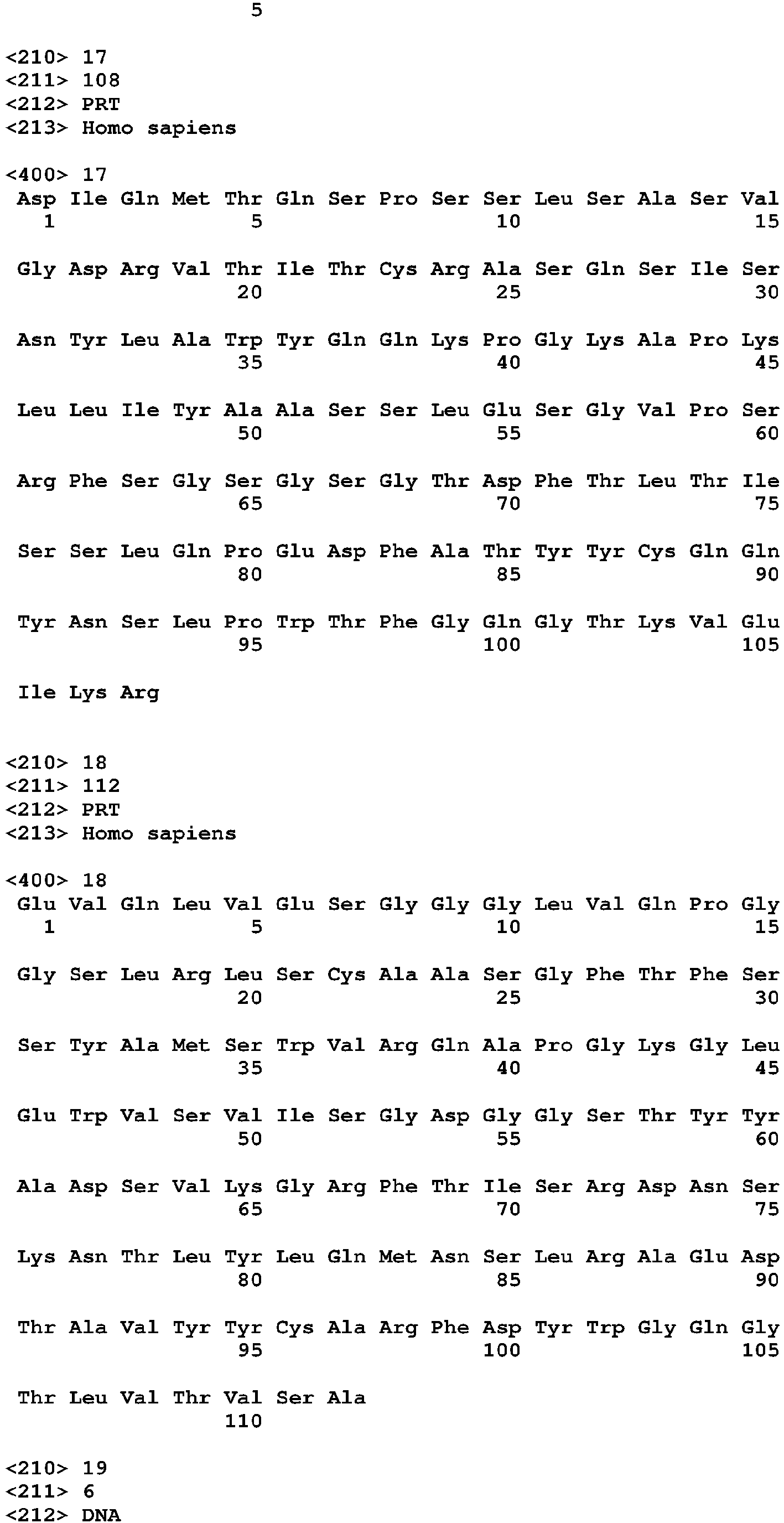


Claims (16)
- An anti-EGFL7 antibody which either:(i) is produced by a hybridoma selected from the group consisting of: anti-EGFL7 mumab 18F7.1.8, anti-EGFL7 mumab 4F11.1.8, and anti-EGFL7 mumab 10G9.1.6, or(ii) is a Fab, Fab', Fab'-SH, F(ab')2 or scFv fragment of the antibody of (i), or which comprises an Fv portion of the antibody of (i) fused to a heavy chain and/or light chain constant region, or which is a linear antibody fragment of the antibody of (i), or which is a humanized version of the antibody of (i), or which is a bispecific version of the antibody of (i), or which is a multivalent version of the antibody of (i), or(iii) comprises one or more complementarity determining regions (CDRs) selected from the group consisting of:(a) 4F11 CDR-L1 sequence KASQSVDYDGDSYMS (SEQ ID NO: 5);(b) 4F11 CDR-L2 sequence GASNLES (SEQ ID NO: 6);(c) 4F11 CDR-L3 sequence QQNNEDPYT (SEQ ID NO: 7);(d) 4F11 CDR-H1 sequence TYGMS (SEQ ID NO: 8);(e) 4F11 CDR-H2 sequence WINTHSGVPTYADDFKG (SEQ ID NO: 9); and(f) 4F11 CDR-H3 sequence LGSSA (SEQ ID NO: 10), or(iv) comprises one or more complementarity determining regions (CDRs) selected from the group consisting of:(a) 10G9 CDR-L1 sequence RSSQSLVHTNGITYLH (SEQ ID NO: 11);(b) 10G9 CDR-L2 sequence KVSNRFS (SEQ ID NO: 12);(c) 10G9 CDR-L3 sequence SQSTHVPLT (SEQ ID NO: 13);(d) 10G9 CDR-H1 sequence DYYMNSDYYMN (SEQ ID NO: 14);(e) 10G9 CDR-H2 sequence DINPKNGGTTYNQKFKG (SEQ ID NO: 15); and(f) 10G9 CDR-H3 sequence ALGVFDY (SEQ ID NO: 16), or(v) specifically binds to a polypeptide comprising an amino acid sequence selected from the group consisting of: CCP, RTIY (SEQ ID NO 33), or(vi) binds to the same epitope on human EGFL7 as the antibody of any one of parts (i) and (iii) to (v), or(vii) competes for EGFL7 binding with the antibody of any one of parts (i) and (iii) to (v), or(viii) is conjugated to a radioactive isotope.
- The anti-EGFL7 antibody of claim 1 part (iii), wherein(i) the light chain of said antibody comprises at least one, at least two or all three of the CDR sequences selected from: KASQSVDYDGDSYMS (SEQ ID NO: 5), GASNLES (SEQ ID NO: 6), and QQNNEDPYT (SEQ ID NO: 7) and/or(ii) the heavy chain of said antibody comprises at least one, at least two or all three of the CDR sequences selected from: TYGMS (SEQ ID NO: 8), WINTHSGVPTYADDFKG (SEQ ID NO: 9), and LGSSA (SEQ ID NO: 10).
- The anti-EGFL7 antibody of claim 1 part (iii), wherein(i) the light chain of said antibody comprises the sequence: DIVLTQSPASLAVSLGQRATISCKASQSVDYDGDSYMSWYQQKPGQPPKLLIYGASNLESG IPARFSGSGSGTDFTLNIHPVEEEDAATYYCQQNNEDPYTFGGGTKVEIKR (SEQ ID NO: 1), or(ii) the heavy chain of said antibody comprises the sequence: QIQLVQSGPELKKPGETVKISCKASGHTFTTYGMSWVKQAPGKGLKWMGWINTHSGVPTY ADDFKGRFAFSLETSASTAHLQINNLKNEDTATYFCARLGSSAVDYWGQGTTVTVSS (SEQ ID NO: 2).
- The anti-EGFL7 antibody of claim 1 part (iv), wherein(i) the light chain of said antibody comprises at least one, at least two or all three of the CDR sequences selected from: RSSQSLVHTNGITYLH (SEQ ID NO: 11), KVSNRFS (SEQ ID NO: 12), and SQSTHVPLT (SEQ ID NO: 13) and/or(ii) the heavy chain of said antibody comprises at least one, at least two or all three of the CDR sequences selected from: DYYMNSDYYMN (SEQ ID NO: 14), DINPKNGGTTYNQKFKG (SEQ ID NO: 15), and ALGVFDY (SEQ ID NO: 16).
- The anti-EGFL7 antibody of claim 1 part (iv), wherein(i) the light chain of said antibody comprises the sequence:DIVMTQTPLSLPVSLGDQASISCRSSQSLVHTNGITYLHWYLQKPGQSPKLLIYKVSNRFSG VPDRFSGSGSGTDFTLKISRVEAEDLGVYFCSQSTHVPLTFGAGTKVEIKR (SEQ ID NO: 3), or(ii) the heavy chain of said antibody comprises the sequence:EVQLQQSGPELVKPGASVKISCKASGYTFSDYYMNSDYYMNWVKQSHGKSLEWIGDINPK NGGTTYNQKFKGKATLTVDKSSSTAYMELRSLTSEDSAVYYCAREADYDPIYYAMDYWGQ GTTLTVSA (SEQ ID NO: 4).
- The antibody of any preceding claim, wherein the antibody is a monoclonal antibody, and/or wherein the antibody is selected from the group consisting of a chimeric antibody, a humanized antibody, an affinity matured antibody, a human antibody, and a bispecific antibody, and/or wherein the antibody is an antibody fragment.
- A pharmaceutical composition comprising an anti-EGFL7 antibody of any preceding claim, optionally further comprising and anti-angiogenic agent, wherein the anti-angiogenic agent is optionally selected from the group consisting of bevacizumab and ranibizumab.
- A polynucleotide encoding an antibody of any of claims 1 to 6.
- A vector comprising the polynucleotide of claim 8, wherein the vector is optionally an expression vector.
- A host cell comprising a vector of claim 9, wherein the host cell is optionally mammalian.
- A method for making an anti-EGFL7 antibody, said method comprising (a) expressing a vector of claim 9 in a suitable host cell, and (b) recovering the antibody.
- The anti-EGFL7 antibody of any one of claims 1 to 6 or a pharmaceutical composition comprising said anti-EGFL7 antibody, for use in a method for reducing or inhibiting angiogenesis in a subject having a pathological condition associated with angiogenesis, wherein the pathological condition is optionally either a neoplasm, optionally a carcinoma, or a pathological condition associated with the eye, optionally an intraocular neovascular disease, and wherein the method optionally further comprises administering to the subject an anti- angiogenic agent, wherein the anti-angiogenic agent is optionally an antagonist of vascular endothelial growth factor (VEGF), wherein the antagonist is optionally an anti-VEGF antibody.
- The antibody or pharmaceutical composition of claim 12, wherein the pathological condition is a neoplasm and the anti-VEGF antibody is bevacizumab, or wherein the pathological condition is associated with the eye and the anti-VEGF antibody is ranibizumab.
- The antibody of any one of claims 1 to 6 or a pharmaceutical composition comprising said anti-EGFL7 antibody and optionally further comprising an anti-angiogenic agent, for use in a method of enhancing the efficacy of an anti-angiogenic agent in a subject having a pathological condition associated with angiogenesis.
- The antibody or pharmaceutical composition of claim 14, wherein(i) the pathological condition is a neoplasm, optionally a carcinoma, and/or(ii) the anti-antigenic agent is bevacizumab, and/or(iii) the method further comprises administering a chemotherapeutic agent.
- The antibody or pharmaceutical composition of claim 14 wherein the pathological condition is associated with the eye, and wherein:(i) the pathological condition is optionally an intraocular neovascular disease;(ii) the anti-angiogenic agent is optionally ranibizumab; the method optionally further comprising administering a corticosteroid, or photodynamic therapy.
Applications Claiming Priority (3)
| Application Number | Priority Date | Filing Date | Title |
|---|---|---|---|
| US78368606P | 2006-03-16 | 2006-03-16 | |
| US81256906P | 2006-06-09 | 2006-06-09 | |
| EP07800145.0A EP1994056B1 (en) | 2006-03-16 | 2007-03-16 | Antibodies to egfl7 and methods for their use |
Related Parent Applications (2)
| Application Number | Title | Priority Date | Filing Date |
|---|---|---|---|
| EP07800145.0A Division-Into EP1994056B1 (en) | 2006-03-16 | 2007-03-16 | Antibodies to egfl7 and methods for their use |
| EP07800145.0 Division | 2007-03-16 |
Publications (2)
| Publication Number | Publication Date |
|---|---|
| EP2468772A2 true EP2468772A2 (en) | 2012-06-27 |
| EP2468772A3 EP2468772A3 (en) | 2013-01-23 |
Family
ID=38510306
Family Applications (2)
| Application Number | Title | Priority Date | Filing Date |
|---|---|---|---|
| EP07800145.0A Active EP1994056B1 (en) | 2006-03-16 | 2007-03-16 | Antibodies to egfl7 and methods for their use |
| EP11195410A Withdrawn EP2468772A3 (en) | 2006-03-16 | 2007-03-16 | Antibodies to EGFL7 and methods for their use |
Family Applications Before (1)
| Application Number | Title | Priority Date | Filing Date |
|---|---|---|---|
| EP07800145.0A Active EP1994056B1 (en) | 2006-03-16 | 2007-03-16 | Antibodies to egfl7 and methods for their use |
Country Status (20)
| Country | Link |
|---|---|
| US (3) | US20100203041A1 (en) |
| EP (2) | EP1994056B1 (en) |
| JP (2) | JP5363972B2 (en) |
| KR (1) | KR101038633B1 (en) |
| AR (1) | AR059851A1 (en) |
| AU (1) | AU2007226522B2 (en) |
| BR (1) | BRPI0707047A2 (en) |
| CA (1) | CA2646513A1 (en) |
| CO (1) | CO6180454A2 (en) |
| CR (1) | CR10280A (en) |
| EC (1) | ECSP088743A (en) |
| IL (3) | IL193514A0 (en) |
| MA (1) | MA30331B1 (en) |
| MX (1) | MX2008011663A (en) |
| NO (1) | NO20084320L (en) |
| NZ (2) | NZ570588A (en) |
| PH (1) | PH12013502194A1 (en) |
| RU (1) | RU2415870C2 (en) |
| TW (1) | TW201446798A (en) |
| WO (1) | WO2007106915A2 (en) |
Cited By (1)
| Publication number | Priority date | Publication date | Assignee | Title |
|---|---|---|---|---|
| US11702469B2 (en) | 2017-04-24 | 2023-07-18 | Ohio State Innovation Foundation | Recombinant EGFL7, EGFL7 antibodies, and uses thereof |
Families Citing this family (9)
| Publication number | Priority date | Publication date | Assignee | Title |
|---|---|---|---|---|
| DE10113776B4 (en) * | 2001-03-21 | 2012-08-09 | "Iba Gmbh" | Isolated streptavidin-binding, competitively elutable peptide, this comprehensive fusion peptide, nucleic acid coding therefor, expression vector, methods for producing a recombinant fusion protein and methods for detecting and / or obtaining the fusion protein |
| AR059851A1 (en) * | 2006-03-16 | 2008-04-30 | Genentech Inc | ANTIBODIES OF EGFL7 AND METHODS OF USE |
| WO2010034155A1 (en) * | 2008-09-25 | 2010-04-01 | 中南大学湘雅医院 | Kit by detecting egfl7 in samples for the diagnosis of hepatocellular carcinoma and its application |
| MX2011010570A (en) * | 2009-05-08 | 2011-10-19 | Genentech Inc | Humanized anti-egfl7 antibodies and methods using same. |
| TWI560199B (en) | 2010-08-31 | 2016-12-01 | Sanofi Sa | Peptide or peptide complex binding to α2 integrin and methods and uses involving the same |
| MX2013008922A (en) * | 2011-02-02 | 2013-10-01 | Genentech Inc | Dosing for treatment with anti-egfl7 antibodies. |
| WO2013061112A1 (en) | 2011-10-24 | 2013-05-02 | Centre National De La Recherche Scientifique | Use of egfl7 modulators for promoting or inhibiting migration of immune cells across vascular endothelium |
| EP2831093A4 (en) * | 2012-03-27 | 2015-11-04 | London Health Sci Ct Res Inc | Egfl7 targeting and/or binding polypeptides and methods for inhibiting angiogenesis |
| CA3012696A1 (en) | 2016-02-05 | 2017-08-10 | The Board Of Regents Of The University Of Texas System | Egfl6 specific monoclonal antibodies and methods of their use |
Citations (119)
| Publication number | Priority date | Publication date | Assignee | Title |
|---|---|---|---|---|
| US3645090A (en) | 1969-06-19 | 1972-02-29 | Citizen Watch Co Ltd | Day-date quick-adjuster for calender timepiece |
| US3720760A (en) | 1968-09-06 | 1973-03-13 | Pharmacia Ab | Method for determining the presence of reagin-immunoglobulins(reagin-ig)directed against certain allergens,in aqueous samples |
| US3773919A (en) | 1969-10-23 | 1973-11-20 | Du Pont | Polylactide-drug mixtures |
| US3896111A (en) | 1973-02-20 | 1975-07-22 | Research Corp | Ansa macrolides |
| US3940475A (en) | 1970-06-11 | 1976-02-24 | Biological Developments, Inc. | Radioimmune method of assaying quantitatively for a hapten |
| US4137230A (en) | 1977-11-14 | 1979-01-30 | Takeda Chemical Industries, Ltd. | Method for the production of maytansinoids |
| US4151042A (en) | 1977-03-31 | 1979-04-24 | Takeda Chemical Industries, Ltd. | Method for producing maytansinol and its derivatives |
| US4248870A (en) | 1978-10-27 | 1981-02-03 | Takeda Chemical Industries, Ltd. | Maytansinoids and use |
| US4256746A (en) | 1978-11-14 | 1981-03-17 | Takeda Chemical Industries | Dechloromaytansinoids, their pharmaceutical compositions and method of use |
| US4260608A (en) | 1978-11-14 | 1981-04-07 | Takeda Chemical Industries, Ltd. | Maytansinoids, pharmaceutical compositions thereof and methods of use thereof |
| US4265814A (en) | 1978-03-24 | 1981-05-05 | Takeda Chemical Industries | Matansinol 3-n-hexadecanoate |
| US4294757A (en) | 1979-01-31 | 1981-10-13 | Takeda Chemical Industries, Ltd | 20-O-Acylmaytansinoids |
| US4307016A (en) | 1978-03-24 | 1981-12-22 | Takeda Chemical Industries, Ltd. | Demethyl maytansinoids |
| US4308269A (en) | 1979-06-11 | 1981-12-29 | Takeda Chemical Industries, Ltd. | Maytansinoids, pharmaceutical compositions thereof and method of use thereof |
| US4308268A (en) | 1979-06-11 | 1981-12-29 | Takeda Chemical Industries, Ltd. | Maytansinoids, pharmaceutical compositions thereof and method of use thereof |
| US4309428A (en) | 1979-07-30 | 1982-01-05 | Takeda Chemical Industries, Ltd. | Maytansinoids |
| US4313946A (en) | 1981-01-27 | 1982-02-02 | The United States Of America As Represented By The Secretary Of Agriculture | Chemotherapeutically active maytansinoids from Trewia nudiflora |
| US4315929A (en) | 1981-01-27 | 1982-02-16 | The United States Of America As Represented By The Secretary Of Agriculture | Method of controlling the European corn borer with trewiasine |
| US4317821A (en) | 1979-06-08 | 1982-03-02 | Takeda Chemical Industries, Ltd. | Maytansinoids, their use and pharmaceutical compositions thereof |
| US4322348A (en) | 1979-06-05 | 1982-03-30 | Takeda Chemical Industries, Ltd. | Maytansinoids |
| US4331598A (en) | 1979-09-19 | 1982-05-25 | Takeda Chemical Industries, Ltd. | Maytansinoids |
| USRE30985E (en) | 1978-01-01 | 1982-06-29 | Serum-free cell culture media | |
| US4362663A (en) | 1979-09-21 | 1982-12-07 | Takeda Chemical Industries, Ltd. | Maytansinoid compound |
| US4364866A (en) | 1979-09-21 | 1982-12-21 | Takeda Chemical Industries, Ltd. | Maytansinoids |
| US4371533A (en) | 1980-10-08 | 1983-02-01 | Takeda Chemical Industries, Ltd. | 4,5-Deoxymaytansinoids, their use and pharmaceutical compositions thereof |
| US4419446A (en) | 1980-12-31 | 1983-12-06 | The United States Of America As Represented By The Department Of Health And Human Services | Recombinant DNA process utilizing a papilloma virus DNA as a vector |
| US4424219A (en) | 1981-05-20 | 1984-01-03 | Takeda Chemical Industries, Ltd. | 9-Thiomaytansinoids and their pharmaceutical compositions and use |
| US4450254A (en) | 1980-11-03 | 1984-05-22 | Standard Oil Company | Impact improvement of high nitrile resins |
| US4560655A (en) | 1982-12-16 | 1985-12-24 | Immunex Corporation | Serum-free cell culture medium and process for making same |
| US4601978A (en) | 1982-11-24 | 1986-07-22 | The Regents Of The University Of California | Mammalian metallothionein promoter system |
| WO1987000195A1 (en) | 1985-06-28 | 1987-01-15 | Celltech Limited | Animal cell culture |
| US4657866A (en) | 1982-12-21 | 1987-04-14 | Sudhir Kumar | Serum-free, synthetic, completely chemically defined tissue culture media |
| US4676980A (en) | 1985-09-23 | 1987-06-30 | The United States Of America As Represented By The Secretary Of The Department Of Health And Human Services | Target specific cross-linked heteroantibodies |
| US4737456A (en) | 1985-05-09 | 1988-04-12 | Syntex (U.S.A.) Inc. | Reducing interference in ligand-receptor binding assays |
| US4767704A (en) | 1983-10-07 | 1988-08-30 | Columbia University In The City Of New York | Protein-free culture medium |
| US4816567A (en) | 1983-04-08 | 1989-03-28 | Genentech, Inc. | Recombinant immunoglobin preparations |
| WO1990003430A1 (en) | 1988-09-23 | 1990-04-05 | Cetus Corporation | Cell culture medium for enhanced cell growth, culture longevity and product expression |
| US4927762A (en) | 1986-04-01 | 1990-05-22 | Cell Enterprises, Inc. | Cell culture medium with antioxidant |
| US4965199A (en) | 1984-04-20 | 1990-10-23 | Genentech, Inc. | Preparation of functional human factor VIII in mammalian cells using methotrexate based selection |
| US4970198A (en) | 1985-10-17 | 1990-11-13 | American Cyanamid Company | Antitumor antibiotics (LL-E33288 complex) |
| US4975278A (en) | 1988-02-26 | 1990-12-04 | Bristol-Myers Company | Antibody-enzyme conjugates in combination with prodrugs for the delivery of cytotoxic agents to tumor cells |
| EP0404097A2 (en) | 1989-06-22 | 1990-12-27 | BEHRINGWERKE Aktiengesellschaft | Bispecific and oligospecific, mono- and oligovalent receptors, production and applications thereof |
| WO1991000360A1 (en) | 1989-06-29 | 1991-01-10 | Medarex, Inc. | Bispecific reagents for aids therapy |
| US5053394A (en) | 1988-09-21 | 1991-10-01 | American Cyanamid Company | Targeted forms of methyltrithio antitumor agents |
| US5079233A (en) | 1987-01-30 | 1992-01-07 | American Cyanamid Company | N-acyl derivatives of the LL-E33288 antitumor antibiotics, composition and methods for using the same |
| WO1992000373A1 (en) | 1990-06-29 | 1992-01-09 | Biosource Genetics Corporation | Melanin production by transformed microorganisms |
| WO1992009690A2 (en) | 1990-12-03 | 1992-06-11 | Genentech, Inc. | Enrichment method for variant proteins with altered binding properties |
| US5122469A (en) | 1990-10-03 | 1992-06-16 | Genentech, Inc. | Method for culturing Chinese hamster ovary cells to improve production of recombinant proteins |
| WO1993006213A1 (en) | 1991-09-23 | 1993-04-01 | Medical Research Council | Production of chimeric antibodies - a combinatorial approach |
| US5208020A (en) | 1989-10-25 | 1993-05-04 | Immunogen Inc. | Cytotoxic agents comprising maytansinoids and their therapeutic use |
| WO1993008829A1 (en) | 1991-11-04 | 1993-05-13 | The Regents Of The University Of California | Compositions that mediate killing of hiv-infected cells |
| WO1993011161A1 (en) | 1991-11-25 | 1993-06-10 | Enzon, Inc. | Multivalent antigen-binding proteins |
| WO1993016185A2 (en) | 1992-02-06 | 1993-08-19 | Creative Biomolecules, Inc. | Biosynthetic binding protein for cancer marker |
| WO1993021232A1 (en) | 1992-04-10 | 1993-10-28 | Research Development Foundation | IMMUNOTOXINS DIRECTED AGAINST c-erbB-2 (HER-2/neu) RELATED SURFACE ANTIGENS |
| US5264365A (en) | 1990-11-09 | 1993-11-23 | Board Of Regents, The University Of Texas System | Protease-deficient bacterial strains for production of proteolytically sensitive polypeptides |
| WO1994004690A1 (en) | 1992-08-17 | 1994-03-03 | Genentech, Inc. | Bispecific immunoadhesins |
| WO1994011026A2 (en) | 1992-11-13 | 1994-05-26 | Idec Pharmaceuticals Corporation | Therapeutic application of chimeric and radiolabeled antibodies to human b lymphocyte restricted differentiation antigen for treatment of b cell lymphoma |
| US5362852A (en) | 1991-09-27 | 1994-11-08 | Pfizer Inc. | Modified peptide derivatives conjugated at 2-hydroxyethylamine moieties |
| WO1994029351A2 (en) | 1993-06-16 | 1994-12-22 | Celltech Limited | Antibodies |
| WO1996007754A1 (en) | 1994-09-02 | 1996-03-14 | The Scripps Research Institute | Methods for producing antibody libraries using universal or randomized immunoglobulin light chains |
| US5500362A (en) | 1987-01-08 | 1996-03-19 | Xoma Corporation | Chimeric antibody with specificity to human B cell surface antigen |
| US5508192A (en) | 1990-11-09 | 1996-04-16 | Board Of Regents, The University Of Texas System | Bacterial host strains for producing proteolytically sensitive polypeptides |
| EP0425235B1 (en) | 1989-10-25 | 1996-09-25 | Immunogen Inc | Cytotoxic agents comprising maytansinoids and their therapeutic use |
| US5571894A (en) | 1991-02-05 | 1996-11-05 | Ciba-Geigy Corporation | Recombinant antibodies specific for a growth factor receptor |
| US5585089A (en) | 1988-12-28 | 1996-12-17 | Protein Design Labs, Inc. | Humanized immunoglobulins |
| US5587458A (en) | 1991-10-07 | 1996-12-24 | Aronex Pharmaceuticals, Inc. | Anti-erbB-2 antibodies, combinations thereof, and therapeutic and diagnostic uses thereof |
| US5606040A (en) | 1987-10-30 | 1997-02-25 | American Cyanamid Company | Antitumor and antibacterial substituted disulfide derivatives prepared from compounds possessing a methyl-trithio group |
| US5624821A (en) | 1987-03-18 | 1997-04-29 | Scotgen Biopharmaceuticals Incorporated | Antibodies with altered effector functions |
| US5635483A (en) | 1992-12-03 | 1997-06-03 | Arizona Board Of Regents Acting On Behalf Of Arizona State University | Tumor inhibiting tetrapeptide bearing modified phenethyl amides |
| US5639635A (en) | 1994-11-03 | 1997-06-17 | Genentech, Inc. | Process for bacterial production of polypeptides |
| US5641870A (en) | 1995-04-20 | 1997-06-24 | Genentech, Inc. | Low pH hydrophobic interaction chromatography for antibody purification |
| US5648237A (en) | 1991-09-19 | 1997-07-15 | Genentech, Inc. | Expression of functional antibody fragments |
| WO1997030087A1 (en) | 1996-02-16 | 1997-08-21 | Glaxo Group Limited | Preparation of glycosylated antibodies |
| US5663149A (en) | 1994-12-13 | 1997-09-02 | Arizona Board Of Regents Acting On Behalf Of Arizona State University | Human cancer inhibitory pentapeptide heterocyclic and halophenyl amides |
| EP0817648A1 (en) | 1995-03-30 | 1998-01-14 | Genentech, Inc. | Vascular endothelial cell growth factor antagonists |
| US5712374A (en) | 1995-06-07 | 1998-01-27 | American Cyanamid Company | Method for the preparation of substantiallly monomeric calicheamicin derivative/carrier conjugates |
| US5714586A (en) | 1995-06-07 | 1998-02-03 | American Cyanamid Company | Methods for the preparation of monomeric calicheamicin derivative/carrier conjugates |
| US5739116A (en) | 1994-06-03 | 1998-04-14 | American Cyanamid Company | Enediyne derivatives useful for the synthesis of conjugates of methyltrithio antitumor agents |
| US5770701A (en) | 1987-10-30 | 1998-06-23 | American Cyanamid Company | Process for preparing targeted forms of methyltrithio antitumor agents |
| US5780588A (en) | 1993-01-26 | 1998-07-14 | Arizona Board Of Regents | Elucidation and synthesis of selected pentapeptides |
| US5821337A (en) | 1991-06-14 | 1998-10-13 | Genentech, Inc. | Immunoglobulin variants |
| WO1998045332A2 (en) | 1997-04-07 | 1998-10-15 | Genentech, Inc. | Humanized antibodies and methods for forming humanized antibodies |
| WO1998045331A2 (en) | 1997-04-07 | 1998-10-15 | Genentech, Inc. | Anti-vegf antibodies |
| WO1998058964A1 (en) | 1997-06-24 | 1998-12-30 | Genentech, Inc. | Methods and compositions for galactosylated glycoproteins |
| US5869046A (en) | 1995-04-14 | 1999-02-09 | Genentech, Inc. | Altered polypeptides with increased half-life |
| WO1999022764A1 (en) | 1997-10-31 | 1999-05-14 | Genentech, Inc. | Methods and compositions comprising glycoprotein glycoforms |
| WO1999051642A1 (en) | 1998-04-02 | 1999-10-14 | Genentech, Inc. | Antibody variants and fragments thereof |
| US6027888A (en) | 1996-04-05 | 2000-02-22 | Board Of Regents, The University Of Texas System | Methods for producing soluble, biologically-active disulfide-bond containing eukaryotic proteins in bacterial cells |
| US6083715A (en) | 1997-06-09 | 2000-07-04 | Board Of Regents, The University Of Texas System | Methods for producing heterologous disulfide bond-containing polypeptides in bacterial cells |
| WO2000042072A2 (en) | 1999-01-15 | 2000-07-20 | Genentech, Inc. | Polypeptide variants with altered effector function |
| WO2000061739A1 (en) | 1999-04-09 | 2000-10-19 | Kyowa Hakko Kogyo Co., Ltd. | Method for controlling the activity of immunologically functional molecule |
| US6194551B1 (en) | 1998-04-02 | 2001-02-27 | Genentech, Inc. | Polypeptide variants |
| WO2001029246A1 (en) | 1999-10-19 | 2001-04-26 | Kyowa Hakko Kogyo Co., Ltd. | Process for producing polypeptide |
| US20020164328A1 (en) | 2000-10-06 | 2002-11-07 | Toyohide Shinkawa | Process for purifying antibody |
| WO2002088172A2 (en) | 2001-04-30 | 2002-11-07 | Seattle Genetics, Inc. | Pentapeptide compounds and uses related thereto |
| WO2003011878A2 (en) | 2001-08-03 | 2003-02-13 | Glycart Biotechnology Ag | Antibody glycosylation variants having increased antibody-dependent cellular cytotoxicity |
| US20030115614A1 (en) | 2000-10-06 | 2003-06-19 | Yutaka Kanda | Antibody composition-producing cell |
| US6602684B1 (en) | 1998-04-20 | 2003-08-05 | Glycart Biotechnology Ag | Glycosylation engineering of antibodies for improving antibody-dependent cellular cytotoxicity |
| US20030157108A1 (en) | 2001-10-25 | 2003-08-21 | Genentech, Inc. | Glycoprotein compositions |
| WO2003084570A1 (en) | 2002-04-09 | 2003-10-16 | Kyowa Hakko Kogyo Co., Ltd. | DRUG CONTAINING ANTIBODY COMPOSITION APPROPRIATE FOR PATIENT SUFFERING FROM FcϜRIIIa POLYMORPHISM |
| WO2003085119A1 (en) | 2002-04-09 | 2003-10-16 | Kyowa Hakko Kogyo Co., Ltd. | METHOD OF ENHANCING ACTIVITY OF ANTIBODY COMPOSITION OF BINDING TO FcϜ RECEPTOR IIIa |
| US20030224948A1 (en) | 2002-02-14 | 2003-12-04 | Dam Willem Van | Lubricating oil additive comprising EC-treated succinimide, borated dispersant and corrosion inhibitor |
| EP1391213A1 (en) | 2002-08-21 | 2004-02-25 | Boehringer Ingelheim International GmbH | Compositions and methods for treating cancer using maytansinoid CD44 antibody immunoconjugates and chemotherapeutic agents |
| US20040093621A1 (en) | 2001-12-25 | 2004-05-13 | Kyowa Hakko Kogyo Co., Ltd | Antibody composition which specifically binds to CD20 |
| US6737056B1 (en) | 1999-01-15 | 2004-05-18 | Genentech, Inc. | Polypeptide variants with altered effector function |
| US20040110704A1 (en) | 2002-04-09 | 2004-06-10 | Kyowa Hakko Kogyo Co., Ltd. | Cells of which genome is modified |
| US20040110282A1 (en) | 2002-04-09 | 2004-06-10 | Kyowa Hakko Kogyo Co., Ltd. | Cells in which activity of the protein involved in transportation of GDP-fucose is reduced or lost |
| US20040109865A1 (en) | 2002-04-09 | 2004-06-10 | Kyowa Hakko Kogyo Co., Ltd. | Antibody composition-containing medicament |
| WO2004056312A2 (en) | 2002-12-16 | 2004-07-08 | Genentech, Inc. | Immunoglobulin variants and uses thereof |
| US20040132140A1 (en) | 2002-04-09 | 2004-07-08 | Kyowa Hakko Kogyo Co., Ltd. | Production process for antibody composition |
| US20050014934A1 (en) | 2002-10-15 | 2005-01-20 | Hinton Paul R. | Alteration of FcRn binding affinities or serum half-lives of antibodies by mutagenesis |
| WO2005012359A2 (en) | 2003-08-01 | 2005-02-10 | Genentech, Inc. | Anti-vegf antibodies |
| WO2005035586A1 (en) | 2003-10-08 | 2005-04-21 | Kyowa Hakko Kogyo Co., Ltd. | Fused protein composition |
| WO2005035778A1 (en) | 2003-10-09 | 2005-04-21 | Kyowa Hakko Kogyo Co., Ltd. | PROCESS FOR PRODUCING ANTIBODY COMPOSITION BY USING RNA INHIBITING THE FUNCTION OF α1,6-FUCOSYLTRANSFERASE |
| US20050123546A1 (en) | 2003-11-05 | 2005-06-09 | Glycart Biotechnology Ag | Antigen binding molecules with increased Fc receptor binding affinity and effector function |
| WO2005053742A1 (en) | 2003-12-04 | 2005-06-16 | Kyowa Hakko Kogyo Co., Ltd. | Medicine containing antibody composition |
| US20050169933A1 (en) | 2003-10-10 | 2005-08-04 | Immunogen, Inc. | Method of targeting specific cell populations using cell-binding agent maytansinoid conjugates linked via a non-cleavable linker, said conjugates, and methods of making said conjugates |
| US20050238649A1 (en) | 2003-11-06 | 2005-10-27 | Seattle Genetics, Inc. | Monomethylvaline compounds capable of conjugation to ligands |
| WO2005117968A2 (en) | 2004-04-14 | 2005-12-15 | Genentech, Inc. | Compositions and methods comprising an egfl7 antagonist for modulating vascular development |
Family Cites Families (7)
| Publication number | Priority date | Publication date | Assignee | Title |
|---|---|---|---|---|
| US6217895B1 (en) * | 1999-03-22 | 2001-04-17 | Control Delivery Systems | Method for treating and/or preventing retinal diseases with sustained release corticosteroids |
| GB0024200D0 (en) * | 2000-10-03 | 2000-11-15 | Smithkline Beecham Sa | Component vaccine |
| WO2002061087A2 (en) * | 2000-12-19 | 2002-08-08 | Lifespan Biosciences, Inc. | Antigenic peptides, such as for g protein-coupled receptors (gpcrs), antibodies thereto, and systems for identifying such antigenic peptides |
| AU2003279754A1 (en) | 2002-10-04 | 2004-05-04 | Schering Aktiengesellschaft | Modified hepsin molecules having a substitute activation sequence and uses thereof |
| JPWO2005094846A1 (en) * | 2004-03-30 | 2008-02-14 | 株式会社レノメディクス研究所 | Prion disease therapeutic agent and method for producing the same |
| CA2576093A1 (en) * | 2004-08-03 | 2006-02-09 | Mochida Pharmaceutical Co., Ltd. | Pharmaceutical compostion containing meltrin antagonist |
| AR059851A1 (en) * | 2006-03-16 | 2008-04-30 | Genentech Inc | ANTIBODIES OF EGFL7 AND METHODS OF USE |
-
2007
- 2007-03-15 AR ARP070101056A patent/AR059851A1/en not_active Application Discontinuation
- 2007-03-16 EP EP07800145.0A patent/EP1994056B1/en active Active
- 2007-03-16 JP JP2009500633A patent/JP5363972B2/en not_active Expired - Fee Related
- 2007-03-16 NZ NZ570588A patent/NZ570588A/en not_active IP Right Cessation
- 2007-03-16 NZ NZ595863A patent/NZ595863A/en not_active IP Right Cessation
- 2007-03-16 US US12/280,673 patent/US20100203041A1/en not_active Abandoned
- 2007-03-16 BR BRPI0707047-0A patent/BRPI0707047A2/en not_active IP Right Cessation
- 2007-03-16 AU AU2007226522A patent/AU2007226522B2/en not_active Ceased
- 2007-03-16 EP EP11195410A patent/EP2468772A3/en not_active Withdrawn
- 2007-03-16 KR KR1020087022517A patent/KR101038633B1/en not_active IP Right Cessation
- 2007-03-16 CA CA002646513A patent/CA2646513A1/en not_active Abandoned
- 2007-03-16 MX MX2008011663A patent/MX2008011663A/en active IP Right Grant
- 2007-03-16 TW TW102148898A patent/TW201446798A/en unknown
- 2007-03-16 WO PCT/US2007/064242 patent/WO2007106915A2/en active Application Filing
- 2007-03-16 RU RU2008140947/10A patent/RU2415870C2/en not_active IP Right Cessation
-
2008
- 2008-08-18 IL IL193514A patent/IL193514A0/en unknown
- 2008-09-10 CR CR10280A patent/CR10280A/en unknown
- 2008-09-16 CO CO08097893A patent/CO6180454A2/en active IP Right Grant
- 2008-09-16 EC EC2008008743A patent/ECSP088743A/en unknown
- 2008-10-13 MA MA31284A patent/MA30331B1/en unknown
- 2008-10-15 NO NO20084320A patent/NO20084320L/en not_active Application Discontinuation
-
2011
- 2011-04-14 US US13/087,247 patent/US8398976B2/en not_active Expired - Fee Related
- 2011-09-28 JP JP2011213087A patent/JP5364137B2/en not_active Expired - Fee Related
-
2012
- 2012-12-04 IL IL223416A patent/IL223416A0/en unknown
-
2013
- 2013-01-31 US US13/755,950 patent/US20130129735A1/en not_active Abandoned
- 2013-10-23 PH PH12013502194A patent/PH12013502194A1/en unknown
-
2014
- 2014-04-08 IL IL232011A patent/IL232011A0/en unknown
Patent Citations (128)
| Publication number | Priority date | Publication date | Assignee | Title |
|---|---|---|---|---|
| US3720760B1 (en) | 1968-09-06 | 1984-02-07 | Pharmacia Ab | |
| US3720760A (en) | 1968-09-06 | 1973-03-13 | Pharmacia Ab | Method for determining the presence of reagin-immunoglobulins(reagin-ig)directed against certain allergens,in aqueous samples |
| US3645090A (en) | 1969-06-19 | 1972-02-29 | Citizen Watch Co Ltd | Day-date quick-adjuster for calender timepiece |
| US3773919A (en) | 1969-10-23 | 1973-11-20 | Du Pont | Polylactide-drug mixtures |
| US3940475A (en) | 1970-06-11 | 1976-02-24 | Biological Developments, Inc. | Radioimmune method of assaying quantitatively for a hapten |
| US3896111A (en) | 1973-02-20 | 1975-07-22 | Research Corp | Ansa macrolides |
| US4151042A (en) | 1977-03-31 | 1979-04-24 | Takeda Chemical Industries, Ltd. | Method for producing maytansinol and its derivatives |
| US4137230A (en) | 1977-11-14 | 1979-01-30 | Takeda Chemical Industries, Ltd. | Method for the production of maytansinoids |
| USRE30985E (en) | 1978-01-01 | 1982-06-29 | Serum-free cell culture media | |
| US4307016A (en) | 1978-03-24 | 1981-12-22 | Takeda Chemical Industries, Ltd. | Demethyl maytansinoids |
| US4265814A (en) | 1978-03-24 | 1981-05-05 | Takeda Chemical Industries | Matansinol 3-n-hexadecanoate |
| US4361650A (en) | 1978-03-24 | 1982-11-30 | Takeda Chemical Industries, Ltd. | Fermentation process of preparing demethyl maytansinoids |
| US4248870A (en) | 1978-10-27 | 1981-02-03 | Takeda Chemical Industries, Ltd. | Maytansinoids and use |
| US4260608A (en) | 1978-11-14 | 1981-04-07 | Takeda Chemical Industries, Ltd. | Maytansinoids, pharmaceutical compositions thereof and methods of use thereof |
| US4256746A (en) | 1978-11-14 | 1981-03-17 | Takeda Chemical Industries | Dechloromaytansinoids, their pharmaceutical compositions and method of use |
| US4294757A (en) | 1979-01-31 | 1981-10-13 | Takeda Chemical Industries, Ltd | 20-O-Acylmaytansinoids |
| US4322348A (en) | 1979-06-05 | 1982-03-30 | Takeda Chemical Industries, Ltd. | Maytansinoids |
| US4317821A (en) | 1979-06-08 | 1982-03-02 | Takeda Chemical Industries, Ltd. | Maytansinoids, their use and pharmaceutical compositions thereof |
| US4308268A (en) | 1979-06-11 | 1981-12-29 | Takeda Chemical Industries, Ltd. | Maytansinoids, pharmaceutical compositions thereof and method of use thereof |
| US4308269A (en) | 1979-06-11 | 1981-12-29 | Takeda Chemical Industries, Ltd. | Maytansinoids, pharmaceutical compositions thereof and method of use thereof |
| US4309428A (en) | 1979-07-30 | 1982-01-05 | Takeda Chemical Industries, Ltd. | Maytansinoids |
| US4331598A (en) | 1979-09-19 | 1982-05-25 | Takeda Chemical Industries, Ltd. | Maytansinoids |
| US4362663A (en) | 1979-09-21 | 1982-12-07 | Takeda Chemical Industries, Ltd. | Maytansinoid compound |
| US4364866A (en) | 1979-09-21 | 1982-12-21 | Takeda Chemical Industries, Ltd. | Maytansinoids |
| US4371533A (en) | 1980-10-08 | 1983-02-01 | Takeda Chemical Industries, Ltd. | 4,5-Deoxymaytansinoids, their use and pharmaceutical compositions thereof |
| US4450254A (en) | 1980-11-03 | 1984-05-22 | Standard Oil Company | Impact improvement of high nitrile resins |
| US4419446A (en) | 1980-12-31 | 1983-12-06 | The United States Of America As Represented By The Department Of Health And Human Services | Recombinant DNA process utilizing a papilloma virus DNA as a vector |
| US4313946A (en) | 1981-01-27 | 1982-02-02 | The United States Of America As Represented By The Secretary Of Agriculture | Chemotherapeutically active maytansinoids from Trewia nudiflora |
| US4315929A (en) | 1981-01-27 | 1982-02-16 | The United States Of America As Represented By The Secretary Of Agriculture | Method of controlling the European corn borer with trewiasine |
| US4424219A (en) | 1981-05-20 | 1984-01-03 | Takeda Chemical Industries, Ltd. | 9-Thiomaytansinoids and their pharmaceutical compositions and use |
| US4601978A (en) | 1982-11-24 | 1986-07-22 | The Regents Of The University Of California | Mammalian metallothionein promoter system |
| US4560655A (en) | 1982-12-16 | 1985-12-24 | Immunex Corporation | Serum-free cell culture medium and process for making same |
| US4657866A (en) | 1982-12-21 | 1987-04-14 | Sudhir Kumar | Serum-free, synthetic, completely chemically defined tissue culture media |
| US4816567A (en) | 1983-04-08 | 1989-03-28 | Genentech, Inc. | Recombinant immunoglobin preparations |
| US4767704A (en) | 1983-10-07 | 1988-08-30 | Columbia University In The City Of New York | Protein-free culture medium |
| US4965199A (en) | 1984-04-20 | 1990-10-23 | Genentech, Inc. | Preparation of functional human factor VIII in mammalian cells using methotrexate based selection |
| US4737456A (en) | 1985-05-09 | 1988-04-12 | Syntex (U.S.A.) Inc. | Reducing interference in ligand-receptor binding assays |
| WO1987000195A1 (en) | 1985-06-28 | 1987-01-15 | Celltech Limited | Animal cell culture |
| US4676980A (en) | 1985-09-23 | 1987-06-30 | The United States Of America As Represented By The Secretary Of The Department Of Health And Human Services | Target specific cross-linked heteroantibodies |
| US4970198A (en) | 1985-10-17 | 1990-11-13 | American Cyanamid Company | Antitumor antibiotics (LL-E33288 complex) |
| US4927762A (en) | 1986-04-01 | 1990-05-22 | Cell Enterprises, Inc. | Cell culture medium with antioxidant |
| US5500362A (en) | 1987-01-08 | 1996-03-19 | Xoma Corporation | Chimeric antibody with specificity to human B cell surface antigen |
| US5079233A (en) | 1987-01-30 | 1992-01-07 | American Cyanamid Company | N-acyl derivatives of the LL-E33288 antitumor antibiotics, composition and methods for using the same |
| US5624821A (en) | 1987-03-18 | 1997-04-29 | Scotgen Biopharmaceuticals Incorporated | Antibodies with altered effector functions |
| US5648260A (en) | 1987-03-18 | 1997-07-15 | Scotgen Biopharmaceuticals Incorporated | DNA encoding antibodies with altered effector functions |
| US5606040A (en) | 1987-10-30 | 1997-02-25 | American Cyanamid Company | Antitumor and antibacterial substituted disulfide derivatives prepared from compounds possessing a methyl-trithio group |
| US5770701A (en) | 1987-10-30 | 1998-06-23 | American Cyanamid Company | Process for preparing targeted forms of methyltrithio antitumor agents |
| US5770710A (en) | 1987-10-30 | 1998-06-23 | American Cyanamid Company | Antitumor and antibacterial substituted disulfide derivatives prepared from compounds possessing a methlytrithio group |
| US4975278A (en) | 1988-02-26 | 1990-12-04 | Bristol-Myers Company | Antibody-enzyme conjugates in combination with prodrugs for the delivery of cytotoxic agents to tumor cells |
| US5053394A (en) | 1988-09-21 | 1991-10-01 | American Cyanamid Company | Targeted forms of methyltrithio antitumor agents |
| WO1990003430A1 (en) | 1988-09-23 | 1990-04-05 | Cetus Corporation | Cell culture medium for enhanced cell growth, culture longevity and product expression |
| US5693762A (en) | 1988-12-28 | 1997-12-02 | Protein Design Labs, Inc. | Humanized immunoglobulins |
| US5585089A (en) | 1988-12-28 | 1996-12-17 | Protein Design Labs, Inc. | Humanized immunoglobulins |
| EP0404097A2 (en) | 1989-06-22 | 1990-12-27 | BEHRINGWERKE Aktiengesellschaft | Bispecific and oligospecific, mono- and oligovalent receptors, production and applications thereof |
| WO1991000360A1 (en) | 1989-06-29 | 1991-01-10 | Medarex, Inc. | Bispecific reagents for aids therapy |
| EP0425235B1 (en) | 1989-10-25 | 1996-09-25 | Immunogen Inc | Cytotoxic agents comprising maytansinoids and their therapeutic use |
| US5208020A (en) | 1989-10-25 | 1993-05-04 | Immunogen Inc. | Cytotoxic agents comprising maytansinoids and their therapeutic use |
| US5416064A (en) | 1989-10-25 | 1995-05-16 | Immunogen, Inc. | Cytotoxic agents comprising maytansinoids and their therapeutic use |
| WO1992000373A1 (en) | 1990-06-29 | 1992-01-09 | Biosource Genetics Corporation | Melanin production by transformed microorganisms |
| US5122469A (en) | 1990-10-03 | 1992-06-16 | Genentech, Inc. | Method for culturing Chinese hamster ovary cells to improve production of recombinant proteins |
| US5508192A (en) | 1990-11-09 | 1996-04-16 | Board Of Regents, The University Of Texas System | Bacterial host strains for producing proteolytically sensitive polypeptides |
| US5264365A (en) | 1990-11-09 | 1993-11-23 | Board Of Regents, The University Of Texas System | Protease-deficient bacterial strains for production of proteolytically sensitive polypeptides |
| WO1992009690A2 (en) | 1990-12-03 | 1992-06-11 | Genentech, Inc. | Enrichment method for variant proteins with altered binding properties |
| US5571894A (en) | 1991-02-05 | 1996-11-05 | Ciba-Geigy Corporation | Recombinant antibodies specific for a growth factor receptor |
| US5821337A (en) | 1991-06-14 | 1998-10-13 | Genentech, Inc. | Immunoglobulin variants |
| US5648237A (en) | 1991-09-19 | 1997-07-15 | Genentech, Inc. | Expression of functional antibody fragments |
| WO1993006213A1 (en) | 1991-09-23 | 1993-04-01 | Medical Research Council | Production of chimeric antibodies - a combinatorial approach |
| US5362852A (en) | 1991-09-27 | 1994-11-08 | Pfizer Inc. | Modified peptide derivatives conjugated at 2-hydroxyethylamine moieties |
| US5587458A (en) | 1991-10-07 | 1996-12-24 | Aronex Pharmaceuticals, Inc. | Anti-erbB-2 antibodies, combinations thereof, and therapeutic and diagnostic uses thereof |
| WO1993008829A1 (en) | 1991-11-04 | 1993-05-13 | The Regents Of The University Of California | Compositions that mediate killing of hiv-infected cells |
| WO1993011161A1 (en) | 1991-11-25 | 1993-06-10 | Enzon, Inc. | Multivalent antigen-binding proteins |
| WO1993016185A2 (en) | 1992-02-06 | 1993-08-19 | Creative Biomolecules, Inc. | Biosynthetic binding protein for cancer marker |
| WO1993021232A1 (en) | 1992-04-10 | 1993-10-28 | Research Development Foundation | IMMUNOTOXINS DIRECTED AGAINST c-erbB-2 (HER-2/neu) RELATED SURFACE ANTIGENS |
| WO1994004690A1 (en) | 1992-08-17 | 1994-03-03 | Genentech, Inc. | Bispecific immunoadhesins |
| WO1994011026A2 (en) | 1992-11-13 | 1994-05-26 | Idec Pharmaceuticals Corporation | Therapeutic application of chimeric and radiolabeled antibodies to human b lymphocyte restricted differentiation antigen for treatment of b cell lymphoma |
| US5635483A (en) | 1992-12-03 | 1997-06-03 | Arizona Board Of Regents Acting On Behalf Of Arizona State University | Tumor inhibiting tetrapeptide bearing modified phenethyl amides |
| US5780588A (en) | 1993-01-26 | 1998-07-14 | Arizona Board Of Regents | Elucidation and synthesis of selected pentapeptides |
| WO1994029351A2 (en) | 1993-06-16 | 1994-12-22 | Celltech Limited | Antibodies |
| US5739116A (en) | 1994-06-03 | 1998-04-14 | American Cyanamid Company | Enediyne derivatives useful for the synthesis of conjugates of methyltrithio antitumor agents |
| US5877296A (en) | 1994-06-03 | 1999-03-02 | American Cyanamid Company | Process for preparing conjugates of methyltrithio antitumor agents |
| US5767285A (en) | 1994-06-03 | 1998-06-16 | American Cyanamid Company | Linkers useful for the synthesis of conjugates of methyltrithio antitumor agents |
| US5773001A (en) | 1994-06-03 | 1998-06-30 | American Cyanamid Company | Conjugates of methyltrithio antitumor agents and intermediates for their synthesis |
| WO1996007754A1 (en) | 1994-09-02 | 1996-03-14 | The Scripps Research Institute | Methods for producing antibody libraries using universal or randomized immunoglobulin light chains |
| US5639635A (en) | 1994-11-03 | 1997-06-17 | Genentech, Inc. | Process for bacterial production of polypeptides |
| US5663149A (en) | 1994-12-13 | 1997-09-02 | Arizona Board Of Regents Acting On Behalf Of Arizona State University | Human cancer inhibitory pentapeptide heterocyclic and halophenyl amides |
| EP0817648A1 (en) | 1995-03-30 | 1998-01-14 | Genentech, Inc. | Vascular endothelial cell growth factor antagonists |
| US5869046A (en) | 1995-04-14 | 1999-02-09 | Genentech, Inc. | Altered polypeptides with increased half-life |
| US5641870A (en) | 1995-04-20 | 1997-06-24 | Genentech, Inc. | Low pH hydrophobic interaction chromatography for antibody purification |
| US5712374A (en) | 1995-06-07 | 1998-01-27 | American Cyanamid Company | Method for the preparation of substantiallly monomeric calicheamicin derivative/carrier conjugates |
| US5714586A (en) | 1995-06-07 | 1998-02-03 | American Cyanamid Company | Methods for the preparation of monomeric calicheamicin derivative/carrier conjugates |
| WO1997030087A1 (en) | 1996-02-16 | 1997-08-21 | Glaxo Group Limited | Preparation of glycosylated antibodies |
| US6027888A (en) | 1996-04-05 | 2000-02-22 | Board Of Regents, The University Of Texas System | Methods for producing soluble, biologically-active disulfide-bond containing eukaryotic proteins in bacterial cells |
| WO1998045332A2 (en) | 1997-04-07 | 1998-10-15 | Genentech, Inc. | Humanized antibodies and methods for forming humanized antibodies |
| WO1998045331A2 (en) | 1997-04-07 | 1998-10-15 | Genentech, Inc. | Anti-vegf antibodies |
| US6083715A (en) | 1997-06-09 | 2000-07-04 | Board Of Regents, The University Of Texas System | Methods for producing heterologous disulfide bond-containing polypeptides in bacterial cells |
| WO1998058964A1 (en) | 1997-06-24 | 1998-12-30 | Genentech, Inc. | Methods and compositions for galactosylated glycoproteins |
| WO1999022764A1 (en) | 1997-10-31 | 1999-05-14 | Genentech, Inc. | Methods and compositions comprising glycoprotein glycoforms |
| US6194551B1 (en) | 1998-04-02 | 2001-02-27 | Genentech, Inc. | Polypeptide variants |
| WO1999051642A1 (en) | 1998-04-02 | 1999-10-14 | Genentech, Inc. | Antibody variants and fragments thereof |
| US6602684B1 (en) | 1998-04-20 | 2003-08-05 | Glycart Biotechnology Ag | Glycosylation engineering of antibodies for improving antibody-dependent cellular cytotoxicity |
| US6737056B1 (en) | 1999-01-15 | 2004-05-18 | Genentech, Inc. | Polypeptide variants with altered effector function |
| WO2000042072A2 (en) | 1999-01-15 | 2000-07-20 | Genentech, Inc. | Polypeptide variants with altered effector function |
| WO2000061739A1 (en) | 1999-04-09 | 2000-10-19 | Kyowa Hakko Kogyo Co., Ltd. | Method for controlling the activity of immunologically functional molecule |
| WO2001029246A1 (en) | 1999-10-19 | 2001-04-26 | Kyowa Hakko Kogyo Co., Ltd. | Process for producing polypeptide |
| US20030115614A1 (en) | 2000-10-06 | 2003-06-19 | Yutaka Kanda | Antibody composition-producing cell |
| US20020164328A1 (en) | 2000-10-06 | 2002-11-07 | Toyohide Shinkawa | Process for purifying antibody |
| WO2002088172A2 (en) | 2001-04-30 | 2002-11-07 | Seattle Genetics, Inc. | Pentapeptide compounds and uses related thereto |
| WO2003011878A2 (en) | 2001-08-03 | 2003-02-13 | Glycart Biotechnology Ag | Antibody glycosylation variants having increased antibody-dependent cellular cytotoxicity |
| US20030157108A1 (en) | 2001-10-25 | 2003-08-21 | Genentech, Inc. | Glycoprotein compositions |
| US20040093621A1 (en) | 2001-12-25 | 2004-05-13 | Kyowa Hakko Kogyo Co., Ltd | Antibody composition which specifically binds to CD20 |
| US20030224948A1 (en) | 2002-02-14 | 2003-12-04 | Dam Willem Van | Lubricating oil additive comprising EC-treated succinimide, borated dispersant and corrosion inhibitor |
| WO2003084570A1 (en) | 2002-04-09 | 2003-10-16 | Kyowa Hakko Kogyo Co., Ltd. | DRUG CONTAINING ANTIBODY COMPOSITION APPROPRIATE FOR PATIENT SUFFERING FROM FcϜRIIIa POLYMORPHISM |
| US20040110704A1 (en) | 2002-04-09 | 2004-06-10 | Kyowa Hakko Kogyo Co., Ltd. | Cells of which genome is modified |
| US20040110282A1 (en) | 2002-04-09 | 2004-06-10 | Kyowa Hakko Kogyo Co., Ltd. | Cells in which activity of the protein involved in transportation of GDP-fucose is reduced or lost |
| US20040109865A1 (en) | 2002-04-09 | 2004-06-10 | Kyowa Hakko Kogyo Co., Ltd. | Antibody composition-containing medicament |
| WO2003085119A1 (en) | 2002-04-09 | 2003-10-16 | Kyowa Hakko Kogyo Co., Ltd. | METHOD OF ENHANCING ACTIVITY OF ANTIBODY COMPOSITION OF BINDING TO FcϜ RECEPTOR IIIa |
| US20040132140A1 (en) | 2002-04-09 | 2004-07-08 | Kyowa Hakko Kogyo Co., Ltd. | Production process for antibody composition |
| EP1391213A1 (en) | 2002-08-21 | 2004-02-25 | Boehringer Ingelheim International GmbH | Compositions and methods for treating cancer using maytansinoid CD44 antibody immunoconjugates and chemotherapeutic agents |
| US20050014934A1 (en) | 2002-10-15 | 2005-01-20 | Hinton Paul R. | Alteration of FcRn binding affinities or serum half-lives of antibodies by mutagenesis |
| WO2004056312A2 (en) | 2002-12-16 | 2004-07-08 | Genentech, Inc. | Immunoglobulin variants and uses thereof |
| WO2005012359A2 (en) | 2003-08-01 | 2005-02-10 | Genentech, Inc. | Anti-vegf antibodies |
| WO2005035586A1 (en) | 2003-10-08 | 2005-04-21 | Kyowa Hakko Kogyo Co., Ltd. | Fused protein composition |
| WO2005035778A1 (en) | 2003-10-09 | 2005-04-21 | Kyowa Hakko Kogyo Co., Ltd. | PROCESS FOR PRODUCING ANTIBODY COMPOSITION BY USING RNA INHIBITING THE FUNCTION OF α1,6-FUCOSYLTRANSFERASE |
| US20050169933A1 (en) | 2003-10-10 | 2005-08-04 | Immunogen, Inc. | Method of targeting specific cell populations using cell-binding agent maytansinoid conjugates linked via a non-cleavable linker, said conjugates, and methods of making said conjugates |
| US20050123546A1 (en) | 2003-11-05 | 2005-06-09 | Glycart Biotechnology Ag | Antigen binding molecules with increased Fc receptor binding affinity and effector function |
| US20050238649A1 (en) | 2003-11-06 | 2005-10-27 | Seattle Genetics, Inc. | Monomethylvaline compounds capable of conjugation to ligands |
| WO2005053742A1 (en) | 2003-12-04 | 2005-06-16 | Kyowa Hakko Kogyo Co., Ltd. | Medicine containing antibody composition |
| WO2005117968A2 (en) | 2004-04-14 | 2005-12-15 | Genentech, Inc. | Compositions and methods comprising an egfl7 antagonist for modulating vascular development |
Non-Patent Citations (215)
| Title |
|---|
| "ANIMAL CELL CULTURE", 1987 |
| "ANTIBODIES, A LABORATORY MANUAL", 1988 |
| "Antibody Engineering", 1992, W.H. FREEMAN AND COMPANY |
| "Applications Handbook and Catalog", 2003, pages: 467 - 498 |
| "CURRENT PROTOCOLS IN MOLECULAR BIOLOGY", 2003 |
| "METHODS IN ENZYMOLOGY", ACADEMIC PRESS, INC. |
| "PCR 2: A PRACTICAL APPROACH", 1995 |
| "The Pharmacology of Monoclonal Antibodies", vol. 113, 1994, SPRINGER-VERLAG, pages: 269 - 315 |
| ABRAHMSEN ET AL., EMBO J, vol. 4, 1985, pages 3901 |
| ADAMIS ET AL., ARCH. OPHTHALMOL., vol. 114, 1996, pages 66 - 71 |
| AGNEW, CHEM INTL. ED. ENGL., vol. 33, 1994, pages 183 - 186 |
| AIELLO ET AL., N. ENGL. ./ MED., vol. 331, 1994, pages 1480 - 87 |
| AMD. LOPEZ ET AL., INVEST. OPHTHALMOL. VIS. SCI., vol. 37, 1996, pages 855 - 68 |
| ARIE ET AL., MOLEC. MICROBIOL., vol. 39, 2001, pages 199 - 210 |
| BACHMANN: "Cellular and Molecular Biology", vol. 2, 1987, AMERICAN SOCIETY FOR MICROBIOLOGY, pages: 1190 - 1219 |
| BALDWIN ET AL., LANCET, 15 March 1986 (1986-03-15), pages 603 - 05 |
| BARBAS ET AL., PROC NAT. ACAD. SCI. USA, vol. 91, 1994, pages 3809 - 13 |
| BARBAS ET AL., PROC. NATL. ACAD. SCI USA, vol. 88, 1991, pages 7978 - 82 |
| BARBAS ET AL., PROC. NATL. ACAD. SCI. USA, vol. 88, 1991, pages 7978 - 82 |
| BARBAS ET AL., PROC. NATL. ACAD. SCI. USA, vol. 89, 1992, pages 4457 - 61 |
| BARNES ET AL., ANAL. BIOCHEM., vol. 102, 1980, pages 255 |
| BASS ET AL., PROTEINS, vol. 8, 1990, pages 309 - 14 |
| BERKMAN ET AL., CLIN. INVEST., vol. 91, 1993, pages 153 - 59 |
| BOERNER ET AL., J IMMUNOL., vol. 147, 1991, pages 86 |
| BORGSTROM ET AL., CANCER RES., vol. 56, 1996, pages 4032 - 39 |
| BOTHMANN; PLIICKTHUN, J. BIOL. CHEM., vol. 275, 2000, pages 17100 - 05 |
| BRENNAN ET AL., SCIENCE, vol. 229, 1985, pages 81 |
| BRENNAN ET AL., SCIENCE, vol. 229, no. 81, 1985 |
| BRODEUR ET AL.: "Monoclonal Antibody Production Techniques and Applications", 1987, MARCEL DEKKER, INC., pages: 51 - 63 |
| BROWN ET AL., CANCER RES., vol. 53, 1993, pages 4727 - 35 |
| BROWN ET AL., HUMAN PATHOL., vol. 26, 1995, pages 86 - 91 |
| BRUGGERMANN ET AL., YEAR IN IMMUNOL., vol. 7, 1993, pages 33 |
| CAMERON ET AL., CANCER CELL INT., vol. 5, 2005, pages 23 |
| CAPEL ET AL., IMMUNOMETHODS, vol. 4, 1994, pages 25 - 34 |
| CARLSSON ET AL., BIOCHEM. ., vol. 173, 1978, pages 723 - 37 |
| CARTER ET AL., BIOLTECHNOLOGY, vol. 10, 1992, pages 163 - 67 |
| CARTER ET AL., PROC. NATL. ACAD. SCI. USA, vol. 89, 1992, pages 4285 |
| CHARI ET AL., CANCER RESEARCH, vol. 52, 1992, pages 127 - 131 |
| CHARI ET AL., CANCER RESEARCH, vol. 52, 1992, pages 127 - 31 |
| CHARI ET AL., CANCER RESEARCH, vol. 52, 1992, pages 127 - 31 1 |
| CHATAL: "Monoclonal Antibodies in Immunoscintigraphy", 1989, CRC PRESS |
| CHEN ET AL., BIOL. CHEM., vol. 274, 1999, pages 19601 - 05 |
| CHEN ET AL., J MOL. BIOL., vol. 293, 1999, pages 865 - 81 |
| CHEN, Y. ET AL., MOL. BIOL., vol. 293, 1999, pages 865 - 81 |
| CHEN, Y. ET AL., MOL. BIOL., vol. 293, 1999, pages 865 - 881 |
| CHOTHIA ET AL., J MOL. BIOL, vol. 196, 1987, pages 901 |
| CHOTHIA; LESK, MOL. BIOL, vol. 196, 1987, pages 901 - 17 |
| CLACKSON ET AL., NATURE, vol. 352, 1991, pages 624 - 28 |
| CLACKSON ET AL., NATURE, vol. 352, 1991, pages 624 - 628 |
| CLYNES ET AL., PROC. NATL. ACAD. SCI. USA, vol. 95, 1998, pages 652 - 56 |
| CLYNES ET AL., PROC. NATL. ACAD. SCI. USA, vol. 95, 1998, pages 652 - 656 |
| CUNNINGHAM; WELLS, SCIENCE, vol. 244, 1989, pages 1081 - 85 |
| DAERON, ANNU. REV. IMMUNOL., vol. 15, 1997, pages 203 - 34 |
| DAVID ET AL., BIOCHEMISTRY, vol. 13, 1974, pages 1014 - 21 |
| DORONINA ET AL., NATURE BIOTECH., vol. 21, no. 7, 2003, pages 778 - 784 |
| DORONINA, NATURE BIOTECHNOL., vol. 21, no. 7, 2003, pages 778 - 84 |
| DRUGS OF THE FUTURE, vol. 25, no. 7, 2000, pages 686 |
| DUNCAN; WINTER, NATURE, vol. 322, 1988, pages 738 - 40 |
| DVORAK ET AL., AYN. ./ PATHOL., vol. 146, 1995, pages 1029 - 39 |
| E. SCHRODER; K. LUBKE: "The Peptides", vol. 1, 1965, ACADEMIC PRESS, pages: 76 - 136 |
| EMBLETON ET AL., NUCL. ACIDS RES., vol. 20, 1992, pages 3831 - 37 |
| ENGELS ET AL., AGNEW. CHEM. INT. ED. ENGL., vol. 28, 1989, pages 716 - 34 |
| FERRARA ET AL., ENDOCR. REV. |
| FERRARA ET AL., ENDOCR. REV., vol. 18, 1997, pages 4 - 25 |
| FINKLE ET AL., CLINICAL CANCER RES., vol. 10, 2004, pages 2499 - 251 |
| FOLKMAN ET AL., J BIOL. CHEM., vol. 267, 1992, pages 10931 - 34 |
| FOLKMAN ET AL., NATURE, vol. 339, 1989, pages 58 |
| FOLKMAN, NAT. MED., vol. 1, no. 1, 1995, pages 27 - 31 |
| FRAKER ET AL., BIOCHEM. BIOPHYS. RES. COMMUN., vol. 80, 1978, pages 49 - 57 |
| GARNER A.: "Pathobiology of Ocular Disease. A Dynamic Approach", 1994, MARCEL DEKKER, article "Vascular diseases", pages: 1625 - 1710 |
| GAZZANO-SANTORO ET AL., 1MMUNOL METHODS, vol. 202, 1996, pages 163 |
| GAZZANO-SANTORO ET AL., IMMUNOL. METH., vol. 202, 1996, pages 163 |
| GEOGHEGAN; STROH, BIOCONJUGATE CHEM., vol. 3, 1992, pages 138 - 46 |
| GHETIE; WARD, IMMUNOL. TODAY, vol. 18, 1997, pages 592 - 8 |
| GODING: "Monoclonal Antibodies: Principles and Practice", 1986, ACADEMIC PRESS, pages: 59 - 103 |
| GRAHAM ET AL., J GEN VIROL., vol. 36, 1977, pages 59 |
| GRAM ET AL., PROC. NATL. ACAD. SCI USA, vol. 89, 1992, pages 3576 - 80 |
| GRIFFITHS ET AL., EMBO J, vol. 12, 1993, pages 725 - 34 |
| GRUBER ET AL., IMMUNOL., vol. 152, 1994, pages 5368 |
| GUSS ET AL., EMBO J, vol. 5, 1986, pages 1567 - 75 |
| GUYER ET AL., IMMUNOL., vol. 117, 1976, pages 587 |
| HAAS ET AL., LAB. CLIN. MED., vol. 126, 1995, pages 330 - 41 |
| HAM ET AL., METH. ENZYMOL., vol. 58, 1979, pages 44 |
| HANAHAN, SCIENCE, vol. 277, 1997, pages 48 - 50 |
| HARA ET AL., MICROBIAL DRUG RESISTANCE, vol. 2, 1996, pages 63 - 72 |
| HARRIS, BIOCHEM. SOC. TRANSACTIONS, vol. 23, 1995, pages 1035 - 38 |
| HAWKINS ET AL., J MOL. BIOL., vol. 226, 1992, pages 889 - 96 |
| HAWKINS ET AL., J. MOL. BIOL., vol. 226, 1992, pages 889 - 96 |
| HINMAN ET AL., CANCER RES., vol. 53, 1993, pages 3336 - 3342 |
| HINMAN ET AL., CANCER RESEARCH, vol. 53, 1993, pages 3336 - 42 |
| HOGAN; KOLODZIEJ, NAT. REV. GENET., vol. 3, 2002, pages 513 - 23 |
| HOGREFE ET AL., GENE, vol. 128, 1993, pages 119 - 26 |
| HOLLINGER ET AL., PROC. NATL. ACAD. SCI. USA, vol. 90, 1993, pages 6444 - 48 |
| HOOGENBOOM ET AL., NUCL. ACIDS RES., vol. 19, 1991, pages 4133 - 37 |
| HOOGENBOOM; WINTER, J MOL. BIOL., vol. 227, 1992, pages 381 - 88 |
| HOOGENBOOM; WINTER, J. MOL. BIOL., vol. 227, 1992, pages 381 - 88 |
| HOOGENBOOM; WINTER, MOL. BIOL., vol. 227, 1992, pages 381 - 88 |
| HORAK ET AL., LANCET, vol. 340, 1992, pages 1120 - 24 |
| HUNTER ET AL., NATURE, vol. 144, 1962, pages 945 |
| HURLE; GROSS, CURR. OP. BIOTECH., vol. 5, 1994, pages 428 - 33 |
| IDUSOGIE ET AL., IMMUNOL., vol. 164, 2000, pages 4178 - 84 |
| IDUSOGIE ET AL., J IMMUNOL., vol. 164, 2000, pages 4178 - 84 |
| JACKSON ET AL., J IMMUNOL., vol. 154, no. 7, 1995, pages 3310 - 19 |
| JAKOBOVITS ET AL., NATURE, vol. 362, 1993, pages 255 |
| JAKOBOVITS ET AL., PROC. NATL. ACAD. SCI USA, vol. 90, 1993, pages 2551 |
| JONES ET AL., BIOTECHNOL., vol. 9, 1991, pages 88 - 89 |
| JONES ET AL., NATURE, vol. 321, 1986, pages 522 - 25 |
| JONES ET AL., NATURE, vol. 321, pages 522 - 25 |
| KABAT ET AL.: "Sequences of Proteins of Immunological Interest", 1991, NATIONAL INSTITUTE OF HEALTH |
| KABAT ET AL.: "Sequences of Proteins of Immunological Interest", 1991, NATIONAL INSTITUTES OF HEALTH |
| KABAT ET AL.: "Sequences of Proteins of Immunological Interest", 1991, NIH PUBLICATION 91-3242, pages: 1 - 3 |
| KABAT ET AL.: "Sequences of Proteins of Immunological Interest", 1991, PUBLIC HEALTH SERVICE, NATIONAL INSTITUTES OF HEALTH |
| KIM ET AL., IMMUNOL., vol. 24, 1994, pages 249 |
| KIM ET AL., NATURE, vol. 362, 1993, pages 841 - 44 |
| KINET, ANNU. REV. IMMUNOL., vol. 9, 1991, pages 457 - 92 |
| KLAGSBRUN ET AL., ANNU. REV. PHYSIOL., vol. 53, 1991, pages 217 - 39 |
| KOHLER ET AL., NATURE, vol. 256, 1975, pages 495 |
| KOSTELNY ET AL., J IMMUNOL., vol. 148, no. 5, 1992, pages 1547 - 53 |
| KOZBOR, J IMMUNOL., vol. 133, 1984, pages 3001 |
| KOZBOR, J. IMMUNOL., vol. 133, 1984, pages 3001 |
| LEE ET AL., CLIN CANCER RES., vol. 11, no. 16, 2005, pages 6065 - 74 |
| LEONG ET AL., APPL. IMMUNOHISTOCHEM., vol. 4, no. 3, 1996, pages 201 |
| LEUNG ET AL., TECHNIQUE, vol. 1, 1989, pages 11 - 15 |
| LINDMARK ET AL., IMMUNOL. METH., vol. 62, 1983, pages 1 - 13 |
| LINDMARK ET AL., J IMMUNOL. METH., vol. 62, 1983, pages 1 - 13 |
| LIU ET AL., PROC. NATL. ACAD. SCI. USA, vol. 93, 1996, pages 8618 - 23 |
| LIU ET AL., PROC. NATL. ACAD. SCI. USA, vol. 93, 1996, pages 8618 - 8623 |
| LODE ET AL., CANCER RES., vol. 58, 1998, pages 2928 |
| LODE ET AL., CANCER RESEARCH, vol. 58, 1998, pages 2925 - 28 |
| LUBARSKY; KRASNOW, CELL, vol. 112, 2003, pages 19 - 28 |
| MACCHIARINI ET AL., LANCET, vol. 340, 1992, pages 145 - 46 |
| MADRI, TRANSPL. IMMUNOL., vol. 5, 1997, pages 179 - 83 |
| MANDLER ET AL., BIOCONJUGATE CHEM., vol. 13, 2002, pages 786 - 91 |
| MANDLER ET AL., BIOORGANIC & MED. CHEM. LETTERS, vol. 10, 2000, pages 1025 - 28 |
| MANDLER ET AL., NAT. CANCER INST., vol. 92, no. 19, 2000, pages 1573 - 81 |
| MARKS ET AL., BIOLTECHNOLOGY, vol. 10, 1992, pages 779 - 83 |
| MARKS ET AL., BIOTECHNOL., vol. 10, 1992, pages 779 - 83 |
| MARKS ET AL., J MOL. BIOL., vol. 222, 1991, pages 581 - 97 |
| MARKS ET AL., J. MOL. BIOL., vol. 222, 1991, pages 581 - 97 |
| MARKS ET AL., MOL. BIOL., vol. 222, 1991, pages 581 - 597 |
| MATHER ET AL., ANNALS N. Y. ACAD. SCI., vol. 383, 1982, pages 44 - 68 |
| MATHER, BIOL. REPROD., vol. 23, 1980, pages 243 - 51 |
| MATSUDA ET AL., NATURE GENET., vol. 3, 1993, pages 88 - 94 |
| MATTERN ET AL., BRIT. ./ CANCER, vol. 73, 1996, pages 931 - 34 |
| MAYER, J NUTR., vol. 129, no. 1S, 1999, pages 256S - 59S |
| MELNYK ET AL., CANCER RES., vol. 56, 1996, pages 921 - 24 |
| METH. ENZYMOL., vol. 44, 1976 |
| MILSTEIN; CUELLO, NATURE, vol. 305, 1983, pages 537 |
| MORIMOTO ET AL., BIOCHEM. BIOPHYS. METH., vol. 24, 1992, pages 107 - 17 |
| MORRISON ET AL., PROC. NATL. ACAD. SCI. USA, vol. 81, 1984, pages 6851 - 55 |
| MORRISON ET AL., PROC. NATL. ACAD. SCI. USA, vol. 81, 1984, pages 6851 - 6855 |
| MORTON ET AL., METH. ENZYMOL., vol. 295, 1998, pages 268 - 94 |
| MUNSON ET AL., ANAL. BIOCHEM., vol. 107, 1980, pages 220 |
| NICULESCU-DUVAZ; SPRINGER, ADV. DRUG DEL. REV., vol. 26, 1997, pages 151 - 72 |
| NYGREN, HISTOCHEM. AND CYTOCHEM., vol. 30, 1982, pages 407 - 12 |
| OKAZAKI ET AL., J MOL. BIOL., vol. 336, 2004, pages 1239 - 49 |
| ORLANDI ET AL., PROC. NATL. ACAD. SCI. USA, vol. 86, 1989, pages 3833 - 37 |
| ORUM ET AL., NUCL. ACIDS RES., vol. 21, 1993, pages 4491 - 4498 |
| O'SULLIVAN ET AL.: "Methods Enzymol.", vol. 73, 1981, ACADEMIC PRESS, article "Methods for the Preparation of Enzyme-antibody Conjugates for Use in Enzyme Immunoassay", pages: 147 - 166 |
| PAIN ET AL., J 1MMUNOL METH., vol. 40, 1981, pages 219 - 30 |
| PARKER ET AL., NATURE, vol. 428, 2004, pages 754 - 58 |
| PETTIT ET AL., ANTI-CANCER DRUG DESIGN, vol. 13, 1998, pages 243 - 77 |
| PETTIT ET AL., ANTIMICROB. AGENTS CHEMOTHER., vol. 42, 1998, pages 2961 - 2965 |
| PETTIT ET AL., J CHEM. SOC. PERKIN TRANS., vol. 1, no. 5, 1996, pages 859 - 863 |
| PETTIT ET AL., JAM. CHEM. SOC., vol. 111, 1989, pages 5463 - 65 |
| PETTIT ET AL., SYNTHESIS, 1996, pages 719 - 25 |
| PLIICKTHUN, 1MMUNOL REV., vol. 130, 1992, pages 151 |
| PRESTA ET AL., CANCER RES., vol. 57, 1997, pages 4593 - 99 |
| PRESTA ET AL., J IMMUNOL., vol. 151, 1993, pages 2623 |
| PRESTA, CURR. OP. STRUCT. BIOL., vol. 2, 1992, pages 593 - 96 |
| PROBA; PLIICKTHUN, GENE, vol. 159, 1995, pages 203 |
| RAMM; PLIICKTHUN, J BIOL. CHEM., vol. 275, 2000, pages 17106 - 13 |
| RAVETCH; KINET, ANNU. REV. IMMUNOL., vol. 9, 1991, pages 457 - 92 |
| RAVETCH; KINET, ANNU. REV. IYNYNUNOL., vol. 9, 1991, pages 457 - 92 |
| REMINGTON: "The Science and Practice of Pharmacy", 2000 |
| RIECHMANN ET AL., NATURE, vol. 332, 1988, pages 323 - 27 |
| RIECHMANN ET AL., NATURE, vol. 332, 1988, pages 323 - 29 |
| RIPKA ET AL., ARCH. BIOCHEM. BIOPHYS., vol. 249, 1986, pages 533 - 45 |
| ROWLAND ET AL., CANCER IMMUNOL. IMMUNOTHER., vol. 21, 1986, pages 183 - 87 |
| SAMBROOK ET AL.: "Molecular Cloning: A Laboratory Manual", COLD SPRING HARBOR LABORATORY PRESS |
| SASTRY ET AL., PROC. NATL. ACAD. SCI. USA, vol. 86, 1989, pages 5728 - 32 |
| SCHIER ET AL., GENE, vol. 169, 1995, pages 147 - 55 |
| SHALABY ET AL., EXP. MED., vol. 175, 1992, pages 217 - 25 |
| SHIELDS ET AL., BIOL. CHEM., vol. 276, 2000, pages 659 - 04 |
| SHIELDS ET AL., BIOL. CHEM., vol. 9, no. 2, 2001, pages 6591 - 6604 |
| SIEBENLIST ET AL., CELL, vol. 20, 1980, pages 269 |
| SIMMONS ET AL., 1MMUNOL METH., vol. 263, 2002, pages 133 - 47 |
| SIMS ET AL., J IMMUNOL., vol. 151, 1993, pages 2296 |
| SKERRA ET AL., CURR. OPINION IN 1MMUNOL, vol. 5, 1993, pages 256 |
| STUPACK; CHERESH, ONCOGENE, vol. 22, 2003, pages 9022 - 29 |
| SURESH ET AL., METH. ENZYMOL., vol. 121, 1986, pages 210 |
| SYRIGOS; EPENETOS, ANTICANCER RESEARCH, vol. 19, 1999, pages 605 - 14 |
| THORPE ET AL.: "Monoclonal Antibodies '84: Biological And Clinical Applications", 1985, article "Antibody Carriers Of Cytotoxic Agents In Cancer Therapy: A Review", pages: 475 - 506 |
| TOMLINSON ET AL., MOL. BIOL., vol. 227, 1992, pages 776 - 98 |
| TRAUNECKER ET AL., EMBOJ, vol. 10, 1991, pages 3655 |
| TUTT ET AL., IMMUNOL., vol. 147, 1991, pages 60 |
| URLAUB ET AL., PROC. NATL. ACAD. SCI. USA, vol. 77, 1980, pages 4216 |
| VASWANI; HAMILTON, ANN. ALLERGY, ASTHMA & 1MMUNOL, vol. 1, 1998, pages 105 - 15 |
| VERHOEYEN ET AL., SCIENCE, vol. 239, 1988, pages 1534 - 36 |
| VITETTA ET AL., SCIENCE, vol. 238, 1987, pages 1098 |
| WARD ET AL., NATURE, vol. 341, 1989, pages 544 - 46 |
| WARREN ET AL., CLIN. INVEST., vol. 95, 1995, pages 1789 - 97 |
| WATERHOUSE ET AL., NUCL. ACIDS RES., vol. 21, 1993, pages 2265 - 66 |
| WEIDNER ET AL., N. ENGL. J MED., vol. 324, 1991, pages 1 - 6 |
| WILLIAMS; WINTER, EUR. ./ IMMUNOL., vol. 23, 1993, pages 1456 - 61 |
| WINTER ET AL., ANN. REV. IMMUNOL., vol. 12, 1994, pages 433 - 55 |
| WISEMAN ET AL., BLOOD, vol. 99, no. 12, 2002, pages 4336 - 42 |
| WISEMAN ET AL., EUR. JOUR. NUCL. MED., vol. 27, no. 7, 2000, pages 766 - 77 |
| WITZIG ET AL., J CLIN. ONCOL., vol. 20, no. 10, 2002, pages 2453 - 63 |
| WITZIG ET AL., J CLIN. ONCOL., vol. 20, no. 15, 2002, pages 3262 - 69 |
| WOYKE ET AL., ANTIMICROB. AGENTS CHEMOTHER., vol. 45, no. 12, 2001, pages 3580 - 3584 |
| XIE ET AL., CYTOKINE, vol. 11, 1999, pages 729 - 35 |
| YAMANE-OHNUKI ET AL., BIOTECH. BIOENG., vol. 87, 2004, pages 614 |
| YANIV, NATURE, vol. 297, 1982, pages 17 - 18 |
| YELTON ET AL., J 1MMUNOL, vol. 155, 1995, pages 1994 - 2004 |
Cited By (1)
| Publication number | Priority date | Publication date | Assignee | Title |
|---|---|---|---|---|
| US11702469B2 (en) | 2017-04-24 | 2023-07-18 | Ohio State Innovation Foundation | Recombinant EGFL7, EGFL7 antibodies, and uses thereof |
Also Published As
Similar Documents
| Publication | Publication Date | Title |
|---|---|---|
| AU2007319672B2 (en) | Anti-DLL4 antibodies and methods using same | |
| EP2402373B1 (en) | Anti-EphB4 Antibodies and Methods Using Same | |
| US8398976B2 (en) | Antibodies to EGFL7 and methods for their use | |
| US9845354B2 (en) | Anti-EPHRINB2 antibodies and methods using same | |
| AU2013200383B2 (en) | Anti-EphB4 antibodies and methods using same | |
| AU2012202825A1 (en) | Antibodies to EGFL7 and methods for their use | |
| AU2011226967A1 (en) | Anti-DLL4 antibodies and methods using same |
Legal Events
| Date | Code | Title | Description |
|---|---|---|---|
| AC | Divisional application: reference to earlier application |
Ref document number: 1994056 Country of ref document: EP Kind code of ref document: P |
|
| AK | Designated contracting states |
Kind code of ref document: A2 Designated state(s): AT BE BG CH CY CZ DE DK EE ES FI FR GB GR HU IE IS IT LI LT LU LV MC MT NL PL PT RO SE SI SK TR |
|
| AX | Request for extension of the european patent |
Extension state: AL BA HR MK RS |
|
| PUAI | Public reference made under article 153(3) epc to a published international application that has entered the european phase |
Free format text: ORIGINAL CODE: 0009012 |
|
| RIC1 | Information provided on ipc code assigned before grant |
Ipc: A61P 27/02 20060101ALI20121004BHEP Ipc: C07K 16/22 20060101AFI20121004BHEP Ipc: A61P 35/00 20060101ALI20121004BHEP |
|
| PUAL | Search report despatched |
Free format text: ORIGINAL CODE: 0009013 |
|
| AK | Designated contracting states |
Kind code of ref document: A3 Designated state(s): AT BE BG CH CY CZ DE DK EE ES FI FR GB GR HU IE IS IT LI LT LU LV MC MT NL PL PT RO SE SI SK TR |
|
| AX | Request for extension of the european patent |
Extension state: AL BA HR MK RS |
|
| RIC1 | Information provided on ipc code assigned before grant |
Ipc: A61P 27/02 20060101ALI20121218BHEP Ipc: A61P 35/00 20060101ALI20121218BHEP Ipc: C07K 16/22 20060101AFI20121218BHEP |
|
| REG | Reference to a national code |
Ref country code: HK Ref legal event code: DE Ref document number: 1171764 Country of ref document: HK |
|
| 17P | Request for examination filed |
Effective date: 20130705 |
|
| RAX | Requested extension states of the european patent have changed |
Extension state: HR Payment date: 20130705 Extension state: AL Payment date: 20130705 Extension state: RS Payment date: 20130705 Extension state: BA Payment date: 20130705 Extension state: MK Payment date: 20130705 |
|
| RBV | Designated contracting states (corrected) |
Designated state(s): AT BE BG CH CY CZ DE DK EE ES FI FR GB GR HU IE IS IT LI LT LU LV MC MT NL PL PT RO SE SI SK TR |
|
| STAA | Information on the status of an ep patent application or granted ep patent |
Free format text: STATUS: THE APPLICATION HAS BEEN WITHDRAWN |
|
| 18W | Application withdrawn |
Effective date: 20140422 |
|
| REG | Reference to a national code |
Ref country code: HK Ref legal event code: WD Ref document number: 1171764 Country of ref document: HK |