EP2518157A1 - Test Systems and methods for identifying a compound altering cellular DDR activity - Google Patents
Test Systems and methods for identifying a compound altering cellular DDR activity Download PDFInfo
- Publication number
- EP2518157A1 EP2518157A1 EP11305488A EP11305488A EP2518157A1 EP 2518157 A1 EP2518157 A1 EP 2518157A1 EP 11305488 A EP11305488 A EP 11305488A EP 11305488 A EP11305488 A EP 11305488A EP 2518157 A1 EP2518157 A1 EP 2518157A1
- Authority
- EP
- European Patent Office
- Prior art keywords
- cells
- collagen
- binding partner
- ddr
- polypeptide
- Prior art date
- Legal status (The legal status is an assumption and is not a legal conclusion. Google has not performed a legal analysis and makes no representation as to the accuracy of the status listed.)
- Withdrawn
Links
Images


Classifications
-
- G—PHYSICS
- G01—MEASURING; TESTING
- G01N—INVESTIGATING OR ANALYSING MATERIALS BY DETERMINING THEIR CHEMICAL OR PHYSICAL PROPERTIES
- G01N33/00—Investigating or analysing materials by specific methods not covered by groups G01N1/00 - G01N31/00
- G01N33/48—Biological material, e.g. blood, urine; Haemocytometers
- G01N33/50—Chemical analysis of biological material, e.g. blood, urine; Testing involving biospecific ligand binding methods; Immunological testing
- G01N33/53—Immunoassay; Biospecific binding assay; Materials therefor
- G01N33/573—Immunoassay; Biospecific binding assay; Materials therefor for enzymes or isoenzymes
-
- C—CHEMISTRY; METALLURGY
- C12—BIOCHEMISTRY; BEER; SPIRITS; WINE; VINEGAR; MICROBIOLOGY; ENZYMOLOGY; MUTATION OR GENETIC ENGINEERING
- C12Q—MEASURING OR TESTING PROCESSES INVOLVING ENZYMES, NUCLEIC ACIDS OR MICROORGANISMS; COMPOSITIONS OR TEST PAPERS THEREFOR; PROCESSES OF PREPARING SUCH COMPOSITIONS; CONDITION-RESPONSIVE CONTROL IN MICROBIOLOGICAL OR ENZYMOLOGICAL PROCESSES
- C12Q1/00—Measuring or testing processes involving enzymes, nucleic acids or microorganisms; Compositions therefor; Processes of preparing such compositions
- C12Q1/48—Measuring or testing processes involving enzymes, nucleic acids or microorganisms; Compositions therefor; Processes of preparing such compositions involving transferase
- C12Q1/485—Measuring or testing processes involving enzymes, nucleic acids or microorganisms; Compositions therefor; Processes of preparing such compositions involving transferase involving kinase
-
- G—PHYSICS
- G01—MEASURING; TESTING
- G01N—INVESTIGATING OR ANALYSING MATERIALS BY DETERMINING THEIR CHEMICAL OR PHYSICAL PROPERTIES
- G01N33/00—Investigating or analysing materials by specific methods not covered by groups G01N1/00 - G01N31/00
- G01N33/48—Biological material, e.g. blood, urine; Haemocytometers
- G01N33/50—Chemical analysis of biological material, e.g. blood, urine; Testing involving biospecific ligand binding methods; Immunological testing
- G01N33/53—Immunoassay; Biospecific binding assay; Materials therefor
- G01N33/566—Immunoassay; Biospecific binding assay; Materials therefor using specific carrier or receptor proteins as ligand binding reagents where possible specific carrier or receptor proteins are classified with their target compounds
-
- G—PHYSICS
- G01—MEASURING; TESTING
- G01N—INVESTIGATING OR ANALYSING MATERIALS BY DETERMINING THEIR CHEMICAL OR PHYSICAL PROPERTIES
- G01N2333/00—Assays involving biological materials from specific organisms or of a specific nature
- G01N2333/435—Assays involving biological materials from specific organisms or of a specific nature from animals; from humans
- G01N2333/705—Assays involving receptors, cell surface antigens or cell surface determinants
-
- G—PHYSICS
- G01—MEASURING; TESTING
- G01N—INVESTIGATING OR ANALYSING MATERIALS BY DETERMINING THEIR CHEMICAL OR PHYSICAL PROPERTIES
- G01N2440/00—Post-translational modifications [PTMs] in chemical analysis of biological material
- G01N2440/14—Post-translational modifications [PTMs] in chemical analysis of biological material phosphorylation
-
- G—PHYSICS
- G01—MEASURING; TESTING
- G01N—INVESTIGATING OR ANALYSING MATERIALS BY DETERMINING THEIR CHEMICAL OR PHYSICAL PROPERTIES
- G01N2500/00—Screening for compounds of potential therapeutic value
- G01N2500/02—Screening involving studying the effect of compounds C on the interaction between interacting molecules A and B (e.g. A = enzyme and B = substrate for A, or A = receptor and B = ligand for the receptor)
-
- G—PHYSICS
- G01—MEASURING; TESTING
- G01N—INVESTIGATING OR ANALYSING MATERIALS BY DETERMINING THEIR CHEMICAL OR PHYSICAL PROPERTIES
- G01N2500/00—Screening for compounds of potential therapeutic value
- G01N2500/10—Screening for compounds of potential therapeutic value involving cells
Definitions
- the present invention relates to test systems and methods for identifying a compound altering cellular DDR (Discoidin domain receptor) tyrosine kinase activity.
- DDR Discoidin domain receptor
- DDRs Discoidin domain receptors
- DDR1 and DDR2 When the DDRs are activated by collagen, the DDRs regulate cell adhesion, proliferation and extracellular matrix remodeling.
- DDR1 can form ligand-independent homodimers at a cell surface already in the absence of collagen. Collagen binding to DDR1 dimers induces their internalization and subsequent activation through tyrosine auto-/transphosphorylation.
- Collagen activation of DDR1 is following a delayed time course, and maximum stimulation is usually observed after 90 minutes to 16 hours dependent on cell type and on the collagen applied.
- Receptor internalization finally results in phosphorylation and subsequent sequence transduction involving signaling cascade molecules like ShcA, Shp-2, Nck2, the transcription factors NF- ⁇ b, Stat5, and the kinases p38, P13K, Pyk2, JNK and cJun.
- signaling cascade like ShcA, Shp-2, Nck2, the transcription factors NF- ⁇ b, Stat5, and the kinases p38, P13K, Pyk2, JNK and cJun.
- the signaling cascade affects differentiation, ECM remodeling, migration and cell cycle control.
- DDR molecules are believed to play a role in various indications and diseases like, for example, cancer, including tumor invasion and metastasis, atherosclerosis, including atherosclerotic plug formation, fibrosis, in particular fibrosis of kidney, liver, lungs and skin, diabetes, in particular diabetic nephropathy, and in inflammation and arthritis.
- cancer including tumor invasion and metastasis, atherosclerosis, including atherosclerotic plug formation, fibrosis, in particular fibrosis of kidney, liver, lungs and skin, diabetes, in particular diabetic nephropathy, and in inflammation and arthritis.
- KR2009042397 discloses an assay system using transgenic insect cell, wherein the binding of the cells to collagen bound to a surface is measured.
- KR2009042398 discloses Actinomycin D as inhibitor of the binding of DDR to collagen.
- WO2007/044894 discloses cell-free translation/transcription systems.
- WO2003/082328 discloses an assay for DDR wherein the maturation of dendritic cells is used as readout.
- WO 98/45711 discloses assay for DDR1 and DDR2 activity. The phospho-tyrosine content was measured by lysing cells, immunoprecipitation and subsequent immunoblotting.
- WO98/34954 discloses assays for DDR1 involving the PTB domain.
- WO95/14089 discloses MCK-10 and the truncated form CCK-2. It is also disclosed that sequences can be used in a general manner in assay systems. WO95/02187 disclose in a very general manner that cell-based assays can be used for protein tyrosine kinase of SEQ ID No. 1 in WO95/02187 . Vogel W.F. et al. ((2006), Cellular Signalling, 18:1108-1116 ) and Vogel W. F. ((1999), FASEB J., 13(S):S77-S82 ) give an overview on DDR polypeptides in general. Vogel W. et al. ((1997), Mol Cell.
- DDR Discoidin Domain Receptor
- DDR Discoidin Domain Receptor
- All members of the subfamily share the approximately 160-amino acid-long amino terminal discoidin homology domain followed by a single transmembrane region, and extended juxtamembrane region, and a catalytic tyrosine kinase domain.
- DDR1 appears in at least five isoforms:
- the present invention relates to a test system for determining Discoidin domain receptor DDR collagen-induced auto-/transphosphorylation activity comprising:
- the present invention relates to a method for identifying a compound altering DDR collagen-induced auto-/transphosphorylation activity, particular DDR1 a, DDR1 b, DDR1 c and/or DDR2 collagen-induced auto-/transphosphorylation activity, in particular collagen-induced auto-/transphosphorylation activity of one or more polypeptides having a sequence selected from SEQ ID No. 2, SEQ ID No. 4, SEQ ID No. 6, SEQ ID No. 8, or SEQ ID No. 10, comprising the steps of:
- washing steps may be included where appropriate.
- a washing step may for example be included after fixing the permeabilizing the cells and before adding an anti-phospho-tyrosine antibody.
- washing of the cells is carried out after step a), step c), step d), step f1), step f2i) and/or step f2ii) of the method of the invention.
- the washing can be can be carried out using an appropriate buffer known to a skilled person, e.g. buffered saline solutions.
- the DDR polypeptide is of mammalian origin. In a preferred embodiment, the DDR polypeptide is selected from DDR1a, DDR1b, DDR1c and DDR2. Such isoforms are for example known in humans and mice, as e.g. used in the examples.
- the DDR polypeptide is a human polypeptide and/or is selected from the polypeptides according to SEQ ID No. 2, SEQ ID No. 4, SEQ ID No. 6, SEQ ID No. 8, or SEQ ID No. 10, or combinations thereof.
- the DDR polypeptides lack the signal peptide. Such signal peptide may be present or may be deleted without significantly altering the DDR activity.
- the DDR polypeptide is a murine polypeptide and/or is selected from the polypeptide according to SEQ ID No.10.
- human DDR proteins including all isoforms and orthologs may be used. Preferred is the use of human DDR1a, human DDR1b, human DDR1c, and human DDR2.
- human DDR1a polypeptide according to SEQ ID No. 2 or encoded by SEQ ID No. 1, human DDR1b polypeptide according to SEQ ID No. 4 or encoded by SEQ ID No. 3, human DDR1c polypeptide according to SEQ ID No. 6 or encoded by SEQ ID No. 5 and/or human DDR2 polypeptide according to SEQ ID No. 8 or encoded by SEQ ID No. 7 are used.
- the DDR polypeptides lack the signal peptide. In yet another embodiment, combinations thereof may be used.
- a murine DDR1 polypeptide according to SEQ ID No. 10, or encoded by SEQ ID No. 9 is used.
- DDR proteins from other organisms may be used.
- DDR1 a, DDR1 b, DDR1c from mouse, rat, or macaca, in particular mouse may be used.
- Mouse DDR1 isoforms are known and are e.g. described in NCBI accession Nos. NM_007584.2, NM_172962.1, NM_001198831.1, NM_001198833.1, and a mouse DDR2 polypeptide is disclosed by NCBI accession No. NM_022563.2.
- the method of the invention can be carried out both with the various human DDR isoforms and with a murine DDR isoform.
- the method is very flexible.
- the test systems of the invention may contain these isoforms.
- the DDR polypeptide is rat, mouse, fly, worm, slime mold or fish DDR polypeptide.
- the functional derivatives of a DDR polypeptide are understood as having at least 80% sequence identity, more preferably at least 90% sequence identity, even more preferred at least 95% sequence identity, most preferred at least 98% sequence identity to a wildtype DDR polypeptide, in particular to a DDR polypeptide having a sequence according to SEQ ID No. 2, SEQ ID No. 4, SEQ ID No. 6, or SEQ ID No. 8, and exhibiting substantially the same collagen-induced auto-/transphosphorylation activity as a wildtype DDR polypeptide.
- the substantially same collagen-induced auto-/transphosphorylation activity is understood as at least 10%, 20%, 30%, 50%, 70% or 90% of the collagen-induced auto-/transphosphorylation of a wildtype DDR polypeptide.
- the activity can be determined as explained in Example 1.
- the signal peptide may be missing.
- the functional derivative is a non-human homologue of DDR1 a, DDR1 b, DDR1 c or DDR2 from mouse, rat, macaca, cow, pig, horse, fish or dog.
- At least one cell containing at least one DDR polypeptide or functional derivative thereof localized in the plasma membrane of the cell isused.
- Such localization can for example be determined by an immunofluorescence method using an antibody specifically recognizing the DDR polypeptide or functional derivative thereof.
- Expression of a DDR polypeptide or functional derivative thereof can for example be determined by Western Blot analysis using an antibody specifically recognizing the DDR polypeptide or functional derivative thereof.
- the cell is of mammalian origin. In a further preferred embodiment, the cell is a mammalian cell. In another embodiment, the cell allows stable expression of a DDR polypeptide or functional derivative thereof. In one embodiment, the at least one DDR polypeptide or functional derivative thereof are expressed as transgenes. In a preferred embodiment, the cells are produced recombinantly. Preferably, the cells are obtained by transfecting the cells with a suitable vector system.
- Transfection with DNA yields stable as well as unstable (transient) cells or cell lines.
- Transient cell lines reflect the survival of the transfected DNA in extrachromosomal form; stable cell lines are understood to result from the integration into the genome resulting in the stable expression of the transfected gene.
- Cells can be transfected using any appropriate means, including viral vectors, chemical transfectants, electroporation, calcium phosphate co-precipitation and direct diffusion of DNA.
- the cell may be isolated, maintained and cultured as known to the skilled person. Aside from temperature and gas mixture, the most commonly varied factor in cell culture systems is the growth medium. Recipes for growth media can vary in pH, glucose concentration, growth factor and the presence of other nutrient components among others. Growth factors used for supplement media are often derived from animal blood such as calf serum. Genetically modified cells may be obtained by inserting the full-length coding sequence of the component in question, as known to the skilled person. The skilled person in the art knows how to derive a nucleic acid sequence coding for the same and how to isolate or produce such a nucleic acid sequence using standard techniques of molecular biology. This can be accomplished, for example, by the use and combination of existing sequences using restriction enzymes.
- the nucleic acid may be combined with further elements, e.g., a promoter and a transcription start and stop signal and a translation start and stop signal in order to provide for expression of a DDR polypeptide.
- vectors are agents that transport the transgene into the cell and may include appropriate transcriptional and translational control signals such as a promoter.
- Vectors can be plasmid, viral or others known in the art.
- the promoter can be inducible or constitutive, general or cell specific, nuclear or cytoplasmic specific promoter. Selection of promoters, vectors and other elements is a matter of routine design within the level of ordinary skill in the art. Many such elements are described in the literature and are available through commercial suppliers.
- the vector pcDNA5-FRT-TO_DEST was used, and Flp-InTM-CHO cells (Invitrogen) were transfected.
- the cells are mammalian cells transfected with a polynucleotide encoding at least one DDR polypeptide or functional derivative thereof, particularly the cells are transfected CHO cells, most preferably transfected Flp-In TM -CHO cells.
- a mammalian cell more preferably CHO cell, most preferably a Flp-In TM -CHO cell is used as parental cell.
- the Flp-In TM -CHO cells contain a single stably integrated FRT site at a transcriptionally active locus.
- the Flp-In TM -CHO cell was created by transfecting CHO cells with pFRT/lacZeo2 and selecting for Zeocin TM -(Invitrogen) resistant clones. Co-transfection of Flp-In TM expression vector and the Flp recombinase vector pOG44 results in targeted integration of the expression vector to the same locus in every cell.
- the parental cells do not contain DDR polypeptides localized in the plasma membrane of the cell.
- DDR polypeptides localized in the plasma membrane of the cell.
- Flp-In TM -CHO cell do not show significant DDR expression, as determined by Western Blot analysis.
- the cell may be part of a tissue; however, the method of the invention is preferably an ex vivo method.
- the cell is from a cell line (many of which are well characterized and provide for constant conditions and convenient handling), particularly a mammalian cell line, more particularly a human or mouse cell line.
- Suitable cell lines are included but are not limited to HEK 293, 745-A, A-431, BxPC3, C5N, Caco-2, Capan-1, CC531, CFPAC, CHO, CHO K1, COS-1, COS-7, CV-1, EAHY, EAHY 926, F98, GH3, H-295 R, H-4-II-E, HACAT, HACAT A131, HEK, HEL, HeLa, Hep G2, High Five, Hs 766T, HT29, HUV-EC R24, HUV-EC-C, IEC 17, IEC 18, Jurkat, K 562, KARPAS-299, L 929, LIN 175, MAt-LYLU, MCF-7, MNEL, MRC-5, MT4, N64, NCTC 2544, NDCK II, Neuro 2A, NIH 3T3, NT2/D1, P19, SF9, SK-UT-1, ST, SW 480, SWU-2 OS, U-373, U-937
- transfection The procedure for introducing a transgene into a recipient cell is called transfection.
- a cell line is transfected by any of the means mentioned above, wherein the transgene is operatively linked to a selectable marker.
- cells are grown e.g. for some days in enriched media and then switched to selective media.
- Transfected cells exhibit resistance to the selection and are able to grow, whereas non-transfected cells die in general.
- selectable markers include puromycin, zeocin, neomycin (neo) and hygromycin B, which confer resistance to puromycin, zeocin, aminoglycoside G-418 and hygromycin, respectively.
- selectable markers include puromycin, zeocin, neomycin (neo) and hygromycin B, which confer resistance to puromycin, zeocin, aminoglycoside G-418 and hygromycin, respectively.
- other selection methods known to the skilled person may be
- cells may be cultured in a suitable medium (e.g. commercially available medium including serum and antibiotics).
- a suitable atmosphere e.g. 5% CO 2
- relative humidity e.g. 90
- temperature e.g. at 37°C
- suitable atmosphere 5% CO 2
- relative humidity e.g. 90
- temperature e.g. at 37°C
- multiwell plates such as 96 well plates, 24 well plates etc.
- the Flp-In-CHO cells in the examples were seeded in complete medium and were cultured for 24 hrs at 37°C and 5% CO 2 .
- Media with or without FCS may be used for incubating the cells.
- the medium is F-12K nutrient mixture, Pen/Strep, 10% FCS and Hygromycin (300 ⁇ g/ml).
- the test system in addition comprises means for quantitating cells, in particular viable cells. This allows correlating the number of cells, with the signal mediated the label, in particular the fluorescence, originating from DDR polypeptide detection.
- at least one reagent for staining the cells in particular for staining cell nuclei is used.
- the Hoechst dye HOE 33342 may be used, as shown in the examples. This step can be performed after fixing and permeabilizing the cells. Preferably, this step is performed after adding the binding partner capable of selectively recognizing phospho-tyrosine to the cells.
- this step is carried out simultaneously with adding the binding partner capable of selectively recognizing the binding partner selectively recognizing phospho-tyrosine to the cells.
- the Hoechst dye HOE 33342 may be used for staining cell nuclei. Therefore, in one embodiment, at least one reagent for staining the cells is added, in particular at least one reagent for staining cell nuclei is added, wherein this step is carried out after step d) of the method of the invention, preferably simultaneously with step f1), f2)i) or f2ii) of the method of the invention.
- the binding partner capable of selectively recognizing phospho-tyrosine is an anti-tyrosine antibody or an antibody fragment.
- anti-tyrosine antibody or an antibody fragment Such antibodies are known in the art.
- the test system comprises another binding partner which is labeled and which is capable of selectively recognizing the binding partner capable of selectively recognizing phospho-tyrosine.
- this another binding partner capable of selectively recognizing the binding partner capable of selectively recognizing phospho-tyrosine is an antibody or antibody fragment.
- this another binding partner is a labeled secondary antibody recognizing the primary anti-phospho-tyrosine antibody.
- a primary antibody or antibody fragment selectively recognizing phospho-tyrosine, and a secondary labeled antibody or antibody fragment selectively recognizing the primary antibody are used.
- anti-phospho-tyrosine antibody produced in rabbit (Sigma, Cat. No. T-1575) was used as primary antibody.
- This antibody is a preferred antibody according to the invention.
- Alexa Fluor F(ab)2 fragment goat anti-rabbit IgG Alexa
- This antibody was shown to be useful according to the invention in the Examples.
- a blocking buffer is added and incubated prior to adding the binding partner selectively recognizing phospho-tyrosine, in particular the anti-phospho-tyrosine antibody or antibody fragment.
- the blocking buffer may be added after fixing the cells and permeabilizing the cells. Blocking buffers are well known in the art. For example, the blocking buffer described in example 1 may be used.
- antibody or antibody fragment is understood as meaning antibodies, antibody fragments, or antigen-binding parts thereof, in particular complementarity-determining regions (CDRs) of antibodies, which have been prepared recombinantly and, where appropriate, modified, such as chimaeric antibodies, humanized antibodies, multifunctional antibodies, bispecific or oligospecific antibodies, single- chain antibodies and Fab, Fab' or F(ab) 2 fragments (see, for example, EP-B1-0 368 684 , US 4,816,567 , US 4,816,397 , WO 88/01649 , WO 93/06213 or WO 98/24884 ), fragments produced by a Fab expression library, anti-idiotypic (anti-ld) antibodies, and epitope-binding fragments of any of the above.
- CDRs complementarity-determining regions
- protein scaffolds as binding partners, e.g. anticalins which are based on lipocalin ( Beste et al. (1999) Proc. Natl. Acad. Sci. USA, 96, 1898-1903 ).
- the natural ligand-binding sites of the lipocalins for example the retinol-binding protein or the bilin-binding protein, can be altered, for example by means of a "combinatorial protein design" approach, in such a way that they bind to selected haptens ( Skerra, 2000, Biochim. Biophys. Acta, 1482, 337-50 ).
- Other known protein scaffolds are known as being alternatives to antibodies for molecular recognition ( Skerra (2000) J. Mol. Recognit., 13, 167-187 ).
- the label may be any covalently or non-covalently bound chemical entity, which can be used for detection.
- Common labeling methods may be used for labeling of one or more functional groups on the surface of the binding partners. These could be for example the primary amino groups, present at the N-terminal of each polypeptide chain and the side chain of lysine residues; sulphhydryl groups, present on cysteine residues made available by treating disulphide bonds with reducing agent or by modifying lysine residues with a reagent such as SATA.
- the binding partner may be labeled with a series of different agents, e.g.
- Radioactive label such as 3 H, 32 P, 35 S, 125 I or 14 C as well as common enzyme labels including penicillinase, horseradish peroxidase and alkaline phosphatase may be used as well.
- a fluorescently labeled binding partner in particular a fluorescently labeled antibody or antibody fragment is used.
- the binding partner which is a labeled binding partner, and which is capable of selectively recognizing the binding partner recognizing phospho-tyrosine, is a fluorescently labeled binding partner.
- a fluorescently labeled antibody or antibody fragment specifically recognizing an anti-phospho-tyrosine antibody or antibody fragment is used.
- the anti-phospho-tyrosine antibody does not need to be labeled, preferably, the anti-phospho-tyrosine antibody is not labeled or labeled with a label different from the label of the antibody or antibody fragment specifically recognizing an anti-phospho-tyrosine antibody or antibody fragment.
- Fluorescence is an optical phenomenon, wherein the molecular absorption of a photon triggers the emission of another photon with a longer wavelength.
- the energy difference between the absorbed and emitted photons ends up as molecular vibrations or heat.
- the absorbed photon is in the ultraviolet range, and the emitted light is in the visible range, but this depends on the absorbance curve and Stokes shift of the particular fluorophore.
- the number of fluorescence applications is growing in the biomedical biological and related sciences.
- a fluorescent dye is used as label in order to detect the effect of a signal in question (here the collagen-induced auto-/transphosphorylation of a DDR polypeptide).
- the fluorescent dye comprises a fluorophore.
- a fluorophore in analogy to a chromophore, is a component of a molecule which causes a molecule to be fluorescent. It is a functional group in a molecule which will absorb energy of a specific wavelength and re-emit energy at a different (but equally specific) wavelength. The amount and wavelength of the emitted energy depend on both the fluorophore and the chemical environment of the fluorophore.
- Fluorescein isothiocyanate a reactive derivative of fluorescein, has been one of the most common fluorophores chemically attached to other, non-fluorescent molecules to create new and fluorescent molecules for a variety of applications.
- Other historically common fluorophores are derivatives of rhodamine, coumarin and cyanine.
- fluorescent dyes include without limitation 7-amino-actinomycin D, acridine orange, acridine yellow, Alexa Fluor, AnaSpec, auramine O, auramine-rhodamine stain, benzanthrone, 9,10-bis(phenylethynyl)anthracene, 5,12-bis(phenylethynyl)naphthacene, CFDA-SE, CFSE, calcein, carboxyfluorescein, 1-chloro-9,10-bis(phenylethynyl)anthracene, 2-chloro-9,10-bis(phenylethynyl)anthracene, coumarin, cyanine, DAPI, Dioc6, DyLight Fluor, ethidium bromide, fluorescein, Fura-2, Fura-2-acetoxymethyl ester, green fluorescent protein, Hilyte Fluor, Hoechst stain, Indian yellow, Indo-1, luciferin
- the fluorescent dye is covalently or non-covalently bound to the binding partner(s) of the present invention, preferably, it is covalently bound.
- any suitable detection may be used.
- the detection is system is adapted to detect the label.
- the assay is a DELFIA (dissociation enhanced lanthanide fluoro immuno assay), a FRET (fluorescence resonance energy transfer) assay, or TR-FRET (time-resolved fluorescence resonance energy transfer) assay.
- DELFIA dissociation enhanced lanthanide fluoro immuno assay
- DELFIA-based assays are solid phase assay.
- the antibody is usually labeled with Europium or another lanthanide and the Europium fluorescence is detected after having washed away un-bound Europium-labeled antibodies.
- the detection means are suitable for detection of fluorescence. Such detection means are used to detect the fluorescent label.
- the means for detection are automated or semi-automated.
- the means for detection is a confocal microscope.
- the ImageXpress Ultra Confocal High Content Screening System (Molecular Devices) may be used, as described in the Examples.
- fluorescence microplate cytometry may be used in another embodiment, e.g. Acumen Explorer TM .
- the solid support for fixing cells at least one solid support for fixing the cells and for culturing the cells is in a preferred embodiment a multiwell plate, in particular comprising 12 wells or more, 24 wells or more, 48 wells or more, 96 wells or more, 384 wells or more.
- the solid support is suitable for both culturing the cells thereon, and for fixing the cells thereon, using suitable reagent(s).
- the fixing of cells may be performed with suitable reagents known in the art.
- paraformaldehyde may be used, as also described in the Examples.
- an orthovanadate salt in particular sodium orthovanadate may in addition be added to the fixing reagent in order to block phosphatase acitvity, as also described in the Examples.
- Triton-X-100 may be used, as also described in the Examples.
- test system comprises:
- the method of the invention is carried out using these components of the test system.
- the test systems and methods of the invention make use of at least one collagen.
- the collagen is employed to mediate the collagen-induced auto-/transphosphorylation of the DDR polypeptides.
- Collagen is a group of naturally occurring proteins. In nature, it is found exclusively in animals, especially in the flesh and connective tissues of mammals.
- a distinctive feature of collagen is the regular arrangement of amino acids in each of the three chains of these collagen subunits. The sequence often follows the pattern Gly-Pro-X or Gly-X-Hyp, where X may be any of various other amino acid residues.
- the collagen is collagen of mammalian origin, in particular collagen from human, rat or mouse. Most preferably, the collagen is human collagen. 29 types of collagen were identified, collagen I to XXIX, with collagen I, II, III, IV and V being the most common ones.
- the collagen is selected from collagen type I, collagen type II, collagen type III or collagen type IV, or mixtures thereof. These collagens are especially preferred in case of DDR1 isoforms and orthologs.
- the collagen is selected from human collagen type I, human collagen type II, human collagen type III and human collagen type IV or mixtures thereof.
- rat collagen type I, rat collagen type II, rat collagen type III or rat collagen type IV, or mixtures thereof may be used.
- fibrillar collagens are used. Such collagens are fibril-forming. Such collagens are especially used for DDR2 isoforms and orthologs.
- the at least one collagen to be added according to the method of the invention, and as present in the test systems of the invention is dissolved in a suitable solvent, preferably in an acidic solvent.
- suitable solvents are known in the art, in particular acetic acid and/or HCl, e.g. 0,1 N HCL, may be used as acidic solvents.
- acetic acid more preferably 0,1% acetic acid or 0,1N acetic acid, as described in Example 1, may be used.
- the at least one collagen is dissolved in the solvent prior to adding according to the method of the invention.
- the duration of incubation with collagen may vary and may be optimized for each incubation medium, DDR polypeptide and collagen.
- the collagen incubation takes place for about 5 minutes to about 4 days.
- collagen incubation is performed for about 10 minutes to about 60 hours, more preferably for about 1 to about 36 hours, even more preferred for about 1 to about 25 hours, most preferred for about 2 to about 18 hours.
- Optimized collagen incubation times of about 2 hours and about 18 hours have been determined in the examples for specific experimental setups of the invention.
- the incubation of the cells may be carried out in various cellular buffers.
- a suitable cellular buffer allows the cells to be viable. Therefore, complete medium or medium without FCS may be used according to the invention.
- the cellular buffer is medium, which optionally comprises FCS.
- FCS may be present in various concentrations.
- the medium may contain about 0,1% to 20% FCS, in particular about 1% to about 10% FCS, as exemplified in Example 1.
- FCS is understood to mean fetal calf serum.
- the method is carried out as a mid-throughput assay or a high-throughput assay.
- the method is carried out as a high-throughput assay.
- a large number of compounds are screened in whole-cell assays.
- these screenings are carried out in at least 96 well plates using automated, robotic station based technologies or in higher-density array (“chip”) formats.
- the method of the invention may be used for screening for a diagnostic and/or medicament for diagnosing, preventing and/or treating a disease involving DDR polypeptide(s), respectively, particularly for preventing and/or treating any of the diseases described above.
- the compound tested may be a compound, the function of which is still unknown or not yet related to the DDR polypeptide(s).
- the method may be used for the identification of new compounds altering collagen-induced auto-/transphosphorylation.
- compounds known to alter collagen-induced auto-/transphosphorylation may be tested for their efficiency using the method of the invention, in particular for quantification.
- a quantitative detection is performed.
- the detection means may be equipped with software to transform experimental parameters in quantitative values.
- the concentration-dependent alteration of collagen-induced auto-/transphosphorylation of the at least one DDR polypeptide or functional derivative thereof is determined. Therefore, the alteration at least two different concentrations of the compound is preferably determined.
- the alteration may be an inhibition or activation, preferably an inhibition.
- % phosphorylation of the DDR polypeptide(s) is determined.
- IC 50 values in case of inhibitory compounds are determined.
- the compound suspected of altering the collagen-induced auto-/transphosphorylation of a DDR polypeptide or functional derivative thereof may be a substance of any chemical nature. It may already be known as a drug or medicament or diagnostic for a disease. Alternatively, it may be a known chemical compound not yet known to have a therapeutic or diagnostic effect in another embodiment and the compound may be a novel or so far unknown chemical compound.
- the agent may be also a mixture of substances or compounds.
- the compound is provided in form of a chemical compound library.
- Chemical compound libraries include a plurality of chemical compounds and have been assembled from any of multiple sources, including chemical synthesized molecules or natural products, or have been generated by combinatorial chemistry techniques. They are especially suitable for high-throughput screening and may be comprised of chemical compounds of a particular structure or compounds of a particular organism such as a plant.
- the chemical compound library is preferably a library comprising proteins and polypeptides or small organic molecules.
- a small organic molecule is less than 500 daltons in size, particularly a soluble, non-oligomeric, organic compound.
- the compound is a biological molecule, like an antibody, antibody fragment, antibody mimetic, peptide, or RNA molecule or libraries thereof.
- the compound altering the collagen-induced auto-/transphosphorylation of a DDR polypeptide or functional derivative thereof may be an inhibitor or an activator. Preferably, it is an inhibitor.
- the inhibitor inhibits collagen-induced auto-/transphosphorylation of a DDR polypeptide or functional derivative by at least 5 %, more preferably 10 %, 30 %, 50 %, 70 % or 90 %, of the control.
- the collagen-induced auto-/transphosphorylation can be determined as shown in Example 1.
- the inhibitor has an IC 50 value of less than about 1 ⁇ g/ ⁇ l, preferably less than about 100ng/ ⁇ l, more preferably less than about 10ng/ ⁇ l, most preferably less than 2 ng/ ⁇ l.
- the IC 50 value can be determined as shown in Example 1.
- the inhibitor has an IC 50 value of less than about 10 ⁇ M, preferably less than about 1 ⁇ M, more preferably less than about 0,1 ⁇ M, even more preferably less than about 10 nM most preferably less than about 7 nM.
- the compound may be added at any time point before fixing the cells of on the solid support and permeabilizing the fixed cells, and optionally washing the cells.
- the compound is added before, simultaneously or after the addition of collagen.
- the experiments below show, that the test systems and methods allow the determination of IC 50 values of the collagen-induced auto-/transphosphorylation of DDR1 in a cellular system by use of a suitable medium without FCS or with FCS, in various concentrations.
- any DDR1 or DDR2 orthologs or isoforms can be used including isoforms and orthologs from mammalian or non-mammalian organisms.
- various collagens may be used insofar as these activate the auto/transphosphorylation of DDR.
- test systems and methods of the present invention can be used for quantitative determination of chemical reagents or biological molecules like, for example, peptides or antibodies which modulate the collagen-induced auto/transphosphorylation of a DDR polypeptide.
- test systems and methods allow performing the methods in various assay formats like, for example, a 12-well, 24-well, 48-well, 96-well, or 384-well plate format. The assay can be performed in a middle- or a high-throughput assay format.
- Flp-In CHO cells were stably transfected with the human full-length gene of the isoform DDR1a according to SEQ ID No. 1. These cell lines express the corresponding DDR1 a isoform in the correct cellular localization in the plasma membrane. The expression was confirmed by Western Blot analysis and the correct physiological localization was detected by immunofluorescence experiments. In the parental cell line, no DDR1 expression could be detected using the above methods.
- the cells were seeded in a black 96-well or 384-well microtiter plate with clear bottom in complete medium which contains 1-10% FCS.
- the cells were treated with either collagen type I, which is commercially available for 2 hours.
- the compound which is suspected to alter the activity of DDR1
- the cells were fixed and permeabilized. Then, they were incubated in blocking buffer, and were subsequently incubated over night with an anti-phospho-tyrosine antibody. This antibody binds to all phosphorylated tyrosines present in the cells.
- a corresponding secondary antibody which is labeled with a fluorescent dye, was administered to the cells and incubated for one hour.
- the cell nuclei were treated with a dye which is specific for cell nuclei.
- the intensity of the so-labeled cells was detected with a corresponding measuring system like, for example, ImageXpress of Molecular Devices or the system Acumen.
- a corresponding measuring system like, for example, ImageXpress of Molecular Devices or the system Acumen.
- the percentage of DDR1 phosphorylation was determined.
- a schematic overview of the experiments is shown in Figure 6 .
- IC 50 values relating to the inhibition of phosphorylation of DDR1 polypeptides of compounds like antibodies or small chemical molecules was determined.
- the experimental setup was shown to be applicable to different cellular buffer conditions:
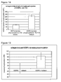
- the quantitative determination of the auto/transphosphorylation of DDR1 in a cell-based system by use of a CHO-Flp-in cell line which stably expresses the full-length DDR1 protein was performed using the CHO medium with different FCS concentrations for incubation during collagen-induced auto-/transphosphorylation of DDR1.
- the result for these different cellular buffers is shown in Figure 1 . It could be shown, that the method of the invention can be performed using CHO medium with different FCS concentrations as cellular buffer.
- the effect of potential modulators of collagen-induced DDR1 and DDR2 activity can be determined using the assay. This can be performed in a medium- to high-throughput assay format.
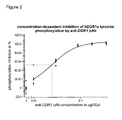
- a commercially available antibody anti-hDDR1 R&D Systems, Cat No. AF2396; Polyclonal Goat IgG; immunogen: Mouse myeloma cell line NS0derived recombinant human DDR1, Asp21-Thr416), which is directed against the extracellular domain of DDR1, was shown to inhibit the collagen-induced phosphorylation of DDR1 under various cellular buffer conditions, dependent on the concentration of the modulator.
- concentrations of the antibody were added. It could be determined that the IC 50 value in CHO medium is about 0,5 ng/ ⁇ l. The result is shown in Figure 2 .
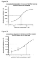
- Flp-In-CHO cells stably overexpressing mouse full-length DDR1 a were used.
- the cells were stimulated with rat collagen for 2 hours.
- a specific anti-DDR1 pAB antibody was used.
- the result is shown in Figure 3B .
- the IC 50 was calculated to be about 2,4 ng/ ⁇ l.
- Flp-In-CHO cells stably overexpressing human full-length DDR1a according to SEQ ID No. 1 were used.
- the cells were stimulated with rat collagen for 2 hours.
- a specific commercially available anti-DDR1 antibody as described in Example 1 was used.
- the result is shown in Figure 2 .
- the IC 50 for the antibody was calculated to be about 0,025 ⁇ g/well, corresponding to about 0,5ng/ ⁇ l.
- human collagen type I (neonatal fibroblasts) was used for stimulating human DDR1 phosphorylation.
- the experiment was performed in normal medium. No induction of phosphorylation was observed at 2 or 6 hours.
- Optimal collagen incubation time for phosphorylation was determined to be about 18 hours.
- rat collagen type I (rat tail) was used for stimulating human DDR1 phosphorylation.
- Optimal collagen incubation time for phosphorylation was determined to be about 2 hours.
- the IC 50 for the specific anti-DDR1 antibody pAB was determined.
- the IC 50 was about 1,8 ng/ ⁇ l
- the IC 50 was about 1,7 ng/ ⁇ l.
- Figures 4A and 4B show that human collagen I-induced DDR1 phosphorylation is inhibited as well in a dose-dependent manner by a specific anti-DDR1 antibody as compared to induction by rat collagen type I (rat tail).
Abstract
The present invention relates to test systems and methods for identifying a compound altering cellular Discoidin domain receptor tyrosine kinase activity.
Description
- The present invention relates to test systems and methods for identifying a compound altering cellular DDR (Discoidin domain receptor) tyrosine kinase activity.
- Discoidin domain receptors (DDRs) are non-integrin type receptors for collagen. The two mammalian gene products,
discoidin domain receptors 1 and 2 (DDR1 and DDR2), constitute a sub-family of tyrosine kinase receptors. They are selectively expressed in a number of different cell types in organs. When the DDRs are activated by collagen, the DDRs regulate cell adhesion, proliferation and extracellular matrix remodeling. DDR1 can form ligand-independent homodimers at a cell surface already in the absence of collagen. Collagen binding to DDR1 dimers induces their internalization and subsequent activation through tyrosine auto-/transphosphorylation. Collagen activation of DDR1 is following a delayed time course, and maximum stimulation is usually observed after 90 minutes to 16 hours dependent on cell type and on the collagen applied. Receptor internalization finally results in phosphorylation and subsequent sequence transduction involving signaling cascade molecules like ShcA, Shp-2, Nck2, the transcription factors NF-κb, Stat5, and the kinases p38, P13K, Pyk2, JNK and cJun. Finally, the signaling cascade affects differentiation, ECM remodeling, migration and cell cycle control. DDR molecules are believed to play a role in various indications and diseases like, for example, cancer, including tumor invasion and metastasis, atherosclerosis, including atherosclerotic plug formation, fibrosis, in particular fibrosis of kidney, liver, lungs and skin, diabetes, in particular diabetic nephropathy, and in inflammation and arthritis. There is, therefore, a need for test systems and assay methods for the identification of modulators of the DDR signaling pathways. In particular, there is a need for the identification of inhibitors of the DDR pathways. In order to meet the requirements of clinical early research, there is also a need for reliable mid-to-high throughput assay systems which allow the identification of modulators which affect either the kinase functions of a DDR or/and activation of a DDR through collagen. -
KR2009042397 KR2009042398 WO2007/044894 discloses cell-free translation/transcription systems.WO2003/082328 discloses an assay for DDR wherein the maturation of dendritic cells is used as readout.WO 98/45711 WO98/34954 WO95/14089 WO95/02187 WO95/02187 Discoidin domain receptor 1 is activated independently of β1-integrin. Bantscheff M. et al. ((2007), Nat Biotechnol. 25(9):1035-44) inter alia discloses that receptor tyrosine kinase DDR1 is target of the tyrosine kinase inhibitor imatinib. - Discoidin Domain Receptor (DDR) according to the present invention is a polypeptide of the subfamily of receptor tyrosine kinases which possesses an extracellular domain related to the lectin discoidin, found in the slime mold Dictyostelium discoideum. All members of the subfamily share the approximately 160-amino acid-long amino terminal discoidin homology domain followed by a single transmembrane region, and extended juxtamembrane region, and a catalytic tyrosine kinase domain.
- Two receptor tyrosine kinase genes with discoidin homology have been cloned in humans and are known as discoidin domain receptor DDR1 and DDR2.
- DDR1 appears in at least five isoforms:
- DDR1a (876AA): NM_001954
(http://www.ncbi.nlm.nih.gov/entrez/viewer.fcgi?val=NM_001954; cDNA: SEQ ID No. 1; protein: SEQ ID No. 2), - DDR1b (913AA): NM_013993
(http://www.ncbi.nlm.nih.gov/entrez/viewer.fcgi?val=NM_013993; cDNA: SEQ ID No. 3; protein: SEQ ID No. 4), - DDR1c (919AA): NM_013994
(http://www.ncbi.nlm.nih.gov/entrez/viewer.fcgi?val=NM_013994; cDNA: SEQ ID No. 5; protein: SEQ ID No. 6), and - DDR1 d (Accession No. AF353182), and DDR1 e (Accession No. AF353183), which are generated by alternative splicing.
- DDR1 a and DDR1 b receptor isoforms lack 37 or 6 amino acids in the juxtamembrane or kinase domain respectively.
- DDR1 d and DDR1 e are truncated variants lacking either the entire kinase region or parts of the juxtamembrane region and the ATP binding site (Alves et al., (2001), FASEB J., 15(7): 1321).
- For DDR2, two splice isoform exist, namely NM_006182.2
(http://www.ncbi.nlm.nih.gov/nuccore/NM-006182.2) and NM_001014796.1
(http://www.ncbi.nlm.nih.gov/nuccore/NM-001014796.1; SEQ ID No. 7). The differences in the transcripts relate to the 5'UTR. The isoforms encode the same protein (SEQ ID No. 8). - DDR protein sequences are well conserved, including those from humans, worms and flies (Vogel, (2006), Cellular Signalling, 18: 1108-1116).
- A murine DDR1 polypeptide is for example disclosed as
DDR1 transcript variant 2 under NCBI Accession number NM_172962 (http://www.ncbi.nlm.nih.gov/nuccore/NM_172962; polypeptide: SEQ ID No. 10; cDNA: SEQ ID No. 9). - The present invention relates to a test system for determining Discoidin domain receptor DDR collagen-induced auto-/transphosphorylation activity comprising:
- a) at least one cell containing at least one DDR polypeptide or functional derivative thereof localized in the plasma membrane of the cell,
- b) either b1) or b2):
- b1) a binding partner capable of selectively recognizing phospho-tyrosine, which binding partner is a labeled binding partner,
- b2) a binding partner capable of selectively recognizing phospho-tyrosine and another binding partner, which is a labeled binding partner, and which is capable of selectively recognizing the binding partner recognizing phospho-tyrosine,
- c) at least one reagent for fixing the cells on a solid support and permeabilizing the cells
- d) at least one solid support for fixing the cells and for culturing the cells,
- e) at least one collagen,
- f) optionally at least one compound suspected of altering the collagen-induced auto-/transphosphorylation of the at least one DDR polypeptide or functional derivative thereof, and
- g) means for detecting the binding partner of b) or c).
- In another embodiment, the present invention relates to a method for identifying a compound altering DDR collagen-induced auto-/transphosphorylation activity, particular DDR1 a, DDR1 b, DDR1 c and/or DDR2 collagen-induced auto-/transphosphorylation activity, in particular collagen-induced auto-/transphosphorylation activity of one or more polypeptides having a sequence selected from SEQ ID No. 2, SEQ ID No. 4, SEQ ID No. 6, SEQ ID No. 8, or SEQ ID No. 10, comprising the steps of:
- a) culturing at least one cell on a solid support,
which cell contains at least one DDR polypeptide or functional derivative thereof localized in the plasma membrane of the cell, - b) adding to the cell culture of step a) at least one collagen, in particular wherein the collagen is selected from collagen type I, collagen type II, collagen type III or collagen type IV, or mixtures thereof,
- c) incubating the cells of step b) in a cellular buffer,
- d) fixing the cells of step c) on the solid support and permeabilizing the fixed cells, and optionally washing the cells,
- e) adding at least one compound suspected of altering the collagen-induced auto-/transphosphorylation of the at least one DDR polypeptide or functional derivative thereof before step d),
- f) carrying out step f1) or f2):
- f1) adding a binding partner to the cells of step d),
which binding partner is capable of selectively recognizing phospho-tyrosine, and
which binding partner is a labeled binding partner, - f2)
- i) adding a binding partner to the cells of step d),
which binding partner is capable of selectively recognizing phospho-tyrosine, - ii) adding another binding partner to cells of step i),
which another binding partner is capable of selectively recognizing the binding partner capable of selectively recognizing phospho-tyrosine, - i) adding a binding partner to the cells of step d),
- f1) adding a binding partner to the cells of step d),
- g) detecting the labeled binding partner.
- Preferably, washing steps may be included where appropriate. A washing step may for example be included after fixing the permeabilizing the cells and before adding an anti-phospho-tyrosine antibody. In a preferred embodiment, washing of the cells is carried out after step a), step c), step d), step f1), step f2i) and/or step f2ii) of the method of the invention. The washing can be can be carried out using an appropriate buffer known to a skilled person, e.g. buffered saline solutions.
- It is understood, that all embodiments below apply both to the test system and the method of the invention.
- In a preferred embodiment, the DDR polypeptide is of mammalian origin. In a preferred embodiment, the DDR polypeptide is selected from DDR1a, DDR1b, DDR1c and DDR2. Such isoforms are for example known in humans and mice, as e.g. used in the examples.
- In a more preferred embodiment, the DDR polypeptide is a human polypeptide and/or is selected from the polypeptides according to SEQ ID No. 2, SEQ ID No. 4, SEQ ID No. 6, SEQ ID No. 8, or SEQ ID No. 10, or combinations thereof. In further preferred embodiment, the DDR polypeptides lack the signal peptide. Such signal peptide may be present or may be deleted without significantly altering the DDR activity.
- In another preferred embodiment, the DDR polypeptide is a murine polypeptide and/or is selected from the polypeptide according to SEQ ID No.10.
According to the present invention, human DDR proteins, including all isoforms and orthologs may be used. Preferred is the use of human DDR1a, human DDR1b, human DDR1c, and human DDR2. - In a particularly preferred embodiment, human DDR1a polypeptide according to SEQ ID No. 2 or encoded by SEQ ID No. 1, human DDR1b polypeptide according to SEQ ID No. 4 or encoded by SEQ ID No. 3, human DDR1c polypeptide according to SEQ ID No. 6 or encoded by SEQ ID No. 5 and/or human DDR2 polypeptide according to SEQ ID No. 8 or encoded by SEQ ID No. 7 are used. In further preferred embodiment, the DDR polypeptides lack the signal peptide. In yet another embodiment, combinations thereof may be used.
- In another particularly preferred embodiment, a murine DDR1 polypeptide according to SEQ ID No. 10, or encoded by SEQ ID No. 9 is used.
- Also, DDR proteins from other organisms, in particular mammalian organisms may be used. For example DDR1 a, DDR1 b, DDR1c from mouse, rat, or macaca, in particular mouse may be used. Mouse DDR1 isoforms are known and are e.g. described in NCBI accession Nos. NM_007584.2, NM_172962.1, NM_001198831.1, NM_001198833.1, and a mouse DDR2 polypeptide is disclosed by NCBI accession No. NM_022563.2.
- As shown in the examples, the method of the invention can be carried out both with the various human DDR isoforms and with a murine DDR isoform. Thus, the method is very flexible. The test systems of the invention may contain these isoforms.
- In another preferred embodiment, the DDR polypeptide is rat, mouse, fly, worm, slime mold or fish DDR polypeptide.
- In addition, functional derivatives of the native DDR proteins may be used. The functional derivatives of a DDR polypeptide are understood as having at least 80% sequence identity, more preferably at least 90% sequence identity, even more preferred at least 95% sequence identity, most preferred at least 98% sequence identity to a wildtype DDR polypeptide, in particular to a DDR polypeptide having a sequence according to SEQ ID No. 2, SEQ ID No. 4, SEQ ID No. 6, or SEQ ID No. 8, and exhibiting substantially the same collagen-induced auto-/transphosphorylation activity as a wildtype DDR polypeptide. In a preferred embodiment, the substantially same collagen-induced auto-/transphosphorylation activity is understood as at least 10%, 20%, 30%, 50%, 70% or 90% of the collagen-induced auto-/transphosphorylation of a wildtype DDR polypeptide. The activity can be determined as explained in Example 1. In particular, the signal peptide may be missing. In a preferred embodiment, the functional derivative is a non-human homologue of DDR1 a, DDR1 b, DDR1 c or DDR2 from mouse, rat, macaca, cow, pig, horse, fish or dog.
- According to the invention, at least one cell containing at least one DDR polypeptide or functional derivative thereof localized in the plasma membrane of the cell isused. Such localization can for example be determined by an immunofluorescence method using an antibody specifically recognizing the DDR polypeptide or functional derivative thereof.
- Expression of a DDR polypeptide or functional derivative thereof can for example be determined by Western Blot analysis using an antibody specifically recognizing the DDR polypeptide or functional derivative thereof.
- In a preferred embodiment, the cell is of mammalian origin. In a further preferred embodiment, the cell is a mammalian cell. In another embodiment, the cell allows stable expression of a DDR polypeptide or functional derivative thereof. In one embodiment, the at least one DDR polypeptide or functional derivative thereof are expressed as transgenes. In a preferred embodiment, the cells are produced recombinantly. Preferably, the cells are obtained by transfecting the cells with a suitable vector system.
- Transfection with DNA yields stable as well as unstable (transient) cells or cell lines. Transient cell lines reflect the survival of the transfected DNA in extrachromosomal form; stable cell lines are understood to result from the integration into the genome resulting in the stable expression of the transfected gene. Cells can be transfected using any appropriate means, including viral vectors, chemical transfectants, electroporation, calcium phosphate co-precipitation and direct diffusion of DNA.
- The cell may be isolated, maintained and cultured as known to the skilled person. Aside from temperature and gas mixture, the most commonly varied factor in cell culture systems is the growth medium. Recipes for growth media can vary in pH, glucose concentration, growth factor and the presence of other nutrient components among others. Growth factors used for supplement media are often derived from animal blood such as calf serum. Genetically modified cells may be obtained by inserting the full-length coding sequence of the component in question, as known to the skilled person. The skilled person in the art knows how to derive a nucleic acid sequence coding for the same and how to isolate or produce such a nucleic acid sequence using standard techniques of molecular biology. This can be accomplished, for example, by the use and combination of existing sequences using restriction enzymes. The nucleic acid may be combined with further elements, e.g., a promoter and a transcription start and stop signal and a translation start and stop signal in order to provide for expression of a DDR polypeptide. As used herein, vectors are agents that transport the transgene into the cell and may include appropriate transcriptional and translational control signals such as a promoter. Vectors can be plasmid, viral or others known in the art. The promoter can be inducible or constitutive, general or cell specific, nuclear or cytoplasmic specific promoter. Selection of promoters, vectors and other elements is a matter of routine design within the level of ordinary skill in the art. Many such elements are described in the literature and are available through commercial suppliers.
- In the examples, the vector pcDNA5-FRT-TO_DEST was used, and Flp-In™-CHO cells (Invitrogen) were transfected. Thus, in a preferred embodiment, the cells are mammalian cells transfected with a polynucleotide encoding at least one DDR polypeptide or functional derivative thereof, particularly the cells are transfected CHO cells, most preferably transfected Flp-In™-CHO cells.
- In a particularly preferred embodiment, a mammalian cell, more preferably CHO cell, most preferably a Flp-In™-CHO cell is used as parental cell. The Flp-In™-CHO cells contain a single stably integrated FRT site at a transcriptionally active locus. The Flp-In™-CHO cell was created by transfecting CHO cells with pFRT/lacZeo2 and selecting for Zeocin™-(Invitrogen) resistant clones. Co-transfection of Flp-In™ expression vector and the Flp recombinase vector pOG44 results in targeted integration of the expression vector to the same locus in every cell.
- In a preferred embodiment, the parental cells do not contain DDR polypeptides localized in the plasma membrane of the cell. As shown in Example 1, Flp-In™-CHO cell do not show significant DDR expression, as determined by Western Blot analysis.
- Optionally, the cell may be part of a tissue; however, the method of the invention is preferably an ex vivo method. In a preferred embodiment of the invention the cell is from a cell line (many of which are well characterized and provide for constant conditions and convenient handling), particularly a mammalian cell line, more particularly a human or mouse cell line. Examples of suitable cell lines are included but are not limited to HEK 293, 745-A, A-431, BxPC3, C5N, Caco-2, Capan-1, CC531, CFPAC, CHO, CHO K1, COS-1, COS-7, CV-1, EAHY, EAHY 926, F98, GH3, H-295 R, H-4-II-E, HACAT, HACAT A131, HEK, HEL, HeLa, Hep G2, High Five, Hs 766T, HT29, HUV-EC R24, HUV-EC-C, IEC 17, IEC 18, Jurkat, K 562, KARPAS-299, L 929, LIN 175, MAt-LYLU, MCF-7, MNEL, MRC-5, MT4, N64, NCTC 2544, NDCK II, Neuro 2A, NIH 3T3, NT2/D1, P19, SF9, SK-UT-1, ST, SW 480, SWU-2 OS, U-373, U-937, and Y-1. Other suitable cells are those known to the one of skill in the art.
- The procedure for introducing a transgene into a recipient cell is called transfection. In general a cell line is transfected by any of the means mentioned above, wherein the transgene is operatively linked to a selectable marker. Following transfection cells are grown e.g. for some days in enriched media and then switched to selective media. Transfected cells exhibit resistance to the selection and are able to grow, whereas non-transfected cells die in general. Examples for selectable markers include puromycin, zeocin, neomycin (neo) and hygromycin B, which confer resistance to puromycin, zeocin, aminoglycoside G-418 and hygromycin, respectively. However, other selection methods known to the skilled person may be also suitable.
- As described above, cells may be cultured in a suitable medium (e.g. commercially available medium including serum and antibiotics). Cells are usually cultured in a suitable atmosphere (5% CO2), relative humidity (e.g. 90 %) and temperature (e.g. at 37°C). For high-through-put screening and/or convenient handling cells may be cultivating in multiwell plates such as 96 well plates, 24 well plates etc. The Flp-In-CHO cells in the examples were seeded in complete medium and were cultured for 24 hrs at 37°C and 5% CO2.
- Media with or without FCS may be used for incubating the cells. In the examples, the medium is F-12K nutrient mixture, Pen/Strep, 10% FCS and Hygromycin (300µg/ml).
- In a preferred embodiment, the test system in addition comprises means for quantitating cells, in particular viable cells. This allows correlating the number of cells, with the signal mediated the label, in particular the fluorescence, originating from DDR polypeptide detection. Thus, in a preferred embodiment, in addition at least one reagent for staining the cells, in particular for staining cell nuclei is used. In particular, the Hoechst dye HOE 33342 may be used, as shown in the examples. This step can be performed after fixing and permeabilizing the cells. Preferably, this step is performed after adding the binding partner capable of selectively recognizing phospho-tyrosine to the cells. In particular, this step is carried out simultaneously with adding the binding partner capable of selectively recognizing the binding partner selectively recognizing phospho-tyrosine to the cells. In particular, the Hoechst dye HOE 33342 may be used for staining cell nuclei. Therefore, in one embodiment, at least one reagent for staining the cells is added, in particular at least one reagent for staining cell nuclei is added, wherein this step is carried out after step d) of the method of the invention, preferably simultaneously with step f1), f2)i) or f2ii) of the method of the invention.
- In another preferred embodiment, the binding partner capable of selectively recognizing phospho-tyrosine is an anti-tyrosine antibody or an antibody fragment. Such antibodies are known in the art.
- In another preferred embodiment, the test system comprises another binding partner which is labeled and which is capable of selectively recognizing the binding partner capable of selectively recognizing phospho-tyrosine. Thus, the variant of b2) of the test system and variant f2) of the method of the invention is preferred. Preferably, this another binding partner capable of selectively recognizing the binding partner capable of selectively recognizing phospho-tyrosine is an antibody or antibody fragment. Thus, in this embodiment, this another binding partner is a labeled secondary antibody recognizing the primary anti-phospho-tyrosine antibody.
- Therefore, in an especially preferred embodiment, a primary antibody or antibody fragment selectively recognizing phospho-tyrosine, and a secondary labeled antibody or antibody fragment selectively recognizing the primary antibody are used. In the examples, anti-phospho-tyrosine antibody produced in rabbit (Sigma, Cat. No. T-1575) was used as primary antibody. This antibody is a preferred antibody according to the invention. In another preferred embodiment, Alexa Fluor F(ab)2 fragment goat anti-rabbit IgG (Alexa) may be used as labeled secondary antibody. This antibody was shown to be useful according to the invention in the Examples.
- In a preferred embodiment, a blocking buffer is added and incubated prior to adding the binding partner selectively recognizing phospho-tyrosine, in particular the anti-phospho-tyrosine antibody or antibody fragment. The blocking buffer may be added after fixing the cells and permeabilizing the cells. Blocking buffers are well known in the art. For example, the blocking buffer described in example 1 may be used.
- Herein, the term antibody or antibody fragment is understood as meaning antibodies, antibody fragments, or antigen-binding parts thereof, in particular complementarity-determining regions (CDRs) of antibodies, which have been prepared recombinantly and, where appropriate, modified, such as chimaeric antibodies, humanized antibodies, multifunctional antibodies, bispecific or oligospecific antibodies, single- chain antibodies and Fab, Fab' or F(ab)2 fragments (see, for example,
EP-B1-0 368 684 ,US 4,816,567 ,US 4,816,397 ,WO 88/01649 WO 93/06213 WO 98/24884 - As an alternative to the classical antibodies it is also possible, for example, to use protein scaffolds as binding partners, e.g. anticalins which are based on lipocalin (Beste et al. (1999) Proc. Natl. Acad. Sci. USA, 96, 1898-1903). The natural ligand-binding sites of the lipocalins, for example the retinol-binding protein or the bilin-binding protein, can be altered, for example by means of a "combinatorial protein design" approach, in such a way that they bind to selected haptens (Skerra, 2000, Biochim. Biophys. Acta, 1482, 337-50). Other known protein scaffolds are known as being alternatives to antibodies for molecular recognition (Skerra (2000) J. Mol. Recognit., 13, 167-187).
- According to the present invention, at least one labeled binding partner is used. The label may be any covalently or non-covalently bound chemical entity, which can be used for detection. Common labeling methods may be used for labeling of one or more functional groups on the surface of the binding partners. These could be for example the primary amino groups, present at the N-terminal of each polypeptide chain and the side chain of lysine residues; sulphhydryl groups, present on cysteine residues made available by treating disulphide bonds with reducing agent or by modifying lysine residues with a reagent such as SATA. The binding partner may be labeled with a series of different agents, e.g. such as biotin (for avidin-biotin chemistry), enzymes, activated fluorescent dyes for labeling amines, sulphhydryls or other functional groups with e.g. FITC, fluorescein, rhodamine, Cy dyes or Alexa fluors. Radioactive label such as 3H, 32P, 35S, 125I or 14C as well as common enzyme labels including penicillinase, horseradish peroxidase and alkaline phosphatase may be used as well.
- In a preferred embodiment, a fluorescently labeled binding partner, in particular a fluorescently labeled antibody or antibody fragment is used. Preferably, the binding partner, which is a labeled binding partner, and which is capable of selectively recognizing the binding partner recognizing phospho-tyrosine, is a fluorescently labeled binding partner. Thus, in an especially preferred embodiment, a fluorescently labeled antibody or antibody fragment specifically recognizing an anti-phospho-tyrosine antibody or antibody fragment is used. In this embodiment, the anti-phospho-tyrosine antibody does not need to be labeled, preferably, the anti-phospho-tyrosine antibody is not labeled or labeled with a label different from the label of the antibody or antibody fragment specifically recognizing an anti-phospho-tyrosine antibody or antibody fragment.
- Fluorescence is an optical phenomenon, wherein the molecular absorption of a photon triggers the emission of another photon with a longer wavelength. The energy difference between the absorbed and emitted photons ends up as molecular vibrations or heat. Usually, the absorbed photon is in the ultraviolet range, and the emitted light is in the visible range, but this depends on the absorbance curve and Stokes shift of the particular fluorophore. The number of fluorescence applications is growing in the biomedical biological and related sciences. Methods of analysis in these fields are also growing, albeit with increasingly unfortunate nomenclature in the form of acronyms such as: FLIM, FLI, FLP, CALI, FLIE, FRET, FRAP, FCS, PFRAP, smFRET, FIONA, FRIPS, SHREK, SHRIMP, TIRF. Most of these techniques rely on fluorescence microscopes. These microscopes use high intensity light sources, usually mercury or xenon lamps, LEDs, or lasers, to excite fluorescence in the samples under observation. Optical filters then separate excitation light from emitted fluorescence, to be detected by eye, or with a (CCD) camera or other light detectors (photomultiplier tubes, spectrographs, etc).
- Preferably, a fluorescent dye is used as label in order to detect the effect of a signal in question (here the collagen-induced auto-/transphosphorylation of a DDR polypeptide). The fluorescent dye comprises a fluorophore. A fluorophore, in analogy to a chromophore, is a component of a molecule which causes a molecule to be fluorescent. It is a functional group in a molecule which will absorb energy of a specific wavelength and re-emit energy at a different (but equally specific) wavelength. The amount and wavelength of the emitted energy depend on both the fluorophore and the chemical environment of the fluorophore. Fluorescein isothiocyanate, a reactive derivative of fluorescein, has been one of the most common fluorophores chemically attached to other, non-fluorescent molecules to create new and fluorescent molecules for a variety of applications. Other historically common fluorophores are derivatives of rhodamine, coumarin and cyanine. Examples fluorescent dyes include without limitation 7-amino-actinomycin D, acridine orange, acridine yellow, Alexa Fluor, AnaSpec, auramine O, auramine-rhodamine stain, benzanthrone, 9,10-bis(phenylethynyl)anthracene, 5,12-bis(phenylethynyl)naphthacene, CFDA-SE, CFSE, calcein, carboxyfluorescein, 1-chloro-9,10-bis(phenylethynyl)anthracene, 2-chloro-9,10-bis(phenylethynyl)anthracene, coumarin, cyanine, DAPI, Dioc6, DyLight Fluor, ethidium bromide, fluorescein, Fura-2, Fura-2-acetoxymethyl ester, green fluorescent protein, Hilyte Fluor, Hoechst stain, Indian yellow, Indo-1, luciferin, perylene, phycobilin, phycoerythrin, phycoerythrobilin, propidium iodide, pyranine, rhodamine, RiboGreen, rubrene, ruthenium(II) tris(bathophenanthroline disulfonate), SYBR Green, stilbene, sulforhodamine 101, TSQ, Texas Red, umbelliferone and yellow fluorescent protein. Preferred is the use of Alexa Fluor, as described in the examples.
- In the context of the present invention, the fluorescent dye is covalently or non-covalently bound to the binding partner(s) of the present invention, preferably, it is covalently bound.
- For the method of the invention any suitable detection may be used. The detection is system is adapted to detect the label. In an embodiment the assay is a DELFIA (dissociation enhanced lanthanide fluoro immuno assay), a FRET (fluorescence resonance energy transfer) assay, or TR-FRET (time-resolved fluorescence resonance energy transfer) assay.
- DELFIA (dissociation enhanced lanthanide fluoro immuno assay)-based assays are solid phase assay. The antibody is usually labeled with Europium or another lanthanide and the Europium fluorescence is detected after having washed away un-bound Europium-labeled antibodies.
- In a preferred embodiment, the detection means are suitable for detection of fluorescence. Such detection means are used to detect the fluorescent label.
- In a preferred embodiment, the means for detection are automated or semi-automated. In particular, the means for detection is a confocal microscope. In particular, the ImageXpress Ultra Confocal High Content Screening System (Molecular Devices) may be used, as described in the Examples. Also, fluorescence microplate cytometry may be used in another embodiment, e.g. Acumen Explorer™.
- The solid support for fixing cells at least one solid support for fixing the cells and for culturing the cells is in a preferred embodiment a multiwell plate, in particular comprising 12 wells or more, 24 wells or more, 48 wells or more, 96 wells or more, 384 wells or more.
- The solid support is suitable for both culturing the cells thereon, and for fixing the cells thereon, using suitable reagent(s).
- The fixing of cells may be performed with suitable reagents known in the art. In particular, paraformaldehyde may be used, as also described in the Examples. Also, an orthovanadate salt, in particular sodium orthovanadate may in addition be added to the fixing reagent in order to block phosphatase acitvity, as also described in the Examples.
- For cell permeabilization, suitable reagents known in the art may be used. In particular, Triton-X-100 may be used, as also described in the Examples.
- In a preferred embodiment, the test system comprises:
- a) at least one mammalian cell, preferably at least one CHO cell, more preferably at least one Flp-In-CHO cell expressing at least one mammalian DDR polypeptide selected from mammalian DDR1a, DDR1b, DDR1c and DDR2 localized in the plasma membrane of the cell,
- b) an anti-phospho-tyrosine antibody or antibody fragment
- c) a fluorescently labeled antibody or antibody fragment selectively binding to the anti-phospho-tyrosine antibody or antibody fragment,
- d) a well plate comprising at least 12 wells,
- e) at least one mammalian collagen,
- f) optionally at least one compound suspected of altering the collagen-induced auto-/transphosphorylation of the at least one DDR polypeptide or functional derivative thereof, and
- g) detection means for quantitative detection of fluorescence in the wells of d).
- In another preferred embodiment, the method of the invention is carried out using these components of the test system.
- The test systems and methods of the invention make use of at least one collagen. The collagen is employed to mediate the collagen-induced auto-/transphosphorylation of the DDR polypeptides. Collagen is a group of naturally occurring proteins. In nature, it is found exclusively in animals, especially in the flesh and connective tissues of mammals. A distinctive feature of collagen is the regular arrangement of amino acids in each of the three chains of these collagen subunits. The sequence often follows the pattern Gly-Pro-X or Gly-X-Hyp, where X may be any of various other amino acid residues.
- In a preferred embodiment, the collagen is collagen of mammalian origin, in particular collagen from human, rat or mouse. Most preferably, the collagen is human collagen. 29 types of collagen were identified, collagen I to XXIX, with collagen I, II, III, IV and V being the most common ones.
- In a preferred embodiment, the collagen is selected from collagen type I, collagen type II, collagen type III or collagen type IV, or mixtures thereof. These collagens are especially preferred in case of DDR1 isoforms and orthologs. In a preferred embodiment, the collagen is selected from human collagen type I, human collagen type II, human collagen type III and human collagen type IV or mixtures thereof. In another preferred embodiment, rat collagen type I, rat collagen type II, rat collagen type III or rat collagen type IV, or mixtures thereof may be used. In another preferred embodiment, fibrillar collagens are used. Such collagens are fibril-forming. Such collagens are especially used for DDR2 isoforms and orthologs.
- In a preferred embodiment, the at least one collagen to be added according to the method of the invention, and as present in the test systems of the invention, is dissolved in a suitable solvent, preferably in an acidic solvent. Suitable solvents are known in the art, in particular acetic acid and/or HCl, e.g. 0,1 N HCL, may be used as acidic solvents. In particular, acetic acid, more preferably 0,1% acetic acid or 0,1N acetic acid, as described in Example 1, may be used. In a particularly preferred embodiment, the at least one collagen is dissolved in the solvent prior to adding according to the method of the invention.
- The duration of incubation with collagen may vary and may be optimized for each incubation medium, DDR polypeptide and collagen. Typically, the collagen incubation takes place for about 5 minutes to about 4 days. In a preferred embodiment, collagen incubation is performed for about 10 minutes to about 60 hours, more preferably for about 1 to about 36 hours, even more preferred for about 1 to about 25 hours, most preferred for about 2 to about 18 hours. Optimized collagen incubation times of about 2 hours and about 18 hours have been determined in the examples for specific experimental setups of the invention.
- "About" according to the present invention is understood as meaning the experimental error range, in particular ± 5% or ± 10%.
- The incubation of the cells may be carried out in various cellular buffers. A suitable cellular buffer allows the cells to be viable. Therefore, complete medium or medium without FCS may be used according to the invention. In a preferred embodiment, the cellular buffer is medium, which optionally comprises FCS. FCS may be present in various concentrations. In particular, the medium may contain about 0,1% to 20% FCS, in particular about 1% to about 10% FCS, as exemplified in Example 1. FCS is understood to mean fetal calf serum.
- In a preferred embodiment, the method is carried out as a mid-throughput assay or a high-throughput assay. Preferably, the method is carried out as a high-throughput assay. In this method, a large number of compounds are screened in whole-cell assays. Typically, these screenings are carried out in at least 96 well plates using automated, robotic station based technologies or in higher-density array ("chip") formats.
- The method of the invention may be used for screening for a diagnostic and/or medicament for diagnosing, preventing and/or treating a disease involving DDR polypeptide(s), respectively, particularly for preventing and/or treating any of the diseases described above. If used for screening purposes the compound tested may be a compound, the function of which is still unknown or not yet related to the DDR polypeptide(s). Also, the method may be used for the identification of new compounds altering collagen-induced auto-/transphosphorylation. Alternatively, compounds known to alter collagen-induced auto-/transphosphorylation may be tested for their efficiency using the method of the invention, in particular for quantification.
- In a preferred embodiment, a quantitative detection is performed. In particular, the detection means may be equipped with software to transform experimental parameters in quantitative values.
- In a particularly preferred embodiment, the concentration-dependent alteration of collagen-induced auto-/transphosphorylation of the at least one DDR polypeptide or functional derivative thereof is determined. Therefore, the alteration at least two different concentrations of the compound is preferably determined.
- The alteration may be an inhibition or activation, preferably an inhibition. In particular, % phosphorylation of the DDR polypeptide(s) is determined. In particular, IC50 values in case of inhibitory compounds are determined.
- As described above, the compound suspected of altering the collagen-induced auto-/transphosphorylation of a DDR polypeptide or functional derivative thereof may be a substance of any chemical nature. It may already be known as a drug or medicament or diagnostic for a disease. Alternatively, it may be a known chemical compound not yet known to have a therapeutic or diagnostic effect in another embodiment and the compound may be a novel or so far unknown chemical compound. The agent may be also a mixture of substances or compounds.
- In one embodiment, the compound is provided in form of a chemical compound library. Chemical compound libraries include a plurality of chemical compounds and have been assembled from any of multiple sources, including chemical synthesized molecules or natural products, or have been generated by combinatorial chemistry techniques. They are especially suitable for high-throughput screening and may be comprised of chemical compounds of a particular structure or compounds of a particular organism such as a plant. In the context of the present invention, the chemical compound library is preferably a library comprising proteins and polypeptides or small organic molecules. Preferably a small organic molecule is less than 500 daltons in size, particularly a soluble, non-oligomeric, organic compound.
- In another preferred embodiment, the compound is a biological molecule, like an antibody, antibody fragment, antibody mimetic, peptide, or RNA molecule or libraries thereof.
- The compound altering the collagen-induced auto-/transphosphorylation of a DDR polypeptide or functional derivative thereof may be an inhibitor or an activator. Preferably, it is an inhibitor.
- In a preferred embodiment, the inhibitor inhibits collagen-induced auto-/transphosphorylation of a DDR polypeptide or functional derivative by at least 5 %, more preferably 10 %, 30 %, 50 %, 70 % or 90 %, of the control. The collagen-induced auto-/transphosphorylation can be determined as shown in Example 1.
- In another preferred embodiment, the inhibitor has an IC50 value of less than about 1µg/µl, preferably less than about 100ng/µl, more preferably less than about 10ng/µl, most preferably less than 2 ng/µl. The IC50 value can be determined as shown in Example 1.
- In another preferred embodiment, the inhibitor has an IC50 value of less than about 10 µM, preferably less than about 1 µM, more preferably less than about 0,1 µM, even more preferably less than about 10 nM most preferably less than about 7 nM.
- The compound may be added at any time point before fixing the cells of on the solid support and permeabilizing the fixed cells, and optionally washing the cells. In a preferred embodiment, the compound is added before, simultaneously or after the addition of collagen. The experiments below show, that the test systems and methods allow the determination of IC50 values of the collagen-induced auto-/transphosphorylation of DDR1 in a cellular system by use of a suitable medium without FCS or with FCS, in various concentrations. Also, any DDR1 or DDR2 orthologs or isoforms can be used including isoforms and orthologs from mammalian or non-mammalian organisms. In addition, various collagens may be used insofar as these activate the auto/transphosphorylation of DDR. Also, various incubation times for the administered collagens can be used and, also, the incubation conditions like, for example, pre-incubation of the inhibitory or activating reagent or simultaneous administration of the modulating compound with the collagen can be performed. Also, the test systems and methods of the present invention can be used for quantitative determination of chemical reagents or biological molecules like, for example, peptides or antibodies which modulate the collagen-induced auto/transphosphorylation of a DDR polypeptide. Moreover, the test systems and methods allow performing the methods in various assay formats like, for example, a 12-well, 24-well, 48-well, 96-well, or 384-well plate format. The assay can be performed in a middle- or a high-throughput assay format.
- SEQ ID No. 1 shows the cDNA encoding human DDR1a (NM_001954; http://www.ncbi.nlm.nih.gov/entrez/viewer.fcgi?val=NM_001954)
- SEQ ID No. 2 shows the human DDR1a polypeptide sequence (NM_001954; http://www.ncbi.nlm.nih.gov/entrez/viewer.fcgi?val=NM_001954)
- SEQ ID No. 3 shows the cDNA encoding human DDR1b (NM_013993; http://www.ncbi.nlm.nih.gov/entrez/viewer.fcgi?val=NM_013993) DDR1 b (913AA): NM_013993
- SEQ ID No. 4 shows the human DDR1 b polypeptide sequence (NM_013993; http://www.ncbi.nlm.nih.gov/entrez/viewer.fcgi?val=NM_013993)
- SEQ ID No 5 shows the cDNA encoding human DDR1c (NM_013994; http://www.ncbi.nim.nih.gov/entrez/viewer.fcgi?val=NM_013994)
- SEQ ID No. 6 shows the human DDR1c polypeptide sequence (NM 013994; http://www.ncbi.nlm.nih.gov/entrez/viewer.fcgi?val=NM_013994)
- SEQ ID No. 7 shows a cDNA encoding human DDR2 (NM_001014796.1 (http://www.ncbi.nlm.nih.gov/nuccore/NM_001014796.1)
- SEQ ID No. 8 shows the human DDR2 polypeptide sequence (NM_001014796.1 (http://www.ncbi.nlm.nih.gov/nuccore/NM_001014796.1)
- SEQ ID No. 9 shows a cDNA encoding a murine DDR1 polypeptide (
transcript variant 2; NM_172962; http://www.ncbi.nlm.nih.gov/nuccore/NM_172962) - SEQ ID No. 10 shows a murine DDR1 polypeptide sequence (NM_172962; http://www.ncbi.nlm.nih.gov/nuccore/NM_172962)
-
-
Figure 1A shows collagen-induced auto-/transphosphorylation of hDDR1a as determined in + and- FCS CHO medium. -
Figure 1 B shows collagen-induced-auto-/trans-phosphorylation of hDDR1c as determined in -FCS CHO medium. -
Figure 2 shows the concentration-dependent inhibition of auto-/transphosphorylation of human DDR1a in CHO medium. Rat collagen I-induced hDDR1a phosphorylation is inhibited in a dose-dependent manner by a specific commercially available anti-DDR1 antibody. IC50 was determined as about 0,5 ng/µl. -
Figure 3A shows that clinical inhibitors of abl kinase or the kinases of the src family significantly inhibit the collagen-induced auto-/transphosphorylation of human DDR1. For Dasatinib as inhibitor, IC50 was determined as about 10 nM. -
Figure 3B shows that rat collagen I-induced mDDR1a phosphorylation is inhibited in a dose-dependent manner by a specific anti-DDR1 pAB antibody. IC50 was determined as about 2,4 ng/µl. -
Figures 4A ,4B and 4C show that human collagen I induces hDDR1a tyrosine phosphorylation and that human collagen I-induced hDDR1a phosphorylation is inhibited as well in a dose-dependent manner by a specific anti-DDR1 antibody as compared to induction by rat collagen type I (rat tail) . -
Figures 5A and 5B show that imatinib (Figure 7A) and nilotinib (Figure 7B) inhibit rat collagen-induced hDDR1 phosphorylation. IC50 of imatinib is about 0.89 µM (10% FCS); IC50 of nilotinib is about 0,096 µM (10% FCS). -
Figure 6 shows a typical experimental protocol followed in the Examples. - For the setup of the assay system, Flp-In CHO cells were stably transfected with the human full-length gene of the isoform DDR1a according to SEQ ID No. 1. These cell lines express the corresponding DDR1 a isoform in the correct cellular localization in the plasma membrane. The expression was confirmed by Western Blot analysis and the correct physiological localization was detected by immunofluorescence experiments. In the parental cell line, no DDR1 expression could be detected using the above methods. The cells were seeded in a black 96-well or 384-well microtiter plate with clear bottom in complete medium which contains 1-10% FCS. After incubation of the cells over night (16-24 hours) in a CO2 (5%) incubator at 37 °C, the cells were treated with either collagen type I, which is commercially available for 2 hours. If necessary, the compound, which is suspected to alter the activity of DDR1, can be administered in various doses for the detection of the corresponding IC50 value before or at the same time as administering collagen. After that, the cells were fixed and permeabilized. Then, they were incubated in blocking buffer, and were subsequently incubated over night with an anti-phospho-tyrosine antibody. This antibody binds to all phosphorylated tyrosines present in the cells. On the following day, a corresponding secondary antibody, which is labeled with a fluorescent dye, was administered to the cells and incubated for one hour. At the same time, the cell nuclei were treated with a dye which is specific for cell nuclei.
- The intensity of the so-labeled cells was detected with a corresponding measuring system like, for example, ImageXpress of Molecular Devices or the system Acumen. By analysis of the measured data with a specified software protocol, the percentage of DDR1 phosphorylation was determined. A schematic overview of the experiments is shown in
Figure 6 . - Using this approach, IC50 values relating to the inhibition of phosphorylation of DDR1 polypeptides of compounds like antibodies or small chemical molecules was determined.
- Following reagents were used in the experiments:
- Vector: pcDNA5-FRT-TO_DEST (Invitrogen Life Technologies)
- Cells: CHO Flp-In wt for generating stably expressing CHO-hDDR1a, CHO-mDDR1a, CHO-hDDR1b, and CHO-hDDR1c.
- Media: F-12K Nutrient mixture (Kaighn's Modification; Gibco, Cat. No.:21127), Pen/Strep (PAN), 10% FCS (PAN), Hygromycin 300µg/ml (Roche Diagnostics Deutschland GmbH) Buffer: PBS
- Reagents: Collagen I (rat tail; Sigma); Collagen human VitroCol (AdvancedBiomatrix);
Acetic acid Tween 20; Odyssey Blocking Buffer (LI-COR, Cat. No.: P2151); Anti-Phospho-Tyrosine rabbit (Sigma, Cat. No.: T-1575); Anti-Phospho-Tyrosine mouse 4G10 (Upstate, Cat. No.: 05-321); Alexa Fluor 488 F(ab)2 fragment goat anti-rabbit IgG (Invitrogen, Cat. No.: A11070); Alexa Fluor 488 F/ab)2 fragment goat anti-mouselgG (Invitrogen Cat. No.: A11017); HOE 33342 - (Invitrogen, Cat. No.: H3570)
- Cell culture plates: BD Falcon, 96 well
- Reference compounds: Anti-human DDR1 Antibody (R&D Systems, Cat No.: AF2396) Normal Goat IgG (R&D Systems, Cat No.: AB-108-C)
- Dasatinib (Sprycel, BMS-354825)
- The corresponding experiments were performed with human DDR1 b according to SEQ ID No. 3 and DDR1c according to SEQ ID No. 5 accordingly.
- The experimental setup was shown to be applicable to different cellular buffer conditions: The quantitative determination of the auto/transphosphorylation of DDR1 in a cell-based system by use of a CHO-Flp-in cell line which stably expresses the full-length DDR1 protein was performed using the CHO medium with different FCS concentrations for incubation during collagen-induced auto-/transphosphorylation of DDR1. The result for these different cellular buffers is shown in
Figure 1 . It could be shown, that the method of the invention can be performed using CHO medium with different FCS concentrations as cellular buffer. - The effect of potential modulators of collagen-induced DDR1 and DDR2 activity can be determined using the assay. This can be performed in a medium- to high-throughput assay format. In the experiment, a commercially available antibody anti-hDDR1 (R&D Systems, Cat No. AF2396; Polyclonal Goat IgG; immunogen: Mouse myeloma cell line NS0derived recombinant human DDR1, Asp21-Thr416), which is directed against the extracellular domain of DDR1, was shown to inhibit the collagen-induced phosphorylation of DDR1 under various cellular buffer conditions, dependent on the concentration of the modulator. Various concentrations of the antibody were added. It could be determined that the IC50 value in CHO medium is about 0,5 ng/µl. The result is shown in
Figure 2 . - Clinical inhibitors of the ABL kinase or the kinases of the SRC family were shown to significantly inhibit the collagen-induced phosphorylation of DDR1. Dasatinib, which is an inhibitor of this class, was determined to have an IC50 of about 7 nM (10% FCS; see
Figure 3A ).Figures 5A and 5B show that imatinib (Figure 5A ) and nilotinib (Figure 5B ) inhibited rat collagen-induced hDDR1 phosphorylation. The IC50 of imatinib is about 0,89 µM (10% FCS); the IC50 of nilotinib was determined to be about 0,096 µM (10% FCS). - In this experiment, Flp-In-CHO cells stably overexpressing mouse full-length DDR1 a were used. The cells were stimulated with rat collagen for 2 hours. As an inhibitor, a specific anti-DDR1 pAB antibody was used. The result is shown in
Figure 3B . The IC50 was calculated to be about 2,4 ng/µl. - In this experiment, Flp-In-CHO cells stably overexpressing human full-length DDR1a according to SEQ ID No. 1 were used. The cells were stimulated with rat collagen for 2 hours. As an inhibitor, a specific commercially available anti-DDR1 antibody as described in Example 1 was used. The result is shown in
Figure 2 . The IC50 for the antibody was calculated to be about 0,025 µg/well, corresponding to about 0,5ng/µl. - For the first part of the experiment, human collagen type I (neonatal fibroblasts) was used for stimulating human DDR1 phosphorylation. The experiment was performed in normal medium. No induction of phosphorylation was observed at 2 or 6 hours. Optimal collagen incubation time for phosphorylation was determined to be about 18 hours.
- For the second part of the experiment, rat collagen type I (rat tail) was used for stimulating human DDR1 phosphorylation. Optimal collagen incubation time for phosphorylation was determined to be about 2 hours.
- For both experimental parts, the IC50 for the specific anti-DDR1 antibody pAB was determined. For the experiment with human collagen, the IC50 was about 1,8 ng/µl, for the experiment with rat collagen, the IC50 was about 1,7 ng/µl.
Figures 4A and4B show that human collagen I-induced DDR1 phosphorylation is inhibited as well in a dose-dependent manner by a specific anti-DDR1 antibody as compared to induction by rat collagen type I (rat tail).


Claims (16)
- A test system for determining the Discoidin domain receptor (DDR) collagen-induced auto-/transphosphorylation activity comprising:a) at least one cell containing at least one DDR polypeptide or functional derivative thereof localized in the plasma membrane of the cell,b) either b1) or b2):b1) a binding partner capable of selectively recognizing phospho-tyrosine,
which binding partner is a labeled binding partner,b2) a binding partner capable of selectively recognizing phospho-tyrosine
and another binding partner, which is a labeled binding partner, and which is capable of selectively recognizing the binding partner recognizing phospho-tyrosine,c) at least one reagent for fixing the cells on a solid support and permeabilizing the cellsd) at least one solid support for fixing the cells and for culturing the cells,e) at least one collagen,f) optionally at least one compound suspected of altering the collagen-induced auto-/transphosphorylation of the at least one DDR polypeptide or functional derivative thereof, andg) means for detecting the binding partner of b) or c). - The test system according to claim 1,a) wherein the cell and/or the DDR polypeptide are of mammalian origin, and/orb) wherein at least one DDR polypeptide or functional derivative thereof are expressed in a mammalian cell, in particular a CHO cell, and/orc) wherein the DDR polypeptide is selected from DDR1a, DDR1b, DDR1c and DDR2.
- The test system according to any of the preceding claims, wherein the DDR polypeptide is a human polypeptide and/or selected from the polypeptides according to SEQ ID No. 2, SEQ ID No. 4, SEQ ID No. 6, SEQ ID No. 8, or SEQ ID No. 10, or combinations thereof.
- The test system according to any of the preceding claims, wherein the binding partner selectively recognizing phospho-tyrosine of claim 1 b1) or 1 b2) is an anti-tyrosine antibody or antibody fragment.
- The test system according to any of the preceding claims, wherein the labeled binding partner is a fluorescently labeled binding partner.
- The test system according to any of the preceding claims, wherein the detection means of claim 1g) are suitable for detection of fluorescence.
- The test system according to any of the preceding claims, wherein the collagen is selected from collagen type I, collagen type II, collagen type III or collagen type IV, or mixtures thereof.
- The test system according to any of the preceding claims, wherein the cells are recombinant cells, and/or wherein the parental cells do not contain DDR polypeptides localized in the plasma membrane of the cell.
- The test system according to any of the preceding claims, wherein the test system in addition comprises at least one reagent for staining the cells, in particular for staining cell nuclei.
- The test system according to any of claims 1 to 9 comprising:a) at least one a mammalian cell, in particular CHO cell, expressing at least one mammalian DDR polypeptide selected from mammalian DDR1 a, DDR1 b, DDR1c and DDR2,b) an anti-phospho-tyrosine antibody or antibody fragment,c) a fluorescently labeled antibody or antibody fragment selectively binding to the anti-phospho-tyrosine antibody or antibody fragment,d) a well plate comprising at least 12 wells,e) at least one mammalian collagen,f) optionally at least one compound suspected of altering the collagen-induced auto-/transphosphorylation of the at least one DDR polypeptide or functional derivative thereof,g) detection means for quantitative detection of fluorescence in the wells of d).
- A method for identifying a compound altering the DDR collagen-induced auto-/ transphosphorylation activity, particular DDR1a, DDR1b, DDR1c and/or DDR2 collagen-induced auto-/transphosphorylation activity, in particular collagen-induced auto-/transphosphorylation activity of one or more polypeptides having a sequence selected from SEQ ID No. 2, SEQ ID No. 4, SEQ ID No. 6, SEQ ID No. 8, or SEQ ID No. 10, comprising the steps of:a) culturing at least one cell on a solid support,
which cell contains at least one DDR polypeptide or functional derivative thereof localized in the plasma membrane of the cell,b) adding to the cell culture of step a) at least one collagen,
in particular wherein the collagen is selected from collagen type I, collagen type II, collagen type III or collagen type IV, or mixtures thereof,c) incubating the cells of step b) in a cellular buffer,d) fixing the cells of step c) on the solid support and permeabilizing the fixed cells, and optionally washing the cells,e) adding at least one compound suspected of altering the collagen-induced auto-/transphosphorylation of the at least one DDR polypeptide or functional derivative thereof before step d),f) carrying out step f1) or f2):f1) adding a binding partner to the cells of step d),
which binding partner is capable of selectively recognizing phospho-tyrosine, and
which binding partner is a labeled binding partner,f2)i) adding a binding partner to the cells of step d),
which binding partner is capable of selectively recognizing phospho-tyrosine,ii) adding another binding partner to cells of step i),which another binding partner is a labeled binding partner, and which another binding partner is capable of selectively recognizing the binding partner capable of selectively recognizing phospho-tyrosine,g) detecting the labeled binding partner. - The method according to claim 11, wherein the cells are mammalian cells transfected with a polynucleotide encoding at least one DDR polypeptide or functional derivative thereof, in particular the cells are transfected CHO cells.
- The method according to claim 11 or 12, wherein the method is carried out as a high-throughput assay.
- The method according to any of claims 11 to 13, whereini) the labeled binding partner is a fluorescently labeled binding partner,
and/orii) wherein quantitative detection is performed, and/oriii) wherein at least one reagent for staining the cells is added, in particularwherein at least one reagent for staining cell nuclei is added,
wherein step iii) is carried out after step d) of claim 11, preferably simultaneously with step f1), f2i) or f2ii) of claim 11. - The method according to any of claims 11 to 14, whereina) collagen incubation is performed for about 10 minutes to about 60 hours, more preferably for about 1 to about 36 hours, even more preferred for about 1 to about 25 hours, most preferred for about 2 to about 18 hours, and/orb) wherein the cellular buffer for step c) in claim 11 is medium, which optionally comprises FCS.
- The method according to any of claims 11 to 14,a) wherein a blocking buffer is added before step f1) or f2i) of claim 11, and/orb) wherein washing of the cells is carried out after step a), step c), step d), step f1), f2ii) and/or step f2ii) of claim 11.
Priority Applications (3)
| Application Number | Priority Date | Filing Date | Title |
|---|---|---|---|
| EP11305488A EP2518157A1 (en) | 2011-04-26 | 2011-04-26 | Test Systems and methods for identifying a compound altering cellular DDR activity |
| PCT/EP2012/057492 WO2012146585A1 (en) | 2011-04-26 | 2012-04-24 | Test systems and methods for identifying a compound altering cellular ddr activity |
| ARP120101412 AR086043A1 (en) | 2011-04-26 | 2012-04-24 | TEST SYSTEM AND METHODS TO IDENTIFY A COMPOUND THAT ALTERS THE CELLULAR ACTIVITY OF THE DDR |
Applications Claiming Priority (1)
| Application Number | Priority Date | Filing Date | Title |
|---|---|---|---|
| EP11305488A EP2518157A1 (en) | 2011-04-26 | 2011-04-26 | Test Systems and methods for identifying a compound altering cellular DDR activity |
Publications (1)
| Publication Number | Publication Date |
|---|---|
| EP2518157A1 true EP2518157A1 (en) | 2012-10-31 |
Family
ID=46044660
Family Applications (1)
| Application Number | Title | Priority Date | Filing Date |
|---|---|---|---|
| EP11305488A Withdrawn EP2518157A1 (en) | 2011-04-26 | 2011-04-26 | Test Systems and methods for identifying a compound altering cellular DDR activity |
Country Status (3)
| Country | Link |
|---|---|
| EP (1) | EP2518157A1 (en) |
| AR (1) | AR086043A1 (en) |
| WO (1) | WO2012146585A1 (en) |
Cited By (3)
| Publication number | Priority date | Publication date | Assignee | Title |
|---|---|---|---|---|
| WO2018083238A1 (en) * | 2016-11-03 | 2018-05-11 | Roche Diagnostics Gmbh | Novel anti-py792-ddr1 antibodies |
| WO2018083237A1 (en) * | 2016-11-03 | 2018-05-11 | Roche Diagnostics Operations Inc. | Novel anti-py520-ddr1 antibodies |
| WO2018083240A1 (en) * | 2016-11-03 | 2018-05-11 | Roche Diagnostics Gmbh | Novel anti-py796-ddr1 antibodies |
Families Citing this family (1)
| Publication number | Priority date | Publication date | Assignee | Title |
|---|---|---|---|---|
| GB201115529D0 (en) * | 2011-09-08 | 2011-10-26 | Imp Innovations Ltd | Antibodies, uses and methods |
Citations (16)
| Publication number | Priority date | Publication date | Assignee | Title |
|---|---|---|---|---|
| WO1988001649A1 (en) | 1986-09-02 | 1988-03-10 | Genex Corporation | Single polypeptide chain binding molecules |
| US4816397A (en) | 1983-03-25 | 1989-03-28 | Celltech, Limited | Multichain polypeptides or proteins and processes for their production |
| US4816567A (en) | 1983-04-08 | 1989-03-28 | Genentech, Inc. | Recombinant immunoglobin preparations |
| WO1993006213A1 (en) | 1991-09-23 | 1993-04-01 | Medical Research Council | Production of chimeric antibodies - a combinatorial approach |
| EP0368684B1 (en) | 1988-11-11 | 1994-03-09 | Medical Research Council | Cloning immunoglobulin variable domain sequences. |
| WO1995002187A1 (en) | 1993-07-09 | 1995-01-19 | The Institute Of Cancer Research | Cell growth factor receptors |
| WO1995014089A2 (en) | 1993-11-16 | 1995-05-26 | MAX-PLANCK-Gesellschaft zur Förderung der Wissenschaften e.V. | Cck-2, a receptor tyrosine kinase |
| WO1998024884A1 (en) | 1996-12-02 | 1998-06-11 | Genpharm International | Transgenic non-human animals capable of producing heterologous antibodies |
| WO1998034954A2 (en) | 1997-02-06 | 1998-08-13 | Mount Sinai Hospital Corporation | Ligands for discoidin domain receptor tyrosine kinases and complexes thereof |
| WO1998045711A2 (en) | 1997-04-04 | 1998-10-15 | Regeneron Pharmaceuticals, Inc. | Assay for ligands to tyrosine kinase receptors |
| WO2003082328A1 (en) | 2002-03-12 | 2003-10-09 | The Government Of The United States Of America As Represented By The Secretary Of The Department Of Health And Human Services | Use of discoidin domain receptor 1 (ddr1) and agents that affect the ddr1/collagen pathway |
| WO2006017549A2 (en) * | 2004-08-03 | 2006-02-16 | Vertex Pharmaceuticals Incoporated | Cell-based kinase assay |
| WO2007044894A2 (en) | 2005-10-11 | 2007-04-19 | Chembridge Research Laboratories, Inc. | Cell-free protein expression systems and methods of use thereof |
| KR20090042397A (en) | 2007-10-26 | 2009-04-30 | 한국과학기술연구원 | The method for screening inhibitors on the binding of ddr proteins to collagens |
| KR20090042398A (en) | 2007-10-26 | 2009-04-30 | 한국과학기술연구원 | The inhibitor on the binding of ddr proteins to collagens comprising an actinomycin d as an effective compound |
| WO2010019702A2 (en) * | 2008-08-12 | 2010-02-18 | Oncomed Pharmaceuticals, Inc. | Ddr1-binding agents and methods of use thereof |
-
2011
- 2011-04-26 EP EP11305488A patent/EP2518157A1/en not_active Withdrawn
-
2012
- 2012-04-24 AR ARP120101412 patent/AR086043A1/en not_active Application Discontinuation
- 2012-04-24 WO PCT/EP2012/057492 patent/WO2012146585A1/en active Application Filing
Patent Citations (16)
| Publication number | Priority date | Publication date | Assignee | Title |
|---|---|---|---|---|
| US4816397A (en) | 1983-03-25 | 1989-03-28 | Celltech, Limited | Multichain polypeptides or proteins and processes for their production |
| US4816567A (en) | 1983-04-08 | 1989-03-28 | Genentech, Inc. | Recombinant immunoglobin preparations |
| WO1988001649A1 (en) | 1986-09-02 | 1988-03-10 | Genex Corporation | Single polypeptide chain binding molecules |
| EP0368684B1 (en) | 1988-11-11 | 1994-03-09 | Medical Research Council | Cloning immunoglobulin variable domain sequences. |
| WO1993006213A1 (en) | 1991-09-23 | 1993-04-01 | Medical Research Council | Production of chimeric antibodies - a combinatorial approach |
| WO1995002187A1 (en) | 1993-07-09 | 1995-01-19 | The Institute Of Cancer Research | Cell growth factor receptors |
| WO1995014089A2 (en) | 1993-11-16 | 1995-05-26 | MAX-PLANCK-Gesellschaft zur Förderung der Wissenschaften e.V. | Cck-2, a receptor tyrosine kinase |
| WO1998024884A1 (en) | 1996-12-02 | 1998-06-11 | Genpharm International | Transgenic non-human animals capable of producing heterologous antibodies |
| WO1998034954A2 (en) | 1997-02-06 | 1998-08-13 | Mount Sinai Hospital Corporation | Ligands for discoidin domain receptor tyrosine kinases and complexes thereof |
| WO1998045711A2 (en) | 1997-04-04 | 1998-10-15 | Regeneron Pharmaceuticals, Inc. | Assay for ligands to tyrosine kinase receptors |
| WO2003082328A1 (en) | 2002-03-12 | 2003-10-09 | The Government Of The United States Of America As Represented By The Secretary Of The Department Of Health And Human Services | Use of discoidin domain receptor 1 (ddr1) and agents that affect the ddr1/collagen pathway |
| WO2006017549A2 (en) * | 2004-08-03 | 2006-02-16 | Vertex Pharmaceuticals Incoporated | Cell-based kinase assay |
| WO2007044894A2 (en) | 2005-10-11 | 2007-04-19 | Chembridge Research Laboratories, Inc. | Cell-free protein expression systems and methods of use thereof |
| KR20090042397A (en) | 2007-10-26 | 2009-04-30 | 한국과학기술연구원 | The method for screening inhibitors on the binding of ddr proteins to collagens |
| KR20090042398A (en) | 2007-10-26 | 2009-04-30 | 한국과학기술연구원 | The inhibitor on the binding of ddr proteins to collagens comprising an actinomycin d as an effective compound |
| WO2010019702A2 (en) * | 2008-08-12 | 2010-02-18 | Oncomed Pharmaceuticals, Inc. | Ddr1-binding agents and methods of use thereof |
Non-Patent Citations (12)
| Title |
|---|
| ALVES ET AL., FASEB J., vol. 15, no. 7, 2001, pages 1321 |
| BANTSCHEFF M. ET AL., NAT BIOTECHNOL., vol. 25, no. 9, 2007, pages 1035 - 44 |
| BESTE ET AL., PROC. NATL. ACAD. SCI. USA, vol. 96, 1999, pages 1898 - 1903 |
| C. RENNE: "Autocrine- and paracrine-activated receptor tyrosine kinases in classic Hodgkin lymphoma", BLOOD, vol. 105, no. 10, 15 May 2005 (2005-05-15), pages 4051 - 4059, XP055012132, ISSN: 0006-4971, DOI: 10.1182/blood-2004-10-4008 * |
| ENTSCHLADEN ET AL: "Differential requirement of protein tyrosine kinases and protein kinase C in the regulation of T cell locomotion in three-dimensional collagen matrices.", THE JOURNAL OF IMMUNOLOGY, vol. 159, no. 7, 1 October 1997 (1997-10-01), pages 3203 - 3210, XP055012312, ISSN: 0022-1767 * |
| SKERRA, BIOCHIM. BIOPHYS. ACTA, vol. 1482, 2000, pages 337 - 50 |
| SKERRA, J. MOL. RECOGNIT., vol. 13, 2000, pages 167 - 187 |
| VOGEL W. ET AL., J. BIOL. CHEM., vol. 275, no. 8, 2000, pages 5779 - 5784 |
| VOGEL W. ET AL., MOL CELL., vol. 1, no. 1, 1997, pages 13 - 23 |
| VOGEL W. F., FASEB J., vol. 13, no. S, 1999, pages S77 - S82 |
| VOGEL W.F. ET AL., CELLULAR SIGNALLING, vol. 18, 2006, pages 1108 - 1116 |
| VOGEL, CELLULAR SIGNALLING, vol. 18, 2006, pages 1108 - 1116 |
Cited By (3)
| Publication number | Priority date | Publication date | Assignee | Title |
|---|---|---|---|---|
| WO2018083238A1 (en) * | 2016-11-03 | 2018-05-11 | Roche Diagnostics Gmbh | Novel anti-py792-ddr1 antibodies |
| WO2018083237A1 (en) * | 2016-11-03 | 2018-05-11 | Roche Diagnostics Operations Inc. | Novel anti-py520-ddr1 antibodies |
| WO2018083240A1 (en) * | 2016-11-03 | 2018-05-11 | Roche Diagnostics Gmbh | Novel anti-py796-ddr1 antibodies |
Also Published As
| Publication number | Publication date |
|---|---|
| WO2012146585A1 (en) | 2012-11-01 |
| AR086043A1 (en) | 2013-11-13 |
Similar Documents
| Publication | Publication Date | Title |
|---|---|---|
| Jiang et al. | Coordinated traffic of Grb2 and Ras during epidermal growth factor receptor endocytosis visualized in living cells | |
| Boassa et al. | Trafficking and recycling of the connexin43 gap junction protein during mitosis | |
| Kogata et al. | Identification of Fer tyrosine kinase localized on microtubules as a platelet endothelial cell adhesion molecule-1 phosphorylating kinase in vascular endothelial cells | |
| Simiczyjew et al. | Effect of overexpression of β-and γ-actin isoforms on actin cytoskeleton organization and migration of human colon cancer cells | |
| KR102051159B1 (en) | Method for detecting protein-protein interaction | |
| JP2005507650A (en) | Assays for novel fusion proteins and molecular binding | |
| US20210293788A1 (en) | Method for identifying a chemical compound or agent inducing ubiquitination of a protein of interest | |
| Guerrera et al. | PLEKHA7 recruits PDZD11 to adherens junctions to stabilize nectins | |
| Wu et al. | Nuclear localization signal deletion mutants of lamin A and progerin reveal insights into lamin A processing and emerin targeting | |
| EP2518157A1 (en) | Test Systems and methods for identifying a compound altering cellular DDR activity | |
| EP2553453B1 (en) | Tools for the identification of Lingo-1-, Lingo-2-, Lingo-3- and Lingo-4- ligands, and uses thereof | |
| Auriau et al. | Gain of affinity for VEGF165 binding within the VEGFR2/NRP1 cellular complex detected by an HTRF-based binding assay | |
| de Guzzi Cassago et al. | Glutaminase affects the transcriptional activity of peroxisome proliferator-activated receptor γ (PPARγ) via direct interaction | |
| Dimri et al. | Dynamic monitoring of STAT3 activation in live cells using a novel STAT3 Phospho-BRET sensor | |
| WO2015190529A1 (en) | Method for judging protein interaction | |
| Robbins et al. | Localisation of the GPRC5B receptor in the rat brain and spinal cord | |
| EP3270159A1 (en) | Observation method using bond dissociation probe | |
| Kosco et al. | SKAP55 modulates T cell antigen receptor-induced activation of the Ras–Erk–AP1 pathway by binding RasGRP1 | |
| CA2410460C (en) | Probes for imaging protein phosphorylation and dephosphorylation and method for detecting and determining protein phosphorylation and dephosphorylation using the same | |
| JP5229721B2 (en) | Screening method for Nesfatin-1 action-regulating substance or Nesfatin-1-like substance using receptor protein selected from the group consisting of GPR3, GPR6, and GPR12 | |
| ES2739525T3 (en) | Methods to detect interactions in eukaryotic cells using microtubule structures and dynamics | |
| US7807381B2 (en) | Methods for assessing CDK5 activation and function | |
| TW201305557A (en) | Test systems and methods for identifying a compound altering cellular DDR activity | |
| JP2010530970A (en) | Method for screening selective modulator of NF-κB pathway activation | |
| Shaaban et al. | Dissecting functional, structural, and molecular requirements for serotonin release from mouse enterochromaffin cells |
Legal Events
| Date | Code | Title | Description |
|---|---|---|---|
| PUAI | Public reference made under article 153(3) epc to a published international application that has entered the european phase |
Free format text: ORIGINAL CODE: 0009012 |
|
| AK | Designated contracting states |
Kind code of ref document: A1 Designated state(s): AL AT BE BG CH CY CZ DE DK EE ES FI FR GB GR HR HU IE IS IT LI LT LU LV MC MK MT NL NO PL PT RO RS SE SI SK SM TR |
|
| AX | Request for extension of the european patent |
Extension state: BA ME |
|
| STAA | Information on the status of an ep patent application or granted ep patent |
Free format text: STATUS: THE APPLICATION HAS BEEN WITHDRAWN |
|
| 18W | Application withdrawn |
Effective date: 20121026 |