EP2548575A1 - Anti-CD19 antibodies that mediate ADCC for use in treating autoimmune diseases - Google Patents
Anti-CD19 antibodies that mediate ADCC for use in treating autoimmune diseases Download PDFInfo
- Publication number
- EP2548575A1 EP2548575A1 EP12164266A EP12164266A EP2548575A1 EP 2548575 A1 EP2548575 A1 EP 2548575A1 EP 12164266 A EP12164266 A EP 12164266A EP 12164266 A EP12164266 A EP 12164266A EP 2548575 A1 EP2548575 A1 EP 2548575A1
- Authority
- EP
- European Patent Office
- Prior art keywords
- antibody
- antibodies
- human
- cell
- cells
- Prior art date
- Legal status (The legal status is an assumption and is not a legal conclusion. Google has not performed a legal analysis and makes no representation as to the accuracy of the status listed.)
- Withdrawn
Links
- 230000010056 antibody-dependent cellular cytotoxicity Effects 0.000 title claims abstract description 88
- 208000023275 Autoimmune disease Diseases 0.000 title claims abstract description 10
- 241000282414 Homo sapiens Species 0.000 claims abstract description 235
- 239000000203 mixture Substances 0.000 claims abstract description 140
- 238000011282 treatment Methods 0.000 claims abstract description 69
- 210000003719 b-lymphocyte Anatomy 0.000 claims description 205
- 102100024222 B-lymphocyte antigen CD19 Human genes 0.000 claims description 109
- 101000980825 Homo sapiens B-lymphocyte antigen CD19 Proteins 0.000 claims description 76
- 239000003053 toxin Substances 0.000 claims description 38
- 231100000765 toxin Toxicity 0.000 claims description 38
- 150000001413 amino acids Chemical class 0.000 claims description 36
- 238000002560 therapeutic procedure Methods 0.000 claims description 35
- -1 amino acids amino acids Chemical class 0.000 claims description 30
- 210000001616 monocyte Anatomy 0.000 claims description 20
- 102000004190 Enzymes Human genes 0.000 claims description 19
- 108090000790 Enzymes Proteins 0.000 claims description 19
- 238000007920 subcutaneous administration Methods 0.000 claims description 13
- 238000001990 intravenous administration Methods 0.000 claims description 11
- 210000002540 macrophage Anatomy 0.000 claims description 11
- 238000002512 chemotherapy Methods 0.000 claims description 10
- 238000007918 intramuscular administration Methods 0.000 claims description 7
- 238000007912 intraperitoneal administration Methods 0.000 claims description 6
- 238000009175 antibody therapy Methods 0.000 claims description 5
- 238000011363 radioimmunotherapy Methods 0.000 claims description 5
- 230000002708 enhancing effect Effects 0.000 claims description 3
- 230000004957 immunoregulator effect Effects 0.000 claims 2
- 125000002345 steroid group Chemical group 0.000 claims 1
- 238000000034 method Methods 0.000 abstract description 223
- 239000000427 antigen Substances 0.000 abstract description 78
- 108091007433 antigens Proteins 0.000 abstract description 77
- 102000036639 antigens Human genes 0.000 abstract description 77
- 239000008194 pharmaceutical composition Substances 0.000 abstract description 27
- 230000001225 therapeutic effect Effects 0.000 abstract description 26
- 230000001024 immunotherapeutic effect Effects 0.000 abstract description 3
- 210000004027 cell Anatomy 0.000 description 199
- 206010028980 Neoplasm Diseases 0.000 description 119
- 108090000623 proteins and genes Proteins 0.000 description 116
- 238000009739 binding Methods 0.000 description 104
- 230000027455 binding Effects 0.000 description 103
- 208000015914 Non-Hodgkin lymphomas Diseases 0.000 description 84
- 235000018102 proteins Nutrition 0.000 description 78
- 102000004169 proteins and genes Human genes 0.000 description 78
- 230000036210 malignancy Effects 0.000 description 62
- 108060003951 Immunoglobulin Proteins 0.000 description 61
- 230000014509 gene expression Effects 0.000 description 61
- 102000018358 immunoglobulin Human genes 0.000 description 61
- 208000037265 diseases, disorders, signs and symptoms Diseases 0.000 description 57
- 201000011510 cancer Diseases 0.000 description 55
- 101100383038 Homo sapiens CD19 gene Proteins 0.000 description 54
- 201000010099 disease Diseases 0.000 description 54
- 206010035226 Plasma cell myeloma Diseases 0.000 description 53
- 206010025323 Lymphomas Diseases 0.000 description 51
- 210000001185 bone marrow Anatomy 0.000 description 47
- 208000006664 Precursor Cell Lymphoblastic Leukemia-Lymphoma Diseases 0.000 description 44
- 206010012818 diffuse large B-cell lymphoma Diseases 0.000 description 42
- 208000024893 Acute lymphoblastic leukemia Diseases 0.000 description 39
- 230000000694 effects Effects 0.000 description 39
- 210000002966 serum Anatomy 0.000 description 39
- 208000037914 B-cell disorder Diseases 0.000 description 37
- 208000014697 Acute lymphocytic leukaemia Diseases 0.000 description 36
- 208000031671 Large B-Cell Diffuse Lymphoma Diseases 0.000 description 36
- 208000034578 Multiple myelomas Diseases 0.000 description 36
- 125000003275 alpha amino acid group Chemical group 0.000 description 36
- 108700012359 toxins Proteins 0.000 description 36
- 102100022005 B-lymphocyte antigen CD20 Human genes 0.000 description 34
- 101000897405 Homo sapiens B-lymphocyte antigen CD20 Proteins 0.000 description 34
- 210000004369 blood Anatomy 0.000 description 34
- 239000008280 blood Substances 0.000 description 34
- 239000012634 fragment Substances 0.000 description 34
- 208000031422 Lymphocytic Chronic B-Cell Leukemia Diseases 0.000 description 33
- 235000001014 amino acid Nutrition 0.000 description 33
- 239000013598 vector Substances 0.000 description 31
- 241000699670 Mus sp. Species 0.000 description 30
- 239000003814 drug Substances 0.000 description 30
- 210000004180 plasmocyte Anatomy 0.000 description 29
- 108090000765 processed proteins & peptides Proteins 0.000 description 29
- 230000006870 function Effects 0.000 description 28
- 210000001519 tissue Anatomy 0.000 description 28
- 239000012636 effector Substances 0.000 description 27
- AOJJSUZBOXZQNB-TZSSRYMLSA-N Doxorubicin Chemical compound O([C@H]1C[C@@](O)(CC=2C(O)=C3C(=O)C=4C=CC=C(C=4C(=O)C3=C(O)C=21)OC)C(=O)CO)[C@H]1C[C@H](N)[C@H](O)[C@H](C)O1 AOJJSUZBOXZQNB-TZSSRYMLSA-N 0.000 description 26
- 238000001727 in vivo Methods 0.000 description 26
- 229940024606 amino acid Drugs 0.000 description 25
- 102000004196 processed proteins & peptides Human genes 0.000 description 25
- 229940002612 prodrug Drugs 0.000 description 25
- 239000000651 prodrug Substances 0.000 description 25
- 239000000306 component Substances 0.000 description 24
- 208000025205 Mantle-Cell Lymphoma Diseases 0.000 description 23
- 230000004048 modification Effects 0.000 description 23
- 238000012986 modification Methods 0.000 description 23
- 229920001184 polypeptide Polymers 0.000 description 23
- HNDVDQJCIGZPNO-YFKPBYRVSA-N L-histidine Chemical compound OC(=O)[C@@H](N)CC1=CN=CN1 HNDVDQJCIGZPNO-YFKPBYRVSA-N 0.000 description 22
- 239000002246 antineoplastic agent Substances 0.000 description 22
- 238000009169 immunotherapy Methods 0.000 description 22
- 238000012360 testing method Methods 0.000 description 22
- 229940079593 drug Drugs 0.000 description 21
- 210000004408 hybridoma Anatomy 0.000 description 21
- 102000009109 Fc receptors Human genes 0.000 description 20
- 108010087819 Fc receptors Proteins 0.000 description 20
- 238000003745 diagnosis Methods 0.000 description 20
- 102000037865 fusion proteins Human genes 0.000 description 20
- 108020001507 fusion proteins Proteins 0.000 description 20
- 239000000047 product Substances 0.000 description 20
- 238000006467 substitution reaction Methods 0.000 description 20
- 208000024891 symptom Diseases 0.000 description 20
- 108010047041 Complementarity Determining Regions Proteins 0.000 description 19
- 108010021625 Immunoglobulin Fragments Proteins 0.000 description 19
- 102000008394 Immunoglobulin Fragments Human genes 0.000 description 19
- 230000037396 body weight Effects 0.000 description 19
- 238000004519 manufacturing process Methods 0.000 description 19
- 239000000126 substance Substances 0.000 description 19
- DHMQDGOQFOQNFH-UHFFFAOYSA-N Glycine Chemical compound NCC(O)=O DHMQDGOQFOQNFH-UHFFFAOYSA-N 0.000 description 18
- 208000033766 Prolymphocytic Leukemia Diseases 0.000 description 18
- 238000003556 assay Methods 0.000 description 18
- 238000005516 engineering process Methods 0.000 description 18
- 230000013595 glycosylation Effects 0.000 description 18
- 238000006206 glycosylation reaction Methods 0.000 description 18
- 238000000338 in vitro Methods 0.000 description 18
- 201000007919 lymphoplasmacytic lymphoma Diseases 0.000 description 18
- XLYOFNOQVPJJNP-UHFFFAOYSA-N water Substances O XLYOFNOQVPJJNP-UHFFFAOYSA-N 0.000 description 18
- 241001529936 Murinae Species 0.000 description 17
- 241000699666 Mus <mouse, genus> Species 0.000 description 17
- 125000000539 amino acid group Chemical group 0.000 description 17
- 230000000875 corresponding effect Effects 0.000 description 17
- 229940088598 enzyme Drugs 0.000 description 17
- 210000001165 lymph node Anatomy 0.000 description 17
- 201000000050 myeloid neoplasm Diseases 0.000 description 17
- 208000003950 B-cell lymphoma Diseases 0.000 description 16
- 108020004414 DNA Proteins 0.000 description 16
- 208000017604 Hodgkin disease Diseases 0.000 description 16
- 208000010747 Hodgkins lymphoma Diseases 0.000 description 16
- 229960002885 histidine Drugs 0.000 description 16
- 241000894007 species Species 0.000 description 16
- 210000000952 spleen Anatomy 0.000 description 16
- 150000001875 compounds Chemical class 0.000 description 15
- 229940127089 cytotoxic agent Drugs 0.000 description 15
- 238000009472 formulation Methods 0.000 description 15
- 210000004698 lymphocyte Anatomy 0.000 description 15
- 238000000159 protein binding assay Methods 0.000 description 15
- 208000010839 B-cell chronic lymphocytic leukemia Diseases 0.000 description 14
- 208000011691 Burkitt lymphomas Diseases 0.000 description 14
- 241000282412 Homo Species 0.000 description 14
- 101000934341 Homo sapiens T-cell surface glycoprotein CD5 Proteins 0.000 description 14
- 241001465754 Metazoa Species 0.000 description 14
- 102100025244 T-cell surface glycoprotein CD5 Human genes 0.000 description 14
- 238000001574 biopsy Methods 0.000 description 14
- 230000000295 complement effect Effects 0.000 description 14
- 231100000433 cytotoxic Toxicity 0.000 description 14
- 230000001472 cytotoxic effect Effects 0.000 description 14
- 230000003325 follicular Effects 0.000 description 14
- 201000003444 follicular lymphoma Diseases 0.000 description 14
- 201000009277 hairy cell leukemia Diseases 0.000 description 14
- 229940127121 immunoconjugate Drugs 0.000 description 14
- 230000001965 increasing effect Effects 0.000 description 14
- 210000003519 mature b lymphocyte Anatomy 0.000 description 14
- 208000021519 Hodgkin lymphoma Diseases 0.000 description 13
- 108091007491 NSP3 Papain-like protease domains Proteins 0.000 description 13
- 210000000988 bone and bone Anatomy 0.000 description 13
- 238000012217 deletion Methods 0.000 description 13
- 230000037430 deletion Effects 0.000 description 13
- 208000031223 plasma cell leukemia Diseases 0.000 description 13
- 239000007787 solid Substances 0.000 description 13
- 239000000243 solution Substances 0.000 description 13
- 208000007452 Plasmacytoma Diseases 0.000 description 12
- 238000006243 chemical reaction Methods 0.000 description 12
- 239000003795 chemical substances by application Substances 0.000 description 12
- DDRJAANPRJIHGJ-UHFFFAOYSA-N creatinine Chemical compound CN1CC(=O)NC1=N DDRJAANPRJIHGJ-UHFFFAOYSA-N 0.000 description 12
- 210000000265 leukocyte Anatomy 0.000 description 12
- 239000002773 nucleotide Substances 0.000 description 12
- 125000003729 nucleotide group Chemical group 0.000 description 12
- 238000002823 phage display Methods 0.000 description 12
- 230000004044 response Effects 0.000 description 12
- 239000008227 sterile water for injection Substances 0.000 description 12
- 238000011269 treatment regimen Methods 0.000 description 12
- CMSMOCZEIVJLDB-UHFFFAOYSA-N Cyclophosphamide Chemical compound ClCCN(CCCl)P1(=O)NCCCO1 CMSMOCZEIVJLDB-UHFFFAOYSA-N 0.000 description 11
- 101710085938 Matrix protein Proteins 0.000 description 11
- 101710127721 Membrane protein Proteins 0.000 description 11
- 108090000028 Neprilysin Proteins 0.000 description 11
- 102000003729 Neprilysin Human genes 0.000 description 11
- 229960004397 cyclophosphamide Drugs 0.000 description 11
- 229960004679 doxorubicin Drugs 0.000 description 11
- 229940072221 immunoglobulins Drugs 0.000 description 11
- 102000040430 polynucleotide Human genes 0.000 description 11
- 108091033319 polynucleotide Proteins 0.000 description 11
- 239000002157 polynucleotide Substances 0.000 description 11
- 230000005945 translocation Effects 0.000 description 11
- FBOZXECLQNJBKD-ZDUSSCGKSA-N L-methotrexate Chemical compound C=1N=C2N=C(N)N=C(N)C2=NC=1CN(C)C1=CC=C(C(=O)N[C@@H](CCC(O)=O)C(O)=O)C=C1 FBOZXECLQNJBKD-ZDUSSCGKSA-N 0.000 description 10
- NWIBSHFKIJFRCO-WUDYKRTCSA-N Mytomycin Chemical compound C1N2C(C(C(C)=C(N)C3=O)=O)=C3[C@@H](COC(N)=O)[C@@]2(OC)[C@@H]2[C@H]1N2 NWIBSHFKIJFRCO-WUDYKRTCSA-N 0.000 description 10
- 108010039491 Ricin Proteins 0.000 description 10
- FAPWRFPIFSIZLT-UHFFFAOYSA-M Sodium chloride Chemical compound [Na+].[Cl-] FAPWRFPIFSIZLT-UHFFFAOYSA-M 0.000 description 10
- 238000010171 animal model Methods 0.000 description 10
- 238000004820 blood count Methods 0.000 description 10
- 239000013604 expression vector Substances 0.000 description 10
- 230000002068 genetic effect Effects 0.000 description 10
- 210000001280 germinal center Anatomy 0.000 description 10
- HNDVDQJCIGZPNO-UHFFFAOYSA-N histidine Natural products OC(=O)C(N)CC1=CN=CN1 HNDVDQJCIGZPNO-UHFFFAOYSA-N 0.000 description 10
- 239000002502 liposome Substances 0.000 description 10
- 230000003211 malignant effect Effects 0.000 description 10
- 201000007924 marginal zone B-cell lymphoma Diseases 0.000 description 10
- 208000021937 marginal zone lymphoma Diseases 0.000 description 10
- 229960000485 methotrexate Drugs 0.000 description 10
- 210000001948 pro-b lymphocyte Anatomy 0.000 description 10
- 208000010721 smoldering plasma cell myeloma Diseases 0.000 description 10
- 102000019260 B-Cell Antigen Receptors Human genes 0.000 description 9
- 108010012919 B-Cell Antigen Receptors Proteins 0.000 description 9
- 102100038080 B-cell receptor CD22 Human genes 0.000 description 9
- 108010006654 Bleomycin Proteins 0.000 description 9
- 206010061728 Bone lesion Diseases 0.000 description 9
- 108091026890 Coding region Proteins 0.000 description 9
- 239000004471 Glycine Substances 0.000 description 9
- 101000884305 Homo sapiens B-cell receptor CD22 Proteins 0.000 description 9
- 101000878605 Homo sapiens Low affinity immunoglobulin epsilon Fc receptor Proteins 0.000 description 9
- 102100038007 Low affinity immunoglobulin epsilon Fc receptor Human genes 0.000 description 9
- 201000003791 MALT lymphoma Diseases 0.000 description 9
- 208000004346 Smoldering Multiple Myeloma Diseases 0.000 description 9
- 210000001744 T-lymphocyte Anatomy 0.000 description 9
- 238000004458 analytical method Methods 0.000 description 9
- 208000007502 anemia Diseases 0.000 description 9
- 230000004071 biological effect Effects 0.000 description 9
- 229960001561 bleomycin Drugs 0.000 description 9
- OYVAGSVQBOHSSS-UAPAGMARSA-O bleomycin A2 Chemical compound N([C@H](C(=O)N[C@H](C)[C@@H](O)[C@H](C)C(=O)N[C@@H]([C@H](O)C)C(=O)NCCC=1SC=C(N=1)C=1SC=C(N=1)C(=O)NCCC[S+](C)C)[C@@H](O[C@H]1[C@H]([C@@H](O)[C@H](O)[C@H](CO)O1)O[C@@H]1[C@H]([C@@H](OC(N)=O)[C@H](O)[C@@H](CO)O1)O)C=1N=CNC=1)C(=O)C1=NC([C@H](CC(N)=O)NC[C@H](N)C(N)=O)=NC(N)=C1C OYVAGSVQBOHSSS-UAPAGMARSA-O 0.000 description 9
- 238000004422 calculation algorithm Methods 0.000 description 9
- 208000032852 chronic lymphocytic leukemia Diseases 0.000 description 9
- 238000002591 computed tomography Methods 0.000 description 9
- 230000009089 cytolysis Effects 0.000 description 9
- 230000001086 cytosolic effect Effects 0.000 description 9
- 238000000684 flow cytometry Methods 0.000 description 9
- 208000015266 indolent plasma cell myeloma Diseases 0.000 description 9
- 201000005328 monoclonal gammopathy of uncertain significance Diseases 0.000 description 9
- 238000002703 mutagenesis Methods 0.000 description 9
- 231100000350 mutagenesis Toxicity 0.000 description 9
- 230000035772 mutation Effects 0.000 description 9
- 210000005259 peripheral blood Anatomy 0.000 description 9
- 239000011886 peripheral blood Substances 0.000 description 9
- 210000003819 peripheral blood mononuclear cell Anatomy 0.000 description 9
- 229920001223 polyethylene glycol Polymers 0.000 description 9
- 102000005962 receptors Human genes 0.000 description 9
- 108020003175 receptors Proteins 0.000 description 9
- 150000003839 salts Chemical class 0.000 description 9
- 210000004881 tumor cell Anatomy 0.000 description 9
- 108010066676 Abrin Proteins 0.000 description 8
- OYPRJOBELJOOCE-UHFFFAOYSA-N Calcium Chemical compound [Ca] OYPRJOBELJOOCE-UHFFFAOYSA-N 0.000 description 8
- 239000002202 Polyethylene glycol Substances 0.000 description 8
- 238000007792 addition Methods 0.000 description 8
- 239000011575 calcium Substances 0.000 description 8
- 229910052791 calcium Inorganic materials 0.000 description 8
- 238000004113 cell culture Methods 0.000 description 8
- 230000002559 cytogenic effect Effects 0.000 description 8
- 230000002496 gastric effect Effects 0.000 description 8
- 210000003297 immature b lymphocyte Anatomy 0.000 description 8
- 210000002700 urine Anatomy 0.000 description 8
- 229960004528 vincristine Drugs 0.000 description 8
- 108010088751 Albumins Proteins 0.000 description 7
- 102000009027 Albumins Human genes 0.000 description 7
- 201000004085 CLL/SLL Diseases 0.000 description 7
- UHDGCWIWMRVCDJ-CCXZUQQUSA-N Cytarabine Chemical compound O=C1N=C(N)C=CN1[C@H]1[C@@H](O)[C@H](O)[C@@H](CO)O1 UHDGCWIWMRVCDJ-CCXZUQQUSA-N 0.000 description 7
- 108010021468 Fc gamma receptor IIA Proteins 0.000 description 7
- 108010021472 Fc gamma receptor IIB Proteins 0.000 description 7
- 108010073807 IgG Receptors Proteins 0.000 description 7
- 108700005091 Immunoglobulin Genes Proteins 0.000 description 7
- 208000022435 Light chain deposition disease Diseases 0.000 description 7
- 102100029185 Low affinity immunoglobulin gamma Fc region receptor III-B Human genes 0.000 description 7
- 206010029461 Nodal marginal zone B-cell lymphomas Diseases 0.000 description 7
- 108020004511 Recombinant DNA Proteins 0.000 description 7
- 229930006000 Sucrose Natural products 0.000 description 7
- CZMRCDWAGMRECN-UGDNZRGBSA-N Sucrose Chemical compound O[C@H]1[C@H](O)[C@@H](CO)O[C@@]1(CO)O[C@@H]1[C@H](O)[C@@H](O)[C@H](O)[C@@H](CO)O1 CZMRCDWAGMRECN-UGDNZRGBSA-N 0.000 description 7
- 208000033559 Waldenström macroglobulinemia Diseases 0.000 description 7
- 239000003153 chemical reaction reagent Substances 0.000 description 7
- 230000000973 chemotherapeutic effect Effects 0.000 description 7
- 208000023738 chronic lymphocytic leukemia/small lymphocytic lymphoma Diseases 0.000 description 7
- 230000024203 complement activation Effects 0.000 description 7
- 238000001514 detection method Methods 0.000 description 7
- 238000002405 diagnostic procedure Methods 0.000 description 7
- 239000003937 drug carrier Substances 0.000 description 7
- VJJPUSNTGOMMGY-MRVIYFEKSA-N etoposide Chemical compound COC1=C(O)C(OC)=CC([C@@H]2C3=CC=4OCOC=4C=C3[C@@H](O[C@H]3[C@@H]([C@@H](O)[C@@H]4O[C@H](C)OC[C@H]4O3)O)[C@@H]3[C@@H]2C(OC3)=O)=C1 VJJPUSNTGOMMGY-MRVIYFEKSA-N 0.000 description 7
- 229960005420 etoposide Drugs 0.000 description 7
- 208000015181 infectious disease Diseases 0.000 description 7
- 230000008595 infiltration Effects 0.000 description 7
- 238000001764 infiltration Methods 0.000 description 7
- 239000003112 inhibitor Substances 0.000 description 7
- 238000002347 injection Methods 0.000 description 7
- 239000007924 injection Substances 0.000 description 7
- 201000011649 lymphoblastic lymphoma Diseases 0.000 description 7
- 210000003563 lymphoid tissue Anatomy 0.000 description 7
- 229960001924 melphalan Drugs 0.000 description 7
- SGDBTWWWUNNDEQ-LBPRGKRZSA-N melphalan Chemical compound OC(=O)[C@@H](N)CC1=CC=C(N(CCCl)CCCl)C=C1 SGDBTWWWUNNDEQ-LBPRGKRZSA-N 0.000 description 7
- 210000004379 membrane Anatomy 0.000 description 7
- 239000012528 membrane Substances 0.000 description 7
- 238000002360 preparation method Methods 0.000 description 7
- 238000000746 purification Methods 0.000 description 7
- 230000002829 reductive effect Effects 0.000 description 7
- 238000011160 research Methods 0.000 description 7
- 239000000523 sample Substances 0.000 description 7
- 239000005720 sucrose Substances 0.000 description 7
- 231100000331 toxic Toxicity 0.000 description 7
- 230000002588 toxic effect Effects 0.000 description 7
- OGWKCGZFUXNPDA-XQKSVPLYSA-N vincristine Chemical compound C([N@]1C[C@@H](C[C@]2(C(=O)OC)C=3C(=CC4=C([C@]56[C@H]([C@@]([C@H](OC(C)=O)[C@]7(CC)C=CCN([C@H]67)CC5)(O)C(=O)OC)N4C=O)C=3)OC)C[C@@](C1)(O)CC)CC1=C2NC2=CC=CC=C12 OGWKCGZFUXNPDA-XQKSVPLYSA-N 0.000 description 7
- OGWKCGZFUXNPDA-UHFFFAOYSA-N vincristine Natural products C1C(CC)(O)CC(CC2(C(=O)OC)C=3C(=CC4=C(C56C(C(C(OC(C)=O)C7(CC)C=CCN(C67)CC5)(O)C(=O)OC)N4C=O)C=3)OC)CN1CCC1=C2NC2=CC=CC=C12 OGWKCGZFUXNPDA-UHFFFAOYSA-N 0.000 description 7
- NFGXHKASABOEEW-UHFFFAOYSA-N 1-methylethyl 11-methoxy-3,7,11-trimethyl-2,4-dodecadienoate Chemical compound COC(C)(C)CCCC(C)CC=CC(C)=CC(=O)OC(C)C NFGXHKASABOEEW-UHFFFAOYSA-N 0.000 description 6
- 102100021569 Apoptosis regulator Bcl-2 Human genes 0.000 description 6
- 101710102159 Atrial natriuretic peptide receptor 2 Proteins 0.000 description 6
- 102100039341 Atrial natriuretic peptide receptor 2 Human genes 0.000 description 6
- 208000036170 B-Cell Marginal Zone Lymphoma Diseases 0.000 description 6
- 208000025324 B-cell acute lymphoblastic leukemia Diseases 0.000 description 6
- 108091012583 BCL2 Proteins 0.000 description 6
- WVDDGKGOMKODPV-UHFFFAOYSA-N Benzyl alcohol Chemical compound OCC1=CC=CC=C1 WVDDGKGOMKODPV-UHFFFAOYSA-N 0.000 description 6
- FBPFZTCFMRRESA-KVTDHHQDSA-N D-Mannitol Chemical compound OC[C@@H](O)[C@@H](O)[C@H](O)[C@H](O)CO FBPFZTCFMRRESA-KVTDHHQDSA-N 0.000 description 6
- 241000588724 Escherichia coli Species 0.000 description 6
- GHASVSINZRGABV-UHFFFAOYSA-N Fluorouracil Chemical compound FC1=CNC(=O)NC1=O GHASVSINZRGABV-UHFFFAOYSA-N 0.000 description 6
- 108700004714 Gelonium multiflorum GEL Proteins 0.000 description 6
- 102100029204 Low affinity immunoglobulin gamma Fc region receptor II-a Human genes 0.000 description 6
- 102100029205 Low affinity immunoglobulin gamma Fc region receptor II-b Human genes 0.000 description 6
- 208000008771 Lymphadenopathy Diseases 0.000 description 6
- 229930195725 Mannitol Natural products 0.000 description 6
- 108010064255 Paraproteins Proteins 0.000 description 6
- 102000015094 Paraproteins Human genes 0.000 description 6
- 229920001213 Polysorbate 20 Polymers 0.000 description 6
- 206010036711 Primary mediastinal large B-cell lymphomas Diseases 0.000 description 6
- 210000000038 chest Anatomy 0.000 description 6
- 238000004587 chromatography analysis Methods 0.000 description 6
- 230000004087 circulation Effects 0.000 description 6
- 229940109239 creatinine Drugs 0.000 description 6
- 238000010494 dissociation reaction Methods 0.000 description 6
- 230000005593 dissociations Effects 0.000 description 6
- 238000001962 electrophoresis Methods 0.000 description 6
- 229960002949 fluorouracil Drugs 0.000 description 6
- RWSXRVCMGQZWBV-WDSKDSINSA-N glutathione Chemical compound OC(=O)[C@@H](N)CCC(=O)N[C@@H](CS)C(=O)NCC(O)=O RWSXRVCMGQZWBV-WDSKDSINSA-N 0.000 description 6
- 230000001900 immune effect Effects 0.000 description 6
- 238000001802 infusion Methods 0.000 description 6
- 230000005764 inhibitory process Effects 0.000 description 6
- 230000003993 interaction Effects 0.000 description 6
- 239000003446 ligand Substances 0.000 description 6
- 230000000670 limiting effect Effects 0.000 description 6
- 201000000564 macroglobulinemia Diseases 0.000 description 6
- 238000012423 maintenance Methods 0.000 description 6
- 239000000594 mannitol Substances 0.000 description 6
- 235000010355 mannitol Nutrition 0.000 description 6
- 239000003550 marker Substances 0.000 description 6
- 230000001404 mediated effect Effects 0.000 description 6
- 239000002609 medium Substances 0.000 description 6
- YBYRMVIVWMBXKQ-UHFFFAOYSA-N phenylmethanesulfonyl fluoride Chemical compound FS(=O)(=O)CC1=CC=CC=C1 YBYRMVIVWMBXKQ-UHFFFAOYSA-N 0.000 description 6
- 239000000256 polyoxyethylene sorbitan monolaurate Substances 0.000 description 6
- 235000010486 polyoxyethylene sorbitan monolaurate Nutrition 0.000 description 6
- 229940068977 polysorbate 20 Drugs 0.000 description 6
- 239000002243 precursor Substances 0.000 description 6
- 230000003439 radiotherapeutic effect Effects 0.000 description 6
- 229940124597 therapeutic agent Drugs 0.000 description 6
- 238000011285 therapeutic regimen Methods 0.000 description 6
- CMXXUDSWGMGYLZ-XRIGFGBMSA-N (2s)-2-amino-3-(1h-imidazol-5-yl)propanoic acid;hydron;chloride;hydrate Chemical compound O.Cl.OC(=O)[C@@H](N)CC1=CN=CN1 CMXXUDSWGMGYLZ-XRIGFGBMSA-N 0.000 description 5
- 239000004475 Arginine Substances 0.000 description 5
- 102100021631 B-cell lymphoma 6 protein Human genes 0.000 description 5
- 241000282693 Cercopithecidae Species 0.000 description 5
- 102100032768 Complement receptor type 2 Human genes 0.000 description 5
- 229930189413 Esperamicin Natural products 0.000 description 5
- 101000941929 Homo sapiens Complement receptor type 2 Proteins 0.000 description 5
- 101000884271 Homo sapiens Signal transducer CD24 Proteins 0.000 description 5
- 102100029193 Low affinity immunoglobulin gamma Fc region receptor III-A Human genes 0.000 description 5
- 101710099301 Low affinity immunoglobulin gamma Fc region receptor III-A Proteins 0.000 description 5
- 206010052178 Lymphocytic lymphoma Diseases 0.000 description 5
- KDXKERNSBIXSRK-UHFFFAOYSA-N Lysine Natural products NCCCCC(N)C(O)=O KDXKERNSBIXSRK-UHFFFAOYSA-N 0.000 description 5
- 239000004472 Lysine Substances 0.000 description 5
- 208000010190 Monoclonal Gammopathy of Undetermined Significance Diseases 0.000 description 5
- 108091028043 Nucleic acid sequence Proteins 0.000 description 5
- 102100038081 Signal transducer CD24 Human genes 0.000 description 5
- 230000005856 abnormality Effects 0.000 description 5
- 239000002253 acid Substances 0.000 description 5
- 229940041181 antineoplastic drug Drugs 0.000 description 5
- 230000001640 apoptogenic effect Effects 0.000 description 5
- ODKSFYDXXFIFQN-UHFFFAOYSA-N arginine Natural products OC(=O)C(N)CCCNC(N)=N ODKSFYDXXFIFQN-UHFFFAOYSA-N 0.000 description 5
- 239000011324 bead Substances 0.000 description 5
- 230000015572 biosynthetic process Effects 0.000 description 5
- 150000001720 carbohydrates Chemical class 0.000 description 5
- 235000014633 carbohydrates Nutrition 0.000 description 5
- 230000011712 cell development Effects 0.000 description 5
- 238000002648 combination therapy Methods 0.000 description 5
- 239000002299 complementary DNA Substances 0.000 description 5
- 125000000151 cysteine group Chemical group N[C@@H](CS)C(=O)* 0.000 description 5
- 230000007423 decrease Effects 0.000 description 5
- 230000004069 differentiation Effects 0.000 description 5
- 206010013023 diphtheria Diseases 0.000 description 5
- 230000012010 growth Effects 0.000 description 5
- 239000001963 growth medium Substances 0.000 description 5
- 210000005260 human cell Anatomy 0.000 description 5
- 238000013394 immunophenotyping Methods 0.000 description 5
- 230000006872 improvement Effects 0.000 description 5
- 238000002955 isolation Methods 0.000 description 5
- 239000007788 liquid Substances 0.000 description 5
- 210000004185 liver Anatomy 0.000 description 5
- 239000011159 matrix material Substances 0.000 description 5
- 238000005259 measurement Methods 0.000 description 5
- 230000007246 mechanism Effects 0.000 description 5
- 229960004857 mitomycin Drugs 0.000 description 5
- 210000000440 neutrophil Anatomy 0.000 description 5
- 239000002245 particle Substances 0.000 description 5
- 229920000136 polysorbate Polymers 0.000 description 5
- 229960004618 prednisone Drugs 0.000 description 5
- XOFYZVNMUHMLCC-ZPOLXVRWSA-N prednisone Chemical compound O=C1C=C[C@]2(C)[C@H]3C(=O)C[C@](C)([C@@](CC4)(O)C(=O)CO)[C@@H]4[C@@H]3CCC2=C1 XOFYZVNMUHMLCC-ZPOLXVRWSA-N 0.000 description 5
- 239000003755 preservative agent Substances 0.000 description 5
- 238000012545 processing Methods 0.000 description 5
- 230000002285 radioactive effect Effects 0.000 description 5
- 238000003127 radioimmunoassay Methods 0.000 description 5
- 239000011780 sodium chloride Substances 0.000 description 5
- 206010062113 splenic marginal zone lymphoma Diseases 0.000 description 5
- 238000011830 transgenic mouse model Methods 0.000 description 5
- 230000003442 weekly effect Effects 0.000 description 5
- FUOOLUPWFVMBKG-UHFFFAOYSA-N 2-Aminoisobutyric acid Chemical compound CC(C)(N)C(O)=O FUOOLUPWFVMBKG-UHFFFAOYSA-N 0.000 description 4
- VPFUWHKTPYPNGT-UHFFFAOYSA-N 3-(3,4-dihydroxyphenyl)-1-(5-hydroxy-2,2-dimethylchromen-6-yl)propan-1-one Chemical compound OC1=C2C=CC(C)(C)OC2=CC=C1C(=O)CCC1=CC=C(O)C(O)=C1 VPFUWHKTPYPNGT-UHFFFAOYSA-N 0.000 description 4
- 229920000936 Agarose Polymers 0.000 description 4
- IJGRMHOSHXDMSA-UHFFFAOYSA-N Atomic nitrogen Chemical compound N#N IJGRMHOSHXDMSA-UHFFFAOYSA-N 0.000 description 4
- 208000012526 B-cell neoplasm Diseases 0.000 description 4
- 208000031404 Chromosome Aberrations Diseases 0.000 description 4
- 206010061850 Extranodal marginal zone B-cell lymphoma (MALT type) Diseases 0.000 description 4
- 108091006020 Fc-tagged proteins Proteins 0.000 description 4
- 102000005720 Glutathione transferase Human genes 0.000 description 4
- 108010070675 Glutathione transferase Proteins 0.000 description 4
- 108010054147 Hemoglobins Proteins 0.000 description 4
- 102000001554 Hemoglobins Human genes 0.000 description 4
- 101000971234 Homo sapiens B-cell lymphoma 6 protein Proteins 0.000 description 4
- 101001057504 Homo sapiens Interferon-stimulated gene 20 kDa protein Proteins 0.000 description 4
- 101001055144 Homo sapiens Interleukin-2 receptor subunit alpha Proteins 0.000 description 4
- 101000608935 Homo sapiens Leukosialin Proteins 0.000 description 4
- 208000037147 Hypercalcaemia Diseases 0.000 description 4
- 108010091358 Hypoxanthine Phosphoribosyltransferase Proteins 0.000 description 4
- 102100029098 Hypoxanthine-guanine phosphoribosyltransferase Human genes 0.000 description 4
- 102100027268 Interferon-stimulated gene 20 kDa protein Human genes 0.000 description 4
- ODKSFYDXXFIFQN-BYPYZUCNSA-P L-argininium(2+) Chemical compound NC(=[NH2+])NCCC[C@H]([NH3+])C(O)=O ODKSFYDXXFIFQN-BYPYZUCNSA-P 0.000 description 4
- 102000003855 L-lactate dehydrogenase Human genes 0.000 description 4
- 108700023483 L-lactate dehydrogenases Proteins 0.000 description 4
- OUYCCCASQSFEME-QMMMGPOBSA-N L-tyrosine Chemical compound OC(=O)[C@@H](N)CC1=CC=C(O)C=C1 OUYCCCASQSFEME-QMMMGPOBSA-N 0.000 description 4
- 102100039564 Leukosialin Human genes 0.000 description 4
- 241000699660 Mus musculus Species 0.000 description 4
- ZDZOTLJHXYCWBA-VCVYQWHSSA-N N-debenzoyl-N-(tert-butoxycarbonyl)-10-deacetyltaxol Chemical compound O([C@H]1[C@H]2[C@@](C([C@H](O)C3=C(C)[C@@H](OC(=O)[C@H](O)[C@@H](NC(=O)OC(C)(C)C)C=4C=CC=CC=4)C[C@]1(O)C3(C)C)=O)(C)[C@@H](O)C[C@H]1OC[C@]12OC(=O)C)C(=O)C1=CC=CC=C1 ZDZOTLJHXYCWBA-VCVYQWHSSA-N 0.000 description 4
- 229910019142 PO4 Inorganic materials 0.000 description 4
- 206010053869 POEMS syndrome Diseases 0.000 description 4
- 208000008691 Precursor B-Cell Lymphoblastic Leukemia-Lymphoma Diseases 0.000 description 4
- 241000283984 Rodentia Species 0.000 description 4
- 206010041660 Splenomegaly Diseases 0.000 description 4
- 210000001015 abdomen Anatomy 0.000 description 4
- RJURFGZVJUQBHK-UHFFFAOYSA-N actinomycin D Natural products CC1OC(=O)C(C(C)C)N(C)C(=O)CN(C)C(=O)C2CCCN2C(=O)C(C(C)C)NC(=O)C1NC(=O)C1=C(N)C(=O)C(C)=C2OC(C(C)=CC=C3C(=O)NC4C(=O)NC(C(N5CCCC5C(=O)N(C)CC(=O)N(C)C(C(C)C)C(=O)OC4C)=O)C(C)C)=C3N=C21 RJURFGZVJUQBHK-UHFFFAOYSA-N 0.000 description 4
- 239000004480 active ingredient Substances 0.000 description 4
- 230000004075 alteration Effects 0.000 description 4
- 230000000890 antigenic effect Effects 0.000 description 4
- 230000008901 benefit Effects 0.000 description 4
- UCMIRNVEIXFBKS-UHFFFAOYSA-N beta-alanine Chemical class NCCC(O)=O UCMIRNVEIXFBKS-UHFFFAOYSA-N 0.000 description 4
- 230000017531 blood circulation Effects 0.000 description 4
- 238000000423 cell based assay Methods 0.000 description 4
- 239000002458 cell surface marker Substances 0.000 description 4
- 210000000349 chromosome Anatomy 0.000 description 4
- 230000004540 complement-dependent cytotoxicity Effects 0.000 description 4
- 230000021615 conjugation Effects 0.000 description 4
- 230000003013 cytotoxicity Effects 0.000 description 4
- 231100000135 cytotoxicity Toxicity 0.000 description 4
- 238000011161 development Methods 0.000 description 4
- 230000018109 developmental process Effects 0.000 description 4
- 229940061607 dibasic sodium phosphate Drugs 0.000 description 4
- BNIILDVGGAEEIG-UHFFFAOYSA-L disodium hydrogen phosphate Chemical compound [Na+].[Na+].OP([O-])([O-])=O BNIILDVGGAEEIG-UHFFFAOYSA-L 0.000 description 4
- 231100000673 dose–response relationship Toxicity 0.000 description 4
- 210000001671 embryonic stem cell Anatomy 0.000 description 4
- 239000002158 endotoxin Substances 0.000 description 4
- 206010016256 fatigue Diseases 0.000 description 4
- 230000036541 health Effects 0.000 description 4
- 238000002744 homologous recombination Methods 0.000 description 4
- 230000006801 homologous recombination Effects 0.000 description 4
- 230000000148 hypercalcaemia Effects 0.000 description 4
- 208000030915 hypercalcemia disease Diseases 0.000 description 4
- 230000028993 immune response Effects 0.000 description 4
- 238000003125 immunofluorescent labeling Methods 0.000 description 4
- 230000001976 improved effect Effects 0.000 description 4
- 238000003780 insertion Methods 0.000 description 4
- 230000037431 insertion Effects 0.000 description 4
- 230000003907 kidney function Effects 0.000 description 4
- 208000032839 leukemia Diseases 0.000 description 4
- 208000018555 lymphatic system disease Diseases 0.000 description 4
- 230000000527 lymphocytic effect Effects 0.000 description 4
- 239000008176 lyophilized powder Substances 0.000 description 4
- 125000003588 lysine group Chemical group [H]N([H])C([H])([H])C([H])([H])C([H])([H])C([H])([H])C([H])(N([H])[H])C(*)=O 0.000 description 4
- 230000002101 lytic effect Effects 0.000 description 4
- 239000003094 microcapsule Substances 0.000 description 4
- 108010010621 modeccin Proteins 0.000 description 4
- 230000000877 morphologic effect Effects 0.000 description 4
- 210000000822 natural killer cell Anatomy 0.000 description 4
- 231100000252 nontoxic Toxicity 0.000 description 4
- 230000003000 nontoxic effect Effects 0.000 description 4
- 102000039446 nucleic acids Human genes 0.000 description 4
- 108020004707 nucleic acids Proteins 0.000 description 4
- 150000007523 nucleic acids Chemical class 0.000 description 4
- 210000000056 organ Anatomy 0.000 description 4
- 210000004197 pelvis Anatomy 0.000 description 4
- NBIIXXVUZAFLBC-UHFFFAOYSA-K phosphate Chemical compound [O-]P([O-])([O-])=O NBIIXXVUZAFLBC-UHFFFAOYSA-K 0.000 description 4
- 239000010452 phosphate Substances 0.000 description 4
- 239000013612 plasmid Substances 0.000 description 4
- 229920000642 polymer Polymers 0.000 description 4
- 239000000244 polyoxyethylene sorbitan monooleate Substances 0.000 description 4
- 235000010482 polyoxyethylene sorbitan monooleate Nutrition 0.000 description 4
- 229950008882 polysorbate Drugs 0.000 description 4
- 229920000053 polysorbate 80 Polymers 0.000 description 4
- 229940068968 polysorbate 80 Drugs 0.000 description 4
- 230000008569 process Effects 0.000 description 4
- 230000006798 recombination Effects 0.000 description 4
- 238000002741 site-directed mutagenesis Methods 0.000 description 4
- 238000010561 standard procedure Methods 0.000 description 4
- 230000004083 survival effect Effects 0.000 description 4
- 230000009885 systemic effect Effects 0.000 description 4
- 238000013518 transcription Methods 0.000 description 4
- 230000035897 transcription Effects 0.000 description 4
- 230000014616 translation Effects 0.000 description 4
- OUYCCCASQSFEME-UHFFFAOYSA-N tyrosine Natural products OC(=O)C(N)CC1=CC=C(O)C=C1 OUYCCCASQSFEME-UHFFFAOYSA-N 0.000 description 4
- 235000002374 tyrosine Nutrition 0.000 description 4
- MTCFGRXMJLQNBG-REOHCLBHSA-N (2S)-2-Amino-3-hydroxypropansäure Chemical compound OC[C@H](N)C(O)=O MTCFGRXMJLQNBG-REOHCLBHSA-N 0.000 description 3
- FBUTXZSKZCQABC-UHFFFAOYSA-N 2-amino-1-methyl-7h-purine-6-thione Chemical compound S=C1N(C)C(N)=NC2=C1NC=N2 FBUTXZSKZCQABC-UHFFFAOYSA-N 0.000 description 3
- NDMPLJNOPCLANR-UHFFFAOYSA-N 3,4-dihydroxy-15-(4-hydroxy-18-methoxycarbonyl-5,18-seco-ibogamin-18-yl)-16-methoxy-1-methyl-6,7-didehydro-aspidospermidine-3-carboxylic acid methyl ester Natural products C1C(CC)(O)CC(CC2(C(=O)OC)C=3C(=CC4=C(C56C(C(C(O)C7(CC)C=CCN(C67)CC5)(O)C(=O)OC)N4C)C=3)OC)CN1CCC1=C2NC2=CC=CC=C12 NDMPLJNOPCLANR-UHFFFAOYSA-N 0.000 description 3
- TVZGACDUOSZQKY-LBPRGKRZSA-N 4-aminofolic acid Chemical compound C1=NC2=NC(N)=NC(N)=C2N=C1CNC1=CC=C(C(=O)N[C@@H](CCC(O)=O)C(O)=O)C=C1 TVZGACDUOSZQKY-LBPRGKRZSA-N 0.000 description 3
- VVIAGPKUTFNRDU-UHFFFAOYSA-N 6S-folinic acid Natural products C1NC=2NC(N)=NC(=O)C=2N(C=O)C1CNC1=CC=C(C(=O)NC(CCC(O)=O)C(O)=O)C=C1 VVIAGPKUTFNRDU-UHFFFAOYSA-N 0.000 description 3
- 206010000830 Acute leukaemia Diseases 0.000 description 3
- DCXYFEDJOCDNAF-UHFFFAOYSA-N Asparagine Natural products OC(=O)C(N)CC(N)=O DCXYFEDJOCDNAF-UHFFFAOYSA-N 0.000 description 3
- 101000669426 Aspergillus restrictus Ribonuclease mitogillin Proteins 0.000 description 3
- UHOVQNZJYSORNB-UHFFFAOYSA-N Benzene Chemical compound C1=CC=CC=C1 UHOVQNZJYSORNB-UHFFFAOYSA-N 0.000 description 3
- 101710158575 Cap-specific mRNA (nucleoside-2'-O-)-methyltransferase Proteins 0.000 description 3
- 206010057248 Cell death Diseases 0.000 description 3
- 206010057250 Cell-mediated cytotoxicity Diseases 0.000 description 3
- 102100025064 Cellular tumor antigen p53 Human genes 0.000 description 3
- 108091035707 Consensus sequence Proteins 0.000 description 3
- 108010058546 Cyclin D1 Proteins 0.000 description 3
- 102000004127 Cytokines Human genes 0.000 description 3
- 108090000695 Cytokines Proteins 0.000 description 3
- 102000007260 Deoxyribonuclease I Human genes 0.000 description 3
- 108010008532 Deoxyribonuclease I Proteins 0.000 description 3
- BWGNESOTFCXPMA-UHFFFAOYSA-N Dihydrogen disulfide Chemical compound SS BWGNESOTFCXPMA-UHFFFAOYSA-N 0.000 description 3
- 108010053187 Diphtheria Toxin Proteins 0.000 description 3
- 102000016607 Diphtheria Toxin Human genes 0.000 description 3
- 206010061818 Disease progression Diseases 0.000 description 3
- 101710082714 Exotoxin A Proteins 0.000 description 3
- 241000724791 Filamentous phage Species 0.000 description 3
- 102100024165 G1/S-specific cyclin-D1 Human genes 0.000 description 3
- 108010024636 Glutathione Proteins 0.000 description 3
- 101000721661 Homo sapiens Cellular tumor antigen p53 Proteins 0.000 description 3
- 101000738771 Homo sapiens Receptor-type tyrosine-protein phosphatase C Proteins 0.000 description 3
- 241000701044 Human gammaherpesvirus 4 Species 0.000 description 3
- FDGQSTZJBFJUBT-UHFFFAOYSA-N Hypoxanthine Natural products O=C1NC=NC2=C1NC=N2 FDGQSTZJBFJUBT-UHFFFAOYSA-N 0.000 description 3
- 102100026120 IgG receptor FcRn large subunit p51 Human genes 0.000 description 3
- 101710177940 IgG receptor FcRn large subunit p51 Proteins 0.000 description 3
- DGAQECJNVWCQMB-PUAWFVPOSA-M Ilexoside XXIX Chemical compound C[C@@H]1CC[C@@]2(CC[C@@]3(C(=CC[C@H]4[C@]3(CC[C@@H]5[C@@]4(CC[C@@H](C5(C)C)OS(=O)(=O)[O-])C)C)[C@@H]2[C@]1(C)O)C)C(=O)O[C@H]6[C@@H]([C@H]([C@@H]([C@H](O6)CO)O)O)O.[Na+] DGAQECJNVWCQMB-PUAWFVPOSA-M 0.000 description 3
- 108010067060 Immunoglobulin Variable Region Proteins 0.000 description 3
- 102000017727 Immunoglobulin Variable Region Human genes 0.000 description 3
- DCXYFEDJOCDNAF-REOHCLBHSA-N L-asparagine Chemical compound OC(=O)[C@@H](N)CC(N)=O DCXYFEDJOCDNAF-REOHCLBHSA-N 0.000 description 3
- WHUUTDBJXJRKMK-VKHMYHEASA-N L-glutamic acid Chemical compound OC(=O)[C@@H](N)CCC(O)=O WHUUTDBJXJRKMK-VKHMYHEASA-N 0.000 description 3
- KDXKERNSBIXSRK-YFKPBYRVSA-N L-lysine Chemical compound NCCCC[C@H](N)C(O)=O KDXKERNSBIXSRK-YFKPBYRVSA-N 0.000 description 3
- 125000000174 L-prolyl group Chemical group [H]N1C([H])([H])C([H])([H])C([H])([H])[C@@]1([H])C(*)=O 0.000 description 3
- 208000020647 Light chain disease Diseases 0.000 description 3
- 241000124008 Mammalia Species 0.000 description 3
- 244000302512 Momordica charantia Species 0.000 description 3
- 235000009811 Momordica charantia Nutrition 0.000 description 3
- 108010045503 Myeloma Proteins Proteins 0.000 description 3
- 102000005717 Myeloma Proteins Human genes 0.000 description 3
- 102100032965 Myomesin-2 Human genes 0.000 description 3
- 241000283973 Oryctolagus cuniculus Species 0.000 description 3
- 101100413173 Phytolacca americana PAP2 gene Proteins 0.000 description 3
- 231100000742 Plant toxin Toxicity 0.000 description 3
- 241000288906 Primates Species 0.000 description 3
- ONIBWKKTOPOVIA-UHFFFAOYSA-N Proline Natural products OC(=O)C1CCCN1 ONIBWKKTOPOVIA-UHFFFAOYSA-N 0.000 description 3
- 241000589516 Pseudomonas Species 0.000 description 3
- 241000589517 Pseudomonas aeruginosa Species 0.000 description 3
- 101000762949 Pseudomonas aeruginosa (strain ATCC 15692 / DSM 22644 / CIP 104116 / JCM 14847 / LMG 12228 / 1C / PRS 101 / PAO1) Exotoxin A Proteins 0.000 description 3
- 241000700159 Rattus Species 0.000 description 3
- 102100037422 Receptor-type tyrosine-protein phosphatase C Human genes 0.000 description 3
- 241000219061 Rheum Species 0.000 description 3
- 108010083644 Ribonucleases Proteins 0.000 description 3
- 102000006382 Ribonucleases Human genes 0.000 description 3
- HEMHJVSKTPXQMS-UHFFFAOYSA-M Sodium hydroxide Chemical compound [OH-].[Na+] HEMHJVSKTPXQMS-UHFFFAOYSA-M 0.000 description 3
- 208000031981 Thrombocytopenic Idiopathic Purpura Diseases 0.000 description 3
- 240000001866 Vernicia fordii Species 0.000 description 3
- 241000700605 Viruses Species 0.000 description 3
- 150000007513 acids Chemical class 0.000 description 3
- 239000000654 additive Substances 0.000 description 3
- 239000002671 adjuvant Substances 0.000 description 3
- QWCKQJZIFLGMSD-UHFFFAOYSA-N alpha-aminobutyric acid Chemical compound CCC(N)C(O)=O QWCKQJZIFLGMSD-UHFFFAOYSA-N 0.000 description 3
- 108010001818 alpha-sarcin Proteins 0.000 description 3
- 229960003896 aminopterin Drugs 0.000 description 3
- 239000003242 anti bacterial agent Substances 0.000 description 3
- 230000000259 anti-tumor effect Effects 0.000 description 3
- 238000010913 antigen-directed enzyme pro-drug therapy Methods 0.000 description 3
- 238000013459 approach Methods 0.000 description 3
- 206010003246 arthritis Diseases 0.000 description 3
- 235000009582 asparagine Nutrition 0.000 description 3
- 229960001230 asparagine Drugs 0.000 description 3
- 230000001363 autoimmune Effects 0.000 description 3
- 230000001580 bacterial effect Effects 0.000 description 3
- 238000006664 bond formation reaction Methods 0.000 description 3
- 239000000872 buffer Substances 0.000 description 3
- KVUAALJSMIVURS-ZEDZUCNESA-L calcium folinate Chemical compound [Ca+2].C1NC=2NC(N)=NC(=O)C=2N(C=O)C1CNC1=CC=C(C(=O)N[C@@H](CCC([O-])=O)C([O-])=O)C=C1 KVUAALJSMIVURS-ZEDZUCNESA-L 0.000 description 3
- HXCHCVDVKSCDHU-LULTVBGHSA-N calicheamicin Chemical compound C1[C@H](OC)[C@@H](NCC)CO[C@H]1O[C@H]1[C@H](O[C@@H]2C\3=C(NC(=O)OC)C(=O)C[C@](C/3=C/CSSSC)(O)C#C\C=C/C#C2)O[C@H](C)[C@@H](NO[C@@H]2O[C@H](C)[C@@H](SC(=O)C=3C(=C(OC)C(O[C@H]4[C@@H]([C@H](OC)[C@@H](O)[C@H](C)O4)O)=C(I)C=3C)OC)[C@@H](O)C2)[C@@H]1O HXCHCVDVKSCDHU-LULTVBGHSA-N 0.000 description 3
- 229930195731 calicheamicin Natural products 0.000 description 3
- 125000003178 carboxy group Chemical group [H]OC(*)=O 0.000 description 3
- 239000000969 carrier Substances 0.000 description 3
- 230000030833 cell death Effects 0.000 description 3
- 239000002771 cell marker Substances 0.000 description 3
- 230000001413 cellular effect Effects 0.000 description 3
- 210000003169 central nervous system Anatomy 0.000 description 3
- 238000005119 centrifugation Methods 0.000 description 3
- JCKYGMPEJWAADB-UHFFFAOYSA-N chlorambucil Chemical compound OC(=O)CCCC1=CC=C(N(CCCl)CCCl)C=C1 JCKYGMPEJWAADB-UHFFFAOYSA-N 0.000 description 3
- 229960004630 chlorambucil Drugs 0.000 description 3
- 238000003776 cleavage reaction Methods 0.000 description 3
- 238000011443 conventional therapy Methods 0.000 description 3
- 229920001577 copolymer Polymers 0.000 description 3
- 230000006735 deficit Effects 0.000 description 3
- 230000001419 dependent effect Effects 0.000 description 3
- 238000001212 derivatisation Methods 0.000 description 3
- 229930191339 dianthin Natural products 0.000 description 3
- 238000003748 differential diagnosis Methods 0.000 description 3
- 230000005750 disease progression Effects 0.000 description 3
- 238000012377 drug delivery Methods 0.000 description 3
- VQNATVDKACXKTF-XELLLNAOSA-N duocarmycin Chemical compound COC1=C(OC)C(OC)=C2NC(C(=O)N3C4=CC(=O)C5=C([C@@]64C[C@@H]6C3)C=C(N5)C(=O)OC)=CC2=C1 VQNATVDKACXKTF-XELLLNAOSA-N 0.000 description 3
- 229960005501 duocarmycin Drugs 0.000 description 3
- 229930184221 duocarmycin Natural products 0.000 description 3
- 108010028531 enomycin Proteins 0.000 description 3
- LJQQFQHBKUKHIS-WJHRIEJJSA-N esperamicin Chemical compound O1CC(NC(C)C)C(OC)CC1OC1C(O)C(NOC2OC(C)C(SC)C(O)C2)C(C)OC1OC1C(\C2=C/CSSSC)=C(NC(=O)OC)C(=O)C(OC3OC(C)C(O)C(OC(=O)C=4C(=CC(OC)=C(OC)C=4)NC(=O)C(=C)OC)C3)C2(O)C#C\C=C/C#C1 LJQQFQHBKUKHIS-WJHRIEJJSA-N 0.000 description 3
- 235000008191 folinic acid Nutrition 0.000 description 3
- 239000011672 folinic acid Substances 0.000 description 3
- 238000001502 gel electrophoresis Methods 0.000 description 3
- 238000012252 genetic analysis Methods 0.000 description 3
- 210000004602 germ cell Anatomy 0.000 description 3
- 125000000291 glutamic acid group Chemical group N[C@@H](CCC(O)=O)C(=O)* 0.000 description 3
- ZDXPYRJPNDTMRX-UHFFFAOYSA-N glutamine Natural products OC(=O)C(N)CCC(N)=O ZDXPYRJPNDTMRX-UHFFFAOYSA-N 0.000 description 3
- 235000004554 glutamine Nutrition 0.000 description 3
- 229960003180 glutathione Drugs 0.000 description 3
- 238000003384 imaging method Methods 0.000 description 3
- 230000016784 immunoglobulin production Effects 0.000 description 3
- 230000008676 import Effects 0.000 description 3
- 229910052738 indium Inorganic materials 0.000 description 3
- APFVFJFRJDLVQX-UHFFFAOYSA-N indium atom Chemical compound [In] APFVFJFRJDLVQX-UHFFFAOYSA-N 0.000 description 3
- 210000003734 kidney Anatomy 0.000 description 3
- 230000003902 lesion Effects 0.000 description 3
- 229960001691 leucovorin Drugs 0.000 description 3
- 150000002632 lipids Chemical class 0.000 description 3
- 238000011068 loading method Methods 0.000 description 3
- 238000002595 magnetic resonance imaging Methods 0.000 description 3
- 210000004962 mammalian cell Anatomy 0.000 description 3
- 229910052757 nitrogen Inorganic materials 0.000 description 3
- 201000006039 nodal marginal zone lymphoma Diseases 0.000 description 3
- 201000009234 osteosclerotic myeloma Diseases 0.000 description 3
- 210000003200 peritoneal cavity Anatomy 0.000 description 3
- 239000012071 phase Substances 0.000 description 3
- 108010076042 phenomycin Proteins 0.000 description 3
- 239000003123 plant toxin Substances 0.000 description 3
- 229910052697 platinum Inorganic materials 0.000 description 3
- BASFCYQUMIYNBI-UHFFFAOYSA-N platinum Substances [Pt] BASFCYQUMIYNBI-UHFFFAOYSA-N 0.000 description 3
- 108700028325 pokeweed antiviral Proteins 0.000 description 3
- 238000002600 positron emission tomography Methods 0.000 description 3
- 238000004393 prognosis Methods 0.000 description 3
- 230000035755 proliferation Effects 0.000 description 3
- 238000003259 recombinant expression Methods 0.000 description 3
- 238000005215 recombination Methods 0.000 description 3
- 230000009467 reduction Effects 0.000 description 3
- 230000007017 scission Effects 0.000 description 3
- 238000012216 screening Methods 0.000 description 3
- 238000000926 separation method Methods 0.000 description 3
- 238000002864 sequence alignment Methods 0.000 description 3
- 238000009097 single-agent therapy Methods 0.000 description 3
- 239000011734 sodium Substances 0.000 description 3
- 229910052708 sodium Inorganic materials 0.000 description 3
- 229960000999 sodium citrate dihydrate Drugs 0.000 description 3
- 239000002904 solvent Substances 0.000 description 3
- 230000009870 specific binding Effects 0.000 description 3
- 230000003393 splenic effect Effects 0.000 description 3
- 238000010186 staining Methods 0.000 description 3
- 238000010254 subcutaneous injection Methods 0.000 description 3
- 239000007929 subcutaneous injection Substances 0.000 description 3
- 235000000346 sugar Nutrition 0.000 description 3
- 238000003786 synthesis reaction Methods 0.000 description 3
- 230000001988 toxicity Effects 0.000 description 3
- 231100000419 toxicity Toxicity 0.000 description 3
- 238000012546 transfer Methods 0.000 description 3
- 230000009261 transgenic effect Effects 0.000 description 3
- 238000013519 translation Methods 0.000 description 3
- 238000002054 transplantation Methods 0.000 description 3
- HRXKRNGNAMMEHJ-UHFFFAOYSA-K trisodium citrate Chemical compound [Na+].[Na+].[Na+].[O-]C(=O)CC(O)(CC([O-])=O)C([O-])=O HRXKRNGNAMMEHJ-UHFFFAOYSA-K 0.000 description 3
- 125000001493 tyrosinyl group Chemical group [H]OC1=C([H])C([H])=C(C([H])=C1[H])C([H])([H])C([H])(N([H])[H])C(*)=O 0.000 description 3
- 241000701161 unidentified adenovirus Species 0.000 description 3
- 229960004355 vindesine Drugs 0.000 description 3
- UGGWPQSBPIFKDZ-KOTLKJBCSA-N vindesine Chemical compound C([C@@H](C[C@]1(C(=O)OC)C=2C(=CC3=C([C@]45[C@H]([C@@]([C@H](O)[C@]6(CC)C=CCN([C@H]56)CC4)(O)C(N)=O)N3C)C=2)OC)C[C@@](C2)(O)CC)N2CCC2=C1N=C1[C]2C=CC=C1 UGGWPQSBPIFKDZ-KOTLKJBCSA-N 0.000 description 3
- 230000003612 virological effect Effects 0.000 description 3
- 238000002689 xenotransplantation Methods 0.000 description 3
- PUPZLCDOIYMWBV-UHFFFAOYSA-N (+/-)-1,3-Butanediol Chemical compound CC(O)CCO PUPZLCDOIYMWBV-UHFFFAOYSA-N 0.000 description 2
- QZNNVYOVQUKYSC-JEDNCBNOSA-N (2s)-2-amino-3-(1h-imidazol-5-yl)propanoic acid;hydron;chloride Chemical compound Cl.OC(=O)[C@@H](N)CC1=CN=CN1 QZNNVYOVQUKYSC-JEDNCBNOSA-N 0.000 description 2
- LSBDFXRDZJMBSC-UHFFFAOYSA-N 2-phenylacetamide Chemical class NC(=O)CC1=CC=CC=C1 LSBDFXRDZJMBSC-UHFFFAOYSA-N 0.000 description 2
- OBKXEAXTFZPCHS-UHFFFAOYSA-N 4-phenylbutyric acid Chemical compound OC(=O)CCCC1=CC=CC=C1 OBKXEAXTFZPCHS-UHFFFAOYSA-N 0.000 description 2
- SLXKOJJOQWFEFD-UHFFFAOYSA-N 6-aminohexanoic acid Chemical class NCCCCCC(O)=O SLXKOJJOQWFEFD-UHFFFAOYSA-N 0.000 description 2
- STQGQHZAVUOBTE-UHFFFAOYSA-N 7-Cyan-hept-2t-en-4,6-diinsaeure Natural products C1=2C(O)=C3C(=O)C=4C(OC)=CC=CC=4C(=O)C3=C(O)C=2CC(O)(C(C)=O)CC1OC1CC(N)C(O)C(C)O1 STQGQHZAVUOBTE-UHFFFAOYSA-N 0.000 description 2
- 102100035248 Alpha-(1,3)-fucosyltransferase 4 Human genes 0.000 description 2
- 108010032595 Antibody Binding Sites Proteins 0.000 description 2
- 206010003445 Ascites Diseases 0.000 description 2
- CIWBSHSKHKDKBQ-JLAZNSOCSA-N Ascorbic acid Chemical compound OC[C@H](O)[C@H]1OC(=O)C(O)=C1O CIWBSHSKHKDKBQ-JLAZNSOCSA-N 0.000 description 2
- 241000201370 Autographa californica nucleopolyhedrovirus Species 0.000 description 2
- 102100025218 B-cell differentiation antigen CD72 Human genes 0.000 description 2
- 101150007337 Bcl6 gene Proteins 0.000 description 2
- 108700004676 Bence Jones Proteins 0.000 description 2
- 102000004506 Blood Proteins Human genes 0.000 description 2
- 108010017384 Blood Proteins Proteins 0.000 description 2
- COVZYZSDYWQREU-UHFFFAOYSA-N Busulfan Chemical compound CS(=O)(=O)OCCCCOS(C)(=O)=O COVZYZSDYWQREU-UHFFFAOYSA-N 0.000 description 2
- 102100027221 CD81 antigen Human genes 0.000 description 2
- 102100027217 CD82 antigen Human genes 0.000 description 2
- 102100035793 CD83 antigen Human genes 0.000 description 2
- 241000282472 Canis lupus familiaris Species 0.000 description 2
- DLGOEMSEDOSKAD-UHFFFAOYSA-N Carmustine Chemical compound ClCCNC(=O)N(N=O)CCCl DLGOEMSEDOSKAD-UHFFFAOYSA-N 0.000 description 2
- 108010084457 Cathepsins Proteins 0.000 description 2
- 102000005600 Cathepsins Human genes 0.000 description 2
- 231100000023 Cell-mediated cytotoxicity Toxicity 0.000 description 2
- 206010008805 Chromosomal abnormalities Diseases 0.000 description 2
- KRKNYBCHXYNGOX-UHFFFAOYSA-K Citrate Chemical compound [O-]C(=O)CC(O)(CC([O-])=O)C([O-])=O KRKNYBCHXYNGOX-UHFFFAOYSA-K 0.000 description 2
- YASYEJJMZJALEJ-UHFFFAOYSA-N Citric acid monohydrate Chemical compound O.OC(=O)CC(O)(C(O)=O)CC(O)=O YASYEJJMZJALEJ-UHFFFAOYSA-N 0.000 description 2
- 102100030886 Complement receptor type 1 Human genes 0.000 description 2
- 239000004971 Cross linker Substances 0.000 description 2
- 108700032819 Croton tiglium crotin II Proteins 0.000 description 2
- 102000003910 Cyclin D Human genes 0.000 description 2
- 108090000259 Cyclin D Proteins 0.000 description 2
- 230000004544 DNA amplification Effects 0.000 description 2
- 102100033215 DNA nucleotidylexotransferase Human genes 0.000 description 2
- 108010008286 DNA nucleotidylexotransferase Proteins 0.000 description 2
- 108010092160 Dactinomycin Proteins 0.000 description 2
- WEAHRLBPCANXCN-UHFFFAOYSA-N Daunomycin Natural products CCC1(O)CC(OC2CC(N)C(O)C(C)O2)c3cc4C(=O)c5c(OC)cccc5C(=O)c4c(O)c3C1 WEAHRLBPCANXCN-UHFFFAOYSA-N 0.000 description 2
- 101710088194 Dehydrogenase Proteins 0.000 description 2
- QSJXEFYPDANLFS-UHFFFAOYSA-N Diacetyl Chemical compound CC(=O)C(C)=O QSJXEFYPDANLFS-UHFFFAOYSA-N 0.000 description 2
- KCXVZYZYPLLWCC-UHFFFAOYSA-N EDTA Chemical compound OC(=O)CN(CC(O)=O)CCN(CC(O)=O)CC(O)=O KCXVZYZYPLLWCC-UHFFFAOYSA-N 0.000 description 2
- 241000196324 Embryophyta Species 0.000 description 2
- 208000034951 Genetic Translocation Diseases 0.000 description 2
- 102100030595 HLA class II histocompatibility antigen gamma chain Human genes 0.000 description 2
- 208000035186 Hemolytic Autoimmune Anemia Diseases 0.000 description 2
- 241000711549 Hepacivirus C Species 0.000 description 2
- 241000238631 Hexapoda Species 0.000 description 2
- 101001022185 Homo sapiens Alpha-(1,3)-fucosyltransferase 4 Proteins 0.000 description 2
- 101000934359 Homo sapiens B-cell differentiation antigen CD72 Proteins 0.000 description 2
- 101000914479 Homo sapiens CD81 antigen Proteins 0.000 description 2
- 101000914469 Homo sapiens CD82 antigen Proteins 0.000 description 2
- 101000946856 Homo sapiens CD83 antigen Proteins 0.000 description 2
- 101000727061 Homo sapiens Complement receptor type 1 Proteins 0.000 description 2
- 101001082627 Homo sapiens HLA class II histocompatibility antigen gamma chain Proteins 0.000 description 2
- 101000777628 Homo sapiens Leukocyte antigen CD37 Proteins 0.000 description 2
- 101000984196 Homo sapiens Leukocyte immunoglobulin-like receptor subfamily A member 5 Proteins 0.000 description 2
- 101000984190 Homo sapiens Leukocyte immunoglobulin-like receptor subfamily B member 1 Proteins 0.000 description 2
- 101000980823 Homo sapiens Leukocyte surface antigen CD53 Proteins 0.000 description 2
- 101000633778 Homo sapiens SLAM family member 5 Proteins 0.000 description 2
- 101000914484 Homo sapiens T-lymphocyte activation antigen CD80 Proteins 0.000 description 2
- 101000851376 Homo sapiens Tumor necrosis factor receptor superfamily member 8 Proteins 0.000 description 2
- PMMYEEVYMWASQN-DMTCNVIQSA-N Hydroxyproline Chemical class O[C@H]1CN[C@H](C(O)=O)C1 PMMYEEVYMWASQN-DMTCNVIQSA-N 0.000 description 2
- UGQMRVRMYYASKQ-UHFFFAOYSA-N Hypoxanthine nucleoside Natural products OC1C(O)C(CO)OC1N1C(NC=NC2=O)=C2N=C1 UGQMRVRMYYASKQ-UHFFFAOYSA-N 0.000 description 2
- 208000028622 Immune thrombocytopenia Diseases 0.000 description 2
- 108010054477 Immunoglobulin Fab Fragments Proteins 0.000 description 2
- 102000001706 Immunoglobulin Fab Fragments Human genes 0.000 description 2
- 102000016844 Immunoglobulin-like domains Human genes 0.000 description 2
- 108050006430 Immunoglobulin-like domains Proteins 0.000 description 2
- ONIBWKKTOPOVIA-BYPYZUCNSA-N L-Proline Chemical compound OC(=O)[C@@H]1CCCN1 ONIBWKKTOPOVIA-BYPYZUCNSA-N 0.000 description 2
- QNAYBMKLOCPYGJ-REOHCLBHSA-N L-alanine Chemical group C[C@H](N)C(O)=O QNAYBMKLOCPYGJ-REOHCLBHSA-N 0.000 description 2
- ZDXPYRJPNDTMRX-VKHMYHEASA-N L-glutamine Chemical compound OC(=O)[C@@H](N)CCC(N)=O ZDXPYRJPNDTMRX-VKHMYHEASA-N 0.000 description 2
- COLNVLDHVKWLRT-QMMMGPOBSA-N L-phenylalanine Chemical compound OC(=O)[C@@H](N)CC1=CC=CC=C1 COLNVLDHVKWLRT-QMMMGPOBSA-N 0.000 description 2
- AYFVYJQAPQTCCC-GBXIJSLDSA-N L-threonine Chemical compound C[C@@H](O)[C@H](N)C(O)=O AYFVYJQAPQTCCC-GBXIJSLDSA-N 0.000 description 2
- QIVBCDIJIAJPQS-VIFPVBQESA-N L-tryptophane Chemical compound C1=CC=C2C(C[C@H](N)C(O)=O)=CNC2=C1 QIVBCDIJIAJPQS-VIFPVBQESA-N 0.000 description 2
- JVTAAEKCZFNVCJ-UHFFFAOYSA-M Lactate Chemical compound CC(O)C([O-])=O JVTAAEKCZFNVCJ-UHFFFAOYSA-M 0.000 description 2
- 206010024264 Lethargy Diseases 0.000 description 2
- 102100031586 Leukocyte antigen CD37 Human genes 0.000 description 2
- 102100025574 Leukocyte immunoglobulin-like receptor subfamily A member 5 Human genes 0.000 description 2
- 102100024221 Leukocyte surface antigen CD53 Human genes 0.000 description 2
- 108010000817 Leuprolide Proteins 0.000 description 2
- 241000282567 Macaca fascicularis Species 0.000 description 2
- 241000282560 Macaca mulatta Species 0.000 description 2
- 206010027076 Mediastinal mass Diseases 0.000 description 2
- 229930192392 Mitomycin Natural products 0.000 description 2
- 108010008707 Mucin-1 Proteins 0.000 description 2
- 102100034256 Mucin-1 Human genes 0.000 description 2
- 102100038895 Myc proto-oncogene protein Human genes 0.000 description 2
- 101710135898 Myc proto-oncogene protein Proteins 0.000 description 2
- 229930012538 Paclitaxel Natural products 0.000 description 2
- 241001504519 Papio ursinus Species 0.000 description 2
- 102000035195 Peptidases Human genes 0.000 description 2
- 108091005804 Peptidases Proteins 0.000 description 2
- ISWSIDIOOBJBQZ-UHFFFAOYSA-N Phenol Natural products OC1=CC=CC=C1 ISWSIDIOOBJBQZ-UHFFFAOYSA-N 0.000 description 2
- 108010089814 Plant Lectins Proteins 0.000 description 2
- 101710182846 Polyhedrin Proteins 0.000 description 2
- 239000004365 Protease Substances 0.000 description 2
- 206010037660 Pyrexia Diseases 0.000 description 2
- RADKZDMFGJYCBB-UHFFFAOYSA-N Pyridoxal Chemical compound CC1=NC=C(CO)C(C=O)=C1O RADKZDMFGJYCBB-UHFFFAOYSA-N 0.000 description 2
- 108010008281 Recombinant Fusion Proteins Proteins 0.000 description 2
- 102000007056 Recombinant Fusion Proteins Human genes 0.000 description 2
- 208000001647 Renal Insufficiency Diseases 0.000 description 2
- 102100029216 SLAM family member 5 Human genes 0.000 description 2
- 229920002684 Sepharose Polymers 0.000 description 2
- 238000012300 Sequence Analysis Methods 0.000 description 2
- MTCFGRXMJLQNBG-UHFFFAOYSA-N Serine Natural products OCC(N)C(O)=O MTCFGRXMJLQNBG-UHFFFAOYSA-N 0.000 description 2
- VYPSYNLAJGMNEJ-UHFFFAOYSA-N Silicium dioxide Chemical compound O=[Si]=O VYPSYNLAJGMNEJ-UHFFFAOYSA-N 0.000 description 2
- 108091081024 Start codon Proteins 0.000 description 2
- QAOWNCQODCNURD-UHFFFAOYSA-L Sulfate Chemical compound [O-]S([O-])(=O)=O QAOWNCQODCNURD-UHFFFAOYSA-L 0.000 description 2
- 108091008874 T cell receptors Proteins 0.000 description 2
- 102000016266 T-Cell Antigen Receptors Human genes 0.000 description 2
- 102100027222 T-lymphocyte activation antigen CD80 Human genes 0.000 description 2
- NYTOUQBROMCLBJ-UHFFFAOYSA-N Tetranitromethane Chemical compound [O-][N+](=O)C([N+]([O-])=O)([N+]([O-])=O)[N+]([O-])=O NYTOUQBROMCLBJ-UHFFFAOYSA-N 0.000 description 2
- FOCVUCIESVLUNU-UHFFFAOYSA-N Thiotepa Chemical compound C1CN1P(N1CC1)(=S)N1CC1 FOCVUCIESVLUNU-UHFFFAOYSA-N 0.000 description 2
- AYFVYJQAPQTCCC-UHFFFAOYSA-N Threonine Natural products CC(O)C(N)C(O)=O AYFVYJQAPQTCCC-UHFFFAOYSA-N 0.000 description 2
- 239000004473 Threonine Substances 0.000 description 2
- IQFYYKKMVGJFEH-XLPZGREQSA-N Thymidine Chemical compound O=C1NC(=O)C(C)=CN1[C@@H]1O[C@H](CO)[C@@H](O)C1 IQFYYKKMVGJFEH-XLPZGREQSA-N 0.000 description 2
- 101710150448 Transcriptional regulator Myc Proteins 0.000 description 2
- 108700019146 Transgenes Proteins 0.000 description 2
- 208000037280 Trisomy Diseases 0.000 description 2
- QIVBCDIJIAJPQS-UHFFFAOYSA-N Tryptophan Natural products C1=CC=C2C(CC(N)C(O)=O)=CNC2=C1 QIVBCDIJIAJPQS-UHFFFAOYSA-N 0.000 description 2
- 108060008682 Tumor Necrosis Factor Proteins 0.000 description 2
- 102000000852 Tumor Necrosis Factor-alpha Human genes 0.000 description 2
- 102100036857 Tumor necrosis factor receptor superfamily member 8 Human genes 0.000 description 2
- XSQUKJJJFZCRTK-UHFFFAOYSA-N Urea Chemical compound NC(N)=O XSQUKJJJFZCRTK-UHFFFAOYSA-N 0.000 description 2
- JXLYSJRDGCGARV-WWYNWVTFSA-N Vinblastine Natural products O=C(O[C@H]1[C@](O)(C(=O)OC)[C@@H]2N(C)c3c(cc(c(OC)c3)[C@]3(C(=O)OC)c4[nH]c5c(c4CCN4C[C@](O)(CC)C[C@H](C3)C4)cccc5)[C@@]32[C@H]2[C@@]1(CC)C=CCN2CC3)C JXLYSJRDGCGARV-WWYNWVTFSA-N 0.000 description 2
- 230000002159 abnormal effect Effects 0.000 description 2
- YRKCREAYFQTBPV-UHFFFAOYSA-N acetylacetone Chemical compound CC(=O)CC(C)=O YRKCREAYFQTBPV-UHFFFAOYSA-N 0.000 description 2
- RJURFGZVJUQBHK-IIXSONLDSA-N actinomycin D Chemical compound C[C@H]1OC(=O)[C@H](C(C)C)N(C)C(=O)CN(C)C(=O)[C@@H]2CCCN2C(=O)[C@@H](C(C)C)NC(=O)[C@H]1NC(=O)C1=C(N)C(=O)C(C)=C2OC(C(C)=CC=C3C(=O)N[C@@H]4C(=O)N[C@@H](C(N5CCC[C@H]5C(=O)N(C)CC(=O)N(C)[C@@H](C(C)C)C(=O)O[C@@H]4C)=O)C(C)C)=C3N=C21 RJURFGZVJUQBHK-IIXSONLDSA-N 0.000 description 2
- 230000003213 activating effect Effects 0.000 description 2
- 239000013543 active substance Substances 0.000 description 2
- 230000000996 additive effect Effects 0.000 description 2
- 229940009456 adriamycin Drugs 0.000 description 2
- 230000002411 adverse Effects 0.000 description 2
- 238000001042 affinity chromatography Methods 0.000 description 2
- 235000004279 alanine Nutrition 0.000 description 2
- 125000000217 alkyl group Chemical group 0.000 description 2
- HDTRYLNUVZCQOY-LIZSDCNHSA-N alpha,alpha-trehalose Chemical compound O[C@@H]1[C@@H](O)[C@H](O)[C@@H](CO)O[C@@H]1O[C@@H]1[C@H](O)[C@@H](O)[C@H](O)[C@@H](CO)O1 HDTRYLNUVZCQOY-LIZSDCNHSA-N 0.000 description 2
- 150000001412 amines Chemical class 0.000 description 2
- 229940088710 antibiotic agent Drugs 0.000 description 2
- 210000000628 antibody-producing cell Anatomy 0.000 description 2
- 125000000637 arginyl group Chemical group N[C@@H](CCCNC(N)=N)C(=O)* 0.000 description 2
- 201000000448 autoimmune hemolytic anemia Diseases 0.000 description 2
- 201000003710 autoimmune thrombocytopenic purpura Diseases 0.000 description 2
- VSRXQHXAPYXROS-UHFFFAOYSA-N azanide;cyclobutane-1,1-dicarboxylic acid;platinum(2+) Chemical compound [NH2-].[NH2-].[Pt+2].OC(=O)C1(C(O)=O)CCC1 VSRXQHXAPYXROS-UHFFFAOYSA-N 0.000 description 2
- 239000008228 bacteriostatic water for injection Substances 0.000 description 2
- 229960000686 benzalkonium chloride Drugs 0.000 description 2
- 235000019445 benzyl alcohol Nutrition 0.000 description 2
- CADWTSSKOVRVJC-UHFFFAOYSA-N benzyl(dimethyl)azanium;chloride Chemical compound [Cl-].C[NH+](C)CC1=CC=CC=C1 CADWTSSKOVRVJC-UHFFFAOYSA-N 0.000 description 2
- 108010005774 beta-Galactosidase Proteins 0.000 description 2
- 230000002146 bilateral effect Effects 0.000 description 2
- 239000011230 binding agent Substances 0.000 description 2
- 238000012742 biochemical analysis Methods 0.000 description 2
- 238000010876 biochemical test Methods 0.000 description 2
- 238000009534 blood test Methods 0.000 description 2
- 229960002092 busulfan Drugs 0.000 description 2
- DQXBYHZEEUGOBF-UHFFFAOYSA-N but-3-enoic acid;ethene Chemical compound C=C.OC(=O)CC=C DQXBYHZEEUGOBF-UHFFFAOYSA-N 0.000 description 2
- 150000001718 carbodiimides Chemical class 0.000 description 2
- 229960004562 carboplatin Drugs 0.000 description 2
- 229930188550 carminomycin Natural products 0.000 description 2
- XREUEWVEMYWFFA-CSKJXFQVSA-N carminomycin Chemical compound C1[C@H](N)[C@H](O)[C@H](C)O[C@H]1O[C@@H]1C2=C(O)C(C(=O)C3=C(O)C=CC=C3C3=O)=C3C(O)=C2C[C@@](O)(C(C)=O)C1 XREUEWVEMYWFFA-CSKJXFQVSA-N 0.000 description 2
- XREUEWVEMYWFFA-UHFFFAOYSA-N carminomycin I Natural products C1C(N)C(O)C(C)OC1OC1C2=C(O)C(C(=O)C3=C(O)C=CC=C3C3=O)=C3C(O)=C2CC(O)(C(C)=O)C1 XREUEWVEMYWFFA-UHFFFAOYSA-N 0.000 description 2
- 229960005243 carmustine Drugs 0.000 description 2
- 229950001725 carubicin Drugs 0.000 description 2
- YCIMNLLNPGFGHC-UHFFFAOYSA-N catechol Chemical compound OC1=CC=CC=C1O YCIMNLLNPGFGHC-UHFFFAOYSA-N 0.000 description 2
- 230000022534 cell killing Effects 0.000 description 2
- 230000005890 cell-mediated cytotoxicity Effects 0.000 description 2
- 210000001175 cerebrospinal fluid Anatomy 0.000 description 2
- 230000008859 change Effects 0.000 description 2
- 239000002738 chelating agent Substances 0.000 description 2
- 229940044683 chemotherapy drug Drugs 0.000 description 2
- OSASVXMJTNOKOY-UHFFFAOYSA-N chlorobutanol Chemical compound CC(C)(O)C(Cl)(Cl)Cl OSASVXMJTNOKOY-UHFFFAOYSA-N 0.000 description 2
- HVYWMOMLDIMFJA-DPAQBDIFSA-N cholesterol Chemical compound C1C=C2C[C@@H](O)CC[C@]2(C)[C@@H]2[C@@H]1[C@@H]1CC[C@H]([C@H](C)CCCC(C)C)[C@@]1(C)CC2 HVYWMOMLDIMFJA-DPAQBDIFSA-N 0.000 description 2
- 230000002759 chromosomal effect Effects 0.000 description 2
- DQLATGHUWYMOKM-UHFFFAOYSA-L cisplatin Chemical compound N[Pt](N)(Cl)Cl DQLATGHUWYMOKM-UHFFFAOYSA-L 0.000 description 2
- 229960004316 cisplatin Drugs 0.000 description 2
- 229960002303 citric acid monohydrate Drugs 0.000 description 2
- 238000003759 clinical diagnosis Methods 0.000 description 2
- 238000010367 cloning Methods 0.000 description 2
- 230000004154 complement system Effects 0.000 description 2
- 230000009918 complex formation Effects 0.000 description 2
- 239000000356 contaminant Substances 0.000 description 2
- 239000013068 control sample Substances 0.000 description 2
- 238000007796 conventional method Methods 0.000 description 2
- 230000008878 coupling Effects 0.000 description 2
- 238000010168 coupling process Methods 0.000 description 2
- 238000005859 coupling reaction Methods 0.000 description 2
- 239000003431 cross linking reagent Substances 0.000 description 2
- XVOYSCVBGLVSOL-UHFFFAOYSA-N cysteic acid Chemical class OC(=O)C(N)CS(O)(=O)=O XVOYSCVBGLVSOL-UHFFFAOYSA-N 0.000 description 2
- XUJNEKJLAYXESH-UHFFFAOYSA-N cysteine Natural products SCC(N)C(O)=O XUJNEKJLAYXESH-UHFFFAOYSA-N 0.000 description 2
- 235000018417 cysteine Nutrition 0.000 description 2
- 230000002380 cytological effect Effects 0.000 description 2
- 231100000599 cytotoxic agent Toxicity 0.000 description 2
- 229960000640 dactinomycin Drugs 0.000 description 2
- 230000006378 damage Effects 0.000 description 2
- STQGQHZAVUOBTE-VGBVRHCVSA-N daunorubicin Chemical compound O([C@H]1C[C@@](O)(CC=2C(O)=C3C(=O)C=4C=CC=C(C=4C(=O)C3=C(O)C=21)OC)C(C)=O)[C@H]1C[C@H](N)[C@H](O)[C@H](C)O1 STQGQHZAVUOBTE-VGBVRHCVSA-N 0.000 description 2
- 230000003247 decreasing effect Effects 0.000 description 2
- 230000002950 deficient Effects 0.000 description 2
- 239000003405 delayed action preparation Substances 0.000 description 2
- 229960003957 dexamethasone Drugs 0.000 description 2
- UREBDLICKHMUKA-CXSFZGCWSA-N dexamethasone Chemical compound C1CC2=CC(=O)C=C[C@]2(C)[C@]2(F)[C@@H]1[C@@H]1C[C@@H](C)[C@@](C(=O)CO)(O)[C@@]1(C)C[C@@H]2O UREBDLICKHMUKA-CXSFZGCWSA-N 0.000 description 2
- 238000000502 dialysis Methods 0.000 description 2
- 239000003085 diluting agent Substances 0.000 description 2
- 239000000539 dimer Substances 0.000 description 2
- XBDQKXXYIPTUBI-UHFFFAOYSA-N dimethylselenoniopropionate Natural products CCC(O)=O XBDQKXXYIPTUBI-UHFFFAOYSA-N 0.000 description 2
- 208000035475 disorder Diseases 0.000 description 2
- PMMYEEVYMWASQN-UHFFFAOYSA-N dl-hydroxyproline Chemical class OC1C[NH2+]C(C([O-])=O)C1 PMMYEEVYMWASQN-UHFFFAOYSA-N 0.000 description 2
- 229960003668 docetaxel Drugs 0.000 description 2
- 239000002552 dosage form Substances 0.000 description 2
- 238000009510 drug design Methods 0.000 description 2
- 239000000975 dye Substances 0.000 description 2
- 230000008030 elimination Effects 0.000 description 2
- 238000003379 elimination reaction Methods 0.000 description 2
- 239000003623 enhancer Substances 0.000 description 2
- 230000002255 enzymatic effect Effects 0.000 description 2
- 239000005038 ethylene vinyl acetate Substances 0.000 description 2
- 238000011156 evaluation Methods 0.000 description 2
- 230000001747 exhibiting effect Effects 0.000 description 2
- 210000003754 fetus Anatomy 0.000 description 2
- 238000001914 filtration Methods 0.000 description 2
- MHMNJMPURVTYEJ-UHFFFAOYSA-N fluorescein-5-isothiocyanate Chemical compound O1C(=O)C2=CC(N=C=S)=CC=C2C21C1=CC=C(O)C=C1OC1=CC(O)=CC=C21 MHMNJMPURVTYEJ-UHFFFAOYSA-N 0.000 description 2
- 238000002866 fluorescence resonance energy transfer Methods 0.000 description 2
- 238000001943 fluorescence-activated cell sorting Methods 0.000 description 2
- 238000002509 fluorescent in situ hybridization Methods 0.000 description 2
- 230000004927 fusion Effects 0.000 description 2
- BTCSSZJGUNDROE-UHFFFAOYSA-N gamma-aminobutyric acid Chemical compound NCCCC(O)=O BTCSSZJGUNDROE-UHFFFAOYSA-N 0.000 description 2
- 239000011521 glass Substances 0.000 description 2
- 229960002989 glutamic acid Drugs 0.000 description 2
- 125000000404 glutamine group Chemical group N[C@@H](CCC(N)=O)C(=O)* 0.000 description 2
- 230000007773 growth pattern Effects 0.000 description 2
- 230000002489 hematologic effect Effects 0.000 description 2
- 125000000487 histidyl group Chemical group [H]N([H])C(C(=O)O*)C([H])([H])C1=C([H])N([H])C([H])=N1 0.000 description 2
- 239000000017 hydrogel Substances 0.000 description 2
- 238000004191 hydrophobic interaction chromatography Methods 0.000 description 2
- 125000002887 hydroxy group Chemical group [H]O* 0.000 description 2
- 229960002591 hydroxyproline Drugs 0.000 description 2
- HOMGKSMUEGBAAB-UHFFFAOYSA-N ifosfamide Chemical compound ClCCNP1(=O)OCCCN1CCCl HOMGKSMUEGBAAB-UHFFFAOYSA-N 0.000 description 2
- 229960001101 ifosfamide Drugs 0.000 description 2
- 210000002865 immune cell Anatomy 0.000 description 2
- 230000003053 immunization Effects 0.000 description 2
- 238000002649 immunization Methods 0.000 description 2
- 230000002871 immunocytoma Effects 0.000 description 2
- 230000002163 immunogen Effects 0.000 description 2
- 230000009851 immunogenic response Effects 0.000 description 2
- 230000005847 immunogenicity Effects 0.000 description 2
- 238000003364 immunohistochemistry Methods 0.000 description 2
- 230000002637 immunotoxin Effects 0.000 description 2
- 239000002596 immunotoxin Substances 0.000 description 2
- 229940051026 immunotoxin Drugs 0.000 description 2
- 231100000608 immunotoxin Toxicity 0.000 description 2
- 238000000099 in vitro assay Methods 0.000 description 2
- 239000004615 ingredient Substances 0.000 description 2
- 230000002401 inhibitory effect Effects 0.000 description 2
- 238000005342 ion exchange Methods 0.000 description 2
- 238000004255 ion exchange chromatography Methods 0.000 description 2
- 238000005304 joining Methods 0.000 description 2
- 201000006370 kidney failure Diseases 0.000 description 2
- 230000002147 killing effect Effects 0.000 description 2
- 238000002372 labelling Methods 0.000 description 2
- RGLRXNKKBLIBQS-XNHQSDQCSA-N leuprolide acetate Chemical compound CC(O)=O.CCNC(=O)[C@@H]1CCCN1C(=O)[C@H](CCCNC(N)=N)NC(=O)[C@H](CC(C)C)NC(=O)[C@@H](CC(C)C)NC(=O)[C@@H](NC(=O)[C@H](CO)NC(=O)[C@H](CC=1C2=CC=CC=C2NC=1)NC(=O)[C@H](CC=1N=CNC=1)NC(=O)[C@H]1NC(=O)CC1)CC1=CC=C(O)C=C1 RGLRXNKKBLIBQS-XNHQSDQCSA-N 0.000 description 2
- 238000009593 lumbar puncture Methods 0.000 description 2
- RLSSMJSEOOYNOY-UHFFFAOYSA-N m-cresol Chemical compound CC1=CC=CC(O)=C1 RLSSMJSEOOYNOY-UHFFFAOYSA-N 0.000 description 2
- 238000007726 management method Methods 0.000 description 2
- 229910052751 metal Inorganic materials 0.000 description 2
- 239000002184 metal Substances 0.000 description 2
- 125000002496 methyl group Chemical group [H]C([H])([H])* 0.000 description 2
- 239000004005 microsphere Substances 0.000 description 2
- 230000000116 mitigating effect Effects 0.000 description 2
- KKZJGLLVHKMTCM-UHFFFAOYSA-N mitoxantrone Chemical compound O=C1C2=C(O)C=CC(O)=C2C(=O)C2=C1C(NCCNCCO)=CC=C2NCCNCCO KKZJGLLVHKMTCM-UHFFFAOYSA-N 0.000 description 2
- 229960001156 mitoxantrone Drugs 0.000 description 2
- 238000012544 monitoring process Methods 0.000 description 2
- 238000010172 mouse model Methods 0.000 description 2
- 108700024542 myc Genes Proteins 0.000 description 2
- 210000003739 neck Anatomy 0.000 description 2
- 210000005170 neoplastic cell Anatomy 0.000 description 2
- 238000007857 nested PCR Methods 0.000 description 2
- 230000007935 neutral effect Effects 0.000 description 2
- 239000000346 nonvolatile oil Substances 0.000 description 2
- 230000002018 overexpression Effects 0.000 description 2
- 229960001592 paclitaxel Drugs 0.000 description 2
- 230000036961 partial effect Effects 0.000 description 2
- 230000037361 pathway Effects 0.000 description 2
- AQIXEPGDORPWBJ-UHFFFAOYSA-N pentan-3-ol Chemical compound CCC(O)CC AQIXEPGDORPWBJ-UHFFFAOYSA-N 0.000 description 2
- 125000001151 peptidyl group Chemical group 0.000 description 2
- 230000002093 peripheral effect Effects 0.000 description 2
- 210000001322 periplasm Anatomy 0.000 description 2
- 239000000546 pharmaceutical excipient Substances 0.000 description 2
- 230000000144 pharmacologic effect Effects 0.000 description 2
- COLNVLDHVKWLRT-UHFFFAOYSA-N phenylalanine Natural products OC(=O)C(N)CC1=CC=CC=C1 COLNVLDHVKWLRT-UHFFFAOYSA-N 0.000 description 2
- 229950009215 phenylbutanoic acid Drugs 0.000 description 2
- OJUGVDODNPJEEC-UHFFFAOYSA-N phenylglyoxal Chemical compound O=CC(=O)C1=CC=CC=C1 OJUGVDODNPJEEC-UHFFFAOYSA-N 0.000 description 2
- 230000026731 phosphorylation Effects 0.000 description 2
- 238000006366 phosphorylation reaction Methods 0.000 description 2
- 231100000614 poison Toxicity 0.000 description 2
- 230000007096 poisonous effect Effects 0.000 description 2
- 229920001200 poly(ethylene-vinyl acetate) Polymers 0.000 description 2
- 239000011148 porous material Substances 0.000 description 2
- CPTBDICYNRMXFX-UHFFFAOYSA-N procarbazine Chemical compound CNNCC1=CC=C(C(=O)NC(C)C)C=C1 CPTBDICYNRMXFX-UHFFFAOYSA-N 0.000 description 2
- 229960000624 procarbazine Drugs 0.000 description 2
- 230000000750 progressive effect Effects 0.000 description 2
- 230000002035 prolonged effect Effects 0.000 description 2
- QELSKZZBTMNZEB-UHFFFAOYSA-N propylparaben Chemical compound CCCOC(=O)C1=CC=C(O)C=C1 QELSKZZBTMNZEB-UHFFFAOYSA-N 0.000 description 2
- 230000005180 public health Effects 0.000 description 2
- NGVDGCNFYWLIFO-UHFFFAOYSA-N pyridoxal 5'-phosphate Chemical compound CC1=NC=C(COP(O)(O)=O)C(C=O)=C1O NGVDGCNFYWLIFO-UHFFFAOYSA-N 0.000 description 2
- 230000008707 rearrangement Effects 0.000 description 2
- 238000010188 recombinant method Methods 0.000 description 2
- 238000011084 recovery Methods 0.000 description 2
- GHMLBKRAJCXXBS-UHFFFAOYSA-N resorcinol Chemical compound OC1=CC=CC(O)=C1 GHMLBKRAJCXXBS-UHFFFAOYSA-N 0.000 description 2
- 238000012552 review Methods 0.000 description 2
- 206010039073 rheumatoid arthritis Diseases 0.000 description 2
- FSYKKLYZXJSNPZ-UHFFFAOYSA-N sarcosine Chemical class C[NH2+]CC([O-])=O FSYKKLYZXJSNPZ-UHFFFAOYSA-N 0.000 description 2
- 238000007423 screening assay Methods 0.000 description 2
- 235000004400 serine Nutrition 0.000 description 2
- 238000002603 single-photon emission computed tomography Methods 0.000 description 2
- 229960002668 sodium chloride Drugs 0.000 description 2
- NLJMYIDDQXHKNR-UHFFFAOYSA-K sodium citrate Chemical compound O.O.[Na+].[Na+].[Na+].[O-]C(=O)CC(O)(CC([O-])=O)C([O-])=O NLJMYIDDQXHKNR-UHFFFAOYSA-K 0.000 description 2
- 239000001509 sodium citrate Substances 0.000 description 2
- BBMHARZCALWXSL-UHFFFAOYSA-M sodium dihydrogenphosphate monohydrate Chemical compound O.[Na+].OP(O)([O-])=O BBMHARZCALWXSL-UHFFFAOYSA-M 0.000 description 2
- 238000002415 sodium dodecyl sulfate polyacrylamide gel electrophoresis Methods 0.000 description 2
- VBJGJHBYWREJQD-UHFFFAOYSA-M sodium;dihydrogen phosphate;dihydrate Chemical compound O.O.[Na+].OP(O)([O-])=O VBJGJHBYWREJQD-UHFFFAOYSA-M 0.000 description 2
- 230000006641 stabilisation Effects 0.000 description 2
- 238000011105 stabilization Methods 0.000 description 2
- 239000003381 stabilizer Substances 0.000 description 2
- 239000000758 substrate Substances 0.000 description 2
- RCINICONZNJXQF-MZXODVADSA-N taxol Chemical compound O([C@@H]1[C@@]2(C[C@@H](C(C)=C(C2(C)C)[C@H](C([C@]2(C)[C@@H](O)C[C@H]3OC[C@]3([C@H]21)OC(C)=O)=O)OC(=O)C)OC(=O)[C@H](O)[C@@H](NC(=O)C=1C=CC=CC=1)C=1C=CC=CC=1)O)C(=O)C1=CC=CC=C1 RCINICONZNJXQF-MZXODVADSA-N 0.000 description 2
- 229940063683 taxotere Drugs 0.000 description 2
- NRUKOCRGYNPUPR-QBPJDGROSA-N teniposide Chemical compound COC1=C(O)C(OC)=CC([C@@H]2C3=CC=4OCOC=4C=C3[C@@H](O[C@H]3[C@@H]([C@@H](O)[C@@H]4O[C@@H](OC[C@H]4O3)C=3SC=CC=3)O)[C@@H]3[C@@H]2C(OC3)=O)=C1 NRUKOCRGYNPUPR-QBPJDGROSA-N 0.000 description 2
- 229960001278 teniposide Drugs 0.000 description 2
- 210000001550 testis Anatomy 0.000 description 2
- 125000003396 thiol group Chemical group [H]S* 0.000 description 2
- FYSNRJHAOHDILO-UHFFFAOYSA-N thionyl chloride Chemical compound ClS(Cl)=O FYSNRJHAOHDILO-UHFFFAOYSA-N 0.000 description 2
- 229960001196 thiotepa Drugs 0.000 description 2
- 235000008521 threonine Nutrition 0.000 description 2
- 238000003325 tomography Methods 0.000 description 2
- FGMPLJWBKKVCDB-UHFFFAOYSA-N trans-L-hydroxy-proline Chemical class ON1CCCC1C(O)=O FGMPLJWBKKVCDB-UHFFFAOYSA-N 0.000 description 2
- 230000002103 transcriptional effect Effects 0.000 description 2
- 230000005740 tumor formation Effects 0.000 description 2
- 238000000108 ultra-filtration Methods 0.000 description 2
- 230000006453 vascular barrier function Effects 0.000 description 2
- 229960003048 vinblastine Drugs 0.000 description 2
- JXLYSJRDGCGARV-XQKSVPLYSA-N vincaleukoblastine Chemical compound C([C@@H](C[C@]1(C(=O)OC)C=2C(=CC3=C([C@]45[C@H]([C@@]([C@H](OC(C)=O)[C@]6(CC)C=CCN([C@H]56)CC4)(O)C(=O)OC)N3C)C=2)OC)C[C@@](C2)(O)CC)N2CCC2=C1NC1=CC=CC=C21 JXLYSJRDGCGARV-XQKSVPLYSA-N 0.000 description 2
- GBABOYUKABKIAF-GHYRFKGUSA-N vinorelbine Chemical compound C1N(CC=2C3=CC=CC=C3NC=22)CC(CC)=C[C@H]1C[C@]2(C(=O)OC)C1=CC([C@]23[C@H]([C@]([C@H](OC(C)=O)[C@]4(CC)C=CCN([C@H]34)CC2)(O)C(=O)OC)N2C)=C2C=C1OC GBABOYUKABKIAF-GHYRFKGUSA-N 0.000 description 2
- 229960002066 vinorelbine Drugs 0.000 description 2
- 239000008215 water for injection Substances 0.000 description 2
- HDTRYLNUVZCQOY-UHFFFAOYSA-N α-D-glucopyranosyl-α-D-glucopyranoside Natural products OC1C(O)C(O)C(CO)OC1OC1C(O)C(O)C(O)C(CO)O1 HDTRYLNUVZCQOY-UHFFFAOYSA-N 0.000 description 1
- DPVHGFAJLZWDOC-PVXXTIHASA-N (2r,3s,4s,5r,6r)-2-(hydroxymethyl)-6-[(2r,3r,4s,5s,6r)-3,4,5-trihydroxy-6-(hydroxymethyl)oxan-2-yl]oxyoxane-3,4,5-triol;dihydrate Chemical compound O.O.O[C@@H]1[C@@H](O)[C@H](O)[C@@H](CO)O[C@@H]1O[C@@H]1[C@H](O)[C@@H](O)[C@H](O)[C@@H](CO)O1 DPVHGFAJLZWDOC-PVXXTIHASA-N 0.000 description 1
- BVAUMRCGVHUWOZ-ZETCQYMHSA-N (2s)-2-(cyclohexylazaniumyl)propanoate Chemical class OC(=O)[C@H](C)NC1CCCCC1 BVAUMRCGVHUWOZ-ZETCQYMHSA-N 0.000 description 1
- MRTPISKDZDHEQI-YFKPBYRVSA-N (2s)-2-(tert-butylamino)propanoic acid Chemical class OC(=O)[C@H](C)NC(C)(C)C MRTPISKDZDHEQI-YFKPBYRVSA-N 0.000 description 1
- NPDBDJFLKKQMCM-SCSAIBSYSA-N (2s)-2-amino-3,3-dimethylbutanoic acid Chemical class CC(C)(C)[C@H](N)C(O)=O NPDBDJFLKKQMCM-SCSAIBSYSA-N 0.000 description 1
- XMQUEQJCYRFIQS-YFKPBYRVSA-N (2s)-2-amino-5-ethoxy-5-oxopentanoic acid Chemical compound CCOC(=O)CC[C@H](N)C(O)=O XMQUEQJCYRFIQS-YFKPBYRVSA-N 0.000 description 1
- MZOFCQQQCNRIBI-VMXHOPILSA-N (3s)-4-[[(2s)-1-[[(2s)-1-[[(1s)-1-carboxy-2-hydroxyethyl]amino]-4-methyl-1-oxopentan-2-yl]amino]-5-(diaminomethylideneamino)-1-oxopentan-2-yl]amino]-3-[[2-[[(2s)-2,6-diaminohexanoyl]amino]acetyl]amino]-4-oxobutanoic acid Chemical compound OC[C@@H](C(O)=O)NC(=O)[C@H](CC(C)C)NC(=O)[C@H](CCCN=C(N)N)NC(=O)[C@H](CC(O)=O)NC(=O)CNC(=O)[C@@H](N)CCCCN MZOFCQQQCNRIBI-VMXHOPILSA-N 0.000 description 1
- WRIDQFICGBMAFQ-UHFFFAOYSA-N (E)-8-Octadecenoic acid Natural products CCCCCCCCCC=CCCCCCCC(O)=O WRIDQFICGBMAFQ-UHFFFAOYSA-N 0.000 description 1
- NWUYHJFMYQTDRP-UHFFFAOYSA-N 1,2-bis(ethenyl)benzene;1-ethenyl-2-ethylbenzene;styrene Chemical compound C=CC1=CC=CC=C1.CCC1=CC=CC=C1C=C.C=CC1=CC=CC=C1C=C NWUYHJFMYQTDRP-UHFFFAOYSA-N 0.000 description 1
- 125000003287 1H-imidazol-4-ylmethyl group Chemical group [H]N1C([H])=NC(C([H])([H])[*])=C1[H] 0.000 description 1
- NHJVRSWLHSJWIN-UHFFFAOYSA-N 2,4,6-trinitrobenzenesulfonic acid Chemical compound OS(=O)(=O)C1=C([N+]([O-])=O)C=C([N+]([O-])=O)C=C1[N+]([O-])=O NHJVRSWLHSJWIN-UHFFFAOYSA-N 0.000 description 1
- BHANCCMWYDZQOR-UHFFFAOYSA-N 2-(methyldisulfanyl)pyridine Chemical compound CSSC1=CC=CC=N1 BHANCCMWYDZQOR-UHFFFAOYSA-N 0.000 description 1
- FKJSFKCZZIXQIP-UHFFFAOYSA-N 2-bromo-1-(4-bromophenyl)ethanone Chemical compound BrCC(=O)C1=CC=C(Br)C=C1 FKJSFKCZZIXQIP-UHFFFAOYSA-N 0.000 description 1
- JQPFYXFVUKHERX-UHFFFAOYSA-N 2-hydroxy-2-cyclohexen-1-one Natural products OC1=CCCCC1=O JQPFYXFVUKHERX-UHFFFAOYSA-N 0.000 description 1
- XBBVURRQGJPTHH-UHFFFAOYSA-N 2-hydroxyacetic acid;2-hydroxypropanoic acid Chemical compound OCC(O)=O.CC(O)C(O)=O XBBVURRQGJPTHH-UHFFFAOYSA-N 0.000 description 1
- AOPRXJXHLWYPQR-UHFFFAOYSA-N 2-phenoxyacetamide Chemical class NC(=O)COC1=CC=CC=C1 AOPRXJXHLWYPQR-UHFFFAOYSA-N 0.000 description 1
- LQJBNNIYVWPHFW-UHFFFAOYSA-N 20:1omega9c fatty acid Natural products CCCCCCCCCCC=CCCCCCCCC(O)=O LQJBNNIYVWPHFW-UHFFFAOYSA-N 0.000 description 1
- VJINKBZUJYGZGP-UHFFFAOYSA-N 3-(1-aminopropylideneamino)propyl-trimethylazanium Chemical compound CCC(N)=NCCC[N+](C)(C)C VJINKBZUJYGZGP-UHFFFAOYSA-N 0.000 description 1
- ONZQYZKCUHFORE-UHFFFAOYSA-N 3-bromo-1,1,1-trifluoropropan-2-one Chemical compound FC(F)(F)C(=O)CBr ONZQYZKCUHFORE-UHFFFAOYSA-N 0.000 description 1
- QHSXWDVVFHXHHB-UHFFFAOYSA-N 3-nitro-2-[(3-nitropyridin-2-yl)disulfanyl]pyridine Chemical compound [O-][N+](=O)C1=CC=CN=C1SSC1=NC=CC=C1[N+]([O-])=O QHSXWDVVFHXHHB-UHFFFAOYSA-N 0.000 description 1
- 102100022464 5'-nucleotidase Human genes 0.000 description 1
- FHIDNBAQOFJWCA-UAKXSSHOSA-N 5-fluorouridine Chemical compound O[C@@H]1[C@H](O)[C@@H](CO)O[C@H]1N1C(=O)NC(=O)C(F)=C1 FHIDNBAQOFJWCA-UAKXSSHOSA-N 0.000 description 1
- QSBYPNXLFMSGKH-UHFFFAOYSA-N 9-Heptadecensaeure Natural products CCCCCCCC=CCCCCCCCC(O)=O QSBYPNXLFMSGKH-UHFFFAOYSA-N 0.000 description 1
- 102100031585 ADP-ribosyl cyclase/cyclic ADP-ribose hydrolase 1 Human genes 0.000 description 1
- 206010000087 Abdominal pain upper Diseases 0.000 description 1
- 206010000159 Abnormal loss of weight Diseases 0.000 description 1
- 206010069754 Acquired gene mutation Diseases 0.000 description 1
- 102100029457 Adenine phosphoribosyltransferase Human genes 0.000 description 1
- 108010024223 Adenine phosphoribosyltransferase Proteins 0.000 description 1
- 208000008190 Agammaglobulinemia Diseases 0.000 description 1
- 102000002260 Alkaline Phosphatase Human genes 0.000 description 1
- 108020004774 Alkaline Phosphatase Proteins 0.000 description 1
- 101001084702 Arabidopsis thaliana Histone H2B.10 Proteins 0.000 description 1
- 208000006820 Arthralgia Diseases 0.000 description 1
- 102000009133 Arylsulfatases Human genes 0.000 description 1
- 101710192393 Attachment protein G3P Proteins 0.000 description 1
- 230000024704 B cell apoptotic process Effects 0.000 description 1
- 230000027530 B cell deletion Effects 0.000 description 1
- 108700024832 B-Cell CLL-Lymphoma 10 Proteins 0.000 description 1
- 102100037598 B-cell lymphoma/leukemia 10 Human genes 0.000 description 1
- 208000032568 B-cell prolymphocytic leukaemia Diseases 0.000 description 1
- 101150074953 BCL10 gene Proteins 0.000 description 1
- 241000894006 Bacteria Species 0.000 description 1
- 102100024775 Beta-1,4-mannosyl-glycoprotein 4-beta-N-acetylglucosaminyltransferase Human genes 0.000 description 1
- 102100027314 Beta-2-microglobulin Human genes 0.000 description 1
- DWRXFEITVBNRMK-UHFFFAOYSA-N Beta-D-1-Arabinofuranosylthymine Natural products O=C1NC(=O)C(C)=CN1C1C(O)C(O)C(CO)O1 DWRXFEITVBNRMK-UHFFFAOYSA-N 0.000 description 1
- 102100026189 Beta-galactosidase Human genes 0.000 description 1
- 206010006002 Bone pain Diseases 0.000 description 1
- 241000283690 Bos taurus Species 0.000 description 1
- 108091003079 Bovine Serum Albumin Proteins 0.000 description 1
- 125000001433 C-terminal amino-acid group Chemical group 0.000 description 1
- 102100025752 CASP8 and FADD-like apoptosis regulator Human genes 0.000 description 1
- 229940124292 CD20 monoclonal antibody Drugs 0.000 description 1
- 102100032912 CD44 antigen Human genes 0.000 description 1
- 108060001253 CD99 Proteins 0.000 description 1
- 102000024905 CD99 Human genes 0.000 description 1
- 241000282832 Camelidae Species 0.000 description 1
- 101100112922 Candida albicans CDR3 gene Proteins 0.000 description 1
- 241000283707 Capra Species 0.000 description 1
- 101710169873 Capsid protein G8P Proteins 0.000 description 1
- 108010006303 Carboxypeptidases Proteins 0.000 description 1
- 102000005367 Carboxypeptidases Human genes 0.000 description 1
- 102000000844 Cell Surface Receptors Human genes 0.000 description 1
- 108010001857 Cell Surface Receptors Proteins 0.000 description 1
- 108010077544 Chromatin Proteins 0.000 description 1
- 208000005443 Circulating Neoplastic Cells Diseases 0.000 description 1
- 208000035473 Communicable disease Diseases 0.000 description 1
- 241000759568 Corixa Species 0.000 description 1
- 241000699800 Cricetinae Species 0.000 description 1
- 241000699802 Cricetulus griseus Species 0.000 description 1
- 102000016736 Cyclin Human genes 0.000 description 1
- 108050006400 Cyclin Proteins 0.000 description 1
- 108010058545 Cyclin D3 Proteins 0.000 description 1
- 206010067477 Cytogenetic abnormality Diseases 0.000 description 1
- 102000000311 Cytosine Deaminase Human genes 0.000 description 1
- 108010080611 Cytosine Deaminase Proteins 0.000 description 1
- FBPFZTCFMRRESA-FSIIMWSLSA-N D-Glucitol Natural products OC[C@H](O)[C@H](O)[C@@H](O)[C@H](O)CO FBPFZTCFMRRESA-FSIIMWSLSA-N 0.000 description 1
- 150000008574 D-amino acids Chemical group 0.000 description 1
- FBPFZTCFMRRESA-JGWLITMVSA-N D-glucitol Chemical compound OC[C@H](O)[C@@H](O)[C@H](O)[C@H](O)CO FBPFZTCFMRRESA-JGWLITMVSA-N 0.000 description 1
- WQZGKKKJIJFFOK-QTVWNMPRSA-N D-mannopyranose Chemical compound OC[C@H]1OC(O)[C@@H](O)[C@@H](O)[C@@H]1O WQZGKKKJIJFFOK-QTVWNMPRSA-N 0.000 description 1
- 101150074155 DHFR gene Proteins 0.000 description 1
- 102000053602 DNA Human genes 0.000 description 1
- 238000001712 DNA sequencing Methods 0.000 description 1
- 208000016192 Demyelinating disease Diseases 0.000 description 1
- 206010012305 Demyelination Diseases 0.000 description 1
- 229920002307 Dextran Polymers 0.000 description 1
- 239000004375 Dextrin Substances 0.000 description 1
- 229920001353 Dextrin Polymers 0.000 description 1
- 108090000204 Dipeptidase 1 Proteins 0.000 description 1
- YQYJSBFKSSDGFO-UHFFFAOYSA-N Epihygromycin Natural products OC1C(O)C(C(=O)C)OC1OC(C(=C1)O)=CC=C1C=C(C)C(=O)NC1C(O)C(O)C2OCOC2C1O YQYJSBFKSSDGFO-UHFFFAOYSA-N 0.000 description 1
- 206010015108 Epstein-Barr virus infection Diseases 0.000 description 1
- 241000283086 Equidae Species 0.000 description 1
- 108010074860 Factor Xa Proteins 0.000 description 1
- 241000282326 Felis catus Species 0.000 description 1
- 102100027842 Fibroblast growth factor receptor 3 Human genes 0.000 description 1
- 101710182396 Fibroblast growth factor receptor 3 Proteins 0.000 description 1
- 229920001917 Ficoll Polymers 0.000 description 1
- 238000012413 Fluorescence activated cell sorting analysis Methods 0.000 description 1
- 102100037859 G1/S-specific cyclin-D3 Human genes 0.000 description 1
- GYHNNYVSQQEPJS-OIOBTWANSA-N Gallium-67 Chemical compound [67Ga] GYHNNYVSQQEPJS-OIOBTWANSA-N 0.000 description 1
- 208000012671 Gastrointestinal haemorrhages Diseases 0.000 description 1
- 108010010803 Gelatin Proteins 0.000 description 1
- 108700028146 Genetic Enhancer Elements Proteins 0.000 description 1
- WQZGKKKJIJFFOK-GASJEMHNSA-N Glucose Natural products OC[C@H]1OC(O)[C@H](O)[C@@H](O)[C@@H]1O WQZGKKKJIJFFOK-GASJEMHNSA-N 0.000 description 1
- WHUUTDBJXJRKMK-UHFFFAOYSA-N Glutamic acid Natural products OC(=O)C(N)CCC(O)=O WHUUTDBJXJRKMK-UHFFFAOYSA-N 0.000 description 1
- SXRSQZLOMIGNAQ-UHFFFAOYSA-N Glutaraldehyde Chemical compound O=CCCCC=O SXRSQZLOMIGNAQ-UHFFFAOYSA-N 0.000 description 1
- JZNWSCPGTDBMEW-UHFFFAOYSA-N Glycerophosphorylethanolamin Natural products NCCOP(O)(=O)OCC(O)CO JZNWSCPGTDBMEW-UHFFFAOYSA-N 0.000 description 1
- 244000068988 Glycine max Species 0.000 description 1
- 235000010469 Glycine max Nutrition 0.000 description 1
- 102000003886 Glycoproteins Human genes 0.000 description 1
- 108090000288 Glycoproteins Proteins 0.000 description 1
- UYTPUPDQBNUYGX-UHFFFAOYSA-N Guanine Natural products O=C1NC(N)=NC2=C1N=CN2 UYTPUPDQBNUYGX-UHFFFAOYSA-N 0.000 description 1
- 102100028967 HLA class I histocompatibility antigen, alpha chain G Human genes 0.000 description 1
- 101710197836 HLA class I histocompatibility antigen, alpha chain G Proteins 0.000 description 1
- 102000006354 HLA-DR Antigens Human genes 0.000 description 1
- 108010058597 HLA-DR Antigens Proteins 0.000 description 1
- 206010019375 Helicobacter infections Diseases 0.000 description 1
- 102100031573 Hematopoietic progenitor cell antigen CD34 Human genes 0.000 description 1
- HTTJABKRGRZYRN-UHFFFAOYSA-N Heparin Chemical compound OC1C(NC(=O)C)C(O)OC(COS(O)(=O)=O)C1OC1C(OS(O)(=O)=O)C(O)C(OC2C(C(OS(O)(=O)=O)C(OC3C(C(O)C(O)C(O3)C(O)=O)OS(O)(=O)=O)C(CO)O2)NS(O)(=O)=O)C(C(O)=O)O1 HTTJABKRGRZYRN-UHFFFAOYSA-N 0.000 description 1
- 206010019842 Hepatomegaly Diseases 0.000 description 1
- 102100029234 Histone-lysine N-methyltransferase NSD2 Human genes 0.000 description 1
- 101710196680 Histone-lysine N-methyltransferase NSD2 Proteins 0.000 description 1
- 101000678236 Homo sapiens 5'-nucleotidase Proteins 0.000 description 1
- 101000777636 Homo sapiens ADP-ribosyl cyclase/cyclic ADP-ribose hydrolase 1 Proteins 0.000 description 1
- 101000914211 Homo sapiens CASP8 and FADD-like apoptosis regulator Proteins 0.000 description 1
- 101000868273 Homo sapiens CD44 antigen Proteins 0.000 description 1
- 101000777663 Homo sapiens Hematopoietic progenitor cell antigen CD34 Proteins 0.000 description 1
- 101000840258 Homo sapiens Immunoglobulin J chain Proteins 0.000 description 1
- 101000917839 Homo sapiens Low affinity immunoglobulin gamma Fc region receptor III-B Proteins 0.000 description 1
- 101000581981 Homo sapiens Neural cell adhesion molecule 1 Proteins 0.000 description 1
- 101000601724 Homo sapiens Paired box protein Pax-5 Proteins 0.000 description 1
- 241000701024 Human betaherpesvirus 5 Species 0.000 description 1
- 206010020631 Hypergammaglobulinaemia benign monoclonal Diseases 0.000 description 1
- 206010020983 Hypogammaglobulinaemia Diseases 0.000 description 1
- 206010021245 Idiopathic thrombocytopenic purpura Diseases 0.000 description 1
- 102000009490 IgG Receptors Human genes 0.000 description 1
- 102000009786 Immunoglobulin Constant Regions Human genes 0.000 description 1
- 108010009817 Immunoglobulin Constant Regions Proteins 0.000 description 1
- 102000006496 Immunoglobulin Heavy Chains Human genes 0.000 description 1
- 108010019476 Immunoglobulin Heavy Chains Proteins 0.000 description 1
- 102100029571 Immunoglobulin J chain Human genes 0.000 description 1
- 102000013463 Immunoglobulin Light Chains Human genes 0.000 description 1
- 108010065825 Immunoglobulin Light Chains Proteins 0.000 description 1
- 102000012745 Immunoglobulin Subunits Human genes 0.000 description 1
- 108010079585 Immunoglobulin Subunits Proteins 0.000 description 1
- 108020005350 Initiator Codon Proteins 0.000 description 1
- 102100022297 Integrin alpha-X Human genes 0.000 description 1
- 238000012695 Interfacial polymerization Methods 0.000 description 1
- 102100030126 Interferon regulatory factor 4 Human genes 0.000 description 1
- 102000006992 Interferon-alpha Human genes 0.000 description 1
- 108010047761 Interferon-alpha Proteins 0.000 description 1
- 102000008070 Interferon-gamma Human genes 0.000 description 1
- 108010074328 Interferon-gamma Proteins 0.000 description 1
- 102000014150 Interferons Human genes 0.000 description 1
- 108010050904 Interferons Proteins 0.000 description 1
- 102000000588 Interleukin-2 Human genes 0.000 description 1
- 108010002350 Interleukin-2 Proteins 0.000 description 1
- 102100026878 Interleukin-2 receptor subunit alpha Human genes 0.000 description 1
- 102000004889 Interleukin-6 Human genes 0.000 description 1
- 108090001005 Interleukin-6 Proteins 0.000 description 1
- 102000015696 Interleukins Human genes 0.000 description 1
- 108010063738 Interleukins Proteins 0.000 description 1
- SNDPXSYFESPGGJ-BYPYZUCNSA-N L-2-aminopentanoic acid Chemical class CCC[C@H](N)C(O)=O SNDPXSYFESPGGJ-BYPYZUCNSA-N 0.000 description 1
- XUJNEKJLAYXESH-REOHCLBHSA-N L-Cysteine Chemical compound SC[C@H](N)C(O)=O XUJNEKJLAYXESH-REOHCLBHSA-N 0.000 description 1
- AHLPHDHHMVZTML-BYPYZUCNSA-N L-Ornithine Chemical class NCCC[C@H](N)C(O)=O AHLPHDHHMVZTML-BYPYZUCNSA-N 0.000 description 1
- ZGUNAGUHMKGQNY-ZETCQYMHSA-N L-alpha-phenylglycine zwitterion Chemical class OC(=O)[C@@H](N)C1=CC=CC=C1 ZGUNAGUHMKGQNY-ZETCQYMHSA-N 0.000 description 1
- CKLJMWTZIZZHCS-REOHCLBHSA-N L-aspartic acid Chemical compound OC(=O)[C@@H](N)CC(O)=O CKLJMWTZIZZHCS-REOHCLBHSA-N 0.000 description 1
- RHGKLRLOHDJJDR-BYPYZUCNSA-N L-citrulline Chemical class NC(=O)NCCC[C@H]([NH3+])C([O-])=O RHGKLRLOHDJJDR-BYPYZUCNSA-N 0.000 description 1
- AGPKZVBTJJNPAG-WHFBIAKZSA-N L-isoleucine Chemical compound CC[C@H](C)[C@H](N)C(O)=O AGPKZVBTJJNPAG-WHFBIAKZSA-N 0.000 description 1
- ROHFNLRQFUQHCH-YFKPBYRVSA-N L-leucine Chemical compound CC(C)C[C@H](N)C(O)=O ROHFNLRQFUQHCH-YFKPBYRVSA-N 0.000 description 1
- FFEARJCKVFRZRR-BYPYZUCNSA-N L-methionine Chemical compound CSCC[C@H](N)C(O)=O FFEARJCKVFRZRR-BYPYZUCNSA-N 0.000 description 1
- SNDPXSYFESPGGJ-UHFFFAOYSA-N L-norVal-OH Chemical class CCCC(N)C(O)=O SNDPXSYFESPGGJ-UHFFFAOYSA-N 0.000 description 1
- LRQKBLKVPFOOQJ-YFKPBYRVSA-N L-norleucine Chemical class CCCC[C@H]([NH3+])C([O-])=O LRQKBLKVPFOOQJ-YFKPBYRVSA-N 0.000 description 1
- KZSNJWFQEVHDMF-BYPYZUCNSA-N L-valine Chemical compound CC(C)[C@H](N)C(O)=O KZSNJWFQEVHDMF-BYPYZUCNSA-N 0.000 description 1
- ROHFNLRQFUQHCH-UHFFFAOYSA-N Leucine Natural products CC(C)CC(N)C(O)=O ROHFNLRQFUQHCH-UHFFFAOYSA-N 0.000 description 1
- 208000028018 Lymphocytic leukaemia Diseases 0.000 description 1
- 101001133631 Lysinibacillus sphaericus Penicillin acylase Proteins 0.000 description 1
- 241000282553 Macaca Species 0.000 description 1
- 239000004907 Macro-emulsion Substances 0.000 description 1
- 101710125418 Major capsid protein Proteins 0.000 description 1
- 101710156564 Major tail protein Gp23 Proteins 0.000 description 1
- 102000018697 Membrane Proteins Human genes 0.000 description 1
- 108010052285 Membrane Proteins Proteins 0.000 description 1
- 206010027476 Metastases Diseases 0.000 description 1
- 206010060880 Monoclonal gammopathy Diseases 0.000 description 1
- NQTADLQHYWFPDB-UHFFFAOYSA-N N-Hydroxysuccinimide Chemical compound ON1C(=O)CCC1=O NQTADLQHYWFPDB-UHFFFAOYSA-N 0.000 description 1
- OVRNDRQMDRJTHS-RTRLPJTCSA-N N-acetyl-D-glucosamine Chemical group CC(=O)N[C@H]1C(O)O[C@H](CO)[C@@H](O)[C@@H]1O OVRNDRQMDRJTHS-RTRLPJTCSA-N 0.000 description 1
- 230000004988 N-glycosylation Effects 0.000 description 1
- 102000003945 NF-kappa B Human genes 0.000 description 1
- 108010057466 NF-kappa B Proteins 0.000 description 1
- 206010028813 Nausea Diseases 0.000 description 1
- RHGKLRLOHDJJDR-UHFFFAOYSA-N Ndelta-carbamoyl-DL-ornithine Chemical class OC(=O)C(N)CCCNC(N)=O RHGKLRLOHDJJDR-UHFFFAOYSA-N 0.000 description 1
- 229930193140 Neomycin Natural products 0.000 description 1
- 102100027347 Neural cell adhesion molecule 1 Human genes 0.000 description 1
- 102000005348 Neuraminidase Human genes 0.000 description 1
- 108010006232 Neuraminidase Proteins 0.000 description 1
- 239000000020 Nitrocellulose Substances 0.000 description 1
- 239000004677 Nylon Substances 0.000 description 1
- 230000004989 O-glycosylation Effects 0.000 description 1
- 239000005642 Oleic acid Substances 0.000 description 1
- ZQPPMHVWECSIRJ-UHFFFAOYSA-N Oleic acid Natural products CCCCCCCCC=CCCCCCCCC(O)=O ZQPPMHVWECSIRJ-UHFFFAOYSA-N 0.000 description 1
- 108020005187 Oligonucleotide Probes Proteins 0.000 description 1
- 108700020796 Oncogene Proteins 0.000 description 1
- AHLPHDHHMVZTML-UHFFFAOYSA-N Orn-delta-NH2 Chemical class NCCCC(N)C(O)=O AHLPHDHHMVZTML-UHFFFAOYSA-N 0.000 description 1
- UTJLXEIPEHZYQJ-UHFFFAOYSA-N Ornithine Chemical class OC(=O)C(C)CCCN UTJLXEIPEHZYQJ-UHFFFAOYSA-N 0.000 description 1
- 208000002193 Pain Diseases 0.000 description 1
- 102100037504 Paired box protein Pax-5 Human genes 0.000 description 1
- 206010033661 Pancytopenia Diseases 0.000 description 1
- 208000002774 Paraproteinemias Diseases 0.000 description 1
- 241001494479 Pecora Species 0.000 description 1
- 108010073038 Penicillin Amidase Proteins 0.000 description 1
- 101710123388 Penicillin G acylase Proteins 0.000 description 1
- 102000010292 Peptide Elongation Factor 1 Human genes 0.000 description 1
- 108010077524 Peptide Elongation Factor 1 Proteins 0.000 description 1
- 241000276498 Pollachius virens Species 0.000 description 1
- 229920000805 Polyaspartic acid Polymers 0.000 description 1
- 239000004698 Polyethylene Substances 0.000 description 1
- 206010036105 Polyneuropathy Diseases 0.000 description 1
- 239000004743 Polypropylene Substances 0.000 description 1
- 239000004793 Polystyrene Substances 0.000 description 1
- ZLMJMSJWJFRBEC-UHFFFAOYSA-N Potassium Chemical compound [K] ZLMJMSJWJFRBEC-UHFFFAOYSA-N 0.000 description 1
- 208000035416 Prolymphocytic B-Cell Leukemia Diseases 0.000 description 1
- 229940124158 Protease/peptidase inhibitor Drugs 0.000 description 1
- 101100384800 Prunus dulcis Cgamma1 gene Proteins 0.000 description 1
- 239000012980 RPMI-1640 medium Substances 0.000 description 1
- 208000035415 Reinfection Diseases 0.000 description 1
- 230000018199 S phase Effects 0.000 description 1
- 238000011579 SCID mouse model Methods 0.000 description 1
- 102000051614 SET domains Human genes 0.000 description 1
- 108700039010 SET domains Proteins 0.000 description 1
- 229910006124 SOCl2 Inorganic materials 0.000 description 1
- 240000004808 Saccharomyces cerevisiae Species 0.000 description 1
- 108010084592 Saporins Proteins 0.000 description 1
- 108010077895 Sarcosine Chemical class 0.000 description 1
- 206010040030 Sensory loss Diseases 0.000 description 1
- 241000607720 Serratia Species 0.000 description 1
- 108010071390 Serum Albumin Proteins 0.000 description 1
- 102000007562 Serum Albumin Human genes 0.000 description 1
- 108010029157 Sialic Acid Binding Ig-like Lectin 2 Proteins 0.000 description 1
- 102000001613 Sialic Acid Binding Ig-like Lectin 2 Human genes 0.000 description 1
- 241000144290 Sigmodon hispidus Species 0.000 description 1
- 241000700584 Simplexvirus Species 0.000 description 1
- 108010003723 Single-Domain Antibodies Proteins 0.000 description 1
- 108020004682 Single-Stranded DNA Proteins 0.000 description 1
- 208000021386 Sjogren Syndrome Diseases 0.000 description 1
- 102220497176 Small vasohibin-binding protein_T47D_mutation Human genes 0.000 description 1
- VMHLLURERBWHNL-UHFFFAOYSA-M Sodium acetate Chemical compound [Na+].CC([O-])=O VMHLLURERBWHNL-UHFFFAOYSA-M 0.000 description 1
- 241000256251 Spodoptera frugiperda Species 0.000 description 1
- 108090000787 Subtilisin Proteins 0.000 description 1
- 241000282887 Suidae Species 0.000 description 1
- 101150052863 THY1 gene Proteins 0.000 description 1
- 206010048669 Terminal state Diseases 0.000 description 1
- 108090001109 Thermolysin Proteins 0.000 description 1
- 108090000190 Thrombin Proteins 0.000 description 1
- 102000006601 Thymidine Kinase Human genes 0.000 description 1
- 108020004440 Thymidine kinase Proteins 0.000 description 1
- 108010034949 Thyroglobulin Proteins 0.000 description 1
- 102000009843 Thyroglobulin Human genes 0.000 description 1
- 101710120037 Toxin CcdB Proteins 0.000 description 1
- 102100039189 Transcription factor Maf Human genes 0.000 description 1
- 108010033576 Transferrin Receptors Proteins 0.000 description 1
- 102100026144 Transferrin receptor protein 1 Human genes 0.000 description 1
- HDTRYLNUVZCQOY-WSWWMNSNSA-N Trehalose Natural products O[C@@H]1[C@@H](O)[C@@H](O)[C@@H](CO)O[C@@H]1O[C@@H]1[C@H](O)[C@@H](O)[C@@H](O)[C@@H](CO)O1 HDTRYLNUVZCQOY-WSWWMNSNSA-N 0.000 description 1
- 206010063092 Trisomy 12 Diseases 0.000 description 1
- 229940122618 Trypsin inhibitor Drugs 0.000 description 1
- 101710162629 Trypsin inhibitor Proteins 0.000 description 1
- 102100040247 Tumor necrosis factor Human genes 0.000 description 1
- 206010053613 Type IV hypersensitivity reaction Diseases 0.000 description 1
- LEHOTFFKMJEONL-UHFFFAOYSA-N Uric Acid Chemical compound N1C(=O)NC(=O)C2=C1NC(=O)N2 LEHOTFFKMJEONL-UHFFFAOYSA-N 0.000 description 1
- 101150117115 V gene Proteins 0.000 description 1
- KZSNJWFQEVHDMF-UHFFFAOYSA-N Valine Natural products CC(C)C(N)C(O)=O KZSNJWFQEVHDMF-UHFFFAOYSA-N 0.000 description 1
- 229940122803 Vinca alkaloid Drugs 0.000 description 1
- PNNCWTXUWKENPE-UHFFFAOYSA-N [N].NC(N)=O Chemical compound [N].NC(N)=O PNNCWTXUWKENPE-UHFFFAOYSA-N 0.000 description 1
- 230000007495 abnormal renal function Effects 0.000 description 1
- 230000021736 acetylation Effects 0.000 description 1
- 238000006640 acetylation reaction Methods 0.000 description 1
- 239000003929 acidic solution Substances 0.000 description 1
- 230000002378 acidificating effect Effects 0.000 description 1
- 230000004913 activation Effects 0.000 description 1
- 230000001154 acute effect Effects 0.000 description 1
- 210000000577 adipose tissue Anatomy 0.000 description 1
- 239000000443 aerosol Substances 0.000 description 1
- 230000009824 affinity maturation Effects 0.000 description 1
- 230000004931 aggregating effect Effects 0.000 description 1
- 230000002776 aggregation Effects 0.000 description 1
- 238000004220 aggregation Methods 0.000 description 1
- 238000012867 alanine scanning Methods 0.000 description 1
- 229960000548 alemtuzumab Drugs 0.000 description 1
- 230000007815 allergy Effects 0.000 description 1
- 229940037003 alum Drugs 0.000 description 1
- 230000001668 ameliorated effect Effects 0.000 description 1
- 230000009435 amidation Effects 0.000 description 1
- 238000007112 amidation reaction Methods 0.000 description 1
- 125000003368 amide group Chemical group 0.000 description 1
- 238000003277 amino acid sequence analysis Methods 0.000 description 1
- 229960002684 aminocaproic acid Drugs 0.000 description 1
- 229940126575 aminoglycoside Drugs 0.000 description 1
- 238000012870 ammonium sulfate precipitation Methods 0.000 description 1
- 230000003321 amplification Effects 0.000 description 1
- 150000008064 anhydrides Chemical class 0.000 description 1
- 239000003957 anion exchange resin Substances 0.000 description 1
- 150000001450 anions Chemical class 0.000 description 1
- 230000002583 anti-histone Effects 0.000 description 1
- 230000000340 anti-metabolite Effects 0.000 description 1
- 238000011091 antibody purification Methods 0.000 description 1
- 229940100197 antimetabolite Drugs 0.000 description 1
- 239000002256 antimetabolite Substances 0.000 description 1
- 239000003963 antioxidant agent Substances 0.000 description 1
- 235000006708 antioxidants Nutrition 0.000 description 1
- 230000006907 apoptotic process Effects 0.000 description 1
- 230000005735 apoptotic response Effects 0.000 description 1
- 125000003118 aryl group Chemical group 0.000 description 1
- 229960005070 ascorbic acid Drugs 0.000 description 1
- 235000010323 ascorbic acid Nutrition 0.000 description 1
- 239000011668 ascorbic acid Substances 0.000 description 1
- 125000000613 asparagine group Chemical group N[C@@H](CC(N)=O)C(=O)* 0.000 description 1
- 235000003704 aspartic acid Nutrition 0.000 description 1
- 210000000649 b-lymphocyte subset Anatomy 0.000 description 1
- 210000003651 basophil Anatomy 0.000 description 1
- 108700042656 bcl-1 Genes Proteins 0.000 description 1
- 229960001950 benzethonium chloride Drugs 0.000 description 1
- UREZNYTWGJKWBI-UHFFFAOYSA-M benzethonium chloride Chemical compound [Cl-].C1=CC(C(C)(C)CC(C)(C)C)=CC=C1OCCOCC[N+](C)(C)CC1=CC=CC=C1 UREZNYTWGJKWBI-UHFFFAOYSA-M 0.000 description 1
- 108010081355 beta 2-Microglobulin Proteins 0.000 description 1
- 108010087667 beta-1,4-mannosyl-glycoprotein beta-1,4-N-acetylglucosaminyltransferase Proteins 0.000 description 1
- WQZGKKKJIJFFOK-VFUOTHLCSA-N beta-D-glucose Chemical compound OC[C@H]1O[C@@H](O)[C@H](O)[C@@H](O)[C@@H]1O WQZGKKKJIJFFOK-VFUOTHLCSA-N 0.000 description 1
- 102000005936 beta-Galactosidase Human genes 0.000 description 1
- IQFYYKKMVGJFEH-UHFFFAOYSA-N beta-L-thymidine Natural products O=C1NC(=O)C(C)=CN1C1OC(CO)C(O)C1 IQFYYKKMVGJFEH-UHFFFAOYSA-N 0.000 description 1
- 229940000635 beta-alanine Drugs 0.000 description 1
- OQFSQFPPLPISGP-UHFFFAOYSA-N beta-carboxyaspartic acid Natural products OC(=O)C(N)C(C(O)=O)C(O)=O OQFSQFPPLPISGP-UHFFFAOYSA-N 0.000 description 1
- 102000006635 beta-lactamase Human genes 0.000 description 1
- 229960000397 bevacizumab Drugs 0.000 description 1
- 230000001588 bifunctional effect Effects 0.000 description 1
- 238000002306 biochemical method Methods 0.000 description 1
- 230000003115 biocidal effect Effects 0.000 description 1
- 238000005460 biophysical method Methods 0.000 description 1
- 238000001815 biotherapy Methods 0.000 description 1
- HUTDDBSSHVOYJR-UHFFFAOYSA-H bis[(2-oxo-1,3,2$l^{5},4$l^{2}-dioxaphosphaplumbetan-2-yl)oxy]lead Chemical compound [Pb+2].[Pb+2].[Pb+2].[O-]P([O-])([O-])=O.[O-]P([O-])([O-])=O HUTDDBSSHVOYJR-UHFFFAOYSA-H 0.000 description 1
- 210000002459 blastocyst Anatomy 0.000 description 1
- 239000012503 blood component Substances 0.000 description 1
- 210000001124 body fluid Anatomy 0.000 description 1
- 238000007470 bone biopsy Methods 0.000 description 1
- 238000009583 bone marrow aspiration Methods 0.000 description 1
- 210000002798 bone marrow cell Anatomy 0.000 description 1
- 238000007469 bone scintigraphy Methods 0.000 description 1
- 229940098773 bovine serum albumin Drugs 0.000 description 1
- 230000003139 buffering effect Effects 0.000 description 1
- 235000019437 butane-1,3-diol Nutrition 0.000 description 1
- LRHPLDYGYMQRHN-UHFFFAOYSA-N butyl alcohol Substances CCCCO LRHPLDYGYMQRHN-UHFFFAOYSA-N 0.000 description 1
- 125000000484 butyl group Chemical group [H]C([*])([H])C([H])([H])C([H])([H])C([H])([H])[H] 0.000 description 1
- 210000004899 c-terminal region Anatomy 0.000 description 1
- 159000000007 calcium salts Chemical class 0.000 description 1
- 208000035269 cancer or benign tumor Diseases 0.000 description 1
- 239000004202 carbamide Substances 0.000 description 1
- 150000001244 carboxylic acid anhydrides Chemical class 0.000 description 1
- 125000002057 carboxymethyl group Chemical group [H]OC(=O)C([H])([H])[*] 0.000 description 1
- 238000012219 cassette mutagenesis Methods 0.000 description 1
- 238000006555 catalytic reaction Methods 0.000 description 1
- 239000003729 cation exchange resin Substances 0.000 description 1
- 230000024245 cell differentiation Effects 0.000 description 1
- 230000006037 cell lysis Effects 0.000 description 1
- 210000000170 cell membrane Anatomy 0.000 description 1
- 230000002032 cellular defenses Effects 0.000 description 1
- 229960005395 cetuximab Drugs 0.000 description 1
- 238000009104 chemotherapy regimen Methods 0.000 description 1
- 208000018805 childhood acute lymphoblastic leukemia Diseases 0.000 description 1
- 210000004978 chinese hamster ovary cell Anatomy 0.000 description 1
- VXIVSQZSERGHQP-UHFFFAOYSA-N chloroacetamide Chemical compound NC(=O)CCl VXIVSQZSERGHQP-UHFFFAOYSA-N 0.000 description 1
- FOCAUTSVDIKZOP-UHFFFAOYSA-N chloroacetic acid Chemical compound OC(=O)CCl FOCAUTSVDIKZOP-UHFFFAOYSA-N 0.000 description 1
- 229940106681 chloroacetic acid Drugs 0.000 description 1
- 229960004926 chlorobutanol Drugs 0.000 description 1
- 235000012000 cholesterol Nutrition 0.000 description 1
- 210000003483 chromatin Anatomy 0.000 description 1
- 238000011098 chromatofocusing Methods 0.000 description 1
- 230000006020 chronic inflammation Effects 0.000 description 1
- 208000037976 chronic inflammation Diseases 0.000 description 1
- 208000024207 chronic leukemia Diseases 0.000 description 1
- 229960002173 citrulline Drugs 0.000 description 1
- 235000013477 citrulline Nutrition 0.000 description 1
- 238000012411 cloning technique Methods 0.000 description 1
- 230000004186 co-expression Effects 0.000 description 1
- 238000005354 coacervation Methods 0.000 description 1
- 238000004440 column chromatography Methods 0.000 description 1
- 230000006957 competitive inhibition Effects 0.000 description 1
- 238000004590 computer program Methods 0.000 description 1
- 238000005094 computer simulation Methods 0.000 description 1
- 230000001268 conjugating effect Effects 0.000 description 1
- 238000010276 construction Methods 0.000 description 1
- 239000005289 controlled pore glass Substances 0.000 description 1
- 230000001276 controlling effect Effects 0.000 description 1
- 230000002596 correlated effect Effects 0.000 description 1
- 239000000287 crude extract Substances 0.000 description 1
- 201000003278 cryoglobulinemia Diseases 0.000 description 1
- 238000012258 culturing Methods 0.000 description 1
- IMBXRZKCLVBLBH-OGYJWPHRSA-N cvp protocol Chemical compound ClCCN(CCCl)P1(=O)NCCCO1.O=C1C=C[C@]2(C)[C@H]3C(=O)C[C@](C)([C@@](CC4)(O)C(=O)CO)[C@@H]4[C@@H]3CCC2=C1.C([C@H](C[C@]1(C(=O)OC)C=2C(=C3C([C@]45[C@H]([C@@]([C@H](OC(C)=O)[C@]6(CC)C=CCN([C@H]56)CC4)(O)C(=O)OC)N3C=O)=CC=2)OC)C[C@@](C2)(O)CC)N2CCC2=C1NC1=CC=CC=C21 IMBXRZKCLVBLBH-OGYJWPHRSA-N 0.000 description 1
- OILAIQUEIWYQPH-UHFFFAOYSA-N cyclohexane-1,2-dione Chemical compound O=C1CCCCC1=O OILAIQUEIWYQPH-UHFFFAOYSA-N 0.000 description 1
- HPXRVTGHNJAIIH-UHFFFAOYSA-N cyclohexanol Chemical compound OC1CCCCC1 HPXRVTGHNJAIIH-UHFFFAOYSA-N 0.000 description 1
- 210000001151 cytotoxic T lymphocyte Anatomy 0.000 description 1
- 239000002254 cytotoxic agent Substances 0.000 description 1
- 239000002619 cytotoxin Substances 0.000 description 1
- 230000003111 delayed effect Effects 0.000 description 1
- 230000000779 depleting effect Effects 0.000 description 1
- 230000003831 deregulation Effects 0.000 description 1
- 238000013461 design Methods 0.000 description 1
- 235000019425 dextrin Nutrition 0.000 description 1
- 238000007435 diagnostic evaluation Methods 0.000 description 1
- 235000014113 dietary fatty acids Nutrition 0.000 description 1
- FFYPMLJYZAEMQB-UHFFFAOYSA-N diethyl pyrocarbonate Chemical compound CCOC(=O)OC(=O)OCC FFYPMLJYZAEMQB-UHFFFAOYSA-N 0.000 description 1
- 230000029087 digestion Effects 0.000 description 1
- 238000010790 dilution Methods 0.000 description 1
- 239000012895 dilution Substances 0.000 description 1
- LOKCTEFSRHRXRJ-UHFFFAOYSA-I dipotassium trisodium dihydrogen phosphate hydrogen phosphate dichloride Chemical compound P(=O)(O)(O)[O-].[K+].P(=O)(O)([O-])[O-].[Na+].[Na+].[Cl-].[K+].[Cl-].[Na+] LOKCTEFSRHRXRJ-UHFFFAOYSA-I 0.000 description 1
- 150000002016 disaccharides Chemical class 0.000 description 1
- 208000037765 diseases and disorders Diseases 0.000 description 1
- 239000002270 dispersing agent Substances 0.000 description 1
- 238000009826 distribution Methods 0.000 description 1
- 229940126534 drug product Drugs 0.000 description 1
- 230000009977 dual effect Effects 0.000 description 1
- 201000006549 dyspepsia Diseases 0.000 description 1
- 239000003792 electrolyte Substances 0.000 description 1
- 238000010828 elution Methods 0.000 description 1
- 239000012149 elution buffer Substances 0.000 description 1
- 208000030172 endocrine system disease Diseases 0.000 description 1
- 210000002889 endothelial cell Anatomy 0.000 description 1
- 208000022532 enlargement of lymph nodes Diseases 0.000 description 1
- 230000008029 eradication Effects 0.000 description 1
- 210000003743 erythrocyte Anatomy 0.000 description 1
- 238000012869 ethanol precipitation Methods 0.000 description 1
- 238000001704 evaporation Methods 0.000 description 1
- 238000002474 experimental method Methods 0.000 description 1
- 208000028327 extreme fatigue Diseases 0.000 description 1
- 229930195729 fatty acid Natural products 0.000 description 1
- 239000000194 fatty acid Substances 0.000 description 1
- 150000004665 fatty acids Chemical class 0.000 description 1
- 230000002349 favourable effect Effects 0.000 description 1
- 239000000835 fiber Substances 0.000 description 1
- 239000000945 filler Substances 0.000 description 1
- XRECTZIEBJDKEO-UHFFFAOYSA-N flucytosine Chemical compound NC1=NC(=O)NC=C1F XRECTZIEBJDKEO-UHFFFAOYSA-N 0.000 description 1
- 229960004413 flucytosine Drugs 0.000 description 1
- 239000012530 fluid Substances 0.000 description 1
- 239000007850 fluorescent dye Substances 0.000 description 1
- 210000000285 follicular dendritic cell Anatomy 0.000 description 1
- 238000005194 fractionation Methods 0.000 description 1
- 125000002446 fucosyl group Chemical group C1([C@@H](O)[C@H](O)[C@H](O)[C@@H](O1)C)* 0.000 description 1
- 230000002538 fungal effect Effects 0.000 description 1
- 229940006110 gallium-67 Drugs 0.000 description 1
- 229960003692 gamma aminobutyric acid Drugs 0.000 description 1
- 229940044627 gamma-interferon Drugs 0.000 description 1
- 208000017215 gastric mucosa-associated lymphoid tissue lymphoma Diseases 0.000 description 1
- 208000030304 gastrointestinal bleeding Diseases 0.000 description 1
- 210000001035 gastrointestinal tract Anatomy 0.000 description 1
- 239000008273 gelatin Substances 0.000 description 1
- 229920000159 gelatin Polymers 0.000 description 1
- 235000019322 gelatine Nutrition 0.000 description 1
- 235000011852 gelatine desserts Nutrition 0.000 description 1
- 238000002523 gelfiltration Methods 0.000 description 1
- 229960003297 gemtuzumab ozogamicin Drugs 0.000 description 1
- 238000003500 gene array Methods 0.000 description 1
- 238000011223 gene expression profiling Methods 0.000 description 1
- 230000004077 genetic alteration Effects 0.000 description 1
- 231100000118 genetic alteration Toxicity 0.000 description 1
- 210000001102 germinal center b cell Anatomy 0.000 description 1
- 239000008103 glucose Substances 0.000 description 1
- 235000013922 glutamic acid Nutrition 0.000 description 1
- 239000004220 glutamic acid Substances 0.000 description 1
- 229930182470 glycoside Natural products 0.000 description 1
- 150000002338 glycosides Chemical class 0.000 description 1
- HHLFWLYXYJOTON-UHFFFAOYSA-N glyoxylic acid Chemical compound OC(=O)C=O HHLFWLYXYJOTON-UHFFFAOYSA-N 0.000 description 1
- 239000003102 growth factor Substances 0.000 description 1
- ZRALSGWEFCBTJO-UHFFFAOYSA-N guanidine group Chemical group NC(=N)N ZRALSGWEFCBTJO-UHFFFAOYSA-N 0.000 description 1
- 210000004837 gut-associated lymphoid tissue Anatomy 0.000 description 1
- 229940093915 gynecological organic acid Drugs 0.000 description 1
- 210000003958 hematopoietic stem cell Anatomy 0.000 description 1
- 208000007475 hemolytic anemia Diseases 0.000 description 1
- 229960002897 heparin Drugs 0.000 description 1
- 229920000669 heparin Polymers 0.000 description 1
- 206010019847 hepatosplenomegaly Diseases 0.000 description 1
- 238000004128 high performance liquid chromatography Methods 0.000 description 1
- 210000001981 hip bone Anatomy 0.000 description 1
- 230000028996 humoral immune response Effects 0.000 description 1
- 229920001477 hydrophilic polymer Polymers 0.000 description 1
- 230000002209 hydrophobic effect Effects 0.000 description 1
- 229920001600 hydrophobic polymer Polymers 0.000 description 1
- 238000012872 hydroxylapatite chromatography Methods 0.000 description 1
- 230000033444 hydroxylation Effects 0.000 description 1
- 238000005805 hydroxylation reaction Methods 0.000 description 1
- 229920003063 hydroxymethyl cellulose Polymers 0.000 description 1
- 229940031574 hydroxymethyl cellulose Drugs 0.000 description 1
- 150000002463 imidates Chemical class 0.000 description 1
- RAXXELZNTBOGNW-UHFFFAOYSA-N imidazole Substances C1=CNC=N1 RAXXELZNTBOGNW-UHFFFAOYSA-N 0.000 description 1
- 239000012642 immune effector Substances 0.000 description 1
- 238000003018 immunoassay Methods 0.000 description 1
- 230000000984 immunochemical effect Effects 0.000 description 1
- 238000000760 immunoelectrophoresis Methods 0.000 description 1
- 238000003126 immunogold labeling Methods 0.000 description 1
- 229940121354 immunomodulator Drugs 0.000 description 1
- 238000001114 immunoprecipitation Methods 0.000 description 1
- 230000001506 immunosuppresive effect Effects 0.000 description 1
- 230000001771 impaired effect Effects 0.000 description 1
- 238000002513 implantation Methods 0.000 description 1
- 238000011534 incubation Methods 0.000 description 1
- 230000006698 induction Effects 0.000 description 1
- 230000001939 inductive effect Effects 0.000 description 1
- 210000004969 inflammatory cell Anatomy 0.000 description 1
- 238000011221 initial treatment Methods 0.000 description 1
- 230000000977 initiatory effect Effects 0.000 description 1
- 229940102223 injectable solution Drugs 0.000 description 1
- 229940102213 injectable suspension Drugs 0.000 description 1
- 238000007689 inspection Methods 0.000 description 1
- 108010051920 interferon regulatory factor-4 Proteins 0.000 description 1
- 229940047124 interferons Drugs 0.000 description 1
- 229940047122 interleukins Drugs 0.000 description 1
- 238000001361 intraarterial administration Methods 0.000 description 1
- 230000003834 intracellular effect Effects 0.000 description 1
- 238000010255 intramuscular injection Methods 0.000 description 1
- 239000007927 intramuscular injection Substances 0.000 description 1
- 238000007913 intrathecal administration Methods 0.000 description 1
- 238000010253 intravenous injection Methods 0.000 description 1
- 229960000310 isoleucine Drugs 0.000 description 1
- AGPKZVBTJJNPAG-UHFFFAOYSA-N isoleucine Natural products CCC(C)C(N)C(O)=O AGPKZVBTJJNPAG-UHFFFAOYSA-N 0.000 description 1
- QXJSBBXBKPUZAA-UHFFFAOYSA-N isooleic acid Natural products CCCCCCCC=CCCCCCCCCC(O)=O QXJSBBXBKPUZAA-UHFFFAOYSA-N 0.000 description 1
- 230000000155 isotopic effect Effects 0.000 description 1
- 108010045069 keyhole-limpet hemocyanin Proteins 0.000 description 1
- 201000003445 large cell neuroendocrine carcinoma Diseases 0.000 description 1
- 231100000518 lethal Toxicity 0.000 description 1
- 230000001665 lethal effect Effects 0.000 description 1
- 229960004338 leuprorelin Drugs 0.000 description 1
- 229920006008 lipopolysaccharide Polymers 0.000 description 1
- 238000007449 liver function test Methods 0.000 description 1
- 230000007774 longterm Effects 0.000 description 1
- 210000004072 lung Anatomy 0.000 description 1
- 229940087857 lupron Drugs 0.000 description 1
- 210000001365 lymphatic vessel Anatomy 0.000 description 1
- 208000003747 lymphoid leukemia Diseases 0.000 description 1
- 208000019420 lymphoid neoplasm Diseases 0.000 description 1
- 230000002934 lysing effect Effects 0.000 description 1
- 230000007434 lytic lesion Effects 0.000 description 1
- 230000014759 maintenance of location Effects 0.000 description 1
- 229960001855 mannitol Drugs 0.000 description 1
- 238000004949 mass spectrometry Methods 0.000 description 1
- 239000000463 material Substances 0.000 description 1
- 230000008774 maternal effect Effects 0.000 description 1
- 231100000682 maximum tolerated dose Toxicity 0.000 description 1
- 230000010534 mechanism of action Effects 0.000 description 1
- 210000001806 memory b lymphocyte Anatomy 0.000 description 1
- 230000002503 metabolic effect Effects 0.000 description 1
- 230000009401 metastasis Effects 0.000 description 1
- MYWUZJCMWCOHBA-VIFPVBQESA-N methamphetamine Chemical compound CN[C@@H](C)CC1=CC=CC=C1 MYWUZJCMWCOHBA-VIFPVBQESA-N 0.000 description 1
- 229930182817 methionine Natural products 0.000 description 1
- RMAHPRNLQIRHIJ-UHFFFAOYSA-N methyl carbamimidate Chemical compound COC(N)=N RMAHPRNLQIRHIJ-UHFFFAOYSA-N 0.000 description 1
- 239000004292 methyl p-hydroxybenzoate Substances 0.000 description 1
- 235000010270 methyl p-hydroxybenzoate Nutrition 0.000 description 1
- NEGQCMNHXHSFGU-UHFFFAOYSA-N methyl pyridine-2-carboximidate Chemical compound COC(=N)C1=CC=CC=N1 NEGQCMNHXHSFGU-UHFFFAOYSA-N 0.000 description 1
- 229960002216 methylparaben Drugs 0.000 description 1
- HPNSFSBZBAHARI-UHFFFAOYSA-N micophenolic acid Natural products OC1=C(CC=C(C)CCC(O)=O)C(OC)=C(C)C2=C1C(=O)OC2 HPNSFSBZBAHARI-UHFFFAOYSA-N 0.000 description 1
- 239000004530 micro-emulsion Substances 0.000 description 1
- 239000011859 microparticle Substances 0.000 description 1
- 238000002156 mixing Methods 0.000 description 1
- 239000003607 modifier Substances 0.000 description 1
- 238000002625 monoclonal antibody therapy Methods 0.000 description 1
- 239000000178 monomer Substances 0.000 description 1
- 150000002772 monosaccharides Chemical class 0.000 description 1
- 229960000951 mycophenolic acid Drugs 0.000 description 1
- HPNSFSBZBAHARI-RUDMXATFSA-N mycophenolic acid Chemical compound OC1=C(C\C=C(/C)CCC(O)=O)C(OC)=C(C)C2=C1C(=O)OC2 HPNSFSBZBAHARI-RUDMXATFSA-N 0.000 description 1
- 239000002088 nanocapsule Substances 0.000 description 1
- 239000002105 nanoparticle Substances 0.000 description 1
- 230000008693 nausea Effects 0.000 description 1
- 239000013642 negative control Substances 0.000 description 1
- 230000021766 negative regulation of B cell proliferation Effects 0.000 description 1
- 229960004927 neomycin Drugs 0.000 description 1
- 201000001119 neuropathy Diseases 0.000 description 1
- 230000007823 neuropathy Effects 0.000 description 1
- 206010029410 night sweats Diseases 0.000 description 1
- 230000036565 night sweats Effects 0.000 description 1
- FEMOMIGRRWSMCU-UHFFFAOYSA-N ninhydrin Chemical compound C1=CC=C2C(=O)C(O)(O)C(=O)C2=C1 FEMOMIGRRWSMCU-UHFFFAOYSA-N 0.000 description 1
- 229920001220 nitrocellulos Polymers 0.000 description 1
- 239000002736 nonionic surfactant Substances 0.000 description 1
- 231100000956 nontoxicity Toxicity 0.000 description 1
- 238000003199 nucleic acid amplification method Methods 0.000 description 1
- 229920001778 nylon Polymers 0.000 description 1
- ZQPPMHVWECSIRJ-KTKRTIGZSA-N oleic acid Chemical compound CCCCCCCC\C=C/CCCCCCCC(O)=O ZQPPMHVWECSIRJ-KTKRTIGZSA-N 0.000 description 1
- 239000002751 oligonucleotide probe Substances 0.000 description 1
- 231100000590 oncogenic Toxicity 0.000 description 1
- 230000002246 oncogenic effect Effects 0.000 description 1
- 230000004768 organ dysfunction Effects 0.000 description 1
- 150000007524 organic acids Chemical class 0.000 description 1
- 235000005985 organic acids Nutrition 0.000 description 1
- 229960003104 ornithine Drugs 0.000 description 1
- 230000000010 osteolytic effect Effects 0.000 description 1
- 210000001672 ovary Anatomy 0.000 description 1
- YFZOUMNUDGGHIW-UHFFFAOYSA-M p-chloromercuribenzoic acid Chemical compound OC(=O)C1=CC=C([Hg]Cl)C=C1 YFZOUMNUDGGHIW-UHFFFAOYSA-M 0.000 description 1
- LXCFILQKKLGQFO-UHFFFAOYSA-N p-hydroxybenzoic acid methyl ester Natural products COC(=O)C1=CC=C(O)C=C1 LXCFILQKKLGQFO-UHFFFAOYSA-N 0.000 description 1
- 238000007911 parenteral administration Methods 0.000 description 1
- 230000007170 pathology Effects 0.000 description 1
- 239000000137 peptide hydrolase inhibitor Substances 0.000 description 1
- 230000010412 perfusion Effects 0.000 description 1
- 208000033808 peripheral neuropathy Diseases 0.000 description 1
- 230000002572 peristaltic effect Effects 0.000 description 1
- 239000000825 pharmaceutical preparation Substances 0.000 description 1
- 210000003800 pharynx Anatomy 0.000 description 1
- 239000002953 phosphate buffered saline Substances 0.000 description 1
- WTJKGGKOPKCXLL-RRHRGVEJSA-N phosphatidylcholine Chemical compound CCCCCCCCCCCCCCCC(=O)OC[C@H](COP([O-])(=O)OCC[N+](C)(C)C)OC(=O)CCCCCCCC=CCCCCCCCC WTJKGGKOPKCXLL-RRHRGVEJSA-N 0.000 description 1
- 150000008104 phosphatidylethanolamines Chemical class 0.000 description 1
- 150000003904 phospholipids Chemical class 0.000 description 1
- 208000010626 plasma cell neoplasm Diseases 0.000 description 1
- 230000036470 plasma concentration Effects 0.000 description 1
- 229920003023 plastic Polymers 0.000 description 1
- 239000004033 plastic Substances 0.000 description 1
- 229920000747 poly(lactic acid) Polymers 0.000 description 1
- 229920002401 polyacrylamide Polymers 0.000 description 1
- 230000008488 polyadenylation Effects 0.000 description 1
- 108010054442 polyalanine Chemical group 0.000 description 1
- 108010064470 polyaspartate Proteins 0.000 description 1
- 239000004417 polycarbonate Substances 0.000 description 1
- 229920000515 polycarbonate Polymers 0.000 description 1
- 229920000728 polyester Polymers 0.000 description 1
- 229920000573 polyethylene Polymers 0.000 description 1
- 229920002338 polyhydroxyethylmethacrylate Polymers 0.000 description 1
- 238000003752 polymerase chain reaction Methods 0.000 description 1
- 102000054765 polymorphisms of proteins Human genes 0.000 description 1
- 230000007824 polyneuropathy Effects 0.000 description 1
- 229920001155 polypropylene Polymers 0.000 description 1
- 229920002223 polystyrene Polymers 0.000 description 1
- 229920002451 polyvinyl alcohol Polymers 0.000 description 1
- 238000010837 poor prognosis Methods 0.000 description 1
- 231100000683 possible toxicity Toxicity 0.000 description 1
- 230000001323 posttranslational effect Effects 0.000 description 1
- 239000011591 potassium Substances 0.000 description 1
- 229910052700 potassium Inorganic materials 0.000 description 1
- 229960005205 prednisolone Drugs 0.000 description 1
- 229940071643 prefilled syringe Drugs 0.000 description 1
- 230000002335 preservative effect Effects 0.000 description 1
- 230000002265 prevention Effects 0.000 description 1
- 125000002924 primary amino group Chemical group [H]N([H])* 0.000 description 1
- 230000000069 prophylactic effect Effects 0.000 description 1
- 235000019260 propionic acid Nutrition 0.000 description 1
- 235000010232 propyl p-hydroxybenzoate Nutrition 0.000 description 1
- 239000004405 propyl p-hydroxybenzoate Substances 0.000 description 1
- 229960003415 propylparaben Drugs 0.000 description 1
- 108020003519 protein disulfide isomerase Proteins 0.000 description 1
- 108020001580 protein domains Proteins 0.000 description 1
- 230000009145 protein modification Effects 0.000 description 1
- 238000001742 protein purification Methods 0.000 description 1
- 230000012743 protein tagging Effects 0.000 description 1
- 201000001474 proteinuria Diseases 0.000 description 1
- 230000017854 proteolysis Effects 0.000 description 1
- 230000002797 proteolythic effect Effects 0.000 description 1
- 230000002685 pulmonary effect Effects 0.000 description 1
- 229960003581 pyridoxal Drugs 0.000 description 1
- 235000008164 pyridoxal Nutrition 0.000 description 1
- 239000011674 pyridoxal Substances 0.000 description 1
- 235000007682 pyridoxal 5'-phosphate Nutrition 0.000 description 1
- 239000011589 pyridoxal 5'-phosphate Substances 0.000 description 1
- 229960001327 pyridoxal phosphate Drugs 0.000 description 1
- IUVKMZGDUIUOCP-BTNSXGMBSA-N quinbolone Chemical compound O([C@H]1CC[C@H]2[C@H]3[C@@H]([C@]4(C=CC(=O)C=C4CC3)C)CC[C@@]21C)C1=CCCC1 IUVKMZGDUIUOCP-BTNSXGMBSA-N 0.000 description 1
- 230000005855 radiation Effects 0.000 description 1
- 238000001959 radiotherapy Methods 0.000 description 1
- 230000000306 recurrent effect Effects 0.000 description 1
- 230000010076 replication Effects 0.000 description 1
- 230000003362 replicative effect Effects 0.000 description 1
- 229920005989 resin Polymers 0.000 description 1
- 239000011347 resin Substances 0.000 description 1
- 108091008146 restriction endonucleases Proteins 0.000 description 1
- 201000006845 reticulosarcoma Diseases 0.000 description 1
- 208000029922 reticulum cell sarcoma Diseases 0.000 description 1
- 238000004007 reversed phase HPLC Methods 0.000 description 1
- 230000002441 reversible effect Effects 0.000 description 1
- 230000000630 rising effect Effects 0.000 description 1
- XMVJITFPVVRMHC-UHFFFAOYSA-N roxarsone Chemical group OC1=CC=C([As](O)(O)=O)C=C1[N+]([O-])=O XMVJITFPVVRMHC-UHFFFAOYSA-N 0.000 description 1
- 210000003079 salivary gland Anatomy 0.000 description 1
- 229940043230 sarcosine Drugs 0.000 description 1
- 238000013391 scatchard analysis Methods 0.000 description 1
- 230000003248 secreting effect Effects 0.000 description 1
- 230000028327 secretion Effects 0.000 description 1
- 238000004062 sedimentation Methods 0.000 description 1
- 239000006152 selective media Substances 0.000 description 1
- 230000035945 sensitivity Effects 0.000 description 1
- 125000003607 serino group Chemical group [H]N([H])[C@]([H])(C(=O)[*])C(O[H])([H])[H] 0.000 description 1
- 230000000405 serological effect Effects 0.000 description 1
- 238000007493 shaping process Methods 0.000 description 1
- 230000019491 signal transduction Effects 0.000 description 1
- 239000000377 silicon dioxide Substances 0.000 description 1
- 238000004513 sizing Methods 0.000 description 1
- 210000003491 skin Anatomy 0.000 description 1
- 206010040882 skin lesion Diseases 0.000 description 1
- 231100000444 skin lesion Toxicity 0.000 description 1
- 201000009295 smoldering myeloma Diseases 0.000 description 1
- 239000001632 sodium acetate Substances 0.000 description 1
- 235000017281 sodium acetate Nutrition 0.000 description 1
- 229960001790 sodium citrate Drugs 0.000 description 1
- IHQKEDIOMGYHEB-UHFFFAOYSA-M sodium dimethylarsinate Chemical compound [Na+].C[As](C)([O-])=O IHQKEDIOMGYHEB-UHFFFAOYSA-M 0.000 description 1
- 210000004872 soft tissue Anatomy 0.000 description 1
- 239000007790 solid phase Substances 0.000 description 1
- 230000000392 somatic effect Effects 0.000 description 1
- 230000037439 somatic mutation Effects 0.000 description 1
- 239000000600 sorbitol Substances 0.000 description 1
- 238000001179 sorption measurement Methods 0.000 description 1
- VIDRYROWYFWGSY-UHFFFAOYSA-N sotalol hydrochloride Chemical compound Cl.CC(C)NCC(O)C1=CC=C(NS(C)(=O)=O)C=C1 VIDRYROWYFWGSY-UHFFFAOYSA-N 0.000 description 1
- 230000003595 spectral effect Effects 0.000 description 1
- 238000001228 spectrum Methods 0.000 description 1
- 230000010473 stable expression Effects 0.000 description 1
- SFVFIFLLYFPGHH-UHFFFAOYSA-M stearalkonium chloride Chemical compound [Cl-].CCCCCCCCCCCCCCCCCC[N+](C)(C)CC1=CC=CC=C1 SFVFIFLLYFPGHH-UHFFFAOYSA-M 0.000 description 1
- 210000000130 stem cell Anatomy 0.000 description 1
- 239000008223 sterile water Substances 0.000 description 1
- 210000001562 sternum Anatomy 0.000 description 1
- 230000004936 stimulating effect Effects 0.000 description 1
- 210000002784 stomach Anatomy 0.000 description 1
- 238000003860 storage Methods 0.000 description 1
- 150000008163 sugars Chemical class 0.000 description 1
- 108060007951 sulfatase Proteins 0.000 description 1
- 239000006228 supernatant Substances 0.000 description 1
- 239000004094 surface-active agent Substances 0.000 description 1
- 239000000375 suspending agent Substances 0.000 description 1
- 239000000725 suspension Substances 0.000 description 1
- 238000013268 sustained release Methods 0.000 description 1
- 239000012730 sustained-release form Substances 0.000 description 1
- 229940036185 synagis Drugs 0.000 description 1
- 230000002195 synergetic effect Effects 0.000 description 1
- 201000000596 systemic lupus erythematosus Diseases 0.000 description 1
- 230000008685 targeting Effects 0.000 description 1
- 231100001274 therapeutic index Toxicity 0.000 description 1
- RTKIYNMVFMVABJ-UHFFFAOYSA-L thimerosal Chemical compound [Na+].CC[Hg]SC1=CC=CC=C1C([O-])=O RTKIYNMVFMVABJ-UHFFFAOYSA-L 0.000 description 1
- 229940033663 thimerosal Drugs 0.000 description 1
- RYYWUUFWQRZTIU-UHFFFAOYSA-K thiophosphate Chemical compound [O-]P([O-])([O-])=S RYYWUUFWQRZTIU-UHFFFAOYSA-K 0.000 description 1
- 125000000341 threoninyl group Chemical group [H]OC([H])(C([H])([H])[H])C([H])(N([H])[H])C(*)=O 0.000 description 1
- 229960004072 thrombin Drugs 0.000 description 1
- 206010043554 thrombocytopenia Diseases 0.000 description 1
- 201000003067 thrombocytopenia due to platelet alloimmunization Diseases 0.000 description 1
- 229940104230 thymidine Drugs 0.000 description 1
- 210000001541 thymus gland Anatomy 0.000 description 1
- 229960002175 thyroglobulin Drugs 0.000 description 1
- 210000001685 thyroid gland Anatomy 0.000 description 1
- 230000000699 topical effect Effects 0.000 description 1
- 229960005267 tositumomab Drugs 0.000 description 1
- 231100000041 toxicology testing Toxicity 0.000 description 1
- 238000003151 transfection method Methods 0.000 description 1
- 238000011820 transgenic animal model Methods 0.000 description 1
- 230000001052 transient effect Effects 0.000 description 1
- 230000010474 transient expression Effects 0.000 description 1
- 229960000575 trastuzumab Drugs 0.000 description 1
- 229940074410 trehalose Drugs 0.000 description 1
- 229940074409 trehalose dihydrate Drugs 0.000 description 1
- 239000002753 trypsin inhibitor Substances 0.000 description 1
- 238000002604 ultrasonography Methods 0.000 description 1
- 210000000689 upper leg Anatomy 0.000 description 1
- 150000007968 uric acids Chemical class 0.000 description 1
- 238000002562 urinalysis Methods 0.000 description 1
- 239000004474 valine Substances 0.000 description 1
- MYPYJXKWCTUITO-UHFFFAOYSA-N vancomycin Natural products O1C(C(=C2)Cl)=CC=C2C(O)C(C(NC(C2=CC(O)=CC(O)=C2C=2C(O)=CC=C3C=2)C(O)=O)=O)NC(=O)C3NC(=O)C2NC(=O)C(CC(N)=O)NC(=O)C(NC(=O)C(CC(C)C)NC)C(O)C(C=C3Cl)=CC=C3OC3=CC2=CC1=C3OC1OC(CO)C(O)C(O)C1OC1CC(C)(N)C(O)C(C)O1 MYPYJXKWCTUITO-UHFFFAOYSA-N 0.000 description 1
- 210000005166 vasculature Anatomy 0.000 description 1
- 239000003981 vehicle Substances 0.000 description 1
- 239000011534 wash buffer Substances 0.000 description 1
- 238000005406 washing Methods 0.000 description 1
- 238000004018 waxing Methods 0.000 description 1
- 230000004580 weight loss Effects 0.000 description 1
- 238000001262 western blot Methods 0.000 description 1
- 239000000080 wetting agent Substances 0.000 description 1
Images

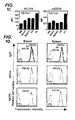
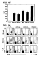
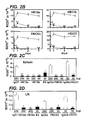

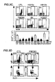
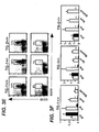


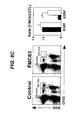


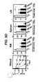
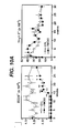
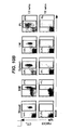
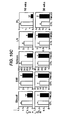
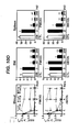
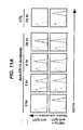
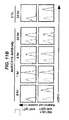


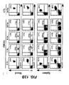


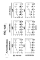


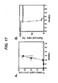
Classifications
-
- C—CHEMISTRY; METALLURGY
- C07—ORGANIC CHEMISTRY
- C07K—PEPTIDES
- C07K16/00—Immunoglobulins [IGs], e.g. monoclonal or polyclonal antibodies
- C07K16/18—Immunoglobulins [IGs], e.g. monoclonal or polyclonal antibodies against material from animals or humans
- C07K16/28—Immunoglobulins [IGs], e.g. monoclonal or polyclonal antibodies against material from animals or humans against receptors, cell surface antigens or cell surface determinants
- C07K16/2803—Immunoglobulins [IGs], e.g. monoclonal or polyclonal antibodies against material from animals or humans against receptors, cell surface antigens or cell surface determinants against the immunoglobulin superfamily
-
- A—HUMAN NECESSITIES
- A61—MEDICAL OR VETERINARY SCIENCE; HYGIENE
- A61K—PREPARATIONS FOR MEDICAL, DENTAL OR TOILETRY PURPOSES
- A61K39/00—Medicinal preparations containing antigens or antibodies
- A61K39/395—Antibodies; Immunoglobulins; Immune serum, e.g. antilymphocytic serum
- A61K39/39533—Antibodies; Immunoglobulins; Immune serum, e.g. antilymphocytic serum against materials from animals
- A61K39/39566—Antibodies; Immunoglobulins; Immune serum, e.g. antilymphocytic serum against materials from animals against immunoglobulins, e.g. anti-idiotypic antibodies
-
- A—HUMAN NECESSITIES
- A61—MEDICAL OR VETERINARY SCIENCE; HYGIENE
- A61K—PREPARATIONS FOR MEDICAL, DENTAL OR TOILETRY PURPOSES
- A61K39/00—Medicinal preparations containing antigens or antibodies
- A61K39/395—Antibodies; Immunoglobulins; Immune serum, e.g. antilymphocytic serum
-
- A—HUMAN NECESSITIES
- A61—MEDICAL OR VETERINARY SCIENCE; HYGIENE
- A61P—SPECIFIC THERAPEUTIC ACTIVITY OF CHEMICAL COMPOUNDS OR MEDICINAL PREPARATIONS
- A61P35/00—Antineoplastic agents
-
- A—HUMAN NECESSITIES
- A61—MEDICAL OR VETERINARY SCIENCE; HYGIENE
- A61P—SPECIFIC THERAPEUTIC ACTIVITY OF CHEMICAL COMPOUNDS OR MEDICINAL PREPARATIONS
- A61P35/00—Antineoplastic agents
- A61P35/02—Antineoplastic agents specific for leukemia
-
- A—HUMAN NECESSITIES
- A61—MEDICAL OR VETERINARY SCIENCE; HYGIENE
- A61P—SPECIFIC THERAPEUTIC ACTIVITY OF CHEMICAL COMPOUNDS OR MEDICINAL PREPARATIONS
- A61P37/00—Drugs for immunological or allergic disorders
-
- A—HUMAN NECESSITIES
- A61—MEDICAL OR VETERINARY SCIENCE; HYGIENE
- A61P—SPECIFIC THERAPEUTIC ACTIVITY OF CHEMICAL COMPOUNDS OR MEDICINAL PREPARATIONS
- A61P43/00—Drugs for specific purposes, not provided for in groups A61P1/00-A61P41/00
-
- A—HUMAN NECESSITIES
- A61—MEDICAL OR VETERINARY SCIENCE; HYGIENE
- A61K—PREPARATIONS FOR MEDICAL, DENTAL OR TOILETRY PURPOSES
- A61K39/00—Medicinal preparations containing antigens or antibodies
- A61K2039/505—Medicinal preparations containing antigens or antibodies comprising antibodies
-
- A—HUMAN NECESSITIES
- A61—MEDICAL OR VETERINARY SCIENCE; HYGIENE
- A61K—PREPARATIONS FOR MEDICAL, DENTAL OR TOILETRY PURPOSES
- A61K39/00—Medicinal preparations containing antigens or antibodies
- A61K2039/54—Medicinal preparations containing antigens or antibodies characterised by the route of administration
-
- C—CHEMISTRY; METALLURGY
- C07—ORGANIC CHEMISTRY
- C07K—PEPTIDES
- C07K2317/00—Immunoglobulins specific features
- C07K2317/50—Immunoglobulins specific features characterized by immunoglobulin fragments
- C07K2317/56—Immunoglobulins specific features characterized by immunoglobulin fragments variable (Fv) region, i.e. VH and/or VL
- C07K2317/565—Complementarity determining region [CDR]
-
- C—CHEMISTRY; METALLURGY
- C07—ORGANIC CHEMISTRY
- C07K—PEPTIDES
- C07K2317/00—Immunoglobulins specific features
- C07K2317/50—Immunoglobulins specific features characterized by immunoglobulin fragments
- C07K2317/56—Immunoglobulins specific features characterized by immunoglobulin fragments variable (Fv) region, i.e. VH and/or VL
- C07K2317/567—Framework region [FR]
-
- C—CHEMISTRY; METALLURGY
- C07—ORGANIC CHEMISTRY
- C07K—PEPTIDES
- C07K2317/00—Immunoglobulins specific features
- C07K2317/70—Immunoglobulins specific features characterized by effect upon binding to a cell or to an antigen
- C07K2317/77—Internalization into the cell
Definitions
- the present invention is directed to methods for the treatment of B cell disorders or diseases in human subjects, including B cell malignancies, using therapeutic antibodies that bind to the human CD19 antigen.
- the therapeutic anti-CD19 antibodies of the compositions and methods of the invention preferably mediate human antigen-dependent-cell-mediated-cytotoxicity (ADCC).
- the present invention is further directed to compositions comprising human, humanized, or chimeric anti-CD19 antibodies of the IgG1 and/or IgG3 human isotype.
- the present invention is further directed to compositions comprising human, humanized, or chimeric anti-CD19 antibodies of the IgG2 and/or IgG4 human isotype that preferably mediate human ADCC.
- the present invention also encompasses monoclonal human, humanized, or chimeric anti-CD19 antibodies.
- B cell surface markers have been generally suggested as targets for the treatment of B cell disorders or diseases, autoimmune disease, and transplantation rejection.
- B cell surface markers include CD10, CD19, CD20, CD21, CD22, CD23, CD24, CD37, CD53, CD72, CD74, CD75, CD77, CD79a, CD79b, CD80, CD81, CD82, CD83, CD84, CD85, and CD86 leukocyte surface markers.
- Antibodies that specifically bind certain of these markers have been developed, and some have been tested for the treatment of diseases and disorders.
- chimeric or radiolabeled monoclonal antibody (mAb)-based therapies directed against the CD20 cell surface molecule specific for mature B cells and their malignant counterparts have been shown to be an effective in vivo treatment for non-Hodgkin's lymphoma ( Tedder et al., Immunol Today 15:450-454 (1994 ); Press et al., Hematology, 221-240 (2001 ); Kaminski et al, N. Engl. J. Med., 329:459-465 (1993 ); Weiner, Semin.
- Anti-CD20 monoclonal antibody therapy has also been found to ameliorate the manifestations of rheumatoid arthritis, systemic lupus erythematosus, idiopathic thrombocytopenic purpura and hemolytic anemia, as well as other immune-mediated diseases ( Silverman et al., Arthritis Rheum., 48:1484-1492 (2002 ); Edwards et al., Rheumatology, 40:1-7 (2001 ); De Vita et al., Arthritis Rheumatism, 46:2029-2033 (2002 ); Leandro et al., Ann. Rheum.
- the anti-CD22 monoclonal antibody LL-2 was shown to be effective in treating aggressive and relapsed lymphoma patients undergoing chemotherapeutic treatment ( Goldenberg U.S. Patent Nos: 6,134,982 and 6,306,393 ).
- the anti-CD20 (IgG1) antibody, RITUXANTM has successfully been used in the treatment of certain diseases such as adult immune thrombocytopenic purpura, rheumatoid arthritis, and autoimmune hemolytic anemia ( Cured et al., WO 00/67796 ).
- ALL acute lymphoblastic leukemias
- B cell malignancies typically do not express CD20, express CD20 at low levels, or have lost CD20 expression following CD20 immunotherapy ( Smith et al., Oncogene, 22:7359-7368 (2003 )).
- CD20 is not predictive of response to anti-CD20 therapy as only half of non-Hodgkin's lymphoma patients respond to CD20-directed immunotherapy.
- the human CD19 molecule is a structurally distinct cell surface receptor expressed on the surface of human B cells, including, but not limited to, pre-B cells, B cells in early development (i.e ., immature B cells), mature B cells through terminal differentiation into plasma cells, and malignant B cells.
- CD19 is expressed by most pre-B acute lymphoblastic leukemias (ALL), non-Hodgkin's lymphomas, B cell chronic lymphocytic leukemias (CLL), pro-lymphocytic leukemias, hairy cell leukemias, common acute lymphocytic leukemias, and some Null-acute lymphoblastic leukemias ( Nadler et al., J.
- CD19 On plasma cells further suggests it may be expressed on differentiated B cell tumors such as multiple myeloma, plasmacytomas, Waldenstrom's tumors ( Grossbard et al., Br. J. Haematol., 102:509-15(1998 ); Treon et al., Semin. Oncol., 30:248-52(2003 )).
- the CD19 antigen was thought to be expressed at higher levels and internalized by cells when bound by an anti-CD19 antibody.
- the CD19 antigen has also been one of the many proposed targets for immunotherapy. However, the perceived unavailability as a target due to cellular internalization, was thought to have presented obstacles to the development of therapeutic protocols that could be successfully in human subjects.
- the CLB-CD19 antibody (anti-CD19 murine IgG2a mAb) was shown to inhibit growth of human tumors implanted in athymic mice ( Hooijberg et al., Cancer Research, 55:840-846 (1995 )).
- the Monoclonal murine antibody FMC63 (IgG2a) was chimerized using a human IgG1 Fc region.
- the murine CLB-CD19 antibody was administered to six patients diagnosed with a progressive non-Hodgkin's lymphoma who had failed previous conventional therapy (chemotherapy or radiotherapy). These patients were given total antibody doses ranging from 225 to 1,000 mg ( Hekman et al., Cancer Immunol. Immunotherapy, 32:364-372 (1991 )). Although circulating tumor cells were temporarily reduced in two patients after antibody infusion, only one patient achieved partial remission after two periods of antibody treatment. No conclusions regarding therapeutic efficacy could be drawn from this small group of refractory patients.
- anti-CD19 antibody-based immunotoxins produced equally discouraging results.
- the B4 anti-CD19 antibody murine IgG1 mAb
- the invention relates to immunotherapeutic compositions and methods for the treatment of B cell diseases and disorders in human subjects, such as, but not limited to, B cell malignancies, using therapeutic antibodies that bind to the human CD19 antigen and that preferably mediate human ADCC.
- the present invention relates to pharmaceutical compositions comprising human or humanized anti-CD 19 antibodies of the IgG 1 or IgG3 human isotype.
- the present invention relates to pharmaceutical compositions comprising human or humanized anti-CD19 antibodies of the IgG2 or IgG4 human isotype that preferably mediate human ADCC.
- the present invention relates to pharmaceutical compositions comprising chimerized anti-CD19 antibodies of the IgG1, IgG2, IgG3, or IgG4 isotype that mediate human ADCC.
- the invention relates to pharmaceutical compositions comprising monoclonal human, humanized, or chimeric anti-CD19 antibodies.
- Therapeutic formulations and regimens are described for treating human subjects diagnosed with B cell malignancies that derive from B cells and their precursors, including but not limited to, acute lymphoblastic leukemias (ALL), Hodgkin's lymphomas, non-Hodgkin's lymphomas, B cell chronic lymphocytic leukemias (CLL), multiple myeloma, follicular lymphoma, mantle cell lymphoma, pro-lymphocytic leukemias, hairy cell leukemias, common acute lymphocytic leukemias and some Null-acute lymphoblastic leukemias.
- ALL acute lymphoblastic leukemias
- non-Hodgkin's lymphomas non-Hodgkin's lymphomas
- B cell chronic lymphocytic leukemias CLL
- multiple myeloma follicular lymphoma
- the methods of the invention are demonstrated by way of example, using a transgenic mouse model for evaluating CD19-directed immunotherapies in human subjects.
- the invention provides for a pharmaceutical composition comprising a monoclonal human or humanized anti-CD19 antibody of the IgG1 or IgG3 human isotype in a pharmaceutically acceptable carrier.
- the invention provides for a pharmaceutical composition comprising a therapeutically effective amount of a monoclonal chimerized anti-CD19 antibody of the IgG1 or IgG3 human isotype in a pharmaceutically acceptable carrier.
- a therapeutically effective amount of a monoclonal chimerized anti-CD19 antibody of the IgG1 or IgG3 human isotype is less than 1 mg/kg of patient body weight.
- a therapeutically effective amount of a monoclonal chimerized anti-CD19 antibody of the IgG1 or IgG3 human isotype is greater than 2 mg/kg of patient body weight.
- the invention provides for a pharmaceutical composition comprising a therapeutically effective amount of monoclonal human or humanized anti-CD19 antibody that mediates human antibody-dependent cellular cytotoxicity (ADCC), in a pharmaceutically acceptable carrier.
- a pharmaceutical composition comprising a monoclonal chimerized anti-CD19 antibody that mediates human antibody-dependent cellular cytotoxicity (ADCC), and/or complement dependent cytotoxicty (CDC) and/or apoptotic activity in a pharmaceutically acceptable carrier.
- the present invention concerns a method of treating a B cell malignancy in a human comprising administering to a human in need thereof a monoclonal human or humanized anti-CD 19 antibody of the IgG1 or IgG3 human isotype in an amount sufficient to deplete circulating B cells.
- the present invention also concerns a method of treating a B cell malignancy in a human comprising administering to a human in thereof a monoclonal human or humanized anti-CD19 antibody that mediates human antibody-dependent cellular cytotoxicity (ADCC) in an amount sufficient to deplete circulating B cells.
- ADCC human antibody-dependent cellular cytotoxicity
- the present invention concerns a method of treating a B cell malignancy in a human patient comprising the administration of a therapeutically effective regimen of a monoclonal human or humanized anti-CD19 antibody of the IgG1 or IgG3 human isotype to a human patient in need of such treatment.
- the present invention provides a method of treating a B cell malignancy in a human patient comprising the administration of a therapeutically effective regimen of a monoclonal human or humanized anti-CD 19 antibody that mediates human antibody-dependent cellular cytotoxicity (ADCC), to a human patient in need of such treatment.
- the present invention provides a method of treating an early stage disease resulting from a B cell malignancy in a human patient comprising administration of a therapeutically effective regimen of a monoclonal anti-CD19 antibody that mediates human antibody-dependent cellular cytotoxicity (ADCC), to a human in need of such treatment.
- ADCC human antibody-dependent cellular cytotoxicity
- the present invention provides a method of treating a B cell malignancy in a human patient comprising administration of a therapeutically effective regimen of a monoclonal anti-CD19 antibody that mediates human antibody-dependent cellular cytotoxicity (ADCC), to a human subject in need thereof, wherein the human subject has not previously received treatment for the malignancy.
- Yet another embodiment of the present invention provides a method of treating a B cell malignancy in a human patient comprising administration of a therapeutically effective regimen of a monoclonal anti-CD19 antibody that mediates human antibody-dependent cellular cytotoxicity (ADCC), to a human patient in need of such treatment, wherein the B cell malignancy is CD19 positive.
- the present invention provides a method of treating a B cell malignancy in a human patient comprising administration of a therapeutically effective regimen of a monoclonal anti-CD19 antibody that mediates human antibody-dependent cellular cytotoxicity (ADCC), to a human patient in need of such treatment, wherein the human patient has a monocyte count of at least 1 per dL of circulating blood.
- a therapeutically effective regimen of a monoclonal anti-CD19 antibody that mediates human antibody-dependent cellular cytotoxicity (ADCC), to a human patient in need of such treatment wherein the human patient has a monocyte count of at least 1 per dL of circulating blood.
- ADCC human antibody-dependent cellular cytotoxicity
- antibody and “antibodies” (immunoglobulins) refer to monoclonal antibodies (including full-length monoclonal antibodies), polyclonal antibodies, multispecific antibodies (e.g ., bispecific antibodies) formed from at least two intact antibodies, human antibodies, humanized antibodies, camelised antibodies ; chimeric antibodies, single-chain Fvs (scFv), single-chain antibodies, single domain antibodies, domain antibodies.
- scFv single-chain Fvs
- antibodies include immunoglobulin molecules and immunologically active fragments of immunoglobulin molecules, i.e ., molecules that contain an antigen-binding site.
- Immunoglobulin molecules can be of any type (e.g ., IgG, IgE, IgM, IgD, IgA and IgY), class (e.g ., IgG1, IgG2, IgG3, IgG4, IgA1 and IgA2) or subclass.
- type e.g ., IgG, IgE, IgM, IgD, IgA and IgY
- class e.g ., IgG1, IgG2, IgG3, IgG4, IgA1 and IgA2
- subclass e.gG1, IgG2, IgG3, IgG4, IgA1 and IgA2
- Native antibodies are usually heterotetrameric glycoproteins of about 150,000 daltons, composed of two identical light (L) chains and two identical heavy (H) chains. Each light chain is linked to a heavy chain by one covalent disulfide bond, while the number of disulfide linkages varies between the heavy chains of different immunoglobulin isotypes. Each heavy and light chain also has regularly spaced intrachain disulfide bridges. Each heavy chain has at one end a variable domain (V H ) allowed by a number of constant domains.
- V H variable domain
- Each light chain has a variable domain at one end (V L ) and a constant domain at its other end; the constant domain of the light chain is aligned with the first constant domain of the heavy chain, and the light chain variable domain is aligned with the variable domain of the heavy chain.
- Particular amino acid residues are believed to form an interface between the light and heavy chain variable domains.
- Such antibodies may be derived from any mammal, including, but not limited to, humans, monkeys, pigs, horses, rabbits, dogs, cats, mice, etc.
- variable refers to the fact that certain portions of the variable domains differ extensively in sequence among antibodies and are responsible for the binding specificity of each particular antibody for its particular antigen. However, the variability is not evenly distributed through the variable domains of antibodies. It is concentrated in segments called Complementarity Determining Regions (CDRs) both in the light chain and the heavy chain variable domains. The more highly conserved portions of the variable domains are called the framework regions (FR).
- CDRs Complementarity Determining Regions
- FR framework regions
- the variable domains of native heavy and light chains each comprise four FR regions, largely adopting a ⁇ -sheet configuration, connected by three CDRs, which form loops connecting, and in some cases forming part of, the ⁇ -sheet structure.
- the CDRs in each chain are held together in close proximity by the FR regions and, with the CDRs from the other chain, contribute to the formation of the antigen-binding site of antibodies (see, Kabat et al., Sequences of proteins of Immunological Interest, 5th Ed. Public Health Service, National Institutes of Health, Bethesda, MD (1991 )).
- the constant domains are generally not involved directly in antigen binding, but may influence antigen binding affinity and may exhibit various effector functions, such as participation of the antibody in ADCC.
- hypervariable region when used herein refers to the amino acid residues of an antibody which are responsible for binding to its antigen.
- the hypervariable region comprises amino acid residues from a "complementarity determining region” or "CDR" (e.g ., residues 24-34 (L1), 50-56 (L2) and 89-97 (L3) in the light chain variable domain and 31-35 (H1), 50-65 (H2) and 95-102 (H3) in the heavy chain variable domain; Kabat et al., Sequences of Proteins of Immunological Interest, 5th Ed.
- CDR complementarity determining region
- the term "monoclonal antibody” as used herein refers to an antibody obtained from a population of substantially homogeneous antibodies, i.e., the individual antibodies comprising the population are identical except for possible naturally occurring mutations that may be present in minor amounts. Monoclonal antibodies are highly specific, being directed against a single antigenic site. Furthermore, in contrast to conventional (polyclonal) antibody preparations which typically include different antibodies directed against different determinants (epitopes), each monoclonal antibody is directed against a single determinant on the antigen. In addition to their specificity, the monoclonal antibodies are advantageous in that they are synthesized by the hybridoma cells, uncontaminated by other immunoglobulin producing cells. Alternatively, the monoclonal antibody may be produced by cells stably or transiently transfected with the heavy and light chain genes encoding the monoclonal antibody.
- the modifier "monoclonal” indicates the character of the antibody as being obtained from a substantially homogeneous population of antibodies, and is not to be construed as requiring engineering of the antibody by any particular method.
- the term “monoclonal” is used herein to refer to an antibody that is derived from a clonal population of cells, including any eukaryotic, prokaryotic, or phage clone, and not the method by which the antibody was engineered.
- the monoclonal antibodies to be used in accordance with the present invention may be made by the hybridoma method first described by Kohler et al., Nature, 256:495 (1975 ), or may be made by any recombinant DNA method (see, e.g., U.S. Patent No. 4,816,567 ), including isolation from phage antibody libraries using the techniques described in Clackson et al., Nature, 352:624-628 (1991 ) and Marks et al., J. Mol. Biol., 222:581-597 (1991 ), for example. These methods can be used to produce monoclonal mammalian, chimeric, humanized, human, domain antibodies, diabodies, vaccibodies, linear antibodies, and bispecific antibodies.
- chimeric antibodies includes antibodies in which at least one portion of the heavy and/or light chain is identical with or homologous to corresponding sequences in antibodies derived from a particular species or belonging to a particular antibody or subclass, and at least one other portion of the chain(s) is identical with or homologous to corresponding sequences in antibodies derived from another species or belonging to another antibody class or subclass, as well as fragments of such antibodies, so long as they exhibit the desired biological activity ( U.S. Patent No. 4,816,567 ; Morrison et al., Proc. Natl. Acad. Sci. USA, 81:6851-6855 (1984 )).
- Chimeric antibodies of interest herein include "Primatized" antibodies comprising variable domain antigen-binding sequences derived from a nonhuman primate (e.g ., Old World Monkey, such as baboon, rhesus or cynomolgus monkey) and human constant region sequences ( U.S. Patent No. 5,693,780 ).
- a nonhuman primate e.g ., Old World Monkey, such as baboon, rhesus or cynomolgus monkey
- human constant region sequences U.S. Patent No. 5,693,780
- Humanize forms of nonhuman (e.g ., murine) antibodies are chimeric antibodies that contain minimal sequence derived from nonhuman immunoglobulin.
- humanized antibodies are human immunoglobulins (recipient antibody) in which residues from a hypervariable region of the recipient are replaced by residues from a hypervariable region of a nonhuman species (donor antibody) such as mouse, rat, rabbit or nonhuman primate having the desired specificity, affinity, and capacity.
- donor antibody such as mouse, rat, rabbit or nonhuman primate having the desired specificity, affinity, and capacity.
- framework region (FR) residues of the human immunoglobulin are replaced by corresponding nonhuman residues.
- humanized antibodies may comprise residues that are not found in the recipient antibody or in the donor antibody. These modifications are made to further refine antibody performance.
- the humanized antibody will comprise substantially all of at least one, and typically two, variable domains, in which all or substantially all of the hypervariable loops correspond to those of a nonhuman immunoglobulin and all or substantially all of the FRs are those of a human immunoglobulin sequence.
- the humanized antibody will comprise at least a portion of an immunoglobulin constant region (Fc), typically that of a human immunoglobulin.
- Fc immunoglobulin constant region
- a “human antibody” can be an antibody derived from a human or an antibody obtained from a transgenic organism that has been "engineered” to produce specific human antibodies in response to antigenic challenge and can be produced by any method known in the art. According to preferred techniques, elements of the human heavy and light chain loci are introduced into strains of the organism derived from embryonic stem cell lines that contain targeted disruptions of the endogenous heavy chain and light chain loci. The transgenic organism can synthesize human antibodies specific for human antigens, and the organism can be used to produce human antibody-secreting hybridomas.
- a human antibody can also be an antibody wherein the heavy and light chains are encoded by a nucleotide sequence derived from one or more sources of human DNA.
- a fully human antibody also can be constructed by genetic or chromosomal transfection methods, as well as phage display technology, or in vitro activated B cells, all of which are known in the art.
- CD 19 antigen refers to an antigen of about 90 kDa identified, for example, by the HD237 or B4 antibody ( Kiesel et al., Leukemia Research II, 12:1119 (1987 )).
- CD19 is found on cells throughout differentiation of B-lineage cells from the stem cell stage through terminal differentiation into plasma cells, including but not limited to, pre-B cells, B cells (including na ⁇ ve B cells, antigen-stimulated B cells, memory B cells, plasma cells, and B lymphocytes) and follicular dendritic cells. CD19 is also found on B cells in human fetal tissue.
- the CD19 antigen targeted by the antibodies of the invention is the human CD19 antigen.
- Antibody-dependent cell-mediated cytotoxicity and “ADCC” refer to a cell-mediated reaction in which non-specific cytotoxic cells (e.g ., Natural Killer (NK) cells, neutrophils, and macrophages) recognize bound antibody on a target cell and subsequently cause lysis of the target cell.
- non-specific cytotoxic cells e.g ., Natural Killer (NK) cells, neutrophils, and macrophages
- NK cells Natural Killer
- neutrophils neutrophils
- macrophages e.g., cytotoxic cells that mediate ADCC generally express Fc receptors (FcRs).
- FcRs Fc receptors
- ADCC activity of a molecule is summarized in Ravetch and Kinet, Annu. Rev. Immunol., 9:457-92 (1991 ).
- an in vitro ADCC assay such as that described in U.S. Patent No. 5,500,362 or 5,821,337 may be performed.
- Useful effector cells for such assays include peripheral blood mononuclear cells (PBMC) and Natural Killer (NK) cells.
- PBMC peripheral blood mononuclear cells
- NK Natural Killer
- ADCC activity of the molecules of interest may be assessed in vivo , e.g ., in an animal model such as that disclosed in Clynes et al., PNAS (USA), 95:652-656 (1998 ).
- “Complement dependent cytotoxicity” or “CDC” refers to the ability of a molecule to initiate complement activation and lyse a target in the presence of complement.
- the complement activation pathway is initiated by the binding of the first component of the complement system (C1q) to a molecule (e.g ., an antibody) complexed with a cognate antigen.
- a CDC assay e.g ., as described in Gazzano-Santaro et al., J. Immunol. Methods, 202:163 (1996 ), may be performed.
- “Effector cells” are leukocytes which express one or more FcRs and perform effector functions.
- the cells express at least Fe ⁇ RI, FC ⁇ RII, Fc ⁇ RIII and/or Fc ⁇ RIV and carry out ADCC effector function.
- human leukocytes which mediate ADCC include peripheral blood mononuclear cells (PBMC), natural killer (NK) cells, monocytes, cytotoxic T cells and neutrophils; with PBMCs and NK cells being preferred.
- the effector cells are human cells.
- Fc receptor or “FcR” are used to describe a receptor that binds to the Fc region of an antibody.
- the preferred FcR is a native sequence human FcR.
- a preferred FcR is one which binds an IgG antibody (a gamma receptor) and includes receptors of the Fc ⁇ RI, Fc ⁇ RII, Fc ⁇ RIII, and Fc ⁇ RIV subclasses, including allelic variants and alternatively spliced forms of these receptors.
- Fc ⁇ RII receptors include Fc ⁇ RIIA (an "activating receptor") and Fc ⁇ RIIB (an “inhibiting receptor”), which have similar amino acid sequences that differ primarily in the cytoplasmic domains thereof.
- Activating receptor Fc ⁇ RIIA contains an immunoreceptor tyrosine-based activation motif (ITAM) in its cytoplasmic domain.
- Inhibiting receptor Fc ⁇ RIIB contains an immunoreceptor tyrosine-based inhibition motif (ITIM) in its cytoplasmic domain.
- ITAM immunoreceptor tyrosine-based activation motif
- ITIM immunoreceptor tyrosine-based inhibition motif
- FcR FcR
- FcRn neonatal receptor
- Fv is the minimum antibody fragment which contains a complete antigen-recognition and binding site. This region consists of a dimer of one heavy and one light chain variable domain in tight, non-covalent or covalent association. It is in this configuration that the three CDRs of each variable domain interact to define an antigen-binding site on the surface of the V H -V L dimer. Collectively, the six CDRs confer antigen-binding specificity to the antibody. However, even a single variable domain (or half of an Fv comprising only three CDRs specific for an antigen) has the ability to recognize and bind antigen, although at a lower affinity than the entire binding site.
- Affinity of an antibody for an to be used in the treatment(s) described herein is a term well understood in the art and means the extent, or strength, of binding of antibody to epitope. Affinity may be measured and/or expressed in a number of ways known in the art, including, but not limited to, equilibrium dissociation constant (KD or Kd), apparent equilibrium dissociation constant (KD' or Kd'), and IC50 (amount needed to effect 50% inhibition in a competition assay). It is understood that, for purposes of this invention, an affinity is an average affinity for a given population of antibodies which bind to an epitope.
- KD' reported herein in terms of mg IgG per mL or mg/mL indicate mg Ig per mL of serum, although plasma can be used.
- antibody affinity can be measured before and/or during treatment, and the values obtained can be used by a clinician in assessing whether a human patient is an appropriate candidate for treatment.
- an “epitope” is a term well understood in the art and means any chemical moiety that exhibits specific binding to an antibody.
- An “epitope” can also comprise an antigen, which is a moiety or molecule that contains an epitope, and, as such, also specifically binds to antibody.
- a "B cell surface marker” as used herein is an antigen expressed on the surface of a B cell which can be targeted with an agent which binds thereto.
- Exemplary B cell surface markers include the CD10, CD19, CD20, CD21, CD22, CD23, CD24, CD25, CD37, CD53, CD72, CD73, CD74, CD75, CD77, CD79a, CD79b, CD80, CD81, CD82, CD83, CD84, CD85, and CD86 leukocyte surface markers.
- the B cell surface marker of particular interest is preferentially expressed on B cells compared to other non-B cell tissues of a mammal and may be expressed on both precursor B cells and mature B cells.
- the preferred marker is CD19, which is found on B cells throughout differentiation of the lineage from the pro/pre-B cell stage through the terminally differentiated plasma cell stage.
- antibody half-life means a pharmacokinetic property of an antibody that is a measure of the mean survival time of antibody molecules following their administration.
- Antibody half-life can be expressed as the time required to eliminate 50 percent of a known quantity of immunoglobulin from the patient's body or a specific compartment thereof, for example, as measured in serum, i.e ., circulating half-life, or in other tissues.
- Half-life may vary from one immunoglobulin or class of immunoglobulin to another. In general, an increase in antibody half-life results in an increase in mean residence time (MRT) in circulation for the antibody administered.
- MRT mean residence time
- isotype refers to the classification of an antibody.
- the constant domains of antibodies are not involved in binding to antigen, but exhibit various effector functions.
- a given antibody or immunoglobulin can be assigned to one of five major classes of immunoglobulins: IgA, IgD, IgE, IgG, and IgM.
- IgA, IgD, IgE, IgG, and IgM Several of these classes may be further divided into subclasses (isotypes), e.g ., IgG1 (gamma 1), IgG2 (gamma 2), IgG3 (gamma 3), and IgG4 (gamma 4), and IgA1 and IgA2.
- the heavy chain constant regions that correspond to the different classes of immunoglobulins are called ⁇ , ⁇ , ⁇ , ⁇ , and ⁇ , respectively.
- the structures and three-dimensional configurations of different classes of immunoglobulins are well-known.
- human immunoglobulin classes only human IgG1, IgG2, IgG3, IgG4, and IgM are known to activate complement.
- Human IgG1 and IgG3 are known to mediate ADCC in humans.
- immunogenicity means that a compound is capable of provoking an immune response (stimulating production of specific antibodies and/or proliferation of specific T cells).
- antigenicity means that a compound is recognized by an antibody or may bind to an antibody and induce an immune response.
- the term "avidity” is a measure of the overall binding strength (i.e ., both antibody arms) with which an antibody binds an antigen.
- Antibody avidity can be determined by measuring the dissociation of the antigen-antibody bond in antigen excess using any means known in the art, such as, but not limited to, by the modification of indirect fluorescent antibody as described by Gray et al., J. Virol. Meth., 44:11-24. (1993 ).
- treat By the terms “treat,” “treating” or “treatment of” (or grammatically equivalent terms) it is meant that the severity of the subject's condition is reduced or at least partially improved or ameliorated and/or that some alleviation, mitigation or decrease in at least one clinical symptom is achieved and/or there is an inhibition or delay in the progression of the condition and/or prevention or delay of the onset of a disease or illness.
- treat By the terms “treat,” “treating” or “treatment of” (or grammatically equivalent terms) refer to both prophylactic and therapeutic treatment regimes.
- a "sufficient amount”' or “an amount sufficient to” achieve a particular result refers to an amount of an antibody or composition of the invention that its effective to produce a desired effect, which is optionally a therapeutic effect ( i.e ., by administration of a therapeutically effective amount).
- a "sufficient amount” or “an amount sufficient to” can be an amount that is effective to deplete B cells.
- a “therapeutically effective” amount as used herein is an amount that provides some improvement or benefit to the subject.
- a “therapeutically effective” amount is an amount that provides some alleviation, mitigation, and/or decrease in at least one clinical symptom.
- Clinical symptoms associated with the disorders that can be treated by the methods of the invention are well-known to those skilled in the art. Further, those skilled in the art will appreciate that the therapeutic effects need not be complete or curative, as long as some benefit is provided to the subject.
- the invention relates to immunothempeutic compositions and methods for the treatment of B cell diseases and disorders in human subjects, such as, but not limited to, B cell malignancies, using therapeutic antibodies that bind to the CD19 antigen and preferably mediate human ADCC.
- the present invention relates to pharmaceutical compositions comprising human, humanized, or chimeric anti-CD19 antibodies of the IgG1 or IgG3 human isotype.
- the present invention also relates to pharmaceutical compositions comprising human or humanized anti-CD19 antibodies of the IgG2 or IgG4 human isotype that preferably mediate human ADCC.
- the present invention also relates to pharmaceutical compositions comprising monoclonal human, humanized, or chimerized anti-CD19 antibodies that can be produced by means known in the art.
- Therapeutic formulations and regimens are described for treating human subjects diagnosed with B cell malignancies that derive from B cells and their precursors, including but not limited to, acute lymphoblastic leukemias (ALL), Hodgkin's lymphomas, non-Hodgkin's lymphomas, B cell chronic lymphocytic leukemias (CLL), multiple myeloma, follicular lymphoma, mantle cell lymphoma, pro-lymphocytic leukemias, hairy cell leukemias, common acute lymphocytic leukemias and some Null-acute lymphoblastic leukemias.
- ALL acute lymphoblastic leukemias
- non-Hodgkin's lymphomas non-Hodgkin's lymphomas
- B cell chronic lymphocytic leukemias CLL
- multiple myeloma follicular lymphoma
- Polyclonal antibodies are preferably raised in animals by multiple subcutaneous (sc) or intraperitoneal (i.p.) injections of the relevant antigen and an adjuvant. It may be useful to conjugate the relevant antigen to a protein that is immunogenic in the species to be immunized, e.g., keyhole limpet hemocyanin, serum albumin, bovine thyroglobulin, or soybean trypsin inhibitor using a bifunctional or derivatizing agent, for example, maleimidobertzoyl sulfosuccinimide ester (conjugation through cysteine residues), N-hydroxysuccinimide (through lysine residues), glutaraldehyde, succunic anhydride, SOCl 2 ,
- a protein that is immunogenic in the species to be immunized e.g., keyhole limpet hemocyanin, serum albumin, bovine thyroglobulin, or soybean trypsin inhibitor using a bifunctional or
- Animals are immunized the antigen, immunogenic conjugates, or derivative by combining, e.g ., 100 ⁇ g or 5 ⁇ g of the protein or conjugate (for rabbits or mice, respectively) with 3 volumes of Freund's complete adjuvant and injecting the solution intradermally at multiple sites.
- the animals are boosted with 1/5 to 1/10 the original amount of peptide or conjugate in Freund's incomplete adjuvant by subcutaneous injection at multiple sites.
- Seven to 14 days later the animals are bled and the serum is assayed for antibody titer. Animals are boosted until the titer plateaus.
- the animal is boosted with the conjugate of the same antigen, but conjugated to a different protein and/or through a different cross-linking reagent.
- Conjugates also can be made in recombinant cell culture as protein fusions.
- aggregating agents such as alum are suitably used to enhance the immune response.
- the monoclonal anti-CD19 antibodies of the invention exhibit binding specificity to human CD19 antigen and can preferably mediate human ADCC. These antibodies can be generated using a wide variety of techniques known in the art including the use of hybridoma, recombinant, and phage display technologies, or a combination thereof. Antibodies are highly specific, being directed against a single antigenic site. Furthermore, in contrast to conventional (polyclonal) antibody preparations which typically include different antibodies directed against different determinants (epitopes), each monoclonal antibody is directed against a single determinant on the human CD19 antigen.
- the monoclonal antibodies to be used in accordance with the present invention may be made by the hybridoma method first described by Kohler et al., Nature, 256:495 (1975 ), which can be used to generate murine antibodies (or antibodies derived from other nonhuman mammals, e.g ., rat, goat, sheep, cows, camels, etc.), or human antibodies derived from transgenic animals ( see , U.S. Patent Nos. 6,075,181 , 6,114,598 , 6,150,584 , and 6,657,103 ).
- the monoclonal antibodies can be made by recombinant DNA methods (see, e.g ., U.S. Patent No.
- the "monoclonal antibodies” may also be isolated from phage antibody libraries using the techniques described in Clackson et al., Nature, 352:624-628 (1991 ) and Marks et al., J. Mol. Biol., 222:581-597 (1991 ), for example.
- An engineered anti-CD19 antibody can be produced by any means known in the art, including, but not limited to, those techniques described below and improvements to those techniques.
- Large-scale high-yield production typically involves culturing a host cell that produces the engineered anti-CD19 antibody and recovering the anti-CD19 antibody from the host cell culture.
- Monoclonal antibodies can be produced using hybridoma techniques including those known in the art and taught, for example, in Harlow et al., Antibodies: A Laboratory Manual, (Cold Spring Harbor Laboratory Press, 2nd ed. 1988 ); Hammerling et al., in Monoclonal Antibodies and T Cell Hybridomas, 563-681 (Elsevier, N.Y., 198 1 ) (said references incorporated by reference in their entireties).
- a mouse or other appropriate host animal such as a hamster or macaque monkey, is immunized to elicit lymphocytes that produce or are capable of producing antibodies that will specifically bind to the protein for immunization.
- lymphocytes may be immunized in vitro .
- Lymphocytes then are fused with myeloma cells using a suitable fusing agent, such as polyethylene glycol, to form a hybridoma cell ( Coding, Monoclonal Antibodies: Principles and Practice, pp. 59-103 (Academic Press, 1986 )).
- a suitable fusing agent such as polyethylene glycol
- the hybridoma cells thus prepared are seeded and grown in a suitable culture medium that preferably contains one or more substances that inhibit the growth or survival of the unfused, parental myeloma cells.
- a suitable culture medium that preferably contains one or more substances that inhibit the growth or survival of the unfused, parental myeloma cells.
- the culture medium for the hybridomas typically will hypoxanthine, aminopterin, and thymidine (HAT medium), which substances prevent the growth of HGPRT-deficient cells.
- Preferred myeloma cells are those that fuse efficiently, support stable high-level production of antibody by the selected antibody-producing cells, and are sensitive to a medium such as HAT medium.
- preferred myeloma cell lines are murine myeloma lines, such as those derived from MOPC-21 and MPC-11 mouse tumors available from the Salk Institute Cell Distribution Center, San Diego, CA, USA, and SP-2 or X63-Ag8.653 cells available from the American Type Culture Collection, Rockville, MD, USA.
- Human myeloma and mouse-human heteromyeloma cell lines also have been described for the production of human monoclonal antibodies ( Kozbor, J. Immunol., 133:3001 (1984 ); Brodeur et al., Monoclonal Antibody Production Techniques and Applications, pp. 51-63 (Marcel Dekker, Inc., New York, 1987 )).
- Culture medium in which hybridoma cells are growing is assayed for production of monoclonal antibodies directed against the human CD19 antigen.
- the binding specificity of monoclonal antibodies produced by hybridoma cells is determined by immunoprecipitation or by an in vitro binding assay, such as radioimmunoassay (RIA) or enzyme-linked immunoabsorbent assay (ELISA).
- RIA radioimmunoassay
- ELISA enzyme-linked immunoabsorbent assay
- the clones may be subcloned by limiting dilution procedures and grown by standard methods ( Goding, Monoclonal Antibodies: Principles and Practice, pp. 59-103 (Academic Press, 1986 )). Suitable culture media for this purpose include, for example, D-MEM or RPMI 1640 medium.
- the hybridoma cells may be grown in vivo as ascites tumors in an animal.
- the monoclonal antibodies secreted by the subclones are suitably separated from the culture medium, ascites fluid, or serum by conventional immunoglobulin purification procedures such as, for example, protein A-Sepharose, hydroxylapatite chromatography, gel electrophoresis, dialysis, or affinity chromatography.
- DNA encoding the anti-CD19 antibodies of the invention is readily isolated and sequenced using conventional procedures (e.g ., by using oligonucleotide probes that are capable of binding specifically to genes encoding the heavy and light chains of the anti-CD19 antibodies).
- the hybridoma cells serve as a preferred source of such DNA.
- the DNA may be placed into expression vectors, which are then transfected into host cells such as E. coli cells, simian COS cells, Chinese hamster ovary (CHO) cells, or myeloma cells that do not otherwise produce immunoglobulin protein, to obtain the synthesis of anti-CD 19 Antibodies in the recombinant host cells.
- phage display methods functional antibody domains are displayed on the surface of phage particles which carry the polynucleotide sequences encoding them.
- DNA sequences encoding V H and V L domains are amplified from animal cDNA libraries (e.g ., human or murine cDNA libraries of affected tissues).
- the DNA encoding the V H and V L domains are recombined with an scFv linker by PCR and cloned into a phagemid vector.
- the vector is electroporated in E. coli and the E. coli is infected with helper phage.
- Phage used in these methods are typically filamentous phage including fd and M13 and the V H and V L domains are usually recombinantly fused to either the phage gene III or gene VIII.
- Phage expressing an antigen-binding domain that binds to a particular antigen can be selected or identified with antigen, e.g ., using labeled antigen or antigen bound or captured to a solid surface or bead.
- Examples of phage display methods can be used to make the antibodies of the present invention include those disclosed in Brinkman et al., 1995, J. Immunol. Methods, 182:41-50 ; Ames et al., 1995, J. Immunol.
- the antibody coding regions from the phage can be isolated and used to generate whole antibodies, including human antibodies, or any other desired antigen-binding fragment, and expressed in any desired host, including mammalian cells, insect cells, plant cells, yeast, and bacteria, e.g ., as described below.
- Techniques to recombinantly produce Fab, Fab' and F(ab') 2 fragments can also be employed using methods known in the art such as those disclosed in PCT Publication No.
- antibodies may be isolated from antibody phage libraries generated using the techniques described in McCafferty et al., Nature, 348:552-554 (1990 ). Clackson et al., Nature, 352:624-628 (1991 ). Marks et al., J. Mol. Biol., 222:581-597 (1991 ) describe the isolation of murine and human antibodies, respectively, using phage libraries.
- Chain shuffling can be used in the production of high affinity (nM range) human antibodies ( Marks et al., Bio/Technology, 10:779-783 (1992 )), as well as combinatorial infection and in vivo recombination as a strategy for constructing very large phage libraries ( Waterhouse et al., Nuc. Acids. Res., 21:2265-2266 (1993 )).
- these techniques are viable alternatives to traditional monoclonal antibody hybridoma techniques for isolation of anti-CD19 antibodies.
- PCR primers including V H or V L nucleotide sequences, a restriction site, and a flanking sequence to protect the restriction site can be used to amplify the V H or V L sequences in scfv clones.
- V H constant region e.g ., the human gamma 4 constant region
- V L constant region e.g ., human kappa or lamba constant regions.
- the vectors for expressing the V H or V L domains comprise an EF-1 ⁇ promoter, a secretion signal, a cloning site for the variable domain, constant domains, and a selection marker such as neomycin.
- the V H and V L domains may also be cloned into one vector expressing the necessary constant regions.
- the heavy chain conversion vectors and light chain conversion vectors are then co-transfected into cell lines to generate stable or transient cell lines that express full-length antibodies, e.g ., IgG, using techniques known to those of skill in the art.
- the DNA also may be modified, for example, by substituting the coding sequence for human heavy and light chain constant domains in place of the homologous murine sequences ( U.S. Patent No. 4,816,567 ; Morrison et al., Proc. Natl. Acad. Sci. USA, 81:6851 (1984 )), or by covalently joining to the immunoglobulin coding sequence all or part of the coding sequence for a non-immunoglobulin polypeptide.
- the anti-CD19 antibodies herein specifically include chimeric antibodies (immunoglobulins) in which a portion of the heavy and/or light chain is identical with or homologous to corresponding sequences in antibodies derived from a particular species or belonging to a particular antibody class or subclass, while another portion of the chain(s) is identical with or homologous to corresponding sequences in antibodies derived from another species or belonging to another antibody class or subclass, as well as fragments of such antibodies, so long as they exhibit the desired biological activity ( U.S. Patent No. 4,816,567 ; Morrison et al., Proc. Natl. Acad. Sci. USA, 81:6851-6855 (1984 )).
- Chimeric antibodies of interest herein include "primatized"' antibodies comprising variable domain antigen-binding sequences derived from a nonhuman primate (e.g ., Old World Monkey; such as baboon, rhesus or cynomolgus monkey) and human constant region sequences ( U.S. Patent No. 5,693,780 ).
- a nonhuman primate e.g ., Old World Monkey; such as baboon, rhesus or cynomolgus monkey
- human constant region sequences U.S. Patent No. 5,693,780
- a humanized antibody can be produced using a variety of techniques known in the art, including but not limited to, CDR-grafting (see , e.g ., European Patent No. EP 239,400 ; International Publication No. WO 91/09967 ; and U.S. Patent Nos. 5,225,539 , 5,530,101 , and 5,585,089 , each of which is incorporated herein in its entirety by reference), veneering or resurfacing ( see, e.g ., European Patent Nos.
- framework residues in the framework regions will be substituted with the corresponding residue from the CDR donor antibody to alter, preferably improve, antigen binding.
- These framework substitutions are identified by methods well-known in the art, e.g ., by modeling of the interactions of the CDR and framework residues to identify framework residues important for antigen binding and sequence comparison to identify unusual framework residues at particular positions. ( See , e.g ., Queen et al., U.S. Patent No. 5,585,089 ; and Riechmann et al., 1988, Nature, 332:323 , which are incorporated herein by reference in their entireties.)
- a humanized anti-CD19 antibody has one or more amino acid residues introduced into it from a source which is nonhuman. These nonhuman amino acid residues are often referred to as "import” residues, which are typically taken from an “import” variable domain.
- humanized antibodies comprise one or more CDRs from nonhuman immunoglobulin molecules and framework regions from human.
- humanized chimeric antibodies substantially less than an intact human variable domain has been substituted by the corresponding sequence from a nonhuman species.
- humanized antibodies are typically human antibodies in which some CDR residues and possibly some FR residues are substituted by residues from analogous sites in rodent antibodies.
- variable domains both light and heavy
- the choice of human variable domains, both light and heavy, to be used in making the humanized antibodies is to reduce antigenicity.
- sequence of the variable domain of a rodent antibody is screened against the entire library of known human variable-domain sequences.
- the human sequence which is closest to that of the rodent is then accepted as the human framework (FR) for the humanized antibody ( Sims et al., J. Immunol., 151:2296 (1993 ); Chothia et al., J. Mol. Biol., 196:901 (1987 ), the contents of which are incorporated herein by reference herein in their entirety).
- Another method uses a particular framework derived from the consensus sequence of all human antibodies of a particular subgroup of light or heavy chains.
- the same framework may be used for several different humanized anti-CD19 antibodies ( Carter et al., Proc. Natl. Acad. Sci. USA, 89:4285 (1992 ); Presta et al., J. Immunol., 151:2623 (1993 ), the contents of which are incorporated herein by reference herein in their entirety).
- Anti-CD19 antibodies can be humanized with retention of high affinity for CD19 and other favorable biological properties.
- humanized antibodies are prepared by a process of analysis of the parental sequences and various conceptual humanized products using three-dimensional models of the parental and humanized sequences. Three-dimensional immunoglobulin models are commonly available and are familiar to those skilled in the art. Computer programs are available which illustrate and display probable three-dimensional conformational structures of selected candidate immunoglobulin sequences. Inspection of these displays permits analysis of the likely role of the residues in the functioning of the candidate immunoglobulin sequence, i.e ., the analysis of residues that influence the ability of the candidate immunoglobulin to bind CD 19. In this way, FR residues can be selected and combined from the recipient and import sequences so that the desired antibody characteristic, such as increased affinity for CD19, is achieved. In general, the CDR residues are directly and most substantially involved in influencing antigen binding.
- a “humanized” antibody retains a similar antigenic specificity as the original antibody, i.e. , in the present invention, the ability to bind human CD19 antigen.
- the affinity and/or specificity of binding of the antibody for human CD19 antigen may be increased using methods of "directed evolution,” as described by Wu et al., J. Mol. Biol., 294:151 (1999 ), the contents of which are incorporated herein by reference herein in their entirety.
- human antibodies For in vivo use of antibodies in humans, it may be preferable to use human antibodies. Completely human antibodies are particularly desirable for therapeutic treatment of human subjects.
- Human antibodies can be made by a variety of methods known in the art including phage display methods described above using antibody libraries derived from human immunoglobulin sequences, including improvements to these techniques. See, also , U.S. Patent Nos. 4,444,887 and 4,716,111 ; and PCT publications WO 98/46645 , WO 98/50433 , WO 98/24893 , WO98/16654 , WO 96/34096 , WO 96/33735 , and WO 91/10741 ; each of which is incorporated herein by reference in its entirety.
- a human antibody can also be an antibody wherein the heavy and light chains are encoded by a nucleotide sequence derived from one or more sources of human DNA.
- Human anti-CD19 antibodies can also be produced using transgenic mice which are incapable of expressing functional endogenous immunoglobulins, but which can express human immunoglobulin genes.
- the human heavy and light chain immunoglobulin gene complexes may be introduced randomly or by homologous recombination into mouse embryonic stem cells.
- the human variable region, constant region, and diversity region may be introduced into mouse embryonic stem cells in addition to the human heavy and light chain genes.
- the mouse heavy and light chain immunoglobulin genes may be rendered non-functional separately or simultaneously with the introduction of human immunoglobulin loci by homologous recombination.
- the modified embryonic stem cells are expanded and microinjected into blastocysts to produce chimeric mice.
- the chimeric mice are then bred to produce homozygous offspring which express human antibodies.
- the transgenic mice are immunized in the normal fashion with a selected antigen, e.g ., all or a portion of a polypeptide of the invention.
- Anti-CD19 antibodies directed against the human CD19 antigen can be obtained from the immunized, transgenic mice using conventional hybridoma technology.
- the human immunoglobulin transgenes harbored by the transgenic mice rearrange during B cell differentiation, and subsequently undergo class switching and somatic mutation.
- IgG, IgA, IgM and IgE antibodies including, but not limited to, IgG1 (gamma 1) and IgG3.
- IgG1 gamma 1
- IgG3 IgG3
- WO 98/24893 WO 96/34096 , and WO 96/33735 ; and U.S. Patent Nos. 5,413,923 ; 5,625,126 ; 5,633,425 ; 5,569,825 ; 5,661,016 ; 5,545,806 ; 5,814,318 ; and 5,939,598 , each of which is incorporated by reference herein in their entirety.
- companies such as Abgenix, Inc. (Freemont, CA) and Genpharm (San Jose, CA) can be engaged to provide human antibodies directed against a selected antigen using technology similar to that described above.
- Human antibodies can also be derived from phage-display libraries ( Hoogenboom et al., J. Mol. Biol., 227:381 (1991 ); Marks et al., J. Mol. Biol., 222:581-597 (1991 ); Vaughan et al., Nature Biotech., 14:309 (1996 )).
- Phage display technology McCafferty et al., Nature, 348:552-553 (1990 ) can be used to produce human antibodies and antibody fragments in vitro, from immunoglobulin variable (V) domain gene repertoires from unimmunized donors.
- antibody V domain genes are cloned inframe into either a major or minor coat protein gene of a filamentous bacteriophage, such as M13 or fd, and displayed as functional antibody fragments on the surface of the phage particle.
- a filamentous bacteriophage such as M13 or fd
- the filamentous particle contains a single-stranded DNA copy of the phage genome
- selections based on the functional properties of the antibody also result in selection of the gene encoding the antibody exhibiting those properties.
- the phage mimics some of the properties of the B cell.
- Phage display can be performed in a variety of formats; for their review see, e.g ., Johnson, Kevin S. and Chiswell, David J., Current Opinion in Structural Biology 3:564-571 (1993 ).
- V-gene segments can be used for phage display.
- Clackson et al., Nature, 352:624-628 (1991 ) isolated a diverse array of anti-oxazolone antibodies from a small random combinatorial library of V genes derived from the spleens of unimmunized mice.
- a repertoire of V genes from unimmunized human donors can be constructed and antibodies to a diverse array of antigens (including self-antigens) can be isolated essentially following the techniques described by Marks et al., J Mol. Biol., 222:581-597 (1991 ), or Griffith et al., EMBO J., 12:725-734 (1993 ).
- Marks et al. J Mol. Biol., 222:581-597 (1991 )
- Griffith et al. EMBO J., 12:725-734 (1993 ).
- U.S. Patent Nos. 5,565,332 and 5,573,905 each of which is incorporated here
- Human antibodies may also be generated by in vitro activated B cells ( see , U.S. Patents 5,567,610 and 5,229,275 , each of which is incorporated herein by reference in its entirety). Human antibodies may also be generated in vitro using hybridoma techniques such as, but not limited to, that described by Roder et al. (Methods Enzymol., 121:140-167 (1986 )).
- anti-CD19 antibodies of the compositions and methods of the invention can be mutant antibodies.
- antibody mutant or “altered antibody” refers to an amino acid sequence variant of an anti-CD19 antibody wherein one or more of the amino acid residues of an anti-CD19 antibody have been modified.
- the modifications to the amino acid sequence of the anti-CD19 antibody include modifications to the sequence to improve affinity or avidity of the antibody for its antigen, and/or modifications to the Fc portion of the antibody to improve effector function.
- the modifications may be made to any known anti-CD 19 antibodies or anti-CD19 antibodies identified as described herein. Such altered antibodies necessarily have less than 100% sequence identity or similarity with a known anti-CD19 antibody.
- the altered antibody will have an amino acid sequence having at least 25%, 35%, 45%, 55%, 65%, or 75% amino acid sequence identity or similarity with the amino acid sequence of either the heavy or light chain variable domain of an anti-CD19 antibody, more preferably at least 80%, more preferably at least 85%, more preferably at least 90%, and most preferably at least 95%.
- the altered antibody will have an amino acid sequence having at least 25%, 35%, 45%, 55%, 65%, or 75% amino acid sequence identity or similarity with the amino acid sequence of the heavy chain CDR1, CDR2, or CDR3 of an anti-CD 19 antibody, more preferably at least 80%, more preferably at least 85%, more preferably at least 90%, and most preferably at least 95%.
- the altered antibody will maintain human CD19 binding capability.
- the anti-CD19 antibody of the invention comprises a heavy chain that is about 10%, 15%, 20%, 25%, 30%, 35%, 40%, 45%, 50%, 55%, 60%, 65%, 70%, 75%, 80%, 85%, 90%, 95% or more identical to an amino acid sequence of SEQ ID NO:2 ( Fig. 5A ) corresponding to the heavy chain of HB12a.
- the anti-CD19 antibody of the invention comprises a heavy chain that is about 10%, 15%, 20%, 25%, 30%, 35%, 40%, 45%, 50%, 55%, 60%, 65%, 70%, 75%, 80%, 85%, 95% or more identical to an amino acid sequence of SEQ ID NO:4 ( Fig.
- the anti-CD19 antibody of the invention comprises a heavy chain that is about 10%, 15%, 20%, 25%, 30%, 35%, 40%, 45%, 50%, 55%, 60%, 65%, 70%, 75%, 80%, 85%, 90%, 95% or more identical to an amino acid sequence of SEQ ID NO:16 ( Fig. 6A ) corresponding to the light chain of HB12a.
- the anti-CD19 antibody of the invention comprises a light chain that is about 10%, 15%, 20%, 25%, 3 0%, 35%, 40%, 45%, 50%, 55%, 60%, 65%, 70%, 75%, 80%, 85%, 90%, 95% or more identical to an amino acid sequence of SEQ ID NO:18 ( Fig. 6B ) corresponding to the light chain of HB12b.
- Hybridomas producing HB12a and HB12b anti-CD19 antibodies have been deposited under ATCC deposit nos. PTA-6580 and PTA-6581.
- Identity or similarity with respect to this sequence is defined herein as the percentage of amino acid residues in the candidate sequence that are identical (i.e ., same residue) or similar (i.e ., amino acid residue from the same group based on common side-chain properties, see below) with anti-CD19 antibody residues, after aligning the sequences and introducing gaps, if necessary, to achieve the maximum percent sequence identity. None of N-terminal, C-terminal, or internal extensions, deletions, or insertions into the antibody sequence outside of the variable domain shall be construed as affecting sequence identity or similarity.
- % identity is a measure of the relationship between two polynucleotides or two polypeptides, as determined by comparing their sequences. In general, the two sequences to be compared are aligned to give a maximum correlation between the sequences. The alignment of the two sequences is examined and the number of positions giving an exact amino acid or nucleotide correspondence between the two sequences determined, divided by the total length of the alignment and multiplied by 100 to give a % identity figure.
- This % identity figure may be determined over the whole length of the sequences to be compared, which is particularly suitable for sequences of the same or very similar length and which are highly homologous, or over shorter defined lengths, which is more suitable for sequences of unequal length or which have a lower level of homology.
- sequences can be aligned with the software clustalw under Unix which generates a file with an ".aln" extension, this file can then be imported into the Bioedit program ( Hall, T.A. 1999, BioEdit: a user-friendly biological sequence alignment editor and analysis program for Windows 95/98/NT. Nucl. Acids. Symp. Ser. 41:95-98 ) which opens the .aln file.
- Bioedit program Hall, T.A. 1999, BioEdit: a user-friendly biological sequence alignment editor and analysis program for Windows 95/98/NT. Nucl. Acids. Symp. Ser. 41:95-98
- the Bioedit window one can choose individual sequences (two at a time) and alignment them. This method allows for comparison of the entire sequence.
- BESTFIT is more suited to comparing two polynucleotide or two polypeptide sequences which are dissimilar in length, the program assuming that the shorter sequence represent a portion of the longer.
- GAP aligns two sequences finding a "maximum similarity"' according to the algorithm of Neddleman and Wunsch (J. Mol. Biol., 48:443-354,1970 ).
- GAP is more suited to comparing sequences which are approximately the same length and an alignment is expected over the entire length.
- the parameters "Gap Weight” and "Length Weight” used in each program are 50 and 3 for polynucleotides and 12 and 4 for polypeptides, respectively.
- % identities and similarities are determined when the two sequences being compared are optimally aligned.
- Gapped BLAST can be utilized as described in Altschul et al., 1997, Nucleic Acids Res., 25:3389-3402 .
- PSI-Blast can be used to perform an iterated search which detects distant relationships between molecules ( Id .).
- the default parameters of the respective programs e.g ., BLAST and NBLAST
- BLAST and NBLAST the default parameters of the respective programs. See , http://www.ncbi.nlm.nih.gov.
- Another preferred, non-limiting example of a mathematical algorithm utilized for the comparison of sequences is the algorithm of Myers and Miller, 1988, CABIOS 4:11-17 .
- FASTA Pearson W.R. and Lipman D.J., Proc. Nat. Acad. Sci. USA, 85:2444-2448,1988 , available as part of the Wisconsin Sequence Analysis Package.
- BLOSUM62 amino acid substitution matrix Henikoff S. and Henikoff J.G., Proc. Nat. Acad. Sci. USA, 89:10915-10919,1992 ) is used in polypeptide sequence comparisons including where nucleotide sequences are first translated into amino acid sequences before comparison.
- SeqWeb Software a web-based interface to the GCG Wisconsin Package: Gap program
- Gap program a web-based interface to the GCG Wisconsin Package: Gap program
- the percent identity between two sequences can be determined using techniques similar to those described above, with or without allowing gaps. In calculating percent identity, typically exact matches are counted.
- the program BESTFIT is used to determine the % identity of a query polynucleotide or a polypeptide sequence with respect to a polynucleotide or a polypeptide sequence of the present invention, the query and the reference sequence being optimally aligned and the parameters of the program set at the default value.
- one or more amino acid alterations are introduced in one or more of the hypervariable regions of the species-dependent antibody.
- one or more alterations e.g ., substitutions
- framework region residues may be introduced in an anti-CD19 antibody where these result in an improvement in the binding affinity of the antibody mutant for the antigen from the second mammalian species.
- framework region residues to modify include those which non-covalently bind antigen directly ( Amit et al., Science, 233:747-753 (1986 )); interact with/effect the conformation of a CDR ( Chothia et al., J. Mol.
- modification of one or more of such framework region residues results in an enhancement of the binding affinity of the antibody for the antigen from the second mammalian species. For example, from about one to about five framework residues may be altered in this embodiment of the invention. Sometimes, this may be sufficient to yield an antibody mutant suitable for use in preclinical trials, even where none of the hypervariable region residues have been altered. Normally, however, an altered antibody will comprise additional hypervariable region alteration(s).
- the hypervariable region residues which are altered may be changed randomly, especially where the starting binding affinity of an anti-CD19 antibody for the antigen from the second mammalian species is such that such randomly produced altered antibody can be readily screened.
- alanine scanning mutagenesis Cunningham and Wells, Science, 244:1081-1085 (1989 )
- one or more of the hypervariable region residue(s) are replaced by alanine or polyalanine residue(s) to affect the interaction of the amino acids with the antigen from the second mammalian species.
- Those hypervariable region residue(s) demonstrating functional sensitivity to the substitutions then are refined by introducing additional or other mutations at or for the sites of substitution.
- the site for introducing an amino acid sequence variation is predetermined, the nature of the mutation per se need not be predetermined.
- the Ala-mutants produced this way are screened for their biological activity as described herein.
- Another procedure for generating such an altered antibody involves affinity maturation using phage display ( Hawkins et al., J. Mol. Biol., 254:889-896 (1992 ) and Lowman et al., Biochemistry, 30(45): 10832-10837 (1991 )). Briefly, several hypervariable region sites (e.g ., 6-7 sites) are mutated to generate all possible amino acid substitutions at each site. The antibody mutants thus generated are displayed in a monovalent fashion from filamentous phage particles as fusions to the gene III product of M13 packaged within each particle. The phage-displayed mutants are then screened for their biological activity (e.g. , binding affinity) as herein disclosed.
- phage display Hawkins et al., J. Mol. Biol., 254:889-896 (1992 ) and Lowman et al., Biochemistry, 30(45): 10832-10837 (1991 )
- hypervariable region sites e.g
- Mutations in antibody sequences may include substitutions, deletions, including internal deletions, additions, including additions yielding fusion proteins, or conservative substitutions of amino acid residues within and/or adjacent to the amino acid sequence, but that result in a "silent" change, in that the change produces a functionally equivalent anti-CD19 antibody.
- Conservative amino acid substitutions may be made on the basis of similarity in polarity, charge, solubility, hydrophobicity, hydrophilicity, and/or the amphipathic nature of the residues involved.
- non-polar (hydrophobic) amino acids include alanine, leucine, isoleucine, valine, proline, phenylalanine, tryptophan, and methionine; polar neutral amino acids include glycine, serine, threonine, cysteine, tyrosine, asparagine, and glutamine; positively charged (basic) amino acids include arginine, lysine, and histidine; and negatively charged (acidic) amino acids include aspartic acid and glutamic acid.
- glycine and proline are residues that can influence chain orientation. Non-conservative substitutions will entail exchanging a member of one of these classes for another class.
- non-classical amino acids or chemical amino acid analogs can be introduced as a substitution or addition into the antibody sequence.
- Non-classical amino acids include, but are not limited to, the D-isomers of the common amino acids, ⁇ -amino isobutyric acid, 4-aminobutyric acid, Abu, 2-amino butyric acid, ⁇ -Abu, ⁇ -Ahx, 6-amino hexanoic acid, Aib, 2-amino isobutyric acid, 3-amino propionic acid, ornithine, norleucine, norvaline, hydroxyproline, sarcosine, citrulline, cysteic acid, t-butylglycine, t-butylalanine, phenylglycine, cyclohexylalanine, ⁇ -alanine, fluoro-amino acids, designer amino acids such as ⁇ -methyl amino acids, C ⁇ -methyl amino acids, N ⁇ -methyl amino acids
- the sites selected for modification are affinity matured using phage display ( see above).
- Any technique for mutagenesis known in the art can be used to modify individual nucleotides in a DNA sequence, for purposes of making amino acid substitution(s) in the antibody sequence, or for creating/deleting restriction sites to facilitate further manipulations.
- Such techniques include, but are not limited to, chemical mutagenesis, in vitro site-directed mutagenesis ( Kunkel, Proc. Natl. Acad, Sci. USA, 82:488 (1985 ); Hutchinson, C. et al., J. Biol. Chem., 253:6551 (1978 )), oligonucleotide-directed mutagenesis ( Smith, Ann. Rev.
- the anti-CD19 antibodies can be modified to produce fusion proteins; i.e ., the antibody, or a fragment fused to a heterologous protein, polypeptide or peptide.
- the protein fused to the portion of an anti-CD19 antibody is an enzyme component of ADEPT.
- toxins such as ricin, abrin, ribonuclease, DNase I, Staphylococcal enterotoxin-A, pokeweed anti-viral protein, gelonin, diphtherin toxin, Pseudomonas exotoxin, and Pseudomonas endotoxi
- Enzymatically active toxins and fragments thereof which can be used include diphtheria A chain, non-binding active fragments of diphtheria toxin, exotoxin A chain (from Pseudomonas aeruginosa), ricin A chain, abrin A chain, modeccin A chain, alpha-sarcin, Aleurites fordii proteins, dianthin proteins, Phytolaca americana proteins (PAPI, PAPII, and PAP-S), momordica charantia inhibitor, curcin, crotin, sapaonaria officinalis inhibitor, gelonin, mitogellin, restrictocin, phenomycin, enomycin and the tricothecenes. See , for example, WO 93/21232 published October 28,1993 .
- DNA shuffling may be employed to alter the activities of SYNAGIS ® or fragments thereof (e.g ., an antibody or a fragment thereof with higher affinities and lower dissociation rates). See , generally, U.S. Patent Nos. 5,605,793 ; 5,811,238 ; 5,830,721 ; 5,834,252 ; and 5,837,458 , and Patten et al, 1997, Curr.
- the antibody can further be a binding-domain immunoglobulin fusion protein as described in U.S. Publication 20030118592 , U.S. Publication 200330133939 , and PCT Publication WO 02/056910 , all to Ledbetter et al ., which are incorporated herein by reference in their entireties.
- the anti-CD19 antibodies of the compositions and methods of the invention can be domain antibodies, e.g., antibodies containing the small functional binding units of antibodies, corresponding to the variable regions of the heavy (V H ) or light (V L ) chains of human antibodies.
- domain antibodies include, but are not limited to, those available from Domantis Limited (Cambridge, UK) and Domantis Inc. (Cambridge, MA, USA) that are specific to therapeutic targets ( see , for example, WO04/058821 ; WO04/003019 ; U.S. Patent Nos. 6,291,158 ; 6,582,915 ; 6,696,245 ; and 6,593,081 ).
- Commercially available libraries of domain antibodies can be used to identity anti-CD19 domain antibodies.
- the anti-CD19 antibodies of the invention comprise a CD19 functional binding unit and a Fc gamma receptor functional binding unit.
- diabodies' refers to small antibody fragments with two antigen-binding sites, which fragments comprise a heavy chain variable domain (V H ) connected to a light chain variable domain (V L ) in the same polypeptide (V H -V L ).
- V H heavy chain variable domain
- V L light chain variable domain
- the domains are forced to pair with the complementary domains of another chain and create two antigen-binding sites.
- Diabodies are described more fully in, for example, EP 404,097 ; WO 93/11161 ; and Hollinger et al., Proc. Natl. Acad Sci. USA, 90:6444-6448 (1993 ).
- the anti-CD19 antibodies are Vaccibodies.
- Vaccibodies are dimeric polypeptides. Each monomer of a vaccibody consists of a scFv with specificity, for a surface molecule on APC connected through a hinge region and a C ⁇ 3 domain to a second scfv.
- vaccibodies containing as one of the scFv's an anti-CD 19 antibody fragment may be used to juxtapose those B cells to be destroyed and an effector cell that mediates ADCC.
- Bogen et al. U.S. Patent Application Publication No. 20040253238 .
- the anti-CD19 antibodies are linear antibodies.
- Linear antibodies comprise a pair of tandem Fd segments (V H -C H1 -V H -C H1 ) which form a pair of antigen-binding regions.
- Linear antibodies can be bispecific or monospecific. See, Zapata et al., Protein Eng., 8(10): 1057-1062 (1995 ).
- the anti-CD19 antibody is a parent antibody.
- a "parent antibody” is an antibody comprising an amino acid sequence which lacks, or is deficient in, one or more amino acid residues in or adjacent to one or more hypervariable regions thereof compared to an altered/mutant antibody as herein disclosed. Thus, the parent antibody has a shorter hypervariable region than the corresponding hypervariable region of an antibody mutant as herein disclosed.
- the parent polypeptide may comprise a native sequence (i.e., a naturally occurring) antibody (including a naturally occurring allelic variant) or an antibody with pre-existing amino acid sequence modifications (such as other insertions, deletions and/or substitutions) of a naturally occurring sequence.
- the parent antibody is a humanized antibody or a human antibody.
- Antibody fragments comprise a portion of a full-length antibody, generally the antigen binding or variable region thereof.
- antibody fragments include Fab, Fab', F(ab') 2 , and Fv fragments; diabodies; linear antibodies; single-chain antibody molecules; and multispecific antibodies formed from antibody fragments.
- these fragments were derived via proteolytic digestion of intact antibodies (see , e.g ., Morimoto et al., Journal of Biochemical and Biophysical Methods, 24:107-117 (1992 ) and Brennan et al., Science, 229:81 (1985 )).
- these fragments can now be produced directly by recombinant host cells.
- the antibody fragments can be isolated from the antibody phage libraries discussed above.
- Fab' -SH fragments can be directly recovered from E. coli and chemically coupled to form F(ab') 2 fragments ( Carter et al., Bio/Technology, 10:163-167 (1992 )).
- F(ab') 2 fragments can be isolated directly from recombinant host cell culture.
- Other techniques for the production of antibody fragments will be apparent to the skilled practitioner.
- the antibody of choice is a single-chain Fv fragment (scFv). See , for example, WO 93/16185 , In certain embodiments, the antibody is not a Fab fragment.
- Bispecific antibodies are antibodies that have binding specificities for at least two different epitopes. Exemplary bispecific antibodies may bind to two different epitomes of the B cell surface marker. Other such antibodies may bind a first B cell marker and further bind a second B cell surface marker. Alternatively, an anti-B cell marker binding arm may be combined with an arm which binds to a triggering molecule on a leukocyte such as a T cell receptor molecule ( e.g ., CD2 or CD3), or Fc receptors for IgG (Fc ⁇ R), so as to focus cellular defense mechanisms to the B cell. Bispecific antibodies may also be used to localize cytotoxic agents to the B cell.
- a T cell receptor molecule e.g ., CD2 or CD3
- Fc receptors for IgG Fc ⁇ R
- bispecific antibodies possess a B cell marker-binding arm and an arm which binds the cytotoxic (e.g ., saporin, anti-interferon-•, vinca alkaloid, ricin A chain, methola-exate or radioactive isotope hapten).
- cytotoxic e.g ., saporin, anti-interferon-•, vinca alkaloid, ricin A chain, methola-exate or radioactive isotope hapten.
- Bispecific antibodies can be prepared as full-length antibodies or antibody fragments (e.g ., F(ab'): bispecific antibodies).
- the compositions and methods do not comprise a bispecific murine antibody with specificity for human CD19 and the CD3 epsilon chain of the T cell receptor such as the bispecific antibody described by Daniel et al., Blood, 92:4750-4757 (1998 ).
- the anti-CD19 antibody of the compositions and methods of the invention is bispecific, the anti-CD19 antibody is human or humanized and has specificity for human CD19 and an epitope on a T cell or is capable of binding to a human effector cell such as, for example, a monocyte/macrophage and/or a natural killer cell to effect cell death.
- the anti-CD19 antibody of the invention may be desirable to modify the anti-CD19 antibody of the invention with respect to effector function, so as to enhance the effectiveness of the antibody in treating B cell malignancies, for example.
- cysteine residue(s) may be introduced in the Fc region, thereby allowing interchain disulfide bond formation in this region.
- the homodimeric antibody thus generated have improved internalization capability and/or increased complement-mediated cell killing and/or antibody-dependent cellular cytotoxicity (ADCC). See , Caron et al., J. Exp Med., 176.1191-1195 (1992 ) and Shapes, B., J. Immunol., 148:2918-2922 (1992 ).
- Homodimeric antibodies with enhanced anti-tumor activity may also be prepared using heterobifunctional cross-linkers as described in Wolff et al., Cancer Research, 53:2560-2565 (1993 ).
- an antibody can be engineered which has dual Fc regions and may thereby have enhanced complement lysis and ADCC capabilities. See , Stevenson et al., Anti-Cancer Drug Design, 3:219-230 (1989 ).
- the present invention provides formulation of proteins comprising a variant Fc region. That is, a non naturally occurring Fc region, for example an Fc region comprising one or more non naturally occurring amino acid residues. Also encompassed by the variant Fc regions of present invention are Fc regions which comprise amino acid deletions, additions and/or modifications.
- Fc region as used herein includes the polypeptide comprising the constant region of an antibody excluding the first constant region immunoglobulin domain.
- Fc refers to the last two constant region immunoglobulin domains of IgA, IgD, and IgG, and the last three constant region immunoglobulin domains of IgE and IgM, and the flexible hinge N-terminal to these domains.
- IgA and IgM Fc may include the J chain.
- Fc comprises immunoglobulin domains Cgamma2 and Cgamma3 (Cy2 and Cy3) and the hinge between Cgamma1 (C ⁇ 1) and Cgamma2 (C ⁇ 2).
- the human IgG heavy chain Fc region is usually defined to comprise residues C226 or P230 to its carboxyl-terminus, wherein the numbering is according to the EU index as in Kabat et al. (1991, NIH Publication 91-3242, National Technical Information Service, Springfield, VA).
- the "EU index as set forth in Kabat” refers to the residue numbering of the human IgG1 EU antibody as described in Kabat et al. supra.
- Fc may refer to this region in isolation, or this region in the context of an antibody, antibody fragment, or Fc fusion protein.
- An Fc variant protein may be an antibody, Fc fusion, or any protein or protein domain that comprises an Fc region.
- proteins comprising variant Fc regions, which are non naturally occurring variants of an Fc.
- Polymorphisms have been observed at a number of Fc positions, including but not limited to Kabat 270, 272, 312, 315, 356, and 358, and thus slight differences between the presented sequence and sequences in the prior art may exist.
- the present invention encompasses Fc variant proteins which have altered binding properties for an Fc ligand (e.g., an Fc receptor, C1q) relative to a comparable molecule (e.g., a protein having the same amino acid sequence except having a wild type Fc region).
- Fc ligand e.g., an Fc receptor, C1q
- comparable molecule e.g., a protein having the same amino acid sequence except having a wild type Fc region.
- binding properties include but are not limited to, binding specificity, equilibrium dissociation constant (KD), dissociation and association rates (Koff and Kon respectively), binding affinity and/or avidity.
- KD equilibrium dissociation constant
- Koff and Kon dissociation and association rates
- a binding molecule with a low KD is preferable to a binding molecule with a high KD.
- the value of the kon or koff may be more relevant than the value of the KD.
- One skilled in the art can determine which kinetic parameter is most important
- the affinities and binding properties of an Fc domain for its ligand may be determined by a variety of in vitro assay methods (biochemical or immunological based assays) known in the art for determining Pc-Fc ⁇ R interactions, i.e., specific binding of an Fc region to an Fc ⁇ R including but not limited to, equilibrium methods (e.g., enzyme-linked immunoabsorbent assay (ELISA), or radioimmunoassay (RIA)), or kinetics (e.g., BIACORE® analysis), and other methods such as indirect binding assays, competitive inhibition assays, fluorescence resonance energy transfer (FRET), gel electrophoresis and chromatography (e.g., gel filtration).
- in vitro assay methods biochemical or immunological based assays
- ELISA enzyme-linked immunoabsorbent assay
- RIA radioimmunoassay
- kinetics e.g., BIACORE® analysis
- indirect binding assays e
- These and other methods may utilize a label on one or more of the components being examined and/or employ a variety of detection methods including but not limited to chromogenic, fluorescent, luminescent, or isotopic labels.
- detection methods including but not limited to chromogenic, fluorescent, luminescent, or isotopic labels.
- the Fc variant protein has enhanced binding to one or more Fc ligand relative to a comparable molecule.
- the Fc variant protein has an affinity for an Fc ligand that is at least 2 fold, or at least 3 fold, or at least 5 fold, or at least 7 fold, or a least 10 fold, or at least 20 fold, or at least 30 fold, or at least 40 fold, or at least 50 fold, or at least 60 fold, or at least 70 fold, or at least 80 fold, or at least 90 fold, or at least 100 fold, or at least 200 fold greater than that of a comparable molecule.
- the Fc variant protein has enhanced binding to an Fc receptor.
- the Fc variant protein has enhanced binding to the Fc receptor Fc ⁇ RIIIA. In still another specific embodiment, the Fc variant protein has enhanced binding to the Fc receptor FcRn. In yet another specific embodiment, the Fc variant protein has enhanced binding to C1q relative to a comparable molecule.
- the serum half-life of proteins comprising Fc regions may be increased by increasing the binding affinity of the Fc region for FcRn.
- the Fc variant protein has enhanced serum half life relative to comparable molecule.
- ADCC antibody-dependent cell-mediated cytotoxicity
- FcRs Fc receptors
- cytotoxic cells e.g., Natural Killer (NK) cells, neutrophils, and macrophages
- NK Natural Killer
- IgG antibodies directed to the surface of target cells "arm" the cytotoxic cells and are absolutely required for such killing. Lysis of the target cell is extracellular, requires direct cell-to-cell contact, and does not involve complement.
- ADCC activity the cell-mediated cytotoxicity resulting from the activity of an Fc fusion protein is also referred to herein as ADCC activity.
- any particular Fc variant protein to mediate lysis of the target cell by ADCC can be assayed.
- an Fc variant protein of interest is added to target cells in combination with immune effector cells, which may be activated by the antigen antibody complexes resulting in cytolysis of the target cell. Cytolysis is generally detected by the release of label (e.g. radioactive substrates, fluorescent dyes or natural intracellular proteins) from the lysed cells.
- label e.g. radioactive substrates, fluorescent dyes or natural intracellular proteins
- useful effector cells for such assays include peripheral blood mononuclear cells (PBMC) and Natural Killer (NK) cells.
- ADCC assays are described in Wisecarver et al., 1985 79:277-282; Bruggemann et al., 1987, J Exp Med 166:1351-1361 ; Wilkinson et al., 2001, J Immunol Methods 258:183-191 ; Patel et al., 1995 J Immunol Methods 184:29-38 .
- ADCC activity of the Fc variant protein of interest may be assessed in vivo, e.g., in a animal model such as that disclosed in Clynes et al., 1998, PNAS USA 95:652-656 .
- an Fc variant protein has enhanced ADCC activity relative to a comparable molecule.
- an Fc variant protein has ADCC activity that is at least 2 fold, or at least 3 fold, or at least 5 fold or at least 10 fold or at least 50 fold or at least 100 fold greater than that of a comparable molecule.
- an Fc variant protein has enhanced binding to the Fc receptor Fc ⁇ RIIIA and has enhanced ADCC activity relative to a comparable molecule.
- the Fc variant protein has both enhanced ADCC activity and enhanced serum half life relative to a comparable molecule.
- “Complement dependent cytotoxicity” and “CDC” refer to the lysing of a target cell in the presence of complement.
- the complement activation pathway is initiated by the binding of the first component of the complement system (C1q) to a molecule, an antibody for example, complexed with a cognate antigen.
- C1q the first component of the complement system
- a CDC assay e.g. as described in Gazzano-Santoro et al., 1996, J. Immunol. Methods, 202:163 , may be performed.
- an Fc variant protein has enhanced CDC activity relative to a comparable molecule.
- an Fc variant protein has CDC activity that is at least 2 fold, or at least 3 fold, or at least 5 fold or at least 10 fold or at least 50 fold or at least 100 fold greater than that of a comparable molecule.
- the Fc variant protein has both enhanced CDC activity and enhanced serum half life relative to a comparable molecule.
- the present invention provides formulations, wherein the Fc region comprises a non naturally occurring amino acid residue at one or more positions selected from the group consisting of 234, 235, 236, 239, 240, 241, 243, 244, 245, 247, 252, 254, 256, 262, 263, 264, 265, 266, 267, 269, 296, 297, 298, 299, 313, 325, 326, 327, 328, 329, 330, 332, 333, and 334 as numbered by the EU index as set forth in Karat.
- the Fc region may comprise a non naturally occurring amino acid residue at additional and/or alternative positions known to one skilled in the art (see, e.g., U.S.
- the present invention provides an Fc variant protein formulation, wherein the Fc region comprises at least one non naturally occurring amino acid residue selected from the group consisting of 234D, 234E, 234N, 234Q, 234T, 234H, 234Y, 234I, 234V, 234F, 235A, 235D, 235R, 235W, 235P, 235S, 235N, 235Q, 235T, 235H, 235Y, 235I, 235V, 235F, 236E, 239D, 239E, 239N, 239Q, 239F, 239T, 239H, 239Y, 240I, 240A, 240T, 240M, 241W, 241 L, 241Y, 241E, 241 R.
- the Fc region comprises at least one non naturally occurring amino acid residue selected from the group consisting of 234D, 234E, 234N, 234Q, 234T, 234H, 234Y, 2
- the Fc region may comprise additional and/or alternative non naturally occurring amino acid residues known to one skilled in the art (see, e.g., U.S. Patents 5,624,821 ; 6,277,375 ; 6,737,056 ; PCT Patent Publications WO 01/58957 ; WO 02/06919 ; WO 04/016750 ; WO 04/029207 ; WO 04/035752 and WO 05/040217 ).
- the present invention provides an Fc variant protein formulation, wherein the Fc region comprises at least a non naturally occurring amino acid at one or more positions selected from the group consisting of 239, 330 and 332, as numbered by the EU index as set forth in Kabat.
- the present invention provides an Fc variant protein formulation, wherein the Fc region comprises at least one non naturally occurring amino acid selected from the group consisting of 239D, 330L and 332E, as numbered by the EU index as set forth in Karat.
- the Fc region may further comprise addtional non naturally occurring amino acid at one or more positions selected from the group consisting of 252, 254, and 256, as numbered by the EU index as set forth in Karat.
- the present invention provides an Fc variant protein formulation, wherein the Fc region comprises at least one non naturally occurring amino acid selected from the group consisting of 239D, 330L and 332E, as numbered by the EU index as set forth in Kabat and at least one non naturally occurring amino acid at one or more positions are selected from the group consisting of 252Y, 254T and 256E, as numbered by the EU index as set forth in Kabat.
- the Fc variants of the present invention may be combined with other known Fc variants such as those disclosed in Ghetie et al., 1997, Nat Biotech. 15:637-40 ; Duncan et al, 1988, Nature 332:563-564 ; Lund et al., 1991, J. Immunol 147:2657-2662 ; Lund et al, 1992, Mol Immunol 29:53-59 ; Alegre et al, 1994, Transplantation 57:1537-1543 ; Hutchins et al., 1995, Proc Natl. Acad Sci U S A 92:11980-11984 ; Jefferis et al, 1995, Immunol Lett.
- Fc regions which comprise deletions, additions and/or modifications. Still other modifications/substitutions/additions/deletions of the Fc domain will be readily apparent to one skilled in the art.
- amino acid substitutions and/or deletions can be generated by mutagenesis methods, including, but not limited to, site- directed mutagenesis ( Kunkel, Proc. Natl. Acad. Sci. USA 82:48 8-492(1985 )), PCR mutagenesis ( Higuchi, in “PCR Protocols: A Guide to Methods and Applications", Academic Press, San Diego, pp. 177-183 (1990 )), and cassette mutagenesis ( Wells et al., Gene 34:315-323 (1985 )).
- site- directed mutagenesis Kunkel, Proc. Natl. Acad. Sci. USA 82:48 8-492(1985 )
- PCR mutagenesis Higuchi, in "PCR Protocols: A Guide to Methods and Applications", Academic Press, San Diego, pp. 177-183 (1990 )
- cassette mutagenesis Wells et al., Gene 34:315-323 (1985 )
- site-directed mutagenesis is performed by the overlap-extension PCR method ( Higuchi, in "PCR Technology: Principles and Applications for DNA Amplification", Stockton Press, New York, pp. 61-70 (1989 )).
- the technique of overlap-extension PCR can be used to introduce any desired mutation(s) into a target sequence (the starting DNA).
- the first round of PCR in the overlap- extension method involves amplifying the target sequence with an outside primer (primer 1) and an internal mutagenesis primer (primer 3), and separately with a second outside primer (primer 4) and an internal primer (primer 2), yielding two PCR segments (segments A and B).
- the internal mutagenesis primer (primer 3) is designed to contain mismatches to the target sequence specifying the desired mutation(s).
- the products of the first round of PCR (segments A and B) are amplified by PCR using the two outside primers (primers 1 and 4).
- the resulting full-length PCR segment (segment C) is digested with restriction enzymes and the resulting restriction fragment is cloned into an appropriate vector.
- the starting DNA e.g., encoding an Fc fusion protein, an antibody or simply an Fc region
- the primers are designed to reflect the desired amino acid substitution.
- an Fc variant protein comprises one or more engineered glycoforms, i.e., a carbohydrate composition that is covalently attached to the molecule comprising an Fc region,
- Engineered glycoforms may be useful for a variety of purposes, including but not limited to enhancing or reducing effector function.
- Engineered glycoforms may be generated by any method known to one skilled in the art, for example by using engineered or variant expression strains, by co-expression with one or more enzymes, for example DI N-acetylglucosaminyltransferase III (GnTI11), by expressing a molecule comprising an Fc region in various organisms or cell lines from various organisms, or by modifying carbohydrate(s) after the molecule comprising Fc region has been expressed.
- Methods for generating engineered glycoforms are known in the art, and include but are not limited to those described in Umana et al, 1999, Nat.
- the glycosylation of antibodies utilized in accordance with the invention is modified.
- an a glycoslated antibody can be made (i.e ., the antibody lacks glycosylation).
- Glycosylation can be altered to, for example, increase the affinity of the antibody for a target antigen.
- Such carbohydrate modifications can be accomplished by, for example, altering one or more sites of glycosylation within the antibody sequence.
- one or more amino acid substitutions can be made that result in elimination of one or more variable region framework glycosylation sites to thereby eliminate glycosylation at that site.
- Such aglycosylation may increase the affinity of the antibody for antigen.
- glycosylation site present in the Fc region (e.g ., Asparagine 297 of IgG).
- a glycosylated antibodies may be produced in bacterial cells which lack the necessary glycosylation machinery.
- an antibody can be made that has an altered type of glycosylation, such as a hypofucosylated antibody having reduced amounts of fucosyl residues or an antibody having increased bisecting GlcNAc structures.
- altered glycosylation patterns have been demonstrated to increase the ADCC ability of antibodies.
- carbohydrate modifications can be accomplished by, for example, expressing the antibody in a host cell with altered glycosylation machinery. Cells with altered glycosylation machinery have been described in the art and can be used as host cells in which to express recombinant antibodies of the invention to thereby produce an antibody with altered glycosylation. See, for example, Shields, R.L. et al. (2002) J. Biol. Chem.
- the anti-CD19 antibody can be produced on a commercial scale using methods that are well-known in the art for large scale manufacturing of antibodies. For example, this can be accomplished using recombinant expressing systems such as, but not limited to, those described below.
- Recombinant expression of an antibody of the invention or variant thereof generally requires construction of an expression vector containing a polynucleotide that encodes the antibody.
- a polynucleotide encoding an antibody molecule or a heavy or light chain of an antibody, or portion thereof (preferably, but not necessarily, containing the heavy or light chain variable domain), of the invention has been obtained, the vector for the production of the antibody molecule may be produced by recombinant DNA technology using techniques well-known in the art. See , e.g ., U.S. Patent No. 6,331,415 , which is incorporated herein by reference in its entirety.
- a protein by expressing a polynucleotide containing an antibody encoding nucleotide sequence are described herein. Methods which are well-known to those skilled in the art can be used to construct expression vectors containing antibody coding sequences and appropriate transcriptional and translational control signals. These methods include, for example, in vitro recombinant DNA techniques, synthetic techniques, and in vivo genetic recombination.
- the invention thus, provides replicable vectors comprising a nucleotide sequence encoding an antibody molecule of the invention, a heavy or light chain of an antibody, a heavy or light chain variable domain of an antibody or a portion thereof, or a heavy or light chain CDR, operably linked to a promoter.
- Such vectors may include the nucleotide sequence encoding the constant region of the antibody molecule (see , e.g ., International Publication Nos. WO 86/05807 and WO 89/01036 ; and U.S. Patent No. 5,122,464 ) and the variable domain of the antibody may be cloned into such a vector for expression of the entire heavy, the entire light chain, or both the entire heavy and light chains.
- the anti-CD19 antibodies of the compositions and methods of the invention can be made using targeted homologous recombination to produce all or portions of the anti-CD19 antibodies ( see , U.S. Patent Nos. 6,063,630 , 6,187,305 , and 6,692,737 ).
- the anti-CD19 antibodies of the compositions and methods of the invention can be made using random recombination techniques to produce all or portions of the anti-CD19 antibodies ( see , U.S. Patent Nos. 6,361,972 , 6,524,818 , 6,541,221 , and 6,623,958 ).
- Anti-CD19 antibodies can also be produced in cells expressing an antibody from a genomic sequence of the cell comprising a modified immunoglobulin locus using Cre-mediated site-specific homologous recombination ( see , U.S. Patent No. 6,091,001 ). Where human antibody production is desired, the host cell should be a human cell line. These methods may advantageously be used to engineer stable cell lines which permanently express the antibody molecule.
- the transfected cells are then cultured by conventional techniques to produce an antibody of the invention.
- the invention includes host cells containing a polynucleotide encoding an antibody of the invention or fragments thereof, or a heavy or light chain thereof, or portion thereof, or a single-chain antibody of the invention, operably linked to a heterologous promoter.
- vectors encoding both the heavy and light chains may be co-expressed in the host cell for expression of the entire immunoglobulin molecule, as detailed below.
- a variety of host-expression vector systems may be utilized to express the anti-CD 19 antibodies of the invention or portions thereof that can be used in the engineering and generation of anti-CD19 antibodies (see , e.g ., U.S. Patent No. 5,807,715 ).
- mammalian cells such as Chinese hamster ovary cells (CHO), in conjunction with a vector such as the major intermediate early gene promoter element from human cytomegalovirus is an effective expression system for antibodies ( Foecking et al., Gene, 45:101 (1986 ); and Cockett et al., Bio/Technology, 8:2 (1990 )).
- a host cell strain may be chosen which modulates the expression of inserted antibody sequences, or modifies and processes the antibody gene product in the specific fashion desired. Such modifications (e.g ., glycosylation) and processing (e.g ., cleavage) of protein products may be important for the function of the protein.
- Different host cells have characteristic and specific mechanisms for the post-translational processing and modification of proteins and gene products. Appropriate cell lines or host systems can be chosen to ensure the correct modification and processing of the antibody or portion thereof expressed.
- eukaryotic host cells which possess the cellular machinery for proper processing of the primary transcript, glycosylation, and phosphorylation of the gene product may be used.
- Such mammalian host cells include but are not limited to CHO, VERY, BHK, Hela, COS, MDCK, 293, 3T3, W138, BT483, Hs578T, HTB2, BT2O and T47D, NS0 (a murine myeloma cell line that does not endogenously produce any immunoglobulin chains), CRL7O3O and HsS78Bst cells.
- human cell lines developed by immortalizing human lymphocytes can be used to recombinantly produce monoclonal human anti-CD19 antibodies.
- the human cell line PER.C6. (Crucell, Netherlands) can be used to recombinantly produce monoclonal human anti-CD 19 antibodies.
- a number of expression vectors may be advantageously selected depending upon the use intended for the antibody molecule being expressed. For example, when a large quantity of such an antibody is to be produced, for the generation of pharmaceutical compositions comprising an anti-CD19 antibody, vectors which direct the expression of high levels of fusion protein products that are readily purified may be desirable. Such vectors include, but are not limited to, the E .
- coli expression vector pUR278 ( Ruther et al., EMBO, 12:1791 (1983 )), in which the antibody coding sequence may be ligated individually into the vector in frame with the lac Z coding region so that a fusion protein is produced; pIN vectors ( Inouye & Inouye, 1985, Nucleic Acids Res. 13:3101-3109 (1985 ); Van Heeke & Schuster, 1989, J. Biol. Chem., 24:5503-5509 (1989 )); and the like.
- pGEX vectors may also be used to express foreign polypeptides as fusion proteins with glutathione 5-transferase (GST).
- fusion proteins are soluble and can easily be purified from lysed cells by adsorption and binding to matrix glutathione agarose beads followed by elution in the presence of free glutathione.
- the pGEX vectors are designed to include thrombin or factor Xa protease cleavage sites so that the cloned target gene product can be released from the GST moiety.
- Autographa californica nuclear polyhedrosis virus (AcNPV) is used as a vector to express foreign genes.
- the virus grows in Spodoptera frugiperda cells.
- the antibody coding sequence may be cloned individually into non-essential regions (for example, the polyhedrin gene) of the virus and placed under control of an ACNPV promoter (for example, the polyhedrin promoter).
- a number of viral-based expression systems may be utilized.
- the antibody coding sequence of interest may be ligated to an adenovirus transcription/translation control complex, e.g ., the late promoter and tripartite leader sequence.
- This chimeric gene may then be inserted in the adenovirus genome by in vitro or in vivo recombination. Insertion in a non-essential region of the viral genome (e.g ., region El or E3) will result in a recombinant virus that is viable and capable of expressing the antibody molecule in infected hosts (e.g ., see , Logan & Shenk, Proc.
- Specific initiation signals may also be required for efficient translation of inserted antibody coding sequences. These signals include the ATG initiation codon and adjacent sequences. Furthermore, the initiation codon should generally be in phase with the reading frame of the desired coding sequence to ensure translation of the entire insert. These exogenous translational control signals and initiation codons can be of a variety of origins, both natural and synthetic. The efficiency of expression may be enhanced by the inclusion of appropriate transcription enhancer elements, transcription terminators, etc. ( see , e.g ., Bittner et al., Methods in Enzymol., 153:51-544(1987 )).
- cell lines which stably express the antibody molecule may be engineered.
- host cells can be transformed with DNA controlled by appropriate expression control elements (e.g ., promoter enhancer, sequences, transcription terminators, polyadenylation sites, etc.), and a selectable marker.
- appropriate expression control elements e.g ., promoter enhancer, sequences, transcription terminators, polyadenylation sites, etc.
- engineered cells may be allowed to grow for 1-2 days in an enriched media, and then are switched to a selective media.
- the selectable marker in the recombinant plasmid confers resistance to the selection and allows cells to stably integrate the plasmid into their chromosomes and grow to form foci which in turn can be cloned and expanded into cell lines.
- Plasmids that encode the anti-CD19 antibody can be used to introduce the gene/cDNA into any cell line suitable for production in culture.
- plasmids called "targeting vectors” can be used to introduce expression control elements (e.g ., promoters, enhancers, etc.) into appropriate chromosomal locations in the host cell to "activate" the endogenous gene for anti-CD19 antibodies.
- a number of selection systems may be used, including, but not limited to, the herpes simplex virus thymidine kinase ( Wigler et al., Cell, 11:223 (1977 )), hypoxanthineguanine phosphoribosyltransferase ( Szybalska & Szybalski, Proc. Natl. Acad. Sci. USA, 48,202 (1992 )), and adenine phosphoribosyltransferase ( Lowy et al., Cell, 22:8-17 (1980 )) genes can be employed in tk - , hgprt - or aprT - cells, respectively.
- antimetabolite resistance can be used as the basis of selection for the following genes: dhfr , which confers resistance to methotrexate ( Wigler et al., Natl. Acad. Sci. USA, 77:357 (1980 ); O'Hare et al., Proc, Natl, Acad. Sci. USA, 78:1527 (1981 )); gpt, which confers resistance to mycophenolic acid ( Mulligan & Berg, Proc. Natl. Acad Sci. USA, 78:2072 (1981 )); neo , which confers resistance to the aminoglycoside G-418 ( Wu and Wu, Biotherapy 3:87-95 (1991 ); Tolstoshev, Ann.
- the expression levels of an antibody molecule can be increased by vector amplification (for a review, see , Bebbington and Hentschel, The use of vectors based on gene amplification for the expression of cloned genes in mammalian cells in DNA cloning, Vol. 3. Academic Press, New York (1987 )).
- a marker in the vector system expressing antibody is amplifiable, increase in the level of inhibitor present in culture of host cell will increase the number of copies of the marker gene. Since the amplified region is associated with the antibody gene, production of the antibody will also increase ( Crouse et al., Mol. Cell. Biol., 3:257 (1983 )).
- Antibody expression levels may be amplified through the use recombinant methods and tools known to those skilled in the art of recombinant protein production, including technologies that remodel surrounding chromatin and enhance transgene expression in the form of an active artificial transcriptional domain.
- the host cell may be co-transfected with two expression vectors of the invention, the first vector encoding a heavy chain derived polypeptide and the second vector encoding a light chain derived polypeptide.
- the two vectors may contain identical selectable markers which enable equal expression of heavy and light chain polypeptides.
- a single vector may be used which encodes, and is capable of expressing, both heavy and light chain polypeptides. In such situations, the light chain should be placed before the heavy chain to avoid an excess of toxic free heavy chain ( Proudfoot, Nature 322:562-65 (1986 ); and Kohler, 1980, Proc. Natl. Acad Sci. USA, 77:2197 (1980 )).
- the coding sequences for the heavy and light chains may comprise cDNA or genomic DNA.
- an antibody molecule of the invention may be purified by any method known in the art for purification of an immunoglobulin molecule, for example, by chromatography (e.g ., ion exchange, affinity, particularly by affinity for the specific antigen after Protein A, and sizing column chromatography), centrifugation, differential solubility, or by any other standard technique for the purification of proteins.
- chromatography e.g ., ion exchange, affinity, particularly by affinity for the specific antigen after Protein A, and sizing column chromatography
- centrifugation e.g., ion exchange, affinity, particularly by affinity for the specific antigen after Protein A, and sizing column chromatography
- differential solubility e.g., differential solubility
- the antibody can be produced intracellularly, in the periplasmic space, or directly secreted into the medium. If the antibody is produced intracellularly, as a first step, the particulate debris, either host cells or lysed fragments, is removed, for example, by centrifugation or ultrafiltration. Carter et al., Bio/Technology, 10:163-167 (1992 ) describe a procedure for isolating antibodies which are secreted into the periplasmic space of E . coli . Briefly, cell paste is thawed in the presence of sodium acetate (pH 3.5), EDTA, and phenylmethylsulfonylfluoride (PMSF) over about 30 main.
- sodium acetate pH 3.5
- EDTA EDTA
- PMSF phenylmethylsulfonylfluoride
- Cell debris can be removed by centrifugation.
- supernatants from such expression systems are generally first concentrated using a commercially available protein concentration filter, for example, an Amicon or Millipore Pellicon ultrafiltration unit.
- a protease inhibitor such as PMSF may be included in any of the foregoing steps to inhibit proteolysis and antibiotics may be included to prevent the growth of adventitious contaminants.
- the antibody composition prepared from the cells can be purified using, for example, hydroxytapatite chromatography, hydrophobic interaction chromatography, ion exchange chromatography, gel electrophoresis, dialysis, and/or affinity chromatography either alone or in combination with other purification steps.
- the suitability of protein A as an affinity ligand depends on the species and isotype of any immunoglobulin Fc domain that is present in the antibody mutant. Protein A can be used to purify antibodies that are based on human ⁇ 1, ⁇ 2, or ⁇ 4 heavy chains ( Lindmark et al., J. Immunol. Methods, 62:1-13 (1983 )).
- Protein G is recommended for all mouse isotypes and for human ⁇ 3 ( Guss et al., EMBO J., 5:15671575 (1986 )).
- the matrix to which the affinity ligand is attached is most often agarose, but other matrices are available. Mechanically stable matrices such as controlled pore glass or poly(styrenedivinyl)benzene allow for faster flow rates and shorter processing times than can be achieved with agarose.
- the antibody comprises a CH 3 domain
- the Bakerbond ABX resin J.T. Baker, Phillipsburg, NJ
- the mixture comprising the antibody of interest and contaminants may be subjected to low pH hydrophobic interaction chromatography using an elution buffer at a pH between about 2.5-4.5, preferably performed at low salt concentrations ( e.g ., from about 0-0.25 M salt).
- the anti-CD19 antibody used in the compositions and methods of the invention is preferably a human antibody or a humanized antibody that preferably mediates human ADCC, or is selected from known anti-CD19 antibodies that preferably mediate human ADCC.
- the anti-CD19 antibodies can be chimeric antibodies.
- anti-CD19 antibody is a monoclonal human, humanized, or chimeric anti-CD19 antibody.
- the anti-CD19 antibody used in the compositions and methods of the invention is preferably a human antibody or a humanized antibody of the IgG1 or IgG3 human isotype.
- the anti-CD19 antibody used in the compositions and methods of the invention is preferably a human antibody or a humanized antibody of the IgG2 or IgG4 human isotype that preferably mediates ADCC.
- the murine antibodies HB12a and HB12b as described herein or other commercially available anti-CD19 antibodies can be chimerized, humanized, or made into human antibodies.
- known anti-CD19 antibodies that can be used include, but are not limited to, HD37 (IgG1) (DAKO, Carpinteria, CA), BU12 (G.D. Johnson, University of Birmingham, Birmingham, United Kingdom), 4G7 (IgG1) (Becton-Dickinson, Heidelberg, Germany), J4.119 (Beckman Coulter, Krefeld, Germany), B43 (PharMingen, San Diego, CA), SJ25C1 (BD PharMingen, San Diego, CA), FMC63 (IgG2a) (Chemicon Int'1., Temecula, CA) ( Nicholson et al., Mol.
- the anti-CD19 antibody of the invention comprises the heavy chain of HB12a comprising an amino acid sequence of SEQ ID NO:2 ( Fig. 5A ). In other embodiments, the anti-CD19 antibody of the invention comprises the heavy chain of HB12b comprising an amino acid sequence of SEQ ID NO:4 ( Fig. 5B ).
- the anti-CD19 antibody of the invention comprises the light chain of HB12a comprising an amino acid sequence of SEQ ID NO:16 ( Fig. 6A ). In other embodiments, the anti-CD19 antibody of the invention comprises the light chain of HB12b comprising an amino acid sequence of SEQ ID NO:18 ( Fig. 6B ).
- the antibody is an isotype switched variant of a known antibody (e.g ., to an IgG1 or IgG3 human isotype) such as those described above (e.g ., HB12a or HB12b).
- a known antibody e.g ., to an IgG1 or IgG3 human isotype
- those described above e.g ., HB12a or HB12b.
- the anti-CD19 antibodies used in the compositions and methods of the invention can be naked antibodies, immunoconjugates or fusion proteins.
- the anti-CD19 antibodies described above for use in the compositions and methods of the invention are able to reduce or deplete B cells and circulating immunoglobulin in a human treated therewith.
- Depletion of B cells can be in circulating B cells, or in particular tissues such as, but not limited to, bone marrow, spleen, gut-associated lymphoid tissues, and/or lymph nodes.
- depletion may be achieved via various mechanisms such as antibody-dependent cell-mediated cytotoxicity (ADCC) and/or complement dependent cytotoxicity (CDC), inhibition of B cell proliferation and/or induction of B cell death (e.g ., via apoptosis).
- ADCC antibody-dependent cell-mediated cytotoxicity
- CDC complement dependent cytotoxicity
- B cell proliferation e.g ., via apoptosis
- depletion of B cells it is meant a reduction in circulating B cells and/or B cells in particular tissue(s) by at least about 25%, 40%, 50%, 65%, 75%, 80%, 85%, 90%, 95% or more as described in Section 5.4.3.
- virtually all detectable B cells are depleted from the circulation and/or particular tissue(s).
- depletion of circulating immunoglobulin (Ig) it is meant a reduction by at least about 25%, 40%, 50%, 65%, 75%, 80%, 85%, 90%, 95% or more as described in Section 5.4.3. In particular embodiments, virtually all detectable Ig is depleted from the circulation.
- Binding assays can be used to identify antibodies that bind the human CD 19 antigen. Binding assays may be performed either as direct binding assays or as competition-binding assays. Binding can be detected using standard ELISA or standard Flow Cytometry assays. In a direct binding assay, a candidate antibody is tested for binding to human CD19 antigen. In certain embodiments, the screening assays comprise, in a second step, determining the ability to cause cell death or apoptosis of B cells expressing human CD19. Competition-binding assays, on the other hand, assess the ability of a candidate antibody to compete with a known anti-CD19 antibody or other compound that binds human CD19.
- the human CD19 antigen is contacted with a candidate antibody under conditions that allow binding of the candidate antibody to the human CD19 antigen.
- the binding may take place in solution or on a solid surface.
- the candidate antibody is previously labeled for detection.
- Any detectable compound may be used for labeling, such as but not limited to, a luminescent, fluorescent, or radioactive isotope or group containing same, or a nonisotopic label, such as an enzyme or dye.
- the reaction is exposed to conditions and manipulations that remove excess or non-specifically bound antibody. Typically, it involves washing with an appropriate buffer. Finally, the presence of a CD19-antibody complex is detected.
- a candidate antibody is evaluated for its ability to inhibit or displace the binding of a known anti-CD19 antibody (or other compound) to the human CD19 antigen.
- a labeled known binder of CD19 may be mixed with the candidate antibody, and placed under conditions in which the interaction between them would normally occur, with and without the addition of the candidate antibody.
- the amount of labeled known binder of CD19 that binds the CD19 may be compared to the amount bound in the presence or absence of the candidate antibody.
- the binding assay is carried out with one or more components immobilized on a solid surface.
- the solid support could be, but is not restricted to, polycarbonate, polystyrene, polypropylene, polyethylene, glass, nitrocellulose, dextran, nylon, polyacrylamide and agarose.
- the support configuration can include beads, membranes, microparticles, the interior surface of a reaction vessel such as a microtiter plate, test tube or other reaction vessel.
- the immobilization of human CD19, or other component can be achieved through covalent or non-covalent attachments. In one embodiment, the attachment may be indirect, i.e., through an attached antibody.
- the human CD19 antigen and negative controls are tagged with an epitope, such as glutathione S-transferase (GST) so that the attachment to the solid surface can be mediated by a commercially available antibody such as anti-GST (Santa Cruz Biotechnology).
- GST glutathione S-transferase
- such an affinity binding assay may be performed using the human CD19 antigen which is immobilized to a solid support.
- the non-mobilized component of the binding reaction in this case the candidate anti-CD19 antibody, is labelled to enable detection.
- labeling methods are available and may be used, such as luminescent, chromophore, fluorescent, or radioactive isotope or group containing same, and nonisotopic labels, such as enzymes or dyes.
- the candidate anti-CD19 antibody is labeled with a fluorophore such as fluorescein isothiocyanate (FITC, available from Sigma Chemicals, St. Louis).
- FITC fluorescein isothiocyanate
- the label remaining on the solid surface may be detected by any detection method known in the art.
- a fluorimeter may be used to detect complexes.
- the human CD19 antigen is added to binding assays in the form of intact cells that express human CD19 antigen, or isolated membranes containing human CD19 antigen.
- direct binding to human CD19 antigen may be assayed in intact cells in culture or in animal models in the presence and absence of the candidate anti-CD19 antibody.
- a labeled candidate anti-CD19 antibody may be mixed with cells that express human CD19 antigen, or with crude extracts obtained from such cells, and the candidate anti-CD19 antibody may be added.
- Isolated membranes may be used to identify candidate anti-CD19 antibodies that interact with human CD19. For example, in a typical experiment using isolated membranes, cells may be genetically engineered to express human CD19 antigen.
- Membranes can be harvested by standard techniques and used in an in vitro binding assay. Labeled candidate anti-CD19 antibody (e.g ., fluorescent labeled antibody) is bound to the membranes and assayed for specific activity; specific binding is determined by comparison with binding assays performed in the presence of excess unlabeled (cold) candidate anti-CD19 antibody.
- soluble human CD19 antigen may be recombinantly expressed and utilized in non-cell based assays to identify antibodies that bind to human CD 19 antigen. The recombinantly expressed human CD19 polypeptides can be used in the non-cell based screening assays.
- peptides corresponding to one or more of the binding portions of human CD19 antigen, or fusion proteins containing one or more of the binding portions of human CD19 antigen can be used in non-cell based assay systems to identify antibodies that bind to portions of human CD19 antigen.
- the recombinantly expressed human CD19 is attached to a solid substrate such as a test tube, microtiter well or a column, by means well-known to those in the art ( see , Ausubel et al., supra ).
- the test antibodies are then assayed for their ability to bind to human CD19 antigen.
- the binding reaction may be carried out in solution.
- the labeled component is allowed to interact with its binding partner(s) in solution. If the size differences between the labeled component and its binding partner(s) permit such a separation, the separation can be achieved by passing the products of the binding reaction through an ultrafilter whose pores allow passage of unbound labeled component but not of its binding partner(s) or of labeled component bound to its partner(s). Separation can also be achieved using any reagent capable of capturing a binding partner of the labeled component from solution, such as an antibody against the binding partner and so on.
- a phage library can be screened by passing phage from a continuous phage display library through a column containing purified human CD19 antigen, or derivative, analog, fragment, or domain, thereof, linked to a solid phase, such as plastic beads.
- a solid phase such as plastic beads.
- Phage isolated from the column can be cloned and affinities can be measured directly. Knowing which antibodies and their amino acid sequences confer the strongest binding to human CD19 antigen, computer models can be used to identify the molecular contacts between CD19 antigen and the candidate antibody.
- the solid support is containing human CD19 antigen attached to a microtiter dish.
- Candidate antibodies can bind cells that express library antibodies cultivated under conditions that allow expression of the library members in the microtiter dish. Library members that bind to the human CD19 are harvested. Such methods, are generally described by way of example in Parmley and Smith, 1988, Gene, 73:305-318 ; Fowlkes et al., 1992, BioTechniques, 13:422-427 ; PCT Publication No. WO94/18318 ; and in references cited hereinabove.
- Antibodies identified as binding to human CD19 antigen can be of any of the types or modifications of antibodies described above.
- Antibodies of the human IgG class are preferred for use in the invention because they have functional characteristics such a long half-life in serum and can mediate various effector functions ( Monoclonal Antibodies: Principles and Applications, Wiley-Liss, Inc., Chapter 1 (1995 )).
- the human IgG class antibody is further classified into the following 4 subclasses: IgG1, IgG2, IgG3 and IgG4.
- IgG1, IgG2, IgG3 and IgG4 A large number of studies have so far been conducted for ADCC and CDC and apoptotic activity as effector functions of the IgG class antibody, and it has been reported that among antibodies of the human IgG class, the IgC1 subclass has the highest ADCC activity and CDC activity in humans ( Chemical Immunology, 65, 88 (1997 )).
- Fc ⁇ R a receptor for an antibody
- effector cells such as killer cells, natural killer cells or activated macrophages.
- C ⁇ 2 domain several amino acid residues in the hinge region and the second domain of C region
- the anti-CD19 antibodies of the invention can be modified with respect to effector function, e.g., so as to enhance ADCC and/or complement dependent cytotoxicity (CDC) and/or apoptotic activity of the antibody. This may be achieved by introducing one or more amino acid substitutions in the Fc region of an antibody. Alternatively or additionally, cysteine residue(s) may be introduced in the Fc region, allowing for interchain disulfide bond formation in this region. In this way a homodimeric antibody can be generated that may have improved internalization capability and or increased complement-mediated cell killing and ADCC ( Caron et al., J. Exp. Med., 176:1191-1195 (1992 ) and Shopes, J.
- effector function e.g., so as to enhance ADCC and/or complement dependent cytotoxicity (CDC) and/or apoptotic activity of the antibody. This may be achieved by introducing one or more amino acid substitutions in the Fc region of an antibody. Alternatively or additionally, cysteine residue(
- Heterobifunctional cross-linkers can also be used to generate homodimeric antibodies with enhanced anti-tumor activity ( Wolff et al., Cancer Research, 53:2560-2565 (1993 )).
- Antibodies can also be engineered to have two or more Fc regions resulting in enhanced complement lysis and ADCC capabilities ( Stevenson et al., Anti-Cancer Drug Design, (3)219-230 (1989 )).
- Fc ⁇ RI CD64
- Fc ⁇ RII CD32
- Fc ⁇ PIII CD16
- Fc ⁇ RIV Fc ⁇ RIV
- Fc ⁇ RII and Fc ⁇ RIII are further classified into Fc ⁇ RIIa and Fc ⁇ RIIb, and Fc ⁇ RIIIa and Fc ⁇ RIIIb, respectively.
- Fc ⁇ R is a membrane protein belonging to the immunoglobulin superfamily
- Fc ⁇ RII, Fc ⁇ RIII, and Fc ⁇ RIV have an ⁇ chain having an extracellular region containing two immunoglobulin-like domains
- Fc ⁇ RI has an ⁇ chain having an extracellular region containing three immunoglobulin-like domains, as a constituting component
- the ⁇ chain is involved in the IgG binding activity.
- Fc ⁇ RI and Fc ⁇ RIII have a ⁇ chain or ⁇ chain as a constituting component which has a signal transduction function in association with the ⁇ chain ( Annu. Rev. Immunol., 18, 709 (2000 ), Annu, Rev. Immunol., 19, 275 (2001 )).
- Fc ⁇ RIV has been described by Bruhns et al., Clin. Invest. Med., (Canada) 27:3D (2004 ).
- an in vitro ADCC assay can be used, such as that described in U.S. Patent No. 5,500,362 or 5,821,337 .
- Useful effector cells for such assays include peripheral blood mononuclear cells (PBMC) and Natural Killer (NK) cells.
- PBMC peripheral blood mononuclear cells
- NK Natural Killer
- the ability of any particular antibody to mediate lysis of the target cell by complement activation and/or ADCC can be assayed.
- the cells of interest are grown and labeled in vitro; the antibody is added to the cell culture in combination with immune cells which may be activated by the antigen antibody complexes; i.e ., effector cells involved in the ADCC response.
- the antibody can also be tested for complement activation.
- cytolysis of the target cells is detected by the release of label from the lysed cells.
- antibodies can be screened using the patient's own serum as a source of complement and/or immune cells. The antibodies that are capable of mediating human ADCC in the in vitro test can then be used therapeutically in that particular patient.
- ADCC activity of the molecule of interest may be assessed in vivo , e.g ., in an animal model such as that disclosed in Clynes et al., PNAS (USA) 95:652-656 (1998 ).
- Antibodies of the present invention preferably are capable or have been modified to have the ability of inducing ADCC and/or CDC and/or an apoptotic response.
- such assays to determined ADCC function are practiced using humans effector cells to assess human ADCC function.
- therapeutic agents or toxins can be conjugated to chimerized, human, or humanized anti-CD19 antibodies for use in the compositions and methods of the invention.
- these conjugates can be generated as fusion proteins ( see , Section 5.1.8).
- therapeutic agents and toxins include, but are not limited to, members of the enediyne family of molecules, such as calicheamicin and esperamicin.
- Chemical toxins can also be taken from the group consisting of duocarmycin ( see , e.g ., U.S. Patent No. 5,703,080 and U.S. Patent No.
- methotrexate methotrexate
- doxorubicin melphalan
- chlorambucil ARA-C
- vindesine mitomycin C
- cis-platinum etoposide
- bleomycin 5-fluorouracil
- chemotherapeutic agents also include Adriamycin, Doxorubicin, 5-Fluorouracil, Cytosine arabinoside (Ara-C), Cyclophosphamide, Thiotepa, Taxotere (docetaxel), Busulfan, Cytoxin, Taxol, Methotrexate, Cisplatin, Melphalan, Vinblastine, Bleomycin, Etoposide, Ifosfamide, Mitomycin C, Mitoxantrone, Vincreistine, Vinorelbine, Carboplatin, Teniposide, Daunomycin, Carminomycin, Aminopterin, Dactinomycin, Mitomycins, Esperamicins ( see , U.S. Patent No. 4,675,187 ), Melphalan, and other related nitrogen mustards.
- CVB (1.5 g/m 2 cyclophosphamide, 200-400 mg/m 2 etoposide, and 150-200 mg/m 2 carmustine) be used in the combination therapies of the invention.
- CVB is a regimen used to treat non-Hodgkin's lymphoma ( Patti et al., Eur. J. Haematol., 51:18 (1993 )).
- suitable combination chemotherapeutic regimens are well-known to those of skill in the art. See , for example, Freedman et al., "Non-Hodgkin's Lymphomas," in Cancer Medicine, Volume 2, 3rd Edition, Holland et al. (eds.), pp.
- first generation chemotherapeutic regimens for treatment of intermediate-grade non-Hodgkin's lymphoma include C-MOPP (cyclophosphamide, vincristine, procarbazine and prednisone) and CHOP (cyclophosphamide, doxorubicin, vincristine, and prednisone).
- a useful second generation chemotherapeutic regimen is m-BACOD (methotrexate, bleomycin, doxorubicin, cyclophosphamide, vincristine, dexamethasone, and leucovorin), while a suitable third generation regimen is MACOP-B (methotrexate, doxorubicin, cyclophosphamide, vincristine, prednisone, bleomycin, and leucovorin).
- Additional useful drugs include phenyl butyrate and brostatin-1.
- Illustrative of toxins which are suitably employed in the combination therapies of the invention are ricin, abrin, ribonuclease, DNase I, Staphylococcal enterotoxin-As pokeweed anti-viral protein, gelonin, diphtherin toxin, Pseudomonas exotoxin, and Pseudomonas endotoxin.
- Enzymatically active toxins and fragments thereof which can be used include diphtheria A chain, non-binding active fragments of diphtheria toxin, exotoxin A chain (from Pseudomonas aeruginosa ), ricin A chain, abrin A chain, modecein A chain, alpha-sarcin, Aleurites fordii proteins, dianthin proteins, Phytolaca americana proteins (PAPI, PAPII, and PAP-S), Momordica charantia inhibitor, curcin, crotin, Sapaonaria officinalis inhibitor, gelonin, mitogellin, restrictocin, phenomycin, enomycin and the tricothecenes. See , for example, WO 93/21232 published October 28,1993 .
- Suitable toxins and chemotherapeutic agents are described in Remington's Pharmaceutical Sciences, 19th Ed. (Mack Publishing Co, 1995 ), and in Goodman And Gilman' The Pharmacological Basis of Therapeutics, 7th Ed. (MacMillan Publishing Co. 1985 ). Other suitable toxins and/or chemotherapeutic agents are known to those of skill in the art.
- the anti-CD 19 antibody of the present invention may also be used in ADEPT by conjugating the antibody to a prodrug-activating enzyme which converts a prodrug (e.g ., a peptidyl chemotherapeutic agent, see, WO81/01145 ) to an active anti-cancer drug.
- a prodrug e.g ., a peptidyl chemotherapeutic agent, see, WO81/01145
- the enzyme component of the immunoconjugate useful for ADEPT includes any enzyme capable of acting on a prodrug in such a way so as to covert it into its more active, cytotoxic form.
- Enzymes that are useful in the method of this invention include, but are not limited to, alkaline phosphatase useful for converting phosphate-containing prodrugs into free drugs; arylsulfatase useful for converting sulfate-containing prodrugs into free drugs; cytosine deaminase useful for converting non-toxic 5-fluorocytosine into the anti-cancer drug, 5-fluorouracil; proteases, such as serratia protease, thermolysin, subtilisin, carboxypeptidases and cathepsins (such as cathepsins B and L), that are useful for converting peptide-containing prodrugs into free drugs; D-alanylcarboxypeptidases, useful for converting prodrugs that contain D-amino acid substituents; carbohydrate-cleaving enzymes such as ⁇ -galactosidase and neuraminidase useful for converting glycosylated prodrugs into free drugs; ⁇
- antibodies with enzymatic activity can be used to convert the prodrugs of the invention into free active drugs (see , e.g ., Massey, Nature 328:457-458 (1987 )).
- Antibody-abzyme conjugates can be prepared as described herein for delivery of the abzyme as desired to portions of a human affected by a B cell malignancy.
- the enzymes of this invention can be covalently bound to the antibody by techniques well-known in the art such as the use of the heterobifunctional crosslinking reagents discussed above.
- fusion proteins comprising at least the antigen-binding region of an antibody of the invention linked to at least a functionally active portion of an enzyme of the invention can be constructed using recombinant DNA techniques well-known in the art ( see , e . g ., Neuberger et al., Nature, 312:604-608 (1984 )).
- Covalent modifications of the anti-CD19 antibody of the invention are included within the scope of this invention. They may be made by chemical synthesis or by enzymatic or chemical cleavage of the antibody, if applicable. Other types of covalent modifications of the anti-CD 19 antibody are introduced into the molecule by reacting targeted amino acid residues of the antibody with an organic derivatizing that is capable of reacting with selected side chains or the N- or C-terminal residues.
- Cysteinyl most commonly are reacted with ⁇ -haloacetates (and corresponding amines), such as chloroacetic acid or chloroacetamide, to give carboxymethyl or carboxyamidomethyl derivatives.
- ⁇ -haloacetates and corresponding amines
- iodo-reagents may also be used.
- Cysteinyl residues also are by reaction with bromotrifluoroacetone, ⁇ -bromo- ⁇ -(5-imidozoyl)propionic acid, phosphate, N-alkylmaleimides, 3-nitro-2-pyridyl disulfide, methyl 2-pyridyl disulfide, p-chloromercuribenzoate, 2-chloromercun-4-nitrophenol, or chloro-7-nitrobenzo-2-oxa-1,3-diazole.
- Histidyl residues are derivatized by with diethylpyrocarbonate at pH 5.5-7.0 because this agent is relatively for the histidyl side chain.
- Para-bromophenacyl bromide also is useful; the reaction is preferably performed in 0.1 M sodium cacodylate at pH 6.0.
- Lysyl and amino-terminal are with succinic or other carboxylic acid anhydrides. Derivatization with these agents has the effect of reversing the charge of the lysinyl residues.
- Other suitable reagents for derivatizing ⁇ -ainino-containing residues and/or ⁇ -amino-containing residues include imidoesters such as methyl picolinimidate, pyridoxal phosphate, pyridoxal, chloroborohydride, trinitrobenzenesulfonic acid, 0-methylisourea, 2,4-pentanedione, and transammase-catalyzed reaction with glyoxylate.
- Arginyl residues are modified by reaction with one or conventional reagents, among them phenylglyoxal, 2,3-butanedione, 1,2-cyclohexanedione, and ninhydrin. Derivatization of arginyl residues requires that the reaction be performed in alkaline conditions because of the high pKa of the guanidine functional group. Furthermore, these reagents may react with the ⁇ -amino groups of lysine as well as the arginine epsilon-amino group.
- tyrosyl may be made, with particular interest in introducing spectral into tyrosyl residues by reaction with aromatic diazonium compounds or tetranitromethane. Most commonly, N-acetylimidizole and tetranitromethane are used to form O-acetyl tyrosyl and 3-nitro derivatives, respectively. Tyrosyl residues are rising 125 I or 131 I to prepare labeled proteins for use in radioimmunoassay.
- R and R' are different alkyl groups, such as 1-cyclohexyl-3-(2-morpholinyl-- 4-ethyl) carbodiimide or 1ethyl-3-(4-azonia-4,4-dimethylpentyl) carbodiimide.
- aspartyl and glutamyl residues are converted to and glutaminyl by with ammonium ions.
- G l utaminyl and asparaginyl residues are frequently deamidated to the corresponding glutamyl and aspartyl residues, respectively. These are deamidated under neutral or basic conditions. The deamidated form of these residues falls within the scope of this invention.
- Another type of covalent modification involves chemically or enzymatically coupling glycosides to the antibody. These procedures are advantageous in that they do not require production of the antibody in a host cell that has glycosylation capabilities for N- or O-linked glycosylation.
- the sugar(s) may be attached to (a) arginine and histidine, (b) free carboxyl groups, (c) free sulfhydryl groups such as those of cysteine, (d) free hydroxyl groups as those of serine, threonine, or hydroxyproline, (e) aromatic residues such as those of phenylalanine, tyrosine, or tryptophan, or (f) the amide group of glutamine.
- the pharmaceutical formulations of the invention contain as the active ingredient humanized, or chimeric anti-CD19 antibodies.
- the formulations contain naked antibody, immunoconjugate, or fusion protein in an amount effective for producing the desired response in a unit of weight or volume suitable for administration to a human patient, and are preferably sterile.
- the response can, for example, be measured by determining the physiological of the anti-CD19 antibody composition, such as, but not limited to, circulating B cell depletion, tissue B cell depletion, regression of a B cell malignancy, or of disease symptoms. Other will be known to one of ordinary skill in the art and can be employed for the level of the response.
- An anti-CD19 antibody composition may be formulated with a pharmaceutically acceptable carrier.
- pharmaceutically acceptable means one or more non-toxic materials that do not interfere with the effectiveness of the biological activity of the active ingredients. Such may routinely contain salts, buffering preservatives, compatible carriers, and optionally other therapeutic Such pharmaceutically acceptable preparations may also routinely contain compatible solid or liquid fillers, diluents or encapsulating substances are suitable for administration into a human.
- the salts be pharmaceutically acceptable, but non-pharmaceutically acceptable salts may conveniently be used to pharmaceutically acceptable salts thereof and are not excluded from the scope of the invention.
- Such pharmacologically and pharmaceutically acceptable salts include, but are not limited to, those prepared from the following acids: hydrochloric, hydrobromic, sulfuric, nitric, phosphoric, maleic, acetic, salicylic, citric, boric, formic, malonic, succinic, and the like.
- pharmaceutically acceptable salts can be prepared as alkaline metal or alkaline earth salts, such as sodium, potassium or calcium salts.
- carrier denotes an organic or inorganic ingredient, natural or synthetic, with which the active ingredient is combined to facilitate the application.
- the components of the pharmaceutical compositions also are capable of being co-mingled with the antibodies of the present invention, and with each other, in a manner such that there is no interaction which would substantially impair the desired pharmaceutical efficacy.
- the anti-CD19 antibody compositions can be prepared for storage by mixing the antibody or immunoconjugate having the desired degree of purity with optional physiologically carriers, excipients or stabilizers ( Remington's Pharmaceutical Sciences, 16th edition, Osol, A. Ed. (1999 )), in the form of lyophilized formulations or solutions.
- Acceptable carriers, excipients, or stabilizers are nontoxic to recipients at the dosages and concentrations employed, and include buffers such as phosphate, citrate, and organic acids; antioxidants including ascorbic acid and mothionine; preservatives (such as octadecyldimethylbenzyl ammonium chloride; hexamethonium chloride; benzalkonium chloride, benzethonium chloride; phenol, butyl or benzyl alcohol; alkyl parabens such as methyl or propyl paraben; catechol; resorcinol; cyclohexanol; 3-pentanol; and m-cresol); low molecular weight (less than about 10 residues) polypeptide; proteins, such as serum albumin, gelatin, or immunoglobulins; hydrophilic polymers such as polyvinylpyrolidone; amino such as glycine, glutamine, asparagine, histidine, arginine,
- the anti-CD19 antibody compositions also may contain, optionally, suitable preservatives, such as: benzalkonium chloride; chlorobutanol; parabens and thimerosal.
- suitable preservatives such as: benzalkonium chloride; chlorobutanol; parabens and thimerosal.
- the anti-CD19 antibody compositions may conveniently be presented in unit dosage form and may be prepared by any of the methods well-known in the art of pharmacy. All methods include the step of bringing the active agent into association with a carrier which constitutes one or more accessory ingredients. In general, the compositions are prepared by uniformly and intimately bringing the active compound into association with a liquid carrier, a finely divided solid or both, and then, if necessary, shaping the product.
- compositions suitable for parenteral administration conveniently comprise a sterile aqueous or non-aqueous preparation of anti-CD19 antibody, which is preferably isotonic with the blood of the recipient.
- This preparation may be formulated according to known methods using suitable dispersing or wetting agents and suspending agents.
- the sterile injectable preparation also may be a sterile injectable solution or suspension in a non-toxic parenterally acceptable diluent or solvent, for example, as a solution in 1,3-butane diol.
- acceptable vehicles and solvents that be employed are water, Rigger' s solution, and isotonic sodium solution.
- sterile, fixed oils are conventionally employed as a solvent or suspending medium.
- any bland fixed oil may be employed including synthetic mono-or di-glycerides.
- fatty acids such as oleic acid may be used in the preparation of injectables.
- Carrier formulation suitable for oral, subcutaneous, intramuscular, etc. administration can be found in Remington's Pharmaceutical Sciences, Mack Publishing Co., Easton, PA .
- formulation suitable for various routes of administration can be the same or similar to that described for RITUXANTM. See , Physicians' Desk Reference (Medical Economics Company, Inc., Montvale, NJ, 2005), pp. 958-960 and 13-54-1357 , which is incorporated herein by reference in its entirety.
- the anti-CD19 antibody compositions are formulated for intravenous administration with sodium chloride, sodium citrate dihydrate, polysorbate 80, and sterile water where the pH of the composition is adjusted to approximately 6.5.
- a vascular barrier comprising endothelial cells of the vasculature and the subendothelial matrix. Still, the vascular barrier is a more notable problem for the uptake of therapeutic antibodies by tumors.
- Lymphomas have relatively high blood flow rates, contributing to effective antibody delivery, Intralymphatic routes of administration, such as subcutaneous or intramuscular injection, or by catheterization of lymphatic vessels, also provide a useful means of treating B cell lymphomas.
- Intralymphatic routes of administration such as subcutaneous or intramuscular injection, or by catheterization of lymphatic vessels, also provide a useful means of treating B cell lymphomas.
- anti-CD19 antibodies of the compositions and methods of the invention are self-administered subcutaneously.
- the composition is formulated as a lyophilized drug or in a liquid buffer (e,g., PBS and/or citrate) at about 50 mg/mL.
- the formulation may also contain than one active compound as necessary for the particular indication being preferably those with complementary activities that do not adversely affect other.
- active compound preferably those with complementary activities that do not adversely affect other.
- it may be desirable to further provide an immunosuppressive
- Such molecules are suitably present in combination in amounts that are effective for the purpose intended.
- the active ingredients may also be entrapped in microcapsule prepared, for example, by coacervation techniques or by interfacial polymerization, for example, hydroxymethylcellulose or gelatin-microcapsule and poly-(methylmethacylate) microcapsule, respectively, in colloidal drug delivery systems (for example, liposomes, albumin microspheres, microemulsions, nano-particles and nanocapsules) or in macroemulsions.
- colloidal drug delivery systems for example, liposomes, albumin microspheres, microemulsions, nano-particles and nanocapsules
- formulations to be used for in vivo administration are typically This is readily accomplished by filtration through filtration membranes.
- Sustained-release preparations may be prepared. Suitable examples of sustained-release preparations include semipermeable matrices of solid hydrophobic polymers containing the anti-CD19 antibody, which matrices are in the form of shaped articles, e.g., films, or microcapsule. Examples of sustained-release matrices include polyesters, hydrogels (for example, poly(2-hydroxyethyl-methacrylate), or poly(vinylalcohol)), polylactides ( U.S. Patent No.
- copolymers of L-glutamic acid and ⁇ ethyl-L-glutamate copolymers of L-glutamic acid and ⁇ ethyl-L-glutamate, non-degradable ethylene-vinyl acetate, degradable lactic acid-glycolic acid copolymers such as the LUPRON DEPOTTM(injectable microspheres composed of lactic acid-glycolic acid copolymer and leuprolide acetate), and poly-D-(-)-3-hydroxybutyric acid.
- White polymers such as ethylene-vinyl acetate and lactic acid-glycolic acid enable release of molecules for over 100 days, hydrogels release proteins for shorter time periods.
- encapsulated antibodies When encapsulated antibodies remain in the body for a long time, they may denature or aggregate as a result of exposure to moisture at 37°C, resulting in a loss of biological activity and possible changes in immunogenicity. Rational strategies can be devised for stabilization depending on the mechanism involved. For example, if the aggregation mechanism is discovered to be intermolecular S--S bond formation through thio-disulfide interchange, stabilization may be achieved by modifying sulfhydryl residues, lyophilizing from acidic solutions, controlling moisture content, appropriate additives, and developing specific polymer matrix compositions. In certain embodiments, the pharmaceutically acceptable used in the compositions of the invention do not affect human ADCC or CDC.
- the anti-CD19 antibody compositions disclosed herein may also be formulated as immunoliposomes.
- a "liposome” is a small vesicle composed of various types of lipids, phospholipids and/or surfactant which is useful for delivery of a drug (such as the anti-CD19 antibodies disclosed herein) to a
- the components of the liposome are commonly arranged in a bilayer formation, similar to the lipid arrangement of biological membranes.
- Liposomes containing the antibodies of the invention are prepare by methods known in the art, such as described in Epstein et al., Proc. Natl. Acad Sci. USA, 82:3688 (1985 ); Hwang et al, Proc. Natl, Acad Sci.
- Liposomes with enhanced circulation time are disclosed in U.S. Patent No. 5,013,556 .
- Particularly liposomes can be generated by the reverse phase evaporation method with a lipid composition comprising phosphatidylcholine, cholesterol and PEG-derivatized phosphatidylethanolamine (PEG-PE). Liposomes are extruded through of defined pore size to yield liposomes with the desired diameter.
- the antibody of the present invention can be conjugated to the liposomes as described in Martin et al., J.
- a therapeutic agent can also be contained within the liposome. See , Gabizon et al., J. National Cancer Inst., (19)1484 (1989 ).
- compositions include, but are not limited to:
- each syringe can be formulated to deliver 0.8 ml (40 mg) of drug product.
- Each 0.8 ml contains 40 mg anti-CD19 antibody, 4.93 mg sodium chloride, 0.69 mg monobasic sodium phosphate dihydrate, 1.22 mg dibasic sodium phosphate dihydrate, 0.24 mg sodium citrate, 1.04 citric acid monohydrate, 9.6 mg mannitol, 0.8 mg polysorbate 80 and water for injection, USP.
- the pharmaceutical composition of the invention is stable at 4°C. In certain embodiments, the pharmaceutical composition of the invention is stable at room temperature.
- the half-life of an anti-CD19 antibody of the compositions and methods of the invention is at least about 4 to 7 days.
- the mean half-life of the anti-CD19 antibody of the compositions and methods of the invention is at least about 2 to 5 days, 3 to 6 days, 4 to 7 days, 5 to 8 days, 6 to 9 days, 7 to 10 days, 8 to 11 days, 8 to 12, 9 to 13, 10 to 14, 11 to 15, 12 to 16, 13 to 17, 14 to 18, 15 to 19, or 16 to 20 days.
- the half-life of an anti-CD19 antibody of the compositions and methods of the invention can be up to about 50 days.
- the half-lives of the antibodies of the compositions and methods of the invention can be prolonged by methods known in the art. Such prolongation can in turn reduce the amount and/or frequency of dosing of the antibody compositions of the invention.
- Antibodies with improved in vivo half-lives and methods for preparing them are disclosed in U.S. Patent No. 6,277,375 ; and International Publication Nos. WO 98/23289 and WO 97/3461 .
- the serum circulation of the anti-CD19 antibodies of the invention in vivo may also be prolonged by attaching inert polymer molecules such as high molecular weight polyethyleneglycol (PEG) to the antibodies with or without a multifunctional linker either through site-specific conjugation of the PEG to the N- or C-terminus of the antibodies or via epsilon-amino groups present on lysyl residues.
- PEG polyethyleneglycol
- Linear or branched polymer derivatization that results in minimal loss of biological activity will be used.
- the degree of conjugation can be closely monitored by SDS-PAGE and mass spectrometry to ensure proper conjugation of PEG molecules to the antibodies.
- Unreacted PEG can be separated from antibody-PEG conjugates by size-exclusion or by ion-exchange chromatography.
- PEG-derivatized antibodies can be tested for binding activity as well as for in vivo efficacy using methods known to those of skill in the art, for example, by immunoassays described herein.
- the antibodies of the compositions and methods of the invention can be conjugated to albumin in order to make the antibody more stable in vivo or have a longer half-life in vivo.
- the techniques are well known in the art, see , e.g ., International Publication Nos. WO 93/15199 , WO 93/15200 , and WO 01/77137 ; and European Patent No. EP 413, 622 , all of which are incorporated herein by reference.
- compositions of the invention can be by any route, including but not limited to intravenous, intradermal, transdermal, subcutaneous, intramuscular, inhalation ( e.g ., via an aerosol), buccal ( e.g ., sub-lingual), topical ( i.e ., both skin and mucosal surfaces, including airway surfaces), intrathecal, intraarticular, intraplural, intracerebral, intra-arterial, intraperitoneal, oral, intralymphatic, intranasal, rectal or vaginal administration, by perfusion through a regional catheter, or by direct intralesional injection.
- intravenous intradermal, transdermal, subcutaneous, intramuscular, inhalation ( e.g ., via an aerosol)
- buccal e.g ., sub-lingual
- topical i.e ., both skin and mucosal surfaces, including airway surfaces
- intrathecal intraarticular, intraplural, intracerebral, intra-arterial
- compositions of the invention are administered by intravenous push or intravenous infusion given over defined period (e.g ., 0.5 to 2 hours).
- the compositions of the invention can be delivered by peristaltic means or in the form of a depot, although the most suitable route in any given case will depend, as is well known in the art, on such factors as the species, age, gender and overall condition of the subject, the nature and severity of the condition being treated and/or on the nature of the particular composition ( i.e ., dosage, formulation) that is being administered.
- the route of administration is via bolus or continuous infusion over a period of time, once or twice a week.
- the route of administration is by subcutaneous injection, optionally once or twice weekly.
- the compositions, and/or methods of the invention are administered on an outpatient basis.
- the dose of a composition comprising anti-CD19 antibody is measured in units of mg/kg of patient body weight. In other embodiments, the dose of a composition comprising anti-CD19 antibody is measured in units of mg/kg of patient lean body weight ( i.e ., body weight minus body fat content). In yet other embodiments, the dose of a composition comprising anti-CD19 antibody is measured in units of mg/m 2 of patient body surface area. In yet other embodiments, the dose of a composition comprising anti-CD19 antibody is measured in units of mg per dose administered to a patient. Any measurement of dose can be used in conjunction with the compositions and methods of the invention and dosage units can be converted by means standard in the art.
- dosages can be selected based on a number of factors including the age, sex, species and condition of the subject ( e.g ., stage of B cell malignancy), the desired degree of cellular depletion, the disease to be treated and/or the particular antibody or antigen-binding fragment being used and can be determined by one of skill in the art.
- effective amounts of the compositions of the invention may be extrapolated from dose-response curves derived in vitro test systems or from animal model (e.g ., the cotton rat or monkey) test systems.
- dosing regimens that can be used in the methods of the invention include, but are not limited to, daily, three times weekly (intermittent), weekly, or every 14 days. In certain embodiments, dosing regimens include, but are not limited to, monthly dosing or dosing every 6-8 weeks.
- dosages are generally higher and/or frequency of administration greater for initial treatment as compared with maintenance regimens.
- the anti-CD19 antibodies bind to B cells and, thus, can result in more efficient (i.e ., at lower dosage) depletion of B cells (as described herein). Higher degrees of binding may be achieved where the density of human CD19 on the surface of a patient's B cells is high.
- dosages of the antibody are at least about 0.0005, 0.001, 0.05, 0.075, 0.1, 0.25, 0.375, 0.5, 1, 2.5, 5, 10, 20, 37.5, or 50 mg/m 2 and/or less than about 500, 475, 450, 425, 400, 375, 350, 325, 300, 275, 250, 225, 200, 175, 150, 125, 100, 75, 60, 50, 37.5, 20, 15, 10, 5, 2.5, 1, 0.5, 0.375, 0.1, 0.075 or 0.01 mg/m 2 .
- the dosage is between about 0.0005 to about 200 mg/m 2 , between about 0.001 and 150 mg/m 2 , between about 0.075 and 125 mg/m 2 , between about 0.375 and 100 mg/m 2 , between about 2.5 and 75 mg/m 2 , between about 10 and 75 mg/m 2 , and between about 20 and 50 mg/m 2 .
- the dosage of anti-CD19 antibody used is at least about 0.
- the dose of naked anti-CD19 antibody used is at least about 1 to 10, 5 to 15, 10 to 20, or 15 to 25 mg/kg of body weight of a patient. In certain embodiments, the dose of anti-CD19 antibody used is at least about 1 to 20, 3 to 15, or 5 two 10 mg/kg of body weight of a patient.
- the dose of anti-CD19 antibody used is at least about 5, 6, 7, 8, 9, or 10 mg/kg of body weight of a patient.
- a single dosage unit of the antibody (optionally in a pharmaceutically acceptable carrier as part of a pharmaceutical composition) can be at least about 0.5, 1, 2, 4, 6, 8, 10, 12, 14, 16, 18, 20, 22, 24, 26, 28, 30, 32, 34, 36, 38, 40, 42, 44, 46, 48, 50, 52, 54, 56, 58, 60, 62, 64, 66, 68, 70, 72, 74, 76, 78, 80, 82, 84, 86, 88, 90, 92, 94, 96, 98, 100, 102, 104, 106, 108, 110, 112, 114, 116, 118, 120, 122, 124, 126, 128, 130, 132, 134, 136, 138, 140, 142, 144, 146, 148, 150, 152, 154, 156, 158
- the dose or amount of the chimeric antibody is greater than about 2, 3, 4, 5, 6, 7, 8, 9, 10, 11, 12, 13, 14, 15, or 16 mg/kg of patient body weight. In other embodiments of the invention where chimeric anti-CD19 antibodies are used, the dose or amount of the chimeric antibody is less than about 1, 0.9, 0.8, 0.7, 0.6, 0.5, 0.4, 0.3, 0.2, or 0.1 mg/kg of patient body weight.
- antibodies and/or compositions of this invention can be administered at a dose lower than about 375 mg/m 2 ; at a dose lower than about 37.5 mg/m 2 ; at a dose lower than about 0.375 mg/m 2 ; and/or at a dose between about 0.075 mg/m 2 and about 125 mg/m 2 .
- dosage regimens comprise low doses, administered at repeated intervals.
- compositions of the invention can be administered at a dose lower than about 375 mg/m 2 at intervals of approximately every 1, 2, 3, 4, 5, 6, 7, 8, 9, 10, 15, 20, 25, 30, 35, 40, 45, 50, 60, 70, 80, 90, 100, 125, 150, 175, or 200 days.
- the specified dosage can result in B cell depletion in the human treated using the compositions and methods of the invention for a period of at least about 1, 2, 3, 5, 7, 10, 14, 20, 30, 45, 60, 75, 90, 120, 150 or 180 days or longer.
- pre-B cells not expressing surface immunoglobulin
- mature B cells expressing surface immunoglobluin
- all non-malignant types of B cells can exhibit depletion. Any of these types of B cells can be used to measure B cell depletion.
- B cell depletion can be measured in bodily fluids such as blood serum, or in tissues such as bone marrow.
- B cells are depleted by at least 30%, 40%, 50%, 60%, 70%, 80%, 90%, or 100% in comparison to B cell levels in the patient being treated before use of the compositions and methods of the invention.
- B cells are depleted by at least 30%, 40%, 50%, 60%, 70%, 80%, 90%, or 100% in comparison to typical standard B cell levels for humans.
- the typical standard B cell levels for humans are determined using patients comparable to the patient being treated with respect to age, sex, weight, and other factors.
- a dosage of about 125 mg/m 2 or less of an antibody or antigen-binding fragment results in B cell depletion for a period of at least about 7, 14, 21, 30, 45, 60, 90, 120, 150, or 200 days.
- a dosage of about 37.5 mg/m 2 or less depletes B cells for a period of at least about 7, 14, 21, 30, 45, 60, 90, 120, 150, or 200 days.
- a dosage of about 0.375 mg/m 2 or less results in depletion of B cells for at least about 7, 14, 21, 30, 45 or 60 days.
- a dosage of about 0.075 mg/m 2 or less results in depletion of B cells for a period of at least about 7, 14, 21, 30, 45, 60, 90, 120, 150, or 200 days.
- a dosage of about 0.01 mg/m 2 , 0.005 mg/m 2 or even 0.001 mg/m 2 or less results in depletion of B cells for at least about 3, 5, 7, 10, 14, 21, 30, 45, 60, 90, 120, 150, or 200 days.
- the dosage can be administered by any suitable route, but is optionally administered by a subcutaneous route.
- the invention provides the discovery that B cell depletion and/or treatment of B cell disorders can be achieved at lower dosages of antibody or antibody fragments than employed in currently available methods.
- the invention provides a method of depleting B cells and/or treating a B cell disorder, comprising administering to a human, an effective amount of an antibody that specifically binds to CD19, wherein a dosage of about 500, 475, 450, 425, 400, 375, 350, 325, 300, 275, 250, 225, 200, 175, 150, 125, 100, 75, 60, 50, 37.5, 20, 10, 5, 2.5, 1, 0.5, 0.375, 0.25, 0.1, 0.075, 0.05, 0.001, 0.0005 mg/m 2 or less results in a depletion of B (circulating and/or tissue B cells) of 25%, 35%, 50%, 60%, 75%, 80%, 85%, 90%, 95%, 98% or more for a period at least about 3, 5, 7, 10, 14, 21, 30, 45, 60, 75
- a dosage of about 125 mg/m 2 or 75 mg/m 2 or less results in at least about 50%, 75%, 85% or 90% depletion of B cells for at least about 7, 14, 21, 30, 60, 75, 90, 120, 150 or 180 days.
- a dosage of about 50, 37.5 or 10 mg/m 2 results in at least about a 50%, 75%, 85% or 90% depletion of B cells for at least about 7, 14, 21, 30, 60, 75, 90, 120 or 180 days.
- a dosage of about 0.375 or 0.1 mg/m 2 results in at least about a 50%, 75%, 85% or 90% depletion of B cells for at least about 7, 14, 21, 30, 60, 75 or 90 days.
- a dosage of about 0.075, 0.01, 0.001, or 0.0005 mg/m 2 results in at least about a 50%, 75%, 85% or 90% depletion of B cells for at least about 7, 14, 21, 30 or 60 days.
- the dose can be escalated or reduced to maintain a constant dose in the blood or in a tissue, such as, but not limited to, bone marrow.
- the dose is escalated or reduced by about 2%, 5%, 8%, 10%, 15%, 20%, 30%, 40%, 50%, 60%, 70%, 80%, 90%, and 95% in order to maintain a desired level of the antibody of the compositions and methods of the invention.
- the dosage can be adjusted and/or the infusion rate can be reduced based on patient's immunogenic response to the compositions and methods of the invention.
- a loading dose of the anti-CD19 antibody and/or composition of the invention can be administered first followed by a maintenance dose until the B cell malignancy being treated progresses or followed by a defined treatment course (e.g ., CAMPATHTM, MYLOTARGTM or RITUXANTM, the latter of which allow patients to be treated for a defined number of doses that has increased as additional data have been generated).
- a defined treatment course e.g ., CAMPATHTM, MYLOTARGTM or RITUXANTM, the latter of which allow patients to be treated for a defined number of doses that has increased as additional data have been generated.
- a patient may be pretreated with the compositions and methods of the invention to detect, minimize immunogenic response, or minimize adverse effects of the compositions and methods of the invention.
- the tolerance, toxicity and/or efficacy of the compositions and/or treatment regimens of the present invention can be determined by standard pharmaceutical procedures in cell cultures or experimental animals, e.g. , for determining the LD50 (the dose lethal to 50% of the population), the ED50 (the dose therapeutically effective in 50% of the population), and IC50 (the dose effective to achieve a 50% inhibition).
- the dose is a dose effective to achieve at least a 60%, 70%, 80%, 90%, 95%, or 99% depletion of circulating B cells or circulating immunogloblulin, or both.
- the dose ratio between toxic and therapeutic effects is the therapeutic index and it can be expressed as the ratio LD50/ED50.
- therapies that exhibit large therapeutic indices are preferred. While therapies that exhibit toxic side effects may be used, care should be taken to design a delivery system that targets such agents to CD19-expressing cells in order to minimize potential damage to CD 19 negative cells and, thereby, reduce side effects.
- Data obtained from the cell culture assays and animal studies can be used in formulating a range of dosages of the compositions and/or treatment regimens for use in humans.
- the dosage of such agents lies preferably within a range of circulating concentrations that include the ED50 with little or no toxicity.
- the dosage may vary within this range depending upon the dosage form employed and the route of administration utilized.
- the therapeutically effective dose can be estimated by appropriate animal models.
- the dose is scaled for human use according to art-accepted formulas, for example, as provided by Freireich et al., Quantitative comparison of toxicity of anticancer agents in mouse, rat, monkey, dog, and human, Cancer Chemotherapy Report, NCI 1966 40:219-244 .
- Data obtained from cell culture assays can be useful for predicting potential toxicity.
- Animal studies can be used to formulate a specific dose to achieve a circulating plasma concentration range that includes the IC50 (i.e., the concentration of the test compound that achieves a half-maximal inhibition of symptoms) as determined in cell culture. Such information can be used to more accurately determine useful doses in humans.
- Plasma drug levels may be measured, for example, by high performance liquid chromatography, ELISA, or by cell based assays.
- the treatment regimen and dose used with the compositions and methods of the invention is chosen based on a number of factors including, but not limited to, the stage of the B cell disease or disorder being treated.
- Appropriate treatment regimens can be determined by one of skill in the art for particular stages of a B cell disease or disorder in a patient or patient population.
- Dose response curves can be generated using standard protocols in the art in order to determine the effective amount of the compositions of the invention for treating patients having different stages of a B cell disease or disorder.
- patients having more advanced stages of a B cell disease or disorder will require higher dose and/or more frequent doses which may be administered over longer periods of time in comparison to patients having an early stage B cell disease or disorder.
- B cell malignancy includes any malignancy that is derived from a cell of the B cell lineage.
- Exemplary B cell malignancies include, but are not limited to: B cell subtype non-Hodgkin's lymphoma (NHL) including low grade/follicular NHL, small lymphocytic (SL) NHL, intermediate grade/follicular NHL, intermediate grade diffuse NHL, high grade immunoblastic NHL, high grade lymphoblastic NHL, high grade small non-cleaved cell NHL; mantle-cell lymphoma, and bulky disease NHL; Burkitt's lymphoma; multiple myeloma; pre-B acute lymphoblastic leukemia and other malignancies that derive from early B cell precursors; common acute lymphocytic leukemia (ALL); chronic lymphocytic leukemia.
- NHL B cell subtype non-Hodgkin's lymphoma
- NHL low grade/follicular NHL
- SL small lymphocytic
- NHL intermediate grade/follicular NHL
- CLL including including immunoglobulin-mutated CLL and immunoglobulin-unmutated CLL
- hairy cell leukemia Null-acute lymphoblastic leukemia; Waldenstom's Macroglobulinemia
- diffuse large B cell lymphoma including germinal center B cell-like (GCB) DLBCL, activated B cell-like (ABC) DLBCL, and type 3 DLBCL
- prolymphocytic leukemia light chain disease
- plasmacytoma osteosclerotic myeloma
- plasma cell leukemia monoclonal gammopathy of undetermined significance (MGUS); smoldering multiple myeloma (SMM); indolent multiple myeloma (IMM); Hodgkin's lymphoma including classical and nodular lymphocyte pre-dominant type; lymphoplasmacytic lymphoma (LPL); and marginal-zone lymphoma including gastric mucosal-associated lymphoid
- the inventive antibodies and compositions can deplete mature B cells.
- the invention can be employed to treat mature B cell malignancies (i.e. , express Ig on the cell surface) including but not limited to follicular lymphoma, mantle-cell lymphoma, Burkitt's lymphoma, multiple myeloma, diffuse large B-cell lymphoma (DLBCL) including germinal center B cell-like (GCB) DLBCL, activated B cell-like (ABC) DLBCL, and type 3 DLBCL, Hodgkin's lymphoma including classical and nodular lymphocyte pro-dominant type, lymphoplasmacytic lymphoma (LPL), marginal-zone lymphoma including gastric mucosal-associated lymphoid tissue (MALT) lymphoma, and chronic lymphocytic leukemia (CLL) including immunoglobulin-mutated CLL and immunoglobulin-unmutated CLL.
- follicular lymphoma i.e
- CD 19 is expressed earlier in B cell development than, for example, CD20, and is therefore particularly suited for treating pre-B cell and immature B cell malignancies (i.e. , do not express Ig on the cell surface), for example, in the bone marrow.
- pre-B cell and immature B cell malignancies include, but are not limited to, acute lymphoblastic leukemia.
- the invention can be practiced to treat extranodal tumors.
- the progression of cancer such as a B cell disease or disorder capable of tumor formation (e.g ., non-Hodgkin lymphoma, diffuse large B cell lymphoma follicular lymphoma, and Burkitt lymphoma) is typically characterized by the degree to which the cancer has spread through the body and is often broken into the following four stages which are prognostic of outcome.
- Stage I The cancer is localized to a particular tissue and has not spread to the lymph nodes.
- Stage II The cancer has spread to the nearby lymph nodes, i.e., metastasis.
- Stage III The cancer is found in the lymph nodes in regions of the body away from the tissue of origin and may comprise a mass or multiple tumors as opposed to one.
- Stage IV The cancer has spread to a distant part of the body.
- the stage of a cancer can be determined by clinical observations and testing methods that are well known to those of skill in the art.
- the stages of cancer described above are traditionally used in conjunction with clinical diagnosis of cancers characterized by tumor formation, and can be used in conjunction with the compositions and methods of the present invention to treat B cell diseases and disorders.
- early stage disease means that the disease remains localized to a portion of a patient's body or has not metastasized.
- stage I, II, or III clinical stage of disease
- stage I, II, or III clinical stage of disease
- stages are further divided according to renal (kidney) function (classified as A or B).
- Stage I (low cell mass) is characterized by all of the following: Hemoglobin value >10 g/dL; Serum calcium value normal or ⁇ 12 mg/dL; Bone x-ray, normal bone structure (scale 0) or solitary bone plasmacytoma only; and Low M-component production rate: IgG value ⁇ 5 g/dL, IgA value ⁇ 3 g/d, Bence Jones protein ⁇ 4 g/24 h. Stage I patients typically have no related organ or tissue impairment or symptoms. Stage II (intermediate cell mass) is characterized by fitting neither stage I nor stage III.
- Stage III is characterized by one or more of the following: Hemoglobin value ⁇ 8.5 g/dL; Serum calcium value >12 mg/dL; Advanced lytic bone lesions (scale 3); High M-component production rate: IgG value >7 g/dL, IgA value >5 g/dL, Bence Jones protein >12 g/24 h Subclassification (either A or B), where A is Relatively normal renal function (serum creatinine value ⁇ 2.0 mg/dL) and B is Abnormal renal function (serum creatinine value ⁇ 2.0 mg/dL).
- ISS International Staging System
- This system can more effectively discriminate between staging groups and is based on easily measured serum levels of beta 2-microglobulin (ß2-M) and albumin.
- ß2-M beta 2-microglobulin
- Stage I is characterized by ß2-M ⁇ 3.5 and Albumin ⁇ 3.5
- Stage II is characterized by ß2-M ⁇ 3.5 and albumin ⁇ 3.5 or ß2-M 3.5 - 5.5
- Stage III is characterized by ß2-M >5.5 (Multiple Myeloma Research Foundation, New Canaan, CT).
- stage of a B cell malignancy in a patient is a clinical determination.
- the spread, location, and number of tumors are the primary factors in the clinical determination of stage. Determination of stage in patients with non-tumor forming B cell malignancies can be more complex requiring serum level measurements as described above.
- stages of B cell diseases and disorders above are not limiting. Other characteristics known in the art for the diagnosis of B cell diseases and disorders can be used as criteria for patients to determine stages of B cell diseases or disorders.
- Diagnostic criteria for different B cell malignancies are known in the art. Historically, diagnosis is typically based on a combination of microscopic appearance and immunophenotype. More recently, molecular techniques such as gene-expression profiling have been applied to develop molecular definitions of B cell malignancies (see, e.g ., Shaffer et al., Nature 2:920-932(2002 )). Exemplary methods for clinical diagnosis of particular B cell malignancies are provided below. Other suitable methods will be apparent to those skilled in the art.
- NHL In general, most NHL (with the exception of mantle-cell lymphoma) have highly mutated immunoglobulin genes that appear to be the result of somatic hypermutation (SHM). The most common genetic abnormalities in NHL are translocations and mutations of the BCL6 gene.
- Follicular NHL is often an indolent B cell lymphoma with a follicular growth pattern. It is the second most common lymphoma in the United States and Western Europe. The median age at which this disease presents is 60 years and there is a slight female predominance. Painless lymphadenopathy is the most common symptom. Tests often indicate involvement of the blood marrow and sometimes the peripheral blood. Follicular NHL is divided into cytologic grades based on the proportion of large cells in the follicle with the grades forming a continuum from follicular small cleaved-cell to large-cell predominance. ( See , S. Freedman, et al., Follicular Lymphoma, pp.
- Follicular NHL is characterized by a translocation between chromosomes 14 and 18 resulting in overexpression of BCL2.
- Follicular NHL is also characterized by both SHM and ongoing SHM and a gene expression profile similar to germinal center (GC) B cells (see, e.g., Shaffer et al., Nature 2:920-932 (2002 )), which are the putative cells of origin for this malignancy. Heavy- and light chain rearrangements are typical.
- the tumor cells of this disease express monoclonal surface immunoglobulin with most expressing IgM. Nearly all follicular NHL tumor cells express the antigens CD19, CD20, CD79a, CD21, CD35 and CD10 but lack expression of CD5 and CD43.
- Diagnosis of follicular NHL generally relies on biopsy of an excised node in order to evaluate tissue architecture and cytological features. Fine-needle aspirations are usually not adequate since this procedure is less likely to provide tissue that can be evaluated and it fails to provide enough tissue for additional tests. Bilateral bone marrow biopsies are also indicated since involvement can be patchy. Additional diagnostic procedures include chest x-rays, chest, abdomen, neck and pelvis computed tomography (CT) scans, complete blood count, and chemistry profile. Flow cytometry and immunohistochemistry can be used to distinguish between follicular NHL and other mature B cell lymphomas. ( See , S. Freedman et al., Follicular Lymphoma, pp.
- Mantle-cell lymphoma localizes to the mantle region of secondary follicles and is characterized by a nodular and/or diffuse growth pattern.
- Mantle-cell lymphoma patients have median age of 60-65 years with the disease affecting predominantly males.
- the usual presenting feature is a generalized lymphadenopathy. Additionally, the spleen is often enlarged.
- This B cell lymphoma is associated with a t(11;14) between the IgH locus and cyclin D1 gene, which results in overexpression of cyclin D1. More than 50% of cases show additional chromosomal abnormalities.
- Mantle-cell lymphoma is typically not characterized by SHM. ( See , W.
- Mantle cell lymphoma cells show them to nearly always be Monoclonal, bearing surface IgM.
- Mantle cell lymphoma cells have also been noted to bear surface IgD.
- the cells express the antigens CD19, CD20, CD22 and CD24, but not CD23. They also express surface antigens CD5 but not for CD10, distinguishing them from true follicle center-cell lymphoma which are almost always CD5 Frequently, extranodal involvement is found including bone marrow infiltration and tumors of the liver and gastrointestinal tract. Mild anemia and leukemic expression is not uncommon with mantle-cell lymphoma. ( See , A.
- Diagnosis of mantle-cell lymphoma involves examination of the peripheral blood as well as bone marrow and lymph node biopsies. In addition, cytogenetic studies and immunophenotyping are useful in differential diagnosis. (See, W. Hiddemann, et al., Mantle Cell Lymphoma pp. 461-476, In Non-Hodgkin's Lymphomas, P. Mauch, et al., eds., Lippincott Williams & Wilkins, Philadelphia, PA (2004 ); D. Weisenburger, et al., Mantle Cell Lymphoma, pp. 28-41, In Malignant Lymphoma, B. Hancock, et al., eds., Oxford University Press, New York, N.Y. (2000 )),
- Burkitt's lymphoma is an aggressive B cell lymphoma typically observed in children and young adults and is usually associated with bulky disease of the jaw and/or abdomen. Approximately 20% of patients have bone marrow involvement. An endemic form of Burkitt's lymphoma involves Epstein-Barr virus (EBV) infection of malignant cells; the sporadic form is independent of EBV infection. A translocation of c-myc to immunoglobulin loci, which results in deregulation of the c-myc gene, is characteristic of this disease (t(8;14)(q24;q32)).
- EBV Epstein-Barr virus
- Immunophenotype of Burkett's lymphoma shows the cells of this disease express CD19, CD20, CD22, and CD79a, but not CD5, CD23, cyclin D or terminal deoxynucleotidyl transferase. Frequently, these cells are positive for CD10 and BCL6 and usually negative for BCL2 .
- CD19, CD20, CD22, and CD79a CD5, CD23, cyclin D or terminal deoxynucleotidyl transferase. Frequently, these cells are positive for CD10 and BCL6 and usually negative for BCL2 .
- High grade B cell Burkitt's-like lymphoma is a lymphoma borderline between Burkitt's lymphoma and large B cell lymphoma.
- the cells of this lymphoma express CD19 and CD20 but expression of CD10, which is nearly always present in true Burkitt's lymphoma, is frequently absent. Because of this and other characteristics, some believe this lymphoma should be classified as a diffuse large B cell lymphoma. ( See , K. Maclennan, Diffuse Aggressive B cell Lymphoma, pp. 49-54, In Malignant Lymphoma, B. Hancock, et al., eds., Oxford University Press, New York, N.Y. (2000 )).
- DLBCL is the most common non-Hodgkin's lymphoma and can arise from small B cell lymphoma, follicular lymphoma or marginal zone lymphoma. Typically, patients present with lymphadenopathy; however, a large percent of patients present in extranodal sites as well, with gastrointestinal involvement being the most common. Bone marrow involvement is observed in about 15% of patients. ( See , Armitage, et al., Diffuse Large B cell Lymphoma, pp. 427-453, In Non-Hodgkin's Lymphomas, P. Mauch, et al., eds., Lippincott Williams & Wilkins, Philadelphia, PA (2004 )).
- GC-DLBCL germinal center B cells
- ABSC activated B cell type
- DLBCLs express the cell surface antigens CD19, CD20, CD22, and CD79a.
- CD10 is expressed in the large majority of cases and CD5 expression is observed in about 10% of cases.
- DLBCL is often marked by abnormalities of BCL6 and/or translocations of BCL2 to the IgH locus.
- GC B cell like (GC) DLBCL is characterized by SHM with highly mutated immunoglobulin genes and ongoing SHM in malignant clones with a GC B cell-like gene expression profile. Most GC DLBCL have undergone immunoglobulin class switching. ABC-DLBCL is characterized by high level expression of NF- ⁇ B target genes including BCL2, interferon regulatory factor 4, CD44, FLIP and cyclin D. SHM, but not ongoing SHM, is present, and ABC-DLBCL does not have a GC B cell gene expression profile. Almost all ABC-DLBCL express a high level of IgM.
- Extranodal marginal-zone lymphoma is an extranodal lymphoma, that occurs in organs normally lacking organized lymphoid tissue (e.g., stomach, salivary glands, lungs and thyroid glands). It is largely a disease that affects older adults with a median age of over 60 years. Often, chronic inflammation or autoimmune processes precede development of the lymphoma.
- Gastric mucosal-associated lymphoid tissue (MALT) lymphoma the most common type of marginal-zone lymphoma, is associated with Helicobacter pylori infection. Studies have shown a resolution of symptoms with eradication of the H. pylori infection following an antibiotic regimen.
- the presenting symptoms for gastric MALT lymphoma include nonspecific dyspepsia, epigastric pain, nausea, gastrointestinal bleeding and anemia. Systemic symptoms are uncommon, as are elevated levels of lactate acid dehydrogenase.
- lactate acid dehydrogenase See , J. Yahalom, et al., Extranodal Marginal Zone B cell Lymphoma of Mucosa- Associated Lymphoid Tissue, pp. 345-360, In Non-Hodgkin's Lymphomas, P. Mauch, et al., eds., Lippincott Williams & Wilkins, Philadelphia, PA (2004 ); J. Radford, Other Low-Grade Non-Hodgkin's Lymphomas, pp.
- Systemic B symptoms include fevers greater than 38°C for longer than 2 weeks without sign of infection, night sweats, extreme fatigue or unintentional weight loss of greater than or equal to 10% of body weight over the previous 6 months).
- the immunophenotye of MALT lymphoma is characterized by expression of CD20, CD79a, CD21 and CD35 and lack of expression of CD5, CD23, and CD 10. About half of MALT lymphoma express CD43, The immunoglobulin typically expressed in the tumor cells of this disease is IgM while IgD is not expressed. These features are critical in distinguishing this lymphoma from other small B cell lymphomas such as mantle cell lymphoma, lymphocytic lymphoma and follicular lymphoma. Trisomy 3 has been reported in 60% of MALT lymphoma cases. In 25-40% of gastric and pulmonary MALT lymphomas a t(11; 18) is observed.
- Marginal-zone lymphomas are generally characterized by SHM and ongoing SHM.
- Diagnostic procedures include immunophenotyping or flow cytometry to determine the identity of the cell surface markers.
- molecular genetic analysis should be done to determine the presence of t(11;18) as this is an indicator that the disease will not respond to antibiotics. Histology can be used to determine the presence of H. pylori. Additional tests should include a complete blood count, basic biochemical tests including that for lactate acid dehydrogenase; CT scans of the abdomen, chest and pelvis and a bone marrow biopsy.
- Nodal Marginal Zone B cell Lymphoma is a relatively newly classified lymphoma thus little has been published on it. It is a primary nodal B cell lymphoma sharing genetic and morphological characteristics with extranodal and splenic marginal zone lymphomas, but does not localize to the spleen or extranodally. Hepatitis C virus has been reported to be associated with this lymphoma as has Sjögren's syndrome. ( See , F. Berger, et al., Nodal Marginal Zone B cell Lymphoma, pp. 361-365, In Non-Hodgkin's Lymphomas, P. Mauch, et al., eds., Lippincott Williams & Wilkins, Philadelphia, PA (2004 )).
- Nodal marginal zone lymphoma has a heterogeneous cytology and morphology. Due to its relatively high proportion of large cells this lymphoma, unlike the other marginal lymphomas (splenic and extranodal), cannot be classified as true low grade B cell lymphoma.
- the genetic and immunological phenotype of nodal marginal zone lymphoma includes expression of CD19, CD20, BCL2, sIgM and cytoplasmic IgG (cIg). These cells do not express CD5, CD10, CD23, CD43 or cyclin D1.
- the translocation characteristic of MALT lymphoma, t(11;18) is not observed for nodal marginal zone lymphoma.
- Splenic Marginal Zone Lymphoma is an indolent micro-nodular B cell lymphoma with a characteristic clinical presentation of prominent splenomegaly and infiltration of the peripheral blood and the bone marrow. In addition, a relatively high level of liver involvement has been reported. A role for hepatitis C virus has been postulated for this lymphoma.
- the immunophenotype of splenic marginal zone lymphoma is typically CD20 + , IgD + , BCL2 + , p27 + , CD3 - , CD5 - ,CD10 - , CD23 - , CD38 - , CD43 - , BCL-6 - , and cyclin D1 - .
- Diagnosis generally relies on immunophenotyping to determine the identity of the cell surface markers. Genetic and biochemical analysis, in combination with data on cell surface markers, help to differentiate this lymphoma from other small B cell lymphomas. (See, M. Piris, et al., Splenic Marginal Zone Lymphoma, pp. 275-282, In Non-Hodgkin's Lymphomas, P. Mauch, et al., eds., Lippincott Williams & Wilkins, Philadelphia, PA (2004 )).
- ALL is a marrow-based neoplasm largely affecting children with the highest incidence between 1-5 years. Most common symptoms at presentation include fatigue, lethargy, fever and bone and joint pain. Fatigue and lethargy correlates with the degree of anemia present. An elevated white blood cell count is common at presentment. Radiographs of the chest often show skeletal lesions. Extramedullary spread is common and involves the central nervous system, testes, lymph nodes, liver, spleen and kidney. Anterior mediastinal masses are observed in only about 5-10% of newly diagnosed cases. ( See , J. Whitlock, et al., Acute Lymphocytic Leukemia, pp. 2241-2271, In Wintrobe's Clinical Hematology, Tenth Edition, G. Lee, et al., eds. Williams & Wilkins, Baltimore, MD (1999 )).
- the immunophenotype of ALL is CD10 + , CD19 + , CD20 + , and CD24 + .
- Pre-B cell ALL cells express cytoplasmic but not surface immunoglobulin, while mature B cell ALL (which accounts for only 1-2% of ALL cases) is distinguished from other leukemias of B cell lineage by the expression of surface immunoglobulin.
- Cytogenetic characteristics of ALL includes t(8;14), t(2;8) and t(8;22). Although rarely detected at the cytogenetic level t(12;21) may be the most common cytogenetic abnormality associated with childhood ALL (observed in about 25% of cases). ( See , M. Kinney, et al., Classification and Differentiation of the Acute Leukemias, pp.
- Precise diagnosis of acute leukemia usually relies on a bone aspirate and biopsy. Aspirate smears are used for morphological, immunological and cytological assessments. The demonstration of lymphoblasts in the bone marrow is diagnostic of ALL. The presence of greater than 5% leukemic lymphoblast cells in the bone marrow confirms ALL diagnosis but most require greater than 25% for a definitive diagnosis. Lumbar punctures are used to diagnose central nervous system involvement. Serum uric acids levels and serum lactate dehydrogenase levels have been found to be elevated in ALL. ( See , M. Kinney, et al., Classification and Differentiation of the Acute Leukemias, pp.
- CLL/SLL is the most common type of leukemia.
- CLL peripheral blood and bone marrow
- SLL lymph nodes and other tissues are infiltrated by cells that are immunologically and morphologically identical to those in CLL, but where leukemic characteristics of the disease are absent, then the disease is referred to as SLL.
- This disease largely afflicts the elderly with a greater incidence of the disease occurring in men than women. Painless lymphadenopathy is the most common finding at presentation.
- Hypogammaglobulinemia is common with most cases of CLL/SLL exhibiting reduced levels of all immunoglobulins rather than any particular subclass of immunoglobulins.
- Asymptomatic patients are frequently diagnosed during routine blood counts (lymphocyte count of over 5000x10 9 /L). As many as 20% of CLL/SLL cases report B symptoms.
- An additional diagnostic feature is infiltration of the bone marrow by more than 30% by immature lymphocytes. Lymph node biopsies generally show infiltration of involved nodes with well-differentiated lymphocytes. Autoimmune phenomena, are often associated with CLL/SLL including autoimmune hemolytic anemia and immune thrombocytopenia. (See, J. Gribben, et al., Small B cell Lymphocytic Lymphoma/Chronic Lymphocytic Leukemia and Prolymphocytic Leukemia, pp.
- CLL/SLL In contrast with many of the low-grade B cell malignancies, nonrandom reciprocal translocations are rarely found in CLL/SLL. However, other cytogenetic abnormalities have been reported including deletions at 13q14, 11q22-23 and 17q13, with the latter two involving the p53 locus. Approximately 20% of cases exhibit trisomy 12. An elevated level of B-2 microglobulin, higher levels of CD38 expression and the production of tumor necrosis factor-alpha are all characteristic of CLL/SLL.
- the immunophenotype of CLL/SLL is very diagnostic and includes weak expression of surface immunoglobulin usually IgM, or IgM and IgG, as well as of the cell antigens CD19, CD20 and usually CD5 and CD23. ( See , J.
- Gribben et al., Small B cell Lymphocytic Lymphoma/Chronic Lymphocytic Leukemia and Prolymphocytic Leukemia, pp. 243-261, In Non-Hodgkin's Lymphomas, P. Mauch, et al., eds., Lippincott Williams & Wilkins, Philadelphia, PA (2004 ); K. Maclennan, Diffuse Indolent B cell Neoplasms, pp. 43-47, In Malignant Lymphoma, B. Hancock, et al., eds., Oxford University Press, New York, N.Y. (2000 )).
- PLL once considered a variant of CLL, is now understood to be a distinct disease.
- PLL is generally a disease of elderly men and is characterized by a very high white blood cell count (greater than 200x10 9 /L) and splenomegaly. Additional symptoms include anemia and thrombocytopenia.
- Prolymphocytes in PLL comprise more than 55% of the cells in the blood and bone marrow.
- autoimmune phenomena are rarely observed in PLL.
- the immunophenotype of PLL is characterized by expression of CD 19, CD21, CD22, CD24 and FMC7.
- the cells of PLL do not express cD23 and most do not express CD5.
- PLL cells exhibit complex chromosomal abnormalities, with deletions at 13q14 and 11q23 being some of the most frequent.
- the pattern of p53 mutation in PLL cells is different from that observed for CLL. Differential diagnosis usually relies on complete blood count, histological, immunophenotypic, and genetic analyses. (See, J. Gribben, et al., Small B cell Lymphocytic Lymphma/Chronic Lymphocytic Leukemia and Prolymphocytic Leukemia, pp. 243-261, In Non-Hodgkin's Lymphomas, P. Mauch, et al., eds., Lippincott Williams & Wilkins, Philadelphia, PA (2004 )).
- HCL is a rare, indolent chronic leukemia affecting more men than women and largely those of middle age.
- the typical symptoms include massive splenomegaly and pancytopenia.
- the peripheral blood and bone marrow contain the typical "hairy cells," which are B lymphocytes with cytoplasmic projections.
- Over 90% of HCL patients have bone marrow infiltration.
- Cytogenetic analysis has shown that clonal abnormalities are present in 19% of cases and involve numerical and structural abnormalities of chromosomes 5, 7 and 14.
- the serum level of TNF- ⁇ is elevated in hairy cell leukemia and correlates with tumor burden.
- Hairy cell leukemia cells express surface immunoglobulins (IgG- and IgM) and CD11c, CD19, CD20, CD22 and typically CD25.
- FMC7, HC-2 and CD103 are expressed.
- HCL cells do not express CD5 or CD10.
- Diagnosis generally involves the use of bone marrow aspirates, cytogenetics, blood smears and immunophenotyping. ( See , Clinical Oncology, A.
- Precursor B cell lymphoblastic lymphoma/pre-B cell acute lymphoblastic leukemia/Lymphoblastic lymphoma is a disease of precursor T or B cells.
- the T and B cell lymphoblastic lymphomas are morphologically identical, but clinical distinctions may be made based on degree of bone marrow infiltration or bone marrow involvement. 85-90% of lymphoblastic lymphomas are T-cell derived with the remainder being B cell derived.
- Lymphoblastic lymphoma has a median age of 20 years with a male predominance.
- Peripheral lymph node involvement is a common feature at presentation, occurring especially in the cervical, supraclavicular and axillary regions. This disease frequently presents with bone marrow involvement.
- Central nervous system is less common at presentment but often appears in cases of relapse.
- Other sites of involvement can include liver, spleen, bone, skin, pharynx and testes ( See , J. Sweetenham, et al., Precursor B- and T-Cell Lymphoblastic Lymphoma, pp. 503-513, In Non-Hodgkin's Lymphomas, P. Mauch, et al., eds., Lippincott Williams & Wilkins, Philadelphia, PA (2004 )).
- Precursor B cell lymphoblastic lymphomas express immature markers B cell markers such as CD99, CD34 and terminal deoxynucleotidyl transferase. These cells also express CD79a, CD19, and sometimes CD20 and typically lack expression of CD45 and surface immunoglobulin. Translocations at 11q23, as well as t(9;22)(q34;q11.2) and t(12;21)(p13;q22), have been associated with poor prognosis. Good prognosis is associated with hyperdiploid karyotype, especially that associated with trisomy 4, 10, and 17 and t(12;21)(p13;q22). ( See , J.
- Diagnostic tests include lymph node biopsies, blood tests, x-rays, ACT scans, and lumbar punctures to examine the cerebralspinal fluid for malignant cells.
- Primary mediastinal large B cell lymphoma is a diffuse large B cell lymphoma occurring predominantly in young women and characterized by a locally invasive anterior mediastinal mass originating in the thymus. Distant spread to peripheral nodes and bone marrow involvement is unusual. Systemic symptoms are common. While this disease resembles nodal large cell lymphomas, it has distinct genetic, immunological, and morphological characteristics.
- the immunophenotype of tumor cells of primary mediastinal large B cell lymphoma are often surface immunoglobulin negative but do express such B cell associated antigens as CD19, CD20, CD22, and CD79a.
- CD10 and BCL6 are also commonly expressed.
- Expression of plasma cell associated markers CD15, CD30, epithelial membrane antigen (EMA) is rare.
- BCL6 and c-myc gene arrangements are also uncommon.
- the presence of clonal immunoglobulin rearrangements, immunoglobulin variable region and gene hypermutation along with BCL6 hypermutation suggest that this lymphoma derives from a mature germinal center or post-germinal center B cell.
- chromosomal translocations that seem to be associated with tumors of this disease are similar to those observed in other forms of diffuse large cell lymphoma. ( See , P. Zinzani, et al., Primary Mediastinal Large B cell Lymphoma, pp. 455-460, In Non-Hodgkin's Lymphomas, P. Mauch, et al., eds., Lippincott Williams & Wilkins, Philadelphia, PA (2004 )).
- the diagnostic evaluation for primary mediastinal large B cell lymphoma generally includes a complete physical examination, complete hematological and biochemical analysis, total-body computerized tomography and bone marrow biopsy.
- Gallium-67 scanning is a useful test for staging, response to treatment and for assessment of relapse. (See, P. Zinzani et al., Primary Mediastinal Large B cell Lymphoma, pp. 455-460, In Non-Hodgkin's Lymphomas, P. Mauch, et al., eds., Lippincott Williams & Wilkins, Philadelphia, PA (2004 )).
- LPL/Lymphoplasmacytic immunocytoma/Waldstrom's Macroglobulinemia is a nodal lymphoma that is usually indolent, and often involves bone marrow, lymph nodes and spleen. This is generally a disease of older adults with males slightly predominating. Most patients have monoclonal IgM paraprotein in their serum (>3g/dL) resulting in hyperviscosity of the serum. Tumor cells have a plasmacytic morphology. A subset of LPL is characterized by recurrent translocations between chromosomes 9 and 14, which involves the PAX5 and immunoglobulin heavy-chain loci.
- LPL is characterized by SHM as well as ongoing SHM, and is believed to be derived from post-GC B cells.
- SHM Stimulated Lymphoplasmacytic Lymphoma and Waldström's Macroglobulinemia, pp, 263-273, In Non-Hodgkin's Lymphomas, P. Mauch, et al., eds., Lippincott Williams & Wilkins, Philadelphia, PA (2004 ); K. Maclennan, Diffuse Indolent B cell Neoplasms, pp. 43-47, In Malignant Lymphoma, B. Hancock, et al., eds., Oxford University Press, New York, N.Y. (2000 ); A.
- the immunophenotype of this disease shows expression of the B cell associated antigens CD19, CD20, CD22, and CD79a and a lack of expression of CD5, CD 10, and CD23. Presence of strong surface immunoglobulin and CD20, the lack of expression of CD5, and CD23 and the presence of cytoplasmic immunoglobulin are characteristics that aid in distinguishing this disease from chronic lymphocytic leukemia. Also diagnostic of this disease is t(9;14)(p13;q32). ( See , A. Rohatiner, et al., Lymphoplasmacytic Lymphoma and Waldström's Macroglobulinemia, pp. 263-273, In Non-Hodgkin's Lymphomas, P.
- Diagnostic tests typically include a complete blood count, renal and liver function tests, CT scans, biopsy and aspiration of the bone marrow, protein electrophoresis to quantify and characterize the paraprotein and serum viscosity. Measurement of ß 2 -microglobulin is used as a prognostic test.
- Null-acute lymphoblastic leukemia is a subset of ALL which lacks B- or T-well characteristics. Phenotypic analysis of leukemic blasts shows a typical null ALL pattern, i.e., CD 10 (common ALL antigen-negative, strongly HLA-DR-positive, and CD 19 (B4)-positive (see Katz et al. (1988) Blood 71(5):1438-47 ).
- Hodgkin's lymphoma usually arises in the lymph nodes of young adults. It can be divided into classical subtype and a less common nodular lymphocytic predominant subtype.
- the classical type exhibits SHM, but not ongoing SHM, and does not have a GC B cell gene expression profile.
- the nodular lymphocyte predominant type in contrast, is characterized by SHM and ongoing SHM and a CC B cell gene expression profile. While the two types differ clinically and biologically, they do share certain features such as a lack of neoplastic cells within a background of benign inflammatory cells.
- Reed-Stemberg (RS) cells are the malignant cells of Hodgkin's lymphoma. RS cells and their variants express CD15, CD25, CD30 and transferrin receptor. In addition these cells express polyclonal cytoplasmic immunoglobulin. In most cases of Hodgkin's lymphoma the RS cells do not express CD45, a feature that aids in distinguishing this disease from non-Hodgkin's Lymphomas. Epstein Barr virus has been demonstrated to be present in Reed-Stemberg cells in about one-half of Hodgkin's lymphoma cases but its role is unclear.
- Diagnosis is most frequently made by lymph node biopsy. Additional, diagnostic tests include a full blood count (often hematological tests are normal; white blood cell counts of less than 1.0 x 10 9 /L are seen in about 20% of cases), erythrocyte sedimentation rate (often elevated in advanced stages of the disease), biochemical tests including electrolytes, urea, creatinine, urate, calcium (hypercalcemia is rare but when present is associated with extensive bone involvement), liver blood tests, lactate dehydrogenase (elevated levels often associated with advanced disease), albumin and beta 2 -microglobulin ( ⁇ 2-M).
- a full blood count often hematological tests are normal; white blood cell counts of less than 1.0 x 10 9 /L are seen in about 20% of cases
- erythrocyte sedimentation rate often elevated in advanced stages of the disease
- biochemical tests including electrolytes, urea, creatinine, urate, calcium (hypercalcemia is rare but when present is associated with extensive bone involvement),
- Lymphanigiograms and chest x-rays and CT scans of the chest, abdomen and pelvis are important in identifying abnormal lymph nodes and the extent of extranodal involvement.
- Bone marrow biopsies are typically considered optional as bone marrow involvement is unusual and the results of such biopsies appear not to affect clinical management or prognosis.
- Splenechtomies are not usually performed today as it rarely influences management and CT or MRI imaging provides information on splenic status.
- Significantly elevated levels of p55, TNF and sICAM-1 are correlated to the stage of the disease, presence of symptoms and complete response rate. ( See , P. Johnson, et al., Hodgkin's Disease: Clinical Features, pp.
- Multiple myeloma is a malignancy of plasma cells. Neoplastic cells are located in the bone marrow, and osteolytic bone lesions are characteristic. Reciprocal chromosomal translocations between one of the immunoglobulin loci and a variety of other genes, e.g ., cyclin D1, cyclin D3, c-MAF, MMSET (multiple myeloma SET-domain protein) or fibroblast growth factor receptor 3 are believed to be the primary oncogenic events. Multiple myeloma is characterized by SHM, and the putative cell of origin is a post-GC B cell.
- patients who are candidates for treatment by the compositions and methods of the invention can undergo further diagnostic tests on blood and/or urine to confirm the diagnosis or suspicion of multiple myeloma including, but not limited to, complete blood count (CBC) tests to determine if the types of cells reported in a CBC are within their normal ranges which are well known in the art, blood chemistry profile to determine whether levels of various blood components, such as albumin, blood urea nitrogen (BUN), calcium, creatinine, and lactate dehydrogenase (LDH), deviate from standard values.
- CBC complete blood count
- BUN blood urea nitrogen
- BUN blood urea nitrogen
- LDH lactate dehydrogenase
- Serum levels of beta 2 -microglobulin (ß 2 -M) can also be examined and surrogate markers for IL-6, a growth factor for myeloma cells.
- Urinalysis can be used to measure the levels of protein in the urine. Electrophoresis can be used to measure the levels of various proteins, including M protein in the blood (called serum protein electrophoresis, or SPEP) or urine (called urine electrophoresis, or UEP). An additional test, called immunofixation electrophoresis (IFE) or immunoelectrophoresis, may also be performed to provide more specific information about the type of abnormal antibody proteins present. Assessing changes and proportions of various proteins, particularly M protein, can be used to track the progression of myeloma disease and response to treatment regimens. Multiple myeloma is characterized by a large increase in M protein which is secreted by the myeloma tumor cells.
- Diagnostic tests on bone can also be conducted to confirm the diagnosis or suspicion of multiple myeloma including, but not limited to, X-rays and other imaging tests-including a bone (skeletal) survey, magnetic resonance imaging (MRI), and computerized axial tomography (CAT), also known as computed tomography (CT)-can assess changes in the bone structure and determine the number and size of tumors in the bone.
- Bone marrow aspiration or bone marrow biopsy can be used to detect an increase in the number of plasma cells in the bone marrow, Aspiration requires a sample of liquid bone marrow, and biopsy requires a sample of solid bone tissue. In both tests, samples are preferably taken from the pelvis (hip bone).
- the sturnum (breast bone) can also be used for aspiration of bone marrow.
- MGUS Monoclonal gammopathy of undetermined significance
- SMM single myeloma
- IMM indolent multiple myeloma
- SMM is characterized by serum M protein greater than or equal to 3 g/dL and IMM is characterized by bone marrow clonal plasma cells greater than or equal to 10% of the bone marrow cells.
- Symptomatic myeloma is characterized by M protein in serum and/or urine and includes Stage II multiple myeloma characterized by the presence of bone marrow clonal plasma cells or plasmacytoma and Stage III multiple myeloma characterized by related organ or tissue impairment.
- Osteosclerotic myeloma is a component of the rare POEMS syndrome (polyneuropathy, organomegaly, endocrinopathy, monoclonal gammopathy and skin lesions). Peak incidence is at 40 to 50 years of age.
- Systemic features include skeletal lesions, marrow-plasma cells ⁇ 5%, a normal CBC, increased platelets, and organomegaly.
- the CSF has a high protein with no cells present.
- Neuropathy occurs in 50% of the patients with weakness both proximal and distal, sensory loss is greater in larger than small fibers; and demyelination and long distal latency.
- Smoldering multiple myeloma patients generally present with stable disease for months/years; no anemia, bone lesions, renal insufficiency or hypercalcemia; have >10% plasma cells in bone marrow and monoclonal serum protein.
- the criteria for smouldering multiple myeloma is compatible with the diagnosis of multiple myeloma; however, there is no evidence of progressive course. These are cases with a slow progression, the tumor cell mass is low at diagnosis and the percentage of bone marrow plasma cells in S phase is low ( ⁇ 0.5%).
- Characteristic clinical features include: serum M protein levels >3 g/dL and/or bone marrow plasma cells ⁇ 10%; absence of anemia, renal failure, hypercalcemia, lytic bone lesions.
- Indolent (or asymptomatic) multiple myeloma is a multiple myeloma diagnosed by chance in the absence of symptoms, usually after screening laboratory studies. Indolent multiple myeloma is similar to smoldering myeloma but with few bone lesions and mild anemia. Most cases of indolent multiple myeloma develop overt multiple myeloma within 3 years.
- Diagnostic criteria are the same as for multiple myeloma except: no bone lesions or one asymptomatic lytic lesion (X-ray survey); M component level ⁇ 3 g/dL for IgG, 2 g/dL for IgA urine light chain ⁇ 4 g/24h; hemoglobin > 10 g/dl, serum calcium normal, serum creatinine ⁇ 2 mg/dL, and no infections.
- Solitary plasmacytoma is one of a spectrum of plasma cell neoplasms which range from benign monoclonal gammopathy to solitary plasmacytoma to multiple myeloma. Approximately seventy per cent of all solitary plasmacytoma cases eventually result in multiple myeloma. These diseases are characterized by a proliferation of B cells which produce the characteristic paraprotein. Solitary plasmacytoma results in a proliferation of clonal plasma cells in a solitary site, usually a single bone or extramedullary tissue site. Diagnostic criteria of solitary plasmacytoma include a histologically confirmed single lesion, normal bone biopsy, negative skeletal survey, no anemia, normal calcium and renal function. Most cases exhibit minimally elevated serum M-protein (paraprotein).
- the median age at diagnosis is 50-55, about 5-10 years younger than the median age for multiple myeloma.
- C. Wilson The Plasma Cell Dycrasias, pp. 113-144, In W. Finn and L. Peterson, eds., Hematopathology in Oncology, Kluwer Academic Publishers, Norwell, MA (2004 ), S. Chaganti, et al., Cytogenetics of Lymphoma, pp. 809-824, In Non-Hodgkin's Lymphomas, P. Mauch, et al., eds., Lippincott Williams & Wilkins, Philadelphia, PA, (2004 )).
- the immunophenotypic and genetic features of plasmacytoma appear to be similar to multiple myeloma.
- LCDD is a plasma cell dycrasias disorder caused by the over-synthesis of immunoglobulin light chains (usually kappa light chains) that are deposited in tissues. Patients commonly present with organ dysfunction, weakness, fatigue and weight loss. In approximately 80% of cases of LCDD a monoclonal immunoglobulin is detected. Detection of monoclonal kappa light chains using immunofluorescent techniques is limited by the tendency of light chains to give excess background staining, therefore, ultrastractural immunogold labeling may be necessary. ( See, C. Wilson, The Plasma Cell Dycrasias, pp. 113-144, In W. Finn and L. Peterson, eds., Hematopathology in Oncology, Kluwer Academic Publishers, Norwell, MA (2004 )).
- PCL a plasma cell dycrasias
- the criteria for plasma cell leukemia is a peripheral blood absolute plasma cell count of greater than 2x10 9 /L or plasma cells greater than 20% of white blood cells. Determination of the presence of a CD138 + population with cytoplasmic light chain restriction by flow cytometry will distinguish PCL from lymphoid neoplasm with plasmacytic features.
- PCL cells are also characterized by the lack of surface light chain and CD19 expression, and either no or weak expression of CD45. About 50 % of cases of PCL express CD20 and about 50% lack expression of CD56.
- PCL patients The genetic abnormalities observed in PCL patients are the same as those observed for multiple myeloma patients but they are found at higher frequency in PCL. (See, C. Wilson, The Plasma Cell Dycrasias, pp. 113-144, In W. Finn and L. Peterson, eds., Hematopathology in Oncology, Kluwer Academic Publishers, Norwell, MA, (2004 )).
- Plasma cell leukemia has two forms: if initial diagnosis is based on leukemic phase of myeloma then the primary form is present, otherwise it is secondary. Primary plasma cell leukemia is associated with a younger age, hepatosplenomegaly, lymphadenopathy, and fewer lytic bone lesions but poorer prognosis than the secondary form. The peripheral blood of plasma cell leukemic patients has greater than 20% plasma cells with absolute count of 2000/ml or more.
- MGUS is a relatively common condition characterized by the presence of electrophoretically homogeneous immunoglobulins or benign M-components. The occurrence of this condition appears to increase with age, Most individuals carrying the M-components never develop malignant plasma cell dycrasias, such as multiple myeloma. However some individuals with this condition have associated malignant conditions. When symptomatic, patients can have enlarged liver or spleen and pleuroneuropathy. ( See , J. Foerster, Plasma Cell Dycrasias: General Considerations, pp. 2612-2630, In Wintrobe's Clinical Hematology, Tenth Edition, G. Lee et al., eds. Williams & Wilkins, Baltimore, MD (1999 )).
- MGUS can be differentiated from multiple myeloma by the presence of increased number of monoclonal plasma cells circulating in the peripheral blood.
- the serological characteristics of M-components are identical to other plasma cell dycrasias conditions, however, the total concentration of M-component is usually less than 30 g/L.
- the paraprotein is usually IgG; however multiple paraproteins may be present including IgG, IgA, IgM.
- the relative amount of each of the individual immunoglobulin classes is typically proportional to that found in normal serum. Proteinemia or proteinuria is rare.
- the inventive anti-CD19 compositions can deplete mature B cells.
- the invention can be practiced to treat mature B cell malignancies including but not limited to follicular lymphoma, mantle-cell lymphoma, Burkitt's lymphoma, multiple myeloma, diffuse large B-cell lymphoma (DLBCL) including germinal center B cell-like (GCB) DLBCL, activated B cell-like (ABC) DLBCL, and type 3 DLBCL, Hodgkin's lymphoma, including classical and nodular lymphocyte predominant type, lymphoplasmacytic lymphoma (LPL), marginal-zone lymphoma including gastric mucosal-associated lymphoid tissue (MALT) lymphoma, and chronic lymphocytic leukemia (CLL) including immunoglobulin-mutated CLL and immunoglobulin-unmutated CELL.
- follicular lymphoma mantle-cell lymphoma
- CD19 is expressed earlier in B cell development than, for example, CD20, and is therefore particularly suited for treating pre-B cell and immature B cell malignancies, e g., in the bone marrow.
- Representative pre-B cell and immature B cell malignancies include but are not limited to mantle cell lymphoma, pre-B cell acute lymphoblastic leukemia, precursor B cell lymphoblastic lymphoma, and other malignancies characterized by CD19 expression.
- assays for CD19 density can be employed to further characterize the patient's diagnosis.
- Methods of determining the density of antibody binding to cells are known to those skilled in the art ( See , e.g ., Sato et al., J. Immunology 165:6635-6643 (2000 ); which discloses a method of assessing cell surface density of specific CD antigens).
- Other standard methods include Scatchard analysis.
- the antibody or fragment can be isolated, radiolabeled, and the specific activity of the radiolabeled antibody determined.
- the antibody is then contacted with a target cell expressing CD19.
- the radioactivity associated with the cell can be measured and, based on the specific activity, the amount of antibody or antibody fragment bound to the cell determined.
- FACS fluorescence activated cell sorting
- the antibody or antibody fragment is bound to a target cell expressing CD 19.
- a second reagent that binds to the antibody is then added, for example, a flourochrome labelled anti-immunoglobulin antibody.
- Flourochrome staining can then be measured and used to determine the density of antibody or antibody fragment binding to the cell.
- the antibody or antibody fragment can be directly labeled with a detectable label, such as a fluorophore, and bound to a target cell.
- a detectable label such as a fluorophore
- the ratio of label to protein is determined and compared with standard beads with known amounts of label bound thereto. Comparison of the amount of label bound to the cell with the known standards can be used to calculate the amount of antibody bound to the cell.
- the present invention provides a method for detecting in vitro or in vivo the presence and/or density of CD19 in a sample or individual. This can also be useful for monitoring disease and effect of treatment and for determining and adjusting the dose of the antibody to be administered.
- the in vivo method can be performed using imaging techniques such as PET (positron emission tomography) or SPECT (single photon emission computed tomography).
- PET positron emission tomography
- SPECT single photon emission computed tomography
- the resulting antibody can be imaged using standard gamma cameras the same way as ZEVALINTM (Indium labeled anti-CD20 mAb) (Biogen Idec) is used to image CD20 antigen.
- the in vivo method can be performed by contacting a sample to be tested, optionally along with a control sample, with a human anti-CD19 antibody of the invention under conditions that allow for formation of a complex between an antibody of the invention and the human CD 19 Complex formation is then detected (e.g ., using an FACS analysis or Western blotting).
- a complex is detected in both samples and any statistically significant difference in the formation of complexes between the samples is indicative of the presence of human CD19 in the test sample.
- mean fluorescence intensity can be used as a measure of CD 19 density.
- B cells are removed from a patient and stained with CD 19 antibodies that have been labeled with a florescent label and the fluorescence intensity is measured using flow cytometry. Fluorescence intensities can be measured and expressed as an average of intensity per B cell. Using such methods, mean florescence intensities that are representative of CD19 density can be compared for a patient before and after treatment using the methods and compositions of the invention, or between patients and normal levels of hCD19 on B cells.
- the density of CD19 may influence the determination and/or adjustment of the dosage and/or treatment regimen used with the anti-CD19 antibody of the compositions and methods of the invention.
- density of CD19 is high, it may be possible to use anti-CD19 antibodies that less efficiently mediate ADCC in humans.
- a higher dosage of the anti-CD19 antibody of the compositions and methods of the invention may be used.
- a low dosage of the anti-CD19 antibody of the compositions and methods of the invention may be used.
- CD19 density can be compared to CD20 density in a patient
- CD19 density can be compared to an average CD19 density for humans or for a particular patient population
- CD19 density can be compared to CD19 levels in the patietn prior to therapy or prior to onset of a B cell disease or disorder.
- the patient treated using the compositions and methods of the invention has a B cell malignancy where CD19 is present on the surface of B cells.
- anti-CD19 antibody compositions used in the therapeutic regimen/protocols can be naked antibodies, immunoconjugates and/or fusion proteins.
- the compositions of the invention can be used as a single agent therapy or in combination with other therapeutic agents or regimens.
- the anti-CD19 antibodies or immunoconjugates can be administered prior to, concurrently with, or following the administration of one or more therapeutic agents.
- Therapeutic agents that can be used in combination therapeutic regimens with the compositions of the invention include any substance that inhibits or prevents the function of cells and/or causes destruction of cells. Examples, include, but are not limited to, radioactive isotopes, chemotherapeutic agents, and toxins such as enzymatically active toxins of bacterial, fungal, plant or animal origin, or fragments thereof.
- the therapeutic regimens described herein, or any desired treatment regimen can be tested for efficacy using a transgenic animal model such as the mouse model described below in Section 6.2, which expresses human CD19 antigen addition to or in place of native CD19 antigen.
- a transgenic animal model such as the mouse model described below in Section 6.2, which expresses human CD19 antigen addition to or in place of native CD19 antigen.
- an anti-CD19 antibody treatment regimen can be tested in an animal model to determine efficacy before administration to a human.
- B cell malignancy includes any malignancy that is derived from a cell of the B cell lineage.
- Exemplary B cell malignancies include, but are not limited to: B cell subtype non-Hodgkin's lymphoma (NHL) including low grade/follicular, NHL, small lymphocytic (SL) NHL, intermediate grade/follicular NHL, intermediate grade diffuse NHL, high grade immunoblastic NHL, high grade lymphoblastic NHL, high grade small non-cleaved cell NHL; mantle-cell lymphoma, and bulky disease NHL; Burkitt's lymphoma; multiple myeloma; pre-B acute lymphoblastic leukemia and other malignancies that derive from early B cell precursors; common acute lymphocytic leukemia (ALL); chronic lymphocytic leukemia (CLL) including including immunoglobulin-mutated CLL and immunoglobulin-mutated CLL and immunoglobulin-mutated CLL and immunoglobulin-mutated CLL and immunoglobul
- the inventive antibodies and compositions can deplete mature B cells.
- the invention can be employed to treat mature B cell malignancies (i.e ., express Ig on the cell surface) including but not limited to follicular lymphoma, mantle-cell lymphoma, Burkitt's lymphoma, multiple myeloma, diffuse large B-cell lymphoma (DLBCL) including germinal center B cell-like (GCB) DLBCL, activated B cell-like (ABC) DLBCL, and type 3 DLBCL, Hodgkin's lymphoma including classical and nodular lymphocyte pre-dominant type, lymphoplasmacytic lymphoma (LPL), marginal-zone lymphoma including gastric mucosal-associated lymphoid tissue (MALT) lymphoma, and chronic lymphocytic leukemia (CLL) including immunoglobulin-mutated CLL and immunoglobulin-unmutated CLL.
- follicular lymphoma i.e
- CD 19 is expressed earlier in B cell development than, for example, CD20, and is therefore particularly suited for treating pre-B cell and immature B cell malignancies (i.e ., do not express Ig on the cell surface), for example, in the bone marrow.
- pre-B cell and immature B cell malignancies include but are not limited to acute lymphoblastic leukemia
- the invention can be practiced to treat extranodal tumors.
- anti-CD19 immunotherapy encompasses the administration of any of the anti-CD19 antibodies of the invention in accordance with any of the therapeutic regimens described herein.
- the anti-CD19 antibodies can be administered as naked antibodies, or immunoconjugates or fusion proteins.
- Anti-CD19 immunotherapy encompasses the administration of the anti-CD19 antibody as a single agent therapeutic for the treatment of a B cell malignancy.
- Anti-CD19 immunotherapy encompasses methods of treating an early stage disease resulting from a B cell malignancy.
- Anti-CD19 immunotherapy encompasses methods of treating a B cell malignancy wherein the anti-CD19 antibody mediates ADCC.
- Anti-CD19 immunotherapy encompasses methods of treating a B cell malignancy wherein the anti-CD19 antibody is administered before the patient has received any treatment for the malignancy, whether that therapy is chemotherapy, radio chemical based therapy or surgical therapy.
- a human subject having a B cell malignancy can be treated by administering a human or humanized antibody that preferably mediates human ADCC.
- a human or humanized antibody that preferably mediates human ADCC can be used in the human subjects (including murine and chimeric antibodies); however, human and humanized antibodies are preferred.
- Antibodies of the IgG1 or IgG3 human isotopes are preferred for therapy.
- the IgG2 or IgG4 human isotypes can be used, provided they mediate human ADCC.
- Such effector function can be assessed by measuring the ability of the antibody in question to mediate target cell lysis by effector cells in vitro or in vivo .
- the dose of antibody used should be sufficient to deplete circulating B cells. Progress of the therapy can be monitored in the patient by analyzing blood samples. Other signs of clinical improvement can be used to monitor therapy.
- circulating B cells depletion can be measured with flow cytometry using a reagent other than an anti-CD19 antibody that binds to B cells to define the amount of B cells.
- antibody levels in the blood can be monitored using standard serum analysis.
- B cell depletion is indirectly measured by defining the amount to an antibody known to be produced by B cells. The level of that antibody is then monitored to determine the depletion and/or functional depletion of B cells.
- B cell depletion can be measured by immunochemical staining to identify B cells.
- B cells extracted from patient tissues can be placed on microscope slides, labeled and examined for presence or absence.
- a comparison is made between B cells extracted prior to therapy and after to determine differences in the presence of B cells.
- Tumor burden can be measured and used in connection with the compositions and methods of the invention.
- Methods for measuring tumor burden are well known in the art and include, but are not limited to the following embodiments.
- PET scans can be used to measure metabolic activity and identify areas of higher activity which are indicative of tumors.
- CT scans and MRI can also be used to examine soft tissue for the presence and size of tumors.
- bone scans can be used to measure tumor volume and location.
- tumor burden can be measured by examining the blood flow into and out of a tumor using doppler technology (e.g., ultrasound). In such embodiments, changes in blood flow over time or deviations from normal blood flow in the appropriate tissue of a patient can be used to calculate an estimate to tumor burden.
- doppler technology e.g., ultrasound
- B cells are depleted and/or tumor burden is decreased while ADCC function is maintained.
- the invention contemplates use of different treatment regimens.
- the anti-CD19 antibody used in the compositions and methods of the invention is a naked antibody.
- the dose of naked anti-CD19 antibody used is at least about 0.1, 0.2, 0.3, 0.4, 0.5, 0.6, 0.7, 0.8, 0.9, 1, 1.5, 2, 2.5, 3, 3.5, 4, 4.5, 5, 5.5, 6, 6.5, 7, 7.5, 8, 8.5, 9, 9.5, 10, 10.5, 11, 11.5, 12, 12.5, 13, 13.5, 14, 14.5, 15, 15.5, 16, 16.5, 17, 17.5, 18, 18.5, 19, 19.5, 20, 20.5 mg/kg of body weight of a patient.
- the dose of naked anti-CD 19 antibody used is at least about 1 to 10, 5 to 15,10 to 20, or 15 to 25 mg/kg of body weight of a patient. In certain embodiments, the dose of naked anti-CD19 antibody used is at least about 1 to 20, 3 to 15, or 5 to 10 mg/kg of body weight of a patient. In preferred embodiments, the dose of naked anti-CD19 antibody used is at least about 5, 6, 7, 8, 9, or 10 mg/kg of body weight of a patient.
- the dose comprises about 375 mg/m 2 of anti-CD19 antibody administered weekly for 4 to 8 consecutive weeks. In certain embodiments, the dose is at least about 1, 2, 3, 4, 5, 6, 7, 8, 9, 10, 11, 12, 13, 14, or 15 mg/kg of body weight of the patient administered weekly for 4 to 8 consecutive weeks.
- the exemplary doses of anti-CD19 antibody described above can be administered as described in Section 5.4.3.
- the above doses are single dose injections.
- the doses are administered over a period of time.
- the doses are administered multiple times over a period of time. The period of time may be measured in days, months or weeks.
- Multiple doses of the anti-CD19 antibody can be administered at intervals suitable to achieve a therapeutic benefit while balancing toxic side effects. For example, where multiple doses are used, it is preferred to time the intervals to allow for recovery of the patient's monocyte count prior to the repeat treatment with antibody. This dosing regimen will optimize the efficiency of treatment, since the monocyte population reflects ADCC function in the patient.
- compositions of the invention are administered to a human patient as long as the patient is responsive to therapy. In other embodiments, the compositions of the invention are administered to a human patient as long as the patient's disease does not progress. In related embodiments, the compositions of the invention are administered to a human patient until a patient's disease does not progress or has not progressed for a period of time, then the patient is not administered the compositions of the invention unless the disease reoccurs or begins to progress again. For example, a patient can be treated with any of the above doses for about 4 to 8 weeks, during which time the patient is monitored for disease progression.
- the patient will not be administered the compositions of the invention until that patient relapses, i.e ., the disease being treated reoccurs or progresses. Upon this reoccurrence or progression, the patient can be treated again with the same dosing regimen initially used or using other doses described above.
- the compositions of the invention can be administered as a loading dose followed by multiple lower doses (maintenance doses) over a period of time.
- the doses may be timed and the amount adjusted to maintain effective B cell depletion.
- the loading dose is about 10, 11, 12, 13, 14, 15, 1,6, 17, or 18 mg/kg of patient body weight and the maintenance dose is at least about 5 to 10 mg/kg of patient body weight.
- the maintenance dose is administered at intervals of every 7, 10, 14 or 21 days.
- the maintenance doses can be continued indefinitely, until toxicity is present, until platelet count decreases, until there is no disease progression, until the patient generates an immune response to the drug, or until disease progresses to a terminal state.
- the compositions of the invention are administered to a human patient until the disease progresses to a terminal stage.
- doses of anti-CD19 antibody administered may be spaced to allow for recovery of monocyte count.
- a composition of the invention may be administered at intervals of every 8, 9, 10, 11, 12, 13, 14, 15, 16, 17, 18, 19, 20, 21, 22, 23, 24, 25, 26, 27, 28, 29, or 30 days.
- an anti-CD19 antibody is conjugated to or administered in conjunction with a toxin
- the dose of anti-CD19 antibody can be adjusted based on the toxin dose and that the toxin dose will depend on the specific type of toxin being used. Typically, where a toxin is used, the dose of anti-CD19 antibody will be less than the dose used with a naked anti-CD19 antibody.
- the appropriate dose can be determined for a particular toxin using techniques well known in the art. For example, a dose ranging study can be conducted to determine the maximum tolerated dose of anti-CD19 antibody when administered with or conjugated to a toxin.
- the dose of the anti-CD19 antibody will vary depending on the radiotherapeutic used.
- a two step process is used. First, the human patient is administered a composition comprising a naked anti-CD19 antibody and about 6, 7, 8, 9, or 10 days later a small amount of the radiotherapeutic is administered. Second, once the tolerance, distribution, and clearance of the low dose therapy has been determined, the patient is administered a dose of the naked anti-CD19 antibody followed by a therapeutic amount of the radiotherapeutic is administered.
- Such treatment regimens are similar to those approved for treatment of Non-Hodgkin's lymphoma using ZEVALINTM (Indium labeled anti-CD20 mAb) (Biogen Idec) or BEXXARTM (GSK, Coulter Pharmaceutical).
- ZEVALINTM Indium labeled anti-CD20 mAb
- Biogen Idec Biogen Idec
- BEXXARTM GSK, Coulter Pharmaceutical
- Anti-CD19 immunotherapy can be used in conjunction with other therapies including but not limited to, chemotherapy, radioimmunotherapy (RIT), chemotherapy and external beam radiation (combined modality therapy, CMT), or combined modality radioimmunotherapy (CMRIT) alone or in combination, etc.
- the anti-CD19 antibody therapy of the present invention can be administered in conjunction with CHOP (Cyclophosphamide-Hydroxydoxorubicin-Oncovin (vincristine)-Prednisolone), the most common chemotherapy regimen for treating non-Hodgkin's lymphoma.
- CHOP Cyclophosphamide-Hydroxydoxorubicin-Oncovin (vincristine)-Prednisolone
- the term "administered in conjunction with” means that the anti-CD19 immunotherapy can be administered before, during, or subsequent to the other therapy employed.
- the anti-CD19 immunotherapy is in conjunction with a cytotoxic radionuclide or radiotherapeutic isotope.
- a cytotoxic radionuclide or radiotherapeutic isotope for example, an alpha-emitting isotope such as 225 Ac, 224 Ac, 211 At, 212 Bi, 213 Bi, 212 Pb, 274 Ra, or 223 Ra.
- the cytotoxic radionuclide may a beta-emitting isotope such as 186 Re, 188 Re, 90 Y, 131 I, 67 Cu, 177 Lu, 153 Sm, 166 Ho, or 14 Cu.
- the cytotoxic radionuclide may emit Auger and low energy electrons and include the isotopes 125 I, 123 I or 77 Br.
- the isotope may be 198 Au, 12 P, and the like.
- the amount of the radionuclide administered to the subject is between about 0.001 mCi/kg and about 10 m
- the amount of the radionuclide administered to the subject is between about 0.1 mCi/kg and about 1.0 mCi/kg. In other preferred embodiments, the amount of the radionuclide administered to the subject is between about 0.005 mCi/kg and 0.1 mCi/kg.
- the anti-CD19 immunotherapy is in conjunction with a chemical toxin or chemotherapeutic agent.
- a chemical toxin or chemotherapeutic agent is selected from the group consisting of an enediyne such as calicheamicin and esperamicin; duocarmycin, methotrexate, doxorubicin, melphalan, chlorambucil, ARA-C, vindesine, mitomycin C, cis-platinum, etoposide, bleomycin and 5-fluorouracil.
- Suitable chemical toxins or chemotherapeutic agents that can be used in combination therapies with the anti-CD19 immunotherapy include members of the enediyne family of molecules, such as calicheamicin and esperamicin.
- Chemical toxins can also be taken from the group consisting of duocarmycin (see, e.g ., U.S. Pat. No. 5,703,080 and U.S. Pat. No. 4,923,990 ), methotrexate, doxorubicin, melphalan, chlorambucil, ARA-C, vindesine, mitomycin C, cis-platinum, etoposide, bleomycin and 5-fluorouracil.
- chemotherapeutic agents also include Adriamycin, Doxorubicin, 5-Fluorouracil, Cytosine arabinoside ("Ara-C"), Cyclophosphamide, Thiotepa, Taxotere (docetaxel), Busulfan, Cytoxin, Taxol, Methotrexate, Cisplatin, Melphalan, Vinblastine, Bleomycin, Etoposide, Ifosfamide, Mitomycin C, Mitoxantrone, Vincreistine, Vinorelbine, Carboplatin, Teniposide, Daunomycin, Carminomycin, Aminopterin, Dactinomycin, Mitomycins, Esperamicins ( see , U.S. Pat. No. 4,675,187 ), Melphalan and other related nitrogen mustards.
- CVB 1.5 g/m 2 cyclophosphamide, 200-400 mg/m 2 etoposide, and 150-200 mg/m 2 carmustine
- CVB is a regimen used to treat non-Hodgkin's lymphoma. Patti et al., Eur. J. Haematol. 51:18 (1993 ).
- Other suitable combination chemotherapeutic regimens are well-known to those of skill in the art. See, for example, Freedman et al., "Non-Hodgkin's Lymphomas," in CANCER MEDICINE, VOLUME 2, 3rd Edition, Holland et al.
- first generation chemotherapeutic regimens for treatment of intermediate-grade non-Hodgkin's lymphoma include C-MOPP (cyclophosphamide, vincristine, procarbazine and prednisone) and CHOP (cyclophosphamide, doxorubicin, vincristine, and prednisone).
- a useful second generation chemotherapeutic regimen is m-BACOD (methotrexate, bleomycin, doxorubicin, cyclophosphamide, vincristine, dexamethasone and leucovorm);, while a suitable third generation regimen is MACOP-B (methotrexate, doxorubicin, cyclophosphamide, vincristme, prednisone, bleomycin and leucovorin).
- Additional useful drugs include phenyl butyrate and brostatin-1.
- both chemotherapeutic drugs and cytokines are co-administered with an antibody, immunoconjugate or fusion protein according to the present invention.
- the cytokines, chemotherapeutic drugs and antibody, immunoconjugate or fusion protein can be administered in any order, or together.
- plant toxins such as ricin, abrin, modeccin, botulina and diphtheria toxins.
- combinations of the various toxins could also be coupled to one antibody molecule thereby accommodating variable cytotoxicity.
- Illustrative of toxins which are suitably employed in the combination therapies of the invention are ricin, abrin, ribonuclease, DNase I, Staphylococcal enterotoxin-A, pokeweed antiviral protein, gelonin, diphtherin toxin, Pseudomonas exotoxin, and Pseudomonas endotoxin.
- Enzymatically active toxins and fragments thereof which can be used include diphtheria A chain, nonbinding active fragments of diphtheria toxin, exotoxin A chain (from Pseudomonas aeruginosa), ricin A chain, abrin A chain, modeccin A chain, alpha-sarcin, Aleurites fordii proteins, dianthin proteins, Phytolaca americana proteins (PAPI, PAPII, and PAP-S), momordica charantia inhibitor, curcin, cretin, sapaonaria officinalis inhibitor, gelonin, mitogellin, restrictocin, phenomycin, enomycin and the tricothecenes. See , for example, WO 93/21232 published October 28,1993 .
- Suitable toxins and chemotherapeutic agents are described in REMINGTON'S PHARMACEUTICAL SCIENCES, 19th Ed. (Mack Publishing Co. 1995 ), and in GOODMAN AND GILMAN'S THE PHARMACOLOGICAL BASIS OF THERAPEUTICS, 7th Ed. (MacMillan Publishing Co. 1985 ). Other suitable toxins and/or chemotherapeutic agents are known to those of skill in the art.
- the anti-CD19 immunotherapy of the present invention may also be in conjunction with a prodrug-activating enzyme which converts a prodrug (e.g ., a peptidyl chemotherapeutic agent, see , WO81/01145 ) to an active anti-cancer drug.
- a prodrug e.g ., a peptidyl chemotherapeutic agent, see , WO81/01145
- the enzyme component of such combinations includes any enzyme capable of acting on a prodrugs in such a way so as to covert it into its more active, cytotoxic form.
- prodrug refers to a precursor or derivative form of a pharmaceutically active substance that is less cytotoxic to tumor cells compared to the parent drug and is capable of being enzymatically activated or converted into the more active parent form. See , e.g ., Wilman, "Prodrugs in Cancer Chemotherapy” Biochemical Society Transactions, 14, pp. 375-382, 615th Meeting Harbor (1986 ) and Stella et al., “Prodrugs: A Chemical Approach to Targeted Drug Delivery,” Directed Drug Delivery, Borchardt et al. (ed.), pp. 247-267, Humana Press (1985 ).
- Prodrugs that can be used in combination with the anti-CD19 antibodies of the invention include, but are not limited to, phosphate-containing prodrugs, thiophosphate-containing prodrugs, sulfate-containing prodrugs, peptide-containing prodrugs, D-Amino acid-modified prodrugs, glycosylated prodrugs, ⁇ -lactam-containing prodrugs, optionally substituted phenoxyacetamide-containing prodrugs or optionally substituted phenylacetamide-containing prodrugs, 3-fluomcytosine and other 5-fluorouridine prodrugs which can be converted into the more active cytotoxic free drug.
- cytotoxic drugs that can be derivatized into a prodrug form for use in this invention include, but are not limited to, those chemotherapeutic agents described above.
- administration of the compositions and methods of the invention may enable the postponement of toxic therapy and may help avoid unnecessary side effects and the risks of complications associated with chemotherapy and delay development of resistance to chemotherapy.
- toxic therapies and/or resistance to toxic therapies is delayed in patients administered the compositions and methods of the invention delay for up to about 6 months, 1, 2, 3, 4, 5, 6, 7, 8, 9, or 10 years.
- the anti-CD19 immunotherapy described herein may be administered in combination with other antibodies, including, but not limited to, anti-CD20 mAb, anti-CD52 mAb, anti-CD22 antibody (as described, for example, in U.S. Patent No. 5,484,892 , U.S. patent publication number 2004/0001828 of U.S. application serial number 10/371,797 , U.S. patent publication number 2003/0202975 of U.S. application serial number 10/372,481 and U.S.
- HERCEPTINTM Trastuzumab; Genentech
- MYLOTARGTM Gamtuzumab ozogamicin; Wyeth Phwmaceuticals
- CAMPATHTM Alemtuzumab; Berlex
- ZEVALINTM Ipritumomab tiuxetan; Biogen Idec
- BEXXARTM Tositumomab; GlaxoSmithKline Corixa
- ERBITUXTM Cetuximab; Imclone
- the anti-CD19 and anti-CD20 and/or anti-CD22 mAb can be administered, optionally in the same pharmaceutical composition, in any suitable ratio.
- the ratio of the anti-CD19 and anti-CD20 antibody can be a ratio of about 1000:1,500:1,250:1,100:1,90:1, 80:1, 70:1, 60;1, 50:1, 40:1, 30:1.
- the ratio of the anti-CD19 and anti-CD22 antibody can be a ratio of about 1000:1, 500:1, 250:1, 100:1, 90:1, 80:1, 70:1, 60:1, 50:1, 40:1, 30:1, 20:1, 19:1, 18:1, 17:1, 16:1, 15:1, 14:1, 13:1, 12:1, 11:1, 10:1, 9:1, 8:1, 7:1, 6:1, 5:1, 4:1, 3:1, 2:1, 1:1, 1:2, 1:3,1:4, 1:5, 1:6, 1:7, 1:8, 1:9, 1:10, 1:11, 1:12, 1:13, 1:14, 1:15, 1:16, 1:17, 1:18, 1:19, 1:20, 1:30, 1:40, 1:50, 1:60, 1:70, 1:80, 1:90. 1:100, 1:250, 1:500 or 1:1000 or more.
- a compound that enhances monocyte or macrophage number or function can be used in conjunction with the anti-CD19 immunotherapy.
- Such compounds include, without limitation, cytokines such as interleukins (e.g ., IL-12), and interferons (e.g ., alpha or gamma interferon).
- the compound that enhances monocyte or macrophage function or enhancement can be formulated in the same pharmaceutical composition as the antibody, immunoconjugate or antigen-binding fragment.
- the antibody/fragment and the compound can be administered concurrently (within a period of hours of each other), can be administered during the same course of therapy, or can be administered sequentially ( i.e ., the patient first receives a course of the antibody/fragment treatment and then a course of the compound that enhances macrophage/monocyte function or vice versa).
- the compound that enhances monocyte or macrophage function is administered to the human subject prior to, concurrently with, or following treatment with other therapeutic regimens and/or the compositions of the invention.
- the human subject has a blood leukocyte, monocyte, neutrophil, lymphocyte, and/or basophil count that is within the normal range for humans.
- Normal range for human blood leukocytes (total) is about 3.5- about 10.5 (10 9 /L).
- Normal range for human blood neutrophils is about 1.7- about 7.0 (10 9 /L)
- monocytes is about 0.3- about 0.9 (10 9 /L)
- lymphocytes is about 0.9- about 2.9 (10 9 /L)
- basophils is about 0- about 0.3 (10 9 /L)
- eosinophils is about 0.05- about 0.5 (10 9 /L).
- the human subject has a blood leukocyte count that is less than the normal range for humans, for example at least about 0.01, 0.05, 0.1, 0.2, 0.3, 0.4, 0.5, 0.6, 0.7, or 0.8 (10 9 /L) leukocytes.
- This embodiment of the invention can be practiced with the antibodies, immunoconjugates or antibody fragments of the invention or with other antibodies known in the art and is particularly suitable for subjects that are resistant to anti-CD19, anti-CD20 and/or anti-CD22 antibody therapy (for example, therapy with existing antibodies such as C2B8), subjects that are currently being or have previously been treated with chemotherapy, subjects that have had a relapse in a B cell disorder, subjects that are immunocompromised, or subjects that otherwise have an impairment in macrophage or monocyte function.
- the prevalence of patients that are resistant to therapy or have a relapse in a B cell disorder may be attributable, at least in part, to an impairment in macrophage or monocyte function.
- the invention provides methods of enhancing ADCC and/or macrophage and/or monocyte function to be used in conjunction with the methods of administering anti-CD19 antibodies and antigen-binding fragments.
- the anti-CD19 immunotherapy of the present invention may also be used in conjunction with an immunoregulatory agent.
- an immunoregulatory agent in this approach, the use of chimerized antibodies is preferred; the use of human or humanized anti-CD19 antibody is most preferred.
- immunoregulatory agent refers to substances that act to suppress, mask, or enhance the immune system of the host. This would include substances that suppress cytokine production, downregulate or suppress self-antigen expression, or mask the MHC antigens. Examples of such agents include 2-amino-6-aryl-5-substituted pyrimidines ( see , U.S. Pat. No.
- anti-idiotypic antibodies for MHC antigens and MHC fragments include cyclosporin A; steroids such as glucocorticosteroids, e.g ., prednisone, methylprednisolone, and dexamethasone; cytokine or cytokine receptor antagonists including anti-interferon- ⁇ , ⁇ , or - ⁇ antibodies; anti-tumor necrosis factor- ⁇ antibodies; anti-tunior necrosis factor- ⁇ antibodies; anti-interleukh-2 antibodies and anti-IL-2 receptor antibodies; anti-L3T4 antibodies; heterologous anti-lymphocyte globulin; pan-T antibodies, preferably anti-CD3 or anti-CD4/CD4a antibodies; soluble peptide containing a LFA-3 binding domain ( WO 90/8187 published Jul.
- cytokines include, but are not limited to lymphokines, monokines, and traditional polypeptide hormones.
- cytokines include growth hormone such as human growth hormone, N-methionyl human growth hormone, and bovine growth hormone; parathyroid hormone; thyroxine; insulin; proinsulin; relaxin; prorelaxin; glycoprotein hormones such as follicle stimulating hormone (FSH), thyroid stimulating hormone (TSH), and luteinizing hormone (LH); hepatic growth factor; fibroblast growth factor; prolactin; placental lactogen; tumor necrosis factor - ⁇ ; mullerian-inhibiting substance; mouse gonadotropin-associated peptide; inhibin; activin; vascular endothelial growth factor; integrin; thrombopoiotin (TPO); nerve growth factors such as NGF- ⁇ ; platelet-growth factor; transforming growth factors (TGFs) such as TGF- ⁇ and TGF- ⁇ ; insulin-like growth factor-I and -II; crythropoietin (EPO); osteoinductive factors; interferons;
- cytokine includes proteins from natural sources or from recombinant cell culture and biologically active equivalents of the native sequence cytokines.
- the methods further include administering to the subject one or more immunomodulatory agents, preferably a cytokine.
- immunomodulatory agents preferably a cytokine.
- Preferred cytokines are selected from the group consisting of interleukin-1 (IL-1), IL-2, IL-3, IL-12, IL-15, IL-18, G-CSF, GM-CSF, thrombopoietin, and ⁇ interferon.
- immunoregulatory agents are administered at the same time or at separate times from the anti-CD19 antibodies of the invention, and are used at the same or lesser dosages than as set forth in the art.
- the preferred immunoregulatory agent will depend on many factors, including the type of disorder being treated, as well as the patient's history, but a general overall preference is that the agent be selected from cyclosporin A, a glucocorticosteroid (most preferably prednisone or methylprednisolone), OKT-3 monoclonal antibody, azathioprine, bromocryptine, heterologous anti-lymphocyte globulin, or a mixture thereof.
- Agents that act on the tumor neovasculature can also be used in conjunction with anti-CD19 immunotherapy and include tubulin-binding agents such as combrestatin A4 ( Griggs et al., Lancet Oncol. 2:82, (2001 )) and angiostatin and endostatin (reviewed in Rosen, Oncologist 5:20, 2000 , incorporated by reference herein).
- Immunomodulators suitable for use in combination with anti-CD19 antibodies include, but are not limited to, ⁇ -interferon, ⁇ -interferon, and tumor necrosis factor alpha (TNF ⁇ ).
- the therapeutic agents used in combination therapies using the compositions and methods of the invention are peptides.
- the anti-CD19 immunotherapy is in conjunction with one or more calicheamicin molecules.
- the calicheamicin family of antibiotics are capable of producing double-stranded DNA breaks at sub-picomolar concentrations.
- Structural analogues of calicheamicin which may be used include, but are not limited to, ⁇ 11, ⁇ 21 , ⁇ 31, N-acetyl- ⁇ 11, PSAG and 011 ( Hinman et al., Cancer Research 53:3336-3342 (1993 ) and Lode et al., Cancer Research 58: 2925-2928 (1998 )).
- a fusion protein comprising an anti-CD19 antibody of the invention and a cytotoxic agent may be made, e.g. , by recombinant techniques or peptide synthesis.
- an anti-CD19 antibody of the invention may be conjugated to a "receptor” (such as streptavidin) for utilization in tumor pretargeting wherein the antagonist-receptor conjugate is administered to the patient, followed by removal of unbound conjugate from the circulation using a clearing agent and then administration of a "ligand” (e.g ., biotin) which is conjugated to a therapeutic agent (e.g ., a radionucleotide).
- a "ligand” e.g ., biotin
- a therapeutic agent e.g a radionucleotide
- a treatment regimen includes compounds that mitigate the cytotoxic effects of the anti-CD19 compositions of the invention.
- Such compounds include analgesics (e.g. , acetaminophen), bisphosphonates, antihistamines (e.g ., chlorpheniramine maleate), and steroids (e.g ., dexamethasone, retinoids, deltoid, betamethasone, cortisol, cortisone, prednisone, dehydrotestosterone, glucocorticoids, mineralocorticoids, estrogen, testosterone, progestins).
- analgesics e.g. , acetaminophen
- bisphosphonates e.g., antihistamines
- antihistamines e.g ., chlorpheniramine maleate
- steroids e.g ., dexamethasone, retinoids, deltoid, betamethasone, cortisol, cortis
- the therapeutic agent used in combination with the anti-CD19 immunotherapy of the invention is a molecule (i.e ., inorganic or organic compounds having a molecular weight of less than about 2500 daltons).
- libraries of small molecules may be commercially obtained from Specs and BioSpecs B.V. (Rijswijk, The Netherlands), Chembridge Corporation (San Dingo, CA), Comgenex USA Inc. (Princeton, NJ), and Maybridge Chemicals Ltd. (Cornwall PL34 OHW, United Kingdom).
- the anti-CD19 immunotherapy can be administered in combination with an anti-bacterial agent.
- anti-bacterial agents include proteins, polypeptides, peptides, fusion proteins, antibodies, nucleic acid molecules, organic molecules, inorganic molecules, and small molecules that inhibit and/or reduce a bacterial infection, inhibit and/or reduce the replication of bacteria, or inhibit and/or reduce the spread of bacteria to other cells or subjects.
- anti-bacterial agents include, but are not limited to, antibiotics such as penicillin, cephalosporin, imipenem, axtreonam, vancomycin, cycloserine, bacitracin, chloramphenicol, erythromycin, clindamycin, tetracycline, streptomycin, tobramycin, gentamicin, amikacin, kanamycin, neomycin, spectinomycin, trimethoprim, norfloxacin, rifampin, polymyxin, amphotericin B, nystatin, ketocanazole, isoniazid, metronidazole, and pentamidine.
- antibiotics such as penicillin, cephalosporin, imipenem, axtreonam, vancomycin, cycloserine, bacitracin, chloramphenicol, erythromycin, clindamycin, tetracycline, streptomycin, tobramycin, gentamicin,
- the anti-CD19 immunotherapy of the invention can be administered in combination with an anti-fungal agent.
- anti-fungal agents include, but are not limited to, azole drugs (e.g. , miconazole, ketoconazole (NIZORAL ® ), caspofungin acetate (CANCIDAS ® ), imidazole, triazoles ( e.g. , fluconazole (DIFLUCAN ® )), and itraconazole (SPORANOX ® )), polyene ( e.g.
- nystatin amphotericin B (FUNGIZONE ® ), amphotericin B lipid complex (“ABLC")(ABELCET ® ), amphotericin B colloidal dispersion (“ABCD”)(AMPHOTEC ® ), liposomal amphotericin B (AMBISONE ® )), potassium iodide (KI), pyrimidine ( e.g ., flucytosine (ANCOBON ® )), and voriconazole (VFEND ® ).
- Administration of anti-bacterial and anti-fungal agents can mitigate the effects or escalation of infectious disease that may occur in the methods of the invention where a patient's B cells are significantly depleted.
- the anti-CD19 immunotherapy of the invention can be administered in combination with one or more of the agents described above to mitigate the toxic side effects that may accompany administration of the compositions of the invention.
- the anti-CD 19 immunotherapy of the invention can be administered in combination with one or more agents that are well known in the art for use in mitigating the side effects of antibody administration, chemotherapy, toxins, or drugs.
- compositions of the invention may be administered in combination with or in treatment regimens with high-dose chemotherapy (melphalan, melphalan/prednisone (MP), einstine/doxorubicin/dexamethasone (VAD), liposomal doxorubicin/vincristine, dexamethasone (DVd), cyclophosphamide, etoposide/dexamethasone/cytambine, cisplatin (EDAP)), stem cell transplants (e.g.
- high-dose chemotherapy melphalan, melphalan/prednisone (MP), einstine/doxorubicin/dexamethasone (VAD), liposomal doxorubicin/vincristine, dexamethasone (DVd), cyclophosphamide, etoposide/dexamethasone/cytambine, cisplatin (EDAP)
- stem cell transplants e.g.
- autologous stem cell transplantation or allogeneic stem cell transplantation, and/or mini-allogeneic (non-myeloablative) stem cell transplantation radiation therapy, steroids (e.g ., corticosteroids, dexamethasone, thalidomide/dexamethasone, prednisone, melphalan/prednisone), supportive therapy (e.g., bisphosphonates, growth factors, antibiotics, intravenous immunoglobulin, low-dose radiotherapy, and/or orthopedic interventions), THALOMIDTM (thalidomide, Celgene), and/or VELCADETM (bortezomib, Millennium).
- steroids e.g ., corticosteroids, dexamethasone, thalidomide/dexamethasone, prednisone, melphalan/prednisone
- supportive therapy e.g., bisphosphonates, growth factors, antibiotics, intravenous immunoglobulin, low-dose radiotherapy
- the additional antibody or antibodies and/or agents can be administered in any sequence relative to the administration of the antibody of this invention.
- the additional antibody or antibodies can be administered before, concurrently with, and/or subsequent to administration of the anti-CD19 antibody or immunoconjugate of the invention to the human subject.
- the additional antibody or antibodies can be present in the same pharmaceutical composition as the antibody of the invention, and/or present in a different pharmaceutical composition.
- the dose and mode of administration of the antibody of this invention and the dose of the additional antibody or antibodies can be the same or different, in accordance with any of the teachings of dosage amounts and modes of administration as provided in this application and as are well known in the art.
- the present invention also encompasses anti-CD19 antibodies, and compositions thereof, that immunospecifically bind to the human CD19 antigen, which anti-CD19 antibodies are conjugated to a diagnostic or detectable agent.
- the antibodies are human or humanized anti-CD19 antibodies.
- Such anti-CD19 antibodies can be useful for monitoring or prognosing the development or progression of a B cell malignancy as part of a clinical testing procedure, such as determining the efficacy of a particular therapy.
- Such diagnosis and detection can be accomplished by coupling an anti-CD 19 antibody that immunospecifically binds to the human CD 19 antigen to a detectable substance including, but not limited to, various enzymes, such as but not limited to, horseradish peroxidase, alkaline phosphatase, beta-galactosidase, or acetytcholinesterase; prosthetic groups, such as but not limited to, streptavidin/biotin and avidin/biotin; fluorescent materials, such as but not limited to, umbelliferone, fluorescein, fluorescein isothiocynate, rhodamine, dichlorotriazinylamine fluorescein, dansyl chloride or phycoerythrin; luminescent materials, such as but not limited to, luminol; bioluminescent materials, such as but not limited to, luciferase, luciferin, and aequorin; radioactive materials, such as but not limited to iodine
- any detectable label that can be readily measured can be conjugated to an anti-CD19 antibody and used in diagnosing B cell malignancies.
- the detectable substance may be coupled or conjugated either directly to an antibody or indirectly, through an intermediate (such as, for example, a linker known in the art) using techniques known in the art. See , e.g., U.S. Patent No. 4,741,900 for metal ions which can be conjugated to antibodies for use as a diagnostics according to the present invention.
- the invention provides for diagnostic kits comprising an anti-CD19 antibody conjugated to a diagnostic or detectable agent.
- the invention provides a pharmaceutical pack or kit comprising one or more containers filled with a composition of the invention for the prevention, treatment, management or amelioration of a B cell malignancy, or one or more symptoms thereof, potentiated by or potentiating a B cell malignancy.
- kits that can be used in the above-described methods.
- a kit comprises a composition of the invention, in one or more containers.
- a kit comprises a composition of the invention, in one or more containers, and one or more other prophylactic or therapeutic agents useful for the prevention, management or treatment of a B cell malignancy, or one or more symptoms thereof, potentiated by or potentiating a B cell malignancy in one or more other containers.
- the kit further comprises instructions for preventing, treating, managing or ameliorating a B cell malignancy, as well as side effects and dosage information for method of administration.
- Optionally associated with such container(s) can be a notice in the form prescribed by a governmental agency regulating the manufacture, use or sale of pharmaceuticals or biological products, which notice reflects approval by the agency of manufacture, use or sale for human administration.
- a transgenic mouse model was used for evaluating human CD 19 directed immunotherapies.
- These data show that antibodies that both bind the CD 19 antigen and mediate ADCC are effective at inducing B cell depletion in vivo, in subjects having effector cells that express FcyR, (preferably, Fc ⁇ RIII or Fc ⁇ RIV) and carry out ADCC.
- FcyR preferably, Fc ⁇ RIII or Fc ⁇ RIV
- Such antibodies can be used to induce a durable depletion of B cells in vivo , and in certain embodiments can eliminate virtually all B cells from the circulation, spleen and lymph nodes.
- bone marrow B cells and their precursors that express relatively low densities of the CD19 antigen are depleted as well.
- the effectiveness of B cell depletion is not dependent on which region of human CD19 an anti-CD 19 antibody binds, but is influenced by CD 19 density (in the patient sample).
- the efficiency of B cell clearance may correlate with the anti-CD19 antibody's ability to mediate ADCC.
- the efficiency of B cell clearance using anti-CD 19 antibodies may also correlate with host effector FcyR expression/function.
- the murine HB12a and HB12b anti-CD19 antibodies described herein are exemplary of antibodies that bind to human CD19. Such antibodies can be used to engineer human, humanized, or chimeric anti-CD19 antibodies using the techniques described above in Section 5.1. Human, humanized, or chimeric anti-CD19 antibodies having the same specificity for human CD 19 or portions thereof as the HB12a and HB12b antibodies are contemplated for use in the compositions and methods of the invention. In particular, human, humanized, or chimeric anti-CD19 antibodies having the same or similar heavy chain CDR1, CDR2, and/or CDR3 regions as the HB12a or HB12b are contemplated for use in the compositions and methods of the invention.
- HB12a and HB12b antibodies were generated in Balb/c mice immunized with a mouse pre-B cell line that was transfected with cDNAs encoding human CD 19 ( Zhou et al., Mol. Cell Biol., 14:3884-94 (1994 )). Both antibodies were submitted to the Fifth International Workshop and Conference on Human Leukocyte Differentiation Antigens that was held in Boston on November 3-7,1993.
- Heavy chain gene utilization was determined using RNA extracted from 1-5 ⁇ 10 6 hybridoma cells using the RNEASY ® Mini Kit (QIAGEN ® , Valencia, CA).
- First strand cDNA was synthesized in a volume of 20 ⁇ L from 2 ⁇ g of total RNA using 200 units of SUPERSCRIPT III ® reverse transcriptase and first strand cDNA synthesis buffer from INVITROGEN ® (Carlsbad, CA), 20 ng random hexamer primers and 20 units of RNAse inhibitor from PROMEGA ® (Madison, WI), and 80 nmoles of dNTP from Denville (Metuchen, NJ).
- PCR reactions were carried out in a 50- ⁇ l volume of a reaction mixture composed of 10 mM Tris-HCl (pH 8.3), 5 mM NH 4 Cl, 50 mM KCl, 1.5 mM MgCl 2 , 800 ⁇ M dNTP (Denville), 400 pmol of each primer, and 2.5 U of Taq DNA polymerase (Invitrogen) with 10% pfu proofreading polymerase (Stratagene, LaJolla, CA).
- PCR reactions were carried out in a 50- ⁇ l volume of a reaction mixture composed of 20 mM Tris-HCl (pH 8.4), 50 mM KCl, 1.5 mM MgCl 2 , 800 ⁇ M DNTP (Denville), 400 pmol of each primer, and 2.5 U of Taq DNA polymerase (Invitrogen) spiked with 10% pfu proofreading polymerase (Stratagene). After a 3 min denaturation step, amplification was for 32 cycles (94°C for 1 min, 58°C for 1 min, 72°C for 1 min) followed by a 10 minute extension at 72°C (Thermocycler, Perkin Elmer).
- Heavy chain cDNA was amplified using a promiscuous sense 5' V H primer (MsV H E; 5' GGG AAT TCG AGG TGC AGC TGC AGG AGT CTG G 3') (SEQ ID NO: 19) as previously described ( Kantor et al., J. Immunol., 158:1175-1186 (1997 )) and an antisense primer complementary to the Cy coding region (primer Cy1; 5' GAG TTC CAG GTC ACT GTC ACT GGC TCA GGG A 3') (SEQ ID NO:20).
- MsV H E 5' GGG AAT TCG AGG TGC AGC TGC AGG AGT CTG G 3'
- an antisense primer complementary to the Cy coding region primer complementary to the Cy coding region (primer Cy1; 5' GAG TTC CAG GTC ACT GTC ACT GGC TCA GGG A 3') (SEQ ID NO:20).
- Light chain gene utilization was determined using cytoplasmic RNA extracted as described for heavy chain.
- the 5' variable region nucleotide sequence was obtained from cDNA that was generated using the GeneRacerTM kit (Invitrogen). Total RNA was dephosphorylated with calf intestinal phosphatase. The 5' cap structure was removed from intact, full-length mRNA with tobacco acid pyrophosphatase.
- a GeneRacer RNA oligo was ligated to the 5' end of the mRNA using T4 RNA ligase providing a known 5' priming site for GeneRacer PCR primers after the mRNA transcribed into cDNA. The ligated mRNA was reverse transcribed with SuperscriptTM III RT and the GeneRacer random primer.
- the first strand cDNA was amplified using the GeneRacer 5' primer (homologous to the GeneRacer RNA oligo) and a constant region specific antisense 3' primer (GAC TGA GGC ACC TCC AGA TGT TAA CTG) (SEQ ID NO:21). Touchdown PCR amplifications were carried out in a 50- ⁇ L volume with buffers as recommended by Invitrogen, using 2.5 U of Taq DNA polymerase (Invitrogen) with 10% pfu proofreading polymerase (Stratagene) added.
- the purified heavy and light chain PCR products were sequenced directly in both directions using an ABI 377 PRISM ® DNA sequencer after amplification using the Perkin Elmer Dye Terminator Sequencing system with AmpliTaq ® DNA polymerase and the same primers used for initial PCR amplification or pCR4-TOPO vector specific primers, as described for light chain.
- the HB12a and HB12b heavy and light chain regions were sequenced completely on both the sense and anti-sense DNA strands.
- Monoclonal mouse anti-CD19 antibodies that bind to the human CD19 antigen used herein included HB12a (IgG1) and HB12b (IgG1), FMC63 (IgG2a, Chemicon International, Temecula, CA), B4 (IgG1, Beckman Coulter, Miami, FL) ( Nadler et al., J. Immunol., 131:244-250 (1983 )), and HD237 (IgG2b, Fourth International Workshop on Human Leukocyte Differentiation Antigens, Vienna, Austria, 1989), an isotype switch variant of the HD37 antibody ( Pezzutto et al., J.
- the mouse pre-B cell line, 300.19 ( Alt et al., Cell, 27:381-388 (1981 )), transfected with hCD19 cDNA ( Tedder and Isaacs, J. Immunol., 143:712-717 (1989 )), or single-cell leukocyte suspensions were stained on ice using predetermined optimal concentrations of each antibody for 20-30 minutes according to established methods ( Zhou et al., Mol. Cell. Biol., 14:3884-3894 (1994 )). Cells with the forward and side light scatter properties of lymphocytes were analyzed on FACSCAN ® or FACSCALIBUR ® flow cytometers (Becton Dickinson, San Jose, CA).
- mice Transgenic mice expressing human CD19 (h19-1) and wild-type (WT) littermates were produced as previously described ( Zhou et al., Mol. Cell. Biol., 14:3884-3 894 (1994 )).
- TG-1 mice were generated from the original h19-1 founders (C57BL/6 x B6/SJL), and were crossed onto a C57BL/6 background for at least 7 generations.
- TG-2 mice were generated from the original h19-4 founders (C57BL/6 x B6/SJL). After multiple generations of backcrossing, TG-1 +/+ mice were obtained the B cells of which expressed cell surface density of human CD19 at about the same density found on human B cells.
- CD19-deficient mice and their WT littermates are also as previously described ( Engel et al., Immunity, 3:39-50 (1995 )). Expression of human CD19 in transgenic mice has been shown to lower endogenous mouse CD19 expression ( Sato et al., J. Immunol., 157:4371-4378 (1996 ); and Sato et al., J.
- mice were bred with FcR (Fc receptor) common ⁇ chain (FcR ⁇ )-deficient mice (FcR ⁇ -/- , B6.129P2- Fcerg1 tml ) from Taconic Farms (Germantown, NY) to generate hCD19 +/- FcR ⁇ -/- and WT littermates.
- Mice hemizygous for a c-Myc transgene E ⁇ -cMycTG, C57B1/6J-TgN(IghMyc); The Jackson Laboratory, Bar Harbor, ME) were as described ( Harris et al., J. Exp.
- mice (B6/129 background) were crossed with hCD19TG-1 +/+ mice to generate hemizygous hCD19TG-1 +/- cMycTG +/- offspring as determined by PCR screening.
- Rag1 -/- mice (B6.129S7- Rag1 tm1Mom /J) mice were from The Jackson Laboratory. Macrophage-deficient mice were generated by tail vein injections of clodronate-encapsulated liposomes (0.1 mL/10 gram body weight; Sigma Chemical Co., St.
- mice were housed in a specific pathogen-fiee barrier facility and first used at 6-9 weeks of age.
- Serum Ig concentrations were determined by ELISA using affinity-purified mouse IgM, IgG1, IgG2a, IgG2b, IgG3, and IgA (Southern Biotechnology Associates, Inc.) to generate standard curves as described ( Engel et al., Immunity, 3:39 (1995 )).
- Serum IgM and IgG autoantibody levels against dsDNA, ssDNA and histone were determined by ELISA using calf thymus double-stranded (ds) DNA (Sigma-Aldrich), boiled calf thymus DNA (which contains single-stranded (ss) DNA) or histone (Sigma-Aldrich) coated microtiter plates as described ( Sato et al., J. Immunol., 157:4371 (1996 ).
- Immunotherapy Sterile anti-CD19 and unreactive, isotype control antibodies (0.5-250 ⁇ g) in 200 ⁇ L phosphate-buffered saline (PBS) were injected through lateral tail veins. All experiments used 250 ⁇ g of antibody indicated otherwise. Blood leukocyte numbers were quantified by hemocytometer following red cell lysis, B220 + B cell frequencies were determined by immunofluorescence staining with flow cytometry analysis. Antibody doses in humans and mice were compared using the Oncology Tool Dose Calculator (www.fda.gov/cder/cancer/animal&ame.htm).
- mice Two-month old WT mice were immunized i.p. with 50 ⁇ g of 2,4,6-trinitrophenyl (TNP)-conjugated lipopolysaccharide (LPS) (Sigma, St. Louis, MO) or 25 ⁇ g 2,4-dinitrophenol-conjugated (DNP)-FICOLO ® (Biosearch Technologies, San Rafael, CA) in saline. Mice were also immunized i.p.
- TNP 2,4,6-trinitrophenyl
- LPS 2,4,6-trinitrophenyl
- DNP 2,4-dinitrophenol-conjugated
- FICOLO Biosearch Technologies, San Rafael, CA
- DNP-KLH DNP-conjugated keyhole limpet hemocyanin
- CALBIOCHEM ® -NOVABIOCHEM ® Corp. La Jolla, CA
- DNP- or TNP-specific antibody titers in individual serum samples were measured in duplicate using ELISA plates coated with DNP-BSA (CALBIOCHEMO ® -NOVABIOCHEM ® Corp., La Jolla, CA) or TNP-BSA (Biosearch Technologies, San Rafael, CA) according to standard methods ( Engel et al., Immunity, 3:39-50 (1995 )).
- Sera from TNP-LPS immunized mice were diluted 1:400, with sera from DNP-FICOLL ® and DNP-BSA immunized mice diluted 1:1000 for ELISA analysis.
- Tumor Studies Spontaneous lymph node tumor from a hCD19TG-1 +/- c-mycTG +/- mouse was isolated and expanded in vivo .
- Tumor cells (10 -5 /mouse) were administered i.v. to Rag -/- recipient mice on day 0, with FMC63 and isotype-matched control mAbs (250 ⁇ g/ml) given i.v. on days 1 and 7.
- Blood leukocytes from recipient mice were isolated weekly with the number of circulating mouse CD19 + B220 + cells quantified by immunofluorescent staining with flow cytometry analysis.
- transgenic hCD19TG mice described herein or other transgenic animals expressing human CD19 can be used to assess different regimens comprising human, humanized, or chimeric anti-CD19 antibodies, such as variations in dosing concentration, amount, or timing.
- the efficacy in human patients of different therapeutic regimens can be predicted using the two indicators described below, i.e ., B cell depletion in certain bodily fluids and/or tissues and the ability of a Monoclonal human or humanized anti-CD19 antibody to bind B cells.
- treatment regimens that are effective in human CD19 transgenic mice can be used with the compositions and methods of the invention to treat B cell malignancies in humans,
- B cells were extracted from the bone marrow, blood, spleen and peritoneal lavage of these mice. Human CD19 and mouse CD19 expression were assessed in these cells by contacting the cells with mouse monoclonal anti-CD19 antibodies that bind CD19. Binding of the antibody to the B lineage cells was detected using two-color immunofluorescence staining with flow cytometry analysis.
- Fig. 1A The results are shown in Fig. 1A in graphs of the detected expression of murine CD19 (mCD19) (x-axis) plotted against the detected expression of human CD19 (hCD19) (y-axis) for bone marrow (BM), blood, spleen and peritoneal lavage (PL).
- the units of the axis represent a four decade log scale beginning with 1 on the lower left.
- the B4 anti-CD19 antibody that binds to human CD 19 (Beckman/Coulter) was used to visualize human CD19 expression and the 1D3 CD19 antibody that binds to mouse CD19 (PharMingen) was used to visualize mouse CD19 expression (also used for Figs. 1B and 1C ).
- FIG. 1A shows that human CD19 expression parallels mouse CD19 expression on peripheral B cells found in blood, spleen and peritoneal lavage (PL) demonstrating that the mouse anti-hCD19 antibody (that binds human CD19) binds the peripheral B cell populations,
- a small population of bone marrow (BM) derived B cells express endogenous mouse CD19 but not human CD19 (monoclonal mouse anfi-CD19 antibody that binds to human CD19).
- bone marrow B cells fall into two categories in hemizygous TG-1 +/- mice, nature B lineage cells that are hCD19 + mCD19 + and less mature B lineage cells that are only mCD19 + ( Fig. 1A ). These results are consistent with the findings of Zhou et al. (Mol. Cell, Biol., 14:3884-3894 (1994 )) which indicated that human CD19 expression in these transgenic mice correlates with B cell maturation. All mature B cells within the blood, spleen, and peritoneal cavity were both hCD19 + and mCD19 + .
- Fig. 1B The relative expression levels of mCD19 and hCD19, as assessed by measuring fluorescence intensity (mouse anti-CD19 for hCD19 and mouse anti-CD19 for mCD19) respectively, are shown in Fig. 1B .
- TG-1 mice homozygous for the hCD19 transgene TG-1 +/+
- hCD19 expression on blood borne B cells was comparable to hCD19 expression on human B cells.
- blood derived B cells were extracted and assayed for CD19 expression as described above. The results are shown in Fig.
- Fig. 1C Further comparisons of the relative densities of hCD19 and mCD19 expression in B cells from TG-1 +/- mouse tissues are shown in Fig. 1C in histograms showing the mean fluorescence intensities (MFI ⁇ SEM) of anti-CD19 antibody staining for B cells from bone marrow, blood, spleen, lymph node, and PL for hCD19 (left) and mCD19 (right).
- MFI ⁇ SEM mean fluorescence intensities
- mice To determine whether mouse anti-hCD19 antibodies (that bind to human CD19) of the IgG1 (HB12a, HB12b, B4), IgG2a (FMC63) and IgG2b (HD237) isotypes react differently, blood and spleen B220 + B cells were isolated from TG-1 +/- mice. The isolated cells were contacted in vitro with the above-nientioned anti-CD19 antibodies and assessed for their ability to hind human CD19 expressing transgenic mouse (hCD19TG) B cells using monoclonal antibody staining which was visualized using isotype-specific PE-conjugated secondary antibodies with flow cytometry analysis.
- Fig. 1D The results are shown in Fig. 1D in graphs of the fluorescence intensity (x-axis) versus the relative B cell number (y-axis) for IgG2b (murine isotype), IgG2a (murine isotype), and IgG1 (murine isotype) anti-CD19 antibodies at 5 ⁇ g/mL.
- the fluorescence intensity of B220 + cells stained with anti-CD19 antibody are shown as solid lines and the fluorescence intensity of the isotype-matched control (CTL) is shown as a dashed line.
- CTL isotype-matched control
- Each antibody reached saturating levels of reactivity with spleen B cells at a concentration of 5 ⁇ g/mL.
- the results demonstrate that anti-CD19 antibody binding density on mouse blood and spleen B220 + B cells from TG-1 +/- mice is uniform for the antibody isotypes tested and for both blood and spleen B cells.
- Fig. 1 demonstrate that hCD19TG mice and the 300.19 cells represent appropriate in vitro and in vivo model systems for assessing the ability of anti-hCD19 antibodies to bind B cells when hCD19 is expressed over a range of densities.
- Figs. 1A-D represent results obtained with ⁇ 3 mice of each genotype.
- mice were assessed for their ability to deplete hCD19TG (TG-1 +/- ) blood, spleen, and lymph node B cells in vivo .
- Each antibody was given to mice at either 250 or 50 ⁇ g/mouse, a single dose about 10 to 50-fold lower than the 375 mg/m 2 dose primarily given four times for anti-CD20 therapy in humans ( Maloney et al., J. Clin, Oncol., 15:3266-74(1997 ) and McLaughlin et al ., 12:1763-9 (1998)).
- Fig. 2A The results are shown in Fig. 2A in a plot of B cell amount 7 days following CD19 or isotype-matched control (CTL) treatment with HB12a, HB12b, or FMC63 anti-CD19 antibodies or a control. Separate plots are provided for lymph nodes, spleen and blood tissues for each anti-CD19 antibody. The percentage of gated lymphocytes depleted at 7 days shown on each plot demonstrates representative B cell depletion from blood, spleen and lymph nodes of TG-1 +/- mice as determined by immunofluorescence staining with flow cytometry analysis. Fig.
- FIG. 2B shows mean numbers ( ⁇ SEM per ml) of B220 + blood B cells following treatment with anti-CD19 (closed circles) or isotype-control (open circles) antibodies. The value shown after time 0 represents data obtained at 1 hour.
- Fig. 2C and Fig. 2D show spleen and lymph node B cell numbers ( ⁇ SEM), respectively, after treatment of TG-1 +/- mice with anti-CD19 (filled bars) or control (open bars) antibody at the indicated doses.
- Figs. 2B-D significant differences between mean results for anti-CD19 or isotype-control antibody treated mice ( ⁇ 3 mice per data point) are indicated; *p ⁇ 0.05, **p ⁇ 0.01, in comparison to controls.
- Each antibody depleted the majority of circulating B cells within one hour of treatment ( Fig. 2B ), with potent depleting effects on spleen and lymph node B cell frequencies ( Fig. 2A ) and numbers ( Figs. 2C-D ) by day seven.
- the HB12a antibody depleted 98% of blood B cells and 90-95% of splenic and lymph node B cells by day seven.
- the HB12b, B4, FMC63, and HD237 antibodies depleted 99%, 96%, 99%, and 97% of blood B cells, respectively.
- the HB12b, B4, FMC63, and HD237 antibodies depleted 88-93%, 64-85%, 72-95%, and 88-90% of splenic and lymph node B cells, respectively.
- the few remaining peripheral B cells primarily represented phenotypically immature cells that were potential emigrants from the bone marrow. None of the CD19 antibodies had significant effects when given to WT mice, and isotype-matched control antibodies given under identical conditions did not affect B cell numbers ( Figs. 2A-D ).
- anti-hCD19 antibodies effectively depleted B cells from the circulation, spleen and lymph nodes of hCD19TG mice by day seven.
- a summary of B cell depletion in TG-1 +/- mice is provided in Table 1.
- BM values are for bilateral femurs.
- Blood numbers are per/ml.
- LN numbers are for bilateral inguinal and axillary nodes.
- Mouse numbers are indicated in parentheses. Significant differences between means are indicated; *p ⁇ 0.05, **p ⁇ 0.01.
- Known anti-CD19 antibodies were tested in hCD19TG mice to determine whether such antibodies were effective in depleting B cells from various bodily fluids and tissues.
- the assays described herein can be used to determine whether other anti-CD19 antibodies, for example, anti-CD 19 antibodies that bind to specific portions of the human CD19 antigen, will effectively deplete B cells.
- the results using anti-CD19 antibodies identified as capable of depleting B cells can be correlated to use in humans.
- Antibodies with properties of the identified antibodies can be used in the compositions and methods of the invention for the treatment of B cell malignancies in humans.
- Figs. 3A-3F depict bone marrow B cell depletion following CD19 antibody treatment.
- Fig. 3A shows graphs of the fluorescence intensity (x-axis) versus the relative B cell number (y-axis) for hCD19 and mCD19 expression by TG-1 +/- bone marrow B cell subpopulations assessed by four-color immunofluorescence staining with flow cytometry analysis of cells with the forward- and side-scatter properties of lymphocytes.
- Pro-B cells were defined as CD43 + IgM - B220 lo , cells were CD43 - IgM - B220 lo , immature B cells were IgM + B220 lo and mature B cells were IgM + B220 hi .
- Bar graphs show relative mean MFI ( ⁇ SEM) values for CD 19 expression by each B cell subset ( ⁇ 3 mice/data point).
- hCD19TG mice Fig. 1A
- CD 19 expression is heterogeneous in humans as B cells mature and exit the bone marrow. Only a small fraction of pro-B cells (20%, CD43 hi IgM - B220 lo ) expressed hCD19 in TG-1 +/- mice, while most pre-B cells were hCD19 + and the majority of mature B cells in the bone marrow expressed hCD19 at relatively high levels.
- pro-B cells (55%, IgM - B220 + ) expressed mCD19 in TG-1 +/- mice, while mCD19 was expression by the majority of pre-B cells and mature B cells in the bone marrow at relatively high levels.
- Fig. 3B shows depletion of hCD19 + cells in hCD19TG mice seven days following FMC63 or isotype-matched control antibody (250 ⁇ g) treatment assessed by two-color immunofluorescence staining with flow cytometry analysis. Numbers represent the relative frequency of cells within the indicated gates. Results represent those obtained with three littermate pairs of each mouse genotype. Following CD19 antibody treatment the vast majority of hCD19 + cells in the bone marrow of TG-1 +/+ , TG-1 +/- and TG-2 +/+ mice were depleted by the FMC63 antibody given at 250 ⁇ g/mouse.
- Fig. 3C shows representative B220 + B cell depletion seven days following anti-CD19 or isotype-matched control antibody (250 ⁇ g) treatment of TG-1 +/- mice.
- Bar graph values represent the total number ( ⁇ SEM) of B220 + cells within the bilateral femurs of antibody treated mice. Significant differences between sample means 3 mice per group) are indicated; *p ⁇ 0.05, **p ⁇ 0.01.
- Fig. 3D shows representative bone marrow B cell subset depletion seven days following FMC63 or isotype-matched control antibody (250 ⁇ g) treatment of TG-1 +/- mice as assessed by three-color immunofluorescence staining. IgM - B220 lo pro-/pre-B cells were further subdivided based on CD43 expression (lower panels).
- Fig. 3E shows representative depletion or CD25 + B220 lo pre-B of bone marrow seven days following FMC63 or isotype-matched control antibody (250 ⁇ g) treatment of hCD19TG mouse lines as assessed by two-color immunofluorescence staining. Results are from experiments carried out on different days so the gates were not identical.
- Fig. 3D shows CD43 hi IgM - B220 lo pro-B cells
- Fig. 3E shows CD25 + CD43 lo IgM - B220 lo pre-B cells
- Fig. 3F shows bar graphs indicating numbers ( ⁇ SEM) of pro-B, pre-B, immature, and mature B cells within bilateral femurs seven days following FMC63 (closed bars) or control (open bars) antibody treatment of ⁇ 3 littermate pairs.
- Peritoneal cavity B cells in TG-1 +/- mice express hCD19 at higher levels than other tissue B cells ( Fig. 1A and Fig. 1C ), primarily due to the presence of CD5 + IgM hi B220 lo B 1 cells that expressed hCD19 at approximately 25% higher densities than the CD5 - IgM lo B220 hi subset of conventional (B2) B cells ( Fig. 4A ).
- Figs. 4B-4C demonstrate that peritoneal cavity B cells are sensitive to anti-CD19 antibody treatment.
- Fig. 4A shows plots of human and mouse CD19 expression (x-axis) versus the relative number of peritoneal cavity CD5 + B220 + B1a and CD5 - B220 hi B2 (conventional) B cells (y-axis).
- Single-cell suspensions of peritoneal cavity lymphocytes were examined by three-color immunofluorescence staining with flow cytometry analysis.
- Bar graphs represent mean MFI ( ⁇ SEM) values for CD19 expression by 3 littermate pairs of TG-1 +/- mice.
- Fig. 4B shows depletion of peritoneal cavity B220 + cells from TG-1 +/- mice treated with CD19 (HB12a, HB12b, and FMC63 at 250 ⁇ g; B4 and HD237 at 50 ⁇ g) antibodies or control antibody (250 ⁇ g).
- Numbers represent the relative frequencies of B220 + cells within the indicated gates on day seven.
- Bar graph values represent the total number ( ⁇ SEM) of B220 + cells within the peritoneum of antibody treated mice ( ⁇ 3 mice per group). Significant differences between sample means are indicated; *p ⁇ 0.05, **p ⁇ 0.01.
- the results demonstrate that anti-CD19 antibody treatment at 250 ⁇ g/mouse depleted a significant portion of peritoneal B220 + B cells by day seven.
- Fig. 4B The results shown in Fig. 4B are in part explained by the depletion of both B1 and conventional B2 cells.
- hCD19 was expressed at the highest densities in TG-1 +/+ mice, the majority of B1 and B2 cells were depleted.
- CD19-mediated depletion of B1 and B2 cells less efficient in TG-1 +/- and TG-2 +/+ mice where hCD19 levels were lower.
- CD19 antibody treatment depleted peritoneal B and B2 cells depending on their density of CD19 expression as assessed using mean fluorescence intensity, although peritoneal B cells were more resistant to anti-CD 19 antibody-mediated depletion than spleen and lymph node B cells.
- Fig. 4C shows representative depletion of CD5 + B220 + B1a and CD5 - B220 hi B2 B cells seven days following anti-CD19 antibody or control antibody treatment of hCD19TG mice. Numbers represent the relative frequencies of each B cell subset within the indicated gates. Bar graph values represent the total number ( ⁇ SEM) of each cell subset within the peritoneum of antibody treated mice ( ⁇ 3 mice per group). Significant differences between sample means are indicated; *p ⁇ 0.05, **p ⁇ 0.01.
- Fig. 5A depicts the nucleotide (SEQ ID NO:1) and predicted amino acid (SEQ ID NO:2) sequences for heavy chain V H -D-J H junctional sequences of the HB12a anti-CD19 antibody. Sequences that overlap with the 5' PCR primer are indicated by double underlining and may vary from the actual DNA sequence since redundant primers were used. Approximate junctional borders between V, D, and J sequences are designated in the sequences by vertical bars (
- Fig. 5B depicts the nucleotide (SEQ ID NO:3) and predicted amino acid (SEQ ID NO:4) sequences for heavy chain V H -D-J H junctional sequences of the HB12b anti-CD19 antibody. Sequences that overlap with the 5' PCR primer are indicated by double underlining and may vary from the actual DNA sequence since redundant primers were used. Approximate junctional borders between V, D, and J sequences are designated in the sequences by vertical bars (
- Fig. 6A depicts the nucleotide (SEQ ID NO:15) and predicted amino acid sequence (SEQ ID NO:16) sequences for light chain V ⁇ -J ⁇ junctional sequences of the HB12a anti-CD19 antibody.
- Fig. 6B depicts the nucleotide (SEQ ID NO:17) and predicted amino acid (SEQ ID NO:18) sequences for the light chain V-J junctional sequences of the HB12b anti-CD19 antibody.
- the amino-terminal amino acid of the mature secreted protein deduced by amino acid sequence analysis is numbered as number 1. Sequences that overlap with the 3' PCR primer are indicated by double underlining. Predicted junctional borders for the V-J-C regions are indicated (/) with J region nucleotides representing potential sites for somatic hypermutation in bold.
- Fig. 7A and 7B depict the amino acid sequence alignment of published mouse anti-CD19 antibodies.
- Fig. 7A shows a sequence alignment for heavy chain V H -D-J H junctional sequences including a consensus sequence (SEQ ID NO:5), HB12a (SEQ ID NO:2), 4G7 (SEQ ID NO:6), HB12b (SEQ ID NO:4), HD37 (SEQ ID NO:7), B43 (SEQ ID NO:8), and FMC63 (SEQ ID NO:9).
- Amino acid numbering and designation of the origins of the coding sequences for each antibody V, D and J region are according to conventional methods ( Kabat et al., Sequences of Proteins of Immunological Interest., U.S.
- Consensus sequence (SEQ ID NO:10), HB12a (SEQ ID NO:16); HB12b (SEQ ID NO:18); HD37 (SEQ ID NO:11), B43 (SEQ ID NO:12), FMC63 (SEQ ID NO:13), and 4G7 (SEQ ID NO:14) are aligned.
- Amino acid numbering and designation of the origins of the coding sequence for each anti-CD19 antibody is according to conventional methods ( Kabat et al., (1991) Sequences of Proteins of Immunological Interest., U.S. Government Printing Office, Bethesda, MD ).
- the amino acid following the predicted signal sequence cleavage site is numbered 1.
- a dash indicates a gap inserted in the sequence to maximize alignment of similar amino acid sequences. CDR regions are highlighted (boxed) for clarity.
- each anti-CD 19 antibody examined in this study depleted significant number of B cells in vivo , the amino acid sequence of each anti-CD 19 antibody variable region was assessed to determine whether these antibodies differ in sequence and potentially bind to different CD19 epitopes.
- Antibodies bind target antigens through molecular interactions that are mediated by specific amino acids within the variable regions of each antibody molecule. Thus, complex interactions between protein antigens and the antibodies that bind to specific epitopes on these antigens are almost unique to each antibody and its specific amino acid sequence. This level of complexity in antigen and antibody interactions is a reflection of a diverse antibody repertoire to most protein antigens.
- CDR complementarity-determining regions
- the heavy chains of the anti-CD19 antibodies were generated through different combinations of V(D)J gene segments with the V regions derived from the V1S39, V1S56, V1S136, or V2S1 gene segments, D regions derived from FL16.1 gene segments, and J regions derived from either J2 or J4 gene segments ( Table 2 ).
- the published heavy and light chain variable regions of the B43 and HD37 antibodies were virtually identical in amino acid sequence ( Figs. 7A-B ).
- the HB12b antibody utilized a distinct V H gene segment ( Table 2 ) and had distinctly different CDR3 sequences ( Fig. 7A ) from the anti-CD19 antibodies.
- the FMC63 antibody also had a very distinct amino acid sequence from the other anti-CD19 antibodies.
- a Numbers in parenthesis indicate the number of nucleotide differences between the CD19 antibody encoding gene and the most homologous germline sequence identified in current databases, excluding regions overlapping with PCR primers.
- the HB12a HB12b, FMC63, 4G7, and HD37/B43 antibodies each utilize distinct light chain genes ( Fig. 7B ).
- Light chains were generated from multiple V and J gene segments. The lack of homogeneity among these six anti-CD19 antibodies H and L chain sequences suggests that these antibodies bind to several distinct sites on human CD19. A comparison of amino acid sequences of paired heavy and light chains further indicates that most of these anti-CD19 antibodies are structurally distinct and will therefore bind human CD19 through different molecular interactions.
- the ability of anti-CD19 antibodies to deplete B cells in vivo is not restricted to a limited number of antibodies that bind CD19 at identical sites, but is a general property of anti-CD19 antibodies as a class.
- the HB12b and FMC63 anti-CD19 antibodies were administered to mice having varying levels of CD19 expression.
- the results demonstrate that human CD19 density on B cells and antibody isotype can influence the depletion of B cells in the presence of an anti-CD19 antibody.
- the same assay can be used to determine whether other anti-CD19 antibodies can effectively deplete B cells and the results can be correlated to treatment of human patients with varying levels of CD19 expression.
- the methods for examining CD19 presence and density in human subjects described in Section 5.5.3 can be used to identify patients or patient populations for which certain anti-CD19 antibodies can deplete B cells and/or to determine suitable dosages.
- Figs. 8A-8D demonstrate that CD19 density influences the efficiency of B cell depletion by anti-CD19 antibodies in vivo .
- Low-level CD19 expression in TG-2 +/+ mice had a marked influence on circulating or tissue B cell depletion by the HB12b antibody on day seven ( Fig. 8A ).
- Differences in CD19 expression by TG-1 +/+ , TG-1 +/- and TG-2 +/+ mice also influenced circulating and tissue B cell depletion by the FMC63 antibody but did not significantly alter circulating B cell depletion ( Fig. 8B ).
- CD19 density is an important factor in CD19 mAb-mediated B cell depletion
- the relative depletion rates of CD19TG-1 +/+ and CD19TG-2 +/+ B cells were compared directly.
- Splenocytes from CD19TG-1 +/+ and CD19TG-2 +/+ mice were differentially labelled with CFSE by labeling unfractionated splenocytes from hCD19TG-1 +/+ and hCD19TG-2 +/+ mice were labeled with 0.1 and 0.01 ⁇ M VybrantTM CFDA SE (CFSE; Molecular Probes), respectively, according to the manufacture's instructions.
- CFSE VybrantTM CFDA SE
- the relative frequency of B220 + cells among CFSE-labeled splenocytes was determined by immunofluorescence staining with flow cytometry analysis. Subsequently, equal numbers of CFSE-labeled B220 + hCD19TG-1 +/+ and hCD19TG-2 +/+ splenocytes (2.5x10 5 ) were injected into the peritoneal cavity of three wild type B6 mice. After 1 hour, the mice were given either FMC63 or control mAb (250 ⁇ g, i.p.). After 24 hours, the labeled lymphocytes were recovered with the relative frequencies of CFSE-labeled B220 + and B220 - cells assessed by flow cytometry.
- the gates in each histogram in Fig. 8C indicate the frequencies of B220 + cells within the CD19TG-1 +/+ (CFSE high ) and CD19TG-2 +/+ (CFSE low ) splenocyte populations.
- the bar graph indicates the number of CFSE labeled cell population present in anti-CD19 mAb treated mice relative to control mAb-treated mice.
- Results represent hCD19TG-1 +/+ splenocytes (filled bars) and hCD19TG-2 +/+ splenocytes (open bars) transferred into ⁇ 3 wild type recipient mice, with significant differences between sample means ( ⁇ SEM) indicated; **p ⁇ 0.01.
- CD19TG-1 +/+ B220 + B cells were depleted at significantly faster rates (p ⁇ 0.01) than CD19TG-2 +/+ B cells in anti-CD19 mAb-treated mice compared with control mAb-treated mice ( Fig. 8C ). Furthermore, the relative frequency of CD19TG-1 +/+ B220 + B cells to CD19TG-2 +/+ B220 + B cells in anti-CD19 mAb treated mice was significantly lower (p ⁇ 0.01) than the ratio of CD19TG-1 +/+ B220 + B cells to CD19TG-2 +/+ B220 + B cells in control mAb treated mice.
- CD19TG-1 +/+ and CD19TG-2 +/+ CFSE-labeled B220 - cell in anti-CD19 or control mAb mice were also comparable.
- CD19TG-1 +/+ B cells that express high density CD 19 were depleted at a faster rate than CD19TG-2 +/+ B cells that express CD19 at a low density.
- Fig. 8D shows fluorescence intensity of B220 + cells stained with CD19 (thick lines), CD20 (thin lines) or isotype-matched control (CTL, dashed lines) antibodies (5 ⁇ g/mL), with antibody staining visualized using isotype-specific, PE-conjugated secondary antibody with flow cytometry analysis. Results represent those obtained in 4 experiments. The results show the relative anti-hCD19 and anti-mCD20 antibody binding densities on spleen B220 + B cells from TG-1 +/- mice. The density of anti-mCD20 antibody binding was 10-64% as high as anti-CD19 antibody binding irrespective of which antibody isotype was used for each antibody ( Fig. 8D ).
- mCD20 expression was generally lower than hCD19 expression
- the levels of hCD19 expression in TG-1+/- mice are still comparable to levels of hCD19 expression found on human B cells ( Fig. 1B ).
- anti-CD19 antibodies effectively depleted TG-2 +/+ B cells that expressed hCD19 at relatively low densities ( Fig. 1B ) , although high level CD19 expression by TG-1 +/+ and TG-1 +/- B cells obfuscated the relative differences in effectiveness of IgG2a and IgG1 antibodies.
- anti-CD19 antibodies identified in these assays can be used to engineer chimeric, human or humanized anti-CD19 antibodies using the techniques described above in Section 5.1. Such antibodies can in turn be used in the compositions and methods of the invention for the treatment of B cell malignanices in humans.
- Fc ⁇ RI, Fc ⁇ RIII and Fc ⁇ RIV are hetero-oligomeric complexes in which the respective ligand-binding ⁇ chains associate with a common ⁇ chain (FcR ⁇ ).
- FcR ⁇ chain expression is required for Fc ⁇ R assembly and for Fc ⁇ R triggering of effector functions, including phagocytosis by macrophages.
- FIG. 9A shows representative blood and spleen B cell depletion seven days anti-CD19 or isotype-control antibody treatment of FcR ⁇ +/- or FcR ⁇ -/- littermates. Numbers indicate the percentage of B220 + lymphocytes within the indicated gates.
- Fig. 9B shows blood and tissue B cell depletion seven days after antibody treatment of FcR ⁇ -/- littermates on day zero.
- Bar graphs represent mean B220 + B cell numbers ( ⁇ SEM) after anti-CD19 (filled bars) or isotype-control (open bars) antibody treatment of mice ( ⁇ 3 mice per group). Significant differences between mean results for anti-CD19 or isotype-control antibody treated mice are indicated; *p ⁇ 0.05, **p ⁇ 0.01.
- the results presented in Figs. 9A and 9B demonstrate that B cell depletion following anti-CD19 antibody treatment is FcR ⁇ -dependent.
- Fig. 9C shows representative B cell numbers in monocyte-depleted hCD19TG-1 +/- mice.
- Fig. 9D shows blood and tissue B cell depletion 7 days after antibody treatment as in (C).
- Bar graphs represent mean B220 + B cell numbers ( ⁇ SEM) after antibody treatment of mice ( ⁇ 3 mice per group).
- values indicate numbers of circulating B cells in PBS-treated mice with FMC63 anti-CD19 antibody (closed triangles), or monocyte-depleted mice treated with control antibody (open circles), CD20 antibody (closed squares), or FMC63 anti-CD19 antibody (closed circles).
- Significant differences between mean results for isotype-control mAb-treated mice and other groups are indicated; *p ⁇ 0.05, **p ⁇ 0.01.
- Fig. 9 show B cell depletion following anti-CD19 antibody treatment is FcR ⁇ and monocyte-dependent. Mice rendered macrophage-deficient by treatment with liposome-encapsulated clodronate did not significantly deplete circulating B cells 1 day alter FMC63, anti-CD20 (MB20-11) or control anti-CD19 antibody treatment, while FMC63 antibody treatment eliminated circulating B cells in mice treated with PBS-loaded liposomes ( Figs. 9C-D ). After 4-7 days, circulating B cell numbers were significantly depleted by both FMC63 and anti-CD20 antibody treatment, with anti-CD19 antibody treatment having more dramatic effects on B cell numbers in clodronate-treated mice.
- anti-CD19 and anti-CD20 antibody treatment decreased bone marrow B220 + cell numbers by 55% in clodronate-treated mice on day 7 relative to control antibody treated littermates, while anti-CD19 antibody treatment decreased bone marrow B220 + cell numbers by 88% in PBS-treated mice.
- Anti-CD19 antibody treatment decreased spleen B cell numbers by 52% in clodronate-treated mice on day 7 relative to control antibody treated littermates, while anti-CD20 antibody depleted B cells minimally, and anti-CD19 antibody treatment decreased spleen B cell numbers by 89% in PBS-treated mice.
- anti-CD19 and anti-CD20 antibody treatment decreased lymph node B cell numbers by 48-53% in clodronate-treated mice on day seven relative to control antibody treated littermates, while anti-CD 19 antibody treatment decreased lymph node B cell numbers by 93% in PBS-treated mice.
- anti-CD19 antibody treatment was significantly effective in clodronate-treated mice than in PBS-treated littermates (p ⁇ 0.01).
- Figs. 10A - 10C demonstrate duration and dose response of B cell depletion following anti-CD19 antibody treatment.
- Fig. 10A shows numbers of blood B220 + B cells and Thy-1 + T cells following FMC63 or isotype-control antibody treatment of TG-1 +/- mice on day zero. Values represent mean ( ⁇ SEM) results from six mice in each group. The results demonstrate that circulating B cells were depleted for 13 weeks with a gradual recovery of blood-borne B cells over the ensuing 13 weeks. Thy-1 + T cell representation was not altered as a result of anti-CD19 treatment.
- Figs. 10B-10C show representative tissue B cell depletion in the mice shown in Fig. 10A at 11 , 16 , and 30 weeks following antibody treatment. Numbers indicate the percentage of B220 + lymphocytes within the indicated gates.
- the results in Fig. 10B show that the bone marrow, blood, spleen, lymph node, and peritoneal cavity were essentially devoid of B cells 11 weeks after antibody treatment (significant differences between sample means are indicated; *p ⁇ 0.05, **p ⁇ 0.01). After the first appearance of circulating B cells, it took >10 additional weeks for circulating B cell numbers to reach the normal range.
- Fig. 10D shows anti-CD19 antibody dose responses for blood, bone marrow and spleen B cell depletion.
- Mice were treated with anti-CD19 antibodies on day zero with tissue B cells representation assessed on day seven. Results represent those obtained with three mice in each group for each antibody dose. Control antibody doses were 250 ⁇ g. Significant differences between sample means are indicated; *p ⁇ 0.05, **p ⁇ 0.01.
- a single FMC63 antibody dose as low as 2 ⁇ g/mouse depleted significant numbers of circulating B cells, while 10 ⁇ g the HB12b antibody was required to significantly reduce circulating B cell numbers ( Fig. 10D ).
- Significant depletion of bone marrow and spleen B cells by day seven required 5-fold higher antibody doses of 10-50 ⁇ g/mouse.
- CD19 antibody treatment at relatively low doses can deplete the majority of circulating and tissue B cells for significant periods of time.
- CD19 internalization influenced B cell depletion in vivo was assessed by comparing cell-surface CD19 expression following HB12a, HB12b and FMC63 antibody treatment (250 ⁇ g).
- Figs. 11A-11C show cell surface CD19 expression and B cell clearance in TG-1 +/- mice treated with HB12a ( Fig. 11A ), HB12b ( Fig. 11B ), FMC63 ( Fig. 11C ) or isotype-matched control antibody (250 ⁇ g) in vivo .
- spleen B cells were harvested and assessed for CD19 (thick line) and control (thin line) antibody binding by treating cells with isotype-specific secondary antibody in vitro with flow cytometry analysis.
- Isolated B cells were also treated in vitro with saturating concentrations of each CD19 antibody plus isotype-specific secondary antibody in vitro with flow cytometry analysis to visualize total cell surface CD19 expression. Each time point represents results with one mouse.
- the results presented in Figs. 11A-11C demonstrate that cell surface CD19 is not eliminated from the cell surface following antibody binding in vivo and show that the majority of spleen B cells expressed uniform high levels of cell surface hCD19 for up to 24 hours after antibody treatment although a subset of B cells expressed reduced levels of hCD19 at 1 hour following FMC63 antibody treatment ( Fig. 11C ).
- the results shown in Figs. 11A-11C also demonstrate that the amount of CD19 on the surface of B cells is constant, indicating that the capability of the B cells to mediate ADCC is maintained.
- results demonstrate that CD19 surprisingly exhibited lower levels of internalization than expected following administration of anti-CD19 antibodies.
- results demonstrate that CD19 unexpectedly persists on the cell surface following binding of an anti-CD19 antibody, thus, the B cell remains accessible to the ADCC activity.
- Figs. 12A-12C document the extent of B cell depletion and the ability of anti-hCD19 antibodies to bind hCD19 and thus inhibit the binding of other anti-hCD19 antibodies.
- the results in Fig. 12A demonstrate that a administration of FMC63 (250 ⁇ g) to TG-1 +/- mice results in significant depletion of both blood and spleen B cells within 1 hour of antibody administration.
- blood and spleen cells harvested and assessed for B cell frequencies prior to anti-CD 19 antibody administration or at various times thereafter (1, 4, or 24 hours).
- Blood samples were stained with anti-Thy1.2 and anti-B220 to identify B cells in the lower right quadrant.
- Spleen cells were stained with anti-IgM and anti-B220 antibodies to identify B cells within the indicated gate. Each time point represents results with one mouse.
- blood B cells were cleared more rapidly than splenic B cells.
- mice were treated with either (hCD19 binding antibody) or isotype-control antibody.
- blood and spleen B cells were stained with the fluorochrome-conjugated B4 antibody to identify unoccupied antibody binding on the surface of mCD19 + or mCD20 + B cells.
- the frequencies of cells within the upper and lower-right quadrants are indicated.
- Each time point represents results obtained from one mouse. The results indicate FMC63 treatment resulted in a progressive depletion of hCD19 bearing cells over the course of the experiment with blood B cells being depleted more rapidly than spleen.
- HB12b antibody did not completely inhibit binding of the B4 antibody suggesting that unlike FMC63, HB12b recognizes an epitope on hCD19 that is distinct from that recognized by B4.
- the results shown in Figs. 12B-12C demonstrate that most anti-CD19 antibodies inhibit the binding of most other anti-CD19 antibodies, indicating that most anti-CD 19 antibodies bind to similar, the same, or overlapping regions or epitopes on the CD19 protein.
- these observations may also result from the relatively small size of the CD19 extracellular domain compared with the size of antibody molecules.
- the assays described in this example can be used to determine whether an anti-CD 19 antibody is capable of eliminating or attenuating immune
- Anti-CD19 antibodies identified in these assays can be used to engineer chimeric, human or humanized anti-CD19 antibodies using the techniques described above in Section 5.1. Such antibodies can in turn be used in the compositions and methods of the invention for the treatment of B cell malignancies in humans.
- Fig. 13A shows CD19 antibody treatment reduces serum immunoglobulin levels in TG-1 +/- mice.
- Two-month-old littermates were treated with a single injection of FMC63 (closed circles) or control (open circles) antibody (250 ⁇ g) on day 0.
- Antibody levels were determined by ELISA, with mean values ( ⁇ SEM) shown for each group of ⁇ 5 mice. Differences between CD 19 or control mAb-treated mice were significant; *p ⁇ 0.05, **p ⁇ 0.01.
- Fig. 13B shows anti-CD19 antibody treatment reduces autoantibody anti-dsDNA, anti-ssDNA and anti-histone autoantibody levels anti-CD19 antibody treatment.
- the results show that anti-CD 19 antibody treatment significantly reduced serum IgM autoantibody levels after 2 weeks and prevented the generation of isotype-switched IgG autoantibodies for up to 10 weeks ( Fig. 13B ).
- B cell depletion substantially reduced acute and long-term antibody responses and attenuated class-switching of normal and pathogenic immune responses.
- T cell-independent type 1 (TI-1) and type 2 (TI-2) antibody responses were assessed by immunizing hCD19TG +/- mice with TNP-LPS or DNP-Ficoll (at day zero), 7 days after anti-CD19 antibody (FMC63) or control antibody treatment.
- Significant hapten-specific IgM, IgG, and IgA antibody responses were not observed in anti-CD19 antibody-treated mice immunized with either antigen ( Figs. 14A and 14B ).
- Antibody responses to the T cell-dependent (TD) Ag, DNP-KLH, were also assessed using mice treated with anti-CD19 antibody 7 days before immunization ( Fig. 14B ).
- 14C shows that DNP-KLH immunized mice with anti-CD19 antibody showed reduced humoral immunity. Littermates were treated with FMC63 (closed circles) or control (open circles) antibody (250 ⁇ g) seven days before primary immunizations on day zero, with serum obtained on the indicated day. For DNP-KLH immunizations, all mice were challenged with 100 ⁇ g of DNP-KLH on day 21. All values are mean ( ⁇ SEM) ELISA OD units obtained using sera from five mice of each group. Differences between anti-CD19 or control antibody-treated mice were significant; *p ⁇ 0.05, ** p ⁇ 0.01.
- mice were also immunized with DNP-KLH and treated with anti-CD19 antibody 14 days later (arrows) ( Fig. 14D ).
- the assay described herein be used to determine whether other combination or conjugate therapies, e.g ., anti-CD19 antibodies in combination with chemotherapy, toxin therapy or radiotherapy, have beneficial effects, such as an additive or more that additive depletion of B cells.
- combination therapies tested in animal models can be correlated to humans by means well-known in the art.
- Anti-CD20 antibodies are effective in depleting human and mouse B cells in vivo . Therefore, the benefit of simultaneous treatment with anti-CD19 (FMC63) and anti-CD20 (MB20-11) antibodies was assessed to determine whether this enhanced B cell depletion. Mice were treated with suboptimal 2 ⁇ g doses of each antibody individually, or a combination of both antibodies at 1 ⁇ g, or with combined 2 ⁇ g doses.
- Fig. 15 shows the results of TG-1 +/- mice treated with control (250 ⁇ g), FMC63 (CD19, 2 ⁇ g), MB20-11 (CD20,2 ⁇ g), FMC63+MB20-11 (1 ⁇ g each), or FMC63+MB20-11 (2 ⁇ g each) antibodies on day zero.
- the assay described herein can be used to determine whether a subcutaneous route of administration of an anti-CD19 antibody can effectively deplete B cells.
- the results of the efficacy of different delivery routes tested in animal models can be correlated to humans by means well-known in the art.
- FIG. 16 demonstrate that subcutaneous (s.c.), intraperitoneal (i.p.) and i.v. administration of CD19 antibody effectively depletes circulating and tissue B cells in vivo .
- the vast majority of circulating and tissue B cells were depleted in mice given anti-CD19 antibodies as 250 ⁇ g doses either i.v., i.p., or s.c. ( Fig. 16 ).
- giving anti-CD19 antibody i.p. did not deplete peritoneal B cells significantly better than i.v. treatment.
- an anti-CD19 antibody can be used to effectively deplete both circulating and tissue B cells when given as ⁇ 64 mg s.c. injections. Since anti-CD19 antibodies are effective down to 10 ⁇ g doses i.v. ( Fig. 10D ) even lower s.c. antibody doses are likely to be effective.
- Burkitt's lymphoma a B cell malignancy in humans, is characterized by translocations of the c-myc proto-oncogene to Ig gene promoter regions, leading to aberrant c-Myc over-expression.
- E ⁇ -cMyc transgenic (cMycTG) mice in which the c-myc proto-oncogene is under the control of the Ig heavy chain enhancer, develop aggressive B cell-derived lymphomas at an early age, have about 90% mortality rate by 20 weeks of age, and have a median age of survival at about 12 weeks ( Harris et al., J. Exp. Med. 167:353 (1988 ) and Adams et al., Nature 318:533(1985 )).
- Tumors from c-MycTG mice are not restricted to a specific B cell developmental stage, but predominantly present with Ig gene rearrangements and phenotypes characteristic of pre-B or immature B cells ( Adams et al., Nature 318:533(1985 )).
- hCD19TG-1 +/+ and cMycTG mice were crossed to generate hCD19TG-1 +/- cMycTG +/- mice that developed aggressive B cell-derived lymphomas at an early age.
- Tumor cells derived from one mouse were isolated, expanded in vitro , and characterized phenotypically to be hCD19 + and mouse CD19 + CD20 - CD43 - IgM + IgD - B220 + lymphoblasts, which are typical of the pre-B/immature B cell tumors that develop in c-mycTG +/- mice ( Harris et al., J. Exp. Med. 167:353 (1988 ) and Adams et al., Mature 318:533 (1985 )).
- Tumor cells (10 5 ) from hCD19TG-1 +/- c-mycTG + mice were transplanted i.v. into 20 Rag -/- mice on day 0.
- Fig. 17A shows the numbers of circulating tumor cells ( ⁇ SEM) quantified by flow cytometry over a 6 week period and Fig. 17B shows mouse percent survival over a 7 week period. Each value indicates the percentage of viable mice on each day they were examined.
- the results in Fig. 17 demonstrate that anti-CD19 antibody treatment prevents hCD19 + lymphoma growth in vivo .
- anti-CD19 antibody treatment may offer an effective therapy for treating patients with B cell lineage malignancies, especially those with tumors that do not express CD20 or express CD20 at low levels.
Abstract
Description
- This invention was made in part with government support under grant numbers CA81776, CA105001, and CA96547 awarded by the National Cancer Institute of the National Institutes of Health and under grant number AI56363 awarded by the National Institute of Allergy and Infectious Disease of the National Institutes of Health. The United States Government has certain rights in the invention.
- This application claims priority benefit under 35 U.S.C. § 119(e) to
U.S. Provisional Application No. 60/653,587 filed February 15, 2005 U.S. Provisional Application No. 60/702,063 filed on July 22, 2005 - The present invention is directed to methods for the treatment of B cell disorders or diseases in human subjects, including B cell malignancies, using therapeutic antibodies that bind to the human CD19 antigen. In a preferred embodiment, the therapeutic anti-CD19 antibodies of the compositions and methods of the invention preferably mediate human antigen-dependent-cell-mediated-cytotoxicity (ADCC). The present invention is further directed to compositions comprising human, humanized, or chimeric anti-CD19 antibodies of the IgG1 and/or IgG3 human isotype. The present invention is further directed to compositions comprising human, humanized, or chimeric anti-CD19 antibodies of the IgG2 and/or IgG4 human isotype that preferably mediate human ADCC. The present invention also encompasses monoclonal human, humanized, or chimeric anti-CD19 antibodies.
- B cell surface markers have been generally suggested as targets for the treatment of B cell disorders or diseases, autoimmune disease, and transplantation rejection. Examples of B cell surface markers include CD10, CD19, CD20, CD21, CD22, CD23, CD24, CD37, CD53, CD72, CD74, CD75, CD77, CD79a, CD79b, CD80, CD81, CD82, CD83, CD84, CD85, and CD86 leukocyte surface markers. Antibodies that specifically bind certain of these markers have been developed, and some have been tested for the treatment of diseases and disorders.
- For example, chimeric or radiolabeled monoclonal antibody (mAb)-based therapies directed against the CD20 cell surface molecule specific for mature B cells and their malignant counterparts have been shown to be an effective in vivo treatment for non-Hodgkin's lymphoma (Tedder et al., Immunol Today 15:450-454 (1994); Press et al., Hematology, 221-240 (2001); Kaminski et al, N. Engl. J. Med., 329:459-465 (1993); Weiner, Semin. Oncol., 26:43-51 (1999); Onrust et al., Drugs, 58:79-88 (1999); McLaughlin et al., Oncology, 12:1763-1769 (1998 ); Reff et al., Blood, 83:435-445 (1994); Maloney et al., Blood, 90:2188-2195 (1997); Maloney et al., J Clin. Oncol., 15:3266-3274 (1997); Anderson et al., Biochem. Soc. Transac., 25:705-708 (1997)). Anti-CD20 monoclonal antibody therapy has also been found to ameliorate the manifestations of rheumatoid arthritis, systemic lupus erythematosus, idiopathic thrombocytopenic purpura and hemolytic anemia, as well as other immune-mediated diseases (Silverman et al., Arthritis Rheum., 48:1484-1492 (2002); Edwards et al., Rheumatology, 40:1-7 (2001); De Vita et al., Arthritis Rheumatism, 46:2029-2033 (2002); Leandro et al., Ann. Rheum. Dis., 61:883-888 (2002); Leandro et al., Arthritis Rheum., 46:2673-2677 (2001)). The anti-CD22 monoclonal antibody LL-2 was shown to be effective in treating aggressive and relapsed lymphoma patients undergoing chemotherapeutic treatment (
Goldenberg U.S. Patent Nos: 6,134,982 and6,306,393 ). The anti-CD20 (IgG1) antibody, RITUXAN™, has successfully been used in the treatment of certain diseases such as adult immune thrombocytopenic purpura, rheumatoid arthritis, and autoimmune hemolytic anemia (Cured et al., WO 00/67796 - The human CD19 molecule is a structurally distinct cell surface receptor expressed on the surface of human B cells, including, but not limited to, pre-B cells, B cells in early development (i.e., immature B cells), mature B cells through terminal differentiation into plasma cells, and malignant B cells. CD19 is expressed by most pre-B acute lymphoblastic leukemias (ALL), non-Hodgkin's lymphomas, B cell chronic lymphocytic leukemias (CLL), pro-lymphocytic leukemias, hairy cell leukemias, common acute lymphocytic leukemias, and some Null-acute lymphoblastic leukemias (Nadler et al., J. Immunol., 131:244-250 (1983), Loken et al., Blood, 70:1316-1324 (1987), Uckun et al., Blood, 71:13-29 (1989), Anderson et al., 1984. Blood, 63:1424-1433 (1984), Scheuermann, Leuk, Lymphoma, 18:385-397(1995)). The expression of CD19 on plasma cells further suggests it may be expressed on differentiated B cell tumors such as multiple myeloma, plasmacytomas, Waldenstrom's tumors (Grossbard et al., Br. J. Haematol., 102:509-15(1998); Treon et al., Semin. Oncol., 30:248-52(2003)). Unlike CD20, the CD19 antigen was thought to be expressed at higher levels and internalized by cells when bound by an anti-CD19 antibody.
- The CD19 antigen has also been one of the many proposed targets for immunotherapy. However, the perceived unavailability as a target due to cellular internalization, was thought to have presented obstacles to the development of therapeutic protocols that could be successfully in human subjects. The CLB-CD19 antibody (anti-CD19 murine IgG2a mAb) was shown to inhibit growth of human tumors implanted in athymic mice (Hooijberg et al., Cancer Research, 55:840-846 (1995)). In another study, the Monoclonal murine antibody FMC63 (IgG2a) was chimerized using a human IgG1 Fc region. Administration of this chimeric antibodies to SCID mice bearing a human B cell lymphoma (xenotransplantation model) did not induce complement-mediated cytotoxicity or ADCC, but resulted in significant killing of the transplanted tumor cells (Geoffrey et al., Cancer Immunol. Immunother., 41:53-60 (1995)).
- The results obtained using xenotransplantation mouse models of tumor implantation led to studies using murine anti-CD19 antibodies in human patients. The murine CLB-CD19 antibody was administered to six patients diagnosed with a progressive non-Hodgkin's lymphoma who had failed previous conventional therapy (chemotherapy or radiotherapy). These patients were given total antibody doses ranging from 225 to 1,000 mg (Hekman et al., Cancer Immunol. Immunotherapy, 32:364-372 (1991)). Although circulating tumor cells were temporarily reduced in two patients after antibody infusion, only one patient achieved partial remission after two periods of antibody treatment. No conclusions regarding therapeutic efficacy could be drawn from this small group of refractory patients.
- Subsequently, these investigators showed that the anti-tumor effects of unconjugated CD20 mAbs are far superior to those of CD19 mAbs in transplantation models (Hooijberg et al., Cancer Res., 55:840-846 (1995); and Hooijberg et al., Cancer Res., 55:2627-2634 (1995)). Moreover, they did not observe additive or synergistic effects on tumor incidence when using CD19 and CD20 mAbs in combination (Hooijberg et al., Cancer Res., 55:840-846 (1995)). Although the xenotransplantation animal models were recognized to be poor prognostic indicators for efficacy in human subjects, the negative results achieved in these animal studies discouraged interest in with naked anti-CD19 antibodies.
- The use of anti-CD19 antibody-based immunotoxins produced equally discouraging results. In early clinical trials, the B4 anti-CD19 antibody (murine IgG1 mAb) was conjugated to the plant toxin ricin and administered to human patients having multiple myeloma who had failed previous conventional therapy (Grossbard et al., British Journal of Haematology, 102:509-515(1998)), advanced non-Hodgkin's lymphoma (Grossbard et al., Clinical Cancer Research, 5:2392-2398 (1999)), and refractory B cell malignancies (Grossbard et al., Blood, 79:576-585 (1992)). These generally demonstrated the safety of administering the B4-ricin conjugate to humans; however, results were mixed and response rates were discouraging in comparison to clinical trials with RITUXAN™ (Grossbard et al., Clinical Cancer Research, 5:2392-2398 (1999)). In addition, a significant portion of the patients developed a human anti-mouse antibody (HAMA) response or a human anti-ricin antibody (HARA) response.
- In another trial, seven low-grade non-Hodgkin's lymphoma patients previously treated with conventional therapy were treated with the murine CLB-CD19 antibody in combination with continuous infusion of low-dose interleukin-2 (Vlasveld et al., Cancer Immunol. Immunotherapy, 40:37-47 (1995)). A partial remission occurred in one leukemic patient, and a greater than 50% reduction of circulating B cells was observed. Circulating B cell numbers were not changed in 4 of 5 remaining patients assessed. Thus, the therapeutic evaluation of murine anti-CD 19 antibodies and anti-CD 19 antibody-based immunotoxins in humans, generated anecdotal data that could not be evaluated for efficacy.
- The invention relates to immunotherapeutic compositions and methods for the treatment of B cell diseases and disorders in human subjects, such as, but not limited to, B cell malignancies, using therapeutic antibodies that bind to the human CD19 antigen and that preferably mediate human ADCC. The present invention relates to pharmaceutical compositions comprising human or humanized anti-CD 19 antibodies of the
IgG 1 or IgG3 human isotype. The present invention relates to pharmaceutical compositions comprising human or humanized anti-CD19 antibodies of the IgG2 or IgG4 human isotype that preferably mediate human ADCC. The present invention relates to pharmaceutical compositions comprising chimerized anti-CD19 antibodies of the IgG1, IgG2, IgG3, or IgG4 isotype that mediate human ADCC. In preferred embodiments, the invention relates to pharmaceutical compositions comprising monoclonal human, humanized, or chimeric anti-CD19 antibodies. - Therapeutic formulations and regimens are described for treating human subjects diagnosed with B cell malignancies that derive from B cells and their precursors, including but not limited to, acute lymphoblastic leukemias (ALL), Hodgkin's lymphomas, non-Hodgkin's lymphomas, B cell chronic lymphocytic leukemias (CLL), multiple myeloma, follicular lymphoma, mantle cell lymphoma, pro-lymphocytic leukemias, hairy cell leukemias, common acute lymphocytic leukemias and some Null-acute lymphoblastic leukemias.
- The methods of the invention are demonstrated by way of example, using a transgenic mouse model for evaluating CD19-directed immunotherapies in human subjects.
- In one embodiment, the invention provides for a pharmaceutical composition comprising a monoclonal human or humanized anti-CD19 antibody of the IgG1 or IgG3 human isotype in a pharmaceutically acceptable carrier. In another embodiment, the invention provides for a pharmaceutical composition comprising a therapeutically effective amount of a monoclonal chimerized anti-CD19 antibody of the IgG1 or IgG3 human isotype in a pharmaceutically acceptable carrier. In related embodiments, a therapeutically effective amount of a monoclonal chimerized anti-CD19 antibody of the IgG1 or IgG3 human isotype is less than 1 mg/kg of patient body weight. In other embodiments, a therapeutically effective amount of a monoclonal chimerized anti-CD19 antibody of the IgG1 or IgG3 human isotype is greater than 2 mg/kg of patient body weight.
- According to one aspect, the invention provides for a pharmaceutical composition comprising a therapeutically effective amount of monoclonal human or humanized anti-CD19 antibody that mediates human antibody-dependent cellular cytotoxicity (ADCC), in a pharmaceutically acceptable carrier. According to another aspect, the invention provides for a pharmaceutical composition comprising a monoclonal chimerized anti-CD19 antibody that mediates human antibody-dependent cellular cytotoxicity (ADCC), and/or complement dependent cytotoxicty (CDC) and/or apoptotic activity in a pharmaceutically acceptable carrier.
- The present invention concerns a method of treating a B cell malignancy in a human comprising administering to a human in need thereof a monoclonal human or humanized anti-CD 19 antibody of the IgG1 or IgG3 human isotype in an amount sufficient to deplete circulating B cells. The present invention also concerns a method of treating a B cell malignancy in a human comprising administering to a human in thereof a monoclonal human or humanized anti-CD19 antibody that mediates human antibody-dependent cellular cytotoxicity (ADCC) in an amount sufficient to deplete circulating B cells. The present invention concerns a method of treating a B cell malignancy in a human patient comprising the administration of a therapeutically effective regimen of a monoclonal human or humanized anti-CD19 antibody of the IgG1 or IgG3 human isotype to a human patient in need of such treatment.
- In one embodiment, the present invention provides a method of treating a B cell malignancy in a human patient comprising the administration of a therapeutically effective regimen of a monoclonal human or humanized anti-CD 19 antibody that mediates human antibody-dependent cellular cytotoxicity (ADCC), to a human patient in need of such treatment. In another embodiment, the present invention provides a method of treating an early stage disease resulting from a B cell malignancy in a human patient comprising administration of a therapeutically effective regimen of a monoclonal anti-CD19 antibody that mediates human antibody-dependent cellular cytotoxicity (ADCC), to a human in need of such treatment. In a further embodiment, the present invention provides a method of treating a B cell malignancy in a human patient comprising administration of a therapeutically effective regimen of a monoclonal anti-CD19 antibody that mediates human antibody-dependent cellular cytotoxicity (ADCC), to a human subject in need thereof, wherein the human subject has not previously received treatment for the malignancy. Yet another embodiment of the present invention provides a method of treating a B cell malignancy in a human patient comprising administration of a therapeutically effective regimen of a monoclonal anti-CD19 antibody that mediates human antibody-dependent cellular cytotoxicity (ADCC), to a human patient in need of such treatment, wherein the B cell malignancy is CD19 positive. In a further embodiment, the present invention provides a method of treating a B cell malignancy in a human patient comprising administration of a therapeutically effective regimen of a monoclonal anti-CD19 antibody that mediates human antibody-dependent cellular cytotoxicity (ADCC), to a human patient in need of such treatment, wherein the human patient has a monocyte count of at least 1 per dL of circulating blood.
- As used herein, the terms "antibody" and "antibodies" (immunoglobulins) refer to monoclonal antibodies (including full-length monoclonal antibodies), polyclonal antibodies, multispecific antibodies (e.g., bispecific antibodies) formed from at least two intact antibodies, human antibodies, humanized antibodies, camelised antibodies; chimeric antibodies, single-chain Fvs (scFv), single-chain antibodies, single domain antibodies, domain antibodies. Fab fragments, F(ab' )2 fragments, antibody fragments that exhibit the desired biological activity, disulfide-linked Fvs (sdFv), and anti-idiotypic (anti-Id) antibodies (including, e.g., anti-Id antibodies to antibodies of the invention), intrabodies, and epitope-binding fragments of any of the above. In particular, antibodies include immunoglobulin molecules and immunologically active fragments of immunoglobulin molecules, i.e., molecules that contain an antigen-binding site. Immunoglobulin molecules can be of any type (e.g., IgG, IgE, IgM, IgD, IgA and IgY), class (e.g., IgG1, IgG2, IgG3, IgG4, IgA1 and IgA2) or subclass.
- Native antibodies are usually heterotetrameric glycoproteins of about 150,000 daltons, composed of two identical light (L) chains and two identical heavy (H) chains. Each light chain is linked to a heavy chain by one covalent disulfide bond, while the number of disulfide linkages varies between the heavy chains of different immunoglobulin isotypes. Each heavy and light chain also has regularly spaced intrachain disulfide bridges. Each heavy chain has at one end a variable domain (VH) allowed by a number of constant domains. Each light chain has a variable domain at one end (VL) and a constant domain at its other end; the constant domain of the light chain is aligned with the first constant domain of the heavy chain, and the light chain variable domain is aligned with the variable domain of the heavy chain. Particular amino acid residues are believed to form an interface between the light and heavy chain variable domains. Such antibodies may be derived from any mammal, including, but not limited to, humans, monkeys, pigs, horses, rabbits, dogs, cats, mice, etc.
- The term "variable" refers to the fact that certain portions of the variable domains differ extensively in sequence among antibodies and are responsible for the binding specificity of each particular antibody for its particular antigen. However, the variability is not evenly distributed through the variable domains of antibodies. It is concentrated in segments called Complementarity Determining Regions (CDRs) both in the light chain and the heavy chain variable domains. The more highly conserved portions of the variable domains are called the framework regions (FR). The variable domains of native heavy and light chains each comprise four FR regions, largely adopting a β-sheet configuration, connected by three CDRs, which form loops connecting, and in some cases forming part of, the β-sheet structure. The CDRs in each chain are held together in close proximity by the FR regions and, with the CDRs from the other chain, contribute to the formation of the antigen-binding site of antibodies (see, Kabat et al., Sequences of proteins of Immunological Interest, 5th Ed. Public Health Service, National Institutes of Health, Bethesda, MD (1991)). The constant domains are generally not involved directly in antigen binding, but may influence antigen binding affinity and may exhibit various effector functions, such as participation of the antibody in ADCC.
- The term "hypervariable region" when used herein refers to the amino acid residues of an antibody which are responsible for binding to its antigen. The hypervariable region comprises amino acid residues from a "complementarity determining region" or "CDR" (e.g., residues 24-34 (L1), 50-56 (L2) and 89-97 (L3) in the light chain variable domain and 31-35 (H1), 50-65 (H2) and 95-102 (H3) in the heavy chain variable domain; Kabat et al., Sequences of Proteins of Immunological Interest, 5th Ed. Public Health Service, National Institutes of Health, Bethesda, MD (1991)) and/or those residues from a "hypervariable loop" (e.g., residues 26-32 (L1), 50-52 (L2) and 91-96 (L3) in the light chain variable domain and 26-32 (H1), 53-5 5 (H2) and 96-101 (H3) in the heavy chain variable domain; Chothia and Lesk, J. Mol. Biol., 196:901-917 (1987)). "Framework" or "FR" residues are those variable domain residues other than the hypervariable region residues as herein defined, and include chimeric, humanized, human, domain antibodies, diabodies, vaccibodies, linear antibodies, and bispecific antibodies.
- The term "monoclonal antibody" as used herein refers to an antibody obtained from a population of substantially homogeneous antibodies, i.e., the individual antibodies comprising the population are identical except for possible naturally occurring mutations that may be present in minor amounts. Monoclonal antibodies are highly specific, being directed against a single antigenic site. Furthermore, in contrast to conventional (polyclonal) antibody preparations which typically include different antibodies directed against different determinants (epitopes), each monoclonal antibody is directed against a single determinant on the antigen. In addition to their specificity, the monoclonal antibodies are advantageous in that they are synthesized by the hybridoma cells, uncontaminated by other immunoglobulin producing cells. Alternatively, the monoclonal antibody may be produced by cells stably or transiently transfected with the heavy and light chain genes encoding the monoclonal antibody.
- The modifier "monoclonal" indicates the character of the antibody as being obtained from a substantially homogeneous population of antibodies, and is not to be construed as requiring engineering of the antibody by any particular method. The term "monoclonal" is used herein to refer to an antibody that is derived from a clonal population of cells, including any eukaryotic, prokaryotic, or phage clone, and not the method by which the antibody was engineered. For example, the monoclonal antibodies to be used in accordance with the present invention may be made by the hybridoma method first described by Kohler et al., Nature, 256:495 (1975), or may be made by any recombinant DNA method (see, e.g.,
U.S. Patent No. 4,816,567 ), including isolation from phage antibody libraries using the techniques described in Clackson et al., Nature, 352:624-628 (1991) and Marks et al., J. Mol. Biol., 222:581-597 (1991), for example. These methods can be used to produce monoclonal mammalian, chimeric, humanized, human, domain antibodies, diabodies, vaccibodies, linear antibodies, and bispecific antibodies. - The term "chimeric" antibodies includes antibodies in which at least one portion of the heavy and/or light chain is identical with or homologous to corresponding sequences in antibodies derived from a particular species or belonging to a particular antibody or subclass, and at least one other portion of the chain(s) is identical with or homologous to corresponding sequences in antibodies derived from another species or belonging to another antibody class or subclass, as well as fragments of such antibodies, so long as they exhibit the desired biological activity (
U.S. Patent No. 4,816,567 ; Morrison et al., Proc. Natl. Acad. Sci. USA, 81:6851-6855 (1984)). Chimeric antibodies of interest herein include "Primatized" antibodies comprising variable domain antigen-binding sequences derived from a nonhuman primate (e.g., Old World Monkey, such as baboon, rhesus or cynomolgus monkey) and human constant region sequences (U.S. Patent No. 5,693,780 ). - "Humanize" forms of nonhuman (e.g., murine) antibodies are chimeric antibodies that contain minimal sequence derived from nonhuman immunoglobulin. For the most part, humanized antibodies are human immunoglobulins (recipient antibody) in which residues from a hypervariable region of the recipient are replaced by residues from a hypervariable region of a nonhuman species (donor antibody) such as mouse, rat, rabbit or nonhuman primate having the desired specificity, affinity, and capacity. In some instances, framework region (FR) residues of the human immunoglobulin are replaced by corresponding nonhuman residues. Furthermore humanized antibodies may comprise residues that are not found in the recipient antibody or in the donor antibody. These modifications are made to further refine antibody performance. In general, the humanized antibody will comprise substantially all of at least one, and typically two, variable domains, in which all or substantially all of the hypervariable loops correspond to those of a nonhuman immunoglobulin and all or substantially all of the FRs are those of a human immunoglobulin sequence. In certain embodiments, the humanized antibody will comprise at least a portion of an immunoglobulin constant region (Fc), typically that of a human immunoglobulin. For further details, see, Jones et al., Nature, 321 :522-525 (1986); Riechmann et al., Nature, 332:323-329 (1988); and Presta, Curr. Op. Struct. Biol., 2:593-596 (1992).
- A "human antibody" can be an antibody derived from a human or an antibody obtained from a transgenic organism that has been "engineered" to produce specific human antibodies in response to antigenic challenge and can be produced by any method known in the art. According to preferred techniques, elements of the human heavy and light chain loci are introduced into strains of the organism derived from embryonic stem cell lines that contain targeted disruptions of the endogenous heavy chain and light chain loci. The transgenic organism can synthesize human antibodies specific for human antigens, and the organism can be used to produce human antibody-secreting hybridomas. A human antibody can also be an antibody wherein the heavy and light chains are encoded by a nucleotide sequence derived from one or more sources of human DNA. A fully human antibody also can be constructed by genetic or chromosomal transfection methods, as well as phage display technology, or in vitro activated B cells, all of which are known in the art.
- The "
CD 19" antigen refers to an antigen of about 90 kDa identified, for example, by the HD237 or B4 antibody (Kiesel et al., Leukemia Research II, 12:1119 (1987)). CD19 is found on cells throughout differentiation of B-lineage cells from the stem cell stage through terminal differentiation into plasma cells, including but not limited to, pre-B cells, B cells (including naïve B cells, antigen-stimulated B cells, memory B cells, plasma cells, and B lymphocytes) and follicular dendritic cells. CD19 is also found on B cells in human fetal tissue. In preferred embodiments, the CD19 antigen targeted by the antibodies of the invention is the human CD19 antigen. - "Antibody-dependent cell-mediated cytotoxicity" and "ADCC" refer to a cell-mediated reaction in which non-specific cytotoxic cells (e.g., Natural Killer (NK) cells, neutrophils, and macrophages) recognize bound antibody on a target cell and subsequently cause lysis of the target cell. In preferred embodiments, such cells are human cells. While not wishing to be limited to any particular mechanism of action, these cytotoxic cells that mediate ADCC generally express Fc receptors (FcRs). The primary cells for mediating ADCC, NK cells, express FeγRIII, whereas monocytes express FcγRI, FcγRII, FcγRIII and/or FcγRIV. FcR expression on hematopoietic cells is summarized in Ravetch and Kinet, Annu. Rev. Immunol., 9:457-92 (1991). To assess ADCC activity of a molecule, an in vitro ADCC assay, such as that described in
U.S. Patent No. 5,500,362 or5,821,337 may be performed. Useful effector cells for such assays include peripheral blood mononuclear cells (PBMC) and Natural Killer (NK) cells. Alternatively, or additionally, ADCC activity of the molecules of interest may be assessed in vivo, e.g., in an animal model such as that disclosed in Clynes et al., PNAS (USA), 95:652-656 (1998). - "Complement dependent cytotoxicity" or "CDC" refers to the ability of a molecule to initiate complement activation and lyse a target in the presence of complement. The complement activation pathway is initiated by the binding of the first component of the complement system (C1q) to a molecule (e.g., an antibody) complexed with a cognate antigen. To assess complement activation, a CDC assay, e.g., as described in Gazzano-Santaro et al., J. Immunol. Methods, 202:163 (1996), may be performed.
- "Effector cells" are leukocytes which express one or more FcRs and perform effector functions. Preferably, the cells express at least FeγRI, FCγRII, FcγRIII and/or FcγRIV and carry out ADCC effector function. Examples of human leukocytes which mediate ADCC include peripheral blood mononuclear cells (PBMC), natural killer (NK) cells, monocytes, cytotoxic T cells and neutrophils; with PBMCs and NK cells being preferred. In preferred embodiments the effector cells are human cells.
- The terms "Fc receptor" or "FcR" are used to describe a receptor that binds to the Fc region of an antibody. The preferred FcR is a native sequence human FcR. Moreover, a preferred FcR is one which binds an IgG antibody (a gamma receptor) and includes receptors of the FcγRI, FcγRII, FcγRIII, and FcγRIV subclasses, including allelic variants and alternatively spliced forms of these receptors. FcγRII receptors include FcγRIIA (an "activating receptor") and FcγRIIB (an "inhibiting receptor"), which have similar amino acid sequences that differ primarily in the cytoplasmic domains thereof. Activating receptor FcγRIIA contains an immunoreceptor tyrosine-based activation motif (ITAM) in its cytoplasmic domain. Inhibiting receptor FcγRIIB contains an immunoreceptor tyrosine-based inhibition motif (ITIM) in its cytoplasmic domain. (See, Daëron, Annu. Rev. Immunol., 15:203-234 (1997)). FcRs are reviewed in Ravetech and Kinet, Annu. Rev. Immunol., 9:457-92 (1991); Capel et al., Immunomethods, 4:25-34 (1994); and de Haas et al., J. Lab. Clin. Med., 126:330-41 (1995). Other FcRs, including those to be identified in the future, are encompassed by the term "FcR" herein. The term also includes the neonatal receptor, FcRn, which is responsible for the transfer of maternal IgGs to the fetus (Guyer et al., Immunol., 117:587 (1976) and Kim et al., J. Immunol., 24:249 (1994)).
- "Fv" is the minimum antibody fragment which contains a complete antigen-recognition and binding site. This region consists of a dimer of one heavy and one light chain variable domain in tight, non-covalent or covalent association. It is in this configuration that the three CDRs of each variable domain interact to define an antigen-binding site on the surface of the VH-VL dimer. Collectively, the six CDRs confer antigen-binding specificity to the antibody. However, even a single variable domain (or half of an Fv comprising only three CDRs specific for an antigen) has the ability to recognize and bind antigen, although at a lower affinity than the entire binding site.
- "Affinity" of an antibody for an to be used in the treatment(s) described herein is a term well understood in the art and means the extent, or strength, of binding of antibody to epitope. Affinity may be measured and/or expressed in a number of ways known in the art, including, but not limited to, equilibrium dissociation constant (KD or Kd), apparent equilibrium dissociation constant (KD' or Kd'), and IC50 (amount needed to effect 50% inhibition in a competition assay). It is understood that, for purposes of this invention, an affinity is an average affinity for a given population of antibodies which bind to an epitope. Values of KD' reported herein in terms of mg IgG per mL or mg/mL indicate mg Ig per mL of serum, although plasma can be used. When antibody affinity is used as a basis for administration of the treatment methods described herein, or selection for the treatment methods described herein, antibody affinity can be measured before and/or during treatment, and the values obtained can be used by a clinician in assessing whether a human patient is an appropriate candidate for treatment.
- An "epitope" is a term well understood in the art and means any chemical moiety that exhibits specific binding to an antibody. An "epitope" can also comprise an antigen, which is a moiety or molecule that contains an epitope, and, as such, also specifically binds to antibody.
- A "B cell surface marker" as used herein is an antigen expressed on the surface of a B cell which can be targeted with an agent which binds thereto. Exemplary B cell surface markers include the CD10, CD19, CD20, CD21, CD22, CD23, CD24, CD25, CD37, CD53, CD72, CD73, CD74, CD75, CD77, CD79a, CD79b, CD80, CD81, CD82, CD83, CD84, CD85, and CD86 leukocyte surface markers. The B cell surface marker of particular interest is preferentially expressed on B cells compared to other non-B cell tissues of a mammal and may be expressed on both precursor B cells and mature B cells. In one embodiment, the preferred marker is CD19, which is found on B cells throughout differentiation of the lineage from the pro/pre-B cell stage through the terminally differentiated plasma cell stage.
- The term "antibody half-life" as used herein means a pharmacokinetic property of an antibody that is a measure of the mean survival time of antibody molecules following their administration. Antibody half-life can be expressed as the time required to eliminate 50 percent of a known quantity of immunoglobulin from the patient's body or a specific compartment thereof, for example, as measured in serum, i.e., circulating half-life, or in other tissues. Half-life may vary from one immunoglobulin or class of immunoglobulin to another. In general, an increase in antibody half-life results in an increase in mean residence time (MRT) in circulation for the antibody administered.
- The term "isotype" refers to the classification of an antibody. The constant domains of antibodies are not involved in binding to antigen, but exhibit various effector functions. Depending on the amino acid sequence of the heavy chain constant region, a given antibody or immunoglobulin can be assigned to one of five major classes of immunoglobulins: IgA, IgD, IgE, IgG, and IgM. Several of these classes may be further divided into subclasses (isotypes), e.g., IgG1 (gamma 1), IgG2 (gamma 2), IgG3 (gamma 3), and IgG4 (gamma 4), and IgA1 and IgA2. The heavy chain constant regions that correspond to the different classes of immunoglobulins are called α, δ, ε, γ, and µ, respectively. The structures and three-dimensional configurations of different classes of immunoglobulins are well-known. Of the various human immunoglobulin classes, only human IgG1, IgG2, IgG3, IgG4, and IgM are known to activate complement. Human IgG1 and IgG3 are known to mediate ADCC in humans.
- As used herein, the term "immunogenicity" means that a compound is capable of provoking an immune response (stimulating production of specific antibodies and/or proliferation of specific T cells).
- As used herein, the term "antigenicity" means that a compound is recognized by an antibody or may bind to an antibody and induce an immune response.
- As used herein, the term "avidity" is a measure of the overall binding strength (i.e., both antibody arms) with which an antibody binds an antigen. Antibody avidity can be determined by measuring the dissociation of the antigen-antibody bond in antigen excess using any means known in the art, such as, but not limited to, by the modification of indirect fluorescent antibody as described by Gray et al., J. Virol. Meth., 44:11-24. (1993).
- By the terms "treat," "treating" or "treatment of" (or grammatically equivalent terms) it is meant that the severity of the subject's condition is reduced or at least partially improved or ameliorated and/or that some alleviation, mitigation or decrease in at least one clinical symptom is achieved and/or there is an inhibition or delay in the progression of the condition and/or prevention or delay of the onset of a disease or illness. Thus, the terms "treat," "treating" or "treatment of" (or grammatically equivalent terms) refer to both prophylactic and therapeutic treatment regimes.
- As used herein, a "sufficient amount"' or "an amount sufficient to" achieve a particular result refers to an amount of an antibody or composition of the invention that its effective to produce a desired effect, which is optionally a therapeutic effect (i.e., by administration of a therapeutically effective amount). For example, a "sufficient amount" or "an amount sufficient to" can be an amount that is effective to deplete B cells.
- A "therapeutically effective" amount as used herein is an amount that provides some improvement or benefit to the subject. Alternatively stated, a "therapeutically effective" amount is an amount that provides some alleviation, mitigation, and/or decrease in at least one clinical symptom. Clinical symptoms associated with the disorders that can be treated by the methods of the invention are well-known to those skilled in the art. Further, those skilled in the art will appreciate that the therapeutic effects need not be complete or curative, as long as some benefit is provided to the subject.
-
-
Figs. 1A-1E illustrate CD19 expression by hCD19TG mouse lines.Fig. 1A shows human and mouse CD19 expression by B cells from hCD19TG (TG-1+/-) mice.Fig. 1B shows the relative mean densities of human and mouse CD19 expression by CD19+ blood B cells from hCD19TG mice.Fig. 1C shows the relative densities of hCD19 and mCD19 expression by CD19+ B cells from TG-1+/- mouse tissues.Fig. 1D shows CD19 antibody binding density on mouse blood and spleen B220+ B cells from TG-1+/- mice.Fig. 1E shows anti-CD19 antibody binding to hCD19 cDNA-transfected 300.19 cells. -
Figs. 2A-2D show blood, spleen, and lymph node B cell depletion in hCD19TG mice.Fig. 2A demonstrates representative B cell depletion from blood, spleen, andlymph node 7 days following CD19 or isotype-matched control (CTL) antibody treatment of TG-1+/- mice.Fig. 2B shows a time course of circulating B cell depletion by anti-CD19 antibodies.Fig. 2C and Fig. 2D show spleen and lymph node B cell numbers (±SEM), respectively, after treatment of TG-1+/- mice with CD19 (filled bars) or control (open bars) antibody at the indicated doses. -
Figs. 3A-3F depict bone marrow B cell depletion following anti-CD19 antibody treatment.Fig. 3A shows representative hCD19 and mCD19 expression by TG-1+/- bone marrow B cell subpopulation assessed by four-color immunofluorescence staining with flow cytometry analysis.Fig. 3B shows depletion of hCD19+ cells in the bone marrow of hCD19TG mice seven days following FMC63 or isotype-matched control antibody (25 0 µg) treatment assessed by two-color immunofluorescence staining with flow cytometry analysis.Fig. 3C shows representative B220+ B cell depletion in the bone marrow seven days following CD19 or isotype-matched control antibody (250 µg) treatment of TG-1+/- mice.Fig. 3D shows representative B cell subset depletion seven days following FMC63 or isotype-matched control antibody (250 µg) treatment of TG-1+/- mice as assessed by three-color immunofluorescence staining. IgM-B220lo pro-/pre-B cells were further subdivided based on CD43 expression (lower panels).Fig. 3E shows representative depletion of CD25+B22lo pre-B cells seven days following FMC63 or isotype-matched control antibody (250 µg) treatment of hCD19TG mouse lines as assessed by two-color immunofluorescence staining.Fig. 3F shows bar graphs indicating numbers (±SEM) of pro-B, pre-B, immature, and mature B cells within bilateral femurs seven days following FMC63 (closed bars) or control (open bars) antibody treatment of ≥3 littermate pairs. -
Figs. 4A-4C demonstrate that peritoneal cavity B cells are sensitive to anti-CD19 antibody treatment.Fig. 4A shows human and mouse CD19 expression by peritoneal cavity CD5+B220+ B1a and CD5-B220hi B2 (conventional) B cells.Fig. 4B shows depletion of peritoneal cavity B220+ cells from TG-1+/- mice treated with CD19 (HB12a, HB12b, and FMC63 at 250 µg; B4 and HD237 at 50 µg) antibodies or control antibody (250 µg).Fig. 4C shows representative depletion of CD5+B220+ B1a and CD5-B220hi B2 B cells seven days following anti-CD 19 or control antibody treatment of hCD19TG mice. -
Fig. 5A depicts the nucleotide (SEQ ID NO:1) and predicted amino acid (SEQ ID NO:2) sequences for heavy chain VH-D-JH junctional sequences of the HB12a anti-CD19 antibody.Fig. 5B depicts the nucleotide (SEQ ID NO:3) and predicted amino acid (SEQ ID NO:4) sequences for heavy chain VH-D-JH junctional sequences of the HB12b anti-CD19 antibody. -
Fig. 6A depicts the nucleotide (SEQ ID NO:15) and predicted amino acid (SEQ ID NO:16) sequences for light chain sequences of theHB12a anti-CD 19 antibody.Fig. 6B depicts the nucleotide (SEQ ID NO:17) and predicted amino acid (SEQ ID NO:18) sequences for light chain sequences of the HB12b anti-CD19 antibody. -
Figs. 7A-7B depict the amino acid sequence alignment of published mouse anti-(human) CD19 antibodies.Fig. 7A shows a sequence alignment for heavy chain VH-D-JH junctional sequences including a consensus sequence (SEQ ID NO:5), HB12a (SEQ ID NO:2), 4G7 (SEQ ID NO:6), HB12b (SEQ ID NO:4), HD37 (SEQ ID NO:7), B43 (SEQ ID NO:8), and FMC63 (SEQ ID NO:9).Fig. 7B shows light chain Vκ amino acid sequence analysis of anti-CD19 antibodies. Consensus sequence (SEQ ID NO:10), HB12a (SEQ ID NO:16), HB12b (SEQ ID NO:18), HD37 (SEQ ID NO:11), B43 (SEQ ID NO:12), FMC63 (SEQ ID NO:13), and 4G7 (SEQ ID NO:14) are aligned. -
Figs. 8A-8C demonstrate that CD19 density influences the efficiency of B cell depletion by anti-CD19 antibodies in vivo. Representative blood and spleen B cell depletion in hCD19TG mice are shown following HB12b (Fig. 8A ) or FMC63 (Fig. 8B ) antibody treatment (seven days, 250 µg/mouse).Fig. 8C shows the relative anti-CD19 and anti-CD20 antibody-binding densities on blood B220+ B cells from TG-1+/- mice.Fig. 8D shows the relative anti-CD19 and anti-CD20 antibody-binding densities on spleen B220+ B cells from TG-1+/- mice. -
Figs. 9A-9D demonstrate B cell depletion following anti-CD19 antibody treatment is FcRy- and monocyte-dependent.Fig. 9A Representative blood and spleenB cell deletion 7 days after CD19 or isotype-control antibody treatment of hCD19 TG-1+/-FcRγ+/- or TG-1+/- FcRγ-/- littermates.Fig. 9B Blood and tissue B cell depletion seven days after antibody treatment of FcRγ-/- littermates on day zero.Fig. 9C Representative B cell numbers in monocyte-depleted hCD19TG-1+/- mice.Fig. 9D Blood and tissue B cell depletion seven days after antibody treatment. -
Figs. 10A-10D demonstrate duration and dose response of B cell depletion following anti-CD19 antibody treatment.Fig. 10A shows numbers of blood B220+ B cells and Thy-1+ T cells following FMC63 or isotype-control antibody treatment of TG-1+/- mice on day zero.Figs. 10B-C show representative tissue B cell depletion in mice shown inFig. 10A at 11 ,16 , and 30 weeks following antibody treatment.Fig. 10D shows anti-CD19 antibody dose responses for blood, bone marrow, and spleen B cell depletion. -
Figs. 11A-11C demonstrate that CD19 is not internalized following antibody binding in vivo. Cell surface CD19 expression and B cell clearance in TG-1+/- mice treated with HB12a (Fig. 11A ), HB12b (Fig. 11B ), FMC63 (Fig. 11C ) or isotype-matched control antibody (250 µg) in vivo. -
Figs. 12A-12C demonstrate CD19 saturation following anti-CD19 antibody binding in vivo.Fig. 12A shows B cell clearance in TG-1+/- mice treated with FMC63 or isotype-matched control antibody (250 µg) in vivo.Fig. 12B shows FMC63 antibody treatment (250 µg) saturates antibody-binding sites on hCD19 within 1 hour of administration.Fig. 12C shows HB12b anti-CD19 antibody treatment (250 µg) saturates antibody-binding sites on hCD19 within 1 hour of administration as assessed inFig. 12 B. -
Figs. 13A-13B demonstrate anti-CD19 antibody treatment reduces serum immunoglobulin and autoantibody levels in TG-1+/- mice.Fig. 13A depicts serum immunoglobulin levels andFig. 13B anti-dsDNA, anti-ssDNA and anti-histone autoantibody levels after anti-CD19 antibody treatment. -
Figs. 14A-14B demonstrate anti-CD19 antibody treatment blocks humoral immune responses in TG-1+/- mice. Antibody-treated mice were immunized withFig. 14A TNP-LPS,Fig. 14B DNP-Ficoll andFigs. 14C-14D DNP-KLH. Littermates were treated with FMC63 (closed circles) or control (open circles) antibody (250 µg) either (A-C) 7 days before or (D) 14 days after primary immunizations onday 0. -
Fig. 15 demonstrates that simultaneous anti-CD19 and anti-CD20 antibody treatments are additive. -
Fig. 16 demonstrates that subcutaneous (s.c.), intraperitoneal (i.p.) and i.v. administration of anti-CD19 antibody effectively depletes circulating and tissue B cells in vivo. -
Fig. 17A-17B . Anti-CD19 antibody treatment prevents hCD19+ lymphoma growth in vivo (Fig. 17A ) and increases survival rate (Fig. 17B ). - The invention relates to immunothempeutic compositions and methods for the treatment of B cell diseases and disorders in human subjects, such as, but not limited to, B cell malignancies, using therapeutic antibodies that bind to the CD19 antigen and preferably mediate human ADCC. The present invention relates to pharmaceutical compositions comprising human, humanized, or chimeric anti-CD19 antibodies of the IgG1 or IgG3 human isotype. The present invention also relates to pharmaceutical compositions comprising human or humanized anti-CD19 antibodies of the IgG2 or IgG4 human isotype that preferably mediate human ADCC. In certain embodiments, the present invention also relates to pharmaceutical compositions comprising monoclonal human, humanized, or chimerized anti-CD19 antibodies that can be produced by means known in the art.
- Therapeutic formulations and regimens are described for treating human subjects diagnosed with B cell malignancies that derive from B cells and their precursors, including but not limited to, acute lymphoblastic leukemias (ALL), Hodgkin's lymphomas, non-Hodgkin's lymphomas, B cell chronic lymphocytic leukemias (CLL), multiple myeloma, follicular lymphoma, mantle cell lymphoma, pro-lymphocytic leukemias, hairy cell leukemias, common acute lymphocytic leukemias and some Null-acute lymphoblastic leukemias.
- Polyclonal antibodies are preferably raised in animals by multiple subcutaneous (sc) or intraperitoneal (i.p.) injections of the relevant antigen and an adjuvant. It may be useful to conjugate the relevant antigen to a protein that is immunogenic in the species to be immunized, e.g., keyhole limpet hemocyanin, serum albumin, bovine thyroglobulin, or soybean trypsin inhibitor using a bifunctional or derivatizing agent, for example, maleimidobertzoyl sulfosuccinimide ester (conjugation through cysteine residues), N-hydroxysuccinimide (through lysine residues), glutaraldehyde, succunic anhydride, SOCl2,
- Animals are immunized the antigen, immunogenic conjugates, or derivative by combining, e.g., 100 µg or 5 µg of the protein or conjugate (for rabbits or mice, respectively) with 3 volumes of Freund's complete adjuvant and injecting the solution intradermally at multiple sites. One month later the animals are boosted with 1/5 to 1/10 the original amount of peptide or conjugate in Freund's incomplete adjuvant by subcutaneous injection at multiple sites. Seven to 14 days later the animals are bled and the serum is assayed for antibody titer. Animals are boosted until the titer plateaus. Preferably, the animal is boosted with the conjugate of the same antigen, but conjugated to a different protein and/or through a different cross-linking reagent. Conjugates also can be made in recombinant cell culture as protein fusions. Also, aggregating agents such as alum are suitably used to enhance the immune response.
- The monoclonal anti-CD19 antibodies of the invention exhibit binding specificity to human CD19 antigen and can preferably mediate human ADCC. These antibodies can be generated using a wide variety of techniques known in the art including the use of hybridoma, recombinant, and phage display technologies, or a combination thereof. Antibodies are highly specific, being directed against a single antigenic site. Furthermore, in contrast to conventional (polyclonal) antibody preparations which typically include different antibodies directed against different determinants (epitopes), each monoclonal antibody is directed against a single determinant on the human CD19 antigen. For example, the monoclonal antibodies to be used in accordance with the present invention may be made by the hybridoma method first described by Kohler et al., Nature, 256:495 (1975), which can be used to generate murine antibodies (or antibodies derived from other nonhuman mammals, e.g., rat, goat, sheep, cows, camels, etc.), or human antibodies derived from transgenic animals (see,
U.S. Patent Nos. 6,075,181 ,6,114,598 ,6,150,584 , and6,657,103 ). Alternatively, the monoclonal antibodies can be made by recombinant DNA methods (see, e.g.,U.S. Patent No. 4,816,567 ) and include chimeric and humanized antibodies. The "monoclonal antibodies" may also be isolated from phage antibody libraries using the techniques described in Clackson et al., Nature, 352:624-628 (1991) and Marks et al., J. Mol. Biol., 222:581-597 (1991), for example. - An engineered anti-CD19 antibody can be produced by any means known in the art, including, but not limited to, those techniques described below and improvements to those techniques. Large-scale high-yield production typically involves culturing a host cell that produces the engineered anti-CD19 antibody and recovering the anti-CD19 antibody from the host cell culture.
- Monoclonal antibodies can be produced using hybridoma techniques including those known in the art and taught, for example, in Harlow et al., Antibodies: A Laboratory Manual, (Cold Spring Harbor Laboratory Press, 2nd ed. 1988); Hammerling et al., in Monoclonal Antibodies and T Cell Hybridomas, 563-681 (Elsevier, N.Y., 198 1) (said references incorporated by reference in their entireties). For example, in the hybridoma method, a mouse or other appropriate host animal, such as a hamster or macaque monkey, is immunized to elicit lymphocytes that produce or are capable of producing antibodies that will specifically bind to the protein for immunization. Alternatively, lymphocytes may be immunized in vitro. Lymphocytes then are fused with myeloma cells using a suitable fusing agent, such as polyethylene glycol, to form a hybridoma cell (Coding, Monoclonal Antibodies: Principles and Practice, pp. 59-103 (Academic Press, 1986)).
- The hybridoma cells thus prepared are seeded and grown in a suitable culture medium that preferably contains one or more substances that inhibit the growth or survival of the unfused, parental myeloma cells. For example, if the parental myeloma cells lack the enzyme hypoxanthine guanine phosphoribosyl (HGPRT or HPRT), the culture medium for the hybridomas typically will hypoxanthine, aminopterin, and thymidine (HAT medium), which substances prevent the growth of HGPRT-deficient cells.
- Preferred myeloma cells are those that fuse efficiently, support stable high-level production of antibody by the selected antibody-producing cells, and are sensitive to a medium such as HAT medium. Among these, preferred myeloma cell lines are murine myeloma lines, such as those derived from MOPC-21 and MPC-11 mouse tumors available from the Salk Institute Cell Distribution Center, San Diego, CA, USA, and SP-2 or X63-Ag8.653 cells available from the American Type Culture Collection, Rockville, MD, USA. Human myeloma and mouse-human heteromyeloma cell lines also have been described for the production of human monoclonal antibodies (Kozbor, J. Immunol., 133:3001 (1984); Brodeur et al., Monoclonal Antibody Production Techniques and Applications, pp. 51-63 (Marcel Dekker, Inc., New York, 1987)).
- Culture medium in which hybridoma cells are growing is assayed for production of monoclonal antibodies directed against the human CD19 antigen. Preferably, the binding specificity of monoclonal antibodies produced by hybridoma cells is determined by immunoprecipitation or by an in vitro binding assay, such as radioimmunoassay (RIA) or enzyme-linked immunoabsorbent assay (ELISA).
- After hybridoma cells are identified that produce antibodies of the desired specificity, affinity, and/or activity, the clones may be subcloned by limiting dilution procedures and grown by standard methods (Goding, Monoclonal Antibodies: Principles and Practice, pp. 59-103 (Academic Press, 1986)). Suitable culture media for this purpose include, for example, D-MEM or RPMI 1640 medium. In addition, the hybridoma cells may be grown in vivo as ascites tumors in an animal.
- The monoclonal antibodies secreted by the subclones are suitably separated from the culture medium, ascites fluid, or serum by conventional immunoglobulin purification procedures such as, for example, protein A-Sepharose, hydroxylapatite chromatography, gel electrophoresis, dialysis, or affinity chromatography.
- DNA encoding the anti-CD19 antibodies of the invention is readily isolated and sequenced using conventional procedures (e.g., by using oligonucleotide probes that are capable of binding specifically to genes encoding the heavy and light chains of the anti-CD19 antibodies). The hybridoma cells serve as a preferred source of such DNA. Once isolated, the DNA may be placed into expression vectors, which are then transfected into host cells such as E. coli cells, simian COS cells, Chinese hamster ovary (CHO) cells, or myeloma cells that do not otherwise produce immunoglobulin protein, to obtain the synthesis of anti-CD 19 Antibodies in the recombinant host cells.
- In phage display methods, functional antibody domains are displayed on the surface of phage particles which carry the polynucleotide sequences encoding them. In particular, DNA sequences encoding VH and VL domains are amplified from animal cDNA libraries (e.g., human or murine cDNA libraries of affected tissues). The DNA encoding the VH and VL domains are recombined with an scFv linker by PCR and cloned into a phagemid vector. The vector is electroporated in E. coli and the E. coli is infected with helper phage. Phage used in these methods are typically filamentous phage including fd and M13 and the VH and VL domains are usually recombinantly fused to either the phage gene III or gene VIII. Phage expressing an antigen-binding domain that binds to a particular antigen can be selected or identified with antigen, e.g., using labeled antigen or antigen bound or captured to a solid surface or bead. Examples of phage display methods can be used to make the antibodies of the present invention include those disclosed in Brinkman et al., 1995, J. Immunol. Methods, 182:41-50; Ames et al., 1995, J. Immunol. Methods, 184:177-186; Kettleborough et al., 1994, Eur. J. Immunol., 24:952-958; Persic et al., 1997, Gene, 187:9-18; Burton et al., 1994, Advances in Immunology, 57:191-280; International Application No.
PCT/GB91/01 134 WO 90/02809 WHO 91/10737 WO 92/01047 WO 92/18619 WO 93/11236 WO 95/15982 WO 95/20401 WO97/13844 U.S. Patent Nos. 5,223,409 ,5,403,484 ,5,580,717 ,5,427,908 ,5,750,753 ,5,821,047 ,5,571,698 ,5,427,908 ,5,516,637 ,5,780,225 ,5,658,727 ,5,733,743 , and5,969,108 ; each of which is incorporated herein by reference in its entirety. - As described in the above references, after phage selection, the antibody coding regions from the phage can be isolated and used to generate whole antibodies, including human antibodies, or any other desired antigen-binding fragment, and expressed in any desired host, including mammalian cells, insect cells, plant cells, yeast, and bacteria, e.g., as described below. Techniques to recombinantly produce Fab, Fab' and F(ab')2 fragments can also be employed using methods known in the art such as those disclosed in
PCT Publication No. WO 92/22324 - In a further embodiment, antibodies may be isolated from antibody phage libraries generated using the techniques described in McCafferty et al., Nature, 348:552-554 (1990). Clackson et al., Nature, 352:624-628 (1991). Marks et al., J. Mol. Biol., 222:581-597 (1991) describe the isolation of murine and human antibodies, respectively, using phage libraries. Chain shuffling can be used in the production of high affinity (nM range) human antibodies (Marks et al., Bio/Technology, 10:779-783 (1992)), as well as combinatorial infection and in vivo recombination as a strategy for constructing very large phage libraries (Waterhouse et al., Nuc. Acids. Res., 21:2265-2266 (1993)). Thus, these techniques are viable alternatives to traditional monoclonal antibody hybridoma techniques for isolation of anti-CD19 antibodies.
- To generate whole antibodies, PCR primers including VH or VL nucleotide sequences, a restriction site, and a flanking sequence to protect the restriction site can be used to amplify the VH or VL sequences in scfv clones. Utilizing cloning techniques known to those of skill in the art, the PCR amplified VH domains can be cloned into vectors expressing a VH constant region, e.g., the
human gamma 4 constant region, and the PCR amplified VL domains can be cloned into vectors expressing a VL constant region, e.g., human kappa or lamba constant regions. Preferably, the vectors for expressing the VH or VL domains comprise an EF-1α promoter, a secretion signal, a cloning site for the variable domain, constant domains, and a selection marker such as neomycin. The VH and VL domains may also be cloned into one vector expressing the necessary constant regions. The heavy chain conversion vectors and light chain conversion vectors are then co-transfected into cell lines to generate stable or transient cell lines that express full-length antibodies, e.g., IgG, using techniques known to those of skill in the art. - The DNA also may be modified, for example, by substituting the coding sequence for human heavy and light chain constant domains in place of the homologous murine sequences (
U.S. Patent No. 4,816,567 ; Morrison et al., Proc. Natl. Acad. Sci. USA, 81:6851 (1984)), or by covalently joining to the immunoglobulin coding sequence all or part of the coding sequence for a non-immunoglobulin polypeptide. - The anti-CD19 antibodies herein specifically include chimeric antibodies (immunoglobulins) in which a portion of the heavy and/or light chain is identical with or homologous to corresponding sequences in antibodies derived from a particular species or belonging to a particular antibody class or subclass, while another portion of the chain(s) is identical with or homologous to corresponding sequences in antibodies derived from another species or belonging to another antibody class or subclass, as well as fragments of such antibodies, so long as they exhibit the desired biological activity (
U.S. Patent No. 4,816,567 ; Morrison et al., Proc. Natl. Acad. Sci. USA, 81:6851-6855 (1984)). Chimeric antibodies of interest herein include "primatized"' antibodies comprising variable domain antigen-binding sequences derived from a nonhuman primate (e.g., Old World Monkey; such as baboon, rhesus or cynomolgus monkey) and human constant region sequences (U.S. Patent No. 5,693,780 ). - A humanized antibody can be produced using a variety of techniques known in the art, including but not limited to, CDR-grafting (see, e.g., European Patent No.
EP 239,400 WO 91/09967 U.S. Patent Nos. 5,225,539 ,5,530,101 , and5,585,089 , each of which is incorporated herein in its entirety by reference), veneering or resurfacing (see, e.g., European Patent Nos.EP 592,106 EP 519,596 U.S. Patent No. 5,565,332 , which is incorporated herein in its entirety by reference), and techniques disclosed in, e.g.,U.S. Patent No. 6,407,213 ,U.S. Patent No. 5,766,886 , International Publication No.WO 9317105 Queen et al., U.S. Patent No. 5,585,089 ; and Riechmann et al., 1988, Nature, 332:323, which are incorporated herein by reference in their entireties.) - A humanized anti-CD19 antibody has one or more amino acid residues introduced into it from a source which is nonhuman. These nonhuman amino acid residues are often referred to as "import" residues, which are typically taken from an "import" variable domain. Thus, humanized antibodies comprise one or more CDRs from nonhuman immunoglobulin molecules and framework regions from human. Humanization of antibodies is well-known in the art and can essentially be performed following the method of Winter and co-workers (Jones et al., Nature, 321:522-525 (1986); Riechmann et al., Nature, 332:323-327 (1988); Verhoeyen et al., Science, 239:1534-1536 (1988)), by substituting rodent CDRs or CDR sequences for the corresponding sequences of a human antibody, i.e., CDR-grafting (
EP 239,400 PCT Publication No. WO 91/09967 U.S. Patent Nos. 4,816,567 ;6,331,415 ;5,225,539 ;5,530,101 ;5,585,089 ;6,548,640 , the contents of which are incorporated herein by reference herein in their entirety). In such humanized chimeric antibodies, substantially less than an intact human variable domain has been substituted by the corresponding sequence from a nonhuman species. In practice, humanized antibodies are typically human antibodies in which some CDR residues and possibly some FR residues are substituted by residues from analogous sites in rodent antibodies. Humanization of anti-CD19 antibodies can also be achieved by veneering or resurfacing (EP 592, 106 EP 519, 596 U.S. Patent No. 5,565,332 ), the contents of which are incorporated herein by reference herein in their entirety. - The choice of human variable domains, both light and heavy, to be used in making the humanized antibodies is to reduce antigenicity. According to the so-called "best-fit" method, the sequence of the variable domain of a rodent antibody is screened against the entire library of known human variable-domain sequences. The human sequence which is closest to that of the rodent is then accepted as the human framework (FR) for the humanized antibody (Sims et al., J. Immunol., 151:2296 (1993); Chothia et al., J. Mol. Biol., 196:901 (1987), the contents of which are incorporated herein by reference herein in their entirety). Another method uses a particular framework derived from the consensus sequence of all human antibodies of a particular subgroup of light or heavy chains. The same framework may be used for several different humanized anti-CD19 antibodies (Carter et al., Proc. Natl. Acad. Sci. USA, 89:4285 (1992); Presta et al., J. Immunol., 151:2623 (1993), the contents of which are incorporated herein by reference herein in their entirety).
- Anti-CD19 antibodies can be humanized with retention of high affinity for CD19 and other favorable biological properties. According to one aspect of the invention, humanized antibodies are prepared by a process of analysis of the parental sequences and various conceptual humanized products using three-dimensional models of the parental and humanized sequences. Three-dimensional immunoglobulin models are commonly available and are familiar to those skilled in the art. Computer programs are available which illustrate and display probable three-dimensional conformational structures of selected candidate immunoglobulin sequences. Inspection of these displays permits analysis of the likely role of the residues in the functioning of the candidate immunoglobulin sequence, i.e., the analysis of residues that influence the ability of the candidate immunoglobulin to bind
CD 19. In this way, FR residues can be selected and combined from the recipient and import sequences so that the desired antibody characteristic, such as increased affinity for CD19, is achieved. In general, the CDR residues are directly and most substantially involved in influencing antigen binding. - A "humanized" antibody retains a similar antigenic specificity as the original antibody, i.e., in the present invention, the ability to bind human CD19 antigen. However, using certain methods of humanization, the affinity and/or specificity of binding of the antibody for human CD19 antigen may be increased using methods of "directed evolution," as described by Wu et al., J. Mol. Biol., 294:151 (1999), the contents of which are incorporated herein by reference herein in their entirety.
- For in vivo use of antibodies in humans, it may be preferable to use human antibodies. Completely human antibodies are particularly desirable for therapeutic treatment of human subjects. Human antibodies can be made by a variety of methods known in the art including phage display methods described above using antibody libraries derived from human immunoglobulin sequences, including improvements to these techniques. See, also,
U.S. Patent Nos. 4,444,887 and4,716,111 ; andPCT publications WO 98/46645 WO 98/50433 WO 98/24893 WO98/16654 WO 96/34096 WO 96/33735 WO 91/10741 - Human anti-CD19 antibodies can also be produced using transgenic mice which are incapable of expressing functional endogenous immunoglobulins, but which can express human immunoglobulin genes. For example, the human heavy and light chain immunoglobulin gene complexes may be introduced randomly or by homologous recombination into mouse embryonic stem cells. Alternatively, the human variable region, constant region, and diversity region may be introduced into mouse embryonic stem cells in addition to the human heavy and light chain genes. The mouse heavy and light chain immunoglobulin genes may be rendered non-functional separately or simultaneously with the introduction of human immunoglobulin loci by homologous recombination. For example, it has been described that the homozygous deletion of the antibody heavy chain joining region (JH) gene in chimeric and germ-line mutant mice results in complete inhibition of endogenous antibody production. The modified embryonic stem cells are expanded and microinjected into blastocysts to produce chimeric mice. The chimeric mice are then bred to produce homozygous offspring which express human antibodies. The transgenic mice are immunized in the normal fashion with a selected antigen, e.g., all or a portion of a polypeptide of the invention. Anti-CD19 antibodies directed against the human CD19 antigen can be obtained from the immunized, transgenic mice using conventional hybridoma technology. The human immunoglobulin transgenes harbored by the transgenic mice rearrange during B cell differentiation, and subsequently undergo class switching and somatic mutation. Thus, using such a technique, it is possible to produce therapeutically useful IgG, IgA, IgM and IgE antibodies, including, but not limited to, IgG1 (gamma 1) and IgG3. For an overview of this technology for producing human antibodies, see, Lonberg and Huszar (Int. Rev. Immunol., 13:65-93 (1995)). For a detailed discussion of this technology for producing human antibodies and human monoclonal antibodies and protocols for producing such antibodies, see, e.g.,
PCT Publication Nos. WO 98/24893 WO 96/34096 WO 96/33735 U.S. Patent Nos. 5,413,923 ;5,625,126 ;5,633,425 ;5,569,825 ;5,661,016 ;5,545,806 ;5,814,318 ; and5,939,598 , each of which is incorporated by reference herein in their entirety. In addition, companies such as Abgenix, Inc. (Freemont, CA) and Genpharm (San Jose, CA) can be engaged to provide human antibodies directed against a selected antigen using technology similar to that described above. For a specific discussion of transfer of a human germ-line immunoglobulin gene array in germline mutant mice that will result in the production of human antibodies upon antigen challenge see, e.g., Jakobovits et al., Proc. Natl. Acad. Sci. USA, 90:2551 (1993); Jakobovits et al., Nature, 362:255-258 (1993); Bruggermann et al., Year in Immunol., 7:33 (1993); and Duchosal et al., Nature, 355:258 (1992). - Human antibodies can also be derived from phage-display libraries (Hoogenboom et al., J. Mol. Biol., 227:381 (1991); Marks et al., J. Mol. Biol., 222:581-597 (1991); Vaughan et al., Nature Biotech., 14:309 (1996)). Phage display technology (McCafferty et al., Nature, 348:552-553 (1990)) can be used to produce human antibodies and antibody fragments in vitro, from immunoglobulin variable (V) domain gene repertoires from unimmunized donors. According to this technique, antibody V domain genes are cloned inframe into either a major or minor coat protein gene of a filamentous bacteriophage, such as M13 or fd, and displayed as functional antibody fragments on the surface of the phage particle. Because the filamentous particle contains a single-stranded DNA copy of the phage genome, selections based on the functional properties of the antibody also result in selection of the gene encoding the antibody exhibiting those properties. Thus, the phage mimics some of the properties of the B cell. Phage display can be performed in a variety of formats; for their review see, e.g., Johnson, Kevin S. and Chiswell, David J., Current Opinion in Structural Biology 3:564-571 (1993). Several sources of V-gene segments can be used for phage display. Clackson et al., Nature, 352:624-628 (1991) isolated a diverse array of anti-oxazolone antibodies from a small random combinatorial library of V genes derived from the spleens of unimmunized mice. A repertoire of V genes from unimmunized human donors can be constructed and antibodies to a diverse array of antigens (including self-antigens) can be isolated essentially following the techniques described by Marks et al., J Mol. Biol., 222:581-597 (1991), or Griffith et al., EMBO J., 12:725-734 (1993). See, also,
U.S. Patent Nos. 5,565,332 and5,573,905 , each of which is incorporated herein by reference in its entirety. - Human antibodies may also be generated by in vitro activated B cells (see,
U.S. Patents 5,567,610 and5,229,275 , each of which is incorporated herein by reference in its entirety). Human antibodies may also be generated in vitro using hybridoma techniques such as, but not limited to, that described by Roder et al. (Methods Enzymol., 121:140-167 (1986)). - The anti-CD19 antibodies of the compositions and methods of the invention can be mutant antibodies. As used herein, "antibody mutant" or "altered antibody" refers to an amino acid sequence variant of an anti-CD19 antibody wherein one or more of the amino acid residues of an anti-CD19 antibody have been modified. The modifications to the amino acid sequence of the anti-CD19 antibody, include modifications to the sequence to improve affinity or avidity of the antibody for its antigen, and/or modifications to the Fc portion of the antibody to improve effector function. The modifications may be made to any known anti-CD 19 antibodies or anti-CD19 antibodies identified as described herein. Such altered antibodies necessarily have less than 100% sequence identity or similarity with a known anti-CD19 antibody. In a preferred embodiment, the altered antibody will have an amino acid sequence having at least 25%, 35%, 45%, 55%, 65%, or 75% amino acid sequence identity or similarity with the amino acid sequence of either the heavy or light chain variable domain of an anti-CD19 antibody, more preferably at least 80%, more preferably at least 85%, more preferably at least 90%, and most preferably at least 95%. In a preferred embodiment, the altered antibody will have an amino acid sequence having at least 25%, 35%, 45%, 55%, 65%, or 75% amino acid sequence identity or similarity with the amino acid sequence of the heavy chain CDR1, CDR2, or CDR3 of an anti-CD 19 antibody, more preferably at least 80%, more preferably at least 85%, more preferably at least 90%, and most preferably at least 95%. In a preferred embodiment, the altered antibody will maintain human CD19 binding capability. In certain embodiments, the anti-CD19 antibody of the invention comprises a heavy chain that is about 10%, 15%, 20%, 25%, 30%, 35%, 40%, 45%, 50%, 55%, 60%, 65%, 70%, 75%, 80%, 85%, 90%, 95% or more identical to an amino acid sequence of SEQ ID NO:2 (
Fig. 5A ) corresponding to the heavy chain of HB12a. In certain embodiments, the anti-CD19 antibody of the invention comprises a heavy chain that is about 10%, 15%, 20%, 25%, 30%, 35%, 40%, 45%, 50%, 55%, 60%, 65%, 70%, 75%, 80%, 85%, 95% or more identical to an amino acid sequence of SEQ ID NO:4 (Fig. 5B ) corresponding to the heavy chain of HB12b. In certain embodiments, the anti-CD19 antibody of the invention comprises a heavy chain that is about 10%, 15%, 20%, 25%, 30%, 35%, 40%, 45%, 50%, 55%, 60%, 65%, 70%, 75%, 80%, 85%, 90%, 95% or more identical to an amino acid sequence of SEQ ID NO:16 (Fig. 6A ) corresponding to the light chain of HB12a. In certain embodiments, the anti-CD19 antibody of the invention comprises a light chain that is about 10%, 15%, 20%, 25%, 3 0%, 35%, 40%, 45%, 50%, 55%, 60%, 65%, 70%, 75%, 80%, 85%, 90%, 95% or more identical to an amino acid sequence of SEQ ID NO:18 (Fig. 6B ) corresponding to the light chain of HB12b. Hybridomas producing HB12a and HB12b anti-CD19 antibodies have been deposited under ATCC deposit nos. PTA-6580 and PTA-6581. - Identity or similarity with respect to this sequence is defined herein as the percentage of amino acid residues in the candidate sequence that are identical (i.e., same residue) or similar (i.e., amino acid residue from the same group based on common side-chain properties, see below) with anti-CD19 antibody residues, after aligning the sequences and introducing gaps, if necessary, to achieve the maximum percent sequence identity. None of N-terminal, C-terminal, or internal extensions, deletions, or insertions into the antibody sequence outside of the variable domain shall be construed as affecting sequence identity or similarity.
- "% identity as known in the art, is a measure of the relationship between two polynucleotides or two polypeptides, as determined by comparing their sequences. In general, the two sequences to be compared are aligned to give a maximum correlation between the sequences. The alignment of the two sequences is examined and the number of positions giving an exact amino acid or nucleotide correspondence between the two sequences determined, divided by the total length of the alignment and multiplied by 100 to give a % identity figure. This % identity figure may be determined over the whole length of the sequences to be compared, which is particularly suitable for sequences of the same or very similar length and which are highly homologous, or over shorter defined lengths, which is more suitable for sequences of unequal length or which have a lower level of homology.
- For example, sequences can be aligned with the software clustalw under Unix which generates a file with an ".aln" extension, this file can then be imported into the Bioedit program (Hall, T.A. 1999, BioEdit: a user-friendly biological sequence alignment editor and analysis program for Windows 95/98/NT. Nucl. Acids. Symp. Ser. 41:95-98) which opens the .aln file. In the Bioedit window, one can choose individual sequences (two at a time) and alignment them. This method allows for comparison of the entire sequence.
- Methods for comparing the identity of two or more sequences are well-known in the art. Thus for instance, programs are available in the Wisconsin Sequence Analysis Package, version 9.1 (Devereux J. et al., Nucleic Acids Res., 12:387-395, 1984, available from Genetic Computer Group, Madison, WI, USA). The determination of percent identity between two sequences can be accomplished using a mathematical algorithm. For example, the programs BESTFIT and GAP, may be used to determine the % identity between two polynucleotide and the % identity between two polypeptide sequences. BESTFIT uses the "local homology" algorithm of Smith and Waterman (Advances in Applied Mathematics, 2:482-489, 1981) and finds the best single region of similarity between two sequences. BESTFIT is more suited to comparing two polynucleotide or two polypeptide sequences which are dissimilar in length, the program assuming that the shorter sequence represent a portion of the longer. In comparison, GAP aligns two sequences finding a "maximum similarity"' according to the algorithm of Neddleman and Wunsch (J. Mol. Biol., 48:443-354,1970). GAP is more suited to comparing sequences which are approximately the same length and an alignment is expected over the entire length. Preferably the parameters "Gap Weight" and "Length Weight" used in each program are 50 and 3 for polynucleotides and 12 and 4 for polypeptides, respectively. Preferably % identities and similarities are determined when the two sequences being compared are optimally aligned.
- Other programs for determining identity and/or similarity between sequences are also known in the art, for instance the BLAST family of programs (Karlin & Altschul, 1990, Proc. Natl. Acad. Sci. USA, 87:2264-2268, modified as in Karlin & Altschul, 1993, Proc. Natl. Acad. Sci, USA, 90:5873-5877, available from the National Center for Biotechnology Information (NCB), Bethesda, MD, USA and accessible through the home page of the NCBI at www.ncbi.nlm.nib.gov). These programs exemplify a preferred, non-limiting example of a mathematical algorithm utilized for the comparison of two sequences. Such an algorithm is incorporated into the NBLAST and XBLAST programs of Altschul et al., 1990, J. Mol. Biol., 215:403-410. BLAST nucleotide searches can be performed with the NBLAST program, score = 100, wordlength = 12 to obtain nucleotide sequences homologous to a nucleic acid molecule encoding all or a portion if an anti-CD19 antibody of the invention. BLAST protein searches can be performed with the XBLAST program, score = 50, wordlength = 3 to obtain amino acid homologous to a protein molecule of the invention. To obtain gapped alignments for comparison purposes. Gapped BLAST can be utilized as described in Altschul et al., 1997, Nucleic Acids Res., 25:3389-3402. Alternatively, PSI-Blast can be used to perform an iterated search which detects distant relationships between molecules (Id.). When utilizing BLAST, Gapped BLAST, and PSI-Blast programs, the default parameters of the respective programs (e.g., BLAST and NBLAST) can be used. See, http://www.ncbi.nlm.nih.gov. Another preferred, non-limiting example of a mathematical algorithm utilized for the comparison of sequences is the algorithm of Myers and Miller, 1988, CABIOS 4:11-17. Such an algorithm is incorporated into the ALIGN program (version 2.0) which is part of the GCG sequence alignment software package. When utilizing the ALIGN program for comparing amino acid sequences, a PAM120 weight residue table, a gap length penalty of 12, and a gap penalty of 4 can be used.
- Another non-limiting example of a program for determining identity and/or similarity between sequences known in the art is FASTA (Pearson W.R. and Lipman D.J., Proc. Nat. Acad. Sci. USA, 85:2444-2448,1988, available as part of the Wisconsin Sequence Analysis Package). Preferably the BLOSUM62 amino acid substitution matrix (Henikoff S. and Henikoff J.G., Proc. Nat. Acad. Sci. USA, 89:10915-10919,1992) is used in polypeptide sequence comparisons including where nucleotide sequences are first translated into amino acid sequences before comparison.
- Yet another non-limiting example of a program known in the art for determining identity and/or similarity between amino acid sequences is SeqWeb Software (a web-based interface to the GCG Wisconsin Package: Gap program) which is utilized with the default algorithm and parameter settings of the program: blosum62,
gap weight 8,length weight 2. - The percent identity between two sequences can be determined using techniques similar to those described above, with or without allowing gaps. In calculating percent identity, typically exact matches are counted.
- Preferably the program BESTFIT is used to determine the % identity of a query polynucleotide or a polypeptide sequence with respect to a polynucleotide or a polypeptide sequence of the present invention, the query and the reference sequence being optimally aligned and the parameters of the program set at the default value.
- To generate an altered antibody, one or more amino acid alterations (e.g., substitutions) are introduced in one or more of the hypervariable regions of the species-dependent antibody. Alternatively, or in addition, one or more alterations (e.g., substitutions) of framework region residues may be introduced in an anti-CD19 antibody where these result in an improvement in the binding affinity of the antibody mutant for the antigen from the second mammalian species. Examples of framework region residues to modify include those which non-covalently bind antigen directly (Amit et al., Science, 233:747-753 (1986)); interact with/effect the conformation of a CDR (Chothia et al., J. Mol. Biol., 196:901-917 (1987)); and/or participate in the VL-VH interface (
EP 239 400B1 - The hypervariable region residues which are altered may be changed randomly, especially where the starting binding affinity of an anti-CD19 antibody for the antigen from the second mammalian species is such that such randomly produced altered antibody can be readily screened.
- One useful procedure for generating such an altered antibody is called "alanine scanning mutagenesis"' (Cunningham and Wells, Science, 244:1081-1085 (1989)). Here, one or more of the hypervariable region residue(s) are replaced by alanine or polyalanine residue(s) to affect the interaction of the amino acids with the antigen from the second mammalian species. Those hypervariable region residue(s) demonstrating functional sensitivity to the substitutions then are refined by introducing additional or other mutations at or for the sites of substitution. Thus, while the site for introducing an amino acid sequence variation is predetermined, the nature of the mutation per se need not be predetermined. The Ala-mutants produced this way are screened for their biological activity as described herein.
- Another procedure for generating such an altered antibody involves affinity maturation using phage display (Hawkins et al., J. Mol. Biol., 254:889-896 (1992) and Lowman et al., Biochemistry, 30(45): 10832-10837 (1991)). Briefly, several hypervariable region sites (e.g., 6-7 sites) are mutated to generate all possible amino acid substitutions at each site. The antibody mutants thus generated are displayed in a monovalent fashion from filamentous phage particles as fusions to the gene III product of M13 packaged within each particle. The phage-displayed mutants are then screened for their biological activity (e.g., binding affinity) as herein disclosed.
- Mutations in antibody sequences may include substitutions, deletions, including internal deletions, additions, including additions yielding fusion proteins, or conservative substitutions of amino acid residues within and/or adjacent to the amino acid sequence, but that result in a "silent" change, in that the change produces a functionally equivalent anti-CD19 antibody. Conservative amino acid substitutions may be made on the basis of similarity in polarity, charge, solubility, hydrophobicity, hydrophilicity, and/or the amphipathic nature of the residues involved. For example, non-polar (hydrophobic) amino acids include alanine, leucine, isoleucine, valine, proline, phenylalanine, tryptophan, and methionine; polar neutral amino acids include glycine, serine, threonine, cysteine, tyrosine, asparagine, and glutamine; positively charged (basic) amino acids include arginine, lysine, and histidine; and negatively charged (acidic) amino acids include aspartic acid and glutamic acid. In addition, glycine and proline are residues that can influence chain orientation. Non-conservative substitutions will entail exchanging a member of one of these classes for another class. Furthermore, if desired, non-classical amino acids or chemical amino acid analogs can be introduced as a substitution or addition into the antibody sequence. Non-classical amino acids include, but are not limited to, the D-isomers of the common amino acids, α -amino isobutyric acid, 4-aminobutyric acid, Abu, 2-amino butyric acid, γ-Abu, ε-Ahx, 6-amino hexanoic acid, Aib, 2-amino isobutyric acid, 3-amino propionic acid, ornithine, norleucine, norvaline, hydroxyproline, sarcosine, citrulline, cysteic acid, t-butylglycine, t-butylalanine, phenylglycine, cyclohexylalanine, β-alanine, fluoro-amino acids, designer amino acids such as β-methyl amino acids, Cα-methyl amino acids, Nα-methyl amino acids, and amino acid analogs in general.
- In another embodiment, the sites selected for modification are affinity matured using phage display (see above).
- Any technique for mutagenesis known in the art can be used to modify individual nucleotides in a DNA sequence, for purposes of making amino acid substitution(s) in the antibody sequence, or for creating/deleting restriction sites to facilitate further manipulations. Such techniques include, but are not limited to, chemical mutagenesis, in vitro site-directed mutagenesis (Kunkel, Proc. Natl. Acad, Sci. USA, 82:488 (1985); Hutchinson, C. et al., J. Biol. Chem., 253:6551 (1978)), oligonucleotide-directed mutagenesis (Smith, Ann. Rev. Genet., 19:423-463 (1985); Hill et al., Methods Enzymol., 155:558-568 (1987)), PCR-based overlap extension (Ho et al., Gene, 77:51-59 (1989)), PCR-based megaprimer mutagenesis (Sarkar et al., Biotechniques, 8:404-407 (1990)), etc. Modifications can be confirmed by double-stranded dideoxy DNA sequencing.
- In certain embodiments of the invention the anti-CD19 antibodies can be modified to produce fusion proteins; i.e., the antibody, or a fragment fused to a heterologous protein, polypeptide or peptide. In certain embodiments, the protein fused to the portion of an anti-CD19 antibody is an enzyme component of ADEPT. Examples of other proteins or polypeptides that can be engineered as a fusion protein with an anti-CD19 antibody include, but are not limited to toxins such as ricin, abrin, ribonuclease, DNase I, Staphylococcal enterotoxin-A, pokeweed anti-viral protein, gelonin, diphtherin toxin, Pseudomonas exotoxin, and Pseudomonas endotoxin. See, for example, Pastan et al., Cell, 47:641 (1986), and Goldenberg et al., Cancer Journal for Clinicians, 44:43 (1994). Enzymatically active toxins and fragments thereof which can be used include diphtheria A chain, non-binding active fragments of diphtheria toxin, exotoxin A chain (from Pseudomonas aeruginosa), ricin A chain, abrin A chain, modeccin A chain, alpha-sarcin, Aleurites fordii proteins, dianthin proteins, Phytolaca americana proteins (PAPI, PAPII, and PAP-S), momordica charantia inhibitor, curcin, crotin, sapaonaria officinalis inhibitor, gelonin, mitogellin, restrictocin, phenomycin, enomycin and the tricothecenes. See, for example,
WO 93/21232 published October 28,1993 - Additional fusion proteins may be generated through the techniques of geneshuffling, motif-shuffling, exon-shuffling, and/or codon-shuffling (collectively referred to as "DNA shuffling"). DNA shuffling may be employed to alter the activities of SYNAGIS® or fragments thereof (e.g., an antibody or a fragment thereof with higher affinities and lower dissociation rates). See, generally,
U.S. Patent Nos. 5,605,793 ;5,811,238 ;5,830,721 ;5,834,252 ; and5,837,458 , and Patten et al, 1997, Curr. Opinion Biotechnol., 8:724-33 ; Harayama, 1998, Trends Biotechnol. 16(2):76-82; Hansson et al., 1999, J. Mol. Biol., 287:265-76; and Lorenzo and Blasco, 1998, Biotechniques 24(2):308-313 (each of these patents and publications are hereby incorporated by reference in its entirety). The antibody can further be a binding-domain immunoglobulin fusion protein as described inU.S. Publication 20030118592 ,U.S. Publication 200330133939 PCT Publication WO 02/056910 - The anti-CD19 antibodies of the compositions and methods of the invention can be domain antibodies, e.g., antibodies containing the small functional binding units of antibodies, corresponding to the variable regions of the heavy (VH) or light (VL) chains of human antibodies. Examples of domain antibodies include, but are not limited to, those available from Domantis Limited (Cambridge, UK) and Domantis Inc. (Cambridge, MA, USA) that are specific to therapeutic targets (see, for example,
WO04/058821 WO04/003019 U.S. Patent Nos. 6,291,158 ;6,582,915 ;6,696,245 ; and6,593,081 ). Commercially available libraries of domain antibodies can be used to identity anti-CD19 domain antibodies. In certain embodiments, the anti-CD19 antibodies of the invention comprise a CD19 functional binding unit and a Fc gamma receptor functional binding unit. - The term "diabodies'" refers to small antibody fragments with two antigen-binding sites, which fragments comprise a heavy chain variable domain (VH) connected to a light chain variable domain (VL) in the same polypeptide (VH-VL). By using a linker that is too short to allow pairing between the two domains on the same chain, the domains are forced to pair with the complementary domains of another chain and create two antigen-binding sites. Diabodies are described more fully in, for example,
EP 404,097 WO 93/11161 - In certain embodiments of the invention, the anti-CD19 antibodies are Vaccibodies. Vaccibodies are dimeric polypeptides. Each monomer of a vaccibody consists of a scFv with specificity, for a surface molecule on APC connected through a hinge region and a Cγ3 domain to a second scfv. In other embodiments of the invention, vaccibodies containing as one of the scFv's an anti-CD 19 antibody fragment may be used to juxtapose those B cells to be destroyed and an effector cell that mediates ADCC. For example, see,
Bogen et al., U.S. Patent Application Publication No. 20040253238 . - In certain embodiments of the invention, the anti-CD19 antibodies are linear antibodies. Linear antibodies comprise a pair of tandem Fd segments (VH-CH1-VH-CH1) which form a pair of antigen-binding regions. Linear antibodies can be bispecific or monospecific. See, Zapata et al., Protein Eng., 8(10): 1057-1062 (1995).
- In certain embodiments of the invention, the anti-CD19 antibody is a parent antibody. A "parent antibody" is an antibody comprising an amino acid sequence which lacks, or is deficient in, one or more amino acid residues in or adjacent to one or more hypervariable regions thereof compared to an altered/mutant antibody as herein disclosed. Thus, the parent antibody has a shorter hypervariable region than the corresponding hypervariable region of an antibody mutant as herein disclosed. The parent polypeptide may comprise a native sequence (i.e., a naturally occurring) antibody (including a naturally occurring allelic variant) or an antibody with pre-existing amino acid sequence modifications (such as other insertions, deletions and/or substitutions) of a naturally occurring sequence. Preferably the parent antibody is a humanized antibody or a human antibody.
- "Antibody fragments" comprise a portion of a full-length antibody, generally the antigen binding or variable region thereof. Examples of antibody fragments include Fab, Fab', F(ab')2, and Fv fragments; diabodies; linear antibodies; single-chain antibody molecules; and multispecific antibodies formed from antibody fragments.
- Traditionally, these fragments were derived via proteolytic digestion of intact antibodies (see, e.g., Morimoto et al., Journal of Biochemical and Biophysical Methods, 24:107-117 (1992) and Brennan et al., Science, 229:81 (1985)). However, these fragments can now be produced directly by recombinant host cells. For example, the antibody fragments can be isolated from the antibody phage libraries discussed above. Alternatively, Fab' -SH fragments can be directly recovered from E. coli and chemically coupled to form F(ab')2 fragments (Carter et al., Bio/Technology, 10:163-167 (1992)). According to another approach, F(ab')2 fragments can be isolated directly from recombinant host cell culture. Other techniques for the production of antibody fragments will be apparent to the skilled practitioner. In other embodiments, the antibody of choice is a single-chain Fv fragment (scFv). See, for example,
WO 93/16185 - Bispecific antibodies are antibodies that have binding specificities for at least two different epitopes. Exemplary bispecific antibodies may bind to two different epitomes of the B cell surface marker. Other such antibodies may bind a first B cell marker and further bind a second B cell surface marker. Alternatively, an anti-B cell marker binding arm may be combined with an arm which binds to a triggering molecule on a leukocyte such as a T cell receptor molecule (e.g., CD2 or CD3), or Fc receptors for IgG (FcγR), so as to focus cellular defense mechanisms to the B cell. Bispecific antibodies may also be used to localize cytotoxic agents to the B cell. These antibodies possess a B cell marker-binding arm and an arm which binds the cytotoxic (e.g., saporin, anti-interferon-•, vinca alkaloid, ricin A chain, methola-exate or radioactive isotope hapten). Bispecific antibodies can be prepared as full-length antibodies or antibody fragments (e.g., F(ab'): bispecific antibodies).
- Methods for making bispecific antibodies are known in the art. (See, for example, Millstein et al., Nature, 305:537-539 (1983); Trauneeker et al., EMBO J., 10:3655-3659 (1991); Suresh et al., Methods in Enzymology, 121:210 (1986); Kostelny et al., J. Immunol., 148(5):1547-1553 (1992); Hollinger et al., Proc. Natl Acad. Sci. USA, 90:6444-6448 (1993); Gruber et al., J. Immunol., 152:5368 (1994);
U.S. Patent Nos. 4,474,893 ;4,714,681 ;4,925,648 ;5,573,920 ;5,601,81 ;95,731,168 4,676,980 ; and4,676,980 ,WO 94/04690 WO 91/00360 WO 92/200373 WO 93/17715 WO 92/08802 EP 03089 - In certain embodiments of the invention, the compositions and methods do not comprise a bispecific murine antibody with specificity for human CD19 and the CD3 epsilon chain of the T cell receptor such as the bispecific antibody described by Daniel et al., Blood, 92:4750-4757 (1998). In preferred embodiments, where the anti-CD19 antibody of the compositions and methods of the invention is bispecific, the anti-CD19 antibody is human or humanized and has specificity for human CD19 and an epitope on a T cell or is capable of binding to a human effector cell such as, for example, a monocyte/macrophage and/or a natural killer cell to effect cell death.
- It may be desirable to modify the anti-CD19 antibody of the invention with respect to effector function, so as to enhance the effectiveness of the antibody in treating B cell malignancies, for example. For example, cysteine residue(s) may be introduced in the Fc region, thereby allowing interchain disulfide bond formation in this region. The homodimeric antibody thus generated have improved internalization capability and/or increased complement-mediated cell killing and/or antibody-dependent cellular cytotoxicity (ADCC). See, Caron et al., J. Exp Med., 176.1191-1195 (1992) and Shapes, B., J. Immunol., 148:2918-2922 (1992). Homodimeric antibodies with enhanced anti-tumor activity may also be prepared using heterobifunctional cross-linkers as described in Wolff et al., Cancer Research, 53:2560-2565 (1993). Alternatively, an antibody can be engineered which has dual Fc regions and may thereby have enhanced complement lysis and ADCC capabilities. See, Stevenson et al., Anti-Cancer Drug Design, 3:219-230 (1989).
- Other methods of engineering Fc regions of antibodies so as to alter effector functions are known in the art (e.g.,
U.S. Patent Publication No. 20040185045 andPCT Publication No. WO 2004/016750, both to Koenig et al ., which describe altering the Fc region to enhance the binding affinity for FcγRIIB as compared with the binding affinity for FCγRIIA; see, also,PCT Publication Nos. WO 99/5 8572 to Armour et al. WO 99/51642 to Idusogie et al U.S. 6,395,272 to Deo et al .; the disclosures of which are incorporated herein in their entireties). Methods of modifying the Fc region to decrease binding affinity to FcγRIIB are also known in the art (e.g.,U.S. Patent Publication No. 20010036459 andPCT Publication No. WO 01/79299, both to Ravetch et al PCT Publication Nos. WO 2004/063351, to Stavenhagen et al .; the disclosure of which is incorporated herein in its entirety). - In vitro assays known in the art can be used to determine whether the anti-CD19 antibodies used in the compositions and methods of the invention are capable of mediating ADCC, such as those described in Section 5.3.2.
- The present invention provides formulation of proteins comprising a variant Fc region. That is, a non naturally occurring Fc region, for example an Fc region comprising one or more non naturally occurring amino acid residues. Also encompassed by the variant Fc regions of present invention are Fc regions which comprise amino acid deletions, additions and/or modifications.
- It will be understood that Fc region as used herein includes the polypeptide comprising the constant region of an antibody excluding the first constant region immunoglobulin domain. Thus Fc refers to the last two constant region immunoglobulin domains of IgA, IgD, and IgG, and the last three constant region immunoglobulin domains of IgE and IgM, and the flexible hinge N-terminal to these domains. For IgA and IgM Fc may include the J chain. For IgG, Fc comprises immunoglobulin domains Cgamma2 and Cgamma3 (Cy2 and Cy3) and the hinge between Cgamma1 (Cγ1) and Cgamma2 (Cγ2). Although the boundaries of the Fc region may vary, the human IgG heavy chain Fc region is usually defined to comprise residues C226 or P230 to its carboxyl-terminus, wherein the numbering is according to the EU index as in Kabat et al. (1991, NIH Publication 91-3242, National Technical Information Service, Springfield, VA). The "EU index as set forth in Kabat" refers to the residue numbering of the human IgG1 EU antibody as described in Kabat et al. supra. Fc may refer to this region in isolation, or this region in the context of an antibody, antibody fragment, or Fc fusion protein. An Fc variant protein may be an antibody, Fc fusion, or any protein or protein domain that comprises an Fc region. Particularly preferred are proteins comprising variant Fc regions, which are non naturally occurring variants of an Fc. Note: Polymorphisms have been observed at a number of Fc positions, including but not limited to Kabat 270, 272, 312, 315, 356, and 358, and thus slight differences between the presented sequence and sequences in the prior art may exist.
- The present invention encompasses Fc variant proteins which have altered binding properties for an Fc ligand (e.g., an Fc receptor, C1q) relative to a comparable molecule (e.g., a protein having the same amino acid sequence except having a wild type Fc region). Examples of binding properties include but are not limited to, binding specificity, equilibrium dissociation constant (KD), dissociation and association rates (Koff and Kon respectively), binding affinity and/or avidity. It is generally understood that a binding molecule (e.g., a Fc variant protein such as an antibody) with a low KD is preferable to a binding molecule with a high KD. However, in some instances the value of the kon or koff may be more relevant than the value of the KD. One skilled in the art can determine which kinetic parameter is most important for a given antibody application.
- The affinities and binding properties of an Fc domain for its ligand, may be determined by a variety of in vitro assay methods (biochemical or immunological based assays) known in the art for determining Pc-FcγR interactions, i.e., specific binding of an Fc region to an FcγR including but not limited to, equilibrium methods (e.g., enzyme-linked immunoabsorbent assay (ELISA), or radioimmunoassay (RIA)), or kinetics (e.g., BIACORE® analysis), and other methods such as indirect binding assays, competitive inhibition assays, fluorescence resonance energy transfer (FRET), gel electrophoresis and chromatography (e.g., gel filtration). These and other methods may utilize a label on one or more of the components being examined and/or employ a variety of detection methods including but not limited to chromogenic, fluorescent, luminescent, or isotopic labels. A detailed description of binding affinities and kinetics can be found in Paul, W.E., ed., Fundamental Immunology, 4th Ed., Lippincott-Raven, Philadelphia (1999), which focuses on antibody-immunogen interactions.
- In one embodiment, the Fc variant protein has enhanced binding to one or more Fc ligand relative to a comparable molecule. In another embodiment, the Fc variant protein has an affinity for an Fc ligand that is at least 2 fold, or at least 3 fold, or at least 5 fold, or at least 7 fold, or a least 10 fold, or at least 20 fold, or at least 30 fold, or at least 40 fold, or at least 50 fold, or at least 60 fold, or at least 70 fold, or at least 80 fold, or at least 90 fold, or at least 100 fold, or at least 200 fold greater than that of a comparable molecule. In a specific embodiment, the Fc variant protein has enhanced binding to an Fc receptor. In another specific embodiment, the Fc variant protein has enhanced binding to the Fc receptor FcγRIIIA. In still another specific embodiment, the Fc variant protein has enhanced binding to the Fc receptor FcRn. In yet another specific embodiment, the Fc variant protein has enhanced binding to C1q relative to a comparable molecule.
- The serum half-life of proteins comprising Fc regions may be increased by increasing the binding affinity of the Fc region for FcRn. In one embodiment, the Fc variant protein has enhanced serum half life relative to comparable molecule.
- "Antibody-dependent cell-mediated cytotoxicity" or "ADCC" refers to a form of cytotoxicity in which secreted Ig bound onto Fc receptors (FcRs) present on certain cytotoxic cells (e.g., Natural Killer (NK) cells, neutrophils, and macrophages) enables these cytotoxic effector cells to bind specifically to an antigen-bearing target cell and subsequently kill the target cell with cytotoxins. Specific high-affinity IgG antibodies directed to the surface of target cells "arm" the cytotoxic cells and are absolutely required for such killing. Lysis of the target cell is extracellular, requires direct cell-to-cell contact, and does not involve complement. It is contemplated that, in addition to antibodies, other proteins comprising Fc regions, specifically Fc fusion proteins, having the capacity to bind specifically to an antigen-bearing target cell will be able to effect cell-mediated cytotoxicity. For simplicity, the cell-mediated cytotoxicity resulting from the activity of an Fc fusion protein is also referred to herein as ADCC activity.
- The ability of any particular Fc variant protein to mediate lysis of the target cell by ADCC can be assayed. To assess ADCC activity an Fc variant protein of interest is added to target cells in combination with immune effector cells, which may be activated by the antigen antibody complexes resulting in cytolysis of the target cell. Cytolysis is generally detected by the release of label (e.g. radioactive substrates, fluorescent dyes or natural intracellular proteins) from the lysed cells. Useful effector cells for such assays include peripheral blood mononuclear cells (PBMC) and Natural Killer (NK) cells. Specific examples of in vitro ADCC assays are described in Wisecarver et al., 1985 79:277-282; Bruggemann et al., 1987, J Exp Med 166:1351-1361; Wilkinson et al., 2001, J Immunol Methods 258:183-191; Patel et al., 1995 J Immunol Methods 184:29-38. Alternatively, or additionally, ADCC activity of the Fc variant protein of interest may be assessed in vivo, e.g., in a animal model such as that disclosed in Clynes et al., 1998, PNAS USA 95:652-656.
- In one embodiment, an Fc variant protein has enhanced ADCC activity relative to a comparable molecule. In a specific embodiment, an Fc variant protein has ADCC activity that is at least 2 fold, or at least 3 fold, or at least 5 fold or at least 10 fold or at least 50 fold or at least 100 fold greater than that of a comparable molecule. In another specific embodiment, an Fc variant protein has enhanced binding to the Fc receptor FcγRIIIA and has enhanced ADCC activity relative to a comparable molecule. In embodiments, the Fc variant protein has both enhanced ADCC activity and enhanced serum half life relative to a comparable molecule.
- "Complement dependent cytotoxicity" and "CDC" refer to the lysing of a target cell in the presence of complement. The complement activation pathway is initiated by the binding of the first component of the complement system (C1q) to a molecule, an antibody for example, complexed with a cognate antigen. To assess complement activation, a CDC assay, e.g. as described in Gazzano-Santoro et al., 1996, J. Immunol. Methods, 202:163, may be performed. In one embodiment, an Fc variant protein has enhanced CDC activity relative to a comparable molecule. In a specific embodiment, an Fc variant protein has CDC activity that is at least 2 fold, or at least 3 fold, or at least 5 fold or at least 10 fold or at least 50 fold or at least 100 fold greater than that of a comparable molecule. In other embodiments, the Fc variant protein has both enhanced CDC activity and enhanced serum half life relative to a comparable molecule.
- In one embodiment, the present invention provides formulations, wherein the Fc region comprises a non naturally occurring amino acid residue at one or more positions selected from the group consisting of 234, 235, 236, 239, 240, 241, 243, 244, 245, 247, 252, 254, 256, 262, 263, 264, 265, 266, 267, 269, 296, 297, 298, 299, 313, 325, 326, 327, 328, 329, 330, 332, 333, and 334 as numbered by the EU index as set forth in Karat. Optionally, the Fc region may comprise a non naturally occurring amino acid residue at additional and/or alternative positions known to one skilled in the art (see, e.g.,
U.S. Patents 5,624,821 ;6,277,375 ;6,737,056 ;PCT Patent Publications WO 01/58957 WO 02/06919 WO 04/016750 WO 04/029207 WO 04/035752 WO 05/040217 - In a specific embodiment, the present invention provides an Fc variant protein formulation, wherein the Fc region comprises at least one non naturally occurring amino acid residue selected from the group consisting of 234D, 234E, 234N, 234Q, 234T, 234H, 234Y, 234I, 234V, 234F, 235A, 235D, 235R, 235W, 235P, 235S, 235N, 235Q, 235T, 235H, 235Y, 235I, 235V, 235F, 236E, 239D, 239E, 239N, 239Q, 239F, 239T, 239H, 239Y, 240I, 240A, 240T, 240M, 241W, 241 L, 241Y, 241E, 241 R. 243W, 243L 243Y, 243R, 243Q, 244H, 245A, 247V, 247G, 252Y, 254T, 256E, 2621, 262A, 262T, 262E, 263I, 263A, 263T, 263M, 264L, 2641, 264W, 264T, 264R, 264F, 264M, 264Y, 264E, 265G, 265N, 265Q, 265Y, 265F, 265V, 265I, 265L, 265H, 265T, 266I, 266A, 266T, 266M, 267Q, 267L, 269H, 269Y, 269F, 269R 296E, 296Q, 296D, 296N, 296S, 296T, 296L, 296I, 296H, 269G, 297S, 297D, 297E, 298H, 298I, 298T, 298F, 2991, 299L, 299A, 299S, 299V, 299H, 299F, 299E, 313F, 325Q, 325L, 3251, 325D, 325E, 325A, 325T, 325V, 325H, 327G, 327W, 327N, 327L, 328S, 328M, 328D, 328E, 328N, 328Q, 328F, 328I, 328V, 328T, 328H, 328A, 329F, 329H, 329Q, 330K, 330G, 330T, 330C, 330L, 330Y, 330V, 330I, 330F, 330R, 330H, 332D, 332S, 332W, 332F, 332E, 332N, 332Q, 332T, 332H, 332Y, and 332A as numbered by the EU index as set forth in Kabat. Optionally, the Fc region may comprise additional and/or alternative non naturally occurring amino acid residues known to one skilled in the art (see, e.g.,
U.S. Patents 5,624,821 ;6,277,375 ;6,737,056 ;PCT Patent Publications WO 01/58957 WO 02/06919 WO 04/016750 WO 04/029207 WO 04/035752 WO 05/040217 - In another embodiment, the present invention provides an Fc variant protein formulation, wherein the Fc region comprises at least a non naturally occurring amino acid at one or more positions selected from the group consisting of 239, 330 and 332, as numbered by the EU index as set forth in Kabat. In a specific embodiment, the present invention provides an Fc variant protein formulation, wherein the Fc region comprises at least one non naturally occurring amino acid selected from the group consisting of 239D, 330L and 332E, as numbered by the EU index as set forth in Karat. Optionally, the Fc region may further comprise addtional non naturally occurring amino acid at one or more positions selected from the group consisting of 252, 254, and 256, as numbered by the EU index as set forth in Karat. In a specific embodiment, the present invention provides an Fc variant protein formulation, wherein the Fc region comprises at least one non naturally occurring amino acid selected from the group consisting of 239D, 330L and 332E, as numbered by the EU index as set forth in Kabat and at least one non naturally occurring amino acid at one or more positions are selected from the group consisting of 252Y, 254T and 256E, as numbered by the EU index as set forth in Kabat.
- In one embodiment, the Fc variants of the present invention may be combined with other known Fc variants such as those disclosed in Ghetie et al., 1997, Nat Biotech. 15:637-40; Duncan et al, 1988, Nature 332:563-564; Lund et al., 1991, J. Immunol 147:2657-2662; Lund et al, 1992, Mol Immunol 29:53-59; Alegre et al, 1994, Transplantation 57:1537-1543; Hutchins et al., 1995, Proc Natl. Acad Sci U S A 92:11980-11984; Jefferis et al, 1995, Immunol Lett. 44:111-117; Lund et al., 1995, Faseb J 9:115-119; Jefferis et al, 1996, Immunol Lett 54:101-104; Lund et al, 1996, J Immunol 157:4963-4969; Armour et al., 1999, Eur J Immunol 29:2613-2624; Idusogie et al, 2000, J Immunol 164:4178-4184; Reddy et al, 2000, J Immunol 164:1925-1933; Xu et al., 2000, Cell Immunol 200:16-26; Idusogie et al, 2001, J Immunol 166:2571-2575; Shields et al., 2001, J Biol Chem 276:6591-6604; Jefferis et al, 2002, Immunol Lett 82:57-65; Presta et al., 2002, Biochem Soc Trans 30:487-490);
U.S. Patent Nos. 5,624,821 ;5,885,573 ;5,677,425 ;6,165,745 ;6,277,375 ;5,869,046 ;6,121,022 ;5,624,821 ;5,648,260 ;6,528,624 ;6,194,551 ;6,737,056 ;6,821,505 ;6,277,375 ;U.S. Patent Publication Nos. 2004/0002587 andPCT Publications WO 94/29351 WO 99/58572 WO 00/42072 WO 02/060919 WO 04/029207 WO 04/099249 WO 04/063351 - Methods for generating non naturally occurring Fc regions are known in the art. For example, amino acid substitutions and/or deletions can be generated by mutagenesis methods, including, but not limited to, site- directed mutagenesis (Kunkel, Proc. Natl. Acad. Sci. USA 82:48 8-492(1985)), PCR mutagenesis (Higuchi, in "PCR Protocols: A Guide to Methods and Applications", Academic Press, San Diego, pp. 177-183 (1990)), and cassette mutagenesis (Wells et al., Gene 34:315-323 (1985)). Preferably, site-directed mutagenesis is performed by the overlap-extension PCR method (Higuchi, in "PCR Technology: Principles and Applications for DNA Amplification", Stockton Press, New York, pp. 61-70 (1989)). Alternatively, the technique of overlap-extension PCR (Higuchi, ibid.) can be used to introduce any desired mutation(s) into a target sequence (the starting DNA). For example, the first round of PCR in the overlap- extension method involves amplifying the target sequence with an outside primer (primer 1) and an internal mutagenesis primer (primer 3), and separately with a second outside primer (primer 4) and an internal primer (primer 2), yielding two PCR segments (segments A and B). The internal mutagenesis primer (primer 3) is designed to contain mismatches to the target sequence specifying the desired mutation(s). In the second round of PCR, the products of the first round of PCR (segments A and B) are amplified by PCR using the two outside primers (
primers 1 and 4). The resulting full-length PCR segment (segment C) is digested with restriction enzymes and the resulting restriction fragment is cloned into an appropriate vector. As the first step of mutagenesis, the starting DNA (e.g., encoding an Fc fusion protein, an antibody or simply an Fc region), is operably cloned into a mutagenesis vector. The primers are designed to reflect the desired amino acid substitution. Other methods useful for the generation of variant Fc regions are known in the art (see, e.g.,U.S. Patent Nos. 5,624,821 ;5,885,573 ;5,677,425 ;6,165,745 ;6,277,375 ;5,869,046 ;6,121,022 ;5,624,821 ;5,648,260 ;6,528,624 ;6,194,551 ;6,737,056 ;6,821,505 ;6,277,375 ;U.S. Patent Publication Nos. 2004/0002587 andPCT Publications WO 94/29351 WO 99/58572 WO 00/42072 WO 02/060919 WO 04/029207 WO 04/099249 WO 04/063351 - In some embodiments, an Fc variant protein comprises one or more engineered glycoforms, i.e., a carbohydrate composition that is covalently attached to the molecule comprising an Fc region, Engineered glycoforms may be useful for a variety of purposes, including but not limited to enhancing or reducing effector function. Engineered glycoforms may be generated by any method known to one skilled in the art, for example by using engineered or variant expression strains, by co-expression with one or more enzymes, for example DI N-acetylglucosaminyltransferase III (GnTI11), by expressing a molecule comprising an Fc region in various organisms or cell lines from various organisms, or by modifying carbohydrate(s) after the molecule comprising Fc region has been expressed. Methods for generating engineered glycoforms are known in the art, and include but are not limited to those described in Umana et al, 1999, Nat. Biotechnol 17:176-180; Davies et al., 20017 Biotechnol Bioeng 74:288-294; Shields et al, 2002, J Biol Chem 277:26733-26740; Shinkawa et al., 2003, J Biol Chem 278:3466-3473)
U.S. Pat. No. 6,602,684 ;U.S. Ser. No. 10/277,370 U.S. Ser. No. 10/113,929 PCT WO 00/61739A1 PCT WO 01/292246A1 PCT WO 02/311140A1 PCT WO 02/30954A1 WO 00061739 EA01229125 US 20030115614 ; Okazaki et al., 2004, JMB, 336: 1239-49. - In still another embodiment, the glycosylation of antibodies utilized in accordance with the invention is modified. For example, an a glycoslated antibody can be made (i.e., the antibody lacks glycosylation). Glycosylation can be altered to, for example, increase the affinity of the antibody for a target antigen. Such carbohydrate modifications can be accomplished by, for example, altering one or more sites of glycosylation within the antibody sequence. For example, one or more amino acid substitutions can be made that result in elimination of one or more variable region framework glycosylation sites to thereby eliminate glycosylation at that site. Such aglycosylation may increase the affinity of the antibody for antigen. Such an approach is described in further detail in
U.S. Patent Nos. 5,714,350 and6,350,861 . Alternatively, one or more amino acid substitutions can be made that result in elimination of a glycosylation site present in the Fc region (e.g., Asparagine 297 of IgG). Furthermore, a glycosylated antibodies may be produced in bacterial cells which lack the necessary glycosylation machinery. - Additionally or alternatively, an antibody can be made that has an altered type of glycosylation, such as a hypofucosylated antibody having reduced amounts of fucosyl residues or an antibody having increased bisecting GlcNAc structures. Such altered glycosylation patterns have been demonstrated to increase the ADCC ability of antibodies. Such carbohydrate modifications can be accomplished by, for example, expressing the antibody in a host cell with altered glycosylation machinery. Cells with altered glycosylation machinery have been described in the art and can be used as host cells in which to express recombinant antibodies of the invention to thereby produce an antibody with altered glycosylation. See, for example, Shields, R.L. et al. (2002) J. Biol. Chem. 277:26733-26740; Umana et al. (1999) Mat. Biotech. 17:176-1, as well as, European Patent No:
EP 1,176,195 ;PCT Publications WO 03/035835 WO 99/54342 - Once a desired anti-CD19 antibody is engineered, the anti-CD19 antibody can be produced on a commercial scale using methods that are well-known in the art for large scale manufacturing of antibodies. For example, this can be accomplished using recombinant expressing systems such as, but not limited to, those described below.
- Recombinant expression of an antibody of the invention or variant thereof, generally requires construction of an expression vector containing a polynucleotide that encodes the antibody. Once a polynucleotide encoding an antibody molecule or a heavy or light chain of an antibody, or portion thereof (preferably, but not necessarily, containing the heavy or light chain variable domain), of the invention has been obtained, the vector for the production of the antibody molecule may be produced by recombinant DNA technology using techniques well-known in the art. See, e.g.,
U.S. Patent No. 6,331,415 , which is incorporated herein by reference in its entirety. Thus, methods for preparing a protein by expressing a polynucleotide containing an antibody encoding nucleotide sequence are described herein. Methods which are well-known to those skilled in the art can be used to construct expression vectors containing antibody coding sequences and appropriate transcriptional and translational control signals. These methods include, for example, in vitro recombinant DNA techniques, synthetic techniques, and in vivo genetic recombination. The invention, thus, provides replicable vectors comprising a nucleotide sequence encoding an antibody molecule of the invention, a heavy or light chain of an antibody, a heavy or light chain variable domain of an antibody or a portion thereof, or a heavy or light chain CDR, operably linked to a promoter. Such vectors may include the nucleotide sequence encoding the constant region of the antibody molecule (see, e.g., International Publication Nos.WO 86/05807 WO 89/01036 U.S. Patent No. 5,122,464 ) and the variable domain of the antibody may be cloned into such a vector for expression of the entire heavy, the entire light chain, or both the entire heavy and light chains. - In an alternate embodiment, the anti-CD19 antibodies of the compositions and methods of the invention can be made using targeted homologous recombination to produce all or portions of the anti-CD19 antibodies (see,
U.S. Patent Nos. 6,063,630 ,6,187,305 , and6,692,737 ). In certain embodiments, the anti-CD19 antibodies of the compositions and methods of the invention can be made using random recombination techniques to produce all or portions of the anti-CD19 antibodies (see,U.S. Patent Nos. 6,361,972 ,6,524,818 ,6,541,221 , and6,623,958 ). Anti-CD19 antibodies can also be produced in cells expressing an antibody from a genomic sequence of the cell comprising a modified immunoglobulin locus using Cre-mediated site-specific homologous recombination (see,U.S. Patent No. 6,091,001 ). Where human antibody production is desired, the host cell should be a human cell line. These methods may advantageously be used to engineer stable cell lines which permanently express the antibody molecule. - Once the expression vector is transferred to a host cell by conventional techniques, the transfected cells are then cultured by conventional techniques to produce an antibody of the invention. Thus, the invention includes host cells containing a polynucleotide encoding an antibody of the invention or fragments thereof, or a heavy or light chain thereof, or portion thereof, or a single-chain antibody of the invention, operably linked to a heterologous promoter. In preferred embodiments for the expression of double-chained antibodies, vectors encoding both the heavy and light chains may be co-expressed in the host cell for expression of the entire immunoglobulin molecule, as detailed below.
- A variety of host-expression vector systems may be utilized to express the anti-CD 19 antibodies of the invention or portions thereof that can be used in the engineering and generation of anti-CD19 antibodies (see, e.g.,
U.S. Patent No. 5,807,715 ). For example, mammalian cells such as Chinese hamster ovary cells (CHO), in conjunction with a vector such as the major intermediate early gene promoter element from human cytomegalovirus is an effective expression system for antibodies (Foecking et al., Gene, 45:101 (1986); and Cockett et al., Bio/Technology, 8:2 (1990)). In addition, a host cell strain may be chosen which modulates the expression of inserted antibody sequences, or modifies and processes the antibody gene product in the specific fashion desired. Such modifications (e.g., glycosylation) and processing (e.g., cleavage) of protein products may be important for the function of the protein. Different host cells have characteristic and specific mechanisms for the post-translational processing and modification of proteins and gene products. Appropriate cell lines or host systems can be chosen to ensure the correct modification and processing of the antibody or portion thereof expressed. To this end, eukaryotic host cells which possess the cellular machinery for proper processing of the primary transcript, glycosylation, and phosphorylation of the gene product may be used. Such mammalian host cells include but are not limited to CHO, VERY, BHK, Hela, COS, MDCK, 293, 3T3, W138, BT483, Hs578T, HTB2, BT2O and T47D, NS0 (a murine myeloma cell line that does not endogenously produce any immunoglobulin chains), CRL7O3O and HsS78Bst cells. - In preferred embodiments, human cell lines developed by immortalizing human lymphocytes can be used to recombinantly produce monoclonal human anti-CD19 antibodies. In preferred embodiments, the human cell line PER.C6. (Crucell, Netherlands) can be used to recombinantly produce monoclonal human anti-CD 19 antibodies.
- In bacterial systems, a number of expression vectors may be advantageously selected depending upon the use intended for the antibody molecule being expressed. For example, when a large quantity of such an antibody is to be produced, for the generation of pharmaceutical compositions comprising an anti-CD19 antibody, vectors which direct the expression of high levels of fusion protein products that are readily purified may be desirable. Such vectors include, but are not limited to, the E. coli expression vector pUR278 (Ruther et al., EMBO, 12:1791 (1983)), in which the antibody coding sequence may be ligated individually into the vector in frame with the lac Z coding region so that a fusion protein is produced; pIN vectors (Inouye & Inouye, 1985, Nucleic Acids Res. 13:3101-3109 (1985); Van Heeke & Schuster, 1989, J. Biol. Chem., 24:5503-5509 (1989)); and the like. pGEX vectors may also be used to express foreign polypeptides as fusion proteins with glutathione 5-transferase (GST). In general, such fusion proteins are soluble and can easily be purified from lysed cells by adsorption and binding to matrix glutathione agarose beads followed by elution in the presence of free glutathione. The pGEX vectors are designed to include thrombin or factor Xa protease cleavage sites so that the cloned target gene product can be released from the GST moiety.
- In an insect system, Autographa californica nuclear polyhedrosis virus (AcNPV) is used as a vector to express foreign genes. The virus grows in Spodoptera frugiperda cells. The antibody coding sequence may be cloned individually into non-essential regions (for example, the polyhedrin gene) of the virus and placed under control of an ACNPV promoter (for example, the polyhedrin promoter).
- In mammalian host cells, a number of viral-based expression systems may be utilized. In cases where an adenovirus is used as an expression vector, the antibody coding sequence of interest may be ligated to an adenovirus transcription/translation control complex, e.g., the late promoter and tripartite leader sequence. This chimeric gene may then be inserted in the adenovirus genome by in vitro or in vivo recombination. Insertion in a non-essential region of the viral genome (e.g., region El or E3) will result in a recombinant virus that is viable and capable of expressing the antibody molecule in infected hosts (e.g., see, Logan & Shenk, Proc. Natl. Acad. Sci. USA, 81:355-359 (1984)). Specific initiation signals may also be required for efficient translation of inserted antibody coding sequences. These signals include the ATG initiation codon and adjacent sequences. Furthermore, the initiation codon should generally be in phase with the reading frame of the desired coding sequence to ensure translation of the entire insert. These exogenous translational control signals and initiation codons can be of a variety of origins, both natural and synthetic. The efficiency of expression may be enhanced by the inclusion of appropriate transcription enhancer elements, transcription terminators, etc. (see, e.g., Bittner et al., Methods in Enzymol., 153:51-544(1987)).
- For long-term, high-yield production of recombinant proteins, stable expression is preferred. For example, cell lines which stably express the antibody molecule may be engineered. Rather than transient expression systems that use replicating expression vectors which contain viral origins of replication, host cells can be transformed with DNA controlled by appropriate expression control elements (e.g., promoter enhancer, sequences, transcription terminators, polyadenylation sites, etc.), and a selectable marker. Following the introduction of the foreign DNA, engineered cells may be allowed to grow for 1-2 days in an enriched media, and then are switched to a selective media. The selectable marker in the recombinant plasmid confers resistance to the selection and allows cells to stably integrate the plasmid into their chromosomes and grow to form foci which in turn can be cloned and expanded into cell lines. Plasmids that encode the anti-CD19 antibody can be used to introduce the gene/cDNA into any cell line suitable for production in culture. Alternatively, plasmids called "targeting vectors" can be used to introduce expression control elements (e.g., promoters, enhancers, etc.) into appropriate chromosomal locations in the host cell to "activate" the endogenous gene for anti-CD19 antibodies.
- A number of selection systems may be used, including, but not limited to, the herpes simplex virus thymidine kinase (Wigler et al., Cell, 11:223 (1977)), hypoxanthineguanine phosphoribosyltransferase (Szybalska & Szybalski, Proc. Natl. Acad. Sci. USA, 48,202 (1992)), and adenine phosphoribosyltransferase (Lowy et al., Cell, 22:8-17 (1980)) genes can be employed in tk-, hgprt- or aprT-cells, respectively. Also, antimetabolite resistance can be used as the basis of selection for the following genes: dhfr, which confers resistance to methotrexate (Wigler et al., Natl. Acad. Sci. USA, 77:357 (1980); O'Hare et al., Proc, Natl, Acad. Sci. USA, 78:1527 (1981)); gpt, which confers resistance to mycophenolic acid (Mulligan & Berg, Proc. Natl. Acad Sci. USA, 78:2072 (1981)); neo, which confers resistance to the aminoglycoside G-418 (Wu and Wu, Biotherapy 3:87-95 (1991); Tolstoshev, Ann. Rev. Pharmacol. Toxicol. 32:573-596 (1993); Mulligan, Science 260:926-932 (1993); and Morgan and Anderson, Ann. Rev. Biochem. 62:191-217 (1993); May, TIB TECH 11(5):155-2 15 (1993)); and hygro, which confers resistance to hygromycin (Santerre et al., Gene, 30:147 (1984)). Methods commonly known in the art of recombinant DNA technology may be routinely applied to select the desired recombinant clone, and such methods are described, for example, in Ausubel et al. (eds.), Current Protocols in Molecular Biology, John Wiley & Sons, NY (1993); Kriegler, Gene Transfer and Expression, A Laboratory Manual, Stockton Press, NY (1990); and in
Chapters - The expression levels of an antibody molecule can be increased by vector amplification (for a review, see, Bebbington and Hentschel, The use of vectors based on gene amplification for the expression of cloned genes in mammalian cells in DNA cloning, Vol. 3. Academic Press, New York (1987)). When a marker in the vector system expressing antibody is amplifiable, increase in the level of inhibitor present in culture of host cell will increase the number of copies of the marker gene. Since the amplified region is associated with the antibody gene, production of the antibody will also increase (Crouse et al., Mol. Cell. Biol., 3:257 (1983)). Antibody expression levels may be amplified through the use recombinant methods and tools known to those skilled in the art of recombinant protein production, including technologies that remodel surrounding chromatin and enhance transgene expression in the form of an active artificial transcriptional domain.
- The host cell may be co-transfected with two expression vectors of the invention, the first vector encoding a heavy chain derived polypeptide and the second vector encoding a light chain derived polypeptide. The two vectors may contain identical selectable markers which enable equal expression of heavy and light chain polypeptides. Alternatively, a single vector may be used which encodes, and is capable of expressing, both heavy and light chain polypeptides. In such situations, the light chain should be placed before the heavy chain to avoid an excess of toxic free heavy chain (Proudfoot, Nature 322:562-65 (1986); and Kohler, 1980, Proc. Natl. Acad Sci. USA, 77:2197 (1980)). The coding sequences for the heavy and light chains may comprise cDNA or genomic DNA.
- Once an antibody molecule of the invention has been produced by recombinant expression, it may be purified by any method known in the art for purification of an immunoglobulin molecule, for example, by chromatography (e.g., ion exchange, affinity, particularly by affinity for the specific antigen after Protein A, and sizing column chromatography), centrifugation, differential solubility, or by any other standard technique for the purification of proteins. Further, the antibodies of the present invention or fragments thereof may be fused to heterologous polypeptide sequences described herein or otherwise known in the art to facilitate purification.
- When using recombinant techniques, the antibody can be produced intracellularly, in the periplasmic space, or directly secreted into the medium. If the antibody is produced intracellularly, as a first step, the particulate debris, either host cells or lysed fragments, is removed, for example, by centrifugation or ultrafiltration. Carter et al., Bio/Technology, 10:163-167 (1992) describe a procedure for isolating antibodies which are secreted into the periplasmic space of E. coli. Briefly, cell paste is thawed in the presence of sodium acetate (pH 3.5), EDTA, and phenylmethylsulfonylfluoride (PMSF) over about 30 main. Cell debris can be removed by centrifugation. Where the antibody mutant is secreted into the medium, supernatants from such expression systems are generally first concentrated using a commercially available protein concentration filter, for example, an Amicon or Millipore Pellicon ultrafiltration unit. A protease inhibitor such as PMSF may be included in any of the foregoing steps to inhibit proteolysis and antibiotics may be included to prevent the growth of adventitious contaminants.
- The antibody composition prepared from the cells can be purified using, for example, hydroxytapatite chromatography, hydrophobic interaction chromatography, ion exchange chromatography, gel electrophoresis, dialysis, and/or affinity chromatography either alone or in combination with other purification steps. The suitability of protein A as an affinity ligand depends on the species and isotype of any immunoglobulin Fc domain that is present in the antibody mutant. Protein A can be used to purify antibodies that are based on human γ1,
γ 2, orγ 4 heavy chains (Lindmark et al., J. Immunol. Methods, 62:1-13 (1983)). Protein G is recommended for all mouse isotypes and for human γ3 (Guss et al., EMBO J., 5:15671575 (1986)). The matrix to which the affinity ligand is attached is most often agarose, but other matrices are available. Mechanically stable matrices such as controlled pore glass or poly(styrenedivinyl)benzene allow for faster flow rates and shorter processing times than can be achieved with agarose. Where the antibody comprises a CH3 domain, the Bakerbond ABX resin (J.T. Baker, Phillipsburg, NJ) is useful for purification. Other techniques for protein purification such as fractionation on an ion-exchange column, ethanol precipitation, Reverse Phase HPLC, chromatography on silica, chromatography on heparin, SEPHAROSE chromatography on an anion or cation exchange resin (such as a polyaspartic acid column), chromatofocusing, SDS-PAGE, and ammonium sulfate precipitation are also available depending on the antibody to be recovered. - Following any preliminary purification step(s), the mixture comprising the antibody of interest and contaminants may be subjected to low pH hydrophobic interaction chromatography using an elution buffer at a pH between about 2.5-4.5, preferably performed at low salt concentrations (e.g., from about 0-0.25 M salt).
- The anti-CD19 antibody used in the compositions and methods of the invention is preferably a human antibody or a humanized antibody that preferably mediates human ADCC, or is selected from known anti-CD19 antibodies that preferably mediate human ADCC. In certain embodiments, the anti-CD19 antibodies can be chimeric antibodies. In preferred embodiments, anti-CD19 antibody is a monoclonal human, humanized, or chimeric anti-CD19 antibody. The anti-CD19 antibody used in the compositions and methods of the invention is preferably a human antibody or a humanized antibody of the IgG1 or IgG3 human isotype. In other embodiments, the anti-CD19 antibody used in the compositions and methods of the invention is preferably a human antibody or a humanized antibody of the IgG2 or IgG4 human isotype that preferably mediates ADCC.
- While such antibodies can be generated using the techniques described above, in other embodiments of the invention, the murine antibodies HB12a and HB12b as described herein or other commercially available anti-CD19 antibodies can be chimerized, humanized, or made into human antibodies.
- For example, known anti-CD19 antibodies that can be used include, but are not limited to, HD37 (IgG1) (DAKO, Carpinteria, CA), BU12 (G.D. Johnson, University of Birmingham, Birmingham, United Kingdom), 4G7 (IgG1) (Becton-Dickinson, Heidelberg, Germany), J4.119 (Beckman Coulter, Krefeld, Germany), B43 (PharMingen, San Diego, CA), SJ25C1 (BD PharMingen, San Diego, CA), FMC63 (IgG2a) (Chemicon Int'1., Temecula, CA) (Nicholson et al., Mol. Immunol., 34:1157-1165 (1997); Pietersz et al., Cancer Immunol. Immunotherapy, 41:53-60 (1995); and Zola et al., Immunol. Cell Biol., 69:411-422 (1991)), B4 (IgG1) (Beckman Coulter, Miami, FL) Nadler et al., J. Immunol., 131:244-250 (1983), and/or HD237 (IgG2b) (Fourth International Workshop on Human Leukocyte Differentiation Antigens, Vienna, Austria, 1989; and Pezzutto et al., J. Immunol., 138:2793-2799 (1987)).
- In certain embodiments, the anti-CD19 antibody of the invention comprises the heavy chain of HB12a comprising an amino acid sequence of SEQ ID NO:2 (
Fig. 5A ). In other embodiments, the anti-CD19 antibody of the invention comprises the heavy chain of HB12b comprising an amino acid sequence of SEQ ID NO:4 (Fig. 5B ). - In certain embodiments, the anti-CD19 antibody of the invention comprises the light chain of HB12a comprising an amino acid sequence of SEQ ID NO:16 (
Fig. 6A ). In other embodiments, the anti-CD19 antibody of the invention comprises the light chain of HB12b comprising an amino acid sequence of SEQ ID NO:18 (Fig. 6B ). - In certain embodiments, the antibody is an isotype switched variant of a known antibody (e.g., to an IgG1 or IgG3 human isotype) such as those described above (e.g., HB12a or HB12b).
- The anti-CD19 antibodies used in the compositions and methods of the invention can be naked antibodies, immunoconjugates or fusion proteins. Preferably the anti-CD19 antibodies described above for use in the compositions and methods of the invention are able to reduce or deplete B cells and circulating immunoglobulin in a human treated therewith. Depletion of B cells can be in circulating B cells, or in particular tissues such as, but not limited to, bone marrow, spleen, gut-associated lymphoid tissues, and/or lymph nodes. Such depletion may be achieved via various mechanisms such as antibody-dependent cell-mediated cytotoxicity (ADCC) and/or complement dependent cytotoxicity (CDC), inhibition of B cell proliferation and/or induction of B cell death (e.g., via apoptosis). By "depletion" of B cells it is meant a reduction in circulating B cells and/or B cells in particular tissue(s) by at least about 25%, 40%, 50%, 65%, 75%, 80%, 85%, 90%, 95% or more as described in Section 5.4.3. In particular embodiments, virtually all detectable B cells are depleted from the circulation and/or particular tissue(s). By "depletion" of circulating immunoglobulin (Ig) it is meant a reduction by at least about 25%, 40%, 50%, 65%, 75%, 80%, 85%, 90%, 95% or more as described in Section 5.4.3. In particular embodiments, virtually all detectable Ig is depleted from the circulation.
- Binding assays can be used to identify antibodies that bind the
human CD 19 antigen. Binding assays may be performed either as direct binding assays or as competition-binding assays. Binding can be detected using standard ELISA or standard Flow Cytometry assays. In a direct binding assay, a candidate antibody is tested for binding to human CD19 antigen. In certain embodiments, the screening assays comprise, in a second step, determining the ability to cause cell death or apoptosis of B cells expressing human CD19. Competition-binding assays, on the other hand, assess the ability of a candidate antibody to compete with a known anti-CD19 antibody or other compound that binds human CD19. - In a direct binding assay, the human CD19 antigen is contacted with a candidate antibody under conditions that allow binding of the candidate antibody to the human CD19 antigen. The binding may take place in solution or on a solid surface. Preferably, the candidate antibody is previously labeled for detection. Any detectable compound may be used for labeling, such as but not limited to, a luminescent, fluorescent, or radioactive isotope or group containing same, or a nonisotopic label, such as an enzyme or dye. After a period of incubation sufficient for binding to take place, the reaction is exposed to conditions and manipulations that remove excess or non-specifically bound antibody. Typically, it involves washing with an appropriate buffer. Finally, the presence of a CD19-antibody complex is detected.
- In a competition-binding assay, a candidate antibody is evaluated for its ability to inhibit or displace the binding of a known anti-CD19 antibody (or other compound) to the human CD19 antigen. A labeled known binder of CD19 may be mixed with the candidate antibody, and placed under conditions in which the interaction between them would normally occur, with and without the addition of the candidate antibody. The amount of labeled known binder of CD19 that binds the CD19 may be compared to the amount bound in the presence or absence of the candidate antibody.
- In a preferred embodiment, to facilitate antibody antigen complex formation and detection, the binding assay is carried out with one or more components immobilized on a solid surface. In various embodiments, the solid support could be, but is not restricted to, polycarbonate, polystyrene, polypropylene, polyethylene, glass, nitrocellulose, dextran, nylon, polyacrylamide and agarose. The support configuration can include beads, membranes, microparticles, the interior surface of a reaction vessel such as a microtiter plate, test tube or other reaction vessel. The immobilization of human CD19, or other component, can be achieved through covalent or non-covalent attachments. In one embodiment, the attachment may be indirect, i.e., through an attached antibody. In another embodiment, the human CD19 antigen and negative controls are tagged with an epitope, such as glutathione S-transferase (GST) so that the attachment to the solid surface can be mediated by a commercially available antibody such as anti-GST (Santa Cruz Biotechnology).
- For example, such an affinity binding assay may be performed using the human CD19 antigen which is immobilized to a solid support. Typically, the non-mobilized component of the binding reaction, in this case the candidate anti-CD19 antibody, is labelled to enable detection. A variety of labeling methods are available and may be used, such as luminescent, chromophore, fluorescent, or radioactive isotope or group containing same, and nonisotopic labels, such as enzymes or dyes. In a preferred embodiment, the candidate anti-CD19 antibody is labeled with a fluorophore such as fluorescein isothiocyanate (FITC, available from Sigma Chemicals, St. Louis).
- Finally, the label remaining on the solid surface may be detected by any detection method known in the art. For example, if the candidate anti-CD19 antibody is labeled with a fluorophore, a fluorimeter may be used to detect complexes.
- Preferably, the human CD19 antigen is added to binding assays in the form of intact cells that express human CD19 antigen, or isolated membranes containing human CD19 antigen. Thus, direct binding to human CD19 antigen may be assayed in intact cells in culture or in animal models in the presence and absence of the candidate anti-CD19 antibody. A labeled candidate anti-CD19 antibody may be mixed with cells that express human CD19 antigen, or with crude extracts obtained from such cells, and the candidate anti-CD19 antibody may be added. Isolated membranes may be used to identify candidate anti-CD19 antibodies that interact with human CD19. For example, in a typical experiment using isolated membranes, cells may be genetically engineered to express human CD19 antigen. Membranes can be harvested by standard techniques and used in an in vitro binding assay. Labeled candidate anti-CD19 antibody (e.g., fluorescent labeled antibody) is bound to the membranes and assayed for specific activity; specific binding is determined by comparison with binding assays performed in the presence of excess unlabeled (cold) candidate anti-CD19 antibody. Alternatively, soluble human CD19 antigen may be recombinantly expressed and utilized in non-cell based assays to identify antibodies that bind to
human CD 19 antigen. The recombinantly expressed human CD19 polypeptides can be used in the non-cell based screening assays. Alternatively, peptides corresponding to one or more of the binding portions of human CD19 antigen, or fusion proteins containing one or more of the binding portions of human CD19 antigen can be used in non-cell based assay systems to identify antibodies that bind to portions of human CD19 antigen. In non-cell based assays the recombinantly expressed human CD19 is attached to a solid substrate such as a test tube, microtiter well or a column, by means well-known to those in the art (see, Ausubel et al., supra). The test antibodies are then assayed for their ability to bind to human CD19 antigen. - Alternatively, the binding reaction may be carried out in solution. In this assay, the labeled component is allowed to interact with its binding partner(s) in solution. If the size differences between the labeled component and its binding partner(s) permit such a separation, the separation can be achieved by passing the products of the binding reaction through an ultrafilter whose pores allow passage of unbound labeled component but not of its binding partner(s) or of labeled component bound to its partner(s). Separation can also be achieved using any reagent capable of capturing a binding partner of the labeled component from solution, such as an antibody against the binding partner and so on.
- In one embodiment, for example, a phage library can be screened by passing phage from a continuous phage display library through a column containing purified human CD19 antigen, or derivative, analog, fragment, or domain, thereof, linked to a solid phase, such as plastic beads. By altering the stringency of the washing buffer, it is possible to enrich for phage that express peptides with high affinity for human CD19 antigen. Phage isolated from the column can be cloned and affinities can be measured directly. Knowing which antibodies and their amino acid sequences confer the strongest binding to human CD19 antigen, computer models can be used to identify the molecular contacts between CD19 antigen and the candidate antibody.
- In another specific embodiment of this aspect of the invention, the solid support is containing human CD19 antigen attached to a microtiter dish. Candidate antibodies, for example, can bind cells that express library antibodies cultivated under conditions that allow expression of the library members in the microtiter dish. Library members that bind to the human CD19 are harvested. Such methods, are generally described by way of example in Parmley and Smith, 1988, Gene, 73:305-318; Fowlkes et al., 1992, BioTechniques, 13:422-427;
PCT Publication No. WO94/18318 - Antibodies of the human IgG class are preferred for use in the invention because they have functional characteristics such a long half-life in serum and can mediate various effector functions (Monoclonal Antibodies: Principles and Applications, Wiley-Liss, Inc., Chapter 1 (1995)). The human IgG class antibody is further classified into the following 4 subclasses: IgG1, IgG2, IgG3 and IgG4. A large number of studies have so far been conducted for ADCC and CDC and apoptotic activity as effector functions of the IgG class antibody, and it has been reported that among antibodies of the human IgG class, the IgC1 subclass has the highest ADCC activity and CDC activity in humans (Chemical Immunology, 65, 88 (1997)).
- Expression of ADCC activity and CDC activity and apoptotic activity of the human IgG1 subclass antibodies generally involves binding of the Fc region of the antibody to a receptor for an antibody (hereinafter referred to as "FcγR") existing on the surface of effector cells such as killer cells, natural killer cells or activated macrophages. Various complement components can be bound. Regarding the binding, it has been suggested that several amino acid residues in the hinge region and the second domain of C region (hereinafter referred to as "Cγ2 domain") of the antibody are important (Eur. J. Immunol., 23, 1098 (1993), Immunology, 86, 319 (1995), Chemical Immunology, 65, 88 (1997)) and that a sugar chain in the Cγ2 domain (Chemical Immunology, 65, 88 (1997)) is also important.
- The anti-CD19 antibodies of the invention can be modified with respect to effector function, e.g., so as to enhance ADCC and/or complement dependent cytotoxicity (CDC) and/or apoptotic activity of the antibody. This may be achieved by introducing one or more amino acid substitutions in the Fc region of an antibody. Alternatively or additionally, cysteine residue(s) may be introduced in the Fc region, allowing for interchain disulfide bond formation in this region. In this way a homodimeric antibody can be generated that may have improved internalization capability and or increased complement-mediated cell killing and ADCC (Caron et al., J. Exp. Med., 176:1191-1195 (1992) and Shopes, J. Immunol., 148:2918-2922 (1992)). Heterobifunctional cross-linkers can also be used to generate homodimeric antibodies with enhanced anti-tumor activity (Wolff et al., Cancer Research, 53:2560-2565 (1993)). Antibodies can also be engineered to have two or more Fc regions resulting in enhanced complement lysis and ADCC capabilities (Stevenson et al., Anti-Cancer Drug Design, (3)219-230 (1989)).
- Other methods of engineering Fc regions of antibodies so as to alter effector functions are known in the art (e.g.,
U.S. Patent Publication No. 20040185045 andPCT Publication No. WO 2004/016750, both to Koenig et al ., which describe altering the Fc region to enhance the binding affinity for FcγRIIB as compared with the binding affinity for FCγRIIA; see alsoPCT Publication Nos. WO 99/58572 to Armour et al WO 99/51642 to Idusogie et al U.S. 6,395,272 to Deo et al .; the disclosures of which are incorporated herein in their entireties). Methods of modifying the Fc region to decrease binding affinity to FcγRIIB are also known in the art (e.g.,U.S. Patent Publication No. 20010036459 andPCT Publication No. WO 01/79299, both to Ravetch et al PCT Publication No. WO 2004/063351, to Stavenhagen et al .; the disclosure of which is incorporated herein in its entirety). - At least four different types of FcγR have been found, which are respectively called FcγRI (CD64), FcγRII (CD32), FcγPIII (CD16), and FcγRIV. In human, FcγRII and FcγRIII are further classified into FcγRIIa and FcγRIIb, and FcγRIIIa and FcγRIIIb, respectively. FcγR is a membrane protein belonging to the immunoglobulin superfamily, FcγRII, FcγRIII, and FcγRIV have an α chain having an extracellular region containing two immunoglobulin-like domains, FcγRI has an α chain having an extracellular region containing three immunoglobulin-like domains, as a constituting component, and the α chain is involved in the IgG binding activity. In addition, FcγRI and FcγRIII have a γ chain or ζ chain as a constituting component which has a signal transduction function in association with the α chain (Annu. Rev. Immunol., 18, 709 (2000), Annu, Rev. Immunol., 19, 275 (2001)). FcγRIV has been described by Bruhns et al., Clin. Invest. Med., (Canada) 27:3D (2004).
- To assess ADCC activity of an anti-CD19 antibody of interest, an in vitro ADCC assay can be used, such as that described in
U.S. Patent No. 5,500,362 or5,821,337 . Useful effector cells for such assays include peripheral blood mononuclear cells (PBMC) and Natural Killer (NK) cells. For example, the ability of any particular antibody to mediate lysis of the target cell by complement activation and/or ADCC can be assayed. The cells of interest are grown and labeled in vitro; the antibody is added to the cell culture in combination with immune cells which may be activated by the antigen antibody complexes; i.e., effector cells involved in the ADCC response. The antibody can also be tested for complement activation. In either case, cytolysis of the target cells is detected by the release of label from the lysed cells. In fact, antibodies can be screened using the patient's own serum as a source of complement and/or immune cells. The antibodies that are capable of mediating human ADCC in the in vitro test can then be used therapeutically in that particular patient. Alternatively, or additionally, ADCC activity of the molecule of interest may be assessed in vivo, e.g., in an animal model such as that disclosed in Clynes et al., PNAS (USA) 95:652-656 (1998). Moreover, techniques for modulating (i.e., increasing or decreasing) the level of ADCC, and optionally CDC activity, and optionally apoptotic activity of an antibody are well-known in the art. See, e.g.,U.S. Patent No. 6,194,551 . (see, e.g., Chaouchi et al., J. Immunol., 154(7): 3096-104 (1995); Pedersen et al., Blood, 99(4): 1314-1318 (2002); Alberts et al., Molecular Biology of the Cell; Steensma et al., Methods Mol Med., 85: 323-32, (2003)). Antibodies of the present invention preferably are capable or have been modified to have the ability of inducing ADCC and/or CDC and/or an apoptotic response. Preferably, such assays to determined ADCC function are practiced using humans effector cells to assess human ADCC function. - According to certain aspects of the invention, therapeutic agents or toxins can be conjugated to chimerized, human, or humanized anti-CD19 antibodies for use in the compositions and methods of the invention. In certain embodiments, these conjugates can be generated as fusion proteins (see, Section 5.1.8). Examples of therapeutic agents and toxins include, but are not limited to, members of the enediyne family of molecules, such as calicheamicin and esperamicin. Chemical toxins can also be taken from the group consisting of duocarmycin (see, e.g.,
U.S. Patent No. 5,703,080 andU.S. Patent No. 4,923,990 ), methotrexate, doxorubicin, melphalan, chlorambucil, ARA-C, vindesine, mitomycin C, cis-platinum, etoposide, bleomycin and 5-fluorouracil. Examples of chemotherapeutic agents also include Adriamycin, Doxorubicin, 5-Fluorouracil, Cytosine arabinoside (Ara-C), Cyclophosphamide, Thiotepa, Taxotere (docetaxel), Busulfan, Cytoxin, Taxol, Methotrexate, Cisplatin, Melphalan, Vinblastine, Bleomycin, Etoposide, Ifosfamide, Mitomycin C, Mitoxantrone, Vincreistine, Vinorelbine, Carboplatin, Teniposide, Daunomycin, Carminomycin, Aminopterin, Dactinomycin, Mitomycins, Esperamicins (see,U.S. Patent No. 4,675,187 ), Melphalan, and other related nitrogen mustards. - In other embodiments, for example, "CVB" (1.5 g/m2 cyclophosphamide, 200-400 mg/m2 etoposide, and 150-200 mg/m2 carmustine) be used in the combination therapies of the invention. CVB is a regimen used to treat non-Hodgkin's lymphoma (Patti et al., Eur. J. Haematol., 51:18 (1993)). Other suitable combination chemotherapeutic regimens are well-known to those of skill in the art. See, for example, Freedman et al., "Non-Hodgkin's Lymphomas," in Cancer Medicine, ). As an illustration, first generation chemotherapeutic regimens for treatment of intermediate-grade non-Hodgkin's lymphoma include C-MOPP (cyclophosphamide, vincristine, procarbazine and prednisone) and CHOP (cyclophosphamide, doxorubicin, vincristine, and prednisone). A useful second generation chemotherapeutic regimen is m-BACOD (methotrexate, bleomycin, doxorubicin, cyclophosphamide, vincristine, dexamethasone, and leucovorin), while a suitable third generation regimen is MACOP-B (methotrexate, doxorubicin, cyclophosphamide, vincristine, prednisone, bleomycin, and leucovorin). Additional useful drugs include phenyl butyrate and brostatin-1.
- Other toxins that can be used in the immunoconjugates of the invention include poisonous lectins, plant toxins such as ricin, abrin, modeccin, botulina, and diphtheria toxins. Of course, combinations of the various toxins could also be coupled to one antibody molecule thereby accommodating variable cytotoxicity. Illustrative of toxins which are suitably employed in the combination therapies of the invention are ricin, abrin, ribonuclease, DNase I, Staphylococcal enterotoxin-As pokeweed anti-viral protein, gelonin, diphtherin toxin, Pseudomonas exotoxin, and Pseudomonas endotoxin. See, for example, Pastan et al., Cell, 47:641 (1986), and Goldenberg et al., Cancer Journal for Clinicians, 44:43 (1994). Enzymatically active toxins and fragments thereof which can be used include diphtheria A chain, non-binding active fragments of diphtheria toxin, exotoxin A chain (from Pseudomonas aeruginosa), ricin A chain, abrin A chain, modecein A chain, alpha-sarcin, Aleurites fordii proteins, dianthin proteins, Phytolaca americana proteins (PAPI, PAPII, and PAP-S), Momordica charantia inhibitor, curcin, crotin, Sapaonaria officinalis inhibitor, gelonin, mitogellin, restrictocin, phenomycin, enomycin and the tricothecenes. See, for example,
WO 93/21232 published October 28,1993 - Suitable toxins and chemotherapeutic agents are described in Remington's Pharmaceutical Sciences, 19th Ed. (Mack Publishing Co, 1995), and in Goodman And Gilman' The Pharmacological Basis of Therapeutics, 7th Ed. (MacMillan Publishing Co. 1985). Other suitable toxins and/or chemotherapeutic agents are known to those of skill in the art.
- The anti-CD 19 antibody of the present invention may also be used in ADEPT by conjugating the antibody to a prodrug-activating enzyme which converts a prodrug (e.g., a peptidyl chemotherapeutic agent, see,
WO81/01145 WO 88/07378 U.S. Patent No. 4,975,278 . The enzyme component of the immunoconjugate useful for ADEPT includes any enzyme capable of acting on a prodrug in such a way so as to covert it into its more active, cytotoxic form. - Enzymes that are useful in the method of this invention include, but are not limited to, alkaline phosphatase useful for converting phosphate-containing prodrugs into free drugs; arylsulfatase useful for converting sulfate-containing prodrugs into free drugs; cytosine deaminase useful for converting non-toxic 5-fluorocytosine into the anti-cancer drug, 5-fluorouracil; proteases, such as serratia protease, thermolysin, subtilisin, carboxypeptidases and cathepsins (such as cathepsins B and L), that are useful for converting peptide-containing prodrugs into free drugs; D-alanylcarboxypeptidases, useful for converting prodrugs that contain D-amino acid substituents; carbohydrate-cleaving enzymes such as β-galactosidase and neuraminidase useful for converting glycosylated prodrugs into free drugs; β-lactamase useful for converting drugs derivatized with α-lactams into free drugs; and penicillin amidases, such as penicillin V amidase or penicillin G amidase, useful for converting drugs derivatized at their amine nitrogens with phenoxyacetyl or phenylacetyl groups, respectively, into free drugs. Alternatively, antibodies with enzymatic activity, also known in the art as "abzymes," can be used to convert the prodrugs of the invention into free active drugs (see, e.g., Massey, Nature 328:457-458 (1987)). Antibody-abzyme conjugates can be prepared as described herein for delivery of the abzyme as desired to portions of a human affected by a B cell malignancy.
- The enzymes of this invention can be covalently bound to the antibody by techniques well-known in the art such as the use of the heterobifunctional crosslinking reagents discussed above. Alternatively, fusion proteins comprising at least the antigen-binding region of an antibody of the invention linked to at least a functionally active portion of an enzyme of the invention can be constructed using recombinant DNA techniques well-known in the art (see, e.g., Neuberger et al., Nature, 312:604-608 (1984)).
- Covalent modifications of the anti-CD19 antibody of the invention are included within the scope of this invention. They may be made by chemical synthesis or by enzymatic or chemical cleavage of the antibody, if applicable. Other types of covalent modifications of the anti-CD 19 antibody are introduced into the molecule by reacting targeted amino acid residues of the antibody with an organic derivatizing that is capable of reacting with selected side chains or the N- or C-terminal residues.
- Cysteinyl most commonly are reacted with α-haloacetates (and corresponding amines), such as chloroacetic acid or chloroacetamide, to give carboxymethyl or carboxyamidomethyl derivatives. Similarly, iodo-reagents may also be used. Cysteinyl residues also are by reaction with bromotrifluoroacetone, α-bromo-β-(5-imidozoyl)propionic acid, phosphate, N-alkylmaleimides, 3-nitro-2-pyridyl disulfide, methyl 2-pyridyl disulfide, p-chloromercuribenzoate, 2-chloromercun-4-nitrophenol, or chloro-7-nitrobenzo-2-oxa-1,3-diazole.
- Histidyl residues are derivatized by with diethylpyrocarbonate at pH 5.5-7.0 because this agent is relatively for the histidyl side chain. Para-bromophenacyl bromide also is useful; the reaction is preferably performed in 0.1 M sodium cacodylate at pH 6.0.
- Lysyl and amino-terminal are with succinic or other carboxylic acid anhydrides. Derivatization with these agents has the effect of reversing the charge of the lysinyl residues. Other suitable reagents for derivatizing α-ainino-containing residues and/or ε-amino-containing residues include imidoesters such as methyl picolinimidate, pyridoxal phosphate, pyridoxal, chloroborohydride, trinitrobenzenesulfonic acid, 0-methylisourea, 2,4-pentanedione, and transammase-catalyzed reaction with glyoxylate.
- Arginyl residues are modified by reaction with one or conventional reagents, among them phenylglyoxal, 2,3-butanedione, 1,2-cyclohexanedione, and ninhydrin. Derivatization of arginyl residues requires that the reaction be performed in alkaline conditions because of the high pKa of the guanidine functional group. Furthermore, these reagents may react with the ε-amino groups of lysine as well as the arginine epsilon-amino group.
- The specific modification of tyrosyl may be made, with particular interest in introducing spectral into tyrosyl residues by reaction with aromatic diazonium compounds or tetranitromethane. Most commonly, N-acetylimidizole and tetranitromethane are used to form O-acetyl tyrosyl and 3-nitro derivatives, respectively. Tyrosyl residues are rising 125I or 131I to prepare labeled proteins for use in radioimmunoassay.
- Carboxyl side groups (aspartyl or glutamyl) are selectively modified by reaction with carbodiimides (R--N=C=N--R'), where R and R' are different alkyl groups, such as 1-cyclohexyl-3-(2-morpholinyl-- 4-ethyl) carbodiimide or 1ethyl-3-(4-azonia-4,4-dimethylpentyl) carbodiimide. Furthermore, aspartyl and glutamyl residues are converted to and glutaminyl by with ammonium ions.
- Glutaminyl and asparaginyl residues are frequently deamidated to the corresponding glutamyl and aspartyl residues, respectively. These are deamidated under neutral or basic conditions. The deamidated form of these residues falls within the scope of this invention.
- Other modifications include hydroxylation of proline and lysine, phosphorylation of hydroxyl groups of seryl or threonyl residues, of the α-amino groups of lysine, arginine, and histidine side chains (T.E. Creighton, Proteins: Structure and Molecular Properties, W.H. Freeman & Co., San Francisco, pp. 79-86 (1983)), acetylation of the N-terminal amine, and amidation of any C-tenminal carboxyl group.
- Another type of covalent modification involves chemically or enzymatically coupling glycosides to the antibody. These procedures are advantageous in that they do not require production of the antibody in a host cell that has glycosylation capabilities for N- or O-linked glycosylation. Depending on the coupling mode used, the sugar(s) may be attached to (a) arginine and histidine, (b) free carboxyl groups, (c) free sulfhydryl groups such as those of cysteine, (d) free hydroxyl groups as those of serine, threonine, or hydroxyproline, (e) aromatic residues such as those of phenylalanine, tyrosine, or tryptophan, or (f) the amide group of glutamine. These methods are described in
WO 87/05330 published 11 Sep. 1987 - The pharmaceutical formulations of the invention contain as the active ingredient humanized, or chimeric anti-CD19 antibodies. The formulations contain naked antibody, immunoconjugate, or fusion protein in an amount effective for producing the desired response in a unit of weight or volume suitable for administration to a human patient, and are preferably sterile. The response can, for example, be measured by determining the physiological of the anti-CD19 antibody composition, such as, but not limited to, circulating B cell depletion, tissue B cell depletion, regression of a B cell malignancy, or of disease symptoms. Other will be known to one of ordinary skill in the art and can be employed for the level of the response.
- An anti-CD19 antibody composition may be formulated with a pharmaceutically acceptable carrier. The term "pharmaceutically acceptable" means one or more non-toxic materials that do not interfere with the effectiveness of the biological activity of the active ingredients. Such may routinely contain salts, buffering preservatives, compatible carriers, and optionally other therapeutic Such pharmaceutically acceptable preparations may also routinely contain compatible solid or liquid fillers, diluents or encapsulating substances are suitable for administration into a human. When in medicine, the salts be pharmaceutically acceptable, but non-pharmaceutically acceptable salts may conveniently be used to pharmaceutically acceptable salts thereof and are not excluded from the scope of the invention. Such pharmacologically and pharmaceutically acceptable salts include, but are not limited to, those prepared from the following acids: hydrochloric, hydrobromic, sulfuric, nitric, phosphoric, maleic, acetic, salicylic, citric, boric, formic, malonic, succinic, and the like. Also, pharmaceutically acceptable salts can be prepared as alkaline metal or alkaline earth salts, such as sodium, potassium or calcium salts. The term "carrier" denotes an organic or inorganic ingredient, natural or synthetic, with which the active ingredient is combined to facilitate the application. The components of the pharmaceutical compositions also are capable of being co-mingled with the antibodies of the present invention, and with each other, in a manner such that there is no interaction which would substantially impair the desired pharmaceutical efficacy.
- According to certain aspects of the invention, the anti-CD19 antibody compositions can be prepared for storage by mixing the antibody or immunoconjugate having the desired degree of purity with optional physiologically carriers, excipients or stabilizers (Remington's Pharmaceutical Sciences, 16th edition, Osol, A. Ed. (1999)), in the form of lyophilized formulations or solutions. Acceptable carriers, excipients, or stabilizers are nontoxic to recipients at the dosages and concentrations employed, and include buffers such as phosphate, citrate, and organic acids; antioxidants including ascorbic acid and mothionine; preservatives (such as octadecyldimethylbenzyl ammonium chloride; hexamethonium chloride; benzalkonium chloride, benzethonium chloride; phenol, butyl or benzyl alcohol; alkyl parabens such as methyl or propyl paraben; catechol; resorcinol; cyclohexanol; 3-pentanol; and m-cresol); low molecular weight (less than about 10 residues) polypeptide; proteins, such as serum albumin, gelatin, or immunoglobulins; hydrophilic polymers such as polyvinylpyrolidone; amino such as glycine, glutamine, asparagine, histidine, arginine, or lysine; monosaccharides, disaccharides, and other carbohydrates including glucose, mannose, or dextrins; chelating agents such as EDTA; sugars such as sucrose, mannitol, trehalose or sorbitol; salt-forming counter-ions such as sodium; metal complexes (e.g., Zn-protein complexes); and/or non-ionic surfactants such as TWEEN, PLURONICS™ or polyethylene glycol (PEG).
- The anti-CD19 antibody compositions also may contain, optionally, suitable preservatives, such as: benzalkonium chloride; chlorobutanol; parabens and thimerosal.
- The anti-CD19 antibody compositions may conveniently be presented in unit dosage form and may be prepared by any of the methods well-known in the art of pharmacy. All methods include the step of bringing the active agent into association with a carrier which constitutes one or more accessory ingredients. In general, the compositions are prepared by uniformly and intimately bringing the active compound into association with a liquid carrier, a finely divided solid or both, and then, if necessary, shaping the product.
- Compositions suitable for parenteral administration conveniently comprise a sterile aqueous or non-aqueous preparation of anti-CD19 antibody, which is preferably isotonic with the blood of the recipient. This preparation may be formulated according to known methods using suitable dispersing or wetting agents and suspending agents. The sterile injectable preparation also may be a sterile injectable solution or suspension in a non-toxic parenterally acceptable diluent or solvent, for example, as a solution in 1,3-butane diol. Among the acceptable vehicles and solvents that be employed are water, Rigger' s solution, and isotonic sodium solution. In addition, sterile, fixed oils are conventionally employed as a solvent or suspending medium. For this purpose any bland fixed oil may be employed including synthetic mono-or di-glycerides. In addition, fatty acids such as oleic acid may be used in the preparation of injectables. Carrier formulation suitable for oral, subcutaneous, intramuscular, etc. administration can be found in Remington's Pharmaceutical Sciences, Mack Publishing Co., Easton, PA. In certain embodiments, formulation suitable for various routes of administration can be the same or similar to that described for RITUXAN™. See, Physicians' Desk Reference (Medical Economics Company, Inc., Montvale, NJ, 2005), pp. 958-960 and 13-54-1357, which is incorporated herein by reference in its entirety. In certain embodiments of the invention, the anti-CD19 antibody compositions are formulated for intravenous administration with sodium chloride, sodium citrate dihydrate,
polysorbate 80, and sterile water where the pH of the composition is adjusted to approximately 6.5. Those of skill in the art are aware that intravenous injection provides a useful of administration due to the thoroughness of the circulation in rapidly distributing antibodies. Intravenous administration, however, is subject to limitation by a vascular barrier comprising endothelial cells of the vasculature and the subendothelial matrix. Still, the vascular barrier is a more notable problem for the uptake of therapeutic antibodies by tumors. Lymphomas have relatively high blood flow rates, contributing to effective antibody delivery, Intralymphatic routes of administration, such as subcutaneous or intramuscular injection, or by catheterization of lymphatic vessels, also provide a useful means of treating B cell lymphomas. In preferred anti-CD19 antibodies of the compositions and methods of the invention are self-administered subcutaneously. In such preferred embodiments, the composition is formulated as a lyophilized drug or in a liquid buffer (e,g., PBS and/or citrate) at about 50 mg/mL. - The formulation may also contain than one active compound as necessary for the particular indication being preferably those with complementary activities that do not adversely affect other. For example, it may be desirable to further provide an immunosuppressive Such molecules are suitably present in combination in amounts that are effective for the purpose intended.
- The active ingredients may also be entrapped in microcapsule prepared, for example, by coacervation techniques or by interfacial polymerization, for example, hydroxymethylcellulose or gelatin-microcapsule and poly-(methylmethacylate) microcapsule, respectively, in colloidal drug delivery systems (for example, liposomes, albumin microspheres, microemulsions, nano-particles and nanocapsules) or in macroemulsions. Such techniques are disclosed in Remington's Pharmaceutical Sciences 16th edition, Osol, A. Ed. (1980).
- The formulations to be used for in vivo administration are typically This is readily accomplished by filtration through filtration membranes.
- Sustained-release preparations may be prepared. Suitable examples of sustained-release preparations include semipermeable matrices of solid hydrophobic polymers containing the anti-CD19 antibody, which matrices are in the form of shaped articles, e.g., films, or microcapsule. Examples of sustained-release matrices include polyesters, hydrogels (for example, poly(2-hydroxyethyl-methacrylate), or poly(vinylalcohol)), polylactides (
U.S. Patent No. 3,773,919 ), copolymers of L-glutamic acid and γ ethyl-L-glutamate, non-degradable ethylene-vinyl acetate, degradable lactic acid-glycolic acid copolymers such as the LUPRON DEPOT™(injectable microspheres composed of lactic acid-glycolic acid copolymer and leuprolide acetate), and poly-D-(-)-3-hydroxybutyric acid. White polymers such as ethylene-vinyl acetate and lactic acid-glycolic acid enable release of molecules for over 100 days, hydrogels release proteins for shorter time periods. When encapsulated antibodies remain in the body for a long time, they may denature or aggregate as a result of exposure to moisture at 37°C, resulting in a loss of biological activity and possible changes in immunogenicity. Rational strategies can be devised for stabilization depending on the mechanism involved. For example, if the aggregation mechanism is discovered to be intermolecular S--S bond formation through thio-disulfide interchange, stabilization may be achieved by modifying sulfhydryl residues, lyophilizing from acidic solutions, controlling moisture content, appropriate additives, and developing specific polymer matrix compositions. In certain embodiments, the pharmaceutically acceptable used in the compositions of the invention do not affect human ADCC or CDC. - The anti-CD19 antibody compositions disclosed herein may also be formulated as immunoliposomes. A "liposome" is a small vesicle composed of various types of lipids, phospholipids and/or surfactant which is useful for delivery of a drug (such as the anti-CD19 antibodies disclosed herein) to a The components of the liposome are commonly arranged in a bilayer formation, similar to the lipid arrangement of biological membranes. Liposomes containing the antibodies of the invention are prepare by methods known in the art, such as described in Epstein et al., Proc. Natl. Acad Sci. USA, 82:3688 (1985); Hwang et al, Proc. Natl, Acad Sci. USA, 77:4030 (1980); and
U.S. Patent Nos. 4,485,045 and4,544,545 . Liposomes with enhanced circulation time are disclosed inU.S. Patent No. 5,013,556 . Particularly liposomes can be generated by the reverse phase evaporation method with a lipid composition comprising phosphatidylcholine, cholesterol and PEG-derivatized phosphatidylethanolamine (PEG-PE). Liposomes are extruded through of defined pore size to yield liposomes with the desired diameter. The antibody of the present invention can be conjugated to the liposomes as described in Martin et al., J. Biol Chem., 257:286-288 (1982) via a disulfide interchange reaction. A therapeutic agent can also be contained within the liposome. See, Gabizon et al., J. National Cancer Inst., (19)1484 (1989). - Some of the preferred pharmaceutical formulations include, but are not limited to:
- (a) a sterile, preservative-free liquid for intravenous (i.v.) administration of aliti-CD19 antibody, supplied at a concentration of 10 mg/ml in either 100 mg (10 mL) or 500 mg (50 mL) single-use vials. The product can be formulated for i.v. administration using sodium chloride, sodium citrate dihydrate, polysorbate sterile water for injection. For example, the product can be formulated in 9.0 mg/mL sodium chloride, 7.35 mg/mL sodium citrate dihydrate, 0.7 mg/
mL polysorbate 80, and sterile water for injection. The pH is adjusted to 6.5. - (b) A sterile, lyophilized powder in single-use glass vials for subcutaneous (s.c.) injection. The product can be formulated with sucrose, L-histidine hydrochloride monohydrate, L-histidine and
polysorbate 20. For example, each single-use vial can contain 150 mg anti-CD19 antibody, 123.2 mg sucrose, 6.8 mg L-histidine hydrochloride monohydrate, 4.3 mg L-histidine., and 3mg polysorbate 20. Reconstitution of the single-use vial with 1.3 ml sterile water for injection yields approximately 1.5 ml solution to deliver 125 mg per 1.25 ml (100 mg/ml) of antibody. - (c) A sterile, preservative-free lyophilized powder for intravenous (i.v.) administration. The product can be formulated with α-trehalose dihydrate, L-histidine HCl, histidine and
polysorbate 20 USP. For example, each vial can contain 440 mg anti-CD19 antibody, 400 mg α,α-trehalose dihydrate, 9.9 mg L-histidine HCl, 6.4 mg L-histidine, and 1.8mg polysorbate 20, USP. Reconstitution with 20 ml of bacteriostatic water for injection (BWFI), USE, containing 1.1% benzyl alcohol as a preservative, yields a multi-dose solution containing 21 mg/ml antibody at a pH of approximately 6. - (d) A sterile, lyophilized powder for intravenous infusion in which the anti-CD 19 antibody is formulated with polysorbate, monobasic sodium phosphate monohydrate, and dibasic sodium phosphate dihydrate. For example, each single-use vial can contain 100 mg antibody, 500 mg sucrose, 0.5
mg polysorbate 80,2.2 mg monobasic sodium phosphate monohydrate, and 6.1 mg dibasic sodium phosphate dihydrate. No preservatives are present. Following reconstitution with 10 ml sterile water for injection, USP, the resulting pH is approximately 7.2. - (e) A sterile, preservative-free solution for subcutaneous administration supplied in a single-use, 1 ml pre-filled syringe. The product can be formulated with sodium chloride, monobasic sodium phosphate dihydrate, dibasic sodium phosphate dihydrate, sodium citrate, citric acid monohydrate, mannitol,
polysorbate 80 and water for injection, USP. Sodium hydroxide may be added to adjust pH to about 5.2. - For example, each syringe can be formulated to deliver 0.8 ml (40 mg) of drug product. Each 0.8 ml contains 40 mg anti-CD19 antibody, 4.93 mg sodium chloride, 0.69 mg monobasic sodium phosphate dihydrate, 1.22 mg dibasic sodium phosphate dihydrate, 0.24 mg sodium citrate, 1.04 citric acid monohydrate, 9.6 mg mannitol, 0.8
mg polysorbate 80 and water for injection, USP. - (f) A sterile, preservative-free, lyophilized powder contained in a single-use vial that is reconstituted with sterile water for injection (SWFI), USP, and administered as a subcutaneous (s.c.) injection. The product can be formulated with sucrose, histidine hydrochloride monohydrate, L-histidine, and polysorbate. For example, a 75 mg vial can contain 129.6 mg or 112.5 mg of the anti-CD19 antibody, 93.1 mg sucrose, 1.8 mg L-histidine hydrochloride monohydrate, 1.2 mg L-histidine, and 0.3
mg polysorbate 20, and is designed to deliver 75 mg of the antibody in 0.6 ml after reconstitution with 0.9 ml SWFI, USP. A 150 mg vial can contain 202.5 mg or 175 mg anti-CD19 antibody, 145.5 mg sucrose, 2.8 mg L-histidine hydrochloride monohydrate, 1.8 mg L-histidine, and 0.5mg polysorbate 20, and is designed to deliver 150 mg of the antibody in 1.2 ml after reconstitution with 1.4 ml SWFI, USP. - (g) A sterile, hyophilized product for reconstitution with sterile water for injection. The product can be formulated as single-use vials for intramuscular (IM) injection using mannitol, histidine and glycine. For example, each single-use vial can contain 100 mg antibody, 67.5 mg of mannitol, 8.7 mg histidine and 0.3 mg glycine, and is designed to deliver 100 mg antibody in 1.0 ml when reconstituted with 1.0 ml sterile water for injection. Alternatively, each single-use vial can contain 50 mg antibody, 40.5 mg mannitol, 5.2 mg histidine and 0.2 mg glycine, and is designed to deliver 50 mg of antibody when reconstituted with 0.6 ml sterile water for injection.
- (h) A sterile, preservative-free solution for intramuscular (IM) injection, supplied at a concentration of 100 mg/ml. The product can be formulated in single-use vials with histidine, glycine, and sterile water for injection. For example, each single-use vial can be formulated with 100 mg antibody, 4.7 mg histidine, and 0.1 mg glycine in a volume of 1.2 ml designed to deliver 100 mg of antibody in 1 ml. Alternatively, each single-use vial can be formulated with 50 mg antibody, 2.7 mg histidine and 0.08 mg glycine in a volume of 0.7 ml or 0.5 ml designed to deliver 50 mg of antibody in 0.5 ml.
- In certain embodiments, the pharmaceutical composition of the invention is stable at 4°C. In certain embodiments, the pharmaceutical composition of the invention is stable at room temperature.
- In certain embodiments, the half-life of an anti-CD19 antibody of the compositions and methods of the invention is at least about 4 to 7 days. In certain embodiments, the mean half-life of the anti-CD19 antibody of the compositions and methods of the invention is at least about 2 to 5 days, 3 to 6 days, 4 to 7 days, 5 to 8 days, 6 to 9 days, 7 to 10 days, 8 to 11 days, 8 to 12, 9 to 13, 10 to 14, 11 to 15, 12 to 16, 13 to 17, 14 to 18, 15 to 19, or 16 to 20 days. In other embodiments the half-life of an anti-CD19 antibody of the compositions and methods of the invention can be up to about 50 days. In certain embodiments, the half-lives of the antibodies of the compositions and methods of the invention can be prolonged by methods known in the art. Such prolongation can in turn reduce the amount and/or frequency of dosing of the antibody compositions of the invention. Antibodies with improved in vivo half-lives and methods for preparing them are disclosed in
U.S. Patent No. 6,277,375 ; and International Publication Nos.WO 98/23289 WO 97/3461 - The serum circulation of the anti-CD19 antibodies of the invention in vivo may also be prolonged by attaching inert polymer molecules such as high molecular weight polyethyleneglycol (PEG) to the antibodies with or without a multifunctional linker either through site-specific conjugation of the PEG to the N- or C-terminus of the antibodies or via epsilon-amino groups present on lysyl residues. Linear or branched polymer derivatization that results in minimal loss of biological activity will be used. The degree of conjugation can be closely monitored by SDS-PAGE and mass spectrometry to ensure proper conjugation of PEG molecules to the antibodies. Unreacted PEG can be separated from antibody-PEG conjugates by size-exclusion or by ion-exchange chromatography. PEG-derivatized antibodies can be tested for binding activity as well as for in vivo efficacy using methods known to those of skill in the art, for example, by immunoassays described herein.
- Further, the antibodies of the compositions and methods of the invention can be conjugated to albumin in order to make the antibody more stable in vivo or have a longer half-life in vivo. The techniques are well known in the art, see, e.g., International Publication Nos.
WO 93/15199 WO 93/15200 WO 01/77137 EP 413, 622 - Administration of the compositions of the invention to a human patient can be by any route, including but not limited to intravenous, intradermal, transdermal, subcutaneous, intramuscular, inhalation (e.g., via an aerosol), buccal (e.g., sub-lingual), topical (i.e., both skin and mucosal surfaces, including airway surfaces), intrathecal, intraarticular, intraplural, intracerebral, intra-arterial, intraperitoneal, oral, intralymphatic, intranasal, rectal or vaginal administration, by perfusion through a regional catheter, or by direct intralesional injection. In a preferred embodiment, the compositions of the invention are administered by intravenous push or intravenous infusion given over defined period (e.g., 0.5 to 2 hours). The compositions of the invention can be delivered by peristaltic means or in the form of a depot, although the most suitable route in any given case will depend, as is well known in the art, on such factors as the species, age, gender and overall condition of the subject, the nature and severity of the condition being treated and/or on the nature of the particular composition (i.e., dosage, formulation) that is being administered. In particular embodiments, the route of administration is via bolus or continuous infusion over a period of time, once or twice a week. In other particular embodiments, the route of administration is by subcutaneous injection, optionally once or twice weekly. In one embodiment, the compositions, and/or methods of the invention are administered on an outpatient basis.
- In certain embodiments, the dose of a composition comprising anti-CD19 antibody is measured in units of mg/kg of patient body weight. In other embodiments, the dose of a composition comprising anti-CD19 antibody is measured in units of mg/kg of patient lean body weight (i.e., body weight minus body fat content). In yet other embodiments, the dose of a composition comprising anti-CD19 antibody is measured in units of mg/m2 of patient body surface area. In yet other embodiments, the dose of a composition comprising anti-CD19 antibody is measured in units of mg per dose administered to a patient. Any measurement of dose can be used in conjunction with the compositions and methods of the invention and dosage units can be converted by means standard in the art.
- Those skilled in the art will appreciate that dosages can be selected based on a number of factors including the age, sex, species and condition of the subject (e.g., stage of B cell malignancy), the desired degree of cellular depletion, the disease to be treated and/or the particular antibody or antigen-binding fragment being used and can be determined by one of skill in the art. For example, effective amounts of the compositions of the invention may be extrapolated from dose-response curves derived in vitro test systems or from animal model (e.g., the cotton rat or monkey) test systems. Models and methods for evaluation of the effects of antibodies are known in the art (Wooldridge et al., Blood, 89(8): 2994-2998 (1997), incorporated by reference herein in its entirety). In certain embodiments, for particular B cell malignancies, therapeutic regimens standard in the art for antibody therapy can be used with the compositions and methods of the invention.
- Examples of dosing regimens that can be used in the methods of the invention include, but are not limited to, daily, three times weekly (intermittent), weekly, or every 14 days. In certain embodiments, dosing regimens include, but are not limited to, monthly dosing or dosing every 6-8 weeks.
- Those skilled in the art will appreciate that dosages are generally higher and/or frequency of administration greater for initial treatment as compared with maintenance regimens.
- In embodiments of the invention, the anti-CD19 antibodies bind to B cells and, thus, can result in more efficient (i.e., at lower dosage) depletion of B cells (as described herein). Higher degrees of binding may be achieved where the density of human CD19 on the surface of a patient's B cells is high. In exemplary embodiments, dosages of the antibody (optionally in a pharmaceutically acceptable carrier as part of a pharmaceutical composition) are at least about 0.0005, 0.001, 0.05, 0.075, 0.1, 0.25, 0.375, 0.5, 1, 2.5, 5, 10, 20, 37.5, or 50 mg/m2 and/or less than about 500, 475, 450, 425, 400, 375, 350, 325, 300, 275, 250, 225, 200, 175, 150, 125, 100, 75, 60, 50, 37.5, 20, 15, 10, 5, 2.5, 1, 0.5, 0.375, 0.1, 0.075 or 0.01 mg/m2. In certain embodiments, the dosage is between about 0.0005 to about 200 mg/m2, between about 0.001 and 150 mg/m2, between about 0.075 and 125 mg/m2, between about 0.375 and 100 mg/m2, between about 2.5 and 75 mg/m2, between about 10 and 75 mg/m2, and between about 20 and 50 mg/m2. In related embodiments, the dosage of anti-CD19 antibody used is at least about 0. 1, 0.2, 0.3, 0.4, 0.5, 0.6, 0.7, 0.8, 0.9, 1, 1.5, 2, 2.5, 3, 3.5, 4, 4.5, 5, 5.5, 6, 6.5, 7, 7.5, 8, 8.5, 9, 9.5, 0,10.5, 11, 11.5, 12, 12.5, 13, 13.5, 14, 14.5, 15, 15.5, 16, 16.5, 17, 17.5, 18, 18.5, 19, 19.5, 20, 20.5 mg/kg of body weight of a patient. In certain embodiments, the dose of naked anti-CD19 antibody used is at least about 1 to 10, 5 to 15, 10 to 20, or 15 to 25 mg/kg of body weight of a patient. In certain embodiments, the dose of anti-CD19 antibody used is at least about 1 to 20, 3 to 15, or 5 two 10 mg/kg of body weight of a patient. In preferred embodiments, the dose of anti-CD19 antibody used is at least about 5, 6, 7, 8, 9, or 10 mg/kg of body weight of a patient. In certain embodiments, a single dosage unit of the antibody (optionally in a pharmaceutically acceptable carrier as part of a pharmaceutical composition) can be at least about 0.5, 1, 2, 4, 6, 8, 10, 12, 14, 16, 18, 20, 22, 24, 26, 28, 30, 32, 34, 36, 38, 40, 42, 44, 46, 48, 50, 52, 54, 56, 58, 60, 62, 64, 66, 68, 70, 72, 74, 76, 78, 80, 82, 84, 86, 88, 90, 92, 94, 96, 98, 100, 102, 104, 106, 108, 110, 112, 114, 116, 118, 120, 122, 124, 126, 128, 130, 132, 134, 136, 138, 140, 142, 144, 146, 148, 150, 152, 154, 156, 158, 160, 162, 164, 166, 168, 170, 172, 174, 176, 178, 180, 182, 184, 186, 188, 190, 192, 194, 196, 198, 200, 204, 206, 208, 210, 212, 214, 216, 218, 220, 222, 224, 226, 228, 230, 232, 234, 236, 238, 240, 242, 244, 246, 248, or 250 micrograms/m2. In other embodiments, dose is up to 1 g per single dosage unit.
- All of the above doses are exemplary and can be used in conjunction with the compositions and methods of the invention, however where an anti-CD19 antibody is used in conjunction with a toxin or radiotherapeutic agent the lower doses described above are preferred. In certain embodiments, where the patient has low levels of CD19 density, the lower doses described above are preferred.
- In certain embodiments of the invention where chimeric anti-CD19 antibodies are used, the dose or amount of the chimeric antibody is greater than about 2, 3, 4, 5, 6, 7, 8, 9, 10, 11, 12, 13, 14, 15, or 16 mg/kg of patient body weight. In other embodiments of the invention where chimeric anti-CD19 antibodies are used, the dose or amount of the chimeric antibody is less than about 1, 0.9, 0.8, 0.7, 0.6, 0.5, 0.4, 0.3, 0.2, or 0.1 mg/kg of patient body weight.
- In some embodiments of the methods of this invention, antibodies and/or compositions of this invention can be administered at a dose lower than about 375 mg/m2; at a dose lower than about 37.5 mg/m2; at a dose lower than about 0.375 mg/m2; and/or at a dose between about 0.075 mg/m2 and about 125 mg/m2. In preferred embodiments of the methods of the invention, dosage regimens comprise low doses, administered at repeated intervals. For example, in one embodiment, the compositions of the invention can be administered at a dose lower than about 375 mg/m2 at intervals of approximately every 1, 2, 3, 4, 5, 6, 7, 8, 9, 10, 15, 20, 25, 30, 35, 40, 45, 50, 60, 70, 80, 90, 100, 125, 150, 175, or 200 days.
- The specified dosage can result in B cell depletion in the human treated using the compositions and methods of the invention for a period of at least about 1, 2, 3, 5, 7, 10, 14, 20, 30, 45, 60, 75, 90, 120, 150 or 180 days or longer. In certain embodiments, pre-B cells (not expressing surface immunoglobulin) are depleted. In certain embodiments, mature B cells (expressing surface immunoglobluin) are depleted. In other embodiments, all non-malignant types of B cells can exhibit depletion. Any of these types of B cells can be used to measure B cell depletion. B cell depletion can be measured in bodily fluids such as blood serum, or in tissues such as bone marrow. In preferred embodiments of the methods of the invention, B cells are depleted by at least 30%, 40%, 50%, 60%, 70%, 80%, 90%, or 100% in comparison to B cell levels in the patient being treated before use of the compositions and methods of the invention. In preferred embodiments of the methods of the invention, B cells are depleted by at least 30%, 40%, 50%, 60%, 70%, 80%, 90%, or 100% in comparison to typical standard B cell levels for humans. In related embodiments, the typical standard B cell levels for humans are determined using patients comparable to the patient being treated with respect to age, sex, weight, and other factors.
- In certain embodiments of the invention, a dosage of about 125 mg/m2 or less of an antibody or antigen-binding fragment results in B cell depletion for a period of at least about 7, 14, 21, 30, 45, 60, 90, 120, 150, or 200 days. In another representative embodiment, a dosage of about 37.5 mg/m2 or less depletes B cells for a period of at least about 7, 14, 21, 30, 45, 60, 90, 120, 150, or 200 days. In still other embodiments, a dosage of about 0.375 mg/m2 or less results in depletion of B cells for at least about 7, 14, 21, 30, 45 or 60 days. In another embodiment, a dosage of about 0.075 mg/m2 or less results in depletion of B cells for a period of at least about 7, 14, 21, 30, 45, 60, 90, 120, 150, or 200 days. In yet other embodiments, a dosage of about 0.01 mg/m2, 0.005 mg/m2 or even 0.001 mg/m2 or less results in depletion of B cells for at least about 3, 5, 7, 10, 14, 21, 30, 45, 60, 90, 120, 150, or 200 days. According to these embodiments, the dosage can be administered by any suitable route, but is optionally administered by a subcutaneous route.
- As another aspect, the invention provides the discovery that B cell depletion and/or treatment of B cell disorders can be achieved at lower dosages of antibody or antibody fragments than employed in currently available methods. Thus, in another embodiment, the invention provides a method of depleting B cells and/or treating a B cell disorder, comprising administering to a human, an effective amount of an antibody that specifically binds to CD19, wherein a dosage of about 500, 475, 450, 425, 400, 375, 350, 325, 300, 275, 250, 225, 200, 175, 150, 125, 100, 75, 60, 50, 37.5, 20, 10, 5, 2.5, 1, 0.5, 0.375, 0.25, 0.1, 0.075, 0.05, 0.001, 0.0005 mg/m2 or less results in a depletion of B (circulating and/or tissue B cells) of 25%, 35%, 50%, 60%, 75%, 80%, 85%, 90%, 95%, 98% or more for a period at least about 3, 5, 7, 10, 14, 21, 30, 45, 60, 75, 90, 120, 150, 180, or 200 days or longer. In representative embodiments, a dosage of about 125 mg/m2 or 75 mg/m2 or less results in at least about 50%, 75%, 85% or 90% depletion of B cells for at least about 7, 14, 21, 30, 60, 75, 90, 120, 150 or 180 days. In other embodiments, a dosage of about 50, 37.5 or 10 mg/m2 results in at least about a 50%, 75%, 85% or 90% depletion of B cells for at least about 7, 14, 21, 30, 60, 75, 90, 120 or 180 days. In still other embodiments, a dosage of about 0.375 or 0.1 mg/m2 results in at least about a 50%, 75%, 85% or 90% depletion of B cells for at least about 7, 14, 21, 30, 60, 75 or 90 days. In further embodiments, a dosage of about 0.075, 0.01, 0.001, or 0.0005 mg/m2 results in at least about a 50%, 75%, 85% or 90% depletion of B cells for at least about 7, 14, 21, 30 or 60 days.
- In certain embodiments of the invention, the dose can be escalated or reduced to maintain a constant dose in the blood or in a tissue, such as, but not limited to, bone marrow. In related embodiments, the dose is escalated or reduced by about 2%, 5%, 8%, 10%, 15%, 20%, 30%, 40%, 50%, 60%, 70%, 80%, 90%, and 95% in order to maintain a desired level of the antibody of the compositions and methods of the invention.
- In certain embodiments, the dosage can be adjusted and/or the infusion rate can be reduced based on patient's immunogenic response to the compositions and methods of the invention.
- According to one aspect of the methods of the invention, a loading dose of the anti-CD19 antibody and/or composition of the invention can be administered first followed by a maintenance dose until the B cell malignancy being treated progresses or followed by a defined treatment course (e.g., CAMPATH™, MYLOTARG™ or RITUXAN™, the latter of which allow patients to be treated for a defined number of doses that has increased as additional data have been generated).
- According to another aspect of the methods of the invention, a patient may be pretreated with the compositions and methods of the invention to detect, minimize immunogenic response, or minimize adverse effects of the compositions and methods of the invention.
- The tolerance, toxicity and/or efficacy of the compositions and/or treatment regimens of the present invention can be determined by standard pharmaceutical procedures in cell cultures or experimental animals, e.g., for determining the LD50 (the dose lethal to 50% of the population), the ED50 (the dose therapeutically effective in 50% of the population), and IC50 (the dose effective to achieve a 50% inhibition). In a preferred embodiment, the dose is a dose effective to achieve at least a 60%, 70%, 80%, 90%, 95%, or 99% depletion of circulating B cells or circulating immunogloblulin, or both. The dose ratio between toxic and therapeutic effects is the therapeutic index and it can be expressed as the ratio LD50/ED50. Therapies that exhibit large therapeutic indices are preferred. While therapies that exhibit toxic side effects may be used, care should be taken to design a delivery system that targets such agents to CD19-expressing cells in order to minimize potential damage to
CD 19 negative cells and, thereby, reduce side effects. - Data obtained from the cell culture assays and animal studies can be used in formulating a range of dosages of the compositions and/or treatment regimens for use in humans. The dosage of such agents lies preferably within a range of circulating concentrations that include the ED50 with little or no toxicity. The dosage may vary within this range depending upon the dosage form employed and the route of administration utilized. For any therapy used in the methods of the invention, the therapeutically effective dose can be estimated by appropriate animal models. Depending on the species of the animal model, the dose is scaled for human use according to art-accepted formulas, for example, as provided by Freireich et al., Quantitative comparison of toxicity of anticancer agents in mouse, rat, monkey, dog, and human, Cancer Chemotherapy Report, NCI 1966 40:219-244. Data obtained from cell culture assays can be useful for predicting potential toxicity. Animal studies can be used to formulate a specific dose to achieve a circulating plasma concentration range that includes the IC50 (i.e., the concentration of the test compound that achieves a half-maximal inhibition of symptoms) as determined in cell culture. Such information can be used to more accurately determine useful doses in humans. Plasma drug levels may be measured, for example, by high performance liquid chromatography, ELISA, or by cell based assays.
- According to certain aspects of the invention, the treatment regimen and dose used with the compositions and methods of the invention is chosen based on a number of factors including, but not limited to, the stage of the B cell disease or disorder being treated. Appropriate treatment regimens can be determined by one of skill in the art for particular stages of a B cell disease or disorder in a patient or patient population. Dose response curves can be generated using standard protocols in the art in order to determine the effective amount of the compositions of the invention for treating patients having different stages of a B cell disease or disorder. In general, patients having more advanced stages of a B cell disease or disorder will require higher dose and/or more frequent doses which may be administered over longer periods of time in comparison to patients having an early stage B cell disease or disorder.
- The anti-CD19 antibodies, compositions and methods of the invention can be practiced to treat B cell diseases, including B cell malignancies. The term "B cell malignancy" includes any malignancy that is derived from a cell of the B cell lineage. Exemplary B cell malignancies include, but are not limited to: B cell subtype non-Hodgkin's lymphoma (NHL) including low grade/follicular NHL, small lymphocytic (SL) NHL, intermediate grade/follicular NHL, intermediate grade diffuse NHL, high grade immunoblastic NHL, high grade lymphoblastic NHL, high grade small non-cleaved cell NHL; mantle-cell lymphoma, and bulky disease NHL; Burkitt's lymphoma; multiple myeloma; pre-B acute lymphoblastic leukemia and other malignancies that derive from early B cell precursors; common acute lymphocytic leukemia (ALL); chronic lymphocytic leukemia. (CLL) including including immunoglobulin-mutated CLL and immunoglobulin-unmutated CLL; hairy cell leukemia; Null-acute lymphoblastic leukemia; Waldenstom's Macroglobulinemia; diffuse large B cell lymphoma (DLBCL) including germinal center B cell-like (GCB) DLBCL, activated B cell-like (ABC) DLBCL, and
type 3 DLBCL; prolymphocytic leukemia; light chain disease; plasmacytoma; osteosclerotic myeloma; plasma cell leukemia.; monoclonal gammopathy of undetermined significance (MGUS); smoldering multiple myeloma (SMM); indolent multiple myeloma (IMM); Hodgkin's lymphoma including classical and nodular lymphocyte pre-dominant type; lymphoplasmacytic lymphoma (LPL); and marginal-zone lymphoma including gastric mucosal-associated lymphoid tissue (MALT) lymphoma. - The inventors have shown that the inventive antibodies and compositions can deplete mature B cells. Thus, as another aspect, the invention can be employed to treat mature B cell malignancies (i.e., express Ig on the cell surface) including but not limited to follicular lymphoma, mantle-cell lymphoma, Burkitt's lymphoma, multiple myeloma, diffuse large B-cell lymphoma (DLBCL) including germinal center B cell-like (GCB) DLBCL, activated B cell-like (ABC) DLBCL, and
type 3 DLBCL, Hodgkin's lymphoma including classical and nodular lymphocyte pro-dominant type, lymphoplasmacytic lymphoma (LPL), marginal-zone lymphoma including gastric mucosal-associated lymphoid tissue (MALT) lymphoma, and chronic lymphocytic leukemia (CLL) including immunoglobulin-mutated CLL and immunoglobulin-unmutated CLL. - Further,
CD 19 is expressed earlier in B cell development than, for example, CD20, and is therefore particularly suited for treating pre-B cell and immature B cell malignancies (i.e., do not express Ig on the cell surface), for example, in the bone marrow. Illustrative pre-B cell and immature B cell malignancies include, but are not limited to, acute lymphoblastic leukemia. - In other particular embodiments, the invention can be practiced to treat extranodal tumors.
- The progression of cancer, such as a B cell disease or disorder capable of tumor formation (e.g., non-Hodgkin lymphoma, diffuse large B cell lymphoma follicular lymphoma, and Burkitt lymphoma) is typically characterized by the degree to which the cancer has spread through the body and is often broken into the following four stages which are prognostic of outcome. Stage I: The cancer is localized to a particular tissue and has not spread to the lymph nodes. Stage II: The cancer has spread to the nearby lymph nodes, i.e., metastasis. Stage III: The cancer is found in the lymph nodes in regions of the body away from the tissue of origin and may comprise a mass or multiple tumors as opposed to one. Stage IV: The cancer has spread to a distant part of the body. The stage of a cancer can be determined by clinical observations and testing methods that are well known to those of skill in the art. The stages of cancer described above are traditionally used in conjunction with clinical diagnosis of cancers characterized by tumor formation, and can be used in conjunction with the compositions and methods of the present invention to treat B cell diseases and disorders. Typically early stage disease means that the disease remains localized to a portion of a patient's body or has not metastasized.
- With respect to non-tumor forming B cell diseases and disorders such as, but not limited to, multiple myeloma, the criteria for determining the stage of disease differs. The Durie-Salmon Staging System has been widely used. In this staging system, clinical stage of disease (stage I, II, or III) is based on several measurements, including levels of M protein, the number of lytic bone lesions, hemoglobin values, and serum calcium levels. Stages are further divided according to renal (kidney) function (classified as A or B). According to the Durie-Salmon Staging System Stage I (low cell mass) is characterized by all of the following: Hemoglobin value >10 g/dL; Serum calcium value normal or ≤ 12 mg/dL; Bone x-ray, normal bone structure (scale 0) or solitary bone plasmacytoma only; and Low M-component production rate: IgG value <5 g/dL, IgA value <3 g/d, Bence Jones protein <4 g/24 h. Stage I patients typically have no related organ or tissue impairment or symptoms. Stage II (intermediate cell mass) is characterized by fitting neither stage I nor stage III. Stage III (high cell mass) is characterized by one or more of the following: Hemoglobin value <8.5 g/dL; Serum calcium value >12 mg/dL; Advanced lytic bone lesions (scale 3); High M-component production rate: IgG value >7 g/dL, IgA value >5 g/dL, Bence Jones protein >12 g/24 h Subclassification (either A or B), where A is Relatively normal renal function (serum creatinine value <2.0 mg/dL) and B is Abnormal renal function (serum creatinine value ≥ 2.0 mg/dL).
- Another staging system for myeloma is the International Staging System (ISS) for myeloma. This system can more effectively discriminate between staging groups and is based on easily measured serum levels of beta 2-microglobulin (ß2-M) and albumin. According to the ISS for myeloma, Stage I is characterized by ß2-M <3.5 and Albumin ≥ 3.5, Stage II is characterized by ß2-M <3.5 and albumin <3.5 or ß2-M 3.5 - 5.5, and Stage III is characterized by ß2-M >5.5 (Multiple Myeloma Research Foundation, New Canaan, CT).
- The stage of a B cell malignancy in a patient is a clinical determination. As indicated above, with respect to solid tumors, the spread, location, and number of tumors are the primary factors in the clinical determination of stage. Determination of stage in patients with non-tumor forming B cell malignancies can be more complex requiring serum level measurements as described above.
- The description of stages of B cell diseases and disorders above are not limiting. Other characteristics known in the art for the diagnosis of B cell diseases and disorders can be used as criteria for patients to determine stages of B cell diseases or disorders.
- Diagnostic criteria for different B cell malignancies are known in the art. Historically, diagnosis is typically based on a combination of microscopic appearance and immunophenotype. More recently, molecular techniques such as gene-expression profiling have been applied to develop molecular definitions of B cell malignancies (see, e.g., Shaffer et al., Nature 2:920-932(2002)). Exemplary methods for clinical diagnosis of particular B cell malignancies are provided below. Other suitable methods will be apparent to those skilled in the art.
- In general, most NHL (with the exception of mantle-cell lymphoma) have highly mutated immunoglobulin genes that appear to be the result of somatic hypermutation (SHM). The most common genetic abnormalities in NHL are translocations and mutations of the BCL6 gene.
- Follicular NHL is often an indolent B cell lymphoma with a follicular growth pattern. It is the second most common lymphoma in the United States and Western Europe. The median age at which this disease presents is 60 years and there is a slight female predominance. Painless lymphadenopathy is the most common symptom. Tests often indicate involvement of the blood marrow and sometimes the peripheral blood. Follicular NHL is divided into cytologic grades based on the proportion of large cells in the follicle with the grades forming a continuum from follicular small cleaved-cell to large-cell predominance. (See, S. Freedman, et al., Follicular Lymphoma, pp. 367-388, In Non-Hodgkin's Lymphomas, P. Mauch et al., eds., Lippincott Williams & Wilkins, Philadelphia, PA (2004); T. Lister et al., Follicular Lymphoma, pp. 309-324, In Malignant Lymphoma, B. Hancock et al., eds., Oxford University Press, New York, N.Y. (2000)).
- Most follicular NHL is characterized by a translocation between
chromosomes - Diagnosis of follicular NHL generally relies on biopsy of an excised node in order to evaluate tissue architecture and cytological features. Fine-needle aspirations are usually not adequate since this procedure is less likely to provide tissue that can be evaluated and it fails to provide enough tissue for additional tests. Bilateral bone marrow biopsies are also indicated since involvement can be patchy. Additional diagnostic procedures include chest x-rays, chest, abdomen, neck and pelvis computed tomography (CT) scans, complete blood count, and chemistry profile. Flow cytometry and immunohistochemistry can be used to distinguish between follicular NHL and other mature B cell lymphomas. (See, S. Freedman et al., Follicular Lymphoma, pp. 367-388, In Non-Hodgkin's Lymphomas, P. Mauch et al., eds., Lippincott Williams & Wilkins, Philadelphia, PA (2004); T. Luster et al., Follicular Lymphoma, pp. 309-324, In Malignant Lymphoma, B. Hancock et al., eds., Oxford University Press, New York, N.Y. (2000)).
- Mantle-cell lymphoma localizes to the mantle region of secondary follicles and is characterized by a nodular and/or diffuse growth pattern. Mantle-cell lymphoma patients have median age of 60-65 years with the disease affecting predominantly males. For diagnostic purposes, the usual presenting feature is a generalized lymphadenopathy. Additionally, the spleen is often enlarged. This B cell lymphoma is associated with a t(11;14) between the IgH locus and cyclin D1 gene, which results in overexpression of cyclin D1. More than 50% of cases show additional chromosomal abnormalities. Mantle-cell lymphoma is typically not characterized by SHM. (See, W. Hiddemann et al., Mantle Cell Lymphoma, pp. 461-476, In Non-Hodgkin's Lymphomas, P. Mauch et al., eds., Lippincott Williams & Wilkins, Philadelphia, PA (2004); D. Weisenburger et al., Mantle Cell Lymphoma, pp. 28-41, In Malignant Lymphoma, B. Hancock et al., eds., Oxford University Press, New York, N.Y. (2000)).
- Immunophenotyping (flow cytometry or frozen section) immunohistochemistry of mantle cell lymphoma cells shows them to nearly always be Monoclonal, bearing surface IgM. Mantle cell lymphoma cells have also been noted to bear surface IgD. The cells express the antigens CD19, CD20, CD22 and CD24, but not CD23. They also express surface antigens CD5 but not for CD10, distinguishing them from true follicle center-cell lymphoma which are almost always CD5 Frequently, extranodal involvement is found including bone marrow infiltration and tumors of the liver and gastrointestinal tract. Mild anemia and leukemic expression is not uncommon with mantle-cell lymphoma. (See, A. Lal et al., Role of Fine Needle Aspiration in Lymphoma, pp. 181-220, In W. Finn et al., eds., Hematopathology in Oncology, Kluwer Academic Publishers, Norwell, MA (2004); W. Hiddemann et al., Mantle Cell Lymphoma, pp. 461-476, In Non-Hodghn's Lymphomas, P. Mauch et al., eds., Lippincott Williams & Wilkins, Philadelphia, PA (2004)).
- Diagnosis of mantle-cell lymphoma involves examination of the peripheral blood as well as bone marrow and lymph node biopsies. In addition, cytogenetic studies and immunophenotyping are useful in differential diagnosis. (See, W. Hiddemann, et al., Mantle Cell Lymphoma pp. 461-476, In Non-Hodgkin's Lymphomas, P. Mauch, et al., eds., Lippincott Williams & Wilkins, Philadelphia, PA (2004); D. Weisenburger, et al., Mantle Cell Lymphoma, pp. 28-41, In Malignant Lymphoma, B. Hancock, et al., eds., Oxford University Press, New York, N.Y. (2000)),
- Burkitt's lymphoma is an aggressive B cell lymphoma typically observed in children and young adults and is usually associated with bulky disease of the jaw and/or abdomen. Approximately 20% of patients have bone marrow involvement. An endemic form of Burkitt's lymphoma involves Epstein-Barr virus (EBV) infection of malignant cells; the sporadic form is independent of EBV infection. A translocation of c-myc to immunoglobulin loci, which results in deregulation of the c-myc gene, is characteristic of this disease (t(8;14)(q24;q32)). Interestingly, deletions of the c-myc sequences appear to be involved in the sporadic form of the disease, while the endemic form usually involves point mutations or insertions. (See, V. Pappa, et al., Molecular Biology, pp. 133-157, In Malignant Lymphoma, B. Hancock, et al., eds., Oxford University Press, New York, N.Y. (2000)). Burkitt's lymphoma, is also characterized by SHM, and the malignant cells have a gene expression profile similar to GC B cells, suggesting that this malignancy is derived from GC B cells.
- Immunophenotype of Burkett's lymphoma shows the cells of this disease express CD19, CD20, CD22, and CD79a, but not CD5, CD23, cyclin D or terminal deoxynucleotidyl transferase. Frequently, these cells are positive for CD10 and BCL6 and usually negative for BCL2. (See, I. Magrath, et al., Burkitt's Lymphoma, pp. 477-501, In Non-Hodgkin's Lymphomas, P. Mauch, et al., eds., Lippincott Williams & Wilkins, Philadelphia, PA (2004)).
- High grade B cell Burkitt's-like lymphoma is a lymphoma borderline between Burkitt's lymphoma and large B cell lymphoma. The cells of this lymphoma express CD19 and CD20 but expression of CD10, which is nearly always present in true Burkitt's lymphoma, is frequently absent. Because of this and other characteristics, some believe this lymphoma should be classified as a diffuse large B cell lymphoma. (See, K. Maclennan, Diffuse Aggressive B cell Lymphoma, pp. 49-54, In Malignant Lymphoma, B. Hancock, et al., eds., Oxford University Press, New York, N.Y. (2000)).
- Diagnosis of Burkitt's lymphoma generally relies on detection of the translocation associated with this lymphoma; thus, conventional cytogenetic analysis is usually performed. Long distance polymerase chain reaction techniques and fluorescent in situ hybridization (FISH) have been used to detect Ig-myc junctions in the translocations and other genetic alterations associated with this disease. (See, R. Siebert, et al., Blood 91:984-990 (1998); T. Denyssevych, et al., Leukemia., 16:276-283 (2002)).
- DLBCL is the most common non-Hodgkin's lymphoma and can arise from small B cell lymphoma, follicular lymphoma or marginal zone lymphoma. Typically, patients present with lymphadenopathy; however, a large percent of patients present in extranodal sites as well, with gastrointestinal involvement being the most common. Bone marrow involvement is observed in about 15% of patients. (See, Armitage, et al., Diffuse Large B cell Lymphoma, pp. 427-453, In Non-Hodgkin's Lymphomas, P. Mauch, et al., eds., Lippincott Williams & Wilkins, Philadelphia, PA (2004)). Heterogeneity in clinical, biological and morphological characteristics makes this group of lymphoma difficult to subclassify. However, two distinct subgroups have been identified with one expressing genes characteristic of germinal center B cells (GC-DLBCL) and the other overexpressing genes in peripheral blood B cells. Survival rates are significantly better for patients with GC-DLBCL than those with activated B cell type (ABC)-DLBCL. (See, W. Chan, Archives of Pathology and Laboratory Medicine 128(12):, 1379-1384(2004)).
- DLBCLs express the cell surface antigens CD19, CD20, CD22, and CD79a. CD10 is expressed in the large majority of cases and CD5 expression is observed in about 10% of cases. (See, K. Maclennan, Diffuse Aggressive B cell Lymphoma, pp. 49-54, In Malignant Lymphoma, B. Hancock, et al., eds., Oxford University Press, New York, N.Y. (2000)). DLBCL is often marked by abnormalities of BCL6 and/or translocations of BCL2 to the IgH locus. GC B cell like (GC) DLBCL is characterized by SHM with highly mutated immunoglobulin genes and ongoing SHM in malignant clones with a GC B cell-like gene expression profile. Most GC DLBCL have undergone immunoglobulin class switching. ABC-DLBCL is characterized by high level expression of NF-κB target genes including BCL2, interferon
regulatory factor 4, CD44, FLIP and cyclin D. SHM, but not ongoing SHM, is present, and ABC-DLBCL does not have a GC B cell gene expression profile. Almost all ABC-DLBCL express a high level of IgM. - Extranodal marginal-zone lymphoma is an extranodal lymphoma, that occurs in organs normally lacking organized lymphoid tissue (e.g., stomach, salivary glands, lungs and thyroid glands). It is largely a disease that affects older adults with a median age of over 60 years. Often, chronic inflammation or autoimmune processes precede development of the lymphoma. Gastric mucosal-associated lymphoid tissue (MALT) lymphoma, the most common type of marginal-zone lymphoma, is associated with Helicobacter pylori infection. Studies have shown a resolution of symptoms with eradication of the H. pylori infection following an antibiotic regimen. The presenting symptoms for gastric MALT lymphoma include nonspecific dyspepsia, epigastric pain, nausea, gastrointestinal bleeding and anemia. Systemic symptoms are uncommon, as are elevated levels of lactate acid dehydrogenase. (See, J. Yahalom, et al., Extranodal Marginal Zone B cell Lymphoma of Mucosa- Associated Lymphoid Tissue, pp. 345-360, In Non-Hodgkin's Lymphomas, P. Mauch, et al., eds., Lippincott Williams & Wilkins, Philadelphia, PA (2004); J. Radford, Other Low-Grade Non-Hodgkin's Lymphomas, pp. 325-330, In Malignant Lymphoma, B. Hancock, et al., eds., Oxford University Press, New York, N.Y. (2000). Systemic B symptoms include fevers greater than 38°C for longer than 2 weeks without sign of infection, night sweats, extreme fatigue or unintentional weight loss of greater than or equal to 10% of body weight over the previous 6 months).
- The immunophenotye of MALT lymphoma is characterized by expression of CD20, CD79a, CD21 and CD35 and lack of expression of CD5, CD23, and
CD 10. About half of MALT lymphoma express CD43, The immunoglobulin typically expressed in the tumor cells of this disease is IgM while IgD is not expressed. These features are critical in distinguishing this lymphoma from other small B cell lymphomas such as mantle cell lymphoma, lymphocytic lymphoma and follicular lymphoma.Trisomy 3 has been reported in 60% of MALT lymphoma cases. In 25-40% of gastric and pulmonary MALT lymphomas a t(11; 18) is observed. This translocation is observed much less frequently in other MALT lymphomas. T(11;18) is associated with nuclear expression of BCL10, (See, J. Yahalom, et al. Extranodal Marginal Zone B cell Lymphoma of mucosa-Associated Lymphoid Tissue, pp. 345-360, In Non-Hodgkin's Lymphomas, P. Mauch, et al., eds., Lippincott Williams & Wilkins, Philadelphia, PA (2004)). Marginal-zone lymphomas are generally characterized by SHM and ongoing SHM. - Diagnostic procedures include immunophenotyping or flow cytometry to determine the identity of the cell surface markers. In addition, molecular genetic analysis should be done to determine the presence of t(11;18) as this is an indicator that the disease will not respond to antibiotics. Histology can be used to determine the presence of H. pylori. Additional tests should include a complete blood count, basic biochemical tests including that for lactate acid dehydrogenase; CT scans of the abdomen, chest and pelvis and a bone marrow biopsy. (See, J. Yahalom, et al., Extranodal Marginal Zone B cell Lymphoma of Mucosa- Associated Lymphoid Tissue, pp. 345-360, In Non-Hodgkin's Lymphomas, P. Mauch, et al., eds., Lippincott Williams & Wilkins, Philadelphia, PA (2004)).
- Nodal Marginal Zone B cell Lymphoma is a relatively newly classified lymphoma thus little has been published on it. It is a primary nodal B cell lymphoma sharing genetic and morphological characteristics with extranodal and splenic marginal zone lymphomas, but does not localize to the spleen or extranodally. Hepatitis C virus has been reported to be associated with this lymphoma as has Sjögren's syndrome. (See, F. Berger, et al., Nodal Marginal Zone B cell Lymphoma, pp. 361-365, In Non-Hodgkin's Lymphomas, P. Mauch, et al., eds., Lippincott Williams & Wilkins, Philadelphia, PA (2004)).
- Nodal marginal zone lymphoma has a heterogeneous cytology and morphology. Due to its relatively high proportion of large cells this lymphoma, unlike the other marginal lymphomas (splenic and extranodal), cannot be classified as true low grade B cell lymphoma. The genetic and immunological phenotype of nodal marginal zone lymphoma includes expression of CD19, CD20, BCL2, sIgM and cytoplasmic IgG (cIg). These cells do not express CD5, CD10, CD23, CD43 or cyclin D1. The translocation characteristic of MALT lymphoma, t(11;18), is not observed for nodal marginal zone lymphoma. These characteristics aid in the differential diagnosis of this lymphoma from other small B cell lymphomas. (See, F. Berger, et al., Nodal Marginal Zone B cell Lymphoma, pp. 361-365, In Non-Hodgkin's Lymphomas, P. Mauch, et al., eds., Lippincott Williams & Wilkins, Philadelphia, PA (2004)).
- Splenic Marginal Zone Lymphoma is an indolent micro-nodular B cell lymphoma with a characteristic clinical presentation of prominent splenomegaly and infiltration of the peripheral blood and the bone marrow. In addition, a relatively high level of liver involvement has been reported. A role for hepatitis C virus has been postulated for this lymphoma. The immunophenotype of splenic marginal zone lymphoma is typically CD20+, IgD+, BCL2+, p27+, CD3-, CD5-,CD10-, CD23-, CD38-, CD43-, BCL-6-, and cyclin D1-. Genetic characteristics include a 7q deletion, p53 alterations and SHM. (See, M. Piris, et al., Splenic Marginal Zone Lymphoma, pp. 275-282, In Non-Hodgkin's Lymphomas, P. Mauch, et al., eds., Lippincott Williams & Wilkins, Philadelphia, PA (2004)).
- Diagnosis generally relies on immunophenotyping to determine the identity of the cell surface markers. Genetic and biochemical analysis, in combination with data on cell surface markers, help to differentiate this lymphoma from other small B cell lymphomas. (See, M. Piris, et al., Splenic Marginal Zone Lymphoma, pp. 275-282, In Non-Hodgkin's Lymphomas, P. Mauch, et al., eds., Lippincott Williams & Wilkins, Philadelphia, PA (2004)).
- ALL is a marrow-based neoplasm largely affecting children with the highest incidence between 1-5 years. Most common symptoms at presentation include fatigue, lethargy, fever and bone and joint pain. Fatigue and lethargy correlates with the degree of anemia present. An elevated white blood cell count is common at presentment. Radiographs of the chest often show skeletal lesions. Extramedullary spread is common and involves the central nervous system, testes, lymph nodes, liver, spleen and kidney. Anterior mediastinal masses are observed in only about 5-10% of newly diagnosed cases. (See, J. Whitlock, et al., Acute Lymphocytic Leukemia, pp. 2241-2271, In Wintrobe's Clinical Hematology, Tenth Edition, G. Lee, et al., eds. Williams & Wilkins, Baltimore, MD (1999)).
- The immunophenotype of ALL is CD10+, CD19+, CD20+, and CD24+ . Pre-B cell ALL cells express cytoplasmic but not surface immunoglobulin, while mature B cell ALL (which accounts for only 1-2% of ALL cases) is distinguished from other leukemias of B cell lineage by the expression of surface immunoglobulin. Cytogenetic characteristics of ALL includes t(8;14), t(2;8) and t(8;22). Although rarely detected at the cytogenetic level t(12;21) may be the most common cytogenetic abnormality associated with childhood ALL (observed in about 25% of cases). (See, M. Kinney, et al., Classification and Differentiation of the Acute Leukemias, pp. 2209-2240, In Wintrobe's Clinical Hematology, Tenth Edition, G. Lee, et al., eds. Williams & Wilkins, Baltimore, MD (1999); J Whitlock, et al., Acute Lymphocytic Leukemia, pp. 2241-2271; In Wititrobe's Clinical Hematology, Tenth Edition, G. Lee, et al., eds. Williams & Wilkins, Baltimore, MD, (1999)).
- Precise diagnosis of acute leukemia usually relies on a bone aspirate and biopsy. Aspirate smears are used for morphological, immunological and cytological assessments. The demonstration of lymphoblasts in the bone marrow is diagnostic of ALL. The presence of greater than 5% leukemic lymphoblast cells in the bone marrow confirms ALL diagnosis but most require greater than 25% for a definitive diagnosis. Lumbar punctures are used to diagnose central nervous system involvement. Serum uric acids levels and serum lactate dehydrogenase levels have been found to be elevated in ALL. (See, M. Kinney, et al., Classification and Differentiation of the Acute Leukemias, pp. 2209-2240, In Wintrobe's Clinical Hematology, Tenth Edition, G. Lee, et al., eds. Williams & Wilkins, Baltimore, MD (1999); J. Whitlock, et al., Acute Lymphocytic Leukemia, pp. 2241-2271; In Wintrobe's Clinical Hematology, Tenth Edition, G. Lee, et al., eds. Williams & Wilkins, Baltimore, MD, (1999)).
- CLL/SLL is the most common type of leukemia. When the disease involves the peripheral blood and bone marrow it is referred to as CLL. However, when the lymph nodes and other tissues are infiltrated by cells that are immunologically and morphologically identical to those in CLL, but where leukemic characteristics of the disease are absent, then the disease is referred to as SLL. This disease largely afflicts the elderly with a greater incidence of the disease occurring in men than women. Painless lymphadenopathy is the most common finding at presentation. Hypogammaglobulinemia is common with most cases of CLL/SLL exhibiting reduced levels of all immunoglobulins rather than any particular subclass of immunoglobulins. Asymptomatic patients are frequently diagnosed during routine blood counts (lymphocyte count of over 5000x109/L). As many as 20% of CLL/SLL cases report B symptoms. An additional diagnostic feature is infiltration of the bone marrow by more than 30% by immature lymphocytes. Lymph node biopsies generally show infiltration of involved nodes with well-differentiated lymphocytes. Autoimmune phenomena, are often associated with CLL/SLL including autoimmune hemolytic anemia and immune thrombocytopenia. (See, J. Gribben, et al., Small B cell Lymphocytic Lymphoma/Chronic Lymphocytic Leukemia and Prolymphocytic Leukemia, pp. 243-261, In Non-Hodgkin's Lymphomas, P. Mauch, et al., eds., Lippincott Williams & Wilkins, Philadelphia, PA (2004); K. Maclennan, Diffuse Indolent B cell Neoplasms, pp. 43-47, In Malignant Lymphoma, B. Hancock, et al., eds., Oxford University Press, New York, N.Y. (2000); Clinical Oncology, A. Neal, et al., Neal, Hoskin and Oxford University Press, co-publ., New York, NY (2003)).
- In contrast with many of the low-grade B cell malignancies, nonrandom reciprocal translocations are rarely found in CLL/SLL. However, other cytogenetic abnormalities have been reported including deletions at 13q14, 11q22-23 and 17q13, with the latter two involving the p53 locus. Approximately 20% of cases exhibit
trisomy 12. An elevated level of B-2 microglobulin, higher levels of CD38 expression and the production of tumor necrosis factor-alpha are all characteristic of CLL/SLL. The immunophenotype of CLL/SLL is very diagnostic and includes weak expression of surface immunoglobulin usually IgM, or IgM and IgG, as well as of the cell antigens CD19, CD20 and usually CD5 and CD23. (See, J. Gribben, et al., Small B cell Lymphocytic Lymphoma/Chronic Lymphocytic Leukemia and Prolymphocytic Leukemia, pp. 243-261, In Non-Hodgkin's Lymphomas, P. Mauch, et al., eds., Lippincott Williams & Wilkins, Philadelphia, PA (2004); K. Maclennan, Diffuse Indolent B cell Neoplasms, pp. 43-47, In Malignant Lymphoma, B. Hancock, et al., eds., Oxford University Press, New York, N.Y. (2000)). - PLL, once considered a variant of CLL, is now understood to be a distinct disease. PLL is generally a disease of elderly men and is characterized by a very high white blood cell count (greater than 200x109/L) and splenomegaly. Additional symptoms include anemia and thrombocytopenia. Prolymphocytes in PLL comprise more than 55% of the cells in the blood and bone marrow. In contrast with CLL, autoimmune phenomena are rarely observed in PLL. (See, J. Gribben, et al., Small B cell Lymphocytic Lymphoma/Chronic Lymphocytic Leukemia and Prolymphocytic Leukemia, pp. 243-261, In Non-Hodgkin's Lymphomas, P. Mauch, et al., eds., Lippincott Williams & Wilkins, Philadelphia, PA (2004)).
- The immunophenotype of PLL is characterized by expression of
CD 19, CD21, CD22, CD24 and FMC7. The cells of PLL do not express cD23 and most do not express CD5. PLL cells exhibit complex chromosomal abnormalities, with deletions at 13q14 and 11q23 being some of the most frequent. The pattern of p53 mutation in PLL cells is different from that observed for CLL. Differential diagnosis usually relies on complete blood count, histological, immunophenotypic, and genetic analyses. (See, J. Gribben, et al., Small B cell Lymphocytic Lymphma/Chronic Lymphocytic Leukemia and Prolymphocytic Leukemia, pp. 243-261, In Non-Hodgkin's Lymphomas, P. Mauch, et al., eds., Lippincott Williams & Wilkins, Philadelphia, PA (2004)). - HCL is a rare, indolent chronic leukemia affecting more men than women and largely those of middle age. The typical symptoms include massive splenomegaly and pancytopenia. The peripheral blood and bone marrow contain the typical "hairy cells," which are B lymphocytes with cytoplasmic projections. Over 90% of HCL patients have bone marrow infiltration. (See, Clinical Oncology, A. Neal, et al., Neal, Hoskin and Oxford University Press, co-publ., New York, NY (2003); J. Johnston, Hairy Cell Leukemia, pp. 2428-2446, In Wintrobe's Clinical Hematology, Tenth Edition, G. Lee et al., eds. Williams & Wilkins, Baltimore, MD (1999)).
- Cytogenetic analysis has shown that clonal abnormalities are present in 19% of cases and involve numerical and structural abnormalities of
chromosomes - Precursor B cell lymphoblastic lymphoma/pre-B cell acute lymphoblastic leukemia/Lymphoblastic lymphoma is a disease of precursor T or B cells. The T and B cell lymphoblastic lymphomas are morphologically identical, but clinical distinctions may be made based on degree of bone marrow infiltration or bone marrow involvement. 85-90% of lymphoblastic lymphomas are T-cell derived with the remainder being B cell derived. Lymphoblastic lymphoma has a median age of 20 years with a male predominance. Peripheral lymph node involvement is a common feature at presentation, occurring especially in the cervical, supraclavicular and axillary regions. This disease frequently presents with bone marrow involvement. Central nervous system is less common at presentment but often appears in cases of relapse. Other sites of involvement can include liver, spleen, bone, skin, pharynx and testes (See, J. Sweetenham, et al., Precursor B- and T-Cell Lymphoblastic Lymphoma, pp. 503-513, In Non-Hodgkin's Lymphomas, P. Mauch, et al., eds., Lippincott Williams & Wilkins, Philadelphia, PA (2004)).
- Precursor B cell lymphoblastic lymphomas express immature markers B cell markers such as CD99, CD34 and terminal deoxynucleotidyl transferase. These cells also express CD79a, CD19, and sometimes CD20 and typically lack expression of CD45 and surface immunoglobulin. Translocations at 11q23, as well as t(9;22)(q34;q11.2) and t(12;21)(p13;q22), have been associated with poor prognosis. Good prognosis is associated with hyperdiploid karyotype, especially that associated with
trisomy - Diagnostic tests include lymph node biopsies, blood tests, x-rays, ACT scans, and lumbar punctures to examine the cerebralspinal fluid for malignant cells.
- Primary mediastinal large B cell lymphoma is a diffuse large B cell lymphoma occurring predominantly in young women and characterized by a locally invasive anterior mediastinal mass originating in the thymus. Distant spread to peripheral nodes and bone marrow involvement is unusual. Systemic symptoms are common. While this disease resembles nodal large cell lymphomas, it has distinct genetic, immunological, and morphological characteristics.
- The immunophenotype of tumor cells of primary mediastinal large B cell lymphoma are often surface immunoglobulin negative but do express such B cell associated antigens as CD19, CD20, CD22, and CD79a. CD10 and BCL6 are also commonly expressed. Expression of plasma cell associated markers CD15, CD30, epithelial membrane antigen (EMA) is rare. BCL6 and c-myc gene arrangements are also uncommon. The presence of clonal immunoglobulin rearrangements, immunoglobulin variable region and gene hypermutation along with BCL6 hypermutation suggest that this lymphoma derives from a mature germinal center or post-germinal center B cell. The chromosomal translocations that seem to be associated with tumors of this disease are similar to those observed in other forms of diffuse large cell lymphoma. (See, P. Zinzani, et al., Primary Mediastinal Large B cell Lymphoma, pp. 455-460, In Non-Hodgkin's Lymphomas, P. Mauch, et al., eds., Lippincott Williams & Wilkins, Philadelphia, PA (2004)).
- The diagnostic evaluation for primary mediastinal large B cell lymphoma generally includes a complete physical examination, complete hematological and biochemical analysis, total-body computerized tomography and bone marrow biopsy. Gallium-67 scanning is a useful test for staging, response to treatment and for assessment of relapse. (See, P. Zinzani et al., Primary Mediastinal Large B cell Lymphoma, pp. 455-460, In Non-Hodgkin's Lymphomas, P. Mauch, et al., eds., Lippincott Williams & Wilkins, Philadelphia, PA (2004)).
- LPL/Lymphoplasmacytic immunocytoma/Waldstrom's Macroglobulinemia is a nodal lymphoma that is usually indolent, and often involves bone marrow, lymph nodes and spleen. This is generally a disease of older adults with males slightly predominating. Most patients have monoclonal IgM paraprotein in their serum (>3g/dL) resulting in hyperviscosity of the serum. Tumor cells have a plasmacytic morphology. A subset of LPL is characterized by recurrent translocations between
chromosomes - The immunophenotype of this disease shows expression of the B cell associated antigens CD19, CD20, CD22, and CD79a and a lack of expression of CD5,
CD 10, and CD23. Presence of strong surface immunoglobulin and CD20, the lack of expression of CD5, and CD23 and the presence of cytoplasmic immunoglobulin are characteristics that aid in distinguishing this disease from chronic lymphocytic leukemia. Also diagnostic of this disease is t(9;14)(p13;q32). (See, A. Rohatiner, et al., Lymphoplasmacytic Lymphoma and Waldström's Macroglobulinemia, pp. 263-273, In Non-Hodgkin's Lymphomas, P. Mauch, et al., eds., Lippincott Williams & Wilkins, Philadelphia, PA (2004); K. Maclennan, Diffuse Indolent B cell Neoplasms, pp. 43-47, In Malignant Lymphoma, B. Hancock, et al., eds., Oxford University Press, New York, N.Y. (2000); R. Chaganti, et al., Cytogenetics of Lymphoma, pp. 809-824, In Non-Hodgkin's Lymphomas, P. Mauch, et al., eds., Lippincott Williams & Wilkins, Philadelphia, PA (2004)). - Diagnostic tests typically include a complete blood count, renal and liver function tests, CT scans, biopsy and aspiration of the bone marrow, protein electrophoresis to quantify and characterize the paraprotein and serum viscosity. Measurement of ß2-microglobulin is used as a prognostic test. (See, A. Rohatiner, et al., Lymphoplasmacytic Lymphoma and Waldström's Macroglobulinemia, pp. 263-273, In Non-Hodgkin's Lymphomas, P. Mauch, et al., eds., Lippincott Williams & Wilkins, Philadelphia, PA (2004)).
- Null-acute lymphoblastic leukemia is a subset of ALL which lacks B- or T-well characteristics. Phenotypic analysis of leukemic blasts shows a typical null ALL pattern, i.e., CD 10 (common ALL antigen-negative, strongly HLA-DR-positive, and CD 19 (B4)-positive (see Katz et al. (1988) Blood 71(5):1438-47).
- Hodgkin's lymphoma usually arises in the lymph nodes of young adults. It can be divided into classical subtype and a less common nodular lymphocytic predominant subtype. The classical type exhibits SHM, but not ongoing SHM, and does not have a GC B cell gene expression profile. The nodular lymphocyte predominant type, in contrast, is characterized by SHM and ongoing SHM and a CC B cell gene expression profile. While the two types differ clinically and biologically, they do share certain features such as a lack of neoplastic cells within a background of benign inflammatory cells. B. Schnitzer et al., Hodgkin Lymphoma, pp. 259-290, In W. Finn and L. Peterson, eds., Hematopathology in Oncology, Kluwer Academic Publishers, Norwell, MA (2004)).
- The most common features at presentation are painless enlargement of lymph nodes, usually in the neck, but occasionally in the inguinal region. Waxing and waning of nodes is also characteristic of this disease. B symptoms are observed in about one-third of patients. Isolated extranodal involvement is rare and in cases where dissemination has occurred extranodal involvement is observed about 10-20% of the time. (See, P. Johnson et al., Hodgkin's Disease: Clinical Features, pp. 181-204, In Malignant Lymphoma, B. Hancock, et al., eds., Oxford University Press, New York, N.Y. (2000)).
- Reed-Stemberg (RS) cells are the malignant cells of Hodgkin's lymphoma. RS cells and their variants express CD15, CD25, CD30 and transferrin receptor. In addition these cells express polyclonal cytoplasmic immunoglobulin. In most cases of Hodgkin's lymphoma the RS cells do not express CD45, a feature that aids in distinguishing this disease from non-Hodgkin's Lymphomas. Epstein Barr virus has been demonstrated to be present in Reed-Stemberg cells in about one-half of Hodgkin's lymphoma cases but its role is unclear.
- Diagnosis is most frequently made by lymph node biopsy. Additional, diagnostic tests include a full blood count (often hematological tests are normal; white blood cell counts of less than 1.0 x 10⁹/L are seen in about 20% of cases), erythrocyte sedimentation rate (often elevated in advanced stages of the disease), biochemical tests including electrolytes, urea, creatinine, urate, calcium (hypercalcemia is rare but when present is associated with extensive bone involvement), liver blood tests, lactate dehydrogenase (elevated levels often associated with advanced disease), albumin and beta2-microglobulin (β2-M). Lymphanigiograms and chest x-rays and CT scans of the chest, abdomen and pelvis are important in identifying abnormal lymph nodes and the extent of extranodal involvement. Bone marrow biopsies are typically considered optional as bone marrow involvement is unusual and the results of such biopsies appear not to affect clinical management or prognosis. Splenechtomies are not usually performed today as it rarely influences management and CT or MRI imaging provides information on splenic status. Significantly elevated levels of p55, TNF and sICAM-1 are correlated to the stage of the disease, presence of symptoms and complete response rate. (See, P. Johnson, et al., Hodgkin's Disease: Clinical Features, pp. 181-204, In Malignant Lymphoma, B. Hancock, et al., eds., Oxford University Press, New York, N.Y. (2000); Clinical Oncology, A. Neal, et al., Neal, Hoskin and Oxford University Press, co-publ., New York, NY (2003); R. Stein, Hodgkin's Disease, pp. 2538-2571, In Wintrobe's Clinical Hematology, Tenth Edition, G. Lee et al., eds. Williams & Wilkins, Baltimore, NM (1999)).
- Multiple myeloma is a malignancy of plasma cells. Neoplastic cells are located in the bone marrow, and osteolytic bone lesions are characteristic. Reciprocal chromosomal translocations between one of the immunoglobulin loci and a variety of other genes, e.g., cyclin D1, cyclin D3, c-MAF, MMSET (multiple myeloma SET-domain protein) or fibroblast
growth factor receptor 3 are believed to be the primary oncogenic events. Multiple myeloma is characterized by SHM, and the putative cell of origin is a post-GC B cell. Multiple myeloma is typically first identified by symptoms such as recurrent infection, fatigue, pain, and kidney problems and is confirmed with clinical testing (see, for example, Cancer: Principles and Practice of Oncology. 6th edition. DeVita, V.T., Hellman, S. and Rosenberg, S. A. editors. 2001 Lippincott Williams and Wilkins Philadelphia, PA 19106 pp. 2465-2499). - In certain embodiments, patients who are candidates for treatment by the compositions and methods of the invention can undergo further diagnostic tests on blood and/or urine to confirm the diagnosis or suspicion of multiple myeloma including, but not limited to, complete blood count (CBC) tests to determine if the types of cells reported in a CBC are within their normal ranges which are well known in the art, blood chemistry profile to determine whether levels of various blood components, such as albumin, blood urea nitrogen (BUN), calcium, creatinine, and lactate dehydrogenase (LDH), deviate from standard values. Serum levels of beta2-microglobulin (ß2-M) can also be examined and surrogate markers for IL-6, a growth factor for myeloma cells. Urinalysis can be used to measure the levels of protein in the urine. Electrophoresis can be used to measure the levels of various proteins, including M protein in the blood (called serum protein electrophoresis, or SPEP) or urine (called urine electrophoresis, or UEP). An additional test, called immunofixation electrophoresis (IFE) or immunoelectrophoresis, may also be performed to provide more specific information about the type of abnormal antibody proteins present. Assessing changes and proportions of various proteins, particularly M protein, can be used to track the progression of myeloma disease and response to treatment regimens. Multiple myeloma is characterized by a large increase in M protein which is secreted by the myeloma tumor cells.
- Diagnostic tests on bone can also be conducted to confirm the diagnosis or suspicion of multiple myeloma including, but not limited to, X-rays and other imaging tests-including a bone (skeletal) survey, magnetic resonance imaging (MRI), and computerized axial tomography (CAT), also known as computed tomography (CT)-can assess changes in the bone structure and determine the number and size of tumors in the bone. Bone marrow aspiration or bone marrow biopsy can be used to detect an increase in the number of plasma cells in the bone marrow, Aspiration requires a sample of liquid bone marrow, and biopsy requires a sample of solid bone tissue. In both tests, samples are preferably taken from the pelvis (hip bone). The sturnum (breast bone) can also be used for aspiration of bone marrow.
- Patients with multiple myeloma are typically categorized into the following three groups that help define effective treatment regimens. Monoclonal gammopathy of undetermined significance (MGUS) is typically characterized by a serum M protein level of less than 3 g/dL, bone marrow clonal plasma cells of less than 10%, no evidence of other B cell disorders, and no related organ or tissue impairment, such as hypercalcemia (increased serum calcium levels), impaired kidney function noted by increased serum creatinine, anemia, or bone lesions. Asymptomatic myelomas are typically stage I and includes smoldering multiple myeloma (SMM) and indolent multiple myeloma (IMM). SMM is characterized by serum M protein greater than or equal to 3 g/dL and IMM is characterized by bone marrow clonal plasma cells greater than or equal to 10% of the bone marrow cells. Symptomatic myeloma is characterized by M protein in serum and/or urine and includes Stage II multiple myeloma characterized by the presence of bone marrow clonal plasma cells or plasmacytoma and Stage III multiple myeloma characterized by related organ or tissue impairment.
- Osteosclerotic myeloma is a component of the rare POEMS syndrome (polyneuropathy, organomegaly, endocrinopathy, monoclonal gammopathy and skin lesions). Peak incidence is at 40 to 50 years of age. Systemic features include skeletal lesions, marrow-plasma cells < 5%, a normal CBC, increased platelets, and organomegaly. The CSF has a high protein with no cells present. The M-protein levels are low (< 3g/dl, median = 1.1 g/dl); heavy chain class - usually α or γ; light chain class - usually λ; rare urine monoclonal and occasional cryoglobulinemia. Neuropathy occurs in 50% of the patients with weakness both proximal and distal, sensory loss is greater in larger than small fibers; and demyelination and long distal latency.
- Smoldering multiple myeloma patients generally present with stable disease for months/years; no anemia, bone lesions, renal insufficiency or hypercalcemia; have >10% plasma cells in bone marrow and monoclonal serum protein. The criteria for smouldering multiple myeloma is compatible with the diagnosis of multiple myeloma; however, there is no evidence of progressive course. These are cases with a slow progression, the tumor cell mass is low at diagnosis and the percentage of bone marrow plasma cells in S phase is low (<0.5%). Characteristic clinical features include: serum M protein levels >3 g/dL and/or bone marrow plasma cells ≥10%; absence of anemia, renal failure, hypercalcemia, lytic bone lesions.
- Indolent (or asymptomatic) multiple myeloma is a multiple myeloma diagnosed by chance in the absence of symptoms, usually after screening laboratory studies. Indolent multiple myeloma is similar to smoldering myeloma but with few bone lesions and mild anemia. Most cases of indolent multiple myeloma develop overt multiple myeloma within 3 years. Diagnostic criteria are the same as for multiple myeloma except: no bone lesions or one asymptomatic lytic lesion (X-ray survey); M component level <3 g/dL for IgG, 2 g/dL for IgA urine light chain < 4 g/24h; hemoglobin > 10 g/dl, serum calcium normal, serum creatinine <2 mg/dL, and no infections.
- Solitary plasmacytoma is one of a spectrum of plasma cell neoplasms which range from benign monoclonal gammopathy to solitary plasmacytoma to multiple myeloma. Approximately seventy per cent of all solitary plasmacytoma cases eventually result in multiple myeloma. These diseases are characterized by a proliferation of B cells which produce the characteristic paraprotein. Solitary plasmacytoma results in a proliferation of clonal plasma cells in a solitary site, usually a single bone or extramedullary tissue site. Diagnostic criteria of solitary plasmacytoma include a histologically confirmed single lesion, normal bone biopsy, negative skeletal survey, no anemia, normal calcium and renal function. Most cases exhibit minimally elevated serum M-protein (paraprotein). The median age at diagnosis is 50-55, about 5-10 years younger than the median age for multiple myeloma. (See, C. Wilson, The Plasma Cell Dycrasias, pp. 113-144, In W. Finn and L. Peterson, eds., Hematopathology in Oncology, Kluwer Academic Publishers, Norwell, MA (2004), S. Chaganti, et al., Cytogenetics of Lymphoma, pp. 809-824, In Non-Hodgkin's Lymphomas, P. Mauch, et al., eds., Lippincott Williams & Wilkins, Philadelphia, PA, (2004)).
- The immunophenotypic and genetic features of plasmacytoma appear to be similar to multiple myeloma.
- LCDD is a plasma cell dycrasias disorder caused by the over-synthesis of immunoglobulin light chains (usually kappa light chains) that are deposited in tissues. Patients commonly present with organ dysfunction, weakness, fatigue and weight loss. In approximately 80% of cases of LCDD a monoclonal immunoglobulin is detected. Detection of monoclonal kappa light chains using immunofluorescent techniques is limited by the tendency of light chains to give excess background staining, therefore, ultrastractural immunogold labeling may be necessary. (See, C. Wilson, The Plasma Cell Dycrasias, pp. 113-144, In W. Finn and L. Peterson, eds., Hematopathology in Oncology, Kluwer Academic Publishers, Norwell, MA (2004)).
- PCL, a plasma cell dycrasias, is a rare aggressive variant of multiple myeloma. The criteria for plasma cell leukemia is a peripheral blood absolute plasma cell count of greater than 2x10⁹/L or plasma cells greater than 20% of white blood cells. Determination of the presence of a CD138+ population with cytoplasmic light chain restriction by flow cytometry will distinguish PCL from lymphoid neoplasm with plasmacytic features. PCL cells are also characterized by the lack of surface light chain and CD19 expression, and either no or weak expression of CD45. About 50 % of cases of PCL express CD20 and about 50% lack expression of CD56. The genetic abnormalities observed in PCL patients are the same as those observed for multiple myeloma patients but they are found at higher frequency in PCL. (See, C. Wilson, The Plasma Cell Dycrasias, pp. 113-144, In W. Finn and L. Peterson, eds., Hematopathology in Oncology, Kluwer Academic Publishers, Norwell, MA, (2004)).
- Plasma cell leukemia has two forms: if initial diagnosis is based on leukemic phase of myeloma then the primary form is present, otherwise it is secondary. Primary plasma cell leukemia is associated with a younger age, hepatosplenomegaly, lymphadenopathy, and fewer lytic bone lesions but poorer prognosis than the secondary form. The peripheral blood of plasma cell leukemic patients has greater than 20% plasma cells with absolute count of 2000/ml or more.
- MGUS is a relatively common condition characterized by the presence of electrophoretically homogeneous immunoglobulins or benign M-components. The occurrence of this condition appears to increase with age, Most individuals carrying the M-components never develop malignant plasma cell dycrasias, such as multiple myeloma. However some individuals with this condition have associated malignant conditions. When symptomatic, patients can have enlarged liver or spleen and pleuroneuropathy. (See, J. Foerster, Plasma Cell Dycrasias: General Considerations, pp. 2612-2630, In Wintrobe's Clinical Hematology, Tenth Edition, G. Lee et al., eds. Williams & Wilkins, Baltimore, MD (1999)).
- MGUS can be differentiated from multiple myeloma by the presence of increased number of monoclonal plasma cells circulating in the peripheral blood. The serological characteristics of M-components are identical to other plasma cell dycrasias conditions, however, the total concentration of M-component is usually less than 30 g/L. The paraprotein is usually IgG; however multiple paraproteins may be present including IgG, IgA, IgM. The relative amount of each of the individual immunoglobulin classes is typically proportional to that found in normal serum. Proteinemia or proteinuria is rare. Serial measurements of M-protein levels in the blood and urine, and continued monitoring of the clinical and laboratory features (including protein electrophoresis) is the most reliable method of differentiating MGUS from early stage plasma cell dycrasias. In Wintrobe's Clinical Hematology, Tenth Edition, G. Lee et al., eds. Williams & Wilkins, Baltimore, MD (1999)).
- The inventors have shown that the inventive anti-CD19 compositions can deplete mature B cells. Thus, as another aspect, the invention can be practiced to treat mature B cell malignancies including but not limited to follicular lymphoma, mantle-cell lymphoma, Burkitt's lymphoma, multiple myeloma, diffuse large B-cell lymphoma (DLBCL) including germinal center B cell-like (GCB) DLBCL, activated B cell-like (ABC) DLBCL, and
type 3 DLBCL, Hodgkin's lymphoma, including classical and nodular lymphocyte predominant type, lymphoplasmacytic lymphoma (LPL), marginal-zone lymphoma including gastric mucosal-associated lymphoid tissue (MALT) lymphoma, and chronic lymphocytic leukemia (CLL) including immunoglobulin-mutated CLL and immunoglobulin-unmutated CELL. - Further, CD19 is expressed earlier in B cell development than, for example, CD20, and is therefore particularly suited for treating pre-B cell and immature B cell malignancies, e g., in the bone marrow. Representative pre-B cell and immature B cell malignancies include but are not limited to mantle cell lymphoma, pre-B cell acute lymphoblastic leukemia, precursor B cell lymphoblastic lymphoma, and other malignancies characterized by CD19 expression.
- While not required, assays for CD19 density can be employed to further characterize the patient's diagnosis. Methods of determining the density of antibody binding to cells are known to those skilled in the art (See, e.g., Sato et al., J. Immunology 165:6635-6643 (2000); which discloses a method of assessing cell surface density of specific CD antigens). Other standard methods include Scatchard analysis. For example, the antibody or fragment can be isolated, radiolabeled, and the specific activity of the radiolabeled antibody determined. The antibody is then contacted with a target cell expressing CD19. The radioactivity associated with the cell can be measured and, based on the specific activity, the amount of antibody or antibody fragment bound to the cell determined.
- Alternatively, fluorescence activated cell sorting (FACS) analysis can be employed. Generally, the antibody or antibody fragment is bound to a target
cell expressing CD 19. A second reagent that binds to the antibody is then added, for example, a flourochrome labelled anti-immunoglobulin antibody. Flourochrome staining can then be measured and used to determine the density of antibody or antibody fragment binding to the cell. - As another suitable method, the antibody or antibody fragment can be directly labeled with a detectable label, such as a fluorophore, and bound to a target cell. The ratio of label to protein is determined and compared with standard beads with known amounts of label bound thereto. Comparison of the amount of label bound to the cell with the known standards can be used to calculate the amount of antibody bound to the cell.
- In yet another aspect, the present invention provides a method for detecting in vitro or in vivo the presence and/or density of CD19 in a sample or individual. This can also be useful for monitoring disease and effect of treatment and for determining and adjusting the dose of the antibody to be administered. The in vivo method can be performed using imaging techniques such as PET (positron emission tomography) or SPECT (single photon emission computed tomography). Alternatively, one could label the anti-CD19 antibody with Indium using a covalently attached chelator. The resulting antibody can be imaged using standard gamma cameras the same way as ZEVALIN™ (Indium labeled anti-CD20 mAb) (Biogen Idec) is used to image CD20 antigen.
- In one embodiment, the in vivo method can be performed by contacting a sample to be tested, optionally along with a control sample, with a human anti-CD19 antibody of the invention under conditions that allow for formation of a complex between an antibody of the invention and the
human CD 19 Complex formation is then detected (e.g., using an FACS analysis or Western blotting). When using a control sample along with the test sample, a complex is detected in both samples and any statistically significant difference in the formation of complexes between the samples is indicative of the presence of human CD19 in the test sample. - In other embodiments, mean fluorescence intensity can be used as a measure of
CD 19 density. In such embodiments, B cells are removed from a patient and stained withCD 19 antibodies that have been labeled with a florescent label and the fluorescence intensity is measured using flow cytometry. Fluorescence intensities can be measured and expressed as an average of intensity per B cell. Using such methods, mean florescence intensities that are representative of CD19 density can be compared for a patient before and after treatment using the methods and compositions of the invention, or between patients and normal levels of hCD19 on B cells. - In patients where the density of CD19 expression on B cells has been determined, the density of CD19 may influence the determination and/or adjustment of the dosage and/or treatment regimen used with the anti-CD19 antibody of the compositions and methods of the invention. For example, where density of CD19 is high, it may be possible to use anti-CD19 antibodies that less efficiently mediate ADCC in humans. In certain embodiments, where the patient treated using the compositions and methods of the invention has a low CD19 density, a higher dosage of the anti-CD19 antibody of the compositions and methods of the invention may be used. In other embodiments, where the patient treated using the compositions and methods of the invention has a low CD19 density, a low dosage of the anti-CD19 antibody of the compositions and methods of the invention may be used. In certain embodiments, where the patient treated using the compositions and methods of the invention has a high CD19 density, a lower dosage of the anti-CD19 antibody of the compositions and methods of the invention may be used. In certain embodiments, CD19 density can be compared to CD20 density in a patient, CD19 density can be compared to an average CD19 density for humans or for a particular patient population, or CD19 density can be compared to CD19 levels in the patietn prior to therapy or prior to onset of a B cell disease or disorder. In certain embodiments, the patient treated using the compositions and methods of the invention has a B cell malignancy where CD19 is present on the surface of B cells.
- The anti-CD19 antibody compositions used in the therapeutic regimen/protocols, referred to herein as "anti-CD19 immunotherapy" can be naked antibodies, immunoconjugates and/or fusion proteins. The compositions of the invention can be used as a single agent therapy or in combination with other therapeutic agents or regimens. The anti-CD19 antibodies or immunoconjugates can be administered prior to, concurrently with, or following the administration of one or more therapeutic agents. Therapeutic agents that can be used in combination therapeutic regimens with the compositions of the invention include any substance that inhibits or prevents the function of cells and/or causes destruction of cells. Examples, include, but are not limited to, radioactive isotopes, chemotherapeutic agents, and toxins such as enzymatically active toxins of bacterial, fungal, plant or animal origin, or fragments thereof.
- The therapeutic regimens described herein, or any desired treatment regimen can be tested for efficacy using a transgenic animal model such as the mouse model described below in Section 6.2, which expresses human CD19 antigen addition to or in place of native CD19 antigen. Thus, an anti-CD19 antibody treatment regimen can be tested in an animal model to determine efficacy before administration to a human.
- The anti-CD19 antibodies, compositions and methods of the invention can be practiced to treat B cell diseases, including B cell malignancies. The term "B cell malignancy" includes any malignancy that is derived from a cell of the B cell lineage. Exemplary B cell malignancies include, but are not limited to: B cell subtype non-Hodgkin's lymphoma (NHL) including low grade/follicular, NHL, small lymphocytic (SL) NHL, intermediate grade/follicular NHL, intermediate grade diffuse NHL, high grade immunoblastic NHL, high grade lymphoblastic NHL, high grade small non-cleaved cell NHL; mantle-cell lymphoma, and bulky disease NHL; Burkitt's lymphoma; multiple myeloma; pre-B acute lymphoblastic leukemia and other malignancies that derive from early B cell precursors; common acute lymphocytic leukemia (ALL); chronic lymphocytic leukemia (CLL) including including immunoglobulin-mutated CLL and immunoglobulin-unmutated CLL; hairy cell leukemia; Null-acute lymphoblastic leukemia; Waldenstrom's Macroglobulinemia; diffuse large B cell lymphoma (DLBCL) including germinal center B cell-like (GCB) DLBCL, activated B cell-like (ABC) DLBCL, and
type 3 DLBCL; prolymphocytic leukemia; light chain disease; plasmacytoma; osteosclerotic myeloma; plasma cell leukemia; monoclonal gammopathy of undetermined significance (MGUS); smoldering multiple myeloma (SMM); indolent multiple myeloma (IMM); Hodgkin's lymphoma including classical and nodular lymphocyte pre-dominant type; lymphoplasmacytic lymphoma (LPL); and marginal-zone lymphoma including gastric mucosal-associated lymphoid tissue (MALT) lymphoma. - The inventors have shown that the inventive antibodies and compositions can deplete mature B cells. Thus, as another aspect, the invention can be employed to treat mature B cell malignancies (i.e., express Ig on the cell surface) including but not limited to follicular lymphoma, mantle-cell lymphoma, Burkitt's lymphoma, multiple myeloma, diffuse large B-cell lymphoma (DLBCL) including germinal center B cell-like (GCB) DLBCL, activated B cell-like (ABC) DLBCL, and
type 3 DLBCL, Hodgkin's lymphoma including classical and nodular lymphocyte pre-dominant type, lymphoplasmacytic lymphoma (LPL), marginal-zone lymphoma including gastric mucosal-associated lymphoid tissue (MALT) lymphoma, and chronic lymphocytic leukemia (CLL) including immunoglobulin-mutated CLL and immunoglobulin-unmutated CLL. - Further,
CD 19 is expressed earlier in B cell development than, for example, CD20, and is therefore particularly suited for treating pre-B cell and immature B cell malignancies (i.e., do not express Ig on the cell surface), for example, in the bone marrow. Illustrative pre-B cell and immature B cell malignancies include but are not limited to acute lymphoblastic leukemia - In other particular embodiments, the invention can be practiced to treat extranodal tumors.
- In accordance with the present invention "anti-CD19 immunotherapy" encompasses the administration of any of the anti-CD19 antibodies of the invention in accordance with any of the therapeutic regimens described herein. The anti-CD19 antibodies can be administered as naked antibodies, or immunoconjugates or fusion proteins.
- Anti-CD19 immunotherapy encompasses the administration of the anti-CD19 antibody as a single agent therapeutic for the treatment of a B cell malignancy. Anti-CD19 immunotherapy encompasses methods of treating an early stage disease resulting from a B cell malignancy. Anti-CD19 immunotherapy encompasses methods of treating a B cell malignancy wherein the anti-CD19 antibody mediates ADCC. Anti-CD19 immunotherapy encompasses methods of treating a B cell malignancy wherein the anti-CD19 antibody is administered before the patient has received any treatment for the malignancy, whether that therapy is chemotherapy, radio chemical based therapy or surgical therapy.
- In a preferred embodiment, a human subject having a B cell malignancy can be treated by administering a human or humanized antibody that preferably mediates human ADCC. In cases of early stage disease, or single agent therapies, any anti-CD19 antibody that preferably mediates ADCC can be used in the human subjects (including murine and chimeric antibodies); however, human and humanized antibodies are preferred.
- Antibodies of the IgG1 or IgG3 human isotopes are preferred for therapy. However, the IgG2 or IgG4 human isotypes can be used, provided they mediate human ADCC. Such effector function can be assessed by measuring the ability of the antibody in question to mediate target cell lysis by effector cells in vitro or in vivo.
- The dose of antibody used should be sufficient to deplete circulating B cells. Progress of the therapy can be monitored in the patient by analyzing blood samples. Other signs of clinical improvement can be used to monitor therapy.
- Methods for measuring depletion of B cell that can be used in connection with the compositions and methods of the invention are well known in the art and include, but are not limited to the following embodiments. In one embodiment, circulating B cells depletion can be measured with flow cytometry using a reagent other than an anti-CD19 antibody that binds to B cells to define the amount of B cells. In other embodiments, antibody levels in the blood can be monitored using standard serum analysis. In such embodiments, B cell depletion is indirectly measured by defining the amount to an antibody known to be produced by B cells. The level of that antibody is then monitored to determine the depletion and/or functional depletion of B cells. In another embodiment, B cell depletion can be measured by immunochemical staining to identify B cells. In such embodiments, B cells extracted from patient tissues can be placed on microscope slides, labeled and examined for presence or absence. In related embodiments, a comparison is made between B cells extracted prior to therapy and after to determine differences in the presence of B cells.
- Tumor burden can be measured and used in connection with the compositions and methods of the invention. Methods for measuring tumor burden are well known in the art and include, but are not limited to the following embodiments. In certain embodiments, PET scans can be used to measure metabolic activity and identify areas of higher activity which are indicative of tumors. CT scans and MRI can also be used to examine soft tissue for the presence and size of tumors. In other embodiments, bone scans can be used to measure tumor volume and location. In yet other embodiments, tumor burden can be measured by examining the blood flow into and out of a tumor using doppler technology (e.g., ultrasound). In such embodiments, changes in blood flow over time or deviations from normal blood flow in the appropriate tissue of a patient can be used to calculate an estimate to tumor burden. Such methods for measuring tumor burden can be used prior to and following the methods of treatment of the invention.
- In preferred embodiments of the methods of the invention B cells are depleted and/or tumor burden is decreased while ADCC function is maintained.
- In embodiments of the invention where the anti-CD19 antibody is administered as a single agent therapy, the invention contemplates use of different treatment regimens.
- According to certain aspects of the invention, the anti-CD19 antibody used in the compositions and methods of the invention, is a naked antibody. In related embodiments, the dose of naked anti-CD19 antibody used is at least about 0.1, 0.2, 0.3, 0.4, 0.5, 0.6, 0.7, 0.8, 0.9, 1, 1.5, 2, 2.5, 3, 3.5, 4, 4.5, 5, 5.5, 6, 6.5, 7, 7.5, 8, 8.5, 9, 9.5, 10, 10.5, 11, 11.5, 12, 12.5, 13, 13.5, 14, 14.5, 15, 15.5, 16, 16.5, 17, 17.5, 18, 18.5, 19, 19.5, 20, 20.5 mg/kg of body weight of a patient. In certain embodiments, the dose of naked anti-CD 19 antibody used is at least about 1 to 10, 5 to 15,10 to 20, or 15 to 25 mg/kg of body weight of a patient. In certain embodiments, the dose of naked anti-CD19 antibody used is at least about 1 to 20, 3 to 15, or 5 to 10 mg/kg of body weight of a patient. In preferred embodiments, the dose of naked anti-CD19 antibody used is at least about 5, 6, 7, 8, 9, or 10 mg/kg of body weight of a patient.
- In certain embodiments, the dose comprises about 375 mg/m2 of anti-CD19 antibody administered weekly for 4 to 8 consecutive weeks. In certain embodiments, the dose is at least about 1, 2, 3, 4, 5, 6, 7, 8, 9, 10, 11, 12, 13, 14, or 15 mg/kg of body weight of the patient administered weekly for 4 to 8 consecutive weeks.
- The exemplary doses of anti-CD19 antibody described above can be administered as described in Section 5.4.3. In one embodiment, the above doses are single dose injections. In other embodiments, the doses are administered over a period of time. In other embodiments, the doses are administered multiple times over a period of time. The period of time may be measured in days, months or weeks. Multiple doses of the anti-CD19 antibody can be administered at intervals suitable to achieve a therapeutic benefit while balancing toxic side effects. For example, where multiple doses are used, it is preferred to time the intervals to allow for recovery of the patient's monocyte count prior to the repeat treatment with antibody. This dosing regimen will optimize the efficiency of treatment, since the monocyte population reflects ADCC function in the patient.
- In certain embodiments, the compositions of the invention are administered to a human patient as long as the patient is responsive to therapy. In other embodiments, the compositions of the invention are administered to a human patient as long as the patient's disease does not progress. In related embodiments, the compositions of the invention are administered to a human patient until a patient's disease does not progress or has not progressed for a period of time, then the patient is not administered the compositions of the invention unless the disease reoccurs or begins to progress again. For example, a patient can be treated with any of the above doses for about 4 to 8 weeks, during which time the patient is monitored for disease progression. If disease progression stops or reverses, then the patient will not be administered the compositions of the invention until that patient relapses, i.e., the disease being treated reoccurs or progresses. Upon this reoccurrence or progression, the patient can be treated again with the same dosing regimen initially used or using other doses described above.
- In certain embodiments, the compositions of the invention can be administered as a loading dose followed by multiple lower doses (maintenance doses) over a period of time. In such embodiments, the doses may be timed and the amount adjusted to maintain effective B cell depletion. In preferred embodiments, the loading dose is about 10, 11, 12, 13, 14, 15, 1,6, 17, or 18 mg/kg of patient body weight and the maintenance dose is at least about 5 to 10 mg/kg of patient body weight. In preferred embodiments, the maintenance dose is administered at intervals of every 7, 10, 14 or 21 days. The maintenance doses can be continued indefinitely, until toxicity is present, until platelet count decreases, until there is no disease progression, until the patient generates an immune response to the drug, or until disease progresses to a terminal state. In yet other embodiments, the compositions of the invention are administered to a human patient until the disease progresses to a terminal stage.
- In embodiments of the invention where circulating monocyte levels of a patient are monitored as part of a treatment regimen, doses of anti-CD19 antibody administered may be spaced to allow for recovery of monocyte count. For example, a composition of the invention may be administered at intervals of every 8, 9, 10, 11, 12, 13, 14, 15, 16, 17, 18, 19, 20, 21, 22, 23, 24, 25, 26, 27, 28, 29, or 30 days.
- In embodiments of the invention where an anti-CD19 antibody is conjugated to or administered in conjunction with a toxin, one skilled in the art will appreciate that the dose of anti-CD19 antibody can be adjusted based on the toxin dose and that the toxin dose will depend on the specific type of toxin being used. Typically, where a toxin is used, the dose of anti-CD19 antibody will be less than the dose used with a naked anti-CD19 antibody. The appropriate dose can be determined for a particular toxin using techniques well known in the art. For example, a dose ranging study can be conducted to determine the maximum tolerated dose of anti-CD19 antibody when administered with or conjugated to a toxin.
- In embodiments of the invention where an anti-CD19 antibody is conjugated to or administered in conjunction with a radiotherapeutic agent, the dose of the anti-CD19 antibody will vary depending on the radiotherapeutic used. In certain preferred embodiments, a two step process is used. First, the human patient is administered a composition comprising a naked anti-CD19 antibody and about 6, 7, 8, 9, or 10 days later a small amount of the radiotherapeutic is administered. Second, once the tolerance, distribution, and clearance of the low dose therapy has been determined, the patient is administered a dose of the naked anti-CD19 antibody followed by a therapeutic amount of the radiotherapeutic is administered. Such treatment regimens are similar to those approved for treatment of Non-Hodgkin's lymphoma using ZEVALIN™ (Indium labeled anti-CD20 mAb) (Biogen Idec) or BEXXAR™ (GSK, Coulter Pharmaceutical).
- Anti-CD19 immunotherapy (using naked antibody, immunoconjugates, or fusion proteins) can be used in conjunction with other therapies including but not limited to, chemotherapy, radioimmunotherapy (RIT), chemotherapy and external beam radiation (combined modality therapy, CMT), or combined modality radioimmunotherapy (CMRIT) alone or in combination, etc. In certain preferred embodiments, the anti-CD19 antibody therapy of the present invention can be administered in conjunction with CHOP (Cyclophosphamide-Hydroxydoxorubicin-Oncovin (vincristine)-Prednisolone), the most common chemotherapy regimen for treating non-Hodgkin's lymphoma. As used herein, the term "administered in conjunction with" means that the anti-CD19 immunotherapy can be administered before, during, or subsequent to the other therapy employed.
- In certain embodiments, the anti-CD19 immunotherapy is in conjunction with a cytotoxic radionuclide or radiotherapeutic isotope. For example, an alpha-emitting isotope such as 225Ac, 224Ac, 211At, 212Bi, 213Bi, 212Pb, 274Ra, or 223Ra. Alternatively, the cytotoxic radionuclide may a beta-emitting isotope such as 186Re, 188Re, 90Y, 131I, 67Cu, 177Lu, 153Sm, 166Ho, or 14Cu. Further, the cytotoxic radionuclide may emit Auger and low energy electrons and include the isotopes 125I, 123I or 77Br. In other embodiments the isotope may be 198Au, 12P, and the like. In certain embodiments, the amount of the radionuclide administered to the subject is between about 0.001 mCi/kg and about 10 mCi/kg.
- In some preferred embodiments, the amount of the radionuclide administered to the subject is between about 0.1 mCi/kg and about 1.0 mCi/kg. In other preferred embodiments, the amount of the radionuclide administered to the subject is between about 0.005 mCi/kg and 0.1 mCi/kg.
- In certain embodiments, the anti-CD19 immunotherapy is in conjunction with a chemical toxin or chemotherapeutic agent. Preferably the chemical toxin or chemotherapeutic agent is selected from the group consisting of an enediyne such as calicheamicin and esperamicin; duocarmycin, methotrexate, doxorubicin, melphalan, chlorambucil, ARA-C, vindesine, mitomycin C, cis-platinum, etoposide, bleomycin and 5-fluorouracil.
- Suitable chemical toxins or chemotherapeutic agents that can be used in combination therapies with the anti-CD19 immunotherapy include members of the enediyne family of molecules, such as calicheamicin and esperamicin. Chemical toxins can also be taken from the group consisting of duocarmycin (see, e.g.,
U.S. Pat. No. 5,703,080 andU.S. Pat. No. 4,923,990 ), methotrexate, doxorubicin, melphalan, chlorambucil, ARA-C, vindesine, mitomycin C, cis-platinum, etoposide, bleomycin and 5-fluorouracil. Examples of chemotherapeutic agents also include Adriamycin, Doxorubicin, 5-Fluorouracil, Cytosine arabinoside ("Ara-C"), Cyclophosphamide, Thiotepa, Taxotere (docetaxel), Busulfan, Cytoxin, Taxol, Methotrexate, Cisplatin, Melphalan, Vinblastine, Bleomycin, Etoposide, Ifosfamide, Mitomycin C, Mitoxantrone, Vincreistine, Vinorelbine, Carboplatin, Teniposide, Daunomycin, Carminomycin, Aminopterin, Dactinomycin, Mitomycins, Esperamicins (see,U.S. Pat. No. 4,675,187 ), Melphalan and other related nitrogen mustards. - In other embodiments, for example, "CVB"' (1.5 g/m2 cyclophosphamide, 200-400 mg/m2 etoposide, and 150-200 mg/m2 carmustine) can be used in the combination therapies of the invention. CVB is a regimen used to treat non-Hodgkin's lymphoma. Patti et al., Eur. J. Haematol. 51:18 (1993). Other suitable combination chemotherapeutic regimens are well-known to those of skill in the art. See, for example, Freedman et al., "Non-Hodgkin's Lymphomas," in CANCER MEDICINE, ). As am illustration, first generation chemotherapeutic regimens for treatment of intermediate-grade non-Hodgkin's lymphoma include C-MOPP (cyclophosphamide, vincristine, procarbazine and prednisone) and CHOP (cyclophosphamide, doxorubicin, vincristine, and prednisone). A useful second generation chemotherapeutic regimen is m-BACOD (methotrexate, bleomycin, doxorubicin, cyclophosphamide, vincristine, dexamethasone and leucovorm);, while a suitable third generation regimen is MACOP-B (methotrexate, doxorubicin, cyclophosphamide, vincristme, prednisone, bleomycin and leucovorin). Additional useful drugs include phenyl butyrate and brostatin-1. In a preferred multimodal therapy, both chemotherapeutic drugs and cytokines are co-administered with an antibody, immunoconjugate or fusion protein according to the present invention. The cytokines, chemotherapeutic drugs and antibody, immunoconjugate or fusion protein can be administered in any order, or together.
- Other toxins that are preferred for use in the compositions and methods of the invention include poisonous lectins, plant toxins such as ricin, abrin, modeccin, botulina and diphtheria toxins. Of course, combinations of the various toxins could also be coupled to one antibody molecule thereby accommodating variable cytotoxicity. Illustrative of toxins which are suitably employed in the combination therapies of the invention are ricin, abrin, ribonuclease, DNase I, Staphylococcal enterotoxin-A, pokeweed antiviral protein, gelonin, diphtherin toxin, Pseudomonas exotoxin, and Pseudomonas endotoxin. See, for example, Pastan et al., Cell 47:641 (1986), and Goldenberg et al., Cancer Journal for Clinicians 44:43 (1994). Enzymatically active toxins and fragments thereof which can be used include diphtheria A chain, nonbinding active fragments of diphtheria toxin, exotoxin A chain (from Pseudomonas aeruginosa), ricin A chain, abrin A chain, modeccin A chain, alpha-sarcin, Aleurites fordii proteins, dianthin proteins, Phytolaca americana proteins (PAPI, PAPII, and PAP-S), momordica charantia inhibitor, curcin, cretin, sapaonaria officinalis inhibitor, gelonin, mitogellin, restrictocin, phenomycin, enomycin and the tricothecenes. See, for example,
WO 93/21232 published October 28,1993 - Suitable toxins and chemotherapeutic agents are described in REMINGTON'S PHARMACEUTICAL SCIENCES, 19th Ed. (Mack Publishing Co. 1995), and in GOODMAN AND GILMAN'S THE PHARMACOLOGICAL BASIS OF THERAPEUTICS, 7th Ed. (MacMillan Publishing Co. 1985). Other suitable toxins and/or chemotherapeutic agents are known to those of skill in the art.
- The anti-CD19 immunotherapy of the present invention may also be in conjunction with a prodrug-activating enzyme which converts a prodrug (e.g., a peptidyl chemotherapeutic agent, see,
WO81/01145 WO 88/07378 U.S. Patent No. 4,975,278 . The enzyme component of such combinations includes any enzyme capable of acting on a prodrugs in such a way so as to covert it into its more active, cytotoxic form. The term "prodrug" as used in this application refers to a precursor or derivative form of a pharmaceutically active substance that is less cytotoxic to tumor cells compared to the parent drug and is capable of being enzymatically activated or converted into the more active parent form. See, e.g., Wilman, "Prodrugs in Cancer Chemotherapy" Biochemical Society Transactions, 14, pp. 375-382, 615th Meeting Belfast (1986) and Stella et al., "Prodrugs: A Chemical Approach to Targeted Drug Delivery," Directed Drug Delivery, Borchardt et al. (ed.), pp. 247-267, Humana Press (1985). Prodrugs that can be used in combination with the anti-CD19 antibodies of the invention include, but are not limited to, phosphate-containing prodrugs, thiophosphate-containing prodrugs, sulfate-containing prodrugs, peptide-containing prodrugs, D-Amino acid-modified prodrugs, glycosylated prodrugs, α-lactam-containing prodrugs, optionally substituted phenoxyacetamide-containing prodrugs or optionally substituted phenylacetamide-containing prodrugs, 3-fluomcytosine and other 5-fluorouridine prodrugs which can be converted into the more active cytotoxic free drug. Examples of cytotoxic drugs that can be derivatized into a prodrug form for use in this invention include, but are not limited to, those chemotherapeutic agents described above. - In certain embodiments, administration of the compositions and methods of the invention may enable the postponement of toxic therapy and may help avoid unnecessary side effects and the risks of complications associated with chemotherapy and delay development of resistance to chemotherapy. In certain embodiments, toxic therapies and/or resistance to toxic therapies is delayed in patients administered the compositions and methods of the invention delay for up to about 6 months, 1, 2, 3, 4, 5, 6, 7, 8, 9, or 10 years.
- The anti-CD19 immunotherapy described herein may be administered in combination with other antibodies, including, but not limited to, anti-CD20 mAb, anti-CD52 mAb, anti-CD22 antibody (as described, for example, in
U.S. Patent No. 5,484,892 ,U.S. patent publication number 2004/0001828 ofU.S. application ,serial number 10/371,797U.S. patent publication number 2003/0202975 ofU.S. application andserial number 10/372,481U.S. provisional application , the entire contents of each of which are incorporated by reference herein for their teachings of CD22 antigens and anti-CD22 antibodies), and anti-CD20 antibodies, such as RITUXAN™ (C2B8; RITUXIMAB™; Biogen Idee), Other examples of therapeutic antibodies that can be used in combination with the antibodies of the invention or used in the compositions of the invention include, but are not limited to, HERCEPTIN™ (Trastuzumab; Genentech), MYLOTARG™ (Gemtuzumab ozogamicin; Wyeth Phwmaceuticals), CAMPATH™ (Alemtuzumab; Berlex), ZEVALIN™ (Ipritumomab tiuxetan; Biogen Idec), BEXXAR™ (Tositumomab; GlaxoSmithKline Corixa), ERBITUX™ (Cetuximab; Imclone), and AVASTIN™ (Bevacizumab; Genentech).serial number 60/420,472 - In certain embodiments, the anti-CD19 and anti-CD20 and/or anti-CD22 mAb can be administered, optionally in the same pharmaceutical composition, in any suitable ratio. To illustrate, the ratio of the anti-CD19 and anti-CD20 antibody can be a ratio of about 1000:1,500:1,250:1,100:1,90:1, 80:1, 70:1, 60;1, 50:1, 40:1, 30:1. 20:1, 19:1, 18:1, 17:1, 16:1, 15:1, 14:1, 13:1, 12:1, 11:1, 10:1, 9:1, 8:1, 7:1, 6:1, 5:1, 4:1, 3:1, 2:1, 1:1, 1:2, 1:3, 1:4, 1:5, 1:6, 1:7, 1:8, 1:9, 1:10, 1:11, 1:12, 1:13, 1:14, 1:15, 1:16, 1:17, 1:18, 1:19, 1:20, 1:30, 1:40, 1:50, 1:60, 1:70, 1:80, 1:90. 1:100, 1:250, 1:500 or 1:1000 or more. Likewise, the ratio of the anti-CD19 and anti-CD22 antibody can be a ratio of about 1000:1, 500:1, 250:1, 100:1, 90:1, 80:1, 70:1, 60:1, 50:1, 40:1, 30:1, 20:1, 19:1, 18:1, 17:1, 16:1, 15:1, 14:1, 13:1, 12:1, 11:1, 10:1, 9:1, 8:1, 7:1, 6:1, 5:1, 4:1, 3:1, 2:1, 1:1, 1:2, 1:3,1:4, 1:5, 1:6, 1:7, 1:8, 1:9, 1:10, 1:11, 1:12, 1:13, 1:14, 1:15, 1:16, 1:17, 1:18, 1:19, 1:20, 1:30, 1:40, 1:50, 1:60, 1:70, 1:80, 1:90. 1:100, 1:250, 1:500 or 1:1000 or more.
- In certain embodiments of the methods of the invention, a compound that enhances monocyte or macrophage number or function (e.g., at least about 25%, 50%, 75%, 85%, 90%, 95% or more) can be used in conjunction with the anti-CD19 immunotherapy. Such compounds are known in the art and include, without limitation, cytokines such as interleukins (e.g., IL-12), and interferons (e.g., alpha or gamma interferon).
- The compound that enhances monocyte or macrophage function or enhancement can be formulated in the same pharmaceutical composition as the antibody, immunoconjugate or antigen-binding fragment. When administered separately, the antibody/fragment and the compound can be administered concurrently (within a period of hours of each other), can be administered during the same course of therapy, or can be administered sequentially (i.e., the patient first receives a course of the antibody/fragment treatment and then a course of the compound that enhances macrophage/monocyte function or vice versa). In such embodiments, the compound that enhances monocyte or macrophage function is administered to the human subject prior to, concurrently with, or following treatment with other therapeutic regimens and/or the compositions of the invention. In one embodiment, the human subject has a blood leukocyte, monocyte, neutrophil, lymphocyte, and/or basophil count that is within the normal range for humans. Normal range for human blood leukocytes (total) is about 3.5- about 10.5 (109/L). Normal range for human blood neutrophils is about 1.7- about 7.0 (109/L), monocytes is about 0.3- about 0.9 (109/L), lymphocytes is about 0.9- about 2.9 (109/L), basophils is about 0- about 0.3 (109/L), and eosinophils is about 0.05- about 0.5 (109/L). In other embodiments, the human subject has a blood leukocyte count that is less than the normal range for humans, for example at least about 0.01, 0.05, 0.1, 0.2, 0.3, 0.4, 0.5, 0.6, 0.7, or 0.8 (109/L) leukocytes.
- This embodiment of the invention can be practiced with the antibodies, immunoconjugates or antibody fragments of the invention or with other antibodies known in the art and is particularly suitable for subjects that are resistant to anti-CD19, anti-CD20 and/or anti-CD22 antibody therapy (for example, therapy with existing antibodies such as C2B8), subjects that are currently being or have previously been treated with chemotherapy, subjects that have had a relapse in a B cell disorder, subjects that are immunocompromised, or subjects that otherwise have an impairment in macrophage or monocyte function. The prevalence of patients that are resistant to therapy or have a relapse in a B cell disorder may be attributable, at least in part, to an impairment in macrophage or monocyte function. Thus, the invention provides methods of enhancing ADCC and/or macrophage and/or monocyte function to be used in conjunction with the methods of administering anti-CD19 antibodies and antigen-binding fragments.
- The anti-CD19 immunotherapy of the present invention may also be used in conjunction with an immunoregulatory agent. In this approach, the use of chimerized antibodies is preferred; the use of human or humanized anti-CD19 antibody is most preferred. The term "immunoregulatory agent" as used herein for combination therapy refers to substances that act to suppress, mask, or enhance the immune system of the host. This would include substances that suppress cytokine production, downregulate or suppress self-antigen expression, or mask the MHC antigens. Examples of such agents include 2-amino-6-aryl-5-substituted pyrimidines (see,
U.S. Pat. No. 4,665,077 ), azathioprine (or cyclophosphamide, if there is an adverse reaction to azathioprine); bromocryptine; glutaraldehyde (which masks the MHC antigens, as described inU.S. Pat. No. 4,120,649 ); anti-idiotypic antibodies for MHC antigens and MHC fragments; cyclosporin A; steroids such as glucocorticosteroids, e.g., prednisone, methylprednisolone, and dexamethasone; cytokine or cytokine receptor antagonists including anti-interferon-γ, β, or -α antibodies; anti-tumor necrosis factor-α antibodies; anti-tunior necrosis factor-β antibodies; anti-interleukh-2 antibodies and anti-IL-2 receptor antibodies; anti-L3T4 antibodies; heterologous anti-lymphocyte globulin; pan-T antibodies, preferably anti-CD3 or anti-CD4/CD4a antibodies; soluble peptide containing a LFA-3 binding domain (WO 90/8187 published Jul. 26, 1990 U.S. Pat. No. 5,114,721 ); T-cell receptor fragments (Offner et al., Science 251:430-432 (1991);WO 90/11294 WO 91/01133 EP 340,109 - These immunoregulatory agents are administered at the same time or at separate times from the anti-CD19 antibodies of the invention, and are used at the same or lesser dosages than as set forth in the art. The preferred immunoregulatory agent will depend on many factors, including the type of disorder being treated, as well as the patient's history, but a general overall preference is that the agent be selected from cyclosporin A, a glucocorticosteroid (most preferably prednisone or methylprednisolone), OKT-3 monoclonal antibody, azathioprine, bromocryptine, heterologous anti-lymphocyte globulin, or a mixture thereof.
- Agents that act on the tumor neovasculature can also be used in conjunction with anti-CD19 immunotherapy and include tubulin-binding agents such as combrestatin A4 (Griggs et al., Lancet Oncol. 2:82, (2001)) and angiostatin and endostatin (reviewed in Rosen, Oncologist 5:20, 2000, incorporated by reference herein). Immunomodulators suitable for use in combination with anti-CD19 antibodies include, but are not limited to, α-interferon, γ-interferon, and tumor necrosis factor alpha (TNFα). In certain embodiments, the therapeutic agents used in combination therapies using the compositions and methods of the invention are peptides.
- In certain embodiments, the anti-CD19 immunotherapy is in conjunction with one or more calicheamicin molecules. The calicheamicin family of antibiotics are capable of producing double-stranded DNA breaks at sub-picomolar concentrations. Structural analogues of calicheamicin which may be used include, but are not limited to, γ11, γ21, γ31, N-acetyl- γ11, PSAG and 011 (Hinman et al., Cancer Research 53:3336-3342 (1993) and Lode et al., Cancer Research 58: 2925-2928 (1998)).
- Alternatively, a fusion protein comprising an anti-CD19 antibody of the invention and a cytotoxic agent may be made, e.g., by recombinant techniques or peptide synthesis.
- In yet another embodiment, an anti-CD19 antibody of the invention may be conjugated to a "receptor" (such as streptavidin) for utilization in tumor pretargeting wherein the antagonist-receptor conjugate is administered to the patient, followed by removal of unbound conjugate from the circulation using a clearing agent and then administration of a "ligand" (e.g., biotin) which is conjugated to a therapeutic agent (e.g., a radionucleotide).
- In certain embodiments, a treatment regimen includes compounds that mitigate the cytotoxic effects of the anti-CD19 compositions of the invention. Such compounds include analgesics (e.g., acetaminophen), bisphosphonates, antihistamines (e.g., chlorpheniramine maleate), and steroids (e.g., dexamethasone, retinoids, deltoid, betamethasone, cortisol, cortisone, prednisone, dehydrotestosterone, glucocorticoids, mineralocorticoids, estrogen, testosterone, progestins).
- In certain embodiments, the therapeutic agent used in combination with the anti-CD19 immunotherapy of the invention is a molecule (i.e., inorganic or organic compounds having a molecular weight of less than about 2500 daltons). For example, libraries of small molecules may be commercially obtained from Specs and BioSpecs B.V. (Rijswijk, The Netherlands), Chembridge Corporation (San Dingo, CA), Comgenex USA Inc. (Princeton, NJ), and Maybridge Chemicals Ltd. (Cornwall PL34 OHW, United Kingdom).
- In certain embodiments the anti-CD19 immunotherapy can be administered in combination with an anti-bacterial agent. Non-limiting examples of anti-bacterial agents include proteins, polypeptides, peptides, fusion proteins, antibodies, nucleic acid molecules, organic molecules, inorganic molecules, and small molecules that inhibit and/or reduce a bacterial infection, inhibit and/or reduce the replication of bacteria, or inhibit and/or reduce the spread of bacteria to other cells or subjects. Specific examples of anti-bacterial agents include, but are not limited to, antibiotics such as penicillin, cephalosporin, imipenem, axtreonam, vancomycin, cycloserine, bacitracin, chloramphenicol, erythromycin, clindamycin, tetracycline, streptomycin, tobramycin, gentamicin, amikacin, kanamycin, neomycin, spectinomycin, trimethoprim, norfloxacin, rifampin, polymyxin, amphotericin B, nystatin, ketocanazole, isoniazid, metronidazole, and pentamidine.
- In certain embodiments the anti-CD19 immunotherapy of the invention can be administered in combination with an anti-fungal agent. Specific examples of anti-fungal agents include, but are not limited to, azole drugs (e.g., miconazole, ketoconazole (NIZORAL®), caspofungin acetate (CANCIDAS®), imidazole, triazoles (e.g., fluconazole (DIFLUCAN®)), and itraconazole (SPORANOX®)), polyene (e.g., nystatin, amphotericin B (FUNGIZONE®), amphotericin B lipid complex ("ABLC")(ABELCET®), amphotericin B colloidal dispersion ("ABCD")(AMPHOTEC®), liposomal amphotericin B (AMBISONE®)), potassium iodide (KI), pyrimidine (e.g., flucytosine (ANCOBON®)), and voriconazole (VFEND®). Administration of anti-bacterial and anti-fungal agents can mitigate the effects or escalation of infectious disease that may occur in the methods of the invention where a patient's B cells are significantly depleted.
- In certain embodiments of the invention, the anti-CD19 immunotherapy of the invention can be administered in combination with one or more of the agents described above to mitigate the toxic side effects that may accompany administration of the compositions of the invention. In other embodiments, the anti-CD 19 immunotherapy of the invention can be administered in combination with one or more agents that are well known in the art for use in mitigating the side effects of antibody administration, chemotherapy, toxins, or drugs.
- In certain embodiments of the invention where the anti-CD19 immunotherapy of the invention is administered to treat multiple myeloma, the compositions of the invention may be administered in combination with or in treatment regimens with high-dose chemotherapy (melphalan, melphalan/prednisone (MP), vineristine/doxorubicin/dexamethasone (VAD), liposomal doxorubicin/vincristine, dexamethasone (DVd), cyclophosphamide, etoposide/dexamethasone/cytambine, cisplatin (EDAP)), stem cell transplants (e.g., autologous stem cell transplantation or allogeneic stem cell transplantation, and/or mini-allogeneic (non-myeloablative) stem cell transplantation), radiation therapy, steroids (e.g., corticosteroids, dexamethasone, thalidomide/dexamethasone, prednisone, melphalan/prednisone), supportive therapy (e.g., bisphosphonates, growth factors, antibiotics, intravenous immunoglobulin, low-dose radiotherapy, and/or orthopedic interventions), THALOMID™ (thalidomide, Celgene), and/or VELCADE™ (bortezomib, Millennium).
- In embodiments of the invention where the anti-CD19 immunotherapy of the invention are administered in combination with another antibody or antibodies and/or agent, the additional antibody or antibodies and/or agents can be administered in any sequence relative to the administration of the antibody of this invention. For example, the additional antibody or antibodies can be administered before, concurrently with, and/or subsequent to administration of the anti-CD19 antibody or immunoconjugate of the invention to the human subject. The additional antibody or antibodies can be present in the same pharmaceutical composition as the antibody of the invention, and/or present in a different pharmaceutical composition. The dose and mode of administration of the antibody of this invention and the dose of the additional antibody or antibodies can be the same or different, in accordance with any of the teachings of dosage amounts and modes of administration as provided in this application and as are well known in the art.
- The present invention also encompasses anti-CD19 antibodies, and compositions thereof, that immunospecifically bind to the human CD19 antigen, which anti-CD19 antibodies are conjugated to a diagnostic or detectable agent. In preferred embodiments, the antibodies are human or humanized anti-CD19 antibodies. Such anti-CD19 antibodies can be useful for monitoring or prognosing the development or progression of a B cell malignancy as part of a clinical testing procedure, such as determining the efficacy of a particular therapy. Such diagnosis and detection can be accomplished by coupling an anti-CD 19 antibody that immunospecifically binds to the human CD 19 antigen to a detectable substance including, but not limited to, various enzymes, such as but not limited to, horseradish peroxidase, alkaline phosphatase, beta-galactosidase, or acetytcholinesterase; prosthetic groups, such as but not limited to, streptavidin/biotin and avidin/biotin; fluorescent materials, such as but not limited to, umbelliferone, fluorescein, fluorescein isothiocynate, rhodamine, dichlorotriazinylamine fluorescein, dansyl chloride or phycoerythrin; luminescent materials, such as but not limited to, luminol; bioluminescent materials, such as but not limited to, luciferase, luciferin, and aequorin; radioactive materials, such as but not limited to iodine (131I,125I,123I, 121I,), carbon (14C), Sulfur (31S), tritium (3H), indium (115In, 113In, 112In, 112In 111In,), and technetium (99Tc), thallium (201Ti), gallium (68Ga, 67Ga), palladium (113Pd), molybdenum (99Mo), xenon (133Xe), fluorine (18F), 153Sm, 177Lu, 159Gd, 149Pm, 140La, 175Yb, 166Ho, 90Y, 47Sc, 186Re, 188Re, 142Pr, 105Rh, 97Ru, 68Ge, 57Co, 65Zn, 85Sr, 32P, 153Gd, 169Yb, 51Cr, 54Mn, 75Se, 113Sn, and 117Tin; position emitting metals using various positron emission tomographies, noradioactive paramagnetic metal ions, and molecules that are radiolabelled or conjugated to specific radioisotopes. Any detectable label that can be readily measured can be conjugated to an anti-CD19 antibody and used in diagnosing B cell malignancies. The detectable substance may be coupled or conjugated either directly to an antibody or indirectly, through an intermediate (such as, for example, a linker known in the art) using techniques known in the art. See, e.g.,
U.S. Patent No. 4,741,900 for metal ions which can be conjugated to antibodies for use as a diagnostics according to the present invention. In certain embodiments, the invention provides for diagnostic kits comprising an anti-CD19 antibody conjugated to a diagnostic or detectable agent. - The invention provides a pharmaceutical pack or kit comprising one or more containers filled with a composition of the invention for the prevention, treatment, management or amelioration of a B cell malignancy, or one or more symptoms thereof, potentiated by or potentiating a B cell malignancy.
- The present invention provides kits that can be used in the above-described methods. In one embodiment, a kit comprises a composition of the invention, in one or more containers. In another embodiment, a kit comprises a composition of the invention, in one or more containers, and one or more other prophylactic or therapeutic agents useful for the prevention, management or treatment of a B cell malignancy, or one or more symptoms thereof, potentiated by or potentiating a B cell malignancy in one or more other containers. Preferably, the kit further comprises instructions for preventing, treating, managing or ameliorating a B cell malignancy, as well as side effects and dosage information for method of administration. Optionally associated with such container(s) can be a notice in the form prescribed by a governmental agency regulating the manufacture, use or sale of pharmaceuticals or biological products, which notice reflects approval by the agency of manufacture, use or sale for human administration.
- In the examples below, a transgenic mouse model was used for evaluating
human CD 19 directed immunotherapies. These data show that antibodies that both bind theCD 19 antigen and mediate ADCC are effective at inducing B cell depletion in vivo, in subjects having effector cells that express FcyR, (preferably, FcγRIII or FcγRIV) and carry out ADCC. Such antibodies can be used to induce a durable depletion of B cells in vivo, and in certain embodiments can eliminate virtually all B cells from the circulation, spleen and lymph nodes. Surprisingly, bone marrow B cells and their precursors that express relatively low densities of the CD19 antigen are depleted as well. The effectiveness of B cell depletion is not dependent on which region of human CD19 an anti-CD 19 antibody binds, but is influenced byCD 19 density (in the patient sample). The efficiency of B cell clearance may correlate with the anti-CD19 antibody's ability to mediate ADCC. The efficiency of B cell clearance using anti-CD 19 antibodies may also correlate with host effector FcyR expression/function. - The murine HB12a and HB12b anti-CD19 antibodies described herein are exemplary of antibodies that bind to human CD19. Such antibodies can be used to engineer human, humanized, or chimeric anti-CD19 antibodies using the techniques described above in Section 5.1. Human, humanized, or chimeric anti-CD19 antibodies having the same specificity for
human CD 19 or portions thereof as the HB12a and HB12b antibodies are contemplated for use in the compositions and methods of the invention. In particular, human, humanized, or chimeric anti-CD19 antibodies having the same or similar heavy chain CDR1, CDR2, and/or CDR3 regions as the HB12a or HB12b are contemplated for use in the compositions and methods of the invention. - Antibody Generation and Sequence Analysis. The HB12a and HB12b antibodies were generated in Balb/c mice immunized with a mouse pre-B cell line that was transfected with cDNAs encoding human CD 19 (Zhou et al., Mol. Cell Biol., 14:3884-94 (1994)). Both antibodies were submitted to the Fifth International Workshop and Conference on Human Leukocyte Differentiation Antigens that was held in Boston on November 3-7,1993.
- Heavy chain gene utilization was determined using RNA extracted from 1-5 × 10⁶ hybridoma cells using the RNEASY® Mini Kit (QIAGEN®, Valencia, CA). First strand cDNA was synthesized in a volume of 20 µL from 2 µg of total RNA using 200 units of SUPERSCRIPT III® reverse transcriptase and first strand cDNA synthesis buffer from INVITROGEN® (Carlsbad, CA), 20 ng random hexamer primers and 20 units of RNAse inhibitor from PROMEGA® (Madison, WI), and 80 nmoles of dNTP from Denville (Metuchen, NJ). One µl of cDNA solution was used as template for PCR amplification of heavy chain (VH) genes. PCR reactions were carried out in a 50-µl volume of a reaction mixture composed of 10 mM Tris-HCl (pH 8.3), 5 mM NH4Cl, 50 mM KCl, 1.5 mM MgCl2, 800 µM dNTP (Denville), 400 pmol of each primer, and 2.5 U of Taq DNA polymerase (Invitrogen) with 10% pfu proofreading polymerase (Stratagene, LaJolla, CA). For VL, PCR reactions were carried out in a 50-µl volume of a reaction mixture composed of 20 mM Tris-HCl (pH 8.4), 50 mM KCl, 1.5 mM MgCl2, 800 µM DNTP (Denville), 400 pmol of each primer, and 2.5 U of Taq DNA polymerase (Invitrogen) spiked with 10% pfu proofreading polymerase (Stratagene). After a 3 min denaturation step, amplification was for 32 cycles (94°C for 1 min, 58°C for 1 min, 72°C for 1 min) followed by a 10 minute extension at 72°C (Thermocycler, Perkin Elmer). Heavy chain cDNA was amplified using a promiscuous sense 5' VH primer (MsVHE; 5' GGG AAT TCG AGG TGC AGC TGC AGG AGT CTG G 3') (SEQ ID NO: 19) as previously described (Kantor et al., J. Immunol., 158:1175-1186 (1997)) and an antisense primer complementary to the Cy coding region (primer Cy1; 5' GAG TTC CAG GTC ACT GTC ACT GGC TCA GGG A 3') (SEQ ID NO:20).
- Light chain gene utilization was determined using cytoplasmic RNA extracted as described for heavy chain. The 5' variable region nucleotide sequence was obtained from cDNA that was generated using the GeneRacer™ kit (Invitrogen). Total RNA was dephosphorylated with calf intestinal phosphatase. The 5' cap structure was removed from intact, full-length mRNA with tobacco acid pyrophosphatase. A GeneRacer RNA oligo was ligated to the 5' end of the mRNA using T4 RNA ligase providing a known 5' priming site for GeneRacer PCR primers after the mRNA transcribed into cDNA. The ligated mRNA was reverse transcribed with Superscript™ III RT and the GeneRacer random primer. The first strand cDNA was amplified using the GeneRacer 5' primer (homologous to the GeneRacer RNA oligo) and a constant region specific antisense 3' primer (GAC TGA GGC ACC TCC AGA TGT TAA CTG) (SEQ ID NO:21). Touchdown PCR amplifications were carried out in a 50-µL volume with buffers as recommended by Invitrogen, using 2.5 U of Taq DNA polymerase (Invitrogen) with 10% pfu proofreading polymerase (Stratagene) added. After a 2 min denaturation step, Taq and pfu was added and amplification was carried out in 3 steps: five cycles of 94°C for 30 s, 72°C for 60 s; 5 cycles of 94°C for 30 s, 72°C for 60 s; 20 cycles of 94°C for 30 s, 65°C for 30 s, 72°C for 60 s, followed by 10 min extension at 72°C. 2.5 U of Taq was added and the extension allowed to proceed for another 10 min to ensure intact 3' A-overhangs. Amplified PCR products were cloned into the pCR4-TOPO vector for sequencing and transformed into OneShot® TOP10 competent cells. DNA inserts from 8 clones was sequenced for each mAb light chain using the pCR4-TOPO vector specific "M13 Forward" and "M13 Reverse" primers, as described for heavy chain.
- The purified heavy and light chain PCR products were sequenced directly in both directions using an ABI 377 PRISM® DNA sequencer after amplification using the Perkin Elmer Dye Terminator Sequencing system with AmpliTaq® DNA polymerase and the same primers used for initial PCR amplification or pCR4-TOPO vector specific primers, as described for light chain. The HB12a and HB12b heavy and light chain regions were sequenced completely on both the sense and anti-sense DNA strands.
- Antibodies and Immunofluorescence Analysis. Monoclonal mouse anti-CD19 antibodies that bind to the human CD19 antigen used herein included HB12a (IgG1) and HB12b (IgG1), FMC63 (IgG2a, Chemicon International, Temecula, CA), B4 (IgG1, Beckman Coulter, Miami, FL) (Nadler et al., J. Immunol., 131:244-250 (1983)), and HD237 (IgG2b, Fourth International Workshop on Human Leukocyte Differentiation Antigens, Vienna, Austria, 1989), an isotype switch variant of the HD37 antibody (Pezzutto et al., J. Immunol., 138:2793-2799(1987)). Other antibodies included: monoclonal mouse anti-CD19 antibody which binds to mouse CD19, MB19-1 (IgA) (Sato et al., J. Immunol., 157:4371-4378 (1996)); monoclonal mouse CD20-specific antibodies (Uchida et al., Intl. Immunol., 16:119-129 (2004)); B220 antibody RA3-682 (DNAX Corp., Palo Alto, CA); Thy1.2 antibody (CALTAG™ Laboratories, Burlingame, CA); and CD5, CD43 and CD25 antibodies (BD PHARMINGEN™, Franklin Lakes, NJ). Isotype-specific and anti-mouse Ig or IgM antibodies were from Southern Biotechnology Associates, Inc. (Birmingham, AL).
- The mouse pre-B cell line, 300.19 (Alt et al., Cell, 27:381-388 (1981)), transfected with hCD19 cDNA (Tedder and Isaacs, J. Immunol., 143:712-717 (1989)), or single-cell leukocyte suspensions were stained on ice using predetermined optimal concentrations of each antibody for 20-30 minutes according to established methods (Zhou et al., Mol. Cell. Biol., 14:3884-3894 (1994)). Cells with the forward and side light scatter properties of lymphocytes were analyzed on FACSCAN® or FACSCALIBUR® flow cytometers (Becton Dickinson, San Jose, CA). Background staining was determined using unreactive control antibodies (CALTAG™ Laboratories, Burlingame, CA) with gates positioned to exclude ≥ 98% of the cells. For each sample examined, ten-thousand cells with the forward and side light scatter properties of mononuclear cells were analyzed for each sample whenever possible, with fluorescence intensities shown on a four-decade log scale.
- Mice. Transgenic mice expressing human CD19 (h19-1) and wild-type (WT) littermates were produced as previously described (Zhou et al., Mol. Cell. Biol., 14:3884-3 894 (1994)). TG-1 mice were generated from the original h19-1 founders (C57BL/6 x B6/SJL), and were crossed onto a C57BL/6 background for at least 7 generations. TG-2 mice were generated from the original h19-4 founders (C57BL/6 x B6/SJL). After multiple generations of backcrossing, TG-1+/+ mice were obtained the B cells of which expressed cell surface density of human CD19 at about the same density found on human B cells. Human CD19 expressing mice have been further described and used as a model in several studies (Engel et al., Immunity, 3:39-50 (1995); Sate et al., Proc. Natl, Acad. Sci. USA, 92:11558-11562 (1995); Sato et al., J Immunol, 157:4371-4378 (1996); Tedder et al., Immunity, 6:107-118 (1997); Sato et al., J. Immunol., 158:4662-4669 (1997); Sate et al., J. Immunol., 159:3278-3287 (1997); Sato et al., Proc. Natl. Acad. Sci. USA, 94:13158-13162 (1997); Inaoki et al., J. Exp. Med., 186:1923-1931 (1997); Fujimoto et al., J. Immunol., 162:7088-7094 (1999); Fujimoto et al., Immunity, 11:191-200 (1999); Satterthwaite et al., Proc. Natl. Acad Sci. USA, 97:6687-6692 (2000); Fujimoto et al., Immunity, 13:47-57 (2000); Sato et al., J. Immunol., 165:6635-6643 (2000); Zipfel et al., J. Immunol., 165:6872-6879 (2000); Qian et al., J. Immunol., 166:2412-2419 (2001); Hasegawa et al., J. Immunol., 167:2469-2478 (2001); Hasegawa et al., J. Immunol., 167:3190-3200 (2001); Fujimoto et al., J. Biol. Chem., 276:44820-44827 (2001); Fujimoto et al., J. Immunol., 168:5465-5476 (2002); Saito et al., J. Clin. Invest., 109:1453-1462 (2002); Yazawa et al., Blood, 102:1374-80 (2003); Shoham et al., J. Immunol., 171:4062-4072 (2003)). CD19-deficient (CD19-/-) mice and their WT littermates are also as previously described (Engel et al., Immunity, 3:39-50 (1995)). Expression of human CD19 in transgenic mice has been shown to lower endogenous mouse CD19 expression (Sato et al., J. Immunol., 157:4371-4378 (1996); and Sato et al., J. Immunol., 158:4662-4669 (1997)) and hypotheses regarding this lowering of endogenous mouse CD19 expression have also been assessed (Shoham et al., J. Immunol., 171:4062-4072 (2003)). Densities of CD19 expression in transgenic mice expressing human CD19 have also been assessed (Sato et al., J. Immunol, 165:6635-6643 (2000)).
- TG-1+/+ mice were bred with FcR (Fc receptor) common γ chain (FcRγ)-deficient mice (FcRγ-/-, B6.129P2-Fcerg1tml ) from Taconic Farms (Germantown, NY) to generate hCD19+/- FcRγ-/- and WT littermates. Mice hemizygous for a c-Myc transgene (Eµ-cMycTG, C57B1/6J-TgN(IghMyc); The Jackson Laboratory, Bar Harbor, ME) were as described (Harris et al., J. Exp. Med, 167:353 (1988) and Adams et al., Nature, 318:533 (1985)). c-MycTG mice (B6/129 background) were crossed with hCD19TG-1+/+ mice to generate hemizygous hCD19TG-1+/- cMycTG+/- offspring as determined by PCR screening. Rag1-/- (B6.129S7-Rag1 tm1Mom/J) mice were from The Jackson Laboratory. Macrophage-deficient mice were generated by tail vein injections of clodronate-encapsulated liposomes (0.1 mL/10 gram body weight; Sigma Chemical Co., St. Louis, MO) into C57BL/6 mice on day -2, 1 and 4 in accordance with standard methods (Van Rooijen and Sanders, J. Immunol. Methods, 174:83-93 (1994)). All mice were housed in a specific pathogen-fiee barrier facility and first used at 6-9 weeks of age.
- ELISAs. Serum Ig concentrations were determined by ELISA using affinity-purified mouse IgM, IgG1, IgG2a, IgG2b, IgG3, and IgA (Southern Biotechnology Associates, Inc.) to generate standard curves as described (Engel et al., Immunity, 3:39 (1995)). Serum IgM and IgG autoantibody levels against dsDNA, ssDNA and histone were determined by ELISA using calf thymus double-stranded (ds) DNA (Sigma-Aldrich), boiled calf thymus DNA (which contains single-stranded (ss) DNA) or histone (Sigma-Aldrich) coated microtiter plates as described (Sato et al., J. Immunol., 157:4371 (1996).
- Immunotherapy Sterile anti-CD19 and unreactive, isotype control antibodies (0.5-250 µg) in 200 µL phosphate-buffered saline (PBS) were injected through lateral tail veins. All experiments used 250 µg of antibody indicated otherwise. Blood leukocyte numbers were quantified by hemocytometer following red cell lysis, B220+ B cell frequencies were determined by immunofluorescence staining with flow cytometry analysis. Antibody doses in humans and mice were compared using the Oncology Tool Dose Calculator (www.fda.gov/cder/cancer/animal&ame.htm).
- Immunizations. Two-month old WT mice were immunized i.p. with 50 µg of 2,4,6-trinitrophenyl (TNP)-conjugated lipopolysaccharide (LPS) (Sigma, St. Louis, MO) or 25
µg 2,4-dinitrophenol-conjugated (DNP)-FICOLO® (Biosearch Technologies, San Rafael, CA) in saline. Mice were also immunized i.p. with 100 µg of DNP-conjugated keyhole limpet hemocyanin (DNP-KLH, CALBIOCHEM®-NOVABIOCHEM® Corp., La Jolla, CA) in complete Freund's adjuvant and were boosted 21 days later with DNP-KLH in incomplete Freund's adjuvant. Mice were bled before and after immunizations as indicated. DNP- or TNP-specific antibody titers in individual serum samples were measured in duplicate using ELISA plates coated with DNP-BSA (CALBIOCHEMO®-NOVABIOCHEM® Corp., La Jolla, CA) or TNP-BSA (Biosearch Technologies, San Rafael, CA) according to standard methods (Engel et al., Immunity, 3:39-50 (1995)). Sera from TNP-LPS immunized mice were diluted 1:400, with sera from DNP-FICOLL® and DNP-BSA immunized mice diluted 1:1000 for ELISA analysis. - Tumor Studies. Spontaneous lymph node tumor from a hCD19TG-1+/- c-mycTG+/- mouse was isolated and expanded in vivo. Tumor cells (10-5/mouse) were administered i.v. to Rag-/- recipient mice on
day 0, with FMC63 and isotype-matched control mAbs (250 µg/ml) given i.v. ondays - Statistical Analysis. All data are shown as meant ± SEM. The Student's t-test was used to determine the significance of differences between sample means.
- The transgenic hCD19TG mice described herein or other transgenic animals expressing human CD19 can be used to assess different regimens comprising human, humanized, or chimeric anti-CD19 antibodies, such as variations in dosing concentration, amount, or timing. The efficacy in human patients of different therapeutic regimens can be predicted using the two indicators described below, i.e., B cell depletion in certain bodily fluids and/or tissues and the ability of a Monoclonal human or humanized anti-CD19 antibody to bind B cells. In particular embodiments, treatment regimens that are effective in human CD19 transgenic mice can be used with the compositions and methods of the invention to treat B cell malignancies in humans,
- In order to determine whether human CD19 was expressed on B cells from transgenic mice (hemizygous TG-1+/-) expressing the human CD19 transgene, B cells were extracted from the bone marrow, blood, spleen and peritoneal lavage of these mice. Human CD19 and mouse CD19 expression were assessed in these cells by contacting the cells with mouse monoclonal anti-CD19 antibodies that bind CD19. Binding of the antibody to the B lineage cells was detected using two-color immunofluorescence staining with flow cytometry analysis.
- The results are shown in
Fig. 1A in graphs of the detected expression of murine CD19 (mCD19) (x-axis) plotted against the detected expression of human CD19 (hCD19) (y-axis) for bone marrow (BM), blood, spleen and peritoneal lavage (PL). The units of the axis represent a four decade log scale beginning with 1 on the lower left. The B4 anti-CD19 antibody that binds to human CD 19 (Beckman/Coulter) was used to visualize human CD19 expression and the 1D3 CD19 antibody that binds to mouse CD19 (PharMingen) was used to visualize mouse CD19 expression (also used forFigs. 1B and1C ). While human CD19 expression increases incrementally during human B cell development, murine CD19 is expressed at high levels during mouse bone marrow B cell development.Fig. 1A shows that human CD19 expression parallels mouse CD19 expression on peripheral B cells found in blood, spleen and peritoneal lavage (PL) demonstrating that the mouse anti-hCD19 antibody (that binds human CD19) binds the peripheral B cell populations, In addition, a small population of bone marrow (BM) derived B cells express endogenous mouse CD19 but not human CD19 (monoclonal mouse anfi-CD19 antibody that binds to human CD19). Thus, bone marrow B cells fall into two categories in hemizygous TG-1+/- mice, nature B lineage cells that are hCD19+mCD19+ and less mature B lineage cells that are only mCD19+ (Fig. 1A ). These results are consistent with the findings of Zhou et al. (Mol. Cell, Biol., 14:3884-3894 (1994)) which indicated that human CD19 expression in these transgenic mice correlates with B cell maturation. All mature B cells within the blood, spleen, and peritoneal cavity were both hCD19+ and mCD19+. - The relative expression levels of mCD19 and hCD19, as assessed by measuring fluorescence intensity (mouse anti-CD19 for hCD19 and mouse anti-CD19 for mCD19) respectively, are shown in
Fig. 1B . Among TG-1 mice homozygous for the hCD19 transgene (TG-1+/+), hCD19 expression on blood borne B cells was comparable to hCD19 expression on human B cells. To compare the relative densities of hCD19 and mCD19 expression in TG-1+/+, TG-1+/-, and TG-2+/+ transgenic mouse lines, blood derived B cells were extracted and assayed for CD19 expression as described above. The results are shown inFig. 1B in histograms showing the percent human CD19 expression for human blood B cells, TG-1+/+, TG-1+/-, and TG-2+/+ blood B cells from hCD19TG mice (left) and the percent mouse CD19 expression for wild type (WT) mouse blood B cells, TG-1+/+, TG-1+/-, and TG-2+/+ CD19+ blood B cells from hCD19TG mice (right). The values (linear values of mean fluorescent intensity) represent the mean relative densities of CD19 expression (±SEM) compared to blood B cells from humans or wild-type (WT) mice (shown as 100%). The results show that in homozygous TG-1+/+ mice, blood B cells expressed hCD19 at densities as measured by mean fluorescence intensities about 72% higher than human blood B cells. Blood B cells in TG-1+/- mice expressed hCD19 at densities similar to human blood B cells, while blood B cells in TG-2+/+ mice expressed hCD19 at densities 65 % lower than human blood B cells. - Further comparisons of the relative densities of hCD19 and mCD19 expression in B cells from TG-1 +/-mouse tissues are shown in
Fig. 1C in histograms showing the mean fluorescence intensities (MFI±SEM) of anti-CD19 antibody staining for B cells from bone marrow, blood, spleen, lymph node, and PL for hCD19 (left) and mCD19 (right). The results demonstrate that in TG-1+/- mice, hCD19 was expressed at increasing levels by B220+ cells in the bone marrow (63% of human blood levels) < blood (100%) < spleen (121%) = lymph node (120%) and < peritoneal cavity (177%). Human CD19 expression had a influence on mCD19 expression. Levels of mRNA for hCD19 and mCD19 did not change. - To determine whether mouse anti-hCD19 antibodies (that bind to human CD19) of the IgG1 (HB12a, HB12b, B4), IgG2a (FMC63) and IgG2b (HD237) isotypes react differently, blood and spleen B220+ B cells were isolated from TG-1+/- mice. The isolated cells were contacted in vitro with the above-nientioned anti-CD19 antibodies and assessed for their ability to hind human CD19 expressing transgenic mouse (hCD19TG) B cells using monoclonal antibody staining which was visualized using isotype-specific PE-conjugated secondary antibodies with flow cytometry analysis.
- The results are shown in
Fig. 1D in graphs of the fluorescence intensity (x-axis) versus the relative B cell number (y-axis) for IgG2b (murine isotype), IgG2a (murine isotype), and IgG1 (murine isotype) anti-CD19 antibodies at 5 µg/mL. The fluorescence intensity of B220+ cells stained with anti-CD19 antibody are shown as solid lines and the fluorescence intensity of the isotype-matched control (CTL) is shown as a dashed line. Each antibody reached saturating levels of reactivity with spleen B cells at a concentration of 5 µg/mL. The results demonstrate that anti-CD19 antibody binding density on mouse blood and spleen B220+ B cells from TG-1+/- mice is uniform for the antibody isotypes tested and for both blood and spleen B cells. - To determine whether mean fluorescence intensities were independent of anti-CD19 antibody isotype, the binding activity of individual anti-CD19 antibodies (at 5 µg/mL) was assessed by staining a mouse pre-B cell line, 300.19, transfected with a hCD19 cDNA using the same anti-mouse Ig secondary antibody. Antibody staining (MFI±SEM) was visualized using mouse Ig-specific PE-conjugated secondary antibody with flow cytometry analysis. The results are shown in
Fig. 1E in a histogram of anti-CD19 antibody binding (as shown by staining intensity, y-axis) to hCD19 cDNA-transfected 300.19 cells, for HB12a, HB12b, B4, FMC63, HD237 anti-CD19 antibodies and a control antibody (CTL). Each antibody stained cells with characteristic fluorescence intensities that were independent of anti-CD19 antibody isotype, with HB12b showing the lowest levels of staining and HD237 demonstrating the highest. Thus, the results shown demonstrate that 300.19 cells are a model in vitro system for the comparison of the ability of anti-CD19 antibodies to bind CD19 in vitro. - Thus, taken together, the results shown in
Fig. 1 demonstrate that hCD19TG mice and the 300.19 cells represent appropriate in vitro and in vivo model systems for assessing the ability of anti-hCD19 antibodies to bind B cells when hCD19 is expressed over a range of densities. -
Figs. 1A-D represent results obtained with ≥ 3 mice of each genotype. - Mouse anti-CD19 (that bind to human CD19) were assessed for their ability to deplete hCD19TG (TG-1+/-) blood, spleen, and lymph node B cells in vivo. Each antibody was given to mice at either 250 or 50 µg/mouse, a single dose about 10 to 50-fold lower than the 375 mg/m2 dose primarily given four times for anti-CD20 therapy in humans (Maloney et al., J. Clin, Oncol., 15:3266-74(1997) and McLaughlin et al., 12:1763-9 (1998)).
- The results are shown in
Fig. 2A in a plot ofB cell amount 7 days following CD19 or isotype-matched control (CTL) treatment with HB12a, HB12b, or FMC63 anti-CD19 antibodies or a control. Separate plots are provided for lymph nodes, spleen and blood tissues for each anti-CD19 antibody. The percentage of gated lymphocytes depleted at 7 days shown on each plot demonstrates representative B cell depletion from blood, spleen and lymph nodes of TG-1+/- mice as determined by immunofluorescence staining with flow cytometry analysis.Fig. 2B shows mean numbers (±SEM per ml) of B220+ blood B cells following treatment with anti-CD19 (closed circles) or isotype-control (open circles) antibodies. The value shown aftertime 0 represents data obtained at 1 hour.Fig. 2C and Fig. 2D show spleen and lymph node B cell numbers (±SEM), respectively, after treatment of TG-1+/- mice with anti-CD19 (filled bars) or control (open bars) antibody at the indicated doses. InFigs. 2B-D, significant differences between mean results for anti-CD19 or isotype-control antibody treated mice (≥3 mice per data point) are indicated; *p<0.05, **p<0.01, in comparison to controls. - Each antibody depleted the majority of circulating B cells within one hour of treatment (
Fig. 2B ), with potent depleting effects on spleen and lymph node B cell frequencies (Fig. 2A ) and numbers (Figs. 2C-D ) by day seven. The HB12a antibody depleted 98% of blood B cells and 90-95% of splenic and lymph node B cells by day seven. Similarly, the HB12b, B4, FMC63, and HD237 antibodies depleted 99%, 96%, 99%, and 97% of blood B cells, respectively. The HB12b, B4, FMC63, and HD237 antibodies depleted 88-93%, 64-85%, 72-95%, and 88-90% of splenic and lymph node B cells, respectively. The few remaining peripheral B cells primarily represented phenotypically immature cells that were potential emigrants from the bone marrow. None of the CD19 antibodies had significant effects when given to WT mice, and isotype-matched control antibodies given under identical conditions did not affect B cell numbers (Figs. 2A-D ). Thus, anti-hCD19 antibodies effectively depleted B cells from the circulation, spleen and lymph nodes of hCD19TG mice by day seven. A summary of B cell depletion in TG-1+/- mice is provided in Table 1.TABLE 1 Tissue B subseta Control mAbb CD19 mAb % Depletion BM: B220+ 3.41±0.57 (11) 0.82±0.13 (11) 76** Pro-B 0.75±0.1 (5) 0,97±0.22 (5) 0 Pre-B 1.74±0.58 (5) 0.10±0.01 (5) 94** immature 0.70±0.16 (5) 0.04±0.01 (5) 93** mature 0.86±0.14 (5) 0.004±0.0004 (5) 99** Blood: B220+ 0.82±0.14 (11) 0.004±0.0006 99** Spleen: B220+ 25.2±2.2 (11) 1.7±0.2 (11) 93** LN: B220+ 0.89±0.11 (11) 0.064±0.01 (11) 93** Peritoneum: B220+ 1.16±0.11 (11) 0.37±0.03 (11) 68** B1a 0.86±0.12 (5) 0.31±0.06 (5) 61** B2 0.34±0.06 (5) 0.08±0.02 (5) 73** aB cell subsets were: bone marrow (BM) pro-B (CD43+IgM-B220lo), pre-B (CD43- IgM-B220lo), immature B (IgM+B220lo), mature B (IgM+B220hi); peritoneal B1a (CD5+B220lo), B2 (CD5-B220hi).
bValues (±SEM) indicate cell numbers (x 10-6) present in mice seven days after antibody treatment (250 µg). BM values are for bilateral femurs. Blood numbers are per/ml. LN numbers are for bilateral inguinal and axillary nodes. Mouse numbers are indicated in parentheses. Significant differences between means are indicated; *p<0.05, **p<0.01. - Known anti-CD19 antibodies were tested in hCD19TG mice to determine whether such antibodies were effective in depleting B cells from various bodily fluids and tissues. The assays described herein can be used to determine whether other anti-CD19 antibodies, for example, anti-CD 19 antibodies that bind to specific portions of the human CD19 antigen, will effectively deplete B cells. The results using anti-CD19 antibodies identified as capable of depleting B cells can be correlated to use in humans. Antibodies with properties of the identified antibodies can be used in the compositions and methods of the invention for the treatment of B cell malignancies in humans.
Figs. 3A-3F depict bone marrow B cell depletion following CD19 antibody treatment. -
Fig. 3A shows graphs of the fluorescence intensity (x-axis) versus the relative B cell number (y-axis) for hCD19 and mCD19 expression by TG-1+/- bone marrow B cell subpopulations assessed by four-color immunofluorescence staining with flow cytometry analysis of cells with the forward- and side-scatter properties of lymphocytes. Pro-B cells were defined as CD43+IgM-B220lo, cells were CD43-IgM-B220lo, immature B cells were IgM+B220lo and mature B cells were IgM+B220hi. Bar graphs (right) show relative mean MFI (±SEM) values forCD 19 expression by each B cell subset (≥ 3 mice/data point). As in hCD19TG mice (Fig. 1A ),CD 19 expression is heterogeneous in humans as B cells mature and exit the bone marrow. Only a small fraction of pro-B cells (20%, CD43hiIgM- B220lo) expressed hCD19 in TG-1+/- mice, while most pre-B cells were hCD19+ and the majority of mature B cells in the bone marrow expressed hCD19 at relatively high levels. Half of pro-B cells (55%, IgM-B220+) expressed mCD19 in TG-1+/- mice, while mCD19 was expression by the majority of pre-B cells and mature B cells in the bone marrow at relatively high levels. -
Fig. 3B shows depletion of hCD19+ cells in hCD19TG mice seven days following FMC63 or isotype-matched control antibody (250 µg) treatment assessed by two-color immunofluorescence staining with flow cytometry analysis. Numbers represent the relative frequency of cells within the indicated gates. Results represent those obtained with three littermate pairs of each mouse genotype. Following CD19 antibody treatment the vast majority of hCD19+ cells in the bone marrow of TG-1+/+, TG-1+/-and TG-2+/+ mice were depleted by the FMC63 antibody given at 250 µg/mouse. -
Fig. 3C shows representative B220+ B cell depletion seven days following anti-CD19 or isotype-matched control antibody (250 µg) treatment of TG-1+/- mice. Bar graph values represent the total number (±SEM) of B220+ cells within the bilateral femurs of antibody treated mice. Significant differences between sample means 3 mice per group) are indicated; *p<0.05, **p<0.01. Unexpectedly, a large fraction of mCD19+ pre-B cells that expressed hCD19 at low to undetectable levels were also depleted from the bone marrow. Consistent with this, the FMC63, HB12a, HB12b, B4 and HD237 antibodies depleted the majority of bone marrow B220+ cells. -
Fig. 3D shows representative bone marrow B cell subset depletion seven days following FMC63 or isotype-matched control antibody (250 µg) treatment of TG-1+/- mice as assessed by three-color immunofluorescence staining. IgM-B220lo pro-/pre-B cells were further subdivided based on CD43 expression (lower panels).Fig. 3E shows representative depletion or CD25+B220lo pre-B of bone marrow seven days following FMC63 or isotype-matched control antibody (250 µg) treatment of hCD19TG mouse lines as assessed by two-color immunofluorescence staining. Results are from experiments carried out on different days so the gates were not identical. When the individual bone marrow subpopulations were analyzed, the majority of CD43hiIgM-B220lo pro-B cells (Fig. 3D ) were not affected by FMC63 antibody treatment in TG-1+/+, TG-1+/- or TG-2+/+ mice, while the majority of CD25+CD43loIgM-B220lo pre-B cells (Fig. 3E ) were depleted.Fig. 3F shows bar graphs indicating numbers (±SEM) of pro-B, pre-B, immature, and mature B cells within bilateral femurs seven days following FMC63 (closed bars) or control (open bars) antibody treatment of ≥ 3 littermate pairs. The results demonstrate that the majority of immature and mature B cells were also depleted from the bone marrow of TG-1+/+, TG-1+/- and TG-2+/+ mice. Thus, most hCD19+ cells were depleted from the bone marrow by CD19 antibody treatment including pre-B cells that expressed hCD19 at low levels. - Peritoneal cavity B cells in TG-1+/-mice express hCD19 at higher levels than other tissue B cells (
Fig. 1A andFig. 1C ), primarily due to the presence of CD5+IgMhiB220lo B 1 cells that expressed hCD19 at approximately 25% higher densities than the CD5- IgMloB220hi subset of conventional (B2) B cells (Fig. 4A ).Figs. 4B-4C demonstrate that peritoneal cavity B cells are sensitive to anti-CD19 antibody treatment. -
Fig. 4A shows plots of human and mouse CD19 expression (x-axis) versus the relative number of peritoneal cavity CD5+B220+ B1a and CD5-B220hi B2 (conventional) B cells (y-axis). Single-cell suspensions of peritoneal cavity lymphocytes were examined by three-color immunofluorescence staining with flow cytometry analysis. Bar graphs represent mean MFI (±SEM) values for CD19 expression by 3 littermate pairs of TG-1+/- mice. -
Fig. 4B shows depletion of peritoneal cavity B220+ cells from TG-1+/- mice treated with CD19 (HB12a, HB12b, and FMC63 at 250 µg; B4 and HD237 at 50 µg) antibodies or control antibody (250 µg). Numbers represent the relative frequencies of B220+ cells within the indicated gates on day seven. Bar graph values represent the total number (±SEM) of B220+ cells within the peritoneum of antibody treated mice (≥ 3 mice per group). Significant differences between sample means are indicated; *p<0.05, **p<0.01. The results demonstrate that anti-CD19 antibody treatment at 250µg/mouse depleted a significant portion of peritoneal B220+ B cells by day seven. The results shown inFig. 4B are in part explained by the depletion of both B1 and conventional B2 cells. When hCD19 was expressed at the highest densities in TG-1+/+ mice, the majority of B1 and B2 cells were depleted. However, CD19-mediated depletion of B1 and B2 cells less efficient in TG-1+/- and TG-2+/+ mice where hCD19 levels were lower. Thus, CD19 antibody treatment depleted peritoneal B and B2 cells depending on their density of CD19 expression as assessed using mean fluorescence intensity, although peritoneal B cells were more resistant to anti-CD 19 antibody-mediated depletion than spleen and lymph node B cells. -
Fig. 4C shows representative depletion of CD5+B220+ B1a and CD5-B220hi B2 B cells seven days following anti-CD19 antibody or control antibody treatment of hCD19TG mice. Numbers represent the relative frequencies of each B cell subset within the indicated gates. Bar graph values represent the total number (±SEM) of each cell subset within the peritoneum of antibody treated mice (≥ 3 mice per group). Significant differences between sample means are indicated; *p<0.05, **p<0.01. - In order to determine whether HB12a and HB12b anti-CD19 antibodies are distinct from known anti-CD19 antibodies, the amino acid sequence of each anti-CD 19 antibody variable region used herein was analyzed (
Figs. 5A and5B , 6A and6B, 7A and7B ). -
Fig. 5A depicts the nucleotide (SEQ ID NO:1) and predicted amino acid (SEQ ID NO:2) sequences for heavy chain VH-D-JH junctional sequences of the HB12a anti-CD19 antibody. Sequences that overlap with the 5' PCR primer are indicated by double underlining and may vary from the actual DNA sequence since redundant primers were used. Approximate junctional borders between V, D, and J sequences are designated in the sequences by vertical bars (|). Nucleotides in lower case letters indicate either nucleotide additions at junctional borders or potential sites for somatic hypermutation. The amino-terminal residue of the antibody (E) is marked asresidue 1. -
Fig. 5B depicts the nucleotide (SEQ ID NO:3) and predicted amino acid (SEQ ID NO:4) sequences for heavy chain VH-D-JH junctional sequences of the HB12b anti-CD19 antibody. Sequences that overlap with the 5' PCR primer are indicated by double underlining and may vary from the actual DNA sequence since redundant primers were used. Approximate junctional borders between V, D, and J sequences are designated in the sequences by vertical bars (|). Nucleotides in lower case letters indicate either nucleotide additions at junctional borders or potential sites for somatic hypermutation. The amino-terminal residue of the antibody (E) is marked asresidue 1. -
Fig. 6A depicts the nucleotide (SEQ ID NO:15) and predicted amino acid sequence (SEQ ID NO:16) sequences for light chain Vκ-Jκ junctional sequences of the HB12a anti-CD19 antibody.Fig. 6B depicts the nucleotide (SEQ ID NO:17) and predicted amino acid (SEQ ID NO:18) sequences for the light chain V-J junctional sequences of the HB12b anti-CD19 antibody. The amino-terminal amino acid of the mature secreted protein deduced by amino acid sequence analysis is numbered asnumber 1. Sequences that overlap with the 3' PCR primer are indicated by double underlining. Predicted junctional borders for the V-J-C regions are indicated (/) with J region nucleotides representing potential sites for somatic hypermutation in bold. -
Fig. 7A and7B depict the amino acid sequence alignment of published mouse anti-CD19 antibodies.Fig. 7A shows a sequence alignment for heavy chain VH-D-JH junctional sequences including a consensus sequence (SEQ ID NO:5), HB12a (SEQ ID NO:2), 4G7 (SEQ ID NO:6), HB12b (SEQ ID NO:4), HD37 (SEQ ID NO:7), B43 (SEQ ID NO:8), and FMC63 (SEQ ID NO:9). Amino acid numbering and designation of the origins of the coding sequences for each antibody V, D and J region are according to conventional methods (Kabat et al., Sequences of Proteins of Immunological Interest., U.S. Government Printing Office, Bethesda, MD (1991)) where amino acid positions 1-94 and complementarity-determining regions CDR1 and 2 are encoded by a VH gene. A dash indicates a gap inserted in the sequence to maximize alignment of similar amino acid sequences. A dot indicates identity between each anti-CD19 antibody and the consensus amino acid sequence for all antibodies. CDR regions are highlighted for clarity,Fig. 7B shows light chain Vκ amino acid sequence analysis of anti-CD19 antibodies. Consensus sequence (SEQ ID NO:10), HB12a (SEQ ID NO:16); HB12b (SEQ ID NO:18); HD37 (SEQ ID NO:11), B43 (SEQ ID NO:12), FMC63 (SEQ ID NO:13), and 4G7 (SEQ ID NO:14) are aligned. Amino acid numbering and designation of the origins of the coding sequence for each anti-CD19 antibody is according to conventional methods (Kabat et al., (1991) Sequences of Proteins of Immunological Interest., U.S. Government Printing Office, Bethesda, MD). The amino acid following the predicted signal sequence cleavage site is numbered 1. A dash indicates a gap inserted in the sequence to maximize alignment of similar amino acid sequences. CDR regions are highlighted (boxed) for clarity. - Since each anti-CD 19 antibody examined in this study depleted significant number of B cells in vivo, the amino acid sequence of each anti-CD 19 antibody variable region was assessed to determine whether these antibodies differ in sequence and potentially bind to different CD19 epitopes. Antibodies bind target antigens through molecular interactions that are mediated by specific amino acids within the variable regions of each antibody molecule. Thus, complex interactions between protein antigens and the antibodies that bind to specific epitopes on these antigens are almost unique to each antibody and its specific amino acid sequence. This level of complexity in antigen and antibody interactions is a reflection of a diverse antibody repertoire to most protein antigens. While antibody interactions with target antigens are primarily mediated by amino acids within complementarity-determining regions (CDR) of antibody molecules, framework amino acids are also critical to antigen-binding activity. Thus, structurally similar antibodies are likely to bind to the same antigens or region of a target molecule, while structurally dissimilar antibodies with different V and CDR regions are likely to interact with different regions of antigens through different molecular interactions.
- Since antibodies that interact with and bind to the same molecular region (or epitope) of a target antigen are structurally similar by definition, the amino acid sequences of HB12a, HB12b, FMC63 and other published anti-CD19 antibodies were compared including the HD37 (Kipriyanov et al., J. Immunol. Methods, 196:51-62(1996); Le Gall et al., FEBS Letters, 453:164-168 (1999)), 2G7 (Meeker et al., Hybridoma, 3:305-320 (1984); Brandl et al., Exp. Hematol., 27:1264-1270 (1999)), and B43 (Bejcek et al., Cancer Res., 55:2346-2351 (1995)) antibodies. The heavy chains of the anti-CD19 antibodies were generated through different combinations of V(D)J gene segments with the V regions derived from the V1S39, V1S56, V1S136, or V2S1 gene segments, D regions derived from FL16.1 gene segments, and J regions derived from either J2 or J4 gene segments (Table 2). The published heavy and light chain variable regions of the B43 and HD37 antibodies were virtually identical in amino acid sequence (
Figs. 7A-B ). This level of conservation reflects the fact that each of these antibodies is also remarkably similar at the nucleotide level, having identical VH(D)JH and VLJL junctions, with most differences accounted for by the use of redundant primers to PCR amplify each CDNA sequence. This indicates that the HD37 and B43 and antibodies share a common, if not identical, origin and therefore bind to identical epitopes on the CD19 protein. The HB12a and 4G7 antibodies were also distinct from other anti-CD19 antibodies. Although the heavy chain regions of the HB12a and 4G7 antibodies were similar and are likely to have derived from the same germline VH(D)JH gene segments, different junctional borders were used for D-JH assembly (Fig. 7A ). The HB12b antibody utilized a distinct VH gene segment (Table 2) and had distinctly different CDR3 sequences (Fig. 7A ) from the anti-CD19 antibodies. The FMC63 antibody also had a very distinct amino acid sequence from the other anti-CD19 antibodies.TABLE 2 Heavy Chain Light Chain Va D J Accession #b V J Accession # V1-133* J2* HB12a V1S136 (12,8) FL16.1 J2 01 01 J4* HB12b V1S56 (27,14) FL16.1 J2 V3-2*01 01 4G7 V1S136 (10,8) FL16.1 J2 AJ555622 V2-137 J5 AJ555479 B43 V1S39 (37,17) FL16.1 J4 S78322 V3-4 J1 S78338 HD37 V1S39 (34,16) FL16.1 J4 X99230 V3-4 J1 X99232 FMC63 V2S1 (20,16) FL16.1 J4 Y14283 V10-96 J2 Y14284 N.D., not determined.
aNumbers in parenthesis indicate the number of nucleotide differences between the CD19 antibody encoding gene and the most homologous germline sequence identified in current databases, excluding regions overlapping with PCR primers.
bGENBANK® accession numbers for gene sequences. - As shown in
Fig. 7B , the HB12a HB12b, FMC63, 4G7, and HD37/B43 antibodies each utilize distinct light chain genes (Fig. 7B ). Light chains were generated from multiple V and J gene segments. The lack of homogeneity among these six anti-CD19 antibodies H and L chain sequences suggests that these antibodies bind to several distinct sites on human CD19. A comparison of amino acid sequences of paired heavy and light chains further indicates that most of these anti-CD19 antibodies are structurally distinct and will therefore bind human CD19 through different molecular interactions. Thus, the ability of anti-CD19 antibodies to deplete B cells in vivo is not restricted to a limited number of antibodies that bind CD19 at identical sites, but is a general property of anti-CD19 antibodies as a class. - To determine whether an anti-CD19 antibody's ability to deplete B cells is dependent on CD19 density, the HB12b and FMC63 anti-CD19 antibodies were administered to mice having varying levels of CD19 expression. The results demonstrate that human CD19 density on B cells and antibody isotype can influence the depletion of B cells in the presence of an anti-CD19 antibody. The same assay can be used to determine whether other anti-CD19 antibodies can effectively deplete B cells and the results can be correlated to treatment of human patients with varying levels of CD19 expression. Thus, the methods for examining CD19 presence and density in human subjects described in Section 5.5.3 can be used to identify patients or patient populations for which certain anti-CD19 antibodies can deplete B cells and/or to determine suitable dosages.
- The results presented above indicate that although all five anti-CD19 antibodies tested were similarly effective in TG-1+/- mice when used at 250 or 50 µg, the extent of B cell depletion for B cells from blood bone marrow and spleen appeared to correlate with antibody isotype, IgG2a>IgG1>IgG2b (
Figs. 2A-2D ). Therefore, the effectiveness of the HB12b (IgG1) and FMC63 (IgG2a) antibodies was compared in homozygous TG-1+/+, heterozygous TG-1+/- and homozygous TG-2+/+ mice that express CD19 at different densities (Figs. 1A-E ). - To determine whether CD19 density influences the effectiveness of anti-CD19 antibody-induced B cell depletion representative blood and spleen B cell depletion was examined in hCD19TG mice after HB12b (
Fig. 8A ) or FMC63 (Fig. 8B ) antibody treatment (7 days, 250 µg/mouse). Numbers indicate the percentage of gated B220+ lymphocytes. Bar graphs indicate numbers (±SEM) of blood (per mL) or spleen (total number) B cells following treatment with anti-CD 19 antibodies (closed bars) or isotype-control (open bars) antibodies. Significant differences between mean results for anti-CD19 antibody or isotype-control antibody treated mice (≤ 3 mice per data point) are indicated; *p<0.05, **p<0.01. - The results presented in
Figs. 8A-8D demonstrate that CD19 density influences the efficiency of B cell depletion by anti-CD19 antibodies in vivo. Low-level CD19 expression in TG-2+/+ mice had a marked influence on circulating or tissue B cell depletion by the HB12b antibody on day seven (Fig. 8A ). Differences in CD19 expression by TG-1+/+, TG-1+/- and TG-2+/+ mice also influenced circulating and tissue B cell depletion by the FMC63 antibody but did not significantly alter circulating B cell depletion (Fig. 8B ). - To further verify that CD19 density is an important factor in CD19 mAb-mediated B cell depletion, the relative depletion rates of CD19TG-1+/+ and CD19TG-2+/+ B cells were compared directly. Splenocytes from CD19TG-1+/+ and CD19TG-2+/+ mice were differentially labelled with CFSE by labeling unfractionated splenocytes from hCD19TG-1+/+ and hCD19TG-2+/+ mice were labeled with 0.1 and 0.01 µM Vybrant™ CFDA SE (CFSE; Molecular Probes), respectively, according to the manufacture's instructions. The relative frequency of B220+ cells among CFSE-labeled splenocytes was determined by immunofluorescence staining with flow cytometry analysis. Subsequently, equal numbers of CFSE-labeled B220+ hCD19TG-1+/+ and hCD19TG-2+/+ splenocytes (2.5x10⁵) were injected into the peritoneal cavity of three wild type B6 mice. After 1 hour, the mice were given either FMC63 or control mAb (250 µg, i.p.). After 24 hours, the labeled lymphocytes were recovered with the relative frequencies of CFSE-labeled B220+ and B220- cells assessed by flow cytometry. The gates in each histogram in
Fig. 8C indicate the frequencies of B220+ cells within the CD19TG-1+/+ (CFSEhigh) and CD19TG-2+/+(CFSElow) splenocyte populations. The bar graph indicates the number of CFSE labeled cell population present in anti-CD19 mAb treated mice relative to control mAb-treated mice. Results represent hCD19TG-1+/+ splenocytes (filled bars) and hCD19TG-2+/+ splenocytes (open bars) transferred into ≥3 wild type recipient mice, with significant differences between sample means (±SEM) indicated; **p<0.01. - B cell clearance was assessed 24 hours after anti-CD19 or control mAb treatment of individual mice. CD19TG-1+/+ B220+ B cells were depleted at significantly faster rates (p<0.01) than CD19TG-2+/+ B cells in anti-CD19 mAb-treated mice compared with control mAb-treated mice (
Fig. 8C ). Furthermore, the relative frequency of CD19TG-1+/+ B220+ B cells to CD19TG-2+/+ B220+ B cells in anti-CD19 mAb treated mice was significantly lower (p<0.01) than the ratio of CD19TG-1+/+ B220+ B cells to CD19TG-2+/+ B220+ B cells in control mAb treated mice. Likewise, the numbers of CD19TG-1+/+ and CD19TG-2+/+ CFSE-labeled B220- cell in anti-CD19 or control mAb mice were also comparable. Thus, CD19TG-1+/+ B cells that expresshigh density CD 19 were depleted at a faster rate than CD19TG-2+/+ B cells that express CD19 at a low density. -
Fig. 8D shows fluorescence intensity of B220+ cells stained with CD19 (thick lines), CD20 (thin lines) or isotype-matched control (CTL, dashed lines) antibodies (5 µg/mL), with antibody staining visualized using isotype-specific, PE-conjugated secondary antibody with flow cytometry analysis. Results represent those obtained in 4 experiments. The results show the relative anti-hCD19 and anti-mCD20 antibody binding densities on spleen B220+ B cells from TG-1+/- mice. The density of anti-mCD20 antibody binding was 10-64% as high as anti-CD19 antibody binding irrespective of which antibody isotype was used for each antibody (Fig. 8D ). Although mCD20 expression was generally lower than hCD19 expression, the levels of hCD19 expression in TG-1+/- mice are still comparable to levels of hCD19 expression found on human B cells (Fig. 1B ). Thus, anti-CD19 antibodies effectively depleted TG-2+/+ B cells that expressed hCD19 at relatively low densities (Fig. 1B ), although high level CD19 expression by TG-1+/+ and TG-1+/- B cells obfuscated the relative differences in effectiveness of IgG2a and IgG1 antibodies. Although there is a direct inverse correlation between numbers of B cells and density of hCD19 expression in TG-1 and TG-2 transgenic mice, density of hCD19 is an important factor contributing to the depletion of B cells. Anti-CD19 antibody levels were saturated when administered at 250 µg/mouse (see, also, saturating levels inFig. 12 ). Thus, free anti-CD19 antibody levels were in excess regardless of B cell number. - The following assays were used to determine whether B cell depletion by an anti-CD19 antibody was dependent on FcγR expression. Through a process of interbreeding hCD19tg with mice lacking expression of certain FcγR, mice were generated that expressed hCD19 and lacked expression of certain FcγR. Such mice were used in assays to assess the ability of anti-CD19 antibodies to deplete B cells through pathways that involve FcγR expression, e.g., ADCC. Thus, anti-CD19 antibodies identified in these assays can be used to engineer chimeric, human or humanized anti-CD19 antibodies using the techniques described above in Section 5.1. Such antibodies can in turn be used in the compositions and methods of the invention for the treatment of B cell malignanices in humans.
- The innate immune system mediates B cell depletion following anti-CD20 antibody treatment through FcγR-dependent processes. Mouse effector cells express four different FcγR for IgG, the high-affinity FcγRI (CD64), and the low-affinity FcγRII (CD32), FcγRIII (CD16), and FcγRIV molecules. FcγRI, FcγRIII and FcγRIV are hetero-oligomeric complexes in which the respective ligand-binding α chains associate with a common γ chain (FcRγ). FcRγ chain expression is required for FcγR assembly and for FcγR triggering of effector functions, including phagocytosis by macrophages. Since FcRγ-/- mice lack high-affinity FcγRI (CD64) and low-affinity FcγRIII (CD16) and FcγRIV molecules, FcRγ-/- mice expressing hCD19 were used to assess the role of FcγR in tissue B cell depletion following anti-CD19 antibody treatment.
Fig. 9A shows representative blood and spleen B cell depletion seven days anti-CD19 or isotype-control antibody treatment of FcRγ+/- or FcRγ-/- littermates. Numbers indicate the percentage of B220+ lymphocytes within the indicated gates.Fig. 9B shows blood and tissue B cell depletion seven days after antibody treatment of FcRγ-/- littermates on day zero. For blood, the value shown after time zero represents data obtained at 1 hour. Bar graphs represent mean B220+ B cell numbers (±SEM) after anti-CD19 (filled bars) or isotype-control (open bars) antibody treatment of mice (≥ 3 mice per group). Significant differences between mean results for anti-CD19 or isotype-control antibody treated mice are indicated; *p<0.05, **p<0.01. The results presented inFigs. 9A and9B demonstrate that B cell depletion following anti-CD19 antibody treatment is FcRγ-dependent. There were no significant changes in numbers of bone marrow, blood, spleen, lymph node and peritoneal cavity B cells in FcRγ-/- mice following FMC63 antibody treatment when compared with FcRγ-/- treated with a control IgG2a antibody. By contrast, anti-CD19 antibody treatment depleted most B cells in FcRy+/- littermates. Thus, anti-CD19 antibody treatment primarily depletes blood and tissue B cells through pathways that require FcγRI and FcγRIII expression. -
Fig. 9C shows representative B cell numbers in monocyte-depleted hCD19TG-1+/- mice. Mice were treated with clodronate-liposomes on day -2, 1 and 4, and given FMC63 (n=9), isotype control (n=6), or CD20 (n=3) mAb (250 µg) onday 0. Mice treated with PBS-liposomes and FMC63 anti-CD19 antibody (n=3) served as controls. Representative blood and spleen B cell depletion is shown 7 days after antibody treatment with the percentage of lymphocytes within the indicated gates indicated. -
Fig. 9D shows blood and tissueB cell depletion 7 days after antibody treatment as in (C). Bar graphs represent mean B220+ B cell numbers (±SEM) after antibody treatment of mice (≥3 mice per group). For blood, values indicate numbers of circulating B cells in PBS-treated mice with FMC63 anti-CD19 antibody (closed triangles), or monocyte-depleted mice treated with control antibody (open circles), CD20 antibody (closed squares), or FMC63 anti-CD19 antibody (closed circles). Significant differences between mean results for isotype-control mAb-treated mice and other groups are indicated; *p<0.05, **p<0.01. - The results presented in
Fig. 9 show B cell depletion following anti-CD19 antibody treatment is FcRγ and monocyte-dependent. Mice rendered macrophage-deficient by treatment with liposome-encapsulated clodronate did not significantly deplete circulatingB cells 1 day alter FMC63, anti-CD20 (MB20-11) or control anti-CD19 antibody treatment, while FMC63 antibody treatment eliminated circulating B cells in mice treated with PBS-loaded liposomes (Figs. 9C-D ). After 4-7 days, circulating B cell numbers were significantly depleted by both FMC63 and anti-CD20 antibody treatment, with anti-CD19 antibody treatment having more dramatic effects on B cell numbers in clodronate-treated mice. Similarly, anti-CD19 and anti-CD20 antibody treatment decreased bone marrow B220+ cell numbers by 55% in clodronate-treated mice onday 7 relative to control antibody treated littermates, while anti-CD19 antibody treatment decreased bone marrow B220+ cell numbers by 88% in PBS-treated mice. Anti-CD19 antibody treatment decreased spleen B cell numbers by 52% in clodronate-treated mice onday 7 relative to control antibody treated littermates, while anti-CD20 antibody depleted B cells minimally, and anti-CD19 antibody treatment decreased spleen B cell numbers by 89% in PBS-treated mice. Both anti-CD19 and anti-CD20 antibody treatment decreased lymph node B cell numbers by 48-53% in clodronate-treated mice on day seven relative to control antibody treated littermates, while anti-CD 19 antibody treatment decreased lymph node B cell numbers by 93% in PBS-treated mice. In blood, spleen and lymph nodes, anti-CD19 antibody treatment was significantly effective in clodronate-treated mice than in PBS-treated littermates (p<0.01). These findings implicate macrophages as major effector cells for depletion of CD19+ and CD20+ B cells in vivo, and indicate that anti-CD19 antibody therapy may be more effective than anti-CD20 antibody therapy when monocyte numbers or function are reduced. - In order to assess the efficacy and duration of B cell depletion, the hCD19TG mice were administered a single
low dose 250 µg injection of anti-CD19 antibody.Figs. 10A-10C demonstrate duration and dose response of B cell depletion following anti-CD19 antibody treatment.Fig. 10A shows numbers of blood B220+ B cells and Thy-1+ T cells following FMC63 or isotype-control antibody treatment of TG-1+/- mice on day zero. Values represent mean (±SEM) results from six mice in each group. The results demonstrate that circulating B cells were depleted for 13 weeks with a gradual recovery of blood-borne B cells over the ensuing 13 weeks. Thy-1+ T cell representation was not altered as a result of anti-CD19 treatment. -
Figs. 10B-10C show representative tissue B cell depletion in the mice shown inFig. 10A at 11 ,16 , and 30 weeks following antibody treatment. Numbers indicate the percentage of B220+ lymphocytes within the indicated gates. The results inFig. 10B show that the bone marrow, blood, spleen, lymph node, and peritoneal cavity were essentially devoid ofB cells 11 weeks after antibody treatment (significant differences between sample means are indicated; *p<0.05, **p<0.01). After the first appearance of circulating B cells, it took >10 additional weeks for circulating B cell numbers to reach the normal range. Byweek 16 post-antibody treatment, blood, spleen, LN and PL B cell numbers had begun to recover while the BM B cell compartment was not significantly different from untreated controls, as shown inFig. 10C . Byweek 30, all tissues were repopulated with B cells at levels comparable to those in normal controls. -
Fig. 10D shows anti-CD19 antibody dose responses for blood, bone marrow and spleen B cell depletion. Mice were treated with anti-CD19 antibodies on day zero with tissue B cells representation assessed on day seven. Results represent those obtained with three mice in each group for each antibody dose. Control antibody doses were 250 µg. Significant differences between sample means are indicated; *p<0.05, **p<0.01. A single FMC63 antibody dose as low as 2 µg/mouse depleted significant numbers of circulating B cells, while 10 µg the HB12b antibody was required to significantly reduce circulating B cell numbers (Fig. 10D ). Significant depletion of bone marrow and spleen B cells by day seven required 5-fold higher antibody doses of 10-50 µg/mouse. Thus, CD19 antibody treatment at relatively low doses can deplete the majority of circulating and tissue B cells for significant periods of time. - Whether CD19 internalization influenced B cell depletion in vivo was assessed by comparing cell-surface CD19 expression following HB12a, HB12b and FMC63 antibody treatment (250 µg).
-
Figs. 11A-11C show cell surface CD19 expression and B cell clearance in TG-1+/- mice treated with HB12a (Fig. 11A ), HB12b (Fig. 11B ), FMC63 (Fig. 11C ) or isotype-matched control antibody (250 µg) in vivo. At time zero (prior to anti-CD19 administration), and at 1,4, and 24 post-antibody administration, spleen B cells were harvested and assessed for CD19 (thick line) and control (thin line) antibody binding by treating cells with isotype-specific secondary antibody in vitro with flow cytometry analysis. Isolated B cells were also treated in vitro with saturating concentrations of each CD19 antibody plus isotype-specific secondary antibody in vitro with flow cytometry analysis to visualize total cell surface CD19 expression. Each time point represents results with one mouse. The results presented inFigs. 11A-11C demonstrate that cell surface CD19 is not eliminated from the cell surface following antibody binding in vivo and show that the majority of spleen B cells expressed uniform high levels of cell surface hCD19 for up to 24 hours after antibody treatment although a subset of B cells expressed reduced levels of hCD19 at 1 hour following FMC63 antibody treatment (Fig. 11C ). The results shown inFigs. 11A-11C also demonstrate that the amount of CD19 on the surface of B cells is constant, indicating that the capability of the B cells to mediate ADCC is maintained. - The results demonstrate that CD19 surprisingly exhibited lower levels of internalization than expected following administration of anti-CD19 antibodies. In particular, the results demonstrate that CD19 unexpectedly persists on the cell surface following binding of an anti-CD19 antibody, thus, the B cell remains accessible to the ADCC activity. These results demonstrate, in part, why the anti-CD19 antibodies and treatment regimens of the invention are efficacious in treating B cell malignancies.
-
Figs. 12A-12C document the extent of B cell depletion and the ability of anti-hCD19 antibodies to bind hCD19 and thus inhibit the binding of other anti-hCD19 antibodies. The results inFig. 12A demonstrate that a administration of FMC63 (250 µg) to TG-1+/- mice results in significant depletion of both blood and spleen B cells within 1 hour of antibody administration. In this experiment, blood and spleen cells harvested and assessed for B cell frequencies prior to anti-CD 19 antibody administration or at various times thereafter (1, 4, or 24 hours). Blood samples were stained with anti-Thy1.2 and anti-B220 to identify B cells in the lower right quadrant. Spleen cells were stained with anti-IgM and anti-B220 antibodies to identify B cells within the indicated gate. Each time point represents results with one mouse. Unexpectedly, blood B cells were cleared more rapidly than splenic B cells. - The B cell depletion described in
Fig. 12A suggested that the administered antibody rapidly saturated available antibody-binding sites on hCD19 within 1 hour of administration. To confirm this observation, mice were treated with either (hCD19 binding antibody) or isotype-control antibody. At various time thereafter blood and spleen B cells were stained with the fluorochrome-conjugated B4 antibody to identify unoccupied antibody binding on the surface of mCD19+ or mCD20+ B cells. The frequencies of cells within the upper and lower-right quadrants are indicated. Each time point represents results obtained from one mouse. The results indicate FMC63 treatment resulted in a progressive depletion of hCD19 bearing cells over the course of the experiment with blood B cells being depleted more rapidly than spleen. Those B cells remaining at each time point could be identified by their expression of mcD19 or mCD20, but were not stained by B4 suggesting that the administered FMC63 was bound to the remaining B cells. These finding confirm the ability of FMC63 to bind and deplete B cells in vivo. Moreover, FMC63 prevents B4 binding suggesting that these antibodies recognize overlapping epitopes on hCD19. The results inFig. 12C confirm that HB12b antibody treatment (250 µg) also saturates antibody-binding sites on hCD19 within 1 hour of administration and results in the depleting of hCD19 positive B cells. Unexpectedly, the HB12b antibody did not completely inhibit binding of the B4 antibody suggesting that unlike FMC63, HB12b recognizes an epitope on hCD19 that is distinct from that recognized by B4. The results shown inFigs. 12B-12C demonstrate that most anti-CD19 antibodies inhibit the binding of most other anti-CD19 antibodies, indicating that most anti-CD 19 antibodies bind to similar, the same, or overlapping regions or epitopes on the CD19 protein. Alternatively, these observations may also result from the relatively small size of the CD19 extracellular domain compared with the size of antibody molecules. - The assays described in this example can be used to determine whether an anti-CD 19 antibody is capable of eliminating or attenuating immune Anti-CD19 antibodies identified in these assays can be used to engineer chimeric, human or humanized anti-CD19 antibodies using the techniques described above in Section 5.1. Such antibodies can in turn be used in the compositions and methods of the invention for the treatment of B cell malignancies in humans.
- The effect of anti-CD19 antibody-induced B cell depletion on serum antibody levels was assessed by giving hCD19TG+/- mice a single injection of anti-CD19 antibody.
Fig. 13A shows CD19 antibody treatment reduces serum immunoglobulin levels in TG-1+/- mice. Two-month-old littermates were treated with a single injection of FMC63 (closed circles) or control (open circles) antibody (250 µg) onday 0. Antibody levels were determined by ELISA, with mean values (±SEM) shown for each group of ≥5 mice. Differences betweenCD 19 or control mAb-treated mice were significant; *p<0.05, **p<0.01. The results show that after 1 to 2 weeks, serum IgM, IgG2b, IgG3, and I g A antibody levels significantly reduced, and remained reduced for at least 10 weeks (Fig. 13A ). IgG1 and IgG2a serum levels were significantly below normal at 6 and 4 weeks posttreatment. - Since hCD19TG+/- mice produce detectable autoantibodies after 2 mos of age (Sato et al., J. Immunol., 157:4371(1996)), serum autoantibody binding to ssDNA, dsDNA and histones was assessed.
Fig. 13B shows anti-CD19 antibody treatment reduces autoantibody anti-dsDNA, anti-ssDNA and anti-histone autoantibody levels anti-CD19 antibody treatment. The results show that anti-CD 19 antibody treatment significantly reduced serum IgM autoantibody levels after 2 weeks and prevented the generation of isotype-switched IgG autoantibodies for up to 10 weeks (Fig. 13B ). Thus, B cell depletion substantially reduced acute and long-term antibody responses and attenuated class-switching of normal and pathogenic immune responses. - The influence of B cell depletion on T cell-independent type 1 (TI-1) and type 2 (TI-2) antibody responses was assessed by immunizing hCD19TG+/- mice with TNP-LPS or DNP-Ficoll (at day zero), 7 days after anti-CD19 antibody (FMC63) or control antibody treatment. Significant hapten-specific IgM, IgG, and IgA antibody responses were not observed in anti-CD19 antibody-treated mice immunized with either antigen (
Figs. 14A and 14B ). Antibody responses to the T cell-dependent (TD) Ag, DNP-KLH, were also assessed using mice treated withanti-CD19 antibody 7 days before immunization (Fig. 14B ).Fig. 14C shows that DNP-KLH immunized mice with anti-CD19 antibody showed reduced humoral immunity. Littermates were treated with FMC63 (closed circles) or control (open circles) antibody (250 µg) seven days before primary immunizations on day zero, with serum obtained on the indicated day. For DNP-KLH immunizations, all mice were challenged with 100 µg of DNP-KLH onday 21. All values are mean (±SEM) ELISA OD units obtained using sera from five mice of each group. Differences between anti-CD19 or control antibody-treated mice were significant; *p<0.05, ** p<0.01. The results show that control antibody-treated littermates generated primaryIgM antibody responses 7 days after DNP-KLH immunization and secondary responses after antigen challenge on day 21 (Fig. 14C ). However, significant hapten-specific IgM, IgG or IgA antibody responses were not detected in CD19 mAb-treated mice immunized or re-challenged with antigen. To assess the effect of B cell depletion on secondary antibody responses, mice were also immunized with DNP-KLH and treated withanti-CD19 antibody 14 days later (arrows) (Fig. 14D ). Byday 21, serum IgM, IgG, and IgA anti-DNP antibody responses had decreased in CD19 mAb-treated mice to levels below those of immunized mice treated with control mAb. However, re-challenge of control mAb-treated mice with DNP-KLH onday 21 induced significant secondary antibody responses, while CD19 mAb-treated mice did not produce anti-DNP antibodies DNP-KLH rechallenge. Thus, CD19 mAb-induced B cell depletion substantially reduced both primary and secondary antibody responses and prevented class-switching during humoral immune responses. - The assay described herein be used to determine whether other combination or conjugate therapies, e.g., anti-CD19 antibodies in combination with chemotherapy, toxin therapy or radiotherapy, have beneficial effects, such as an additive or more that additive depletion of B cells. The results of combination therapies tested in animal models can be correlated to humans by means well-known in the art.
- Anti-CD20 antibodies are effective in depleting human and mouse B cells in vivo. Therefore, the benefit of simultaneous treatment with anti-CD19 (FMC63) and anti-CD20 (MB20-11) antibodies was assessed to determine whether this enhanced B cell depletion. Mice were treated with suboptimal 2 µg doses of each antibody individually, or a combination of both antibodies at 1 µg, or with combined 2 µg doses.
Fig. 15 shows the results of TG-1+/- mice treated with control (250 µg), FMC63 (CD19, 2 µg), MB20-11 (CD20,2 µg), FMC63+MB20-11 (1 µg each), or FMC63+MB20-11 (2 µg each) antibodies on day zero. Blood B cell numbers were measured at time zero, one hour, and on days one, four and seven. Tissue B cell numbers were determined on day seven. Values represent means (±SEM) from groups of three mice. The results shown inFig. 15 demonstrate that simultaneous anti-CD19 and anti-CD20 antibody treatments are beneficial. B cell depletion in mice treated with a combination of both antibodies at 1 µg was intermediate or similar to depletion observed following treatment of mice with 2 µg of each individual antibody (Fig. 15 ). However, the simultaneous treatment of mice both antibodies at 2 µg lead to significantly more B cell depletion than observed with either antibody alone. Thus, combined anti-CD19 and anti-CD20 antibody therapies had beneficial effects that enhanced B cell depletion. This likely results from the accumulation of more therapeutically effective antibody molecules on the surface of individual B cells. - The assay described herein can be used to determine whether a subcutaneous route of administration of an anti-CD19 antibody can effectively deplete B cells. The results of the efficacy of different delivery routes tested in animal models can be correlated to humans by means well-known in the art.
- Since anti-CD19 antibody given i.v. effectively depletes circulating and tissue B cells, it was assessed whether anti-CD19 antibody given s.c. or i.p. depleted B cells to an equivalent extent. Wild-type mice were treated with the FMC63 antibody at 250 µg either subcutaneous (s.c.), intraperitoneal (i.p.) or i.v. Values represent mean (±SEM) blood (per mL), bone marrow, spleen, lymph node, and peritoneal cavity B220+ B cell numbers on day seven (n≥3) as assessed by flow cytometry. Significant differences between mean results for each group of mice are indicated; *p<0.05, **p<0.01 in comparison to the control. The results in
Fig. 16 demonstrate that subcutaneous (s.c.), intraperitoneal (i.p.) and i.v. administration of CD19 antibody effectively depletes circulating and tissue B cells in vivo. The vast majority of circulating and tissue B cells were depleted in mice given anti-CD19 antibodies as 250 µg doses either i.v., i.p., or s.c. (Fig. 16 ). Unexpectedly, giving anti-CD19 antibody i.p. did not deplete peritoneal B cells significantly better than i.v. treatment. Accordingly, an anti-CD19 antibody can be used to effectively deplete both circulating and tissue B cells when given as ≤ 64 mg s.c. injections. Since anti-CD19 antibodies are effective down to 10 µg doses i.v. (Fig. 10D ) even lower s.c. antibody doses are likely to be effective. - Burkitt's lymphoma, a B cell malignancy in humans, is characterized by translocations of the c-myc proto-oncogene to Ig gene promoter regions, leading to aberrant c-Myc over-expression. Similarly, Eµ-cMyc transgenic (cMycTG) mice, in which the c-myc proto-oncogene is under the control of the Ig heavy chain enhancer, develop aggressive B cell-derived lymphomas at an early age, have about 90% mortality rate by 20 weeks of age, and have a median age of survival at about 12 weeks (Harris et al., J. Exp. Med. 167:353 (1988) and Adams et al., Nature 318:533(1985)). Tumors from c-MycTG mice are not restricted to a specific B cell developmental stage, but predominantly present with Ig gene rearrangements and phenotypes characteristic of pre-B or immature B cells (Adams et al., Nature 318:533(1985)). To assess the efficacy of CD19-directed immunotherapy in vivo, hCD19TG-1+/+ and cMycTG mice were crossed to generate hCD19TG-1+/- cMycTG+/- mice that developed aggressive B cell-derived lymphomas at an early age. Tumor cells derived from one mouse were isolated, expanded in vitro, and characterized phenotypically to be hCD19+ and mouse CD19+ CD20- CD43- IgM+ IgD- B220+ lymphoblasts, which are typical of the pre-B/immature B cell tumors that develop in c-mycTG+/- mice (Harris et al., J. Exp. Med. 167:353 (1988) and Adams et al., Mature 318:533 (1985)). Tumor cells (105) from hCD19TG-1+/- c-mycTG+ mice were transplanted i.v. into 20 Rag-/- mice on
day 0. Equal numbers of randomly selected mice were treated with FMC63 (filled circles) or control (open circles) antibody (250 µg) ondays Fig. 17A shows the numbers of circulating tumor cells (±SEM) quantified by flow cytometry over a 6 week period andFig. 17B shows mouse percent survival over a 7 week period. Each value indicates the percentage of viable mice on each day they were examined. The results inFig. 17 demonstrate that anti-CD19 antibody treatment prevents hCD19+ lymphoma growth in vivo. Transplantation of these tumor cells into twenty Rag-/- mice resulted in the appearance of circulating mouse CD19+ and B220+ lymphoblasts by 2 weeks in ten randomly selected recipients that were treated with a control mAb, with death by 3.5 weeks. By contrast, treating ten mice with anti-CD19 antibody (day 1 and 7) following tumor transplantation prevented the appearance of circulating tumor cells in all 10 recipients for up to 7 weeks. One anti-CD19 antibody-treated mouse died during blood harvest, but never displayed circulating tumor cells. Thus, anti-CD19 antibody treatment may offer an effective therapy for treating patients with B cell lineage malignancies, especially those with tumors that do not express CD20 or express CD20 at low levels. - The present invention is not to be limited in scope by the specific embodiments described herein. Indeed, various modifications of the invention in addition to those described will become apparent to those skilled in the art from the foregoing description and accompanying figures. Such modifications are intended to fall within the scope of the appended claims.
- Various publications are cited herein, the disclosures of which are incorporated by reference in their entireties.
- The present invention is not to be limited in scope by the specific embodiments described herein. Indeed, various modifications of the invention in addition to those described will become apparent to those skilled in the art from the foregoing description and accompanying figures. Such modifications are intended to fall within the scope of the appended claims.
- Various publications are cited herein, the disclosures of which are incorporated by reference in their entireties.


Claims (16)
- A composition for treating an autoimmune disease or disorder in a human subject comprising an anti-CD19 antibody that mediates human ADCC in an amount sufficient to deplete circulating B cells, wherein the anti-CD19 antibody comprises a sequence having at least 90% identity to amino acids 33 to 37, a sequence having at least 90% identity to amino acids 51 to 68 and a sequence having at least 90% identity to amino acids 101 to 115 of SEQ ID NO:2 and a sequence having at least 85% identity to amino acids amino acids 43 to 58, a sequence having at least 85% identity to amino acids 74 to 80 and a sequence having at least 85% identity to amino acids 113 to 121 of SEQ ID NO:16, or a sequence having at least 90% identity to amino acids 32 to 36, a sequence having at least 90% identity to amino acids 51 to 68 and a sequence having at least 90% identity to amino acids 101 to 114 of SEQ ID NO:4 and a sequence having at least 85% identity to amino acids 44 to 58, a sequence having at least 85% identity to amino acids 74 to 80 and a sequence having at least 85% identity to amino acids 113-121 of SEQ ID NO:18.
- The composition of claim 1, wherein the anti-CD19 antibody is a human or humanized antibody.
- The composition of claim 1, wherein the anti-CD19 antibody is of the IgG1, IgG2, IgG3, or IgG4 human isotype.
- The composition of claim 1, in which the autoimmune disease or disorder is treated prior to the administration of the composition.
- The composition of claim 4, wherein the prior treatment is a steroid, chemotherapy, radioimmunotherapy, toxin therapy, prodrug-activating enzyme therapy, antibody therapy, monocyte or macrophage enhancing therapy, immunoregulatory therapy, surgical therapy, or any combination thereof.
- The composition of claim 1, in which the autoimmune disease or disorder is treated with a therapy other than an anti-CD19 antibody therapy subsequent to the administration of the composition.
- The composition of claim 6, wherein the subsequent treatment is chemotherapy, radioimmunotherapy, toxin therapy, prodrug-activating enzyme therapy, antibody therapy, monocyte or macrophage enhancing therapy, immunoregulatory therapy, surgical therapy, or any combination thereof.
- The composition of any one of the previous claims, wherein the autoimmune disease or disorder is at an early stage.
- The composition of any one of the previous claims, wherein the human subject has not previously received treatment for the autoimmune disease or disorder.
- The composition of any one of the previous claims, wherein the autoimmune disease or disorder is CD19 positive.
- The composition of any one of the previous claims, wherein the human subject has a monocyte count of at least 1 per dl.
- The composition of any one of the previous claims, wherein the anti-CD19 antibody has a half-life that is at least 4 to 7 days.
- The composition of any one of the previous claims, wherein the composition is administered by a parenteral, intraperitoneal, intramuscular route, intravenous or subcutaneous route.
- The composition of any one of the previous claims, wherein the anti-CD19 antibody is administered in a dose of 1.5 mg/m2 or less or is administered by a subcutaneous route in a dose of 37.5 mg/m2 or less.
- The composition of any one of the previous claims, wherein the amount is sufficient to deplete at least 75% of the circulating B cells.
- The composition of claim 15, wherein said depletion of circulating B cells is observed for a period of at least 7 days, at least 30 days or at least 6 months.
Applications Claiming Priority (3)
| Application Number | Priority Date | Filing Date | Title |
|---|---|---|---|
| US65358705P | 2005-02-15 | 2005-02-15 | |
| US70206305P | 2005-07-22 | 2005-07-22 | |
| EP06735370.6A EP1853718B1 (en) | 2005-02-15 | 2006-02-15 | Anti-cd19 antibodies and uses in oncology |
Related Parent Applications (2)
| Application Number | Title | Priority Date | Filing Date |
|---|---|---|---|
| EP06735370.6 Division | 2006-02-15 | ||
| EP06735370.6A Division-Into EP1853718B1 (en) | 2005-02-15 | 2006-02-15 | Anti-cd19 antibodies and uses in oncology |
Publications (1)
| Publication Number | Publication Date |
|---|---|
| EP2548575A1 true EP2548575A1 (en) | 2013-01-23 |
Family
ID=36917095
Family Applications (2)
| Application Number | Title | Priority Date | Filing Date |
|---|---|---|---|
| EP06735370.6A Active EP1853718B1 (en) | 2005-02-15 | 2006-02-15 | Anti-cd19 antibodies and uses in oncology |
| EP12164266A Withdrawn EP2548575A1 (en) | 2005-02-15 | 2006-02-15 | Anti-CD19 antibodies that mediate ADCC for use in treating autoimmune diseases |
Family Applications Before (1)
| Application Number | Title | Priority Date | Filing Date |
|---|---|---|---|
| EP06735370.6A Active EP1853718B1 (en) | 2005-02-15 | 2006-02-15 | Anti-cd19 antibodies and uses in oncology |
Country Status (15)
| Country | Link |
|---|---|
| US (2) | US20060233791A1 (en) |
| EP (2) | EP1853718B1 (en) |
| JP (2) | JP5651285B2 (en) |
| KR (2) | KR101289537B1 (en) |
| CN (1) | CN105535967B (en) |
| AU (1) | AU2006214121B9 (en) |
| CA (1) | CA2597924C (en) |
| DK (1) | DK1853718T3 (en) |
| ES (1) | ES2550621T3 (en) |
| HK (1) | HK1110898A1 (en) |
| HU (1) | HUE025945T2 (en) |
| PL (1) | PL1853718T3 (en) |
| PT (1) | PT1853718E (en) |
| SI (1) | SI1853718T1 (en) |
| WO (1) | WO2006089133A2 (en) |
Families Citing this family (67)
| Publication number | Priority date | Publication date | Assignee | Title |
|---|---|---|---|---|
| US20060280738A1 (en) * | 2005-06-08 | 2006-12-14 | Tedder Thomas F | Anti-CD19 antibody therapy for transplantation |
| US8444973B2 (en) | 2005-02-15 | 2013-05-21 | Duke University | Anti-CD19 antibodies and uses in B cell disorders |
| DK1874821T3 (en) | 2005-04-26 | 2013-07-08 | Trion Pharma Gmbh | Combination of antibodies with glycocorticoids to treat cancer |
| EP2221316A1 (en) * | 2005-05-05 | 2010-08-25 | Duke University | Anti-CD19 antibody therapy for autoimmune disease |
| AU2006294663B2 (en) * | 2005-09-26 | 2012-03-22 | Medarex, Inc. | Human monoclonal antibodies to CD70 |
| US20070166306A1 (en) * | 2006-01-17 | 2007-07-19 | Fey Georg H M | Anti-CD19 antibody composition and method |
| US20070178103A1 (en) * | 2006-01-30 | 2007-08-02 | Fey Georg H | CD19-specific immunotoxin and treatment method |
| DK2383297T5 (en) * | 2006-08-14 | 2022-07-04 | Xencor Inc | Optimized antibodies directed against CD19 |
| CN101534859B (en) * | 2006-09-08 | 2017-06-09 | 米迪缪尼有限公司 | Humanized anti-cd 19 antibodies and its application in treating cancer, transplantation disease and autoimmunity disease |
| AU2007294575B2 (en) * | 2006-09-08 | 2013-06-27 | Viela Bio, Inc. | Humanized anti-CD19 antibodies and their use in treatment of oncology, transplantation and autoimmune disease |
| EP1914242A1 (en) * | 2006-10-19 | 2008-04-23 | Sanofi-Aventis | Novel anti-CD38 antibodies for the treatment of cancer |
| CL2007003622A1 (en) * | 2006-12-13 | 2009-08-07 | Medarex Inc | Human anti-cd19 monoclonal antibody; composition comprising it; and tumor cell growth inhibition method. |
| KR20090088946A (en) * | 2006-12-14 | 2009-08-20 | 메다렉스, 인코포레이티드 | Human antibodies that bind cd70 and uses thereof |
| JP5618172B2 (en) * | 2007-01-05 | 2014-11-05 | 国立大学法人東京大学 | Diagnosis and treatment of cancer using anti-PRG-3 antibody |
| US20080267977A1 (en) * | 2007-04-26 | 2008-10-30 | Friedrich-Alexander University Of Erlangen-Nuremberg | Combined immunological agent and sensitizing agent for the treatment of cancer |
| EP3392273A1 (en) | 2007-05-30 | 2018-10-24 | Xencor, Inc. | Methods and compositions for inhibiting cd32b expressing cells |
| HUE031533T2 (en) | 2007-10-19 | 2017-07-28 | Seattle Genetics Inc | Cd19 binding agents and uses thereof |
| US11072655B2 (en) | 2008-01-15 | 2021-07-27 | The Board Of Trustees Of The Leland Stanford Junior University | Markers of acute myeloid leukemia stem cells |
| CA3221018A1 (en) | 2008-01-15 | 2009-07-23 | Ravindra Majeti | Markers of acute myeloid leukemia stem cells |
| PT3056514T (en) | 2008-01-15 | 2019-07-19 | Univ Leland Stanford Junior | Methods for manipulating phagocytosis mediated by cd47 |
| EP2282769A4 (en) | 2008-04-29 | 2012-04-25 | Abbott Lab | Dual variable domain immunoglobulins and uses thereof |
| RU2010153580A (en) | 2008-06-03 | 2012-07-20 | Эбботт Лэборетриз (Us) | IMMUNOGLOBULINS WITH TWO VARIABLE DOMAINS AND THEIR APPLICATION |
| CA2725666A1 (en) | 2008-06-03 | 2009-12-10 | Abbott Laboratories | Dual variable domain immunoglobulins and uses thereof |
| JP5674654B2 (en) | 2008-07-08 | 2015-02-25 | アッヴィ・インコーポレイテッド | Prostaglandin E2 double variable domain immunoglobulin and use thereof |
| JP2012518404A (en) * | 2009-02-23 | 2012-08-16 | グレンマーク・ファーマシューティカルズ・エスエー | Humanized antibodies that bind to CD19 and uses thereof |
| TW201109438A (en) * | 2009-07-29 | 2011-03-16 | Abbott Lab | Dual variable domain immunoglobulins and uses thereof |
| TW201119673A (en) * | 2009-09-01 | 2011-06-16 | Abbott Lab | Dual variable domain immunoglobulins and uses thereof |
| TW201119676A (en) | 2009-10-15 | 2011-06-16 | Abbott Lab | Dual variable domain immunoglobulins and uses thereof |
| UY32979A (en) | 2009-10-28 | 2011-02-28 | Abbott Lab | IMMUNOGLOBULINS WITH DUAL VARIABLE DOMAIN AND USES OF THE SAME |
| BR112012021261A2 (en) | 2010-02-23 | 2020-01-07 | Sanofi | HUMANIZED ANTI-ALFA2 INTEGRIN ANTIBODY, USE OF THE SAME, ISOLATED NUCLEIC ACID ENCODING AN ANTI-ALFA2BETA1 INTEGRIN ANTIBODY, VECTOR, HOST CELL, COMPOSITION, KIT AND ANTIBODY |
| WO2011147834A1 (en) | 2010-05-26 | 2011-12-01 | Roche Glycart Ag | Antibodies against cd19 and uses thereof |
| CA2807014A1 (en) | 2010-08-03 | 2012-02-09 | Abbvie Inc. | Dual variable domain immunoglobulins and uses thereof |
| CA2809433A1 (en) | 2010-08-26 | 2012-03-01 | Abbvie Inc. | Dual variable domain immunoglobulins and uses thereof |
| AU2011311673B2 (en) | 2010-10-05 | 2015-09-17 | F. Hoffmann-La Roche Ag | Antibodies against human TWEAK and uses thereof |
| MA34619B1 (en) | 2010-10-27 | 2013-10-02 | Amgen Res Munich Gmbh | MEANS AND METHODS OF TREATMENT OF LARGE-CELL DIFFUSED LYMPHOMA B |
| CN103228291A (en) * | 2010-11-15 | 2013-07-31 | 米迪缪尼有限公司 | Combination therapy for B cell lymphomas |
| US9623117B2 (en) * | 2011-04-04 | 2017-04-18 | Wisconsin Alumni Research Foundation | Method for selective targeting and entry of bacterial toxins to cells |
| BR112014015851A2 (en) | 2011-12-30 | 2019-09-24 | Abbvie Inc | double specific binding proteins directed against il-13 and / or il-17 |
| SG11201503412RA (en) | 2012-11-01 | 2015-05-28 | Abbvie Inc | Anti-vegf/dll4 dual variable domain immunoglobulins and uses thereof |
| CN105324396A (en) | 2013-03-15 | 2016-02-10 | 艾伯维公司 | Dual specific binding proteins directed against il-1 beta and il-17 |
| US20160068598A1 (en) * | 2013-04-12 | 2016-03-10 | University Of Louisville Research Foundation, Inc. | Anti-cd19 compositions and methods for treating cancer |
| GB201315486D0 (en) * | 2013-08-30 | 2013-10-16 | Ucb Pharma Sa | Antibodies |
| CA2934436A1 (en) * | 2013-12-20 | 2015-06-25 | Cellectis | Method of engineering multi-input signal sensitive t cell for immunotherapy |
| KR20230004939A (en) | 2014-02-07 | 2023-01-06 | 맥마스터 유니버시티 | Trifunctional t cell-antigen coupler and methods and uses thereof |
| WO2016019969A1 (en) | 2014-08-08 | 2016-02-11 | Ludwig-Maximilians-Universität München | Subcutaneously administered bispecific antibodies for use in the treatment of cancer |
| WO2016094881A2 (en) | 2014-12-11 | 2016-06-16 | Abbvie Inc. | Lrp-8 binding proteins |
| TW201710286A (en) | 2015-06-15 | 2017-03-16 | 艾伯維有限公司 | Binding proteins against VEGF, PDGF, and/or their receptors |
| MA44909A (en) | 2015-09-15 | 2018-07-25 | Acerta Pharma Bv | THERAPEUTIC ASSOCIATION OF A CD19 INHIBITOR AND A BTK INHIBITOR |
| AR106188A1 (en) | 2015-10-01 | 2017-12-20 | Hoffmann La Roche | ANTI-CD19 HUMANIZED HUMAN ANTIBODIES AND METHODS OF USE |
| KR20180054863A (en) * | 2015-10-02 | 2018-05-24 | 에프. 호프만-라 로슈 아게 | Anti-human CD19 antibody with high affinity |
| JP7362250B2 (en) * | 2016-03-04 | 2023-10-17 | モルフォシス・アーゲー | Clinical evaluation of M protein response in multiple myeloma |
| EP3468997B1 (en) | 2016-06-08 | 2023-09-13 | Xencor, Inc. | Treatment of igg4-related diseases with anti-cd19 antibodies crossbinding to cd32b |
| JP7176958B2 (en) * | 2016-06-24 | 2022-11-22 | サイエンコンス アクスイェ セルスカプ | Preparation of 212Pb-labeled monoclonal antibody |
| CN106977605A (en) * | 2016-12-13 | 2017-07-25 | 无锡傲锐东源生物科技有限公司 | Anti- CD19 protein monoclonal antibodies and application thereof |
| US11634488B2 (en) * | 2017-07-10 | 2023-04-25 | International—Drug—Development—Biotech | Treatment of B cell malignancies using afucosylated pro-apoptotic anti-CD19 antibodies in combination with anti CD20 antibodies or chemotherapeutics |
| WO2019071358A1 (en) | 2017-10-12 | 2019-04-18 | Mcmaster University | T cell-antigen coupler with y182t mutation and methods and uses thereof |
| CA3083936C (en) * | 2017-12-06 | 2022-08-23 | Abclon Inc. | Anti-cd19 antibody or antigen-binding fragment and cd19-specific chimeric antigen receptor comprising the same |
| WO2019119822A1 (en) * | 2017-12-23 | 2019-06-27 | Uwell Biopharma Inc. | Pharmaceutical chimeric receptor composition and method thereof |
| CN108659128B (en) * | 2018-05-30 | 2021-06-04 | 福州迈新生物技术开发有限公司 | Monoclonal antibody of anti-CD 19 protein, cell strain, preparation method and application thereof |
| US11110123B2 (en) | 2018-07-17 | 2021-09-07 | Triumvira Immunologics Usa, Inc. | T cell-antigen coupler with various construct optimizations |
| US10640562B2 (en) | 2018-07-17 | 2020-05-05 | Mcmaster University | T cell-antigen coupler with various construct optimizations |
| KR20210032311A (en) * | 2018-07-20 | 2021-03-24 | 테네오바이오, 인코포레이티드 | Heavy chain antibody that binds to CD19 |
| CN113272327A (en) | 2018-12-30 | 2021-08-17 | 豪夫迈·罗氏有限公司 | Anti-rabbit CD19 antibodies and methods of use thereof |
| EP4038194A1 (en) | 2019-10-04 | 2022-08-10 | Ultragenyx Pharmaceutical Inc. | Methods for improved therapeutic use of recombinant aav |
| US11453723B1 (en) | 2021-06-25 | 2022-09-27 | Mcmaster University | BCMA T cell-antigen couplers and uses thereof |
| CN114354931B (en) * | 2021-12-14 | 2023-03-14 | 南方医科大学南方医院 | Application of splenomegaly in determining curative effect of tumor patient immune checkpoint inhibitor treatment |
| WO2023198744A1 (en) | 2022-04-13 | 2023-10-19 | Tessa Therapeutics Ltd. | Therapeutic t cell product |
Citations (154)
| Publication number | Priority date | Publication date | Assignee | Title |
|---|---|---|---|---|
| US3773919A (en) | 1969-10-23 | 1973-11-20 | Du Pont | Polylactide-drug mixtures |
| US4120649A (en) | 1975-04-10 | 1978-10-17 | Israel Schechter | Transplants |
| EP0003089A1 (en) | 1978-01-06 | 1979-07-25 | Bernard David | Drier for silkscreen printed sheets |
| WO1981001145A1 (en) | 1979-10-18 | 1981-04-30 | Univ Illinois | Hydrolytic enzyme-activatible pro-drugs |
| US4444887A (en) | 1979-12-10 | 1984-04-24 | Sloan-Kettering Institute | Process for making human antibody producing B-lymphocytes |
| US4474893A (en) | 1981-07-01 | 1984-10-02 | The University of Texas System Cancer Center | Recombinant monoclonal antibodies |
| US4485045A (en) | 1981-07-06 | 1984-11-27 | Research Corporation | Synthetic phosphatidyl cholines useful in forming liposomes |
| US4544545A (en) | 1983-06-20 | 1985-10-01 | Trustees University Of Massachusetts | Liposomes containing modified cholesterol for organ targeting |
| WO1986005807A1 (en) | 1985-04-01 | 1986-10-09 | Celltech Limited | Transformed myeloma cell-line and a process for the expression of a gene coding for a eukaryotic polypeptide employing same |
| US4665077A (en) | 1979-03-19 | 1987-05-12 | The Upjohn Company | Method for treating rejection of organ or skin grafts with 6-aryl pyrimidine compounds |
| US4675187A (en) | 1983-05-16 | 1987-06-23 | Bristol-Myers Company | BBM-1675, a new antibiotic complex |
| US4676980A (en) | 1985-09-23 | 1987-06-30 | The United States Of America As Represented By The Secretary Of The Department Of Health And Human Services | Target specific cross-linked heteroantibodies |
| WO1987005330A1 (en) | 1986-03-07 | 1987-09-11 | Michel Louis Eugene Bergh | Method for enhancing glycoprotein stability |
| EP0239400A2 (en) | 1986-03-27 | 1987-09-30 | Medical Research Council | Recombinant antibodies and methods for their production |
| US4714681A (en) | 1981-07-01 | 1987-12-22 | The Board Of Reagents, The University Of Texas System Cancer Center | Quadroma cells and trioma cells and methods for the production of same |
| US4716111A (en) | 1982-08-11 | 1987-12-29 | Trustees Of Boston University | Process for producing human antibodies |
| US4741900A (en) | 1982-11-16 | 1988-05-03 | Cytogen Corporation | Antibody-metal ion complexes |
| WO1988007378A1 (en) | 1987-03-09 | 1988-10-06 | Cancer Research Campaign Technology Ltd. | Improvements relating to drug delivery systems |
| US4816567A (en) | 1983-04-08 | 1989-03-28 | Genentech, Inc. | Recombinant immunoglobin preparations |
| EP0340109A2 (en) | 1988-04-28 | 1989-11-02 | The Board Of Trustees Of The Leland Stanford Junior University | Anti-T-cell receptor determinants as autoimmune disease treatment |
| WO1990002809A1 (en) | 1988-09-02 | 1990-03-22 | Protein Engineering Corporation | Generation and selection of recombinant varied binding proteins |
| US4923990A (en) | 1986-04-17 | 1990-05-08 | Kyowa Hakko Kogyo Co., Ltd. | Pyrindamycins A and B and duocarmycin A antibiotics derived from certain streptomyces culture |
| US4925648A (en) | 1988-07-29 | 1990-05-15 | Immunomedics, Inc. | Detection and treatment of infectious and inflammatory lesions |
| WO1990008187A1 (en) | 1989-01-19 | 1990-07-26 | Dana Farber Cancer Institute | Soluble two domain cd2 protein |
| WO1990011294A1 (en) | 1989-03-21 | 1990-10-04 | The Immune Response Corporation | Vaccination and methods against diseases resulting from pathogenic responses by specific t cell populations |
| US4975278A (en) | 1988-02-26 | 1990-12-04 | Bristol-Myers Company | Antibody-enzyme conjugates in combination with prodrugs for the delivery of cytotoxic agents to tumor cells |
| EP0404097A2 (en) | 1989-06-22 | 1990-12-27 | BEHRINGWERKE Aktiengesellschaft | Bispecific and oligospecific, mono- and oligovalent receptors, production and applications thereof |
| WO1991000360A1 (en) | 1989-06-29 | 1991-01-10 | Medarex, Inc. | Bispecific reagents for aids therapy |
| WO1991001133A1 (en) | 1989-07-19 | 1991-02-07 | Arthur Allen Vandenbark | T cell receptor peptides as therapeutics for autoimmune and malignant disease |
| EP0413622A1 (en) | 1989-08-03 | 1991-02-20 | Rhone-Poulenc Sante | Albumin derivatives with therapeutic functions |
| US5013556A (en) | 1989-10-20 | 1991-05-07 | Liposome Technology, Inc. | Liposomes with enhanced circulation time |
| WO1991009967A1 (en) | 1989-12-21 | 1991-07-11 | Celltech Limited | Humanised antibodies |
| WO1991010737A1 (en) | 1990-01-11 | 1991-07-25 | Molecular Affinities Corporation | Production of antibodies using gene libraries |
| WO1991010741A1 (en) | 1990-01-12 | 1991-07-25 | Cell Genesys, Inc. | Generation of xenogeneic antibodies |
| WO1992001047A1 (en) | 1990-07-10 | 1992-01-23 | Cambridge Antibody Technology Limited | Methods for producing members of specific binding pairs |
| US5114721A (en) | 1988-03-15 | 1992-05-19 | Yeda Research And Development Co. Ltd. | Preparation of t-cell and t-cell membrane for use in prevention and treatment of autoimmune diseases |
| WO1992008802A1 (en) | 1990-10-29 | 1992-05-29 | Cetus Oncology Corporation | Bispecific antibodies, method of production, and uses thereof |
| US5122464A (en) | 1986-01-23 | 1992-06-16 | Celltech Limited, A British Company | Method for dominant selection in eucaryotic cells |
| WO1992018619A1 (en) | 1991-04-10 | 1992-10-29 | The Scripps Research Institute | Heterodimeric receptor libraries using phagemids |
| WO1992020373A1 (en) | 1991-05-14 | 1992-11-26 | Repligen Corporation | Heteroconjugate antibodies for treatment of hiv infection |
| EP0519596A1 (en) | 1991-05-17 | 1992-12-23 | Merck & Co. Inc. | A method for reducing the immunogenicity of antibody variable domains |
| WO1992022324A1 (en) | 1991-06-14 | 1992-12-23 | Xoma Corporation | Microbially-produced antibody fragments and their conjugates |
| WO1993011161A1 (en) | 1991-11-25 | 1993-06-10 | Enzon, Inc. | Multivalent antigen-binding proteins |
| WO1993011236A1 (en) | 1991-12-02 | 1993-06-10 | Medical Research Council | Production of anti-self antibodies from antibody segment repertoires and displayed on phage |
| US5223409A (en) | 1988-09-02 | 1993-06-29 | Protein Engineering Corp. | Directed evolution of novel binding proteins |
| US5225539A (en) | 1986-03-27 | 1993-07-06 | Medical Research Council | Recombinant altered antibodies and methods of making altered antibodies |
| US5229275A (en) | 1990-04-26 | 1993-07-20 | Akzo N.V. | In-vitro method for producing antigen-specific human monoclonal antibodies |
| WO1993015199A1 (en) | 1992-01-31 | 1993-08-05 | Rhone-Poulenc Rorer S.A. | Novel biologically active polypeptides, preparation thereof and pharmaceutical composition containing said polypeptides |
| WO1993015200A1 (en) | 1992-01-31 | 1993-08-05 | Rhone-Poulenc Rorer S.A. | Antithrombotic polypeptides as antagonists of the binding of vwf to platelets or to subendothelium |
| WO1993016185A2 (en) | 1992-02-06 | 1993-08-19 | Creative Biomolecules, Inc. | Biosynthetic binding protein for cancer marker |
| WO1993017105A1 (en) | 1992-02-19 | 1993-09-02 | Scotgen Limited | Altered antibodies, products and processes relating thereto |
| WO1993017715A1 (en) | 1992-03-05 | 1993-09-16 | Board Of Regents, The University Of Texas System | Diagnostic and/or therapeutic agents, targeted to neovascular endothelial cells |
| WO1993021232A1 (en) | 1992-04-10 | 1993-10-28 | Research Development Foundation | IMMUNOTOXINS DIRECTED AGAINST c-erbB-2 (HER-2/neu) RELATED SURFACE ANTIGENS |
| WO1994004690A1 (en) | 1992-08-17 | 1994-03-03 | Genentech, Inc. | Bispecific immunoadhesins |
| EP0592106A1 (en) | 1992-09-09 | 1994-04-13 | Immunogen Inc | Resurfacing of rodent antibodies |
| WO1994018318A1 (en) | 1993-02-01 | 1994-08-18 | The University Of North Carolina At Chapel Hill | Totally synthetic affinity reagents |
| WO1994029351A2 (en) | 1993-06-16 | 1994-12-22 | Celltech Limited | Antibodies |
| US5413923A (en) | 1989-07-25 | 1995-05-09 | Cell Genesys, Inc. | Homologous recombination for universal donor cells and chimeric mammalian hosts |
| WO1995015982A2 (en) | 1993-12-08 | 1995-06-15 | Genzyme Corporation | Process for generating specific antibodies |
| US5427908A (en) | 1990-05-01 | 1995-06-27 | Affymax Technologies N.V. | Recombinant library screening methods |
| WO1995020401A1 (en) | 1994-01-31 | 1995-08-03 | Trustees Of Boston University | Polyclonal antibody libraries |
| US5484892A (en) | 1993-05-21 | 1996-01-16 | Dana-Farber Cancer Institute, Inc. | Monoclonal antibodies that block ligand binding to the CD22 receptor in mature B cells |
| US5500362A (en) | 1987-01-08 | 1996-03-19 | Xoma Corporation | Chimeric antibody with specificity to human B cell surface antigen |
| US5516637A (en) | 1994-06-10 | 1996-05-14 | Dade International Inc. | Method involving display of protein binding pairs on the surface of bacterial pili and bacteriophage |
| US5530101A (en) | 1988-12-28 | 1996-06-25 | Protein Design Labs, Inc. | Humanized immunoglobulins |
| US5545806A (en) | 1990-08-29 | 1996-08-13 | Genpharm International, Inc. | Ransgenic non-human animals for producing heterologous antibodies |
| US5565332A (en) | 1991-09-23 | 1996-10-15 | Medical Research Council | Production of chimeric antibodies - a combinatorial approach |
| US5567610A (en) | 1986-09-04 | 1996-10-22 | Bioinvent International Ab | Method of producing human monoclonal antibodies and kit therefor |
| US5569825A (en) | 1990-08-29 | 1996-10-29 | Genpharm International | Transgenic non-human animals capable of producing heterologous antibodies of various isotypes |
| WO1996033735A1 (en) | 1995-04-27 | 1996-10-31 | Abgenix, Inc. | Human antibodies derived from immunized xenomice |
| WO1996034096A1 (en) | 1995-04-28 | 1996-10-31 | Abgenix, Inc. | Human antibodies derived from immunized xenomice |
| US5573920A (en) | 1991-04-26 | 1996-11-12 | Surface Active Limited | Antibodies, and methods for their use |
| US5573905A (en) | 1992-03-30 | 1996-11-12 | The Scripps Research Institute | Encoded combinatorial chemical libraries |
| WO1997003461A1 (en) | 1995-07-11 | 1997-01-30 | Minnesota Mining And Manufacturing Company | Semiconductor wafer processing adhesives and tapes |
| US5601819A (en) | 1988-08-11 | 1997-02-11 | The General Hospital Corporation | Bispecific antibodies for selective immune regulation and for selective immune cell binding |
| US5605793A (en) | 1994-02-17 | 1997-02-25 | Affymax Technologies N.V. | Methods for in vitro recombination |
| WO1997013844A1 (en) | 1995-10-06 | 1997-04-17 | Cambridge Antibody Technology Limited | Specific binding members for human transforming growth factor beta; materials and methods |
| US5624821A (en) | 1987-03-18 | 1997-04-29 | Scotgen Biopharmaceuticals Incorporated | Antibodies with altered effector functions |
| US5625126A (en) | 1990-08-29 | 1997-04-29 | Genpharm International, Inc. | Transgenic non-human animals for producing heterologous antibodies |
| US5633425A (en) | 1990-08-29 | 1997-05-27 | Genpharm International, Inc. | Transgenic non-human animals capable of producing heterologous antibodies |
| US5661016A (en) | 1990-08-29 | 1997-08-26 | Genpharm International Inc. | Transgenic non-human animals capable of producing heterologous antibodies of various isotypes |
| US5677425A (en) | 1987-09-04 | 1997-10-14 | Celltech Therapeutics Limited | Recombinant antibody |
| US5693780A (en) | 1991-07-25 | 1997-12-02 | Idec Pharmaceuticals Corporation | Recombinant antibodies for human therapy |
| US5693790A (en) | 1991-11-11 | 1997-12-02 | Biochimica Opos Spa | Crystalline form of a cephalosporin antibiotic |
| US5698426A (en) | 1990-09-28 | 1997-12-16 | Ixsys, Incorporated | Surface expression libraries of heteromeric receptors |
| US5703080A (en) | 1994-05-20 | 1997-12-30 | Kyowa Hakko Kogyo Co., Ltd. | Method for stabilizing duocarmycin derivatives |
| US5714350A (en) | 1992-03-09 | 1998-02-03 | Protein Design Labs, Inc. | Increasing antibody affinity by altering glycosylation in the immunoglobulin variable region |
| US5731168A (en) | 1995-03-01 | 1998-03-24 | Genentech, Inc. | Method for making heteromultimeric polypeptides |
| US5733743A (en) | 1992-03-24 | 1998-03-31 | Cambridge Antibody Technology Limited | Methods for producing members of specific binding pairs |
| WO1998016654A1 (en) | 1996-10-11 | 1998-04-23 | Japan Tobacco, Inc. | Production of a multimeric protein by cell fusion method |
| US5750753A (en) | 1996-01-24 | 1998-05-12 | Chisso Corporation | Method for manufacturing acryloxypropysilane |
| WO1998024893A2 (en) | 1996-12-03 | 1998-06-11 | Abgenix, Inc. | TRANSGENIC MAMMALS HAVING HUMAN IG LOCI INCLUDING PLURAL VH AND Vλ REGIONS AND ANTIBODIES PRODUCED THEREFROM |
| US5766886A (en) | 1991-12-13 | 1998-06-16 | Xoma Corporation | Modified antibody variable domains |
| US5780225A (en) | 1990-01-12 | 1998-07-14 | Stratagene | Method for generating libaries of antibody genes comprising amplification of diverse antibody DNAs and methods for using these libraries for the production of diverse antigen combining molecules |
| US5807715A (en) | 1984-08-27 | 1998-09-15 | The Board Of Trustees Of The Leland Stanford Junior University | Methods and transformed mammalian lymphocyte cells for producing functional antigen-binding protein including chimeric immunoglobulin |
| US5814318A (en) | 1990-08-29 | 1998-09-29 | Genpharm International Inc. | Transgenic non-human animals for producing heterologous antibodies |
| US5821047A (en) | 1990-12-03 | 1998-10-13 | Genentech, Inc. | Monovalent phage display |
| US5821337A (en) | 1991-06-14 | 1998-10-13 | Genentech, Inc. | Immunoglobulin variants |
| WO1998046645A2 (en) | 1997-04-14 | 1998-10-22 | Micromet Gesellschaft Für Biomedizinische Forschung Mbh | Method for the production of antihuman antigen receptors and uses thereof |
| US5834252A (en) | 1995-04-18 | 1998-11-10 | Glaxo Group Limited | End-complementary polymerase reaction |
| WO1998050433A2 (en) | 1997-05-05 | 1998-11-12 | Abgenix, Inc. | Human monoclonal antibodies to epidermal growth factor receptor |
| US5837458A (en) | 1994-02-17 | 1998-11-17 | Maxygen, Inc. | Methods and compositions for cellular and metabolic engineering |
| US5869046A (en) | 1995-04-14 | 1999-02-09 | Genentech, Inc. | Altered polypeptides with increased half-life |
| US5885573A (en) | 1993-06-01 | 1999-03-23 | Arch Development Corporation | Methods and materials for modulation of the immunosuppressive activity and toxicity of monoclonal antibodies |
| WO1999051642A1 (en) | 1998-04-02 | 1999-10-14 | Genentech, Inc. | Antibody variants and fragments thereof |
| WO1999054342A1 (en) | 1998-04-20 | 1999-10-28 | Pablo Umana | Glycosylation engineering of antibodies for improving antibody-dependent cellular cytotoxicity |
| WO1999058572A1 (en) | 1998-05-08 | 1999-11-18 | Cambridge University Technical Services Limited | Binding molecules derived from immunoglobulins which do not trigger complement mediated lysis |
| US6063630A (en) | 1991-11-05 | 2000-05-16 | Transkaryotic Therapies, Inc. | Targeted introduction of DNA into primary or secondary cells and their use for gene therapy |
| US6075181A (en) | 1990-01-12 | 2000-06-13 | Abgenix, Inc. | Human antibodies derived from immunized xenomice |
| US6091001A (en) | 1995-03-29 | 2000-07-18 | Abgenix, Inc. | Production of antibodies using Cre-mediated site-specific recombination |
| WO2000042072A2 (en) | 1999-01-15 | 2000-07-20 | Genentech, Inc. | Polypeptide variants with altered effector function |
| US6121022A (en) | 1995-04-14 | 2000-09-19 | Genentech, Inc. | Altered polypeptides with increased half-life |
| WO2000061739A1 (en) | 1999-04-09 | 2000-10-19 | Kyowa Hakko Kogyo Co., Ltd. | Method for controlling the activity of immunologically functional molecule |
| US6134982A (en) | 1997-03-14 | 2000-10-24 | Koyo Seiko Co., Ltd. | Shock absorbing steering device |
| WO2000067796A1 (en) | 1999-05-07 | 2000-11-16 | Genentech, Inc. | Treatment of autoimmune diseases with antagonists which bind to b cell surface markers |
| US6150584A (en) | 1990-01-12 | 2000-11-21 | Abgenix, Inc. | Human antibodies derived from immunized xenomice |
| US6165745A (en) | 1992-04-24 | 2000-12-26 | Board Of Regents, The University Of Texas System | Recombinant production of immunoglobulin-like domains in prokaryotic cells |
| US6194551B1 (en) | 1998-04-02 | 2001-02-27 | Genentech, Inc. | Polypeptide variants |
| WO2001029246A1 (en) | 1999-10-19 | 2001-04-26 | Kyowa Hakko Kogyo Co., Ltd. | Process for producing polypeptide |
| WO2001058957A2 (en) | 2000-02-11 | 2001-08-16 | Lexigen Pharmaceuticals Corp. | Enhancing the circulating half-life of antibody-based fusion proteins |
| US6277375B1 (en) | 1997-03-03 | 2001-08-21 | Board Of Regents, The University Of Texas System | Immunoglobulin-like domains with increased half-lives |
| US6291158B1 (en) | 1989-05-16 | 2001-09-18 | Scripps Research Institute | Method for tapping the immunological repertoire |
| WO2001077137A1 (en) | 2000-04-12 | 2001-10-18 | Human Genome Sciences, Inc. | Albumin fusion proteins |
| US6306393B1 (en) | 1997-03-24 | 2001-10-23 | Immunomedics, Inc. | Immunotherapy of B-cell malignancies using anti-CD22 antibodies |
| WO2001079299A1 (en) | 2000-04-13 | 2001-10-25 | The Rockefeller University | Enhancement of antibody-mediated immune responses |
| WO2002006919A2 (en) | 2000-07-18 | 2002-01-24 | Aegis Analytical Corporation | System, method and computer program product for mapping data of multi-database origins |
| US6361972B1 (en) | 1997-09-26 | 2002-03-26 | Athersys, Inc. | Compositions and methods for non-targeted activation of endogenous genes |
| WO2002031140A1 (en) | 2000-10-06 | 2002-04-18 | Kyowa Hakko Kogyo Co., Ltd. | Cells producing antibody compositions |
| WO2002030954A1 (en) | 2000-10-06 | 2002-04-18 | Kyowa Hakko Kogyo Co., Ltd. | Method of purifying antibody |
| US6395272B1 (en) | 1995-06-07 | 2002-05-28 | Mederex, Inc. | Therapeutic compounds comprised of anti-Fc receptor antibodies |
| WO2002056910A1 (en) | 2001-01-17 | 2002-07-25 | Trubion Pharmaceuticals, Inc. | Binding domain-immunoglobulin fusion proteins |
| WO2002060919A2 (en) | 2000-12-12 | 2002-08-08 | Medimmune, Inc. | Molecules with extended half-lives, compositions and uses thereof |
| US6528624B1 (en) | 1998-04-02 | 2003-03-04 | Genentech, Inc. | Polypeptide variants |
| US6548640B1 (en) | 1986-03-27 | 2003-04-15 | Btg International Limited | Altered antibodies |
| WO2003035835A2 (en) | 2001-10-25 | 2003-05-01 | Genentech, Inc. | Glycoprotein compositions |
| US20030115614A1 (en) | 2000-10-06 | 2003-06-19 | Yutaka Kanda | Antibody composition-producing cell |
| US20030118592A1 (en) | 2001-01-17 | 2003-06-26 | Genecraft, Inc. | Binding domain-immunoglobulin fusion proteins |
| US20030133939A1 (en) | 2001-01-17 | 2003-07-17 | Genecraft, Inc. | Binding domain-immunoglobulin fusion proteins |
| US20030202975A1 (en) | 2002-02-21 | 2003-10-30 | Tedder Thomas F. | Reagents and treatment methods for autoimmune diseases |
| US6657103B1 (en) | 1990-01-12 | 2003-12-02 | Abgenix, Inc. | Human antibodies derived from immunized xenomice |
| US20040002587A1 (en) | 2002-02-20 | 2004-01-01 | Watkins Jeffry D. | Fc region variants |
| WO2004003019A2 (en) | 2002-06-28 | 2004-01-08 | Domantis Limited | Immunoglobin single variant antigen-binding domains and dual-specific constructs |
| US6692737B1 (en) | 1991-11-05 | 2004-02-17 | Transkaryotic Therapies, Inc. | In vivo protein production and delivery system for gene therapy |
| WO2004016750A2 (en) | 2002-08-14 | 2004-02-26 | Macrogenics, Inc. | FcϜRIIB-SPECIFIC ANTIBODIES AND METHODS OF USE THEREOF |
| WO2004029207A2 (en) | 2002-09-27 | 2004-04-08 | Xencor Inc. | Optimized fc variants and methods for their generation |
| WO2004035752A2 (en) | 2002-10-15 | 2004-04-29 | Protein Design Labs, Inc. | ALTERATION OF FcRn BINDING AFFINITIES OR SERUM HALF-LIVES OF ANTIBODIES BY MUTAGENESIS |
| US6737056B1 (en) | 1999-01-15 | 2004-05-18 | Genentech, Inc. | Polypeptide variants with altered effector function |
| WO2004058821A2 (en) | 2002-12-27 | 2004-07-15 | Domantis Limited | Dual specific single domain antibodies specific for a ligand and for the receptor of the ligand |
| WO2004063351A2 (en) | 2003-01-09 | 2004-07-29 | Macrogenics, Inc. | IDENTIFICATION AND ENGINEERING OF ANTIBODIES WITH VARIANT Fc REGIONS AND METHODS OF USING SAME |
| WO2004099249A2 (en) | 2003-05-02 | 2004-11-18 | Xencor, Inc. | Optimized fc variants and methods for their generation |
| US20040253238A1 (en) | 2003-02-25 | 2004-12-16 | Bjarne Bogen | Modified antibody |
| WO2005012493A2 (en) * | 2003-07-31 | 2005-02-10 | Immunomedics, Inc. | Anti-cd19 antibodies |
| WO2005040217A2 (en) | 2003-10-17 | 2005-05-06 | Cambridge University Technical Services Limited | Antibodies having a mutated amino acid residue at position 268 (ch2 region) in constant regions |
| US11392902B2 (en) | 2017-06-06 | 2022-07-19 | United Parcel Service Of America, Inc. | Systems, methods, apparatuses and computer program products for providing notification of items for pickup and delivery |
Family Cites Families (27)
| Publication number | Priority date | Publication date | Assignee | Title |
|---|---|---|---|---|
| US560181A (en) | 1896-05-12 | Elizabeth howard administratrix of said tom howard | ||
| US5618920A (en) * | 1985-11-01 | 1997-04-08 | Xoma Corporation | Modular assembly of antibody genes, antibodies prepared thereby and use |
| GB8717430D0 (en) | 1987-07-23 | 1987-08-26 | Celltech Ltd | Recombinant dna product |
| US5506126A (en) * | 1988-02-25 | 1996-04-09 | The General Hospital Corporation | Rapid immunoselection cloning method |
| US5859205A (en) * | 1989-12-21 | 1999-01-12 | Celltech Limited | Humanised antibodies |
| US5147972A (en) * | 1990-06-06 | 1992-09-15 | North American Dynamics | Parallel path single bay ammunition feed system |
| GB9022543D0 (en) * | 1990-10-17 | 1990-11-28 | Wellcome Found | Antibody production |
| US5686072A (en) * | 1992-06-17 | 1997-11-11 | Board Of Regents, The University Of Texas | Epitope-specific monoclonal antibodies and immunotoxins and uses thereof |
| US5417972A (en) * | 1993-08-02 | 1995-05-23 | The Board Of Trustees Of The Leland Stanford Junior University | Method of killing B-cells in a complement independent and an ADCC independent manner using antibodies which specifically bind CDIM |
| US5675062A (en) * | 1994-07-18 | 1997-10-07 | President And Fellows Of Harvard College | Cellular basis of transplant arteriosclerosis in mice |
| WO1998023289A1 (en) | 1996-11-27 | 1998-06-04 | The General Hospital Corporation | MODULATION OF IgG BINDING TO FcRn |
| GB9722131D0 (en) | 1997-10-20 | 1997-12-17 | Medical Res Council | Method |
| BR9907950A (en) * | 1998-02-19 | 2001-12-18 | Xcyte Therapies Inc | Compositions and processes for regulating lymphocyte activation |
| AU782160B2 (en) * | 1999-06-09 | 2005-07-07 | Immunomedics Inc. | Immunotherapy of autoimmune disorders using antibodies which target B-cells |
| JP2001058956A (en) * | 1999-08-19 | 2001-03-06 | Welfide Corp | Autoimmune disease-preventing and treating agent |
| US20020009444A1 (en) * | 2000-04-25 | 2002-01-24 | Idec Pharmaceuticals Corporation | Intrathecal administration of rituximab for treatment of central nervous system lymphomas |
| AU7013401A (en) * | 2000-06-22 | 2002-01-02 | Univ Iowa Res Found | Methods for enhancing antibody-induced cell lysis and treating cancer |
| CA2412901A1 (en) * | 2000-06-22 | 2001-12-27 | Idec Pharmaceutical Corporation | Bispecific fusion protein and method of use for enhancing effector cell killing of target cells |
| AU6461201A (en) * | 2000-07-12 | 2002-01-21 | Idec Pharma Corp | Treatment of b cell malignancies using combination of b cell depleting antibody and immune modulating antibody related applications |
| US6661602B2 (en) * | 2001-09-10 | 2003-12-09 | Hewlett-Packard Development Company, L.P. | Methods and apparatus for transporting data cartridges between adjacent cartridge libraries |
| JP2005507659A (en) * | 2001-10-15 | 2005-03-24 | イミューノメディクス、インコーポレイテッド | Direct targeting binding protein |
| ZA200507805B (en) * | 2003-04-09 | 2006-12-27 | Genentech Inc | Therapy of autoimmune disease in a patient with an inadequate response to a TNF-alpha inhibitor |
| JP5416338B2 (en) * | 2003-05-09 | 2014-02-12 | デューク ユニバーシティ | CD20-specific antibody and method of use thereof |
| US7754209B2 (en) * | 2003-07-26 | 2010-07-13 | Trubion Pharmaceuticals | Binding constructs and methods for use thereof |
| DE10345799A1 (en) * | 2003-09-30 | 2005-04-14 | Basf Ag | Melamine resin-containing pressure-sensitive adhesives |
| US20060280738A1 (en) * | 2005-06-08 | 2006-12-14 | Tedder Thomas F | Anti-CD19 antibody therapy for transplantation |
| EP2221316A1 (en) * | 2005-05-05 | 2010-08-25 | Duke University | Anti-CD19 antibody therapy for autoimmune disease |
-
2006
- 2006-02-15 WO PCT/US2006/005676 patent/WO2006089133A2/en active Application Filing
- 2006-02-15 CN CN201610091069.9A patent/CN105535967B/en active Active
- 2006-02-15 KR KR1020077020064A patent/KR101289537B1/en active IP Right Grant
- 2006-02-15 JP JP2007555383A patent/JP5651285B2/en active Active
- 2006-02-15 EP EP06735370.6A patent/EP1853718B1/en active Active
- 2006-02-15 AU AU2006214121A patent/AU2006214121B9/en active Active
- 2006-02-15 DK DK06735370.6T patent/DK1853718T3/en active
- 2006-02-15 SI SI200631989T patent/SI1853718T1/en unknown
- 2006-02-15 US US11/355,905 patent/US20060233791A1/en not_active Abandoned
- 2006-02-15 PT PT67353706T patent/PT1853718E/en unknown
- 2006-02-15 PL PL06735370T patent/PL1853718T3/en unknown
- 2006-02-15 HU HUE06735370A patent/HUE025945T2/en unknown
- 2006-02-15 CA CA2597924A patent/CA2597924C/en active Active
- 2006-02-15 EP EP12164266A patent/EP2548575A1/en not_active Withdrawn
- 2006-02-15 KR KR1020127030580A patent/KR101335798B1/en active IP Right Grant
- 2006-02-15 ES ES06735370.6T patent/ES2550621T3/en active Active
-
2008
- 2008-05-13 HK HK08105255.1A patent/HK1110898A1/en unknown
-
2009
- 2009-03-10 US US12/401,310 patent/US20090285808A1/en not_active Abandoned
-
2012
- 2012-02-24 JP JP2012039043A patent/JP2012140446A/en active Pending
Patent Citations (182)
| Publication number | Priority date | Publication date | Assignee | Title |
|---|---|---|---|---|
| US3773919A (en) | 1969-10-23 | 1973-11-20 | Du Pont | Polylactide-drug mixtures |
| US4120649A (en) | 1975-04-10 | 1978-10-17 | Israel Schechter | Transplants |
| EP0003089A1 (en) | 1978-01-06 | 1979-07-25 | Bernard David | Drier for silkscreen printed sheets |
| US4665077A (en) | 1979-03-19 | 1987-05-12 | The Upjohn Company | Method for treating rejection of organ or skin grafts with 6-aryl pyrimidine compounds |
| WO1981001145A1 (en) | 1979-10-18 | 1981-04-30 | Univ Illinois | Hydrolytic enzyme-activatible pro-drugs |
| US4444887A (en) | 1979-12-10 | 1984-04-24 | Sloan-Kettering Institute | Process for making human antibody producing B-lymphocytes |
| US4714681A (en) | 1981-07-01 | 1987-12-22 | The Board Of Reagents, The University Of Texas System Cancer Center | Quadroma cells and trioma cells and methods for the production of same |
| US4474893A (en) | 1981-07-01 | 1984-10-02 | The University of Texas System Cancer Center | Recombinant monoclonal antibodies |
| US4485045A (en) | 1981-07-06 | 1984-11-27 | Research Corporation | Synthetic phosphatidyl cholines useful in forming liposomes |
| US4716111A (en) | 1982-08-11 | 1987-12-29 | Trustees Of Boston University | Process for producing human antibodies |
| US4741900A (en) | 1982-11-16 | 1988-05-03 | Cytogen Corporation | Antibody-metal ion complexes |
| US4816567A (en) | 1983-04-08 | 1989-03-28 | Genentech, Inc. | Recombinant immunoglobin preparations |
| US6331415B1 (en) | 1983-04-08 | 2001-12-18 | Genentech, Inc. | Methods of producing immunoglobulins, vectors and transformed host cells for use therein |
| US4675187A (en) | 1983-05-16 | 1987-06-23 | Bristol-Myers Company | BBM-1675, a new antibiotic complex |
| US4544545A (en) | 1983-06-20 | 1985-10-01 | Trustees University Of Massachusetts | Liposomes containing modified cholesterol for organ targeting |
| US5807715A (en) | 1984-08-27 | 1998-09-15 | The Board Of Trustees Of The Leland Stanford Junior University | Methods and transformed mammalian lymphocyte cells for producing functional antigen-binding protein including chimeric immunoglobulin |
| WO1986005807A1 (en) | 1985-04-01 | 1986-10-09 | Celltech Limited | Transformed myeloma cell-line and a process for the expression of a gene coding for a eukaryotic polypeptide employing same |
| US4676980A (en) | 1985-09-23 | 1987-06-30 | The United States Of America As Represented By The Secretary Of The Department Of Health And Human Services | Target specific cross-linked heteroantibodies |
| US5122464A (en) | 1986-01-23 | 1992-06-16 | Celltech Limited, A British Company | Method for dominant selection in eucaryotic cells |
| WO1987005330A1 (en) | 1986-03-07 | 1987-09-11 | Michel Louis Eugene Bergh | Method for enhancing glycoprotein stability |
| US5225539A (en) | 1986-03-27 | 1993-07-06 | Medical Research Council | Recombinant altered antibodies and methods of making altered antibodies |
| US6548640B1 (en) | 1986-03-27 | 2003-04-15 | Btg International Limited | Altered antibodies |
| EP0239400B1 (en) | 1986-03-27 | 1994-08-03 | Medical Research Council | Recombinant antibodies and methods for their production |
| EP0239400A2 (en) | 1986-03-27 | 1987-09-30 | Medical Research Council | Recombinant antibodies and methods for their production |
| US4923990A (en) | 1986-04-17 | 1990-05-08 | Kyowa Hakko Kogyo Co., Ltd. | Pyrindamycins A and B and duocarmycin A antibiotics derived from certain streptomyces culture |
| US5567610A (en) | 1986-09-04 | 1996-10-22 | Bioinvent International Ab | Method of producing human monoclonal antibodies and kit therefor |
| US5500362A (en) | 1987-01-08 | 1996-03-19 | Xoma Corporation | Chimeric antibody with specificity to human B cell surface antigen |
| WO1988007378A1 (en) | 1987-03-09 | 1988-10-06 | Cancer Research Campaign Technology Ltd. | Improvements relating to drug delivery systems |
| US5624821A (en) | 1987-03-18 | 1997-04-29 | Scotgen Biopharmaceuticals Incorporated | Antibodies with altered effector functions |
| US5648260A (en) | 1987-03-18 | 1997-07-15 | Scotgen Biopharmaceuticals Incorporated | DNA encoding antibodies with altered effector functions |
| US5677425A (en) | 1987-09-04 | 1997-10-14 | Celltech Therapeutics Limited | Recombinant antibody |
| US4975278A (en) | 1988-02-26 | 1990-12-04 | Bristol-Myers Company | Antibody-enzyme conjugates in combination with prodrugs for the delivery of cytotoxic agents to tumor cells |
| US5114721A (en) | 1988-03-15 | 1992-05-19 | Yeda Research And Development Co. Ltd. | Preparation of t-cell and t-cell membrane for use in prevention and treatment of autoimmune diseases |
| EP0340109A2 (en) | 1988-04-28 | 1989-11-02 | The Board Of Trustees Of The Leland Stanford Junior University | Anti-T-cell receptor determinants as autoimmune disease treatment |
| US4925648A (en) | 1988-07-29 | 1990-05-15 | Immunomedics, Inc. | Detection and treatment of infectious and inflammatory lesions |
| US5601819A (en) | 1988-08-11 | 1997-02-11 | The General Hospital Corporation | Bispecific antibodies for selective immune regulation and for selective immune cell binding |
| US5223409A (en) | 1988-09-02 | 1993-06-29 | Protein Engineering Corp. | Directed evolution of novel binding proteins |
| US5571698A (en) | 1988-09-02 | 1996-11-05 | Protein Engineering Corporation | Directed evolution of novel binding proteins |
| WO1990002809A1 (en) | 1988-09-02 | 1990-03-22 | Protein Engineering Corporation | Generation and selection of recombinant varied binding proteins |
| US5403484A (en) | 1988-09-02 | 1995-04-04 | Protein Engineering Corporation | Viruses expressing chimeric binding proteins |
| US5530101A (en) | 1988-12-28 | 1996-06-25 | Protein Design Labs, Inc. | Humanized immunoglobulins |
| US5585089A (en) | 1988-12-28 | 1996-12-17 | Protein Design Labs, Inc. | Humanized immunoglobulins |
| WO1990008187A1 (en) | 1989-01-19 | 1990-07-26 | Dana Farber Cancer Institute | Soluble two domain cd2 protein |
| WO1990011294A1 (en) | 1989-03-21 | 1990-10-04 | The Immune Response Corporation | Vaccination and methods against diseases resulting from pathogenic responses by specific t cell populations |
| US6291158B1 (en) | 1989-05-16 | 2001-09-18 | Scripps Research Institute | Method for tapping the immunological repertoire |
| EP0404097A2 (en) | 1989-06-22 | 1990-12-27 | BEHRINGWERKE Aktiengesellschaft | Bispecific and oligospecific, mono- and oligovalent receptors, production and applications thereof |
| WO1991000360A1 (en) | 1989-06-29 | 1991-01-10 | Medarex, Inc. | Bispecific reagents for aids therapy |
| WO1991001133A1 (en) | 1989-07-19 | 1991-02-07 | Arthur Allen Vandenbark | T cell receptor peptides as therapeutics for autoimmune and malignant disease |
| US5413923A (en) | 1989-07-25 | 1995-05-09 | Cell Genesys, Inc. | Homologous recombination for universal donor cells and chimeric mammalian hosts |
| EP0413622A1 (en) | 1989-08-03 | 1991-02-20 | Rhone-Poulenc Sante | Albumin derivatives with therapeutic functions |
| US5013556A (en) | 1989-10-20 | 1991-05-07 | Liposome Technology, Inc. | Liposomes with enhanced circulation time |
| WO1991009967A1 (en) | 1989-12-21 | 1991-07-11 | Celltech Limited | Humanised antibodies |
| WO1991010737A1 (en) | 1990-01-11 | 1991-07-25 | Molecular Affinities Corporation | Production of antibodies using gene libraries |
| WO1991010741A1 (en) | 1990-01-12 | 1991-07-25 | Cell Genesys, Inc. | Generation of xenogeneic antibodies |
| US5939598A (en) | 1990-01-12 | 1999-08-17 | Abgenix, Inc. | Method of making transgenic mice lacking endogenous heavy chains |
| US6075181A (en) | 1990-01-12 | 2000-06-13 | Abgenix, Inc. | Human antibodies derived from immunized xenomice |
| US6657103B1 (en) | 1990-01-12 | 2003-12-02 | Abgenix, Inc. | Human antibodies derived from immunized xenomice |
| US5780225A (en) | 1990-01-12 | 1998-07-14 | Stratagene | Method for generating libaries of antibody genes comprising amplification of diverse antibody DNAs and methods for using these libraries for the production of diverse antigen combining molecules |
| US6114598A (en) | 1990-01-12 | 2000-09-05 | Abgenix, Inc. | Generation of xenogeneic antibodies |
| US6150584A (en) | 1990-01-12 | 2000-11-21 | Abgenix, Inc. | Human antibodies derived from immunized xenomice |
| US5229275A (en) | 1990-04-26 | 1993-07-20 | Akzo N.V. | In-vitro method for producing antigen-specific human monoclonal antibodies |
| US5427908A (en) | 1990-05-01 | 1995-06-27 | Affymax Technologies N.V. | Recombinant library screening methods |
| US5580717A (en) | 1990-05-01 | 1996-12-03 | Affymax Technologies N.V. | Recombinant library screening methods |
| US5969108A (en) | 1990-07-10 | 1999-10-19 | Medical Research Council | Methods for producing members of specific binding pairs |
| WO1992001047A1 (en) | 1990-07-10 | 1992-01-23 | Cambridge Antibody Technology Limited | Methods for producing members of specific binding pairs |
| US5661016A (en) | 1990-08-29 | 1997-08-26 | Genpharm International Inc. | Transgenic non-human animals capable of producing heterologous antibodies of various isotypes |
| US5814318A (en) | 1990-08-29 | 1998-09-29 | Genpharm International Inc. | Transgenic non-human animals for producing heterologous antibodies |
| US5545806A (en) | 1990-08-29 | 1996-08-13 | Genpharm International, Inc. | Ransgenic non-human animals for producing heterologous antibodies |
| US5625126A (en) | 1990-08-29 | 1997-04-29 | Genpharm International, Inc. | Transgenic non-human animals for producing heterologous antibodies |
| US5633425A (en) | 1990-08-29 | 1997-05-27 | Genpharm International, Inc. | Transgenic non-human animals capable of producing heterologous antibodies |
| US5569825A (en) | 1990-08-29 | 1996-10-29 | Genpharm International | Transgenic non-human animals capable of producing heterologous antibodies of various isotypes |
| US5698426A (en) | 1990-09-28 | 1997-12-16 | Ixsys, Incorporated | Surface expression libraries of heteromeric receptors |
| WO1992008802A1 (en) | 1990-10-29 | 1992-05-29 | Cetus Oncology Corporation | Bispecific antibodies, method of production, and uses thereof |
| US5821047A (en) | 1990-12-03 | 1998-10-13 | Genentech, Inc. | Monovalent phage display |
| US5658727A (en) | 1991-04-10 | 1997-08-19 | The Scripps Research Institute | Heterodimeric receptor libraries using phagemids |
| WO1992018619A1 (en) | 1991-04-10 | 1992-10-29 | The Scripps Research Institute | Heterodimeric receptor libraries using phagemids |
| US5573920A (en) | 1991-04-26 | 1996-11-12 | Surface Active Limited | Antibodies, and methods for their use |
| WO1992020373A1 (en) | 1991-05-14 | 1992-11-26 | Repligen Corporation | Heteroconjugate antibodies for treatment of hiv infection |
| EP0519596A1 (en) | 1991-05-17 | 1992-12-23 | Merck & Co. Inc. | A method for reducing the immunogenicity of antibody variable domains |
| US5821337A (en) | 1991-06-14 | 1998-10-13 | Genentech, Inc. | Immunoglobulin variants |
| WO1992022324A1 (en) | 1991-06-14 | 1992-12-23 | Xoma Corporation | Microbially-produced antibody fragments and their conjugates |
| US6407213B1 (en) | 1991-06-14 | 2002-06-18 | Genentech, Inc. | Method for making humanized antibodies |
| US5693780A (en) | 1991-07-25 | 1997-12-02 | Idec Pharmaceuticals Corporation | Recombinant antibodies for human therapy |
| US5565332A (en) | 1991-09-23 | 1996-10-15 | Medical Research Council | Production of chimeric antibodies - a combinatorial approach |
| US6187305B1 (en) | 1991-11-05 | 2001-02-13 | Transkaryotic Therapies, Inc. | Targeted introduction of DNA into primary or secondary cells and their use for gene therapy and protein production |
| US6063630A (en) | 1991-11-05 | 2000-05-16 | Transkaryotic Therapies, Inc. | Targeted introduction of DNA into primary or secondary cells and their use for gene therapy |
| US6692737B1 (en) | 1991-11-05 | 2004-02-17 | Transkaryotic Therapies, Inc. | In vivo protein production and delivery system for gene therapy |
| US5693790A (en) | 1991-11-11 | 1997-12-02 | Biochimica Opos Spa | Crystalline form of a cephalosporin antibiotic |
| WO1993011161A1 (en) | 1991-11-25 | 1993-06-10 | Enzon, Inc. | Multivalent antigen-binding proteins |
| US6582915B1 (en) | 1991-12-02 | 2003-06-24 | Medical Research Council | Production of anti-self bodies from antibody segment repertories and displayed on phage |
| WO1993011236A1 (en) | 1991-12-02 | 1993-06-10 | Medical Research Council | Production of anti-self antibodies from antibody segment repertoires and displayed on phage |
| US6593081B1 (en) | 1991-12-02 | 2003-07-15 | Medical Research Council | Production of anti-self antibodies from antibody segment repertoires and displayed on phage |
| US5766886A (en) | 1991-12-13 | 1998-06-16 | Xoma Corporation | Modified antibody variable domains |
| WO1993015200A1 (en) | 1992-01-31 | 1993-08-05 | Rhone-Poulenc Rorer S.A. | Antithrombotic polypeptides as antagonists of the binding of vwf to platelets or to subendothelium |
| WO1993015199A1 (en) | 1992-01-31 | 1993-08-05 | Rhone-Poulenc Rorer S.A. | Novel biologically active polypeptides, preparation thereof and pharmaceutical composition containing said polypeptides |
| WO1993016185A2 (en) | 1992-02-06 | 1993-08-19 | Creative Biomolecules, Inc. | Biosynthetic binding protein for cancer marker |
| WO1993017105A1 (en) | 1992-02-19 | 1993-09-02 | Scotgen Limited | Altered antibodies, products and processes relating thereto |
| WO1993017715A1 (en) | 1992-03-05 | 1993-09-16 | Board Of Regents, The University Of Texas System | Diagnostic and/or therapeutic agents, targeted to neovascular endothelial cells |
| US6350861B1 (en) | 1992-03-09 | 2002-02-26 | Protein Design Labs, Inc. | Antibodies with increased binding affinity |
| US5714350A (en) | 1992-03-09 | 1998-02-03 | Protein Design Labs, Inc. | Increasing antibody affinity by altering glycosylation in the immunoglobulin variable region |
| US5733743A (en) | 1992-03-24 | 1998-03-31 | Cambridge Antibody Technology Limited | Methods for producing members of specific binding pairs |
| US5573905A (en) | 1992-03-30 | 1996-11-12 | The Scripps Research Institute | Encoded combinatorial chemical libraries |
| WO1993021232A1 (en) | 1992-04-10 | 1993-10-28 | Research Development Foundation | IMMUNOTOXINS DIRECTED AGAINST c-erbB-2 (HER-2/neu) RELATED SURFACE ANTIGENS |
| US6165745A (en) | 1992-04-24 | 2000-12-26 | Board Of Regents, The University Of Texas System | Recombinant production of immunoglobulin-like domains in prokaryotic cells |
| WO1994004690A1 (en) | 1992-08-17 | 1994-03-03 | Genentech, Inc. | Bispecific immunoadhesins |
| EP0592106A1 (en) | 1992-09-09 | 1994-04-13 | Immunogen Inc | Resurfacing of rodent antibodies |
| WO1994018318A1 (en) | 1993-02-01 | 1994-08-18 | The University Of North Carolina At Chapel Hill | Totally synthetic affinity reagents |
| US5484892A (en) | 1993-05-21 | 1996-01-16 | Dana-Farber Cancer Institute, Inc. | Monoclonal antibodies that block ligand binding to the CD22 receptor in mature B cells |
| US5885573A (en) | 1993-06-01 | 1999-03-23 | Arch Development Corporation | Methods and materials for modulation of the immunosuppressive activity and toxicity of monoclonal antibodies |
| WO1994029351A2 (en) | 1993-06-16 | 1994-12-22 | Celltech Limited | Antibodies |
| WO1995015982A2 (en) | 1993-12-08 | 1995-06-15 | Genzyme Corporation | Process for generating specific antibodies |
| WO1995020401A1 (en) | 1994-01-31 | 1995-08-03 | Trustees Of Boston University | Polyclonal antibody libraries |
| US5837458A (en) | 1994-02-17 | 1998-11-17 | Maxygen, Inc. | Methods and compositions for cellular and metabolic engineering |
| US5605793A (en) | 1994-02-17 | 1997-02-25 | Affymax Technologies N.V. | Methods for in vitro recombination |
| US5830721A (en) | 1994-02-17 | 1998-11-03 | Affymax Technologies N.V. | DNA mutagenesis by random fragmentation and reassembly |
| US5811238A (en) | 1994-02-17 | 1998-09-22 | Affymax Technologies N.V. | Methods for generating polynucleotides having desired characteristics by iterative selection and recombination |
| US5703080A (en) | 1994-05-20 | 1997-12-30 | Kyowa Hakko Kogyo Co., Ltd. | Method for stabilizing duocarmycin derivatives |
| US5516637A (en) | 1994-06-10 | 1996-05-14 | Dade International Inc. | Method involving display of protein binding pairs on the surface of bacterial pili and bacteriophage |
| US5731168A (en) | 1995-03-01 | 1998-03-24 | Genentech, Inc. | Method for making heteromultimeric polypeptides |
| US6091001A (en) | 1995-03-29 | 2000-07-18 | Abgenix, Inc. | Production of antibodies using Cre-mediated site-specific recombination |
| US6121022A (en) | 1995-04-14 | 2000-09-19 | Genentech, Inc. | Altered polypeptides with increased half-life |
| US5869046A (en) | 1995-04-14 | 1999-02-09 | Genentech, Inc. | Altered polypeptides with increased half-life |
| US5834252A (en) | 1995-04-18 | 1998-11-10 | Glaxo Group Limited | End-complementary polymerase reaction |
| WO1996033735A1 (en) | 1995-04-27 | 1996-10-31 | Abgenix, Inc. | Human antibodies derived from immunized xenomice |
| WO1996034096A1 (en) | 1995-04-28 | 1996-10-31 | Abgenix, Inc. | Human antibodies derived from immunized xenomice |
| US6395272B1 (en) | 1995-06-07 | 2002-05-28 | Mederex, Inc. | Therapeutic compounds comprised of anti-Fc receptor antibodies |
| WO1997003461A1 (en) | 1995-07-11 | 1997-01-30 | Minnesota Mining And Manufacturing Company | Semiconductor wafer processing adhesives and tapes |
| WO1997013844A1 (en) | 1995-10-06 | 1997-04-17 | Cambridge Antibody Technology Limited | Specific binding members for human transforming growth factor beta; materials and methods |
| US5750753A (en) | 1996-01-24 | 1998-05-12 | Chisso Corporation | Method for manufacturing acryloxypropysilane |
| WO1998016654A1 (en) | 1996-10-11 | 1998-04-23 | Japan Tobacco, Inc. | Production of a multimeric protein by cell fusion method |
| WO1998024893A2 (en) | 1996-12-03 | 1998-06-11 | Abgenix, Inc. | TRANSGENIC MAMMALS HAVING HUMAN IG LOCI INCLUDING PLURAL VH AND Vλ REGIONS AND ANTIBODIES PRODUCED THEREFROM |
| US6277375B1 (en) | 1997-03-03 | 2001-08-21 | Board Of Regents, The University Of Texas System | Immunoglobulin-like domains with increased half-lives |
| US6821505B2 (en) | 1997-03-03 | 2004-11-23 | Board Of Regents, The University Of Texas System | Immunoglobin-like domains with increased half lives |
| US6134982A (en) | 1997-03-14 | 2000-10-24 | Koyo Seiko Co., Ltd. | Shock absorbing steering device |
| US6306393B1 (en) | 1997-03-24 | 2001-10-23 | Immunomedics, Inc. | Immunotherapy of B-cell malignancies using anti-CD22 antibodies |
| WO1998046645A2 (en) | 1997-04-14 | 1998-10-22 | Micromet Gesellschaft Für Biomedizinische Forschung Mbh | Method for the production of antihuman antigen receptors and uses thereof |
| WO1998050433A2 (en) | 1997-05-05 | 1998-11-12 | Abgenix, Inc. | Human monoclonal antibodies to epidermal growth factor receptor |
| US6541221B1 (en) | 1997-09-26 | 2003-04-01 | Athersys, Inc. | Compositions and methods for non-targeted activation of endogenous genes |
| US6524818B1 (en) | 1997-09-26 | 2003-02-25 | Athersys, Inc. | Compositions and methods for non-targeted activation of endogenous genes |
| US6623958B1 (en) | 1997-09-26 | 2003-09-23 | Athersys, Inc. | Compositions and methods for non-targeted activation of endogenous genes |
| US6361972B1 (en) | 1997-09-26 | 2002-03-26 | Athersys, Inc. | Compositions and methods for non-targeted activation of endogenous genes |
| US6194551B1 (en) | 1998-04-02 | 2001-02-27 | Genentech, Inc. | Polypeptide variants |
| WO1999051642A1 (en) | 1998-04-02 | 1999-10-14 | Genentech, Inc. | Antibody variants and fragments thereof |
| US6528624B1 (en) | 1998-04-02 | 2003-03-04 | Genentech, Inc. | Polypeptide variants |
| US6602684B1 (en) | 1998-04-20 | 2003-08-05 | Glycart Biotechnology Ag | Glycosylation engineering of antibodies for improving antibody-dependent cellular cytotoxicity |
| WO1999054342A1 (en) | 1998-04-20 | 1999-10-28 | Pablo Umana | Glycosylation engineering of antibodies for improving antibody-dependent cellular cytotoxicity |
| WO1999058572A1 (en) | 1998-05-08 | 1999-11-18 | Cambridge University Technical Services Limited | Binding molecules derived from immunoglobulins which do not trigger complement mediated lysis |
| WO2000042072A2 (en) | 1999-01-15 | 2000-07-20 | Genentech, Inc. | Polypeptide variants with altered effector function |
| US6737056B1 (en) | 1999-01-15 | 2004-05-18 | Genentech, Inc. | Polypeptide variants with altered effector function |
| EP1176195A1 (en) | 1999-04-09 | 2002-01-30 | Kyowa Hakko Kogyo Co., Ltd. | Method for controlling the activity of immunologically functional molecule |
| WO2000061739A1 (en) | 1999-04-09 | 2000-10-19 | Kyowa Hakko Kogyo Co., Ltd. | Method for controlling the activity of immunologically functional molecule |
| WO2000067796A1 (en) | 1999-05-07 | 2000-11-16 | Genentech, Inc. | Treatment of autoimmune diseases with antagonists which bind to b cell surface markers |
| WO2001029246A1 (en) | 1999-10-19 | 2001-04-26 | Kyowa Hakko Kogyo Co., Ltd. | Process for producing polypeptide |
| EP1229125A1 (en) | 1999-10-19 | 2002-08-07 | Kyowa Hakko Kogyo Co., Ltd. | Process for producing polypeptide |
| WO2001058957A2 (en) | 2000-02-11 | 2001-08-16 | Lexigen Pharmaceuticals Corp. | Enhancing the circulating half-life of antibody-based fusion proteins |
| WO2001077137A1 (en) | 2000-04-12 | 2001-10-18 | Human Genome Sciences, Inc. | Albumin fusion proteins |
| US20010036459A1 (en) | 2000-04-13 | 2001-11-01 | Ravetch Jeffrey V. | Enhancement of antibody-mediated immune responses |
| WO2001079299A1 (en) | 2000-04-13 | 2001-10-25 | The Rockefeller University | Enhancement of antibody-mediated immune responses |
| WO2002006919A2 (en) | 2000-07-18 | 2002-01-24 | Aegis Analytical Corporation | System, method and computer program product for mapping data of multi-database origins |
| US20030115614A1 (en) | 2000-10-06 | 2003-06-19 | Yutaka Kanda | Antibody composition-producing cell |
| WO2002030954A1 (en) | 2000-10-06 | 2002-04-18 | Kyowa Hakko Kogyo Co., Ltd. | Method of purifying antibody |
| WO2002031140A1 (en) | 2000-10-06 | 2002-04-18 | Kyowa Hakko Kogyo Co., Ltd. | Cells producing antibody compositions |
| WO2002060919A2 (en) | 2000-12-12 | 2002-08-08 | Medimmune, Inc. | Molecules with extended half-lives, compositions and uses thereof |
| WO2002056910A1 (en) | 2001-01-17 | 2002-07-25 | Trubion Pharmaceuticals, Inc. | Binding domain-immunoglobulin fusion proteins |
| US20030133939A1 (en) | 2001-01-17 | 2003-07-17 | Genecraft, Inc. | Binding domain-immunoglobulin fusion proteins |
| US20030118592A1 (en) | 2001-01-17 | 2003-06-26 | Genecraft, Inc. | Binding domain-immunoglobulin fusion proteins |
| WO2003035835A2 (en) | 2001-10-25 | 2003-05-01 | Genentech, Inc. | Glycoprotein compositions |
| US20040002587A1 (en) | 2002-02-20 | 2004-01-01 | Watkins Jeffry D. | Fc region variants |
| US20030202975A1 (en) | 2002-02-21 | 2003-10-30 | Tedder Thomas F. | Reagents and treatment methods for autoimmune diseases |
| US20040001828A1 (en) | 2002-02-21 | 2004-01-01 | Joseph Tuscano | Treatment methods using anti-CD22 antibodies |
| WO2004003019A2 (en) | 2002-06-28 | 2004-01-08 | Domantis Limited | Immunoglobin single variant antigen-binding domains and dual-specific constructs |
| US20040185045A1 (en) | 2002-08-14 | 2004-09-23 | Macrogenics, Inc. | FcgammaRIIB-specific antibodies and methods of use thereof |
| WO2004016750A2 (en) | 2002-08-14 | 2004-02-26 | Macrogenics, Inc. | FcϜRIIB-SPECIFIC ANTIBODIES AND METHODS OF USE THEREOF |
| WO2004029207A2 (en) | 2002-09-27 | 2004-04-08 | Xencor Inc. | Optimized fc variants and methods for their generation |
| WO2004035752A2 (en) | 2002-10-15 | 2004-04-29 | Protein Design Labs, Inc. | ALTERATION OF FcRn BINDING AFFINITIES OR SERUM HALF-LIVES OF ANTIBODIES BY MUTAGENESIS |
| WO2004058821A2 (en) | 2002-12-27 | 2004-07-15 | Domantis Limited | Dual specific single domain antibodies specific for a ligand and for the receptor of the ligand |
| WO2004063351A2 (en) | 2003-01-09 | 2004-07-29 | Macrogenics, Inc. | IDENTIFICATION AND ENGINEERING OF ANTIBODIES WITH VARIANT Fc REGIONS AND METHODS OF USING SAME |
| US20040253238A1 (en) | 2003-02-25 | 2004-12-16 | Bjarne Bogen | Modified antibody |
| WO2004099249A2 (en) | 2003-05-02 | 2004-11-18 | Xencor, Inc. | Optimized fc variants and methods for their generation |
| WO2005012493A2 (en) * | 2003-07-31 | 2005-02-10 | Immunomedics, Inc. | Anti-cd19 antibodies |
| WO2005040217A2 (en) | 2003-10-17 | 2005-05-06 | Cambridge University Technical Services Limited | Antibodies having a mutated amino acid residue at position 268 (ch2 region) in constant regions |
| US11392902B2 (en) | 2017-06-06 | 2022-07-19 | United Parcel Service Of America, Inc. | Systems, methods, apparatuses and computer program products for providing notification of items for pickup and delivery |
Non-Patent Citations (366)
| Title |
|---|
| "Goodman And Gilman's The Pharmacological of Therapeutics, 7th Ed.", 1985, MACMILLAN PUBLISHING CO. |
| "Malignant Lymphoma", 2000, OXFORD UNIVERSITY PRESS, article "DiffuseIndolent B cell Neoplasms", pages: 43 - 47 |
| "Monoclonal Antibodies: Principles and Applications", 1995, WILEY-LISS, INC., article "Chapter 1" |
| "Physicians' Desk Reference", 2005, MEDICAL ECONOMICS COMPANY, INC., pages: 1354 - 1357 |
| "Remington's Pharmaceutical Sciences, 19th Ed.", 1995, MACK PUBLISHING CO. |
| A. LAL ET AL.: "Hematopathology in Oncology", 2004, KLUWER ACADEMIC PUBLISHERS, article "Role of Fine Needle Aspiration in Lymphoma", pages: 181 - 220 |
| A. NEAL ET AL.: "Clinical Oncology", 2003, NEAL, HOSKIN AND OXFORD UNIVERSITY PRESS |
| A. NEAL ET AL.: "Clinical Oncology", 2003, OXFORD UNIVERSITY PRESS |
| A. ROHATINER ET AL.: "Non-Hodgkin 's Lyinphomas", 2004, LIPPINCOTT WILLIAMS & WILKINS, article "Lymphoplasmacytic Lymphoma and Waldström's Macroglobulinemia", pages: 263 - 273 |
| A. ROHATINER ET AL.: "Non-Hodgkin 's Lymphomas", 2004, LIPPINCOTT WILLIAMS & WILKINS, article "Lymphoplasmacytic Lymphoma and Waldström's Macroglobulinemia", pages: 263 - 273 |
| A. ROHATINER ET AL.: "Non-Hodgkin's Lyinphomas", 2004, LIPPINCOTT WILLIAMS & WILKINS, article "Lyinphoplasmacytic Lymphoma and Waldström's Macroglobulinemia", pages: 263 - 273 |
| ADAMS ET AL., NATURE, vol. 318, 1985, pages 533 |
| ALBERTS ET AL., MOLECULAR BIOLOGY OF THE CELL |
| ALEGRE ET AL., TRANSPLANTATION, vol. 57, 1994, pages 1537 - 1543 |
| ALT ET AL., CELL, vol. 27, 1981, pages 381 - 388 |
| ALTSCHUL ET AL., NUCLEIC ACIDS RES., vol. 25, 1997, pages 3389 - 3402 |
| AMES ET AL., J. IMMUNOL. METHODS, vol. 184, 1995, pages 177 - 186 |
| AMIT ET AL., SCIENCE, vol. 233, 1986, pages 747 - 753 |
| ANDERSEN ET AL., BIOCHEM. SOC. TRANSAC., vol. 25, 1997, pages 705 - 708 |
| ANDERSON ET AL., BLOOD, vol. 63, 1984, pages 1424 - 1433 |
| ANNU, REV. IMMUNOL., vol. 19, 2001, pages 275 |
| ANNU. REV. IMMUNOL., vol. 18, 2000, pages 709 |
| APLIN; WRISTON, CRC CRIT. REV. BIOCHEM., 1981, pages 259 - 306 |
| ARMITAGE ET AL.: "Non-Hodgkin's Lymphomas", 2004, LIPPINCOTT WILLIAMS & WILKINS, article "Diffuse Large B cell Lymphoma", pages: 427 - 453 |
| ARMOUR ET AL., EUR J IMMUNOL, vol. 29, 1999, pages 2613 - 2624 |
| AUSUBEL ET AL.: "Current Protocols in Molecular Biology", 1993, JOHN WILEY & SONS |
| B. SCHNITZER ET AL.: "Hematopathology in Oncology", 2004, KLUWER ACADEMIC PUBLISHERS, article "Hodgkin Lymphoma", pages: 259 - 290 |
| BACA ET AL., J. BIOL. CHEM., vol. 272, no. 16, 1997, pages 10678 - 84 |
| BEBBINGTON; HENTSCHEL: "DNA cloning", vol. 3, 1987, ACADEMIC PRESS, article "The use of vectors based on gene amplification for the expression of cloned genes in mammalian cells" |
| BEJCEK ET AL., CANCER RES., vol. 55, 1995, pages 2346 - 2351 |
| BETTER ET AL., SCIENCE, vol. 240, 1988, pages 1041 - 1043 |
| BITTNER ET AL., METHODS IN ENZYMOL., vol. 153, 1987, pages 51 - 544 |
| BRANDL ET AL., EXP. HEMATOL., vol. 27, 1999, pages 1264 - 1270 |
| BRENNAN ET AL., SCIENCE, vol. 229, 1985, pages 81 |
| BRINKMAN ET AL., J. IMMUNOL, METHODS, vol. 182, 1995, pages 41 - 50 |
| BRODEUR ET AL.: "Monoclonal Antibody Production Techniques and Applications", 1987, MARCEL DEKKER, INC., pages: 51 - 63 |
| BRUGGEMANN ET AL., J EXP MED, vol. 166, 1987, pages 1351 - 1361 |
| BRUGGERMANN ET AL., YEAR IN IMMUNOL., vol. 7, 1993, pages 33 |
| BRUHNS ET AL., CLIN. INVEST. MED., (CANADA, vol. 27, 2004, pages 3D |
| BURTON ET AL., ADVANCES IN IMMUNOLOGY, vol. 57, 1994, pages 191 - 280 |
| C. WILSON: "Hematopathology in Oncology", 2004, KLUWER ACADEMIC PUBLISHERS, article "The Plasma Cell Dycrasias", pages: 113 - 144 |
| CALDAS ET AL., PROTEIN ENG., vol. 13, no. 5, 2000, pages 353 - 60 |
| CAPEL ET AL., IMMUNOMETHODS, vol. 4, 1994, pages 25 - 34 |
| CARON ET AL., J EXP MED., vol. 176, 1992, pages 1191 - 1195 |
| CARON ET AL., J. EXP. MED., vol. 176, 1992, pages 1191 - 1195 |
| CARTER ET AL., BIO/TECHNOLOGY, vol. 10, 1992, pages 163 - 167 |
| CARTER ET AL., PROC. NATL. ACAD. SCI. USA, vol. 89, 1992, pages 4285 |
| CARTER, BIO/TECHNOLOGY, vol. 10, 1992, pages 163 - 167 |
| CHAOUCHI ET AL., J. IMMUNOL., vol. 154, no. 7, 1995, pages 3096 - 104 |
| CHEMICAL IMMUNOLOGY, vol. 65, 1997, pages 88 |
| CHOTHIA ET AL., J. MOL. BIOL., vol. 196, 1987, pages 901 |
| CHOTHIA ET AL., J.MOL. BIOL., vol. 196, 1987, pages 901 - 917 |
| CLACKSON ET AL., NATURE, vol. 352, 1991, pages 624 - 628 |
| CLYNES ET AL., PNAS (USA, vol. 95, 1998, pages 652 - 656 |
| CLYNES ET AL., PNAS USA, vol. 95, 1998, pages 652 - 656 |
| COCKETT ET AL., BIO/TECHNOLOGY, vol. 8, 1990, pages 2 |
| CODING: "Monoclonal Antibodies: Principles and Practice", 1986, ACADEMIC PRESS, pages: 59 - 103 |
| COLBERRE-GARAPIN ET AL., J. MOL BIOL., vol. 150, 1981, pages 1 |
| COUTO ET AL., CANCER RES., vol. 55, no. 23, 1995, pages 5973S - 5977S |
| COUTO ET AL., CANCER RES., vol. 55, no. 8, 1995, pages 1717 - 22 |
| CROUSE ET AL., MOL. CELL. BIOL., vol. 3, 1983, pages 257 |
| CUNNINGHAM; WELLS, SCIENCE, vol. 244, 1989, pages 1081 - 1085 |
| D. WEISENBURGER ET AL.: "Malignant Lymphoma", 2000, OXFORD UNIVERSITY PRESS, article "Mantle Cell Lymphoma", pages: 28 - 41 |
| D. WEISENBURGER ET AL.: "Malignant Lyniphoma", 2000, OXFORD UNIVERSITY PRESS, article "Mantle Cell A-yniphoma", pages: 28 - 41 |
| DAËRON, ANNU. REV. IMMUNOL., vol. 15, 1997, pages 203 - 234 |
| DANIEL ET AL., BLOOD, vol. 92, 1998, pages 4750 - 4757 |
| DAVIES ET AL., BIOTECHNOL BIOENG, vol. 74, 2001, pages 288 - 294 |
| DE HAAS ET AL., J.LAB. CLIN. MED., vol. 126, 1995, pages 330 - 41 |
| DE VITA ET AL., ARTHRITIS RHEUMATISM, vol. 46, 2002, pages 2029 - 2033 |
| DEVEREUX J. ET AL., NUCLEIC ACIDS RES., vol. 12, 1984, pages 387 - 395 |
| DEVITA, V.T., HELLMAN, S. AND ROSENBERG, S. A.: "Cancer: Principles and Practice of Oncology. 6th edition.", 2001, LIPPINCOTT WILLIAMS AND WILKINS PHILADELPHIA, pages: 2465 - 2499 |
| DRACOPOLI ET AL.: "Current Protocols in Human Genetics", 1994, JOHN WILEY & SONS, article "Chapters 12 and 13" |
| DUCHOSAL ET AL., NATURE, vol. 355, 1992, pages 258 |
| DUNCAN, NATURE, vol. 332, 1988, pages 563 - 564 |
| EDWARDS ET AL., RHEUMATOLOGY, vol. 40, 2001, pages 1 - 7 |
| ENGEL ET AL., IMMUNITY, vol. 3, 1995, pages 39 |
| ENGEL ET AL., IMMUNITY, vol. 3, 1995, pages 39 - 50 |
| EPSTEIN ET AL., PROC. NATL. ACAD SCI. USA, vol. 82, 1985, pages 3688 |
| EUR. J. IMMUNOL., vol. 23, 1993, pages 1098 |
| F. BERGER ET AL.: "Non-Hodgkin's Lymphomas", 2004, LIPPINCOTT WILLIAMS & WILKINS, article "Nodal Marginal Zone B cell Lymphoma", pages: 361 - 365 |
| FOECKING ET AL., GENE, vol. 45, 1986, pages 101 |
| FOWLKES ET AL., BIOTECHNIQUES, vol. 13, 1992, pages 422 - 427 |
| FREEDMAN ET AL.: "Cancer Medicine, 3rd Edition,", vol. 2, 1993, LEA & FEBIGER, article "Non-Hodgkin's Lymphomas", pages: 2028 - 2068 |
| FREEDMAN ET AL.: "CANCER MEDICINE,3rd Edition", vol. 2, 1993, LEA & FEBIGER, article "Non-Hodgkin's Lymphomas", pages: 2028 - 2068 |
| FREIREICH ET AL.: "Quantitative comparison of toxicity of anticancer agents in mouse, rat, monkey, dog, and human", CANCER CHEMOTHERAPY REPORTS, vol. 40, 1966, pages 219 - 244, XP008025817 |
| FUJIMOTO ET AL., IMMUNITY, vol. 11, 1999, pages 191 - 200 |
| FUJIMOTO ET AL., IMMUNITY, vol. 13, 2000, pages 47 - 57 |
| FUJIMOTO ET AL., J BIOL. CHEM., vol. 276, 2001, pages 44820 - 44827 |
| FUJIMOTO ET AL., J IMMUNOL, vol. 168, 2002, pages 5465 - 5476 |
| FUJIMOTO ET AL., J. IMMUNOL, vol. 162, 1999, pages 7088 - 7094 |
| GABIZON ET AL., J NATIONAL CANCER INST., 1989, pages 1484 |
| GAZZANO-SANTORO ET AL., J. IMMUNOL. METHODS, vol. 202, 1996, pages 163 |
| GEOFFREY ET AL., CANCER IMMUNOL. IMMUNOTHER., vol. 41, 1995, pages 53 - 60 |
| GHETIE ET AL., NAT BIOTECH., vol. 15, 1997, pages 637 - 40 |
| GODING: "Monoclonal Antibodies: Principles and Practice", 1986, ACADEMIC PRESS, pages: 59 - 103 |
| GOLDENBERG ET AL., CANCER JOURNAL FOR CLINICIANS, vol. 44, 1994, pages 43 |
| GRAY ET AL., J. VIROL. METH., vol. 44, 1993, pages 11 - 24 |
| GRIFFITH ET AL., EMBO J., vol. 12, 1993, pages 725 - 734 |
| GRIGGS ET AL., LANCET OTICOL., vol. 2, 2001, pages 82 |
| GRO SSBARD ET AL., BRITISH JOURNAL OF HAEMATOLOGY, vol. 102, 1998, pages 509 - 515 |
| GROSSBARD ET AL., BLOOD, vol. 79, 1992, pages 576 - 585 |
| GROSSBARD ET AL., CLINICAL CANCER RESEARCH, vol. 5, 1999, pages 2392 - 2398 |
| GROSSBARD, BR. J HAEMATOL., vol. 102, 1998, pages 509 - 15 |
| GRUBER ET AL., J. IMMUNOL., vol. 152, 1994, pages 5368 |
| GUSS ET AL., EMBO J, vol. 5, 1986, pages 1567 - 1575 |
| GUYER ET AL., IMMUNOL., vol. 117, 1976, pages 587 |
| H. ZOLA ET AL.: "Preparation and characterization of a chimeric CD19 monoclonal antibody.", IMMUNOLOGY AND CELL BIOLOGY, vol. 69, 1991, pages 411 - 422, XP008115309 * |
| HALL, T.A.: "BioEdit: a user-friendly biological sequence alignment editor and analysis program for Windol4,s 95/98/NT", AM. ACIDS. SYMP. SER., vol. 41, 1999, pages 95 - 98 |
| HAMMERLING ET AL.: "Monoclonal Antibodies and T Cell Hybridontas", 1981, ELSEVIER, pages: 563 - 681 |
| HANSSON ET AL., J. MOL. BIOL., vol. 287, 1999, pages 265 - 76 |
| HARAYAMA, TRENDS BIOTECHNOL., vol. 16, no. 2, 1998, pages 76 - 82 |
| HARLOW: "Antibodies: A Laboratory Manual", 1988, COLD SPRING HARBOR LABORATORY PRESS |
| HARRIS ET AL., J. EXP, MED., vol. 167, pages 353 |
| HARRIS ET AL., J. EXP. MED., vol. 167, 1988, pages 353 |
| HASEGAWA ET AL., J. IMMUOL., vol. 167, 2001, pages 3190 - 3200 |
| HASEGAWA ET AL., J: IMMUNOL., vol. 167, 2001, pages 2469 - 2478 |
| HAWKINS ET AL., J. MOL. BIOL., vol. 254, 1992, pages 889 - 896 |
| HEKMAN ET AL., CANCER IMMUNOL. IMMUNOTHERAPY, vol. 32, 1991, pages 364 - 372 |
| HENIKOFF S.; HENIKOFF J.G., PROC. NAT. ACAD SCI, USA, vol. 89, 1992, pages 10915 - 10919 |
| HIGUCHI: "PCR Protocols: A Guide to Methods and Applications", 1990, ACADEMIC PRESS, pages: 177 - 183 |
| HIGUCHI: "PCR Technology: Principles and Applications for DNA Aanplification", 1989, PRESS, pages: 61 - 70 |
| HILL ET AL., METHODS ENZYMOL, vol. 15-5, 1987, pages 558 - 568 |
| HINMAN ET AL., CANCER RESEARCH, vol. 53, 1993, pages 3336 - 3342 |
| HO ET AL., GENE, vol. 77, 1989, pages 51 - 59 |
| HOLLINGER ET AL., PROC. NATL ACAD. SCI. USA, vol. 90, 1993, pages 6444 - 6448 |
| HOLLINGER ET AL., PROC. NATL. ACAD. SCI. USA, vol. 90, 1993, pages 6444 - 6448 |
| HOOGENBOOM ET AL., J.MOL. BIOL., vol. 227, 1991, pages 381 |
| HOOIJBERG ET AL., CANCER RES., vol. 55, 1995, pages 2627 - 2634 |
| HOOIJBERG ET AL., CANCER RES., vol. 55, 1995, pages 840 - 846 |
| HOOIJBERG ET AL., CANCER RESEARCH, vol. 55, 1995, pages 840 - 846 |
| HUTCHINS ET AL., PROC NATL. ACAD SCI U S A, vol. 92, 1995, pages 11980 - 11984 |
| HUTCHINSON, C. ET AL., J. BIOL. CLIEM., vol. 253, 1978, pages 6551 |
| HWANG ET AL., PROC. NATL. ACAD. SCI. USA, vol. 77, 1980, pages 4030 |
| I. MAGRATH ET AL.: "Non-Hodgkin's Lymphomas", 2004, LIPPINCOTT WILLIAMS & WILKIN, article "Burkitt's Lymphoma", pages: 477 - 501 |
| IDUSOGIC ET AL., J IMMUNOL, vol. 164, 2000, pages 4178 - 4184 |
| IDUSOGIE ET AL., J IMMUNOL, vol. 166, 2001, pages 2571 - 2575 |
| IMMUNOLOGY, vol. 86, 1995, pages 319 |
| INAOKI ET AL., J EXP. MED., vol. 186, 1997, pages 1923 - 1931 |
| INOUYE; INOUYE, NUCLEIC ACIDS RES., vol. 13, 1985, pages 3101 - 3109 |
| J WHITLOCK ET AL.: "Wititrobe's Clinical Hematology", 1999, WILLIAMS & WILKINS, article "Acute Lymphocytic Leukemia", pages: 2241 - 2271 |
| J. FOERSTER ET AL.: "Wintrobe's Clinical Hematology, Tenth Edition,", 1999, WILLIAMS & WILKINS, article "Plasma Cell Dycrasias: General Considerations", pages: 2612 - 2630 |
| J. GRIBBEN ET AL.: "Non-Hodgkin's Lymphomas", 2004, LIPPINCOTT WILLIAMS & WILKINS, article "Small B cell Lyinpliocytic Lymphoma/Chronic Lyniphocytic Leukemia and Prolymphocytic Leukemia", pages: 243 - 261 |
| J. GRIBBEN ET AL.: "Non-Hodgkin's Lymphomas", 2004, LIPPINCOTT WILLIAMS & WILKINS, article "Small B cell Lymphocytic Lymphoma/Chronic Lymphocytic Leukemia and Prolymphocytic Leukemia", pages: 243 - 26 1 |
| J. GRIBBEN ET AL.: "Non-Hodgkin's Lymphomas", 2004, LIPPINCOTT WILLIAMS & WILKINS, article "Small B cell Lymphocytic LymphomalChronic Lymphocytic Leukemia and Prolymphocytic Leukemia", pages: 243 - 261 |
| J. GRIBBEN ET AL.: "Non-Hodgkin's Lymphomas", 2004, LIPPINCOTT WILLIAMS & WILKINS, article "Small B cell LymphocyticLymphoma/Chronic Lymphocytic Leukemia and Prolymphocytic Leukemia", pages: 243 - 261 |
| J. JOHNSTON ET AL.: "Wintrobe's Clinical Hematology", 1999, WILLIAMS & WILKINS, article "Hairy Cell Leukemia", pages: 2428 - 2446 |
| J. RADFORD ET AL.: "Malignant Lymphoma", 2000, OXFORD UNIVERSITY PRESS, article "Other Low-Grade Non-Hodgkin's Lymphomas", pages: 325 - 330 |
| J. SWEETENHAM ET AL.: "Non-Hodgkin's Lymphomas", 2004, LIPPINCOTT WILLIAMS & WILKINS, article "Precursor B- and T-Cell Lymphoblastic Lyinphoma", pages: 503 - 513 |
| J. SWEETENHAM ET AL.: "Non-Hodgkin's Lymphomas", 2004, LIPPINCOTT WILLIAMS & WILKINS, article "Precursor B- and T-Cell Lymphoblastic Lymphotma", pages: 503 - 513 |
| J. WHITLOCK ET AL.: "Wintrobe's Clinical Hematology", 1999, WILLIAMS & WILKINS, article "Acute Lymphocytic Leukemi", pages: 2241 - 227 1 |
| J. WHITLOCK ET AL.: "Wintrobe's Clinical Hematology", WILLIAMS & WILKINS, article "Acute Lymphocytic Leukemia", pages: 2241 - 2271 |
| J. YAHALOM ET AL.: "A Non-Hodgkin's Lymphomas", 2004, LIPPINCOTT WILLIAMS & WILKINS, article "Extranodal Marginal Zone B cell Lyniphoma of Mucosa- Associated Lymphoid Tissue", pages: 345 - 360 |
| J. YAHALOM ET AL.: "Non-Hodgkin's Lymphomas", 2004, LIPPINCOTT WILLIAMS & WILKINS, article "Extranodal Marginal Zone B cell Lymphoma of Mucosa- Associated Lymphoid Tissu", pages: 345 - 360 |
| J. YAHALOM ET AL.: "Non-Hodgkin's Lymphomas", 2004, LIPPINCOTT WILLIAMS & WILKINS, article "Extranodal Marginal Zone B cell Lymphoma ofMucosa-Associated Lymphoid Tissue", pages: 345 - 360 |
| JAKOBOVITS ET AL., NATURE, vol. 362, 1993, pages 255 - 258 |
| JAKOBOVITS ET AL., PROC, NATL. ACAD. SCI. USA, vol. 90, 1993, pages 2551 |
| JEFFERIS ET AL., IMMUNOL LETT, vol. 54, 1996, pages 101 - 104 |
| JEFFERIS ET AL., IMMUNOL LETT, vol. 82, 2002, pages 57 - 65 |
| JEFFERIS ET AL., IMMUNOL LETT., vol. 44, 1995, pages 111 - 117 |
| JOHNSON, KEVIN S.; CHISWELL, DAVID J., CURRENT OPINION IN STRUCTURAL BIOLOGY, vol. 3, 1993, pages 564 - 571 |
| JONES ET AL., NATURE, vol. 321, 1986, pages 522 - 525 |
| K. MACLENNAN ET AL.: "Malignant Lymphoma", 2000, OXFORD UNIVERSITY PRESS, article "Diffuse Aggressive B cell Lymphoma", pages: 49 - 54 |
| K. MACLENNAN ET AL.: "Malignant Lymphoma", 2000, OXFORD UNIVERSITY PRESS, article "Diffuse Aggressive B cell Lymphonia", pages: 49 - 54 |
| K. MACLENNAN ET AL.: "Malignant Lymphoma", 2000, OXFORD UNIVERSITY PRESS, article "Diffuse Indolent B cell Neoplasms", pages: 43 - 47 |
| K. MACLENNAN ET AL.: "Malignant Lymphoma", 2000, OXFORD UNIVERSITY PRESS, article "Diffuse Indolent B cell Neoplasnis", pages: 43 - 47 |
| KABAT ET AL.: "Sequences of Proteins of Immunological Interest, 5th Ed.", 1991, NATIONAL INSTITUTES OF HEALTH |
| KABAT ET AL.: "Sequences of proteins of immunological Interest, 5th Ed.", 1991, NATIONAL INSTITUTES OF HEALTH, BETHESDA |
| KABAT ET AL.: "Sequences of Proteins of Immunological Interest.", 1991 |
| KAMINSKI ET AL., N. ENGL. J. MED., vol. 329, 1993, pages 459 - 465 |
| KANTOR ET AL., J. IMMUNOL., vol. 158, 1997, pages 1175 - 1186 |
| KARLIN; ALTSCHUL, PROC. NATL. ACAD. SCI. USA, vol. 87, 1990, pages 2264 - 2268 |
| KARLIN; ALTSCHUL, PROC. NATL. ACAD. SCI. USA, vol. 90, 1993, pages 5873 - 5877 |
| KATZ ET AL., BLOOD, vol. 71, no. 5, 1988, pages 1438 - 47 |
| KETTLEBOROUGH ET AL., EUR. J. IMMUNOL, vol. 24, 1994, pages 952 - 958 |
| KIESEL ET AL., LEUKEMIA RESEARCH II, vol. 12, 1987, pages 1119 |
| KIM ET AL., J. IMMUNOL., vol. 24, 1994, pages 249 |
| KIPRIYANOV ET AL., J. IMMUNOL. METHODS, vol. 196, 1996, pages 51 - 62 |
| KOHLER ET AL., NATURE, vol. 256, 1975, pages 495 |
| KOHLER, PROC. NATL. ACAD. SCI. USA, vol. 77, 1980, pages 2197 |
| KOSTELNY ET AL., J. IMMUNOL., vol. 148, no. 5, 1992, pages 1547 - 15 53 |
| KOZBOR, J. IMMUNOL., vol. 133, 1984, pages 3001 |
| KRIEGLER: "Gene Transfer and Expression, A Laboratory Manual", 1990 |
| KUNKEL, PROC. NATL. ACAD, SCI. USA, vol. 82, 1985, pages 488 |
| KUNKEL, PROC. NATL. ACAD. SCI. USA, vol. 82, 1985, pages 488 - 492 |
| LAND ET AL., J IMMUNOL, vol. 157, 1996, pages 4963 - 4969 |
| LE GALL ET AL., FEBS LETTERS, vol. 453, 1999, pages 164 - 168 |
| LEANDRO ET AL., ANN. RHEUM. DIS., vol. 61, 2002, pages 883 - 888 |
| LEANDRO ET AL., ARTHRITIS RHEUM., vol. 46, 2001, pages 2673 - 2677 |
| LESK, J. MOL. BIOL., vol. 196, 1987, pages 901 - 917 |
| LINDMARK ET AL., J. IMMUNOL. METHODS, vol. 62, 1983, pages 1 - 13 |
| LODE ET AL., CANCER RESEARCH, vol. 58, 1998, pages 2925 - 2928 |
| LOGAN; SHENK, PROC. NATL. ACAD SCI. USA, vol. 81, 1984, pages 355 - 359 |
| LOKEN ET AL., BLOOD, vol. 70, 1987, pages 1316 - 1324 |
| LONBERG; HUSZAR, INT. REV. IMMUNOL., vol. 13, 1995, pages 65 - 93 |
| LORENZO; BLASCO, BIOTECHNIQUES, vol. 24, no. 2, 1998, pages 308 - 313 |
| LOWMAN ET AL., BIOCHEMISTRY, vol. 30, no. 45, 1991, pages 10832 - 10837 |
| LOWY ET AL., CELL, vol. 22, 1980, pages 8 - 17 |
| LUND ET AL., FASEB J, vol. 9, 1995, pages 115 - 119 |
| LUND ET AL., J. IMMUNOL, vol. 147, 1991, pages 2657 - 2662 |
| LUND ET AL., MOL IMMUNOL, vol. 29, 1992, pages 53 - 59 |
| M. KINNEY ET AL.: "Wintrobe's Clinical Hematology", 1999, WILLIAMS & WILKINS, article "Classfication and Differentiation of the Acute Leukemias", pages: 2209 - 2240 |
| M. KINNEY ET AL.: "Wintrobe's Clinical Hematology", 1999, WILLIAMS & WILKINS, article "Classification and Differentiation of the Acute Leukemias", pages: 2209 - 2240 |
| M. PIRIS ET AL.: "Non-Hodgkin's Lymphomas", 2004, LIPPINCOTT WILLIAMS & WILKINS, article "Splenic marginal Zone Lymphom", pages: 275 - 282 |
| M. PIRIS ET AL.: "Non-Hodgkin's Lymphomas", 2004, LIPPINCOTT WILLIAMS & WILKINS, article "Splenic Marginal Zone Lymphoma", pages: 275 - 282 |
| MALONEY ET AL., BLOOD, vol. 90, 1997, pages 2188 - 2195 |
| MALONEY ET AL., J. CLIN. ONCOL., vol. 15, 1997, pages 3266 - 3274 |
| MALONEY ET AL., J. CLIN. ONCOL., vol. 15, 1997, pages 3266 - 74 |
| MARKS ET AL., BIO/TECHNOLOGY, vol. 10, 1992, pages 779 - 783 |
| MARKS ET AL., J MOL. BIOL., vol. 222, 1991, pages 581 - 597 |
| MARKS ET AL., J. MOL. BIOL., vol. 222, 1991, pages 581 - 597 |
| MARTIN ET AL., J. BIOL CHEM., vol. 257, 1982, pages 286 - 288 |
| MASSEY, NATURE, vol. 328, 1987, pages 457 - 458 |
| MAY, TIB TECH, vol. 1L, no. 5, 1993, pages 155 - 2 15 |
| MCCAFFERTY ET AL., NATURE, vol. 348, 1990, pages 552 - 553 |
| MCCAFFERTY ET AL., NATURE, vol. 348, 1990, pages 552 - 554 |
| MCLAUGHLIN ET AL., ONCOLOGY, vol. 12, 1998, pages 1763 - 1769 |
| MEEKER ET AL., HYBRIDOMA, vol. 3, 1984, pages 305 - 320 |
| MILLSTEIN ET AL., NATURE, vol. 305, 1983, pages 537 - 539 |
| MOREA ET AL., METHODS, vol. 20, no. 3, 2000, pages 267 - 79 |
| MORGAN; ANDERSON, ANN. REV. BIOCHEM., vol. 62, 1993, pages 191 - 217 |
| MORIMOTO ET AL., JOURNAL OF BIOCHEMICAL AND BIOPHYSICAL METHODS, vol. 24, 1992, pages 107 - 117 |
| MORRISON ET AL., PROC. NATL. ACAD. SCI. US, vol. 4, no. 81, 1984, pages 6851 - 6855 |
| MORRISON ET AL., PROC. NATL. ACAD. SCI. USA, vol. 81, 1984, pages 6851 |
| MORRISON ET AL., PROC. NATL. ACAD. SCI. USA, vol. 81, 1984, pages 6851 - 6855 |
| MULLIGAN, SCIENCE, vol. 260, 1993, pages 926 - 932 |
| MULLIGAN; BERG, PROC. NATL. ACAD. SCI. USA, vol. 78, 1981, pages 2072 |
| MULLINAX ET AL., BIOTECHNIQUES, vol. 12, no. 6, 1992, pages 864 - 869 |
| MYERS; MILLER, CABIOS, vol. 4, 1988, pages 11 - 17 |
| N. YAZAWA ET AL.: "Immunotherapy using unconjugated CD19 monoclonal antibodies in animal models for B lymphocyte malignancies and autoimmune disease.", PROCEEDINGS OF THE NATIONAL ACADEMY OF SCIENCES OF THE U.S.A., vol. 102, no. 42, 18 October 2005 (2005-10-18), Washington, DC, USA, pages 15178 - 15183, XP002426225 * |
| NADLER ET AL., J IMMUNOL., vol. 131, 1983, pages 244 - 250 |
| NADLER ET AL., J. IMMUNOL., vol. 131, 1983, pages 244 - 250 |
| NADTER ET AL., J.IMMUNOL., vol. 131, 1983, pages 244 - 250 |
| NEDDLEMAN; WUNSCH, J. MOL. BIOL., vol. 48, 1970, pages 443 - 354 |
| NEUBERGER ET AL., NATURE, vol. 312, 1984, pages 604 - 608 |
| NIRHOLSON ET AL., MOL. IMMUNOL, vol. 34, 1997, pages 1157 - 1165 |
| OFFNER ET AL., SCIENCE, vol. 251, 1991, pages 430 - 432 |
| O'HARE ET AL., PROC, NATL. ACAD. SCI. USA, vol. 78, 1981, pages 1527 |
| OKAZAKI ET AL., JMB, vol. 336, 2004, pages 1239 - 49 |
| ONRUST ET AL., DRUGS, vol. 58, 1999, pages 79 - 88 |
| OSOL, A.: "Remington's Pharmaceutical Sciences, 16th edition,", 1999 |
| P. JOHNSON ET AL.: "Malignant Lymphoma", 2000, OXFORD UNIVERSITY PRESS, article "Hodgkin's Disease: Clinical Features", pages: 181 - 204 |
| P. ZINZANI ET AL.: "Non-Hodgkin's Lymphomas", 2004, LIPPINCOTT WILLIAMS & WILKINS, article "Primary Mediastinal Large B cell Lymphoma", pages: 455 - 460 |
| PADLAN, MOLECULAR IMMUNOLOGY, vol. 28, no. 4/5, 1991, pages 489 - 498 |
| PARMLEY; SMITH, GENE, vol. 73, 1988, pages 305 - 318 |
| PASTAN ET AL., CELL, vol. 47, 1986, pages 641 |
| PATEL ET AL., J IMMUNOL METHODS, vol. 184, 1995, pages 29 - 38 |
| PATTEN ET AL., CURR. OPINION BIOTECHNOL., vol. 8, 1997, pages 724 - 33 |
| PATTI ET AL., EUR. J. HAEMATOL., vol. 51, 1993, pages 18 |
| PAUL, W.E.,: "Fundamental Immunology, 4th Ed.", 1999, LIPPINCOTT-RAVEN |
| PEARSON W.R.; LIPMAN D.J., PROC. NAT. ACAD SCI. USA, vol. 85, 1988, pages 2444 - 2448 |
| PEDERSEN ET AL., BLOOD, vol. 99, no. 4, 2002, pages 1314 - 1318 |
| PEDERSEN ET AL., J. MOL. BIOL., vol. 235, no. 3, 1994, pages 959 - 73 |
| PERSIC ET AL., GENE, vol. 187, 1997, pages 9 - 18 |
| PEZZUTTO ET AL., J. IMMUNOL., vol. 138, 1987, pages 2793 - 2799 |
| PIETERSZ ET AL., CANCER IMMUNOL. IMMUNOTHERAPY, vol. 41, 1995, pages 53 - 60 |
| PRESS ET AL., HEMATOLOGY, 2001, pages 221 - 240 |
| PRESTA ET AL., BIOCHEM, vol. 30, 2002, pages 487 - 490 |
| PRESTA ET AL., J. IMMUNOL., vol. 151, 1993, pages 2623 |
| PRESTA, CURR. OP. STRUCT. BIOL., vol. 2, 1992, pages 593 - 596 |
| PROUDFOOT, NATURE, vol. 322, 1986, pages 562 - 65 |
| QIAN ET AL., J. IMMUNOL., vol. 166, 2001, pages 2412 - 2419 |
| R. CHAGANTI ET AL.: "Non-Hodgkin 's Lymphomas", 2004, LIPPINCOTT WILLIAMS & WILKINS, article "Cytogenetics of Lymphoma", pages: 809 - 824 |
| R. SIEBERT ET AL., BLOOD, vol. 91, 1998, pages 984 - 990 |
| R. STEIN ET AL.: "Wintrobe's Clinical Hematology", 1999, WILLIAMS & WILKINS, article "Hodgkin's Disease", pages: 2538 - 2571 |
| RAVETCH; KINET, ANNU. REV. IMMUNOL., vol. 9, 1991, pages 457 - 92 |
| RAVETECH; KINET, ANNU. REV. IMMUNOL., vol. 9, 1991, pages 457 - 92 |
| REDDY ET AL., J IMMUNOL, vol. 164, 2000, pages 1925 - 1933 |
| REFF ET AL., BLOOD, vol. 83, 1994, pages 435 - 445 |
| RIECHMANN ET AL., NATURE, vol. 332, 1988, pages 323 |
| RIECHMANN ET AL., NATURE, vol. 332, 1988, pages 323 - 327 |
| RIECHMANN ET AL., NATURE, vol. 332, 1988, pages 323 - 329 |
| RODER ET AL., METHODS ENZYMOL, vol. 121, 1986, pages 140 - 167 |
| ROGUSKA ET AL., PNAS, vol. 91, 1994, pages 969 - 973 |
| ROGUSKA ET AL., PROTEIN ENG., vol. 9, no. 10, 1996, pages 895 - 904 |
| ROSEN, ONCOLOGIST, vol. 5, 2000, pages 20 |
| RUTHER ET AL., EMBO, vol. 12, 1983, pages 1791 |
| S. CHAGANTI ET AL.: "Non-Hodgkin's Lymphomas", 2004, LIPPINCOTT WILLIAMS & WILKINS, article "Cytogenetics of Lymphoma", pages: 809 - 824 |
| S. FREEDMAN ET AL.: "A Non-Hodgkin's Lymphomas", 2004, LIPPINCOTT WILLIAMS & WILKINS, article "Follicular Lymphoma", pages: 367 - 388 |
| S. FREEDMAN ET AL.: "Non-Hodgkin's Lyinphomas", 2004, LIPPINCOTT WILLIAMS & WILKINS, article "Follicular Lymphoma", pages: 367 - 388 |
| S. FREEDMAN ET AL.: "Non-Hodgkin's Lymphomas", 2004, LIPPINCOTT WILLIAMS & WILKINS, article "Follicular Lymphoma", pages: 367 - 388 |
| SAITO ET AL., J: CLIN. INVEST., vol. 109, 2002, pages 1453 - 1462 |
| SANDHU JS, GENE, vol. 150, no. 2, 1994, pages 409 - 10 |
| SANTERRE ET AL., GENE, vol. 30, 1984, pages 147 |
| SARKAR ET AL., BIOTECHNIQUES, vol. 8, 1990, pages 404 - 407 |
| SATO ET AL., J IMMUNOL., vol. 157, 1996, pages 4371 |
| SATO ET AL., J IMMUNOL., vol. 157, 1996, pages 4371 - 4378 |
| SATO ET AL., J IMMUNOL., vol. 158, 1997, pages 4662 - 4669 |
| SATO ET AL., J. IINMUNOL., vol. 165, 2000, pages 6635 - 6643 |
| SATO ET AL., J. IMMUNOL., vol. 157, 1996, pages 4371 |
| SATO ET AL., J. IMMUNOL., vol. 157, 1996, pages 4371 - 4378 |
| SATO ET AL., J. IMMUNOL., vol. 158, 1997, pages 4662 - 4669 |
| SATO ET AL., J. IMMUNOL., vol. 159, 1997, pages 3278 - 3287 |
| SATO ET AL., J. IMMUNOL., vol. 165, 2000, pages 6635 - 6643 |
| SATO ET AL., J.IMMUNOLOGY, vol. 165, 2000, pages 6635 - 6643 |
| SATO ET AL., PROC. NATL, ACAD SCI. USA, vol. 92, 1995, pages 11558 - 11562 |
| SATO ET AL., PROC. NATL. ACAD. SCI. USA, vol. 94, 1997, pages 13158 - 13162 |
| SATTERTHWAITE ET AL., PROC. NATL. ACAD. SCI. USA, vol. 97, 2000, pages 6687 - 6692 |
| SAWAI ET AL., AJRI, vol. 34, 1995, pages 26 - 34 |
| SCHEUERMANN, LEUK, LYMPHOMA, vol. 18, 1995, pages 385 - 397 |
| SHAFFER ET AL., NATURE, vol. 2, 2002, pages 920 - 932 |
| SHIELDS ET AL., J BIOL CHEM, vol. 276, 2001, pages 6591 - 6604 |
| SHIELDS ET AL., J BIOL CHEM, vol. 277, 2002, pages 26733 - 26740 |
| SHIELDS, R.L. ET AL., J BIOL. CHEM., vol. 277, 2002, pages 26733 - 26740 |
| SHINKAWA, J BIOL CHEM, vol. 278, 2003, pages 3466 - 3473 |
| SHOHAM ET AL., J. IMMUNOL., vol. 171, 2003, pages 4062 - 4072 |
| SHOHAM ET AL., J.IMMUNOL., vol. 171, 2003, pages 4062 - 4072 |
| SHOPES, B., J. IMMUNOL., vol. 148, 1992, pages 2918 - 2922 |
| SHOPES, J., IMMUNOL., vol. 148, 1992, pages 2918 - 2922 |
| SILVENNAN ET AL., ARTHRITIS RHEUM., vol. 48, 2002, pages 1484 - 1492 |
| SIMS ET AL., J IMMUNOL., vol. 151, 1993, pages 2296 |
| SMITH ET AL., ONCOGENE, vol. 22, 2003, pages 7359 - 7368 |
| SMITH, ANN. REV. GENET., vol. 19, 1985, pages 423 - 463 |
| SMITH; WATERMAN, ADVANCES IN APPLIED MATHEMATICS, vol. 2, 1981, pages 482 - 489 |
| STEENSMA ET AL., METHODS MOL MED., vol. 85, 2003, pages 323 - 32 |
| STELLA ET AL.: "Directed Drug Dolivery", 1985, HUMANA PRESS, article "Prodrugs: A Chemical Approach to Targeted Drug Delivery", pages: 247 - 267 |
| STEVENSON ET AL., ANTI-CANCER DRUG DESIGN, no. 3, 1989, pages 219 - 230 |
| STEVENSON ET AL., ANTI-CANCER DRUG DESIGN, vol. 3, 1989, pages 219 - 230 |
| STUDNICKA ET AL., PROTEIN ENGINEERING, vol. 7, no. 6, 1994, pages 805 - 814 |
| SURESH, METHODS IN ENZYMOLOGY, vol. 121, 1986, pages 210 |
| SZYBALSKA; SZYBALSKI, PROC. NATL. ACAD. SCI. USA, vol. 48, 1992, pages 202 |
| T. DENYSSEVYCH ET AL., LEUKEMIA, vol. 16, 2002, pages 276 - 283 |
| T. KIPPS ET AL.: "Importance of immunoglobulin isotype in human antibody-dependent, cell-mediated cytotoxicity directed by murine monoclonal antibodies.", THE JOURNAL OF EXPERIMENTAL MEDICINE, vol. 161, no. 1, 1 January 1985 (1985-01-01), USA, pages 1 - 17, XP002609364 * |
| T. LISTER ET AL.: "Follicular Lymphoma", 2000, OXFORD UNIVERSITY PRESS, pages: 309 - 324 |
| T. LISTER ET AL.: "Malignant Lymphoma", 2000, OXFORD UNIVERSITY NEW YORK, article "Follicular Lymphoma", pages: 309 - 324 |
| T. LISTER ET AL.: "Malignant Lymphonia", 2000, OXFORD UNIVERSITY PRESS, article "Follicular Lymphoma", pages: 309 - 324 |
| T.E. CREIGHTON: "Proteins: Structure and Molecular Properties", 1983, W.H. FREEMAN & CO., pages: 79 - 86 |
| TAN ET AL., J. IMMUNOL., vol. 169, 2002, pages 1119 - 25 |
| TEDDER ET AL., IMMUNITY, vol. 6, 1997, pages 107 - 118 |
| TEDDER, IMMUNOL. TODAY, vol. 15, 1994, pages 450 - 454 |
| TEDDER; ISAACS, J. IMMUNOL., vol. 143, 1989, pages 712 - 717 |
| TOLSTOSHEV, ANN. REV. PHARMACOL. TOXICOL., vol. 32, 1993, pages 573 - 596 |
| TRAUNECKER ET AL., EMBO J., vol. 10, 1991, pages 3655 - 3659 |
| TREON ET AL., SEMIN. ONCOL., vol. 30, 2003, pages 248 - 52 |
| UCHIDA ET AL., INTL. IMMUNOL., vol. 16, 2004, pages 119 - 129 |
| UCKUN ET AL., BLOOD, vol. 71, 1988, pages 13 - 29 |
| UMANA ET AL., MAT. BIOTECH., vol. 17, 1999, pages 176 - 1 |
| UMANA ET AL., NAT. BIOTECHNOL, vol. 17, 1999, pages 176 - 180 |
| V. PAPPA ET AL.: "Malignant Lymphoma", 2000, OXFORD UNIVERSITY PRESS, article "Molecular Biology", pages: 133 - 157 |
| VAN HEEKE; SCHUSTER, J. BIOL. CHEM., vol. 24, 1989, pages 5503 - 5509 |
| VAN ROOIJEN; SANDERS, J IMMUNOL. METHODS, vol. 174, 1994, pages 83 - 93 |
| VAUGHAN ET AL., NATURE BIOTECH., vol. 14, 1996, pages 309 |
| VERHOEYEN ET AL., SCIENCE, vol. 239, 1988, pages 1534 - 1536 |
| VLASVELD, CANCER IMMUNOL. IMMUNOTHERAPY, vol. 40, 1995, pages 37 - 47 |
| W. CHAN, ARCHIVES OF PATHOLOGY AND LABORATORY MEDICINE, vol. 128, no. 12, 2004, pages 13 79 - 1384 |
| W. HIDDEMANN ET AL.: "A Non-Hodgkin's Lymphomas", 2004, LIPPINCOTT WILLIAMS & WILKINS, article "Mantle Cell Lymphoma", pages: 461 - 476 |
| W. HIDDEMANN ET AL.: "Non-Hodghn 's Lymphoma", LIPPINCOTT WILLIAMS & WILKIN, article "Mantle Cell Lymphoma", pages: 461 - 476 |
| W. HIDDEMANN ET AL.: "Non-Hodgkin's Lyniphoma", 2004, LIPPINCOTT WILLIAMS & WILKINS, article "Mantle Cell Lymphoma", pages: 461 - 476 |
| W. VUIST ET AL.: "Potentiation by interleûkin 2 of Burkitt's lymphoma therapy with anti-pan B (anti-CD19) monoclonal antibodies in a mouse xenotransplantation model.", CANCER RESEARCH, vol. 49, no. 14, 15 July 1989 (1989-07-15), USA, pages 3783 - 3788, XP002688937 * |
| WATERHOUSE ET AL., NUC. ACIDS. RES., vol. 21, 1993, pages 2265 - 2266 |
| WEINER, SEMIN. ONCOL., vol. 26, 1999, pages 43 - 51 |
| WELLS ET AL., GENE, vol. 34, 1985, pages 315 - 323 |
| WIGLER ET AL., CELL, vol. 11, 1977, pages 223 |
| WIGLER ET AL., NATL. ACAD SCI. USA, vol. 77, 1980, pages 357 |
| WILKINSON ET AL., J IMMUNOL METHODS, vol. 258, 2001, pages 183 - 191 |
| WILMAN: "Biochemical Society Transactions", vol. 14, 1986, article "Prodrugs in Cancer Chemotherapy", pages: 375 - 382 |
| WOLFF ET AL., CANCER RESEARCH, vol. 53, 1993, pages 2560 - 2565 |
| WOOLDRIDGE ET AL., BLOOD, vol. 89, no. 8, 1997, pages 2994 - 2998 |
| WU ET AL., J. MOL. BIOL, vol. 294, 1999, pages 151 |
| WU; WU, BIOTHERAPY, vol. 3, 1991, pages 87 - 95 |
| XU ET AL., CELL IMMUNOL, vol. 200, 2000, pages 16 - 26 |
| YAZAWA ET AL., BLOOD, vol. 102, 2003, pages 1374 - 80 |
| ZAPATA ET AL., PROTEIN ENG., vol. 8, no. 10, 1995, pages 1057 - 1062 |
| ZHOU ET AL., MOL. CELL. BIOL., vol. 14, 1994, pages 3884 - 3894 |
| ZHOU ET AL., MOL., CELL BIOL., vol. 14, 1994, pages 3884 - 94 |
| ZIPFEL ET AL., J IMMUNOL., vol. 165, 2000, pages 6872 - 6879 |
| ZOLA ET AL., IMMUNOL. CELL BIOL., vol. 69, 1991, pages 411 - 422 |
Also Published As
| Publication number | Publication date |
|---|---|
| CA2597924C (en) | 2018-10-02 |
| AU2006214121B2 (en) | 2013-01-17 |
| CN105535967B (en) | 2022-05-03 |
| EP1853718B1 (en) | 2015-08-05 |
| PT1853718E (en) | 2015-11-12 |
| PL1853718T3 (en) | 2016-01-29 |
| KR101335798B1 (en) | 2013-12-02 |
| JP2008530142A (en) | 2008-08-07 |
| ES2550621T3 (en) | 2015-11-11 |
| AU2006214121B9 (en) | 2013-02-14 |
| HUE025945T2 (en) | 2016-07-28 |
| JP5651285B2 (en) | 2015-01-07 |
| EP1853718A4 (en) | 2010-01-06 |
| AU2006214121A1 (en) | 2006-08-24 |
| CN105535967A (en) | 2016-05-04 |
| KR101289537B1 (en) | 2013-07-31 |
| US20060233791A1 (en) | 2006-10-19 |
| WO2006089133A3 (en) | 2006-12-21 |
| HK1110898A1 (en) | 2008-07-25 |
| WO2006089133A8 (en) | 2006-11-02 |
| KR20130001334A (en) | 2013-01-03 |
| EP1853718A2 (en) | 2007-11-14 |
| KR20070114144A (en) | 2007-11-29 |
| JP2012140446A (en) | 2012-07-26 |
| WO2006089133A2 (en) | 2006-08-24 |
| US20090285808A1 (en) | 2009-11-19 |
| CA2597924A1 (en) | 2006-08-24 |
| SI1853718T1 (en) | 2016-02-29 |
| DK1853718T3 (en) | 2015-11-09 |
Similar Documents
| Publication | Publication Date | Title |
|---|---|---|
| EP1853718B1 (en) | Anti-cd19 antibodies and uses in oncology | |
| JP5047947B2 (en) | Anti-CD19 antibody treatment for autoimmune disease | |
| US9260530B2 (en) | Anti-CD19 antibodies and uses in B cell disorders | |
| EP1998799B1 (en) | Humanized anti-cd22 antibodies and their use in treatment of oncology, transplantation and autoimmune disease | |
| US20060280738A1 (en) | Anti-CD19 antibody therapy for transplantation | |
| US20130266558A1 (en) | Humanized Anti-CD22 Antibodies And Their Use In Treatment Of Oncology, Transplantation And Autoimmune Disease | |
| CN101218351A (en) | Anti-CD19 antibodies and uses in oncology |
Legal Events
| Date | Code | Title | Description |
|---|---|---|---|
| PUAI | Public reference made under article 153(3) epc to a published international application that has entered the european phase |
Free format text: ORIGINAL CODE: 0009012 |
|
| 17P | Request for examination filed |
Effective date: 20120516 |
|
| AC | Divisional application: reference to earlier application |
Ref document number: 1853718 Country of ref document: EP Kind code of ref document: P |
|
| AK | Designated contracting states |
Kind code of ref document: A1 Designated state(s): AT BE BG CH CY CZ DE DK EE ES FI FR GB GR HU IE IS IT LI LT LU LV MC NL PL PT RO SE SI SK TR |
|
| REG | Reference to a national code |
Ref country code: HK Ref legal event code: DE Ref document number: 1180978 Country of ref document: HK |
|
| 17Q | First examination report despatched |
Effective date: 20131003 |
|
| STAA | Information on the status of an ep patent application or granted ep patent |
Free format text: STATUS: THE APPLICATION IS DEEMED TO BE WITHDRAWN |
|
| 18D | Application deemed to be withdrawn |
Effective date: 20171107 |
|
| REG | Reference to a national code |
Ref country code: HK Ref legal event code: WD Ref document number: 1180978 Country of ref document: HK |