EP2561887A2 - Cancer therapy sensitizer - Google Patents
Cancer therapy sensitizer Download PDFInfo
- Publication number
- EP2561887A2 EP2561887A2 EP12183770A EP12183770A EP2561887A2 EP 2561887 A2 EP2561887 A2 EP 2561887A2 EP 12183770 A EP12183770 A EP 12183770A EP 12183770 A EP12183770 A EP 12183770A EP 2561887 A2 EP2561887 A2 EP 2561887A2
- Authority
- EP
- European Patent Office
- Prior art keywords
- cells
- cancer
- sparc
- cell
- polypeptide
- Prior art date
- Legal status (The legal status is an assumption and is not a legal conclusion. Google has not performed a legal analysis and makes no representation as to the accuracy of the status listed.)
- Granted
Links
- 238000011275 oncology therapy Methods 0.000 title abstract description 14
- 206010028980 Neoplasm Diseases 0.000 claims abstract description 289
- 108090000765 processed proteins & peptides Proteins 0.000 claims abstract description 288
- 102000004196 processed proteins & peptides Human genes 0.000 claims abstract description 281
- 229920001184 polypeptide Polymers 0.000 claims abstract description 279
- 108700015859 SPARC family Proteins 0.000 claims abstract description 202
- 102000055788 SPARC family Human genes 0.000 claims abstract description 201
- 201000011510 cancer Diseases 0.000 claims abstract description 196
- 102000040430 polynucleotide Human genes 0.000 claims abstract description 180
- 108091033319 polynucleotide Proteins 0.000 claims abstract description 180
- 239000002157 polynucleotide Substances 0.000 claims abstract description 180
- 238000011282 treatment Methods 0.000 claims description 159
- 238000002512 chemotherapy Methods 0.000 claims description 77
- 230000001225 therapeutic effect Effects 0.000 claims description 74
- 241000124008 Mammalia Species 0.000 claims description 72
- 239000003814 drug Substances 0.000 claims description 46
- GHASVSINZRGABV-UHFFFAOYSA-N Fluorouracil Chemical group FC1=CNC(=O)NC1=O GHASVSINZRGABV-UHFFFAOYSA-N 0.000 claims description 42
- 229960002949 fluorouracil Drugs 0.000 claims description 37
- 230000005855 radiation Effects 0.000 claims description 27
- 230000001965 increasing effect Effects 0.000 claims description 24
- 230000035945 sensitivity Effects 0.000 claims description 21
- 229940124597 therapeutic agent Drugs 0.000 claims description 13
- 239000012829 chemotherapy agent Substances 0.000 claims description 12
- 206010009944 Colon cancer Diseases 0.000 claims description 9
- 208000001333 Colorectal Neoplasms Diseases 0.000 claims description 9
- 238000002360 preparation method Methods 0.000 claims description 6
- FWMNVWWHGCHHJJ-SKKKGAJSSA-N 4-amino-1-[(2r)-6-amino-2-[[(2r)-2-[[(2r)-2-[[(2r)-2-amino-3-phenylpropanoyl]amino]-3-phenylpropanoyl]amino]-4-methylpentanoyl]amino]hexanoyl]piperidine-4-carboxylic acid Chemical compound C([C@H](C(=O)N[C@H](CC(C)C)C(=O)N[C@H](CCCCN)C(=O)N1CCC(N)(CC1)C(O)=O)NC(=O)[C@H](N)CC=1C=CC=CC=1)C1=CC=CC=C1 FWMNVWWHGCHHJJ-SKKKGAJSSA-N 0.000 claims 2
- 238000000034 method Methods 0.000 abstract description 139
- 239000000203 mixture Substances 0.000 abstract description 72
- 230000001235 sensitizing effect Effects 0.000 abstract description 43
- 238000001727 in vivo Methods 0.000 abstract description 30
- 238000000338 in vitro Methods 0.000 abstract description 29
- 238000002560 therapeutic procedure Methods 0.000 abstract description 21
- 210000004027 cell Anatomy 0.000 description 446
- 108090000623 proteins and genes Proteins 0.000 description 149
- 230000014509 gene expression Effects 0.000 description 136
- 102000004169 proteins and genes Human genes 0.000 description 81
- 239000000523 sample Substances 0.000 description 80
- 235000018102 proteins Nutrition 0.000 description 67
- 210000001519 tissue Anatomy 0.000 description 58
- 241001465754 Metazoa Species 0.000 description 51
- 239000003795 chemical substances by application Substances 0.000 description 51
- 239000013598 vector Substances 0.000 description 47
- 108020004999 messenger RNA Proteins 0.000 description 41
- 239000003623 enhancer Substances 0.000 description 40
- 230000000694 effects Effects 0.000 description 38
- 108020004414 DNA Proteins 0.000 description 36
- 241000701161 unidentified adenovirus Species 0.000 description 33
- 150000001413 amino acids Chemical group 0.000 description 32
- 229940079593 drug Drugs 0.000 description 30
- 239000012634 fragment Substances 0.000 description 29
- 238000001959 radiotherapy Methods 0.000 description 28
- 230000006907 apoptotic process Effects 0.000 description 27
- 230000004044 response Effects 0.000 description 26
- 235000001014 amino acid Nutrition 0.000 description 25
- 229940024606 amino acid Drugs 0.000 description 25
- 238000003556 assay Methods 0.000 description 24
- 208000037265 diseases, disorders, signs and symptoms Diseases 0.000 description 24
- 238000001415 gene therapy Methods 0.000 description 22
- 210000000987 immune system Anatomy 0.000 description 21
- 201000010099 disease Diseases 0.000 description 19
- 241000700605 Viruses Species 0.000 description 18
- 239000002246 antineoplastic agent Substances 0.000 description 18
- 239000002773 nucleotide Substances 0.000 description 18
- 125000003729 nucleotide group Chemical group 0.000 description 18
- 239000000126 substance Substances 0.000 description 17
- 230000004927 fusion Effects 0.000 description 16
- 239000000243 solution Substances 0.000 description 16
- 102000035195 Peptidases Human genes 0.000 description 15
- 108091005804 Peptidases Proteins 0.000 description 15
- 239000004365 Protease Substances 0.000 description 15
- 238000001890 transfection Methods 0.000 description 15
- 230000001404 mediated effect Effects 0.000 description 14
- 238000002428 photodynamic therapy Methods 0.000 description 14
- 108010043324 Amyloid Precursor Protein Secretases Proteins 0.000 description 13
- 102000002659 Amyloid Precursor Protein Secretases Human genes 0.000 description 13
- CURLTUGMZLYLDI-UHFFFAOYSA-N Carbon dioxide Chemical compound O=C=O CURLTUGMZLYLDI-UHFFFAOYSA-N 0.000 description 13
- 239000013604 expression vector Substances 0.000 description 13
- 208000015181 infectious disease Diseases 0.000 description 13
- 239000000047 product Substances 0.000 description 13
- 230000006870 function Effects 0.000 description 12
- 210000003491 skin Anatomy 0.000 description 12
- 238000001356 surgical procedure Methods 0.000 description 12
- 239000013603 viral vector Substances 0.000 description 12
- 102000004127 Cytokines Human genes 0.000 description 11
- 108090000695 Cytokines Proteins 0.000 description 11
- AOJJSUZBOXZQNB-TZSSRYMLSA-N Doxorubicin Chemical compound O([C@H]1C[C@@](O)(CC=2C(O)=C3C(=O)C=4C=CC=C(C=4C(=O)C3=C(O)C=21)OC)C(=O)CO)[C@H]1C[C@H](N)[C@H](O)[C@H](C)O1 AOJJSUZBOXZQNB-TZSSRYMLSA-N 0.000 description 11
- 102000014150 Interferons Human genes 0.000 description 11
- 108010050904 Interferons Proteins 0.000 description 11
- 210000001744 T-lymphocyte Anatomy 0.000 description 11
- 239000000427 antigen Substances 0.000 description 11
- 108091007433 antigens Proteins 0.000 description 11
- 102000036639 antigens Human genes 0.000 description 11
- 230000032823 cell division Effects 0.000 description 11
- 239000000367 immunologic factor Substances 0.000 description 11
- UWKQSNNFCGGAFS-XIFFEERXSA-N irinotecan Chemical compound C1=C2C(CC)=C3CN(C(C4=C([C@@](C(=O)OC4)(O)CC)C=4)=O)C=4C3=NC2=CC=C1OC(=O)N(CC1)CCC1N1CCCCC1 UWKQSNNFCGGAFS-XIFFEERXSA-N 0.000 description 11
- 210000002307 prostate Anatomy 0.000 description 11
- 235000019419 proteases Nutrition 0.000 description 11
- 230000001105 regulatory effect Effects 0.000 description 11
- 238000012546 transfer Methods 0.000 description 11
- 210000004881 tumor cell Anatomy 0.000 description 11
- 108010071942 Colony-Stimulating Factors Proteins 0.000 description 10
- 102000007644 Colony-Stimulating Factors Human genes 0.000 description 10
- -1 SPARC Proteins 0.000 description 10
- FAPWRFPIFSIZLT-UHFFFAOYSA-M Sodium chloride Chemical compound [Na+].[Cl-] FAPWRFPIFSIZLT-UHFFFAOYSA-M 0.000 description 10
- IQFYYKKMVGJFEH-XLPZGREQSA-N Thymidine Chemical compound O=C1NC(=O)C(C)=CN1[C@@H]1O[C@H](CO)[C@@H](O)C1 IQFYYKKMVGJFEH-XLPZGREQSA-N 0.000 description 10
- 229940047120 colony stimulating factors Drugs 0.000 description 10
- 229940127089 cytotoxic agent Drugs 0.000 description 10
- 230000006378 damage Effects 0.000 description 10
- 208000032839 leukemia Diseases 0.000 description 10
- 238000004519 manufacturing process Methods 0.000 description 10
- 241000702421 Dependoparvovirus Species 0.000 description 9
- 206010059866 Drug resistance Diseases 0.000 description 9
- PEDCQBHIVMGVHV-UHFFFAOYSA-N Glycerine Chemical compound OCC(O)CO PEDCQBHIVMGVHV-UHFFFAOYSA-N 0.000 description 9
- 210000001185 bone marrow Anatomy 0.000 description 9
- 229960005420 etoposide Drugs 0.000 description 9
- VJJPUSNTGOMMGY-MRVIYFEKSA-N etoposide Chemical compound COC1=C(O)C(OC)=CC([C@@H]2C3=CC=4OCOC=4C=C3[C@@H](O[C@H]3[C@@H]([C@@H](O)[C@@H]4O[C@H](C)OC[C@H]4O3)O)[C@@H]3[C@@H]2C(OC3)=O)=C1 VJJPUSNTGOMMGY-MRVIYFEKSA-N 0.000 description 9
- 230000012010 growth Effects 0.000 description 9
- 238000009396 hybridization Methods 0.000 description 9
- 238000009169 immunotherapy Methods 0.000 description 9
- 229940047124 interferons Drugs 0.000 description 9
- 239000000463 material Substances 0.000 description 9
- 238000010369 molecular cloning Methods 0.000 description 9
- 239000003504 photosensitizing agent Substances 0.000 description 9
- 239000013612 plasmid Substances 0.000 description 9
- 230000028327 secretion Effects 0.000 description 9
- 238000012360 testing method Methods 0.000 description 9
- 241001430294 unidentified retrovirus Species 0.000 description 9
- 230000003612 virological effect Effects 0.000 description 9
- 206010020843 Hyperthermia Diseases 0.000 description 8
- 241000699666 Mus <mouse, genus> Species 0.000 description 8
- 208000003837 Second Primary Neoplasms Diseases 0.000 description 8
- 238000013459 approach Methods 0.000 description 8
- 210000004369 blood Anatomy 0.000 description 8
- 239000008280 blood Substances 0.000 description 8
- 238000003776 cleavage reaction Methods 0.000 description 8
- 230000007423 decrease Effects 0.000 description 8
- 238000001514 detection method Methods 0.000 description 8
- 230000036031 hyperthermia Effects 0.000 description 8
- 239000002502 liposome Substances 0.000 description 8
- 210000004962 mammalian cell Anatomy 0.000 description 8
- 230000035772 mutation Effects 0.000 description 8
- 239000002245 particle Substances 0.000 description 8
- 230000008569 process Effects 0.000 description 8
- 230000002285 radioactive effect Effects 0.000 description 8
- 230000010076 replication Effects 0.000 description 8
- 230000007017 scission Effects 0.000 description 8
- 235000002639 sodium chloride Nutrition 0.000 description 8
- 108091032973 (ribonucleotides)n+m Proteins 0.000 description 7
- 206010025323 Lymphomas Diseases 0.000 description 7
- 238000010240 RT-PCR analysis Methods 0.000 description 7
- 238000010171 animal model Methods 0.000 description 7
- 229940041181 antineoplastic drug Drugs 0.000 description 7
- 230000004071 biological effect Effects 0.000 description 7
- 229940022399 cancer vaccine Drugs 0.000 description 7
- 238000009566 cancer vaccine Methods 0.000 description 7
- 229910002092 carbon dioxide Inorganic materials 0.000 description 7
- 230000001413 cellular effect Effects 0.000 description 7
- DQLATGHUWYMOKM-UHFFFAOYSA-L cisplatin Chemical compound N[Pt](N)(Cl)Cl DQLATGHUWYMOKM-UHFFFAOYSA-L 0.000 description 7
- 229960004316 cisplatin Drugs 0.000 description 7
- 150000001875 compounds Chemical class 0.000 description 7
- 230000003247 decreasing effect Effects 0.000 description 7
- 230000002950 deficient Effects 0.000 description 7
- 238000011534 incubation Methods 0.000 description 7
- 230000001939 inductive effect Effects 0.000 description 7
- 239000007924 injection Substances 0.000 description 7
- 238000002347 injection Methods 0.000 description 7
- 239000008194 pharmaceutical composition Substances 0.000 description 7
- 102000005962 receptors Human genes 0.000 description 7
- 108020003175 receptors Proteins 0.000 description 7
- 238000006467 substitution reaction Methods 0.000 description 7
- 108010035563 Chloramphenicol O-acetyltransferase Proteins 0.000 description 6
- 108020004705 Codon Proteins 0.000 description 6
- LFQSCWFLJHTTHZ-UHFFFAOYSA-N Ethanol Chemical compound CCO LFQSCWFLJHTTHZ-UHFFFAOYSA-N 0.000 description 6
- 241000710198 Foot-and-mouth disease virus Species 0.000 description 6
- 108010002350 Interleukin-2 Proteins 0.000 description 6
- 102000000588 Interleukin-2 Human genes 0.000 description 6
- 102000015696 Interleukins Human genes 0.000 description 6
- 108010063738 Interleukins Proteins 0.000 description 6
- 206010061535 Ovarian neoplasm Diseases 0.000 description 6
- 230000015572 biosynthetic process Effects 0.000 description 6
- 230000037396 body weight Effects 0.000 description 6
- 210000000481 breast Anatomy 0.000 description 6
- 238000004113 cell culture Methods 0.000 description 6
- 230000008859 change Effects 0.000 description 6
- 210000001072 colon Anatomy 0.000 description 6
- 239000002299 complementary DNA Substances 0.000 description 6
- 210000001508 eye Anatomy 0.000 description 6
- 108020001507 fusion proteins Proteins 0.000 description 6
- 102000037865 fusion proteins Human genes 0.000 description 6
- 230000002068 genetic effect Effects 0.000 description 6
- 239000007943 implant Substances 0.000 description 6
- 230000010354 integration Effects 0.000 description 6
- 229940047122 interleukins Drugs 0.000 description 6
- 210000003734 kidney Anatomy 0.000 description 6
- 210000000056 organ Anatomy 0.000 description 6
- 238000004806 packaging method and process Methods 0.000 description 6
- PHEDXBVPIONUQT-RGYGYFBISA-N phorbol 13-acetate 12-myristate Chemical compound C([C@]1(O)C(=O)C(C)=C[C@H]1[C@@]1(O)[C@H](C)[C@H]2OC(=O)CCCCCCCCCCCCC)C(CO)=C[C@H]1[C@H]1[C@]2(OC(C)=O)C1(C)C PHEDXBVPIONUQT-RGYGYFBISA-N 0.000 description 6
- 239000002644 phorbol ester Substances 0.000 description 6
- 239000002953 phosphate buffered saline Substances 0.000 description 6
- 238000011160 research Methods 0.000 description 6
- UCSJYZPVAKXKNQ-HZYVHMACSA-N streptomycin Chemical compound CN[C@H]1[C@H](O)[C@@H](O)[C@H](CO)O[C@H]1O[C@@H]1[C@](C=O)(O)[C@H](C)O[C@H]1O[C@@H]1[C@@H](NC(N)=N)[C@H](O)[C@@H](NC(N)=N)[C@H](O)[C@H]1O UCSJYZPVAKXKNQ-HZYVHMACSA-N 0.000 description 6
- 238000013518 transcription Methods 0.000 description 6
- 230000035897 transcription Effects 0.000 description 6
- 229960005486 vaccine Drugs 0.000 description 6
- 229920001817 Agar Polymers 0.000 description 5
- DWRXFEITVBNRMK-UHFFFAOYSA-N Beta-D-1-Arabinofuranosylthymine Natural products O=C1NC(=O)C(C)=CN1C1C(O)C(O)C(CO)O1 DWRXFEITVBNRMK-UHFFFAOYSA-N 0.000 description 5
- 208000026310 Breast neoplasm Diseases 0.000 description 5
- 241000588724 Escherichia coli Species 0.000 description 5
- 241001467552 Mycobacterium bovis BCG Species 0.000 description 5
- 108700020796 Oncogene Proteins 0.000 description 5
- 206010033128 Ovarian cancer Diseases 0.000 description 5
- 238000012288 TUNEL assay Methods 0.000 description 5
- 102000002933 Thioredoxin Human genes 0.000 description 5
- 108010084455 Zeocin Proteins 0.000 description 5
- 230000002378 acidificating effect Effects 0.000 description 5
- 239000008272 agar Substances 0.000 description 5
- 235000010419 agar Nutrition 0.000 description 5
- 229960000190 bacillus calmette–guérin vaccine Drugs 0.000 description 5
- IQFYYKKMVGJFEH-UHFFFAOYSA-N beta-L-thymidine Natural products O=C1NC(=O)C(C)=CN1C1OC(CO)C(O)C1 IQFYYKKMVGJFEH-UHFFFAOYSA-N 0.000 description 5
- 238000002725 brachytherapy Methods 0.000 description 5
- 210000004556 brain Anatomy 0.000 description 5
- 238000010293 colony formation assay Methods 0.000 description 5
- 230000000295 complement effect Effects 0.000 description 5
- 229960004679 doxorubicin Drugs 0.000 description 5
- 238000005516 engineering process Methods 0.000 description 5
- 210000000981 epithelium Anatomy 0.000 description 5
- 238000002474 experimental method Methods 0.000 description 5
- 238000009472 formulation Methods 0.000 description 5
- 239000008101 lactose Substances 0.000 description 5
- 210000000265 leukocyte Anatomy 0.000 description 5
- 239000003446 ligand Substances 0.000 description 5
- 210000004072 lung Anatomy 0.000 description 5
- 208000020816 lung neoplasm Diseases 0.000 description 5
- 210000004698 lymphocyte Anatomy 0.000 description 5
- 239000003550 marker Substances 0.000 description 5
- 239000002609 medium Substances 0.000 description 5
- 230000004048 modification Effects 0.000 description 5
- 238000012986 modification Methods 0.000 description 5
- 210000000822 natural killer cell Anatomy 0.000 description 5
- CWCMIVBLVUHDHK-ZSNHEYEWSA-N phleomycin D1 Chemical compound N([C@H](C(=O)N[C@H](C)[C@@H](O)[C@H](C)C(=O)N[C@@H]([C@H](O)C)C(=O)NCCC=1SC[C@@H](N=1)C=1SC=C(N=1)C(=O)NCCCCNC(N)=N)[C@@H](O[C@H]1[C@H]([C@@H](O)[C@H](O)[C@H](CO)O1)O[C@@H]1[C@H]([C@@H](OC(N)=O)[C@H](O)[C@@H](CO)O1)O)C=1N=CNC=1)C(=O)C1=NC([C@H](CC(N)=O)NC[C@H](N)C(N)=O)=NC(N)=C1C CWCMIVBLVUHDHK-ZSNHEYEWSA-N 0.000 description 5
- BASFCYQUMIYNBI-UHFFFAOYSA-N platinum Substances [Pt] BASFCYQUMIYNBI-UHFFFAOYSA-N 0.000 description 5
- 229920000642 polymer Polymers 0.000 description 5
- 230000002062 proliferating effect Effects 0.000 description 5
- 230000001177 retroviral effect Effects 0.000 description 5
- 238000012552 review Methods 0.000 description 5
- 239000011780 sodium chloride Substances 0.000 description 5
- 241000894007 species Species 0.000 description 5
- 208000024891 symptom Diseases 0.000 description 5
- 238000003786 synthesis reaction Methods 0.000 description 5
- 108060008226 thioredoxin Proteins 0.000 description 5
- 229940094937 thioredoxin Drugs 0.000 description 5
- 229940104230 thymidine Drugs 0.000 description 5
- 230000014616 translation Effects 0.000 description 5
- 239000003656 tris buffered saline Substances 0.000 description 5
- 208000037965 uterine sarcoma Diseases 0.000 description 5
- 238000001262 western blot Methods 0.000 description 5
- 108091003079 Bovine Serum Albumin Proteins 0.000 description 4
- 206010006187 Breast cancer Diseases 0.000 description 4
- 102000005701 Calcium-Binding Proteins Human genes 0.000 description 4
- 108010045403 Calcium-Binding Proteins Proteins 0.000 description 4
- 230000004543 DNA replication Effects 0.000 description 4
- 229920002307 Dextran Polymers 0.000 description 4
- IAZDPXIOMUYVGZ-UHFFFAOYSA-N Dimethylsulphoxide Chemical compound CS(C)=O IAZDPXIOMUYVGZ-UHFFFAOYSA-N 0.000 description 4
- 239000006144 Dulbecco’s modified Eagle's medium Substances 0.000 description 4
- 102000004190 Enzymes Human genes 0.000 description 4
- 108090000790 Enzymes Proteins 0.000 description 4
- 208000000461 Esophageal Neoplasms Diseases 0.000 description 4
- 108010010803 Gelatin Proteins 0.000 description 4
- 108700028146 Genetic Enhancer Elements Proteins 0.000 description 4
- 101000588007 Homo sapiens SPARC-like protein 1 Proteins 0.000 description 4
- 241001135569 Human adenovirus 5 Species 0.000 description 4
- ROHFNLRQFUQHCH-YFKPBYRVSA-N L-leucine Chemical compound CC(C)C[C@H](N)C(O)=O ROHFNLRQFUQHCH-YFKPBYRVSA-N 0.000 description 4
- GUBGYTABKSRVRQ-QKKXKWKRSA-N Lactose Natural products OC[C@H]1O[C@@H](O[C@H]2[C@H](O)[C@@H](O)C(O)O[C@@H]2CO)[C@H](O)[C@@H](O)[C@H]1O GUBGYTABKSRVRQ-QKKXKWKRSA-N 0.000 description 4
- 206010058467 Lung neoplasm malignant Diseases 0.000 description 4
- 108091028043 Nucleic acid sequence Proteins 0.000 description 4
- 108010044159 Proprotein Convertases Proteins 0.000 description 4
- 102000006437 Proprotein Convertases Human genes 0.000 description 4
- 101150036757 SPARC gene Proteins 0.000 description 4
- 102100031581 SPARC-like protein 1 Human genes 0.000 description 4
- 102100021995 SPARC-related modular calcium-binding protein 1 Human genes 0.000 description 4
- 101710202248 SPARC-related modular calcium-binding protein 1 Proteins 0.000 description 4
- 102000006601 Thymidine Kinase Human genes 0.000 description 4
- 108020004440 Thymidine kinase Proteins 0.000 description 4
- 102100033159 Transcription factor 19 Human genes 0.000 description 4
- 230000009471 action Effects 0.000 description 4
- 230000004913 activation Effects 0.000 description 4
- 230000001093 anti-cancer Effects 0.000 description 4
- 210000003719 b-lymphocyte Anatomy 0.000 description 4
- 230000008901 benefit Effects 0.000 description 4
- 238000001574 biopsy Methods 0.000 description 4
- 210000000601 blood cell Anatomy 0.000 description 4
- 239000002775 capsule Substances 0.000 description 4
- 239000000969 carrier Substances 0.000 description 4
- 230000030833 cell death Effects 0.000 description 4
- 238000010822 cell death assay Methods 0.000 description 4
- 210000000170 cell membrane Anatomy 0.000 description 4
- 230000004663 cell proliferation Effects 0.000 description 4
- 238000006243 chemical reaction Methods 0.000 description 4
- 238000000576 coating method Methods 0.000 description 4
- 230000000112 colonic effect Effects 0.000 description 4
- 238000002648 combination therapy Methods 0.000 description 4
- 238000011284 combination treatment Methods 0.000 description 4
- 238000012217 deletion Methods 0.000 description 4
- 230000037430 deletion Effects 0.000 description 4
- 208000035475 disorder Diseases 0.000 description 4
- 239000006185 dispersion Substances 0.000 description 4
- 239000008298 dragée Substances 0.000 description 4
- 238000004520 electroporation Methods 0.000 description 4
- 229940088598 enzyme Drugs 0.000 description 4
- 210000002919 epithelial cell Anatomy 0.000 description 4
- 210000003743 erythrocyte Anatomy 0.000 description 4
- 210000003238 esophagus Anatomy 0.000 description 4
- 210000003527 eukaryotic cell Anatomy 0.000 description 4
- 239000012091 fetal bovine serum Substances 0.000 description 4
- 239000008273 gelatin Substances 0.000 description 4
- 229920000159 gelatin Polymers 0.000 description 4
- 235000019322 gelatine Nutrition 0.000 description 4
- 235000011852 gelatine desserts Nutrition 0.000 description 4
- 239000003102 growth factor Substances 0.000 description 4
- 210000002443 helper t lymphocyte Anatomy 0.000 description 4
- 210000002865 immune cell Anatomy 0.000 description 4
- 230000006698 induction Effects 0.000 description 4
- 238000003780 insertion Methods 0.000 description 4
- 230000037431 insertion Effects 0.000 description 4
- 238000002372 labelling Methods 0.000 description 4
- 238000002430 laser surgery Methods 0.000 description 4
- 239000007788 liquid Substances 0.000 description 4
- 201000005202 lung cancer Diseases 0.000 description 4
- 238000005259 measurement Methods 0.000 description 4
- 201000001441 melanoma Diseases 0.000 description 4
- 239000012528 membrane Substances 0.000 description 4
- 244000005700 microbiome Species 0.000 description 4
- 210000001672 ovary Anatomy 0.000 description 4
- 239000000546 pharmaceutical excipient Substances 0.000 description 4
- 230000008488 polyadenylation Effects 0.000 description 4
- 239000002244 precipitate Substances 0.000 description 4
- 230000035755 proliferation Effects 0.000 description 4
- 230000009467 reduction Effects 0.000 description 4
- 230000002829 reductive effect Effects 0.000 description 4
- 210000000130 stem cell Anatomy 0.000 description 4
- 230000004614 tumor growth Effects 0.000 description 4
- 239000003981 vehicle Substances 0.000 description 4
- XLYOFNOQVPJJNP-UHFFFAOYSA-N water Substances O XLYOFNOQVPJJNP-UHFFFAOYSA-N 0.000 description 4
- UZFPOOOQHWICKY-UHFFFAOYSA-N 3-[13-[1-[1-[8,12-bis(2-carboxyethyl)-17-(1-hydroxyethyl)-3,7,13,18-tetramethyl-21,24-dihydroporphyrin-2-yl]ethoxy]ethyl]-18-(2-carboxyethyl)-8-(1-hydroxyethyl)-3,7,12,17-tetramethyl-22,23-dihydroporphyrin-2-yl]propanoic acid Chemical compound N1C(C=C2C(=C(CCC(O)=O)C(C=C3C(=C(C)C(C=C4N5)=N3)CCC(O)=O)=N2)C)=C(C)C(C(C)O)=C1C=C5C(C)=C4C(C)OC(C)C1=C(N2)C=C(N3)C(C)=C(C(O)C)C3=CC(C(C)=C3CCC(O)=O)=NC3=CC(C(CCC(O)=O)=C3C)=NC3=CC2=C1C UZFPOOOQHWICKY-UHFFFAOYSA-N 0.000 description 3
- FWBHETKCLVMNFS-UHFFFAOYSA-N 4',6-Diamino-2-phenylindol Chemical compound C1=CC(C(=N)N)=CC=C1C1=CC2=CC=C(C(N)=N)C=C2N1 FWBHETKCLVMNFS-UHFFFAOYSA-N 0.000 description 3
- 102100033350 ATP-dependent translocase ABCB1 Human genes 0.000 description 3
- 108090000672 Annexin A5 Proteins 0.000 description 3
- 102000004121 Annexin A5 Human genes 0.000 description 3
- 241000894006 Bacteria Species 0.000 description 3
- 241000701022 Cytomegalovirus Species 0.000 description 3
- FBPFZTCFMRRESA-FSIIMWSLSA-N D-Glucitol Natural products OC[C@H](O)[C@H](O)[C@@H](O)[C@H](O)CO FBPFZTCFMRRESA-FSIIMWSLSA-N 0.000 description 3
- FBPFZTCFMRRESA-JGWLITMVSA-N D-glucitol Chemical compound OC[C@H](O)[C@@H](O)[C@H](O)[C@H](O)CO FBPFZTCFMRRESA-JGWLITMVSA-N 0.000 description 3
- 102000053602 DNA Human genes 0.000 description 3
- 108050001049 Extracellular proteins Proteins 0.000 description 3
- WHUUTDBJXJRKMK-UHFFFAOYSA-N Glutamic acid Natural products OC(=O)C(N)CCC(O)=O WHUUTDBJXJRKMK-UHFFFAOYSA-N 0.000 description 3
- DHMQDGOQFOQNFH-UHFFFAOYSA-N Glycine Chemical compound NCC(O)=O DHMQDGOQFOQNFH-UHFFFAOYSA-N 0.000 description 3
- 102100039619 Granulocyte colony-stimulating factor Human genes 0.000 description 3
- 102100039620 Granulocyte-macrophage colony-stimulating factor Human genes 0.000 description 3
- 108010043121 Green Fluorescent Proteins Proteins 0.000 description 3
- 102000004144 Green Fluorescent Proteins Human genes 0.000 description 3
- 208000002250 Hematologic Neoplasms Diseases 0.000 description 3
- 208000032843 Hemorrhage Diseases 0.000 description 3
- 241000282412 Homo Species 0.000 description 3
- 101000881168 Homo sapiens SPARC Proteins 0.000 description 3
- 102000006992 Interferon-alpha Human genes 0.000 description 3
- 108010047761 Interferon-alpha Proteins 0.000 description 3
- 102000003996 Interferon-beta Human genes 0.000 description 3
- 108090000467 Interferon-beta Proteins 0.000 description 3
- 208000008839 Kidney Neoplasms Diseases 0.000 description 3
- DCXYFEDJOCDNAF-REOHCLBHSA-N L-asparagine Chemical compound OC(=O)[C@@H](N)CC(N)=O DCXYFEDJOCDNAF-REOHCLBHSA-N 0.000 description 3
- CKLJMWTZIZZHCS-REOHCLBHSA-N L-aspartic acid Chemical compound OC(=O)[C@@H](N)CC(O)=O CKLJMWTZIZZHCS-REOHCLBHSA-N 0.000 description 3
- AGPKZVBTJJNPAG-WHFBIAKZSA-N L-isoleucine Chemical compound CC[C@H](C)[C@H](N)C(O)=O AGPKZVBTJJNPAG-WHFBIAKZSA-N 0.000 description 3
- COLNVLDHVKWLRT-QMMMGPOBSA-N L-phenylalanine Chemical compound OC(=O)[C@@H](N)CC1=CC=CC=C1 COLNVLDHVKWLRT-QMMMGPOBSA-N 0.000 description 3
- OUYCCCASQSFEME-QMMMGPOBSA-N L-tyrosine Chemical compound OC(=O)[C@@H](N)CC1=CC=C(O)C=C1 OUYCCCASQSFEME-QMMMGPOBSA-N 0.000 description 3
- HLFSDGLLUJUHTE-SNVBAGLBSA-N Levamisole Chemical compound C1([C@H]2CN3CCSC3=N2)=CC=CC=C1 HLFSDGLLUJUHTE-SNVBAGLBSA-N 0.000 description 3
- KDXKERNSBIXSRK-UHFFFAOYSA-N Lysine Natural products NCCCCC(N)C(O)=O KDXKERNSBIXSRK-UHFFFAOYSA-N 0.000 description 3
- 108010047230 Member 1 Subfamily B ATP Binding Cassette Transporter Proteins 0.000 description 3
- 108010006519 Molecular Chaperones Proteins 0.000 description 3
- 241000699670 Mus sp. Species 0.000 description 3
- 238000000636 Northern blotting Methods 0.000 description 3
- 206010030155 Oesophageal carcinoma Diseases 0.000 description 3
- 108091034117 Oligonucleotide Proteins 0.000 description 3
- 229930012538 Paclitaxel Natural products 0.000 description 3
- 229930182555 Penicillin Natural products 0.000 description 3
- JGSARLDLIJGVTE-MBNYWOFBSA-N Penicillin G Chemical compound N([C@H]1[C@H]2SC([C@@H](N2C1=O)C(O)=O)(C)C)C(=O)CC1=CC=CC=C1 JGSARLDLIJGVTE-MBNYWOFBSA-N 0.000 description 3
- 239000002202 Polyethylene glycol Substances 0.000 description 3
- DNIAPMSPPWPWGF-UHFFFAOYSA-N Propylene glycol Chemical compound CC(O)CO DNIAPMSPPWPWGF-UHFFFAOYSA-N 0.000 description 3
- 102000006010 Protein Disulfide-Isomerase Human genes 0.000 description 3
- 206010037660 Pyrexia Diseases 0.000 description 3
- 108020004511 Recombinant DNA Proteins 0.000 description 3
- 240000004808 Saccharomyces cerevisiae Species 0.000 description 3
- KZSNJWFQEVHDMF-UHFFFAOYSA-N Valine Chemical compound CC(C)C(N)C(O)=O KZSNJWFQEVHDMF-UHFFFAOYSA-N 0.000 description 3
- 108020005202 Viral DNA Proteins 0.000 description 3
- 239000004480 active ingredient Substances 0.000 description 3
- 208000009956 adenocarcinoma Diseases 0.000 description 3
- 125000000539 amino acid group Chemical group 0.000 description 3
- 238000004458 analytical method Methods 0.000 description 3
- 230000000692 anti-sense effect Effects 0.000 description 3
- 230000001640 apoptogenic effect Effects 0.000 description 3
- 239000007900 aqueous suspension Substances 0.000 description 3
- 230000001580 bacterial effect Effects 0.000 description 3
- 238000001815 biotherapy Methods 0.000 description 3
- 230000000740 bleeding effect Effects 0.000 description 3
- 210000001772 blood platelet Anatomy 0.000 description 3
- 210000000988 bone and bone Anatomy 0.000 description 3
- 239000000872 buffer Substances 0.000 description 3
- 230000010261 cell growth Effects 0.000 description 3
- 238000001516 cell proliferation assay Methods 0.000 description 3
- 239000003153 chemical reaction reagent Substances 0.000 description 3
- 210000000349 chromosome Anatomy 0.000 description 3
- 208000029742 colonic neoplasm Diseases 0.000 description 3
- 210000004748 cultured cell Anatomy 0.000 description 3
- 235000018417 cysteine Nutrition 0.000 description 3
- XUJNEKJLAYXESH-UHFFFAOYSA-N cysteine Natural products SCC(N)C(O)=O XUJNEKJLAYXESH-UHFFFAOYSA-N 0.000 description 3
- 231100000433 cytotoxic Toxicity 0.000 description 3
- 230000001472 cytotoxic effect Effects 0.000 description 3
- 230000001419 dependent effect Effects 0.000 description 3
- 238000011161 development Methods 0.000 description 3
- 230000018109 developmental process Effects 0.000 description 3
- LOKCTEFSRHRXRJ-UHFFFAOYSA-I dipotassium trisodium dihydrogen phosphate hydrogen phosphate dichloride Chemical compound P(=O)(O)(O)[O-].[K+].P(=O)(O)([O-])[O-].[Na+].[Na+].[Cl-].[K+].[Cl-].[Na+] LOKCTEFSRHRXRJ-UHFFFAOYSA-I 0.000 description 3
- 239000002552 dosage form Substances 0.000 description 3
- 239000000890 drug combination Substances 0.000 description 3
- 210000002889 endothelial cell Anatomy 0.000 description 3
- 108010048367 enhanced green fluorescent protein Proteins 0.000 description 3
- 201000004101 esophageal cancer Diseases 0.000 description 3
- 239000000284 extract Substances 0.000 description 3
- 206010016256 fatigue Diseases 0.000 description 3
- 210000002950 fibroblast Anatomy 0.000 description 3
- 238000000684 flow cytometry Methods 0.000 description 3
- 102000035175 foldases Human genes 0.000 description 3
- 108091005749 foldases Proteins 0.000 description 3
- 238000001476 gene delivery Methods 0.000 description 3
- 239000005090 green fluorescent protein Substances 0.000 description 3
- 238000010438 heat treatment Methods 0.000 description 3
- 102000045436 human SPARC Human genes 0.000 description 3
- 238000003119 immunoblot Methods 0.000 description 3
- 230000001976 improved effect Effects 0.000 description 3
- 239000000411 inducer Substances 0.000 description 3
- 239000004615 ingredient Substances 0.000 description 3
- 229940079322 interferon Drugs 0.000 description 3
- 239000007928 intraperitoneal injection Substances 0.000 description 3
- 238000001990 intravenous administration Methods 0.000 description 3
- 238000002647 laser therapy Methods 0.000 description 3
- 239000010410 layer Substances 0.000 description 3
- 229960001614 levamisole Drugs 0.000 description 3
- 210000004185 liver Anatomy 0.000 description 3
- 210000002540 macrophage Anatomy 0.000 description 3
- 239000011159 matrix material Substances 0.000 description 3
- 230000007246 mechanism Effects 0.000 description 3
- 230000001394 metastastic effect Effects 0.000 description 3
- 206010061289 metastatic neoplasm Diseases 0.000 description 3
- 238000000520 microinjection Methods 0.000 description 3
- 238000010172 mouse model Methods 0.000 description 3
- 210000000214 mouth Anatomy 0.000 description 3
- 230000036457 multidrug resistance Effects 0.000 description 3
- 230000002611 ovarian Effects 0.000 description 3
- 229960001592 paclitaxel Drugs 0.000 description 3
- 229940049954 penicillin Drugs 0.000 description 3
- 229920001223 polyethylene glycol Polymers 0.000 description 3
- 235000013855 polyvinylpyrrolidone Nutrition 0.000 description 3
- 239000001267 polyvinylpyrrolidone Substances 0.000 description 3
- 229920000036 polyvinylpyrrolidone Polymers 0.000 description 3
- 229960004293 porfimer sodium Drugs 0.000 description 3
- 239000000843 powder Substances 0.000 description 3
- 238000012545 processing Methods 0.000 description 3
- 108020003519 protein disulfide isomerase Proteins 0.000 description 3
- 238000000746 purification Methods 0.000 description 3
- 239000000941 radioactive substance Substances 0.000 description 3
- 210000000664 rectum Anatomy 0.000 description 3
- 230000022532 regulation of transcription, DNA-dependent Effects 0.000 description 3
- 230000008439 repair process Effects 0.000 description 3
- 229960004641 rituximab Drugs 0.000 description 3
- 150000003839 salts Chemical class 0.000 description 3
- 210000002966 serum Anatomy 0.000 description 3
- 230000035939 shock Effects 0.000 description 3
- 239000007787 solid Substances 0.000 description 3
- 239000002904 solvent Substances 0.000 description 3
- 239000000600 sorbitol Substances 0.000 description 3
- 210000002784 stomach Anatomy 0.000 description 3
- 229960005322 streptomycin Drugs 0.000 description 3
- 238000007920 subcutaneous administration Methods 0.000 description 3
- 235000000346 sugar Nutrition 0.000 description 3
- 230000008093 supporting effect Effects 0.000 description 3
- 239000003826 tablet Substances 0.000 description 3
- RCINICONZNJXQF-MZXODVADSA-N taxol Chemical compound O([C@@H]1[C@@]2(C[C@@H](C(C)=C(C2(C)C)[C@H](C([C@]2(C)[C@@H](O)C[C@H]3OC[C@]3([C@H]21)OC(C)=O)=O)OC(=O)C)OC(=O)[C@H](O)[C@@H](NC(=O)C=1C=CC=CC=1)C=1C=CC=CC=1)O)C(=O)C1=CC=CC=C1 RCINICONZNJXQF-MZXODVADSA-N 0.000 description 3
- 210000001550 testis Anatomy 0.000 description 3
- 238000013519 translation Methods 0.000 description 3
- GPRLSGONYQIRFK-MNYXATJNSA-N triton Chemical compound [3H+] GPRLSGONYQIRFK-MNYXATJNSA-N 0.000 description 3
- 241001529453 unidentified herpesvirus Species 0.000 description 3
- 210000004291 uterus Anatomy 0.000 description 3
- DIGQNXIGRZPYDK-WKSCXVIASA-N (2R)-6-amino-2-[[2-[[(2S)-2-[[2-[[(2R)-2-[[(2S)-2-[[(2R,3S)-2-[[2-[[(2S)-2-[[2-[[(2S)-2-[[(2S)-2-[[(2R)-2-[[(2S,3S)-2-[[(2R)-2-[[(2S)-2-[[(2S)-2-[[(2S)-2-[[2-[[(2S)-2-[[(2R)-2-[[2-[[2-[[2-[(2-amino-1-hydroxyethylidene)amino]-3-carboxy-1-hydroxypropylidene]amino]-1-hydroxy-3-sulfanylpropylidene]amino]-1-hydroxyethylidene]amino]-1-hydroxy-3-sulfanylpropylidene]amino]-1,3-dihydroxypropylidene]amino]-1-hydroxyethylidene]amino]-1-hydroxypropylidene]amino]-1,3-dihydroxypropylidene]amino]-1,3-dihydroxypropylidene]amino]-1-hydroxy-3-sulfanylpropylidene]amino]-1,3-dihydroxybutylidene]amino]-1-hydroxy-3-sulfanylpropylidene]amino]-1-hydroxypropylidene]amino]-1,3-dihydroxypropylidene]amino]-1-hydroxyethylidene]amino]-1,5-dihydroxy-5-iminopentylidene]amino]-1-hydroxy-3-sulfanylpropylidene]amino]-1,3-dihydroxybutylidene]amino]-1-hydroxy-3-sulfanylpropylidene]amino]-1,3-dihydroxypropylidene]amino]-1-hydroxyethylidene]amino]-1-hydroxy-3-sulfanylpropylidene]amino]-1-hydroxyethylidene]amino]hexanoic acid Chemical compound C[C@@H]([C@@H](C(=N[C@@H](CS)C(=N[C@@H](C)C(=N[C@@H](CO)C(=NCC(=N[C@@H](CCC(=N)O)C(=NC(CS)C(=N[C@H]([C@H](C)O)C(=N[C@H](CS)C(=N[C@H](CO)C(=NCC(=N[C@H](CS)C(=NCC(=N[C@H](CCCCN)C(=O)O)O)O)O)O)O)O)O)O)O)O)O)O)O)N=C([C@H](CS)N=C([C@H](CO)N=C([C@H](CO)N=C([C@H](C)N=C(CN=C([C@H](CO)N=C([C@H](CS)N=C(CN=C(C(CS)N=C(C(CC(=O)O)N=C(CN)O)O)O)O)O)O)O)O)O)O)O)O DIGQNXIGRZPYDK-WKSCXVIASA-N 0.000 description 2
- IXPNQXFRVYWDDI-UHFFFAOYSA-N 1-methyl-2,4-dioxo-1,3-diazinane-5-carboximidamide Chemical compound CN1CC(C(N)=N)C(=O)NC1=O IXPNQXFRVYWDDI-UHFFFAOYSA-N 0.000 description 2
- IIZPXYDJLKNOIY-JXPKJXOSSA-N 1-palmitoyl-2-arachidonoyl-sn-glycero-3-phosphocholine Chemical compound CCCCCCCCCCCCCCCC(=O)OC[C@H](COP([O-])(=O)OCC[N+](C)(C)C)OC(=O)CCC\C=C/C\C=C/C\C=C/C\C=C/CCCCC IIZPXYDJLKNOIY-JXPKJXOSSA-N 0.000 description 2
- 239000013607 AAV vector Substances 0.000 description 2
- 102100022900 Actin, cytoplasmic 1 Human genes 0.000 description 2
- 108010085238 Actins Proteins 0.000 description 2
- 108010088751 Albumins Proteins 0.000 description 2
- 201000004384 Alopecia Diseases 0.000 description 2
- 102100032126 Aminopeptidase B Human genes 0.000 description 2
- 101710137189 Amyloid-beta A4 protein Proteins 0.000 description 2
- 102100022704 Amyloid-beta precursor protein Human genes 0.000 description 2
- 101710151993 Amyloid-beta precursor protein Proteins 0.000 description 2
- XKRFYHLGVUSROY-UHFFFAOYSA-N Argon Chemical compound [Ar] XKRFYHLGVUSROY-UHFFFAOYSA-N 0.000 description 2
- 241000416162 Astragalus gummifer Species 0.000 description 2
- 208000032791 BCR-ABL1 positive chronic myelogenous leukemia Diseases 0.000 description 2
- UXVMQQNJUSDDNG-UHFFFAOYSA-L Calcium chloride Chemical compound [Cl-].[Cl-].[Ca+2] UXVMQQNJUSDDNG-UHFFFAOYSA-L 0.000 description 2
- 108090000565 Capsid Proteins Proteins 0.000 description 2
- 102100032378 Carboxypeptidase E Human genes 0.000 description 2
- 108010058255 Carboxypeptidase H Proteins 0.000 description 2
- 208000005623 Carcinogenesis Diseases 0.000 description 2
- 102100023321 Ceruloplasmin Human genes 0.000 description 2
- 108091033380 Coding strand Proteins 0.000 description 2
- 108060005980 Collagenase Proteins 0.000 description 2
- 102000029816 Collagenase Human genes 0.000 description 2
- 208000035473 Communicable disease Diseases 0.000 description 2
- 238000000018 DNA microarray Methods 0.000 description 2
- 108010092160 Dactinomycin Proteins 0.000 description 2
- AHCYMLUZIRLXAA-SHYZEUOFSA-N Deoxyuridine 5'-triphosphate Chemical compound O1[C@H](COP(O)(=O)OP(O)(=O)OP(O)(O)=O)[C@@H](O)C[C@@H]1N1C(=O)NC(=O)C=C1 AHCYMLUZIRLXAA-SHYZEUOFSA-N 0.000 description 2
- 241000196324 Embryophyta Species 0.000 description 2
- YQYJSBFKSSDGFO-UHFFFAOYSA-N Epihygromycin Natural products OC1C(O)C(C(=O)C)OC1OC(C(=C1)O)=CC=C1C=C(C)C(=O)NC1C(O)C(O)C2OCOC2C1O YQYJSBFKSSDGFO-UHFFFAOYSA-N 0.000 description 2
- 238000012413 Fluorescence activated cell sorting analysis Methods 0.000 description 2
- ZHNUHDYFZUAESO-UHFFFAOYSA-N Formamide Chemical compound NC=O ZHNUHDYFZUAESO-UHFFFAOYSA-N 0.000 description 2
- 241000233866 Fungi Species 0.000 description 2
- WQZGKKKJIJFFOK-GASJEMHNSA-N Glucose Natural products OC[C@H]1OC(O)[C@H](O)[C@@H](O)[C@@H]1O WQZGKKKJIJFFOK-GASJEMHNSA-N 0.000 description 2
- 108010070675 Glutathione transferase Proteins 0.000 description 2
- 102000005720 Glutathione transferase Human genes 0.000 description 2
- 108010017080 Granulocyte Colony-Stimulating Factor Proteins 0.000 description 2
- 108010017213 Granulocyte-Macrophage Colony-Stimulating Factor Proteins 0.000 description 2
- 101001012157 Homo sapiens Receptor tyrosine-protein kinase erbB-2 Proteins 0.000 description 2
- 241000598171 Human adenovirus sp. Species 0.000 description 2
- 108060003951 Immunoglobulin Proteins 0.000 description 2
- 206010022004 Influenza like illness Diseases 0.000 description 2
- 101710172072 Kexin Proteins 0.000 description 2
- 102000008072 Lymphokines Human genes 0.000 description 2
- 108010074338 Lymphokines Proteins 0.000 description 2
- 108700041567 MDR Genes Proteins 0.000 description 2
- 102000043131 MHC class II family Human genes 0.000 description 2
- 108091054438 MHC class II family Proteins 0.000 description 2
- 102000003792 Metallothionein Human genes 0.000 description 2
- 108090000157 Metallothionein Proteins 0.000 description 2
- 206010027480 Metastatic malignant melanoma Diseases 0.000 description 2
- 102000005431 Molecular Chaperones Human genes 0.000 description 2
- 241000713333 Mouse mammary tumor virus Species 0.000 description 2
- ZDZOTLJHXYCWBA-VCVYQWHSSA-N N-debenzoyl-N-(tert-butoxycarbonyl)-10-deacetyltaxol Chemical compound O([C@H]1[C@H]2[C@@](C([C@H](O)C3=C(C)[C@@H](OC(=O)[C@H](O)[C@@H](NC(=O)OC(C)(C)C)C=4C=CC=CC=4)C[C@]1(O)C3(C)C)=O)(C)[C@@H](O)C[C@H]1OC[C@]12OC(=O)C)C(=O)C1=CC=CC=C1 ZDZOTLJHXYCWBA-VCVYQWHSSA-N 0.000 description 2
- 108010069196 Neural Cell Adhesion Molecules Proteins 0.000 description 2
- 102100023616 Neural cell adhesion molecule L1-like protein Human genes 0.000 description 2
- 241000283973 Oryctolagus cuniculus Species 0.000 description 2
- 208000002193 Pain Diseases 0.000 description 2
- 206010061902 Pancreatic neoplasm Diseases 0.000 description 2
- ISWSIDIOOBJBQZ-UHFFFAOYSA-N Phenol Chemical compound OC1=CC=CC=C1 ISWSIDIOOBJBQZ-UHFFFAOYSA-N 0.000 description 2
- WCUXLLCKKVVCTQ-UHFFFAOYSA-M Potassium chloride Chemical compound [Cl-].[K+] WCUXLLCKKVVCTQ-UHFFFAOYSA-M 0.000 description 2
- 108010071690 Prealbumin Proteins 0.000 description 2
- ONIBWKKTOPOVIA-UHFFFAOYSA-N Proline Natural products OC(=O)C1CCCN1 ONIBWKKTOPOVIA-UHFFFAOYSA-N 0.000 description 2
- 102100030086 Receptor tyrosine-protein kinase erbB-2 Human genes 0.000 description 2
- 206010038389 Renal cancer Diseases 0.000 description 2
- 108700008625 Reporter Genes Proteins 0.000 description 2
- 241000725643 Respiratory syncytial virus Species 0.000 description 2
- 206010070834 Sensitisation Diseases 0.000 description 2
- 102000054727 Serum Amyloid A Human genes 0.000 description 2
- 108700028909 Serum Amyloid A Proteins 0.000 description 2
- 208000000453 Skin Neoplasms Diseases 0.000 description 2
- 229920002472 Starch Polymers 0.000 description 2
- 108090000787 Subtilisin Proteins 0.000 description 2
- CZMRCDWAGMRECN-UGDNZRGBSA-N Sucrose Chemical compound O[C@H]1[C@H](O)[C@@H](CO)O[C@@]1(CO)O[C@@H]1[C@H](O)[C@@H](O)[C@H](O)[C@@H](CO)O1 CZMRCDWAGMRECN-UGDNZRGBSA-N 0.000 description 2
- 229930006000 Sucrose Natural products 0.000 description 2
- GWEVSGVZZGPLCZ-UHFFFAOYSA-N Titan oxide Chemical compound O=[Ti]=O GWEVSGVZZGPLCZ-UHFFFAOYSA-N 0.000 description 2
- 229920001615 Tragacanth Polymers 0.000 description 2
- 102000004357 Transferases Human genes 0.000 description 2
- 108090000992 Transferases Proteins 0.000 description 2
- 102000009618 Transforming Growth Factors Human genes 0.000 description 2
- 108010009583 Transforming Growth Factors Proteins 0.000 description 2
- 102100029290 Transthyretin Human genes 0.000 description 2
- 102100040247 Tumor necrosis factor Human genes 0.000 description 2
- 206010047700 Vomiting Diseases 0.000 description 2
- 238000010521 absorption reaction Methods 0.000 description 2
- 239000002253 acid Substances 0.000 description 2
- RJURFGZVJUQBHK-UHFFFAOYSA-N actinomycin D Natural products CC1OC(=O)C(C(C)C)N(C)C(=O)CN(C)C(=O)C2CCCN2C(=O)C(C(C)C)NC(=O)C1NC(=O)C1=C(N)C(=O)C(C)=C2OC(C(C)=CC=C3C(=O)NC4C(=O)NC(C(N5CCCC5C(=O)N(C)CC(=O)N(C)C(C(C)C)C(=O)OC4C)=O)C(C)C)=C3N=C21 RJURFGZVJUQBHK-UHFFFAOYSA-N 0.000 description 2
- 238000007792 addition Methods 0.000 description 2
- 125000003368 amide group Chemical group 0.000 description 2
- 108090000449 aminopeptidase B Proteins 0.000 description 2
- 230000003321 amplification Effects 0.000 description 2
- DZHSAHHDTRWUTF-SIQRNXPUSA-N amyloid-beta polypeptide 42 Chemical compound C([C@@H](C(=O)N[C@@H](C)C(=O)N[C@@H](CCC(O)=O)C(=O)N[C@@H](CC(O)=O)C(=O)N[C@H](C(=O)NCC(=O)N[C@@H](CO)C(=O)N[C@@H](CC(N)=O)C(=O)N[C@@H](CCCCN)C(=O)NCC(=O)N[C@@H](C)C(=O)N[C@H](C(=O)N[C@@H]([C@@H](C)CC)C(=O)NCC(=O)N[C@@H](CC(C)C)C(=O)N[C@@H](CCSC)C(=O)N[C@@H](C(C)C)C(=O)NCC(=O)NCC(=O)N[C@@H](C(C)C)C(=O)N[C@@H](C(C)C)C(=O)N[C@@H]([C@@H](C)CC)C(=O)N[C@@H](C)C(O)=O)[C@@H](C)CC)C(C)C)NC(=O)[C@H](CC=1C=CC=CC=1)NC(=O)[C@@H](NC(=O)[C@H](CC(C)C)NC(=O)[C@H](CCCCN)NC(=O)[C@H](CCC(N)=O)NC(=O)[C@H](CC=1N=CNC=1)NC(=O)[C@H](CC=1N=CNC=1)NC(=O)[C@@H](NC(=O)[C@H](CCC(O)=O)NC(=O)[C@H](CC=1C=CC(O)=CC=1)NC(=O)CNC(=O)[C@H](CO)NC(=O)[C@H](CC(O)=O)NC(=O)[C@H](CC=1N=CNC=1)NC(=O)[C@H](CCCNC(N)=N)NC(=O)[C@H](CC=1C=CC=CC=1)NC(=O)[C@H](CCC(O)=O)NC(=O)[C@H](C)NC(=O)[C@@H](N)CC(O)=O)C(C)C)C(C)C)C1=CC=CC=C1 DZHSAHHDTRWUTF-SIQRNXPUSA-N 0.000 description 2
- 208000007502 anemia Diseases 0.000 description 2
- 230000001028 anti-proliverative effect Effects 0.000 description 2
- 230000004596 appetite loss Effects 0.000 description 2
- 239000007864 aqueous solution Substances 0.000 description 2
- 125000003118 aryl group Chemical group 0.000 description 2
- QVGXLLKOCUKJST-UHFFFAOYSA-N atomic oxygen Chemical compound [O] QVGXLLKOCUKJST-UHFFFAOYSA-N 0.000 description 2
- 108010027375 bacterioferritin Proteins 0.000 description 2
- 230000006399 behavior Effects 0.000 description 2
- 230000000903 blocking effect Effects 0.000 description 2
- 210000004899 c-terminal region Anatomy 0.000 description 2
- 239000001110 calcium chloride Substances 0.000 description 2
- 229910001628 calcium chloride Inorganic materials 0.000 description 2
- 235000011148 calcium chloride Nutrition 0.000 description 2
- 239000001506 calcium phosphate Substances 0.000 description 2
- 229910000389 calcium phosphate Inorganic materials 0.000 description 2
- 235000011010 calcium phosphates Nutrition 0.000 description 2
- 230000036952 cancer formation Effects 0.000 description 2
- 230000005907 cancer growth Effects 0.000 description 2
- 239000012830 cancer therapeutic Substances 0.000 description 2
- 239000001569 carbon dioxide Substances 0.000 description 2
- 231100000504 carcinogenesis Toxicity 0.000 description 2
- 230000022131 cell cycle Effects 0.000 description 2
- 239000001913 cellulose Substances 0.000 description 2
- 229920002678 cellulose Polymers 0.000 description 2
- 210000001175 cerebrospinal fluid Anatomy 0.000 description 2
- 210000003679 cervix uteri Anatomy 0.000 description 2
- 229940044683 chemotherapy drug Drugs 0.000 description 2
- OSASVXMJTNOKOY-UHFFFAOYSA-N chlorobutanol Chemical compound CC(C)(O)C(Cl)(Cl)Cl OSASVXMJTNOKOY-UHFFFAOYSA-N 0.000 description 2
- 238000010367 cloning Methods 0.000 description 2
- 238000009643 clonogenic assay Methods 0.000 description 2
- 231100000096 clonogenic assay Toxicity 0.000 description 2
- 239000011248 coating agent Substances 0.000 description 2
- 229960002424 collagenase Drugs 0.000 description 2
- 238000009096 combination chemotherapy Methods 0.000 description 2
- 238000005520 cutting process Methods 0.000 description 2
- 238000002716 delivery method Methods 0.000 description 2
- 239000002612 dispersion medium Substances 0.000 description 2
- 239000003937 drug carrier Substances 0.000 description 2
- 238000001962 electrophoresis Methods 0.000 description 2
- 230000002708 enhancing effect Effects 0.000 description 2
- 239000000835 fiber Substances 0.000 description 2
- 239000000945 filler Substances 0.000 description 2
- 108700004026 gag Genes Proteins 0.000 description 2
- 239000000499 gel Substances 0.000 description 2
- 239000011521 glass Substances 0.000 description 2
- 108010017007 glucose-regulated proteins Proteins 0.000 description 2
- ZDXPYRJPNDTMRX-UHFFFAOYSA-N glutamine Natural products OC(=O)C(N)CCC(N)=O ZDXPYRJPNDTMRX-UHFFFAOYSA-N 0.000 description 2
- 239000001963 growth medium Substances 0.000 description 2
- 101150028578 grp78 gene Proteins 0.000 description 2
- 230000003676 hair loss Effects 0.000 description 2
- 210000002216 heart Anatomy 0.000 description 2
- 210000003494 hepatocyte Anatomy 0.000 description 2
- 229940022353 herceptin Drugs 0.000 description 2
- 238000002744 homologous recombination Methods 0.000 description 2
- 230000006801 homologous recombination Effects 0.000 description 2
- 210000005260 human cell Anatomy 0.000 description 2
- 210000004408 hybridoma Anatomy 0.000 description 2
- 238000009217 hyperthermia therapy Methods 0.000 description 2
- 230000001900 immune effect Effects 0.000 description 2
- 230000028993 immune response Effects 0.000 description 2
- 102000018358 immunoglobulin Human genes 0.000 description 2
- 229940072221 immunoglobulins Drugs 0.000 description 2
- 239000002955 immunomodulating agent Substances 0.000 description 2
- 210000003000 inclusion body Anatomy 0.000 description 2
- 238000010348 incorporation Methods 0.000 description 2
- 239000012678 infectious agent Substances 0.000 description 2
- 230000000977 initiatory effect Effects 0.000 description 2
- NOESYZHRGYRDHS-UHFFFAOYSA-N insulin Chemical compound N1C(=O)C(NC(=O)C(CCC(N)=O)NC(=O)C(CCC(O)=O)NC(=O)C(C(C)C)NC(=O)C(NC(=O)CN)C(C)CC)CSSCC(C(NC(CO)C(=O)NC(CC(C)C)C(=O)NC(CC=2C=CC(O)=CC=2)C(=O)NC(CCC(N)=O)C(=O)NC(CC(C)C)C(=O)NC(CCC(O)=O)C(=O)NC(CC(N)=O)C(=O)NC(CC=2C=CC(O)=CC=2)C(=O)NC(CSSCC(NC(=O)C(C(C)C)NC(=O)C(CC(C)C)NC(=O)C(CC=2C=CC(O)=CC=2)NC(=O)C(CC(C)C)NC(=O)C(C)NC(=O)C(CCC(O)=O)NC(=O)C(C(C)C)NC(=O)C(CC(C)C)NC(=O)C(CC=2NC=NC=2)NC(=O)C(CO)NC(=O)CNC2=O)C(=O)NCC(=O)NC(CCC(O)=O)C(=O)NC(CCCNC(N)=N)C(=O)NCC(=O)NC(CC=3C=CC=CC=3)C(=O)NC(CC=3C=CC=CC=3)C(=O)NC(CC=3C=CC(O)=CC=3)C(=O)NC(C(C)O)C(=O)N3C(CCC3)C(=O)NC(CCCCN)C(=O)NC(C)C(O)=O)C(=O)NC(CC(N)=O)C(O)=O)=O)NC(=O)C(C(C)CC)NC(=O)C(CO)NC(=O)C(C(C)O)NC(=O)C1CSSCC2NC(=O)C(CC(C)C)NC(=O)C(NC(=O)C(CCC(N)=O)NC(=O)C(CC(N)=O)NC(=O)C(NC(=O)C(N)CC=1C=CC=CC=1)C(C)C)CC1=CN=CN1 NOESYZHRGYRDHS-UHFFFAOYSA-N 0.000 description 2
- 230000003993 interaction Effects 0.000 description 2
- 239000000543 intermediate Substances 0.000 description 2
- 238000011462 intraperitoneal chemotherapy Methods 0.000 description 2
- 230000005865 ionizing radiation Effects 0.000 description 2
- 229960004768 irinotecan Drugs 0.000 description 2
- 238000006317 isomerization reaction Methods 0.000 description 2
- 201000010982 kidney cancer Diseases 0.000 description 2
- 210000003292 kidney cell Anatomy 0.000 description 2
- 210000000867 larynx Anatomy 0.000 description 2
- 239000000787 lecithin Substances 0.000 description 2
- 235000010445 lecithin Nutrition 0.000 description 2
- 229940067606 lecithin Drugs 0.000 description 2
- 230000000670 limiting effect Effects 0.000 description 2
- 229920002521 macromolecule Polymers 0.000 description 2
- HQKMJHAJHXVSDF-UHFFFAOYSA-L magnesium stearate Chemical compound [Mg+2].CCCCCCCCCCCCCCCCCC([O-])=O.CCCCCCCCCCCCCCCCCC([O-])=O HQKMJHAJHXVSDF-UHFFFAOYSA-L 0.000 description 2
- 238000012423 maintenance Methods 0.000 description 2
- 230000036210 malignancy Effects 0.000 description 2
- 230000003211 malignant effect Effects 0.000 description 2
- 238000002844 melting Methods 0.000 description 2
- 230000008018 melting Effects 0.000 description 2
- 210000003584 mesangial cell Anatomy 0.000 description 2
- 208000021039 metastatic melanoma Diseases 0.000 description 2
- 229920000609 methyl cellulose Polymers 0.000 description 2
- 239000001923 methylcellulose Substances 0.000 description 2
- 235000010981 methylcellulose Nutrition 0.000 description 2
- 230000033607 mismatch repair Effects 0.000 description 2
- 238000002156 mixing Methods 0.000 description 2
- 210000001616 monocyte Anatomy 0.000 description 2
- 210000003205 muscle Anatomy 0.000 description 2
- 210000000663 muscle cell Anatomy 0.000 description 2
- 210000002569 neuron Anatomy 0.000 description 2
- 238000003199 nucleic acid amplification method Methods 0.000 description 2
- 210000000287 oocyte Anatomy 0.000 description 2
- 230000008520 organization Effects 0.000 description 2
- 239000001301 oxygen Substances 0.000 description 2
- 229910052760 oxygen Inorganic materials 0.000 description 2
- 230000036407 pain Effects 0.000 description 2
- 230000001575 pathological effect Effects 0.000 description 2
- 239000000825 pharmaceutical preparation Substances 0.000 description 2
- 230000001766 physiological effect Effects 0.000 description 2
- 230000001817 pituitary effect Effects 0.000 description 2
- 210000002381 plasma Anatomy 0.000 description 2
- 239000013600 plasmid vector Substances 0.000 description 2
- 210000004180 plasmocyte Anatomy 0.000 description 2
- 229910052697 platinum Inorganic materials 0.000 description 2
- 229920005862 polyol Polymers 0.000 description 2
- 150000003077 polyols Chemical class 0.000 description 2
- 230000002265 prevention Effects 0.000 description 2
- 210000001236 prokaryotic cell Anatomy 0.000 description 2
- 230000001737 promoting effect Effects 0.000 description 2
- XJMOSONTPMZWPB-UHFFFAOYSA-M propidium iodide Chemical compound [I-].[I-].C12=CC(N)=CC=C2C2=CC=C(N)C=C2[N+](CCC[N+](C)(CC)CC)=C1C1=CC=CC=C1 XJMOSONTPMZWPB-UHFFFAOYSA-M 0.000 description 2
- QELSKZZBTMNZEB-UHFFFAOYSA-N propylparaben Chemical compound CCCOC(=O)C1=CC=C(O)C=C1 QELSKZZBTMNZEB-UHFFFAOYSA-N 0.000 description 2
- 201000005825 prostate adenocarcinoma Diseases 0.000 description 2
- 238000001243 protein synthesis Methods 0.000 description 2
- 230000000717 retained effect Effects 0.000 description 2
- 235000002020 sage Nutrition 0.000 description 2
- 238000012216 screening Methods 0.000 description 2
- 230000008313 sensitization Effects 0.000 description 2
- 210000000329 smooth muscle myocyte Anatomy 0.000 description 2
- 235000010413 sodium alginate Nutrition 0.000 description 2
- 239000000661 sodium alginate Substances 0.000 description 2
- 229940005550 sodium alginate Drugs 0.000 description 2
- 239000003381 stabilizer Substances 0.000 description 2
- 238000010186 staining Methods 0.000 description 2
- 235000019698 starch Nutrition 0.000 description 2
- 230000004936 stimulating effect Effects 0.000 description 2
- 239000005720 sucrose Substances 0.000 description 2
- 150000008163 sugars Chemical class 0.000 description 2
- 230000004083 survival effect Effects 0.000 description 2
- 239000000725 suspension Substances 0.000 description 2
- 230000008961 swelling Effects 0.000 description 2
- 230000002194 synthesizing effect Effects 0.000 description 2
- 229910019655 synthetic inorganic crystalline material Inorganic materials 0.000 description 2
- 239000000454 talc Substances 0.000 description 2
- 229910052623 talc Inorganic materials 0.000 description 2
- 235000012222 talc Nutrition 0.000 description 2
- 229940063683 taxotere Drugs 0.000 description 2
- 210000001685 thyroid gland Anatomy 0.000 description 2
- WYWHKKSPHMUBEB-UHFFFAOYSA-N tioguanine Chemical compound N1C(N)=NC(=S)C2=C1N=CN2 WYWHKKSPHMUBEB-UHFFFAOYSA-N 0.000 description 2
- 230000007838 tissue remodeling Effects 0.000 description 2
- 210000002105 tongue Anatomy 0.000 description 2
- 210000003437 trachea Anatomy 0.000 description 2
- 230000009466 transformation Effects 0.000 description 2
- 238000011269 treatment regimen Methods 0.000 description 2
- QORWJWZARLRLPR-UHFFFAOYSA-H tricalcium bis(phosphate) Chemical compound [Ca+2].[Ca+2].[Ca+2].[O-]P([O-])([O-])=O.[O-]P([O-])([O-])=O QORWJWZARLRLPR-UHFFFAOYSA-H 0.000 description 2
- 230000003827 upregulation Effects 0.000 description 2
- 238000011144 upstream manufacturing Methods 0.000 description 2
- 210000003932 urinary bladder Anatomy 0.000 description 2
- 210000002700 urine Anatomy 0.000 description 2
- LNAZSHAWQACDHT-XIYTZBAFSA-N (2r,3r,4s,5r,6s)-4,5-dimethoxy-2-(methoxymethyl)-3-[(2s,3r,4s,5r,6r)-3,4,5-trimethoxy-6-(methoxymethyl)oxan-2-yl]oxy-6-[(2r,3r,4s,5r,6r)-4,5,6-trimethoxy-2-(methoxymethyl)oxan-3-yl]oxyoxane Chemical compound CO[C@@H]1[C@@H](OC)[C@H](OC)[C@@H](COC)O[C@H]1O[C@H]1[C@H](OC)[C@@H](OC)[C@H](O[C@H]2[C@@H]([C@@H](OC)[C@H](OC)O[C@@H]2COC)OC)O[C@@H]1COC LNAZSHAWQACDHT-XIYTZBAFSA-N 0.000 description 1
- CHADEQDQBURGHL-UHFFFAOYSA-N (6'-acetyloxy-3-oxospiro[2-benzofuran-1,9'-xanthene]-3'-yl) acetate Chemical compound O1C(=O)C2=CC=CC=C2C21C1=CC=C(OC(C)=O)C=C1OC1=CC(OC(=O)C)=CC=C21 CHADEQDQBURGHL-UHFFFAOYSA-N 0.000 description 1
- FDKXTQMXEQVLRF-ZHACJKMWSA-N (E)-dacarbazine Chemical compound CN(C)\N=N\c1[nH]cnc1C(N)=O FDKXTQMXEQVLRF-ZHACJKMWSA-N 0.000 description 1
- WHTVZRBIWZFKQO-AWEZNQCLSA-N (S)-chloroquine Chemical compound ClC1=CC=C2C(N[C@@H](C)CCCN(CC)CC)=CC=NC2=C1 WHTVZRBIWZFKQO-AWEZNQCLSA-N 0.000 description 1
- ICLYJLBTOGPLMC-KVVVOXFISA-N (z)-octadec-9-enoate;tris(2-hydroxyethyl)azanium Chemical compound OCCN(CCO)CCO.CCCCCCCC\C=C/CCCCCCCC(O)=O ICLYJLBTOGPLMC-KVVVOXFISA-N 0.000 description 1
- UUUHXMGGBIUAPW-UHFFFAOYSA-N 1-[1-[2-[[5-amino-2-[[1-[5-(diaminomethylideneamino)-2-[[1-[3-(1h-indol-3-yl)-2-[(5-oxopyrrolidine-2-carbonyl)amino]propanoyl]pyrrolidine-2-carbonyl]amino]pentanoyl]pyrrolidine-2-carbonyl]amino]-5-oxopentanoyl]amino]-3-methylpentanoyl]pyrrolidine-2-carbon Chemical compound C1CCC(C(=O)N2C(CCC2)C(O)=O)N1C(=O)C(C(C)CC)NC(=O)C(CCC(N)=O)NC(=O)C1CCCN1C(=O)C(CCCN=C(N)N)NC(=O)C1CCCN1C(=O)C(CC=1C2=CC=CC=C2NC=1)NC(=O)C1CCC(=O)N1 UUUHXMGGBIUAPW-UHFFFAOYSA-N 0.000 description 1
- VSNHCAURESNICA-NJFSPNSNSA-N 1-oxidanylurea Chemical compound N[14C](=O)NO VSNHCAURESNICA-NJFSPNSNSA-N 0.000 description 1
- QZDDFQLIQRYMBV-UHFFFAOYSA-N 2-[3-nitro-2-(2-nitrophenyl)-4-oxochromen-8-yl]acetic acid Chemical compound OC(=O)CC1=CC=CC(C(C=2[N+]([O-])=O)=O)=C1OC=2C1=CC=CC=C1[N+]([O-])=O QZDDFQLIQRYMBV-UHFFFAOYSA-N 0.000 description 1
- JKMHFZQWWAIEOD-UHFFFAOYSA-N 2-[4-(2-hydroxyethyl)piperazin-1-yl]ethanesulfonic acid Chemical compound OCC[NH+]1CCN(CCS([O-])(=O)=O)CC1 JKMHFZQWWAIEOD-UHFFFAOYSA-N 0.000 description 1
- NDMPLJNOPCLANR-UHFFFAOYSA-N 3,4-dihydroxy-15-(4-hydroxy-18-methoxycarbonyl-5,18-seco-ibogamin-18-yl)-16-methoxy-1-methyl-6,7-didehydro-aspidospermidine-3-carboxylic acid methyl ester Natural products C1C(CC)(O)CC(CC2(C(=O)OC)C=3C(=CC4=C(C56C(C(C(O)C7(CC)C=CCN(C67)CC5)(O)C(=O)OC)N4C)C=3)OC)CN1CCC1=C2NC2=CC=CC=C12 NDMPLJNOPCLANR-UHFFFAOYSA-N 0.000 description 1
- UMCMPZBLKLEWAF-BCTGSCMUSA-N 3-[(3-cholamidopropyl)dimethylammonio]propane-1-sulfonate Chemical compound C([C@H]1C[C@H]2O)[C@H](O)CC[C@]1(C)[C@@H]1[C@@H]2[C@@H]2CC[C@H]([C@@H](CCC(=O)NCCC[N+](C)(C)CCCS([O-])(=O)=O)C)[C@@]2(C)[C@@H](O)C1 UMCMPZBLKLEWAF-BCTGSCMUSA-N 0.000 description 1
- 238000002729 3-dimensional conformal radiation therapy Methods 0.000 description 1
- AOJJSUZBOXZQNB-VTZDEGQISA-N 4'-epidoxorubicin Chemical compound O([C@H]1C[C@@](O)(CC=2C(O)=C3C(=O)C=4C=CC=C(C=4C(=O)C3=C(O)C=21)OC)C(=O)CO)[C@H]1C[C@H](N)[C@@H](O)[C@H](C)O1 AOJJSUZBOXZQNB-VTZDEGQISA-N 0.000 description 1
- CYDQOEWLBCCFJZ-UHFFFAOYSA-N 4-(4-fluorophenyl)oxane-4-carboxylic acid Chemical compound C=1C=C(F)C=CC=1C1(C(=O)O)CCOCC1 CYDQOEWLBCCFJZ-UHFFFAOYSA-N 0.000 description 1
- XZIIFPSPUDAGJM-UHFFFAOYSA-N 6-chloro-2-n,2-n-diethylpyrimidine-2,4-diamine Chemical compound CCN(CC)C1=NC(N)=CC(Cl)=N1 XZIIFPSPUDAGJM-UHFFFAOYSA-N 0.000 description 1
- STQGQHZAVUOBTE-UHFFFAOYSA-N 7-Cyan-hept-2t-en-4,6-diinsaeure Natural products C1=2C(O)=C3C(=O)C=4C(OC)=CC=CC=4C(=O)C3=C(O)C=2CC(O)(C(C)=O)CC1OC1CC(N)C(O)C(C)O1 STQGQHZAVUOBTE-UHFFFAOYSA-N 0.000 description 1
- 102100026802 72 kDa type IV collagenase Human genes 0.000 description 1
- 101710151806 72 kDa type IV collagenase Proteins 0.000 description 1
- 208000030507 AIDS Diseases 0.000 description 1
- 208000004998 Abdominal Pain Diseases 0.000 description 1
- 244000215068 Acacia senegal Species 0.000 description 1
- 208000004804 Adenomatous Polyps Diseases 0.000 description 1
- 229920000936 Agarose Polymers 0.000 description 1
- 102000009027 Albumins Human genes 0.000 description 1
- 102100033312 Alpha-2-macroglobulin Human genes 0.000 description 1
- GUBGYTABKSRVRQ-XLOQQCSPSA-N Alpha-Lactose Chemical compound O[C@@H]1[C@@H](O)[C@@H](O)[C@@H](CO)O[C@H]1O[C@@H]1[C@@H](CO)O[C@H](O)[C@H](O)[C@H]1O GUBGYTABKSRVRQ-XLOQQCSPSA-N 0.000 description 1
- 102100023635 Alpha-fetoprotein Human genes 0.000 description 1
- 101710154825 Aminoglycoside 3'-phosphotransferase Proteins 0.000 description 1
- 102100022749 Aminopeptidase N Human genes 0.000 description 1
- 108030000961 Aminopeptidase Y Proteins 0.000 description 1
- 108090000915 Aminopeptidases Proteins 0.000 description 1
- 102000004400 Aminopeptidases Human genes 0.000 description 1
- 206010061424 Anal cancer Diseases 0.000 description 1
- 208000007860 Anus Neoplasms Diseases 0.000 description 1
- 239000004475 Arginine Substances 0.000 description 1
- 102000005427 Asialoglycoprotein Receptor Human genes 0.000 description 1
- 108010024976 Asparaginase Proteins 0.000 description 1
- 102000015790 Asparaginase Human genes 0.000 description 1
- DCXYFEDJOCDNAF-UHFFFAOYSA-N Asparagine Natural products OC(=O)C(N)CC(N)=O DCXYFEDJOCDNAF-UHFFFAOYSA-N 0.000 description 1
- 102000035101 Aspartic proteases Human genes 0.000 description 1
- 108091005502 Aspartic proteases Proteins 0.000 description 1
- 241000711404 Avian avulavirus 1 Species 0.000 description 1
- 208000028564 B-cell non-Hodgkin lymphoma Diseases 0.000 description 1
- 102100026189 Beta-galactosidase Human genes 0.000 description 1
- 108010006654 Bleomycin Proteins 0.000 description 1
- 241000255789 Bombyx mori Species 0.000 description 1
- 102100028728 Bone morphogenetic protein 1 Human genes 0.000 description 1
- 108090000654 Bone morphogenetic protein 1 Proteins 0.000 description 1
- 206010006002 Bone pain Diseases 0.000 description 1
- 208000003174 Brain Neoplasms Diseases 0.000 description 1
- 206010055113 Breast cancer metastatic Diseases 0.000 description 1
- COVZYZSDYWQREU-UHFFFAOYSA-N Busulfan Chemical compound CS(=O)(=O)OCCCCOS(C)(=O)=O COVZYZSDYWQREU-UHFFFAOYSA-N 0.000 description 1
- 101150019620 CAD gene Proteins 0.000 description 1
- 108010049990 CD13 Antigens Proteins 0.000 description 1
- 102100032912 CD44 antigen Human genes 0.000 description 1
- QCMYYKRYFNMIEC-UHFFFAOYSA-N COP(O)=O Chemical class COP(O)=O QCMYYKRYFNMIEC-UHFFFAOYSA-N 0.000 description 1
- 102000000905 Cadherin Human genes 0.000 description 1
- 108050007957 Cadherin Proteins 0.000 description 1
- BHPQYMZQTOCNFJ-UHFFFAOYSA-N Calcium cation Chemical compound [Ca+2] BHPQYMZQTOCNFJ-UHFFFAOYSA-N 0.000 description 1
- GAGWJHPBXLXJQN-UORFTKCHSA-N Capecitabine Chemical compound C1=C(F)C(NC(=O)OCCCCC)=NC(=O)N1[C@H]1[C@H](O)[C@H](O)[C@@H](C)O1 GAGWJHPBXLXJQN-UORFTKCHSA-N 0.000 description 1
- GAGWJHPBXLXJQN-UHFFFAOYSA-N Capecitabine Natural products C1=C(F)C(NC(=O)OCCCCC)=NC(=O)N1C1C(O)C(O)C(C)O1 GAGWJHPBXLXJQN-UHFFFAOYSA-N 0.000 description 1
- 108090000018 Carboxypeptidase D Proteins 0.000 description 1
- 102100032407 Carboxypeptidase D Human genes 0.000 description 1
- 102100021953 Carboxypeptidase Z Human genes 0.000 description 1
- 108010006303 Carboxypeptidases Proteins 0.000 description 1
- 102000005367 Carboxypeptidases Human genes 0.000 description 1
- 208000017897 Carcinoma of esophagus Diseases 0.000 description 1
- DLGOEMSEDOSKAD-UHFFFAOYSA-N Carmustine Chemical compound ClCCNC(=O)N(N=O)CCCl DLGOEMSEDOSKAD-UHFFFAOYSA-N 0.000 description 1
- 108090000397 Caspase 3 Proteins 0.000 description 1
- 102000004018 Caspase 6 Human genes 0.000 description 1
- 108090000425 Caspase 6 Proteins 0.000 description 1
- 108090000567 Caspase 7 Proteins 0.000 description 1
- 102100035904 Caspase-1 Human genes 0.000 description 1
- 108090000426 Caspase-1 Proteins 0.000 description 1
- 102100032616 Caspase-2 Human genes 0.000 description 1
- 108090000552 Caspase-2 Proteins 0.000 description 1
- 102100029855 Caspase-3 Human genes 0.000 description 1
- 102100025597 Caspase-4 Human genes 0.000 description 1
- 101710090338 Caspase-4 Proteins 0.000 description 1
- 102100038902 Caspase-7 Human genes 0.000 description 1
- 102100026548 Caspase-8 Human genes 0.000 description 1
- 108090000538 Caspase-8 Proteins 0.000 description 1
- 102100026550 Caspase-9 Human genes 0.000 description 1
- 108090000566 Caspase-9 Proteins 0.000 description 1
- 208000002177 Cataract Diseases 0.000 description 1
- 241001193938 Cavia magna Species 0.000 description 1
- 102000016289 Cell Adhesion Molecules Human genes 0.000 description 1
- 108010067225 Cell Adhesion Molecules Proteins 0.000 description 1
- 102000000844 Cell Surface Receptors Human genes 0.000 description 1
- 108010001857 Cell Surface Receptors Proteins 0.000 description 1
- 241000282693 Cercopithecidae Species 0.000 description 1
- 206010008342 Cervix carcinoma Diseases 0.000 description 1
- 101710163595 Chaperone protein DnaK Proteins 0.000 description 1
- 208000010833 Chronic myeloid leukaemia Diseases 0.000 description 1
- GUTLYIVDDKVIGB-OUBTZVSYSA-N Cobalt-60 Chemical compound [60Co] GUTLYIVDDKVIGB-OUBTZVSYSA-N 0.000 description 1
- 101100007328 Cocos nucifera COS-1 gene Proteins 0.000 description 1
- 108091026890 Coding region Proteins 0.000 description 1
- 108010035532 Collagen Proteins 0.000 description 1
- 102000008186 Collagen Human genes 0.000 description 1
- 102000012422 Collagen Type I Human genes 0.000 description 1
- 108010022452 Collagen Type I Proteins 0.000 description 1
- 206010052360 Colorectal adenocarcinoma Diseases 0.000 description 1
- 206010052358 Colorectal cancer metastatic Diseases 0.000 description 1
- 206010010774 Constipation Diseases 0.000 description 1
- 241000557626 Corvus corax Species 0.000 description 1
- 206010011224 Cough Diseases 0.000 description 1
- CMSMOCZEIVJLDB-UHFFFAOYSA-N Cyclophosphamide Chemical compound ClCCN(CCCl)P1(=O)NCCCO1 CMSMOCZEIVJLDB-UHFFFAOYSA-N 0.000 description 1
- 201000003883 Cystic fibrosis Diseases 0.000 description 1
- UHDGCWIWMRVCDJ-CCXZUQQUSA-N Cytarabine Chemical compound O=C1N=C(N)C=CN1[C@H]1[C@@H](O)[C@H](O)[C@@H](CO)O1 UHDGCWIWMRVCDJ-CCXZUQQUSA-N 0.000 description 1
- FBPFZTCFMRRESA-KVTDHHQDSA-N D-Mannitol Chemical compound OC[C@@H](O)[C@@H](O)[C@H](O)[C@H](O)CO FBPFZTCFMRRESA-KVTDHHQDSA-N 0.000 description 1
- 102000016928 DNA-directed DNA polymerase Human genes 0.000 description 1
- 108010014303 DNA-directed DNA polymerase Proteins 0.000 description 1
- 241000521299 Deinocerites cancer Species 0.000 description 1
- 206010012735 Diarrhoea Diseases 0.000 description 1
- 102100034581 Dihydroorotase Human genes 0.000 description 1
- 102100025012 Dipeptidyl peptidase 4 Human genes 0.000 description 1
- 101710106383 Disulfide bond formation protein B Proteins 0.000 description 1
- 241000255581 Drosophila <fruit fly, genus> Species 0.000 description 1
- 206010013801 Duchenne Muscular Dystrophy Diseases 0.000 description 1
- 208000000059 Dyspnea Diseases 0.000 description 1
- 206010013975 Dyspnoeas Diseases 0.000 description 1
- 238000002965 ELISA Methods 0.000 description 1
- 206010063045 Effusion Diseases 0.000 description 1
- 108700041152 Endoplasmic Reticulum Chaperone BiP Proteins 0.000 description 1
- 102100021451 Endoplasmic reticulum chaperone BiP Human genes 0.000 description 1
- 102100039328 Endoplasmin Human genes 0.000 description 1
- HTIJFSOGRVMCQR-UHFFFAOYSA-N Epirubicin Natural products COc1cccc2C(=O)c3c(O)c4CC(O)(CC(OC5CC(N)C(=O)C(C)O5)c4c(O)c3C(=O)c12)C(=O)CO HTIJFSOGRVMCQR-UHFFFAOYSA-N 0.000 description 1
- 102000003951 Erythropoietin Human genes 0.000 description 1
- 108090000394 Erythropoietin Proteins 0.000 description 1
- 241000701959 Escherichia virus Lambda Species 0.000 description 1
- 108090000371 Esterases Proteins 0.000 description 1
- 208000010201 Exanthema Diseases 0.000 description 1
- 108091029865 Exogenous DNA Proteins 0.000 description 1
- 102000009123 Fibrin Human genes 0.000 description 1
- 108010073385 Fibrin Proteins 0.000 description 1
- BWGVNKXGVNDBDI-UHFFFAOYSA-N Fibrin monomer Chemical compound CNC(=O)CNC(=O)CN BWGVNKXGVNDBDI-UHFFFAOYSA-N 0.000 description 1
- 108010029961 Filgrastim Proteins 0.000 description 1
- 102000016970 Follistatin Human genes 0.000 description 1
- 108010014612 Follistatin Proteins 0.000 description 1
- 108090001126 Furin Proteins 0.000 description 1
- 102100035233 Furin Human genes 0.000 description 1
- 101150082479 GAL gene Proteins 0.000 description 1
- 101150094690 GAL1 gene Proteins 0.000 description 1
- 101150038242 GAL10 gene Proteins 0.000 description 1
- 102100028501 Galanin peptides Human genes 0.000 description 1
- 102100024637 Galectin-10 Human genes 0.000 description 1
- 241000287828 Gallus gallus Species 0.000 description 1
- 101000930822 Giardia intestinalis Dipeptidyl-peptidase 4 Proteins 0.000 description 1
- 241000713813 Gibbon ape leukemia virus Species 0.000 description 1
- 208000032612 Glial tumor Diseases 0.000 description 1
- 206010018338 Glioma Diseases 0.000 description 1
- 102100031181 Glyceraldehyde-3-phosphate dehydrogenase Human genes 0.000 description 1
- 239000004471 Glycine Substances 0.000 description 1
- 108060005986 Granzyme Proteins 0.000 description 1
- 102000001398 Granzyme Human genes 0.000 description 1
- 102100023737 GrpE protein homolog 1, mitochondrial Human genes 0.000 description 1
- 229920000084 Gum arabic Polymers 0.000 description 1
- 239000007995 HEPES buffer Substances 0.000 description 1
- 108010062347 HLA-DQ Antigens Proteins 0.000 description 1
- 101150112743 HSPA5 gene Proteins 0.000 description 1
- 239000012981 Hank's balanced salt solution Substances 0.000 description 1
- 101710178376 Heat shock 70 kDa protein Proteins 0.000 description 1
- 101710152018 Heat shock cognate 70 kDa protein Proteins 0.000 description 1
- 102100021519 Hemoglobin subunit beta Human genes 0.000 description 1
- 108091005904 Hemoglobin subunit beta Proteins 0.000 description 1
- 208000031220 Hemophilia Diseases 0.000 description 1
- 208000009292 Hemophilia A Diseases 0.000 description 1
- 241000700721 Hepatitis B virus Species 0.000 description 1
- 208000017095 Hereditary nonpolyposis colon cancer Diseases 0.000 description 1
- 208000009889 Herpes Simplex Diseases 0.000 description 1
- 241000238631 Hexapoda Species 0.000 description 1
- 108010033040 Histones Proteins 0.000 description 1
- 208000017604 Hodgkin disease Diseases 0.000 description 1
- 208000021519 Hodgkin lymphoma Diseases 0.000 description 1
- 208000010747 Hodgkins lymphoma Diseases 0.000 description 1
- 101000868273 Homo sapiens CD44 antigen Proteins 0.000 description 1
- 101100121078 Homo sapiens GAL gene Proteins 0.000 description 1
- 101000829489 Homo sapiens GrpE protein homolog 1, mitochondrial Proteins 0.000 description 1
- 101001040800 Homo sapiens Integral membrane protein GPR180 Proteins 0.000 description 1
- 101000608935 Homo sapiens Leukosialin Proteins 0.000 description 1
- 101000917858 Homo sapiens Low affinity immunoglobulin gamma Fc region receptor III-A Proteins 0.000 description 1
- 101000917839 Homo sapiens Low affinity immunoglobulin gamma Fc region receptor III-B Proteins 0.000 description 1
- 101001098833 Homo sapiens Proprotein convertase subtilisin/kexin type 6 Proteins 0.000 description 1
- 101000800580 Homo sapiens Transcription factor 19 Proteins 0.000 description 1
- 101001050288 Homo sapiens Transcription factor Jun Proteins 0.000 description 1
- 101000851376 Homo sapiens Tumor necrosis factor receptor superfamily member 8 Proteins 0.000 description 1
- 101150003028 Hprt1 gene Proteins 0.000 description 1
- 102000002265 Human Growth Hormone Human genes 0.000 description 1
- 108010000521 Human Growth Hormone Proteins 0.000 description 1
- 239000000854 Human Growth Hormone Substances 0.000 description 1
- 241000701024 Human betaherpesvirus 5 Species 0.000 description 1
- 206010020751 Hypersensitivity Diseases 0.000 description 1
- 108010091358 Hypoxanthine Phosphoribosyltransferase Proteins 0.000 description 1
- 102100029098 Hypoxanthine-guanine phosphoribosyltransferase Human genes 0.000 description 1
- XDXDZDZNSLXDNA-TZNDIEGXSA-N Idarubicin Chemical compound C1[C@H](N)[C@H](O)[C@H](C)O[C@H]1O[C@@H]1C2=C(O)C(C(=O)C3=CC=CC=C3C3=O)=C3C(O)=C2C[C@@](O)(C(C)=O)C1 XDXDZDZNSLXDNA-TZNDIEGXSA-N 0.000 description 1
- XDXDZDZNSLXDNA-UHFFFAOYSA-N Idarubicin Natural products C1C(N)C(O)C(C)OC1OC1C2=C(O)C(C(=O)C3=CC=CC=C3C3=O)=C3C(O)=C2CC(O)(C(C)=O)C1 XDXDZDZNSLXDNA-UHFFFAOYSA-N 0.000 description 1
- 206010061598 Immunodeficiency Diseases 0.000 description 1
- 208000029462 Immunodeficiency disease Diseases 0.000 description 1
- 102000006496 Immunoglobulin Heavy Chains Human genes 0.000 description 1
- 108010019476 Immunoglobulin Heavy Chains Proteins 0.000 description 1
- 102000013463 Immunoglobulin Light Chains Human genes 0.000 description 1
- 108010065825 Immunoglobulin Light Chains Proteins 0.000 description 1
- 208000026350 Inborn Genetic disease Diseases 0.000 description 1
- 102000004877 Insulin Human genes 0.000 description 1
- 108090001061 Insulin Proteins 0.000 description 1
- 102100021496 Insulin-degrading enzyme Human genes 0.000 description 1
- 108090000828 Insulysin Proteins 0.000 description 1
- 102100021244 Integral membrane protein GPR180 Human genes 0.000 description 1
- 102000008070 Interferon-gamma Human genes 0.000 description 1
- 108010074328 Interferon-gamma Proteins 0.000 description 1
- 108010002352 Interleukin-1 Proteins 0.000 description 1
- 102100030694 Interleukin-11 Human genes 0.000 description 1
- 102000010789 Interleukin-2 Receptors Human genes 0.000 description 1
- 108010038453 Interleukin-2 Receptors Proteins 0.000 description 1
- 108090001005 Interleukin-6 Proteins 0.000 description 1
- 102000004889 Interleukin-6 Human genes 0.000 description 1
- 102000010781 Interleukin-6 Receptors Human genes 0.000 description 1
- 108010038501 Interleukin-6 Receptors Proteins 0.000 description 1
- 108091092195 Intron Proteins 0.000 description 1
- 108010044467 Isoenzymes Proteins 0.000 description 1
- 102000004195 Isomerases Human genes 0.000 description 1
- 108090000769 Isomerases Proteins 0.000 description 1
- 208000007766 Kaposi sarcoma Diseases 0.000 description 1
- 108010092694 L-Selectin Proteins 0.000 description 1
- 125000000998 L-alanino group Chemical group [H]N([*])[C@](C([H])([H])[H])([H])C(=O)O[H] 0.000 description 1
- FFEARJCKVFRZRR-BYPYZUCNSA-N L-methionine Chemical compound CSCC[C@H](N)C(O)=O FFEARJCKVFRZRR-BYPYZUCNSA-N 0.000 description 1
- 125000000393 L-methionino group Chemical group [H]OC(=O)[C@@]([H])(N([H])[*])C([H])([H])C(SC([H])([H])[H])([H])[H] 0.000 description 1
- FBOZXECLQNJBKD-ZDUSSCGKSA-N L-methotrexate Chemical compound C=1N=C2N=C(N)N=C(N)C2=NC=1CN(C)C1=CC=C(C(=O)N[C@@H](CCC(O)=O)C(O)=O)C=C1 FBOZXECLQNJBKD-ZDUSSCGKSA-N 0.000 description 1
- 102100033467 L-selectin Human genes 0.000 description 1
- QIVBCDIJIAJPQS-VIFPVBQESA-N L-tryptophane Chemical compound C1=CC=C2C(C[C@H](N)C(O)=O)=CNC2=C1 QIVBCDIJIAJPQS-VIFPVBQESA-N 0.000 description 1
- KZSNJWFQEVHDMF-BYPYZUCNSA-N L-valine Chemical compound CC(C)[C@H](N)C(O)=O KZSNJWFQEVHDMF-BYPYZUCNSA-N 0.000 description 1
- 241000254158 Lampyridae Species 0.000 description 1
- 101710128836 Large T antigen Proteins 0.000 description 1
- 101710180643 Leishmanolysin Proteins 0.000 description 1
- ROHFNLRQFUQHCH-UHFFFAOYSA-N Leucine Natural products CC(C)CC(N)C(O)=O ROHFNLRQFUQHCH-UHFFFAOYSA-N 0.000 description 1
- 102100039564 Leukosialin Human genes 0.000 description 1
- 108090001030 Lipoproteins Proteins 0.000 description 1
- 102000004895 Lipoproteins Human genes 0.000 description 1
- GQYIWUVLTXOXAJ-UHFFFAOYSA-N Lomustine Chemical compound ClCCN(N=O)C(=O)NC1CCCCC1 GQYIWUVLTXOXAJ-UHFFFAOYSA-N 0.000 description 1
- 102100029193 Low affinity immunoglobulin gamma Fc region receptor III-A Human genes 0.000 description 1
- 102100029185 Low affinity immunoglobulin gamma Fc region receptor III-B Human genes 0.000 description 1
- 108060001084 Luciferase Proteins 0.000 description 1
- 239000005089 Luciferase Substances 0.000 description 1
- 206010050017 Lung cancer metastatic Diseases 0.000 description 1
- 239000004472 Lysine Substances 0.000 description 1
- 108010059343 MM Form Creatine Kinase Proteins 0.000 description 1
- 231100000002 MTT assay Toxicity 0.000 description 1
- 238000000134 MTT assay Methods 0.000 description 1
- 206010064912 Malignant transformation Diseases 0.000 description 1
- 229930195725 Mannitol Natural products 0.000 description 1
- 108010076557 Matrix Metalloproteinase 14 Proteins 0.000 description 1
- 102000000422 Matrix Metalloproteinase 3 Human genes 0.000 description 1
- 108010000684 Matrix Metalloproteinases Proteins 0.000 description 1
- 102000002274 Matrix Metalloproteinases Human genes 0.000 description 1
- 102100030216 Matrix metalloproteinase-14 Human genes 0.000 description 1
- 201000005505 Measles Diseases 0.000 description 1
- 102100034028 Membrane-bound transcription factor site-1 protease Human genes 0.000 description 1
- 101710193467 Membrane-bound transcription factor site-1 protease Proteins 0.000 description 1
- 206010027476 Metastases Diseases 0.000 description 1
- 229930192392 Mitomycin Natural products 0.000 description 1
- 241000713869 Moloney murine leukemia virus Species 0.000 description 1
- 102000004855 Multi drug resistance-associated proteins Human genes 0.000 description 1
- 108090001099 Multi drug resistance-associated proteins Proteins 0.000 description 1
- 208000005647 Mumps Diseases 0.000 description 1
- 241001529936 Murinae Species 0.000 description 1
- 241000699660 Mus musculus Species 0.000 description 1
- 208000000112 Myalgia Diseases 0.000 description 1
- 208000033761 Myelogenous Chronic BCR-ABL Positive Leukemia Diseases 0.000 description 1
- NWIBSHFKIJFRCO-WUDYKRTCSA-N Mytomycin Chemical compound C1N2C(C(C(C)=C(N)C3=O)=O)=C3[C@@H](COC(N)=O)[C@@]2(OC)[C@@H]2[C@H]1N2 NWIBSHFKIJFRCO-WUDYKRTCSA-N 0.000 description 1
- BKAYIFDRRZZKNF-VIFPVBQESA-N N-acetylcarnosine Chemical compound CC(=O)NCCC(=O)N[C@H](C(O)=O)CC1=CN=CN1 BKAYIFDRRZZKNF-VIFPVBQESA-N 0.000 description 1
- 102100021850 Nardilysin Human genes 0.000 description 1
- 108090000970 Nardilysin Proteins 0.000 description 1
- 206010028813 Nausea Diseases 0.000 description 1
- 229910052779 Neodymium Inorganic materials 0.000 description 1
- 241001045988 Neogene Species 0.000 description 1
- 206010061309 Neoplasm progression Diseases 0.000 description 1
- 108090000812 Neurolysin Proteins 0.000 description 1
- 208000015914 Non-Hodgkin lymphomas Diseases 0.000 description 1
- 108020004485 Nonsense Codon Proteins 0.000 description 1
- 241001195348 Nusa Species 0.000 description 1
- 102000043276 Oncogene Human genes 0.000 description 1
- 108700026244 Open Reading Frames Proteins 0.000 description 1
- 240000007594 Oryza sativa Species 0.000 description 1
- 235000007164 Oryza sativa Nutrition 0.000 description 1
- 208000001132 Osteoporosis Diseases 0.000 description 1
- 239000002033 PVDF binder Substances 0.000 description 1
- 108010067372 Pancreatic elastase Proteins 0.000 description 1
- 102000016387 Pancreatic elastase Human genes 0.000 description 1
- 241001631646 Papillomaviridae Species 0.000 description 1
- 208000002471 Penile Neoplasms Diseases 0.000 description 1
- 108010077524 Peptide Elongation Factor 1 Proteins 0.000 description 1
- 102000004270 Peptidyl-Dipeptidase A Human genes 0.000 description 1
- 108090000882 Peptidyl-Dipeptidase A Proteins 0.000 description 1
- 206010057249 Phagocytosis Diseases 0.000 description 1
- 108091000080 Phosphotransferase Proteins 0.000 description 1
- 241000920340 Pion Species 0.000 description 1
- 208000007913 Pituitary Neoplasms Diseases 0.000 description 1
- 102000010780 Platelet-Derived Growth Factor Human genes 0.000 description 1
- 108010038512 Platelet-Derived Growth Factor Proteins 0.000 description 1
- 229920002873 Polyethylenimine Polymers 0.000 description 1
- 108010039918 Polylysine Proteins 0.000 description 1
- 241001505332 Polyomavirus sp. Species 0.000 description 1
- 108010076039 Polyproteins Proteins 0.000 description 1
- 208000006994 Precancerous Conditions Diseases 0.000 description 1
- 108010015078 Pregnancy-Associated alpha 2-Macroglobulins Proteins 0.000 description 1
- 208000026149 Primary peritoneal carcinoma Diseases 0.000 description 1
- 101710116318 Probable disulfide formation protein Proteins 0.000 description 1
- 108010050808 Procollagen Proteins 0.000 description 1
- 206010036790 Productive cough Diseases 0.000 description 1
- 102000056251 Prolyl Oligopeptidases Human genes 0.000 description 1
- 101710178372 Prolyl endopeptidase Proteins 0.000 description 1
- 102100038946 Proprotein convertase subtilisin/kexin type 6 Human genes 0.000 description 1
- 206010060862 Prostate cancer Diseases 0.000 description 1
- 208000000236 Prostatic Neoplasms Diseases 0.000 description 1
- 101800001494 Protease 2A Proteins 0.000 description 1
- 108010076504 Protein Sorting Signals Proteins 0.000 description 1
- 102000007568 Proto-Oncogene Proteins c-fos Human genes 0.000 description 1
- 108010071563 Proto-Oncogene Proteins c-fos Proteins 0.000 description 1
- 102100033192 Puromycin-sensitive aminopeptidase Human genes 0.000 description 1
- 101710185720 Putative ethidium bromide resistance protein Proteins 0.000 description 1
- 108090001002 Pyroglutamyl-peptidase II Proteins 0.000 description 1
- 102000004892 Pyroglutamyl-peptidase II Human genes 0.000 description 1
- 101000780280 Rattus norvegicus Disintegrin and metalloproteinase domain-containing protein 1 Proteins 0.000 description 1
- 101000868151 Rattus norvegicus Somatotropin Proteins 0.000 description 1
- 108010008281 Recombinant Fusion Proteins Proteins 0.000 description 1
- 102000007056 Recombinant Fusion Proteins Human genes 0.000 description 1
- 208000015634 Rectal Neoplasms Diseases 0.000 description 1
- 108010083644 Ribonucleases Proteins 0.000 description 1
- 102000006382 Ribonucleases Human genes 0.000 description 1
- 241000714474 Rous sarcoma virus Species 0.000 description 1
- 101100111629 Saccharomyces cerevisiae (strain ATCC 204508 / S288c) KAR2 gene Proteins 0.000 description 1
- 101100379247 Salmo trutta apoa1 gene Proteins 0.000 description 1
- MTCFGRXMJLQNBG-UHFFFAOYSA-N Serine Natural products OCC(N)C(O)=O MTCFGRXMJLQNBG-UHFFFAOYSA-N 0.000 description 1
- 241000700584 Simplexvirus Species 0.000 description 1
- 206010040880 Skin irritation Diseases 0.000 description 1
- VMHLLURERBWHNL-UHFFFAOYSA-M Sodium acetate Chemical compound [Na+].CC([O-])=O VMHLLURERBWHNL-UHFFFAOYSA-M 0.000 description 1
- 229920002125 Sokalan® Polymers 0.000 description 1
- 244000061456 Solanum tuberosum Species 0.000 description 1
- 235000002595 Solanum tuberosum Nutrition 0.000 description 1
- 238000002105 Southern blotting Methods 0.000 description 1
- DMWVGXGXHPOEPT-UHFFFAOYSA-N Src Inhibitor-1 Chemical compound C=12C=C(OC)C(OC)=CC2=NC=NC=1NC(C=C1)=CC=C1OC1=CC=CC=C1 DMWVGXGXHPOEPT-UHFFFAOYSA-N 0.000 description 1
- 229910000831 Steel Inorganic materials 0.000 description 1
- 208000005718 Stomach Neoplasms Diseases 0.000 description 1
- 108091008874 T cell receptors Proteins 0.000 description 1
- 102000016266 T-Cell Antigen Receptors Human genes 0.000 description 1
- 239000008049 TAE buffer Substances 0.000 description 1
- 208000024313 Testicular Neoplasms Diseases 0.000 description 1
- 206010043376 Tetanus Diseases 0.000 description 1
- 239000004098 Tetracycline Substances 0.000 description 1
- 102100031293 Thimet oligopeptidase Human genes 0.000 description 1
- RYYWUUFWQRZTIU-UHFFFAOYSA-N Thiophosphoric acid Chemical class OP(O)(S)=O RYYWUUFWQRZTIU-UHFFFAOYSA-N 0.000 description 1
- FOCVUCIESVLUNU-UHFFFAOYSA-N Thiotepa Chemical compound C1CN1P(N1CC1)(=S)N1CC1 FOCVUCIESVLUNU-UHFFFAOYSA-N 0.000 description 1
- AYFVYJQAPQTCCC-UHFFFAOYSA-N Threonine Natural products CC(O)C(N)C(O)=O AYFVYJQAPQTCCC-UHFFFAOYSA-N 0.000 description 1
- 239000004473 Threonine Substances 0.000 description 1
- 206010043515 Throat cancer Diseases 0.000 description 1
- 108090000190 Thrombin Proteins 0.000 description 1
- 208000024770 Thyroid neoplasm Diseases 0.000 description 1
- AUYYCJSJGJYCDS-LBPRGKRZSA-N Thyrolar Chemical class IC1=CC(C[C@H](N)C(O)=O)=CC(I)=C1OC1=CC=C(O)C(I)=C1 AUYYCJSJGJYCDS-LBPRGKRZSA-N 0.000 description 1
- 108010061174 Thyrotropin Proteins 0.000 description 1
- 102000011923 Thyrotropin Human genes 0.000 description 1
- 102000003978 Tissue Plasminogen Activator Human genes 0.000 description 1
- 108090000373 Tissue Plasminogen Activator Proteins 0.000 description 1
- IVTVGDXNLFLDRM-HNNXBMFYSA-N Tomudex Chemical compound C=1C=C2NC(C)=NC(=O)C2=CC=1CN(C)C1=CC=C(C(=O)N[C@@H](CCC(O)=O)C(O)=O)S1 IVTVGDXNLFLDRM-HNNXBMFYSA-N 0.000 description 1
- 102100023132 Transcription factor Jun Human genes 0.000 description 1
- 108010033576 Transferrin Receptors Proteins 0.000 description 1
- 102100026144 Transferrin receptor protein 1 Human genes 0.000 description 1
- 206010066901 Treatment failure Diseases 0.000 description 1
- YCPOZVAOBBQLRI-WDSKDSINSA-N Treosulfan Chemical compound CS(=O)(=O)OC[C@H](O)[C@@H](O)COS(C)(=O)=O YCPOZVAOBBQLRI-WDSKDSINSA-N 0.000 description 1
- 239000007983 Tris buffer Substances 0.000 description 1
- 235000021307 Triticum Nutrition 0.000 description 1
- 244000098338 Triticum aestivum Species 0.000 description 1
- 239000013504 Triton X-100 Substances 0.000 description 1
- 229920004890 Triton X-100 Polymers 0.000 description 1
- 102000013394 Troponin I Human genes 0.000 description 1
- 108010065729 Troponin I Proteins 0.000 description 1
- 108090000631 Trypsin Proteins 0.000 description 1
- 102000004142 Trypsin Human genes 0.000 description 1
- QIVBCDIJIAJPQS-UHFFFAOYSA-N Tryptophan Natural products C1=CC=C2C(CC(N)C(O)=O)=CNC2=C1 QIVBCDIJIAJPQS-UHFFFAOYSA-N 0.000 description 1
- 108090000704 Tubulin Proteins 0.000 description 1
- 102000004243 Tubulin Human genes 0.000 description 1
- 108060008682 Tumor Necrosis Factor Proteins 0.000 description 1
- 102000044209 Tumor Suppressor Genes Human genes 0.000 description 1
- 108700025716 Tumor Suppressor Genes Proteins 0.000 description 1
- 108010040002 Tumor Suppressor Proteins Proteins 0.000 description 1
- 102000001742 Tumor Suppressor Proteins Human genes 0.000 description 1
- 102100031988 Tumor necrosis factor ligand superfamily member 6 Human genes 0.000 description 1
- 108050002568 Tumor necrosis factor ligand superfamily member 6 Proteins 0.000 description 1
- 102100036857 Tumor necrosis factor receptor superfamily member 8 Human genes 0.000 description 1
- 208000035896 Twin-reversed arterial perfusion sequence Diseases 0.000 description 1
- 229910052770 Uranium Inorganic materials 0.000 description 1
- 102000003990 Urokinase-type plasminogen activator Human genes 0.000 description 1
- 108090000435 Urokinase-type plasminogen activator Proteins 0.000 description 1
- 208000006105 Uterine Cervical Neoplasms Diseases 0.000 description 1
- 208000002495 Uterine Neoplasms Diseases 0.000 description 1
- 241000700618 Vaccinia virus Species 0.000 description 1
- 102100035071 Vimentin Human genes 0.000 description 1
- 108010065472 Vimentin Proteins 0.000 description 1
- JXLYSJRDGCGARV-WWYNWVTFSA-N Vinblastine Natural products O=C(O[C@H]1[C@](O)(C(=O)OC)[C@@H]2N(C)c3c(cc(c(OC)c3)[C@]3(C(=O)OC)c4[nH]c5c(c4CCN4C[C@](O)(CC)C[C@H](C3)C4)cccc5)[C@@]32[C@H]2[C@@]1(CC)C=CCN2CC3)C JXLYSJRDGCGARV-WWYNWVTFSA-N 0.000 description 1
- 108700005077 Viral Genes Proteins 0.000 description 1
- 108010067390 Viral Proteins Proteins 0.000 description 1
- 208000004354 Vulvar Neoplasms Diseases 0.000 description 1
- 108010027570 Xanthine phosphoribosyltransferase Proteins 0.000 description 1
- 240000008042 Zea mays Species 0.000 description 1
- 235000005824 Zea mays ssp. parviglumis Nutrition 0.000 description 1
- 235000002017 Zea mays subsp mays Nutrition 0.000 description 1
- VRGWBRLULZUWAJ-XFFXIZSCSA-N [(2s)-2-[(1r,3z,5s,8z,12z,15s)-5,17-dihydroxy-4,8,12,15-tetramethyl-16-oxo-18-bicyclo[13.3.0]octadeca-3,8,12,17-tetraenyl]propyl] acetate Chemical compound C1\C=C(C)/CC\C=C(C)/CC[C@H](O)\C(C)=C/C[C@@H]2C([C@@H](COC(C)=O)C)=C(O)C(=O)[C@]21C VRGWBRLULZUWAJ-XFFXIZSCSA-N 0.000 description 1
- JLCPHMBAVCMARE-UHFFFAOYSA-N [3-[[3-[[3-[[3-[[3-[[3-[[3-[[3-[[3-[[3-[[3-[[5-(2-amino-6-oxo-1H-purin-9-yl)-3-[[3-[[3-[[3-[[3-[[3-[[5-(2-amino-6-oxo-1H-purin-9-yl)-3-[[5-(2-amino-6-oxo-1H-purin-9-yl)-3-hydroxyoxolan-2-yl]methoxy-hydroxyphosphoryl]oxyoxolan-2-yl]methoxy-hydroxyphosphoryl]oxy-5-(5-methyl-2,4-dioxopyrimidin-1-yl)oxolan-2-yl]methoxy-hydroxyphosphoryl]oxy-5-(6-aminopurin-9-yl)oxolan-2-yl]methoxy-hydroxyphosphoryl]oxy-5-(6-aminopurin-9-yl)oxolan-2-yl]methoxy-hydroxyphosphoryl]oxy-5-(6-aminopurin-9-yl)oxolan-2-yl]methoxy-hydroxyphosphoryl]oxy-5-(6-aminopurin-9-yl)oxolan-2-yl]methoxy-hydroxyphosphoryl]oxyoxolan-2-yl]methoxy-hydroxyphosphoryl]oxy-5-(5-methyl-2,4-dioxopyrimidin-1-yl)oxolan-2-yl]methoxy-hydroxyphosphoryl]oxy-5-(4-amino-2-oxopyrimidin-1-yl)oxolan-2-yl]methoxy-hydroxyphosphoryl]oxy-5-(5-methyl-2,4-dioxopyrimidin-1-yl)oxolan-2-yl]methoxy-hydroxyphosphoryl]oxy-5-(5-methyl-2,4-dioxopyrimidin-1-yl)oxolan-2-yl]methoxy-hydroxyphosphoryl]oxy-5-(6-aminopurin-9-yl)oxolan-2-yl]methoxy-hydroxyphosphoryl]oxy-5-(6-aminopurin-9-yl)oxolan-2-yl]methoxy-hydroxyphosphoryl]oxy-5-(4-amino-2-oxopyrimidin-1-yl)oxolan-2-yl]methoxy-hydroxyphosphoryl]oxy-5-(4-amino-2-oxopyrimidin-1-yl)oxolan-2-yl]methoxy-hydroxyphosphoryl]oxy-5-(4-amino-2-oxopyrimidin-1-yl)oxolan-2-yl]methoxy-hydroxyphosphoryl]oxy-5-(6-aminopurin-9-yl)oxolan-2-yl]methoxy-hydroxyphosphoryl]oxy-5-(4-amino-2-oxopyrimidin-1-yl)oxolan-2-yl]methyl [5-(6-aminopurin-9-yl)-2-(hydroxymethyl)oxolan-3-yl] hydrogen phosphate Polymers Cc1cn(C2CC(OP(O)(=O)OCC3OC(CC3OP(O)(=O)OCC3OC(CC3O)n3cnc4c3nc(N)[nH]c4=O)n3cnc4c3nc(N)[nH]c4=O)C(COP(O)(=O)OC3CC(OC3COP(O)(=O)OC3CC(OC3COP(O)(=O)OC3CC(OC3COP(O)(=O)OC3CC(OC3COP(O)(=O)OC3CC(OC3COP(O)(=O)OC3CC(OC3COP(O)(=O)OC3CC(OC3COP(O)(=O)OC3CC(OC3COP(O)(=O)OC3CC(OC3COP(O)(=O)OC3CC(OC3COP(O)(=O)OC3CC(OC3COP(O)(=O)OC3CC(OC3COP(O)(=O)OC3CC(OC3COP(O)(=O)OC3CC(OC3COP(O)(=O)OC3CC(OC3COP(O)(=O)OC3CC(OC3COP(O)(=O)OC3CC(OC3CO)n3cnc4c(N)ncnc34)n3ccc(N)nc3=O)n3cnc4c(N)ncnc34)n3ccc(N)nc3=O)n3ccc(N)nc3=O)n3ccc(N)nc3=O)n3cnc4c(N)ncnc34)n3cnc4c(N)ncnc34)n3cc(C)c(=O)[nH]c3=O)n3cc(C)c(=O)[nH]c3=O)n3ccc(N)nc3=O)n3cc(C)c(=O)[nH]c3=O)n3cnc4c3nc(N)[nH]c4=O)n3cnc4c(N)ncnc34)n3cnc4c(N)ncnc34)n3cnc4c(N)ncnc34)n3cnc4c(N)ncnc34)O2)c(=O)[nH]c1=O JLCPHMBAVCMARE-UHFFFAOYSA-N 0.000 description 1
- 230000002159 abnormal effect Effects 0.000 description 1
- 235000010489 acacia gum Nutrition 0.000 description 1
- 239000000205 acacia gum Substances 0.000 description 1
- 238000009825 accumulation Methods 0.000 description 1
- DPXJVFZANSGRMM-UHFFFAOYSA-N acetic acid;2,3,4,5,6-pentahydroxyhexanal;sodium Chemical compound [Na].CC(O)=O.OCC(O)C(O)C(O)C(O)C=O DPXJVFZANSGRMM-UHFFFAOYSA-N 0.000 description 1
- HGEVZDLYZYVYHD-UHFFFAOYSA-N acetic acid;2-amino-2-(hydroxymethyl)propane-1,3-diol;2-[2-[bis(carboxymethyl)amino]ethyl-(carboxymethyl)amino]acetic acid Chemical compound CC(O)=O.OCC(N)(CO)CO.OC(=O)CN(CC(O)=O)CCN(CC(O)=O)CC(O)=O HGEVZDLYZYVYHD-UHFFFAOYSA-N 0.000 description 1
- 150000007513 acids Chemical class 0.000 description 1
- 229930183665 actinomycin Natural products 0.000 description 1
- RJURFGZVJUQBHK-IIXSONLDSA-N actinomycin D Chemical compound C[C@H]1OC(=O)[C@H](C(C)C)N(C)C(=O)CN(C)C(=O)[C@@H]2CCCN2C(=O)[C@@H](C(C)C)NC(=O)[C@H]1NC(=O)C1=C(N)C(=O)C(C)=C2OC(C(C)=CC=C3C(=O)N[C@@H]4C(=O)N[C@@H](C(N5CCC[C@H]5C(=O)N(C)CC(=O)N(C)[C@@H](C(C)C)C(=O)O[C@@H]4C)=O)C(C)C)=C3N=C21 RJURFGZVJUQBHK-IIXSONLDSA-N 0.000 description 1
- 230000003213 activating effect Effects 0.000 description 1
- 239000013543 active substance Substances 0.000 description 1
- 230000001154 acute effect Effects 0.000 description 1
- 230000006978 adaptation Effects 0.000 description 1
- 239000000853 adhesive Substances 0.000 description 1
- 230000001070 adhesive effect Effects 0.000 description 1
- 229940009456 adriamycin Drugs 0.000 description 1
- 239000011543 agarose gel Substances 0.000 description 1
- 235000004279 alanine Nutrition 0.000 description 1
- 108700025316 aldesleukin Proteins 0.000 description 1
- 229960005310 aldesleukin Drugs 0.000 description 1
- 235000010443 alginic acid Nutrition 0.000 description 1
- 239000000783 alginic acid Substances 0.000 description 1
- 229920000615 alginic acid Polymers 0.000 description 1
- 229960001126 alginic acid Drugs 0.000 description 1
- 150000004781 alginic acids Chemical class 0.000 description 1
- 101150087698 alpha gene Proteins 0.000 description 1
- 108010026331 alpha-Fetoproteins Proteins 0.000 description 1
- 230000004075 alteration Effects 0.000 description 1
- 229960000473 altretamine Drugs 0.000 description 1
- JNDMLEXHDPKVFC-UHFFFAOYSA-N aluminum;oxygen(2-);yttrium(3+) Chemical compound [O-2].[O-2].[O-2].[Al+3].[Y+3] JNDMLEXHDPKVFC-UHFFFAOYSA-N 0.000 description 1
- 230000033115 angiogenesis Effects 0.000 description 1
- 108010074902 angiotensin converting enzyme secretase Proteins 0.000 description 1
- 238000000137 annealing Methods 0.000 description 1
- 230000000844 anti-bacterial effect Effects 0.000 description 1
- 229940121375 antifungal agent Drugs 0.000 description 1
- 239000003429 antifungal agent Substances 0.000 description 1
- 201000011165 anus cancer Diseases 0.000 description 1
- 238000003782 apoptosis assay Methods 0.000 description 1
- 208000021780 appendiceal neoplasm Diseases 0.000 description 1
- 239000008365 aqueous carrier Substances 0.000 description 1
- 239000008135 aqueous vehicle Substances 0.000 description 1
- ODKSFYDXXFIFQN-UHFFFAOYSA-N arginine Natural products OC(=O)C(N)CCCNC(N)=N ODKSFYDXXFIFQN-UHFFFAOYSA-N 0.000 description 1
- 229910052786 argon Inorganic materials 0.000 description 1
- 108010006523 asialoglycoprotein receptor Proteins 0.000 description 1
- 229960003272 asparaginase Drugs 0.000 description 1
- DCXYFEDJOCDNAF-UHFFFAOYSA-M asparaginate Chemical compound [O-]C(=O)C(N)CC(N)=O DCXYFEDJOCDNAF-UHFFFAOYSA-M 0.000 description 1
- 235000009582 asparagine Nutrition 0.000 description 1
- 229960001230 asparagine Drugs 0.000 description 1
- 235000003704 aspartic acid Nutrition 0.000 description 1
- 230000003416 augmentation Effects 0.000 description 1
- VSRXQHXAPYXROS-UHFFFAOYSA-N azanide;cyclobutane-1,1-dicarboxylic acid;platinum(2+) Chemical compound [NH2-].[NH2-].[Pt+2].OC(=O)C1(C(O)=O)CCC1 VSRXQHXAPYXROS-UHFFFAOYSA-N 0.000 description 1
- 230000004888 barrier function Effects 0.000 description 1
- 239000011324 bead Substances 0.000 description 1
- 108010005774 beta-Galactosidase Proteins 0.000 description 1
- OQFSQFPPLPISGP-UHFFFAOYSA-N beta-carboxyaspartic acid Natural products OC(=O)C(N)C(C(O)=O)C(O)=O OQFSQFPPLPISGP-UHFFFAOYSA-N 0.000 description 1
- 239000011230 binding agent Substances 0.000 description 1
- 230000003115 biocidal effect Effects 0.000 description 1
- 239000013060 biological fluid Substances 0.000 description 1
- 239000012620 biological material Substances 0.000 description 1
- 230000031018 biological processes and functions Effects 0.000 description 1
- 208000034158 bleeding Diseases 0.000 description 1
- 229960001561 bleomycin Drugs 0.000 description 1
- OYVAGSVQBOHSSS-UAPAGMARSA-O bleomycin A2 Chemical compound N([C@H](C(=O)N[C@H](C)[C@@H](O)[C@H](C)C(=O)N[C@@H]([C@H](O)C)C(=O)NCCC=1SC=C(N=1)C=1SC=C(N=1)C(=O)NCCC[S+](C)C)[C@@H](O[C@H]1[C@H]([C@@H](O)[C@H](O)[C@H](CO)O1)O[C@@H]1[C@H]([C@@H](OC(N)=O)[C@H](O)[C@@H](CO)O1)O)C=1N=CNC=1)C(=O)C1=NC([C@H](CC(N)=O)NC[C@H](N)C(N)=O)=NC(N)=C1C OYVAGSVQBOHSSS-UAPAGMARSA-O 0.000 description 1
- 239000010836 blood and blood product Substances 0.000 description 1
- 230000023555 blood coagulation Effects 0.000 description 1
- 230000036772 blood pressure Effects 0.000 description 1
- 229940125691 blood product Drugs 0.000 description 1
- 210000004204 blood vessel Anatomy 0.000 description 1
- 210000002449 bone cell Anatomy 0.000 description 1
- 210000002798 bone marrow cell Anatomy 0.000 description 1
- 210000004958 brain cell Anatomy 0.000 description 1
- 239000007853 buffer solution Substances 0.000 description 1
- 239000007975 buffered saline Substances 0.000 description 1
- 229960002092 busulfan Drugs 0.000 description 1
- 210000004900 c-terminal fragment Anatomy 0.000 description 1
- 229960002713 calcium chloride Drugs 0.000 description 1
- 229910001424 calcium ion Inorganic materials 0.000 description 1
- 208000035269 cancer or benign tumor Diseases 0.000 description 1
- 229960004117 capecitabine Drugs 0.000 description 1
- 210000001043 capillary endothelial cell Anatomy 0.000 description 1
- 210000000234 capsid Anatomy 0.000 description 1
- 150000004657 carbamic acid derivatives Chemical class 0.000 description 1
- 150000001720 carbohydrates Chemical class 0.000 description 1
- 235000014633 carbohydrates Nutrition 0.000 description 1
- 229960004562 carboplatin Drugs 0.000 description 1
- 239000001768 carboxy methyl cellulose Substances 0.000 description 1
- 108010053786 carboxypeptidase Z Proteins 0.000 description 1
- 229960005243 carmustine Drugs 0.000 description 1
- 230000015556 catabolic process Effects 0.000 description 1
- 230000003197 catalytic effect Effects 0.000 description 1
- 125000002091 cationic group Chemical group 0.000 description 1
- 150000001768 cations Chemical class 0.000 description 1
- 230000004956 cell adhesive effect Effects 0.000 description 1
- 239000006143 cell culture medium Substances 0.000 description 1
- 230000005779 cell damage Effects 0.000 description 1
- 230000003915 cell function Effects 0.000 description 1
- 208000037887 cell injury Diseases 0.000 description 1
- 230000022534 cell killing Effects 0.000 description 1
- 230000009087 cell motility Effects 0.000 description 1
- 210000004671 cell-free system Anatomy 0.000 description 1
- 230000007248 cellular mechanism Effects 0.000 description 1
- 210000003169 central nervous system Anatomy 0.000 description 1
- 201000010881 cervical cancer Diseases 0.000 description 1
- 230000000973 chemotherapeutic effect Effects 0.000 description 1
- 230000005929 chemotherapeutic response Effects 0.000 description 1
- JCKYGMPEJWAADB-UHFFFAOYSA-N chlorambucil Chemical compound OC(=O)CCCC1=CC=C(N(CCCl)CCCl)C=C1 JCKYGMPEJWAADB-UHFFFAOYSA-N 0.000 description 1
- 229960004630 chlorambucil Drugs 0.000 description 1
- 229960004926 chlorobutanol Drugs 0.000 description 1
- 229960003677 chloroquine Drugs 0.000 description 1
- WHTVZRBIWZFKQO-UHFFFAOYSA-N chloroquine Natural products ClC1=CC=C2C(NC(C)CCCN(CC)CC)=CC=NC2=C1 WHTVZRBIWZFKQO-UHFFFAOYSA-N 0.000 description 1
- 210000001366 chromaffin granule Anatomy 0.000 description 1
- 230000002759 chromosomal effect Effects 0.000 description 1
- 230000001684 chronic effect Effects 0.000 description 1
- 229940121657 clinical drug Drugs 0.000 description 1
- 239000013599 cloning vector Substances 0.000 description 1
- 230000003021 clonogenic effect Effects 0.000 description 1
- 101150036359 clpB gene Proteins 0.000 description 1
- 238000007621 cluster analysis Methods 0.000 description 1
- 230000004186 co-expression Effects 0.000 description 1
- 229920001436 collagen Polymers 0.000 description 1
- 230000001332 colony forming effect Effects 0.000 description 1
- 230000000052 comparative effect Effects 0.000 description 1
- 230000000536 complexating effect Effects 0.000 description 1
- 238000010276 construction Methods 0.000 description 1
- 238000011109 contamination Methods 0.000 description 1
- 238000013270 controlled release Methods 0.000 description 1
- 238000007796 conventional method Methods 0.000 description 1
- 235000005822 corn Nutrition 0.000 description 1
- 239000013078 crystal Substances 0.000 description 1
- 238000009109 curative therapy Methods 0.000 description 1
- 229960004397 cyclophosphamide Drugs 0.000 description 1
- 150000001945 cysteines Chemical class 0.000 description 1
- 229960000684 cytarabine Drugs 0.000 description 1
- 210000000805 cytoplasm Anatomy 0.000 description 1
- 210000001151 cytotoxic T lymphocyte Anatomy 0.000 description 1
- 231100000135 cytotoxicity Toxicity 0.000 description 1
- 230000003013 cytotoxicity Effects 0.000 description 1
- 229960003901 dacarbazine Drugs 0.000 description 1
- 229960000640 dactinomycin Drugs 0.000 description 1
- STQGQHZAVUOBTE-VGBVRHCVSA-N daunorubicin Chemical compound O([C@H]1C[C@@](O)(CC=2C(O)=C3C(=O)C=4C=CC=C(C=4C(=O)C3=C(O)C=21)OC)C(C)=O)[C@H]1C[C@H](N)[C@H](O)[C@H](C)O1 STQGQHZAVUOBTE-VGBVRHCVSA-N 0.000 description 1
- 229960000975 daunorubicin Drugs 0.000 description 1
- 230000007547 defect Effects 0.000 description 1
- 230000007812 deficiency Effects 0.000 description 1
- 238000006731 degradation reaction Methods 0.000 description 1
- 230000003111 delayed effect Effects 0.000 description 1
- 230000000994 depressogenic effect Effects 0.000 description 1
- 230000000368 destabilizing effect Effects 0.000 description 1
- 206010012601 diabetes mellitus Diseases 0.000 description 1
- 238000002405 diagnostic procedure Methods 0.000 description 1
- 238000010586 diagram Methods 0.000 description 1
- 239000010432 diamond Substances 0.000 description 1
- 235000005911 diet Nutrition 0.000 description 1
- 230000037213 diet Effects 0.000 description 1
- UGMCXQCYOVCMTB-UHFFFAOYSA-K dihydroxy(stearato)aluminium Chemical compound CCCCCCCCCCCCCCCCCC(=O)O[Al](O)O UGMCXQCYOVCMTB-UHFFFAOYSA-K 0.000 description 1
- 238000010494 dissociation reaction Methods 0.000 description 1
- 230000005593 dissociations Effects 0.000 description 1
- 150000002019 disulfides Chemical class 0.000 description 1
- NAGJZTKCGNOGPW-UHFFFAOYSA-N dithiophosphoric acid Chemical class OP(O)(S)=S NAGJZTKCGNOGPW-UHFFFAOYSA-N 0.000 description 1
- 230000003828 downregulation Effects 0.000 description 1
- 238000012377 drug delivery Methods 0.000 description 1
- 230000000857 drug effect Effects 0.000 description 1
- 241001493065 dsRNA viruses Species 0.000 description 1
- 239000000975 dye Substances 0.000 description 1
- 230000000235 effect on cancer Effects 0.000 description 1
- 230000002900 effect on cell Effects 0.000 description 1
- 230000001804 emulsifying effect Effects 0.000 description 1
- 210000001163 endosome Anatomy 0.000 description 1
- 230000010595 endothelial cell migration Effects 0.000 description 1
- 230000003511 endothelial effect Effects 0.000 description 1
- 108010005324 enkephalin degrading enzyme Proteins 0.000 description 1
- 108700004025 env Genes Proteins 0.000 description 1
- 238000007824 enzymatic assay Methods 0.000 description 1
- 229960001904 epirubicin Drugs 0.000 description 1
- 229940105423 erythropoietin Drugs 0.000 description 1
- BEFDCLMNVWHSGT-UHFFFAOYSA-N ethenylcyclopentane Chemical compound C=CC1CCCC1 BEFDCLMNVWHSGT-UHFFFAOYSA-N 0.000 description 1
- ZMMJGEGLRURXTF-UHFFFAOYSA-N ethidium bromide Chemical compound [Br-].C12=CC(N)=CC=C2C2=CC=C(N)C=C2[N+](CC)=C1C1=CC=CC=C1 ZMMJGEGLRURXTF-UHFFFAOYSA-N 0.000 description 1
- 229960005542 ethidium bromide Drugs 0.000 description 1
- 125000001495 ethyl group Chemical group [H]C([H])([H])C([H])([H])* 0.000 description 1
- 201000005884 exanthem Diseases 0.000 description 1
- 235000013861 fat-free Nutrition 0.000 description 1
- 239000010685 fatty oil Substances 0.000 description 1
- 229950003499 fibrin Drugs 0.000 description 1
- 229960004177 filgrastim Drugs 0.000 description 1
- 238000007667 floating Methods 0.000 description 1
- 229960000390 fludarabine Drugs 0.000 description 1
- GIUYCYHIANZCFB-FJFJXFQQSA-N fludarabine phosphate Chemical compound C1=NC=2C(N)=NC(F)=NC=2N1[C@@H]1O[C@H](COP(O)(O)=O)[C@@H](O)[C@@H]1O GIUYCYHIANZCFB-FJFJXFQQSA-N 0.000 description 1
- 239000012530 fluid Substances 0.000 description 1
- 108020005243 folate receptor Proteins 0.000 description 1
- 102000006815 folate receptor Human genes 0.000 description 1
- 230000037406 food intake Effects 0.000 description 1
- 230000037433 frameshift Effects 0.000 description 1
- 238000004108 freeze drying Methods 0.000 description 1
- 239000012737 fresh medium Substances 0.000 description 1
- 230000005714 functional activity Effects 0.000 description 1
- 238000002825 functional assay Methods 0.000 description 1
- 230000002538 fungal effect Effects 0.000 description 1
- VRGWBRLULZUWAJ-UHFFFAOYSA-N fusaproliferin Natural products C1C=C(C)CCC=C(C)CCC(O)C(C)=CCC2C(C(COC(C)=O)C)=C(O)C(=O)C21C VRGWBRLULZUWAJ-UHFFFAOYSA-N 0.000 description 1
- 101150098622 gag gene Proteins 0.000 description 1
- 230000002496 gastric effect Effects 0.000 description 1
- 210000001035 gastrointestinal tract Anatomy 0.000 description 1
- 239000003349 gelling agent Substances 0.000 description 1
- SDUQYLNIPVEERB-QPPQHZFASA-N gemcitabine Chemical compound O=C1N=C(N)C=CN1[C@H]1C(F)(F)[C@H](O)[C@@H](CO)O1 SDUQYLNIPVEERB-QPPQHZFASA-N 0.000 description 1
- 229960005277 gemcitabine Drugs 0.000 description 1
- 238000002695 general anesthesia Methods 0.000 description 1
- 208000016361 genetic disease Diseases 0.000 description 1
- 231100000734 genotoxic potential Toxicity 0.000 description 1
- 210000004907 gland Anatomy 0.000 description 1
- 230000009477 glass transition Effects 0.000 description 1
- 208000005017 glioblastoma Diseases 0.000 description 1
- 108060003196 globin Proteins 0.000 description 1
- 102000018146 globin Human genes 0.000 description 1
- 210000004704 glottis Anatomy 0.000 description 1
- 239000003862 glucocorticoid Substances 0.000 description 1
- 239000008103 glucose Substances 0.000 description 1
- 235000013922 glutamic acid Nutrition 0.000 description 1
- 239000004220 glutamic acid Substances 0.000 description 1
- 108020004445 glyceraldehyde-3-phosphate dehydrogenase Proteins 0.000 description 1
- 101150106093 gpt gene Proteins 0.000 description 1
- 239000008187 granular material Substances 0.000 description 1
- 238000000227 grinding Methods 0.000 description 1
- 230000007773 growth pattern Effects 0.000 description 1
- 229920000591 gum Polymers 0.000 description 1
- 230000003394 haemopoietic effect Effects 0.000 description 1
- 210000003780 hair follicle Anatomy 0.000 description 1
- 208000024963 hair loss Diseases 0.000 description 1
- 201000009277 hairy cell leukemia Diseases 0.000 description 1
- 230000035876 healing Effects 0.000 description 1
- 230000036541 health Effects 0.000 description 1
- 229910001385 heavy metal Inorganic materials 0.000 description 1
- 201000005787 hematologic cancer Diseases 0.000 description 1
- 210000003958 hematopoietic stem cell Anatomy 0.000 description 1
- UUVWYPNAQBNQJQ-UHFFFAOYSA-N hexamethylmelamine Chemical compound CN(C)C1=NC(N(C)C)=NC(N(C)C)=N1 UUVWYPNAQBNQJQ-UHFFFAOYSA-N 0.000 description 1
- HNDVDQJCIGZPNO-UHFFFAOYSA-N histidine Natural products OC(=O)C(N)CC1=CN=CN1 HNDVDQJCIGZPNO-UHFFFAOYSA-N 0.000 description 1
- 239000001257 hydrogen Substances 0.000 description 1
- 229910052739 hydrogen Inorganic materials 0.000 description 1
- 125000001165 hydrophobic group Chemical group 0.000 description 1
- 238000012872 hydroxylapatite chromatography Methods 0.000 description 1
- 239000001866 hydroxypropyl methyl cellulose Substances 0.000 description 1
- 235000010979 hydroxypropyl methyl cellulose Nutrition 0.000 description 1
- 229920003088 hydroxypropyl methyl cellulose Polymers 0.000 description 1
- UFVKGYZPFZQRLF-UHFFFAOYSA-N hydroxypropyl methyl cellulose Chemical compound OC1C(O)C(OC)OC(CO)C1OC1C(O)C(O)C(OC2C(C(O)C(OC3C(C(O)C(O)C(CO)O3)O)C(CO)O2)O)C(CO)O1 UFVKGYZPFZQRLF-UHFFFAOYSA-N 0.000 description 1
- 230000002977 hyperthermial effect Effects 0.000 description 1
- 229960000908 idarubicin Drugs 0.000 description 1
- 230000007124 immune defense Effects 0.000 description 1
- 230000036737 immune function Effects 0.000 description 1
- 238000003018 immunoassay Methods 0.000 description 1
- 230000007813 immunodeficiency Effects 0.000 description 1
- 238000003364 immunohistochemistry Methods 0.000 description 1
- 230000006872 improvement Effects 0.000 description 1
- 230000002458 infectious effect Effects 0.000 description 1
- 230000001524 infective effect Effects 0.000 description 1
- 230000002757 inflammatory effect Effects 0.000 description 1
- 239000003112 inhibitor Substances 0.000 description 1
- 230000002401 inhibitory effect Effects 0.000 description 1
- 238000013383 initial experiment Methods 0.000 description 1
- 239000007972 injectable composition Substances 0.000 description 1
- 238000011081 inoculation Methods 0.000 description 1
- 238000002743 insertional mutagenesis Methods 0.000 description 1
- 229940125396 insulin Drugs 0.000 description 1
- 230000002452 interceptive effect Effects 0.000 description 1
- 229960003130 interferon gamma Drugs 0.000 description 1
- 229960001388 interferon-beta Drugs 0.000 description 1
- 210000000936 intestine Anatomy 0.000 description 1
- 230000003834 intracellular effect Effects 0.000 description 1
- 238000010255 intramuscular injection Methods 0.000 description 1
- 239000007927 intramuscular injection Substances 0.000 description 1
- 238000007912 intraperitoneal administration Methods 0.000 description 1
- 230000002601 intratumoral effect Effects 0.000 description 1
- 238000010253 intravenous injection Methods 0.000 description 1
- 238000011835 investigation Methods 0.000 description 1
- XMBWDFGMSWQBCA-YPZZEJLDSA-N iodane Chemical compound [125IH] XMBWDFGMSWQBCA-YPZZEJLDSA-N 0.000 description 1
- 229940044173 iodine-125 Drugs 0.000 description 1
- 150000002500 ions Chemical class 0.000 description 1
- 238000002955 isolation Methods 0.000 description 1
- AGPKZVBTJJNPAG-UHFFFAOYSA-N isoleucine Natural products CCC(C)C(N)C(O)=O AGPKZVBTJJNPAG-UHFFFAOYSA-N 0.000 description 1
- 229960000310 isoleucine Drugs 0.000 description 1
- 239000007951 isotonicity adjuster Substances 0.000 description 1
- 210000001503 joint Anatomy 0.000 description 1
- 230000002147 killing effect Effects 0.000 description 1
- 239000004922 lacquer Substances 0.000 description 1
- 238000001638 lipofection Methods 0.000 description 1
- 229940057995 liquid paraffin Drugs 0.000 description 1
- 229960002247 lomustine Drugs 0.000 description 1
- 230000007774 longterm Effects 0.000 description 1
- 239000000314 lubricant Substances 0.000 description 1
- 210000001165 lymph node Anatomy 0.000 description 1
- 210000004324 lymphatic system Anatomy 0.000 description 1
- 239000012139 lysis buffer Substances 0.000 description 1
- 210000003712 lysosome Anatomy 0.000 description 1
- 230000001868 lysosomic effect Effects 0.000 description 1
- 230000032575 lytic viral release Effects 0.000 description 1
- 235000019359 magnesium stearate Nutrition 0.000 description 1
- 230000014759 maintenance of location Effects 0.000 description 1
- 108700025647 major vault Proteins 0.000 description 1
- 230000036212 malign transformation Effects 0.000 description 1
- 208000015486 malignant pancreatic neoplasm Diseases 0.000 description 1
- 210000001161 mammalian embryo Anatomy 0.000 description 1
- 238000007726 management method Methods 0.000 description 1
- 239000000594 mannitol Substances 0.000 description 1
- 235000010355 mannitol Nutrition 0.000 description 1
- SGDBTWWWUNNDEQ-LBPRGKRZSA-N melphalan Chemical compound OC(=O)[C@@H](N)CC1=CC=C(N(CCCl)CCCl)C=C1 SGDBTWWWUNNDEQ-LBPRGKRZSA-N 0.000 description 1
- 229960001924 melphalan Drugs 0.000 description 1
- GLVAUDGFNGKCSF-UHFFFAOYSA-N mercaptopurine Chemical compound S=C1NC=NC2=C1NC=N2 GLVAUDGFNGKCSF-UHFFFAOYSA-N 0.000 description 1
- 229960001428 mercaptopurine Drugs 0.000 description 1
- 230000009401 metastasis Effects 0.000 description 1
- 208000037819 metastatic cancer Diseases 0.000 description 1
- 208000011575 metastatic malignant neoplasm Diseases 0.000 description 1
- MYWUZJCMWCOHBA-VIFPVBQESA-N methamphetamine Chemical compound CN[C@@H](C)CC1=CC=CC=C1 MYWUZJCMWCOHBA-VIFPVBQESA-N 0.000 description 1
- 229930182817 methionine Natural products 0.000 description 1
- 229960000485 methotrexate Drugs 0.000 description 1
- 230000011987 methylation Effects 0.000 description 1
- 238000007069 methylation reaction Methods 0.000 description 1
- HPNSFSBZBAHARI-UHFFFAOYSA-N micophenolic acid Natural products OC1=C(CC=C(C)CCC(O)=O)C(OC)=C(C)C2=C1C(=O)OC2 HPNSFSBZBAHARI-UHFFFAOYSA-N 0.000 description 1
- 238000002493 microarray Methods 0.000 description 1
- 238000010208 microarray analysis Methods 0.000 description 1
- 230000000813 microbial effect Effects 0.000 description 1
- 235000013336 milk Nutrition 0.000 description 1
- 239000008267 milk Substances 0.000 description 1
- 210000004080 milk Anatomy 0.000 description 1
- 230000003278 mimic effect Effects 0.000 description 1
- 230000002438 mitochondrial effect Effects 0.000 description 1
- 230000002297 mitogenic effect Effects 0.000 description 1
- 229960004857 mitomycin Drugs 0.000 description 1
- 229960001156 mitoxantrone Drugs 0.000 description 1
- KKZJGLLVHKMTCM-UHFFFAOYSA-N mitoxantrone Chemical compound O=C1C2=C(O)C=CC(O)=C2C(=O)C2=C1C(NCCNCCO)=CC=C2NCCNCCO KKZJGLLVHKMTCM-UHFFFAOYSA-N 0.000 description 1
- 230000000877 morphologic effect Effects 0.000 description 1
- 201000010879 mucinous adenocarcinoma Diseases 0.000 description 1
- 208000010805 mumps infectious disease Diseases 0.000 description 1
- 101150008049 mx gene Proteins 0.000 description 1
- 229960000951 mycophenolic acid Drugs 0.000 description 1
- HPNSFSBZBAHARI-RUDMXATFSA-N mycophenolic acid Chemical compound OC1=C(C\C=C(/C)CCC(O)=O)C(OC)=C(C)C2=C1C(=O)OC2 HPNSFSBZBAHARI-RUDMXATFSA-N 0.000 description 1
- 210000003098 myoblast Anatomy 0.000 description 1
- KTQJVAJLJZIKKD-UHFFFAOYSA-N n-[2-(1h-indol-3-yl)ethyl]-n-methylpropan-2-amine Chemical compound C1=CC=C2C(CCN(C)C(C)C)=CNC2=C1 KTQJVAJLJZIKKD-UHFFFAOYSA-N 0.000 description 1
- 210000004898 n-terminal fragment Anatomy 0.000 description 1
- 239000006225 natural substrate Substances 0.000 description 1
- 230000008693 nausea Effects 0.000 description 1
- 230000017066 negative regulation of growth Effects 0.000 description 1
- 101150091879 neo gene Proteins 0.000 description 1
- QEFYFXOXNSNQGX-UHFFFAOYSA-N neodymium atom Chemical compound [Nd] QEFYFXOXNSNQGX-UHFFFAOYSA-N 0.000 description 1
- 210000005036 nerve Anatomy 0.000 description 1
- 210000000653 nervous system Anatomy 0.000 description 1
- 210000004498 neuroglial cell Anatomy 0.000 description 1
- 230000007171 neuropathology Effects 0.000 description 1
- 239000002547 new drug Substances 0.000 description 1
- 210000002445 nipple Anatomy 0.000 description 1
- 208000002154 non-small cell lung carcinoma Diseases 0.000 description 1
- 230000037434 nonsense mutation Effects 0.000 description 1
- 102000039446 nucleic acids Human genes 0.000 description 1
- 108020004707 nucleic acids Proteins 0.000 description 1
- 150000007523 nucleic acids Chemical class 0.000 description 1
- 238000011580 nude mouse model Methods 0.000 description 1
- 235000015097 nutrients Nutrition 0.000 description 1
- GYCKQBWUSACYIF-UHFFFAOYSA-N o-hydroxybenzoic acid ethyl ester Natural products CCOC(=O)C1=CC=CC=C1O GYCKQBWUSACYIF-UHFFFAOYSA-N 0.000 description 1
- 108010046821 oprelvekin Proteins 0.000 description 1
- 229960001840 oprelvekin Drugs 0.000 description 1
- 239000013307 optical fiber Substances 0.000 description 1
- 239000003960 organic solvent Substances 0.000 description 1
- 239000003791 organic solvent mixture Substances 0.000 description 1
- 230000003204 osmotic effect Effects 0.000 description 1
- 230000002018 overexpression Effects 0.000 description 1
- DWAFYCQODLXJNR-BNTLRKBRSA-L oxaliplatin Chemical compound O1C(=O)C(=O)O[Pt]11N[C@@H]2CCCC[C@H]2N1 DWAFYCQODLXJNR-BNTLRKBRSA-L 0.000 description 1
- 229960001756 oxaliplatin Drugs 0.000 description 1
- 230000003647 oxidation Effects 0.000 description 1
- 238000007254 oxidation reaction Methods 0.000 description 1
- KDLHZDBZIXYQEI-OIOBTWANSA-N palladium-103 Chemical compound [103Pd] KDLHZDBZIXYQEI-OIOBTWANSA-N 0.000 description 1
- 238000002638 palliative care Methods 0.000 description 1
- 210000000496 pancreas Anatomy 0.000 description 1
- 201000002528 pancreatic cancer Diseases 0.000 description 1
- 208000008443 pancreatic carcinoma Diseases 0.000 description 1
- 239000012188 paraffin wax Substances 0.000 description 1
- 230000036961 partial effect Effects 0.000 description 1
- 230000037361 pathway Effects 0.000 description 1
- 230000000149 penetrating effect Effects 0.000 description 1
- 230000035515 penetration Effects 0.000 description 1
- 230000010412 perfusion Effects 0.000 description 1
- 210000005259 peripheral blood Anatomy 0.000 description 1
- 239000011886 peripheral blood Substances 0.000 description 1
- 230000002093 peripheral effect Effects 0.000 description 1
- 201000002524 peritoneal carcinoma Diseases 0.000 description 1
- 230000008782 phagocytosis Effects 0.000 description 1
- 230000000144 pharmacologic effect Effects 0.000 description 1
- 229960003742 phenol Drugs 0.000 description 1
- COLNVLDHVKWLRT-UHFFFAOYSA-N phenylalanine Natural products OC(=O)C(N)CC1=CC=CC=C1 COLNVLDHVKWLRT-UHFFFAOYSA-N 0.000 description 1
- 239000008363 phosphate buffer Substances 0.000 description 1
- 150000008300 phosphoramidites Chemical class 0.000 description 1
- 102000020233 phosphotransferase Human genes 0.000 description 1
- 229940109328 photofrin Drugs 0.000 description 1
- 230000002165 photosensitisation Effects 0.000 description 1
- 238000001126 phototherapy Methods 0.000 description 1
- 230000004962 physiological condition Effects 0.000 description 1
- 239000000049 pigment Substances 0.000 description 1
- 239000006187 pill Substances 0.000 description 1
- 108010088880 plasmagel Proteins 0.000 description 1
- 229940012957 plasmin Drugs 0.000 description 1
- 231100000614 poison Toxicity 0.000 description 1
- 239000002574 poison Substances 0.000 description 1
- 108700004029 pol Genes Proteins 0.000 description 1
- 229920000656 polylysine Polymers 0.000 description 1
- 238000003752 polymerase chain reaction Methods 0.000 description 1
- 229920002981 polyvinylidene fluoride Polymers 0.000 description 1
- 230000008092 positive effect Effects 0.000 description 1
- 230000004481 post-translational protein modification Effects 0.000 description 1
- 230000001124 posttranscriptional effect Effects 0.000 description 1
- 239000001103 potassium chloride Substances 0.000 description 1
- 235000011164 potassium chloride Nutrition 0.000 description 1
- 229960002816 potassium chloride Drugs 0.000 description 1
- OXCMYAYHXIHQOA-UHFFFAOYSA-N potassium;[2-butyl-5-chloro-3-[[4-[2-(1,2,4-triaza-3-azanidacyclopenta-1,4-dien-5-yl)phenyl]phenyl]methyl]imidazol-4-yl]methanol Chemical compound [K+].CCCCC1=NC(Cl)=C(CO)N1CC1=CC=C(C=2C(=CC=CC=2)C2=N[N-]N=N2)C=C1 OXCMYAYHXIHQOA-UHFFFAOYSA-N 0.000 description 1
- 238000001556 precipitation Methods 0.000 description 1
- 239000003755 preservative agent Substances 0.000 description 1
- 238000009117 preventive therapy Methods 0.000 description 1
- CPTBDICYNRMXFX-UHFFFAOYSA-N procarbazine Chemical compound CNNCC1=CC=C(C(=O)NC(C)C)C=C1 CPTBDICYNRMXFX-UHFFFAOYSA-N 0.000 description 1
- 229960000624 procarbazine Drugs 0.000 description 1
- 230000005522 programmed cell death Effects 0.000 description 1
- 108010090013 prohormone thiol protease Proteins 0.000 description 1
- 229930185346 proliferin Natural products 0.000 description 1
- 230000002035 prolonged effect Effects 0.000 description 1
- 125000001500 prolyl group Chemical group [H]N1C([H])(C(=O)[*])C([H])([H])C([H])([H])C1([H])[H] 0.000 description 1
- 230000000644 propagated effect Effects 0.000 description 1
- 230000001681 protective effect Effects 0.000 description 1
- 230000017854 proteolysis Effects 0.000 description 1
- 230000002797 proteolythic effect Effects 0.000 description 1
- 238000007388 punch biopsy Methods 0.000 description 1
- 239000002534 radiation-sensitizing agent Substances 0.000 description 1
- 238000000163 radioactive labelling Methods 0.000 description 1
- 238000011363 radioimmunotherapy Methods 0.000 description 1
- 230000003537 radioprotector Effects 0.000 description 1
- 229910052705 radium Inorganic materials 0.000 description 1
- HCWPIIXVSYCSAN-UHFFFAOYSA-N radium atom Chemical compound [Ra] HCWPIIXVSYCSAN-UHFFFAOYSA-N 0.000 description 1
- 229960004432 raltitrexed Drugs 0.000 description 1
- 108700042226 ras Genes Proteins 0.000 description 1
- 206010037844 rash Diseases 0.000 description 1
- 238000009790 rate-determining step (RDS) Methods 0.000 description 1
- 230000008707 rearrangement Effects 0.000 description 1
- 230000010837 receptor-mediated endocytosis Effects 0.000 description 1
- 238000003259 recombinant expression Methods 0.000 description 1
- 230000006798 recombination Effects 0.000 description 1
- 238000005215 recombination Methods 0.000 description 1
- 230000022983 regulation of cell cycle Effects 0.000 description 1
- 230000037425 regulation of transcription Effects 0.000 description 1
- 238000009877 rendering Methods 0.000 description 1
- 210000004994 reproductive system Anatomy 0.000 description 1
- 230000029058 respiratory gaseous exchange Effects 0.000 description 1
- 108091008146 restriction endonucleases Proteins 0.000 description 1
- 230000000979 retarding effect Effects 0.000 description 1
- 210000001525 retina Anatomy 0.000 description 1
- 238000010839 reverse transcription Methods 0.000 description 1
- 238000003757 reverse transcription PCR Methods 0.000 description 1
- 230000002441 reversible effect Effects 0.000 description 1
- 235000009566 rice Nutrition 0.000 description 1
- 108010038379 sargramostim Proteins 0.000 description 1
- 229960002530 sargramostim Drugs 0.000 description 1
- 230000037390 scarring Effects 0.000 description 1
- 238000002864 sequence alignment Methods 0.000 description 1
- 230000001568 sexual effect Effects 0.000 description 1
- 208000013220 shortness of breath Diseases 0.000 description 1
- 230000011664 signaling Effects 0.000 description 1
- 239000002356 single layer Substances 0.000 description 1
- 238000002741 site-directed mutagenesis Methods 0.000 description 1
- 238000005549 size reduction Methods 0.000 description 1
- 230000036556 skin irritation Effects 0.000 description 1
- 231100000475 skin irritation Toxicity 0.000 description 1
- 239000002002 slurry Substances 0.000 description 1
- 150000003384 small molecules Chemical class 0.000 description 1
- 239000001632 sodium acetate Substances 0.000 description 1
- 235000017281 sodium acetate Nutrition 0.000 description 1
- 229960004249 sodium acetate Drugs 0.000 description 1
- 235000019812 sodium carboxymethyl cellulose Nutrition 0.000 description 1
- 229920001027 sodium carboxymethylcellulose Polymers 0.000 description 1
- 229960002668 sodium chloride Drugs 0.000 description 1
- 238000002415 sodium dodecyl sulfate polyacrylamide gel electrophoresis Methods 0.000 description 1
- 229910001415 sodium ion Inorganic materials 0.000 description 1
- 239000001540 sodium lactate Substances 0.000 description 1
- 229940005581 sodium lactate Drugs 0.000 description 1
- 235000011088 sodium lactate Nutrition 0.000 description 1
- 239000007901 soft capsule Substances 0.000 description 1
- 239000012439 solid excipient Substances 0.000 description 1
- 238000000527 sonication Methods 0.000 description 1
- 239000004334 sorbic acid Substances 0.000 description 1
- 229940075582 sorbic acid Drugs 0.000 description 1
- 235000010199 sorbic acid Nutrition 0.000 description 1
- 229940035044 sorbitan monolaurate Drugs 0.000 description 1
- 210000000952 spleen Anatomy 0.000 description 1
- 230000007480 spreading Effects 0.000 description 1
- 238000003892 spreading Methods 0.000 description 1
- 210000003802 sputum Anatomy 0.000 description 1
- 208000024794 sputum Diseases 0.000 description 1
- 238000000545 stagnation point adsorption reflectometry Methods 0.000 description 1
- 238000011272 standard treatment Methods 0.000 description 1
- 239000008107 starch Substances 0.000 description 1
- 239000007858 starting material Substances 0.000 description 1
- 239000010959 steel Substances 0.000 description 1
- 230000001954 sterilising effect Effects 0.000 description 1
- 238000004659 sterilization and disinfection Methods 0.000 description 1
- 150000003431 steroids Chemical class 0.000 description 1
- 230000000638 stimulation Effects 0.000 description 1
- 201000000498 stomach carcinoma Diseases 0.000 description 1
- 238000003860 storage Methods 0.000 description 1
- ZSJLQEPLLKMAKR-GKHCUFPYSA-N streptozocin Chemical compound O=NN(C)C(=O)N[C@H]1[C@@H](O)O[C@H](CO)[C@@H](O)[C@@H]1O ZSJLQEPLLKMAKR-GKHCUFPYSA-N 0.000 description 1
- 229960001052 streptozocin Drugs 0.000 description 1
- 108091007196 stromelysin Proteins 0.000 description 1
- 238000010254 subcutaneous injection Methods 0.000 description 1
- 239000007929 subcutaneous injection Substances 0.000 description 1
- 239000000758 substrate Substances 0.000 description 1
- 208000014794 superficial urinary bladder carcinoma Diseases 0.000 description 1
- 239000006228 supernatant Substances 0.000 description 1
- 239000004094 surface-active agent Substances 0.000 description 1
- 239000000375 suspending agent Substances 0.000 description 1
- 230000009747 swallowing Effects 0.000 description 1
- 239000006188 syrup Substances 0.000 description 1
- 235000020357 syrup Nutrition 0.000 description 1
- 229940037128 systemic glucocorticoids Drugs 0.000 description 1
- 230000008685 targeting Effects 0.000 description 1
- 229930101283 tetracycline Natural products 0.000 description 1
- 229960002180 tetracycline Drugs 0.000 description 1
- 235000019364 tetracycline Nutrition 0.000 description 1
- 150000003522 tetracyclines Chemical class 0.000 description 1
- 238000000015 thermotherapy Methods 0.000 description 1
- RTKIYNMVFMVABJ-UHFFFAOYSA-L thimerosal Chemical compound [Na+].CC[Hg]SC1=CC=CC=C1C([O-])=O RTKIYNMVFMVABJ-UHFFFAOYSA-L 0.000 description 1
- 229940033663 thimerosal Drugs 0.000 description 1
- 108010073106 thimet oligopeptidase Proteins 0.000 description 1
- 150000003573 thiols Chemical class 0.000 description 1
- 229960001196 thiotepa Drugs 0.000 description 1
- 229960004072 thrombin Drugs 0.000 description 1
- 239000005495 thyroid hormone Substances 0.000 description 1
- 229940036555 thyroid hormone Drugs 0.000 description 1
- 229960003087 tioguanine Drugs 0.000 description 1
- 230000003867 tiredness Effects 0.000 description 1
- 208000016255 tiredness Diseases 0.000 description 1
- 239000003104 tissue culture media Substances 0.000 description 1
- 229960000187 tissue plasminogen activator Drugs 0.000 description 1
- 230000017423 tissue regeneration Effects 0.000 description 1
- 239000004408 titanium dioxide Substances 0.000 description 1
- 230000000699 topical effect Effects 0.000 description 1
- 229960000303 topotecan Drugs 0.000 description 1
- UCFGDBYHRUNTLO-QHCPKHFHSA-N topotecan Chemical compound C1=C(O)C(CN(C)C)=C2C=C(CN3C4=CC5=C(C3=O)COC(=O)[C@]5(O)CC)C4=NC2=C1 UCFGDBYHRUNTLO-QHCPKHFHSA-N 0.000 description 1
- 231100000027 toxicology Toxicity 0.000 description 1
- 239000003053 toxin Substances 0.000 description 1
- 231100000765 toxin Toxicity 0.000 description 1
- 108700012359 toxins Proteins 0.000 description 1
- 235000010487 tragacanth Nutrition 0.000 description 1
- 239000000196 tragacanth Substances 0.000 description 1
- 229940116362 tragacanth Drugs 0.000 description 1
- 238000011830 transgenic mouse model Methods 0.000 description 1
- 230000017105 transposition Effects 0.000 description 1
- 229960000575 trastuzumab Drugs 0.000 description 1
- 238000011277 treatment modality Methods 0.000 description 1
- 229960003181 treosulfan Drugs 0.000 description 1
- 230000004102 tricarboxylic acid cycle Effects 0.000 description 1
- 150000005691 triesters Chemical class 0.000 description 1
- 229940117013 triethanolamine oleate Drugs 0.000 description 1
- 230000001960 triggered effect Effects 0.000 description 1
- LENZDBCJOHFCAS-UHFFFAOYSA-N tris Chemical compound OCC(N)(CO)CO LENZDBCJOHFCAS-UHFFFAOYSA-N 0.000 description 1
- 239000012588 trypsin Substances 0.000 description 1
- 239000002753 trypsin inhibitor Substances 0.000 description 1
- 229960002109 tuberculosis vaccine Drugs 0.000 description 1
- 102000003390 tumor necrosis factor Human genes 0.000 description 1
- 230000005751 tumor progression Effects 0.000 description 1
- 208000029729 tumor suppressor gene on chromosome 11 Diseases 0.000 description 1
- 231100000588 tumorigenic Toxicity 0.000 description 1
- 230000000381 tumorigenic effect Effects 0.000 description 1
- 108010087967 type I signal peptidase Proteins 0.000 description 1
- OUYCCCASQSFEME-UHFFFAOYSA-N tyrosine Natural products OC(=O)C(N)CC1=CC=C(O)C=C1 OUYCCCASQSFEME-UHFFFAOYSA-N 0.000 description 1
- 230000009452 underexpressoin Effects 0.000 description 1
- 241000701447 unidentified baculovirus Species 0.000 description 1
- 241001515965 unidentified phage Species 0.000 description 1
- DNYWZCXLKNTFFI-UHFFFAOYSA-N uranium Chemical compound [U][U][U][U][U][U][U][U][U][U][U][U][U][U][U][U][U][U][U][U][U][U][U][U][U][U][U][U][U][U][U][U][U][U][U][U][U][U][U][U][U][U][U][U][U][U][U][U][U][U][U][U][U][U][U][U][U][U][U][U][U][U][U][U][U][U][U][U][U][U][U][U][U][U][U][U][U][U][U][U][U][U][U][U][U][U][U][U][U][U][U][U][U][U][U][U][U][U][U][U][U][U][U][U][U][U][U][U][U][U][U][U][U][U] DNYWZCXLKNTFFI-UHFFFAOYSA-N 0.000 description 1
- 206010046766 uterine cancer Diseases 0.000 description 1
- 238000001291 vacuum drying Methods 0.000 description 1
- 208000013139 vaginal neoplasm Diseases 0.000 description 1
- 239000004474 valine Substances 0.000 description 1
- 235000015112 vegetable and seed oil Nutrition 0.000 description 1
- 239000008158 vegetable oil Substances 0.000 description 1
- 210000003462 vein Anatomy 0.000 description 1
- 238000012795 verification Methods 0.000 description 1
- 210000003501 vero cell Anatomy 0.000 description 1
- 210000005048 vimentin Anatomy 0.000 description 1
- 229960003048 vinblastine Drugs 0.000 description 1
- JXLYSJRDGCGARV-XQKSVPLYSA-N vincaleukoblastine Chemical compound C([C@@H](C[C@]1(C(=O)OC)C=2C(=CC3=C([C@]45[C@H]([C@@]([C@H](OC(C)=O)[C@]6(CC)C=CCN([C@H]56)CC4)(O)C(=O)OC)N3C)C=2)OC)C[C@@](C2)(O)CC)N2CCC2=C1NC1=CC=CC=C21 JXLYSJRDGCGARV-XQKSVPLYSA-N 0.000 description 1
- 229960004528 vincristine Drugs 0.000 description 1
- OGWKCGZFUXNPDA-XQKSVPLYSA-N vincristine Chemical compound C([N@]1C[C@@H](C[C@]2(C(=O)OC)C=3C(=CC4=C([C@]56[C@H]([C@@]([C@H](OC(C)=O)[C@]7(CC)C=CCN([C@H]67)CC5)(O)C(=O)OC)N4C=O)C=3)OC)C[C@@](C1)(O)CC)CC1=C2NC2=CC=CC=C12 OGWKCGZFUXNPDA-XQKSVPLYSA-N 0.000 description 1
- OGWKCGZFUXNPDA-UHFFFAOYSA-N vincristine Natural products C1C(CC)(O)CC(CC2(C(=O)OC)C=3C(=CC4=C(C56C(C(C(OC(C)=O)C7(CC)C=CCN(C67)CC5)(O)C(=O)OC)N4C=O)C=3)OC)CN1CCC1=C2NC2=CC=CC=C12 OGWKCGZFUXNPDA-UHFFFAOYSA-N 0.000 description 1
- 229960004355 vindesine Drugs 0.000 description 1
- UGGWPQSBPIFKDZ-KOTLKJBCSA-N vindesine Chemical compound C([C@@H](C[C@]1(C(=O)OC)C=2C(=CC3=C([C@]45[C@H]([C@@]([C@H](O)[C@]6(CC)C=CCN([C@H]56)CC4)(O)C(N)=O)N3C)C=2)OC)C[C@@](C2)(O)CC)N2CCC2=C1N=C1[C]2C=CC=C1 UGGWPQSBPIFKDZ-KOTLKJBCSA-N 0.000 description 1
- 229960002066 vinorelbine Drugs 0.000 description 1
- GBABOYUKABKIAF-GHYRFKGUSA-N vinorelbine Chemical compound C1N(CC=2C3=CC=CC=C3NC=22)CC(CC)=C[C@H]1C[C@]2(C(=O)OC)C1=CC([C@]23[C@H]([C@]([C@H](OC(C)=O)[C@]4(CC)C=CCN([C@H]34)CC2)(O)C(=O)OC)N2C)=C2C=C1OC GBABOYUKABKIAF-GHYRFKGUSA-N 0.000 description 1
- 230000006648 viral gene expression Effects 0.000 description 1
- 230000029812 viral genome replication Effects 0.000 description 1
- 210000002845 virion Anatomy 0.000 description 1
- 210000001835 viscera Anatomy 0.000 description 1
- 210000001260 vocal cord Anatomy 0.000 description 1
- 230000008673 vomiting Effects 0.000 description 1
- 239000001993 wax Substances 0.000 description 1
- 230000003442 weekly effect Effects 0.000 description 1
- 239000000080 wetting agent Substances 0.000 description 1
- 230000029663 wound healing Effects 0.000 description 1
- 229910019901 yttrium aluminum garnet Inorganic materials 0.000 description 1
Images


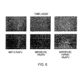


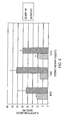
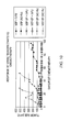


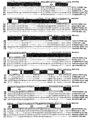


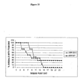


Classifications
-
- A—HUMAN NECESSITIES
- A61—MEDICAL OR VETERINARY SCIENCE; HYGIENE
- A61K—PREPARATIONS FOR MEDICAL, DENTAL OR TOILETRY PURPOSES
- A61K45/00—Medicinal preparations containing active ingredients not provided for in groups A61K31/00 - A61K41/00
- A61K45/06—Mixtures of active ingredients without chemical characterisation, e.g. antiphlogistics and cardiaca
-
- A—HUMAN NECESSITIES
- A61—MEDICAL OR VETERINARY SCIENCE; HYGIENE
- A61K—PREPARATIONS FOR MEDICAL, DENTAL OR TOILETRY PURPOSES
- A61K31/00—Medicinal preparations containing organic active ingredients
- A61K31/33—Heterocyclic compounds
- A61K31/395—Heterocyclic compounds having nitrogen as a ring hetero atom, e.g. guanethidine or rifamycins
- A61K31/435—Heterocyclic compounds having nitrogen as a ring hetero atom, e.g. guanethidine or rifamycins having six-membered rings with one nitrogen as the only ring hetero atom
- A61K31/47—Quinolines; Isoquinolines
- A61K31/4738—Quinolines; Isoquinolines ortho- or peri-condensed with heterocyclic ring systems
- A61K31/4745—Quinolines; Isoquinolines ortho- or peri-condensed with heterocyclic ring systems condensed with ring systems having nitrogen as a ring hetero atom, e.g. phenantrolines
-
- A—HUMAN NECESSITIES
- A61—MEDICAL OR VETERINARY SCIENCE; HYGIENE
- A61K—PREPARATIONS FOR MEDICAL, DENTAL OR TOILETRY PURPOSES
- A61K31/00—Medicinal preparations containing organic active ingredients
- A61K31/33—Heterocyclic compounds
- A61K31/395—Heterocyclic compounds having nitrogen as a ring hetero atom, e.g. guanethidine or rifamycins
- A61K31/495—Heterocyclic compounds having nitrogen as a ring hetero atom, e.g. guanethidine or rifamycins having six-membered rings with two or more nitrogen atoms as the only ring heteroatoms, e.g. piperazine or tetrazines
- A61K31/505—Pyrimidines; Hydrogenated pyrimidines, e.g. trimethoprim
- A61K31/513—Pyrimidines; Hydrogenated pyrimidines, e.g. trimethoprim having oxo groups directly attached to the heterocyclic ring, e.g. cytosine
-
- A—HUMAN NECESSITIES
- A61—MEDICAL OR VETERINARY SCIENCE; HYGIENE
- A61K—PREPARATIONS FOR MEDICAL, DENTAL OR TOILETRY PURPOSES
- A61K31/00—Medicinal preparations containing organic active ingredients
- A61K31/70—Carbohydrates; Sugars; Derivatives thereof
- A61K31/7042—Compounds having saccharide radicals and heterocyclic rings
- A61K31/7048—Compounds having saccharide radicals and heterocyclic rings having oxygen as a ring hetero atom, e.g. leucoglucosan, hesperidin, erythromycin, nystatin, digitoxin or digoxin
-
- A—HUMAN NECESSITIES
- A61—MEDICAL OR VETERINARY SCIENCE; HYGIENE
- A61K—PREPARATIONS FOR MEDICAL, DENTAL OR TOILETRY PURPOSES
- A61K38/00—Medicinal preparations containing peptides
- A61K38/16—Peptides having more than 20 amino acids; Gastrins; Somatostatins; Melanotropins; Derivatives thereof
- A61K38/17—Peptides having more than 20 amino acids; Gastrins; Somatostatins; Melanotropins; Derivatives thereof from animals; from humans
- A61K38/39—Connective tissue peptides, e.g. collagen, elastin, laminin, fibronectin, vitronectin, cold insoluble globulin [CIG]
-
- A—HUMAN NECESSITIES
- A61—MEDICAL OR VETERINARY SCIENCE; HYGIENE
- A61P—SPECIFIC THERAPEUTIC ACTIVITY OF CHEMICAL COMPOUNDS OR MEDICINAL PREPARATIONS
- A61P35/00—Antineoplastic agents
-
- A—HUMAN NECESSITIES
- A61—MEDICAL OR VETERINARY SCIENCE; HYGIENE
- A61P—SPECIFIC THERAPEUTIC ACTIVITY OF CHEMICAL COMPOUNDS OR MEDICINAL PREPARATIONS
- A61P35/00—Antineoplastic agents
- A61P35/02—Antineoplastic agents specific for leukemia
-
- A—HUMAN NECESSITIES
- A61—MEDICAL OR VETERINARY SCIENCE; HYGIENE
- A61P—SPECIFIC THERAPEUTIC ACTIVITY OF CHEMICAL COMPOUNDS OR MEDICINAL PREPARATIONS
- A61P43/00—Drugs for specific purposes, not provided for in groups A61P1/00-A61P41/00
-
- A—HUMAN NECESSITIES
- A61—MEDICAL OR VETERINARY SCIENCE; HYGIENE
- A61K—PREPARATIONS FOR MEDICAL, DENTAL OR TOILETRY PURPOSES
- A61K48/00—Medicinal preparations containing genetic material which is inserted into cells of the living body to treat genetic diseases; Gene therapy
Definitions
- the invention relates to cancer therapy sensitizing compositions and methods.
- MDR multidrug resistance family
- LRP lung resistance protein
- MMR DNA mismatch repair
- SPARC Secreted protein acidic and rich in cystein
- matricellular proteins secreted protein acidic and rich in cystein
- SPARC belongs to a family of extracellular proteins, called matricellular proteins. Since its identification and cloning, the functional role of SPARC remains unclear. Its high evolutionary conservation suggests an important physiological role for this protein ( Iruela-Arispe ML, Lane TF, Redmond D, Reilly M, Bolender RP, Kavanagh TJ, Sage EH. Mol Biol Cell. 1995 Mar; 6(3):327-43 ).
- Initial studies showed that SPARC is important in bone mineralization ( Termine JD, Kleinman HK, Whitson SW, Conn KM, McGarvey ML, Martin GR. Cell. 1981 Oct; 26(1 Pt 1):99-105 ).
- SPARC is expressed at high levels in bone tissue, it is also distributed widely in other tissues and cell types ( Maillard, C., et al., Bone, 13:257-264 (1992 )). Its role has been expanded to include tissue remodeling ( Latvala T, Puolakkainen P, Vesaluoma M, Tervo T. Exp Eye Res. 1996 Nov;63(5):579-84 ; Kelm RJ Jr, Swords NA, Orfeo T, Mann KG. J Biol Chem. 1994 Dec 2;269(48):30147-53 ); endothelial cell migration ( Hasselaar P, Sage EH. J Cell Biochem.
- WO0202771 discloses a novel hSPARC-h1 polypeptide and its potential applications in tissue remodeling, tissue repair and general modulation of various growth factor activities.
- U.S. Patent No. 6,387,664 provides a SPARC fusion protein obtainable by fusing SPARC to thioredoxin which can be used for basic research in neurobiology and/or for treating various neuropathologies.
- U.S. Patent No. 6,239,326 provides a SPARC-deficient transgenic mouse model for testing drugs in promoting or retarding wound healing and treating or preventing cataracts, diabetes mellitus or osteoporosis.
- the invention is based on the discovery that SPARC sentitizes cancer therapy.
- the present invention provides compositions and methods for sensitizing cancer therapeutic treatment.
- the present invention provides a composition comprising a SPARC family polypeptide and a chemotherapy agent.
- the present invention provides a composition comprising a SPARC family polypeptide and a chemotherapy-resistant cell.
- the present invention provides a chemotherapy-resistant cell comprising a recombinant SPARC family polynucleotide.
- the present invention provides a Recombinant cell comprising a heterologous transcription control region operatively associated with a SPARC family polynucleotide.
- the present invention provides a method for in vivo sensitizing a mammal to a therapeutic treatment, the method comprising administrating to the mammal diagnosed with cancer an effective amount of a SPARC family polypeptide or a polynucleotide encoding a SPARC polypeptide.
- the present invention provides a method for ex vivo sensitizing a mammal diagnosed with cancer to a therapeutic treatment, the method comprising: administering to a mammal an effective amount of a cell comprising a SPARC family polypeptide or a polynucleotide encoding a SPARC family polypeptide; wherein the cell produces an increased amount of the SPARC polypeptide.
- the present invention also provides a method for ex vivo sensitizing a mammal diagnosed with cancer to a therapeutic treatment, the method comprising: (1) Obtaining a cancer sample from the mammal; (2) contacting the cancer sample with an effective amount of a composition comprising a SPARC family polypeptide or a polynucleotide encoding a SPARC polypeptide; and (3) returning the cancer sample after the contacting of (2) to the mammal.
- the present invention further provides a method for sensitizing a cancer sample to a therapeutic treatment, the method comprising contacting the cancer sample with an effective amount of a composition comprising a SPARC family polypeptide or a polynucleotide encoding a SPARC polypeptide.
- the cancer sample is a cell or tissue sample.
- the cancer sample is transfected or infected with the polynucleotide of (e)-(f).
- the present invention provides a method for evaluating a first cancer cell for its resistance to a therapeutic treatment, comprising: (a) measuring the expression level of a SPARC family mRNA or polypeptide, or the extracellular level of a SPARC family polypeptide in the first cancer cell; and (b) comparing the expression level or the extracellular level obtained in (a) with the expression level of the SPARC family mRNA or polypeptide, or the extracellular level of the SPARC family polypeptide in a second cancer cell which does not exhibit resistance to the therapeutic treatment; wherein a lower level of expression or extracellular level in (a) is indicative of the first cancer cell being resistant to the therapeutic treatment.
- the present invention provides a method for evaluating a first cancer cell for its resistance to a therapeutic treatment, comprising: (a) measuring expression level of a SPARC family mRNA or polypeptide, or extracellular level of a SPARC family polypeptide in the first cancer sample; (b) measuring expression level of the SPARC family mRNA or polypeptide, or extracellular level of the SPARC family polypeptide in a second cancer sample which does not exhibit resistance to the therapeutic treatment; (c) comparing the expression levels or the extracellular levels obtained in (a) and (b), where a lower level of expression or extracellular level in (a) is indicative of the first cancer sample being resistant to the therapeutic treatment.
- the first sample is from a first mammal and the lower level of expression or extracellular level in (a) is further indicative of the first mammal being resistant to the therapeutic treatment.
- the second cancer sample is from a first mammal who provides the first cancer sample.
- the second cancer sample is from a second mammal who is different from the first mammal providing the first cancer sample.
- the first mamnal and the second mammal are diagnosed with the same cancer.
- the expression level of the SPARC family mRNA is measured by polymerase chain reaction, DNA microarray or northern blot.
- the expression or extracellular level of the SPARC family polypeptide is measured by Immuno Blotting or Enzyme-Linked Immunosorbent Assay (Elisa).
- the present invention provides a method for identifying an agent which modulates a SPARC family mRNA or polypeptide expression, or a SPARC family polypeptide secretion, comprising: (a) measuring expression level of the SPARC family mRNA or polypeptide, or extracellular level of the SPARC family polypeptide in a sample; (b) contacting a candidate agent with the sample; (c) after the contacting of (b), measuring expression or extracellular level of the SPARC family mRNA or polypeptide, or extracellular level of the SPARC family polypeptide in the sample of (b); (d) comparing the expression levels or the extracellular levels in (a) and (c), where a differential level of expression or extracellular level in (a) and (c) indicates the candidate agent being an agent which modulates the SPARC family mRNA or polypeptide expression, or the SPARC family polypeptide secretion.
- the present invention also provides a method for identifying an agent which sensitizes a cancer sample to a therapeutic treatment, comprising: (a) measuring expression level of a SPARC family mRNA or polypeptide, or extracellular level of a SPARC family polypeptide in the cancer sample; (b) contacting a candidate agent with the cancer sample; (c) after the contacting of (b), measuring expression level of the SPARC family mRNA or polypeptide, or extracellular level of the SPARC family polypeptide in the cancer sample of (b); (d) comparing the expression levels or the extracellular levels obtained in (a) and (c), where an increased level of expression or extracellular level in (c) indicates the candidate agent being an agent which sensitizes a cancer sample to a therapeutic treatment.
- the cancer sample of the subject method is from a mammal diagnosed with cancer, and the increased level of expression or extracellular level is further indicative of the candidate agent being an agent which sensitizes the mammal to a therapeutic treatment.
- the present invention provides a method for determining a therapeutic treatment protocol for a first mammal diagnosed with cancer, comprising: (a) determining if expression of a SPARC family mRNA or polypeptide or extracellular level of a SPARC family polypeptide is lower in a first sample from the first mammal than a second sample which does not exhibit resistance to the therapeutic treatment; and (b) if (a) is positive, increasing the strength of the therapeutic treatment to the first mammal so as to increase the response to the treatment.
- the polynucleotide of (e) or (f) of the subject composition and method is an expression vector.
- the expression vector is a plasmid or a viral vector.
- the plasmid vector is pcDNA3.1.
- the SPARC family polypeptide or a polynucleotide encoding a SPARC family polypeptide of the present invention include: (a) a SPARC polypeptide which is selected from the group consisting of SEQ ID Nos. 1-17; (b) a polypeptide having an amino acid sequence of at least 60% homology to the SPARC family polypeptide in (a); (c) a polypeptide fragment of (a)-(b) where the fragment is at least 50 amino acids in length; (d) a fusion polypeptide comprising the polypeptide of (a), (b), or (c); (e) a polynucleotide encoding the polypeptide of (a), (b), (c) or the fusion polypeptide of (d); or (f) a polynucleotide hybridizing to the polynucleotide of (e) under a stringent hybridization condition.
- the SPARC family polypeptide or polynucleotide of the present invention is selected from: SMOC-1, SPARC, hevin, SC1, QR-1, follistatin-like proteins (TSC-36), testican.
- the therapeutic agent of the subject composition and method is a chemotherapy agent or a radiation therapy agent.
- the therapeutic agent is selected from the group consisting of the agents listed in Table 1.
- the mammal of the subj ect method exhibits resistance to the therapeutic treatment.
- the therapeutic treatment is chemotherapy or radiation therapy.
- the cancer sample of the subject method is a cell or a tissue sample.
- the cell or tissue sample is lysed before the measuring of the expression or extracellular level of a SPARC family polypeptide or polynucleotide (e.g., mRNA).
- a SPARC family polypeptide or polynucleotide e.g., mRNA
- a polynucleotide or polypeptide extract is obtained from the cell or tissue sample before the measuring of the expression or extracellular level of a SPARC family polypeptide or polynucleotide (e.g., mRNA).
- a SPARC family polypeptide or polynucleotide e.g., mRNA
- the invention is based on the SPARC family and sensitization to cancer therapy.
- SPARC family polypeptide refers to a polypeptide (including a fragment or variant thereof) of a family of extracellular proteins.
- This family of extracellular proteins include SPARC and other members of the family, such as SMOC-1, hevin, SC1, QR-1, follistatin-like proteins (TSC-36) and testican (see for example, Van Spotify et al., (2002), J. Biol. Chem. 277(41): 37977-37986 ; Johnston, I. G., Paladino, T., Gurd, J. W., and Brown, I. R.
- a SPARC family polypeptide is typically composed of three independently folded domains ( Yan and Sage, 1999, J. Histochem. & Cytochem., 47(12):1495-1505 , hereby incorporated by reference).
- the N-terminal domain e.g., two adjacent N-terminal Glu 3 and Glu 4 in SPARC
- the second domain e.g., residues 53-137 in SPARC
- FS follistatin
- the C-terminal extracellular calcium-binding (EC) domain e.g., residues 138-286 in SPARC
- SPARC family polypeptide includes the full length polypeptide or a fragment thereof, a wild-type polypeptide or any variant thereof.
- a "SPARC family polynucleotide” is a polynucleotide (e.g., DNA or mRNA) molecule encoding a SPARC polypeptide, or a fragment thereof. (a) a SPARC family polypeptide or gene selected from the group consisting of SEQ ID Nos.
- SPARC polypeptide refers to SEQ ID Nos. 1-17, and the term "SPARC gene” to SEQ ID Nos. 18-34, the corresponding nucleotide sequences of SEQ ID Nos. 1-17. It is contemplated that variations of these sequences which retain the biological activity of SPARC are equivalents of these sequences.
- the biological activity of the SPARC gene is that it is downregulated in chemotherapy resistant cells. The gene also may be overexpressed in cells that are sensitive to chemotherapy. The biological activity of the SPARC polypeptide is that it sensitizes chemotherapy resistant cells to chemotherapy.
- SPARC family polypeptide member With respect to a SPARC family polypeptide member, it is contemplated that variations of their sequences which retain the biological activity of the family member are equivalents of these sequences.
- the biological activity of a SPARC gene family member is that the gene is downregulated in chemotherapy resistant cells, i.e., expression decreased by at least 25%, for example, 30%, 40%, 50%, 75%, 100% (1-fold), 2-fold, 4-fold, or 5-fold or more, compared to chemotherapy sensitive cells.
- the gene also may be overexpressed in cells that are sensitive to chemotherapy.
- the biological activity of the SPARC family polypeptide member is that it sensitizes chemotherapy resistant cells to chemotherapy, i.e., increase the response to chemotherapy by at least 25%, for example, 30%, 40%, 50%, 60%, 70%, 80%, or more, to 2-fold, 3-fold, 4-fold, 5-fold, 10-fald, 15-fold, 20-fold or more, compared to treatment sensitivity in the absence of the SPARC family polypeptide.
- tissue is an aggregate of cells that perform a particular function in an organism.
- tissue refers to cellular material from a particular physiological region.
- the cells in a particular tissue may comprise several different cell types. A non-limiting example of this would be brain cells that further comprise neurons and glial cells, as well as capillary endothelial cells and blood cells.
- tissue type refers to the tissue of origin, for example, from which a tumor develops.
- tissue types include, for example, without limitation, blood, colorectal, breast, esophageal, hepatocellular, prostate, ovarian, thyroid, pancreas, uterus, testis, pituitary, kidney, stomach, esophagus and rectum.
- cancer refers to a proliferative disorder disease caused or characterized by the proliferation of cells which have lost susceptibility to normal growth control.
- cancer includes tumors and any other proliferative disorders. Cancers of the same tissue type originate in the same tissue, and may be divided into different subtypes based on their biological characteristics.
- the cancer may be selected from one or more from the group consisting of: melanoma, leukemia, astocytoma, glioblastoma, lymphoma, glioma, Hodgkins lymphoma, chronic lymphocyte leukemia and cancer of the pancreas, breast, thyroid, ovary, uterus, testis, pituitary, kidney, stomach, esophagus and rectum.
- the term "sensitizing” refers to an increased sensitivity or reduce the resistance of a cancer sample or a mammal responding to a therapeutic treatment.
- An increased sensitivity or a reduced sensitivity to a therapeutic treatment is measured according to a known method in the art for the particular treatment and methods described herein below, including, but not limited to, cell proliferative assays ( Tanigawa N, Kern DH, Kikasa Y, Morton DL, Cancer Res 1982; 42: 2159-2164 ), cell death assays ( Weisenthal LM, Shoemaker RH, Marsden JA, Dill PL, Baker JA, Moran EM, Cancer Res 1984; 94: 161-173 ; Weisenthal LM, Lippman ME, Cancer Treat Rep 1985; 69: 615-632 ; Weisenthal LM, In: Kaspers GJL, Pieters R, Twentyman PR, Weisenthal LM, Veerman AJP, eds.
- the sensitivity or resistance may also be measured in animal by measuring the tumor size reduction over a period of time, for example, 6 month for human and 4-6 weeks for mouse.
- a composition or a method sensitizes response to a therapeutic treatment if the increase in treatment sensitivity or the reduction in resistance is 25% or more, for example, 30%, 40%, 50%, 60%, 70%, 80%, or more, to 2-fold, 3-fold, 4-fold, 5-fold, 10-fold, 15-fold, 20-fold or more, compared to treatment sensitivity or resistance in the absence of such composition or method.
- the determination of sensitivity or resistance to a therapeutic treatment is routine in the art and within the skill of an ordinarily skilled clinician.
- administer refers to introduce by any means a composition (e.g., a therapeutic agent) into the body of a mammal in order to prevent or treat a disease or condition (e.g., cancer).
- a composition e.g., a therapeutic agent
- a disease or condition e.g., cancer
- treating refers to curative therapy, prophylactic therapy, or preventative therapy.
- An example of “preventative therapy” is the prevention or lessening of a targeted disease (e.g., cancer) or related condition thereto.
- a targeted disease e.g., cancer
- Those in need of treatment include those already with the disease or condition as well as those prone to have the disease or condition to be prevented.
- the terms “treating”, “treatment”, “therapy” and “therapeutic treatment” as used herein also describe the management and care of a mammal for the purpose of combating a disease, or related condition, and includes the administration of a composition to alleviate the symptoms, side effects, or other complications of the disease, condition.
- Therapeutic treatment for cancer includes, but is not limited to, surgery, chemotherapy, radiation therapy, gene therapy, and immunotherapy.
- therapeutically effective amount is meant an amount that relieves (to some extent, as judged by a skilled medical practitioner) one or more symptoms of the disease or condition in a mammal. Additionally, by “therapeutically effective amount” is meant an amount that returns to normal, either partially or completely, physiological or biochemical parameters associated with or causative of a disease or condition.
- a clinician skilled in the art can determine the therapeutically effective amount of a composition in order to treat or prevent a particular disease condition, or disorder when it is administered, such as intravenously, subcutaneously, intraperitoneally, orally, or through inhalation.
- compositions required to be therapeutically effective will depend upon numerous factors, e.g., such as the specific activity of the active agent, the delivery device employed, physical characteristics of the agent, purpose for the administration, in addition to many patient specific considerations.
- amount of a composition that must be administered to be therapeutically effective is routine in the art and within the skill of an ordinarily skilled clinician.
- agent refers to a chemical compound, a mixture of chemical compounds, a biological macromolecule, or an extract made from biological materials such as bacteria, plants, fungi, or animal (particularly mammalian) cells or tissues that are suspected of having therapeutic properties.
- the agent or drug may be purified, substantially purified or partially purified.
- An “agent”, according to the present invention, also includes a radiation therapy agent.
- modulation means that a desired/selected response is more efficient (e.g., at least 10%, 20%, 40%, 60% or more), more rapid (e.g., at least 10%, 20%, 40%, 60% or more), greater in magnitude (e.g., at least 10%, 20%, 40%, 60% or greater), and/or more easily induced (e.g., at least 10%, 20%, 40%, 60% or more) in the presence of an agent than in the absence of the agent.
- the term "resistance to a therapeutic treatment” refers to an acquired or natural resistance of a cancer sample or a mammal to a therapy, i.e., being nonresponsive to or having reduced or limited response to the therapeutic treatment, e.g., having a reduced response to a therapeutic treatment by 25% or more, for example, 30%, 40%, 50%, 60%, 70%, 80%, or more, to 2-fold, 3-fold, 4-fold, 5-fold, 10-fold, 15-fold, 20-fold or more.
- the reduction in response is measured by comparing with the same cancer sample or mammal before the resistance is acquired, or by comparing with a different cancer sample or a mammal who is known to have no resistance to the therapeutic treatment.
- a typical acquired resistance to chemotherapy is called "multidrug resistance.”
- the multidrug resistance can be mediated by P-glycoprotein or can be mediated by other mechanisms, or it can occur when a mammal is infected with a multi drug-resistant microorganism or a combination of microorganisms.
- the determination of resistance to a therapeutic treatment is routine in the art and within the skill of an ordinarily skilled clinician, for example, can be measured by cell proliferative assays and cell death assays as described herein above under “sensitizing”.
- chemotherapy refers to the use of drugs to destroy cancer cells (including leukaemias and lymphomas). There are over 50 different chemotherapy drugs and some are given on their own, but often several drugs may be combined (known as combination chemotherapy). Chemotherapy may be used alone to treat some types of cancer. Sometimes it can be used together with other types of treatment such as surgery, radiotherapy, immunotherapy, or a combination thereof.
- Radiotherapy also called “radiation therapy” refers to the treatment of cancer and other diseases with ionizing radiation. Ionizing radiation deposits energy that injures or destroys cells in the area being treated (the “target tissue") by damaging their genetic material, making it impossible for these cells to continue to grow. Although radiation damages both cancer cells and normal cells, the latter are able to repair themselves and function properly. Radiotherapy may be used to treat localized solid tumors, such as cancers of the skin, tongue, larynx, brain, breast, or uterine cervix. It can also be used to treat leukemia and lymphoma (cancers of the blood-forming cells and lymphatic system, respectively)
- treatment protocol refers to the process of informing the decision making for the treatment of a disease.
- treatment protocol is based on the comparative levels of one or more cell growth-related polypeptides in a patient's tissue sample relative to the levels of the same polypeptide(s) in a plurality of normal and diseased tissue samples from mammals for whom patient information, including treatment approaches and outcomes is available.
- biological characteristics refers to the phenotype and/or genotype of one or more cells or tissues, which can include cell type, and/or tissue type from which the cell was obtained, morphological features of the cell(s)/tissue(s), and the expression of biological molecules within the cell(s)/tissue(s).
- sample refers to material derived from the body of a mammal, including, but not limited to, blood, serum, plasma, urine, nipple aspirate, cerebrospinal fluid, liver, kidney, breast, bone, bone marrow, testes or ovaries and brain, colon, and lung.
- sample may also be cultured cells and tissues.
- a “cancer sample” refers to a sample which originates from a cancer, i.e., from a new growth of different or abnormal tissue.
- a “cancer sample” may be a cell or tissue sample.
- the cancer cells may exist as part of the cancer tissue, or may exist as free-floating cells detached from the cancer tissue from which they originate.
- a cancer sample, according to the present invention may be used for in vitro or ex vivo testing of cancers.
- non-cancer sample refers to cell or tissue sample obtained from a normal tissue. A sample may be judged a non-tumor sample by one of skill in the art on the basis of morphology and other diagnostic tests.
- mammal refers to a human or other animal, such as farm animals or laboratory animals (e.g. guinea pig or mice).
- hybridization refers to hybridization which occurs when two polynucleotide sequences are substantially complementary, i.e., there is at least about 60% and preferably, at least about 80% or 90% identity between the sequences, wherein the region of identity comprises at least 10 nucleotides.
- the sequences hybridize under stringent conditions following incubation of the sequences overnight at 42°C, followed by stringent washes (0.2X SSC at 65° C).
- stringent conditions will be those in which the salt concentration is at least about 0.01 to 1.0 M Na ion concentration (or other salts) at pH 7.0 to 8.3 and the temperature is at least about 30°C for short probes (e.g., about 6 to 50 nucleotides). Stringent conditions may also be achieved with the addition of destabilizing agents such as formamide. Generally, stringent conditions are selected to be about 5°C lower than the thermal melting point (Tm) for the specific sequence at a defined ionic strength and pH, as calculated using methods routine in the art.
- Tm thermal melting point
- homology refers to the optimal alignment of sequences (either nucleotides or amino acids), which may be conducted by computerized implementations of algorithms.
- homoology with regard to polynucleotides, for example, may be determined by analysis with BLASTN version 2.0 using the default parameters.
- Hanology with respect to polypeptides (i.e., amino acids), may be determined using a program, such as BLASTP version 2.2.2 with the default parameters, which aligns the polypeptides or fragments being compared and determines the extent of amino acid identity or similarity between them. It will be appreciated that amino acid "homology” includes conservative substitutions, i.e.
- a "homology of 60% or higher” includes a homology of, for example, 60%, 70%, 75%, 80%, 85%, 90%, 95%, and up to 100% (identical) between two or more nucleotide or amino acid sequences.
- polynucleotide includes RNA, cDNA, genomic DNA, synthetic forms, and mixed polymers, both sense and antisense strands, and may be chemically or biochemically modified or may contain non-natural or derivatized nucleotide bases, as will be readily appreciated by those skilled in the art.
- Such modifications include, for example, labels, methylation, substitution of one or more of the naturally occurring nucleotides with an analog, internucleotide modifications such as uncharged linkages (e.g., methyl phosphonates, phosphotriesters, phosphoamidates, carbamates, etc.), charged linkages (e.g., phosphorothioates, phosphorodithioates, etc.), pendent moieties (e.g., polypeptides), and modified linkages (e.g., alpha anomeric polynucleotides, etc.).
- uncharged linkages e.g., methyl phosphonates, phosphotriesters, phosphoamidates, carbamates, etc.
- charged linkages e.g., phosphorothioates, phosphorodithioates, etc.
- pendent moieties e.g., polypeptides
- modified linkages e.g., alpha anomeric polynucleotides
- mutation refers to a change in nucleotide sequence within a gene, or outside the gene in a regulatory sequence compared to wild type.
- the change may be a deletion, substitution, point mutation, mutation of multiple nucleotides, transposition, inversion, frame shift, nonsense mutation or other forms of aberration that differentiate the polynucleotide or protein sequence from that of a normally expressed gene in a functional cell where expression and functionality are within the normally occurring range.
- Polypeptide and “protein” are used interchangeably herein to refer to a polymer of amino acid residues.
- recombinant protein refers to a protein that is produced by expression of a recombinant DNA molecule that encodes the amino acid sequence of the protein.
- Polynucleotides and recombinantly produced polypeptide, and fragments or analogs thereof, may be prepared according to methods known in the art and described in Maniatis et al., Molecular Cloning: A Laboratory Manual, 2nd Ed., (1989), Cold Spring Harbor, N.Y ., and Berger and Kimmel, Methods in Enzymology, Volume 152, Guide to Molecular Cloning Techniques (1987), Academic Press, Inc., San Diego, Calif ., which are incorporated herein by reference.
- fragment when in reference to a polypeptide (as in "a fragment of a given protein") refers to a shorter portion of the polypeptide.
- the fragment may range in size from four amino acid residues to the entire amino acid sequence minus one amino acid.
- the present invention contemplates "functional fragments" of a polypeptide.
- Such fragments, according to the present invention are “functional” in that they retain the ability to sensitize a cancer sample or cancer mammal to a therapeutic treatment, albeit with perhaps lower sensitizing activity than that observed for the full-length polypeptide.
- fragment of a polypeptide is preferably greater than 10 amino acids in length, and more preferably greater than 50 amino acids in length, and even more preferably greater than 100 amino acids in length.
- a “fragment” of a SPARC family polypeptide, according to the present invention may contain one or more of the three conserved domains of the SPARC family members, i.e., the Acidic N-terminal domain, the follistatin-like domain, and the extracellular calcium-binding EC domain.
- a “variant" of a specific polypeptide refers to a polypeptide substantially similar to either the entire specific peptide or a fragment thereof.
- substantially similar it means that the variant is made to arrive at a final construct which possesses the desired function, i.e., sensitizing a cancer sample or a mammal to a therapeutic treatment, albeit with perhaps lower sensitizing activity of the variant than that observed for the wild-type polypeptide.
- Such variants include, for example, deletions, insertions, or substitutions of residues within the aminoacid sequence of the specific polypeptide.
- a “variant” may also be a fusion polypeptide between a SPARC family polypeptide and a second polypeptide. Any combination of deletion, insertion, substitution, and fusion may also be made.
- isolated or purified when used in reference to a polynucleotide or a polypeptide means that a naturally occurring nucleotide or amino acid sequence has been removed from its normal cellular environment or is synthesized in a non-natural environment (e.g., artificially synthesized). Thus, an "isolated” or “purified” sequence may be in a cell-free solution or placed in a different cellular environment.
- nucleotide or amino acid sequence is the only polynucleotide or polypeptide present, but that it is essentially free (about 90-95%, up to 99-100% pure) of non-polynucleotide or polypeptide material naturally associated with it..
- encoding refers to the inherent property of specific sequences of nucleotides in a polynucleotide, such as a gene in a chromosome or an inRNA, to serve as templates for synthesis of other polymers and macromolecules in biological processes having a defined sequence of nucleotides (i.e., rRNA, tRNA, other RNA molecules) or amino acids and the biological properties resulting therefrom.
- a gene encodes a protein, if transcription and translation of mRNA produced by that gene produces the protein in a cell or other biological system.
- Both the coding strand, the nucleotide sequence of which is identical to the mRNA sequence and is usually provided in sequence listings, and non-coding strand, used as the template for transcription, of a gene or cDNA can be referred to as encoding the protein or other product of that gene or cDNA.
- a polynucleotide that encodes a protein includes any polynucleotides that have different nucleotide sequences but encode the same amino acid sequence of the protein due to the degeneracy of the genetic code. Polynucleotides and nucleotide sequences that encode proteins may include introns and may be genomic DNA.
- vector refers to a polynucleotide compound used for introducing exogenous or endogenous polynucleotide into host cells.
- a vector comprises a nucleotide sequence which may encode one or more polypeptide molecules. Plasmids, cosmids, viruses and bacteriophages, in a natural state or which have undergone recombinant engineering, are non-limiting examples of commonly used vectors to provide recombinant vectors comprising at least one desired isolated polynucleotide molecule.
- transformation or the term “transfection” refers to a variety of art-recognized techniques for introducing exogenous polynucleotide (e.g., DNA) into a cell.
- a cell is “transformed” or “transfected” when exogenous DNA has been introduced inside the cell membrane.
- transformation and “transfection” and terms derived from each are used interchangeably.
- an "expression vector” refers to a recombinant expression cassette which has a polynucleotide which encodes a polypeptide (i.e., a protein) that can be transcribed and translated by a cell.
- the expression vector can be a plasmid, virus, or polynucleotide fragment.
- expression refers to the production of a protein or nucleotide sequence in a cell or in a cell-free system, and includes transcription into an RNA product, post-transcriptional modification and/or translation into a protein product or polypeptide from a DNA encoding that product, as well as possible post-translational modifications.
- comparing the expression level refers to comparing the deferential expression of a polynucleotide or a polypeptide in two or more samples.
- the term “differential expression” refers to both quantitative, as well as qualitative, differences in a polynucleotide or a polypeptide expression patterns among two or more samples.
- a polynucleotide or a polypeptide is said to be “differentially expressed” if its expression is detectable in one sample, but not in another sample, by known methods for polynucleotide or polypeptide detection (e.g., electrophoresis).
- a polynucleotide or a polypeptide is also said to be "differentially expressed” if the quantitative difference of its expression (i.e., increase or decrease, measured in ⁇ g, ⁇ mol or copy number) between two samples is about 20%, about 30%, about 50%, about 70%, about 90% to about 100% (about twofold) or more, up to and including about 1.2 fold, 2.5 fold, 5-fold, 10-fold, 20-fold, 50-fold or more.
- a “differentially expressed" gene transcript means a mRNA transcript that is found in different numbers of copies between two or more samples.
- an "increased amount of a SPARC family polypeptide or polynucleotide” refers to a greater level of expression of at least 20%, e.g., 30%, 40%, 50%, 60%, 70%, 80%, or 90%, or 2-fold, 3-fold, 4-fold, 5-fold or more in a cell, compared to a control cell.
- a cell expressing a SPARC family polypeptide or polynucleotide is said to have an increase amount of such polypeptide or polynucleotide if the expression is as defined herein above when compared with a chemotherapy resistant cell.
- secreted protein refers to a protein having at least a portion which is extracellular and includes proteins which are completely extracellular (i.e., not attached to a cell).
- Level of secretion refers to the level (i.e., amount) of a secreted protein in the extracellular compartment.
- the term “proliferation” refers to the rate of cell division and the ability of a cell to continue to divide.
- One complete cell division process is referred to as a "cycle".
- an “increase in cell proliferation” is meant to increase the cell division rate so that the cell has a higher rate of cell division compared to normal cells of that cell type, or to allow the cell division to continue for more cycles without changing the rate of each cell division, e.g., increase by 10% or higher (e.g., 20%, 30%, 40% 50%, up to 2 fold, 5 fold, 10 fold or higher.
- an “decrease in cell proliferation” is meant to decrease the cell division rate so that the cell has a lower rate of cell division compared to normal cells of that cell type, or to reduce the number of cycles of the cell division without changing the rate of each cell division, e.g., decrease by 10% or higher (e.g., 20%, 30%, 40% 50%, up to 2 fold, 5 fold, 10 fold or higher).
- the present invention provides compositions and methods for sensitizing cancer therapeutic treatments. Such sensitizing compositions and methods are particularly useful in enhancing the response of patients who are resistant to a treatment. They are also useful in reducing the side-effects of cancer therapy, for example, by enhancing the response of a patient to a smaller strength (i.e., dosage) of the treatment.
- the composition of the present invention may reduce the dosage of a therapeutic treatment agent by at least 20%, for example, 30%, 40%, 50%, and up to 60%.
- the present invention provides (a) a SPARC family polypeptide selected from the group consisting of SEQ ID Nos. 1-17; (b) a polypeptide having an amino acid sequence of at least 60% homology to said SPARC family polypeptide in (a); (c) a polypeptide fragment of (a)-(b) wherein said fragment is at least 50 amino acids in length, and (d) a fusion polypeptide comprising the polypeptide of (a)-(c).
- a SPARC family polypeptide provided by the present invention may be a wild-type polypeptide or a variant thereof, it may be the full length polypeptide or a fragment thereof.
- a SPARC family polypeptide is within the scope of the present invention so long as it retains the function of sensitizing a cancer sample or a patient to a therapeutic treatment, albeit that a lower activity may exist for a variant or a fragment polypeptide when compared to the wile-type or full length polypeptide.
- SMOC-1 Van Spotify et al., 2002, J. Biol. Chem. 277(41):37977-37986
- hevin Bendik I, Schraml P, Ludwig CU, Cancer Res. 1998; 58(4):626-9
- SC1 Johnston IG, Paladino T, Gurd JW, Brown IR, Neuron. 1990 4(1):165-76
- QR-1 follistatin-like proteins
- TSC-36 Shibanuma,M., Mashimo,J., Mita,A., Kuroki,T. and Nose,K, 1993, Eur. J. Biochem.
- a SPARC family polypeptide of the present invention includes, but is not limited to, SPARC and any of its identified family members known in the art or as described herein.
- the alignment of the various polynucleotide sequences permit one skilled in the art to select conserved portions of the proteins (i.e. those portions in common between two or more sequences) as well as unconserved portions (i.e. those portions unique to two or more sequences).
- the present invention contemplates conserved fragments 10 amino acids in length or greater, and more typically greater than 50 amino acids in length.
- the SPARC family polypeptide of the present invention contains one or two or three domains (i.e. the Acidic N-terminal domain, the follistatin-like domain, and/or the EC domain), conserved among the SPARC family members.
- the therapy-sensitizing fragment or a variant of a wild-type SPARC family protein may be delineated by routine sequence manipulations known to those skilled in the art, including, but 5 not limited to, deletion mutations, point mutations, fusions as described herein below and as described in J. Sambrook, E. F. Fritsch, and T. Maniatis, Molecular Cloning: A. Laboratory Manual, 2 Ed., Cold Spring Harbor Laboratory Press, Cold Spring Harbor, New York, 1989 , and Ausubel et al., Current Protocols in Molecular Biology, 1994 , incorporated by reference herein.
- a mutation in the DNA encoding the variant polypeptide must not alter the reading frame ) and preferably will not create complementary regions that could produce secondary mRNA structures.
- these variants are prepared by site directed mutagenesis of nucleotides in the DNA encoding the peptide molecule thereby producing DNA encoding the variant, and thereafter expressing the DNA in recombinant cell culture.
- a SPARC family polypeptide provided by the present invention may be produced in a prokaryotic or eukaryotic cell using any known method, for example, recombinant DNA techniques. Recombination techniques may be conducted as described herein below, or for example, by the methods disclosed in T. Maniatis et al., "Molecular Cloning", 2nd Ed., Cold Spring Harbor Laboratory, Cold Spring Harbor, N. T.
- a SPARC family polypeptide may be expressed and purified from a recombinant host cell.
- Recombinant host cells may be prokaryotic or eukaryotic, including but not limited to bacteria such as E. coli, fungal cells such as yeast, insect cells including but not limited to drosophila and silkworm derived cell lines, and mammalian cells and cell lines.
- SPARC family polypeptide of the present invention when expressing and purifying a SPARC family polypeptide of the present invention, techniques for improving protein solubility are employed to prevent the formation of inclusion body (which are insoluble fractions), and therefore obtaining large quantities of the polypeptide.
- SPARC accumulated in inclusion bodies is an inactive-type SPARC not retaining its physiological activities.
- Solubility of a purified SPARC family polypeptide may be improved by methods known in the art, and as described herein below.
- solubility may also be improved by expressing a functional fragment, but not the full length SPARC family polypeptide.
- the fragment expressed should retain the sensitizing activity as described herein, albeit the activity may be lower than that of a full length polypeptide.
- a fragment containing one or two or three domains of the three conserved domains of the SPARC family members is expressed.
- an expressed protein e.g., in E.coli
- Polyols e.g. sorbitol
- sucrose may also be added to the media because the increase in osmotic pressure caused by these additions leads to the accumulation of osmoprotectants in the cell, which stabilize the native protein structure.
- Ethanol, low molecular weight thiols and disulfides, and NaCl may be added.
- chaperones and/or foldases may be co-expressed with the desired polypeptide.
- Molecular chaperones promote the proper isomerization and cellular targeting by transiently interacting with folding intermediates.
- the best characterized E. coli chaperone systems are: GroES-GrOEL, DnaK-DnaJ-GrpE, ClpB. Foldases accelerate rate-limiting steps along the folding pathway.
- peptidyl prolyl cis/trans isomerases PPI's
- DsbA disulfide oxidoreductase
- DsbC disulfide isomerase
- PDI protein disulfide isomerase
- a SPARC family polypeptide of the present invention may be produced as a fusion protein in order to improve its solubility and production.
- the fusion protein comprises a SPARC family polypeptide and a second polypeptide fused together in frame.
- the second polypeptide may be a fusion partner known in the art to improve the solubility of the polypeptide to which it is fused, for example, NusA, bacterioferritin (BFR), GrpE, thioredoxin (TRX) and glutathione-S-transferase (GST). Madison, Wis.-based Novagen Inc. provides the pET 43.1 vector series which permit the formation of a NusA-target fusion. DsbA and DsbC have also shown positive effects on expression levels when used as a fusion partner, therefore can be used to fuse with a SPARC polypeptide for achieving higher solubility.
- a SPARC family polypeptide is produced as a fusion polypeptide comprising the SPARC family polypeptide and a fusion partner thioredoxin, as described in U. S. Patent No. 6,387,664 , hereby incorporated by reference in its entirety.
- the thioredoxin-SPARC fusion can be produced in E. coli as an easy-to-formulate, soluble protein in a large quantity without losing the physiological activities of SPARC.
- 6,387,664 provides a fusion SPARC protein with SPARC fused to the C-terminus of thioredoxin, it is understood, for the purpose of the present invention, a SPARC family polypeptide may be fused either to the N-terminus or the C-terminus of a second polypeptide, so long as its sensitizing function is retained.
- a fusion protein comprising a SPARC family polypeptide may be constructed for the easy detection of the expression of the SPARC family polypeptide in a cell.
- the second polypeptide which fused to the SPARC family polypeptide is a reporter polypeptide.
- the reporter polypeptide when served for such detection purpose, does not have to be fused with the SPARC family polypeptide. It may be encoded by the same polynucleotide (e.g., a vector) which also encodes the SPARC family polypeptide and be co-introduced and co-expressed in a target cell.
- the reporter polypeptide used in the invention is an autofluorescent protein (e.g., GFP, EGFP).
- Autofluorescent proteins provide a ready assay for identification of expression of a polynucleotide (and the polypeptide product) of interest. Because the activity of the reporter polypeptide (and by inference its expression level) can be monitored quantitatively using a flow sorter, it is simple to assay many independent transfectants either sequentially or in bulk population. Cells with the best expression can then be screened for or selected from the population. This is useful when selecting a recombinant cell comprising a SPARC family polypeptide or polynucleotide for sensitizing treatment according to the present invention.
- Quantitative parameters such as mean fluorescence intensity and variance can be determined from the fluorescence intensity profile of the cell population ( Shapiro, H., 1995, Practical Flow Cytometry, 217-228 ).
- reporter molecules useful in the invention include luciferase (from firefly or other species), chloramphenicol acetyltransferase, ⁇ -galactosidase, green fluorescent protein (GFP), enhanced green fluorescent protein (EGFP), and dsRed.
- SPARC polypeptide expression of the SPARC polypeptide (either by itself, or as a fusion protein) can also be directly determined by an immunoassay such as an ELISA (enzyme-linked immunoabsorbent assay) (see e.g., U.S. Patent No. 5,962,320 ; U.S. Patent No 6,187,307 ; U.S. Patent No 6,194,205 ), western blot, or by other methods routine in the art.
- the expression of a SPARC family polypeptide can be indirectly detected by detecting the transcript of the protein (e.g., by hybridization analysis such as Northern blot or DNA Microarray, or by PCR).
- a polynucleotide encoding a second polypeptide is fused to a polynucleotide encoding a SPARC family polypeptide through an intervening linker sequence which encodes for a linker polypeptide.
- the linker polypeptide comprises a protease cleavage site comprising a peptide bond which is hydrolyzable by a protease.
- the SPARC family polypeptide can be separated from the second polypeptide after expression by proteolysis.
- the linker can comprise one or more additional amino acids on either side of the bond to which the catalytic site of the protease also binds (see, e.g., Schecter and Berger, 1967, Biochem. Biophys. Res. Commun. 27, 157-62 ).
- the cleavage site of the linker can be separate from ) the recognition site of the protease and the two cleavage site and recognition site can be separated by one or more (e.g., two to four) amino acids.
- the linker comprises at least 2, 3, 4, 5, 6, 7, 8, 9, 10, 20, 30, 40, 50 or more amino acids. More preferably the linker is between 5 and 25 amino acids in length, and most preferably, the linker is between 8 and 15 amino acids in length.
- proteases useful according to the invention are discussed in the following references: V.Y.H. Hook, Proteolytic and cellular mechanisms in prohormone and proprotein processing, RG Austin, Texas, USA (1998 ); N.M. Hooper et al., 1997, Biochem. J. 321:265-279 ; Werb, 1997, Cell 91: 439-442 ; Wolfsberg et al., 1995, J. Cell Biol. 131: 275-278 ; Murakami and Etlinger, 1987, Biochem. Biophys. Res. Comm. 146: 1249-1259 ; Berg et al., 1995, Biochem. J.
- proteases include, but are not limited to, those listed in Table 1 below. TABLE 1. Proteases and Their Cleavage Signals Protease Cleavage Signal (Exemplary Linker Polynucleotide Sequence) subtilisin/kexin family RXK R (SEQ ID NO.
- proprotein convertases cleavage at hydrophobic residues (e.g., Leu, Phe, Val, or Met) or at small amino acid residues such as Ala or Thr foot and mouth disease virus, protease 2A NFDLLKLAGDVESNP GP (SEQ ID NO. 69) (AAC TTC GAC CTG CTG AAG CTG GCC GGC GAC GTG GAG AGC AAC CCC GGC CCC) (SEQ ID NO. 70) signal peptidase A-X-A- X (SEQ ID NO. 71) (GCC NNN GCC NNN) (SEQ ID NO.
- aminopeptidases e.g., arginine aminopeptidase, lysine aminopeptidase, aminopeptidase B, trypsin
- LTK aminopeptidases
- CTG ACC AAG aminopeptidase B
- trypsin aminopeptidases
- GGFLR K VGQ insulin degrading enzyme
- Additional linker polypeptides can be obtained from the substrates for proopiomelanocortin converting enzyme (PCE); chromaffin granule aspartic protease (CGAP); prohormone thiol protease; carboxypeptidases (e.g., carboxypeptidase E/H, carboxypeptidase D and carboxypeptidase Z); prolyl endopeptidase; and high molecular weight protease.
- PCE proopiomelanocortin converting enzyme
- CGAP chromaffin granule aspartic protease
- prohormone thiol protease e.g., carboxypeptidase E/H, carboxypeptidase D and carboxypeptidase Z
- prolyl endopeptidase e.g., prolyl endopeptidase
- high molecular weight protease e.g., high molecular weight protease.
- Cell surface proteases also can be used with cleavable linkers according to the invention and include, but are not limited to: Aminopeptidase N; Puromycin sensitive aminopeptidase; Angiotensin converting enzyme; Pyroglutamyl peptidase II; Dipeptidyl peptidase IV; N-arginine dibasic convertase; Endopeptidase 24.15; Endopeptidase 24.16; Amyloid precursor protein secretases alpha, beta and gamma; Angiotensin converting enzyme secretase; TGF alpha secretase; TNF alpha secretase; FAS ligand secretase; TNF receptor-I and -II secretases; CD30 secretase; KL1 and KL2 secretases; IL6 receptor secretase; CD43, CD44 secretase; CD16-I and CD16-II secretases; L-selectin secretase; Folate receptor secretas
- FMDV foot and mouth disease virus
- This is a short polypeptide of 17 amino acids that cleaves the polyprotein of FMDV at the 2A/2B junction.
- the sequence of the FMDV 2A propeptide is NFDLLKLAGDVESNPGP (SEQ ID NO. 77). Cleavage occurs at the C-terminus of the peptide at the final glycine-proline amino acid pair and is independent of the presence of other FMDV sequences and cleaves even in the presence of heterologous sequences.
- Insertion of this sequence between two protein coding regions results in the formation of a self-cleaving chimera which cleaves itself into a C-terminal fragment which carries the C-terminal proline of the 2A protease on its N-terminal end, and an N-terminal fragment that carries the rest of the 2A protease peptide on its C-terminus (see, e.g., P. deFelipe et al., Gene Therapy 6: 198-208 (1999 )).
- Self-cleaving linkers and additional protease-linker combinations are described further in WO 0120989 , the entirety of which is incorporated by reference herein.
- Polynucleotides encoding linker sequences described above can be cloned from sequences encoding the natural substrates of an appropriate protease or can be chemically synthesized using methods routine in the art.
- the codons selected for such the polynucleotide encoding the SPARC family polypeptide are preferably those which are most frequently used in humans, such as those listed in Table 2 below.
- the exemplary polynucleotide sequences shown in Table 4 rely on codons which are most frequently used in humans. TABLE 2.
- the uppermost codons represent those most preferred for use in human genes. Underlined codons are almost never used in human genes and are therefore not preferred.
- the present invention provides (a) a polynucleotide encoding the polypeptide of a SPARC family polypeptide or a fusion polypeptide comprising a SPARC family polypeptide; and (b) a polynucleotide hybridizing to the polynucleotide of (a) under a stringent hybridization condition.
- Polynucleotide sequences for use in the present invention may also be produced in part or in total by chemical synthesis, e.g. by the phosphoramidite method described by Beaucage, et al., 1981, Tetra. Letts., 22:1859-1862 , or the triester method ( Matteucci et al., 1981, J. Am. Chem. Soc., 103:3185 ), which may be performed on commercial automated oligonucleotide synthesizers.
- a double-stranded fragment may be obtained from the single-stranded product of chemical synthesis either by synthesizing the complementary strand and annealing the strand together under appropriate conditions, or by synthesizing the complementary strand using DNA polymerase with an appropriate primer sequence.
- the SPARC family polynucleotide provided by the present invention exists as a vector, preferably, an expression vector.
- a polynucleotide coding for a SPARC family polypeptide sequence may be incorporated into vectors capable of introduction into and replication in a prokaryotic or eukaryotic cell to produce a SPARC family polypeptide of the present invention for sensitizing treatment, or it can be used to transfect or infect a cell or tissue and achieve the sensitizing function directly by expressing the SPARC family polypeptide.
- the vectors may or may not integrate within the genome of the transfected or infected cell.
- Useful polynucleotide molecules encoding a SPARC family polypeptide of the present invention may be cloned into an expression vector before they are introduced into an appropriate cell and may be passage in cells to generate useable quantities of these polynucleotides.
- Suitable vectors for the invention may be plasmid or viral vectors.
- Plasmid expression vectors include, but are not limited to, plasmids including pBR322, pUC, pcDNA3.1 or BluescriptTM.
- Viral vectors include, but are not limited to baculoviruses, adenoviruses, poxviruses, adenoassociated viruses (AAV), and retrovirus vectors ( Price et al, 1987, Proc. Natl. Acad. Sci.
- MMLV based replication incompetent vector pMV-7 Kirschmeier et al., 1988, DNA, 7:219-225
- HACs and YACs human and yeast modified chromosomes
- the expression vectors may comprise one or more regulatory elements to drive and/or enhance expression of upstream or downstream polynucleotides. These regulatory sequences are selected on the basis of the cells to be used for introduction and/or expression, and are operatively linked to a polynucleotide sequence to be expressed.
- regulatory elements is intended to include promoters, enhances and other expression control elements (e.g., polyadenylation signals). Such regulatory elements are described, for example, in Goeddel; 1990, Gene Expression Technology: Methods in Enzymology 185, Academic Press, San Diego, CA .
- Regulatory elements include those which direct expression of a nucleotide sequence in many types of subject cells as well as those which direct expression of the nucleotide sequence only in certain subject cells (e.g., tissue-specific regulatory sequences).
- Regulatory elements also include those which direct constitutive expression of an operatively linked polynucleotide sequence and those which direct inducible expression of the polynucleotide sequence.
- suitable promoters may be used.
- such promoters may include tryptophan (trp) promoter, lactose (lac) promoter, tryptophan-lactose (tac) promoter, lipoprotein (lpp) promoter, ⁇ phage P L promoter, etc. in the case of plasmids where Escherichia coli is used as a host; and GAL1, GAL10 promoters, etc. in the case of plasmids where yeast is used as a host.
- Some eukaryotic promoters and enhancers have a broad range of cells in which they can activate and/or modulate transcription while others are functional only in a limited subset of cell types (See e.g., Voss et al., 1986, Trends Biochem. Sci., 11:287 ; and Maniatis et al., supra, for reviews).
- the SV40 early gene enhancer is very active in a wide variety of cell types from many mammalian species and has been widely used for the expression of proteins in mammalian cells ( Dijkema et al., 1985, EMBO J. 4:761 ).
- promoter/enhancer elements active in a broad range of mammalian cell types are those from the human elongation factor 1 ⁇ gene ( Uetsuki et al., 1989, J. Biol. Chem., 264:5791 ; Kim et al., 1990, Gene, 91:217 ; and Mizushima, et al., 1990, Nagata, Nuc. Acids. Res., 18:5322 ) and the long terminal repeats of the Rous sarcoma virus ( Gorman et al., 1982, Proc. Natl. Acad. Sci. USA, 79:6777 ) and the human cytomegalovirus ( Boshart et al., 1985, Cell, 41:521 ).
- Suitable promoters for eukaryotic cell expression include, but are not limited to, TRAP promoters, adenoviral promoters, such as the adenoviral major late promoter; the cytomegalovirus (CMV) promoter; the respiratory syncytial virus (RSV) promoter; inducible promoters, such as the MMT promoter, the metallothionein promoter, heat shock promoters; the albumin promoter; the ApoAI promoter; human globin promoters; viral thymidine kinase promoters, such as the Herpes Simplex thymidine kinase promoter; retroviral LTRs; ITRs; the ⁇ -actin promoter; and human growth hormone promoters.
- TRAP promoters such as the adenoviral major late promoter
- CMV cytomegalovirus
- RSV respiratory syncytial virus
- inducible promoters such as the MMT promoter, the
- the promoter also may be the native promoter that controls the polynucleotide encoding the polypeptide and the sequences of native promoters may be found in the art (see Agrawal et al., 2000, J. Hematother. Stem Cell Res., 795-812 ; Cournoyer et al.., 1993, Annu. Rev. Immunol., 11:297-329 ; van de Stolpe et al., 1996, J. Mol. Med., 74:13-33 ; Herrmann, 1995, J. Mol. Med., 73:157-63 )
- enhancer sequences can be used in the instant invention including but not limited to: Immunoglobulin Heavy Chain enhancer; Immunoglobulin Light Chain enhancer; T-Cell Receptor enhancer; HLA DQ ⁇ and DQ ⁇ enhancers ; ⁇ -Interferon enhancer; interleukin-2 enhancer; Interleukin-2 Receptor enhancer; MHC Class II 5 a k enhancer; MHC Class II HLA-DR ⁇ enhancer; ⁇ -Actin enhancer; Muscle Creatine Kinase enhancer; Prealbumin (Transthyretin) enhancer; Elastase I enhancer; Metallothionein enhancer; Collagenase enhancer; Albumin Gene enhancer; ⁇ -Fetoprotein enhancer; ⁇ -Globin enhancer; c-fos enhancer; c-HA-ras enhancer; Insulin enhancer; Neural Cell Adhesion Molecule (NCAM) enhancer; ⁇ 1 -Antitrypsin enhancer
- inducible promoter/enhancer sequences and their inducers are listed below. TABLE 3. Useful Inducible Promoters/Enhancers Element Inducer MTII PhorbolEster(TFA) Heavymetals MMTV (mouse mammary tumor virus) Glucocorticoids ⁇ -Interferon poly(rI) X poly(rc) Adenovirus 5 E2 Ela c-jun Phorbol Ester (TPA), H 2 O 2 Collagenase Phorbol Ester (TPA) Stromelysin Phorbol Ester (TPA), IL-1 SV40 Phorbol Ester (TFA) Murine MX Gene Interferon, Newcastle Disease Virus GRP78 Gene A23187 ⁇ -2-Macroglobulin IL-6 Vimentin Serum MHC ClassI Gene H-2kB Interferon HSP70 Ela, SV40 Large T Antigen Proliferin Phorbol Ester (TPA) Tumor Necrosis Factor FMA Thyroid Stimulating Hor
- Additional Eukaryotic regulatory sequences may be obtained from the Eukaryotic Promoter Data Base EPDB) also can be used to drive expression of a polynucleotide.
- the delivery of a vector in a cell may be identified in vitro or in vivo by including a selection marker in the expression construct, such as described herein above.
- the marker would result in an identifiable change to the modified cell permitting easy identification of expression.
- a drug selection marker aids in cloning and in the selection af transformants.
- Genes which can be used as selectable markers in Eukaryotic cells are known in the art and include, examples of dominant selectable markers include the bacterial aminoglycoside 3' phosphotransferase gene (also referred to as the neo gene) which confers resistance to the drug G418 in mammalian cells, the bacterial hygromycin G phosphotransferase (hyg) gene which confers resistance to the antibiotic hygromycin and the bacterial xanthine-guanine phosphoribosyl transferase gene (also referred to as the gpt gene) which confers the ability to grow in the presence of mycophenolic acid.
- the bacterial aminoglycoside 3' phosphotransferase gene also referred to as the neo gene
- hyg bacterial hygromycin G phosphotransferase
- gpt gene bacterial xanthine-guanine phosphoribosyl transferase gene
- selectable markers are not dominant in that there use must be in conjunction with a cell line that lacks the relevant enzyme activity.
- examples of non-dominant selectable markers include the thymidine kinase (tk) gene which is used in conjunction with tk - cell lines, the CAD gene which is used in conjunction with CAD-deficient cells and the mammalian hypoxanthine-guanine phosphoribosyl transferase (hprt) gene which is used in conjunction with hprt - cell lines.
- tk thymidine kinase
- CAD CAD gene
- hprt mammalian hypoxanthine-guanine phosphoribosyl transferase
- genes encoding enzymes such as herpes simplex virus thymidine kinase (tk) (eukaryotic) or chloramphenicol acetyltransferase (CAT) (prokaryotic) may be employed to provide selectable markers.
- tk herpes simplex virus thymidine kinase
- CAT chloramphenicol acetyltransferase
- selectable markers also can be employed. The exact type selectable marker employed is not believed to be important, so long as it is capable of being expressed simultaneously with the polynucleotide encoding a polypeptide of interest. Further examples of selectable markers are well known to one of skill in the art.
- a cDNA insert is employed to express a SPARC family polypeptide of the invention
- the nature of the polyadenylation signal is not believed to be crucial to the successful practice of the invention, and any such sequence may be employed. These elements can serve to enhance message levels and to minimize read through from the expression cassette into other sequences.
- a SPARC family polynucleotide of the invention may be introduced into a cell in order to express the SPARC family polypeptide for purification or to express and achieve the sensitizing effect of the invention according to methods well known in the art, for example, in Current Protocols in Molecular Biology, Ausubel, F.M. et al. (eds.), 1989, Greene Publishing Associates, Section 9.1 and in Molecular Cloning: A Laboratory Manual, 2nd Edition, Sambrook et al., 1989, Cold Spring Harbor Laboratory Press .
- Non-viral methods for the transfer of vectors into cultured mammalian cells include calcium phosphate precipitation (Graham, et al., 1973; Chen, et al., 1987; Rippe, et al., 1990) DEAE-dextran (Gopal, 1985), electroporation (Tur-Kaspa et al., 1986; Potter et al., 1984), direct microinjection (Harland, et al., 1985), DNA-loaded liposomes (Nicolau, et al., 1982; Fraley et al., 1979) and lipofectamine-DNA complexes, cell sonication (Fechheimer et al., 1987), gene bombardment using high velocity microprojectiles (Yang et al., 1990), and receptor-mediated transfection (Wu, et al.., 1987; Wu, et al., 1988).
- the polynucleotide encoding a SPARC family polypeptide may be positioned and expressed at different sites.
- the polynucleotide encoding a SPARC family polypeptide may be stably integrated into the genome of the cell. This integration may be via homologous recombination (gene replacement) or it may be integrated in a random, non-specific location (gene augmentation), see Holmes-Son et al.., 2001, Adv. Genet. 43: 33-69 .
- the polynucleotide encoding a SPARC family polypeptide may be stably maintained in the cell as a separate, episomal segment of DNA.
- polynucleotide segments or “episomes” encode sequences sufficient to permit maintenance and replication independent of or in synchronization with the subject cell cycle. How the expression construct is delivered to a cell and where in the cell the polynucleotide remains is well known in the art and is dependent on the type of expression construct employed.
- Cell lines derived from mammalian species which may be suitable for transfection and infection of a SPARC family polynucleotide and for expression and purification of a recombinant polypeptide may be commercially available. These cell lines include but are not limited to, CV-1 (ATCC CCL 70), COS-1 (ATCC CRL 1650), COS-7 (ATCC CRL 1651), CHO-K1 (ATCC CCL 61), 3T3 (ATCC CCL 92), NIH/3T3 (ATCC CRL 1658), HeLa (ATCC CCL 2), C127I (ATCC CRL 1616), BS-C-1 (ATCC CCL 26), MRC-5 (ATCC CCL 171), L-cells, HEK-293 (ATCC CRL1573), NS0 (ECACC85110503) and HT1080.
- CV-1 ATCC CCL 70
- COS-1 ATCC CRL 1650
- COS-7 ATCC CRL 1651
- CHO-K1 ATCC CCL
- Cell cultures may be prepared in various ways for gene transfer in vitro. In order for the cells to be kept viable while in vitro and in contact with the expression construct, it is necessary to ensure that the cells maintain contact with the correct ratio of oxygen and carbon dioxide and nutrients but are protected from microbial contamination.
- Transfer of the construct may be performed by any of the methods known in the art and as described herein below. Some methods may be particularly applicable for transfer in vitro but it may be applied to in vivo use as well.
- a polynucleotide encoding a SPARC family polypeptide can be introduced into cells by forming a precipitate containing the polynucleotide and calcium phosphate.
- a HEPES-buffered saline solution can be mixed with a solution containing calcium chloride and polynucleotide to form a precipitate and the precipitate is then incubated with cells.
- a glycerol or dimethyl sulfoxide shock step can be added to increase the amount of polynucleotide taken up by certain cells.
- CaPO 4 -mediate transfection can be used to stably (or transiently) transfect cells and is only applicable to in vitro modification of cells.
- a polynucleotide encoding a SPARC family polypeptide can be introduced into cells by forming a mixture of the polynucleotide and DEAE-dextran and incubating the mixture with the cells.
- a dimethylsulfoxide or chloroquine shock step can be added to increase the amount of polynucleotide uptake.
- Protocols for DEAE-dextran-mediated transfection can be found in Current Protocols in Molecular Biology, Ausubel, F.M. et al. (eds.), 1989, Greene Publishing Associates, Section 9.2 and in Molecular Cloning: A Laboratory Manual, 2nd Edition, Sambrook et al., 1989, Cold Spring Harbor Laboratory Press, Sections 16.41-16.46 or other standard laboratory manuals.
- a polynucleotide encoding a SPARC family polypeptide can also be introduced into cells by incubating the cells and the polynucleotide together in an appropriate buffer and subjecting the cells to a high-voltage electric pulse.
- the efficiency with which polynucleotide is introduced into cells by electroporation is influenced by the strength of the applied field, the length of the electric pulse, the temperature, the conformation and concentration of the polynucleotide and the ionic composition of the media. Electroporation can be used to stably (or transiently) transfect a wide variety of cell types.. Protocols for electroporating cells can be found in Ausubel, F.M. et al. (eds.), supra, Section 9.3 and in Sambrook et al., supra, Sections 16.54-16.55 or other standard laboratory manuals.
- a polynucleotide encoding a SPARC family polypeptide also can be introduced into cells by mixing the polynucleotide with a liposome suspension containing cationic lipids. The polynucleotide/liposome complex is then incubated with cells. Liposome mediated transfection can be used to stably (or transiently) transfect cells in culture in vitro. Protocols can be found in Ausubel, F.M. et al. (eds.), supra, Section 9.4 and other standard laboratory manuals. Additionally, gene delivery in vivo has been accomplished using liposomes. See for example Nicolau et al., 1987, Meth.
- a polynucleotide encoding a SPARC family polypeptide can be introduced into cells by directly injecting the polynucleotide into the cells.
- polynucleotide can be introduced by microinjection. Since each cell is microinjected individually, this approach is very labor intensive when modifying large numbers of cells.
- microinjection is a method of choice is in the production of transgenc animals (discussed in greater detail below).
- the polynucleotide is stably introduced into a fertilized oocyte which is then allowed to develop into an animal. The resultant animal contains cells carrying the polynucleotide introduced into the oocyte.
- Direct injection may be used to introduce the polynucleotide encoding a SPARC family polypeptide into cells in vivo (see e.g., Acsadi et al., 1991, Nature, 332: 815-818 ; Wolff et al., 1990, Science, 247:1465-1468 ).
- a delivery apparatus e.g., a "gene gun" for injecting DNA into cells in vivo can be used.
- Such an apparatus is commercially available (e.g., from BioRad).
- a polynucleotide encoding a SPARC family polypeptide also can be introduced into cells by complexing the polynucleotide to a cation, such as polylysine, which is coupled to a ligand for a cell-surface receptor (see for example Wu, et al., 1988, J. Biol. Chem., 263:14621 ; Wilson et al., 1992, J. Biol. Chem., 267:963-967 ; and U.S. Patent No. 5,166,320 ). Binding of the polynucleotide-ligand complex to the receptor facilitates uptake of the polynucleotide by receptor-mediated endocytosis.
- a cation such as polylysine
- Receptors to which a polynucleotide-ligand complex have targeted include the transferrin receptor and the asialoglycoprotein receptor.
- a polynucleotide-ligand complex linked to adenovirus capsids which naturally disrupt endosomes, thereby releasing material into the cytoplasm can be used to avoid degradation of the complex by intracellular lysosomes (see for example Curiel et al., 1991, Proc. Natl. Acad. Sci. USA, 88:8850 ; Cristiano et al., 1993, Proc. Natl. Acad. Sci. USA, 90:2122-2126 ).
- Receptor-mediated polynucleotide uptake can be used to introduce the polynucleotide encoding a SPARC family polypeptide into cells either in vitro or in vivo and, additionally, has the added feature that polynucleotide can be selectively targeted to a particular cell type by use of a ligand which binds to a receptor selectively expressed on a target cell of interest.
- Another approach for introducing a polynucleotide encoding a SPARC family polypeptide into a cell is by use of a viral vector containing the polynucleotide encoding a SPARC family polypeptide.
- Infection of cells with a viral vector has the advantage that a large proportion of cells receive the polynucleotide, which can obviate the need for selection of cells which have received the polynucleotide.
- molecules encoded within the viral vector e.g., by a cDNA contained in the viral vector, are expressed efficiently in cells which have taken up viral vector polynucleotide and viral vector systems can be used either in vitro or in vivo.
- Nonreplicating viral vectors can be produced in packaging cell lines which produce virus particles which are infectious but replication-defective, rendering them useful vectors for introduction of polynucleotide into a cell lacking complementary genetic information enabling encapsidation ( Mann et al., 1983, cell, 33:153 ; Miller and Buttimore, Mol. Cell. Biol., 1986, 6:2895 (PA317, ATCC CRL9078).
- Packaging cell lines which contain amphotrophic packaging genes able to transform cells of human and other species origin are preferred.
- the retroviruses are a group of single-stranded RNA viruses characterized by an ability to convert their RNA to double-stranded DNA in infected cells by a process of reverse-transcription ( Coffin, 1990, in Fields et al., Ceds, Virology, Raven Press, New York, pp. 1437-1500 ).
- the resulting DNA then stably integrates into cellular chromosomes as a provirus and directs synthesis of viral proteins.
- the integration results in the retention of the viral gene sequences in the recipient cell and its descendants.
- the retroviral genome contains three genes, gag, pol, and env that code for capsid proteins, polymerase enzyme, and envelope components, respectively.
- a sequence found upstream from the gag gene functions as a signal for packaging of the genome into virions.
- Two long terminal repeat (LTR) sequences are present at the 5' and 3' ends of the viral genome. These contain strong promoter and enhancer sequences and are also required for integration in the subject cell genome (Coffin, supra ) .
- Defective retroviruses are well characterized for use in gene transfer for gene therapy purposes (for a review see Miller, 1990, Blood 76:271 ).
- retroviruses examples include pLJ, pZIP, pWE and pEM which are well known to those skilled in the art.
- suitable packaging virus lines include Crip, Cre, 2 and Am.
- Retroviruses have been used to introduce a variety of genes into many different cell types, including epithelial cells, endothelial cells, lymphocytes, myoblasts, hepatocytes, bone marrow cells, in vitro and/or in vivo (see for example Eglitis, et al., 1985, Science, 230:1395-1398 ; Danos, et al., 1988, Proc. Natl. Acad. Sci. USA, 85:6460-6464 ; Wilson et al., 1988, Proc. Natl. Acad. Sci. USA, 85:3014-3018 ; Armentano et al.,1990, Proc. Natl. Acad. Sci.
- Retroviral vectors require target cell division in order for the retroviral genome (and foreign polynucleotide inserted into it) to be integrated into the subject genome to stably introduce polynucleotide into the cell. Thus, it may be necessary to stimulate replication of the target cell.
- adenovirus a 36 kB, linear and double-stranded DNA virus, allows substitution of a large piece of adenoviral DNA with foreign sequences up to 7 kB ( Grunhaus, et al., 1992, Seminar in Virology, 3:237-252 ).
- retrovirus the infection of adenoviral DNA into subject cells does not result in chromosomal integration because adenoviral DNA can replicate in an episomal manner without potential genotoxicity.
- adenoviruses are structurally stable, and no genome rearrangement has been detected after extensive amplification. Adenovirus can infect virtually all epithelial cells regardless of their cell cycle stage.
- Adenovirus is particularly suitable for use as a gene transfer vector because of its midsized genome, ease of manipulation, high titer, wide target-cell range, and high infectivity. Both ends of the viral genome contain 100-200 base pair (bp) inverted terminal repeats (ITR), which are cis elements necessary for viral DNA replication and packaging.
- ITR inverted terminal repeats
- the early (E) and late (L) regions of the genome contain different transcription units that are divided by the onset of viral DNA replication.
- the E1 region (E1A and E1B) encodes proteins responsible for the regulation of transcription of the viral genome and a few cellular genes.
- the expression of the E2 region results in the synthesis of the proteins for viral DNA replication.
- MLP major late promoter
- TL tripartite leader
- adenovirus The genome of an adenovirus can be manipulated such that it encodes and expresses a gene product of interest but is inactivated in terms of its ability to replicate in a normal lytic viral life cycle. See for example Berkner, et al., 1988, BioTechniques, 6:616 ; Rosenfeld, et al., 1991, Science, 252:431-434 ; and Rosenfeld, et al., 1992, Cell, 68:143-155 .
- Suitable adenoviral vectors derived from the adenovirus strain Ad type 5 d1324 or other strains of adenovirus are well known to those skilled in the art.
- Recombinant adenoviruses are advantageous in that they do not require dividing cells to be effective gene delivery vehicles and can be used to infect a wide variety of cell types, including airway epithelium (Rosenfeld, et al., 1992, cited supra ) , endothelial cells ( Lemarchand, et al., 19992, Proc. Natl. Acad. Sci. USA, 89:6482-6486 ), hepatocytes ( Herz, et al., 1993, Proc. Natl. Acad. Sci. USA, 90:2812-2816 ) and muscle cells ( Quantin, et al., 1992, Proc. Natl. Acad. Sci.
- introduced adenoviral polynucleotide (and foreign DNA contained therein) is not integrated into the genome of a subject cell but remains episomal, thereby avoiding potential problems that can occur as a result of insertional mutagenesis in situations where introduced polynucleotide becomes integrated into the subject genome (e.g., retroviral DNA).
- the carrying capacity of the adenoviral genome for foreign DNA is large (up to 8 kilobases) relative to other gene delivery vectors (Berkner, et al. cited supra; Haj-Ahmand, et al., 1986, J. Virol., 57:267 ).
- Most replication-defective adenoviral vectors currently in use are deleted for all or parts of the viral E1 and E3 genes but retain as much as 80 % of the adenoviral genetic material.
- Recombinant adenovirus may be generated by methods known in the art, e.g., as described in U.S. Patent No 6,194,191 , incorporated herein by reference.
- adenovirus vectors which are replication deficient, depend on a unique helper cell line, designated 293, which was transformed from human embryonic kidney cells by Ad5 DNA fragments and constitutively expresses E1 proteins (Graham, et al., 1977). Since the E3 region is dispensable from the adenovirus genome (Jones, et al., 1978), the current adenovirus vectors, with the help of 293 cells, carry foreign DNA in either the E1, the E3 or both regions (Graham, et al., 1991).
- adenovirus can package approximately 105% of the wild-type genome (Ghosh-Choudhury, et al., 1987), providing capacity for about 2 extra kB of DNA. Combined with the approximately 5.5 kB of DNA that is replaceable in the E1 and E3 regions, the maximum capacity of the current adenovirus vector is under 7.5 kB, or about 15% of the total length of the vector. More than 80% of the adenovirus viral genome remains in the vector backbone and is the source of vector-borne cytotoxicity. Also, the replication deficiency of the E1 deleted virus is incomplete. For example, leakage of viral gene expression has been observed with the currently available adenovirus vectors at high multiplicities of infection (Mulligan., 1993).
- Helper cell lines may be derived from human cells such as human embryonic kidney cells, muscle cells, hematopoietic cells or other human embryonic mesenchymal or epithelial cells.
- the helper cells may be derived from the cells of other mammalian species that are permissive for human adenovirus. Such cells include, e.g., Vero cells or other monkey embryonic mesenchymal or epithelial cells.
- the preferred helper cell line is 293.
- the adenovirus may be of any of the 42 different known serotypes or subgroups A-F.
- Adenovirus type 5 of subgroup C is the preferred starting material in order to obtain the conditional replication-defective adenovirus vector for use in the method of the present invention. This is because Adenovirus type 5 is a human adenovirus about which a great deal of biochemical and genetic information is known, and it has historically been used for most constructions employing adenovirus as a vector.
- the typical vector according to the present invention is replication-defective and will not have an adenovirus E1 region.
- it will be most convenient to introduce the polynucleotide encoding a polypeptide of interest at the position from which the E1 coding sequences have been removed.
- the position of insertion of the coding region of a selected polynucleotide within the adenovirus sequences is not critical to the present invention.
- Adenovirus is easy to grow and manipulate and exhibits broad subj ect range in vitro and in vivo. This group of viruses can be obtained in high titers, e.g., 10 9 -10 11 plaque-forming unit (PFU)/ml, and they are highly infective. The life cycle of adenovirus does not require integration into the subject cell genome.
- PFU plaque-forming unit
- Adenovirus vectors have been used in eukaryotic gene expression ( Levrero, et al., 1991, Gene, 101:195-202 ; Gomez-Foix, et al., 1992, J. Biol. Chem., 267:25129-25134 ) and vaccine development ( Grunhaus, et al., 1992, Seminar in Virology, 3:237-252 ; Graham, et al., 1992, Biotechnology, 20:363-390 ). Animal studies suggested that recombinant adenovirus could be used for gene therapy ( Stratford-Perricaudet, et al.., 1991, in: Human Gene Transfer, O.
- viral vectors may be employed as expression constructs in the present invention.
- Vectors derived from viruses such as vaccinia virus ( Ridgeway, 1988, in: Rodriguez R L, Denhardt D T, ed. Vectors: A Survey of Molecular Cloning Vector s and Their Uses. Stoneham: Butterworth, pp.467-492 ; Baichwal, et al., 1986 In: Kucherlapati R, ed. Gene Transfer. New York: Plenum Press, pp. 117-148 ; Coupar, et al., 1988, Gene, 68:1-10 ), adeno-associated virus (AAV) (Baichwal, et al., 1986, supra; Hermonat, et al., 1984, Proc. Natl.
- AAV adeno-associated virus
- herpesviruses may be employed. They offer several attractive features for various mammalian cells ( Friedmann, 1989, Science, 244:1275-1281 ; Baichwal, et al., 1986, supra; Coupar, et al., 1988, supra; Horwich, et al., 1990, J. Virol., 64:642-650 ).
- Adeno-associated virus is a naturally occurring defective virus that requires another virus, such as an adenovirus or a herpes virus, as a helper virus for efficient replication and a productive life cycle.
- AAV Adeno-associated virus
- Vectors containing as little as 300 base pairs of AAV can be packaged and can integrate. Space for exogenous polynucleotide is limited to about 4.5 kb.
- An AAV vector such as that described in Tratschin et al., 1985, Mol. Cell. Biol., 5:3251-3260 can be used to introduce polynucleotide into cells.
- a variety of polynucleotides have been introduced into different cell types using AAV vectors (see for example Hermonat, et al., 1984, Proc. Natl. Acad. Sci.
- the cells may be selected and used for sensitizing treatment according to the present invention.
- the efficacy of a particular expression vector system and method of introducing polynucleotide into a cell can be assessed by standard approaches routinely used in the art.
- polynucleotide introduced into a cell can be detected by a filter hybridization technique (e.g., Southern blotting) and RNA produced by transcription of introduced polynucleotide can be detected, for example, by Northern blotting, RNase protection or reverse transcriptase-polymerase chain reaction (RT-PCR).
- a filter hybridization technique e.g., Southern blotting
- RNA produced by transcription of introduced polynucleotide can be detected, for example, by Northern blotting, RNase protection or reverse transcriptase-polymerase chain reaction (RT-PCR).
- the gene product can be detected by an appropriate assay, for example by immunological detection of a produced protein, such as with a specific antibody, or by a functional assay to detect a functional activity of the gene product, such as an enzymatic assay.
- an expression system can first be optimized using a reporter gene linked to the regulatory elements and vector to be used as described herein above.
- the reporter gene encodes a separate gene product which is easily detectable and, thus, can be used to evaluate the efficacy of the system.
- SPARC family polypeptide is a secreted protein
- its extracellular level may also be detected to determine its expression using methods known in the art, such as Immunoblotting or Elisa.
- Endogenous gene activation is a method of introducing, by homologous recombination with genomic DNA, DNA sequences (e.g., strong promoters) which are not normally functionally linked to the endogenous gene and (1) which, when inserted into the host genome at or near the endogenous gene, serve to alter (e.g., activate) the expression of the endogenous gene, and further (2) allow for selection of cells in which the activated endogenous gene is amplified.
- DNA sequences e.g., strong promoters
- a endogenous SPARC family gene expression is activated (e.g., increased) by inserting a tetracycline-inducible tetracycline promoter/operator to control its expression.
- Cancer is typically treated by surgery, chemotherapy or radiation therapy.
- Biological therapies such as immunotherapy and gene therapy are also being developed.
- Other therapies include hyperthermic therapy, photodynamic therapy, etc. (see National Cancer Institute home page at world wide web nci.nih.gov).
- Chemotherapy is the use of anti-cancer (cytotoxic) drugs to destroy cancer cells.
- chemotherapy drugs include: Actinomycin D, Adriamycin, Altretamine, Asparaginase, Bleomycin, Busulphan, Capecitabine, Carboplatin, Carmustine, Chlorambucil, Cisplatin, CPT-11, Cyclophosphamide, Cytarabine, dacarbazine, Daunorubicin, Doxorubicin, Epirubicin, Etoposide, Fludarabine, Fluorouracil, Gemcitabine, Hydroxyurea, Idarubicin, fosfamide, Irinotecan, Liposomal Doxorubicin, Lomustine, Melphalan, Mercaptopurine, Methotrex
- cancer cells may grow and divide more rapidly than normal cells, many anticancer drugs are made to kill growing cells. But certain normal, healthy cells also multiply quickly, and chemotherapy can affect these cells, too. This damage to normal cells causes side effects.
- the fast-growing, normal cells most likely to be affected are blood cells forming in the bone marrow and cells in the digestive tract (mouth, stomach, intestines, esophagus), reproductive system (sexual organs), and hair follicles.
- Some anticancer drugs may affect cells of vital organs, such as the heart, kidney, bladder, lungs, and nervous system.
- side effects include fatigue, nausea and vomiting, pain, hair loss, anemia, central nervous system problems, infection, blood clotting problems, mouth, gum, and throat problems, diarrhea, constipation, nerve and muscle effects, effects on skin and nails, radiation recall, kidney and bladder effects, flu-like symptoms, and fluid retention.
- X-rays were the first form of photon radiation to be used to treat cancer. Depending on the amount of energy they possess, the rays can be used to destroy cancer cells on the surface of or deeper in the body. The higher the energy of the x-ray beam, the deeper the x-rays can go into the target tissue.
- Linear accelerators and betatrons are machines that produce x-rays of increasingly greater energy. The use of machines to focus radiation (such as x-rays) on a cancer site is called external beam radiotherapy.
- Gamma rays are another form of photons used in radiotherapy.
- Gamma rays are produced spontaneously as certain elements (such as radium, uranium, and cobalt 60) release radiation as they decompose, or decay. Each element decays at a specific rate and gives off energy in the form of gamma rays and other particles.
- X-rays and gamma rays have the same effect on cancer cells.
- Another technique for delivering radiation to cancer cells is to place radioactive implants directly in a tumor or body cavity. This is called internal radiotherapy.
- Internal radiotherapy Braintherapy, interstitial irradiation, and intracavitary irradiation are types of internal radiotherapy.
- the radiation dose is concentrated in a small area, and the patient stays in the hospital for a few days.
- Internal radiotherapy is frequently used for cancers of the tongue, uterus, and cervix.
- One such technique is intraoperative irradiation, in which a large dose of external radiation is directed at the tumor and surrounding tissue during surgery.
- particle beam radiation therapy differs from photon radiotherapy in that it involves the use of fast-moving subatomic particles to treat localized cancers. A sophisticated machine is needed to produce and accelerate the particles required for this procedure. Some particles (neutrons, pions, and heavy ions) deposit more energy along the path they take through tissue than do x-rays or gamma rays, thus causing more damage to the cells they hit. This type of radiation is often referred to as high linear energy transfer (high LET) radiation.
- high LET high linear energy transfer
- Radiosensitizers make the tumor cells more likely to be damaged, and radioprotectors protect normal tissues from the effects of radiation.
- Hyperthermia the use of heat, is also being studied for its effectiveness in sensitizing tissue to radiation.
- Radioactive seed implants can be used as the sole treatment modality for adenocarcinoma of the prostate for appropriate patients with early stage disease.
- the two most common sources are Iodine-125 and Palladium-103 with no compelling clinical data that one is superior to the other.
- the radioactive seed implant can be individually customized to a patient's prostate to maximize the dose to the gland while minimizing the dose to the surrounding normal structures.
- Prostate brachytherapy offers the highest level of conformal radiation therapy for adenocarcinoma of the prostate.
- the prostate brachytherapy team at Thomas Jefferson University has extensive experience in prostate brachytherapy and has presented work at national and international forums.
- Prostate brachytherapy or radioactive seed implant is a highly technical, operator dependent method delivers the radiation energy by placing many small radioactive seeds directly inside the prostate, effectively delivering the treatment "from the inside-out". This is done in the operating room under general anesthesia, as a one-time procedure. These seeds can deliver high doses of radiation directly to the tumor, with little harm to the normal, healthy tissue around the prostate. This may be combined with 3-dimensional conformal radiation therapy in some settings.
- Radiolabeled antibodies are highly specific proteins that are made by the body in response to the presence of antigens (substances recognized as foreign by the immune system). Some tumor cells contain specific antigens that trigger the production of tumor-specific antibodies. Large quantities of these antibodies can be made in the laboratory and attached to radioactive substances (a process known as radiolabeling). Once injected into the body, the antibodies actively seek out the cancer cells, which are destroyed by the cell-killing (cytotoxic) action of the radiation. This approach can minimize the risk of radiation damage to healthy cells. The success of this technique will depend upon both the identification of appropriate radioactive substances and determination of the safe and effective dose of radiation that can be delivered in this way.
- Radiation therapy may be used alone or in combination with chemotherapy or surgery. Like all forms of cancer treatment, radiation therapy can have side effects. Possible side effects of treatment with radiation include temporary or permanent loss of hair in the area being treated, skin irritation, temporary change in sin color in the treated area, and tiredness. Other side effects are largely dependent on the area of the body that is treated.
- Hyperthermia a procedure in which body tissue is exposed to high temperatures (up to 106°F), is under investigation to assess its effectiveness in the treatment of cancer. Heat may help shrink tumors by damaging cells or depriving them of substances they need to live. Hyperthermia therapy can be local, regional, and whole-body hyperthermia, using external and internal heating devices. Hyperthermia is almost always used with other forms of therapy (radiation therapy, chemotherapy, and biological therapy) to try to increase their effectiveness.
- Local hyperthermia refers to heat that is applied to a very small area, such as a tumor.
- the area may be heated externally with high-frequency waves aimed at a tumor from a device outside the body.
- sterile probes including thin, heated wires or hollow tubes filled with warm water; implanted microwave antennae; and radiofrequency electrodes.
- an organ or a limb is heated. Magnets and devices that produce high energy are placed over the region to be heated.
- perfusion some of the patient's blood is removed, heated, and then pumped (perfused) into the region that is to be heated internally.
- Whole-body heating is used to treat metastatic cancer that has spread throughout the body. It can be accomplished using warm-water blankets, hot wax, inductive coils (like those in electric blankets), or thermal chambers (similar to large incubators).
- Hyperthermia does not cause any marked increase in radiation side effects or complications. Heat applied directly to the skin, however, can cause discomfort or even significant local pain in about half the patients treated. It can also cause blisters, which generally heal rapidly. Less commonly, it can cause burns.
- Photodynamic therapy (also called PDT, photoradiation therapy, phototherapy, or photochemotherapy) is a treatment for some types of cancer. It is based on the discovery that certain chemicals known as photosensitizing agents can kill one-celled organisms when the organisms are exposed to a particular type of light. PDT destroys cancer cells through the use of a fixed-frequency laser light in combination with a photosensitizing agent.
- the photosensitizing agent In PDT, the photosensitizing agent is injected into the bloodstream and absorbed by cells all over the body. The agent remains in cancer cells for a longer time than it does in normal cells. When the treated cancer cells are exposed to laser light, the photosensitizing agent absorbs the light and produces an active form of oxygen that destroys the treated cancer cells. Light exposure must be timed carefully so that it occurs when most of the photosensitizing agent has left healthy cells but is still present in the cancer cells.
- the laser light used in PDT can be directed through a fiber-optic (a very thin glass strand).
- the fiber-optic is placed close to the cancer to deliver the proper amount of light.
- the fiber-optic can be directed through a bronchoscope into the lungs for the treatment of lung cancer or through an endoscope into the esophagus for the treatment of esophageal cancer.
- PDT causes minimal damage to healthy tissue.
- the laser light currently in use cannot pass through more than about 3 centimeters of tissue (a little more than one and an eighth inch)
- PDT is mainly used to treat tumors on or just under the skin or on the lining of internal organs.
- Photodynamic therapy makes the skin and eyes sensitive to light for 6 weeks or more after treatment. Patients are advised to avoid direct sunlight and bright indoor light for at least 6 weeks. If patients must go outdoors, they need to wear protective clothing, including sunglasses. Other temporary side effects of PDT are related to the treatment of specific areas and can include coughing, trouble swallowing, abdominal pain, and painful breathing or shortness of breath.
- Laser therapy involves the use of high-intensity light to destroy cancer cells. This technique is often used to relieve symptoms of cancer such as bleeding or obstruction, especially when the cancer cannot be cured by other treatments. It may also be used to treat cancer by shrinking or destroying tumors.
- Laser stands for light amplification by stimulated emission of radiation. Ordinary light, such as that from a light bulb, has many wavelengths and spreads in all directions. Laser light, on the other hand, has a specific wavelength and is focused in a narrow beam. This type of high-intensity light contains a lot of energy. Lasers are very powerful and may be used to cut through steel or to shape diamonds. Lasers also can be used for very precise surgical work, such as repairing a damaged retina in the eye or cutting through tissue (in place of a scalpel).
- Lasers have several advantages over standard surgical tools, including:
- Lasers can be used in two ways to treat cancer: by shrinking or destroying a tumor with heat, or by activating a chemical-known as a photosensitizing agent-that destroys cancer cells.
- a photosensitizing agent is retained in cancer cells and can be stimulated by light to cause a reaction that kills cancer cells.
- CO2 and Nd:YAG lasers are used to shrink or destroy tumors. They may be used with endoscopes, tubes that allow physicians to see into certain areas of the body, such as the bladder. The light from some lasers can be transmitted through a flexible endoscope fitted with fiber optics. This allows physicians to see and work in parts of the body that could not otherwise be reached except by surgery and therefore allows very precise aiming of the laser beam. Lasers also may be used with low-power microscopes, giving the doctor a clear view of the site being treated. Used with other instrument, laser systems can produce a cutting area as small as 200 microns in diameter-less than the width of a very fine thread.
- Lasers are used to treat many types of cancer. Laser surgery is a standard treatment for certain stages of glottis (vocal cord), cervical, skin, lung, vaginal, vulvar, and penile cancers.
- laser surgery is also used to help relieve symptoms caused by cancer (palliative care).
- lasers may be used to shrink or destroy a tumor that is blocking a patient's trachea (windpipe), making it easier to breathe. It is also sometimes used for palliation in colorectal and anal cancer.
- LITT Laser-induced interstitial thermotherapy
- hyperthermia a cancer treatment
- heat may help shrink tumors by damaging cells or depriving them of substances they need to live.
- lasers are directed to interstitial areas (areas between organs) in the body. The laser light then raises the temperature of the tumor, which damages or destroys cancer cells.
- Gene therapy is an experimental medical intervention that involves modifying the genetic material of living cells to fight disease. Gene therapy is being studied in clinical trials (research studies with human) for many different types of cancer and for other diseases.
- One of the goals of gene therapy is to supply cells with healthy copies of missing or altered genes. Instead of giving a patient a drug, doctors attempt to correct the problem by altering the genetic makeup of some of the patient's cells. Examples of diseases that could be treated this way include cystic fibrosis and hemophilia.
- Gene therapy is also being studied as a way to change how a cell functions; for example, by stimulating immune system cells to attack cancer cells.
- a gene is delivered to the cell using a "vector.”
- vectors used in gene therapy are viruses.
- Viruses used as vectors in gene therapy are genetically disabled; they are unable to reproduce themselves.
- Most gene therapy clinical trials rely on mouse retroviruses to deliver the desired gene.
- Other viruses used as vectors include adenoviruses, adeno-associated viruses, poxviruses, and the herpes virus.
- a gene therapy can be done both ex vivo and in vivo.
- cells from the patient's blood or bone marrow are removed and grown in the laboratory. The cells are exposed to the virus that is carrying the desired gene. The virus enters the cells, and the desired gene becomes part of the cells' DNA. The cells grow in the laboratory and are then returned to the patient by injection into a vein.
- vectors or liposomes are used to deliver the desired gene to cells inside the patient's body.
- Immunotherapy uses the body's immune system, either directly or indirectly, to fight cancer or to lessen the side effects that may be caused by some cancer treatments. Immunotherapy is designed to repair, stimulate, or enhance the immune system's responses.
- Immune system cells include the following: Lymphocytes are a type of white blood cell found in the blood and many other parts of the body. Types of lymphocytes include B cells, T cells, and Natural Killer cells. B cells (B lymphocytes) mature into plasma cells that secrete antibodies (immunoglobulins), the proteins that recognize and attach to foreign substances known as antigens. Each type of B cell makes one specific antibody, which recognizes one specific antigen. T cells (T lymphocytes) directly attack infected, foreign, or cancerous cells. T cells also regulate the immune response by signaling other immune system defenders. T cells work primarily by producing proteins called lymphokines. Natural Killer cells (NK cells) produce powerful chemical substances that bind to and kill any foreign invader. They attack without first having to recognize a specific antigen. Monocytes are white blood cells that can swallow and digest microscopic organisms and particles in a process known as phagocytosis. Monocytes can also travel into tissue and become macrophages.
- Cytokines are substances produced by some immune system cells to communicate with other cells. Types of cytokines include lymphokines, interferons, interleukins, and colony-stimulating factors. Cytotoxic cytokines are released by a type of T cell called a cytotoxic T cell. These cytokines attack cancer cells directly.
- Nonspecific immunomodulating agents are substances that stimulate or indirectly augment the immune system. Often, these agents target key immune system cells and cause secondary responses such as increased production of cytokines and immunoglobulins.
- Two nonspecific immunomodulating agents used in cancer treatment are bacillus Calmette-Guerin (BCG) and levamisole.
- BCG which has been widely used as a tuberculosis vaccine, is used in the treatment of superficial bladder cancer following surgery. BCG may work by stimulating an inflammatory, and possibly an immune, response. A solution of BCG is instilled in the bladder and stays there for about 2 hours before the patient is allowed to empty the bladder by urinating. This treatment is usually performed once a week for 6 weeks.
- Levamisole is used along with fluorouracil (5-FU) chemotherapy in the treatment of stage III (Dukes' C) colon cancer following surgery. Levamisole may act to restore depressed immune function.
- BRMs biological response modifiers
- Immunotherapy may be used to stop, control, or suppress processes that permit cancer growth; make cancer cells more recognizable, and therefore more susceptible, to destruction by the immune system; boost the killing power of immune system cells, such as T cells, NK cells, and macrophages; alter cancer cells' growth patterns to promote behavior like that of healthy cells; block or reverse the process that changes a normal cell or a precancerous cell into a cancerous cell; enhance the body's ability to repair or replace normal cells damaged or destroyed by other forms of cancer treatment, such as chemotherapy or radiation; and prevent cancer cells from spreading to other parts of the body.
- immune system cells such as T cells, NK cells, and macrophages
- BRMs are a standard part of treatment for certain types of cancer, while others are being studied in clinical trials. BRMs are being used alone or in combination with each other. They are also being used with other treatments, such as radiation therapy and chemotherapy.
- Interferons are types of cytokines that occur naturally in the body. They were the first cytokines produced in the laboratory for use as BRMs. There are three major types of interferons-interferon alpha, interferon beta, and interferon gamma; interferon alpha is the type most widely used in cancer treatment, Interferons can improve the way a cancer patient's immune system acts against cancer cells. In addition, interferons may act directly on cancer cells by slowing their growth or promoting their development into cells with more normal behavior. Some interferons may also stimulate NK cells, T cells, and macrophages, boosting the immune system's anticancer function. The U.S.
- FDA Food and Drug Administration
- interferon alpha for the treatment of certain types of cancer, including hairy cell leukemia, melanoma, chronic myeloid leukemia, and AIDS-related Kaposi's sarcoma.
- interleukins are cytokines that occur naturally in the body and can be made in the laboratory. Many interleukins have been identified; interleukin-2 (IL-2 or aldesleukin) has been the most widely studied in cancer treatment. IL-2 stimulates the growth and activity of many immune cells, such as lymphocytes, that can destroy cancer cells.
- the FDA has approved IL-2 for the treatment of metastatic kidney cancer and metastatic melanoma.
- Colony-stimulating factors (sometimes called hematopoietic growth factors) usually do not directly affect tumor cells; rather, they encourage bone marrow stem cells to divide and develop into white blood cells, platelets, and red blood cells. Bone marrow is critical to the body's immune system because it is the source of all blood cells. The CSFs' stimulation of the immune system may benefit patients undergoing cancer treatment. Because anticancer drugs can damage the body's ability to make white blood cells, red blood cells, and platelets, patients receiving anticancer drags have an increased risk of developing infections, becoming anemic, and bleeding more easily. By using CSFs to stimulate blood cell production, doctors can increase the doses of anticancer drugs without increasing the risk of infection or the need for transfusion with blood products.
- CSFs Colony-stimulating factors
- CSFs are particularly useful when combined with high-dose chemotherapy.
- Some examples of CSFs and their use in cancer therapy are as follows: G-CSF (filgrastim) and GM-CSF (sargramostim) can increase the number of white blood cells, thereby reducing the risk of infection in patients receiving chemotherapy. G-CSF and GM-CSF can also stimulate the production of stem cells in preparation for stem cell or bone marrow transplants; Erythropoietin can increase the number of red blood cells and reduce the need for red blood cell transfusions in patients receiving chemotherapy; and Oprelvekin can reduce the need for platelet transfusions in patients receiving chemotherapy.
- CSFs are used in clinical trials to treat some types of leukemia, metastatic colorectal cancer, melanoma, lung cancer, and other types of cancer.
- MOABs Monoclonal Antibodies
- MOABs are made by injecting human cancer cells into mice so that their immune systems will make antibodies against these cancer cells. The mouse cells producing the antibodies are then removed and fused with laboratory-grown cells to create "hybrid" cells called hybridomas. Hybridomas can indefinitely produce large quantities of these pure antibodies, or MOABs.
- MOABs may be used in cancer treatment in a number of ways: MOABs that react with specific types of cancer may enhance a patient's immune response to the cancer. MOABs can be programmed to act against cell growth factors, thus interfering with the growth of cancer cells. MOABs maybe linked to anticancer drugs, radioisotopes (radioactive substances), other BRMs, or other toxins.
- MOABs may help destroy cancer cells in bone marrow that has been removed from a patient in preparation for a bone marrow transplant.
- MOABs carrying radioisotopes may also prove useful in diagnosing certain cancers, such as colorectal, ovarian, and prostate.
- Rituxan® (rituximab) and Herceptin® (trastuzumab) are examples, of monoclonal antibodies that have been approved by the FDA.
- Rituxan is used for the treatment of B-cell non-Hodgkin's lymphoma that has returned after a period of improvement or has not responded to chemotherapy.
- Herceptin is used to treat metastatic breast cancer in patients with tumors that produce excess amounts of a protein called HER-2. (Approximately 25 percent of breast cancer tumors produce excess amounts of HER-2.) MOABs are begun tested in clinical trials to treat lymphomas, leukemias, colorectal cancer, lung cancer, brain tumors, prostate cancer, and other types of cancer.
- Cancer vaccines are another form of immunotherapy currently under study.
- Vaccines for infectious diseases such as measles, mumps, and tetanus
- Vaccines for infectious diseases are effective because they expose the body's immune cells to weakened forms of antigens that are present on the surface of the infectious agent. This exposure causes the immune cells to produce more plasma cells, which make antibodies. T cells that recognize the infectious agent also multiply. These activated T cells later remember the exposure. The next time the agent enters the body, cells in the immune system are already prepared to respond and stop the infection.
- cancer vaccines For cancer treatment, researchers are developing vaccines that may encourage the patient's immune system to recognize cancer cells. These vaccines may help the body reject tumors and prevent cancer from recurring. In contrast to vaccines against infectious diseases, cancer vaccines are designed to be injected after the disease is diagnosed, rather than before it develops. Cancer vaccines given when the tumor is small may be able to eradicate the cancer. Early cancer vaccine clinical trials (research studies with people) involved mainly patients with melanoma. Currently, cancer vaccines are also being studied in the treatment of many other types of cancer, including lymphomas and cancers of the kidney, breast, ovary, prostate, colon, and rectum. researchers are also investigating ways that cancer vaccines can be used in combination with other BRMs.
- the present invention provides for a composition
- a composition comprising a SPARC family polypeptide or a polynucleotide encoding such polypeptide and a therapeutic agent.
- the SPARC polypeptide or polynucleotide is provided as a therapeutically effective amount so as to sensitize a cancer cell or patient to the treatment by said therapeutic agent.
- the therapeutic agent may be any suitable agent for a specific therapy as described herein and as known in the art. It may be a chemotherapy agent, i.e., a drug, for example, 5-fluorouracil; it may be a radiation agent, such as a radiolabeled antibody, a radiosensitizer, or a radioactive seed implant.
- the therapeutic agent may also be a photosensitizing agent, such as porfimer sodium; or a gene therapy agent (e.g., a vector), or it may be all immunotherapy agent, such as a immune cell, an antibody, or cytokine.
- the present invention provides a composition comprising a SPARC family polypeptide and a chemotherapy-resistant cell.
- the present invention also provides a recombinant cell comprising a heterologous transcription control region operatively associated with a SPARC family polynucleotide.
- compositions of the present invention can reduce the dosage of a therapy, therefore reducing the side effects caused by cancer therapy.
- compositions may be a pharmaceutical composition which includes a pharmaceutically acceptable carrier or excipient.
- a “carrier” refers to any substance suitable as a vehicle for delivering an APC to a suitable in vitro or in vivo site of action.
- carriers can act as an excipient for formulation of a therapeutic or experimental reagent containing an APC.
- Preferred carriers are capable of maintaining an APC in a form that is capable of interacting with a T cell. Examples of such carriers include, but are not limited to water, phosphate buffered saline, saline, Ringer's solution, dextrose solution, serum-containing solutions, Hank's solution and other aqueous physiologically balanced solutions or cell culture medium.
- Aqueous carriers can also contain suitable auxiliary substances required to approximate the physiological conditions of the recipient, for example, enhancement of chemical stability and isotonicity.
- suitable auxiliary substances include, for example, sodium acetate, sodium chloride, sodium lactate, potassium chloride, calcium chloride, sorbitan monolaurate, triethanolamine oleate, and other substances used to produce phosphate buffer, Tris buffer, and bicarbonate buffer.
- a composition comprising a SPARC family polypeptide and a therapeutic agent may be used to sensitize a cancer in vitro by directly contacting the cancer sample with an effective amount of the purified SPARC family polypeptide.
- a mammal e.g., a cancer patient
- a composition comprising a SPARC family polypeptide to achieve the sensitizing effect in vivo.
- a cancer sample either cells or tissue, may be obtained from the mammal and sensitized using the SPARC family polypeptide ex vivo before being returned back to the mammal.
- a SPARC family polynucleotide of the present invention may be introduced into a cancer cell in vitro to sensitizing the response of the cancer cell, or it may be delivered to a mammal in vivo through an appropriate vector as known in the art and as described herein above.
- the polynucleotide may be introduced ex vivo into cancer cells or tissue obtained from a mammal in need, and the cells or tissue then returned to the mammal in need.
- a SPARC family polypeptide made by such ex vivo introduced cells may function in the local environment to sensitize not only the modified cells, but also the neighboring non-modified cancer cells.
- a composition comprising a recombinant cell may be introduced into a mammal for sensitizing treatment.
- Subject dose size, number of doses, frequency of dose administration, and mode of administration can be determined and optimized using methods known in the art (see, e.g., Hardman et al., Ceds 1995, Goodman & Gilman's The Pharmacological Basis of Therapeutics, Ninth Edition, McGraw-Hill ).
- a suitable SPARC polypeptide dose may be in the range of 0.01 to 100 mg SPARC polypeptide per kilogram body weight of the recipient per day, preferably in the range of 0.2 to 10 mg per kilogram body weight per day.
- a SPARC polynucleotide of the present invention may be administered at a suitable dose in the range of 0.01 to 100 mg polynucleotide per kilogram body weight of the recipient per day, preferably in the range of 0.2 to 10 mg per kilogram body weight per day.
- the cells comprising a recombinant SPARC polynucleotide may be administered at a dosage in the range of 104-1010 per kilogram body weight of the recipient, preferably in the range of 106-108 per kilogram body weight of the recipient.
- the desired dose is preferably presented once daily, but may be dosed as two, three, four, five, six or more sub-doses administered at appropriate intervals throughout the day.
- These sub-doses may be administered in unit dosage forms, for example, containing 10 to 1500 mg, preferably 20 to 1000 mg, and most preferably 50 to 700 mg of the SPARC family polypeptide per unit dosage form.
- Dosages of the SPARC family polypeptide or the SPARC family polynucleotide, or the cells comprising a recombinant SPARC family polynucleotide useful according to the invention will vary depending upon the condition to be treated or prevented and on the identity of the SPARC family polypeptide or polynucleotide being used. Estimates of effective dosages and in vivo half-lives for the individual composition encompassed by the invention can be made on the basis of in vivo testing using an animal model, such as the mouse model described herein or an adaptation of such method to larger mammals.
- compositions provided by the present invention may be used to sensitize a cancer cell in vitro using methods known in the art, and as described herein before.
- the present invention provides a method for sensitizing a cancer cell to a therapeutic treatment, the method comprising contacting the cancer sample with an effective amount of a composition comprising a SPARC family polypeptide or a polynucleotide encoding a SPARC family polypeptide.
- the present invention also provides a method for ex vivo sensitizing a mammal diagnosed with cancer to a therapeutic treatment, the method comprising: (1) Obtaining a cancer sample from the mammal; (2) contacting the cancer sample with an effective amount of a composition comprising a SPARC family polypeptide or a polynucleotide encoding a SPARC family polypeptide; and (3) returning the cancer sample after the contacting of (2) to the mammal.
- Ex vivo gene therapy refers to the isolation of cells from an animal, the delivery of a polynucleotide into the cells, in vitro, and then the return of the modified cells back into the animal. This may involve the surgical removal of tissue/organs from an animal or the primary culture of cells and tissues.
- the return of the modified cells back to a mammal may increase the extracellular concentration of a SPARC family polypeptide locally and therefore sensitizing the unmodified cancer cells which are in proximity with the modified cells.
- tissue biopsies When tissue sample needs to be taken from a mammal for ex vivo application, cellular extracts may be prepared from tissue biopsies of patients including, but not limited to brain, heart, lung, lymph nodes, eyes, joints, skin and neoplasms associated with these organs.
- tissue biopsy also encompasses the collection of biological fluids including but not limited to blood, plasma, sputum, urine, cerebrospinal fluid, lavages, and leukophoresis samples.
- tissue biopsies according to the invention are taken from tumors of the breast, ovary or prostate.
- tissue biopsies are obtained using techniques well known in the art including needle aspiration and punch biopsy of the skin.
- composition provided by the present invention can be administered to a mammal, e.g., in a method of sensitizing a therapeutic treatment.
- the present invention provides a method for in vivo sensitizing a mammal diagnosed with cancer to a therapeutic treatment, the method comprising administrating to the mammal an effective amount of a composition comprising a SPARC family polypeptide or a polynucleotide encoding a SPARC family polypeptide.
- compositions of the present invention can depend upon the particular purpose for the delivery, the overall health and condition of the patient and the judgment of the physician or technician administering the target vehicle.
- a composition of the present invention can be administered to an animal using a variety of methods. Such delivery methods can include parenteral, topical, oral or local administration, such as intradermally.
- a composition can be administered in a variety of unit dosage forms depending upon the method of administration.
- Preferred delivery methods for a composition of the present invention include intravenous administration, local administration (e.g., intra-tumoral) by, for example, injection, intradermal injection, intramuscular injection, intraperitoneal injection and inhalation.
- a composition of the present invention can be formulated in an excipient of the present invention.
- a composition of the present invention can be administered to any animal, preferably to mammals, and more preferably to humans.
- the pharmaceutical forms suitable for injectable use include sterile aqueous solutions or dispersions and sterile powders for the extemporaneous preparation of sterile injectable solutions or dispersions.
- the form must be sterile and must be fluid to the extent that easy syringability exists. It must be stable under the conditions of manufacture and storage and must be preserved against the contaminating action of microorganisms, such as bacteria and fungi.
- the carrier can be a solvent or dispersion medium containing, for example, water, ethanol, polyol (for example, glycerol, propylene glycol, and liquid polyethylene glycol, and the like), suitable mixtures thereof, and vegetable oils.
- the proper fluidity can be maintained, for example, by the use of a coating, such as lecithin, by the maintenance of the required particle size in the case of dispersion and by the use of surfactants.
- a coating such as lecithin
- surfactants for example, sodium sulfate, sodium sulfate, sodium sulfate, sodium sulfate, sodium sulfate, sodium sulfate, sodium sulfate, sodium sorbic acid, thimerosal, and the like.
- isotonic agents for example, sugars or sodium chloride.
- Prolonged absorption of the injectable compositions can be brought about by the use in the compositions of agents delaying absorption, for example, aluminum monostearate and gelatin.
- Sterile injectable solutions are prepared by incorporating the active compounds in the required amount in the appropriate solvent with various of the other ingredients enumerated above, as required, followed by filtered sterilization.
- dispersions are prepared by incorporating the various sterilized active ingredients into a sterile vehicle which contains the basic dispersion medium and the required other ingredients from those enumerated above.
- the preferred methods of preparation are vacuum-drying and freeze- drying techniques which yield a powder of the active ingredient plus any additional desired ingredient from a previously sterile-filtered solution thereof.
- compositions for oral administration are formulated using pharmaceutically acceptable carriers well known in the art in dosages suitable for oral administration. Such carriers enable the pharmaceutical compositions to be formulated as tablets, pills, dragees, capsules, liquids, gels, syrups, slurries, suspensions and the like, for ingestion by the patient.
- compositions for oral use are obtained through a combination of active compounds with solid excipient, optionally grinding a resulting mixture, and processing the mixture of granules, after adding suitable auxiliaries, if desired, to obtain tablets or dragee cores.
- Suitable excipients are carbohydrate or protein fillers such as sugars, including lactose, sucrose, mannitol, or sorbitol; starch from corn, wheat, rice, potato, or other plants; cellulose such as methyl cellulose, hydroxypropylmethyl-cellulose, or sodium carboxymethyl cellulose; and gums including arabic and tragacanth; and proteins such as gelatin and collagen.
- disintegrating or solubilizing agents may be added, such as the cross-linked polyvinyl pyrrolidone, agar, alginic acid, or a salt thereof, such as sodium alginate.
- Dragee cores are provided with suitable coatings such as concentrated sugar solutions, which may also contain gum arabic, talc, polyvinylpyrrolidone, carbopol gel, polyethylene glycol, and/or titanium dioxide, lacquer solutions, and suitable organic solvents or solvent mixtures.
- Dyestuffs or pigments may be added to the tablets or dragee coatings for product identification or to characterize the quantity of active compound, i.e., dosage.
- Push-fit capsules made of gelatin, as well as soft, sealed capsules made of gelatin and a coating such as glycerol or sorbitol.
- Push-fit capsules can contain active ingredients mixed with a filler or binders such as lactose or starches, lubricants such as talc or magnesium stearate, and, optionally, stabilizers.
- the active compounds may be dissolved or suspended in suitable liquids, such as fatty oils, liquid paraffin, or liquid polyethylene glycol with or without stabilizers.
- Nasal administration For nasal administration, penetrants appropriate to the particular barrier to be permeated are used in the formulation. Such penetrants are generally known in the art.
- Subcutaneous and intravenous use the composition of the invention will generally be provided in sterile aqueous solutions or suspensions, buffered to an appropriate pH and isotonicity.
- Suitable aqueous vehicles include Ringer's solution and isotonic sodium chloride.
- Aqueous suspensions according to the invention may include suspending agents such as cellulose derivatives, sodium alginate, polyvinylpyrrolidone and gum tragacanth, and a wetting agent such as lecithin.
- Suitable preservatives for aqueous suspensions include ethyl and n-propyl p-hydroxybenzoate.
- composition useful according to the invention may also be presented as liposome formulations.
- Gene therapy using the compositions provided by the present invention may be carried out according to generally accepted methods, for example, as described by Friedman in “Therapy for Genetic Disease,” T. Friedman, ed., Oxford University Press (1991), pp. 105-121 , hereby incorporated by reference.
- compositions of the present invention may be manufactured in a manner known in the art, e.g. by means of conventional mixing, dissolving, granulating, dragee-making, levitating, emulsifying, encapsulating, entrapping or lyophilizing processes.
- compositions comprising a therapeutic agent of the invention formulated in a acceptable carrier have been prepared, they are placed in an appropriate container and labeled for treatment of an indicated condition with information including amount, frequency and method of administration.
- the exact dosage is chosen by the individual physician in view of the patient to be treated. Dosage and administration are adjusted to provide sufficient levels of the active moiety or to maintain the desired effect. Additional factors which may be taken into account include the severity of the disease state (e.g., location of the disease, age, weight, and gender of the patient, diet, time and frequency of administration, drug combination(s), reaction sensitivities, and tolerance/response to therapy). Long acting pharmaceutical compositions might be administered every 3 to 4 days, every week, or once every two weeks depending on half-life and clearance rate of the particular formulation. General guidance as to particular dosages and methods of delivery for other applications is provided in the literature (see U.S. Pat. Nos.
- composition provided by the present invention may be formulated as described in U.S. Patent No. 6187330 (hereby incorporated by reference in its entirety) which provides a composition for the controlled release of a peptide or protein comprising a biocompatible, bioerodable polymer having dispersed therein a glassy matrix phase comprising the peptide or protein and a thermoprotectant, said glassy matrix phase having a glass transition temperature above the melting point of the polymer. Since the peptide or protein drug is stable within the composition, it can conveniently be formed, in its melt stage, into suitably shaped devices to be used as drug delivery implants, e.g. in the form of rods, films, beads or other desired shapes.
- CCDRT cell culture drug resistance testing
- the determination of a cancer sample (e.g., cells or tissue) responding to a therapeutic treatment can be carried out by any methods known in the art. For example, by a cell culture drug resistance testing (CCDRT).
- CCDRT refers to testing a cancer sample (e.g., taken from a mammal patient) in the laboratory to drugs that may be used to treat the patient's cancer.
- the testing can identify the cancer sample is sensitive to which drugs and resistant to which drugs, which indicates which drugs are more likely to work and which drugs are less likely to work in the patient.
- the sensitivity of the cancer sample (therefore, the patient) can be sensitized by treatment with the composition provided by the present invention.
- CCDRT can be performed again with the sensitizing treatment and determine if a sensitizing composition provided by the present invention can sensitize the response of the cancer sample to a specific treatment or not.
- a composition can be said to sensitize a therapeutic treatment if the response as measured by CCDRT is increased by at least 20%, e.g., 30%, 40%, 50% 80%, 100% (2-fold), or 3-fold, 4-fold, 5-fold, or more when compared to the response in the absence of the sensitizing composition.
- CCDRT typically include the cell proliferation assays and cell death assays.
- the cell proliferation assay measures the proliferation of cells. It can be done by the radioactive thymidine incorporation assay originally described by Tanigawa and Kern (supra). In this assay, applied only to solid tumors and not to hematologic neoplasms, tumor cells suspended in soft agarose are cultured for 4 - 6 days in the continuous presence of antineoplastic drugs. At the end of the culture period, radioactive thymidine is introduced and differences in putative thymidine incorporation into DNA are compared between control and drug-treated cultures. Kern and Weisenthal analyzed the clinical correlation data and defined the concept of "extreme drug resistance," or EDR [ Kern DH, Weisenthal LM. J Natl Cancer Inst 1990; 82: 582-588 ].
- Kern and Weisenthal also defined “low drug resistance” (LDR) as a result less resistant than the median and “intermediate drug resistance” (IDR) as a result more resistant than the median but less resistant than EDR (in other words, between the median and one standard deviation more resistant than the median).
- LDR low drug resistance
- IDR intermediate drug resistance
- EDR to paclitaxel does not appear to be a prognostic factor in ovarian cancer patients or in patients with primary peritoneal carcinoma treated with paclitaxel plus platinum ( Eltabbakh GH, Piver MS, Hempling RE, et al. Gynecol Oncol 1998; 70: 392-397 ; Eltabbakh GH. J Surg Oncol 2000; 73: 148-152 ).
- the thymidine "EDR” assay has a very high specificity (>98%) for the identification of inactive single agents, but a low sensitivity ( ⁇ 40%).
- a drug with assay-defined “EDR” is predicted to be almost certain to be inactive as a single agent (high specificity for identifying inactive drugs), but many drugs without “EDR” will also be inactive (low sensitivity for identifying inactive drugs).
- a second form of cell proliferation assay is the adhesive tumor cell culture system, based on comparing monolayer growth of cells over a proprietary "cell adhesive matrix" ( Ajani JA, Baker FL, Spitzer G, et al. J Clin Oncol 1987 ). Positive clinical correlations were also described in this publication.
- colony formation assays are used to measure cell proliferation.
- cells are grown in vitro in soft agar (tissue culture medium containing agar as a gelling agent; also referred to as semi-solid agar) or other highly viscous media, containing, for example, methylcellulose, plasma gel or fibrin clots.
- semi-solid media reduce cell movement and allow individual cells to develop into cell clones that are identified as single colonies.
- Clonogenic assays are also generally referred to as Clonogenic assays.
- the colony formation assays are well known in the art, for example, see Rizzino, A Soft agar growth assays for transforming growth factors and mitogenic peptides.
- apoptosis is mearued by a terminal deoxynucleotide transferase-mediated dUTP nick end labeling (TUNEL) assay which is well known in the art and Materials and Methods available as supporting online material on Science Online..
- TUNEL terminal deoxynucleotide transferase-mediated dUTP nick end labeling
- a fresh specimen is obtained from a viable neoplasm.
- the specimen is most often a surgical specimen from a viable solid tumor. Less often, it is a malignant effusion, bone marrow, or peripheral blood specimen containing "tumor" cells (a word used to describe cells from either a solid or hematologic neoplasm).
- tumor cells are isolated and then cultured in the continuous presence or absence of drugs, most often for 3 to 7 days.
- a measurement is made of cell injury, which correlates directly with cell death.
- programmed cell death or apoptosis
- apoptosis is measured by a terminal deoxynucleotide transferase-mediated dUTP nick end labeling (TUNEL) assay which is well known in the art and for example as described in Materials and Methods available as supporting online material on Science Online.
- TUNEL terminal deoxynucleotide transferase-mediated dUTP nick end labeling
- the sensitivity or resistance of an animal to a treatment may be directly determined by measuring tumor size before and after treatment and/or over a period of time of treatment. If tumor size is decreased by 50%, preferably 75%, more preferably 85%, most preferably 100% with a treatment, than the animal is said to be sensitive (not resistant) to the treatment. Otherwise, the animal is considered to be resistant to the treatment. If the tumor size is reduced by at least 25%, preferably 50%, more preferably 75%, most preferably 100% after the administering of a treatment sensitizing composition of the present invention compared to the tumor after treatment but in the absence of the a composition of the present invention, then the composition is said to be effective in sensitizing the treatment in the animal.
- the tumor size may be compared over a window of 6 month period of treatment, in other animals, this window varies for example a 4-6 week window may be used for mouse. It is understood that the actual time window for comparing tumor size may be determined according to knowledge in the art and the particular tumor to be treated.
- the present invention provides a method for evaluating a first cancer sample for its resistance to a therapeutic treatment, comprising: (a) measuring expression level of a SPARC family mRNA or polypeptide, or extracellular level of a SPARC family polypeptide in the first cancer sample; (b) measuring expression level of the SPARC family mRNA or polypeptide, or extracellular level of the SPARC family polypeptide in a second cancer sample which does not exhibit resistance to the therapeutic treatment; (c) comparing the expression levels or the extracellular levels obtained in (a) and (b), wherein a lower level of expression or extracellular level in (a) is indicative of the first cancer sample being resistant to the therapeutic treatment.
- the present invention further provides a method for identifying an agent which modulates a SPARC family mRNA or polypeptide expression, or a SPARC family polypeptide secretion, comprising: (a) measuring expression level of the SPARC family mRNA or polypeptide, or extracellular level of the SPARC family polypeptide in a sample; (b) contacting a candidate agent with the sample; (c) after the contacting of (b), measuring expression or extracellular level of the SPARC family mRNA or polypeptide, or extracellular level of the SPARC family polypeptide in the sample of (b); (d) comparing the expression levels or the extracellular levels in (a) and (c), wherein a differential level of expression or extracellular level in (a) and (c) indicates the candidate agent being an agent which modulates the SPARC family mRNA or polypeptide expression, or the SPARC family polypeptide secretion.
- Expression levels of a SPARC polypeptide or a polynucleotide and the secretion levels of a SPARC polypeptide can be measured as described herein before and by any method known in the art.
- An agent which enhances the expression or secretion of a SPARC family member, may itself be used as a therapy sensitizing agent as described in the present invention.
- the agent may be a chemical, or a biological molecule (e.g., a protein, or a polynucleotide, etc.)
- compositions provided by the present invention may be tested in various animal models. This may be done in vitro, ex vivo, or in vivo as described herein before.
- Mouse models for proliferative disorders are known in the art and can be found, for example, on Jackson laboratory mouse database at world wide web www.jax.org and The Jackson Laboratory catalog - Jax-Mice - June 2001- May 2003 , or Jackson-Grusby L. 2002, Oncogene. 22;21(35):5504-14 ; Ghebranious N, Donehower LA., 1998, Oncogene. 24;17(25):3385-400 ; Palapattu GS, Bao S, Kumar TR, Matzuk MM. 1998, Cancer Detect Prev. 22(1):75-86 ).
- tumor mouse models include those used for the study of Chronic Myelogenous Leukemia (CML), defects in cell cdhesion molecules, genes regulating growth and proliferation, growth factors/receptors/cytokiazes, increased tumor incidence, oncogenes, toxicology and tumor suppressor genes.
- CML Chronic Myelogenous Leukemia
- defects in cell cdhesion molecules genes regulating growth and proliferation, growth factors/receptors/cytokiazes, increased tumor incidence, oncogenes, toxicology and tumor suppressor genes.
- the invention is based on the observation that SPARC was found to be significantly underexpressed in chemotherapy resistant cells, that SPARC polypeptide sensitizes cancer therapy resistant cells to cancer treatment, that SPARC encoding DNA sensitizes cells to cancer therapy, and that animals engrafted with SPARC transfectant cells show a dramatic reduction in tumor growth compared to animals engrafted with a control.
- the colorectal cell line MIP-101 was maintained in Dulbecco's modified Eagle's medium (DMEM) (Invitrogen) supplemented with 10% fetal bovine serum (FBS) (Invitrogen), 1% penicillin/streptomycin (Invitrogen) at 37°C and 5% CO2.
- Resistant MIP101 cells were developed following long-term incubation with incremental concentrations of 5-fluorouracil (5-FU), irinotecan (CPT-11), cisplatin (CIS), and etoposide (ETO).
- Stable MIP101 cells transduced with SPARC (MIP/SP) were maintained in DMEM supplemented with 10% FBS, 1% penicillin/streptomycin, and 0.1% Zeocin at 37°C and 5% CO 2 .
- GAPDH 5'-CTC TCT GCT CCT CCT GTT CGA CAG-3' (sense) (SEQ ID NO. 80) and 5'-AGG GGT CTT ACT CCT TGG AGG CCA-3' (antisense) (SEQ ID NO. 81) was used as internal control and to normalize the gene expression levels. The following settings were used for the reaction: 50oCx 1 hr, followed by 37 cycles of 94oC x 1 min, 65oC x 1 min, 72oC x 2 min, followed by 72 o C x 10 min and incubation at 4oC. The PCR products were separated on 1% agarose gel in TAE buffer (stained with ethidium bromide 0.5 ug/ml) by electrophoresis for 1 hr at 100 V and subsequently photographed.
- cells were plated at 250,000 cells/plate in 6-well plates overnight, followed by 12-hr incubation with the following chemotherapy agents: 5-FU 1000 ⁇ M, CPT-11 200 ⁇ M, cisplatin 100 ⁇ M, and etoposide 10 ⁇ M.
- Cells were collected by using a nonenzymatic cell dissociation medium (Sigma), washed with phosphate-buffered saline and subsequently stained for Annexin V and propidium iodide using an Apoptosis detection kit (R & D Research) according to the manufacturer's protocol. The proportion of cells labeled with Annexin V and propidium iodide was analyzed by XL Flow Cytometry Analyzer. Data was collected from 100,000 events.
- MIP/SP Stably transduced clones
- RT-PCR reverse-transcription polymerase chain reaction
- MIP/SP clones with the highest expression of SPARC mRNA (by RT-PCR) and protein (by Western blot) were selected for subsequent in-vitro and in-vivo studies.
- Control cell lines used for this study included MIP101 cells stably transduced with pcDNA3.1 empty vector only (MIP/Zeo) and selected based on Zeocin resistance.
- Sections were then incubated with mouse anti-SPARC antibody (1:50) overnight at 4oC (Haematologic Technologies Inc.), washed several times with TBS/Triton and counterstained with avidin-biotin-peroxidase (ABC) complex solution (Vecstain ABC kits, Vector Laboratories Inc, Burlingame, CA) for 1 hr, followed by incubation in DAB solution. Sections were mounted using Permount.
- MIP101 parental cells and MIP/SP cells were plated at 1,000 cells/plate in 48-well plates and incubated with increasing concentrations of 5-FU (0, 10 ⁇ M, 100 ⁇ M, 1000 ⁇ M), CPT-11 (0, 1 ⁇ M, 10 ⁇ M, 100 ⁇ M), or etoposide (0, 10 ⁇ M, 100 ⁇ M, 1000 ⁇ M) for 4 days. Cells were then washed with PBS and incubated in fresh medium containing the appropriate concentrations of chemotherapy for an additional 7 days. Each well was stained with crystal violet and the colonies with more than 50 cells were counted. The number of colonies formed in the treated group was calculated based on the colonies formed from the control, untreated cells.
- SPARC(s) Concentrated SPARC-containing supernatant [SPARC(s)] - MIP/SP cells were plated at 1 x 10 6 cells in 100cm flasks in DMEM (10% FBS, 1% penicillin/streptomycin, 0.1% Zeocin) for 24 hrs. Cells were subsequently washed with PBS twice and incubated in serum-free VP-SFM medium supplemented with glutamine 4mM (Invitrogen) for 72 hrs. This medium was concentrated from 500ml to 2 ml using Centricon Filter units (Millipore) at 4°C. All media collected and processed by this method were used for subsequent animal studies.
- NIH nude mice (6 weeks old, Taconic Laboratories) were engrafted following subcutaneous injection of 2 x 10 6 cells into the left flank. Treatment regimens were initiated once the average tumor size was 50-75 mm 3 in size. Tumor measurements were performed using a hand-held caliper (Fisher) twice weekly and weight measurements were made concurrently until the completion of the study. Chemotherapy was provided using a 3-week cycle regimen for a total of 6 cycles: 5-FU 25 mg/kg or CPT-11 25 mg/kg intraperitoneal injections three times on week 1 of each cycle, followed by 2 weeks of treatment-free periods. Dosing schedule for SPARC(s) was 100 ⁇ L of SPARC(s) three times per week until the completion of the chemotherapy cycle.
- MIP-5FUR and MIP-ETOR Two chemotherapy resistant clones (MIP-5FUR and MIP-ETOR), as supported by colony formation assays ( Fig. 3 ) and TUNEL assay ( Fig. 4 ), were used for the detection of SPARC in chemotherapy resistant cells.
- Microarray analysis identified a number of genes underexpressed in the resistant cells, including SPARC.
- Figure 19 shows human SPARC mRNA and protein levels in colorectal cancer cell lines sensitive and resistant to chemotherapy.
- A Oligonucleotide microarray cluster analysis diagram (left panel) reveals that SPARC gene expression is significantly lower in cell lines resistant to chemotherapy, which was confirmed by semi-quautitative RT-PCR (right panel).
- B Detection of SPARC expression levels in a paired uterine sarcoma cell line sensitive to chemotherapy (MES-SA) and resistant to doxorubicin (MES-SA/DX5) shows a similar decrease in expression in the resistant cell line. In breast cancer cell lines, MDA435 had slightly higher levels of SPARC expression than MCF-7.
- MES-SA/DX5 Another set of resistant cell line of uterine sarcoma origin (MES-SA/DX5, uterine sarcoma resistant to doxorubicin) shows decreased expression of SPARC in comparison to the parental sensitive cell lines (MES-SA, parental uterine sarcoma).
- Figure 20 shows SPARC protein expression in human colonic epithelium.
- A Normal colon shows a differential pattern of SPARC protein expression with higher levels of the protein within the superficial cells proximal of the lumen and a gradient of decreasing expression towards the crypts.
- SPARC protein levels in (B) Adenocarcinoma of the colon, (C) mucinous adenocarcinoma and (D) adenocarcinoma of the colon metastatic to liver show low level of SPARC protein diffusely within the malignant epithelium. Sections 6 ⁇ m cross sections, x20 magnification.
- MIP101 cells resistant to 5-FU could not be triggered to undergo apoptosis with 5-FU at a concentration of 500uM, while a significant number of cells from the parental, sensitive cell line underwent apoptosis following exposure to a similar concentration of 5-FU.
- Figure 21 shows assessment of the effect of SPARC in influencing the sensitivity of cells to chemotherapy.
- A Effect of exposure of MIP/5FU cells to exogenous SPARC in combination with 5-FU in-vitro.
- Assessment of apoptosis by TUNEL assay shows positively stained cells in sensitive MIP101 cells exposed to 5-FU 1000uM (a, TUNEL stain; b, DAPI stain) but lack of apoptosis in the resistant phenotype (c, TUNEL stain; d, DAPI stain) following exposure to a similar concentration of 5-FU.
- Example 4 SPARC polynucleotide sensitizes recombinant cells to various chemotherapy treatment
- MIP101 cells were transfected with SPARC for the purposes of generating overexpression systems for additional in vitro studies.
- Two clones overexpressing SPARC (clones 4, 5; Fig. 7 ) were used for subsequent studies.
- the sensitivity of the SPARC-transfectants to various chemotherapeutic agents was assessed by colony formation assay, which showed that clones overexpressing SPARC were unable to form tumorigenic colonies at higher concentrations of chemotherapy when compared to the parental cell lines.
- FACS analysis of cell populations induced to undergo apoptosis following exposure to chemotherapeutic agents showed a dramatic shift toward early apoptosis in SPARC-transfectants ( Fig. 8D ) following a 12-hr exposure to chemotherapy.
- a smaller population of cells from the parental cell line underwent apoptosis following induction with chemotherapy only ( Fig. 8C ).
- FIG. 9 shows the response of SPARC transfectants to chemotherapy agents.
- the screening of a modulator of SPARC polypeptide expression can be performed as a simple mammalian cell-based screen.
- a mammalian tissue culture cell line e.g., Hela cells are first preincubated with random candidate small molecules. Cell clones are then screened using anti-SPARC western blots or ELISA. Alternatively, a RT-PCR reaction is carried out to examining the modulation on SPAR mRNA expression.
- xenograft animals with tumors engrafted with either MIP101 or MIP/SP treated with different chemotherapeutic agent show a more rapid rate of tumor regression of tumor xenografts of MIP/SP in comparison to tumor xenografts of the parental MIP101 cell line (MIP).
- MIP parental MIP101 cell line
- the invention also relates to the following aspects as defined in the following numbered paragraphs:
Abstract
Description
- The invention relates to cancer therapy sensitizing compositions and methods.
- The reason for cancer treatment failures following induction with chemotherapy or radiation therapy is still unclear. Many factors have been implicated in therapeutic resistance, such as upregulation of efflux pumps from multidrug resistance family (MDR) p-glycoprotein and other non-classical MDR proteins (multidrug resistance-associated protein, MRP; lung resistance protein, LRP) have been described in a variety of cancers (Lehnert, M. Anticancer Res 1998; 18:2225-2226; Ringborg, U. and Platz, A. Acta Oncol 1996; 5:76-80; Shea, T.C., Kelley, S.L., and Henner, W.D. Cancer Res 1998; 48:527-533). Unfortunately, many tumors that are intrinsically resistant to chemotherapy, such as the gastrointestinal malignancies, have relatively low levels of expression of the MDR genes. For example, only 23% of primary colorectal tumors express MRP and 65% express p-glycoprotein (Filipits M, Suchomel RW, Dekan G, Stigilbauer W, Haider K, Depisch D, Pirker R. Br. J Cancer 1997; 75: 208-212). Therefore, resistance to therapeutic agents cannot be explained solely on the basis of activation and upregulation of known MDR genes. Studies have also shown that genetic mutations responsible for tumorigenesis may also contribute to drug resistance. For example, loss of DNA mismatch repair (MMR) genes found in hereditary non-polyposis colorectal cancer (HNPCC), have been associated with a more rapid emergence of clinical drug resistance (de las Alas MM, S Aebi, D Fink, SB Howell, G Los. J Natl Canc Inst 1997; 89:1537-41; Lin X, Howell SB (1999). Mol Pharmacol 56:390-5). Mutations in the K-ras gene, detected in approximately 40% of adenomatous polyps and adenocarcinomas, are associated with an increased relapse rate, mortality and a poor chemotherapeutic response (Arber N, I. Shapira, J. Ratan et al. Gastroenterology 2000; 118:1045-1050). Genes involved in cell cycle regulation, such as p21 and p27, have been shown to protect tumors from undergoing apoptosis elicited by various anticancer agents (Waldman T, Lengauer C, Kinzler KW, Vogelstein B. Nature 1996, 381:713-716; St. Croix B, Florenes VA, Rak JW, Flanagan M, Bhattacharya N, slingerland JM, Kerbel RS. Nature Med 1996, 2:1204-1210). In addition, cell adhesion molecules, such as E-cadherin, confer resistance to cells when exposed to chemotherapeutic agents (Skoudy A, Llosas MD, Garcia de Herreros A. Biochem J 1996).
- The mechanisms involved in therapeutic resistance therefore appear to be very complex. Recent evidence suggests that the selectivity of chemotherapy for the relatively few tumors ever cured by drugs depends, to a large extent, upon their easy susceptibility to undergo apoptosis, i.e., to kill themselves (Makin G, Expert Opin Ther Targets. 2002 6(1):73-84; Johnstone RW, Ruefli AA, Lowe SW, Cell. 2002 108(2):153-64; Kamesaki H, Int J Hematol. 1998 68(1):29-43).
- Secreted protein acidic and rich in cystein (SPARC) belongs to a family of extracellular proteins, called matricellular proteins. Since its identification and cloning, the functional role of SPARC remains unclear. Its high evolutionary conservation suggests an important physiological role for this protein (Iruela-Arispe ML, Lane TF, Redmond D, Reilly M, Bolender RP, Kavanagh TJ, Sage EH. Mol Biol Cell. 1995 Mar; 6(3):327-43). Initial studies showed that SPARC is important in bone mineralization (Termine JD, Kleinman HK, Whitson SW, Conn KM, McGarvey ML, Martin GR. Cell. 1981 Oct; 26(1 Pt 1):99-105). While SPARC is expressed at high levels in bone tissue, it is also distributed widely in other tissues and cell types (Maillard, C., et al., Bone, 13:257-264 (1992)). Its role has been expanded to include tissue remodeling (Latvala T, Puolakkainen P, Vesaluoma M, Tervo T. Exp Eye Res. 1996 Nov;63(5):579-84; Kelm RJ Jr, Swords NA, Orfeo T, Mann KG. J Biol Chem. 1994 Dec 2;269(48):30147-53); endothelial cell migration (Hasselaar P, Sage EH. J Cell Biochem. 1992 Jul;49(3):272-83), moiphogenesis (Mason IJ, Murphy D, Munke M, Francke U, Elliott RW, Hogan BL. EMBO J. 1986 Aug; 5(8):1831-7; Strandjord TP, Sage EH, Clark JG. Am J Respir Cell Mol Biol. 1995 Sep; 13(3):279-87), and angiogenesis (Kupprion C, Motamed K, Sage EH. J Biol Chem. 1998 Nov 6; 273(45):29635-40; Lane TF, Iruela-Arispe ML, Johnson RS, Sage EH. J Cell Biol. 1994 May; 125(4):929-43). SPARC has also been shown to have an antiproliferative effect on endothelial cells, mesangial cells, fibroblasts and smooth muscle cells (Sage EH. Biochem Cell Biol. 1992 Jul; 70(7):579-92).
- Experiments in vitro have also identified SPARC in tumors (Schulz, A., et al., Am. J. Pathol., 132:233-238 (1988); Porter, P.L., et al., J. Histochem. Cytochem., 43:791- 800 (1995)). There is conflicting evidence that SPARC can function either as an oncogene, as suggested by studies in melanoma (Ledda MF, Adris S, Bravo AT, Kairiyama C, Bover L, Chernajovsky Y, Mordoh J, Podhajcer OL.Nat Med. 1997 Feb; 3(2):171-6) or as a tumor suppressor, as demonstrated by its strong inhibition of growth in vJun-ml and v-Src-transformed chicken embryo fibroblasts (Vial E, Castellazzi M. Oncogene. 2000 Mar 30; 19(14):1772-82). Although the growth inhibitory properties of SPARC have been mainly shown in primary cells, such as endothelial, fibroblast, mesangial and smooth muscle cells, this may also contribute to the role of SPARC in tumorigenesis. SPARC has also been shown to have tumor invasive properties. Variable SPARC expression has been observed in a variety of cancers. Higher levels of expression have been detected in breast cancer (Bellahcene A, Castronovo V. Am J Pathol. 1995 Jan; 146(1):95-100), esophageal cancer (Porte H, Triboulet JP, Kotelevets L, Carrat F, Prevot S, Nordlinger B, DiGioia Y, Wurtz A, Comoglio P, Gespach C, Chastre E. Clin Cancer Res. 1998 Jun; 4(6):375-82), hepatocellular carcinoma (Le Bail B, Faouzi S, Boussarie L, Guirouilh J, Blanc JF, Carles J, Bioulac-Sage P, Balabaud C, Rosenbaum J. J Pathol. 1999 Sep; 189(1):46-52), and prostate (Thomas R, True LD, Bassuk JA, Lange PH, Vessella RL. Clin Cancer Res 2000; 6:1140- 1149). However, conflicting results have been seen with ovarian cancers (Brown TJ, Shaw PA, Karp X, Huynh MH, Begley, Ringuette MJ. Gynecol Oncol 1999; 75: 25-33; Paley PJ, Goff BA, Gown AM, Greer BE, Sage EH. Gynecol Oncol 2000; 78: 336-341; Yiu GK, Chan WY, Ng SW, Chan PS, Cheung KK, Berkowitz RS, Mok SC. Am J Pathol 2001; 159:609-622), and colorectal cancers (Porte H, Chastre E, Prevot 5, Nordlinger B, Empereur S, Basset P, Chambon P, Gespach C. Int J Cancer 1995; 64: 70-75; Lussier C, Sodek J, Beaulieu JF. J Cell Biochem. 2001;81 (3):463-76).
- Recently, SPARC has been suggested to be involved in inducing apoptosis of ovarian cancer cells (Yiu GK, Chan WY, Ng SW, Chan PS, Cheung KK, Berkowitz RS, Mok SC. Am J Pathol 2001; 159:609-622). Yiu et al. (2001, supra) has showed that there was downregulation of SPARC expression following malignant transformation, and that there were anti-proliferative properties of SPARC on both normal ovarian and cancer cells. Yiu et al. (2001, supra) further provided additional evidence that exogenous exposure to SPARC alone was capable of inducing apoptosis in ovarian cancer cells. However, human pathological specimens of tumors with high SPARC expression levels have not been shown to have higher number of apoptotic cells.
-
WO0202771 -
U.S. Patent No. 6,387,664 provides a SPARC fusion protein obtainable by fusing SPARC to thioredoxin which can be used for basic research in neurobiology and/or for treating various neuropathologies. -
U.S. Patent No. 6,239,326 provides a SPARC-deficient transgenic mouse model for testing drugs in promoting or retarding wound healing and treating or preventing cataracts, diabetes mellitus or osteoporosis. - All references cited herein above and throughout the specification, including patents, patent applications, are hereby incorporated by reference in their entirety.
- The invention is based on the discovery that SPARC sentitizes cancer therapy.
- The present invention provides compositions and methods for sensitizing cancer therapeutic treatment.
- The present invention provides a composition comprising a SPARC family polypeptide and a chemotherapy agent.
- The present invention provides a composition comprising a SPARC family polypeptide and a chemotherapy-resistant cell.
- The present invention provides a chemotherapy-resistant cell comprising a recombinant SPARC family polynucleotide.
- The present invention provides a Recombinant cell comprising a heterologous transcription control region operatively associated with a SPARC family polynucleotide.
- In another aspect, the present invention provides a method for in vivo sensitizing a mammal to a therapeutic treatment, the method comprising administrating to the mammal diagnosed with cancer an effective amount of a SPARC family polypeptide or a polynucleotide encoding a SPARC polypeptide.
- The present invention provides a method for ex vivo sensitizing a mammal diagnosed with cancer to a therapeutic treatment, the method comprising: administering to a mammal an effective amount of a cell comprising a SPARC family polypeptide or a polynucleotide encoding a SPARC family polypeptide; wherein the cell produces an increased amount of the SPARC polypeptide.
- The present invention also provides a method for ex vivo sensitizing a mammal diagnosed with cancer to a therapeutic treatment, the method comprising: (1) Obtaining a cancer sample from the mammal; (2) contacting the cancer sample with an effective amount of a composition comprising a SPARC family polypeptide or a polynucleotide encoding a SPARC polypeptide; and (3) returning the cancer sample after the contacting of (2) to the mammal.
- The present invention further provides a method for sensitizing a cancer sample to a therapeutic treatment, the method comprising contacting the cancer sample with an effective amount of a composition comprising a SPARC family polypeptide or a polynucleotide encoding a SPARC polypeptide.
- In one embodiment of the invention, the cancer sample is a cell or tissue sample.
- In another embodiment of the invention, the cancer sample is transfected or infected with the polynucleotide of (e)-(f).
- The present invention provides a method for evaluating a first cancer cell for its resistance to a therapeutic treatment, comprising: (a) measuring the expression level of a SPARC family mRNA or polypeptide, or the extracellular level of a SPARC family polypeptide in the first cancer cell; and (b) comparing the expression level or the extracellular level obtained in (a) with the expression level of the SPARC family mRNA or polypeptide, or the extracellular level of the SPARC family polypeptide in a second cancer cell which does not exhibit resistance to the therapeutic treatment; wherein a lower level of expression or extracellular level in (a) is indicative of the first cancer cell being resistant to the therapeutic treatment.
- The present invention provides a method for evaluating a first cancer cell for its resistance to a therapeutic treatment, comprising: (a) measuring expression level of a SPARC family mRNA or polypeptide, or extracellular level of a SPARC family polypeptide in the first cancer sample; (b) measuring expression level of the SPARC family mRNA or polypeptide, or extracellular level of the SPARC family polypeptide in a second cancer sample which does not exhibit resistance to the therapeutic treatment; (c) comparing the expression levels or the extracellular levels obtained in (a) and (b), where a lower level of expression or extracellular level in (a) is indicative of the first cancer sample being resistant to the therapeutic treatment.
- In one embodiment, the first sample is from a first mammal and the lower level of expression or extracellular level in (a) is further indicative of the first mammal being resistant to the therapeutic treatment.
- In another embodiment, the second cancer sample is from a first mammal who provides the first cancer sample.
- In yet another embodiment, the second cancer sample is from a second mammal who is different from the first mammal providing the first cancer sample.
- Preferably, the first mamnal and the second mammal are diagnosed with the same cancer.
- In one embodiment, the expression level of the SPARC family mRNA is measured by polymerase chain reaction, DNA microarray or northern blot.
- In one embodiment, the expression or extracellular level of the SPARC family polypeptide is measured by Immuno Blotting or Enzyme-Linked Immunosorbent Assay (Elisa).
- The present invention provides a method for identifying an agent which modulates a SPARC family mRNA or polypeptide expression, or a SPARC family polypeptide secretion, comprising: (a) measuring expression level of the SPARC family mRNA or polypeptide, or extracellular level of the SPARC family polypeptide in a sample; (b) contacting a candidate agent with the sample; (c) after the contacting of (b), measuring expression or extracellular level of the SPARC family mRNA or polypeptide, or extracellular level of the SPARC family polypeptide in the sample of (b); (d) comparing the expression levels or the extracellular levels in (a) and (c), where a differential level of expression or extracellular level in (a) and (c) indicates the candidate agent being an agent which modulates the SPARC family mRNA or polypeptide expression, or the SPARC family polypeptide secretion.
- The present invention also provides a method for identifying an agent which sensitizes a cancer sample to a therapeutic treatment, comprising: (a) measuring expression level of a SPARC family mRNA or polypeptide, or extracellular level of a SPARC family polypeptide in the cancer sample; (b) contacting a candidate agent with the cancer sample; (c) after the contacting of (b), measuring expression level of the SPARC family mRNA or polypeptide, or extracellular level of the SPARC family polypeptide in the cancer sample of (b); (d) comparing the expression levels or the extracellular levels obtained in (a) and (c), where an increased level of expression or extracellular level in (c) indicates the candidate agent being an agent which sensitizes a cancer sample to a therapeutic treatment.
- In one embodiment, the cancer sample of the subject method is from a mammal diagnosed with cancer, and the increased level of expression or extracellular level is further indicative of the candidate agent being an agent which sensitizes the mammal to a therapeutic treatment.
- The present invention provides a method for determining a therapeutic treatment protocol for a first mammal diagnosed with cancer, comprising: (a) determining if expression of a SPARC family mRNA or polypeptide or extracellular level of a SPARC family polypeptide is lower in a first sample from the first mammal than a second sample which does not exhibit resistance to the therapeutic treatment; and (b) if (a) is positive, increasing the strength of the therapeutic treatment to the first mammal so as to increase the response to the treatment.
- Preferably, the polynucleotide of (e) or (f) of the subject composition and method is an expression vector.
- More preferably, the expression vector is a plasmid or a viral vector.
- In one embodiment, the plasmid vector is pcDNA3.1.
- The SPARC family polypeptide or a polynucleotide encoding a SPARC family polypeptide of the present invention include: (a) a SPARC polypeptide which is selected from the group consisting of SEQ ID Nos. 1-17; (b) a polypeptide having an amino acid sequence of at least 60% homology to the SPARC family polypeptide in (a); (c) a polypeptide fragment of (a)-(b) where the fragment is at least 50 amino acids in length; (d) a fusion polypeptide comprising the polypeptide of (a), (b), or (c); (e) a polynucleotide encoding the polypeptide of (a), (b), (c) or the fusion polypeptide of (d); or (f) a polynucleotide hybridizing to the polynucleotide of (e) under a stringent hybridization condition.
- Preferably, the SPARC family polypeptide or polynucleotide of the present invention is selected from: SMOC-1, SPARC, hevin, SC1, QR-1, follistatin-like proteins (TSC-36), testican.
- Preferably, the therapeutic agent of the subject composition and method is a chemotherapy agent or a radiation therapy agent.
- More preferably, the therapeutic agent is selected from the group consisting of the agents listed in Table 1.
- In one embodiment, the mammal of the subj ect method exhibits resistance to the therapeutic treatment.
- In one embodiment, the therapeutic treatment is chemotherapy or radiation therapy.
- In one embodiment, the cancer sample of the subject method is a cell or a tissue sample.
- Preferably, the cell or tissue sample is lysed before the measuring of the expression or extracellular level of a SPARC family polypeptide or polynucleotide (e.g., mRNA).
- More preferably, a polynucleotide or polypeptide extract is obtained from the cell or tissue sample before the measuring of the expression or extracellular level of a SPARC family polypeptide or polynucleotide (e.g., mRNA).
- The foregoing features of the invention will be more readily understood by reference to the following detailed description, taken with reference to the accompanying drawings:
-
Figure 1 is a modular structure of human SPARC and the location and functions of synthetic peptides. Three domains and their residue numbers are shown as described in Yan and Sage, 1999, J. Histochem. & Cytochem. 47(12):1495-1505. -
Figure 2 is a domain organization of various SPARC family proteins. FS represents the follistatin-like domain, TY the thyroglobulin-like domain, EC the extracellular calcium-binding domain as described in Vannahme et al., 2002, J. Biol. Chem. 277(41):37977-37986. Domains with no homology to other proteins are shown as open boxes. Signal peptides are not shown. -
Figure 3 is a picture showing colony formation assays of chemotherapy sensitive and resistant cells according to one embodiment of the invention. -
Figure 4 is a picture showing TUNEL assays of chemotherapy sensitive and resistant cells according to one embodiment of the invention. -
Figure 5 (A and B) is a picture showing the decreased level of SPARC polypeptide in chemotherapy resistant cell lines according to one embodiment of the invention. -
Figure 6 is a Tunel assay showing the response of the resistant MIP101 cells to exogenous SPARC in reversing the resistant phenotype according to one embodiment of the invention.. -
Figure 7 is a immuno blot assay showing recombinant cells expressing SPARC polypeptide according to one embodiment of the invention. -
Figure 8 is a FACS analysis showing cell populations induced to undergo apoptosis following exposure to chemotherapeutic agents according to one embodiment of the invention. -
Figure 9 is a graph showing the percentage of apoptosis of cells following exposure to chemotherapeutic agents according to one embodiment of the invention. -
Figure 10 is a graph showing the response of SPARC transfectants to chemotherapy agents according to one embodiment of the invention. -
Figure 11 is a graph presentation showing complete tumor regression in animals transplanted with SPARC-transfectants following 6 cycles of chemotherapy in two animals according to one embodiment of the invention. -
Figure 12 is a figure containing the polynucleotide and polypeptide sequences of the SPARC family members according to one embodiment of the invention. -
Figure 13 is a figure showing the sequence alignment among different SPARC family polypeptides and polynucleotides according to one embodiment of the invention. -
Figure 14 shows effect of chemotherapy on tumor xenografts of SPARC-overexpressing cells according to one embodiment of the invention. -
Figure 15 shows effect of radiation therapy on tumor xenografts of SPARC-overexpressing cells according to one embodiment of the invention. -
Figure 16 shows treatment ofMIP 101 tumor xenografts with combination therapy with SPARC(s) intraperitoneally according to one embodiment of the invention. -
Figure 17 shows treatment of MIP101 tumor xenografts with combination therapy with SPARC(s) subcutaneously according to one embodiment of the invention. -
Figure 18 shows treatment of MIP/5FU tumor xenografts with combination therapy with SPARC(s) according to one embodiment of the invention. -
Figure 19 shows human SPARC mRNA and protein levels in colorectal cancer cell lines sensitive and resistant to chemotherapy according to one embodiment of the invention. -
Figure 20 shows SPARC protein expression in human colonic epithelium according to one embodiment of the invention. -
Figure 21 shows assessment of the effect of SPARC in influencing the sensitivity of cells to chemotherapy according to one embodiment of the invention. - The invention is based on the SPARC family and sensitization to cancer therapy.
- As used herein, a "SPARC family polypeptide" refers to a polypeptide (including a fragment or variant thereof) of a family of extracellular proteins. This family of extracellular proteins include SPARC and other members of the family, such as SMOC-1, hevin, SC1, QR-1, follistatin-like proteins (TSC-36) and testican (see for example, Vannahme et al., (2002), J. Biol. Chem. 277(41): 37977-37986; Johnston, I. G., Paladino, T., Gurd, J. W., and Brown, I. R. (1990) ; Guermah, M., Crisanti, P., Laugier, D., Dezelee, P., Bidou, L., Pessac, B., and Calothy, G. (1991) Proc. Natl. Acad. Sci. U. S. A. 88, 4503-4507; Shibanuma, M., Mashimo, J., Mita, A., Kuroki, T., and Nose, K. (1993) Eur. J. Biochem. 217, 13-19; Alliel, P. M., Perin, J. P., Jolles, P., and Bonnet, F. J. (1993) Eur. J. Biochem. 214, 347-350, hereby incorporated by reference.) A SPARC family polypeptide is typically composed of three independently folded domains (Yan and Sage, 1999, J. Histochem. & Cytochem., 47(12):1495-1505, hereby incorporated by reference). The N-terminal domain (e.g., two adjacent N-terminal Glu3 and Glu4 in SPARC) is negatively charged, the second domain (e.g., residues 53-137 in SPARC) is homologous to follistatin (FS)1 with 10 cysteines in a typical pattern, the C-terminal extracellular calcium-binding (EC) domain (e.g., residues 138-286 in SPARC) has two EF-hand calcium-binding motifs, each with a bound calcium ion in the x-ray structure (Maurer, P., Hohenadl, C., Hohenester, E., Göhring, W., Timpl, R., and Engel, J. (1995) J. Mol. Biol. 253, 347-357; Hohenester, E., Maurer, P., Hohenadl, C., Timpl, R., Jansonius, J. N., and Engel, J. (1996) Nat. Struct. Biol. 3, 67-73).
- The term "SPARC family polypeptide", according to the present invention, includes the full length polypeptide or a fragment thereof, a wild-type polypeptide or any variant thereof. A "SPARC family polynucleotide" is a polynucleotide (e.g., DNA or mRNA) molecule encoding a SPARC polypeptide, or a fragment thereof. (a) a SPARC family polypeptide or gene selected from the group consisting of SEQ ID Nos. 1-17; (b) a polypeptide having an amino acid sequence of at least 60% homology to the SPARC family polypeptide in (a) or a gene encoding the polypeptide of at least 60% homology; (c) a polypeptide fragment of (a)-(b) wherein the fragment is at least 50 amino acids in length; (d) a fusion polypeptide comprising the polypeptide of (a), (b), or (c); (e) a polynucleotide encoding the polypeptide of (a), (b), (c) or the fusion polypeptide of (d); or (f) a polynucleotide hybridizing to the polynucleotide of (e) under a stringent hybridization condition.
- The term "SPARC" polypeptide refers to SEQ ID Nos. 1-17, and the term "SPARC gene" to SEQ ID Nos. 18-34, the corresponding nucleotide sequences of SEQ ID Nos. 1-17. It is contemplated that variations of these sequences which retain the biological activity of SPARC are equivalents of these sequences. The biological activity of the SPARC gene is that it is downregulated in chemotherapy resistant cells. The gene also may be overexpressed in cells that are sensitive to chemotherapy. The biological activity of the SPARC polypeptide is that it sensitizes chemotherapy resistant cells to chemotherapy.
- With respect to a SPARC family polypeptide member, it is contemplated that variations of their sequences which retain the biological activity of the family member are equivalents of these sequences. The biological activity of a SPARC gene family member is that the gene is downregulated in chemotherapy resistant cells, i.e., expression decreased by at least 25%, for example, 30%, 40%, 50%, 75%, 100% (1-fold), 2-fold, 4-fold, or 5-fold or more, compared to chemotherapy sensitive cells. The gene also may be overexpressed in cells that are sensitive to chemotherapy. The biological activity of the SPARC family polypeptide member is that it sensitizes chemotherapy resistant cells to chemotherapy, i.e., increase the response to chemotherapy by at least 25%, for example, 30%, 40%, 50%, 60%, 70%, 80%, or more, to 2-fold, 3-fold, 4-fold, 5-fold, 10-fald, 15-fold, 20-fold or more, compared to treatment sensitivity in the absence of the SPARC family polypeptide.
- As defined herein, a "tissue" is an aggregate of cells that perform a particular function in an organism. The term "tissue" as used herein refers to cellular material from a particular physiological region. The cells in a particular tissue may comprise several different cell types. A non-limiting example of this would be brain cells that further comprise neurons and glial cells, as well as capillary endothelial cells and blood cells.
- The term "cell type" or "tissue type" refers to the tissue of origin, for example, from which a tumor develops. Such tissues (cells types) include, for example, without limitation, blood, colorectal, breast, esophageal, hepatocellular, prostate, ovarian, thyroid, pancreas, uterus, testis, pituitary, kidney, stomach, esophagus and rectum.
- As used herein, the term "cancer" refers to a proliferative disorder disease caused or characterized by the proliferation of cells which have lost susceptibility to normal growth control. The term "cancer," as used in the present application, includes tumors and any other proliferative disorders. Cancers of the same tissue type originate in the same tissue, and may be divided into different subtypes based on their biological characteristics. The cancer may be selected from one or more from the group consisting of: melanoma, leukemia, astocytoma, glioblastoma, lymphoma, glioma, Hodgkins lymphoma, chronic lymphocyte leukemia and cancer of the pancreas, breast, thyroid, ovary, uterus, testis, pituitary, kidney, stomach, esophagus and rectum.
- As used herein, the term "sensitizing" refers to an increased sensitivity or reduce the resistance of a cancer sample or a mammal responding to a therapeutic treatment. An increased sensitivity or a reduced sensitivity to a therapeutic treatment is measured according to a known method in the art for the particular treatment and methods described herein below, including, but not limited to, cell proliferative assays (Tanigawa N, Kern DH, Kikasa Y, Morton DL, Cancer Res 1982; 42: 2159-2164), cell death assays (Weisenthal LM, Shoemaker RH, Marsden JA, Dill PL, Baker JA, Moran EM, Cancer Res 1984; 94: 161-173; Weisenthal LM, Lippman ME, Cancer Treat Rep 1985; 69: 615-632; Weisenthal LM, In: Kaspers GJL, Pieters R, Twentyman PR, Weisenthal LM, Veerman AJP, eds. Drug Resistance in Leukemia and Lymphoma. Langhorne, PA: Harwood Academic Publishers, 1993: 415-432; Weisenthal LM, ). The sensitivity or resistance may also be measured in animal by measuring the tumor size reduction over a period of time, for example, 6 month for human and 4-6 weeks for mouse. A composition or a method sensitizes response to a therapeutic treatment if the increase in treatment sensitivity or the reduction in resistance is 25% or more, for example, 30%, 40%, 50%, 60%, 70%, 80%, or more, to 2-fold, 3-fold, 4-fold, 5-fold, 10-fold, 15-fold, 20-fold or more, compared to treatment sensitivity or resistance in the absence of such composition or method. The determination of sensitivity or resistance to a therapeutic treatment is routine in the art and within the skill of an ordinarily skilled clinician.
- As used herein, the term "administer" or "administering" refers to introduce by any means a composition (e.g., a therapeutic agent) into the body of a mammal in order to prevent or treat a disease or condition (e.g., cancer).
- As used herein, the terms "treating", "treatment", "therapy" and "therapeutic treatment" as used herein refer to curative therapy, prophylactic therapy, or preventative therapy. An example of "preventative therapy" is the prevention or lessening of a targeted disease (e.g., cancer) or related condition thereto. Those in need of treatment include those already with the disease or condition as well as those prone to have the disease or condition to be prevented. The terms "treating", "treatment", "therapy" and "therapeutic treatment" as used herein also describe the management and care of a mammal for the purpose of combating a disease, or related condition, and includes the administration of a composition to alleviate the symptoms, side effects, or other complications of the disease, condition. Therapeutic treatment for cancer includes, but is not limited to, surgery, chemotherapy, radiation therapy, gene therapy, and immunotherapy.
- By "therapeutically effective amount" is meant an amount that relieves (to some extent, as judged by a skilled medical practitioner) one or more symptoms of the disease or condition in a mammal. Additionally, by "therapeutically effective amount" is meant an amount that returns to normal, either partially or completely, physiological or biochemical parameters associated with or causative of a disease or condition. A clinician skilled in the art can determine the therapeutically effective amount of a composition in order to treat or prevent a particular disease condition, or disorder when it is administered, such as intravenously, subcutaneously, intraperitoneally, orally, or through inhalation. The precise amount of the composition required to be therapeutically effective will depend upon numerous factors, e.g., such as the specific activity of the active agent, the delivery device employed, physical characteristics of the agent, purpose for the administration, in addition to many patient specific considerations. The determination of amount of a composition that must be administered to be therapeutically effective is routine in the art and within the skill of an ordinarily skilled clinician.
- As used herein, the term "agent" or "drug" or "therapeutic agent" refers to a chemical compound, a mixture of chemical compounds, a biological macromolecule, or an extract made from biological materials such as bacteria, plants, fungi, or animal (particularly mammalian) cells or tissues that are suspected of having therapeutic properties. The agent or drug may be purified, substantially purified or partially purified. An "agent", according to the present invention, also includes a radiation therapy agent.
- As used herein, "modulation" or "modulating" means that a desired/selected response is more efficient (e.g., at least 10%, 20%, 40%, 60% or more), more rapid (e.g., at least 10%, 20%, 40%, 60% or more), greater in magnitude (e.g., at least 10%, 20%, 40%, 60% or greater), and/or more easily induced (e.g., at least 10%, 20%, 40%, 60% or more) in the presence of an agent than in the absence of the agent.
- As used herein, the term "resistance to a therapeutic treatment" refers to an acquired or natural resistance of a cancer sample or a mammal to a therapy, i.e., being nonresponsive to or having reduced or limited response to the therapeutic treatment, e.g., having a reduced response to a therapeutic treatment by 25% or more, for example, 30%, 40%, 50%, 60%, 70%, 80%, or more, to 2-fold, 3-fold, 4-fold, 5-fold, 10-fold, 15-fold, 20-fold or more. The reduction in response is measured by comparing with the same cancer sample or mammal before the resistance is acquired, or by comparing with a different cancer sample or a mammal who is known to have no resistance to the therapeutic treatment. A typical acquired resistance to chemotherapy is called "multidrug resistance." The multidrug resistance can be mediated by P-glycoprotein or can be mediated by other mechanisms, or it can occur when a mammal is infected with a multi drug-resistant microorganism or a combination of microorganisms. The determination of resistance to a therapeutic treatment is routine in the art and within the skill of an ordinarily skilled clinician, for example, can be measured by cell proliferative assays and cell death assays as described herein above under "sensitizing".
- As used herein, the term "chemotherapy" refers to the use of drugs to destroy cancer cells (including leukaemias and lymphomas). There are over 50 different chemotherapy drugs and some are given on their own, but often several drugs may be combined (known as combination chemotherapy). Chemotherapy may be used alone to treat some types of cancer. Sometimes it can be used together with other types of treatment such as surgery, radiotherapy, immunotherapy, or a combination thereof.
- As used herein, "radiotherapy", also called "radiation therapy", refers to the treatment of cancer and other diseases with ionizing radiation. Ionizing radiation deposits energy that injures or destroys cells in the area being treated (the "target tissue") by damaging their genetic material, making it impossible for these cells to continue to grow. Although radiation damages both cancer cells and normal cells, the latter are able to repair themselves and function properly. Radiotherapy may be used to treat localized solid tumors, such as cancers of the skin, tongue, larynx, brain, breast, or uterine cervix. It can also be used to treat leukemia and lymphoma (cancers of the blood-forming cells and lymphatic system, respectively)
- As used herein, the term "treatment protocol" refers to the process of informing the decision making for the treatment of a disease. As used herein, treatment protocol is based on the comparative levels of one or more cell growth-related polypeptides in a patient's tissue sample relative to the levels of the same polypeptide(s) in a plurality of normal and diseased tissue samples from mammals for whom patient information, including treatment approaches and outcomes is available.
- As used herein, the term "biological characteristics" refers to the phenotype and/or genotype of one or more cells or tissues, which can include cell type, and/or tissue type from which the cell was obtained, morphological features of the cell(s)/tissue(s), and the expression of biological molecules within the cell(s)/tissue(s).
- As used herein, the term "sample" refers to material derived from the body of a mammal, including, but not limited to, blood, serum, plasma, urine, nipple aspirate, cerebrospinal fluid, liver, kidney, breast, bone, bone marrow, testes or ovaries and brain, colon, and lung. A "sample," according to the present invention, may also be cultured cells and tissues.
- As used herein, a "cancer sample" refers to a sample which originates from a cancer, i.e., from a new growth of different or abnormal tissue. A "cancer sample" may be a cell or tissue sample. The cancer cells may exist as part of the cancer tissue, or may exist as free-floating cells detached from the cancer tissue from which they originate. A cancer sample, according to the present invention, may be used for in vitro or ex vivo testing of cancers.
- As used herein, the term "non-cancer sample" refers to cell or tissue sample obtained from a normal tissue. A sample may be judged a non-tumor sample by one of skill in the art on the basis of morphology and other diagnostic tests.
- As used herein, the term "mammal" refers to a human or other animal, such as farm animals or laboratory animals (e.g. guinea pig or mice).
- As used herein, "specific hybridization" or "selective hybridization" or "hybridization under a stringent condition" refers to hybridization which occurs when two polynucleotide sequences are substantially complementary, i.e., there is at least about 60% and preferably, at least about 80% or 90% identity between the sequences, wherein the region of identity comprises at least 10 nucleotides. In one embodiment, the sequences hybridize under stringent conditions following incubation of the sequences overnight at 42°C, followed by stringent washes (0.2X SSC at 65° C). Typically, stringent conditions will be those in which the salt concentration is at least about 0.01 to 1.0 M Na ion concentration (or other salts) at pH 7.0 to 8.3 and the temperature is at least about 30°C for short probes (e.g., about 6 to 50 nucleotides). Stringent conditions may also be achieved with the addition of destabilizing agents such as formamide. Generally, stringent conditions are selected to be about 5°C lower than the thermal melting point (Tm) for the specific sequence at a defined ionic strength and pH, as calculated using methods routine in the art.
- As used herein, the term "homology" refers to the optimal alignment of sequences (either nucleotides or amino acids), which may be conducted by computerized implementations of algorithms. "Homology", with regard to polynucleotides, for example, may be determined by analysis with BLASTN version 2.0 using the default parameters. "Hornology", with respect to polypeptides (i.e., amino acids), may be determined using a program, such as BLASTP version 2.2.2 with the default parameters, which aligns the polypeptides or fragments being compared and determines the extent of amino acid identity or similarity between them. It will be appreciated that amino acid "homology" includes conservative substitutions, i.e. those that substitute a given ainino acid in a polypeptide by another amino acid of similar characteristics. Typically seen as conservative substitutions are the following replacements: replacements of an aliphatic amino acid such as Ala, Val, Leu and Ile with another aliphatic amino acid; replacement of a Ser with a Thr or vice versa; replacement of an acidic residue such as Asp or Glu with another acidic residue; replacement of a residue bearing an amide group, such as Asn or Gln, with another residue bearing an amide group; exchange of a basic residue such as Lys or Arg with another basic residue; and replacement of an aromatic residue such as Phe or Tyr with another aromatic residue. A "homology of 60% or higher" includes a homology of, for example, 60%, 70%, 75%, 80%, 85%, 90%, 95%, and up to 100% (identical) between two or more nucleotide or amino acid sequences.
- As used herein, the term "polynucleotide" includes RNA, cDNA, genomic DNA, synthetic forms, and mixed polymers, both sense and antisense strands, and may be chemically or biochemically modified or may contain non-natural or derivatized nucleotide bases, as will be readily appreciated by those skilled in the art. Such modifications include, for example, labels, methylation, substitution of one or more of the naturally occurring nucleotides with an analog, internucleotide modifications such as uncharged linkages (e.g., methyl phosphonates, phosphotriesters, phosphoamidates, carbamates, etc.), charged linkages (e.g., phosphorothioates, phosphorodithioates, etc.), pendent moieties (e.g., polypeptides), and modified linkages (e.g., alpha anomeric polynucleotides, etc.). Also included are synthetic molecules that mimic polynucleotides in their ability to bind to a designated sequence via hydrogen bonding and other chemical interactions.
- As used herein, the term "mutation" refers to a change in nucleotide sequence within a gene, or outside the gene in a regulatory sequence compared to wild type. The change may be a deletion, substitution, point mutation, mutation of multiple nucleotides, transposition, inversion, frame shift, nonsense mutation or other forms of aberration that differentiate the polynucleotide or protein sequence from that of a normally expressed gene in a functional cell where expression and functionality are within the normally occurring range.
- "Polypeptide" and "protein" are used interchangeably herein to refer to a polymer of amino acid residues. The term "recombinant protein" refers to a protein that is produced by expression of a recombinant DNA molecule that encodes the amino acid sequence of the protein. Polynucleotides and recombinantly produced polypeptide, and fragments or analogs thereof, may be prepared according to methods known in the art and described in Maniatis et al., Molecular Cloning: A Laboratory Manual, 2nd Ed., (1989), Cold Spring Harbor, N.Y., and Berger and Kimmel, Methods in Enzymology, Volume 152, Guide to Molecular Cloning Techniques (1987), Academic Press, Inc., San Diego, Calif., which are incorporated herein by reference.
- As used herein, the term "fragment" when in reference to a polypeptide (as in "a fragment of a given protein") refers to a shorter portion of the polypeptide. The fragment may range in size from four amino acid residues to the entire amino acid sequence minus one amino acid. In one embodiment, the present invention contemplates "functional fragments" of a polypeptide. Such fragments, according to the present invention, are "functional" in that they retain the ability to sensitize a cancer sample or cancer mammal to a therapeutic treatment, albeit with perhaps lower sensitizing activity than that observed for the full-length polypeptide. Such "fragment" of a polypeptide is preferably greater than 10 amino acids in length, and more preferably greater than 50 amino acids in length, and even more preferably greater than 100 amino acids in length. A "fragment" of a SPARC family polypeptide, according to the present invention, may contain one or more of the three conserved domains of the SPARC family members, i.e., the Acidic N-terminal domain, the follistatin-like domain, and the extracellular calcium-binding EC domain.
- As used herein, a "variant" of a specific polypeptide refers to a polypeptide substantially similar to either the entire specific peptide or a fragment thereof. By "substantially similar", it means that the variant is made to arrive at a final construct which possesses the desired function, i.e., sensitizing a cancer sample or a mammal to a therapeutic treatment, albeit with perhaps lower sensitizing activity of the variant than that observed for the wild-type polypeptide. Such variants include, for example, deletions, insertions, or substitutions of residues within the aminoacid sequence of the specific polypeptide. In addition, a "variant" may also be a fusion polypeptide between a SPARC family polypeptide and a second polypeptide. Any combination of deletion, insertion, substitution, and fusion may also be made.
- As used herein, "isolated" or "purified" when used in reference to a polynucleotide or a polypeptide means that a naturally occurring nucleotide or amino acid sequence has been removed from its normal cellular environment or is synthesized in a non-natural environment (e.g., artificially synthesized). Thus, an "isolated" or "purified" sequence may be in a cell-free solution or placed in a different cellular environment. The term "purified" does not imply that the nucleotide or amino acid sequence is the only polynucleotide or polypeptide present, but that it is essentially free (about 90-95%, up to 99-100% pure) of non-polynucleotide or polypeptide material naturally associated with it..
- As used herein the term "encoding" refers to the inherent property of specific sequences of nucleotides in a polynucleotide, such as a gene in a chromosome or an inRNA, to serve as templates for synthesis of other polymers and macromolecules in biological processes having a defined sequence of nucleotides (i.e., rRNA, tRNA, other RNA molecules) or amino acids and the biological properties resulting therefrom. Thus a gene encodes a protein, if transcription and translation of mRNA produced by that gene produces the protein in a cell or other biological system. Both the coding strand, the nucleotide sequence of which is identical to the mRNA sequence and is usually provided in sequence listings, and non-coding strand, used as the template for transcription, of a gene or cDNA can be referred to as encoding the protein or other product of that gene or cDNA. A polynucleotide that encodes a protein includes any polynucleotides that have different nucleotide sequences but encode the same amino acid sequence of the protein due to the degeneracy of the genetic code. Polynucleotides and nucleotide sequences that encode proteins may include introns and may be genomic DNA.
- As used herein, the term "vector" refers to a polynucleotide compound used for introducing exogenous or endogenous polynucleotide into host cells. A vector comprises a nucleotide sequence which may encode one or more polypeptide molecules. Plasmids, cosmids, viruses and bacteriophages, in a natural state or which have undergone recombinant engineering, are non-limiting examples of commonly used vectors to provide recombinant vectors comprising at least one desired isolated polynucleotide molecule.
- As used herein, the term "transformation" or the term "transfection" refers to a variety of art-recognized techniques for introducing exogenous polynucleotide (e.g., DNA) into a cell. A cell is "transformed" or "transfected" when exogenous DNA has been introduced inside the cell membrane. The terms "transformation" and "transfection" and terms derived from each are used interchangeably.
- As used herein, an "expression vector" refers to a recombinant expression cassette which has a polynucleotide which encodes a polypeptide (i.e., a protein) that can be transcribed and translated by a cell. The expression vector can be a plasmid, virus, or polynucleotide fragment.
- The term "expression" refers to the production of a protein or nucleotide sequence in a cell or in a cell-free system, and includes transcription into an RNA product, post-transcriptional modification and/or translation into a protein product or polypeptide from a DNA encoding that product, as well as possible post-translational modifications.
- As used herein, the term "comparing the expression level" refers to comparing the deferential expression of a polynucleotide or a polypeptide in two or more samples.
- As used herein, the term "differential expression" refers to both quantitative, as well as qualitative, differences in a polynucleotide or a polypeptide expression patterns among two or more samples. A polynucleotide or a polypeptide is said to be "differentially expressed" if its expression is detectable in one sample, but not in another sample, by known methods for polynucleotide or polypeptide detection (e.g., electrophoresis). A polynucleotide or a polypeptide is also said to be "differentially expressed" if the quantitative difference of its expression (i.e., increase or decrease, measured in µg, µmol or copy number) between two samples is about 20%, about 30%, about 50%, about 70%, about 90% to about 100% (about twofold) or more, up to and including about 1.2 fold, 2.5 fold, 5-fold, 10-fold, 20-fold, 50-fold or more. A "differentially expressed" gene transcript means a mRNA transcript that is found in different numbers of copies between two or more samples.
- As used herein, an "increased amount of a SPARC family polypeptide or polynucleotide" refers to a greater level of expression of at least 20%, e.g., 30%, 40%, 50%, 60%, 70%, 80%, or 90%, or 2-fold, 3-fold, 4-fold, 5-fold or more in a cell, compared to a control cell. A cell expressing a SPARC family polypeptide or polynucleotide is said to have an increase amount of such polypeptide or polynucleotide if the expression is as defined herein above when compared with a chemotherapy resistant cell.
- The term "secreted protein" refers to a protein having at least a portion which is extracellular and includes proteins which are completely extracellular (i.e., not attached to a cell). "Level of secretion" refers to the level (i.e., amount) of a secreted protein in the extracellular compartment.
- As used herein, the term "proliferation" refers to the rate of cell division and the ability of a cell to continue to divide. One complete cell division process is referred to as a "cycle". By an "increase in cell proliferation" is meant to increase the cell division rate so that the cell has a higher rate of cell division compared to normal cells of that cell type, or to allow the cell division to continue for more cycles without changing the rate of each cell division, e.g., increase by 10% or higher (e.g., 20%, 30%, 40% 50%, up to 2 fold, 5 fold, 10 fold or higher. By an "decrease in cell proliferation" is meant to decrease the cell division rate so that the cell has a lower rate of cell division compared to normal cells of that cell type, or to reduce the number of cycles of the cell division without changing the rate of each cell division, e.g., decrease by 10% or higher (e.g., 20%, 30%, 40% 50%, up to 2 fold, 5 fold, 10 fold or higher).
- The present invention provides compositions and methods for sensitizing cancer therapeutic treatments. Such sensitizing compositions and methods are particularly useful in enhancing the response of patients who are resistant to a treatment. They are also useful in reducing the side-effects of cancer therapy, for example, by enhancing the response of a patient to a smaller strength (i.e., dosage) of the treatment. The composition of the present invention may reduce the dosage of a therapeutic treatment agent by at least 20%, for example, 30%, 40%, 50%, and up to 60%.
- In one embodiment, the present invention provides (a) a SPARC family polypeptide selected from the group consisting of SEQ ID Nos. 1-17; (b) a polypeptide having an amino acid sequence of at least 60% homology to said SPARC family polypeptide in (a); (c) a polypeptide fragment of (a)-(b) wherein said fragment is at least 50 amino acids in length, and (d) a fusion polypeptide comprising the polypeptide of (a)-(c).
- A SPARC family polypeptide provided by the present invention may be a wild-type polypeptide or a variant thereof, it may be the full length polypeptide or a fragment thereof. A SPARC family polypeptide is within the scope of the present invention so long as it retains the function of sensitizing a cancer sample or a patient to a therapeutic treatment, albeit that a lower activity may exist for a variant or a fragment polypeptide when compared to the wile-type or full length polypeptide.
- Based on sequence homology, several members of the SPARC family have been identified, such as SMOC-1 (Vannahme et al., 2002, J. Biol. Chem. 277(41):37977-37986), hevin (Bendik I, Schraml P, Ludwig CU, Cancer Res. 1998; 58(4):626-9), SC1 (Johnston IG, Paladino T, Gurd JW, Brown IR, Neuron. 1990 4(1):165-76), QR-1, follistatin-like proteins (TSC-36) (Shibanuma,M., Mashimo,J., Mita,A., Kuroki,T. and Nose,K, 1993, Eur. J. Biochem. 217 (1) 13-19) and testican (Alliel,P.M., Perin,J.P., Jolles,P. and Bonnet,F.J, 1993, Eur. J. Biochem. 214 (1), 347-350). A SPARC family polypeptide of the present invention includes, but is not limited to, SPARC and any of its identified family members known in the art or as described herein.
- The alignment of the various polynucleotide sequences permit one skilled in the art to select conserved portions of the proteins (i.e. those portions in common between two or more sequences) as well as unconserved portions (i.e. those portions unique to two or more sequences). In one embodiment, the present invention contemplates conserved
fragments 10 amino acids in length or greater, and more typically greater than 50 amino acids in length. Preferably, the SPARC family polypeptide of the present invention contains one or two or three domains (i.e. the Acidic N-terminal domain, the follistatin-like domain, and/or the EC domain), conserved among the SPARC family members. - The therapy-sensitizing fragment or a variant of a wild-type SPARC family protein may be delineated by routine sequence manipulations known to those skilled in the art, including, but 5 not limited to, deletion mutations, point mutations, fusions as described herein below and as described in J. Sambrook, E. F. Fritsch, and T. Maniatis, Molecular Cloning: A. Laboratory Manual, 2 Ed., Cold Spring Harbor Laboratory Press, Cold Spring Harbor, New York, 1989, and Ausubel et al., Current Protocols in Molecular Biology, 1994, incorporated by reference herein.
- A mutation in the DNA encoding the variant polypeptide must not alter the reading frame ) and preferably will not create complementary regions that could produce secondary mRNA structures. At the genetic level these variants are prepared by site directed mutagenesis of nucleotides in the DNA encoding the peptide molecule thereby producing DNA encoding the variant, and thereafter expressing the DNA in recombinant cell culture.
- A SPARC family polypeptide provided by the present invention may be produced in a prokaryotic or eukaryotic cell using any known method, for example, recombinant DNA techniques. Recombination techniques may be conducted as described herein below, or for example, by the methods disclosed in T. Maniatis et al., "Molecular Cloning", 2nd Ed., Cold Spring Harbor Laboratory, Cold Spring Harbor, N. T. (1989); Nippon Seikagaltu Kai (Biochemical Society of Japan) ed., "Zoku-Seikagaku Jikken Kouza 1, Idenshi Kenkyuho II (Lectures on Biochemical Experiments (Second Series; 1), Methods for Gene Study II)", Tokyo Kagaku Dojin, Japan (1986); Nippon Seikagaku Kai (Biochemical Society of Japan) ed., "Shin-Seikagaku Jikken Kouza 2, Kalcusan III (Kumikae DNA Gijutsu) (New Lectures on Biochemical Experiments 2, Nucleic Acids III (Recombinant DNA Technique))", Tokyo Kagaku Dojin, Japan (1992); R. Wu (ed.), "Methods in Enzymology", Vol. 68, Academic Press, New York (1980); R. Wu et al. (ed.), "Methods in Enzymology", Vols. 100 & 101, Academic Press, New York (1983); R. Wu et al. (ed.), "Methods in Enzymology", Vols. 153, 154 & 155, Academic Press, New York (1987), etc. as well as by techniques disclosed in the references cited therein, the disclosures of which are hereby incorporated by reference. Such techniques and means may also be those which are individually modified/improved from conventional techniques depending upon the object of the present invention.
- A SPARC family polypeptide may be expressed and purified from a recombinant host cell. Recombinant host cells may be prokaryotic or eukaryotic, including but not limited to bacteria such as E. coli, fungal cells such as yeast, insect cells including but not limited to drosophila and silkworm derived cell lines, and mammalian cells and cell lines.
- In certain embodiments, when expressing and purifying a SPARC family polypeptide of the present invention, techniques for improving protein solubility are employed to prevent the formation of inclusion body (which are insoluble fractions), and therefore obtaining large quantities of the polypeptide. SPARC accumulated in inclusion bodies is an inactive-type SPARC not retaining its physiological activities.
- Solubility of a purified SPARC family polypeptide may be improved by methods known in the art, and as described herein below.
- For example, solubility may also be improved by expressing a functional fragment, but not the full length SPARC family polypeptide. The fragment expressed should retain the sensitizing activity as described herein, albeit the activity may be lower than that of a full length polypeptide.
- In one embodiment, a fragment containing one or two or three domains of the three conserved domains of the SPARC family members is expressed.
- In addition, to increase the solubility of an expressed protein (e.g., in E.coli), one can reduce the rate of protein synthesis by lowering the growth temperature, using a weaker promoter, using a lower copy number plasmid, lowering the inducer concentration, changing the growth medium as described in Georgiou, G. & Valax, P. (1996, Current Opinion Biotechnol. 7, 190-197). This decreases the rate of protein synthesis and usually more soluble protein is obtained. One can also add prostethic groups or co-factors which are essential for proper folding or for protein stability, or add buffer to control pH fluctuation in the medium during growth, or add 1% glucose to repress induction of the lac promoter by lactose, which is present in most rich media (such as LB, 2xYT). Polyols (e.g. sorbitol) and sucrose may also be added to the media because the increase in osmotic pressure caused by these additions leads to the accumulation of osmoprotectants in the cell, which stabilize the native protein structure. Ethanol, low molecular weight thiols and disulfides, and NaCl may be added. In addition, chaperones and/or foldases may be co-expressed with the desired polypeptide. Molecular chaperones promote the proper isomerization and cellular targeting by transiently interacting with folding intermediates. The best characterized E. coli chaperone systems are: GroES-GrOEL, DnaK-DnaJ-GrpE, ClpB. Foldases accelerate rate-limiting steps along the folding pathway. Three types of foldases play an important role: peptidyl prolyl cis/trans isomerases (PPI's), disulfide oxidoreductase (DsbA) and disulfide isomerase (DsbC), protein disulfide isomerase (PDI) which is an eukaryotic protein that catalyzes both protein cysteine oxidation and disulfide bond isomerization. Co-expression of one or more of these proteins with the target protein could lead to higher levels of soluble protein.
- A SPARC family polypeptide of the present invention may be produced as a fusion protein in order to improve its solubility and production. The fusion protein comprises a SPARC family polypeptide and a second polypeptide fused together in frame. The second polypeptide may be a fusion partner known in the art to improve the solubility of the polypeptide to which it is fused, for example, NusA, bacterioferritin (BFR), GrpE, thioredoxin (TRX) and glutathione-S-transferase (GST). Madison, Wis.-based Novagen Inc. provides the pET 43.1 vector series which permit the formation of a NusA-target fusion. DsbA and DsbC have also shown positive effects on expression levels when used as a fusion partner, therefore can be used to fuse with a SPARC polypeptide for achieving higher solubility.
- In one embodiment, a SPARC family polypeptide is produced as a fusion polypeptide comprising the SPARC family polypeptide and a fusion partner thioredoxin, as described in
U. S. Patent No. 6,387,664 , hereby incorporated by reference in its entirety. The thioredoxin-SPARC fusion can be produced in E. coli as an easy-to-formulate, soluble protein in a large quantity without losing the physiological activities of SPARC. AlthoughU.S. Patent No. 6,387,664 provides a fusion SPARC protein with SPARC fused to the C-terminus of thioredoxin, it is understood, for the purpose of the present invention, a SPARC family polypeptide may be fused either to the N-terminus or the C-terminus of a second polypeptide, so long as its sensitizing function is retained. - In addition to increase solubility, a fusion protein comprising a SPARC family polypeptide may be constructed for the easy detection of the expression of the SPARC family polypeptide in a cell. In one embodiment, the second polypeptide which fused to the SPARC family polypeptide is a reporter polypeptide. The reporter polypeptide, when served for such detection purpose, does not have to be fused with the SPARC family polypeptide. It may be encoded by the same polynucleotide (e.g., a vector) which also encodes the SPARC family polypeptide and be co-introduced and co-expressed in a target cell.
- Preferably, the reporter polypeptide used in the invention is an autofluorescent protein (e.g., GFP, EGFP). Autofluorescent proteins provide a ready assay for identification of expression of a polynucleotide (and the polypeptide product) of interest. Because the activity of the reporter polypeptide (and by inference its expression level) can be monitored quantitatively using a flow sorter, it is simple to assay many independent transfectants either sequentially or in bulk population. Cells with the best expression can then be screened for or selected from the population. This is useful when selecting a recombinant cell comprising a SPARC family polypeptide or polynucleotide for sensitizing treatment according to the present invention. Quantitative parameters such as mean fluorescence intensity and variance can be determined from the fluorescence intensity profile of the cell population (Shapiro, H., 1995, Practical Flow Cytometry, 217-228). Non-limiting examples of reporter molecules useful in the invention include luciferase (from firefly or other species), chloramphenicol acetyltransferase, β-galactosidase, green fluorescent protein (GFP), enhanced green fluorescent protein (EGFP), and dsRed.
- Expression of the SPARC polypeptide (either by itself, or as a fusion protein) can also be directly determined by an immunoassay such as an ELISA (enzyme-linked immunoabsorbent assay) (see e.g.,
U.S. Patent No. 5,962,320 ;U.S. Patent No 6,187,307 ;U.S. Patent No 6,194,205 ), western blot, or by other methods routine in the art. The expression of a SPARC family polypeptide can be indirectly detected by detecting the transcript of the protein (e.g., by hybridization analysis such as Northern blot or DNA Microarray, or by PCR). - In one embodiment, a polynucleotide encoding a second polypeptide is fused to a polynucleotide encoding a SPARC family polypeptide through an intervening linker sequence which encodes for a linker polypeptide.
- In another embodiment, the linker polypeptide comprises a protease cleavage site comprising a peptide bond which is hydrolyzable by a protease. As a result, the SPARC family polypeptide can be separated from the second polypeptide after expression by proteolysis. The linker can comprise one or more additional amino acids on either side of the bond to which the catalytic site of the protease also binds (see, e.g., Schecter and Berger, 1967, Biochem. Biophys. Res. Commun. 27, 157-62). Alternatively, the cleavage site of the linker can be separate from ) the recognition site of the protease and the two cleavage site and recognition site can be separated by one or more (e.g., two to four) amino acids. In one aspect, the linker comprises at least 2, 3, 4, 5, 6, 7, 8, 9, 10, 20, 30, 40, 50 or more amino acids. More preferably the linker is between 5 and 25 amino acids in length, and most preferably, the linker is between 8 and 15 amino acids in length.
- Some proteases useful according to the invention are discussed in the following references: V.Y.H. Hook, Proteolytic and cellular mechanisms in prohormone and proprotein processing, RG Landes Company, Austin, Texas, USA (1998); N.M. Hooper et al., 1997, Biochem. J. 321:265-279; Werb, 1997, Cell 91: 439-442; Wolfsberg et al., 1995, J. Cell Biol. 131: 275-278; Murakami and Etlinger, 1987, Biochem. Biophys. Res. Comm. 146: 1249-1259; Berg et al., 1995, Biochem. J. 307: 313-326; Smyth and Trapani, 1995, Immunology Today 16: 202-206; Talanian et al., 1997, J. Biol. Chem. 272: 9677-9682; and Thornberry et al., 1997, J. Biol. Chem. 272: 17907-17911. Suitable proteases include, but are not limited to, those listed in Table 1 below.
TABLE 1. Proteases and Their Cleavage Signals Protease Cleavage Signal (Exemplary Linker Polynucleotide Sequence) subtilisin/kexin family RXKR (SEQ ID NO. 49) (furin, PC1, PC2, PC4, PACE4, PC5, PC) (CGC NNN AAG CGC) (SEQ ID NO. 50) MMP-2 PLGLWA (SEQ ID NO. 51) (CCC CTG GGC CTG TGG GCC) (SEQ ID NO. 52) MT1-MMP PLGLWA (SEQ ID NO. 51) (CCC CTG GGC CTG TGG GCC) (SEQ ID NO. 52) Protease Cleavage Signal-Amino Acid Sequence (Exemplary Linker Polynucleotide Sequence) caspase-1 YEVDGW (SEQ ID NO. 53) (TAC GAG GTG GAC GGC TGG) (SEQ ID NO. 54) caspase-2 VDVADGW (SEQ ID NO. 55) (GTG GAC GTG GCC GAC GGC TGG) (SEQ ID NO. 56) caspase-3 VDQMDGW (SEQ ID NO. 57) (GTG GAC CAG ATG GAC GGC TGG) (SEQ ID NO. 58) caspase-4 LEVDGW (SEQ ID NO. 59) (CTG GAG GTG GAC GGC TGG) (SEQ ID NO. 60) caspase-6 VQVDGW (SEQ ID NO. 61) (GTG CAG GTG GAC GGC TGG) (SEQ ID NO. 62) caspase-7 VDQVDGW (SEQ ID NO. 63) (GTG GAC CAG GTG GAC GGC TGG) (SEQ ID NO. 64) caspase-8 DXXD (SEQ ID NO. 65) (GAC NNN NNN GAC) (SEQ ID NO. 66) caspase-9 DXXD (SEQ ID NO. 65) (GAC NNN NNN GAC) alpha-secretase amyloid precursor protein (APP) proprotein convertase (subtilisin/kexin isozyme SKI-1) RGLT (SEQ ID NO. 67) (CGC GGC CTG ACC) (SEQ ID NO. 68) proprotein convertases cleavage at hydrophobic residues (e.g., Leu, Phe, Val, or Met) or at small amino acid residues such as Ala or Thr foot and mouth disease virus, protease 2A NFDLLKLAGDVESNPGP (SEQ ID NO. 69) (AAC TTC GAC CTG CTG AAG CTG GCC GGC GAC GTG GAG AGC AAC CCC GGC CCC) (SEQ ID NO. 70) signal peptidase A-X-A-X (SEQ ID NO. 71) (GCC NNN GCC NNN) (SEQ ID NO. 72) aminopeptidases (e.g., arginine aminopeptidase, lysine aminopeptidase, aminopeptidase B, trypsin) LTK (SEQ ID NO. 73) (CTG ACC AAG) (SEQ ID NO. 74) insulin degrading enzyme GGFLRKVGQ (SEQ ID NO. 75) (GGC GGC TTC CTG CGC AAG GTG GGC CAG) (SEQ ID NO. 76) - Additional linker polypeptides can be obtained from the substrates for proopiomelanocortin converting enzyme (PCE); chromaffin granule aspartic protease (CGAP); prohormone thiol protease; carboxypeptidases (e.g., carboxypeptidase E/H, carboxypeptidase D and carboxypeptidase Z); prolyl endopeptidase; and high molecular weight protease.
- Cell surface proteases also can be used with cleavable linkers according to the invention and include, but are not limited to: Aminopeptidase N; Puromycin sensitive aminopeptidase; Angiotensin converting enzyme; Pyroglutamyl peptidase II; Dipeptidyl peptidase IV; N-arginine dibasic convertase; Endopeptidase 24.15; Endopeptidase 24.16; Amyloid precursor protein secretases alpha, beta and gamma; Angiotensin converting enzyme secretase; TGF alpha secretase; TNF alpha secretase; FAS ligand secretase; TNF receptor-I and -II secretases; CD30 secretase; KL1 and KL2 secretases; IL6 receptor secretase; CD43, CD44 secretase; CD16-I and CD16-II secretases; L-selectin secretase; Folate receptor secretase;
MMP ADAM - An alternative to relying on cell-associated proteases is to use a a self-cleaving linker. For example, the foot and mouth disease virus (FMDV) 2A protease may be used as a linker. This is a short polypeptide of 17 amino acids that cleaves the polyprotein of FMDV at the 2A/2B junction. The sequence of the FMDV 2A propeptide is NFDLLKLAGDVESNPGP (SEQ ID NO. 77). Cleavage occurs at the C-terminus of the peptide at the final glycine-proline amino acid pair and is independent of the presence of other FMDV sequences and cleaves even in the presence of heterologous sequences.
- Insertion of this sequence between two protein coding regions (i.e., between the SPARC family polypeptide and the second polypeptide of a fusion protein according to the invention) results in the formation of a self-cleaving chimera which cleaves itself into a C-terminal fragment which carries the C-terminal proline of the 2A protease on its N-terminal end, and an N-terminal fragment that carries the rest of the 2A protease peptide on its C-terminus (see, e.g., P. deFelipe et al., Gene Therapy 6: 198-208 (1999)). Self-cleaving linkers and additional protease-linker combinations are described further in
WO 0120989 - Polynucleotides encoding linker sequences described above can be cloned from sequences encoding the natural substrates of an appropriate protease or can be chemically synthesized using methods routine in the art.
- When expressing a SPARC family polypeptide in a human cell, e.g., in vitro or in vivo, the codons selected for such the polynucleotide encoding the SPARC family polypeptide are preferably those which are most frequently used in humans, such as those listed in Table 2 below. The exemplary polynucleotide sequences shown in Table 4 rely on codons which are most frequently used in humans.
TABLE 2. Preferred DNA Codons For Human Use Amino Acids 3 Letter Code 1 Letter Code Codons Preferred in Human Genes Alanine Ala A GCC GCT GCA GCG Cysteine Cys C TGT TGT Aspartic Acid Asp D GAC GAT Glutamic Acid Glu E GAG GAA Phenylalanine Phe F TTC TTT Glycine Gly G GGC GGG GGA GGT Histidine His H CAC CAT Isoleucine Ile I ATC ATT ATA Lysine Lys K AAG AAA Leucine Leu L CTG TTG CTT CTA TTA Methionine Met M ATG Asparagine Asn N AAC AAT Proline Pro P CCC CCT CCA CCG Glutamine Gln Q CAG CAA Arginine Arg R CGC AGG CGG AGA CGA CGT Serine Ser S AGC TCC TCT AGT TCA TCG Threonine Thr T ACC ACA ACT ACG Valine Val V GTG GTC GTT GTA Tryprophan Trp W TGG Tyrosine Tyr Y TAC TAT - The uppermost codons represent those most preferred for use in human genes. Underlined codons are almost never used in human genes and are therefore not preferred.
- In mother embodiment, the present invention provides (a) a polynucleotide encoding the polypeptide of a SPARC family polypeptide or a fusion polypeptide comprising a SPARC family polypeptide; and (b) a polynucleotide hybridizing to the polynucleotide of (a) under a stringent hybridization condition.
- Techniques for polynucleotide manipulation to perform the above embodiments of the invention are well known in the art. (See, e.g., Sambrook et al., 1989; Ausubel et al. 1987 and in Annual Reviews of Biochemistry, 1992, 61:131-156). Reagents useful in applying such techniques, such as restriction enzymes and the like, are widely known in the art and commercially available from a number of vendors.
- Polynucleotide sequences for use in the present invention may also be produced in part or in total by chemical synthesis, e.g. by the phosphoramidite method described by Beaucage, et al., 1981, Tetra. Letts., 22:1859-1862, or the triester method (Matteucci et al., 1981, J. Am. Chem. Soc., 103:3185), which may be performed on commercial automated oligonucleotide synthesizers. A double-stranded fragment may be obtained from the single-stranded product of chemical synthesis either by synthesizing the complementary strand and annealing the strand together under appropriate conditions, or by synthesizing the complementary strand using DNA polymerase with an appropriate primer sequence.
- In one embodiment, the SPARC family polynucleotide provided by the present invention exists as a vector, preferably, an expression vector.
- A polynucleotide coding for a SPARC family polypeptide sequence may be incorporated into vectors capable of introduction into and replication in a prokaryotic or eukaryotic cell to produce a SPARC family polypeptide of the present invention for sensitizing treatment, or it can be used to transfect or infect a cell or tissue and achieve the sensitizing function directly by expressing the SPARC family polypeptide. The vectors may or may not integrate within the genome of the transfected or infected cell.
- Useful polynucleotide molecules encoding a SPARC family polypeptide of the present invention may be cloned into an expression vector before they are introduced into an appropriate cell and may be passage in cells to generate useable quantities of these polynucleotides.
- Suitable vectors for the invention may be plasmid or viral vectors. Plasmid expression vectors include, but are not limited to, plasmids including pBR322, pUC, pcDNA3.1 or Bluescript™. Viral vectors include, but are not limited to baculoviruses, adenoviruses, poxviruses, adenoassociated viruses (AAV), and retrovirus vectors (Price et al, 1987, Proc. Natl. Acad. Sci. USA, 84:156-160) such as the MMLV based replication incompetent vector pMV-7 (Kirschmeier et al., 1988, DNA, 7:219-225), as well as human and yeast modified chromosomes (HACs and YACs).
- The expression vectors may comprise one or more regulatory elements to drive and/or enhance expression of upstream or downstream polynucleotides. These regulatory sequences are selected on the basis of the cells to be used for introduction and/or expression, and are operatively linked to a polynucleotide sequence to be expressed. The term "regulatory elements" is intended to include promoters, enhances and other expression control elements (e.g., polyadenylation signals). Such regulatory elements are described, for example, in Goeddel; 1990, Gene Expression Technology: Methods in Enzymology 185, Academic Press, San Diego, CA.
- Regulatory elements include those which direct expression of a nucleotide sequence in many types of subject cells as well as those which direct expression of the nucleotide sequence only in certain subject cells (e.g., tissue-specific regulatory sequences).
- Regulatory elements also include those which direct constitutive expression of an operatively linked polynucleotide sequence and those which direct inducible expression of the polynucleotide sequence.
- Preferably, suitable promoters may be used. For example, such promoters may include tryptophan (trp) promoter, lactose (lac) promoter, tryptophan-lactose (tac) promoter, lipoprotein (lpp) promoter, λ phage PL promoter, etc. in the case of plasmids where Escherichia coli is used as a host; and GAL1, GAL10 promoters, etc. in the case of plasmids where yeast is used as a host.
- Some eukaryotic promoters and enhancers have a broad range of cells in which they can activate and/or modulate transcription while others are functional only in a limited subset of cell types (See e.g., Voss et al., 1986, Trends Biochem. Sci., 11:287; and Maniatis et al., supra, for reviews). For example, the SV40 early gene enhancer is very active in a wide variety of cell types from many mammalian species and has been widely used for the expression of proteins in mammalian cells (Dijkema et al., 1985, EMBO J. 4:761). Two other examples of promoter/enhancer elements active in a broad range of mammalian cell types are those from the human elongation factor 1α gene (Uetsuki et al., 1989, J. Biol. Chem., 264:5791; Kim et al., 1990, Gene, 91:217; and Mizushima, et al., 1990, Nagata, Nuc. Acids. Res., 18:5322) and the long terminal repeats of the Rous sarcoma virus (Gorman et al., 1982, Proc. Natl. Acad. Sci. USA, 79:6777) and the human cytomegalovirus (Boshart et al., 1985, Cell, 41:521).
- Suitable promoters for eukaryotic cell expression include, but are not limited to, TRAP promoters, adenoviral promoters, such as the adenoviral major late promoter; the cytomegalovirus (CMV) promoter; the respiratory syncytial virus (RSV) promoter; inducible promoters, such as the MMT promoter, the metallothionein promoter, heat shock promoters; the albumin promoter; the ApoAI promoter; human globin promoters; viral thymidine kinase promoters, such as the Herpes Simplex thymidine kinase promoter; retroviral LTRs; ITRs; the β-actin promoter; and human growth hormone promoters. The promoter also may be the native promoter that controls the polynucleotide encoding the polypeptide and the sequences of native promoters may be found in the art (see Agrawal et al., 2000, J. Hematother. Stem Cell Res., 795-812; Cournoyer et al.., 1993, Annu. Rev. Immunol., 11:297-329; van de Stolpe et al., 1996, J. Mol. Med., 74:13-33; Herrmann, 1995, J. Mol. Med., 73:157-63)
- A variety of enhancer sequences can be used in the instant invention including but not limited to: Immunoglobulin Heavy Chain enhancer; Immunoglobulin Light Chain enhancer; T-Cell Receptor enhancer; HLA DQ α and DQ β enhancers ; β-Interferon enhancer; interleukin-2 enhancer; Interleukin-2 Receptor enhancer;
MHC Class II 5a k enhancer; MHC Class II HLA-DRα enhancer; β-Actin enhancer; Muscle Creatine Kinase enhancer; Prealbumin (Transthyretin) enhancer; Elastase I enhancer; Metallothionein enhancer; Collagenase enhancer; Albumin Gene enhancer; α-Fetoprotein enhancer; β-Globin enhancer; c-fos enhancer; c-HA-ras enhancer; Insulin enhancer; Neural Cell Adhesion Molecule (NCAM) enhancer; α1-Antitrypsin enhancer; H2B (TH2B) Histone enhancer; Mouse or Type I Collagen enhancer; Glucose-Regulated Proteins (GRP94 and GRP78) enhancer; Rat Growth Hormone enhancer; Human Serum Amyloid A (SAA) enhancer; Troponin I (TN I) enhance; Platelet-Derived Growth Factor enhancer; Duchenne Muscular Dystrophy enhancer; SV40 Polyoma enhancer; Retrovirusal enhancer; Papilloma Virus enhancer; Hepatitis B Virus enhancer; Human Immunodeficiency enhancer; Cytomegalovirus enhancer; and Gibbon Ape Leukemia Virus enhancer. - Exemplary inducible promoter/enhancer sequences and their inducers are listed below.
TABLE 3. Useful Inducible Promoters/Enhancers Element Inducer MTII PhorbolEster(TFA) Heavymetals MMTV (mouse mammary tumor virus) Glucocorticoids β-Interferon poly(rI) X poly(rc) Adenovirus 5 E2Ela c-jun Phorbol Ester (TPA), H2O2 Collagenase Phorbol Ester (TPA) Stromelysin Phorbol Ester (TPA), IL-1 SV40 Phorbol Ester (TFA) Murine MX Gene Interferon, Newcastle Disease Virus GRP78 Gene A23187 α-2-Macroglobulin IL-6 Vimentin Serum MHC ClassI Gene H-2kB Interferon HSP70 Ela, SV40 Large T Antigen Proliferin Phorbol Ester (TPA) Tumor Necrosis Factor FMA Thyroid Stimulating Hormone α Gene Thyroid Hormone - Additional Eukaryotic regulatory sequences may be obtained from the Eukaryotic Promoter Data Base EPDB) also can be used to drive expression of a polynucleotide.
- In certain embodiments of the invention, the delivery of a vector in a cell may be identified in vitro or in vivo by including a selection marker in the expression construct, such as described herein above. The marker would result in an identifiable change to the modified cell permitting easy identification of expression. Usually the inclusion of a drug selection marker aids in cloning and in the selection af transformants. Genes which can be used as selectable markers in Eukaryotic cells are known in the art and include, examples of dominant selectable markers include the bacterial aminoglycoside 3' phosphotransferase gene (also referred to as the neo gene) which confers resistance to the drug G418 in mammalian cells, the bacterial hygromycin G phosphotransferase (hyg) gene which confers resistance to the antibiotic hygromycin and the bacterial xanthine-guanine phosphoribosyl transferase gene (also referred to as the gpt gene) which confers the ability to grow in the presence of mycophenolic acid. Other selectable markers are not dominant in that there use must be in conjunction with a cell line that lacks the relevant enzyme activity. Examples of non-dominant selectable markers include the thymidine kinase (tk) gene which is used in conjunction with tk- cell lines, the CAD gene which is used in conjunction with CAD-deficient cells and the mammalian hypoxanthine-guanine phosphoribosyl transferase (hprt) gene which is used in conjunction with hprt- cell lines. A review of the use of selectable markers in mammalian cell lines is provided in Sambrook, J. et al., 1989, Molecular Cloning: A Laboratory Manual, 2nd ed., Cold Spring Harbor Laboratory Press, New York pp.16.9-16.15.
- Alternatively, genes encoding enzymes, such as herpes simplex virus thymidine kinase (tk) (eukaryotic) or chloramphenicol acetyltransferase (CAT) (prokaryotic) may be employed to provide selectable markers. Immunologic markers also can be employed. The exact type selectable marker employed is not believed to be important, so long as it is capable of being expressed simultaneously with the polynucleotide encoding a polypeptide of interest. Further examples of selectable markers are well known to one of skill in the art.
- Where a cDNA insert is employed to express a SPARC family polypeptide of the invention, one typically will desire to include a polyadenylation signal to effect proper polyadenylation of the polynucleotide transcript. The nature of the polyadenylation signal is not believed to be crucial to the successful practice of the invention, and any such sequence may be employed. These elements can serve to enhance message levels and to minimize read through from the expression cassette into other sequences.
- As described above, a SPARC family polynucleotide of the invention may be introduced into a cell in order to express the SPARC family polypeptide for purification or to express and achieve the sensitizing effect of the invention according to methods well known in the art, for example, in Current Protocols in Molecular Biology, Ausubel, F.M. et al. (eds.), 1989, Greene Publishing Associates, Section 9.1 and in Molecular Cloning: A Laboratory Manual, 2nd Edition, Sambrook et al., 1989, Cold Spring Harbor Laboratory Press.
- Several non-viral methods for the transfer of vectors into cultured mammalian cells are contemplated by the present invention. These include calcium phosphate precipitation (Graham, et al., 1973; Chen, et al., 1987; Rippe, et al., 1990) DEAE-dextran (Gopal, 1985), electroporation (Tur-Kaspa et al., 1986; Potter et al., 1984), direct microinjection (Harland, et al., 1985), DNA-loaded liposomes (Nicolau, et al., 1982; Fraley et al., 1979) and lipofectamine-DNA complexes, cell sonication (Fechheimer et al., 1987), gene bombardment using high velocity microprojectiles (Yang et al., 1990), and receptor-mediated transfection (Wu, et al.., 1987; Wu, et al., 1988). Some of these techniques may be successfully adapted for in vivo or ex vivo use.
- Once the vector has been delivered into the cell, the polynucleotide encoding a SPARC family polypeptide may be positioned and expressed at different sites. In certain embodiments, the polynucleotide encoding a SPARC family polypeptide may be stably integrated into the genome of the cell. This integration may be via homologous recombination (gene replacement) or it may be integrated in a random, non-specific location (gene augmentation), see Holmes-Son et al.., 2001, Adv. Genet. 43: 33-69. In yet further embodiments, the polynucleotide encoding a SPARC family polypeptide may be stably maintained in the cell as a separate, episomal segment of DNA. Such polynucleotide segments or "episomes" encode sequences sufficient to permit maintenance and replication independent of or in synchronization with the subject cell cycle. How the expression construct is delivered to a cell and where in the cell the polynucleotide remains is well known in the art and is dependent on the type of expression construct employed.
- Cell lines derived from mammalian species which may be suitable for transfection and infection of a SPARC family polynucleotide and for expression and purification of a recombinant polypeptide, may be commercially available. These cell lines include but are not limited to, CV-1 (ATCC CCL 70), COS-1 (ATCC CRL 1650), COS-7 (ATCC CRL 1651), CHO-K1 (ATCC CCL 61), 3T3 (ATCC CCL 92), NIH/3T3 (ATCC CRL 1658), HeLa (ATCC CCL 2), C127I (ATCC CRL 1616), BS-C-1 (ATCC CCL 26), MRC-5 (ATCC CCL 171), L-cells, HEK-293 (ATCC CRL1573), NS0 (ECACC85110503) and HT1080.
- Cell cultures may be prepared in various ways for gene transfer in vitro. In order for the cells to be kept viable while in vitro and in contact with the expression construct, it is necessary to ensure that the cells maintain contact with the correct ratio of oxygen and carbon dioxide and nutrients but are protected from microbial contamination.
- Transfer of the construct may be performed by any of the methods known in the art and as described herein below. Some methods may be particularly applicable for transfer in vitro but it may be applied to in vivo use as well.
- A polynucleotide encoding a SPARC family polypeptide can be introduced into cells by forming a precipitate containing the polynucleotide and calcium phosphate. For example, a HEPES-buffered saline solution can be mixed with a solution containing calcium chloride and polynucleotide to form a precipitate and the precipitate is then incubated with cells. A glycerol or dimethyl sulfoxide shock step can be added to increase the amount of polynucleotide taken up by certain cells. CaPO4-mediate transfection can be used to stably (or transiently) transfect cells and is only applicable to in vitro modification of cells. Protocols for CaPO4 mediated transfection can be found in Current Protocols in Molecular Biology, Ausubel, F.M. et al. (eds.), 1989, Greene Publishing Associates, Section 9.1 and in Molecular Cloning: A Laboratory Manual, 2nd Edition, Sambrook et al., 1989, Cold Spring Harbor Laboratory Press, , Sections 16.32-16.40 or other standard laboratory manuals.
- Dubensky et al. (1984) successfully injected polyomavirus DNA in the form of CaPO4 precipitates into liver and spleen of adult and newborn mice demonstrating active viral replication and acute infection. Benvenisty and Neshif (1986) also demonstrated that direct intraperitoneal injection of CaPO4 precipitated plasmids results in expression of the transfected genes. Thus the polynucleotide encoding a SPARC family polypeptide may also be transferred in a similar manner in vivo to express a desired SPARC family polypeptide as described above.
- A polynucleotide encoding a SPARC family polypeptide can be introduced into cells by forming a mixture of the polynucleotide and DEAE-dextran and incubating the mixture with the cells. A dimethylsulfoxide or chloroquine shock step can be added to increase the amount of polynucleotide uptake. Protocols for DEAE-dextran-mediated transfection can be found in Current Protocols in Molecular Biology, Ausubel, F.M. et al. (eds.), 1989, Greene Publishing Associates, Section 9.2 and in Molecular Cloning: A Laboratory Manual, 2nd Edition, Sambrook et al., 1989, Cold Spring Harbor Laboratory Press, Sections 16.41-16.46 or other standard laboratory manuals.
- A polynucleotide encoding a SPARC family polypeptide can also be introduced into cells by incubating the cells and the polynucleotide together in an appropriate buffer and subjecting the cells to a high-voltage electric pulse. The efficiency with which polynucleotide is introduced into cells by electroporation is influenced by the strength of the applied field, the length of the electric pulse, the temperature, the conformation and concentration of the polynucleotide and the ionic composition of the media. Electroporation can be used to stably (or transiently) transfect a wide variety of cell types.. Protocols for electroporating cells can be found in Ausubel, F.M. et al. (eds.), supra, Section 9.3 and in Sambrook et al., supra, Sections 16.54-16.55 or other standard laboratory manuals.
- A polynucleotide encoding a SPARC family polypeptide also can be introduced into cells by mixing the polynucleotide with a liposome suspension containing cationic lipids. The polynucleotide/liposome complex is then incubated with cells. Liposome mediated transfection can be used to stably (or transiently) transfect cells in culture in vitro. Protocols can be found in Ausubel, F.M. et al. (eds.), supra, Section 9.4 and other standard laboratory manuals. Additionally, gene delivery in vivo has been accomplished using liposomes. See for example Nicolau et al., 1987, Meth. Enz., 149:157-176; Wang, et al., 1987, Proc. Natl. Acad. Sci. USA, 84:7851-7855; Brigham et al., 1989, Am. J. Med. Sci., 298:278; and Gould-Fogerite et al.., 1989, Gene, 84:429-438.
- A polynucleotide encoding a SPARC family polypeptide can be introduced into cells by directly injecting the polynucleotide into the cells. For an in vitro culture of cells, polynucleotide can be introduced by microinjection. Since each cell is microinjected individually, this approach is very labor intensive when modifying large numbers of cells. However, a situation where microinjection is a method of choice is in the production of transgenc animals (discussed in greater detail below). In this situation, the polynucleotide is stably introduced into a fertilized oocyte which is then allowed to develop into an animal. The resultant animal contains cells carrying the polynucleotide introduced into the oocyte. Direct injection may be used to introduce the polynucleotide encoding a SPARC family polypeptide into cells in vivo (see e.g., Acsadi et al., 1991, Nature, 332: 815-818; Wolff et al., 1990, Science, 247:1465-1468). A delivery apparatus (e.g., a "gene gun") for injecting DNA into cells in vivo can be used. Such an apparatus is commercially available (e.g., from BioRad).
- A polynucleotide encoding a SPARC family polypeptide also can be introduced into cells by complexing the polynucleotide to a cation, such as polylysine, which is coupled to a ligand for a cell-surface receptor (see for example Wu, et al., 1988, J. Biol. Chem., 263:14621; Wilson et al., 1992, J. Biol. Chem., 267:963-967; and
U.S. Patent No. 5,166,320 ). Binding of the polynucleotide-ligand complex to the receptor facilitates uptake of the polynucleotide by receptor-mediated endocytosis. Receptors to which a polynucleotide-ligand complex have targeted include the transferrin receptor and the asialoglycoprotein receptor. A polynucleotide-ligand complex linked to adenovirus capsids which naturally disrupt endosomes, thereby releasing material into the cytoplasm can be used to avoid degradation of the complex by intracellular lysosomes (see for example Curiel et al., 1991, Proc. Natl. Acad. Sci. USA, 88:8850; Cristiano et al., 1993, Proc. Natl. Acad. Sci. USA, 90:2122-2126). Receptor-mediated polynucleotide uptake can be used to introduce the polynucleotide encoding a SPARC family polypeptide into cells either in vitro or in vivo and, additionally, has the added feature that polynucleotide can be selectively targeted to a particular cell type by use of a ligand which binds to a receptor selectively expressed on a target cell of interest. - Another approach for introducing a polynucleotide encoding a SPARC family polypeptide into a cell is by use of a viral vector containing the polynucleotide encoding a SPARC family polypeptide. Infection of cells with a viral vector has the advantage that a large proportion of cells receive the polynucleotide, which can obviate the need for selection of cells which have received the polynucleotide. Additionally, molecules encoded within the viral vector, e.g., by a cDNA contained in the viral vector, are expressed efficiently in cells which have taken up viral vector polynucleotide and viral vector systems can be used either in vitro or in vivo.
- Nonreplicating viral vectors can be produced in packaging cell lines which produce virus particles which are infectious but replication-defective, rendering them useful vectors for introduction of polynucleotide into a cell lacking complementary genetic information enabling encapsidation (Mann et al., 1983, cell, 33:153; Miller and Buttimore, Mol. Cell. Biol., 1986, 6:2895 (PA317, ATCC CRL9078). Packaging cell lines which contain amphotrophic packaging genes able to transform cells of human and other species origin are preferred.
- The retroviruses are a group of single-stranded RNA viruses characterized by an ability to convert their RNA to double-stranded DNA in infected cells by a process of reverse-transcription (Coffin, 1990, in Fields et al., Ceds, Virology, Raven Press, New York, pp. 1437-1500). The resulting DNA then stably integrates into cellular chromosomes as a provirus and directs synthesis of viral proteins. The integration results in the retention of the viral gene sequences in the recipient cell and its descendants. The retroviral genome contains three genes, gag, pol, and env that code for capsid proteins, polymerase enzyme, and envelope components, respectively. A sequence found upstream from the gag gene, functions as a signal for packaging of the genome into virions. Two long terminal repeat (LTR) sequences are present at the 5' and 3' ends of the viral genome. These contain strong promoter and enhancer sequences and are also required for integration in the subject cell genome (Coffin, supra).
- Defective retroviruses are well characterized for use in gene transfer for gene therapy purposes (for a review see Miller, 1990, Blood 76:271).
- Protocols for producing recombinant retroviruses and for infecting cells in vitro or in vivo with such viruses can be found in Current Protocols in Molecular Biology, Ausubel, F.M. et al. (eds.), 1989, Greene Publishing Associates, Sections 9.10-9.14 and other standard laboratory manuals. Examples of suitable retroviruses include pLJ, pZIP, pWE and pEM which are well known to those skilled in the art. Examples of suitable packaging virus lines include Crip, Cre, 2 and Am. Retroviruses have been used to introduce a variety of genes into many different cell types, including epithelial cells, endothelial cells, lymphocytes, myoblasts, hepatocytes, bone marrow cells, in vitro and/or in vivo (see for example Eglitis, et al., 1985, Science, 230:1395-1398; Danos, et al., 1988, Proc. Natl. Acad. Sci. USA, 85:6460-6464; Wilson et al., 1988, Proc. Natl. Acad. Sci. USA, 85:3014-3018; Armentano et al.,1990, Proc. Natl. Acad. Sci. USA, 87:6141-6145; Huber et al., 1991, Proc. Natl. Acad. Sci. USA, 88:8039-8043; Ferry et al., 1991, Proc. Natl. Acad. Sci. USA, 88:8377-8381; Chowdhury et al., 1991, Science, 254:1802-1805; van Beusechem et al., 1992, Proc. Natl. Acad. Sci. USA, 89:7640-7644; Kay et al., 1992, Human Gene Therapy, 3:641-647; Dai et al., 1992, Proc. Natl. Acad. Sci. USA, 89:10892-10895; Hwu et al., 1993, J. Immunol., 150:4104-4115;
U.S. Patent No. 4,868,116 ;U.S. Patent No. 4,980,286 ;PCT Application WO 89/07136 PCT Application WO 89/02468 PCT Application WO 89/05345 PCT Application WO 92/07573 - Knowledge of the genetic organization of adenovirus, a 36 kB, linear and double-stranded DNA virus, allows substitution of a large piece of adenoviral DNA with foreign sequences up to 7 kB (Grunhaus, et al., 1992, Seminar in Virology, 3:237-252). In contrast to retrovirus, the infection of adenoviral DNA into subject cells does not result in chromosomal integration because adenoviral DNA can replicate in an episomal manner without potential genotoxicity. Also, adenoviruses are structurally stable, and no genome rearrangement has been detected after extensive amplification. Adenovirus can infect virtually all epithelial cells regardless of their cell cycle stage.
- Adenovirus is particularly suitable for use as a gene transfer vector because of its midsized genome, ease of manipulation, high titer, wide target-cell range, and high infectivity. Both ends of the viral genome contain 100-200 base pair (bp) inverted terminal repeats (ITR), which are cis elements necessary for viral DNA replication and packaging. The early (E) and late (L) regions of the genome contain different transcription units that are divided by the onset of viral DNA replication. The E1 region (E1A and E1B) encodes proteins responsible for the regulation of transcription of the viral genome and a few cellular genes. The expression of the E2 region (E2A and E2B) results in the synthesis of the proteins for viral DNA replication. These proteins are involved in DNA replication, late gene expression, and host cell shut off (Renan, 1990). The products of the late genes, including the majority of the viral capsid proteins, are expressed only after significant processing of a single primary transcript issued by the major late promoter (MLP). The MLP is particularly efficient during the late phase of infection, and all the mRNAs issued from this promoter possess a 5' tripartite leader (TL) sequence which makes them preferred mRNAs for translation.
- The genome of an adenovirus can be manipulated such that it encodes and expresses a gene product of interest but is inactivated in terms of its ability to replicate in a normal lytic viral life cycle. See for example Berkner, et al., 1988, BioTechniques, 6:616; Rosenfeld, et al., 1991, Science, 252:431-434; and Rosenfeld, et al., 1992, Cell, 68:143-155. Suitable adenoviral vectors derived from the adenovirus
strain Ad type 5 d1324 or other strains of adenovirus (e.g., Ad2, Ad3, Ad7, etc.) are well known to those skilled in the art. Recombinant adenoviruses are advantageous in that they do not require dividing cells to be effective gene delivery vehicles and can be used to infect a wide variety of cell types, including airway epithelium (Rosenfeld, et al., 1992, cited supra), endothelial cells (Lemarchand, et al., 19992, Proc. Natl. Acad. Sci. USA, 89:6482-6486), hepatocytes (Herz, et al., 1993, Proc. Natl. Acad. Sci. USA, 90:2812-2816) and muscle cells (Quantin, et al., 1992, Proc. Natl. Acad. Sci. USA, 89:2581-2584). Additionally, introduced adenoviral polynucleotide (and foreign DNA contained therein) is not integrated into the genome of a subject cell but remains episomal, thereby avoiding potential problems that can occur as a result of insertional mutagenesis in situations where introduced polynucleotide becomes integrated into the subject genome (e.g., retroviral DNA). Moreover, the carrying capacity of the adenoviral genome for foreign DNA is large (up to 8 kilobases) relative to other gene delivery vectors (Berkner, et al. cited supra; Haj-Ahmand, et al., 1986, J. Virol., 57:267). Most replication-defective adenoviral vectors currently in use are deleted for all or parts of the viral E1 and E3 genes but retain as much as 80 % of the adenoviral genetic material. - Recombinant adenovirus may be generated by methods known in the art, e.g., as described in
U.S. Patent No 6,194,191 , incorporated herein by reference. - Generation and propagation of the adenovirus vectors, which are replication deficient, depend on a unique helper cell line, designated 293, which was transformed from human embryonic kidney cells by Ad5 DNA fragments and constitutively expresses E1 proteins (Graham, et al., 1977). Since the E3 region is dispensable from the adenovirus genome (Jones, et al., 1978), the current adenovirus vectors, with the help of 293 cells, carry foreign DNA in either the E1, the E3 or both regions (Graham, et al., 1991). In nature, adenovirus can package approximately 105% of the wild-type genome (Ghosh-Choudhury, et al., 1987), providing capacity for about 2 extra kB of DNA. Combined with the approximately 5.5 kB of DNA that is replaceable in the E1 and E3 regions, the maximum capacity of the current adenovirus vector is under 7.5 kB, or about 15% of the total length of the vector. More than 80% of the adenovirus viral genome remains in the vector backbone and is the source of vector-borne cytotoxicity. Also, the replication deficiency of the E1 deleted virus is incomplete. For example, leakage of viral gene expression has been observed with the currently available adenovirus vectors at high multiplicities of infection (Mulligan., 1993).
- Helper cell lines may be derived from human cells such as human embryonic kidney cells, muscle cells, hematopoietic cells or other human embryonic mesenchymal or epithelial cells. Alternatively, the helper cells may be derived from the cells of other mammalian species that are permissive for human adenovirus. Such cells include, e.g., Vero cells or other monkey embryonic mesenchymal or epithelial cells. As stated above, the preferred helper cell line is 293.
- Other than the requirement that the adenovirus vector be replication defective, or at least conditionally defective, the nature of the adenovirus vector is not believed to be crucial to the successful practice of the invention. The adenovirus may be of any of the 42 different known serotypes or subgroups A-F.
Adenovirus type 5 of subgroup C is the preferred starting material in order to obtain the conditional replication-defective adenovirus vector for use in the method of the present invention. This is becauseAdenovirus type 5 is a human adenovirus about which a great deal of biochemical and genetic information is known, and it has historically been used for most constructions employing adenovirus as a vector. - As stated above, the typical vector according to the present invention is replication-defective and will not have an adenovirus E1 region. Thus, it will be most convenient to introduce the polynucleotide encoding a polypeptide of interest at the position from which the E1 coding sequences have been removed. However, the position of insertion of the coding region of a selected polynucleotide within the adenovirus sequences is not critical to the present invention.
- Adenovirus is easy to grow and manipulate and exhibits broad subj ect range in vitro and in vivo. This group of viruses can be obtained in high titers, e.g., 109 -1011 plaque-forming unit (PFU)/ml, and they are highly infective. The life cycle of adenovirus does not require integration into the subject cell genome.
- Adenovirus vectors have been used in eukaryotic gene expression (Levrero, et al., 1991, Gene, 101:195-202; Gomez-Foix, et al., 1992, J. Biol. Chem., 267:25129-25134) and vaccine development (Grunhaus, et al., 1992, Seminar in Virology, 3:237-252; Graham, et al., 1992, Biotechnology, 20:363-390). Animal studies suggested that recombinant adenovirus could be used for gene therapy (Stratford-Perricaudet, et al.., 1991, in: Human Gene Transfer, O. Cohen-Haguenauer, Ceds), John Libbey Eurotext, France; Stratford-Perricaudet, et al., 1990, Hum. Gene Ther., 1:241-256; Rich, et al., 1993, Nature, 361:647-650). Experiments in administering recombinant adenovirus to different tissues include trachea instillation (Rosenfeld, et al., 1991, Science, 252:431-434; Rosenfeld, et al., 1992, Cell, 68:143-155), muscle injection (Ragot, et al., 1993, Nature, 361:647-650), peripheral intravenous injection (Herz, et al., 1993, Proc. Nat'l. Acad. Sci. USA 90:2812-2816), and stereotactic inoculation into the brain (Le Gal La Salle et al., 1993, Science, 259:988-990).
- Other viral vectors may be employed as expression constructs in the present invention. Vectors derived from viruses such as vaccinia virus (Ridgeway, 1988, in: Rodriguez R L, Denhardt D T, ed. Vectors: A Survey of Molecular Cloning Vector s and Their Uses. Stoneham: Butterworth, pp.467-492; Baichwal, et al., 1986 In: Kucherlapati R, ed. Gene Transfer. New York: Plenum Press, pp. 117-148; Coupar, et al., 1988, Gene, 68:1-10), adeno-associated virus (AAV) (Baichwal, et al., 1986, supra; Hermonat, et al., 1984, Proc. Natl. Acad. Sci. USA, 81 :6466-6470) and herpesviruses may be employed. They offer several attractive features for various mammalian cells (Friedmann, 1989, Science, 244:1275-1281; Baichwal, et al., 1986, supra; Coupar, et al., 1988, supra; Horwich, et al., 1990, J. Virol., 64:642-650).
- Adeno-Associated Viruses: Adeno-associated virus (AAV) is a naturally occurring defective virus that requires another virus, such as an adenovirus or a herpes virus, as a helper virus for efficient replication and a productive life cycle. (For a review see Muzyczka et al., 1992, Curr. Topics in Micro. and Immunol., 158:97-129). It is also one of the few viruses that may integrate its DNA into non-dividing cells, and exhibits a high frequency of stable integration (see, for example, Flotte et al., 1992, Am. J. Respir. Cell. Mol. Biol., 7:349-356; Samulski et al., 1989, J. Virol., 63:3822-3828; and McLaughlin et al., 1989, J. Virol., 62:1963-1973). Vectors containing as little as 300 base pairs of AAV can be packaged and can integrate. Space for exogenous polynucleotide is limited to about 4.5 kb. An AAV vector such as that described in Tratschin et al., 1985, Mol. Cell. Biol., 5:3251-3260 can be used to introduce polynucleotide into cells. A variety of polynucleotides have been introduced into different cell types using AAV vectors (see for example Hermonat, et al., 1984, Proc. Natl. Acad. Sci. USA, 81:6466-6470; Tratschin, et al., 1985, Mol. Cell. Biol., 4:2072-2081; Wondisford, et al., 1988, Mol. Endocrinol., 2:32-39; Tratschin, et al., 1984, J. Virol., 51:611-619; and Flotte, et al., 1993, J. Biol. Chem., 268:3781-3790).
- After the transfer of a polynucleotide encoding a SPARC family polypeptide into cells, the cells may be selected and used for sensitizing treatment according to the present invention. The efficacy of a particular expression vector system and method of introducing polynucleotide into a cell can be assessed by standard approaches routinely used in the art. For example, polynucleotide introduced into a cell can be detected by a filter hybridization technique (e.g., Southern blotting) and RNA produced by transcription of introduced polynucleotide can be detected, for example, by Northern blotting, RNase protection or reverse transcriptase-polymerase chain reaction (RT-PCR). The gene product can be detected by an appropriate assay, for example by immunological detection of a produced protein, such as with a specific antibody, or by a functional assay to detect a functional activity of the gene product, such as an enzymatic assay. Alternatively, an expression system can first be optimized using a reporter gene linked to the regulatory elements and vector to be used as described herein above. The reporter gene encodes a separate gene product which is easily detectable and, thus, can be used to evaluate the efficacy of the system.
- Since a SPARC family polypeptide is a secreted protein, its extracellular level may also be detected to determine its expression using methods known in the art, such as Immunoblotting or Elisa.
- Another way of increase the expression of a SPARC polynucleotide or polypeptide in a cell is by endogenous gene activation, i.e., inserting a strong promoter before the natural SPARC family gene sequence in the genome of the cell. Endogenous gene activation is a method of introducing, by homologous recombination with genomic DNA, DNA sequences (e.g., strong promoters) which are not normally functionally linked to the endogenous gene and (1) which, when inserted into the host genome at or near the endogenous gene, serve to alter (e.g., activate) the expression of the endogenous gene, and further (2) allow for selection of cells in which the activated endogenous gene is amplified. Expression of proteins by endogenous gene activation is well known in the art and is disclosed, for example in
U.S. Patent Nos. 5,733,761 ,5,641,670 , and5,733,746 , and international patent publication Nos.WO 93/09222 WO 94/12650 WO 95/31560 WO 90/11354 WO 91/06667 WO 91/09955 - In one embodiment, a endogenous SPARC family gene expression is activated (e.g., increased) by inserting a tetracycline-inducible tetracycline promoter/operator to control its expression.
- The methods described above to transfer polynucleotide into cells and to make recombinant cells of the invention are merely for purposes of illustration and are typical of those that might be used. However, other procedures may also be employed to obtain expression of a SPARC family polypeptide in cells, as is understood in the art.
- Cancer is typically treated by surgery, chemotherapy or radiation therapy. Biological therapies such as immunotherapy and gene therapy are also being developed. Other therapies include hyperthermic therapy, photodynamic therapy, etc. (see National Cancer Institute home page at world wide web nci.nih.gov).
- Chemotherapy is the use of anti-cancer (cytotoxic) drugs to destroy cancer cells. There are over 50 different chemotherapy drugs and some are given on their own, but often several drugs may be combined (this is known as combination chemotherapy). An example list of chemotherapy agents, as described at World wide web cancerbacup.org.uk/info/actinomycin.htm, include: Actinomycin D, Adriamycin, Altretamine, Asparaginase, Bleomycin, Busulphan, Capecitabine, Carboplatin, Carmustine, Chlorambucil, Cisplatin, CPT-11, Cyclophosphamide, Cytarabine, Dacarbazine, Daunorubicin, Doxorubicin, Epirubicin, Etoposide, Fludarabine, Fluorouracil, Gemcitabine, Hydroxyurea, Idarubicin, fosfamide, Irinotecan, Liposomal Doxorubicin, Lomustine, Melphalan, Mercaptopurine, Methotrexate, Mitomycin, Mitozantrone, Oxaliplatin, Procarbazine, Steroids, Streptozocin, Taxol, Taxotere, Taxotere - the TACT trial, Tamozolomide, Thioguanine, Thiotepa, Tomudex, Topotecan, Treosulfan, UFT (Uracil-tegufur), Vinblastine, Vincristine, Vindesine, Vinorelbine.
- Because cancer cells may grow and divide more rapidly than normal cells, many anticancer drugs are made to kill growing cells. But certain normal, healthy cells also multiply quickly, and chemotherapy can affect these cells, too. This damage to normal cells causes side effects. The fast-growing, normal cells most likely to be affected are blood cells forming in the bone marrow and cells in the digestive tract (mouth, stomach, intestines, esophagus), reproductive system (sexual organs), and hair follicles. Some anticancer drugs may affect cells of vital organs, such as the heart, kidney, bladder, lungs, and nervous system.
- The kinds of side effects one has and how severe they are depend on the type and dose of chemotherapy one gets and how its body reacts. Side effects of chemotherapy include fatigue, nausea and vomiting, pain, hair loss, anemia, central nervous system problems, infection, blood clotting problems, mouth, gum, and throat problems, diarrhea, constipation, nerve and muscle effects, effects on skin and nails, radiation recall, kidney and bladder effects, flu-like symptoms, and fluid retention.
- One type of radiation therapy commonly used involves photons, "packets" of energy. X-rays were the first form of photon radiation to be used to treat cancer. Depending on the amount of energy they possess, the rays can be used to destroy cancer cells on the surface of or deeper in the body. The higher the energy of the x-ray beam, the deeper the x-rays can go into the target tissue. Linear accelerators and betatrons are machines that produce x-rays of increasingly greater energy. The use of machines to focus radiation (such as x-rays) on a cancer site is called external beam radiotherapy.
- Gamma rays are another form of photons used in radiotherapy. Gamma rays are produced spontaneously as certain elements (such as radium, uranium, and cobalt 60) release radiation as they decompose, or decay. Each element decays at a specific rate and gives off energy in the form of gamma rays and other particles. X-rays and gamma rays have the same effect on cancer cells.
- Another technique for delivering radiation to cancer cells is to place radioactive implants directly in a tumor or body cavity. This is called internal radiotherapy. (Brachytherapy, interstitial irradiation, and intracavitary irradiation are types of internal radiotherapy.) In this treatment, the radiation dose is concentrated in a small area, and the patient stays in the hospital for a few days. Internal radiotherapy is frequently used for cancers of the tongue, uterus, and cervix.
- Several new approaches to radiation therapy are being evaluated to determine their effectiveness in treating cancer. One such technique is intraoperative irradiation, in which a large dose of external radiation is directed at the tumor and surrounding tissue during surgery.
- Another investigational approach is particle beam radiation therapy. This type of therapy differs from photon radiotherapy in that it involves the use of fast-moving subatomic particles to treat localized cancers. A sophisticated machine is needed to produce and accelerate the particles required for this procedure. Some particles (neutrons, pions, and heavy ions) deposit more energy along the path they take through tissue than do x-rays or gamma rays, thus causing more damage to the cells they hit. This type of radiation is often referred to as high linear energy transfer (high LET) radiation.
- Two types of investigational drugs are being studied for their effect on cells undergoing radiation. Radiosensitizers make the tumor cells more likely to be damaged, and radioprotectors protect normal tissues from the effects of radiation. Hyperthermia, the use of heat, is also being studied for its effectiveness in sensitizing tissue to radiation.
- Radioactive seed implants can be used as the sole treatment modality for adenocarcinoma of the prostate for appropriate patients with early stage disease. The two most common sources are Iodine-125 and Palladium-103 with no compelling clinical data that one is superior to the other. The radioactive seed implant can be individually customized to a patient's prostate to maximize the dose to the gland while minimizing the dose to the surrounding normal structures. Prostate brachytherapy offers the highest level of conformal radiation therapy for adenocarcinoma of the prostate. The prostate brachytherapy team at Thomas Jefferson University has extensive experience in prostate brachytherapy and has presented work at national and international forums.
- Prostate brachytherapy or radioactive seed implant is a highly technical, operator dependent method delivers the radiation energy by placing many small radioactive seeds directly inside the prostate, effectively delivering the treatment "from the inside-out". This is done in the operating room under general anesthesia, as a one-time procedure. These seeds can deliver high doses of radiation directly to the tumor, with little harm to the normal, healthy tissue around the prostate. This may be combined with 3-dimensional conformal radiation therapy in some settings.
- Other recent radiotherapy research has focused on the use of radiolabeled antibodies to deliver doses of radiation directly to the cancer site (radioimmunotherapy). Antibodies are highly specific proteins that are made by the body in response to the presence of antigens (substances recognized as foreign by the immune system). Some tumor cells contain specific antigens that trigger the production of tumor-specific antibodies. Large quantities of these antibodies can be made in the laboratory and attached to radioactive substances (a process known as radiolabeling). Once injected into the body, the antibodies actively seek out the cancer cells, which are destroyed by the cell-killing (cytotoxic) action of the radiation. This approach can minimize the risk of radiation damage to healthy cells. The success of this technique will depend upon both the identification of appropriate radioactive substances and determination of the safe and effective dose of radiation that can be delivered in this way.
- Radiation therapy may be used alone or in combination with chemotherapy or surgery. Like all forms of cancer treatment, radiation therapy can have side effects. Possible side effects of treatment with radiation include temporary or permanent loss of hair in the area being treated, skin irritation, temporary change in sin color in the treated area, and tiredness. Other side effects are largely dependent on the area of the body that is treated.
- Hyperthermia, a procedure in which body tissue is exposed to high temperatures (up to 106°F), is under investigation to assess its effectiveness in the treatment of cancer. Heat may help shrink tumors by damaging cells or depriving them of substances they need to live. Hyperthermia therapy can be local, regional, and whole-body hyperthermia, using external and internal heating devices. Hyperthermia is almost always used with other forms of therapy (radiation therapy, chemotherapy, and biological therapy) to try to increase their effectiveness.
- Local hyperthermia refers to heat that is applied to a very small area, such as a tumor. The area may be heated externally with high-frequency waves aimed at a tumor from a device outside the body. To achieve internal heating, one of several types of sterile probes may be used, including thin, heated wires or hollow tubes filled with warm water; implanted microwave antennae; and radiofrequency electrodes.
- In regional hyperthermia, an organ or a limb is heated. Magnets and devices that produce high energy are placed over the region to be heated. In another approach, called perfusion, some of the patient's blood is removed, heated, and then pumped (perfused) into the region that is to be heated internally.
- Whole-body heating is used to treat metastatic cancer that has spread throughout the body. It can be accomplished using warm-water blankets, hot wax, inductive coils (like those in electric blankets), or thermal chambers (similar to large incubators).
- Hyperthermia does not cause any marked increase in radiation side effects or complications. Heat applied directly to the skin, however, can cause discomfort or even significant local pain in about half the patients treated. It can also cause blisters, which generally heal rapidly. Less commonly, it can cause burns.
- Photodynamic therapy (also called PDT, photoradiation therapy, phototherapy, or photochemotherapy) is a treatment for some types of cancer. It is based on the discovery that certain chemicals known as photosensitizing agents can kill one-celled organisms when the organisms are exposed to a particular type of light. PDT destroys cancer cells through the use of a fixed-frequency laser light in combination with a photosensitizing agent.
- In PDT, the photosensitizing agent is injected into the bloodstream and absorbed by cells all over the body. The agent remains in cancer cells for a longer time than it does in normal cells. When the treated cancer cells are exposed to laser light, the photosensitizing agent absorbs the light and produces an active form of oxygen that destroys the treated cancer cells. Light exposure must be timed carefully so that it occurs when most of the photosensitizing agent has left healthy cells but is still present in the cancer cells.
- The laser light used in PDT can be directed through a fiber-optic (a very thin glass strand). The fiber-optic is placed close to the cancer to deliver the proper amount of light. The fiber-optic can be directed through a bronchoscope into the lungs for the treatment of lung cancer or through an endoscope into the esophagus for the treatment of esophageal cancer.
- An advantage of PDT is that it causes minimal damage to healthy tissue. However, because the laser light currently in use cannot pass through more than about 3 centimeters of tissue (a little more than one and an eighth inch), PDT is mainly used to treat tumors on or just under the skin or on the lining of internal organs.
- Photodynamic therapy makes the skin and eyes sensitive to light for 6 weeks or more after treatment. Patients are advised to avoid direct sunlight and bright indoor light for at least 6 weeks. If patients must go outdoors, they need to wear protective clothing, including sunglasses. Other temporary side effects of PDT are related to the treatment of specific areas and can include coughing, trouble swallowing, abdominal pain, and painful breathing or shortness of breath.
- In December 1995, the U.S. Food and Drug Administration (FDA) approved a photosensitizing agent called porfimer sodium, or Photofrin®, to relieve symptoms of esophageal cancer that is causing an obstruction and for esophageal cancer that cannot be satisfactorily treated with lasers alone. In January 1998, the FDA approved porfimer sodium for the treatment of early nonsmall cell lung cancer in patients for whom the usual treatments for lung cancer are not appropriate. The National Cancer Institute and other institutions are supporting clinical trials (research studies) to evaluate the use of photodynamic therapy for several types of cancer, including cancers of the bladder, brain, larynx, and oral cavity.
- Laser therapy involves the use of high-intensity light to destroy cancer cells. This technique is often used to relieve symptoms of cancer such as bleeding or obstruction, especially when the cancer cannot be cured by other treatments. It may also be used to treat cancer by shrinking or destroying tumors.
- The term "laser" stands for light amplification by stimulated emission of radiation. Ordinary light, such as that from a light bulb, has many wavelengths and spreads in all directions. Laser light, on the other hand, has a specific wavelength and is focused in a narrow beam. This type of high-intensity light contains a lot of energy. Lasers are very powerful and may be used to cut through steel or to shape diamonds. Lasers also can be used for very precise surgical work, such as repairing a damaged retina in the eye or cutting through tissue (in place of a scalpel).
- Although there are several different kinds of lasers, only three kinds have gained wide use in medicine:
- · Carbon dioxide (CO2 laser-This type of laser can remove thin layers from the skin's surface without penetrating the deeper layers. This technique is particularly useful in treating tumors that have not spread deep into the skin and certain precancerous conditions. As an alternative to traditional scalpel surgery, the CO2 laser is also able to cut the skin. The laser is used in this way to remove skin cancers.
- · Neodymium:yttrium-aluminum-garnet (Nd:YAG) laser-Light from this laser can penetrate deeper into tissue than light from the other types of lasers, and it can cause blood to clot quickly. It can be carried through optical fibers to less accessible parts of the body. This type of laser is sometimes used to treat throat cancers.
- · Argon laser-This laser can pass through only superficial layers of tissue and is therefore useful in dermatology and in eye surgery. It also is used with light-sensitive dyes to treat tumors in a procedure known as photodynamic therapy (PDT).
- Lasers have several advantages over standard surgical tools, including:
- · Lasers are more precise than scalpels. Tissue near an incision is protected, since there is little contact with surrounding skin or other tissue.
- · The heat produced by lasers sterilizes the surgery site, thus reducing the risk of infection.
- · Less operating time may be needed because the precision of the laser allows for a smaller incision.
- · Healing time is often shortened; since laser heat seals blood vessels, there is less bleeding, swelling, or scarring.
- · Laser surgery may be less complicated. For example, with fiber optics, laser light can be directed to parts of the body without making a large incision.
- · More procedures may be done on an outpatient basis.
- There are also disadvantages with laser surgery:
- · Relatively few surgeons are trained in laser use.
- · Laser equipment is expensive and bulky compared with the usual surgical tools, such as scalpels.
- · Strict safety precautions must be observed in the operating room. (For example, the surgical team and the patient must use eye protection.)
- Lasers can be used in two ways to treat cancer: by shrinking or destroying a tumor with heat, or by activating a chemical-known as a photosensitizing agent-that destroys cancer cells. In PDT, a photosensitizing agent is retained in cancer cells and can be stimulated by light to cause a reaction that kills cancer cells.
- CO2 and Nd:YAG lasers are used to shrink or destroy tumors. They may be used with endoscopes, tubes that allow physicians to see into certain areas of the body, such as the bladder. The light from some lasers can be transmitted through a flexible endoscope fitted with fiber optics. This allows physicians to see and work in parts of the body that could not otherwise be reached except by surgery and therefore allows very precise aiming of the laser beam. Lasers also may be used with low-power microscopes, giving the doctor a clear view of the site being treated. Used with other instrument, laser systems can produce a cutting area as small as 200 microns in diameter-less than the width of a very fine thread.
- Lasers are used to treat many types of cancer. Laser surgery is a standard treatment for certain stages of glottis (vocal cord), cervical, skin, lung, vaginal, vulvar, and penile cancers.
- In addition to its use to destroy the cancer, laser surgery is also used to help relieve symptoms caused by cancer (palliative care). For example, lasers may be used to shrink or destroy a tumor that is blocking a patient's trachea (windpipe), making it easier to breathe. It is also sometimes used for palliation in colorectal and anal cancer.
- Laser-induced interstitial thermotherapy (LITT) is one of the most recent developments in laser therapy. LITT uses the same idea as a cancer treatment called hyperthermia; that heat may help shrink tumors by damaging cells or depriving them of substances they need to live. In this treatment, lasers are directed to interstitial areas (areas between organs) in the body. The laser light then raises the temperature of the tumor, which damages or destroys cancer cells.
- Gene therapy is an experimental medical intervention that involves modifying the genetic material of living cells to fight disease. Gene therapy is being studied in clinical trials (research studies with human) for many different types of cancer and for other diseases.
- One of the goals of gene therapy is to supply cells with healthy copies of missing or altered genes. Instead of giving a patient a drug, doctors attempt to correct the problem by altering the genetic makeup of some of the patient's cells. Examples of diseases that could be treated this way include cystic fibrosis and hemophilia.
- Gene therapy is also being studied as a way to change how a cell functions; for example, by stimulating immune system cells to attack cancer cells.
- In general, a gene is delivered to the cell using a "vector." The most common types of vectors used in gene therapy are viruses. Viruses used as vectors in gene therapy are genetically disabled; they are unable to reproduce themselves. Most gene therapy clinical trials rely on mouse retroviruses to deliver the desired gene. Other viruses used as vectors include adenoviruses, adeno-associated viruses, poxviruses, and the herpes virus.
- A gene therapy can be done both ex vivo and in vivo. In most ex vivo gene therapy clinical trials, cells from the patient's blood or bone marrow are removed and grown in the laboratory. The cells are exposed to the virus that is carrying the desired gene. The virus enters the cells, and the desired gene becomes part of the cells' DNA. The cells grow in the laboratory and are then returned to the patient by injection into a vein. In in vivo gene therapy, vectors or liposomes are used to deliver the desired gene to cells inside the patient's body.
- Cancer may develop when the immune system breaks down or is not functioning adequately. Immunotherapy uses the body's immune system, either directly or indirectly, to fight cancer or to lessen the side effects that may be caused by some cancer treatments. Immunotherapy is designed to repair, stimulate, or enhance the immune system's responses.
- Immune system cells include the following: Lymphocytes are a type of white blood cell found in the blood and many other parts of the body. Types of lymphocytes include B cells, T cells, and Natural Killer cells. B cells (B lymphocytes) mature into plasma cells that secrete antibodies (immunoglobulins), the proteins that recognize and attach to foreign substances known as antigens. Each type of B cell makes one specific antibody, which recognizes one specific antigen. T cells (T lymphocytes) directly attack infected, foreign, or cancerous cells. T cells also regulate the immune response by signaling other immune system defenders. T cells work primarily by producing proteins called lymphokines. Natural Killer cells (NK cells) produce powerful chemical substances that bind to and kill any foreign invader. They attack without first having to recognize a specific antigen. Monocytes are white blood cells that can swallow and digest microscopic organisms and particles in a process known as phagocytosis. Monocytes can also travel into tissue and become macrophages.
- Cells in the immune system secrete two types of proteins: antibodies and cytokines. Antibodies respond to antigens by latching on to, or binding with, the antigens. Specific antibodies match specific antigens, fitting together much the way a key fits a lock. Cytokines are substances produced by some immune system cells to communicate with other cells. Types of cytokines include lymphokines, interferons, interleukins, and colony-stimulating factors. Cytotoxic cytokines are released by a type of T cell called a cytotoxic T cell. These cytokines attack cancer cells directly.
- Nonspecific immunomodulating agents are substances that stimulate or indirectly augment the immune system. Often, these agents target key immune system cells and cause secondary responses such as increased production of cytokines and immunoglobulins. Two nonspecific immunomodulating agents used in cancer treatment are bacillus Calmette-Guerin (BCG) and levamisole. BCG, which has been widely used as a tuberculosis vaccine, is used in the treatment of superficial bladder cancer following surgery. BCG may work by stimulating an inflammatory, and possibly an immune, response. A solution of BCG is instilled in the bladder and stays there for about 2 hours before the patient is allowed to empty the bladder by urinating. This treatment is usually performed once a week for 6 weeks. Levamisole is used along with fluorouracil (5-FU) chemotherapy in the treatment of stage III (Dukes' C) colon cancer following surgery. Levamisole may act to restore depressed immune function.
- Some antibodies, cytokines, and other immune system substances can be produced in the laboratory for use in cancer treatment. These substances are often called biological response modifiers (BRMs). They alter the interaction between the body's immune defenses and cancer cells to boost, direct, or restore the body's ability to fight the disease. BRMs include interferons, interleukins, colony-stimulating factors, monoclonal antibodies, and vaccines. Immunotherapy may be used to stop, control, or suppress processes that permit cancer growth; make cancer cells more recognizable, and therefore more susceptible, to destruction by the immune system; boost the killing power of immune system cells, such as T cells, NK cells, and macrophages; alter cancer cells' growth patterns to promote behavior like that of healthy cells; block or reverse the process that changes a normal cell or a precancerous cell into a cancerous cell; enhance the body's ability to repair or replace normal cells damaged or destroyed by other forms of cancer treatment, such as chemotherapy or radiation; and prevent cancer cells from spreading to other parts of the body.
- Some BRMs are a standard part of treatment for certain types of cancer, while others are being studied in clinical trials. BRMs are being used alone or in combination with each other. They are also being used with other treatments, such as radiation therapy and chemotherapy.
- Interferons (IFNs) are types of cytokines that occur naturally in the body. They were the first cytokines produced in the laboratory for use as BRMs. There are three major types of interferons-interferon alpha, interferon beta, and interferon gamma; interferon alpha is the type most widely used in cancer treatment, Interferons can improve the way a cancer patient's immune system acts against cancer cells. In addition, interferons may act directly on cancer cells by slowing their growth or promoting their development into cells with more normal behavior. Some interferons may also stimulate NK cells, T cells, and macrophages, boosting the immune system's anticancer function. The U.S. Food and Drug Administration (FDA) has approved the use of interferon alpha for the treatment of certain types of cancer, including hairy cell leukemia, melanoma, chronic myeloid leukemia, and AIDS-related Kaposi's sarcoma. Studies have shown that interferon alpha may also be effective in treating other cancers such as metastatic kidney cancer and non-Hodgkin's lymphoma.
- Like interferons, interleukins (IL) are cytokines that occur naturally in the body and can be made in the laboratory. Many interleukins have been identified; interleukin-2 (IL-2 or aldesleukin) has been the most widely studied in cancer treatment. IL-2 stimulates the growth and activity of many immune cells, such as lymphocytes, that can destroy cancer cells. The FDA has approved IL-2 for the treatment of metastatic kidney cancer and metastatic melanoma.
- Colony-stimulating factors (CSFs) (sometimes called hematopoietic growth factors) usually do not directly affect tumor cells; rather, they encourage bone marrow stem cells to divide and develop into white blood cells, platelets, and red blood cells. Bone marrow is critical to the body's immune system because it is the source of all blood cells. The CSFs' stimulation of the immune system may benefit patients undergoing cancer treatment. Because anticancer drugs can damage the body's ability to make white blood cells, red blood cells, and platelets, patients receiving anticancer drags have an increased risk of developing infections, becoming anemic, and bleeding more easily. By using CSFs to stimulate blood cell production, doctors can increase the doses of anticancer drugs without increasing the risk of infection or the need for transfusion with blood products. CSFs are particularly useful when combined with high-dose chemotherapy. Some examples of CSFs and their use in cancer therapy are as follows: G-CSF (filgrastim) and GM-CSF (sargramostim) can increase the number of white blood cells, thereby reducing the risk of infection in patients receiving chemotherapy. G-CSF and GM-CSF can also stimulate the production of stem cells in preparation for stem cell or bone marrow transplants; Erythropoietin can increase the number of red blood cells and reduce the need for red blood cell transfusions in patients receiving chemotherapy; and Oprelvekin can reduce the need for platelet transfusions in patients receiving chemotherapy.
- CSFs are used in clinical trials to treat some types of leukemia, metastatic colorectal cancer, melanoma, lung cancer, and other types of cancer.
- Monoclonal Antibodies (MOABs) are also being evaluated in cancer therapy. These antibodies are produced by a single type of cell and are specific for a particular antigen. MOABs specific to the antigens found on the surface of the cancer cell being treated are being created.
- MOABs are made by injecting human cancer cells into mice so that their immune systems will make antibodies against these cancer cells. The mouse cells producing the antibodies are then removed and fused with laboratory-grown cells to create "hybrid" cells called hybridomas. Hybridomas can indefinitely produce large quantities of these pure antibodies, or MOABs. MOABs may be used in cancer treatment in a number of ways: MOABs that react with specific types of cancer may enhance a patient's immune response to the cancer. MOABs can be programmed to act against cell growth factors, thus interfering with the growth of cancer cells. MOABs maybe linked to anticancer drugs, radioisotopes (radioactive substances), other BRMs, or other toxins. When the antibodies latch onto cancer cells, they deliver these poisons directly to the tumor, helping to destroy it. MOABs may help destroy cancer cells in bone marrow that has been removed from a patient in preparation for a bone marrow transplant. MOABs carrying radioisotopes may also prove useful in diagnosing certain cancers, such as colorectal, ovarian, and prostate.
- Rituxan® (rituximab) and Herceptin® (trastuzumab) are examples, of monoclonal antibodies that have been approved by the FDA. Rituxan is used for the treatment of B-cell non-Hodgkin's lymphoma that has returned after a period of improvement or has not responded to chemotherapy. Herceptin is used to treat metastatic breast cancer in patients with tumors that produce excess amounts of a protein called HER-2. (Approximately 25 percent of breast cancer tumors produce excess amounts of HER-2.) MOABs are begun tested in clinical trials to treat lymphomas, leukemias, colorectal cancer, lung cancer, brain tumors, prostate cancer, and other types of cancer.
- Cancer vaccines are another form of immunotherapy currently under study. Vaccines for infectious diseases, such as measles, mumps, and tetanus, are effective because they expose the body's immune cells to weakened forms of antigens that are present on the surface of the infectious agent. This exposure causes the immune cells to produce more plasma cells, which make antibodies. T cells that recognize the infectious agent also multiply. These activated T cells later remember the exposure. The next time the agent enters the body, cells in the immune system are already prepared to respond and stop the infection.
- For cancer treatment, researchers are developing vaccines that may encourage the patient's immune system to recognize cancer cells. These vaccines may help the body reject tumors and prevent cancer from recurring. In contrast to vaccines against infectious diseases, cancer vaccines are designed to be injected after the disease is diagnosed, rather than before it develops. Cancer vaccines given when the tumor is small may be able to eradicate the cancer. Early cancer vaccine clinical trials (research studies with people) involved mainly patients with melanoma. Currently, cancer vaccines are also being studied in the treatment of many other types of cancer, including lymphomas and cancers of the kidney, breast, ovary, prostate, colon, and rectum. Researchers are also investigating ways that cancer vaccines can be used in combination with other BRMs.
- Like other forms of cancer treatment, biological therapies can cause a number of side effects, which can vary widely from patient to patient. Rashes or swelling may develop at the site where the BRMs are injected. Several BRMs, including interferons and interleukins, may cause flu-like symptoms including fever, chills, nausea, vomiting, and appetite loss. Fatigue is another common side effect of BRMs. Blood pressure may also be affected. The side effects of IL-2 can often be severe, depending on the dosage given. Patients need to be closely monitored during treatment. Side effects of CSFs may include bone pain, fatigue, fever, and appetite loss. The side effects of MOABs vary, and serious allergic reactions may occur. Cancer vaccines can cause muscle aches and fever.
- The present invention provides for a composition comprising a SPARC family polypeptide or a polynucleotide encoding such polypeptide and a therapeutic agent. The SPARC polypeptide or polynucleotide is provided as a therapeutically effective amount so as to sensitize a cancer cell or patient to the treatment by said therapeutic agent.
- The therapeutic agent may be any suitable agent for a specific therapy as described herein and as known in the art. It may be a chemotherapy agent, i.e., a drug, for example, 5-fluorouracil; it may be a radiation agent, such as a radiolabeled antibody, a radiosensitizer, or a radioactive seed implant. The therapeutic agent may also be a photosensitizing agent, such as porfimer sodium; or a gene therapy agent (e.g., a vector), or it may be all immunotherapy agent, such as a immune cell, an antibody, or cytokine.
- The present invention provides a composition comprising a SPARC family polypeptide and a chemotherapy-resistant cell.
- The present invention also provides a recombinant cell comprising a heterologous transcription control region operatively associated with a SPARC family polynucleotide.
- In addition to sensitizing a sample or a mammal to cancer therapy, the use of the subject compositions of the present invention can reduce the dosage of a therapy, therefore reducing the side effects caused by cancer therapy.
- The above compositions may be a pharmaceutical composition which includes a pharmaceutically acceptable carrier or excipient.
- As used herein, a "carrier" refers to any substance suitable as a vehicle for delivering an APC to a suitable in vitro or in vivo site of action. As such, carriers can act as an excipient for formulation of a therapeutic or experimental reagent containing an APC. Preferred carriers are capable of maintaining an APC in a form that is capable of interacting with a T cell. Examples of such carriers include, but are not limited to water, phosphate buffered saline, saline, Ringer's solution, dextrose solution, serum-containing solutions, Hank's solution and other aqueous physiologically balanced solutions or cell culture medium. Aqueous carriers can also contain suitable auxiliary substances required to approximate the physiological conditions of the recipient, for example, enhancement of chemical stability and isotonicity. Suitable auxiliary substances include, for example, sodium acetate, sodium chloride, sodium lactate, potassium chloride, calcium chloride, sorbitan monolaurate, triethanolamine oleate, and other substances used to produce phosphate buffer, Tris buffer, and bicarbonate buffer.
- A composition comprising a SPARC family polypeptide and a therapeutic agent may be used to sensitize a cancer in vitro by directly contacting the cancer sample with an effective amount of the purified SPARC family polypeptide. A mammal (e.g., a cancer patient) can be administered a composition comprising a SPARC family polypeptide to achieve the sensitizing effect in vivo. In addition, a cancer sample, either cells or tissue, may be obtained from the mammal and sensitized using the SPARC family polypeptide ex vivo before being returned back to the mammal.
- A SPARC family polynucleotide of the present invention may be introduced into a cancer cell in vitro to sensitizing the response of the cancer cell, or it may be delivered to a mammal in vivo through an appropriate vector as known in the art and as described herein above. In addition, the polynucleotide may be introduced ex vivo into cancer cells or tissue obtained from a mammal in need, and the cells or tissue then returned to the mammal in need. Being a secreted protein, a SPARC family polypeptide made by such ex vivo introduced cells may function in the local environment to sensitize not only the modified cells, but also the neighboring non-modified cancer cells.
- A composition comprising a recombinant cell may be introduced into a mammal for sensitizing treatment.
- Subject dose size, number of doses, frequency of dose administration, and mode of administration can be determined and optimized using methods known in the art (see, e.g., Hardman et al., ).
- Dosages of each therapy in treating various cancer patients are known in the art and can be determined by a skilled physician. For example, a suitable SPARC polypeptide dose may be in the range of 0.01 to 100 mg SPARC polypeptide per kilogram body weight of the recipient per day, preferably in the range of 0.2 to 10 mg per kilogram body weight per day. A SPARC polynucleotide of the present invention may be administered at a suitable dose in the range of 0.01 to 100 mg polynucleotide per kilogram body weight of the recipient per day, preferably in the range of 0.2 to 10 mg per kilogram body weight per day. The cells comprising a recombinant SPARC polynucleotide may be administered at a dosage in the range of 104-1010 per kilogram body weight of the recipient, preferably in the range of 106-108 per kilogram body weight of the recipient. The desired dose is preferably presented once daily, but may be dosed as two, three, four, five, six or more sub-doses administered at appropriate intervals throughout the day. These sub-doses may be administered in unit dosage forms, for example, containing 10 to 1500 mg, preferably 20 to 1000 mg, and most preferably 50 to 700 mg of the SPARC family polypeptide per unit dosage form. Dosages of the SPARC family polypeptide or the SPARC family polynucleotide, or the cells comprising a recombinant SPARC family polynucleotide useful according to the invention will vary depending upon the condition to be treated or prevented and on the identity of the SPARC family polypeptide or polynucleotide being used. Estimates of effective dosages and in vivo half-lives for the individual composition encompassed by the invention can be made on the basis of in vivo testing using an animal model, such as the mouse model described herein or an adaptation of such method to larger mammals.
- Compositions provided by the present invention may be used to sensitize a cancer cell in vitro using methods known in the art, and as described herein before. Thus the present invention provides a method for sensitizing a cancer cell to a therapeutic treatment, the method comprising contacting the cancer sample with an effective amount of a composition comprising a SPARC family polypeptide or a polynucleotide encoding a SPARC family polypeptide. The present invention also provides a method for ex vivo sensitizing a mammal diagnosed with cancer to a therapeutic treatment, the method comprising: (1) Obtaining a cancer sample from the mammal; (2) contacting the cancer sample with an effective amount of a composition comprising a SPARC family polypeptide or a polynucleotide encoding a SPARC family polypeptide; and (3) returning the cancer sample after the contacting of (2) to the mammal.
- Ex vivo gene therapy refers to the isolation of cells from an animal, the delivery of a polynucleotide into the cells, in vitro, and then the return of the modified cells back into the animal. This may involve the surgical removal of tissue/organs from an animal or the primary culture of cells and tissues.
Anderson et al., U.S. Patent No. 5,399,346 , and incorporated herein in its entirety, disclose ex vivo therapeutic methods. This method is applicable because a SPARC family polypeptide is a secreted polypeptide. The return of the modified cells back to a mammal may increase the extracellular concentration of a SPARC family polypeptide locally and therefore sensitizing the unmodified cancer cells which are in proximity with the modified cells. - When tissue sample needs to be taken from a mammal for ex vivo application, cellular extracts may be prepared from tissue biopsies of patients including, but not limited to brain, heart, lung, lymph nodes, eyes, joints, skin and neoplasms associated with these organs. "Tissue biopsy" also encompasses the collection of biological fluids including but not limited to blood, plasma, sputum, urine, cerebrospinal fluid, lavages, and leukophoresis samples. In a preferred embodiment, "tissue biopsies" according to the invention are taken from tumors of the breast, ovary or prostate. "Tissue biopsies" are obtained using techniques well known in the art including needle aspiration and punch biopsy of the skin.
- Generally, when a polynucleotide is introduced into cells in culture (e.g., by one of the transfection techniques described above) only a small fraction of cells (about 1 out of 105) typically integrate the transfected polynucleotide into their genomes (i.e., the polynucleotide is maintained in the cell episomally). Thus, in order to identify cells which have taken up exogenous polynucleotide, it is advantageous to transfect polynucleotide encoding a selectable marker into the cell along with the polynucleotide(s) of interest, i.e., a SPARC family polynucleotide, as described herein before.
- The composition provided by the present invention can be administered to a mammal, e.g., in a method of sensitizing a therapeutic treatment. Thus the present invention provides a method for in vivo sensitizing a mammal diagnosed with cancer to a therapeutic treatment, the method comprising administrating to the mammal an effective amount of a composition comprising a SPARC family polypeptide or a polynucleotide encoding a SPARC family polypeptide.
- The manner of administration of a composition of the present invention can depend upon the particular purpose for the delivery, the overall health and condition of the patient and the judgment of the physician or technician administering the target vehicle. A composition of the present invention can be administered to an animal using a variety of methods. Such delivery methods can include parenteral, topical, oral or local administration, such as intradermally. A composition can be administered in a variety of unit dosage forms depending upon the method of administration. Preferred delivery methods for a composition of the present invention include intravenous administration, local administration (e.g., intra-tumoral) by, for example, injection, intradermal injection, intramuscular injection, intraperitoneal injection and inhalation. For particular modes of delivery, a composition of the present invention can be formulated in an excipient of the present invention. A composition of the present invention can be administered to any animal, preferably to mammals, and more preferably to humans.
- Injection: The pharmaceutical forms suitable for injectable use include sterile aqueous solutions or dispersions and sterile powders for the extemporaneous preparation of sterile injectable solutions or dispersions. In all cases the form must be sterile and must be fluid to the extent that easy syringability exists. It must be stable under the conditions of manufacture and storage and must be preserved against the contaminating action of microorganisms, such as bacteria and fungi. The carrier can be a solvent or dispersion medium containing, for example, water, ethanol, polyol (for example, glycerol, propylene glycol, and liquid polyethylene glycol, and the like), suitable mixtures thereof, and vegetable oils. The proper fluidity can be maintained, for example, by the use of a coating, such as lecithin, by the maintenance of the required particle size in the case of dispersion and by the use of surfactants. The prevention of the action of microorganisms can be brought about by various antibacterial an antifungal agents, for example, parabens, chlorobutanol, phenol, sorbic acid, thimerosal, and the like. In many cases, it will be preferable to include isotonic agents, for example, sugars or sodium chloride. Prolonged absorption of the injectable compositions can be brought about by the use in the compositions of agents delaying absorption, for example, aluminum monostearate and gelatin.
- Sterile injectable solutions are prepared by incorporating the active compounds in the required amount in the appropriate solvent with various of the other ingredients enumerated above, as required, followed by filtered sterilization. Generally, dispersions are prepared by incorporating the various sterilized active ingredients into a sterile vehicle which contains the basic dispersion medium and the required other ingredients from those enumerated above. In the case of sterile powders for the preparation of sterile injectable solutions, the preferred methods of preparation are vacuum-drying and freeze- drying techniques which yield a powder of the active ingredient plus any additional desired ingredient from a previously sterile-filtered solution thereof.
- Oral administration: Pharmaceutical compositions for oral administration are formulated using pharmaceutically acceptable carriers well known in the art in dosages suitable for oral administration. Such carriers enable the pharmaceutical compositions to be formulated as tablets, pills, dragees, capsules, liquids, gels, syrups, slurries, suspensions and the like, for ingestion by the patient.
- Pharmaceutical preparations for oral use are obtained through a combination of active compounds with solid excipient, optionally grinding a resulting mixture, and processing the mixture of granules, after adding suitable auxiliaries, if desired, to obtain tablets or dragee cores. Suitable excipients are carbohydrate or protein fillers such as sugars, including lactose, sucrose, mannitol, or sorbitol; starch from corn, wheat, rice, potato, or other plants; cellulose such as methyl cellulose, hydroxypropylmethyl-cellulose, or sodium carboxymethyl cellulose; and gums including arabic and tragacanth; and proteins such as gelatin and collagen. If desired, disintegrating or solubilizing agents may be added, such as the cross-linked polyvinyl pyrrolidone, agar, alginic acid, or a salt thereof, such as sodium alginate.
- Dragee cores are provided with suitable coatings such as concentrated sugar solutions, which may also contain gum arabic, talc, polyvinylpyrrolidone, carbopol gel, polyethylene glycol, and/or titanium dioxide, lacquer solutions, and suitable organic solvents or solvent mixtures. Dyestuffs or pigments may be added to the tablets or dragee coatings for product identification or to characterize the quantity of active compound, i.e., dosage.
- Pharmaceutical preparations which are used orally include push-fit capsules made of gelatin, as well as soft, sealed capsules made of gelatin and a coating such as glycerol or sorbitol. Push-fit capsules can contain active ingredients mixed with a filler or binders such as lactose or starches, lubricants such as talc or magnesium stearate, and, optionally, stabilizers. In soft capsules, the active compounds may be dissolved or suspended in suitable liquids, such as fatty oils, liquid paraffin, or liquid polyethylene glycol with or without stabilizers.
- Nasal administration: For nasal administration, penetrants appropriate to the particular barrier to be permeated are used in the formulation. Such penetrants are generally known in the art.
- Subcutaneous and intravenous use: For subcutaneous and intravenous use, the composition of the invention will generally be provided in sterile aqueous solutions or suspensions, buffered to an appropriate pH and isotonicity. Suitable aqueous vehicles include Ringer's solution and isotonic sodium chloride. Aqueous suspensions according to the invention may include suspending agents such as cellulose derivatives, sodium alginate, polyvinylpyrrolidone and gum tragacanth, and a wetting agent such as lecithin. Suitable preservatives for aqueous suspensions include ethyl and n-propyl p-hydroxybenzoate.
- The composition useful according to the invention may also be presented as liposome formulations.
- Gene therapy using the compositions provided by the present invention may be carried out according to generally accepted methods, for example, as described by Friedman in "Therapy for Genetic Disease," T. Friedman, ed., Oxford University Press (1991), pp. 105-121, hereby incorporated by reference.
- The pharmaceutical compositions of the present invention may be manufactured in a manner known in the art, e.g. by means of conventional mixing, dissolving, granulating, dragee-making, levitating, emulsifying, encapsulating, entrapping or lyophilizing processes.
- After pharmaceutical compositions comprising a therapeutic agent of the invention formulated in a acceptable carrier have been prepared, they are placed in an appropriate container and labeled for treatment of an indicated condition with information including amount, frequency and method of administration.
- The exact dosage is chosen by the individual physician in view of the patient to be treated. Dosage and administration are adjusted to provide sufficient levels of the active moiety or to maintain the desired effect. Additional factors which may be taken into account include the severity of the disease state (e.g., location of the disease, age, weight, and gender of the patient, diet, time and frequency of administration, drug combination(s), reaction sensitivities, and tolerance/response to therapy). Long acting pharmaceutical compositions might be administered every 3 to 4 days, every week, or once every two weeks depending on half-life and clearance rate of the particular formulation. General guidance as to particular dosages and methods of delivery for other applications is provided in the literature (see
U.S. Pat. Nos. 4,657,760 ;5,206,344 ; and5,225,212 , herein incorporated by reference). Those skilled in the art will typically employ different formulations for oligonucleotides and gene therapy vectors than for proteins or their inhibitors. Similarly, delivery of polynucleotides or polypeptides will be specific to particular cells, conditions, locations, and the like. - The composition provided by the present invention may be formulated as described in
U.S. Patent No. 6187330 (hereby incorporated by reference in its entirety) which provides a composition for the controlled release of a peptide or protein comprising a biocompatible, bioerodable polymer having dispersed therein a glassy matrix phase comprising the peptide or protein and a thermoprotectant, said glassy matrix phase having a glass transition temperature above the melting point of the polymer. Since the peptide or protein drug is stable within the composition, it can conveniently be formed, in its melt stage, into suitably shaped devices to be used as drug delivery implants, e.g. in the form of rods, films, beads or other desired shapes. - The determination of a cancer sample (e.g., cells or tissue) responding to a therapeutic treatment can be carried out by any methods known in the art. For example, by a cell culture drug resistance testing (CCDRT). CCDRT refers to testing a cancer sample (e.g., taken from a mammal patient) in the laboratory to drugs that may be used to treat the patient's cancer. The testing can identify the cancer sample is sensitive to which drugs and resistant to which drugs, which indicates which drugs are more likely to work and which drugs are less likely to work in the patient. The sensitivity of the cancer sample (therefore, the patient) can be sensitized by treatment with the composition provided by the present invention. CCDRT can be performed again with the sensitizing treatment and determine if a sensitizing composition provided by the present invention can sensitize the response of the cancer sample to a specific treatment or not. A composition can be said to sensitize a therapeutic treatment if the response as measured by CCDRT is increased by at least 20%, e.g., 30%, 40%, 50% 80%, 100% (2-fold), or 3-fold, 4-fold, 5-fold, or more when compared to the response in the absence of the sensitizing composition.
- CCDRT typically include the cell proliferation assays and cell death assays.
- The cell proliferation assay measures the proliferation of cells. It can be done by the radioactive thymidine incorporation assay originally described by Tanigawa and Kern (supra). In this assay, applied only to solid tumors and not to hematologic neoplasms, tumor cells suspended in soft agarose are cultured for 4 - 6 days in the continuous presence of antineoplastic drugs. At the end of the culture period, radioactive thymidine is introduced and differences in putative thymidine incorporation into DNA are compared between control and drug-treated cultures. Kern and Weisenthal analyzed the clinical correlation data and defined the concept of "extreme drug resistance," or EDR [Kern DH, Weisenthal LM. J ]. This was defined as an assay result which was one standard deviation more resistant than the median result for comparison, database assays. Patients treated with single agents showing EDR in the assay virtually never enjoyed a partial or complete response. Kern and Weisenthal also defined "low drug resistance" (LDR) as a result less resistant than the median and "intermediate drug resistance" (IDR) as a result more resistant than the median but less resistant than EDR (in other words, between the median and one standard deviation more resistant than the median).
- The principles and clinical correlation data with the thymidine "EDR" assay were reviewed in 1992 (Weisenthal LM, Kern DH. Oncology (U S A) 1992; 5: 93-103]. There have been only a few follow-up studies published since this time. One such study showed that EDR to one or more of the single agents used in a two drug combination is not apparently associated with a lower probability of response to the two drug combination in the setting of intraperitoneal chemotherapy of appendiceal and colon cancers (Fernandez-Trigo V, Shamsa F, Vidal-Jove J, Kern DH, Sugarbaker PH. Am ). It is, however, possible that response to the high drug concentrations achievable with intraperitoneal chemotherapy may be more closely associated with drug penetration to the tumor than to intrinsic drug resistance of the tumor cells. It was also shown that EDR to paclitaxel does not appear to be a prognostic factor in ovarian cancer patients or in patients with primary peritoneal carcinoma treated with paclitaxel plus platinum (Eltabbakh GH, Piver MS, Hempling RE, et al. Gynecol Oncol 1998; 70: 392-397; Eltabbakh GH. ). However, it was recently reported that EDR to platinum in ovarian cancer may have prognostic implications (Fruehauf,J., et al. Proc ASCO,v.20,Abs 2529, 2001). It was also reported that previously-untreated breast cancer patients with tumors showing LDR (defined above) had superior times to progression and overall survivals than patients with tumors showing either IDR or EDR (Mehta,R.S., et al, Breast Cancer Res Treat 66:225-37, 2001).
- The thymidine "EDR" assay has a very high specificity (>98%) for the identification of inactive single agents, but a low sensitivity (<40%). In other words, a drug with assay-defined "EDR" is predicted to be almost certain to be inactive as a single agent (high specificity for identifying inactive drugs), but many drugs without "EDR" will also be inactive (low sensitivity for identifying inactive drugs).
- A second form of cell proliferation assay is the adhesive tumor cell culture system, based on comparing monolayer growth of cells over a proprietary "cell adhesive matrix" (Ajani JA, Baker FL, Spitzer G, et al. J Clin Oncol 1987). Positive clinical correlations were also described in this publication.
- In some embodiments, colony formation assays are used to measure cell proliferation. In this test cells are grown in vitro in soft agar (tissue culture medium containing agar as a gelling agent; also referred to as semi-solid agar) or other highly viscous media, containing, for example, methylcellulose, plasma gel or fibrin clots. These semi-solid media reduce cell movement and allow individual cells to develop into cell clones that are identified as single colonies. These assays are also generally referred to as Clonogenic assays. The colony formation assays are well known in the art, for example, see Rizzino, A Soft agar growth assays for transforming growth factors and mitogenic peptides. Methods in Enzymology 146: 341-53 (1987) and In some embodiments, apoptosis is mearued by a terminal deoxynucleotide transferase-mediated dUTP nick end labeling (TUNEL) assay which is well known in the art and Materials and Methods available as supporting online material on Science Online..
- As opposed to measuring cell proliferation, there is a closely-related family of assays based on the concept of total cell kill, or, in other words, cell death occurring in the population of tumor cells (Weisenthal LM, Shoemaker RH, Marsden JA, Dill PL, Baker JA, Moran EM. Recent Results Cancer Res 1984; 94: 161-173; Weisenthal LM, Lippman ME. Cancer Treat Rep 1985; 69: 615-632; Weisenthal LM. Cell culture assays for hematologic neoplasms based on the concept of total tumor cell kill. In: Kaspers GJL, Pieters R, Twentyman PR, Weisenthal LM, Veerman AJP, eds. Drug Resistance in Leukemia and Lymphoma. Langhorne, PA: Harwood Academic Publishers, 1993: 415-432; Weisenthal LM. ). The concepts underlying cell death assays are relatively simple, even though the technical features and data interpretation can be very complex.
- The basic technology concepts are straightforward. For example, a fresh specimen is obtained from a viable neoplasm. The specimen is most often a surgical specimen from a viable solid tumor. Less often, it is a malignant effusion, bone marrow, or peripheral blood specimen containing "tumor" cells (a word used to describe cells from either a solid or hematologic neoplasm). These cells are isolated and then cultured in the continuous presence or absence of drugs, most often for 3 to 7 days. At the end of the culture period, a measurement is made of cell injury, which correlates directly with cell death. There is evidence that the majority of available anticancer drugs may work through a mechanism of causing sufficient damage to trigger socalled programmed cell death, or apoptosis (Hickman JA. Cancer Metastasis Rev 1992; 11: 121-139; Zunino F, Perego P, Pilotti S, Pratesi G, Supino R, Arcamone ).
- Although there are methods for specifically measuring apoptosis which can be applied to cultured cells, there are practical difficulties in applying these methods to mixed (and clumpy) populations of tumor cells and normal cells. Thus, more general measurements of cell death have been applied. These include: (1) delayed loss of cell membrane integrity (which has been found to be a useful surrogate for apoptosis), as measured by differential staining in the DISC assay method, which allows selective drug effects against tumor cells to be recognized in a mixed population of tumor and normal cells (Weisenthal LM, Kern DH. Oncology (U S A) 1992; 5: 93-103; Weisenthal LM, Marsden JA, Dill PL, Macaluso CK. Cancer Res 1983; 43: 749-757), (2) loss of mitochondrial Krebs cycle activity, as measured in the MTT assay (Carmichael J, DeGraff WG, Gazdar AF, Minna JD, Mitchell JB. Cancer Res 1987; 47: 936-942), (3) loss of cellular ATP, as measured in the ATP assay (Kangas L, Gronroos M, Nieminen AL. Med Biol 1984; 62: 338-343; Garewal HS, Ahmann FR, Schifman RB, Celniker A. J ; Sevin B-U, Peng ZL, Perras JP, Ganjei P, Penalver M, Averette HE. ), and (4) loss of cell membrane esterase activity and cell membrane integrity, as measured by the fluorescein diacetate assay (Rotman B, Teplitz C, Dickinson K, Cozzolino JP. In vitro ; Larsson R, Nygren P, Ekberg M, ; Nygren P, Kristensen J, Jonsson B, et al. Leukemia 1992; 6: 1121-1128).
- In some embodiments, apoptosis is measured by a terminal deoxynucleotide transferase-mediated dUTP nick end labeling (TUNEL) assay which is well known in the art and for example as described in Materials and Methods available as supporting online material on Science Online.
- In addition, the sensitivity or resistance of an animal to a treatment may be directly determined by measuring tumor size before and after treatment and/or over a period of time of treatment. If tumor size is decreased by 50%, preferably 75%, more preferably 85%, most preferably 100% with a treatment, than the animal is said to be sensitive (not resistant) to the treatment. Otherwise, the animal is considered to be resistant to the treatment. If the tumor size is reduced by at least 25%, preferably 50%, more preferably 75%, most preferably 100% after the administering of a treatment sensitizing composition of the present invention compared to the tumor after treatment but in the absence of the a composition of the present invention, then the composition is said to be effective in sensitizing the treatment in the animal. In human, the tumor size may be compared over a window of 6 month period of treatment, in other animals, this window varies for example a 4-6 week window may be used for mouse. It is understood that the actual time window for comparing tumor size may be determined according to knowledge in the art and the particular tumor to be treated.
- Furthermore, whether a cell is resistant to a treatment may be also determined by measuring the expression of a SPARC family polypeptide or polynucleotide as described herein before. Thus the present invention provides a method for evaluating a first cancer sample for its resistance to a therapeutic treatment, comprising: (a) measuring expression level of a SPARC family mRNA or polypeptide, or extracellular level of a SPARC family polypeptide in the first cancer sample; (b) measuring expression level of the SPARC family mRNA or polypeptide, or extracellular level of the SPARC family polypeptide in a second cancer sample which does not exhibit resistance to the therapeutic treatment; (c) comparing the expression levels or the extracellular levels obtained in (a) and (b), wherein a lower level of expression or extracellular level in (a) is indicative of the first cancer sample being resistant to the therapeutic treatment.
- The present invention further provides a method for identifying an agent which modulates a SPARC family mRNA or polypeptide expression, or a SPARC family polypeptide secretion, comprising: (a) measuring expression level of the SPARC family mRNA or polypeptide, or extracellular level of the SPARC family polypeptide in a sample; (b) contacting a candidate agent with the sample; (c) after the contacting of (b), measuring expression or extracellular level of the SPARC family mRNA or polypeptide, or extracellular level of the SPARC family polypeptide in the sample of (b); (d) comparing the expression levels or the extracellular levels in (a) and (c), wherein a differential level of expression or extracellular level in (a) and (c) indicates the candidate agent being an agent which modulates the SPARC family mRNA or polypeptide expression, or the SPARC family polypeptide secretion.
- Expression levels of a SPARC polypeptide or a polynucleotide and the secretion levels of a SPARC polypeptide can be measured as described herein before and by any method known in the art.
- An agent, which enhances the expression or secretion of a SPARC family member, may itself be used as a therapy sensitizing agent as described in the present invention. The agent may be a chemical, or a biological molecule (e.g., a protein, or a polynucleotide, etc.)
- The therapeutic effects of the compositions provided by the present invention may be tested in various animal models. This may be done in vitro, ex vivo, or in vivo as described herein before.
- Mouse models for proliferative disorders are known in the art and can be found, for example, on Jackson laboratory mouse database at world wide web www.jax.org and The Jackson Laboratory catalog - Jax-Mice - June 2001-May 2003, or Jackson-Grusby L. 2002, Oncogene. 22;21(35):5504-14; Ghebranious N, Donehower LA., 1998, Oncogene. 24;17(25):3385-400; Palapattu GS, Bao S, Kumar TR, Matzuk MM. 1998, Cancer Detect Prev. 22(1):75-86). For example, tumor mouse models include those used for the study of Chronic Myelogenous Leukemia (CML), defects in cell cdhesion molecules, genes regulating growth and proliferation, growth factors/receptors/cytokiazes, increased tumor incidence, oncogenes, toxicology and tumor suppressor genes.
- The invention is based on the observation that SPARC was found to be significantly underexpressed in chemotherapy resistant cells, that SPARC polypeptide sensitizes cancer therapy resistant cells to cancer treatment, that SPARC encoding DNA sensitizes cells to cancer therapy, and that animals engrafted with SPARC transfectant cells show a dramatic reduction in tumor growth compared to animals engrafted with a control.
- Cell Culture -The colorectal cell line MIP-101was maintained in Dulbecco's modified Eagle's medium (DMEM) (Invitrogen) supplemented with 10% fetal bovine serum (FBS) (Invitrogen), 1% penicillin/streptomycin (Invitrogen) at 37°C and 5% CO2. Resistant MIP101 cells were developed following long-term incubation with incremental concentrations of 5-fluorouracil (5-FU), irinotecan (CPT-11), cisplatin (CIS), and etoposide (ETO). Stable MIP101 cells transduced with SPARC (MIP/SP) were maintained in DMEM supplemented with 10% FBS, 1% penicillin/streptomycin, and 0.1% Zeocin at 37°C and 5% CO2.
- Analytical Reverse Transcription-Polymerase Chain Reaction -Total RNA was extracted from cultured cells (2 x 10⁶ cells, 75% confluence) using TRIZOL reagent (Invitrogen) according to the manufacturer's protocol. RT-PCR was performed using a commercially available kit (BD Biosciences) following the manufacturer's protocol using lug of total RNA. The specific primers used to amplify SPARC: 5'CGA AGA GGA GGT GGT GGC GGA AA-3' (sense) (SEQ ID NO. 78) and 5'GGT TGT TGT CCT CAT CCC TCT CAT AC-3' (antisense) (SEQ ID NO. 79). GAPDH: 5'-CTC TCT GCT CCT CCT GTT CGA CAG-3' (sense) (SEQ ID NO. 80) and 5'-AGG GGT CTT ACT CCT TGG AGG CCA-3' (antisense) (SEQ ID NO. 81) was used as internal control and to normalize the gene expression levels. The following settings were used for the reaction:
50ºCx 1 hr, followed by 37 cycles of 94ºC x 1 min, 65ºC x 1 min, 72ºC x 2 min, followed by 72ºC x 10 min and incubation at 4ºC. The PCR products were separated on 1% agarose gel in TAE buffer (stained with ethidium bromide 0.5 ug/ml) by electrophoresis for 1 hr at 100 V and subsequently photographed. - Quantitation of Apoptosis -For the TUNEL assay, cells were plated onto glass coverslips in 6-well plates at 250,000 cells/plate overnight prior to any
induction study 24 hours later. For the assessment of apoptosis following exogenous SPARC (Haematologic Technologies Inc), cells were incubated withSPARC 5 µg/ml for 24 hrs followed by a 12-hr exposure to 5-FU 1000 µM. Cells were then processed for labeling by Apoptosis Detection Kit (Promega) according to the manufacturer's instructions. For quantitation of apoptosis, cells were plated at 250,000 cells/plate in 6-well plates overnight, followed by 12-hr incubation with the following chemotherapy agents: 5-FU 1000µM, CPT-11 200µM, cisplatin 100µM, and etoposide 10µM. Cells were collected by using a nonenzymatic cell dissociation medium (Sigma), washed with phosphate-buffered saline and subsequently stained for Annexin V and propidium iodide using an Apoptosis detection kit (R & D Research) according to the manufacturer's protocol. The proportion of cells labeled with Annexin V and propidium iodide was analyzed by XL Flow Cytometry Analyzer. Data was collected from 100,000 events. - Transfection and Selection of Clone - The SPARC cDNA was cloned into pcDNA3.1 expression vector. Transfections were performed with 2.0 µg of the gene/expression vector construct using the polyethylenimine method of Boussiff et al. (1995) with minor modifications (Tai et al., 2002). After transfection, cells were washed with phosphate-buffered saline (PBS, pH 7.4) and maintained in culture medium for 24 hours, followed by a change to an appropriate selection medium containing 1% Zeocin. Cells were selected based on Zeocin resistance and individual colonies and clones from these colonies were then propagated for further verification. Stably transduced clones (MIP/SP) were screened for SPARC mRNA expression by reverse-transcription polymerase chain reaction (RT-PCR) analysis. MIP/SP clones with the highest expression of SPARC mRNA (by RT-PCR) and protein (by Western blot) were selected for subsequent in-vitro and in-vivo studies. Control cell lines used for this study included MIP101 cells stably transduced with pcDNA3.1 empty vector only (MIP/Zeo) and selected based on Zeocin resistance.
- Western Blot Analysis - Total protein was extracted from cell lines cultured on 10-cm plates using CHAPS lysis buffer. 10-30 ug of total protein were electrophoresed using SDS-PAGE and transferred to PVDF membrane. After blocking with 5% nonfat milk solution, the membranes were incubated with anti-SPARC antibody (1:1000, Haematologic Technologies Inc) overnight at 4°C. The membrane was subsequently incubated with rabbit anti-mouse HRP-conjugated secondary antibody (1:2000) for 1-hr at room temperature and detected by Extendi-Dura chemiluminescence kit (Pierce). The same membrane was stripped using Western Blot Restore Stripping Buffer (Pierce) and subsequently re-probed for tubulin with a primary anti-tubulin mouse antibody (Sigma) and rabbit anti-mouse HRP-conjugated secondary antibody (1:2000) as an internal control.
- Immunohistochemistry - Paraffin sections of human colorectal cancers or normal colonic epithelium were kindly provided by Dr. Maximo Loda (Dana Farber Cancer Institute, Boston). Prior to staining, the sections were washed with 0.1% Tris-buffered saline (TBS) containing 0,1% Triton X-100 (Sigma), treated with 1% H2O2 for 30 min, washed in TBS/0.1% Triton for 30 min (x 3) at room temperature, blocked with 3%BSA in TBS/0.1% Triton for 1 hr. Sections were then incubated with mouse anti-SPARC antibody (1:50) overnight at 4ºC (Haematologic Technologies Inc.), washed several times with TBS/Triton and counterstained with avidin-biotin-peroxidase (ABC) complex solution (Vecstain ABC kits, Vector Laboratories Inc, Burlingame, CA) for 1 hr, followed by incubation in DAB solution. Sections were mounted using Permount.
- Colony forming Assay - For clonogenic cell survival studies, MIP101 parental cells and MIP/SP cells were plated at 1,000 cells/plate in 48-well plates and incubated with increasing concentrations of 5-FU (0, 10µM, 100µM, 1000µM), CPT-11 (0, 1µM, 10µM, 100µM), or etoposide (0, 10µM, 100µM, 1000µM) for 4 days. Cells were then washed with PBS and incubated in fresh medium containing the appropriate concentrations of chemotherapy for an additional 7 days. Each well was stained with crystal violet and the colonies with more than 50 cells were counted. The number of colonies formed in the treated group was calculated based on the colonies formed from the control, untreated cells.
- Concentrated SPARC-containing supernatant [SPARC(s)] - MIP/SP cells were plated at 1 x 10⁶ cells in 100cm flasks in DMEM (10% FBS, 1% penicillin/streptomycin, 0.1% Zeocin) for 24 hrs. Cells were subsequently washed with PBS twice and incubated in serum-free VP-SFM medium supplemented with glutamine 4mM (Invitrogen) for 72 hrs. This medium was concentrated from 500ml to 2 ml using Centricon Filter units (Millipore) at 4°C. All media collected and processed by this method were used for subsequent animal studies.
- Animal Studies - Tumor xenograft animal models were used to assess the effect of SPARC on tumor progression in-vivo. NIH nude mice (6 weeks old, Taconic Laboratories) were engrafted following subcutaneous injection of 2 x 10⁶ cells into the left flank. Treatment regimens were initiated once the average tumor size was 50-75 mm3 in size. Tumor measurements were performed using a hand-held caliper (Fisher) twice weekly and weight measurements were made concurrently until the completion of the study. Chemotherapy was provided using a 3-week cycle regimen for a total of 6 cycles: 5-
FU 25 mg/kg or CPT-11 25 mg/kg intraperitoneal injections three times onweek 1 of each cycle, followed by 2 weeks of treatment-free periods. Dosing schedule for SPARC(s) was 100 µL of SPARC(s) three times per week until the completion of the chemotherapy cycle. - Two chemotherapy resistant clones (MIP-5FUR and MIP-ETOR), as supported by colony formation assays (
Fig. 3 ) and TUNEL assay (Fig. 4 ), were used for the detection of SPARC in chemotherapy resistant cells. Microarray analysis identified a number of genes underexpressed in the resistant cells, including SPARC. - Underexpression at the gene expression level also translated into lower levels of SPARC protein levels in the chemotherapy resistant cell lines (
Fig. 5A ). This feature was not unique to the resistant cell lines developed solely for the purposes of the current study, since another well established uterine sarcoma cell line, MES-SA, also showed decreased expression of SPARC when it is resistant to a different chemotherapeutic agent, doxorubicin. (Fig. 5B ). Furthermore, in normal human pathological samples, SPARC protein expression appears to be highest in the villi, with a decreasing gradient towards colonic crypts. This variable expression is lost in malignancy, with a general decrease in expression of SPARC in colorectal adenocarcinoma of various stages. -
Figure 19 shows human SPARC mRNA and protein levels in colorectal cancer cell lines sensitive and resistant to chemotherapy. (A) Oligonucleotide microarray cluster analysis diagram (left panel) reveals that SPARC gene expression is significantly lower in cell lines resistant to chemotherapy, which was confirmed by semi-quautitative RT-PCR (right panel). (B) Detection of SPARC expression levels in a paired uterine sarcoma cell line sensitive to chemotherapy (MES-SA) and resistant to doxorubicin (MES-SA/DX5) shows a similar decrease in expression in the resistant cell line. In breast cancer cell lines, MDA435 had slightly higher levels of SPARC expression than MCF-7. Low levels of expression were detected in pancreatic cancer cell line (CRL 1420), lung cancer cell line (JMN 1B), colorectal cancer lines (RKO,CCL 227, HT 29). High levels of SPARC expression was detected in normal colon cell line (CRL 1541) and a colon cancer cell line (HCT 116). (C) SPARC protein expression verifies that there is a significant decrease in this protein in the MIP101 resistant clones (resistant cell lines: MIP/5FU, MIP/CPT, MIP/ETO, MIPT/CIS) in comparison to the normal parental cell line (lane 5, MIP101). Similarly, another set of resistant cell line of uterine sarcoma origin (MES-SA/DX5, uterine sarcoma resistant to doxorubicin) shows decreased expression of SPARC in comparison to the parental sensitive cell lines (MES-SA, parental uterine sarcoma). -
Figure 20 shows SPARC protein expression in human colonic epithelium. (A) Normal colon shows a differential pattern of SPARC protein expression with higher levels of the protein within the superficial cells proximal of the lumen and a gradient of decreasing expression towards the crypts. SPARC protein levels in (B) Adenocarcinoma of the colon, (C) mucinous adenocarcinoma and (D) adenocarcinoma of the colon metastatic to liver show low level of SPARC protein diffusely within the malignant epithelium. Sections 6µm cross sections, x20 magnification. - In order to further delineate this potential role, we assessed the response of the resistant MIP101 cells (
Fig 6 ) to exogenous SPARC in reversing the resistant phenotype. As indicated by initial experiments, MIP101 cells resistant to 5-FU (MIP-5FUR) could not be triggered to undergo apoptosis with 5-FU at a concentration of 500uM, while a significant number of cells from the parental, sensitive cell line underwent apoptosis following exposure to a similar concentration of 5-FU. A significant finding was observed with exogenous exposure of resistant cells with SPARC: incubation of the resistant clones with SPARC for a 24-hr period followed by a 12-hr exposure to a chemotherapeutic agent was sufficient in reversing the resistant phenotype, as apoptotic cells were once again detected by TUNEL assay in cells exposed to concentrations of chemotherapy that previously did not stimulate cell death. Incubation with exogenous SPARC alone without subsequent exposure to chemotherapy did not induce apoptosis in either theparental MIP 101 or the resistant cells. -
Figure 21 shows assessment of the effect of SPARC in influencing the sensitivity of cells to chemotherapy. (A) Effect of exposure of MIP/5FU cells to exogenous SPARC in combination with 5-FU in-vitro. Assessment of apoptosis by TUNEL assay shows positively stained cells in sensitive MIP101 cells exposed to 5-FU 1000uM (a, TUNEL stain; b, DAPI stain) but lack of apoptosis in the resistant phenotype (c, TUNEL stain; d, DAPI stain) following exposure to a similar concentration of 5-FU. However, following a 24hr exposure to SPARC (5ug/ml), 5-FU resistant cells once again became sensitive to 5-FU 1000 uM as shown by TUNEL-positive stained cells (e; f, DAPI stain), indicating the presence of apoptotic cells. This is the first indication that exogenous exposure to SPARC reverses the resistant phenotype of the 5-FU resistant cells and thereby suggesting that SPARC may function as a chemotherapy sensitizer. (B) Stably transduced MIP101 cell lines overexpressing SPARC (MIP/SP) and control (MIP/Zeo) exposed to increasing concentrations of chemotherapy (5-FU, CPT-11 and etoposide) showed fewer cell colonies of MIP/SP cells when exposed to lower drug concentrations than MIP/Zeo cells, thereby indicating increased sensitivity of the SPARC overexpressing clones to chemotherapy as fewer cells survived at relatively lower concentrations of chemotherapy. (B) Greater number of MIP101 overepressing SPARC (MIP/SP) undergo apoptosis following a 12-hr exposure to various chemotherapeutic agents (ETO=etoposide, CIS=cisplatin, 5-FU=5-fluorouracil, CPT=CPT-11) in comparison to control cells (MIP/Zeo) (p<0.05). Analysis of apoptosis following Annexin V labeling by flow cytometry represent results of three independent studies performed in triplicate. Results of the clonogenic assay (B) is a representative experiment that was repeated three times with similar results. - In order to test this hypothesis, MIP101 cells were transfected with SPARC for the purposes of generating overexpression systems for additional in vitro studies. Two clones overexpressing SPARC (
clones Fig. 7 ) were used for subsequent studies. - The sensitivity of the SPARC-transfectants to various chemotherapeutic agents was assessed by colony formation assay, which showed that clones overexpressing SPARC were unable to form tumorigenic colonies at higher concentrations of chemotherapy when compared to the parental cell lines. Similarly, FACS analysis of cell populations induced to undergo apoptosis following exposure to chemotherapeutic agents showed a dramatic shift toward early apoptosis in SPARC-transfectants (
Fig. 8D ) following a 12-hr exposure to chemotherapy. A smaller population of cells from the parental cell line underwent apoptosis following induction with chemotherapy only (Fig. 8C ). Overall, there appeared to be at least a 2-fold increase in the population of SPARC-overexpressing cells undergoing apoptosis in comparison to the parental MIP101 cell line following exposure to various chemotherapeutic agents (Fig. 9 ).Figure 10 shows the response of SPARC transfectants to chemotherapy agents. - The increased sensitivity to chemotherapy in vitro translated to the in-vivo model system, two of four animals showing complete tumor regression in animals transplanted with SPARC-transfectants following 6 cycles of chemotherapy (
Fig. 11 ). The remaining animals engrafted with SPARC-transfectants had a dramatic reduction in tumor growth rate in comparison to animals engrafted with the parental MIP101. All control animals (xenografts of MIP101 treated with chemotherapy) had tumors >400 mm2 by 50 days post initiation of chemotherapeutic treatment, while animals engrafted with SPARC-transfectant that did not undergo complete tumor regression, had tumors that remained <300mm 2 140 days post-initiation of chemotherapy (result not shown). - The screening of a modulator of SPARC polypeptide expression can be performed as a simple mammalian cell-based screen. A mammalian tissue culture cell line, e.g., Hela cells are first preincubated with random candidate small molecules. Cell clones are then screened using anti-SPARC western blots or ELISA. Alternatively, a RT-PCR reaction is carried out to examining the modulation on SPAR mRNA expression.
- Various animal model therapy was carried out and the results are shown in
Figures 14 -. - In
Figure 14 , xenograft animals with tumors engrafted with either MIP101 or MIP/SP treated with different chemotherapeutic agent (5-FU or CPT-11) show a more rapid rate of tumor regression of tumor xenografts of MIP/SP in comparison to tumor xenografts of the parental MIP101 cell line (MIP). Two of four animals carrying MIP/SP xenografts had complete tumor regression, while the remaining two had a much slower rate of tumor growth in comparison to the control animals carrying MIP101 exposed to a similar treatment regimen. Representative animals with a tumor xenografts of MIP-SPARC treated with 2 cycles of 5-FU had complete remission by 23 days post-transplant or significantly smaller tumors following only 2 cycles of CPT-11 in comparison to an animal transplanted with a xenograft of the parental MIP101. - In
Figure 15 , more animals with xenografts of MIP/SP cells showed evidence of complete tumor regression earlier in the post-radiation treatment period than animals with xenografts of control MIP/Zeo cells. By 15 weeks after radiation therapy, none of the MIP/SP xenograft animals had evidence of tumor, while 30% of MIP/Zeo xenografts continued to harbor tumors (n=10 animals/group; total dose of radiation: 100Gy). - In
Figure 16 , combination treatment with SPARC(s) (IP, intraperitoneal) and 5-FU resulted in tumor regression that was significantly greater than treatment with 5-FU alone by 51 days after initiation of treatment. (B) This combination treatment of SPARC(s) (IP) and 5-FU resulted in complete tumor regression in several animals by 84 days post-treatment, while this was not observed in animals treated with 5-FU alone. (mean ± SE, n=6 animals/group). - In
Figure 17 , combination treatment with SPARC(s) (SC, subcutaneous) and 5-FU resulted in tumor regression that was significantly greater than treatment with 5-FU alone throughout the treatment period. This combination treatment of SPARC(s) (SC) and 5-FU resulted in complete tumor regression in several animals by 42 days post-treatment, while this was not observed in animals treated with 5-FU alone. (mean + SE, n=6 animals/group). - In
Figure 18 , animals engrafted with MIP/5FU resistant cells were treated with either 5FU alone or combination SPARC(s) and 5-FU showed that rapid tumor growth continued in animals treated with 5-FU alone, while dramatic tumor regression was observed in animals treated with the combination therapy beginning at 28 days post-treatment. Several animals receiving combination SPARC(s) and 5FU therapy showed complete tumor regression by 117 days post-treatment. (mean + SE, n=6 animals/group). - All patents, patent applications, and published references cited herein are hereby incorporated by reference in their entirety. While this invention has been particularly shown and described with references to preferred embodiments thereof, it will be understood by those skilled in the art that various changes in form and details may be made therein without departing from the scope of the invention encompassed by the appended claims.
- The invention also relates to the following aspects as defined in the following numbered paragraphs:
- 1. A composition comprising:
- a SPARC family polypeptide and a chemotherapy agent.
- 2. A composition comprising
a SPARC family polypeptide and a chemotherapy-resistant cell. - 3. A recombinant cell comprising:
- a heterologous transcription control region operatively associated with a SPARC family polynucleotide.
- 4. The cell of
paragraph 3, wherein said cell is a chemotherapy resistant cell. - 5. An isolated chemotherapy-resistant cell comprising:
- a recombinant SPARC family polynucleotide.
- 6. The cell of
paragraph - 7. The composition of
paragraphs paragraph - 8. A method for in vivo sensitizing a mammal to a therapeutic treatment, said method comprising administering to said mammal diagnosed with cancer an effective amount of a SPARC family polypeptide or a polynucleotide encoding a SPARC family polypeptide.
- 9. A method for ex vivo sensitizing a mammal diagnosed with cancer to a therapeutic treatment, said method comprising:
- administering to a mammal an effective amount of a cell comprising a SPARC family polypeptide or a polynucleotide encoding a SPARC family polypeptide; wherein said cell produces an increased amount of said SPARC polypeptide.
- 10. The method of
paragraph - 11. The method of
paragraph - 12. A method for sensitizing a cancer cell to a therapeutic treatment, said method comprising contacting said cancer cell with an effective amount of a composition comprising a SPARC family polypeptide or a polynucleotide encoding a SPARC family polypeptide.
- 13. The method of
paragraph 12, wherein said cancer cell exhibits resistance to said therapeutic treatment. - 14. The method of
paragraph 12, further comprising transfecting or infecting said cancer cell with said polynucleotide of (e)- (f). - 15. A method for evaluating a first cancer cell for its resistance to a therapeutic treatment, comprising:
- (a) measuring the expression level of a SPARC family mRNA or polypeptide, or the extracellular level of a SPARC family polypeptide in said first cancer cell; and
- (b) comparing the expression level or the extracellular level obtained in (a) with the expression level of said SPARC family mRNA or polypeptide, or the extracellular level of said SPARC family polypeptide in a second cancer cell which does not exhibit resistance to said therapeutic treatment; wherein a lower level of expression or extracellular level in (a) is indicative of said first cancer cell being resistant to said therapeutic treatment.
- 16. The method of
paragraph 15, wherein said first sample is from a first mammal and said lower level of expression or extracellular level in (a) is further indicative of said first mammal being resistant to said therapeutic treatment. - 17. The method of
paragraph 15, wherein said second cancer cell is from a first mammal who provides said first cancer cell. - 18. The method of
paragraph 15, wherein said second cancer cell is from a second mammal who is different from said first mammal providing said first cancer cell. - 19. The method of
paragraph 18, wherein said first mammal and said second mammal are diagnosed with the same cancer. - 20. A method for identifying an agent which modulates a SPARC family mRNA or polypeptide expression, or a SPARC family polypeptide secretion, comprising:
- (a) measuring expression level of said SPARC family mRNA or polypeptide, or extracellular level of said SPARC family polypeptide in a sample;
- (b) contacting a candidate agent with said sample;
- (c) after said contacting of (b), measuring expression or extracellular level of said SPARC family mRNA or polypeptide, or extracellular level of said SPARC family polypeptide in said sample of (b);
- (d) comparing the expression levels or the extracellular levels in (a) and (c), wherein a differential level of expression or extracellular level in (a) and (c) indicates said candidate agent being an agent which modulates said SPARC family mRNA or polypeptide expression, or said SPARC family polypeptide secretion.
- 21. A method for identifying an agent which sensitizes a cancer cell to a therapeutic treatment, comprising:
- (a) measuring expression level of a SPARC family mRNA or polypeptide, or extracellular level of a SPARC family polypeptide in said cancer cell;
- (b) contacting a candidate agent with said cancer cell;
- (c) after said contacting of (b), measuring expression level of said SPARC family mRNA or polypeptide, or extracellular level of said SPARC family polypeptide in said cancer cell of (b);
- (d) comparing the expression levels or the extracellular levels obtained in (a) and (c), 5 wherein an increased level of expression or extracellular level in (c) indicates said candidate agent being an agent which sensitizes a cancer cell to a therapeutic treatment.
- 22. The method of
paragraph 21, wherein said cancer cell is from a mammal diagnosed with cancer, and said increased level of expression or extracellular level is further indicative of said candidate agent being an agent which sensitizes said mammal to a therapeutic treatment. - 23. A method for determining a therapeutic treatment protocol for a first mammal diagnosed with cancer, comprising:
- (a) determining if expression of a SPARC family mRNA or polypeptide or extracellular level of a SPARC family polypeptide is lower in a first sample from said first mammal than a second sample which does not exhibit resistance to said therapeutic treatment; and
- (b) if (a) is positive, increasing the strength of said therapeutic treatment to said first mammal so as to increase the response to said treatment.


Claims (12)
- The use of a SPARC family polynucleotide encoding a SPARC family polypeptide with the sequence of SEQ ID NO: 1 in the preparation of a medicament for increasing the sensitivity or reducing the resistance of a mammal diagnosed with cancer to a therapeutic treatment for cancer wherein said mammal exhibits resistance to said therapeutic treatment.
- The use according to claim 1, wherein said therapeutic treatment is chemotherapy agent or a radiation therapeutic agent.
- The use according to claim 1, wherein said therapeutic treatment is chemotherapy.
- The use accordingly to any one of claims 1-3 wherein the SPARC family polynucleotide comprises SEQ ID NO: 2
- The use according to any one of claims 1-4, wherein the cancer is colorectal cancer.
- The use according to any one of claims 2- 4, wherein the chemotherapy agent is fluorouracil.
- A SPARC family polynucleotide encoding a SPARC family polypeptide with the sequence of SEQ ID NO: 1 for use in increasing the sensitivity or reducing the resistance of a mammal diagnosed with cancer to a therapeutic treatment for cancer wherein said mammal exhibits resistance to said therapeutic treatment.
- A SPARC family polynucleotide according to claim 7, wherein said therapeutic treatment is chemotherapy agent or a radiation therapeutic agent.
- A SPARC family polynucleotide according to claim 7, wherein said therapeutic treatment is chemotherapy.
- A SPARC family polynucleotide accordingly to any one of claims 7-9 wherein the SPARC family polynucleotide comprises SEQ ID NO: 2
- A SPARC family polynucleotide according to any one of claims 7-10, wherein the cancer is colorectal cancer.
- A SPARC family polynucleotide according to any one of claims 8-11, wherein the chemotherapy agent is fluorouracil.
Applications Claiming Priority (2)
| Application Number | Priority Date | Filing Date | Title |
|---|---|---|---|
| US44000903P | 2003-01-14 | 2003-01-14 | |
| EP04702173.8A EP1594433B8 (en) | 2003-01-14 | 2004-01-14 | Cancer therapy sensitizer |
Related Parent Applications (3)
| Application Number | Title | Priority Date | Filing Date |
|---|---|---|---|
| EP04702173.8A Division-Into EP1594433B8 (en) | 2003-01-14 | 2004-01-14 | Cancer therapy sensitizer |
| EP04702173.8A Division EP1594433B8 (en) | 2003-01-14 | 2004-01-14 | Cancer therapy sensitizer |
| EP04702173.8 Division | 2004-01-14 |
Publications (4)
| Publication Number | Publication Date |
|---|---|
| EP2561887A2 true EP2561887A2 (en) | 2013-02-27 |
| EP2561887A3 EP2561887A3 (en) | 2013-03-27 |
| EP2561887B1 EP2561887B1 (en) | 2016-01-13 |
| EP2561887B8 EP2561887B8 (en) | 2016-12-21 |
Family
ID=32771768
Family Applications (2)
| Application Number | Title | Priority Date | Filing Date |
|---|---|---|---|
| EP12183770.2A Expired - Lifetime EP2561887B8 (en) | 2003-01-14 | 2004-01-14 | Cancer therapy sensitizer |
| EP04702173.8A Expired - Lifetime EP1594433B8 (en) | 2003-01-14 | 2004-01-14 | Cancer therapy sensitizer |
Family Applications After (1)
| Application Number | Title | Priority Date | Filing Date |
|---|---|---|---|
| EP04702173.8A Expired - Lifetime EP1594433B8 (en) | 2003-01-14 | 2004-01-14 | Cancer therapy sensitizer |
Country Status (7)
| Country | Link |
|---|---|
| US (2) | US7638272B2 (en) |
| EP (2) | EP2561887B8 (en) |
| JP (3) | JP5568208B2 (en) |
| CA (2) | CA2802143C (en) |
| ES (2) | ES2565327T3 (en) |
| HK (1) | HK1082378A1 (en) |
| WO (1) | WO2004064785A2 (en) |
Cited By (1)
| Publication number | Priority date | Publication date | Assignee | Title |
|---|---|---|---|---|
| SE2000055A1 (en) * | 2020-03-15 | 2021-09-16 | Martin Ivanov Denev | Use of photohydraulic mechanical shock, for selective bursting of relatively weaker cell membranes of cancer cells, which have weaker cell membranes than healthy cells |
Families Citing this family (15)
| Publication number | Priority date | Publication date | Assignee | Title |
|---|---|---|---|---|
| AU2006249235B2 (en) | 2004-05-14 | 2010-11-11 | Abraxis Bioscience, Llc | Sparc and methods of use thereof |
| EP1828389B1 (en) | 2004-11-30 | 2011-10-05 | The University Of British Columbia | Sparc promoter mutations associated with drug resistance, and methods associated therewith |
| US7332568B2 (en) | 2005-02-18 | 2008-02-19 | Abraxis Bioscience, Inc. | Q3 SPARC deletion mutant and uses thereof |
| WO2008000079A1 (en) * | 2006-06-26 | 2008-01-03 | The University Of British Columbia | Secreted protein acidic and rich in cysteine (sparc) as chemotherapeutic sensitizers |
| WO2008042508A1 (en) * | 2006-10-03 | 2008-04-10 | University Of Southern California | Grp78 as a predictor of responsiveness to therapeutic agents |
| ES2389828T3 (en) * | 2007-04-13 | 2012-11-02 | Abraxis Bioscience, Inc. | Compositions comprising SPARC polypeptides |
| WO2009026496A1 (en) * | 2007-08-22 | 2009-02-26 | University Of Southern California | Grp78 and tumor angiogenesis |
| AU2009322126B2 (en) * | 2008-12-05 | 2013-06-20 | Abraxis Bioscience, Llc | Albumin binding peptide-mediated disease targeting |
| EP2435068B1 (en) | 2009-05-28 | 2015-03-04 | Abraxis BioScience, LLC | Use of 2 anti-sparc antibodies to predict response to chemotherapy |
| GB201018664D0 (en) * | 2010-11-05 | 2010-12-22 | Msd Biolog Uk Ltd | Expression process |
| EP3368057A4 (en) * | 2015-10-30 | 2019-05-01 | Monash University | Methods and compositions for improving glucose metabolism |
| KR101804246B1 (en) | 2015-11-18 | 2017-12-04 | 주식회사 메타신 | Pharmaceutical composition containing a FSTL1 protein for the prevention and treatment of osseous metabolic diseases |
| KR101804241B1 (en) | 2015-11-20 | 2017-12-04 | 주식회사 메타신 | Pharmaceutical composition containing a FSTL1 protein for the Suppression of cancer metastasis |
| KR101804248B1 (en) | 2015-11-25 | 2017-12-04 | 주식회사 메타신 | Pharmaceutical composition containing a FSTL1 protein for the prevention and treatment of cancer |
| US20190381127A1 (en) * | 2016-02-02 | 2019-12-19 | Splash Pharmaceuticals, Inc. | Combination therapy with a6, chemotherapeutic agents, radiation therapy, or a combination thereof for the treatment of cancer |
Citations (27)
| Publication number | Priority date | Publication date | Assignee | Title |
|---|---|---|---|---|
| US4657760A (en) | 1979-03-20 | 1987-04-14 | Ortho Pharmaceutical Corporation | Methods and compositions using monoclonal antibody to human T cells |
| WO1989002468A1 (en) | 1987-09-11 | 1989-03-23 | Whitehead Institute For Biomedical Research | Transduced fibroblasts and uses therefor |
| WO1989005345A1 (en) | 1987-12-11 | 1989-06-15 | Whitehead Institute For Biomedical Research | Genetic modification of endothelial cells |
| WO1989007136A2 (en) | 1988-02-05 | 1989-08-10 | Whitehead Institute For Biomedical Research | Modified hepatocytes and uses therefor |
| US4868116A (en) | 1985-07-05 | 1989-09-19 | Whitehead Institute For Biomedical Research | Introduction and expression of foreign genetic material in epithelial cells |
| WO1990011354A1 (en) | 1989-03-20 | 1990-10-04 | Institut Pasteur | Process for the specific replacement of a copy of a gene present in the receiver genome via the integration of a gene |
| US4980286A (en) | 1985-07-05 | 1990-12-25 | Whitehead Institute For Biomedical Research | In vivo introduction and expression of foreign genetic material in epithelial cells |
| WO1991006667A1 (en) | 1989-11-06 | 1991-05-16 | Cell Genesys, Inc. | Production of proteins using homologous recombination |
| WO1991009955A1 (en) | 1989-12-22 | 1991-07-11 | Applied Research Systems Ars Holding N.V. | Endogenous gene expression modification with regulatory element |
| WO1992007573A1 (en) | 1990-10-31 | 1992-05-14 | Somatix Therapy Corporation | Genetic modification of endothelial cells |
| US5166320A (en) | 1987-04-22 | 1992-11-24 | University Of Connecticut | Carrier system and method for the introduction of genes into mammalian cells |
| US5206344A (en) | 1985-06-26 | 1993-04-27 | Cetus Oncology Corporation | Interleukin-2 muteins and polymer conjugation thereof |
| WO1993009222A2 (en) | 1991-11-05 | 1993-05-13 | Transkaryotic Therapies, Inc. | Transfection of vertebrate cells e.g. by homologous recombination |
| US5225212A (en) | 1989-10-20 | 1993-07-06 | Liposome Technology, Inc. | Microreservoir liposome composition and method |
| WO1994012650A2 (en) | 1992-12-03 | 1994-06-09 | Transkaryotic Therapies, Inc. | Activating expression of an amplifying endogenous gene by homologous recombination |
| US5399346A (en) | 1989-06-14 | 1995-03-21 | The United States Of America As Represented By The Department Of Health And Human Services | Gene therapy |
| WO1995031560A1 (en) | 1994-05-13 | 1995-11-23 | Transkaryotic Therapies, Inc. | Dna construct for effecting homologous recombination and uses thereof |
| US5733761A (en) | 1991-11-05 | 1998-03-31 | Transkaryotic Therapies, Inc. | Protein production and protein delivery |
| US5962320A (en) | 1993-04-20 | 1999-10-05 | Leland Stanford Junior University | Engineered antigen presenting cells and methods for their use |
| US6187330B1 (en) | 1998-01-30 | 2001-02-13 | Scios Inc. | Controlled release delivery of peptide or protein |
| US6187307B1 (en) | 1997-01-31 | 2001-02-13 | Research Corporation Technologies, Inc. | Cancer immunotherapy with semi-allogeneic cells |
| US6194205B1 (en) | 1997-09-15 | 2001-02-27 | Gsf-Forschungszentrum Fur Umwelt Und Gesundheit Gmbh | Method for the stimulation of T cells having a desired antigen specificity |
| US6194191B1 (en) | 1996-11-20 | 2001-02-27 | Introgen Therapeutics, Inc. | Method for the production and purification of adenoviral vectors |
| WO2001020989A1 (en) | 1999-09-22 | 2001-03-29 | Mayo Foundation For Medical Education And Research | Therapeutic methods and compositions using viruses of the recombinant paramyxoviridae family |
| US6239326B1 (en) | 1996-11-07 | 2001-05-29 | The Wistar Institute Of Anatomy And Biology | Sparc-deficient transgenic mice |
| WO2002002771A2 (en) | 2000-07-05 | 2002-01-10 | Eli Lilly And Company | Human sparc-homologous (hsparc-h1) gene and methods and uses thereof |
| US6387664B1 (en) | 1999-02-26 | 2002-05-14 | Secretary Of Agency Of Industrial Science And Technology | Sparc fusion protein and method for producing the same |
Family Cites Families (18)
| Publication number | Priority date | Publication date | Assignee | Title |
|---|---|---|---|---|
| IL108497A0 (en) * | 1993-02-01 | 1994-05-30 | Seq Ltd | Methods and apparatus for dna sequencing |
| JPH09235223A (en) * | 1995-12-26 | 1997-09-09 | Nisshin Flour Milling Co Ltd | Multidrug resistance-overcoming agent |
| AU5333298A (en) | 1996-12-27 | 1998-07-31 | Instituto De Investigaciones Bioquimicas Fundacion Campomar | Compositions and methods for tumour therapy |
| US6475784B1 (en) * | 1997-11-14 | 2002-11-05 | Valentis, Inc. | Inhibition of angiogenesis by delivery of nucleic acids encoding anti-angiogenic polypeptides |
| US6406693B1 (en) * | 1998-07-13 | 2002-06-18 | Board Of Regents, The University Of Texas System | Cancer treatment methods using antibodies to aminophospholipids |
| US6316193B1 (en) * | 1998-10-06 | 2001-11-13 | Origene Technologies, Inc. | Rapid-screen cDNA library panels |
| CA2374141A1 (en) | 1999-05-28 | 2000-12-07 | University Of Virginia Patent Foundation | Osteonectin based toxic gene therapy for the treatment of calcified tumors and tissues |
| US6962696B1 (en) * | 1999-10-04 | 2005-11-08 | Vion Pharmaceuticals Inc. | Compositions and methods for tumor-targeted delivery of effector molecules |
| CN1420783A (en) | 1999-10-04 | 2003-05-28 | 维昂药品公司 | Compositions and methods for tumor-targeted delivery of effector molecules |
| JP2003530893A (en) | 2000-04-25 | 2003-10-21 | ディーエヌエー サイエンシーズ インコーポレーテッド | Detection of nucleotide sequence variation by proofreading activity of polymerase |
| ATE405283T1 (en) * | 2001-02-15 | 2008-09-15 | Sinai School Medicine | INHIBITION OF ATF2 ACTIVITY TO TREAT CANCER |
| WO2002089772A1 (en) * | 2001-05-09 | 2002-11-14 | Inex Pharmaceuticals Corporation | Anti-angiogenic therapy using liposome-encapsulated chemotherapeutic agents |
| EP1572934A4 (en) * | 2002-02-21 | 2007-12-19 | Wyeth Corp | Follistatin domain containing proteins |
| AU2003281325A1 (en) | 2002-07-02 | 2004-01-23 | The Johns Hopkins University | Secreted and cytoplasmic tumor endothelial markers |
| US6814248B2 (en) * | 2002-08-08 | 2004-11-09 | Dtl Technology Limited Partnership | Plastics container with recessed handgrip features and associated method and apparatus for manufacture thereof |
| EP2236614A3 (en) | 2002-08-15 | 2011-01-26 | Genzyme Corporation | Brain endothelial cell expression patterns |
| US7485414B2 (en) * | 2002-08-30 | 2009-02-03 | Rigel Pharmaceuticals, Inc. | Modulators of angiogenesis |
| EP1514929A1 (en) * | 2003-09-12 | 2005-03-16 | Deutsches Krebsforschungszentrum Stiftung des öffentlichen Rechts | Antisense oligonucleotides for prevention of metastasis formation of cancer cells |
-
2004
- 2004-01-14 EP EP12183770.2A patent/EP2561887B8/en not_active Expired - Lifetime
- 2004-01-14 WO PCT/US2004/000901 patent/WO2004064785A2/en active Application Filing
- 2004-01-14 JP JP2006500948A patent/JP5568208B2/en not_active Expired - Lifetime
- 2004-01-14 EP EP04702173.8A patent/EP1594433B8/en not_active Expired - Lifetime
- 2004-01-14 ES ES04702173.8T patent/ES2565327T3/en not_active Expired - Lifetime
- 2004-01-14 ES ES12183770.2T patent/ES2565496T3/en not_active Expired - Lifetime
- 2004-01-14 CA CA2802143A patent/CA2802143C/en not_active Expired - Lifetime
- 2004-01-14 CA CA2513251A patent/CA2513251C/en not_active Expired - Lifetime
-
2005
- 2005-07-14 US US11/181,115 patent/US7638272B2/en not_active Expired - Lifetime
-
2006
- 2006-04-03 HK HK06104049.7A patent/HK1082378A1/en not_active IP Right Cessation
-
2009
- 2009-11-25 US US12/625,841 patent/US8071294B2/en not_active Expired - Lifetime
-
2012
- 2012-06-06 JP JP2012128544A patent/JP2012197294A/en active Pending
-
2014
- 2014-10-08 JP JP2014207617A patent/JP2015057394A/en not_active Withdrawn
Patent Citations (28)
| Publication number | Priority date | Publication date | Assignee | Title |
|---|---|---|---|---|
| US4657760A (en) | 1979-03-20 | 1987-04-14 | Ortho Pharmaceutical Corporation | Methods and compositions using monoclonal antibody to human T cells |
| US5206344A (en) | 1985-06-26 | 1993-04-27 | Cetus Oncology Corporation | Interleukin-2 muteins and polymer conjugation thereof |
| US4868116A (en) | 1985-07-05 | 1989-09-19 | Whitehead Institute For Biomedical Research | Introduction and expression of foreign genetic material in epithelial cells |
| US4980286A (en) | 1985-07-05 | 1990-12-25 | Whitehead Institute For Biomedical Research | In vivo introduction and expression of foreign genetic material in epithelial cells |
| US5166320A (en) | 1987-04-22 | 1992-11-24 | University Of Connecticut | Carrier system and method for the introduction of genes into mammalian cells |
| WO1989002468A1 (en) | 1987-09-11 | 1989-03-23 | Whitehead Institute For Biomedical Research | Transduced fibroblasts and uses therefor |
| WO1989005345A1 (en) | 1987-12-11 | 1989-06-15 | Whitehead Institute For Biomedical Research | Genetic modification of endothelial cells |
| WO1989007136A2 (en) | 1988-02-05 | 1989-08-10 | Whitehead Institute For Biomedical Research | Modified hepatocytes and uses therefor |
| WO1990011354A1 (en) | 1989-03-20 | 1990-10-04 | Institut Pasteur | Process for the specific replacement of a copy of a gene present in the receiver genome via the integration of a gene |
| US5399346A (en) | 1989-06-14 | 1995-03-21 | The United States Of America As Represented By The Department Of Health And Human Services | Gene therapy |
| US5225212A (en) | 1989-10-20 | 1993-07-06 | Liposome Technology, Inc. | Microreservoir liposome composition and method |
| WO1991006667A1 (en) | 1989-11-06 | 1991-05-16 | Cell Genesys, Inc. | Production of proteins using homologous recombination |
| WO1991009955A1 (en) | 1989-12-22 | 1991-07-11 | Applied Research Systems Ars Holding N.V. | Endogenous gene expression modification with regulatory element |
| WO1992007573A1 (en) | 1990-10-31 | 1992-05-14 | Somatix Therapy Corporation | Genetic modification of endothelial cells |
| WO1993009222A2 (en) | 1991-11-05 | 1993-05-13 | Transkaryotic Therapies, Inc. | Transfection of vertebrate cells e.g. by homologous recombination |
| US5641670A (en) | 1991-11-05 | 1997-06-24 | Transkaryotic Therapies, Inc. | Protein production and protein delivery |
| US5733761A (en) | 1991-11-05 | 1998-03-31 | Transkaryotic Therapies, Inc. | Protein production and protein delivery |
| WO1994012650A2 (en) | 1992-12-03 | 1994-06-09 | Transkaryotic Therapies, Inc. | Activating expression of an amplifying endogenous gene by homologous recombination |
| US5962320A (en) | 1993-04-20 | 1999-10-05 | Leland Stanford Junior University | Engineered antigen presenting cells and methods for their use |
| WO1995031560A1 (en) | 1994-05-13 | 1995-11-23 | Transkaryotic Therapies, Inc. | Dna construct for effecting homologous recombination and uses thereof |
| US6239326B1 (en) | 1996-11-07 | 2001-05-29 | The Wistar Institute Of Anatomy And Biology | Sparc-deficient transgenic mice |
| US6194191B1 (en) | 1996-11-20 | 2001-02-27 | Introgen Therapeutics, Inc. | Method for the production and purification of adenoviral vectors |
| US6187307B1 (en) | 1997-01-31 | 2001-02-13 | Research Corporation Technologies, Inc. | Cancer immunotherapy with semi-allogeneic cells |
| US6194205B1 (en) | 1997-09-15 | 2001-02-27 | Gsf-Forschungszentrum Fur Umwelt Und Gesundheit Gmbh | Method for the stimulation of T cells having a desired antigen specificity |
| US6187330B1 (en) | 1998-01-30 | 2001-02-13 | Scios Inc. | Controlled release delivery of peptide or protein |
| US6387664B1 (en) | 1999-02-26 | 2002-05-14 | Secretary Of Agency Of Industrial Science And Technology | Sparc fusion protein and method for producing the same |
| WO2001020989A1 (en) | 1999-09-22 | 2001-03-29 | Mayo Foundation For Medical Education And Research | Therapeutic methods and compositions using viruses of the recombinant paramyxoviridae family |
| WO2002002771A2 (en) | 2000-07-05 | 2002-01-10 | Eli Lilly And Company | Human sparc-homologous (hsparc-h1) gene and methods and uses thereof |
Non-Patent Citations (183)
| Title |
|---|
| "Current Protocols in Molecular Biology", 1989, GREENE PUBLISHING ASSOCIATES |
| "Goodman & Gilman's The Pharmacological Basis of Therapeutics", 1995, MCGRAW-HILL |
| "Methods in Enzymology", vol. 100, 101, 1983, ACADEMIC PRESS |
| "Methods in Enzymology", vol. 153, 154, 1987, ACADEMIC PRESS |
| "Methods in Enzymology", vol. 68, 1980, ACADEMIC PRESS |
| "Nippon Seikagaku Kai", 1986, TOKYO KAGAKU DOJIN |
| "Nippon Seikagalcu Kai", 1992, TOKYO KAGAKU DOJIN |
| "The Jackson Laboratory catalog", June 2001 |
| "Therapy for Genetic Disease", 1991, OXFORD UNIVERSITY PRESS, pages: 105 - 121 |
| ACSADI ET AL., NATURE, vol. 332, 1991, pages 815 - 818 |
| AGRAWAL ET AL., J. HEMATOTHER. STEM CELL RES., 2000, pages 795 - 812 |
| AJANI JA; BAKER FL; SPITZER G ET AL., J CLIN ONCOL, 1987 |
| ALLIEL, P. M.; PERIN, J. P; JOLLES, P.; BONNET, F. J., EUR. J. BIOCHEM., vol. 214, 1993, pages 347 - 350 |
| ALLIEL,P.M.; PERIN,J.P.; JOLLES,P.; BONNET,F.J, EUR. J. BIOCHEM., vol. 214, no. 1, 1993, pages 347 - 350 |
| ANNUAL REVIEWS OF BIOCHEMISTRY, vol. 61, 1992, pages 131 - 156 |
| ARBER N; I. SHAPIRA; J. RATAN ET AL., GASTROENTEROLOGY, vol. 118, 2000, pages 1045 - 1050 |
| ARMENTANO ET AL., PROC. NATL. ACAD. SCI. USA, vol. 87, 1990, pages 6141 - 6145 |
| AUSUBEL ET AL., CURRENT PROTOCOLS IN MOLECULAR BIOLOGY, 1994 |
| BAICHWAL ET AL.: "Gene Transfer. New York", 1986, PLENUM PRESS, pages: 117 - 148 |
| BEAUCAGE ET AL., TETRA. LETTS., vol. 22, 1981, pages 1859 - 1862 |
| BELLAHCENE A; CASTRONOVO V., AM J PATHOL., vol. 146, no. 1, January 1995 (1995-01-01), pages 95 - 100 |
| BENDIK I; SCHRAML P; LUDWIG CU, CANCER RES., vol. 58, no. 4, 1998, pages 626 - 9 |
| BERG ET AL., BIOCHEM. J., vol. 307, 1995, pages 313 - 326 |
| BERGER; KIMMEL: "Guide to Molecular Cloning Techniques", vol. 152, 1987, ACADEMIC PRESS, INC., article "Methods in Enzymology" |
| BERKNER ET AL., BIOTECL1NIQUES, vol. 6, 1988, pages 616 |
| BOSHART ET AL., CELL, vol. 41, 1985, pages 521 |
| BRIGHAM ET AL., AM. J. MED. SCI., vol. 298, 1989, pages 278 |
| BROWN TJ; SHAW PA; KARP X; HUYNH MH; BEGLEY; RINGUETTE MJ, GYNECOL ONCOL, vol. 75, 1999, pages 25 - 33 |
| CARMICHAEL J; DEGRAFF WG; GAZDAR AF; MINNA JD; MITCHELL JB, CANCER RES, vol. 47, 1987, pages 936 - 942 |
| CHOWDHURY ET AL., SCIENCE, vol. 254, 1991, pages 1802 - 1805 |
| COFFIN ET AL.: "Virology", 1990, RAVEN PRESS, pages: 1437 - 1500 |
| COUPAR ET AL., GENE, vol. 68, 1988, pages 1 - 10 |
| COURNOYER ET AL., ANNU. REV. IMMUNOL., vol. 11, 1993, pages 297 - 329 |
| CRISTIANO ET AL., PROC. NATL. ACAD. SCI. USA, vol. 90, 1993, pages 2122 - 2126 |
| CURIEL ET AL., PROC. NATL. ACAD. SCI. USA, vol. 88, 1991, pages 8850 |
| DAI ET AL., PROC. NATL. ACAD. SCI. USA, vol. 89, 1992, pages 10892 - 10895 |
| DANOS ET AL., PROC. NATL. ACAD. SCI. USA, vol. 85, 1988, pages 6460 - 6464 |
| DE LAS ALAS MM; S AEBI; D FINK; SB HOWELL; G LOS., J NATL CANC INST, vol. 89, 1997, pages 1537 - 41 |
| DIJKEMA ET AL., EMBO J., vol. 4, 1985, pages 761 |
| EGLITIS ET AL., SCIENCE, vol. 230, 1985, pages 1395 - 1398 |
| ELTABBAKH GH, J SURG ONCOL, vol. 73, 2000, pages 148 - 152 |
| ELTABBAKH GH; PIVER MS; HEMPLING RE ET AL., GYNECOL ONCOL, vol. 70, 1998, pages 392 - 397 |
| FEMANDEZ-TRIGO V; SHAMSA F; VIDAL-JOVE J; KERN DH; SUGARBAKER PH, AM J CLIN ONCOL, vol. 18, 1995, pages 454 - 460 |
| FERRY ET AL., PROC. NATL. ACAD. SCI. USA, vol. 88, 1991, pages 8377 - 8381 |
| FILIPITS M; SUCHOMEL RW; DEKAN G; STIGILBAUER W; HAIDER K; DEPISCH D; PIRKER R., BR. J CANCER, vol. 75, 1997, pages 208 - 212 |
| FLOTTE ET AL., AM. J. RESPIR. CELL. MOL. BIOL., vol. 7, 1992, pages 349 - 356 |
| FLOTTE ET AL., J. BIOL. CHEM., vol. 268, 1993, pages 3781 - 3790 |
| FRIEDMANN, SCIENCE, vol. 244, 1989, pages 1275 - 1281 |
| FRUEHAUFJ. ET AL., PROC ASCO, vol. 20, 2001 |
| GAREWAL HS; AHMANN FR; SCLUFMAN RB; CELNIKER A, J NATL CANCER INST, vol. 77, 1986, pages 1039 - 1045 |
| GEORGIOU, G.; VALAX, P., CURRENT OPINION BIOTECHNOL., vol. 7, 1996, pages 190 - 197 |
| GHEBRANIOUS N; DONEHOWER LA., ONCOGENE, vol. 17, no. 25, 24 December 1997 (1997-12-24), pages 3385 - 400 |
| GOEDDEL: "Gene Expression Technology: Methods in Enzymology", 1990, ACADEMIC PRESS, pages: 185 |
| GOMEZ-FOIX ET AL., J. BIOL. CHEM., vol. 267, 1992, pages 25129 - 25134 |
| GORMAN ET AL., PROC. NATL. ACAD. SCI. USA, vol. 79, 1982, pages 6777 |
| GOULD-FOGERITE ET AL., GENE, vol. 84, 1989, pages 429 - 438 |
| GRAHAM ET AL., BIOTECHNOLOGY, vol. 20, 1992, pages 363 - 390 |
| GRUNHAUS ET AL., SEMINAR IN VIROLOGY, vol. 3, 1992, pages 237 - 252 |
| GUENNAH, M.; CRISANTI, P.; LAUGIER, D.; DEZELEE, P.; BIDOU, L.; PESSAC, B.; CALOTHY, G., PROC. NATL. ACAD. SCI. U. S. A., vol. 88, 1991, pages 4503 - 4507 |
| HASSELAAR P; SAGE EH., J CELL BIOCHEM., vol. 49, no. 3, July 1992 (1992-07-01), pages 272 - 83 |
| HERMONAT ET AL., PROC. NATL. ACAD. SCI. USA, vol. 81, 1984, pages 6466 - 6470 |
| HERRMANN, J. MOL. MED., vol. 73, 1995, pages 157 - 63 |
| HERZ ET AL., PROC. NAT'L. ACAD. SCI. USA, vol. 90, 1993, pages 2812 - 2816 |
| HERZ ET AL., PROC. NATL. ACAD. SCI. USA, vol. 90, 1993, pages 2812 - 2816 |
| HICKMAN JA, CANCER METASTASIS REV, vol. 11, 1992, pages 121 - 139 |
| HOHENESTER, E.; MAURER, P.; HOHENADL, C.; TIMPL, R.; JANSONIUS, J. N.; ENGEL, J., NAT. STRUCT. BIOL., vol. 3, 1996, pages 67 - 73 |
| HOLMES-SON ET AL., ADV. GENET., vol. 43, 2001, pages 33 - 69 |
| HORWICH ET AL., J. VIROL., vol. 64, 1990, pages 642 - 650 |
| HUBER ET AL., PROC. NATL. ACAD. SCI. USA, vol. 88, 1991, pages 8039 - 8043 |
| HWU ET AL., J. IMMUNOL., vol. 150, 1993, pages 4104 - 4115 |
| IRUELA-ARISPE ML; LANE TF; REDMOND D; REILLY M; BOLENDER RP; KAVANAGH TJ; SAGE EH., MOL BIOL CELL., vol. 6, no. 3, March 1995 (1995-03-01), pages 327 - 43 |
| J. SAMBROOK; E. F. FRITSCH; T. MANIATIS: "Molecular Cloning: A. Laboratory Manual", 1989, COLD SPRING HARBOR LABORATORY PRESS |
| JACKSON-GRUSBY L., ONCOGENE, vol. 21, no. 35, 12 December 2001 (2001-12-12), pages 5504 - 14 |
| JOHNSTON IG; PALADINO T; GURD JW; BROWN IR, NEURON, vol. 4, no. 1, 1990, pages 165 - 76 |
| JOHNSTON, I. G.; PALADINO, T.; GURD, J. W.; BROWN, I. R., NEURON, vol. 2, 1990, pages 165 - 176 |
| JOHNSTONE RW; RUEFLI AA; LOWE SW, CELL, vol. 108, no. 2, 2002, pages 153 - 64 |
| KAMESAKI H, INT J HEMATOL, vol. 68, no. 1, 1998, pages 29 - 43 |
| KANGAS L; GRONROOS M; NIEMINEN AL., MED BIOL, vol. 62, 1984, pages 338 - 343 |
| KAY ET AL., HUMAN GENE THERAPY, vol. 3, 1992, pages 641 - 647 |
| KEHN RJ JR; SWORDS NA; ORFEO T; MAIM KG, J BIOL CHEM., vol. 269, no. 48, 2 December 1994 (1994-12-02), pages 30147 - 53 |
| KERN DH; WEISENTHAL LM, J NATL CANCER W ST, vol. 82, 1990, pages 582 - 588 |
| KIM ET AL., GENE, vol. 91, 1990, pages 217 |
| KIRSCHMEIER ET AL., DNA, vol. 7, 1988, pages 219 - 225 |
| KUPPRION C; MOTAMED K; SAGE EH, J BIOL CHEM., vol. 273, no. 45, 6 November 1998 (1998-11-06), pages 29635 - 40 |
| LANE TF; IRUELA-ARISPE ML; JOHNSON RS; SAGE EH, J CELL BIOL., vol. 125, no. 4, May 1994 (1994-05-01), pages 929 - 43 |
| LARSSON R; NYGREN P; EKBERG M; SLATER L., LEUKEMIA, vol. 4, 1990, pages 567 - 571 |
| LATVALA T; PUOLAKKAINEN P; VESALUOMA M; TERVO T., EXP EYE RES., vol. 63, no. 5, November 1996 (1996-11-01), pages 579 - 84 |
| LE BAIL B; FAOUZI S; BOUSSARIE L; GUIROUILH J; BLANC JF; CARLES J; BIOULAC-SAGE P; BALABAUD C; ROSENBAUM J., J PATHOL., vol. 189, no. 1, September 1999 (1999-09-01), pages 46 - 52 |
| LE GAL LA SALLE ET AL., SCIENCE, vol. 259, 1993, pages 988 - 990 |
| LEDDA MF; ADRIS S; BRAVO AT; KAIRIYAMA C; BOVER L; CHEMAJOVSKY Y; MORDOH J; PODHAJCER OL., NAT MED., vol. 3, no. 2, February 1997 (1997-02-01), pages 171 - 6 |
| LEHNERT, M, ANTICANCER RES, vol. 18, 1998, pages 2225 - 2226 |
| LEMARCHAND ET AL., PROC. NATL. ACAD. SCI. USA, vol. 89, 1992, pages 6482 - 6486 |
| LEVRERO ET AL., GENE, vol. 101, 1991, pages 195 - 202 |
| LIN X; HOWELL SB, MOL PHANNACOL, vol. 56, 1999, pages 390 - 5 |
| LUSSIER C; SODEK J; BEAULIEU JF., J CELL BIOCHEM., vol. 81, no. 3, 2001, pages 463 - 76 |
| MAILLARD, C. ET AL., BONE, vol. 13, 1992, pages 257 - 264 |
| MAKIN G, EXPERT OPIN THER TARGETS, vol. 6, no. 1, 2002, pages 73 - 84 |
| MANIATIS ET AL.: "Molecular Cloning: A Laboratory ManuaL", 1989, COLD SPRING HARBOR |
| MANN ET AL., CELL, vol. 33, 1983, pages 153 |
| MASON IJ; MURPHY D; MUNKE M; FRANCKE U; ELLIOTT RW; HOGAN BL, EMBO J., vol. 5, no. 8, August 1986 (1986-08-01), pages 1831 - 7 |
| MATTEUCCI ET AL., J. AM. CHEM. SOC., vol. 103, 1981, pages 3185 |
| MAURER, P.; HOHENADL, C.; HOHENESTER, E.; GÖHRING, W.; TIMPL, R.; ENGEL, J., J. MOL. BIOL., vol. 253, 1995, pages 347 - 357 |
| MCLAUGHLIN ET AL., J. VIROL., vol. 62, 1989, pages 1963 - 1973 |
| MEHTA,R.S. ET AL., BREAST CANCER RES TREAT, vol. 66, 2001, pages 225 - 37 |
| MILLER, BLOOD, vol. 76, 1990, pages 271 |
| MILLER; BUTTIMORE, MOL. CELL. BIOL., vol. 6, 1986, pages 2895 |
| MIZUSHIMA ET AL., NAGATA, NUC. ACIDS. RES., vol. 18, 1990, pages 5322 |
| MURAKAMI; ETLINGER, BIOCHEM. BIOPHYS. RES. COMM., vol. 146, 1987, pages 1249 - 1259 |
| MUZYCZKA ET AL., CURR. TOPICS IN MICRO. AND IMMUNOL., vol. 158, 1992, pages 97 - 129 |
| N.M. HOOPER ET AL., BIOCHEM. J., vol. 321, 1997, pages 265 - 279 |
| NICOLAU ET AL., METH. ENZ., vol. 149, 1987, pages 157 - 176 |
| NYGREN P; KRISTENSEN J; JONSSON B ET AL., LEUKEMIA, vol. 6, 1992, pages 1121 - 1128 |
| P. DEFELIPE ET AL., GENE THERAPY, vol. 6, 1999, pages 198 - 208 |
| PALAPATTU GS; BAO S; KUMAR TR; MATZUK MM, CANCER DETECT PREV., vol. 22, no. 1, 1998, pages 75 - 86 |
| PALEY PJ; GOFF BA; GOWN AM; GREER BE; SAGE EH., GYNECOL ONCOL, vol. 78, 2000, pages 336 - 341 |
| PORTE H; CHASTRE E; PREVOT 5; NORDLINGER B; EMPEREUR S; BASSET P; CHAMBON P; GESPACH C, INT J CANCER, vol. 64, 1995, pages 70 - 75 |
| PORTE H; TRIBOULET JP; KOTELEVETS L; CARRAT F; PREVOT S; NORDLINGER B; DIGIOIA Y; WURTZ A; COMOGLIO P; GESPACH C, CLIN CANCER RES., vol. 4, no. 6, June 1998 (1998-06-01), pages 1375 - 82 |
| PORTER, P.L. ET AL., J. HISTOCHEM. CYTOCHEM., vol. 43, 1995, pages 791 - 800 |
| PRICE ET AL., PROC. NATL. ACAD. SCI. USA, vol. 84, 1987, pages 156 - 160 |
| QUANTIN ET AL., PROC. NATL. ACAD. SCI. USA, vol. 89, 1992, pages 2581 - 2584 |
| RAGOT ET AL., NATURE, vol. 361, 1993, pages 647 - 650 |
| RICH ET AL., NATURE, vol. 361, 1993, pages 647 - 650 |
| RIDGEWAY: "Vectors: A Survey ofmolecular Cloning Vectors and Their Uses.", 1988, BUTTERWORTH, pages: 467 - 492 |
| RINGBORG, U.; PLATZ, A., ACTA ONCOL, vol. 5, 1996, pages 76 - 80 |
| RIZZINO: "A Soft agar growth assays for transforming growth factors and mitogenic peptides", METHODS IN ENZYMOLOGY, vol. 146, 1987, pages 341 - 53 |
| ROSENFELD ET AL., CELL, vol. 68, 1992, pages 143 - 155 |
| ROSENFELD ET AL., SCIENCE, vol. 252, 1991, pages 431 - 434 |
| ROTMAN B; TEPLITZ C; DICKINSON K; COZZOLINO JP, IN VITRO CELL DEV BIOL, vol. 24, 1988, pages 1137 - 1138 |
| SAGE EH, BIOCHEM CELL BIOL., vol. 70, no. 7, July 1992 (1992-07-01), pages 579 - 92 |
| SAMBROOK ET AL.: "Molecular Cloning: A Laboratory Manual", 1989, COLD SPRING HARBOR LABORATORY PRESS |
| SAMBROOK ET AL.: "Molecular Cloning: A Laboratory Manual", 1989, COLD SPRING HARBOR LABORATORY PRESS, pages: 16.32 - 16.40 |
| SAMBROOK, J. ET AL.: "Molecular Cloning: A Laboratory Manual", 1989, COLD SPRING HARBOR LABORATORY PRESS, pages: 16.9 - 16.15 |
| SAMULSKI ET AL., J. VIROL., vol. 63, 1989, pages 3822 - 3828 |
| SCHECTER; BERGER, BIOCHEM. BIOPHYS. RES. CONUNUN., vol. 27, 1967, pages 157 - 62 |
| SCHULZ, A. ET AL., AM. J. PATHOL., vol. 132, 1988, pages 233 - 238 |
| SEVIN B-U; PENG ZL; PERRAS JP; GANJEI P; PENALVER M; AVERETTE HE, GYNECOL ONCOL, vol. 31, 1988, pages 191 - 204 |
| SHEA, T.C.; KELLEY, S.L.; HENNER, W.D., CANCER RES, vol. 48, 1998, pages 527 - 533 |
| SHIBANUMA, M.; MASHIMO, J.; MITA, A.; KUROKI, T.; NOSE, K., EUR. J. BIOCHEM., vol. 217, 1993, pages 13 - 19 |
| SHIBANUMA,M.; MASHIMO, J.; MITA,A.; KUROKI,T.; NOSE,K, EUR. J. BIOCHEM., vol. 217, no. 1, 1993, pages 13 - 19 |
| SLCOUDY A; LLOSAS MD; GARCIA DE HERREROS A, BIOCHEM J, 1996 |
| SMYTH; TRAPANI, IMMUNOLOGY TODAY, vol. 16, 1995, pages 202 - 206 |
| ST. CROIX B; FLORENES VA; RAK JW; FLANAGAN M; BHATTACHARYA N; SLINGERLAND JM; KERBEL RS, NATURE MED, vol. 2, 1996, pages 1204 - 1210 |
| STRANDJORD TP; SAGE'EH; CLARK JG, AM J RESPIR CELL MOL BIOL., vol. 13, no. 3, September 1995 (1995-09-01), pages 279 - 87 |
| STRATFORD-PERRICAUDET ET AL., HUM. GENE THER., vol. 1, 1990, pages 241 - 256 |
| STRATFORD-PERRICAUDET ET AL.: "Human Gene Transfer, O. Cohen-Haguenauer, Ceds", 1991, JOHN LIBBEY EUROTEXT |
| T. MANIATIS ET AL.: "Molecular Cloning", 1989, COLD SPRING HARBOR LABORATORY |
| TALANIAN ET AL., J. BIOL. CHEM., vol. 272, 1997, pages 9677 - 9682 |
| TANIGAWA N; KERN DH; KIKASA Y; MORTON DL, CANCER RES, vol. 42, 1982, pages 2159 - 2164 |
| TERMINE JD; KLEINMAN HK; WHITSON SW; CORM KM; MCGARVEY ML; MARTIN GR, CELL, vol. 26, October 1981 (1981-10-01), pages 99 - 105 |
| THOMAS R; TRUE LD; BASSULC JA; LANGE PH; VESSELLA RL, CLIN CANCER RES, vol. 6, 2000, pages 1140 - 1149 |
| THOMBERRY ET AL., J. BIOL. CHEM., vol. 272, 1997, pages 17907 - 17911 |
| TRATSCHIN ET AL., J. VIROL., vol. 51, 1984, pages 611 - 619 |
| TRATSCHIN ET AL., MOL. CELL. BIOL., vol. 4, 1985, pages 2072 - 2081 |
| TRATSCHIN ET AL., MOL. CELL. BIOL., vol. 5, 1985, pages 3251 - 3260 |
| UETSUKI ET AL., J. BIOL. CHEM., vol. 264, 1989, pages 5791 |
| V.Y.H. HOOK: "Proteolytic and cellular mechanisms in prohormone and proprotein processing", 1998, RG LANDES COMPANY |
| VAN BEUSECHEM ET AL., PROC. NATL. ACAD. SCI. USA, vol. 89, 1992, pages 7640 - 7644 |
| VAN DE STOLPE ET AL., J. MOL. MED., vol. 74, 1996, pages 13 - 33 |
| VANNAHME ET AL., J. BIOL. CHEM., vol. 277, no. 41, 2002, pages 37977 - 37986 |
| VIAL E; CASTELLAZZI M., ONCOGENE, vol. 19, no. 14, 30 March 2000 (2000-03-30), pages 1772 - 82 |
| VOSS ET AL., TRENDS BIOCHEM. SCI., vol. 11, 1986, pages 287 |
| WALDMAN T; LENGAUER C; KINZLER KW; VOGELSTEIN B, NATURE, vol. 381, 1996, pages 713 - 716 |
| WANG ET AL., PROC. NATL. ACAD. SCI. USA, vol. 84, 1987, pages 7851 - 7855 |
| WEISENTHAL LM, CONTRIB GYNECOL OBSTET, vol. 19, 1994, pages 82 - 90 |
| WEISENTHAL LM.: "Drug Resistance in Leukemia and Lymphoma", 1993, HARWOOD ' ACADEMIC PUBLISHERS, article "Cell culture assays for hematologic neoplasms based oil the concept of total tumor cell kill", pages: 415 - 432 |
| WEISENTHAL LM: "Drug Resistance in Leukemia and Lymphoma", 1993, HARWOOD ACADEMIC PUBLISHERS, pages: 415 - 432 |
| WEISENTHAL LM; KERN DH, ONCOLOGY (USA, vol. 5, 1992, pages 93 - 103 |
| WEISENTHAL LM; KERN DH., ONCOLOGY (U SA, vol. 5, 1992, pages 93 - 103 |
| WEISENTHAL LM; LIPPMAN ME, CANCER TREAT REP, vol. 69, 1985, pages 615 - 632 |
| WEISENTHAL LM; MARSDEN JA; DILL PL; MACALUSO CK, CANCER RES, vol. 43, 1983, pages 749 - 757 |
| WEISENTHAL LM; SHOEMAKER RH; MARSDEN JA; DILL PL; BAKER JA; MORAN EM, CANCER RES, vol. 94, 1984, pages 161 - 173 |
| WEISENTHAL LM; SHOEMAKER RH; MARSDEN JA; DILL PL; BAKER JA; MORAN EM, RECENT RESULTS CANCER RES, vol. 94, 1984, pages 161 - 173 |
| WERB, CELL, vol. 91, 1997, pages 439 - 442 |
| WILSON ET AL., J. BIOL. CHEM., vol. 267, 1992, pages 963 - 967 |
| WILSON ET AL., PROC. NATL. ACAD. SCI. USA, vol. 85, 1988, pages 3014 - 3018 |
| WOLFF ET AL., SCIENCE, vol. 247, 1990, pages 1465 - 1468 |
| WOLFSBERG ET AL., J. CELL BIOL., vol. 131, 1995, pages 275 - 278 |
| WONDISFORD ET AL., MOL. ENDOCRINOI., vol. 2, 1988, pages 32 - 39 |
| WU ET AL., J. BIOL. CHEM., vol. 263, 1988, pages 14621 |
| YAN; SAGE, J. HISTOCHEM. & CYTOCHEM., vol. 47, no. 12, 1999, pages 1495 - 1505 |
| YIU GK; CHAN WY; NG SW; CHAN PS; CHEUNG KK; BERKOWITZ RS; MOK SC, AM J PATHOL, vol. 159, 2001, pages 609 - 622 |
| YIU GK; CHAN WY; NG SW; CHAN PS; CHEUNG KK; BERKOWITZ RS; MOK SC., AM J PATHOL, vol. 159, 2001, pages 609 - 622 |
| ZUNINO F; PEREGO P; PILOTTI S; PRATESI G; SUPINO R; ARCAMONE F., PHARMACOL THER, vol. 76, 1997, pages 177 - 185 |
Cited By (1)
| Publication number | Priority date | Publication date | Assignee | Title |
|---|---|---|---|---|
| SE2000055A1 (en) * | 2020-03-15 | 2021-09-16 | Martin Ivanov Denev | Use of photohydraulic mechanical shock, for selective bursting of relatively weaker cell membranes of cancer cells, which have weaker cell membranes than healthy cells |
Also Published As
| Publication number | Publication date |
|---|---|
| US20100151006A1 (en) | 2010-06-17 |
| CA2802143C (en) | 2018-06-19 |
| CA2513251A1 (en) | 2004-08-05 |
| WO2004064785A2 (en) | 2004-08-05 |
| EP1594433A2 (en) | 2005-11-16 |
| JP2006517926A (en) | 2006-08-03 |
| WO2004064785A9 (en) | 2004-09-30 |
| EP1594433B8 (en) | 2016-12-21 |
| EP2561887A3 (en) | 2013-03-27 |
| WO2004064785A3 (en) | 2005-10-20 |
| US8071294B2 (en) | 2011-12-06 |
| EP2561887B8 (en) | 2016-12-21 |
| EP1594433A4 (en) | 2008-10-01 |
| US20060088511A1 (en) | 2006-04-27 |
| ES2565327T3 (en) | 2016-04-04 |
| JP2012197294A (en) | 2012-10-18 |
| HK1082378A1 (en) | 2006-06-09 |
| EP2561887B1 (en) | 2016-01-13 |
| JP2015057394A (en) | 2015-03-26 |
| JP5568208B2 (en) | 2014-08-06 |
| US7638272B2 (en) | 2009-12-29 |
| CA2802143A1 (en) | 2004-08-05 |
| EP1594433B1 (en) | 2016-01-13 |
| ES2565496T3 (en) | 2016-04-05 |
| CA2513251C (en) | 2013-03-19 |
Similar Documents
| Publication | Publication Date | Title |
|---|---|---|
| US7638272B2 (en) | Cancer therapy sensitizer | |
| US7157079B2 (en) | Combined tumor suppressor gene therapy and chemotherapy in the treatment of neoplasms | |
| US20090305963A1 (en) | Lipocalin 2 for the Treatment, Prevention, and Management of Cancer Metastasis, Angiogenesis, and Fibrosis | |
| EP0969720B1 (en) | Combined tumor suppressor gene therapy and chemotherapy in the treatment of neoplasms | |
| EA026228B1 (en) | METHODS AND COMPOSITIONS RELATING TO p62 FOR THE TREATMENT AND PROPHYLAXIS OF CANCER | |
| US20050095270A1 (en) | Drug delivery to the inner ear and methods of using same | |
| CN103501803B (en) | The treatment use of SMAD7 | |
| JP2010533657A (en) | Tissue kallikrein for treating diseases related to amyloid protein | |
| CN115414384A (en) | Prevention of pulmonary recurrence of cancer with cisplatin lipid complexes | |
| JP6576251B2 (en) | PTD-Smad7 drug therapy | |
| Kikuchi et al. | Delivery of replication-competent retrovirus expressing Escherichia coli purine nucleoside phosphorylase increases the metabolism of the prodrug, fludarabine phosphate and suppresses the growth of bladder tumor xenografts | |
| JP2003528813A (en) | Method for treating solid tumors and metastases by gene therapy | |
| WO2021007094A1 (en) | Endostatin peptides for the treatment of tumors, fibrosis and acute lung injury | |
| WO2004019761A2 (en) | Methods of treating age-related defects and diseases | |
| US20220079972A1 (en) | Crispr-based methods and novel compositions for treating vascular disorders | |
| Banerjee et al. | Combination of verteporfin-photodynamic therapy with 5-aza-2’-deoxycytidine enhances the anti-tumour immune response in triple negative breast cancer | |
| Li et al. | Rhinocerebral mucormycosis secondary to acute lymphoblastic leukemia: a case report | |
| CA3230917A1 (en) | Endostatin peptides for the treatment of tumors, fibrosis and acute lung injury | |
| CN105879053A (en) | Use of CMTM1-V5 gene and its encoded protein | |
| Hopper et al. | Disulfonated tetraphenyl chlorin (TPCS2a)–induced photochemical internalisation of bleomycin in patients with solid malignancies: A first-in-man phase I dose escalation clinical trial | |
| JP2007509611A (en) | Lentiviral vector delivery of MSP36 to treat cancer |
Legal Events
| Date | Code | Title | Description |
|---|---|---|---|
| PUAL | Search report despatched |
Free format text: ORIGINAL CODE: 0009013 |
|
| PUAI | Public reference made under article 153(3) epc to a published international application that has entered the european phase |
Free format text: ORIGINAL CODE: 0009012 |
|
| AC | Divisional application: reference to earlier application |
Ref document number: 1594433 Country of ref document: EP Kind code of ref document: P |
|
| AK | Designated contracting states |
Kind code of ref document: A2 Designated state(s): AT BE BG CH CY CZ DE DK EE ES FI FR GB GR HU IE IT LI LU MC NL PT RO SE SI SK TR |
|
| AK | Designated contracting states |
Kind code of ref document: A3 Designated state(s): AT BE BG CH CY CZ DE DK EE ES FI FR GB GR HU IE IT LI LU MC NL PT RO SE SI SK TR |
|
| RIC1 | Information provided on ipc code assigned before grant |
Ipc: A61K 45/06 20060101ALI20130221BHEP Ipc: A61K 38/17 20060101AFI20130221BHEP Ipc: A61P 35/00 20060101ALI20130221BHEP Ipc: A61K 38/39 20060101ALI20130221BHEP Ipc: A61K 48/00 20060101ALI20130221BHEP |
|
| 17P | Request for examination filed |
Effective date: 20130925 |
|
| RBV | Designated contracting states (corrected) |
Designated state(s): AT BE BG CH CY CZ DE DK EE ES FI FR GB GR HU IE IT LI LU MC NL PT RO SE SI SK TR |
|
| 17Q | First examination report despatched |
Effective date: 20150122 |
|
| GRAP | Despatch of communication of intention to grant a patent |
Free format text: ORIGINAL CODE: EPIDOSNIGR1 |
|
| RIC1 | Information provided on ipc code assigned before grant |
Ipc: A61P 35/00 20060101ALI20150527BHEP Ipc: A61K 31/4745 20060101ALI20150527BHEP Ipc: A61K 33/24 20060101ALI20150527BHEP Ipc: A61K 31/513 20060101ALI20150527BHEP Ipc: A61K 45/06 20060101ALI20150527BHEP Ipc: A61K 38/39 20060101ALI20150527BHEP Ipc: A61K 48/00 20060101ALI20150527BHEP Ipc: A61K 31/7048 20060101ALI20150527BHEP Ipc: A61K 38/17 20060101AFI20150527BHEP |
|
| INTG | Intention to grant announced |
Effective date: 20150611 |
|
| RAP1 | Party data changed (applicant data changed or rights of an application transferred) |
Owner name: DANA-FARBER CANCER INSTITUTE |
|
| RIN1 | Information on inventor provided before grant (corrected) |
Inventor name: TAI, ISABELLE, T. Inventor name: CHEN, LAN BO |
|
| GRAP | Despatch of communication of intention to grant a patent |
Free format text: ORIGINAL CODE: EPIDOSNIGR1 |
|
| INTG | Intention to grant announced |
Effective date: 20150720 |
|
| GRAS | Grant fee paid |
Free format text: ORIGINAL CODE: EPIDOSNIGR3 |
|
| GRAA | (expected) grant |
Free format text: ORIGINAL CODE: 0009210 |
|
| AC | Divisional application: reference to earlier application |
Ref document number: 1594433 Country of ref document: EP Kind code of ref document: P |
|
| AK | Designated contracting states |
Kind code of ref document: B1 Designated state(s): AT BE BG CH CY CZ DE DK EE ES FI FR GB GR HU IE IT LI LU MC NL PT RO SE SI SK TR |
|
| REG | Reference to a national code |
Ref country code: GB Ref legal event code: FG4D |
|
| REG | Reference to a national code |
Ref country code: CH Ref legal event code: EP |
|
| REG | Reference to a national code |
Ref country code: IE Ref legal event code: FG4D |
|
| REG | Reference to a national code |
Ref country code: AT Ref legal event code: REF Ref document number: 770047 Country of ref document: AT Kind code of ref document: T Effective date: 20160215 |
|
| REG | Reference to a national code |
Ref country code: FR Ref legal event code: PLFP Year of fee payment: 13 |
|
| REG | Reference to a national code |
Ref country code: DE Ref legal event code: R096 Ref document number: 602004048537 Country of ref document: DE |
|
| REG | Reference to a national code |
Ref country code: ES Ref legal event code: FG2A Ref document number: 2565496 Country of ref document: ES Kind code of ref document: T3 Effective date: 20160405 |
|
| REG | Reference to a national code |
Ref country code: NL Ref legal event code: MP Effective date: 20160113 |
|
| PG25 | Lapsed in a contracting state [announced via postgrant information from national office to epo] |
Ref country code: BE Free format text: LAPSE BECAUSE OF NON-PAYMENT OF DUE FEES Effective date: 20160131 |
|
| REG | Reference to a national code |
Ref country code: AT Ref legal event code: MK05 Ref document number: 770047 Country of ref document: AT Kind code of ref document: T Effective date: 20160113 |
|
| PG25 | Lapsed in a contracting state [announced via postgrant information from national office to epo] |
Ref country code: NL Free format text: LAPSE BECAUSE OF FAILURE TO SUBMIT A TRANSLATION OF THE DESCRIPTION OR TO PAY THE FEE WITHIN THE PRESCRIBED TIME-LIMIT Effective date: 20160113 |
|
| PG25 | Lapsed in a contracting state [announced via postgrant information from national office to epo] |
Ref country code: GR Free format text: LAPSE BECAUSE OF FAILURE TO SUBMIT A TRANSLATION OF THE DESCRIPTION OR TO PAY THE FEE WITHIN THE PRESCRIBED TIME-LIMIT Effective date: 20160414 Ref country code: FI Free format text: LAPSE BECAUSE OF FAILURE TO SUBMIT A TRANSLATION OF THE DESCRIPTION OR TO PAY THE FEE WITHIN THE PRESCRIBED TIME-LIMIT Effective date: 20160113 |
|
| PG25 | Lapsed in a contracting state [announced via postgrant information from national office to epo] |
Ref country code: AT Free format text: LAPSE BECAUSE OF FAILURE TO SUBMIT A TRANSLATION OF THE DESCRIPTION OR TO PAY THE FEE WITHIN THE PRESCRIBED TIME-LIMIT Effective date: 20160113 Ref country code: PT Free format text: LAPSE BECAUSE OF FAILURE TO SUBMIT A TRANSLATION OF THE DESCRIPTION OR TO PAY THE FEE WITHIN THE PRESCRIBED TIME-LIMIT Effective date: 20160513 Ref country code: SE Free format text: LAPSE BECAUSE OF FAILURE TO SUBMIT A TRANSLATION OF THE DESCRIPTION OR TO PAY THE FEE WITHIN THE PRESCRIBED TIME-LIMIT Effective date: 20160113 |
|
| REG | Reference to a national code |
Ref country code: CH Ref legal event code: PL |
|
| REG | Reference to a national code |
Ref country code: DE Ref legal event code: R097 Ref document number: 602004048537 Country of ref document: DE |
|
| PG25 | Lapsed in a contracting state [announced via postgrant information from national office to epo] |
Ref country code: DK Free format text: LAPSE BECAUSE OF FAILURE TO SUBMIT A TRANSLATION OF THE DESCRIPTION OR TO PAY THE FEE WITHIN THE PRESCRIBED TIME-LIMIT Effective date: 20160113 Ref country code: EE Free format text: LAPSE BECAUSE OF FAILURE TO SUBMIT A TRANSLATION OF THE DESCRIPTION OR TO PAY THE FEE WITHIN THE PRESCRIBED TIME-LIMIT Effective date: 20160113 Ref country code: CH Free format text: LAPSE BECAUSE OF NON-PAYMENT OF DUE FEES Effective date: 20160131 Ref country code: MC Free format text: LAPSE BECAUSE OF FAILURE TO SUBMIT A TRANSLATION OF THE DESCRIPTION OR TO PAY THE FEE WITHIN THE PRESCRIBED TIME-LIMIT Effective date: 20160113 Ref country code: LI Free format text: LAPSE BECAUSE OF NON-PAYMENT OF DUE FEES Effective date: 20160131 |
|
| REG | Reference to a national code |
Ref country code: IE Ref legal event code: MM4A |
|
| PLBE | No opposition filed within time limit |
Free format text: ORIGINAL CODE: 0009261 |
|
| STAA | Information on the status of an ep patent application or granted ep patent |
Free format text: STATUS: NO OPPOSITION FILED WITHIN TIME LIMIT |
|
| RAP2 | Party data changed (patent owner data changed or rights of a patent transferred) |
Owner name: DANA-FARBER CANCER INSTITUTE, INC. |
|
| PG25 | Lapsed in a contracting state [announced via postgrant information from national office to epo] |
Ref country code: CZ Free format text: LAPSE BECAUSE OF FAILURE TO SUBMIT A TRANSLATION OF THE DESCRIPTION OR TO PAY THE FEE WITHIN THE PRESCRIBED TIME-LIMIT Effective date: 20160113 Ref country code: SK Free format text: LAPSE BECAUSE OF FAILURE TO SUBMIT A TRANSLATION OF THE DESCRIPTION OR TO PAY THE FEE WITHIN THE PRESCRIBED TIME-LIMIT Effective date: 20160113 Ref country code: RO Free format text: LAPSE BECAUSE OF FAILURE TO SUBMIT A TRANSLATION OF THE DESCRIPTION OR TO PAY THE FEE WITHIN THE PRESCRIBED TIME-LIMIT Effective date: 20160113 |
|
| REG | Reference to a national code |
Ref country code: DE Ref legal event code: R082 Ref document number: 602004048537 Country of ref document: DE Representative=s name: D YOUNG & CO LLP, DE Ref country code: DE Ref legal event code: R081 Ref document number: 602004048537 Country of ref document: DE Owner name: DANA-FARBER CANCER INSTITUTE, INC., BOSTON, US Free format text: FORMER OWNER: DANA-FARBER CANCER INSTITUTE, BOSTON, MASS., US Ref country code: DE Ref legal event code: R082 Ref document number: 602004048537 Country of ref document: DE Representative=s name: D YOUNG & CO LLP, GB |
|
| 26N | No opposition filed |
Effective date: 20161014 |
|
| PG25 | Lapsed in a contracting state [announced via postgrant information from national office to epo] |
Ref country code: BE Free format text: LAPSE BECAUSE OF FAILURE TO SUBMIT A TRANSLATION OF THE DESCRIPTION OR TO PAY THE FEE WITHIN THE PRESCRIBED TIME-LIMIT Effective date: 20160113 |
|
| REG | Reference to a national code |
Ref country code: FR Ref legal event code: PLFP Year of fee payment: 14 |
|
| PG25 | Lapsed in a contracting state [announced via postgrant information from national office to epo] |
Ref country code: IE Free format text: LAPSE BECAUSE OF NON-PAYMENT OF DUE FEES Effective date: 20160114 |
|
| PG25 | Lapsed in a contracting state [announced via postgrant information from national office to epo] |
Ref country code: SI Free format text: LAPSE BECAUSE OF FAILURE TO SUBMIT A TRANSLATION OF THE DESCRIPTION OR TO PAY THE FEE WITHIN THE PRESCRIBED TIME-LIMIT Effective date: 20160113 Ref country code: BG Free format text: LAPSE BECAUSE OF FAILURE TO SUBMIT A TRANSLATION OF THE DESCRIPTION OR TO PAY THE FEE WITHIN THE PRESCRIBED TIME-LIMIT Effective date: 20160413 |
|
| REG | Reference to a national code |
Ref country code: FR Ref legal event code: PLFP Year of fee payment: 15 |
|
| PG25 | Lapsed in a contracting state [announced via postgrant information from national office to epo] |
Ref country code: HU Free format text: LAPSE BECAUSE OF FAILURE TO SUBMIT A TRANSLATION OF THE DESCRIPTION OR TO PAY THE FEE WITHIN THE PRESCRIBED TIME-LIMIT; INVALID AB INITIO Effective date: 20040114 Ref country code: CY Free format text: LAPSE BECAUSE OF FAILURE TO SUBMIT A TRANSLATION OF THE DESCRIPTION OR TO PAY THE FEE WITHIN THE PRESCRIBED TIME-LIMIT Effective date: 20160113 |
|
| PG25 | Lapsed in a contracting state [announced via postgrant information from national office to epo] |
Ref country code: LU Free format text: LAPSE BECAUSE OF NON-PAYMENT OF DUE FEES Effective date: 20160114 Ref country code: TR Free format text: LAPSE BECAUSE OF FAILURE TO SUBMIT A TRANSLATION OF THE DESCRIPTION OR TO PAY THE FEE WITHIN THE PRESCRIBED TIME-LIMIT Effective date: 20160113 |
|
| REG | Reference to a national code |
Ref country code: DE Ref legal event code: R082 Ref document number: 602004048537 Country of ref document: DE Representative=s name: D YOUNG & CO LLP, DE |
|
| PGFP | Annual fee paid to national office [announced via postgrant information from national office to epo] |
Ref country code: GB Payment date: 20221201 Year of fee payment: 20 Ref country code: FR Payment date: 20221208 Year of fee payment: 20 |
|
| PGFP | Annual fee paid to national office [announced via postgrant information from national office to epo] |
Ref country code: ES Payment date: 20230216 Year of fee payment: 20 |
|
| PGFP | Annual fee paid to national office [announced via postgrant information from national office to epo] |
Ref country code: IT Payment date: 20221213 Year of fee payment: 20 Ref country code: DE Payment date: 20221130 Year of fee payment: 20 |
|
| REG | Reference to a national code |
Ref country code: DE Ref legal event code: R071 Ref document number: 602004048537 Country of ref document: DE |
|
| REG | Reference to a national code |
Ref country code: ES Ref legal event code: FD2A Effective date: 20240129 |
|
| REG | Reference to a national code |
Ref country code: GB Ref legal event code: PE20 Expiry date: 20240113 |