EP2716301A2 - Antibodies against ERBB3 and uses thereof - Google Patents
Antibodies against ERBB3 and uses thereof Download PDFInfo
- Publication number
- EP2716301A2 EP2716301A2 EP13180584.8A EP13180584A EP2716301A2 EP 2716301 A2 EP2716301 A2 EP 2716301A2 EP 13180584 A EP13180584 A EP 13180584A EP 2716301 A2 EP2716301 A2 EP 2716301A2
- Authority
- EP
- European Patent Office
- Prior art keywords
- antibody
- erbb3
- chain variable
- variable region
- seq
- Prior art date
- Legal status (The legal status is an assumption and is not a legal conclusion. Google has not performed a legal analysis and makes no representation as to the accuracy of the status listed.)
- Granted
Links
- 101710100969 Receptor tyrosine-protein kinase erbB-3 Proteins 0.000 title abstract description 18
- 102100029986 Receptor tyrosine-protein kinase erbB-3 Human genes 0.000 title description 5
- 230000027455 binding Effects 0.000 claims abstract description 282
- 239000003446 ligand Substances 0.000 claims abstract description 60
- 230000026731 phosphorylation Effects 0.000 claims abstract description 56
- 238000006366 phosphorylation reaction Methods 0.000 claims abstract description 56
- 230000001404 mediated effect Effects 0.000 claims abstract description 45
- 101100118545 Holotrichia diomphalia EGF-like gene Proteins 0.000 claims abstract description 25
- 210000004027 cell Anatomy 0.000 claims description 287
- 239000000427 antigen Substances 0.000 claims description 204
- 108091007433 antigens Proteins 0.000 claims description 204
- 102000036639 antigens Human genes 0.000 claims description 204
- 206010028980 Neoplasm Diseases 0.000 claims description 101
- 238000000034 method Methods 0.000 claims description 86
- 241000282414 Homo sapiens Species 0.000 claims description 85
- 108090000556 Neuregulin-1 Proteins 0.000 claims description 73
- 102400000058 Neuregulin-1 Human genes 0.000 claims description 73
- 125000003275 alpha amino acid group Chemical group 0.000 claims description 72
- 230000005764 inhibitory process Effects 0.000 claims description 54
- 239000000203 mixture Substances 0.000 claims description 45
- 201000011510 cancer Diseases 0.000 claims description 38
- 230000011664 signaling Effects 0.000 claims description 38
- 101800001382 Betacellulin Proteins 0.000 claims description 36
- 102100029837 Probetacellulin Human genes 0.000 claims description 36
- 108010080308 biregulin Proteins 0.000 claims description 36
- 150000007523 nucleic acids Chemical class 0.000 claims description 36
- 101800000155 Epiregulin Proteins 0.000 claims description 35
- 102100025498 Proepiregulin Human genes 0.000 claims description 35
- 238000003556 assay Methods 0.000 claims description 33
- 102000039446 nucleic acids Human genes 0.000 claims description 32
- 108020004707 nucleic acids Proteins 0.000 claims description 32
- 102100030323 Epigen Human genes 0.000 claims description 28
- 108010016906 Epigen Proteins 0.000 claims description 28
- 235000019410 glycyrrhizin Nutrition 0.000 claims description 28
- 230000007423 decrease Effects 0.000 claims description 27
- 102000005789 Vascular Endothelial Growth Factors Human genes 0.000 claims description 26
- 108010019530 Vascular Endothelial Growth Factors Proteins 0.000 claims description 26
- 230000028327 secretion Effects 0.000 claims description 22
- 239000003814 drug Substances 0.000 claims description 20
- 230000035755 proliferation Effects 0.000 claims description 18
- 239000003795 chemical substances by application Substances 0.000 claims description 15
- 238000002198 surface plasmon resonance spectroscopy Methods 0.000 claims description 15
- 230000005012 migration Effects 0.000 claims description 14
- 238000013508 migration Methods 0.000 claims description 14
- 101000871708 Homo sapiens Proheparin-binding EGF-like growth factor Proteins 0.000 claims description 12
- 206010061535 Ovarian neoplasm Diseases 0.000 claims description 12
- 102100033762 Proheparin-binding EGF-like growth factor Human genes 0.000 claims description 12
- 230000010261 cell growth Effects 0.000 claims description 12
- 201000001441 melanoma Diseases 0.000 claims description 11
- 238000000159 protein binding assay Methods 0.000 claims description 11
- 150000003384 small molecules Chemical class 0.000 claims description 10
- 206010033128 Ovarian cancer Diseases 0.000 claims description 9
- 101800004564 Transforming growth factor alpha Proteins 0.000 claims description 9
- 206010006187 Breast cancer Diseases 0.000 claims description 8
- 208000026310 Breast neoplasm Diseases 0.000 claims description 8
- 206010060862 Prostate cancer Diseases 0.000 claims description 8
- 208000000236 Prostatic Neoplasms Diseases 0.000 claims description 8
- 239000003937 drug carrier Substances 0.000 claims description 8
- 239000013604 expression vector Substances 0.000 claims description 8
- 239000003112 inhibitor Substances 0.000 claims description 8
- 229940124597 therapeutic agent Drugs 0.000 claims description 8
- 206010009944 Colon cancer Diseases 0.000 claims description 7
- 206010038389 Renal cancer Diseases 0.000 claims description 7
- 230000008685 targeting Effects 0.000 claims description 7
- 208000029742 colonic neoplasm Diseases 0.000 claims description 6
- 229940088597 hormone Drugs 0.000 claims description 6
- 239000005556 hormone Substances 0.000 claims description 6
- 108010033760 Amphiregulin Proteins 0.000 claims description 5
- 206010017993 Gastrointestinal neoplasms Diseases 0.000 claims description 5
- 206010058467 Lung neoplasm malignant Diseases 0.000 claims description 5
- 208000006265 Renal cell carcinoma Diseases 0.000 claims description 5
- 229940100198 alkylating agent Drugs 0.000 claims description 5
- 239000002168 alkylating agent Substances 0.000 claims description 5
- 230000002496 gastric effect Effects 0.000 claims description 5
- 201000005202 lung cancer Diseases 0.000 claims description 5
- 208000020816 lung neoplasm Diseases 0.000 claims description 5
- 201000010174 renal carcinoma Diseases 0.000 claims description 5
- 102100039688 Insulin-like growth factor 1 receptor Human genes 0.000 claims description 4
- 230000000340 anti-metabolite Effects 0.000 claims description 4
- 229940100197 antimetabolite Drugs 0.000 claims description 4
- 239000002256 antimetabolite Substances 0.000 claims description 4
- 239000003534 dna topoisomerase inhibitor Substances 0.000 claims description 4
- 229940044693 topoisomerase inhibitor Drugs 0.000 claims description 4
- 206010073140 Clear cell sarcoma of soft tissue Diseases 0.000 claims description 3
- 101001034652 Homo sapiens Insulin-like growth factor 1 receptor Proteins 0.000 claims description 3
- 229940123573 Protein synthesis inhibitor Drugs 0.000 claims description 3
- 201000000292 clear cell sarcoma Diseases 0.000 claims description 3
- 229940043355 kinase inhibitor Drugs 0.000 claims description 3
- 229940124302 mTOR inhibitor Drugs 0.000 claims description 3
- 239000003628 mammalian target of rapamycin inhibitor Substances 0.000 claims description 3
- 239000003757 phosphotransferase inhibitor Substances 0.000 claims description 3
- 239000000007 protein synthesis inhibitor Substances 0.000 claims description 3
- NHXLMOGPVYXJNR-ATOGVRKGSA-N somatostatin Chemical class C([C@H]1C(=O)N[C@H](C(N[C@@H](CO)C(=O)N[C@@H](CSSC[C@@H](C(=O)N[C@@H](CCCCN)C(=O)N[C@@H](CC(N)=O)C(=O)N[C@@H](CC=2C=CC=CC=2)C(=O)N[C@@H](CC=2C=CC=CC=2)C(=O)N[C@@H](CC=2C3=CC=CC=C3NC=2)C(=O)N[C@@H](CCCCN)C(=O)N[C@H](C(=O)N1)[C@@H](C)O)NC(=O)CNC(=O)[C@H](C)N)C(O)=O)=O)[C@H](O)C)C1=CC=CC=C1 NHXLMOGPVYXJNR-ATOGVRKGSA-N 0.000 claims description 3
- 230000001093 anti-cancer Effects 0.000 claims description 2
- 230000001024 immunotherapeutic effect Effects 0.000 claims description 2
- FWMNVWWHGCHHJJ-SKKKGAJSSA-N 4-amino-1-[(2r)-6-amino-2-[[(2r)-2-[[(2r)-2-[[(2r)-2-amino-3-phenylpropanoyl]amino]-3-phenylpropanoyl]amino]-4-methylpentanoyl]amino]hexanoyl]piperidine-4-carboxylic acid Chemical compound C([C@H](C(=O)N[C@H](CC(C)C)C(=O)N[C@H](CCCCN)C(=O)N1CCC(N)(CC1)C(O)=O)NC(=O)[C@H](N)CC=1C=CC=CC=1)C1=CC=CC=C1 FWMNVWWHGCHHJJ-SKKKGAJSSA-N 0.000 claims 2
- 102100038778 Amphiregulin Human genes 0.000 claims 1
- 102000009024 Epidermal Growth Factor Human genes 0.000 claims 1
- 101710098940 Pro-epidermal growth factor Proteins 0.000 claims 1
- 102000006747 Transforming Growth Factor alpha Human genes 0.000 claims 1
- 102000052116 epidermal growth factor receptor activity proteins Human genes 0.000 claims 1
- 108700015053 epidermal growth factor receptor activity proteins Proteins 0.000 claims 1
- YOHYSYJDKVYCJI-UHFFFAOYSA-N n-[3-[[6-[3-(trifluoromethyl)anilino]pyrimidin-4-yl]amino]phenyl]cyclopropanecarboxamide Chemical compound FC(F)(F)C1=CC=CC(NC=2N=CN=C(NC=3C=C(NC(=O)C4CC4)C=CC=3)C=2)=C1 YOHYSYJDKVYCJI-UHFFFAOYSA-N 0.000 claims 1
- 102000056372 ErbB-3 Receptor Human genes 0.000 abstract description 13
- 230000002401 inhibitory effect Effects 0.000 abstract description 13
- 102000005962 receptors Human genes 0.000 abstract description 7
- 108020003175 receptors Proteins 0.000 abstract description 7
- 108090000623 proteins and genes Proteins 0.000 description 53
- 108010047041 Complementarity Determining Regions Proteins 0.000 description 47
- 239000000872 buffer Substances 0.000 description 40
- 102000004169 proteins and genes Human genes 0.000 description 39
- 108060003951 Immunoglobulin Proteins 0.000 description 36
- 102000018358 immunoglobulin Human genes 0.000 description 36
- 235000018102 proteins Nutrition 0.000 description 36
- 101100112922 Candida albicans CDR3 gene Proteins 0.000 description 35
- 102100035360 Cerebellar degeneration-related antigen 1 Human genes 0.000 description 34
- 101000737793 Homo sapiens Cerebellar degeneration-related antigen 1 Proteins 0.000 description 34
- 208000037265 diseases, disorders, signs and symptoms Diseases 0.000 description 34
- 102100035361 Cerebellar degeneration-related protein 2 Human genes 0.000 description 33
- 101000737796 Homo sapiens Cerebellar degeneration-related protein 2 Proteins 0.000 description 33
- 102000001301 EGF receptor Human genes 0.000 description 31
- 108060006698 EGF receptor Proteins 0.000 description 30
- 230000000694 effects Effects 0.000 description 30
- 150000001875 compounds Chemical class 0.000 description 28
- 201000010099 disease Diseases 0.000 description 27
- CURLTUGMZLYLDI-UHFFFAOYSA-N Carbon dioxide Chemical compound O=C=O CURLTUGMZLYLDI-UHFFFAOYSA-N 0.000 description 23
- 235000001014 amino acid Nutrition 0.000 description 23
- 238000010790 dilution Methods 0.000 description 23
- 239000012895 dilution Substances 0.000 description 23
- 238000001727 in vivo Methods 0.000 description 22
- 239000004698 Polyethylene Substances 0.000 description 20
- 150000001413 amino acids Chemical class 0.000 description 20
- 108091003079 Bovine Serum Albumin Proteins 0.000 description 19
- UQLDLKMNUJERMK-UHFFFAOYSA-L di(octadecanoyloxy)lead Chemical compound [Pb+2].CCCCCCCCCCCCCCCCCC([O-])=O.CCCCCCCCCCCCCCCCCC([O-])=O UQLDLKMNUJERMK-UHFFFAOYSA-L 0.000 description 18
- 239000002773 nucleotide Substances 0.000 description 18
- 125000003729 nucleotide group Chemical group 0.000 description 18
- 210000004881 tumor cell Anatomy 0.000 description 18
- 229940024606 amino acid Drugs 0.000 description 17
- 238000002474 experimental method Methods 0.000 description 17
- 239000012091 fetal bovine serum Substances 0.000 description 17
- 210000004602 germ cell Anatomy 0.000 description 17
- 230000001225 therapeutic effect Effects 0.000 description 17
- IJGRMHOSHXDMSA-UHFFFAOYSA-N Atomic nitrogen Chemical compound N#N IJGRMHOSHXDMSA-UHFFFAOYSA-N 0.000 description 16
- 229920001213 Polysorbate 20 Polymers 0.000 description 16
- 239000002246 antineoplastic agent Substances 0.000 description 16
- 239000000256 polyoxyethylene sorbitan monolaurate Substances 0.000 description 16
- 235000010486 polyoxyethylene sorbitan monolaurate Nutrition 0.000 description 16
- 230000001419 dependent effect Effects 0.000 description 15
- 239000012634 fragment Substances 0.000 description 15
- 239000002953 phosphate buffered saline Substances 0.000 description 15
- 241000699670 Mus sp. Species 0.000 description 14
- 230000004048 modification Effects 0.000 description 14
- 238000012986 modification Methods 0.000 description 14
- 238000006467 substitution reaction Methods 0.000 description 14
- 108020004414 DNA Proteins 0.000 description 13
- 108091028043 Nucleic acid sequence Proteins 0.000 description 13
- FAPWRFPIFSIZLT-UHFFFAOYSA-M Sodium chloride Chemical compound [Na+].[Cl-] FAPWRFPIFSIZLT-UHFFFAOYSA-M 0.000 description 13
- -1 other antibodies Substances 0.000 description 13
- 230000009261 transgenic effect Effects 0.000 description 13
- 238000011282 treatment Methods 0.000 description 13
- 239000013598 vector Substances 0.000 description 13
- PEDCQBHIVMGVHV-UHFFFAOYSA-N Glycerine Chemical compound OCC(O)CO PEDCQBHIVMGVHV-UHFFFAOYSA-N 0.000 description 12
- 229910002092 carbon dioxide Inorganic materials 0.000 description 12
- 239000000463 material Substances 0.000 description 12
- 239000008194 pharmaceutical composition Substances 0.000 description 12
- 239000000758 substrate Substances 0.000 description 12
- ZDXPYRJPNDTMRX-VKHMYHEASA-N L-glutamine Chemical compound OC(=O)[C@@H](N)CCC(N)=O ZDXPYRJPNDTMRX-VKHMYHEASA-N 0.000 description 11
- 241000699666 Mus <mouse, genus> Species 0.000 description 11
- 125000000539 amino acid group Chemical group 0.000 description 11
- 230000003247 decreasing effect Effects 0.000 description 11
- 210000004408 hybridoma Anatomy 0.000 description 11
- 239000006166 lysate Substances 0.000 description 11
- VBEQCZHXXJYVRD-GACYYNSASA-N uroanthelone Chemical compound C([C@@H](C(=O)N[C@H](C(=O)N[C@@H](CS)C(=O)N[C@@H](CC(N)=O)C(=O)N[C@@H](CS)C(=O)N[C@H](C(=O)N[C@@H]([C@@H](C)CC)C(=O)NCC(=O)N[C@@H](CC=1C=CC(O)=CC=1)C(=O)N[C@@H](CO)C(=O)NCC(=O)N[C@@H](CC(O)=O)C(=O)N[C@@H](CCCNC(N)=N)C(=O)N[C@@H](CS)C(=O)N[C@@H](CCC(N)=O)C(=O)N[C@@H]([C@@H](C)O)C(=O)N[C@@H](CCCNC(N)=N)C(=O)N[C@@H](CC(O)=O)C(=O)N[C@@H](CC(C)C)C(=O)N[C@@H](CCCNC(N)=N)C(=O)N[C@@H](CC=1C2=CC=CC=C2NC=1)C(=O)N[C@@H](CC=1C2=CC=CC=C2NC=1)C(=O)N[C@@H](CCC(O)=O)C(=O)N[C@@H](CC(C)C)C(=O)N[C@@H](CCCNC(N)=N)C(O)=O)C(C)C)[C@@H](C)O)NC(=O)[C@H](CO)NC(=O)[C@H](CC(O)=O)NC(=O)[C@H](CC(C)C)NC(=O)[C@H](CO)NC(=O)[C@H](CCC(O)=O)NC(=O)[C@@H](NC(=O)[C@H](CC=1NC=NC=1)NC(=O)[C@H](CCSC)NC(=O)[C@H](CS)NC(=O)[C@@H](NC(=O)CNC(=O)CNC(=O)[C@H](CC(N)=O)NC(=O)[C@H](CC(C)C)NC(=O)[C@H](CS)NC(=O)[C@H](CC=1C=CC(O)=CC=1)NC(=O)CNC(=O)[C@H](CC(O)=O)NC(=O)[C@H](CC=1C=CC(O)=CC=1)NC(=O)[C@H](CO)NC(=O)[C@H](CO)NC(=O)[C@H]1N(CCC1)C(=O)[C@H](CS)NC(=O)CNC(=O)[C@H]1N(CCC1)C(=O)[C@H](CC=1C=CC(O)=CC=1)NC(=O)[C@H](CO)NC(=O)[C@@H](N)CC(N)=O)C(C)C)[C@@H](C)CC)C1=CC=C(O)C=C1 VBEQCZHXXJYVRD-GACYYNSASA-N 0.000 description 11
- 241000196324 Embryophyta Species 0.000 description 10
- 101800003838 Epidermal growth factor Proteins 0.000 description 10
- 102000003745 Hepatocyte Growth Factor Human genes 0.000 description 10
- 108090000100 Hepatocyte Growth Factor Proteins 0.000 description 10
- 102100033237 Pro-epidermal growth factor Human genes 0.000 description 10
- 239000003242 anti bacterial agent Substances 0.000 description 10
- 229940079593 drug Drugs 0.000 description 10
- 229940116977 epidermal growth factor Drugs 0.000 description 10
- 238000000338 in vitro Methods 0.000 description 10
- 230000001965 increasing effect Effects 0.000 description 10
- 239000012139 lysis buffer Substances 0.000 description 10
- 230000006870 function Effects 0.000 description 9
- 230000012010 growth Effects 0.000 description 9
- 239000007788 liquid Substances 0.000 description 9
- 230000035772 mutation Effects 0.000 description 9
- 150000003839 salts Chemical class 0.000 description 9
- 210000002966 serum Anatomy 0.000 description 9
- 210000001519 tissue Anatomy 0.000 description 9
- 230000004614 tumor growth Effects 0.000 description 9
- 241000283707 Capra Species 0.000 description 8
- 108700005091 Immunoglobulin Genes Proteins 0.000 description 8
- 229930182816 L-glutamine Natural products 0.000 description 8
- 241001465754 Metazoa Species 0.000 description 8
- 102400001320 Transforming growth factor alpha Human genes 0.000 description 8
- 229940088710 antibiotic agent Drugs 0.000 description 8
- 230000001413 cellular effect Effects 0.000 description 8
- 238000001514 detection method Methods 0.000 description 8
- 229940082789 erbitux Drugs 0.000 description 8
- 238000005259 measurement Methods 0.000 description 8
- 229910052757 nitrogen Inorganic materials 0.000 description 8
- 230000002611 ovarian Effects 0.000 description 8
- 239000000523 sample Substances 0.000 description 8
- 239000000243 solution Substances 0.000 description 8
- 239000012980 RPMI-1640 medium Substances 0.000 description 7
- 239000006146 Roswell Park Memorial Institute medium Substances 0.000 description 7
- 239000004480 active ingredient Substances 0.000 description 7
- 230000000903 blocking effect Effects 0.000 description 7
- 239000001569 carbon dioxide Substances 0.000 description 7
- 230000012292 cell migration Effects 0.000 description 7
- 208000035475 disorder Diseases 0.000 description 7
- 230000003828 downregulation Effects 0.000 description 7
- 238000005516 engineering process Methods 0.000 description 7
- 238000009472 formulation Methods 0.000 description 7
- 239000002502 liposome Substances 0.000 description 7
- 238000004519 manufacturing process Methods 0.000 description 7
- 108090000765 processed proteins & peptides Proteins 0.000 description 7
- 230000006798 recombination Effects 0.000 description 7
- 238000005215 recombination Methods 0.000 description 7
- 239000011780 sodium chloride Substances 0.000 description 7
- 239000007790 solid phase Substances 0.000 description 7
- 238000012360 testing method Methods 0.000 description 7
- LFQSCWFLJHTTHZ-UHFFFAOYSA-N Ethanol Chemical compound CCO LFQSCWFLJHTTHZ-UHFFFAOYSA-N 0.000 description 6
- 108010001336 Horseradish Peroxidase Proteins 0.000 description 6
- DNIAPMSPPWPWGF-UHFFFAOYSA-N Propylene glycol Chemical compound CC(O)CO DNIAPMSPPWPWGF-UHFFFAOYSA-N 0.000 description 6
- 102000004022 Protein-Tyrosine Kinases Human genes 0.000 description 6
- 108090000412 Protein-Tyrosine Kinases Proteins 0.000 description 6
- 239000002253 acid Substances 0.000 description 6
- 239000002671 adjuvant Substances 0.000 description 6
- 239000013592 cell lysate Substances 0.000 description 6
- 230000004663 cell proliferation Effects 0.000 description 6
- 229940127089 cytotoxic agent Drugs 0.000 description 6
- 239000002552 dosage form Substances 0.000 description 6
- 238000001802 infusion Methods 0.000 description 6
- 238000002360 preparation method Methods 0.000 description 6
- 102000004196 processed proteins & peptides Human genes 0.000 description 6
- 238000003127 radioimmunoassay Methods 0.000 description 6
- WYWHKKSPHMUBEB-UHFFFAOYSA-N tioguanine Chemical compound N1C(N)=NC(=S)C2=C1N=CN2 WYWHKKSPHMUBEB-UHFFFAOYSA-N 0.000 description 6
- KCXVZYZYPLLWCC-UHFFFAOYSA-N EDTA Chemical compound OC(=O)CN(CC(O)=O)CCN(CC(O)=O)CC(O)=O KCXVZYZYPLLWCC-UHFFFAOYSA-N 0.000 description 5
- 102000004190 Enzymes Human genes 0.000 description 5
- 108090000790 Enzymes Proteins 0.000 description 5
- 238000012413 Fluorescence activated cell sorting analysis Methods 0.000 description 5
- 102100020870 La-related protein 6 Human genes 0.000 description 5
- 108050008265 La-related protein 6 Proteins 0.000 description 5
- 238000004458 analytical method Methods 0.000 description 5
- 230000009918 complex formation Effects 0.000 description 5
- 239000006185 dispersion Substances 0.000 description 5
- 238000012377 drug delivery Methods 0.000 description 5
- 229940088598 enzyme Drugs 0.000 description 5
- 230000004927 fusion Effects 0.000 description 5
- 229940127121 immunoconjugate Drugs 0.000 description 5
- 238000002347 injection Methods 0.000 description 5
- 239000007924 injection Substances 0.000 description 5
- 239000011159 matrix material Substances 0.000 description 5
- 239000012528 membrane Substances 0.000 description 5
- 239000013612 plasmid Substances 0.000 description 5
- 229920001184 polypeptide Polymers 0.000 description 5
- 230000004044 response Effects 0.000 description 5
- 239000002904 solvent Substances 0.000 description 5
- 241000894007 species Species 0.000 description 5
- 238000010186 staining Methods 0.000 description 5
- 238000010561 standard procedure Methods 0.000 description 5
- 239000000126 substance Substances 0.000 description 5
- YBJHBAHKTGYVGT-ZKWXMUAHSA-N (+)-Biotin Chemical compound N1C(=O)N[C@@H]2[C@H](CCCCC(=O)O)SC[C@@H]21 YBJHBAHKTGYVGT-ZKWXMUAHSA-N 0.000 description 4
- 102000007299 Amphiregulin Human genes 0.000 description 4
- PTOAARAWEBMLNO-KVQBGUIXSA-N Cladribine Chemical compound C1=NC=2C(N)=NC(Cl)=NC=2N1[C@H]1C[C@H](O)[C@@H](CO)O1 PTOAARAWEBMLNO-KVQBGUIXSA-N 0.000 description 4
- 108091026890 Coding region Proteins 0.000 description 4
- 108091035707 Consensus sequence Proteins 0.000 description 4
- CMSMOCZEIVJLDB-UHFFFAOYSA-N Cyclophosphamide Chemical compound ClCCN(CCCl)P1(=O)NCCCO1 CMSMOCZEIVJLDB-UHFFFAOYSA-N 0.000 description 4
- AOJJSUZBOXZQNB-TZSSRYMLSA-N Doxorubicin Chemical compound O([C@H]1C[C@@](O)(CC=2C(O)=C3C(=O)C=4C=CC=C(C=4C(=O)C3=C(O)C=21)OC)C(=O)CO)[C@H]1C[C@H](N)[C@H](O)[C@H](C)O1 AOJJSUZBOXZQNB-TZSSRYMLSA-N 0.000 description 4
- 108010031794 IGF Type 1 Receptor Proteins 0.000 description 4
- 102000038455 IGF Type 1 Receptor Human genes 0.000 description 4
- 108010021625 Immunoglobulin Fragments Proteins 0.000 description 4
- 102000008394 Immunoglobulin Fragments Human genes 0.000 description 4
- 108091007960 PI3Ks Proteins 0.000 description 4
- 108010004729 Phycoerythrin Proteins 0.000 description 4
- 108020004511 Recombinant DNA Proteins 0.000 description 4
- 238000010521 absorption reaction Methods 0.000 description 4
- 150000007513 acids Chemical class 0.000 description 4
- RJURFGZVJUQBHK-UHFFFAOYSA-N actinomycin D Natural products CC1OC(=O)C(C(C)C)N(C)C(=O)CN(C)C(=O)C2CCCN2C(=O)C(C(C)C)NC(=O)C1NC(=O)C1=C(N)C(=O)C(C)=C2OC(C(C)=CC=C3C(=O)NC4C(=O)NC(C(N5CCCC5C(=O)N(C)CC(=O)N(C)C(C(C)C)C(=O)OC4C)=O)C(C)C)=C3N=C21 RJURFGZVJUQBHK-UHFFFAOYSA-N 0.000 description 4
- 238000007796 conventional method Methods 0.000 description 4
- 230000009089 cytolysis Effects 0.000 description 4
- 239000001963 growth medium Substances 0.000 description 4
- UUVWYPNAQBNQJQ-UHFFFAOYSA-N hexamethylmelamine Chemical compound CN(C)C1=NC(N(C)C)=NC(N(C)C)=N1 UUVWYPNAQBNQJQ-UHFFFAOYSA-N 0.000 description 4
- 238000002513 implantation Methods 0.000 description 4
- 230000001939 inductive effect Effects 0.000 description 4
- 210000004698 lymphocyte Anatomy 0.000 description 4
- 239000002609 medium Substances 0.000 description 4
- 239000000137 peptide hydrolase inhibitor Substances 0.000 description 4
- 238000002823 phage display Methods 0.000 description 4
- 239000013641 positive control Substances 0.000 description 4
- 230000003389 potentiating effect Effects 0.000 description 4
- 239000000843 powder Substances 0.000 description 4
- QFJCIRLUMZQUOT-HPLJOQBZSA-N sirolimus Chemical compound C1C[C@@H](O)[C@H](OC)C[C@@H]1C[C@@H](C)[C@H]1OC(=O)[C@@H]2CCCCN2C(=O)C(=O)[C@](O)(O2)[C@H](C)CC[C@H]2C[C@H](OC)/C(C)=C/C=C/C=C/[C@@H](C)C[C@@H](C)C(=O)[C@H](OC)[C@H](O)/C(C)=C/[C@@H](C)C(=O)C1 QFJCIRLUMZQUOT-HPLJOQBZSA-N 0.000 description 4
- 238000010254 subcutaneous injection Methods 0.000 description 4
- 239000007929 subcutaneous injection Substances 0.000 description 4
- 208000024891 symptom Diseases 0.000 description 4
- XLYOFNOQVPJJNP-UHFFFAOYSA-N water Substances O XLYOFNOQVPJJNP-UHFFFAOYSA-N 0.000 description 4
- IIZPXYDJLKNOIY-JXPKJXOSSA-N 1-palmitoyl-2-arachidonoyl-sn-glycero-3-phosphocholine Chemical compound CCCCCCCCCCCCCCCC(=O)OC[C@H](COP([O-])(=O)OCC[N+](C)(C)C)OC(=O)CCC\C=C/C\C=C/C\C=C/C\C=C/CCCCC IIZPXYDJLKNOIY-JXPKJXOSSA-N 0.000 description 3
- UHDGCWIWMRVCDJ-CCXZUQQUSA-N Cytarabine Chemical compound O=C1N=C(N)C=CN1[C@H]1[C@@H](O)[C@H](O)[C@@H](CO)O1 UHDGCWIWMRVCDJ-CCXZUQQUSA-N 0.000 description 3
- 238000008157 ELISA kit Methods 0.000 description 3
- 241000124008 Mammalia Species 0.000 description 3
- OKKJLVBELUTLKV-UHFFFAOYSA-N Methanol Chemical compound OC OKKJLVBELUTLKV-UHFFFAOYSA-N 0.000 description 3
- 102000029749 Microtubule Human genes 0.000 description 3
- 108091022875 Microtubule Proteins 0.000 description 3
- HRNLUBSXIHFDHP-UHFFFAOYSA-N N-(2-aminophenyl)-4-[[[4-(3-pyridinyl)-2-pyrimidinyl]amino]methyl]benzamide Chemical compound NC1=CC=CC=C1NC(=O)C(C=C1)=CC=C1CNC1=NC=CC(C=2C=NC=CC=2)=N1 HRNLUBSXIHFDHP-UHFFFAOYSA-N 0.000 description 3
- 244000061176 Nicotiana tabacum Species 0.000 description 3
- 235000002637 Nicotiana tabacum Nutrition 0.000 description 3
- 108700020796 Oncogene Proteins 0.000 description 3
- 108090000430 Phosphatidylinositol 3-kinases Proteins 0.000 description 3
- 102000003993 Phosphatidylinositol 3-kinases Human genes 0.000 description 3
- 239000002202 Polyethylene glycol Substances 0.000 description 3
- 229940124158 Protease/peptidase inhibitor Drugs 0.000 description 3
- 240000004808 Saccharomyces cerevisiae Species 0.000 description 3
- 108010090804 Streptavidin Proteins 0.000 description 3
- CBPNZQVSJQDFBE-FUXHJELOSA-N Temsirolimus Chemical compound C1C[C@@H](OC(=O)C(C)(CO)CO)[C@H](OC)C[C@@H]1C[C@@H](C)[C@H]1OC(=O)[C@@H]2CCCCN2C(=O)C(=O)[C@](O)(O2)[C@H](C)CC[C@H]2C[C@H](OC)/C(C)=C/C=C/C=C/[C@@H](C)C[C@@H](C)C(=O)[C@H](OC)[C@H](O)/C(C)=C/[C@@H](C)C(=O)C1 CBPNZQVSJQDFBE-FUXHJELOSA-N 0.000 description 3
- 230000009471 action Effects 0.000 description 3
- 230000003213 activating effect Effects 0.000 description 3
- 230000010056 antibody-dependent cellular cytotoxicity Effects 0.000 description 3
- 230000000890 antigenic effect Effects 0.000 description 3
- 239000003963 antioxidant agent Substances 0.000 description 3
- 235000006708 antioxidants Nutrition 0.000 description 3
- 230000001580 bacterial effect Effects 0.000 description 3
- 230000001588 bifunctional effect Effects 0.000 description 3
- 238000012742 biochemical analysis Methods 0.000 description 3
- 230000004071 biological effect Effects 0.000 description 3
- 230000008499 blood brain barrier function Effects 0.000 description 3
- 210000001218 blood-brain barrier Anatomy 0.000 description 3
- 239000000969 carrier Substances 0.000 description 3
- 230000032823 cell division Effects 0.000 description 3
- 230000005754 cellular signaling Effects 0.000 description 3
- 230000008859 change Effects 0.000 description 3
- KRKNYBCHXYNGOX-UHFFFAOYSA-N citric acid Chemical compound OC(=O)CC(O)(C(O)=O)CC(O)=O KRKNYBCHXYNGOX-UHFFFAOYSA-N 0.000 description 3
- 238000000576 coating method Methods 0.000 description 3
- 238000012875 competitive assay Methods 0.000 description 3
- 230000000295 complement effect Effects 0.000 description 3
- 230000002950 deficient Effects 0.000 description 3
- 239000002612 dispersion medium Substances 0.000 description 3
- 238000010494 dissociation reaction Methods 0.000 description 3
- 230000005593 dissociations Effects 0.000 description 3
- 231100000673 dose–response relationship Toxicity 0.000 description 3
- 239000012636 effector Substances 0.000 description 3
- 239000003623 enhancer Substances 0.000 description 3
- GTTBEUCJPZQMDZ-UHFFFAOYSA-N erlotinib hydrochloride Chemical compound [H+].[Cl-].C=12C=C(OCCOC)C(OCCOC)=CC2=NC=NC=1NC1=CC=CC(C#C)=C1 GTTBEUCJPZQMDZ-UHFFFAOYSA-N 0.000 description 3
- 239000000284 extract Substances 0.000 description 3
- 239000000499 gel Substances 0.000 description 3
- 230000036541 health Effects 0.000 description 3
- 239000007943 implant Substances 0.000 description 3
- 239000004615 ingredient Substances 0.000 description 3
- 230000003993 interaction Effects 0.000 description 3
- 238000007918 intramuscular administration Methods 0.000 description 3
- 238000001990 intravenous administration Methods 0.000 description 3
- GURKHSYORGJETM-WAQYZQTGSA-N irinotecan hydrochloride (anhydrous) Chemical compound Cl.C1=C2C(CC)=C3CN(C(C4=C([C@@](C(=O)OC4)(O)CC)C=4)=O)C=4C3=NC2=CC=C1OC(=O)N(CC1)CCC1N1CCCCC1 GURKHSYORGJETM-WAQYZQTGSA-N 0.000 description 3
- 239000007951 isotonicity adjuster Substances 0.000 description 3
- 239000000787 lecithin Substances 0.000 description 3
- 229940067606 lecithin Drugs 0.000 description 3
- 235000010445 lecithin Nutrition 0.000 description 3
- RGLRXNKKBLIBQS-XNHQSDQCSA-N leuprolide acetate Chemical compound CC(O)=O.CCNC(=O)[C@@H]1CCCN1C(=O)[C@H](CCCNC(N)=N)NC(=O)[C@H](CC(C)C)NC(=O)[C@@H](CC(C)C)NC(=O)[C@@H](NC(=O)[C@H](CO)NC(=O)[C@H](CC=1C2=CC=CC=C2NC=1)NC(=O)[C@H](CC=1N=CNC=1)NC(=O)[C@H]1NC(=O)CC1)CC1=CC=C(O)C=C1 RGLRXNKKBLIBQS-XNHQSDQCSA-N 0.000 description 3
- 230000000670 limiting effect Effects 0.000 description 3
- GLVAUDGFNGKCSF-UHFFFAOYSA-N mercaptopurine Chemical compound S=C1NC=NC2=C1NC=N2 GLVAUDGFNGKCSF-UHFFFAOYSA-N 0.000 description 3
- 210000004688 microtubule Anatomy 0.000 description 3
- 238000002703 mutagenesis Methods 0.000 description 3
- 231100000350 mutagenesis Toxicity 0.000 description 3
- 231100000252 nontoxic Toxicity 0.000 description 3
- 230000003000 nontoxic effect Effects 0.000 description 3
- 238000010606 normalization Methods 0.000 description 3
- 229960001592 paclitaxel Drugs 0.000 description 3
- 229920001223 polyethylene glycol Polymers 0.000 description 3
- 238000012545 processing Methods 0.000 description 3
- XJMOSONTPMZWPB-UHFFFAOYSA-M propidium iodide Chemical compound [I-].[I-].C12=CC(N)=CC=C2C2=CC=C(N)C=C2[N+](CCC[N+](C)(CC)CC)=C1C1=CC=CC=C1 XJMOSONTPMZWPB-UHFFFAOYSA-M 0.000 description 3
- 210000002307 prostate Anatomy 0.000 description 3
- ZAHRKKWIAAJSAO-UHFFFAOYSA-N rapamycin Natural products COCC(O)C(=C/C(C)C(=O)CC(OC(=O)C1CCCCN1C(=O)C(=O)C2(O)OC(CC(OC)C(=CC=CC=CC(C)CC(C)C(=O)C)C)CCC2C)C(C)CC3CCC(O)C(C3)OC)C ZAHRKKWIAAJSAO-UHFFFAOYSA-N 0.000 description 3
- 230000010076 replication Effects 0.000 description 3
- 238000012216 screening Methods 0.000 description 3
- 229960002930 sirolimus Drugs 0.000 description 3
- 230000009870 specific binding Effects 0.000 description 3
- 238000007920 subcutaneous administration Methods 0.000 description 3
- RCINICONZNJXQF-MZXODVADSA-N taxol Chemical compound O([C@@H]1[C@@]2(C[C@@H](C(C)=C(C2(C)C)[C@H](C([C@]2(C)[C@@H](O)C[C@H]3OC[C@]3([C@H]21)OC(C)=O)=O)OC(=O)C)OC(=O)[C@H](O)[C@@H](NC(=O)C=1C=CC=CC=1)C=1C=CC=CC=1)O)C(=O)C1=CC=CC=C1 RCINICONZNJXQF-MZXODVADSA-N 0.000 description 3
- NRUKOCRGYNPUPR-QBPJDGROSA-N teniposide Chemical compound COC1=C(O)C(OC)=CC([C@@H]2C3=CC=4OCOC=4C=C3[C@@H](O[C@H]3[C@@H]([C@@H](O)[C@@H]4O[C@@H](OC[C@H]4O3)C=3SC=CC=3)O)[C@@H]3[C@@H]2C(OC3)=O)=C1 NRUKOCRGYNPUPR-QBPJDGROSA-N 0.000 description 3
- 230000004797 therapeutic response Effects 0.000 description 3
- 229960003087 tioguanine Drugs 0.000 description 3
- 239000003053 toxin Substances 0.000 description 3
- 231100000765 toxin Toxicity 0.000 description 3
- 108700012359 toxins Proteins 0.000 description 3
- 238000013518 transcription Methods 0.000 description 3
- 230000035897 transcription Effects 0.000 description 3
- IHIXIJGXTJIKRB-UHFFFAOYSA-N trisodium vanadate Chemical compound [Na+].[Na+].[Na+].[O-][V]([O-])([O-])=O IHIXIJGXTJIKRB-UHFFFAOYSA-N 0.000 description 3
- 230000003827 upregulation Effects 0.000 description 3
- ZBVJFYPGLGEMIN-OYLNGHKZSA-N (2s)-n-[(2s)-1-[[(2s)-1-[[(2s)-1-[[(2s)-1-[[(2r)-1-[[(2s)-1-[[(2s)-1-[(2s)-2-[(2-amino-2-oxoethyl)carbamoyl]pyrrolidin-1-yl]-5-(diaminomethylideneamino)-1-oxopentan-2-yl]amino]-4-methyl-1-oxopentan-2-yl]amino]-3-(1h-indol-3-yl)-1-oxopropan-2-yl]amino]-3-( Chemical compound C1=CC=C2C(CC=3C4=CC=CC=C4C=C(C=3O)C(=O)O)=C(O)C(C(O)=O)=CC2=C1.C([C@@H](C(=O)N[C@H](CC=1C2=CC=CC=C2NC=1)C(=O)N[C@@H](CC(C)C)C(=O)N[C@@H](CCCNC(N)=N)C(=O)N1[C@@H](CCC1)C(=O)NCC(N)=O)NC(=O)[C@H](CO)NC(=O)[C@H](CC=1C2=CC=CC=C2NC=1)NC(=O)[C@H](CC=1N=CNC=1)NC(=O)[C@H]1NC(=O)CC1)C1=CC=C(O)C=C1 ZBVJFYPGLGEMIN-OYLNGHKZSA-N 0.000 description 2
- FDKXTQMXEQVLRF-ZHACJKMWSA-N (E)-dacarbazine Chemical compound CN(C)\N=N\c1[nH]cnc1C(N)=O FDKXTQMXEQVLRF-ZHACJKMWSA-N 0.000 description 2
- VPFUWHKTPYPNGT-UHFFFAOYSA-N 3-(3,4-dihydroxyphenyl)-1-(5-hydroxy-2,2-dimethylchromen-6-yl)propan-1-one Chemical compound OC1=C2C=CC(C)(C)OC2=CC=C1C(=O)CCC1=CC=C(O)C(O)=C1 VPFUWHKTPYPNGT-UHFFFAOYSA-N 0.000 description 2
- UMCMPZBLKLEWAF-BCTGSCMUSA-N 3-[(3-cholamidopropyl)dimethylammonio]propane-1-sulfonate Chemical compound C([C@H]1C[C@H]2O)[C@H](O)CC[C@]1(C)[C@@H]1[C@@H]2[C@@H]2CC[C@H]([C@@H](CCC(=O)NCCC[N+](C)(C)CCCS([O-])(=O)=O)C)[C@@]2(C)[C@@H](O)C1 UMCMPZBLKLEWAF-BCTGSCMUSA-N 0.000 description 2
- CLPFFLWZZBQMAO-UHFFFAOYSA-N 4-(5,6,7,8-tetrahydroimidazo[1,5-a]pyridin-5-yl)benzonitrile Chemical compound C1=CC(C#N)=CC=C1C1N2C=NC=C2CCC1 CLPFFLWZZBQMAO-UHFFFAOYSA-N 0.000 description 2
- YCBPQSYLYYBPDW-UHFFFAOYSA-N 4-methyl-n-[3-(4-methylimidazol-1-yl)-5-(trifluoromethyl)phenyl]-3-[(4-pyridin-3-ylpyrimidin-2-yl)amino]benzamide;hydrate;hydrochloride Chemical compound O.Cl.C1=NC(C)=CN1C1=CC(NC(=O)C=2C=C(NC=3N=C(C=CN=3)C=3C=NC=CC=3)C(C)=CC=2)=CC(C(F)(F)F)=C1 YCBPQSYLYYBPDW-UHFFFAOYSA-N 0.000 description 2
- STQGQHZAVUOBTE-UHFFFAOYSA-N 7-Cyan-hept-2t-en-4,6-diinsaeure Natural products C1=2C(O)=C3C(=O)C=4C(OC)=CC=CC=4C(=O)C3=C(O)C=2CC(O)(C(C)=O)CC1OC1CC(N)C(O)C(C)O1 STQGQHZAVUOBTE-UHFFFAOYSA-N 0.000 description 2
- BUROJSBIWGDYCN-GAUTUEMISA-N AP 23573 Chemical compound C1C[C@@H](OP(C)(C)=O)[C@H](OC)C[C@@H]1C[C@@H](C)[C@H]1OC(=O)[C@@H]2CCCCN2C(=O)C(=O)[C@](O)(O2)[C@H](C)CC[C@H]2C[C@H](OC)/C(C)=C/C=C/C=C/[C@@H](C)C[C@@H](C)C(=O)[C@H](OC)[C@H](O)/C(C)=C/[C@@H](C)C(=O)C1 BUROJSBIWGDYCN-GAUTUEMISA-N 0.000 description 2
- BFYIZQONLCFLEV-DAELLWKTSA-N Aromasine Chemical compound O=C1C=C[C@]2(C)[C@H]3CC[C@](C)(C(CC4)=O)[C@@H]4[C@@H]3CC(=C)C2=C1 BFYIZQONLCFLEV-DAELLWKTSA-N 0.000 description 2
- 206010003445 Ascites Diseases 0.000 description 2
- CIWBSHSKHKDKBQ-JLAZNSOCSA-N Ascorbic acid Chemical compound OC[C@H](O)[C@H]1OC(=O)C(O)=C1O CIWBSHSKHKDKBQ-JLAZNSOCSA-N 0.000 description 2
- 108010024976 Asparaginase Proteins 0.000 description 2
- 206010003497 Asphyxia Diseases 0.000 description 2
- 241000283690 Bos taurus Species 0.000 description 2
- 101000693922 Bos taurus Albumin Proteins 0.000 description 2
- 108010037003 Buserelin Proteins 0.000 description 2
- 239000004322 Butylated hydroxytoluene Substances 0.000 description 2
- NLZUEZXRPGMBCV-UHFFFAOYSA-N Butylhydroxytoluene Chemical compound CC1=CC(C(C)(C)C)=C(O)C(C(C)(C)C)=C1 NLZUEZXRPGMBCV-UHFFFAOYSA-N 0.000 description 2
- GAGWJHPBXLXJQN-UORFTKCHSA-N Capecitabine Chemical compound C1=C(F)C(NC(=O)OCCCCC)=NC(=O)N1[C@H]1[C@H](O)[C@H](O)[C@@H](C)O1 GAGWJHPBXLXJQN-UORFTKCHSA-N 0.000 description 2
- 201000009030 Carcinoma Diseases 0.000 description 2
- 102100021906 Cyclin-O Human genes 0.000 description 2
- FBPFZTCFMRRESA-FSIIMWSLSA-N D-Glucitol Natural products OC[C@H](O)[C@H](O)[C@@H](O)[C@H](O)CO FBPFZTCFMRRESA-FSIIMWSLSA-N 0.000 description 2
- FBPFZTCFMRRESA-JGWLITMVSA-N D-glucitol Chemical compound OC[C@H](O)[C@@H](O)[C@H](O)[C@H](O)CO FBPFZTCFMRRESA-JGWLITMVSA-N 0.000 description 2
- 102000053602 DNA Human genes 0.000 description 2
- 108010092160 Dactinomycin Proteins 0.000 description 2
- 238000002965 ELISA Methods 0.000 description 2
- 238000012286 ELISA Assay Methods 0.000 description 2
- 102000018651 Epithelial Cell Adhesion Molecule Human genes 0.000 description 2
- 108010066687 Epithelial Cell Adhesion Molecule Proteins 0.000 description 2
- HKVAMNSJSFKALM-GKUWKFKPSA-N Everolimus Chemical compound C1C[C@@H](OCCO)[C@H](OC)C[C@@H]1C[C@@H](C)[C@H]1OC(=O)[C@@H]2CCCCN2C(=O)C(=O)[C@](O)(O2)[C@H](C)CC[C@H]2C[C@H](OC)/C(C)=C/C=C/C=C/[C@@H](C)C[C@@H](C)C(=O)[C@H](OC)[C@H](O)/C(C)=C/[C@@H](C)C(=O)C1 HKVAMNSJSFKALM-GKUWKFKPSA-N 0.000 description 2
- 108091008794 FGF receptors Proteins 0.000 description 2
- 102000044168 Fibroblast Growth Factor Receptor Human genes 0.000 description 2
- VWUXBMIQPBEWFH-WCCTWKNTSA-N Fulvestrant Chemical compound OC1=CC=C2[C@H]3CC[C@](C)([C@H](CC4)O)[C@@H]4[C@@H]3[C@H](CCCCCCCCCS(=O)CCCC(F)(F)C(F)(F)F)CC2=C1 VWUXBMIQPBEWFH-WCCTWKNTSA-N 0.000 description 2
- 108010010803 Gelatin Proteins 0.000 description 2
- 102100041003 Glutamate carboxypeptidase 2 Human genes 0.000 description 2
- 108010069236 Goserelin Proteins 0.000 description 2
- 102000009465 Growth Factor Receptors Human genes 0.000 description 2
- 108010009202 Growth Factor Receptors Proteins 0.000 description 2
- 101000897441 Homo sapiens Cyclin-O Proteins 0.000 description 2
- 101001057612 Homo sapiens Dual specificity protein phosphatase 5 Proteins 0.000 description 2
- 101000892862 Homo sapiens Glutamate carboxypeptidase 2 Proteins 0.000 description 2
- 101000623901 Homo sapiens Mucin-16 Proteins 0.000 description 2
- 101001024605 Homo sapiens Next to BRCA1 gene 1 protein Proteins 0.000 description 2
- 108010054477 Immunoglobulin Fab Fragments Proteins 0.000 description 2
- 102000001706 Immunoglobulin Fab Fragments Human genes 0.000 description 2
- 108010067060 Immunoglobulin Variable Region Proteins 0.000 description 2
- 102000017727 Immunoglobulin Variable Region Human genes 0.000 description 2
- 102000013462 Interleukin-12 Human genes 0.000 description 2
- 108010065805 Interleukin-12 Proteins 0.000 description 2
- 108010002350 Interleukin-2 Proteins 0.000 description 2
- 102000000588 Interleukin-2 Human genes 0.000 description 2
- 208000008839 Kidney Neoplasms Diseases 0.000 description 2
- OUYCCCASQSFEME-QMMMGPOBSA-N L-tyrosine Chemical compound OC(=O)[C@@H](N)CC1=CC=C(O)C=C1 OUYCCCASQSFEME-QMMMGPOBSA-N 0.000 description 2
- 108010000817 Leuprolide Proteins 0.000 description 2
- 206010025323 Lymphomas Diseases 0.000 description 2
- 102100034256 Mucin-1 Human genes 0.000 description 2
- 108010008707 Mucin-1 Proteins 0.000 description 2
- 102100023123 Mucin-16 Human genes 0.000 description 2
- NWIBSHFKIJFRCO-WUDYKRTCSA-N Mytomycin Chemical compound C1N2C(C(C(C)=C(N)C3=O)=O)=C3[C@@H](COC(N)=O)[C@@]2(OC)[C@@H]2[C@H]1N2 NWIBSHFKIJFRCO-WUDYKRTCSA-N 0.000 description 2
- ZDZOTLJHXYCWBA-VCVYQWHSSA-N N-debenzoyl-N-(tert-butoxycarbonyl)-10-deacetyltaxol Chemical compound O([C@H]1[C@H]2[C@@](C([C@H](O)C3=C(C)[C@@H](OC(=O)[C@H](O)[C@@H](NC(=O)OC(C)(C)C)C=4C=CC=CC=4)C[C@]1(O)C3(C)C)=O)(C)[C@@H](O)C[C@H]1OC[C@]12OC(=O)C)C(=O)C1=CC=CC=C1 ZDZOTLJHXYCWBA-VCVYQWHSSA-N 0.000 description 2
- 108091005461 Nucleic proteins Chemical group 0.000 description 2
- 108010016076 Octreotide Proteins 0.000 description 2
- 102000038030 PI3Ks Human genes 0.000 description 2
- 229930012538 Paclitaxel Natural products 0.000 description 2
- 206010061902 Pancreatic neoplasm Diseases 0.000 description 2
- 108091005804 Peptidases Proteins 0.000 description 2
- YNPNZTXNASCQKK-UHFFFAOYSA-N Phenanthrene Natural products C1=CC=C2C3=CC=CC=C3C=CC2=C1 YNPNZTXNASCQKK-UHFFFAOYSA-N 0.000 description 2
- ISWSIDIOOBJBQZ-UHFFFAOYSA-N Phenol Chemical compound OC1=CC=CC=C1 ISWSIDIOOBJBQZ-UHFFFAOYSA-N 0.000 description 2
- 102000004160 Phosphoric Monoester Hydrolases Human genes 0.000 description 2
- 108090000608 Phosphoric Monoester Hydrolases Proteins 0.000 description 2
- NBIIXXVUZAFLBC-UHFFFAOYSA-N Phosphoric acid Chemical compound OP(O)(O)=O NBIIXXVUZAFLBC-UHFFFAOYSA-N 0.000 description 2
- 108091000080 Phosphotransferase Proteins 0.000 description 2
- 206010035226 Plasma cell myeloma Diseases 0.000 description 2
- 108010051742 Platelet-Derived Growth Factor beta Receptor Proteins 0.000 description 2
- 102100030485 Platelet-derived growth factor receptor alpha Human genes 0.000 description 2
- 101710148465 Platelet-derived growth factor receptor alpha Proteins 0.000 description 2
- 102100026547 Platelet-derived growth factor receptor beta Human genes 0.000 description 2
- ZTHYODDOHIVTJV-UHFFFAOYSA-N Propyl gallate Chemical compound CCCOC(=O)C1=CC(O)=C(O)C(O)=C1 ZTHYODDOHIVTJV-UHFFFAOYSA-N 0.000 description 2
- 239000004365 Protease Substances 0.000 description 2
- 229940123934 Reductase inhibitor Drugs 0.000 description 2
- 102100037486 Reverse transcriptase/ribonuclease H Human genes 0.000 description 2
- 108010039491 Ricin Proteins 0.000 description 2
- 102220497176 Small vasohibin-binding protein_T47D_mutation Human genes 0.000 description 2
- PXIPVTKHYLBLMZ-UHFFFAOYSA-N Sodium azide Chemical compound [Na+].[N-]=[N+]=[N-] PXIPVTKHYLBLMZ-UHFFFAOYSA-N 0.000 description 2
- 210000001744 T-lymphocyte Anatomy 0.000 description 2
- IQFYYKKMVGJFEH-XLPZGREQSA-N Thymidine Chemical compound O=C1NC(=O)C(C)=CN1[C@@H]1O[C@H](CO)[C@@H](O)C1 IQFYYKKMVGJFEH-XLPZGREQSA-N 0.000 description 2
- IVTVGDXNLFLDRM-HNNXBMFYSA-N Tomudex Chemical compound C=1C=C2NC(C)=NC(=O)C2=CC=1CN(C)C1=CC=C(C(=O)N[C@@H](CCC(O)=O)C(O)=O)S1 IVTVGDXNLFLDRM-HNNXBMFYSA-N 0.000 description 2
- IWEQQRMGNVVKQW-OQKDUQJOSA-N Toremifene citrate Chemical compound OC(=O)CC(O)(C(O)=O)CC(O)=O.C1=CC(OCCN(C)C)=CC=C1C(\C=1C=CC=CC=1)=C(\CCCl)C1=CC=CC=C1 IWEQQRMGNVVKQW-OQKDUQJOSA-N 0.000 description 2
- 108700019146 Transgenes Proteins 0.000 description 2
- 101000980463 Treponema pallidum (strain Nichols) Chaperonin GroEL Proteins 0.000 description 2
- 108010050144 Triptorelin Pamoate Proteins 0.000 description 2
- 239000007983 Tris buffer Substances 0.000 description 2
- 239000013504 Triton X-100 Substances 0.000 description 2
- 229920004890 Triton X-100 Polymers 0.000 description 2
- 240000008042 Zea mays Species 0.000 description 2
- 235000016383 Zea mays subsp huehuetenangensis Nutrition 0.000 description 2
- 235000002017 Zea mays subsp mays Nutrition 0.000 description 2
- DGEZNRSVGBDHLK-UHFFFAOYSA-N [1,10]phenanthroline Chemical compound C1=CN=C2C3=NC=CC=C3C=CC2=C1 DGEZNRSVGBDHLK-UHFFFAOYSA-N 0.000 description 2
- KYIKRXIYLAGAKQ-UHFFFAOYSA-N abcn Chemical compound C1CCCCC1(C#N)N=NC1(C#N)CCCCC1 KYIKRXIYLAGAKQ-UHFFFAOYSA-N 0.000 description 2
- XQEJFZYLWPSJOV-XJQYZYIXSA-N acetic acid;(4r,7s,10s,13r,16s,19r)-10-(4-aminobutyl)-19-[[(2r)-2-amino-3-phenylpropanoyl]amino]-16-benzyl-n-[(2r,3r)-1,3-dihydroxybutan-2-yl]-7-[(1r)-1-hydroxyethyl]-13-(1h-indol-3-ylmethyl)-6,9,12,15,18-pentaoxo-1,2-dithia-5,8,11,14,17-pentazacycloicosa Chemical compound CC(O)=O.C([C@@H](N)C(=O)N[C@H]1CSSC[C@H](NC(=O)[C@H]([C@@H](C)O)NC(=O)[C@H](CCCCN)NC(=O)[C@@H](CC=2C3=CC=CC=C3NC=2)NC(=O)[C@H](CC=2C=CC=CC=2)NC1=O)C(=O)N[C@H](CO)[C@H](O)C)C1=CC=CC=C1 XQEJFZYLWPSJOV-XJQYZYIXSA-N 0.000 description 2
- RJURFGZVJUQBHK-IIXSONLDSA-N actinomycin D Chemical compound C[C@H]1OC(=O)[C@H](C(C)C)N(C)C(=O)CN(C)C(=O)[C@@H]2CCCN2C(=O)[C@@H](C(C)C)NC(=O)[C@H]1NC(=O)C1=C(N)C(=O)C(C)=C2OC(C(C)=CC=C3C(=O)N[C@@H]4C(=O)N[C@@H](C(N5CCC[C@H]5C(=O)N(C)CC(=O)N(C)[C@@H](C(C)C)C(=O)O[C@@H]4C)=O)C(C)C)=C3N=C21 RJURFGZVJUQBHK-IIXSONLDSA-N 0.000 description 2
- 230000004913 activation Effects 0.000 description 2
- 108700025316 aldesleukin Proteins 0.000 description 2
- 229960000473 altretamine Drugs 0.000 description 2
- YBBLVLTVTVSKRW-UHFFFAOYSA-N anastrozole Chemical compound N#CC(C)(C)C1=CC(C(C)(C#N)C)=CC(CN2N=CN=C2)=C1 YBBLVLTVTVSKRW-UHFFFAOYSA-N 0.000 description 2
- 230000033115 angiogenesis Effects 0.000 description 2
- 230000000844 anti-bacterial effect Effects 0.000 description 2
- 230000009830 antibody antigen interaction Effects 0.000 description 2
- 229940121375 antifungal agent Drugs 0.000 description 2
- 239000003429 antifungal agent Substances 0.000 description 2
- 238000013459 approach Methods 0.000 description 2
- VSRXQHXAPYXROS-UHFFFAOYSA-N azanide;cyclobutane-1,1-dicarboxylic acid;platinum(2+) Chemical compound [NH2-].[NH2-].[Pt+2].OC(=O)C1(C(O)=O)CCC1 VSRXQHXAPYXROS-UHFFFAOYSA-N 0.000 description 2
- LMEKQMALGUDUQG-UHFFFAOYSA-N azathioprine Chemical compound CN1C=NC([N+]([O-])=O)=C1SC1=NC=NC2=C1NC=N2 LMEKQMALGUDUQG-UHFFFAOYSA-N 0.000 description 2
- 239000011324 bead Substances 0.000 description 2
- 229960002685 biotin Drugs 0.000 description 2
- 235000020958 biotin Nutrition 0.000 description 2
- 239000011616 biotin Substances 0.000 description 2
- 229940098773 bovine serum albumin Drugs 0.000 description 2
- CUWODFFVMXJOKD-UVLQAERKSA-N buserelin Chemical compound CCNC(=O)[C@@H]1CCCN1C(=O)[C@H](CCCN=C(N)N)NC(=O)[C@H](CC(C)C)NC(=O)[C@@H](COC(C)(C)C)NC(=O)[C@@H](NC(=O)[C@H](CO)NC(=O)[C@H](CC=1C2=CC=CC=C2NC=1)NC(=O)[C@H](CC=1NC=NC=1)NC(=O)[C@H]1NC(=O)CC1)CC1=CC=C(O)C=C1 CUWODFFVMXJOKD-UVLQAERKSA-N 0.000 description 2
- 235000010354 butylated hydroxytoluene Nutrition 0.000 description 2
- 229940095259 butylated hydroxytoluene Drugs 0.000 description 2
- AIYUHDOJVYHVIT-UHFFFAOYSA-M caesium chloride Chemical compound [Cl-].[Cs+] AIYUHDOJVYHVIT-UHFFFAOYSA-M 0.000 description 2
- 229950002826 canertinib Drugs 0.000 description 2
- OMZCMEYTWSXEPZ-UHFFFAOYSA-N canertinib Chemical compound C1=C(Cl)C(F)=CC=C1NC1=NC=NC2=CC(OCCCN3CCOCC3)=C(NC(=O)C=C)C=C12 OMZCMEYTWSXEPZ-UHFFFAOYSA-N 0.000 description 2
- 239000012876 carrier material Substances 0.000 description 2
- 238000004113 cell culture Methods 0.000 description 2
- 108700008462 cetrorelix Proteins 0.000 description 2
- KFEFLCOCAHJBEA-ANRVCLKPSA-N cetrorelix acetate Chemical compound CC(O)=O.C([C@@H](C(=O)N[C@H](CCCNC(N)=O)C(=O)N[C@@H](CC(C)C)C(=O)N[C@@H](CCCNC(N)=N)C(=O)N1[C@@H](CCC1)C(=O)N[C@H](C)C(N)=O)NC(=O)[C@H](CO)NC(=O)[C@@H](CC=1C=NC=CC=1)NC(=O)[C@@H](CC=1C=CC(Cl)=CC=1)NC(=O)[C@@H](CC=1C=C2C=CC=CC2=CC=1)NC(C)=O)C1=CC=C(O)C=C1 KFEFLCOCAHJBEA-ANRVCLKPSA-N 0.000 description 2
- 239000002738 chelating agent Substances 0.000 description 2
- 239000003153 chemical reaction reagent Substances 0.000 description 2
- OSASVXMJTNOKOY-UHFFFAOYSA-N chlorobutanol Chemical compound CC(C)(O)C(Cl)(Cl)Cl OSASVXMJTNOKOY-UHFFFAOYSA-N 0.000 description 2
- 229960002436 cladribine Drugs 0.000 description 2
- 238000000749 co-immunoprecipitation Methods 0.000 description 2
- 239000011248 coating agent Substances 0.000 description 2
- 238000002648 combination therapy Methods 0.000 description 2
- 238000013270 controlled release Methods 0.000 description 2
- COFJBSXICYYSKG-OAUVCNBTSA-N cph2u7dndy Chemical compound OS(O)(=O)=O.C([C@H](C[C@]1(C(=O)OC)C=2C(=CC3=C([C@]45[C@H]([C@@]([C@H](O)[C@]6(CC)C=CCN([C@H]56)CC4)(O)C(N)=O)N3C)C=2)OC)C[C@@](C2)(O)CC)N2CCC2=C1NC1=CC=CC=C21 COFJBSXICYYSKG-OAUVCNBTSA-N 0.000 description 2
- 239000006071 cream Substances 0.000 description 2
- 230000009260 cross reactivity Effects 0.000 description 2
- 231100000599 cytotoxic agent Toxicity 0.000 description 2
- 229960000640 dactinomycin Drugs 0.000 description 2
- STQGQHZAVUOBTE-VGBVRHCVSA-N daunorubicin Chemical compound O([C@H]1C[C@@](O)(CC=2C(O)=C3C(=O)C=4C=CC=C(C=4C(=O)C3=C(O)C=21)OC)C(C)=O)[C@H]1C[C@H](N)[C@H](O)[C@H](C)O1 STQGQHZAVUOBTE-VGBVRHCVSA-N 0.000 description 2
- 238000012217 deletion Methods 0.000 description 2
- 230000037430 deletion Effects 0.000 description 2
- UREBDLICKHMUKA-CXSFZGCWSA-N dexamethasone Chemical compound C1CC2=CC(=O)C=C[C@]2(C)[C@]2(F)[C@@H]1[C@@H]1C[C@@H](C)[C@@](C(=O)CO)(O)[C@@]1(C)C[C@@H]2O UREBDLICKHMUKA-CXSFZGCWSA-N 0.000 description 2
- 238000003745 diagnosis Methods 0.000 description 2
- 239000003085 diluting agent Substances 0.000 description 2
- 238000006471 dimerization reaction Methods 0.000 description 2
- LOKCTEFSRHRXRJ-UHFFFAOYSA-I dipotassium trisodium dihydrogen phosphate hydrogen phosphate dichloride Chemical compound P(=O)(O)(O)[O-].[K+].P(=O)(O)([O-])[O-].[Na+].[Na+].[Cl-].[K+].[Cl-].[Na+] LOKCTEFSRHRXRJ-UHFFFAOYSA-I 0.000 description 2
- 230000012202 endocytosis Effects 0.000 description 2
- 230000007613 environmental effect Effects 0.000 description 2
- 150000002148 esters Chemical class 0.000 description 2
- VJJPUSNTGOMMGY-MRVIYFEKSA-N etoposide Chemical compound COC1=C(O)C(OC)=CC([C@@H]2C3=CC=4OCOC=4C=C3[C@@H](O[C@H]3[C@@H]([C@@H](O)[C@@H]4O[C@H](C)OC[C@H]4O3)O)[C@@H]3[C@@H]2C(OC3)=O)=C1 VJJPUSNTGOMMGY-MRVIYFEKSA-N 0.000 description 2
- 229950011548 fadrozole Drugs 0.000 description 2
- GIUYCYHIANZCFB-FJFJXFQQSA-N fludarabine phosphate Chemical compound C1=NC=2C(N)=NC(F)=NC=2N1[C@@H]1O[C@H](COP(O)(O)=O)[C@@H](O)[C@@H]1O GIUYCYHIANZCFB-FJFJXFQQSA-N 0.000 description 2
- 229960005304 fludarabine phosphate Drugs 0.000 description 2
- XGALLCVXEZPNRQ-UHFFFAOYSA-N gefitinib Chemical compound C=12C=C(OCCCN3CCOCC3)C(OC)=CC2=NC=NC=1NC1=CC=C(F)C(Cl)=C1 XGALLCVXEZPNRQ-UHFFFAOYSA-N 0.000 description 2
- 239000008273 gelatin Substances 0.000 description 2
- 229920000159 gelatin Polymers 0.000 description 2
- 235000019322 gelatine Nutrition 0.000 description 2
- 235000011852 gelatine desserts Nutrition 0.000 description 2
- XKUKSGPZAADMRA-UHFFFAOYSA-N glycyl-glycyl-glycine Chemical compound NCC(=O)NCC(=O)NCC(O)=O XKUKSGPZAADMRA-UHFFFAOYSA-N 0.000 description 2
- 239000003163 gonadal steroid hormone Substances 0.000 description 2
- 238000003306 harvesting Methods 0.000 description 2
- 238000000265 homogenisation Methods 0.000 description 2
- 238000001794 hormone therapy Methods 0.000 description 2
- 102000043381 human DUSP5 Human genes 0.000 description 2
- 238000009396 hybridization Methods 0.000 description 2
- 229960001101 ifosfamide Drugs 0.000 description 2
- HOMGKSMUEGBAAB-UHFFFAOYSA-N ifosfamide Chemical compound ClCCNP1(=O)OCCCN1CCCl HOMGKSMUEGBAAB-UHFFFAOYSA-N 0.000 description 2
- YLMAHDNUQAMNNX-UHFFFAOYSA-N imatinib methanesulfonate Chemical compound CS(O)(=O)=O.C1CN(C)CCN1CC1=CC=C(C(=O)NC=2C=C(NC=3N=C(C=CN=3)C=3C=NC=CC=3)C(C)=CC=2)C=C1 YLMAHDNUQAMNNX-UHFFFAOYSA-N 0.000 description 2
- 210000000987 immune system Anatomy 0.000 description 2
- 238000003018 immunoassay Methods 0.000 description 2
- 230000005847 immunogenicity Effects 0.000 description 2
- 229940072221 immunoglobulins Drugs 0.000 description 2
- 238000001114 immunoprecipitation Methods 0.000 description 2
- 238000009169 immunotherapy Methods 0.000 description 2
- 238000010348 incorporation Methods 0.000 description 2
- 239000000411 inducer Substances 0.000 description 2
- 239000002198 insoluble material Substances 0.000 description 2
- 238000007912 intraperitoneal administration Methods 0.000 description 2
- 238000010253 intravenous injection Methods 0.000 description 2
- 201000010982 kidney cancer Diseases 0.000 description 2
- 238000000021 kinase assay Methods 0.000 description 2
- BCFGMOOMADDAQU-UHFFFAOYSA-N lapatinib Chemical compound O1C(CNCCS(=O)(=O)C)=CC=C1C1=CC=C(N=CN=C2NC=3C=C(Cl)C(OCC=4C=C(F)C=CC=4)=CC=3)C2=C1 BCFGMOOMADDAQU-UHFFFAOYSA-N 0.000 description 2
- HPJKCIUCZWXJDR-UHFFFAOYSA-N letrozole Chemical compound C1=CC(C#N)=CC=C1C(N1N=CN=C1)C1=CC=C(C#N)C=C1 HPJKCIUCZWXJDR-UHFFFAOYSA-N 0.000 description 2
- 208000032839 leukemia Diseases 0.000 description 2
- 229960004338 leuprorelin Drugs 0.000 description 2
- 238000012423 maintenance Methods 0.000 description 2
- 235000009973 maize Nutrition 0.000 description 2
- 229950008001 matuzumab Drugs 0.000 description 2
- 230000007246 mechanism Effects 0.000 description 2
- RQZAXGRLVPAYTJ-GQFGMJRRSA-N megestrol acetate Chemical compound C1=C(C)C2=CC(=O)CC[C@]2(C)[C@@H]2[C@@H]1[C@@H]1CC[C@@](C(C)=O)(OC(=O)C)[C@@]1(C)CC2 RQZAXGRLVPAYTJ-GQFGMJRRSA-N 0.000 description 2
- SGDBTWWWUNNDEQ-LBPRGKRZSA-N melphalan Chemical compound OC(=O)[C@@H](N)CC1=CC=C(N(CCCl)CCCl)C=C1 SGDBTWWWUNNDEQ-LBPRGKRZSA-N 0.000 description 2
- DASQOOZCTWOQPA-GXKRWWSZSA-L methotrexate disodium Chemical compound [Na+].[Na+].C=1N=C2N=C(N)N=C(N)C2=NC=1CN(C)C1=CC=C(C(=O)N[C@@H](CCC([O-])=O)C([O-])=O)C=C1 DASQOOZCTWOQPA-GXKRWWSZSA-L 0.000 description 2
- 238000010232 migration assay Methods 0.000 description 2
- KKZJGLLVHKMTCM-UHFFFAOYSA-N mitoxantrone Chemical compound O=C1C2=C(O)C=CC(O)=C2C(=O)C2=C1C(NCCNCCO)=CC=C2NCCNCCO KKZJGLLVHKMTCM-UHFFFAOYSA-N 0.000 description 2
- 201000000050 myeloid neoplasm Diseases 0.000 description 2
- LBWFXVZLPYTWQI-IPOVEDGCSA-N n-[2-(diethylamino)ethyl]-5-[(z)-(5-fluoro-2-oxo-1h-indol-3-ylidene)methyl]-2,4-dimethyl-1h-pyrrole-3-carboxamide;(2s)-2-hydroxybutanedioic acid Chemical compound OC(=O)[C@@H](O)CC(O)=O.CCN(CC)CCNC(=O)C1=C(C)NC(\C=C/2C3=CC(F)=CC=C3NC\2=O)=C1C LBWFXVZLPYTWQI-IPOVEDGCSA-N 0.000 description 2
- XWXYUMMDTVBTOU-UHFFFAOYSA-N nilutamide Chemical compound O=C1C(C)(C)NC(=O)N1C1=CC=C([N+]([O-])=O)C(C(F)(F)F)=C1 XWXYUMMDTVBTOU-UHFFFAOYSA-N 0.000 description 2
- 229950010203 nimotuzumab Drugs 0.000 description 2
- 208000002154 non-small cell lung carcinoma Diseases 0.000 description 2
- 210000000056 organ Anatomy 0.000 description 2
- 230000003204 osmotic effect Effects 0.000 description 2
- 201000002528 pancreatic cancer Diseases 0.000 description 2
- 208000008443 pancreatic carcinoma Diseases 0.000 description 2
- 229960001972 panitumumab Drugs 0.000 description 2
- 238000007911 parenteral administration Methods 0.000 description 2
- 239000002245 particle Substances 0.000 description 2
- 239000006072 paste Substances 0.000 description 2
- 230000037361 pathway Effects 0.000 description 2
- 239000008188 pellet Substances 0.000 description 2
- WBXPDJSOTKVWSJ-ZDUSSCGKSA-N pemetrexed Chemical compound C=1NC=2NC(N)=NC(=O)C=2C=1CCC1=CC=C(C(=O)N[C@@H](CCC(O)=O)C(O)=O)C=C1 WBXPDJSOTKVWSJ-ZDUSSCGKSA-N 0.000 description 2
- FPVKHBSQESCIEP-JQCXWYLXSA-N pentostatin Chemical compound C1[C@H](O)[C@@H](CO)O[C@H]1N1C(N=CNC[C@H]2O)=C2N=C1 FPVKHBSQESCIEP-JQCXWYLXSA-N 0.000 description 2
- YBYRMVIVWMBXKQ-UHFFFAOYSA-N phenylmethanesulfonyl fluoride Chemical compound FS(=O)(=O)CC1=CC=CC=C1 YBYRMVIVWMBXKQ-UHFFFAOYSA-N 0.000 description 2
- 102000020233 phosphotransferase Human genes 0.000 description 2
- 229920005862 polyol Polymers 0.000 description 2
- 150000003077 polyols Chemical class 0.000 description 2
- 239000003755 preservative agent Substances 0.000 description 2
- CPTBDICYNRMXFX-UHFFFAOYSA-N procarbazine Chemical compound CNNCC1=CC=C(C(=O)NC(C)C)C=C1 CPTBDICYNRMXFX-UHFFFAOYSA-N 0.000 description 2
- 230000002035 prolonged effect Effects 0.000 description 2
- 238000010298 pulverizing process Methods 0.000 description 2
- 230000005855 radiation Effects 0.000 description 2
- 238000001959 radiotherapy Methods 0.000 description 2
- GZUITABIAKMVPG-UHFFFAOYSA-N raloxifene Chemical compound C1=CC(O)=CC=C1C1=C(C(=O)C=2C=CC(OCCN3CCCCC3)=CC=2)C2=CC=C(O)C=C2S1 GZUITABIAKMVPG-UHFFFAOYSA-N 0.000 description 2
- 229960004432 raltitrexed Drugs 0.000 description 2
- 102000027426 receptor tyrosine kinases Human genes 0.000 description 2
- 108091008598 receptor tyrosine kinases Proteins 0.000 description 2
- 238000003259 recombinant expression Methods 0.000 description 2
- 238000011084 recovery Methods 0.000 description 2
- 238000011160 research Methods 0.000 description 2
- 229960001302 ridaforolimus Drugs 0.000 description 2
- 238000013391 scatchard analysis Methods 0.000 description 2
- 230000003248 secreting effect Effects 0.000 description 2
- FQENQNTWSFEDLI-UHFFFAOYSA-J sodium diphosphate Chemical compound [Na+].[Na+].[Na+].[Na+].[O-]P([O-])(=O)OP([O-])([O-])=O FQENQNTWSFEDLI-UHFFFAOYSA-J 0.000 description 2
- 229940048086 sodium pyrophosphate Drugs 0.000 description 2
- GEHJYWRUCIMESM-UHFFFAOYSA-L sodium sulfite Chemical compound [Na+].[Na+].[O-]S([O-])=O GEHJYWRUCIMESM-UHFFFAOYSA-L 0.000 description 2
- 239000007787 solid Substances 0.000 description 2
- 239000000600 sorbitol Substances 0.000 description 2
- 239000007921 spray Substances 0.000 description 2
- 230000000638 stimulation Effects 0.000 description 2
- 235000000346 sugar Nutrition 0.000 description 2
- 150000008163 sugars Chemical class 0.000 description 2
- 239000006228 supernatant Substances 0.000 description 2
- 239000004094 surface-active agent Substances 0.000 description 2
- 238000001356 surgical procedure Methods 0.000 description 2
- 229960000235 temsirolimus Drugs 0.000 description 2
- 235000019818 tetrasodium diphosphate Nutrition 0.000 description 2
- 239000001577 tetrasodium phosphonato phosphate Substances 0.000 description 2
- 230000000699 topical effect Effects 0.000 description 2
- UCFGDBYHRUNTLO-QHCPKHFHSA-N topotecan Chemical compound C1=C(O)C(CN(C)C)=C2C=C(CN3C4=CC5=C(C3=O)COC(=O)[C@]5(O)CC)C4=NC2=C1 UCFGDBYHRUNTLO-QHCPKHFHSA-N 0.000 description 2
- 231100000331 toxic Toxicity 0.000 description 2
- 230000002588 toxic effect Effects 0.000 description 2
- 230000007704 transition Effects 0.000 description 2
- LENZDBCJOHFCAS-UHFFFAOYSA-N tris Chemical compound OCC(N)(CO)CO LENZDBCJOHFCAS-UHFFFAOYSA-N 0.000 description 2
- 208000029729 tumor suppressor gene on chromosome 11 Diseases 0.000 description 2
- 125000001493 tyrosinyl group Chemical group [H]OC1=C([H])C([H])=C(C([H])=C1[H])C([H])([H])C([H])(N([H])[H])C(*)=O 0.000 description 2
- KDQAABAKXDWYSZ-PNYVAJAMSA-N vinblastine sulfate Chemical compound OS(O)(=O)=O.C([C@H](C[C@]1(C(=O)OC)C=2C(=CC3=C([C@]45[C@H]([C@@]([C@H](OC(C)=O)[C@]6(CC)C=CCN([C@H]56)CC4)(O)C(=O)OC)N3C)C=2)OC)C[C@@](C2)(O)CC)N2CCC2=C1NC1=CC=CC=C21 KDQAABAKXDWYSZ-PNYVAJAMSA-N 0.000 description 2
- 229960004528 vincristine Drugs 0.000 description 2
- OGWKCGZFUXNPDA-XQKSVPLYSA-N vincristine Chemical compound C([N@]1C[C@@H](C[C@]2(C(=O)OC)C=3C(=CC4=C([C@]56[C@H]([C@@]([C@H](OC(C)=O)[C@]7(CC)C=CCN([C@H]67)CC5)(O)C(=O)OC)N4C=O)C=3)OC)C[C@@](C1)(O)CC)CC1=C2NC2=CC=CC=C12 OGWKCGZFUXNPDA-XQKSVPLYSA-N 0.000 description 2
- OGWKCGZFUXNPDA-UHFFFAOYSA-N vincristine Natural products C1C(CC)(O)CC(CC2(C(=O)OC)C=3C(=CC4=C(C56C(C(C(OC(C)=O)C7(CC)C=CCN(C67)CC5)(O)C(=O)OC)N4C=O)C=3)OC)CN1CCC1=C2NC2=CC=CC=C12 OGWKCGZFUXNPDA-UHFFFAOYSA-N 0.000 description 2
- 239000013603 viral vector Substances 0.000 description 2
- 239000000277 virosome Substances 0.000 description 2
- 238000003260 vortexing Methods 0.000 description 2
- 238000001262 western blot Methods 0.000 description 2
- QIJRTFXNRTXDIP-UHFFFAOYSA-N (1-carboxy-2-sulfanylethyl)azanium;chloride;hydrate Chemical compound O.Cl.SCC(N)C(O)=O QIJRTFXNRTXDIP-UHFFFAOYSA-N 0.000 description 1
- XMAYWYJOQHXEEK-OZXSUGGESA-N (2R,4S)-ketoconazole Chemical compound C1CN(C(=O)C)CCN1C(C=C1)=CC=C1OC[C@@H]1O[C@@](CN2C=NC=C2)(C=2C(=CC(Cl)=CC=2)Cl)OC1 XMAYWYJOQHXEEK-OZXSUGGESA-N 0.000 description 1
- VEEGZPWAAPPXRB-BJMVGYQFSA-N (3e)-3-(1h-imidazol-5-ylmethylidene)-1h-indol-2-one Chemical compound O=C1NC2=CC=CC=C2\C1=C/C1=CN=CN1 VEEGZPWAAPPXRB-BJMVGYQFSA-N 0.000 description 1
- NMWKYTGJWUAZPZ-WWHBDHEGSA-N (4S)-4-[[(4R,7S,10S,16S,19S,25S,28S,31R)-31-[[(2S)-2-[[(1R,6R,9S,12S,18S,21S,24S,27S,30S,33S,36S,39S,42R,47R,53S,56S,59S,62S,65S,68S,71S,76S,79S,85S)-47-[[(2S)-2-[[(2S)-4-amino-2-[[(2S)-2-[[(2S)-2-[[(2S)-2-[[(2S)-2-[[(2S)-2-amino-3-methylbutanoyl]amino]-3-methylbutanoyl]amino]-3-hydroxypropanoyl]amino]-3-(1H-imidazol-4-yl)propanoyl]amino]-3-phenylpropanoyl]amino]-4-oxobutanoyl]amino]-3-carboxypropanoyl]amino]-18-(4-aminobutyl)-27,68-bis(3-amino-3-oxopropyl)-36,71,76-tribenzyl-39-(3-carbamimidamidopropyl)-24-(2-carboxyethyl)-21,56-bis(carboxymethyl)-65,85-bis[(1R)-1-hydroxyethyl]-59-(hydroxymethyl)-62,79-bis(1H-imidazol-4-ylmethyl)-9-methyl-33-(2-methylpropyl)-8,11,17,20,23,26,29,32,35,38,41,48,54,57,60,63,66,69,72,74,77,80,83,86-tetracosaoxo-30-propan-2-yl-3,4,44,45-tetrathia-7,10,16,19,22,25,28,31,34,37,40,49,55,58,61,64,67,70,73,75,78,81,84,87-tetracosazatetracyclo[40.31.14.012,16.049,53]heptaoctacontane-6-carbonyl]amino]-3-methylbutanoyl]amino]-7-(3-carbamimidamidopropyl)-25-(hydroxymethyl)-19-[(4-hydroxyphenyl)methyl]-28-(1H-imidazol-4-ylmethyl)-10-methyl-6,9,12,15,18,21,24,27,30-nonaoxo-16-propan-2-yl-1,2-dithia-5,8,11,14,17,20,23,26,29-nonazacyclodotriacontane-4-carbonyl]amino]-5-[[(2S)-1-[[(2S)-1-[[(2S)-3-carboxy-1-[[(2S)-1-[[(2S)-1-[[(1S)-1-carboxyethyl]amino]-4-methyl-1-oxopentan-2-yl]amino]-4-methyl-1-oxopentan-2-yl]amino]-1-oxopropan-2-yl]amino]-1-oxopropan-2-yl]amino]-3-(1H-imidazol-4-yl)-1-oxopropan-2-yl]amino]-5-oxopentanoic acid Chemical compound CC(C)C[C@H](NC(=O)[C@H](CC(C)C)NC(=O)[C@H](CC(O)=O)NC(=O)[C@H](C)NC(=O)[C@H](Cc1c[nH]cn1)NC(=O)[C@H](CCC(O)=O)NC(=O)[C@@H]1CSSC[C@H](NC(=O)[C@@H](NC(=O)[C@@H]2CSSC[C@@H]3NC(=O)[C@H](Cc4ccccc4)NC(=O)[C@H](CCC(N)=O)NC(=O)[C@@H](NC(=O)[C@H](Cc4c[nH]cn4)NC(=O)[C@H](CO)NC(=O)[C@H](CC(O)=O)NC(=O)[C@@H]4CCCN4C(=O)[C@H](CSSC[C@H](NC(=O)[C@@H](NC(=O)CNC(=O)[C@H](Cc4c[nH]cn4)NC(=O)[C@H](Cc4ccccc4)NC3=O)[C@@H](C)O)C(=O)N[C@@H](CCCNC(N)=N)C(=O)N[C@@H](Cc3ccccc3)C(=O)N[C@@H](CC(C)C)C(=O)N[C@@H](C(C)C)C(=O)N[C@@H](CCC(N)=O)C(=O)N[C@@H](CCC(O)=O)C(=O)N[C@@H](CC(O)=O)C(=O)N[C@@H](CCCCN)C(=O)N3CCC[C@H]3C(=O)N[C@@H](C)C(=O)N2)NC(=O)[C@H](CC(O)=O)NC(=O)[C@H](CC(N)=O)NC(=O)[C@H](Cc2ccccc2)NC(=O)[C@H](Cc2c[nH]cn2)NC(=O)[C@H](CO)NC(=O)[C@@H](NC(=O)[C@@H](N)C(C)C)C(C)C)[C@@H](C)O)C(C)C)C(=O)N[C@@H](Cc2c[nH]cn2)C(=O)N[C@@H](CO)C(=O)NCC(=O)N[C@@H](Cc2ccc(O)cc2)C(=O)N[C@@H](C(C)C)C(=O)NCC(=O)N[C@@H](C)C(=O)N[C@@H](CCCNC(N)=N)C(=O)N1)C(=O)N[C@@H](C)C(O)=O NMWKYTGJWUAZPZ-WWHBDHEGSA-N 0.000 description 1
- IEUUDEWWMRQUDS-UHFFFAOYSA-N (6-azaniumylidene-1,6-dimethoxyhexylidene)azanium;dichloride Chemical compound Cl.Cl.COC(=N)CCCCC(=N)OC IEUUDEWWMRQUDS-UHFFFAOYSA-N 0.000 description 1
- YYGNTYWPHWGJRM-UHFFFAOYSA-N (6E,10E,14E,18E)-2,6,10,15,19,23-hexamethyltetracosa-2,6,10,14,18,22-hexaene Chemical compound CC(C)=CCCC(C)=CCCC(C)=CCCC=C(C)CCC=C(C)CCC=C(C)C YYGNTYWPHWGJRM-UHFFFAOYSA-N 0.000 description 1
- VNTHYLVDGVBPOU-QQYBVWGSSA-N (7s,9s)-9-acetyl-7-[(2r,4s,5s,6s)-4-amino-5-hydroxy-6-methyloxan-2-yl]oxy-6,9,11-trihydroxy-4-methoxy-8,10-dihydro-7h-tetracene-5,12-dione;2-hydroxypropane-1,2,3-tricarboxylic acid Chemical compound OC(=O)CC(O)(C(O)=O)CC(O)=O.O([C@H]1C[C@@](O)(CC=2C(O)=C3C(=O)C=4C=CC=C(C=4C(=O)C3=C(O)C=21)OC)C(C)=O)[C@H]1C[C@H](N)[C@H](O)[C@H](C)O1 VNTHYLVDGVBPOU-QQYBVWGSSA-N 0.000 description 1
- FPVKHBSQESCIEP-UHFFFAOYSA-N (8S)-3-(2-deoxy-beta-D-erythro-pentofuranosyl)-3,6,7,8-tetrahydroimidazo[4,5-d][1,3]diazepin-8-ol Natural products C1C(O)C(CO)OC1N1C(NC=NCC2O)=C2N=C1 FPVKHBSQESCIEP-UHFFFAOYSA-N 0.000 description 1
- GVJHHUAWPYXKBD-IEOSBIPESA-N (R)-alpha-Tocopherol Natural products OC1=C(C)C(C)=C2O[C@@](CCC[C@H](C)CCC[C@H](C)CCCC(C)C)(C)CCC2=C1C GVJHHUAWPYXKBD-IEOSBIPESA-N 0.000 description 1
- LKJPYSCBVHEWIU-KRWDZBQOSA-N (R)-bicalutamide Chemical compound C([C@@](O)(C)C(=O)NC=1C=C(C(C#N)=CC=1)C(F)(F)F)S(=O)(=O)C1=CC=C(F)C=C1 LKJPYSCBVHEWIU-KRWDZBQOSA-N 0.000 description 1
- 108091032973 (ribonucleotides)n+m Proteins 0.000 description 1
- VILFTWLXLYIEMV-UHFFFAOYSA-N 1,5-difluoro-2,4-dinitrobenzene Chemical compound [O-][N+](=O)C1=CC([N+]([O-])=O)=C(F)C=C1F VILFTWLXLYIEMV-UHFFFAOYSA-N 0.000 description 1
- SXBIBRZQJJSKMX-UHFFFAOYSA-N 1-(1h-benzimidazol-2-yl)pyridin-2-one Chemical compound O=C1C=CC=CN1C1=NC2=CC=CC=C2N1 SXBIBRZQJJSKMX-UHFFFAOYSA-N 0.000 description 1
- 102100025573 1-alkyl-2-acetylglycerophosphocholine esterase Human genes 0.000 description 1
- YBBNVCVOACOHIG-UHFFFAOYSA-N 2,2-diamino-1,4-bis(4-azidophenyl)-3-butylbutane-1,4-dione Chemical compound C=1C=C(N=[N+]=[N-])C=CC=1C(=O)C(N)(N)C(CCCC)C(=O)C1=CC=C(N=[N+]=[N-])C=C1 YBBNVCVOACOHIG-UHFFFAOYSA-N 0.000 description 1
- FZDFGHZZPBUTGP-UHFFFAOYSA-N 2-[[2-[bis(carboxymethyl)amino]-3-(4-isothiocyanatophenyl)propyl]-[2-[bis(carboxymethyl)amino]propyl]amino]acetic acid Chemical compound OC(=O)CN(CC(O)=O)C(C)CN(CC(O)=O)CC(N(CC(O)=O)CC(O)=O)CC1=CC=C(N=C=S)C=C1 FZDFGHZZPBUTGP-UHFFFAOYSA-N 0.000 description 1
- UYJNQQDJUOUFQJ-UHFFFAOYSA-N 2-[[5-chloro-2-[2-methoxy-4-(4-morpholinyl)anilino]-4-pyrimidinyl]amino]-N-methylbenzamide Chemical compound CNC(=O)C1=CC=CC=C1NC1=NC(NC=2C(=CC(=CC=2)N2CCOCC2)OC)=NC=C1Cl UYJNQQDJUOUFQJ-UHFFFAOYSA-N 0.000 description 1
- FBUTXZSKZCQABC-UHFFFAOYSA-N 2-amino-1-methyl-7h-purine-6-thione Chemical compound S=C1N(C)C(N)=NC2=C1NC=N2 FBUTXZSKZCQABC-UHFFFAOYSA-N 0.000 description 1
- SYYMNUFXRFAELA-BTQNPOSSSA-N 4-[4-[[(1r)-1-phenylethyl]amino]-7h-pyrrolo[2,3-d]pyrimidin-6-yl]phenol;hydrobromide Chemical compound Br.N([C@H](C)C=1C=CC=CC=1)C(C=1C=2)=NC=NC=1NC=2C1=CC=C(O)C=C1 SYYMNUFXRFAELA-BTQNPOSSSA-N 0.000 description 1
- OYBHFGVQXMMBPI-MVNLRXSJSA-N 4-amino-1-[(2R,3R,4S,5R)-3,4-dihydroxy-5-(hydroxymethyl)oxolan-2-yl]-1,3,5-triazin-2-one 3,7-dihydropurine-6-thione Chemical compound SC1=C2NC=NC2=NC=N1.[C@@H]1([C@H](O)[C@H](O)[C@@H](CO)O1)N1C(=O)N=C(N)N=C1 OYBHFGVQXMMBPI-MVNLRXSJSA-N 0.000 description 1
- TVZGACDUOSZQKY-LBPRGKRZSA-N 4-aminofolic acid Chemical compound C1=NC2=NC(N)=NC(N)=C2N=C1CNC1=CC=C(C(=O)N[C@@H](CCC(O)=O)C(O)=O)C=C1 TVZGACDUOSZQKY-LBPRGKRZSA-N 0.000 description 1
- FJKROLUGYXJWQN-UHFFFAOYSA-N 4-hydroxybenzoic acid Chemical compound OC(=O)C1=CC=C(O)C=C1 FJKROLUGYXJWQN-UHFFFAOYSA-N 0.000 description 1
- AECDBHGVIIRMOI-UHFFFAOYSA-N 7-[3-(azetidin-1-ylmethyl)cyclobutyl]-5-(3-phenylmethoxyphenyl)pyrrolo[2,3-d]pyrimidin-4-amine Chemical compound C1=2C(N)=NC=NC=2N(C2CC(CN3CCC3)C2)C=C1C(C=1)=CC=CC=1OCC1=CC=CC=C1 AECDBHGVIIRMOI-UHFFFAOYSA-N 0.000 description 1
- 108010066676 Abrin Proteins 0.000 description 1
- 206010069754 Acquired gene mutation Diseases 0.000 description 1
- 229920000936 Agarose Polymers 0.000 description 1
- 108010012934 Albumin-Bound Paclitaxel Proteins 0.000 description 1
- 108010049777 Ankyrins Proteins 0.000 description 1
- 102000008102 Ankyrins Human genes 0.000 description 1
- 108010039627 Aprotinin Proteins 0.000 description 1
- 101100067974 Arabidopsis thaliana POP2 gene Proteins 0.000 description 1
- 102000015790 Asparaginase Human genes 0.000 description 1
- 101000669426 Aspergillus restrictus Ribonuclease mitogillin Proteins 0.000 description 1
- 241001132374 Asta Species 0.000 description 1
- MLDQJTXFUGDVEO-UHFFFAOYSA-N BAY-43-9006 Chemical compound C1=NC(C(=O)NC)=CC(OC=2C=CC(NC(=O)NC=3C=C(C(Cl)=CC=3)C(F)(F)F)=CC=2)=C1 MLDQJTXFUGDVEO-UHFFFAOYSA-N 0.000 description 1
- DWRXFEITVBNRMK-UHFFFAOYSA-N Beta-D-1-Arabinofuranosylthymine Natural products O=C1NC(=O)C(C)=CN1C1C(O)C(O)C(CO)O1 DWRXFEITVBNRMK-UHFFFAOYSA-N 0.000 description 1
- 206010005003 Bladder cancer Diseases 0.000 description 1
- 108010006654 Bleomycin Proteins 0.000 description 1
- COVZYZSDYWQREU-UHFFFAOYSA-N Busulfan Chemical compound CS(=O)(=O)OCCCCOS(C)(=O)=O COVZYZSDYWQREU-UHFFFAOYSA-N 0.000 description 1
- 230000005724 C-terminal phosphorylation Effects 0.000 description 1
- OYPRJOBELJOOCE-UHFFFAOYSA-N Calcium Chemical compound [Ca] OYPRJOBELJOOCE-UHFFFAOYSA-N 0.000 description 1
- 206010058019 Cancer Pain Diseases 0.000 description 1
- 101710158575 Cap-specific mRNA (nucleoside-2'-O-)-methyltransferase Proteins 0.000 description 1
- GAGWJHPBXLXJQN-UHFFFAOYSA-N Capecitabine Natural products C1=C(F)C(NC(=O)OCCCCC)=NC(=O)N1C1C(O)C(O)C(C)O1 GAGWJHPBXLXJQN-UHFFFAOYSA-N 0.000 description 1
- OKTJSMMVPCPJKN-NJFSPNSNSA-N Carbon-14 Chemical compound [14C] OKTJSMMVPCPJKN-NJFSPNSNSA-N 0.000 description 1
- DLGOEMSEDOSKAD-UHFFFAOYSA-N Carmustine Chemical compound ClCCNC(=O)N(N=O)CCCl DLGOEMSEDOSKAD-UHFFFAOYSA-N 0.000 description 1
- 102000000844 Cell Surface Receptors Human genes 0.000 description 1
- 108010001857 Cell Surface Receptors Proteins 0.000 description 1
- 238000003734 CellTiter-Glo Luminescent Cell Viability Assay Methods 0.000 description 1
- 206010008342 Cervix carcinoma Diseases 0.000 description 1
- 241001227713 Chiron Species 0.000 description 1
- 108020004705 Codon Proteins 0.000 description 1
- 102000008186 Collagen Human genes 0.000 description 1
- 108010035532 Collagen Proteins 0.000 description 1
- 102000012422 Collagen Type I Human genes 0.000 description 1
- 108010022452 Collagen Type I Proteins 0.000 description 1
- 208000001333 Colorectal Neoplasms Diseases 0.000 description 1
- 241000938605 Crocodylia Species 0.000 description 1
- 239000004971 Cross linker Substances 0.000 description 1
- 108700032819 Croton tiglium crotin II Proteins 0.000 description 1
- FBPFZTCFMRRESA-KVTDHHQDSA-N D-Mannitol Chemical compound OC[C@@H](O)[C@@H](O)[C@H](O)[C@H](O)CO FBPFZTCFMRRESA-KVTDHHQDSA-N 0.000 description 1
- 102000003915 DNA Topoisomerases Human genes 0.000 description 1
- 108090000323 DNA Topoisomerases Proteins 0.000 description 1
- 230000005778 DNA damage Effects 0.000 description 1
- 231100000277 DNA damage Toxicity 0.000 description 1
- WEAHRLBPCANXCN-UHFFFAOYSA-N Daunomycin Natural products CCC1(O)CC(OC2CC(N)C(O)C(C)O2)c3cc4C(=O)c5c(OC)cccc5C(=O)c4c(O)c3C1 WEAHRLBPCANXCN-UHFFFAOYSA-N 0.000 description 1
- 102100037101 Deoxycytidylate deaminase Human genes 0.000 description 1
- 241000702421 Dependoparvovirus Species 0.000 description 1
- FEWJPZIEWOKRBE-JCYAYHJZSA-N Dextrotartaric acid Chemical compound OC(=O)[C@H](O)[C@@H](O)C(O)=O FEWJPZIEWOKRBE-JCYAYHJZSA-N 0.000 description 1
- 102000016607 Diphtheria Toxin Human genes 0.000 description 1
- 108010053187 Diphtheria Toxin Proteins 0.000 description 1
- 101100012887 Drosophila melanogaster btl gene Proteins 0.000 description 1
- 101100012878 Drosophila melanogaster htl gene Proteins 0.000 description 1
- 101150082819 ERBB3 gene Proteins 0.000 description 1
- LVGKNOAMLMIIKO-UHFFFAOYSA-N Elaidinsaeure-aethylester Natural products CCCCCCCCC=CCCCCCCCC(=O)OCC LVGKNOAMLMIIKO-UHFFFAOYSA-N 0.000 description 1
- 206010014733 Endometrial cancer Diseases 0.000 description 1
- 206010014759 Endometrial neoplasm Diseases 0.000 description 1
- 229930189413 Esperamicin Natural products 0.000 description 1
- PIICEJLVQHRZGT-UHFFFAOYSA-N Ethylenediamine Chemical compound NCCN PIICEJLVQHRZGT-UHFFFAOYSA-N 0.000 description 1
- 101710082714 Exotoxin A Proteins 0.000 description 1
- 102100024785 Fibroblast growth factor 2 Human genes 0.000 description 1
- 108090000379 Fibroblast growth factor 2 Proteins 0.000 description 1
- 102100037362 Fibronectin Human genes 0.000 description 1
- 108010067306 Fibronectins Proteins 0.000 description 1
- GHASVSINZRGABV-UHFFFAOYSA-N Fluorouracil Chemical compound FC1=CNC(=O)NC1=O GHASVSINZRGABV-UHFFFAOYSA-N 0.000 description 1
- 229940126656 GS-4224 Drugs 0.000 description 1
- 241000287828 Gallus gallus Species 0.000 description 1
- 108700004714 Gelonium multiflorum GEL Proteins 0.000 description 1
- 206010018338 Glioma Diseases 0.000 description 1
- WQZGKKKJIJFFOK-GASJEMHNSA-N Glucose Natural products OC[C@H]1OC(O)[C@H](O)[C@@H](O)[C@@H]1O WQZGKKKJIJFFOK-GASJEMHNSA-N 0.000 description 1
- DHCLVCXQIBBOPH-UHFFFAOYSA-N Glycerol 2-phosphate Chemical compound OCC(CO)OP(O)(O)=O DHCLVCXQIBBOPH-UHFFFAOYSA-N 0.000 description 1
- BLCLNMBMMGCOAS-URPVMXJPSA-N Goserelin Chemical compound C([C@@H](C(=O)N[C@H](COC(C)(C)C)C(=O)N[C@@H](CC(C)C)C(=O)N[C@@H](CCCN=C(N)N)C(=O)N1[C@@H](CCC1)C(=O)NNC(N)=O)NC(=O)[C@H](CO)NC(=O)[C@H](CC=1C2=CC=CC=C2NC=1)NC(=O)[C@H](CC=1NC=NC=1)NC(=O)[C@H]1NC(=O)CC1)C1=CC=C(O)C=C1 BLCLNMBMMGCOAS-URPVMXJPSA-N 0.000 description 1
- 208000005176 Hepatitis C Diseases 0.000 description 1
- 206010019799 Hepatitis viral Diseases 0.000 description 1
- 241000282412 Homo Species 0.000 description 1
- 101000690301 Homo sapiens Aldo-keto reductase family 1 member C4 Proteins 0.000 description 1
- 101000955042 Homo sapiens Deoxycytidylate deaminase Proteins 0.000 description 1
- 101100118549 Homo sapiens EGFR gene Proteins 0.000 description 1
- 101000851181 Homo sapiens Epidermal growth factor receptor Proteins 0.000 description 1
- 101001116548 Homo sapiens Protein CBFA2T1 Proteins 0.000 description 1
- 101001012157 Homo sapiens Receptor tyrosine-protein kinase erbB-2 Proteins 0.000 description 1
- 101001010819 Homo sapiens Receptor tyrosine-protein kinase erbB-3 Proteins 0.000 description 1
- DGAQECJNVWCQMB-PUAWFVPOSA-M Ilexoside XXIX Chemical compound C[C@@H]1CC[C@@]2(CC[C@@]3(C(=CC[C@H]4[C@]3(CC[C@@H]5[C@@]4(CC[C@@H](C5(C)C)OS(=O)(=O)[O-])C)C)[C@@H]2[C@]1(C)O)C)C(=O)O[C@H]6[C@@H]([C@H]([C@@H]([C@H](O6)CO)O)O)O.[Na+] DGAQECJNVWCQMB-PUAWFVPOSA-M 0.000 description 1
- 102000006496 Immunoglobulin Heavy Chains Human genes 0.000 description 1
- 108010019476 Immunoglobulin Heavy Chains Proteins 0.000 description 1
- 102000013463 Immunoglobulin Light Chains Human genes 0.000 description 1
- 108010065825 Immunoglobulin Light Chains Proteins 0.000 description 1
- 101710184277 Insulin-like growth factor 1 receptor Proteins 0.000 description 1
- 102000006992 Interferon-alpha Human genes 0.000 description 1
- 108010047761 Interferon-alpha Proteins 0.000 description 1
- 239000011786 L-ascorbyl-6-palmitate Substances 0.000 description 1
- QAQJMLQRFWZOBN-LAUBAEHRSA-N L-ascorbyl-6-palmitate Chemical compound CCCCCCCCCCCCCCCC(=O)OC[C@H](O)[C@H]1OC(=O)C(O)=C1O QAQJMLQRFWZOBN-LAUBAEHRSA-N 0.000 description 1
- ROHFNLRQFUQHCH-YFKPBYRVSA-N L-leucine Chemical compound CC(C)C[C@H](N)C(O)=O ROHFNLRQFUQHCH-YFKPBYRVSA-N 0.000 description 1
- 239000005517 L01XE01 - Imatinib Substances 0.000 description 1
- 239000005411 L01XE02 - Gefitinib Substances 0.000 description 1
- 239000005551 L01XE03 - Erlotinib Substances 0.000 description 1
- 239000002147 L01XE04 - Sunitinib Substances 0.000 description 1
- 239000005511 L01XE05 - Sorafenib Substances 0.000 description 1
- 239000002136 L01XE07 - Lapatinib Substances 0.000 description 1
- 239000005536 L01XE08 - Nilotinib Substances 0.000 description 1
- UCEQXRCJXIVODC-PMACEKPBSA-N LSM-1131 Chemical compound C1CCC2=CC=CC3=C2N1C=C3[C@@H]1C(=O)NC(=O)[C@H]1C1=CNC2=CC=CC=C12 UCEQXRCJXIVODC-PMACEKPBSA-N 0.000 description 1
- 244000207740 Lemna minor Species 0.000 description 1
- 235000006439 Lemna minor Nutrition 0.000 description 1
- ROHFNLRQFUQHCH-UHFFFAOYSA-N Leucine Natural products CC(C)CC(N)C(O)=O ROHFNLRQFUQHCH-UHFFFAOYSA-N 0.000 description 1
- GDBQQVLCIARPGH-UHFFFAOYSA-N Leupeptin Natural products CC(C)CC(NC(C)=O)C(=O)NC(CC(C)C)C(=O)NC(C=O)CCCN=C(N)N GDBQQVLCIARPGH-UHFFFAOYSA-N 0.000 description 1
- GQYIWUVLTXOXAJ-UHFFFAOYSA-N Lomustine Chemical compound ClCCN(N=O)C(=O)NC1CCCCC1 GQYIWUVLTXOXAJ-UHFFFAOYSA-N 0.000 description 1
- 102000002569 MAP Kinase Kinase 4 Human genes 0.000 description 1
- 108010068304 MAP Kinase Kinase 4 Proteins 0.000 description 1
- FYYHWMGAXLPEAU-UHFFFAOYSA-N Magnesium Chemical compound [Mg] FYYHWMGAXLPEAU-UHFFFAOYSA-N 0.000 description 1
- 229930195725 Mannitol Natural products 0.000 description 1
- 229930192392 Mitomycin Natural products 0.000 description 1
- 244000302512 Momordica charantia Species 0.000 description 1
- 235000009811 Momordica charantia Nutrition 0.000 description 1
- 241000699660 Mus musculus Species 0.000 description 1
- LKJPYSCBVHEWIU-UHFFFAOYSA-N N-[4-cyano-3-(trifluoromethyl)phenyl]-3-[(4-fluorophenyl)sulfonyl]-2-hydroxy-2-methylpropanamide Chemical compound C=1C=C(C#N)C(C(F)(F)F)=CC=1NC(=O)C(O)(C)CS(=O)(=O)C1=CC=C(F)C=C1 LKJPYSCBVHEWIU-UHFFFAOYSA-N 0.000 description 1
- MBBZMMPHUWSWHV-BDVNFPICSA-N N-methylglucamine Chemical compound CNC[C@H](O)[C@@H](O)[C@H](O)[C@H](O)CO MBBZMMPHUWSWHV-BDVNFPICSA-N 0.000 description 1
- WTBIAPVQQBCLFP-UHFFFAOYSA-N N.N.N.CC(O)=O.CC(O)=O.CC(O)=O.CC(O)=O.CC(O)=O Chemical compound N.N.N.CC(O)=O.CC(O)=O.CC(O)=O.CC(O)=O.CC(O)=O WTBIAPVQQBCLFP-UHFFFAOYSA-N 0.000 description 1
- 238000005481 NMR spectroscopy Methods 0.000 description 1
- 108010021717 Nafarelin Proteins 0.000 description 1
- 102000005348 Neuraminidase Human genes 0.000 description 1
- 108010006232 Neuraminidase Proteins 0.000 description 1
- 208000009905 Neurofibromatoses Diseases 0.000 description 1
- 108091034117 Oligonucleotide Proteins 0.000 description 1
- 102000043276 Oncogene Human genes 0.000 description 1
- 108700026244 Open Reading Frames Proteins 0.000 description 1
- OYONTEXKYJZFHA-SSHUPFPWSA-N PHA-665752 Chemical compound CC=1C(C(=O)N2[C@H](CCC2)CN2CCCC2)=C(C)NC=1\C=C(C1=C2)/C(=O)NC1=CC=C2S(=O)(=O)CC1=C(Cl)C=CC=C1Cl OYONTEXKYJZFHA-SSHUPFPWSA-N 0.000 description 1
- 239000002033 PVDF binder Substances 0.000 description 1
- 241000282577 Pan troglodytes Species 0.000 description 1
- 241001494479 Pecora Species 0.000 description 1
- 241000009328 Perro Species 0.000 description 1
- 229940122907 Phosphatase inhibitor Drugs 0.000 description 1
- 101100413173 Phytolacca americana PAP2 gene Proteins 0.000 description 1
- 241000276498 Pollachius virens Species 0.000 description 1
- 229920002732 Polyanhydride Polymers 0.000 description 1
- 229920000954 Polyglycolide Polymers 0.000 description 1
- 229920001710 Polyorthoester Polymers 0.000 description 1
- 235000001855 Portulaca oleracea Nutrition 0.000 description 1
- ZLMJMSJWJFRBEC-UHFFFAOYSA-N Potassium Chemical compound [K] ZLMJMSJWJFRBEC-UHFFFAOYSA-N 0.000 description 1
- 206010036790 Productive cough Diseases 0.000 description 1
- XBDQKXXYIPTUBI-UHFFFAOYSA-M Propionate Chemical compound CCC([O-])=O XBDQKXXYIPTUBI-UHFFFAOYSA-M 0.000 description 1
- 241000589517 Pseudomonas aeruginosa Species 0.000 description 1
- 239000012979 RPMI medium Substances 0.000 description 1
- 102100030086 Receptor tyrosine-protein kinase erbB-2 Human genes 0.000 description 1
- 102100029981 Receptor tyrosine-protein kinase erbB-4 Human genes 0.000 description 1
- 101710100963 Receptor tyrosine-protein kinase erbB-4 Proteins 0.000 description 1
- 102000000505 Ribonucleotide Reductases Human genes 0.000 description 1
- 108010041388 Ribonucleotide Reductases Proteins 0.000 description 1
- 239000012722 SDS sample buffer Substances 0.000 description 1
- 101100123851 Saccharomyces cerevisiae (strain ATCC 204508 / S288c) HER1 gene Proteins 0.000 description 1
- 206010061934 Salivary gland cancer Diseases 0.000 description 1
- 206010039491 Sarcoma Diseases 0.000 description 1
- 229920002684 Sepharose Polymers 0.000 description 1
- MTCFGRXMJLQNBG-UHFFFAOYSA-N Serine Natural products OCC(N)C(O)=O MTCFGRXMJLQNBG-UHFFFAOYSA-N 0.000 description 1
- 201000004283 Shwachman-Diamond syndrome Diseases 0.000 description 1
- 206010041067 Small cell lung cancer Diseases 0.000 description 1
- 244000061456 Solanum tuberosum Species 0.000 description 1
- 235000002595 Solanum tuberosum Nutrition 0.000 description 1
- 208000005718 Stomach Neoplasms Diseases 0.000 description 1
- 102000013530 TOR Serine-Threonine Kinases Human genes 0.000 description 1
- 108010065917 TOR Serine-Threonine Kinases Proteins 0.000 description 1
- FEWJPZIEWOKRBE-UHFFFAOYSA-N Tartaric acid Natural products [H+].[H+].[O-]C(=O)C(O)C(O)C([O-])=O FEWJPZIEWOKRBE-UHFFFAOYSA-N 0.000 description 1
- BHEOSNUKNHRBNM-UHFFFAOYSA-N Tetramethylsqualene Natural products CC(=C)C(C)CCC(=C)C(C)CCC(C)=CCCC=C(C)CCC(C)C(=C)CCC(C)C(C)=C BHEOSNUKNHRBNM-UHFFFAOYSA-N 0.000 description 1
- FOCVUCIESVLUNU-UHFFFAOYSA-N Thiotepa Chemical compound C1CN1P(N1CC1)(=S)N1CC1 FOCVUCIESVLUNU-UHFFFAOYSA-N 0.000 description 1
- 208000024770 Thyroid neoplasm Diseases 0.000 description 1
- 101710183280 Topoisomerase Proteins 0.000 description 1
- 102000007537 Type II DNA Topoisomerases Human genes 0.000 description 1
- 108010046308 Type II DNA Topoisomerases Proteins 0.000 description 1
- 208000007097 Urinary Bladder Neoplasms Diseases 0.000 description 1
- 208000006105 Uterine Cervical Neoplasms Diseases 0.000 description 1
- 240000001866 Vernicia fordii Species 0.000 description 1
- 241000251539 Vertebrata <Metazoa> Species 0.000 description 1
- JXLYSJRDGCGARV-WWYNWVTFSA-N Vinblastine Natural products O=C(O[C@H]1[C@](O)(C(=O)OC)[C@@H]2N(C)c3c(cc(c(OC)c3)[C@]3(C(=O)OC)c4[nH]c5c(c4CCN4C[C@](O)(CC)C[C@H](C3)C4)cccc5)[C@@]32[C@H]2[C@@]1(CC)C=CCN2CC3)C JXLYSJRDGCGARV-WWYNWVTFSA-N 0.000 description 1
- 241000700605 Viruses Species 0.000 description 1
- 206010047741 Vulval cancer Diseases 0.000 description 1
- 238000012452 Xenomouse strains Methods 0.000 description 1
- 229940028652 abraxane Drugs 0.000 description 1
- 239000003070 absorption delaying agent Substances 0.000 description 1
- 239000013543 active substance Substances 0.000 description 1
- 230000002730 additional effect Effects 0.000 description 1
- 229940009456 adriamycin Drugs 0.000 description 1
- 238000001042 affinity chromatography Methods 0.000 description 1
- 230000009824 affinity maturation Effects 0.000 description 1
- 238000000246 agarose gel electrophoresis Methods 0.000 description 1
- 230000032683 aging Effects 0.000 description 1
- 150000001299 aldehydes Chemical class 0.000 description 1
- 229960005310 aldesleukin Drugs 0.000 description 1
- 229940110282 alimta Drugs 0.000 description 1
- 125000001931 aliphatic group Chemical group 0.000 description 1
- 229910052784 alkaline earth metal Inorganic materials 0.000 description 1
- 150000001342 alkaline earth metals Chemical class 0.000 description 1
- 229940098174 alkeran Drugs 0.000 description 1
- 125000000217 alkyl group Chemical group 0.000 description 1
- 239000002295 alkylating antineoplastic agent Substances 0.000 description 1
- 229940087168 alpha tocopherol Drugs 0.000 description 1
- 108010001818 alpha-sarcin Proteins 0.000 description 1
- AZDRQVAHHNSJOQ-UHFFFAOYSA-N alumane Chemical class [AlH3] AZDRQVAHHNSJOQ-UHFFFAOYSA-N 0.000 description 1
- WNROFYMDJYEPJX-UHFFFAOYSA-K aluminium hydroxide Chemical compound [OH-].[OH-].[OH-].[Al+3] WNROFYMDJYEPJX-UHFFFAOYSA-K 0.000 description 1
- ILRRQNADMUWWFW-UHFFFAOYSA-K aluminium phosphate Chemical compound O1[Al]2OP1(=O)O2 ILRRQNADMUWWFW-UHFFFAOYSA-K 0.000 description 1
- 229910000147 aluminium phosphate Inorganic materials 0.000 description 1
- 229940024545 aluminum hydroxide Drugs 0.000 description 1
- 150000001408 amides Chemical class 0.000 description 1
- 150000001412 amines Chemical class 0.000 description 1
- 229960003896 aminopterin Drugs 0.000 description 1
- 239000012491 analyte Substances 0.000 description 1
- 229960002932 anastrozole Drugs 0.000 description 1
- 239000003098 androgen Substances 0.000 description 1
- 229940030486 androgens Drugs 0.000 description 1
- 239000004037 angiogenesis inhibitor Substances 0.000 description 1
- 229940121369 angiogenesis inhibitor Drugs 0.000 description 1
- 238000010171 animal model Methods 0.000 description 1
- 229940124599 anti-inflammatory drug Drugs 0.000 description 1
- 230000000259 anti-tumor effect Effects 0.000 description 1
- 229940034982 antineoplastic agent Drugs 0.000 description 1
- 229940041181 antineoplastic drug Drugs 0.000 description 1
- 230000006907 apoptotic process Effects 0.000 description 1
- 229960004405 aprotinin Drugs 0.000 description 1
- 239000012062 aqueous buffer Substances 0.000 description 1
- 239000007864 aqueous solution Substances 0.000 description 1
- 229940078010 arimidex Drugs 0.000 description 1
- 229940087620 aromasin Drugs 0.000 description 1
- 159000000032 aromatic acids Chemical class 0.000 description 1
- 235000010323 ascorbic acid Nutrition 0.000 description 1
- 229960005070 ascorbic acid Drugs 0.000 description 1
- 239000011668 ascorbic acid Substances 0.000 description 1
- 235000010385 ascorbyl palmitate Nutrition 0.000 description 1
- 230000002238 attenuated effect Effects 0.000 description 1
- 229940120638 avastin Drugs 0.000 description 1
- 210000003050 axon Anatomy 0.000 description 1
- 229940022777 azasan Drugs 0.000 description 1
- 229960002170 azathioprine Drugs 0.000 description 1
- 210000003719 b-lymphocyte Anatomy 0.000 description 1
- 230000009286 beneficial effect Effects 0.000 description 1
- 230000008901 benefit Effects 0.000 description 1
- JUHORIMYRDESRB-UHFFFAOYSA-N benzathine Chemical compound C=1C=CC=CC=1CNCCNCC1=CC=CC=C1 JUHORIMYRDESRB-UHFFFAOYSA-N 0.000 description 1
- IQFYYKKMVGJFEH-UHFFFAOYSA-N beta-L-thymidine Natural products O=C1NC(=O)C(C)=CN1C1OC(CO)C(O)C1 IQFYYKKMVGJFEH-UHFFFAOYSA-N 0.000 description 1
- 229960000997 bicalutamide Drugs 0.000 description 1
- 239000011230 binding agent Substances 0.000 description 1
- 238000005842 biochemical reaction Methods 0.000 description 1
- 229920000249 biocompatible polymer Polymers 0.000 description 1
- 230000015572 biosynthetic process Effects 0.000 description 1
- OWMVSZAMULFTJU-UHFFFAOYSA-N bis-tris Chemical compound OCCN(CCO)C(CO)(CO)CO OWMVSZAMULFTJU-UHFFFAOYSA-N 0.000 description 1
- 201000000053 blastoma Diseases 0.000 description 1
- 229960001561 bleomycin Drugs 0.000 description 1
- OYVAGSVQBOHSSS-UAPAGMARSA-O bleomycin A2 Chemical compound N([C@H](C(=O)N[C@H](C)[C@@H](O)[C@H](C)C(=O)N[C@@H]([C@H](O)C)C(=O)NCCC=1SC=C(N=1)C=1SC=C(N=1)C(=O)NCCC[S+](C)C)[C@@H](O[C@H]1[C@H]([C@@H](O)[C@H](O)[C@H](CO)O1)O[C@@H]1[C@H]([C@@H](OC(N)=O)[C@H](O)[C@@H](CO)O1)O)C=1N=CNC=1)C(=O)C1=NC([C@H](CC(N)=O)NC[C@H](N)C(N)=O)=NC(N)=C1C OYVAGSVQBOHSSS-UAPAGMARSA-O 0.000 description 1
- 210000001124 body fluid Anatomy 0.000 description 1
- 239000010839 body fluid Substances 0.000 description 1
- 238000006664 bond formation reaction Methods 0.000 description 1
- 229960002719 buserelin Drugs 0.000 description 1
- DQXBYHZEEUGOBF-UHFFFAOYSA-N but-3-enoic acid;ethene Chemical compound C=C.OC(=O)CC=C DQXBYHZEEUGOBF-UHFFFAOYSA-N 0.000 description 1
- 235000019282 butylated hydroxyanisole Nutrition 0.000 description 1
- 210000004899 c-terminal region Anatomy 0.000 description 1
- 229910052791 calcium Inorganic materials 0.000 description 1
- 239000011575 calcium Substances 0.000 description 1
- 229940088954 camptosar Drugs 0.000 description 1
- 229960004117 capecitabine Drugs 0.000 description 1
- 150000001720 carbohydrates Chemical group 0.000 description 1
- 229960004562 carboplatin Drugs 0.000 description 1
- XREUEWVEMYWFFA-CSKJXFQVSA-N carminomycin Chemical compound C1[C@H](N)[C@H](O)[C@H](C)O[C@H]1O[C@@H]1C2=C(O)C(C(=O)C3=C(O)C=CC=C3C3=O)=C3C(O)=C2C[C@@](O)(C(C)=O)C1 XREUEWVEMYWFFA-CSKJXFQVSA-N 0.000 description 1
- 229930188550 carminomycin Natural products 0.000 description 1
- XREUEWVEMYWFFA-UHFFFAOYSA-N carminomycin I Natural products C1C(N)C(O)C(C)OC1OC1C2=C(O)C(C(=O)C3=C(O)C=CC=C3C3=O)=C3C(O)=C2CC(O)(C(C)=O)C1 XREUEWVEMYWFFA-UHFFFAOYSA-N 0.000 description 1
- 229950001725 carubicin Drugs 0.000 description 1
- 239000005018 casein Substances 0.000 description 1
- BECPQYXYKAMYBN-UHFFFAOYSA-N casein, tech. Chemical compound NCCCCC(C(O)=O)N=C(O)C(CC(O)=O)N=C(O)C(CCC(O)=N)N=C(O)C(CC(C)C)N=C(O)C(CCC(O)=O)N=C(O)C(CC(O)=O)N=C(O)C(CCC(O)=O)N=C(O)C(C(C)O)N=C(O)C(CCC(O)=N)N=C(O)C(CCC(O)=N)N=C(O)C(CCC(O)=N)N=C(O)C(CCC(O)=O)N=C(O)C(CCC(O)=O)N=C(O)C(COP(O)(O)=O)N=C(O)C(CCC(O)=N)N=C(O)C(N)CC1=CC=CC=C1 BECPQYXYKAMYBN-UHFFFAOYSA-N 0.000 description 1
- 235000021240 caseins Nutrition 0.000 description 1
- 229940097647 casodex Drugs 0.000 description 1
- 230000003197 catalytic effect Effects 0.000 description 1
- 230000022131 cell cycle Effects 0.000 description 1
- 230000030833 cell death Effects 0.000 description 1
- 230000011712 cell development Effects 0.000 description 1
- 230000022534 cell killing Effects 0.000 description 1
- 230000006727 cell loss Effects 0.000 description 1
- 239000006285 cell suspension Substances 0.000 description 1
- 230000019522 cellular metabolic process Effects 0.000 description 1
- 230000004640 cellular pathway Effects 0.000 description 1
- 230000033077 cellular process Effects 0.000 description 1
- 210000001175 cerebrospinal fluid Anatomy 0.000 description 1
- 201000010881 cervical cancer Diseases 0.000 description 1
- 229960003230 cetrorelix Drugs 0.000 description 1
- 229940112106 cetrotide Drugs 0.000 description 1
- 229960005395 cetuximab Drugs 0.000 description 1
- 238000006243 chemical reaction Methods 0.000 description 1
- 239000002975 chemoattractant Substances 0.000 description 1
- 239000012829 chemotherapy agent Substances 0.000 description 1
- 235000013330 chicken meat Nutrition 0.000 description 1
- 210000004978 chinese hamster ovary cell Anatomy 0.000 description 1
- 229960004630 chlorambucil Drugs 0.000 description 1
- JCKYGMPEJWAADB-UHFFFAOYSA-N chlorambucil Chemical compound OC(=O)CCCC1=CC=C(N(CCCl)CCCl)C=C1 JCKYGMPEJWAADB-UHFFFAOYSA-N 0.000 description 1
- 229960004926 chlorobutanol Drugs 0.000 description 1
- VDANGULDQQJODZ-UHFFFAOYSA-N chloroprocaine Chemical compound CCN(CC)CCOC(=O)C1=CC=C(N)C=C1Cl VDANGULDQQJODZ-UHFFFAOYSA-N 0.000 description 1
- 229960002023 chloroprocaine Drugs 0.000 description 1
- OEYIOHPDSNJKLS-UHFFFAOYSA-N choline Chemical compound C[N+](C)(C)CCO OEYIOHPDSNJKLS-UHFFFAOYSA-N 0.000 description 1
- 229960001231 choline Drugs 0.000 description 1
- 229960004316 cisplatin Drugs 0.000 description 1
- DQLATGHUWYMOKM-UHFFFAOYSA-L cisplatin Chemical compound N[Pt](N)(Cl)Cl DQLATGHUWYMOKM-UHFFFAOYSA-L 0.000 description 1
- 238000003776 cleavage reaction Methods 0.000 description 1
- 229920001436 collagen Polymers 0.000 description 1
- 229940096422 collagen type i Drugs 0.000 description 1
- 238000004440 column chromatography Methods 0.000 description 1
- 230000009137 competitive binding Effects 0.000 description 1
- 230000006957 competitive inhibition Effects 0.000 description 1
- 230000004154 complement system Effects 0.000 description 1
- 239000002299 complementary DNA Substances 0.000 description 1
- 238000013329 compounding Methods 0.000 description 1
- 230000001268 conjugating effect Effects 0.000 description 1
- 230000021615 conjugation Effects 0.000 description 1
- 239000000356 contaminant Substances 0.000 description 1
- 230000001276 controlling effect Effects 0.000 description 1
- 239000007822 coupling agent Substances 0.000 description 1
- 229960004397 cyclophosphamide Drugs 0.000 description 1
- 125000000151 cysteine group Chemical group N[C@@H](CS)C(=O)* 0.000 description 1
- 229960001305 cysteine hydrochloride Drugs 0.000 description 1
- 229960000684 cytarabine Drugs 0.000 description 1
- 230000021953 cytokinesis Effects 0.000 description 1
- 210000004292 cytoskeleton Anatomy 0.000 description 1
- 230000001086 cytosolic effect Effects 0.000 description 1
- 239000002254 cytotoxic agent Substances 0.000 description 1
- 239000002619 cytotoxin Substances 0.000 description 1
- 229960003901 dacarbazine Drugs 0.000 description 1
- 229960003334 daunorubicin citrate Drugs 0.000 description 1
- 229940107841 daunoxome Drugs 0.000 description 1
- 229940026692 decadron Drugs 0.000 description 1
- 230000001934 delay Effects 0.000 description 1
- 230000001627 detrimental effect Effects 0.000 description 1
- 229960003957 dexamethasone Drugs 0.000 description 1
- 238000000502 dialysis Methods 0.000 description 1
- 229930191339 dianthin Natural products 0.000 description 1
- 150000001991 dicarboxylic acids Chemical class 0.000 description 1
- ZBCBWPMODOFKDW-UHFFFAOYSA-N diethanolamine Chemical compound OCCNCCO ZBCBWPMODOFKDW-UHFFFAOYSA-N 0.000 description 1
- 229940043237 diethanolamine Drugs 0.000 description 1
- UGMCXQCYOVCMTB-UHFFFAOYSA-K dihydroxy(stearato)aluminium Chemical compound CCCCCCCCCCCCCCCCCC(=O)O[Al](O)O UGMCXQCYOVCMTB-UHFFFAOYSA-K 0.000 description 1
- 125000005442 diisocyanate group Chemical group 0.000 description 1
- 239000000539 dimer Substances 0.000 description 1
- 206010013023 diphtheria Diseases 0.000 description 1
- 229940042399 direct acting antivirals protease inhibitors Drugs 0.000 description 1
- 230000008034 disappearance Effects 0.000 description 1
- 239000002270 dispersing agent Substances 0.000 description 1
- 238000009826 distribution Methods 0.000 description 1
- ZWIBGKZDAWNIFC-UHFFFAOYSA-N disuccinimidyl suberate Chemical compound O=C1CCC(=O)N1OC(=O)CCCCCCC(=O)ON1C(=O)CCC1=O ZWIBGKZDAWNIFC-UHFFFAOYSA-N 0.000 description 1
- 229960003668 docetaxel Drugs 0.000 description 1
- PRAKJMSDJKAYCZ-UHFFFAOYSA-N dodecahydrosqualene Natural products CC(C)CCCC(C)CCCC(C)CCCCC(C)CCCC(C)CCCC(C)C PRAKJMSDJKAYCZ-UHFFFAOYSA-N 0.000 description 1
- 229940115080 doxil Drugs 0.000 description 1
- 229960004679 doxorubicin Drugs 0.000 description 1
- 238000009510 drug design Methods 0.000 description 1
- 238000002651 drug therapy Methods 0.000 description 1
- 230000009977 dual effect Effects 0.000 description 1
- 230000003028 elevating effect Effects 0.000 description 1
- 229940073038 elspar Drugs 0.000 description 1
- 201000008184 embryoma Diseases 0.000 description 1
- 239000003995 emulsifying agent Substances 0.000 description 1
- 239000000839 emulsion Substances 0.000 description 1
- 201000003914 endometrial carcinoma Diseases 0.000 description 1
- 230000002708 enhancing effect Effects 0.000 description 1
- 108010028531 enomycin Proteins 0.000 description 1
- 230000002255 enzymatic effect Effects 0.000 description 1
- 229960001433 erlotinib Drugs 0.000 description 1
- 229940011871 estrogen Drugs 0.000 description 1
- 239000000262 estrogen Substances 0.000 description 1
- BEFDCLMNVWHSGT-UHFFFAOYSA-N ethenylcyclopentane Chemical compound C=CC1CCCC1 BEFDCLMNVWHSGT-UHFFFAOYSA-N 0.000 description 1
- LVGKNOAMLMIIKO-QXMHVHEDSA-N ethyl oleate Chemical compound CCCCCCCC\C=C/CCCCCCCC(=O)OCC LVGKNOAMLMIIKO-QXMHVHEDSA-N 0.000 description 1
- 229940093471 ethyl oleate Drugs 0.000 description 1
- 239000005038 ethylene vinyl acetate Substances 0.000 description 1
- 229940012017 ethylenediamine Drugs 0.000 description 1
- 229960005420 etoposide Drugs 0.000 description 1
- 229960000752 etoposide phosphate Drugs 0.000 description 1
- LIQODXNTTZAGID-OCBXBXKTSA-N etoposide phosphate Chemical compound COC1=C(OP(O)(O)=O)C(OC)=CC([C@@H]2C3=CC=4OCOC=4C=C3[C@@H](O[C@H]3[C@@H]([C@@H](O)[C@@H]4O[C@H](C)OC[C@H]4O3)O)[C@@H]3[C@@H]2C(OC3)=O)=C1 LIQODXNTTZAGID-OCBXBXKTSA-N 0.000 description 1
- 229960005167 everolimus Drugs 0.000 description 1
- 229940085363 evista Drugs 0.000 description 1
- 230000029142 excretion Effects 0.000 description 1
- 229960000255 exemestane Drugs 0.000 description 1
- 208000021045 exocrine pancreatic carcinoma Diseases 0.000 description 1
- 229940043168 fareston Drugs 0.000 description 1
- 229940087861 faslodex Drugs 0.000 description 1
- 210000003608 fece Anatomy 0.000 description 1
- 229940087476 femara Drugs 0.000 description 1
- 238000000684 flow cytometry Methods 0.000 description 1
- 239000012530 fluid Substances 0.000 description 1
- 238000001943 fluorescence-activated cell sorting Methods 0.000 description 1
- 239000007850 fluorescent dye Substances 0.000 description 1
- 150000002222 fluorine compounds Chemical class 0.000 description 1
- 229960002949 fluorouracil Drugs 0.000 description 1
- 239000006260 foam Substances 0.000 description 1
- 229940014144 folate Drugs 0.000 description 1
- OVBPIULPVIDEAO-LBPRGKRZSA-N folic acid Chemical compound C=1N=C2NC(N)=NC(=O)C2=NC=1CNC1=CC=C(C(=O)N[C@@H](CCC(O)=O)C(O)=O)C=C1 OVBPIULPVIDEAO-LBPRGKRZSA-N 0.000 description 1
- 235000019152 folic acid Nutrition 0.000 description 1
- 239000011724 folic acid Substances 0.000 description 1
- 238000004108 freeze drying Methods 0.000 description 1
- 229960002258 fulvestrant Drugs 0.000 description 1
- 230000002538 fungal effect Effects 0.000 description 1
- 229930182830 galactose Natural products 0.000 description 1
- 206010017758 gastric cancer Diseases 0.000 description 1
- 229960002584 gefitinib Drugs 0.000 description 1
- 238000001502 gel electrophoresis Methods 0.000 description 1
- 238000010353 genetic engineering Methods 0.000 description 1
- 229940080856 gleevec Drugs 0.000 description 1
- 208000005017 glioblastoma Diseases 0.000 description 1
- 239000003862 glucocorticoid Substances 0.000 description 1
- 239000008103 glucose Substances 0.000 description 1
- 230000013595 glycosylation Effects 0.000 description 1
- 238000006206 glycosylation reaction Methods 0.000 description 1
- 108010067216 glycyl-glycyl-glycine Proteins 0.000 description 1
- 229960003690 goserelin acetate Drugs 0.000 description 1
- 229940093915 gynecological organic acid Drugs 0.000 description 1
- 238000012835 hanging drop method Methods 0.000 description 1
- 201000010536 head and neck cancer Diseases 0.000 description 1
- 208000014829 head and neck neoplasm Diseases 0.000 description 1
- 230000002440 hepatic effect Effects 0.000 description 1
- 208000010710 hepatitis C virus infection Diseases 0.000 description 1
- 206010073071 hepatocellular carcinoma Diseases 0.000 description 1
- 229940022353 herceptin Drugs 0.000 description 1
- 108010084091 heregulin beta1 Proteins 0.000 description 1
- 238000005734 heterodimerization reaction Methods 0.000 description 1
- 229940003183 hexalen Drugs 0.000 description 1
- 238000002744 homologous recombination Methods 0.000 description 1
- 230000006801 homologous recombination Effects 0.000 description 1
- 230000003054 hormonal effect Effects 0.000 description 1
- 102000045108 human EGFR Human genes 0.000 description 1
- 102000057750 human ERBB3 Human genes 0.000 description 1
- 102000054751 human RUNX1T1 Human genes 0.000 description 1
- 229940088013 hycamtin Drugs 0.000 description 1
- BHEPBYXIRTUNPN-UHFFFAOYSA-N hydridophosphorus(.) (triplet) Chemical compound [PH] BHEPBYXIRTUNPN-UHFFFAOYSA-N 0.000 description 1
- 150000002433 hydrophilic molecules Chemical class 0.000 description 1
- 125000002887 hydroxy group Chemical group [H]O* 0.000 description 1
- 238000012872 hydroxylapatite chromatography Methods 0.000 description 1
- 229960003685 imatinib mesylate Drugs 0.000 description 1
- 150000002463 imidates Chemical class 0.000 description 1
- 150000002460 imidazoles Chemical class 0.000 description 1
- 230000001900 immune effect Effects 0.000 description 1
- 230000003053 immunization Effects 0.000 description 1
- 238000002649 immunization Methods 0.000 description 1
- 239000002955 immunomodulating agent Substances 0.000 description 1
- 239000012133 immunoprecipitate Substances 0.000 description 1
- 239000003018 immunosuppressive agent Substances 0.000 description 1
- 229940125721 immunosuppressive agent Drugs 0.000 description 1
- 230000002637 immunotoxin Effects 0.000 description 1
- 239000002596 immunotoxin Substances 0.000 description 1
- 229940051026 immunotoxin Drugs 0.000 description 1
- 231100000608 immunotoxin Toxicity 0.000 description 1
- 238000005462 in vivo assay Methods 0.000 description 1
- 230000002779 inactivation Effects 0.000 description 1
- 208000015181 infectious disease Diseases 0.000 description 1
- ZPNFWUPYTFPOJU-LPYSRVMUSA-N iniprol Chemical compound C([C@H]1C(=O)NCC(=O)NCC(=O)N[C@H]2CSSC[C@H]3C(=O)N[C@@H](CCCCN)C(=O)N[C@@H](C)C(=O)N[C@@H](CCCNC(N)=N)C(=O)N[C@H](C(N[C@H](C(=O)N[C@@H](CCCNC(N)=N)C(=O)N[C@@H](CC=4C=CC(O)=CC=4)C(=O)N[C@@H](CC=4C=CC=CC=4)C(=O)N[C@@H](CC=4C=CC(O)=CC=4)C(=O)N[C@@H](CC(N)=O)C(=O)N[C@@H](C)C(=O)N[C@@H](CCCCN)C(=O)N[C@@H](C)C(=O)NCC(=O)N[C@@H](CC(C)C)C(=O)N[C@@H](CSSC[C@H](NC(=O)[C@H](CC(O)=O)NC(=O)[C@H](CCC(O)=O)NC(=O)[C@H](C)NC(=O)[C@H](CO)NC(=O)[C@H](CCCCN)NC(=O)[C@H](CC=4C=CC=CC=4)NC(=O)[C@H](CC(N)=O)NC(=O)[C@H](CC(N)=O)NC(=O)[C@H](CCCNC(N)=N)NC(=O)[C@H](CCCCN)NC(=O)[C@H](C)NC(=O)[C@H](CCCNC(N)=N)NC2=O)C(=O)N[C@@H](CCSC)C(=O)N[C@@H](CCCNC(N)=N)C(=O)N[C@@H]([C@@H](C)O)C(=O)N[C@@H](CSSC[C@H](NC(=O)[C@H](CC=2C=CC=CC=2)NC(=O)[C@H](CC(O)=O)NC(=O)[C@H]2N(CCC2)C(=O)[C@@H](N)CCCNC(N)=N)C(=O)N[C@@H](CC(C)C)C(=O)N[C@@H](CCC(O)=O)C(=O)N2[C@@H](CCC2)C(=O)N2[C@@H](CCC2)C(=O)N[C@@H](CC=2C=CC(O)=CC=2)C(=O)N[C@@H]([C@@H](C)O)C(=O)NCC(=O)N2[C@@H](CCC2)C(=O)N3)C(=O)NCC(=O)NCC(=O)N[C@@H](C)C(O)=O)C(=O)N[C@@H](CCC(N)=O)C(=O)N[C@H](C(=O)N[C@@H](CC=2C=CC=CC=2)C(=O)N[C@H](C(=O)N1)C(C)C)[C@@H](C)O)[C@@H](C)CC)=O)[C@@H](C)CC)C1=CC=C(O)C=C1 ZPNFWUPYTFPOJU-LPYSRVMUSA-N 0.000 description 1
- 239000007972 injectable composition Substances 0.000 description 1
- 238000003780 insertion Methods 0.000 description 1
- 230000037431 insertion Effects 0.000 description 1
- 230000003426 interchromosomal effect Effects 0.000 description 1
- 229940117681 interleukin-12 Drugs 0.000 description 1
- 238000001361 intraarterial administration Methods 0.000 description 1
- 238000007913 intrathecal administration Methods 0.000 description 1
- 230000002601 intratumoral effect Effects 0.000 description 1
- 229940084651 iressa Drugs 0.000 description 1
- 229960002014 ixabepilone Drugs 0.000 description 1
- FABUFPQFXZVHFB-CFWQTKTJSA-N ixabepilone Chemical compound C/C([C@@H]1C[C@@H]2O[C@]2(C)CCC[C@@H]([C@@H]([C@H](C)C(=O)C(C)(C)[C@H](O)CC(=O)N1)O)C)=C\C1=CSC(C)=N1 FABUFPQFXZVHFB-CFWQTKTJSA-N 0.000 description 1
- 229960004125 ketoconazole Drugs 0.000 description 1
- 229960001320 lapatinib ditosylate Drugs 0.000 description 1
- 229960003881 letrozole Drugs 0.000 description 1
- GDBQQVLCIARPGH-ULQDDVLXSA-N leupeptin Chemical compound CC(C)C[C@H](NC(C)=O)C(=O)N[C@@H](CC(C)C)C(=O)N[C@H](C=O)CCCN=C(N)N GDBQQVLCIARPGH-ULQDDVLXSA-N 0.000 description 1
- 108010052968 leupeptin Proteins 0.000 description 1
- 108020001756 ligand binding domains Proteins 0.000 description 1
- 238000012417 linear regression Methods 0.000 description 1
- 201000007270 liver cancer Diseases 0.000 description 1
- 208000014018 liver neoplasm Diseases 0.000 description 1
- 239000006210 lotion Substances 0.000 description 1
- 108010078259 luprolide acetate gel depot Proteins 0.000 description 1
- 229940087857 lupron Drugs 0.000 description 1
- 210000002751 lymph Anatomy 0.000 description 1
- 229910052749 magnesium Inorganic materials 0.000 description 1
- 239000011777 magnesium Substances 0.000 description 1
- 230000014759 maintenance of location Effects 0.000 description 1
- 230000036210 malignancy Effects 0.000 description 1
- 208000015486 malignant pancreatic neoplasm Diseases 0.000 description 1
- 239000000594 mannitol Substances 0.000 description 1
- 235000010355 mannitol Nutrition 0.000 description 1
- 150000008146 mannosides Chemical class 0.000 description 1
- 238000013507 mapping Methods 0.000 description 1
- 229940087732 matulane Drugs 0.000 description 1
- 238000002483 medication Methods 0.000 description 1
- 229940090004 megace Drugs 0.000 description 1
- 229960004296 megestrol acetate Drugs 0.000 description 1
- 229960001924 melphalan Drugs 0.000 description 1
- 230000009245 menopause Effects 0.000 description 1
- 229960001428 mercaptopurine Drugs 0.000 description 1
- 239000002207 metabolite Substances 0.000 description 1
- 229910052751 metal Inorganic materials 0.000 description 1
- 239000002184 metal Substances 0.000 description 1
- 229960003058 methotrexate sodium Drugs 0.000 description 1
- 125000002496 methyl group Chemical group [H]C([H])([H])* 0.000 description 1
- 239000004530 micro-emulsion Substances 0.000 description 1
- 238000001471 micro-filtration Methods 0.000 description 1
- 244000005700 microbiome Species 0.000 description 1
- 238000000386 microscopy Methods 0.000 description 1
- 230000003278 mimic effect Effects 0.000 description 1
- 150000007522 mineralic acids Chemical class 0.000 description 1
- 229960004857 mitomycin Drugs 0.000 description 1
- 230000011278 mitosis Effects 0.000 description 1
- 229960001156 mitoxantrone Drugs 0.000 description 1
- 108010010621 modeccin Proteins 0.000 description 1
- 238000010369 molecular cloning Methods 0.000 description 1
- AZBFJBJXUQUQLF-UHFFFAOYSA-N n-(1,5-dimethylpyrrolidin-3-yl)pyrrolidine-1-carboxamide Chemical compound C1N(C)C(C)CC1NC(=O)N1CCCC1 AZBFJBJXUQUQLF-UHFFFAOYSA-N 0.000 description 1
- BLCLNMBMMGCOAS-UHFFFAOYSA-N n-[1-[[1-[[1-[[1-[[1-[[1-[[1-[2-[(carbamoylamino)carbamoyl]pyrrolidin-1-yl]-5-(diaminomethylideneamino)-1-oxopentan-2-yl]amino]-4-methyl-1-oxopentan-2-yl]amino]-3-[(2-methylpropan-2-yl)oxy]-1-oxopropan-2-yl]amino]-3-(4-hydroxyphenyl)-1-oxopropan-2-yl]amin Chemical compound C1CCC(C(=O)NNC(N)=O)N1C(=O)C(CCCN=C(N)N)NC(=O)C(CC(C)C)NC(=O)C(COC(C)(C)C)NC(=O)C(NC(=O)C(CO)NC(=O)C(CC=1C2=CC=CC=C2NC=1)NC(=O)C(CC=1NC=NC=1)NC(=O)C1NC(=O)CC1)CC1=CC=C(O)C=C1 BLCLNMBMMGCOAS-UHFFFAOYSA-N 0.000 description 1
- RWHUEXWOYVBUCI-ITQXDASVSA-N nafarelin Chemical compound C([C@@H](C(=O)N[C@H](CC=1C=C2C=CC=CC2=CC=1)C(=O)N[C@@H](CC(C)C)C(=O)N[C@@H](CCCN=C(N)N)C(=O)N1[C@@H](CCC1)C(=O)NCC(N)=O)NC(=O)[C@H](CO)NC(=O)[C@H](CC=1C2=CC=CC=C2NC=1)NC(=O)[C@H](CC=1NC=NC=1)NC(=O)[C@H]1NC(=O)CC1)C1=CC=C(O)C=C1 RWHUEXWOYVBUCI-ITQXDASVSA-N 0.000 description 1
- 229960002333 nafarelin Drugs 0.000 description 1
- 239000002105 nanoparticle Substances 0.000 description 1
- 229940086322 navelbine Drugs 0.000 description 1
- 230000014399 negative regulation of angiogenesis Effects 0.000 description 1
- 230000010807 negative regulation of binding Effects 0.000 description 1
- 230000013152 negative regulation of cell migration Effects 0.000 description 1
- 230000009826 neoplastic cell growth Effects 0.000 description 1
- 201000004931 neurofibromatosis Diseases 0.000 description 1
- 210000002569 neuron Anatomy 0.000 description 1
- 229940080607 nexavar Drugs 0.000 description 1
- 229940099637 nilandron Drugs 0.000 description 1
- 229960002653 nilutamide Drugs 0.000 description 1
- 229940109551 nipent Drugs 0.000 description 1
- 230000009871 nonspecific binding Effects 0.000 description 1
- 238000011580 nude mouse model Methods 0.000 description 1
- 229960001494 octreotide acetate Drugs 0.000 description 1
- 239000003921 oil Substances 0.000 description 1
- 235000019198 oils Nutrition 0.000 description 1
- 239000002674 ointment Substances 0.000 description 1
- 235000008390 olive oil Nutrition 0.000 description 1
- 239000004006 olive oil Substances 0.000 description 1
- 230000006548 oncogenic transformation Effects 0.000 description 1
- 230000000771 oncological effect Effects 0.000 description 1
- 238000005457 optimization Methods 0.000 description 1
- 150000007524 organic acids Chemical class 0.000 description 1
- 235000005985 organic acids Nutrition 0.000 description 1
- 150000002895 organic esters Chemical class 0.000 description 1
- 229960001756 oxaliplatin Drugs 0.000 description 1
- DWAFYCQODLXJNR-BNTLRKBRSA-L oxaliplatin Chemical compound O1C(=O)C(=O)O[Pt]11N[C@@H]2CCCC[C@H]2N1 DWAFYCQODLXJNR-BNTLRKBRSA-L 0.000 description 1
- 230000036961 partial effect Effects 0.000 description 1
- 239000013610 patient sample Substances 0.000 description 1
- WVUNYSQLFKLYNI-AATRIKPKSA-N pelitinib Chemical compound C=12C=C(NC(=O)\C=C\CN(C)C)C(OCC)=CC2=NC=C(C#N)C=1NC1=CC=C(F)C(Cl)=C1 WVUNYSQLFKLYNI-AATRIKPKSA-N 0.000 description 1
- 229960005079 pemetrexed Drugs 0.000 description 1
- 229960002340 pentostatin Drugs 0.000 description 1
- 230000002688 persistence Effects 0.000 description 1
- 239000012071 phase Substances 0.000 description 1
- 108010076042 phenomycin Proteins 0.000 description 1
- BQVCCPGCDUSGOE-UHFFFAOYSA-N phenylarsine oxide Chemical compound O=[As]C1=CC=CC=C1 BQVCCPGCDUSGOE-UHFFFAOYSA-N 0.000 description 1
- 125000002467 phosphate group Chemical group [H]OP(=O)(O[H])O[*] 0.000 description 1
- 150000004713 phosphodiesters Chemical group 0.000 description 1
- 230000004962 physiological condition Effects 0.000 description 1
- 229940063179 platinol Drugs 0.000 description 1
- 229920001200 poly(ethylene-vinyl acetate) Polymers 0.000 description 1
- 229920000747 poly(lactic acid) Polymers 0.000 description 1
- 239000004633 polyglycolic acid Substances 0.000 description 1
- 239000004626 polylactic acid Substances 0.000 description 1
- 102000040430 polynucleotide Human genes 0.000 description 1
- 108091033319 polynucleotide Proteins 0.000 description 1
- 239000002157 polynucleotide Substances 0.000 description 1
- 229920002981 polyvinylidene fluoride Polymers 0.000 description 1
- 239000011591 potassium Substances 0.000 description 1
- 229910052700 potassium Inorganic materials 0.000 description 1
- 238000011533 pre-incubation Methods 0.000 description 1
- 230000002265 prevention Effects 0.000 description 1
- MFDFERRIHVXMIY-UHFFFAOYSA-N procaine Chemical compound CCN(CC)CCOC(=O)C1=CC=C(N)C=C1 MFDFERRIHVXMIY-UHFFFAOYSA-N 0.000 description 1
- 229960004919 procaine Drugs 0.000 description 1
- 229960000624 procarbazine Drugs 0.000 description 1
- 230000008569 process Effects 0.000 description 1
- 239000000047 product Substances 0.000 description 1
- 238000004393 prognosis Methods 0.000 description 1
- 229940087463 proleukin Drugs 0.000 description 1
- 239000003380 propellant Substances 0.000 description 1
- 230000000069 prophylactic effect Effects 0.000 description 1
- 239000000473 propyl gallate Substances 0.000 description 1
- 235000010388 propyl gallate Nutrition 0.000 description 1
- 229940075579 propyl gallate Drugs 0.000 description 1
- 208000023958 prostate neoplasm Diseases 0.000 description 1
- 238000002818 protein evolution Methods 0.000 description 1
- 238000000746 purification Methods 0.000 description 1
- 229940117820 purinethol Drugs 0.000 description 1
- 230000002285 radioactive effect Effects 0.000 description 1
- 229960004622 raloxifene Drugs 0.000 description 1
- 238000002708 random mutagenesis Methods 0.000 description 1
- 229940099538 rapamune Drugs 0.000 description 1
- 238000010188 recombinant method Methods 0.000 description 1
- 230000002829 reductive effect Effects 0.000 description 1
- 230000001105 regulatory effect Effects 0.000 description 1
- 230000004043 responsiveness Effects 0.000 description 1
- 230000000717 retained effect Effects 0.000 description 1
- 201000003804 salivary gland carcinoma Diseases 0.000 description 1
- 229940072272 sandostatin Drugs 0.000 description 1
- 238000009738 saturating Methods 0.000 description 1
- 230000007017 scission Effects 0.000 description 1
- 239000006152 selective media Substances 0.000 description 1
- 230000035945 sensitivity Effects 0.000 description 1
- 230000019491 signal transduction Effects 0.000 description 1
- 239000002356 single layer Substances 0.000 description 1
- 238000002741 site-directed mutagenesis Methods 0.000 description 1
- 208000000587 small cell lung carcinoma Diseases 0.000 description 1
- 239000011734 sodium Substances 0.000 description 1
- 229910052708 sodium Inorganic materials 0.000 description 1
- WBHQBSYUUJJSRZ-UHFFFAOYSA-M sodium bisulfate Chemical compound [Na+].OS([O-])(=O)=O WBHQBSYUUJJSRZ-UHFFFAOYSA-M 0.000 description 1
- 229910000342 sodium bisulfate Inorganic materials 0.000 description 1
- 229940100996 sodium bisulfate Drugs 0.000 description 1
- HRZFUMHJMZEROT-UHFFFAOYSA-L sodium disulfite Chemical compound [Na+].[Na+].[O-]S(=O)S([O-])(=O)=O HRZFUMHJMZEROT-UHFFFAOYSA-L 0.000 description 1
- 238000002415 sodium dodecyl sulfate polyacrylamide gel electrophoresis Methods 0.000 description 1
- 229940001584 sodium metabisulfite Drugs 0.000 description 1
- 235000010262 sodium metabisulphite Nutrition 0.000 description 1
- 229940001482 sodium sulfite Drugs 0.000 description 1
- 235000010265 sodium sulphite Nutrition 0.000 description 1
- 210000001082 somatic cell Anatomy 0.000 description 1
- 230000037439 somatic mutation Effects 0.000 description 1
- IVDHYUQIDRJSTI-UHFFFAOYSA-N sorafenib tosylate Chemical compound [H+].CC1=CC=C(S([O-])(=O)=O)C=C1.C1=NC(C(=O)NC)=CC(OC=2C=CC(NC(=O)NC=3C=C(C(Cl)=CC=3)C(F)(F)F)=CC=2)=C1 IVDHYUQIDRJSTI-UHFFFAOYSA-N 0.000 description 1
- 229960000487 sorafenib tosylate Drugs 0.000 description 1
- 235000010199 sorbic acid Nutrition 0.000 description 1
- 239000004334 sorbic acid Substances 0.000 description 1
- 229940075582 sorbic acid Drugs 0.000 description 1
- 210000003802 sputum Anatomy 0.000 description 1
- 208000024794 sputum Diseases 0.000 description 1
- 229940031439 squalene Drugs 0.000 description 1
- TUHBEKDERLKLEC-UHFFFAOYSA-N squalene Natural products CC(=CCCC(=CCCC(=CCCC=C(/C)CCC=C(/C)CC=C(C)C)C)C)C TUHBEKDERLKLEC-UHFFFAOYSA-N 0.000 description 1
- 206010041823 squamous cell carcinoma Diseases 0.000 description 1
- 208000017572 squamous cell neoplasm Diseases 0.000 description 1
- 230000001954 sterilising effect Effects 0.000 description 1
- 238000004659 sterilization and disinfection Methods 0.000 description 1
- 238000012414 sterilization procedure Methods 0.000 description 1
- 201000011549 stomach cancer Diseases 0.000 description 1
- 238000003860 storage Methods 0.000 description 1
- 150000005846 sugar alcohols Polymers 0.000 description 1
- 229960002812 sunitinib malate Drugs 0.000 description 1
- 108010033090 surfactant protein A receptor Proteins 0.000 description 1
- 230000004083 survival effect Effects 0.000 description 1
- 230000004654 survival pathway Effects 0.000 description 1
- 230000002459 sustained effect Effects 0.000 description 1
- 238000013268 sustained release Methods 0.000 description 1
- 229940034785 sutent Drugs 0.000 description 1
- 230000008961 swelling Effects 0.000 description 1
- 229940037128 systemic glucocorticoids Drugs 0.000 description 1
- 229940120982 tarceva Drugs 0.000 description 1
- 239000011975 tartaric acid Substances 0.000 description 1
- 235000002906 tartaric acid Nutrition 0.000 description 1
- 229940069905 tasigna Drugs 0.000 description 1
- RCINICONZNJXQF-XAZOAEDWSA-N taxol® Chemical compound O([C@@H]1[C@@]2(CC(C(C)=C(C2(C)C)[C@H](C([C@]2(C)[C@@H](O)C[C@H]3OC[C@]3(C21)OC(C)=O)=O)OC(=O)C)OC(=O)[C@H](O)[C@@H](NC(=O)C=1C=CC=CC=1)C=1C=CC=CC=1)O)C(=O)C1=CC=CC=C1 RCINICONZNJXQF-XAZOAEDWSA-N 0.000 description 1
- 229940063683 taxotere Drugs 0.000 description 1
- 229960001674 tegafur Drugs 0.000 description 1
- WFWLQNSHRPWKFK-ZCFIWIBFSA-N tegafur Chemical compound O=C1NC(=O)C(F)=CN1[C@@H]1OCCC1 WFWLQNSHRPWKFK-ZCFIWIBFSA-N 0.000 description 1
- QFJCIRLUMZQUOT-UHFFFAOYSA-N temsirolimus Natural products C1CC(O)C(OC)CC1CC(C)C1OC(=O)C2CCCCN2C(=O)C(=O)C(O)(O2)C(C)CCC2CC(OC)C(C)=CC=CC=CC(C)CC(C)C(=O)C(OC)C(O)C(C)=CC(C)C(=O)C1 QFJCIRLUMZQUOT-UHFFFAOYSA-N 0.000 description 1
- 229960001278 teniposide Drugs 0.000 description 1
- CNHYKKNIIGEXAY-UHFFFAOYSA-N thiolan-2-imine Chemical compound N=C1CCCS1 CNHYKKNIIGEXAY-UHFFFAOYSA-N 0.000 description 1
- 229960001196 thiotepa Drugs 0.000 description 1
- 229940104230 thymidine Drugs 0.000 description 1
- 201000002510 thyroid cancer Diseases 0.000 description 1
- 229950005976 tivantinib Drugs 0.000 description 1
- AOBORMOPSGHCAX-DGHZZKTQSA-N tocofersolan Chemical compound OCCOC(=O)CCC(=O)OC1=C(C)C(C)=C2O[C@](CCC[C@H](C)CCC[C@H](C)CCCC(C)C)(C)CCC2=C1C AOBORMOPSGHCAX-DGHZZKTQSA-N 0.000 description 1
- 229960000984 tocofersolan Drugs 0.000 description 1
- 238000011200 topical administration Methods 0.000 description 1
- 229960000303 topotecan Drugs 0.000 description 1
- 229960004167 toremifene citrate Drugs 0.000 description 1
- 229940100411 torisel Drugs 0.000 description 1
- 230000002110 toxicologic effect Effects 0.000 description 1
- 231100000759 toxicological effect Toxicity 0.000 description 1
- 238000012448 transchromosomic mouse model Methods 0.000 description 1
- 238000012546 transfer Methods 0.000 description 1
- 230000014723 transformation of host cell by virus Effects 0.000 description 1
- 238000012451 transgenic animal system Methods 0.000 description 1
- 238000011830 transgenic mouse model Methods 0.000 description 1
- 230000014616 translation Effects 0.000 description 1
- 238000013519 translation Methods 0.000 description 1
- 229960000575 trastuzumab Drugs 0.000 description 1
- 229940032510 trelstar Drugs 0.000 description 1
- 229940111528 trexall Drugs 0.000 description 1
- VSQQQLOSPVPRAZ-RRKCRQDMSA-N trifluridine Chemical compound C1[C@H](O)[C@@H](CO)O[C@H]1N1C(=O)NC(=O)C(C(F)(F)F)=C1 VSQQQLOSPVPRAZ-RRKCRQDMSA-N 0.000 description 1
- 229960000294 triptorelin pamoate Drugs 0.000 description 1
- 229940094060 tykerb Drugs 0.000 description 1
- OUYCCCASQSFEME-UHFFFAOYSA-N tyrosine Natural products OC(=O)C(N)CC1=CC=C(O)C=C1 OUYCCCASQSFEME-UHFFFAOYSA-N 0.000 description 1
- 229940121358 tyrosine kinase inhibitor Drugs 0.000 description 1
- 239000005483 tyrosine kinase inhibitor Substances 0.000 description 1
- GFNNBHLJANVSQV-UHFFFAOYSA-N tyrphostin AG 1478 Chemical compound C=12C=C(OC)C(OC)=CC2=NC=NC=1NC1=CC=CC(Cl)=C1 GFNNBHLJANVSQV-UHFFFAOYSA-N 0.000 description 1
- 241000701161 unidentified adenovirus Species 0.000 description 1
- 241000712461 unidentified influenza virus Species 0.000 description 1
- 241001430294 unidentified retrovirus Species 0.000 description 1
- 238000011144 upstream manufacturing Methods 0.000 description 1
- 201000005112 urinary bladder cancer Diseases 0.000 description 1
- 210000002700 urine Anatomy 0.000 description 1
- 238000001291 vacuum drying Methods 0.000 description 1
- 238000009777 vacuum freeze-drying Methods 0.000 description 1
- 235000015112 vegetable and seed oil Nutrition 0.000 description 1
- 239000008158 vegetable oil Substances 0.000 description 1
- 239000003981 vehicle Substances 0.000 description 1
- 230000028973 vesicle-mediated transport Effects 0.000 description 1
- 229960004982 vinblastine sulfate Drugs 0.000 description 1
- 229960004355 vindesine Drugs 0.000 description 1
- CILBMBUYJCWATM-PYGJLNRPSA-N vinorelbine ditartrate Chemical compound OC(=O)[C@H](O)[C@@H](O)C(O)=O.OC(=O)[C@H](O)[C@@H](O)C(O)=O.C1N(CC=2C3=CC=CC=C3NC=22)CC(CC)=C[C@H]1C[C@]2(C(=O)OC)C1=CC([C@]23[C@H]([C@@]([C@H](OC(C)=O)[C@]4(CC)C=CCN([C@H]34)CC2)(O)C(=O)OC)N2C)=C2C=C1OC CILBMBUYJCWATM-PYGJLNRPSA-N 0.000 description 1
- 229960002166 vinorelbine tartrate Drugs 0.000 description 1
- GBABOYUKABKIAF-IWWDSPBFSA-N vinorelbinetartrate Chemical compound C1N(CC=2C3=CC=CC=C3NC=22)CC(CC)=C[C@H]1C[C@]2(C(=O)OC)C1=CC(C23[C@H]([C@@]([C@H](OC(C)=O)[C@]4(CC)C=CCN([C@H]34)CC2)(O)C(=O)OC)N2C)=C2C=C1OC GBABOYUKABKIAF-IWWDSPBFSA-N 0.000 description 1
- 201000001862 viral hepatitis Diseases 0.000 description 1
- 230000003612 virological effect Effects 0.000 description 1
- 201000005102 vulva cancer Diseases 0.000 description 1
- 238000005406 washing Methods 0.000 description 1
- 230000003442 weekly effect Effects 0.000 description 1
- 239000000080 wetting agent Substances 0.000 description 1
- 238000002424 x-ray crystallography Methods 0.000 description 1
- 229940053867 xeloda Drugs 0.000 description 1
- 229910000166 zirconium phosphate Inorganic materials 0.000 description 1
- 229940033942 zoladex Drugs 0.000 description 1
- 239000002076 α-tocopherol Substances 0.000 description 1
- 235000004835 α-tocopherol Nutrition 0.000 description 1
Images
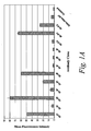

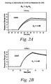
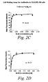
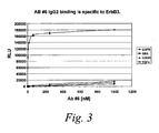
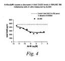
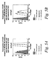


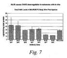
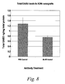


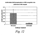
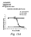

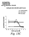
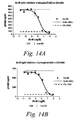


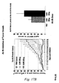

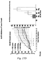


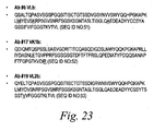


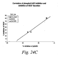


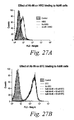
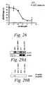

Classifications
-
- C—CHEMISTRY; METALLURGY
- C07—ORGANIC CHEMISTRY
- C07K—PEPTIDES
- C07K16/00—Immunoglobulins [IGs], e.g. monoclonal or polyclonal antibodies
- C07K16/18—Immunoglobulins [IGs], e.g. monoclonal or polyclonal antibodies against material from animals or humans
- C07K16/32—Immunoglobulins [IGs], e.g. monoclonal or polyclonal antibodies against material from animals or humans against translation products of oncogenes
-
- C—CHEMISTRY; METALLURGY
- C07—ORGANIC CHEMISTRY
- C07K—PEPTIDES
- C07K16/00—Immunoglobulins [IGs], e.g. monoclonal or polyclonal antibodies
- C07K16/18—Immunoglobulins [IGs], e.g. monoclonal or polyclonal antibodies against material from animals or humans
- C07K16/28—Immunoglobulins [IGs], e.g. monoclonal or polyclonal antibodies against material from animals or humans against receptors, cell surface antigens or cell surface determinants
-
- A—HUMAN NECESSITIES
- A61—MEDICAL OR VETERINARY SCIENCE; HYGIENE
- A61K—PREPARATIONS FOR MEDICAL, DENTAL OR TOILETRY PURPOSES
- A61K31/00—Medicinal preparations containing organic active ingredients
- A61K31/33—Heterocyclic compounds
- A61K31/335—Heterocyclic compounds having oxygen as the only ring hetero atom, e.g. fungichromin
- A61K31/337—Heterocyclic compounds having oxygen as the only ring hetero atom, e.g. fungichromin having four-membered rings, e.g. taxol
-
- A—HUMAN NECESSITIES
- A61—MEDICAL OR VETERINARY SCIENCE; HYGIENE
- A61K—PREPARATIONS FOR MEDICAL, DENTAL OR TOILETRY PURPOSES
- A61K31/00—Medicinal preparations containing organic active ingredients
- A61K31/33—Heterocyclic compounds
- A61K31/395—Heterocyclic compounds having nitrogen as a ring hetero atom, e.g. guanethidine or rifamycins
- A61K31/495—Heterocyclic compounds having nitrogen as a ring hetero atom, e.g. guanethidine or rifamycins having six-membered rings with two or more nitrogen atoms as the only ring heteroatoms, e.g. piperazine or tetrazines
- A61K31/505—Pyrimidines; Hydrogenated pyrimidines, e.g. trimethoprim
- A61K31/517—Pyrimidines; Hydrogenated pyrimidines, e.g. trimethoprim ortho- or peri-condensed with carbocyclic ring systems, e.g. quinazoline, perimidine
-
- A—HUMAN NECESSITIES
- A61—MEDICAL OR VETERINARY SCIENCE; HYGIENE
- A61K—PREPARATIONS FOR MEDICAL, DENTAL OR TOILETRY PURPOSES
- A61K39/00—Medicinal preparations containing antigens or antibodies
- A61K39/395—Antibodies; Immunoglobulins; Immune serum, e.g. antilymphocytic serum
-
- A—HUMAN NECESSITIES
- A61—MEDICAL OR VETERINARY SCIENCE; HYGIENE
- A61K—PREPARATIONS FOR MEDICAL, DENTAL OR TOILETRY PURPOSES
- A61K39/00—Medicinal preparations containing antigens or antibodies
- A61K39/395—Antibodies; Immunoglobulins; Immune serum, e.g. antilymphocytic serum
- A61K39/39533—Antibodies; Immunoglobulins; Immune serum, e.g. antilymphocytic serum against materials from animals
- A61K39/3955—Antibodies; Immunoglobulins; Immune serum, e.g. antilymphocytic serum against materials from animals against proteinaceous materials, e.g. enzymes, hormones, lymphokines
-
- A—HUMAN NECESSITIES
- A61—MEDICAL OR VETERINARY SCIENCE; HYGIENE
- A61K—PREPARATIONS FOR MEDICAL, DENTAL OR TOILETRY PURPOSES
- A61K39/00—Medicinal preparations containing antigens or antibodies
- A61K39/395—Antibodies; Immunoglobulins; Immune serum, e.g. antilymphocytic serum
- A61K39/39533—Antibodies; Immunoglobulins; Immune serum, e.g. antilymphocytic serum against materials from animals
- A61K39/39558—Antibodies; Immunoglobulins; Immune serum, e.g. antilymphocytic serum against materials from animals against tumor tissues, cells, antigens
-
- A—HUMAN NECESSITIES
- A61—MEDICAL OR VETERINARY SCIENCE; HYGIENE
- A61K—PREPARATIONS FOR MEDICAL, DENTAL OR TOILETRY PURPOSES
- A61K9/00—Medicinal preparations characterised by special physical form
- A61K9/0012—Galenical forms characterised by the site of application
- A61K9/0019—Injectable compositions; Intramuscular, intravenous, arterial, subcutaneous administration; Compositions to be administered through the skin in an invasive manner
-
- A—HUMAN NECESSITIES
- A61—MEDICAL OR VETERINARY SCIENCE; HYGIENE
- A61P—SPECIFIC THERAPEUTIC ACTIVITY OF CHEMICAL COMPOUNDS OR MEDICINAL PREPARATIONS
- A61P1/00—Drugs for disorders of the alimentary tract or the digestive system
-
- A—HUMAN NECESSITIES
- A61—MEDICAL OR VETERINARY SCIENCE; HYGIENE
- A61P—SPECIFIC THERAPEUTIC ACTIVITY OF CHEMICAL COMPOUNDS OR MEDICINAL PREPARATIONS
- A61P11/00—Drugs for disorders of the respiratory system
-
- A—HUMAN NECESSITIES
- A61—MEDICAL OR VETERINARY SCIENCE; HYGIENE
- A61P—SPECIFIC THERAPEUTIC ACTIVITY OF CHEMICAL COMPOUNDS OR MEDICINAL PREPARATIONS
- A61P13/00—Drugs for disorders of the urinary system
- A61P13/08—Drugs for disorders of the urinary system of the prostate
-
- A—HUMAN NECESSITIES
- A61—MEDICAL OR VETERINARY SCIENCE; HYGIENE
- A61P—SPECIFIC THERAPEUTIC ACTIVITY OF CHEMICAL COMPOUNDS OR MEDICINAL PREPARATIONS
- A61P13/00—Drugs for disorders of the urinary system
- A61P13/12—Drugs for disorders of the urinary system of the kidneys
-
- A—HUMAN NECESSITIES
- A61—MEDICAL OR VETERINARY SCIENCE; HYGIENE
- A61P—SPECIFIC THERAPEUTIC ACTIVITY OF CHEMICAL COMPOUNDS OR MEDICINAL PREPARATIONS
- A61P15/00—Drugs for genital or sexual disorders; Contraceptives
-
- A—HUMAN NECESSITIES
- A61—MEDICAL OR VETERINARY SCIENCE; HYGIENE
- A61P—SPECIFIC THERAPEUTIC ACTIVITY OF CHEMICAL COMPOUNDS OR MEDICINAL PREPARATIONS
- A61P17/00—Drugs for dermatological disorders
-
- A—HUMAN NECESSITIES
- A61—MEDICAL OR VETERINARY SCIENCE; HYGIENE
- A61P—SPECIFIC THERAPEUTIC ACTIVITY OF CHEMICAL COMPOUNDS OR MEDICINAL PREPARATIONS
- A61P35/00—Antineoplastic agents
-
- A—HUMAN NECESSITIES
- A61—MEDICAL OR VETERINARY SCIENCE; HYGIENE
- A61P—SPECIFIC THERAPEUTIC ACTIVITY OF CHEMICAL COMPOUNDS OR MEDICINAL PREPARATIONS
- A61P43/00—Drugs for specific purposes, not provided for in groups A61P1/00-A61P41/00
-
- C—CHEMISTRY; METALLURGY
- C07—ORGANIC CHEMISTRY
- C07K—PEPTIDES
- C07K16/00—Immunoglobulins [IGs], e.g. monoclonal or polyclonal antibodies
- C07K16/18—Immunoglobulins [IGs], e.g. monoclonal or polyclonal antibodies against material from animals or humans
- C07K16/28—Immunoglobulins [IGs], e.g. monoclonal or polyclonal antibodies against material from animals or humans against receptors, cell surface antigens or cell surface determinants
- C07K16/2863—Immunoglobulins [IGs], e.g. monoclonal or polyclonal antibodies against material from animals or humans against receptors, cell surface antigens or cell surface determinants against receptors for growth factors, growth regulators
-
- A—HUMAN NECESSITIES
- A61—MEDICAL OR VETERINARY SCIENCE; HYGIENE
- A61K—PREPARATIONS FOR MEDICAL, DENTAL OR TOILETRY PURPOSES
- A61K39/00—Medicinal preparations containing antigens or antibodies
- A61K2039/505—Medicinal preparations containing antigens or antibodies comprising antibodies
-
- A—HUMAN NECESSITIES
- A61—MEDICAL OR VETERINARY SCIENCE; HYGIENE
- A61K—PREPARATIONS FOR MEDICAL, DENTAL OR TOILETRY PURPOSES
- A61K33/00—Medicinal preparations containing inorganic active ingredients
- A61K33/24—Heavy metals; Compounds thereof
- A61K33/243—Platinum; Compounds thereof
-
- C—CHEMISTRY; METALLURGY
- C07—ORGANIC CHEMISTRY
- C07K—PEPTIDES
- C07K2317/00—Immunoglobulins specific features
- C07K2317/20—Immunoglobulins specific features characterized by taxonomic origin
- C07K2317/21—Immunoglobulins specific features characterized by taxonomic origin from primates, e.g. man
-
- C—CHEMISTRY; METALLURGY
- C07—ORGANIC CHEMISTRY
- C07K—PEPTIDES
- C07K2317/00—Immunoglobulins specific features
- C07K2317/30—Immunoglobulins specific features characterized by aspects of specificity or valency
- C07K2317/34—Identification of a linear epitope shorter than 20 amino acid residues or of a conformational epitope defined by amino acid residues
-
- C—CHEMISTRY; METALLURGY
- C07—ORGANIC CHEMISTRY
- C07K—PEPTIDES
- C07K2317/00—Immunoglobulins specific features
- C07K2317/50—Immunoglobulins specific features characterized by immunoglobulin fragments
- C07K2317/55—Fab or Fab'
-
- C—CHEMISTRY; METALLURGY
- C07—ORGANIC CHEMISTRY
- C07K—PEPTIDES
- C07K2317/00—Immunoglobulins specific features
- C07K2317/50—Immunoglobulins specific features characterized by immunoglobulin fragments
- C07K2317/56—Immunoglobulins specific features characterized by immunoglobulin fragments variable (Fv) region, i.e. VH and/or VL
- C07K2317/565—Complementarity determining region [CDR]
-
- C—CHEMISTRY; METALLURGY
- C07—ORGANIC CHEMISTRY
- C07K—PEPTIDES
- C07K2317/00—Immunoglobulins specific features
- C07K2317/70—Immunoglobulins specific features characterized by effect upon binding to a cell or to an antigen
- C07K2317/73—Inducing cell death, e.g. apoptosis, necrosis or inhibition of cell proliferation
-
- C—CHEMISTRY; METALLURGY
- C07—ORGANIC CHEMISTRY
- C07K—PEPTIDES
- C07K2317/00—Immunoglobulins specific features
- C07K2317/70—Immunoglobulins specific features characterized by effect upon binding to a cell or to an antigen
- C07K2317/76—Antagonist effect on antigen, e.g. neutralization or inhibition of binding
-
- C—CHEMISTRY; METALLURGY
- C07—ORGANIC CHEMISTRY
- C07K—PEPTIDES
- C07K2317/00—Immunoglobulins specific features
- C07K2317/90—Immunoglobulins specific features characterized by (pharmaco)kinetic aspects or by stability of the immunoglobulin
- C07K2317/92—Affinity (KD), association rate (Ka), dissociation rate (Kd) or EC50 value
Definitions
- the ErbB/HER subfamily of polypeptide growth factor receptors include the epidermal growth factor (EGF) receptor (EGFR, ErbB1/HER1), the neu oncogene product (ErbB2/HER2), and the more recently identified ErbB3/HER3 and ErbB4/HER4 receptor proteins (see, e.g., Hynes et. al. (1994) Biochim. Biophys. Acta Rev. Cancer 1198, 165-184 ).
- Each of these receptors is predicted to consist of an extracellular ligand-binding domain, a membrane-spanning domain, a cytosolic protein tyrosine kinase (PTK) domain and a C-terminal phosphorylation domain (see, e.g., Kim et al., (1998) Biochem. J. 334, 189-195 ).
- PTK cytosolic protein tyrosine kinase
- ErbB3 is constitutively phosphorylated on tyrosine residues in a subset of human breast cancer cell lines overexpressing this protein (see, e.g., Kraus et al. (1993) Proc. Natl. Acad Sci. USA. 90, 2900-2904 ; and Kim et al. Supra; see, also, Schaefer et al. (2006) Neoplasia 8(7):613-22 and Schaefer et al. Cancer Res (2004) 64(10):3395-405 ).
- ErbB3 remains largely unappreciated as a target for clinical intervention.
- Current immunotherapies primarily focus on inhibiting the action of ErbB2 and, in particular, heterodimerization of ErbB2/ErbB3 complexes (see, e.g. , Sliwkowski et al. (1994) J. Biol. Chem. 269(20):14661-14665 (1994 )). Accordingly, it is an object of the present invention to provide improved immunotherapies that effectively inhibit ErbB3 signaling, and can be used to treat and diagnose a variety of cancers.
- the present invention provides a novel class of monoclonal antibodies which binds to the ErbB3 receptor and inhibits various ErbB3 functions.
- the antibodies described herein are capable of binding to ErbB3 and inhibiting EGF-like ligand mediated phosphorylation of the receptor.
- EGF-like ligands include EGF, TGF- ⁇ , betacellulin, heparin-binding epidermal growth factor, biregulin and amphiregulin, which bind to EGFR and induce dimerization of EGFR with ErbB3. This dimerization, in turn, causes phosphorylation of ErbB3, and activates signaling through the receptor.
- Monoclonal antibodies of the present invention are useful for treating and diagnosing a variety of cancers associated with ErbB3-mediated cellular signaling. Accordingly, in one embodiment, the present invention provides monoclonal antibodies (and antigen binding portions thereof) which bind to ErbB3 and inhibit EGF-like ligand mediated phosphorylation of ErbB3.
- the antibodies are further characterized by one or more of the following properties: (i) inhibition of ErbB3 ligand-mediated signaling, including signaling mediated by binding of ErbB3 ligands, such as heregulin, epiregulin, epigen and BIR, to ErbB3; (ii) inhibition of proliferation of cells expressing ErbB3; (iii) the ability to decrease levels of ErbB3 on cell surfaces (e.g., by inducing internalization of ErbB3); (iv) inhibition of VEGF secretion of cells expressing ErbB3; (v) inhibition of the migration of cells expressing ErbB3; (vi) inhibition of spheroid growth of cells expressing ErbB3; and/or (vii) binding to an epitope located on domain I (residues 20-209) of ErbB3, for example, an epitope involving or spanning residues 20-202 of the amino acid sequence of ErbB3.
- ErbB3 ligand-mediated signaling including signal
- Particular monoclonal antibodies and antigen binding portions thereof of the present invention exhibit a K D of 50 nM or less, as measured by a surface plasmon resonance assay or a cell binding assay
- particular monoclonal antibodies and antigen binding portions thereof of the present invention include a heavy chain variable region comprising an amino acid sequence which is at least 80% (e.g ., 85%, 90%, 95%, 96%, 97%, 98% or 99%) identical to the heavy chain variable region amino acid sequence set forth in SEQ ID NO:1, SEQ ID NO:3, SEQ ID NO:5, SEQ ID NO:35, or SEQ ID NO: 37.
- antibodies and antigen binding portions thereof of the present invention include a light chain variable region comprising an amino acid sequence which is at least 80% (e.g., 85%, 90%, 95%, 96%, 97%, 98% or 99%) identical to the light chain variable region amino acid sequence set forth in SEQ ID NO:2, SEQ ID NO:4, SEQ ID NO:6, SEQ ID NO:36, or SEQ ID NO:38.
- the antibodies may also include both of the aforementioned heavy chain and light chain variable regions.
- variable heavy and light chain regions of the antibodies or antigen binding portions thereof typically include one or more complementarity determining regions (CDRs). These include one or more CDR1, CDR2, and CDR3 regions. Accordingly, other particular antibodies and antigen binding portions thereof of the present invention include one or more CDR sequences selected from a heavy chain variable region CDR1 comprising SEQ ID NO:7; a heavy chain variable region CDR2 comprising SEQ ID NO:8; a heavy chain variable region CDR3 comprising SEQ ID NO:9; a light chain variable region CDR1 comprising SEQ ID NO:10; a light chain variable region CDR2 comprising SEQ ID NO:11; a light chain variable region CDR3 comprising SEQ ID NO:12; and combinations thereof.
- CDRs complementarity determining regions
- Still other particular antibodies and antigen binding portions thereof of the present invention include one or more CDR sequences selected from a heavy chain variable region CDR1 comprising SEQ ID NO:13; a heavy chain variable region CDR2 comprising SEQ ID NO:14; a heavy chain variable region CDR3 comprising SEQ ID NO:15; a light chain variable region CDR1 comprising SEQ ID NO:16; a light chain variable region CDR2 comprising SEQ ID NO:17; a light chain variable region CDR3 comprising SEQ ID NO:18; and combinations thereof.
- Still other particular antibodies and antigen binding portions thereof of the present invention include; or one or more CDR sequences selected from a heavy chain variable region CDR1 comprising SEQ ID NO:19; a heavy chain variable region CDR2 comprising SEQ ID NO:20; a heavy chain variable region CDR3 comprising SEQ ID NO:21; a light chain variable region CDR1 comprising SEQ ID NO:22; a light chain variable region CDR2 comprising SEQ ID NO:23; a light chain variable region CDR3 comprising SEQ ID NO:24; and combinations thereof.
- Still other particular antibodies and antigen binding portions thereof of the present invention include; or one or more CDR sequences selected from a heavy chain variable region CDR1 comprising SEQ ID NO:39; a heavy chain variable region CDR2 comprising SEQ ID NO:40; a heavy chain variable region CDR3 comprising SEQ ID NO:41; a light chain variable region CDR1 comprising SEQ ID NO:42; a light chain variable region CDR2 comprising SEQ ID NO:43; a light chain variable region CDR3 comprising SEQ ID NO:44; and combinations thereof.
- Still other particular antibodies and antigen binding portions thereof of the present invention include; or one or more CDR sequences selected from a heavy chain variable region CDR1 comprising SEQ ID NO:45; a heavy chain variable region CDR2 comprising SEQ ID NO:46; a heavy chain variable region CDR3 comprising SEQ ID NO:47; a light chain variable region CDR1 comprising SEQ ID NO:48; a light chain variable region CDR2 comprising SEQ ID NO:49; a light chain variable region CDR3 comprising SEQ ID NO:50; and combinations thereof.
- the antibodies and antigen binding portions thereof may also comprise one or more CDRs which are at least 80% (e.g., 85%, 90%, 95%, 96%, 97%, 98% or 99%) identical to any of the aforementioned CDRs, or combinations of CDRs.
- the antibodies and antibody portions thereof are fully human (i.e., contains human CDR and framework sequences).
- Particular human antibodies of the present invention include those having a heavy chain variable region that is from a human VH3 germ line gene, and/or a light chain variable region from human VL2 germ line gene.
- Also encompassed by the present invention are monoclonal antibodies and portions thereof that bind to the same or overlapping epitopes bound by any of the antibodies or portions thereof described herein (e.g., an epitope located on domain I of ErbB3, such as an epitope involving or spanning, residues 20-202 of the amino acid sequence of ErbB3).
- Antibodies which have the same activity as the antibodies described herein, e.g., antibodies having the same sequence as Ab #6, are also encompassed by the present invention.
- Antibodies of the present invention include all known forms of antibodies and other protein scaffolds with antibody-like properties.
- the antibody can be a human antibody, a humanized antibody, a bispecific antibody, a chimeric antibody or a protein scaffold with antibody-like properties, such as fibronectin or Ankyrin repeats.
- the antibody also can be a Fab, Fab'2, ScFv, SMIP, affibody, nanobody, or a domain antibody.
- the antibody also can have any of the following isotypes: IgG1, IgG2, IgG3, IgG4, IgM, IgA1, IgA2, IgAsec, IgD, and IgE.
- the present invention further provides compositions comprising combinations of antibodies or antigen binding portions described herein, formulated with an acceptable carrier and/or adjuvant.
- the composition comprises two or more antibodies that bind different epitopes on ErbB3 or antibodies described herein combined with anti-cancer antibodies which do not bind ErbB3.
- the present invention provides isolated nucleic acids encoding the antibodies and antigen binding portions thereof described herein.
- the nucleic acid encodes a heavy chain variable region comprising a nucleotide sequence which is at least 80% (e.g.
- the present invention further provides transgenic non-human mammals, hybridomas, and transgenic plants that express and/or produce the antibodies and antigen binding portions described herein.
- kits comprising one or more isolated monoclonal antibodies or antigen binding portions thereof described herein and, optionally, instructions for use in treating or diagnosing a disease associated with ErbB3 dependent signaling, such as cancers.
- Antibodies and antigen binding portions thereof of the present invention can be used in a broad variety of therapeutic and diagnostic applications, particularly oncological applications. Accordingly, in another aspect, the invention provides method for inhibiting EGF-like ligand mediated phosphorylation of ErbB3 in a subject by administering one or more antibodies or antigen binding portions thereof described herein in an amount sufficient to inhibit EGF-like mediated phosphorylation of ErbB3.
- the invention further provides methods for treating a variety of cancers in a subject, including, but not limited to, melanoma, breast cancer, ovarian cancer, renal carcinoma, gastrointestinal/colon cancer, lung cancer, clear cell sarcoma, and prostate cancer, by administering one or more antibodies or antigen binding portions thereof described herein in an amount sufficient to treat the cancer.
- the antibodies or antigen binding portions thereof can be administered alone or in combination with other therapeutic agents, such as anti-cancer agents, e.g ., other antibodies, chemotherapeutic agents and/or radiation.
- the invention provides methods for diagnosing and prognosing diseases (e.g. , cancers) associated with ErbB3. In one embodiment, this is achieved by contacting antibodies or antigen binding portions of the invention (e.g., ex vivo or in vivo ) with cells from the subject, and measuring the level of binding to ErbB3 on the cells, wherein abnormally high levels of binding to ErbB3 indicate that the subject has a cancer associated with ErbB3.
- diseases e.g. , cancers
- this is achieved by contacting antibodies or antigen binding portions of the invention (e.g., ex vivo or in vivo ) with cells from the subject, and measuring the level of binding to ErbB3 on the cells, wherein abnormally high levels of binding to ErbB3 indicate that the subject has a cancer associated with ErbB3.
- ErbB3 refers human ErbB3 protein, as described in U.S. Pat. No. 5,480,968 and Plowman et al., Proc. Natl. Acad. Sci. USA, 87:4905-4909 (1990 ); see, also, Kani et al., Biochemistry 44:15842-857 (2005 ), Cho and Leahy, Science 297:1330-1333 (2002 )).
- EGF-like ligand refers to ligands of epidermal growth factor receptor (EGFR), including epidermal growth factor (EGF) and closely related proteins, such as transforming growth factor- ⁇ (TGF- ⁇ ), betacellulin (BTC), heparin-binding epidermal growth factor (HB-EGF), biregulin (BIR) and amphiregulin (AR), which bind to EGFR on the surface of cells and stimulate the receptor's intrinsic protein-tyrosine kinase activity.
- EGF- ⁇ transforming growth factor- ⁇
- BTC betacellulin
- HB-EGF heparin-binding epidermal growth factor
- BIR biregulin
- AR amphiregulin
- EGF-like ligands induce formation of EGFR (also referred to as ErbB1) and ErbB3 protein complex (see e.g., Kim et al., (1998) Biochem J., 334:189-195 ), which results in phosphorylation of tyrosine residues in the complex.
- EGFR also referred to as ErbB1
- ErbB3 protein complex see e.g., Kim et al., (1998) Biochem J., 334:189-195 , which results in phosphorylation of tyrosine residues in the complex.
- the antibodies and antigen binding portions thereof of the present invention inhibit EGF-like ligand mediated phosphorylation of ErbB3 and, in certain embodiments, exhibit one or more of the following additional properties: (i) inhibition of one or more of heregulin, epiregulin, epigen and biregulin (BIR)-mediated signaling through ErbB3; (ii) inhibition of proliferation of cells expressing ErbB3; (iii) the ability to decrease levels of ErbB3 on cell surfaces; (iv) inhibition of VEGF secretion of cells expressing ErbB3; (v) inhibition of the migration of cells expressing ErbB3; (vi) inhibition of spheroid growth of cells expressing ErbB3; and/or (vii) binding to an epitope located on domain I of ErbB3, e.g., an epitope which involves or spans residues 20-202 of the amino acid sequence of ErbB3.
- BIR biregulin
- inhibitor refers to any statistically significant decrease in biological activity, including full blocking of the activity.
- “inhibition” can refer to a decrease of about 10%, 20%, 30%, 40%, 50%, 60%, 70%, 80%, 90%, or 100% in biological activity.
- the phrase "inhibition of EGF-like ligand mediated phosphorylation of ErbB3,” as used herein, refers to the ability of an antibody or antigen binding portion to statistically significantly decrease the phosphorylation of ErbB3 induced by an EGF-like ligand, relative to the phosphorylation in an untreated (control) cell.
- the cell which expresses ErbB3 can be a naturally occurring cell or cell line or can be recombinantly produced by introducing nucleic acid encoding ErbB3 into a host cell.
- the antibody or antigen binding portion thereof inhibits EGF-like ligand mediated phosphorylation of ErbB3 by at least 10%, or at least 20%, or at least 30%, or at least 40%, or at least 50%, or at least 60%, or at least 70%, or at least 80%, or at least 90%, or 100%, as determined, for example, by Western blotting followed by probing with an anti-phosphotyrosine antibody as described in Kim et al., (1998) Biochem J., 334:189-195 and the Examples infra.
- the phrase "inhibition of heregulin, epiregulin, epigen or biregulin-mediated signaling through ErbB3,” as used herein, refers to the ability of an antibody or an antigen-binding portion thereof to statistically significantly decrease signaling mediated by an ErbB3 ligand (e.g., heregulin, epiregullin, epigen and biregulin) through ErbB3, relative to the signaling in the absence of the antibody (control).
- an ErbB3 ligand e.g., heregulin, epiregullin, epigen and biregulin
- ErbB3-ligands are also referred to herein as "heregulin-like ligands.” This means that, in the presence of the antibody or antigen binding portion thereof, a signal mediated in a cell expressing ErbB3 by one or more of heregulin, epiregulin, epigen and biregulin, relative to a control (no antibody), is statistically significantly decreased.
- An ErbB3-ligand mediated signal can be measured by assaying for the level or activity of an ErbB3 substrate, and/or a protein which is present in a cellular cascade involving ErbB3.
- the antibody or antigen binding portion thereof decreases the level or activity of an ErbB3 substrate and/or that of a protein in a cellular cascade involving ErbB3, by at least 10%, or at least 20%, or at least 30%, or at least 40%, or at least 50%, or at least 60%, or at least 70%, or at least 80%, or at least 90%, or 100% relative to the level or activity in the absence of such antibody or antigen binding portion thereof (control).
- Such ErbB3-ligand mediated signaling can be measured using art recognized techniques which measure the level or activity of a substrate of ErbB3 (e.g., SHC or PI3K) or a protein in a cellular cascade involving ErbB3 (e.g.
- the antibody or antigen binding portion thereof inhibits ErbB3-ligand (e.g., heregulin, epiregulin, epigen or biregulin) mediated signaling through ErbB3 by inhibiting the binding of the ErbB3-ligand (e.g., one or more of heregulin, epiregulin, epigen or biregulin) to ErbB3.
- ErbB3-ligand e.g., one or more of heregulin, epiregulin, epigen or biregulin
- Some ligands e.g., biregulin or BIR
- the amount of an ErbB3 ligand which binds ErbB3 may be decreased in the presence of an antibody or antigen binding portion thereof of the invention by at least 10%, or at least 20%, or at least 30%, or at least 40%, or at least 50%, or at least 60%, or at least 70%, or at least 80%, or at least 90%, or 100% relative to the amount in the absence of the antibody or antigen binding portion thereof (control).
- a decrease in ErbB3-ligand binding can be measured using art recognized techniques which measure the level of binding of labeled ErbB3-ligand (e.g. , radiolabelled heregulin, epiregulin, epigen or biregulin) to cells expressing ErbB3 in the presence or absence (control) of the antibody or antigen binding portion thereof.
- the phrase "inhibition of proliferation of a cell expressing ErbB3,” as used herein, refers to the ability of an antibody or an antigen-binding portion thereof to statistically significantly decrease proliferation of a cell expressing ErbB3 relative to the proliferation in the absence of the antibody.
- the proliferation of a cell expressing ErbB3 (e.g., a cancer cell) may be decreased by at least 10%, or at least 20%, or at least 30%, or at least 40%, or at least 50%, or at least 60%, or at least 70%, or at least 80%, or at least 90%, or 100% when the cells are contacted with the antibody or antigen binding portion thereof of the present invention, relative to the proliferation measured in the absence of the antibody or antigen binding portion thereof (control).
- Cellular proliferation can be assayed using art recognized techniques which measure rate of cell division, the fraction of cells within a cell population undergoing cell division, and/or rate of cell loss from a cell population due to terminal differentiation or cell death (e.g., using a cell titer glow assay or thymidine incorporation).
- the ability to decrease levels of ErbB3 on cell surfaces refers to the ability of an antibody or antigen binding portion thereof to statistically significantly reduce the amount of ErbB3 found on the surface of a cell which has been exposed to the antibody relative to an untreated (control) cell.
- a decrease in levels of ErbB3 on cell surfaces may result from increased internalization of ErbB3 (or increased ErbB3 endocytosis).
- the antibody or antigen binding portion thereof decreases cell surface expression of ErbB3 by at least 10%, or at least 20%, or at least 30%, or at least 40%, or at least 50%, or at least 60%, or at least 70%, or at least 80%, or at least 90%, or 100% and/or increases internalization of the ErbB3 receptor by at least 10%, or at least 20%, or at least 30%, or at least 40%, or at least 50%, or at least 60%, or at least 70%, or at least 80%, or at least 90%, or 100% relative to the cell surface expression or internalization in the absence of the antibody or antigen binding portion thereof (control).
- the levels of ErbB3 on surfaces of cells and/or internalization of the ErbB3 receptor in the absence and the presence of an antibody or antigen-binding portion thereof can be readily measured using art recognized techniques, such as those described in Horst et al., supra and in the examples herein.
- VEGF secretion of cells expressing ErbB3 refers to the ability of an antibody or an antigen-binding portion thereof to statistically significantly decrease VEGF secretion of a cell expressing ErbB3 relative to the VEGF secretion in the absence of the antibody.
- the VEGF secretion of a cell expressing ErbB3 e.g.
- a cancer cell may be decreased by at least 10%, or at least 20%, or at least 30%, or at least 40%, or at least 50%, or at least 60%, or at least 70%, or at least 80%, or at least 90%, or 100% when the cells are contacted with the antibody or antigen binding portion thereof of the present invention, relative to the VEGF secretion measured in the absence of the antibody or antigen binding portion thereof (control).
- VEGF secretion can be assayed using art recognized techniques, such as those described herein.
- the phrase "inhibition of the migration of cells expressing ErbB3,” as used herein, refers to the ability of an antibody or an antigen-binding portion thereof to statistically significantly decrease the migration of a cell expressing ErbB3 relative to the migration of the cell in the absence of the antibody.
- the migration of a cell expressing ErbB3 (e.g., a cancer cell) may be decreased by at least 10%, or at least 20%, or at least 30%, or at least 40%, or at least 50%, or at least 60%, or at least 70%, or at least 80%, or at least 90%, or 100% when the cells are contacted with the antibody or antigen binding portion thereof of the present invention, relative to cell migration measured in the absence of the antibody or antigen binding portion thereof (control).
- Cell migration can be assayed using art recognized techniques, such as those described herein.
- inhibitortion of spheroid growth of cells expressing ErbB3 refers to the ability of an antibody or an antigen-binding portion thereof to statistically significantly decrease the migration of a cell expressing ErbB3 relative to the migration of the cell in the absence of the antibody.
- the migration of a cell expressing ErbB3 may be decreased by at least 10%, or at least 20%, or at least 30%, or at least 40%, or at least 50%, or at least 60%, or at least 70%, or at least 80%, or at least 90%, or 100% when the cells are contacted with the antibody or antigen binding portion thereof of the present invention, relative to cell migration measured in the absence of the antibody or antigen binding portion thereof (control).
- Cell migration can be assayed using art recognized techniques, such as those described herein.
- the term "antibody” or "immunoglobulin,” as used interchangeably herein, includes whole antibodies and any antigen binding fragment ( i.e. , "antigen-binding portion") or single chains thereof.
- An “antibody” comprises at least two heavy (H) chains and two light (L) chains inter-connected by disulfide bonds.
- Each heavy chain is comprised of a heavy chain variable region (abbreviated herein as V H ) and a heavy chain constant region.
- the heavy chain constant region is comprised of three domains, CH1, CH2 and CH3.
- Each light chain is comprised of a light chain variable region (abbreviated herein as V L ) and a light chain constant region.
- the light chain constant region is comprised of one domain, CL.
- the V H and V L regions can be further subdivided into regions of hypervariability, termed complementarity determining regions (CDR), interspersed with regions that are more conserved, termed framework regions (FR).
- CDR complementarity determining regions
- Each V H and V L is composed of three CDRs and four FRs, arranged from amino-terminus to carboxy-terminus in the following order: FR1, CDR1, FR2, CDR2, FR3, CDR3, FR4.
- the variable regions of the heavy and light chains contain a binding domain that interacts with an antigen.
- the constant regions of the antibodies may mediate the binding of the immunoglobulin to host tissues or factors, including various cells of the immune system (e.g. , effector cells) and the first component (Clq) of the classical complement system.
- Exemplary antibodies of the invention include antibodies#1, 3 and 14, and antigen-binding portions thereof.
- antibody portion refers to one or more fragments of an antibody that retain the ability to specifically bind to an antigen (e.g. , ErbB3). It has been shown that the antigen-binding function of an antibody can be performed by fragments of a full-length antibody.
- binding fragments encompassed within the term "antigen-binding portion" of an antibody include (i) a Fab fragment, a monovalent fragment consisting of the V L , V H , CL and CH1 domains; (ii) a F(ab') 2 fragment, a bivalent fragment comprising two Fab fragments linked by a disulfide bridge at the hinge region; (iii) a Fd fragment consisting of the V H and CH1 domains; (iv) a Fv fragment consisting of the V L and V H domains of a single arm of an antibody, (v) a dAb including VH and VL domains; (vi) a dAb fragment ( Ward et al.
- V H domain a dAb which consists of a VH or a VL domain
- CDR an isolated complementarity determining region
- ix a combination of two or more isolated CDRs which may optionally be joined by a synthetic linker.
- the two domains of the Fv fragment, V L and V H are coded for by separate genes, they can be joined, using recombinant methods, by a synthetic linker that enables them to be made as a single protein chain in which the V L and V H regions pair to form monovalent molecules (known as single chain Fv (scFv); see e.g. , Bird et al.
- scFv single chain Fv
- Such single chain antibodies are also intended to be encompassed within the term "antigen-binding portion" of an antibody.
- antibody fragments are obtained using conventional techniques known to those with skill in the art, and the fragments are screened for utility in the same manner as are intact antibodies.
- Antigen-binding portions can be produced by recombinant DNA techniques, or by enzymatic or chemical cleavage of intact immunoglobulins.
- monoclonal antibody refers to an antibody obtained from a population of substantially homogeneous antibodies, i.e. , the individual antibodies comprising the population are identical except for possible naturally occurring mutations that may be present in minor amounts. Monoclonal antibodies are highly specific, being directed against a single antigenic site. Furthermore, in contrast to conventional (polyclonal) antibody preparations which typically include different antibodies directed against different determinants (epitopes), each monoclonal antibody is directed against a single determinant on the antigen. Monoclonal antibodies can be prepared using any art recognized technique and those described herein such as, for example, a hybridoma method, as described by Kohler et al.
- recombinant antibody refers to antibodies that are prepared, expressed, created or isolated by recombinant means, such as (a) antibodies isolated from an animal (e.g. , a mouse) that is transgenic or transchromosomal for immunoglobulin genes (e.g. , human immunoglobulin genes) or a hybridoma prepared therefrom, (b) antibodies isolated from a host cell transformed to express the antibody, e.g. , from a transfectoma, (c) antibodies isolated from a recombinant, combinatorial antibody library (e.g.
- recombinant antibodies may have variable and constant regions derived from human germline immunoglobulin sequences.
- such recombinant human antibodies can be subjected to in vitro mutagenesis and thus the amino acid sequences of the V H and V L regions of the recombinant antibodies are sequences that, while derived from and related to human germline V H and V L sequences, may not naturally exist within the human antibody germline repertoire in vivo.
- chimeric immunoglobulin refers to an immunoglobulin or antibody whose variable regions derive from a first species and whose constant regions derive from a second species. Chimeric immunoglobulins or antibodies can be constructed, for example by genetic engineering, from immunoglobulin gene segments belonging to different species.
- human antibody is intended to include antibodies having variable regions in which both the framework and CDR regions are derived from human germline immunoglobulin sequences as described, for example, by Kabat et al. (See Kabat, et al. (1991) Sequences of proteins of Immunological Interest, Fifth Edition, U.S. Department of Health and Human Services, NIH Publication No. 91-3242 ). Furthermore, if the antibody contains a constant region, the constant region also is derived from human germline immunoglobulin sequences.
- the human antibodies may include amino acid residues not encoded by human germline immunoglobulin sequences (e.g., mutations introduced by random or site-specific mutagenesis in vitro or by somatic mutation in vivo ). However, the term "human antibody”, as used herein, is not intended to include antibodies in which CDR sequences derived from the germline of another mammalian species, such as a mouse, have been grafted onto human framework sequences.
- the human antibody can have at least one ore more amino acids replaced with an amino acid residue, e.g., an activity enhancing amino acid residue which is not encoded by the human germline immunoglobulin sequence.
- the human antibody can have up to twenty positions replaced with amino acid residues which are not part of the human germline immunoglobulin sequence. In a particular embodiment, these replacements are within the CDR regions as described in detail below.
- humanized immunoglobulin or “humanized antibody” refers to an immunoglobulin or antibody that includes at least one humanized immunoglobulin or antibody chain (i.e. , at least one humanized light or heavy chain).
- humanized immunoglobulin chain or “humanized antibody chain” (i.e.
- a “humanized immunoglobulin light chain” or “humanized immunoglobulin heavy chain”) refers to an immunoglobulin or antibody chain ( i.e., a light or heavy chain, respectively) having a variable region that includes a variable framework region substantially from a human immunoglobulin or antibody and complementarity determining regions (CDRs) (e.g., at least one CDR, preferably two CDRs, more preferably three CDRs) substantially from a non-human immunoglobulin or antibody, and further includes constant regions (e.g., at least one constant region or portion thereof, in the case of a light chain, and preferably three constant regions in the case of a heavy chain).
- CDRs complementarity determining regions
- variable region refers to a variable region that includes a variable framework region substantially from a human immunoglobulin or antibody and complementarity determining regions (CDRs) substantially from a non-human immunoglobulin or antibody.
- CDRs complementarity determining regions
- bispecific or “bifunctional antibody” is an artificial hybrid antibody having two different heavy/light chain pairs and two different binding sites.
- Bispecific antibodies can be produced by a variety of methods including fusion of hybridomas or linking of Fab' fragments. See, e.g. , Songsivilai & Lachmann, (1990) Clin. Exp. Immunol. 79, 315-321 ; Kostelny et al. (1992) J. Immunol. 148, 1547-1553 .
- a bispecific antibody according to the present invention includes binding sites for both ErbB3 and IGF1-R (i.e. , insulin-like growth factor 1-receptor).
- a bispecific antibody according to the present invention includes binding sites for both ErbB3 and C-MET.
- a bispecific antibody includes a binding site for ErbB3 and a binding site for ErbB2, ERbB3, ErbB4, EGFR, Lewis Y, MUC-1, EpCAM, CA125, prostate specific membrane antigen, PDGFR- ⁇ , PDGFR- ⁇ , C-KIT, or any of the FGF receptors.
- a heterologous antibody is defined in relation to the transgenic non-human organism or plant producing such an antibody.
- an “isolated antibody,” as used herein, is intended to refer to an antibody which is substantially free of other antibodies having different antigenic specificities (e.g., an isolated antibody that specifically binds to ErbB3 is substantially free of antibodies that specifically bind antigens other than ErbB3).
- an isolated antibody is typically substantially free of other cellular material and/or chemicals.
- a combination of "isolated" monoclonal antibodies having different ErbB3 binding specificities are combined in a well defined composition.
- isotype refers to the antibody class (e.g., IgM or IgG1) that is encoded by heavy chain constant region genes.
- an antibody or antigen binding portion thereof is of an isotype selected from an IgG1, an IgG2, an IgG3, an IgG4, an IgM, an IgA1, an IgA2, an IgAsec, an IgD, or an IgE antibody isotype.
- a monoclonal antibody of the invention is of the IgG1 isotype.
- a monoclonal antibody of the invention is of the IgG2 isotype.
- isotype switching refers to the phenomenon by which the class, or isotype, of an antibody changes from one Ig class to one of the other Ig classes.
- nonswitched isotype refers to the isotypic class of heavy chain that is produced when no isotype switching has taken place; the CH gene encoding the nonswitched isotype is typically the first CH gene immediately downstream from the functionally rearranged VDJ gene. Isotype switching has been classified as classical or non-classical isotype switching.
- Classical isotype switching occurs by recombination events which involve at least one switch sequence regions in a gene encoding an antibody.
- Non-classical isotype switching may occur by, for example, homologous recombination between human ⁇ ⁇ and human ⁇ ⁇ ( ⁇ -associated deletion).
- Alternative non-classical switching mechanisms such as intertransgene and/or interchromosomal recombination, among others, may occur and effectuate isotype switching.
- switch sequence refers to those DNA sequences responsible for switch recombination.
- a "switch donor” sequence typically a ⁇ switch region, will be 5' ( i.e. , upstream) of the construct region to be deleted during the switch recombination.
- the "switch acceptor” region will be between the construct region to be deleted and the replacement constant region ( e.g. , ⁇ , ⁇ , etc.). As there is no specific site where recombination always occurs, the final gene sequence will typically not be predictable from the construct.
- an “antigen” is an entity (e.g., a proteinaceous entity or peptide) to which an antibody or antigen-binding portion thereof binds.
- an antigen is ErbB3 or a ErbB3-like molecule.
- an antigen is human ErbB3.
- epitopes refers to a site on an antigen to which an immunoglobulin or antibody specifically binds.
- Epitopes can be formed both from contiguous amino acids or noncontiguous amino acids juxtaposed by tertiary folding of a protein. Epitopes formed from contiguous amino acids are typically retained on exposure to denaturing solvents, whereas epitopes formed by tertiary folding are typically lost on treatment with denaturing solvents.
- An epitope typically includes at least 3, 4, 5, 6, 7, 8, 9, 10, 11, 12, 13, 14 or 15 amino acids in a unique spatial conformation.
- Methods of determining spatial conformation of epitopes include techniques in the art and those described herein, for example, x-ray crystallography and 2-dimensional nuclear magnetic resonance. See, e.g., Epitope Mapping Protocols in Methods in Molecular Biology, Vol. 66, G. E. Morris, Ed. (1996 ).
- antibodies that bind the same or an overlapping epitope as the antibodies of the present invention i.e., antibodies that compete for binding to ErbB3, or bind epitopes which overlap with epitopes bound by the antibodies described herein, i.e., an epitope located on domain I of ErbB3.
- Antibodies that recognize the same epitope can be identified using routine techniques such as an immunoassay, for example, by showing the ability of one antibody to block the binding of another antibody to a target antigen, i.e., a competitive binding assay.
- Competitive binding is determined in an assay in which the immunoglobulin under test inhibits specific binding of a reference antibody to a common antigen, such as ErbB3.
- RIA solid phase direct or indirect radioimmunoassay
- EIA solid phase direct or indirect enzyme immunoassay
- sandwich competition assay see Stahli et al., (1983) Methods in Enzymology 9:242
- solid phase direct biotin-avidin EIA see Kirkland et al., (1986) J. Immunol. 137:3614
- solid phase direct labeled assay solid phase direct labeled sandwich assay
- solid phase direct label RIA using I-125 label see Morel et al., (1988) Mol. Immunol.
- the terms “specific binding,” “specifically binds,” “selective binding,” and “selectively binds,” mean that an antibody or antigen-binding portion thereof, exhibits appreciable affinity for a particular antigen or epitope and, generally, does not exhibit significant cross-reactivity with other antigens and epitopes.
- “Appreciable” or preferred binding includes binding with an affinity of at least 10 6 , 10 7 , 10 8 , 10 9 M -1 , or 10 10 M -1 . Affinities greater than 10 7 M -1 , preferably greater than 10 8 M -1 are more preferred.
- a preferred binding affinity can be indicated as a range of affinities, for example, 10 6 to 10 10 M -1 , preferably 10 7 to 10 10 M 1 , more preferably 10 8 to 10 10 M -1 .
- An antibody that "does not exhibit significant cross-reactivity" is one that will not appreciably bind to an undesirable entity (e.g. , an undesirable proteinaceous entity).
- an antibody or antigen-binding portion thereof that specifically binds to ErbB3 will appreciably bind that ErbB3 molecule but will not significantly react with other ErbB molecules and non-ErbB proteins or peptides.
- Specific or selective binding can be determined according to any art-recognized means for determining such binding, including, for example, according to Scatchard analysis and/or competitive binding assays.
- K D is intended to refer to the dissociation equilibrium constant of a particular antibody-antigen interaction or the affinity of an antibody for an antigen.
- the antibody or antigen binding portion thereof according to the present invention binds an antigen (e.g. , ErbB3) with an affinity (K D ) of 50 nM or better (i.e., or less) ( e.g. , 40 nM or 30 nM or 20 nM or 10 nM or less), as measured using a surface plasmon resonance assay or a cell binding assay.
- an antibody or antigen binding portion thereof binds ErbB3 with an affinity (K D ) of 8 nM or better (e.g. , 7 nM, 6 nM, 5 nM, 4 nM, 2 nM, 1.5 nM, 1.4 nM, 1.3 nM, 1nM or less), as measured by a surface plasmon resonance assay or a cell binding assay.
- K D affinity
- an antibody or antigen binding portion thereof binds an antigen (e.g ., ErbB3) with an affinity (K D ) of approximately less than 10 -7 M, such as approximately less than 10 -8 M, 10 -9 M or 10 -10 M or even lower when determined by surface plasmon resonance (SPR) technology in a BIACORE 3000 instrument using recombinant ErbB3 as the analyte and the antibody as the ligand, and binds to the predetermined antigen with an affinity that is at least two-fold greater than its affinity for binding to a non-specific antigen (e.g. , BSA, casein) other than the predetermined antigen or a closely-related antigen.
- a non-specific antigen e.g. , BSA, casein
- K off is intended to refer to the off rate constant for the dissociation of an antibody from the antibody/antigen complex.
- EC50 refers to the concentration of an antibody or an antigen-binding portion thereof, which induces a response, either in an in vitro or an in vivo assay, which is 50% of the maximal response, i.e. , halfway between the maximal response and the baseline.
- glycosylation pattern is defined as the pattern of carbohydrate units that are covalently attached to a protein, more specifically to an immunoglobulin protein.
- naturally-occurring refers to the fact that an object can be found in nature.
- a polypeptide or polynucleotide sequence that is present in an organism (including viruses) that can be isolated from a source in nature and which has not been intentionally modified by man in the laboratory is naturally-occurring.
- rearranged refers to a configuration of a heavy chain or light chain immunoglobulin locus wherein a V segment is positioned immediately adjacent to a D-J or J segment in a conformation encoding essentially a complete V H or V L domain, respectively.
- a rearranged immunoglobulin gene locus can be identified by comparison to germline DNA; a rearranged locus will have at least one recombined heptamer/nonamer homology element.
- V segment configuration refers to the configuration wherein the V segment is not recombined so as to be immediately adjacent to a D or J segment.
- nucleic acid molecule is intended to include DNA molecules and RNA molecules.
- a nucleic acid molecule may be single-stranded or double-stranded, but preferably is double-stranded DNA.
- isolated nucleic acid molecule as used herein in reference to nucleic acids encoding antibodies or antibody portions ( e.g. , V H , V L , CDR3) that bind to ErbB3, is intended to refer to a nucleic acid molecule in which the nucleotide sequences encoding the antibody or antibody portion are free of other nucleotide sequences encoding antibodies that bind antigens other than ErbB3, which other sequences may naturally flank the nucleic acid in human genomic DNA.
- modifying is intended to refer to changing one or more amino acids in the antibodies or antigen-binding portions thereof.
- the change can be produced by adding, substituting or deleting an amino acid at one or more positions.
- the change can be produced using known techniques, such as PCR mutagenesis.
- an antibody or an antigen-binding portion thereof identified using the methods of the invention can be modified, to thereby modify the binding affinity of the antibody or antigen-binding portion thereof to ErbB3.
- the present invention also encompasses "conservative amino acid substitutions" in the sequences of the antibodies of the invention, i.e. , nucleotide and amino acid sequence modifications which do not abrogate the binding of the antibody encoded by the nucleotide sequence or containing the amino acid sequence, to the antigen, i. e., ErbB3.
- Conservative amino acid substitutions include the substitution of an amino acid in one class by an amino acid of the same class, where a class is defined by common physicochemical amino acid side chain properties and high substitution frequencies in homologous proteins found in nature, as determined, for example, by a standard Dayhoff frequency exchange matrix or BLOSUM matrix.
- non-conservative amino acid substitution refers to the substitution of an amino acid in one class with an amino acid from another class; for example, substitution of an Ala, a class II residue, with a class III residue such as Asp, Asn, Glu, or Gln.
- mutations can be introduced randomly along all or part of an anti-ErbB3 antibody coding sequence, such as by saturation mutagenesis, and the resulting modified anti-ErbB3 antibodies can be screened for binding activity.
- a "consensus sequence” is a sequence formed from the most frequently occurring amino acids (or nucleotides) in a family of related sequences (See e.g., Winnaker, From Genes to Clones (Verlagsgesellschaft, Weinheim, Germany 1987 ). In a family of proteins, each position in the consensus sequence is occupied by the amino acid occurring most frequently at that position in the family. If two amino acids occur equally frequently, either can be included in the consensus sequence.
- a “consensus framework” of an immunoglobulin refers to a framework region in the consensus immunoglobulin sequence.
- the consensus sequence for the CDRs of can be derived by optimal alignment of the CDR amino acid sequences of ErbB3 antibodies of the present invention.
- nucleic acids For nucleic acids, the term "substantial homology" indicates that two nucleic acids, or designated sequences thereof, when optimally aligned and compared, are identical, with appropriate nucleotide insertions or deletions, in at least about 80% of the nucleotides, usually at least about 90% to 95%, and more preferably at least about 98% to 99.5% of the nucleotides. Alternatively, substantial homology exists when the segments will hybridize under selective hybridization conditions, to the complement of the strand.
- the comparison of sequences and determination of percent identity between two sequences can be accomplished using a mathematical algorithm, as described in the non-limiting examples below.
- the percent identity between two nucleotide sequences can be determined using the GAP program in the GCG software, using a NWSgapdna.CMP matrix and a gap weight of 40, 50, 60, 70, or 80 and a length weight of 1, 2, 3, 4, 5, or 6.
- the percent identity between two nucleotide or amino acid sequences can also be determined using the algorithm of E. Meyers and W. Miller (CABIOS, 4:11-17 (1989 )) which has been incorporated into the ALIGN program (version 2.0), using a PAM120 weight residue table, a gap length penalty of 12 and a gap penalty of 4.
- the percent identity between two amino acid sequences can be determined using the Needleman and Wunsch (J. Mol. Biol.
- the nucleic acid and protein sequences of the present invention can further be used as a "query sequence" to perform a search against public databases to, for example, identify related sequences.
- Such searches can be performed using the NBLAST and XBLAST programs (version 2.0) of Altschul, et al. (1990) J. Mol. Biol. 215:403-10 .
- Gapped BLAST can be utilized as described in Altschul et al., (1997) Nucleic Acids Res. 25(17):3389-3402 .
- the default parameters of the respective programs e.g., XBLAST and NBLAST.
- the nucleic acids may be present in whole cells, in a cell lysate, or in a partially purified or substantially pure form.
- a nucleic acid is "isolated” or “rendered substantially pure” when purified away from other cellular components or other contaminants, e.g., other cellular nucleic acids or proteins, by standard techniques, including alkaline/SDS treatment, CsCl banding, column chromatography, agarose gel electrophoresis and others well known in the art. See, F. Ausubel, et al., ed. Current Protocols in Molecular Biology, Greene Publishing and Wiley Interscience, New York (1987 ).
- nucleic acid compositions of the present invention while often in a native sequence (except for modified restriction sites and the like), from either cDNA, genomic or mixtures thereof may be mutated, in accordance with standard techniques to provide gene sequences. For coding sequences, these mutations, may affect amino acid sequence as desired.
- DNA sequences substantially homologous to or derived from native V, D, J, constant, switches and other such sequences described herein are contemplated (where "derived" indicates that a sequence is identical or modified from another sequence).
- operably linked refers to a nucleic acid sequence placed into a functional relationship with another nucleic acid sequence.
- DNA for a presequence or secretory leader is operably linked to DNA for a polypeptide if it is expressed as a preprotein that participates in the secretion of the polypeptide;
- a promoter or enhancer is operably linked to a coding sequence if it affects the transcription of the sequence; or
- a ribosome binding site is operably linked to a coding sequence if it is positioned so as to facilitate translation.
- “operably linked” means that the DNA sequences being linked are contiguous, and, in the case of a secretory leader, contiguous and in reading phase.
- a nucleic acid is "operably linked" when it is placed into a functional relationship with another nucleic acid sequence.
- a promoter or enhancer is operably linked to a coding sequence if it affects the transcription of the sequence.
- operably linked means that the DNA sequences being linked are contiguous and, where necessary to join two protein coding regions, contiguous and in reading frame.
- operably linked indicates that the sequences are capable of effecting switch recombination.
- vector is intended to refer to a nucleic acid molecule capable of transporting another nucleic acid to which it has been linked.
- plasmid refers to a circular double stranded DNA loop into which additional DNA segments may be ligated.
- viral vector Another type of vector is a viral vector, wherein additional DNA segments may be ligated into the viral genome.
- Certain vectors are capable of autonomous replication in a host cell into which they are introduced (e.g., bacterial vectors having a bacterial origin of replication and episomal mammalian vectors).
- vectors e.g., non-episomal mammalian vectors
- vectors can be integrated into the genome of a host cell upon introduction into the host cell, and thereby are replicated along with the host genome.
- certain vectors are capable of directing the expression of genes to which they are operatively linked.
- Such vectors are referred to herein as "recombinant expression vectors"(or simply, "expression vectors”).
- expression vectors of utility in recombinant DNA techniques are often in the form of plasmids.
- plasmid and vector may be used interchangeably.
- the invention is intended to include such other forms of expression vectors, such as viral vectors (e.g., replication defective retroviruses, adenoviruses and adeno-associated viruses), which serve equivalent functions.
- recombinant host cell (or simply “host cell”), as used herein, is intended to refer to a cell into which a recombinant expression vector has been introduced. It should be understood that such terms are intended to refer not only to the particular subject cell but to the progeny of such a cell. Because certain modifications may occur in succeeding generations due to either mutation or environmental influences, such progeny may not, in fact, be identical to the parent cell, but are still included within the scope of the term “host cell” as used herein.
- treat refers to therapeutic or preventative measures described herein.
- the methods of “treatment” employ administration to a subject, an antibody or antigen binding portion of the present invention, for example, a subject having a disease or disorder associated with ErbB3 dependent signaling or predisposed to having such a disease or disorder, in order to prevent, cure, delay, reduce the severity of, or ameliorate one or more symptoms of the disease or disorder or recurring disease or disorder, or in order to prolong the survival of a subject beyond that expected in the absence of such treatment.
- disease associated with ErbB3 dependent signaling includes disease states and/or symptoms associated with a disease state, where increased levels of ErbB3 and/or activation of cellular cascades involving ErbB3 are found. It is understood that ErbB3 heterodimerizes with other ErbB proteins such as, EGFR and ErbB2, when increased levels of ErbB3 are found. Accordingly, the term “disease associated with ErbB3 dependent signaling,” also includes disease states and/or symptoms associated with disease states where increased levels of EGFR/ErbB3 and/or ErbB2/ErbB3 heterodimers are found.
- disease associated with ErbB3 dependent signaling refers to any disorder, the onset, progression or the persistence of the symptoms of which requires the participation of ErbB3.
- Exemplary ErbB3-mediated disorders include, but are not limited to, for example, cancer.
- cancer and “cancerous” refer to or describe the physiological condition in mammals that is typically characterized by unregulated cell growth.
- Examples of cancer include but are not limited to, carcinoma, lymphoma, blastoma, sarcoma, and leukemia.
- cancers include squamous cell cancer, small-cell lung cancer, non-small cell lung cancer, gastric cancer, pancreatic cancer, glial cell tumors such as glioblastoma and neurofibromatosis, cervical cancer, ovarian cancer, liver cancer, bladder cancer, hepatoma, breast cancer, colon cancer, melanoma, colorectal cancer, endometrial carcinoma, salivary gland carcinoma, kidney cancer, renal cancer, prostate cancer, vulval cancer, thyroid cancer, hepatic carcinoma and various types of head and neck cancer.
- a cancer treated or diagnosed using the methods of the present invention is selected from melanoma, breast cancer, ovarian cancer, renal carcinoma, gastrointestinal/colon cancer, lung cancer, and prostate cancer.
- an effective amount refers to that amount of an antibody or an antigen binding portion thereof that binds ErbB3, which is sufficient to effect treatment, prognosis or diagnosis of a disease associated with ErbB3 dependent signaling, as described herein, when administered to a subject.
- a therapeutically effective amount will vary depending upon the subject and disease condition being treated, the weight and age of the subject, the severity of the disease condition, the manner of administration and the like, which can readily be determined by one of ordinary skill in the art.
- the dosages for administration can range from, for example, about 1 ng to about 10,000 mg, about 5 ng to about 9,500 mg, about 10 ng to about 9,000 mg, about 20 ng to about 8,500 mg, about 30 ng to about 7,500 mg, about 40 ng to about 7,000 mg, about 50 ng to about 6,500 mg, about 100 ng to about 6,000 mg, about 200 ng to about 5,500 mg, about 300 ng to about 5,000 mg, about 400 ng to about 4,500 mg, about 500 ng to about 4,000 mg, about 1 ⁇ g to about 3,500 mg, about 5 ⁇ g to about 3,000 mg, about 10 ⁇ g to about 2,600 mg, about 20 ⁇ g to about 2,575 mg, about 30 ⁇ g to about 2,550 mg, about 40 ⁇ g to about 2,500 mg, about 50 ⁇ g to about 2,475 mg, about 100 ⁇ g to about 2,450 mg, about 200 ⁇ g to about 2,425 mg, about 300 ⁇ g to about 2,000, about 400 ⁇ g to about 1,175 mg
- patient includes human and other mammalian subjects that receive either prophylactic or therapeutic treatment.
- the term "subject” includes any human or non-human animal.
- the methods and compositions of the present invention can be used to treat a subject having cancer.
- the subject is a human.
- non-human animal includes all vertebrates, e.g., mammals and non-mammals, such as non-human primates, sheep, dog, cow, chickens, amphibians, reptiles, etc.
- sample refers to tissue, body fluid, or a cell from a patient or a subject. Normally, the tissue or cell will be removed from the patient, but in vivo diagnosis is also contemplated.
- a tissue sample can be taken from a surgically removed tumor and prepared for testing by conventional techniques.
- lymphomas and leukemias lymphocytes, leukemic cells, or lymph tissues can be obtained and appropriately prepared.
- Other patient samples including urine, tear drops, serum, cerebrospinal fluid, feces, sputum, cell extracts etc. can also be useful for particular tumors.
- anti-cancer agent and “antineoplastic agent” refer to drugs used to treat malignancies, such as cancerous growths. Drug therapy may be used alone, or in combination with other treatments such as surgery or radiation therapy. Several classes of drugs may be used in cancer treatment, depending on the nature of the organ involved. For example, breast cancers are commonly stimulated by estrogens, and may be treated with drugs which inactive the sex hormones. Similarly, prostate cancer may be treated with drugs that inactivate androgens, the male sex hormone.
- Anti-cancer agents of the present invention include, among others, the following agents: Anti-Cancer Agent Comments Examples Antibodies Antibodies which bind IGF-1R (insulin-like growth factor type 1 receptor), which is expressed on the cell surface of must human cancers A12 (fully humanized mAb) (a) antibodies other than anti-ErbB3 antibodies; and 19D12 (fully humanized mAb) CP751-871 (fully humanized mAb) H7C10 (humanized mAb) (b) anti-ErbB3 antibodies which bind different epitopes alphaIR3 (mouse) scFV/FC (mouse/human chimera) EM/164 (mouse) Antibodies which bind EGFR (epidermal growth factor receptor); Mutations affecting EGFR expression or activity could result in cancer Matuzumab (EMD72000) Erbitux® / Cetuximab (Imclone) Vectibix® / Panitumumab (Amgen)
- Flourouracil (5-FU) Capecitabine / XELODA® (HLR Roche) 5-Trifluoromethyl-2'-deoxyuridine Methotrexate sodium (Trexall) (Barr) Raltitrexed / Tomudex® (AstraZaneca) Pemetrexed / Alimta® (Lilly) Tegafur Cytosine Arabinoside (Cytarabine, Ara-C) / Thioguanine® (GlaxoSmithKline) 5-azacytidine 6-mercaptopurine (Mercaptopurine, 6-MP) Azathioprine / Azasan® (AAIPHARMA LLC) 6-thioguanine (6-TG) / Purinethol® (TEVA) Pentostatin / Nipent® (Hospira Inc.) Fludarabine phosphate / Fludara® (Bayer Health Care) Cladribine (2-CdA, 2-chlorodeoxyadenosine)
- Doxorubicin HCL / Doxil® (Alza) Daunorubicin citrate / Daunoxome® (Gilead) Mitoxantrone HCL/Novantrone (EMD Serono) Actinomycin D Etoposide / Vepesid® (BMS)/ Etopophos® (Hospira, Bedford, Teva Parenteral, Etc.) Topotecan HCL / Hycamtin® (GlaxoSmithKline) Teniposide (VM-26) / Vumon® (BMS) Irinotecan HCL(CPT-11) / Camptosar® (Pharmacia & Upjohn) Microtubule targeting agents Microtubules are one of the components of the cytoskeleton.
- Microtubules serve as structural components within cells and are involved in many cellular processes including mitosis, cytokinesis, and vesicular transport.
- Vincristine / Oncovin® Vinblastine sulfate/Velban®(discontinued) (Lilly) Vinorelbine tartrate / Navelbine® (PierreFabre) Vindesine sulphate / Eldisine® (Lilly) Paclitaxel / Taxol® (BMS) Docetaxel / Taxotere® (Sanofi Aventis US) Nanoparticle paclitaxel (ABI-007) / Abraxane® (Abraxis BioScience, Inc.) Ixabepilone / IXEMPRATM (BMS) Kinase inhibitors Tyrosine kinases are enzymes within the cell that function to attach phosphate groups to the amino acid tyrosine.
- Imatinib mesylate / Gleevec (Novartis) Sunitinib malate / Sutent® (Pfizer) Sorafenib tosylate / Nexavar® (Bayer) Nilotinib hydrochloride monohydrate / Tasigna® (Novartis) Protein synthesis inhibitors Induces cell apoptosis L-asparaginase / Elspar® (Merck & Co.)
- Immunotherapeutic agents Induces cancer patients to exhibit immune responsiveness
- Hormone therapy as a cancer treatment either reduces the level of specific hormones or alters the cancer's ability to use these hormones to grow and spread.
- Predinsolone Dexamethasone / Decadron® (Wyeth) Aromatose inhibitors Includes imidazoles Ketoconazole mTOR inhibitors The mTOR signaling pathway was originally discovered during studies of the immunosuppressive agent rapamycin. This highly conserved pathway regulates cell proliferation and metabolism in response to environmental factors, linking cell growth factor receptor signaling via phosphoinositide-3-kinase (PI-3K) to cell growth, proliferation, and angiogenesis.
- PI-3K phosphoinositide-3-kinase
- Sirolimus Rosolimus (Rapamycin) / Rapamune® (Wyeth) Temsirolimus (CCI-779) / Torisel® (Wyeth) Deforolimus (AP23573) (Ariad Pharm.) Everolimus (RAD001) /Certican® (Novartis) Chemotherapeutic agents Adriamycin, 5-Fluorouracil, Cytoxin, Bleomycin, Mitomycin C, Daunomycin, Carminomycin, Aminopterin, Dactinomycin, Mitomycins, Esperamicins
- One or more anti-cancer agents may be administered either simultaneously or before or after administration of an antibody or antigen binding portion thereof of the present invention.
- Monoclonal antibodies of the invention can be produced using a variety of known techniques, such as the standard somatic cell hybridization technique described by Kohler and Milstein (1975) Nature 256: 495 , viral or oncogenic transformation of B lymphocytes or phage display technique using libraries of human antibody genes.
- the antibodies are fully human monoclonal antibodies.
- a hybridoma method is used for producing an antibody that binds ErbB3.
- a mouse or other appropriate host animal can be immunized with a suitable antigen in order to elicit lymphocytes that produce or are capable of producing antibodies that will specifically bind to the antigen used for immunization.
- lymphocytes may be immunized in vitro. Lymphocytes can then be fused with myeloma cells using a suitable fusing agent, such as polyethylene glycol, to form a hybridoma cell ( Goding, Monoclonal Antibodies: Principles and Practice, pp.59-103 (Academic Press, 1986 )).
- Culture medium in which hybridoma cells are growing is assayed for production of monoclonal antibodies directed against the antigen. After hybridoma cells are identified that produce antibodies of the desired specificity, affinity, and/or activity, the clones may be subcloned by limiting dilution procedures and grown by standard methods ( Goding, Monoclonal Antibodies:Principles and Practice, pp. 59-103 (Academic Press, 1986 )). Suitable culture media for this purpose include, for example, D-MEM or RPMI-1640 medium. In addition, the hybridoma cells may be grown in vivo as ascites tumors in an animal.
- the monoclonal antibodies secreted by the subclones can be separated from the culture medium, ascites fluid, or serum by conventional immunoglobulin purification procedures such as, for example, protein A-Sepharose, hydroxylapatite chromatography, gel electrophoresis, dialysis, or affinity chromatography.
- antibodies and antibody portions that bind ErbB3 can be isolated from antibody phage libraries generated using the techniques described in, for example, McCafferty et al., Nature, 348:552-554 (1990 ). Clackson et al., Nature, 352:624-628 (1991 ), Marks et al., J. Mol. Biol., 222:581-597 (1991 ) and Hoet et al (2005) Nature Biotechnology 23, 344-348 ; U.S. Patent Nos. 5,223,409 ; 5,403,484 ; and 5,571,698 to Ladner et al .; U.S. Patent Nos.
- the monoclonal antibody or antigen binding portion thereof that binds ErbB3 is produced using the phage display technique described by Hoet et al., supra.
- This technique involves the generation of a human Fab library having a unique combination of immunoglobulin sequences isolated from human donors and having synthetic diversity in the heavy-chain CDRs is generated. The library is then screened for Fabs that bind to ErbB3.
- human monoclonal antibodies directed against ErbB3 can be generated using transgenic or transchromosomic mice carrying parts of the human immune system rather than the mouse system (see e.g. , Lonberg, et al. (1994) Nature 368(6474): 856-859 ; Lonberg, N. et al. (1994), supra; reviewed in Lonberg, N. (1994) Handbook of Experimental Pharmacology 113:49-101 ; Lonberg, N. and Huszar, D. (1995) Intern. Rev. Immunol. 13: 65-93 , and Harding, F. and Lonberg, N. (1995) Ann. N.Y. Acad Sci. 764:536-546 . See further, U.S.
- human antibodies of the invention can be raised using a mouse that carries human immunoglobulin sequences on transgenes and transchomosomes, such as a mouse that carries a human heavy chain transgene and a human light chain transchromosome (see e.g., PCT Publication WO 02/43478 to Ishida et al . ).
- transgenic animal systems expressing human immunoglobulin genes are available in the art and can be used to raise anti-ErbB3 antibodies of the invention.
- an alternative transgenic system referred to as the Xenomouse (Abgenix, Inc.) can be used; such mice are described in, for example, U.S. Patent Nos. 5,939,598 ; 6,075,181 ; 6,114,598 ; 6, 150,584 and 6,162,963 to Kucherlapati et al.
- mice carrying both a human heavy chain transchromosome and a human light chain tranchromosome can be used; as described in Tomizuka et al. (2000) Proc. Natl. Acad. Sci. USA 97:722-727 .
- cows carrying human heavy and light chain transchromosomes have been described in the art ( Kuroiwa et al. (2002) Nature Biotechnology 20:889-894 ) and can be used to raise anti-ErbB3 antibodies of the invention.
- antibodies of the present invention can be prepared using a transgenic plant and/or cultured plant cells (such as, for example, tobacco, maize and duckweed) that produce such antibodies.
- transgenic tobacco leaves expressing antibodies or antigen binding portions thereof can be used to produce such antibodies by, for example, using an inducible promoter (see, e.g., Cramer et al., Curr. Top. Microbol. Immunol. 240:95 118 (1999 )).
- transgenic maize can be used to express such antibodies and antigen binding portions thereof (see, e.g., Hood et al., Adv. Exp. Med. Biol. 464:127 147 (1999 )).
- Antibodies can also be produced in large amounts from transgenic plant seeds including antibody portions, such as single chain antibodies (scFv's), for example, using tobacco seeds and potato tubers (see, e.g., Conrad et al., Plant Mol. Biol. 38:101 109 (1998 )).
- Methods of producing antibodies or antigen binding portions in plants can also be found in, e.g., Fischer et al., Biotechnol. Appl. Biochem. 30:99 108 (1999 ), Ma et al., Trends Biotechnol. 13:522 7 (1995 ); Ma et al., Plant Physiol. 109:341 6 (1995 ); Whitelam et al., Biochem. Soc. Trans. 22:940 944 (1994 ) and U.S. Patent Nos. 6,040,498 and 6,815,184 .
- the binding specificity of monoclonal antibodies or portions thereof that bind ErbB3 prepared using any technique including those disclosed here, can be determined by immunoprecipitation or by an in vitro binding assay, such as radioimmunoassay (RIA) or enzyme-linked immunoabsorbent assay (ELISA).
- RIA radioimmunoassay
- ELISA enzyme-linked immunoabsorbent assay
- the binding affinity of a monoclonal antibody or portion thereof also can be determined by the Scatchard analysis of Munson et al., Anal. Biochem., 107:220 (1980 ).
- an ErbB3 antibody or portion thereof produced using any of the methods discussed above may be further altered or optimized to achieve a desired binding specificity and/or affinity using art recognized techniques, such as those described herein.
- partial antibody sequences derived from an ErbB3 antibody may be used to produce structurally and functionally related antibodies.
- antibodies interact with target antigens predominantly through amino acid residues that are located in the six heavy and light chain complementarity determining regions (CDRs). For this reason, the amino acid sequences within CDRs are more diverse between individual antibodies than sequences outside of CDRs. Because CDR sequences are responsible for most antibody-antigen interactions, it is possible to express recombinant antibodies that mimic the properties of specific naturally occurring antibodies by constructing expression vectors that include CDR sequences from the specific naturally occurring antibody grafted onto framework sequences from a different antibody with different properties (see, e.g., Riechmann, L.
- Such framework sequences can be obtained from public DNA databases that include germline antibody gene sequences.
- an anti-ErbB3 antibody of the invention can be used to create structurally related anti-ErbB3 antibodies that retain at least one functional property of the antibodies of the invention, e.g., inhibiting EGF-like ligand mediated phosphorylation of ErbB3; inhibiting one or more of heregulin, epiregulin, epigen or biregulin-mediated signaling through ErbB3; inhibiting proliferation or cells expressing ErbB3; and/or decreasing levels of ErbB3 on cell surfaces.
- one or more CDR regions selected from SEQ ID NOs:7-12, SEQ ID NOs:13-18, SEQ ID NOs:19-24, SEQ ID NOs:39-44, and SEQ ID NOs:45-50 is combined recombinantly with known human framework regions and CDRs to create additional, recombinantly-engineered, anti-ErbB3 antibodies of the invention.
- the heavy and light chain variable framework regions can be derived from the same or different antibody sequences.
- antibodies are generated that include the heavy and/or light chain CDR3s of the particular antibodies described herein (e.g., SEQ ID NOs:9, 15, 21, 41, 47 and/or SEQ ID NOs:12, 18, 24, 44, 50).
- the antibodies can further include the heavy and/or light chain CDR1 and/or CDR2s of the antibodies of the present invention (e.g., SEQ ID NOs:7-8 and/or SEQ ID NOs:10-11; SEQ ID NOs: 13-14 and/or SEQ ID NOs:16-17; SEQ ID NOs:20-21 and/or SEQ ID NOs:22-23; SEQ ID NOs:39-40 and/or SEQ ID NOs:42-43; or SEQ ID NOs:45-46 and/or SEQ ID NOs:48-49).
- SEQ ID NOs:7-8 and/or SEQ ID NOs:10-11 e.g., SEQ ID NOs: 13-14 and/or SEQ ID NOs:16-17
- SEQ ID NOs:20-21 and/or SEQ ID NOs:22-23 e.g., SEQ ID NOs:39-40 and/or SEQ ID NOs:42-43
- the CDRs, 2, and/or 3 regions of the engineered antibodies described above can comprise the exact amino acid sequence(s) as those disclosed herein (e.g. , CDRs of Ab #6, Ab #3, Ab #14, Ab #17, or Ab #19, set forth in SEQ ID NOs:7-12, 13-18, 19-24, 39-44, and 45-50, respectively).
- the engineered antibody may be composed of one or more CDRs that are, for example, 90%, 95%, 98%, 99% or 99.5% identical to one or more CDRs of Ab #6, Ab #3 or Ab #14.
- one or more residues of a CDR may be altered to modify binding to achieve a more favored on-rate of binding.
- an antibody having ultra high binding affinity of, for example, 10 10 M -1 or more can be achieved.
- Affinity maturation techniques well known in the art and those described herein, can be used to alter the CDR region(s) followed by screening of the resultant binding molecules for the desired change in binding. Accordingly, as CDR(s) are altered, changes in binding affinity as well as immunogenicity can be monitored and scored such that an antibody optimized for the best combined binding and low immunogenicity are achieved.
- modifications can also be made within one or more of the framework regions, For1, FR2, FR3 and FR4, of the heavy and/or the light chain variable regions of an antibody, so long as these modifications do not eliminate the binding affinity of the antibody.
- the antibody is further modified with respect to effector function, so as to enhance the effectiveness of the antibody in treating cancer, for example.
- cysteine residue(s) may be introduced in the Fc region, thereby allowing interchain disulfide bond formation in this region.
- the homodimeric antibody thus generated may have improved internalization capability and/or increased complement-mediated cell killing and antibody-dependent cellular cytotoxicity (ADCC). See Caron et al., J. ExpMed. 176:1191-1195 (1992 ) and Shopes, B. J. Immunol. 148:2918-2922 (1992 ).
- Homodimeric antibodies with enhanced anti-tumor activity may also be prepared using heterobifunctional cross-linkers as described in Wolff et al.
- an antibody can be engineered which has dual Fc regions and may thereby have enhanced complement lysis and ADCC capabilities. See Stevenson et al. Anti-Cancer Drug Design 3:219-230 (1989 ).
- bispecific antibodies and immunoconjugates are also encompassed by the present invention.
- Bispecific antibodies of the present invention include at least one binding specificity for ErbB3 and at least one binding specificity for another antigen, such as the product of an oncogene.
- Bispecific antibodies can be prepared as full length antibodies or antibody fragments (e.g. F(ab') 2 bispecific antibodies).
- bispecific antibodies are well known in the art (see, e.g., WO 05117973 and WO 06091209 ).
- production of full length bispecific antibodies can be based on the coexpression of two immunoglobulin heavy chain-light chain pairs, where the two chains have different specificities (see, e.g. , Millstein et al., Nature, 305:537-539 (1983 )).
- Further details of generating bispecific antibodies can be found, for example, in Suresh et al., Methods in Enzymology, 121:210 (1986 ) and in Brennan et al., Science, 229: 81 (1985 ), which describes a chemical linkage process for making bispecific antibodies.
- bispecific antibodies have been produced using leucine zippers (see, e.g., Kostelny et al., J. Immunol., 148(5):1547-1553 (1992 )).
- Another strategy for making bispecific antibody fragments by the use of single-chain Fv (sFv) dimers has also been reported (see, e.g. , Gruber et al., J. Immunol., 152:5368 (1994 )).
- the bispecific antibody comprises a first antibody or binding portion thereof which binds to ErbB3 and a second antibody or binding portion thereof which binds to ErbB2, ERbB3, ErbB4, EGFR, IGF1-R, C-MET, Lewis Y, MUC-1, EpCAM, CA125, prostate specific membrane antigen, PDGFR- ⁇ , PDGFR- ⁇ , C-KIT, or any of the FGF receptors.
- Immunoconjugates of the present invention can be formed by conjugating the antibodies or antigen binding portions thereof described herein to another therapeutic agent.
- Suitable agents include, for example, a cytotoxic agent (e.g., a chemotherapeutic agent), a toxin (e.g. an enzymatically active toxin of bacterial, fungal, plant or animal origin, or fragments thereof), and/or a radioactive isotope (i.e., a radioconjugate).
- cytotoxic agent e.g., a chemotherapeutic agent
- a toxin e.g. an enzymatically active toxin of bacterial, fungal, plant or animal origin, or fragments thereof
- a radioactive isotope i.e., a radioconjugate
- Enzymatically active toxins and fragments thereof which can be used include diphtheria A chain, nonbinding active fragments of diphtheria toxin, exotoxin A chain (from Pseudomonas aeruginosa), ricin A chain, abrin A chain, modeccin A chain, alpha-sarcin, Aleurites fordii proteins, dianthin proteins, Phytolaca americana proteins (PAPI, PAPII, and PAP-S), momordica charantia inhibitor, curcin, crotin, sapaonaria officinalis inhibitor, gelonin, mitogellin, restrictocin, phenomycin, enomycin and the tricothecenes.
- a variety of radionuclides are available for the production of radioconjugated anti-ErbB3 antibodies. Examples include 212 Bi, 131 I, 131 In, 90 Y and 186 Re.
- Immunoconjugates of the invention can be made using a variety of bifunctional protein coupling agents such as N-succinimidyl-3-(2-pyridyldithiol) propionate (SPDP), iminothiolane (IT), bifunctional derivatives of imidoesters (such as dimethyl adipimidate HCL), active esters (such as disuccinimidyl suberate), aldehydes (such as glutareldehyde), bis-azido compounds (such as bis (p-azidobenzoyl) hexanediamine), bis-diazonium derivatives (such as bis-(p-diazoniumbenzoyl)-ethylenediamine), diisocyanates (such as tolyene 2,6-diisocyanate), and bis-active fluorine compounds (such as 1,5-difluoro-2,4-dinitrobenzene).
- SPDP N-succinimidyl-3-(2-pyri
- a ricin immunotoxin can be prepared as described in Vitetta et al., Science 238: 1098 (1987 ).
- Carbon-14-labeled 1-isothiocyanatobenzyl-3-methyldiethylene triaminepentaacetic acid (MX-DTPA) is an exemplary chelating agent for conjugation of radionucleotide to the antibody (see, e.g., WO94/11026 ).
- antibodies or antigen binding portions that bind ErbB3 can be screened for various properties, such as those described herein, using a variety of assays that are well known in the art.
- the antibodies or antigen binding portions thereof are screened for the ability to inhibit EGF-like ligand mediated phosphorylation of ErbB3.
- This can be done by treating cells expressing ErbB3 with an EGF-like ligand in the presence and absence of the antibody or antigen binding portion thereof. The cells can then be lysed and the crude lysates can be centrifuged to remove insoluble material. ErbB3 phosphorylation can be measured, for example, by Western blotting followed by probing with an anti-phosphotyrosine antibody as described in Kim et al., supra and the Examples below.
- the antibodies and antigen binding portions are further screened for one or more of the following properties: (1) inhibition of ErbB3-ligand (e.g., heregulin, epiregulin, epigen or biregulin) mediated signaling through ErbB3; (2) inhibition of proliferation of cells expressing ErbB3; (3) the ability to decrease levels of ErbB3 on cell surface (e.g.
- ErbB3-ligand e.g., heregulin, epiregulin, epigen or biregulin
- Inhibition of one or more of heregulin, epiregulin, epigen or biregulin-mediated signaling through ErbB3 can be readily measured using routine assays, such as, described in Horst et al. supra.
- the ability of an antibody or antigen binding portion thereof to inhibit heregulin, epiregulin, epigen or biregulin-mediated signaling through ErbB3 can be measured by kinase assays for known substrates of ErbB3 such as, for example, SHC and PI3K, as described in, for example, Horst et al. supra, Sudo et al., (2000) Methods Enzymol, 322:388-92 ; and Morgan et al. (1990) Eur. J.
- Biochem., 191:761-767 following stimulation by one or more of heregulin, epiregulin, epigen or biregulin.
- cells expressing ErbB3 can be stimulated with one or more of heregulin, epiregulin, epigen or biregulin, and incubated with a candidate antibody or antigen-binding portion thereof.
- Cell lysates subsequently prepared from such cells can be immunoprecipitated with an antibody for a substrate of ErbB3 (or a protein in a cellular pathway involving ErbB3) such as, for example, an anti-JNK-1 antibody, and assayed for kinase activity (e.g.
- a decrease in or complete disappearance in level or activity (e.g. , kinase activity) of a ErbB3 substrate or protein in a pathway involving ErbB3 in the presence of the antibody or antigen binding portion thereof, relative to the level or activity in the absence of the antibody or antigen binding portion thereof, is indicative of an antibody or antigen binding portion which inhibits one or more of heregulin, epiregulin, epigen or biregulin-mediated signaling.
- the antibody or antigen binding portion thereof inhibits ErbB3-ligand (e.g. , heregulin, epiregulin, epigen or biregulin) mediated signaling by decreasing the binding of one or more of heregulin, epiregulin, epigen or biregulin to ERbB3.
- ErbB3-ligand e.g. , heregulin, epiregulin, epigen or biregulin
- cells which express ErbB3 e.g. MALME-3M cells, as described in the Examples infra
- a labeled ErbB3-ligand e.g., radiolabeled heregulin, epiregulin, epigen or biregulin
- the antibody or antigen binding portion thereof inhibits heregulin, epiregulin, epigen or biregulin binding to ErbB3, then a statistically significantly decrease in the amount of label recovered (e.g. , radiolabeled heregulin, epiregulin, epigen or biregulin), relative to the amount in the absence of the antibody or antigen binding portion thereof, will be observed.
- the antibody or antigen binding portion thereof may inhibit the binding of the ErbB3-ligand (e.g. , heregulin, epiregulin, epigen or biregulin) by any mechanism.
- the antibody or antigen binding portion thereof may inhibit binding of the ErbB3 ligand (e.g. , one or more of heregulin, epiregulin, epigen or biregulin) to ErbB3 by binding to the same site or an overlapping site on ErbB3 as the ErbB3 ligand.
- the antibody or antigen binding portion thereof may inhibit binding of an ErbB3 ligand by altering or distorting the conformation of ErbB3, such that it is unable to bind to the ErbB3 ligand.
- Antibodies and antigen binding portions thereof that decrease levels of ErbB3 on cell surfaces can be identified by their ability to downregulate ErbB3 on tumor cells.
- the antibodies or antigen binding portions thereof decrease ErbB3 cell surface expression by inducing internalization (or increasing endocytosis) of Erbb3.
- ErbB3 can be biotinylated and the number of ErbB3 molecules on the cell surface can be readily determined, for example, by measuring the amount of biotin on a monolayer of cells in culture in the presence or absence of an antibody or antigen binding portion thereof, for example, as described in, e.g ., Waterman et al., J. Biol. Chem.
- Antibodies or antigen binding portions thereof of the present invention can also be tested for their ability to inhibit proliferation of cells expressing ErbB3, for example, tumor cells, using art recognized techniques, such as the Cell Titer Glow Assay described in the Examples below (also see, e.g., Macallan et al., Proc. Natl. Acad Sci. (1998) 20;95(2):708-13 ; Perez et al. (1995) Cancer Research 55, 392-398 ).
- the antibodies or antigen binding portions thereof are screened for the ability to inhibit VEGF secretion of cells expressing ErbB3. This can be done by using well-known assays, such as the VEGF ELISA kit available from R&D Systems (Minneapolis, MN, Cat.#DY293B). Similarly, the antibodies or portions can be screened for the ability to inhibit the migration of cells expressing ErbB3 (e.g. , MCF-7 cells) using a trans-well assay (Millipore Corp., Billerica, MA, Cat # ECM552) as described herein.
- a trans-well assay Millipore Corp., Billerica, MA, Cat # ECM552
- the antibodies or antigen binding portions thereof are screened for the ability to inhibit spheroid growth of cells expressing ErbB3. This can be done by using an assay which approximates conditions of a developing tumor growth (see, e.g., Herman et al. (2007) Journal of Biomolecular Screening Electronic publication ) as described herein.
- Antibodies or antigen binding portions thereof that bind to the same or overlapping epitopes as one or more antibodies of the present invention can also be identified using standard techniques known in the art and described herein. For example, in order to screen for antibodies which bind to the same or an overlapping epitope on ErbB3 bound by an antibody of interest, a cross-blocking assay, such as that described in Antibodies, A Laboratory Manual, Cold Spring Harbor Laboratory, Ed Harlow and David Lane (1988 ), can be performed.
- the present invention provides a composition, e.g. , a pharmaceutical composition, containing one or a combination of monoclonal antibodies, or antigen-binding portion(s) thereof, of the present invention, formulated together with a pharmaceutically acceptable carrier.
- the compositions include a combination of multiple ( e.g., two or more) isolated antibodies of the invention, which bind different epitopes on ErbB3.
- pharmaceutically acceptable carrier includes any and all solvents, dispersion media, coatings, antibacterial and antifungal agents, isotonic and absorption delaying agents, and the like that are physiologically compatible.
- the carrier is suitable for intravenous, intramuscular, subcutaneous, parenteral, spinal or epidermal administration (e.g., by injection or infusion).
- the active compound i.e., antibody, bispecific and multispecific molecule, may be coated in a material to protect the compound from the action of acids and other natural conditions that may inactivate the compound.
- a “pharmaceutically acceptable salt” refers to a salt that retains the desired biological activity of the parent compound and does not impart any undesired toxicological effects (see e.g., Berge, S.M., et al. (1977) J. Pharm. Sci. 66:1-19 ). Examples of such salts include acid addition salts and base addition salts.
- Acid addition salts include those derived from nontoxic inorganic acids, such as hydrochloric, nitric, phosphoric, sulfuric, hydrobromic, hydroiodic, phosphorous and the like, as well as from nontoxic organic acids such as aliphatic mono- and dicarboxylic acids, phenyl-substituted alkanoic acids, hydroxy alkanoic acids, aromatic acids, aliphatic and aromatic sulfonic acids and the like.
- nontoxic inorganic acids such as hydrochloric, nitric, phosphoric, sulfuric, hydrobromic, hydroiodic, phosphorous and the like
- nontoxic organic acids such as aliphatic mono- and dicarboxylic acids, phenyl-substituted alkanoic acids, hydroxy alkanoic acids, aromatic acids, aliphatic and aromatic sulfonic acids and the like.
- Base addition salts include those derived from alkaline earth metals, such as sodium, potassium, magnesium, calcium and the like, as well as from nontoxic organic amines, such as N,N'-dibenzylethylenediamine, N-methylglucamine, chloroprocaine, choline, diethanolamine, ethylenediamine, procaine and the like.
- compositions of the invention can be administered alone or in combination therapy, i.e., combined with other agents.
- the combination therapy can include a composition of the present invention with at least one or more additional therapeutic agents, such as the anti-cancer agents described infra.
- additional therapeutic agents such as the anti-cancer agents described infra.
- the pharmaceutical compositions of the invention can also be administered in conjunction with radiation therapy and/or surgery.
- a composition of the present invention can be administered by a variety of methods known in the art. As will be appreciated by the skilled artisan, the route and/or mode of administration will vary depending upon the desired results.
- the active compounds can be prepared with carriers that will protect the compound against rapid release, such as a controlled release formulation, including implants, transdermal patches, and microencapsulated delivery systems.
- Biodegradable, biocompatible polymers can be used, such as ethylene vinyl acetate, polyanhydrides, polyglycolic acid, collagen, polyorthoesters, and polylactic acid. Many methods for the preparation of such formulations are patented or generally known to those skilled in the art. See, e.g., Sustained and Controlled Release Drug Delivery Systems, J.R. Robinson, ed., Marcel Dekker, Inc., New York, 1978 .
- the compound may be administered to a subject in an appropriate carrier, for example, liposomes, or a diluent.
- suitable diluents include saline and aqueous buffer solutions.
- Liposomes include water-in-oil-in-water CGF emulsions as well as conventional liposomes ( Strejan et al. (1984) J. Neuroimmunol. 7:27 ).
- Pharmaceutically acceptable carriers include sterile aqueous solutions or dispersions and sterile powders for the extemporaneous preparation of sterile injectable solutions or dispersion.
- sterile aqueous solutions or dispersions and sterile powders for the extemporaneous preparation of sterile injectable solutions or dispersion.
- the use of such media and agents for pharmaceutically active substances is known in the art. Except insofar as any conventional media or agent is incompatible with the active compound, use thereof in the pharmaceutical compositions of the invention is contemplated. Supplementary active compounds can also be incorporated into the compositions.
- compositions typically must be sterile and stable under the conditions of manufacture and storage.
- the composition can be formulated as a solution, microemulsion, liposome, or other ordered structure suitable to high drug concentration.
- the carrier can be a solvent or dispersion medium containing, for example, water, ethanol, polyol (for example, glycerol, propylene glycol, and liquid polyethylene glycol, and the like), and suitable mixtures thereof.
- the proper fluidity can be maintained, for example, by the use of a coating such as lecithin, by the maintenance of the required particle size in the case of dispersion and by the use of surfactants.
- isotonic agents for example, sugars, polyalcohols such as mannitol, sorbitol, or sodium chloride in the composition.
- Prolonged absorption of the injectable compositions can be brought about by including in the composition an agent that delays absorption, for example, monostearate salts and gelatin.
- Sterile injectable solutions can be prepared by incorporating the active compound in the required amount in an appropriate solvent with one or a combination of ingredients enumerated above, as required, followed by sterilization microfiltration.
- dispersions are prepared by incorporating the active compound into a sterile vehicle that contains a basic dispersion medium and the required other ingredients from those enumerated above.
- the preferred methods of preparation are vacuum drying and freeze-drying (lyophilization) that yield a powder of the active ingredient plus any additional desired ingredient from a previously sterile-filtered solution thereof.
- Dosage regimens are adjusted to provide the optimum desired response (e.g. , a therapeutic response). For example, a single bolus may be administered, several divided doses may be administered over time or the dose may be proportionally reduced or increased as indicated by the exigencies of the therapeutic situation.
- the human antibodies of the invention may be administered once or twice weekly by subcutaneous injection or once or twice monthly by subcutaneous injection.
- Dosage unit form refers to physically discrete units suited as unitary dosages for the subjects to be treated; each unit contains a predetermined quantity of active compound calculated to produce the desired therapeutic effect in association with the required pharmaceutical carrier.
- the specification for the dosage unit forms of the invention are dictated by and directly dependent on (a) the unique characteristics of the active compound and the particular therapeutic effect to be achieved, and (b) the limitations inherent in the art of compounding such an active compound for the treatment of sensitivity in individuals.
- antioxidants examples include: (1) water soluble antioxidants, such as ascorbic acid, cysteine hydrochloride, sodium bisulfate, sodium metabisulfite, sodium sulfite and the like; (2) oil-soluble antioxidants, such as ascorbyl palmitate, butylated hydroxyanisole (BHA), butylated hydroxytoluene (BHT), lecithin, propyl gallate, alpha-tocopherol, and the like; and (3) metal chelating agents, such as citric acid, ethylenediamine tetraacetic acid (EDTA), sorbitol, tartaric acid, phosphoric acid, and the like.
- water soluble antioxidants such as ascorbic acid, cysteine hydrochloride, sodium bisulfate, sodium metabisulfite, sodium sulfite and the like
- oil-soluble antioxidants such as ascorbyl palmitate, butylated hydroxyanisole (BHA), butylated hydroxytoluene (BHT), le
- formulations of the present invention include those suitable for oral, nasal, topical (including buccal and sublingual), rectal, vaginal and/or parenteral administration.
- the formulations may conveniently be presented in unit dosage form and may be prepared by any methods known in the art of pharmacy.
- the amount of active ingredient which can be combined with a carrier material to produce a single dosage form will vary depending upon the subject being treated, and the particular mode of administration.
- the amount of active ingredient which can be combined with a carrier material to produce a single dosage form will generally be that amount of the composition which produces a therapeutic effect. Generally, out of one hundred per cent, this amount will range from about 0.001 per cent to about ninety percent of active ingredient, preferably from about 0.005 per cent to about 70 per cent, most preferably from about 0.01 per cent to about 30 per cent.
- Formulations of the present invention which are suitable for vaginal administration also include pessaries, tampons, creams, gels, pastes, foams or spray formulations containing such carriers as are known in the art to be appropriate.
- Dosage forms for the topical or transdermal administration of compositions of this invention include powders, sprays, ointments, pastes, creams, lotions, gels, solutions, patches and inhalants.
- the active compound may be mixed under sterile conditions with a pharmaceutically acceptable carrier, and with any preservatives, buffers, or propellants which may be required.
- parenteral administration and “administered parenterally” as used herein means modes of administration other than enteral and topical administration, usually by injection, and includes, without limitation, intravenous, intramuscular, intraarterial, intrathecal, intracapsular, intraorbital, intracardiac, intradermal, intraperitoneal, transtracheal, subcutaneous, subcuticular, intraarticular, subcapsular, subarachnoid, intraspinal, epidural and intrasternal injection and infusion.
- aqueous and nonaqueous carriers examples include water, ethanol, polyols (such as glycerol, propylene glycol, polyethylene glycol, and the like), and suitable mixtures thereof, vegetable oils, such as olive oil, and injectable organic esters, such as ethyl oleate.
- polyols such as glycerol, propylene glycol, polyethylene glycol, and the like
- vegetable oils such as olive oil
- injectable organic esters such as ethyl oleate.
- Proper fluidity can be maintained, for example, by the use of coating materials, such as lecithin, by the maintenance of the required particle size in the case of dispersions, and by the use of surfactants.
- compositions may also contain adjuvants such as preservatives, wetting agents, emulsifying agents and dispersing agents.
- adjuvants such as preservatives, wetting agents, emulsifying agents and dispersing agents.
- adjuvants which are well-known in the art include, for example, inorganic adjuvants (such as aluminum salts, e.g., aluminum phosphate and aluminumhydroxide), organic adjuvants (e.g., squalene), oil-based adjuvants, virosomes (e.g., virosomes which contain a membrane-bound heagglutinin and neuraminidase derived from the influenza virus).
- inorganic adjuvants such as aluminum salts, e.g., aluminum phosphate and aluminumhydroxide
- organic adjuvants e.g., squalene
- oil-based adjuvants e.g., virosomes which contain a membrane-
- Prevention of presence of microorganisms may be ensured both by sterilization procedures, supra, and by the inclusion of various antibacterial and antifungal agents, for example, paraben, chlorobutanol, phenol sorbic acid, and the like. It may also be desirable to include isotonic agents, such as sugars, sodium chloride, and the like into the compositions. In addition, prolonged absorption of the injectable pharmaceutical form may be brought about by the inclusion of agents which delay absorption such as aluminum monostearate and gelatin.
- the compounds of the present invention are administered as pharmaceuticals, to humans and animals, they can be given alone or as a pharmaceutical composition containing, for example, 0.001 to 90% (more preferably, 0.005 to 70%, such as 0.01 to 30%) of active ingredient in combination with a pharmaceutically acceptable carrier.
- the compounds of the present invention which may be used in a suitable hydrated form, and/or the pharmaceutical compositions of the present invention, are formulated into pharmaceutically acceptable dosage forms by conventional methods known to those of skill in the art.
- Actual dosage levels of the active ingredients in the pharmaceutical compositions of the present invention may be varied so as to obtain an amount of the active ingredient which is effective to achieve the desired therapeutic response for a particular patient, composition, and mode of administration, without being toxic to the patient.
- the selected dosage level will depend upon a variety of pharmacokinetic factors including the activity of the particular compositions of the present invention employed, or the ester, salt or amide thereof, the route of administration, the time of administration, the rate of excretion of the particular compound being employed, the duration of the treatment, other drugs, compounds and/or materials used in combination with the particular compositions employed, the age, sex, weight, condition, general health and prior medical history of the patient being treated, and like factors well known in the medical arts.
- a physician or veterinarian having ordinary skill in the art can readily determine and prescribe the effective amount of the pharmaceutical composition required.
- the physician or veterinarian could start doses of the compounds of the invention employed in the pharmaceutical composition at levels lower than that required in order to achieve the desired therapeutic effect and gradually increase the dosage until the desired effect is achieved.
- a suitable daily dose of a composition of the invention will be that amount of the compound which is the lowest dose effective to produce a therapeutic effect.
- Such an effective dose will generally depend upon the factors described above. It is preferred that administration be intravenous, intramuscular, intraperitoneal, or subcutaneous, preferably administered proximal to the site of the target.
- the effective daily dose of a therapeutic composition may be administered as two, three, four, five, six or more sub-doses administered separately at appropriate intervals throughout the day, optionally, in unit dosage forms. While it is possible for a compound of the present invention to be administered alone, it is preferable to administer the compound as a pharmaceutical formulation (composition).
- compositions can be administered with medical devices known in the art.
- a therapeutic composition of the invention can be administered with a needleless hypodermic injection device, such as the devices disclosed in U.S. Patent Nos. 5,399,163 , 5,383,851 , 5,312,335 , 5,064,413 , 4,941,880 , 4,790,824 , or 4,596,556 .
- a needleless hypodermic injection device such as the devices disclosed in U.S. Patent Nos. 5,399,163 , 5,383,851 , 5,312,335 , 5,064,413 , 4,941,880 , 4,790,824 , or 4,596,556 .
- Examples of well-known implants and modules useful in the present invention include: U. S. Patent No. 4,487,603 , which discloses an implantable micro-infusion pump for dispensing medication at a controlled rate; U.S. Patent No.
- the monoclonal antibodies of the invention can be formulated to ensure proper distribution in vivo.
- the blood-brain barrier (BBB) excludes many highly hydrophilic compounds.
- the therapeutic compounds of the invention cross the BBB (if desired)
- they can be formulated, for example, in liposomes.
- liposomes For methods of manufacturing liposomes, see, e.g. , U.S. Patents 4,522,811 ; 5,374,548 ; and 5,399,331 .
- the liposomes may comprise one or more moieties which are selectively transported into specific cells or organs, thus enhance targeted drug delivery (see, e.g., V.V. Ranade (1989) J. Clin. Pharmacol. 29:685 ).
- Exemplary targeting moieties include folate or biotin (see, e.g. , U.S. Patent 5,416,016 to Low et al. ); mannosides ( Umezawa et al., (1988) Biochem. Biophys Res. Commun. 153:1038 ); antibodies ( P.G. Bloeman et al. (1995) FEBS Lett. 357:140 ; M. Owais et al. (1995) Antimicrob. Agents Chemother. 39:180 ); surfactant protein A receptor ( Briscoe et al. (1995) Am. J. Physiol.
- the present invention also provides methods of using antibodies and antigen-binding portions thereof that bind ErbB3 in a variety of ex vivo and in vivo diagnostic and therapeutic applications.
- antibodies of the invention can be used for treating a disease associated with ErbB3 dependent signaling, including a variety of cancers.
- the present invention provides a method for treating a disease associated with ErbB3 dependent signaling by administering to a subject an antibody or antigen binding portion thereof of the invention in an amount effective to treat the disease.
- Suitable diseases include, for example, a variety of cancers including, but not limited to, melanoma, breast cancer, ovarian cancer, renal carcinoma, gastrointestinal cancer, colon cancer, lung cancer, and prostate cancer.
- the antibody can be administered alone or with another therapeutic agent which acts in conjunction with or synergistically with the antibody to treat the disease associated with ErbB3 mediated signaling.
- therapeutic agents include, for example, the anticancer agents described infra (e.g., cytotoxins, chemotherapeutic agents, small molecules and radiation).
- the present invention provides a method for diagnosing a disease (e.g., a cancer) associated with ErbB3 upregulation in a subject, by contacting antibodies or antigen binding portions of the invention (e.g., ex vivo or in vivo) with cells from the subject, and measuring the level of binding to ErbB3 on the cells. Abnormally high levels of binding to ErbB3 indicate that the subject has a disease associated with ErbB3 upregulation.
- a disease e.g., a cancer
- antibodies or antigen binding portions of the invention e.g., ex vivo or in vivo
- kits comprising antibodies and antigen binding portions thereof of the invention which optionally include instructions for use in treating or diagnosing a disease associated with ErbB3 upregulation and/or ErbB3 dependent signaling.
- the kits may include a label indicating the intended use of the contents of the kit.
- label includes any writing, marketing materials or recorded material supplied on or with the kit, or which otherwise accompanies the kit.
- the practice of the present invention employs, unless otherwise indicated, conventional techniques of chemistry, molecular biology, recombinant DNA technology, immunology (especially, e.g., antibody technology), and standard techniques in polypeptide preparation.
- conventional techniques of chemistry, molecular biology, recombinant DNA technology, immunology (especially, e.g., antibody technology), and standard techniques in polypeptide preparation See, e.g., Sambrook, Fritsch and Maniatis, Molecular Cloning: Cold Spring Harbor Laboratory Press (1989 ); Antibody Engineering Protocols (Methods in Molecular Biotogy), 510, Paul, S., Humana Pr (1996 ); Antibody Engineering: A Practical Approach (Practical Approach Series, 169), McCafferty, Ed., Irl Pr (1996 ); Antibodies: A Laboratory Manual, Harlow et al., C. S.H.L. Press, Pub.
- a cryopulverizer (Covaris Inc) was used for the pulverization of tumors. Tumors were stored in special bags (pre-weighed before the addition of the tumor) and placed in liquid nitrogen while handling them. For small tumors, 200 uL of Lysis buffer was first added to the bag containing the tumor, frozen in liquid nitrogen and then pulverized to improve the recovery of the tumor from the bag. Pulverized tumors were transferred to 2 mL Eppendorf tubes and placed in liquid nitrogen until ready for further processing
- Tumors were lysed in Lysis buffer supplemented with protease and phosphatase inhibitors. Lysis Buffer was added to the tumor aliquots in a final concentration of about 62.5 mg/mL. Tumor samples were homogenized by vortexing for 30 sec and incubating on ice for about 30 min. The lysates were spun for about 10 min in Qiagen Qiashredder columns for further homogenization of the samples. Cleared lysates were aliquoted into fresh tubes for further processing.
- ELISA reagents for the total and phospho-ErbB3 ELISAs were purchased from R&D Systems as Duoset kits.
- 96-well Nunc Maxisorb plates were coated with 50 uL of an antibody and incubated overnight at room temperature. Next morning, plates were washed 3 times with 1000 ⁇ l/well in the BioTek plate washer with PBST (0.05% Tween-20). Plates were subsequently blocked for about an 1 hr at room temperature with 2 % BSA in PBS. The plates were washed 3 times with 1000 ⁇ l/well in the BioTek plate washer with PBST (0.05%Tween-20). 50 ⁇ L of cell lysates and standards diluted in 50% Lysis buffer and 1%BSA were used in duplicates for further processing.
- Binding of the various Fabs to MALME-3M cells was next examined using about 500 nM of the Fabs and a 1:750 dilution of a goat anti-human Alexa 647 secondary antibody. As shown in Figures 1A and 1B , several candidate Fabs exhibited appreciable staining of MALME-3M cells.
- VH and VL regions were reformatted using expression constructs for expression as an IgG1 or IgG2 isotype.
- the constructs included a Selexis backbone which has a cassette designed for substitution of the appropriate heavy and light chain sequences.
- the Selexis vectors included a CMV promoter and a matching poly-A signal.
- nucleic acid sequences for the codon-optimized VH and VL of Ab #6 are set forth in SEQ ID NOs:25 and 26, respectively, and those for Ab #3 are set forth in SEQ ID NOs:27 and 28, respectively, as shown in Figure 22 .
- the dissociation constants of the anti-ErbB3 antibodies were measured using two independent techniques, i.e., a Surface Plasmon Resonance Assay and a cell binding assay using MALME-3M cells.
- the Surface Plasmon Resonance Assay (also referred to as the Flexchip assay) was performed as described in Wassaf et al. (2006) Analytical Biochem., 351:241-253 .
- the cell binding assay for determining the K D values of Ab #6 and Ab #3 was performed as follows.
- MALME-3M cells were detached with 2 mls trypsin-EDTA + 2 mls RMPI + 5mM EDTA at room temperature for 5 minutes.
- Complete RPMI (10 mls) was added immediately to the trypsinized cells, resuspended gently and spun down in a Beckman tabletop centrifuge at 1100 rpm for 5 minutes.
- Cells were resuspended in BD stain buffer (PBS + 2% fetal bovine serum + 0.1% sodium azide, Becton Dickinson) at a concentration of 2 x 10 6 cells per ml and 50 ⁇ l (1 x 10 5 cells) aliquots were plated in a 96-well titer plate.
- a 150 ⁇ l solution of 200 nM anti-ErbB3 antibody (Ab #6 or Ab #3) in BD stain buffer was prepared in an eppendorf tube and serially diluted 2-fold into 75 ⁇ l BD stain buffer. The concentrations of the diluted antibody ranged from 200 nM to 0.4 nM. 50 ⁇ l aliquots of the different protein dilutions were then added directly to the 50 ul cell suspension giving the final concentrations of 100 nM, 50 nM, 25 nM, 12 nM, 6 nM, 3 nM, 1.5 nM, 0.8 nM, 0.4 nM and 0.2 nM of the antibody.
- MFI values and the corresponding concentrations of the anti-ErbB3-antibodies were plotted on the y-axis and x-axis, respectively.
- the K D of the molecule was determined by GraphPad Prism using the one-site binding model for a non-linear regression curve.
- the binding specificity of an IgG2 isotype of Ab #6 to ErbB3 was assayed using ELISA as follows. Identification of the epitope bound by Ab #6 was also analyzed.
- 96-well Nunc Maxisorb plates were coated with 50 ⁇ l of 5 ⁇ g/ml protein (recombinant human ErbB3, recombinant human EGFR or unrelated protein (BSA)) and incubated overnight at room temperature. The next morning, plates were washed 3 times with 1000 ⁇ l/well of PBST (0.05% Tween-20) in the BioTek plate washer. The wells were blocked for 1 hr at room temperature with 2 % BSA in PBS. The plates were washed 3 times with 1000 ⁇ l/well of PBST (0.05%Tween-20) in the BioTek plate washer.
- PBST recombinant human ErbB3, recombinant human EGFR or unrelated protein
- the plates were washed plate 3 times with 1000 ⁇ l/well of PBST (0.05%Tween-20) in the BioTek plate washer. 50 ⁇ L of Supersignal Pico ELISA substrate was added and the plate was read on the Fusion plate reader. The data was analyzed using the EXCEL program. Duplicate samples were averaged and the error bars represent the standard deviation between the two replicates.
- a fragment (truncation mutant) corresponding to amino acid residues 20-202 of ErbB3 was cloned into the yeast display vector pYD2 (a modified version of pYD1 (Invitrogen) with a stop codon engineered in front of the His tag) between the Nhe and BsiWI restriction sites.
- the plasmid was transformed into the yeast strain EBY100 (Invitrogen) and clones containing the plasmid selected on Trp- selective medium. The clone was grown in glucose containing medium overnight at 30°C and expression of the ErbB3 truncation mutant was induced by transfer to a galactose-containing medium for 2 days at 18°C.
- Yeast displaying the ErbB3 truncation mutant were stained with 50 nM of Ab #6, followed by a goat anti-human antibody labeled with Alexa dye-647. A separate sample was stained with the goat anti-human antibody only to show that there is no non-specific binding to yeast of the secondary antibody. Analysis was performed by flow cytometry on the FACS Calibur cell sorter (BD Biosciences).
- Ab #6 bound to the truncation mutant i.e., amino acid residues 20-202 of ErbB3.
- MALME-3M cells were seeded in 96 well tissue culture plates and grown in RPMI-1640 media supplemented with antibiotics, 2mM L-glutamine and 10% fetal bovine serum (FBS) for 24 hours at 37°C and 5% carbon dioxide. Media was then switched to RPMI-1640 MEDIA with antibiotics, 2mM L-glutamine with and without the antibody at concentrations of luM, 250nM, 63nM, 16nM, 4.0nM, 1.0nM, 240pM, 61pM and 15pom.
- FBS fetal bovine serum
- Cells were grown for 24 hours at 37°C and 5% carbon dioxide, washed with cold PBS, then harvested with mammalian protein extract (MPER) lysis (Pierce, 78505) buffer containing 150mM NaCl, 5mM sodium pyrophosphate, 10uM bpV (phen), 50uM phenalarsine, 1mM sodium orthovanadate, and protease inhibitor cocktail (Sigma, P714). Cell lysates were diluted two-fold with 4% bovine serum albumin in phosphate buffered saline with 0.1% tween-20, then analyzed by ELISA with mouse anti-human ErbB3 capture antibody and biotinylated mouse anti-human ErbB3 secondary detection antibody. Signal was generated with streptavidin conjugated to horseradish-peroxidase reacted with chemiluminescent substrate (Pierce, 37070). ELISAS were visualized using a luminometer.
- MPER mammalian protein extract
- Ab #6 decreased total ErbB3 levels by about 46.9% in MALME-3M cells in vitro, as measured by ELISA. Media containing no serum and antibody was used as control.
- MALME-3M cells were trypsinized from a 15 cm dish and washed once with RPMI + 10% fetal bovine serum. Cell pellets were resuspended at a density of 1 x 10 6 cells per ml. Two aliquots of 2 x 10 5 cells were added to a 12-well tissue culture plate and resuspended in a final volume of 800 ul RPMI + 10% fetal bovine serum. To one well, Ab #6 IgG1 or Ab #6IgG2 isotype was added to a final concentration of 100 nM (treated sample) and to the other well, an equivalent volume of PBS was (untreated sample) was added.
- treated and untreated cells were trypsinized, washed and incubated with 100 nM of Ab #6 in BD stain buffer for 30 minutes on ice.
- Cells were washed twice with 1 ml BD stain buffer and incubated with 100 ul of a 1:500 dilution of Alexa 647-labeled goat anti-human Alexa 647 for 45 minutes on ice.
- Cells were then washed and resuspended in 300 ul BD stain buffer + 0.5 ug/ml propidium iodide. Analysis of 10,000 cells was done in a FACScalibur flow cytometer using the FL4 channel.
- both IgG1 and IgG2 isotypes of Ab #6 downregulated ErbB3 on MALME-3M cells by about 62% and about 66%, respectively.
- MALME-3M cells were trypsinized from a 15 cm dish and washed once with RPMI + 10% fetal bovine serum. Cell pellets were resuspended at a density of 1 x 10 6 cells per ml. Two aliquots of 2 x 10 5 cells were added to a 12-well tissue culture plate and resuspended in a final volume of 800 ⁇ l RPMI + 10% fetal bovine serum.
- the anti-ErbB3 antibody was added to a final concentration of 100 nM (treated sample) and to the other well, an equivalent volume of PBS was (untreated sample) was added. The following day, treated and untreated cells were trypsinized, washed and incubated with 100 nM anti-ErbB3 antibody in BD stain buffer for 30 minutes on ice. Cells were washed twice with 1 ml BD stain buffer and incubated with 100 ⁇ l of a 1:500 dilution of Alexa 647-labeled goat anti-human Alexa 647 for 45 minutes on ice.
- T-cell deficient nu/nu mice (3-4 week old female mice originated at NIH; outbred; albino background) were purchased from Charles River Labs (Wilmington, MA).
- MALME-3M cells for implantation were grown in culture (RPMI media, 10% FBS, L-glutamine and antibiotics, 37°C, 5% CO2) to about 80% confluency before harvesting. Cells were kept on ice until implantation. Mice were implanted via subcutaneous injection with 100ul MALME-3M cells on the right flank and allowed to recover while being monitored for initial tumor growth.
- mice were dosed with IgG2a (Sigma, M7769-5MG) by intravenous injection. Mice were dosed intra-peritoneally every other day with either 15 ⁇ g or 100 ⁇ g of antibody number 6 and tumors were measured three times per week and recorded in a Microsoft EXCEL spreadsheet.
- Ab #6 caused downregulation of total ErbB3 as soon as 24 hours post-injection in tumors treated with either IgG1 or IgG2 isotype of Ab #6).
- PBS was used as a control
- the samples were pulverized in a cryopulverizer (Covaris Inc). Tumors were stored in special bags (pre-weighed before the addition of the tumor) and placed in liquid nitrogen while handling them. For small tumors, 200 ⁇ L of Lysis buffer was first added to the bag with the tumor, frozen in liquid nitrogen and then pulverized to improve the recovery of the tumor from the bag. Pulverized tumors were transferred to 2 ml Eppendorf tubes and placed in liquid nitrogen until lysed. Tumors were lysed in Lysis buffer supplemented with protease and phosphatase inhibitors. Lysis Buffer was added to the tumor aliquots in a final concentration of 62.5 mg/ml.
- Tumor samples were homogenized by vortexing for 30 seconds and letting them sit on ice for 30 min. The lysates were spun for 10 minutes in Qiagen Qiashredder columns for further homogenization of the samples. Cleared lysates were aliquoted into fresh tubes.
- the BCA assay was performed as set forth in the materials and methods section supra.
- the total levels of ErbB3 were determined by ELISA.
- the ELISA reagents were purchased from R&D Systems as Duoset kits.
- 96-well Nunc Maxisorb plates were coated with 50 ⁇ l of respective capture antibody and incubated overnight at room temperature. The next morning, the plates were washed 3 times with 1000 ⁇ l/well in a BioTek plate washer with PBST (0.05% Tween-20) and then blocked for 1 hour at room temperature with 2 % BSA in PBS. The plates were then washed three times with 1000 ⁇ l/well in the BioTek plate washer with PBST (0.05%Tween-20). Lysates (50 ⁇ l) and standards were diluted in 50% Lysis buffer and 1%BSA; all samples were run in duplicate.
- MALME3M, ACHN and NCI/ADRr cells were seeded in 96 well tissue culture plates and grown in RPMI-1640 media supplemented with antibiotics, 2mM L-glutamine and 10% fetal bovine serum (FBS) for 24 hours at 37 degrees Celsius and 5% carbon dioxide. Media was then switched to RPMI-1640 media with antibiotics, 2mM L-glutamine and with and without the antibody at 1uM, 250nM, 63nM, 16nM, 4.0nM, 1.0nM, 240pM, 61pM and 15pM concentrations.
- FBS fetal bovine serum
- Cells were grown for 96 hours at 37°C and 5% carbon dioxide, then harvested with CellTiter-Glo® Luminescent Cell Viability Assay (Promega, G7573) and analyzed on a luminometer. Media containing no serum and antibody was used as control.
- Ab #6 inhibited proliferation of MALME-3M cells ( Figure 9 ), ADRr ovarian cancer cells ( Figure 10 ) and ACHN cells ( Figure 11 ) which express ErbB3.
- Ab # 6 inhibited proliferation of MALME-3M cells by about 19.6%, as measured using the Cell Titer Glow assay, and inhibited proliferation of ADRr ovarian cancer cells by about 30.5%.
- Ab # 6 inhibited proliferation of ACHN cells by about 25.4%.
- the samples were pulverized using the technique described in Example 5 supra, with respect to Figure 8 .
- the BCA assay was performed as set forth in the Materials and Methods section supra, and the ELISA assay was performed as described in Example 5 supra with respect to Figure 8 .
- Ovarian ADRr cells were preincubated with Ab #6 for 30 minutes prior to stimulation with 50 mM BTC, 10 mM HRG or 333 nM TGF- ⁇ . Following pre incubation, the media was removed and the cells were stimulated for 5 minutes at 37°C, 5% CO2 with 50nM BTC or 333nM TGF- ⁇ (for PE498). HRG controls (5 minutes, 5nM), 10% serum and 0% serum controls were also used.
- OVCAR 5 and OVCAR 8 cell lines were obtained from the National. Cancer Institute, Division of Cancer Treatment and Diagnostics ("DCTD").
- DCTD Cancer Treatment and Diagnostics
- the ELISA was performed as described in the Materials and Methods section supra.
- ADRr cells or MALME-3M cells (1 x 10 5 ) were pre-incubated with 25 ⁇ M of anti-ErbB3 Ab # 6 or 25 ⁇ M of Erbitux (as control) in 50 ⁇ l BD stain buffer for 30 minutes on ice. After 30 minutes, 50 ⁇ l of 400 nM biotinylated BTC was added to the cells and incubated for another 30 minutes on ice. This gave a final concentration of 12.5 ⁇ M antibodies and 200 nM BTC.
- MALME-3M cells were seeded in 96 well tissue culture plates and grown in RPMI-1640 media supplemented with antibiotics, 2mM L-glutamine and 10% fetal bovine serum (FBS) for 24 hours at 37°C and 5% carbon dioxide. Cells were serum starved in RPMI-1640 media with antibiotics and 2mM L-glutamine for 24 hours at 37°C and 5% carbon dioxide.
- FBS fetal bovine serum
- Cells were pre-treated with and without the anti-ErbB3 antibody (IgG2 isotype of Ab #6) at 1 ⁇ M, 250nM, 63nM, 16nM, 4.0nM, 1.0nM, 240pM and 61pM concentrations for 30 minutes then stimulated with HRG1-betal-ECD for 10 minutes at 37°C and 5% carbon dioxide.
- IgG2 isotype of Ab #6 the anti-ErbB3 antibody
- MPER mammalian protein extract
- lysis Pierce, 78505
- buffer containing 150mM NaCl 5mM sodium pyrophosphate, 10uM bpV (phen), 50 ⁇ M phenalarsine, 1mM sodium orthovanadate, and protease inhibitor cocktail (Sigma, P714).
- Cell lysates were diluted two-fold with 4% bovine serum albumin in phosphate buffered saline with 0.1% tween-20, then analyzed by ELISA for AKT (a downstream effector of ErbB3) and ErbB3 phosphorylation.
- AKT a downstream effector of ErbB3
- ErbB3 phosphorylation a downstream effector of ErbB3 phosphorylation.
- lysates were run on an ELISA plate with a capture antibody specific for AKT and biotinylated detection antibody specific to the phosphorylation site on serine 473 of AKT. Signal was generated with streptavidin conjugated to horseradish-peroxidase reacted with chemiluminescent substrate (Pierce, 37070).
- chemiluminescent substrate Pierce, 37070
- lysates were run on an ELISA plate with a capture antibody specific for ErbB3 and an anti-phosphotyrosine detection antibody conjugated to horseradish-peroxidase. This was then reacted with chemiluminescent substrate (Pierce, 37070). ELISAs were visualized using a luminometer.
- Ab #6 was a potent inhibitor of heregulin-mediated signaling in MALME-3M cells, as measured by decreased phosphorylation of ErbB3 ( Figure 16A ) and AKT ( Figure 16B ). Notably, Ab #6 inhibited the phosphorylation of AKT by almost 100%.
- mice T-cell deficient nu/nu mice (3-4 week old female mice originated at NIH; outbred; albino background) were purchased from Charles River Labs (Wilmington, MA) for xenograft studies.
- ADRr cells for implantation were grown in culture (RPMI media, 10% FBS, L-glutamine and antibiotics, 37°C, 5% CO2) to about 85% confluency before harvesting. Cells were kept on ice until implantation. Mice were implanted via subcutaneous injection with 100 ⁇ l ADRr cells on the right flank and allowed to recover while being monitored for initial tumor growth.
- mice were dosed with IgG2a (Sigma, M7769-5MG) by intravenous injection. Mice were dosed intra-peritoneally every third day with either 30 ⁇ g or 300 ⁇ g of Ab #6 and tumors were measured three times per week and recorded in a Microsoft Excel spreadsheet.
- ADRr cells (1 x 10 5 ) were incubated with 10 ⁇ M of an anti-ErbB3 antibody (e.g., Ab #6 or Ab/Fab # 3) in 50 ⁇ l BD stain buffer for 30 minutes on ice. After 30 minutes, 50 ⁇ l of 40 nM biotinylated heregulin EGF was added to the cells and incubated for another 10 minutes on ice. This gave a final concentration of 5 ⁇ M antibody and 20 nM heregulin EGF.
- an anti-ErbB3 antibody e.g., Ab #6 or Ab/Fab # 3
- PE phycoerythrin
- PE phycoerythrin
- HB-EGF binds to ErbB on ADRr cells and Ab #6 does not inhibit this binding, evidencing that Ab #6 is specific for inhibiting the binding of ErbB3 ligands (e.g., heregulin and epiregulin) to ErbB3.
- ErbB3 ligands e.g., heregulin and epiregulin
- Ab #6 shows a potent inhibitory effect at high levels in all three cell lines with the highest being in COLO-357 ( Fig. 24A ).
- Ab #6 also shows a similar effect in vivo by inhibiting VEGF secretion in three different xenografts, the highest being in COLO-357 xenograft ( Fig. 24B ).
- Inhibition of VEGF correlates with inhibition of ErbB3 phosphorylation ( Fig. 24 C) .
- Inhibition of VEGF secretion also correlates with inhibition of angiogenesis of the tumor cells.
- myeloma cell-secreted factors such as VEGF and bFGF, trigger angiogenesis (see, e.g., Leung et al. (1989) Science 246(4935):1306-9 ; Yen et al. (2000) Oncogene 19(31):3460-9 ).
- Ab #6 to inhibit the migration of cells expressing ErbB3 (e.g., MCF-7 cells) was examined using a trans-well assay (Millipore Corp., Billerica, MA, Cat # ECM552). First, MCF-7 cells were serum-starved overnight and then incubated in the presence or absence of Ab #6 (8uM final concentration) for 15 minutes at room temperature. The cells were then transferred to an upper chamber that is separated from a lower chamber by a collagen type I-coated membrane through which the cells can migrate. 10%FBS was added to media in the lower chamber to act as a chemoattractant in the presence of absence of Ab #6.
- MCF-7 cells were serum-starved overnight and then incubated in the presence or absence of Ab #6 (8uM final concentration) for 15 minutes at room temperature. The cells were then transferred to an upper chamber that is separated from a lower chamber by a collagen type I-coated membrane through which the cells can migrate. 10%FBS was added to media in the lower chamber to act as a chem
- Ab #6 inhibits spheroid growth in AdrR cells ( Fig. 26A ).
- 3.4nM HRG stimulates spheroid growth and Ab #6 inhibits the HRG effect ( Fig. 26B ).
- Spheroids derived from DU145 did not increase in size during 13 days of the experiment; however, growth was significantly stimulated by HRG1-beta 1.
- 8uM Ab #6 inhibits HRG induced spheroid growth ( Fig. 26C ).
- Ab #6 The ability of Ab #6 to inhibit the signaling induced by different ligands was examined. For example, the effect of Ab #6 on HRG and BTC binding to AdrR cells expressing ErbB3 receptor was tested. As shown in Figures 27A and B , using FACS - analysis, Ab #6 competes with HRG and not BTC for binding to AdrR cells: Accordingly, blocking by Ab #6 of HRG binding to ErbB3 would prevent signaling induced by HRG.
- HGF Hepatocyte Growth Factor
- ErbB3 phosphorylation in AdrR cells in a dose dependent manner as shown in Figure 28 .
- Ab #6 inhibits HGF induced erbB3 phosphorylation.
- AdrR cells were pre-incubated with buffer (control), or 250 nM Ab #6 for 60 minutes at room temperature, then treated with 10 nM HRG or 10 nM BTC or control buffer for 10 minutes.
- the cells were lysed in 25 mM Tris, pH+7.5, 150 mM NaCl, 1 mM EDTA, 1.0% Triton X-100, 1.0% CHAPS, 10% v/v glycerol, containing 0.2mM PMSF, 50mTU/mL aprotinin, and 100 uM leupeptin, and the crude lysate were centrifuged briefly to remove insoluble material.
- the immunoprecipitates were then washed with ice-cold lysis buffer 3 times, resuspended in 30 ul of 2X SDS sample buffer, heat denatured at 95°C for 7 minutes and run on 4-12% Bis-Tris Gels. SDS-PAGE and electro-transferred to PVDF membrane in Tri-Glycine buffer with 10% MeOH. The membrane was blocked for 1hour in 10ml of blocking buffer (Li-Cor Biosciences, Lincoln, NE, Cat# 927-40000) and then incubated with the anti-ErbB2 antibody at 1:1000 (Cell Signaling Technology, Danvers, MA, Cat # 29D8) in 10 ml of blocking buffer (Li-Cor Biosciences, Cat# 927-40000). The signal was detected using goat anti-rabbit IRDye800 at 1:5000 (2ul) in 10ml of blocking buffer (Li-Cor Biosciences, Cat# 927-40000).
Abstract
Description
- The ErbB/HER subfamily of polypeptide growth factor receptors include the epidermal growth factor (EGF) receptor (EGFR, ErbB1/HER1), the neu oncogene product (ErbB2/HER2), and the more recently identified ErbB3/HER3 and ErbB4/HER4 receptor proteins (see, e.g., Hynes et. al. (1994) Biochim. Biophys. Acta Rev. Cancer 1198, 165-184). Each of these receptors is predicted to consist of an extracellular ligand-binding domain, a membrane-spanning domain, a cytosolic protein tyrosine kinase (PTK) domain and a C-terminal phosphorylation domain (see, e.g., Kim et al., (1998) Biochem. J. 334, 189-195).
- Experiments in vitro have indicated that the protein tyrosine kinase activity of the ErbB3 protein is attenuated significantly relative to that of other ErbB/HER family members and this attenuation has been attributed, in part, to the occurrence of non-conservative amino acid substitutions in the predicted catalytic domain of ErbB3 (see, e.g., Guy et al. (1994) Proc. Natl. Acad. Sci. USA. 91, 8132-8136; Sierke et al. (1997) Biochem. J. 322, 757-763). However, the ErbB3 protein has been shown to be phosphorylated in a variety of cellular contexts. For example, ErbB3 is constitutively phosphorylated on tyrosine residues in a subset of human breast cancer cell lines overexpressing this protein (see, e.g., Kraus et al. (1993) Proc. Natl. Acad Sci. USA. 90, 2900-2904; and Kim et al. Supra; see, also, Schaefer et al. (2006) Neoplasia 8(7):613-22 and Schaefer et al. Cancer Res (2004) 64(10):3395-405).
- Although, the role of ErbB3 in cancer has been explored (see, e.g., Horst et al. (2005) 115, 519-527; Xue et al. (2006) Cancer Res. 66, 1418-1426), ErbB3 remains largely unappreciated as a target for clinical intervention. Current immunotherapies primarily focus on inhibiting the action of ErbB2 and, in particular, heterodimerization of ErbB2/ErbB3 complexes (see, e.g., Sliwkowski et al. (1994) J. Biol. Chem. 269(20):14661-14665 (1994)). Accordingly, it is an object of the present invention to provide improved immunotherapies that effectively inhibit ErbB3 signaling, and can be used to treat and diagnose a variety of cancers.
- The present invention provides a novel class of monoclonal antibodies which binds to the ErbB3 receptor and inhibits various ErbB3 functions. For example, the antibodies described herein are capable of binding to ErbB3 and inhibiting EGF-like ligand mediated phosphorylation of the receptor. As described herein, EGF-like ligands include EGF, TGF-α, betacellulin, heparin-binding epidermal growth factor, biregulin and amphiregulin, which bind to EGFR and induce dimerization of EGFR with ErbB3. This dimerization, in turn, causes phosphorylation of ErbB3, and activates signaling through the receptor. Monoclonal antibodies of the present invention, thus, are useful for treating and diagnosing a variety of cancers associated with ErbB3-mediated cellular signaling. Accordingly, in one embodiment, the present invention provides monoclonal antibodies (and antigen binding portions thereof) which bind to ErbB3 and inhibit EGF-like ligand mediated phosphorylation of ErbB3.
- In another embodiment, the antibodies are further characterized by one or more of the following properties: (i) inhibition of ErbB3 ligand-mediated signaling, including signaling mediated by binding of ErbB3 ligands, such as heregulin, epiregulin, epigen and BIR, to ErbB3; (ii) inhibition of proliferation of cells expressing ErbB3; (iii) the ability to decrease levels of ErbB3 on cell surfaces (e.g., by inducing internalization of ErbB3); (iv) inhibition of VEGF secretion of cells expressing ErbB3; (v) inhibition of the migration of cells expressing ErbB3; (vi) inhibition of spheroid growth of cells expressing ErbB3; and/or (vii) binding to an epitope located on domain I (residues 20-209) of ErbB3, for example, an epitope involving or spanning residues 20-202 of the amino acid sequence of ErbB3.
- Particular monoclonal antibodies and antigen binding portions thereof of the present invention exhibit a KD of 50 nM or less, as measured by a surface plasmon resonance assay or a cell binding assay
- In further embodiments, particular monoclonal antibodies and antigen binding portions thereof of the present invention include a heavy chain variable region comprising an amino acid sequence which is at least 80% (e.g., 85%, 90%, 95%, 96%, 97%, 98% or 99%) identical to the heavy chain variable region amino acid sequence set forth in SEQ ID NO:1, SEQ ID NO:3, SEQ ID NO:5, SEQ ID NO:35, or SEQ ID NO: 37. Other particular monoclonal antibodies and antigen binding portions thereof of the present invention include a light chain variable region comprising an amino acid sequence which is at least 80% (e.g., 85%, 90%, 95%, 96%, 97%, 98% or 99%) identical to the light chain variable region amino acid sequence set forth in SEQ ID NO:2, SEQ ID NO:4, SEQ ID NO:6, SEQ ID NO:36, or SEQ ID NO:38. The antibodies may also include both of the aforementioned heavy chain and light chain variable regions.
- The variable heavy and light chain regions of the antibodies or antigen binding portions thereof typically include one or more complementarity determining regions (CDRs). These include one or more CDR1, CDR2, and CDR3 regions. Accordingly, other particular antibodies and antigen binding portions thereof of the present invention include one or more CDR sequences selected from a heavy chain variable region CDR1 comprising SEQ ID NO:7; a heavy chain variable region CDR2 comprising SEQ ID NO:8; a heavy chain variable region CDR3 comprising SEQ ID NO:9; a light chain variable region CDR1 comprising SEQ ID NO:10; a light chain variable region CDR2 comprising SEQ ID NO:11; a light chain variable region CDR3 comprising SEQ ID NO:12; and combinations thereof.
- Still other particular antibodies and antigen binding portions thereof of the present invention include one or more CDR sequences selected from a heavy chain variable region CDR1 comprising SEQ ID NO:13; a heavy chain variable region CDR2 comprising SEQ ID NO:14; a heavy chain variable region CDR3 comprising SEQ ID NO:15; a light chain variable region CDR1 comprising SEQ ID NO:16; a light chain variable region CDR2 comprising SEQ ID NO:17; a light chain variable region CDR3 comprising SEQ ID NO:18; and combinations thereof.
- Still other particular antibodies and antigen binding portions thereof of the present invention include; or one or more CDR sequences selected from a heavy chain variable region CDR1 comprising SEQ ID NO:19; a heavy chain variable region CDR2 comprising SEQ ID NO:20; a heavy chain variable region CDR3 comprising SEQ ID NO:21; a light chain variable region CDR1 comprising SEQ ID NO:22; a light chain variable region CDR2 comprising SEQ ID NO:23; a light chain variable region CDR3 comprising SEQ ID NO:24; and combinations thereof.
- Still other particular antibodies and antigen binding portions thereof of the present invention include; or one or more CDR sequences selected from a heavy chain variable region CDR1 comprising SEQ ID NO:39; a heavy chain variable region CDR2 comprising SEQ ID NO:40; a heavy chain variable region CDR3 comprising SEQ ID NO:41; a light chain variable region CDR1 comprising SEQ ID NO:42; a light chain variable region CDR2 comprising SEQ ID NO:43; a light chain variable region CDR3 comprising SEQ ID NO:44; and combinations thereof.
- Still other particular antibodies and antigen binding portions thereof of the present invention include; or one or more CDR sequences selected from a heavy chain variable region CDR1 comprising SEQ ID NO:45; a heavy chain variable region CDR2 comprising SEQ ID NO:46; a heavy chain variable region CDR3 comprising SEQ ID NO:47; a light chain variable region CDR1 comprising SEQ ID NO:48; a light chain variable region CDR2 comprising SEQ ID NO:49; a light chain variable region CDR3 comprising SEQ ID NO:50; and combinations thereof.
- The antibodies and antigen binding portions thereof may also comprise one or more CDRs which are at least 80% (e.g., 85%, 90%, 95%, 96%, 97%, 98% or 99%) identical to any of the aforementioned CDRs, or combinations of CDRs.
- In one embodiment, the antibodies and antibody portions thereof are fully human (i.e., contains human CDR and framework sequences). Particular human antibodies of the present invention include those having a heavy chain variable region that is from a human VH3 germ line gene, and/or a light chain variable region from human VL2 germ line gene.
- Also encompassed by the present invention are monoclonal antibodies and portions thereof that bind to the same or overlapping epitopes bound by any of the antibodies or portions thereof described herein (e.g., an epitope located on domain I of ErbB3, such as an epitope involving or spanning, residues 20-202 of the amino acid sequence of ErbB3). Antibodies which have the same activity as the antibodies described herein, e.g., antibodies having the same sequence as
Ab # 6, are also encompassed by the present invention. - Antibodies of the present invention include all known forms of antibodies and other protein scaffolds with antibody-like properties. For example, the antibody can be a human antibody, a humanized antibody, a bispecific antibody, a chimeric antibody or a protein scaffold with antibody-like properties, such as fibronectin or Ankyrin repeats. The antibody also can be a Fab, Fab'2, ScFv, SMIP, affibody, nanobody, or a domain antibody. The antibody also can have any of the following isotypes: IgG1, IgG2, IgG3, IgG4, IgM, IgA1, IgA2, IgAsec, IgD, and IgE.
- In yet another embodiment, the present invention further provides compositions comprising combinations of antibodies or antigen binding portions described herein, formulated with an acceptable carrier and/or adjuvant. In a particular embodiment, the composition comprises two or more antibodies that bind different epitopes on ErbB3 or antibodies described herein combined with anti-cancer antibodies which do not bind ErbB3.
- In still another embodiment, the present invention provides isolated nucleic acids encoding the antibodies and antigen binding portions thereof described herein. In particular embodiments, the nucleic acid encodes a heavy chain variable region comprising a nucleotide sequence which is at least 80% (e.g., 85%, 90%, 95%, 96%, 97%, 98% or 99%) identical to, or which hybridizes under high stringency conditions to, SEQ ID NO:25, SEQ ID NO:27, SEQ ID NO:29, SEQ ID NO:35, or SEQ ID NO:37; or a light chain variable region comprising a nucleotide sequence which is at least 80% (e.g., 85%, 90%, 95%, 96%, 97%, 98% or 99%) identical to, or which hybridizes under high stringency conditions to, SEQ ID NO:26, SEQ ID NO:28, SEQ ID NO:30, SEQ ID NO:36, or SEQ ID NO:38; or combinations of such heavy and light variable regions.
- The present invention further provides transgenic non-human mammals, hybridomas, and transgenic plants that express and/or produce the antibodies and antigen binding portions described herein.
- Also provided by the invention are kits comprising one or more isolated monoclonal antibodies or antigen binding portions thereof described herein and, optionally, instructions for use in treating or diagnosing a disease associated with ErbB3 dependent signaling, such as cancers.
- Antibodies and antigen binding portions thereof of the present invention can be used in a broad variety of therapeutic and diagnostic applications, particularly oncological applications. Accordingly, in another aspect, the invention provides method for inhibiting EGF-like ligand mediated phosphorylation of ErbB3 in a subject by administering one or more antibodies or antigen binding portions thereof described herein in an amount sufficient to inhibit EGF-like mediated phosphorylation of ErbB3. The invention further provides methods for treating a variety of cancers in a subject, including, but not limited to, melanoma, breast cancer, ovarian cancer, renal carcinoma, gastrointestinal/colon cancer, lung cancer, clear cell sarcoma, and prostate cancer, by administering one or more antibodies or antigen binding portions thereof described herein in an amount sufficient to treat the cancer. The antibodies or antigen binding portions thereof can be administered alone or in combination with other therapeutic agents, such as anti-cancer agents, e.g., other antibodies, chemotherapeutic agents and/or radiation.
- In yet other embodiments, the invention provides methods for diagnosing and prognosing diseases (e.g., cancers) associated with ErbB3. In one embodiment, this is achieved by contacting antibodies or antigen binding portions of the invention (e.g., ex vivo or in vivo) with cells from the subject, and measuring the level of binding to ErbB3 on the cells, wherein abnormally high levels of binding to ErbB3 indicate that the subject has a cancer associated with ErbB3.
- Other features and advantages of the invention will be apparent from the following detailed description, and from the claims.
-
-
Figures 1A and1B are bar graphs depicting the binding of various anti-ErbB3 antibody candidates (Fabs, referred to as Abs herein) to ErbB3 expressed on MALME-3M melanoma cells using a goat anti-human Alexa 647 secondary antibody. -
Figures 2A-2D are graphs depicting the KD values of various anti-ErbB3 antibody candidates.Figures 2A and 2B are graphs depicting the KD value of Antibody # 6 (referred to as Ab #6) and Antibody # 3 (referred to as Ab #3), respectively, as measured using surface plasmon resonance (SPR) technology.Figures 2C and 2D are graphs depicting the KD values ofAb # 6 andAb # 3, respectively, as measured using a cell binding assay using MALME-3M melanoma cells. -
Figure 3 is a graph depicting the binding specificity of an anti-ErbB3 antibody (Ab #6) to ErbB3 using ELISA. EGFR, BSA and TGF-α were used as controls. -
Figure 4 is a graph depicting the ability of an anti-ErbB3 antibody (Ab #6) to decrease total ErbB3 levels in MALME-3M melanoma cells in vitro, as measured using ELISA. -
Figures 5A and 5B are graphs depicting the ability of an anti-ErbB3 antibody (Ab #6) to downregulate ErbB3 receptors on MALME-3M cells, measured using FACS analysis.Figure 5A shows the results using an IgG1 isotype of the antibody.Figure 5B shows the results using an IgG2 isotype of the antibody. -
Figures 6A-6D are graphs depicting the timecourse of antibody-mediated ErbB3 downregulation (Ab #6), as measured using FACS analysis. -
Figure 7 is a bar graph depicting the ability of various anti-ErbB3 antibodies to downregulate ErbB3 in melanoma cells in vivo. -
Figure 8 is a bar graph depicting the ability of an anti-ErbB3 antibody (Ab #6) to downregulate ErbB3 in ADRr xenografts in vivo. -
Figure 9 is a graph depicting the ability of an anti-ErbB3 antibody (Ab #6) to inhibit proliferation of MALME-3M cells in a Cell Titer Glow Assay. -
Figure 10 is a graph depicting the ability of an anti-ErbB3 antibody (Ab #6) to inhibit cell proliferation in an ovarian cell line, ADRr. -
Figure 11 is a graph depicting the ability of an anti-ErbB3 antibody (Ab #6) to inhibit proliferation of ACHN cells. -
Figure 12 is a bar graph depicting the ability of an anti-ErbB3 antibody (Ab #6) to inhibit ErbB3 phosphorylation in ADRr xenografts in vivo. -
Figures 13A-13C are graphs depicting the ability of an anti-ErbB3 antibody (Ab #6) to inhibit betacellulin and heregulin-mediated phosphorylation of ErbB3 in ADRr cells. -
Figures 14A-14B are graphs depicting the ability of an anti-ErbB3 antibody (Ab # 6 IgG2 isotype) to inhibit ErbB3 phosphorylation in ovarian tumor cell lines OVCAR 5 andOVCAR 8. -
Figures 15A-15C are graphs depicting the ability of betacellulin (BTC) to bind ErbB1 as shown by a lack of binding to ErbB1 negative MALME-3M cells (Figure 17A ); binding to ErbB1 positive ADRr cells at concentrations of 10 nM (Figure 17B ) and 200 nM (Figure 17B ), respectively, and the inhibition of such binding by Erbitux. -
Figures 16A-16B are graphs depicting the ability of an anti-ErbB3 antibody (Ab # 6 IgG2 isotype) to inhibit heregulin-mediated signaling in MALME-3M cells.Figure 16A depicts the ability of theAb # 6 to inhibit heregulin-mediated phosphorylation of ErbB3 in MALME-3M cells and 16B depicts the ability ofAb # 6 to inhibit phosphorylation of AKT in MALME-3M cells. -
Figures 17A-D are graphs depicting the ability of an anti-ErbB3 antibody (Ab #6) to inhibit (A) ovarian (ADRr cells), (B) prostate Du145 cells), (C) ovarian (OvCAR8 cells), and (D) pancreatic (colo357 cells) tumor growth via xenograft studies. -
Figures 18A and 18B are graphs depicting the ability of Ab #6 (Figure 18A ) and Fab for Ab #3 (Figure 18B ) to inhibit heregulin binding to ErbB3 on MALME-3M cells, as measured using FACS analysis. -
Figures 19A and 19B are graphs depicting the ability ofAb # 6 to inhibit the binding of epiregulin to ErbB3 on ADRr cells.Figure 19A depicts the binding of epiregulin to ADRr cells, andFigure 19B depicts the ability of both Erbitux andAb # 6 to inhibit epiregulin binding to ADRr cells. -
Figures 20A and 20B are graphs depicting the ability of heparin binding epidermal growth factor (HB-EGF) to bind ErbB on ADRr cells (Figure 20A ) and the inability of an anti-ErbB3 antibody (Ab #6) to inhibit such binding (Figure 20B ). -
Figures 21A-21C show the amino acid sequences of the variable heavy and light chain regions of antibodies:Ab # 6,Ab # 3,Ab # 14,Ab # 17, andAb # 19. -
Figures 22A-22B show the nucleotide sequences of the variable heavy and light chain regions of antibodies:Ab # 6,Ab # 3, andAb # 14. -
Figure 23 shows the amino acid sequences of the variable light chain regions of antibodies:Ab # 6,Ab # 17, andAb # 19, which have been reverted to the corresponding germline amino acid sequence. Amino acid residue changes are underlined. -
Figures 24A-24C are graphs showing the ability ofAb # 6 to inhibit VEGF secretion of tumor cells. -
Figure 25 is a graph showing the effect ofAb # 6 on cell migration. -
Figures 26A-C are graphs showing (A) inhibition of spheroid growth in AdrR cells, (B) inhibition of HRG induced spheroid growth in AdrR, and (C) inhibition of HRG induced spheroid growth in Du145 cells. -
Figures 27 A and B are graphs showing the effect ofAb # 6 on (A) HRG and (B) BTC binding to AdrR cells. -
Figure 28 is a graph showing the effect ofAb # 6 on HGF induced ErbB3 phosphorylation. -
Figures 29 A and B show the effect ofAb # 6 on phosphorylation of (A) pErbB1 and pErbB3 and (B) HRG induced ErbB2/3 complex formation. -
Figure 30 is a graph showingAb # 6 binds amino acid residues 20-202 of ErbB3. - In order that the present invention may be more readily understood, certain terms are first defined. Additional definitions are set forth throughout the detailed description.
- The terms "ErbB3," "HER3," "ErbB3 receptor," and "HER3 receptor," as used interchangeably herein, refer human ErbB3 protein, as described in
U.S. Pat. No. 5,480,968 and Plowman et al., Proc. Natl. Acad. Sci. USA, 87:4905-4909 (1990); see, also, Kani et al., Biochemistry 44:15842-857 (2005), Cho and Leahy, Science 297:1330-1333 (2002)). - The term "EGF-like ligand," as used herein, refers to ligands of epidermal growth factor receptor (EGFR), including epidermal growth factor (EGF) and closely related proteins, such as transforming growth factor-α (TGF-α), betacellulin (BTC), heparin-binding epidermal growth factor (HB-EGF), biregulin (BIR) and amphiregulin (AR), which bind to EGFR on the surface of cells and stimulate the receptor's intrinsic protein-tyrosine kinase activity. Specifically, EGF-like ligands induce formation of EGFR (also referred to as ErbB1) and ErbB3 protein complex (see e.g., Kim et al., (1998) Biochem J., 334:189-195), which results in phosphorylation of tyrosine residues in the complex.
- The antibodies and antigen binding portions thereof of the present invention inhibit EGF-like ligand mediated phosphorylation of ErbB3 and, in certain embodiments, exhibit one or more of the following additional properties: (i) inhibition of one or more of heregulin, epiregulin, epigen and biregulin (BIR)-mediated signaling through ErbB3; (ii) inhibition of proliferation of cells expressing ErbB3; (iii) the ability to decrease levels of ErbB3 on cell surfaces; (iv) inhibition of VEGF secretion of cells expressing ErbB3; (v) inhibition of the migration of cells expressing ErbB3; (vi) inhibition of spheroid growth of cells expressing ErbB3; and/or (vii) binding to an epitope located on domain I of ErbB3, e.g., an epitope which involves or spans residues 20-202 of the amino acid sequence of ErbB3.
- The term "inhibition" as used herein, refers to any statistically significant decrease in biological activity, including full blocking of the activity. For example, "inhibition" can refer to a decrease of about 10%, 20%, 30%, 40%, 50%, 60%, 70%, 80%, 90%, or 100% in biological activity.
- Accordingly, the phrase "inhibition of EGF-like ligand mediated phosphorylation of ErbB3," as used herein, refers to the ability of an antibody or antigen binding portion to statistically significantly decrease the phosphorylation of ErbB3 induced by an EGF-like ligand, relative to the phosphorylation in an untreated (control) cell. The cell which expresses ErbB3 can be a naturally occurring cell or cell line or can be recombinantly produced by introducing nucleic acid encoding ErbB3 into a host cell. In one embodiment, the antibody or antigen binding portion thereof inhibits EGF-like ligand mediated phosphorylation of ErbB3 by at least 10%, or at least 20%, or at least 30%, or at least 40%, or at least 50%, or at least 60%, or at least 70%, or at least 80%, or at least 90%, or 100%, as determined, for example, by Western blotting followed by probing with an anti-phosphotyrosine antibody as described in Kim et al., (1998) Biochem J., 334:189-195 and the Examples infra.
- The phrase "inhibition of heregulin, epiregulin, epigen or biregulin-mediated signaling through ErbB3," as used herein, refers to the ability of an antibody or an antigen-binding portion thereof to statistically significantly decrease signaling mediated by an ErbB3 ligand (e.g., heregulin, epiregullin, epigen and biregulin) through ErbB3, relative to the signaling in the absence of the antibody (control). ErbB3-ligands are also referred to herein as "heregulin-like ligands." This means that, in the presence of the antibody or antigen binding portion thereof, a signal mediated in a cell expressing ErbB3 by one or more of heregulin, epiregulin, epigen and biregulin, relative to a control (no antibody), is statistically significantly decreased. An ErbB3-ligand mediated signal can be measured by assaying for the level or activity of an ErbB3 substrate, and/or a protein which is present in a cellular cascade involving ErbB3. In one embodiment, the antibody or antigen binding portion thereof decreases the level or activity of an ErbB3 substrate and/or that of a protein in a cellular cascade involving ErbB3, by at least 10%, or at least 20%, or at least 30%, or at least 40%, or at least 50%, or at least 60%, or at least 70%, or at least 80%, or at least 90%, or 100% relative to the level or activity in the absence of such antibody or antigen binding portion thereof (control). Such ErbB3-ligand mediated signaling can be measured using art recognized techniques which measure the level or activity of a substrate of ErbB3 (e.g., SHC or PI3K) or a protein in a cellular cascade involving ErbB3 (e.g., AKT) using kinase assays for such proteins (see, e.g., Horst et al. supra, Sudo et al. (2000) Methods Enzymol, 322:388-92; and Morgan et al. (1990) Eur. J. Biochem., 191:761-767).
- In a particular embodiment, the antibody or antigen binding portion thereof inhibits ErbB3-ligand (e.g., heregulin, epiregulin, epigen or biregulin) mediated signaling through ErbB3 by inhibiting the binding of the ErbB3-ligand (e.g., one or more of heregulin, epiregulin, epigen or biregulin) to ErbB3. Some ligands (e.g., biregulin or BIR) function both as EGF-like ligands (i.e., bind to EGFR/ErbB1) as well as ErbB3-like ligands (i.e., bind to ErbB3).
- The phrase "inhibition of heregulin, epiregulin, epigen or biregulin binding to ErbB3," as used herein, refers to the ability of an antibody or an antigen-binding portion thereof to statistically significantly decrease the binding of an ErbB3 ligand (e.g., one or more of heregulin, epiregulin, epigen or biregulin) to ErbB3, relative to the binding in the absence of the antibody (control). This means that, in the presence of the antibody or antigen binding portion thereof, the amount of the ErbB3-ligand (e.g., heregulin, epiregulin, epigen or biregulin) which binds to ErbB3 relative to a control (no antibody), is statistically significantly decreased. The amount of an ErbB3 ligand which binds ErbB3 may be decreased in the presence of an antibody or antigen binding portion thereof of the invention by at least 10%, or at least 20%, or at least 30%, or at least 40%, or at least 50%, or at least 60%, or at least 70%, or at least 80%, or at least 90%, or 100% relative to the amount in the absence of the antibody or antigen binding portion thereof (control). A decrease in ErbB3-ligand binding can be measured using art recognized techniques which measure the level of binding of labeled ErbB3-ligand (e.g., radiolabelled heregulin, epiregulin, epigen or biregulin) to cells expressing ErbB3 in the presence or absence (control) of the antibody or antigen binding portion thereof.
- The phrase "inhibition of proliferation of a cell expressing ErbB3," as used herein, refers to the ability of an antibody or an antigen-binding portion thereof to statistically significantly decrease proliferation of a cell expressing ErbB3 relative to the proliferation in the absence of the antibody. In one embodiment, the proliferation of a cell expressing ErbB3 (e.g., a cancer cell) may be decreased by at least 10%, or at least 20%, or at least 30%, or at least 40%, or at least 50%, or at least 60%, or at least 70%, or at least 80%, or at least 90%, or 100% when the cells are contacted with the antibody or antigen binding portion thereof of the present invention, relative to the proliferation measured in the absence of the antibody or antigen binding portion thereof (control). Cellular proliferation can be assayed using art recognized techniques which measure rate of cell division, the fraction of cells within a cell population undergoing cell division, and/or rate of cell loss from a cell population due to terminal differentiation or cell death (e.g., using a cell titer glow assay or thymidine incorporation).
- The phrase "the ability to decrease levels of ErbB3 on cell surfaces," as used herein, refers to the ability of an antibody or antigen binding portion thereof to statistically significantly reduce the amount of ErbB3 found on the surface of a cell which has been exposed to the antibody relative to an untreated (control) cell. For example, a decrease in levels of ErbB3 on cell surfaces may result from increased internalization of ErbB3 (or increased ErbB3 endocytosis). In one embodiment, the antibody or antigen binding portion thereof decreases cell surface expression of ErbB3 by at least 10%, or at least 20%, or at least 30%, or at least 40%, or at least 50%, or at least 60%, or at least 70%, or at least 80%, or at least 90%, or 100% and/or increases internalization of the ErbB3 receptor by at least 10%, or at least 20%, or at least 30%, or at least 40%, or at least 50%, or at least 60%, or at least 70%, or at least 80%, or at least 90%, or 100% relative to the cell surface expression or internalization in the absence of the antibody or antigen binding portion thereof (control). The levels of ErbB3 on surfaces of cells and/or internalization of the ErbB3 receptor in the absence and the presence of an antibody or antigen-binding portion thereof can be readily measured using art recognized techniques, such as those described in Horst et al., supra and in the examples herein.
- The phrase "inhibition of VEGF secretion of cells expressing ErbB3," as used herein, refers to the ability of an antibody or an antigen-binding portion thereof to statistically significantly decrease VEGF secretion of a cell expressing ErbB3 relative to the VEGF secretion in the absence of the antibody. In one embodiment, the VEGF secretion of a cell expressing ErbB3 (e.g., a cancer cell) may be decreased by at least 10%, or at least 20%, or at least 30%, or at least 40%, or at least 50%, or at least 60%, or at least 70%, or at least 80%, or at least 90%, or 100% when the cells are contacted with the antibody or antigen binding portion thereof of the present invention, relative to the VEGF secretion measured in the absence of the antibody or antigen binding portion thereof (control). VEGF secretion can be assayed using art recognized techniques, such as those described herein.
- The phrase "inhibition of the migration of cells expressing ErbB3," as used herein, refers to the ability of an antibody or an antigen-binding portion thereof to statistically significantly decrease the migration of a cell expressing ErbB3 relative to the migration of the cell in the absence of the antibody. In one embodiment, the migration of a cell expressing ErbB3 (e.g., a cancer cell) may be decreased by at least 10%, or at least 20%, or at least 30%, or at least 40%, or at least 50%, or at least 60%, or at least 70%, or at least 80%, or at least 90%, or 100% when the cells are contacted with the antibody or antigen binding portion thereof of the present invention, relative to cell migration measured in the absence of the antibody or antigen binding portion thereof (control). Cell migration can be assayed using art recognized techniques, such as those described herein.
- The phrase "inhibition of spheroid growth of cells expressing ErbB3," as used herein, refers to the ability of an antibody or an antigen-binding portion thereof to statistically significantly decrease the migration of a cell expressing ErbB3 relative to the migration of the cell in the absence of the antibody. In one embodiment, the migration of a cell expressing ErbB3 (e.g., a cancer cell) may be decreased by at least 10%, or at least 20%, or at least 30%, or at least 40%, or at least 50%, or at least 60%, or at least 70%, or at least 80%, or at least 90%, or 100% when the cells are contacted with the antibody or antigen binding portion thereof of the present invention, relative to cell migration measured in the absence of the antibody or antigen binding portion thereof (control). Cell migration can be assayed using art recognized techniques, such as those described herein. The term "antibody" or "immunoglobulin," as used interchangeably herein, includes whole antibodies and any antigen binding fragment (i.e., "antigen-binding portion") or single chains thereof. An "antibody" comprises at least two heavy (H) chains and two light (L) chains inter-connected by disulfide bonds. Each heavy chain is comprised of a heavy chain variable region (abbreviated herein as VH) and a heavy chain constant region. The heavy chain constant region is comprised of three domains, CH1, CH2 and CH3. Each light chain is comprised of a light chain variable region (abbreviated herein as VL) and a light chain constant region. The light chain constant region is comprised of one domain, CL. The VH and VL regions can be further subdivided into regions of hypervariability, termed complementarity determining regions (CDR), interspersed with regions that are more conserved, termed framework regions (FR). Each VH and VL is composed of three CDRs and four FRs, arranged from amino-terminus to carboxy-terminus in the following order: FR1, CDR1, FR2, CDR2, FR3, CDR3, FR4. The variable regions of the heavy and light chains contain a binding domain that interacts with an antigen. The constant regions of the antibodies may mediate the binding of the immunoglobulin to host tissues or factors, including various cells of the immune system (e.g., effector cells) and the first component (Clq) of the classical complement system. Exemplary antibodies of the invention include
antibodies# - The term "antigen-binding portion" of an antibody (or simply "antibody portion"), as used herein, refers to one or more fragments of an antibody that retain the ability to specifically bind to an antigen (e.g., ErbB3). It has been shown that the antigen-binding function of an antibody can be performed by fragments of a full-length antibody. Examples of binding fragments encompassed within the term "antigen-binding portion" of an antibody include (i) a Fab fragment, a monovalent fragment consisting of the VL, VH, CL and CH1 domains; (ii) a F(ab')2 fragment, a bivalent fragment comprising two Fab fragments linked by a disulfide bridge at the hinge region; (iii) a Fd fragment consisting of the VH and CH1 domains; (iv) a Fv fragment consisting of the VL and VH domains of a single arm of an antibody, (v) a dAb including VH and VL domains; (vi) a dAb fragment (Ward et al. (1989) Nature 341, 544-546), which consists of a VH domain; (vii) a dAb which consists of a VH or a VL domain; and (viii) an isolated complementarity determining region (CDR) or (ix) a combination of two or more isolated CDRs which may optionally be joined by a synthetic linker. Furthermore, although the two domains of the Fv fragment, VL and VH, are coded for by separate genes, they can be joined, using recombinant methods, by a synthetic linker that enables them to be made as a single protein chain in which the VL and VH regions pair to form monovalent molecules (known as single chain Fv (scFv); see e.g., Bird et al. (1988) Science 242, 423-426; and Huston et al. (1988) Proc. Natl. Acad Sci. USA 85, 5879-5883). Such single chain antibodies are also intended to be encompassed within the term "antigen-binding portion" of an antibody. These antibody fragments are obtained using conventional techniques known to those with skill in the art, and the fragments are screened for utility in the same manner as are intact antibodies. Antigen-binding portions can be produced by recombinant DNA techniques, or by enzymatic or chemical cleavage of intact immunoglobulins.
- The term "monoclonal antibody" as used herein refers to an antibody obtained from a population of substantially homogeneous antibodies, i.e., the individual antibodies comprising the population are identical except for possible naturally occurring mutations that may be present in minor amounts. Monoclonal antibodies are highly specific, being directed against a single antigenic site. Furthermore, in contrast to conventional (polyclonal) antibody preparations which typically include different antibodies directed against different determinants (epitopes), each monoclonal antibody is directed against a single determinant on the antigen. Monoclonal antibodies can be prepared using any art recognized technique and those described herein such as, for example, a hybridoma method, as described by Kohler et al. (1975) Nature, 256:495, a transgenic animal, as described by, for example, (see e.g., Lonberg, et al. (1994) Nature 368(6474): 856-859), recombinant DNA methods (see, e.g.,
U.S. Pat. No. 4,816,567 ), or using phage antibody libraries using the techniques described in, for example, Clackson et al., Nature, 352:624-628 (1991) and Marks et al., J. Mol. Biol., 222:581-597 (1991). Monoclonal antibodies include chimeric antibodies, human antibodies and humanized antibodies and may occur naturally or be recombinantly produced. - The term "recombinant antibody," refers to antibodies that are prepared, expressed, created or isolated by recombinant means, such as (a) antibodies isolated from an animal (e.g., a mouse) that is transgenic or transchromosomal for immunoglobulin genes (e.g., human immunoglobulin genes) or a hybridoma prepared therefrom, (b) antibodies isolated from a host cell transformed to express the antibody, e.g., from a transfectoma, (c) antibodies isolated from a recombinant, combinatorial antibody library (e.g., containing human antibody sequences) using phage display, and (d) antibodies prepared, expressed, created or isolated by any other means that involve splicing of immunoglobulin gene sequences (e.g., human immunoglobulin genes) to other DNA sequences. Such recombinant antibodies may have variable and constant regions derived from human germline immunoglobulin sequences. In certain embodiments, however, such recombinant human antibodies can be subjected to in vitro mutagenesis and thus the amino acid sequences of the VH and VL regions of the recombinant antibodies are sequences that, while derived from and related to human germline VH and VL sequences, may not naturally exist within the human antibody germline repertoire in vivo.
- The term "chimeric immunoglobulin" or antibody refers to an immunoglobulin or antibody whose variable regions derive from a first species and whose constant regions derive from a second species. Chimeric immunoglobulins or antibodies can be constructed, for example by genetic engineering, from immunoglobulin gene segments belonging to different species.
- The term "human antibody," as used herein, is intended to include antibodies having variable regions in which both the framework and CDR regions are derived from human germline immunoglobulin sequences as described, for example, by Kabat et al. (See Kabat, et al. (1991) Sequences of proteins of Immunological Interest, Fifth Edition, U.S. Department of Health and Human Services, NIH Publication No. 91-3242). Furthermore, if the antibody contains a constant region, the constant region also is derived from human germline immunoglobulin sequences. The human antibodies may include amino acid residues not encoded by human germline immunoglobulin sequences (e.g., mutations introduced by random or site-specific mutagenesis in vitro or by somatic mutation in vivo). However, the term "human antibody", as used herein, is not intended to include antibodies in which CDR sequences derived from the germline of another mammalian species, such as a mouse, have been grafted onto human framework sequences.
- The human antibody can have at least one ore more amino acids replaced with an amino acid residue, e.g., an activity enhancing amino acid residue which is not encoded by the human germline immunoglobulin sequence. Typically, the human antibody can have up to twenty positions replaced with amino acid residues which are not part of the human germline immunoglobulin sequence. In a particular embodiment, these replacements are within the CDR regions as described in detail below.
- The term "humanized immunoglobulin" or "humanized antibody" refers to an immunoglobulin or antibody that includes at least one humanized immunoglobulin or antibody chain (i.e., at least one humanized light or heavy chain). The term "humanized immunoglobulin chain" or "humanized antibody chain" (i.e., a "humanized immunoglobulin light chain" or "humanized immunoglobulin heavy chain") refers to an immunoglobulin or antibody chain (i.e., a light or heavy chain, respectively) having a variable region that includes a variable framework region substantially from a human immunoglobulin or antibody and complementarity determining regions (CDRs) (e.g., at least one CDR, preferably two CDRs, more preferably three CDRs) substantially from a non-human immunoglobulin or antibody, and further includes constant regions (e.g., at least one constant region or portion thereof, in the case of a light chain, and preferably three constant regions in the case of a heavy chain). The term "humanized variable region" (e.g., "humanized light chain variable region" or "humanized heavy chain variable region") refers to a variable region that includes a variable framework region substantially from a human immunoglobulin or antibody and complementarity determining regions (CDRs) substantially from a non-human immunoglobulin or antibody.
- A "bispecific" or "bifunctional antibody" is an artificial hybrid antibody having two different heavy/light chain pairs and two different binding sites. Bispecific antibodies can be produced by a variety of methods including fusion of hybridomas or linking of Fab' fragments. See, e.g., Songsivilai & Lachmann, (1990) Clin. Exp. Immunol. 79, 315-321; Kostelny et al. (1992) J. Immunol. 148, 1547-1553. In a particular embodiment, a bispecific antibody according to the present invention includes binding sites for both ErbB3 and IGF1-R (i.e., insulin-like growth factor 1-receptor). In another embodiment, a bispecific antibody according to the present invention includes binding sites for both ErbB3 and C-MET. In other embodiments, a bispecific antibody includes a binding site for ErbB3 and a binding site for ErbB2, ERbB3, ErbB4, EGFR, Lewis Y, MUC-1, EpCAM, CA125, prostate specific membrane antigen, PDGFR-α, PDGFR-β, C-KIT, or any of the FGF receptors.
- As used herein, a "heterologous antibody" is defined in relation to the transgenic non-human organism or plant producing such an antibody.
- An "isolated antibody," as used herein, is intended to refer to an antibody which is substantially free of other antibodies having different antigenic specificities (e.g., an isolated antibody that specifically binds to ErbB3 is substantially free of antibodies that specifically bind antigens other than ErbB3). In addition, an isolated antibody is typically substantially free of other cellular material and/or chemicals. In one embodiment of the invention, a combination of "isolated" monoclonal antibodies having different ErbB3 binding specificities are combined in a well defined composition.
- As used herein, "isotype" refers to the antibody class (e.g., IgM or IgG1) that is encoded by heavy chain constant region genes. In one embodiment, an antibody or antigen binding portion thereof is of an isotype selected from an IgG1, an IgG2, an IgG3, an IgG4, an IgM, an IgA1, an IgA2, an IgAsec, an IgD, or an IgE antibody isotype. In some embodiments, a monoclonal antibody of the invention is of the IgG1 isotype. In other embodiments, a monoclonal antibody of the invention is of the IgG2 isotype.
- As used herein, "isotype switching" refers to the phenomenon by which the class, or isotype, of an antibody changes from one Ig class to one of the other Ig classes.
- As used herein, "nonswitched isotype" refers to the isotypic class of heavy chain that is produced when no isotype switching has taken place; the CH gene encoding the nonswitched isotype is typically the first CH gene immediately downstream from the functionally rearranged VDJ gene. Isotype switching has been classified as classical or non-classical isotype switching. Classical isotype switching occurs by recombination events which involve at least one switch sequence regions in a gene encoding an antibody. Non-classical isotype switching may occur by, for example, homologous recombination between human σµ and human ∑µ (δ-associated deletion). Alternative non-classical switching mechanisms, such as intertransgene and/or interchromosomal recombination, among others, may occur and effectuate isotype switching.
- As used herein, the term "switch sequence" refers to those DNA sequences responsible for switch recombination. A "switch donor" sequence, typically a µ switch region, will be 5' (i.e., upstream) of the construct region to be deleted during the switch recombination. The "switch acceptor" region will be between the construct region to be deleted and the replacement constant region (e.g., γ, ε, etc.). As there is no specific site where recombination always occurs, the final gene sequence will typically not be predictable from the construct.
- An "antigen" is an entity (e.g., a proteinaceous entity or peptide) to which an antibody or antigen-binding portion thereof binds. In various embodiments of the present invention, an antigen is ErbB3 or a ErbB3-like molecule. In a particular embodiment according to the invention, an antigen is human ErbB3.
- The term "epitope" or "antigenic determinant" refers to a site on an antigen to which an immunoglobulin or antibody specifically binds. Epitopes can be formed both from contiguous amino acids or noncontiguous amino acids juxtaposed by tertiary folding of a protein. Epitopes formed from contiguous amino acids are typically retained on exposure to denaturing solvents, whereas epitopes formed by tertiary folding are typically lost on treatment with denaturing solvents. An epitope typically includes at least 3, 4, 5, 6, 7, 8, 9, 10, 11, 12, 13, 14 or 15 amino acids in a unique spatial conformation. Methods of determining spatial conformation of epitopes include techniques in the art and those described herein, for example, x-ray crystallography and 2-dimensional nuclear magnetic resonance. See, e.g., Epitope Mapping Protocols in Methods in Molecular Biology, Vol. 66, G. E. Morris, Ed. (1996).
- Also encompassed by the present invention are antibodies that bind the same or an overlapping epitope as the antibodies of the present invention, i.e., antibodies that compete for binding to ErbB3, or bind epitopes which overlap with epitopes bound by the antibodies described herein, i.e., an epitope located on domain I of ErbB3. Antibodies that recognize the same epitope can be identified using routine techniques such as an immunoassay, for example, by showing the ability of one antibody to block the binding of another antibody to a target antigen, i.e., a competitive binding assay. Competitive binding is determined in an assay in which the immunoglobulin under test inhibits specific binding of a reference antibody to a common antigen, such as ErbB3. Numerous types of competitive binding assays are known, for example: solid phase direct or indirect radioimmunoassay (RIA), solid phase direct or indirect enzyme immunoassay (EIA), sandwich competition assay (see Stahli et al., (1983) Methods in Enzymology 9:242); solid phase direct biotin-avidin EIA (see Kirkland et al., (1986) J. Immunol. 137:3614); solid phase direct labeled assay, solid phase direct labeled sandwich assay (see Harlow and Lane, (1988) Antibodies: A Laboratory Manual, Cold Spring Harbor Press); solid phase direct label RIA using I-125 label (see Morel et al., (1988) Mol. Immunol. 25(1):7); solid phase direct biotin-avidin EIA (Cheung et al., (1990) Virology 176:546); and direct labeled RIA. (Moldenhauer et al., (1990) Scand J. Immunol. 32:77). Typically, such an assay involves the use of purified antigen (e.g., ErbB3) bound to a solid surface or cells bearing either of these, an unlabeled test immunoglobulin and a labeled reference immunoglobulin. Competitive inhibition is measured by determining the amount of label bound to the solid surface or cells in the presence of the test immunoglobulin. Usually the test immunoglobulin is present in excess. Usually, when a competing antibody is present in excess, it will inhibit specific binding of a reference antibody to a common antigen by at least 50-55%, 55-60%, 60-65%, 65-70% 70-75% or more.
- As used herein, the terms "specific binding," "specifically binds," "selective binding," and "selectively binds," mean that an antibody or antigen-binding portion thereof, exhibits appreciable affinity for a particular antigen or epitope and, generally, does not exhibit significant cross-reactivity with other antigens and epitopes. "Appreciable" or preferred binding includes binding with an affinity of at least 106, 107, 108, 109 M-1, or 1010 M-1. Affinities greater than 107M-1, preferably greater than 108 M-1 are more preferred. Values intermediate of those set forth herein are also intended to be within the scope of the present invention and a preferred binding affinity can be indicated as a range of affinities, for example, 106 to 1010 M-1, preferably 107 to 1010 M1, more preferably 108 to 1010 M-1. An antibody that "does not exhibit significant cross-reactivity" is one that will not appreciably bind to an undesirable entity (e.g., an undesirable proteinaceous entity). For example, in one embodiment, an antibody or antigen-binding portion thereof that specifically binds to ErbB3 will appreciably bind that ErbB3 molecule but will not significantly react with other ErbB molecules and non-ErbB proteins or peptides. Specific or selective binding can be determined according to any art-recognized means for determining such binding, including, for example, according to Scatchard analysis and/or competitive binding assays.
- The term "KD," as used herein, is intended to refer to the dissociation equilibrium constant of a particular antibody-antigen interaction or the affinity of an antibody for an antigen. In one embodiment, the antibody or antigen binding portion thereof according to the present invention binds an antigen (e.g., ErbB3) with an affinity (KD) of 50 nM or better (i.e., or less) (e.g., 40 nM or 30 nM or 20 nM or 10 nM or less), as measured using a surface plasmon resonance assay or a cell binding assay. In a particular embodiment, an antibody or antigen binding portion thereof according to the present invention binds ErbB3 with an affinity (KD) of 8 nM or better (e.g., 7 nM, 6 nM, 5 nM, 4 nM, 2 nM, 1.5 nM, 1.4 nM, 1.3 nM, 1nM or less), as measured by a surface plasmon resonance assay or a cell binding assay. In other embodiments, an antibody or antigen binding portion thereof binds an antigen (e.g., ErbB3) with an affinity (KD) of approximately less than 10-7 M, such as approximately less than 10-8 M, 10-9 M or 10-10 M or even lower when determined by surface plasmon resonance (SPR) technology in a
BIACORE 3000 instrument using recombinant ErbB3 as the analyte and the antibody as the ligand, and binds to the predetermined antigen with an affinity that is at least two-fold greater than its affinity for binding to a non-specific antigen (e.g., BSA, casein) other than the predetermined antigen or a closely-related antigen. - The term "Koff," as used herein, is intended to refer to the off rate constant for the dissociation of an antibody from the antibody/antigen complex.
- The term "EC50," as used herein, refers to the concentration of an antibody or an antigen-binding portion thereof, which induces a response, either in an in vitro or an in vivo assay, which is 50% of the maximal response, i.e., halfway between the maximal response and the baseline.
- As used herein, "glycosylation pattern" is defined as the pattern of carbohydrate units that are covalently attached to a protein, more specifically to an immunoglobulin protein.
- The term "naturally-occurring" as used herein as applied to an object refers to the fact that an object can be found in nature. For example, a polypeptide or polynucleotide sequence that is present in an organism (including viruses) that can be isolated from a source in nature and which has not been intentionally modified by man in the laboratory is naturally-occurring.
- The term "rearranged" as used herein refers to a configuration of a heavy chain or light chain immunoglobulin locus wherein a V segment is positioned immediately adjacent to a D-J or J segment in a conformation encoding essentially a complete VH or VL domain, respectively. A rearranged immunoglobulin gene locus can be identified by comparison to germline DNA; a rearranged locus will have at least one recombined heptamer/nonamer homology element.
- The term "unrearranged" or "germline configuration" as used herein in reference to a V segment refers to the configuration wherein the V segment is not recombined so as to be immediately adjacent to a D or J segment.
- The term "nucleic acid molecule," as used herein, is intended to include DNA molecules and RNA molecules. A nucleic acid molecule may be single-stranded or double-stranded, but preferably is double-stranded DNA.
- The term "isolated nucleic acid molecule," as used herein in reference to nucleic acids encoding antibodies or antibody portions (e.g., VH, VL, CDR3) that bind to ErbB3, is intended to refer to a nucleic acid molecule in which the nucleotide sequences encoding the antibody or antibody portion are free of other nucleotide sequences encoding antibodies that bind antigens other than ErbB3, which other sequences may naturally flank the nucleic acid in human genomic DNA.
- The term "modifying," or "modification," as used herein, is intended to refer to changing one or more amino acids in the antibodies or antigen-binding portions thereof. The change can be produced by adding, substituting or deleting an amino acid at one or more positions. The change can be produced using known techniques, such as PCR mutagenesis. For example, in some embodiments, an antibody or an antigen-binding portion thereof identified using the methods of the invention can be modified, to thereby modify the binding affinity of the antibody or antigen-binding portion thereof to ErbB3.
- The present invention also encompasses "conservative amino acid substitutions" in the sequences of the antibodies of the invention, i.e., nucleotide and amino acid sequence modifications which do not abrogate the binding of the antibody encoded by the nucleotide sequence or containing the amino acid sequence, to the antigen, i. e., ErbB3. Conservative amino acid substitutions include the substitution of an amino acid in one class by an amino acid of the same class, where a class is defined by common physicochemical amino acid side chain properties and high substitution frequencies in homologous proteins found in nature, as determined, for example, by a standard Dayhoff frequency exchange matrix or BLOSUM matrix. Six general classes of amino acid side chains have been categorized and include: Class I (Cys); Class II (Ser, Thr, Pro, Ala, Gly); Class III (Asn, Asp, Gln, Glu); Class IV (His, Arg, Lys); Class V (Ile, Leu, Val, Met); and Class VI (Phe, Tyr, Trp). For example, substitution of an Asp for another class III residue such as Asn, Gln, or Glu, is a conservative substitution. Thus, a predicted nonessential amino acid residue in an anti-ErbB3 antibody is preferably replaced with another amino acid residue from the same class. Methods of identifying nucleotide and amino acid conservative substitutions which do not eliminate antigen binding are well-known in the art (see, e.g., Brummell et al., Biochem. 32:1180-1187 (1993); Kobayashi et al. Protein Eng. 12(10):879-884 (1999); and Burks et al. Proc. Natl. Acad Sci. USA 94:.412-417 (1997)).
- The term "non-conservative amino acid substitution" refers to the substitution of an amino acid in one class with an amino acid from another class; for example, substitution of an Ala, a class II residue, with a class III residue such as Asp, Asn, Glu, or Gln.
- Alternatively, in another embodiment, mutations (conservative or non-conservative) can be introduced randomly along all or part of an anti-ErbB3 antibody coding sequence, such as by saturation mutagenesis, and the resulting modified anti-ErbB3 antibodies can be screened for binding activity.
- A "consensus sequence" is a sequence formed from the most frequently occurring amino acids (or nucleotides) in a family of related sequences (See e.g., Winnaker, From Genes to Clones (Verlagsgesellschaft, Weinheim, Germany 1987). In a family of proteins, each position in the consensus sequence is occupied by the amino acid occurring most frequently at that position in the family. If two amino acids occur equally frequently, either can be included in the consensus sequence. A "consensus framework" of an immunoglobulin refers to a framework region in the consensus immunoglobulin sequence.
- Similarly, the consensus sequence for the CDRs of can be derived by optimal alignment of the CDR amino acid sequences of ErbB3 antibodies of the present invention.
- For nucleic acids, the term "substantial homology" indicates that two nucleic acids, or designated sequences thereof, when optimally aligned and compared, are identical, with appropriate nucleotide insertions or deletions, in at least about 80% of the nucleotides, usually at least about 90% to 95%, and more preferably at least about 98% to 99.5% of the nucleotides. Alternatively, substantial homology exists when the segments will hybridize under selective hybridization conditions, to the complement of the strand.
- The percent identity between two sequences is a function of the number of identical positions shared by the sequences (i.e., % homology = # of identical positions/total # of positions x 100), taking into account the number of gaps, and the length of each gap, which need to be introduced for optimal alignment of the two sequences. The comparison of sequences and determination of percent identity between two sequences can be accomplished using a mathematical algorithm, as described in the non-limiting examples below.
- The percent identity between two nucleotide sequences can be determined using the GAP program in the GCG software, using a NWSgapdna.CMP matrix and a gap weight of 40, 50, 60, 70, or 80 and a length weight of 1, 2, 3, 4, 5, or 6. The percent identity between two nucleotide or amino acid sequences can also be determined using the algorithm ofE. Meyers and W. Miller (CABIOS, 4:11-17 (1989)) which has been incorporated into the ALIGN program (version 2.0), using a PAM120 weight residue table, a gap length penalty of 12 and a gap penalty of 4. In addition, the percent identity between two amino acid sequences can be determined using the Needleman and Wunsch (J. Mol. Biol. (48):444-453 (1970)) algorithm which has been incorporated into the GAP program in the GCG software package, using either a Blossum 62 matrix or a PAM250 matrix, and a gap weight of 16, 14, 12, 10, 8, 6, or 4 and a length weight of 1, 2, 3, 4, 5, or 6.
- The nucleic acid and protein sequences of the present invention can further be used as a "query sequence" to perform a search against public databases to, for example, identify related sequences. Such searches can be performed using the NBLAST and XBLAST programs (version 2.0) of Altschul, et al. (1990) J. Mol. Biol. 215:403-10. BLAST nucleotide searches can be performed with the NBLAST program, score = 100, wordlength = 12 to obtain nucleotide sequences homologous to the nucleic acid molecules of the invention. BLAST protein searches can be performed with the XBLAST program, score = 50, wordlength = 3 to obtain amino acid sequences homologous to the protein molecules of the invention. To obtain gapped alignments for comparison purposes, Gapped BLAST can be utilized as described in Altschul et al., (1997) Nucleic Acids Res. 25(17):3389-3402. When utilizing BLAST and Gapped BLAST programs, the default parameters of the respective programs (e.g., XBLAST and NBLAST) can be used.
- The nucleic acids may be present in whole cells, in a cell lysate, or in a partially purified or substantially pure form. A nucleic acid is "isolated" or "rendered substantially pure" when purified away from other cellular components or other contaminants, e.g., other cellular nucleic acids or proteins, by standard techniques, including alkaline/SDS treatment, CsCl banding, column chromatography, agarose gel electrophoresis and others well known in the art. See, F. Ausubel, et al., ed. Current Protocols in Molecular Biology, Greene Publishing and Wiley Interscience, New York (1987).
- The nucleic acid compositions of the present invention, while often in a native sequence (except for modified restriction sites and the like), from either cDNA, genomic or mixtures thereof may be mutated, in accordance with standard techniques to provide gene sequences. For coding sequences, these mutations, may affect amino acid sequence as desired. In particular, DNA sequences substantially homologous to or derived from native V, D, J, constant, switches and other such sequences described herein are contemplated (where "derived" indicates that a sequence is identical or modified from another sequence).
- The term "operably linked" refers to a nucleic acid sequence placed into a functional relationship with another nucleic acid sequence. For example, DNA for a presequence or secretory leader is operably linked to DNA for a polypeptide if it is expressed as a preprotein that participates in the secretion of the polypeptide; a promoter or enhancer is operably linked to a coding sequence if it affects the transcription of the sequence; or a ribosome binding site is operably linked to a coding sequence if it is positioned so as to facilitate translation. Generally, "operably linked" means that the DNA sequences being linked are contiguous, and, in the case of a secretory leader, contiguous and in reading phase. However, enhancers do not have to be contiguous. Linking is accomplished by ligation at convenient restriction sites. If such sites do not exist, the synthetic oligonucleotide adaptors or linkers are used in accordance with conventional practice. A nucleic acid is "operably linked" when it is placed into a functional relationship with another nucleic acid sequence. For instance, a promoter or enhancer is operably linked to a coding sequence if it affects the transcription of the sequence. With respect to transcription regulatory sequences, operably linked means that the DNA sequences being linked are contiguous and, where necessary to join two protein coding regions, contiguous and in reading frame. For switch sequences, operably linked indicates that the sequences are capable of effecting switch recombination.
- The term "vector," as used herein, is intended to refer to a nucleic acid molecule capable of transporting another nucleic acid to which it has been linked. One type of vector is a "plasmid," which refers to a circular double stranded DNA loop into which additional DNA segments may be ligated. Another type of vector is a viral vector, wherein additional DNA segments may be ligated into the viral genome. Certain vectors are capable of autonomous replication in a host cell into which they are introduced (e.g., bacterial vectors having a bacterial origin of replication and episomal mammalian vectors). Other vectors (e.g., non-episomal mammalian vectors) can be integrated into the genome of a host cell upon introduction into the host cell, and thereby are replicated along with the host genome. Moreover, certain vectors are capable of directing the expression of genes to which they are operatively linked. Such vectors are referred to herein as "recombinant expression vectors"(or simply, "expression vectors"). In general, expression vectors of utility in recombinant DNA techniques are often in the form of plasmids. The terms, "plasmid" and "vector" may be used interchangeably. However, the invention is intended to include such other forms of expression vectors, such as viral vectors (e.g., replication defective retroviruses, adenoviruses and adeno-associated viruses), which serve equivalent functions.
- The term "recombinant host cell" (or simply "host cell"), as used herein, is intended to refer to a cell into which a recombinant expression vector has been introduced. It should be understood that such terms are intended to refer not only to the particular subject cell but to the progeny of such a cell. Because certain modifications may occur in succeeding generations due to either mutation or environmental influences, such progeny may not, in fact, be identical to the parent cell, but are still included within the scope of the term "host cell" as used herein.
- The terms "treat," "treating," and "treatment," as used herein, refer to therapeutic or preventative measures described herein. The methods of "treatment" employ administration to a subject, an antibody or antigen binding portion of the present invention, for example, a subject having a disease or disorder associated with ErbB3 dependent signaling or predisposed to having such a disease or disorder, in order to prevent, cure, delay, reduce the severity of, or ameliorate one or more symptoms of the disease or disorder or recurring disease or disorder, or in order to prolong the survival of a subject beyond that expected in the absence of such treatment.
- The term "disease associated with ErbB3 dependent signaling," or "disorder associated with ErbB3 dependent signaling," as used herein, includes disease states and/or symptoms associated with a disease state, where increased levels of ErbB3 and/or activation of cellular cascades involving ErbB3 are found. It is understood that ErbB3 heterodimerizes with other ErbB proteins such as, EGFR and ErbB2, when increased levels of ErbB3 are found. Accordingly, the term "disease associated with ErbB3 dependent signaling," also includes disease states and/or symptoms associated with disease states where increased levels of EGFR/ErbB3 and/or ErbB2/ErbB3 heterodimers are found. In general, the term "disease associated with ErbB3 dependent signaling," refers to any disorder, the onset, progression or the persistence of the symptoms of which requires the participation of ErbB3. Exemplary ErbB3-mediated disorders include, but are not limited to, for example, cancer.
- The terms "cancer" and "cancerous" refer to or describe the physiological condition in mammals that is typically characterized by unregulated cell growth. Examples of cancer include but are not limited to, carcinoma, lymphoma, blastoma, sarcoma, and leukemia. More particular examples of such cancers include squamous cell cancer, small-cell lung cancer, non-small cell lung cancer, gastric cancer, pancreatic cancer, glial cell tumors such as glioblastoma and neurofibromatosis, cervical cancer, ovarian cancer, liver cancer, bladder cancer, hepatoma, breast cancer, colon cancer, melanoma, colorectal cancer, endometrial carcinoma, salivary gland carcinoma, kidney cancer, renal cancer, prostate cancer, vulval cancer, thyroid cancer, hepatic carcinoma and various types of head and neck cancer. In a particular embodiment, a cancer treated or diagnosed using the methods of the present invention is selected from melanoma, breast cancer, ovarian cancer, renal carcinoma, gastrointestinal/colon cancer, lung cancer, and prostate cancer.
- The term "effective amount," as used herein, refers to that amount of an antibody or an antigen binding portion thereof that binds ErbB3, which is sufficient to effect treatment, prognosis or diagnosis of a disease associated with ErbB3 dependent signaling, as described herein, when administered to a subject. A therapeutically effective amount will vary depending upon the subject and disease condition being treated, the weight and age of the subject, the severity of the disease condition, the manner of administration and the like, which can readily be determined by one of ordinary skill in the art. The dosages for administration can range from, for example, about 1 ng to about 10,000 mg, about 5 ng to about 9,500 mg, about 10 ng to about 9,000 mg, about 20 ng to about 8,500 mg, about 30 ng to about 7,500 mg, about 40 ng to about 7,000 mg, about 50 ng to about 6,500 mg, about 100 ng to about 6,000 mg, about 200 ng to about 5,500 mg, about 300 ng to about 5,000 mg, about 400 ng to about 4,500 mg, about 500 ng to about 4,000 mg, about 1 µg to about 3,500 mg, about 5 µg to about 3,000 mg, about 10 µg to about 2,600 mg, about 20 µg to about 2,575 mg, about 30 µg to about 2,550 mg, about 40 µg to about 2,500 mg, about 50 µg to about 2,475 mg, about 100 µg to about 2,450 mg, about 200 µg to about 2,425 mg, about 300 µg to about 2,000, about 400 µg to about 1,175 mg, about 500 µg to about 1,150 mg, about 0.5 mg to about 1,125 mg, about 1 mg to about 1,100 mg, about 1.25 mg to about 1,075 mg, about 1.5 mg to about 1,050 mg, about 2.0 mg to about 1,025 mg, about 2.5 mg to about 1,000 mg, about 3.0 mg to about 975 mg, about 3.5 mg to about 950 mg, about 4.0 mg to about 925 mg, about 4.5 mg to about 900 mg, about 5 mg to about 875 mg, about 10 mg to about 850 mg, about 20 mg to about 825 mg, about 30 mg to about 800 mg, about 40 mg to about 775 mg, about 50 mg to about 750 mg, about 100 mg to about 725 mg, about 200 mg to about 700 mg, about 300 mg to about 675 mg, about 400 mg to about 650 mg, about 500 mg, or about 525 mg to about 625 mg, of an antibody or antigen binding portion thereof, according to the invention. Dosage regiments may be adjusted to provide the optimum therapeutic response. An effective amount is also one in which any toxic or detrimental effects (i.e., side effects) of an antibody or antigen binding portion thereof are minimized and/or outweighed by the beneficial effects.
- The term "patient" includes human and other mammalian subjects that receive either prophylactic or therapeutic treatment.
- As used herein, the term "subject" includes any human or non-human animal. For example, the methods and compositions of the present invention can be used to treat a subject having cancer. In a particular embodiment, the subject is a human. The term "non-human animal" includes all vertebrates, e.g., mammals and non-mammals, such as non-human primates, sheep, dog, cow, chickens, amphibians, reptiles, etc.
- The term "sample" refers to tissue, body fluid, or a cell from a patient or a subject. Normally, the tissue or cell will be removed from the patient, but in vivo diagnosis is also contemplated. In the case of a solid tumor, a tissue sample can be taken from a surgically removed tumor and prepared for testing by conventional techniques. In the case of lymphomas and leukemias, lymphocytes, leukemic cells, or lymph tissues can be obtained and appropriately prepared. Other patient samples, including urine, tear drops, serum, cerebrospinal fluid, feces, sputum, cell extracts etc. can also be useful for particular tumors.
- The terms "anti-cancer agent" and "antineoplastic agent" refer to drugs used to treat malignancies, such as cancerous growths. Drug therapy may be used alone, or in combination with other treatments such as surgery or radiation therapy. Several classes of drugs may be used in cancer treatment, depending on the nature of the organ involved. For example, breast cancers are commonly stimulated by estrogens, and may be treated with drugs which inactive the sex hormones. Similarly, prostate cancer may be treated with drugs that inactivate androgens, the male sex hormone. Anti-cancer agents of the present invention include, among others, the following agents:
Anti-Cancer Agent Comments Examples Antibodies Antibodies which bind IGF-1R (insulin-like growth factor type 1 receptor), which is expressed on the cell surface of must human cancers A12 (fully humanized mAb) (a) antibodies other than anti-ErbB3 antibodies; and 19D12 (fully humanized mAb) CP751-871 (fully humanized mAb) H7C10 (humanized mAb) (b) anti-ErbB3 antibodies which bind different epitopes alphaIR3 (mouse) scFV/FC (mouse/human chimera) EM/164 (mouse) Antibodies which bind EGFR (epidermal growth factor receptor); Mutations affecting EGFR expression or activity could result in cancer Matuzumab (EMD72000) Erbitux® / Cetuximab (Imclone) Vectibix® / Panitumumab (Amgen) mAb 806 Nimotuzumab (TheraCIM) Antibodies which bind cMET (Mesenchymal epithelial transition factor); a member of the MET family of receptor tyrosine kinases) AVEO (AV299) (AVEO) AMG102 (Amgen) 5D5 (OA-5D5) (Genentech) Anti-ErbB3 antibodies which bind different epitopes IGF-1R (insulin-like growth factor type 1 receptor), which is expressed on the cell surface of must human cancers Ab #14 (MM 121-14) described herein Herceptin® (Trastuzumab; Genentech) 1B4C3; 2D1D12 (U3 Pharma AG) Small Molecules Targeting IGF1R NVP-AEW541-A BMS-536,924 (1H-benzoimidazol-2-yl)-1H-pyridin-2-one) BMS-554,417 Cycloligan TAE226 PQ401 Small Molecules Targeting EGFR EGFR (epidermal growth factor receptor); Mutations affecting EGFR expression or activity could result in cancer Iressa® / Gefitinib (AstraZeneca) CI-1033 (PD 183805) (Pfizer) Lapatinib (GW-572016) (GlaxoSmithKline) Tykerb® / Lapatinib Ditosylate (SmithKline Beecham) Tarceva®/ Erlotinib HCL (OSI-774) (OSI Pharma) PKI-166 (Novartis) PD-158780 EKB-569 Tyrphostin AG 1478(4-(3-Chloroanillino)-6,7-dimethoxyquinazoline) Small Molecules Targeting cMET cMET (Mesenchymal epithelial transition factor); a member of the MET family of receptor tyrosine kinases) PHA665752 ARQ 197 Antimetabolites An antimetabolite is a chemical with a similar structure to a substance (a metabolite) required for normal biochemical reactions, yet different enough to interfere with the normal functions of cells, including cell division. Flourouracil (5-FU) Capecitabine / XELODA® (HLR Roche) 5-Trifluoromethyl-2'-deoxyuridine Methotrexate sodium (Trexall) (Barr) Raltitrexed / Tomudex® (AstraZaneca) Pemetrexed / Alimta® (Lilly) Tegafur Cytosine Arabinoside (Cytarabine, Ara-C) / Thioguanine® (GlaxoSmithKline) 5-azacytidine 6-mercaptopurine (Mercaptopurine, 6-MP) Azathioprine / Azasan® (AAIPHARMA LLC) 6-thioguanine (6-TG) / Purinethol® (TEVA) Pentostatin / Nipent® (Hospira Inc.) Fludarabine phosphate / Fludara® (Bayer Health Care) Cladribine (2-CdA, 2-chlorodeoxyadenosine) / Leustatin® (Ortho Biotech) Alkylating agents An alkylating antineoplastic agent is an alkylating agent that attaches an alkyl group to DNA. Since cancer cells generally proliferate unrestrictively more than do healthy cells they are more sensitive to DNA damage, and alkylating agents are used clinically to treat a variety of tumours. Ribonucleotide Reductase Inhibitor (RNR) Cyclophosphamide / Cytoxan (BMS) Neosar (TEVA) Ifosfamide /Mitoxana® (ASTA Medica) Thiotepa (Bedford, Abraxis, Teva) BCNU→ 1,3-bis(2-chloroethyl)-1- nitosourea CCNU→ 1,-(2-chloroethyl)-3-cyclohexyl-1-nitrosourea (methyl CCNU) Hexamethylmelamine (Altretamine, HMM) / Hexalen® (MGI Pharma Inc.) Busulfan / Myleran (GlaxoSmithKline) Procarbazine HCL / Matulane (Sigma Tau Pharmaceuticals, Inc.) Dacarbazine (DTIC) Chlorambucil / Leukaran® (SmithKline Beecham) Melphalan / Alkeran® (GlaxoSmithKline) Cisplatin (Cisplatinum, CDDP) / Platinol (Bristol Myers) Carboplatin / Paraplatin (BMS) Oxaliplatin / Eloxitan® (Sanofi-Aventis US) Topoisomerase inhibitors Topoisomerase inhibitors are chemotherapy agents designed to interfere with the action of topoisomerase enzymes (topoisomerase I and II), which are enzymes that control the changes in DNA structure by catalyzing the breaking and rejoining of the phosphodiester backbone of DNA strands during the normal cell cycle. Doxorubicin HCL / Doxil® (Alza) Daunorubicin citrate / Daunoxome® (Gilead) Mitoxantrone HCL/Novantrone (EMD Serono) Actinomycin D Etoposide / Vepesid® (BMS)/ Etopophos® (Hospira, Bedford, Teva Parenteral, Etc.) Topotecan HCL / Hycamtin® (GlaxoSmithKline) Teniposide (VM-26) / Vumon® (BMS) Irinotecan HCL(CPT-11) / Camptosar® (Pharmacia & Upjohn) Microtubule targeting agents Microtubules are one of the components of the cytoskeleton. They have diameter of ∼ 24 nm and length varying from several micrometers to possibly millimeters in axons of nerve cells. Microtubules serve as structural components within cells and are involved in many cellular processes including mitosis, cytokinesis, and vesicular transport. Vincristine / Oncovin® (Lilly) Vinblastine sulfate/Velban®(discontinued) (Lilly) Vinorelbine tartrate / Navelbine® (PierreFabre) Vindesine sulphate / Eldisine® (Lilly) Paclitaxel / Taxol® (BMS) Docetaxel / Taxotere® (Sanofi Aventis US) Nanoparticle paclitaxel (ABI-007) / Abraxane® (Abraxis BioScience, Inc.) Ixabepilone / IXEMPRA™ (BMS) Kinase inhibitors Tyrosine kinases are enzymes within the cell that function to attach phosphate groups to the amino acid tyrosine. By blocking the ability of protein tyrosine kinases to function, these compounds provide a tool for controlling cancerous cell growth. Imatinib mesylate / Gleevec (Novartis) Sunitinib malate / Sutent® (Pfizer) Sorafenib tosylate / Nexavar® (Bayer) Nilotinib hydrochloride monohydrate / Tasigna® (Novartis) Protein synthesis inhibitors Induces cell apoptosis L-asparaginase / Elspar® (Merck & Co.) Immunotherapeutic agents Induces cancer patients to exhibit immune responsiveness Alpha interferon Angiogenesis Inhibitor / Avastin® (Genentech) IL-2→ Interleukin 2 (Aldesleukin) / Proleukin ® (Chiron) IL-12 → Interleukin 12Hormones Hormone therapies associated with menopause and aging seek to increase the amount of certain hormones in your body to compensate for age- or disease-related hormonal declines. Hormone therapy as a cancer treatment either reduces the level of specific hormones or alters the cancer's ability to use these hormones to grow and spread. Toremifene citrate / Fareston® (GTX, Inc.) Fulvestrant / Faslodex® (AstraZeneca) Raloxifene HCL / Evista® (Lilly) Anastrazole / Arimidex® (AstraZeneca) Letrozole / Femara® (Novartis) Fadrozole (CGS 16949A) Exemestane / Aromasin® (Pharmacia & Upjohn) Leuprolide acetate / Eligard® (QTL USA) Lupron® (TAP Pharm.) Goserelin acetate / Zoladex® (AstraZeneca) Triptorelin pamoate / Trelstar® (Watson Labs) Buserelin / Suprefact® (Sanofi Aventis) Nafarelin Cetrorelix / Cetrotide® (EMD Serono) Bicalutamide / Casodex® (AstraZeneca) Nilutamide / Nilandron® (Aventis Pharm.) Megestrol acetate / Megace® (BMS) Somatostatin Analogs (Octreotide acetate / Sandostatin® (Novartis)) Glucocorticoids Anti-inflammatory drugs used to reduce swelling that causes cancer pain. Predinsolone Dexamethasone / Decadron® (Wyeth) Aromatose inhibitors Includes imidazoles Ketoconazole mTOR inhibitors The mTOR signaling pathway was originally discovered during studies of the immunosuppressive agent rapamycin. This highly conserved pathway regulates cell proliferation and metabolism in response to environmental factors, linking cell growth factor receptor signaling via phosphoinositide-3-kinase (PI-3K) to cell growth, proliferation, and angiogenesis. Sirolimus (Rapamycin) / Rapamune® (Wyeth) Temsirolimus (CCI-779) / Torisel® (Wyeth) Deforolimus (AP23573) (Ariad Pharm.) Everolimus (RAD001) /Certican® (Novartis) Chemotherapeutic agents Adriamycin, 5-Fluorouracil, Cytoxin, Bleomycin, Mitomycin C, Daunomycin, Carminomycin, Aminopterin, Dactinomycin, Mitomycins, Esperamicins - Various aspects of the invention are described in further detail in the following subsections.
- Monoclonal antibodies of the invention can be produced using a variety of known techniques, such as the standard somatic cell hybridization technique described by Kohler and Milstein (1975) Nature 256: 495, viral or oncogenic transformation of B lymphocytes or phage display technique using libraries of human antibody genes. In particular embodiments, the antibodies are fully human monoclonal antibodies.
- Accordingly, in one embodiment, a hybridoma method is used for producing an antibody that binds ErbB3. In this method, a mouse or other appropriate host animal can be immunized with a suitable antigen in order to elicit lymphocytes that produce or are capable of producing antibodies that will specifically bind to the antigen used for immunization. Alternatively, lymphocytes may be immunized in vitro. Lymphocytes can then be fused with myeloma cells using a suitable fusing agent, such as polyethylene glycol, to form a hybridoma cell (Goding, Monoclonal Antibodies: Principles and Practice, pp.59-103 (Academic Press, 1986)). Culture medium in which hybridoma cells are growing is assayed for production of monoclonal antibodies directed against the antigen. After hybridoma cells are identified that produce antibodies of the desired specificity, affinity, and/or activity, the clones may be subcloned by limiting dilution procedures and grown by standard methods (Goding, Monoclonal Antibodies:Principles and Practice, pp. 59-103 (Academic Press, 1986)). Suitable culture media for this purpose include, for example, D-MEM or RPMI-1640 medium. In addition, the hybridoma cells may be grown in vivo as ascites tumors in an animal. The monoclonal antibodies secreted by the subclones can be separated from the culture medium, ascites fluid, or serum by conventional immunoglobulin purification procedures such as, for example, protein A-Sepharose, hydroxylapatite chromatography, gel electrophoresis, dialysis, or affinity chromatography.
- In another embodiment, antibodies and antibody portions that bind ErbB3 can be isolated from antibody phage libraries generated using the techniques described in, for example, McCafferty et al., Nature, 348:552-554 (1990). Clackson et al., Nature, 352:624-628 (1991), Marks et al., J. Mol. Biol., 222:581-597 (1991) and Hoet et al (2005) ;
U.S. Patent Nos. 5,223,409 ;5,403,484 ; and5,571,698 to Ladner et al .;U.S. Patent Nos. 5,427,908 and5,580,717 to Dower et al .;U. S. Patent Nos. 5,969,108 and6,172,197 to McCafferty et al .; andU. S. Patent Nos. 5,885,793 ;6,521,404 ;6,544,731 ;6,555,313 ;6,582,915 and6,593,081 to Griffiths et al .. Additionally, production of high affinity (nM range) human antibodies by chain shuffling (Marks et al., Bio/Technology, 10:779-783 (1992)), as well as combinatorial infection and in vivo recombination as a strategy for constructing very large phage libraries (Waterhouse et al., Nuc. Acids. Res., 21:2265-2266 (1993)) may also be used. - In a particular embodiment, the monoclonal antibody or antigen binding portion thereof that binds ErbB3 is produced using the phage display technique described by Hoet et al., supra. This technique involves the generation of a human Fab library having a unique combination of immunoglobulin sequences isolated from human donors and having synthetic diversity in the heavy-chain CDRs is generated. The library is then screened for Fabs that bind to ErbB3.
- In yet another embodiment, human monoclonal antibodies directed against ErbB3 can be generated using transgenic or transchromosomic mice carrying parts of the human immune system rather than the mouse system (see e.g., Lonberg, et al. (1994) Nature 368(6474): 856-859; Lonberg, N. et al. (1994), supra; reviewed in Lonberg, N. (1994) Handbook of Experimental Pharmacology 113:49-101; Lonberg, N. and Huszar, D. (1995) Intern. Rev. Immunol. 13: 65-93, and Harding, F. and Lonberg, N. (1995) Ann. N.Y. Acad Sci. 764:536-546. See further,
U.S. Patent Nos. 5,545,806 ;5,569,825 ;5,625,126 ;5,633,425 ;5,789,650 ;5,877,397 ;5,661,016 ;5,814,318 ;5,874,299 ; and5,770,429; all to Lonberg and Kay ;U.S. Patent No. 5,545,807 to Surani et al .;PCT Publication Nos. WO 92/03918 WO 93/12227 WO 94/25585 WO 97/13852 WO 98/24884 WO 99/45962, all to Lonberg and Kay PCT Publication No. WO 01/14424 to Korman et al - In another embodiment, human antibodies of the invention can be raised using a mouse that carries human immunoglobulin sequences on transgenes and transchomosomes, such as a mouse that carries a human heavy chain transgene and a human light chain transchromosome (see e.g.,
PCT Publication WO 02/43478 to Ishida et al - Still further, alternative transgenic animal systems expressing human immunoglobulin genes are available in the art and can be used to raise anti-ErbB3 antibodies of the invention. For example, an alternative transgenic system referred to as the Xenomouse (Abgenix, Inc.) can be used; such mice are described in, for example,
U.S. Patent Nos. 5,939,598 ;6,075,181 ;6,114,598 ;6, 150,584 and6,162,963 to Kucherlapati et al. - Moreover, alternative transchromosomic animal systems expressing human immunoglobulin genes are available in the art and can be used to raise anti-ErbB3 antibodies of the invention. For example, mice carrying both a human heavy chain transchromosome and a human light chain tranchromosome can be used; as described in Tomizuka et al. (2000) Proc. Natl. Acad. Sci. USA 97:722-727. Furthermore, cows carrying human heavy and light chain transchromosomes have been described in the art (Kuroiwa et al. (2002) Nature Biotechnology 20:889-894) and can be used to raise anti-ErbB3 antibodies of the invention.
- In yet another embodiment, antibodies of the present invention can be prepared using a transgenic plant and/or cultured plant cells (such as, for example, tobacco, maize and duckweed) that produce such antibodies. For example, transgenic tobacco leaves expressing antibodies or antigen binding portions thereof can be used to produce such antibodies by, for example, using an inducible promoter (see, e.g., Cramer et al., Curr. Top. Microbol. Immunol. 240:95 118 (1999)). Also, transgenic maize can be used to express such antibodies and antigen binding portions thereof (see, e.g., Hood et al., Adv. Exp. Med. Biol. 464:127 147 (1999)). Antibodies can also be produced in large amounts from transgenic plant seeds including antibody portions, such as single chain antibodies (scFv's), for example, using tobacco seeds and potato tubers (see, e.g., Conrad et al., Plant Mol. Biol. 38:101 109 (1998)). Methods of producing antibodies or antigen binding portions in plants can also be found in, e.g., Fischer et al., Biotechnol. Appl. Biochem. 30:99 108 (1999), Ma et al., Trends Biotechnol. 13:522 7 (1995); Ma et al., Plant Physiol. 109:341 6 (1995); Whitelam et al., Biochem. Soc. Trans. 22:940 944 (1994) and
U.S. Patent Nos. 6,040,498 and6,815,184 . - The binding specificity of monoclonal antibodies or portions thereof that bind ErbB3 prepared using any technique including those disclosed here, can be determined by immunoprecipitation or by an in vitro binding assay, such as radioimmunoassay (RIA) or enzyme-linked immunoabsorbent assay (ELISA). The binding affinity of a monoclonal antibody or portion thereof also can be determined by the Scatchard analysis of Munson et al., Anal. Biochem., 107:220 (1980).
- In certain embodiments, an ErbB3 antibody or portion thereof produced using any of the methods discussed above may be further altered or optimized to achieve a desired binding specificity and/or affinity using art recognized techniques, such as those described herein.
- In one embodiment, partial antibody sequences derived from an ErbB3 antibody may be used to produce structurally and functionally related antibodies. For example, antibodies interact with target antigens predominantly through amino acid residues that are located in the six heavy and light chain complementarity determining regions (CDRs). For this reason, the amino acid sequences within CDRs are more diverse between individual antibodies than sequences outside of CDRs. Because CDR sequences are responsible for most antibody-antigen interactions, it is possible to express recombinant antibodies that mimic the properties of specific naturally occurring antibodies by constructing expression vectors that include CDR sequences from the specific naturally occurring antibody grafted onto framework sequences from a different antibody with different properties (see, e.g., Riechmann, L. et al., 1998, Nature 332:323-327; Jones, P. et al., 1986, Nature 321:522-525; and Queen, C. et al., 1989, Proc. Natl. Acad See. U.S.A. 86:10029-10033). Such framework sequences can be obtained from public DNA databases that include germline antibody gene sequences.
- Thus, one or more structural features of an anti-ErbB3 antibody of the invention, such as the CDRs, can be used to create structurally related anti-ErbB3 antibodies that retain at least one functional property of the antibodies of the invention, e.g., inhibiting EGF-like ligand mediated phosphorylation of ErbB3; inhibiting one or more of heregulin, epiregulin, epigen or biregulin-mediated signaling through ErbB3; inhibiting proliferation or cells expressing ErbB3; and/or decreasing levels of ErbB3 on cell surfaces.
- In a particular embodiment, one or more CDR regions selected from SEQ ID NOs:7-12, SEQ ID NOs:13-18, SEQ ID NOs:19-24, SEQ ID NOs:39-44, and SEQ ID NOs:45-50 is combined recombinantly with known human framework regions and CDRs to create additional, recombinantly-engineered, anti-ErbB3 antibodies of the invention. The heavy and light chain variable framework regions can be derived from the same or different antibody sequences.
- It is well known in the art that antibody heavy and light chain CDR3 domains play a particularly important role in the binding specificity/affiinity of an antibody for an antigen (see, Hall et al., J. Imunol., 149:1605-1612 (1992); Polymenis et al., J. Immunol., 152:5318-5329 (1994); Jahn et al., Immunobiol., 193:400-419 (1995); Klimka et al., Brit. J. Cancer, 83:252-260 (2000); Beiboer et al., J. Mol. Biol, 296:833-849 (2000); Rader et al., Proc. Natl. Acad. Sci. USA, 95:8910-8915 (1998); Barbas et al., J. Am. Chem. Soc., 116:2161-2162 (1994); Ditzel et al., J. Immunol., 157:739-749 (1996)). Accordingly, in certain embodiments, antibodies are generated that include the heavy and/or light chain CDR3s of the particular antibodies described herein (e.g., SEQ ID NOs:9, 15, 21, 41, 47 and/or SEQ ID NOs:12, 18, 24, 44, 50). The antibodies can further include the heavy and/or light chain CDR1 and/or CDR2s of the antibodies of the present invention (e.g., SEQ ID NOs:7-8 and/or SEQ ID NOs:10-11; SEQ ID NOs: 13-14 and/or SEQ ID NOs:16-17; SEQ ID NOs:20-21 and/or SEQ ID NOs:22-23; SEQ ID NOs:39-40 and/or SEQ ID NOs:42-43; or SEQ ID NOs:45-46 and/or SEQ ID NOs:48-49).
- The CDRs, 2, and/or 3 regions of the engineered antibodies described above can comprise the exact amino acid sequence(s) as those disclosed herein (e.g., CDRs of
Ab # 6,Ab # 3,Ab # 14,Ab # 17, orAb # 19, set forth in SEQ ID NOs:7-12, 13-18, 19-24, 39-44, and 45-50, respectively). However, the ordinarily skilled artisan will appreciate that some deviation from the exact CDR sequences may be possible while still retaining the ability of the antibody to bind ErbB3 effectively (e.g., conservative amino acid substitutions). Accordingly, in another embodiment, the engineered antibody may be composed of one or more CDRs that are, for example, 90%, 95%, 98%, 99% or 99.5% identical to one or more CDRs ofAb # 6,Ab # 3 orAb # 14. - In another embodiment, one or more residues of a CDR may be altered to modify binding to achieve a more favored on-rate of binding. Using this strategy, an antibody having ultra high binding affinity of, for example, 1010 M-1 or more, can be achieved. Affinity maturation techniques, well known in the art and those described herein, can be used to alter the CDR region(s) followed by screening of the resultant binding molecules for the desired change in binding. Accordingly, as CDR(s) are altered, changes in binding affinity as well as immunogenicity can be monitored and scored such that an antibody optimized for the best combined binding and low immunogenicity are achieved.
- In addition to, or instead of, modifications within the CDRs, modifications can also be made within one or more of the framework regions, For1, FR2, FR3 and FR4, of the heavy and/or the light chain variable regions of an antibody, so long as these modifications do not eliminate the binding affinity of the antibody.
- In another embodiment, the antibody is further modified with respect to effector function, so as to enhance the effectiveness of the antibody in treating cancer, for example. For example cysteine residue(s) may be introduced in the Fc region, thereby allowing interchain disulfide bond formation in this region. The homodimeric antibody thus generated may have improved internalization capability and/or increased complement-mediated cell killing and antibody-dependent cellular cytotoxicity (ADCC). See Caron et al., J. ExpMed. 176:1191-1195 (1992) and Shopes, B. J. Immunol. 148:2918-2922 (1992). Homodimeric antibodies with enhanced anti-tumor activity may also be prepared using heterobifunctional cross-linkers as described in Wolff et al. Cancer Research 53:2560-2565 (1993). Alternatively, an antibody can be engineered which has dual Fc regions and may thereby have enhanced complement lysis and ADCC capabilities. See Stevenson et al. Anti-Cancer Drug Design 3:219-230 (1989).
- Also encompassed by the present invention are bispecific antibodies and immunoconjugates, as discussed below.
- Bispecific antibodies of the present invention include at least one binding specificity for ErbB3 and at least one binding specificity for another antigen, such as the product of an oncogene. Bispecific antibodies can be prepared as full length antibodies or antibody fragments (e.g. F(ab')2 bispecific antibodies).
- Methods for making bispecific antibodies are well known in the art (see, e.g.,
WO 05117973 WO 06091209 - In a particular embodiment, the bispecific antibody comprises a first antibody or binding portion thereof which binds to ErbB3 and a second antibody or binding portion thereof which binds to ErbB2, ERbB3, ErbB4, EGFR, IGF1-R, C-MET, Lewis Y, MUC-1, EpCAM, CA125, prostate specific membrane antigen, PDGFR-α, PDGFR-β, C-KIT, or any of the FGF receptors.
- Immunoconjugates of the present invention can be formed by conjugating the antibodies or antigen binding portions thereof described herein to another therapeutic agent. Suitable agents include, for example, a cytotoxic agent (e.g., a chemotherapeutic agent), a toxin (e.g. an enzymatically active toxin of bacterial, fungal, plant or animal origin, or fragments thereof), and/or a radioactive isotope (i.e., a radioconjugate). Chemotherapeutic agents useful in the generation of such immunoconjugates have been described above. Enzymatically active toxins and fragments thereof which can be used include diphtheria A chain, nonbinding active fragments of diphtheria toxin, exotoxin A chain (from Pseudomonas aeruginosa), ricin A chain, abrin A chain, modeccin A chain, alpha-sarcin, Aleurites fordii proteins, dianthin proteins, Phytolaca americana proteins (PAPI, PAPII, and PAP-S), momordica charantia inhibitor, curcin, crotin, sapaonaria officinalis inhibitor, gelonin, mitogellin, restrictocin, phenomycin, enomycin and the tricothecenes. A variety of radionuclides are available for the production of radioconjugated anti-ErbB3 antibodies. Examples include 212Bi, 131I, 131In, 90Y and 186 Re.
- Immunoconjugates of the invention can be made using a variety of bifunctional protein coupling agents such as N-succinimidyl-3-(2-pyridyldithiol) propionate (SPDP), iminothiolane (IT), bifunctional derivatives of imidoesters (such as dimethyl adipimidate HCL), active esters (such as disuccinimidyl suberate), aldehydes (such as glutareldehyde), bis-azido compounds (such as bis (p-azidobenzoyl) hexanediamine), bis-diazonium derivatives (such as bis-(p-diazoniumbenzoyl)-ethylenediamine), diisocyanates (such as
tolyene 2,6-diisocyanate), and bis-active fluorine compounds (such as 1,5-difluoro-2,4-dinitrobenzene). For example, a ricin immunotoxin can be prepared as described in Vitetta et al., Science 238: 1098 (1987). Carbon-14-labeled 1-isothiocyanatobenzyl-3-methyldiethylene triaminepentaacetic acid (MX-DTPA) is an exemplary chelating agent for conjugation of radionucleotide to the antibody (see, e.g.,WO94/11026 - Subsequent to producing antibodies or antigen binding portions that bind ErbB3, such antibodies, or portions thereof, can be screened for various properties, such as those described herein, using a variety of assays that are well known in the art.
- In one embodiment, the antibodies or antigen binding portions thereof are screened for the ability to inhibit EGF-like ligand mediated phosphorylation of ErbB3. This can be done by treating cells expressing ErbB3 with an EGF-like ligand in the presence and absence of the antibody or antigen binding portion thereof. The cells can then be lysed and the crude lysates can be centrifuged to remove insoluble material. ErbB3 phosphorylation can be measured, for example, by Western blotting followed by probing with an anti-phosphotyrosine antibody as described in Kim et al., supra and the Examples below.
- In other embodiments, the antibodies and antigen binding portions are further screened for one or more of the following properties: (1) inhibition of ErbB3-ligand (e.g., heregulin, epiregulin, epigen or biregulin) mediated signaling through ErbB3; (2) inhibition of proliferation of cells expressing ErbB3; (3) the ability to decrease levels of ErbB3 on cell surface (e.g., by inducing internalization of ErbB3), (4) inhibition of VEGF secretion of cells expressing ErbB3; (5) inhibition of the migration of cells expressing ErbB3; (6) inhibition of spheroid growth of cells expressing ErbB3; and/or (7) binding to an epitope located on domain I of ErbB3, each of which can be readily measured using art recognized techniques and those discussed herein.
- Inhibition of one or more of heregulin, epiregulin, epigen or biregulin-mediated signaling through ErbB3 can be readily measured using routine assays, such as, described in Horst et al. supra. For example, the ability of an antibody or antigen binding portion thereof to inhibit heregulin, epiregulin, epigen or biregulin-mediated signaling through ErbB3 can be measured by kinase assays for known substrates of ErbB3 such as, for example, SHC and PI3K, as described in, for example, Horst et al. supra, Sudo et al., (2000) Methods Enzymol, 322:388-92; and Morgan et al. (1990) Eur. J. Biochem., 191:761-767, following stimulation by one or more of heregulin, epiregulin, epigen or biregulin. Accordingly, cells expressing ErbB3 can be stimulated with one or more of heregulin, epiregulin, epigen or biregulin, and incubated with a candidate antibody or antigen-binding portion thereof. Cell lysates subsequently prepared from such cells can be immunoprecipitated with an antibody for a substrate of ErbB3 (or a protein in a cellular pathway involving ErbB3) such as, for example, an anti-JNK-1 antibody, and assayed for kinase activity (e.g., JNK kinase activity or PI3-kinase activity) using art recognized techniques. A decrease in or complete disappearance in level or activity (e.g., kinase activity) of a ErbB3 substrate or protein in a pathway involving ErbB3 in the presence of the antibody or antigen binding portion thereof, relative to the level or activity in the absence of the antibody or antigen binding portion thereof, is indicative of an antibody or antigen binding portion which inhibits one or more of heregulin, epiregulin, epigen or biregulin-mediated signaling.
- In certain embodiments, the antibody or antigen binding portion thereof inhibits ErbB3-ligand (e.g., heregulin, epiregulin, epigen or biregulin) mediated signaling by decreasing the binding of one or more of heregulin, epiregulin, epigen or biregulin to ERbB3.
- In order to select for those antibodies or antigen binding portions thereof which inhibit the binding of one or more of heregulin, epiregulin, epigen or biregulin to ErbB3, cells which express ErbB3 (e.g. MALME-3M cells, as described in the Examples infra), can be contacted with a labeled ErbB3-ligand (e.g., radiolabeled heregulin, epiregulin, epigen or biregulin) in the absence (control) or presence of the anti-ErbB3 antibody or antigen binding portion thereof If the antibody or antigen binding portion thereof inhibits heregulin, epiregulin, epigen or biregulin binding to ErbB3, then a statistically significantly decrease in the amount of label recovered (e.g., radiolabeled heregulin, epiregulin, epigen or biregulin), relative to the amount in the absence of the antibody or antigen binding portion thereof, will be observed.
- The antibody or antigen binding portion thereof may inhibit the binding of the ErbB3-ligand (e.g., heregulin, epiregulin, epigen or biregulin) by any mechanism. For example, the antibody or antigen binding portion thereof may inhibit binding of the ErbB3 ligand (e.g., one or more of heregulin, epiregulin, epigen or biregulin) to ErbB3 by binding to the same site or an overlapping site on ErbB3 as the ErbB3 ligand. Alternatively, the antibody or antigen binding portion thereof may inhibit binding of an ErbB3 ligand by altering or distorting the conformation of ErbB3, such that it is unable to bind to the ErbB3 ligand.
- Antibodies and antigen binding portions thereof that decrease levels of ErbB3 on cell surfaces can be identified by their ability to downregulate ErbB3 on tumor cells. In certain embodiments, the antibodies or antigen binding portions thereof decrease ErbB3 cell surface expression by inducing internalization (or increasing endocytosis) of Erbb3. To test this, ErbB3 can be biotinylated and the number of ErbB3 molecules on the cell surface can be readily determined, for example, by measuring the amount of biotin on a monolayer of cells in culture in the presence or absence of an antibody or antigen binding portion thereof, for example, as described in, e.g., Waterman et al., J. Biol. Chem. (1998), 273:13819-27, followed by immunoprecipitation of ErbB3 and probing with streptavidin. A decrease in detection of biotinylated ErbB3 over time in the presence of an antibody or antigen binding portion is indicative of an antibody which decreases ErbB3 levels on cell surfaces.
- Antibodies or antigen binding portions thereof of the present invention can also be tested for their ability to inhibit proliferation of cells expressing ErbB3, for example, tumor cells, using art recognized techniques, such as the Cell Titer Glow Assay described in the Examples below (also see, e.g., Macallan et al., Proc. Natl. Acad Sci. (1998) 20;95(2):708-13; Perez et al. (1995) ).
- In another embodiment, the antibodies or antigen binding portions thereof are screened for the ability to inhibit VEGF secretion of cells expressing ErbB3. This can be done by using well-known assays, such as the VEGF ELISA kit available from R&D Systems (Minneapolis, MN, Cat.#DY293B). Similarly, the antibodies or portions can be screened for the ability to inhibit the migration of cells expressing ErbB3 (e.g., MCF-7 cells) using a trans-well assay (Millipore Corp., Billerica, MA, Cat # ECM552) as described herein.
- In another embodiment, the antibodies or antigen binding portions thereof are screened for the ability to inhibit spheroid growth of cells expressing ErbB3. This can be done by using an assay which approximates conditions of a developing tumor growth (see, e.g., Herman et al. (2007) Journal of Biomolecular Screening Electronic publication) as described herein.
- Antibodies or antigen binding portions thereof that bind to the same or overlapping epitopes as one or more antibodies of the present invention can also be identified using standard techniques known in the art and described herein. For example, in order to screen for antibodies which bind to the same or an overlapping epitope on ErbB3 bound by an antibody of interest, a cross-blocking assay, such as that described in Antibodies, A Laboratory Manual, Cold Spring Harbor Laboratory, Ed Harlow and David Lane (1988), can be performed.
- In another aspect, the present invention provides a composition, e.g., a pharmaceutical composition, containing one or a combination of monoclonal antibodies, or antigen-binding portion(s) thereof, of the present invention, formulated together with a pharmaceutically acceptable carrier. In one embodiment, the compositions include a combination of multiple (e.g., two or more) isolated antibodies of the invention, which bind different epitopes on ErbB3.
- As used herein, "pharmaceutically acceptable carrier" includes any and all solvents, dispersion media, coatings, antibacterial and antifungal agents, isotonic and absorption delaying agents, and the like that are physiologically compatible. Preferably, the carrier is suitable for intravenous, intramuscular, subcutaneous, parenteral, spinal or epidermal administration (e.g., by injection or infusion). Depending on the route of administration, the active compound, i.e., antibody, bispecific and multispecific molecule, may be coated in a material to protect the compound from the action of acids and other natural conditions that may inactivate the compound.
- A "pharmaceutically acceptable salt" refers to a salt that retains the desired biological activity of the parent compound and does not impart any undesired toxicological effects (see e.g., Berge, S.M., et al. (1977) J. Pharm. Sci. 66:1-19). Examples of such salts include acid addition salts and base addition salts. Acid addition salts include those derived from nontoxic inorganic acids, such as hydrochloric, nitric, phosphoric, sulfuric, hydrobromic, hydroiodic, phosphorous and the like, as well as from nontoxic organic acids such as aliphatic mono- and dicarboxylic acids, phenyl-substituted alkanoic acids, hydroxy alkanoic acids, aromatic acids, aliphatic and aromatic sulfonic acids and the like. Base addition salts include those derived from alkaline earth metals, such as sodium, potassium, magnesium, calcium and the like, as well as from nontoxic organic amines, such as N,N'-dibenzylethylenediamine, N-methylglucamine, chloroprocaine, choline, diethanolamine, ethylenediamine, procaine and the like.
- Pharmaceutical compositions of the invention can be administered alone or in combination therapy, i.e., combined with other agents. For example, the combination therapy can include a composition of the present invention with at least one or more additional therapeutic agents, such as the anti-cancer agents described infra. The pharmaceutical compositions of the invention can also be administered in conjunction with radiation therapy and/or surgery.
- A composition of the present invention can be administered by a variety of methods known in the art. As will be appreciated by the skilled artisan, the route and/or mode of administration will vary depending upon the desired results. The active compounds can be prepared with carriers that will protect the compound against rapid release, such as a controlled release formulation, including implants, transdermal patches, and microencapsulated delivery systems. Biodegradable, biocompatible polymers can be used, such as ethylene vinyl acetate, polyanhydrides, polyglycolic acid, collagen, polyorthoesters, and polylactic acid. Many methods for the preparation of such formulations are patented or generally known to those skilled in the art. See, e.g., Sustained and Controlled Release Drug Delivery Systems, J.R. Robinson, ed., Marcel Dekker, Inc., New York, 1978.
- To administer a compound of the invention by certain routes of administration, it may be necessary to coat the compound with, or co-administer the compound with, a material to prevent its inactivation. For example, the compound may be administered to a subject in an appropriate carrier, for example, liposomes, or a diluent. Pharmaceutically acceptable diluents include saline and aqueous buffer solutions. Liposomes include water-in-oil-in-water CGF emulsions as well as conventional liposomes (Strejan et al. (1984) J. Neuroimmunol. 7:27 ).
- Pharmaceutically acceptable carriers include sterile aqueous solutions or dispersions and sterile powders for the extemporaneous preparation of sterile injectable solutions or dispersion. The use of such media and agents for pharmaceutically active substances is known in the art. Except insofar as any conventional media or agent is incompatible with the active compound, use thereof in the pharmaceutical compositions of the invention is contemplated. Supplementary active compounds can also be incorporated into the compositions.
- Therapeutic compositions typically must be sterile and stable under the conditions of manufacture and storage. The composition can be formulated as a solution, microemulsion, liposome, or other ordered structure suitable to high drug concentration. The carrier can be a solvent or dispersion medium containing, for example, water, ethanol, polyol (for example, glycerol, propylene glycol, and liquid polyethylene glycol, and the like), and suitable mixtures thereof. The proper fluidity can be maintained, for example, by the use of a coating such as lecithin, by the maintenance of the required particle size in the case of dispersion and by the use of surfactants. In many cases, it will be preferable to include isotonic agents, for example, sugars, polyalcohols such as mannitol, sorbitol, or sodium chloride in the composition. Prolonged absorption of the injectable compositions can be brought about by including in the composition an agent that delays absorption, for example, monostearate salts and gelatin.
- Sterile injectable solutions can be prepared by incorporating the active compound in the required amount in an appropriate solvent with one or a combination of ingredients enumerated above, as required, followed by sterilization microfiltration. Generally, dispersions are prepared by incorporating the active compound into a sterile vehicle that contains a basic dispersion medium and the required other ingredients from those enumerated above. In the case of sterile powders for the preparation of sterile injectable solutions, the preferred methods of preparation are vacuum drying and freeze-drying (lyophilization) that yield a powder of the active ingredient plus any additional desired ingredient from a previously sterile-filtered solution thereof.
- Dosage regimens are adjusted to provide the optimum desired response (e.g., a therapeutic response). For example, a single bolus may be administered, several divided doses may be administered over time or the dose may be proportionally reduced or increased as indicated by the exigencies of the therapeutic situation. For example, the human antibodies of the invention may be administered once or twice weekly by subcutaneous injection or once or twice monthly by subcutaneous injection.
- It is especially advantageous to formulate parenteral compositions in dosage unit form for ease of administration and uniformity of dosage. Dosage unit form as used herein refers to physically discrete units suited as unitary dosages for the subjects to be treated; each unit contains a predetermined quantity of active compound calculated to produce the desired therapeutic effect in association with the required pharmaceutical carrier. The specification for the dosage unit forms of the invention are dictated by and directly dependent on (a) the unique characteristics of the active compound and the particular therapeutic effect to be achieved, and (b) the limitations inherent in the art of compounding such an active compound for the treatment of sensitivity in individuals.
- Examples of pharmaceutically-acceptable antioxidants include: (1) water soluble antioxidants, such as ascorbic acid, cysteine hydrochloride, sodium bisulfate, sodium metabisulfite, sodium sulfite and the like; (2) oil-soluble antioxidants, such as ascorbyl palmitate, butylated hydroxyanisole (BHA), butylated hydroxytoluene (BHT), lecithin, propyl gallate, alpha-tocopherol, and the like; and (3) metal chelating agents, such as citric acid, ethylenediamine tetraacetic acid (EDTA), sorbitol, tartaric acid, phosphoric acid, and the like.
- For the therapeutic compositions, formulations of the present invention include those suitable for oral, nasal, topical (including buccal and sublingual), rectal, vaginal and/or parenteral administration. The formulations may conveniently be presented in unit dosage form and may be prepared by any methods known in the art of pharmacy. The amount of active ingredient which can be combined with a carrier material to produce a single dosage form will vary depending upon the subject being treated, and the particular mode of administration. The amount of active ingredient which can be combined with a carrier material to produce a single dosage form will generally be that amount of the composition which produces a therapeutic effect. Generally, out of one hundred per cent, this amount will range from about 0.001 per cent to about ninety percent of active ingredient, preferably from about 0.005 per cent to about 70 per cent, most preferably from about 0.01 per cent to about 30 per cent.
- Formulations of the present invention which are suitable for vaginal administration also include pessaries, tampons, creams, gels, pastes, foams or spray formulations containing such carriers as are known in the art to be appropriate. Dosage forms for the topical or transdermal administration of compositions of this invention include powders, sprays, ointments, pastes, creams, lotions, gels, solutions, patches and inhalants. The active compound may be mixed under sterile conditions with a pharmaceutically acceptable carrier, and with any preservatives, buffers, or propellants which may be required.
- The phrases "parenteral administration" and "administered parenterally" as used herein means modes of administration other than enteral and topical administration, usually by injection, and includes, without limitation, intravenous, intramuscular, intraarterial, intrathecal, intracapsular, intraorbital, intracardiac, intradermal, intraperitoneal, transtracheal, subcutaneous, subcuticular, intraarticular, subcapsular, subarachnoid, intraspinal, epidural and intrasternal injection and infusion.
- Examples of suitable aqueous and nonaqueous carriers which may be employed in the pharmaceutical compositions of the invention include water, ethanol, polyols (such as glycerol, propylene glycol, polyethylene glycol, and the like), and suitable mixtures thereof, vegetable oils, such as olive oil, and injectable organic esters, such as ethyl oleate. Proper fluidity can be maintained, for example, by the use of coating materials, such as lecithin, by the maintenance of the required particle size in the case of dispersions, and by the use of surfactants.
- These compositions may also contain adjuvants such as preservatives, wetting agents, emulsifying agents and dispersing agents. Particular examples of adjuvants which are well-known in the art include, for example, inorganic adjuvants (such as aluminum salts, e.g., aluminum phosphate and aluminumhydroxide), organic adjuvants (e.g., squalene), oil-based adjuvants, virosomes (e.g., virosomes which contain a membrane-bound heagglutinin and neuraminidase derived from the influenza virus).
- Prevention of presence of microorganisms may be ensured both by sterilization procedures, supra, and by the inclusion of various antibacterial and antifungal agents, for example, paraben, chlorobutanol, phenol sorbic acid, and the like. It may also be desirable to include isotonic agents, such as sugars, sodium chloride, and the like into the compositions. In addition, prolonged absorption of the injectable pharmaceutical form may be brought about by the inclusion of agents which delay absorption such as aluminum monostearate and gelatin.
- When the compounds of the present invention are administered as pharmaceuticals, to humans and animals, they can be given alone or as a pharmaceutical composition containing, for example, 0.001 to 90% (more preferably, 0.005 to 70%, such as 0.01 to 30%) of active ingredient in combination with a pharmaceutically acceptable carrier.
- Regardless of the route of administration selected, the compounds of the present invention, which may be used in a suitable hydrated form, and/or the pharmaceutical compositions of the present invention, are formulated into pharmaceutically acceptable dosage forms by conventional methods known to those of skill in the art.
- Actual dosage levels of the active ingredients in the pharmaceutical compositions of the present invention may be varied so as to obtain an amount of the active ingredient which is effective to achieve the desired therapeutic response for a particular patient, composition, and mode of administration, without being toxic to the patient. The selected dosage level will depend upon a variety of pharmacokinetic factors including the activity of the particular compositions of the present invention employed, or the ester, salt or amide thereof, the route of administration, the time of administration, the rate of excretion of the particular compound being employed, the duration of the treatment, other drugs, compounds and/or materials used in combination with the particular compositions employed, the age, sex, weight, condition, general health and prior medical history of the patient being treated, and like factors well known in the medical arts. A physician or veterinarian having ordinary skill in the art can readily determine and prescribe the effective amount of the pharmaceutical composition required. For example, the physician or veterinarian could start doses of the compounds of the invention employed in the pharmaceutical composition at levels lower than that required in order to achieve the desired therapeutic effect and gradually increase the dosage until the desired effect is achieved. In general, a suitable daily dose of a composition of the invention will be that amount of the compound which is the lowest dose effective to produce a therapeutic effect. Such an effective dose will generally depend upon the factors described above. It is preferred that administration be intravenous, intramuscular, intraperitoneal, or subcutaneous, preferably administered proximal to the site of the target. If desired, the effective daily dose of a therapeutic composition may be administered as two, three, four, five, six or more sub-doses administered separately at appropriate intervals throughout the day, optionally, in unit dosage forms. While it is possible for a compound of the present invention to be administered alone, it is preferable to administer the compound as a pharmaceutical formulation (composition).
- Therapeutic compositions can be administered with medical devices known in the art. For example, in a preferred embodiment, a therapeutic composition of the invention can be administered with a needleless hypodermic injection device, such as the devices disclosed in
U.S. Patent Nos. 5,399,163 ,5,383,851 ,5,312,335 ,5,064,413 ,4,941,880 ,4,790,824 , or4,596,556 . Examples of well-known implants and modules useful in the present invention include:U. S. Patent No. 4,487,603 , which discloses an implantable micro-infusion pump for dispensing medication at a controlled rate;U.S. Patent No. 4.,486,194 , which discloses a therapeutic device for administering medications through the skin;U.S. Patent No. 4,447,233 , which discloses a medication infusion pump for delivering medication at a precise infusion rate;U.S. Patent No. 4,447,224 , which discloses a variable flow implantable infusion apparatus for continuous drug delivery;U.S. Patent No. 4,439,196 , which discloses an osmotic drug delivery system having multi-chamber compartments; andU.S. Patent No. 4,475,196 , which discloses an osmotic drug delivery system. Many other such implants, delivery systems, and modules are known to those skilled in the art. - In certain embodiments, the monoclonal antibodies of the invention can be formulated to ensure proper distribution in vivo. For example, the blood-brain barrier (BBB) excludes many highly hydrophilic compounds. To ensure that the therapeutic compounds of the invention cross the BBB (if desired), they can be formulated, for example, in liposomes. For methods of manufacturing liposomes, see, e.g.,
U.S. Patents 4,522,811 ;5,374,548 ; and5,399,331 . The liposomes may comprise one or more moieties which are selectively transported into specific cells or organs, thus enhance targeted drug delivery (see, e.g., V.V. Ranade (1989) J. Clin. Pharmacol. 29:685). Exemplary targeting moieties include folate or biotin (see, e.g.,U.S. Patent 5,416,016 to Low et al. ); mannosides (Umezawa et al., (1988) Biochem. Biophys Res. Commun. 153:1038); antibodies (P.G. Bloeman et al. (1995) FEBS Lett. 357:140; M. Owais et al. (1995) Antimicrob. Agents Chemother. 39:180); surfactant protein A receptor (Briscoe et al. (1995) Am. J. Physiol. 1233:134), different species of which may comprise the formulations of the inventions, as well as components of the invented molecules; p120 (Schreier et al. (1994) J. Biol. Chem. 269:9090); see also K. Keinanen; M.L. Laukkanen (1994) FEBS Lett. 346:123; J.J. Killion; I.J. Fidler (1994) Immunomethods 4:273 . - The present invention also provides methods of using antibodies and antigen-binding portions thereof that bind ErbB3 in a variety of ex vivo and in vivo diagnostic and therapeutic applications. For example, antibodies of the invention can be used for treating a disease associated with ErbB3 dependent signaling, including a variety of cancers.
- In one embodiment, the present invention provides a method for treating a disease associated with ErbB3 dependent signaling by administering to a subject an antibody or antigen binding portion thereof of the invention in an amount effective to treat the disease. Suitable diseases include, for example, a variety of cancers including, but not limited to, melanoma, breast cancer, ovarian cancer, renal carcinoma, gastrointestinal cancer, colon cancer, lung cancer, and prostate cancer.
- The antibody can be administered alone or with another therapeutic agent which acts in conjunction with or synergistically with the antibody to treat the disease associated with ErbB3 mediated signaling. Such therapeutic agents include, for example, the anticancer agents described infra (e.g., cytotoxins, chemotherapeutic agents, small molecules and radiation).
- In another embodiment, the present invention provides a method for diagnosing a disease (e.g., a cancer) associated with ErbB3 upregulation in a subject, by contacting antibodies or antigen binding portions of the invention (e.g., ex vivo or in vivo) with cells from the subject, and measuring the level of binding to ErbB3 on the cells. Abnormally high levels of binding to ErbB3 indicate that the subject has a disease associated with ErbB3 upregulation.
- Also within the scope of the present invention are kits comprising antibodies and antigen binding portions thereof of the invention which optionally include instructions for use in treating or diagnosing a disease associated with ErbB3 upregulation and/or ErbB3 dependent signaling. The kits may include a label indicating the intended use of the contents of the kit. The term label includes any writing, marketing materials or recorded material supplied on or with the kit, or which otherwise accompanies the kit.
- Other embodiments of the present invention are described in the following Examples.
- The present invention is further illustrated by the following examples which should not be construed as further limiting. The contents of Sequence Listing, figures and all references, patents and published patent applications cited throughout this application are expressly incorporated herein by reference.
- Throughout the examples, the following materials and methods were used unless otherwise stated.
- In general, the practice of the present invention employs, unless otherwise indicated, conventional techniques of chemistry, molecular biology, recombinant DNA technology, immunology (especially, e.g., antibody technology), and standard techniques in polypeptide preparation. See, e.g., Sambrook, Fritsch and Maniatis, Molecular Cloning: Cold Spring Harbor Laboratory Press (1989); Antibody Engineering Protocols (Methods in Molecular Biotogy), 510, Paul, S., Humana Pr (1996); Antibody Engineering: A Practical Approach (Practical Approach Series, 169), McCafferty, Ed., Irl Pr (1996); Antibodies: A Laboratory Manual, Harlow et al., C. S.H.L. Press, Pub. (1999); and Current Protocols in Molecular Biology, eds. Ausubel et al., John Wiley & Sons (1992). In vitro and in vivo model systems for assaying HCV biology are described, for example, in Cell culture models and animal models of viral hepatitis. Part II: hepatitis C, Lab. Anim. (NY).;34(2):39-47 (2005) and in The chimpanzee model of hepatitis C virus infections, ILAR J.;42(2):117-26 (2001).
- All the cell lines used in the experiments described below were obtained from the National Cancer Institute or provided by investigators, as indicated.
- Cell Lines:
- MCF7- ATCC cat. No. HTB-22
- T47D- ATCC cat. No. HTB-133
- Colo357- These cells were obtained from an academic investigator and are described by Kolb et al. (2006) Int. J. Cancer, 120:514-523.
- Du145- ATCC cat. No. HTB-81
- OVCAR8- source already described in provisional application.
- H1975 ATCC cat. No. CRL-5908
- A cryopulverizer (Covaris Inc) was used for the pulverization of tumors. Tumors were stored in special bags (pre-weighed before the addition of the tumor) and placed in liquid nitrogen while handling them. For small tumors, 200 uL of Lysis buffer was first added to the bag containing the tumor, frozen in liquid nitrogen and then pulverized to improve the recovery of the tumor from the bag. Pulverized tumors were transferred to 2 mL Eppendorf tubes and placed in liquid nitrogen until ready for further processing
- Tumors were lysed in Lysis buffer supplemented with protease and phosphatase inhibitors. Lysis Buffer was added to the tumor aliquots in a final concentration of about 62.5 mg/mL. Tumor samples were homogenized by vortexing for 30 sec and incubating on ice for about 30 min. The lysates were spun for about 10 min in Qiagen Qiashredder columns for further homogenization of the samples. Cleared lysates were aliquoted into fresh tubes for further processing.
- BCA assay (Pierce) was performed following the manufacturer's protocol on all tumor samples. The total protein concentration (in mg/mL) of each tumor sample was later used in the normalization of the ELISA results
- All ELISA reagents for the total and phospho-ErbB3 ELISAs were purchased from R&D Systems as Duoset kits. 96-well Nunc Maxisorb plates were coated with 50 uL of an antibody and incubated overnight at room temperature. Next morning, plates were washed 3 times with 1000 µl/well in the BioTek plate washer with PBST (0.05% Tween-20). Plates were subsequently blocked for about an 1 hr at room temperature with 2 % BSA in PBS. The plates were washed 3 times with 1000 µl/well in the BioTek plate washer with PBST (0.05%Tween-20). 50 µL of cell lysates and standards diluted in 50% Lysis buffer and 1%BSA were used in duplicates for further processing. Samples were incubated for 2hrs at 4°C on a plate shaker and washed 3 times with 1000 µl/well in the BioTek plate washer with PBST (0.05%Tween-20). About 50 µl of a detection antibody diluted in 2% BSA, PBST was added and incubated for about 1 hr at room temperature. For phosphor-ErbB3, the detection antibody was directly conjugated to horse radish peroxidase (HRP) and incubated for 2 hrs at room temperature. The plate was washed 3 times with 1000 µl/well in the BioTek plate washer with PBST (0.05%Tween-20). About 50 µl of Streptavidin-HRP was added and incubate for 30 min at room temperature (except for pErbB3). The plates were washed 3 times with 1000 µl/well in the BioTek plate washer with PBST (0.05%Tween-20). About 50 µL of Supersignal Pico ELISA substrate was added and the plate was read using a Fusion plate reader. The data was analysed using EXCEL. Duplicate samples were averaged and the error bars were used to represent the standard deviation between the two replicates.
- In order to obtain human anti-ErbB3 antibodies referred to herein as
Ab # 6,Ab # 3,Ab # 14,Ab # 17, andAb # 19, a human Fab-phage library including a unique combination of immunoglobulin sequences obtained from human donors (Hoet et al. supra, incorporated by reference in its entirety herein) was initially screened for ErbB3 binders. - Using purified ErbB3 and a Chinese hamster ovary cell line expressing cell surface ErbB3, 73 unique Fab sequences from the library were identified. These 73 clones were then reformatted as Fab only without the phage. Using high throughput methods, these Fabs were expressed on a small scale and tested for binding using ELISA and the Flexchip method which is a high-throughput surface plasmon resonance (SPR) technology. The 73 Fabs without the phage were spotted on a chip surface and the binding kinetics and epitope blocking to a ErbB3-his fusion target protein or a ErbB3-Fcprotein (R & D Systems) were measured. The equilibrium binding constant and on/off rates for the Fabs were calculated from the data obtained.
- Binding of the various Fabs to MALME-3M cells was next examined using about 500 nM of the Fabs and a 1:750 dilution of a
goat anti-human Alexa 647 secondary antibody. As shown inFigures 1A and1B , several candidate Fabs exhibited appreciable staining of MALME-3M cells. - Subsequent to the identification of Fabs which blocked the binding of ErbB3 ligand, heregulin, to ErbB3, the VH and VL sequences of the Fabs were codon-optimized as follows.
- Specifically, the VH and VL regions were reformatted using expression constructs for expression as an IgG1 or IgG2 isotype. The constructs included a Selexis backbone which has a cassette designed for substitution of the appropriate heavy and light chain sequences. The Selexis vectors included a CMV promoter and a matching poly-A signal.
- The nucleic acid sequences for the codon-optimized VH and VL of
Ab # 6 are set forth in SEQ ID NOs:25 and 26, respectively, and those forAb # 3 are set forth in SEQ ID NOs:27 and 28, respectively, as shown inFigure 22 . - The dissociation constants of the anti-ErbB3 antibodies were measured using two independent techniques, i.e., a Surface Plasmon Resonance Assay and a cell binding assay using MALME-3M cells.
- The Surface Plasmon Resonance Assay (also referred to as the Flexchip assay) was performed as described in Wassaf et al. (2006) Analytical Biochem., 351:241-253. The KD value was calculated based on the formula KD = Kd/Ka.
- The KD values of
Ab # 6 andAb # 3, respectively, as measured using the Surface Plasmon Resonance Assay, are depicted inFigures 2A and 2B .Ab # 6 had a KD value of about 4 nM andAb # 3 had a KD value of about 8 nM, as depicted inFigures 2A and 2B , respectively. - The cell binding assay for determining the KD values of
Ab # 6 andAb # 3 was performed as follows. - MALME-3M cells were detached with 2 mls trypsin-EDTA + 2 mls RMPI + 5mM EDTA at room temperature for 5 minutes. Complete RPMI (10 mls) was added immediately to the trypsinized cells, resuspended gently and spun down in a Beckman tabletop centrifuge at 1100 rpm for 5 minutes. Cells were resuspended in BD stain buffer (PBS + 2% fetal bovine serum + 0.1% sodium azide, Becton Dickinson) at a concentration of 2 x 10⁶ cells per ml and 50 µl (1 x 10⁵ cells) aliquots were plated in a 96-well titer plate.
- A 150 µl solution of 200 nM anti-ErbB3 antibody (
Ab # 6 or Ab #3) in BD stain buffer was prepared in an eppendorf tube and serially diluted 2-fold into 75 µl BD stain buffer. The concentrations of the diluted antibody ranged from 200 nM to 0.4 nM. 50 µl aliquots of the different protein dilutions were then added directly to the 50 ul cell suspension giving the final concentrations of 100 nM, 50 nM, 25 nM, 12 nM, 6 nM, 3 nM, 1.5 nM, 0.8 nM, 0.4 nM and 0.2 nM of the antibody. - Aliquoted cells in the 96-well plate were incubated with the protein dilutions for 30 minutes at room temperature on a platform shaker and washed 3 times with 300 µl BD stain buffer. Cells were then incubated with 100 µl of a 1:750 dilution of Alexa 647-labeled goat anti-human IgG in BD stain buffer for 45 minutes on a platform shaker in the cold room. Finally, cells were washed twice, pelleted and resuspended in 250 µl BD stain buffer + 0.5 µg/ml propidium iodide. Analysis of 10,000 cells was done in a FACScalibur flow cytometer using the FL4 channel. MFI values and the corresponding concentrations of the anti-ErbB3-antibodies were plotted on the y-axis and x-axis, respectively. The KD of the molecule was determined by GraphPad Prism using the one-site binding model for a non-linear regression curve.
- The KD value was calculated based on the formula Y=Bmax* X/ KD + X (Bmax = fluorescence at saturation. X= antibody concentration. Y = degree of binding). As shown in
Figures 2C and 2D ,Ab # 6 andAb # 3 had KD values of about 4 nM and 1.3 nM, respectively, in a cell binding assay using MALME-3M cells. - The binding specificity of an IgG2 isotype of
Ab # 6 to ErbB3 was assayed using ELISA as follows. Identification of the epitope bound byAb # 6 was also analyzed. - Specifically, 96-well Nunc Maxisorb plates were coated with 50 µl of 5 µg/ml protein (recombinant human ErbB3, recombinant human EGFR or unrelated protein (BSA)) and incubated overnight at room temperature. The next morning, plates were washed 3 times with 1000 µl/well of PBST (0.05% Tween-20) in the BioTek plate washer. The wells were blocked for 1 hr at room temperature with 2 % BSA in PBS. The plates were washed 3 times with 1000 µl/well of PBST (0.05%Tween-20) in the BioTek plate washer. About 50 µL of the
Ab # 6 was added at several dilutions (1 µM and serial 2 fold dilutions) in 2% BSA, PBST. All samples were run in duplicate and incubated for 2hrs at 4°C on a plate shaker. The plates were washedplates 3 times with 1000 µl/well of PBST (0.05%Tween-20) in the BioTek plate washer. 50 µl of human IgG detection antibody (HRP conjugated (Bethyl Inc; 1:75000 dilution in 2% BSA, PBST)) was added and the plates were incubated for 1 hr at room temperature. The plates were washedplate 3 times with 1000 µl/well of PBST (0.05%Tween-20) in the BioTek plate washer. 50 µL of Supersignal Pico ELISA substrate was added and the plate was read on the Fusion plate reader. The data was analyzed using the EXCEL program. Duplicate samples were averaged and the error bars represent the standard deviation between the two replicates. - As shown in
Figure 3 ,Ab # 6 bound recombinant ErbB3 in an ELISA, but did not show any appreciable binding to EGFR, BSA or TGF-α. - A fragment (truncation mutant) corresponding to amino acid residues 20-202 of ErbB3 was cloned into the yeast display vector pYD2 (a modified version of pYD1 (Invitrogen) with a stop codon engineered in front of the His tag) between the Nhe and BsiWI restriction sites. The plasmid was transformed into the yeast strain EBY100 (Invitrogen) and clones containing the plasmid selected on Trp- selective medium. The clone was grown in glucose containing medium overnight at 30°C and expression of the ErbB3 truncation mutant was induced by transfer to a galactose-containing medium for 2 days at 18°C. Yeast displaying the ErbB3 truncation mutant were stained with 50 nM of
Ab # 6, followed by a goat anti-human antibody labeled with Alexa dye-647. A separate sample was stained with the goat anti-human antibody only to show that there is no non-specific binding to yeast of the secondary antibody. Analysis was performed by flow cytometry on the FACS Calibur cell sorter (BD Biosciences). - As shown in
Figure 30 ,Ab # 6 bound to the truncation mutant, i.e., amino acid residues 20-202 of ErbB3. - The ability of
Ab # 6 to downregulate ErbB3 expression both in vitro and in vivo in tumor cells was tested as follows. - MALME-3M cells were seeded in 96 well tissue culture plates and grown in RPMI-1640 media supplemented with antibiotics, 2mM L-glutamine and 10% fetal bovine serum (FBS) for 24 hours at 37°C and 5% carbon dioxide. Media was then switched to RPMI-1640 MEDIA with antibiotics, 2mM L-glutamine with and without the antibody at concentrations of luM, 250nM, 63nM, 16nM, 4.0nM, 1.0nM, 240pM, 61pM and 15pom. Cells were grown for 24 hours at 37°C and 5% carbon dioxide, washed with cold PBS, then harvested with mammalian protein extract (MPER) lysis (Pierce, 78505) buffer containing 150mM NaCl, 5mM sodium pyrophosphate, 10uM bpV (phen), 50uM phenalarsine, 1mM sodium orthovanadate, and protease inhibitor cocktail (Sigma, P714). Cell lysates were diluted two-fold with 4% bovine serum albumin in phosphate buffered saline with 0.1% tween-20, then analyzed by ELISA with mouse anti-human ErbB3 capture antibody and biotinylated mouse anti-human ErbB3 secondary detection antibody. Signal was generated with streptavidin conjugated to horseradish-peroxidase reacted with chemiluminescent substrate (Pierce, 37070). ELISAS were visualized using a luminometer.
- As shown in
Figure 4 ,Ab # 6 decreased total ErbB3 levels by about 46.9% in MALME-3M cells in vitro, as measured by ELISA. Media containing no serum and antibody was used as control. - In a further experiment, the downregulation of ErbB3 receptors on MALME-3M cells using IgG1 and IgG2 isotypes of
Ab # 6 was examined using FACS analysis. MALME-3M cells were trypsinized from a 15 cm dish and washed once with RPMI + 10% fetal bovine serum. Cell pellets were resuspended at a density of 1 x 10⁶ cells per ml. Two aliquots of 2 x 10⁵ cells were added to a 12-well tissue culture plate and resuspended in a final volume of 800 ul RPMI + 10% fetal bovine serum. To one well,Ab # 6 IgG1 or Ab #6IgG2 isotype was added to a final concentration of 100 nM (treated sample) and to the other well, an equivalent volume of PBS was (untreated sample) was added. - The following day, treated and untreated cells were trypsinized, washed and incubated with 100 nM of
Ab # 6 in BD stain buffer for 30 minutes on ice. Cells were washed twice with 1 ml BD stain buffer and incubated with 100 ul of a 1:500 dilution of Alexa 647-labeledgoat anti-human Alexa 647 for 45 minutes on ice. Cells were then washed and resuspended in 300 ul BD stain buffer + 0.5 ug/ml propidium iodide. Analysis of 10,000 cells was done in a FACScalibur flow cytometer using the FL4 channel. - As shown in
Figures 5A and 5B , both IgG1 and IgG2 isotypes ofAb # 6 downregulated ErbB3 on MALME-3M cells by about 62% and about 66%, respectively. - In order to determine whether this decrease was due to internalization of the ErbB3 receptor on the surface of MALME-3M cells, the expression of ErbB3 in the presence of the antibody over time was measured. Specifically, MALME-3M cells were trypsinized from a 15 cm dish and washed once with RPMI + 10% fetal bovine serum. Cell pellets were resuspended at a density of 1 x 10⁶ cells per ml. Two aliquots of 2 x 10⁵ cells were added to a 12-well tissue culture plate and resuspended in a final volume of 800 µl RPMI + 10% fetal bovine serum. To one well, the anti-ErbB3 antibody was added to a final concentration of 100 nM (treated sample) and to the other well, an equivalent volume of PBS was (untreated sample) was added. The following day, treated and untreated cells were trypsinized, washed and incubated with 100 nM anti-ErbB3 antibody in BD stain buffer for 30 minutes on ice. Cells were washed twice with 1 ml BD stain buffer and incubated with 100 µl of a 1:500 dilution of Alexa 647-labeled
goat anti-human Alexa 647 for 45 minutes on ice. Cells were then washed and resuspended in 300 µl BD stain buffer + 0.5 µg/ml propidium iodide. Analysis of 10,000 cells was done in a FACScalibur flow cytometer using the FL4 channel. - As shown in
Figure 6 , downregulation of ErbB3 in the presence ofAb # 6 was measured at 0 hour (Figure 6A ), 0.5 hour (Figure 6B ), 2 hour (Figure 6C ) and24 hours (Figure 6D ). As shown inFigure 6A-6D , about 50% of the cell surface ErbB3 receptors were downregulated after about 30minutes and at about 24 hours, about 93% of the cell surface receptors were downregulation. - The ability of
Ab # 6 to cause ErbB3 downregulation in vivo in melanoma cells was also examined as follows. - Briefly, T-cell deficient nu/nu mice (3-4 week old female mice originated at NIH; outbred; albino background) were purchased from Charles River Labs (Wilmington, MA). MALME-3M cells for implantation were grown in culture (RPMI media, 10% FBS, L-glutamine and antibiotics, 37°C, 5% CO2) to about 80% confluency before harvesting. Cells were kept on ice until implantation. Mice were implanted via subcutaneous injection with 100ul MALME-3M cells on the right flank and allowed to recover while being monitored for initial tumor growth.
- The tumors were measured (length by width) by digital caliper and the mice were dosed with IgG2a (Sigma, M7769-5MG) by intravenous injection. Mice were dosed intra-peritoneally every other day with either 15 µg or 100 µg of
antibody number 6 and tumors were measured three times per week and recorded in a Microsoft EXCEL spreadsheet. - Final tumor measurements (L x W) were taken, the mice were euthanized by CO2 asphyxiation and tumors were excised, snap frozen in liquid nitrogen, and were stored at -80°C (for biochemical analysis). Final tumor measurements were analyzed and graphed by tumor area and tumor volume as described, for example, in Burtrum et al., (2003) Cancer Res., 63:8912-8921. The data was also analyzed by "normalized' and "non-normalized" means for both tumor volume and tumor area. For the "normalization" of the data, at each time point of measurement, each tumor in each group was divided by the initial tumor size determined by caliper measurement.
- As shown in
Figure 7 , among the various antibodies tested in this assay,Ab # 6 caused downregulation of total ErbB3 as soon as 24 hours post-injection in tumors treated with either IgG1 or IgG2 isotype of Ab #6). PBS was used as a control - In a further experiment, the ability of
Ab # 6 to downregulate ErbB3 in ADRr xenografts in vivo was examined. - Briefly, the samples were pulverized in a cryopulverizer (Covaris Inc). Tumors were stored in special bags (pre-weighed before the addition of the tumor) and placed in liquid nitrogen while handling them. For small tumors, 200 µL of Lysis buffer was first added to the bag with the tumor, frozen in liquid nitrogen and then pulverized to improve the recovery of the tumor from the bag. Pulverized tumors were transferred to 2 ml Eppendorf tubes and placed in liquid nitrogen until lysed. Tumors were lysed in Lysis buffer supplemented with protease and phosphatase inhibitors. Lysis Buffer was added to the tumor aliquots in a final concentration of 62.5 mg/ml. Tumor samples were homogenized by vortexing for 30 seconds and letting them sit on ice for 30 min. The lysates were spun for 10 minutes in Qiagen Qiashredder columns for further homogenization of the samples. Cleared lysates were aliquoted into fresh tubes.
- The BCA assay was performed as set forth in the materials and methods section supra.
- The total levels of ErbB3 were determined by ELISA. The ELISA reagents were purchased from R&D Systems as Duoset kits. 96-well Nunc Maxisorb plates were coated with 50 µl of respective capture antibody and incubated overnight at room temperature. The next morning, the plates were washed 3 times with 1000 µl/well in a BioTek plate washer with PBST (0.05% Tween-20) and then blocked for 1 hour at room temperature with 2 % BSA in PBS. The plates were then washed three times with 1000 µl/well in the BioTek plate washer with PBST (0.05%Tween-20). Lysates (50 µl) and standards were diluted in 50% Lysis buffer and 1%BSA; all samples were run in duplicate. Plates were incubated for 2 hours at 4° C on a plate shaker and then washed three times with 1000 µl/well in a BioTek plate washer with PBST (0.05%Tween-20). Fifty microliters of detection antibody diluted in 2% BSA, PBST was added and the plates were incubated for 1 hour at room temperature. Plates were washed three times with 1000 µl/well in the BioTek plate washer with PBST (0.05%Tween-20). Fifty microliters of Streptavidin-HRP was added and the plates were incubated for 30 minutes at room temperature. Plates were washed again three times with 1000 µl/well in a BioTek plate washer with PBST (0.05%Tween-20). Fifty microliters of Supersignal Pico ELISA substrate was added and readout was performed on a Fusion plate reader. Data was analyzed using EXCEL. Duplicate samples were averaged and the error bars represent the standard deviation between the two replicates.
- The results of this experiment are shown in
Figure 8 . As shown inFigure 8 ,Ab # 6 downregulated ErbB3 in ADRr xenografts in vivo. - The ability of
Ab # 6 to inhibit cellular proliferation of cells expressing ErbB3 (e.g., cancer cells) was examined as follows. - MALME3M, ACHN and NCI/ADRr cells were seeded in 96 well tissue culture plates and grown in RPMI-1640 media supplemented with antibiotics, 2mM L-glutamine and 10% fetal bovine serum (FBS) for 24 hours at 37 degrees Celsius and 5% carbon dioxide. Media was then switched to RPMI-1640 media with antibiotics, 2mM L-glutamine and with and without the antibody at 1uM, 250nM, 63nM, 16nM, 4.0nM, 1.0nM, 240pM, 61pM and 15pM concentrations. Cells were grown for 96 hours at 37°C and 5% carbon dioxide, then harvested with CellTiter-Glo® Luminescent Cell Viability Assay (Promega, G7573) and analyzed on a luminometer. Media containing no serum and antibody was used as control.
- As shown in
Figures 9 ,10 and11 ,Ab # 6 inhibited proliferation of MALME-3M cells (Figure 9 ), ADRr ovarian cancer cells (Figure 10 ) and ACHN cells (Figure 11 ) which express ErbB3. Specifically,Ab # 6 inhibited proliferation of MALME-3M cells by about 19.6%, as measured using the Cell Titer Glow assay, and inhibited proliferation of ADRr ovarian cancer cells by about 30.5%. Also, as shown inFigure 11 ,Ab # 6 inhibited proliferation of ACHN cells by about 25.4%. - The ability of
Ab # 6 to inhibit ErbB3 phosphorylation in vivo was examined as follows. - The samples were pulverized using the technique described in Example 5 supra, with respect to
Figure 8 . The BCA assay was performed as set forth in the Materials and Methods section supra, and the ELISA assay was performed as described in Example 5 supra with respect toFigure 8 . - The results of this experiment are shown in
Figure 12 . As shown inFigure 12 ,Ab # 6 significantly inhibited ErbB3 phosphorylation in ADRr ovarian xenografts in vivo, as measured by the amount of phosphorylated ErbB3 (pErbB3) in ng/mg of total protein. - The ability of
Ab # 6 to inhibit betacellulin (BTC) or heregulin (HRG) induced ErbB3 phosphorylation was also examined, as follows. - Ovarian ADRr cells were preincubated with
Ab # 6 for 30 minutes prior to stimulation with 50 mM BTC, 10 mM HRG or 333 nM TGF-α. Following pre incubation, the media was removed and the cells were stimulated for 5 minutes at 37°C, 5% CO2 with 50nM BTC or 333nM TGF-α (for PE498). HRG controls (5 minutes, 5nM), 10% serum and 0% serum controls were also used. Cells were washed with 1X cold PBS and lysed in 30µl cold lysis buffer (M-PER buffer plus sodium vanadate (NaVO4, Sigma), 2-glycerophosphate, phenylarsine oxide, BpV and protease inhibitors) by incubating on ice for 30 minutes. Lysates were stored overnight at -80°C. - As shown in
Figures 13A-13C ,Ab # 6 significantly inhibited both betacellulin and heregulin-mediated phosphorylation of ErbB3. - In a further experiment, the ability of
Ab # 6 to inhibit ErbB3 phosphorylation in ovarian tumor cell lines OVCAR 5 andOVCAR 8 was examined as follows. - The
OVCAR 5 andOVCAR 8 cell lines were obtained from the National. Cancer Institute, Division of Cancer Treatment and Diagnostics ("DCTD"). The ELISA was performed as described in the Materials and Methods section supra. - The results of this experiment are depicted in
Figures 14A and 14B . As depicted inFigures 14A and 14B ,Ab # 6 inhibited ErbB3 phosphorylation in bothOVCAR 5 andOVCAR 8 ovarian cancer cell lines. - As discussed above,
Ab # 6 inhibits betacellulin-mediated phosphorylation of ErbB3. In order to investigate whether betacellulin-mediated phosphorylation of ErbB3 occurs throughErbB 1 or ErbB3, the following experiment was performed. - ADRr cells or MALME-3M cells (1 x 10⁵) were pre-incubated with 25µM of
anti-ErbB3 Ab # - The results of this experiment are depicted in
Figures 15A-15C . As shown inFigure 15A , betacellulin (BTC) does not show any appreciable binding to ErbB1 negative MALME-3M cells. However, as depicted inFigures 15B and15C , BTC does show binding toErbB 1 positive ADRr cells. - Also, as shown in
Figures 15B and15C , this binding was blocked by Erbitux, which is an anti-EGFR antibody which specifically binds EGFR and was included as a control to demonstrate that EGF-like ligands bind to EGFR, and which is described in e.g., Adams et al. (2005), . - The ability of
Ab # 6 to inhibit heregulin-mediated tumor cell signaling was investigated as follows. - MALME-3M cells were seeded in 96 well tissue culture plates and grown in RPMI-1640 media supplemented with antibiotics, 2mM L-glutamine and 10% fetal bovine serum (FBS) for 24 hours at 37°C and 5% carbon dioxide. Cells were serum starved in RPMI-1640 media with antibiotics and 2mM L-glutamine for 24 hours at 37°C and 5% carbon dioxide. Cells were pre-treated with and without the anti-ErbB3 antibody (IgG2 isotype of Ab #6) at 1µM, 250nM, 63nM, 16nM, 4.0nM, 1.0nM, 240pM and 61pM concentrations for 30 minutes then stimulated with HRG1-betal-ECD for 10 minutes at 37°C and 5% carbon dioxide. Cells were washed with cold PBS then harvested with mammalian protein extract (MPER) lysis (Pierce, 78505) buffer containing 150mM NaCl 5mM sodium pyrophosphate, 10uM bpV (phen), 50µM phenalarsine, 1mM sodium orthovanadate, and protease inhibitor cocktail (Sigma, P714). Cell lysates were diluted two-fold with 4% bovine serum albumin in phosphate buffered saline with 0.1% tween-20, then analyzed by ELISA for AKT (a downstream effector of ErbB3) and ErbB3 phosphorylation.
- In order to test for AKT phosphorylation, lysates were run on an ELISA plate with a capture antibody specific for AKT and biotinylated detection antibody specific to the phosphorylation site on serine 473 of AKT. Signal was generated with streptavidin conjugated to horseradish-peroxidase reacted with chemiluminescent substrate (Pierce, 37070). In order to assay for ErbB3 phosphorylation, lysates were run on an ELISA plate with a capture antibody specific for ErbB3 and an anti-phosphotyrosine detection antibody conjugated to horseradish-peroxidase. This was then reacted with chemiluminescent substrate (Pierce, 37070). ELISAs were visualized using a luminometer.
- As shown in
Figures 16A and 16B ,Ab # 6 was a potent inhibitor of heregulin-mediated signaling in MALME-3M cells, as measured by decreased phosphorylation of ErbB3 (Figure 16A ) and AKT (Figure 16B ). Notably,Ab # 6 inhibited the phosphorylation of AKT by almost 100%. - To assess the efficacy of
Ab # 6 in vivo, several xenograft models of human cancer were established in nude mice and the inhibition of tumor growth was assessed at multiple doses. For example, T-cell deficient nu/nu mice (3-4 week old female mice originated at NIH; outbred; albino background) were purchased from Charles River Labs (Wilmington, MA) for xenograft studies. ADRr cells for implantation were grown in culture (RPMI media, 10% FBS, L-glutamine and antibiotics, 37°C, 5% CO2) to about 85% confluency before harvesting. Cells were kept on ice until implantation. Mice were implanted via subcutaneous injection with 100µl ADRr cells on the right flank and allowed to recover while being monitored for initial tumor growth. - Tumors were measured (length by width) by digital caliper and the mice were dosed with IgG2a (Sigma, M7769-5MG) by intravenous injection. Mice were dosed intra-peritoneally every third day with either 30 µg or 300 µg of
Ab # 6 and tumors were measured three times per week and recorded in a Microsoft Excel spreadsheet. - Final tumor measurements (L x W) were taken, the mice were euthanized by CO2 asphyxiation and tumors were excised, snap frozen in liquid nitrogen, and were stored at -80°C (for biochemical analysis). Final tumor measurements were analyzed and graphed by tumor area and tumor volume, as described in Burtrum et al., supra.. The data was also analyzed by "normalized' and "non-normalized" means for both tumor volume and tumor area. For the "normalization" of the data, at each time point of measurement, each tumor in each group was divided by the initial tumor size determined by caliper measurement.
- The data from three different models derived from human tumor cell lines, ADRr (ovarian), Du145 (prostate) and OvCAR8 (ovarian) are shown in
Figures 17A-C and Colo357 xenograft study is shown inFigure 17D . The data from these studies demonstrated that a 300ug dose ofAb # 6 every three days (Q3d) results in significant inhibition of tumor growth (p<0.05 for multiple time points during the studies). Moreover, this inhibitory effect ofAb # 6 was further elevated when the dose was increased to 600ug, Q3d, in the Du145 prostate cancer model as well as a renal and a pancreatic carcinoma xenograft model (ACHN and COLO357 However, further elevating the dose to 1500ug Q3d did not result in increased efficacy (OvCAR8-Figure 17 ; COLO357) suggesting that the 600ug is saturating in regards to tumor growth inhibition. Pharmacokinetic (PK) analyses of the serum from the animals from these studies demonstrate a dose-dependent increase in the serum retention ofAb # 6. Similarly, biochemical analysis of the intra-tumoral levels ofAb # 6 from these different studies showed a dose-dependent range of 0 to -6 pg MM121/ug of total tumor lysate (data not shown). - In a further experiment, the specificity of the antibodies of the invention to inhibit the binding of ErbB3 ligands to ErbB3, and not EGF-like ligands to EGFR, was investigated as follows.
- In one experiment, the specificity of
Ab # 6 and a Fab version of Ab #3 (Ab/Fab #3) to inhibit the binding of ErbB3 ligands (e.g., heregulin and epiregulin) to ErbB3 was investigated. - In order to investigate the ability of
Ab # 6 and Ab/Fab # 3 to inhibit the binding of heregulin to ErbB3, the following experiment was performed. - ADRr cells (1 x 10⁵) were incubated with 10 µM of an anti-ErbB3 antibody (e.g.,
Ab # 6 or Ab/Fab # 3) in 50 µl BD stain buffer for 30 minutes on ice. After 30 minutes, 50 µl of 40 nM biotinylated heregulin EGF was added to the cells and incubated for another 10 minutes on ice. This gave a final concentration of 5 µM antibody and 20 nM heregulin EGF. Cells were then washed twice with 500 µl BD stain buffer and incubated with 100 µl of a 1:200 dilution of streptavidin-PE (PE=phycoerythrin) (Invitrogen) in BD stain buffer for 45 minutes. Finally, cells were washed twice, resuspended in 300 µl ofBD stain buffer and analyzed in a FACScalibur flow cytometer. As a positive control, 1 x 10⁵ ADRr cells were incubated with 20 nM heregulin EGF for 10 minutes on ice, washed twice and incubated with a 1:200 dilution of streptavidin-PE for 45 minutes. In order to assess background staining from the streptavidin-PE conjugate, 1 x 10⁵ ADRr cells were incubated with 100 µl of a 1:200 dilution of streptavidin-PE only for 45 minutes. - The results of this experiment are shown in
Figures 18A and 18B . As depicted inFigures 18A and 18B , bothAb # 6 and Ab/Fab # 3 were able to inhibit heregulin binding to ErbB3. - Similarly, the ability of
Ab # 6 to inhibit the binding of another ErbB3-ligand, epiregulin, to ErbB3, was examined as follows. - ADRr cells (1 x 10⁵) were pre-incubated with 25 µM of
Ab # - The results of this experiment are depicted in
Figures 19A and 19B . As shown inFigure 19A , epiregulin binds to ErbB3 positive ADRr cells. Further, as shown inFigure 19B , this binding is inhibited by both Erbitux andAb # 6, suggesting that epiregulin may bind to both EGFR and ErbB3. - A further experiment was performed to investigate whether
Ab # 6 is able to inhibit the binding of an EGF-like ligand (e.g., HB-EGF) to tumor cells. - ADRr cells (1 x 10⁵) were pre-incubated with 25 µM of
Ab # - As shown in
Figures 20A and 20B , HB-EGF binds to ErbB on ADRr cells andAb # 6 does not inhibit this binding, evidencing thatAb # 6 is specific for inhibiting the binding of ErbB3 ligands (e.g., heregulin and epiregulin) to ErbB3. - The ability of
Ab # 6 to inhibit VEGF secretion of cells expressing ErbB3 (e.g., cancer cells) was examined using VEGF secretion assay (VEGF ELISA kit available from R&D Systems, Minneapolis, MN, Cat.#DY293B). First, the ability ofAb # 6 to inhibit VEGF secretion in the untreated and HRG -betal treated MCF-7, T47D, and COLO-357 cells was analyzed. These studies revealed that COLO-357 secreted the highest amount of VEGF into the media. As these cells also had very high HRG levels (data not shown), addition of HRG to the media was not able to further induce VEGF secretion (Fig. 24A ). In contrast, HRG was able to induce VEGF secretion in MCF-7 and T47D cells. -
Ab # 6 shows a potent inhibitory effect at high levels in all three cell lines with the highest being in COLO-357 (Fig. 24A ).Ab # 6 also shows a similar effect in vivo by inhibiting VEGF secretion in three different xenografts, the highest being in COLO-357 xenograft (Fig. 24B ). Inhibition of VEGF correlates with inhibition of ErbB3 phosphorylation (Fig. 24 C) . Inhibition of VEGF secretion also correlates with inhibition of angiogenesis of the tumor cells. In particular, it has been identified that myeloma cell-secreted factors, such as VEGF and bFGF, trigger angiogenesis (see, e.g., Leung et al. (1989) Science 246(4935):1306-9; Yen et al. (2000) Oncogene 19(31):3460-9). - The ability of
Ab # 6 to inhibit the migration of cells expressing ErbB3 (e.g., MCF-7 cells) was examined using a trans-well assay (Millipore Corp., Billerica, MA, Cat # ECM552). First, MCF-7 cells were serum-starved overnight and then incubated in the presence or absence of Ab #6 (8uM final concentration) for 15 minutes at room temperature. The cells were then transferred to an upper chamber that is separated from a lower chamber by a collagen type I-coated membrane through which the cells can migrate. 10%FBS was added to media in the lower chamber to act as a chemoattractant in the presence of absence ofAb # 6. The chambers were incubated at 37°C for 16 hours and then the cells that migrated through the membrane were removed using a detachment buffer and incubated with a cell-binding fluorescent dye. Fluorescence was quantitated using a fluorescent plate reader. The average fluorescence ± SEM (n=2) is shown inFigure 25 . - As shown in
Figure 25 , 10% FBS stimulates cell migration (lane 3) as compared to untreated control (lane 1) and8uM Ab # 6 inhibits the FBS induced cell migration (lane 4). - The ability of
Ab # 6 to inhibit the spheroid growth of cells expressing ErbB3 was examined using an assay which approximates conditions of a developing tumor growth (Herman et al. (2007) Journal of Biomolecular Screening Electronic publication). AdrR and DU145 spheroids were initiated at a frequency of 1 spheroid per well of a 96 well plate using the hanging drop method (Herrman et al., 2008). Individual spheroids were then treated with either Ab #6 (8 uM final concentration), Heregulin-β1 EGF domain (R&D Systems, Minneapolis, MN, Cat # 396-HB, 3.4 nM final concentration), or a combination of both, as indicated. The diameters of the spheroids were measured using light microscopy (10X objective) atday 1 andday 13. -
Ab # 6 inhibits spheroid growth in AdrR cells (Fig. 26A ). In addition, 3.4nM HRG stimulates spheroid growth andAb # 6 inhibits the HRG effect (Fig. 26B ). Spheroids derived from DU145 did not increase in size during 13 days of the experiment; however, growth was significantly stimulated by HRG1-beta 1. In these cells,8uM Ab # 6 inhibits HRG induced spheroid growth (Fig. 26C ). - The ability of
Ab # 6 to inhibit the signaling induced by different ligands was examined. For example, the effect ofAb # 6 on HRG and BTC binding to AdrR cells expressing ErbB3 receptor was tested. As shown inFigures 27A and B , using FACS - analysis,Ab # 6 competes with HRG and not BTC for binding to AdrR cells: Accordingly, blocking byAb # 6 of HRG binding to ErbB3 would prevent signaling induced by HRG. - Additionally, various ligands were tested for inducement of ErBb3 phosphorylation. Three ligands, HRG, BTC, and HGF, were able to stimulate ErbB3 induced phosphorylation in AdrR cells, while EGF could not. As shown in
Figure 28 ,Ab # 6 inhibits HGF induced pErbB3 phosphorylation in AdrR cells (Fig. 28 ). Further, as known in the art (see, e.g., Wallenius et al. (2000) Am J Pathol. 156 (3):821-9 10702398), enhanced HGF signaling has been found in various epithelial and non-epithelial tumors. - It has been shown that non-small-cell lung cancers carrying activating mutations in the epidermal growth factor receptor (EGFR) develop resistance to tyrosine kinase inhibitors by recruiting MET and HER3 and thus activating the PI3K-AKT cell survival pathway (Engelmann et al. (2007) Science 316: 1039-1043; Gou (2007) PNAS: 105(2): 692-697). The association between EGFR and c-MET in cell lines that carry activating EGFR mutations has been well established by co-immunoprecipitation (Engelmann et al. 2007; Gou 2007). Guo et al. recently demonstrated that c-MET and ErbB3 also exist in a complex in a gastric cell line MKN45 known to be dependent on amplified c-MET, using co-immunoprecipitation.
- This c-MET-erbB3 interaction occurs also in AdrR cells carrying the wild type EGFR and is not dependent on amplified c-MET. HGF (Hepatocyte Growth Factor) induces ErbB3 phosphorylation in AdrR cells in a dose dependent manner as shown in
Figure 28 . In addition,Ab # 6 inhibits HGF induced erbB3 phosphorylation. - The effect of HRG and BTC on both
ErbB 1 and ErbB3 phosphorylation has also been investigated, and HRG and BTC were found to induce phosphorylation of both ErbB1 and ErbB3. HRG was found to be a more potent inducer of ErbB3 phosphorylation while BTC was a potent inducer of ErbB1 phosphorylation (Fig. 29 ). This phosphorylation is likely to be driven by the complex betweenErbB 1 and ErbB3. Briefly, HRG binding to ErbB3 induces complex formation betweenErbB 1 and ErbB3, leading to the activation of both receptors. The same phenomenon appears likely for BTC, where BTC binding toErbB 1 stimulates complex formation betweenErbB 1 and ErbB3, leading to the phosphorylation of bothErbB 1 and ErbB3. - The ability of
Ab # 6 to inhibit ligand (HRG, BTC, EGF, and HGF) induced ErbB3 phosphorylation was examined based on the following method: - 1. AdrR cells were plated into 96 well plate at a density of 30,000 cells/well/100 uL in RPMI medium containing 10% FBS and allowed to grow overnight;
- 2. The next day, cells were serum-starved by changing medium to FBS-free medium and allowed to grow overnight;
- 3. Cells were pre-treated with different concentrations of Ab #6 (from 0.01 nM to 100nM), or buffer (control), for 2hours;
- 4. The cells were then stimulated with 10 nM HRG and HGF for 10 minutes, or 10 nM BTC and EGF for 5 minutes;
- 5. The reaction was stopped by removing the culture medium and washing the cells once with ice cold PBS;
- 6. The cells were then lysed in 25 mM Tris, pH+7.5, 150 mM NaCl, 1 mM EDTA, 1.0% Triton X-100, 1.0% CHAPS, 10% v/v glycerol, containing 1X protease inhibitor and 1X phosphatase inhibitor; and
- 7. ErbB3 phosphorylation was measured in cell lysates using Human Phospho-ErbB3 ELISA kit (R&D Systems, Minneapolis, MN, Cat. No. DYC1769) according to manufacturer's instructions.
- AdrR cells were pre-incubated with buffer (control), or 250
nM Ab # 6 for 60 minutes at room temperature, then treated with 10 nM HRG or 10 nM BTC or control buffer for 10 minutes. The cells were lysed in 25 mM Tris, pH+7.5, 150 mM NaCl, 1 mM EDTA, 1.0% Triton X-100, 1.0% CHAPS, 10% v/v glycerol, containing 0.2mM PMSF, 50mTU/mL aprotinin, and 100 uM leupeptin, and the crude lysate were centrifuged briefly to remove insoluble material. Supernatant was transferred to a new eppendorf tube, and anti-ErbB3 antibody (Santa Cruz sc-285) was added at 1:500 dilution. Supernatants were incubated overnight with gentle shaking at 4C. 60 ul of Immobilized Protein A/G agarose beads (Pierce, Rockford, IL, Cat# 20421) was first washed with 1X PBS. The cell lysate-antibody mixture was added to the PBS washed beads, and incubated for 2 hours with gentle shaking at 4°C. The immunoprecipitates were then washed with ice-cold lysis buffer 3 times, resuspended in 30 ul of 2X SDS sample buffer, heat denatured at 95°C for 7 minutes and run on 4-12% Bis-Tris Gels. SDS-PAGE and electro-transferred to PVDF membrane in Tri-Glycine buffer with 10% MeOH. The membrane was blocked for 1hour in 10ml of blocking buffer (Li-Cor Biosciences, Lincoln, NE, Cat# 927-40000) and then incubated with the anti-ErbB2 antibody at 1:1000 (Cell Signaling Technology, Danvers, MA, Cat # 29D8) in 10 ml of blocking buffer (Li-Cor Biosciences, Cat# 927-40000). The signal was detected using goat anti-rabbit IRDye800 at 1:5000 (2ul) in 10ml of blocking buffer (Li-Cor Biosciences, Cat# 927-40000). -
Ab # 6 was also shown to completely inhibit HRG stimulated ErbB2/3 complex formation (Fig.29B ). - Those skilled in the art will recognize, or be able to ascertain using no more than routine experimentation, many equivalents of the specific embodiments of the invention described herein. Such equivalents are intended to be encompassed by the following claims. Any combination of the embodiments disclosed in the dependent claims are contemplated to be within the scope of the invention.
- All publications, patents, and pending patent applications referred to herein are hereby incorporated by reference in their entirety.
-
- 1. An isolated monoclonal antibody or antigen binding portion thereof that binds to ErbB3 and inhibits EGF-like ligand mediated phosphorylation of ErbB3.
- 2. An isolated monoclonal antibody or antigen binding portion thereof that binds to ErbB3, wherein the antibody or antigen binding portion thereof exhibits one or more of the following properties:
- (i) inhibition of heregulin, epiregulin, betacellulin, epigen or biregulin-mediated signaling through ErbB3;
- (ii) inhibition of proliferation of cells expressing ErbB3;
- (iii) the ability to decrease levels of ErbB3 on cell surfaces;
- (iv) inhibition of VEGF secretion of cells expressing ErbB3;
- (v) inhibition of the migration of cells expressing ErbB3;
- (vi) inhibition of spheroid growth of cells expressing ErbB3; and/or
- (vii) binding to an epitope located on domain I of ErbB3 .
- 3. The antibody or antigen binding portion thereof of any one of the preceding aspects, wherein the antibody or antigen binding portion thereof binds to ErbB3 with a KD of at least about 8 nM or better, as measured by a surface plasmon resonance assay or a cell binding assay.
- 4. The antibody or antigen binding portion of any one of the preceding aspects, wherein the EGF-like ligand is selected from the group consisting of EGF, TGF-α, betacellulin, heparin-binding epidermal growth factor, amphiregulin and biregulin.
- 5. An isolated monoclonal antibody or antigen binding portion thereof that binds to ErbB3, wherein the antibody or antigen binding portion thereof comprises a heavy chain variable region comprising an amino acid sequence at least 80% identical to the heavy chain variable region amino acid sequence set forth in SEQ ID NO:1, SEQ ID NO:3, SEQ ID NO:5, SEQ ID NO:35, or SEQ ID NO:37.
- 6. An isolated monoclonal antibody or antigen binding portion thereof that binds to ErbB3, wherein the antibody or antigen binding portion thereof comprises a light chain variable region comprising an amino acid sequence at least 80% identical to the light chain variable region amino acid sequence set forth in SEQ ID NO:2, SEQ ID NO:4, SEQ ID NO:6, SEQ ID NO:36, or SEQ ID NO:38.
- 7. An isolated monoclonal antibody or antigen binding portion thereof that binds to ErbB3, wherein the antibody or antigen binding portion thereof comprises a heavy chain variable region comprising an amino acid sequence at least 95% identical to the heavy chain variable region amino acid sequence set forth in SEQ ID NO:1, SEQ ID NO:3, SEQ ID NO:5, SEQ ID NO:35, or SEQ ID NO:37.
- 8. An isolated monoclonal antibody or antigen binding portion thereof that binds to ErbB3, wherein the antibody or antigen binding portion thereof comprises a light chain variable region comprising an amino acid sequence at least 95% identical to the light chain variable region amino acid sequence set forth in SEQ ID NO:2, SEQ ID NO:4, SEQ ID NO:6, SEQ ID NO:36, or SEQ ID NO:38.
- 9. The antibody or antigen binding portion thereof of
claim 7, wherein the antibody or antigen binding portion thereof further comprises a light chain variable region comprising an amino acid sequence at least 95% identical to the light chain variable region amino acid sequence set forth in SEQ ID NO:2, SEQ ID NO:4, SEQ ID NO:6, SEQ ID NO:36, or SEQ IDNO:38. - 10. An isolated antibody or antigen binding portion thereof that binds to the epitope which is same or overlapping with the epitope bound by the antibody of any one of the preceding aspects.
- 11. The antibody or antigen binding portion thereof of any of the preceding aspects, which binds to residues 20-202 of the amino acid sequence of ErbB3.
- 12. An isolated monoclonal antibody or antigen binding portion thereof that binds to ErbB3, wherein the antibody or antigen binding portion thereof comprises:
- a heavy chain variable region comprising CDR1, CDR2, and CDR3 sequences; and
- a light chain variable region comprising CDR1, CDR2, and CDR3 sequences, wherein
- the heavy chain variable region CDR3 sequence comprises an amino acid sequence selected from the group consisting of SEQ ID NOs:9, 15, 21, 41, 47 and conservative amino acid substitutions thereof.
- 13. The antibody or antigen binding portion thereof of 12, wherein the light chain variable region CDR3 sequence comprises an amino acid sequence selected from the group consisting of SEQ ID NOs:12, 18, 24, 44, 50 and conservative sequence modifications thereof.
- 14. The antibody or antigen binding portion thereof of 12 or 13, wherein the heavy chain variable region CDR2 sequence comprises an amino acid sequence selected from the group consisting of SEQ ID NOs:8, 14, 20, 40, 46 and conservative sequence modifications thereof.
- 15. The antibody or antigen binding portion thereof of 12-14, wherein the light chain variable region CDR2 sequence comprises an amino acid sequence selected from the group consisting of SEQ ID NOs:11, 17, 23, 43, 49 and conservative sequence modifications thereof.
- 16. The antibody or antigen binding portion thereof of 12-15, wherein the heavy chain variable region CDR1 sequence comprises an amino acid sequence selected from the group consisting of SEQ ID NOs: 7, 13, 19, 39, 45 and conservative sequence modifications thereof.
- 17. The antibody or antigen binding portion thereof of 12-16, wherein the light chain variable region CDR1 sequence comprises an amino acid sequence selected from the group consisting of SEQ ID NOs:10, 16, 22, 42, 48 and conservative sequence modifications thereof.
- 18. An isolated monoclonal antibody or antigen binding portion thereof that binds to ErbB3 and comprises heavy and light chain variable region CDR1, CDR2 and CDR3 sequences selected from the group consisting of:
- (a) a heavy chain variable region CDR1 comprising SEQ ID NO:7;
a heavy chain variable region CDR2 comprising SEQ ID NO:8;
a heavy chain variable region CDR3 comprising SEQ ID NO:9;
a light chain variable region CDR1 comprising SEQ ID NO:10;
a light chain variable region CDR2 comprising SEQ ID NO:11;
a light chain variable region CDR3 comprising SEQ ID NO:12;
and combinations thereof; - (b) a heavy chain variable region CDR1 comprising SEQ ID NO:13;
a heavy chain variable region CDR2 comprising SEQ ID NO:14;
a heavy chain variable region CDR3 comprising SEQ ID NO:15;
a light chain variable region CDR1 comprising SEQ ID NO:16;
a light chain variable region CDR2 comprising SEQ ID NO:17;
a light chain variable region CDR3 comprising SEQ ID NO:18; and
combinations thereof; - (c) a heavy chain variable region CDR1 comprising SEQ ID NO:19;
a heavy chain variable region CDR2 comprising SEQ ID NO:20;
a heavy chain variable region CDR3 comprising SEQ ID NO:21;
a light chain variable region CDR1 comprising SEQ ID NO:22;
a light chain variable region CDR2 comprising SEQ ID NO:23;
a light chain variable region CDR3 comprising SEQ ID NO:24;
and - (d) a heavy chain variable region CDR1 comprising SEQ ID NO:39;
a heavy chain variable region CDR2 comprising SEQ ID NO:40;
a heavy chain variable region CDR3 comprising SEQ ID NO:41;
a light chain variable region CDR1 comprising SEQ ID NO:42;
a light chain variable region CDR2 comprising SEQ ID NO:43;
a light chain variable region CDR3 comprising SEQ ID NO:44;
and - (e) a heavy chain variable region CDR1 comprising SEQ ID NO:45;
a heavy chain variable region CDR2 comprising SEQ ID NO:46;
a heavy chain variable region CDR3 comprising SEQ ID NO:47;
a light chain variable region CDR1 comprising SEQ ID NO:48;
a light chain variable region CDR2 comprising SEQ ID NO:49;
a light chain variable region CDR3 comprising SEQ ID NO:50;
- (a) a heavy chain variable region CDR1 comprising SEQ ID NO:7;
- 19. An isolated monoclonal antibody or antigen binding portion thereof that binds to ErbB3 and comprises:
- a heavy chain variable region CDR1 comprising SEQ ID NO:7;
- a heavy chain variable region CDR2 comprising SEQ ID NO:8;
- a heavy chain variable region CDR3 comprising SEQ ID NO:9;
- a light chain variable region CDR1 comprising SEQ ID NO:10;
- a light chain variable region CDR2 comprising SEQ ID NO:11; and
- a light chain variable region CDR3 comprising SEQ ID NO:12.
- 20. An isolated monoclonal antibody or antigen binding portion thereof that binds to ErbB3 and comprises:
- a heavy chain variable region comprising CDR1, CDR2, and CDR3 sequences; and
- a light chain variable region comprising CDR1, CDR2, and CDR3 sequences, wherein:
- the heavy chain variable region CDR3 sequence comprises an amino acid sequence which is at least 90% identical to the amino acid sequence selected from the group consisting of SEQ ID NOs:9, 15 and 21.
- 21. The antibody of 20, wherein the light chain variable region CDR3 sequence comprises an amino acid sequence which is at least 90% identical to the amino acid sequence selected from the group consisting of SEQ ID NOs:12, 18 and 24.
- 22. The antibody of 20 or 21, wherein the heavy chain variable region CDR2 sequence comprises an amino acid sequence which is at least 90% identical to the amino acid sequence selected from the group consisting of SEQ ID NOs:8, 14 and 20.
- 23. The antibody of 20-22, wherein the light chain variable region CDR2 sequence comprises an amino acid sequence which is at least 90% identical to the amino acid sequence selected from the group consisting of SEQ ID NOs:11, 17 and 23.
- 24. The antibody of 20-23, wherein the heavy chain variable region CDR1 sequence comprises an amino acid sequence which is at least 90% identical to the amino acid sequence selected from the group consisting of SEQ ID NOs:7, 13 and 19.
- 25. The antibody of 20-24, wherein the light chain variable region CDR1 sequence comprises an amino acid sequence which is at least 90% identical to the amino acid sequence selected from the group consisting of SEQ ID NOs:10, 16 and 22.
- 26. An isolated monoclonal antibody or antigen binding portion thereof that binds to ErbB3, wherein the antibody or antigen binding portion thereof:
- comprises a heavy chain variable region that is from a human VH3 germline gene.
- 27. The antibody or antigen binding portion thereof of 26, wherein the antibody or antigen binding portion thereof further comprises a light chain variable region from human VL2 germline gene.
- 28. The antibody or antigen binding portion thereof of any one of the preceding aspects, wherein the antibody is selected from the group consisting of a human antibody, a humanized antibody, a bispecific antibody and a chimeric antibody.
- 29. The antibody or antigen binding portion thereof of any one of the preceding aspects, wherein the antibody or antigen binding portion thereof is selected from the group consisting of a Fab, Fab'2, ScFv, SMIP, affibody, avimer, nanobody, and a domain antibody.
- 30. The antibody or antigen binding portion thereof of any one of the preceding aspects, wherein the antibody isotype is selected from the group consisting of an IgG1, an IgG2, an IgG3, an IgG4, an IgM, an IgA1, an IgA2, an IgAsec, an IgD, and an IgE antibody.
- 31. A composition comprising the antibody or antigen binding portion of any one of the preceding aspects in a pharmaceutically acceptable carrier.
- 32. A composition comprising two or more antibodies of any one of the preceding aspects, wherein the antibodies bind to different epitopes on ErbB3.
- 33. An isolated nucleic acid encoding a variable region of a human antibody that binds ErbB3 comprising a sequence at least 90% identical to the heavy chain variable region of SEQ ID NO:25, the light chain variable region of SEQ ID NO:26, the heavy chain variable region of SEQ ID NO:27, the light chain variable region of SEQ ID NO:28, the heavy chain variable region of SEQ ID NO:29, the light chain variable region of SEQ ID NO:30, or combinations thereof.
- 34. An isolated nucleic acid encoding a variable region of a human antibody that binds ErbB3 comprising a sequence that hybridizes under high stringency to the nucleotide sequence of heavy chain variable region of SEQ ID NO:25, the light chain variable region of SEQ ID NO:26, the heavy chain variable region of SEQ ID NO:27, the light chain variable region of SEQ ID NO:28, the heavy chain variable region of SEQ ID NO:29, or the light chain variable region of SEQ ID NO:30.
- 35. An expression vector comprising the nucleic acid of 33 or 34.
- 36. A host cell comprising the nucleic acid of 35.
- 37. A transgenic non-human mammal which expresses a monoclonal antibody or antigen binding portion thereof that binds the same epitope as the antibody or antigen binding portion of any one of the preceding aspects.
- 38. A transgenic plant which expresses a monoclonal antibody or antigen binding portion thereof that binds the same epitope as the antibody or antigen binding portion of any one of the preceding aspects.
- 39. A hybridoma which produces an antibody or antigen binding portion thereof of any one of the preceding aspects.
- 40. A hybridoma which produces a human antibody that binds to ErbB3, wherein the antibody is encoded by:
- (a) a heavy chain variable region nucleotide sequence set forth in SEQ ID NO:25 and a light chain variable region nucleotide sequence set forth in SEQ ID NO:26, and conservative sequence modifications thereof;
- (b) a heavy chain variable region nucleotide sequence set forth in SEQ ID NO:27 and a light chain variable region nucleotide sequence set forth in SEQ ID NO:28, and conservative sequence modifications thereof; or
- (c) a heavy chain variable region nucleotide sequence set forth in SEQ ID NO:29 and a light chain variable region nucleotide sequence set forth in SEQ ID NO:30, and conservative sequence modifications thereof.
- 41. A kit comprising one or more isolated monoclonal antibodies or antigen binding portions thereof of any one of the preceding aspects, and optionally comprising instructions for use in treating or diagnosing a disease associated with ErbB3 dependent signaling.
- 42. The kit of 41, wherein the disease is a cancer.
- 43. A method of inhibiting EGF-like ligand mediated phosphorylation of ErbB3 in a subject, comprising administering to the subject an isolated monoclonal antibody or antigen binding portion thereof of any one of the preceding aspects, in an amount sufficient to inhibit EGF-like mediated phosphorylation of ErbB3.
- 44. A method of treating cancer in a subject, comprising administering to the subject a therapeutically effective amount of an isolated monoclonal antibody or antigen binding portion thereof of any one of the preceding aspects.
- 45. The method of 44, wherein the cancer is selected from the group consisting of melanoma, breast cancer, ovarian cancer, renal carcinoma, gastrointestinal/colon cancer, lung cancer, clear cell sarcoma ,and prostate cancer.
- 46. The method of any one of 43-45, wherein the subject is human.
- 47. The method of any one of 43-45, wherein the antibody or antigen binding portion thereof is administered intravenously, intramuscularly, or subcutaneously to the subject.
- 48. The method of any one of 44-47, wherein the antibody or antigen binding portion thereof is administered in combination with a second therapeutic agent.
- 49. The method of 48, wherein the second agent is a second antibody or antigen binding portion thereof.
- 50. The method of 48, wherein the second agent is an anti-cancer agent.
- 51. The method of 50, wherein the anti-cancer agent is selected from the group consisting of an antibody, a small molecule, an antimetabolite, an alkylating agent, a topoisomerase inhibitor, a microtubule-targeting agent, a kinase inhibitor, a protein synthesis inhibitor, an immunotherapeutic, a hormone or analog thereof, a somatostatin analog, a glucocortocoid, an aromatose inhibitor, and an mTOR inhibitor.
- 52. The method of 51, wherein the antibody is an anti-IGF1R antibody, an anti-EGFR antibody, or an anti-cmet antibody.
- 53. The method of 51, wherein the small molecule binds IGF1R, EGFR, or cmet,
- 54. A method of diagnosing a cancer associated with ErbB3 in a subject, comprising (a) contacting ex vivo or in vivo cells from the subject with an isolated monoclonal antibody or antigen binding portion thereof of any of 1-30, and (b) measuring the level of binding to ErbB3 on the cells, wherein abnormally high levels of binding to ErbB3 indicate that the subject has a cancer associated with ErbB3.


Claims (15)
- An isolated monoclonal antibody or antigen binding portion thereof that binds to ErbB3 and comprises heavy and light chain variable regions, wherein(i)(a) the heavy chain variable region CDR1 comprises SEQ ID NO: 7;
the heavy chain variable region CDR2 comprises SEQ ID NO: 8;
the heavy chain variable region CDR3 comprises SEQ ID NO: 9;
the light chain variable region CDR1 comprises SEQ ID NO: 10;
the light chain variable region CDR2 comprises SEQ ID NO: 11;
the light chain variable region CDR3 comprises SEQ ID NO: 12; or(b) the heavy chain variable region CDR1 comprises SEQ ID NO: 13;
the heavy chain variable region CDR2 comprises SEQ ID NO: 14;
the heavy chain variable region CDR3 comprises SEQ ID NO: 15;
the light chain variable region CDR1 comprises SEQ ID NO: 16;
the light chain variable region CDR2 comprises SEQ ID NO: 17;
the light chain variable region CDR3 comprises SEQ ID NO: 18; or(c) the heavy chain variable region CDR1 comprises SEQ ID NO: 19;
the heavy chain variable region CDR2 comprises SEQ ID NO: 20;
the heavy chain variable region CDR3 comprises SEQ ID NO: 21;
the light chain variable region CDR1 comprises SEQ ID NO: 22;
the light chain variable region CDR2 comprises SEQ ID NO: 23;
the light chain variable region CDR3 comprises SEQ ID NO: 24; or(d) the heavy chain variable region CDR1 comprises SEQ ID NO: 39;
the heavy chain variable region CDR2 comprises SEQ ID NO: 40;
the heavy chain variable region CDR3 comprises SEQ ID NO: 41;
the light chain variable region CDR1 comprises SEQ ID NO: 42;
the light chain variable region CDR2 comprises SEQ ID NO: 43;
the light chain variable region CDR3 comprises SEQ ID NO: 44; or(e) the heavy chain variable region CDR1 comprises SEQ ID NO: 45;
the heavy chain variable region CDR2 comprises SEQ ID NO: 46;
the heavy chain variable region CDR3 comprises SEQ ID NO: 47;
the light chain variable region CDR1 comprises SEQ ID NO: 48;
the light chain variable region CDR2 comprises SEQ ID NO: 49;
the light chain variable region CDR3 comprises SEQ ID NO: 50; or(ii) the heavy chain variable region CDR1, CDR2 and CDR3 comprise heavy chain variable region amino acid sequences which are at least 90% identical to the CDR1, CDR2 and CDR3 amino acid sequences defined in any one of (a) to (e); and the light chain variable region CDR1, CDR2 and CDR3 comprise amino acid sequences which are at least 90% identical to the light chain variable region CDR1, CDR2 and CDR3 amino acid sequences defined in any one of (a) to (e). - The antibody or antigen binding portion thereof of claim 1, wherein the heavy chain variable region comprises an amino acid sequence at least 80% identical to the heavy chain variable region amino acid sequence set forth in SEQ ID NO: 1, SEQ ID NO: 3, SEQ ID NO: 5, SEQ ID NO: 35, or SEQ ID NO: 37.
- The antibody or antigen binding portion thereof of claim 1 or 2, wherein the light chain variable region comprises an amino acid sequence at least 80% identical to the light chain variable region amino acid sequence set forth in SEQ ID NO: 2, SEQ ID NO: 4, SEQ ID NO: 6, SEQ ID NO: 36, or SEQ ID NO: 38.
- The antibody or antigen binding portion thereof of any one of the preceding claims, wherein:(a) the heavy chain region comprises the amino acid sequence of SEQ ID NO: 1 and the light chain variable region comprises the amino acid sequence of SEQ ID NO: 2;(b) the heavy chain region comprises the amino acid sequence of SEQ ID NO: 3 and the light chain variable region comprises the amino acid sequence of SEQ ID NO: 4;(c) the heavy chain region comprises the amino acid sequence of SEQ ID NO: 5 and the light chain variable region comprises the amino acid sequence of SEQ ID NO: 6;(d) the heavy chain region comprises the amino acid sequence of SEQ ID NO: 35 and the light chain variable region comprises the amino acid sequence of SEQ ID NO: 36; or(e) the heavy chain region comprises the amino acid sequence of SEQ ID NO: 37 and the light chain variable region comprises the amino acid sequence of SEQ ID NO: 38.
- The antibody or antigen binding portion thereof of any one of the preceding claims, wherein the antibody or antigen binding portion thereof exhibits one or more of the following properties:(i) inhibits EGF-like ligand mediated phosphorylation or ErbB3;(ii) inhibition of heregulin, epiregulin, betacellulin, epigen or biregulin-mediated signaling through ErbB3;(iii) inhibition of proliferation of cells expressing ErbB3;(iv) the ability to decrease levels of ErbB3 on cell surfaces;(v) inhibition of VEGF secretion of cells expressing ErbB3;(vi) inhibition of the migration of cells expressing ErbB3;(vii) inhibition of spheroid growth of cells expressing ErbB3;(viii) binding to an epitope located on domain I of ErbB3; and/or(ix) binding to ErbB3 with a KD of at least about 8 nM or better, as measured by a surface plasmon resonance assay or a cell binding assay.
- The antibody or antigen binding portion of claim 5, wherein the EGF-like ligand is selected from the group consisting of EGF, TGF-α, betacellulin, heparin-binding epidermal growth factor, amphiregulin and biregulin.
- An isolated antibody or antigen binding portion thereof of any one of the preceding claims that: (a) binds to the epitope which is the same as or overlapping with the epitope bound by the antibody of claim 4; and or (b) binds to residues 20-202 of the amino acid sequence of ErbB3.
- The antibody or antigen binding portion thereof of any one of the preceding claims wherein:(a) the antibody is selected from a human antibody, a humanized antibody, a bispecific antibody and a chimeric antibody;(b) antibody or antigen binding portion thereof is selected from a Fab, Fab'2, ScFv, SMIP, affibody, avimer, nanobody, and a domain antibody; or(c) the antibody isotype is selected from an IgG1, an IgG2, an IgG3, an IgG4, an IgM, an IgA1, an IgA2, an IgAsec, an IgD, and an IgE antibody.
- A composition comprising the antibody or antigen binding portion of any one of the preceding claims in a pharmaceutically acceptable carrier.
- An isolated nucleic acid encoding:(a) a heavy chain variable region of a human antibody that binds ErbB3 comprising a sequence at least 90% identical to, or that hybridizes under high stringency to, SEQ ID NO: 25 in combination with an isolated nucleic acid encoding a light chain variable region of a human antibody that binds ErbB3, comprising a sequence at least 90% identical to, or that hybridizes under high stringency to, SEQ ID NO: 26;(b) a heavy chain variable region of a human antibody that binds ErbB3 comprising a sequence at least 90% identical to, or that hybridizes under high stringency to, SEQ ID NO: 27 in combination with an isolated nucleic acid encoding a light chain variable region of a human antibody that binds ErbB3, comprising a sequence at least 90% identical to, or that hybridizes under high stringency to, SEQ ID NO: 28;(c) a heavy chain variable region of a human antibody that binds ErbB3 comprising a sequence at least 90% identical to, or that hybridizes under high stringency to, SEQ ID NO: 29 in combination with an isolated nucleic acid encoding a light chain variable region of a human antibody that binds ErbB3, comprising a sequence at least 90% identical to, or that hybridizes under high stringency to, SEQ ID NO: 30.
- The nucleic acid of claim 11, wherein each nucleic acid is comprised in an expression vector.
- A host cell comprising the nucleic acid of claim 11, or which produces the antibody or antigen bidning portion thereof of any one of claims 1 to 9.
- An isolated monoclonal antibody or antigen binding portion thereof of any one of claims 1 to 9 or a composition of claim 10 for use in a method of treating cancer, wherein the cancer is optionally selected from melanoma, breast cancer, ovarian cancer, renal carcinoma, gastrointestinal/colon cancer, lung cancer, clear cell sarcoma, and prostate cancer.
- An antibody or antigen binding portion thereof of any one of calims 1 to 9 or a composition according to claim 10 for use according to claim 13, wherein;(a) the antibody or antigen binding portion thereof is administered intravenously, intramuscularly, or subcutaneously to the subject; and/or(b) the antibody or antigen binding portion thereof is administered in combination with a second therapeutic agent, wherein the second therapeutic agent is a second antibody or antigen binding portion thereof or an anti-cancer small molecule.
- An antibody or antigen binding portion thereof of any one of claims 1 to 9 or a composition of claim 10 for use according to claim 14, wherein:(a) the antibody is an anti-IGF1R antibody, an anti-EGFR antibody, or an anti-cmet antibody; or(b) the small molecule is an antimetabolite, an alkylating agent, a topoisomerase inhibitor, a microtubule-targeting agent, a kinase inhibitor, a protein synthesis inhibitor, an immunotherapeutic, a hormone or analog thereof, a somatostatin analog, a glucocortocoid, an aromatose inhibitor, an mTOR inhibitor,a small molecule targeting IGF1R, or a small molecule targeting EGFR.
Priority Applications (4)
| Application Number | Priority Date | Filing Date | Title |
|---|---|---|---|
| EP17150566.2A EP3248617A3 (en) | 2007-02-16 | 2008-02-15 | Antibodies against erbb3 and uses thereof |
| SI200831823A SI2716301T1 (en) | 2007-02-16 | 2008-02-15 | Antibodies against erbb3 and uses thereof |
| PL13180584T PL2716301T3 (en) | 2007-02-16 | 2008-02-15 | Antibodies against erbb3 and uses thereof |
| HRP20170834TT HRP20170834T1 (en) | 2007-02-16 | 2017-05-31 | Antibodies against erbb3 and uses thereof |
Applications Claiming Priority (3)
| Application Number | Priority Date | Filing Date | Title |
|---|---|---|---|
| US90190407P | 2007-02-16 | 2007-02-16 | |
| US979608P | 2008-01-02 | 2008-01-02 | |
| EP08725722.6A EP2129396B1 (en) | 2007-02-16 | 2008-02-15 | Antibodies against erbb3 and uses thereof |
Related Parent Applications (1)
| Application Number | Title | Priority Date | Filing Date |
|---|---|---|---|
| EP08725722.6A Division EP2129396B1 (en) | 2007-02-16 | 2008-02-15 | Antibodies against erbb3 and uses thereof |
Related Child Applications (2)
| Application Number | Title | Priority Date | Filing Date |
|---|---|---|---|
| EP17150566.2A Division EP3248617A3 (en) | 2007-02-16 | 2008-02-15 | Antibodies against erbb3 and uses thereof |
| EP17150566.2A Division-Into EP3248617A3 (en) | 2007-02-16 | 2008-02-15 | Antibodies against erbb3 and uses thereof |
Publications (3)
| Publication Number | Publication Date |
|---|---|
| EP2716301A2 true EP2716301A2 (en) | 2014-04-09 |
| EP2716301A3 EP2716301A3 (en) | 2014-07-16 |
| EP2716301B1 EP2716301B1 (en) | 2017-04-05 |
Family
ID=39690706
Family Applications (4)
| Application Number | Title | Priority Date | Filing Date |
|---|---|---|---|
| EP13180584.8A Active EP2716301B1 (en) | 2007-02-16 | 2008-02-15 | Antibodies against erbb3 and uses thereof |
| EP13174963.2A Withdrawn EP2647388A1 (en) | 2007-02-16 | 2008-02-15 | Antibodies Against ERBB3 and Uses Thereof |
| EP17150566.2A Withdrawn EP3248617A3 (en) | 2007-02-16 | 2008-02-15 | Antibodies against erbb3 and uses thereof |
| EP08725722.6A Active EP2129396B1 (en) | 2007-02-16 | 2008-02-15 | Antibodies against erbb3 and uses thereof |
Family Applications After (3)
| Application Number | Title | Priority Date | Filing Date |
|---|---|---|---|
| EP13174963.2A Withdrawn EP2647388A1 (en) | 2007-02-16 | 2008-02-15 | Antibodies Against ERBB3 and Uses Thereof |
| EP17150566.2A Withdrawn EP3248617A3 (en) | 2007-02-16 | 2008-02-15 | Antibodies against erbb3 and uses thereof |
| EP08725722.6A Active EP2129396B1 (en) | 2007-02-16 | 2008-02-15 | Antibodies against erbb3 and uses thereof |
Country Status (29)
| Country | Link |
|---|---|
| US (8) | US7846440B2 (en) |
| EP (4) | EP2716301B1 (en) |
| JP (2) | JP5564266B2 (en) |
| KR (1) | KR101598229B1 (en) |
| CN (1) | CN101674846B (en) |
| AU (1) | AU2008216600B2 (en) |
| BR (1) | BRPI0808055A2 (en) |
| CA (1) | CA2678181C (en) |
| CO (1) | CO6231000A2 (en) |
| CR (1) | CR11029A (en) |
| CY (1) | CY1114632T1 (en) |
| DK (2) | DK2129396T3 (en) |
| EA (1) | EA020465B1 (en) |
| EC (1) | ECSP099637A (en) |
| ES (2) | ES2431940T3 (en) |
| HK (1) | HK1138771A1 (en) |
| HR (2) | HRP20131113T1 (en) |
| HU (1) | HUE033472T2 (en) |
| IL (1) | IL200373A (en) |
| MA (1) | MA31254B1 (en) |
| MX (1) | MX2009008656A (en) |
| NZ (1) | NZ579644A (en) |
| PL (2) | PL2129396T3 (en) |
| PT (2) | PT2129396E (en) |
| RS (1) | RS53042B (en) |
| SG (1) | SG178789A1 (en) |
| SI (2) | SI2716301T1 (en) |
| WO (1) | WO2008100624A2 (en) |
| ZA (1) | ZA200906360B (en) |
Cited By (1)
| Publication number | Priority date | Publication date | Assignee | Title |
|---|---|---|---|---|
| US9382236B2 (en) | 2012-01-13 | 2016-07-05 | Nippon Chemiphar Co., Ltd | P2X4 receptor antagonist |
Families Citing this family (182)
| Publication number | Priority date | Publication date | Assignee | Title |
|---|---|---|---|---|
| AUPQ105799A0 (en) | 1999-06-18 | 1999-07-08 | Victor Chang Cardiac Research Institute, The | Cell growth inhibition |
| CA2480099C (en) | 2002-03-26 | 2019-01-08 | Zensun (Shanghai) Sci-Tech. Ltd. | Erbb3 based methods and compositions for treating neoplasms |
| US20040067532A1 (en) | 2002-08-12 | 2004-04-08 | Genetastix Corporation | High throughput generation and affinity maturation of humanized antibody |
| TWI229650B (en) * | 2002-11-19 | 2005-03-21 | Sharp Kk | Substrate accommodating tray |
| KR101462819B1 (en) | 2004-05-03 | 2014-11-21 | 헤르메스 바이오사이언스, 인코포레이티드 | Liposomes useful for drug delivery |
| EP2716301B1 (en) | 2007-02-16 | 2017-04-05 | Merrimack Pharmaceuticals, Inc. | Antibodies against erbb3 and uses thereof |
| CN101679974B (en) | 2007-03-27 | 2015-09-30 | 航道生物技术有限责任公司 | Comprise construct and the library of antibody surrogate light chain sequences |
| BRPI0816785A2 (en) | 2007-09-14 | 2017-05-02 | Adimab Inc | rationally designed synthetic antibody libraries, and uses thereof |
| US8877688B2 (en) | 2007-09-14 | 2014-11-04 | Adimab, Llc | Rationally designed, synthetic antibody libraries and uses therefor |
| US20090162359A1 (en) | 2007-12-21 | 2009-06-25 | Christian Klein | Bivalent, bispecific antibodies |
| US9266967B2 (en) | 2007-12-21 | 2016-02-23 | Hoffmann-La Roche, Inc. | Bivalent, bispecific antibodies |
| BRPI0910482A2 (en) | 2008-04-29 | 2019-09-24 | Abbott Lab | double variable domain immunoglobins and their uses |
| SG191625A1 (en) | 2008-06-03 | 2013-07-31 | Abbott Lab | Dual variable domain immunoglobulins and uses thereof |
| EP2297209A4 (en) | 2008-06-03 | 2012-08-01 | Abbott Lab | Dual variable domain immunoglobulins and uses thereof |
| US8822645B2 (en) | 2008-07-08 | 2014-09-02 | Abbvie Inc. | Prostaglandin E2 dual variable domain immunoglobulins and uses thereof |
| CN102224255A (en) * | 2008-08-15 | 2011-10-19 | 梅里麦克制药股份有限公司 | Methods, systems and products for predicting response of tumor cells to a therapeutic agent and treating a patient according to the predicted response |
| PE20120539A1 (en) * | 2009-03-20 | 2012-05-12 | Genentech Inc | ANTI-HER BIESPECIFIC ANTIBODIES |
| BRPI1014089A2 (en) | 2009-04-02 | 2016-04-19 | Roche Glycart Ag | multispecific antibodies comprising full length antibodies and single chain fab fragments |
| MX2011010158A (en) * | 2009-04-07 | 2011-10-17 | Roche Glycart Ag | Bispecific anti-erbb-2/anti-c-met antibodies. |
| PL2417156T3 (en) * | 2009-04-07 | 2015-07-31 | Roche Glycart Ag | Trivalent, bispecific antibodies |
| MX2011010166A (en) * | 2009-04-07 | 2011-10-11 | Roche Glycart Ag | Bispecific anti-erbb-3/anti-c-met antibodies. |
| US20120128671A1 (en) | 2009-05-13 | 2012-05-24 | Lawrence Horowitz | Neutralizing molecules to influenza viruses |
| US9676845B2 (en) | 2009-06-16 | 2017-06-13 | Hoffmann-La Roche, Inc. | Bispecific antigen binding proteins |
| AU2010284018C1 (en) * | 2009-08-21 | 2015-10-15 | Merrimack Pharmaceuticals, Inc. | Antibodies against the ectodomain of ErbB3 and uses thereof |
| KR20120060877A (en) | 2009-09-01 | 2012-06-12 | 아보트 러보러터리즈 | Dual variable domain immunoglobulins and uses thereof |
| NZ701769A (en) | 2009-09-16 | 2016-06-24 | Genentech Inc | Coiled coil and/or tether containing protein complexes and uses thereof |
| AU2010303443A1 (en) * | 2009-10-09 | 2012-04-19 | Merck Sharp & Dohme Corp. | Generation, characterization and uses thereof of anti-Her 3 antibodies |
| EP2488199A1 (en) | 2009-10-14 | 2012-08-22 | Merrimack Pharmaceuticals, Inc. | Bispecific binding agents targeting igf-1r and erbb3 signalling and uses thereof |
| JP2013507928A (en) | 2009-10-15 | 2013-03-07 | アボット・ラボラトリーズ | Dual variable domain immunoglobulins and uses thereof |
| UY32979A (en) | 2009-10-28 | 2011-02-28 | Abbott Lab | IMMUNOGLOBULINS WITH DUAL VARIABLE DOMAIN AND USES OF THE SAME |
| JP5960598B2 (en) * | 2009-11-04 | 2016-08-02 | アフィボディ・アーベー | HER3 binding polypeptide |
| MX349513B (en) * | 2009-11-13 | 2017-08-02 | Amgen Inc * | Material and methods for treating or preventing her-3 associated diseases. |
| WO2011060380A1 (en) * | 2009-11-14 | 2011-05-19 | The Regents Of The University Of California | Pik3ca mutation status and sash1 expression predicts synergy between lapatinib and an akt inhibitor in her2 positive breast cancer |
| HUE029026T2 (en) * | 2009-12-22 | 2017-01-30 | Roche Glycart Ag | ANTI-HER3 Antibodies and uses thereof |
| ES2602971T3 (en) | 2010-03-02 | 2017-02-23 | Kyowa Hakko Kirin Co., Ltd. | Modified Antibody Composition |
| ES2535503T3 (en) * | 2010-03-11 | 2015-05-12 | Merrimack Pharmaceuticals, Inc. | Use of ErbB3 inhibitors in the treatment of triple negative breast cancer |
| TW201138821A (en) | 2010-03-26 | 2011-11-16 | Roche Glycart Ag | Bispecific antibodies |
| BR112012025730B1 (en) | 2010-04-09 | 2020-12-08 | Aveo Pharmaceuticals, Inc | isolated antibody that binds to human erbb3, its uses, its production process and expression vector |
| US20120010388A1 (en) * | 2010-04-16 | 2012-01-12 | Gottfried Himmler | LeY SPECIFIC BIOTHERAPEUTIC |
| SG185366A1 (en) | 2010-05-04 | 2012-12-28 | Merrimack Pharmaceuticals Inc | Antibodies against epidermal growth factor receptor (egfr) and uses thereof |
| KR20130080790A (en) | 2010-05-20 | 2013-07-15 | 아블린쓰 엔.브이. | Biological materials related to her3 |
| AU2011276285A1 (en) | 2010-07-06 | 2013-01-24 | Aveo Pharmaceuticals, Inc. | Anti-RON antibodies |
| JP2013539453A (en) | 2010-07-09 | 2013-10-24 | エクセリクシス, インク. | Combination of kinase inhibitors for the treatment of cancer |
| CA2807014A1 (en) | 2010-08-03 | 2012-02-09 | Abbvie Inc. | Dual variable domain immunoglobulins and uses thereof |
| WO2012019024A2 (en) * | 2010-08-04 | 2012-02-09 | Immunogen, Inc. | Her3-binding molecules and immunoconjugates thereof |
| MY162825A (en) | 2010-08-20 | 2017-07-31 | Novartis Ag | Antibodies for epidermal growth factor receptor 3 (her3) |
| EP2609111B1 (en) | 2010-08-24 | 2017-11-01 | F. Hoffmann-La Roche AG | Bispecific antibodies comprising a disulfide stabilized-fv fragment |
| US9046513B2 (en) | 2010-08-26 | 2015-06-02 | Abbvie Inc. | Dual variable domain immunoglobulins and uses thereof |
| TW201302793A (en) | 2010-09-03 | 2013-01-16 | Glaxo Group Ltd | Novel antigen binding proteins |
| ES2632486T3 (en) | 2010-10-18 | 2017-09-13 | Mediapharma S.R.L. | ErbB3 binding antibody |
| US9155802B2 (en) * | 2010-11-01 | 2015-10-13 | Symphogen A/S | Pan-HER antibody composition |
| PL2635604T3 (en) * | 2010-11-01 | 2017-09-29 | Symphogen A/S | Pan-her antibody composition |
| ITRM20100577A1 (en) | 2010-11-02 | 2012-05-03 | Takis Srl | IMMUNOTHERAPY AGAINST THE ERBB-3 RECEPTOR |
| JP6208582B2 (en) | 2010-12-06 | 2017-10-04 | メリマック ファーマシューティカルズ インコーポレーティッド | Dose and administration for suppressing cardiotoxicity in treatment with ERBB2 targeted immunoliposomes containing anthracycline chemotherapeutic agents |
| SG191153A1 (en) | 2010-12-23 | 2013-07-31 | Hoffmann La Roche | Polypeptide-polynucleotide-complex and its use in targeted effector moiety delivery |
| SG10201510086VA (en) * | 2010-12-23 | 2016-01-28 | Nestec Sa | Drug selection for malignant cancer therapy using antibody-based arrays |
| WO2012099968A1 (en) * | 2011-01-19 | 2012-07-26 | The Trustees Of The University Of Pennsylvania | Compositions and methods for treating skin cancer associated diseases |
| AU2012211258A1 (en) * | 2011-01-27 | 2013-03-21 | Merrimack Pharmaceuticals, Inc. | Treatment of advanced solid stage tumors using anti-ErbB3 antibodies |
| CA2827118A1 (en) | 2011-02-15 | 2012-08-23 | Merrimack Pharmaceuticals, Inc. | Compositions and methods for delivering nucleic acid to a cell |
| CN103403025B (en) | 2011-02-28 | 2016-10-12 | 弗·哈夫曼-拉罗切有限公司 | Monovalent antigen binding protein |
| JP5764677B2 (en) | 2011-02-28 | 2015-08-19 | エフ・ホフマン−ラ・ロシュ・アクチェンゲゼルシャフト | Antigen binding protein |
| EP2683290B1 (en) | 2011-03-07 | 2018-11-07 | F.Hoffmann-La Roche Ag | Methods for in vivo testing of therapeutic antibodies |
| EA201300996A1 (en) * | 2011-03-11 | 2014-01-30 | Мерримак Фармасьютикалс, Инк. | USE OF EGFR FAMILY RECEPTOR INHIBITORS IN THE TREATMENT OF THE BREAST-GROWTH CANCER |
| EP2686015A2 (en) | 2011-03-15 | 2014-01-22 | Merrimack Pharmaceuticals, Inc. | Overcoming resistance to erbb pathway inhibitors |
| CA2833643A1 (en) * | 2011-04-19 | 2012-10-26 | Merrimack Pharmaceuticals, Inc. | Monospecific and bispecific anti-igf-1r and anti-erbb3 antibodies |
| UA117218C2 (en) | 2011-05-05 | 2018-07-10 | Мерк Патент Гмбх | Amino acid sequences directed against il-17a, il-17f and/or il17-a/f and polypeptides comprising the same |
| MX2013012995A (en) * | 2011-05-06 | 2014-07-09 | Merrimack Pharmaceuticals Inc | Methods for preventing toxic drug-drug interactions in combination therapies comprising anti-erbb3 agents. |
| WO2012156309A1 (en) * | 2011-05-13 | 2012-11-22 | Millegen | Antibodies against her3 |
| EP2710042A2 (en) | 2011-05-16 | 2014-03-26 | Fabion Pharmaceuticals, Inc. | Multi-specific fab fusion proteins and methods of use |
| WO2012156532A1 (en) | 2011-05-19 | 2012-11-22 | INSERM (Institut National de la Santé et de la Recherche Médicale) | Anti-human-her3 antibodies and uses thereof |
| AU2012271041A1 (en) * | 2011-06-16 | 2013-04-04 | Merrimack Pharmaceuticals, Inc. | Dosage and administration of anti-ErbB3 antibodies in combination with tyrosine kinase inhibitors |
| WO2012176779A1 (en) * | 2011-06-20 | 2012-12-27 | 協和発酵キリン株式会社 | Anti-erbb3 antibody |
| AU2012273361A1 (en) * | 2011-06-24 | 2013-03-21 | Merrimack Pharmaceuticals, Inc. | Dosage and administration of anti-ErbB3 antibodies in combination with paclitaxel |
| US20140248280A1 (en) * | 2011-06-30 | 2014-09-04 | Sanofi | Anti-erbb3 antibodies in combination with paclitaxel for treatment of gynecological cancers |
| WO2013016714A1 (en) * | 2011-07-28 | 2013-01-31 | Sea Lane Biotechnologies | Sur-binding proteins against erbb3 |
| WO2013023043A2 (en) * | 2011-08-10 | 2013-02-14 | Merrimack Pharmaceuticals, Inc. | Treatment of advanced solid tumors using combination of anti-erbb3 immunotherapy and selected chemotherapy |
| CA2843158A1 (en) | 2011-08-26 | 2013-03-07 | Merrimack Pharmaceuticals, Inc. | Tandem fc bispecific antibodies |
| KR20140069331A (en) * | 2011-09-30 | 2014-06-09 | 리제너론 파아마슈티컬스, 인크. | ANTI-ErbB3 ANTIBODIES AND USES THEREOF |
| US9273143B2 (en) | 2011-09-30 | 2016-03-01 | Regeneron Pharmaceuticals, Inc. | Methods and compositions comprising a combination of an anti-ErbB3 antibody and an anti-EGFR antibody |
| EP3974832A1 (en) * | 2011-10-06 | 2022-03-30 | Aveo Pharmaceuticals, Inc. | Predicting tumor response to anti-erbb3 antibodies |
| US9637543B2 (en) | 2011-11-09 | 2017-05-02 | The Uab Research Foundation | HER3 antibodies and uses thereof |
| KR102080535B1 (en) | 2011-11-23 | 2020-02-24 | 메디뮨 엘엘씨 | Binding molecules specific for her3 and uses thereof |
| KR102151383B1 (en) | 2011-12-05 | 2020-09-03 | 노파르티스 아게 | Antibodies for epidermal growth factor receptor 3 (her3) |
| ES2710916T3 (en) | 2011-12-22 | 2019-04-29 | I2 Pharmaceuticals Inc | Substitute binding proteins |
| MX2014008101A (en) | 2011-12-30 | 2014-09-25 | Abbvie Inc | Dual variable domain immunoglobulins against il-13 and/or il-17. |
| CA2861124A1 (en) | 2012-02-10 | 2013-08-15 | Genentech, Inc. | Single-chain antibodies and other heteromultimers |
| WO2013124297A1 (en) * | 2012-02-22 | 2013-08-29 | U3 Pharma Gmbh | Combination of hb-egf binding protein and egfr inhibitor |
| AU2013201584A1 (en) * | 2012-03-12 | 2013-09-26 | Merrimack Pharmaceuticals, Inc. | Methods for treating pancreatic cancer using combination therapies comprising an anti-ErbB3 antibody |
| AU2013240261A1 (en) | 2012-03-27 | 2014-09-18 | Genentech, Inc. | Diagnosis and treatments relating to HER3 inhibitors |
| AU2013255537B2 (en) | 2012-05-02 | 2018-02-15 | Symphogen A/S | Humanized pan-her antibody compositions |
| AU2013202947B2 (en) | 2012-06-13 | 2016-06-02 | Ipsen Biopharm Ltd. | Methods for treating pancreatic cancer using combination therapies comprising liposomal irinotecan |
| US9717724B2 (en) | 2012-06-13 | 2017-08-01 | Ipsen Biopharm Ltd. | Methods for treating pancreatic cancer using combination therapies |
| WO2014001325A1 (en) | 2012-06-27 | 2014-01-03 | F. Hoffmann-La Roche Ag | Method for making antibody fc-region conjugates comprising at least one binding entity that specifically binds to a target and uses thereof |
| KR20150023889A (en) | 2012-06-27 | 2015-03-05 | 에프. 호프만-라 로슈 아게 | Method for selection and production of tailor-made highly selective and multi-specific targeting entities containing at least two different binding entities and uses thereof |
| EP2900696A1 (en) | 2012-09-25 | 2015-08-05 | Glenmark Pharmaceuticals S.A. | Purification of hetero-dimeric immunoglobulins |
| WO2014053586A1 (en) | 2012-10-05 | 2014-04-10 | Affibody Ab | Her3 binding polypeptides |
| RU2636043C2 (en) | 2012-11-01 | 2017-11-17 | Эббви Инк. | Anti-vegf/dll4-immunoglobulins with double variable domains and their application |
| WO2014072306A1 (en) | 2012-11-08 | 2014-05-15 | F. Hoffmann-La Roche Ag | Her3 antigen binding proteins binding to the beta-hairpin of her3 |
| AU2013343666A1 (en) | 2012-11-08 | 2015-04-09 | F. Hoffmann-La Roche Ag | Anti-HER3/HER4 antigen binding proteins binding to the beta-hairpin of HER3 and the beta-hairpin of HER4 |
| CN105050619A (en) | 2012-12-03 | 2015-11-11 | 梅里马克制药公司 | Combination therapy for treating HER2-positive cancers |
| AR094403A1 (en) | 2013-01-11 | 2015-07-29 | Hoffmann La Roche | ANTI-HER3 ANTIBODY COMBINATION THERAPY |
| SG11201506451UA (en) | 2013-03-06 | 2015-09-29 | Merrimack Pharmaceuticals Inc | Anti-c-met tandem fc bispecific antibodies |
| CN105246508A (en) | 2013-03-14 | 2016-01-13 | 基因泰克公司 | Combinations of a mek inhibitor compound with an her3/egfr inhibitor compound and methods of use |
| BR112015023797A2 (en) | 2013-03-15 | 2017-10-24 | Abbvie Inc | dual specificity binding proteins directed against il-1b and / or il-17 |
| EP2994164B1 (en) * | 2013-05-08 | 2020-08-05 | Zymeworks Inc. | Bispecific her2 and her3 antigen binding constructs |
| EP2821071A1 (en) | 2013-07-04 | 2015-01-07 | Institut d'Investigació Biomèdica de Bellvitge (IDIBELL) | Compounds for breast cancer treatment |
| US11305012B2 (en) | 2013-09-24 | 2022-04-19 | Medimmune, Llc | Binding molecules specific for HER3 and uses thereof |
| CN105612182B (en) | 2013-10-11 | 2019-12-10 | 豪夫迈·罗氏有限公司 | Multispecific domain exchange consensus variable light chain antibodies |
| WO2015066543A1 (en) | 2013-11-01 | 2015-05-07 | Board Of Regents, The University Of Texas System | Targeting her2 and her3 with bispecific antibodies in cancerous cells |
| RU2704228C2 (en) * | 2013-11-07 | 2019-10-24 | Инсерм (Институт Насьональ Де Ла Сант Эт Де Ла Решерш Медикаль) | Allosteric antibodies against human her3 non competitive for neuregulin and use thereof |
| EP3087394A2 (en) | 2013-12-27 | 2016-11-02 | Merrimack Pharmaceuticals, Inc. | Biomarker profiles for predicting outcomes of cancer therapy with erbb3 inhibitors and/or chemotherapies |
| KR102127408B1 (en) | 2014-01-29 | 2020-06-29 | 삼성전자주식회사 | Anti-Her3 scFv fragment and Bispecific anti-c-Met/anti-Her3 antibodies comprising the same |
| EP3110849B9 (en) | 2014-02-28 | 2021-04-07 | Merus N.V. | Antibody that binds erbb-2 and erbb-3 |
| EP3786186A1 (en) | 2014-02-28 | 2021-03-03 | Merus N.V. | Antibodies that bind egfr and erbb3 |
| AR099812A1 (en) | 2014-03-21 | 2016-08-17 | Abbvie Inc | ANTI-EGFR ANTIBODY AND DRUG ANTIBODIES AND CONJUGATES |
| CA2946538A1 (en) | 2014-04-04 | 2015-10-08 | Del Mar Pharmaceuticals | Use of dianhydrogalactitol and analogs or derivatives thereof to treat non-small-cell carcinoma of the lung and ovarian cancer |
| IL300540A (en) | 2014-04-10 | 2023-04-01 | Daiichi Sankyo Co Ltd | Method for producing an anti-her3 antibody-drug conjugate |
| US10745490B2 (en) | 2014-04-11 | 2020-08-18 | Celldex Therapeutics, Inc. | Anti-ErbB antibodies and methods of use thereof |
| FR3020063A1 (en) | 2014-04-16 | 2015-10-23 | Gamamabs Pharma | ANTI-HER4 HUMAN ANTIBODY |
| US9783611B2 (en) | 2014-05-14 | 2017-10-10 | Hoffman-La Roche Inc. | Anti-HER3 antibodies binding to the beta-hairpin of HER3 |
| WO2016022723A1 (en) | 2014-08-05 | 2016-02-11 | Merrimack Pharmaceuticals, Inc. | Combination therapies for treating her2-positive cancers that are resistant to her2-targeted therapies |
| EP3227332B1 (en) | 2014-12-03 | 2019-11-06 | F.Hoffmann-La Roche Ag | Multispecific antibodies |
| US10093733B2 (en) | 2014-12-11 | 2018-10-09 | Abbvie Inc. | LRP-8 binding dual variable domain immunoglobulin proteins |
| MA45420A (en) * | 2015-04-17 | 2019-05-01 | Merrimack Pharmaceuticals Inc | COMBINED TREATMENTS WITH SERIBANTUMAB |
| EP3091033A1 (en) | 2015-05-06 | 2016-11-09 | Gamamabs Pharma | Anti-human-her3 antibodies and uses thereof |
| US11318131B2 (en) | 2015-05-18 | 2022-05-03 | Ipsen Biopharm Ltd. | Nanoliposomal irinotecan for use in treating small cell lung cancer |
| US20180169230A1 (en) | 2015-05-29 | 2018-06-21 | Merrimack Pharmaceuticals, Inc. | Combination cancer therapies |
| US10184006B2 (en) | 2015-06-04 | 2019-01-22 | Merrimack Pharmaceuticals, Inc. | Biomarkers for predicting outcomes of cancer therapy with ErbB3 inhibitors |
| TW201710286A (en) | 2015-06-15 | 2017-03-16 | 艾伯維有限公司 | Binding proteins against VEGF, PDGF, and/or their receptors |
| EP4180455A1 (en) | 2015-06-29 | 2023-05-17 | Daiichi Sankyo Company, Limited | Method for selectively manufacturing antibody-drug conjugate |
| TW201716439A (en) | 2015-07-20 | 2017-05-16 | 美國禮來大藥廠 | HER3 antibodies |
| ES2848118T3 (en) | 2015-08-20 | 2021-08-05 | Ipsen Biopharm Ltd | Combination therapy using liposomal irinotecan and a PARP inhibitor for the treatment of cancer |
| BR112018002941B1 (en) | 2015-08-21 | 2023-12-05 | Ipsen Biopharm Ltd | USE OF LIPOSOMAL IRINOTECAN, OXALIPLATIN, LEUCOVORIN AND 5-FLUOROURACIL IN FIRST-LINE TREATMENT OF METASTATIC ADENOCARCINOMA OF THE PANCREAS |
| WO2017035482A1 (en) * | 2015-08-27 | 2017-03-02 | Merrimack Pharmaceuticals, Inc | Combination therapies for treatment of heregulin positive cancers |
| WO2017053983A1 (en) | 2015-09-24 | 2017-03-30 | Expression Pathology, Inc. | Quantifying met protein for cancer treatment |
| MA42991A (en) | 2015-10-16 | 2018-08-22 | Ipsen Biopharm Ltd | STABILIZATION OF PHARMACEUTICAL COMPOSITIONS OF CAMPTOTHECIN |
| JP7296728B2 (en) | 2015-10-23 | 2023-06-23 | メルス ナムローゼ フェンノートシャップ | Binding molecules that suppress cancer growth |
| WO2017096361A1 (en) | 2015-12-04 | 2017-06-08 | Merrimack Pharmaceuticals, Inc. | Disulfide-stabilized fabs |
| KR101746152B1 (en) * | 2015-12-07 | 2017-06-13 | 주식회사 이수앱지스 | Antibody specifically binding to ErbB3 and use thereof |
| CN106854244B (en) * | 2015-12-09 | 2020-05-22 | 南京英瀚斯生物科技有限公司 | Nano antibody aiming at HER3 and clinical application thereof |
| MA43416A (en) | 2015-12-11 | 2018-10-17 | Regeneron Pharma | METHODS TO SLOW OR PREVENT THE GROWTH OF TUMORS RESISTANT TO BLOCKING EGFR AND / OR ERBB3 |
| SG10201601719RA (en) | 2016-03-04 | 2017-10-30 | Agency Science Tech & Res | Anti-LAG-3 Antibodies |
| WO2017157305A1 (en) | 2016-03-15 | 2017-09-21 | Generon (Shanghai) Corporation Ltd. | Multispecific fab fusion proteins and use thereof |
| CN109310754A (en) | 2016-03-15 | 2019-02-05 | 梅里麦克制药股份有限公司 | Use the method for conjoint therapy treatment ER+, HER2-, HRG+ breast cancer comprising anti-ERBB3 antibody |
| SG10201603721TA (en) | 2016-05-10 | 2017-12-28 | Agency Science Tech & Res | Anti-CTLA-4 Antibodies |
| BR112018075653A2 (en) | 2016-06-08 | 2019-08-27 | Abbvie Inc | anti-b7-h3 antibodies and drug antibody conjugates |
| MX2018015274A (en) | 2016-06-08 | 2019-10-07 | Abbvie Inc | Anti-cd98 antibodies and antibody drug conjugates. |
| BR112018075626A2 (en) | 2016-06-08 | 2019-03-19 | Abbvie Inc. | anti-b7-h3 antibodies and antibody drug conjugates |
| WO2017214233A1 (en) | 2016-06-08 | 2017-12-14 | Abbvie Inc. | Anti-egfr antibody drug conjugates |
| JP2019521973A (en) | 2016-06-08 | 2019-08-08 | アッヴィ・インコーポレイテッド | Anti-BH7-H3 antibody and antibody drug conjugate |
| AU2017277534A1 (en) | 2016-06-08 | 2019-01-03 | Abbvie Inc. | Anti-EGFR antibody drug conjugates |
| EA201990979A1 (en) | 2016-11-02 | 2019-09-30 | Ипсен Биофарм Лтд. | METHODS FOR TREATING GASTROINTESTINAL CANCER USING COMBINATION TYPES OF THERAPY CONTAINING LIPOSOMAL IRINOTECAN AND OXALALPLATIN |
| JPWO2018110515A1 (en) | 2016-12-12 | 2019-10-24 | 第一三共株式会社 | Combination of antibody-drug conjugate and immune checkpoint inhibitor |
| WO2018118780A1 (en) | 2016-12-19 | 2018-06-28 | Calico Biolabs, Inc. | Monovalent and divalent binding proteins |
| JP6679762B2 (en) | 2017-01-17 | 2020-04-15 | 第一三共株式会社 | Anti-GPR20 antibody and anti-GPR20 antibody-drug conjugate |
| CA3053749A1 (en) * | 2017-02-28 | 2018-09-07 | Kinki University | Method for treating egfr-tki-resistant non-small cell lung cancer by administration of anti-her3 antibody-drug conjugate |
| WO2018182422A1 (en) | 2017-03-31 | 2018-10-04 | Merus N.V. | Erbb-2 and erbb3 binding bispecific antibodies for use in the treatment f cells that have an nrg1 fusion gene |
| JP7304815B2 (en) | 2017-03-31 | 2023-07-07 | メルス ナムローゼ フェンノートシャップ | ERBB-2 targeting agents comprising antigen binding sites that bind to epitopes on the extracellular portion of ERB-2 and ERBB-3 for the treatment of individuals with ERBB-2, ERBB-2/ERBB-3 positive tumors and bispecific antibodies |
| US11932694B2 (en) | 2017-04-19 | 2024-03-19 | Bluefin Biomedicine, Inc. | Anti-VTCN1 antibodies and antibody drug conjugates |
| US11028159B2 (en) | 2017-05-02 | 2021-06-08 | Venomyx, Inc. | Composition and methods for treating snake envenomation |
| TW202330036A (en) | 2017-05-15 | 2023-08-01 | 日商第一三共股份有限公司 | Manufacturing method of antibody-drug conjugates |
| KR20200042485A (en) | 2017-08-09 | 2020-04-23 | 메뤼스 엔.페. | Antibodies that bind EGFR and cMET |
| WO2019039483A1 (en) | 2017-08-23 | 2019-02-28 | 第一三共株式会社 | Antibody-drug conjugate preparation and lyophilization for same |
| KR20200041993A (en) | 2017-08-31 | 2020-04-22 | 다이이찌 산쿄 가부시키가이샤 | Method for improved production of antibody-drug conjugates |
| BR112020003466B1 (en) | 2017-08-31 | 2023-12-12 | Daiichi Sankyo Company, Limited | COMPOST PRODUCTION METHODS, AND, COMPOST |
| WO2019118318A1 (en) | 2017-12-12 | 2019-06-20 | Calico Biolabs, Inc. | Recombinant antibody comprising heavy chain genetically fused to signature peptide and uses thereof |
| TW202402806A (en) | 2018-05-18 | 2024-01-16 | 日商第一三共股份有限公司 | Anti-muc1 antibody-drug conjugate |
| KR20210038904A (en) | 2018-07-25 | 2021-04-08 | 다이이찌 산쿄 가부시키가이샤 | Effective method for preparing antibody-drug conjugates |
| US20210169852A1 (en) | 2018-07-27 | 2021-06-10 | Daiichi Sankyo Company, Limited | Protein recognizing drug moiety of antibody-drug conjugate |
| CA3108044A1 (en) | 2018-07-31 | 2020-02-06 | Daiichi Sankyo Company, Limited | Treatment of metastatic brain tumor by administration of antibody-drug conjugate |
| EP3834843A4 (en) | 2018-08-06 | 2022-05-11 | Daiichi Sankyo Company, Limited | Combination of antibody-drug conjugate and tubulin inhibitor |
| CN112739826A (en) | 2018-08-23 | 2021-04-30 | 第一三共株式会社 | Sensitivity markers for antibody-drug conjugates |
| TW202024133A (en) | 2018-09-20 | 2020-07-01 | 日商第一三共股份有限公司 | Treatment for her3 mutated cancer comprising administering anti-her3 antibody-drug conjugate |
| AU2019396895A1 (en) | 2018-12-11 | 2021-07-08 | Daiichi Sankyo Company, Limited | Combination of antibody-drug conjugate with PARP inhibitor |
| CA3124330A1 (en) | 2018-12-21 | 2020-06-25 | Daiichi Sankyo Company, Limited | Combination of antibody-drug conjugate and kinase inhibitor |
| CN115843256A (en) * | 2020-10-15 | 2023-03-24 | 上海翰森生物医药科技有限公司 | anti-ERBB 3 antibody or antigen binding fragment thereof and medical application thereof |
| EP4245321A1 (en) | 2020-11-11 | 2023-09-20 | Daiichi Sankyo Company, Limited | Combination of antibody-drug conjugate with anti-sirpalpha antibody |
| AR124681A1 (en) | 2021-01-20 | 2023-04-26 | Abbvie Inc | ANTI-EGFR ANTIBODY-DRUG CONJUGATES |
| CN117412766A (en) * | 2021-03-11 | 2024-01-16 | 艾利维申肿瘤学公司 | Dosage and administration of anti-ERBB 3 (HER 3) monoclonal antibodies for treatment of tumors associated with neuregulin 1 (NRG 1) gene fusion |
| WO2023198138A1 (en) * | 2022-04-13 | 2023-10-19 | 上海翰森生物医药科技有限公司 | Antibody or antigen-binding fragment thereof and medical use thereof |
| WO2023209591A1 (en) | 2022-04-27 | 2023-11-02 | Daiichi Sankyo Company, Limited | Combination of antibody-drug conjugate with ezh1 and/or ezh2 inhibitor |
| TW202400650A (en) | 2022-05-11 | 2024-01-01 | 日商第一三共股份有限公司 | Combination of an antibody and cd47 inhibitor |
Citations (49)
| Publication number | Priority date | Publication date | Assignee | Title |
|---|---|---|---|---|
| US4439196A (en) | 1982-03-18 | 1984-03-27 | Merck & Co., Inc. | Osmotic drug delivery system |
| US4447224A (en) | 1982-09-20 | 1984-05-08 | Infusaid Corporation | Variable flow implantable infusion apparatus |
| US4447233A (en) | 1981-04-10 | 1984-05-08 | Parker-Hannifin Corporation | Medication infusion pump |
| US4475196A (en) | 1981-03-06 | 1984-10-02 | Zor Clair G | Instrument for locating faults in aircraft passenger reading light and attendant call control system |
| US4486194A (en) | 1983-06-08 | 1984-12-04 | James Ferrara | Therapeutic device for administering medicaments through the skin |
| US4487603A (en) | 1982-11-26 | 1984-12-11 | Cordis Corporation | Implantable microinfusion pump system |
| US4522811A (en) | 1982-07-08 | 1985-06-11 | Syntex (U.S.A.) Inc. | Serial injection of muramyldipeptides and liposomes enhances the anti-infective activity of muramyldipeptides |
| US4596556A (en) | 1985-03-25 | 1986-06-24 | Bioject, Inc. | Hypodermic injection apparatus |
| US4790824A (en) | 1987-06-19 | 1988-12-13 | Bioject, Inc. | Non-invasive hypodermic injection device |
| US4816567A (en) | 1983-04-08 | 1989-03-28 | Genentech, Inc. | Recombinant immunoglobin preparations |
| US4941880A (en) | 1987-06-19 | 1990-07-17 | Bioject, Inc. | Pre-filled ampule and non-invasive hypodermic injection device assembly |
| US5064413A (en) | 1989-11-09 | 1991-11-12 | Bioject, Inc. | Needleless hypodermic injection device |
| WO1992003918A1 (en) | 1990-08-29 | 1992-03-19 | Genpharm International, Inc. | Transgenic non-human animals capable of producing heterologous antibodies |
| WO1993012227A1 (en) | 1991-12-17 | 1993-06-24 | Genpharm International, Inc. | Transgenic non-human animals capable of producing heterologous antibodies |
| US5223409A (en) | 1988-09-02 | 1993-06-29 | Protein Engineering Corp. | Directed evolution of novel binding proteins |
| US5312335A (en) | 1989-11-09 | 1994-05-17 | Bioject Inc. | Needleless hypodermic injection device |
| WO1994011026A2 (en) | 1992-11-13 | 1994-05-26 | Idec Pharmaceuticals Corporation | Therapeutic application of chimeric and radiolabeled antibodies to human b lymphocyte restricted differentiation antigen for treatment of b cell lymphoma |
| WO1994025585A1 (en) | 1993-04-26 | 1994-11-10 | Genpharm International, Inc. | Transgenic non-human animals capable of producing heterologous antibodies |
| US5374548A (en) | 1986-05-02 | 1994-12-20 | Genentech, Inc. | Methods and compositions for the attachment of proteins to liposomes using a glycophospholipid anchor |
| US5383851A (en) | 1992-07-24 | 1995-01-24 | Bioject Inc. | Needleless hypodermic injection device |
| US5399331A (en) | 1985-06-26 | 1995-03-21 | The Liposome Company, Inc. | Method for protein-liposome coupling |
| US5416016A (en) | 1989-04-03 | 1995-05-16 | Purdue Research Foundation | Method for enhancing transmembrane transport of exogenous molecules |
| US5427908A (en) | 1990-05-01 | 1995-06-27 | Affymax Technologies N.V. | Recombinant library screening methods |
| US5480968A (en) | 1989-12-01 | 1996-01-02 | The United States Of America As Represented By The Department Of Health And Human Services | Isolated polypeptide erbB-3, related to the epidermal growth factor receptor and antibody thereto |
| US5545806A (en) | 1990-08-29 | 1996-08-13 | Genpharm International, Inc. | Ransgenic non-human animals for producing heterologous antibodies |
| US5545807A (en) | 1988-10-12 | 1996-08-13 | The Babraham Institute | Production of antibodies from transgenic animals |
| WO1997013852A1 (en) | 1995-10-10 | 1997-04-17 | Genpharm International, Inc. | Transgenic non-human animals capable of producing heterologous antibodies |
| US5625126A (en) | 1990-08-29 | 1997-04-29 | Genpharm International, Inc. | Transgenic non-human animals for producing heterologous antibodies |
| US5633425A (en) | 1990-08-29 | 1997-05-27 | Genpharm International, Inc. | Transgenic non-human animals capable of producing heterologous antibodies |
| US5661016A (en) | 1990-08-29 | 1997-08-26 | Genpharm International Inc. | Transgenic non-human animals capable of producing heterologous antibodies of various isotypes |
| WO1998024884A1 (en) | 1996-12-02 | 1998-06-11 | Genpharm International | Transgenic non-human animals capable of producing heterologous antibodies |
| US5789650A (en) | 1990-08-29 | 1998-08-04 | Genpharm International, Inc. | Transgenic non-human animals for producing heterologous antibodies |
| US5814318A (en) | 1990-08-29 | 1998-09-29 | Genpharm International Inc. | Transgenic non-human animals for producing heterologous antibodies |
| US5874299A (en) | 1990-08-29 | 1999-02-23 | Genpharm International, Inc. | Transgenic non-human animals capable of producing heterologous antibodies |
| US5877397A (en) | 1990-08-29 | 1999-03-02 | Genpharm International Inc. | Transgenic non-human animals capable of producing heterologous antibodies of various isotypes |
| US5885793A (en) | 1991-12-02 | 1999-03-23 | Medical Research Council | Production of anti-self antibodies from antibody segment repertoires and displayed on phage |
| US5939598A (en) | 1990-01-12 | 1999-08-17 | Abgenix, Inc. | Method of making transgenic mice lacking endogenous heavy chains |
| WO1999045962A1 (en) | 1998-03-13 | 1999-09-16 | Genpharm International, Inc. | Transgenic non-human animals capable of producing heterologous antibodies |
| US5969108A (en) | 1990-07-10 | 1999-10-19 | Medical Research Council | Methods for producing members of specific binding pairs |
| US6040498A (en) | 1998-08-11 | 2000-03-21 | North Caroline State University | Genetically engineered duckweed |
| US6075181A (en) | 1990-01-12 | 2000-06-13 | Abgenix, Inc. | Human antibodies derived from immunized xenomice |
| US6150584A (en) | 1990-01-12 | 2000-11-21 | Abgenix, Inc. | Human antibodies derived from immunized xenomice |
| US6162963A (en) | 1990-01-12 | 2000-12-19 | Abgenix, Inc. | Generation of Xenogenetic antibodies |
| US6172197B1 (en) | 1991-07-10 | 2001-01-09 | Medical Research Council | Methods for producing members of specific binding pairs |
| WO2001014424A2 (en) | 1999-08-24 | 2001-03-01 | Medarex, Inc. | Human ctla-4 antibodies and their uses |
| WO2002043478A2 (en) | 2000-11-30 | 2002-06-06 | Medarex, Inc. | Transgenic transchromosomal rodents for making human antibodies |
| US6815184B2 (en) | 2000-07-31 | 2004-11-09 | Biolex, Inc. | Expression of biologically active polypeptide in duckweed |
| WO2005117973A2 (en) | 2004-05-05 | 2005-12-15 | Merrimack Pharmaceuticals, Inc. | Bispecific binding agents for modulating biological activity |
| WO2006091209A2 (en) | 2005-02-23 | 2006-08-31 | Merrimack Pharmaceuticals, Inc. | Bispecific binding agents for modulating biological activity |
Family Cites Families (79)
| Publication number | Priority date | Publication date | Assignee | Title |
|---|---|---|---|---|
| US6657103B1 (en) * | 1990-01-12 | 2003-12-02 | Abgenix, Inc. | Human antibodies derived from immunized xenomice |
| US5344760A (en) * | 1991-06-03 | 1994-09-06 | Ciba Corning Diagnostics Corp. | Method of cancer detection |
| US6983227B1 (en) * | 1995-01-17 | 2006-01-03 | Intertech Ventures, Ltd. | Virtual models of complex systems |
| WO1997035885A1 (en) * | 1996-03-27 | 1997-10-02 | Genentech, Inc. | ErbB3 ANTIBODIES |
| US5968511A (en) * | 1996-03-27 | 1999-10-19 | Genentech, Inc. | ErbB3 antibodies |
| DK0912734T3 (en) | 1996-07-12 | 2011-02-07 | Genentech Inc | Chimeric heteromultimer adhesives |
| US6255455B1 (en) * | 1996-10-11 | 2001-07-03 | The Trustees Of The University Of Pennsylvania | Rh(D)-binding proteins and magnetically activated cell sorting method for production thereof |
| US20020002276A1 (en) * | 1997-02-10 | 2002-01-03 | Genentech, Inc. | Chimeric heteromultimer adhesins |
| US6417168B1 (en) | 1998-03-04 | 2002-07-09 | The Trustees Of The University Of Pennsylvania | Compositions and methods of treating tumors |
| GB2336695A (en) | 1998-04-20 | 1999-10-27 | Teamware Group Oy | Modelling a work process |
| PL348634A1 (en) | 1998-05-15 | 2002-06-03 | Imclone Systems Inc | Treatment of human tumors with radiation and inhibitors of growth factor receptor tyrosine kinases |
| AUPQ105799A0 (en) | 1999-06-18 | 1999-07-08 | Victor Chang Cardiac Research Institute, The | Cell growth inhibition |
| KR20110008112A (en) * | 1999-08-27 | 2011-01-25 | 제넨테크, 인크. | Dosages for treatment with anti-erbb2 antibodies |
| US7097840B2 (en) * | 2000-03-16 | 2006-08-29 | Genentech, Inc. | Methods of treatment using anti-ErbB antibody-maytansinoid conjugates |
| WO2001078652A2 (en) | 2000-04-14 | 2001-10-25 | Metabolon, Inc. | Methods for drug discovery, disease treatment, and diagnosis using metabolomics |
| DK1282443T3 (en) * | 2000-05-19 | 2010-01-04 | Genentech Inc | Gene detection assay to improve the likelihood of an effective response to an ErbB antagonist cancer therapy |
| US20020119148A1 (en) * | 2000-09-01 | 2002-08-29 | Gerritsen Mary E. | ErbB4 antagonists |
| US6871171B1 (en) * | 2000-10-19 | 2005-03-22 | Optimata Ltd. | System and methods for optimized drug delivery and progression of diseased and normal cells |
| EP1228766A1 (en) * | 2001-01-31 | 2002-08-07 | Max-Planck-Gesellschaft zur Förderung der Wissenschaften e.V. | PYK2 phosphorylation by HER3 induces tumor invasion |
| US7638302B2 (en) | 2001-05-31 | 2009-12-29 | Tumor Biology Investment Group, Inc. | Soluble ErbB3 receptor isoforms |
| US7125680B2 (en) * | 2001-07-27 | 2006-10-24 | The Regents Of The University Of California | Methods and materials for characterizing and modulating interaction between heregulin and HER3 |
| WO2003014159A1 (en) * | 2001-08-03 | 2003-02-20 | Commonwealth Scientific And Industrial Research Organisation | Methods of screening based on the egf receptor crystal structure |
| US7662374B2 (en) | 2001-08-03 | 2010-02-16 | The Trustees Of The University Of Pennsylvania | Monoclonal antibodies to activated erbB family members and methods of use thereof |
| EP1283053A1 (en) | 2001-08-09 | 2003-02-12 | Max-Planck-Gesellschaft zur Förderung der Wissenschaften e.V. | Inhibitors of HER3 activity |
| PL368458A1 (en) * | 2001-10-19 | 2005-03-21 | Pharmamar S.A. | Improved use of antitumoral compound in cancer therapy |
| US7415359B2 (en) * | 2001-11-02 | 2008-08-19 | Gene Network Sciences, Inc. | Methods and systems for the identification of components of mammalian biochemical networks as targets for therapeutic agents |
| CA2480099C (en) * | 2002-03-26 | 2019-01-08 | Zensun (Shanghai) Sci-Tech. Ltd. | Erbb3 based methods and compositions for treating neoplasms |
| US7332580B2 (en) * | 2002-04-05 | 2008-02-19 | The Regents Of The University Of California | Bispecific single chain Fv antibody molecules and methods of use thereof |
| US20040248151A1 (en) * | 2002-04-05 | 2004-12-09 | Ventana Medical Systems, Inc. | Method for predicting the response to HER2-directed therapy |
| US20040229380A1 (en) * | 2002-05-21 | 2004-11-18 | Po-Ying Chan-Hui | ErbB heterodimers as biomarkers |
| US20040132097A1 (en) * | 2002-06-19 | 2004-07-08 | Bacus Sarah S. | Method for predicting response to epidermal growth factor receptor-directed therapy |
| CN1678634A (en) * | 2002-06-28 | 2005-10-05 | 多曼蒂斯有限公司 | Immunoglobulin single variable antigen combination area and its opposite constituent |
| EP1613205B1 (en) | 2003-04-01 | 2013-04-24 | Monogram BioSciences, Inc. | Erbb surface receptor complexes as biomarkers |
| US20050079184A1 (en) * | 2003-08-08 | 2005-04-14 | Immunomedics, Inc. | Bispecific antibodies for inducing apoptosis of tumor and diseased cells |
| WO2005017493A2 (en) | 2003-08-15 | 2005-02-24 | Smithkline Beecham Corporation | Biomarkers in cancer |
| US8554486B2 (en) * | 2004-02-20 | 2013-10-08 | The Mathworks, Inc. | Method, computer program product, and apparatus for selective memory restoration of a simulation |
| AU2005228446B2 (en) * | 2004-03-31 | 2007-02-22 | Dana-Farber Cancer Institute, Inc. | Method to determine responsiveness of cancer to epidermal growth factor receptor targeting treatments |
| EP1751309B1 (en) * | 2004-05-27 | 2015-07-22 | The Regents of The University of Colorado | Methods for prediction of clinical outcome to epidermal growth factor receptor inhibitors by cancer patients |
| US7794960B2 (en) * | 2004-06-04 | 2010-09-14 | Glaxosmithkline Llc | Predictive biomarkers in cancer therapy |
| JP2008515774A (en) * | 2004-08-03 | 2008-05-15 | ダイアックス コーポレイション | hK1 binding protein |
| CN101061238B (en) * | 2004-08-06 | 2013-11-20 | 健泰科生物技术公司 | Assays and methods using biomarkers |
| AU2005272848A1 (en) * | 2004-08-12 | 2006-02-23 | Dyax Corp. | Tie complex binding proteins |
| WO2006044378A2 (en) * | 2004-10-12 | 2006-04-27 | University Of Iowa Research Foundation | Rapid computational identification of targets |
| US20080254497A1 (en) * | 2004-10-15 | 2008-10-16 | Monogram Biosciences, Inc. | Response Predictors for Erbb Pathway-Specific Drugs |
| WO2006096861A2 (en) * | 2005-03-08 | 2006-09-14 | Genentech, Inc. | METHODS FOR IDENTIFYING TUMORS RESPONSIVE TO TREATMENT WITH HER DIMERIZATION INHIBITORS (HDIs) |
| US20090061422A1 (en) | 2005-04-19 | 2009-03-05 | Linke Steven P | Diagnostic markers of breast cancer treatment and progression and methods of use thereof |
| US20090298061A1 (en) | 2005-07-29 | 2009-12-03 | Siemens Healthcare Diagnostics Inc. | Diagnostic Methods for the Prediction of Therapeutic Success, Recurrence Free and Overall Survival in Cancer Therapy |
| WO2007041502A2 (en) | 2005-09-30 | 2007-04-12 | Monogram Biosciences | Methods for determining responsiveness to cancer therapy |
| AU2005337051A1 (en) | 2005-10-05 | 2007-04-12 | Astrazeneca Uk Limited | Method to predict or monitor the response of a patient to an ErbB receptor drug |
| AR056857A1 (en) * | 2005-12-30 | 2007-10-24 | U3 Pharma Ag | DIRECTED ANTIBODIES TO HER-3 (RECEIVER OF THE HUMAN EPIDERMAL GROWTH FACTOR-3) AND ITS USES |
| US7908091B2 (en) * | 2006-03-17 | 2011-03-15 | Prometheus Laboratories Inc. | Methods of predicting and monitoring tyrosine kinase inhibitor therapy |
| WO2007115571A2 (en) | 2006-04-07 | 2007-10-18 | Enkam Pharmaceuticals A/S | Erbb receptor-derived peptide fragments |
| DK2032982T3 (en) | 2006-05-05 | 2012-11-05 | Univ Yale | USE OF SUBCELLULAR LOCATION PROFILES AS PROGNOSTIC OR PREDICTIVE INDICATORS |
| WO2008008500A2 (en) * | 2006-07-13 | 2008-01-17 | Yale University | Methods for making cancer prognoses based on the subcellular localization of biomarkers |
| EP2094870A4 (en) | 2006-11-03 | 2010-05-05 | Univ Texas | Bifunctional predictors of cancer treatment sensitivity and resistance |
| US20100047829A1 (en) | 2006-11-28 | 2010-02-25 | U3 Pharma Gmbh | Activated her3 as a marker for predicting therapeutic efficacy |
| US7825127B2 (en) * | 2006-12-28 | 2010-11-02 | Takeda Pharmaceutical Company, Limited | Method for treating cancer |
| EP2716301B1 (en) * | 2007-02-16 | 2017-04-05 | Merrimack Pharmaceuticals, Inc. | Antibodies against erbb3 and uses thereof |
| NZ578824A (en) | 2007-03-02 | 2012-03-30 | Genentech Inc | Predicting response to a her dimerisation inhibitor based on low her3 expression |
| EP2851091B1 (en) | 2007-04-13 | 2017-12-27 | Dana-Farber Cancer Institute, Inc. | Methods for treating cancer resistant to ERBB therapeutics |
| US8268793B2 (en) | 2007-05-11 | 2012-09-18 | Santaris Pharma A/S | RNA antagonist compounds for the modulation of HER3 |
| CN102224255A (en) | 2008-08-15 | 2011-10-19 | 梅里麦克制药股份有限公司 | Methods, systems and products for predicting response of tumor cells to a therapeutic agent and treating a patient according to the predicted response |
| US8927694B2 (en) | 2008-11-18 | 2015-01-06 | Merrimack Pharmaceuticals, Inc. | Human serum albumin linkers and conjugates thereof |
| AU2010284018C1 (en) | 2009-08-21 | 2015-10-15 | Merrimack Pharmaceuticals, Inc. | Antibodies against the ectodomain of ErbB3 and uses thereof |
| ES2535503T3 (en) * | 2010-03-11 | 2015-05-12 | Merrimack Pharmaceuticals, Inc. | Use of ErbB3 inhibitors in the treatment of triple negative breast cancer |
| US8877687B2 (en) | 2010-04-26 | 2014-11-04 | Merrimack Pharmaceuticals | Assays for anti-drug antibodies in the presence of abundant endogenous protein counterpart of the drug |
| JP2013539453A (en) | 2010-07-09 | 2013-10-24 | エクセリクシス, インク. | Combination of kinase inhibitors for the treatment of cancer |
| TWI452135B (en) * | 2010-11-17 | 2014-09-11 | 中外製藥股份有限公司 | A multiple specific antigen-binding molecule that replaces the function of Factor VIII in blood coagulation |
| JP2014500278A (en) | 2010-12-10 | 2014-01-09 | メリマック ファーマシューティカルズ インコーポレーティッド | Bispecific scFv conjugate dosage and administration |
| EP2678359A4 (en) | 2011-02-24 | 2015-01-28 | Merrimack Pharmaceuticals Inc | Combination therapies comprising anti-erbb3 agents |
| EA201300996A1 (en) | 2011-03-11 | 2014-01-30 | Мерримак Фармасьютикалс, Инк. | USE OF EGFR FAMILY RECEPTOR INHIBITORS IN THE TREATMENT OF THE BREAST-GROWTH CANCER |
| EP2686015A2 (en) | 2011-03-15 | 2014-01-22 | Merrimack Pharmaceuticals, Inc. | Overcoming resistance to erbb pathway inhibitors |
| CA2833643A1 (en) | 2011-04-19 | 2012-10-26 | Merrimack Pharmaceuticals, Inc. | Monospecific and bispecific anti-igf-1r and anti-erbb3 antibodies |
| MX2013012995A (en) | 2011-05-06 | 2014-07-09 | Merrimack Pharmaceuticals Inc | Methods for preventing toxic drug-drug interactions in combination therapies comprising anti-erbb3 agents. |
| AU2012273361A1 (en) | 2011-06-24 | 2013-03-21 | Merrimack Pharmaceuticals, Inc. | Dosage and administration of anti-ErbB3 antibodies in combination with paclitaxel |
| US20140248280A1 (en) | 2011-06-30 | 2014-09-04 | Sanofi | Anti-erbb3 antibodies in combination with paclitaxel for treatment of gynecological cancers |
| US8790651B2 (en) * | 2011-07-21 | 2014-07-29 | Zoetis Llc | Interleukin-31 monoclonal antibody |
| WO2014036520A1 (en) | 2012-08-30 | 2014-03-06 | Merrimack Pharmaceuticals, Inc. | Combination therapies comprising anti-erbb3 agents |
| EP3087394A2 (en) * | 2013-12-27 | 2016-11-02 | Merrimack Pharmaceuticals, Inc. | Biomarker profiles for predicting outcomes of cancer therapy with erbb3 inhibitors and/or chemotherapies |
-
2008
- 2008-02-15 EP EP13180584.8A patent/EP2716301B1/en active Active
- 2008-02-15 DK DK08725722.6T patent/DK2129396T3/en active
- 2008-02-15 PL PL08725722T patent/PL2129396T3/en unknown
- 2008-02-15 ES ES08725722T patent/ES2431940T3/en active Active
- 2008-02-15 NZ NZ579644A patent/NZ579644A/en not_active IP Right Cessation
- 2008-02-15 EP EP13174963.2A patent/EP2647388A1/en not_active Withdrawn
- 2008-02-15 CA CA2678181A patent/CA2678181C/en not_active Expired - Fee Related
- 2008-02-15 EP EP17150566.2A patent/EP3248617A3/en not_active Withdrawn
- 2008-02-15 PL PL13180584T patent/PL2716301T3/en unknown
- 2008-02-15 KR KR1020097019335A patent/KR101598229B1/en active IP Right Grant
- 2008-02-15 SI SI200831823A patent/SI2716301T1/en unknown
- 2008-02-15 MX MX2009008656A patent/MX2009008656A/en active IP Right Grant
- 2008-02-15 WO PCT/US2008/002119 patent/WO2008100624A2/en active Application Filing
- 2008-02-15 AU AU2008216600A patent/AU2008216600B2/en not_active Ceased
- 2008-02-15 DK DK13180584.8T patent/DK2716301T3/en active
- 2008-02-15 HU HUE13180584A patent/HUE033472T2/en unknown
- 2008-02-15 BR BRPI0808055-0A patent/BRPI0808055A2/en not_active Application Discontinuation
- 2008-02-15 US US12/281,925 patent/US7846440B2/en active Active
- 2008-02-15 PT PT87257226T patent/PT2129396E/en unknown
- 2008-02-15 SG SG2012010476A patent/SG178789A1/en unknown
- 2008-02-15 EA EA200901119A patent/EA020465B1/en not_active IP Right Cessation
- 2008-02-15 PT PT131805848T patent/PT2716301T/en unknown
- 2008-02-15 CN CN200880009633.1A patent/CN101674846B/en active Active
- 2008-02-15 EP EP08725722.6A patent/EP2129396B1/en active Active
- 2008-02-15 JP JP2009549644A patent/JP5564266B2/en active Active
- 2008-02-15 ES ES13180584.8T patent/ES2631727T3/en active Active
- 2008-02-15 RS RS20130512A patent/RS53042B/en unknown
- 2008-02-15 SI SI200831098T patent/SI2129396T1/en unknown
-
2009
- 2009-04-17 US US12/425,874 patent/US20100056761A1/en not_active Abandoned
- 2009-08-13 IL IL200373A patent/IL200373A/en active IP Right Grant
- 2009-08-21 US US12/545,279 patent/US8691225B2/en active Active
- 2009-09-14 ZA ZA2009/06360A patent/ZA200906360B/en unknown
- 2009-09-15 MA MA32222A patent/MA31254B1/en unknown
- 2009-09-15 EC EC2009009637A patent/ECSP099637A/en unknown
- 2009-09-16 CO CO09100503A patent/CO6231000A2/en active IP Right Grant
- 2009-09-16 CR CR11029A patent/CR11029A/en unknown
-
2010
- 2010-06-02 HK HK10105467.1A patent/HK1138771A1/en unknown
- 2010-10-14 US US12/904,492 patent/US8961966B2/en active Active
-
2013
- 2013-11-21 HR HRP20131113AT patent/HRP20131113T1/en unknown
- 2013-11-21 CY CY20131101043T patent/CY1114632T1/en unknown
-
2014
- 2014-02-14 US US14/181,334 patent/US9487588B2/en active Active
- 2014-03-12 JP JP2014048517A patent/JP2014113167A/en not_active Withdrawn
-
2016
- 2016-09-23 US US15/274,989 patent/US20170073427A1/en not_active Abandoned
-
2017
- 2017-05-31 HR HRP20170834TT patent/HRP20170834T1/en unknown
-
2018
- 2018-11-05 US US16/181,054 patent/US20190292271A1/en not_active Abandoned
-
2021
- 2021-07-15 US US17/376,627 patent/US20220204642A1/en active Pending
Patent Citations (61)
| Publication number | Priority date | Publication date | Assignee | Title |
|---|---|---|---|---|
| US4475196A (en) | 1981-03-06 | 1984-10-02 | Zor Clair G | Instrument for locating faults in aircraft passenger reading light and attendant call control system |
| US4447233A (en) | 1981-04-10 | 1984-05-08 | Parker-Hannifin Corporation | Medication infusion pump |
| US4439196A (en) | 1982-03-18 | 1984-03-27 | Merck & Co., Inc. | Osmotic drug delivery system |
| US4522811A (en) | 1982-07-08 | 1985-06-11 | Syntex (U.S.A.) Inc. | Serial injection of muramyldipeptides and liposomes enhances the anti-infective activity of muramyldipeptides |
| US4447224A (en) | 1982-09-20 | 1984-05-08 | Infusaid Corporation | Variable flow implantable infusion apparatus |
| US4487603A (en) | 1982-11-26 | 1984-12-11 | Cordis Corporation | Implantable microinfusion pump system |
| US4816567A (en) | 1983-04-08 | 1989-03-28 | Genentech, Inc. | Recombinant immunoglobin preparations |
| US4486194A (en) | 1983-06-08 | 1984-12-04 | James Ferrara | Therapeutic device for administering medicaments through the skin |
| US4596556A (en) | 1985-03-25 | 1986-06-24 | Bioject, Inc. | Hypodermic injection apparatus |
| US5399331A (en) | 1985-06-26 | 1995-03-21 | The Liposome Company, Inc. | Method for protein-liposome coupling |
| US5374548A (en) | 1986-05-02 | 1994-12-20 | Genentech, Inc. | Methods and compositions for the attachment of proteins to liposomes using a glycophospholipid anchor |
| US4941880A (en) | 1987-06-19 | 1990-07-17 | Bioject, Inc. | Pre-filled ampule and non-invasive hypodermic injection device assembly |
| US4790824A (en) | 1987-06-19 | 1988-12-13 | Bioject, Inc. | Non-invasive hypodermic injection device |
| US5223409A (en) | 1988-09-02 | 1993-06-29 | Protein Engineering Corp. | Directed evolution of novel binding proteins |
| US5403484A (en) | 1988-09-02 | 1995-04-04 | Protein Engineering Corporation | Viruses expressing chimeric binding proteins |
| US5571698A (en) | 1988-09-02 | 1996-11-05 | Protein Engineering Corporation | Directed evolution of novel binding proteins |
| US5545807A (en) | 1988-10-12 | 1996-08-13 | The Babraham Institute | Production of antibodies from transgenic animals |
| US5416016A (en) | 1989-04-03 | 1995-05-16 | Purdue Research Foundation | Method for enhancing transmembrane transport of exogenous molecules |
| US5064413A (en) | 1989-11-09 | 1991-11-12 | Bioject, Inc. | Needleless hypodermic injection device |
| US5312335A (en) | 1989-11-09 | 1994-05-17 | Bioject Inc. | Needleless hypodermic injection device |
| US5480968A (en) | 1989-12-01 | 1996-01-02 | The United States Of America As Represented By The Department Of Health And Human Services | Isolated polypeptide erbB-3, related to the epidermal growth factor receptor and antibody thereto |
| US6114598A (en) | 1990-01-12 | 2000-09-05 | Abgenix, Inc. | Generation of xenogeneic antibodies |
| US6075181A (en) | 1990-01-12 | 2000-06-13 | Abgenix, Inc. | Human antibodies derived from immunized xenomice |
| US5939598A (en) | 1990-01-12 | 1999-08-17 | Abgenix, Inc. | Method of making transgenic mice lacking endogenous heavy chains |
| US6150584A (en) | 1990-01-12 | 2000-11-21 | Abgenix, Inc. | Human antibodies derived from immunized xenomice |
| US6162963A (en) | 1990-01-12 | 2000-12-19 | Abgenix, Inc. | Generation of Xenogenetic antibodies |
| US5580717A (en) | 1990-05-01 | 1996-12-03 | Affymax Technologies N.V. | Recombinant library screening methods |
| US5427908A (en) | 1990-05-01 | 1995-06-27 | Affymax Technologies N.V. | Recombinant library screening methods |
| US5969108A (en) | 1990-07-10 | 1999-10-19 | Medical Research Council | Methods for producing members of specific binding pairs |
| US5874299A (en) | 1990-08-29 | 1999-02-23 | Genpharm International, Inc. | Transgenic non-human animals capable of producing heterologous antibodies |
| US5633425A (en) | 1990-08-29 | 1997-05-27 | Genpharm International, Inc. | Transgenic non-human animals capable of producing heterologous antibodies |
| WO1992003918A1 (en) | 1990-08-29 | 1992-03-19 | Genpharm International, Inc. | Transgenic non-human animals capable of producing heterologous antibodies |
| US5625126A (en) | 1990-08-29 | 1997-04-29 | Genpharm International, Inc. | Transgenic non-human animals for producing heterologous antibodies |
| US5569825A (en) | 1990-08-29 | 1996-10-29 | Genpharm International | Transgenic non-human animals capable of producing heterologous antibodies of various isotypes |
| US5661016A (en) | 1990-08-29 | 1997-08-26 | Genpharm International Inc. | Transgenic non-human animals capable of producing heterologous antibodies of various isotypes |
| US5770429A (en) | 1990-08-29 | 1998-06-23 | Genpharm International, Inc. | Transgenic non-human animals capable of producing heterologous antibodies |
| US5545806A (en) | 1990-08-29 | 1996-08-13 | Genpharm International, Inc. | Ransgenic non-human animals for producing heterologous antibodies |
| US5789650A (en) | 1990-08-29 | 1998-08-04 | Genpharm International, Inc. | Transgenic non-human animals for producing heterologous antibodies |
| US5814318A (en) | 1990-08-29 | 1998-09-29 | Genpharm International Inc. | Transgenic non-human animals for producing heterologous antibodies |
| US5877397A (en) | 1990-08-29 | 1999-03-02 | Genpharm International Inc. | Transgenic non-human animals capable of producing heterologous antibodies of various isotypes |
| US6172197B1 (en) | 1991-07-10 | 2001-01-09 | Medical Research Council | Methods for producing members of specific binding pairs |
| US6521404B1 (en) | 1991-12-02 | 2003-02-18 | Medical Research Council | Production of anti-self antibodies from antibody segment repertoires and displayed on phage |
| US6555313B1 (en) | 1991-12-02 | 2003-04-29 | Medical Research Council | Production of anti-self antibodies from antibody segment repertoires and displayed on phage |
| US6544731B1 (en) | 1991-12-02 | 2003-04-08 | Medical Research Council | Production of anti-self antibodies from antibody segment repertories and displayed on phage |
| US6582915B1 (en) | 1991-12-02 | 2003-06-24 | Medical Research Council | Production of anti-self bodies from antibody segment repertories and displayed on phage |
| US6593081B1 (en) | 1991-12-02 | 2003-07-15 | Medical Research Council | Production of anti-self antibodies from antibody segment repertoires and displayed on phage |
| US5885793A (en) | 1991-12-02 | 1999-03-23 | Medical Research Council | Production of anti-self antibodies from antibody segment repertoires and displayed on phage |
| WO1993012227A1 (en) | 1991-12-17 | 1993-06-24 | Genpharm International, Inc. | Transgenic non-human animals capable of producing heterologous antibodies |
| US5399163A (en) | 1992-07-24 | 1995-03-21 | Bioject Inc. | Needleless hypodermic injection methods and device |
| US5383851A (en) | 1992-07-24 | 1995-01-24 | Bioject Inc. | Needleless hypodermic injection device |
| WO1994011026A2 (en) | 1992-11-13 | 1994-05-26 | Idec Pharmaceuticals Corporation | Therapeutic application of chimeric and radiolabeled antibodies to human b lymphocyte restricted differentiation antigen for treatment of b cell lymphoma |
| WO1994025585A1 (en) | 1993-04-26 | 1994-11-10 | Genpharm International, Inc. | Transgenic non-human animals capable of producing heterologous antibodies |
| WO1997013852A1 (en) | 1995-10-10 | 1997-04-17 | Genpharm International, Inc. | Transgenic non-human animals capable of producing heterologous antibodies |
| WO1998024884A1 (en) | 1996-12-02 | 1998-06-11 | Genpharm International | Transgenic non-human animals capable of producing heterologous antibodies |
| WO1999045962A1 (en) | 1998-03-13 | 1999-09-16 | Genpharm International, Inc. | Transgenic non-human animals capable of producing heterologous antibodies |
| US6040498A (en) | 1998-08-11 | 2000-03-21 | North Caroline State University | Genetically engineered duckweed |
| WO2001014424A2 (en) | 1999-08-24 | 2001-03-01 | Medarex, Inc. | Human ctla-4 antibodies and their uses |
| US6815184B2 (en) | 2000-07-31 | 2004-11-09 | Biolex, Inc. | Expression of biologically active polypeptide in duckweed |
| WO2002043478A2 (en) | 2000-11-30 | 2002-06-06 | Medarex, Inc. | Transgenic transchromosomal rodents for making human antibodies |
| WO2005117973A2 (en) | 2004-05-05 | 2005-12-15 | Merrimack Pharmaceuticals, Inc. | Bispecific binding agents for modulating biological activity |
| WO2006091209A2 (en) | 2005-02-23 | 2006-08-31 | Merrimack Pharmaceuticals, Inc. | Bispecific binding agents for modulating biological activity |
Non-Patent Citations (115)
| Title |
|---|
| "Antibody Engineering: A Practical Approach (Practical Approach Series, 169", 1996, IRL PR |
| "Current Protocols in Molecular Biology", 1992, JOHN WILEY & SONS |
| "Sustained and Controlled Release Drug Delivery Systems", 1978, MARCEL DEKKER, INC. |
| ADAMS ET AL., NATURE BIOTECHNOLOGY, vol. 23, 2005, pages 1147 - 1157 |
| ALTSCHUL ET AL., J. MOL. BIOL., vol. 215, 1990, pages 403 - 10 |
| ALTSCHUL ET AL., NUCLEIC ACIDS RES., vol. 25, no. 17, 1997, pages 3389 - 3402 |
| BARBAS ET AL., J. AM. CHEM. SOC., vol. 116, 1994, pages 2161 - 2162 |
| BEIBOER ET AL., J MOL. BIOL, vol. 296, 2000, pages 833 - 849 |
| BERGE, S.M. ET AL., J PHARM. SCI., vol. 66, 1977, pages 1 - 19 |
| BIRD ET AL., SCIENCE, vol. 242, 1988, pages 423 - 426 |
| BRENNAN ET AL., SCIENCE, vol. 229, 1985, pages 81 |
| BRISCOE ET AL., AM. J. PHYSIOL., vol. 1233, 1995, pages 134 |
| BRUMMELL ET AL., BIOCHEM., vol. 32, 1993, pages 1180 - 1187 |
| BURKS ET AL., PROC. NATT. ACAD SCI. USA, vol. 94, 1997, pages 412 - 417 |
| BURTRUM ET AL., CANCER RES., vol. 63, 2003, pages 8912 - 8921 |
| CARON ET AL., J EXPMED., vol. 176, 1992, pages 1191 - 1195 |
| CHEUNG ET AL., VIROLOGY, vol. 176, 1990, pages 546 |
| CHO; LEAHY, SCIENCE, vol. 297, 2002, pages 1330 - 1333 |
| CLACKSON ET AL., NATURE, vol. 352, 1991, pages 624 - 628 |
| CONRAD ET AL., PTANTMOT. BIOL., vol. 38, 1998, pages 101 - 109 |
| CRAMER ET AL., CURR. TOP. MICROBOL. 1MMUNOL, vol. 240, 1999, pages 95 - 118 |
| DITZEL ET AL., J. IMMUNOL., vol. 157, 1996, pages 739 - 749 |
| E. MEYERS; W. MILLER, CABIOS, vol. 4, 1989, pages 11 - 17 |
| ED HARLOW; DAVID LANE: "Antibodies, A Laboratory Manual", 1988, COLD SPRING HARBOR LABORATORY |
| ENGELMANN ET AL., SCIENCE, vol. 316, 2007, pages 1039 - 1043 |
| EPITOPE MAPPING PROTOCOLS IN METHODS IN MOLECULAR BIOLOGY, vol. 66, 1996 |
| F. AUSUBEL ET AL.: "Current Protocols in Molecular Biology", 1987, GREENE PUBLISHING AND WILEY INTERSCIENCE |
| FISCHER ET AL., BIOTECHNOL. APPL. BIOCHEM., vol. 30, 1999, pages 99 - 108 |
| GODING: "Monoclonal Antibodies: Principles and Practice", 1986, ACADEMIC PRESS, pages: 59 - 103 |
| GODING: "Monoclonal Antibodies:Principles and Practice", 1986, ACADEMIC PRESS, pages: 59 - 103 |
| GOU, PNAS, vol. 105, no. 2, 2007, pages 692 - 697 |
| GRUBER ET AL., J IMMUNOL., vol. 152, 1994, pages 5368 |
| GUY ET AL., PROC. NATL. ACAD SCI. USA., vol. 91, 1994, pages 8132 - 8136 |
| HALL ET AL., J IMUNOL., vol. 149, 1992, pages 1605 - 1612 |
| HARDING, F.; LONBERG, N., ANN. N.Y. ACAD SCI., vol. 764, 1995, pages 536 - 546 |
| HARLOW ET AL.: "Antibodies: A Laboratory Manual", 1999, C.S.H.L. PRESS |
| HARLOW; LANE: "Antibodies: A Laboratory Manual", 1988, COLD SPRING HARBOR PRESS |
| HERMAN ET AL., JOURNAL OFBIOMOLECULAR SCREENING ELECTRONIC PUBLICATION, 2007 |
| HOET ET AL., NATURE BIOTECHNOLOGY, vol. 23, 2005, pages 344 - 348 |
| HOOD ET AL., ADV. EXP. MED BIOL., vol. 464, 1999, pages 127 - 147 |
| HUSTON ET AL., PROC. NATL. ACAD SCI. USA, vol. 85, 1988, pages 5879 - 5883 |
| HYNES, BIOCHIM. BIOPHYS. ACTA REV. CANCER, vol. 1198, 1994, pages 165 - 184 |
| ILAR J., vol. 42, no. 2, 2001, pages 117 - 26 |
| J.J. KILLION; I.J. FIDLER, IMMUNOMETHODS, vol. 4, 1994, pages 273 |
| JAHN ET AL., IMMUNOBIOL., vol. 193, 1995, pages 400 - 419 |
| JONES, P. ET AL., NATURE, vol. 321, 1986, pages 522 - 525 |
| K. KEINANEN; M.L. LAUKKANEN, FEBS LETT., vol. 346, 1994, pages 123 |
| KABAT ET AL.: "Sequences of proteins of Immunological Interest", 1991, N1H PUBLICATION NO. 91-3242 |
| KANI ET AL., BIOCHEMISTRY, vol. 44, 2005, pages 15842 - 857 |
| KIM ET AL., BIOCHEM J., vol. 334, 1998, pages 189 - 195 |
| KIM ET AL., BIOCHEM. J., vol. 334, 1998, pages 189 - 195 |
| KIRKLAND ET AL., J IMMUNOL., vol. 137, 1986, pages 3614 |
| KLIMKA ET AL., BRIT. J CANCER, vol. 83, 2000, pages 252 - 260 |
| KOBAYASHI ET AL., PROTEIN ENG., vol. 12, no. 10, 1999, pages 879 - 884 |
| KOHLER ET AL., NATURE, vol. 256, 1975, pages 495 |
| KOHLER; MILSTEIN, NATURE, vol. 256, 1975, pages 495 |
| KOLB ET AL., INT. J. CANCER, vol. 120, 2006, pages 514 - 523 |
| KOSTELNY ET AL., J IMMUNOL., vol. 148, 1992, pages 1547 - 1553 |
| KOSTELNY ET AL., J. IMMUNOL., vol. 148, no. 5, 1992, pages 1547 - 1553 |
| KRAUS ET AL., PROC. NATL. ACAD SCI. USA., vol. 90, 1993, pages 2900 - 2904 |
| KUROIWA ET AL., NATURE BIOTECHNOLOGY, vol. 20, 2002, pages 889 - 894 |
| LEUNG ET AL., SCIENCE, vol. 246, no. 4935, 1989, pages 1306 - 9 |
| LONBERG ET AL., NATURE, vol. 368, no. 6474, 1994, pages 856 - 859 |
| LONBERG, N.: "Handbook ofExperimental Pharmacology", vol. 113, 1994, pages: 49 - 101 |
| LONBERG, N.; HUSZAR, D., INTERN. REV. IMMUNOL., vol. 13, 1995, pages 65 - 93 |
| M. OWAIS ET AL., ANTIMICROB. AGENTS CHEMOTHER., vol. 39, 1995, pages 180 |
| MA ET AL., PLANT PHYSIOL., vol. 109, 1995, pages 341 - 6 |
| MA ET AL., TRENDS BIOTECHNOL., vol. 13, 1995, pages 522 - 7 |
| MACALLAN ET AL., PROC. NATL. ACAD SCI., vol. 95, no. 2, 20 December 1997 (1997-12-20), pages 708 - 13 |
| MARKS ET AL., BIOLTECHNOLOGY, vol. 10, 1992, pages 779 - 783 |
| MARKS ET AL., J MOL. BIOL., vol. 222, 1991, pages 581 - 597 |
| MCCAFFERTY ET AL., NATURE, vol. 348, 1990, pages 552 - 554 |
| MILLSTEIN ET AL., NATURE, vol. 305, 1983, pages 537 - 539 |
| MOLDENHAUER ET AL., SCAND J. IMMUNOL., vol. 32, 1990, pages 77 |
| MOREL ET AL., MOT. IMMUNOL., vol. 25, no. 1, 1988, pages 7 |
| MORGAN ET AL., EUR. J BIOCHEM., vol. 191, 1990, pages 761 - 767 |
| MORGAN ET AL., EUR. J. BIOCHEM., vol. 191, 1990, pages 761 - 767 |
| MUNSON ET AL., ANAL. BIOCHEM., vol. 107, 1980, pages 220 |
| NEEDLEMAN; WUNSCH, J. MOL. BIOL, 1970, pages 444 - 453 |
| P.G. BLOEMAN ET AL., FEBSLETT, vol. 357, 1995, pages 140 |
| PART II: HEPATITIS C, LAB. ANIM. (NY)., vol. 34, no. 2, 2005, pages 39 - 47 |
| PAUL, S.: "Antibody Engineering Protocols (Methods in Molecular Biotogy", 1996, HUMANA PR |
| PEREZ ET AL., CANCER RESEARCH, vol. 55, 1995, pages 392 - 398 |
| PLOWMAN ET AL., PROC. NATL. ACAD SCI. USA, vol. 87, 1990, pages 4905 - 4909 |
| POLYMENIS ET AL., J. IMMUNOL., vol. 152, 1994, pages 5318 - 5329 |
| QUEEN, C. ET AL., PROC. NATT. ACAD SEE. U.S.A., vol. 86, 1989, pages 10029 - 10033 |
| RADER ET AL., PROC. NATL. ACAD SCI. USA, vol. 95, 1998, pages 8910 - 8915 |
| RIECHMANN, L. ET AL., NATURE, vol. 332, 1998, pages 323 - 327 |
| SAMBROOK; FRITSCH; MANIATIS: "Molecular Cloning", 1989, COLD SPRING HARBOR LABORATORY PRESS |
| SCHAEFER ET AL., CANCER RES, vol. 64, no. 10, 2004, pages 3395 - 405 |
| SCHAEFER ET AL., NEOPLASIA, vol. 8, no. 7, 2006, pages 613 - 22 |
| SCHREIER, J BIOL. CHEM., vol. 269, 1994, pages 9090 |
| SHOPES, B., J IMMUNOL., vol. 148, 1992, pages 2918 - 2922 |
| SIERKE ET AL., BIOCHEM. J., vol. 322, 1997, pages 757 - 763 |
| SLIWKOWSKI ET AL., J. BIOL. CHEM., vol. 269, no. 20, 1994, pages 14661 - 14665 |
| SONGSIVILAI; LACHMANN, CLIN. EXP. IMMUNOL., vol. 79, 1990, pages 315 - 321 |
| STAHLI ET AL., METHODS IN ENZYMOLOGY, vol. 9, 1983, pages 242 |
| STEVENSON ET AL., ANTI-CANCER DRUG DESIGN, vol. 3, 1989, pages 219 - 230 |
| STREJAN ET AL., J. NEUROIMMUNOL., vol. 7, 1984, pages 27 |
| SUDO ET AL., METHODS ENZYMOL, vol. 322, 2000, pages 388 - 92 |
| SUDO ET AL., METHODS ENZYMOT, vol. 322, no. 3, 2000, pages 88 - 92 |
| SURESH ET AL., METHODS IN ENZYMOTOGY, vol. 121, 1986, pages 210 |
| TOMIZUKA ET AL., PROC. NATL. ACAD SCI. USA, vol. 97, 2000, pages 722 - 727 |
| UMEZAWA ET AL., BIOCHEM. BIOPHYS. RES. COMMUN., vol. 153, 1988, pages 1038 |
| V.V. RANADE, J CLIN. PHARMACOL., vol. 29, 1989, pages 685 |
| VITETTA ET AL., SCIENCE, vol. 238, 1987, pages 1098 |
| WALLENIUS ET AL., AM J PATHOL., vol. 156, no. 3, 2000, pages 821 - 9 |
| WARD ET AL., NATURE, vol. 341, 1989, pages 544 - 546 |
| WASSAF ET AL., ANALYTICAL BIOCHEM., vol. 351, 2006, pages 241 - 253 |
| WATERHOUSE ET AL., NUC. ACIDS RES., vol. 21, 1993, pages 2265 - 2266 |
| WATERMAN ET AL., J. BIOL. CHEM., vol. 273, 1998, pages 13819 - 27 |
| WHITELAM ET AL., BIOCHEM. SOC. TRANS., vol. 22, 1994, pages 940 - 944 |
| WOLFF ET AL., CANCER RESEARCH, vol. 53, 1993, pages 2560 - 2565 |
| XUE ET AL., CANCER RES., vol. 66, 2006, pages 1418 - 1426 |
| YEN ET AL., ONCOGENE, vol. 19, no. 31, 2000, pages 3460 - 9 |
Cited By (4)
| Publication number | Priority date | Publication date | Assignee | Title |
|---|---|---|---|---|
| US9382236B2 (en) | 2012-01-13 | 2016-07-05 | Nippon Chemiphar Co., Ltd | P2X4 receptor antagonist |
| US9969700B2 (en) | 2012-01-13 | 2018-05-15 | Nippon Chemiphar Co., Ltd. | P2X4 receptor antagonist |
| US10633349B2 (en) | 2012-01-13 | 2020-04-28 | Nippon Chemiphar Co., Ltd | P2X4 receptor antagonist |
| US11434207B2 (en) | 2012-01-13 | 2022-09-06 | Nippon Chemiphar Co., Ltd | P2X4 receptor antagonist |
Also Published As
Similar Documents
| Publication | Publication Date | Title |
|---|---|---|
| US20220204642A1 (en) | Antibodies against the ectodomain of erbb3 and uses thereof | |
| AU2010284018C1 (en) | Antibodies against the ectodomain of ErbB3 and uses thereof | |
| AU2012279132B2 (en) | Antibodies against epidermal growth factor receptor (EGFR) and uses thereof |
Legal Events
| Date | Code | Title | Description |
|---|---|---|---|
| PUAI | Public reference made under article 153(3) epc to a published international application that has entered the european phase |
Free format text: ORIGINAL CODE: 0009012 |
|
| AC | Divisional application: reference to earlier application |
Ref document number: 2129396 Country of ref document: EP Kind code of ref document: P |
|
| AK | Designated contracting states |
Kind code of ref document: A2 Designated state(s): AT BE BG CH CY CZ DE DK EE ES FI FR GB GR HR HU IE IS IT LI LT LU LV MC MT NL NO PL PT RO SE SI SK TR |
|
| AX | Request for extension of the european patent |
Extension state: AL BA MK RS |
|
| PUAL | Search report despatched |
Free format text: ORIGINAL CODE: 0009013 |
|
| AK | Designated contracting states |
Kind code of ref document: A3 Designated state(s): AT BE BG CH CY CZ DE DK EE ES FI FR GB GR HR HU IE IS IT LI LT LU LV MC MT NL NO PL PT RO SE SI SK TR |
|
| AX | Request for extension of the european patent |
Extension state: AL BA MK RS |
|
| RIC1 | Information provided on ipc code assigned before grant |
Ipc: A61P 35/00 20060101ALI20140606BHEP Ipc: A61K 39/395 20060101AFI20140606BHEP |
|
| 17P | Request for examination filed |
Effective date: 20150113 |
|
| RBV | Designated contracting states (corrected) |
Designated state(s): AT BE BG CH CY CZ DE DK EE ES FI FR GB GR HR HU IE IS IT LI LT LU LV MC MT NL NO PL PT RO SE SI SK TR |
|
| 17Q | First examination report despatched |
Effective date: 20150527 |
|
| GRAP | Despatch of communication of intention to grant a patent |
Free format text: ORIGINAL CODE: EPIDOSNIGR1 |
|
| INTG | Intention to grant announced |
Effective date: 20160909 |
|
| GRAJ | Information related to disapproval of communication of intention to grant by the applicant or resumption of examination proceedings by the epo deleted |
Free format text: ORIGINAL CODE: EPIDOSDIGR1 |
|
| STAA | Information on the status of an ep patent application or granted ep patent |
Free format text: STATUS: EXAMINATION IS IN PROGRESS |
|
| GRAR | Information related to intention to grant a patent recorded |
Free format text: ORIGINAL CODE: EPIDOSNIGR71 |
|
| GRAS | Grant fee paid |
Free format text: ORIGINAL CODE: EPIDOSNIGR3 |
|
| STAA | Information on the status of an ep patent application or granted ep patent |
Free format text: STATUS: GRANT OF PATENT IS INTENDED |
|
| INTC | Intention to grant announced (deleted) | ||
| GRAA | (expected) grant |
Free format text: ORIGINAL CODE: 0009210 |
|
| STAA | Information on the status of an ep patent application or granted ep patent |
Free format text: STATUS: THE PATENT HAS BEEN GRANTED |
|
| INTG | Intention to grant announced |
Effective date: 20170209 |
|
| AC | Divisional application: reference to earlier application |
Ref document number: 2129396 Country of ref document: EP Kind code of ref document: P |
|
| AK | Designated contracting states |
Kind code of ref document: B1 Designated state(s): AT BE BG CH CY CZ DE DK EE ES FI FR GB GR HR HU IE IS IT LI LT LU LV MC MT NL NO PL PT RO SE SI SK TR |
|
| REG | Reference to a national code |
Ref country code: GB Ref legal event code: FG4D |
|
| REG | Reference to a national code |
Ref country code: CH Ref legal event code: EP |
|
| REG | Reference to a national code |
Ref country code: AT Ref legal event code: REF Ref document number: 881170 Country of ref document: AT Kind code of ref document: T Effective date: 20170415 |
|
| REG | Reference to a national code |
Ref country code: IE Ref legal event code: FG4D |
|
| REG | Reference to a national code |
Ref country code: DE Ref legal event code: R096 Ref document number: 602008049671 Country of ref document: DE |
|
| REG | Reference to a national code |
Ref country code: HR Ref legal event code: TUEP Ref document number: P20170834 Country of ref document: HR |
|
| REG | Reference to a national code |
Ref country code: NL Ref legal event code: FP |
|
| REG | Reference to a national code |
Ref country code: RO Ref legal event code: EPE |
|
| REG | Reference to a national code |
Ref country code: CH Ref legal event code: NV Representative=s name: NOVAGRAAF INTERNATIONAL SA, CH |
|
| REG | Reference to a national code |
Ref country code: PT Ref legal event code: SC4A Ref document number: 2716301 Country of ref document: PT Date of ref document: 20170704 Kind code of ref document: T Free format text: AVAILABILITY OF NATIONAL TRANSLATION Effective date: 20170620 |
|
| REG | Reference to a national code |
Ref country code: SE Ref legal event code: TRGR |
|
| REG | Reference to a national code |
Ref country code: DK Ref legal event code: T3 Effective date: 20170724 |
|
| REG | Reference to a national code |
Ref country code: HR Ref legal event code: T1PR Ref document number: P20170834 Country of ref document: HR Ref country code: LT Ref legal event code: MG4D |
|
| REG | Reference to a national code |
Ref country code: ES Ref legal event code: FG2A Ref document number: 2631727 Country of ref document: ES Kind code of ref document: T3 Effective date: 20170904 |
|
| REG | Reference to a national code |
Ref country code: NO Ref legal event code: T2 Effective date: 20170405 |
|
| PG25 | Lapsed in a contracting state [announced via postgrant information from national office to epo] |
Ref country code: GR Free format text: LAPSE BECAUSE OF FAILURE TO SUBMIT A TRANSLATION OF THE DESCRIPTION OR TO PAY THE FEE WITHIN THE PRESCRIBED TIME-LIMIT Effective date: 20170706 Ref country code: LT Free format text: LAPSE BECAUSE OF FAILURE TO SUBMIT A TRANSLATION OF THE DESCRIPTION OR TO PAY THE FEE WITHIN THE PRESCRIBED TIME-LIMIT Effective date: 20170405 |
|
| PG25 | Lapsed in a contracting state [announced via postgrant information from national office to epo] |
Ref country code: IS Free format text: LAPSE BECAUSE OF FAILURE TO SUBMIT A TRANSLATION OF THE DESCRIPTION OR TO PAY THE FEE WITHIN THE PRESCRIBED TIME-LIMIT Effective date: 20170805 Ref country code: BG Free format text: LAPSE BECAUSE OF FAILURE TO SUBMIT A TRANSLATION OF THE DESCRIPTION OR TO PAY THE FEE WITHIN THE PRESCRIBED TIME-LIMIT Effective date: 20170705 Ref country code: LV Free format text: LAPSE BECAUSE OF FAILURE TO SUBMIT A TRANSLATION OF THE DESCRIPTION OR TO PAY THE FEE WITHIN THE PRESCRIBED TIME-LIMIT Effective date: 20170405 |
|
| REG | Reference to a national code |
Ref country code: HU Ref legal event code: AG4A Ref document number: E033472 Country of ref document: HU |
|
| REG | Reference to a national code |
Ref country code: DE Ref legal event code: R097 Ref document number: 602008049671 Country of ref document: DE |
|
| PG25 | Lapsed in a contracting state [announced via postgrant information from national office to epo] |
Ref country code: EE Free format text: LAPSE BECAUSE OF FAILURE TO SUBMIT A TRANSLATION OF THE DESCRIPTION OR TO PAY THE FEE WITHIN THE PRESCRIBED TIME-LIMIT Effective date: 20170405 Ref country code: SK Free format text: LAPSE BECAUSE OF FAILURE TO SUBMIT A TRANSLATION OF THE DESCRIPTION OR TO PAY THE FEE WITHIN THE PRESCRIBED TIME-LIMIT Effective date: 20170405 Ref country code: CZ Free format text: LAPSE BECAUSE OF FAILURE TO SUBMIT A TRANSLATION OF THE DESCRIPTION OR TO PAY THE FEE WITHIN THE PRESCRIBED TIME-LIMIT Effective date: 20170405 |
|
| PLBE | No opposition filed within time limit |
Free format text: ORIGINAL CODE: 0009261 |
|
| STAA | Information on the status of an ep patent application or granted ep patent |
Free format text: STATUS: NO OPPOSITION FILED WITHIN TIME LIMIT |
|
| 26N | No opposition filed |
Effective date: 20180108 |
|
| REG | Reference to a national code |
Ref country code: FR Ref legal event code: PLFP Year of fee payment: 11 |
|
| PG25 | Lapsed in a contracting state [announced via postgrant information from national office to epo] |
Ref country code: MC Free format text: LAPSE BECAUSE OF FAILURE TO SUBMIT A TRANSLATION OF THE DESCRIPTION OR TO PAY THE FEE WITHIN THE PRESCRIBED TIME-LIMIT Effective date: 20170405 |
|
| REG | Reference to a national code |
Ref country code: HR Ref legal event code: ODRP Ref document number: P20170834 Country of ref document: HR Payment date: 20190121 Year of fee payment: 12 |
|
| REG | Reference to a national code |
Ref country code: AT Ref legal event code: UEP Ref document number: 881170 Country of ref document: AT Kind code of ref document: T Effective date: 20170405 |
|
| PG25 | Lapsed in a contracting state [announced via postgrant information from national office to epo] |
Ref country code: MT Free format text: LAPSE BECAUSE OF NON-PAYMENT OF DUE FEES Effective date: 20180215 |
|
| REG | Reference to a national code |
Ref country code: HR Ref legal event code: ODRP Ref document number: P20170834 Country of ref document: HR Payment date: 20200122 Year of fee payment: 13 |
|
| REG | Reference to a national code |
Ref country code: GB Ref legal event code: 732E Free format text: REGISTERED BETWEEN 20200423 AND 20200429 |
|
| PG25 | Lapsed in a contracting state [announced via postgrant information from national office to epo] |
Ref country code: CY Free format text: LAPSE BECAUSE OF FAILURE TO SUBMIT A TRANSLATION OF THE DESCRIPTION OR TO PAY THE FEE WITHIN THE PRESCRIBED TIME-LIMIT Effective date: 20170405 |
|
| REG | Reference to a national code |
Ref country code: HR Ref legal event code: ODRP Ref document number: P20170834 Country of ref document: HR Payment date: 20210120 Year of fee payment: 14 |
|
| REG | Reference to a national code |
Ref country code: ES Ref legal event code: PC2A Owner name: ELEVATION ONCOLOGY, INC. Effective date: 20211130 |
|
| REG | Reference to a national code |
Ref country code: HR Ref legal event code: ODRP Ref document number: P20170834 Country of ref document: HR Payment date: 20220118 Year of fee payment: 15 |
|
| REG | Reference to a national code |
Ref country code: HR Ref legal event code: ODRP Ref document number: P20170834 Country of ref document: HR Payment date: 20230119 Year of fee payment: 16 |
|
| PGFP | Annual fee paid to national office [announced via postgrant information from national office to epo] |
Ref country code: NL Payment date: 20230226 Year of fee payment: 16 |
|
| PGFP | Annual fee paid to national office [announced via postgrant information from national office to epo] |
Ref country code: RO Payment date: 20230120 Year of fee payment: 16 Ref country code: NO Payment date: 20230227 Year of fee payment: 16 Ref country code: LU Payment date: 20230227 Year of fee payment: 16 Ref country code: IE Payment date: 20230227 Year of fee payment: 16 Ref country code: FR Payment date: 20230223 Year of fee payment: 16 Ref country code: FI Payment date: 20230223 Year of fee payment: 16 Ref country code: ES Payment date: 20230301 Year of fee payment: 16 Ref country code: DK Payment date: 20230227 Year of fee payment: 16 Ref country code: CH Payment date: 20230307 Year of fee payment: 16 Ref country code: AT Payment date: 20230119 Year of fee payment: 16 |
|
| PGFP | Annual fee paid to national office [announced via postgrant information from national office to epo] |
Ref country code: TR Payment date: 20230209 Year of fee payment: 16 Ref country code: SI Payment date: 20230119 Year of fee payment: 16 Ref country code: SE Payment date: 20230227 Year of fee payment: 16 Ref country code: PT Payment date: 20230123 Year of fee payment: 16 Ref country code: PL Payment date: 20230119 Year of fee payment: 16 Ref country code: IT Payment date: 20230221 Year of fee payment: 16 Ref country code: HU Payment date: 20230126 Year of fee payment: 16 Ref country code: HR Payment date: 20230119 Year of fee payment: 16 Ref country code: GB Payment date: 20230227 Year of fee payment: 16 Ref country code: DE Payment date: 20230223 Year of fee payment: 16 Ref country code: BE Payment date: 20230227 Year of fee payment: 16 |
|
| P01 | Opt-out of the competence of the unified patent court (upc) registered |
Effective date: 20230413 |
|
| REG | Reference to a national code |
Ref country code: HR Ref legal event code: ODRP Ref document number: P20170834 Country of ref document: HR Payment date: 20240118 Year of fee payment: 17 |
|
| PGFP | Annual fee paid to national office [announced via postgrant information from national office to epo] |
Ref country code: LU Payment date: 20240227 Year of fee payment: 17 |