EP3059248A1 - Antibodies to receptor of advanced glycation end products (rage) and uses thereof - Google Patents
Antibodies to receptor of advanced glycation end products (rage) and uses thereof Download PDFInfo
- Publication number
- EP3059248A1 EP3059248A1 EP16162587.6A EP16162587A EP3059248A1 EP 3059248 A1 EP3059248 A1 EP 3059248A1 EP 16162587 A EP16162587 A EP 16162587A EP 3059248 A1 EP3059248 A1 EP 3059248A1
- Authority
- EP
- European Patent Office
- Prior art keywords
- seq
- antibody
- cdr
- rage
- antibodies
- Prior art date
- Legal status (The legal status is an assumption and is not a legal conclusion. Google has not performed a legal analysis and makes no representation as to the accuracy of the status listed.)
- Withdrawn
Links
- 108010005094 Advanced Glycation End Products Proteins 0.000 title abstract description 8
- 230000027455 binding Effects 0.000 claims abstract description 207
- 108090000623 proteins and genes Proteins 0.000 claims abstract description 128
- 102000004169 proteins and genes Human genes 0.000 claims abstract description 110
- 208000037265 diseases, disorders, signs and symptoms Diseases 0.000 claims abstract description 60
- 208000035475 disorder Diseases 0.000 claims abstract description 38
- 125000003275 alpha amino acid group Chemical group 0.000 claims description 151
- 239000000427 antigen Substances 0.000 claims description 138
- 108091007433 antigens Proteins 0.000 claims description 138
- 102000036639 antigens Human genes 0.000 claims description 138
- 102000014914 Carrier Proteins Human genes 0.000 claims description 127
- 108091008324 binding proteins Proteins 0.000 claims description 127
- 238000000034 method Methods 0.000 claims description 125
- 241000282414 Homo sapiens Species 0.000 claims description 122
- 239000000203 mixture Substances 0.000 claims description 84
- 101001014223 Homo sapiens MAPK/MAK/MRK overlapping kinase Proteins 0.000 claims description 41
- 102000049409 human MOK Human genes 0.000 claims description 41
- 150000007523 nucleic acids Chemical class 0.000 claims description 33
- 230000035772 mutation Effects 0.000 claims description 30
- 239000013598 vector Substances 0.000 claims description 28
- 239000008194 pharmaceutical composition Substances 0.000 claims description 27
- 102000039446 nucleic acids Human genes 0.000 claims description 26
- 108020004707 nucleic acids Proteins 0.000 claims description 26
- 208000024827 Alzheimer disease Diseases 0.000 claims description 25
- 238000009472 formulation Methods 0.000 claims description 16
- 206010002022 amyloidosis Diseases 0.000 claims description 12
- 239000001963 growth medium Substances 0.000 claims description 11
- 239000003937 drug carrier Substances 0.000 claims description 10
- 239000004615 ingredient Substances 0.000 claims description 10
- 206010012289 Dementia Diseases 0.000 claims description 9
- 206010003591 Ataxia Diseases 0.000 claims description 8
- 206010040047 Sepsis Diseases 0.000 claims description 7
- 201000006417 multiple sclerosis Diseases 0.000 claims description 7
- 230000002792 vascular Effects 0.000 claims description 7
- 238000012258 culturing Methods 0.000 claims description 6
- FWMNVWWHGCHHJJ-SKKKGAJSSA-N 4-amino-1-[(2r)-6-amino-2-[[(2r)-2-[[(2r)-2-[[(2r)-2-amino-3-phenylpropanoyl]amino]-3-phenylpropanoyl]amino]-4-methylpentanoyl]amino]hexanoyl]piperidine-4-carboxylic acid Chemical compound C([C@H](C(=O)N[C@H](CC(C)C)C(=O)N[C@H](CCCCN)C(=O)N1CCC(N)(CC1)C(O)=O)NC(=O)[C@H](N)CC=1C=CC=CC=1)C1=CC=CC=C1 FWMNVWWHGCHHJJ-SKKKGAJSSA-N 0.000 claims description 5
- 208000007342 Diabetic Nephropathies Diseases 0.000 claims description 5
- 201000010374 Down Syndrome Diseases 0.000 claims description 5
- 206010044688 Trisomy 21 Diseases 0.000 claims description 5
- 230000001154 acute effect Effects 0.000 claims description 5
- 208000033679 diabetic kidney disease Diseases 0.000 claims description 5
- 208000027866 inflammatory disease Diseases 0.000 claims description 5
- 206010039073 rheumatoid arthritis Diseases 0.000 claims description 5
- 208000020431 spinal cord injury Diseases 0.000 claims description 5
- 206010006074 Brachial plexus injury Diseases 0.000 claims description 4
- 206010008111 Cerebral haemorrhage Diseases 0.000 claims description 4
- 208000010412 Glaucoma Diseases 0.000 claims description 4
- 208000023105 Huntington disease Diseases 0.000 claims description 4
- 206010020651 Hyperkinesia Diseases 0.000 claims description 4
- 208000000269 Hyperkinesis Diseases 0.000 claims description 4
- 206010026749 Mania Diseases 0.000 claims description 4
- 206010028289 Muscle atrophy Diseases 0.000 claims description 4
- 208000012902 Nervous system disease Diseases 0.000 claims description 4
- 208000025966 Neurological disease Diseases 0.000 claims description 4
- 208000018737 Parkinson disease Diseases 0.000 claims description 4
- 206010039966 Senile dementia Diseases 0.000 claims description 4
- 201000010829 Spina bifida Diseases 0.000 claims description 4
- 208000006097 Spinal Dysraphism Diseases 0.000 claims description 4
- 208000006011 Stroke Diseases 0.000 claims description 4
- 206010043118 Tardive Dyskinesia Diseases 0.000 claims description 4
- 208000030886 Traumatic Brain injury Diseases 0.000 claims description 4
- 206010002026 amyotrophic lateral sclerosis Diseases 0.000 claims description 4
- 208000025698 brain inflammatory disease Diseases 0.000 claims description 4
- 208000029028 brain injury Diseases 0.000 claims description 4
- 206010008129 cerebral palsy Diseases 0.000 claims description 4
- 208000010877 cognitive disease Diseases 0.000 claims description 4
- 206010014599 encephalitis Diseases 0.000 claims description 4
- 208000014674 injury Diseases 0.000 claims description 4
- 201000010901 lateral sclerosis Diseases 0.000 claims description 4
- 208000036546 leukodystrophy Diseases 0.000 claims description 4
- 208000027061 mild cognitive impairment Diseases 0.000 claims description 4
- 208000005264 motor neuron disease Diseases 0.000 claims description 4
- 230000020763 muscle atrophy Effects 0.000 claims description 4
- 201000000585 muscular atrophy Diseases 0.000 claims description 4
- 206010028417 myasthenia gravis Diseases 0.000 claims description 4
- 210000005036 nerve Anatomy 0.000 claims description 4
- 208000037959 spinal tumor Diseases 0.000 claims description 4
- 208000011580 syndromic disease Diseases 0.000 claims description 4
- 230000008733 trauma Effects 0.000 claims description 4
- 230000009529 traumatic brain injury Effects 0.000 claims description 4
- 241000124008 Mammalia Species 0.000 claims description 3
- 239000003446 ligand Substances 0.000 abstract description 30
- 201000010099 disease Diseases 0.000 abstract description 22
- 102000013455 Amyloid beta-Peptides Human genes 0.000 abstract description 9
- 108010090849 Amyloid beta-Peptides Proteins 0.000 abstract description 9
- 230000001991 pathophysiological effect Effects 0.000 abstract 1
- 108010045108 Receptor for Advanced Glycation End Products Proteins 0.000 description 275
- 102000005622 Receptor for Advanced Glycation End Products Human genes 0.000 description 274
- 210000004027 cell Anatomy 0.000 description 192
- 108090000765 processed proteins & peptides Proteins 0.000 description 136
- 108010047041 Complementarity Determining Regions Proteins 0.000 description 107
- 235000018102 proteins Nutrition 0.000 description 100
- 102000004196 processed proteins & peptides Human genes 0.000 description 90
- 239000012634 fragment Substances 0.000 description 74
- 210000004408 hybridoma Anatomy 0.000 description 58
- 229920001184 polypeptide Polymers 0.000 description 54
- 230000000694 effects Effects 0.000 description 52
- 108060003951 Immunoglobulin Proteins 0.000 description 51
- 239000003795 chemical substances by application Substances 0.000 description 51
- 102000018358 immunoglobulin Human genes 0.000 description 51
- 239000003814 drug Substances 0.000 description 48
- 229940124597 therapeutic agent Drugs 0.000 description 42
- FAPWRFPIFSIZLT-UHFFFAOYSA-M Sodium chloride Chemical compound [Na+].[Cl-] FAPWRFPIFSIZLT-UHFFFAOYSA-M 0.000 description 41
- 230000013595 glycosylation Effects 0.000 description 41
- 108020004414 DNA Proteins 0.000 description 39
- 241001465754 Metazoa Species 0.000 description 38
- 235000001014 amino acid Nutrition 0.000 description 38
- 239000000872 buffer Substances 0.000 description 35
- 230000014509 gene expression Effects 0.000 description 34
- 238000006206 glycosylation reaction Methods 0.000 description 34
- -1 nucleotide sugars Chemical class 0.000 description 34
- 239000000243 solution Substances 0.000 description 33
- 239000013612 plasmid Substances 0.000 description 32
- LOKCTEFSRHRXRJ-UHFFFAOYSA-I dipotassium trisodium dihydrogen phosphate hydrogen phosphate dichloride Chemical compound P(=O)(O)(O)[O-].[K+].P(=O)(O)([O-])[O-].[Na+].[Na+].[Cl-].[K+].[Cl-].[Na+] LOKCTEFSRHRXRJ-UHFFFAOYSA-I 0.000 description 31
- 239000002953 phosphate buffered saline Substances 0.000 description 31
- 241000699666 Mus <mouse, genus> Species 0.000 description 30
- 108700022230 human sRAGE Proteins 0.000 description 30
- 102000056530 human sRAGE Human genes 0.000 description 30
- 229940024606 amino acid Drugs 0.000 description 29
- 150000001413 amino acids Chemical group 0.000 description 29
- 230000000069 prophylactic effect Effects 0.000 description 29
- PEDCQBHIVMGVHV-UHFFFAOYSA-N Glycerine Chemical compound OCC(O)CO PEDCQBHIVMGVHV-UHFFFAOYSA-N 0.000 description 27
- 235000002639 sodium chloride Nutrition 0.000 description 27
- 230000001225 therapeutic effect Effects 0.000 description 27
- 239000002773 nucleotide Substances 0.000 description 26
- 238000001727 in vivo Methods 0.000 description 25
- 241000699670 Mus sp. Species 0.000 description 24
- 239000013078 crystal Substances 0.000 description 24
- 230000003472 neutralizing effect Effects 0.000 description 24
- 125000003729 nucleotide group Chemical group 0.000 description 24
- 238000002965 ELISA Methods 0.000 description 23
- 239000011780 sodium chloride Substances 0.000 description 23
- 238000011282 treatment Methods 0.000 description 23
- RAXXELZNTBOGNW-UHFFFAOYSA-N imidazole Natural products C1=CNC=N1 RAXXELZNTBOGNW-UHFFFAOYSA-N 0.000 description 22
- 241001529936 Murinae Species 0.000 description 21
- 239000011324 bead Substances 0.000 description 21
- 230000004071 biological effect Effects 0.000 description 21
- 238000005516 engineering process Methods 0.000 description 21
- 230000006870 function Effects 0.000 description 21
- 238000002347 injection Methods 0.000 description 20
- 239000007924 injection Substances 0.000 description 20
- 102100029599 Advanced glycosylation end product-specific receptor Human genes 0.000 description 19
- 101001061840 Homo sapiens Advanced glycosylation end product-specific receptor Proteins 0.000 description 19
- 108010090804 Streptavidin Proteins 0.000 description 19
- 238000007792 addition Methods 0.000 description 19
- 150000001875 compounds Chemical class 0.000 description 19
- 239000000463 material Substances 0.000 description 19
- XLYOFNOQVPJJNP-UHFFFAOYSA-N water Substances O XLYOFNOQVPJJNP-UHFFFAOYSA-N 0.000 description 19
- 241000588724 Escherichia coli Species 0.000 description 18
- 239000003112 inhibitor Substances 0.000 description 18
- 238000002360 preparation method Methods 0.000 description 17
- 102000004190 Enzymes Human genes 0.000 description 16
- 108090000790 Enzymes Proteins 0.000 description 16
- 238000004458 analytical method Methods 0.000 description 16
- 229940088598 enzyme Drugs 0.000 description 16
- 239000003155 DNA primer Substances 0.000 description 15
- 239000002671 adjuvant Substances 0.000 description 15
- 239000013604 expression vector Substances 0.000 description 15
- 230000004927 fusion Effects 0.000 description 15
- 229940127121 immunoconjugate Drugs 0.000 description 15
- 230000003993 interaction Effects 0.000 description 15
- 102000005962 receptors Human genes 0.000 description 15
- 108020003175 receptors Proteins 0.000 description 15
- 125000000539 amino acid group Chemical group 0.000 description 14
- 239000005557 antagonist Substances 0.000 description 14
- 238000002868 homogeneous time resolved fluorescence Methods 0.000 description 14
- 230000003053 immunization Effects 0.000 description 14
- 238000000338 in vitro Methods 0.000 description 14
- 238000000746 purification Methods 0.000 description 14
- 241000894007 species Species 0.000 description 14
- 239000006228 supernatant Substances 0.000 description 14
- 238000010171 animal model Methods 0.000 description 13
- 239000003153 chemical reaction reagent Substances 0.000 description 13
- 238000013270 controlled release Methods 0.000 description 13
- 230000009977 dual effect Effects 0.000 description 13
- 238000002474 experimental method Methods 0.000 description 13
- 239000013615 primer Substances 0.000 description 13
- 229920005989 resin Polymers 0.000 description 13
- 239000011347 resin Substances 0.000 description 13
- YBJHBAHKTGYVGT-ZKWXMUAHSA-N (+)-Biotin Chemical compound N1C(=O)N[C@@H]2[C@H](CCCCC(=O)O)SC[C@@H]21 YBJHBAHKTGYVGT-ZKWXMUAHSA-N 0.000 description 12
- WEVYAHXRMPXWCK-UHFFFAOYSA-N Acetonitrile Chemical compound CC#N WEVYAHXRMPXWCK-UHFFFAOYSA-N 0.000 description 12
- QGZKDVFQNNGYKY-UHFFFAOYSA-N Ammonia Chemical compound N QGZKDVFQNNGYKY-UHFFFAOYSA-N 0.000 description 12
- CURLTUGMZLYLDI-UHFFFAOYSA-N Carbon dioxide Chemical compound O=C=O CURLTUGMZLYLDI-UHFFFAOYSA-N 0.000 description 12
- DHMQDGOQFOQNFH-UHFFFAOYSA-N Glycine Chemical compound NCC(O)=O DHMQDGOQFOQNFH-UHFFFAOYSA-N 0.000 description 12
- 108010064397 amyloid beta-protein (1-40) Proteins 0.000 description 12
- 238000003556 assay Methods 0.000 description 12
- 210000004602 germ cell Anatomy 0.000 description 12
- 238000001802 infusion Methods 0.000 description 12
- 239000000047 product Substances 0.000 description 12
- 239000002904 solvent Substances 0.000 description 12
- NFGXHKASABOEEW-UHFFFAOYSA-N 1-methylethyl 11-methoxy-3,7,11-trimethyl-2,4-dodecadienoate Chemical compound COC(C)(C)CCCC(C)CC=CC(C)=CC(=O)OC(C)C NFGXHKASABOEEW-UHFFFAOYSA-N 0.000 description 11
- 102100037907 High mobility group protein B1 Human genes 0.000 description 11
- 206010012601 diabetes mellitus Diseases 0.000 description 11
- 230000002163 immunogen Effects 0.000 description 11
- 229940072221 immunoglobulins Drugs 0.000 description 11
- 239000007788 liquid Substances 0.000 description 11
- 238000012216 screening Methods 0.000 description 11
- 210000002966 serum Anatomy 0.000 description 11
- 238000010561 standard procedure Methods 0.000 description 11
- 239000000126 substance Substances 0.000 description 11
- 230000009261 transgenic effect Effects 0.000 description 11
- 241000700159 Rattus Species 0.000 description 10
- 240000004808 Saccharomyces cerevisiae Species 0.000 description 10
- 235000014680 Saccharomyces cerevisiae Nutrition 0.000 description 10
- 230000000903 blocking effect Effects 0.000 description 10
- 238000005119 centrifugation Methods 0.000 description 10
- 230000007423 decrease Effects 0.000 description 10
- UQLDLKMNUJERMK-UHFFFAOYSA-L di(octadecanoyloxy)lead Chemical compound [Pb+2].CCCCCCCCCCCCCCCCCC([O-])=O.CCCCCCCCCCCCCCCCCC([O-])=O UQLDLKMNUJERMK-UHFFFAOYSA-L 0.000 description 10
- 238000010828 elution Methods 0.000 description 10
- 230000001965 increasing effect Effects 0.000 description 10
- 230000005764 inhibitory process Effects 0.000 description 10
- 239000012528 membrane Substances 0.000 description 10
- BDAGIHXWWSANSR-UHFFFAOYSA-N methanoic acid Natural products OC=O BDAGIHXWWSANSR-UHFFFAOYSA-N 0.000 description 10
- 230000009467 reduction Effects 0.000 description 10
- 238000013268 sustained release Methods 0.000 description 10
- 102000002110 C2 domains Human genes 0.000 description 9
- 108050009459 C2 domains Proteins 0.000 description 9
- 108091028043 Nucleic acid sequence Proteins 0.000 description 9
- 239000004365 Protease Substances 0.000 description 9
- 239000007983 Tris buffer Substances 0.000 description 9
- 239000000556 agonist Substances 0.000 description 9
- 239000011616 biotin Substances 0.000 description 9
- 229960002685 biotin Drugs 0.000 description 9
- 210000004899 c-terminal region Anatomy 0.000 description 9
- 150000001720 carbohydrates Chemical group 0.000 description 9
- 229910002092 carbon dioxide Inorganic materials 0.000 description 9
- 239000002299 complementary DNA Substances 0.000 description 9
- 238000002649 immunization Methods 0.000 description 9
- 230000036961 partial effect Effects 0.000 description 9
- 108091033319 polynucleotide Proteins 0.000 description 9
- 102000040430 polynucleotide Human genes 0.000 description 9
- 239000002157 polynucleotide Substances 0.000 description 9
- 239000012730 sustained-release form Substances 0.000 description 9
- 208000024891 symptom Diseases 0.000 description 9
- LENZDBCJOHFCAS-UHFFFAOYSA-N tris Chemical compound OCC(N)(CO)CO LENZDBCJOHFCAS-UHFFFAOYSA-N 0.000 description 9
- 108091032973 (ribonucleotides)n+m Proteins 0.000 description 8
- LFQSCWFLJHTTHZ-UHFFFAOYSA-N Ethanol Chemical compound CCO LFQSCWFLJHTTHZ-UHFFFAOYSA-N 0.000 description 8
- 210000004369 blood Anatomy 0.000 description 8
- 239000008280 blood Substances 0.000 description 8
- 230000002490 cerebral effect Effects 0.000 description 8
- 230000001627 detrimental effect Effects 0.000 description 8
- 238000010494 dissociation reaction Methods 0.000 description 8
- 230000005593 dissociations Effects 0.000 description 8
- 239000002552 dosage form Substances 0.000 description 8
- 125000003147 glycosyl group Chemical group 0.000 description 8
- 210000004962 mammalian cell Anatomy 0.000 description 8
- 239000008188 pellet Substances 0.000 description 8
- 238000003259 recombinant expression Methods 0.000 description 8
- 230000002829 reductive effect Effects 0.000 description 8
- 239000011435 rock Substances 0.000 description 8
- 239000007787 solid Substances 0.000 description 8
- 238000002198 surface plasmon resonance spectroscopy Methods 0.000 description 8
- 230000014616 translation Effects 0.000 description 8
- 102000004127 Cytokines Human genes 0.000 description 7
- 108090000695 Cytokines Proteins 0.000 description 7
- 241000282412 Homo Species 0.000 description 7
- 108010001336 Horseradish Peroxidase Proteins 0.000 description 7
- 206010035226 Plasma cell myeloma Diseases 0.000 description 7
- 102100029812 Protein S100-A12 Human genes 0.000 description 7
- 239000011543 agarose gel Substances 0.000 description 7
- 230000003727 cerebral blood flow Effects 0.000 description 7
- 210000004978 chinese hamster ovary cell Anatomy 0.000 description 7
- 238000003776 cleavage reaction Methods 0.000 description 7
- 238000010276 construction Methods 0.000 description 7
- 239000002254 cytotoxic agent Substances 0.000 description 7
- 229940127089 cytotoxic agent Drugs 0.000 description 7
- 231100000599 cytotoxic agent Toxicity 0.000 description 7
- 238000011161 development Methods 0.000 description 7
- 238000010790 dilution Methods 0.000 description 7
- 239000012895 dilution Substances 0.000 description 7
- 210000003527 eukaryotic cell Anatomy 0.000 description 7
- 238000001415 gene therapy Methods 0.000 description 7
- 238000011534 incubation Methods 0.000 description 7
- 238000001990 intravenous administration Methods 0.000 description 7
- 238000005259 measurement Methods 0.000 description 7
- 239000002609 medium Substances 0.000 description 7
- 210000004379 membrane Anatomy 0.000 description 7
- 229920000642 polymer Polymers 0.000 description 7
- 230000008569 process Effects 0.000 description 7
- 239000000523 sample Substances 0.000 description 7
- 230000007017 scission Effects 0.000 description 7
- 239000000725 suspension Substances 0.000 description 7
- 210000001519 tissue Anatomy 0.000 description 7
- 238000001890 transfection Methods 0.000 description 7
- 230000009466 transformation Effects 0.000 description 7
- 239000011534 wash buffer Substances 0.000 description 7
- 208000037259 Amyloid Plaque Diseases 0.000 description 6
- 241000283707 Capra Species 0.000 description 6
- 229920002307 Dextran Polymers 0.000 description 6
- 108700021058 Dynamin Proteins 0.000 description 6
- 102000043859 Dynamin Human genes 0.000 description 6
- 239000004471 Glycine Substances 0.000 description 6
- 101710168537 High mobility group protein B1 Proteins 0.000 description 6
- 108010054477 Immunoglobulin Fab Fragments Proteins 0.000 description 6
- 102000001706 Immunoglobulin Fab Fragments Human genes 0.000 description 6
- 102000008394 Immunoglobulin Fragments Human genes 0.000 description 6
- 108010021625 Immunoglobulin Fragments Proteins 0.000 description 6
- 241001494479 Pecora Species 0.000 description 6
- 102000035195 Peptidases Human genes 0.000 description 6
- 108091005804 Peptidases Proteins 0.000 description 6
- 108020004511 Recombinant DNA Proteins 0.000 description 6
- UIIMBOGNXHQVGW-UHFFFAOYSA-M Sodium bicarbonate Chemical compound [Na+].OC([O-])=O UIIMBOGNXHQVGW-UHFFFAOYSA-M 0.000 description 6
- 238000010521 absorption reaction Methods 0.000 description 6
- 229910021529 ammonia Inorganic materials 0.000 description 6
- 108010064539 amyloid beta-protein (1-42) Proteins 0.000 description 6
- 210000004102 animal cell Anatomy 0.000 description 6
- 235000020958 biotin Nutrition 0.000 description 6
- 230000001413 cellular effect Effects 0.000 description 6
- 238000006243 chemical reaction Methods 0.000 description 6
- 230000003247 decreasing effect Effects 0.000 description 6
- 238000001514 detection method Methods 0.000 description 6
- 239000006185 dispersion Substances 0.000 description 6
- 239000012636 effector Substances 0.000 description 6
- 230000036541 health Effects 0.000 description 6
- YWXYYJSYQOXTPL-SLPGGIOYSA-N isosorbide mononitrate Chemical compound [O-][N+](=O)O[C@@H]1CO[C@@H]2[C@@H](O)CO[C@@H]21 YWXYYJSYQOXTPL-SLPGGIOYSA-N 0.000 description 6
- 210000004698 lymphocyte Anatomy 0.000 description 6
- 238000004519 manufacturing process Methods 0.000 description 6
- 201000000050 myeloid neoplasm Diseases 0.000 description 6
- 230000001537 neural effect Effects 0.000 description 6
- 238000007911 parenteral administration Methods 0.000 description 6
- 229920001223 polyethylene glycol Polymers 0.000 description 6
- 239000000843 powder Substances 0.000 description 6
- 238000012545 processing Methods 0.000 description 6
- 230000002797 proteolythic effect Effects 0.000 description 6
- 229940044601 receptor agonist Drugs 0.000 description 6
- 239000000018 receptor agonist Substances 0.000 description 6
- 238000011160 research Methods 0.000 description 6
- 230000004044 response Effects 0.000 description 6
- 108091008146 restriction endonucleases Proteins 0.000 description 6
- 238000006467 substitution reaction Methods 0.000 description 6
- 235000000346 sugar Nutrition 0.000 description 6
- 239000003826 tablet Substances 0.000 description 6
- 238000013519 translation Methods 0.000 description 6
- 239000003981 vehicle Substances 0.000 description 6
- 238000005406 washing Methods 0.000 description 6
- OSWFIVFLDKOXQC-UHFFFAOYSA-N 4-(3-methoxyphenyl)aniline Chemical compound COC1=CC=CC(C=2C=CC(N)=CC=2)=C1 OSWFIVFLDKOXQC-UHFFFAOYSA-N 0.000 description 5
- 241000283690 Bos taurus Species 0.000 description 5
- 102000003886 Glycoproteins Human genes 0.000 description 5
- 108090000288 Glycoproteins Proteins 0.000 description 5
- 101000823051 Homo sapiens Amyloid-beta precursor protein Proteins 0.000 description 5
- 108010067060 Immunoglobulin Variable Region Proteins 0.000 description 5
- 102000017727 Immunoglobulin Variable Region Human genes 0.000 description 5
- 206010028980 Neoplasm Diseases 0.000 description 5
- 229920001213 Polysorbate 20 Polymers 0.000 description 5
- 108700016890 S100A12 Proteins 0.000 description 5
- 229920002684 Sepharose Polymers 0.000 description 5
- 239000000443 aerosol Substances 0.000 description 5
- 239000012472 biological sample Substances 0.000 description 5
- 230000015572 biosynthetic process Effects 0.000 description 5
- 239000002775 capsule Substances 0.000 description 5
- 235000014633 carbohydrates Nutrition 0.000 description 5
- 230000029087 digestion Effects 0.000 description 5
- 239000000839 emulsion Substances 0.000 description 5
- 235000019253 formic acid Nutrition 0.000 description 5
- 108020001507 fusion proteins Proteins 0.000 description 5
- 102000037865 fusion proteins Human genes 0.000 description 5
- 239000000499 gel Substances 0.000 description 5
- 102000046783 human APP Human genes 0.000 description 5
- 238000003384 imaging method Methods 0.000 description 5
- 230000006872 improvement Effects 0.000 description 5
- 229930027917 kanamycin Natural products 0.000 description 5
- SBUJHOSQTJFQJX-NOAMYHISSA-N kanamycin Chemical compound O[C@@H]1[C@@H](O)[C@H](O)[C@@H](CN)O[C@@H]1O[C@H]1[C@H](O)[C@@H](O[C@@H]2[C@@H]([C@@H](N)[C@H](O)[C@@H](CO)O2)O)[C@H](N)C[C@@H]1N SBUJHOSQTJFQJX-NOAMYHISSA-N 0.000 description 5
- 229960000318 kanamycin Drugs 0.000 description 5
- 229930182823 kanamycin A Natural products 0.000 description 5
- 230000000670 limiting effect Effects 0.000 description 5
- 239000003550 marker Substances 0.000 description 5
- 238000004949 mass spectrometry Methods 0.000 description 5
- 108020004999 messenger RNA Proteins 0.000 description 5
- 230000004048 modification Effects 0.000 description 5
- 238000012986 modification Methods 0.000 description 5
- 238000006386 neutralization reaction Methods 0.000 description 5
- 238000002823 phage display Methods 0.000 description 5
- 239000000256 polyoxyethylene sorbitan monolaurate Substances 0.000 description 5
- 235000010486 polyoxyethylene sorbitan monolaurate Nutrition 0.000 description 5
- 238000003127 radioimmunoassay Methods 0.000 description 5
- 230000002441 reversible effect Effects 0.000 description 5
- 150000003839 salts Chemical class 0.000 description 5
- 238000007920 subcutaneous administration Methods 0.000 description 5
- 239000000758 substrate Substances 0.000 description 5
- 239000004094 surface-active agent Substances 0.000 description 5
- 238000003786 synthesis reaction Methods 0.000 description 5
- 238000012360 testing method Methods 0.000 description 5
- 238000002560 therapeutic procedure Methods 0.000 description 5
- 238000012546 transfer Methods 0.000 description 5
- LMDZBCPBFSXMTL-UHFFFAOYSA-N 1-ethyl-3-(3-dimethylaminopropyl)carbodiimide Chemical compound CCN=C=NCCCN(C)C LMDZBCPBFSXMTL-UHFFFAOYSA-N 0.000 description 4
- HZAXFHJVJLSVMW-UHFFFAOYSA-N 2-Aminoethan-1-ol Chemical compound NCCO HZAXFHJVJLSVMW-UHFFFAOYSA-N 0.000 description 4
- 102000002260 Alkaline Phosphatase Human genes 0.000 description 4
- 108020004774 Alkaline Phosphatase Proteins 0.000 description 4
- 108090001008 Avidin Proteins 0.000 description 4
- 241000894006 Bacteria Species 0.000 description 4
- 108091026890 Coding region Proteins 0.000 description 4
- 108091035707 Consensus sequence Proteins 0.000 description 4
- 241000196324 Embryophyta Species 0.000 description 4
- 238000004252 FT/ICR mass spectrometry Methods 0.000 description 4
- 108010010803 Gelatin Proteins 0.000 description 4
- 241000238631 Hexapoda Species 0.000 description 4
- VEXZGXHMUGYJMC-UHFFFAOYSA-N Hydrochloric acid Chemical compound Cl VEXZGXHMUGYJMC-UHFFFAOYSA-N 0.000 description 4
- MHAJPDPJQMAIIY-UHFFFAOYSA-N Hydrogen peroxide Chemical compound OO MHAJPDPJQMAIIY-UHFFFAOYSA-N 0.000 description 4
- 108010002352 Interleukin-1 Proteins 0.000 description 4
- 102000000589 Interleukin-1 Human genes 0.000 description 4
- FBOZXECLQNJBKD-ZDUSSCGKSA-N L-methotrexate Chemical compound C=1N=C2N=C(N)N=C(N)C2=NC=1CN(C)C1=CC=C(C(=O)N[C@@H](CCC(O)=O)C(O)=O)C=C1 FBOZXECLQNJBKD-ZDUSSCGKSA-N 0.000 description 4
- 101100193649 Mus musculus Ager gene Proteins 0.000 description 4
- 102100036154 Platelet basic protein Human genes 0.000 description 4
- 239000002202 Polyethylene glycol Substances 0.000 description 4
- 102100033725 Tumor necrosis factor receptor superfamily member 16 Human genes 0.000 description 4
- 239000003708 ampul Substances 0.000 description 4
- 238000013459 approach Methods 0.000 description 4
- 238000003491 array Methods 0.000 description 4
- 230000009286 beneficial effect Effects 0.000 description 4
- 201000011510 cancer Diseases 0.000 description 4
- 125000002057 carboxymethyl group Chemical group [H]OC(=O)C([H])([H])[*] 0.000 description 4
- 238000010367 cloning Methods 0.000 description 4
- 238000000576 coating method Methods 0.000 description 4
- 239000002872 contrast media Substances 0.000 description 4
- 238000007796 conventional method Methods 0.000 description 4
- ADEBPBSSDYVVLD-UHFFFAOYSA-N donepezil Chemical compound O=C1C=2C=C(OC)C(OC)=CC=2CC1CC(CC1)CCN1CC1=CC=CC=C1 ADEBPBSSDYVVLD-UHFFFAOYSA-N 0.000 description 4
- 229940079593 drug Drugs 0.000 description 4
- GNBHRKFJIUUOQI-UHFFFAOYSA-N fluorescein Chemical compound O1C(=O)C2=CC=CC=C2C21C1=CC=C(O)C=C1OC1=CC(O)=CC=C21 GNBHRKFJIUUOQI-UHFFFAOYSA-N 0.000 description 4
- MHMNJMPURVTYEJ-UHFFFAOYSA-N fluorescein-5-isothiocyanate Chemical compound O1C(=O)C2=CC(N=C=S)=CC=C2C21C1=CC=C(O)C=C1OC1=CC(O)=CC=C21 MHMNJMPURVTYEJ-UHFFFAOYSA-N 0.000 description 4
- 238000002599 functional magnetic resonance imaging Methods 0.000 description 4
- 230000002538 fungal effect Effects 0.000 description 4
- ASUTZQLVASHGKV-JDFRZJQESA-N galanthamine Chemical compound O1C(=C23)C(OC)=CC=C2CN(C)CC[C@]23[C@@H]1C[C@@H](O)C=C2 ASUTZQLVASHGKV-JDFRZJQESA-N 0.000 description 4
- 239000008273 gelatin Substances 0.000 description 4
- 229920000159 gelatin Polymers 0.000 description 4
- 229940014259 gelatin Drugs 0.000 description 4
- 235000019322 gelatine Nutrition 0.000 description 4
- 235000011852 gelatine desserts Nutrition 0.000 description 4
- 230000002068 genetic effect Effects 0.000 description 4
- 102000035122 glycosylated proteins Human genes 0.000 description 4
- 108091005608 glycosylated proteins Proteins 0.000 description 4
- 210000004295 hippocampal neuron Anatomy 0.000 description 4
- 239000012216 imaging agent Substances 0.000 description 4
- 230000001900 immune effect Effects 0.000 description 4
- 230000028993 immune response Effects 0.000 description 4
- 229940102223 injectable solution Drugs 0.000 description 4
- 238000007912 intraperitoneal administration Methods 0.000 description 4
- 239000002502 liposome Substances 0.000 description 4
- 239000008176 lyophilized powder Substances 0.000 description 4
- 239000011159 matrix material Substances 0.000 description 4
- 230000001404 mediated effect Effects 0.000 description 4
- 229960000485 methotrexate Drugs 0.000 description 4
- 125000002496 methyl group Chemical group [H]C([H])([H])* 0.000 description 4
- 238000010172 mouse model Methods 0.000 description 4
- 239000002245 particle Substances 0.000 description 4
- 239000000546 pharmaceutical excipient Substances 0.000 description 4
- 229920000747 poly(lactic acid) Polymers 0.000 description 4
- 229920002704 polyhistidine Polymers 0.000 description 4
- 239000003755 preservative agent Substances 0.000 description 4
- 210000001236 prokaryotic cell Anatomy 0.000 description 4
- 238000001742 protein purification Methods 0.000 description 4
- RXWNCPJZOCPEPQ-NVWDDTSBSA-N puromycin Chemical compound C1=CC(OC)=CC=C1C[C@H](N)C(=O)N[C@H]1[C@@H](O)[C@H](N2C3=NC=NC(=C3N=C2)N(C)C)O[C@@H]1CO RXWNCPJZOCPEPQ-NVWDDTSBSA-N 0.000 description 4
- 229940044551 receptor antagonist Drugs 0.000 description 4
- 239000002464 receptor antagonist Substances 0.000 description 4
- 230000010076 replication Effects 0.000 description 4
- PYWVYCXTNDRMGF-UHFFFAOYSA-N rhodamine B Chemical compound [Cl-].C=12C=CC(=[N+](CC)CC)C=C2OC2=CC(N(CC)CC)=CC=C2C=1C1=CC=CC=C1C(O)=O PYWVYCXTNDRMGF-UHFFFAOYSA-N 0.000 description 4
- 238000011830 transgenic mouse model Methods 0.000 description 4
- 230000032258 transport Effects 0.000 description 4
- 238000012384 transportation and delivery Methods 0.000 description 4
- JKMHFZQWWAIEOD-UHFFFAOYSA-N 2-[4-(2-hydroxyethyl)piperazin-1-yl]ethanesulfonic acid Chemical compound OCC[NH+]1CCN(CCS([O-])(=O)=O)CC1 JKMHFZQWWAIEOD-UHFFFAOYSA-N 0.000 description 3
- GUBGYTABKSRVRQ-XLOQQCSPSA-N Alpha-Lactose Chemical compound O[C@@H]1[C@@H](O)[C@@H](O)[C@@H](CO)O[C@H]1O[C@@H]1[C@@H](CO)O[C@H](O)[C@H](O)[C@H]1O GUBGYTABKSRVRQ-XLOQQCSPSA-N 0.000 description 3
- 108091003079 Bovine Serum Albumin Proteins 0.000 description 3
- 238000011740 C57BL/6 mouse Methods 0.000 description 3
- 102000008186 Collagen Human genes 0.000 description 3
- 108010035532 Collagen Proteins 0.000 description 3
- PMATZTZNYRCHOR-CGLBZJNRSA-N Cyclosporin A Chemical compound CC[C@@H]1NC(=O)[C@H]([C@H](O)[C@H](C)C\C=C\C)N(C)C(=O)[C@H](C(C)C)N(C)C(=O)[C@H](CC(C)C)N(C)C(=O)[C@H](CC(C)C)N(C)C(=O)[C@@H](C)NC(=O)[C@H](C)NC(=O)[C@H](CC(C)C)N(C)C(=O)[C@H](C(C)C)NC(=O)[C@H](CC(C)C)N(C)C(=O)CN(C)C1=O PMATZTZNYRCHOR-CGLBZJNRSA-N 0.000 description 3
- 108010036949 Cyclosporine Proteins 0.000 description 3
- IAZDPXIOMUYVGZ-UHFFFAOYSA-N Dimethylsulphoxide Chemical compound CS(C)=O IAZDPXIOMUYVGZ-UHFFFAOYSA-N 0.000 description 3
- 241000283086 Equidae Species 0.000 description 3
- 108091029865 Exogenous DNA Proteins 0.000 description 3
- 108010072051 Glatiramer Acetate Proteins 0.000 description 3
- WQZGKKKJIJFFOK-GASJEMHNSA-N Glucose Natural products OC[C@H]1OC(O)[C@H](O)[C@@H](O)[C@@H]1O WQZGKKKJIJFFOK-GASJEMHNSA-N 0.000 description 3
- 239000007995 HEPES buffer Substances 0.000 description 3
- 101000611183 Homo sapiens Tumor necrosis factor Proteins 0.000 description 3
- 108010003272 Hyaluronate lyase Proteins 0.000 description 3
- 102000001974 Hyaluronidases Human genes 0.000 description 3
- 102000009786 Immunoglobulin Constant Regions Human genes 0.000 description 3
- 108010009817 Immunoglobulin Constant Regions Proteins 0.000 description 3
- 108010050904 Interferons Proteins 0.000 description 3
- 102000014150 Interferons Human genes 0.000 description 3
- GUBGYTABKSRVRQ-QKKXKWKRSA-N Lactose Natural products OC[C@H]1O[C@@H](O[C@H]2[C@H](O)[C@@H](O)C(O)O[C@@H]2CO)[C@H](O)[C@@H](O)[C@H]1O GUBGYTABKSRVRQ-QKKXKWKRSA-N 0.000 description 3
- 102000010410 Nogo Proteins Human genes 0.000 description 3
- 108010077641 Nogo Proteins Proteins 0.000 description 3
- 239000012124 Opti-MEM Substances 0.000 description 3
- 108090000526 Papain Proteins 0.000 description 3
- 102000057297 Pepsin A Human genes 0.000 description 3
- 108090000284 Pepsin A Proteins 0.000 description 3
- 229940123932 Phosphodiesterase 4 inhibitor Drugs 0.000 description 3
- 108010004729 Phycoerythrin Proteins 0.000 description 3
- 229920002732 Polyanhydride Polymers 0.000 description 3
- 229920000954 Polyglycolide Polymers 0.000 description 3
- 229920001710 Polyorthoester Polymers 0.000 description 3
- 101150097337 S100A12 gene Proteins 0.000 description 3
- HEMHJVSKTPXQMS-UHFFFAOYSA-M Sodium hydroxide Chemical compound [OH-].[Na+] HEMHJVSKTPXQMS-UHFFFAOYSA-M 0.000 description 3
- 241000282887 Suidae Species 0.000 description 3
- 108700019146 Transgenes Proteins 0.000 description 3
- ZMANZCXQSJIPKH-UHFFFAOYSA-N Triethylamine Chemical compound CCN(CC)CC ZMANZCXQSJIPKH-UHFFFAOYSA-N 0.000 description 3
- 229920004890 Triton X-100 Polymers 0.000 description 3
- 102100040247 Tumor necrosis factor Human genes 0.000 description 3
- 238000012452 Xenomouse strains Methods 0.000 description 3
- 239000002253 acid Substances 0.000 description 3
- 239000004480 active ingredient Substances 0.000 description 3
- 239000000654 additive Substances 0.000 description 3
- 230000009824 affinity maturation Effects 0.000 description 3
- 230000010056 antibody-dependent cellular cytotoxicity Effects 0.000 description 3
- 230000000890 antigenic effect Effects 0.000 description 3
- 229950001863 bapineuzumab Drugs 0.000 description 3
- WQZGKKKJIJFFOK-VFUOTHLCSA-N beta-D-glucose Chemical compound OC[C@H]1O[C@@H](O)[C@H](O)[C@@H](O)[C@@H]1O WQZGKKKJIJFFOK-VFUOTHLCSA-N 0.000 description 3
- 230000003115 biocidal effect Effects 0.000 description 3
- 230000008827 biological function Effects 0.000 description 3
- 230000010261 cell growth Effects 0.000 description 3
- 210000000170 cell membrane Anatomy 0.000 description 3
- 239000001913 cellulose Chemical class 0.000 description 3
- 235000010980 cellulose Nutrition 0.000 description 3
- 229920002678 cellulose Chemical class 0.000 description 3
- 238000012512 characterization method Methods 0.000 description 3
- 239000007795 chemical reaction product Substances 0.000 description 3
- 239000013626 chemical specie Substances 0.000 description 3
- 229960001265 ciclosporin Drugs 0.000 description 3
- 239000011248 coating agent Substances 0.000 description 3
- 229920001436 collagen Polymers 0.000 description 3
- 238000002648 combination therapy Methods 0.000 description 3
- 230000004540 complement-dependent cytotoxicity Effects 0.000 description 3
- 229920001577 copolymer Polymers 0.000 description 3
- 239000003246 corticosteroid Substances 0.000 description 3
- 229960001334 corticosteroids Drugs 0.000 description 3
- 230000008878 coupling Effects 0.000 description 3
- 238000010168 coupling process Methods 0.000 description 3
- 238000005859 coupling reaction Methods 0.000 description 3
- 238000002425 crystallisation Methods 0.000 description 3
- 230000008025 crystallization Effects 0.000 description 3
- 229930182912 cyclosporin Natural products 0.000 description 3
- 230000002950 deficient Effects 0.000 description 3
- 230000001419 dependent effect Effects 0.000 description 3
- 238000004520 electroporation Methods 0.000 description 3
- 230000008030 elimination Effects 0.000 description 3
- 238000003379 elimination reaction Methods 0.000 description 3
- 230000002255 enzymatic effect Effects 0.000 description 3
- 238000011067 equilibration Methods 0.000 description 3
- 235000019441 ethanol Nutrition 0.000 description 3
- 239000012091 fetal bovine serum Substances 0.000 description 3
- 230000000971 hippocampal effect Effects 0.000 description 3
- 210000005260 human cell Anatomy 0.000 description 3
- 229960002773 hyaluronidase Drugs 0.000 description 3
- 210000000987 immune system Anatomy 0.000 description 3
- 239000007943 implant Substances 0.000 description 3
- 229940079322 interferon Drugs 0.000 description 3
- 230000003834 intracellular effect Effects 0.000 description 3
- 238000007918 intramuscular administration Methods 0.000 description 3
- 208000017169 kidney disease Diseases 0.000 description 3
- 238000002372 labelling Methods 0.000 description 3
- 239000008101 lactose Substances 0.000 description 3
- 230000013016 learning Effects 0.000 description 3
- 239000006166 lysate Substances 0.000 description 3
- 239000012139 lysis buffer Substances 0.000 description 3
- 238000013507 mapping Methods 0.000 description 3
- KKZJGLLVHKMTCM-UHFFFAOYSA-N mitoxantrone Chemical compound O=C1C2=C(O)C=CC(O)=C2C(=O)C2=C1C(NCCNCCO)=CC=C2NCCNCCO KKZJGLLVHKMTCM-UHFFFAOYSA-N 0.000 description 3
- 229960001156 mitoxantrone Drugs 0.000 description 3
- 239000000041 non-steroidal anti-inflammatory agent Substances 0.000 description 3
- 229940021182 non-steroidal anti-inflammatory drug Drugs 0.000 description 3
- 239000002674 ointment Substances 0.000 description 3
- 150000002482 oligosaccharides Polymers 0.000 description 3
- 229940055729 papain Drugs 0.000 description 3
- 235000019834 papain Nutrition 0.000 description 3
- 230000007170 pathology Effects 0.000 description 3
- 229940111202 pepsin Drugs 0.000 description 3
- 239000012071 phase Substances 0.000 description 3
- 239000002587 phosphodiesterase IV inhibitor Substances 0.000 description 3
- 235000019419 proteases Nutrition 0.000 description 3
- 238000002818 protein evolution Methods 0.000 description 3
- 230000017854 proteolysis Effects 0.000 description 3
- 230000002685 pulmonary effect Effects 0.000 description 3
- 238000006862 quantum yield reaction Methods 0.000 description 3
- 230000028327 secretion Effects 0.000 description 3
- 238000011218 seed culture Methods 0.000 description 3
- 230000011664 signaling Effects 0.000 description 3
- 229910000030 sodium bicarbonate Inorganic materials 0.000 description 3
- 210000000952 spleen Anatomy 0.000 description 3
- 210000004988 splenocyte Anatomy 0.000 description 3
- 239000007921 spray Substances 0.000 description 3
- 239000003381 stabilizer Substances 0.000 description 3
- 150000003431 steroids Chemical class 0.000 description 3
- 239000011550 stock solution Substances 0.000 description 3
- 230000005062 synaptic transmission Effects 0.000 description 3
- 238000001262 western blot Methods 0.000 description 3
- 239000000080 wetting agent Substances 0.000 description 3
- 210000005253 yeast cell Anatomy 0.000 description 3
- DGVVWUTYPXICAM-UHFFFAOYSA-N β‐Mercaptoethanol Chemical compound OCCS DGVVWUTYPXICAM-UHFFFAOYSA-N 0.000 description 3
- HDTRYLNUVZCQOY-UHFFFAOYSA-N α-D-glucopyranosyl-α-D-glucopyranoside Natural products OC1C(O)C(O)C(CO)OC1OC1C(O)C(O)C(O)C(CO)O1 HDTRYLNUVZCQOY-UHFFFAOYSA-N 0.000 description 2
- KIUKXJAPPMFGSW-DNGZLQJQSA-N (2S,3S,4S,5R,6R)-6-[(2S,3R,4R,5S,6R)-3-Acetamido-2-[(2S,3S,4R,5R,6R)-6-[(2R,3R,4R,5S,6R)-3-acetamido-2,5-dihydroxy-6-(hydroxymethyl)oxan-4-yl]oxy-2-carboxy-4,5-dihydroxyoxan-3-yl]oxy-5-hydroxy-6-(hydroxymethyl)oxan-4-yl]oxy-3,4,5-trihydroxyoxane-2-carboxylic acid Chemical compound CC(=O)N[C@H]1[C@H](O)O[C@H](CO)[C@@H](O)[C@@H]1O[C@H]1[C@H](O)[C@@H](O)[C@H](O[C@H]2[C@@H]([C@@H](O[C@H]3[C@@H]([C@@H](O)[C@H](O)[C@H](O3)C(O)=O)O)[C@H](O)[C@@H](CO)O2)NC(C)=O)[C@@H](C(O)=O)O1 KIUKXJAPPMFGSW-DNGZLQJQSA-N 0.000 description 2
- VZQHRKZCAZCACO-PYJNHQTQSA-N (2s)-2-[[(2s)-2-[2-[[(2s)-2-[[(2s)-2-amino-5-(diaminomethylideneamino)pentanoyl]amino]propanoyl]amino]prop-2-enoylamino]-3-methylbutanoyl]amino]propanoic acid Chemical compound OC(=O)[C@H](C)NC(=O)[C@H](C(C)C)NC(=O)C(=C)NC(=O)[C@H](C)NC(=O)[C@@H](N)CCCNC(N)=N VZQHRKZCAZCACO-PYJNHQTQSA-N 0.000 description 2
- XSYUPRQVAHJETO-WPMUBMLPSA-N (2s)-2-[[(2s)-2-[[(2s)-2-[[(2s)-2-[[(2s)-2-[[(2s)-2-amino-3-(1h-imidazol-5-yl)propanoyl]amino]-3-(1h-imidazol-5-yl)propanoyl]amino]-3-(1h-imidazol-5-yl)propanoyl]amino]-3-(1h-imidazol-5-yl)propanoyl]amino]-3-(1h-imidazol-5-yl)propanoyl]amino]-3-(1h-imidaz Chemical compound C([C@H](N)C(=O)N[C@@H](CC=1NC=NC=1)C(=O)N[C@@H](CC=1NC=NC=1)C(=O)N[C@@H](CC=1NC=NC=1)C(=O)N[C@@H](CC=1NC=NC=1)C(=O)N[C@@H](CC=1NC=NC=1)C(O)=O)C1=CN=CN1 XSYUPRQVAHJETO-WPMUBMLPSA-N 0.000 description 2
- SYTBZMRGLBWNTM-SNVBAGLBSA-N (R)-flurbiprofen Chemical compound FC1=CC([C@H](C(O)=O)C)=CC=C1C1=CC=CC=C1 SYTBZMRGLBWNTM-SNVBAGLBSA-N 0.000 description 2
- IIZPXYDJLKNOIY-JXPKJXOSSA-N 1-palmitoyl-2-arachidonoyl-sn-glycero-3-phosphocholine Chemical compound CCCCCCCCCCCCCCCC(=O)OC[C@H](COP([O-])(=O)OCC[N+](C)(C)C)OC(=O)CCC\C=C/C\C=C/C\C=C/C\C=C/CCCCC IIZPXYDJLKNOIY-JXPKJXOSSA-N 0.000 description 2
- SNKZJIOFVMKAOJ-UHFFFAOYSA-N 3-Aminopropanesulfonate Chemical compound NCCCS(O)(=O)=O SNKZJIOFVMKAOJ-UHFFFAOYSA-N 0.000 description 2
- NUKYPUAOHBNCPY-UHFFFAOYSA-N 4-aminopyridine Chemical compound NC1=CC=NC=C1 NUKYPUAOHBNCPY-UHFFFAOYSA-N 0.000 description 2
- 108091005482 5-HT4 receptors Proteins 0.000 description 2
- 239000005541 ACE inhibitor Substances 0.000 description 2
- 108010042708 Acetylmuramyl-Alanyl-Isoglutamine Proteins 0.000 description 2
- 241000251468 Actinopterygii Species 0.000 description 2
- 108010088751 Albumins Chemical class 0.000 description 2
- 102000009027 Albumins Human genes 0.000 description 2
- 108010043324 Amyloid Precursor Protein Secretases Proteins 0.000 description 2
- 102000002659 Amyloid Precursor Protein Secretases Human genes 0.000 description 2
- 101710137189 Amyloid-beta A4 protein Proteins 0.000 description 2
- 101710151993 Amyloid-beta precursor protein Proteins 0.000 description 2
- 102100022704 Amyloid-beta precursor protein Human genes 0.000 description 2
- 102000009091 Amyloidogenic Proteins Human genes 0.000 description 2
- 108010048112 Amyloidogenic Proteins Proteins 0.000 description 2
- 206010002023 Amyloidoses Diseases 0.000 description 2
- 241000271566 Aves Species 0.000 description 2
- OBMZMSLWNNWEJA-XNCRXQDQSA-N C1=CC=2C(C[C@@H]3NC(=O)[C@@H](NC(=O)[C@H](NC(=O)N(CC#CCN(CCCC[C@H](NC(=O)[C@@H](CC4=CC=CC=C4)NC3=O)C(=O)N)CC=C)NC(=O)[C@@H](N)C)CC3=CNC4=C3C=CC=C4)C)=CNC=2C=C1 Chemical compound C1=CC=2C(C[C@@H]3NC(=O)[C@@H](NC(=O)[C@H](NC(=O)N(CC#CCN(CCCC[C@H](NC(=O)[C@@H](CC4=CC=CC=C4)NC3=O)C(=O)N)CC=C)NC(=O)[C@@H](N)C)CC3=CNC4=C3C=CC=C4)C)=CNC=2C=C1 OBMZMSLWNNWEJA-XNCRXQDQSA-N 0.000 description 2
- 229940124802 CB1 antagonist Drugs 0.000 description 2
- 102100035904 Caspase-1 Human genes 0.000 description 2
- 108090000426 Caspase-1 Proteins 0.000 description 2
- 102000000844 Cell Surface Receptors Human genes 0.000 description 2
- 108010001857 Cell Surface Receptors Proteins 0.000 description 2
- 108090000317 Chymotrypsin Proteins 0.000 description 2
- 206010009944 Colon cancer Diseases 0.000 description 2
- 108020004635 Complementary DNA Proteins 0.000 description 2
- 241000938605 Crocodylia Species 0.000 description 2
- FBPFZTCFMRRESA-KVTDHHQDSA-N D-Mannitol Chemical compound OC[C@@H](O)[C@@H](O)[C@H](O)[C@H](O)CO FBPFZTCFMRRESA-KVTDHHQDSA-N 0.000 description 2
- SHZGCJCMOBCMKK-UHFFFAOYSA-N D-mannomethylose Natural products CC1OC(O)C(O)C(O)C1O SHZGCJCMOBCMKK-UHFFFAOYSA-N 0.000 description 2
- WQZGKKKJIJFFOK-QTVWNMPRSA-N D-mannopyranose Chemical compound OC[C@H]1OC(O)[C@@H](O)[C@@H](O)[C@@H]1O WQZGKKKJIJFFOK-QTVWNMPRSA-N 0.000 description 2
- 102000053602 DNA Human genes 0.000 description 2
- 101000953562 Dendroaspis angusticeps Kunitz-type serine protease inhibitor homolog calcicludine Proteins 0.000 description 2
- 101000723297 Dendroaspis polylepis polylepis Calciseptin Proteins 0.000 description 2
- 206010012689 Diabetic retinopathy Diseases 0.000 description 2
- 101100296720 Dictyostelium discoideum Pde4 gene Proteins 0.000 description 2
- BWGNESOTFCXPMA-UHFFFAOYSA-N Dihydrogen disulfide Chemical compound SS BWGNESOTFCXPMA-UHFFFAOYSA-N 0.000 description 2
- AOJJSUZBOXZQNB-TZSSRYMLSA-N Doxorubicin Chemical compound O([C@H]1C[C@@](O)(CC=2C(O)=C3C(=O)C=4C=CC=C(C=4C(=O)C3=C(O)C=21)OC)C(=O)CO)[C@H]1C[C@H](N)[C@H](O)[C@H](C)O1 AOJJSUZBOXZQNB-TZSSRYMLSA-N 0.000 description 2
- KCXVZYZYPLLWCC-UHFFFAOYSA-N EDTA Chemical compound OC(=O)CN(CC(O)=O)CCN(CC(O)=O)CC(O)=O KCXVZYZYPLLWCC-UHFFFAOYSA-N 0.000 description 2
- 241000283073 Equus caballus Species 0.000 description 2
- 241001198387 Escherichia coli BL21(DE3) Species 0.000 description 2
- LYCAIKOWRPUZTN-UHFFFAOYSA-N Ethylene glycol Chemical compound OCCO LYCAIKOWRPUZTN-UHFFFAOYSA-N 0.000 description 2
- PNNNRSAQSRJVSB-SLPGGIOYSA-N Fucose Natural products C[C@H](O)[C@@H](O)[C@H](O)[C@H](O)C=O PNNNRSAQSRJVSB-SLPGGIOYSA-N 0.000 description 2
- 102000004300 GABA-A Receptors Human genes 0.000 description 2
- 108090000839 GABA-A Receptors Proteins 0.000 description 2
- 239000004366 Glucose oxidase Substances 0.000 description 2
- 108010015776 Glucose oxidase Proteins 0.000 description 2
- 229920002683 Glycosaminoglycan Polymers 0.000 description 2
- 102000005744 Glycoside Hydrolases Human genes 0.000 description 2
- 108010031186 Glycoside Hydrolases Proteins 0.000 description 2
- 108700023372 Glycosyltransferases Proteins 0.000 description 2
- 239000000579 Gonadotropin-Releasing Hormone Substances 0.000 description 2
- 101100268553 Homo sapiens APP gene Proteins 0.000 description 2
- 101000690301 Homo sapiens Aldo-keto reductase family 1 member C4 Proteins 0.000 description 2
- 101000947178 Homo sapiens Platelet basic protein Proteins 0.000 description 2
- 101001116548 Homo sapiens Protein CBFA2T1 Proteins 0.000 description 2
- 101000914484 Homo sapiens T-lymphocyte activation antigen CD80 Proteins 0.000 description 2
- 102100026120 IgG receptor FcRn large subunit p51 Human genes 0.000 description 2
- 108700005091 Immunoglobulin Genes Proteins 0.000 description 2
- 108010005716 Interferon beta-1a Proteins 0.000 description 2
- 108010005714 Interferon beta-1b Proteins 0.000 description 2
- 108090000467 Interferon-beta Proteins 0.000 description 2
- 102000003810 Interleukin-18 Human genes 0.000 description 2
- 108090000171 Interleukin-18 Proteins 0.000 description 2
- 102100026878 Interleukin-2 receptor subunit alpha Human genes 0.000 description 2
- 102000013264 Interleukin-23 Human genes 0.000 description 2
- 108010065637 Interleukin-23 Proteins 0.000 description 2
- 102000004388 Interleukin-4 Human genes 0.000 description 2
- 108090000978 Interleukin-4 Proteins 0.000 description 2
- 108090001005 Interleukin-6 Proteins 0.000 description 2
- 102000004889 Interleukin-6 Human genes 0.000 description 2
- 108090001007 Interleukin-8 Proteins 0.000 description 2
- 102000004890 Interleukin-8 Human genes 0.000 description 2
- SHZGCJCMOBCMKK-DHVFOXMCSA-N L-fucopyranose Chemical compound C[C@@H]1OC(O)[C@@H](O)[C@H](O)[C@@H]1O SHZGCJCMOBCMKK-DHVFOXMCSA-N 0.000 description 2
- HNDVDQJCIGZPNO-YFKPBYRVSA-N L-histidine Chemical compound OC(=O)[C@@H](N)CC1=CN=CN1 HNDVDQJCIGZPNO-YFKPBYRVSA-N 0.000 description 2
- ROHFNLRQFUQHCH-YFKPBYRVSA-N L-leucine Chemical compound CC(C)C[C@H](N)C(O)=O ROHFNLRQFUQHCH-YFKPBYRVSA-N 0.000 description 2
- ROHFNLRQFUQHCH-UHFFFAOYSA-N Leucine Natural products CC(C)CC(N)C(O)=O ROHFNLRQFUQHCH-UHFFFAOYSA-N 0.000 description 2
- 108010000817 Leuprolide Proteins 0.000 description 2
- 108060001084 Luciferase Proteins 0.000 description 2
- 239000005089 Luciferase Substances 0.000 description 2
- 102000009151 Luteinizing Hormone Human genes 0.000 description 2
- 108010073521 Luteinizing Hormone Proteins 0.000 description 2
- TWRXJAOTZQYOKJ-UHFFFAOYSA-L Magnesium chloride Chemical compound [Mg+2].[Cl-].[Cl-] TWRXJAOTZQYOKJ-UHFFFAOYSA-L 0.000 description 2
- 229930195725 Mannitol Natural products 0.000 description 2
- 238000000585 Mann–Whitney U test Methods 0.000 description 2
- 101100042271 Mus musculus Sema3b gene Proteins 0.000 description 2
- 108010021466 Mutant Proteins Proteins 0.000 description 2
- 102000008300 Mutant Proteins Human genes 0.000 description 2
- NQTADLQHYWFPDB-UHFFFAOYSA-N N-Hydroxysuccinimide Chemical compound ON1C(=O)CCC1=O NQTADLQHYWFPDB-UHFFFAOYSA-N 0.000 description 2
- OVRNDRQMDRJTHS-UHFFFAOYSA-N N-acelyl-D-glucosamine Natural products CC(=O)NC1C(O)OC(CO)C(O)C1O OVRNDRQMDRJTHS-UHFFFAOYSA-N 0.000 description 2
- OVRNDRQMDRJTHS-FMDGEEDCSA-N N-acetyl-beta-D-glucosamine Chemical compound CC(=O)N[C@H]1[C@H](O)O[C@H](CO)[C@@H](O)[C@@H]1O OVRNDRQMDRJTHS-FMDGEEDCSA-N 0.000 description 2
- MBLBDJOUHNCFQT-LXGUWJNJSA-N N-acetylglucosamine Natural products CC(=O)N[C@@H](C=O)[C@@H](O)[C@H](O)[C@H](O)CO MBLBDJOUHNCFQT-LXGUWJNJSA-N 0.000 description 2
- 108091008604 NGF receptors Proteins 0.000 description 2
- 108091007491 NSP3 Papain-like protease domains Proteins 0.000 description 2
- 108010032605 Nerve Growth Factor Receptors Proteins 0.000 description 2
- 108010043296 Neurocan Proteins 0.000 description 2
- 102100030466 Neurocan core protein Human genes 0.000 description 2
- PXHVJJICTQNCMI-UHFFFAOYSA-N Nickel Chemical compound [Ni] PXHVJJICTQNCMI-UHFFFAOYSA-N 0.000 description 2
- 108091005461 Nucleic proteins Proteins 0.000 description 2
- 230000004989 O-glycosylation Effects 0.000 description 2
- 101710176384 Peptide 1 Proteins 0.000 description 2
- 101100082610 Plasmodium falciparum (isolate 3D7) PDEdelta gene Proteins 0.000 description 2
- 101710195957 Platelet basic protein Proteins 0.000 description 2
- 241000276498 Pollachius virens Species 0.000 description 2
- 229920000776 Poly(Adenosine diphosphate-ribose) polymerase Polymers 0.000 description 2
- 101710110949 Protein S100-A12 Proteins 0.000 description 2
- 102100032421 Protein S100-A6 Human genes 0.000 description 2
- 238000012181 QIAquick gel extraction kit Methods 0.000 description 2
- XSVMFMHYUFZWBK-NSHDSACASA-N Rivastigmine Chemical compound CCN(C)C(=O)OC1=CC=CC([C@H](C)N(C)C)=C1 XSVMFMHYUFZWBK-NSHDSACASA-N 0.000 description 2
- 108010005260 S100 Calcium Binding Protein A6 Proteins 0.000 description 2
- VYPSYNLAJGMNEJ-UHFFFAOYSA-N Silicium dioxide Chemical compound O=[Si]=O VYPSYNLAJGMNEJ-UHFFFAOYSA-N 0.000 description 2
- VMHLLURERBWHNL-UHFFFAOYSA-M Sodium acetate Chemical compound [Na+].CC([O-])=O VMHLLURERBWHNL-UHFFFAOYSA-M 0.000 description 2
- PXIPVTKHYLBLMZ-UHFFFAOYSA-N Sodium azide Chemical compound [Na+].[N-]=[N+]=[N-] PXIPVTKHYLBLMZ-UHFFFAOYSA-N 0.000 description 2
- 229920002125 Sokalan® Polymers 0.000 description 2
- 101000857870 Squalus acanthias Gonadoliberin Proteins 0.000 description 2
- 229930006000 Sucrose Natural products 0.000 description 2
- CZMRCDWAGMRECN-UGDNZRGBSA-N Sucrose Chemical compound O[C@H]1[C@H](O)[C@@H](CO)O[C@@]1(CO)O[C@@H]1[C@H](O)[C@@H](O)[C@H](O)[C@@H](CO)O1 CZMRCDWAGMRECN-UGDNZRGBSA-N 0.000 description 2
- QAOWNCQODCNURD-UHFFFAOYSA-N Sulfuric acid Chemical compound OS(O)(=O)=O QAOWNCQODCNURD-UHFFFAOYSA-N 0.000 description 2
- 241000282898 Sus scrofa Species 0.000 description 2
- 210000001744 T-lymphocyte Anatomy 0.000 description 2
- 102100027222 T-lymphocyte activation antigen CD80 Human genes 0.000 description 2
- CYQFCXCEBYINGO-UHFFFAOYSA-N THC Natural products C1=C(C)CCC2C(C)(C)OC3=CC(CCCCC)=CC(O)=C3C21 CYQFCXCEBYINGO-UHFFFAOYSA-N 0.000 description 2
- QJJXYPPXXYFBGM-LFZNUXCKSA-N Tacrolimus Chemical compound C1C[C@@H](O)[C@H](OC)C[C@@H]1\C=C(/C)[C@@H]1[C@H](C)[C@@H](O)CC(=O)[C@H](CC=C)/C=C(C)/C[C@H](C)C[C@H](OC)[C@H]([C@H](C[C@H]2C)OC)O[C@@]2(O)C(=O)C(=O)N2CCCC[C@H]2C(=O)O1 QJJXYPPXXYFBGM-LFZNUXCKSA-N 0.000 description 2
- 238000012338 Therapeutic targeting Methods 0.000 description 2
- HDTRYLNUVZCQOY-WSWWMNSNSA-N Trehalose Natural products O[C@@H]1[C@@H](O)[C@@H](O)[C@@H](CO)O[C@@H]1O[C@@H]1[C@H](O)[C@@H](O)[C@@H](O)[C@@H](CO)O1 HDTRYLNUVZCQOY-WSWWMNSNSA-N 0.000 description 2
- 239000013504 Triton X-100 Substances 0.000 description 2
- 108090000631 Trypsin Proteins 0.000 description 2
- 102000004142 Trypsin Human genes 0.000 description 2
- SXEHKFHPFVVDIR-UHFFFAOYSA-N [4-(4-hydrazinylphenyl)phenyl]hydrazine Chemical compound C1=CC(NN)=CC=C1C1=CC=C(NN)C=C1 SXEHKFHPFVVDIR-UHFFFAOYSA-N 0.000 description 2
- RJURFGZVJUQBHK-UHFFFAOYSA-N actinomycin D Natural products CC1OC(=O)C(C(C)C)N(C)C(=O)CN(C)C(=O)C2CCCN2C(=O)C(C(C)C)NC(=O)C1NC(=O)C1=C(N)C(=O)C(C)=C2OC(C(C)=CC=C3C(=O)NC4C(=O)NC(C(N5CCCC5C(=O)N(C)CC(=O)N(C)C(C(C)C)C(=O)OC4C)=O)C(C)C)=C3N=C21 RJURFGZVJUQBHK-UHFFFAOYSA-N 0.000 description 2
- 239000013543 active substance Substances 0.000 description 2
- 230000002411 adverse Effects 0.000 description 2
- 229940125516 allosteric modulator Drugs 0.000 description 2
- HDTRYLNUVZCQOY-LIZSDCNHSA-N alpha,alpha-trehalose Chemical compound O[C@@H]1[C@@H](O)[C@H](O)[C@@H](CO)O[C@@H]1O[C@@H]1[C@H](O)[C@@H](O)[C@H](O)[C@@H](CO)O1 HDTRYLNUVZCQOY-LIZSDCNHSA-N 0.000 description 2
- WQZGKKKJIJFFOK-PHYPRBDBSA-N alpha-D-galactose Chemical compound OC[C@H]1O[C@H](O)[C@H](O)[C@@H](O)[C@H]1O WQZGKKKJIJFFOK-PHYPRBDBSA-N 0.000 description 2
- 230000004075 alteration Effects 0.000 description 2
- 150000001412 amines Chemical class 0.000 description 2
- 230000003321 amplification Effects 0.000 description 2
- 230000006933 amyloid-beta aggregation Effects 0.000 description 2
- 239000004037 angiogenesis inhibitor Substances 0.000 description 2
- 150000001450 anions Chemical class 0.000 description 2
- 239000003242 anti bacterial agent Substances 0.000 description 2
- 230000003110 anti-inflammatory effect Effects 0.000 description 2
- 239000012131 assay buffer Substances 0.000 description 2
- 238000002820 assay format Methods 0.000 description 2
- 230000000712 assembly Effects 0.000 description 2
- 238000000429 assembly Methods 0.000 description 2
- 230000003143 atherosclerotic effect Effects 0.000 description 2
- LMEKQMALGUDUQG-UHFFFAOYSA-N azathioprine Chemical compound CN1C=NC([N+]([O-])=O)=C1SC1=NC=NC2=C1NC=N2 LMEKQMALGUDUQG-UHFFFAOYSA-N 0.000 description 2
- 229960002170 azathioprine Drugs 0.000 description 2
- 230000001580 bacterial effect Effects 0.000 description 2
- SQVRNKJHWKZAKO-UHFFFAOYSA-N beta-N-Acetyl-D-neuraminic acid Natural products CC(=O)NC1C(O)CC(O)(C(O)=O)OC1C(O)C(O)CO SQVRNKJHWKZAKO-UHFFFAOYSA-N 0.000 description 2
- 125000004057 biotinyl group Chemical group [H]N1C(=O)N([H])[C@]2([H])[C@@]([H])(SC([H])([H])[C@]12[H])C([H])([H])C([H])([H])C([H])([H])C([H])([H])C(*)=O 0.000 description 2
- 210000004556 brain Anatomy 0.000 description 2
- 239000007853 buffer solution Substances 0.000 description 2
- 239000004067 bulking agent Substances 0.000 description 2
- 239000001506 calcium phosphate Substances 0.000 description 2
- 229960001714 calcium phosphate Drugs 0.000 description 2
- 229910000389 calcium phosphate Inorganic materials 0.000 description 2
- 235000011010 calcium phosphates Nutrition 0.000 description 2
- 239000006143 cell culture medium Substances 0.000 description 2
- 239000006285 cell suspension Substances 0.000 description 2
- 210000003169 central nervous system Anatomy 0.000 description 2
- 230000008859 change Effects 0.000 description 2
- 108010088172 chelatin Proteins 0.000 description 2
- 238000012412 chemical coupling Methods 0.000 description 2
- 230000003399 chemotactic effect Effects 0.000 description 2
- 230000001684 chronic effect Effects 0.000 description 2
- 229960002376 chymotrypsin Drugs 0.000 description 2
- 230000002860 competitive effect Effects 0.000 description 2
- 230000000295 complement effect Effects 0.000 description 2
- 235000008504 concentrate Nutrition 0.000 description 2
- 239000012141 concentrate Substances 0.000 description 2
- 229940038717 copaxone Drugs 0.000 description 2
- 239000006071 cream Substances 0.000 description 2
- 238000004132 cross linking Methods 0.000 description 2
- 239000002577 cryoprotective agent Substances 0.000 description 2
- 125000000151 cysteine group Chemical group N[C@@H](CS)C(=O)* 0.000 description 2
- 238000007405 data analysis Methods 0.000 description 2
- 230000009849 deactivation Effects 0.000 description 2
- 230000006735 deficit Effects 0.000 description 2
- 238000012217 deletion Methods 0.000 description 2
- 230000037430 deletion Effects 0.000 description 2
- CYQFCXCEBYINGO-IAGOWNOFSA-N delta1-THC Chemical compound C1=C(C)CC[C@H]2C(C)(C)OC3=CC(CCCCC)=CC(O)=C3[C@@H]21 CYQFCXCEBYINGO-IAGOWNOFSA-N 0.000 description 2
- 230000008021 deposition Effects 0.000 description 2
- 238000009795 derivation Methods 0.000 description 2
- 239000002612 dispersion medium Substances 0.000 description 2
- 239000012153 distilled water Substances 0.000 description 2
- 229960003530 donepezil Drugs 0.000 description 2
- 231100000673 dose–response relationship Toxicity 0.000 description 2
- 229960004242 dronabinol Drugs 0.000 description 2
- 239000012149 elution buffer Substances 0.000 description 2
- 239000003995 emulsifying agent Substances 0.000 description 2
- 239000003623 enhancer Substances 0.000 description 2
- 238000006911 enzymatic reaction Methods 0.000 description 2
- 229960004979 fampridine Drugs 0.000 description 2
- 230000035557 fibrillogenesis Effects 0.000 description 2
- 210000005153 frontal cortex Anatomy 0.000 description 2
- 229930182830 galactose Natural products 0.000 description 2
- 229960003980 galantamine Drugs 0.000 description 2
- ASUTZQLVASHGKV-UHFFFAOYSA-N galanthamine hydrochloride Natural products O1C(=C23)C(OC)=CC=C2CN(C)CCC23C1CC(O)C=C2 ASUTZQLVASHGKV-UHFFFAOYSA-N 0.000 description 2
- 239000008103 glucose Substances 0.000 description 2
- 229940116332 glucose oxidase Drugs 0.000 description 2
- 235000019420 glucose oxidase Nutrition 0.000 description 2
- 230000004190 glucose uptake Effects 0.000 description 2
- 102000045442 glycosyltransferase activity proteins Human genes 0.000 description 2
- 108700014210 glycosyltransferase activity proteins Proteins 0.000 description 2
- XLXSAKCOAKORKW-AQJXLSMYSA-N gonadorelin Chemical compound C([C@@H](C(=O)NCC(=O)N[C@@H](CC(C)C)C(=O)N[C@@H](CCCNC(N)=N)C(=O)N1[C@@H](CCC1)C(=O)NCC(N)=O)NC(=O)[C@H](CO)NC(=O)[C@H](CC=1C2=CC=CC=C2NC=1)NC(=O)[C@H](CC=1N=CNC=1)NC(=O)[C@H]1NC(=O)CC1)C1=CC=C(O)C=C1 XLXSAKCOAKORKW-AQJXLSMYSA-N 0.000 description 2
- 229940035638 gonadotropin-releasing hormone Drugs 0.000 description 2
- 239000003102 growth factor Substances 0.000 description 2
- 238000004128 high performance liquid chromatography Methods 0.000 description 2
- 210000001320 hippocampus Anatomy 0.000 description 2
- 239000003453 histamine agonist Substances 0.000 description 2
- 229960002885 histidine Drugs 0.000 description 2
- 238000002744 homologous recombination Methods 0.000 description 2
- 230000006801 homologous recombination Effects 0.000 description 2
- 102000054751 human RUNX1T1 Human genes 0.000 description 2
- 229920002674 hyaluronan Polymers 0.000 description 2
- 229960003160 hyaluronic acid Drugs 0.000 description 2
- 150000002460 imidazoles Chemical class 0.000 description 2
- 230000005847 immunogenicity Effects 0.000 description 2
- 230000016784 immunoglobulin production Effects 0.000 description 2
- 239000002955 immunomodulating agent Substances 0.000 description 2
- 229940121354 immunomodulator Drugs 0.000 description 2
- 230000002584 immunomodulator Effects 0.000 description 2
- 230000003308 immunostimulating effect Effects 0.000 description 2
- 230000006698 induction Effects 0.000 description 2
- 230000028709 inflammatory response Effects 0.000 description 2
- 108091006086 inhibitor proteins Proteins 0.000 description 2
- 230000002401 inhibitory effect Effects 0.000 description 2
- 238000003780 insertion Methods 0.000 description 2
- 230000037431 insertion Effects 0.000 description 2
- 238000010255 intramuscular injection Methods 0.000 description 2
- 239000007927 intramuscular injection Substances 0.000 description 2
- 230000002601 intratumoral effect Effects 0.000 description 2
- 150000002500 ions Chemical class 0.000 description 2
- 238000002955 isolation Methods 0.000 description 2
- BPHPUYQFMNQIOC-NXRLNHOXSA-N isopropyl beta-D-thiogalactopyranoside Chemical compound CC(C)S[C@@H]1O[C@H](CO)[C@H](O)[C@H](O)[C@H]1O BPHPUYQFMNQIOC-NXRLNHOXSA-N 0.000 description 2
- 239000007951 isotonicity adjuster Substances 0.000 description 2
- 229940043355 kinase inhibitor Drugs 0.000 description 2
- 238000012004 kinetic exclusion assay Methods 0.000 description 2
- 238000011813 knockout mouse model Methods 0.000 description 2
- 229910052747 lanthanoid Inorganic materials 0.000 description 2
- 150000002602 lanthanoids Chemical class 0.000 description 2
- 239000000787 lecithin Substances 0.000 description 2
- 235000010445 lecithin Nutrition 0.000 description 2
- 229940067606 lecithin Drugs 0.000 description 2
- GFIJNRVAKGFPGQ-LIJARHBVSA-N leuprolide Chemical compound CCNC(=O)[C@@H]1CCCN1C(=O)[C@H](CCCNC(N)=N)NC(=O)[C@H](CC(C)C)NC(=O)[C@@H](CC(C)C)NC(=O)[C@@H](NC(=O)[C@H](CO)NC(=O)[C@H](CC=1C2=CC=CC=C2NC=1)NC(=O)[C@H](CC=1N=CNC=1)NC(=O)[C@H]1NC(=O)CC1)CC1=CC=C(O)C=C1 GFIJNRVAKGFPGQ-LIJARHBVSA-N 0.000 description 2
- 229960004338 leuprorelin Drugs 0.000 description 2
- 238000001638 lipofection Methods 0.000 description 2
- 239000008297 liquid dosage form Substances 0.000 description 2
- 239000003589 local anesthetic agent Substances 0.000 description 2
- 230000007774 longterm Effects 0.000 description 2
- 229940040129 luteinizing hormone Drugs 0.000 description 2
- 229920002521 macromolecule Polymers 0.000 description 2
- 210000002540 macrophage Anatomy 0.000 description 2
- HQKMJHAJHXVSDF-UHFFFAOYSA-L magnesium stearate Chemical compound [Mg+2].CCCCCCCCCCCCCCCCCC([O-])=O.CCCCCCCCCCCCCCCCCC([O-])=O HQKMJHAJHXVSDF-UHFFFAOYSA-L 0.000 description 2
- 230000014759 maintenance of location Effects 0.000 description 2
- 235000010355 mannitol Nutrition 0.000 description 2
- 239000000594 mannitol Substances 0.000 description 2
- 201000001441 melanoma Diseases 0.000 description 2
- BUGYDGFZZOZRHP-UHFFFAOYSA-N memantine Chemical compound C1C(C2)CC3(C)CC1(C)CC2(N)C3 BUGYDGFZZOZRHP-UHFFFAOYSA-N 0.000 description 2
- 229960004640 memantine Drugs 0.000 description 2
- 230000004060 metabolic process Effects 0.000 description 2
- 229910052751 metal Inorganic materials 0.000 description 2
- 239000002184 metal Substances 0.000 description 2
- 239000011859 microparticle Substances 0.000 description 2
- 238000010369 molecular cloning Methods 0.000 description 2
- 239000000472 muscarinic agonist Substances 0.000 description 2
- 238000002703 mutagenesis Methods 0.000 description 2
- 231100000350 mutagenesis Toxicity 0.000 description 2
- 231100000219 mutagenic Toxicity 0.000 description 2
- 230000003505 mutagenic effect Effects 0.000 description 2
- 239000003703 n methyl dextro aspartic acid receptor blocking agent Substances 0.000 description 2
- 229950006780 n-acetylglucosamine Drugs 0.000 description 2
- 239000006199 nebulizer Substances 0.000 description 2
- 230000010807 negative regulation of binding Effects 0.000 description 2
- 210000002682 neurofibrillary tangle Anatomy 0.000 description 2
- 238000003199 nucleic acid amplification method Methods 0.000 description 2
- 230000003287 optical effect Effects 0.000 description 2
- 230000002018 overexpression Effects 0.000 description 2
- 239000004031 partial agonist Substances 0.000 description 2
- 239000000137 peptide hydrolase inhibitor Substances 0.000 description 2
- 239000008177 pharmaceutical agent Substances 0.000 description 2
- 239000000825 pharmaceutical preparation Substances 0.000 description 2
- 239000003757 phosphotransferase inhibitor Substances 0.000 description 2
- 229920001200 poly(ethylene-vinyl acetate) Polymers 0.000 description 2
- 229920002451 polyvinyl alcohol Polymers 0.000 description 2
- 239000011148 porous material Substances 0.000 description 2
- 230000004481 post-translational protein modification Effects 0.000 description 2
- 229960005205 prednisolone Drugs 0.000 description 2
- OIGNJSKKLXVSLS-VWUMJDOOSA-N prednisolone Chemical compound O=C1C=C[C@]2(C)[C@H]3[C@@H](O)C[C@](C)([C@@](CC4)(O)C(=O)CO)[C@@H]4[C@@H]3CCC2=C1 OIGNJSKKLXVSLS-VWUMJDOOSA-N 0.000 description 2
- 150000003141 primary amines Chemical class 0.000 description 2
- MFDFERRIHVXMIY-UHFFFAOYSA-N procaine Chemical compound CCN(CC)CCOC(=O)C1=CC=C(N)C=C1 MFDFERRIHVXMIY-UHFFFAOYSA-N 0.000 description 2
- 229960004919 procaine Drugs 0.000 description 2
- 230000002035 prolonged effect Effects 0.000 description 2
- 239000003380 propellant Substances 0.000 description 2
- AQHHHDLHHXJYJD-UHFFFAOYSA-N propranolol Chemical compound C1=CC=C2C(OCC(O)CNC(C)C)=CC=CC2=C1 AQHHHDLHHXJYJD-UHFFFAOYSA-N 0.000 description 2
- 230000001681 protective effect Effects 0.000 description 2
- 238000012383 pulmonary drug delivery Methods 0.000 description 2
- 208000005069 pulmonary fibrosis Diseases 0.000 description 2
- 239000013014 purified material Substances 0.000 description 2
- 229950010131 puromycin Drugs 0.000 description 2
- 239000012857 radioactive material Substances 0.000 description 2
- ZAHRKKWIAAJSAO-UHFFFAOYSA-N rapamycin Natural products COCC(O)C(=C/C(C)C(=O)CC(OC(=O)C1CCCCN1C(=O)C(=O)C2(O)OC(CC(OC)C(=CC=CC=CC(C)CC(C)C(=O)C)C)CCC2C)C(C)CC3CCC(O)C(C3)OC)C ZAHRKKWIAAJSAO-UHFFFAOYSA-N 0.000 description 2
- 230000001105 regulatory effect Effects 0.000 description 2
- 230000000717 retained effect Effects 0.000 description 2
- 230000001177 retroviral effect Effects 0.000 description 2
- 238000012552 review Methods 0.000 description 2
- 210000003705 ribosome Anatomy 0.000 description 2
- 229960004136 rivastigmine Drugs 0.000 description 2
- 239000012146 running buffer Substances 0.000 description 2
- 238000007789 sealing Methods 0.000 description 2
- 230000003248 secreting effect Effects 0.000 description 2
- 239000003751 serotonin 6 antagonist Substances 0.000 description 2
- SQVRNKJHWKZAKO-OQPLDHBCSA-N sialic acid Chemical compound CC(=O)N[C@@H]1[C@@H](O)C[C@@](O)(C(O)=O)OC1[C@H](O)[C@H](O)CO SQVRNKJHWKZAKO-OQPLDHBCSA-N 0.000 description 2
- 230000019491 signal transduction Effects 0.000 description 2
- 229960002930 sirolimus Drugs 0.000 description 2
- QFJCIRLUMZQUOT-HPLJOQBZSA-N sirolimus Chemical compound C1C[C@@H](O)[C@H](OC)C[C@@H]1C[C@@H](C)[C@H]1OC(=O)[C@@H]2CCCCN2C(=O)C(=O)[C@](O)(O2)[C@H](C)CC[C@H]2C[C@H](OC)/C(C)=C/C=C/C=C/[C@@H](C)C[C@@H](C)C(=O)[C@H](OC)[C@H](O)/C(C)=C/[C@@H](C)C(=O)C1 QFJCIRLUMZQUOT-HPLJOQBZSA-N 0.000 description 2
- 239000001632 sodium acetate Substances 0.000 description 2
- 235000017281 sodium acetate Nutrition 0.000 description 2
- 235000017557 sodium bicarbonate Nutrition 0.000 description 2
- 239000001488 sodium phosphate Substances 0.000 description 2
- 229910000162 sodium phosphate Inorganic materials 0.000 description 2
- 235000010356 sorbitol Nutrition 0.000 description 2
- 239000000600 sorbitol Substances 0.000 description 2
- 230000009870 specific binding Effects 0.000 description 2
- 210000004989 spleen cell Anatomy 0.000 description 2
- 238000003860 storage Methods 0.000 description 2
- 238000010254 subcutaneous injection Methods 0.000 description 2
- 239000007929 subcutaneous injection Substances 0.000 description 2
- 239000005720 sucrose Substances 0.000 description 2
- 239000000375 suspending agent Substances 0.000 description 2
- 239000006188 syrup Substances 0.000 description 2
- 235000020357 syrup Nutrition 0.000 description 2
- 230000009885 systemic effect Effects 0.000 description 2
- QJJXYPPXXYFBGM-SHYZHZOCSA-N tacrolimus Natural products CO[C@H]1C[C@H](CC[C@@H]1O)C=C(C)[C@H]2OC(=O)[C@H]3CCCCN3C(=O)C(=O)[C@@]4(O)O[C@@H]([C@H](C[C@H]4C)OC)[C@@H](C[C@H](C)CC(=C[C@@H](CC=C)C(=O)C[C@H](O)[C@H]2C)C)OC QJJXYPPXXYFBGM-SHYZHZOCSA-N 0.000 description 2
- 230000008685 targeting Effects 0.000 description 2
- 239000006208 topical dosage form Substances 0.000 description 2
- 230000000699 topical effect Effects 0.000 description 2
- 239000003053 toxin Substances 0.000 description 2
- 231100000765 toxin Toxicity 0.000 description 2
- 230000005030 transcription termination Effects 0.000 description 2
- QORWJWZARLRLPR-UHFFFAOYSA-H tricalcium bis(phosphate) Chemical compound [Ca+2].[Ca+2].[Ca+2].[O-]P([O-])([O-])=O.[O-]P([O-])([O-])=O QORWJWZARLRLPR-UHFFFAOYSA-H 0.000 description 2
- LWIHDJKSTIGBAC-UHFFFAOYSA-K tripotassium phosphate Chemical compound [K+].[K+].[K+].[O-]P([O-])([O-])=O LWIHDJKSTIGBAC-UHFFFAOYSA-K 0.000 description 2
- 239000003656 tris buffered saline Substances 0.000 description 2
- RYFMWSXOAZQYPI-UHFFFAOYSA-K trisodium phosphate Chemical compound [Na+].[Na+].[Na+].[O-]P([O-])([O-])=O RYFMWSXOAZQYPI-UHFFFAOYSA-K 0.000 description 2
- 239000012588 trypsin Substances 0.000 description 2
- 210000005166 vasculature Anatomy 0.000 description 2
- 108700026220 vif Genes Proteins 0.000 description 2
- 239000013603 viral vector Substances 0.000 description 2
- LLXVXPPXELIDGQ-UHFFFAOYSA-N (2,5-dioxopyrrolidin-1-yl) 3-(2,5-dioxopyrrol-1-yl)benzoate Chemical compound C=1C=CC(N2C(C=CC2=O)=O)=CC=1C(=O)ON1C(=O)CCC1=O LLXVXPPXELIDGQ-UHFFFAOYSA-N 0.000 description 1
- NITUEMISTORFON-PPFXTMJRSA-N (2S)-1-[(2S,3R)-2-[[(2S)-2-[[(2S)-1-[(2S,3R)-2-[[(2S)-2-[[(2S,3S)-2-[[(2S)-4-amino-2-[[(2S)-2-[[(2S)-2-[[(2S)-2-[[(2S)-2-[[(2S)-2-[[(2S)-2-[[(2S)-1-[(2S)-6-amino-2-[[(2R)-2-aminopropanoyl]amino]hexanoyl]pyrrolidine-2-carbonyl]amino]-3-methylbutanoyl]amino]-3-methylbutanoyl]amino]-3-(1H-imidazol-5-yl)propanoyl]amino]-4-methylpentanoyl]amino]-3-phenylpropanoyl]amino]propanoyl]amino]-4-oxobutanoyl]amino]-3-methylpentanoyl]amino]-3-methylbutanoyl]amino]-3-hydroxybutanoyl]pyrrolidine-2-carbonyl]amino]-5-(diaminomethylideneamino)pentanoyl]amino]-3-hydroxybutanoyl]pyrrolidine-2-carboxylic acid Chemical compound C([C@@H](C(=O)N[C@@H](C)C(=O)N[C@@H](CC(N)=O)C(=O)N[C@@H]([C@@H](C)CC)C(=O)N[C@@H](C(C)C)C(=O)N[C@@H]([C@@H](C)O)C(=O)N1[C@@H](CCC1)C(=O)N[C@@H](CCCN=C(N)N)C(=O)N[C@@H]([C@@H](C)O)C(=O)N1[C@@H](CCC1)C(O)=O)NC(=O)[C@H](CC(C)C)NC(=O)[C@H](CC=1NC=NC=1)NC(=O)[C@@H](NC(=O)[C@@H](NC(=O)[C@H]1N(CCC1)C(=O)[C@H](CCCCN)NC(=O)[C@@H](C)N)C(C)C)C(C)C)C1=CC=CC=C1 NITUEMISTORFON-PPFXTMJRSA-N 0.000 description 1
- SFGFYNXPJMOUHK-PKAFTLKUSA-N (2r)-2-[[(2r)-2-amino-5-(diaminomethylideneamino)pentanoyl]amino]-n-[(2r)-1-[[(2r)-1-[[(2r)-1-[[(2r)-1-[[(2r)-1-[[(2r)-1-[[2-[[(2r)-1-amino-3-(4-hydroxyphenyl)-1-oxopropan-2-yl]amino]-2-oxoethyl]amino]-1-oxohexan-2-yl]amino]-1-oxohexan-2-yl]amino]-1-oxohe Chemical compound NC(N)=NCCC[C@@H](N)C(=O)N[C@H](CCCC)C(=O)N[C@H](CCCC)C(=O)N[C@H](CCCC)C(=O)N[C@H](CCCN=C(N)N)C(=O)N[C@H](CCCC)C(=O)N[C@H](CCCC)C(=O)N[C@H](CCCC)C(=O)NCC(=O)N[C@@H](C(N)=O)CC1=CC=C(O)C=C1 SFGFYNXPJMOUHK-PKAFTLKUSA-N 0.000 description 1
- QEIFSLUFHRCVQL-UHFFFAOYSA-N (5-bromo-4-chloro-1h-indol-3-yl) hydrogen phosphate;(4-methylphenyl)azanium Chemical compound CC1=CC=C(N)C=C1.C1=C(Br)C(Cl)=C2C(OP(O)(=O)O)=CNC2=C1 QEIFSLUFHRCVQL-UHFFFAOYSA-N 0.000 description 1
- UCTWMZQNUQWSLP-VIFPVBQESA-N (R)-adrenaline Chemical compound CNC[C@H](O)C1=CC=C(O)C(O)=C1 UCTWMZQNUQWSLP-VIFPVBQESA-N 0.000 description 1
- 229930182837 (R)-adrenaline Natural products 0.000 description 1
- IAKHMKGGTNLKSZ-INIZCTEOSA-N (S)-colchicine Chemical compound C1([C@@H](NC(C)=O)CC2)=CC(=O)C(OC)=CC=C1C1=C2C=C(OC)C(OC)=C1OC IAKHMKGGTNLKSZ-INIZCTEOSA-N 0.000 description 1
- 230000006269 (delayed) early viral mRNA transcription Effects 0.000 description 1
- 125000003088 (fluoren-9-ylmethoxy)carbonyl group Chemical group 0.000 description 1
- IRELROQHIPLASX-SEYXRHQNSA-N (z)-2-cyano-3-hydroxy-n-[4-(trifluoromethyl)phenyl]hept-2-en-6-ynamide Chemical compound C#CCCC(/O)=C(\C#N)C(=O)NC1=CC=C(C(F)(F)F)C=C1 IRELROQHIPLASX-SEYXRHQNSA-N 0.000 description 1
- DDMOUSALMHHKOS-UHFFFAOYSA-N 1,2-dichloro-1,1,2,2-tetrafluoroethane Chemical compound FC(F)(Cl)C(F)(F)Cl DDMOUSALMHHKOS-UHFFFAOYSA-N 0.000 description 1
- MIJDSYMOBYNHOT-UHFFFAOYSA-N 2-(ethylamino)ethanol Chemical compound CCNCCO MIJDSYMOBYNHOT-UHFFFAOYSA-N 0.000 description 1
- QKNYBSVHEMOAJP-UHFFFAOYSA-N 2-amino-2-(hydroxymethyl)propane-1,3-diol;hydron;chloride Chemical compound Cl.OCC(N)(CO)CO QKNYBSVHEMOAJP-UHFFFAOYSA-N 0.000 description 1
- DJKNRCWSXSZACF-UHFFFAOYSA-N 4-acetamido-n-tert-butylbenzamide Chemical compound CC(=O)NC1=CC=C(C(=O)NC(C)(C)C)C=C1 DJKNRCWSXSZACF-UHFFFAOYSA-N 0.000 description 1
- 229940124801 5-HT6 antagonist Drugs 0.000 description 1
- 108091032151 5-hydroxytryptamine receptor family Proteins 0.000 description 1
- 102000040125 5-hydroxytryptamine receptor family Human genes 0.000 description 1
- SVAGFBGXEWPNJC-SPIKMXEPSA-N 6,9-bis(2-aminoethylamino)benzo[g]isoquinoline-5,10-dione;(z)-but-2-enedioic acid Chemical compound OC(=O)\C=C/C(O)=O.OC(=O)\C=C/C(O)=O.O=C1C2=CN=CC=C2C(=O)C2=C1C(NCCN)=CC=C2NCCN SVAGFBGXEWPNJC-SPIKMXEPSA-N 0.000 description 1
- FHVDTGUDJYJELY-UHFFFAOYSA-N 6-{[2-carboxy-4,5-dihydroxy-6-(phosphanyloxy)oxan-3-yl]oxy}-4,5-dihydroxy-3-phosphanyloxane-2-carboxylic acid Chemical class O1C(C(O)=O)C(P)C(O)C(O)C1OC1C(C(O)=O)OC(OP)C(O)C1O FHVDTGUDJYJELY-UHFFFAOYSA-N 0.000 description 1
- STQGQHZAVUOBTE-UHFFFAOYSA-N 7-Cyan-hept-2t-en-4,6-diinsaeure Natural products C1=2C(O)=C3C(=O)C=4C(OC)=CC=CC=4C(=O)C3=C(O)C=2CC(O)(C(C)=O)CC1OC1CC(N)C(O)C(C)O1 STQGQHZAVUOBTE-UHFFFAOYSA-N 0.000 description 1
- CJIJXIFQYOPWTF-UHFFFAOYSA-N 7-hydroxycoumarin Natural products O1C(=O)C=CC2=CC(O)=CC=C21 CJIJXIFQYOPWTF-UHFFFAOYSA-N 0.000 description 1
- 238000013296 A/J mouse Methods 0.000 description 1
- 102000012440 Acetylcholinesterase Human genes 0.000 description 1
- 108010022752 Acetylcholinesterase Proteins 0.000 description 1
- 206010069754 Acquired gene mutation Diseases 0.000 description 1
- 102100036601 Aggrecan core protein Human genes 0.000 description 1
- 108010067219 Aggrecans Proteins 0.000 description 1
- 235000019489 Almond oil Nutrition 0.000 description 1
- QGZKDVFQNNGYKY-UHFFFAOYSA-O Ammonium Chemical compound [NH4+] QGZKDVFQNNGYKY-UHFFFAOYSA-O 0.000 description 1
- 108700031308 Antennapedia Homeodomain Proteins 0.000 description 1
- 108010032595 Antibody Binding Sites Proteins 0.000 description 1
- 102000044503 Antimicrobial Peptides Human genes 0.000 description 1
- 108700042778 Antimicrobial Peptides Proteins 0.000 description 1
- 239000004475 Arginine Substances 0.000 description 1
- 206010003210 Arteriosclerosis Diseases 0.000 description 1
- 102100024630 Asc-type amino acid transporter 1 Human genes 0.000 description 1
- 206010003445 Ascites Diseases 0.000 description 1
- 208000023275 Autoimmune disease Diseases 0.000 description 1
- 102100024222 B-lymphocyte antigen CD19 Human genes 0.000 description 1
- 102100022005 B-lymphocyte antigen CD20 Human genes 0.000 description 1
- 102100027106 BRCA1-associated protein Human genes 0.000 description 1
- 101710196510 BRCA1-associated protein Proteins 0.000 description 1
- 102100036597 Basement membrane-specific heparan sulfate proteoglycan core protein Human genes 0.000 description 1
- 102100026189 Beta-galactosidase Human genes 0.000 description 1
- 108010049931 Bone Morphogenetic Protein 2 Proteins 0.000 description 1
- 108010049955 Bone Morphogenetic Protein 4 Proteins 0.000 description 1
- 108010049870 Bone Morphogenetic Protein 7 Proteins 0.000 description 1
- 102100024506 Bone morphogenetic protein 2 Human genes 0.000 description 1
- 102100024505 Bone morphogenetic protein 4 Human genes 0.000 description 1
- 102100022544 Bone morphogenetic protein 7 Human genes 0.000 description 1
- 101100193647 Bos taurus AGER gene Proteins 0.000 description 1
- 101000653791 Bos taurus Protein S100-A12 Proteins 0.000 description 1
- 108010085074 Brevican Proteins 0.000 description 1
- 102100032312 Brevican core protein Human genes 0.000 description 1
- 102100032367 C-C motif chemokine 5 Human genes 0.000 description 1
- 102100034871 C-C motif chemokine 8 Human genes 0.000 description 1
- 102000017420 CD3 protein, epsilon/gamma/delta subunit Human genes 0.000 description 1
- 108050005493 CD3 protein, epsilon/gamma/delta subunit Proteins 0.000 description 1
- 108010029697 CD40 Ligand Proteins 0.000 description 1
- 101150013553 CD40 gene Proteins 0.000 description 1
- 102100032937 CD40 ligand Human genes 0.000 description 1
- 101100289995 Caenorhabditis elegans mac-1 gene Proteins 0.000 description 1
- OYPRJOBELJOOCE-UHFFFAOYSA-N Calcium Chemical compound [Ca] OYPRJOBELJOOCE-UHFFFAOYSA-N 0.000 description 1
- 241000282472 Canis lupus familiaris Species 0.000 description 1
- 208000024172 Cardiovascular disease Diseases 0.000 description 1
- 108010078791 Carrier Proteins Proteins 0.000 description 1
- 108010076667 Caspases Proteins 0.000 description 1
- 102000011727 Caspases Human genes 0.000 description 1
- 241000282693 Cercopithecidae Species 0.000 description 1
- 108010055166 Chemokine CCL5 Proteins 0.000 description 1
- 229940122444 Chemokine receptor antagonist Drugs 0.000 description 1
- 241001227713 Chiron Species 0.000 description 1
- 108010005939 Ciliary Neurotrophic Factor Proteins 0.000 description 1
- 102100031614 Ciliary neurotrophic factor Human genes 0.000 description 1
- 108010044213 Class 5 Receptor-Like Protein Tyrosine Phosphatases Proteins 0.000 description 1
- 102000014447 Complement C1q Human genes 0.000 description 1
- 108010078043 Complement C1q Proteins 0.000 description 1
- 229920002261 Corn starch Polymers 0.000 description 1
- 241000699802 Cricetulus griseus Species 0.000 description 1
- 208000011231 Crohn disease Diseases 0.000 description 1
- 239000004971 Cross linker Substances 0.000 description 1
- 229920001651 Cyanoacrylate Polymers 0.000 description 1
- CMSMOCZEIVJLDB-UHFFFAOYSA-N Cyclophosphamide Chemical compound ClCCN(CCCl)P1(=O)NCCCO1 CMSMOCZEIVJLDB-UHFFFAOYSA-N 0.000 description 1
- 229930105110 Cyclosporin A Natural products 0.000 description 1
- FBPFZTCFMRRESA-FSIIMWSLSA-N D-Glucitol Natural products OC[C@H](O)[C@H](O)[C@@H](O)[C@H](O)CO FBPFZTCFMRRESA-FSIIMWSLSA-N 0.000 description 1
- FBPFZTCFMRRESA-JGWLITMVSA-N D-glucitol Chemical compound OC[C@H](O)[C@@H](O)[C@H](O)[C@H](O)CO FBPFZTCFMRRESA-JGWLITMVSA-N 0.000 description 1
- 101150074155 DHFR gene Proteins 0.000 description 1
- 230000006820 DNA synthesis Effects 0.000 description 1
- 108010092160 Dactinomycin Proteins 0.000 description 1
- XPDXVDYUQZHFPV-UHFFFAOYSA-N Dansyl Chloride Chemical compound C1=CC=C2C(N(C)C)=CC=CC2=C1S(Cl)(=O)=O XPDXVDYUQZHFPV-UHFFFAOYSA-N 0.000 description 1
- 108010008532 Deoxyribonuclease I Proteins 0.000 description 1
- 102000007260 Deoxyribonuclease I Human genes 0.000 description 1
- 241000702421 Dependoparvovirus Species 0.000 description 1
- 108010002156 Depsipeptides Proteins 0.000 description 1
- 208000002249 Diabetes Complications Diseases 0.000 description 1
- 206010012655 Diabetic complications Diseases 0.000 description 1
- 239000004338 Dichlorodifluoromethane Substances 0.000 description 1
- 102100024746 Dihydrofolate reductase Human genes 0.000 description 1
- 206010061818 Disease progression Diseases 0.000 description 1
- 102000015554 Dopamine receptor Human genes 0.000 description 1
- 108050004812 Dopamine receptor Proteins 0.000 description 1
- CYQFCXCEBYINGO-DLBZAZTESA-N Dronabinol Natural products C1=C(C)CC[C@H]2C(C)(C)OC3=CC(CCCCC)=CC(O)=C3[C@H]21 CYQFCXCEBYINGO-DLBZAZTESA-N 0.000 description 1
- 101000588350 Drosophila melanogaster Protein new-glue 2 Proteins 0.000 description 1
- 108010036694 Dynamin I Proteins 0.000 description 1
- 102100025137 Early activation antigen CD69 Human genes 0.000 description 1
- MBYXEBXZARTUSS-QLWBXOBMSA-N Emetamine Natural products O(C)c1c(OC)cc2c(c(C[C@@H]3[C@H](CC)CN4[C@H](c5c(cc(OC)c(OC)c5)CC4)C3)ncc2)c1 MBYXEBXZARTUSS-QLWBXOBMSA-N 0.000 description 1
- 102400000792 Endothelial monocyte-activating polypeptide 2 Human genes 0.000 description 1
- 108010055179 EphA4 Receptor Proteins 0.000 description 1
- 102100021616 Ephrin type-A receptor 4 Human genes 0.000 description 1
- 102100033919 Ephrin-A2 Human genes 0.000 description 1
- 108010043942 Ephrin-A2 Proteins 0.000 description 1
- 102100033941 Ephrin-A5 Human genes 0.000 description 1
- 102100023733 Ephrin-B3 Human genes 0.000 description 1
- 108010044085 Ephrin-B3 Proteins 0.000 description 1
- 241000283074 Equus asinus Species 0.000 description 1
- 102000003951 Erythropoietin Human genes 0.000 description 1
- 108090000394 Erythropoietin Proteins 0.000 description 1
- 241000206602 Eukaryota Species 0.000 description 1
- 101150021185 FGF gene Proteins 0.000 description 1
- 108010073385 Fibrin Proteins 0.000 description 1
- 102000009123 Fibrin Human genes 0.000 description 1
- 108010080379 Fibrin Tissue Adhesive Proteins 0.000 description 1
- BWGVNKXGVNDBDI-UHFFFAOYSA-N Fibrin monomer Chemical compound CNC(=O)CNC(=O)CN BWGVNKXGVNDBDI-UHFFFAOYSA-N 0.000 description 1
- 102100024785 Fibroblast growth factor 2 Human genes 0.000 description 1
- 108090000379 Fibroblast growth factor 2 Proteins 0.000 description 1
- 241000724791 Filamentous phage Species 0.000 description 1
- 239000012739 FreeStyle 293 Expression medium Substances 0.000 description 1
- 229910052688 Gadolinium Inorganic materials 0.000 description 1
- 102000002464 Galactosidases Human genes 0.000 description 1
- 108010093031 Galactosidases Proteins 0.000 description 1
- 108700028146 Genetic Enhancer Elements Proteins 0.000 description 1
- 108010051815 Glutamyl endopeptidase Proteins 0.000 description 1
- 108010026389 Gramicidin Proteins 0.000 description 1
- 108010017213 Granulocyte-Macrophage Colony-Stimulating Factor Proteins 0.000 description 1
- 102100039620 Granulocyte-macrophage colony-stimulating factor Human genes 0.000 description 1
- 108010051696 Growth Hormone Proteins 0.000 description 1
- 208000032843 Hemorrhage Diseases 0.000 description 1
- 108010093488 His-His-His-His-His-His Proteins 0.000 description 1
- 101000980825 Homo sapiens B-lymphocyte antigen CD19 Proteins 0.000 description 1
- 101000897405 Homo sapiens B-lymphocyte antigen CD20 Proteins 0.000 description 1
- 101000934374 Homo sapiens Early activation antigen CD69 Proteins 0.000 description 1
- 101000925251 Homo sapiens Ephrin-A5 Proteins 0.000 description 1
- 101000876511 Homo sapiens General transcription and DNA repair factor IIH helicase subunit XPD Proteins 0.000 description 1
- 101001025337 Homo sapiens High mobility group protein B1 Proteins 0.000 description 1
- 101001057504 Homo sapiens Interferon-stimulated gene 20 kDa protein Proteins 0.000 description 1
- 101001055144 Homo sapiens Interleukin-2 receptor subunit alpha Proteins 0.000 description 1
- 101000616778 Homo sapiens Myelin-associated glycoprotein Proteins 0.000 description 1
- 101000973997 Homo sapiens Nucleosome assembly protein 1-like 4 Proteins 0.000 description 1
- 101000982376 Homo sapiens Oligodendrocyte-myelin glycoprotein Proteins 0.000 description 1
- 101001067174 Homo sapiens Plexin-B1 Proteins 0.000 description 1
- 101000685712 Homo sapiens Protein S100-A1 Proteins 0.000 description 1
- 101000653788 Homo sapiens Protein S100-A11 Proteins 0.000 description 1
- 101000693054 Homo sapiens Protein S100-A13 Proteins 0.000 description 1
- 101000821885 Homo sapiens Protein S100-B Proteins 0.000 description 1
- 101000821881 Homo sapiens Protein S100-P Proteins 0.000 description 1
- 101000738771 Homo sapiens Receptor-type tyrosine-protein phosphatase C Proteins 0.000 description 1
- 101000934346 Homo sapiens T-cell surface antigen CD2 Proteins 0.000 description 1
- 101000716102 Homo sapiens T-cell surface glycoprotein CD4 Proteins 0.000 description 1
- 101000946843 Homo sapiens T-cell surface glycoprotein CD8 alpha chain Proteins 0.000 description 1
- 101000914514 Homo sapiens T-cell-specific surface glycoprotein CD28 Proteins 0.000 description 1
- 101000800116 Homo sapiens Thy-1 membrane glycoprotein Proteins 0.000 description 1
- 101000801227 Homo sapiens Tumor necrosis factor receptor superfamily member 19 Proteins 0.000 description 1
- 101000851376 Homo sapiens Tumor necrosis factor receptor superfamily member 8 Proteins 0.000 description 1
- 102000003839 Human Proteins Human genes 0.000 description 1
- 108090000144 Human Proteins Proteins 0.000 description 1
- 239000003458 I kappa b kinase inhibitor Substances 0.000 description 1
- HEFNNWSXXWATRW-UHFFFAOYSA-N Ibuprofen Chemical compound CC(C)CC1=CC=C(C(C)C(O)=O)C=C1 HEFNNWSXXWATRW-UHFFFAOYSA-N 0.000 description 1
- 101710177940 IgG receptor FcRn large subunit p51 Proteins 0.000 description 1
- DGAQECJNVWCQMB-PUAWFVPOSA-M Ilexoside XXIX Chemical compound C[C@@H]1CC[C@@]2(CC[C@@]3(C(=CC[C@H]4[C@]3(CC[C@@H]5[C@@]4(CC[C@@H](C5(C)C)OS(=O)(=O)[O-])C)C)[C@@H]2[C@]1(C)O)C)C(=O)O[C@H]6[C@@H]([C@H]([C@@H]([C@H](O6)CO)O)O)O.[Na+] DGAQECJNVWCQMB-PUAWFVPOSA-M 0.000 description 1
- 102100026215 Immunoglobulin gamma-1 heavy chain Human genes 0.000 description 1
- 101710111858 Immunoglobulin gamma-1 heavy chain Proteins 0.000 description 1
- 102100029567 Immunoglobulin kappa light chain Human genes 0.000 description 1
- 101710189008 Immunoglobulin kappa light chain Proteins 0.000 description 1
- 102000016844 Immunoglobulin-like domains Human genes 0.000 description 1
- 108050006430 Immunoglobulin-like domains Proteins 0.000 description 1
- 206010022095 Injection Site reaction Diseases 0.000 description 1
- 101710144867 Inositol monophosphatase Proteins 0.000 description 1
- 108010001127 Insulin Receptor Proteins 0.000 description 1
- 102100036721 Insulin receptor Human genes 0.000 description 1
- 108010008212 Integrin alpha4beta1 Proteins 0.000 description 1
- 102100026720 Interferon beta Human genes 0.000 description 1
- 229940124137 Interferon gamma antagonist Drugs 0.000 description 1
- 102000006992 Interferon-alpha Human genes 0.000 description 1
- 108010047761 Interferon-alpha Proteins 0.000 description 1
- 102000003996 Interferon-beta Human genes 0.000 description 1
- 102400000025 Interleukin-1 receptor type 1, soluble form Human genes 0.000 description 1
- 101800000542 Interleukin-1 receptor type 1, soluble form Proteins 0.000 description 1
- 102400000027 Interleukin-1 receptor type 2, soluble form Human genes 0.000 description 1
- 101800001003 Interleukin-1 receptor type 2, soluble form Proteins 0.000 description 1
- 102000003814 Interleukin-10 Human genes 0.000 description 1
- 108090000174 Interleukin-10 Proteins 0.000 description 1
- 108090000177 Interleukin-11 Proteins 0.000 description 1
- 102000003815 Interleukin-11 Human genes 0.000 description 1
- 102000013462 Interleukin-12 Human genes 0.000 description 1
- 108010065805 Interleukin-12 Proteins 0.000 description 1
- 102000003816 Interleukin-13 Human genes 0.000 description 1
- 108090000176 Interleukin-13 Proteins 0.000 description 1
- 108090000172 Interleukin-15 Proteins 0.000 description 1
- 101800003050 Interleukin-16 Proteins 0.000 description 1
- 108050003558 Interleukin-17 Proteins 0.000 description 1
- 102000013691 Interleukin-17 Human genes 0.000 description 1
- 108010002350 Interleukin-2 Proteins 0.000 description 1
- 108010002586 Interleukin-7 Proteins 0.000 description 1
- UETNIIAIRMUTSM-UHFFFAOYSA-N Jacareubin Natural products CC1(C)OC2=CC3Oc4c(O)c(O)ccc4C(=O)C3C(=C2C=C1)O UETNIIAIRMUTSM-UHFFFAOYSA-N 0.000 description 1
- YQEZLKZALYSWHR-UHFFFAOYSA-N Ketamine Chemical compound C=1C=CC=C(Cl)C=1C1(NC)CCCCC1=O YQEZLKZALYSWHR-UHFFFAOYSA-N 0.000 description 1
- 238000012313 Kruskal-Wallis test Methods 0.000 description 1
- FFEARJCKVFRZRR-UHFFFAOYSA-N L-Methionine Natural products CSCCC(N)C(O)=O FFEARJCKVFRZRR-UHFFFAOYSA-N 0.000 description 1
- ODKSFYDXXFIFQN-BYPYZUCNSA-P L-argininium(2+) Chemical compound NC(=[NH2+])NCCC[C@H]([NH3+])C(O)=O ODKSFYDXXFIFQN-BYPYZUCNSA-P 0.000 description 1
- ZDXPYRJPNDTMRX-VKHMYHEASA-N L-glutamine Chemical compound OC(=O)[C@@H](N)CCC(N)=O ZDXPYRJPNDTMRX-VKHMYHEASA-N 0.000 description 1
- 229930182816 L-glutamine Natural products 0.000 description 1
- 125000002066 L-histidyl group Chemical group [H]N1C([H])=NC(C([H])([H])[C@](C(=O)[*])([H])N([H])[H])=C1[H] 0.000 description 1
- FFEARJCKVFRZRR-BYPYZUCNSA-N L-methionine Chemical compound CSCC[C@H](N)C(O)=O FFEARJCKVFRZRR-BYPYZUCNSA-N 0.000 description 1
- 229930195722 L-methionine Natural products 0.000 description 1
- QEFRNWWLZKMPFJ-ZXPFJRLXSA-N L-methionine (R)-S-oxide Chemical compound C[S@@](=O)CC[C@H]([NH3+])C([O-])=O QEFRNWWLZKMPFJ-ZXPFJRLXSA-N 0.000 description 1
- QEFRNWWLZKMPFJ-UHFFFAOYSA-N L-methionine sulphoxide Natural products CS(=O)CCC(N)C(O)=O QEFRNWWLZKMPFJ-UHFFFAOYSA-N 0.000 description 1
- 108010001831 LDL receptors Proteins 0.000 description 1
- 102000000853 LDL receptors Human genes 0.000 description 1
- 235000010643 Leucaena leucocephala Nutrition 0.000 description 1
- 240000007472 Leucaena leucocephala Species 0.000 description 1
- 102100032352 Leukemia inhibitory factor Human genes 0.000 description 1
- 108090000581 Leukemia inhibitory factor Proteins 0.000 description 1
- 102000001109 Leukocyte L1 Antigen Complex Human genes 0.000 description 1
- 108010069316 Leukocyte L1 Antigen Complex Proteins 0.000 description 1
- NNJVILVZKWQKPM-UHFFFAOYSA-N Lidocaine Chemical compound CCN(CC)CC(=O)NC1=C(C)C=CC=C1C NNJVILVZKWQKPM-UHFFFAOYSA-N 0.000 description 1
- 239000006142 Luria-Bertani Agar Substances 0.000 description 1
- 239000006137 Luria-Bertani broth Substances 0.000 description 1
- 102100035304 Lymphotactin Human genes 0.000 description 1
- 108010016230 MBP-8298 Proteins 0.000 description 1
- 235000019759 Maize starch Nutrition 0.000 description 1
- 101710091437 Major capsid protein 2 Proteins 0.000 description 1
- 206010027458 Metastases to lung Diseases 0.000 description 1
- FQISKWAFAHGMGT-SGJOWKDISA-M Methylprednisolone sodium succinate Chemical compound [Na+].C([C@@]12C)=CC(=O)C=C1[C@@H](C)C[C@@H]1[C@@H]2[C@@H](O)C[C@]2(C)[C@@](O)(C(=O)COC(=O)CCC([O-])=O)CC[C@H]21 FQISKWAFAHGMGT-SGJOWKDISA-M 0.000 description 1
- 229920000168 Microcrystalline cellulose Polymers 0.000 description 1
- 101710151805 Mitochondrial intermediate peptidase 1 Proteins 0.000 description 1
- 229930192392 Mitomycin Natural products 0.000 description 1
- 239000004909 Moisturizer Substances 0.000 description 1
- 102000016943 Muramidase Human genes 0.000 description 1
- 108010014251 Muramidase Proteins 0.000 description 1
- 241000699660 Mus musculus Species 0.000 description 1
- 241000238367 Mya arenaria Species 0.000 description 1
- 108010083674 Myelin Proteins Proteins 0.000 description 1
- 102000006386 Myelin Proteins Human genes 0.000 description 1
- 102100021831 Myelin-associated glycoprotein Human genes 0.000 description 1
- NWIBSHFKIJFRCO-WUDYKRTCSA-N Mytomycin Chemical compound C1N2C(C(C(C)=C(N)C3=O)=O)=C3[C@@H](COC(N)=O)[C@@]2(OC)[C@@H]2[C@H]1N2 NWIBSHFKIJFRCO-WUDYKRTCSA-N 0.000 description 1
- 108010062010 N-Acetylmuramoyl-L-alanine Amidase Proteins 0.000 description 1
- WHNWPMSKXPGLAX-UHFFFAOYSA-N N-Vinyl-2-pyrrolidone Chemical compound C=CN1CCCC1=O WHNWPMSKXPGLAX-UHFFFAOYSA-N 0.000 description 1
- OVRNDRQMDRJTHS-RTRLPJTCSA-N N-acetyl-D-glucosamine Chemical group CC(=O)N[C@H]1C(O)O[C@H](CO)[C@@H](O)[C@@H]1O OVRNDRQMDRJTHS-RTRLPJTCSA-N 0.000 description 1
- 230000004988 N-glycosylation Effects 0.000 description 1
- 125000000729 N-terminal amino-acid group Chemical group 0.000 description 1
- 239000000020 Nitrocellulose Substances 0.000 description 1
- 102100026746 Oligodendrocyte-myelin glycoprotein Human genes 0.000 description 1
- 108090000630 Oncostatin M Proteins 0.000 description 1
- 102100031942 Oncostatin-M Human genes 0.000 description 1
- 241000283973 Oryctolagus cuniculus Species 0.000 description 1
- 239000012826 P38 inhibitor Substances 0.000 description 1
- 238000012408 PCR amplification Methods 0.000 description 1
- 229930012538 Paclitaxel Natural products 0.000 description 1
- 208000002193 Pain Diseases 0.000 description 1
- 206010033372 Pain and discomfort Diseases 0.000 description 1
- 208000031481 Pathologic Constriction Diseases 0.000 description 1
- 108010081690 Pertussis Toxin Proteins 0.000 description 1
- 206010057249 Phagocytosis Diseases 0.000 description 1
- 108090000778 Platelet factor 4 Proteins 0.000 description 1
- 102100030304 Platelet factor 4 Human genes 0.000 description 1
- 102100034384 Plexin-B1 Human genes 0.000 description 1
- 229920001054 Poly(ethylene‐co‐vinyl acetate) Polymers 0.000 description 1
- 229920002560 Polyethylene Glycol 3000 Polymers 0.000 description 1
- ZLMJMSJWJFRBEC-UHFFFAOYSA-N Potassium Chemical compound [K] ZLMJMSJWJFRBEC-UHFFFAOYSA-N 0.000 description 1
- 208000014675 Prion-associated disease Diseases 0.000 description 1
- 101710098940 Pro-epidermal growth factor Proteins 0.000 description 1
- 229940124158 Protease/peptidase inhibitor Drugs 0.000 description 1
- 102100023097 Protein S100-A1 Human genes 0.000 description 1
- 102100029811 Protein S100-A11 Human genes 0.000 description 1
- 102100025670 Protein S100-A13 Human genes 0.000 description 1
- 102100023087 Protein S100-A4 Human genes 0.000 description 1
- 102100021487 Protein S100-B Human genes 0.000 description 1
- 102100021494 Protein S100-P Human genes 0.000 description 1
- 108010076504 Protein Sorting Signals Proteins 0.000 description 1
- 201000004681 Psoriasis Diseases 0.000 description 1
- 108091034057 RNA (poly(A)) Proteins 0.000 description 1
- 101100193650 Rattus norvegicus Ager gene Proteins 0.000 description 1
- 102100037422 Receptor-type tyrosine-protein phosphatase C Human genes 0.000 description 1
- 102100028508 Receptor-type tyrosine-protein phosphatase zeta Human genes 0.000 description 1
- 206010038923 Retinopathy Diseases 0.000 description 1
- 108091028664 Ribonucleotide Proteins 0.000 description 1
- 241000283984 Rodentia Species 0.000 description 1
- 102000011425 S100 Calcium Binding Protein beta Subunit Human genes 0.000 description 1
- 108010085149 S100 Calcium-Binding Protein A4 Proteins 0.000 description 1
- AUVVAXYIELKVAI-UHFFFAOYSA-N SJ000285215 Natural products N1CCC2=CC(OC)=C(OC)C=C2C1CC1CC2C3=CC(OC)=C(OC)C=C3CCN2CC1CC AUVVAXYIELKVAI-UHFFFAOYSA-N 0.000 description 1
- 108091006242 SLC7A10 Proteins 0.000 description 1
- 108010034546 Serratia marcescens nuclease Proteins 0.000 description 1
- 102000005686 Serum Globulins Human genes 0.000 description 1
- 108010045362 Serum Globulins Proteins 0.000 description 1
- 108010003723 Single-Domain Antibodies Proteins 0.000 description 1
- 108020004459 Small interfering RNA Proteins 0.000 description 1
- DBMJMQXJHONAFJ-UHFFFAOYSA-M Sodium laurylsulphate Chemical compound [Na+].CCCCCCCCCCCCOS([O-])(=O)=O DBMJMQXJHONAFJ-UHFFFAOYSA-M 0.000 description 1
- 239000004141 Sodium laurylsulphate Substances 0.000 description 1
- 102100023145 Sodium- and chloride-dependent glycine transporter 1 Human genes 0.000 description 1
- 101710083171 Sodium- and chloride-dependent glycine transporter 1 Proteins 0.000 description 1
- 102100038803 Somatotropin Human genes 0.000 description 1
- 229920002472 Starch Polymers 0.000 description 1
- 108091008874 T cell receptors Proteins 0.000 description 1
- 102000016266 T-Cell Antigen Receptors Human genes 0.000 description 1
- 102100025237 T-cell surface antigen CD2 Human genes 0.000 description 1
- 102100036011 T-cell surface glycoprotein CD4 Human genes 0.000 description 1
- 102100034922 T-cell surface glycoprotein CD8 alpha chain Human genes 0.000 description 1
- 102100027213 T-cell-specific surface glycoprotein CD28 Human genes 0.000 description 1
- JACAAXNEHGBPOQ-LLVKDONJSA-N Talampanel Chemical compound C([C@H](N(N=1)C(C)=O)C)C2=CC=3OCOC=3C=C2C=1C1=CC=C(N)C=C1 JACAAXNEHGBPOQ-LLVKDONJSA-N 0.000 description 1
- 241000188156 Tamu Species 0.000 description 1
- FEWJPZIEWOKRBE-UHFFFAOYSA-N Tartaric Acid Chemical class [H+].[H+].[O-]C(=O)C(O)C(O)C([O-])=O FEWJPZIEWOKRBE-UHFFFAOYSA-N 0.000 description 1
- 102100038126 Tenascin Human genes 0.000 description 1
- 108010008125 Tenascin Proteins 0.000 description 1
- 108090000166 Thrombin receptors Proteins 0.000 description 1
- 102000003790 Thrombin receptors Human genes 0.000 description 1
- 102100033523 Thy-1 membrane glycoprotein Human genes 0.000 description 1
- 108010033576 Transferrin Receptors Proteins 0.000 description 1
- 102000007238 Transferrin Receptors Human genes 0.000 description 1
- 108090001012 Transforming Growth Factor beta Proteins 0.000 description 1
- 102000011117 Transforming Growth Factor beta2 Human genes 0.000 description 1
- 102100030742 Transforming growth factor beta-1 proprotein Human genes 0.000 description 1
- 101800000304 Transforming growth factor beta-2 Proteins 0.000 description 1
- 108060008682 Tumor Necrosis Factor Proteins 0.000 description 1
- 102000000852 Tumor Necrosis Factor-alpha Human genes 0.000 description 1
- 102100033760 Tumor necrosis factor receptor superfamily member 19 Human genes 0.000 description 1
- 102100040245 Tumor necrosis factor receptor superfamily member 5 Human genes 0.000 description 1
- 102100036857 Tumor necrosis factor receptor superfamily member 8 Human genes 0.000 description 1
- GBOGMAARMMDZGR-UHFFFAOYSA-N UNPD149280 Natural products N1C(=O)C23OC(=O)C=CC(O)CCCC(C)CC=CC3C(O)C(=C)C(C)C2C1CC1=CC=CC=C1 GBOGMAARMMDZGR-UHFFFAOYSA-N 0.000 description 1
- 206010047139 Vasoconstriction Diseases 0.000 description 1
- 241000251539 Vertebrata <Metazoa> Species 0.000 description 1
- JXLYSJRDGCGARV-WWYNWVTFSA-N Vinblastine Natural products O=C(O[C@H]1[C@](O)(C(=O)OC)[C@@H]2N(C)c3c(cc(c(OC)c3)[C@]3(C(=O)OC)c4[nH]c5c(c4CCN4C[C@](O)(CC)C[C@H](C3)C4)cccc5)[C@@]32[C@H]2[C@@]1(CC)C=CCN2CC3)C JXLYSJRDGCGARV-WWYNWVTFSA-N 0.000 description 1
- 208000036142 Viral infection Diseases 0.000 description 1
- WVHBEIJGAINUBW-UHFFFAOYSA-N Xaliproden hydrochloride Chemical compound Cl.FC(F)(F)C1=CC=CC(C=2CCN(CCC=3C=C4C=CC=CC4=CC=3)CC=2)=C1 WVHBEIJGAINUBW-UHFFFAOYSA-N 0.000 description 1
- 230000002159 abnormal effect Effects 0.000 description 1
- 230000005856 abnormality Effects 0.000 description 1
- 238000002835 absorbance Methods 0.000 description 1
- 239000003070 absorption delaying agent Substances 0.000 description 1
- 238000009825 accumulation Methods 0.000 description 1
- FHEAIOHRHQGZPC-KIWGSFCNSA-N acetic acid;(2s)-2-amino-3-(4-hydroxyphenyl)propanoic acid;(2s)-2-aminopentanedioic acid;(2s)-2-aminopropanoic acid;(2s)-2,6-diaminohexanoic acid Chemical compound CC(O)=O.C[C@H](N)C(O)=O.NCCCC[C@H](N)C(O)=O.OC(=O)[C@@H](N)CCC(O)=O.OC(=O)[C@@H](N)CC1=CC=C(O)C=C1 FHEAIOHRHQGZPC-KIWGSFCNSA-N 0.000 description 1
- 229940022698 acetylcholinesterase Drugs 0.000 description 1
- 150000007513 acids Chemical class 0.000 description 1
- RJURFGZVJUQBHK-IIXSONLDSA-N actinomycin D Chemical compound C[C@H]1OC(=O)[C@H](C(C)C)N(C)C(=O)CN(C)C(=O)[C@@H]2CCCN2C(=O)[C@@H](C(C)C)NC(=O)[C@H]1NC(=O)C1=C(N)C(=O)C(C)=C2OC(C(C)=CC=C3C(=O)N[C@@H]4C(=O)N[C@@H](C(N5CCC[C@H]5C(=O)N(C)CC(=O)N(C)[C@@H](C(C)C)C(=O)O[C@@H]4C)=O)C(C)C)=C3N=C21 RJURFGZVJUQBHK-IIXSONLDSA-N 0.000 description 1
- 230000004913 activation Effects 0.000 description 1
- 230000006978 adaptation Effects 0.000 description 1
- 230000033289 adaptive immune response Effects 0.000 description 1
- 230000000996 additive effect Effects 0.000 description 1
- 239000000464 adrenergic agent Substances 0.000 description 1
- 238000001042 affinity chromatography Methods 0.000 description 1
- 229960000548 alemtuzumab Drugs 0.000 description 1
- 229940072056 alginate Drugs 0.000 description 1
- 235000010443 alginic acid Nutrition 0.000 description 1
- 229920000615 alginic acid Chemical class 0.000 description 1
- 239000002168 alkylating agent Substances 0.000 description 1
- 229940100198 alkylating agent Drugs 0.000 description 1
- 108010004614 allotrap Proteins 0.000 description 1
- 239000008168 almond oil Substances 0.000 description 1
- 238000003016 alphascreen Methods 0.000 description 1
- VREFGVBLTWBCJP-UHFFFAOYSA-N alprazolam Chemical compound C12=CC(Cl)=CC=C2N2C(C)=NN=C2CN=C1C1=CC=CC=C1 VREFGVBLTWBCJP-UHFFFAOYSA-N 0.000 description 1
- 150000003863 ammonium salts Chemical class 0.000 description 1
- DZHSAHHDTRWUTF-SIQRNXPUSA-N amyloid-beta polypeptide 42 Chemical compound C([C@@H](C(=O)N[C@@H](C)C(=O)N[C@@H](CCC(O)=O)C(=O)N[C@@H](CC(O)=O)C(=O)N[C@H](C(=O)NCC(=O)N[C@@H](CO)C(=O)N[C@@H](CC(N)=O)C(=O)N[C@@H](CCCCN)C(=O)NCC(=O)N[C@@H](C)C(=O)N[C@H](C(=O)N[C@@H]([C@@H](C)CC)C(=O)NCC(=O)N[C@@H](CC(C)C)C(=O)N[C@@H](CCSC)C(=O)N[C@@H](C(C)C)C(=O)NCC(=O)NCC(=O)N[C@@H](C(C)C)C(=O)N[C@@H](C(C)C)C(=O)N[C@@H]([C@@H](C)CC)C(=O)N[C@@H](C)C(O)=O)[C@@H](C)CC)C(C)C)NC(=O)[C@H](CC=1C=CC=CC=1)NC(=O)[C@@H](NC(=O)[C@H](CC(C)C)NC(=O)[C@H](CCCCN)NC(=O)[C@H](CCC(N)=O)NC(=O)[C@H](CC=1N=CNC=1)NC(=O)[C@H](CC=1N=CNC=1)NC(=O)[C@@H](NC(=O)[C@H](CCC(O)=O)NC(=O)[C@H](CC=1C=CC(O)=CC=1)NC(=O)CNC(=O)[C@H](CO)NC(=O)[C@H](CC(O)=O)NC(=O)[C@H](CC=1N=CNC=1)NC(=O)[C@H](CCCNC(N)=N)NC(=O)[C@H](CC=1C=CC=CC=1)NC(=O)[C@H](CCC(O)=O)NC(=O)[C@H](C)NC(=O)[C@@H](N)CC(O)=O)C(C)C)C(C)C)C1=CC=CC=C1 DZHSAHHDTRWUTF-SIQRNXPUSA-N 0.000 description 1
- 239000003263 anabolic agent Substances 0.000 description 1
- 230000003444 anaesthetic effect Effects 0.000 description 1
- 230000000202 analgesic effect Effects 0.000 description 1
- 229940121369 angiogenesis inhibitor Drugs 0.000 description 1
- 229940044094 angiotensin-converting-enzyme inhibitor Drugs 0.000 description 1
- 230000008485 antagonism Effects 0.000 description 1
- MWPLVEDNUUSJAV-UHFFFAOYSA-N anthracene Chemical compound C1=CC=CC2=CC3=CC=CC=C3C=C21 MWPLVEDNUUSJAV-UHFFFAOYSA-N 0.000 description 1
- 229940045799 anthracyclines and related substance Drugs 0.000 description 1
- 230000000844 anti-bacterial effect Effects 0.000 description 1
- 230000003466 anti-cipated effect Effects 0.000 description 1
- 230000003092 anti-cytokine Effects 0.000 description 1
- 230000001430 anti-depressive effect Effects 0.000 description 1
- 230000000340 anti-metabolite Effects 0.000 description 1
- 230000000845 anti-microbial effect Effects 0.000 description 1
- 230000002682 anti-psoriatic effect Effects 0.000 description 1
- 230000000561 anti-psychotic effect Effects 0.000 description 1
- 230000003356 anti-rheumatic effect Effects 0.000 description 1
- 230000009830 antibody antigen interaction Effects 0.000 description 1
- 210000000628 antibody-producing cell Anatomy 0.000 description 1
- 239000003146 anticoagulant agent Substances 0.000 description 1
- 239000000935 antidepressant agent Substances 0.000 description 1
- 229940005513 antidepressants Drugs 0.000 description 1
- 239000003429 antifungal agent Substances 0.000 description 1
- 229940121375 antifungal agent Drugs 0.000 description 1
- 229940100197 antimetabolite Drugs 0.000 description 1
- 239000002256 antimetabolite Substances 0.000 description 1
- 239000004599 antimicrobial Substances 0.000 description 1
- 239000003080 antimitotic agent Substances 0.000 description 1
- 239000003435 antirheumatic agent Substances 0.000 description 1
- 229960004676 antithrombotic agent Drugs 0.000 description 1
- 230000001640 apoptogenic effect Effects 0.000 description 1
- 239000012062 aqueous buffer Substances 0.000 description 1
- 239000008135 aqueous vehicle Substances 0.000 description 1
- ODKSFYDXXFIFQN-UHFFFAOYSA-N arginine Natural products OC(=O)C(N)CCCNC(N)=N ODKSFYDXXFIFQN-UHFFFAOYSA-N 0.000 description 1
- 208000011775 arteriosclerosis disease Diseases 0.000 description 1
- 206010003246 arthritis Diseases 0.000 description 1
- 229940127225 asthma medication Drugs 0.000 description 1
- QVGXLLKOCUKJST-UHFFFAOYSA-N atomic oxygen Chemical compound [O] QVGXLLKOCUKJST-UHFFFAOYSA-N 0.000 description 1
- 239000012752 auxiliary agent Substances 0.000 description 1
- 229940003504 avonex Drugs 0.000 description 1
- 210000003719 b-lymphocyte Anatomy 0.000 description 1
- 238000007630 basic procedure Methods 0.000 description 1
- 230000008901 benefit Effects 0.000 description 1
- 229940125388 beta agonist Drugs 0.000 description 1
- 239000002439 beta secretase inhibitor Substances 0.000 description 1
- 108010005774 beta-Galactosidase Proteins 0.000 description 1
- 229940021459 betaseron Drugs 0.000 description 1
- 230000001588 bifunctional effect Effects 0.000 description 1
- 239000011230 binding agent Substances 0.000 description 1
- 239000012148 binding buffer Substances 0.000 description 1
- 229920000249 biocompatible polymer Polymers 0.000 description 1
- 239000012620 biological material Substances 0.000 description 1
- 238000001815 biotherapy Methods 0.000 description 1
- 230000000740 bleeding effect Effects 0.000 description 1
- 230000008499 blood brain barrier function Effects 0.000 description 1
- 210000001218 blood-brain barrier Anatomy 0.000 description 1
- RSIHSRDYCUFFLA-DYKIIFRCSA-N boldenone Chemical compound O=C1C=C[C@]2(C)[C@H]3CC[C@](C)([C@H](CC4)O)[C@@H]4[C@@H]3CCC2=C1 RSIHSRDYCUFFLA-DYKIIFRCSA-N 0.000 description 1
- 210000000988 bone and bone Anatomy 0.000 description 1
- 239000006189 buccal tablet Substances 0.000 description 1
- 239000000337 buffer salt Substances 0.000 description 1
- DQXBYHZEEUGOBF-UHFFFAOYSA-N but-3-enoic acid;ethene Chemical compound C=C.OC(=O)CC=C DQXBYHZEEUGOBF-UHFFFAOYSA-N 0.000 description 1
- 239000011575 calcium Substances 0.000 description 1
- 229960005069 calcium Drugs 0.000 description 1
- 229910052791 calcium Inorganic materials 0.000 description 1
- FUFJGUQYACFECW-UHFFFAOYSA-L calcium hydrogenphosphate Chemical compound [Ca+2].OP([O-])([O-])=O FUFJGUQYACFECW-UHFFFAOYSA-L 0.000 description 1
- 229930003827 cannabinoid Natural products 0.000 description 1
- 239000003557 cannabinoid Substances 0.000 description 1
- 238000004850 capillary HPLC Methods 0.000 description 1
- 125000000837 carbohydrate group Chemical group 0.000 description 1
- 239000001569 carbon dioxide Substances 0.000 description 1
- 229960004424 carbon dioxide Drugs 0.000 description 1
- 239000011545 carbonate/bicarbonate buffer Substances 0.000 description 1
- 125000003178 carboxy group Chemical group [H]OC(*)=O 0.000 description 1
- 229940082638 cardiac stimulant phosphodiesterase inhibitors Drugs 0.000 description 1
- 239000000969 carrier Substances 0.000 description 1
- 150000001768 cations Chemical class 0.000 description 1
- 238000004113 cell culture Methods 0.000 description 1
- 230000009920 chelation Effects 0.000 description 1
- RCTCWZRPYFBGLQ-KVBIMOIYSA-N chembl2105639 Chemical compound C([C@@H](C(=O)N[C@@H](CCCCN)C(=O)N[C@@H](CC(N)=O)C(=O)N[C@H]([C@@H](C)CC)C(=O)N[C@@H](C(C)C)C(=O)N[C@@H]([C@@H](C)O)C(=O)N1[C@@H](CCC1)C(=O)N[C@@H](CCCNC(N)=N)C(=O)N[C@@H]([C@@H](C)O)C(O)=O)NC(=O)[C@H](CC=1C=CC=CC=1)NC(=O)[C@H](CC=1NC=NC=1)NC(=O)[C@@H](NC(=O)[C@@H](NC(=O)[C@H]1N(CCC1)C(=O)[C@H](CC(N)=O)NC(=O)[C@H](CCC(O)=O)NC(=O)[C@@H](N)CC(O)=O)C(C)C)C(C)C)C1=CC=CC=C1 RCTCWZRPYFBGLQ-KVBIMOIYSA-N 0.000 description 1
- NDAYQJDHGXTBJL-MWWSRJDJSA-N chembl557217 Chemical compound C1=CC=C2C(C[C@H](NC(=O)[C@@H](CC(C)C)NC(=O)[C@H](CC=3C4=CC=CC=C4NC=3)NC(=O)[C@@H](CC(C)C)NC(=O)[C@H](CC=3C4=CC=CC=C4NC=3)NC(=O)[C@@H](CC(C)C)NC(=O)[C@H](CC=3C4=CC=CC=C4NC=3)NC(=O)[C@@H](C(C)C)NC(=O)[C@H](C(C)C)NC(=O)[C@@H](C(C)C)NC(=O)[C@H](C)NC(=O)[C@H](NC(=O)CNC(=O)[C@@H](NC=O)C(C)C)CC(C)C)C(=O)NCCO)=CNC2=C1 NDAYQJDHGXTBJL-MWWSRJDJSA-N 0.000 description 1
- 238000010382 chemical cross-linking Methods 0.000 description 1
- 125000003636 chemical group Chemical group 0.000 description 1
- 239000002559 chemokine receptor antagonist Substances 0.000 description 1
- 238000004587 chromatography analysis Methods 0.000 description 1
- 210000000349 chromosome Anatomy 0.000 description 1
- 238000012761 co-transfection Methods 0.000 description 1
- 230000019771 cognition Effects 0.000 description 1
- 229960005188 collagen Drugs 0.000 description 1
- 238000001360 collision-induced dissociation Methods 0.000 description 1
- 208000029742 colonic neoplasm Diseases 0.000 description 1
- 239000003086 colorant Substances 0.000 description 1
- 238000004737 colorimetric analysis Methods 0.000 description 1
- 238000004040 coloring Methods 0.000 description 1
- 239000004074 complement inhibitor Substances 0.000 description 1
- 238000013329 compounding Methods 0.000 description 1
- 238000004590 computer program Methods 0.000 description 1
- 238000012790 confirmation Methods 0.000 description 1
- 239000000599 controlled substance Substances 0.000 description 1
- 238000012937 correction Methods 0.000 description 1
- 230000009260 cross reactivity Effects 0.000 description 1
- NLCKLZIHJQEMCU-UHFFFAOYSA-N cyano prop-2-enoate Chemical class C=CC(=O)OC#N NLCKLZIHJQEMCU-UHFFFAOYSA-N 0.000 description 1
- ATDGTVJJHBUTRL-UHFFFAOYSA-N cyanogen bromide Chemical compound BrC#N ATDGTVJJHBUTRL-UHFFFAOYSA-N 0.000 description 1
- 229960004397 cyclophosphamide Drugs 0.000 description 1
- GBOGMAARMMDZGR-TYHYBEHESA-N cytochalasin B Chemical compound C([C@H]1[C@@H]2[C@@H](C([C@@H](O)[C@@H]3/C=C/C[C@H](C)CCC[C@@H](O)/C=C/C(=O)O[C@@]23C(=O)N1)=C)C)C1=CC=CC=C1 GBOGMAARMMDZGR-TYHYBEHESA-N 0.000 description 1
- GBOGMAARMMDZGR-JREHFAHYSA-N cytochalasin B Natural products C[C@H]1CCC[C@@H](O)C=CC(=O)O[C@@]23[C@H](C=CC1)[C@H](O)C(=C)[C@@H](C)[C@@H]2[C@H](Cc4ccccc4)NC3=O GBOGMAARMMDZGR-JREHFAHYSA-N 0.000 description 1
- 239000000430 cytokine receptor antagonist Substances 0.000 description 1
- 108010057085 cytokine receptors Proteins 0.000 description 1
- 102000003675 cytokine receptors Human genes 0.000 description 1
- 210000005220 cytoplasmic tail Anatomy 0.000 description 1
- 230000001086 cytosolic effect Effects 0.000 description 1
- 229960002806 daclizumab Drugs 0.000 description 1
- 229960000640 dactinomycin Drugs 0.000 description 1
- STQGQHZAVUOBTE-VGBVRHCVSA-N daunorubicin Chemical compound O([C@H]1C[C@@](O)(CC=2C(O)=C3C(=O)C=4C=CC=C(C=4C(=O)C3=C(O)C=21)OC)C(C)=O)[C@H]1C[C@H](N)[C@H](O)[C@H](C)O1 STQGQHZAVUOBTE-VGBVRHCVSA-N 0.000 description 1
- 229960000975 daunorubicin Drugs 0.000 description 1
- PCCPERGCFKIYIS-AWEZNQCLSA-N daxalipram Chemical compound C1=C(OC)C(OCCC)=CC([C@@]2(C)OC(=O)NC2)=C1 PCCPERGCFKIYIS-AWEZNQCLSA-N 0.000 description 1
- 230000007850 degeneration Effects 0.000 description 1
- RSIHSRDYCUFFLA-UHFFFAOYSA-N dehydrotestosterone Natural products O=C1C=CC2(C)C3CCC(C)(C(CC4)O)C4C3CCC2=C1 RSIHSRDYCUFFLA-UHFFFAOYSA-N 0.000 description 1
- 230000001934 delay Effects 0.000 description 1
- 239000003405 delayed action preparation Substances 0.000 description 1
- 230000002939 deleterious effect Effects 0.000 description 1
- CFCUWKMKBJTWLW-UHFFFAOYSA-N deoliosyl-3C-alpha-L-digitoxosyl-MTM Natural products CC=1C(O)=C2C(O)=C3C(=O)C(OC4OC(C)C(O)C(OC5OC(C)C(O)C(OC6OC(C)C(O)C(C)(O)C6)C5)C4)C(C(OC)C(=O)C(O)C(C)O)CC3=CC2=CC=1OC(OC(C)C1O)CC1OC1CC(O)C(O)C(C)O1 CFCUWKMKBJTWLW-UHFFFAOYSA-N 0.000 description 1
- 230000000994 depressogenic effect Effects 0.000 description 1
- 238000001212 derivatisation Methods 0.000 description 1
- 238000013461 design Methods 0.000 description 1
- SSQJFGMEZBFMNV-PMACEKPBSA-N dexanabinol Chemical compound C1C(CO)=CC[C@@H]2C(C)(C)OC3=CC(C(C)(C)CCCCCC)=CC(O)=C3[C@H]21 SSQJFGMEZBFMNV-PMACEKPBSA-N 0.000 description 1
- 239000008121 dextrose Substances 0.000 description 1
- 238000003745 diagnosis Methods 0.000 description 1
- 239000000032 diagnostic agent Substances 0.000 description 1
- 229940039227 diagnostic agent Drugs 0.000 description 1
- 238000000502 dialysis Methods 0.000 description 1
- 235000019700 dicalcium phosphate Nutrition 0.000 description 1
- PXBRQCKWGAHEHS-UHFFFAOYSA-N dichlorodifluoromethane Chemical compound FC(F)(Cl)Cl PXBRQCKWGAHEHS-UHFFFAOYSA-N 0.000 description 1
- 235000019404 dichlorodifluoromethane Nutrition 0.000 description 1
- 229940042935 dichlorodifluoromethane Drugs 0.000 description 1
- 229940087091 dichlorotetrafluoroethane Drugs 0.000 description 1
- 235000005911 diet Nutrition 0.000 description 1
- 230000037213 diet Effects 0.000 description 1
- 108020001096 dihydrofolate reductase Proteins 0.000 description 1
- 239000003085 diluting agent Substances 0.000 description 1
- 238000003113 dilution method Methods 0.000 description 1
- 229940042399 direct acting antivirals protease inhibitors Drugs 0.000 description 1
- 230000005750 disease progression Effects 0.000 description 1
- 239000007884 disintegrant Substances 0.000 description 1
- 239000002270 dispersing agent Substances 0.000 description 1
- ZWIBGKZDAWNIFC-UHFFFAOYSA-N disuccinimidyl suberate Chemical compound O=C1CCC(=O)N1OC(=O)CCCCCCC(=O)ON1C(=O)CCC1=O ZWIBGKZDAWNIFC-UHFFFAOYSA-N 0.000 description 1
- 229960004679 doxorubicin Drugs 0.000 description 1
- 238000012377 drug delivery Methods 0.000 description 1
- 229940126534 drug product Drugs 0.000 description 1
- 230000004064 dysfunction Effects 0.000 description 1
- 230000002526 effect on cardiovascular system Effects 0.000 description 1
- 235000013601 eggs Nutrition 0.000 description 1
- 238000001378 electrochemiluminescence detection Methods 0.000 description 1
- 239000003480 eluent Substances 0.000 description 1
- AUVVAXYIELKVAI-CKBKHPSWSA-N emetine Chemical compound N1CCC2=CC(OC)=C(OC)C=C2[C@H]1C[C@H]1C[C@H]2C3=CC(OC)=C(OC)C=C3CCN2C[C@@H]1CC AUVVAXYIELKVAI-CKBKHPSWSA-N 0.000 description 1
- 229960002694 emetine Drugs 0.000 description 1
- AUVVAXYIELKVAI-UWBTVBNJSA-N emetine Natural products N1CCC2=CC(OC)=C(OC)C=C2[C@H]1C[C@H]1C[C@H]2C3=CC(OC)=C(OC)C=C3CCN2C[C@H]1CC AUVVAXYIELKVAI-UWBTVBNJSA-N 0.000 description 1
- 238000005538 encapsulation Methods 0.000 description 1
- 210000004954 endothelial membrane Anatomy 0.000 description 1
- 230000002708 enhancing effect Effects 0.000 description 1
- 230000007613 environmental effect Effects 0.000 description 1
- 102000052116 epidermal growth factor receptor activity proteins Human genes 0.000 description 1
- 108700015053 epidermal growth factor receptor activity proteins Proteins 0.000 description 1
- 229960005139 epinephrine Drugs 0.000 description 1
- 210000004955 epithelial membrane Anatomy 0.000 description 1
- 210000003743 erythrocyte Anatomy 0.000 description 1
- 229940105423 erythropoietin Drugs 0.000 description 1
- 239000003797 essential amino acid Substances 0.000 description 1
- 235000020776 essential amino acid Nutrition 0.000 description 1
- 150000002148 esters Chemical class 0.000 description 1
- BEFDCLMNVWHSGT-UHFFFAOYSA-N ethenylcyclopentane Chemical compound C=CC1CCCC1 BEFDCLMNVWHSGT-UHFFFAOYSA-N 0.000 description 1
- ZMMJGEGLRURXTF-UHFFFAOYSA-N ethidium bromide Chemical compound [Br-].C12=CC(N)=CC=C2C2=CC=C(N)C=C2[N+](CC)=C1C1=CC=CC=C1 ZMMJGEGLRURXTF-UHFFFAOYSA-N 0.000 description 1
- 229960005542 ethidium bromide Drugs 0.000 description 1
- 125000001495 ethyl group Chemical group [H]C([H])([H])C([H])([H])* 0.000 description 1
- 239000005038 ethylene vinyl acetate Substances 0.000 description 1
- VJJPUSNTGOMMGY-MRVIYFEKSA-N etoposide Chemical compound COC1=C(O)C(OC)=CC([C@@H]2C3=CC=4OCOC=4C=C3[C@@H](O[C@H]3[C@@H]([C@@H](O)[C@@H]4O[C@H](C)OC[C@H]4O3)O)[C@@H]3[C@@H]2C(OC3)=O)=C1 VJJPUSNTGOMMGY-MRVIYFEKSA-N 0.000 description 1
- 229960005420 etoposide Drugs 0.000 description 1
- 238000011156 evaluation Methods 0.000 description 1
- 230000001747 exhibiting effect Effects 0.000 description 1
- 239000013613 expression plasmid Substances 0.000 description 1
- 239000003925 fat Substances 0.000 description 1
- 235000013861 fat-free Nutrition 0.000 description 1
- 229950003499 fibrin Drugs 0.000 description 1
- 239000000945 filler Substances 0.000 description 1
- 239000000796 flavoring agent Substances 0.000 description 1
- 239000012530 fluid Substances 0.000 description 1
- 239000007850 fluorescent dye Substances 0.000 description 1
- 238000011010 flushing procedure Methods 0.000 description 1
- 235000013305 food Nutrition 0.000 description 1
- 235000003599 food sweetener Nutrition 0.000 description 1
- 239000012737 fresh medium Substances 0.000 description 1
- 125000002446 fucosyl group Chemical group C1([C@@H](O)[C@H](O)[C@H](O)[C@@H](O1)C)* 0.000 description 1
- 230000005714 functional activity Effects 0.000 description 1
- UIWYJDYFSGRHKR-UHFFFAOYSA-N gadolinium atom Chemical compound [Gd] UIWYJDYFSGRHKR-UHFFFAOYSA-N 0.000 description 1
- 239000007789 gas Substances 0.000 description 1
- 210000001035 gastrointestinal tract Anatomy 0.000 description 1
- 239000007903 gelatin capsule Substances 0.000 description 1
- 239000007897 gelcap Substances 0.000 description 1
- 229960003776 glatiramer acetate Drugs 0.000 description 1
- 239000003862 glucocorticoid Substances 0.000 description 1
- 230000012010 growth Effects 0.000 description 1
- 210000000020 growth cone Anatomy 0.000 description 1
- 239000000122 growth hormone Substances 0.000 description 1
- 210000005205 gut mucosa Anatomy 0.000 description 1
- 230000002949 hemolytic effect Effects 0.000 description 1
- HNDVDQJCIGZPNO-UHFFFAOYSA-N histidine Natural products OC(=O)C(N)CC1=CN=CN1 HNDVDQJCIGZPNO-UHFFFAOYSA-N 0.000 description 1
- 229940088597 hormone Drugs 0.000 description 1
- 239000005556 hormone Substances 0.000 description 1
- 239000003906 humectant Substances 0.000 description 1
- 238000009396 hybridization Methods 0.000 description 1
- 230000002209 hydrophobic effect Effects 0.000 description 1
- 150000004679 hydroxides Chemical class 0.000 description 1
- WGCNASOHLSPBMP-UHFFFAOYSA-N hydroxyacetaldehyde Natural products OCC=O WGCNASOHLSPBMP-UHFFFAOYSA-N 0.000 description 1
- 239000001866 hydroxypropyl methyl cellulose Substances 0.000 description 1
- 235000010979 hydroxypropyl methyl cellulose Nutrition 0.000 description 1
- 229920003088 hydroxypropyl methyl cellulose Polymers 0.000 description 1
- UFVKGYZPFZQRLF-UHFFFAOYSA-N hydroxypropyl methyl cellulose Chemical compound OC1C(O)C(OC)OC(CO)C1OC1C(O)C(O)C(OC2C(C(O)C(OC3C(C(O)C(O)C(CO)O3)O)C(CO)O2)O)C(CO)O1 UFVKGYZPFZQRLF-UHFFFAOYSA-N 0.000 description 1
- 229940044700 hylenex Drugs 0.000 description 1
- 229960001680 ibuprofen Drugs 0.000 description 1
- 238000003018 immunoassay Methods 0.000 description 1
- 238000003364 immunohistochemistry Methods 0.000 description 1
- 230000001506 immunosuppresive effect Effects 0.000 description 1
- 230000001771 impaired effect Effects 0.000 description 1
- 238000002513 implantation Methods 0.000 description 1
- 230000008676 import Effects 0.000 description 1
- 239000012535 impurity Substances 0.000 description 1
- 238000000099 in vitro assay Methods 0.000 description 1
- 230000002779 inactivation Effects 0.000 description 1
- 210000003000 inclusion body Anatomy 0.000 description 1
- 238000010348 incorporation Methods 0.000 description 1
- 239000003701 inert diluent Substances 0.000 description 1
- 210000004969 inflammatory cell Anatomy 0.000 description 1
- 239000007972 injectable composition Substances 0.000 description 1
- 238000007689 inspection Methods 0.000 description 1
- 108010044426 integrins Proteins 0.000 description 1
- 102000006495 integrins Human genes 0.000 description 1
- 229960004461 interferon beta-1a Drugs 0.000 description 1
- 229960003161 interferon beta-1b Drugs 0.000 description 1
- 229960001388 interferon-beta Drugs 0.000 description 1
- 210000004347 intestinal mucosa Anatomy 0.000 description 1
- 238000010253 intravenous injection Methods 0.000 description 1
- 239000003456 ion exchange resin Substances 0.000 description 1
- 229920003303 ion-exchange polymer Polymers 0.000 description 1
- XEEYBQQBJWHFJM-UHFFFAOYSA-N iron Substances [Fe] XEEYBQQBJWHFJM-UHFFFAOYSA-N 0.000 description 1
- WTFXARWRTYJXII-UHFFFAOYSA-N iron(2+);iron(3+);oxygen(2-) Chemical compound [O-2].[O-2].[O-2].[O-2].[Fe+2].[Fe+3].[Fe+3] WTFXARWRTYJXII-UHFFFAOYSA-N 0.000 description 1
- JJWLVOIRVHMVIS-UHFFFAOYSA-N isopropylamine Chemical compound CC(C)N JJWLVOIRVHMVIS-UHFFFAOYSA-N 0.000 description 1
- 238000005304 joining Methods 0.000 description 1
- 229960003299 ketamine Drugs 0.000 description 1
- 239000000832 lactitol Substances 0.000 description 1
- VQHSOMBJVWLPSR-JVCRWLNRSA-N lactitol Chemical compound OC[C@H](O)[C@@H](O)[C@@H]([C@H](O)CO)O[C@@H]1O[C@H](CO)[C@H](O)[C@H](O)[C@H]1O VQHSOMBJVWLPSR-JVCRWLNRSA-N 0.000 description 1
- 235000010448 lactitol Nutrition 0.000 description 1
- 229960003451 lactitol Drugs 0.000 description 1
- 229960001375 lactose Drugs 0.000 description 1
- GKWPCEFFIHSJOE-UHFFFAOYSA-N laquinimod Chemical compound OC=1C2=C(Cl)C=CC=C2N(C)C(=O)C=1C(=O)N(CC)C1=CC=CC=C1 GKWPCEFFIHSJOE-UHFFFAOYSA-N 0.000 description 1
- JNODQFNWMXFMEV-UHFFFAOYSA-N latrepirdine Chemical compound C1N(C)CCC2=C1C1=CC(C)=CC=C1N2CCC1=CC=C(C)N=C1 JNODQFNWMXFMEV-UHFFFAOYSA-N 0.000 description 1
- 239000012633 leachable Substances 0.000 description 1
- VHOGYURTWQBHIL-UHFFFAOYSA-N leflunomide Chemical compound O1N=CC(C(=O)NC=2C=CC(=CC=2)C(F)(F)F)=C1C VHOGYURTWQBHIL-UHFFFAOYSA-N 0.000 description 1
- 229960000681 leflunomide Drugs 0.000 description 1
- 102000005861 leptin receptors Human genes 0.000 description 1
- 108010019813 leptin receptors Proteins 0.000 description 1
- 210000000265 leukocyte Anatomy 0.000 description 1
- 229960004194 lidocaine Drugs 0.000 description 1
- 238000012886 linear function Methods 0.000 description 1
- 239000000865 liniment Substances 0.000 description 1
- 150000002632 lipids Chemical class 0.000 description 1
- 239000006193 liquid solution Substances 0.000 description 1
- 238000011068 loading method Methods 0.000 description 1
- 239000006210 lotion Substances 0.000 description 1
- 239000000314 lubricant Substances 0.000 description 1
- HWYHZTIRURJOHG-UHFFFAOYSA-N luminol Chemical compound O=C1NNC(=O)C2=C1C(N)=CC=C2 HWYHZTIRURJOHG-UHFFFAOYSA-N 0.000 description 1
- 210000004072 lung Anatomy 0.000 description 1
- 206010025135 lupus erythematosus Diseases 0.000 description 1
- 210000001165 lymph node Anatomy 0.000 description 1
- 108010019677 lymphotactin Proteins 0.000 description 1
- 229960000274 lysozyme Drugs 0.000 description 1
- 239000004325 lysozyme Substances 0.000 description 1
- 235000010335 lysozyme Nutrition 0.000 description 1
- 101150035390 mag gene Proteins 0.000 description 1
- 229910001629 magnesium chloride Inorganic materials 0.000 description 1
- 235000019359 magnesium stearate Nutrition 0.000 description 1
- 238000012423 maintenance Methods 0.000 description 1
- 238000007726 management method Methods 0.000 description 1
- 238000001819 mass spectrum Methods 0.000 description 1
- 238000000816 matrix-assisted laser desorption--ionisation Methods 0.000 description 1
- 230000035800 maturation Effects 0.000 description 1
- AEUKDPKXTPNBNY-XEYRWQBLSA-N mcp 2 Chemical compound C([C@@H](C(=O)N[C@@H](CS)C(=O)N[C@@H](CCCNC(N)=N)C(=O)N[C@@H]([C@@H](C)CC)C(=O)N[C@@H](CCCNC(N)=N)C(=O)NCC(=O)N[C@@H](CCCNC(N)=N)C(=O)N[C@@H]([C@@H](C)CC)C(=O)N[C@@H](CC=1NC=NC=1)C(=O)N1[C@@H](CCC1)C(=O)N[C@@H](CC(C)C)C(=O)N[C@@H](CS)C(=O)N[C@@H](CS)C(=O)N[C@@H](CCCNC(N)=N)C(=O)N[C@@H](CCCNC(N)=N)C(O)=O)NC(=O)CNC(=O)[C@H](C)NC(=O)[C@H](CCCNC(N)=N)NC(=O)[C@H](CCCNC(N)=N)NC(=O)[C@H](CCC(O)=O)NC(=O)[C@H](CC(C)C)NC(=O)[C@H]1N(CCC1)C(=O)[C@H](CC(C)C)NC(=O)[C@H](CS)NC(=O)[C@H](CC(C)C)NC(=O)[C@H](C)NC(=O)[C@H](CCCNC(N)=N)NC(=O)[C@H](CCCNC(N)=N)NC(=O)[C@H](CS)NC(=O)[C@H](C)NC(=O)[C@H](CS)NC(=O)[C@@H](NC(=O)[C@@H](N)C(C)C)C(C)C)C1=CC=CC=C1 AEUKDPKXTPNBNY-XEYRWQBLSA-N 0.000 description 1
- 230000007246 mechanism Effects 0.000 description 1
- 108010000525 member 1 small inducible cytokine subfamily E Proteins 0.000 description 1
- 230000015654 memory Effects 0.000 description 1
- 210000002418 meninge Anatomy 0.000 description 1
- GLVAUDGFNGKCSF-UHFFFAOYSA-N mercaptopurine Chemical class S=C1NC=NC2=C1NC=N2 GLVAUDGFNGKCSF-UHFFFAOYSA-N 0.000 description 1
- 229910021645 metal ion Inorganic materials 0.000 description 1
- 239000003475 metalloproteinase inhibitor Substances 0.000 description 1
- 229960004452 methionine Drugs 0.000 description 1
- 235000010270 methyl p-hydroxybenzoate Nutrition 0.000 description 1
- 229960004584 methylprednisolone Drugs 0.000 description 1
- 239000004530 micro-emulsion Substances 0.000 description 1
- 244000005700 microbiome Species 0.000 description 1
- 239000003094 microcapsule Substances 0.000 description 1
- 230000004089 microcirculation Effects 0.000 description 1
- 229940016286 microcrystalline cellulose Drugs 0.000 description 1
- 235000019813 microcrystalline cellulose Nutrition 0.000 description 1
- 239000008108 microcrystalline cellulose Substances 0.000 description 1
- 235000013336 milk Nutrition 0.000 description 1
- 239000008267 milk Substances 0.000 description 1
- 210000004080 milk Anatomy 0.000 description 1
- 239000007758 minimum essential medium Substances 0.000 description 1
- 239000003595 mist Substances 0.000 description 1
- CFCUWKMKBJTWLW-BKHRDMLASA-N mithramycin Chemical compound O([C@@H]1C[C@@H](O[C@H](C)[C@H]1O)OC=1C=C2C=C3C[C@H]([C@@H](C(=O)C3=C(O)C2=C(O)C=1C)O[C@@H]1O[C@H](C)[C@@H](O)[C@H](O[C@@H]2O[C@H](C)[C@H](O)[C@H](O[C@@H]3O[C@H](C)[C@@H](O)[C@@](C)(O)C3)C2)C1)[C@H](OC)C(=O)[C@@H](O)[C@@H](C)O)[C@H]1C[C@@H](O)[C@H](O)[C@@H](C)O1 CFCUWKMKBJTWLW-BKHRDMLASA-N 0.000 description 1
- 239000002829 mitogen activated protein kinase inhibitor Substances 0.000 description 1
- 229960004857 mitomycin Drugs 0.000 description 1
- 238000002156 mixing Methods 0.000 description 1
- 230000001333 moisturizer Effects 0.000 description 1
- 238000001823 molecular biology technique Methods 0.000 description 1
- 238000012544 monitoring process Methods 0.000 description 1
- 239000000178 monomer Substances 0.000 description 1
- 210000002200 mouth mucosa Anatomy 0.000 description 1
- 229960004866 mycophenolate mofetil Drugs 0.000 description 1
- RTGDFNSFWBGLEC-SYZQJQIISA-N mycophenolate mofetil Chemical compound COC1=C(C)C=2COC(=O)C=2C(O)=C1C\C=C(/C)CCC(=O)OCCN1CCOCC1 RTGDFNSFWBGLEC-SYZQJQIISA-N 0.000 description 1
- 210000005012 myelin Anatomy 0.000 description 1
- 239000003158 myorelaxant agent Substances 0.000 description 1
- ZTLGJPIZUOVDMT-UHFFFAOYSA-N n,n-dichlorotriazin-4-amine Chemical compound ClN(Cl)C1=CC=NN=N1 ZTLGJPIZUOVDMT-UHFFFAOYSA-N 0.000 description 1
- ZTUGCJNAJJDKDC-UHFFFAOYSA-N n-(3-hydroxypropyl)-2-methylprop-2-enamide Chemical compound CC(=C)C(=O)NCCCO ZTUGCJNAJJDKDC-UHFFFAOYSA-N 0.000 description 1
- YOHYSYJDKVYCJI-UHFFFAOYSA-N n-[3-[[6-[3-(trifluoromethyl)anilino]pyrimidin-4-yl]amino]phenyl]cyclopropanecarboxamide Chemical compound FC(F)(F)C1=CC=CC(NC=2N=CN=C(NC=3C=C(NC(=O)C4CC4)C=CC=3)C=2)=C1 YOHYSYJDKVYCJI-UHFFFAOYSA-N 0.000 description 1
- GKTNLYAAZKKMTQ-UHFFFAOYSA-N n-[bis(dimethylamino)phosphinimyl]-n-methylmethanamine Chemical compound CN(C)P(=N)(N(C)C)N(C)C GKTNLYAAZKKMTQ-UHFFFAOYSA-N 0.000 description 1
- 230000003533 narcotic effect Effects 0.000 description 1
- 229960005027 natalizumab Drugs 0.000 description 1
- 210000000478 neocortex Anatomy 0.000 description 1
- 108010068617 neonatal Fc receptor Proteins 0.000 description 1
- 238000002610 neuroimaging Methods 0.000 description 1
- 239000000842 neuromuscular blocking agent Substances 0.000 description 1
- 230000014511 neuron projection development Effects 0.000 description 1
- 230000002981 neuropathic effect Effects 0.000 description 1
- 201000001119 neuropathy Diseases 0.000 description 1
- 230000007823 neuropathy Effects 0.000 description 1
- 230000000324 neuroprotective effect Effects 0.000 description 1
- 230000003018 neuroregenerative effect Effects 0.000 description 1
- 230000007935 neutral effect Effects 0.000 description 1
- 229910052759 nickel Inorganic materials 0.000 description 1
- 229920001220 nitrocellulos Polymers 0.000 description 1
- 239000002687 nonaqueous vehicle Substances 0.000 description 1
- 230000001254 nonsecretory effect Effects 0.000 description 1
- 238000010606 normalization Methods 0.000 description 1
- 238000001208 nuclear magnetic resonance pulse sequence Methods 0.000 description 1
- 210000004940 nucleus Anatomy 0.000 description 1
- 238000011580 nude mouse model Methods 0.000 description 1
- 239000003921 oil Substances 0.000 description 1
- 235000019198 oils Nutrition 0.000 description 1
- 238000002515 oligonucleotide synthesis Methods 0.000 description 1
- 229920001542 oligosaccharide Polymers 0.000 description 1
- 238000001543 one-way ANOVA Methods 0.000 description 1
- 210000000056 organ Anatomy 0.000 description 1
- 125000000962 organic group Chemical group 0.000 description 1
- 230000003204 osmotic effect Effects 0.000 description 1
- 201000008482 osteoarthritis Diseases 0.000 description 1
- 210000001672 ovary Anatomy 0.000 description 1
- 239000001301 oxygen Substances 0.000 description 1
- 229910052760 oxygen Inorganic materials 0.000 description 1
- 229960001592 paclitaxel Drugs 0.000 description 1
- 238000004091 panning Methods 0.000 description 1
- 230000001717 pathogenic effect Effects 0.000 description 1
- 230000007310 pathophysiology Effects 0.000 description 1
- 230000037361 pathway Effects 0.000 description 1
- 238000010647 peptide synthesis reaction Methods 0.000 description 1
- 125000001151 peptidyl group Chemical group 0.000 description 1
- 108010049224 perlecan Proteins 0.000 description 1
- 230000008782 phagocytosis Effects 0.000 description 1
- 239000002571 phosphodiesterase inhibitor Substances 0.000 description 1
- LFGREXWGYUGZLY-UHFFFAOYSA-N phosphoryl Chemical group [P]=O LFGREXWGYUGZLY-UHFFFAOYSA-N 0.000 description 1
- 239000006187 pill Substances 0.000 description 1
- 229960003073 pirfenidone Drugs 0.000 description 1
- ISWRGOKTTBVCFA-UHFFFAOYSA-N pirfenidone Chemical compound C1=C(C)C=CC(=O)N1C1=CC=CC=C1 ISWRGOKTTBVCFA-UHFFFAOYSA-N 0.000 description 1
- 210000002826 placenta Anatomy 0.000 description 1
- 230000007505 plaque formation Effects 0.000 description 1
- 229960003171 plicamycin Drugs 0.000 description 1
- 238000005498 polishing Methods 0.000 description 1
- 229920001983 poloxamer Polymers 0.000 description 1
- 229920000191 poly(N-vinyl pyrrolidone) Polymers 0.000 description 1
- 229920001308 poly(aminoacid) Polymers 0.000 description 1
- 229920000117 poly(dioxanone) Polymers 0.000 description 1
- 229920001606 poly(lactic acid-co-glycolic acid) Polymers 0.000 description 1
- 229920003229 poly(methyl methacrylate) Polymers 0.000 description 1
- 229920002401 polyacrylamide Polymers 0.000 description 1
- 230000008488 polyadenylation Effects 0.000 description 1
- 229920001610 polycaprolactone Polymers 0.000 description 1
- 229920000728 polyester Polymers 0.000 description 1
- 239000004633 polyglycolic acid Substances 0.000 description 1
- 229920002338 polyhydroxyethylmethacrylate Polymers 0.000 description 1
- 239000004626 polylactic acid Substances 0.000 description 1
- 238000003752 polymerase chain reaction Methods 0.000 description 1
- 239000004926 polymethyl methacrylate Substances 0.000 description 1
- 229920005862 polyol Polymers 0.000 description 1
- 150000003077 polyols Chemical class 0.000 description 1
- 239000000244 polyoxyethylene sorbitan monooleate Substances 0.000 description 1
- 235000010482 polyoxyethylene sorbitan monooleate Nutrition 0.000 description 1
- 229940068977 polysorbate 20 Drugs 0.000 description 1
- 229940068968 polysorbate 80 Drugs 0.000 description 1
- 229920000053 polysorbate 80 Polymers 0.000 description 1
- 229920000036 polyvinylpyrrolidone Polymers 0.000 description 1
- 239000001267 polyvinylpyrrolidone Substances 0.000 description 1
- 235000013855 polyvinylpyrrolidone Nutrition 0.000 description 1
- 231100000683 possible toxicity Toxicity 0.000 description 1
- 239000011591 potassium Substances 0.000 description 1
- 229910052700 potassium Inorganic materials 0.000 description 1
- 229910000160 potassium phosphate Inorganic materials 0.000 description 1
- 235000011009 potassium phosphates Nutrition 0.000 description 1
- OXCMYAYHXIHQOA-UHFFFAOYSA-N potassium;[2-butyl-5-chloro-3-[[4-[2-(1,2,4-triaza-3-azanidacyclopenta-1,4-dien-5-yl)phenyl]phenyl]methyl]imidazol-4-yl]methanol Chemical compound [K+].CCCCC1=NC(Cl)=C(CO)N1CC1=CC=C(C=2C(=CC=CC=2)C2=N[N-]N=N2)C=C1 OXCMYAYHXIHQOA-UHFFFAOYSA-N 0.000 description 1
- 229920001592 potato starch Polymers 0.000 description 1
- 238000001556 precipitation Methods 0.000 description 1
- 229940071643 prefilled syringe Drugs 0.000 description 1
- 230000002335 preservative effect Effects 0.000 description 1
- 230000002265 prevention Effects 0.000 description 1
- 210000002243 primary neuron Anatomy 0.000 description 1
- 230000000770 proinflammatory effect Effects 0.000 description 1
- 229960003712 propranolol Drugs 0.000 description 1
- 235000010232 propyl p-hydroxybenzoate Nutrition 0.000 description 1
- QELSKZZBTMNZEB-UHFFFAOYSA-N propylparaben Chemical class CCCOC(=O)C1=CC=C(O)C=C1 QELSKZZBTMNZEB-UHFFFAOYSA-N 0.000 description 1
- 230000004952 protein activity Effects 0.000 description 1
- 230000012846 protein folding Effects 0.000 description 1
- 239000012460 protein solution Substances 0.000 description 1
- 230000006337 proteolytic cleavage Effects 0.000 description 1
- 238000011002 quantification Methods 0.000 description 1
- 239000012217 radiopharmaceutical Substances 0.000 description 1
- 229940121896 radiopharmaceutical Drugs 0.000 description 1
- 230000002799 radiopharmaceutical effect Effects 0.000 description 1
- 238000001959 radiotherapy Methods 0.000 description 1
- 238000002708 random mutagenesis Methods 0.000 description 1
- 230000008707 rearrangement Effects 0.000 description 1
- 230000010837 receptor-mediated endocytosis Effects 0.000 description 1
- 229940120723 recombinant human hyaluronidase Drugs 0.000 description 1
- 238000010188 recombinant method Methods 0.000 description 1
- 230000006798 recombination Effects 0.000 description 1
- 238000005215 recombination Methods 0.000 description 1
- 238000007670 refining Methods 0.000 description 1
- 238000009877 rendering Methods 0.000 description 1
- 230000003362 replicative effect Effects 0.000 description 1
- 230000029058 respiratory gaseous exchange Effects 0.000 description 1
- 208000037803 restenosis Diseases 0.000 description 1
- 239000000837 restrainer Substances 0.000 description 1
- 238000012340 reverse transcriptase PCR Methods 0.000 description 1
- 238000003757 reverse transcription PCR Methods 0.000 description 1
- 238000004007 reversed phase HPLC Methods 0.000 description 1
- 239000002336 ribonucleotide Substances 0.000 description 1
- 125000002652 ribonucleotide group Chemical group 0.000 description 1
- OHRURASPPZQGQM-GCCNXGTGSA-N romidepsin Chemical compound O1C(=O)[C@H](C(C)C)NC(=O)C(=C/C)/NC(=O)[C@H]2CSSCC\C=C\[C@@H]1CC(=O)N[C@H](C(C)C)C(=O)N2 OHRURASPPZQGQM-GCCNXGTGSA-N 0.000 description 1
- 239000013017 sartobind Substances 0.000 description 1
- 210000002955 secretory cell Anatomy 0.000 description 1
- 239000000932 sedative agent Substances 0.000 description 1
- 230000001624 sedative effect Effects 0.000 description 1
- 239000006152 selective media Substances 0.000 description 1
- 239000008299 semisolid dosage form Substances 0.000 description 1
- 230000035945 sensitivity Effects 0.000 description 1
- 238000012163 sequencing technique Methods 0.000 description 1
- 238000013207 serial dilution Methods 0.000 description 1
- 239000012679 serum free medium Substances 0.000 description 1
- 239000002453 shampoo Substances 0.000 description 1
- 239000000377 silicon dioxide Substances 0.000 description 1
- 238000009097 single-agent therapy Methods 0.000 description 1
- 238000002741 site-directed mutagenesis Methods 0.000 description 1
- 150000003384 small molecules Chemical class 0.000 description 1
- 239000011734 sodium Substances 0.000 description 1
- 229910052708 sodium Inorganic materials 0.000 description 1
- 239000007974 sodium acetate buffer Substances 0.000 description 1
- 239000001509 sodium citrate Substances 0.000 description 1
- NLJMYIDDQXHKNR-UHFFFAOYSA-K sodium citrate Chemical compound O.O.[Na+].[Na+].[Na+].[O-]C(=O)CC(O)(CC([O-])=O)C([O-])=O NLJMYIDDQXHKNR-UHFFFAOYSA-K 0.000 description 1
- 235000019333 sodium laurylsulphate Nutrition 0.000 description 1
- 229940079832 sodium starch glycolate Drugs 0.000 description 1
- 239000008109 sodium starch glycolate Substances 0.000 description 1
- 229920003109 sodium starch glycolate Polymers 0.000 description 1
- 229940074404 sodium succinate Drugs 0.000 description 1
- ZDQYSKICYIVCPN-UHFFFAOYSA-L sodium succinate (anhydrous) Chemical compound [Na+].[Na+].[O-]C(=O)CCC([O-])=O ZDQYSKICYIVCPN-UHFFFAOYSA-L 0.000 description 1
- POECFFCNUXZPJT-UHFFFAOYSA-M sodium;carbonic acid;hydrogen carbonate Chemical compound [Na+].OC(O)=O.OC([O-])=O POECFFCNUXZPJT-UHFFFAOYSA-M 0.000 description 1
- 239000007909 solid dosage form Substances 0.000 description 1
- 239000007790 solid phase Substances 0.000 description 1
- 230000000392 somatic effect Effects 0.000 description 1
- 230000037439 somatic mutation Effects 0.000 description 1
- 239000004334 sorbic acid Substances 0.000 description 1
- 235000010199 sorbic acid Nutrition 0.000 description 1
- 229940075582 sorbic acid Drugs 0.000 description 1
- VIDRYROWYFWGSY-UHFFFAOYSA-N sotalol hydrochloride Chemical compound Cl.CC(C)NCC(O)C1=CC=C(NS(C)(=O)=O)C=C1 VIDRYROWYFWGSY-UHFFFAOYSA-N 0.000 description 1
- 125000006850 spacer group Chemical group 0.000 description 1
- 230000006886 spatial memory Effects 0.000 description 1
- 230000003393 splenic effect Effects 0.000 description 1
- 230000002269 spontaneous effect Effects 0.000 description 1
- 238000001694 spray drying Methods 0.000 description 1
- 230000000087 stabilizing effect Effects 0.000 description 1
- 230000010473 stable expression Effects 0.000 description 1
- 239000008107 starch Substances 0.000 description 1
- 229940032147 starch Drugs 0.000 description 1
- 235000019698 starch Nutrition 0.000 description 1
- 238000007619 statistical method Methods 0.000 description 1
- 239000008227 sterile water for injection Substances 0.000 description 1
- 230000001954 sterilising effect Effects 0.000 description 1
- 238000004659 sterilization and disinfection Methods 0.000 description 1
- 150000005846 sugar alcohols Polymers 0.000 description 1
- 150000008163 sugars Chemical class 0.000 description 1
- 229960001940 sulfasalazine Drugs 0.000 description 1
- NCEXYHBECQHGNR-QZQOTICOSA-N sulfasalazine Chemical compound C1=C(O)C(C(=O)O)=CC(\N=N\C=2C=CC(=CC=2)S(=O)(=O)NC=2N=CC=CC=2)=C1 NCEXYHBECQHGNR-QZQOTICOSA-N 0.000 description 1
- NCEXYHBECQHGNR-UHFFFAOYSA-N sulfasalazine Natural products C1=C(O)C(C(=O)O)=CC(N=NC=2C=CC(=CC=2)S(=O)(=O)NC=2N=CC=CC=2)=C1 NCEXYHBECQHGNR-UHFFFAOYSA-N 0.000 description 1
- 125000000472 sulfonyl group Chemical group *S(*)(=O)=O 0.000 description 1
- 239000001117 sulphuric acid Substances 0.000 description 1
- 235000011149 sulphuric acid Nutrition 0.000 description 1
- 239000000829 suppository Substances 0.000 description 1
- 230000001629 suppression Effects 0.000 description 1
- 238000001356 surgical procedure Methods 0.000 description 1
- 230000004083 survival effect Effects 0.000 description 1
- 230000002459 sustained effect Effects 0.000 description 1
- 239000003765 sweetening agent Substances 0.000 description 1
- 230000007617 synaptic impairment Effects 0.000 description 1
- 230000002195 synergetic effect Effects 0.000 description 1
- 210000001179 synovial fluid Anatomy 0.000 description 1
- 230000002194 synthesizing effect Effects 0.000 description 1
- 229940037128 systemic glucocorticoids Drugs 0.000 description 1
- 229950004608 talampanel Drugs 0.000 description 1
- 239000000454 talc Substances 0.000 description 1
- 229910052623 talc Inorganic materials 0.000 description 1
- 238000004885 tandem mass spectrometry Methods 0.000 description 1
- 235000002906 tartaric acid Nutrition 0.000 description 1
- 102000013498 tau Proteins Human genes 0.000 description 1
- 108010026424 tau Proteins Proteins 0.000 description 1
- RCINICONZNJXQF-MZXODVADSA-N taxol Chemical compound O([C@@H]1[C@@]2(C[C@@H](C(C)=C(C2(C)C)[C@H](C([C@]2(C)[C@@H](O)C[C@H]3OC[C@]3([C@H]21)OC(C)=O)=O)OC(=O)C)OC(=O)[C@H](O)[C@@H](NC(=O)C=1C=CC=CC=1)C=1C=CC=CC=1)O)C(=O)C1=CC=CC=C1 RCINICONZNJXQF-MZXODVADSA-N 0.000 description 1
- NRUKOCRGYNPUPR-QBPJDGROSA-N teniposide Chemical compound COC1=C(O)C(OC)=CC([C@@H]2C3=CC=4OCOC=4C=C3[C@@H](O[C@H]3[C@@H]([C@@H](O)[C@@H]4O[C@@H](OC[C@H]4O3)C=3SC=CC=3)O)[C@@H]3[C@@H]2C(OC3)=O)=C1 NRUKOCRGYNPUPR-QBPJDGROSA-N 0.000 description 1
- 229960001278 teniposide Drugs 0.000 description 1
- 229960000331 teriflunomide Drugs 0.000 description 1
- UTNUDOFZCWSZMS-YFHOEESVSA-N teriflunomide Chemical compound C\C(O)=C(/C#N)C(=O)NC1=CC=C(C(F)(F)F)C=C1 UTNUDOFZCWSZMS-YFHOEESVSA-N 0.000 description 1
- 229960002372 tetracaine Drugs 0.000 description 1
- GKCBAIGFKIBETG-UHFFFAOYSA-N tetracaine Chemical compound CCCCNC1=CC=C(C(=O)OCCN(C)C)C=C1 GKCBAIGFKIBETG-UHFFFAOYSA-N 0.000 description 1
- ZRKFYGHZFMAOKI-QMGMOQQFSA-N tgfbeta Chemical compound C([C@H](NC(=O)[C@H](C(C)C)NC(=O)CNC(=O)[C@H](CCC(O)=O)NC(=O)[C@H](CCCNC(N)=N)NC(=O)[C@H](CC(N)=O)NC(=O)[C@H](CC(C)C)NC(=O)[C@H]([C@@H](C)O)NC(=O)[C@H](CCC(O)=O)NC(=O)[C@H]([C@@H](C)O)NC(=O)[C@H](CC(C)C)NC(=O)CNC(=O)[C@H](C)NC(=O)[C@H](CO)NC(=O)[C@H](CCC(N)=O)NC(=O)[C@@H](NC(=O)[C@H](C)NC(=O)[C@H](C)NC(=O)[C@@H](NC(=O)[C@H](CC(C)C)NC(=O)[C@@H](N)CCSC)C(C)C)[C@@H](C)CC)C(=O)N[C@@H]([C@@H](C)O)C(=O)N[C@@H](C(C)C)C(=O)N[C@@H](CC=1C=CC=CC=1)C(=O)N[C@@H](C)C(=O)N1[C@@H](CCC1)C(=O)N[C@@H]([C@@H](C)O)C(=O)N[C@@H](CC(N)=O)C(=O)N[C@@H](CCC(O)=O)C(=O)N[C@@H](C)C(=O)N[C@@H](CC=1C=CC=CC=1)C(=O)N[C@@H](CCCNC(N)=N)C(=O)N[C@@H](C)C(=O)N[C@@H](CC(C)C)C(=O)N1[C@@H](CCC1)C(=O)N1[C@@H](CCC1)C(=O)N[C@@H](CCCNC(N)=N)C(=O)N[C@@H](CCC(O)=O)C(=O)N[C@@H](CCCNC(N)=N)C(=O)N[C@@H](CO)C(=O)N[C@@H](CCCNC(N)=N)C(=O)N[C@@H](CC(C)C)C(=O)N[C@@H](CC(C)C)C(O)=O)C1=CC=C(O)C=C1 ZRKFYGHZFMAOKI-QMGMOQQFSA-N 0.000 description 1
- 210000001103 thalamus Anatomy 0.000 description 1
- 238000010257 thawing Methods 0.000 description 1
- 230000036962 time dependent Effects 0.000 description 1
- 229950010980 tiplimotide Drugs 0.000 description 1
- XFYDIVBRZNQMJC-UHFFFAOYSA-N tizanidine Chemical compound ClC=1C=CC2=NSN=C2C=1NC1=NCCN1 XFYDIVBRZNQMJC-UHFFFAOYSA-N 0.000 description 1
- 229960000488 tizanidine Drugs 0.000 description 1
- 238000011200 topical administration Methods 0.000 description 1
- 231100000331 toxic Toxicity 0.000 description 1
- 230000002588 toxic effect Effects 0.000 description 1
- 230000001988 toxicity Effects 0.000 description 1
- 231100000419 toxicity Toxicity 0.000 description 1
- 238000013518 transcription Methods 0.000 description 1
- 230000035897 transcription Effects 0.000 description 1
- 230000005026 transcription initiation Effects 0.000 description 1
- 230000002463 transducing effect Effects 0.000 description 1
- 238000011820 transgenic animal model Methods 0.000 description 1
- ODLHGICHYURWBS-LKONHMLTSA-N trappsol cyclo Chemical compound CC(O)COC[C@H]([C@H]([C@@H]([C@H]1O)O)O[C@H]2O[C@@H]([C@@H](O[C@H]3O[C@H](COCC(C)O)[C@H]([C@@H]([C@H]3O)O)O[C@H]3O[C@H](COCC(C)O)[C@H]([C@@H]([C@H]3O)O)O[C@H]3O[C@H](COCC(C)O)[C@H]([C@@H]([C@H]3O)O)O[C@H]3O[C@H](COCC(C)O)[C@H]([C@@H]([C@H]3O)O)O3)[C@H](O)[C@H]2O)COCC(O)C)O[C@@H]1O[C@H]1[C@H](O)[C@@H](O)[C@@H]3O[C@@H]1COCC(C)O ODLHGICHYURWBS-LKONHMLTSA-N 0.000 description 1
- CYRMSUTZVYGINF-UHFFFAOYSA-N trichlorofluoromethane Chemical compound FC(Cl)(Cl)Cl CYRMSUTZVYGINF-UHFFFAOYSA-N 0.000 description 1
- 229940029284 trichlorofluoromethane Drugs 0.000 description 1
- 210000004881 tumor cell Anatomy 0.000 description 1
- 239000002447 tumor necrosis factor alpha converting enzyme inhibitor Substances 0.000 description 1
- 229940046728 tumor necrosis factor alpha inhibitor Drugs 0.000 description 1
- 239000002451 tumor necrosis factor inhibitor Substances 0.000 description 1
- ORHBXUUXSCNDEV-UHFFFAOYSA-N umbelliferone Chemical compound C1=CC(=O)OC2=CC(O)=CC=C21 ORHBXUUXSCNDEV-UHFFFAOYSA-N 0.000 description 1
- HFTAFOQKODTIJY-UHFFFAOYSA-N umbelliferone Natural products Cc1cc2C=CC(=O)Oc2cc1OCC=CC(C)(C)O HFTAFOQKODTIJY-UHFFFAOYSA-N 0.000 description 1
- 241000701161 unidentified adenovirus Species 0.000 description 1
- 241001430294 unidentified retrovirus Species 0.000 description 1
- 210000002700 urine Anatomy 0.000 description 1
- 238000001291 vacuum drying Methods 0.000 description 1
- 210000005167 vascular cell Anatomy 0.000 description 1
- 230000025033 vasoconstriction Effects 0.000 description 1
- 235000015112 vegetable and seed oil Nutrition 0.000 description 1
- 239000008158 vegetable oil Substances 0.000 description 1
- 210000003462 vein Anatomy 0.000 description 1
- 229960003048 vinblastine Drugs 0.000 description 1
- JXLYSJRDGCGARV-XQKSVPLYSA-N vincaleukoblastine Chemical compound C([C@@H](C[C@]1(C(=O)OC)C=2C(=CC3=C([C@]45[C@H]([C@@]([C@H](OC(C)=O)[C@]6(CC)C=CCN([C@H]56)CC4)(O)C(=O)OC)N3C)C=2)OC)C[C@@](C2)(O)CC)N2CCC2=C1NC1=CC=CC=C21 JXLYSJRDGCGARV-XQKSVPLYSA-N 0.000 description 1
- OGWKCGZFUXNPDA-XQKSVPLYSA-N vincristine Chemical compound C([N@]1C[C@@H](C[C@]2(C(=O)OC)C=3C(=CC4=C([C@]56[C@H]([C@@]([C@H](OC(C)=O)[C@]7(CC)C=CCN([C@H]67)CC5)(O)C(=O)OC)N4C=O)C=3)OC)C[C@@](C1)(O)CC)CC1=C2NC2=CC=CC=C12 OGWKCGZFUXNPDA-XQKSVPLYSA-N 0.000 description 1
- 229960004528 vincristine Drugs 0.000 description 1
- OGWKCGZFUXNPDA-UHFFFAOYSA-N vincristine Natural products C1C(CC)(O)CC(CC2(C(=O)OC)C=3C(=CC4=C(C56C(C(C(OC(C)=O)C7(CC)C=CCN(C67)CC5)(O)C(=O)OC)N4C=O)C=3)OC)CN1CCC1=C2NC2=CC=CC=C12 OGWKCGZFUXNPDA-UHFFFAOYSA-N 0.000 description 1
- 230000009385 viral infection Effects 0.000 description 1
- 230000003612 virological effect Effects 0.000 description 1
- 230000031836 visual learning Effects 0.000 description 1
- 235000012431 wafers Nutrition 0.000 description 1
- 238000009736 wetting Methods 0.000 description 1
- 239000002023 wood Substances 0.000 description 1
- 239000012224 working solution Substances 0.000 description 1
Images
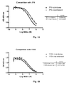


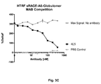

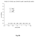


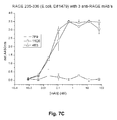


Classifications
-
- C—CHEMISTRY; METALLURGY
- C07—ORGANIC CHEMISTRY
- C07K—PEPTIDES
- C07K16/00—Immunoglobulins [IGs], e.g. monoclonal or polyclonal antibodies
- C07K16/18—Immunoglobulins [IGs], e.g. monoclonal or polyclonal antibodies against material from animals or humans
- C07K16/28—Immunoglobulins [IGs], e.g. monoclonal or polyclonal antibodies against material from animals or humans against receptors, cell surface antigens or cell surface determinants
- C07K16/2803—Immunoglobulins [IGs], e.g. monoclonal or polyclonal antibodies against material from animals or humans against receptors, cell surface antigens or cell surface determinants against the immunoglobulin superfamily
-
- A—HUMAN NECESSITIES
- A61—MEDICAL OR VETERINARY SCIENCE; HYGIENE
- A61K—PREPARATIONS FOR MEDICAL, DENTAL OR TOILETRY PURPOSES
- A61K39/00—Medicinal preparations containing antigens or antibodies
- A61K39/395—Antibodies; Immunoglobulins; Immune serum, e.g. antilymphocytic serum
-
- A—HUMAN NECESSITIES
- A61—MEDICAL OR VETERINARY SCIENCE; HYGIENE
- A61P—SPECIFIC THERAPEUTIC ACTIVITY OF CHEMICAL COMPOUNDS OR MEDICINAL PREPARATIONS
- A61P13/00—Drugs for disorders of the urinary system
- A61P13/12—Drugs for disorders of the urinary system of the kidneys
-
- A—HUMAN NECESSITIES
- A61—MEDICAL OR VETERINARY SCIENCE; HYGIENE
- A61P—SPECIFIC THERAPEUTIC ACTIVITY OF CHEMICAL COMPOUNDS OR MEDICINAL PREPARATIONS
- A61P19/00—Drugs for skeletal disorders
- A61P19/02—Drugs for skeletal disorders for joint disorders, e.g. arthritis, arthrosis
-
- A—HUMAN NECESSITIES
- A61—MEDICAL OR VETERINARY SCIENCE; HYGIENE
- A61P—SPECIFIC THERAPEUTIC ACTIVITY OF CHEMICAL COMPOUNDS OR MEDICINAL PREPARATIONS
- A61P21/00—Drugs for disorders of the muscular or neuromuscular system
- A61P21/02—Muscle relaxants, e.g. for tetanus or cramps
-
- A—HUMAN NECESSITIES
- A61—MEDICAL OR VETERINARY SCIENCE; HYGIENE
- A61P—SPECIFIC THERAPEUTIC ACTIVITY OF CHEMICAL COMPOUNDS OR MEDICINAL PREPARATIONS
- A61P21/00—Drugs for disorders of the muscular or neuromuscular system
- A61P21/04—Drugs for disorders of the muscular or neuromuscular system for myasthenia gravis
-
- A—HUMAN NECESSITIES
- A61—MEDICAL OR VETERINARY SCIENCE; HYGIENE
- A61P—SPECIFIC THERAPEUTIC ACTIVITY OF CHEMICAL COMPOUNDS OR MEDICINAL PREPARATIONS
- A61P25/00—Drugs for disorders of the nervous system
-
- A—HUMAN NECESSITIES
- A61—MEDICAL OR VETERINARY SCIENCE; HYGIENE
- A61P—SPECIFIC THERAPEUTIC ACTIVITY OF CHEMICAL COMPOUNDS OR MEDICINAL PREPARATIONS
- A61P25/00—Drugs for disorders of the nervous system
- A61P25/14—Drugs for disorders of the nervous system for treating abnormal movements, e.g. chorea, dyskinesia
- A61P25/16—Anti-Parkinson drugs
-
- A—HUMAN NECESSITIES
- A61—MEDICAL OR VETERINARY SCIENCE; HYGIENE
- A61P—SPECIFIC THERAPEUTIC ACTIVITY OF CHEMICAL COMPOUNDS OR MEDICINAL PREPARATIONS
- A61P25/00—Drugs for disorders of the nervous system
- A61P25/18—Antipsychotics, i.e. neuroleptics; Drugs for mania or schizophrenia
-
- A—HUMAN NECESSITIES
- A61—MEDICAL OR VETERINARY SCIENCE; HYGIENE
- A61P—SPECIFIC THERAPEUTIC ACTIVITY OF CHEMICAL COMPOUNDS OR MEDICINAL PREPARATIONS
- A61P25/00—Drugs for disorders of the nervous system
- A61P25/24—Antidepressants
-
- A—HUMAN NECESSITIES
- A61—MEDICAL OR VETERINARY SCIENCE; HYGIENE
- A61P—SPECIFIC THERAPEUTIC ACTIVITY OF CHEMICAL COMPOUNDS OR MEDICINAL PREPARATIONS
- A61P25/00—Drugs for disorders of the nervous system
- A61P25/28—Drugs for disorders of the nervous system for treating neurodegenerative disorders of the central nervous system, e.g. nootropic agents, cognition enhancers, drugs for treating Alzheimer's disease or other forms of dementia
-
- A—HUMAN NECESSITIES
- A61—MEDICAL OR VETERINARY SCIENCE; HYGIENE
- A61P—SPECIFIC THERAPEUTIC ACTIVITY OF CHEMICAL COMPOUNDS OR MEDICINAL PREPARATIONS
- A61P27/00—Drugs for disorders of the senses
- A61P27/02—Ophthalmic agents
- A61P27/06—Antiglaucoma agents or miotics
-
- A—HUMAN NECESSITIES
- A61—MEDICAL OR VETERINARY SCIENCE; HYGIENE
- A61P—SPECIFIC THERAPEUTIC ACTIVITY OF CHEMICAL COMPOUNDS OR MEDICINAL PREPARATIONS
- A61P29/00—Non-central analgesic, antipyretic or antiinflammatory agents, e.g. antirheumatic agents; Non-steroidal antiinflammatory drugs [NSAID]
-
- A—HUMAN NECESSITIES
- A61—MEDICAL OR VETERINARY SCIENCE; HYGIENE
- A61P—SPECIFIC THERAPEUTIC ACTIVITY OF CHEMICAL COMPOUNDS OR MEDICINAL PREPARATIONS
- A61P37/00—Drugs for immunological or allergic disorders
-
- C—CHEMISTRY; METALLURGY
- C07—ORGANIC CHEMISTRY
- C07K—PEPTIDES
- C07K16/00—Immunoglobulins [IGs], e.g. monoclonal or polyclonal antibodies
-
- C—CHEMISTRY; METALLURGY
- C07—ORGANIC CHEMISTRY
- C07K—PEPTIDES
- C07K16/00—Immunoglobulins [IGs], e.g. monoclonal or polyclonal antibodies
- C07K16/18—Immunoglobulins [IGs], e.g. monoclonal or polyclonal antibodies against material from animals or humans
- C07K16/28—Immunoglobulins [IGs], e.g. monoclonal or polyclonal antibodies against material from animals or humans against receptors, cell surface antigens or cell surface determinants
-
- C—CHEMISTRY; METALLURGY
- C07—ORGANIC CHEMISTRY
- C07K—PEPTIDES
- C07K16/00—Immunoglobulins [IGs], e.g. monoclonal or polyclonal antibodies
- C07K16/46—Hybrid immunoglobulins
- C07K16/461—Igs containing Ig-regions, -domains or -residues form different species
- C07K16/464—Igs containing CDR-residues from one specie grafted between FR-residues from another
-
- C—CHEMISTRY; METALLURGY
- C12—BIOCHEMISTRY; BEER; SPIRITS; WINE; VINEGAR; MICROBIOLOGY; ENZYMOLOGY; MUTATION OR GENETIC ENGINEERING
- C12P—FERMENTATION OR ENZYME-USING PROCESSES TO SYNTHESISE A DESIRED CHEMICAL COMPOUND OR COMPOSITION OR TO SEPARATE OPTICAL ISOMERS FROM A RACEMIC MIXTURE
- C12P21/00—Preparation of peptides or proteins
- C12P21/005—Glycopeptides, glycoproteins
-
- A—HUMAN NECESSITIES
- A61—MEDICAL OR VETERINARY SCIENCE; HYGIENE
- A61K—PREPARATIONS FOR MEDICAL, DENTAL OR TOILETRY PURPOSES
- A61K39/00—Medicinal preparations containing antigens or antibodies
- A61K2039/505—Medicinal preparations containing antigens or antibodies comprising antibodies
-
- C—CHEMISTRY; METALLURGY
- C07—ORGANIC CHEMISTRY
- C07K—PEPTIDES
- C07K2317/00—Immunoglobulins specific features
- C07K2317/10—Immunoglobulins specific features characterized by their source of isolation or production
- C07K2317/14—Specific host cells or culture conditions, e.g. components, pH or temperature
-
- C—CHEMISTRY; METALLURGY
- C07—ORGANIC CHEMISTRY
- C07K—PEPTIDES
- C07K2317/00—Immunoglobulins specific features
- C07K2317/20—Immunoglobulins specific features characterized by taxonomic origin
- C07K2317/24—Immunoglobulins specific features characterized by taxonomic origin containing regions, domains or residues from different species, e.g. chimeric, humanized or veneered
-
- C—CHEMISTRY; METALLURGY
- C07—ORGANIC CHEMISTRY
- C07K—PEPTIDES
- C07K2317/00—Immunoglobulins specific features
- C07K2317/30—Immunoglobulins specific features characterized by aspects of specificity or valency
- C07K2317/34—Identification of a linear epitope shorter than 20 amino acid residues or of a conformational epitope defined by amino acid residues
-
- C—CHEMISTRY; METALLURGY
- C07—ORGANIC CHEMISTRY
- C07K—PEPTIDES
- C07K2317/00—Immunoglobulins specific features
- C07K2317/50—Immunoglobulins specific features characterized by immunoglobulin fragments
- C07K2317/56—Immunoglobulins specific features characterized by immunoglobulin fragments variable (Fv) region, i.e. VH and/or VL
- C07K2317/565—Complementarity determining region [CDR]
-
- C—CHEMISTRY; METALLURGY
- C07—ORGANIC CHEMISTRY
- C07K—PEPTIDES
- C07K2317/00—Immunoglobulins specific features
- C07K2317/50—Immunoglobulins specific features characterized by immunoglobulin fragments
- C07K2317/56—Immunoglobulins specific features characterized by immunoglobulin fragments variable (Fv) region, i.e. VH and/or VL
- C07K2317/567—Framework region [FR]
-
- C—CHEMISTRY; METALLURGY
- C07—ORGANIC CHEMISTRY
- C07K—PEPTIDES
- C07K2317/00—Immunoglobulins specific features
- C07K2317/70—Immunoglobulins specific features characterized by effect upon binding to a cell or to an antigen
- C07K2317/76—Antagonist effect on antigen, e.g. neutralization or inhibition of binding
-
- C—CHEMISTRY; METALLURGY
- C07—ORGANIC CHEMISTRY
- C07K—PEPTIDES
- C07K2317/00—Immunoglobulins specific features
- C07K2317/90—Immunoglobulins specific features characterized by (pharmaco)kinetic aspects or by stability of the immunoglobulin
- C07K2317/92—Affinity (KD), association rate (Ka), dissociation rate (Kd) or EC50 value
-
- Y—GENERAL TAGGING OF NEW TECHNOLOGICAL DEVELOPMENTS; GENERAL TAGGING OF CROSS-SECTIONAL TECHNOLOGIES SPANNING OVER SEVERAL SECTIONS OF THE IPC; TECHNICAL SUBJECTS COVERED BY FORMER USPC CROSS-REFERENCE ART COLLECTIONS [XRACs] AND DIGESTS
- Y02—TECHNOLOGIES OR APPLICATIONS FOR MITIGATION OR ADAPTATION AGAINST CLIMATE CHANGE
- Y02A—TECHNOLOGIES FOR ADAPTATION TO CLIMATE CHANGE
- Y02A50/00—TECHNOLOGIES FOR ADAPTATION TO CLIMATE CHANGE in human health protection, e.g. against extreme weather
- Y02A50/30—Against vector-borne diseases, e.g. mosquito-borne, fly-borne, tick-borne or waterborne diseases whose impact is exacerbated by climate change
Definitions
- the present application relates to antibodies, particularly monoclonal antibodies, and in particular CDR grafted, humanized versions thereof, that may be used in the treatment and diagnosis of Alzheimer's Disease (AD), central nervous system cell degeneration, impaired learning and memory, abnormal transport of amyloid ß ⁇ and other neuroinflammatrory conditions associated with the Receptor of Advanced Glycation End Products (RAGE).
- AD Alzheimer's Disease
- RAGE Advanced Glycation End Products
- the present invention relates to antibodies and fragments thereof that bind to RAGE.
- AD Alzheimer's Disease
- AD is characterized by a deposit of abnormally aggregated proteins. In the case of extra-cellular amyloid plaques, these deposits consist mostly of amyloid- ⁇ -peptide filaments (Aß), and in the case of the intracellular neurofibrillary tangles (NFTs), mostly of the tau protein.
- Aß amyloid- ⁇ -peptide filaments
- NFTs neurofibrillary tangles
- AD is also characterized by an increased neuronal expression of RAGE.
- RAGE is a multi-ligand receptor of the immunoglobulin family which functions as a signal-transducing cell surface acceptor for Aß.
- a ß40 infusion in mice has been shown by several groups to lead to vasoconstriction of cerebral vessels and a decrease of cerebral blood flow (CBF). Patients suffering from AD also have a decreased cerebral blood flow.
- CBF cerebral blood flow
- the transgenic animals overexpress the protein Amyloid Precursor Protein (APP) that leads to disease causing plaque formation, RAGE has been implicated as a pathogenic factor in the disease progression ( Deane et al. Nature Medicine 9(7) pp 907-913, 2003 ; Arancio et al. EMBOJ, 1-10, 2004 ).
- APP Amyloid Precursor Protein
- RAGE has been shown to bind to Aß-peptides. Inhibition of this interaction suppresses accumulation of Aß in the transgenic animal model; therefore RAGE is believed to be involved in AD.
- Treatment with sRAGE (soluble RAGE) as well as anti-RAGE antibodies has been shown to lower plaque numbers (Deane et al, 2003). Blocking the interaction of RAGE with amyloid by antibodies could become a treatment for AD patients; however, existing polyclonal antibodies generated from animal serum are not suited for the chronic treatment of humans.
- WO 2008/137552 A2 discloses certain monoclonal anti-RAGE antibodies binding to different domains of RAGE. Most of said antibodies inhibit the interaction of human RAGE and a complex of HMGB1 and CpG DNA.
- WO 2006/077101 relates to the identification, functionality and use of peptides designated AGER-RME and AGER-CDP of RAGE. Said peptides are inter alia applicable for identifying and preparing RAGE binding ligands like anti-RAGE antibodies.
- the present invention describes novel monoclonal antibodies that bind to the C-domains of RAGE and the specific interaction and competition with the binding of A ⁇ with monoclonal antibodies for the C1 and C2-domain in RAGE.
- the present invention provides binding molecules, in particular antibodies, that bind specifically to RAGE; representative anti-RAGE antibodies of the invention may comprise at least one of the antibody variable region amino acid sequences shown in SEQ ID NOs: 1, 5, 9, 13, 17, and 21, or individual CDRs thereof or related CDR sequences, as specified in more detail below.
- the present invention provides monoclonal antibodies that bind to RAGE, more specifically monoclonal antibodies that bind to the C-domain of RAGE.
- anti-RAGE antibodies that bind specifically to RAGE and comprise a light chain variable region having an amino acid sequence that is at least 90% identical to any of SEQ ID NOs.: 5, 13, and 21, or is a RAGE-binding fragment of an antibody comprised in said sequences.
- anti-RAGE antibodies that bind specifically to RAGE and comprise a heavy chain variable region having an amino acid sequence that is at least 90% identical to any of SEQ ID NOs.: 1, 9, and 17, or is a RAGE-binding fragment of an antibody comprised in said sequences.
- a particular embodiment of the present invention is represented by several monoclonal antibodies that are able to bind to the C-domain of RAGE, and to block the binding of Aß -globulomers. More specifically the present invention describes monoclonal antibody 11E6, which binds to the C-2 domain of RAGE, does not bind to peptides with amino acid sequences used to generate polyclonal antibodies, and is able to neutralize in vivo the effect of Aß1-40 on cerebral vasculature in mice.
- the anti-RAGE antibodies of the invention include antibodies that bind specifically to the C-domain of RAGE.
- the anti-RAGE antibodies of the invention include an anti-RAGE antibody or a RAGE-binding fragment as described above, which is selected from the group consisting of chimeric antibody, a CDR-grafted or humanized antibody, a single chain antibody, a fusion protein, and a human antibody.
- the antibodies of the invention are recombinant antibodies or monoclonal antibodies.
- Particular neutralizing antibodies of the present application are referred to herein as mAb7F9, mAb11E6, and mAb4E5 and functional antibody fragments thereof, and other antibodies and functional antibody fragments with equivalent properties to mAb7F9, mAb11E6, and mAb4E5, such as high affinity binding to RAGE with low dissociation kinetics and high neutralizing capacity, are intended as part of the present invention.
- the human antibodies of the present application may include amino acid residues not encoded by human germline immunoglobulin immunogenic RAGE polypeptide or fragment thereof, that may be determined by any method known in the art.
- the binding affinity can be measured by competitive ELISAs, c RIAs, BIAcore or KinExA technology.
- the dissociation rate also can be measured by BIAcore or KinExA technology.
- the binding affinity and dissociation rate are measured by surface plasmon resonance using, e.g., BIAcore.
- the mAb7F9 antibody has at least 90% amino acid sequence identity with a sequence comprising a heavy chain variable region (VH region) comprising the sequence of SEQ ID NO: 1; and SEQ ID NOs. 2, 3, and 4 which are residues 31-35, 50-68, and 101-108 of SEQ ID NO: 1, respectively.
- the mAb7F9 antibody of the present invention has at least 90% amino acid sequence identity with a sequence comprising a light chain variable region (VL region) comprising the sequence of SEQ ID NO: 5, and SEQ ID NOs. 6, 7, and 8 which are residues 24-34, 50-56, 89-97 of SEQ ID NO: 5, respectively.
- the mAb11E6 antibody has at least 90% amino acid sequence identity with a sequence comprising a heavy chain variable region (VH region) comprising the sequence of SEQ ID NO: 9; and SEQ ID NOs. 10, 11, and 12 which are residues 31-35, 50-66, and 99-109 of SEQ ID NO: 9, respectively.
- the mAb11E6 antibody of the present invention has at least 90% amino acid sequence identity with a sequence comprising a light chain variable region (VL region) comprising the sequence of SEQ ID NO: 13, and SEQ ID NOs. 14, 15, and 16 which are residues 24-34, 50-56, 89-97 of SEQ ID NO: 13, respectively.
- the mAb11E6 binds to the C-2 domain of RAGE, does not bind to peptides with amino acid sequences used to generate polyclonal antibodies, and is able to neutralize in vivo the effect of Aß1-40 on cerebral vasculature in mice.
- the mAb4E5 antibody has at least 90% amino acid sequence identity with a sequence comprising a heavy chain variable region (VH region) comprising the sequence of SEQ ID NO: 17; and SEQ ID NOs. 18, 19, and 20 which are residues 31-35, 50-66, and 99-109 of SEQ ID NO: 17, respectively.
- the mAb4E5 antibody of the present invention has at least 90% amino acid sequence identity with a sequence comprising a light chain variable region (VL region) comprising the sequence of SEQ ID NO: 21, and SEQ ID NOs. 22, 23, and 24 which are residues 24-34, 50-56, 89-97 of SEQ ID NO: 21, respectively.
- the isolated monoclonal antibodies that interact with RAGE of the present application may be a glycosylated binding protein wherein the antibody or antigen-binding portion thereof comprises one or more carbohydrate residues.
- Nascent in vivo protein production may undergo further processing, known as post-translational modification.
- sugar (glycosyl) residues may be added enzymatically, a process known as glycosylation.
- glycosylation The resulting proteins bearing covalently linked oligosaccharide side chains are known as glycosylated proteins or glycoproteins. Protein glycosylation depends on the amino acid sequence of the protein of interest, as well as the host cell in which the protein is expressed.
- glycosylation enzymes eg., glycosyltransferases and glycosidases
- substrates nucleotide sugars
- protein glycosylation pattern, and composition of glycosyl residues may differ depending on the host system in which the particular protein is expressed.
- Glycosyl residues useful in the invention may include, but are not limited to, glucose, galactose, mannose, fucose, n-acetylglucosamine and sialic acid.
- the antibodies of the present application comprise a heavy chain constant region, such as an IgG1, IgG2, IgG3, IgG4, IgA, IgE, IgM or IgD constant region.
- the antibody can comprise a light chain constant region, either a kappa light chain constant region or a lambda light chain constant region.
- the antibody comprises a kappa light chain constant region.
- the antibody portion can be, for example, a Fab fragment or a single chain Fv fragment. Replacements of amino acid residues in the Fc portion to alter antibody effector function are known in the art ( Winter, et al. U.S. Pat. Nos. 5,648,260 ; 5,624,821 ).
- the Fc portion of an antibody mediates several important effector functions. e.g. cytokine induction, ADCC, phagocytosis, complement dependent cytotoxicity (CDC) and half-life/clearance rate of antibody and antigen-antibody complexes. In some cases these effector functions are desirable for therapeutic antibodies but in other cases might be unnecessary or even deleterious, depending on the therapeutic objectives.
- Certain human IgG isotypes, particularly IgG1 and IgG3, mediate ADCC and CDC via binding to Fc ⁇ Rs and complement C1q, respectively.
- Neonatal Fc receptors (FcRn) are the critical components determining the circulating half-life of antibodies.
- at least one amino acid residue is replaced in the constant region of the antibody, for example the Fc region of the antibody, such that effector functions of the antibody are altered.
- polypeptide refers to any polymeric chain of amino acids.
- peptide and protein are used interchangeably with the term polypeptide and also refer to a polymeric chain of amino acids.
- polypeptide encompasses native or artificial proteins, protein fragments and polypeptide analogs of a protein sequence.
- a polypeptide may be monomeric or polymeric.
- isolated protein or "isolated polypeptide” is a protein or polypeptide that by virtue of its origin or source of derivation is not associated with naturally associated components that accompany it in its native state; is substantially free of other proteins from the same species; is expressed by a cell from a different species; or does not occur in nature.
- a polypeptide that is chemically synthesized or synthesized in a cellular system different from the cell from which it naturally originates will be “isolated” from its naturally associated components.
- a protein may also be rendered substantially free of naturally associated components by isolation, using protein purification techniques well known in the art.
- recovering refers to the process of rendering a chemical species such as a polypeptide substantially free of naturally associated components by isolation, e.g., using protein purification techniques well known in the art.
- RAGE Receptor for Advanced Glycation Endproducts
- RAGE The structure of RAGE has not been solved. Homology to other proteins leads to a model where RAGE has several domains. These domains are named in analogy to immunoglobulins: (i) V-like domain at the N-terminus: this equivalent domain in immunoglobulins binds antigen and represents the only binding region within these proteins. In RAGE, this domain binds to some ligands like S100 ( Ostendorp et al. EMBOJ. 26,3875,2007 ; Leclerc et al. JBC 282,31317,2007 ).
- S100A12 also called ENRAGE or Calgranulin C
- Kd 90 nM
- a ⁇ competes with S100A12 for this domain, suggesting that A ⁇ also binds to the C1-domain.
- Second C2-like domain this domain is called C2 (also used by Ostendorp et al. BBRC 2006 ).
- RAGE ligand S100A6 binds to the C2 domain and an antibody generated against a peptide from the C2 domain was shown to compete againt S100A6 for this binding; the antibody lead to reduced signal transduction in SH-SY5Y ( Leclerc et al. JBC 282,31317,2007 ).
- RAGE domain may be defined in line with different definitions provided in the state of the art:
- each of the definitions may be applied in order to define the binding characteristics of the binding proteins of the present invention.
- RAGE is capable of binding different ligands via different domains.
- Results from competition experiments with other ligands seem to indicate that A ⁇ binds to the C1-domain ( Hofinann et al. Cell 97, 889, 1999 or Xie et al. JBC, 282, 4218, 2007 ).
- RAGE ligands there may be mentioned:
- RAGE as used herein, particularly refers to human RAGE, also designated “hRAGE” or “huRAGE”. Unless otherwise stated the term “RAGE” also encompasses RAGE molecules isolated or obtained from other, different from human, species, as for example, rodents, like mice or rats; or bovine RAGE molecules.
- sRAGE refers to a soluble form of RAGE, derived from the extra cellular domain of RAGE.
- a sRAGE molecule derived from human RAGE also designated as husRAGE comprises amino acid residues 1 to 331 (see SEQ ID NO: 61) of human RAGE (see SEQ ID NO: 60).
- Bioactivity refers to all inherent biological properties of RAGE as defined herein.
- telomere binding in reference to the interaction of an antibody, a protein, or a peptide with a second chemical species, mean that the interaction is dependent upon the presence of a particular structure (e.g., an "antigenic determinant” or “epitope” as defined below) on the chemical species; for example, an antibody recognizes and binds to a specific protein structure rather than to proteins generally. If an antibody is specific for epitope "A”, the presence of a molecule containing epitope A (or free, unlabeled A), in a reaction containing labeled "A” and the antibody, will reduce the amount of labeled A bound to the antibody.
- antibody broadly refers to any immunoglobulin (Ig) molecule comprised of four polypeptide chains, two heavy (H) chains and two light (L) chains, or any functional fragment, mutant, variant, or derivation thereof, which retains the essential epitope binding features of an Ig molecule.
- Ig immunoglobulin
- Such mutant, variant, or derivative antibody formats are known in the art. Nonlimiting embodiments of which are discussed below.
- An antibody is said to be “capable of binding" a molecule if it is capable of specifically reacting with the molecule to thereby bind the molecule to the antibody.
- a “monoclonal antibody” as used herein is intended to refer to a preparation of antibody molecules, which share a common heavy chain and common light chain amino acid sequence, in contrast with “polyclonal” antibody preparations that contain a mixture of different antibodies.
- Monoclonal antibodies can be generated by several novel technologies like phage, bacteria, yeast or ribosomal display, as well as classical methods exemplified by hybridoma-derived antibodies (e.g., an antibody secreted by a hybridoma prepared by hybridoma technology, such as the standard Kohler and Milstein hybridoma methodology ((1975) Nature 256:495-497 ).
- each heavy chain is comprised of a heavy chain variable region (abbreviated herein as HCVR or VH) and a heavy chain constant region.
- the heavy chain constant region is comprised of three domains, CH1, CH2 and CH3.
- Each light chain is comprised of a light chain variable region (abbreviated herein as LCVR or VL) and a light chain constant region.
- the light chain constant region is comprised of one domain, CL.
- the VH and VL regions can be further subdivided into regions of hypervariability, termed complementarity determining regions (CDR), interspersed with regions that are more conserved, termed framework regions (FR).
- CDR complementarity determining regions
- Each VH and VL is composed of three CDRs and four FRs, arranged from amino-terminus to carboxy-terminus in the following order: FR1, CDR1, FR2, CDR2, FR3, CDR3, FR4.
- Immunoglobulin molecules can be of any type (e.g., IgG, IgE, IgM, IgD, IgA and IgY), class (e.g., IgG 1, IgG2, IgG 3, IgG 4, IgA1 and IgA2) or subclass.
- antigen-binding portion or “antigen-binding fragment”of an antibody (or simply “antibody portion” or “antibody fragment”), as used herein, refers to one or more fragments of an antibody that retain the ability to specifically bind to an antigen (e.g., RAGE). It has been shown that the antigen-binding function of an antibody can be performed by fragments of a full-length antibody. Such antibody embodiments may also be bispecific, dual specific, or multi-specific formats; specifically binding to two or more different antigens.
- binding fragments encompassed within the term "antigen-binding portion" of an antibody include (i) a Fab fragment, a monovalent fragment consisting of the VL, VH, CL and CH1 domains; (ii) a F(ab') 2 fragment, a bivalent fragment comprising two Fab fragments linked by a disulfide bridge at the hinge region; (iii) a Fd fragment consisting of the VH and CH1 domains; (iv) a Fv fragment consisting of the VL and VH domains of a single arm of an antibody, (v) a dAb fragment ( Ward et al., (1989) Mature 341:544-546 , Winter et al., PCT publication WO 90/05144 A1 herein incorporated by reference), which comprises a single variable domain; and (vi) an isolated complementarity determining region (CDR).
- CDR complementarity determining region
- the two domains of the Fv fragment, VL and VH are coded for by separate genes, they can be joined, using recombinant methods, by a synthetic linker that enables them to be made as a single protein chain in which the VL and VH regions pair to form monovalent molecules (known as single chain Fv (scFv); see e.g., Bird et al. (1988) Science 242:423-426 ; and Huston et al. (1988) Proc. Natl. Acad. Sci. USA 85:5879-5883 ).
- single chain Fv single chain Fv
- Such single chain antibodies are also intended to be encompassed within the term "antigen-binding portion" of an antibody.
- Other forms of single chain antibodies, such as diabodies are also encompassed.
- Diabodies are bivalent, bispecific antibodies in which VH and VL domains are expressed on a single polypeptide chain, but using a linker that is too short to allow for pairing between the two domains on the same chain, thereby forcing the domains to pair with complementary domains of another chain and creating two antigen binding sites (see e.g., Holliger, P., et al. (1993) Proc. Natl. Acad. Sci. USA 90:6444-6448 ; Poljak, R.J., et al. (1994) Structure 2:1121-1123 ).
- Such antibody binding portions are known in the art ( Kontermann and Dubel eds., Antibody Engineering (2001) Springer-Verlag. New York. 790 pp. (ISBN 3-540-41354-5 ).
- antibody construct refers to a polypeptide comprising one or more the antigen binding portions of the invention linked to a linker polypeptide or an immunoglobulin constant domain.
- Linker polypeptides comprise two or more amino acid residues joined by peptide bonds and are used to link one or more antigen binding portions.
- Such linker polypeptides are well known in the art (see e.g., Holliger, P., et al. (1993) Proc. Natl. Acad. Sci. USA 90:6444-6448 ; Poljak, R.J., et al. (1994) Stricture 2:1121-1123 ).
- An immunoglobulin constant domain refers to a heavy or light chain constant domain. Human IgG heavy chain and light chain constant domain amino acid sequences are known in the art and represented in Table 1.
- an antibody or antigen-binding portion thereof may be part of a larger immunoadhesion molecules, formed by covalent or noncovalent association of the antibody or antibody portion with one or more other proteins or peptides.
- immunoadhesion molecules include use of the streptavidin core region to make a tetrameric scFv molecule ( Kipriyanov, S.M., et al. (1995) Human Antibodies and Hybridomas 6:93-101 ) and use of a cysteine residue, a marker peptide and a C-terminal polyhistidine tag to make bivalent and biotinylated scFv molecules ( Kipriyanov, S.M., et al. (1994) Mol.
- Antibody portions such as Fab and F(ab') 2 fragments, can be prepared from whole antibodies using conventional techniques, such as papain or pepsin digestion, respectively, of whole antibodies.
- antibodies, antibody portions and immunoadhesion molecules can be obtained using standard recombinant DNA techniques, as described herein.
- an "isolated antibody”, as used herein, is intended to refer to an antibody that is substantially free of other antibodies having different antigenic specificities (e.g., an isolated antibody that specifically binds human RAGE is substantially free of antibodies that specifically bind antigens other than human RAGE).
- An isolated antibody that specifically binds human RAGE may, however, have cross-reactivity to other antigens, such as RAGE molecules from other species.
- an isolated antibody may be substantially free of other cellular material and/or chemicals.
- human antibody is intended to include antibodies having variable and constant regions derived from human germline immunoglobulin sequences.
- the human antibodies of the invention may include amino acid residues not encoded by human germline immunoglobulin sequences (e.g., mutations introduced by random or site-specific mutagenesis in vitro or by somatic mutation in vivo), for example in the CDRs and in particular CDR3.
- human antibody as used herein, is not intended to include antibodies in which CDR sequences derived from the germline of another mammalian species, such as a mouse, have been grafted onto human framework sequences.
- recombinant human antibody is intended to include all human antibodies that are prepared, expressed, created or isolated by recombinant means, such as antibodies expressed using a recombinant expression vector transfected into a host cell (described further below), antibodies isolated from a recombinant, combinatorial human antibody library ( Hoogenboom H.R., (1997) TIB Tech. 15:62-70 ; Azzazy H., and Highsmith W.E., (2002) Clin. Biochem. 35:425-445 ; Gavilondo J.V., and Larrick J.W. (2002) BioTechniques 29:128-145 ; Hoogenboom H., and Chames P.
- such recombinant human antibodies are subjected to in vitro mutagenesis (or, when an animal transgenic for human Ig sequences is used, in vivo somatic mutagenesis) and thus the amino acid sequences of the VH and VL regions of the recombinant antibodies are sequences that, while derived from and related to human germline VH and VL sequences, may not naturally exist within the human antibody germline repertoire in vivo.
- chimeric antibody refers to antibodies which comprise heavy and light chain variable region sequences from one species and constant region sequences from another species, such as antibodies having murine heavy and light chain variable regions linked to human constant regions.
- the chimeric antibody can be produced through recombinant molecular biological techniques, or may be physically conjugated together.
- CDR-grafted antibody refers to antibodies which comprise heavy and light chain variable region sequences from one species but in which the sequences of one or more of the CDR regions of VH and/or VL are replaced with CDR sequences of another species, such as antibodies having murine heavy and light chain variable regions in which one or more of the murine CDRs (e.g., CDR3) has been replaced with human CDR sequences.
- Kabat numbering “Kabat definitions and “Kabat labeling” are used interchangeably herein. These terms, which are recognized in the art, refer to a system of numbering amino acid residues which are more variable ( i.e. hypervariable) than other amino acid residues in the heavy and light chain variable regions of an antibody, or an antigen binding portion thereof ( Kabat et al. (1971) Ann. NY Acad, Sci. 190:382-391 and , Kabat, E.A., et al. (1991) Sequences of Proteins of Immunological Interest, Fifth Edition, U.S. Department of Health and Human Services, NIH Publication No. 91-3242 ).
- the hypervariable region ranges from amino acid positions 31 to 35 for CDR1, amino acid positions 50 to 65 for CDR2, and amino acid positions 95 to 102 for CDR3.
- the hypervariable region ranges from amino acid positions 24 to 34 for CDR1, amino acid positions 50 to 56 for CDR2, and amino acid positions 89 to 97 for CDR3.
- the terms “acceptor” and “acceptor antibody” refer to the antibody or nucleic acid sequence providing or encoding at least 50, 55, 60, 65, 70, 75 or 80%, at least 85%, at least 90%, at least 95%, at least 98% or 100% of the amino acid sequences of one or more of the framework regions.
- the term “acceptor” refers to the antibody amino acid or nucleic acid sequence providing or encoding the constant region(s).
- the term “acceptor” refers to the antibody amino acid or nucleic acid sequence providing or encoding one or more of the framework regions and the constant region(s).
- the term "acceptor” refers to a human antibody amino acid or nucleic acid sequence that provides or encodes at least 50, 55, 60, 65, 70, 75 or 80%, particularly, at least 85%, at least 90%, at least 95%, at least 98%, or 100% of the amino acid sequences of one or more of the framework regions.
- an acceptor may contain at least 1, at least 2, at least 3, least 4, at least 5, or at least 10 amino acid residues that does (do) not occur at one or more specific positions of a human antibody.
- acceptor framework region and/or acceptor constant region(s) may be, e.g., derived or obtained from a germline antibody gene, a mature antibody gene, a functional antibody (e.g., antibodies well-known in the art, antibodies in development, or antibodies commercially available).
- CDR refers to the complementarity determining region within antibody variable sequences. There are three CDRs in each of the variable regions of the heavy chain and the light chain, which are designated CDR1, CDR2 and CDR3, for each of the variable regions.
- CDR set refers to a group of three CDRs that occur in a single variable region capable of binding the antigen. The exact boundaries of these CDRs have been defined differently according to different systems. The system described by Kabat ( Kabat et al., Sequences of Proteins of Immunological Interest (National Institutes of Health, Bethesda, Md.
- CDR boundary definitions may not strictly follow one of the above systems, but will nonetheless overlap with the Kabat CDRs, although they may be shortened or lengthened in light of prediction or experimental findings that particular residues or groups of residues or even entire CDRs do not significantly impact antigen binding.
- the methods used herein may utilize CDRs defined according to any of these systems, although particular embodiments use Kabat or Chothia defined CDRs.
- canonical residue refers to a residue in a CDR or framework that defines a particular canonical CDR structure as defined by Chothia et al. (J. Mol. Biol. 196:901-907 (1987 ); Chothia et al., J. Mol. Biol. 227:799 (1992 ), both are incorporated herein by reference). According to Chothia et al., critical portions of the CDRs of many antibodies have nearly identical peptide backbone confirmations despite great diversity at the level of amino acid sequence. Each canonical structure specifies primarily a set of peptide backbone torsion angles for a contiguous segment of amino acid residues forming a loop.
- the terms “donor” and “donor antibody” refer to an antibody providing one or more CDRs.
- the donor antibody is an antibody from a species different from the antibody from which the framework regions are obtained or derived.
- the term “donor antibody” refers to a non-human antibody providing one or more CDRs.
- the term "framework” or "framework sequence” refers to the remaining sequences of a variable region minus the CDRs. Because the exact definition of a CDR sequence can be determined by different systems, the meaning of a framework sequence is subject to correspondingly different interpretations.
- the six CDRs (CDR-L1, -L2, and -L3 of light chain and CDR-H1, -H2, and -H3 of heavy chain) also divide the framework regions on the light chain and the heavy chain into four sub-regions (FR1, FR2, FR3 and FR4) on each chain, in which CDR1 is positioned between FR1 and FR2, CDR2 between FR2 and FR3, and CDR3 between FR3 and FR4.
- a framework region represents the combined FR's within the variable region of a single, naturally occurring immunoglobulin chain.
- a FR represents one of the four sub- regions, and FRs represents two or more of the four sub- regions constituting a framework region.
- Human heavy chain and light chain acceptor sequences are known in the art. In one embodiment of the invention the human heavy chain and light chain acceptor sequences are selected from the sequences described in Table 2 and Table 3. Different combinations for human framework sequences FR1 to FR4 are stated in said tables.
- the term "germline antibody gene” or “gene fragment” refers to an immunoglobulin sequence encoded by non-lymphoid cells that have not undergone the maturation process that leads to genetic rearrangement and mutation for expression of a particular immunoglobulin. (See, e.g., Shapiro et al., Crit. Rev. Immunol. 22(3): 183-200 (2002 ); Marchalonis et al., Adv Exp Med Biol. 484:13-30 (2001 )).
- One of the advantages provided by various embodiments of the present invention stems from the recognition that germline antibody genes are more likely than mature antibody genes to conserve essential amino acid sequence structures characteristic of individuals in the species, hence less likely to be recognized as from a foreign source when used therapeutically in that species.
- key residues refer to certain residues within the variable region that have more impact on the binding specificity and/or affinity of an antibody, in particular a humanized antibody.
- a key residue includes, but is not limited to, one or more of the following: a residue that is adjacent to a CDR, a potential glycosylation site (can be either N- or O-glycosylation site), a rare residue, a residue capable of interacting with the antigen, a residue capable of interacting with a CDR, a canonical residue, a contact residue between heavy chain variable region and light chain variable region, a residue within the Vernier zone, and a residue in the region that overlaps between the Chothia definition of a variable heavy chain CDR1 and the Kabat definition of the first heavy chain framework.
- humanized antibody generally refers to antibodies which comprise heavy and light chain variable region sequences from a non-human species (e.g., a mouse) but in which at least a portion of the VH and/or VL sequence has been altered to be more "human-like", i.e., more similar to human germline variable sequences.
- a non-human species e.g., a mouse
- human CDR-grafted antibody in which human CDR sequences are introduced into non-human VH and VL sequences to replace the corresponding nonhuman CDR sequences.
- humanized antibody is an antibody or a variant, derivative, analog or fragment thereof which immunospecifically binds to an antigen of interest and which comprises a framework (FR) region having substantially the amino acid sequence of a human antibody and a complementary determining region (CDR) having substantially the amino acid sequence of a non-human antibody.
- FR framework
- CDR complementary determining region
- substantially in the context of a CDR refers to a CDR having an amino acid sequence at least 50, 55, 60, 65, 70, 75 or 80%, particularly at least 85%, at least 90%, at least 95%, at least 98% or at least 99% identical to the amino acid sequence of a non-human antibody CDR.
- a humanized antibody comprises substantially all of at least one, and typically two, variable domains (Fab, Fab', F(ab') 2, FabC, Fv) in which all or substantially all of the CDR regions correspond to those of a non-human immunoglobulin (i.e., donor antibody) and all or substantially all of the framework regions are those of a human immunoglobulin consensus sequence.
- a humanized antibody also comprises at least a portion of an immunoglobulin constant region (Fc), typically that of a human immunoglobulin.
- a humanized antibody contains both the light chain as well as at least the variable domain of a heavy chain.
- the antibody also may include the CH1, hinge, CH2, CH3, and CH4 regions of the heavy chain.
- a humanized antibody only contains a humanized light chain. In some embodiments, a humanized antibody only contains a humanized heavy chain. In specific embodiments, a humanized antibody only contains a humanized variable domain of a light chain and/or humanized heavy chain.
- the humanized antibody can be selected from any class of immunoglobulins, including IgY, IgM, IgG, IgD, IgA and IgE, and any isotype, including without limitation IgA1, IgA2, IgG1, IgG2, IgG3 and IgG4.
- the humanized antibody may comprise sequences from more than one class or isotype, and particular constant domains may be selected to optimize desired effector functions using techniques well-known in the art.
- the framework and CDR regions of a humanized antibody need not correspond precisely to the parental sequences, e.g., the donor antibody CDR or the consensus framework may be mutagenized by substitution, insertion and/or deletion of at least one amino acid residue so that the CDR or framework residue at that site does not correspond to either the donor antibody or the consensus framework. In a particular embodiment, such mutations, however, will not be extensive. Usually, at least 50, 55, 60, 65, 70, 75 or 80%, particularly at least 85%, more particularly at least 90%, and in particular at least 95% of the humanized antibody residues will correspond to those of the parental FR and CDR sequences.
- the term "consensus framework" refers to the framework region in the consensus immunoglobulin sequence.
- the term "consensus immunoglobulin sequence” refers to the sequence formed from the most frequently occurring amino acids (or nucleotides) in a family of related immunoglobulin sequences (See e.g., Winnaker, From Genes to Clones (Verlagsgesellschaft, Weinheim, Germany 1987 ). In a family of immunoglobulins, each position in the consensus sequence is occupied by the amino acid occurring most frequently at that position in the family. If two amino acids occur equally frequently, either can be included in the consensus sequence.
- Vernier zone refers to a subset of framework residues that may adjust CDR structure and fine-tune the fit to antigen as described by Foote and Winter (1992, J. Mol. Biol. 224:487-499 , which is incorporated herein by reference). Vernier zone residues form a layer underlying the CDRs and may impact on the structure of CDRs and the affinity of the antibody.
- inhibitortion of binding of RAGE to one of his ligands encompasses partial (as for example by about 1 to 10% or more, in particular about 20%, 30%, 40%, 50%, 60%, 70%, 80%, 85%, 90%, or 95% or more) or complete reduction of said ligand binding activity.
- Said “inhibition of binding” may be determined by any suitable method available in the art, preferably by any method as exemplified herein, as for example HTRF assays described herein.
- neutralizing refers to neutralization of biological activity of a target protein when a binding protein specifically binds the target protein.
- Neutralizing may be the result of different ways of binding of said binding protein to the target.
- neutralizing may be caused by binding of the binding protein in a region of the target, which does not affect receptor binding to the target molecule.
- binding of a binding protein may result in a blockade of the receptor binding to the target, which blockade finally neutralizes the target protein activity.
- Each of said different mechanism may occur according to the invention.
- a neutralizing binding protein is a neutralizing antibody whose binding to RAGE results in neutralization of a biological activity of RAGE
- the neutralizing binding protein binds RAGE and reduces a biologically activity of RAGE by at least about 1 to10%, at least about 20%, 30%, 40%, 50%, 60%, 70%, 80%, 85% or more.
- Neutralization of a biological activity of RAGE by a neutralizing binding protein can be assessed by measuring one or more indicators of RAGE biological activity well known in the art, and/or exemplified in the experimental part, as in particular, in Examples 5, 6, 10, and 16 to 19. For example neutralization of RAGE neutralizes the binding of RAGE to Aß-globulomers as measured in Examples 5 and 6 below.
- An "inhibition of soluble Aß1-40 peptide-induced reduction of cerebral blood volume" determined in vivo in an animal model relates to a partial or complete inhibition, as for example by at least about 1 to10%, at least about 15%, 20%, 30%, 40%, 50%, 60%, 70%, 80%, 85% or more, if compared to a non-treated control, or in particular, if compared to a negative, monoclonal or polyclonal, immunoglobulin-isotype control.
- An "improvement of the cerebral blood volume" as in vivo in an animal model over-expressing human APP relates to a statistically significant improvement of CBV, if compared to a non-treated control, or in particular, if compared to a negative, monoclonal or polyclonal, immunoglobulin-isotype control.
- a "reduction of amyloid plaque number and /or amyloid plaque area" as measured in vivo in an animal model over-expressing human APP relates to a partial or complete reduction, as for example by at least about 1 to 10%, at least about 15%, 20%, 30%, 40%, 50%, 60%, 70%, 80%, 85% or more, if compared to a non-treated control, or in particular, if compared to a negative, monoclonal or polyclonal, immunoglobulin-isotype control.
- An "inhibition of aggregated Aß1-40 peptide-induced dynamin cleavage of hippocampal neurons in vitro; relates to a partial or complete inhibition, as for example by at least about 1 to10%, at least about 15%, 20%, 30%, 40%, 50%, 60%, 70%, 80%, 85% or more, if compared to a non-treated control, or in particular, if compared to a negative, monoclonal or polyclonal, immunoglobulin-isotype control.
- a "reversal of Aß1-42 globulomer-induced reduction of synaptic transmission” in vitro relates to a partial or complete reversial, as for example by at least about 1 to10%, at least about 15%, 20%, 30%, 40%, 50%, 60%, 70%, 80%, 85% or more, if compared to a non-treated control, or in particular, if compared to a negative, monoclonal or polyclonal, immunoglobulin-isotype control.
- a “neutralizing monoclonal antibody” as used herein is intended to refer to a preparation of antibody molecules, which upon binding to the specific antigen are able to compete and inhibit the binding of the natural ligand for said antigen.
- the neutralizing antibodies of the present invention are capable of competing with RAGE for binding to at least one of its ligands, in particular a ligand selected from Aß-peptides, Aß-globulomers, S100b and Amphoterin, and to prevent RAGE biological activity or function.
- activity includes activities such as the binding specificity/affinity of an antibody for an antigen, for example, an anti-RAGE antibody that binds to an RAGE antigen and/or the neutralizing potency of an antibody, for example, an anti-RAGE antibody whose binding to RAGE neutralizes the biological activity of RAGE.
- the "biological function" or "activity” of RAGE may be described as that of a signal transducing cell surface receptor for Aß or a receptor that mediates transport of proteins into or through the cell.
- epitope determinant includes any polypeptide determinant capable of specific binding to an immunoglobulin or T-cell receptor.
- epitope determinants include chemically active surface groupings of molecules such as amino acids, sugar side chains, phosphoryl, or sulfonyl, and, in certain embodiments, may have specific three dimensional structural characteristics, and/or specific charge characteristics.
- An epitope is a region of an antigen that is bound by an antibody.
- an antibody is said to specifically bind an antigen when it preferentially recognizes its target antigen in a complex mixture of proteins and/or macromolecules.
- surface plasmon resonance refers to an optical phenomenon that allows for the analysis of real-time biospecific interactions by detection of alterations in protein concentrations within a biosensor matrix, for example using the BIAcore system (Pharmacia Biosensor AB, Uppsala, Sweden and Piscataway, NJ).
- BIAcore Pharmaacia Biosensor AB, Uppsala, Sweden and Piscataway, NJ.
- Jönsson, U., et al. (1993) Ann. Biol. Clin. 51:19-26 Jönsson, U., et al. (1991) Biotechniques 11:620-627 ; Johnsson, B., et al. (1995) J. Mol. Recognit. 8:125-131 ; and Johnnson, B., et al. (1991) Anal. Biochem. 198:268-277 .
- k on is intended to refer to the on rate constant for association of an antibody to the antigen to form the antibody/antigen complex as is known in the art.
- k off is intended to refer to the off rate constant for dissociation of an antibody from the antibody/antigen complex as is known in the art.
- K d is intended to refer to the dissociation constant of a particular antibody-antigen interaction as is known in the art.
- labelled binding protein refers to a protein with a label incorporated that provides for the identification of the binding protein.
- the label is a detectable marker, e.g., incorporation of a radio labelled amino acid or attachment to a polypeptide of biotinyl moieties that can be detected by marked avidin (e.g., streptavidin containing a fluorescent marker or enzymatic activity that can be detected by optical or colorimetric methods).
- labels for polypeptides include, but are not limited to, the following: radioisotopes or radionuclides (e.g., 3 H 14 C, 35 S, 90 Y, 99 Tc, 111 In, 125 I, 131 I, 177 Lu, 166 Ho, or 153 Sm); fluorescent labels (e.g., FITC, rhodamine, lanthanide phosphors), enzymatic labels (e.g., horseradish peroxidase, luciferase, alkaline phosphatase); chemiluminescent markers; biotinyl groups; predetermined polypeptide epitopes recognized by a secondary reporter (e.g., leucine zipper pair sequences, binding sites for secondary antibodies, metal binding domains, epitope tags); and magnetic agents, such as gadolinium chelates.
- radioisotopes or radionuclides e.g., 3 H 14 C, 35 S, 90 Y, 99 Tc, 111 In
- antibody conjugate refers to a binding protein, such as an antibody, chemically linked to a second chemical moiety, such as a therapeutic or cytotoxic agent.
- agent is used herein to denote a chemical compound, a mixture of chemical compounds, a biological macromolecule, or an extract made from biological materials.
- the therapeutic or cytotoxic agents include, but are not limited to, pertussis toxin, taxol, cytochalasin B, gramicidin D, ethidium bromide, emetine, mitomycin, etoposide, tenoposide, vincristine, vinblastine, colchicin, doxorubicin, daunorubicin, dihydroxy anthracin dione, mitoxantrone, mithramycin, actinomycin D, 1-dehydrotestosterone, glucocorticoids, procaine, tetracaine, lidocaine, propranolol, and puromycin and analogs or homo logs thereof.
- crystal refers to an antibody, or antigen binding portion thereof, that exists in the form of a crystal.
- Crystals are one form of the solid state of matter, which is distinct from other forms such as the amorphous solid state or the liquid crystalline state.
- Crystals are composed of regular, repeating, three-dimensional arrays of atoms, ions, molecules (e.g., proteins such as antibodies), or molecular assemblies (e.g., antigen/antibody complexes). These three-dimensional arrays are arranged according to specific mathematical relationships that are well-understood in the field.
- the fundamental unit, or building block, that is repeated in a crystal is called the asymmetric unit.
- Repetition of the asymmetric unit in an arrangement that conforms to a given, well-defined crystallographic symmetry provides the "unit cell" of the crystal. Repetition of the unit cell by regular translations in all three dimensions provides the crystal. See Giege, R. and Ducruix, A. Barrett, Crystallization of Nucleic Acids and Proteins, a Practical Approach, 2nd ea., pp. 20 1-16, Oxford University Press, New York, New York, (1999 )."
- polynucleotide as referred to herein means a polymeric form of two or more nucleotides, either ribonucleotides or deoxynucleotides or a modified form of either type of nucleotide.
- the term includes single and double stranded forms of DNA but particularly is double-stranded DNA.
- isolated polynucleotide shall mean a polynucleotide (e.g., of genomic, cDNA, or synthetic origin, or some combination thereof) that, by virtue of its origin , the "isolated polynucleotide”: is not associated with all or a portion of a polynucleotide with which the "isolated polynucleotide” is found in nature; is operably linked to a polynucleotide that it is not linked to in nature; or does not occur in nature as part of a larger sequence.
- isolated polynucleotide e.g., of genomic, cDNA, or synthetic origin, or some combination thereof
- vector is intended to refer to a nucleic acid molecule capable of transporting another nucleic acid to which it has been linked.
- plasmid refers to a circular double stranded DNA loop into which additional DNA segments may be ligated.
- viral vector Another type of vector is a viral vector, wherein additional DNA segments may be ligated into the viral genome.
- Certain vectors are capable of autonomous replication in a host cell into which they are introduced (e.g., bacterial vectors having a bacterial origin of replication and episomal mammalian vectors).
- vectors e.g., non-episomal mammalian vectors
- vectors can be integrated into the genome of a host cell upon introduction into the host cell, and thereby are replicated along with the host genome.
- certain vectors are capable of directing the expression of genes to which they are operatively linked.
- Such vectors are referred to herein as "recombinant expression vectors" (or simply, "expression vectors”).
- expression vectors of utility in recombinant DNA techniques are often in the form of plasmids.
- plasmid and vector may be used interchangeably as the plasmid is the most commonly used form of vector.
- the invention is intended to include such other forms of expression vectors, such as viral vectors (e.g., replication defective retroviruses, adenoviruses and adeno-associated viruses), which serve equivalent functions.
- operably linked refers to a juxtaposition wherein the components described are in a relationship permitting them to function in their intended manner.
- a control sequence "operably linked" to a coding sequence is ligated in such a way that expression of the coding sequence is achieved under conditions compatible with the control sequences.
- "Operably linked” sequences include both expression control sequences that are contiguous with the gene of interest and expression control sequences that act in trans or at a distance to control the gene of interest.
- expression control sequence refers to polynucleotide sequences, which are necessary to effect the expression and processing of coding sequences to which they are ligated.
- Expression control sequences include appropriate transcription initiation, termination, promoter and enhancer sequences; efficient RNA processing signals such as splicing and polyadenylation signals; sequences that stabilize cytoplasmic mRNA; sequences that enhance translation efficiency (i.e., Kozak consensus sequence); sequences that enhance protein stability; and when desired, sequences that enhance protein secretion.
- the nature of such control sequences differs depending upon the host organism; in prokaryotes, such control sequences generally include promoter, ribosomal binding site, and transcription termination sequence; in eukaryotes, generally, such control sequences include promoters and transcription termination sequence.
- control sequences is intended to include components whose presence is essential for expression and processing, and can also include additional components whose presence is advantageous, for example, leader sequences and fusion partner sequences.
- Transformation refers to any process by which exogenous DNA enters a host cell. Transformation may occur under natural or artificial conditions using various methods well known in the art. Transformation may rely on any known method for the insertion of foreign nucleic acid sequences into a prokaryotic or eukaryotic host cell. The method is selected based on the host cell being transformed and may include, but is not limited to, viral infection, electroporation, lipofection, and particle bombardment. Such "transformed” cells include stably transformed cells in which the inserted DNA is capable of replication either as an autonomously replicating plasmid or as part of the host chromosome. They also include cells which transiently express the inserted DNA or RNA for limited periods of time.
- host cell is intended to refer to a cell into which exogenous DNA has been introduced. It should be understood that such terms are intended to refer not only to the particular subject cell, but, to the progeny of such a cell. Because certain modifications may occur in succeeding generations due to either mutation or environmental influences, such progeny may not, in fact, be identical to the parent cell, but are still included within the scope of the term "host cell” as used herein.
- host cells include prokaryotic and eukaryotic cells selected from any of the Kingdoms of life.
- eukaryotic cells include protist, fungal, plant and animal cells. In particular host cells include but are not limited to the prokaryotic cell line E. coli; mammalian cell lines CHO, HEK 293 and COS; the insect cell line Sf9; and the fungal cell Saccharomyces cerevisiae.
- Standard techniques may be used for recombinant DNA, oligonucleotide synthesis, and tissue culture and transformation (e.g., electroporation, lipofection).
- Enzymatic reactions and purification techniques may be performed according to manufacturer's specifications or as commonly accomplished in the art or as described herein.
- the foregoing techniques and procedures may be generally performed according to conventional methods well known in the art and as described in various general and more specific references that are cited and discussed throughout the present specification. See e.g., Sambrook et al. Molecular Cloning: A Laboratory Manual (2d ed., Cold Spring Harbor Laboratory Press, Cold Spring Harbor, N.Y. (1989 )), which is incorporated herein by reference for any purpose.
- Transgenic organism refers to an organism having cells that contain a transgene, wherein the transgene introduced into the organism (or an ancestor of the organism) expresses a polypeptide not naturally expressed in the organism.
- a "transgene” is a DNA construct, which is stably and operably integrated into the genome of a cell from which a transgenic organism develops, directing the expression of an encoded gene product in one or more cell types or tissues of the transgenic organism.
- the term “regulate” and “modulate” are used interchangeably, and, as used herein, refers to a change or an alteration in the activity of a molecule of interest (e.g., the biological activity of RAGE). Modulation may be an increase or a decrease in the magnitude of a certain activity or function of the molecule of interest. Exemplary activities and functions of a molecule include, but are not limited to, binding characteristics, enzymatic activity, cell receptor activation, and signal transduction.
- modulator is a compound capable of changing or altering an activity or function of a molecule of interest (e.g., the biological activity of RAGE).
- a modulator may cause an increase or decrease in the magnitude of a certain activity or function of a molecule compared to the magnitude of the activity or function observed in the absence of the modulator.
- agonist refers to a modulator that, when contacted with a molecule of interest, causes an increase in the magnitude of a certain activity or function of the molecule compared to the magnitude of the activity or function observed in the absence of the agonist.
- agonists of interest may include, but are not limited to, RAGE polypeptides or polypeptides, nucleic acids, carbohydrates, or any other molecules that bind to RAGE.
- antagonist refers to a modulator that, when contacted with a molecule of interest causes a decrease in the magnitude of a certain activity or function of the molecule compared to the magnitude of the activity or function observed in the absence of the antagonist.
- exemplary antagonists include, but are not limited to, proteins, peptides, antibodies, peptibodies, carbohydrates or small organic molecules. Peptibodies are described, e.g., in WO01/83525 .
- Antagonists of RAGE include those that block or modulate the biological or immunological activity of RAGE.
- Antagonists of RAGE may include, but are not limited to, proteins, nucleic acids, carbohydrates, or any other molecules, which bind to RAGE, particularly monoclonal antibodies that interact with the RAGE molecule. It should be noted that the interaction with RAGE may result in binding and neutralization of other ligands/cell membrane components, and may be useful for additive or synergistic functioning against multiple diseases.
- the term "effective amount” refers to the amount of a therapy which is sufficient to reduce or ameliorate the severity and/or duration of a disorder or one or more symptoms thereof, prevent the advancement of a disorder, cause regression of a disorder, prevent the recurrence, development, onset or progression of one or more symptoms associated with a disorder, detect a disorder, or enhance or improve the prophylactic or therapeutic effect(s) of another therapy (e.g. , prophylactic or therapeutic agent).
- sample includes, but is not limited to, any quantity of a substance from a living thing or formerly living thing.
- living things include, but are not limited to, humans, mice, rats, monkeys, dogs, rabbits and other animals.
- substances include, but are not limited to, blood, serum, urine, synovial fluid, cells, organs, tissues, bone marrow, lymph nodes and spleen.
- Antibodies of the application can be generated by immunization of a suitable host (e.g., vertebrates, including humans, mice, rats, sheep, goats, pigs, cattle, horses, reptiles, fishes, amphibians, and in eggs of birds, reptiles and fish).
- a suitable host e.g., vertebrates, including humans, mice, rats, sheep, goats, pigs, cattle, horses, reptiles, fishes, amphibians, and in eggs of birds, reptiles and fish.
- the host is immunized with an immunogenic RAGE polypeptide or fragment thereof of the invention.
- immunogenic RAGE polypeptide or fragment thereof of the invention refers herein to the process of presenting an antigen to an immune repertoire whether that repertoire exists in a natural genetically unaltered organism, or a transgenic organism, including those modified to display an artificial hum5an immune repertoire.
- an "immunogenic preparation” is a formulation of antigen that contains adjuvants or other additives that
- Immunization of animals may be done by any method known in the art. See, e.g., Harlow and Lane, Antibodies: A Laboratory Manual, New York: Cold Spring Harbor Press, 1990 . Methods for immunizing non- human animals such as mice, rats, sheep, goats, pigs, cattle and horses are well known in the art. See, e.g., Harlow and Lane and U.S. Pat. No. 5, 994,619 .
- the RAGE antigen is administered with an adjuvant to stimulate the immune response.
- adjuvants include complete or incomplete Freund's adjuvant, RIBI (muramyl dipeptides) or ISCOM (immunostimulating complexes).
- Such adjuvants may protect the polypeptide from rapid dispersal by sequestering it in a local deposit, or they may contain substances that stimulate the host to secrete factors that are chemotactic for macrophages and other components of the immune system. Particularly, if a polypeptide is being administered, the immunization schedule will involve two or more administrations of the polypeptide, spread out over several weeks.
- the animal host is immunized with the antigen associated with the cell membrane of an intact or disrupted cell and antibodies of the present application are identified by binding to an immunogenic polypeptide of the invention.
- antibodies may be obtained from the animal.
- the antibody-containing serum is obtained from the animal by bleeding or sacrificing the animal.
- the serum may be used as it is obtained from the animal, an immunoglobulin fraction may be obtained from the serum, or the antibodies may be purified from the serum. Serum or immunoglobulins obtained in this manner are polyclonal, thus having a heterogeneous array of properties.
- Monoclonal antibodies can be prepared using a wide variety of techniques known in the art including the use of hybridoma, recombinant, and phage display technologies, or a combination thereof.
- monoclonal antibodies can be produced using hybridoma techniques including those known in the art and taught, for example, in Harlow et al. , Antibodies: A Laboratory Manual, (Cold Spring Harbor Laboratory Press, 2nd ed. 1988 ); Hammerling, et al., in: Monoclonal Antibodies and T-Cell Hybridomas 563-681 (Elsevier, N.Y., 1981 ) (said references incorporated by reference in their entireties).
- the term “monoclonal antibody” as used herein is not limited to antibodies produced through hybridoma technology.
- the term “monoclonal antibody” refers to an antibody that is derived from a single clone, including any eukaryotic, prokaryotic, or phage clone, and not the method by which it is produced.
- the present invention provides methods of generating monoclonal antibodies as well as antibodies produced by the method comprising culturing a hybridoma cell secreting an antibody of the invention wherein, particularly, the hybridoma is generated by fusing splenocytes isolated from a mouse immunized with an antigen of the invention with myeloma cells and then screening the hybridomas resulting from the fusion for hybridoma clones that secrete an antibody able to bind a polypeptide of the invention.
- mice can be immunized with an RAGE antigen.
- the RAGE antigen is administered with a adjuvant to stimulate the immune response.
- adjuvants include complete or incomplete Freund's adjuvant, RIBI (muramyl dipeptides) or ISCOM (immunostimulating complexes).
- RIBI muramyl dipeptides
- ISCOM immunological complexes
- Such adjuvants may protect the polypeptide from rapid dispersal by sequestering it in a local deposit, or they may contain substances that stimulate the host to secrete factors that are chemotactic for macrophages and other components of the immune system.
- the immunization schedule will involve two or more administrations of the polypeptide, spread out over several weeks.
- the mouse spleen is harvested and splenocytes isolated.
- the splenocytes are then fused by well-known techniques to any suitable myeloma cells, for example cells from cell line SP20 available from the ATCC.
- Hybridomas are selected and cloned by limited dilution.
- the hybridoma clones are then assayed by methods known in the art for cells that secrete antibodies capable of binding RAGE.
- Ascites fluid which generally contains high levels of antibodies, can be generated by immunizing mice with positive hybridoma clones.
- antibody-producing immortalized hybridomas may be prepared from the immunized animal. After immunization, the animal is sacrificed and the splenic B cells are fused to immortalized myeloma cells as is well known in the art. See, e.g., Harlow and Lane, supra . In a particular embodiment, the myeloma cells do not secrete immunoglobulin polypeptides (a non-secretory cell line). After fusion and antibiotic selection, the hybridomas are screened using RAGE or a portion thereof, or a cell expressing RAGE. In a particular embodiment, the initial screening is performed using an enzyme-linked immunoassay (ELISA) or a radioimmunoassay (RIA), particularlyan ELISA. An example of ELISA screening is provided in WO 00/37504 , herein incorporated by reference.
- ELISA enzyme-linked immunoassay
- RIA radioimmunoassay
- Anti- RAGE antibody-producing hybridomas are selected, cloned and further screened for desirable characteristics, including robust hybridoma growth, high antibody production and desirable antibody characteristics, as discussed further below.
- Hybridomas may be cultured and expanded in vivo in syngeneic animals, in animals that lack an immune system, e.g., nude mice, or in cell culture in vitro . Methods of selecting, cloning and expanding hybridomas are well known to those of ordinary skill in the art.
- the hybridomas are mouse hybridomas, as described above.
- the hybridomas are produced in a non-human, non-mouse species such as rats, sheep, pigs, goats, cattle or horses.
- the hybridomas are human hybridomas, in which a human non-secretory myeloma is fused with a human cell expressing an anti- RAGE antibody.
- Antibody fragments that recognize specific epitopes may be generated by known techniques.
- Fab and F(ab')2 fragments of the invention may be produced by proteolytic cleavage of immunoglobulin molecules, using enzymes such as papain (to produce Fab fragments) or pepsin (to produce F(ab')2 fragments).
- F(ab')2 fragments contain the variable region, the light chain constant region and the CHI domain of the heavy chain.
- recombinant antibodies are generated from single, isolated lymphocytes using a procedure referred to in the art as the selected lymphocyte antibody method (SLAM), as described in U.S. Patent No. 5,627,052 , PCT Publication WO 92/02551 and Babcock, J.S. et al. (1996) Proc. Natl. Acad. Sci. USA 93:7843-7848 .
- SAM selected lymphocyte antibody method
- single cells secreting antibodies of interest e.g., lymphocytes derived from any one of the immunized animals described above
- an antigen-specific hemolytic plaque assay wherein the antigen RAGE, a subunit of RAGE, or a fragment thereof, is coupled to sheep red blood cells using a linker, such as biotin, and used to identify single cells that secrete antibodies with specificity for RAGE.
- variable regions e.g ., human constant regions
- mammalian host cells such as COS or CHO cells.
- the host cells transfected with the amplified immunoglobulin sequences, derived from in vivo selected lymphocytes, can then undergo further analysis and selection in vitro , for example by panning the transfected cells to isolate cells expressing antibodies to RAGE.
- the amplified immunoglobulin sequences further can be manipulated in vitro , such as by in vitro affinity maturation methods such as those described in PCT Publication WO 97/29131 and PCT Publication WO 00/56772 .
- antibodies are produced by immunizing a non-human animal comprising some, or all, of the human immunoglobulin locus with an RAGE antigen.
- the non-human animal is a XENOMOUSE transgenic mouse, an engineered mouse strain that comprises large fragments of the human immunoglobulin loci and is deficient in mouse antibody production. See, e.g., Green et al. Nature Genetics 7:13-21 (1994 ) and United States Patents 5,916,771 , 5,939,598 , 5,985,615 , 5,998,209 , 6,075,181 , 6,091,001 , 6,114,598 and 6,130,364 .
- WO 91/10741 published July 25,1991 , WO 94/02602, published February 3, 1994 , WO 96/34096 and WO 96/33735, both published October 31, 1996 , WO 98/16654, published April 23, 1998 , WO 98/24893, published June 11, 1998 , WO 98/50433, published November 12, 1998 , WO 99/45031, published September 10, 1999 , WO 99/53049, published October 21, 1999 , WO 00 09560, published February 24, 2000 and WO 00/037504, published June 29, 2000 .
- the XENOMOUSE transgenic mouse produces an adult-like human repertoire of fully human antibodies, and generates antigen-specific human Mabs.
- the XENOMOUSE transgenic mouse contains approximately 80% of the human antibody repertoire through introduction of megabase sized, germline configuration YAC fragments of the human heavy chain loci and x light chain loci. See Mendez et al., Nature Genetics 15:146-156 (1997 ), Green and Jakobovits J. Exp. Med. 188:483-495 (1998 ), the disclosures of which are hereby incorporated by reference.
- In vitro methods also can be used to make the antibodies of the invention, wherein an antibody library is screened to identify an antibody having the desired binding specificity.
- Methods for such screening of recombinant antibody libraries are well known in the art and include methods described in, for example, Ladner et al. U.S. Patent No. 5,223,409 ; Kang et al. PCT Publication No. WO 92/18619 ; Dower et al. PCT Publication No. WO 91/17271 ; Winter et al. PCT Publication No. WO 92/20791 ; Markland et al. PCT Publication No. WO 92/15679 ; Breitling et al. PCT Publication No.
- the recombinant antibody library may be from a subject immunized with RAGE, or a portion of RAGE.
- the recombinant antibody library may be from a naive subject, i.e., one who has not been immunized with RAGE, such as a human antibody library from a human subject who has not been immunized with human RAGE.
- Antibodies of the invention are selected by screening the recombinant antibody library with the peptide comprising human RAGE to thereby select those antibodies that recognize RAGE. Methods for conducting such screening and selection are well known in the art, such as described in the references in the preceding paragraph.
- antibodies of the invention having particular binding affinities for hRAGE such as those that dissociate from human RAGE with a particular k off rate constant
- the art-known method of surface plasmon resonance can be used to select antibodies having the desired k off rate constant.
- a particular neutralizing activity for hRAGE such as those with a particular an IC 50
- standard methods known in the art for assessing the inhibition of hRAGE activity may be used.
- the invention pertains to an isolated antibody, or an antigen-binding portion thereof, that binds human RAGE.
- the antibody is a neutralizing antibody.
- the antibody is a recombinant antibody or a monoclonal antibody.
- the antibodies of the present invention can also be generated using various phage display methods known in the art.
- phage display methods functional antibody domains are displayed on the surface of phage particles, which carry the polynucleotide sequences encoding them.
- phage can be utilized to display antigen-binding domains expressed from a repertoire or combinatorial antibody library (e. g., human or murine).
- Phage expressing an antigen binding domain that binds the antigen of interest can be selected or identified with antigen, e.g., using labeled antigen or antigen bound or captured to a solid surface or bead.
- Phage used in these methods are typically filamentous phage including fd and M13 binding domains expressed from phage with Fab, Fv or disulfide stabilized Fv antibody domains recombinantly fused to either the phage gene III or gene VIII protein.
- Examples of phage display methods that can be used to make the antibodies of the present invention include those disclosed in Brinkman et al., J. Immunol. Methods 182:41-50 (1995 ); Ames et al., J. Immunol. Methods 184:177-186 (1995 ); Kettleborough et al., Eur. J. Immunol.
- the antibody coding regions from the phage can be isolated and used to generate whole antibodies including human antibodies or any other desired antigen binding fragment, and expressed in any desired host, including mammalian cells, insect cells, plant cells, yeast, and bacteria, e.g., as described in detail below.
- RNA-protein fusions as described in PCT Publication No. WO 98/31700 by Szostak and Roberts , and in Roberts, R.W. and Szostak, J.W. (1997) Proc. Natl. Acad. Sci. USA 94:12297-12302 .
- a covalent fusion is created between an mRNA and the peptide or protein that it encodes by in vitro translation of synthetic mRNAs that carry puromycin, a peptidyl acceptor antibiotic, at their 3' end.
- a specific mRNA can be enriched from a complex mixture of mRNAs (e.g., a combinatorial library) based on the properties of the encoded peptide or protein, e.g., antibody, or portion thereof, such as binding of the antibody, or portion thereof, to the dual specificity antigen.
- Nucleic acid sequences encoding antibodies, or portions thereof, recovered from screening of such libraries can be expressed by recombinant means as described above (e.g., in mammalian host cells) and, moreover, can be subjected to further affinity maturation by either additional rounds of screening of mRNA-peptide fusions in which mutations have been introduced into the originally selected sequence(s), or by other methods for affinity maturation in vitro of recombinant antibodies, as described above.
- the antibodies of the present invention can also be generated using yeast display methods known in the art.
- yeast display methods genetic methods are used to tether antibody domains to the yeast cell wall and display them on the surface of yeast.
- yeast can be utilized to display antigen-binding domains expressed from a repertoire or combinatorial antibody library (e. g., human or murine).
- yeast display methods that can be used to make the antibodies of the present invention include those disclosed Wittrup, et al. U.S. Patent No. 6,699,658 incorporated herein by reference.
- Antibodies of the present invention may be produced by any of a number of techniques known in the art. For example, expression from host cells, wherein expression vector(s) encoding the heavy and light chains is (are) transfected into a host cell by standard techniques.
- transfection are intended to encompass a wide variety of techniques commonly used for the introduction of exogenous DNA into a prokaryotic or eukaryotic host cell, e.g., electroporation, calcium-phosphate precipitation, DEAE-dextran transfection and the like.
- the antibodies of the invention in either prokaryotic or eukaryotic host cells, expression of antibodies in eukaryotic cells is preferable, and most preferable in mammalian host cells, because such eukaryotic cells (and in particular mammalian cells) are more likely than prokaryotic cells to assemble and secrete a properly folded and immunologically active antibody.
- Particular mammalian host cells for expressing the recombinant antibodies of the invention include Chinese Hamster Ovary (CHO cells) (including dhfr- CHO cells, described in Urlaub and Chasin, (1980) Proc. Natl. Acad. Sci. USA 77:4216-4220 , used with a DHFR selectable marker, e.g., as described in R.J. Kaufman and P.A. Sharp (1982) Mol. Biol. 159:601-621 ), NS0 myeloma cells, COS cells and SP2 cells.
- Chinese Hamster Ovary CHO cells
- dhfr- CHO cells described in Urlaub and Chasin, (1980) Proc. Natl. Acad. Sci. USA 77:4216-4220 , used with a DHFR selectable marker, e.g., as described in R.J. Kaufman and P.A. Sharp (1982) Mol. Biol. 159:601-621
- the antibodies When recombinant expression vectors encoding antibody genes are introduced into mammalian host cells, the antibodies are produced by culturing the host cells for a period of time sufficient to allow for expression of the antibody in the host cells or, in particular, secretion of the antibody into the culture medium in which the host cells are grown. Antibodies can be recovered from the culture medium using standard protein purification methods.
- Host cells can also be used to produce functional antibody fragments, such as Fab fragments or scFv molecules. It will be understood that variations on the above procedure are within the scope of the present invention. For example, it may be desirable to transfect a host cell with DNA encoding functional fragments of either the light chain and/or the heavy chain of an antibody of this invention. Recombinant DNA technology may also be used to remove some, or all, of the DNA encoding either or both of the light and heavy chains that is not necessary for binding to the antigens of interest. The molecules expressed from such truncated DNA molecules are also encompassed by the antibodies of the invention.
- bifunctional antibodies may be produced in which one heavy and one light chain are an antibody of the invention and the other heavy and light chain are specific for an antigen other than the antigens of interest by crosslinking an antibody of the invention to a second antibody by standard chemical crosslinking methods.
- a recombinant expression vector encoding both the antibody heavy chain and the antibody light chain is introduced into dhfr- CHO cells by calcium phosphate-mediated transfection.
- the antibody heavy and light chain genes are each operatively linked to CMV enhancer/AdMLP promoter regulatory elements to drive high levels of transcription of the genes.
- the recombinant expression vector also carries a DHFR gene, which allows for selection of CHO cells that have been transfected with the vector using methotrexate selection/amplification.
- the selected transformant host cells are cultured to allow for expression of the antibody heavy and light chains and intact antibody is recovered from the culture medium.
- Standard molecular biology techniques are used to prepare the recombinant expression vector, transfect the host cells, select for transformants, culture the host cells and recover the antibody from the culture medium.
- the invention provides a method of synthesizing a recombinant antibody of the invention by culturing a host cell of the invention in a suitable culture medium until a recombinant antibody of the invention is synthesized. The method can further comprise isolating the recombinant antibody from the culture medium.
- Table 4 is a list of amino acid sequences of VH and VL regions of particular anti-hRAGE antibodies of the invention.
- Table 4 List of Amino Acid Sequences of VH and VL regions of anti-huRAGE antibodies Seq. ID No. Protein Region Sequence 1234567890123456789012345678901 1 VH 7F9 VH 7F9 CDR-H1 Residues 31-35 of SEQ ID NO.:1 NYWMD VH 7F9 CDR-H2 Residues 50-68 of SEQ ID NO.:1 EIRLKSNYYSTHYAESVKG VH 7F9 CDR-H3 Residues 101-108 of SEQ ID NO.:1 NAYWYFDV 5 VL 7F9 VL 7F9 CDR-L1 Residues 24-34 of SEQ ID NO.:5 KASQDVGTSVA VL 7F9 CDR-L2 Residues50-56 of SEQ ID NO.:5 WTSTR
- a chimeric antibody is a molecule in which different portions of the antibody are derived from different animal species, such as antibodies having a variable region derived from a murine monoclonal antibody and a human immunoglobulin constant region.
- Methods for producing chimeric antibodies are known in the art. See e.g., Morrison, Science 229:1202 (1985 ); Oi et al., BioTechniques 4:214 (1986 ); Gillies et al., (1989) J. Immunol. Methods 125:191-202 ; U.S. Pat. Nos. 5,807,715 ; 4,816,567 ; and 4,816,397 , which are incorporated herein by reference in their entireties.
- the chimeric antibodies of the invention are produced by replacing the heavy chain constant region of the murine monoclonal anti human RAGE antibodies described herein with a human IgG1 constant region.
- CDR-grafted antibodies of the invention comprise heavy and light chain variable region sequences from a human antibody wherein one or more of the CDR regions of V H and/or V L are replaced with CDR sequences of non- human, as for example, murine antibodies of the invention.
- a framework sequence from any human antibody may serve as the template for CDR grafting.
- straight chain replacement onto such a framework often leads to some loss of binding affinity to the antigen.
- the more homologous a human antibody is to the original murine antibody the less likely the possibility that combining the murine CDRs with the human framework will introduce distortions in the CDRs that could reduce affinity.
- the human variable framework that is chosen to replace the murine variable framework apart from the CDRs have at least a 65% sequence identity with the murine antibody variable region framework. It is more preferable that the human and murine variable regions apart from the CDRs have at least 70% sequence identify. It is even more preferable that the human and murine variable regions apart from the CDRs have at least 75% sequence identity. It is most preferable that the human and murine variable regions apart from the CDRs have at least 80% sequence identity.
- Methods for producing CDR-grafted antibodies are known in the art ( Jones et al., Nature 321:522-525 (1986 ); U.S. Pat. Nos. 5,225,539 ).
- the invention provides CDR grafted antibodies with V H and/or V L chains as described in Table 5.
- Table 5 CDR Grafted antibodies SEQ ID No. Protein region Sequence 123456789012345678901234567890 56 VH 11E6.1-GL (43) (VH7-4.1/JH6 FR1) (44) (VH7-4.1/JH6 FR2) (45) (VH7-4.1/JH6 FR3) (46) (VH7-4.1/JH6 FR4) 57 VH 11E6.2-GL (47) (VH1-2/JH6 FR1) (44) (VH1-2/JH6 FR2) (48) (VH1-2/JH6 FR3) (46) (VH1-2/JH6 FR4) 58 VL 11E6.1-GL (49) (1-12/L5/JK2 FR1) (50) (1-12/L5/JK2 FR2) (51) (1-12/L5/JK2 FR3) (52) (1-12/L5/JK2 FR3) (5
- Humanized antibodies are antibody molecules from non-human species antibody that binds the desired antigen having one or more complementarity determining regions (CDRs) from the non-human species and framework regions from a human immunoglobulin molecule.
- CDRs complementarity determining regions
- Known human Ig sequences are disclosed, e.g., www.ncbi.nlm.nih.gov/entrez- /query.fcgi; www.atcc.org/phage/hdb.html; www.sciquest.com/; www.abcam.com/; www.antibodyresource.com/onlinecomp.html; www.public.iastate.edu/.about.pedro/research_tools.html; www.mgen.uni-heidelberg.de/SD/IT/IT.html; www.whfreeman.com/immunology/CH- 05/kuby05.htm; www.library.thinkquest.org/12429/Imm
- Framework residues in the human framework regions may be substituted with the corresponding residue from the CDR donor antibody to alter, particularly improve, antigen binding. These framework substitutions are identified by methods well known in the art, e.g., by modeling of the interactions of the CDR and framework residues to identify framework residues important for antigen binding and sequence comparison to identify unusual framework residues at particular positions.
- FR residues can be selected and combined from the consensus and import sequences so that the desired antibody characteristic, such as increased affinity for the target antigen(s), is achieved.
- the CDR residues are directly and most substantially involved in influencing antigen binding.
- Antibodies can be humanized using a variety of techniques known in the art, such as but not limited to those described in Jones et al., Nature 321:522 (1986 ); Verhoeyen et al., Science 239:1534 (1988 )), Sims et al., J. Immunol. 151: 2296 (1993 ); Chothia and Lesk, J. Mol. Biol. 196:901 (1987 ), Carter et al., Proc. Natl.
- the present application also describes a fusion antibody or immunoadhesin that may be made which comprises all or a portion of a RAGE antibody of the present application linked to another polypeptide.
- a fusion antibody or immunoadhesin that may be made which comprises all or a portion of a RAGE antibody of the present application linked to another polypeptide.
- only the variable region of the RAGE antibody is linked to the polypeptide.
- the VH domain of a RAGE antibody of this application is linked to a first polypeptide, while the VL domain of the antibody is linked to a second polypeptide that associates with the first polypeptide in a manner that permits the VH and VL domains to interact with one another to form an antibody binding site.
- the VH domain is separated from the VL domain by a linker that permits the VH and VL domains to interact with one another (see below under Single Chain Antibodies).
- the VH -linker- VL antibody is then linked to a polypeptide of interest.
- the fusion antibody is useful to directing a polypeptide to a cell or tissue that expresses a RAGE.
- the polypeptide of interest may be a therapeutic agent, such as a toxin, or may be a diagnostic agent, such as an enzyme; that may be easily visualized, such as horseradish peroxidase.
- fusion antibodies can be created in which two (or more) single-chain antibodies are linked to one another. This is useful if one wants to create a divalent or polyvalent antibody on a single polypeptide chain, or if one wants to create a bispecific antibody.
- a labelled binding protein wherein an antibody or antibody portion of the present application is derivatized or linked to another functional molecule (e.g., another peptide or protein).
- a labelled binding protein of the present application can be derived by functionally linking an antibody or antibody portion of the present application (by chemical coupling, genetic fusion, noncovalent association or otherwise) to one or more other molecular entities, such as a nucleic acid, another antibody (e.g., a bispecific antibody or a diabody), a detectable agent, a cytotoxic agent, a pharmaceutical agent, and/or a protein or peptide that can mediate association of the antibody or antibody portion with another molecule (such as a streptavidin core region or a polyhistidine tag).
- Useful detectable agents with which an antibody or antibody portion of the present application may be derivatized include fluorescent compounds.
- Exemplary fluorescent detectable agents include fluorescein, fluorescein isothiocyanate, rhodamine, 5-dimethylamine-1-napthalenesulfonyl chloride, phycoerythrin and the like.
- An antibody may also be derivatized with detectable enzymes, such as alkaline phosphatase, horseradish peroxidase, glucose oxidase and the like. When an antibody is derivatized with a detectable enzyme, it is detected by adding additional reagents that the enzyme uses to produce a detectable reaction product.
- an antibody may also be derivatized with a nucleic acid, biotin, and detected through indirect measurement of avidin or streptavidin binding.
- the present application includes a single chain antibody (scFv) that binds an immunogenic RAGE of the invention.
- scFv single chain antibody
- VH- and V-encoding DNA is operatively linked to DNA encoding a flexible linker, e.g., encoding the amino acid sequence (Gly4-Ser), such that the VH and VL sequences can be expressed as a contiguous single-chain protein, with the VL and VH regions joined by the flexible linker (see e.g., Bird et al. (1988) Science 242:423-42 6 ; Huston et al. (1988) Proc. Natl. Acad. Sci.
- a flexible linker e.g., encoding the amino acid sequence (Gly4-Ser)
- the single chain antibody may be monovalent, if only a single VH and VL are used, bivalent, if two VH and VL are used, or polyvalent, if more than two VH and VL are used.
- Two of said scFv fragments coupled via a linker are called "diabody" which form is also encompassed by the invention.
- the present application further includes a bispecific antibody or antigen-binding fragment thereof in which one specificity is for an immunogenic RAGE polypeptide of the present application.
- a bispecific antibody can be generated that specifically binds to an immunogenic RAGE polypeptide of the invention through one binding domain and to a second molecule through a second binding domain
- a single chain antibody containing more than one VH and VL may be generated that binds specifically to an immunogenic polypeptide of the invention and to another molecule that is associated with attenuating myelin mediated growth cone collapse and inhibition of neurite outgrowth and sprouting.
- Such bispecific antibodies can be generated using techniques that are well known for example, Fanger et al. Immunol Methods 4: 72-81 (1994 ) and Wright and Harris, 20 (supra).
- the bispecific antibodies are prepared using one or more of the variable regions from an antibody of the invention. In another embodiment, the bispecific antibody is prepared using one or more CDR regions from said antibody.
- an antibody or an antigen- binding fragment of the present application can be derivatized or linked to another molecule (e.g., another peptide or protein).
- another molecule e.g., another peptide or protein.
- the antibody or antigen-binding fragment is derivatized such that binding to an immunogenic polypeptide of the invention is not affected adversely by the derivatization or labeling.
- an antibody or antibody portion of the present application can be functionally linked (by chemical coupling, genetic fusion, noncovalent association or otherwise) to one or more other molecular entities, such as another antibody (e.g., a bispecific antibody or a diabody), a detection reagent, a cytotoxic agent, a pharmaceutical agent, and/or a protein or peptide that can mediate association of the antibody or antigen-binding fragment with another molecule (such as a streptavidin core region or a polyhistidine tag).
- an antibody or antigen-binding portion thereof may be part of a larger immunoadhesion molecule, formed by covalent or noncovalent association of the antibody or antibody portion with one or more other or different proteins or peptides.
- immunoadhesion molecules examples include use of the streptavidin core region to make a tetrameric scFv molecule ( Kipriyanov et al. (1995) Human Antibodies and Hybridomas 6:93-101 ) and use of a cysteine residue, a marker peptide and a C-terminal polyhistidine tag to make bivalent and biotinylated scFv molecules ( Kipriyanov et al. (1994) Molecular Immunology 31:1047-1058 ).
- Antibody portions, such as Fab and F(ab') 2 fragments can be prepared from whole antibodies using conventional techniques, such as papain or pepsin digestion, respectively, of whole antibodies.
- antibodies, antibody portions and immunoadhesion molecules can be obtained using standard recombinant DNA techniques.
- a derivatized antibody may be produced by crosslinking two or more antibodies (of the same type or of different types, e. g., to create bispecific antibodies).
- Suitable crosslinkers include those that are heterobifunctional, having two distinctly reactive groups separated by an appropriate spacer (e.g. m-maleimidobenzoyl-N-hydroxysuccinimide ester) or homobifunctional (e.g., disuccinimidyl suberate).
- spacer e.g. m-maleimidobenzoyl-N-hydroxysuccinimide ester
- homobifunctional e.g., disuccinimidyl suberate
- a derivatized antibody may also be a labeled antibody.
- detection agents with which an antibody or antibody portion of the invention may be derivatized are fluorescent compounds, including fluorescein, fluorescein isothiocyanate, rhodamine, 5-dimethylamine-1-napthalenesulfonyl chloride, phycoerythrin, lanthanide phosphors and the like.
- An antibody also may be labeled with enzymes that are useful for detection, such as horseradish peroxidase, galactosidase, luciferase, alkaline phosphatase, glucoseoxidase and the like.
- the antibody is detected by adding additional reagents that the enzyme uses to produce a detectable reaction product.
- additional reagents that the enzyme uses to produce a detectable reaction product.
- an antibody also may be labeled with biotin, and detected through indirect measurement of avidin or streptavidin binding.
- An antibody may also be labeled with a predetermined polypeptide epitope recognized by a secondary reporter (e. g., leucine zipper pair sequences, binding sites for secondary antibodies, metal binding domains, epitope: tags).
- An RAGE antibody or an antigen fragment thereof also may be labeled with a radio-labeled amino acid.
- the radio label may be used for both diagnostic and therapeutic purposes.
- the radio-labeled RAGE antibody may be used diagnostically, for example, for determining RAGE receptor levels in a subject. Further, the radio-labeled RAGE antibody may be used therapeutically for treating spinal cord injury.
- labels for polypeptides include, but are not limited to, the following radioisotopes or radionucleotides 15 N, 35 S, 90 Y, 99 Tc, 111 In, 125 I, 131 I, 177 Lu, 166 Ho, 153 Sm.
- a RAGE antibody or an antigen fragment thereof may also be derivatized with a chemical group such as polyethylene glycol (PEG), a methyl or ethyl group, or a carbohydrate group. These groups may be useful to improve the biological characteristics of the antibody, e.g., to increase serum half-life or to increase tissue binding.
- a label for polypeptides can include a nucleic acid, for example DNA for detection by PCR, or enhancing gene expression, or siRNA to suppress gene expression in RAGE-bearing cells or tissues.
- the class and subclass of RAGE antibodies may be determined by any method known in the art.
- the class and subclass of an antibody may be determined using antibodies that are specific for a particular class and subclass of antibody. Such antibodies are available commercially.
- the class and subclass can be determined by ELISA, Western Blot as well as other techniques.
- the class and subclass may be determined by sequencing all or a portion of the constant domains of the heavy and/or light chains of the antibodies, comparing their amino acid sequences to the known amino acid sequences of various classes and subclasses of immunoglobulins, and determining the class and subclass of the antibodies.
- Dual variable domain (DVD) binding proteins or immunoglobulins as used herein are binding proteins that comprise two or more antigen binding sites and are tetravalent or multivalent binding proteins, as for example divalent and tetravalent.
- the term "multivalent binding protein” is used in this specification to denote a binding protein comprising two or more antigen binding sites.
- the multivalent binding protein is particularly engineered to have the two or more antigen binding sites, and is generally not a naturally occurring antibody.
- multispecific binding protein refers to a binding protein capable of binding two or more related or unrelated targets. Such DVDs may be monospecific, i.e capable of binding one antigen or multispecific, i.e. capable of binding two or more antigens.
- DVD binding proteins comprising two heavy chain DVD polypeptides and two light chain DVD polypeptides are referred to a DVD Ig.
- Each half of a DVD Ig comprises a heavy chain DVD polypeptide, and a light chain DVD polypeptide, and two antigen binding sites.
- Each binding site comprises a heavy chain variable domain and a light chain variable domain with a total of 6 CDRs involved in antigen binding per antigen binding site.
- DVD binding proteins and methods of making DVD binding proteins are disclosed in US. Patent Application No. 11/507,050 and incorporated herein by reference. It is intended that the present invention comprises a DVD binding protein comprising binding proteins capable of binding RAGE. Particularly the DVD binding protein is capable of binding RAGE and a second target.
- the second target is selected from the group consisting of anti inflammatory MAB activities (IL-1, IL-6, IL-8, IL-11, IL-12,IL-17, IL-18, IL-23,TNF alpha/beta, IFN-beta, gamma, LIF, OSM, CNTF, PF-4, Platelet basic protein (PBP), NAP-2, beta-TG, MIP-1, MCP2/3, RANTES, lymphotactin), of transport-mediating proteins (insulin receptor, transferrin receptor, thrombin receptor, leptin receptor, LDL receptor). of other neuroregenerative MABs (NgR, Lingo, p75, CSPG (e.g.
- NG-2 neurocan, brevican, versican, aggrecan) hyaluronic acid, mAG, tenascin, NI-35, NI-250, IMP, perlecan, neurocan, phosphacan, nogo-A, OMGP, Sema4D, Sema 3A, ephrin B3, ephrin A2, ephrin A5, MAG, EphA4,plexin B1, TROY, wnts, ryk rec., BMP-2, BMP-4, BMP-7), of neuroprotective MAB activities(EGF, EGFR, Sema 3), of anti-amyloid beta MABs (e.g. m266, 3D6 (bapineuzumab), anti-globulomer MABs 7C6), of CNS located receptors and transporters (serotonin receptors, dopamine receptors, DAT, Asc-1, GlyT1).
- mAG tena
- Dual-specific antibodies may serve as agonists, antagonists, or both in different combinations.
- Dual-specific antibodies are antibodies in which the VH chain binds to a first antigen and the VL chain binds to another antigen as exemplified in WO2008082651 .
- Crystallized refers to an antibody, or antigen binding portion thereof, that exists in the form of a crystal.
- Crystals are one form of the solid state of matter, which is distinct from other forms such as the amorphous solid state or the liquid crystalline state. Crystals are composed of regular, repeating, three-dimensional arrays of atoms, ions, molecules (e.g., proteins such as antibodies), or molecular assemblies (e.g., antigen/antibody complexes). These three-dimensional arrays are arranged according to specific mathematical relationships that are well understood in the field.
- the fundamental unit, or building block, that is repeated in a crystal is called the asymmetric unit.
- Repetition of the asymmetric unit in an arrangement that conforms to a given, well-defined crystallographic symmetry provides the "unit cell" of the crystal. Repetition of the unit cell by regular translations in all three dimensions provides the crystal. See Giege, R. and Ducruix, A. Barrett, Crystallization of Nucleic Acids and Proteins, a Practical Approach, 2nd ed., pp. 20 1-16, Oxford University Press, New York, New York, (1999 ).
- the present application describes crystals of whole RAGE antibodies and fragments thereof as disclosed herein, and formulations and compositions comprising such crystals.
- the crystallized binding protein has a greater half-life in vivo than the soluble counterpart of the binding protein.
- the binding protein retains biological activity after crystallization.
- Crystallized binding protein of the invention may be produced according methods known in the art and as disclosed in WO 02072636 , incorporated herein by reference.
- Another embodiment of the invention provides a glycosylated binding protein wherein the antibody or antigen-binding portion thereof comprises one or more carbohydrate residues.
- Nascent in vivo protein production may undergo further processing, known as post-translational modification.
- sugar (glycosyl) residues may be added enzymatically, a process known as glycosylation.
- glycosylation The resulting proteins bearing covalently linked oligosaccharide side chains are known as glycosylated proteins or glycoproteins.
- Antibodies are glycoproteins with one or more carbohydrate residues in the Fc domain, as well as the variable domain.
- Carbohydrate residues in the Fc domain have important effect on the effector function of the Fc domain, with minimal effect on antigen binding or half-life of the antibody ( R. Jefferis, Biotechnol. Prog. 21 (2005), pp. 11-16 ).
- glycosylation of the variable domain may have an effect on the antigen binding activity of the antibody.
- Glycosylation in the variable domain may have a negative effect on antibody binding affinity, likely due to steric hindrance ( Co, M.S., et al., Mol. Immunol. (1993) 30:1361-1367 ), or result in increased affinity for the antigen ( Wallick, S.C., et al., Exp. Med. (1988) 168:1099-1109 ; Wright, A., et al., EMBO J. (1991) 10:2717 2723 ).
- One aspect of the present invention is directed to generating glycosylation site mutants in which the O- or N-linked glycosylation site of the binding protein has been mutated.
- One skilled in the art can generate such mutants using standard well-known technologies.
- Glycosylation site mutants that retain the biological activity but have increased or decreased binding activity are another object of the present invention.
- the glycosylation of the antibody or antigen-binding portion of the invention is modified.
- an aglycoslated antibody can be made (i.e., the antibody lacks glycosylation).
- Glycosylation can be altered to, for example, increase the affinity of the antibody for antigen.
- carbohydrate modifications can be accomplished by, for example, altering one or more sites of glycosylation within the antibody sequence.
- one or more amino acid substitutions can be made that result in elimination of one or more variable region glycosylation sites to thereby eliminate glycosylation at that site.
- Such aglycosylation may increase the affinity of the antibody for antigen.
- Such an approach is described in further detail in PCT Publication WO2003016466A2 , and U.S. Pat. Nos. 5,714,350 and 6,350,861 , each of which is incorporated herein by reference in its entirety.
- a modified antibody of the invention can be made that has an altered type of glycosylation, such as a hypofucosylated antibody having reduced amounts of fucosyl residues or an antibody having increased bisecting GlcNAc structures.
- altered glycosylation patterns have been demonstrated to increase the ADCC ability of antibodies.
- carbohydrate modifications can be accomplished by, for example, expressing the antibody in a host cell with altered glycosylation machinery. Cells with altered glycosylation machinery have been described in the art and can be used as host cells in which to express recombinant antibodies of the invention to thereby produce an antibody with altered glycosylation. See, for example, Shields, R. L. et al. (2002) J. Biol. Chem.
- Protein glycosylation depends on the amino acid sequence of the protein of interest, as well as the host cell in which the protein is expressed. Different organisms may produce different glycosylation enzymes (eg., glycosyltransferases and glycosidases), and have different substrates (nucleotide sugars) available. Due to such factors, protein glycosylation pattern, and composition of glycosyl residues, may differ depending on the host system in which the particular protein is expressed. Glycosyl residues useful in the invention may include, but are not limited to, glucose, galactose, mannose, fucose, n-acetylglucosamine and sialic acid. Particularly the glycosylated binding protein comprises glycosyl residues such that the glycosylation pattern is human.
- a therapeutic protein produced in a microorganism host such as yeast
- glycosylated utilizing the yeast endogenous pathway may be reduced compared to that of the same protein expressed in a mammalian cell, such as a CHO cell line.
- Such glycoproteins may also be immunogenic in humans and show reduced half-life in vivo after administration.
- Specific receptors in humans and other animals may recognize specific glycosyl residues and promote the rapid clearance of the protein from the bloodstream.
- a practitioner may prefer a therapeutic protein with a specific composition and pattern of glycosylation, for example glycosylation composition and pattern identical, or at least similar, to that produced in human cells or in the species-specific cells of the intended subject animal.
- glycosylated proteins different from that of a host cell may be achieved by genetically modifying the host cell to express heterologous glycosylation enzymes. Using techniques known in the art a practitioner may generate antibodies or antigen-binding portions thereof exhibiting human protein glycosylation. For example, yeast strains have been genetically modified to express non-naturally occurring glycosylation enzymes such that glycosylated proteins (glycoproteins) produced in these yeast strains exhibit protein glycosylation identical to that of animal cells, especially human cells ( U.S patent applications 20040018590 and 20020137134 and PCT publication WO2005100584 A2 ).
- a protein of interest may be expressed using a library of host cells genetically engineered to express various glycosylation enzymes, such that member host cells of the library produce the protein of interest with variant glycosylation patterns. A practitioner may then select and isolate the protein of interest with particular novel glycosylation patterns. Particularly, the protein having a particularly selected novel glycosylation pattern exhibits improved or altered biological properties.
- an anti-idiotypic (anti-Id) antibody specific for such binding proteins of the invention.
- An anti-Id antibody is an antibody, which recognizes unique determinants generally associated with the antigen-binding region of another antibody.
- the anti-Id can be prepared by immunizing an animal with the binding protein or a CDR containing region thereof. The immunized animal will recognize, and respond to the idiotypic determinants of the immunizing antibody and produce an anti-Id antibody.
- the anti-Id antibody may also be used as an "immunogen" to induce an immune response in yet another animal, producing a so-called anti-anti-Id antibody.
- the neutralizing antibodies of the present application can be used to detect human RAGE (e.g., in a biological sample, such as serum or plasma), using a conventional immunoassay, such as an enzyme linked immunosorbent assays (ELISA), a radioimmunoassay (RIA) or tissue immunohistochemistry.
- a conventional immunoassay such as an enzyme linked immunosorbent assays (ELISA), a radioimmunoassay (RIA) or tissue immunohistochemistry.
- ELISA enzyme linked immunosorbent assays
- RIA radioimmunoassay
- tissue immunohistochemistry tissue immunohistochemistry.
- the present application provides a method for detecting human RAGE in a biological sample comprising contacting a biological sample with an antibody, or antibody portion, of the invention and detecting either the antibody (or antibody portion) bound to human RAGE or unbound antibody (or antibody portion), to thereby detect human RAGE in the biological sample.
- Suitable detectable substances include various enzymes, prosthetic groups, fluorescent materials, luminescent materials and radioactive materials.
- suitable enzymes include horseradish peroxidase, alkaline phosphatase, beta-galactosidase, or acetylcholinesterase
- suitable prosthetic group complexes include streptavidin/biotin and avidin/biotin
- suitable fluorescent materials include umbelliferone, fluorescein, fluorescein isothiocyanate, rhodamine, dichlorotriazinylamine fluorescein, dansyl chloride or phycoerythrin
- an example of a luminescent material includes luminol
- suitable radioactive material include 3 H, 14 C, 35 S, 90 Y, 99 Tc, 111 In, 125 I, 131 I, 177 Lu, 166 Ho, 153 Sm.
- the antibodies and antibody portions of the present application particularly are capable of neutralizing human RAGE activity both in vitro and in vivo. Accordingly, such antibodies and antibody portions of the invention can be used to inhibit RAGE binding to its ligands and therefore neutralize the resulting activity.
- the present application provides a method for reducing RAGE activity in a subject, advantageously from a subject suffering from a disease or disorder in which RAGE resulting activity is detrimental.
- the present application provides methods for reducing RAGE activity in a subject suffering from such a disease or disorder, by preventing RAGE binding to at least one of its ligands, like Aß-globulomers, through the use of the monoclonal antibodies of the present application.
- the antibodies of the present invention in particular, the humanized antibodies disclosed herein, can be administered to a human subject for therapeutic purposes.
- the antibodies of the present application can be administered to a non-human mammal expressing an RAGE with which the antibody is capable of binding for veterinary purposes or as an animal model of human disease. Regarding the latter, such animal models may be useful for evaluating the therapeutic efficacy of antibodies of the invention (e.g., testing of dosages and time courses of administration).
- a disorder in which RAGE activity is detrimental is intended to include diseases and other disorders in which the presence of RAGE or its resulting activity in a subject suffering from the disorder has been shown to be or is suspected of being either responsible for the pathophysiology of the disorder or a factor that contributes to a worsening of the disorder. Accordingly, a disorder in which RAGE activity is detrimental is a disorder in which reduction of RAGE activity is expected to alleviate the symptoms and/or progression of the disorder.
- disorders that can be treated with the antibodies of the invention include those disorders discussed in the section below pertaining to pharmaceutical compositions of the antibodies of the invention.
- RAGE plays an important role in the pathology associated with a variety of diseases involving neurological diseases selected from the group comprising Amytropic Lateral Sclerosis, Brachial Plexus Injury, Brain Injury, including traumatic brain injury, Cerebral Palsy, Friedrich's Ataxia, Guillain Barré, Leukodystrophies, Multiple Sclerosis, Post Polio, Spina Bifida, Spinal Cord Injury, Spinal Muscle Atrophy, Spinal Tumors, Stroke, Transverse Myelitits, dementia, senile dementia, mild cognitive impairment, Alzheimer-related dementia, Huntington's chorea, tardive dyskinesia, hyperkinesias, manias, Morbus Parkinson, steel-Richard syndrome, Down's syndrome, myasthenia gravis, nerve trauma, vascular amyloidosis, cerebral hemorrhage I with amyloidosis, brain inflammation, Friedrich's ataxia, acute confusion disorder, amyotrophic lateral sclerosis, glaucoma ,
- Diabetes and resulting complications like diabetic retinopathy, nephropathy, vascular complications; atherosclerotic complications, pulmonary fibrosis, Cancer especially melanomas, other amyloidoses.
- Amyloidosis, cancer, arthritis, Crohn's disease, chronic and acute inflammatory diseases Schmidt AM et al: J Clin Invest. 2001 Oct;108(7):949-55 .
- cardiovascular diseases, diabetes, diabetic complications Yan SD et al: Eur J Clin Invest. 1997 Mar;27(3):179-81
- Prion-associated diseases Sasaki N et al:Neurosci Lett.
- DVD immunoglobulins or dual-specific antibodies between any one of the partners described above may be of use.
- Such antibody preparations as described above may be useful for the treatment of such diseases.
- the antibodies of the present application may also be combined with peptides allowing the trans-membrane transfer to include targeting of intracellular target proteins.
- peptide sequences may include, but are not limited to, tat, antennapedia, poly-args, some anti-microbial peptides.
- Such peptides may allow transfer through membranes, including cellular plasma membranes, but also epithelia and endothelial membranes, including the blood-brain-barrier, gut mucosa, meninges, and others.
- an antibody, or antibody portion, of the present application also can be administered with one or more additional small molecule therapeutic agents useful in the treatment of disorders in which RAGE activity is involved as discussed in the foregoing paragraphs.
- the antibodies of the present application or antigen binding portion thereof can be used alone or in combination with an additional agent, e.g., a therapeutic agent, said additional agent being selected by the skilled artisan for its intended purpose.
- the additional agent can be a therapeutic agent art-recognized as being useful to treat the disease or condition being treated by the antibody of the present invention.
- the additional agent also can be an agent that imparts a beneficial attribute to the therapeutic composition e.g., an agent that affects the viscosity of the composition.
- the invention also provides pharmaceutical compositions comprising an antibody, or antigen-binding portion thereof, of the invention and a pharmaceutically acceptable carrier.
- the pharmaceutical compositions comprising antibodies of the invention are for use in, but not limited to, diagnosing, detecting, or monitoring a disorder, in preventing, treating, managing, or ameliorating of a disorder or one or more symptoms thereof, and/or in research.
- a composition comprises one or more antibodies of the invention.
- the pharmaceutical composition comprises one or more antibodies of the invention and one or more prophylactic or therapeutic agents other than antibodies of the invention for treating a disorder in which RAGE activity is detrimental.
- the composition may further comprise of a carrier, diluent or excipient.
- the antibodies and antibody-portions of the invention can be incorporated into pharmaceutical compositions suitable for administration to a subject.
- the pharmaceutical composition comprises an antibody or antibody portion of the invention and a pharmaceutically acceptable carrier.
- pharmaceutically acceptable carrier includes any and all solvents, dispersion media, coatings, antibacterial and antifungal agents, isotonic and absorption delaying agents, and the like that are physiologically compatible.
- pharmaceutically acceptable carriers include one or more of water, saline, phosphate buffered saline, dextrose, glycerol, ethanol and the like, as well as combinations thereof.
- isotonic agents for example, sugars, polyalcohols such as mannitol, sorbitol, or sodium chloride in the composition.
- Pharmaceutically acceptable carriers may further comprise minor amounts of auxiliary substances such as wetting or emulsifying agents, preservatives or buffers, which enhance the shelf life or effectiveness of the antibody or antibody portion.
- Various delivery systems are known and can be used to administer one or more antibodies of the invention or the combination of one or more antibodies of the invention and a prophylactic agent or therapeutic agent useful for preventing, managing, treating, or ameliorating a disorder or one or more symptoms thereof, e.g., encapsulation in liposomes, microparticles, microcapsules, recombinant cells capable of expressing the antibody or antibody fragment, receptor- mediated endocytosis (see, e. g., Wu and Wu, J. Biol. Chem. 262:4429-4432 (1987 )), construction of a nucleic acid as part of a retroviral or other vector, etc.
- a prophylactic agent or therapeutic agent useful for preventing, managing, treating, or ameliorating a disorder or one or more symptoms thereof, e.g., encapsulation in liposomes, microparticles, microcapsules, recombinant cells capable of expressing the antibody or antibody fragment, receptor- mediated endocytosis
- Methods of administering a prophylactic or therapeutic agent of the invention include, but are not limited to, parenteral administration (e.g., intradermal, intramuscular, intraperitoneal, intravenous and subcutaneous), epidurala administration, intratumoral administration, and mucosal adminsitration (e.g., intranasal and oral routes).
- parenteral administration e.g., intradermal, intramuscular, intraperitoneal, intravenous and subcutaneous
- epidurala administration e.g., intratumoral administration
- mucosal adminsitration e.g., intranasal and oral routes.
- pulmonary administration can be employed, e.g., by use of an inhaler or nebulizer, and formulation with an aerosolizing agent. See, e.g., U.S. Pat. Nos.
- an antibody of the invention, combination therapy, or a composition of the invention is administered using Alkermes AIR® pulmonary drug delivery technology (Alkermes, Inc., Cambridge, Mass.).
- prophylactic or therapeutic agents of the invention are administered intramuscularly, intravenously, intratumorally, orally, intranasally, pulmonary, or subcutaneously.
- the prophylactic or therapeutic agents may be administered by any convenient route, for example by infusion or bolus injection, by absorption through epithelial or mucocutaneous linings (e.g. , oral mucosa, rectal and intestinal mucosa, etc.) and may be administered together with other biologically active agents. Administration can be systemic or local.
- the prophylactic or therapeutic agents of the invention may be desirable to administer the prophylactic or therapeutic agents of the invention locally to the area in need of treatment; this may be achieved by, for example, and not by way of limitation, local infusion, by injection, or by means of an implant, said implant being of a porous or non-porous material, including membranes and matrices, such as sialastic membranes, polymers, fibrous matrices (e.g., Tissel®), or collagen matrices.
- an effective amount of one or more antibodies of the invention antagonists is administered locally to the affected area to a subject to prevent, treat, manage, and/or ameliorate a disorder or a symptom thereof.
- an effective amount of one or more antibodies of the invention is administered locally to the affected area in combination with an effective amount of one or more therapies (e. g., one or more prophylactic or therapeutic agents) other than an antibody of the invention of a subject to prevent, treat, manage, and/or ameliorate a disorder or one or more symptoms thereof.
- therapies e. g., one or more prophylactic or therapeutic agents
- the prophylactic or therapeutic agent can be delivered in a controlled release or sustained release system.
- a pump may be used to achieve controlled or sustained release (see Langer, supra; Sefton, 1987, CRC Crit. Ref. Biomed. Eng. 14:20 ; Buchwald et al., 1980, Surgery 88:507 ; Saudek et al., 1989, N. Engl. J. Med. 321:574 ).
- polymeric materials can be used to achieve controlled or sustained release of the therapies of the invention (see e.g., Medical Applications of Controlled Release, Langer and Wise (eds.), CRC Pres., Boca Raton, Fla.
- polymers used in sustained release formulations include, but are not limited to, poly(2-hydroxy ethyl methacrylate), poly(methyl methacrylate), poly(acrylic acid), poly(ethylene-co-vinyl acetate), poly(methacrylic acid), polyglycolides (PLG), polyanhydrides, poly(N- vinyl pyrrolidone), poly(vinyl alcohol), polyacrylamide, poly(ethylene glycol), polylactides (PLA), poly(lactide-co-glycolides) (PLGA), and polyorthoesters.
- the polymer used in a sustained release formulation is inert, free of leachable impurities, stable on storage, sterile, and biodegradable.
- a controlled or sustained release system can be placed in proximity of the prophylactic or therapeutic target, thus requiring only a fraction of the systemic dose (see, e.g., Goodson, in Medical Applications of Controlled Release, supra, vol. 2, pp. 115-138 (1984 )).
- Controlled release systems are discussed in the review by Langer (1990, Science 249:1527-1533 ). Any technique known to one of skill in the art can be used to produce sustained release formulations comprising one or more therapeutic agents of the invention. See, e.g., U. S. Pat. No. 4,526, 938 , PCT publication WO 91/05548 , PCT publication WO 96/20698 , Ning et al.
- the composition of the invention is a nucleic acid encoding a prophylactic or therapeutic agent
- the nucleic acid can be administered in vivo to promote expression of its encoded prophylactic or therapeutic agent, by constructing it as part of an appropriate nucleic acid expression vector and administering it so that it becomes intracellular, e.g., by use of a retroviral vector (see U. S. Pat. No.
- a nucleic acid can be introduced intracellularly and incorporated within host cell DNA for expression by homologous recombination.
- a pharmaceutical composition of the invention is formulated to be compatible with its intended route of administration.
- routes of administration include, but are not limited to, parenteral, e.g., intravenous, intradermal, subcutaneous, oral, intranasal (e.g., inhalation), transdermal (e.g., topical), transmucosal, and rectal administration.
- the composition is formulated in accordance with routine procedures as a pharmaceutical composition adapted for intravenous, subcutaneous, intramuscular, oral, intranasal, or topical administration to human beings.
- compositions for intravenous administration are solutions in sterile isotonic aqueous buffer.
- the composition may also include a solubilizing agent and a local anesthetic such as lignocamne to ease pain at the site of the injection.
- compositions of the invention are to be administered topically, the compositions can be formulated in the form of an ointment, cream, transdermal patch, lotion, gel, shampoo, spray, aerosol, solution, emulsion, or other form well known to one of skill in the art. See, e.g., Remington's Pharmaceutical Sciences and Introduction to Pharmaceutical Dosage Forms, 19th ed., Mack Pub. Co., Easton, Pa. (1995 ).
- viscous to semi-solid or solid forms comprising a carrier or one or more excipients compatible with topical application and having a dynamic viscosity particularly greater than water are typically employed.
- Suitable formulations include, without limitation, solutions, suspensions, emulsions, creams, ointments, powders, liniments, salves, and the like, which are, if desired, sterilized or mixed with auxiliary agents (e.g., preservatives, stabilizers, wetting agents, buffers, or salts) for influencing various properties, such as, for example, osmotic pressure.
- auxiliary agents e.g., preservatives, stabilizers, wetting agents, buffers, or salts
- Other suitable topical dosage forms include sprayable aerosol preparations wherein the active ingredient, particularly in combination with a solid or liquid inert carrier, is packaged in a mixture with a pressurized volatile (e.g., a gaseous propellant, such as freon) or in a squeeze bottle.
- a pressurized volatile e.g., a gaseous propellant, such as freon
- humectants can also be added to pharmaceutical compositions
- the composition can be formulated in an aerosol form, spray, mist or in the form of drops.
- prophylactic or therapeutic agents for use according to the present invention can be conveniently delivered in the form of an aerosol spray presentation from pressurized packs or a nebuliser, with the use of a suitable propellant (e.g., dichlorodifluoromethane, trichlorofluoromethane, dichlorotetrafluoroethane, carbon dioxide or other suitable gas).
- a suitable propellant e.g., dichlorodifluoromethane, trichlorofluoromethane, dichlorotetrafluoroethane, carbon dioxide or other suitable gas.
- the dosage unit may be determined by providing a valve to deliver a metered amount.
- Capsules and cartridges for use in an inhaler or insufflator may be formulated containing a powder mix of the compound and a suitable powder base such as lactose or starch.
- compositions can be formulated orally in the form of tablets, capsules, cachets, gelcaps, solutions, suspensions, and the like.
- Tablets or capsules can be prepared by conventional means with pharmaceutically acceptable excipients such as binding agents (e.g., pregelatinised maize starch, polyvinylpyrrolidone, or hydroxypropyl methylcellulose); fillers (e.g., lactose, microcrystalline cellulose, or calcium hydrogen phosphate) ; lubricants (e.g., magnesium stearate, talc, or silica); disintegrants (e.g., potato starch or sodium starch glycolate) ; or wetting agents (e.g., sodium lauryl sulphate).
- binding agents e.g., pregelatinised maize starch, polyvinylpyrrolidone, or hydroxypropyl methylcellulose
- fillers e.g., lactose, microcrystalline cellulose, or calcium hydrogen phosphate
- Liquid preparations for oral administration may take the form of, but not limited to, solutions, syrups or suspensions, or they may be presented as a dry product for constitution with water or other suitable vehicle before use.
- Such liquid preparations may be prepared by conventional means with pharmaceutically acceptable additives such as suspending agents (e.g., sorbitol syrup, cellulose derivatives, or hydrogenated edible fats); emulsifying agents (e.g., lecithin or acacia); non-aqueous vehicles (e.g., almond oil, oily esters, ethyl alcohol, or fractionated vegetable oils); and preservatives (e.g., methyl or propyl-p-hydroxybenzoates or sorbic acid).
- the preparations may also contain buffer salts, flavoring, coloring, and sweetening agents as appropriate.
- Preparations for oral administration may be suitably formulated for slow release, controlled release, or sustained release of a prophylactic or therapeutic agent(s).
- the method of the invention may comprise pulmonary administration, e.g., by use of an inhaler or nebulizer, of a composition formulated with an aerosolizing agent.
- pulmonary administration e.g., by use of an inhaler or nebulizer, of a composition formulated with an aerosolizing agent.
- an antibody of the invention, combination therapy, and/or composition of the invention is administered using Alkermes AIR® pulmonary drug delivery technology (Alkermes, Inc., Cambridge, Mass.).
- the method of the invention may comprise administration of a composition formulated for parenteral administration by injection (e. g., by bolus injection or continuous infusion).
- Formulations for injection may be presented in unit dosage form (e.g., in ampoules or in multi-dose containers) with an added preservative.
- the compositions may take such forms as suspensions, solutions or emulsions in oily or aqueous vehicles, and may contain formulatory agents such as suspending, stabilizing and/or dispersing agents.
- the active ingredient may be in powder form for constitution with a suitable vehicle (e.g., sterile pyrogen-free water) before use.
- the methods of the invention may additionally comprise of administration of compositions formulated as depot preparations.
- compositions may be administered by implantation (e.g., subcutaneously or intramuscularly) or by intramuscular injection.
- the compositions may be formulated with suitable polymeric or hydrophobic materials (e.g., as an emulsion in an acceptable oil) or ion exchange resins, or as sparingly soluble derivatives (e.g., as a sparingly soluble salt).
- compositions formulated as neutral or salt forms include those formed with anions such as those derived from hydrochloric, phosphoric, acetic, oxalic, tartaric acids, etc., and those formed with cations such as those derived from sodium, potassium, ammonium, calcium, ferric hydroxides, isopropylamine, triethylamine, 2-ethylamino ethanol, histidine, procaine, etc.
- compositions are supplied either separately or mixed together in unit dosage form, for example, as a dry lyophilized powder or water free concentrate in a hermetically sealed container such as an ampoule or sachette indicating the quantity of active agent.
- a hermetically sealed container such as an ampoule or sachette indicating the quantity of active agent.
- composition can be dispensed with an infusion bottle containing sterile pharmaceutical grade water or saline.
- an ampoule of sterile water for injection or saline can be provided so that the ingredients may be mixed prior to administration.
- the invention also provides that one or more of the prophylactic or therapeutic agents, or pharmaceutical compositions of the invention is packaged in a hermetically sealed container such as an ampoule or sachette indicating the quantity of the agent.
- a hermetically sealed container such as an ampoule or sachette indicating the quantity of the agent.
- one or more of the prophylactic or therapeutic agents, or pharmaceutical compositions of the invention is supplied as a dry sterilized lyophilized powder or water free concentrate in a hermetically sealed container and can be reconstituted (e.g., with water or saline) to the appropriate concentration for administration to a subject.
- one or more of the prophylactic or therapeutic agents or pharmaceutical compositions of the invention is supplied as a dry sterile lyophilized powder in a hermetically sealed container at a unit dosage of at least 5 mg, more particularly at least 10 mg, at least 15 mg, at least 25 mg, at least 35 mg, at least 45 mg, at least 50 mg, at least 75 mg, or at least 100 mg.
- the lyophilized prophylactic or therapeutic agents or pharmaceutical compositions of the invention should be stored at between 2° C. and 8° C.
- the prophylactic or therapeutic agents, or pharmaceutical compositions of the invention should be administered within 1 week, particularly within 5 days, within 72 hours, within 48 hours, within 24 hours, within 12 hours, within 6 hours, within 5 hours, within 3 hours, or within 1 hour after being reconstituted.
- one or more of the prophylactic or therapeutic agents or pharmaceutical compositions of the invention is supplied in liquid form in a hermetically sealed container indicating the quantity and concentration of the agent.
- the liquid form of the administered composition is supplied in a hermetically sealed container at least 0.25 mg/ml, more particularly at least 0.5 mg/ml, at least 1 mg/ml, at least 2.5 mg/ml, at least 5 mg/ml, at least 8 mg/ml, at least 10 mg/ml, at least 15 mg/kg, at least 25 mg/ml, at least 50 mg/ml, at least 75 mg/ml or at least 100 mg/ml.
- the liquid form should be stored at between 2° C. and 8° C. in its original container.
- the antibodies and antibody-portions of the invention can be incorporated into a pharmaceutical composition suitable for parenteral administration.
- the antibody or antibody-portions will be prepared as an injectable solution containing 0.1-250 mg/ml antibody.
- the injectable solution can be composed of either a liquid or lyophilized dosage form in a flint or amber vial, ampule or pre-filled syringe.
- the buffer can be L-histidine (1-50 mM), optimally 5-10mM, at pH 5.0 to 7.0 (optimally pH 6.0).
- Other suitable buffers include but are not limited to, sodium succinate, sodium citrate, sodium phosphate or potassium phosphate.
- Sodium chloride can be used to modify the toxicity of the solution at a concentration of 0-300 mM (optimally 150 mM for a liquid dosage form).
- Cryoprotectants can be included for a lyophilized dosage form, principally 0-10% sucrose (optimally 0.5-1.0%).
- Other suitable cryoprotectants include trehalose and lactose.
- Bulking agents can be included for a lyophilized dosage form, principally 1-10% mannitol (optimally 2-4%).
- Stabilizers can be used in both liquid and lyophilized dosage forms, principally 1-50 mM L-Methionine (optimally 5-10 mM).
- compositions comprising the antibodies and antibody-portions of the invention prepared as an injectable solution for parenteral administration, can further comprise an agent useful as an adjuvant, such as those used to increase the absorption, or dispersion of a therapeutic protein (e.g., antibody).
- an agent useful as an adjuvant such as those used to increase the absorption, or dispersion of a therapeutic protein (e.g., antibody).
- a particularly useful adjuvant is hyaluronidase, such as Hylenex® (recombinant human hyaluronidase).
- Hylenex® recombinant human hyaluronidase
- Additional hyaluronidase in the injectable solution improves human bioavailability following parenteral administration, particularly subcutaneous administration. It also allows for greater injection site volumes (i.e. greater than 1 ml) with less pain and discomfort, and minimum incidence of injection site reactions. (see WO2004078140 , US2006104968 incorporated herein by reference).
- compositions of this invention may be in a variety of forms. These include, for example, liquid, semi-solid and solid dosage forms, such as liquid solutions (e.g., injectable and infusible solutions), dispersions or suspensions, tablets, pills, powders, liposomes and suppositories.
- liquid solutions e.g., injectable and infusible solutions
- dispersions or suspensions tablets, pills, powders, liposomes and suppositories.
- the particular form depends on the intended mode of administration and therapeutic application. Typical particular compositions are in the form of injectable or infusible solutions, such as compositions similar to those used for passive immunization of humans with other antibodies.
- a particular mode of administration is parenteral (e.g., intravenous, subcutaneous, intraperitoneal, intramuscular).
- the antibody is administered by intravenous infusion or injection.
- the antibody is administered by intramuscular or subcutaneous injection.
- compositions typically must be sterile and stable under the conditions of manufacture and storage.
- the composition can be formulated as a solution, microemulsion, dispersion, liposome, or other ordered structure suitable to high drug concentration.
- Sterile injectable solutions can be prepared by incorporating the active compound (i.e., antibody or antibody portion) in the required amount in an appropriate solvent with one or a combination of ingredients enumerated above, as required, followed by filtered sterilization.
- dispersions are prepared by incorporating the active compound into a sterile vehicle that contains a basic dispersion medium and the required other ingredients from those enumerated above.
- lyophilized powders for the preparation of sterile injectable solutions particular methods of preparation are vacuum drying and spray-drying that yields a powder of the active ingredient plus any additional desired ingredient from a previously sterile-filtered solution thereof.
- the proper fluidity of a solution can be maintained, for example, by the use of a coating such as lecithin, by the maintenance of the required particle size in the case of dispersion and by the use of surfactants.
- Prolonged absorption of injectable compositions can be brought about by including, in the composition, an agent that delays absorption, for example, monostearate salts and gelatin.
- the antibodies and antibody-portions of the present invention can be administered by a variety of methods known in the art, although for many therapeutic applications, a particular route/mode of administration is subcutaneous injection, intravenous injection or infusion. As will be appreciated by the skilled artisan, the route and/or mode of administration will vary depending upon the desired results.
- the active compound may be prepared with a carrier that will protect the compound against rapid release, such as a controlled release formulation, including implants, transdermal patches, and microencapsulated delivery systems.
- a carrier such as a controlled release formulation, including implants, transdermal patches, and microencapsulated delivery systems.
- Biodegradable, biocompatible polymers can be used, such as ethylene vinyl acetate, polyanhydrides, polyglycolic acid, collagen, polyorthoesters, and polylactic acid.
- an antibody or antibody portion of the invention may be orally administered, for example, with an inert diluent or an assimilable edible carrier.
- the compound (and other ingredients, if desired) may also be enclosed in a hard or soft shell gelatin capsule, compressed into tablets, or incorporated directly into the subject's diet.
- the compounds may be incorporated with excipients and used in the form of ingestible tablets, buccal tablets, troches, capsules, elixirs, suspensions, syrups, wafers, and the like.
- To administer a compound of the invention by other than parenteral administration it may be necessary to coat the compound with, or co-administer the compound with, a material to prevent its inactivation.
- an antibody or antibody portion of the invention is coformulated with and/or coadministered with one or more additional therapeutic agents that are useful for treating disorders in which RAGE activity is detrimental.
- an anti-RAGE antibody or antibody portion of the invention may be coformulated and/or coadministered with one or more additional antibodies that bind other targets (e.g., antibodies that bind cytokines or that bind cell surface molecules).
- one or more antibodies of the invention may be used in combination with two or more of the foregoing therapeutic agents.
- Such combination therapies may advantageously utilize lower dosages of the administered therapeutic agents, thus avoiding possible toxicities or complications associated with the various monotherapies.
- an antibody to RAGE or fragment thereof is linked to a half-life extending vehicle known in the art.
- vehicles include, but are not limited to, the Fc domain, polyethylene glycol, and dextran.
- Such vehicles are described, e.g., in U.S. Application Serial No. 09/428,082 and published PCT Application No. WO 99/25044 , which are hereby incorporated by reference for any purpose.
- nucleic acid sequences comprising nucleotide sequences encoding an antibody of the invention or another prophylactic or therapeutic agent of the invention are administered to treat, prevent, manage, or ameliorate a disorder or one or more symptoms thereof by way of gene therapy.
- Gene therapy refers to therapy performed by the administration to a subject of an expressed or expressible nucleic acid.
- the nucleic acids produce their encoded antibody or prophylactic or therapeutic agent of the invention that mediates a prophylactic or therapeutic effect.
- RAGE plays a critical role in the pathology associated with a variety of diseases as defined herein above. Infusion of amyloid A ⁇ -peptides into animals leads to responses like inflammatory responses in arteriolose, decrease in cerebral blood flow. These effects could be prevented by antibodies against RAGE ( Rhodin, J. et al. World Congress for Microcirculation, submitted Papers, 7th, Sydney, Australia, Aug. 19-22, 2001 ,543-547 ; Deane et al. Nature med. 2003 ). RAGE is upregulated in the microvasculature of AD patients and in transgenice mice where the human APP gene has been overexpressed ( Deane et al. Nature med. 2003 ).
- RAGE knock-out mice were used to show an involvement of RAGE in sepsis (Birgit Liliensiek et al. J Clin Invest. 2004 June 1; 113(11): 1641-1650 ; Receptor for advanced glycation end products (RAGE) regulates sepsis but not the adaptive immune response).
- Blocking F(ab) 2 fragments derived from anti-RAGE IgG reduces the inflammatory response in MOG or MBP-induced EAE ( Yan, S.S., et al. 2003. Nat. Med. 9:287-293 .)
- Involvement of RAGE in Cancer was shown ( Abe-R et al. Journal of Investigative Dermatology, 2004, 122/2 (461-467 ). In tumor-bearing mice, survival rates were prolonged, and spontaneous pulmonary metastases were inhibited by treatment using anti-RAGE neutralizing antibodies.
- the antibodies, and antibody portions of the invention can be used to treat humans suffering from such a diseases.
- the antibodies of the invention or antigen binding portion thereof can be used alone or in combination with an additional agent, e.g., a therapeutic agent, said additional agent being selected by the skilled artisan for its intended purpose.
- the additional agent can be a therapeutic agent art-recognized as being useful to treat the disease or condition being treated by the antibody of the present invention.
- the additional agent also can be an agent that imparts a beneficial attribute to the therapeutic composition e.g., an agent, which effects the viscosity of the composition.
- the combinations which are to be included within this invention are those combinations useful for their intended purpose.
- the agents set forth below are illustrative for purposes and not intended to be limited.
- the combinations, which are part of this invention can be the antibodies of the present invention and at least one additional agent selected from the lists below.
- the combination can also include more than one additional agent, e.g., two or three additional agents if the combination is such that the formed composition can perform its intended function.
- Non-limiting examples of therapeutic agents for multiple sclerosis with which an antibody, or antibody portion, of the invention can be combined include the following: corticosteroids; prednisolone; methylprednisolone; azathioprine; cyclophosphamide; cyclosporine; methotrexate; 4-aminopyridine; tizanidine; interferon-ß1a (AVONEX; Biogen); interferon-ß1b (BETASERON; Chiron/Berlex); interferon ⁇ -n3) (Interferon Sciences/Fujimoto), interferon- ⁇ (Alfa Wassermann/J&J), interferon ß1A-IF (Serono/Inhale Therapeutics), Peginterferon ⁇ 2b (Enzon/Schering-Plough), Copolymer 1 (Cop-1; COPAXONE; Teva Pharmaceutical Industries, Inc.); hyperbaric oxygen; intravenous immunoglobulin; clabribine; antibodies to or antagonist
- Antibodies of the invention, or antigen binding portions thereof, can be combined with antibodies to cell surface molecules such as CD2, CD3, CD4, CD8, CD19, CD20, CD25, CD28, CD30, CD40, CD45, CD69, CD80, CD86, CD90 or their ligands.
- cell surface molecules such as CD2, CD3, CD4, CD8, CD19, CD20, CD25, CD28, CD30, CD40, CD45, CD69, CD80, CD86, CD90 or their ligands.
- the antibodies of the invention, or antigen binding portions thereof, may also be combined with agents, such as methotrexate, cyclosporine, FK506, rapamycin, mycophenolate mofetil, leflunomide, NSAIDs, for example, ibuprofen, corticosteroids such as prednisolone, phosphodiesterase inhibitors, adensosine agonists, antithrombotic agents, complement inhibitors, adrenergic agents, agents which interfere with signalling by proinflammatory cytokines such as TNFalpha ⁇ or IL-1 (e.g.
- agents such as methotrexate, cyclosporine, FK506, rapamycin, mycophenolate mofetil, leflunomide, NSAIDs, for example, ibuprofen, corticosteroids such as prednisolone, phosphodiesterase inhibitors, adensosine agonists, antithrombotic agents, complement inhibitors, adrenergic
- IL-1ß converting enzyme inhibitors TACE inhibitors
- T-cell signaling inhibitors such as kinase inhibitors, metalloproteinase inhibitors, sulfasalazine, azathioprine, 6-mercaptopurines, angiotensin converting enzyme inhibitors, soluble cytokine receptors and derivatives thereof (e.g. soluble p55 or p75 TNF receptors, sIL-1RI, sIL-1RII, sIL-6R) and antiinflammatory cytokines (e.g. IL-4, IL-10, IL-13 and TGFbeta).
- IL-4, IL-10, IL-13 and TGFbeta antiinflammatory cytokines
- therapeutic agents for multiple sclerosis in which the antibody or antigen binding portion thereof can be combined to include interferon-beta, for example, IFNß1a and IFNß1b; copaxone, corticosteroids, caspase inhibitors, for example inhibitors of caspase-1, IL-1 inhibitors, TNF inhibitors, and antibodies to CD40 ligand and CD80.
- interferon-beta for example, IFNß1a and IFNß1b
- copaxone corticosteroids
- caspase inhibitors for example inhibitors of caspase-1, IL-1 inhibitors, TNF inhibitors, and antibodies to CD40 ligand and CD80.
- binding proteins and antibodies of the present invention may be utilized to treat an amyloidosis, for example, Alzheimer's disease and Down's syndrome. It should be understood that the binding proteins and antibodies of the invention can be used alone or in combination with at least one additional agents suitable for treating one of the above diseases. Said at least one additional agent may be selected by the skilled artisan for its intended purpose.
- the additional agent can be a therapeutic agent such as a cholesterinase inhibitor (e.g., tactrine, donepezil, rivastigmine or galantamine), a partial NMDA receptor blocker (e.g., memantine), a glycosaminoglycan mimetic (e.g., Alzhemed) , an inhibitor or allosteric modulator of gamma secretase (e.g., R- flurbiprofen), a luteinizing hormone blockade gonadotropin releasing hormone agonist (e.g., leuprorelin), a serotinin 5- HTIA receptor antagonist, a chelatin agent, a neuronal selective L-type calcium channel blocker, an immunomodulator, an amyloid fibrillogenesis inhibitor or amyloid protein deposition inhibitor (e.g., M266), another antibody (e.g., bapineuzumab), a 5-HTla receptor antagonist, a PDE4
- the antibodies of the invention, or antigen binding portions thereof, may also be combined with agents, such as alemtuzumab, dronabinol, Unimed, daclizumab, mitoxantrone, xaliproden hydrochloride, fampridine, glatiramer acetate, natalizumab, sinnabidol, ⁇ -immunokine NNSO3, ABR-215062, AnergiX.MS, chemokine receptor antagonists, BBR-2778, calagualine, CPI-1189, LEM (liposome encapsulated mitoxantrone), THC.CBD (cannabinoid agonist) MBP-8298, mesopram (PDE4 inhibitor), MNA-715, anti-IL-6 receptor antibody, neurovax, pirfenidone allotrap 1258 (RDP-1258), sTNF-R1, talampanel, teriflunomide,TGF-beta2, tiplimot
- compositions of the invention may include a "therapeutically effective amount” or a “prophylactically effective amount” of an antibody or antibody portion of the invention.
- a “therapeutically effective amount” refers to an amount effective, at dosages and for periods of time necessary, to achieve the desired therapeutic result.
- a therapeutically effective amount of the antibody or antibody portion may be determined by a person skilled in the art and may vary according to factors such as the disease state, age, sex, and weight of the individual, and the ability of the antibody or antibody portion to elicit a desired response in the individual.
- a therapeutically effective amount is also one in which any toxic or detrimental effects of the antibody, or antibody portion, are outweighed by the therapeutically beneficial effects.
- prophylactically effective amount refers to an amount effective, at dosages and for periods of time necessary, to achieve the desired prophylactic result. Typically, since a prophylactic dose is used in subjects prior to or at an earlier stage of disease, the prophylactically effective amount will be less than the therapeutically effective amount.
- Dosage regimens may be adjusted to provide the optimum desired response (e.g., a therapeutic or prophylactic response). For example, a single bolus may be administered, several divided doses may be administered over time or the dose may be proportionally reduced or increased as indicated by the exigencies of the therapeutic situation. It is especially advantageous to formulate parenteral compositions in dosage unit form for ease of administration and uniformity of dosage.
- Dosage unit form as used herein refers to physically discrete units suited as unitary dosages for the mammalian subjects to be treated; each unit containing a predetermined quantity of active compound calculated to produce the desired therapeutic effect in association with the required pharmaceutical carrier.
- An exemplary, non-limiting range for a therapeutically or prophylactically effective amount of an antibody or antibody portion of the invention is 0.1-20 mg/kg, more particularly 1-10 mg/kg. It is to be noted that dosage values may vary with the type and severity of the condition to be alleviated. It is to be further understood that for any particular subject, specific dosage regimens should be adjusted over time according to the individual need and the professional judgment of the person administering or supervising the administration of the compositions, and that dosage ranges set forth herein are exemplary only and are not intended to limit the scope or practice of the claimed composition.
- mice 4-6 weeks of age, were immunized and boosted subcutaneously with human RAGE. Animals were injected every three weeks, beginning with a primary injection of 30 ⁇ g in complete Freund's adjuvant and injection boosts of 30 ⁇ g in Incomplete Freund's Adjuvant. Mice selected for fusion were injected intravenously with 10 ⁇ g hRAGE in saline, four days prior to fusion. Spleens from immunized animals were removed and single cell suspensions were prepared. SP2/0 myeloma cells were harvested from culture and washed. Spleen cells and tumor cells were mixed at a ratio of 5:1 and fused using 50% PEG 3000 using standard techniques (Kohler and Milstein, 1975).
- Fused cells were seeded in 96 well plates in selective media, at a density of 2.5x10 5 spleen cells per well. Fusions were incubated at 37°C for 7-10 days. When macroscopic colonies were observed, supernatants were removed and tested in the hRAGE ELISA.
- Hybridomas that were producing mAbs with desired characteristics were subcloned by the limiting dilution method. Supernatant containing subclones were assayed for binding to hRAGE by ELISA. Heavy and light chain subclasses of the mAbs were determined using the Zymed EIA Isotyping kit.
- RNA was subjected to first strand DNA synthesis using the SuperScript First-Strand Synthesis System (Invitrogen, CA) per the manufacturers instructions. Oligo(dT) was used to prime first-strand synthesis to select for poly(A) + RNA. The first-strand cDNA product was then amplified by PCR with primers designed for amplification of murine immunoglobulin variable regions (Ig-Primer Sets, Novagen, WI).
- PCR products were resolved on an agarose gel, excised, purified, and then subcloned with the TOPO Cloning kit into pCR2.1-TOPO vector (Invitrogen, CA) and transformed into TOP10 chemically competent E. coli (Invitrogen, CA). Colony PCR was performed on the transformants to identify clones containing insert. Plasmid DNA was isolated from clones containing insert using a QIAprep Miniprep kit (Qiagen, Valencia, CA). Inserts in the plasmids were sequenced on both strands to determine the variable heavy or variable light chain DNA sequences using M13 forward and M13 reverse primers (Fermentas Life Sciences, Hanover MD). Variable heavy and variable light chain sequences of the 3 monoclonal antibodies 7F9, 11E6 and 4E5 and their three variable heavy chain CDRs and three variable light chain CDRs are listed in Table 4, above.
- the DNA encoding the heavy chain constant region of murine anti-human RAGE monoclonal antibodies 7F9, 11E6 and 4E5 was replaced by a cDNA fragment encoding the human IgG1 constant region by homologous recombination in bacteria.
- the light chain constant region of each of these antibodies was replaced by a human kappa constant region (Table 1, above).
- Full-length chimeric antibodies were transiently expressed in COS cells or 293 cells by co-transfection of chimeric heavy and light chain cDNAs ligated into the pBOS or pTT3 expression plasmid ( Mizushima and Nagata, Nucleic Acids Research 1990, Vol 18, pg 5322 ).
- Cell supernatants containing recombinant chimeric antibody were purified by Protein A Sepharose chromatography and bound antibody was eluted by addition of acid buffer. Antibodies were neutralized and dialyzed into PBS.
- the purified chimeric anti-human RAGE monoclonal antibodies were tested for their ability to bind human RAGE in a competition ELISA.
- Recombinant chimeric anti-human RAGE monoclonal antibodies or hybridoma-derived anti-human RAGE monoclonal antibodies were diluted in PBST + 10% Superblock (Pierce Biotech, Rockford, IL) and made up as a 2x stock at various concentrations ranging from 320 ⁇ g/mL to 0.0156 ⁇ g /mL (7F9 and 11E6) and from 160 ⁇ g /mL to 0.0078 ⁇ g /mL (4E5).
- Biotinylated hybridoma-derived anti-human RAGE monoclonal antibodies (7F9-biotin, 11E6-biotin and 4E5-biotin) were prepared at 8 ⁇ g /mL in PBST+10% Superblock. Equal volumes (50 ⁇ L) of each recombinant chimeric anti-human RAGE monoclonal antibodies or hybridoma-derived anti-human RAGE monoclonal antibodies and each corresponding biotinylated hybridoma-derived anti-RAGE mAbs were mixed. 50 ⁇ L of this mixture was then added to ELISA plates pre-coated with recombinant human RAGE at 2 ⁇ g /mL and incubated for 1.5 hours at room temperature.
- Recombinant husRAGE protein 293/6.1 sRAGE His 6 was expressed and purified in HEK293 cell (ATCC CRL-1573).
- the expression vector used for the generation of the stable expression was "pcDNA3 (-) 6.1 C HIS A”.
- RNA from human lymphocytes was reverse transcribed into cDNA using Superscript RT-PCR system (Invitrogen, Carlsbad USA).
- the RAGE cDNA was amplified from the cDNA (obtained above) yielding the RAGE cDNA as described in reference sequence NM-001136.
- the PCR-fragment was run on an agarose gel, purified and extracted with QIAquick Gelextraction Kit (Qiagen GmbH, Germany). Afterwards the cDNA was cut with restriction endonucleases EcoR1 and XhoI.
- the resulting fragment was gel purified and ligated into vector pcDNA 3 (Invitrogen, USA) which had been precut with XhoI/EcoR1. After transformation into E.coli XL-1 blue cells (Invitrogen, USA) a positive recombinant clone was identified. The sequence of this clone was verified and the pcDNA3/RAGE 2.6 plasmid DNA isolated using plasmid mini-Kit (Qiagen, Germany).
- N-SE A AGT AAC GGC CGC CAG TGT GCT GGA ATT CGG A
- C-SE B CCG GTA CCA CCT GCA GTT GGC CCC TCC TCG CC (SEQ ID NO: 84).
- pcDNA 3 (-) 6.1 C HIS A was transfected into HEK293 cells using Superfect (Qiagen, Germany) according to manufacturers instructions. Selection of resistant cells was done using 800 ⁇ g/ml G418 in MEM Medium (#M4528, Sigma, Germany) + 10% FCS, 2 mM L-Glutamin, 100 U/ml Pennicillin/Streptavidin (Invitrogen, USA).
- Protein purification was done using immobilized metal ion affinity chromatography (IMAC) by Diarect AG (Freiburg, Germany) and sepharose FF for chelation (Amersham-Bioscience, Sweden). Equilibration of the column and binding of the hexa-His containing protein from cell supernatants to the matrix were done according to instructions by the manufacturer. Elution of the protein was done using step gradients with increasing concentrations of imidazole. Eluted fractions were analyzed for protein containing hexa-His using Western Blots and anti HIS antibodies. Purified husRAGE eluted specifically at 250 -500 mM imidazole. Positive fractions were combined, concentrated and dialyzed 3 times against PBS (2 x 4h, 1 x 16 h).
- IMAC immobilized metal ion affinity chromatography
- a N-terminal shortened version of husRAGE (102-331-sRAGE-HIS) missing the first 101 amino acids of human RAGE was generated by standard techniques in molecular biology as described above for the husRAGE protein. This protein was generated by the same basic procedure used for husRAGE (1-331).
- pcDNA3/RAGE 2.6 plasmid described above
- two primers CGA AGC TTG ATG AAC AGG AAT GGA AAG GAG ACC AAG (SEQ ID NO: 85) and TCC TCG AGC ACC TGC AGT TGG CCC CTC CTC GCC T (SEQ ID NO: 86)
- the shorter version of the DNA for husRAGE was amplified by PCR.
- a C-terminal shortened version of husRAGE-fusion protein (1-130-sRAGE-Fc) missing the amino acids following amino acid 130 of human RAGE was generated by standard techniques in molecular biology as described above for the husRAGE protein.
- pcDNA3/RAGE 2.6 plasmid described above
- two primers GCACCATGGCAGCCGGAACAGCAGTTG (SEQ ID NO: 87) and GAGTCTCGAGGCAGAATCTACAATTTCTG (SEQ ID NO: 88)
- the shorter version of the DNA for husRAGE was amplified by PCR.
- the resulting pure fragment was cleaved with restriction endonucleases NcoI and XhoI and purified again using agarose gel and elution.
- the fragment was then ligated into the plasmid pENTR4 that was precut with restriction endonucleases NcoI and XhoI.
- the ligation mixtures were transformed into E.coli "TOP10 One Shot” cells (Invitrogen, USA) to generate pENTR4-RAGE 1-130. A positive clone was picked and the plasmid DNA isolated.
- plasmid was constructed (see below) that after transformation into "TOP10 One Shot” cells (Invitrogen, USA) and purification encoded for husRAGE-1-130-Fc (plasmid called: pEXP hRAGE 1-130/hIgG lambda hc 257-Stop).
- oligonucleotide primers (gtacgatatcgagggacgaatggatccaccgtgcccagcacc (SEQ ID NO: 91); ctagtctagatcatttacccggagacagggag (SEQ ID NO: 92)) were used to amplify the DNA sequence for hIgG lambda heavy chain from a human placenta cDNA Library (Clontech # HL5014a) using EasyA Polymerase in a PCR, polymerase chain reaction.
- the resulting DNA was gel purified (as described above), cloned into pcDNA3.1 V5-His TOPO Vektor (pcDNA3.1/V5/His TOPO TA Expression Kit Invitrogen #K4800-01) using instruction from the manufacturer and transformed into E.coli TOP10 cells as described above.
- the hIgG lambda hc part of the DNA was amplified, cut with EcoRV/XbaI, ligated to EcoRV/XbaI precut pcDNA3.1(+)Zeo vector DNA and transformed into E.coli TOP10 cells.
- the resulting plasmid was named: pcDNA3.1(+)Zeo hIgG lambda hc 257-Stop and used for further work to express proteins N-terminally fused in frame to a C-terminal part of immunoglobulin IgG heavy chains.
- HEK 293F cells that had been grown in culture for 2 - 3 days in Free Style 293 Expression Medium were centrifuged at 400 g and the supernatant discarded. The cell pellet was resuspended in medium and adjusted to 3x10⁷ cells in 28 ml fresh medium, transferred to a 125 ml Erlenmeyer and incubated in an incubator at 37°C, 8% CO2 on an orbital shaker at 150 rpm until the transfection mixture was set up.
- DNA mixture and 239fectin-solution from (i) and (ii) were transferred to a new tube, mixed slightly and after incubation for 25 minutes at room temperature were added to the cells in the Erlenmeyer.
- beads protein G-sepharose 4 Fast Flow (Amersham Bioscience) were washed 3 times in PBS by suspending the beads in PBS and centrifugation at 13,500 rpm, discarding the supernatant. Beads were incubated with the respective cell supernatants (300 ml cell supernatants per ml beads) to be coupled for 1 - 2 hours on a rotator at room temperature. The beads were washed 3 times with PBS and incubated with the cell supernatants for 12 hours or overnight at 4°C. After incubation the beads were washed 3 times with PBS as above.
- Bound protein was eluted by adding 200 ⁇ l 140mM NaCl + 0.1M glycine to the bead pellet and incubating for 30 minutes on a rotator. After centrifugation the supernatant was immediately neutralized by adding 2 M Tris to adjust the pH to 7.1 - pH 7.4. The bead pellet was discarded. Obtained Probes were dialyzed against PBS and stored frozen in aliquots at -20°C. RAGE-Fc fusion protein containing the full extracellular ectodomain of RAGE was obtained from R&D systems (no. 1145-RG; Recombinant Human RAGE/Fc Chimera).
- Dot blots were used to evaluate the binding of antibodies to peptides or fragments of RAGE in a non-denatured form.
- Proteins used were either sRAGE-Protein (1-331 sRAGE-HIS) or a N-terminal shortened version (102-331-sRAGE-HIS).
- Peptides were ordered and synthesized by Biotrend according to standard methods (solid phases peptide synthesis on AMS 222 synthesizer using Fmoc/tBu-chemistry) containing a free carboxyl terminus. Peptides were HPLC-purified and analysis of every peptide for purity was done using RP-HPLC. All peptides had a purity of >80%. The identities of peptides were verified by mass spectrometry.
- Peptides used were 30mers spanning the extracellular region of the human RAGE protein. Net charge of most peptides was similar.
- NtermR31 QNITARIGEPLVLKCKGAPKKPPQRLEWKLN Net charge:: +7 (SEQ ID NO: 70)
- Peptide 1 KLNTGRTEAWKVLSPQGGGPWDSVARVLPN Net charge: +2 (SEQ ID NO: 71)
- Peptide 2 LPNGSLFLPAVGIQDEGIFRCQAMNRNGKE Net charge: 0 (SEQ ID NO: 72)
- Peptide 3 GKETKSNYRVRVYQIPGKPEIVDSASELTA Net charge: +1 (SEQ ID NO: 73)
- Peptide 4 LTAGVPNKVGTCVSEGSYPAGTLSWKLDGK Net charge: +1 (SEQ ID NO: 74)
- Peptide 5 DGKPLVPNEKGVSVKEQTRRHPETGLFTLQ Net charge: +2 (S
- Monoclonal antibodies were ML37-7F9, ML37-11E6, ML37-4E5, commercially available antibodies from R&D systems (e.g. AF1145). Blots were washed 4 times (each time 5 minute incubation with shaking at room temperature) with 1xPBS. Blots were then incubated with goat anti mouse IgG AP secondary antibody (Sigma no. A-7434) diluted 1:2000 in Western Blocking Reagent (Roche, no. 192173). Incubation for 1 hour was as before (shaking, room temperature). The filters were washed 4 times (5 minutes each) in 1X PBS. Development of signals was according to manufacturers instructions with NBT/BCIP substrate solution (Roche, no. 1697471). Color development was stopped after 10 minutes with bi-distilled water. See Figure 2 .
- Example 4 Interaction between A ⁇ 1-42-globulomer and protein derived from husRAGE using HTRF technology .
- the assay is based on the HTRF (Homogeneous time resolved fluorescence) technology available from CIS Bio International (Bagnols, France),
- HTRF Donor- and Acceptor-components Anti-6HIS-Europiumcryptate (CIS Bio catalogue no.: 61HISKLA; 500 wells/13 ⁇ g) and Streptavidin XL - 665 (CIS Bio catalogue no.: 611SAXLA, 500 wells/250 ⁇ g), were each dissolved in 250 ⁇ l bi-distilled water. These stock solutions were diluted 100-fold in PBS, 0.1 % BSA, pH 7.4 to obtain working solutions with final concentrations of 3.7 nM Anti6His-Cryptate and 60.6 nM Streptavidin XL-665.
- the used globulomer concentration was calculated based on the concentration of Aß1-42 monomers, which were used for the generation of the globulomers. Solutions of 10 ⁇ M, 5 ⁇ M, 2.5 ⁇ M, 1.25 ⁇ M, 0.625 ⁇ M, 0.312 ⁇ M, 0.156 ⁇ M, of the biotinylated Aß-globulomer was prepared in the same buffer (PBS, 0.1% BSA, pH 7.4).
- the assay was incubated for 2 hours at 4°C. After addition of 4 ⁇ l of a 2M KF stock solution the HTRF signal was measured in HTRF mode in a BMG Pherastar fluorescence instrument (BMG Labtech GmbH, Germany). Maximum signal curves without antibody and background results using only Anti6HIS-Cryptate - or Streptavidin XL - solution were used. %DeltaF values were calculated according to instructions by the manufacturer CisBio using GraphPad Prism 4 (GraphPad Software, San Diego, USA).
- Example 5 Inhibition of A ⁇ 1-42-globulomer binding to husRAGE by antibodies using HTRF technology
- HTRF Donor- and Acceptor-components were diluted 40-fold to 10.25 nM for Anti-6HIS-Europiumcryptate and 151.5 nM for Streptavidin XL-665 in PBS, pH 7.4, 0.1% BSA.
- MABs Purified monoclonal antibodies
- husRAGE or control immunoglobulins mouse IgG1 and mouse IgG2a; no. M-5284 rsp. No. M-5409; Sigma, Germany
- the assay was performed in a total volume of 20 ⁇ l in 384 well plates. For each assay point: 4 ⁇ l of 1 ⁇ M husRAGE was incubated with 4 ⁇ l of test antibody or IgG control-antibodies in concentrations of 2 ⁇ M, 1 ⁇ M, 0.5 ⁇ M, 0.25 ⁇ M, 0.125 ⁇ M, 62.5 nM, 31.25 nM, 15.62 nM, 7.81 nM, 3.9 nM for 1 hour at room temperature. Control for background was done without husRAGE and without antibodies. Maximum signal was obtained without antibodies.
- %Delta F values were calculated according to instructions by the manufacturer CisBio using GraphPad Prism 4 (GraphPad Software, San Diego, USA). Results are shown in Figures 3A, 3B , and 3C . Concentrations indicated in the figures are the final concentrations of the proteins in 20 ⁇ l assay volume.
- husRAGE expressing all three domains of RAGE did bind to amyloid A ⁇ -globulomers.
- a RAGE mutant protein consisting of human sRAGE lacking most of the v-domain did bind with higher affinity to amyloid A ⁇ -globulomers indicating that the domain within human RAGE for binding to A ⁇ -globulomers is within the C-terminus.
- Example 6 Binding of A ⁇ -globulomers to RAGE proteins using the ALPHA screen assay technology
- This assay was performed in assay buffer (25 mM HEPES, 100 mM NaCl pH 7.4 and 0.1% BSA) in a volume of 20 ⁇ l.
- Donor beads used were Streptavidin coated (Perkin Elmer; 6760002S) and acceptor beads used were Protein A ALPHALISA (Perkin Elmer; CUSM64133000EA), 4 ⁇ l of each of the beads was pre-diluted with 196 ul of assay buffer.
- Acceptor beads were loaded with different amounts of RAGE-Fc fusion proteins using 4 ⁇ l of the pre-diluted acceptor beads and 6 ⁇ l of different dilutions of RAGE-Fc fusion proteins (starting with e.g. 100 ⁇ g/ml).
- Loading binding of the proteins to the beads was done in the dark at room temperature for 30 minutes.
- Binding of A ⁇ -globulomers to RAGE started by combining pre-loaded donor and acceptor bead preparations for additional 180 minutes in the dark. Signals were measured in an ALPHA-Quest instrument (Perkin Elmer) with a time delay of 1 second. Further analyses were done using GraphPadPrism software. In a different experiment but using the same technology, the binding of A ⁇ -globulomers to RAGE-Fc consisting of all three domains, was compared to the binding of A ⁇ -globulomers to RAGE-Fc mutant protein consisting of the v-domain only (amino acids 1-130 of huRAGE).
- binding of amyloid A ⁇ -globulomers to the three domains of soluble RAGE was strong and binding of amyloid A ⁇ -globulomers to the v-domain of RAGE was negligible. Since the binding of A ⁇ -globulomers to RAGE takes place in the C-terminal region, antibodies preferably binding to these domains would be predicted to compete with this binding.
- Construct 1 was created by PCR amplification from the template plasmid pcDNA 3 (-) 6.1 C HIS A using the forward primer (atgctacatatgaaaaagacagctatcgcgatt gcagtggcactggctggtttcgctaccgtagcgcaggccgctcaaacatcacagcc (SEQ ID NO: 89)) and reverse primer (atgctactcgagtcagtggtggtgg tgg tggtgagttcccagccctgatcctcccacagagcctgcagttggcccctcccccccc (SEQ ID NO: 90)) which introduced Nde I and Xho I restriction sites which were utilized for subcloning into the analogous sites of pET29.
- forward primer atgctacatatgaaaaagacagctatcgcgatt gcag
- the remaining constructs (#2 - #7, Table 6) were generated using Construct 1 as a template. Sequences encoding RAGE amino acid residues 24-129, 24-234, 24-336, 130-234, 130-336, 235-336 were PCR amplified from Construct 1. The resulting DNA fragments were run on a 1.0% agarose gel, and the DNA purified using the QIAquick Gel Extraction Kit from Qiagen. The DNA fragments were digested with NdeI and XhoI, and ligated into similarly digested pET28a. The ligation mix was transformed into Max Efficiency DH5 ⁇ competent cells and plated onto LB agar plates containing 50 mg/L kanamycin.
- RAGE constructs Construct # Protein Form Name Protein SEQ ID Plasmid SEQ ID 1 OmpA-[RAGE (23-340)]-6His 34 27 2 6His-(Thr)-[RAGE (24-129)] 35 28 3 6His-(Thr)-[RAGE (24-234)] 36 29 4 6His-(Thr)-[RAGE (24-336)] 37 30 5 6His-(Thr)-[RAGE (130-234)] 38 31 6 6His-(Thr)-[RAGE (130-336)] 39 32 7 6His-(Thr)-[RAGE (235-336)] 40 33
- E. coli strain BL21(DE3) was transformed with Construct #1 plasmid DNA, plated on LB plates containing kanamycin (50 mg/L), and incubated at 37°C overnight.
- 14 fernbach flasks, each containing 1L of Terrific Broth with kanamycin (50 mg/L), were inoculated with a CFU and placed shaking (180 rpm) in an incubator at 37°C.
- the flasks were transferred to a 30°C incubator (still shaking at 180 rpm) and expression was induced by addition of 0.4 mM IPTG.
- Cells were harvested 4 hours after induction by centrifugation (15,900 g, 8 minutes, 4°C), and the cell paste then frozen at -80°C until purification.
- RAGE Construct 1 Purification of RAGE Construct 1 proceeded by first thawing and resuspending an ⁇ 20g cell pellet in 180 ml of lysis buffer [50 mM Tris pH 7.6, 300 mM NaCl, 10% glycerol, 0.1% triton X-100, 0.5 mM MgCl 2 , 20 mM imidazole, 1X Roche EDTA-free protease inhibitors, 20 U/ml DNase I]. Cells were lysed by passing the suspension three consecutive times through an Avestin Emulsiflex microfluidizer at 3°C.
- Clarified lysate was then loaded onto a 5 ml HiTrap IMAC-column (GE Healthcare, 17-5255-01) at 2 ml/min.
- the column was then washed with 10 CV of wash buffer [50 mM Tris pH 7.6, 300 mM NaCl, 10% glycerol, 20 mM imidazole].
- wash buffer [50 mM Tris pH 7.6, 300 mM NaCl, 10% glycerol, 500 mM imidazole].
- Fractions containing RAGE were pooled and then dialyzed against 50mM Tris pH 7.6, 20mM NaCl, 10% glycerol.
- Mass-spec analysis of the purified material confirmed the OmpA-leader had been processed off and that the purified material began with residue 23 of RAGE (i.e. the sequence A-Q-N-%) as expected.
- Plasmids encoding Constructs #2- #7 were separately transformed into E. coli strain BL21(DE3), plated onto LB plates containing kanamycin (50mg/L), and incubated at 37°C overnight. The next day 1L of Overnight Express Instant TB Medium (Novagen) was inoculated with a colony and shaken for 19 hours at 30°C. The cells were pelleted by centrifugation (15,900 xg, 10 minutes, 4°C) and then frozen at-80°C.
- the pellets (5-6 grams each) were thawed and resuspended in 50 ml of lysis buffer [50 mM Tris, pH 8, 300 mM NaCl, 0.1% Triton X-100, 10% glycerol, 0.2 mg/ml lysozyme, 1 ml of protease inhibitor cocktail set III (Calbiochem), 20U/ml benzonase, 5 mM B-mercaptoethanol].
- the lysates were sonicated on a Vibra Cell Sonicator for 2 minutes, followed by centrifugation at 20K x g for 30 minutes.
- the media used for hybridoma cell expansion consisted of BD Cell MAb Quantum Yield Medium (Becton Dickenson - catalog # 220511) containing 10% ultra low IgG fetal bovine serum (Invitrogen - catalog #16250-078). Briefly, multiple 300 ml seed cultures of the murine hybridoma cell line expressing RAGE monoclonal antibody 11E6 were expanded in a 2 L roller bottle shaking in an incubator (65 rpm, 8% CO 2 , 37°C) until reaching a density of 1.0x10 6 cell/ml.
- Cells were then seeded into 20L of media at a density 0.06 x 10 6 cells/mL in a 25L Wave BioReactor with operational settings of 14 rocks/minute, a rock angle of 6°, temperature of 37°C, and an 8% CO 2 sparge-rate of 0.15 Lpm. After two days, the culture was further expanded by addition of media to a final volume of 24 L, resulting in a new cell density of 0.43 x 10 6 cells/mL. The culture was harvested 12 days after being expanded to full volume. Cells were removed by continuous centrifugation (Carr ViaFuge, 6000 rpm, 1.7 Lpm). After addition of 5 mM NaN 3 (from a 1 M NaN 3 stock - Hampton Research) to the clarified media, the material immediately was utilized in the purification process.
- the media used for hybridoma cell expansion consisted of BD Cell MAb Quantum Yield Medium (Becton Dickenson - catalog # 220511) containing 10% ultra low IgG fetal bovine serum (Invitrogen - catalog #16250-078). Briefly, multiple 300ml seed cultures of the murine hybridoma cell line expressing RAGE monoclonal antibody 4E5 were expanded in a 2 L roller bottle shaking in an incubator (65 rpm, 8% CO 2 , 37°C) until reaching a density of 1.0x10 6 cell/ml.
- Cells were then seeded into 5L of media at a density 0.12 x 10 6 cells/mL in a 25L Wave BioReactor with operational settings of 12 rocks/minute, a rock angle of 6°, temperature of 37°C, and an 8% CO 2 sparge-rate of 0.15 Lpm. After four days, the culture was further expanded by addition of media to a final volume of 24 L, resulting in a new cell density of 0.24 x 10 6 cells/mL. The rock-rate was increased to 14 rocks/minute. The culture was harvested 12 days after being expanded to full volume. Cells were removed by continuous centrifugation (Carr ViaFuge, 6000 rpm, 1.7 Lpm). After addition of 5mM NaN 3 (from a 1 M NaN 3 stock - Hampton Research) to the clarified media, the material immediately was utilized in the purification process.
- 5mM NaN 3 from a 1 M NaN 3 stock - Hampton Research
- the media used for hybridoma cell expansion consisted of BD Cell MAb Quantum Yield Medium (Becton Dickenson - catalog # 220511) containing 10% ultra low IgG fetal bovine serum (Invitrogen - catalog #16250-078). Briefly, multiple 300ml seed cultures of the murine hybridoma cell line expressing RAGE monoclonal antibody 7F9 were expanded in a 2 L roller bottle shaking in an incubator (65 rpm, 8% CO 2 , 37°C) until reaching a density of 1.0x10 6 cell/ml.
- Cells were then seeded into 10L of media at a density 0.05 x 10 6 cells/mL in a 25L Wave BioReactor with operational settings of 12 rocks/minute, a rock angle of 6°, temperature of 37°C, and an 8% CO 2 sparge-rate of 0.15 Lpm. After four days, the culture was further expanded by addition of media to a final volume of 25 L, resulting in a new cell density of 0.25 x 10 6 cells/mL. The rock-rate was increased to 14 rocks/minute. The culture was harvested 10 days after being expanded to full volume. Cells were removed by continuous centrifugation (Carr ViaFuge, 6000 rpm, 1.7 Lpm). After addition of 5mM NaN 3 (from a 1 M NaN 3 stock - Hampton Research) to the clarified media, the material immediately was utilized in the purification process.
- 5mM NaN 3 from a 1 M NaN 3 stock - Hampton Research
- Antigens (purified construct #1-7 proteins) were diluted into Coating Buffer [100 mM NaHCO 3 , pH 8.2] to 1 ⁇ g/ml, and 100 ⁇ l of the resulting solution was then aliquoted into a 96-well Nunc Immuno Plate (Maxi-Sorb Surface, flat bottom, catalog # 439454). The plate was sealed with sealing film and incubated at 4°C overnight. The next day, the plate wells were each washed 3 times with 150 ⁇ L PBST buffer [Sigma PBS + 0.05% Tween 20]. Then 300ul of blocking solution (3% NFDM in PBST) was added to each well. The plate was incubated for 2 hours at room temperature and shaking at 100 rpm.
- Coating Buffer 100 mM NaHCO 3 , pH 8.2]
- 100 ⁇ l of the resulting solution was then aliquoted into a 96-well Nunc Immuno Plate (Maxi-Sorb Surface, flat bottom, catalog # 439454). The
- each well was again washed three times with 150 uL PBST.
- 100 ul of the corresponding antibody to be tested i.e. 7F9, 11E6, and 4E5 was added at various dilutions made in PBST/0.5% BSA.
- the plate was then sealed with sealing film and incubated for 2 hours at room temperature and shaking at 100 rpm.
- the antibody solution was drained out of the wells, and each well then washed three times with 200ul PBST.
- 200ul of a 1:5000 dilution (in PBST/1% NFDM) of conjugated secondary antibody [Donkey anti-mouse HRPO conjugate, Jackson Immuno Research, Catalog #715-035-150] was added.
- the plate was covered and allowed to incubate for 1 hour at room temperature while shaking at 100 rpm. Following this incubation, the solution was removed from the wells and each well was washed three times with 200ul PBST. To each well 100 ⁇ L of HRPO substrate [3,3',5',5'-Tetramethylbenzidine Liquid Substrate (TMB), Supersensitive for ELISA, Sigma Catalog #T4444] solution was added and the plate incubated at room temperature for 10 minutes. Finally, 50 ⁇ L of 2M H 2 SO 4 was added to each well to stop the reaction and the A 540nm of each well was measured using a microtiter plate reader.
- TMB 3,3',5',5'-Tetramethylbenzidine Liquid Substrate
- Sigma Catalog #T4444 Supersensitive for ELISA
- CNBr-activated Sepharose fast flow resin was weighed into three compact reaction columns fitted with 35 ⁇ m frits. The resins were permitted to swell in 200 ⁇ L of 1 mM HCl before washing 3 times with 200 ⁇ L of 1 mM HCl. The resins were subsequently rinsed 3 times with 200 ⁇ L of 100 mM sodium bicarbonate buffer (pH 8.3) containing 500 mM sodium chloride (buffer A). Once completed, the solutions were flushed from the columns so that only a thin layer of buffer remained on the surface of each resin.
- the sRAGE antigens were permitted to bind to the antibody columns by adding 75 ⁇ L of the E.coli -expressed antigen to the 11E6 and 4E5 resins and adding 125 ⁇ L of the BacMam -expressed antigen to the 11E6, 4E5 and 7E9 resins.
- the columns were sealed and the samples allowed to mix by inversion at room temperature for 2 hours. After this time, the columns were unsealed and flushed with 4 additions of 200 ⁇ L of buffer D.
- the resins were resuspended in 200 ⁇ L of buffer D as well as with the proteases, generated as 0.1 mg/mL solutions of either Trypsin, endoproteinase Glu-C or Chymotrypsin.
- the amounts of the proteases varied between the experiments to attenuate the digestions, but ranged from 200-400 fold excess of the antigen over the protease by weight.
- Proteolysis was permitted to occur at room temperature with mixing by inversion for 12 hours. After this time, the columns were unsealed and the proteolytic solution was flushed through and saved for further analysis.
- the resins were resuspended in 200 ⁇ L of buffer D and a second protease before the next steps.
- the columns were subjected to two individual 200 ⁇ L washes in buffer D followed by a 200 ⁇ L wash in buffer A then a final 200 ⁇ L wash in buffer D. Each wash was retained separately for later analysis.
- the epitope containing peptides were eluted from the column with three individual 200 ⁇ L washes in 2% formic acid. Each elution sample was retained separately for later analysis.
- the samples were analyzed using both a Bruker Apex QE 7T Fourier-Transform Ion Cyclotron Resonance (FT-ICR) mass spectrometer as well as an Applied Biosystems Q-STAR Pulsar I mass spectrometer.
- FT-ICR Fourier-Transform Ion Cyclotron Resonance
- 8 ⁇ L of the epitope excision samples were injected onto a Jupiter C4 reversed phase column (0.5 x 150 mm, 5 ⁇ particle size, 300 ⁇ pore size) by an Agilent series 1100 capillary HPLC.
- the samples were washed for 5 minutes in 90% water with 0.1% formic acid (solvent A) / 10% acetonitrile with 0.1% formic acid (solvent B) at a 5 ⁇ L/min flow rate to desalt.
- the peptides were eluted into the mass spectrometer by changing the mobile phase composition to 5% solvent A / 95% solvent B.
- Samples requiring direct infusion for tandem mass spectrometry were injected onto a protein Microtrap (Michrom) equilibrated in 98% water, 1% acetonitrile and 1% formic acid.
- the samples were washed with 1 mL of the equilibration solvent before being eluted in 300 ⁇ L of 60% acetonitrile, 40% water and 0.1% formic acid.
- the eluent was directly infused into the FT-ICR mass spectrometer at 2 ⁇ L/min.
- For the Q-STAR Pulsar mass spectrometer between 5-30 ⁇ L of sample was injected onto a protein Microtrap (Michrom) by an Agilent series 1100 HPLC.
- the samples were washed in 95% solvent A / 5% solvent B for 1 minute prior to elution of the bound peptides into the mass spectrometer in 5% solvent A / 95% solvent B.
- Proteolytic excision of the BacMam- expressed sRAGE bound to the 7F9 antibody and elution of epitope-containing peptides revealed multiple species representing several overlapping peptides.
- Deconvolution of the mass spectrum revealed peptides of masses 12,079.6 Da, 12,372.9 Da, 13,477.3 Da and 24,132.3 Da. These masses match to residues Asn 105 -Arg 216 , Asn 105 -Arg 218 , Asn 105 -Arg 228 and Asn 105 -Gly 331 , respectively, suggesting a minimal epitope spanning residues Asn 105 -Arg 216 .
- the affinity of the three monoclonal antibodies of the present invention was measured using Biacore and surface plasmon resonance measurements.
- Biacore 2000 instrument was used to measure mouse anti-RAGE mAb binding kinetics.
- the assay format for mAb affinity analysis was Fc-based capture via immobilized anti-Fc antibodies.
- a standard amine coupling protocol was employed to immobilize Fc-specific IgG via primary amines to the carboxy-methyl (CM) dextran surface of CM5 sensorchips (Biacore).
- CM carboxy-methyl
- Biacore anti-mouse Fc
- An automated protocol, available on the Biacore 2000 was used to immobilize 8000-10000RU of capture reagent in all 4 flowcells of the sensorchip.
- CM-dextran surfaces were activated by freshly prepared 1:1 50mM N-hydroxysuccinimide (NHS):200mM 3-(N,N-dimethylamino) propyl-N-ethylcarbodiimide (EDC). Then the anti-Fc IgG capture reagent (20ug/ml in 10mM sodium acetate, pH4.5) was applied to the surface followed by deactivation of the surface and blocking of the residual reactive sites with 1M ethanolamine (pH 8.5).
- NHS N-hydroxysuccinimide
- EDC 3-(N,N-dimethylamino) propyl-N-ethylcarbodiimide
- the running buffer employed was HBS-EP+ [10mM HEPES, pH 7.4, 150mM NaCl, 3mM EDTA, 0.05% P20 surfactant (Biacore)] for the mouse anti-RAGE mAbs.
- Each experimental cycle consisted of the following steps: 1) anti-RAGE mAbs were captured in flowcells 2, 3 or 4 to a level of 50-200RU (depending on the antigen). All measurements were referenced against flowcell 1 which had no captured anti-RAGE mAb. 2) Antigen was injected through all 4 flowcells, 180ul at 60ul/min. After the antigen injection, dissociation was monitored for 600 seconds at 60ul/min. 3) the anti-Fc capture surface was regenerated with low pH glycine. For kinetic determinations, RAGE injections were a 2 fold dilution series from 20nM - 0.31nM and buffer only in randomized duplicates.
- Data were processed using Biacore evaluation software. Briefly, the data were double referenced by first, subtracting the signal from the reference cell and second, by subtracting the signal from buffer-only injections. The double referenced data from the RAGE injection series were then fit globally to a 1:1 (Langmuir) binding model, which included a mass transfer term, to determine the binding kinetic rate constants, ka and kd, and the affinity, K D .
- Table 7 shows that 4E5 did not bind to mouse RAGE (Mu-RAGE). 11E6 and 4E5 cross-competed with each other for binding to RAGE. 7F9 does not bind to RAGE produced in E.coli lacking glycosylation.
- Table 7 also shows the specific epitopes to which the three antibodies of the present invention bound to human RAGE, via epitope mapping using protection of human sRAGE from proteolytic digestion and identification of the protected peptides with mass spectrometry.
- MAb 7F9 bound to Cl (Asn 105 - Pro 215 ); mAb 4E5 bound to C2 (Val 229 - Thr 340 in E.coli RAGE; Val 229 - Gly 331 in husRAGE produced in mammalian cells), and 11E6 bound to C2 (Val 238 - Arg 314 in E.coli RAGE; Val 229 -Gly 331 in husRAGE produced in mammalian cells).
- Table 7 epitopes to which the antibodies are binding Mab Affinity (nM) Epitope Mouse-RAGE 7F9 0.11 C1; glycosylation sensitive + 11E6 0.29 C2; overlap with 4E5 + 4E5 2.2 C2; overlap with 11 E6 -
- Example 10 In vivo cerebral blood volume (CBV) studies in C57BL/6 female mice
- mice C57BL/6 female mice (4-6 months old; Taconic, Germantown, New York, USA) were maintained on standard sterile wood chip bedding in a quiet room under conditions of 12 h lights on/12 h lights off (on at 06:00), with food and water available ad libitum. A total of 33 mice were used in fMRI-CBV studies. All studies were approved and closely monitored by the Abbott institutional Animal Care and Use Committee, adhering to National Institutes of Health Guide for Care and Use of Laboratory Animals guidelines in facilities accredited by the Association for the Assessment and Accreditation of Laboratory Animal Care.
- a ⁇ 1-40 (>99% pure) was synthesized at Abbott Laboratories. Briefly, E. coli. BL21(DE3) were induced with 1mM IPTG and expressed for 3 hours at 41°C for 3 hours in an 18L fermentor run. 185 gm of cell paste was harvested. The peptide was expressed as inclusion bodies. Cells were lysed in 0.1M tris buffer containing 0.1% triton X100. The pellet was then washed 3X with 50mM tris buffer and once with water. The washes were discarded and the final pellet was resuspended into water and lyophilized. The lyophilized pellet was extracted into 500mL of DMSO and diluted to 1L with 0.2% ammonia in water.
- This 1L sample was dialyzed against 19L of 10% ethanol containing 0.1% ammonia. Dialysis was continued with just 0.1% ammonia in water for a total time of 5 hours at room temperature.
- the 1.4L sample was diluted with 0.1% ammonia to 2L and applied to a 2.5 X 25cm PLRP-S HPLC column, (Polymer Labs, Amherst, MA), equilibrated with 0.1% ammonia in water. Elution was with acetonitrile containing 0.1% ammonia. A ⁇ 1-40 eluted during a gradient from 10 to 30%B over 200 minutes. The pooled material was lyophilized. MALDI analysis was used to confirm the identity and purity of the material.
- the material was purified as A ⁇ Met-1-40, N15 labeled. A small amount of methionine sulfoxide was present at +16 mass units in the sample. 138 mg were purified in this single run as the ammonium salt of the peptide.
- the reverse sequence, A ⁇ 40-1 (>99% pure), was purchased from Sigma Chemical Co. (St. Louis, MO, USA).
- a ⁇ peptides (0.01 or 0.1 mg) were separately dissolved in fresh phosphate buffered saline (0.1 mL; PBS) immediately prior to every fMRI-CBV experiment.
- mice were randomly assigned into five treatment groups: PBS control, A ⁇ 1-40 (0.01 or 0.1 mg/mouse) or A ⁇ 40-1 (0.01 or 0.1 mg/mouse), with five animals in each group.
- a negative IgG control antibody and the anti-RAGE antibody, 11E6 were synthesized at Abbott Laboratories.
- RARE fast spin-echo rapid acquisition relaxation enhanced
- the time-course rCBV changes were detrended with a linear function to account for elimination of contrast agent from the blood (Cox, 1996).
- the initial values fitted to parameters t 0 , k , ⁇ 1 and ⁇ 2 were 0-45 seconds, -500-500, 0-0.15, and 0.15 -0.5, respectively, based on known A ⁇ kinetics ( Shiiki et al., J Neurosci 24:9632-9637, 2004 ).
- Final values for t 0 , k, ⁇ 1 and ⁇ 2 were automatically determined using AFNI based on maximal significance of model fitting.
- Activated rCBV voxels were then determined at p ⁇ 0.05 after Bonferroni correction.
- Results shown in Figure 8 indicate that A ⁇ 1-40 decreased CBV in a dose-dependent and region-specific manner (with 0.01 mg Fig. 8a and with 0.1 mg Fig.8b ).
- the extent of the decreased CBV was significantly greater when mice were treated with the high dose ( Fig. 8b ) compared with the lower dose ( Fig. 8a ) of A ⁇ 1-40 although similar brain regions (e.g. frontal cortex, caudate, thalamus, hippocampus) were affected.
- the reverse peptide, A ⁇ 40-1 did not significantly affect CBV when tested at the same doses (with 0.01 mg Fig. 8c and with 0.1 mg Fig.
- the CDR sequences of VH and VL chains of monoclonal antibody 11E6 are grafted into different different human heavy and light chain acceptor sequences. Based on sequence VH and VL alignments with the VH and VL sequences of monoclonal antibody 11E6 of the present invention the following known human sequences are selected:
- VH 11E6.1-GL, VH 11E6.2-GL, VL 11E6.1-GL and VL 11E6.2-GL were prepared (see also Table 5, above): VH 11E6.1-GL, VH 11E6.2-GL, VL 11E6.1-GL and VL 11E6.2-GL.
- mutations are introduced into the CDR-grafted antibodies using de novo synthesis of variable domains and/or mutagenic primers and PCR, using methods well known in the art. Different combinations of back mutations and other mutations are constructed for each of the CDR-grafts as follows.
- V2 ⁇ I Vernier and VH/VL interfacing residues are back mutated as follows: V2 ⁇ I, and/or Y95 ⁇ F.
- VH 11E6.2-GL For heavy chain VH 11E6.2-GL one or more of the following Vernier and VH/VL interfacing residues are back mutated as follows: V2 ⁇ I, V68 ⁇ F, M70 ⁇ F, R72 ⁇ L, Y95 ⁇ F.
- Vernier and VH/VL interfacing residues are back mutated as follows: A43 ⁇ S, Y49 ⁇ F, Y87 ⁇ F.
- Vernier and VH/VL interfacing residues are back mutated as follows: A43 ⁇ S, Y49 ⁇ F, I58 ⁇ V, Y87 ⁇ F.
- Additional mutations include the following:
- EXAMPLE 13 Construction and expression of recombinant humanized anti RAGE antibodies
- pHybE expression vectors harboring heavy and light chains containing framework back mutations were co-transfected into 293-6E cells to transiently produce full-length humanized antibodies. Mutations were introduced into the CDR-grafted antibody sequences as prepared according to Example 11, by de novo synthesis of the variable domain and/or using mutagenic primers and PCR, and methods well known in the art.
- the amino acid sequences of the VH and VL regions of the humanized antibodies are disclosed in Table 8.
- Table 8 Expression of humanized antibodies SEQ ID No.
- ELISA plates (Costar 3369) were coated overnight at 4°C with 50 ⁇ l/well of 2 ⁇ g/ml hRAGE (1-331) in 0.2 M sodium carbonate-bicarbonate buffer, pH 9.4, washed with Wash Buffer (PBS containing 0.1% Tween 20), and blocked for 1 hr at room temperature with 200 ⁇ l/well of 2% nonfat dry milk in PBS. After washing with Wash Buffer, a mixture of a biotinylated m11E6 (0.3 ⁇ g/ml final concentration) and unlabelled competitor test antibody starting at 81 ⁇ g/ml final concentration and serially diluted 3-fold) in 50 ⁇ l/well of ELISA buffer was added in duplicate.
- Wash Buffer PBS containing 0.1% Tween 20
- Table 9 IC50 values of humanized 11E6 antibodies in a competitive ELISA Antibody IC50 (nM) Antibody IC50 (nM) h11E6.1 19.7 h11E6.9 17.9 h11E6.2 N/A h11E6.10 N/A h11E6.3 18.8 h11E6.11 19.9 h11E6.4 14.1 h11E6.12 11.3 h11E6.5 16.1 h11E6.13 16.8 h11E6.6 N/A h11E6.14 N/A h11E6.7 15.5 h11E6.15 14.0 h11E6.8 10.8 h11E6.16 9.2
- Biacore 2000 and Biacore T100 instruments were used to measure anti-RAGE mAb binding kinetics.
- the assay format for mAb affinity analysis was Fc-based capture via immobilized anti-Fc antibodies.
- a standard amine coupling protocol was employed to immobilize Fc-specific IgG via primary amines to the carboxy-methyl (CM) dextran surface of CM5 sensorchips (Biacore).
- anti-mouse Fc (Biacore, anti-mouse, BR-1005-14) was used as the immobilized capture reagent and for the study of humanized anti-RAGE mAbs, anti-human Fc (Pierce 31125) was used as the immobilized capture reagent.
- CM-dextran surfaces were activated by freshly prepared 1:1 50mM N-hydroxysuccinimide (NHS):200mM 3-(N,N-dimethylamino) propyl-N-ethylcarbodiimide (EDC). Then the anti-Fc IgG capture reagent (20ug/ml in 10mM sodium acetate, pH4.5) was applied to the surface followed by deactivation of the surface and blocking of the residual reactive sites with 1M ethanolamine (pH 8.5).
- NHS N-hydroxysuccinimide
- EDC 3-(N,N-dimethylamino) propyl-N-ethylcarbodiimide
- the running buffer employed was PBS-P [1X PBS (Sigma P3813), pH 7.4, 0.005% P20 surfactant (Biacore)] for the humanized antibodies.
- Each experimental cycle consisted of the following steps: 1) anti-RAGE mAbs were captured in flowcells 2, 3 or 4 to a level of 50-200 RU (depending on the antigen). All measurements were referenced against flowcell 1 which had no captured anti-RAGE mAb. 2) Antigen was injected through all 4 flowcells, 240ul at 80ul/min. After the antigen injection, dissociation was monitored for 600 seconds at 80ul/min. 3) the anti-Fc capture surface was regenerated with low pH glycine. For kinetic determinations, antigen injections were a 3 fold dilution series from either 30nM - 0.12nM (for sRAGE [RAGE (1-331)] and buffer only in randomized duplicates.
- Example 16 In vivo cerebrovascular blood volume (CBV) studies in aged Tg2576 mice
- the Tg2576 mouse model of Alzheimer's disease expresses the Swedish mutation of APP (APPK670N,M671L) at high level under control of the hamster prion protein (PrP) promoter. It is well established that this mutation causes concomitant increases in secreted Ab42 and Ab40.
- PrP hamster prion protein
- plaques appear that are similar to those seen in Alzheimer's disease.
- Tg2576 mice develop age dependent behavioural deficits as assessed by Y maze, T maze, and Morris water maze testing ( Hsiao et al. (1996) Correlative memory deficits, Ab elevation, and amyloid plaques in transgenic mice. Science 274:99 -102 .)
- a negative IgG control antibody and the anti-RAGE antibody, 11E6 were synthesized at Abbott Laboratories.
- fMRI-CBV experiments were performed on a 7.0 T/21 cm horizontal magnet with a 20 G/cm magnetic field gradient insert (Biospec Bruker, Billerica, MA).
- Aged Tg2576 mice (19-20 months old) were first anesthetized with meditomidine (1 mg/kg, i.p.; Pfizer Animal Health, Exton, Pennsylvania, USA)+ketamine (75 mg/kg, i.p.; Fort Dodge Animal Health, Iowa, USA) and then placed in a dual-coil small animal restrainer (Insight Neuroimaging Systems, LLC, Worcester, MA), which contains a volume coil for transmitting and a surface coil for receiving. Respiration rates and waveforms were continuously monitored via a force transducer.
- RARE fast spin-echo rapid acquisition relaxation enhanced
- Mouse 11E6 or nonspecific mouse IgG1 (control antibody) were administered via the tail vein 6 min after the contrast agent using a syringe pump (0.1 mL/min for 1 min) and changes in CBV were then detected over a subsequent 10 min period.
- the time-course rCBV changes were detrended with a linear function to account for elimination of contrast agent from the blood (Cox, 1996).
- rCBV voxels were then determined at p ⁇ 0.05 after Bonferroni correction. Whole brain activated voxels with CBV increase were recorded. Since the untransformed data do not confirm with the ANOVA assumptions, Box-Cox transformation was employed to ensure adequate normality and variance homogeneity. The 11E6 effect is statistically significant versus IgG1 (p ⁇ 0.05) in 19 month old TG2576 mice in the fMRI-CBV model.
- Example 17 Protection of hippocampal neurons against Aß induced dynamin cleavage by antibody 11E6
- Rat hippocampal neurons were prepared according to literature ( Goslin and Banker. (1991) Rat Hippocampal Neurons in Low-Density Culture. In: Banker G and Goslin K (ed). Culturing Nerve Cells, MIT Press, Cambridge ) with slight modification. Briefly, hippocampi of 19d old embryonic rats were dissected and freed from meninges. Hippocampal neurons were obtained by trypsination of tissue (0,1% trypsin / 17-20min / 37 °C) followed by trituration with fire-polished Pasteur pipettes.
- Hippocampal neurons were plated at a density of 0.2-1.0 x 10 5 cells into poly-D-lysine coated 6well or 24well plates (BiocoatTM plate; BD Biosciences, Heidelberg, Germany) using 0,5-3ml of serum-free culture medium (Neurobasal medium, B27 supplement, 2mM L-Glutamine;1% Penicillin-Streptomycin; Invitrogen, Düsseldorf, Germany). Cells were cultivated at 37 °C, 5% CO 2 for at least 21d and one third of the medium was exchanged once a week.
- Aß was aggregated according to the literature ( Kelly, B. L., and Ferreira, A. (2006) J Biol Chem 281(38), 28079-28089 ; Kelly, B. L., Vassar, R., and Ferreira, A. (2005) J Biol Chem 280(36), 31746-31753 ) with slight modifications. Briefly, Aß1-40 (American Peptide, Sunnyvale, Ca) was dissolved in serum free culture medium at 0,1mg/ml and incubated for 4d at 37 °C.
- Anti-RAGE monoclonal antibody 11E6 an IgG1 isotype monoclonal control antibody directed against KLH (Keyhole Limpet Hemocyanin from Megathura crenulata, Abbott) or PBS were incubated with the aggregated Aß for 1h at 25°C under constant agitation in a final volume of 225 ⁇ l-1ml. The mixtures were added to the culture medium resulting in a final concentration of 5 ⁇ M Aß (calculated as monomer) and 2 ⁇ M antibody, respectively. Every treatment was performed in triplicate and wells without addition of Aß or antibodies were included as further controls. Cells were cultivated for another 24h and briefly inspected by light microscopy before being processed for Western blot. Treatment with Aß did not induce overt neuronal death during the incubation period.
- a horseradish peroxidase-conjugated secondary antibody (goat anti-rabbit IgG, Jackson ImmunoResearch, West Grove, PA) was applied to blots at room temperature for 1 h and detected using enhanced chemiluminescence (SuperSignal West Pico Chemiluminescent Substrate; Pierce, Rockford, Il). Immunoblot signals were visualized by a VersaDoc system (Bio-Rad, Ober, Germany) and the signal of intact dynamin I ( ⁇ 100 kDa) was quantified using Quantity One software (Bio-Rad, Ober, Germany) .
- the blots were stripped (Restore Western Blot Stripping Buffer; Pierce, Rockford, Il) for 30 min at 37°C, washed in PBS, and re-probed with a primary antibody directed against ßIII tubulin (TuJ-I, 1:1000 dilution, Abcam, Cambridge, MA), and secondary antibody (donkey anti-mouse IgG, Jackson ImmunoResearch, West Grove, PA).
- the mean of the normalized dynamin I expression of cells not treated with Aß was set to 100%. Percent data of three separate experiments were analyzed for statistical significance by a One-Way ANOVA (Kruskal-Wallis test) followed by Dunn's test (GraphPad PrismTM; GraphPad Software, San Diego, Ca).
- Anti-RAGE antibody 11E6 protects hippocampal neurons against Aß induced dynamin cleavage
- a RAGE-unrelated murine IgG1 isotype control antibody did not provide any protection ( FIG. 11 , upper panel).
- Organotypic hippocampal slice cultures were prepared in a modified protocol of Stoppini et al ( Journal of Neuroscience Methods, 37, Issue 2, April 1991, Pages 173-182 "A simple method for organotypic cultures of nervous tissue" L. Stoppinia, P.-A. Buchsa and D. Muller ) and cultured on millicell-CM membranes (Millipore, Billerica, USA) in high potassium medium for 3 days and later in supplemented neurobasal A medium in the liquid/gas interface at 34°C / 5%CO2.
- Rat hippocampal slice cultures were prepared from 9 day old Wistar rats and used at 15-16 days in vitro. Slice cultures were incubated over night with either
- the antibody was applied to slice culture medium 2 hours before globulomer. Recordings were performed in an interface recording chamber under continuous perfusion with artificial cerebrospinal fluid. Field excitatiory postsynaptic potentials were recorded from the CA1 region after stimulation of the Schaffer collateral with biphasic pulses at different voltage intensities.
- the Schaffer collateral was stimulated with biphasic pulses (0.1 ms/phase) using a 0.5M bipolar Tungsten electrode (WPI; Saraosta USA), and fEPSP amplitudes were recorded with glass electrodes filled with aCSF (0.7-1.1 Megaohm, GC150F-15, Harvard Apparatus, Hugstetten, Germany). Signals were digitized using a power CED 1401 (Cambridge Electronic Design Ltd., Cambridge, UK) and analyzed using Signal 2.14 (Cambridge Electronic Design Ltd., Cambridge, UK).
- Rat hippocampal slice cultures were prepared from 9 day old Wistar rats and used at 16-18 days in vitro. Slice cultures were incubated over night with either
- mice For these experiments 14.5 month old Tg2576 mice ( Hsiao et al., 1996, Science; 274(5284), 99-102 ) was used.
- the mice overexpress human APP with the so-called Swedish mutation (K670N/M671L) and formed ⁇ amyloid deposits in the brain parenchyma at about 11 months of age.
- PBS phosphate-buffered saline
- the brain was removed from the cranium and divided longitudinally.
- One hemisphere of the brain was shock-frozen, the other fixated by immersion into 4% paraformaldehyde.
- the immersion-fixated hemisphere was cryoprotected by soaking in 30% sucrose in PBS and mounted on a freezing microtome.
- the entire forebrain was cut into 40 ⁇ m section which were collected in PBS and mounted on Superfrost® Plus glass slides (Menzel Glaeser, Braunschweig, Germany), for the subsequent staining procedure.
- Staining of A ⁇ containing amyloid plaques was performed with the mouse monoclonal antibody 6G1 raised against monomeric A ⁇ ( Barghorn et al., 2005, J. Neurochem.
- an automatic staining device Ventana Discovery®, Roche Diagnostics GmbH, Mannheim, Germany ) in accordance with the following protocol:
- Plaque staining was quantified in 3 histological sections of the neocortex using the ImagePro 5.0 image analysis system. The experimentator was blinded to the treatment of the mouse under analysis and determined the following parameters: area of the neocortex, area covered with 6G1 positive staining and number of stained plaques. These parameters were variable and not normally distributed. Therefore, a reduction of plaque load was statistically evaluated by a on-sided Mann-Whitney U-test. Results of the A ⁇ deposit staining in Tg2576 mice are shown in Figure 13 .
Abstract
Description
- The present application relates to antibodies, particularly monoclonal antibodies, and in particular CDR grafted, humanized versions thereof, that may be used in the treatment and diagnosis of Alzheimer's Disease (AD), central nervous system cell degeneration, impaired learning and memory, abnormal transport of amyloid ß□ and other neuroinflammatrory conditions associated with the Receptor of Advanced Glycation End Products (RAGE). In particular, the present invention relates to antibodies and fragments thereof that bind to RAGE.
- Alzheimer's Disease (AD) is the most frequent cause for dementia among the aged, with an incidence of about 10% of the population in those above 65 years of age. With increasing age, the probability of disease also rises. Globally, there are about 15 million people affected with the disease and further increases in life expectancy are expected to increase the number of people affected with the disease to about three-fold over the next decades. In view of the foregoing, there is a tremendous and immediate need for a treatment for AD. With such treatment, affected patients may be able to maintain a functional and active lifestyle for many years beyond that which is not possible without such treatment. Thus, not only are there financial implications for such a treatment but "quality of life" implications as well, for the patients as well as for their caregivers.
- From a molecular point of view, AD is characterized by a deposit of abnormally aggregated proteins. In the case of extra-cellular amyloid plaques, these deposits consist mostly of amyloid-β-peptide filaments (Aß), and in the case of the intracellular neurofibrillary tangles (NFTs), mostly of the tau protein. AD is also characterized by an increased neuronal expression of RAGE. RAGE is a multi-ligand receptor of the immunoglobulin family which functions as a signal-transducing cell surface acceptor for Aß.
- Aß40 infusion in mice has been shown by several groups to lead to vasoconstriction of cerebral vessels and a decrease of cerebral blood flow (CBF). Patients suffering from AD also have a decreased cerebral blood flow. In mouse models of AD where the transgenic animals overexpress the protein Amyloid Precursor Protein (APP) that leads to disease causing plaque formation, RAGE has been implicated as a pathogenic factor in the disease progression (Deane et al. Nature Medicine 9(7) pp 907-913, 2003; Arancio et al. EMBOJ, 1-10, 2004).
- RAGE has been shown to bind to Aß-peptides. Inhibition of this interaction suppresses accumulation of Aß in the transgenic animal model; therefore RAGE is believed to be involved in AD. Treatment with sRAGE (soluble RAGE) as well as anti-RAGE antibodies has been shown to lower plaque numbers (Deane et al, 2003). Blocking the interaction of RAGE with amyloid by antibodies could become a treatment for AD patients; however, existing polyclonal antibodies generated from animal serum are not suited for the chronic treatment of humans.
- Interaction of RAGE with Aß is disclosed in
WO 2006/077101 A1 , which describes competition of RAGE lacking the v-domain for the binding of Aß to RAGE, as well as the competition of peptides representing parts of the C-terminal domain of RAGE, mostly the C1-domain. Interaction of anti-RAGE antibodies with the v-domain of RAGE is disclosed inWO2007109749(A2 ); which also describes that binding of different ligands (S100b, HMGB1 (HighMobility Group Box 1 protein), amyloid aβ) would bind to RAGE via binding to this domain. -
WO 2008/137552 A2 discloses certain monoclonal anti-RAGE antibodies binding to different domains of RAGE. Most of said antibodies inhibit the interaction of human RAGE and a complex of HMGB1 and CpG DNA. -
WO 2006/077101 relates to the identification, functionality and use of peptides designated AGER-RME and AGER-CDP of RAGE. Said peptides are inter alia applicable for identifying and preparing RAGE binding ligands like anti-RAGE antibodies. - The present invention describes novel monoclonal antibodies that bind to the C-domains of RAGE and the specific interaction and competition with the binding of Aβ with monoclonal antibodies for the C1 and C2-domain in RAGE.
- The present invention provides binding molecules, in particular antibodies, that bind specifically to RAGE; representative anti-RAGE antibodies of the invention may comprise at least one of the antibody variable region amino acid sequences shown in SEQ ID NOs: 1, 5, 9, 13, 17, and 21, or individual CDRs thereof or related CDR sequences, as specified in more detail below.
- Specifically the present invention provides monoclonal antibodies that bind to RAGE, more specifically monoclonal antibodies that bind to the C-domain of RAGE.
- Included in the present invention are anti-RAGE antibodies that bind specifically to RAGE and comprise a light chain variable region having an amino acid sequence that is at least 90% identical to any of SEQ ID NOs.: 5, 13, and 21, or is a RAGE-binding fragment of an antibody comprised in said sequences.
- Also included are anti-RAGE antibodies that bind specifically to RAGE and comprise a heavy chain variable region having an amino acid sequence that is at least 90% identical to any of SEQ ID NOs.: 1, 9, and 17, or is a RAGE-binding fragment of an antibody comprised in said sequences.
- A particular embodiment of the present invention is represented by several monoclonal antibodies that are able to bind to the C-domain of RAGE, and to block the binding of Aß -globulomers. More specifically the present invention describes monoclonal antibody 11E6, which binds to the C-2 domain of RAGE, does not bind to peptides with amino acid sequences used to generate polyclonal antibodies, and is able to neutralize in vivo the effect of Aß1-40 on cerebral vasculature in mice.
- The anti-RAGE antibodies of the invention include antibodies that bind specifically to the C-domain of RAGE.
- The anti-RAGE antibodies of the invention include an anti-RAGE antibody or a RAGE-binding fragment as described above, which is selected from the group consisting of chimeric antibody, a CDR-grafted or humanized antibody, a single chain antibody, a fusion protein, and a human antibody.
- In various embodiments, the antibodies of the invention are recombinant antibodies or monoclonal antibodies. Particular neutralizing antibodies of the present application are referred to herein as mAb7F9, mAb11E6, and mAb4E5 and functional antibody fragments thereof, and other antibodies and functional antibody fragments with equivalent properties to mAb7F9, mAb11E6, and mAb4E5, such as high affinity binding to RAGE with low dissociation kinetics and high neutralizing capacity, are intended as part of the present invention. The human antibodies of the present application, however, may include amino acid residues not encoded by human germline immunoglobulin immunogenic RAGE polypeptide or fragment thereof, that may be determined by any method known in the art. For example, the binding affinity can be measured by competitive ELISAs, c RIAs, BIAcore or KinExA technology. The dissociation rate also can be measured by BIAcore or KinExA technology. The binding affinity and dissociation rate are measured by surface plasmon resonance using, e.g., BIAcore.
- One of the monoclonal antibodies of the present application, the mAb7F9 antibody, has at least 90% amino acid sequence identity with a sequence comprising a heavy chain variable region (VH region) comprising the sequence of SEQ ID NO: 1; and SEQ ID NOs. 2, 3, and 4 which are residues 31-35, 50-68, and 101-108 of SEQ ID NO: 1, respectively. The mAb7F9 antibody of the present invention has at least 90% amino acid sequence identity with a sequence comprising a light chain variable region (VL region) comprising the sequence of SEQ ID NO: 5, and SEQ ID NOs. 6, 7, and 8 which are residues 24-34, 50-56, 89-97 of SEQ ID NO: 5, respectively.
- Another of the monoclonal antibodies of the present application, the mAb11E6 antibody, has at least 90% amino acid sequence identity with a sequence comprising a heavy chain variable region (VH region) comprising the sequence of SEQ ID NO: 9; and SEQ ID NOs. 10, 11, and 12 which are residues 31-35, 50-66, and 99-109 of SEQ ID NO: 9, respectively. The mAb11E6 antibody of the present invention has at least 90% amino acid sequence identity with a sequence comprising a light chain variable region (VL region) comprising the sequence of SEQ ID NO: 13, and SEQ ID NOs. 14, 15, and 16 which are residues 24-34, 50-56, 89-97 of SEQ ID NO: 13, respectively. The mAb11E6 binds to the C-2 domain of RAGE, does not bind to peptides with amino acid sequences used to generate polyclonal antibodies, and is able to neutralize in vivo the effect of Aß1-40 on cerebral vasculature in mice.
- Another of the monoclonal antibodies of the present application, the mAb4E5 antibody, has at least 90% amino acid sequence identity with a sequence comprising a heavy chain variable region (VH region) comprising the sequence of SEQ ID NO: 17; and SEQ ID NOs. 18, 19, and 20 which are residues 31-35, 50-66, and 99-109 of SEQ ID NO: 17, respectively. The mAb4E5 antibody of the present invention has at least 90% amino acid sequence identity with a sequence comprising a light chain variable region (VL region) comprising the sequence of SEQ ID NO: 21, and SEQ ID NOs. 22, 23, and 24 which are residues 24-34, 50-56, 89-97 of SEQ ID NO: 21, respectively.
- It is also intended that the isolated monoclonal antibodies that interact with RAGE of the present application may be a glycosylated binding protein wherein the antibody or antigen-binding portion thereof comprises one or more carbohydrate residues. Nascent in vivo protein production may undergo further processing, known as post-translational modification. In particular, sugar (glycosyl) residues may be added enzymatically, a process known as glycosylation. The resulting proteins bearing covalently linked oligosaccharide side chains are known as glycosylated proteins or glycoproteins. Protein glycosylation depends on the amino acid sequence of the protein of interest, as well as the host cell in which the protein is expressed. Different organisms may produce different glycosylation enzymes (eg., glycosyltransferases and glycosidases), and have different substrates (nucleotide sugars) available. Due to such factors, protein glycosylation pattern, and composition of glycosyl residues, may differ depending on the host system in which the particular protein is expressed. Glycosyl residues useful in the invention may include, but are not limited to, glucose, galactose, mannose, fucose, n-acetylglucosamine and sialic acid.
- The antibodies of the present application comprise a heavy chain constant region, such as an IgG1, IgG2, IgG3, IgG4, IgA, IgE, IgM or IgD constant region. Furthermore, the antibody can comprise a light chain constant region, either a kappa light chain constant region or a lambda light chain constant region. Particularly, the antibody comprises a kappa light chain constant region. Alternatively, the antibody portion can be, for example, a Fab fragment or a single chain Fv fragment. Replacements of amino acid residues in the Fc portion to alter antibody effector function are known in the art (
Winter, et al. U.S. Pat. Nos. 5,648,260 ;5,624,821 ). The Fc portion of an antibody mediates several important effector functions. e.g. cytokine induction, ADCC, phagocytosis, complement dependent cytotoxicity (CDC) and half-life/clearance rate of antibody and antigen-antibody complexes. In some cases these effector functions are desirable for therapeutic antibodies but in other cases might be unnecessary or even deleterious, depending on the therapeutic objectives. Certain human IgG isotypes, particularly IgG1 and IgG3, mediate ADCC and CDC via binding to Fcγ Rs and complement C1q, respectively. Neonatal Fc receptors (FcRn) are the critical components determining the circulating half-life of antibodies. In still another embodiment at least one amino acid residue is replaced in the constant region of the antibody, for example the Fc region of the antibody, such that effector functions of the antibody are altered. -
-
Figures 1A -1C show ELISA binding of recombinant and hybridoma-derived anti-human RAGE monoclonal antibodies 7F9, 11E6, and 4E5 to recombinant human RAGE. -
Figure 2 shows characterization of monoclonal antibodies 7F9, 11E6, and 4E5 by dot blot binding. -
Figures 3A - 3C show HTRF assay results showing sRAGE - Ap-globulomers - monoclonal antibodies competition. -
Figure 4 shows HTRF sRAGE - Aβ-globulomer binding. -
Figure 5 shows binding of Aβ-globulomer to sRAGE-Fc and to RAGE v-domain. -
Figures 6A - 6C show ELISA binding experiments of monoclonal antibodies of the invention to different RAGE fragments. -
Figures 7A - 7C show ELISA binding experiments of monoclonal antibodies of the invention to different RAGE fragments. -
Figures 8a - 8f show changes of Cerebral Blood Flow induced by different doses of Aß1-40. -
Figures 9a - 9c show effects of 11E6 in Aß1-40-induced changes in Cerebral Blood Flow. -
Figures 10 show changes of Cerebral Blood Flow observed in Aged Tg2576 mice treated with different doses of 11E6 or control antibody. -
Figure 11 shows that antibody 11E6 protects hippocampal neurons against Aß induced dynamin cleavage. Upper panel: Samples representing repeated experimental conditions from a single experiment are shown and treatment concentrations (in µM) are indicated above. Cells without addition of Aß show mostly intact (∼100 kDa) dynamin I signals (1st bar), which is decreased and reverted to a ~90kDa cleavage product in cells treated with 5µM Aß (2nd bar). Antibody 11E6 treatment (3rd bar) prevents the cleavage, whereas an Ig1 control antibody (4th bar) is without a pronounced protective effect. Lower panel: Quantification of the dynamin signal of three independent experiments (expressed as % dynamin +/- SEM after normalization to 0µM Aß treatment) revealed a statistically significant protective effect of 11E6 (One-way ANOVA, Kruskal Wallis test followed by Dunns test; * indicates p<0,05) -
Figure 12 shows the influence of 11E6 (A) or Control antibody (B) on the Globulomer-induced strong suppression of synaptic transmission in rat hippocampal slice culture. 0.1µM 11E6 completely reversed the globulomer-induced deficits (see (A)). -
Figure 13 shows the effect of 12 week treatment with antibody 11E6 on amyloid plaque deposits in Tg2576 mice. Area covered with plaques (A,B) and number of plaques (C,D) as detected by labelling with the anti Aβ antibody 6G1 after 11E6 or IgG1 control antibody treatment (n=19/group) is shown. Treatment with 11E6 reduced area covered by and number of deposits in the neocortex by 24.5% (A) and 26.8% (C), respectively. Statistical analysis revealed a strong trend (asterisks in brackets, p<0.06; Mann-Whitney U-test). The reduction was statistically significant in the frontal cortex (asterisks, p<0.05; Mann-Whitney U-test) where the area of deposits was reduced by 23.5% (B) and their number by 26.8% (D) after 11E6 treatment. -
- SEQ ID NO: 1: amino acid sequence of mAb VH 7F9
- SEQ ID NO: 2: amino acid sequence of mAb VH 7F9 CDR-H1
- SEQ ID NO: 3: amino acid sequence of mAb VH 7F9 CDR-H2
- SEQ ID NO: 4: amino acid sequence of mAb VH 7F9 CDR-H3
- SEQ ID NO: 5: amino acid sequence of mAb VL 7F9
- SEQ ID NO: 6: amino acid sequence of mAb VL 7F9 CDR-L1
- SEQ ID NO: 7: amino acid sequence of mAb VL 7F9 CDR-L2
- SEQ ID NO: 8: amino acid sequence of mAb VL 7F9 CDR-L3
- SEQ ID NO: 9: amino acid sequence of mAb VH 11E6
- SEQ ID NO: 10: amino acid sequence of mAb VH 11E6 CDR-H1
- SEQ ID NO: 11: amino acid sequence of mAb VH 11E6 CDR-H2
- SEQ ID NO: 12: amino acid sequence of mAb VH 11E6 CDR-H3
- SEQ ID NO: 13: amino acid sequence of mAb VL 11E6
- SEQ ID NO: 14: amino acid sequence of mAb VL 11E6 CDR-L1
- SEQ ID NO: 15: amino acid sequence of mAb VL 11E6 CDR-L2
- SEQ ID NO: 16: amino acid sequence of mAb VL 11E6 CDR-L3
- SEQ ID NO: 17: amino acid sequence of mAb VH 4E5
- SEQ ID NO: 18: amino acid sequence of mAb VH 4E5 CDR-H1
- SEQ ID NO: 19: amino acid sequence of mAb VH 4E5 CDR-H2
- SEQ ID NO: 20: amino acid sequence of mAb VH 4E5 CDR-H3
- SEQ ID NO: 21: amino acid sequence of mAb VL 4E5
- SEQ ID NO: 22: amino acid sequence of mAb VL 4E5 CDR-L1
- SEQ ID NO: 23: amino acid sequence of mAb VL 4E5 CDR-L2
- SEQ ID NO: 24: amino acid sequence of mAb VL 4E5 CDR-L3
- SEQ ID NO: 25: amino acid sequence of human Ig gamma□ □1 heavy chain constant region
- SEQ ID NO: 26: amino acid sequence of human Ig kappa light chain constant region
- SEQ ID NO: 27: Full plasmid nucleotide sequence of Construct #1 (bold) encoding OmpA-[RAGE (23-340)]-6His
- SEQ ID NO: 28: Full plasmid nucleotide sequence of Construct # 2 (bold) encoding 6His-(Thr)-[RAGE (24-129)]
- SEQ ID NO: 29: Full plasmid nucleotide sequence of Construct # 3 (bold) encoding 6His-(Thr)-[RAGE (24-234)]
- SEQ ID NO: 30: Full plasmid nucleotide sequence of Construct # 4 (bold) encoding 6His-(Thr)-[RAGE (24-336)]
- SEQ ID NO: 31: Full plasmid nucleotide sequence of Construct # 5 (bold) encoding 6His-(Thr)-[RAGE (130-234)]
- SEQ ID NO: 32: Full plasmid nucleotide sequence of Construct # 6 (bold) encoding 6His-(Thr)-[RAGE (130-336)]
- SEQ ID NO: 33: Full plasmid nucleotide sequence of Construct # 7 (bold) encoding 6His-(Thr)-[RAGE (235-336)]
- SEQ ID NO: 34: encoded amino acid sequence
RAGE protein # 1 - SEQ ID NO: 35: encoded amino acid sequence
RAGE protein # 2 - SEQ ID NO: 36: encoded amino acid sequence
RAGE protein # 3 - SEQ ID NO: 37: encoded amino acid sequence
RAGE protein # 4 - SEQ ID NO: 38: encoded amino acid sequence
RAGE protein # 5 - SEQ ID NO: 39: encoded amino acid sequence
RAGE protein # 6 - SEQ ID NO: 40: encoded amino acid sequence
RAGE protein # 7 - SEQ ID NO:41: Ig gamma-1 constant region mutant amino acid sequence
- SEQ ID NO:42: Ig gamma-2 constant region amino acid sequence
- SEQ ID NO:43: framework amino acid sequence VH7-4.1/JH6 FR1
- SEQ ID NO:44: framework amino acid sequence VH7-4.1/JH6 FR2 and VH1-2/JH6 FR2
- SEQ ID NO:45: framework amino acid sequence VH7-4.1/JH6 FR3
- SEQ ID NO:46: framework amino acid sequence VH7-4.1/JH6 FR4 and VH1-2/JH6 FR4
- SEQ ID NO:47: framework amino acid sequence VH1-2/JH6 FR1
- SEQ ID NO:48: framework amino acid sequence VH1-2/JH6 FR3
- SEQ ID NO:49: framework amino acid sequence 1-12/L5/JK2 FR1
- SEQ ID NO:50: framework amino acid sequence 1-12/L5/JK2 FR2
- SEQ ID NO:51: framework amino acid sequence 1-12/L5/JK2 FR3
- SEQ ID NO:52: framework amino acid sequence 1-12/L5/JK2 FR4 and 3-15/L2/JK2 FR4
- SEQ ID NO:53: framework amino acid sequence 3-15/L2/JK2 FR1
- SEQ ID NO:54: framework amino acid sequence 3-15/L2/JK2 FR2
- SEQ ID NO:55: framework amino acid sequence 3-15/L2/JK2 FR3
- SEQ ID NO:56: CDR-grafted amino acid sequence VH 11E6.1-GL
- SEQ ID NO:57: CDR-grafted amino acid sequence VH 11E6.2-GL
- SEQ ID NO:58: CDR-grafted amino acid sequence VL 11E6.1-GL
- SEQ ID NO:59: CDR-grafted amino acid sequence VL 11E6.2-GL
- SEQ ID NO: 60: amino acid sequence of hRAGE
- SEQ ID NO: 61: amino acid sequence of a husRAGE fragment
- SEQ ID NO: 62: humanized antibody sequence VH h11E6.1
- SEQ ID NO: 63: humanized antibody sequence VL h11E6.1
- SEQ ID NO: 64: humanized antibody sequence VL h11E6.2
- SEQ ID NO: 65: humanized antibody sequence VL h11E6.3
- SEQ ID NO: 66: humanized antibody sequence VL h11E6.4
- SEQ ID NO: 67: humanized antibody sequence VH h11E6.5
- SEQ ID NO: 68: humanized antibody sequence VH h11E6.9
- SEQ ID NO: 69: humanized antibody sequence VH h11E6.16
- SEQ ID NO:70: amino acid sequence of RAGE-derived peptide NtermR31
- SEQ ID NO:71: amino acid sequence of RAGE-derived
peptide 1 - SEQ ID NO:72: amino acid sequence of RAGE-derived
peptide 2 - SEQ ID NO:73: amino acid sequence of RAGE-derived
peptide 3 - SEQ ID NO:74: amino acid sequence of RAGE-derived
peptide 4 - SEQ ID NO:75: amino acid sequence of RAGE-derived
peptide 5 - SEQ ID NO:76: amino acid sequence of RAGE-derived
peptide 6 - SEQ ID NO:77: amino acid sequence of RAGE-derived
peptide 7 - SEQ ID NO:78: amino acid sequence of RAGE-derived
peptide 8 - SEQ ID NO:79: amino acid sequence of RAGE-derived
peptide 9 - SEQ ID NO:80: amino acid sequence of RAGE-derived
peptide 10 - SEQ ID NO:81: nucleotide sequence of oligonucleotide primers
- SEQ ID NO:82: nucleotide sequence of oligonucleotide primers
- SEQ ID NO:83: nucleotide sequence of oligonucleotide primers
- SEQ ID NO:84: nucleotide sequence of oligonucleotide primers
- SEQ ID NO:85: nucleotide sequence of oligonucleotide primers
- SEQ ID NO:86: nucleotide sequence of oligonucleotide primers
- SEQ ID NO:87: nucleotide sequence of oligonucleotide primers
- SEQ ID NO:88: nucleotide sequence of oligonucleotide primers
- SEQ ID NO:89: nucleotide sequence of oligonucleotide primers
- SEQ ID NO:90: nucleotide sequence of oligonucleotide primers
- SEQ ID NO:91: nucleotide sequence of oligonucleotide primers
- SEQ ID NO:92: nucleotide sequence of oligonucleotide primers
- SEQ ID NO:93: nucleotide sequence of oligonucleotide primers
- SEQ ID NO:94: nucleotide sequence of oligonucleotide primers
- Unless otherwise defined herein, scientific and technical terms used in connection with the present invention shall have the meanings that are commonly understood by those of ordinary skill in the art. The meaning and scope of the terms should be clear, however, in the event of any latent ambiguity, definitions provided herein take precedent over any dictionary or extrinsic definition. Further, unless otherwise required by context, singular terms shall include pluralities and plural terms shall include the singular. In this application, the use of "or" means "and/or" unless stated otherwise. Furthermore, the use of the term "including", as well as other forms, such as "includes" and "included", is not limiting. Also, terms such as "element" or "component" encompass both elements and components comprising one unit and elements and components that comprise more than one subunit unless specifically stated otherwise.
- Generally, nomenclatures used in connection with, and techniques of, cell and tissue culture, molecular biology, immunology, microbiology, genetics and protein and nucleic acid chemistry and hybridization described herein are those well known and commonly used in the art. The methods and techniques of the present invention are generally performed according to conventional methods well known in the art and as described in various general and more specific references that are cited and discussed throughout the present specification unless otherwise indicated. Enzymatic reactions and purification techniques are performed according to manufacturer's specifications, as commonly accomplished in the art or as described herein. The nomenclatures used in connection with, and the laboratory procedures and techniques of, analytical chemistry, synthetic organic chemistry, and medicinal and pharmaceutical chemistry described herein are those well known and commonly used in the art. Standard techniques are used for chemical syntheses, chemical analyses, pharmaceutical preparation, formulation, and delivery, and treatment of patients.
- That the present invention may be more readily understood, selected terms are defined below.
- The term "polypeptide" as used herein, refers to any polymeric chain of amino acids. The terms "peptide" and "protein" are used interchangeably with the term polypeptide and also refer to a polymeric chain of amino acids. The term "polypeptide" encompasses native or artificial proteins, protein fragments and polypeptide analogs of a protein sequence. A polypeptide may be monomeric or polymeric.
- The term "isolated protein" or "isolated polypeptide" is a protein or polypeptide that by virtue of its origin or source of derivation is not associated with naturally associated components that accompany it in its native state; is substantially free of other proteins from the same species; is expressed by a cell from a different species; or does not occur in nature. Thus, a polypeptide that is chemically synthesized or synthesized in a cellular system different from the cell from which it naturally originates will be "isolated" from its naturally associated components. A protein may also be rendered substantially free of naturally associated components by isolation, using protein purification techniques well known in the art.
- The term "recovering" as used herein, refers to the process of rendering a chemical species such as a polypeptide substantially free of naturally associated components by isolation, e.g., using protein purification techniques well known in the art.
- The term "Receptor for Advanced Glycation Endproducts (RAGE)" designates a multiligand receptor in the immunoglobulin family, which binds soluble Aß peptide, S100b, and HMGB1 (also known as amphoterin) among others. RAGE mediates patho-physiologically relevant cellular changes in response to binding to these ligands. Transgenic animals overexpressing RAGE and human APP display early abnormalities in spatial learning and memory, indicating that RAGE is a cofactor for Aß-induced neuronal perturbation in Alzheimer-type pathologies, and suggesting that RAGE is a potential therapeutic target to ameliorate cellular dysfunction.
- The structure of RAGE has not been solved. Homology to other proteins leads to a model where RAGE has several domains. These domains are named in analogy to immunoglobulins: (i) V-like domain at the N-terminus: this equivalent domain in immunoglobulins binds antigen and represents the only binding region within these proteins. In RAGE, this domain binds to some ligands like S100 (Ostendorp et al. EMBOJ. 26,3875,2007; Leclerc et al. JBC 282,31317,2007). A monoclonal antibody binding to the v-like domain in RAGE competes with binding of different ligands S100b, HMGB1, and amyloid β (
WO2007109749(A2 )) implying that these ligands would also bind to RAGE via this same domain. (ii) First C2-like domain: Two domains within RAGE have homology to the C2 domains of immunoglobulins; one of these domains has been called C1 (nomenclature also used by Ostendorp et al. BBRC 2006). Several ligands binding to this domain have been described, for instance, S100A12 (also called ENRAGE or Calgranulin C), which binds with an affinity of Kd = 90 nM (Hofmann et al. Cell 97, 889, 1999; Xie et al. JBC, 282, 4218, 2007). Aβ competes with S100A12 for this domain, suggesting that Aβ also binds to the C1-domain. (iii) Second C2-like domain: this domain is called C2 (also used by Ostendorp et al. BBRC 2006). RAGE ligand S100A6 binds to the C2 domain and an antibody generated against a peptide from the C2 domain was shown to compete againt S100A6 for this binding; the antibody lead to reduced signal transduction in SH-SY5Y (Leclerc et al. JBC 282,31317,2007). - A "RAGE domain" may be defined in line with different definitions provided in the state of the art:
- According to Xie et al. 2007, J. Biol. Chem., Vol. 282:4218-4231 the following definition of h RAGE domains applies:
- the V domain (amino acids 24-129 of SEQ ID NO: 60),
- the C1 domain (amino acids 130-234 of SEQ ID NO:60),
- the C2 domain (amino acids 235-336 of SEQ ID NO:60),
- According to a more recent definition by Hudson et al, The FASEB Journal. 2008;22:1572-1580, h RAGE (404 amino acid residues according to SEQ ID NO:60) has an extracellular region (amino acids 1-342 of SEQ ID NO:60) composed of
- a signal peptide (amino acids 1-22 of SEQ ID NO:60), followed by three immunoglobulin-like domains, including
- an Ig-like V-type domain (amino acids 23-116 of SEQ ID NO:60) and
- two Ig-like C2-
type 1/2 domains (amino acids 124-221 of SEQ ID NO:60; also designated C1 domain; and amino acids 227-317 of SEQ ID NO:60; also designated C2 domain); - a single transmembrane domain (amino acids 343-363 of SEQ ID NO:60), and
- a short cytoplasmic tail (amino acids 364-404 of SEQ ID NO:60).
- In view of the high degree of identity, in particular with respect to the definition of domains V, C1 and C2, and unless otherwise stated, each of the definitions may be applied in order to define the binding characteristics of the binding proteins of the present invention.
- As indicated above, RAGE is capable of binding different ligands via different domains. Results from competition experiments with other ligands seem to indicate that Aβ binds to the C1-domain (Hofinann et al. Cell 97, 889, 1999 or Xie et al. JBC, 282, 4218, 2007). As non-limiting examples of RAGE ligands there may be mentioned:
- Advanced glycation end products (AGEs), (Baynes J.W., 1991, Diabetes. 1991, 40:405-412; Ahmed K.A., 2007, J Clin Biochem Nutr. 41 (2):97-105);
- Members of the S100/calgranulin family (e.g., calgranulin C (also known as ENRAGE and S100A12), S100A1, S100A4, S100A11, S100A13, S100B, and S100P);
- Amyloid-ß-peptide (Aß), as for example Aß 1-40 peptide
- Amyloid-ß globulomers; as for example Aß1-42, Aß12-42, Aß20-42 globulomers (see Barghorn et al., Globular amyloid ß peptide1-42 oligomer - a homogenous and stable neuropathological protein in Alzheimer's disease) Journal of Neurochemistry. 95(3):834-847, November 2005;
WO2007/062852 ;WO2008/150949 ; all incorporated by reference); - eukocyte integrins (e.g., Mac-1)
- The term "RAGE" as used herein, particularly refers to human RAGE, also designated "hRAGE" or "huRAGE". Unless otherwise stated the term "RAGE" also encompasses RAGE molecules isolated or obtained from other, different from human, species, as for example, rodents, like mice or rats; or bovine RAGE molecules.
- The term "sRAGE" refers to a soluble form of RAGE, derived from the extra cellular domain of RAGE. For example, a sRAGE molecule derived from human RAGE, also designated as husRAGE comprises
amino acid residues 1 to 331 (see SEQ ID NO: 61) of human RAGE (see SEQ ID NO: 60). - "Biological activity" as used herein, refers to all inherent biological properties of RAGE as defined herein.
- The terms "specific binding" or "specifically binding", as used herein, in reference to the interaction of an antibody, a protein, or a peptide with a second chemical species, mean that the interaction is dependent upon the presence of a particular structure (e.g., an "antigenic determinant" or "epitope" as defined below) on the chemical species; for example, an antibody recognizes and binds to a specific protein structure rather than to proteins generally. If an antibody is specific for epitope "A", the presence of a molecule containing epitope A (or free, unlabeled A), in a reaction containing labeled "A" and the antibody, will reduce the amount of labeled A bound to the antibody.
- The term "antibody", as used herein, broadly refers to any immunoglobulin (Ig) molecule comprised of four polypeptide chains, two heavy (H) chains and two light (L) chains, or any functional fragment, mutant, variant, or derivation thereof, which retains the essential epitope binding features of an Ig molecule. Such mutant, variant, or derivative antibody formats are known in the art. Nonlimiting embodiments of which are discussed below. An antibody is said to be "capable of binding" a molecule if it is capable of specifically reacting with the molecule to thereby bind the molecule to the antibody.
- A "monoclonal antibody" as used herein is intended to refer to a preparation of antibody molecules, which share a common heavy chain and common light chain amino acid sequence, in contrast with "polyclonal" antibody preparations that contain a mixture of different antibodies. Monoclonal antibodies can be generated by several novel technologies like phage, bacteria, yeast or ribosomal display, as well as classical methods exemplified by hybridoma-derived antibodies (e.g., an antibody secreted by a hybridoma prepared by hybridoma technology, such as the standard Kohler and Milstein hybridoma methodology ((1975) Nature 256:495-497).
- In a full-length antibody, each heavy chain is comprised of a heavy chain variable region (abbreviated herein as HCVR or VH) and a heavy chain constant region. The heavy chain constant region is comprised of three domains, CH1, CH2 and CH3. Each light chain is comprised of a light chain variable region (abbreviated herein as LCVR or VL) and a light chain constant region. The light chain constant region is comprised of one domain, CL. The VH and VL regions can be further subdivided into regions of hypervariability, termed complementarity determining regions (CDR), interspersed with regions that are more conserved, termed framework regions (FR). Each VH and VL is composed of three CDRs and four FRs, arranged from amino-terminus to carboxy-terminus in the following order: FR1, CDR1, FR2, CDR2, FR3, CDR3, FR4. Immunoglobulin molecules can be of any type (e.g., IgG, IgE, IgM, IgD, IgA and IgY), class (e.g.,
IgG 1, IgG2,IgG 3,IgG 4, IgA1 and IgA2) or subclass. - The term "antigen-binding portion" or "antigen-binding fragment"of an antibody (or simply "antibody portion" or "antibody fragment"), as used herein, refers to one or more fragments of an antibody that retain the ability to specifically bind to an antigen (e.g., RAGE). It has been shown that the antigen-binding function of an antibody can be performed by fragments of a full-length antibody. Such antibody embodiments may also be bispecific, dual specific, or multi-specific formats; specifically binding to two or more different antigens. Examples of binding fragments encompassed within the term "antigen-binding portion" of an antibody include (i) a Fab fragment, a monovalent fragment consisting of the VL, VH, CL and CH1 domains; (ii) a F(ab')2 fragment, a bivalent fragment comprising two Fab fragments linked by a disulfide bridge at the hinge region; (iii) a Fd fragment consisting of the VH and CH1 domains; (iv) a Fv fragment consisting of the VL and VH domains of a single arm of an antibody, (v) a dAb fragment (Ward et al., (1989) Mature 341:544-546 ,
Winter et al., PCT publication WO 90/05144 A1 - The term "antibody construct" as used herein refers to a polypeptide comprising one or more the antigen binding portions of the invention linked to a linker polypeptide or an immunoglobulin constant domain. Linker polypeptides comprise two or more amino acid residues joined by peptide bonds and are used to link one or more antigen binding portions. Such linker polypeptides are well known in the art (see e.g., Holliger, P., et al. (1993) Proc. Natl. Acad. Sci. USA 90:6444-6448; Poljak, R.J., et al. (1994) Stricture 2:1121-1123). An immunoglobulin constant domain refers to a heavy or light chain constant domain. Human IgG heavy chain and light chain constant domain amino acid sequences are known in the art and represented in Table 1.

- Still further, an antibody or antigen-binding portion thereof may be part of a larger immunoadhesion molecules, formed by covalent or noncovalent association of the antibody or antibody portion with one or more other proteins or peptides. Examples of such immunoadhesion molecules include use of the streptavidin core region to make a tetrameric scFv molecule (Kipriyanov, S.M., et al. (1995) Human Antibodies and Hybridomas 6:93-101) and use of a cysteine residue, a marker peptide and a C-terminal polyhistidine tag to make bivalent and biotinylated scFv molecules (Kipriyanov, S.M., et al. (1994) Mol. Immunol. 31:1047-1058). Antibody portions, such as Fab and F(ab')2 fragments, can be prepared from whole antibodies using conventional techniques, such as papain or pepsin digestion, respectively, of whole antibodies. Moreover, antibodies, antibody portions and immunoadhesion molecules can be obtained using standard recombinant DNA techniques, as described herein.
- An "isolated antibody", as used herein, is intended to refer to an antibody that is substantially free of other antibodies having different antigenic specificities (e.g., an isolated antibody that specifically binds human RAGE is substantially free of antibodies that specifically bind antigens other than human RAGE). An isolated antibody that specifically binds human RAGE may, however, have cross-reactivity to other antigens, such as RAGE molecules from other species. Moreover, an isolated antibody may be substantially free of other cellular material and/or chemicals.
- The term "human antibody", as used herein, is intended to include antibodies having variable and constant regions derived from human germline immunoglobulin sequences. The human antibodies of the invention may include amino acid residues not encoded by human germline immunoglobulin sequences (e.g., mutations introduced by random or site-specific mutagenesis in vitro or by somatic mutation in vivo), for example in the CDRs and in particular CDR3. However, the term "human antibody", as used herein, is not intended to include antibodies in which CDR sequences derived from the germline of another mammalian species, such as a mouse, have been grafted onto human framework sequences.
- The term "recombinant human antibody", as used herein, is intended to include all human antibodies that are prepared, expressed, created or isolated by recombinant means, such as antibodies expressed using a recombinant expression vector transfected into a host cell (described further below), antibodies isolated from a recombinant, combinatorial human antibody library (Hoogenboom H.R., (1997) TIB Tech. 15:62-70; Azzazy H., and Highsmith W.E., (2002) Clin. Biochem. 35:425-445; Gavilondo J.V., and Larrick J.W. (2002) BioTechniques 29:128-145; Hoogenboom H., and Chames P. (2000) Immunology Today 21:371-378), antibodies isolated from an animal (e.g., a mouse) that is transgenic for human immunoglobulin genes (see e.g., Taylor, L. D., et al. (1992) Nucl. Acids Res. 20:6287-6295; Kellermann S-A., and Green L.L. (2002) Current Opinion in Biotechnology 13:593-597; Little M. et al (2000) Immunology Today 21:364-370) or antibodies prepared, expressed, created or isolated by any other means that involves splicing of human immunoglobulin gene sequences to other DNA sequences. Such recombinant human antibodies have variable and constant regions derived from human germline immunoglobulin sequences. In certain embodiments, however, such recombinant human antibodies are subjected to in vitro mutagenesis (or, when an animal transgenic for human Ig sequences is used, in vivo somatic mutagenesis) and thus the amino acid sequences of the VH and VL regions of the recombinant antibodies are sequences that, while derived from and related to human germline VH and VL sequences, may not naturally exist within the human antibody germline repertoire in vivo.
- The term "chimeric antibody" refers to antibodies which comprise heavy and light chain variable region sequences from one species and constant region sequences from another species, such as antibodies having murine heavy and light chain variable regions linked to human constant regions. The chimeric antibody can be produced through recombinant molecular biological techniques, or may be physically conjugated together.
- The term "CDR-grafted antibody" refers to antibodies which comprise heavy and light chain variable region sequences from one species but in which the sequences of one or more of the CDR regions of VH and/or VL are replaced with CDR sequences of another species, such as antibodies having murine heavy and light chain variable regions in which one or more of the murine CDRs (e.g., CDR3) has been replaced with human CDR sequences.
- The terms "Kabat numbering", "Kabat definitions and "Kabat labeling" are used interchangeably herein. These terms, which are recognized in the art, refer to a system of numbering amino acid residues which are more variable (i.e. hypervariable) than other amino acid residues in the heavy and light chain variable regions of an antibody, or an antigen binding portion thereof (Kabat et al. (1971) Ann. NY Acad, Sci. 190:382-391 and , Kabat, E.A., et al. (1991) Sequences of Proteins of Immunological Interest, Fifth Edition, U.S. Department of Health and Human Services, NIH Publication No. 91-3242). For the heavy chain variable region, the hypervariable region ranges from amino acid positions 31 to 35 for CDR1, amino acid positions 50 to 65 for CDR2, and amino acid positions 95 to 102 for CDR3. For the light chain variable region, the hypervariable region ranges from amino acid positions 24 to 34 for CDR1, amino acid positions 50 to 56 for CDR2, and amino acid positions 89 to 97 for CDR3.
- As used herein, the terms "acceptor" and "acceptor antibody" refer to the antibody or nucleic acid sequence providing or encoding at least 50, 55, 60, 65, 70, 75 or 80%, at least 85%, at least 90%, at least 95%, at least 98% or 100% of the amino acid sequences of one or more of the framework regions. In some embodiments, the term "acceptor" refers to the antibody amino acid or nucleic acid sequence providing or encoding the constant region(s). In yet another embodiment, the term "acceptor" refers to the antibody amino acid or nucleic acid sequence providing or encoding one or more of the framework regions and the constant region(s). In a specific embodiment, the term "acceptor" refers to a human antibody amino acid or nucleic acid sequence that provides or encodes at least 50, 55, 60, 65, 70, 75 or 80%, particularly, at least 85%, at least 90%, at least 95%, at least 98%, or 100% of the amino acid sequences of one or more of the framework regions. In accordance with this embodiment, an acceptor may contain at least 1, at least 2, at least 3, least 4, at least 5, or at least 10 amino acid residues that does (do) not occur at one or more specific positions of a human antibody. An acceptor framework region and/or acceptor constant region(s) may be, e.g., derived or obtained from a germline antibody gene, a mature antibody gene, a functional antibody (e.g., antibodies well-known in the art, antibodies in development, or antibodies commercially available).
- As used herein, the term "CDR" refers to the complementarity determining region within antibody variable sequences. There are three CDRs in each of the variable regions of the heavy chain and the light chain, which are designated CDR1, CDR2 and CDR3, for each of the variable regions. The term "CDR set" as used herein refers to a group of three CDRs that occur in a single variable region capable of binding the antigen. The exact boundaries of these CDRs have been defined differently according to different systems. The system described by Kabat (Kabat et al., Sequences of Proteins of Immunological Interest (National Institutes of Health, Bethesda, Md. (1987) and (1991)) not only provides an unambiguous residue numbering system applicable to any variable region of an antibody, but also provides precise residue boundaries defining the three CDRs. These CDRs may be referred to as Kabat CDRs. Chothia and coworkers (Chothia &Lesk, J. Mol. Biol. 196:901-917 (1987) and Chothia et al., Nature 342:877-883 (1989)) found that certain sub- portions within Kabat CDRs adopt nearly identical peptide backbone conformations, despite having great diversity at the level of amino acid sequence. These sub-portions were designated as L1, L2 and L3 or H1, H2 and H3 where the "L" and the "H" designates the light chain and the heavy chains regions, respectively. These regions may be referred to as Chothia CDRs, which have boundaries that overlap with Kabat CDRs. Other boundaries defining CDRs overlapping with the Kabat CDRs have been described by Padlan (FASEB J. 9:133-139 (1995)) and MacCallum (J Mol Biol 262(5):732-45 (1996)). Still other CDR boundary definitions may not strictly follow one of the above systems, but will nonetheless overlap with the Kabat CDRs, although they may be shortened or lengthened in light of prediction or experimental findings that particular residues or groups of residues or even entire CDRs do not significantly impact antigen binding. The methods used herein may utilize CDRs defined according to any of these systems, although particular embodiments use Kabat or Chothia defined CDRs.
- As used herein, the term "canonical" residue refers to a residue in a CDR or framework that defines a particular canonical CDR structure as defined by Chothia et al. (J. Mol. Biol. 196:901-907 (1987); Chothia et al., J. Mol. Biol. 227:799 (1992), both are incorporated herein by reference). According to Chothia et al., critical portions of the CDRs of many antibodies have nearly identical peptide backbone confirmations despite great diversity at the level of amino acid sequence. Each canonical structure specifies primarily a set of peptide backbone torsion angles for a contiguous segment of amino acid residues forming a loop.
- As used herein, the terms "donor" and "donor antibody" refer to an antibody providing one or more CDRs. In a particular embodiment, the donor antibody is an antibody from a species different from the antibody from which the framework regions are obtained or derived. In the context of a humanized antibody, the term "donor antibody" refers to a non-human antibody providing one or more CDRs.
- As used herein, the term "framework" or "framework sequence" refers to the remaining sequences of a variable region minus the CDRs. Because the exact definition of a CDR sequence can be determined by different systems, the meaning of a framework sequence is subject to correspondingly different interpretations. The six CDRs (CDR-L1, -L2, and -L3 of light chain and CDR-H1, -H2, and -H3 of heavy chain) also divide the framework regions on the light chain and the heavy chain into four sub-regions (FR1, FR2, FR3 and FR4) on each chain, in which CDR1 is positioned between FR1 and FR2, CDR2 between FR2 and FR3, and CDR3 between FR3 and FR4. Without specifying the particular sub-regions as FR1, FR2, FR3 or FR4, a framework region, as referred by others, represents the combined FR's within the variable region of a single, naturally occurring immunoglobulin chain. As used herein, a FR represents one of the four sub- regions, and FRs represents two or more of the four sub- regions constituting a framework region.
- Human heavy chain and light chain acceptor sequences are known in the art. In one embodiment of the invention the human heavy chain and light chain acceptor sequences are selected from the sequences described in Table 2 and Table 3. Different combinations for human framework sequences FR1 to FR4 are stated in said tables.


- As used herein, the term "germline antibody gene" or "gene fragment" refers to an immunoglobulin sequence encoded by non-lymphoid cells that have not undergone the maturation process that leads to genetic rearrangement and mutation for expression of a particular immunoglobulin. (See, e.g., Shapiro et al., Crit. Rev. Immunol. 22(3): 183-200 (2002); Marchalonis et al., Adv Exp Med Biol. 484:13-30 (2001)). One of the advantages provided by various embodiments of the present invention stems from the recognition that germline antibody genes are more likely than mature antibody genes to conserve essential amino acid sequence structures characteristic of individuals in the species, hence less likely to be recognized as from a foreign source when used therapeutically in that species.
- As used herein, the term "key" residues refer to certain residues within the variable region that have more impact on the binding specificity and/or affinity of an antibody, in particular a humanized antibody. A key residue includes, but is not limited to, one or more of the following: a residue that is adjacent to a CDR, a potential glycosylation site (can be either N- or O-glycosylation site), a rare residue, a residue capable of interacting with the antigen, a residue capable of interacting with a CDR, a canonical residue, a contact residue between heavy chain variable region and light chain variable region, a residue within the Vernier zone, and a residue in the region that overlaps between the Chothia definition of a variable heavy chain CDR1 and the Kabat definition of the first heavy chain framework.
- The term "humanized antibody" generally refers to antibodies which comprise heavy and light chain variable region sequences from a non-human species (e.g., a mouse) but in which at least a portion of the VH and/or VL sequence has been altered to be more "human-like", i.e., more similar to human germline variable sequences. One type of humanized antibody is a CDR-grafted antibody, in which human CDR sequences are introduced into non-human VH and VL sequences to replace the corresponding nonhuman CDR sequences.
- In particular, the term "humanized antibody" as used herein, is an antibody or a variant, derivative, analog or fragment thereof which immunospecifically binds to an antigen of interest and which comprises a framework (FR) region having substantially the amino acid sequence of a human antibody and a complementary determining region (CDR) having substantially the amino acid sequence of a non-human antibody. As used herein, the term "substantially" in the context of a CDR refers to a CDR having an amino acid sequence at least 50, 55, 60, 65, 70, 75 or 80%, particularly at least 85%, at least 90%, at least 95%, at least 98% or at least 99% identical to the amino acid sequence of a non-human antibody CDR. A humanized antibody comprises substantially all of at least one, and typically two, variable domains (Fab, Fab', F(ab') 2, FabC, Fv) in which all or substantially all of the CDR regions correspond to those of a non-human immunoglobulin (i.e., donor antibody) and all or substantially all of the framework regions are those of a human immunoglobulin consensus sequence. Particularly, a humanized antibody also comprises at least a portion of an immunoglobulin constant region (Fc), typically that of a human immunoglobulin. In some embodiments, a humanized antibody contains both the light chain as well as at least the variable domain of a heavy chain. The antibody also may include the CH1, hinge, CH2, CH3, and CH4 regions of the heavy chain. In some embodiments, a humanized antibody only contains a humanized light chain. In some embodiments, a humanized antibody only contains a humanized heavy chain. In specific embodiments, a humanized antibody only contains a humanized variable domain of a light chain and/or humanized heavy chain.
- The humanized antibody can be selected from any class of immunoglobulins, including IgY, IgM, IgG, IgD, IgA and IgE, and any isotype, including without limitation IgA1, IgA2, IgG1, IgG2, IgG3 and IgG4. The humanized antibody may comprise sequences from more than one class or isotype, and particular constant domains may be selected to optimize desired effector functions using techniques well-known in the art.
- The framework and CDR regions of a humanized antibody need not correspond precisely to the parental sequences, e.g., the donor antibody CDR or the consensus framework may be mutagenized by substitution, insertion and/or deletion of at least one amino acid residue so that the CDR or framework residue at that site does not correspond to either the donor antibody or the consensus framework. In a particular embodiment, such mutations, however, will not be extensive. Usually, at least 50, 55, 60, 65, 70, 75 or 80%, particularly at least 85%, more particularly at least 90%, and in particular at least 95% of the humanized antibody residues will correspond to those of the parental FR and CDR sequences. As used herein, the term "consensus framework" refers to the framework region in the consensus immunoglobulin sequence. As used herein, the term "consensus immunoglobulin sequence" refers to the sequence formed from the most frequently occurring amino acids (or nucleotides) in a family of related immunoglobulin sequences (See e.g., Winnaker, From Genes to Clones (Verlagsgesellschaft, Weinheim, Germany 1987). In a family of immunoglobulins, each position in the consensus sequence is occupied by the amino acid occurring most frequently at that position in the family. If two amino acids occur equally frequently, either can be included in the consensus sequence.
- As used herein, "Vernier" zone refers to a subset of framework residues that may adjust CDR structure and fine-tune the fit to antigen as described by Foote and Winter (1992, J. Mol. Biol. 224:487-499, which is incorporated herein by reference). Vernier zone residues form a layer underlying the CDRs and may impact on the structure of CDRs and the affinity of the antibody.
- The term "inhibition of binding" of RAGE to one of his ligands as used herein encompasses partial (as for example by about 1 to 10% or more, in particular about 20%, 30%, 40%, 50%, 60%, 70%, 80%, 85%, 90%, or 95% or more) or complete reduction of said ligand binding activity. Said "inhibition of binding" may be determined by any suitable method available in the art, preferably by any method as exemplified herein, as for example HTRF assays described herein.
- As used herein, the term "neutralizing" refers to neutralization of biological activity of a target protein when a binding protein specifically binds the target protein. Neutralizing may be the result of different ways of binding of said binding protein to the target. For example, neutralizing may be caused by binding of the binding protein in a region of the target, which does not affect receptor binding to the target molecule. Alternatively binding of a binding protein may result in a blockade of the receptor binding to the target, which blockade finally neutralizes the target protein activity. Each of said different mechanism may occur according to the invention.
- Particularly a neutralizing binding protein is a neutralizing antibody whose binding to RAGE results in neutralization of a biological activity of RAGE Particularly the neutralizing binding protein binds RAGE and reduces a biologically activity of RAGE by at least about 1 to10%, at least about 20%, 30%, 40%, 50%, 60%, 70%, 80%, 85% or more. Neutralization of a biological activity of RAGE by a neutralizing binding protein can be assessed by measuring one or more indicators of RAGE biological activity well known in the art, and/or exemplified in the experimental part, as in particular, in Examples 5, 6, 10, and 16 to 19. For example neutralization of RAGE neutralizes the binding of RAGE to Aß-globulomers as measured in Examples 5 and 6 below.
- An "inhibition of soluble Aß1-40 peptide-induced reduction of cerebral blood volume" determined in vivo in an animal model relates to a partial or complete inhibition, as for example by at least about 1 to10%, at least about 15%, 20%, 30%, 40%, 50%, 60%, 70%, 80%, 85% or more, if compared to a non-treated control, or in particular, if compared to a negative, monoclonal or polyclonal, immunoglobulin-isotype control.
- An "improvement of the cerebral blood volume" as in vivo in an animal model over-expressing human APP, relates to a statistically significant improvement of CBV, if compared to a non-treated control, or in particular, if compared to a negative, monoclonal or polyclonal, immunoglobulin-isotype control.
- A "reduction of amyloid plaque number and /or amyloid plaque area" as measured in vivo in an animal model over-expressing human APP relates to a partial or complete reduction, as for example by at least about 1 to 10%, at least about 15%, 20%, 30%, 40%, 50%, 60%, 70%, 80%, 85% or more, if compared to a non-treated control, or in particular, if compared to a negative, monoclonal or polyclonal, immunoglobulin-isotype control.
- An "inhibition of aggregated Aß1-40 peptide-induced dynamin cleavage of hippocampal neurons in vitro; relates to a partial or complete inhibition, as for example by at least about 1 to10%, at least about 15%, 20%, 30%, 40%, 50%, 60%, 70%, 80%, 85% or more, if compared to a non-treated control, or in particular, if compared to a negative, monoclonal or polyclonal, immunoglobulin-isotype control.
- A "reversal of Aß1-42 globulomer-induced reduction of synaptic transmission" in vitro, relates to a partial or complete reversial, as for example by at least about 1 to10%, at least about 15%, 20%, 30%, 40%, 50%, 60%, 70%, 80%, 85% or more, if compared to a non-treated control, or in particular, if compared to a negative, monoclonal or polyclonal, immunoglobulin-isotype control.
- A "neutralizing monoclonal antibody" as used herein is intended to refer to a preparation of antibody molecules, which upon binding to the specific antigen are able to compete and inhibit the binding of the natural ligand for said antigen. In a particular embodiment of the present application, the neutralizing antibodies of the present invention are capable of competing with RAGE for binding to at least one of its ligands, in particular a ligand selected from Aß-peptides, Aß-globulomers, S100b and Amphoterin, and to prevent RAGE biological activity or function.
- The term "activity" includes activities such as the binding specificity/affinity of an antibody for an antigen, for example, an anti-RAGE antibody that binds to an RAGE antigen and/or the neutralizing potency of an antibody, for example, an anti-RAGE antibody whose binding to RAGE neutralizes the biological activity of RAGE.
- The "biological function" or "activity" of RAGE may be described as that of a signal transducing cell surface receptor for Aß or a receptor that mediates transport of proteins into or through the cell.
- The term "epitope" or "antigenic determinant" includes any polypeptide determinant capable of specific binding to an immunoglobulin or T-cell receptor. In certain embodiments, epitope determinants include chemically active surface groupings of molecules such as amino acids, sugar side chains, phosphoryl, or sulfonyl, and, in certain embodiments, may have specific three dimensional structural characteristics, and/or specific charge characteristics. An epitope is a region of an antigen that is bound by an antibody. In certain embodiments, an antibody is said to specifically bind an antigen when it preferentially recognizes its target antigen in a complex mixture of proteins and/or macromolecules.
- The term "surface plasmon resonance", as used herein, refers to an optical phenomenon that allows for the analysis of real-time biospecific interactions by detection of alterations in protein concentrations within a biosensor matrix, for example using the BIAcore system (Pharmacia Biosensor AB, Uppsala, Sweden and Piscataway, NJ). For further descriptions, see Jönsson, U., et al. (1993) Ann. Biol. Clin. 51:19-26; Jönsson, U., et al. (1991) Biotechniques 11:620-627; Johnsson, B., et al. (1995) J. Mol. Recognit. 8:125-131; and Johnnson, B., et al. (1991) Anal. Biochem. 198:268-277.
- The term "kon", as used herein, is intended to refer to the on rate constant for association of an antibody to the antigen to form the antibody/antigen complex as is known in the art.
- The term "koff", as used herein, is intended to refer to the off rate constant for dissociation of an antibody from the antibody/antigen complex as is known in the art.
- The term "Kd", as used herein, is intended to refer to the dissociation constant of a particular antibody-antigen interaction as is known in the art.
- The term "labelled binding protein" as used herein, refers to a protein with a label incorporated that provides for the identification of the binding protein. Particularly, the label is a detectable marker, e.g., incorporation of a radio labelled amino acid or attachment to a polypeptide of biotinyl moieties that can be detected by marked avidin (e.g., streptavidin containing a fluorescent marker or enzymatic activity that can be detected by optical or colorimetric methods). Examples of labels for polypeptides include, but are not limited to, the following: radioisotopes or radionuclides (e.g., 3H 14C, 35S, 90Y, 99Tc, 111In, 125I, 131I, 177Lu, 166Ho, or 153Sm); fluorescent labels (e.g., FITC, rhodamine, lanthanide phosphors), enzymatic labels (e.g., horseradish peroxidase, luciferase, alkaline phosphatase); chemiluminescent markers; biotinyl groups; predetermined polypeptide epitopes recognized by a secondary reporter (e.g., leucine zipper pair sequences, binding sites for secondary antibodies, metal binding domains, epitope tags); and magnetic agents, such as gadolinium chelates.
- The term "antibody conjugate" refers to a binding protein, such as an antibody, chemically linked to a second chemical moiety, such as a therapeutic or cytotoxic agent. The term "agent" is used herein to denote a chemical compound, a mixture of chemical compounds, a biological macromolecule, or an extract made from biological materials. Particularly the therapeutic or cytotoxic agents include, but are not limited to, pertussis toxin, taxol, cytochalasin B, gramicidin D, ethidium bromide, emetine, mitomycin, etoposide, tenoposide, vincristine, vinblastine, colchicin, doxorubicin, daunorubicin, dihydroxy anthracin dione, mitoxantrone, mithramycin, actinomycin D, 1-dehydrotestosterone, glucocorticoids, procaine, tetracaine, lidocaine, propranolol, and puromycin and analogs or homo logs thereof.
- The terms "crystal", and "crystallized" as used herein, refer to an antibody, or antigen binding portion thereof, that exists in the form of a crystal. Crystals are one form of the solid state of matter, which is distinct from other forms such as the amorphous solid state or the liquid crystalline state. Crystals are composed of regular, repeating, three-dimensional arrays of atoms, ions, molecules (e.g., proteins such as antibodies), or molecular assemblies (e.g., antigen/antibody complexes). These three-dimensional arrays are arranged according to specific mathematical relationships that are well-understood in the field. The fundamental unit, or building block, that is repeated in a crystal is called the asymmetric unit. Repetition of the asymmetric unit in an arrangement that conforms to a given, well-defined crystallographic symmetry provides the "unit cell" of the crystal. Repetition of the unit cell by regular translations in all three dimensions provides the crystal. See Giege, R. and Ducruix, A. Barrett, Crystallization of Nucleic Acids and Proteins, a Practical Approach, 2nd ea., pp. 20 1-16, Oxford University Press, New York, New York, (1999)."
- The term "polynucleotide" as referred to herein means a polymeric form of two or more nucleotides, either ribonucleotides or deoxynucleotides or a modified form of either type of nucleotide. The term includes single and double stranded forms of DNA but particularly is double-stranded DNA.
- The term "isolated polynucleotide" as used herein shall mean a polynucleotide (e.g., of genomic, cDNA, or synthetic origin, or some combination thereof) that, by virtue of its origin , the "isolated polynucleotide": is not associated with all or a portion of a polynucleotide with which the "isolated polynucleotide" is found in nature; is operably linked to a polynucleotide that it is not linked to in nature; or does not occur in nature as part of a larger sequence.
- The term "vector", as used herein, is intended to refer to a nucleic acid molecule capable of transporting another nucleic acid to which it has been linked. One type of vector is a "plasmid", which refers to a circular double stranded DNA loop into which additional DNA segments may be ligated. Another type of vector is a viral vector, wherein additional DNA segments may be ligated into the viral genome. Certain vectors are capable of autonomous replication in a host cell into which they are introduced (e.g., bacterial vectors having a bacterial origin of replication and episomal mammalian vectors). Other vectors (e.g., non-episomal mammalian vectors) can be integrated into the genome of a host cell upon introduction into the host cell, and thereby are replicated along with the host genome. Moreover, certain vectors are capable of directing the expression of genes to which they are operatively linked. Such vectors are referred to herein as "recombinant expression vectors" (or simply, "expression vectors"). In general, expression vectors of utility in recombinant DNA techniques are often in the form of plasmids. In the present specification, "plasmid" and "vector" may be used interchangeably as the plasmid is the most commonly used form of vector. However, the invention is intended to include such other forms of expression vectors, such as viral vectors (e.g., replication defective retroviruses, adenoviruses and adeno-associated viruses), which serve equivalent functions.
- The term "operably linked" refers to a juxtaposition wherein the components described are in a relationship permitting them to function in their intended manner. A control sequence "operably linked" to a coding sequence is ligated in such a way that expression of the coding sequence is achieved under conditions compatible with the control sequences. "Operably linked" sequences include both expression control sequences that are contiguous with the gene of interest and expression control sequences that act in trans or at a distance to control the gene of interest. The term "expression control sequence" as used herein refers to polynucleotide sequences, which are necessary to effect the expression and processing of coding sequences to which they are ligated. Expression control sequences include appropriate transcription initiation, termination, promoter and enhancer sequences; efficient RNA processing signals such as splicing and polyadenylation signals; sequences that stabilize cytoplasmic mRNA; sequences that enhance translation efficiency (i.e., Kozak consensus sequence); sequences that enhance protein stability; and when desired, sequences that enhance protein secretion. The nature of such control sequences differs depending upon the host organism; in prokaryotes, such control sequences generally include promoter, ribosomal binding site, and transcription termination sequence; in eukaryotes, generally, such control sequences include promoters and transcription termination sequence. The term "control sequences" is intended to include components whose presence is essential for expression and processing, and can also include additional components whose presence is advantageous, for example, leader sequences and fusion partner sequences.
- "Transformation", as defined herein, refers to any process by which exogenous DNA enters a host cell. Transformation may occur under natural or artificial conditions using various methods well known in the art. Transformation may rely on any known method for the insertion of foreign nucleic acid sequences into a prokaryotic or eukaryotic host cell. The method is selected based on the host cell being transformed and may include, but is not limited to, viral infection, electroporation, lipofection, and particle bombardment. Such "transformed" cells include stably transformed cells in which the inserted DNA is capable of replication either as an autonomously replicating plasmid or as part of the host chromosome. They also include cells which transiently express the inserted DNA or RNA for limited periods of time.
- The term "recombinant host cell" (or simply "host cell"), as used herein, is intended to refer to a cell into which exogenous DNA has been introduced. It should be understood that such terms are intended to refer not only to the particular subject cell, but, to the progeny of such a cell. Because certain modifications may occur in succeeding generations due to either mutation or environmental influences, such progeny may not, in fact, be identical to the parent cell, but are still included within the scope of the term "host cell" as used herein. Particularly host cells include prokaryotic and eukaryotic cells selected from any of the Kingdoms of life. Particular eukaryotic cells include protist, fungal, plant and animal cells. In particular host cells include but are not limited to the prokaryotic cell line E. coli; mammalian cell lines CHO, HEK 293 and COS; the insect cell line Sf9; and the fungal cell Saccharomyces cerevisiae.
- Standard techniques may be used for recombinant DNA, oligonucleotide synthesis, and tissue culture and transformation (e.g., electroporation, lipofection). Enzymatic reactions and purification techniques may be performed according to manufacturer's specifications or as commonly accomplished in the art or as described herein. The foregoing techniques and procedures may be generally performed according to conventional methods well known in the art and as described in various general and more specific references that are cited and discussed throughout the present specification. See e.g., Sambrook et al. Molecular Cloning: A Laboratory Manual (2d ed., Cold Spring Harbor Laboratory Press, Cold Spring Harbor, N.Y. (1989)), which is incorporated herein by reference for any purpose.
- "Transgenic organism", as known in the art and as used herein, refers to an organism having cells that contain a transgene, wherein the transgene introduced into the organism (or an ancestor of the organism) expresses a polypeptide not naturally expressed in the organism. A "transgene" is a DNA construct, which is stably and operably integrated into the genome of a cell from which a transgenic organism develops, directing the expression of an encoded gene product in one or more cell types or tissues of the transgenic organism.
- The term "regulate" and "modulate" are used interchangeably, and, as used herein, refers to a change or an alteration in the activity of a molecule of interest (e.g., the biological activity of RAGE). Modulation may be an increase or a decrease in the magnitude of a certain activity or function of the molecule of interest. Exemplary activities and functions of a molecule include, but are not limited to, binding characteristics, enzymatic activity, cell receptor activation, and signal transduction.
- Correspondingly, the term "modulator," as used herein, is a compound capable of changing or altering an activity or function of a molecule of interest (e.g., the biological activity of RAGE). For example, a modulator may cause an increase or decrease in the magnitude of a certain activity or function of a molecule compared to the magnitude of the activity or function observed in the absence of the modulator.
- The term "agonist", as used herein, refers to a modulator that, when contacted with a molecule of interest, causes an increase in the magnitude of a certain activity or function of the molecule compared to the magnitude of the activity or function observed in the absence of the agonist. Particular agonists of interest may include, but are not limited to, RAGE polypeptides or polypeptides, nucleic acids, carbohydrates, or any other molecules that bind to RAGE.
- The term "antagonist", as used herein, refer to a modulator that, when contacted with a molecule of interest causes a decrease in the magnitude of a certain activity or function of the molecule compared to the magnitude of the activity or function observed in the absence of the antagonist. Exemplary antagonists include, but are not limited to, proteins, peptides, antibodies, peptibodies, carbohydrates or small organic molecules. Peptibodies are described, e.g., in
WO01/83525 - Particular antagonists of interest include those that block or modulate the biological or immunological activity of RAGE. Antagonists of RAGE may include, but are not limited to, proteins, nucleic acids, carbohydrates, or any other molecules, which bind to RAGE, particularly monoclonal antibodies that interact with the RAGE molecule. It should be noted that the interaction with RAGE may result in binding and neutralization of other ligands/cell membrane components, and may be useful for additive or synergistic functioning against multiple diseases.
- As used herein, the term "effective amount" refers to the amount of a therapy which is sufficient to reduce or ameliorate the severity and/or duration of a disorder or one or more symptoms thereof, prevent the advancement of a disorder, cause regression of a disorder, prevent the recurrence, development, onset or progression of one or more symptoms associated with a disorder, detect a disorder, or enhance or improve the prophylactic or therapeutic effect(s) of another therapy (e.g. , prophylactic or therapeutic agent).
- The term "sample", as used herein, is used in its broadest sense. A "biological sample", as used herein, includes, but is not limited to, any quantity of a substance from a living thing or formerly living thing. Such living things include, but are not limited to, humans, mice, rats, monkeys, dogs, rabbits and other animals. Such substances include, but are not limited to, blood, serum, urine, synovial fluid, cells, organs, tissues, bone marrow, lymph nodes and spleen.
- Specific embodiments of the invention are listed below:
1. An isolated binding protein that dissociates from human RAGE with a KD of 1 x 10⁻⁷ M or less and a koff rate constant of 1 x 10⁻² s-1 or less, both determined by surface plasmon resonance.
2. - i) The binding protein of
embodiment 1, that binds to human RAGE and modulates, in particular inhibits, the ability of RAGE to bind to at least one of its ligands, as determined in a standard in vitro assay, as for example, a HTRF assay, as for example described in more detail in the experimental part, in particular examples 4 and 5, and references cited therein. - ii) The binding protein of
embodiment 1 being capable of inhibiting a RAGE-mediated biological activity. - iii) A binding protein, in particular, according to one of the preceding embodiments, having at least one of the following biological activities:
- a. inhibition of soluble Aß1-40 peptide-induced reduction of cerebral blood volume (CBV) in vivo in an animal model, like C57BL/6 female mice, as for example described in more detail in the experimental part, in particular example 10 and references cited therein;
- b. improvement of the cerebral blood volume in vivo in an animal model over-expressing human APP, like the transgenic Tg2576 mouse model, as for example described in more detail in the experimental part, in particular example 16 and references cited therein;
- c. reduction of amyloid plaque number and /or amyloid plaque area in vivo in an animal model over-expressing human APP, like the transgenic Tg2576 mouse model, as for example described in more detail in the experimental part, in particular example 19 and references cited therein;
- d. inhibition of aggregated Aß1-40 peptide-induced dynamin cleavage of hippocampal neurons in vitro, like hippocampal neurons as obtained from embryonic rats, as for example described in more detail in the experimental part, in particular example 17 and references cited therein;
- e. reversal of Aß1-42 globulomer-induced reduction of synaptic transmission in vitro, determined in hippocampal slice cultures, as for example described in more detail in the experimental part, in particular example 18 and references cited therein.
- the CDR-H3 group of amino acid sequences consisting of SEQ ID NO.: 4, 12 and 20; and modified CDR amino acid sequences having a sequence identity of at least 50%, as for example at least 55, 60, 65, 70, 75, 80, 85, 90, 95 % identity, to one of said sequences;
- the CDR-L3 group of amino acid sequences consisting of SEQ ID NO.: 8, 16 and 24; and modified CDR amino acid sequences having a sequence identity of at least 50%, as for example at least 55, 60, 65, 70, 75, 80, 85, 90, 95 % identity, to one of said sequences.
- VH 7F9 set & VL 7F9 set;
- VH 4E5 set & VL 4E5 and
- VH 11E6 set & VL 11E6 set.
- SEQ ID NOs: 56 & 58 ; 56 & 59 ;
- SEQ ID NOs: .57 & 58 ; 57 & 59.
- a residue adjacent to a CDR;
- a glycosylation site residue;
- a rare residue;
- a residue capable of interacting with a RAGE epitope;
- a residue capable of interacting with a CDR;
- a canonical residue;
- a contact residue between heavy chain variable region and light chain variable region;
- a residue within a Vernier zone;
- an N-terminal residue capable of para-glutamate formation; and
- a residue in a region that overlaps between a Chothia-defined variable heavy chain CDR1 and a Kabat-defined first heavy chain framework.
- (heavy chain sequence position): 1, 2, 68, 70, 72, 76, 85, 89, 95
- (light chain sequence position): 11, 13, 43, 49, 58, 70, 87.
- (heavy chain sequences) SEQ ID NO: 62, 67, 68 and 69;
- (light chain sequences) SEQ ID NO: 63, 64, 65 and 66.
- SEQ ID NOs: 62 & 63 ; 62 & 64 ; 62 & 65 ; 62 & 66 ;;
- SEQ ID NOs: 67 & 63 ; 67 & 64 ; 67 & 65 ; 67 & 66 ;
- SEQ ID NOs: 68 & 63 ; 68 & 64 ; 68 & 65 ; 68 & 66 ;
- binding to mouse and rat RAGE.
- binding of human RAGE to Aß peptides, Aß-globulomers, S100b and amphoterin.
- an immunoglobulin molecule,
- a monoclonal antibody,
- a chimeric antibody,
- a CDR-grafted antibody,
- a humanized antibody,
- a Fab,
- a Fab',
- a F(ab')2,
- a Fv,
- a disulfide linked Fv,
- a scFv,
- a single domain antibody,
- a diabody,
- a multispecific antibody,
- a dual specific antibody,
- a dual variable domain immunoglobulin, and
- a bispecific antibody.
- a human IgM constant domain,
- a human IgG1 constant domain,
- a human IgG2 constant domain,
- a human IgG3 constant domain,
- a human IgG4 constant domain,
- a human IgE constant domain,
- a human IgD constant domain,
- a human IgA1 constant domain
- a human IgA2 constant domain
- a human IgY constant domain and
- and corresponding mutated domains.
- (a) a formulation, wherein said formulation comprises a crystallized product protein according to any one of embodiments 48 to 50, and an ingredient; and
- (b) at least one polymeric carrier.
- a heavy chain variable region having an amino acid sequence selected from the group consisting of SEQ ID NO. 1, SEQ ID NO. 9, and SEQ ID NO. 17;
- a light chain variable region having an amino acid sequence selected from the group consisting of SEQ ID NO. 5, SEQ ID NO. 13, and SEQ ID NO. 21;
- a
human Immunoglobulin gamma 1 heavy chain constant region with amino acid sequence SEQ ID No. 25; and - a human Immunoglobulin kappa light chain constant region with amino acid sequence SEQ ID NO 26.
- Antibodies of the application can be generated by immunization of a suitable host (e.g., vertebrates, including humans, mice, rats, sheep, goats, pigs, cattle, horses, reptiles, fishes, amphibians, and in eggs of birds, reptiles and fish). To generate the antibodies of the present application, the host is immunized with an immunogenic RAGE polypeptide or fragment thereof of the invention. The term "immunization" refers herein to the process of presenting an antigen to an immune repertoire whether that repertoire exists in a natural genetically unaltered organism, or a transgenic organism, including those modified to display an artificial hum5an immune repertoire. Similarly, an "immunogenic preparation" is a formulation of antigen that contains adjuvants or other additives that would enhance the immunogenicity of the antigen.
- Immunization of animals may be done by any method known in the art. See, e.g., Harlow and Lane, Antibodies: A Laboratory Manual, New York: Cold Spring Harbor Press, 1990. Methods for immunizing non- human animals such as mice, rats, sheep, goats, pigs, cattle and horses are well known in the art. See, e.g., Harlow and Lane and
U.S. Pat. No. 5, 994,619 . In a particular embodiment, the RAGE antigen is administered with an adjuvant to stimulate the immune response. Such adjuvants include complete or incomplete Freund's adjuvant, RIBI (muramyl dipeptides) or ISCOM (immunostimulating complexes). Such adjuvants may protect the polypeptide from rapid dispersal by sequestering it in a local deposit, or they may contain substances that stimulate the host to secrete factors that are chemotactic for macrophages and other components of the immune system. Particularly, if a polypeptide is being administered, the immunization schedule will involve two or more administrations of the polypeptide, spread out over several weeks. - It is contemplated that the animal host is immunized with the antigen associated with the cell membrane of an intact or disrupted cell and antibodies of the present application are identified by binding to an immunogenic polypeptide of the invention. After immunization of the animal host with the antigen, antibodies may be obtained from the animal. The antibody-containing serum is obtained from the animal by bleeding or sacrificing the animal. The serum may be used as it is obtained from the animal, an immunoglobulin fraction may be obtained from the serum, or the antibodies may be purified from the serum. Serum or immunoglobulins obtained in this manner are polyclonal, thus having a heterogeneous array of properties.
- Monoclonal antibodies can be prepared using a wide variety of techniques known in the art including the use of hybridoma, recombinant, and phage display technologies, or a combination thereof. For example, monoclonal antibodies can be produced using hybridoma techniques including those known in the art and taught, for example, in Harlow et al. , Antibodies: A Laboratory Manual, (Cold Spring Harbor Laboratory Press, 2nd ed. 1988); Hammerling, et al., in: Monoclonal Antibodies and T-Cell Hybridomas 563-681 (Elsevier, N.Y., 1981) (said references incorporated by reference in their entireties). The term "monoclonal antibody" as used herein is not limited to antibodies produced through hybridoma technology. The term "monoclonal antibody" refers to an antibody that is derived from a single clone, including any eukaryotic, prokaryotic, or phage clone, and not the method by which it is produced.
- Methods for producing and screening for specific antibodies using hybridoma technology are routine and well known in the art. In one embodiment, the present invention provides methods of generating monoclonal antibodies as well as antibodies produced by the method comprising culturing a hybridoma cell secreting an antibody of the invention wherein, particularly, the hybridoma is generated by fusing splenocytes isolated from a mouse immunized with an antigen of the invention with myeloma cells and then screening the hybridomas resulting from the fusion for hybridoma clones that secrete an antibody able to bind a polypeptide of the invention. Briefly, mice can be immunized with an RAGE antigen. In a particular embodiment, the RAGE antigen is administered with a adjuvant to stimulate the immune response. Such adjuvants include complete or incomplete Freund's adjuvant, RIBI (muramyl dipeptides) or ISCOM (immunostimulating complexes). Such adjuvants may protect the polypeptide from rapid dispersal by sequestering it in a local deposit, or they may contain substances that stimulate the host to secrete factors that are chemotactic for macrophages and other components of the immune system. Particularly, if a polypeptide is being administered, the immunization schedule will involve two or more administrations of the polypeptide, spread out over several weeks.
- Once an immune response is detected, e.g., antibodies specific for the antigen RAGE are detected in the mouse serum, the mouse spleen is harvested and splenocytes isolated. The splenocytes are then fused by well-known techniques to any suitable myeloma cells, for example cells from cell line SP20 available from the ATCC. Hybridomas are selected and cloned by limited dilution. The hybridoma clones are then assayed by methods known in the art for cells that secrete antibodies capable of binding RAGE. Ascites fluid, which generally contains high levels of antibodies, can be generated by immunizing mice with positive hybridoma clones.
- In another embodiment, antibody-producing immortalized hybridomas may be prepared from the immunized animal. After immunization, the animal is sacrificed and the splenic B cells are fused to immortalized myeloma cells as is well known in the art. See, e.g., Harlow and Lane, supra. In a particular embodiment, the myeloma cells do not secrete immunoglobulin polypeptides (a non-secretory cell line). After fusion and antibiotic selection, the hybridomas are screened using RAGE or a portion thereof, or a cell expressing RAGE. In a particular embodiment, the initial screening is performed using an enzyme-linked immunoassay (ELISA) or a radioimmunoassay (RIA), particularlyan ELISA. An example of ELISA screening is provided in
WO 00/37504 - Anti- RAGE antibody-producing hybridomas are selected, cloned and further screened for desirable characteristics, including robust hybridoma growth, high antibody production and desirable antibody characteristics, as discussed further below. Hybridomas may be cultured and expanded in vivo in syngeneic animals, in animals that lack an immune system, e.g., nude mice, or in cell culture in vitro. Methods of selecting, cloning and expanding hybridomas are well known to those of ordinary skill in the art.
- In a particular embodiment, the hybridomas are mouse hybridomas, as described above. In another particular embodiment, the hybridomas are produced in a non-human, non-mouse species such as rats, sheep, pigs, goats, cattle or horses. In another embodiment, the hybridomas are human hybridomas, in which a human non-secretory myeloma is fused with a human cell expressing an anti- RAGE antibody.
- Antibody fragments that recognize specific epitopes may be generated by known techniques. For example, Fab and F(ab')2 fragments of the invention may be produced by proteolytic cleavage of immunoglobulin molecules, using enzymes such as papain (to produce Fab fragments) or pepsin (to produce F(ab')2 fragments). F(ab')2 fragments contain the variable region, the light chain constant region and the CHI domain of the heavy chain.
- In another aspect of the invention, recombinant antibodies are generated from single, isolated lymphocytes using a procedure referred to in the art as the selected lymphocyte antibody method (SLAM), as described in
U.S. Patent No. 5,627,052 ,PCT Publication WO 92/02551 PCT Publication WO 97/29131 PCT Publication WO 00/56772 - In another embodiment of the instant invention, antibodies are produced by immunizing a non-human animal comprising some, or all, of the human immunoglobulin locus with an RAGE antigen. In a particular embodiment, the non-human animal is a XENOMOUSE transgenic mouse, an engineered mouse strain that comprises large fragments of the human immunoglobulin loci and is deficient in mouse antibody production. See, e.g., Green et al. Nature Genetics 7:13-21 (1994) and
United States Patents 5,916,771 ,5,939,598 ,5,985,615 ,5,998,209 ,6,075,181 ,6,091,001 ,6,114,598 and6,130,364 . See alsoWO 91/10741, published July 25,1991 WO 94/02602, published February 3, 1994 WO 96/34096 WO 96/33735, both published October 31, 1996 WO 98/16654, published April 23, 1998 WO 98/24893, published June 11, 1998 WO 98/50433, published November 12, 1998 WO 99/45031, published September 10, 1999 WO 99/53049, published October 21, 1999 WO 00 09560, published February 24, 2000 WO 00/037504, published June 29, 2000 - In vitro methods also can be used to make the antibodies of the invention, wherein an antibody library is screened to identify an antibody having the desired binding specificity. Methods for such screening of recombinant antibody libraries are well known in the art and include methods described in, for example,
Ladner et al. U.S. Patent No. 5,223,409 ;Kang et al. PCT Publication No. WO 92/18619 Dower et al. PCT Publication No. WO 91/17271 Winter et al. PCT Publication No. WO 92/20791 Markland et al. PCT Publication No. WO 92/15679 Breitling et al. PCT Publication No. WO 93/01288 McCafferty et al. PCT Publication No. WO 92/01047 Garrard et al. PCT Publication No. WO 92/09690 US patent application publication 20030186374 , andPCT Publication No. WO 97/29131 - The recombinant antibody library may be from a subject immunized with RAGE, or a portion of RAGE. Alternatively, the recombinant antibody library may be from a naive subject, i.e., one who has not been immunized with RAGE, such as a human antibody library from a human subject who has not been immunized with human RAGE. Antibodies of the invention are selected by screening the recombinant antibody library with the peptide comprising human RAGE to thereby select those antibodies that recognize RAGE. Methods for conducting such screening and selection are well known in the art, such as described in the references in the preceding paragraph. To select antibodies of the invention having particular binding affinities for hRAGE, such as those that dissociate from human RAGE with a particular koff rate constant, the art-known method of surface plasmon resonance can be used to select antibodies having the desired koff rate constant. To select antibodies of the invention having a particular neutralizing activity for hRAGE, such as those with a particular an IC50, standard methods known in the art for assessing the inhibition of hRAGE activity may be used.
- In one aspect, the invention pertains to an isolated antibody, or an antigen-binding portion thereof, that binds human RAGE. Particularly, the antibody is a neutralizing antibody. In various embodiments, the antibody is a recombinant antibody or a monoclonal antibody.
- For example, the antibodies of the present invention can also be generated using various phage display methods known in the art. In phage display methods, functional antibody domains are displayed on the surface of phage particles, which carry the polynucleotide sequences encoding them. In a particular, such phage can be utilized to display antigen-binding domains expressed from a repertoire or combinatorial antibody library (e. g., human or murine). Phage expressing an antigen binding domain that binds the antigen of interest can be selected or identified with antigen, e.g., using labeled antigen or antigen bound or captured to a solid surface or bead. Phage used in these methods are typically filamentous phage including fd and M13 binding domains expressed from phage with Fab, Fv or disulfide stabilized Fv antibody domains recombinantly fused to either the phage gene III or gene VIII protein. Examples of phage display methods that can be used to make the antibodies of the present invention include those disclosed in Brinkman et al., J. Immunol. Methods 182:41-50 (1995); Ames et al., J. Immunol. Methods 184:177-186 (1995); Kettleborough et al., Eur. J. Immunol. 24:952-958 (1994); Persic et al., Gene 187 9-18 (1997); Burton et al., Advances in Immunology 57:191-280 (1994); PCT application No.
PCT/GB91/01134 PCT publications WO 90/02809 WO 91/10737 WO 92/01047 WO 92/18619 WO 93/11236 WO 95/15982 WO 95/20401 U.S. Pat. Nos. 5,698,426 ;5,223,409 ;5,403,484 ;5,580,717 ;5,427,908 ;5,750,753 ;5,821,047 ;5,571,698 ;5,427,908 ;5,516,637 ;5,780 ,225 ;5,658,727 ;5,733,743 and5,969,108 ; each of which is incorporated herein by reference in its entirety. - As described in the above references, after phage selection, the antibody coding regions from the phage can be isolated and used to generate whole antibodies including human antibodies or any other desired antigen binding fragment, and expressed in any desired host, including mammalian cells, insect cells, plant cells, yeast, and bacteria, e.g., as described in detail below. For example, techniques to recombinantly produce Fab, Fab' and F(ab')2 fragments can also be employed using methods known in the art such as those disclosed in
PCT publication WO 92/22324 U.S. Pat. 4,946,778 and5,258, 498 ; Huston et al., Methods in Enzymology 203:46-88 (1991); Shu et al., PNAS 90:7995-7999 (1993); and Skerra et al., Science 240:1038-1040 (1988). - Alternative to screening of recombinant antibody libraries by phage display, other methodologies known in the art for screening large combinatorial libraries can be applied to the identification of dual specificity antibodies of the invention. One type of alternative expression system is one in which the recombinant antibody library is expressed as RNA-protein fusions, as described in
PCT Publication No. WO 98/31700 by Szostak and Roberts - In another approach the antibodies of the present invention can also be generated using yeast display methods known in the art. In yeast display methods, genetic methods are used to tether antibody domains to the yeast cell wall and display them on the surface of yeast. In particular, such yeast can be utilized to display antigen-binding domains expressed from a repertoire or combinatorial antibody library (e. g., human or murine). Examples of yeast display methods that can be used to make the antibodies of the present invention include those disclosed
Wittrup, et al. U.S. Patent No. 6,699,658 incorporated herein by reference. - Antibodies of the present invention may be produced by any of a number of techniques known in the art. For example, expression from host cells, wherein expression vector(s) encoding the heavy and light chains is (are) transfected into a host cell by standard techniques. The various forms of the term "transfection" are intended to encompass a wide variety of techniques commonly used for the introduction of exogenous DNA into a prokaryotic or eukaryotic host cell, e.g., electroporation, calcium-phosphate precipitation, DEAE-dextran transfection and the like. Although it is possible to express the antibodies of the invention in either prokaryotic or eukaryotic host cells, expression of antibodies in eukaryotic cells is preferable, and most preferable in mammalian host cells, because such eukaryotic cells (and in particular mammalian cells) are more likely than prokaryotic cells to assemble and secrete a properly folded and immunologically active antibody.
- Particular mammalian host cells for expressing the recombinant antibodies of the invention include Chinese Hamster Ovary (CHO cells) (including dhfr- CHO cells, described in Urlaub and Chasin, (1980) Proc. Natl. Acad. Sci. USA 77:4216-4220, used with a DHFR selectable marker, e.g., as described in R.J. Kaufman and P.A. Sharp (1982) Mol. Biol. 159:601-621 ), NS0 myeloma cells, COS cells and SP2 cells. When recombinant expression vectors encoding antibody genes are introduced into mammalian host cells, the antibodies are produced by culturing the host cells for a period of time sufficient to allow for expression of the antibody in the host cells or, in particular, secretion of the antibody into the culture medium in which the host cells are grown. Antibodies can be recovered from the culture medium using standard protein purification methods.
- Host cells can also be used to produce functional antibody fragments, such as Fab fragments or scFv molecules. It will be understood that variations on the above procedure are within the scope of the present invention. For example, it may be desirable to transfect a host cell with DNA encoding functional fragments of either the light chain and/or the heavy chain of an antibody of this invention. Recombinant DNA technology may also be used to remove some, or all, of the DNA encoding either or both of the light and heavy chains that is not necessary for binding to the antigens of interest. The molecules expressed from such truncated DNA molecules are also encompassed by the antibodies of the invention. In addition, bifunctional antibodies may be produced in which one heavy and one light chain are an antibody of the invention and the other heavy and light chain are specific for an antigen other than the antigens of interest by crosslinking an antibody of the invention to a second antibody by standard chemical crosslinking methods.
- In a particular system for recombinant expression of an antibody, or antigen-binding portion thereof, of the invention, a recombinant expression vector encoding both the antibody heavy chain and the antibody light chain is introduced into dhfr- CHO cells by calcium phosphate-mediated transfection. Within the recombinant expression vector, the antibody heavy and light chain genes are each operatively linked to CMV enhancer/AdMLP promoter regulatory elements to drive high levels of transcription of the genes. The recombinant expression vector also carries a DHFR gene, which allows for selection of CHO cells that have been transfected with the vector using methotrexate selection/amplification. The selected transformant host cells are cultured to allow for expression of the antibody heavy and light chains and intact antibody is recovered from the culture medium. Standard molecular biology techniques are used to prepare the recombinant expression vector, transfect the host cells, select for transformants, culture the host cells and recover the antibody from the culture medium. Still further the invention provides a method of synthesizing a recombinant antibody of the invention by culturing a host cell of the invention in a suitable culture medium until a recombinant antibody of the invention is synthesized. The method can further comprise isolating the recombinant antibody from the culture medium.
- Table 4 is a list of amino acid sequences of VH and VL regions of particular anti-hRAGE antibodies of the invention.
Table 4: List of Amino Acid Sequences of VH and VL regions of anti-huRAGE antibodies Seq. ID No. Protein Region Sequence 1234567890123456789012345678901 1 VH 7F9 
VH 7F9 CDR-H1 Residues 31-35 of SEQ ID NO.:1 NYWMD VH 7F9 CDR-H2 Residues 50-68 of SEQ ID NO.:1 EIRLKSNYYSTHYAESVKG VH 7F9 CDR-H3 Residues 101-108 of SEQ ID NO.:1 NAYWYFDV 5 VL 7F9 
VL 7F9 CDR-L1 Residues 24-34 of SEQ ID NO.:5 KASQDVGTSVA VL 7F9 CDR-L2 Residues50-56 of SEQ ID NO.:5 WTSTRHT VL 7F9 CDR-L3 Residues89-97 of SEQ ID NO.:5 QQYNNYPLT 9 VH 11E6 
VH 11E6 CDR-H1 Residues 31-35 of SEQ ID NO.: 9 NFGMN VH 11E6 CDR-H2 Residues 50-66 of SEQ ID NO.: 9 YINTNTGESIYSEEFKG VH 11E6 CDR-H3 Residues 99-109 of SEQ ID NO.: 9 SRMVTAYGMDY 13 VL 11E6 
1234567890123456789012345678901 VL 11E6 CDR-L1 Residues24-34 of SEQ ID NO.:13 KASQNVGTAVA VL 11E6 CDR-L2 Residues50-56 of SEQ ID NO.:13 SASNRYT VL 11E6 CDR-L3 Residues89-97 of SEQ ID NO.:13 QQYSSYPLT 17 VH 4E5 
VH 4E5 CDR-H1 Residues 31-35 of SEQ ID NO.:17 NYLIE VH 4E5 CDR-H2 Residues 50-66 of SEQ ID NO.:17 VINPGSGGTNHNEKFKV VH 4E5 CDR-H3 Residues 99-109 of SEQ ID NO.:17 SAGTARARFAY 21 VL 4E5 
VL 4E5 CDR-L1 Residues24-34 of SEQ ID NO.:21 RASQDIGSSLN VL 4E5 CDR-L2 Residues50-56 of SEQ ID NO.:21 ATSSLDS VL 4E5 CDR-L3 Residues89-97 of SEQ ID NO.:21 LQYASFPFT - The foregoing isolated anti-RAGE antibody CDR sequences establish a novel family of RAGE binding proteins, isolated in accordance with this invention. To generate and to select CDR's of the invention having particular RAGE binding and/or neutralizing activity with respect to hRAGE, standard methods known in the art for generating binding proteins of the present invention and assessing the RAGE binding and/or neutralizing characteristics of those binding protein may be used, including but not limited to those specifically described herein.
- A chimeric antibody is a molecule in which different portions of the antibody are derived from different animal species, such as antibodies having a variable region derived from a murine monoclonal antibody and a human immunoglobulin constant region. Methods for producing chimeric antibodies are known in the art. See e.g., Morrison, Science 229:1202 (1985); Oi et al., BioTechniques 4:214 (1986); Gillies et al., (1989) J. Immunol. Methods 125:191-202;
U.S. Pat. Nos. 5,807,715 ;4,816,567 ; and4,816,397 , which are incorporated herein by reference in their entireties. In addition, techniques developed for the production of "chimeric antibodies" (Morrison et al., 1984, Proc. Natl. Acad. Sci. 81:851-855; Neuberger et al., 1984, Nature 312:604-608; Takeda et al., 1985, Nature 314:452-454 which are incorporated herein by reference in their entireties) by splicing genes from a mouse antibody molecule of appropriate antigen specificity together with genes from a human antibody molecule of appropriate biological activity can be used. - In one embodiment, the chimeric antibodies of the invention are produced by replacing the heavy chain constant region of the murine monoclonal anti human RAGE antibodies described herein with a human IgG1 constant region.
- CDR-grafted antibodies of the invention comprise heavy and light chain variable region sequences from a human antibody wherein one or more of the CDR regions of VH and/or VL are replaced with CDR sequences of non- human, as for example, murine antibodies of the invention. A framework sequence from any human antibody may serve as the template for CDR grafting. However, straight chain replacement onto such a framework often leads to some loss of binding affinity to the antigen. The more homologous a human antibody is to the original murine antibody, the less likely the possibility that combining the murine CDRs with the human framework will introduce distortions in the CDRs that could reduce affinity. Therefore, it is preferable that the human variable framework that is chosen to replace the murine variable framework apart from the CDRs have at least a 65% sequence identity with the murine antibody variable region framework. It is more preferable that the human and murine variable regions apart from the CDRs have at least 70% sequence identify. It is even more preferable that the human and murine variable regions apart from the CDRs have at least 75% sequence identity. It is most preferable that the human and murine variable regions apart from the CDRs have at least 80% sequence identity. Methods for producing CDR-grafted antibodies are known in the art (Jones et al., Nature 321:522-525 (1986);
U.S. Pat. Nos. 5,225,539 ). - In a specific embodiment the invention provides CDR grafted antibodies with VH and/or VL chains as described in Table 5.
Table 5: CDR Grafted antibodies SEQ ID No. Protein region Sequence 123456789012345678901234567890 56 VH 11E6.1-GL 
(43) (VH7-4.1/JH6 FR1) (44) (VH7-4.1/JH6 FR2) (45) (VH7-4.1/JH6 FR3) (46) (VH7-4.1/JH6 FR4) 57 VH 11E6.2-GL 
(47) (VH1-2/JH6 FR1) (44) (VH1-2/JH6 FR2) (48) (VH1-2/JH6 FR3) (46) (VH1-2/JH6 FR4) 58 VL 11E6.1-GL 
(49) (1-12/L5/JK2 FR1) (50) (1-12/L5/JK2 FR2) (51) (1-12/L5/JK2 FR3) (52) (1-12/L5/JK2 FR4) 59 VL 11E6.2-GL 
(53) (3-15/L2/JK2 FR1) (54) (3-15/L2/JK2 FR2) (55) (3-15/L2/JK2 FR3) (52) (3-15/L2/JK2 FR4) - Humanized antibodies are antibody molecules from non-human species antibody that binds the desired antigen having one or more complementarity determining regions (CDRs) from the non-human species and framework regions from a human immunoglobulin molecule. Known human Ig sequences are disclosed, e.g., www.ncbi.nlm.nih.gov/entrez- /query.fcgi; www.atcc.org/phage/hdb.html; www.sciquest.com/; www.abcam.com/; www.antibodyresource.com/onlinecomp.html; www.public.iastate.edu/.about.pedro/research_tools.html; www.mgen.uni-heidelberg.de/SD/IT/IT.html; www.whfreeman.com/immunology/CH- 05/kuby05.htm; www.library.thinkquest.org/12429/Immune/Antibody.html; www.hhmi.org/grants/lectures/1996/vlab/; www.path.cam.ac.uk/.about.mrc7/m-ikeimages.html; www.antibodyresource.com/; mcb.harvard.edu/BioLinks/Immunology.html.www.immunologylink.com/; pathbox.wustl.edu/.about.hcenter/index.- html; www.biotech.ufl.edu/.about.hcl/; www.pebio.com/pa/340913/340913.html-; www.nal.usda.gov/awic/pubs/antibody/; www.m.ehime-u.acjp/.about.yasuhito-/Elisa.html; www.biodesign.com/table.asp; www.icnet.uk/axp/facs/davies/lin- ks.html; www.biotech.ufl.edu/.about.fccl/protocol.html; www.isac-net.org/sites_geo.html; aximtl.imt.uni-marburg.de/.about.rek/AEP- Start.html; baserv.uci.kun.nl/.about.jraats/linksl.html; www.recab.uni-hd.de/immuno.bme.nwu.edu/; www.mrc-cpe.cam.ac.uk/imt-doc/pu- blic/INTRO.html; www.ibt.unam.mx/vir/V_mice.html; imgt.cnusc.fr:8104/; www.biochem.ucl.ac.uk/.about.martin/abs/index.html; antibody.bath.ac.uk/; abgen.cvm.tamu.edu/lab/wwwabgen.html; www.unizh.ch/.about.honegger/AHOseminar/Slide01.html; www.cryst.bbk.ac.uk/.about.ubcg07s/; www.nimr.mrc.ac.uk/CC/ccaewg/ccaewg.htm; www.path.cam.ac.uk/.about.mrc7/h-umanisation/TAHHP.html; www.ibt.unam.mx/vir/structure/stat_aim.html; www.biosci.missouri.edu/smithgp/index.html; www.cryst.bioc.cam.ac.uk/.about.fmolina/Web-pages/Pept/spottech.html; www.jerini.de/fr roducts.htm; www.patents.ibm.com/ibm.html.Kabat et al., Sequences of Proteins of Immunological Interest, U.S. Dept. Health (1983), each entirely incorporated herein by reference. Such imported sequences can be used to reduce immunogenicity or reduce, enhance or modify binding, affinity, on-rate, off-rate, avidity, specificity, half-life, or any other suitable characteristic, as known in the art.
Framework residues in the human framework regions may be substituted with the corresponding residue from the CDR donor antibody to alter, particularly improve, antigen binding. These framework substitutions are identified by methods well known in the art, e.g., by modeling of the interactions of the CDR and framework residues to identify framework residues important for antigen binding and sequence comparison to identify unusual framework residues at particular positions. (See, e.g.,Queen et al., U.S. Pat. No. 5,585,089 ; Riechmann et al., Nature 332:323 (1988), which are incorporated herein by reference in their entireties.) Three-dimensional immunoglobulin models are commonly available and are familiar to those skilled in the art. Computer programs are available which illustrate and display probable three-dimensional conformational structures of selected candidate immunoglobulin sequences. Inspection of these displays permits analysis of the likely role of the residues in the functioning of the candidate immunoglobulin sequence, i.e., the analysis of residues that influence the ability of the candidate immunoglobulin to bind its antigen. In this way, FR residues can be selected and combined from the consensus and import sequences so that the desired antibody characteristic, such as increased affinity for the target antigen(s), is achieved. In general, the CDR residues are directly and most substantially involved in influencing antigen binding. Antibodies can be humanized using a variety of techniques known in the art, such as but not limited to those described in Jones et al., Nature 321:522 (1986); Verhoeyen et al., Science 239:1534 (1988)), Sims et al., J. Immunol. 151: 2296 (1993); Chothia and Lesk, J. Mol. Biol. 196:901 (1987), Carter et al., Proc. Natl. Acad. Sci. U.S.A. 89:4285 (1992); Presta et al., J. Immunol. 151:2623 (1993), Padlan, Molecular Immunology 28(4/5):489-498 (1991); Studnicka et al., Protein Engineering 7(6):805-814 (1994); Roguska. et al. , PNAS 91:969-973 (1994);PCT publication WO 91/09967 PCT/: US98/16280 US96/18978 US91/09630 US91/05939 US94/01234 GB89/01334 GB91/01134 GB92/01755 WO90/14443 WO90/14424 WO90/14430 EP 229246 EP 592,106 EP 519,596 EP 239,400 U.S. Pat. Nos. 5,565,332 ,5,723,323 ,5,976,862 ,5,824,514 ,5,817,483 ,5814476 ,5763192 ,5723323 ,5,766886 ,5,714,352 ,6,204,023 ,6,180,370 ,5,693,762 ,5,530,101 ,5,585,089 ,5,225,539 ;4,816,567 , each entirely incorporated herein by reference, included references cited therein. - The present application also describes a fusion antibody or immunoadhesin that may be made which comprises all or a portion of a RAGE antibody of the present application linked to another polypeptide. In some embodiments, only the variable region of the RAGE antibody is linked to the polypeptide. In other embodiments, the VH domain of a RAGE antibody of this application is linked to a first polypeptide, while the VL domain of the antibody is linked to a second polypeptide that associates with the first polypeptide in a manner that permits the VH and VL domains to interact with one another to form an antibody binding site. In other embodiments, the VH domain is separated from the VL domain by a linker that permits the VH and VL domains to interact with one another (see below under Single Chain Antibodies). The VH -linker- VL antibody is then linked to a polypeptide of interest. The fusion antibody is useful to directing a polypeptide to a cell or tissue that expresses a RAGE. The polypeptide of interest may be a therapeutic agent, such as a toxin, or may be a diagnostic agent, such as an enzyme; that may be easily visualized, such as horseradish peroxidase. In addition, fusion antibodies can be created in which two (or more) single-chain antibodies are linked to one another. This is useful if one wants to create a divalent or polyvalent antibody on a single polypeptide chain, or if one wants to create a bispecific antibody.
- One embodiment provides a labelled binding protein wherein an antibody or antibody portion of the present application is derivatized or linked to another functional molecule (e.g., another peptide or protein). For example, a labelled binding protein of the present application can be derived by functionally linking an antibody or antibody portion of the present application (by chemical coupling, genetic fusion, noncovalent association or otherwise) to one or more other molecular entities, such as a nucleic acid, another antibody (e.g., a bispecific antibody or a diabody), a detectable agent, a cytotoxic agent, a pharmaceutical agent, and/or a protein or peptide that can mediate association of the antibody or antibody portion with another molecule (such as a streptavidin core region or a polyhistidine tag).
- Useful detectable agents with which an antibody or antibody portion of the present application may be derivatized include fluorescent compounds. Exemplary fluorescent detectable agents include fluorescein, fluorescein isothiocyanate, rhodamine, 5-dimethylamine-1-napthalenesulfonyl chloride, phycoerythrin and the like. An antibody may also be derivatized with detectable enzymes, such as alkaline phosphatase, horseradish peroxidase, glucose oxidase and the like. When an antibody is derivatized with a detectable enzyme, it is detected by adding additional reagents that the enzyme uses to produce a detectable reaction product. For example, when the detectable agent horseradish peroxidase is present, the addition of hydrogen peroxide and diaminobenzidine leads to a colored reaction product, which is detectable. An antibody may also be derivatized with a nucleic acid, biotin, and detected through indirect measurement of avidin or streptavidin binding.
- The present application includes a single chain antibody (scFv) that binds an immunogenic RAGE of the invention. To produce the scFv, VH- and V-encoding DNA is operatively linked to DNA encoding a flexible linker, e.g., encoding the amino acid sequence (Gly4-Ser), such that the VH and VL sequences can be expressed as a contiguous single-chain protein, with the VL and VH regions joined by the flexible linker (see e.g., Bird et al. (1988) Science 242:423-42 6; Huston et al. (1988) Proc. Natl. Acad. Sci. USA 85: 5879-5883; McCafferty et al., 30 Nature (1990) 34 8: 552- 554). The single chain antibody may be monovalent, if only a single VH and VL are used, bivalent, if two VH and VL are used, or polyvalent, if more than two VH and VL are used. Two of said scFv fragments coupled via a linker are called "diabody" which form is also encompassed by the invention.
- The present application further includes a bispecific antibody or antigen-binding fragment thereof in which one specificity is for an immunogenic RAGE polypeptide of the present application. For example, a bispecific antibody can be generated that specifically binds to an immunogenic RAGE polypeptide of the invention through one binding domain and to a second molecule through a second binding domainIn addition, a single chain antibody containing more than one VH and VL may be generated that binds specifically to an immunogenic polypeptide of the invention and to another molecule that is associated with attenuating myelin mediated growth cone collapse and inhibition of neurite outgrowth and sprouting. Such bispecific antibodies can be generated using techniques that are well known for example, Fanger et al. Immunol Methods 4: 72-81 (1994) and Wright and Harris, 20 (supra).
- In some embodiments, the bispecific antibodies are prepared using one or more of the variable regions from an antibody of the invention. In another embodiment, the bispecific antibody is prepared using one or more CDR regions from said antibody.
- An antibody or an antigen- binding fragment of the present application can be derivatized or linked to another molecule (e.g., another peptide or protein). In general, the antibody or antigen-binding fragment is derivatized such that binding to an immunogenic polypeptide of the invention is not affected adversely by the derivatization or labeling.
- For example, an antibody or antibody portion of the present application can be functionally linked (by chemical coupling, genetic fusion, noncovalent association or otherwise) to one or more other molecular entities, such as another antibody (e.g., a bispecific antibody or a diabody), a detection reagent, a cytotoxic agent, a pharmaceutical agent, and/or a protein or peptide that can mediate association of the antibody or antigen-binding fragment with another molecule (such as a streptavidin core region or a polyhistidine tag). Still further, an antibody or antigen-binding portion thereof may be part of a larger immunoadhesion molecule, formed by covalent or noncovalent association of the antibody or antibody portion with one or more other or different proteins or peptides. Examples of such immunoadhesion molecules include use of the streptavidin core region to make a tetrameric scFv molecule (Kipriyanov et al. (1995) Human Antibodies and Hybridomas 6:93-101) and use of a cysteine residue, a marker peptide and a C-terminal polyhistidine tag to make bivalent and biotinylated scFv molecules (Kipriyanov et al. (1994) Molecular Immunology 31:1047-1058). Antibody portions, such as Fab and F(ab')2 fragments, can be prepared from whole antibodies using conventional techniques, such as papain or pepsin digestion, respectively, of whole antibodies. Moreover, antibodies, antibody portions and immunoadhesion molecules can be obtained using standard recombinant DNA techniques.
- A derivatized antibody may be produced by crosslinking two or more antibodies (of the same type or of different types, e. g., to create bispecific antibodies). Suitable crosslinkers include those that are heterobifunctional, having two distinctly reactive groups separated by an appropriate spacer (e.g. m-maleimidobenzoyl-N-hydroxysuccinimide ester) or homobifunctional (e.g., disuccinimidyl suberate). Such linkers are available from Pierce Chemical Company, Rockford, Ill.
- A derivatized antibody may also be a labeled antibody. For instance, detection agents with which an antibody or antibody portion of the invention may be derivatized are fluorescent compounds, including fluorescein, fluorescein isothiocyanate, rhodamine, 5-dimethylamine-1-napthalenesulfonyl chloride, phycoerythrin, lanthanide phosphors and the like. An antibody also may be labeled with enzymes that are useful for detection, such as horseradish peroxidase, galactosidase, luciferase, alkaline phosphatase, glucoseoxidase and the like. In embodiments that are labeled with a detectable enzyme, the antibody is detected by adding additional reagents that the enzyme uses to produce a detectable reaction product. For example, horseradish peroxidase with hydrogen peroxide and diaminobenzidine. An antibody also may be labeled with biotin, and detected through indirect measurement of avidin or streptavidin binding. An antibody may also be labeled with a predetermined polypeptide epitope recognized by a secondary reporter (e. g., leucine zipper pair sequences, binding sites for secondary antibodies, metal binding domains, epitope: tags). An RAGE antibody or an antigen fragment thereof also may be labeled with a radio-labeled amino acid. The radio label may be used for both diagnostic and therapeutic purposes. The radio-labeled RAGE antibody may be used diagnostically, for example, for determining RAGE receptor levels in a subject. Further, the radio-labeled RAGE antibody may be used therapeutically for treating spinal cord injury.
- Examples of labels for polypeptides include, but are not limited to, the following radioisotopes or radionucleotides 15N, 35S, 90Y, 99Tc, 111In, 125I, 131I, 177Lu, 166Ho, 153Sm. A RAGE antibody or an antigen fragment thereof may also be derivatized with a chemical group such as polyethylene glycol (PEG), a methyl or ethyl group, or a carbohydrate group. These groups may be useful to improve the biological characteristics of the antibody, e.g., to increase serum half-life or to increase tissue binding. Also, a label for polypeptides can include a nucleic acid, for example DNA for detection by PCR, or enhancing gene expression, or siRNA to suppress gene expression in RAGE-bearing cells or tissues.
- The class and subclass of RAGE antibodies may be determined by any method known in the art. In general, the class and subclass of an antibody may be determined using antibodies that are specific for a particular class and subclass of antibody. Such antibodies are available commercially. The class and subclass can be determined by ELISA, Western Blot as well as other techniques. Alternatively, the class and subclass may be determined by sequencing all or a portion of the constant domains of the heavy and/or light chains of the antibodies, comparing their amino acid sequences to the known amino acid sequences of various classes and subclasses of immunoglobulins, and determining the class and subclass of the antibodies.
- Dual variable domain (DVD) binding proteins or immunoglobulins as used herein, are binding proteins that comprise two or more antigen binding sites and are tetravalent or multivalent binding proteins, as for example divalent and tetravalent. The term "multivalent binding protein" is used in this specification to denote a binding protein comprising two or more antigen binding sites. The multivalent binding protein is particularly engineered to have the two or more antigen binding sites, and is generally not a naturally occurring antibody. The term "multispecific binding protein" refers to a binding protein capable of binding two or more related or unrelated targets. Such DVDs may be monospecific, i.e capable of binding one antigen or multispecific, i.e. capable of binding two or more antigens. DVD binding proteins comprising two heavy chain DVD polypeptides and two light chain DVD polypeptides are referred to a DVD Ig. Each half of a DVD Ig comprises a heavy chain DVD polypeptide, and a light chain DVD polypeptide, and two antigen binding sites. Each binding site comprises a heavy chain variable domain and a light chain variable domain with a total of 6 CDRs involved in antigen binding per antigen binding site. DVD binding proteins and methods of making DVD binding proteins are disclosed in
US. Patent Application No. 11/507,050 - The present application also describes "dual-specific antibody" technology. Dual-specific antibodies may serve as agonists, antagonists, or both in different combinations. Dual-specific antibodies are antibodies in which the VH chain binds to a first antigen and the VL chain binds to another antigen as exemplified in
WO2008082651 . - Another embodiment of the present application provides a crystallized binding protein. The term "crystallized" as used herein, refer to an antibody, or antigen binding portion thereof, that exists in the form of a crystal. Crystals are one form of the solid state of matter, which is distinct from other forms such as the amorphous solid state or the liquid crystalline state. Crystals are composed of regular, repeating, three-dimensional arrays of atoms, ions, molecules (e.g., proteins such as antibodies), or molecular assemblies (e.g., antigen/antibody complexes). These three-dimensional arrays are arranged according to specific mathematical relationships that are well understood in the field. The fundamental unit, or building block, that is repeated in a crystal is called the asymmetric unit. Repetition of the asymmetric unit in an arrangement that conforms to a given, well-defined crystallographic symmetry provides the "unit cell" of the crystal. Repetition of the unit cell by regular translations in all three dimensions provides the crystal. See Giege, R. and Ducruix, A. Barrett, Crystallization of Nucleic Acids and Proteins, a Practical Approach, 2nd ed., pp. 20 1-16, Oxford University Press, New York, New York, (1999).
- Particularly the present application describes crystals of whole RAGE antibodies and fragments thereof as disclosed herein, and formulations and compositions comprising such crystals. In one embodiment the crystallized binding protein has a greater half-life in vivo than the soluble counterpart of the binding protein. In another embodiment the binding protein retains biological activity after crystallization.
- Crystallized binding protein of the invention may be produced according methods known in the art and as disclosed in
WO 02072636 - Another embodiment of the invention provides a glycosylated binding protein wherein the antibody or antigen-binding portion thereof comprises one or more carbohydrate residues. Nascent in vivo protein production may undergo further processing, known as post-translational modification. In particular, sugar (glycosyl) residues may be added enzymatically, a process known as glycosylation. The resulting proteins bearing covalently linked oligosaccharide side chains are known as glycosylated proteins or glycoproteins. Antibodies are glycoproteins with one or more carbohydrate residues in the Fc domain, as well as the variable domain. Carbohydrate residues in the Fc domain have important effect on the effector function of the Fc domain, with minimal effect on antigen binding or half-life of the antibody (R. Jefferis, Biotechnol. Prog. 21 (2005), pp. 11-16). In contrast, glycosylation of the variable domain may have an effect on the antigen binding activity of the antibody. Glycosylation in the variable domain may have a negative effect on antibody binding affinity, likely due to steric hindrance (Co, M.S., et al., Mol. Immunol. (1993) 30:1361-1367), or result in increased affinity for the antigen (Wallick, S.C., et al., Exp. Med. (1988) 168:1099-1109; Wright, A., et al., EMBO J. (1991) 10:2717 2723).
- One aspect of the present invention is directed to generating glycosylation site mutants in which the O- or N-linked glycosylation site of the binding protein has been mutated. One skilled in the art can generate such mutants using standard well-known technologies. Glycosylation site mutants that retain the biological activity but have increased or decreased binding activity are another object of the present invention.
- In still another embodiment, the glycosylation of the antibody or antigen-binding portion of the invention is modified. For example, an aglycoslated antibody can be made (i.e., the antibody lacks glycosylation). Glycosylation can be altered to, for example, increase the affinity of the antibody for antigen. Such carbohydrate modifications can be accomplished by, for example, altering one or more sites of glycosylation within the antibody sequence. For example, one or more amino acid substitutions can be made that result in elimination of one or more variable region glycosylation sites to thereby eliminate glycosylation at that site. Such aglycosylation may increase the affinity of the antibody for antigen. Such an approach is described in further detail in
PCT Publication WO2003016466A2 U.S. Pat. Nos. 5,714,350 and6,350,861 , each of which is incorporated herein by reference in its entirety. - Additionally or alternatively, a modified antibody of the invention can be made that has an altered type of glycosylation, such as a hypofucosylated antibody having reduced amounts of fucosyl residues or an antibody having increased bisecting GlcNAc structures. Such altered glycosylation patterns have been demonstrated to increase the ADCC ability of antibodies. Such carbohydrate modifications can be accomplished by, for example, expressing the antibody in a host cell with altered glycosylation machinery. Cells with altered glycosylation machinery have been described in the art and can be used as host cells in which to express recombinant antibodies of the invention to thereby produce an antibody with altered glycosylation. See, for example, Shields, R. L. et al. (2002) J. Biol. Chem. 277:26733-26740; Umana et al. (1999) Nat. Biotech. 17:176-1, as well as, European Patent No:
EP 1,176,195 ;PCT Publications WO 03/035835 WO 99/54342 80 - Protein glycosylation depends on the amino acid sequence of the protein of interest, as well as the host cell in which the protein is expressed. Different organisms may produce different glycosylation enzymes (eg., glycosyltransferases and glycosidases), and have different substrates (nucleotide sugars) available. Due to such factors, protein glycosylation pattern, and composition of glycosyl residues, may differ depending on the host system in which the particular protein is expressed. Glycosyl residues useful in the invention may include, but are not limited to, glucose, galactose, mannose, fucose, n-acetylglucosamine and sialic acid. Particularly the glycosylated binding protein comprises glycosyl residues such that the glycosylation pattern is human.
- It is known to those skilled in the art that differing protein glycosylation may result in differing protein characteristics. For instance, the efficacy of a therapeutic protein produced in a microorganism host, such as yeast, and glycosylated utilizing the yeast endogenous pathway may be reduced compared to that of the same protein expressed in a mammalian cell, such as a CHO cell line. Such glycoproteins may also be immunogenic in humans and show reduced half-life in vivo after administration. Specific receptors in humans and other animals may recognize specific glycosyl residues and promote the rapid clearance of the protein from the bloodstream. Other adverse effects may include changes in protein folding, solubility, susceptibility to proteases, trafficking, transport, compartmentalization, secretion, recognition by other proteins or factors, antigenicity, or allergenicity. Accordingly, a practitioner may prefer a therapeutic protein with a specific composition and pattern of glycosylation, for example glycosylation composition and pattern identical, or at least similar, to that produced in human cells or in the species-specific cells of the intended subject animal.
- Expressing glycosylated proteins different from that of a host cell may be achieved by genetically modifying the host cell to express heterologous glycosylation enzymes. Using techniques known in the art a practitioner may generate antibodies or antigen-binding portions thereof exhibiting human protein glycosylation. For example, yeast strains have been genetically modified to express non-naturally occurring glycosylation enzymes such that glycosylated proteins (glycoproteins) produced in these yeast strains exhibit protein glycosylation identical to that of animal cells, especially human cells (
U.S patent applications 20040018590 and20020137134 andPCT publication WO2005100584 A2 - Further, it will be appreciated by one skilled in the art that a protein of interest may be expressed using a library of host cells genetically engineered to express various glycosylation enzymes, such that member host cells of the library produce the protein of interest with variant glycosylation patterns. A practitioner may then select and isolate the protein of interest with particular novel glycosylation patterns. Particularly, the protein having a particularly selected novel glycosylation pattern exhibits improved or altered biological properties.
- In addition to the binding proteins, the present invention is also directed to an anti-idiotypic (anti-Id) antibody specific for such binding proteins of the invention. An anti-Id antibody is an antibody, which recognizes unique determinants generally associated with the antigen-binding region of another antibody. The anti-Id can be prepared by immunizing an animal with the binding protein or a CDR containing region thereof. The immunized animal will recognize, and respond to the idiotypic determinants of the immunizing antibody and produce an anti-Id antibody. The anti-Id antibody may also be used as an "immunogen" to induce an immune response in yet another animal, producing a so-called anti-anti-Id antibody.
- Given their ability to bind to human RAGE, the neutralizing antibodies of the present application, or portions thereof, can be used to detect human RAGE (e.g., in a biological sample, such as serum or plasma), using a conventional immunoassay, such as an enzyme linked immunosorbent assays (ELISA), a radioimmunoassay (RIA) or tissue immunohistochemistry. The present application provides a method for detecting human RAGE in a biological sample comprising contacting a biological sample with an antibody, or antibody portion, of the invention and detecting either the antibody (or antibody portion) bound to human RAGE or unbound antibody (or antibody portion), to thereby detect human RAGE in the biological sample. The antibody is directly or indirectly labeled with a detectable substance to facilitate detection of the bound or unbound antibody. Suitable detectable substances include various enzymes, prosthetic groups, fluorescent materials, luminescent materials and radioactive materials. Examples of suitable enzymes include horseradish peroxidase, alkaline phosphatase, beta-galactosidase, or acetylcholinesterase; examples of suitable prosthetic group complexes include streptavidin/biotin and avidin/biotin; examples of suitable fluorescent materials include umbelliferone, fluorescein, fluorescein isothiocyanate, rhodamine, dichlorotriazinylamine fluorescein, dansyl chloride or phycoerythrin; an example of a luminescent material includes luminol; and examples of suitable radioactive material include 3H, 14C, 35S, 90Y, 99Tc, 111In, 125I, 131I, 177Lu, 166Ho, 153Sm.
- The antibodies and antibody portions of the present application particularly are capable of neutralizing human RAGE activity both in vitro and in vivo. Accordingly, such antibodies and antibody portions of the invention can be used to inhibit RAGE binding to its ligands and therefore neutralize the resulting activity.
- In another embodiment, the present application provides a method for reducing RAGE activity in a subject, advantageously from a subject suffering from a disease or disorder in which RAGE resulting activity is detrimental. The present application provides methods for reducing RAGE activity in a subject suffering from such a disease or disorder, by preventing RAGE binding to at least one of its ligands, like Aß-globulomers, through the use of the monoclonal antibodies of the present application. The antibodies of the present invention, in particular, the humanized antibodies disclosed herein, can be administered to a human subject for therapeutic purposes. Moreover, the antibodies of the present application can be administered to a non-human mammal expressing an RAGE with which the antibody is capable of binding for veterinary purposes or as an animal model of human disease. Regarding the latter, such animal models may be useful for evaluating the therapeutic efficacy of antibodies of the invention (e.g., testing of dosages and time courses of administration).
- As used herein, the term "a disorder in which RAGE activity is detrimental" is intended to include diseases and other disorders in which the presence of RAGE or its resulting activity in a subject suffering from the disorder has been shown to be or is suspected of being either responsible for the pathophysiology of the disorder or a factor that contributes to a worsening of the disorder. Accordingly, a disorder in which RAGE activity is detrimental is a disorder in which reduction of RAGE activity is expected to alleviate the symptoms and/or progression of the disorder. Non-limiting examples of disorders that can be treated with the antibodies of the invention include those disorders discussed in the section below pertaining to pharmaceutical compositions of the antibodies of the invention.
- It is recognized that RAGE plays an important role in the pathology associated with a variety of diseases involving neurological diseases selected from the group comprising Amytropic Lateral Sclerosis, Brachial Plexus Injury, Brain Injury, including traumatic brain injury, Cerebral Palsy, Friedrich's Ataxia, Guillain Barré, Leukodystrophies, Multiple Sclerosis, Post Polio, Spina Bifida, Spinal Cord Injury, Spinal Muscle Atrophy, Spinal Tumors, Stroke, Transverse Myelitits, dementia, senile dementia, mild cognitive impairment, Alzheimer-related dementia, Huntington's chorea, tardive dyskinesia, hyperkinesias, manias, Morbus Parkinson, steel-Richard syndrome, Down's syndrome, myasthenia gravis, nerve trauma, vascular amyloidosis, cerebral hemorrhage I with amyloidosis, brain inflammation, Friedrich's ataxia, acute confusion disorder, amyotrophic lateral sclerosis, glaucoma , Alzheimer's disease, diabetic nephropathy, sepsis, rheumatoid arthritis and related inflammatory diseases. Diabetes and resulting complications like diabetic retinopathy, nephropathy, vascular complications; atherosclerotic complications, pulmonary fibrosis, Cancer especially melanomas, other amyloidoses. (See for example the following references: Amyloidosis, cancer, arthritis, Crohn's disease, chronic and acute inflammatory diseases: Schmidt AM et al: J Clin Invest. 2001 Oct;108(7):949-55.; cardiovascular diseases, diabetes, diabetic complications: Yan SD et al: Eur J Clin Invest. 1997 Mar;27(3):179-81; Prion-associated diseases: Sasaki N et al:Neurosci Lett. 2002 Jun 28;326(2):117-20; vascularitis, nephropathies, retinopathies and neuropathies: Thornalley PJ.:Int Rev Neurobiol. 2002;50:37-57; alzheimer disease: Weldon DT et al: Geriatrics. 1997 Sep;52 Suppl 2:S13-6; Yan SD et al: Biochim Biophys Acta. 2000 Jul 26;1502(1):145-57; rheumatoid arthritis, osteoarthritis: Drinda S et al:.Rheumatol Int. 2004 Mar 26; bowel disease: Foell D et al:Gut. 2003 Jun;52(6):847-53; multiple sclerosis: Yan SS et al:Nat Med. 2003 Mar;9(3):287-93; psoriasis: Foell D et al: Rheumatology (Oxford). 2003 Nov;42(11):1383-9: lupus : Tanji N et al:J Am Soc Nephrol. 2000 Sep;11(9):1656-66; general autoimmune diseases, sepsis: Liliensiek B et al:J Clin Invest. 2004 Jun;113(11):1641-50; arteriosclerosis and restenosis: Schmidt AM et al: Circ Res. 1999 ).
- Also, as previously discussed, DVD immunoglobulins, or dual-specific antibodies between any one of the partners described above may be of use. Such antibody preparations as described above may be useful for the treatment of such diseases.
- The antibodies of the present application may also be combined with peptides allowing the trans-membrane transfer to include targeting of intracellular target proteins. Such peptide sequences may include, but are not limited to, tat, antennapedia, poly-args, some anti-microbial peptides. Such peptides may allow transfer through membranes, including cellular plasma membranes, but also epithelia and endothelial membranes, including the blood-brain-barrier, gut mucosa, meninges, and others.
- An antibody, or antibody portion, of the present application also can be administered with one or more additional small molecule therapeutic agents useful in the treatment of disorders in which RAGE activity is involved as discussed in the foregoing paragraphs. It should be understood that the antibodies of the present application or antigen binding portion thereof can be used alone or in combination with an additional agent, e.g., a therapeutic agent, said additional agent being selected by the skilled artisan for its intended purpose. For example, the additional agent can be a therapeutic agent art-recognized as being useful to treat the disease or condition being treated by the antibody of the present invention. The additional agent also can be an agent that imparts a beneficial attribute to the therapeutic composition e.g., an agent that affects the viscosity of the composition.
- The invention also provides pharmaceutical compositions comprising an antibody, or antigen-binding portion thereof, of the invention and a pharmaceutically acceptable carrier. The pharmaceutical compositions comprising antibodies of the invention are for use in, but not limited to, diagnosing, detecting, or monitoring a disorder, in preventing, treating, managing, or ameliorating of a disorder or one or more symptoms thereof, and/or in research. In a specific embodiment, a composition comprises one or more antibodies of the invention. In another embodiment, the pharmaceutical composition comprises one or more antibodies of the invention and one or more prophylactic or therapeutic agents other than antibodies of the invention for treating a disorder in which RAGE activity is detrimental. Particularly, the prophylactic or therapeutic agents known to be useful for or having been or currently being used in the prevention, treatment, management, or amelioration of a disorder or one or more symptoms thereof. In accordance with these embodiments, the composition may further comprise of a carrier, diluent or excipient.
- The antibodies and antibody-portions of the invention can be incorporated into pharmaceutical compositions suitable for administration to a subject. Typically, the pharmaceutical composition comprises an antibody or antibody portion of the invention and a pharmaceutically acceptable carrier. As used herein, "pharmaceutically acceptable carrier" includes any and all solvents, dispersion media, coatings, antibacterial and antifungal agents, isotonic and absorption delaying agents, and the like that are physiologically compatible. Examples of pharmaceutically acceptable carriers include one or more of water, saline, phosphate buffered saline, dextrose, glycerol, ethanol and the like, as well as combinations thereof. In many cases, it will be preferable to include isotonic agents, for example, sugars, polyalcohols such as mannitol, sorbitol, or sodium chloride in the composition. Pharmaceutically acceptable carriers may further comprise minor amounts of auxiliary substances such as wetting or emulsifying agents, preservatives or buffers, which enhance the shelf life or effectiveness of the antibody or antibody portion.
- Various delivery systems are known and can be used to administer one or more antibodies of the invention or the combination of one or more antibodies of the invention and a prophylactic agent or therapeutic agent useful for preventing, managing, treating, or ameliorating a disorder or one or more symptoms thereof, e.g., encapsulation in liposomes, microparticles, microcapsules, recombinant cells capable of expressing the antibody or antibody fragment, receptor- mediated endocytosis (see, e. g., Wu and Wu, J. Biol. Chem. 262:4429-4432 (1987)), construction of a nucleic acid as part of a retroviral or other vector, etc. Methods of administering a prophylactic or therapeutic agent of the invention include, but are not limited to, parenteral administration (e.g., intradermal, intramuscular, intraperitoneal, intravenous and subcutaneous), epidurala administration, intratumoral administration, and mucosal adminsitration (e.g., intranasal and oral routes). In addition, pulmonary administration can be employed, e.g., by use of an inhaler or nebulizer, and formulation with an aerosolizing agent. See, e.g.,
U.S. Pat. Nos. 6, 019,968 ,5,985, 320 ,5,985,309 ,5,934, 272 ,5,874,064 ,5,855,913 ,5,290, 540 , and4,880,078 ; andPCT Publication Nos. WO 92/19244 WO 97/32572 WO 97/44013 WO 98/31346 WO 99/66903 - In a specific embodiment, it may be desirable to administer the prophylactic or therapeutic agents of the invention locally to the area in need of treatment; this may be achieved by, for example, and not by way of limitation, local infusion, by injection, or by means of an implant, said implant being of a porous or non-porous material, including membranes and matrices, such as sialastic membranes, polymers, fibrous matrices (e.g., Tissel®), or collagen matrices. In one embodiment, an effective amount of one or more antibodies of the invention antagonists is administered locally to the affected area to a subject to prevent, treat, manage, and/or ameliorate a disorder or a symptom thereof. In another embodiment, an effective amount of one or more antibodies of the invention is administered locally to the affected area in combination with an effective amount of one or more therapies (e. g., one or more prophylactic or therapeutic agents) other than an antibody of the invention of a subject to prevent, treat, manage, and/or ameliorate a disorder or one or more symptoms thereof.
- In another embodiment, the prophylactic or therapeutic agent can be delivered in a controlled release or sustained release system. In one embodiment, a pump may be used to achieve controlled or sustained release (see Langer, supra; Sefton, 1987, CRC Crit. Ref. Biomed. Eng. 14:20; Buchwald et al., 1980, Surgery 88:507; Saudek et al., 1989, N. Engl. J. Med. 321:574). In another embodiment, polymeric materials can be used to achieve controlled or sustained release of the therapies of the invention (see e.g., Medical Applications of Controlled Release, Langer and Wise (eds.), CRC Pres., Boca Raton, Fla. (1974); Controlled Drug Bioavailability, Drug Product Design and Performance, Smolen and Ball (eds.), Wiley, New York (1984); Ranger and Peppas, 1983, J., Macromol. Sci. Rev. Macromol. Chem. 23:61; see also Levy et al., 1985, Science 228:190; During et al., 1989, Ann. Neurol. 25:351; Howard et al., 1989, J. Neurosurg. 7 1:105);
U.S. Pat. No. 5,679,377 ;U.S. Pat. No. 5, 916,597 ;U. S. Pat. No. 5,912,015 ;U.S. Pat. No. 5,989,463 ;U.S. Pat. No. 5,128,326 ;PCT Publication No. WO 99/15154 PCT Publication No. WO 99/20253 - Controlled release systems are discussed in the review by Langer (1990, Science 249:1527-1533). Any technique known to one of skill in the art can be used to produce sustained release formulations comprising one or more therapeutic agents of the invention. See, e.g.,
U. S. Pat. No. 4,526, 938 ,PCT publication WO 91/05548 PCT publication WO 96/20698 - In a specific embodiment, where the composition of the invention is a nucleic acid encoding a prophylactic or therapeutic agent, the nucleic acid can be administered in vivo to promote expression of its encoded prophylactic or therapeutic agent, by constructing it as part of an appropriate nucleic acid expression vector and administering it so that it becomes intracellular, e.g., by use of a retroviral vector (see
U. S. Pat. No. 4,980,286 ), or by direct injection, or by use of microparticle bombardment (e.g., a gene gun; Biolistic, Dupont), or coating with lipids or cell-surface receptors or transfecting agents, or by administering it in linkage to a homeobox-like peptide which is known to enter the nucleus (see, e.g., Joliot et al., 1991, Proc. Natl. Acad. Sci. USA 88:1864-1868). Alternatively, a nucleic acid can be introduced intracellularly and incorporated within host cell DNA for expression by homologous recombination. - A pharmaceutical composition of the invention is formulated to be compatible with its intended route of administration. Examples of routes of administration include, but are not limited to, parenteral, e.g., intravenous, intradermal, subcutaneous, oral, intranasal (e.g., inhalation), transdermal (e.g., topical), transmucosal, and rectal administration. In a specific embodiment, the composition is formulated in accordance with routine procedures as a pharmaceutical composition adapted for intravenous, subcutaneous, intramuscular, oral, intranasal, or topical administration to human beings. Typically, compositions for intravenous administration are solutions in sterile isotonic aqueous buffer. Where necessary, the composition may also include a solubilizing agent and a local anesthetic such as lignocamne to ease pain at the site of the injection.
- If the compositions of the invention are to be administered topically, the compositions can be formulated in the form of an ointment, cream, transdermal patch, lotion, gel, shampoo, spray, aerosol, solution, emulsion, or other form well known to one of skill in the art. See, e.g., Remington's Pharmaceutical Sciences and Introduction to Pharmaceutical Dosage Forms, 19th ed., Mack Pub. Co., Easton, Pa. (1995). For non-sprayable topical dosage forms, viscous to semi-solid or solid forms comprising a carrier or one or more excipients compatible with topical application and having a dynamic viscosity particularly greater than water are typically employed. Suitable formulations include, without limitation, solutions, suspensions, emulsions, creams, ointments, powders, liniments, salves, and the like, which are, if desired, sterilized or mixed with auxiliary agents (e.g., preservatives, stabilizers, wetting agents, buffers, or salts) for influencing various properties, such as, for example, osmotic pressure. Other suitable topical dosage forms include sprayable aerosol preparations wherein the active ingredient, particularly in combination with a solid or liquid inert carrier, is packaged in a mixture with a pressurized volatile (e.g., a gaseous propellant, such as freon) or in a squeeze bottle. Moisturizers or humectants can also be added to pharmaceutical compositions and dosage forms if desired. Examples of such additional ingredients are well known in the art.
- If the method of the invention comprises intranasal administration of a composition, the composition can be formulated in an aerosol form, spray, mist or in the form of drops. In particular, prophylactic or therapeutic agents for use according to the present invention can be conveniently delivered in the form of an aerosol spray presentation from pressurized packs or a nebuliser, with the use of a suitable propellant (e.g., dichlorodifluoromethane, trichlorofluoromethane, dichlorotetrafluoroethane, carbon dioxide or other suitable gas). In the case of a pressurized aerosol the dosage unit may be determined by providing a valve to deliver a metered amount. Capsules and cartridges (composed of, e.g., gelatin) for use in an inhaler or insufflator may be formulated containing a powder mix of the compound and a suitable powder base such as lactose or starch.
- If the method of the invention comprises oral administration, compositions can be formulated orally in the form of tablets, capsules, cachets, gelcaps, solutions, suspensions, and the like. Tablets or capsules can be prepared by conventional means with pharmaceutically acceptable excipients such as binding agents (e.g., pregelatinised maize starch, polyvinylpyrrolidone, or hydroxypropyl methylcellulose); fillers (e.g., lactose, microcrystalline cellulose, or calcium hydrogen phosphate) ; lubricants (e.g., magnesium stearate, talc, or silica); disintegrants (e.g., potato starch or sodium starch glycolate) ; or wetting agents (e.g., sodium lauryl sulphate). The tablets may be coated by methods well-known in the art. Liquid preparations for oral administration may take the form of, but not limited to, solutions, syrups or suspensions, or they may be presented as a dry product for constitution with water or other suitable vehicle before use. Such liquid preparations may be prepared by conventional means with pharmaceutically acceptable additives such as suspending agents (e.g., sorbitol syrup, cellulose derivatives, or hydrogenated edible fats); emulsifying agents (e.g., lecithin or acacia); non-aqueous vehicles (e.g., almond oil, oily esters, ethyl alcohol, or fractionated vegetable oils); and preservatives (e.g., methyl or propyl-p-hydroxybenzoates or sorbic acid). The preparations may also contain buffer salts, flavoring, coloring, and sweetening agents as appropriate. Preparations for oral administration may be suitably formulated for slow release, controlled release, or sustained release of a prophylactic or therapeutic agent(s).
- The method of the invention may comprise pulmonary administration, e.g., by use of an inhaler or nebulizer, of a composition formulated with an aerosolizing agent. See, e.g.,
U.S. Pat. Nos. 6,019, 968 ,5,985, 320 ,5, 985,309 ,5,934,272 ,5,874,064 ,5,855,913 ,5,290,540 , and4,880,078 ; andPCT Publication Nos. WO 92/19244 WO 97/32572 WO 97/44013 WO 98/31346 WO 99/66903 - The method of the invention may comprise administration of a composition formulated for parenteral administration by injection (e. g., by bolus injection or continuous infusion). Formulations for injection may be presented in unit dosage form (e.g., in ampoules or in multi-dose containers) with an added preservative. The compositions may take such forms as suspensions, solutions or emulsions in oily or aqueous vehicles, and may contain formulatory agents such as suspending, stabilizing and/or dispersing agents. Alternatively, the active ingredient may be in powder form for constitution with a suitable vehicle (e.g., sterile pyrogen-free water) before use. The methods of the invention may additionally comprise of administration of compositions formulated as depot preparations. Such long acting formulations may be administered by implantation (e.g., subcutaneously or intramuscularly) or by intramuscular injection. Thus, for example, the compositions may be formulated with suitable polymeric or hydrophobic materials (e.g., as an emulsion in an acceptable oil) or ion exchange resins, or as sparingly soluble derivatives (e.g., as a sparingly soluble salt).
- The methods of the invention encompass administration of compositions formulated as neutral or salt forms. Pharmaceutically acceptable salts include those formed with anions such as those derived from hydrochloric, phosphoric, acetic, oxalic, tartaric acids, etc., and those formed with cations such as those derived from sodium, potassium, ammonium, calcium, ferric hydroxides, isopropylamine, triethylamine, 2-ethylamino ethanol, histidine, procaine, etc.
- Generally, the ingredients of compositions are supplied either separately or mixed together in unit dosage form, for example, as a dry lyophilized powder or water free concentrate in a hermetically sealed container such as an ampoule or sachette indicating the quantity of active agent. Where the mode of administration is infusion, composition can be dispensed with an infusion bottle containing sterile pharmaceutical grade water or saline. Where the mode of administration is by injection, an ampoule of sterile water for injection or saline can be provided so that the ingredients may be mixed prior to administration.
- In particular, the invention also provides that one or more of the prophylactic or therapeutic agents, or pharmaceutical compositions of the invention is packaged in a hermetically sealed container such as an ampoule or sachette indicating the quantity of the agent. In one embodiment, one or more of the prophylactic or therapeutic agents, or pharmaceutical compositions of the invention is supplied as a dry sterilized lyophilized powder or water free concentrate in a hermetically sealed container and can be reconstituted (e.g., with water or saline) to the appropriate concentration for administration to a subject. Particularly, one or more of the prophylactic or therapeutic agents or pharmaceutical compositions of the invention is supplied as a dry sterile lyophilized powder in a hermetically sealed container at a unit dosage of at least 5 mg, more particularly at least 10 mg, at least 15 mg, at least 25 mg, at least 35 mg, at least 45 mg, at least 50 mg, at least 75 mg, or at least 100 mg. The lyophilized prophylactic or therapeutic agents or pharmaceutical compositions of the invention should be stored at between 2° C. and 8° C. in its original container and the prophylactic or therapeutic agents, or pharmaceutical compositions of the invention should be administered within 1 week, particularly within 5 days, within 72 hours, within 48 hours, within 24 hours, within 12 hours, within 6 hours, within 5 hours, within 3 hours, or within 1 hour after being reconstituted. In an alternative embodiment, one or more of the prophylactic or therapeutic agents or pharmaceutical compositions of the invention is supplied in liquid form in a hermetically sealed container indicating the quantity and concentration of the agent. Particularly, the liquid form of the administered composition is supplied in a hermetically sealed container at least 0.25 mg/ml, more particularly at least 0.5 mg/ml, at least 1 mg/ml, at least 2.5 mg/ml, at least 5 mg/ml, at least 8 mg/ml, at least 10 mg/ml, at least 15 mg/kg, at least 25 mg/ml, at least 50 mg/ml, at least 75 mg/ml or at least 100 mg/ml. The liquid form should be stored at between 2° C. and 8° C. in its original container.
- The antibodies and antibody-portions of the invention can be incorporated into a pharmaceutical composition suitable for parenteral administration. Particularly, the antibody or antibody-portions will be prepared as an injectable solution containing 0.1-250 mg/ml antibody. The injectable solution can be composed of either a liquid or lyophilized dosage form in a flint or amber vial, ampule or pre-filled syringe. The buffer can be L-histidine (1-50 mM), optimally 5-10mM, at pH 5.0 to 7.0 (optimally pH 6.0). Other suitable buffers include but are not limited to, sodium succinate, sodium citrate, sodium phosphate or potassium phosphate. Sodium chloride can be used to modify the toxicity of the solution at a concentration of 0-300 mM (optimally 150 mM for a liquid dosage form). Cryoprotectants can be included for a lyophilized dosage form, principally 0-10% sucrose (optimally 0.5-1.0%). Other suitable cryoprotectants include trehalose and lactose. Bulking agents can be included for a lyophilized dosage form, principally 1-10% mannitol (optimally 2-4%). Stabilizers can be used in both liquid and lyophilized dosage forms, principally 1-50 mM L-Methionine (optimally 5-10 mM). Other suitable bulking agents include glycine, arginine, can be included as 0-0.05% polysorbate-80 (optimally 0.005-0.01%). Additional surfactants include but are not limited to
polysorbate 20 and BRIJ surfactants. The pharmaceutical composition comprising the antibodies and antibody-portions of the invention prepared as an injectable solution for parenteral administration, can further comprise an agent useful as an adjuvant, such as those used to increase the absorption, or dispersion of a therapeutic protein (e.g., antibody). A particularly useful adjuvant is hyaluronidase, such as Hylenex® (recombinant human hyaluronidase).Addition of hyaluronidase in the injectable solution improves human bioavailability following parenteral administration, particularly subcutaneous administration. It also allows for greater injection site volumes (i.e. greater than 1 ml) with less pain and discomfort, and minimum incidence of injection site reactions. (seeWO2004078140 ,US2006104968 incorporated herein by reference). - The compositions of this invention may be in a variety of forms. These include, for example, liquid, semi-solid and solid dosage forms, such as liquid solutions (e.g., injectable and infusible solutions), dispersions or suspensions, tablets, pills, powders, liposomes and suppositories. The particular form depends on the intended mode of administration and therapeutic application. Typical particular compositions are in the form of injectable or infusible solutions, such as compositions similar to those used for passive immunization of humans with other antibodies. A particular mode of administration is parenteral (e.g., intravenous, subcutaneous, intraperitoneal, intramuscular). In a particular embodiment, the antibody is administered by intravenous infusion or injection. In another particular embodiment, the antibody is administered by intramuscular or subcutaneous injection.
- Therapeutic compositions typically must be sterile and stable under the conditions of manufacture and storage. The composition can be formulated as a solution, microemulsion, dispersion, liposome, or other ordered structure suitable to high drug concentration. Sterile injectable solutions can be prepared by incorporating the active compound (i.e., antibody or antibody portion) in the required amount in an appropriate solvent with one or a combination of ingredients enumerated above, as required, followed by filtered sterilization. Generally, dispersions are prepared by incorporating the active compound into a sterile vehicle that contains a basic dispersion medium and the required other ingredients from those enumerated above. In the case of sterile, lyophilized powders for the preparation of sterile injectable solutions, particular methods of preparation are vacuum drying and spray-drying that yields a powder of the active ingredient plus any additional desired ingredient from a previously sterile-filtered solution thereof. The proper fluidity of a solution can be maintained, for example, by the use of a coating such as lecithin, by the maintenance of the required particle size in the case of dispersion and by the use of surfactants. Prolonged absorption of injectable compositions can be brought about by including, in the composition, an agent that delays absorption, for example, monostearate salts and gelatin.
- The antibodies and antibody-portions of the present invention can be administered by a variety of methods known in the art, although for many therapeutic applications, a particular route/mode of administration is subcutaneous injection, intravenous injection or infusion. As will be appreciated by the skilled artisan, the route and/or mode of administration will vary depending upon the desired results. In certain embodiments, the active compound may be prepared with a carrier that will protect the compound against rapid release, such as a controlled release formulation, including implants, transdermal patches, and microencapsulated delivery systems. Biodegradable, biocompatible polymers can be used, such as ethylene vinyl acetate, polyanhydrides, polyglycolic acid, collagen, polyorthoesters, and polylactic acid. Many methods for the preparation of such formulations are patented or generally known to those skilled in the art. See, e.g., Sustained and Controlled Release Drug Delivery Systems, J.R. Robinson, ed., Marcel Dekker, Inc., New York, 1978.
- In certain embodiments, an antibody or antibody portion of the invention may be orally administered, for example, with an inert diluent or an assimilable edible carrier. The compound (and other ingredients, if desired) may also be enclosed in a hard or soft shell gelatin capsule, compressed into tablets, or incorporated directly into the subject's diet. For oral therapeutic administration, the compounds may be incorporated with excipients and used in the form of ingestible tablets, buccal tablets, troches, capsules, elixirs, suspensions, syrups, wafers, and the like. To administer a compound of the invention by other than parenteral administration, it may be necessary to coat the compound with, or co-administer the compound with, a material to prevent its inactivation.
- Supplementary active compounds can also be incorporated into the compositions. In certain embodiments, an antibody or antibody portion of the invention is coformulated with and/or coadministered with one or more additional therapeutic agents that are useful for treating disorders in which RAGE activity is detrimental. For example, an anti-RAGE antibody or antibody portion of the invention may be coformulated and/or coadministered with one or more additional antibodies that bind other targets (e.g., antibodies that bind cytokines or that bind cell surface molecules). Furthermore, one or more antibodies of the invention may be used in combination with two or more of the foregoing therapeutic agents. Such combination therapies may advantageously utilize lower dosages of the administered therapeutic agents, thus avoiding possible toxicities or complications associated with the various monotherapies.
- In certain embodiments, an antibody to RAGE or fragment thereof is linked to a half-life extending vehicle known in the art. Such vehicles include, but are not limited to, the Fc domain, polyethylene glycol, and dextran. Such vehicles are described, e.g., in
U.S. Application Serial No. 09/428,082 and publishedPCT Application No. WO 99/25044 - In a specific embodiment, nucleic acid sequences comprising nucleotide sequences encoding an antibody of the invention or another prophylactic or therapeutic agent of the invention are administered to treat, prevent, manage, or ameliorate a disorder or one or more symptoms thereof by way of gene therapy. Gene therapy refers to therapy performed by the administration to a subject of an expressed or expressible nucleic acid. In this embodiment of the invention, the nucleic acids produce their encoded antibody or prophylactic or therapeutic agent of the invention that mediates a prophylactic or therapeutic effect.
- Any of the methods for gene therapy available in the art can be used according to the present invention. For general reviews of the methods of gene therapy, see Goldspiel et al., 1993, Clinical Pharmacy 12:488-505; Wu and Wu, 1991, Biotherapy 3:87-95; Tolstoshev, 1993, Ann. Rev. Pharmacol. Toxicol. 32:573-596; Mulligan, Science 260:926- 932 (1993); and Morgan and Anderson, 1993, Ann. Rev. Biochem. 62:191-217; May, 1993, TIBTECH 11(5):155-215. Methods commonly known in the art of recombinant DNA technology which can be used are described in Ausubel et al. (eds.), Current Protocols in Molecular Biology, John Wiley &Sons, NY (1993); and Kriegler, Gene Transfer and Expression, A Laboratory Manual, Stockton Press, NY (1990). Detailed description of various methods of gene therapy are disclosed in
US20050042664 A1 which is incorporated herein by reference. - RAGE plays a critical role in the pathology associated with a variety of diseases as defined herein above. Infusion of amyloid Aβ-peptides into animals leads to responses like inflammatory responses in arteriolose, decrease in cerebral blood flow. These effects could be prevented by antibodies against RAGE (Rhodin, J. et al. World Congress for Microcirculation, submitted Papers, 7th, Sydney, Australia, Aug. 19-22, 2001 ,543-547; Deane et al. Nature med. 2003). RAGE is upregulated in the microvasculature of AD patients and in transgenice mice where the human APP gene has been overexpressed (Deane et al. Nature med. 2003). Using double-transgenic mice where the human APP gene is expressed and RAGE is overexpressed it was shown that overexpression of the normal RAGE gene leads to impairment in learning, increase in plaques whereas overexpression of a dominant-negative signalling defective RAGE variant leads to improvement in learning and lower plaque levels (Arancio et al. 2004 EMBO J. 2004). Experimentation in animal models of both
Type obese type 2 diabetic mice were shown by Flyvbjerg et al (Diabetes, 2004, 53, 1, p. 166-72). RAGE knock-out mice were used to show an involvement of RAGE in sepsis (Birgit Liliensiek et al. J Clin Invest. 2004 June 1; 113(11): 1641-1650; Receptor for advanced glycation end products (RAGE) regulates sepsis but not the adaptive immune response). Blocking F(ab)2 fragments derived from anti-RAGE IgG reduces the inflammatory response in MOG or MBP-induced EAE (Yan, S.S., et al. 2003. Nat. Med. 9:287-293.) Involvement of RAGE in Cancer was shown (Abe-R et al. Journal of Investigative Dermatology, 2004, 122/2 (461-467). In tumor-bearing mice, survival rates were prolonged, and spontaneous pulmonary metastases were inhibited by treatment using anti-RAGE neutralizing antibodies. - The antibodies, and antibody portions of the invention can be used to treat humans suffering from such a diseases.
- It should be understood that the antibodies of the invention or antigen binding portion thereof can be used alone or in combination with an additional agent, e.g., a therapeutic agent, said additional agent being selected by the skilled artisan for its intended purpose. For example, the additional agent can be a therapeutic agent art-recognized as being useful to treat the disease or condition being treated by the antibody of the present invention. The additional agent also can be an agent that imparts a beneficial attribute to the therapeutic composition e.g., an agent, which effects the viscosity of the composition.
- It should further be understood that the combinations which are to be included within this invention are those combinations useful for their intended purpose. The agents set forth below are illustrative for purposes and not intended to be limited. The combinations, which are part of this invention, can be the antibodies of the present invention and at least one additional agent selected from the lists below. The combination can also include more than one additional agent, e.g., two or three additional agents if the combination is such that the formed composition can perform its intended function.
- Non-limiting examples of therapeutic agents for multiple sclerosis with which an antibody, or antibody portion, of the invention can be combined include the following: corticosteroids; prednisolone; methylprednisolone; azathioprine; cyclophosphamide; cyclosporine; methotrexate; 4-aminopyridine; tizanidine; interferon-ß1a (AVONEX; Biogen); interferon-ß1b (BETASERON; Chiron/Berlex); interferon α-n3) (Interferon Sciences/Fujimoto), interferon-α (Alfa Wassermann/J&J), interferon ß1A-IF (Serono/Inhale Therapeutics), Peginterferon α 2b (Enzon/Schering-Plough), Copolymer 1 (Cop-1; COPAXONE; Teva Pharmaceutical Industries, Inc.); hyperbaric oxygen; intravenous immunoglobulin; clabribine; antibodies to or antagonists of other human cytokines or growth factors and their receptors, for example, TNF, LT, IL-1, IL-2, IL-6, IL-7, IL-8, IL-23, IL-15, IL-16, IL-18, EMAP-II, GM-CSF, FGF, and PDGF. Antibodies of the invention, or antigen binding portions thereof, can be combined with antibodies to cell surface molecules such as CD2, CD3, CD4, CD8, CD19, CD20, CD25, CD28, CD30, CD40, CD45, CD69, CD80, CD86, CD90 or their ligands. The antibodies of the invention, or antigen binding portions thereof, may also be combined with agents, such as methotrexate, cyclosporine, FK506, rapamycin, mycophenolate mofetil, leflunomide, NSAIDs, for example, ibuprofen, corticosteroids such as prednisolone, phosphodiesterase inhibitors, adensosine agonists, antithrombotic agents, complement inhibitors, adrenergic agents, agents which interfere with signalling by proinflammatory cytokines such as TNFalpha□ or IL-1 (e.g. IRAK, NIK, IKK, p38 or MAP kinase inhibitors), IL-1ß converting enzyme inhibitors, TACE inhibitors, T-cell signaling inhibitors such as kinase inhibitors, metalloproteinase inhibitors, sulfasalazine, azathioprine, 6-mercaptopurines, angiotensin converting enzyme inhibitors, soluble cytokine receptors and derivatives thereof (e.g. soluble p55 or p75 TNF receptors, sIL-1RI, sIL-1RII, sIL-6R) and antiinflammatory cytokines (e.g. IL-4, IL-10, IL-13 and TGFbeta).
- Particular examples of therapeutic agents for multiple sclerosis in which the antibody or antigen binding portion thereof can be combined to include interferon-beta, for example, IFNß1a and IFNß1b; copaxone, corticosteroids, caspase inhibitors, for example inhibitors of caspase-1, IL-1 inhibitors, TNF inhibitors, and antibodies to CD40 ligand and CD80.
- Particularly, the binding proteins and antibodies of the present invention may be utilized to treat an amyloidosis, for example, Alzheimer's disease and Down's syndrome. It should be understood that the binding proteins and antibodies of the invention can be used alone or in combination with at least one additional agents suitable for treating one of the above diseases. Said at least one additional agent may be selected by the skilled artisan for its intended purpose. For example, the additional agent can be a therapeutic agent such as a cholesterinase inhibitor (e.g., tactrine, donepezil, rivastigmine or galantamine), a partial NMDA receptor blocker (e.g., memantine), a glycosaminoglycan mimetic (e.g., Alzhemed) , an inhibitor or allosteric modulator of gamma secretase (e.g., R- flurbiprofen), a luteinizing hormone blockade gonadotropin releasing hormone agonist (e.g., leuprorelin), a serotinin 5- HTIA receptor antagonist, a chelatin agent, a neuronal selective L-type calcium channel blocker, an immunomodulator, an amyloid fibrillogenesis inhibitor or amyloid protein deposition inhibitor (e.g., M266), another antibody (e.g., bapineuzumab), a 5-HTla receptor antagonist, a PDE4 inhibitor, a histamine agonist, a receptor protein for advanced glycation end products, a PARP stimulator, a serotonin 6 receptor antagonist, a 5-HT4 receptor agonist, a human steroid, a glucose uptake stimulant which enhanced neuronal metabolism, a selective CB1 antagonist, a partial agonist at benzodiazepine receptors, an amyloid beta production antagonist or inhibitor, an amyloid beta deposition inhibitor, a NNR alpha-7 partial antagonist, a therapeutic targeting PDE4, a RNA translation inhibitor, a muscarinic agonist, a nerve growth factor receptor agonist, a NGF receptor agonist and a gene therapy modulator (i.e., those agents currently recognized, or in the future being recognized, as useful to treat the disease or condition being treated by the antibody or binding protein of the present invention) . The additional agent also can be an agent that imparts a beneficial attribute to the therapeutic composition e.g., an agent that affects the viscosity of the composition.
- The antibodies of the invention, or antigen binding portions thereof, may also be combined with agents, such as alemtuzumab, dronabinol, Unimed, daclizumab, mitoxantrone, xaliproden hydrochloride, fampridine, glatiramer acetate, natalizumab, sinnabidol, α-immunokine NNSO3, ABR-215062, AnergiX.MS, chemokine receptor antagonists, BBR-2778, calagualine, CPI-1189, LEM (liposome encapsulated mitoxantrone), THC.CBD (cannabinoid agonist) MBP-8298, mesopram (PDE4 inhibitor), MNA-715, anti-IL-6 receptor antibody, neurovax, pirfenidone allotrap 1258 (RDP-1258), sTNF-R1, talampanel, teriflunomide,TGF-beta2, tiplimotide, VLA-4 antagonists (for example, TR-14035, VLA4 Ultrahaler, Antegran-ELAN/Biogen), interferon gamma antagonists, IL-4 agonists.
- The pharmaceutical compositions of the invention may include a "therapeutically effective amount" or a "prophylactically effective amount" of an antibody or antibody portion of the invention. A "therapeutically effective amount" refers to an amount effective, at dosages and for periods of time necessary, to achieve the desired therapeutic result. A therapeutically effective amount of the antibody or antibody portion may be determined by a person skilled in the art and may vary according to factors such as the disease state, age, sex, and weight of the individual, and the ability of the antibody or antibody portion to elicit a desired response in the individual. A therapeutically effective amount is also one in which any toxic or detrimental effects of the antibody, or antibody portion, are outweighed by the therapeutically beneficial effects. A "prophylactically effective amount" refers to an amount effective, at dosages and for periods of time necessary, to achieve the desired prophylactic result. Typically, since a prophylactic dose is used in subjects prior to or at an earlier stage of disease, the prophylactically effective amount will be less than the therapeutically effective amount.
- Dosage regimens may be adjusted to provide the optimum desired response (e.g., a therapeutic or prophylactic response). For example, a single bolus may be administered, several divided doses may be administered over time or the dose may be proportionally reduced or increased as indicated by the exigencies of the therapeutic situation. It is especially advantageous to formulate parenteral compositions in dosage unit form for ease of administration and uniformity of dosage. Dosage unit form as used herein refers to physically discrete units suited as unitary dosages for the mammalian subjects to be treated; each unit containing a predetermined quantity of active compound calculated to produce the desired therapeutic effect in association with the required pharmaceutical carrier. The specification for the dosage unit forms of the invention are dictated by and directly dependent on (a) the unique characteristics of the active compound and the particular therapeutic or prophylactic effect to be achieved, and (b) the limitations inherent in the art of compounding such an active compound for the treatment of sensitivity in individuals.
- An exemplary, non-limiting range for a therapeutically or prophylactically effective amount of an antibody or antibody portion of the invention is 0.1-20 mg/kg, more particularly 1-10 mg/kg. It is to be noted that dosage values may vary with the type and severity of the condition to be alleviated. It is to be further understood that for any particular subject, specific dosage regimens should be adjusted over time according to the individual need and the professional judgment of the person administering or supervising the administration of the compositions, and that dosage ranges set forth herein are exemplary only and are not intended to limit the scope or practice of the claimed composition.
- It will be readily apparent to those skilled in the art that other suitable modifications and adaptations of the methods of the invention described herein are obvious and may be made using suitable equivalents without departing from the scope of the invention or the embodiments disclosed herein. Having now described the present invention in detail, the same will be more clearly understood by reference to the following examples, which are included for purposes of illustration only and are not intended to be limiting of the invention.
- Balb/c and A/J mice, 4-6 weeks of age, were immunized and boosted subcutaneously with human RAGE. Animals were injected every three weeks, beginning with a primary injection of 30 µg in complete Freund's adjuvant and injection boosts of 30 µg in Incomplete Freund's Adjuvant. Mice selected for fusion were injected intravenously with 10 µg hRAGE in saline, four days prior to fusion. Spleens from immunized animals were removed and single cell suspensions were prepared. SP2/0 myeloma cells were harvested from culture and washed. Spleen cells and tumor cells were mixed at a ratio of 5:1 and fused using 50% PEG 3000 using standard techniques (Kohler and Milstein, 1975). Fused cells were seeded in 96 well plates in selective media, at a density of 2.5x10⁵ spleen cells per well. Fusions were incubated at 37°C for 7-10 days. When macroscopic colonies were observed, supernatants were removed and tested in the hRAGE ELISA.
- Hybridomas that were producing mAbs with desired characteristics were subcloned by the limiting dilution method. Supernatant containing subclones were assayed for binding to hRAGE by ELISA. Heavy and light chain subclasses of the mAbs were determined using the Zymed EIA Isotyping kit.
- For each amino acid sequence determination, approximately 10x106 hybridoma cells were isolated by centrifugation and processed to isolate total RNA with Trizol (Gibco BRL/Invitrogen, CA.) following manufacturer's instructions. Total RNA was subjected to first strand DNA synthesis using the SuperScript First-Strand Synthesis System (Invitrogen, CA) per the manufacturers instructions. Oligo(dT) was used to prime first-strand synthesis to select for poly(A)+ RNA. The first-strand cDNA product was then amplified by PCR with primers designed for amplification of murine immunoglobulin variable regions (Ig-Primer Sets, Novagen, WI). PCR products were resolved on an agarose gel, excised, purified, and then subcloned with the TOPO Cloning kit into pCR2.1-TOPO vector (Invitrogen, CA) and transformed into TOP10 chemically competent E. coli (Invitrogen, CA). Colony PCR was performed on the transformants to identify clones containing insert. Plasmid DNA was isolated from clones containing insert using a QIAprep Miniprep kit (Qiagen, Valencia, CA). Inserts in the plasmids were sequenced on both strands to determine the variable heavy or variable light chain DNA sequences using M13 forward and M13 reverse primers (Fermentas Life Sciences, Hanover MD). Variable heavy and variable light chain sequences of the 3 monoclonal antibodies 7F9, 11E6 and 4E5 and their three variable heavy chain CDRs and three variable light chain CDRs are listed in Table 4, above.
- The DNA encoding the heavy chain constant region of murine anti-human RAGE monoclonal antibodies 7F9, 11E6 and 4E5 was replaced by a cDNA fragment encoding the human IgG1 constant region by homologous recombination in bacteria. The light chain constant region of each of these antibodies was replaced by a human kappa constant region (Table 1, above). Full-length chimeric antibodies were transiently expressed in COS cells or 293 cells by co-transfection of chimeric heavy and light chain cDNAs ligated into the pBOS or pTT3 expression plasmid (Mizushima and Nagata, Nucleic Acids Research 1990, Vol 18, pg 5322). Cell supernatants containing recombinant chimeric antibody were purified by Protein A Sepharose chromatography and bound antibody was eluted by addition of acid buffer. Antibodies were neutralized and dialyzed into PBS.
- The purified chimeric anti-human RAGE monoclonal antibodies were tested for their ability to bind human RAGE in a competition ELISA. Recombinant chimeric anti-human RAGE monoclonal antibodies or hybridoma-derived anti-human RAGE monoclonal antibodies were diluted in PBST + 10% Superblock (Pierce Biotech, Rockford, IL) and made up as a 2x stock at various concentrations ranging from 320 µg/mL to 0.0156 µg /mL (7F9 and 11E6) and from 160 µg /mL to 0.0078 µg /mL (4E5). Biotinylated hybridoma-derived anti-human RAGE monoclonal antibodies (7F9-biotin, 11E6-biotin and 4E5-biotin) were prepared at 8 µg /mL in PBST+10% Superblock. Equal volumes (50 µL) of each recombinant chimeric anti-human RAGE monoclonal antibodies or hybridoma-derived anti-human RAGE monoclonal antibodies and each corresponding biotinylated hybridoma-derived anti-RAGE mAbs were mixed. 50 µL of this mixture was then added to ELISA plates pre-coated with recombinant human RAGE at 2 µg /mL and incubated for 1.5 hours at room temperature. Wells were washed three times with PBS+ 0.05% Tween-20. Streptavidin HRP (1 mg/mL) was diluted 1:16000 in PBST + 10% Superblock; 50 L/well was added and the plates incubated for 1 hour at room temperature. Plates were washed 3 times with PBS+ 0.05% Tween-20. 50 µL of TMB solution (Sigma, St Louis, MO) was added to each well and incubated for 10 minutes at room temperature. The reaction was stopped by addition of IN sulphuric acid. Plates were read spectrophotometrically at a wavelength of 450 nm. Results are shown in
Figures 1A, 1B , and1C . - Recombinant husRAGE protein 293/6.1 sRAGE His 6 was expressed and purified in HEK293 cell (ATCC CRL-1573). The expression vector used for the generation of the stable expression was "pcDNA3 (-) 6.1 C HIS A".
- Molecular biological standard techniques were used according to Sambrook and Russel (Molecular Cloning: A Laboratory Manual, 3rd edition, Cold Spring Harbor Laboratory Press. 2001). Total RNA from human lymphocytes (PBL) was reverse transcribed into cDNA using Superscript RT-PCR system (Invitrogen, Carlsbad USA). Using the oligonucleodtide primers RAGE-SE : CCG AAT TCC GGA AGC AGG ATG GCA GCC G (SEQ ID NO: 81) and RAGE-AS: CCC TCG AGC CCC TCA AGG CCC TCA GTA CTA CT (SEQ ID NO: 82), the RAGE cDNA was amplified from the cDNA (obtained above) yielding the RAGE cDNA as described in reference sequence NM-001136. The PCR-fragment was run on an agarose gel, purified and extracted with QIAquick Gelextraction Kit (Qiagen GmbH, Germany). Afterwards the cDNA was cut with restriction endonucleases EcoR1 and XhoI. The resulting fragment was gel purified and ligated into vector pcDNA 3 (Invitrogen, USA) which had been precut with XhoI/EcoR1. After transformation into E.coli XL-1 blue cells (Invitrogen, USA) a positive recombinant clone was identified. The sequence of this clone was verified and the pcDNA3/RAGE 2.6 plasmid DNA isolated using plasmid mini-Kit (Qiagen, Germany). The coding region for the extracellular part of RAGE, husRAGE, was amplified from pcDNA3/RAGE 2.6 using PCR and primers N-SE A: AGT AAC GGC CGC CAG TGT GCT GGA ATT CGG A (SEQ ID NO: 83) and C-SE B: CCG GTA CCA CCT GCA GTT GGC CCC TCC TCG CC (SEQ ID NO: 84). The resulting PCR product was cut with restriction endonucleases EcoR1 und Kpn1, gel purified as described above and ligated into "pcDNA3.1(-) Myc HIS" (Invitrogen, USA) which had been precut with EcoR1/Kpn1. The resulting plasmid "pcDNA 3 (-) 6.1 C HIS A" was transfected into HEK293 cells using Superfect (Qiagen, Germany) according to manufacturers instructions. Selection of resistant cells was done using 800 µg/ml G418 in MEM Medium (#M4528, Sigma, Germany) + 10% FCS, 2 mM L-Glutamin, 100 U/ml Pennicillin/Streptavidin (Invitrogen, USA). Cloning of single cells through serial dilutions of cell suspensions lead to the identification of a clone "293/6.1 sRAGE His 6" that secreted husRAGE into the cell culture medium as confirmed by Western Blot using a RAGE specific antibody (Santa Cruz; # sc5563). For expression and purification of sufficient amounts of husRAGE protein this clone was grown in serum containing cell culture medium (see above) in cell factories (Nunc, Germany). Cells were then switched to serum-free medium Pro293a-CDM (#12-764Q, BioWhittaker, Belgiun) and incubated for 3 days at 37 °C. 80 liters of cell free medium was harvested and concentrated using Hemoflow F-Series High-Flux columns (Fresenisus Medical Care AG, Germany) to a volume of 1400 ml.
- Protein purification was done using immobilized metal ion affinity chromatography (IMAC) by Diarect AG (Freiburg, Germany) and sepharose FF for chelation (Amersham-Bioscience, Sweden). Equilibration of the column and binding of the hexa-His containing protein from cell supernatants to the matrix were done according to instructions by the manufacturer. Elution of the protein was done using step gradients with increasing concentrations of imidazole. Eluted fractions were analyzed for protein containing hexa-His using Western Blots and anti HIS antibodies. Purified husRAGE eluted specifically at 250 -500 mM imidazole. Positive fractions were combined, concentrated and dialyzed 3 times against PBS (2 x 4h, 1 x 16 h).
- A N-terminal shortened version of husRAGE (102-331-sRAGE-HIS) missing the first 101 amino acids of human RAGE was generated by standard techniques in molecular biology as described above for the husRAGE protein. This protein was generated by the same basic procedure used for husRAGE (1-331). Using the plasmid described above (pcDNA3/RAGE 2.6) and two primers (CGA AGC TTG ATG AAC AGG AAT GGA AAG GAG ACC AAG (SEQ ID NO: 85) and TCC TCG AGC ACC TGC AGT TGG CCC CTC CTC GCC T (SEQ ID NO: 86)) the shorter version of the DNA for husRAGE was amplified by PCR. After agarose gel and elution of the fragment the resulting pure fragment was cleaved with restriction endonucleases HindIII and XhoI and purified again using agarose gel and elution. The fragment was ligated into psecTAG 2A (Invitrogen, USA) that was precut with restriction endonucleases HindIII and XhoI. After transformation into E.coli "TOP10 One Shot" cells (Invitrogen, USA) a positive clone was picked and the plasmid DNA isolated. The DNA in the expression vector was transfected into HEK293 F cells using the Freestyle expression system (Invitrogen, USA). After 96 hours of expression the cell free supernatant was used for the purification using Ni-NTA Superflow beads (Quiagen, Germany). Equilibration and binding were done according to instruction by the manufacturer. Bound protein was eluted in buffer (PBS, 160 mM NaCl, 150 mM Imidazole, pH8.0). Fractions containing protein were combined and dialyzed over night at 4°C against TBS (Tris-buffered saline;
pH 7,4). Purified husRAGE (102-331) concentrations were determined spectrophotometrically. - A C-terminal shortened version of husRAGE-fusion protein (1-130-sRAGE-Fc) missing the amino acids following amino acid 130 of human RAGE was generated by standard techniques in molecular biology as described above for the husRAGE protein. Using the plasmid described above (pcDNA3/RAGE 2.6) and two primers (GCACCATGGCAGCCGGAACAGCAGTTG (SEQ ID NO: 87) and GAGTCTCGAGGCAGAATCTACAATTTCTG (SEQ ID NO: 88)) the shorter version of the DNA for husRAGE was amplified by PCR. After agarose gel and elution of the fragment the resulting pure fragment was cleaved with restriction endonucleases NcoI and XhoI and purified again using agarose gel and elution. The fragment was then ligated into the plasmid pENTR4 that was precut with restriction endonucleases NcoI and XhoI. The ligation mixtures were transformed into E.coli "TOP10 One Shot" cells (Invitrogen, USA) to generate pENTR4-RAGE 1-130. A positive clone was picked and the plasmid DNA isolated. Using site specific recombination and the gateway cloning system (Invitrogen, Carlsbad, USA; attL x attR) with DNA of the clone pENTR4 hRAGE 1-130 and DNA of the vector pcDNA3.1(+)Zeo hIgG lambda hc 257-Stop a plasmid was constructed (see below) that after transformation into "TOP10 One Shot" cells (Invitrogen, USA) and purification encoded for husRAGE-1-130-Fc (plasmid called: pEXP hRAGE 1-130/hIgG lambda hc 257-Stop). Expression of this plasmid using the Freestyle Expression system and 293F cells (Example 2.1) and purification of the resulting protein from the cell supernatant using Protein G-beads (Example 2.2) resulted in a protein with > 95 % purity.
- 2 oligonucleotide primers (gtacgatatcgagggacgaatggatccaccgtgcccagcacc (SEQ ID NO: 91); ctagtctagatcatttacccggagacagggag (SEQ ID NO: 92)) were used to amplify the DNA sequence for hIgG lambda heavy chain from a human placenta cDNA Library (Clontech # HL5014a) using EasyA Polymerase in a PCR, polymerase chain reaction. The resulting DNA was gel purified (as described above), cloned into pcDNA3.1 V5-His TOPO Vektor (pcDNA3.1/V5/His TOPO TA Expression Kit Invitrogen #K4800-01) using instruction from the manufacturer and transformed into E.coli TOP10 cells as described above. Positive clones were identified and the resulting plasmid DNA purified (named: pcDNA3.1(V5His) FC/hIgG lambda hc Nr.2/7) using PCR and olgionucleotide primers (gtacgatatcgagggacgaatggatccaccgtgcccagcacc (SEQ ID NO: 93); ctagtctagatcatttacccggagacagggag (SEQ ID NO: 94)). The hIgG lambda hc part of the DNA was amplified, cut with EcoRV/XbaI, ligated to EcoRV/XbaI precut pcDNA3.1(+)Zeo vector DNA and transformed into E.coli TOP10 cells. The resulting plasmid was named: pcDNA3.1(+)Zeo hIgG lambda hc 257-Stop and used for further work to express proteins N-terminally fused in frame to a C-terminal part of immunoglobulin IgG heavy chains.
- HEK 293F cells that had been grown in culture for 2 - 3 days in Free Style 293 Expression Medium were centrifuged at 400 g and the supernatant discarded. The cell pellet was resuspended in medium and adjusted to 3x10⁷ cells in 28 ml fresh medium, transferred to a 125 ml Erlenmeyer and incubated in an incubator at 37°C, 8% CO2 on an orbital shaker at 150 rpm until the transfection mixture was set up.
- Transfection mixtures with 293fectin-DNA complex were set up as follows:
- (i) 30 µg of DNA were diluted with Opti-MEM I to a total volume of 1000 µl (
control 1000 µl Opti-MEM I) and mixed. - (ii) 35 µl of 293fectin (Invitrogen #12347-019; 1ml) were diluted with Opti-MEM I to a total volume of 1000 µl, mixed and incubated for 5 min at room temperature.
- DNA mixture and 239fectin-solution from (i) and (ii) were transferred to a new tube, mixed slightly and after incubation for 25 minutes at room temperature were added to the cells in the Erlenmeyer.
- Cells were incubated with this transfection mixture for the indicated time in an incubator at 37°C, 8% CO2 on an orbital shaker at 150 rpm. Cell supernatants were harvested by centrifugation at 400 g for 10 minutes.
- To couple the protein from cell supernatants to beads, beads (protein G-
sepharose 4 Fast Flow (Amersham Bioscience) were washed 3 times in PBS by suspending the beads in PBS and centrifugation at 13,500 rpm, discarding the supernatant. Beads were incubated with the respective cell supernatants (300 ml cell supernatants per ml beads) to be coupled for 1 - 2 hours on a rotator at room temperature. The beads were washed 3 times with PBS and incubated with the cell supernatants for 12 hours or overnight at 4°C. After incubation the beads were washed 3 times with PBS as above. Bound protein was eluted by adding 200 µl 140mM NaCl + 0.1M glycine to the bead pellet and incubating for 30 minutes on a rotator. After centrifugation the supernatant was immediately neutralized by adding 2 M Tris to adjust the pH to 7.1 - pH 7.4. The bead pellet was discarded. Obtained Probes were dialyzed against PBS and stored frozen in aliquots at -20°C. RAGE-Fc fusion protein containing the full extracellular ectodomain of RAGE was obtained from R&D systems (no. 1145-RG; Recombinant Human RAGE/Fc Chimera). - Dot blots were used to evaluate the binding of antibodies to peptides or fragments of RAGE in a non-denatured form. Proteins used were either sRAGE-Protein (1-331 sRAGE-HIS) or a N-terminal shortened version (102-331-sRAGE-HIS). Peptides were ordered and synthesized by Biotrend according to standard methods (solid phases peptide synthesis on AMS 222 synthesizer using Fmoc/tBu-chemistry) containing a free carboxyl terminus. Peptides were HPLC-purified and analysis of every peptide for purity was done using RP-HPLC. All peptides had a purity of >80%. The identities of peptides were verified by mass spectrometry.
- Peptides used were 30mers spanning the extracellular region of the human RAGE protein. Net charge of most peptides was similar.
NtermR31: QNITARIGEPLVLKCKGAPKKPPQRLEWKLN Net charge:: +7 (SEQ ID NO: 70) Peptide 1: KLNTGRTEAWKVLSPQGGGPWDSVARVLPN Net charge: +2 (SEQ ID NO: 71) Peptide 2: LPNGSLFLPAVGIQDEGIFRCQAMNRNGKE Net charge: 0 (SEQ ID NO: 72) Peptide 3: GKETKSNYRVRVYQIPGKPEIVDSASELTA Net charge: +1 (SEQ ID NO: 73) Peptide 4: LTAGVPNKVGTCVSEGSYPAGTLSWKLDGK Net charge: +1 (SEQ ID NO: 74) Peptide 5: DGKPLVPNEKGVSVKEQTRRHPETGLFTLQ Net charge: +2 (SEQ ID NO: 75) Peptide 6: TLQSELMVTPARGGDPRPTFSCSFSPGLPR Net charge: +1 (SEQ ID NO: 76) Peptide 7: LPRHRALRTAPIQPRVWEPVPLEEVQLVVE Net charge: +2 (SEQ ID NO: 77) Peptide 8: VVEPEGGAVAPGGTVTLTCEVPAQPSPQIH Net charge: -2 (SEQ ID NO: 78) Peptide 9: QIHWMKDGVPLPLPPSPVLILPEIGPQDQG Net charge: +0 (SEQ ID NO: 79) Peptide 10: DQGTYSCVATHSSHGPQESRAVSISIIEPG Net charge: -1 (SEQ ID NO: 80) - Dots consisting of different amount of protein / peptide (30 ng, 10 ng, 3 ng, 1 ng, 0.3 ng, 0.1 ng, 0.03 ng, and 0.01 ng) in a volume of 1 µl in 1xPBS were spotted onto a Hybond-ECL Nitrocellulose Membrane (Amersham, RPN68D) in duplicates. Membranes were dried and unspecific binding was blocked by shaking the membranes for 1 hour at room temperature with Western Blocking reagent (Roche, no. 1921673). Blocking reagent was discarded and the membranes were incubated with antibodies in a concentration of 7.14 nM (shaking, 1 hour at room temperature). Monoclonal antibodies were ML37-7F9, ML37-11E6, ML37-4E5, commercially available antibodies from R&D systems (e.g. AF1145). Blots were washed 4 times (each
time 5 minute incubation with shaking at room temperature) with 1xPBS. Blots were then incubated with goat anti mouse IgG AP secondary antibody (Sigma no. A-7434) diluted 1:2000 in Western Blocking Reagent (Roche, no. 192173). Incubation for 1 hour was as before (shaking, room temperature). The filters were washed 4 times (5 minutes each) in 1X PBS. Development of signals was according to manufacturers instructions with NBT/BCIP substrate solution (Roche, no. 1697471). Color development was stopped after 10 minutes with bi-distilled water. SeeFigure 2 . - Although husRAGE was detected in dot blots by all three monoclonal antibodies of the present invention and by polyclonal antibody AF1145 (from a commercial source, R&D) none of the peptides were detected by the monoclonal antibodies of the present invention. However the polyclonal antibody detected several peptides.
Peptide 9, which did include the amino acid sequence used to generate polyclonal antibodies as described by Ostendorp et al. (EMBOJ. 26,3875,2007), was clearly detected by the commercially available polyclonal antibody. These results indicate that the monoclonal antibodies of the present invention clearly recognize a different epitope than currently available antibodies. - Further characterization of the binding was done by analysis of RAGE mutants of human sRAGE expressed in E. coli. Monoclonal antibodies 11E6 and 4E5 bind to a region around the C2 domain, since binding is lost in deletion mutants lacking amino acids 235-336 and binding, is apparent in mutant RAGE protein consisting of the amino acids 235-336.
- The assay is based on the HTRF (Homogeneous time resolved fluorescence) technology available from CIS Bio International (Bagnols, France),
- HTRF Donor- and Acceptor-components, Anti-6HIS-Europiumcryptate (CIS Bio catalogue no.: 61HISKLA; 500 wells/13 µg) and Streptavidin XL - 665 (CIS Bio catalogue no.: 611SAXLA, 500 wells/250µg), were each dissolved in 250 µl bi-distilled water. These stock solutions were diluted 100-fold in PBS, 0.1 % BSA, pH 7.4 to obtain working solutions with final concentrations of 3.7 nM Anti6His-Cryptate and 60.6 nM Streptavidin XL-665. Solutions of 10 µM, 5 µM, 2.5 µM, 1.25 µM, 0.625 µM, 312.5 nM, 156.25 nM of the biotinylated Aß-globulomer (1/5 of Aβ1-42 peptide used to prepare the Aß-globulomers were Biotinyl-Amyloid ß-Protein (1-42) (Bachem no. H-5642) were prepared according to Barghorn et al. (J. Neurochemistry, vol. 95, no 3, pp. 834-847, 2005 and
WO/2007/062852 ; International Application No.PCT/EP2006/011530 - The assay was incubated for 2 hours at 4°C. After addition of 4 µl of a 2M KF stock solution the HTRF signal was measured in HTRF mode in a BMG Pherastar fluorescence instrument (BMG Labtech GmbH, Germany). Maximum signal curves without antibody and background results using only Anti6HIS-Cryptate - or Streptavidin XL - solution were used. %DeltaF values were calculated according to instructions by the manufacturer CisBio using GraphPad Prism 4 (GraphPad Software, San Diego, USA).
- The basic protocol as described above was used with few modifications. HTRF Donor- and Acceptor-components were diluted 40-fold to 10.25 nM for Anti-6HIS-Europiumcryptate and 151.5 nM for Streptavidin XL-665 in PBS, pH 7.4, 0.1% BSA.
- Purified monoclonal antibodies (MABs) were used against husRAGE or control immunoglobulins (mouse IgG1 and mouse IgG2a; no. M-5284 rsp. No. M-5409; Sigma, Germany) as control antibodies.
- The assay was performed in a total volume of 20 µl in 384 well plates. For each assay point: 4 µl of 1 µM husRAGE was incubated with 4 µl of test antibody or IgG control-antibodies in concentrations of 2 µM, 1 µM, 0.5 µM, 0.25 µM, 0.125 µM, 62.5 nM, 31.25 nM, 15.62 nM, 7.81 nM, 3.9 nM for 1 hour at room temperature. Control for background was done without husRAGE and without antibodies. Maximum signal was obtained without antibodies. Subsequently, 4 µl of 800 nM biotinylated Aß-Globulomer was added, as well as 2 µl of 10.25 nM for Anti-6HIS-Europiumcryptate and 151.5 nM for Streptavidin XL-665. Differences in volume were adjusted by adding binding buffer (1xPBS pH 7.4; 0.1% BSA). The assay was incubated for another hour. After addition of 4 µl of a 2M KF stock solution the HTRF signal was measured in HTRF mode in a BMG Pherastar fluorescence instrument (BMG Labtech GmbH, Germany). Maximum signal curves without antibody and background results using only Anti6HIS-Cryptate - or Streptavidin XL - solution were used. %Delta F values were calculated according to instructions by the manufacturer CisBio using GraphPad Prism 4 (GraphPad Software, San Diego, USA). Results are shown in
Figures 3A, 3B , and3C . Concentrations indicated in the figures are the final concentrations of the proteins in 20 µl assay volume. - As shown in
figure 4 , husRAGE expressing all three domains of RAGE did bind to amyloid Aβ-globulomers. A RAGE mutant protein consisting of human sRAGE lacking most of the v-domain (RAGE102-331) did bind with higher affinity to amyloid Aβ-globulomers indicating that the domain within human RAGE for binding to Aβ-globulomers is within the C-terminus. - This assay was performed in assay buffer (25 mM HEPES, 100 mM NaCl pH 7.4 and 0.1% BSA) in a volume of 20 µl. Donor beads used were Streptavidin coated (Perkin Elmer; 6760002S) and acceptor beads used were Protein A ALPHALISA (Perkin Elmer; CUSM64133000EA), 4 µl of each of the beads was pre-diluted with 196 ul of assay buffer.
- Using a 384-well Proxi-Plate (Perkin Elmer, no. 6006280) donor beads were loaded with biotin-aβ-globulomers (see above) using 4 µl of the prediluted donor beads and 6µl of a 200 nM solution of biotinylated-aβ-globulomers.
- Acceptor beads were loaded with different amounts of RAGE-Fc fusion proteins using 4 µl of the pre-diluted acceptor beads and 6µl of different dilutions of RAGE-Fc fusion proteins (starting with e.g. 100 µg/ml).
- Loading (binding of the proteins to the beads) was done in the dark at room temperature for 30 minutes.
- Binding of Aβ-globulomers to RAGE started by combining pre-loaded donor and acceptor bead preparations for additional 180 minutes in the dark. Signals were measured in an ALPHA-Quest instrument (Perkin Elmer) with a time delay of 1 second. Further analyses were done using GraphPadPrism software. In a different experiment but using the same technology, the binding of Aβ-globulomers to RAGE-Fc consisting of all three domains, was compared to the binding of Aβ-globulomers to RAGE-Fc mutant protein consisting of the v-domain only (amino acids 1-130 of huRAGE). As shown in
figure 5 , binding of amyloid Aβ-globulomers to the three domains of soluble RAGE was strong and binding of amyloid Aβ-globulomers to the v-domain of RAGE was negligible. Since the binding of Aβ-globulomers to RAGE takes place in the C-terminal region, antibodies preferably binding to these domains would be predicted to compete with this binding. - The E. coli RAGE constructs listed in Table 6 were generated as follows.
Construct 1 was created by PCR amplification from the template plasmid pcDNA 3 (-) 6.1 C HIS A using the forward primer (atgctacatatgaaaaagacagctatcgcgatt gcagtggcactggctggtttcgctaccgtagcgcaggccgctcaaaacatcacagcc (SEQ ID NO: 89)) and reverse primer (atgctactcgagtcagtggtggtgg tggtggtgagttcccagccctgatcctcccacagagcctgcagttggcccctcc (SEQ ID NO: 90)) which introduced Nde I and Xho I restriction sites which were utilized for subcloning into the analogous sites of pET29. The remaining constructs (#2 - #7, Table 6) were generated usingConstruct 1 as a template. Sequences encoding RAGE amino acid residues 24-129, 24-234, 24-336, 130-234, 130-336, 235-336 were PCR amplified fromConstruct 1. The resulting DNA fragments were run on a 1.0% agarose gel, and the DNA purified using the QIAquick Gel Extraction Kit from Qiagen. The DNA fragments were digested with NdeI and XhoI, and ligated into similarly digested pET28a. The ligation mix was transformed into Max Efficiency DH5α competent cells and plated onto LB agar plates containing 50 mg/L kanamycin. After overnight incubation at 37°C, three colonies for each clone were inoculated into 3 ml of LB broth containing 50 mg/L kanamycin and shaken overnight at 37°C. The DNA was isolated using the QIAprep Spin Miniprep Kit from Qiagen, and the insert sequenced using T7 promoter and T7 terminator specific primers. The DNA sequence of the plasmids encoding constructs #1 - #7 are listed as SEQ ID Nos.: 27-33, and the corresponding translated regions are listed as SEQ ID Nos 34-40.Table 6: RAGE constructs Construct # Protein Form Name Protein SEQ ID Plasmid SEQ ID 1 OmpA-[RAGE (23-340)]-6His 34 27 2 6His-(Thr)-[RAGE (24-129)] 35 28 3 6His-(Thr)-[RAGE (24-234)] 36 29 4 6His-(Thr)-[RAGE (24-336)] 37 30 5 6His-(Thr)-[RAGE (130-234)] 38 31 6 6His-(Thr)-[RAGE (130-336)] 39 32 7 6His-(Thr)-[RAGE (235-336)] 40 33 - E. coli strain BL21(DE3) was transformed with
Construct # 1 plasmid DNA, plated on LB plates containing kanamycin (50 mg/L), and incubated at 37°C overnight. The next day 14 fernbach flasks, each containing 1L of Terrific Broth with kanamycin (50 mg/L), were inoculated with a CFU and placed shaking (180 rpm) in an incubator at 37°C. When the cultures reached an OD600nm of 0.47, the flasks were transferred to a 30°C incubator (still shaking at 180 rpm) and expression was induced by addition of 0.4 mM IPTG. Cells were harvested 4 hours after induction by centrifugation (15,900 g, 8 minutes, 4°C), and the cell paste then frozen at -80°C until purification. - Purification of
RAGE Construct 1 proceeded by first thawing and resuspending an ∼20g cell pellet in 180 ml of lysis buffer [50 mM Tris pH 7.6, 300 mM NaCl, 10% glycerol, 0.1% triton X-100, 0.5 mM MgCl2, 20 mM imidazole, 1X Roche EDTA-free protease inhibitors, 20 U/ml DNase I]. Cells were lysed by passing the suspension three consecutive times through an Avestin Emulsiflex microfluidizer at 3°C. Clarified lysate was then loaded onto a 5 ml HiTrap IMAC-column (GE Healthcare, 17-5255-01) at 2 ml/min. The column was then washed with 10 CV of wash buffer [50 mM Tris pH 7.6, 300 mM NaCl, 10% glycerol, 20 mM imidazole]. Following the wash step, RAGE was gradient eluted using elution buffer [50 mM Tris pH 7.6, 300 mM NaCl, 10% glycerol, 500 mM imidazole]. Fractions containing RAGE were pooled and then dialyzed against 50mM Tris pH 7.6, 20mM NaCl, 10% glycerol. Mass-spec analysis of the purified material confirmed the OmpA-leader had been processed off and that the purified material began with residue 23 of RAGE (i.e. the sequence A-Q-N-...) as expected. - Plasmids encoding Constructs #2- #7 were separately transformed into E. coli strain BL21(DE3), plated onto LB plates containing kanamycin (50mg/L), and incubated at 37°C overnight. The next day 1L of Overnight Express Instant TB Medium (Novagen) was inoculated with a colony and shaken for 19 hours at 30°C. The cells were pelleted by centrifugation (15,900 xg, 10 minutes, 4°C) and then frozen at-80°C. The pellets (5-6 grams each) were thawed and resuspended in 50 ml of lysis buffer [50 mM Tris,
pH Pac 10 columns from Bio-Rad were filled with a 2 ml bed volume of ProBond Nickel Resin and equilibrated with lysis buffer. The clarified lysates were passed through thecolumns 3 consecutive times, followed by washing with 3 x 10 column volumes (60 ml total) of wash buffer [2X PBS, 20 mM imidazole, 10% glycerol, 5 mM B-mercaptoethanol]. The proteins were eluted off the columns with 5 x 1 column volume (10 ml total) of elution buffer [2X PBS, 500 mM imidazole, 10% glycerol, 5 mM B-mercaptoethanol]. The eluted material was transferred into PBS, 10% glycerol, and 1 mM DTT using Bio-Rad Econo-Pac 10DG Columns. - The media used for hybridoma cell expansion consisted of BD Cell MAb Quantum Yield Medium (Becton Dickenson - catalog # 220511) containing 10% ultra low IgG fetal bovine serum (Invitrogen - catalog #16250-078). Briefly, multiple 300 ml seed cultures of the murine hybridoma cell line expressing RAGE monoclonal antibody 11E6 were expanded in a 2 L roller bottle shaking in an incubator (65 rpm, 8% CO2, 37°C) until reaching a density of 1.0x10⁶ cell/ml. Cells were then seeded into 20L of media at a density 0.06 x 10⁶ cells/mL in a 25L Wave BioReactor with operational settings of 14 rocks/minute, a rock angle of 6°, temperature of 37°C, and an 8% CO2 sparge-rate of 0.15 Lpm. After two days, the culture was further expanded by addition of media to a final volume of 24 L, resulting in a new cell density of 0.43 x 10⁶ cells/mL. The culture was harvested 12 days after being expanded to full volume. Cells were removed by continuous centrifugation (Carr ViaFuge, 6000 rpm, 1.7 Lpm). After addition of 5 mM NaN3 (from a 1 M NaN3 stock - Hampton Research) to the clarified media, the material immediately was utilized in the purification process.
- The media used for hybridoma cell expansion consisted of BD Cell MAb Quantum Yield Medium (Becton Dickenson - catalog # 220511) containing 10% ultra low IgG fetal bovine serum (Invitrogen - catalog #16250-078). Briefly, multiple 300ml seed cultures of the murine hybridoma cell line expressing RAGE monoclonal antibody 4E5 were expanded in a 2 L roller bottle shaking in an incubator (65 rpm, 8% CO2, 37°C) until reaching a density of 1.0x10⁶ cell/ml. Cells were then seeded into 5L of media at a density 0.12 x 10⁶ cells/mL in a 25L Wave BioReactor with operational settings of 12 rocks/minute, a rock angle of 6°, temperature of 37°C, and an 8% CO2 sparge-rate of 0.15 Lpm. After four days, the culture was further expanded by addition of media to a final volume of 24 L, resulting in a new cell density of 0.24 x 10⁶ cells/mL. The rock-rate was increased to 14 rocks/minute. The culture was harvested 12 days after being expanded to full volume. Cells were removed by continuous centrifugation (Carr ViaFuge, 6000 rpm, 1.7 Lpm). After addition of 5mM NaN3 (from a 1 M NaN3 stock - Hampton Research) to the clarified media, the material immediately was utilized in the purification process.
- The media used for hybridoma cell expansion consisted of BD Cell MAb Quantum Yield Medium (Becton Dickenson - catalog # 220511) containing 10% ultra low IgG fetal bovine serum (Invitrogen - catalog #16250-078). Briefly, multiple 300ml seed cultures of the murine hybridoma cell line expressing RAGE monoclonal antibody 7F9 were expanded in a 2 L roller bottle shaking in an incubator (65 rpm, 8% CO2, 37°C) until reaching a density of 1.0x10⁶ cell/ml. Cells were then seeded into 10L of media at a density 0.05 x 10⁶ cells/mL in a 25L Wave BioReactor with operational settings of 12 rocks/minute, a rock angle of 6°, temperature of 37°C, and an 8% CO2 sparge-rate of 0.15 Lpm. After four days, the culture was further expanded by addition of media to a final volume of 25 L, resulting in a new cell density of 0.25 x 10⁶ cells/mL. The rock-rate was increased to 14 rocks/minute. The culture was harvested 10 days after being expanded to full volume. Cells were removed by continuous centrifugation (Carr ViaFuge, 6000 rpm, 1.7 Lpm). After addition of 5mM NaN3 (from a 1 M NaN3 stock - Hampton Research) to the clarified media, the material immediately was utilized in the purification process.
- To the clarified hybridoma culture media, glycine and NaCl were added to final concentrations of 3M and 1.5M, respectively. The pH was adjusted to 8.0 with NaOH. Material is filtered using a 5 µm Pall Capsule #120 membrane filter and loaded onto a 200 mL BioSepra Protein A column. Protein solution was washed with 11 CV of wash buffer (20 mM sodium phosphate pH 8.0, 1mM sodium azide) and eluted using a step gradient of 50 mM glycine (pH 3.0). Fractions of 100 mL size were collected and protein concentration determined by measuring the A280nm. All column-processing steps were run at 4° C. Pooled material was dialyzed against 20 L of 10 mM Tris pH 8.0 overnight at 4° C. Further chromatographic polishing was achieved using a SartoBind Q strong basic anion exchanger Singlesep Mini cartridge (Sartorius) in flow through mode at 10 mL/min. The antibody does not bind the column and is collected as one pool. Following this step, the material was concentrated to 5 mg/ml using an Amicon stirred pressure cell (5000 MWCO membrane, 75 psi, 4°C). Finally, the antibody was dialyzed two times against 20L of PBS buffer (10 mM phophate, 0.138 M NaCl, 0.0027 M KCI, pH 7.4), each cycle for 24h at 4°C.
- Antigens (purified construct #1-7 proteins) were diluted into Coating Buffer [100 mM NaHCO3, pH 8.2] to 1µg/ml, and 100 µl of the resulting solution was then aliquoted into a 96-well Nunc Immuno Plate (Maxi-Sorb Surface, flat bottom, catalog # 439454). The plate was sealed with sealing film and incubated at 4°C overnight. The next day, the plate wells were each washed 3 times with 150 µL PBST buffer [Sigma PBS + 0.05% Tween 20]. Then 300ul of blocking solution (3% NFDM in PBST) was added to each well. The plate was incubated for 2 hours at room temperature and shaking at 100 rpm. After the incubation step, each well was again washed three times with 150 uL PBST. 100 ul of the corresponding antibody to be tested (i.e. 7F9, 11E6, and 4E5) was added at various dilutions made in PBST/0.5% BSA. The plate was then sealed with sealing film and incubated for 2 hours at room temperature and shaking at 100 rpm. Next the antibody solution was drained out of the wells, and each well then washed three times with 200ul PBST. To each well then 200ul of a 1:5000 dilution (in PBST/1% NFDM) of conjugated secondary antibody [Donkey anti-mouse HRPO conjugate, Jackson Immuno Research, Catalog #715-035-150] was added. The plate was covered and allowed to incubate for 1 hour at room temperature while shaking at 100 rpm. Following this incubation, the solution was removed from the wells and each well was washed three times with 200ul PBST. To each well 100µL of HRPO substrate [3,3',5',5'-Tetramethylbenzidine Liquid Substrate (TMB), Supersensitive for ELISA, Sigma Catalog #T4444] solution was added and the plate incubated at room temperature for 10 minutes. Finally, 50 µL of 2M H2SO4 was added to each well to stop the reaction and the A540nm of each well was measured using a microtiter plate reader.
- Results of these binding experiments are shown in
Figures 6A ,6B and6C , andFigure 7A ,7B , and7C .Figure 6A ,6B , and6C shows that RAGE residues 24-234 were not involved in binding of RAGE monoclonal antibodies 11E6, 4E5, or 7F9. Conversely, as shown inFigure 7A ,7B , and7C , RAGE residues 235-336 were sufficient for binding of RAGE monoclonal antibodies 11E6 and 4E5. RAGE mAb 7F9 did not show any binding to any of these (E. coli expressed) RAGE fragments. - Approximately 20 mg of CNBr-activated Sepharose fast flow resin was weighed into three compact reaction columns fitted with 35 µm frits. The resins were permitted to swell in 200 µL of 1 mM HCl before washing 3 times with 200 µL of 1 mM HCl. The resins were subsequently rinsed 3 times with 200 µL of 100 mM sodium bicarbonate buffer (pH 8.3) containing 500 mM sodium chloride (buffer A). Once completed, the solutions were flushed from the columns so that only a thin layer of buffer remained on the surface of each resin. Approximately 5.5 nmoles of the antibodies were immobilized to the resins, which required the addition of 235 µL of 7F9 (3.4 mg/mL), 500 µL of 11E6 (1.6 mg/mL), and 200 µL of 4E5 (3.75 mg/mL). For the 7F9 and 4E5 antibody immobilizations, 200 µL of buffer A was also included. The compact reaction columns were sealed and permitted to mix through inversion at room temperature for 4 hours. Once completed, the compact reaction columns were unsealed and flushed with three additions of 200 µL of buffer A to remove unbound antibody. After the flush, 200 µL of a buffer containing 100 mM Tris-HCl (pH 8) and 500 mM sodium chloride (buffer B) was added to each column. The columns were resealed and allowed to mix through inversion at room temperature to block unbound but activated sites on the resins. After 2 hours, the columns were unsealed and flushed first with 200 µL of 100 mM sodium acetate buffer (pH 4) containing 500 mM sodium chloride (buffer C) followed by 200 µL of buffer B. This process was repeated an additional two times to ensure complete removal of unbound antibody and to fully block the remaining sites of attachment on the resins. The resins were then washed four times with 200 µL of 100 mM sodium bicarbonate buffer (pH 8.3) containing 100 mM sodium chloride (buffer D) prior to coupling the antigen.
- The sRAGE antigens were permitted to bind to the antibody columns by adding 75 µL of the E.coli-expressed antigen to the 11E6 and 4E5 resins and adding 125 µL of the BacMam-expressed antigen to the 11E6, 4E5 and 7E9 resins. The columns were sealed and the samples allowed to mix by inversion at room temperature for 2 hours. After this time, the columns were unsealed and flushed with 4 additions of 200 µL of buffer D. After flushing through the rinses, the resins were resuspended in 200 µL of buffer D as well as with the proteases, generated as 0.1 mg/mL solutions of either Trypsin, endoproteinase Glu-C or Chymotrypsin. The amounts of the proteases varied between the experiments to attenuate the digestions, but ranged from 200-400 fold excess of the antigen over the protease by weight. Proteolysis was permitted to occur at room temperature with mixing by inversion for 12 hours. After this time, the columns were unsealed and the proteolytic solution was flushed through and saved for further analysis. For samples subjected to dual digestion, the resins were resuspended in 200 µL of buffer D and a second protease before the next steps. For those samples not treated in a second protease, the columns were subjected to two individual 200 µL washes in buffer D followed by a 200 µL wash in buffer A then a final 200 µL wash in buffer D. Each wash was retained separately for later analysis. The epitope containing peptides were eluted from the column with three individual 200 µL washes in 2% formic acid. Each elution sample was retained separately for later analysis.
- The samples were analyzed using both a Bruker Apex QE 7T Fourier-Transform Ion Cyclotron Resonance (FT-ICR) mass spectrometer as well as an Applied Biosystems Q-STAR Pulsar I mass spectrometer. For the FT-ICR mass spectrometric analysis, 8 µL of the epitope excision samples were injected onto a Jupiter C4 reversed phase column (0.5 x 150 mm, 5µ particle size, 300 Å pore size) by an Agilent series 1100 capillary HPLC. The samples were washed for 5 minutes in 90% water with 0.1% formic acid (solvent A) / 10% acetonitrile with 0.1% formic acid (solvent B) at a 5 µL/min flow rate to desalt. The peptides were eluted into the mass spectrometer by changing the mobile phase composition to 5% solvent A / 95% solvent B. Samples requiring direct infusion for tandem mass spectrometry were injected onto a protein Microtrap (Michrom) equilibrated in 98% water, 1% acetonitrile and 1% formic acid. The samples were washed with 1 mL of the equilibration solvent before being eluted in 300 µL of 60% acetonitrile, 40% water and 0.1% formic acid. The eluent was directly infused into the FT-ICR mass spectrometer at 2 µL/min. For the Q-STAR Pulsar mass spectrometer, between 5-30 µL of sample was injected onto a protein Microtrap (Michrom) by an Agilent series 1100 HPLC. The samples were washed in 95% solvent A / 5% solvent B for 1 minute prior to elution of the bound peptides into the mass spectrometer in 5% solvent A / 95% solvent B.
- Proteolytic excision of the E.coli-expressed sRAGE bound to the 11E6 antibody and elution of epitope-containing peptides revealed the presence of a peptide with a mass of 12,204.5 Da. Mass selection and collisionally-activated dissociation of the 10+ charge state confirmed the identity of this peptide, which corresponded to the residues Val229-His346 (this His residue is due to adding the Hexa-His-tag to the sRAGE protein).Further epitope mapping using Trypsin followed by Chymotrypsin proteolysis revealed cleavage of the C-terminal hexahistidine tag, thus refining the epitope to residues Val229-His341 (this His residue is due to adding the Hexa-His-tag to the sRAGE protein). No further refinement of this epitope could be obtained using proteolysis. Similarly executed proteolytic excision of the E.coli-expressed sRAGE bound to the 4E5 antibody revealed the same peptide of 12,204.5 Da that was observed for the 11E6 antibody epitope. Correspondingly, excision of the BacMam-expressed sRAGE bound to either the 11E6 or 4E5 antibody revealed two major epitope containing peptides of masses 10,671.9 Da and 10,614.0 Da. These peptides match to the C-terminal of the BacMam-expressed construct, spanning residues Val229-Gly331 and Val229-Ala330, respectively.
- Proteolytic excision of the BacMam-expressed sRAGE bound to the 7F9 antibody and elution of epitope-containing peptides revealed multiple species representing several overlapping peptides. Deconvolution of the mass spectrum revealed peptides of masses 12,079.6 Da, 12,372.9 Da, 13,477.3 Da and 24,132.3 Da. These masses match to residues Asn105-Arg216, Asn105-Arg218, Asn105-Arg228 and Asn105-Gly331, respectively, suggesting a minimal epitope spanning residues Asn105-Arg216.
- These results indicate that antibodies 11E6 and 4E5 possess epitopes on the C-termini of both E.coli and BacMam-expressed sRAGE, and that antibody 7F9 recognizes an epitope on the center domain of BacMam-expressed sRAGE.
- The affinity of the three monoclonal antibodies of the present invention, i.e. 7F9, 11E6 and 4E5, was measured using Biacore and surface plasmon resonance measurements.
-
Biacore 2000 instrument was used to measure mouse anti-RAGE mAb binding kinetics. The assay format for mAb affinity analysis was Fc-based capture via immobilized anti-Fc antibodies. A standard amine coupling protocol was employed to immobilize Fc-specific IgG via primary amines to the carboxy-methyl (CM) dextran surface of CM5 sensorchips (Biacore). For the study of mouse anti-RAGE mAbs, anti-mouse Fc (Biacore, anti-mouse, BR-1005-14) was used as the immobilized capture reagent. An automated protocol, available on theBiacore 2000, was used to immobilize 8000-10000RU of capture reagent in all 4 flowcells of the sensorchip. Briefly, the CM-dextran surfaces were activated by freshly prepared 1:1 50mM N-hydroxysuccinimide (NHS):200mM 3-(N,N-dimethylamino) propyl-N-ethylcarbodiimide (EDC). Then the anti-Fc IgG capture reagent (20ug/ml in 10mM sodium acetate, pH4.5) was applied to the surface followed by deactivation of the surface and blocking of the residual reactive sites with 1M ethanolamine (pH 8.5). - The running buffer employed was HBS-EP+ [10mM HEPES, pH 7.4, 150mM NaCl, 3mM EDTA, 0.05% P20 surfactant (Biacore)] for the mouse anti-RAGE mAbs. Each experimental cycle consisted of the following steps: 1) anti-RAGE mAbs were captured in
flowcells flowcell 1 which had no captured anti-RAGE mAb. 2) Antigen was injected through all 4 flowcells, 180ul at 60ul/min. After the antigen injection, dissociation was monitored for 600 seconds at 60ul/min. 3) the anti-Fc capture surface was regenerated with low pH glycine. For kinetic determinations, RAGE injections were a 2 fold dilution series from 20nM - 0.31nM and buffer only in randomized duplicates. - Data were processed using Biacore evaluation software. Briefly, the data were double referenced by first, subtracting the signal from the reference cell and second, by subtracting the signal from buffer-only injections. The double referenced data from the RAGE injection series were then fit globally to a 1:1 (Langmuir) binding model, which included a mass transfer term, to determine the binding kinetic rate constants, ka and kd, and the affinity, KD.
- Table 7 shows that 4E5 did not bind to mouse RAGE (Mu-RAGE). 11E6 and 4E5 cross-competed with each other for binding to RAGE. 7F9 does not bind to RAGE produced in E.coli lacking glycosylation.
- Table 7 also shows the specific epitopes to which the three antibodies of the present invention bound to human RAGE, via epitope mapping using protection of human sRAGE from proteolytic digestion and identification of the protected peptides with mass spectrometry. MAb 7F9 bound to Cl (Asn105 - Pro215); mAb 4E5 bound to C2 (Val229 - Thr340 in E.coli RAGE; Val229 - Gly331 in husRAGE produced in mammalian cells), and 11E6 bound to C2 (Val238 - Arg314 in E.coli RAGE; Val229-Gly331 in husRAGE produced in mammalian cells).
Table 7: epitopes to which the antibodies are binding Mab Affinity (nM) Epitope Mouse-RAGE 7F9 0.11 C1; glycosylation sensitive + 11E6 0.29 C2; overlap with 4E5 + 4E5 2.2 C2; overlap with 11 E6 - - C57BL/6 female mice (4-6 months old; Taconic, Germantown, New York, USA) were maintained on standard sterile wood chip bedding in a quiet room under conditions of 12 h lights on/12 h lights off (on at 06:00), with food and water available ad libitum. A total of 33 mice were used in fMRI-CBV studies. All studies were approved and closely monitored by the Abbott institutional Animal Care and Use Committee, adhering to National Institutes of Health Guide for Care and Use of Laboratory Animals guidelines in facilities accredited by the Association for the Assessment and Accreditation of Laboratory Animal Care.
- Aβ1-40 (>99% pure) was synthesized at Abbott Laboratories. Briefly, E. coli. BL21(DE3) were induced with 1mM IPTG and expressed for 3 hours at 41°C for 3 hours in an 18L fermentor run. 185 gm of cell paste was harvested. The peptide was expressed as inclusion bodies. Cells were lysed in 0.1M tris buffer containing 0.1% triton X100. The pellet was then washed 3X with 50mM tris buffer and once with water. The washes were discarded and the final pellet was resuspended into water and lyophilized. The lyophilized pellet was extracted into 500mL of DMSO and diluted to 1L with 0.2% ammonia in water. This 1L sample was dialyzed against 19L of 10% ethanol containing 0.1% ammonia. Dialysis was continued with just 0.1% ammonia in water for a total time of 5 hours at room temperature. The 1.4L sample was diluted with 0.1% ammonia to 2L and applied to a 2.5 X 25cm PLRP-S HPLC column, (Polymer Labs, Amherst, MA), equilibrated with 0.1% ammonia in water. Elution was with acetonitrile containing 0.1% ammonia. Aβ1-40 eluted during a gradient from 10 to 30%B over 200 minutes. The pooled material was lyophilized. MALDI analysis was used to confirm the identity and purity of the material. The material was purified as Aβ Met-1-40, N15 labeled. A small amount of methionine sulfoxide was present at +16 mass units in the sample. 138 mg were purified in this single run as the ammonium salt of the peptide. The reverse sequence, Aβ40-1 (>99% pure), was purchased from Sigma Chemical Co. (St. Louis, MO, USA). Aβ peptides (0.01 or 0.1 mg) were separately dissolved in fresh phosphate buffered saline (0.1 mL; PBS) immediately prior to every fMRI-CBV experiment. For initial studies, mice were randomly assigned into five treatment groups: PBS control, Aβ1-40 (0.01 or 0.1 mg/mouse) or Aβ40-1 (0.01 or 0.1 mg/mouse), with five animals in each group.
- A negative IgG control antibody and the anti-RAGE antibody, 11E6 (both >99% pure), were synthesized at Abbott Laboratories.
- All fMRI experiments were performed on a 7.0 T/21 cm horizontal magnet with a 20 G/cm magnetic field gradient insert (Biospec Bruker, Billerica, MA). Animals were first anesthetized with meditomidine (1 mg/kg, i.p.; Pfizer Animal Health, Exton, Pennsylvania, USA)+ketamine (75 mg/kg, i.p.; Fort Dodge Animal Health, Iowa, USA) and then placed in a dual-coil small animal restrainer (Insight Neuroimaging Systems, LLC, Worcester, MA), which contains a volume coil for transmitting and a surface coil for receiving. Respiration rates and waveforms were continuously monitored via a force transducer. Rectal temperature was monitored and maintained at 37 ± 1 °C via a feedback-regulated, circulating water pad. All imaging was performed during the light phase. Coil-to-coil electromagnetic interaction was actively decoupled. Anatomical images were acquired using the fast spin-echo rapid acquisition relaxation enhanced (RARE) pulse sequence with TR = 3 s, effective TE = 100 ms, matrix = 256 x 256, FOV = 2.56 cm x 2.56 cm, nine 1.0-mm slices, and four averages. Gradient echo single-shot echo-planar imaging (EPI) was used for fMRI-CBV image acquisition with TR = 2 s, TE = 13 ms, matrix = 64 x 64, FOV = 2.56 cm x 2.56 cm, and giving an in-plane resolution = 400 µm x 400 µm. 10 mg Fe/kg ultrasmall superparamagnetic iron oxide (USPIO) contrast agent (SH U555C, Schering AG, Germany) was administered intravenously 2 min into an 18 min image acquisition. Soluble Aβ and PBS were administered via the
tail vein 6 min after the contrast agent using a syringe pump (0.1 mL/min for 1 min) and changes in CBV were then detected over a subsequent 10 min period. Control IgG1 antibody or 11E6 was administered ip to mice in their home cages, 3 hours prior to the commencement of imaging studies. - Data analysis was performed using the Analysis of Functional NeuroImages (AFNI) software package (Cox R W, Comput Biomed Res 29:162-173, 1996). To identify time-dependent relative CBV change, rCBV(t), was calculated from time course raw data based on the relationship (Mandeville et al., 1999),

- Subsequently, the rCBV signal for each voxel in every mouse was fitted to a nonlinear differential exponential model (Eq.2) reflecting the drug's kinetics (Luo et al., 2004) where t0 is the time delay of response, k is the multiplicative coefficient, α1 is the elimination rate and α2 the absorption rate.

- The initial values fitted to parameters t0 , k, α1 and α2 were 0-45 seconds, -500-500, 0-0.15, and 0.15 -0.5, respectively, based on known Aβ kinetics (Shiiki et al., J Neurosci 24:9632-9637, 2004). Final values for t0, k, α1 and α2 were automatically determined using AFNI based on maximal significance of model fitting. Activated rCBV voxels were then determined at p < 0.05 after Bonferroni correction.
- Results shown in
Figure 8 indicate that Aβ1-40 decreased CBV in a dose-dependent and region-specific manner (with 0.01 mgFig. 8a and with 0.1 mgFig.8b ). The extent of the decreased CBV was significantly greater when mice were treated with the high dose (Fig. 8b ) compared with the lower dose (Fig. 8a ) of Aβ1-40 although similar brain regions (e.g. frontal cortex, caudate, thalamus, hippocampus) were affected. In contrast to the dose-dependent effects of Aβ1-40, the reverse peptide, Aβ40-1, did not significantly affect CBV when tested at the same doses (with 0.01 mgFig. 8c and with 0.1 mgFig. 8d ) and the extent of any decrease in CBV observed did not differ significantly from the PBS-treated control group (PBS,Fig. 8e ). During a 12-minute time course, the amplitude of the decreases in CBV induced by Aβ1-40, which ranged from 10-20 % for affected voxels across several brain regions, consistently reached a maximum within 5 min following administration, remained depressed for the duration of the study (representative data shown for the hippocampus inFig.8 f) and was similar for both doses of Aβ1-40. These data are consistent with an additional study using the invasive laser Doppler flowmetry technique (not shown). - To test the effects of 11E6 on CBV using fMRI, the effects of pre-administration of a control IgG1 antibody was evaluated first. As anticipated, pre-administration of an
IgG1 antibody 3 hours prior to commencement of imaging, did not block the effect of a challenge with Aβ1-40 (with 0.01 mgFig. 9a ). In contrast, 11E6 at a dose of 0.1 mg/mouse given 3 hours prior to commencement of imaging, completely blocked the decrease in CBV normally elicited by challenge with Aβ1-40 (with 0.1 mgFig. 9b ). Similarly, decreases in CBV amplitude were also abolished by pretreatment with the anti-RAGE antibody, 11E6, but not by the control antibody (Fig. 9c ). These data demonstrate in vivo functional activity of the antibody, 11E6, in an animal model relevant for Alzheimer's disease. - By applying standard methods well known in the art, the CDR sequences of VH and VL chains of monoclonal antibody 11E6 (see Table 4 above) are grafted into different different human heavy and light chain acceptor sequences. Based on sequence VH and VL alignments with the VH and VL sequences of monoclonal antibody 11E6 of the present invention the following known human sequences are selected:
- a) VH7-4.1 and VH1-2 as well as the joining sequences hJH6 for constructing heavy chain acceptor sequences (according to Table 2 above);
- b) 1-12/L5 and 3-15/L2 as well as hJK2 for constructing light chain acceptor sequences (according to Table 3 above).
- By grafting the corresponding VH and VL CDRs of 11E6 into said acceptor sequences the following CDR grafted, humanized, modified VH and VL sequences were prepared (see also Table 5, above): VH 11E6.1-GL, VH 11E6.2-GL, VL 11E6.1-GL and VL 11E6.2-GL.
- To generate humanized antibody framework mutations, mutations are introduced into the CDR-grafted antibodies using de novo synthesis of variable domains and/or mutagenic primers and PCR, using methods well known in the art. Different combinations of back mutations and other mutations are constructed for each of the CDR-grafts as follows.
- For heavy chain VH 11E6.1-GL one or more of the following Vernier and VH/VL interfacing residues are back mutated as follows: V2→I, and/or Y95→F.
- For heavy chain VH 11E6.2-GL one or more of the following Vernier and VH/VL interfacing residues are back mutated as follows: V2→I, V68→F, M70→F, R72→L, Y95→F.
- For light chain VL 11E6.1-GL one or more of the following Vernier and VH/VL interfacing residues are back mutated as follows: A43→S, Y49→F, Y87→F.
- For light chain VL 11E6.2-GL one or more of the following Vernier and VH/VL interfacing residues are back mutated as follows: A43→S, Y49→F, I58→V, Y87→F.
- Additional mutations include the following:
- for heavy chain
- VH 11E6.1-GL, Q1→E, and for
- VH 11E6.2-GL, Q1→E, I76→T, R85→S, D89→E;
- for light chain
- VL 11E6.1-GL, V11→L, and
- VL 11E6.2-GL, V13→L, E70→D.
- pHybE expression vectors harboring heavy and light chains containing framework back mutations were co-transfected into 293-6E cells to transiently produce full-length humanized antibodies. Mutations were introduced into the CDR-grafted antibody sequences as prepared according to Example 11, by de novo synthesis of the variable domain and/or using mutagenic primers and PCR, and methods well known in the art. The amino acid sequences of the VH and VL regions of the humanized antibodies are disclosed in Table 8.
Table 8: Expression of humanized antibodies SEQ ID No. Protein region Sequence 123456789012345678901234567890 62 VH h11E6.1 
63 VL h11E6.1 
62 VH h11E6.2 
64 VL h11E6.2 
62 VH h11E6.3 
65 VL h11E6.3 
62 VH h11E6.4 
66 VL h11E6.4 
67 VH h11E6.5 
63 VL h11E6.5 
67 VH h11E6.6 
64 VL h11E6.6 
67 VH h11E6.7 
65 VL h11E6.7 
67 VH h11E6.8 
66 VL h11E6.8 
68 VH h11E6.9 
63 VL h11E6.9 
68 VH h11E6.10 
64 VL h11E6.10 
68 VH h11E6.11 
65 VL h11E6.11 
68 VH h11E6.12 
66 VL h11E6.12 
69 VH h11E6.13 
63 VL h11E6.13 
69 VH h11E6.14 
64 VL h11E6.14 
69 VH h11E6.15 
65 VL h11E6.15 
69 VH h11E6.16 
66 VL h11E6.16 
- Specifically, for the heavy chains:
- VH h11E6.1, VH h11E6.2, VH h11E6.3, and VH h11E6.4 contain VH 11E6.1-GL, with a Q1→E mutation.
- VH h11E6.5, VH h11E6.6, VH h11E6.7 and VH h11E6.8 contain VH 11E6.1-GL, with a Q1→E mutation and the following Vernier and VH/VL interfacing residue back mutations: V2→I and Y95→F.
- VH h11E6.9, VH h11E6.10, VH h11E6.11, and VH h11E6.12 contain VH 11E6.2-GL, with a Q1→E, I76→T, R85→S, and D89→E mutation.
- VH h11E6.13, VH h11E6.14, VH h11E6.15, and VH h11E6.16 contain VH 11E6.2-GL, with a Q1→E, I76→T, R85→S, D89→E mutation and the following Vernier and VH/VL interfacing residue back mutations: V2→I, V68→F, M70→F, R72→L, and Y95→F.
- For the light chains:
- VL h11E6.1, VL h11E6.5, VL h11E6.9, and VL h11E6.13 contain VL 11E6.1-GL with a V11→L mutation.
- VL h11E6.2, VL h11E6.6, VL h11E6.10, and VL h11E6.14 contain VL 11E6.1-GL with a V11→L mutation and the following Vernier and VH/VL interfacing residue back mutations: A43→S, Y49→F, and Y87→F.
- VL h11E6.3, VL h11E6.7, VL h11E6.11, and VL h11E6.15 contain VL 11E6.2-GL with V13→L and E70→D mutations.
- VL h11E6.4, VL h11E6.8, VL h11E6.12, and VL h11E6.16 contain VL 11E6.2-GL with V13→L and E70→D mutations and the following Vernier and VH/VL interfacing residue back mutations: A43→S, Y49→F, I58→V, and V87→F.
- ELISA plates (Costar 3369) were coated overnight at 4°C with 50 µl/well of 2µg/ml hRAGE (1-331) in 0.2 M sodium carbonate-bicarbonate buffer, pH 9.4, washed with Wash Buffer (PBS containing 0.1% Tween 20), and blocked for 1 hr at room temperature with 200 µl/well of 2% nonfat dry milk in PBS. After washing with Wash Buffer, a mixture of a biotinylated m11E6 (0.3 µg/ml final concentration) and unlabelled competitor test antibody starting at 81 µg/ml final concentration and serially diluted 3-fold) in 50 µl/well of ELISA buffer was added in duplicate. After incubating the plates for 1 hr at room temperature, and washing with Wash Buffer, bound antibodies were detected using 100 µl/well of 1:10,000 dilution of HRP-conjugated streptavidin (Fitzgerald) in ELISA buffer. After incubating for 1 hr at room temperature, and washing with Wash Buffer, color development was performed by adding 100 µl/well of TMB Buffer (Zymed). After incubating for 15 min at room temperature, color development was stopped by adding 50 µl/well of IN hydrochloric acid. Absorbance was read at 490 nm. Table 9 shows the IC50 values of humanized 11E6 antibodies obtained using the computer software GraphPad Prism (GraphPad Software Inc., San Diego, CA).
Table 9: IC50 values of humanized 11E6 antibodies in a competitive ELISA Antibody IC50 (nM) Antibody IC50 (nM) h11E6.1 19.7 h11E6.9 17.9 h11E6.2 N/A h11E6.10 N/A h11E6.3 18.8 h11E6.11 19.9 h11E6.4 14.1 h11E6.12 11.3 h11E6.5 16.1 h11E6.13 16.8 h11E6.6 N/A h11E6.14 N/A h11E6.7 15.5 h11E6.15 14.0 h11E6.8 10.8 h11E6.16 9.2 -
Biacore 2000 and Biacore T100 instruments were used to measure anti-RAGE mAb binding kinetics. The assay format for mAb affinity analysis was Fc-based capture via immobilized anti-Fc antibodies. A standard amine coupling protocol was employed to immobilize Fc-specific IgG via primary amines to the carboxy-methyl (CM) dextran surface of CM5 sensorchips (Biacore). For the study of mouse anti-RAGE mAbs, anti-mouse Fc (Biacore, anti-mouse, BR-1005-14) was used as the immobilized capture reagent and for the study of humanized anti-RAGE mAbs, anti-human Fc (Pierce 31125) was used as the immobilized capture reagent. An automated protocol, available on theBiacore 2000 and Biacore T100, was used to immobilize 8000-10000 RU of capture reagent in all 4 flowcells of the sensorchip. Briefly, the CM-dextran surfaces were activated by freshly prepared 1:1 50mM N-hydroxysuccinimide (NHS):200mM 3-(N,N-dimethylamino) propyl-N-ethylcarbodiimide (EDC). Then the anti-Fc IgG capture reagent (20ug/ml in 10mM sodium acetate, pH4.5) was applied to the surface followed by deactivation of the surface and blocking of the residual reactive sites with 1M ethanolamine (pH 8.5). - The running buffer employed was PBS-P [1X PBS (Sigma P3813), pH 7.4, 0.005% P20 surfactant (Biacore)] for the humanized antibodies. Each experimental cycle consisted of the following steps: 1) anti-RAGE mAbs were captured in
flowcells flowcell 1 which had no captured anti-RAGE mAb. 2) Antigen was injected through all 4 flowcells, 240ul at 80ul/min. After the antigen injection, dissociation was monitored for 600 seconds at 80ul/min. 3) the anti-Fc capture surface was regenerated with low pH glycine. For kinetic determinations, antigen injections were a 3 fold dilution series from either 30nM - 0.12nM (for sRAGE [RAGE (1-331)] and buffer only in randomized duplicates. - Data were processed using either Biacore evaluation software or Scrubber 2.0 software (BioLogic Software). Briefly, the data were double referenced by first, subtracting the signal from the reference cell and second, by subtracting the signal from buffer-only injections. The double referenced data for the RAGE injection series were then fit globally to a 1:1 (Langmuir) binding model, which included a mass transfer term, to determine the binding kinetic rate constants, ka and kd, and the affinity, KD (ka = kon; kd = koff)
Table 10: Biacore data 11E6 mAb kon (M-1 s-1) koff(s-1) KD (pM) Resid Stdev h11E6.8 2.5E+07 8.4E-04 33 1.6 h11E6.12 2.0E+07 1.7E-03 83 1.8 h11E6.16 2.8E+07 1.4E-03 50 1.9 mouse 11E6 1.8E+07 1.3E-03 68 1.4 The KD values are double-digit pM for all three mAbs and there does not seem to be a significant distinction between the three mAbs regarding their binding kinetics. - The original mouse 11E6 mAb was previously reported to be 290pM. However those early experiments used a different buffer system (Biacore buffer HBS-EP+: 10mM HEPES, pH 7.4, 150mM NaCl, 3mM EDTA, 0.05%P20). The buffer choice can, of course, affect the kinetics.
- The Tg2576 mouse model of Alzheimer's disease (Hsiao et al.,1996) expresses the Swedish mutation of APP (APPK670N,M671L) at high level under control of the hamster prion protein (PrP) promoter. It is well established that this mutation causes concomitant increases in secreted Ab42 and Ab40. As Tg2576 mice age, plaques appear that are similar to those seen in Alzheimer's disease. In addition, Tg2576 mice develop age dependent behavioural deficits as assessed by Y maze, T maze, and Morris water maze testing (Hsiao et al. (1996) Correlative memory deficits, Ab elevation, and amyloid plaques in transgenic mice. Science 274:99 -102.)
- A negative IgG control antibody and the anti-RAGE antibody, 11E6 (both >99% pure), were synthesized at Abbott Laboratories.
- fMRI-CBV experiments were performed on a 7.0 T/21 cm horizontal magnet with a 20 G/cm magnetic field gradient insert (Biospec Bruker, Billerica, MA). Aged Tg2576 mice (19-20 months old) were first anesthetized with meditomidine (1 mg/kg, i.p.; Pfizer Animal Health, Exton, Pennsylvania, USA)+ketamine (75 mg/kg, i.p.; Fort Dodge Animal Health, Iowa, USA) and then placed in a dual-coil small animal restrainer (Insight Neuroimaging Systems, LLC, Worcester, MA), which contains a volume coil for transmitting and a surface coil for receiving. Respiration rates and waveforms were continuously monitored via a force transducer. Rectal temperature was monitored and maintained at 37 ± 1 °C via a feedback-regulated, circulating water pad. All imaging was performed during the light phase. Coil-to-coil electromagnetic interaction was actively decoupled. Anatomical images were acquired using the fast spin-echo rapid acquisition relaxation enhanced (RARE) pulse sequence with TR = 3 s, effective TE = 100 ms, matrix = 256 x 256, FOV = 2.56 cm x 2.56 cm, nine 1.0-mm slices, and four averages. Gradient echo single-shot echo-planar imaging (EPI) was used for fMRI-CBV image acquisition with TR = 2 s, TE = 13 ms, matrix = 64 x 64, FOV = 2.56 cm x 2.56 cm, and giving an in-plane resolution = 400 µm x 400 µm. 10 mg Fe/kg ultrasmall superparamagnetic iron oxide (USPIO) contrast agent (SH U555C, Schering AG, Berlin, Germany) was administered intravenously 2 min into an 18 min image acquisition. Mouse 11E6 or nonspecific mouse IgG1 (control antibody) were administered via the
tail vein 6 min after the contrast agent using a syringe pump (0.1 mL/min for 1 min) and changes in CBV were then detected over a subsequent 10 min period. - Data analysis was performed using the Analysis of Functional NeuroImages (AFNI) software package (Cox, 1996). To identify time-dependent relative CBV change, rCBV(t), was calculated from time course raw data based on the relationship (Mandeville et al., 1999),

- Subsequently, the rCBV signal for each voxel in every mouse was fitted to a nonlinear differential exponential model (Eq.2) reflecting the drug's kinetics (Luo et al., 2004) where t0 is the time delay of response, k is the multiplicative coefficient, α1 is the elimination rate and α2 the absorption rate.

- The results are shown in the attached
Figure 10 . Our data extend results by Deane (Deane et al, 2003) showing that polyclonal anti-RAGE antibodies targeting a variety of epitopes within RAGE can enhance cerebral blood perfusion in APP-transgenic mice. Our data show for the first time that a monoclonal antibody 11E6 targeting the C2-domain within RAGE increases cerebral blood perfusion in APP transgenic mice. This effect is probably mediated by competing binding of high level of Aβ to RAGE. High level of Aβ exist in the brain and plasma of due to overexpression of human APP. Treatment of AD patients with 11E6 may thus increase cerebral perfusion in these patients potentially leading to improvement of neuronal functions. At the same time treatment of patients could be potentially monitored by following cerebral blood flow during treatment. - Rat hippocampal neurons were prepared according to literature (Goslin and Banker. (1991) Rat Hippocampal Neurons in Low-Density Culture. In: Banker G and Goslin K (ed). Culturing Nerve Cells, MIT Press, Cambridge) with slight modification. Briefly, hippocampi of 19d old embryonic rats were dissected and freed from meninges. Hippocampal neurons were obtained by trypsination of tissue (0,1% trypsin / 17-20min / 37 °C) followed by trituration with fire-polished Pasteur pipettes. Hippocampal neurons were plated at a density of 0.2-1.0
x 105 cells into poly-D-lysine coated 6well or 24well plates (Biocoat™ plate; BD Biosciences, Heidelberg, Germany) using 0,5-3ml of serum-free culture medium (Neurobasal medium, B27 supplement, 2mM L-Glutamine;1% Penicillin-Streptomycin; Invitrogen, Karlsruhe, Germany). Cells were cultivated at 37 °C, 5% CO2 for at least 21d and one third of the medium was exchanged once a week. - Aß was aggregated according to the literature (Kelly, B. L., and Ferreira, A. (2006) J Biol Chem 281(38), 28079-28089; Kelly, B. L., Vassar, R., and Ferreira, A. (2005) J Biol Chem 280(36), 31746-31753) with slight modifications. Briefly, Aß1-40 (American Peptide, Sunnyvale, Ca) was dissolved in serum free culture medium at 0,1mg/ml and incubated for 4d at 37 °C. Anti-RAGE monoclonal antibody 11E6, an IgG1 isotype monoclonal control antibody directed against KLH (Keyhole Limpet Hemocyanin from Megathura crenulata, Abbott) or PBS were incubated with the aggregated Aß for 1h at 25°C under constant agitation in a final volume of 225µl-1ml. The mixtures were added to the culture medium resulting in a final concentration of 5µM Aß (calculated as monomer) and 2µM antibody, respectively. Every treatment was performed in triplicate and wells without addition of Aß or antibodies were included as further controls. Cells were cultivated for another 24h and briefly inspected by light microscopy before being processed for Western blot. Treatment with Aß did not induce overt neuronal death during the incubation period.
- Culture medium was removed and cells were washed with PBS once. Cells were lyzed by the addition of cold buffer (50mM Tris-HCl pH 7.5 ; 150mM NaCl ; 1% NP-40 ; 1% Triton X-100 ; 2mM EDTA) containing protease and phosphatase inhibitor cocktails (Roche, Mannheim, Germany). Cells were scraped and the homogenate was centrifuged at 13000g at 4°C for 5min.. The supernatant was removed and total protein concentration was determined by the Bradford method using a commercial kit (Bio-Rad, München, Germany). Protein was diluted to 1µg/µl into loading buffer (Bio-Rad, München, Germany) and boiled for 5min. 25µg of each sample was run on a 4-20% SDS-PAGE (Invitrogen, Karlsruhe, Germany) and transferred onto nitrocellulose membranes using the iBlot system (Invitrogen, Karlsruhe, Germany). Alternatively, cells were directly lyzed on 96 well plates,diluted with loading buffer,and and 1/4 to 1/5 of the protein was loaded on SDS-PAGE. Membranes were blocked at room temperature for 1-2 h using 1x Blocking Reagent (Roche, Mannheim, Germany) and were then incubated with primary antibody against dynamin I (PA-1-660; 1:1000 dilution; Affinity BioReagents, Golden, Co) at 4°C overnight. Subsequently, a horseradish peroxidase-conjugated secondary antibody (goat anti-rabbit IgG, Jackson ImmunoResearch, West Grove, PA) was applied to blots at room temperature for 1 h and detected using enhanced chemiluminescence (SuperSignal West Pico Chemiluminescent Substrate; Pierce, Rockford, Il). Immunoblot signals were visualized by a VersaDoc system (Bio-Rad, München, Germany) and the signal of intact dynamin I (∼100 kDa) was quantified using Quantity One software (Bio-Rad, München, Germany) . For normalization, the blots were stripped (Restore Western Blot Stripping Buffer; Pierce, Rockford, Il) for 30 min at 37°C, washed in PBS, and re-probed with a primary antibody directed against ßIII tubulin (TuJ-I, 1:1000 dilution, Abcam, Cambridge, MA), and secondary antibody (donkey anti-mouse IgG, Jackson ImmunoResearch, West Grove, PA). The mean of the normalized dynamin I expression of cells not treated with Aß was set to 100%. Percent data of three separate experiments were analyzed for statistical significance by a One-Way ANOVA (Kruskal-Wallis test) followed by Dunn's test (GraphPad Prism™; GraphPad Software, San Diego, Ca).
- Aggregated Aß1-40 had recently been shown to induce cleavage of the synaptic marker protein dynamin I in hippocampal neurons (Kelly et al, 2005; Kelly & Ferreira, 2006). In full accordance with the published results, we observed a marked decrease in the amount of intact (∼100kDa) dynamin I after incubation of hippocampal neurons with aggregated Aß for 24h and a concomitant increase of a ∼90 kDa cleavage product (
Fig. 11 , upper panel). Pre-incubation of the Aß with the anti-RAGE antibody 11E6 prevented the cleavage to about 70%. In contrast, a RAGE-unrelated murine IgG1 isotype control antibody did not provide any protection (Fig. 11 , upper panel). Densitometric scanning of the triplicate samples, normalization to the amount a control protein (ßIII tubulin), and data analysis of three independent experiments revealed the statistical significance of the observed protective effect (Fig. 11 , lower panel; One-Way ANOVA; p<0,05). - Organotypic hippocampal slice cultures were prepared in a modified protocol of Stoppini et al (Journal of Neuroscience Methods, 37, ) and cultured on millicell-CM membranes (Millipore, Billerica, USA) in high potassium medium for 3 days and later in supplemented neurobasal A medium in the liquid/gas interface at 34°C / 5%CO2.
- Rat hippocampal slice cultures were prepared from 9 day old Wistar rats and used at 15-16 days in vitro. Slice cultures were incubated over night with either
- 1 µM 1-42 globulomer,
- 0.1 µM 11E6 (RAGE mAb ML 39-11E6 purification #4194, sample #6116) + 1 µM 1-42 globulomer
- control (globulomer ultrafiltrate + SDS)
- In the co-incubation group the antibody was applied to slice
culture medium 2 hours before globulomer. Recordings were performed in an interface recording chamber under continuous perfusion with artificial cerebrospinal fluid. Field excitatiory postsynaptic potentials were recorded from the CA1 region after stimulation of the Schaffer collateral with biphasic pulses at different voltage intensities. The Schaffer collateral was stimulated with biphasic pulses (0.1 ms/phase) using a 0.5M bipolar Tungsten electrode (WPI; Saraosta USA), and fEPSP amplitudes were recorded with glass electrodes filled with aCSF (0.7-1.1 Megaohm, GC150F-15, Harvard Apparatus, Hugstetten, Germany). Signals were digitized using a power CED 1401 (Cambridge Electronic Design Ltd., Cambridge, UK) and analyzed using Signal 2.14 (Cambridge Electronic Design Ltd., Cambridge, UK). - The results are shown in
Figure 12A . Globulomer-application strongly suppressed synaptic transmission. Co-application of 0.1 µM 11E6 completely reversed the globulomer-induced deficits. Thus, 11E6 can reverse globulomer-induced deficits in synaptic transmission. - Rat hippocampal slice cultures were prepared from 9 day old Wistar rats and used at 16-18 days in vitro. Slice cultures were incubated over night with either
- 1 µM 1-42 globulomer
- 0.1 µM IgG1 mAb H35C206 (KLH) + 1 µM 1-42 globulomer
- control (SDS)
- The results are shown in
Figure 12B . Globulomer-application strongly suppressed synaptic transmission. Co-application of IgG1 did not reverse the globulomer-induced deficits. Thus, the IgG control antibody does not reverse globulomer-induced deficits in synaptic transmission at 0.1 µM. - For these experiments 14.5 month old Tg2576 mice (Hsiao et al., 1996, Science; 274(5284), 99-102) was used. The mice overexpress human APP with the so-called Swedish mutation (K670N/M671L) and formed β amyloid deposits in the brain parenchyma at about 11 months of age. Starting at 12 months of age the mice were injected with 500 µg 11E6 (n=19) i.p. (intraperitoneal) or an IgG1 control antibody (n=19) once weekly for 12 weeks. After the last injection, the animals were deeply anaesthetized and transcardially perfused with 0.1 M phosphate-buffered saline (PBS) to flush the blood. Then the brain was removed from the cranium and divided longitudinally. One hemisphere of the brain was shock-frozen, the other fixated by immersion into 4% paraformaldehyde. The immersion-fixated hemisphere was cryoprotected by soaking in 30% sucrose in PBS and mounted on a freezing microtome. The entire forebrain was cut into 40 µm section which were collected in PBS and mounted on Superfrost® Plus glass slides (Menzel Glaeser, Braunschweig, Germany), for the subsequent staining procedure. Staining of Aβ containing amyloid plaques was performed with the mouse monoclonal antibody 6G1 raised against monomeric Aβ (Barghorn et al., 2005, J. Neurochem. On an automatic staining device (Ventana Discovery®, Roche Diagnostics GmbH, Mannheim, Germany) in accordance with the following protocol:
- the sections on the glass slides were thoroughly air-dried and transferred to the Ventana machine
- an automatic protocol provided by Ventana for the chromogenic diamino benzidine (DAB) procedure was used that contained washing and blocking steps and staining with the DAB MapTM Kit was used; antigen retrieval and primary and secondary immunohistochemistry were included in the automatized protocol by the experimentator:
- antigen retrieval was obtained in the presence of conditioner #2 (citrat-based buffer, pH 6.0) at 95 °C for 45 minutes
- incubation with 6G1 (1:500) in antibody diluent (Roche Diagnostics GmbH, Mannheim, Germany) at 37°C for 3 hours
- incubation with biotinylated secondary antibody donkey anti-mouse IgG (1:500; Jackson ImmunoResearch, Newmarket, UK) at 37°C for 30 minutes
- after finalization of automated staining, slides were washed in normal water, dehydrated in graded ethanols, cleared in XTRA-Solve® (J.T. Baker, Griesheim, Germany), and coverslipped with UltraKitt® (J.T. Baker, Griesheim, Germany)
- Plaque staining was quantified in 3 histological sections of the neocortex using the ImagePro 5.0 image analysis system. The experimentator was blinded to the treatment of the mouse under analysis and determined the following parameters: area of the neocortex, area covered with 6G1 positive staining and number of stained plaques. These parameters were variable and not normally distributed. Therefore, a reduction of plaque load was statistically evaluated by a on-sided Mann-Whitney U-test. Results of the Aβ deposit staining in Tg2576 mice are shown in
Figure 13 . - Evaluation of brown DAB deposits showed that the anti-RAGE antibody reduced the number and area of amyloid plaques in the neocortex by 24.5% and 26.8%, respectively (p<0.1). The reduction in plaque number and area was most evident in the frontal neocortex (p<0.05).
Documents as cited herein are incorporated by reference.


4. The binding protein of one of the preceding embodiments, which is a neutralizing binding protein.
5. The binding protein of one of the preceding embodiments, which is capable of blocking, in particular inhibiting, the binding of Aß globulomer to human RAGE.
6. The binding protein of one of the preceding embodiments wherein said wherein said Aß globulomer is Aß1-42.
7. The binding protein of one of the preceding embodiments, wherein said binding protein interacts with at least one, as for example 2, 3, 4, 5, 6, 7, 8, 9, 10, 11, or 12amino acid residue of the C1- and /or C2-domain of human RAGE.
8. The binding protein according to one of the preceding embodiments, which is a humanized antibody.
9. The binding protein according to one of the preceding embodiments, comprising an antigen binding domain, said binding protein capable of binding an epitope of a human RAGE molecule, said antigen binding domain comprising at least one CDR comprising an amino acid sequence selected from
the CDR-H3 group of amino acid sequences consisting of SEQ ID NO.: 4, 12 and 20; and modified CDR amino acid sequences having a sequence identity of at least 50%, as for example at least 55, 60, 65, 70, 75, 80, 85, 90, 95 % identity, to one of said sequences;
the CDR-L3 group of amino acid sequences consisting of SEQ ID NO.: 8, 16 and 24; andmodified CDR amino acid sequences having a sequence identity of at least 50%, as for example at least 55, 60, 65, 70, 75, 80, 85, 90, 95 % identity, to one of said sequences.
10. A binding protein comprising an antigen binding domain, said binding protein capable of binding an epitope of a human RAGE molecule, said antigen binding domain comprising at least one CDR comprising an amino acid sequence selected from:
12. The binding protein according to any one of the preceding embodiments, wherein said at least one CDR comprises an amino acid sequence selected from the group consisting of:
| SEQ ID NO.: 2 | Residues 31-35 of SEQ ID NO.:1 |
| SEQ ID NO.: 3 | Residues 50-68 of SEQ ID NO.:1 |
| SEQ ID NO.: 4 | Residues 101-108 of SEQ ID NO.:1 |
| SEQ ID NO.: 6 | Residues 24-34 of SEQ ID NO.:5 |
| SEQ ID NO.: 7 | Residues 50-56 of SEQ ID NO.:5 |
| SEQ ID NO.: 8 | Residues 89-97 of SEQ ID NO.:5 |
| SEQ ID NO.: 10 | Residues 31-35 of SEQ ID NO.:9 |
| SEQ ID NO.: 11 | Residues 50-66 of SEQ ID NO.:9 |
| SEQ ID NO.: 12 | Residues 97-109 of SEQ ID NO.:9 |
| SEQ ID NO.: 14 | Residues 24-34 of SEQ ID NO.:13 |
| SEQ ID NO.: 15 | Residues 50-56 of SEQ ID NO.:13 |
| SEQ ID NO.: 16 | Residues 89-97 of SEQ ID NO.:13 |
| SEQ ID NO.: 18 | Residues 31-35 of SEQ ID NO.:17 |
| SEQ ID NO.: 19 | Residues 50-66 of SEQ ID NO.:17 |
| SEQ ID NO.: 20 | Residues 99-109 of SEQ ID NO.:17 |
| SEQ ID NO.: 22 | Residues 24-34 of SEQ ID NO.:21 |
| SEQ ID NO.: 23 | Residues 50-56 of SEQ ID NO.:21 |
| SEQ ID NO.: 24 | Residues 89-97 of SEQ ID NO.:21 |
| VH 7F9 set | ||
| VH 7F9 CDR-H1 | Residues 31-35 of SEQ ID NO.:1 | SEQ ID NO: 2 |
| VH 7F9 CDR-H2 | Residues 50-68 of SEQ ID NO.:1 | SEQ ID NO: 3 |
| VH 7F9 CDR-H3 | Residues 101-108 of SEQ ID NO.:1 | SEQ ID NO: 4 |
| VL 7F9 set | ||
| VL 7F9 CDR-L1 | Residues 24-34 of SEQ ID NO.:5 | SEQ ID NO: 6 |
| VL 7F9 CDR-L2 | Residues 50-56 of SEQ ID NO.:5 | SEQ ID NO: 7 |
| VL 7F9 CDR-L3 | Residues 89-97 of SEQ ID NO.:5 | SEQ ID NO: 8 |
| VH 11E6 set | ||
| VH 11E6 CDR-H1 | Residues 31-35 of SEQ ID NO.:9 | SEQ ID NO: 10 |
| VH 11E6 CDR-H2 | Residues 50-66 of SEQ ID NO.:9 | SEQ ID NO: 11 |
| VH 11E6 CDR-H3 | Residues 99-109 of SEQ ID NO.:9 | SEQ ID NO: 12 |
| VL 11E6 set | ||
| VL 11E6 CDR-L1 | Residues 24-34 of SEQ ID NO.:13 | SEQ ID NO: 14 |
| VL 11E6 CDR-L2 | Residues 50-56 of SEQ ID NO.:13 | SEQ ID NO: 15 |
| VL 11E6 CDR-L3 | Residues 89-97 of SEQ ID NO.:13 | SEQ ID NO: 16 |
| VH 4E5 set | ||
| VH 4E5 CDR-H1 | Residues 31-35 of SEQ ID NO.:17 | SEQ ID NO: 18 |
| VH 4E5 CDR-H2 | Residues 50-66 of SEQ ID NO.:17 | SEQ ID NO: 19 |
| VH 4E5 CDR-H3 | Residues 99-109 of SEQ ID NO.:17 | SEQ ID NO: 20 |
| VL 4E5 set | ||
| VL 4E5 CDR-L1 | Residues 24-34 of SEQ ID NO.:21 | SEQ ID NO: 22 |
| VL 4E5 CDR-L2 | Residues 50-56 of SEQ ID NO.:21 | SEQ ID NO: 23 |
| VL 4E5 CDR-L3 | Residues 89-97 of SEQ ID NO.:21 | SEQ ID NO: 24 |
14. The binding protein according to embodiment 13, comprising at least two variable domain CDR sets.
15. The binding protein according to embodiment 14, wherein said at least two variable domain CDR sets are selected from a group consisting of:
17. The binding protein according to embodiment 16, wherein said human acceptor framework comprises at least one amino acid sequence selected from the group consisting of SEQ ID NO: 43, 44, 45, 46, 47, 48, 49, 50, 51, 52, 53 54 and 55.
18. The binding protein of any one of the preceding embodiments comprising at least one heavy chain variable domain selected from SEQ ID NO: 56 and 57; and/or at least one light chain variable domain selected from SEQ ID NO: 58 and 59.
19. The binding protein of embodiment 18, wherein said binding protein comprises two variable domains, wherein said two variable domains have amino acid sequences selected from:
23. The binding protein of any one of the embodiments 16 to 22, wherein said human acceptor framework comprises at least one framework region amino acid substitution, as for example 1 to 20, 1 to 15, 1 to 10, or 2, 3, 4, 5, 6, 7, 8 or 9 substitutions, wherein the amino acid sequence of the framework is at least 65% identical to the sequence of said human acceptor framework and comprises at least 70 amino acid residues identical to said human acceptor framework.
24. The binding protein of any one of the preceding embodiments, wherein said binding protein comprises at least one (framework mutated) variable domain having an amino acid sequence selected from the group consisting of:
27. The binding protein of any one of the preceding embodiments capable of binding to human RAGE.
28. The binding protein of embodiment 27, having at least one of the following additional functional characteristics:
30. The binding protein of embodiment 29, wherein said binding protein modulates, in particular inhibits, the ability of RAGE to bind to at least one of its ligands.
31. The binding protein of embodiment 30, wherein said binding protein modulates, in particular inhibits, at least one of the following interactions:
33. The binding protein of embodiment 32, wherein the RAGE molecule is RAGE or a RAGE fragment, like sRAGE.
34. The binding protein of embodiment 33, wherein the RAGE is selected from human, rat and mouse.
35. The binding protein of any one of the preceding embodiments, wherein said binding protein has an on rate constant (kon) to said target selected from the group consisting of: at least about 102M-1s-1; at least about 103M-1s-1; at least about 104M-1s-1; at least about 105M-1s-1; at least about 106M-1s-1, and at least about 107M-1s-1 as measured by surface plasmon resonance.
36. The binding protein of any one of the preceding embodiments, wherein said binding protein has an off rate constant (koff) to said target selected from the group consisting of: at most about 10-2s-1; at most about 10-3s-1; at most about 10-4s-1; at most about 10-5s-1; and at most about 10-6s-1, as measured by surface plasmon resonance.
37. The binding protein of any one of the preceding embodiments, wherein said binding protein has a dissociation constant (KD) to said target selected from the group consisting of: at most about 10-7 M; at most about 10-8 M; at most about 10-9 M; at most about 10-10 M; at most about 10-11 M; at most about 10-12 M; and at most 10-13 M.
38. An antibody construct comprising a binding protein described in any one of the preceding embodiments, said antibody construct further comprising a linker polypeptide or an immunoglobulin constant domain.
39. The antibody construct according to embodiment 38, wherein said binding protein is selected from the group consisting of:
42. An antibody conjugate comprising an antibody construct described in any one of embodiments 38 to 41, said antibody conjugate further comprising an agent selected from the group consisting of; an immunoadhension molecule, an imaging agent, a therapeutic agent, and a cytotoxic agent.
43. The antibody conjugate according to embodiment 42, wherein said agent is an imaging agent selected from the group consisting of a radio label, an enzyme, a fluorescent label, a luminescent label, a bioluminescent label, a magnetic label, and biotin.
44. The antibody conjugate according to embodiment 43, wherein said imaging agent is a radiolabel selected from the group consisting of: 3H, 14C, 35S, 90Y, 99Tc, 111In, 125I, 131I, 177Lu, 166Ho, and 153Sm.
45. The antibody conjugate according to embodiment 42, wherein said agent is a therapeutic or cytotoxic agent selected from the group consisting of; an antimetabolite, an alkylating agent, an antibiotic, a growth factor, a cytokine, an anti-angiogenic agent, an anti-mitotic agent, an anthracycline, toxin, and an apoptotic agent.
46. The antibody construct according to any on of the embodiments 38 to 41, wherein said binding protein possesses a human glycosylation pattern.
47. The antibody conjugate according to any on of the embodiments 42 to 45, wherein said binding protein possesses a human glycosylation pattern.
48. The binding protein according to any one of the
49. The antibody construct according to any one of the embodiments 38 to 41, wherein said antibody construct exists as a crystal.
50. The antibody conjugate according to any one of the embodiments 42 to 45, wherein said antibody construct exists as a crystal.
51. The binding protein according to embodiment 48, wherein said crystal is a carrier-free pharmaceutical controlled release crystal.
52. The antibody construct according to embodiment 49, wherein said crystal is a carrier-free pharmaceutical controlled release crystal.
53. The antibody conjugate according to
54. The binding protein according to embodiment 48, wherein said binding protein has a greater half life in vivo than the soluble counterpart of said binding protein.
55. The antibody construct according to embodiment 49, wherein said antibody construct has a greater half life in vivo than the soluble counterpart of said antibody construct.
56. The antibody conjugate according to
57. The binding protein according to embodiment 48, wherein said binding protein retains biological activity.
58. The antibody construct according to embodiment 49, wherein said antibody construct retains biological activity.
59. The antibody conjugate according to
60. An isolated nucleic acid encoding a binding protein amino acid sequence of any one of embodiments 1-37.
61. An isolated nucleic acid encoding an antibody construct amino acid sequence of any one of embodiments 38-41.
62. An isolated nucleic acid encoding an antibody conjugate amino acid sequence of any one of embodiments 42-45.
63. A vector comprising an isolated nucleic acid according to any one of embodiments 60 to 62.
64. The vector of embodiment 63 wherein said vector is selected from the group consisting of pcDNA, pTT, pTT3, pEFBOS, pBV, pJV, pHybE, and pBJ.
65. A host cell comprising a vector according to any one of embodiments 63 and 64.
66. The host cell according to embodiment 65, wherein said host cell is a prokaryotic cell.
67. The host cell according to embodiment 66, wherein said host cell is E. coli.
68. The host cell according to embodiment 67, wherein said host cell is a eukaryotic cell.
69. The host cell according to embodiment 68, wherein said eukaryotic cell is selected from the group consisting of protist cell, animal cell, plant cell and fungal cell.
70. The host cell according to embodiment 69, wherein said eukaryotic cell is an animal cell selected from the group consisting of; a mammalian cell, an avian cell, and an insect cell.
71. The host cell according to embodiment 69, wherein said host cell is selected from HEK Cells, CHO cells COS cells and yeast cells.
72. The host cell according to embodiment 71, wherein said yeast cell is Saccharomyces cerevisiae.
73. The host cell according to embodiment 70, wherein said host cell is an insect Sf9 cell.
74. A method of producing a protein capable of binding RAGE, comprising culturing a host cell of any one of embodiments 65 to 73 in culture medium under conditions sufficient to produce a binding protein capable of binding RAGE.
75. A protein produced according to the method of embodiment 74.
76. A composition for the release of a binding protein said composition comprising
78. The composition according to embodiment 76, wherein said ingredient is selected from the group consisting of albumin, sucrose, trehalose, lactitol, gelatin, hydroxypropyl-ß- cyclodextrin, methoxypolyethylene glycol and polyethylene glycol.
79. A method for treating a mammal comprising the step of administering to the mammal an effective amount of the composition according to any one of the embodiments 77 and 78.
80. A pharmaceutical composition comprising the product of any one of
81. The pharmaceutical composition of embodiment 80, wherein said pharmaceutically acceptable carrier functions as adjuvant useful to increase the absorption, or dispersion of said binding protein.
82. The pharmaceutical composition of embodiment 81, wherein said adjuvant is hyaluronidase.
83. The pharmaceutical composition of embodiment 82 further comprising at least one additional therapeutic agent for treating a disorder in which RAGE activity is detrimental.
84. The pharmaceutical composition of embodiment 83, wherein said additional agent is selected from the group consisting of: Therapeutic agent, imaging agent, cytotoxic agent, angiogenesis inhibitors; kinase inhibitors; co-stimulation molecule blockers; adhesion molecule blockers; anti-cytokine antibody or functional fragment thereof; methotrexate; cyclosporin; rapamycin; FK506; detectable label or reporter; a TNF antagonist; an antirheumatic; a muscle relaxant, a narcotic, a non-steroid anti-inflammatory drug (NSAID), an analgesic, an anesthetic, a sedative, a local anesthetic, a neuromuscular blocker, an antimicrobial, an antipsoriatic, a corticosteriod, an anabolic steroid, an erythropoietin, an immunization, an immunoglobulin, an immunosuppressive, a growth hormone, a hormone replacement drug, a radiopharmaceutical, an antidepressant, an antipsychotic, a stimulant, an asthma medication, a beta agonist, an inhaled steroid, an epinephrine or analog, a cytokine, and a cytokine antagonist. Further examples are: Dimebon, anti-Aβ-antibodies, beta-secretase inhibitors, tau-modulators, cognition enhancers like e.g. 5-HT6 antagonists, cholesterinase inhibitor (e.g., tactrine, donepezil, rivastigmine or galantamine), a partial NMDA receptor blocker (e.g., memantine), a glycosaminoglycan mimetic (e.g., Alzhemed), an inhibitor or allosteric modulator of gamma secretase (e.g., R- flurbiprofen), a luteinizing hormone blockade gonadotropin releasing hormone agonist (e.g., leuprorelin), a serotinin 5- HTIA receptor antagonist, a chelatin agent, a neuronal selective L-type calcium channel blocker, an immunomodulator, an amyloid fibrillogenesis inhibitor or amyloid protein deposition inhibitor (e.g., M266), another antibody (e.g., bapineuzumab) , a 5-HTla receptor antagonist, a PDE4 inhibitor, a histamine agonist, a receptor protein for advanced glycation end products, a PARP stimulator, a serotonin 6 receptor antagonist, a 5-HT4 receptor agonist, a human steroid, a glucose uptake stimulant which enhanced neuronal metabolism, a selective CB1 antagonist, a partial agonist at benzodiazepine receptors, an amyloid beta production antagonist or inhibitor, an amyloid beta deposition inhibitor, a NNR alpha-7 partial antagonist, a therapeutic targeting PDE4, a RNA translation inhibitor, a muscarinic agonist, a nerve growth factor receptor agonist, a NGF receptor agonist and a gene therapy modulator.
85. A method for reducing human RAGE □activity comprising contacting human RAGE with the product of any one of
86. A method for decreasing hRAGE binding to at least one ligand selected from Aß peptides, globulomers, S100b and Amphoterin in a subject in need thereof, comprising the step of administering to the subject a product of any one of
87. A method of treating a subject for a disorder associated with RAGE activity comprising the step of administering alone or in combination with other therapeutic agents a product of any one of
88. A method for reducing RAGE activity in a subject suffering from a disorder in which RAGE activity is detrimental, comprising administering to the subject a product of any one of
89. The method of embodiment 88, wherein the disorder comprises neurological diseases selected from the group comprising Amytropic Lateral Sclerosis, Brachial Plexus Injury, Brain Injury, including traumatic brain injury, Cerebral Palsy, Friedrich's Ataxia, Guillain Barre, Leukodystrophies, Multiple Sclerosis, Post Polio, Spina Bifida, Spinal Cord Injury, Spinal Muscle Atrophy, Spinal Tumors, Stroke, Transverse Myelitits, dementia, senile dementia, mild cognitive impairment, Alzheimer-related dementia, Huntington's chorea, tardive dyskinesia, hyperkinesias, manias, Morbus Parkinson, steel-Richard syndrome, Down's syndrome, myasthenia gravis, nerve trauma, vascular amyloidosis, cerebral hemorrhage I with amyloidosis, brain inflammation, Friedrich's ataxia, acute confusion disorder, amyotrophic lateral sclerosis, glaucoma, Alzheimer's disease, diabetic nephropathy, sepsis, rheumatoid arthritis and related inflammatory diseases; Diabetes and resulting complications like diabetic retinopathy, nephropathy, vascular complications; atherosclerotic complications, pulmonary fibrosis, Cancer especially melanomas, other amyloidoses
90. An isolated CDR of a binding protein as defined in any one of the
91. An isolated binding protein that specifically interacts to at least one epitope of a Receptor of Advanced Glycation Endproducts (RAGE) protein.
92. The isolated binding protein of embodiment 91, wherein the isolated protein is a monoclonal antibody or antigen binding fragment thereof.
93. The monoclonal antibody or antigen binding fragment according to embodiment 92, which comprises a VH and a VL domain.
94. The monoclonal antibody according to embodiment 92 wherein said monoclonal antibody diminishes the ability of RAGE to bind to its ligands.
95. The ligands according to embodiment 94, wherein the ligands comprise Aß peptides, globulomers, S100b and amphoterin.
96. The monoclonal antibody according to embodiment 92 wherein said antibody is capable of blocking the binding of Aß globulomer to RAGE.
97. The monoclonal antibody or antigen binding fragment thereof according to embodiment 92 wherein the antibody or antigen binding fragment comprises:
99. The monoclonal antibody according to embodiment 93 wherein said VH domain comprises a heavy chain variable region having an amino acid sequence that has at least 90% homology with the sequence selected from the group consisting of SEQ ID NO. 1, SEQ ID NO. 9, and SEQ ID NO. 17.
100. The monoclonal antibody according to embodiment 99 wherein said VH domain comprises at least one CDR region comprising an amino acid sequence with at least 90% homology with the sequence selected form the group consisting of SEQ ID NOs: 2, 3, 4, 10, 11, 12, 18, 19, and 20.
101. The monoclonal antibody according to
102. The monoclonal antibody according to embodiment 93 wherein said VL domain comprises a light chain variable region having an amino acid sequence that has at least 90% homology with the sequence selected from the group consisting of SEQ ID NO. 5, SEQ ID NO. 13, and SEQ ID NO. 21.
103. The monoclonal antibody according to embodiment 102 wherein said VL domain comprises at least one CDR region comprising an amino acid sequence with at least 90% homology with the sequence selected form the group consisting of SEQ ID NOs: 6, 7, 8, 14, 15, 16, 22, 23, and 24.
104. The monoclonal antibody according to embodiment 103 wherein said VL domain comprises at least three CDR regions selected from the set of SEQ ID NOs: 6, 7, 8; SEQ ID NOs. 14, 15, 16; SEQ ID NOs. 22, 23, and 24.
105. The antibody or antigen-binding fragment of embodiment 92, wherein said antibody or antigen-binding fragment is a mouse antibody, a humanized antibody, a fully human, a chimeric antibody, an antigen-binding fragment of a humanized antibody, or an antigen-binding fragment of a chimeric antibody.
106. The antibody or antigen-binding fragment of embodiment 92, wherein said antibody or antigen-binding fragment is an antigen-binding fragment selected from the group consisting of a Fab fragment, a Fab' fragment, a F(ab')2 fragment and a Fv fragment.
107. A hybridoma cell line that produces a monoclonal antibody or antigen-binding fragment thereof according to embodiment 96.
108. The hybridoma cell line of embodiment 107, wherein the hybridoma is selected from the group consisting of mouse, human, rat, sheep, pig, cattle, goat, and horse hybridoma.
109. A hybridoma cell line that produces a monoclonal antibody, which specifically binds to at least one epitope of a RAGE protein.
110. The hybridoma cell line of embodiment 107, wherein the hybridoma is selected from the group consisting of mouse and human hybridoma.
111. The hybridoma cell line of embodiment 107, wherein the hybridoma is selected from the group consisting of rat, sheep, pig, cattle, goat, and horse hybridoma.
112. A vector comprising the isolated nucleic acid comprising the isolated nucleic acid that encodes any of the amino acid sequences of embodiment 97, wherein said vector is selected form the group consisting of pcDNA; pTT; pTT3; pEFBOS; pBV; pJV; and pBJ.
113. A host cell transformed with the vector according to embodiment 112, wherein the host cell is selected form the group consisting of protist cell, animal cell, plant cell and fungal cell.
114. The host cell of embodiment 113 wherein the animal cell is a mammalian cell selected form the group comprising HEK293, CHO and COS.
115. A method of producing the isolated binding protein according to embodiment 91, comprising culturing a host cell in a culture medium under conditions sufficient to produce the binding protein, collecting the culture media, and purifying the produced isolated binding protein.
116. A pharmaceutical composition comprising the monoclonal antibody or antigen-binding portion according to any of embodiments 99 or 102 and a pharmaceutically acceptable carrier.
117. A method of treating a disease or disorder comprising administering the monoclonal antibodies of embodiments 99 or 102 that bind to the C2-domain in RAGE.
118. The method of embodiment 117 wherein the disorder comprises_neurological diseases selected from the group comprising Amytropic Lateral Sclerosis, Brachial Plexus Injury, Brain Injury, including traumatic brain injury, Cerebral Palsy, Friedrich's Ataxia, Guillain Barré, Leukodystrophies, Multiple Sclerosis, Post Polio, Spina Bifida, Spinal Cord Injury, Spinal Muscle Atrophy, Spinal Tumors, Stroke, Transverse Myelitits, dementia, senile dementia, mild cognitive impairment, Alzheimer-related dementia, Huntington's chorea, tardive dyskinesia, hyperkinesias, manias, Morbus Parkinson, steel-Richard syndrome, Down's syndrome, myasthenia gravis, nerve trauma, vascular amyloidosis, cerebral hemorrhage I with amyloidosis, brain inflammation, Friedrich's ataxia, acute confusion disorder, amyotrophic lateral sclerosis, glaucoma, Alzheimer's disease, diabetic nephropathy, sepsis, rheumatoid arthritis and related inflammatory diseases.
119. The antibody of embodiment 105, comprising at least one VH region comprising an amino acid sequence selected from SEQ ID NO: 56 and 57.
120. The antibody of embodiment 105, comprising at least one VL region comprising an amino acid sequence selected from SEQ ID NO: 58 and 59.
121. The antibody of embodiment 119 or 120, additionally modified by 1 to 10 mutations in an VH or VL sequence.
122. The antibody of embodiment 121, wherein the mutations are selected from framework back mutations and mutations of Vernier and VH/VL interfacing residues.
123. An antibody or binding protein of one of the preceding embodiments which inibits the binding of RAGE to an HMGB1-CpG DNA complex; or an antibody or binding protein of one of the preceding embodiments which does not inhibit the binding of RAGE to an HMGB1-CpG DNA complex.
Claims (16)
- An isolated monoclonal antibody comprising an antigen binding domain, said antibody capable of binding an epitope of a human RAGE molecule, said antigen binding domain comprising at least one CDR comprising an amino acid sequence selected from:a) the CDR-H3 group of amino acid sequences consisting of SEQ ID NO.: 4, 12 and 20; modified CDR amino acid sequences having a sequence identity of at least 50% to one of said sequences; and/orb) the CDR-L3 group of amino acid sequences consisting of SEQ ID NO.: 8, 16 and 24; and modified CDR amino acid sequences having a sequence identity of at least 50% to one of said sequences.
- The antibody according to claim 1, further comprising at least one CDR comprising an amino acid sequence selected from the CDR-H1 group consisting of SEQ ID NO: 2, 10, 18; or selected from the CDR-H2 group consisting of SEQ ID NO: 3, 11, 19; or selected from the CDR-L1 group consisting of SEQ ID NO: 6, 14, 22; or selected from the CDR-L2 group consisting of SEQ ID NO: 7, 15, 23; and modified CDR amino acid sequences having a sequence identity of at least 50% to one of said sequences.
- The antibody according to claim 2, comprising at least 3 CDRs which are selected from a variable domain CDR set consisting of:
VH 7F9 set VH 7F9 CDR-H1 Residues 31-35 of SEQ ID NO.:1 SEQ ID NO: 2 VH 7F9 CDR-H2 Residues 50-68 of SEQ ID NO.:1 SEQ ID NO: 3 VH 7F9 CDR-H3 Residues 101-108 of SEQ ID NO.:1 SEQ ID NO: 4 VL 7F9 set VL 7F9 CDR-L1 Residues 24-34 of SEQ ID NO.:5 SEQ ID NO: 6 VL 7F9 CDR-L2 Residues 50-56 of SEQ ID NO.:5 SEQ ID NO: 7 VL 7F9 CDR-L3 Residues 89-97 of SEQ ID NO.:5 SEQ ID NO: 8 VH 11E6 set VH 11E6 CDR-H1 Residues 31-35 of SEQ ID NO.:9 SEQ ID NO: 10 VH 11E6 CDR-H2 Residues 50-66 of SEQ ID NO.:9 SEQ ID NO: 11 VH 11E6 CDR-H3 Residues 99-109 of SEQ ID NO.:9 SEQ ID NO: 12 VL 11E6 set VL 11E6 CDR-L1 Residues 24-34 of SEQ ID NO.:13 SEQ ID NO: 14 VL 11E6 CDR-L2 Residues 50-56 of SEQ ID NO.:13 SEQ ID NO: 15 VL 11E6 CDR-L3 Residues 89-97 of SEQ ID NO.:13 SEQ ID NO: 16 VH 4E5 set VH 4E5 CDR-H1 Residues 31-35 of SEQ ID NO.:17 SEQ ID NO: 18 VH 4E5 CDR-H2 Residues 50-66 of SEQ ID NO.:17 SEQ ID NO: 19 VH 4E5 CDR-H3 Residues 99-109 of SEQ ID NO.:17 SEQ ID NO: 20 VL 4E5 set VL 4E5 CDR-L1 Residues 24-34 of SEQ ID NO.:21 SEQ ID NO: 22 VL 4E5 CDR-L2 Residues 50-56 of SEQ ID NO.:21 SEQ ID NO: 23 VL 4E5 CDR-L3 Residues 89-97 of SEQ ID NO.:21 SEQ ID NO: 24 - The antibody according to claim 3, wherein said at least two variable domain CDR sets are selected from a group consisting of:VH 7F9 set & VL 7F9 set;VH 4E5 set & VL 4E5 andVH 11E6 set & VL 11E6 set.
- The antibody according to one of the preceding claims, further comprising a human acceptor framework.
- The antibody of any one of the preceding claims comprising at least one heavy chain variable domain selected from SEQ ID NO: 56 and 57; and/or at least one light chain variable domain selected from SEQ ID NO: 58 and 59.
- The antibody according to claim 6, wherein said binding protein comprises at least one framework mutation selected from the group consisting of:(heavy chain sequence position): 1, 2, 68, 70, 72, 76, 85, 89, 95(light chain sequence position): 11, 13, 43, 49, 58, 70, 87.
- The antibody of any one of the preceding claims, wherein said binding protein comprises at least one (framework mutated) variable domain having an amino acid sequence selected from the group consisting of:(heavy chain sequences) SEQ ID NO: 62, 67, 68 and 69;(light chain sequences) SEQ ID NO: 63, 64, 65 and 66.
- The antibody of claim 1, which is selected from the group consisting of antibodies 11E6, 4E5 and 7F9.
- An isolated nucleic acid encoding the amino acid sequence of the antibody of any one of claims 1-9.
- A vector comprising an isolated nucleic acid according to claims 10.
- A host cell comprising a vector according to claim 11.
- A method of producing a protein capable of binding RAGE, comprising culturing a host cell according to claim 12 in culture medium under conditions sufficient to produce a binding protein capable of binding RAGE.
- A protein produced according to the method of 13.
- A composition for the release of an antibody said composition comprising(a) a formulation, wherein said formulation comprises the antibody of claim 1 to 9 in crystallized form as an ingredient; and(b) at least one polymeric carrier.or
a pharmaceutical composition comprising the antibody of any one of claims 1 to 9, and a pharmaceutically acceptable carrier. - A composition according to claim 15 for use in treating a mammal, in particular for treating neurological diseases selected from the group comprising Amytropic Lateral Sclerosis, Brachial Plexus Injury, Brain Injury, including traumatic brain injury, Cerebral Palsy, Friedrich's Ataxia, Guillain Barre, Leukodystrophies, Multiple Sclerosis, Post Polio, Spina Bifida, Spinal Cord Injury, Spinal Muscle Atrophy, Spinal Tumors, Stroke, Transverse Myelitits, dementia, senile dementia, mild cognitive impairment, Alzheimer-related dementia, Huntington's chorea, tardive dyskinesia, hyperkinesias, manias, Morbus Parkinson, steel-Richard syndrome, Down's syndrome, myasthenia gravis, nerve trauma, vascular amyloidosis, cerebral hemorrhage I with amyloidosis, brain inflammation, Friedrich's ataxia, acute confusion disorder, amyotrophic lateral sclerosis, glaucoma, Alzheimer's disease, diabetic nephropathy, sepsis, rheumatoid arthritis and related inflammatory diseases.
Applications Claiming Priority (4)
| Application Number | Priority Date | Filing Date | Title |
|---|---|---|---|
| US5186308P | 2008-05-09 | 2008-05-09 | |
| US9341608P | 2008-09-01 | 2008-09-01 | |
| EP12168663.8A EP2500361B1 (en) | 2008-05-09 | 2009-05-08 | Antibodies to receptor of advanced glycation end products (RAGE) and uses thereof |
| EP09159779.9A EP2116556B1 (en) | 2008-05-09 | 2009-05-08 | Antibodies to receptor of advanced glycation end products (RAGE) and uses thereof |
Related Parent Applications (2)
| Application Number | Title | Priority Date | Filing Date |
|---|---|---|---|
| EP12168663.8A Division EP2500361B1 (en) | 2008-05-09 | 2009-05-08 | Antibodies to receptor of advanced glycation end products (RAGE) and uses thereof |
| EP09159779.9A Division EP2116556B1 (en) | 2008-05-09 | 2009-05-08 | Antibodies to receptor of advanced glycation end products (RAGE) and uses thereof |
Publications (1)
| Publication Number | Publication Date |
|---|---|
| EP3059248A1 true EP3059248A1 (en) | 2016-08-24 |
Family
ID=40910812
Family Applications (3)
| Application Number | Title | Priority Date | Filing Date |
|---|---|---|---|
| EP16162587.6A Withdrawn EP3059248A1 (en) | 2008-05-09 | 2009-05-08 | Antibodies to receptor of advanced glycation end products (rage) and uses thereof |
| EP12168663.8A Not-in-force EP2500361B1 (en) | 2008-05-09 | 2009-05-08 | Antibodies to receptor of advanced glycation end products (RAGE) and uses thereof |
| EP09159779.9A Not-in-force EP2116556B1 (en) | 2008-05-09 | 2009-05-08 | Antibodies to receptor of advanced glycation end products (RAGE) and uses thereof |
Family Applications After (2)
| Application Number | Title | Priority Date | Filing Date |
|---|---|---|---|
| EP12168663.8A Not-in-force EP2500361B1 (en) | 2008-05-09 | 2009-05-08 | Antibodies to receptor of advanced glycation end products (RAGE) and uses thereof |
| EP09159779.9A Not-in-force EP2116556B1 (en) | 2008-05-09 | 2009-05-08 | Antibodies to receptor of advanced glycation end products (RAGE) and uses thereof |
Country Status (24)
| Country | Link |
|---|---|
| US (3) | US8323651B2 (en) |
| EP (3) | EP3059248A1 (en) |
| JP (2) | JP5890174B2 (en) |
| KR (4) | KR101649189B1 (en) |
| CN (3) | CN104558178A (en) |
| AR (1) | AR071698A1 (en) |
| AU (2) | AU2009245354C1 (en) |
| BR (1) | BRPI0911758A8 (en) |
| CA (1) | CA2723219A1 (en) |
| CO (1) | CO6331370A2 (en) |
| CR (3) | CR11806A (en) |
| DO (1) | DOP2010000334A (en) |
| EC (1) | ECSP10010669A (en) |
| ES (1) | ES2579554T3 (en) |
| HK (1) | HK1137182A1 (en) |
| IL (2) | IL208884A (en) |
| MX (2) | MX2010012142A (en) |
| MY (1) | MY159667A (en) |
| NZ (1) | NZ588713A (en) |
| RU (1) | RU2518351C2 (en) |
| SG (1) | SG188142A1 (en) |
| UA (1) | UA105768C2 (en) |
| WO (1) | WO2009136382A2 (en) |
| ZA (1) | ZA201007885B (en) |
Families Citing this family (63)
| Publication number | Priority date | Publication date | Assignee | Title |
|---|---|---|---|---|
| MX2010011955A (en) | 2008-04-29 | 2011-01-21 | Abbott Lab | Dual variable domain immunoglobulins and uses thereof. |
| CA2724886C (en) | 2008-05-23 | 2017-11-14 | Siwa Corporation | Methods, compositions and apparatuses for facilitating regeneration |
| US9035027B2 (en) | 2008-06-03 | 2015-05-19 | Abbvie Inc. | Dual variable domain immunoglobulins and uses thereof |
| NZ589436A (en) | 2008-06-03 | 2012-12-21 | Abbott Lab | Dual variable domain immunoglobulins and uses thereof |
| NZ590074A (en) | 2008-07-08 | 2012-12-21 | Abbott Lab | Prostaglandin e2 dual variable domain immunoglobulins and uses thereof |
| EP2319871A1 (en) * | 2009-11-05 | 2011-05-11 | Sanofi-aventis | Polypeptides for binding to the "receptor for advanced glycation endproducts" as well as compositions and methods involving the same |
| EP2486058A1 (en) * | 2009-10-09 | 2012-08-15 | Sanofi | Polypeptides for binding to the "receptor for advanced glycation endproducts" as well as compositions and methods involving the same |
| AR078651A1 (en) | 2009-10-15 | 2011-11-23 | Abbott Lab | IMMUNOGLOBULINS WITH DUAL VARIABLE DOMAIN AND USES OF THE SAME |
| UY32979A (en) | 2009-10-28 | 2011-02-28 | Abbott Lab | IMMUNOGLOBULINS WITH DUAL VARIABLE DOMAIN AND USES OF THE SAME |
| TW201121568A (en) | 2009-10-31 | 2011-07-01 | Abbott Lab | Antibodies to receptor for advanced glycation end products (RAGE) and uses thereof |
| AU2011285852B2 (en) | 2010-08-03 | 2014-12-11 | Abbvie Inc. | Dual variable domain immunoglobulins and uses thereof |
| KR20130139884A (en) | 2010-08-26 | 2013-12-23 | 애브비 인코포레이티드 | Dual variable domain immunoglobulins and uses thereof |
| US9649376B2 (en) | 2010-09-27 | 2017-05-16 | Siwa Corporation | Selective removal of age-modified cells for treatment of atherosclerosis |
| US8721571B2 (en) | 2010-11-22 | 2014-05-13 | Siwa Corporation | Selective removal of cells having accumulated agents |
| WO2012135616A1 (en) * | 2011-03-31 | 2012-10-04 | Siwa Corporation | Vaccination against advanced glycation end-products |
| TW201333035A (en) | 2011-12-30 | 2013-08-16 | Abbvie Inc | Dual specific binding proteins directed against IL-13 and/or IL-17 |
| KR20180008921A (en) | 2012-11-01 | 2018-01-24 | 애브비 인코포레이티드 | Anti-vegf/dll4 dual variable domain immunoglobulins and uses thereof |
| CA2904448A1 (en) | 2013-03-15 | 2014-09-18 | Tariq Ghayur | Dual specific binding proteins directed against il-1.beta. and/or il-17 |
| JP2016528221A (en) * | 2013-08-02 | 2016-09-15 | アンスティチュ ナショナル ドゥ ラ サンテ エ ドゥ ラ ルシェルシュ メディカル | Anti-clodin 1 antibody and use thereof |
| WO2015014657A1 (en) | 2013-08-02 | 2015-02-05 | INSERM (Institut National de la Santé et de la Recherche Médicale) | Anti-claudin 1 antibodies for use in the treatment of colorectal cancer |
| EP3035903B1 (en) | 2013-08-20 | 2018-08-22 | Anutra Medical, Inc. | Syringe fill system and method |
| WO2015085097A1 (en) | 2013-12-05 | 2015-06-11 | The Broad Institute, Inc. | Compositions and methods for identifying and treating cachexia or pre-cachexia |
| KR20230119670A (en) | 2014-03-27 | 2023-08-16 | 버드 락 바이오, 인크. | Antibodies that bind human cannabinoid 1 (cb1) receptor |
| USD763433S1 (en) | 2014-06-06 | 2016-08-09 | Anutra Medical, Inc. | Delivery system cassette |
| USD774182S1 (en) | 2014-06-06 | 2016-12-13 | Anutra Medical, Inc. | Anesthetic delivery device |
| USD750768S1 (en) | 2014-06-06 | 2016-03-01 | Anutra Medical, Inc. | Fluid administration syringe |
| RU2558242C1 (en) * | 2014-06-30 | 2015-07-27 | Федеральное государственное бюджетное учреждение науки институт биоорганической химии им. академиков М.М. Шемякина и Ю.А. Овчинникова Российской академии наук (ИБХ РАН) | Peptide having curative effect against alzheimer disease |
| US9616114B1 (en) | 2014-09-18 | 2017-04-11 | David Gordon Bermudes | Modified bacteria having improved pharmacokinetics and tumor colonization enhancing antitumor activity |
| KR102438295B1 (en) | 2014-09-19 | 2022-08-31 | 시와 코퍼레이션 | Anti-age antibodies for treating inflammation and auto-immune disorders |
| US10093733B2 (en) | 2014-12-11 | 2018-10-09 | Abbvie Inc. | LRP-8 binding dual variable domain immunoglobulin proteins |
| US9993535B2 (en) | 2014-12-18 | 2018-06-12 | Siwa Corporation | Method and composition for treating sarcopenia |
| US10358502B2 (en) | 2014-12-18 | 2019-07-23 | Siwa Corporation | Product and method for treating sarcopenia |
| CN104628822B (en) * | 2015-01-30 | 2017-12-12 | 暨南大学 | The specific antagonist peptide and its derivative of a kind of Advanced Glycation End Product Receptors and application |
| MA41842A (en) | 2015-03-31 | 2018-02-06 | Oblique Therapeutics Ab | NEW EPITOPIC SELECTION PROCESSES |
| GB201509202D0 (en) | 2015-05-28 | 2015-07-15 | Ge Healthcare Bio Sciences Ab | Semi-static cell culture |
| US20180355033A1 (en) | 2015-06-10 | 2018-12-13 | Dana-Farber Cancer Institute, Inc. | Antibodies, compounds and screens for identifying and treating cachexia or pre-cachexia |
| TW201710286A (en) | 2015-06-15 | 2017-03-16 | 艾伯維有限公司 | Binding proteins against VEGF, PDGF, and/or their receptors |
| KR102644875B1 (en) | 2015-07-23 | 2024-03-06 | 베링거잉겔하임인터내쇼날유한회사 | Compounds targeting IL-23A and B-cell activating factor (BAFF) and uses thereof |
| MX2018003982A (en) | 2015-09-30 | 2019-01-31 | Bird Rock Bio Inc | Antibodies that bind human cannabinoid 1 (cb1) recetor. |
| US11111296B2 (en) | 2015-12-14 | 2021-09-07 | The Broad Institute, Inc. | Compositions and methods for treating cardiac dysfunction |
| PT3337829T (en) * | 2016-02-19 | 2020-02-10 | Siwa Corp | Method and composition for treating cancer, killing metastatic cancer cells and preventing cancer metastasis using antibody to advanced glycation end products (age) |
| EP3448874A4 (en) | 2016-04-29 | 2020-04-22 | Voyager Therapeutics, Inc. | Compositions for the treatment of disease |
| US11299751B2 (en) | 2016-04-29 | 2022-04-12 | Voyager Therapeutics, Inc. | Compositions for the treatment of disease |
| WO2017222535A1 (en) | 2016-06-23 | 2017-12-28 | Siwa Corporation | Vaccines for use in treating various diseases and disorders |
| GB201617002D0 (en) | 2016-10-06 | 2016-11-23 | Oblique Therapeutics Ab | Multi-protease method |
| KR20230154472A (en) | 2016-11-01 | 2023-11-08 | 버지니아 커먼웰스 유니버시티 | Treatment of diseases related to igfb3 and its receptor |
| WO2018085565A2 (en) * | 2016-11-03 | 2018-05-11 | Helix Biomedix, Inc. | Short bioactive peptides blocking activity of advanced glycation end products, compositions, and methods of use |
| US11129906B1 (en) | 2016-12-07 | 2021-09-28 | David Gordon Bermudes | Chimeric protein toxins for expression by therapeutic bacteria |
| US11180535B1 (en) | 2016-12-07 | 2021-11-23 | David Gordon Bermudes | Saccharide binding, tumor penetration, and cytotoxic antitumor chimeric peptides from therapeutic bacteria |
| US10925937B1 (en) | 2017-01-06 | 2021-02-23 | Siwa Corporation | Vaccines for use in treating juvenile disorders associated with inflammation |
| US10995151B1 (en) | 2017-01-06 | 2021-05-04 | Siwa Corporation | Methods and compositions for treating disease-related cachexia |
| US10858449B1 (en) | 2017-01-06 | 2020-12-08 | Siwa Corporation | Methods and compositions for treating osteoarthritis |
| US10961321B1 (en) | 2017-01-06 | 2021-03-30 | Siwa Corporation | Methods and compositions for treating pain associated with inflammation |
| US20190383836A1 (en) * | 2017-02-22 | 2019-12-19 | Vaccinex, Inc. | Early detection of glial cell activation in neurodegenerative or neuroinflammatory diseases |
| JP2020516648A (en) | 2017-04-13 | 2020-06-11 | シワ コーポレーション | Humanized monoclonal advanced glycation end product antibody |
| US11518801B1 (en) | 2017-12-22 | 2022-12-06 | Siwa Corporation | Methods and compositions for treating diabetes and diabetic complications |
| US20210246454A1 (en) * | 2018-05-17 | 2021-08-12 | Lifesplice Pharma Llc | Splice modulating oligonucleotides targeting receptor for advanced glycation end products and methods of use |
| US11197908B2 (en) | 2018-07-17 | 2021-12-14 | The Board Of Trustees Of The University Of Arkansas | Peptoids and methods for attenuating inflammatory response |
| RU2728696C2 (en) * | 2018-12-28 | 2020-07-30 | Федеральное государственное бюджетное учреждение науки институт биоорганической химии им. академиков М.М. Шемякина и Ю.А. Овчинникова Российской академии наук (ИБХ РАН) | Monoclonal antibody to human interferon beta-1a |
| RU2729391C2 (en) * | 2018-12-28 | 2020-08-06 | Федеральное государственное бюджетное учреждение науки институт биоорганической химии им. академиков М.М. Шемякина и Ю.А. Овчинникова Российской академии наук (ИБХ РАН) | Monoclonal antibody capable of neutralizing biological activity of human interferon beta 1a |
| CN113121692B (en) * | 2021-04-29 | 2022-12-06 | 中国科学技术大学 | Alpaca-derived antibodies that bind to the extracellular domain of human RAGE |
| WO2023092082A1 (en) * | 2021-11-18 | 2023-05-25 | Bentley Cornelia | Antibodies to receptor of advanced glycation end products (rage) and uses thereof |
| KR20230091544A (en) | 2021-12-16 | 2023-06-23 | (주)엔키아 | Portable wire rope diagnostic device |
Citations (112)
| Publication number | Priority date | Publication date | Assignee | Title |
|---|---|---|---|---|
| US4526938A (en) | 1982-04-22 | 1985-07-02 | Imperial Chemical Industries Plc | Continuous release formulations |
| EP0229246A2 (en) | 1986-01-15 | 1987-07-22 | ANT Nachrichtentechnik GmbH | Method for decoding digital signals, as well as a Viterbi decoder and applications |
| EP0239400A2 (en) | 1986-03-27 | 1987-09-30 | Medical Research Council | Recombinant antibodies and methods for their production |
| US4816397A (en) | 1983-03-25 | 1989-03-28 | Celltech, Limited | Multichain polypeptides or proteins and processes for their production |
| US4816567A (en) | 1983-04-08 | 1989-03-28 | Genentech, Inc. | Recombinant immunoglobin preparations |
| US4880078A (en) | 1987-06-29 | 1989-11-14 | Honda Giken Kogyo Kabushiki Kaisha | Exhaust muffler |
| WO1990002809A1 (en) | 1988-09-02 | 1990-03-22 | Protein Engineering Corporation | Generation and selection of recombinant varied binding proteins |
| WO1990005144A1 (en) | 1988-11-11 | 1990-05-17 | Medical Research Council | Single domain ligands, receptors comprising said ligands, methods for their production, and use of said ligands and receptors |
| US4946778A (en) | 1987-09-21 | 1990-08-07 | Genex Corporation | Single polypeptide chain binding molecules |
| WO1990014430A1 (en) | 1989-05-16 | 1990-11-29 | Scripps Clinic And Research Foundation | A new method for tapping the immunological repertoire |
| WO1990014424A1 (en) | 1989-05-16 | 1990-11-29 | Scripps Clinic And Research Foundation | Method for isolating receptors having a preselected specificity |
| WO1990014443A1 (en) | 1989-05-16 | 1990-11-29 | Huse William D | Co-expression of heteromeric receptors |
| US4980286A (en) | 1985-07-05 | 1990-12-25 | Whitehead Institute For Biomedical Research | In vivo introduction and expression of foreign genetic material in epithelial cells |
| WO1991005548A1 (en) | 1989-10-10 | 1991-05-02 | Pitman-Moore, Inc. | Sustained release composition for macromolecular proteins |
| WO1991009967A1 (en) | 1989-12-21 | 1991-07-11 | Celltech Limited | Humanised antibodies |
| WO1991010741A1 (en) | 1990-01-12 | 1991-07-25 | Cell Genesys, Inc. | Generation of xenogeneic antibodies |
| WO1991010737A1 (en) | 1990-01-11 | 1991-07-25 | Molecular Affinities Corporation | Production of antibodies using gene libraries |
| WO1991017271A1 (en) | 1990-05-01 | 1991-11-14 | Affymax Technologies N.V. | Recombinant library screening methods |
| WO1992001047A1 (en) | 1990-07-10 | 1992-01-23 | Cambridge Antibody Technology Limited | Methods for producing members of specific binding pairs |
| WO1992002551A1 (en) | 1990-08-02 | 1992-02-20 | B.R. Centre Limited | Methods for the production of proteins with a desired function |
| WO1992009690A2 (en) | 1990-12-03 | 1992-06-11 | Genentech, Inc. | Enrichment method for variant proteins with altered binding properties |
| US5128326A (en) | 1984-12-06 | 1992-07-07 | Biomatrix, Inc. | Drug delivery systems based on hyaluronans derivatives thereof and their salts and methods of producing same |
| WO1992015679A1 (en) | 1991-03-01 | 1992-09-17 | Protein Engineering Corporation | Improved epitode displaying phage |
| WO1992018619A1 (en) | 1991-04-10 | 1992-10-29 | The Scripps Research Institute | Heterodimeric receptor libraries using phagemids |
| WO1992019244A2 (en) | 1991-05-01 | 1992-11-12 | Henry M. Jackson Foundation For The Advancement Of Military Medicine | A method for treating infectious respiratory diseases |
| WO1992020791A1 (en) | 1990-07-10 | 1992-11-26 | Cambridge Antibody Technology Limited | Methods for producing members of specific binding pairs |
| WO1992022324A1 (en) | 1991-06-14 | 1992-12-23 | Xoma Corporation | Microbially-produced antibody fragments and their conjugates |
| EP0519596A1 (en) | 1991-05-17 | 1992-12-23 | Merck & Co. Inc. | A method for reducing the immunogenicity of antibody variable domains |
| WO1993001288A1 (en) | 1991-07-08 | 1993-01-21 | Deutsches Krebsforschungszentrum Stiftung des öffentlichen Rechts | Phagemide for screening antibodies |
| WO1993011236A1 (en) | 1991-12-02 | 1993-06-10 | Medical Research Council | Production of anti-self antibodies from antibody segment repertoires and displayed on phage |
| US5223409A (en) | 1988-09-02 | 1993-06-29 | Protein Engineering Corp. | Directed evolution of novel binding proteins |
| US5225539A (en) | 1986-03-27 | 1993-07-06 | Medical Research Council | Recombinant altered antibodies and methods of making altered antibodies |
| US5258498A (en) | 1987-05-21 | 1993-11-02 | Creative Biomolecules, Inc. | Polypeptide linkers for production of biosynthetic proteins |
| WO1994002602A1 (en) | 1992-07-24 | 1994-02-03 | Cell Genesys, Inc. | Generation of xenogeneic antibodies |
| EP0592106A1 (en) | 1992-09-09 | 1994-04-13 | Immunogen Inc | Resurfacing of rodent antibodies |
| WO1995015982A2 (en) | 1993-12-08 | 1995-06-15 | Genzyme Corporation | Process for generating specific antibodies |
| WO1995020401A1 (en) | 1994-01-31 | 1995-08-03 | Trustees Of Boston University | Polyclonal antibody libraries |
| US5516637A (en) | 1994-06-10 | 1996-05-14 | Dade International Inc. | Method involving display of protein binding pairs on the surface of bacterial pili and bacteriophage |
| US5530101A (en) | 1988-12-28 | 1996-06-25 | Protein Design Labs, Inc. | Humanized immunoglobulins |
| WO1996020698A2 (en) | 1995-01-05 | 1996-07-11 | The Board Of Regents Acting For And On Behalf Of The University Of Michigan | Surface-modified nanoparticles and method of making and using same |
| US5565332A (en) | 1991-09-23 | 1996-10-15 | Medical Research Council | Production of chimeric antibodies - a combinatorial approach |
| WO1996033735A1 (en) | 1995-04-27 | 1996-10-31 | Abgenix, Inc. | Human antibodies derived from immunized xenomice |
| WO1996034096A1 (en) | 1995-04-28 | 1996-10-31 | Abgenix, Inc. | Human antibodies derived from immunized xenomice |
| US5624821A (en) | 1987-03-18 | 1997-04-29 | Scotgen Biopharmaceuticals Incorporated | Antibodies with altered effector functions |
| WO1997029131A1 (en) | 1996-02-09 | 1997-08-14 | Basf Aktiengesellschaft | HUMAN ANTIBODIES THAT BIND HUMAN TNF$g(a) |
| WO1997032572A2 (en) | 1996-03-04 | 1997-09-12 | The Penn State Research Foundation | Materials and methods for enhancing cellular internalization |
| US5679377A (en) | 1989-11-06 | 1997-10-21 | Alkermes Controlled Therapeutics, Inc. | Protein microspheres and methods of using them |
| WO1997039125A1 (en) * | 1996-04-16 | 1997-10-23 | Schering Aktiengesellschaft Patente | Antibodies against the advanced glycosylation end-product receptor and uses thereof |
| WO1997044013A1 (en) | 1996-05-24 | 1997-11-27 | Massachusetts Institute Of Technology | Aerodynamically light particles for pulmonary drug delivery |
| US5698426A (en) | 1990-09-28 | 1997-12-16 | Ixsys, Incorporated | Surface expression libraries of heteromeric receptors |
| US5714352A (en) | 1996-03-20 | 1998-02-03 | Xenotech Incorporated | Directed switch-mediated DNA recombination |
| US5714350A (en) | 1992-03-09 | 1998-02-03 | Protein Design Labs, Inc. | Increasing antibody affinity by altering glycosylation in the immunoglobulin variable region |
| US5723323A (en) | 1985-03-30 | 1998-03-03 | Kauffman; Stuart Alan | Method of identifying a stochastically-generated peptide, polypeptide, or protein having ligand binding property and compositions thereof |
| US5733743A (en) | 1992-03-24 | 1998-03-31 | Cambridge Antibody Technology Limited | Methods for producing members of specific binding pairs |
| WO1998016654A1 (en) | 1996-10-11 | 1998-04-23 | Japan Tobacco, Inc. | Production of a multimeric protein by cell fusion method |
| US5750753A (en) | 1996-01-24 | 1998-05-12 | Chisso Corporation | Method for manufacturing acryloxypropysilane |
| US5763192A (en) | 1986-11-20 | 1998-06-09 | Ixsys, Incorporated | Process for obtaining DNA, RNA, peptides, polypeptides, or protein, by recombinant DNA technique |
| WO1998024893A2 (en) | 1996-12-03 | 1998-06-11 | Abgenix, Inc. | TRANSGENIC MAMMALS HAVING HUMAN IG LOCI INCLUDING PLURAL VH AND Vλ REGIONS AND ANTIBODIES PRODUCED THEREFROM |
| US5766886A (en) | 1991-12-13 | 1998-06-16 | Xoma Corporation | Modified antibody variable domains |
| US5780225A (en) | 1990-01-12 | 1998-07-14 | Stratagene | Method for generating libaries of antibody genes comprising amplification of diverse antibody DNAs and methods for using these libraries for the production of diverse antigen combining molecules |
| WO1998031700A1 (en) | 1997-01-21 | 1998-07-23 | The General Hospital Corporation | Selection of proteins using rna-protein fusions |
| WO1998031346A1 (en) | 1997-01-16 | 1998-07-23 | Massachusetts Institute Of Technology | Preparation of particles for inhalation |
| US5807715A (en) | 1984-08-27 | 1998-09-15 | The Board Of Trustees Of The Leland Stanford Junior University | Methods and transformed mammalian lymphocyte cells for producing functional antigen-binding protein including chimeric immunoglobulin |
| WO1998050433A2 (en) | 1997-05-05 | 1998-11-12 | Abgenix, Inc. | Human monoclonal antibodies to epidermal growth factor receptor |
| US5855913A (en) | 1997-01-16 | 1999-01-05 | Massachusetts Instite Of Technology | Particles incorporating surfactants for pulmonary drug delivery |
| WO1999015154A1 (en) | 1997-09-24 | 1999-04-01 | Alkermes Controlled Therapeutics, Inc. | Methods for fabricating polymer-based controlled release preparations |
| WO1999020253A1 (en) | 1997-10-23 | 1999-04-29 | Bioglan Therapeutics Ab | Encapsulation method |
| WO1999025044A1 (en) | 1997-11-07 | 1999-05-20 | Nathan Cohen | Microstrip patch antenna with fractal structure |
| US5912015A (en) | 1992-03-12 | 1999-06-15 | Alkermes Controlled Therapeutics, Inc. | Modulated release from biocompatible polymers |
| US5916597A (en) | 1995-08-31 | 1999-06-29 | Alkermes Controlled Therapeutics, Inc. | Composition and method using solid-phase particles for sustained in vivo release of a biologically active agent |
| US5934272A (en) | 1993-01-29 | 1999-08-10 | Aradigm Corporation | Device and method of creating aerosolized mist of respiratory drug |
| WO1999045031A2 (en) | 1998-03-03 | 1999-09-10 | Abgenix, Inc. | Cd147 binding molecules as therapeutics |
| WO1999053049A1 (en) | 1998-04-15 | 1999-10-21 | Abgenix, Inc. | Epitope-driven human antibody production and gene expression profiling |
| WO1999054342A1 (en) | 1998-04-20 | 1999-10-28 | Pablo Umana | Glycosylation engineering of antibodies for improving antibody-dependent cellular cytotoxicity |
| US5985309A (en) | 1996-05-24 | 1999-11-16 | Massachusetts Institute Of Technology | Preparation of particles for inhalation |
| US5994619A (en) | 1996-04-01 | 1999-11-30 | University Of Massachusetts, A Public Institution Of Higher Education Of The Commonwealth Of Massachusetts, As Represented By Its Amherst Campus | Production of chimeric bovine or porcine animals using cultured inner cell mass cells |
| US5998209A (en) | 1995-04-21 | 1999-12-07 | Abgenix, Inc. | Generation of large genomic DNA deletions |
| WO1999066903A2 (en) | 1998-06-24 | 1999-12-29 | Advanced Inhalation Research, Inc. | Large porous particles emitted from an inhaler |
| US6019968A (en) | 1995-04-14 | 2000-02-01 | Inhale Therapeutic Systems, Inc. | Dispersible antibody compositions and methods for their preparation and use |
| WO2000009560A2 (en) | 1998-08-17 | 2000-02-24 | Abgenix, Inc. | Generation of modified molecules with increased serum half-lives |
| US6075181A (en) | 1990-01-12 | 2000-06-13 | Abgenix, Inc. | Human antibodies derived from immunized xenomice |
| WO2000037504A2 (en) | 1998-12-23 | 2000-06-29 | Pfizer Inc. | Human monoclonal antibodies to ctla-4 |
| US6091001A (en) | 1995-03-29 | 2000-07-18 | Abgenix, Inc. | Production of antibodies using Cre-mediated site-specific recombination |
| WO2000056772A1 (en) | 1999-03-25 | 2000-09-28 | Knoll Gmbh | Human antibodies that bind human il-12 and methods for producing |
| US6130364A (en) | 1995-03-29 | 2000-10-10 | Abgenix, Inc. | Production of antibodies using Cre-mediated site-specific recombination |
| US6204023B1 (en) | 1985-11-01 | 2001-03-20 | Xoma Ltd. | Modular assembly of antibody genes, antibodies prepared thereby and use |
| WO2001083525A2 (en) | 2000-05-03 | 2001-11-08 | Amgen Inc. | Modified peptides, comprising an fc domain, as therapeutic agents |
| EP1176195A1 (en) | 1999-04-09 | 2002-01-30 | Kyowa Hakko Kogyo Co., Ltd. | Method for controlling the activity of immunologically functional molecule |
| WO2002072636A2 (en) | 2000-12-28 | 2002-09-19 | Altus Biologics Inc. | Crystals of whole antibodies and fragments thereof and methods for making and using them |
| US20020137134A1 (en) | 2000-06-28 | 2002-09-26 | Gerngross Tillman U. | Methods for producing modified glycoproteins |
| WO2003016466A2 (en) | 2001-08-17 | 2003-02-27 | Eli Lilly And Company | ANTI-Aβ ANTIBODIES |
| WO2003035835A2 (en) | 2001-10-25 | 2003-05-01 | Genentech, Inc. | Glycoprotein compositions |
| US20030186374A1 (en) | 2001-10-01 | 2003-10-02 | Hufton Simon E. | Multi-chain eukaryotic display vectors and uses thereof |
| US20040018590A1 (en) | 2000-06-28 | 2004-01-29 | Gerngross Tillman U. | Combinatorial DNA library for producing modified N-glycans in lower eukaryotes |
| US6699658B1 (en) | 1996-05-31 | 2004-03-02 | Board Of Trustees Of The University Of Illinois | Yeast cell surface display of proteins and uses thereof |
| WO2004078140A2 (en) | 2003-03-05 | 2004-09-16 | Halozyme, Inc. | SOLUBLE HYALURONIDASE GLYCOPROTEIN (sHASEGP), PROCESS FOR PREPARING THE SAME, USES AND PHARMACEUTICAL COMPOSITIONS COMPRISING THEREOF |
| US20050042664A1 (en) | 2003-08-22 | 2005-02-24 | Medimmune, Inc. | Humanization of antibodies |
| WO2005100584A2 (en) | 2004-04-15 | 2005-10-27 | Glycofi, Inc. | Production of galactosylated glycoproteins in lower eukaryotes |
| US20060104968A1 (en) | 2003-03-05 | 2006-05-18 | Halozyme, Inc. | Soluble glycosaminoglycanases and methods of preparing and using soluble glycosaminogly ycanases |
| WO2006077101A2 (en) | 2005-01-18 | 2006-07-27 | Abbott Gmbh & Co. Kg | Ager-peptides and use thereof |
| WO2007062852A2 (en) | 2005-11-30 | 2007-06-07 | Abbott Laboratories | ANTI-Aβ GLOBULOMER ANTIBODIES, ANTIGEN-BINDING MOIETIES THEREOF, CORRESPONDING HYBRIDOMAS, NUCLEIC ACIDS, VECTORS, HOST CELLS, METHODS OF PRODUCING SAID ANTIBODIES, COMPOSITIONS COMPRISING SAID ANTIBODIES, USES OF SAID ANTIBODIES AND METHODS OF USING SAID ANTIBODIES |
| WO2007109749A2 (en) | 2006-03-21 | 2007-09-27 | Wyeth | Methods for preventing and treating amyloidogenic diseases |
| US20080019986A1 (en) * | 1999-08-13 | 2008-01-24 | The Trustees Of Columbia University In The City Of New York | Methods of inhibiting binding of beta-sheet fibril to rage and consequences thereof |
| WO2008082651A2 (en) | 2006-12-29 | 2008-07-10 | Abbott Laboratories | Dual-specific il-1a/ il-1b antibodies |
| WO2008137552A2 (en) | 2007-05-02 | 2008-11-13 | Medimmune, Llc | Anti-rage antibodies and methods of use thereof |
| WO2008150949A1 (en) | 2007-05-30 | 2008-12-11 | Abbott Laboratories | HUMANIZED ANTIBODIES TO Aß(20-42) GLOBULOMER AND USES THEREOF |
| WO2009149185A2 (en) * | 2008-06-03 | 2009-12-10 | Abbott Laboratories | Dual variable domain immunoglobulins and uses thereof |
| US9105939B2 (en) | 2008-03-10 | 2015-08-11 | Nissan Motor Co., Ltd. | Battery with battery electrode and method of manufacturing same |
| US9109630B2 (en) | 2013-01-15 | 2015-08-18 | Jtekt Corporation | Rolling bearing unit |
| US9401234B2 (en) | 2013-03-22 | 2016-07-26 | Polytronics Technology Corp. | Over-current protection device |
| US9618978B2 (en) | 2013-10-03 | 2017-04-11 | Acer Incorporated | Methods for controlling a touch panel and portable computers using the same |
| US9816280B1 (en) | 2016-11-02 | 2017-11-14 | Matthew Reitnauer | Portable floor |
Family Cites Families (19)
| Publication number | Priority date | Publication date | Assignee | Title |
|---|---|---|---|---|
| GB8826530D0 (en) | 1988-11-12 | 1988-12-14 | Ped Capacitors Ltd | Electrical capacitors |
| JP3306063B2 (en) | 1990-08-24 | 2002-07-24 | イグジス, インコーポレイテッド | Method for synthesizing oligonucleotides having random codons |
| ATE177782T1 (en) | 1990-12-20 | 1999-04-15 | Ixsys Inc | OPTIMIZATION OF BINDING PROTEINS |
| EP0605522B1 (en) | 1991-09-23 | 1999-06-23 | Medical Research Council | Methods for the production of humanized antibodies |
| WO1994018219A1 (en) | 1993-02-02 | 1994-08-18 | The Scripps Research Institute | Methods for producing antibody libraries using universal or randomized immunoglobulin light chains |
| US6331431B1 (en) | 1995-11-28 | 2001-12-18 | Ixsys, Inc. | Vacuum device and method for isolating periplasmic fraction from cells |
| US6790443B2 (en) * | 1996-11-22 | 2004-09-14 | The Trustees Of Columbia University In The City Of New York | Method for treating symptoms of diabetes |
| EP1007967A2 (en) | 1997-08-04 | 2000-06-14 | Ixsys, Incorporated | Methods for identifying ligand specific binding molecules |
| US6464686B1 (en) | 1998-01-21 | 2002-10-15 | Abbott Laboratories | Polyurethane feeding tube and associated adaptors |
| EP0940172A1 (en) | 1998-02-25 | 1999-09-08 | Ammonia Casale S.A. | Process for effecting mass transfer between a liquid phase and a gaseous phase |
| US6660843B1 (en) * | 1998-10-23 | 2003-12-09 | Amgen Inc. | Modified peptides as therapeutic agents |
| US20050026811A1 (en) * | 2003-05-20 | 2005-02-03 | Mjalli Adnan M. M. | Rage antagonists as agents to reverse amyloidosis and diseases associated therewith |
| US6908741B1 (en) * | 2000-05-30 | 2005-06-21 | Transtech Pharma, Inc. | Methods to identify compounds that modulate RAGE |
| US7772188B2 (en) * | 2003-01-28 | 2010-08-10 | Ironwood Pharmaceuticals, Inc. | Methods and compositions for the treatment of gastrointestinal disorders |
| EP2374819B1 (en) * | 2003-05-12 | 2017-03-22 | Helion Biotech ApS | Antibodies to MASP-2 |
| AU2004286198C1 (en) | 2003-08-18 | 2011-02-24 | Medimmune, Llc | Humanization of antibodies |
| ZA200601810B (en) * | 2003-09-05 | 2008-05-28 | Univ Columbia | Rage-related methods and compositions for treating glomerular injury |
| TR201815885T4 (en) * | 2004-01-07 | 2018-11-21 | Novartis Vaccines & Diagnostics Inc | M-csf specific monoclonal antibodies and their use. |
| WO2005068506A1 (en) | 2004-01-14 | 2005-07-28 | Sartomer Technology Co., Inc. | Poly(ethylene-butylene) (meth)acrylates and related compositions |
-
2009
- 2009-05-08 CN CN201410587756.0A patent/CN104558178A/en active Pending
- 2009-05-08 UA UAA201014755A patent/UA105768C2/en unknown
- 2009-05-08 US US12/437,715 patent/US8323651B2/en active Active
- 2009-05-08 RU RU2010150348/10A patent/RU2518351C2/en not_active IP Right Cessation
- 2009-05-08 EP EP16162587.6A patent/EP3059248A1/en not_active Withdrawn
- 2009-05-08 CN CN201510004761.9A patent/CN104693307A/en active Pending
- 2009-05-08 MX MX2010012142A patent/MX2010012142A/en active IP Right Grant
- 2009-05-08 ES ES12168663.8T patent/ES2579554T3/en active Active
- 2009-05-08 EP EP12168663.8A patent/EP2500361B1/en not_active Not-in-force
- 2009-05-08 KR KR1020157030288A patent/KR101649189B1/en active IP Right Grant
- 2009-05-08 MX MX2013001681A patent/MX343624B/en unknown
- 2009-05-08 BR BRPI0911758A patent/BRPI0911758A8/en not_active IP Right Cessation
- 2009-05-08 KR KR1020107027504A patent/KR101603917B1/en not_active IP Right Cessation
- 2009-05-08 NZ NZ588713A patent/NZ588713A/en not_active IP Right Cessation
- 2009-05-08 MY MYPI2010005162A patent/MY159667A/en unknown
- 2009-05-08 KR KR1020167006696A patent/KR101649168B1/en active IP Right Grant
- 2009-05-08 AR ARP090101682A patent/AR071698A1/en unknown
- 2009-05-08 SG SG2013010228A patent/SG188142A1/en unknown
- 2009-05-08 WO PCT/IB2009/051915 patent/WO2009136382A2/en active Application Filing
- 2009-05-08 EP EP09159779.9A patent/EP2116556B1/en not_active Not-in-force
- 2009-05-08 KR KR1020167021949A patent/KR20160107299A/en not_active Application Discontinuation
- 2009-05-08 AU AU2009245354A patent/AU2009245354C1/en not_active Ceased
- 2009-05-08 CN CN200980126432.4A patent/CN102089430B/en not_active Expired - Fee Related
- 2009-05-08 CA CA2723219A patent/CA2723219A1/en not_active Abandoned
- 2009-05-08 JP JP2011508045A patent/JP5890174B2/en not_active Expired - Fee Related
-
2010
- 2010-04-20 HK HK10103784.2A patent/HK1137182A1/en not_active IP Right Cessation
- 2010-10-21 IL IL208884A patent/IL208884A/en not_active IP Right Cessation
- 2010-11-03 ZA ZA2010/07885A patent/ZA201007885B/en unknown
- 2010-11-05 DO DO2010000334A patent/DOP2010000334A/en unknown
- 2010-11-11 CO CO10141550A patent/CO6331370A2/en active IP Right Grant
- 2010-11-23 CR CR11806A patent/CR11806A/en unknown
- 2010-12-08 EC EC2010010669A patent/ECSP10010669A/en unknown
-
2012
- 2012-11-27 US US13/685,899 patent/US9394363B2/en active Active
-
2015
- 2015-12-02 JP JP2015235370A patent/JP2016104740A/en active Pending
- 2015-12-17 CR CR20150670A patent/CR20150670A/en unknown
- 2015-12-17 CR CR20150669A patent/CR20150669A/en unknown
-
2016
- 2016-01-25 AU AU2016200412A patent/AU2016200412A1/en not_active Abandoned
- 2016-07-18 US US15/213,130 patent/US20170002075A1/en not_active Abandoned
-
2017
- 2017-04-25 IL IL251918A patent/IL251918A0/en unknown
Patent Citations (137)
| Publication number | Priority date | Publication date | Assignee | Title |
|---|---|---|---|---|
| US4526938A (en) | 1982-04-22 | 1985-07-02 | Imperial Chemical Industries Plc | Continuous release formulations |
| US4816397A (en) | 1983-03-25 | 1989-03-28 | Celltech, Limited | Multichain polypeptides or proteins and processes for their production |
| US4816567A (en) | 1983-04-08 | 1989-03-28 | Genentech, Inc. | Recombinant immunoglobin preparations |
| US5807715A (en) | 1984-08-27 | 1998-09-15 | The Board Of Trustees Of The Leland Stanford Junior University | Methods and transformed mammalian lymphocyte cells for producing functional antigen-binding protein including chimeric immunoglobulin |
| US5128326A (en) | 1984-12-06 | 1992-07-07 | Biomatrix, Inc. | Drug delivery systems based on hyaluronans derivatives thereof and their salts and methods of producing same |
| US5814476A (en) | 1985-03-30 | 1998-09-29 | Stuart Kauffman | Process for the production of stochastically-generated transcription or translation products |
| US5976862A (en) | 1985-03-30 | 1999-11-02 | Ixsys Corporation | Process for obtaining DNA, RNA, peptides, polypeptides, or proteins, by recombinant DNA technique |
| US5723323A (en) | 1985-03-30 | 1998-03-03 | Kauffman; Stuart Alan | Method of identifying a stochastically-generated peptide, polypeptide, or protein having ligand binding property and compositions thereof |
| US5817483A (en) | 1985-03-30 | 1998-10-06 | Stuart Kauffman | Process for the production of stochastically-generated peptides,polypeptides or proteins having a predetermined property |
| US5824514A (en) | 1985-03-30 | 1998-10-20 | Stuart A. Kauffman | Process for the production of expression vectors comprising at least one stochastic sequence of polynucleotides |
| US4980286A (en) | 1985-07-05 | 1990-12-25 | Whitehead Institute For Biomedical Research | In vivo introduction and expression of foreign genetic material in epithelial cells |
| US6204023B1 (en) | 1985-11-01 | 2001-03-20 | Xoma Ltd. | Modular assembly of antibody genes, antibodies prepared thereby and use |
| EP0229246A2 (en) | 1986-01-15 | 1987-07-22 | ANT Nachrichtentechnik GmbH | Method for decoding digital signals, as well as a Viterbi decoder and applications |
| US5225539A (en) | 1986-03-27 | 1993-07-06 | Medical Research Council | Recombinant altered antibodies and methods of making altered antibodies |
| EP0239400A2 (en) | 1986-03-27 | 1987-09-30 | Medical Research Council | Recombinant antibodies and methods for their production |
| US5763192A (en) | 1986-11-20 | 1998-06-09 | Ixsys, Incorporated | Process for obtaining DNA, RNA, peptides, polypeptides, or protein, by recombinant DNA technique |
| US5624821A (en) | 1987-03-18 | 1997-04-29 | Scotgen Biopharmaceuticals Incorporated | Antibodies with altered effector functions |
| US5648260A (en) | 1987-03-18 | 1997-07-15 | Scotgen Biopharmaceuticals Incorporated | DNA encoding antibodies with altered effector functions |
| US5258498A (en) | 1987-05-21 | 1993-11-02 | Creative Biomolecules, Inc. | Polypeptide linkers for production of biosynthetic proteins |
| US4880078A (en) | 1987-06-29 | 1989-11-14 | Honda Giken Kogyo Kabushiki Kaisha | Exhaust muffler |
| US4946778A (en) | 1987-09-21 | 1990-08-07 | Genex Corporation | Single polypeptide chain binding molecules |
| US5223409A (en) | 1988-09-02 | 1993-06-29 | Protein Engineering Corp. | Directed evolution of novel binding proteins |
| WO1990002809A1 (en) | 1988-09-02 | 1990-03-22 | Protein Engineering Corporation | Generation and selection of recombinant varied binding proteins |
| US5403484A (en) | 1988-09-02 | 1995-04-04 | Protein Engineering Corporation | Viruses expressing chimeric binding proteins |
| US5571698A (en) | 1988-09-02 | 1996-11-05 | Protein Engineering Corporation | Directed evolution of novel binding proteins |
| WO1990005144A1 (en) | 1988-11-11 | 1990-05-17 | Medical Research Council | Single domain ligands, receptors comprising said ligands, methods for their production, and use of said ligands and receptors |
| US5585089A (en) | 1988-12-28 | 1996-12-17 | Protein Design Labs, Inc. | Humanized immunoglobulins |
| US5530101A (en) | 1988-12-28 | 1996-06-25 | Protein Design Labs, Inc. | Humanized immunoglobulins |
| US5693762A (en) | 1988-12-28 | 1997-12-02 | Protein Design Labs, Inc. | Humanized immunoglobulins |
| US6180370B1 (en) | 1988-12-28 | 2001-01-30 | Protein Design Labs, Inc. | Humanized immunoglobulins and methods of making the same |
| WO1990014424A1 (en) | 1989-05-16 | 1990-11-29 | Scripps Clinic And Research Foundation | Method for isolating receptors having a preselected specificity |
| WO1990014443A1 (en) | 1989-05-16 | 1990-11-29 | Huse William D | Co-expression of heteromeric receptors |
| WO1990014430A1 (en) | 1989-05-16 | 1990-11-29 | Scripps Clinic And Research Foundation | A new method for tapping the immunological repertoire |
| WO1991005548A1 (en) | 1989-10-10 | 1991-05-02 | Pitman-Moore, Inc. | Sustained release composition for macromolecular proteins |
| US5679377A (en) | 1989-11-06 | 1997-10-21 | Alkermes Controlled Therapeutics, Inc. | Protein microspheres and methods of using them |
| WO1991009967A1 (en) | 1989-12-21 | 1991-07-11 | Celltech Limited | Humanised antibodies |
| WO1991010737A1 (en) | 1990-01-11 | 1991-07-25 | Molecular Affinities Corporation | Production of antibodies using gene libraries |
| US5780225A (en) | 1990-01-12 | 1998-07-14 | Stratagene | Method for generating libaries of antibody genes comprising amplification of diverse antibody DNAs and methods for using these libraries for the production of diverse antigen combining molecules |
| US6075181A (en) | 1990-01-12 | 2000-06-13 | Abgenix, Inc. | Human antibodies derived from immunized xenomice |
| US5939598A (en) | 1990-01-12 | 1999-08-17 | Abgenix, Inc. | Method of making transgenic mice lacking endogenous heavy chains |
| WO1991010741A1 (en) | 1990-01-12 | 1991-07-25 | Cell Genesys, Inc. | Generation of xenogeneic antibodies |
| US6114598A (en) | 1990-01-12 | 2000-09-05 | Abgenix, Inc. | Generation of xenogeneic antibodies |
| WO1991017271A1 (en) | 1990-05-01 | 1991-11-14 | Affymax Technologies N.V. | Recombinant library screening methods |
| US5427908A (en) | 1990-05-01 | 1995-06-27 | Affymax Technologies N.V. | Recombinant library screening methods |
| US5580717A (en) | 1990-05-01 | 1996-12-03 | Affymax Technologies N.V. | Recombinant library screening methods |
| US5969108A (en) | 1990-07-10 | 1999-10-19 | Medical Research Council | Methods for producing members of specific binding pairs |
| WO1992001047A1 (en) | 1990-07-10 | 1992-01-23 | Cambridge Antibody Technology Limited | Methods for producing members of specific binding pairs |
| WO1992020791A1 (en) | 1990-07-10 | 1992-11-26 | Cambridge Antibody Technology Limited | Methods for producing members of specific binding pairs |
| WO1992002551A1 (en) | 1990-08-02 | 1992-02-20 | B.R. Centre Limited | Methods for the production of proteins with a desired function |
| US5627052A (en) | 1990-08-02 | 1997-05-06 | B.R. Centre, Ltd. | Methods for the production of proteins with a desired function |
| US5698426A (en) | 1990-09-28 | 1997-12-16 | Ixsys, Incorporated | Surface expression libraries of heteromeric receptors |
| US5821047A (en) | 1990-12-03 | 1998-10-13 | Genentech, Inc. | Monovalent phage display |
| WO1992009690A2 (en) | 1990-12-03 | 1992-06-11 | Genentech, Inc. | Enrichment method for variant proteins with altered binding properties |
| WO1992015679A1 (en) | 1991-03-01 | 1992-09-17 | Protein Engineering Corporation | Improved epitode displaying phage |
| US5658727A (en) | 1991-04-10 | 1997-08-19 | The Scripps Research Institute | Heterodimeric receptor libraries using phagemids |
| WO1992018619A1 (en) | 1991-04-10 | 1992-10-29 | The Scripps Research Institute | Heterodimeric receptor libraries using phagemids |
| US5290540A (en) | 1991-05-01 | 1994-03-01 | Henry M. Jackson Foundation For The Advancement Of Military Medicine | Method for treating infectious respiratory diseases |
| WO1992019244A2 (en) | 1991-05-01 | 1992-11-12 | Henry M. Jackson Foundation For The Advancement Of Military Medicine | A method for treating infectious respiratory diseases |
| EP0519596A1 (en) | 1991-05-17 | 1992-12-23 | Merck & Co. Inc. | A method for reducing the immunogenicity of antibody variable domains |
| WO1992022324A1 (en) | 1991-06-14 | 1992-12-23 | Xoma Corporation | Microbially-produced antibody fragments and their conjugates |
| WO1993001288A1 (en) | 1991-07-08 | 1993-01-21 | Deutsches Krebsforschungszentrum Stiftung des öffentlichen Rechts | Phagemide for screening antibodies |
| US5565332A (en) | 1991-09-23 | 1996-10-15 | Medical Research Council | Production of chimeric antibodies - a combinatorial approach |
| WO1993011236A1 (en) | 1991-12-02 | 1993-06-10 | Medical Research Council | Production of anti-self antibodies from antibody segment repertoires and displayed on phage |
| US5766886A (en) | 1991-12-13 | 1998-06-16 | Xoma Corporation | Modified antibody variable domains |
| US5714350A (en) | 1992-03-09 | 1998-02-03 | Protein Design Labs, Inc. | Increasing antibody affinity by altering glycosylation in the immunoglobulin variable region |
| US6350861B1 (en) | 1992-03-09 | 2002-02-26 | Protein Design Labs, Inc. | Antibodies with increased binding affinity |
| US5912015A (en) | 1992-03-12 | 1999-06-15 | Alkermes Controlled Therapeutics, Inc. | Modulated release from biocompatible polymers |
| US5733743A (en) | 1992-03-24 | 1998-03-31 | Cambridge Antibody Technology Limited | Methods for producing members of specific binding pairs |
| WO1994002602A1 (en) | 1992-07-24 | 1994-02-03 | Cell Genesys, Inc. | Generation of xenogeneic antibodies |
| EP0592106A1 (en) | 1992-09-09 | 1994-04-13 | Immunogen Inc | Resurfacing of rodent antibodies |
| US5934272A (en) | 1993-01-29 | 1999-08-10 | Aradigm Corporation | Device and method of creating aerosolized mist of respiratory drug |
| WO1995015982A2 (en) | 1993-12-08 | 1995-06-15 | Genzyme Corporation | Process for generating specific antibodies |
| WO1995020401A1 (en) | 1994-01-31 | 1995-08-03 | Trustees Of Boston University | Polyclonal antibody libraries |
| US5516637A (en) | 1994-06-10 | 1996-05-14 | Dade International Inc. | Method involving display of protein binding pairs on the surface of bacterial pili and bacteriophage |
| WO1996020698A2 (en) | 1995-01-05 | 1996-07-11 | The Board Of Regents Acting For And On Behalf Of The University Of Michigan | Surface-modified nanoparticles and method of making and using same |
| US6130364A (en) | 1995-03-29 | 2000-10-10 | Abgenix, Inc. | Production of antibodies using Cre-mediated site-specific recombination |
| US6091001A (en) | 1995-03-29 | 2000-07-18 | Abgenix, Inc. | Production of antibodies using Cre-mediated site-specific recombination |
| US6019968A (en) | 1995-04-14 | 2000-02-01 | Inhale Therapeutic Systems, Inc. | Dispersible antibody compositions and methods for their preparation and use |
| US5998209A (en) | 1995-04-21 | 1999-12-07 | Abgenix, Inc. | Generation of large genomic DNA deletions |
| WO1996033735A1 (en) | 1995-04-27 | 1996-10-31 | Abgenix, Inc. | Human antibodies derived from immunized xenomice |
| WO1996034096A1 (en) | 1995-04-28 | 1996-10-31 | Abgenix, Inc. | Human antibodies derived from immunized xenomice |
| US5916597A (en) | 1995-08-31 | 1999-06-29 | Alkermes Controlled Therapeutics, Inc. | Composition and method using solid-phase particles for sustained in vivo release of a biologically active agent |
| US5750753A (en) | 1996-01-24 | 1998-05-12 | Chisso Corporation | Method for manufacturing acryloxypropysilane |
| WO1997029131A1 (en) | 1996-02-09 | 1997-08-14 | Basf Aktiengesellschaft | HUMAN ANTIBODIES THAT BIND HUMAN TNF$g(a) |
| WO1997032572A2 (en) | 1996-03-04 | 1997-09-12 | The Penn State Research Foundation | Materials and methods for enhancing cellular internalization |
| US5985320A (en) | 1996-03-04 | 1999-11-16 | The Penn State Research Foundation | Materials and methods for enhancing cellular internalization |
| US5714352A (en) | 1996-03-20 | 1998-02-03 | Xenotech Incorporated | Directed switch-mediated DNA recombination |
| US5985615A (en) | 1996-03-20 | 1999-11-16 | Abgenix, Inc. | Directed switch-mediated DNA recombination |
| US5994619A (en) | 1996-04-01 | 1999-11-30 | University Of Massachusetts, A Public Institution Of Higher Education Of The Commonwealth Of Massachusetts, As Represented By Its Amherst Campus | Production of chimeric bovine or porcine animals using cultured inner cell mass cells |
| WO1997039125A1 (en) * | 1996-04-16 | 1997-10-23 | Schering Aktiengesellschaft Patente | Antibodies against the advanced glycosylation end-product receptor and uses thereof |
| US5985309A (en) | 1996-05-24 | 1999-11-16 | Massachusetts Institute Of Technology | Preparation of particles for inhalation |
| WO1997044013A1 (en) | 1996-05-24 | 1997-11-27 | Massachusetts Institute Of Technology | Aerodynamically light particles for pulmonary drug delivery |
| US5874064A (en) | 1996-05-24 | 1999-02-23 | Massachusetts Institute Of Technology | Aerodynamically light particles for pulmonary drug delivery |
| US6699658B1 (en) | 1996-05-31 | 2004-03-02 | Board Of Trustees Of The University Of Illinois | Yeast cell surface display of proteins and uses thereof |
| WO1998016654A1 (en) | 1996-10-11 | 1998-04-23 | Japan Tobacco, Inc. | Production of a multimeric protein by cell fusion method |
| US5916771A (en) | 1996-10-11 | 1999-06-29 | Abgenix, Inc. | Production of a multimeric protein by cell fusion method |
| WO1998024893A2 (en) | 1996-12-03 | 1998-06-11 | Abgenix, Inc. | TRANSGENIC MAMMALS HAVING HUMAN IG LOCI INCLUDING PLURAL VH AND Vλ REGIONS AND ANTIBODIES PRODUCED THEREFROM |
| US5855913A (en) | 1997-01-16 | 1999-01-05 | Massachusetts Instite Of Technology | Particles incorporating surfactants for pulmonary drug delivery |
| WO1998031346A1 (en) | 1997-01-16 | 1998-07-23 | Massachusetts Institute Of Technology | Preparation of particles for inhalation |
| WO1998031700A1 (en) | 1997-01-21 | 1998-07-23 | The General Hospital Corporation | Selection of proteins using rna-protein fusions |
| WO1998050433A2 (en) | 1997-05-05 | 1998-11-12 | Abgenix, Inc. | Human monoclonal antibodies to epidermal growth factor receptor |
| US5989463A (en) | 1997-09-24 | 1999-11-23 | Alkermes Controlled Therapeutics, Inc. | Methods for fabricating polymer-based controlled release devices |
| WO1999015154A1 (en) | 1997-09-24 | 1999-04-01 | Alkermes Controlled Therapeutics, Inc. | Methods for fabricating polymer-based controlled release preparations |
| WO1999020253A1 (en) | 1997-10-23 | 1999-04-29 | Bioglan Therapeutics Ab | Encapsulation method |
| WO1999025044A1 (en) | 1997-11-07 | 1999-05-20 | Nathan Cohen | Microstrip patch antenna with fractal structure |
| WO1999045031A2 (en) | 1998-03-03 | 1999-09-10 | Abgenix, Inc. | Cd147 binding molecules as therapeutics |
| WO1999053049A1 (en) | 1998-04-15 | 1999-10-21 | Abgenix, Inc. | Epitope-driven human antibody production and gene expression profiling |
| WO1999054342A1 (en) | 1998-04-20 | 1999-10-28 | Pablo Umana | Glycosylation engineering of antibodies for improving antibody-dependent cellular cytotoxicity |
| WO1999066903A2 (en) | 1998-06-24 | 1999-12-29 | Advanced Inhalation Research, Inc. | Large porous particles emitted from an inhaler |
| WO2000009560A2 (en) | 1998-08-17 | 2000-02-24 | Abgenix, Inc. | Generation of modified molecules with increased serum half-lives |
| WO2000037504A2 (en) | 1998-12-23 | 2000-06-29 | Pfizer Inc. | Human monoclonal antibodies to ctla-4 |
| WO2000056772A1 (en) | 1999-03-25 | 2000-09-28 | Knoll Gmbh | Human antibodies that bind human il-12 and methods for producing |
| EP1176195A1 (en) | 1999-04-09 | 2002-01-30 | Kyowa Hakko Kogyo Co., Ltd. | Method for controlling the activity of immunologically functional molecule |
| US20080019986A1 (en) * | 1999-08-13 | 2008-01-24 | The Trustees Of Columbia University In The City Of New York | Methods of inhibiting binding of beta-sheet fibril to rage and consequences thereof |
| WO2001083525A2 (en) | 2000-05-03 | 2001-11-08 | Amgen Inc. | Modified peptides, comprising an fc domain, as therapeutic agents |
| US20020137134A1 (en) | 2000-06-28 | 2002-09-26 | Gerngross Tillman U. | Methods for producing modified glycoproteins |
| US20040018590A1 (en) | 2000-06-28 | 2004-01-29 | Gerngross Tillman U. | Combinatorial DNA library for producing modified N-glycans in lower eukaryotes |
| WO2002072636A2 (en) | 2000-12-28 | 2002-09-19 | Altus Biologics Inc. | Crystals of whole antibodies and fragments thereof and methods for making and using them |
| WO2003016466A2 (en) | 2001-08-17 | 2003-02-27 | Eli Lilly And Company | ANTI-Aβ ANTIBODIES |
| US20030186374A1 (en) | 2001-10-01 | 2003-10-02 | Hufton Simon E. | Multi-chain eukaryotic display vectors and uses thereof |
| WO2003035835A2 (en) | 2001-10-25 | 2003-05-01 | Genentech, Inc. | Glycoprotein compositions |
| US20060104968A1 (en) | 2003-03-05 | 2006-05-18 | Halozyme, Inc. | Soluble glycosaminoglycanases and methods of preparing and using soluble glycosaminogly ycanases |
| WO2004078140A2 (en) | 2003-03-05 | 2004-09-16 | Halozyme, Inc. | SOLUBLE HYALURONIDASE GLYCOPROTEIN (sHASEGP), PROCESS FOR PREPARING THE SAME, USES AND PHARMACEUTICAL COMPOSITIONS COMPRISING THEREOF |
| US20050042664A1 (en) | 2003-08-22 | 2005-02-24 | Medimmune, Inc. | Humanization of antibodies |
| WO2005100584A2 (en) | 2004-04-15 | 2005-10-27 | Glycofi, Inc. | Production of galactosylated glycoproteins in lower eukaryotes |
| WO2006077101A2 (en) | 2005-01-18 | 2006-07-27 | Abbott Gmbh & Co. Kg | Ager-peptides and use thereof |
| WO2007062852A2 (en) | 2005-11-30 | 2007-06-07 | Abbott Laboratories | ANTI-Aβ GLOBULOMER ANTIBODIES, ANTIGEN-BINDING MOIETIES THEREOF, CORRESPONDING HYBRIDOMAS, NUCLEIC ACIDS, VECTORS, HOST CELLS, METHODS OF PRODUCING SAID ANTIBODIES, COMPOSITIONS COMPRISING SAID ANTIBODIES, USES OF SAID ANTIBODIES AND METHODS OF USING SAID ANTIBODIES |
| WO2007109749A2 (en) | 2006-03-21 | 2007-09-27 | Wyeth | Methods for preventing and treating amyloidogenic diseases |
| WO2008082651A2 (en) | 2006-12-29 | 2008-07-10 | Abbott Laboratories | Dual-specific il-1a/ il-1b antibodies |
| WO2008137552A2 (en) | 2007-05-02 | 2008-11-13 | Medimmune, Llc | Anti-rage antibodies and methods of use thereof |
| WO2008150949A1 (en) | 2007-05-30 | 2008-12-11 | Abbott Laboratories | HUMANIZED ANTIBODIES TO Aß(20-42) GLOBULOMER AND USES THEREOF |
| US9105939B2 (en) | 2008-03-10 | 2015-08-11 | Nissan Motor Co., Ltd. | Battery with battery electrode and method of manufacturing same |
| WO2009149185A2 (en) * | 2008-06-03 | 2009-12-10 | Abbott Laboratories | Dual variable domain immunoglobulins and uses thereof |
| US9109630B2 (en) | 2013-01-15 | 2015-08-18 | Jtekt Corporation | Rolling bearing unit |
| US9401234B2 (en) | 2013-03-22 | 2016-07-26 | Polytronics Technology Corp. | Over-current protection device |
| US9618978B2 (en) | 2013-10-03 | 2017-04-11 | Acer Incorporated | Methods for controlling a touch panel and portable computers using the same |
| US9816280B1 (en) | 2016-11-02 | 2017-11-14 | Matthew Reitnauer | Portable floor |
Non-Patent Citations (176)
| Title |
|---|
| "Current Protocols in Molecular Biology", 1993, JOHN WILEY &SONS |
| "Remington's Pharmaceutical Sciences and Introduction to Pharmaceutical Dosage Forms, 19th ed.,", 1995, MACK PUB. CO. |
| ; SAUDEK ET AL., N. ENGL. J. MED., vol. 321, 1989, pages 574 |
| ABE-R ET AL., JOURNAL OF INVESTIGATIVE DERMATOLOGY, vol. 122/2, 2004, pages 461 - 467 |
| AHMED K.A., J CLIN BIOCHEM NUTR., vol. 41, no. 2, 2007, pages 97 - 105 |
| AMES ET AL., J. IMMUNOL. METHODS, vol. 184, 1995, pages 177 - 186 |
| ARANCIO ET AL., EMBO J., 2004 |
| ARANCIO ET AL., EMBOJ, 2004, pages 1 - 10 |
| AZZAZY H.; HIGHSMITH W.E., CLIN. BIOCHEM., vol. 35, 2002, pages 425 - 445 |
| BABCOCK, J.S. ET AL., PROC. NATL. ACAD. SCI. USA, vol. 93, 1996, pages 7843 - 7848 |
| BARBAS ET AL., PNAS, vol. 88, 1991, pages 7978 - 7982 |
| BARGHORN ET AL., J. NEUROCHEM, 2005 |
| BARGHORN ET AL., J. NEUROCHEMISTRY, vol. 95, no. 3, 2005, pages 834 - 847 |
| BARGHORN ET AL.: "Globular amyloid B peptidel-42 oligomer - a homogenous and stable neuropathological protein in Alzheimer's disease", JOURNAL OF NEUROCHEMISTRY, vol. 95, no. 3, November 2005 (2005-11-01), pages 834 - 847, XP002485999, DOI: doi:10.1111/j.1471-4159.2005.03407.x |
| BAYNES J.W., DIABETES, vol. 40, 1991, pages 405 - 412 |
| BETTER ET AL., SCIENCE, vol. 240, 1988, pages 1041 - 1043 |
| BIRD ET AL., SCIENCE, vol. 242, 1988, pages 423 - 42 6 |
| BIRD ET AL., SCIENCE, vol. 242, 1988, pages 423 - 426 |
| BIRGIT LILIENSIEK ET AL., J CLIN INVEST., vol. 113, no. 11, 1 June 2004 (2004-06-01), pages 1641 - 1650 |
| BRINKMAN ET AL., J. IMMUNOL. METHODS, vol. 182, 1995, pages 41 - 50 |
| BUCHWALD ET AL., SURGERY, vol. 88, 1980, pages 507 |
| BURTON ET AL., ADVANCES IN IMMUNOLOGY, vol. 57, 1994, pages 191 - 280 |
| CARTER ET AL., PROC. NATL. ACAD. SCI. U.S.A., vol. 89, 1992, pages 4285 |
| CHOTHIA ET AL., J. MOL. BIOL, vol. 227, 1992, pages 799 |
| CHOTHIA ET AL., J. MOL. BIOL., vol. 196, 1987, pages 901 - 907 |
| CHOTHIA ET AL., NATURE, vol. 342, 1989, pages 877 - 883 |
| CHOTHIA; LESK, J. MOL. BIOL., vol. 196, 1987, pages 901 |
| CHOTHIA; LESK, J. MOL. BIOL., vol. 196, 1987, pages 901 - 917 |
| CLACKSON ET AL., NATURE, vol. 352, 1991, pages 624 - 628 |
| CLEEK ET AL.: "Biodegradable Polymeric Carriers for a bFGF Antibody for Cardiovascular Application", PRO. INT'L. SYMP. CONTROL. REL. BIOACT. MATER, vol. 24, 1997, pages 853 - 854 |
| CO, M.S. ET AL., MOL. IMMUNOL., vol. 30, 1993, pages 1361 - 1367 |
| COX R W, COMPUT BIOMED RES, vol. 29, 1996, pages 162 - 173 |
| DEANE ET AL., NATURE MED, 2003 |
| DEANE ET AL., NATURE MED., 2003 |
| DEANE ET AL., NATURE MEDICINE, vol. 9, no. 7, 2003, pages 907 - 913 |
| DE-VRIESE ET AL., JOURNAL OF THE AMERICAN SOCIETY OF NEPHROLOGY, vol. 14/8, 2003, pages 2109 |
| DRINDA S ET AL., RHEUMATOL INT., 26 March 2004 (2004-03-26) |
| DURING ET AL., ANN. NEUROL., vol. 25, 1989, pages 351 |
| FANGER ET AL., IMMUNOL METHODS, vol. 4, 1994, pages 72 - 81 |
| FLYVBJERG ET AL., DIABETES, vol. 53, no. 1, 2004, pages 166 - 72 |
| FOELL D ET AL., GUT, vol. 52, no. 6, June 2003 (2003-06-01), pages 847 - 53 |
| FOELL D ET AL., RHEUMATOLOGY (OXFORD, vol. 42, no. 11, November 2003 (2003-11-01), pages 1383 - 9 |
| FOOTE; WINTER, J. MOL. BIOL., vol. 224, 1992, pages 487 - 499 |
| FUCHS ET AL., BIOLTECHNOLOGY, vol. 9, 1991, pages 1370 - 1372 |
| GARRAD ET AL., BIOLTECHNOLOGY, vol. 9, 1991, pages 1373 - 1377 |
| GAVILONDO J.V.; LARRICK J.W., BIOTECHNIQUES, vol. 29, 2002, pages 128 - 145 |
| GIEGE, R.; DUCRUIX, A. BARRETT: "Crystallization of Nucleic Acids and Proteins, a Practical Approach, 2nd ed.,", 1999, OXFORD UNIVERSITY PRESS, pages: 20 1 - 16 |
| GIEGE, R.; DUCRUIX, A. BARRETT: "Crystallization of Nucleic Acids and Proteins, a Practical Approach, 2nd ed.,", vol. 20, 1999, OXFORD UNIVERSITY PRESS, pages: 1 - 16 |
| GILLIES ET AL., J. IMMUNOL. METHODS, vol. 125, 1989, pages 191 - 202 |
| GOLDSPIEL ET AL., CLINICAL PHARMACY, vol. 12, 1993, pages 488 - 505 |
| GOODSON, MEDICAL APPLICATIONS OF CONTROLLED RELEASE, vol. 2, 1984, pages 115 - 138 |
| GOSLIN; BANKER: "Culturing Nerve Cells", 1991, MIT PRESS, article "Rat Hippocampal Neurons in Low-Density Culture" |
| GRAM ET AL., PNAS, vol. 89, 1992, pages 3576 - 3580 |
| GREEN ET AL., NATURE GENETICS, vol. 7, 1994, pages 13 - 21 |
| GREEN; JAKOBOVITS, J. EXP. MED., vol. 188, 1998, pages 483 - 495 |
| GRIFFITHS ET AL., EMBO J, vol. 12, 1993, pages 725 - 734 |
| HAMMERLING ET AL.: "Monoclonal Antibodies and T-Cell Hybridomas", 1981, ELSEVIER, pages: 563 - 681 |
| HARLOW ET AL.: "Antibodies: A Laboratory Manual, 2nd ed.,", 1988, COLD SPRING HARBOR LABORATORY PRESS |
| HARLOW; LANE: "Antibodies: A Laboratory Manual", 1990, COLD SPRING HARBOR PRESS |
| HAWKINS ET AL., JMOL BIOL, vol. 226, 1992, pages 889 - 896 |
| HAY ET AL., HUM ANTIBODHYBRIDOMAS, vol. 3, 1992, pages 81 - 85 |
| HOFMANN ET AL., CELL, vol. 97, 1999, pages 889 |
| HOLLIGER, P. ET AL., PROC. NATL. ACAD. SCI. USA, vol. 90, 1993, pages 6444 - 6448 |
| HOOGENBOOM ET AL., NUC ACID RES, vol. 19, 1991, pages 4133 - 4137 |
| HOOGENBOOM H.; CHAMES P., IMMUNOLOGY TODAY, vol. 21, 2000, pages 371 - 378 |
| HOOGENBOOM H.R., TIB TECH, vol. 15, 1997, pages 62 - 70 |
| HOWARD ET AL., J. NEUROSURG., vol. 7 1, 1989, pages 105 |
| HSIAO ET AL., SCIENCE, vol. 274, no. 5284, 1996, pages 99 - 102 |
| HSIAO ET AL.: "Correlative memory deficits, Ab elevation, and amyloid plaques in transgenic mice", SCIENCE, vol. 274, 1996, pages 99 - 102, XP002609351 |
| HUDSON ET AL., THE FASEB JOURNAL., vol. 22, 2008, pages 1572 - 1580 |
| HUSE ET AL., SCIENCE, vol. 246, 1989, pages 1275 - 1281 |
| HUSTON ET AL., METHODS IN ENZYMOLOGY, vol. 203, 1991, pages 46 - 88 |
| HUSTON ET AL., PROC. NATL. ACAD. SCI. USA, vol. 85, 1988, pages 5879 - 5883 |
| J.R. ROBINSON,: "Sustained and Controlled Release Drug Delivery Systems", 1978, MARCEL DEKKER, INC. |
| JENSEN ET AL.: "Renal effects of a neutralising RAGE-antibody in long-term streptozotocin-diabetic mice", THE JOURNAL OF ENDOCRINOLOGY, vol. 188, 2006, pages 493 |
| JOHNNSON, B. ET AL., ANAL. BIOCHEM., vol. 198, 1991, pages 268 - 277 |
| JOHNSSON, B. ET AL., J. MOL. RECOGNIT., vol. 8, 1995, pages 125 - 131 |
| JOLIOT ET AL., PROC. NATL. ACAD. SCI. USA, vol. 88, 1991, pages 1864 - 1868 |
| JONES ET AL., NATURE, vol. 321, 1986, pages 522 |
| JONES ET AL., NATURE, vol. 321, 1986, pages 522 - 525 |
| JONSSON, U. ET AL., ANN. BIOL. CLIN., vol. 51, 1993, pages 19 - 26 |
| JONSSON, U. ET AL., BIOTECHNIQUES, vol. 11, 1991, pages 620 - 627 |
| KABAT ET AL., ANN. NY ACAD, SCI., vol. 190, 1971, pages 382 - 391 |
| KABAT ET AL.: "Sequences of Proteins of Immunological Interest", 1987, NATIONAL INSTITUTES OF HEALTH |
| KABAT, E.A. ET AL.: "Sequences of Proteins of Immunological Interest, Fifth Edition,", 1991, NIH PUBLICATION NO. 91-3242 |
| KELLERMANN S-A.; GREEN L.L., CURRENT OPINION IN BIOTECHNOLOGY, vol. 13, 2002, pages 593 - 597 |
| KELLY, B. L.; FERREIRA, A., JBIOL CHEM, vol. 281, no. 38, 2006, pages 28079 - 28089 |
| KELLY, B. L.; VASSAR, R.; FERREIRA, A., JBIOL CHEM, vol. 280, no. 36, 2005, pages 31746 - 31753 |
| KETTLEBOROUGH ET AL., EUR. J. IMMUNOL., vol. 24, 1994, pages 952 - 958 |
| KIPRIYANOV ET AL., HUMAN ANTIBODIES AND HYBRIDOMAS, vol. 6, 1995, pages 93 - 101 |
| KIPRIYANOV ET AL., MOLECULAR IMMUNOLOGY, vol. 31, 1994, pages 1047 - 1058 |
| KIPRIYANOV, S.M. ET AL., HUMAN ANTIBODIES AND HYBRIDOMAS, vol. 6, 1995, pages 93 - 101 |
| KIPRIYANOV, S.M. ET AL., MOL. IMMUNOL., vol. 31, 1994, pages 1047 - 1058 |
| KONTERMANN AND DUBEL: "Antibody Engineering", 2001, SPRINGER-VERLAG, pages: 790 |
| KRIEGLER: "Gene Transfer and Expression, A Laboratory Manual", 1990, STOCKTON PRESS |
| LAM ET AL.: "Microencapsulation of Recombinant Humanized Monoclonal Antibody for Local Delivery", PROC. INT'L. SYMP. CONTROL REL. BIOACT. MATER, vol. 24, 1997, pages 759 - 760 |
| LANGER AND WISE: "Medical Applications of Controlled Release", 1974, CRC PRES |
| LANGER, SCIENCE, vol. 249, 1990, pages 1527 - 1533 |
| LECLERC ET AL., JBC, vol. 282, 2007, pages 31317 |
| LEVY ET AL., SCIENCE, vol. 228, 1985, pages 190 |
| LILIENSIEK B ET AL., J CLIN INVEST., vol. 113, no. 11, June 2004 (2004-06-01), pages 1641 - 50 |
| LITTLE M. ET AL., IMMUNOLOGY TODAY, vol. 21, 2000, pages 364 - 370 |
| MACCALLUM, J MOL BIOL, vol. 262, no. 5, 1996, pages 732 - 45 |
| MARCHALONIS ET AL., ADV EXP MED BIOL, vol. 484, 2001, pages 13 - 30 |
| MAY, TIBTECH, vol. 11, no. 5, 1993, pages 155 - 215 |
| MCCAFFERTY ET AL., 30 NATURE, vol. 34, no. 8, 1990, pages 552 - 554 |
| MCCAFFERTY ET AL., NATURE, vol. 348, 1990, pages 552 - 554 |
| MENDEZ ET AL., NATURE GENETICS, vol. 15, 1997, pages 146 - 156 |
| MIZUSHIMA; NAGATA, NUCLEIC ACIDS RESEARCH, vol. 18, 1990, pages 5322 |
| MORGAN; ANDERSON, ANN. REV. BIOCHEM., vol. 62, 1993, pages 191 - 217 |
| MORRISON ET AL., PROC. NATL. ACAD. SCI., vol. 81, 1984, pages 851 - 855 |
| MORRISON, SCIENCE, vol. 229, 1985, pages 1202 |
| MULLIGAN, SCIENCE, vol. 260, 1993, pages 926 - 932 |
| MULLINAX ET AL., BIOTECHNIQUES, vol. 12, no. 6, 1992, pages 864 - 869 |
| MYINT KHIN ET AL., DIABETES, vol. 55, no. 9, 2006, pages 2510 |
| NATURE, vol. 256, 1975, pages 495 - 497 |
| NEUBERGER ET AL., NATURE, vol. 312, 1984, pages 604 - 608 |
| NING ET AL.: "Intratumoral Radioimmunotheraphy of a Human Colon Cancer Xenograft Using a Sustained-Release Gel", RADIOTHERAPY &ONCOLOGY, vol. 39, 1996, pages 179 - 189 |
| OI ET AL., BIOTECHNIQUES, vol. 4, 1986, pages 214 |
| OSTENDORP E, BBRC, 2006 |
| OSTENDORP ET AL., BBRC, 2006 |
| OSTENDORP ET AL., EMBOJ., vol. 26, 2007, pages 3875 |
| PADLAN, FASEB J., vol. 9, 1995, pages 133 - 139 |
| PADLAN, MOLECULAR IMMUNOLOGY, vol. 28, no. 4/5, 1991, pages 489 - 498 |
| PERSIC ET AL., GENE, vol. 187, 1997, pages 9 - 18 |
| POLJAK, R.J ET AL., STRUCTURE, vol. 2, 1994, pages 1121 - 1123 |
| POLJAK, R.J. ET AL., STRUCTURE, vol. 2, 1994, pages 1121 - 1123 |
| PRESTA ET AL., J. IMMUNOL., vol. 151, 1993, pages 2623 |
| R. JEFFERIS, BIOTECHNOL. PROG., vol. 21, 2005, pages 11 - 16 |
| R.J. KAUFMAN; P.A. SHARP, MOL. BIOL., vol. 159, 1982, pages 601 - 621 |
| RANGER; PEPPAS, J., MACROMOL. SCI. REV. MACROMOL. CHEM., vol. 23, 1983, pages 61 |
| RAVICHANDRAN R. ET AL., CANADIAN JOURNAL OF DIABETES, vol. 30, no. 4, 2006, pages 422 |
| RHODIN, J. ET AL., WORLD CONGRESS FOR MICROCIRCULATION, 19 August 2001 (2001-08-19), pages 543 - 547 |
| RIECHMANN ET AL., NATURE, vol. 332, 1988, pages 323 |
| ROBERTS, R.W.; SZOSTAK, J.W., PROC. NATL. ACAD. SCI. USA, vol. 94, 1997, pages 12297 - 12302 |
| ROGUSKA. ET AL., PNAS, vol. 91, 1994, pages 969 - 973 |
| SAMBROOK ET AL.: "Molecular Cloning: A Laboratory Manual, 2nd ed.,", 1989, COLD SPRING HARBOR LABORATORY PRESS |
| SAMBROOK; RUSSEL: "Molecular Cloning: A Laboratory Manual, 3rd edition,", 2001, COLD SPRING HARBOR LABORATORY PRESS |
| SASAKI N ET AL., NEUROSCI LETT., vol. 326, no. 2, 28 June 2002 (2002-06-28), pages 117 - 20 |
| SAWAI ET AL., AJRI, vol. 34, 1995, pages 26 - 34 |
| SCHMIDT AM ET AL., CIRC RES., vol. 84, no. 5, 19 March 1999 (1999-03-19), pages 489 - 97 |
| SCHMIDT AM ET AL., J CLIN INVEST., vol. 108, no. 7, October 2001 (2001-10-01), pages 949 - 55 |
| SEFTON, CRC CRIT. REF. BIOMED. ENG., vol. 14, 1987, pages 20 |
| SHAPIRO ET AL., CRIT. REV. IMMUNOL, vol. 22, no. 3, 2002, pages 183 - 200 |
| SHIELDS, R. L. ET AL., J. BIOL. CHEM., vol. 277, 2002, pages 26733 - 26740 |
| SHIIKI ET AL., J NEUROSCI, vol. 24, 2004, pages 9632 - 9637 |
| SHU ET AL., PNAS, vol. 90, 1993, pages 7995 - 7999 |
| SIMS ET AL., J. IMMUNOL., vol. 151, 1993, pages 2296 |
| SKERRA ET AL., SCIENCE, vol. 240, 1988, pages 1038 - 1040 |
| SMOLEN AND BALL: "Controlled Drug Bioavailability, Drug Product Design and Performance", 1984, WILEY |
| SONG ET AL.: "Antibody Mediated Lung Targeting of Long- Circulating Emulsions", PDA JOURNAL OF PHARMACEUTICAL SCIENCE &TECHNOLOGY, vol. 50, 1995, pages 372 - 397 |
| STOPPINI ET AL.: "A simple method for organotypic cultures of nervous tissue", JOURNAL OF NEUROSCIENCE METHODS, vol. 37, no. 2, April 1991 (1991-04-01), pages 173 - 182, XP024323668, DOI: doi:10.1016/0165-0270(91)90128-M |
| STUDNICKA ET AL., PROTEIN ENGINEERING, vol. 7, no. 6, 1994, pages 805 - 814 |
| STURCHLER EMMANUEL ET AL: "Site-specific blockade of RAGE-V-d prevents amyloid-beta oligomer neurotoxicity", JOURNAL OF NEUROSCIENCE, vol. 28, no. 20, May 2008 (2008-05-01), pages 5149 - 5158, XP002541369, ISSN: 0270-6474 * |
| TAKEDA ET AL., NATURE, vol. 314, 1985, pages 452 - 454 |
| TANJI N ET AL., J AM SOC NEPHROL., vol. 11, no. 9, September 2000 (2000-09-01), pages 1656 - 66 |
| TAYLOR, L. D. ET AL., NUCL. ACIDS RES., vol. 20, 1992, pages 6287 - 6295 |
| THOMALLEY PJ., INT REV NEUROBIOL., vol. 50, 2002, pages 37 - 57 |
| TOLSTOSHEV, ANN. REV. PHARMACOL. TOXICOL., vol. 32, 1993, pages 573 - 596 |
| UMANA ET AL., NAT. BIOTECH, vol. 17, 1999, pages 176 - 1 |
| URLAUB; CHASIN, PROC. NATL. ACAD. SCI. USA, vol. 77, 1980, pages 4216 - 4220 |
| VERHOEYEN ET AL., SCIENCE, vol. 239, 1988, pages 1534 |
| WALLICK, S.C. ET AL., EXP. MED., vol. 168, 1988, pages 1099 - 1109 |
| WARD ET AL., NATURE, vol. 341, 1989, pages 544 - 546 |
| WELDON DT ET AL., GERIATRICS, vol. 52, no. 2, September 1997 (1997-09-01), pages 13 - 6 |
| WINNAKER: "From Genes to Clones", 1987, VERLAGSGESELLSCHAFT |
| WRIGHT, A. ET AL., EMBO J., vol. 10, 1991, pages 2717 2723 |
| WU; WU, BIOTHERAPY, vol. 3, 1991, pages 87 - 95 |
| WU; WU, J. BIOL. CHEM., vol. 262, 1987, pages 4429 - 4432 |
| XIE ET AL., J. BIOL. CHEM., vol. 282, 2007, pages 4218 - 4231 |
| XIE ET AL., JBC, vol. 282, 2007, pages 4218 |
| XIE ET AL., JBC, vol. 282, no. 4218, 2007 |
| YAN SD ET AL., BIOCHIM BIOPHYS ACTA, vol. 1502, no. 1, 26 July 2000 (2000-07-26), pages 145 - 57 |
| YAN SD ET AL., EUR J CLIN INVEST, vol. 27, no. 3, March 1997 (1997-03-01), pages 179 - 81 |
| YAN SS ET AL., NAT MED., vol. 9, no. 3, March 2003 (2003-03-01), pages 287 - 93 |
| YAN, S.S. ET AL., NAT. MED, vol. 9, 2003, pages 287 - 293 |
Also Published As
Similar Documents
| Publication | Publication Date | Title |
|---|---|---|
| EP2500361B1 (en) | Antibodies to receptor of advanced glycation end products (RAGE) and uses thereof | |
| US9067996B2 (en) | Antibodies to receptor for advanced glycation end products (RAGE) and uses thereof | |
| EP2260055B1 (en) | Monoclonal antibodies against the rgm a protein and uses thereof | |
| JP6430438B2 (en) | Monoclonal antibody against RGM A protein for use in the treatment of retinal nerve fiber layer degeneration | |
| AU2015200167A1 (en) | Monoclonal antibodies against the RGM A protein and uses thereof |
Legal Events
| Date | Code | Title | Description |
|---|---|---|---|
| PUAI | Public reference made under article 153(3) epc to a published international application that has entered the european phase |
Free format text: ORIGINAL CODE: 0009012 |
|
| AC | Divisional application: reference to earlier application |
Ref document number: 2116556 Country of ref document: EP Kind code of ref document: P Ref document number: 2500361 Country of ref document: EP Kind code of ref document: P |
|
| AK | Designated contracting states |
Kind code of ref document: A1 Designated state(s): AT BE BG CH CY CZ DE DK EE ES FI FR GB GR HR HU IE IS IT LI LT LU LV MC MK MT NL NO PL PT RO SE SI SK TR |
|
| STAA | Information on the status of an ep patent application or granted ep patent |
Free format text: STATUS: REQUEST FOR EXAMINATION WAS MADE |
|
| 17P | Request for examination filed |
Effective date: 20170224 |
|
| RBV | Designated contracting states (corrected) |
Designated state(s): AT BE BG CH CY CZ DE DK EE ES FI FR GB GR HR HU IE IS IT LI LT LU LV MC MK MT NL NO PL PT RO SE SI SK TR |
|
| STAA | Information on the status of an ep patent application or granted ep patent |
Free format text: STATUS: EXAMINATION IS IN PROGRESS |
|
| 17Q | First examination report despatched |
Effective date: 20170906 |
|
| STAA | Information on the status of an ep patent application or granted ep patent |
Free format text: STATUS: THE APPLICATION IS DEEMED TO BE WITHDRAWN |
|
| 18D | Application deemed to be withdrawn |
Effective date: 20181117 |