EP3144320A1 - Fc fusion proteins comprising novel linkers or arrangements - Google Patents
Fc fusion proteins comprising novel linkers or arrangements Download PDFInfo
- Publication number
- EP3144320A1 EP3144320A1 EP16194148.9A EP16194148A EP3144320A1 EP 3144320 A1 EP3144320 A1 EP 3144320A1 EP 16194148 A EP16194148 A EP 16194148A EP 3144320 A1 EP3144320 A1 EP 3144320A1
- Authority
- EP
- European Patent Office
- Prior art keywords
- seq
- domain
- polypeptide
- sequence
- amino acid
- Prior art date
- Legal status (The legal status is an assumption and is not a legal conclusion. Google has not performed a legal analysis and makes no representation as to the accuracy of the status listed.)
- Granted
Links
Images


Classifications
-
- C—CHEMISTRY; METALLURGY
- C07—ORGANIC CHEMISTRY
- C07K—PEPTIDES
- C07K14/00—Peptides having more than 20 amino acids; Gastrins; Somatostatins; Melanotropins; Derivatives thereof
- C07K14/435—Peptides having more than 20 amino acids; Gastrins; Somatostatins; Melanotropins; Derivatives thereof from animals; from humans
- C07K14/78—Connective tissue peptides, e.g. collagen, elastin, laminin, fibronectin, vitronectin, cold insoluble globulin [CIG]
-
- C—CHEMISTRY; METALLURGY
- C07—ORGANIC CHEMISTRY
- C07K—PEPTIDES
- C07K14/00—Peptides having more than 20 amino acids; Gastrins; Somatostatins; Melanotropins; Derivatives thereof
- C07K14/435—Peptides having more than 20 amino acids; Gastrins; Somatostatins; Melanotropins; Derivatives thereof from animals; from humans
- C07K14/46—Peptides having more than 20 amino acids; Gastrins; Somatostatins; Melanotropins; Derivatives thereof from animals; from humans from vertebrates
- C07K14/47—Peptides having more than 20 amino acids; Gastrins; Somatostatins; Melanotropins; Derivatives thereof from animals; from humans from vertebrates from mammals
-
- C—CHEMISTRY; METALLURGY
- C07—ORGANIC CHEMISTRY
- C07K—PEPTIDES
- C07K16/00—Immunoglobulins [IGs], e.g. monoclonal or polyclonal antibodies
- C07K16/18—Immunoglobulins [IGs], e.g. monoclonal or polyclonal antibodies against material from animals or humans
- C07K16/24—Immunoglobulins [IGs], e.g. monoclonal or polyclonal antibodies against material from animals or humans against cytokines, lymphokines or interferons
- C07K16/244—Interleukins [IL]
-
- C—CHEMISTRY; METALLURGY
- C07—ORGANIC CHEMISTRY
- C07K—PEPTIDES
- C07K16/00—Immunoglobulins [IGs], e.g. monoclonal or polyclonal antibodies
- C07K16/18—Immunoglobulins [IGs], e.g. monoclonal or polyclonal antibodies against material from animals or humans
- C07K16/28—Immunoglobulins [IGs], e.g. monoclonal or polyclonal antibodies against material from animals or humans against receptors, cell surface antigens or cell surface determinants
- C07K16/2863—Immunoglobulins [IGs], e.g. monoclonal or polyclonal antibodies against material from animals or humans against receptors, cell surface antigens or cell surface determinants against receptors for growth factors, growth regulators
-
- C—CHEMISTRY; METALLURGY
- C07—ORGANIC CHEMISTRY
- C07K—PEPTIDES
- C07K16/00—Immunoglobulins [IGs], e.g. monoclonal or polyclonal antibodies
- C07K16/40—Immunoglobulins [IGs], e.g. monoclonal or polyclonal antibodies against enzymes
-
- C—CHEMISTRY; METALLURGY
- C07—ORGANIC CHEMISTRY
- C07K—PEPTIDES
- C07K16/00—Immunoglobulins [IGs], e.g. monoclonal or polyclonal antibodies
- C07K16/46—Hybrid immunoglobulins
-
- C—CHEMISTRY; METALLURGY
- C07—ORGANIC CHEMISTRY
- C07K—PEPTIDES
- C07K7/00—Peptides having 5 to 20 amino acids in a fully defined sequence; Derivatives thereof
- C07K7/04—Linear peptides containing only normal peptide links
- C07K7/06—Linear peptides containing only normal peptide links having 5 to 11 amino acids
-
- C—CHEMISTRY; METALLURGY
- C07—ORGANIC CHEMISTRY
- C07K—PEPTIDES
- C07K7/00—Peptides having 5 to 20 amino acids in a fully defined sequence; Derivatives thereof
- C07K7/04—Linear peptides containing only normal peptide links
- C07K7/08—Linear peptides containing only normal peptide links having 12 to 20 amino acids
-
- G—PHYSICS
- G01—MEASURING; TESTING
- G01N—INVESTIGATING OR ANALYSING MATERIALS BY DETERMINING THEIR CHEMICAL OR PHYSICAL PROPERTIES
- G01N33/00—Investigating or analysing materials by specific methods not covered by groups G01N1/00 - G01N31/00
- G01N33/48—Biological material, e.g. blood, urine; Haemocytometers
- G01N33/50—Chemical analysis of biological material, e.g. blood, urine; Testing involving biospecific ligand binding methods; Immunological testing
- G01N33/53—Immunoassay; Biospecific binding assay; Materials therefor
- G01N33/573—Immunoassay; Biospecific binding assay; Materials therefor for enzymes or isoenzymes
-
- A—HUMAN NECESSITIES
- A61—MEDICAL OR VETERINARY SCIENCE; HYGIENE
- A61K—PREPARATIONS FOR MEDICAL, DENTAL OR TOILETRY PURPOSES
- A61K38/00—Medicinal preparations containing peptides
-
- C—CHEMISTRY; METALLURGY
- C07—ORGANIC CHEMISTRY
- C07K—PEPTIDES
- C07K2317/00—Immunoglobulins specific features
- C07K2317/90—Immunoglobulins specific features characterized by (pharmaco)kinetic aspects or by stability of the immunoglobulin
- C07K2317/92—Affinity (KD), association rate (Ka), dissociation rate (Kd) or EC50 value
-
- C—CHEMISTRY; METALLURGY
- C07—ORGANIC CHEMISTRY
- C07K—PEPTIDES
- C07K2317/00—Immunoglobulins specific features
- C07K2317/90—Immunoglobulins specific features characterized by (pharmaco)kinetic aspects or by stability of the immunoglobulin
- C07K2317/94—Stability, e.g. half-life, pH, temperature or enzyme-resistance
-
- C—CHEMISTRY; METALLURGY
- C07—ORGANIC CHEMISTRY
- C07K—PEPTIDES
- C07K2318/00—Antibody mimetics or scaffolds
- C07K2318/20—Antigen-binding scaffold molecules wherein the scaffold is not an immunoglobulin variable region or antibody mimetics
-
- C—CHEMISTRY; METALLURGY
- C07—ORGANIC CHEMISTRY
- C07K—PEPTIDES
- C07K2319/00—Fusion polypeptide
- C07K2319/30—Non-immunoglobulin-derived peptide or protein having an immunoglobulin constant or Fc region, or a fragment thereof, attached thereto
-
- G—PHYSICS
- G01—MEASURING; TESTING
- G01N—INVESTIGATING OR ANALYSING MATERIALS BY DETERMINING THEIR CHEMICAL OR PHYSICAL PROPERTIES
- G01N2333/00—Assays involving biological materials from specific organisms or of a specific nature
- G01N2333/90—Enzymes; Proenzymes
- G01N2333/914—Hydrolases (3)
- G01N2333/948—Hydrolases (3) acting on peptide bonds (3.4)
Definitions
- One method for increasing the serum half-life of a protein is to attach it to a pharmacokinetic moiety.
- One type of pharmacokinetic moiety that has been used is an "Fc" domain of an antibody.
- Antibodies comprise two functionally independent parts, a variable domain known as “Fab”, which binds antigen, and a constant domain known as "Fc”, which links to such effector functions as complement activation and attack by phagocytic cells.
- An Fc domain has a long serum half-life. Capon et al. (1989), Nature 337: 525-31 . When fused to a therapeutic protein, an Fc domain can provide longer half-life or incorporate such functions as Fc receptor binding, protein A binding, complement fixation and perhaps even placental transfer.
- This application provides novel Fc fusion proteins that increase the serum half-life of various therapeutics, polypeptides having increased serum half-life, and methods for increasing the serum half-life of therapeutics.
- the application provides novel Fc fusion proteins.
- the application provides a polypeptide comprising: (a) a 10 Fn3 domain having an altered amino acid sequence relative to the wild-type sequence, wherein the 10 Fn3 domain binds to a target molecule with a K D of less than 500 nM; (b) an immunoglobulin (Ig) Fc domain; and (c) a hinge sequence.
- the polypeptide may have the following arrangement from N-terminus to C-terminus: 10 Fn3 domain-hinge-Fc domain. In alternative embodiments, the polypeptide may have the following arrangement from N-terminus to C-terminus: hinge-Fc domain-linker- 10 Fn3 domain.
- the polypeptide is a dimer.
- the dimer preferably forms via a disulfide bond between free cysteine residues in the hinge region.
- the polypeptide further comprises a second 10 Fn3 domain having an altered amino acid sequence relative to the wild-type sequence and wherein the second 10 Fn3 domain binds to a target molecule with a K D of less than 500 nM.
- the two 10 Fn3 domains may bind to the same or different targets.
- the Fc domain of the polypeptide may be from an IgG, IgM, IgD, IgE, or IgA.
- the Fc domain is derived from an IgG, such as an IgG1.
- the hinge sequence and the Fc domain may be derived from the same or different Ig isotypes.
- the hinge region comprises residues 104-119 of SEQ ID NO: 22 or a sequence having at least 90% sequence identity thereto.
- the application provides a polypeptide comprising an immunoglobulin Fc domain and a heterologous polypeptide, wherein the heterologous polypeptide is fused to the C-terminus of the Fc domain by a polypeptide linker comprising a sequence derived from the C-terminal tail region of the heavy chain of a membrane bound or secretory immunoglobulin.
- the polypeptide linker comprises a sequence that is at least 80% identical to any one of SEQ ID NOs: 51-70, comprises at least 5 or 10 contiguous amino acids of any one of SEQ ID NOs: 51-70, or comprises the sequence of any one of SEQ ID NOs: 51-70.
- the heterologous polypeptide comprises a 10 Fn3 domain. In certain embodiments, the the heterologous polypeptide comprises two 10 Fn3 domains, wherein the two 10 Fn3 domains may bind to the same or different targets.
- the application provides a nucleic acid encoding the Fc fusion proteins provided herein.
- vectors including expression vectors, comprising a nucleic acid encoding any of the Fc fusion proteins described herein.
- host cells containing such expression vectors and methods for producing the Fc fusion proteins described herein in the host cells.
- polypeptide By a “polypeptide” is meant any sequence of two or more amino acids, regardless of length, post-translation modification, or function. "Polypeptide,” “peptide,” and “protein” are used interchangeably herein. Polypeptides can include natural amino acids and non-natural amino acids such as those described in U.S. Patent No. 6,559,126 , incorporated herein by reference.
- Polypeptides can also be modified in any of a variety of standard chemical ways (e.g., an amino acid can be modified with a protecting group; the carboxy-terminal amino acid can be made into a terminal amide group; the amino-terminal residue can be modified with groups to, e.g., enhance lipophilicity; or the polypeptide can be chemically glycosylated or otherwise modified to increase stability or in vivo half-life).
- Polypeptide modifications can include the attachment of another structure such as a cyclic compound or other molecule to the polypeptide and can also include polypeptides that contain one or more amino acids in an altered configuration (i.e., R or S; or, L or D).
- Percent (%) amino acid sequence identity herein is defined as the percentage of amino acid residues in a candidate sequence that are identical with the amino acid residues in a selected sequence, after aligning the sequences and introducing gaps, if necessary, to achieve the maximum percent sequence identity, and not considering any conservative substitutions as part of the sequence identity. Alignment for purposes of determining percent amino acid sequence identity can be achieved in various ways that are within the skill in the art, for instance, using publicly available computer software such as BLAST, BLAST-2, ALIGN, ALIGN-2 or Megalign (DNASTAR) software. Those skilled in the art can determine appropriate parameters for measuring alignment, including any algorithms needed to achieve maximal alignment over the full-length of the sequences being compared.
- % amino acid sequence identity values are obtained as described below by using the sequence comparison computer program ALIGN-2.
- the ALIGN-2 sequence comparison computer program was authored by Genentech, Inc. has been filed with user documentation in the U.S. Copyright Office, Washington D.C., 20559, where it is registered under U.S. Copyright Registration No. TXU510087, and is publicly available through Genentech, Inc., South San Francisco, Calif.
- the ALIGN-2 program should be compiled for use on a UNIX operating system, preferably digital UNIX V4.0D. All sequence comparison parameters are set by the ALIGN-2 program and do not vary.
- the % amino acid sequence identity of a given amino acid sequence A to, with, or against a given amino acid sequence B is calculated as follows: 100 times the fraction X/Y where X is the number of amino acid residues scored as identical matches by the sequence alignment program ALIGN-2 in that program's alignment of A and B, and where Y is the total number of amino acid residues in B. It will be appreciated that where the length of amino acid sequence A is not equal to the length of amino acid sequence B, the % amino acid sequence identity of A to B will not equal the % amino acid sequence identity of B to A.
- the "half-life" of a polypeptide can generally be defined as the time taken for the serum concentration of the polypeptide to be reduced by 50%, in vivo, for example due to degradation of the polypeptide and/or clearance or sequestration of the polypeptide by natural mechanisms.
- the half-life can be determined in any manner known per se, such as by pharmacokinetic analysis.
- Suitable techniques will be clear to the person skilled in the art, and may, for example, generally involve the steps of administering a suitable dose of a polypeptide to a rodent or primate; collecting blood samples or other samples from said primate at regular intervals; determining the level or concentration of the polypeptide in said blood sample; and calculating, from (a plot of) the data thus obtained, the time until the level or concentration of the polypeptide has been reduced by 50% compared to the initial level upon dosing.
- Half-life can be expressed using parameters such as the t1/2-alpha, t1/2-beta, HL_Lambda_z, and the area under the curve (AUC).
- an "increase in half-life” refers to an increase in any one of these parameters, any two of these parameters, any three of these parameters or all four of these parameters.
- An “increase in half-life” in particular refers to an increase in the t1/2-beta and/or HL_Lambda_z, either with or without an increase in the t1/2-alpha and/or the AUC or both.
- Other PK parameters that can be assessed include volume of distribution (VD), clearance (CL), and mean residence time (MRT).
- VD volume of distribution
- CL clearance
- MRT mean residence time
- a "change in pharmacokinetics” refers to changes in any one of these parameters, any two of these parameters, or all three of these parameters, in the presence or absence of changes in the half-life parameters listed above.
- This application relates to novel Fc fusion proteins having improved properties.
- the application provides Fc-X fusion proteins having novel linkers that confer favorable properties such as increased expression, reduced immunogenicity and/or increased protease resistance.
- the application also relates to novel fibronectin based scaffold polypeptide Fc fusions having improved pharmacokinetics properties compared to their non-Fc fusion counterparts.
- the novel fibronectin based scaffold polypeptide Fc fusions described herein may be designed to bind to any target of interest.
- the target is an antigen, a polypeptide or a therapeutic protein target of interest.
- Exemplary therapeutically desirable targets include, for example, tumor necrosis factor alpha (TNF-alpha), delta-like protein 4 (DLL4), interleukin 17 (IL-17), proprotein convertase subtilisin kexin type 9 (PCSK9), pregnane X receptor (PXR), epidermal growth factor receptor (EGFR), insulin-like growth factor 1 receptor (IGF-1R), vascular endothelial growth factor receptor (VEGFR2) and interleukin 23 (IL-23).
- TNF-alpha tumor necrosis factor alpha
- DLL4 delta-like protein 4
- IL-17 interleukin 17
- PCSK9 proprotein convertase subtilisin kexin type 9
- PXR pregnane X receptor
- EGFR epidermal growth factor receptor
- IGF-1R insulin-like growth factor 1 receptor
- VEGFR2 vascular endothelial growth factor receptor
- IL-23 interleukin 23
- Fc fusion proteins having the arrangement Fc-X contain a linker sequence separating the immunoglobulin domain (Ig domain) from the heterologous polypeptide.
- linkers typically are artificial flexible domains, such as GGGGS.
- these sequences are not natural sequences and may lead to undesirable properties, such as immunogenicity. Accordingly, in one aspect, the application provides for novel, improved Fc fusion proteins using linker sequences derived from naturally occurring antibody sequences, including natural allelic or splice variants.
- the application provides novel Fc fusion proteins having the arrangement from N-terminus to C-terminus: Fc-L 1 -X, where Fc is an Fc domain (as described further below), L 1 is linker a sequence derived from the natural tail sequence of a membrane-bound or secretory form of an antibody, and X is a heterologous polypeptide.
- the linker will be positioned in the Fc fusion protein in its natural context, e.g., in its natural place in the Ig CH3 of CH4 sequence.
- These natural linker sequences will permit the construction of Fc fusion proteins with linkers of varying length that will be in a natural context and therefore likely to have favorable properties with regard to expression, immunogenicity and/or protease resistance.
- the membrane-bound isoform consists of the soluble form with a tail alternatively spliced in the CH3 or CH4 domain towards the C-terminus before the stop codon.
- the tail of the membrane-bound isoform consists of a linker, a trans-membrane segment, and an intracellular segment.
- Certain immunoglobulins, such as IgA contain tail segments in their secretory forms, which may also be used as linkers.
- the application provides an Fc fusion protein having the arrangement Fc-L 1 -X, wherein L1 is a linker sequence derived from the tail segment of a membrane bound form of an immunoglobulin.
- L1 is a linker sequence derived from the tail segment of a membrane bound form of an immunoglobulin.
- exemplary linker sequences include for example: (i) the tail region of the membrane long isoform of IgA1 (m ⁇ 1 L ): SCSVADWQMPPPYVVLDLPQETLEEETPGAN (SEQ ID NO: 51), (ii) the tail region of the membrane variant long isoform of IgA1 (m ⁇ 1 L with extra cys):
- the application provides the application provides an Fc fusion protein having the arrangement Fc-L 1 -X, wherein L1 is a linker sequence derived from the tail segment of a secretory or soluble form of an immunoglobulin.
- L1 is a linker sequence derived from the tail segment of a secretory or soluble form of an immunoglobulin.
- exemplary linker sequences include for example: (i) the tail region of the soluble form of IgA1:
- linker sequence containing a free cysteine residue in order to permit the formation of a disulfide bond between linkers thereby forming dimers of the Fc fusion proteins.
- linker sequences may be desirable to alter the linker sequences to remove free cysteine residues, e.g., by mutating one or more cysteine residues in a linker to another residue, such as a serine, alanine or glycine.
- linker sequences derived from the tail regions of membrane bound immunoglobulins that have been altered to remove free cysteine residues include:
- the application provides an Fc fusion protein having the arrangement Fc-L 1 -X, wherein L 1 is a linker sequence comprising, consisting essentially of, or consisting of an amino acid sequence that is at least 50%, 60%, 75%, 80%, 85%, 90%, 95%, 96%, 97%, 98%, or 99% to any one of SEQ ID NOs: 51-70, or an amino acid sequence comprising, consisting essentially of, or consisting of any one of SEQ ID NOs: 51-70.
- L 1 is a linker sequence comprising, consisting essentially of, or consisting of an amino acid sequence that is at least 50%, 60%, 75%, 80%, 85%, 90%, 95%, 96%, 97%, 98%, or 99% to any one of SEQ ID NOs: 51-70, or an amino acid sequence comprising, consisting essentially of, or consisting of any one of SEQ ID NOs: 51-70.
- the application provides an Fc fusion protein having the arrangement Fc-L 1 -X, wherein L 1 is a linker sequence comprising at least 2, 5, 10, 12, 15, 20, 25, or 30 contiguous amino acid residues from any of SEQ ID NOs: 51-70, or a sequence comprising from 1-5, 1-10, 1-15, 1-20, 1-25, 2-5, 2-10, 2-15, 2-20, 2-25, 5-10, 5-15, 5-20, 5-25, 5-30, 10-15, 10-20, 10-25, 10-30, 15-20, 15-25, 15-30, 20-25, 25-30 or 25-30 contiguous amino acid residues from any of SEQ ID NOs: 51-70.
- the linker sequence does not contain a cysteine residue.
- the linker sequence may be extended in length by repetition, concatenation or combination of any one of SEQ ID NOs: 51-70, or fragments thereof.
- the Fc-L 1 -X fusion proteins provided herein may have increased expression, decreased immunogenicity, and/or improved protease resistance relative to Fc fusion proteins having different linker sequences.
- a host cell comprising an expression vector encoding for an Fc-L 1 -X fusion protein provided herein may provide at least 10%, 20%, 30%, 40%, 50% 75% or 100% greater expression than an equivalent Fc fusion protein having a non-naturally occurring linker sequence, or at least 2-fold, 3-fold, 4-fold, 5-fold or 10-fold higher levels of expression than an equivalent Fc fusion protein having a non-naturally occurring linker sequence.
- an Fc-L 1 -X fusion protein provided herein may have reduced immunogenicity relative an equivalent Fc fusion protein having a non-naturally occurring linker sequence.
- the immunogenicity of a polypeptide described herein may be assessed, for example, by one or more of the following methods: Human Leukocyte Antigen ("HLA") binding, in silico prediction of HLA binding (for example, with the Epimatrix program), in vitro activation of human T-cells, in vivo animal immune response, or other methods for evaluating immunogenicity potential.
- HLA Human Leukocyte Antigen
- an Fc-L 1 -X fusion protein provided herein may have increased protease resistance relative to an equivalent Fc fusion protein having a non-naturally occurring linker sequence.
- the Fc-L 1 -X fusion proteins described herein contain an X portion that may be any protein of interest.
- the X portion is a therapeutic peptide or protein, such as, for example, interferon alpha, L-asparaginas, or granulocyte colony-stimulating factor.
- the X portion of the fusions described herein is an antibody, or fragment thereof, such as, for example, and anti-TNF-alpha antibody.
- the X portion of the Fc fusion proteins is a polypeptide comprising 10 Fn3 domain, including, for example, a polypeptide comprising a 10 Fn3 domain that binds to a target such as tumor necrosis factor alpha (TNF-alpha), delta-like protein 4 (DLL4), interleukin 17 (IL-17), proprotein convertase subtilisin kexin type 9 (PCSK9), pregnane X receptor (PXR), epidermal growth factor receptor (EGFR), insulin-like growth factor 1 receptor (IGF-1R), vascular endothelial growth factor receptor (VEGFR2) and interleukin 23 (IL-23).
- TNF-alpha tumor necrosis factor alpha
- DLL4 delta-like protein 4
- IL-17 interleukin 17
- PCSK9 proprotein convertase subtilisin kexin type 9
- PXR pregnane X receptor
- EGFR epidermal growth factor receptor
- IGF-1R insulin-like
- Fc fusion proteins comprising an Fc domain fused to a polypeptide that binds to a target.
- the polypeptide that binds to a target may be derived from a fibronectin or tenascin molecule or it may be a synthetic molecule that is based on the sequences and structure of fibronectin and tenascin molecules.
- Polypeptides that may be used in Fc fusion proteins are described, e.g., in WO2010/051274 , WO2010/051310 and WO2009/086116 .
- the application provides Fc fusion proteins comprising an Fc domain fused, a polypeptide comprising a 10 Fn3 domain, and a hinge sequence. These fusions are referred to collectively herein as Fc- 10 Fn3 fusions.
- the Fc- 10 Fn3 fusion proteins may be arranged in either order, e.g., from N-terminus to C-terminus, Fc- 10 Fn3 or 10 Fn3-Fc.
- a Fc- 10 Fn3 fusion protein has the following arrangement from N-terminus to C-terminus: 10 Fn3-hinge-Fc domain, wherein 10 Fn3 refers to a polypeptide comprising a 10 Fn3 domain, hinge refers to an immunoglobulin hinge sequence as described further herein, and Fc refers to an immunoglobulin Fc domain.
- a Fc- 10 Fn3 fusion protein has the following arrangement from N-terminus to C-terminus: 10 Fn3 - Fc domain, wherein 10 Fn3 refers to a polypeptide comprising a 10 Fn3 domain and Fc refers to an immunoglobulin Fc domain.
- a Fc- 10 Fn3 fusion protein has the following arrangement from N-terminus to C-terminus: hinge-Fc domain-L 2 - 10 Fn3, wherein hinge refers to an immunoglobulin hinge sequence as described further herein, Fc refers to an immunoglobulin Fc domain, L 2 refers to a linker as further defined herein, and 10 Fn3 refers to a polypeptide comprising a 10 Fn3 domain.
- a Fc- 10 Fn3 fusion protein has the following arrangement from N-terminus to C-terminus: Fc domain-L 2 - 10 Fn3, wherein Fc refers to an immunoglobulin Fc domain, L 2 refers to a linker as further defined herein, and 10 Fn3 refers to a polypeptide comprising a 10 Fn3 domain.
- a Fc- 10 Fn3 fusion protein has the following arrangement from N-terminus to C-terminus: Fc domain- 10 Fn3, wherein Fc refers to an immunoglobulin Fc domain and 10 Fn3 refers to a polypeptide comprising a 10 Fn3 domain.
- a Fc- 10 Fn3 fusion protein has the following arrangement from N-terminus to C-terminus: hinge-Fc domain- 10 Fn3, wherein hinge refers to an immunoglobulin hinge sequence as described further herein, Fc refers to an immunoglobulin Fc domain, and 10 Fn3 refers to a polypeptide comprising a 10 Fn3 domain.
- hinge refers to an immunoglobulin hinge sequence as described further herein
- Fc refers to an immunoglobulin Fc domain
- 10 Fn3 refers to a polypeptide comprising a 10 Fn3 domain.
- the Fc- 10 Fn3 fusion proteins described herein may further contain an N-terminal methionine and/or a leader sequence (e.g., for expression in mammalian cells).
- the Fc- 10 Fn3 fusion proteins described herein comprise a hinge sequence, preferably a hinge sequence that contains a free cysteine residue that is capable of forming a disulfide bond such that the Fc- 10 Fn3 fusion protein forms a dimer.
- the hinge sequence may naturally contain a cysteine residue, or may be engineered to contain one or more cysteine residues.
- the Fc- 10 Fn3 fusion proteins described herein may contain an immunoglobulin hinge region.
- the hinge region may be derived from antibodies belonging any of the immunoglobulin classes, i.e. IgA, IgD, IgE, IgG, or IgM.
- the hinge region is derived from any of the IgG antibody subclasses, i.e. IgG1, IgG2, IgG3, and IgG4.
- the hinge region may further include residues derived from the CH1 and CH2 regions that flank the core hinge sequence, as discussed further below.
- the Fc- 10 Fn3 fusion proteins described herein comprise a hinge region derived from a human IgG1.
- the hinge region comprises the core hinge residues spanning positions 104-119 of SEQ ID NO: 22 (DKTHTCPPCPAPELLG; SEQ ID NO: 23) of IgG1, which corresponds to positions 221-236 according to EU numbering.
- the hinge sequence may include substitutions that confer desirable pharmacokinetic, biophysical, and/or biological properties.
- Some exemplary hinge sequences include EPKSS DKTHTCPPCPAPELLG GPS (SEQ ID NO: 24; core hinge region underlined), EPKSS DKTHTCPPCPAPELLG GSS (SEQ ID NO: 25; core hinge region underlined), EPKSS GSTHTCPPCPAPELLG GSS (SEQ ID NO: 26; core hinge region underlined), DKTHTCPPCPAPELLG GPS (SEQ ID NO: 27; core hinge region underlined), and DKTHTCPPCPAPELLG GSS (SEQ ID NO: 28, core hinge region underlined).
- the hinge sequence is a derivative of an IgG1 hinge comprising a P122S substitution based on the numbering in SEQ ID NO: 22 (EU numbering 238) (e.g., the Proline residue at position 122 in SEQ ID NO: 22 is substituted with serine).
- the P122S substitution ablates Fc effector function and is exemplified by the hinges having any one of SEQ ID NOs: 25, 26, and 28.
- the hinge sequence is a derivative of an IgG1 hinge comprising D104G and K105S substitutions based on the numbering in SEQ ID NO: 22 (EU numbering 221-222).
- the D104G and K105S substitutions remove a potential cleavage site and therefore increase the protease resistance of the fusion molecule.
- a hinge having D104G and K105S substitutions is exemplified in SEQ ID NO: 26.
- the hinge sequence is a derivative of an IgG1 hinge comprising a C103S substitution based on the numbering in SEQ ID NO: 22 (EU numbering 220).
- the C103S substitution prevents improper cysteine bond formation in the absence of a light chain.
- Hinges having a C103S substitution are exemplified by SEQ ID NOs: 24-26.
- the application provides a Fc- 10 Fn3 fusion protein, wherein the hinge sequence comprises, consists essentially of, or consists of an amino acid sequence that is at least 50%, 60%, 75%, 80%, 85%, 90%, 95%, 96%, 97%, 98%, or 99% to any one of SEQ ID NOs: 24-28, or comprises, consists essentially of, or consists of an amino acid sequence of any one of SEQ ID NOs: 24-28.
- the application provides a Fc- 10 Fn3 fusion protein, wherein the hinge portion comprises at least 2, 5, 10, 12, 15, 18 or 20 contiguous amino acid residues from any of SEQ ID NOs: 24-28, or a sequence comprising from 1-5, 1-10, 1-15, 1-20, 2-5, 2-10, 2-15, 2-20, 5-10, 5-15, 5-20, 10-15, 10-20, or 15-20 contiguous amino acid residues from any of SEQ ID NOs: 24-28.
- the hinge sequence comprises a cysteine residue.
- an Fc fusion protein does not comprise a hinge.
- an Fc fusion protein may comprise an Fc domain linked to a heterologous protein, e.g., in the Fc-X or X-Fc format, without comprising a hinge or a core hinge.
- an Fc fusion protein does not comprise the sequence EPKSSDKTHTCPPCP (SEQ ID NO: 89) or a variant thereof.
- an Fc fusion protein does not comprise a linker.
- an Fc fusion protein may comprise an Fc domain that is linked directly to a heterologous protein, e.g., a 10 Fn3 protein without an intervening sequence.
- a heterologous protein e.g., a 10 Fn3 protein without an intervening sequence.
- Such Fc fusion proteins may be X-Fc or Fc-X fusion proteins, wherein X is the heterologous protein, and wherein X and Fc are directly linked to each other.
- an Fc fusion protein does not comprise a hinge and does not comprise a linker.
- the Fc- 10 Fn3 fusion proteins described herein comprise an Fc domain, as described further below.
- the Fc domain and the hinge region may be derived from one antibody class or subclass.
- the hinge region and the Fc domain may be derived from IgG1.
- the Fc domain and hinge region may be derived from different antibody classes or subclasses.
- the Fc domain may be derived from IgG2 or IgG4 and the hinge region may be derived from IgG1.
- a Fc- 10 Fn3 fusion protein described herein has the arrangement hinge-Fc domain-L 2 - 10 Fn3, wherein L 2 is a linker that connects the Fc domain to the polypeptide comprising a 10 Fn3 domain.
- the L 2 linker is selected from the group consisting of: GSGSGSGSGSGSGS (SEQ ID NO: 33), AGGGGSG (SEQ ID NO: 37), AGGGGSGG (SEQ ID NO: 38), QPDEPGGS (SEQ ID NO: 45), ELQLEESAAEAQDGELD (SEQ ID NO: 46), TVAAPS (SEQ ID NO: 47), QPDEPGGSG (SEQ ID NO: 48), ELQLEESAAEAQDGELDG (SEQ ID NO: 49), TVAAPSG (SEQ ID NO: 50), and any one of SEQ ID NOs: 51-70, 81-88 and 90-98.
- the L 2 linker comprises, consists essentially of, or consists of an amino acid sequence that is at least 50%, 60%, 75%, 80%, 85%, 90%, 95%, 96%, 97%, 98%, or 99% to any one of SEQ ID NOs: 33, 37-38, 45-70, 81-88 and 90-98, or comprises, consists essentially of, or consists of any one of SEQ ID NOs: 33, 37-38, 45-70, 81-88 and 90-98.
- L 2 comprises at least 2, 5, 10, 12, 15, 20, 25, or 30 contiguous amino acid residues from any of SEQ ID NOs: 33, 37-38, 45-70, 81-88 and 90-98, or a sequence comprising from 1-5, 1-10, 1-15, 1-20, 1-25, 2-5, 2-10, 2-15, 2-20, 2-25, 5-10, 5-15, 5-20, 5-25, 5-30, 10-15, 10-20, 10-25, 10-30, 15-20, 15-25, 15-30, 20-25, 25-30 or 25-30 contiguous amino acid residues from any of SEQ ID NOs: 33, 37-38, 45-70, 81-88 and 90-98.
- the L 2 linker sequence does not contain a cysteine residue.
- the linker sequence may be extended in length by repetition, concatenation or combination of any one of SEQ ID NOs: 33, 37-38, 45-70, 81-88 and 90-98, or fragments thereof.
- Fc domains and polypeptides comprising 10 Fn3 domains for use in the Fc- 10 Fn3 fusion proteins are described further below.
- the Fc- 10 Fn3 fusion proteins provided herein may have an increased serum half-life relative to a 10 Fn3 domain without the Fc fusion or relative to a 10 Fn3 domain fused to a different pharmacokinetic moiety, such as, for example a polyethylene glycol (PEG) moiety.
- a Fc- 10 Fn3 fusion protein provided herein may have a serum half life that is at least 10%, 20%, 30%, 40%, 50% 75% or 100% greater than the serum half life of an equivalent 10 Fn3 domain without the Fc domain or relative to an equivalent 10 Fn3 domain fused to a different pharmacokinetic moiety, such as, for example a polyethylene glycol (PEG) moiety.
- a Fc- 10 Fn3 fusion protein provided herein has a serum half life that is at least 2-fold, 3-fold, 4-fold, 5-fold or 10-fold longer than the serum half life of an equivalent 10 Fn3 domain without the Fc domain or relative to an equivalent 10 Fn3 domain fused to a different pharmacokinetic moiety, such as, for example a polyethylene glycol (PEG) moiety.
- an Fc fusion protein e.g., a 10 Fn3-Fc fusion protein, is a dimer, wherein each monomer comprises a fusion protein (a homodimer).
- an Fc fusion protein e.g., a 10 Fn3-Fc fusion protein
- the Fc portion of a monomer may comprise one or more amino acid modifications (or mutations) relative to a wild type Fc that favor dimer formation with another Fc.
- an Fc of a dimer may comprise a "hole” and the other Fc of the dimer may comprise a "bump" or "knob,” as described, e.g., in WO96/027011 ; US 5,731,168 and US 5,821,333 .
- Other modification, such as electrostatic modifications may be used to enhance dimer formation. Exemplary modifications are described, e.g., in WO2007/110205 ; WO2009/089004 and WO2010/129304 .
- Such changes are particularly useful for enhancing the association of two heterologous monomers to form a dimer, such as a dimer that comprises a monomer comprising an Fc fusion protein and a monomer comprising an Fc that is different from the Fc fusion protein, e.g., by the lack of a heterologous protein.
- Monomers of the dimer may be linked covalently or non covalently to each other.
- an Fc fusion protein comprises a monomer comprising the structure X-Fc and a monomer comprising the structure Fc-X (or Fc-Y), wherein each monomer may optionally comprise a linker and optionally comprise a hinge.
- a heterodimeric Fc fusion protein may comprise a single chain Fc (scFc), wherein the first and the second Fc domain (or the first and the second hinge-Fc domains) are linked through a linker.
- a scFc comprises in N- to C-terminal order a first CH2 domain, which first CH2 domain is linked to a first CH3 domain, which CH3 domain is linked to an Fc linker, which Fc linker is linked the a second CH2 domain, which second CH2 domain is linked to a second CH3 domain, wherein the first and the second CH2 and CH3 domains associate to form a dimeric Fc.
- An scFc may comprise in N- to C-terminal order a first hinge, which first hinge is linked to a first CH2 domain, which first CH2 domain is linked to a first CH3 domain, which first CH3 domain is linked to an Fc linker, which Fc linker is linked to a second hinge, which second hinge is linked to a second CH2 domain, which second CH2 domain is linked to a second CH3 domain, wherein the first and the second hinges, CH2 domains and CH3 domains associate to form a dimeric Fc.
- scFcs are described, e.g., in WO2008/131242 , WO2008/143954 and WO2008/012543 .
- polypeptide fusions that comprise an Fc portion fused to a heterologous portion.
- the heterologous portion is a 10 Fn3 domain.
- Fc portion encompasses domains derived from the constant region of an immunoglobulin, preferably a human immunoglobulin, including a fragment, analog, variant, mutant or derivative of the constant region.
- Suitable immunoglobulins include IgG1, IgG2, IgG3, IgG4, and other classes such as IgA, IgD, IgE and IgM.
- the constant region of an immunoglobulin is defined as a naturally-occurring or synthetically-produced polypeptide homologous to the immunoglobulin C-terminal region, and can include a CH1 domain, a hinge, a CH2 domain, a CH3 domain, or a CH4 domain, separately or in combination.
- the constant region of an immunoglobulin is responsible for many important antibody functions including Fc receptor (FcR) binding and complement fixation.
- FcR Fc receptor
- IgG is separated into four subclasses known as IgG1, IgG2, IgG3, and IgG4.
- Ig molecules interact with multiple classes of cellular receptors.
- IgG molecules interact with three classes of Fc ⁇ receptors (Fc ⁇ R) specific for the IgG class of antibody, namely Fc ⁇ RI, Fc ⁇ RII, and Fc ⁇ RIII.
- Fc ⁇ R Fc ⁇ receptors
- the important sequences for the binding of IgG to the Fc ⁇ R receptors have been reported to be located in the CH2 and CH3 domains.
- the serum half-life of an antibody is influenced by the ability of that antibody to bind to an Fc receptor (FcR).
- FcR Fc receptor
- the serum half-life of IgFc fusion proteins is also influenced by the ability to bind to such receptors ( Gillies S D et al., (1999) Cancer Res. 59:2159-66 ).
- the fusion proteins disclosed herein comprise an Fc portion that includes at least a portion of the carboxy-terminus of an immunoglobulin heavy chain.
- the Fc portion may comprise: a CH2 domain, a CH3 domain, a CH4 domain, a CH2-CH3 domain, a CH2-CH4 domain, a CH2-CH3-CH4 domain, a hinge-CH2 domain, a hinge-CH2-CH3 domain, a hing-CH2-CH4 domain, or a hinge-CH2-CH3-CH4 domain.
- the Fc domain may be derived from antibodies belonging any of the immunoglobulin classes, i.e., IgA, IgD, IgE, IgG, or IgM or any of the IgG antibody subclasses, i.e., IgG1, IgG2, IgG3, and IgG4.
- the Fc domain may be a naturally occurring Fc sequence, including natural allelic or splice variants.
- the Fc domain may be a hybrid domain comprising a portion of an Fc domain from two or more different Ig isotypes, for example, an IgG2/IgG4 hybrid Fc domain.
- the Fc domain is derived from a human immunoglobulin molecule.
- the Fc domain may be a humanized or deimmunized version of an Fc domain from a non-human animal, including but not limited to mouse, rat, rabbit, camel, llama, dromedary and monkey.
- the Fc domain is a variant Fc sequence, e.g., an Fc sequence that has been modified (e.g., by amino acid substitution, deletion and/or insertion) relative to a parent Fc sequence (e.g., an unmodified Fc polypeptide that is subsequently modified to generate a variant), to provide desirable structural features and/or biological activity.
- a variant Fc sequence e.g., an Fc sequence that has been modified (e.g., by amino acid substitution, deletion and/or insertion) relative to a parent Fc sequence (e.g., an unmodified Fc polypeptide that is subsequently modified to generate a variant), to provide desirable structural features and/or biological activity.
- Fc region variants will generally comprise at least one amino acid modification in the Fc region. Combining amino acid modifications is thought to be particularly desirable.
- the variant Fc region may include two, three, four, five, etc substitutions therein, e.g. of the specific Fc region positions identified herein.
- a variant Fc domain may also comprise a sequence alteration wherein sites involved in disulfide bond formation are removed. Such removal may avoid reaction with other cysteine-containing proteins present in the host cell used to produce the molecules of the invention.
- the cysteine-containing segment at the N-terminus may be truncated or cysteine residues may be deleted or substituted with other amino acids (e.g., alanyl, seryl). Even when cysteine residues are removed, the single chain Fc domains can still form a dimeric Fc domain that is held together non-covalently.
- a native Fc domain may be modified to make it more compatible with a selected host cell.
- a portion of the N-terminus of a native Fc domain is removed to prevent N-terminal heterogeneity when expressed in a selected host cell.
- one or more glycosylation sites within the Fc domain may be removed.
- Residues that are typically glycosylated may confer cytolytic response. Such residues may be deleted or substituted with unglycosylated residues (e.g., alanine).
- sites involved in interaction with complement such as the C1q binding site, may be removed from the Fc domain. For example, one may delete or substitute the EKK sequence of human IgG1.
- sites that affect binding to Fc receptors may be removed, preferably sites other than salvage receptor binding sites.
- an Fc domain may be modified to remove an ADCC site. ADCC sites are known in the art; see, for example, Molec. Immunol. 29 (5): 633-9 (1992 ) with regard to ADCC sites in IgG1. Specific examples of variant Fc domains are disclosed for example, in WO 97/34631 and WO 96/32478 .
- an Fc fusion protein described herein comprises the CH2 and CH3 regions of a human IgG1 as shown below:
- Fc regions of the disclosure comprise the numbering scheme according to the EU index as in Kabat et al. (1991, NIH Publication 91-3242, National Technical Information Service, Springfield, Va .).
- an Fc fusion protein described herein may comprise an Fc region having one or more of amino acid residues 234, 235, 236, 237, 297, 318, 320 and 322 substituted to a different amino acid residue, such that the variant Fc region has an altered affinity for an effector ligand, e.g., an Fc receptor or the C1 component of complement, as described in U.S. Pat. Nos. 5,624,821 and 5,648,260 , both to Winter et al.
- one or more of amino acid residues 329, 331 and 322 can be replaced such that the variant Fc region has altered C1q binding and/or reduced or abolished complement dependent cytotoxicity (CDC), as described in U.S. Pat. No. 6,194,551 by Idusogie et al.
- CDC complement dependent cytotoxicity
- one or more amino acid residues within amino acid positions 231 and 239 may be altered to thereby alter the ability of the variant Fc region to fix complement. This approach is described further in WO 94/29351 by Bodmer et al.
- the Fc region may be modified to increase antibody dependent cellular cytotoxicity (ADCC) and/or to increase the affinity for an Fc ⁇ receptor by modifying one or more amino acids at the following positions: 234, 235, 236, 238, 239, 240, 241, 243, 244, 245, 247, 248, 249, 252, 254, 255, 256, 258, 262, 263, 264, 265, 267, 268, 269, 270, 272, 276, 278, 280, 283, 285, 286, 289, 290, 292, 293, 294, 295, 296, 298, 299, 301, 303, 305, 307, 309, 312, 313, 315, 320, 322, 324, 325, 326, 327, 329, 330, 331, 332, 333, 334, 335, 337, 338, 340, 360, 373, 376, 378, 382, 388, 389, 398, 414, 416, 419, 430, 433,
- Exemplary substitutions include 236A, 239D, 239E, 268D, 267E, 268E, 268F, 324T, 332D, and 332E.
- Exemplary variants include 239D/332E, 236A/332E, 236A/239D/332E, 268F/324T, 267E/268F, 267E/324T, and 267E/268F/324T.
- Fc modifications that increase binding to an Fc gamma receptor include amino acid modifications at any one or more of amino acid positions 238, 239, 248, 249, 252, 254, 255, 256, 258, 265, 267, 268, 269, 270, 272, 279, 280, 283, 285, 298, 289, 290, 292, 293, 294, 295, 296, 298, 301, 303, 305, 307, 312, 315, 324, 327, 329, 330, 335, 337, 3338, 340, 360, 373, 376, 379, 382, 388, 389, 398, 414, 416, 419, 430, 434, 435, 437, 438 or 439 of the Fc region, wherein the numbering of the residues in the Fc region is that of the EU index as in Kabat ( WO00/42072 ).
- Fc modifications that can be made to Fcs are those for reducing or ablating binding to FcyRs and/or complement proteins, thereby reducing or ablating Fc-mediated effector functions such as ADCC, ADCP, and CDC.
- Exemplary modifications include but are not limited substitutions, insertions, and deletions at positions 234, 235, 236, 237, 267, 269, 325, and 328, wherein numbering is according to the EU index.
- Exemplary substitutions include but are not limited to 234G, 235G, 236R, 237K, 267R, 269R, 325L, and 328R, wherein numbering is according to the EU index.
- An Fc variant may comprise 236R/328R.
- the Fc region may comprise a non-naturally occurring amino acid residue at additional and/or alternative positions known to one skilled in the art (see, e.g., U.S. Pat. Nos. 5,624,821 ; 6,277,375 ; 6,737,056 ; 6,194,551 ; 7,317,091 ; 8,101,720 ; PCT Patent Publications WO 00/42072 ; WO 01/58957 ; WO 02/06919 ; WO 04/016750 ; WO 04/029207 ; WO 04/035752 ; WO 04/074455 ; WO 04/099249 ; WO 04/063351 ; WO 05/070963 ; WO 05/040217 , WO 05/092925 and WO 06/020114 ).
- Fc variants that enhance affinity for an inhibitory receptor FcyRllb may also be used. Such variants may provide an Fc fusion protein with immunomodulatory activities related to FcyR11b + cells, including for example B cells and monocytes. In one embodiment, the Fc variants provide selectively enhanced affinity to FcyR11b relative to one or more activating receptors. Modifications for altering binding to FcyR11b include one or more modifications at a position selected from the group consisting of 234, 235, 236, 237, 239, 266, 267, 268, 325, 326, 327, 328, and 332, according to the EU index.
- Exemplary substitutions for enhancing FcyR11b affinity include but are not limited to 234D, 234E, 234W, 235D, 235F, 235R, 235Y, 236D, 236N, 237D, 237N, 239D, 239E, 266M, 267D, 267E, 268D, 268E, 327D, 327E, 328F, 328W, 328Y, and 332E.
- Exemplary substitutions include 235Y, 236D, 239D, 266M, 267E, 268D, 268E, 328F, 328W, and 328Y.
- Fc variants for enhancing binding to FcyR11b include 235Y/267E, 236D/267E, 239D/268D, 239D/267E, 267E/268D, 267E/268E, and 267E/328F.
- the affinities and binding properties of an Fc region for its ligand may be determined by a variety of in vitro assay methods (biochemical or immunological based assays) known in the art including but not limited to, equilibrium methods (e.g., enzyme-linked immunoabsorbent assay (ELISA), or radioimmunoassay (RIA)), or kinetics (e.g., BIACORE analysis), and other methods such as indirect binding assays, competitive inhibition assays, fluorescence resonance energy transfer (FRET), gel electrophoresis and chromatography (e.g., gel filtration).
- in vitro assay methods biochemical or immunological based assays
- equilibrium methods e.g., enzyme-linked immunoabsorbent assay (ELISA), or radioimmunoassay (RIA)
- kinetics e.g., BIACORE analysis
- indirect binding assays e.g., competitive inhibition assays, fluorescence resonance energy transfer (FRET), gel electrophore
- These and other methods may utilize a label on one or more of the components being examined and/or employ a variety of detection methods including but not limited to chromogenic, fluorescent, luminescent, or isotopic labels.
- detection methods including but not limited to chromogenic, fluorescent, luminescent, or isotopic labels.
- An Fc fusion protein of the present disclosure may also comprise an Fc portion which increases the serum half-life of the Fc-fusion protein. For example, this may be done by increasing the binding affinity of the Fc region for FcRn. For example, one or more of more of following residues can be mutated: 252, 254, 256, 433, 435, 436, as described in U.S. Pat. No. 6,277,375 .
- variants that increase binding to FcRn and/or improve pharmacokinetic properties include substitutions at positions 259, 308, 428, and 434, including for example 259I, 308F, 428L, 428M, 434S, 434H, 434F, 434Y, and 434M.
- Other variants that increase Fc binding to FcRn include: 250E, 250Q, 428L, 428F, 250Q/428L ( Hinton et al., 2004, J. Biol. Chem. 279(8): 6213-6216 , Hinton et al.
- hybrid IgG isotypes with particular biological characteristics may be used.
- an lgG1 /lgG3 hybrid variant may be constructed by substituting lgG1 positions in the CH2 and/or CH3 region with the amino acids from lgG3 at positions where the two isotypes differ.
- hybrid variant IgG antibody may be constructed that comprises one or more substitutions, e.g., 274Q, 276K, 300F, 339T, 356E, 358M, 384S, 392N, 397M, 422I, 435R, and 436F.
- an lgG1 /lgG2 hybrid variant may be constructed by substituting lgG2 positions in the CH2 and/or CH3 region with amino acids from lgG1 at positions where the two isotypes differ.
- hybrid variant IgG antibody may be constructed that comprises one or more substitutions, e.g., one or more of the following amino acid substitutions: 233E, 234L, 235L, -236G (referring to an insertion of a glycine at position 236), and 327A.
- the glycosylation of the Fc is modified.
- Oligosaccharides that are covalently attached to the Fc region can be changed, for example by expressing an IgG in various organisms or cell lines, engineered or otherwise (for example Lec-13 CHO cells or rat hybridoma YB2/0 cells), by regulating enzymes involved in the glycosylation pathway (for example FUT8 [a1 ,6-fucosyltranserase] and/or ⁇ 1-4- N-acetylglucosaminyltransferase III [GnTIII]), by modifying carbohydrate(s) after the IgG has been expressed, or by expressing an Fc fusion protein in the presence of fucose analogs as enzymatic inhibitors.
- enzymes involved in the glycosylation pathway for example FUT8 [a1 ,6-fucosyltranserase] and/or ⁇ 1-4- N-acetylglucosaminyltransferase III [GnT
- Fc fusions are glycoengineered to alter the level of sialylation. Higher levels of sialylated Fc glycans in Fc molecules can adversely impact functionality ( Scallon et al., 2007, Mol Immunol.
- the level of glycosylation of an Fc molecule may also be modified by specific mutations. For example, a mutation at amino acid position 297 or 299 removes the glycosyation at position 297. Such mutants may also be used with Fc fusion proteins.
- Fc modifications that may be used in Fc fusion proteins include those described in WO88/07054 , WO88/07089 , US 6,277,375 , WO99/051642 , WO01/058957 , WO2003/074679 , WO2004/029207 , US 7,317,091 and WO2004/099249 .
- FIG. 25 shows the comparison of the wild type human ⁇ 1 constant region Fc (human IgG1 Fc; designated as Fc1 in Figure 25 ) with Fc4 (SEQ ID NO: 99), Fc5 (SEQ ID NO: 100), Fc6 (SEQ ID NO: 101), Fc7 (SEQ ID NO: 102), Fc8 (SEQ ID NO: 103), Fc9 (SEQ ID NO: 104), Fc10 (SEQ ID NO: 105), Fc11 (SEQ ID NO: 106), Fc12 (SEQ ID NO: 107), Fc13 (SEQ ID NO: 108), Fc14 (SEQ ID NO: 109), Fc15 (SEQ ID NO: 110), Fc16 (SEQ ID NO: 111), Fc17 (SEQ ID NO: 112), Fc18 (SEQ ID NO: 113), Fc19 (SEQ ID NO: 114), Fc4 (SEQ ID NO: 99), Fc5 (SEQ ID NO: 100), Fc6 (SEQ
- an Fc fusion protein described herein comprises an Fc domain having at least 50, 100, or 150 contiguous amino acids of any one of SEQ ID NOs: 99-117. In other embodiments, an Fc fusion protein described herein comprises an Fc domain having from 50-100, 50-150, or 100-150 contiguous amino acids of SEQ ID NOs: 99-117. In yet other embodiments, an Fc fusion protein described herein comprises an Fc domain comprising SEQ ID NOs: 99-117 with from 1-5, 1-10, 1-15, 1-20, or 1-25 substitutions or conservative substitutions.
- the human wild type ⁇ 1 constant region sequence was first described by Leroy Hood's group in Ellison et al., Nucl. Acids Res.
- EU Index positions 356, 358, and 431 define the G1m ⁇ 1 haplotype.
- the wild type sequence shown here is of the G1m(1), positions 356 and 368, and nG1m(2), position 431, haplotype.
- the Fc4 variant contains a ⁇ 1 hinge region, but Arg 218 has been introduced in the hinge region to include a Bg1II restriction enzyme recognition sequence to facilitate cloning.
- Cys 220 is the Cys residue that forms the disulfide bond to the light chain constant region in an intact immunoglobulin IgG1 protein.
- Fc4 also includes a Ser for Cys residue substitution to prevent deleterious effects due to the potential presence of an unpaired sulfhydral group.
- the CH2 region of Fc4 is based on the ⁇ 1 CH2 and contains three amino acid substitutions that reduce Fc ⁇ receptor I (Fc ⁇ RI) binding. These are the substitutions at EU index positions 234, 235, and 237. These substitutions were described by Greg Winter's group in Duncan et al., Nature 332:563 (1988 ) and were shown in that paper to reduce binding to the Fc ⁇ RI.
- Fc5 is a variant of Fc4. In the Fc5 hinge region the Arg 218 substitution was returned to the wild type Lys 218 residue. Fc5 contains the same Cys 220 to Ser substitution as described above for Fc4. Fc5 contains the same CH2 substitutions as does Fc4, and the Fc5 CH2 region is identical to the wild type ⁇ 1 Fc.
- the Fc6 variant contains the same hinge region substitutions as Fc5 and contains the same CH2 substitutions as Fc4.
- the Fc6 CH3 region does not contain a carboxyl terminal lysine residue. This particular Lys residue does not have an assigned EU index number. This lysine is removed to a varying degree from mature immunoglobulins and therefore predominantly not found on circulating antibodies. The absence of this residue on recombinant Fc fusion proteins may result in a more homogeneous product.
- the Fc7 variant is identical to the wild type ⁇ 1 Fc in the hinge region. Its CH2 region is based on ⁇ 1 CH2, but the N-linked carbohydrate attachment site at residue Asn-297 is changed to Gln to produce a deglycosylated Fc. (See e.g., Tao and Morrison (1989) J. Immunol. 143:2595-2601 ).
- the CH3 region is identical to the wild type ⁇ 1 Fc.
- Fc8 variant has a hinge region that is identical to Fc4, and both the CH2 region and the CH3 region are identical to the corresponding wild type ⁇ 1 Fc regions.
- the Fc9 variant contains a shortened ⁇ 1 hinge starting at the Asp residue just carboxy-terminal to the Cys residue involved in disulfide linkage to the light chain.
- the remaining hinge sequence is identical to the wild type ⁇ 1 hinge.
- Both the CH2 region sequence and the CH3 region sequence are identical to the corresponding regions for the wild-type ⁇ 1 Fc.
- the Fc10 variant contains the same hinge region substitution as Fc5. Both the CH2 region sequence and the CH3 region sequence are identical to the corresponding regions for the wild-type ⁇ 1 Fc.
- the Fc11 variant contains the same hinge region substitutions as Fc5. Its CH2 domain is based on ⁇ 1 CH2, but contains the substitutions to decrease Fc ⁇ Receptor binding (substitutions at EU index positions 234, 235, and 237). Fc11 is wild type for C1q binding and complement fixation. The CH3 domain of Fc11 is identical to the wild type ⁇ 1 CH3.
- the Fc12 variant contains a ⁇ 1 hinge with Cys 220 Ser, Cys 226 Ser, and Cys 229 Ser substitutions, has a CH2 domain that is identical to that of Fc5, and has wild-type ⁇ 1 CH3 domain.
- the Fc13 variant contains a ⁇ 1 hinge with Cys 220 Ser, Cys 226 Ser, and Cys 229 Ser substitutions, has CH2 domain that is identical to that of Fc5, and has a wild-type ⁇ 1 CH3 with Tyr 407 Gly substitution.
- the Fc14 variant contains a ⁇ 1 hinge with Cys 220 Ser, Cys 226 Ser, and Cys 229 Ser substitutions, has a wild-type ⁇ 1 CH2, and has a wild-type ⁇ 1 CH3 with Tyr 407 Gly substitution.
- the Fc15 variant contains a ⁇ 4 hinge with a Ser 228 Pro substitution to decrease IgG4 "Fab exchange", and has a wild-type ⁇ 4 CH2 and CH3 domains.
- the Fc16 variant contains a ⁇ 1 hinge that contains a Cys 220 Ser substitution, has a CH2 domain identical to the ⁇ 1 CH2, and has a CH3 domain identical to the wild type ⁇ 4 CH3.
- the Fc17 variant contains a ⁇ 1 hinge with a Cys 220 Ser substitution, has a ⁇ 1 CH2 domain with a Phe 243 Ala substitution, and has a CH3 domain identical to the wild type ⁇ 1 CH3.
- the Fc18 variant contains a ⁇ 1 hinge with a Cys 220 Ser substitution, has a ⁇ 1 CH2 domain identical to the wild type ⁇ 1 CH2, and contains a ⁇ 1 CH3 with a His 435 Ala substitution.
- the Fc19 variant contains a hinge identical to Fc5, has a CH2 domain identical to Fc5, except N-linked carbohydrate attachment site at residue Asn-297 is changed to Gln to produce a deglycosylated Fc, and has a CH3 domain identical to the wild type ⁇ 1 CH3.
- the Fc21 variant contains a ⁇ 1 hinge with Cys 220 Ser, Cys 226 Ser, and Cys 229 Ser substitutions, has a CH2 domain identical to Fc5, and has a ⁇ 1 CH3 with Phe 405 Ala and Tyr 407 Gly substitutions.
- the Fc22 variant contains a ⁇ 1 hinge with Cys 220 Ser, Cys 226 Ser, and Cys 229 Ser substitutions, has a CH2 domain identical to Fc1, and has a ⁇ 1 CH3 with Phe 405 Ala and Tyr 407 Gly substitutions.
- the Fc23 variant contains a ⁇ 1 hinge with Cys 220 Ser substitution, has a ⁇ 1 CH2 domain with Leu 234 Ala, Leu 235 Glu, Pro 331 Ser substitutions, and a CH3 domain identical to the wild type ⁇ 1 Fc.
- Figure 26 shows an alignment of additional Fc variants that may also be used for the Fc portion of the Fc fusion proteins described herein.
- Figure 26 shows the comparison of the amino acid sequences of wild type BALB/c mouse ⁇ 2a constant region Fc (mFc1; SEQ ID NO: 118) and wild type C57BL/6 mouse ⁇ 2c constant region Fc (mFc3; SEQ ID NO: 119) with two mouse Fc variants, mFc2 (SEQ ID NO: 120) and mFc4 (SEQ ID NO: 121), which have little or no effector function.
- the wild type C57BL/6 ⁇ 2c was initially isolated and sequenced in the early 1980's and referred to as the mouse ⁇ 2a, b allotype ( Schreier et al. PNAS 78:4495 (1981 )). Subsequent sequence analysis comparisons have shown that the gene corresponds in fact to mouse ⁇ 2c ( Fukui et al., J. Mol. Cell. Immunol. 1:321 (1984 ) and Morgado et al., EMBO J. 8:3245 (1989 )). Note that several different allotypes do exist for both the ⁇ 2a and ⁇ 2c sequences.
- the sequence of mFc1 corresponds to GenBank Accession #V00825 while the sequence of mFc3 corresponds to GenBank Accession #Y10606.
- an Fc fusion protein described herein comprises an Fc domain having at least 50, 100, or 150 contiguous amino acids of any one of SEQ ID NOs: 118-121. In other embodiments, an Fc fusion protein described herein comprises an Fc domain having from 50-100, 50-150, or 100-150 contiguous amino acids of SEQ ID NOs: 118-121. In yet other embodiments, an Fc fusion protein described herein comprises an Fc domain comprising SEQ ID NOs: 118-121 with from 1-5, 1-10, 1-15, 1-20, or 1-25 substitutions or conservative substitutions.
- the mFc1 variant contains a wild type BALB/c mouse ⁇ 2a Fc.
- the mFc2 variant contains a BALB/c mouse ⁇ 2a hinge with a Gly 219 Ser substitution.
- the mFc2 CH2 domain contains an amino acid substitution relative to mouse wild type ⁇ 2a at position 235 (Leu to Glu) to inactivate binding to Fc ⁇ RI and Fc ⁇ RII as described in Duncan et al., Nature 332:563 (1988 ) and Zheng et al., J Immunol. 163:4041 (1999 ). Three additional changes were made at the complement C1q binding site to reduce complement fixation at positions 318, 320 and 322. These substitutions are also described by Zheng et al. The interaction of IgG and C1q was originally identified in Duncan and Winter, Nature 332:738 (1988 ). The CH3 domain is identical to the wild type mouse ⁇ 2a Fc.
- the mFc3 variant contains a wild type C57BL/6 mouse ⁇ 2c Fc.
- the mFc3 variant is identical to mFc3 except that it contains the Gly 219 Ser and Leu 235 Glu substitutions present in mFc2.
- the Fc fusion proteins provided herein comprise a 10 Fn3 domain, which is a fibronectin based scaffold protein.
- Fibronectin based scaffold proteins generally make use of a scaffold derived from a fibronectin type III (Fn3) or Fn3-like domain and function in a manner characteristic of natural or engineered antibodies (that is, polyclonal, monoclonal, or single-chain antibodies) and, in addition, possess structural advantages. Specifically, the structure of these antibody mimics has been designed for optimal folding, stability, and solubility, even under conditions that normally lead to the loss of structure and function in antibodies.
- An example of fibronectin-based scaffold proteins are AdnectinsTM (Adnexus, a wholly owned subsidiary of Bristol-Myers Squibb). Fibronectin-based scaffold proteins and AdnectinsTM may be monovalent or multivalent.
- Fn3 domain is small, monomeric, soluble, and stable. It lacks disulfide bonds and, therefore, is stable under reducing conditions.
- the overall structure of Fn3 resembles the Ig fold.
- Fn3 domains comprise, in order from N-terminus to C-terminus, a beta or beta-like strand, A; a loop, AB; a beta or beta-like strand, B; a loop, BC; a beta or beta-like strand, C; a loop, CD; a beta or beta-like strand, D; a loop, DE; a beta or beta-like strand, E; a loop, EF; a beta or beta-like strand, F; a loop, FG; and a beta or beta-like strand, G.
- the seven antiparallel ⁇ -strands are arranged as two beta sheets that form a stable core, while creating two "faces" composed of the loops that connect the beta or beta-like strands.
- Loops AB, CD, and EF are located at one face and loops BC, DE, and FG are located on the opposing face. Any or all of loops AB, BC, CD, DE, EF and FG may participate in ligand binding.
- amino acid sequence of the naturally occurring human tenth fibronectin type III domain i.e., the tenth module of human Fn3 ( 10 Fn3), is set forth in SEQ ID NO: 1:
- the AB loop corresponds to residues 15-16
- the BC loop corresponds to residues 21-30
- the CD loop corresponds to residues 39-45
- the DE loop corresponds to residues 51-56
- the EF loop corresponds to residues 60-66
- the FG loop corresponds to residues 76-87. See e.g., Xu et al., Chemistry & Biology 2002 9:933-942 .
- the BC, DE and FG loops align along one face of the molecule (sometimes referred to as the "north pole” loops) and the AB, CD and EF loops align along the opposite face of the molecule (sometimes referred to as the "south pole” loops).
- beta strand A corresponds to residues 9-14
- beta strand B corresponds to residues 17-20
- beta strand C corresponds to residues 31-38
- beta strand D corresponds to residues 46-50
- beta strand E corresponds to residues 57-59
- beta strand F corresponds to residues 67-75
- beta strand G corresponds to residues 88-94.
- the strands are connected to each other through the corresponding loop, e.g., strands A and B are connected via loop AB in the formation of strand A, loop AB, strand B, etc.
- the first 8 amino acids of SEQ ID NO:1 may be deleted while still retaining binding activity of the molecule.
- Residues involved in forming the hydrophobic core include the amino acids corresponding to the following amino acids of SEQ ID NO: 1: L8, V10, A13, L18, I20, W22, Y32, I34, Y36, F48, V50, A57, I59, L62, Y68, I70, V72, A74, I88, I90 and Y92, wherein the core amino acid residues are represented by the single letter amino acid code followed by the position at which they are located within SEQ ID NO: 1. See e.g., Dickinson et al., J. Mol. Biol. 236: 1079-1092 (1994 ).
- 10 Fn3 domains are structurally and functionally analogous to antibodies, specifically the variable region of an antibody. While 10 Fn3 domains may be described as "antibody mimics" or “antibody-like proteins", they do offer a number of advantages over conventional antibodies. In particular, they exhibit better folding and thermostability properties as compared to antibodies, and they lack disulphide bonds, which are known to impede or prevent proper folding under certain conditions.
- the BC, DE, and FG loops of 10 Fn3 domains are analogous to the complementary determining regions (CDRs) from immunoglobulins. Alteration of the amino acid sequence in these loop regions changes the binding specificity of 10 Fn3. 10 Fn3 domains with modifications in the AB, CD and EF loops may also be made in order to produce a molecule that binds to a desired target.
- the protein sequences outside of the loops are analogous to the framework regions from immunoglobulins and play a role in the structural conformation of the 10 Fn3. Alterations in the framework-like regions of 10 Fn3 are permissible to the extent that the structural conformation is not so altered as to disrupt ligand binding.
- amino acid residues corresponding to residues 21-30, 51-56, and 76-87 of SEQ ID NO: 1 define the BC, DE and FG loops, respectively.
- the BC loop may be defined by amino acids corresponding to residues 23-30 of SEQ ID NO: 1
- the DE loop may be defined by amino acids corresponding to residues 52-55 of SEQ ID NO: 1.
- insertions and deletions in the loop regions may also be made while still producing high affinity 10 Fn3 binders.
- one or more loops selected from BC, DE, and FG may be extended or shortened in length relative to the corresponding loop in wild-type human 10 Fn3.
- the length of the loop may be extended by 2-25 amino acids.
- the length of the loop may be decreased by 1-11 amino acids.
- the FG loop of 10 Fn3 is 12 residues long, whereas the corresponding loop in antibody heavy chains ranges from 4-28 residues. To optimize antigen binding, therefore, the length of the FG loop of 10 Fn3 may be altered in length as well as in sequence to cover the CDR3 range of 4-28 residues to obtain the greatest possible flexibility and affinity in antigen binding.
- the integrin-binding motif "arginine-glycine-aspartic acid" (RGD), located at residues 79-81 of SEQ ID NO: 1, may be modified in order to disrupt integrin binding.
- RGD sequence may be replaced with SGE or RGE.
- the non-ligand binding sequences of 10 Fn3, i.e., the " 10 Fn3 scaffold” may be altered provided that the 10 Fn3 retains ligand binding function and/or structural stability.
- one or more of Asp 7, Glu 9, and Asp 23 are replaced by another amino acid, such as, for example, a non-negatively charged amino acid residue (e.g., Asn, Lys, etc.).
- a non-negatively charged amino acid residue e.g., Asn, Lys, etc.
- hydrophobic core amino acids are not modified relative to the wild-type sequence.
- the following hydrophobic amino acids may be mutated: W22 and/or L62.
- the 10 Fn3 scaffold may be modified by one or more conservative substitutions. As many as 5%, 10%, 20% or even 30% or more of the amino acids in the 10 Fn3 scaffold may be altered by a conservative substitution without substantially altering the affinity of the 10 Fn3 for a ligand.
- the scaffold may comprise anywhere from 0-15, 0-10, 0-8, 0-6, 0-5, 0-4, 0-3, 1-15, 1-10, 1-8, 1-6, 1-5, 1-4, 1-3, 2-15, 2-10, 2-8, 2-6, 2-5, 2-4, 5-15, or 5-10 conservative amino acid substitutions.
- the substitutions in the scaffold do not include substitutions of the hydrophobic core amino acid residues.
- the scaffold modification reduces the binding affinity of the 10 Fn3 binder for a ligand by less than 100-fold, 50-fold, 25-fold, 10-fold, 5-fold, or 2-fold. It may be that such changes will alter the immunogenicity of the 10 Fn3 in vivo, and where the immunogenicity is decreased, such changes will be desirable.
- conservative substitutions refers to replacement of one amino acid with another amino acid that is physically or functionally similar to the amino acid being replaced. That is, a conservative substitution and its reference residue have similar size, shape, electric charge, chemical properties including the ability to form covalent or hydrogen bonds, or the like.
- conservative substitutions are those fulfilling the criteria defined for an accepted point mutation in Dayhoff et al., Atlas of Protein Sequence and Structure 5:345-352 (1978 & Supp .).
- Examples of conservative substitutions are substitutions within the following groups: (a) valine, glycine; (b) glycine, alanine; (c) valine, isoleucine, leucine; (d) aspartic acid, glutamic acid; (e) asparagine, glutamine; (f) serine, threonine; (g) lysine, arginine, methionine; and (h) phenylalanine, tyrosine.
- the application provides an Fc fusion protein comprising a 10 Fn3 domain, wherein the 10 Fn3 polypeptide is at least 40%, 50%, 55%, 60%, 65%, 70%, 75%, 80%, 85%, or 90% identity to the human 10 Fn3 domain having the amino acid sequence of SEQ ID NO: 1. Much of the variability will generally occur in one or more of the loops.
- Each of the beta or beta-like strands of a 10 Fn3 domain in a fibronectin based scaffold protein may comprise, consist essentially of, or consist of an amino acid sequence that is at least 80%, 85%, 90%, 95% or 100% identical to the sequence of a corresponding beta or beta-like strand of SEQ ID NO: 1, provided that such variation does not disrupt the stability of the polypeptide in physiological conditions.
- the 10 Fn3 domain binds to a desired target with a K D of less than 500 nM, 100 nM, 10 nM, 1 nM, 500 pM, 100 pM or less.
- the fibronectin based scaffold protein binds specifically to a target that is not bound by a wild-type 10 Fn3 domain, particularly the wild-type human 10 Fn3 domain.
- the application provides an Fc fusion protein comprising a 10 Fn3 domain, wherein the 10 Fn3 polypeptide has an amino acid sequence at least 80, 85, 90, 95, 98, or 100% identical to the non-loop regions of SEQ ID NO: 1, wherein at least one loop selected from BC, DE, and FG is altered.
- the altered BC loop has up to 10 amino acid substitutions, up to 4 amino acid deletions, up to 10 amino acid insertions, or a combination thereof.
- the altered DE loop has up to 6 amino acid substitutions, up to 4 amino acid deletions, up to 13 amino acid insertions, or a combination thereof.
- the FG loop has up to 12 amino acid substitutions, up to 11 amino acid deletions, up to 25 amino acid insertions, or a combination thereof.
- the application provides Fc fusion proteins comprising a 10 Fn3 domain, wherein the 10 Fn3 domain comprises a loop, AB; a loop, BC; a loop, CD; a loop, DE; a loop, EF; and a loop, FG; and has at least one loop selected from loop BC, DE, and FG with an altered amino acid sequence relative to the sequence of the corresponding loop of the human 10 Fn3 domain.
- the BC and FG loops are altered.
- the BC, DE, and FG loops are altered, i.e., the 10 Fn3 domain comprises non-naturally occurring loops.
- altered is meant one or more amino acid sequence alterations relative to a template sequence (i.e., the corresponding human fibronectin domain) and includes amino acid additions, deletions, and substitutions. Altering an amino acid sequence may be accomplished through intentional, blind, or spontaneous sequence variation, generally of a nucleic acid coding sequence, and may occur by any technique, for example, PCR, error-prone PCR, or chemical DNA synthesis.
- the application provides Fc fusion proteins comprising a 10 Fn3 domain, wherein the 10 Fn3 domain can be defined generally by the following core amino acid sequence:
- the AB loop is represented by X a
- the CD loop is represented by X b
- the EF loop is represented by X c
- the BC loop is represented by X x
- the DE loop is represented by X y
- the FG loop is represented by X z .
- X represents any amino acid and the subscript following the X represents an integer of the number of amino acids.
- a may be anywhere from 1-15, 2-15, 1-10, 2-10, 1-8, 2-8, 1-5, 2-5, 1-4, 2-4, 1-3, 2-3, or 1-2 amino acids; and b, c, x, y and z may each independently be anywhere from 2-20, 2-15, 2-10, 2-8, 5-20, 5-15, 5-10, 5-8, 6-20, 6-15, 6-10, 6-8, 2-7, 5-7, or 6-7 amino acids.
- a is 2 amino acids
- b is 7 amino acids
- c is 7 amino acids
- x is 9 amino acids
- y 6 amino acids
- z is 12 amino acids.
- sequences of the beta strands may have anywhere from 0 to 10, from 0 to 8, from 0 to 6, from 0 to 5, from 0 to 4, from 0 to 3, from 0 to 2, or from 0 to 1 substitutions, deletions or additions across all 7 scaffold regions relative to the corresponding amino acids shown in SEQ ID NO: 2.
- sequences of the beta strands may have anywhere from 0 to 10, from 0 to 8, from 0 to 6, from 0 to 5, from 0 to 4, from 0 to 3, from 0 to 2, or from 0 to 1 conservative substitutions across all 7 scaffold regions relative to the corresponding amino acids shown in SEQ ID NO: 2.
- the hydrophobic core amino acid residues are fixed and any substitutions, conservative substitutions, deletions or additions occur at residues other than the core amino acid residues.
- the BC, DE, and FG loops as represented by (X) x , (X) y , and (X) z , respectively, are replaced with polypeptides comprising BC, DE and FG loop sequences that bind to specific targets.
- the application provides Fc fusion proteins comprising a 10 Fn3 domain, wherein the 10 Fn3 domain can be defined generally by the sequence:
- the BC loop is represented by X x
- the DE loop is represented by X y
- the FG loop is represented by X z .
- X represents any amino acid and the subscript following the X represents an integer of the number of amino acids.
- x, y and z may each independently be anywhere from 2-20, 2-15, 2-10, 2-8, 5-20, 5-15, 5-10, 5-8, 6-20, 6-15, 6-10, 6-8, 2-7, 5-7, or 6-7 amino acids.
- x is 9 amino acids
- y is 6 amino acids
- z is 12 amino acids.
- sequences of the beta strands and south pole loops may have anywhere from 0 to 10, from 0 to 8, from 0 to 6, from 0 to 5, from 0 to 4, from 0 to 3, from 0 to 2, or from 0 to 1 substitutions, deletions or additions across all 7 scaffold regions and south pole loops relative to the corresponding amino acids shown in SEQ ID NO: 3.
- the sequences of the beta strands and south pole loops may have anywhere from 0 to 10, from 0 to 8, from 0 to 6, from 0 to 5, from 0 to 4, from 0 to 3, from 0 to 2, or from 0 to 1 conservative substitutions across all 7 scaffold regions and south pole loops relative to the corresponding amino acids shown in SEQ ID NO: 3.
- the core amino acid residues are fixed and any substitutions, conservative substitutions, deletions or additions occur at residues other than the core amino acid residues.
- the BC, DE, and FG loops as represented by (X) x , (X) y , and (X) z , respectively, are replaced with polypeptides comprising BC, DE and FG loop sequences that bind to specific targets.
- a 10 Fn3 domain as described herein may optionally contain a modified N- and/or C-terminal sequence.
- the 10 Fn3 domain may comprise an N-terminal extension and/or a C-terminal tail as described further below.
- the 10 Fn3 domain as shown in SEQ ID NO: 2 or 3 may optionally comprise an N-terminal extension of from 1-20, 1-15, 1-10, 1-8, 1-5, 1-4, 1-3, 1-2, or 1 amino acids in length.
- N-terminal extensions include (represented by the single letter amino acid code) M, MG, G, MGVSDVPRDL (SEQ ID NO: 4), VSDVPRDL (SEQ ID NO: 5), and GVSDVPRDL (SEQ ID NO: 6), or N-terminal truncations of any one of SEQ ID NOs: 4, 5 or 6.
- a Met-Gly sequence is added to the N-terminus of a 10 Fn3 domain, the M will usually be cleaved off, leaving a G at the N-terminus.
- the 10 Fn3 domain as shown in SEQ ID NO: 2 or 3 may optionally comprise a C-terminal tail of from 1-20, 1-15, 1-10, 1-8, 1-5, or 1-4 amino acids in length.
- tail sequences include, for example, polypeptides comprising, consisting essentially of, or consisting of, EIEK (SEQ ID NO: 13), EGSGC (SEQ ID NO: 14), EIEKPCQ (SEQ ID NO: 15), EIEKPSQ (SEQ ID NO: 16), EIEKP (SEQ ID NO: 17), EIEKPS (SEQ ID NO: 18), EIEKPC (SEQ ID NO: 19), EIDKPSQ (SEQ ID NO: 20), or EIDKPSQLE (SEQ ID NO: 21).
- the 10 Fn3 domain comprises a C-terminal tail comprising a sequence X(ED) n , wherein n is an integer from 2-10, 2-8, 2-5, 3-10, 3-8, 3-7, 3-5, 4-7, or wherein n is 2, 3, 4, 5, 6, 7, 8, 9 or 10, and X is optional, and when present is an E, I or EI.
- ED repeat tails may enhance solubility and/or reduce aggregation of the 10 Fn3 domain.
- the C-terminal tail comprises, consists essentially of, or consists of the amino acid sequence of SEQ ID NO: 15.
- the C-terminal sequences lack DK sequences.
- the fibronectin based scaffold proteins comprise a 10 Fn3 domain having both an N-terminal extension and a C-terminal tail.
- a 10 Fn3 domain is a domain set forth in WO 2012/016245 .
- the application provides an Fc fusion protein comprising a polypeptide having two or more 10 Fn3 domains, e.g., a multivalent fibronectin based scaffold protein.
- a multivalent fibronectin based scaffold protein may comprise 2, 3 or more 10 Fn3 domains that are covalently associated.
- the fibronectin based scaffold protein is a bispecific or dimeric protein comprising two 10 Fn3 domains.
- a multivalent fibronectin based protein scaffold comprises a first 10 Fn3 domain that binds to a first target molecule and a second 10 Fn3 domain that binds to a second target molecule.
- the first and second target molecules may be the same or different target molecules.
- the 10 Fn3 domains i.e., the binding loops
- the first and second 10 Fn3 domains may be the same or different.
- they may bind to the same or different epitopes on the target.
- each 10 Fn3 domain of a multivalent fibronectin based protein scaffold binds to a desired target with a K D of less than 1 mM, 100 ⁇ M, 10 ⁇ M, 1 ⁇ M, 500 nM, 100 nM, 10 nM , 1 nM, 500 pM, 100 pM or less.
- each 10 Fn3 domain of a multivalent fibronectin based protein scaffold binds specifically to a target that is not bound by a wild-type 10 Fn3 domain, particularly the wild-type human 10 Fn3 domain.
- none of the 10 Fn3 domains of a multivalent fibronectin based protein scaffold bind to an integrin protein.
- a multivalent fibronectin based scaffold protein comprises two or more 10 Fn3 domains, wherein each domain comprises a C-terminal tail that does not contain a DK sequence. In certain embodiments, a multivalent fibronectin based scaffold protein comprises two or more 10 Fn3 domains, wherein each domain comprises a C-terminal tail that does not contain a DK sequence.
- the 10 Fn3 domains in a multivalent fibronectin based scaffold protein may be connected by a peptide linker.
- exemplary peptide linkers include peptides having from 1-20, 1-15, 1-10, 1-8, 1-5, 1-4, 1-3, or 1-2 amino acids.
- Suitable linkers for joining the 10 Fn3 domains are those which allow the separate domains to fold independently of each other forming a three dimensional structure that permits high affinity binding to a target molecule.
- suitable linkers that allow the separate domains or portions to fold independently of each other comprise glycine-serine based linkers, glycine-proline based linkers and proline-alanine based linkers.
- the linker is a glycine-serine based linker. These linkers comprise glycine and serine residues and may be between 8 and 50, 10 and 30, and 10 and 20 amino acids in length.
- linkers examples include GSGSGSGSGSGSGSGS (SEQ ID NO: 32), GSGSGSGSGSGSGS (SEQ ID NO: 33), GSGSGSGSGSGSGSGSGSGSGSGSGSGSGSGSGSGSGSGSGSGSGSGSGSGSGSGSGSGS (SEQ ID NO: 34), GGGGSGGGGSGGGGS (SEQ ID NO: 35), GGGGSGGGGSGGGGSGGGGSGGGGS (SEQ ID NO: 80), and GGGGSGGGGSGGGSG (SEQ ID NO: 36).
- the linker is a glycine-proline based linker. These linkers comprise glycine and proline residues and may be between 3 and 30, 10 and 30, and 3 and 20 amino acids in length.
- linkers examples include GPG (SEQ ID NO: 39), GPGPGPG (SEQ ID NO: 40) and GPGPGPGPGPG (SEQ ID NO: 41).
- the linker is a proline-alanine based linker. These linkers comprise proline and alanine residues and may be between 3 and 30, 10 and 30, 3 and 20 and 6 and 18 amino acids in length. Examples of such linkers include PAPAPA (SEQ ID NO: 42), PAPAPAPAPAPA (SEQ ID NO: 43) and PAPAPAPAPAPAPAPA (SEQ ID NO: 44).
- the linker comprises the sequence PSTSTST (SEQ ID NO: 71). It is contemplated, that the optimal linker length and amino acid composition may be determined by routine experimentation based on the teachings provided herein. In exemplary embodiments, the linker does not contain any DK sequences.
- the application provides nucleic acids encoding any of the various Fc fusion proteins disclosed herein. Codon usage may be selected so as to improve expression in a cell. Such codon usage will depend on the cell type selected. Specialized codon usage patterns have been developed for E. coli and other bacteria, as well as mammalian ceils, plant cells, yeast cells and insect cells. See for example: Mayfield et al., Proc. Natl. Acad. Sci. USA, 100(2):438-442 (Jan. 21, 2003 ); Sinclair et al., Protein Expr. Purif., 26(I):96-105 (Oct. 2002 ); Connell, N.D., Curr. Opin.
- the DNA encoding the polypeptide is operably linked to suitable transcriptional or translational regulatory elements derived from mammalian, viral, or insect genes.
- suitable transcriptional or translational regulatory elements include a transcriptional promoter, an optional operator sequence to control transcription, a sequence encoding suitable mRNA ribosomal binding sites, and sequences that control the termination of transcription and translation.
- the ability to replicate in a host, usually conferred by an origin of replication, and a selection gene to facilitate recognition of transformants is additionally incorporated.
- the Fc fusion proteins described herein may be produced recombinantly not only directly, but also as a fusion polypeptide with a heterologous polypeptide, which is preferably a signal sequence or other polypeptide having a specific cleavage site at the N-terminus of the mature protein or polypeptide.
- a heterologous polypeptide which is preferably a signal sequence or other polypeptide having a specific cleavage site at the N-terminus of the mature protein or polypeptide.
- the heterologous signal sequence selected preferably is one that is recognized and processed ( i.e., cleaved by a signal peptidase) by the host cell.
- An exemplary N-terminal leader sequence for production of polypeptides in a mammalian system is METDTLLLWVLLLWVPGSTG (SEQ ID NO: 29), which is removed by the host cell following expression.
- the signal sequence is substituted by a prokaryotic signal sequence selected, for example, from the group of the alkaline phosphatase, penicillinase, lpp, or heat-stable enterotoxin II leaders.
- the native signal sequence may be substituted by, e.g., the yeast invertase leader, a factor leader (including Saccharomyces and Kluyveromyces alpha-factor leaders), or acid phosphatase leader, the C. albicans glucoamylase leader, or the signal described in U.S Patent No. 5,631,144 .
- yeast invertase leader a factor leader (including Saccharomyces and Kluyveromyces alpha-factor leaders), or acid phosphatase leader, the C. albicans glucoamylase leader, or the signal described in U.S Patent No. 5,631,144 .
- mammalian signal sequences as well as viral secretory leaders for example, the herpes simplex gD signal, are available.
- the DNA for such precursor regions may be ligated in reading frame to DNA encoding the protein.
- Both expression and cloning vectors contain a nucleic acid sequence that enables the vector to replicate in one or more selected host cells.
- this sequence is one that enables the vector to replicate independently of the host chromosomal DNA, and includes origins of replication or autonomously replicating sequences.
- origins of replication or autonomously replicating sequences are well known for a variety of bacteria, yeast, and viruses.
- the origin of replication from the plasmid pBR322 is suitable for most Gram-negative bacteria, the 2 micron plasmid origin is suitable for yeast, and various viral origins (SV40, polyoma, adenovirus, VSV or BPV) are useful for cloning vectors in mammalian cells.
- the origin of replication component is not needed for mammalian expression vectors (the SV40 origin may typically be used only because it contains the early promoter).
- Selection genes may contain a selection gene, also termed a selectable marker.
- Typical selection genes encode proteins that (a) confer resistance to antibiotics or other toxins, e.g., ampicillin, neomycin, methotrexate, or tracycline, (b) complement auxotrophic deficiencies, or (c) supply critical nutrients not available from complex media, e.g., the gene encoding D-alanine racemase for Bacilli.
- Expression and cloning vectors usually contain a promoter that is recognized by the host organism and is operably linked to the nucleic acid encoding the protein disclosed herein, e.g., a fibronectin-based scaffold protein.
- Promoters suitable for use with prokaryotic hosts include the phoA promoter, beta-lactamase and lactose promoter systems, alkaline phosphatase, a tryptophan (trp) promoter system, and hybrid promoters such as the tan promoter.
- trp tryptophan
- Promoters for use in bacterial systems also will contain a Shine-Dalgarno (S.D.) sequence operably linked to the DNA encoding the protein disclosed herein.
- Promoter sequences are known for eukaryotes. Virtually all eukaryotic genes have an AT-rich region located approximately 25 to 30 bases upstream from the site where transcription is initiated. Another sequence found 70 to 80 bases upstream from the start of transcription of many genes is a CNCAAT region where N may be any nucleotide. At the 3' end of most eukaryotic genes is an AATAAA sequence that may be the signal for addition of the poly A tall to the 3' end of the coding sequence. All of these sequences are suitably inserted into eukaryotic expression vectors.
- suitable promoting sequences for use with yeast hosts include the promoters for 3-phosphoglycerate kinase or other glycolytic enzymes, such as enolase, glyceraldehyde-3-phosphate dehydrogenase, hexokinase, pyruvate decarboxylase, phosphofructokinase, glucose-6-phosphate isomerase, 3-phosphoglycerate mutase, pyruvate kinase, triosephosphate isomerase, phosphoglucose isomerase, and glucokinase.
- 3-phosphoglycerate kinase or other glycolytic enzymes such as enolase, glyceraldehyde-3-phosphate dehydrogenase, hexokinase, pyruvate decarboxylase, phosphofructokinase, glucose-6-phosphate isomerase, 3-phosphoglycerate mutase, pyruvate
- Transcription from vectors in mammalian host cells can be controlled, for example, by promoters obtained from the genomes of viruses such as polyoma virus, fowlpox virus, adenovirus (such as Adenovirus 2), bovine papilloma virus, avian sarcoma virus, cytomegalovirus, a retrovirus, hepatitis-B virus and most preferably Simian Virus 40 (SV40), from heterologous mammalian promoters, e.g. , the actin promoter or an immunoglobulin promoter, from heat-shock promoters, provided such promoters are compatible with the host cell systems.
- viruses such as polyoma virus, fowlpox virus, adenovirus (such as Adenovirus 2), bovine papilloma virus, avian sarcoma virus, cytomegalovirus, a retrovirus, hepatitis-B virus and most preferably Simian Virus 40 (
- Enhancer sequences are now known from mammalian genes (globin, elastase, albumin, ⁇ -fetoprotein, and insulin). Typically, however, one will use an enhancer from a eukaryotic cell virus. Examples include the SV40 enhancer on the late side of the replication origin (bp 100-270), the cytomegalovirus early promoter enhancer, the polyoma enhancer on the late side of the replication origin, and adenovirus enhancers.
- the enhancer may be spliced into the vector at a position 5' or 3' to the peptide-encoding sequence, but is preferably located at a site 5' from the promoter.
- Expression vectors used in eukaryotic host cells will also contain sequences necessary for the termination of transcription and for stabilizing the mRNA. Such sequences are commonly available from the 5' and, occasionally 3', untranslated regions of eukaryotic or viral DNAs or cDNAs. These regions contain nucleotide segments transcribed as polyadenylated fragments in the untranslated portion of mRNA encoding the protein disclosed herein.
- One useful transcription termination component is the bovine growth hormone polyadenylation region. See WO94/11026 and the expression vector disclosed therein.
- the recombinant DNA can also include any type of protein tag sequence that may be useful for purifying the protein.
- protein tags include but are not limited to a histidine tag, a FLAG tag, a myc tag, an HA tag, or a GST tag.
- Appropriate cloning and expression vectors for use with bacterial, fungal, yeast, and mammalian cellular hosts can be found in Cloning Vectors: A Laboratory Manual, (Elsevier, New York (1985 )), the relevant disclosure of which is hereby incorporated by reference.
- the expression construct is introduced into the host cell using a method appropriate to the host cell, as will be apparent to one of skill in the art.
- a variety of methods for introducing nucleic acids into host cells are known in the art, including, but not limited to, electroporation; transfection employing calcium chloride, rubidium chloride, calcium phosphate, DEAE-dextran, or other substances; microprojectile bombardment; lipofection; and infection (where the vector is an infectious agent).
- Suitable host cells include prokaryotes, yeast, mammalian cells, or bacterial cells.
- Suitable bacteria include gram negative or gram positive organisms, for example, E. coli or Bacillus spp. Yeast, preferably from the Saccharomyces species, such as S. cerevisiae, may also be used for production of polypeptides.
- Saccharomyces species such as S. cerevisiae
- Various mammalian or insect cell culture systems can also be employed to express recombinant proteins. Baculovirus systems for production of heterologous proteins in insect cells are reviewed by Luckow et al. (Bio/Technology, 6:47 (1988 )).
- suitable mammalian host cell lines include endothelial cells, COS-7 monkey kidney cells, CV-1, L cells, C127, 3T3, Chinese hamster ovary (CHO), human embryonic kidney ceils, HeLa, 293, 293T, and BHK cell lines.
- Purified polypeptides are prepared by culturing suitable host/vector systems to express the recombinant proteins. For many applications, the small size of many of the polypeptides disclosed herein would make expression in E. coli as the preferred method for expression. The protein is then purified from culture media or cell extracts.
- the application provides host cells containing vectors encoding the Fc fusion proteins described herein, as well as methods for producing the Fc fusion proteins described herein.
- Host cells may be transformed with the herein-described expression or cloning vectors for protein production and cultured in conventional nutrient media modified as appropriate for inducing promoters, selecting transformants, or amplifying the genes encoding the desired sequences.
- Host cells useful for high-throughput protein production (HTPP) and mid-scale production include the HMS174-bacterial strain.
- the host cells used to produce the proteins disclosed herein may be cultured in a variety of media.
- Patent No. RE 30,985 may be used as culture media for the host cells. Any of these media may be supplemented as necessary with hormones and/or other growth factors (such as insulin, transferrin, or epidermal growth factor), salts (such as sodium chloride, calcium, magnesium, and phosphate), buffers (such as HEPES), nucleotides (such as adenosine and thymidine), antibiotics (such as Gentamycin drug), trace elements (defined as inorganic compounds usually present at final concentrations in the micromolar range), and glucose or an equivalent energy source. Any other necessary supplements may also be included at appropriate concentrations that would be known to those skilled in the art.
- the culture conditions such as temperature, pH, and the like, are those previously used with the host cell selected for expression, and will be apparent to the ordinarily skilled artisan.
- the Fc fusion proteins provided herein can also be produced using cell-translation systems.
- the nucleic acids encoding the fusion protein must be modified to allow in vitro transcription to produce mRNA and to allow cell-free translation of the mRNA in the particular cell-free system being utilized (eukaryotic such as a mammalian or yeast cell-free translation system or prokaryotic such as a bacterial ceil-free translation system.
- the Fc fusion proteins disclosed herein can also be produced by chemical synthesis (e.g., by the methods described in Solid Phase Peptide Synthesis, 2nd Edition, The Pierce Chemical Co., Rockford, IL (1984 )). Modifications to the Fc fusion proteins can also be produced by chemical synthesis.
- the Fc fusion proteins disclosed herein can be purified by isolation/purification methods for proteins generally known in the field of protein chemistry.
- Non-limiting examples include extraction, recrystallization, salting out (e.g., with ammonium sulfate or sodium sulfate), centrifugation, dialysis, ultrafiltration, adsorption chromatography, ion exchange chromatography, hydrophobic chromatography, normal phase chromatography, reversed-phase chromatography, get filtration, gel permeation chromatography, affinity chromatography, electrophoresis, countercurrant distribution or any combinations of these.
- polypeptides may be exchanged into different buffers and/or concentrated by any of a variety of methods known to the art, including, but not limited to, filtration and dialysis.
- the purified Fc fusion proteins is preferably at least 85% pure, or preferably at least 95% pure, and most preferably at least 98% pure. Regardless of the exact numerical value of the purity, the Fc fusion protein is sufficiently pure for use as a pharmaceutical product.
- the application provides Fc fusion proteins that are useful as diagnostic or therapeutic agents.
- Fc fusion proteins useful as diagnostic agents may be labeled with a detectable moiety.
- the Fc fusion proteins may be used for a variety of diagnostic applications.
- the detectable moiety can be any one which is capable of producing, either directly or indirectly, a detectable signal.
- the detectable moiety may be a radioisotope, such as H3, C14, C13, P32, S35, or I131; a fluorescent or chemiluminescent compound, such as fluorescein isothiocyanate, rhodamine, or luciferin; or an enzyme, such as alkaline phosphatase, beta-galactosidase or horseradish peroxidase.
- a radioisotope such as H3, C14, C13, P32, S35, or I131
- a fluorescent or chemiluminescent compound such as fluorescein isothiocyanate, rhodamine, or luciferin
- an enzyme such as alkaline phosphatase, beta-galactosidase or horseradish peroxidase.
- any method known in the art for conjugating a protein to the detectable moiety may be employed, including those methods described by Hunter, et al., Nature 144:945 (1962 ); David, et al., Biochemistry 13:1014 (1974 ); Pain, et al., J. Immunol. Meth. 40:219 (1981 ); and Nygren, J. Histochem. and Cytochem. 30:407 (1982 ).
- conjugation chemistry well know in the art including chemistry compatible with proteins, such as chemistry for specific amino acids, such as Cys and Lys.
- a linking group or reactive group is used.
- linking groups are well known in the art and include disulfide groups, thioether groups, acid labile groups, photolabile groups, peptidase labile groups and esterase labile groups.
- Preferred linking groups are disulfide groups and thioether groups depending on the application.
- a Cys can be engineered in a location to allow for activity of the protein to exist while creating a location for conjugation.
- Fc fusion proteins linked with a detectable moiety are useful for in vitro or in vivo imaging.
- the polypeptide may be linked to a radio-opaque agent or radioisotope, administered to a subject, preferably into the bloodstream, and the presence and location of the labeled protein in the subject may be assayed.
- This imaging technique is useful, for example, in the staging and treatment of malignancies when the Fc fusion protein binds to a target associated with cancer.
- the Fc fusion protein may be labeled with any moiety that is detectable in a subject, whether by nuclear magnetic resonance, radiology, or other detection means known in the art.
- Fc fusion proteins also are useful as affinity purification agents.
- the Fc fusion proteins are immobilized on a suitable support, such as Sephadex resin or filter paper, using methods well known in the art.
- Fc fusion proteins can be employed in any known assay method, such as competitive binding assays, direct and indirect sandwich assays, and immunoprecipitation assays ( Zola, Monoclonal Antibodies: A Manual of Techniques, pp. 147-158 (CRC Press, Inc., 1987 )).
- a method may comprise contacting the sample with an Fc fusion protein described herein, wherein said contacting is carried out under conditions that allow the Fc fusion protein-target complex formation; and detecting said complex, thereby detecting said target in said sample.
- Detection may be carried out using any technique known in the art, such as, for example, radiography, immunological assay, fluorescence detection, mass spectroscopy, or surface plasmon resonance.
- the sample will often by a biological sample, such as a biopsy, and particularly a biopsy of a tumor, or a suspected tumor, where the Fc fusion protein binds to a target associated with cancer.
- the sample may be from a human or other mammal.
- the Fc fusion protein may be labeled with a labeling moiety, such as a radioactive moiety, a fluorescent moiety, a chromogenic moiety, a chemiluminescent moiety, or a hapten moiety.
- a labeling moiety such as a radioactive moiety, a fluorescent moiety, a chromogenic moiety, a chemiluminescent moiety, or a hapten moiety.
- the Fc fusion protein may be immobilized on a solid support.
- the application provides Fc fusion proteins useful in the treatment of disorders.
- the diseases or disorders that may be treated will be dictated by the identity of the protein fused to the Fc domain.
- Exemplary therapeutic proteins that may be bound to an Fc domain include, for example, interferon alpha (for treating hepatitis), L-asparaginase (for the treatment of actue lymphoblastic leukemia), or granulocyte colony-stimulating factor (for treatment of cancer chemotherapy induced neutropenia).
- the Fc fusion proteins described herein comprise an antibody, or fragment thereof, such as, for example, and anti-TNF-alpha antibody (for the treatment of autoimmune diseases like rheumatoid arthritis or Crohn's disease).
- the Fc fusion protein described herein comprise a polypeptide comprising 10 Fn3 domain, including, for example, a polypeptide comprising a 10 Fn3 domain that binds to a target such as tumor necrosis factor alpha (TNF-alpha), delta-like protein 4 (DLL4), interleukin 17 (IL-17), proprotein convertase subtilisin kexin type 9 (PCSK9), pregnane X receptor (PXR), epidermal growth factor receptor (EGFR), insulin-like growth factor 1 receptor (IGF-1R), vascular endothelial growth factor receptor (VEGFR2), and interleukin 23 (IL-23).
- TNF-alpha tumor necrosis factor alpha
- DLL4 delta-like protein 4
- IL-17 interleukin 17
- PCSK9 proprotein convertase subtilisin kexin type 9
- PXR pregnane X receptor
- EGFR epidermal growth factor receptor
- IGF-1R insulin-like growth
- 10 Fn3 domains that bind to TNF-alpha may be used to treat autoimmune disorders such as rheumatoid arthritis, inflammatory bowel disease, psoriasis, and asthma; 10 Fn3 domains that bind to IL-17 may be used to treat asthma; 10 Fn3 domains that bind to DLL4, EGFR, VEGFR2 or IGF-1R may be used to treat hyperproliferative disorders or diseases associated with unwanted angiogenesis, such as cancers or tumors; and 10 Fn3 domains that bind to PCSK9 may be used to treat atherosclerosis, hypercholesterolemia and other cholesterol related diseases.
- autoimmune disorders such as rheumatoid arthritis, inflammatory bowel disease, psoriasis, and asthma
- 10 Fn3 domains that bind to IL-17 may be used to treat asthma
- 10 Fn3 domains that bind to DLL4, EGFR, VEGFR2 or IGF-1R may be used to treat hyperproliferative disorders
- the application also provides methods for administering Fc fusion proteins to a subject.
- the subject is a human.
- the Fc fusion proteins are pharmaceutically acceptable to a mammal, in particular a human.
- a "pharmaceutically acceptable" composition refers to a composition that is administered to an animal without significant adverse medical consequences. Examples of pharmaceutically acceptable compositions include compositions that are essentially endotoxin or pyrogen free or have very low endotoxin or pyrogen levels.
- compositions comprising the Fc fusion proteins described herein.
- Therapeutic formulations comprising Fc fusion proteins are prepared for storage by mixing the described proteins having the desired degree of purity with optional physiologically acceptable carriers, excipients or stabilizers ( Remington's Pharmaceutical Sciences 16th edition, Osol, A. Ed. (1980 )), in the form of aqueous solutions, lyophilized or other dried formulations.
- Acceptable carriers, excipients, or stabilizers are nontoxic to recipients at the dosages and concentrations employed, and include buffers such as phosphate, citrate, and other organic acids; antioxidants including ascorbic acid and methionine; preservatives (such as octadecyidimethylbenzyl ammonium chloride; hexamethonium chloride; benzalkonium chloride, benzethonium chloride; phenol, butyl or benzyl alcohol; alkyl parabens such as methyl or propyl paraben; catechol; resorcinol; cyclohexanol; 3-pentanol; and m-cresol); low molecular weight (less than about 10 residues) polypeptides; proteins, such as serum albumin, gelatin, or immunoglobulins; hydrophilic polymers such as polyvinylpyrrolidone; amino acids such as glycine, glutamine, asparagine, histidine,
- formulations herein may also contain more than one active compounds as necessary for the particular indication being treated, preferably those with complementary activities that do not adversely affect each other.
- Such molecules are suitably present in combination in amounts that are effective for the purpose intended.
- the Fc fusion proteins may also be entrapped in microcapsule prepared, for example, by coacervation techniques or by interfacial polymerization, for example, hydroxymethylcellulose or gelatin-microcapsule and poly-(methylmethacylate) microcapsule, respectively, in colloidal drug delivery systems (for example, liposomes, albumin microspheres, microemulsions, nanoparticles and nanocapsules) or in macroemulsions.
- colloidal drug delivery systems for example, liposomes, albumin microspheres, microemulsions, nanoparticles and nanocapsules
- the formulations to be used for in vivo administration must be sterile. This is readily accomplished by filtration through sterile filtration membranes.
- Sustained-release preparations may be prepared. Suitable examples of sustained-release preparations include semipermeable matrices of solid hydrophobic polymers containing the fibronectin based scaffold proteins described herein, which matrices are in the form of shaped articles, e.g., films, or microcapsule. Examples of sustained-release matrices include polyesters, hydrogels (for example, poly(2-hydroxyethyl-methacrylate), or poly(vinylalcohol)), polylactides ( U.S. Patent No.
- copolymers of L-glutamic acid and y ethyl-L-glutamate copolymers of L-glutamic acid and y ethyl-L-glutamate, nondegradable ethylene-vinyl acetate, degradable lactic acid-glycolic acid copolymers such as the LUPRON DEPOTTM (injectable microspheres composed of lactic acid-glycolic acid copolymer and leuprolide acetate), and poly-D-(-)-3-hydroxybutyric acid. While polymers such as ethylene-vinyl acetate and lactic acid-glycolic acid enable release of molecules for over 100 days, certain hydrogels release proteins for shorter time periods.
- encapsulated proteins When encapsulated proteins remain in the body for a long time, they may denature or aggregate as a result of exposure to moisture at 37°C, resulting in a loss of biological activity and possible changes in immunogenicity. Rational strategies can be devised for stabilization depending on the mechanism involved. For example, if the aggregation mechanism is discovered to be intermolecular S--S bond formation through thiodisulfide interchange, stabilization may be achieved by modifying sulfhydryl residues, lyophilizing from acidic solutions, controlling moisture content, using appropriate additives, and developing specific polymer matrix compositions.
- each Fc fusion protein will be dependent on the identity of the protein, the preferred dosages can range from about 10 mg/square meter to about 2000 mg/square meter, more preferably from about 50 mg/square meter to about 1000 mg/square meter.
- the Fc fusion proteins are administered to a subject, in a pharmaceutically acceptable dosage form. They can be administered intravenously as a bolus or by continuous infusion over a period of time, by intramuscular, subcutaneous, intra-articular, intrasynovial, intrathecal, oral, topical, or inhalation routes. The protein may also be administered by intratumoral, peritumoral, intralesional, or perilesional routes, to exert local as well as systemic therapeutic effects. Suitable pharmaceutically acceptable carriers, diluents, and excipients are well known and can be determined by those of skill in the art as the clinical situation warrants.
- Suitable carriers, diluents and/or excipients include: (1) Dulbecco's phosphate buffered saline, pH about 7.4, containing about 1 mg/ml to 25 mg/ml human serum albumin, (2) 0.9% saline (0.9% w/v NaCl), and (3) 5% (w/v) dextrose.
- the methods of the present invention can be practiced in vitro, in vivo, or ex vivo.
- Fc fusion proteins and one or more additional therapeutic agents, whether co-administered or administered sequentially, may occur as described above for therapeutic applications.
- Suitable pharmaceutically acceptable carriers, diluents, and excipients for co-administration will be understood by the skilled artisan to depend on the identity of the particular therapeutic agent being co-administered.
- the Fc fusion protein When present in an aqueous dosage form, rather than being lyophilized, the Fc fusion protein typically will be formulated at a concentration of about 0.1 mg/ml to 100 mg/ml, although wide variation outside of these ranges is permitted.
- the appropriate dosage of Fc fusion proteins will depend on the type of disease to be treated, the severity and course of the disease, whether the Fc fusion proteins are administered for preventive or therapeutic purposes, the course of previous therapy, the patient's clinical history and response to the Fc fusion protein, and the discretion of the attending physician.
- the Fc fusion protein is suitably administered to the patient at one time or over a series of treatments.
- ATI-1174 is a pegylated anti-PCSK9 Adnectin having the following amino acid sequence:
- ATI-1174 is encoded by the following nucleotide sequence:
- ATI-1081 is an anti-PCSK9 Adnectin having the following amino acid sequence and a 6x His tag:
- ATI-1081 is encoded by the following nucleotide sequence:
- ATI-1114 is a pegylated anti-PCSK9 adnectin that is a derivative of ATI-1081 having a different C-terminal tail sequence and a 6x His tag:
- ATI-1114 is encoded by the following nucleotide sequence:
- ATI-972 is a biotinylated anti-PCSK9 adnectin with 6-histidine c-terminus and biotinylation at cysteine, and having the following sequence:
- ATI-972 is encoded by the following nucleotide sequence:
- PRD460 is an anti-PCSK9 Adnectin-Fc fusion proteins having the following amino acid sequence:
- the anti-PCSK9 adnectins may be expressed in E. coli with an N-terminal methionine, or in mammalian cells with the following leader sequence: METDTLLLWVLLLWVPGSTG (SEQ ID NO: 29).
- the clone(s), followed by the HIS 6 tag, are cloned into a pET9d (EMD Bioscience, San Diego, CA) vector and are expressed in E. coli HMS174 cells.
- Twenty ml of an inoculum culture (generated from a single plated colony) is used to inoculate 1 liter of LB medium containing 50 ⁇ g/ml carbenicillin and 34 ⁇ g/ml chloromphenicol. The culture is grown at 37 °C until A 600 0.6-1.0.
- IPTG isopropyl- ⁇ -thiogalactoside
- Cell lysis is achieved by high pressure homongenization ( ⁇ 18,000 psi) using a Model M-110S MICROFLUIDIZER® (Microfluidics).
- the insoluble fraction is separated by centrifugation for 30 minutes at 23,300 g at 4 °C.
- the insoluble pellet recovered from centrifugation of the lysate is washed with 20mM sodiumphosphate/500mM NaCl, pH7.4.
- the pellet is resolubilized in 6.0M guanidine hydrochloride in 20mM sodium phosphate/500M NaCl pH 7.4 with sonication followed by incubation at 37 degrees for 1-2 hours.
- the resolubilized pellet is filtered to 0.45 ⁇ m and loaded onto a Histrap column equilibrated with the 20mM sodium phosphate/500M NaCl/6.0M guanidine pH 7.4 buffer. After loading, the column is washed for an additional 25 CV with the same buffer. Bound protein is eluted with 50mM Imidazole in 20mM sodium phosphate/500mM NaCl/6.0M guan-HCl pH7.4. The purified protein is refolded by dialysis against 50mM sodium acetate/150mM NaCl pH 4.5.
- soluble clones For expression of soluble clones, the clone(s), followed by the HIS 6 tag, were cloned into a pET9d (EMD Bioscience, San Diego, CA) vector and were expressed in E. coli HMS174 cells. Twenty ml of an inoculum culture (generated from a single plated colony) was used to inoculate 1 liter of LB medium containing 50 ⁇ g/ml carbenicillin and 34 ⁇ g/ml chloromphenicol. The culture was grown at 37 °C until A 600 0.6-1.0.
- IPTG isopropyl- ⁇ -thiogalactoside
- Cell lysis was achieved by high pressure homongenization ( ⁇ 18,000 psi) using a Model M-110S MICROFLUIDIZER® (Microfluidics). The soluble fraction was separated by centrifugation for 30 minutes at 23,300 g at 4 °C. The supernatant was clarified via 0.45 ⁇ m filter. The clarified lysate was loaded onto a Histrap column (GE) pre-equilibrated with the 20mM sodium phosphate/500M NaCl pH 7.4.
- GE Histrap column
- Fc fusions can be made in mammalian cells or in E. coli.
- a vector encoding PRD460 was transfected into HEK-293 6E cells using polyethylenimine (PEI).
- PEI polyethylenimine
- the cells were grown at 37 °C for 5 days with 80% humidification and 5% CO 2 .
- the cells were then pelleted, the supernatant was passed through a 0.22 um filter and then loaded onto to a ProteinA column.
- the column was washed with PBS and the protein was eluted with 20mM Gylcine, 150 mM NaCl pH 2.8.
- the eluted protein was concentrated and passed over a superdex200 column in 50mM MES, 100 mM NaCl pH 5.8.
- the binding characteristics were characterized by Surface Plasmon Resonance (SPR).
- SPR Surface Plasmon Resonance
- Anti-human antibody was immobilized on a Biacore chip, and PRD460 was captured on the chip surface.
- Varying concentrations of hPCSK9 were placed into the flow solution using MgCl2 (3 M) for chip regeneration between cycles.
- ATI-1081 was captured on an anti-His antibody immobilized on a Biacore chip.
- Duplicate experiments for PRD460 were performed on different days. Kinetic determinations were performed at 25 °C. Evaluation of the kinetic parameters was performed using the 1:1 Binding algorithm on the Biacore Evaluation software.
- K D dissociation constant
- the off-rate determinations using this assay format may be artificially limited by the off-rate of the captured ligand from the immobilized capture antibody, thus the assay format using direct immobilization of PCSK9 is a more accurate reflection of dissociation constant (K D ) for ATI-1081.
- the PCSK9:EGFA FRET assay measures the binding of PCSK9 to the LDLR, using a soluble epidermal growth factor precursor homology domain-A (EGFA) peptide and recombinant human PCSK9.
- the PCSK9:ATI972 FRET assay measures competitive displacement by adnectins of the biotinylated adnectin, ATI-972, from PCSK9.
- PRD460 potently displaced the biotinylated adnectin ATI-972 from its binding site on PCSK9.
- the higher potency of PRD460 relative to ATI-1081 and ATI-1174 is consistent with bivalent binding by PRD460.
- PCSK9 promotes the depletion of LDLR from the surface of HepG2 cells. Preincubation of PCSK9 with PCSK9 adnectins inhibits PCSK9 binding to LDLR and prevents the depletion of LDLR from the cell surface. This assay was used to measure the potency of ATI-1081, ATI-1174 and PRD460 to inhibit PCSK9 induced depletion of LDLR from the cell surface.
- PCSK9 adnectins were pre-incubated with 10 nM human PCSK9 for 1 hr at 37 degrees, the pre-incubated mixture was added to HepG2 cells, and the cells were incubated for 24 hours. Following this incubation, the level of LDLR on HepG2 cells was measured using FACS analysis. The percentage of inhibition of PCSK9-induced LDLR depletion was calculated and graphed ( Fig. 2 ). In this assay ATI-1081, ATI-1174, and PRD460 inhibited PCSK9 with comparable EC50's (9nM, 8nM and 6nM respectively) although a leftward-shift of the response curve was consistently observed for PRD460. These EC50's represent the limit of the assay.
- This assay was also used to determine the importance of Fc orientation on the biological activity of Fc- 10 Fn3 fusion proteins.
- the ability of 1784F03 (no Fc), 1784F03-Fc (X-Fc orientation, wherein X is the 10 Fn3 domain) and Fc-1784F03 (Fc-X orientation) to inhibit PCSK9 induced depletion of LDLR from the cell surface was assessed.
- the ability of 1813E02 (no Fc), 1813E02-Fc (X-Fc orientation) and Fc-1813E02 (Fc-X orientation) to inhibit PCSK9 induced depletion of LDLR from the cell surface was also assessed.
- a dilution series was prepared and pre-incubated as above with 10 nM human PCSK9 for 1 hr at 37 degrees, then added to HepG2 cells, and the cells were incubated for 24 hours. Following this incubation, the level of LDLR on HepG2 cells was measured using FACS analysis. The percentage of inhibition of PCSK9-induced LDLR depletion was calculated and graphed ( Figs. 16-17 , and Tables 17-18).
- Table 17 Summary of HepG2 depletion inhibition by 1784F03, 1784F03-Fc and Fc-1784F03 1784F03 1784F03-Fc Fc-1784F03 (PRD 289) (PRD 292) IC50 13.24 9.150 47.77 R 2 0.9934 0.9871 0.9879
- Table 18 Summary of HepG2 depletion inhibition by 1813E02, 1813E02-Fc and Fc-1813E02 1813E02-Fc Fc-1813E02 1813-E02 PRD 290 PRD 293 IC50 10.55 4.201 37.78 R 2 0.9961 0.9871 0.9745
- PCSK9 binding to the LDLR on the surface of hepatocytes results in co-internalization of the LDLR-PCSK9 complex during LDLR endocytosis, leading to enhanced degradation of the LDLR.
- a cell-based assay was developed to measure LDLR-dependent cellular entry of fluorescent PCSK9.
- Human PCSK9 was covalently labeled using the fluorophore Alexa Fluor-647(AF647).
- PCSK9-AF647 was incubated with HepG2 cells with or without PCSK9-adnectins and the intracellular fluorescence was quantified by high content fluorescent microscopy and image analysis (Cellomics). Dependence of PCSK9-AF647 cell entry on LDLR endocytosis was established in preliminary experiments.
- HepG2 cells were incubated with 10 nM PCSK9-AF647 and varying levels of adnectins for 4 hrs at 37 degrees.
- Example 7 In vivo transgenic mouse study
- Example 8 In vivo pharmacodynamics in cynomolgus monkeys
- PRD460 The pharmacodynamic effects of PCSK9 adnectin PRD460 were evaluated in normal lean cynomolgus monkeys.
- PRD460 was administered to monkeys by i.v. dosing at 15 mg/kg , and plasma samples were collected at time intervals over 4 wks for the assay of LDL-C and free PCSK9 levels.
- LDL-C levels were reduced by 50% or more for a week at this dose, remaining significantly below baseline for 3 wks and returning to baseline by 4 wks.
- monkeys were dosed from 0.5-15mg/kg either IV or SC with the fusion protein of interest and serum or plasma samples were collected at specific time points over the course of 4 weeks. Samples were collected and processed in K 2 EDTA or SST for plasma or serum, respectively, and stored at - 80°C until analysis.
- ELISA or ECLA assays were developed to determine the plasma concentration of Fc- 10 Fn3 fusions in mouse or monkey plasma.
- biotinylated target, target-Fc fusion, or anti-idiotypic antibodies were used to capture the Fc- 10 Fn3 fusions in plasma or serum.
- Detection was achieved via either an anti-hu-Fc antibody coupled to HRP or sulfo-tag, or antibodies that is binds the constant regions of the 10 Fn3 domain in combination with anti-rabbit-HRP or sulfo-tagged polyclonal antibodies.
- both capture and detection were achieved via anti-hu-Fc polyclonals in which the detection antibody was coupled to HRP.
- the read-out was either colorimetric via TMB or electrochemiluminescent using the Mesoscale Discovery platform. Plasma concentrations were typically calculated based on a 4 or 5-parameter fit of an 8-point standard curve.
- LC/MS/MS methods were developed to determine the plasma concentration of Fc- 10 Fn3 fusions in mouse or monkey plasma or serum.
- the analysis utilizes trypsin digestion of the target proteins to generate a surrogate peptide from the Adnectin portion of the molecules and a surrogate peptide from the Fc region.
- the surrogate peptides were detected by tandem mass spectrometry.
- the basis of quantification is the stoichiometric relationship between Adnectin proteins and the surrogates.
- Standard curves were prepared in the same matrix as the study samples.
- the standard curves and study samples were subjected to thermal denaturation followed by tryptic digestion prior to protein precipitation, followed by LC-MS/MS analysis. Plasma concentrations were typically calculated based on quadratic fit of a standard curve.
- PK Pharmacokinetic parameters for Fc- 10 Fn3 fusions were calculated using Phoenix WinNonlin version 6.2 (Pharsight Corp, Mountain View, California) non-compartmental analysis or comparable software.
- Cmax peak concentration
- AUC area under the curve
- Adn-1 Fc- 10 Fn3 fusion protein
- mice were dosed either IV or SC with the fusion protein of interest and serum or plasma samples were collected at specific time points over the course of 2-3 weeks. Samples were collected via tail vein or retro-orbital sinus in either CPD or K 2 EDTA for plasma or in SST for serum and stored at -80°C until analysis. The details of various study designs are listed in Table 7 below.
- PK profiles of Fc- 10 FN3 fusion proteins targeting cell-surface receptors are shown in Figure 11 and half-lives (t 1/2 s) are summarized in Table 2.
- the results indicate similar PK profiles for the majority of Fc- 10 FN3 fusion proteins examined.
- the half-lives ranged from 23-73 hours in mice.
- Two Fc- 10 FN3 fusion proteins exhibited a different profile from the majority of the group and these results suggest an influence of the 10 FN3 component and/or target on PK.
- mice influences Fc- 10 FN3 fusion protein pharmacokinetics (PK).
- the PK properties of PRD460 were evaluated following IV administration in nude or C57B1/6 mice. As shown in Figure 13 and Tables 2 and 9, the mouse strain does not affect the PK properties of Fc- 10 FN3 fusion proteins.
- the binding properties of Fc- 10 Fn3 fusion proteins and non-Fc 10 Fn3 proteins were characterized by Surface Plasmon Resonance (SPR).
- SPR Surface Plasmon Resonance
- Anti-human or anti-Histidine antibody was immobilized on a Biacore chip, and 10 Fn3 proteins and Fc- 10 Fn3 fusions were captured on the chip surface.
- Varying concentrations of target were placed into the flow solution using MgCl2 (3 M) for chip regeneration between cycles.
- Kinetic determinations were performed at 25 °C. Evaluation of the kinetic parameters was performed using the 1:1 binding algorithm on the Biacore Evaluation software.
- C7FL-Fc anti-VEGFR2 Fc- 10 Fn3
- CT322 anti-VEGFR2 10 Fn3
- Ba/F3 cells stably expressing a VEGFR2 fusion protein comprising the extracellular domain of hVEGFR2 and the intracellular domain of hEpoR
- VEGFR2 fusion protein comprising the extracellular domain of hVEGFR2 and the intracellular domain of hEpoR
- Fig. 18 is a graph summarizing the average yield per transfection volume of each Fc- 10 Fn3 fusion series. Diamonds represent the And-1 series, squares represent the Fc- C7FL series, triangles represent the Adn-10 series, and crosses represent the Fc-2013 series. Overall, the Adn-1 series had the highest average yield per transfection volume.
- Size exclusion chromatography was performed on the Fc- 10 Fn3 fusion proteins resulting from the HMEP.
- Gel filtration standards (Bio-Rad Laboratories, Hercules, CA) were used for molecular weight calibration.
- Fig. 19 is a graph summarizing the monomer score of each Fc- 10 Fn3 fusion series. Labels are the same as in Fig. 18 . Results show that Fc- 10 Fn3 fusions with linker 7 have high percent monomer score.
- Adn-1 linker series was chosen for midscale analysis. Expression constructs encoding the Adn-1 linker series were transfected into 175 ml of HEK-293-6E. Five days post-transfection, the cells were lysed and protein was purified using Protein A purification on an AKTA 100. The resulting protein preparation was evaluated for protein yield using a BCA Protein assay with SGE (control AdnectinTM) as the protein standard.
- Fig. 20 is a graph summarizing the average yield for the Adn-1 linker series. Results show that yield is high for most Adn-1 fusions.
- Fig. 21 is a graph summarizing the monomer score for each of the Adn-1 fusions.
- LC-MS Liquid chromatography-mass spectrometry
- the melting temperatures of the midscale purified Fc- 10 Fn3 fusion proteins were measured by differential scanning calorimetry (DSC).
- DSC differential scanning calorimetry
- a 1 mg/ml solution of each of the Fc- 10 Fn3 fusion protein preparation was scanned in a N-DSC II calorimeter (Calorimetry Sciences Corp) by ramping the temperature from 5 °C to 95 °C at a rate of 1 degree per minute under 3 atm pressure.
- the data was analyzed vs. a control run of the appropriate buffer using a best fit using Orgin Software (OrginLab Corp).
- Fig. 23 shows the melting temperatures for each of the Adn-1 fusions compared to control, which in this experiment are the CH2 and CH3 domains of Fc.
- the Adn-1 fusions have melting temperatures comparable to that of unmodified Adn-1 (no-Fc), which was previously determined to be 57 °C.
- Fig. 24 summarizes the binding properties of the Adn-1 series to immobilized target. Results show that all Adn-1 fusions retain binding affinity to target.
- the adaptive immune response is initiated by the processing and digestion of an internalized protein by an antigen-presenting cell (APC), such as a dendritic cell.
- APC antigen-presenting cell
- the APC clips the internalized protein into short peptides and then displays the peptides on its surface MHC Class II molecules.
- the peptide binding site of the MHC Class II molecule is long and narrow, like a hot-dog bun, and holds its peptide in an extended format, with room for nine amino acids in the primary binding site (and generally allows for short tails on either side of the peptide).
- Certain pockets in the MHC binding site are dominant in determining peptide binding. These pockets correspond to amino acid positions 1, 4, 6, and 9 in the anchored portion of the 9-mer peptide.
- a peptide that has favorable side chains at each of these four positions will in general bind to HLA (an MHC Class II molecule) well.
- Position 1 is thought to be the most important 'anchor residue' involved in binding between the peptide and the HLA molecule. Position 1 generally favors a hydrophobic side chain - thus, 9-mers that often bind HLA are initiated with V, I, L, M, F, Y, or W. The other positions are much more variable, with different HLA alleles favoring different sets of amino acids at each site.
- HLA binding may be predicted in silico, for example, using EpiMatrix.
- EpiMatrix is a proprietary computer algorithm developed by EpiVax, which is used to screen protein sequences for the presence of putative HLA binding motifs. Input sequences are parsed into overlapping 9-mer frames where each frame overlaps the last by 8 amino acids. Each of the resulting frames is then scored for predicted binding affinity with respect to a panel of eight common Class II HLA alleles (DRB1*0101, DRB1*0301, DRB1*0401, DRB1*0701, DRB1*0801, DRB1*1101, DRB1*1301, and DRB1*1501). Raw scores are normalized against the scores of a large sample of randomly generated peptides. The resulting "Z" score is reported. Any 9-mer peptide with an EpiMatrix Z-score in excess of 1.64 is considered a putative HLA binding motif.
- linkers used to generate Fc- 10 Fn3 fusion proteins was predicted using the above described in silico method.
- Table 14 lists the amino acid sequences of the linkers analyzed (highlighted in gray) plus flanking regions, in this case the C-terminus of IgG1 Fc and the N-terminus of the 10 Fn3 domain.
- Table 15 shows the EpiMatrix score for each of the linkers analyzed. All scores are very low (negative numbers in the "EpiMatrix CLUSTER SCORE" column"), indicating that the linkers are predicted to have very low immunogenicity. Table 15 - Linker EpiMatrix results Input Sequence Clutter Address (w/FLANKS) Cluster Sequence Hydrophobicity EpiMatrix HITS (w/oFLANKS) EpiMatrix CLUSTER SCORE (w/o FLANKS) tReg Adjusted CLUSTER Score (w/oFLANKS) FC_LINKER_1 1 - 21 QKSESLSPQPDEPGVSDVPRD -1.11 1 -9.02 -9.02 FC_LINKER 10 1 - 23 QKSLSLSPELQLEESGVSDVPRD -0.73 4 -4.53 -4.53 FC_LINKER_11 1 - 33 QKSLSLSPELQLEESAAEAQEGELEGVSDVPRD -0.78 4 -13.79
- 1571G04-PEG or 1571G04-Fc Three cynomolgus monkeys were injected i.v. with 3 mg/kg of 1571G04-PEG or 1571G04-Fc on Days 1, 8 and 15. Plasma samples were collected on Days 1, 8, 15 prior to each injection as well as at 168, 240, 336, 408 and 504 hours after the 3 rd dose. Plasma was analyzed for anti-adnectin antibodies in a typical ELISA assay. In short, 1571G04-PEG or 1571G04-Fc was adsorbed to microtiter plates and anti-drug antibodies in plasma samples are captured and detected with rabbit anti-human IgG-HRP conjugated antibodies. A positive response is defined as greater than twice the background level observed at the predose 1 time point for each animal.
- 1571G04-PEG induced a significant anti- 10 Fn3 IgG response after three weekly i.v. injections of 3 mg/kg.
- the 1571G04-Fc molecule induced very little anti- 10 Fn3 IgG response, such that we did not see an increase in antibodies at any time-point analyzed.
- Parham et al. A receptor for the heterodimeric cytokine IL-23 is composed of IL-12Rbeta1 and a novel cytokine receptor subunit, IL-23R. J Immunol. 2002 Jun 1; 168(11):5699-708 ) cloned the IL-23R from the human IL-2 dependent T-cell line, Kit225. These cells have been characterized for expression of both IL-12RB1 and 1L-23R by FACS analysis and responded to IL-23 by stimulation of pSTAT3 and to IL-12 by stimulation of pSTAT4. Kit225 cells were seeded into 96 well plates and quiesced in the absence of FBS and IL-2 for 3 hrs at 37°C.
- the present invention further relates to the following embodiments E1 to E22:
Abstract
Description
- This application claims priority to
U.S. provisional application No. 61/475,004 filed April 13, 2011 - The utility of many therapeutics, particularly biologicals such as peptides, polypeptides and polynucleotides, suffer from inadequate serum half-lives. This necessitates the administration of such therapeutics at high frequencies and/or higher doses, or the use of sustained release formulations, in order to maintain the serum levels necessary for therapeutic effects. Frequent systemic administration of drugs is associated with considerable negative side effects. For example, frequent systemic injections represent a considerable discomfort to the subject, and pose a high risk of administration related infections, and may require hospitalization or frequent visits to the hospital, in particular when the therapeutic is to be administered intravenously. Moreover, in long term treatments daily intravenous injections can also lead to considerable side effects of tissue scarring and vascular pathologies caused by the repeated puncturing of vessels. Similar problems are known for all frequent systemic administrations of therapeutics, such as, for example, the administration of insulin to diabetics, or interferon drugs in patients suffering from multiple sclerosis. All these factors lead to a decrease in patient compliance and increased costs for the health system.
- One method for increasing the serum half-life of a protein is to attach it to a pharmacokinetic moiety. One type of pharmacokinetic moiety that has been used is an "Fc" domain of an antibody. Antibodies comprise two functionally independent parts, a variable domain known as "Fab", which binds antigen, and a constant domain known as "Fc", which links to such effector functions as complement activation and attack by phagocytic cells. An Fc domain has a long serum half-life. Capon et al. (1989), Nature 337: 525-31. When fused to a therapeutic protein, an Fc domain can provide longer half-life or incorporate such functions as Fc receptor binding, protein A binding, complement fixation and perhaps even placental transfer.
- This application provides novel Fc fusion proteins that increase the serum half-life of various therapeutics, polypeptides having increased serum half-life, and methods for increasing the serum half-life of therapeutics.
- The application provides novel Fc fusion proteins.
- In one aspect, the application provides a polypeptide comprising: (a) a 10Fn3 domain having an altered amino acid sequence relative to the wild-type sequence, wherein the 10Fn3 domain binds to a target molecule with a KD of less than 500 nM; (b) an immunoglobulin (Ig) Fc domain; and (c) a hinge sequence.
- In certain embodiments, the polypeptide may have the following arrangement from N-terminus to C-terminus: 10Fn3 domain-hinge-Fc domain. In alternative embodiments, the polypeptide may have the following arrangement from N-terminus to C-terminus: hinge-Fc domain-linker-10Fn3 domain.
- In exemplary embodiments, the polypeptide is a dimer. The dimer preferably forms via a disulfide bond between free cysteine residues in the hinge region.
- In certain embodiments, the polypeptide further comprises a second 10Fn3 domain having an altered amino acid sequence relative to the wild-type sequence and wherein the second 10Fn3 domain binds to a target molecule with a KD of less than 500 nM. The two 10Fn3 domains may bind to the same or different targets.
- In certain embodiments, the Fc domain of the polypeptide may be from an IgG, IgM, IgD, IgE, or IgA. In exemplary embodiments, the Fc domain is derived from an IgG, such as an IgG1.
- In various embodiments, the hinge sequence and the Fc domain may be derived from the same or different Ig isotypes.
- In certain embodiments, the hinge region comprises residues 104-119 of SEQ ID NO: 22 or a sequence having at least 90% sequence identity thereto.
- In another aspect, the application provides a polypeptide comprising an immunoglobulin Fc domain and a heterologous polypeptide, wherein the heterologous polypeptide is fused to the C-terminus of the Fc domain by a polypeptide linker comprising a sequence derived from the C-terminal tail region of the heavy chain of a membrane bound or secretory immunoglobulin.
- In certain embodiments, the polypeptide linker comprises a sequence that is at least 80% identical to any one of SEQ ID NOs: 51-70, comprises at least 5 or 10 contiguous amino acids of any one of SEQ ID NOs: 51-70, or comprises the sequence of any one of SEQ ID NOs: 51-70.
- In certain embodiments, the the heterologous polypeptide comprises a 10Fn3 domain. In certain embodiments, the the heterologous polypeptide comprises two 10Fn3 domains, wherein the two 10Fn3 domains may bind to the same or different targets.
- In another aspect, the application provides a nucleic acid encoding the Fc fusion proteins provided herein. Also provided are vectors, including expression vectors, comprising a nucleic acid encoding any of the Fc fusion proteins described herein. Also provided are host cells containing such expression vectors and methods for producing the Fc fusion proteins described herein in the host cells.
-
-
Figure 1 . Inhibition of PCSK9:EGFA (left panel) and PCSK9:ATI-972 (right panel) by PRD460 in a FRET assay. -
Figure 2 . Inhibition of PCSK9-induced LDLR depletion from HepG2 cell surface by anti-PCSK9 Adnectins. -
Figure 3 . Inhibition of PCSK9-AF647 cell entry in HepG2 cells by anti-PCSK9 Adnectins. -
Figure 4 . Plasma unbound hPCSK9 levels in transgenic mice treated with PRD460 (dosed i.p.). -
Figure 5 . Effect of PRD460 (15 mg/kg i.v.) on LDL-C and free PCSK9 in cynomolgus monkeys (mean +/- SEM, n=3). -
Figure 6 . Pharmacokinetics of PRD460 and ATI-1081 following intravenous administration into cynomolgus monkeys. -
Figure 7 . Pharmacokinetics of PRD460 and Adn-1 following intravenous administration into cynomolgus monkeys. -
Figure 8 . Pharmacokinetics of C7FLFc, Adn-2 and Adn-3 following intravenous administration into cynomolgus monkeys. -
Figure 9 . Pharmacokinetics of Adn-1 in cynomolgus monkeys following intravenous and subcutaneous administration. -
Figure 10 . Pharmacokinetics of PRD460, PRD461, PRD239, PRD713, Adn-1, Adn-4, Adn-5, Adn-6 and Adn-7 following intravenous administration into mice. -
Figure 11 . Pharmacokinetics of C7FLFc, Adn-8, Adn-3 and Adn-9 following intravenous administration into mice. -
Figure 12 . Pharmacokinetics of PRD239 and PRD713 following intravenous administration into mice. -
Figure 13 . Pharmacokinetics of PRD460 following intravenous administration into C57B1/6 and nude mice. -
Figure 14 . Pharmacokinetics of PRD460 and PRD461 following intravenous administration into mice. -
Figure 15 . Inhibition of BaF3 proliferation by C7FL-Fc. -
Figure 16 . Inhibition of PCSK9-induced LDLR depletion from HepG2 cell surface by anti-PCSK9 Fc-10Fn3 fusion proteins. -
Figure 17 . Inhibition of PCSK9-induced LDLR depletion from HepG2 cell surface by anti-PCSK9 Fc-10Fn3 fusion proteins. -
Figure 18 . Average yield of high-throughput mammalian expressed Fc-10Fn3 proteins. -
Figure 19 . Monomer score of high-throughput mammalian expressed Fc-10Fn3 proteins. -
Figure 20 . Average yield of mid-scale expressed Fc-10Fn3 proteins. -
Figure 21 . Monomer score of mid-scale expressed Fc-10Fn3 proteins. -
Figure 22 . LC-MS data of mid-scale expressed Fc-10Fn3 proteins. -
Figure 23 . DSC data of mid-scale expressed Fc-10Fn3 proteins. -
Figure 24 . SPR sensogram data for the binding of mid-scale expressed Fc-10Fn3 proteins to target. -
Figure 25 . Comparison of the wild type human γ1 constant region Fc (Fc1) amino acid sequence with Fc variants Fc4 through Fc17. TheC H1 domain of the human γ1 constant region is not part of the Fc and is therefore not shown. The locations of the hinge region, theC H2 domain, and theC H3 domain are indicated. The Cys residues normally involved in disulfide bonding to the light chain constant region (LC) and heavy chain constant region (HC) are indicated. A "." indicates identity to wild type at that position. A "-" indicates a gap introduced into the sequence to optimize alignment. Only locations where the Fc variants differ from wild type are shown, otherwise the Fc sequences match the wild type sequence shown. The sequence positions are numbered according to the universally accepted EU Index numbering system for immunoglobulin proteins. *** indicates the location of the carboxyl terminus and is included to clarify the difference in the carboxyl terminus of Fc6 relative to the other Fc versions. -
Figure 26 . Comparison of the wild type BALB/c mouse γ2a constant region Fc (mFc1) and the wild type C57BL/6 mouse γ2c constant region Fc (mFc3) amino acid sequences with mouse Fc effector function minus variants mFc2 and mFc4. The location of the hinge region, theC H2 domain, and theC H3 domain are indicated. The Cys residues normally involved in disulfide bonding to the heavy chain constant region (HC) are indicated. A "." indicates identity to wild type at that position. A "-" indicates a gap inserted in the sequence to maximize the alignment. The sequence positions are numbered according to the universally accepted EU Index numbering system for immunoglobulin proteins. -
Figure 27 . Immunogenicity of 1571G04-PEG in cynomolgus monkeys. -
Figure 28 . Immunogenicity of 1571G04-Fc in cynomolgus monkeys. - By a "polypeptide" is meant any sequence of two or more amino acids, regardless of length, post-translation modification, or function. "Polypeptide," "peptide," and "protein" are used interchangeably herein. Polypeptides can include natural amino acids and non-natural amino acids such as those described in
U.S. Patent No. 6,559,126 , incorporated herein by reference. Polypeptides can also be modified in any of a variety of standard chemical ways (e.g., an amino acid can be modified with a protecting group; the carboxy-terminal amino acid can be made into a terminal amide group; the amino-terminal residue can be modified with groups to, e.g., enhance lipophilicity; or the polypeptide can be chemically glycosylated or otherwise modified to increase stability or in vivo half-life). Polypeptide modifications can include the attachment of another structure such as a cyclic compound or other molecule to the polypeptide and can also include polypeptides that contain one or more amino acids in an altered configuration (i.e., R or S; or, L or D). - "Percent (%) amino acid sequence identity" herein is defined as the percentage of amino acid residues in a candidate sequence that are identical with the amino acid residues in a selected sequence, after aligning the sequences and introducing gaps, if necessary, to achieve the maximum percent sequence identity, and not considering any conservative substitutions as part of the sequence identity. Alignment for purposes of determining percent amino acid sequence identity can be achieved in various ways that are within the skill in the art, for instance, using publicly available computer software such as BLAST, BLAST-2, ALIGN, ALIGN-2 or Megalign (DNASTAR) software. Those skilled in the art can determine appropriate parameters for measuring alignment, including any algorithms needed to achieve maximal alignment over the full-length of the sequences being compared. For purposes herein, however, % amino acid sequence identity values are obtained as described below by using the sequence comparison computer program ALIGN-2. The ALIGN-2 sequence comparison computer program was authored by Genentech, Inc. has been filed with user documentation in the U.S. Copyright Office, Washington D.C., 20559, where it is registered under U.S. Copyright Registration No. TXU510087, and is publicly available through Genentech, Inc., South San Francisco, Calif. The ALIGN-2 program should be compiled for use on a UNIX operating system, preferably digital UNIX V4.0D. All sequence comparison parameters are set by the ALIGN-2 program and do not vary.
- For purposes herein, the % amino acid sequence identity of a given amino acid sequence A to, with, or against a given amino acid sequence B (which can alternatively be phrased as a given amino acid sequence A that has or comprises a certain % amino acid sequence identity to, with, or against a given amino acid sequence B) is calculated as follows: 100 times the fraction X/Y where X is the number of amino acid residues scored as identical matches by the sequence alignment program ALIGN-2 in that program's alignment of A and B, and where Y is the total number of amino acid residues in B. It will be appreciated that where the length of amino acid sequence A is not equal to the length of amino acid sequence B, the % amino acid sequence identity of A to B will not equal the % amino acid sequence identity of B to A.
- The notations "mpk", "mg/kg", or "mg per kg" refer to milligrams per kilogram. All notations are used interchangeably throughout the present disclosure.
- The "half-life" of a polypeptide can generally be defined as the time taken for the serum concentration of the polypeptide to be reduced by 50%, in vivo, for example due to degradation of the polypeptide and/or clearance or sequestration of the polypeptide by natural mechanisms. The half-life can be determined in any manner known per se, such as by pharmacokinetic analysis. Suitable techniques will be clear to the person skilled in the art, and may, for example, generally involve the steps of administering a suitable dose of a polypeptide to a rodent or primate; collecting blood samples or other samples from said primate at regular intervals; determining the level or concentration of the polypeptide in said blood sample; and calculating, from (a plot of) the data thus obtained, the time until the level or concentration of the polypeptide has been reduced by 50% compared to the initial level upon dosing. Methods for determining half-life may be found, for example, in Kenneth et al., Chemical Stability of Pharmaceuticals: A Handbook for Pharmacists (1986); Peters et al, Pharmacokinete analysis: A Practical Approach (1996); and "Pharmacokinetics", M Gibaldi & D Perron, published by Marcel Dekker, 2nd Rev. edition (1982).
- Half-life can be expressed using parameters such as the t1/2-alpha, t1/2-beta, HL_Lambda_z, and the area under the curve (AUC). In the present specification, an "increase in half-life" refers to an increase in any one of these parameters, any two of these parameters, any three of these parameters or all four of these parameters. An "increase in half-life" in particular refers to an increase in the t1/2-beta and/or HL_Lambda_z, either with or without an increase in the t1/2-alpha and/or the AUC or both. Other PK parameters that can be assessed include volume of distribution (VD), clearance (CL), and mean residence time (MRT). In the present specification, a "change in pharmacokinetics" refers to changes in any one of these parameters, any two of these parameters, or all three of these parameters, in the presence or absence of changes in the half-life parameters listed above.
- This application relates to novel Fc fusion proteins having improved properties. The application provides Fc-X fusion proteins having novel linkers that confer favorable properties such as increased expression, reduced immunogenicity and/or increased protease resistance. The application also relates to novel fibronectin based scaffold polypeptide Fc fusions having improved pharmacokinetics properties compared to their non-Fc fusion counterparts. The novel fibronectin based scaffold polypeptide Fc fusions described herein may be designed to bind to any target of interest. In exemplary embodiments, the target is an antigen, a polypeptide or a therapeutic protein target of interest. Exemplary therapeutically desirable targets, include, for example, tumor necrosis factor alpha (TNF-alpha), delta-like protein 4 (DLL4), interleukin 17 (IL-17), proprotein convertase subtilisin kexin type 9 (PCSK9), pregnane X receptor (PXR), epidermal growth factor receptor (EGFR), insulin-
like growth factor 1 receptor (IGF-1R), vascular endothelial growth factor receptor (VEGFR2) and interleukin 23 (IL-23). - In many cases, Fc fusion proteins having the arrangement Fc-X (e.g., a heterologous polypeptide attached to the C-terminus of the Fc domain) contain a linker sequence separating the immunoglobulin domain (Ig domain) from the heterologous polypeptide. These linkers typically are artificial flexible domains, such as GGGGS. However, these sequences are not natural sequences and may lead to undesirable properties, such as immunogenicity. Accordingly, in one aspect, the application provides for novel, improved Fc fusion proteins using linker sequences derived from naturally occurring antibody sequences, including natural allelic or splice variants. In particular, the application provides novel Fc fusion proteins having the arrangement from N-terminus to C-terminus: Fc-L1-X, where Fc is an Fc domain (as described further below), L1 is linker a sequence derived from the natural tail sequence of a membrane-bound or secretory form of an antibody, and X is a heterologous polypeptide. The linker will be positioned in the Fc fusion protein in its natural context, e.g., in its natural place in the Ig CH3 of CH4 sequence. These natural linker sequences will permit the construction of Fc fusion proteins with linkers of varying length that will be in a natural context and therefore likely to have favorable properties with regard to expression, immunogenicity and/or protease resistance.
- Most immunoglobulins exist in soluble and membrane-bound isoforms. The membrane-bound isoform consists of the soluble form with a tail alternatively spliced in the CH3 or CH4 domain towards the C-terminus before the stop codon. The tail of the membrane-bound isoform consists of a linker, a trans-membrane segment, and an intracellular segment. Certain immunoglobulins, such as IgA, contain tail segments in their secretory forms, which may also be used as linkers.
- In one embodiment, the application provides an Fc fusion protein having the arrangement Fc-L1-X, wherein L1 is a linker sequence derived from the tail segment of a membrane bound form of an immunoglobulin. Exemplary linker sequences include for example: (i) the tail region of the membrane long isoform of IgA1 (mα1L): SCSVADWQMPPPYVVLDLPQETLEEETPGAN (SEQ ID NO: 51), (ii) the tail region of the membrane variant long isoform of IgA1 (mα1L with extra cys):
- SCCVADWQMPPPYVVLDLPQETLEEETPGAN (SEQ ID NO: 52), (iii) the tail region of the membrane short isoform of IgA1 (mα1s with 6 amino acid N-terminal deletion):
- DWQMPPPYVVLDLPQETLEEETPGAN (SEQ ID NO: 53), (iv) the tail region of the membrane bound form of IgA2: SCCVADWQMPPPYVVLDLPQETLEEETPGAN (SEQ ID NO: 54), (v) the tail region of the membrane bound form of IgD:
- YLAMTPLIPQSKDENSDDYTTFDDVGS (SEQ ID NO: 55), (vi) the tail region of the membrane-bound form of IgE: ELDVCVEEAEGEAPW (SEQ ID NO: 56), (vii) the tail region of the membrane bound form of IgG: ELQLEESCAEAQDGELDG (SEQ ID NO: 57), and (viii) the tail region of the membrane bound form of IgM EGEVSADEEGFEN (SEQ ID NO: 58).
- In other embodiments, the application provides the application provides an Fc fusion protein having the arrangement Fc-L1-X, wherein L1 is a linker sequence derived from the tail segment of a secretory or soluble form of an immunoglobulin. Exemplary linker sequences include for example: (i) the tail region of the soluble form of IgA1:
- KPTHVNVSVVMAEVDGTCY (SEQ ID NO: 59), (ii) the tail region of the soluble form of IgA2: KPTHVNVSVVMAEVDGTCY (SEQ ID NO: 60), (iii) the tail region of the soluble form of IgD: YVTDHGPMK (SEQ ID NO: 61), and (iv): the tail region of the soluble form of IgM:
- PTLYNVSLVMSDTAGTCY (SEQ ID NO: 62).
- In certain embodiments, it may be desirable to have a linker sequence containing a free cysteine residue in order to permit the formation of a disulfide bond between linkers thereby forming dimers of the Fc fusion proteins. In other embodiments, it may be desirable to alter the linker sequences to remove free cysteine residues, e.g., by mutating one or more cysteine residues in a linker to another residue, such as a serine, alanine or glycine. Examples of linker sequences derived from the tail regions of membrane bound immunoglobulins that have been altered to remove free cysteine residues include:
- (i) SXSVADWQMPPPYVVLDLPQETLEEETPGAN, wherein X is serine, alanine or glycine (SEQ ID NO: 63), (ii) SXXVADWQMPPPYVVLDLPQETLEEETPGAN, wherein each X is independently selected from serine, alanine or glycine (SEQ ID NO: 64), (iii) SXXVADWQMPPPYVVLDLPQETLEEETPGAN, wherein each X is independently selected from serine, alanine or glycine (SEQ ID NO: 65), (iv) ELDVXVEEAEGEAPW, wherein X is serine, alanine or glycine (SEQ ID NO: 66), and (v) ELQLEESXAEAQDGELDG, wherein X is serine, alanine or glycine (SEQ ID NO: 67). Examples of linker sequences derived from the tail regions of secretory forms of immunoglobulins that have been altered to remove free cysteine residues include: (i) KPTHVNVSVVMAEVDGTXY, wherein X is serine, alanine or glycine (SEQ ID NO: 68), (ii) KPTHVNVSVVMAEVDGTXY, wherein X is serine, alanine or glycine (SEQ ID NO: 69), and (iii) PTLYNVSLVMSDTAGTXY, wherein X is serine, alanine or glycine (SEQ ID NO: 70).
- In one embodiment, the application provides an Fc fusion protein having the arrangement Fc-L1-X, wherein L1 is a linker sequence comprising, consisting essentially of, or consisting of an amino acid sequence that is at least 50%, 60%, 75%, 80%, 85%, 90%, 95%, 96%, 97%, 98%, or 99% to any one of SEQ ID NOs: 51-70, or an amino acid sequence comprising, consisting essentially of, or consisting of any one of SEQ ID NOs: 51-70. In another embodiment, the application provides an Fc fusion protein having the arrangement Fc-L1-X, wherein L1 is a linker sequence comprising at least 2, 5, 10, 12, 15, 20, 25, or 30 contiguous amino acid residues from any of SEQ ID NOs: 51-70, or a sequence comprising from 1-5, 1-10, 1-15, 1-20, 1-25, 2-5, 2-10, 2-15, 2-20, 2-25, 5-10, 5-15, 5-20, 5-25, 5-30, 10-15, 10-20, 10-25, 10-30, 15-20, 15-25, 15-30, 20-25, 25-30 or 25-30 contiguous amino acid residues from any of SEQ ID NOs: 51-70. In certain embodiments, the linker sequence does not contain a cysteine residue. In certain embodiments, the linker sequence may be extended in length by repetition, concatenation or combination of any one of SEQ ID NOs: 51-70, or fragments thereof.
- In certain embodiments, the Fc-L1-X fusion proteins provided herein may have increased expression, decreased immunogenicity, and/or improved protease resistance relative to Fc fusion proteins having different linker sequences. For example, a host cell comprising an expression vector encoding for an Fc-L1-X fusion protein provided herein may provide at least 10%, 20%, 30%, 40%, 50% 75% or 100% greater expression than an equivalent Fc fusion protein having a non-naturally occurring linker sequence, or at least 2-fold, 3-fold, 4-fold, 5-fold or 10-fold higher levels of expression than an equivalent Fc fusion protein having a non-naturally occurring linker sequence. In certain embodiments, an Fc-L1-X fusion protein provided herein may have reduced immunogenicity relative an equivalent Fc fusion protein having a non-naturally occurring linker sequence. The immunogenicity of a polypeptide described herein may be assessed, for example, by one or more of the following methods: Human Leukocyte Antigen ("HLA") binding, in silico prediction of HLA binding (for example, with the Epimatrix program), in vitro activation of human T-cells, in vivo animal immune response, or other methods for evaluating immunogenicity potential. In other embodiments, an Fc-L1-X fusion protein provided herein may have increased protease resistance relative to an equivalent Fc fusion protein having a non-naturally occurring linker sequence.
- The Fc-L1-X fusion proteins described herein contain an X portion that may be any protein of interest. In exemplary embodiments, the X portion is a therapeutic peptide or protein, such as, for example, interferon alpha, L-asparaginas, or granulocyte colony-stimulating factor. In certain embodiments, the X portion of the fusions described herein is an antibody, or fragment thereof, such as, for example, and anti-TNF-alpha antibody. In an exemplary embodiment, the X portion of the Fc fusion proteins is a polypeptide comprising 10Fn3 domain, including, for example, a polypeptide comprising a 10Fn3 domain that binds to a target such as tumor necrosis factor alpha (TNF-alpha), delta-like protein 4 (DLL4), interleukin 17 (IL-17), proprotein convertase subtilisin kexin type 9 (PCSK9), pregnane X receptor (PXR), epidermal growth factor receptor (EGFR), insulin-
like growth factor 1 receptor (IGF-1R), vascular endothelial growth factor receptor (VEGFR2) and interleukin 23 (IL-23). - Provided herein are Fc fusion proteins comprising an Fc domain fused to a polypeptide that binds to a target. The polypeptide that binds to a target may be derived from a fibronectin or tenascin molecule or it may be a synthetic molecule that is based on the sequences and structure of fibronectin and tenascin molecules. Polypeptides that may be used in Fc fusion proteins are described, e.g., in
WO2010/051274 ,WO2010/051310 andWO2009/086116 . - In one aspect, the application provides Fc fusion proteins comprising an Fc domain fused, a polypeptide comprising a 10Fn3 domain, and a hinge sequence. These fusions are referred to collectively herein as Fc-10Fn3 fusions. The Fc-10Fn3 fusion proteins may be arranged in either order, e.g., from N-terminus to C-terminus, Fc-10Fn3 or 10Fn3-Fc. In an exemplary embodiment, a Fc-10Fn3 fusion protein has the following arrangement from N-terminus to C-terminus: 10Fn3-hinge-Fc domain, wherein 10Fn3 refers to a polypeptide comprising a 10Fn3 domain, hinge refers to an immunoglobulin hinge sequence as described further herein, and Fc refers to an immunoglobulin Fc domain. In an exemplary embodiment, a Fc-10Fn3 fusion protein has the following arrangement from N-terminus to C-terminus: 10Fn3 - Fc domain, wherein 10Fn3 refers to a polypeptide comprising a 10Fn3 domain and Fc refers to an immunoglobulin Fc domain. In another exemplary embodiment, a Fc-10Fn3 fusion protein has the following arrangement from N-terminus to C-terminus: hinge-Fc domain-L2-10Fn3, wherein hinge refers to an immunoglobulin hinge sequence as described further herein, Fc refers to an immunoglobulin Fc domain, L2 refers to a linker as further defined herein, and 10Fn3 refers to a polypeptide comprising a 10Fn3 domain. In an exemplary embodiment, a Fc-10Fn3 fusion protein has the following arrangement from N-terminus to C-terminus: Fc domain-L2-10Fn3, wherein Fc refers to an immunoglobulin Fc domain, L2 refers to a linker as further defined herein, and 10Fn3 refers to a polypeptide comprising a 10Fn3 domain. In an exemplary embodiment, a Fc-10Fn3 fusion protein has the following arrangement from N-terminus to C-terminus: Fc domain-10Fn3, wherein Fc refers to an immunoglobulin Fc domain and 10Fn3 refers to a polypeptide comprising a 10Fn3 domain. In an exemplary embodiment, a Fc-10Fn3 fusion protein has the following arrangement from N-terminus to C-terminus: hinge-Fc domain-10Fn3, wherein hinge refers to an immunoglobulin hinge sequence as described further herein, Fc refers to an immunoglobulin Fc domain, and 10Fn3 refers to a polypeptide comprising a 10Fn3 domain. In either orientation, the Fc-10Fn3 fusion proteins described herein may further contain an N-terminal methionine and/or a leader sequence (e.g., for expression in mammalian cells).
- In certain embodiments, the Fc-10Fn3 fusion proteins described herein comprise a hinge sequence, preferably a hinge sequence that contains a free cysteine residue that is capable of forming a disulfide bond such that the Fc-10Fn3 fusion protein forms a dimer. The hinge sequence may naturally contain a cysteine residue, or may be engineered to contain one or more cysteine residues.
- The Fc-10Fn3 fusion proteins described herein may contain an immunoglobulin hinge region. The hinge region may be derived from antibodies belonging any of the immunoglobulin classes, i.e. IgA, IgD, IgE, IgG, or IgM. In certain embodiments, the hinge region is derived from any of the IgG antibody subclasses, i.e. IgG1, IgG2, IgG3, and IgG4. In some embodiments, the hinge region may further include residues derived from the CH1 and CH2 regions that flank the core hinge sequence, as discussed further below.
- Shown below is the sequence of a human IgG1 immunoglobulin constant region, and the relative position of each domain within the constant region are indicated based on the EU numbering format:
- ASTKGPSVFPLAPSSKSTSGGTAALGCLVKDYFPEPVTVSWNSGALTSGVHTFPAVLQSSGLYS LSSWTVPSSSLGTQTYICNVNHKPSNTKVDKKVEPKSC DKTHTCPPCPAPELLGGPSVFLFP PKPKDTLMISRTPEVTCVVVDVSHEDPEVKFNWYVDGVEVHNAKTKPREEQYNSTYR VVSVLTVLHQDWLNGKEYKCKVSNKALPAPIEKTISKAKGQPREPQVYTLPPSRDELTK NQVSLTCLVKGFYPSDIAVEWESNGQPENNYKTTPPVLDSDGSFFLYSKLTVDKSRWQ QGNVFSCSVMHEALHNHYTQKSLSLSPGK (SEQ ID NO: 22). The core hinge sequence is underlined, and the CH1 region is italicized; the CH2 and CH3 regions are in regular text. It should be understood that the C-terminal lysine is optional. In certain embodiments, the C-terminal lysine of an IgG sequence may be removed or replaced with a non-lysine amino acid, such as alanine, to further increase the serum half-life of the Fc fusion protein.
- In certain embodiments, the Fc-10Fn3 fusion proteins described herein comprise a hinge region derived from a human IgG1. In some embodiments, the hinge region comprises the core hinge residues spanning positions 104-119 of SEQ ID NO: 22 (DKTHTCPPCPAPELLG; SEQ ID NO: 23) of IgG1, which corresponds to positions 221-236 according to EU numbering.
- In certain embodiments, the hinge sequence may include substitutions that confer desirable pharmacokinetic, biophysical, and/or biological properties. Some exemplary hinge sequences include EPKSSDKTHTCPPCPAPELLGGPS (SEQ ID NO: 24; core hinge region underlined), EPKSSDKTHTCPPCPAPELLGGSS (SEQ ID NO: 25; core hinge region underlined), EPKSSGSTHTCPPCPAPELLGGSS (SEQ ID NO: 26; core hinge region underlined), DKTHTCPPCPAPELLGGPS (SEQ ID NO: 27; core hinge region underlined), and DKTHTCPPCPAPELLGGSS (SEQ ID NO: 28, core hinge region underlined). In one embodiment, the hinge sequence is a derivative of an IgG1 hinge comprising a P122S substitution based on the numbering in SEQ ID NO: 22 (EU numbering 238) (e.g., the Proline residue at position 122 in SEQ ID NO: 22 is substituted with serine). The P122S substitution ablates Fc effector function and is exemplified by the hinges having any one of SEQ ID NOs: 25, 26, and 28. In another embodiment, the hinge sequence is a derivative of an IgG1 hinge comprising D104G and K105S substitutions based on the numbering in SEQ ID NO: 22 (EU numbering 221-222). The D104G and K105S substitutions remove a potential cleavage site and therefore increase the protease resistance of the fusion molecule. A hinge having D104G and K105S substitutions is exemplified in SEQ ID NO: 26. In another embodiment, the hinge sequence is a derivative of an IgG1 hinge comprising a C103S substitution based on the numbering in SEQ ID NO: 22 (EU numbering 220). The C103S substitution prevents improper cysteine bond formation in the absence of a light chain. Hinges having a C103S substitution are exemplified by SEQ ID NOs: 24-26.
- In one embodiment, the application provides a Fc-10Fn3 fusion protein, wherein the hinge sequence comprises, consists essentially of, or consists of an amino acid sequence that is at least 50%, 60%, 75%, 80%, 85%, 90%, 95%, 96%, 97%, 98%, or 99% to any one of SEQ ID NOs: 24-28, or comprises, consists essentially of, or consists of an amino acid sequence of any one of SEQ ID NOs: 24-28. In another embodiment, the application provides a Fc-10Fn3 fusion protein, wherein the hinge portion comprises at least 2, 5, 10, 12, 15, 18 or 20 contiguous amino acid residues from any of SEQ ID NOs: 24-28, or a sequence comprising from 1-5, 1-10, 1-15, 1-20, 2-5, 2-10, 2-15, 2-20, 5-10, 5-15, 5-20, 10-15, 10-20, or 15-20 contiguous amino acid residues from any of SEQ ID NOs: 24-28. In exemplary embodiments, the hinge sequence comprises a cysteine residue.
- In certain embodiments, an Fc fusion protein does not comprise a hinge. For example, an Fc fusion protein may comprise an Fc domain linked to a heterologous protein, e.g., in the Fc-X or X-Fc format, without comprising a hinge or a core hinge. In one example, an Fc fusion protein does not comprise the sequence EPKSSDKTHTCPPCP (SEQ ID NO: 89) or a variant thereof.
- In certain embodiments, an Fc fusion protein does not comprise a linker. For example, an Fc fusion protein may comprise an Fc domain that is linked directly to a heterologous protein, e.g., a 10Fn3 protein without an intervening sequence. In certain embodiments, there may be 1, 2, 3, 4 or 5 amino acids (e.g., from 1-5 or 1-10 amino acids) between the Fc domain and the heterologous protein. Such Fc fusion proteins may be X-Fc or Fc-X fusion proteins, wherein X is the heterologous protein, and wherein X and Fc are directly linked to each other.
- In certain embodiments, an Fc fusion protein does not comprise a hinge and does not comprise a linker.
- The Fc-10Fn3 fusion proteins described herein comprise an Fc domain, as described further below. In certain embodiments, the Fc domain and the hinge region may be derived from one antibody class or subclass. For example, the hinge region and the Fc domain may be derived from IgG1. In other embodiments, the Fc domain and hinge region may be derived from different antibody classes or subclasses. For example, the Fc domain may be derived from IgG2 or IgG4 and the hinge region may be derived from IgG1.
- In certain embodiments, a Fc-10Fn3 fusion protein described herein has the arrangement hinge-Fc domain-L2-10Fn3, wherein L2 is a linker that connects the Fc domain to the polypeptide comprising a 10Fn3 domain. In exemplary embodiments, the L2 linker is selected from the group consisting of: GSGSGSGSGSGS (SEQ ID NO: 33), AGGGGSG (SEQ ID NO: 37), AGGGGSGG (SEQ ID NO: 38), QPDEPGGS (SEQ ID NO: 45), ELQLEESAAEAQDGELD (SEQ ID NO: 46), TVAAPS (SEQ ID NO: 47), QPDEPGGSG (SEQ ID NO: 48), ELQLEESAAEAQDGELDG (SEQ ID NO: 49), TVAAPSG (SEQ ID NO: 50), and any one of SEQ ID NOs: 51-70, 81-88 and 90-98. In other embodiments, the L2 linker comprises, consists essentially of, or consists of an amino acid sequence that is at least 50%, 60%, 75%, 80%, 85%, 90%, 95%, 96%, 97%, 98%, or 99% to any one of SEQ ID NOs: 33, 37-38, 45-70, 81-88 and 90-98, or comprises, consists essentially of, or consists of any one of SEQ ID NOs: 33, 37-38, 45-70, 81-88 and 90-98. In another embodiment, L2 comprises at least 2, 5, 10, 12, 15, 20, 25, or 30 contiguous amino acid residues from any of SEQ ID NOs: 33, 37-38, 45-70, 81-88 and 90-98, or a sequence comprising from 1-5, 1-10, 1-15, 1-20, 1-25, 2-5, 2-10, 2-15, 2-20, 2-25, 5-10, 5-15, 5-20, 5-25, 5-30, 10-15, 10-20, 10-25, 10-30, 15-20, 15-25, 15-30, 20-25, 25-30 or 25-30 contiguous amino acid residues from any of SEQ ID NOs: 33, 37-38, 45-70, 81-88 and 90-98. In certain embodiments, the L2 linker sequence does not contain a cysteine residue. In certain embodiments, the linker sequence may be extended in length by repetition, concatenation or combination of any one of SEQ ID NOs: 33, 37-38, 45-70, 81-88 and 90-98, or fragments thereof.
- Suitable Fc domains and polypeptides comprising 10Fn3 domains for use in the Fc-10Fn3 fusion proteins are described further below.
- In certain embodiments, the Fc-10Fn3 fusion proteins provided herein may have an increased serum half-life relative to a 10Fn3 domain without the Fc fusion or relative to a 10Fn3 domain fused to a different pharmacokinetic moiety, such as, for example a polyethylene glycol (PEG) moiety. For example, a Fc-10Fn3 fusion protein provided herein may have a serum half life that is at least 10%, 20%, 30%, 40%, 50% 75% or 100% greater than the serum half life of an equivalent 10Fn3 domain without the Fc domain or relative to an equivalent 10Fn3 domain fused to a different pharmacokinetic moiety, such as, for example a polyethylene glycol (PEG) moiety. In certain embodiments, a Fc-10Fn3 fusion protein provided herein has a serum half life that is at least 2-fold, 3-fold, 4-fold, 5-fold or 10-fold longer than the serum half life of an equivalent 10Fn3 domain without the Fc domain or relative to an equivalent 10Fn3 domain fused to a different pharmacokinetic moiety, such as, for example a polyethylene glycol (PEG) moiety.
In certain embodiments, an Fc fusion protein, e.g., a 10Fn3-Fc fusion protein, is a dimer, wherein each monomer comprises a fusion protein (a homodimer). In certain embodiments, an Fc fusion protein, e.g., a 10Fn3-Fc fusion protein, is a heterodimer comprising, e.g., a monomer that comprises an Fc fusion protein and a monomer that comprises an Fc that is not linked to a heterologous protein. The Fc portion of a monomer may comprise one or more amino acid modifications (or mutations) relative to a wild type Fc that favor dimer formation with another Fc. For example, an Fc of a dimer may comprise a "hole" and the other Fc of the dimer may comprise a "bump" or "knob," as described, e.g., inWO96/027011 US 5,731,168 andUS 5,821,333 . Other modification, such as electrostatic modifications may be used to enhance dimer formation. Exemplary modifications are described, e.g., inWO2007/110205 ;WO2009/089004 andWO2010/129304 . Such changes are particularly useful for enhancing the association of two heterologous monomers to form a dimer, such as a dimer that comprises a monomer comprising an Fc fusion protein and a monomer comprising an Fc that is different from the Fc fusion protein, e.g., by the lack of a heterologous protein. Monomers of the dimer may be linked covalently or non covalently to each other. - In certain embodiments, an Fc fusion protein comprises a monomer comprising the structure X-Fc and a monomer comprising the structure Fc-X (or Fc-Y), wherein each monomer may optionally comprise a linker and optionally comprise a hinge.
- A heterodimeric Fc fusion protein may comprise a single chain Fc (scFc), wherein the first and the second Fc domain (or the first and the second hinge-Fc domains) are linked through a linker. In one embodiment, a scFc comprises in N- to C-terminal order a first CH2 domain, which first CH2 domain is linked to a first CH3 domain, which CH3 domain is linked to an Fc linker, which Fc linker is linked the a second CH2 domain, which second CH2 domain is linked to a second CH3 domain, wherein the first and the second CH2 and CH3 domains associate to form a dimeric Fc. An scFc may comprise in N- to C-terminal order a first hinge, which first hinge is linked to a first CH2 domain, which first CH2 domain is linked to a first CH3 domain, which first CH3 domain is linked to an Fc linker, which Fc linker is linked to a second hinge, which second hinge is linked to a second CH2 domain, which second CH2 domain is linked to a second CH3 domain, wherein the first and the second hinges, CH2 domains and CH3 domains associate to form a dimeric Fc. scFcs are described, e.g., in
WO2008/131242 ,WO2008/143954 andWO2008/012543 . - Described herein are polypeptide fusions that comprise an Fc portion fused to a heterologous portion. In some aspects, the heterologous portion is a 10Fn3 domain.
- As used herein, "Fc portion" encompasses domains derived from the constant region of an immunoglobulin, preferably a human immunoglobulin, including a fragment, analog, variant, mutant or derivative of the constant region. Suitable immunoglobulins include IgG1, IgG2, IgG3, IgG4, and other classes such as IgA, IgD, IgE and IgM. The constant region of an immunoglobulin is defined as a naturally-occurring or synthetically-produced polypeptide homologous to the immunoglobulin C-terminal region, and can include a CH1 domain, a hinge, a CH2 domain, a CH3 domain, or a CH4 domain, separately or in combination.
- The constant region of an immunoglobulin is responsible for many important antibody functions including Fc receptor (FcR) binding and complement fixation. There are five major classes of heavy chain constant region, classified as IgA, IgG, IgD, IgE, IgM, each with characteristic effector functions designated by isotype. For example, IgG is separated into four subclasses known as IgG1, IgG2, IgG3, and IgG4.
- Ig molecules interact with multiple classes of cellular receptors. For example IgG molecules interact with three classes of Fcγ receptors (FcγR) specific for the IgG class of antibody, namely FcγRI, FcγRII, and FcγRIII. The important sequences for the binding of IgG to the FcγR receptors have been reported to be located in the CH2 and CH3 domains. The serum half-life of an antibody is influenced by the ability of that antibody to bind to an Fc receptor (FcR). Similarly, the serum half-life of IgFc fusion proteins is also influenced by the ability to bind to such receptors (Gillies S D et al., (1999) Cancer Res. 59:2159-66).
- The fusion proteins disclosed herein comprise an Fc portion that includes at least a portion of the carboxy-terminus of an immunoglobulin heavy chain. For example, the Fc portion may comprise: a CH2 domain, a CH3 domain, a CH4 domain, a CH2-CH3 domain, a CH2-CH4 domain, a CH2-CH3-CH4 domain, a hinge-CH2 domain, a hinge-CH2-CH3 domain, a hing-CH2-CH4 domain, or a hinge-CH2-CH3-CH4 domain. The Fc domain may be derived from antibodies belonging any of the immunoglobulin classes, i.e., IgA, IgD, IgE, IgG, or IgM or any of the IgG antibody subclasses, i.e., IgG1, IgG2, IgG3, and IgG4. The Fc domain may be a naturally occurring Fc sequence, including natural allelic or splice variants. Alternatively, the Fc domain may be a hybrid domain comprising a portion of an Fc domain from two or more different Ig isotypes, for example, an IgG2/IgG4 hybrid Fc domain. In exemplary embodiments, the Fc domain is derived from a human immunoglobulin molecule. Alternatively, the Fc domain may be a humanized or deimmunized version of an Fc domain from a non-human animal, including but not limited to mouse, rat, rabbit, camel, llama, dromedary and monkey.
- In certain embodiments, the Fc domain is a variant Fc sequence, e.g., an Fc sequence that has been modified (e.g., by amino acid substitution, deletion and/or insertion) relative to a parent Fc sequence (e.g., an unmodified Fc polypeptide that is subsequently modified to generate a variant), to provide desirable structural features and/or biological activity.
- For example, one may make modifications in the Fc region in order to generate an Fc variant that (a) has increased or decreased antibody-dependent cell-mediated cytotoxicity (ADCC), (b) increased or decreased complement mediated cytotoxicity (CDC), (c) has increased or decreased affinity for C1q and/or (d) has increased or decreased affinity for a Fc receptor relative to the parent Fc. Such Fc region variants will generally comprise at least one amino acid modification in the Fc region. Combining amino acid modifications is thought to be particularly desirable. For example, the variant Fc region may include two, three, four, five, etc substitutions therein, e.g. of the specific Fc region positions identified herein.
- A variant Fc domain may also comprise a sequence alteration wherein sites involved in disulfide bond formation are removed. Such removal may avoid reaction with other cysteine-containing proteins present in the host cell used to produce the molecules of the invention. For this purpose, the cysteine-containing segment at the N-terminus may be truncated or cysteine residues may be deleted or substituted with other amino acids (e.g., alanyl, seryl). Even when cysteine residues are removed, the single chain Fc domains can still form a dimeric Fc domain that is held together non-covalently. In other embodiments, a native Fc domain may be modified to make it more compatible with a selected host cell. For example, one may remove the PA sequence near the N-terminus of a typical native Fc, which may be recognized by a digestive enzyme in E. coli such as proline iminopeptidase. One may also add an N-terminal methionine residue, especially when the molecule is expressed recombinantly in a bacterial cell such as E. coli. In another embodiment, a portion of the N-terminus of a native Fc domain is removed to prevent N-terminal heterogeneity when expressed in a selected host cell. For this purpose, one may delete any of the first 20 amino acid residues at the N-terminus, particularly those at
positions WO 97/34631 WO 96/32478 - In certain embodiments, an Fc fusion protein described herein comprises the CH2 and CH3 regions of a human IgG1 as shown below:
- VFLFPPKPKDTLMISRTPEVTCVVVDVSHEDPEVKFNWYVDGVEVHNAKTKPREEQYN STYRVVSVLTVLHQDWLNGKEYKCKVSNKALPAPIEKTISKAKGQPREPQVYTLPPSRD ELTKNQVSLTCLVKGFYPSDIAVEWESNGQPENNYKTTPPVLDSDGSFFLYSKLTVDKS RWQQGNVFSCSVMHEALHNHYTQKSLSLSPGK (SEQ ID NO: 31). It should be understood that the glycine and lysine at the end of SEQ ID NO: 31 are optional. In other embodiments, an Fc fusion protein described herein comprises an Fc domain that is at least 50%, 60%, 75%, 80%, 85%, 90%, 95%, 96%, 97%, 98%, or 99% identical to SEQ ID NO: 31. In other embodiments, an Fc fusion protein described herein comprises an Fc domain having at least 50, 100, or 150 contiguous amino acids of SEQ ID NO: 31. In other embodiments, an Fc fusion protein described herein comprises an Fc domain having from 50-100, 50-150, or 100-150 contiguous amino acids of SEQ ID NO: 31. In yet other embodiments, an Fc fusion protein described herein comprises an Fc domain comprising SEQ ID NO: 31 with from 1-5, 1-10, 1-15, 1-20, or 1-25 substitutions or conservative substitutions.
- Additional Fc variants are described below. It is understood that the Fc regions of the disclosure comprise the numbering scheme according to the EU index as in Kabat et al. (1991, NIH Publication 91-3242, National Technical Information Service, Springfield, Va.).
- The present disclosure encompasses variant Fc portions which have altered binding properties for an Fc ligand relative to an unmodified parent Fc molecule. For example, an Fc fusion protein described herein may comprise an Fc region having one or more of
amino acid residues U.S. Pat. Nos. 5,624,821 and5,648,260 , both to Winter et al. - In another example, one or more of
amino acid residues U.S. Pat. No. 6,194,551 by Idusogie et al. - In another example, one or more amino acid residues within amino acid positions 231 and 239 may be altered to thereby alter the ability of the variant Fc region to fix complement. This approach is described further in
WO 94/29351 by Bodmer et al. - In yet another example, the Fc region may be modified to increase antibody dependent cellular cytotoxicity (ADCC) and/or to increase the affinity for an Fcγ receptor by modifying one or more amino acids at the following positions: 234, 235, 236, 238, 239, 240, 241, 243, 244, 245, 247, 248, 249, 252, 254, 255, 256, 258, 262, 263, 264, 265, 267, 268, 269, 270, 272, 276, 278, 280, 283, 285, 286, 289, 290, 292, 293, 294, 295, 296, 298, 299, 301, 303, 305, 307, 309, 312, 313, 315, 320, 322, 324, 325, 326, 327, 329, 330, 331, 332, 333, 334, 335, 337, 338, 340, 360, 373, 376, 378, 382, 388, 389, 398, 414, 416, 419, 430, 433, 434, 435, 436, 437, 438 or 439. Exemplary substitutions include 236A, 239D, 239E, 268D, 267E, 268E, 268F, 324T, 332D, and 332E. Exemplary variants include 239D/332E, 236A/332E, 236A/239D/332E, 268F/324T, 267E/268F, 267E/324T, and 267E/268F/324T. Other modifications for enhancing FcyR and complement interactions include but are not limited to substitutions 298A, 333A, 334A, 326A, 2471, 339D, 339Q, 280H, 290S, 298D, 298V, 243L, 292P, 300L, 396L, 3051, and 396L. These and other modifications are reviewed in Strohl, 2009, Current Opinion in Biotechnology 20:685-691.
- Fc modifications that increase binding to an Fc gamma receptor include amino acid modifications at any one or more of amino acid positions 238, 239, 248, 249, 252, 254, 255, 256, 258, 265, 267, 268, 269, 270, 272, 279, 280, 283, 285, 298, 289, 290, 292, 293, 294, 295, 296, 298, 301, 303, 305, 307, 312, 315, 324, 327, 329, 330, 335, 337, 3338, 340, 360, 373, 376, 379, 382, 388, 389, 398, 414, 416, 419, 430, 434, 435, 437, 438 or 439 of the Fc region, wherein the numbering of the residues in the Fc region is that of the EU index as in Kabat (
WO00/42072 - Other Fc modifications that can be made to Fcs are those for reducing or ablating binding to FcyRs and/or complement proteins, thereby reducing or ablating Fc-mediated effector functions such as ADCC, ADCP, and CDC. Exemplary modifications include but are not limited substitutions, insertions, and deletions at
positions position 297 by mutational or enzymatic means or by production in organisms such as bacteria that do not glycosylate proteins. These and other modifications are reviewed in Strohl, 2009, Current Opinion in Biotechnology 20:685-691. - Optionally, the Fc region may comprise a non-naturally occurring amino acid residue at additional and/or alternative positions known to one skilled in the art (see, e.g.,
U.S. Pat. Nos. 5,624,821 ;6,277,375 ;6,737,056 ;6,194,551 ;7,317,091 ;8,101,720 ;PCT Patent Publications WO 00/42072 WO 01/58957 WO 02/06919 WO 04/016750 WO 04/029207 WO 04/035752 WO 04/074455 WO 04/099249 WO 04/063351 WO 05/070963 WO 05/040217 WO 05/092925 WO 06/020114 - Fc variants that enhance affinity for an inhibitory receptor FcyRllb may also be used. Such variants may provide an Fc fusion protein with immunomodulatory activities related to FcyR11b+ cells, including for example B cells and monocytes. In one embodiment, the Fc variants provide selectively enhanced affinity to FcyR11b relative to one or more activating receptors. Modifications for altering binding to FcyR11b include one or more modifications at a position selected from the group consisting of 234, 235, 236, 237, 239, 266, 267, 268, 325, 326, 327, 328, and 332, according to the EU index. Exemplary substitutions for enhancing FcyR11b affinity include but are not limited to 234D, 234E, 234W, 235D, 235F, 235R, 235Y, 236D, 236N, 237D, 237N, 239D, 239E, 266M, 267D, 267E, 268D, 268E, 327D, 327E, 328F, 328W, 328Y, and 332E. Exemplary substitutions include 235Y, 236D, 239D, 266M, 267E, 268D, 268E, 328F, 328W, and 328Y. Other Fc variants for enhancing binding to FcyR11b include 235Y/267E, 236D/267E, 239D/268D, 239D/267E, 267E/268D, 267E/268E, and 267E/328F.
- The affinities and binding properties of an Fc region for its ligand may be determined by a variety of in vitro assay methods (biochemical or immunological based assays) known in the art including but not limited to, equilibrium methods (e.g., enzyme-linked immunoabsorbent assay (ELISA), or radioimmunoassay (RIA)), or kinetics (e.g., BIACORE analysis), and other methods such as indirect binding assays, competitive inhibition assays, fluorescence resonance energy transfer (FRET), gel electrophoresis and chromatography (e.g., gel filtration). These and other methods may utilize a label on one or more of the components being examined and/or employ a variety of detection methods including but not limited to chromogenic, fluorescent, luminescent, or isotopic labels. A detailed description of binding affinities and kinetics can be found in Paul, W. E., ed., Fundamental Immunology, 4th Ed., Lippincott-Raven, Philadelphia (1999), which focuses on antibody-immunogen interactions.
- An Fc fusion protein of the present disclosure may also comprise an Fc portion which increases the serum half-life of the Fc-fusion protein. For example, this may be done by increasing the binding affinity of the Fc region for FcRn. For example, one or more of more of following residues can be mutated: 252, 254, 256, 433, 435, 436, as described in
U.S. Pat. No. 6,277,375 . - Other exemplary variants that increase binding to FcRn and/or improve pharmacokinetic properties include substitutions at positions 259, 308, 428, and 434, including for example 259I, 308F, 428L, 428M, 434S, 434H, 434F, 434Y, and 434M. Other variants that increase Fc binding to FcRn include: 250E, 250Q, 428L, 428F, 250Q/428L (Hinton et al., 2004, J. Biol. Chem. 279(8): 6213-6216, Hinton et al. 2006 Journal of Immunology 176:346-356), 256A, 272A, 286A, 305A, 307A, 307Q, 31 1A, 312A, 376A, 378Q, 380A, 382A, 434A (Shields et al, Journal of Biological Chemistry, 2001, 276(9):6591-6604), 252F, 252T, 252Y, 252W, 254T, 256S, 256R, 256Q, 256E, 256D, 256T, 309P, 31 1 S, 433R, 433S, 433I, 433P, 433Q, 434H, 434F, 434Y, 252Y/254T/256E, 433K/434F/436H, 308T/309P/311S (Dall Acqua et al. Journal of Immunology, 2002, 169:5171 -5180, Dall'Acqua et al., 2006, Journal of Biological Chemistry 281 :23514-23524). Other modifications for modulating FcRn binding are described in Yeung et al., 2010, J Immunol, 182:7663-7671. In certain embodiments, hybrid IgG isotypes with particular biological characteristics may be used. For example, an lgG1 /lgG3 hybrid variant may be constructed by substituting lgG1 positions in the CH2 and/or CH3 region with the amino acids from lgG3 at positions where the two isotypes differ. Thus a hybrid variant IgG antibody may be constructed that comprises one or more substitutions, e.g., 274Q, 276K, 300F, 339T, 356E, 358M, 384S, 392N, 397M, 422I, 435R, and 436F. In other embodiments of the invention, an lgG1 /lgG2 hybrid variant may be constructed by substituting lgG2 positions in the CH2 and/or CH3 region with amino acids from lgG1 at positions where the two isotypes differ. Thus a hybrid variant IgG antibody may be constructed that comprises one or more substitutions, e.g., one or more of the following amino acid substitutions: 233E, 234L, 235L, -236G (referring to an insertion of a glycine at position 236), and 327A.
- In certain embodiments, the glycosylation of the Fc is modified. Oligosaccharides that are covalently attached to the Fc region can be changed, for example by expressing an IgG in various organisms or cell lines, engineered or otherwise (for example Lec-13 CHO cells or rat hybridoma YB2/0 cells), by regulating enzymes involved in the glycosylation pathway (for example FUT8 [a1 ,6-fucosyltranserase] and/or β1-4- N-acetylglucosaminyltransferase III [GnTIII]), by modifying carbohydrate(s) after the IgG has been expressed, or by expressing an Fc fusion protein in the presence of fucose analogs as enzymatic inhibitors. Other methods for modifying glycoforms of Fc fusion proteins include using glycoengineered strains of yeast (Li et al., 2006, Nature Biotechnology 24(2):210-215), moss (Nechansky et al., 2007, Mol Immunjol 44(7): 1826-8), and plants (Cox et al., 2006, Nat Biotechnol 24(12):1591 -7). In one embodiment, Fc fusions are glycoengineered to alter the level of sialylation. Higher levels of sialylated Fc glycans in Fc molecules can adversely impact functionality (Scallon et al., 2007, Mol Immunol. 44(7): 1524-34), and differences in levels of Fc sialylation can result in modified anti-inflammatory activity (Kaneko et al., 2006, Science 313:670-673). The level of glycosylation of an Fc molecule may also be modified by specific mutations. For example, a mutation at
amino acid position 297 or 299 removes the glycosyation atposition 297. Such mutants may also be used with Fc fusion proteins.
Other Fc modifications that may be used in Fc fusion proteins include those described inWO88/07054 WO88/07089 US 6,277,375 ,WO99/051642 WO01/058957 WO2003/074679 ,WO2004/029207 ,US 7,317,091 andWO2004/099249 . - Moreover, the following Fc variants may also be used for the Fc portion of the Fc fusion proteins described herein.
Figure 25 shows the comparison of the wild type human γ1 constant region Fc (human IgG1 Fc; designated as Fc1 inFigure 25 ) with Fc4 (SEQ ID NO: 99), Fc5 (SEQ ID NO: 100), Fc6 (SEQ ID NO: 101), Fc7 (SEQ ID NO: 102), Fc8 (SEQ ID NO: 103), Fc9 (SEQ ID NO: 104), Fc10 (SEQ ID NO: 105), Fc11 (SEQ ID NO: 106), Fc12 (SEQ ID NO: 107), Fc13 (SEQ ID NO: 108), Fc14 (SEQ ID NO: 109), Fc15 (SEQ ID NO: 110), Fc16 (SEQ ID NO: 111), Fc17 (SEQ ID NO: 112), Fc18 (SEQ ID NO: 113), Fc19 (SEQ ID NO: 114), Fc21 (SEQ ID NO: 115), Fc22 (SEQ ID NO: 116), Fc23 (SEQ ID NO: 117). In some aspects, an Fc fusion protein described herein comprises an Fc domain having at least 50, 100, or 150 contiguous amino acids of any one of SEQ ID NOs: 99-117. In other embodiments, an Fc fusion protein described herein comprises an Fc domain having from 50-100, 50-150, or 100-150 contiguous amino acids of SEQ ID NOs: 99-117. In yet other embodiments, an Fc fusion protein described herein comprises an Fc domain comprising SEQ ID NOs: 99-117 with from 1-5, 1-10, 1-15, 1-20, or 1-25 substitutions or conservative substitutions. The human wild type γ1 constant region sequence was first described by Leroy Hood's group in Ellison et al., Nucl. Acids Res. 10:4071 (1982). EU Index positions 356, 358, and 431 define the G1mγ1 haplotype. The wild type sequence shown here is of the G1m(1),positions 356 and 368, and nG1m(2),position 431, haplotype. - The Fc4 variant contains a γ1 hinge region, but
Arg 218 has been introduced in the hinge region to include a Bg1II restriction enzyme recognition sequence to facilitate cloning.Cys 220 is the Cys residue that forms the disulfide bond to the light chain constant region in an intact immunoglobulin IgG1 protein. Fc4 also includes a Ser for Cys residue substitution to prevent deleterious effects due to the potential presence of an unpaired sulfhydral group. The CH2 region of Fc4 is based on the γ1 CH2 and contains three amino acid substitutions that reduce Fc γ receptor I (FcγRI) binding. These are the substitutions at EU index positions 234, 235, and 237. These substitutions were described by Greg Winter's group in Duncan et al., Nature 332:563 (1988) and were shown in that paper to reduce binding to the Fc γ RI. - Two amino acid substitutions in the complement C1q binding site were introduced to reduce complement fixation. These are the substitutions at EU index positions 330 and 331. The importance, or relevance, of
positions - Fc5 is a variant of Fc4. In the Fc5 hinge region the
Arg 218 substitution was returned to thewild type Lys 218 residue. Fc5 contains thesame Cys 220 to Ser substitution as described above for Fc4. Fc5 contains the same CH2 substitutions as does Fc4, and the Fc5 CH2 region is identical to the wild type γ1 Fc. - The Fc6 variant contains the same hinge region substitutions as Fc5 and contains the same CH2 substitutions as Fc4. The Fc6 CH3 region does not contain a carboxyl terminal lysine residue. This particular Lys residue does not have an assigned EU index number. This lysine is removed to a varying degree from mature immunoglobulins and therefore predominantly not found on circulating antibodies. The absence of this residue on recombinant Fc fusion proteins may result in a more homogeneous product.
- The Fc7 variant is identical to the wild type γ1 Fc in the hinge region. Its CH2 region is based on γ1 CH2, but the N-linked carbohydrate attachment site at residue Asn-297 is changed to Gln to produce a deglycosylated Fc. (See e.g., Tao and Morrison (1989) J. Immunol. 143:2595-2601). The CH3 region is identical to the wild type γ1 Fc.
- Fc8 variant has a hinge region that is identical to Fc4, and both the CH2 region and the CH3 region are identical to the corresponding wild type γ1 Fc regions.
- The Fc9 variant contains a shortened γ1 hinge starting at the Asp residue just carboxy-terminal to the Cys residue involved in disulfide linkage to the light chain. The remaining hinge sequence is identical to the wild type γ1 hinge. Both the CH2 region sequence and the CH3 region sequence are identical to the corresponding regions for the wild-type γ1 Fc.
- The Fc10 variant contains the same hinge region substitution as Fc5. Both the CH2 region sequence and the CH3 region sequence are identical to the corresponding regions for the wild-type γ1 Fc.
- The Fc11 variant contains the same hinge region substitutions as Fc5. Its CH2 domain is based on γ1 CH2, but contains the substitutions to decrease Fcγ Receptor binding (substitutions at EU index positions 234, 235, and 237). Fc11 is wild type for C1q binding and complement fixation. The CH3 domain of Fc11 is identical to the wild type γ1 CH3.
- The Fc12 variant contains a γ1 hinge with
Cys 220 Ser, Cys 226 Ser, and Cys 229 Ser substitutions, has a CH2 domain that is identical to that of Fc5, and has wild-type γ1 CH3 domain. - The Fc13 variant contains a γ1 hinge with
Cys 220 Ser, Cys 226 Ser, and Cys 229 Ser substitutions, has CH2 domain that is identical to that of Fc5, and has a wild-type γ1 CH3 with Tyr 407 Gly substitution. - The Fc14 variant contains a γ1 hinge with
Cys 220 Ser, Cys 226 Ser, and Cys 229 Ser substitutions, has a wild-type γ1 CH2, and has a wild-type γ1 CH3 with Tyr 407 Gly substitution.The Fc15 variant contains a γ4 hinge with aSer 228 Pro substitution to decrease IgG4 "Fab exchange", and has a wild-type γ4 CH2 and CH3 domains. - The Fc16 variant contains a γ1 hinge that contains a
Cys 220 Ser substitution, has a CH2 domain identical to the γ1 CH2, and has a CH3 domain identical to the wild type γ4 CH3. - The Fc17 variant contains a γ1 hinge with a
Cys 220 Ser substitution, has a γ1 CH2 domain with aPhe 243 Ala substitution, and has a CH3 domain identical to the wild type γ1 CH3. - The Fc18 variant contains a γ1 hinge with a
Cys 220 Ser substitution, has a γ1 CH2 domain identical to the wild type γ1 CH2, and contains a γ1 CH3 with a His 435 Ala substitution. - The Fc19 variant contains a hinge identical to Fc5, has a CH2 domain identical to Fc5, except N-linked carbohydrate attachment site at residue Asn-297 is changed to Gln to produce a deglycosylated Fc, and has a CH3 domain identical to the wild type γ1 CH3.
- The Fc21 variant contains a γ1 hinge with
Cys 220 Ser, Cys 226 Ser, and Cys 229 Ser substitutions, has a CH2 domain identical to Fc5, and has a γ1 CH3 with Phe 405 Ala and Tyr 407 Gly substitutions. - The Fc22 variant contains a γ1 hinge with
Cys 220 Ser, Cys 226 Ser, and Cys 229 Ser substitutions, has a CH2 domain identical to Fc1, and has a γ1 CH3 with Phe 405 Ala and Tyr 407 Gly substitutions. - The Fc23 variant contains a γ1 hinge with
Cys 220 Ser substitution, has a γ1 CH2 domain withLeu 234 Ala,Leu 235 Glu,Pro 331 Ser substitutions, and a CH3 domain identical to the wild type γ1 Fc. -
Figure 26 shows an alignment of additional Fc variants that may also be used for the Fc portion of the Fc fusion proteins described herein.Figure 26 shows the comparison of the amino acid sequences of wild type BALB/c mouse γ2a constant region Fc (mFc1; SEQ ID NO: 118) and wild type C57BL/6 mouse γ2c constant region Fc (mFc3; SEQ ID NO: 119) with two mouse Fc variants, mFc2 (SEQ ID NO: 120) and mFc4 (SEQ ID NO: 121), which have little or no effector function. The wild type C57BL/6 γ2c was initially isolated and sequenced in the early 1980's and referred to as the mouse γ2a, b allotype (Schreier et al. PNAS 78:4495 (1981)). Subsequent sequence analysis comparisons have shown that the gene corresponds in fact to mouse γ2c (Fukui et al., J. Mol. Cell. Immunol. 1:321 (1984) and Morgado et al., EMBO J. 8:3245 (1989)). Note that several different allotypes do exist for both the γ2a and γ2c sequences. The sequence of mFc1 corresponds to GenBank Accession #V00825 while the sequence of mFc3 corresponds to GenBank Accession #Y10606. - In some aspects, an Fc fusion protein described herein comprises an Fc domain having at least 50, 100, or 150 contiguous amino acids of any one of SEQ ID NOs: 118-121. In other embodiments, an Fc fusion protein described herein comprises an Fc domain having from 50-100, 50-150, or 100-150 contiguous amino acids of SEQ ID NOs: 118-121. In yet other embodiments, an Fc fusion protein described herein comprises an Fc domain comprising SEQ ID NOs: 118-121 with from 1-5, 1-10, 1-15, 1-20, or 1-25 substitutions or conservative substitutions.
- The mFc1 variant contains a wild type BALB/c mouse γ2a Fc.
- The mFc2 variant contains a BALB/c mouse γ2a hinge with a
Gly 219 Ser substitution. The mFc2 CH2 domain contains an amino acid substitution relative to mouse wild type γ2a at position 235 (Leu to Glu) to inactivate binding to FcγRI and FcγRII as described in Duncan et al., Nature 332:563 (1988) and Zheng et al., J Immunol. 163:4041 (1999). Three additional changes were made at the complement C1q binding site to reduce complement fixation atpositions - The mFc3 variant contains a wild type C57BL/6 mouse γ2c Fc.
- The mFc3 variant is identical to mFc3 except that it contains the
Gly 219 Ser andLeu 235 Glu substitutions present in mFc2. - Other modifications/substitutions/additions/deletions of the Fc domain will be readily apparent to one skilled in the art.
- In certain embodiments, the Fc fusion proteins provided herein comprise a 10Fn3 domain, which is a fibronectin based scaffold protein. Fibronectin based scaffold proteins generally make use of a scaffold derived from a fibronectin type III (Fn3) or Fn3-like domain and function in a manner characteristic of natural or engineered antibodies (that is, polyclonal, monoclonal, or single-chain antibodies) and, in addition, possess structural advantages. Specifically, the structure of these antibody mimics has been designed for optimal folding, stability, and solubility, even under conditions that normally lead to the loss of structure and function in antibodies. An example of fibronectin-based scaffold proteins are Adnectins™ (Adnexus, a wholly owned subsidiary of Bristol-Myers Squibb). Fibronectin-based scaffold proteins and Adnectins™ may be monovalent or multivalent.
- An Fn3 domain is small, monomeric, soluble, and stable. It lacks disulfide bonds and, therefore, is stable under reducing conditions. The overall structure of Fn3 resembles the Ig fold. Fn3 domains comprise, in order from N-terminus to C-terminus, a beta or beta-like strand, A; a loop, AB; a beta or beta-like strand, B; a loop, BC; a beta or beta-like strand, C; a loop, CD; a beta or beta-like strand, D; a loop, DE; a beta or beta-like strand, E; a loop, EF; a beta or beta-like strand, F; a loop, FG; and a beta or beta-like strand, G. The seven antiparallel β-strands are arranged as two beta sheets that form a stable core, while creating two "faces" composed of the loops that connect the beta or beta-like strands. Loops AB, CD, and EF are located at one face and loops BC, DE, and FG are located on the opposing face. Any or all of loops AB, BC, CD,
DE, EF and FG may participate in ligand binding. There are at least 15 different modules of Fn3, and while the sequence homology between the modules is low, they all share a high similarity in tertiary structure. - The amino acid sequence of the naturally occurring human tenth fibronectin type III domain, i.e., the tenth module of human Fn3 (10Fn3), is set forth in SEQ ID NO: 1:
- VSDVPRDLEVVAATPTSLLISWDAPAVTVRYYRITYGETGGNSPVQEFTVPGSKST ATISGLKPGVDYTITVYAVTGRGDSPASSKPISINYRT (SEQ ID NO:1) (the AB, CD and EF loops are underlined, and the BC, FG, and DE loops are emphasized in bold).
- In SEQ ID NO:1, the AB loop corresponds to residues 15-16, the BC loop corresponds to residues 21-30, the CD loop corresponds to residues 39-45, the DE loop corresponds to residues 51-56, the EF loop corresponds to residues 60-66, and the FG loop corresponds to residues 76-87. See e.g., Xu et al., Chemistry & Biology 2002 9:933-942. The BC, DE and FG loops align along one face of the molecule (sometimes referred to as the "north pole" loops) and the AB, CD and EF loops align along the opposite face of the molecule (sometimes referred to as the "south pole" loops). In SEQ ID NO: 1, beta strand A corresponds to residues 9-14, beta strand B corresponds to residues 17-20, beta strand C corresponds to residues 31-38, beta strand D corresponds to residues 46-50, beta strand E corresponds to residues 57-59, beta strand F corresponds to residues 67-75, and beta strand G corresponds to residues 88-94. The strands are connected to each other through the corresponding loop, e.g., strands A and B are connected via loop AB in the formation of strand A, loop AB, strand B, etc. The first 8 amino acids of SEQ ID NO:1 (italicized above) may be deleted while still retaining binding activity of the molecule. Residues involved in forming the hydrophobic core (the "core amino acid residues") include the amino acids corresponding to the following amino acids of SEQ ID NO: 1: L8, V10, A13, L18, I20, W22, Y32, I34, Y36, F48, V50, A57, I59, L62, Y68, I70, V72, A74, I88, I90 and Y92, wherein the core amino acid residues are represented by the single letter amino acid code followed by the position at which they are located within SEQ ID NO: 1. See e.g., Dickinson et al., J. Mol. Biol. 236: 1079-1092 (1994).
- 10Fn3 domains are structurally and functionally analogous to antibodies, specifically the variable region of an antibody. While 10Fn3 domains may be described as "antibody mimics" or "antibody-like proteins", they do offer a number of advantages over conventional antibodies. In particular, they exhibit better folding and thermostability properties as compared to antibodies, and they lack disulphide bonds, which are known to impede or prevent proper folding under certain conditions.
- The BC, DE, and FG loops of 10Fn3 domains are analogous to the complementary determining regions (CDRs) from immunoglobulins. Alteration of the amino acid sequence in these loop regions changes the binding specificity of 10Fn3. 10Fn3 domains with modifications in the AB, CD and EF loops may also be made in order to produce a molecule that binds to a desired target. The protein sequences outside of the loops are analogous to the framework regions from immunoglobulins and play a role in the structural conformation of the 10Fn3. Alterations in the framework-like regions of 10Fn3 are permissible to the extent that the structural conformation is not so altered as to disrupt ligand binding. Methods for generating 10Fn3 ligand specific binders have been described in PCT Publication Nos.
WO 00/034787 WO 01/64942 WO 02/032925 WO 2008/097497 WO 2008/066752 WO 98/056915 WO 02/081497 WO 2008/031098 andU.S. Publication No. 2003186385 . - As described above, amino acid residues corresponding to residues 21-30, 51-56, and 76-87 of SEQ ID NO: 1 define the BC, DE and FG loops, respectively. However, it should be understood that not every residue within the loop region needs to be modified in order to achieve a 10Fn3 binder having strong affinity for a desired target. For example, in many cases, only residues corresponding to amino acids 23-30 of the BC loop and 52-55 of the DE loop are modified and result in high affinity 10Fn3 binders. Accordingly, in certain embodiments, the BC loop may be defined by amino acids corresponding to residues 23-30 of SEQ ID NO: 1, and the DE loop may be defined by amino acids corresponding to residues 52-55 of SEQ ID NO: 1. Additionally, insertions and deletions in the loop regions may also be made while still producing high affinity 10Fn3 binders.
- Accordingly, in some embodiments, one or more loops selected from BC, DE, and FG may be extended or shortened in length relative to the corresponding loop in wild-type human 10Fn3. In some embodiments, the length of the loop may be extended by 2-25 amino acids. In some embodiments, the length of the loop may be decreased by 1-11 amino acids. In particular, the FG loop of 10Fn3 is 12 residues long, whereas the corresponding loop in antibody heavy chains ranges from 4-28 residues. To optimize antigen binding, therefore, the length of the FG loop of 10Fn3 may be altered in length as well as in sequence to cover the CDR3 range of 4-28 residues to obtain the greatest possible flexibility and affinity in antigen binding. In some embodiments, the integrin-binding motif "arginine-glycine-aspartic acid" (RGD), located at residues 79-81 of SEQ ID NO: 1, may be modified in order to disrupt integrin binding. For example, the RGD sequence may be replaced with SGE or RGE.
- As described herein, the non-ligand binding sequences of 10Fn3, i.e., the "10Fn3 scaffold", may be altered provided that the 10Fn3 retains ligand binding function and/or structural stability. In some embodiments, one or more of
Asp 7,Glu 9, and Asp 23 are replaced by another amino acid, such as, for example, a non-negatively charged amino acid residue (e.g., Asn, Lys, etc.). These mutations have been reported to have the effect of promoting greater stability of the mutant 10Fn3 at neutral pH as compared to the wild-type form (See,PCT Publication No. WO 02/04523 - The 10Fn3 scaffold may be modified by one or more conservative substitutions. As many as 5%, 10%, 20% or even 30% or more of the amino acids in the 10Fn3 scaffold may be altered by a conservative substitution without substantially altering the affinity of the 10Fn3 for a ligand. In certain embodiments, the scaffold may comprise anywhere from 0-15, 0-10, 0-8, 0-6, 0-5, 0-4, 0-3, 1-15, 1-10, 1-8, 1-6, 1-5, 1-4, 1-3, 2-15, 2-10, 2-8, 2-6, 2-5, 2-4, 5-15, or 5-10 conservative amino acid substitutions. In certain embodiments, the substitutions in the scaffold do not include substitutions of the hydrophobic core amino acid residues. Preferably, the scaffold modification reduces the binding affinity of the 10Fn3 binder for a ligand by less than 100-fold, 50-fold, 25-fold, 10-fold, 5-fold, or 2-fold. It may be that such changes will alter the immunogenicity of the 10Fn3 in vivo, and where the immunogenicity is decreased, such changes will be desirable. As used herein, "conservative substitutions" refers to replacement of one amino acid with another amino acid that is physically or functionally similar to the amino acid being replaced. That is, a conservative substitution and its reference residue have similar size, shape, electric charge, chemical properties including the ability to form covalent or hydrogen bonds, or the like. Preferred conservative substitutions are those fulfilling the criteria defined for an accepted point mutation in Dayhoff et al., Atlas of Protein Sequence and Structure 5:345-352 (1978 & Supp.). Examples of conservative substitutions are substitutions within the following groups: (a) valine, glycine; (b) glycine, alanine; (c) valine, isoleucine, leucine; (d) aspartic acid, glutamic acid; (e) asparagine, glutamine; (f) serine, threonine; (g) lysine, arginine, methionine; and (h) phenylalanine, tyrosine.
- In some embodiments, the application provides an Fc fusion protein comprising a 10Fn3 domain, wherein the 10Fn3 polypeptide is at least 40%, 50%, 55%, 60%, 65%, 70%, 75%, 80%, 85%, or 90% identity to the human 10Fn3 domain having the amino acid sequence of SEQ ID NO: 1. Much of the variability will generally occur in one or more of the loops. Each of the beta or beta-like strands of a 10Fn3 domain in a fibronectin based scaffold protein may comprise, consist essentially of, or consist of an amino acid sequence that is at least 80%, 85%, 90%, 95% or 100% identical to the sequence of a corresponding beta or beta-like strand of SEQ ID NO: 1, provided that such variation does not disrupt the stability of the polypeptide in physiological conditions. In exemplary embodiments, the 10Fn3 domain binds to a desired target with a KD of less than 500 nM, 100 nM, 10 nM, 1 nM, 500 pM, 100 pM or less. In exemplary embodiments, the fibronectin based scaffold protein binds specifically to a target that is not bound by a wild-type 10Fn3 domain, particularly the wild-type human 10Fn3 domain.
- In some embodiments, the application provides an Fc fusion protein comprising a 10Fn3 domain, wherein the 10Fn3 polypeptide has an amino acid sequence at least 80, 85, 90, 95, 98, or 100% identical to the non-loop regions of SEQ ID NO: 1, wherein at least one loop selected from BC, DE, and FG is altered. In some embodiments, the altered BC loop has up to 10 amino acid substitutions, up to 4 amino acid deletions, up to 10 amino acid insertions, or a combination thereof. In some embodiments, the altered DE loop has up to 6 amino acid substitutions, up to 4 amino acid deletions, up to 13 amino acid insertions, or a combination thereof. In some embodiments, the FG loop has up to 12 amino acid substitutions, up to 11 amino acid deletions, up to 25 amino acid insertions, or a combination thereof.
- In some embodiments, the application provides Fc fusion proteins comprising a 10Fn3 domain, wherein the 10Fn3 domain comprises a loop, AB; a loop, BC; a loop, CD; a loop, DE; a loop, EF; and a loop, FG; and has at least one loop selected from loop BC, DE, and FG with an altered amino acid sequence relative to the sequence of the corresponding loop of the human 10Fn3 domain. In some embodiments, the BC and FG loops are altered. In some embodiments, the BC, DE, and FG loops are altered, i.e., the 10Fn3 domain comprises non-naturally occurring loops. By "altered" is meant one or more amino acid sequence alterations relative to a template sequence (i.e., the corresponding human fibronectin domain) and includes amino acid additions, deletions, and substitutions. Altering an amino acid sequence may be accomplished through intentional, blind, or spontaneous sequence variation, generally of a nucleic acid coding sequence, and may occur by any technique, for example, PCR, error-prone PCR, or chemical DNA synthesis.
- In certain embodiments, the application provides Fc fusion proteins comprising a 10Fn3 domain, wherein the 10Fn3 domain can be defined generally by the following core amino acid sequence:
- EVVAAT(X) a SLLI(X) x YYRITYGE(X) b QEFTV(X) y ATI(X) c DYTITVYAV(X) z ISINYRT (SEQ ID NO:2).
- In SEQ ID NO:2, the AB loop is represented by X a , the CD loop is represented by X b , the EF loop is represented by X c , the BC loop is represented by X x , the DE loop is represented by X y , and the FG loop is represented by X z . X represents any amino acid and the subscript following the X represents an integer of the number of amino acids. In particular, a may be anywhere from 1-15, 2-15, 1-10, 2-10, 1-8, 2-8, 1-5, 2-5, 1-4, 2-4, 1-3, 2-3, or 1-2 amino acids; and b, c, x, y and z may each independently be anywhere from 2-20, 2-15, 2-10, 2-8, 5-20, 5-15, 5-10, 5-8, 6-20, 6-15, 6-10, 6-8, 2-7, 5-7, or 6-7 amino acids. In preferred embodiments, a is 2 amino acids, b is 7 amino acids, c is 7 amino acids, x is 9 amino acids, y is 6 amino acids, and z is 12 amino acids. The sequences of the beta strands (underlined in SEQ ID NO: 2) may have anywhere from 0 to 10, from 0 to 8, from 0 to 6, from 0 to 5, from 0 to 4, from 0 to 3, from 0 to 2, or from 0 to 1 substitutions, deletions or additions across all 7 scaffold regions relative to the corresponding amino acids shown in SEQ ID NO: 2. In an exemplary embodiment, the sequences of the beta strands may have anywhere from 0 to 10, from 0 to 8, from 0 to 6, from 0 to 5, from 0 to 4, from 0 to 3, from 0 to 2, or from 0 to 1 conservative substitutions across all 7 scaffold regions relative to the corresponding amino acids shown in SEQ ID NO: 2. In certain embodiments, the hydrophobic core amino acid residues are fixed and any substitutions, conservative substitutions, deletions or additions occur at residues other than the core amino acid residues. In exemplary embodiments, the BC, DE, and FG loops as represented by (X) x , (X) y , and (X) z , respectively, are replaced with polypeptides comprising BC, DE and FG loop sequences that bind to specific targets.
- In certain embodiments, the application provides Fc fusion proteins comprising a 10Fn3 domain, wherein the 10Fn3 domain can be defined generally by the sequence:
- EVVAATPTSLLI(X) x YYRITYGETGGNSPVQEFTV(X) y ATISGLKPGVDYTITVYAV(X) z IS INYRT (SEQ ID NO:3).
- In SEQ ID NO:3, the BC loop is represented by Xx, the DE loop is represented by Xy, and the FG loop is represented by Xz. X represents any amino acid and the subscript following the X represents an integer of the number of amino acids. In particular, x, y and z may each independently be anywhere from 2-20, 2-15, 2-10, 2-8, 5-20, 5-15, 5-10, 5-8, 6-20, 6-15, 6-10, 6-8, 2-7, 5-7, or 6-7 amino acids. In preferred embodiments, x is 9 amino acids, y is 6 amino acids, and z is 12 amino acids. The sequences of the beta strands and south pole loops (underlined in SEQ ID NO: 3) may have anywhere from 0 to 10, from 0 to 8, from 0 to 6, from 0 to 5, from 0 to 4, from 0 to 3, from 0 to 2, or from 0 to 1 substitutions, deletions or additions across all 7 scaffold regions and south pole loops relative to the corresponding amino acids shown in SEQ ID NO: 3. In an exemplary embodiment, the sequences of the beta strands and south pole loops may have anywhere from 0 to 10, from 0 to 8, from 0 to 6, from 0 to 5, from 0 to 4, from 0 to 3, from 0 to 2, or from 0 to 1 conservative substitutions across all 7 scaffold regions and south pole loops relative to the corresponding amino acids shown in SEQ ID NO: 3. In certain embodiments, the core amino acid residues are fixed and any substitutions, conservative substitutions, deletions or additions occur at residues other than the core amino acid residues. In exemplary embodiments, the BC, DE, and FG loops as represented by (X)x, (X)y, and (X)z, respectively, are replaced with polypeptides comprising BC, DE and FG loop sequences that bind to specific targets.
- A 10Fn3 domain as described herein may optionally contain a modified N- and/or C-terminal sequence. For example, with reference to SEQ ID NO:2 or 3, the 10Fn3 domain may comprise an N-terminal extension and/or a C-terminal tail as described further below.
- In certain embodiments, the 10Fn3 domain as shown in SEQ ID NO: 2 or 3 may optionally comprise an N-terminal extension of from 1-20, 1-15, 1-10, 1-8, 1-5, 1-4, 1-3, 1-2, or 1 amino acids in length. Exemplary N-terminal extensions include (represented by the single letter amino acid code) M, MG, G, MGVSDVPRDL (SEQ ID NO: 4), VSDVPRDL (SEQ ID NO: 5), and GVSDVPRDL (SEQ ID NO: 6), or N-terminal truncations of any one of SEQ ID NOs: 4, 5 or 6. Other suitable N-terminal extensions include, for example, XnSDVPRDL (SEQ ID NO: 7), XnDVPRDL (SEQ ID NO: 8), XnVPRDL (SEQ ID NO: 9), XnPRDL (SEQ ID NO: 10), XnRDL (SEQ ID NO: 11), XnDL (SEQ ID NO: 12), or XnL, wherein n = 0, 1 or 2 amino acids, wherein when n = 1, X is Met or Gly, and when n = 2, X is Met-Gly. When a Met-Gly sequence is added to the N-terminus of a 10Fn3 domain, the M will usually be cleaved off, leaving a G at the N-terminus.
- In certain embodiments, the 10Fn3 domain as shown in SEQ ID NO: 2 or 3 may optionally comprise a C-terminal tail of from 1-20, 1-15, 1-10, 1-8, 1-5, or 1-4 amino acids in length. Specific examples of tail sequences include, for example, polypeptides comprising, consisting essentially of, or consisting of, EIEK (SEQ ID NO: 13), EGSGC (SEQ ID NO: 14), EIEKPCQ (SEQ ID NO: 15), EIEKPSQ (SEQ ID NO: 16), EIEKP (SEQ ID NO: 17), EIEKPS (SEQ ID NO: 18), EIEKPC (SEQ ID NO: 19), EIDKPSQ (SEQ ID NO: 20), or EIDKPSQLE (SEQ ID NO: 21). In certain embodiments, the 10Fn3 domain comprises a C-terminal tail comprising a sequence X(ED)n, wherein n is an integer from 2-10, 2-8, 2-5, 3-10, 3-8, 3-7, 3-5, 4-7, or wherein n is 2, 3, 4, 5, 6, 7, 8, 9 or 10, and X is optional, and when present is an E, I or EI. Such ED repeat tails may enhance solubility and/or reduce aggregation of the 10Fn3 domain. In exemplary embodiments, the C-terminal tail comprises, consists essentially of, or consists of the amino acid sequence of SEQ ID NO: 15. In preferred embodiments, the C-terminal sequences lack DK sequences.
- In certain embodiments, the fibronectin based scaffold proteins comprise a 10Fn3 domain having both an N-terminal extension and a C-terminal tail.
- In certain embodiments, a 10Fn3 domain is a domain set forth in
WO 2012/016245 . - In certain embodiments, the application provides an Fc fusion protein comprising a polypeptide having two or more 10Fn3 domains, e.g., a multivalent fibronectin based scaffold protein. For example, a multivalent fibronectin based scaffold protein may comprise 2, 3 or more 10Fn3 domains that are covalently associated. In exemplary embodiments, the fibronectin based scaffold protein is a bispecific or dimeric protein comprising two 10Fn3 domains. In certain embodiments, a multivalent fibronectin based protein scaffold comprises a first 10Fn3 domain that binds to a first target molecule and a second 10Fn3 domain that binds to a second target molecule. The first and second target molecules may be the same or different target molecules. When the first and second target molecules are the same, the 10Fn3 domains, i.e., the binding loops, may be the same or different. Furthermore, when the first and second 10Fn3 domains bind to the same target, they may bind to the same or different epitopes on the target.
- In exemplary embodiments, each 10Fn3 domain of a multivalent fibronectin based protein scaffold binds to a desired target with a KD of less than 1 mM, 100 µM, 10 µM, 1 µM, 500 nM, 100 nM, 10 nM , 1 nM, 500 pM, 100 pM or less. In exemplary embodiments, each 10Fn3 domain of a multivalent fibronectin based protein scaffold binds specifically to a target that is not bound by a wild-type 10Fn3 domain, particularly the wild-type human 10Fn3 domain. In exemplary embodiments, none of the 10Fn3 domains of a multivalent fibronectin based protein scaffold bind to an integrin protein.
- In the case of multivalent fibronectin based scaffold proteins, preferably none of the 10Fn3 domains comprise a C-terminal tail containing a DK sequence. In exemplary embodiments, a multivalent fibronectin based scaffold protein comprises two or more 10Fn3 domains, wherein each domain comprises a C-terminal tail that does not contain a DK sequence. In certain embodiments, a multivalent fibronectin based scaffold protein comprises two or more 10Fn3 domains, wherein each domain comprises a C-terminal tail that does not contain a DK sequence.
- The 10Fn3 domains in a multivalent fibronectin based scaffold protein may be connected by a peptide linker. Exemplary peptide linkers include peptides having from 1-20, 1-15, 1-10, 1-8, 1-5, 1-4, 1-3, or 1-2 amino acids. Suitable linkers for joining the 10Fn3 domains are those which allow the separate domains to fold independently of each other forming a three dimensional structure that permits high affinity binding to a target molecule. In some embodiments, suitable linkers that allow the separate domains or portions to fold independently of each other comprise glycine-serine based linkers, glycine-proline based linkers and proline-alanine based linkers. The Examples described in
WO 2009/142773 demonstrate that Fn3 domains joined via these linkers retain their target binding function. In some embodiments, the linker is a glycine-serine based linker. These linkers comprise glycine and serine residues and may be between 8 and 50, 10 and 30, and 10 and 20 amino acids in length. Examples of such linkers include GSGSGSGSGS (SEQ ID NO: 32), GSGSGSGSGSGS (SEQ ID NO: 33), GSGSGSGSGSGSGSGSGSGS (SEQ ID NO: 34), GGGGSGGGGSGGGGS (SEQ ID NO: 35), GGGGSGGGGSGGGGSGGGGSGGGGS (SEQ ID NO: 80), and GGGGSGGGGSGGGSG (SEQ ID NO: 36). In some embodiments, the linker is a glycine-proline based linker. These linkers comprise glycine and proline residues and may be between 3 and 30, 10 and 30, and 3 and 20 amino acids in length. Examples of such linkers include GPG (SEQ ID NO: 39), GPGPGPG (SEQ ID NO: 40) and GPGPGPGPGPG (SEQ ID NO: 41). In some embodiments, the linker is a proline-alanine based linker. These linkers comprise proline and alanine residues and may be between 3 and 30, 10 and 30, 3 and 20 and 6 and 18 amino acids in length. Examples of such linkers include PAPAPA (SEQ ID NO: 42), PAPAPAPAPAPA (SEQ ID NO: 43) and PAPAPAPAPAPAPAPAPA (SEQ ID NO: 44). In other embodiments, the linker comprises the sequence PSTSTST (SEQ ID NO: 71). It is contemplated, that the optimal linker length and amino acid composition may be determined by routine experimentation based on the teachings provided herein. In exemplary embodiments, the linker does not contain any DK sequences. - In other embodiments, the application provides nucleic acids encoding any of the various Fc fusion proteins disclosed herein. Codon usage may be selected so as to improve expression in a cell. Such codon usage will depend on the cell type selected. Specialized codon usage patterns have been developed for E. coli and other bacteria, as well as mammalian ceils, plant cells, yeast cells and insect cells. See for example: Mayfield et al., Proc. Natl. Acad. Sci. USA, 100(2):438-442 (Jan. 21, 2003); Sinclair et al., Protein Expr. Purif., 26(I):96-105 (Oct. 2002); Connell, N.D., Curr. Opin. Biotechnol., 12(5):446-449 (Oct. 2001); Makrides et al., Microbiol Rev., 60(3):512-538 (Sep. 1996); and Sharp et at., Yeast, 7(7):657-678 (Oct. 1991).
- General techniques for nucleic acid manipulation are described for example in Sambrook et al., Molecular Cloning: A Laboratory Manual, 2nd Edition, Vols. 1-3, Cold Spring Harbor Laboratory Press (1989), or Ausubel, F. et at., Current Protocols in Molecular Biology, Green Publishing and Wiley-Interscience, New York (1987) and periodic updates, herein incorporated by reference. Generally, the DNA encoding the polypeptide is operably linked to suitable transcriptional or translational regulatory elements derived from mammalian, viral, or insect genes. Such regulatory elements include a transcriptional promoter, an optional operator sequence to control transcription, a sequence encoding suitable mRNA ribosomal binding sites, and sequences that control the termination of transcription and translation. The ability to replicate in a host, usually conferred by an origin of replication, and a selection gene to facilitate recognition of transformants is additionally incorporated.
- The Fc fusion proteins described herein may be produced recombinantly not only directly, but also as a fusion polypeptide with a heterologous polypeptide, which is preferably a signal sequence or other polypeptide having a specific cleavage site at the N-terminus of the mature protein or polypeptide. The heterologous signal sequence selected preferably is one that is recognized and processed (i.e., cleaved by a signal peptidase) by the host cell. An exemplary N-terminal leader sequence for production of polypeptides in a mammalian system is METDTLLLWVLLLWVPGSTG (SEQ ID NO: 29), which is removed by the host cell following expression.
- For prokaryotic host cells that do not recognize and process a native signal sequence, the signal sequence is substituted by a prokaryotic signal sequence selected, for example, from the group of the alkaline phosphatase, penicillinase, lpp, or heat-stable enterotoxin II leaders.
- For yeast secretion the native signal sequence may be substituted by, e.g., the yeast invertase leader, a factor leader (including Saccharomyces and Kluyveromyces alpha-factor leaders), or acid phosphatase leader, the C. albicans glucoamylase leader, or the signal described in
U.S Patent No. 5,631,144 . In mammalian cell expression, mammalian signal sequences as well as viral secretory leaders, for example, the herpes simplex gD signal, are available. The DNA for such precursor regions may be ligated in reading frame to DNA encoding the protein. - Both expression and cloning vectors contain a nucleic acid sequence that enables the vector to replicate in one or more selected host cells. Generally, in cloning vectors this sequence is one that enables the vector to replicate independently of the host chromosomal DNA, and includes origins of replication or autonomously replicating sequences. Such sequences are well known for a variety of bacteria, yeast, and viruses. The origin of replication from the plasmid pBR322 is suitable for most Gram-negative bacteria, the 2 micron plasmid origin is suitable for yeast, and various viral origins (SV40, polyoma, adenovirus, VSV or BPV) are useful for cloning vectors in mammalian cells. Generally, the origin of replication component is not needed for mammalian expression vectors (the SV40 origin may typically be used only because it contains the early promoter).
- Expression and cloning vectors may contain a selection gene, also termed a selectable marker. Typical selection genes encode proteins that (a) confer resistance to antibiotics or other toxins, e.g., ampicillin, neomycin, methotrexate, or tracycline, (b) complement auxotrophic deficiencies, or (c) supply critical nutrients not available from complex media, e.g., the gene encoding D-alanine racemase for Bacilli.
- Expression and cloning vectors usually contain a promoter that is recognized by the host organism and is operably linked to the nucleic acid encoding the protein disclosed herein, e.g., a fibronectin-based scaffold protein. Promoters suitable for use with prokaryotic hosts include the phoA promoter, beta-lactamase and lactose promoter systems, alkaline phosphatase, a tryptophan (trp) promoter system, and hybrid promoters such as the tan promoter. However, other known bacterial promoters are suitable. Promoters for use in bacterial systems also will contain a Shine-Dalgarno (S.D.) sequence operably linked to the DNA encoding the protein disclosed herein. Promoter sequences are known for eukaryotes. Virtually all eukaryotic genes have an AT-rich region located approximately 25 to 30 bases upstream from the site where transcription is initiated. Another sequence found 70 to 80 bases upstream from the start of transcription of many genes is a CNCAAT region where N may be any nucleotide. At the 3' end of most eukaryotic genes is an AATAAA sequence that may be the signal for addition of the poly A tall to the 3' end of the coding sequence. All of these sequences are suitably inserted into eukaryotic expression vectors.
- Examples of suitable promoting sequences for use with yeast hosts include the promoters for 3-phosphoglycerate kinase or other glycolytic enzymes, such as enolase, glyceraldehyde-3-phosphate dehydrogenase, hexokinase, pyruvate decarboxylase, phosphofructokinase, glucose-6-phosphate isomerase, 3-phosphoglycerate mutase, pyruvate kinase, triosephosphate isomerase, phosphoglucose isomerase, and glucokinase.
- Transcription from vectors in mammalian host cells can be controlled, for example, by promoters obtained from the genomes of viruses such as polyoma virus, fowlpox virus, adenovirus (such as Adenovirus 2), bovine papilloma virus, avian sarcoma virus, cytomegalovirus, a retrovirus, hepatitis-B virus and most preferably Simian Virus 40 (SV40), from heterologous mammalian promoters, e.g., the actin promoter or an immunoglobulin promoter, from heat-shock promoters, provided such promoters are compatible with the host cell systems.
- Transcription of a DNA encoding proteins disclosed herein by higher eukaryotes is often increased by inserting an enhancer sequence into the vector. Many enhancer sequences are now known from mammalian genes (globin, elastase, albumin, α-fetoprotein, and insulin). Typically, however, one will use an enhancer from a eukaryotic cell virus. Examples include the SV40 enhancer on the late side of the replication origin (bp 100-270), the cytomegalovirus early promoter enhancer, the polyoma enhancer on the late side of the replication origin, and adenovirus enhancers. See also Yaniv, Nature, 297:17-18 (1982) on enhancing elements for activation of eukaryotic promoters. The enhancer may be spliced into the vector at a position 5' or 3' to the peptide-encoding sequence, but is preferably located at a site 5' from the promoter.
- Expression vectors used in eukaryotic host cells (e.g., yeast, fungi, insect, plant, animal, human, or nucleated cells from other multicellular organisms) will also contain sequences necessary for the termination of transcription and for stabilizing the mRNA. Such sequences are commonly available from the 5' and, occasionally 3', untranslated regions of eukaryotic or viral DNAs or cDNAs. These regions contain nucleotide segments transcribed as polyadenylated fragments in the untranslated portion of mRNA encoding the protein disclosed herein. One useful transcription termination component is the bovine growth hormone polyadenylation region. See
WO94/11026 - The recombinant DNA can also include any type of protein tag sequence that may be useful for purifying the protein. Examples of protein tags include but are not limited to a histidine tag, a FLAG tag, a myc tag, an HA tag, or a GST tag. Appropriate cloning and expression vectors for use with bacterial, fungal, yeast, and mammalian cellular hosts can be found in Cloning Vectors: A Laboratory Manual, (Elsevier, New York (1985)), the relevant disclosure of which is hereby incorporated by reference.
- The expression construct is introduced into the host cell using a method appropriate to the host cell, as will be apparent to one of skill in the art. A variety of methods for introducing nucleic acids into host cells are known in the art, including, but not limited to, electroporation; transfection employing calcium chloride, rubidium chloride, calcium phosphate, DEAE-dextran, or other substances; microprojectile bombardment; lipofection; and infection (where the vector is an infectious agent).
- Suitable host cells include prokaryotes, yeast, mammalian cells, or bacterial cells. Suitable bacteria include gram negative or gram positive organisms, for example, E. coli or Bacillus spp. Yeast, preferably from the Saccharomyces species, such as S. cerevisiae, may also be used for production of polypeptides. Various mammalian or insect cell culture systems can also be employed to express recombinant proteins. Baculovirus systems for production of heterologous proteins in insect cells are reviewed by Luckow et al. (Bio/Technology, 6:47 (1988)). Examples of suitable mammalian host cell lines include endothelial cells, COS-7 monkey kidney cells, CV-1, L cells, C127, 3T3, Chinese hamster ovary (CHO), human embryonic kidney ceils, HeLa, 293, 293T, and BHK cell lines. Purified polypeptides are prepared by culturing suitable host/vector systems to express the recombinant proteins. For many applications, the small size of many of the polypeptides disclosed herein would make expression in E. coli as the preferred method for expression. The protein is then purified from culture media or cell extracts.
- In other aspects, the application provides host cells containing vectors encoding the Fc fusion proteins described herein, as well as methods for producing the Fc fusion proteins described herein. Host cells may be transformed with the herein-described expression or cloning vectors for protein production and cultured in conventional nutrient media modified as appropriate for inducing promoters, selecting transformants, or amplifying the genes encoding the desired sequences. Host cells useful for high-throughput protein production (HTPP) and mid-scale production include the HMS174-bacterial strain. The host cells used to produce the proteins disclosed herein may be cultured in a variety of media. Commercially available media such as Ham's F10 (Sigma), Minimal Essential Medium ((MEM), (Sigma)), RPMI-1640 (Sigma), and Dulbecco's Modified Eagle's Medium ((DMEM), Sigma)) are suitable for culturing the host cells. In addition, may of the media described in Ham et al., Meth. Enzymol., 58:44 (1979), Barites et al., Anal. Biochem., 102:255 (1980),
U.S. Patent Nos. 4,767,704 ,4,657,866 ,4,927,762 ,4,560,655 ,5,122,469 ,6,048,728 ,5,672,502 , orU.S. Patent No. RE 30,985 may be used as culture media for the host cells. Any of these media may be supplemented as necessary with hormones and/or other growth factors (such as insulin, transferrin, or epidermal growth factor), salts (such as sodium chloride, calcium, magnesium, and phosphate), buffers (such as HEPES), nucleotides (such as adenosine and thymidine), antibiotics (such as Gentamycin drug), trace elements (defined as inorganic compounds usually present at final concentrations in the micromolar range), and glucose or an equivalent energy source. Any other necessary supplements may also be included at appropriate concentrations that would be known to those skilled in the art. The culture conditions, such as temperature, pH, and the like, are those previously used with the host cell selected for expression, and will be apparent to the ordinarily skilled artisan. - The Fc fusion proteins provided herein can also be produced using cell-translation systems. For such purposes the nucleic acids encoding the fusion protein must be modified to allow in vitro transcription to produce mRNA and to allow cell-free translation of the mRNA in the particular cell-free system being utilized (eukaryotic such as a mammalian or yeast cell-free translation system or prokaryotic such as a bacterial ceil-free translation system.
- The Fc fusion proteins disclosed herein can also be produced by chemical synthesis (e.g., by the methods described in Solid Phase Peptide Synthesis, 2nd Edition, The Pierce Chemical Co., Rockford, IL (1984)). Modifications to the Fc fusion proteins can also be produced by chemical synthesis.
- The Fc fusion proteins disclosed herein can be purified by isolation/purification methods for proteins generally known in the field of protein chemistry. Non-limiting examples include extraction, recrystallization, salting out (e.g., with ammonium sulfate or sodium sulfate), centrifugation, dialysis, ultrafiltration, adsorption chromatography, ion exchange chromatography, hydrophobic chromatography, normal phase chromatography, reversed-phase chromatography, get filtration, gel permeation chromatography, affinity chromatography, electrophoresis, countercurrant distribution or any combinations of these. After purification, polypeptides may be exchanged into different buffers and/or concentrated by any of a variety of methods known to the art, including, but not limited to, filtration and dialysis.
- The purified Fc fusion proteins is preferably at least 85% pure, or preferably at least 95% pure, and most preferably at least 98% pure. Regardless of the exact numerical value of the purity, the Fc fusion protein is sufficiently pure for use as a pharmaceutical product.
- In one aspect, the application provides Fc fusion proteins that are useful as diagnostic or therapeutic agents. Fc fusion proteins useful as diagnostic agents may be labeled with a detectable moiety. The Fc fusion proteins may be used for a variety of diagnostic applications. The detectable moiety can be any one which is capable of producing, either directly or indirectly, a detectable signal. For example, the detectable moiety may be a radioisotope, such as H3, C14, C13, P32, S35, or I131; a fluorescent or chemiluminescent compound, such as fluorescein isothiocyanate, rhodamine, or luciferin; or an enzyme, such as alkaline phosphatase, beta-galactosidase or horseradish peroxidase.
- Any method known in the art for conjugating a protein to the detectable moiety may be employed, including those methods described by Hunter, et al., Nature 144:945 (1962); David, et al., Biochemistry 13:1014 (1974); Pain, et al., J. Immunol. Meth. 40:219 (1981); and Nygren, J. Histochem. and Cytochem. 30:407 (1982). In vitro methods, include conjugation chemistry well know in the art including chemistry compatible with proteins, such as chemistry for specific amino acids, such as Cys and Lys. In order to link a detectable moiety to an Fc protein, a linking group or reactive group is used. Suitable linking groups are well known in the art and include disulfide groups, thioether groups, acid labile groups, photolabile groups, peptidase labile groups and esterase labile groups. Preferred linking groups are disulfide groups and thioether groups depending on the application. For polypeptides without a Cys amino acid, a Cys can be engineered in a location to allow for activity of the protein to exist while creating a location for conjugation.
- Fc fusion proteins linked with a detectable moiety are useful for in vitro or in vivo imaging. The polypeptide may be linked to a radio-opaque agent or radioisotope, administered to a subject, preferably into the bloodstream, and the presence and location of the labeled protein in the subject may be assayed. This imaging technique is useful, for example, in the staging and treatment of malignancies when the Fc fusion protein binds to a target associated with cancer. The Fc fusion protein may be labeled with any moiety that is detectable in a subject, whether by nuclear magnetic resonance, radiology, or other detection means known in the art.
- Fc fusion proteins also are useful as affinity purification agents. In this process, the Fc fusion proteins are immobilized on a suitable support, such as Sephadex resin or filter paper, using methods well known in the art.
- Fc fusion proteins can be employed in any known assay method, such as competitive binding assays, direct and indirect sandwich assays, and immunoprecipitation assays (Zola, Monoclonal Antibodies: A Manual of Techniques, pp. 147-158 (CRC Press, Inc., 1987)).
- In certain aspects, the disclosure provides methods for detecting a target molecule in a sample. A method may comprise contacting the sample with an Fc fusion protein described herein, wherein said contacting is carried out under conditions that allow the Fc fusion protein-target complex formation; and detecting said complex, thereby detecting said target in said sample. Detection may be carried out using any technique known in the art, such as, for example, radiography, immunological assay, fluorescence detection, mass spectroscopy, or surface plasmon resonance. The sample will often by a biological sample, such as a biopsy, and particularly a biopsy of a tumor, or a suspected tumor, where the Fc fusion protein binds to a target associated with cancer. The sample may be from a human or other mammal. The Fc fusion protein may be labeled with a labeling moiety, such as a radioactive moiety, a fluorescent moiety, a chromogenic moiety, a chemiluminescent moiety, or a hapten moiety. The Fc fusion protein may be immobilized on a solid support.
- In one aspect, the application provides Fc fusion proteins useful in the treatment of disorders. The diseases or disorders that may be treated will be dictated by the identity of the protein fused to the Fc domain. Exemplary therapeutic proteins that may be bound to an Fc domain include, for example, interferon alpha (for treating hepatitis), L-asparaginase (for the treatment of actue lymphoblastic leukemia), or granulocyte colony-stimulating factor (for treatment of cancer chemotherapy induced neutropenia). In certain embodiments, the Fc fusion proteins described herein comprise an antibody, or fragment thereof, such as, for example, and anti-TNF-alpha antibody (for the treatment of autoimmune diseases like rheumatoid arthritis or Crohn's disease). In an exemplary embodiment, the Fc fusion protein described herein comprise a polypeptide comprising 10Fn3 domain, including, for example, a polypeptide comprising a 10Fn3 domain that binds to a target such as tumor necrosis factor alpha (TNF-alpha), delta-like protein 4 (DLL4), interleukin 17 (IL-17), proprotein convertase subtilisin kexin type 9 (PCSK9), pregnane X receptor (PXR), epidermal growth factor receptor (EGFR), insulin-
like growth factor 1 receptor (IGF-1R), vascular endothelial growth factor receptor (VEGFR2), and interleukin 23 (IL-23). 10Fn3 domains that bind to TNF-alpha may be used to treat autoimmune disorders such as rheumatoid arthritis, inflammatory bowel disease, psoriasis, and asthma; 10Fn3 domains that bind to IL-17 may be used to treat asthma; 10Fn3 domains that bind to DLL4, EGFR, VEGFR2 or IGF-1R may be used to treat hyperproliferative disorders or diseases associated with unwanted angiogenesis, such as cancers or tumors; and 10Fn3 domains that bind to PCSK9 may be used to treat atherosclerosis, hypercholesterolemia and other cholesterol related diseases. - The application also provides methods for administering Fc fusion proteins to a subject. In some embodiments, the subject is a human. In some embodiments, the Fc fusion proteins are pharmaceutically acceptable to a mammal, in particular a human. A "pharmaceutically acceptable" composition refers to a composition that is administered to an animal without significant adverse medical consequences. Examples of pharmaceutically acceptable compositions include compositions that are essentially endotoxin or pyrogen free or have very low endotoxin or pyrogen levels.
- The application further provides pharmaceutically acceptable compositions comprising the Fc fusion proteins described herein. Therapeutic formulations comprising Fc fusion proteins are prepared for storage by mixing the described proteins having the desired degree of purity with optional physiologically acceptable carriers, excipients or stabilizers (Remington's Pharmaceutical Sciences 16th edition, Osol, A. Ed. (1980)), in the form of aqueous solutions, lyophilized or other dried formulations. Acceptable carriers, excipients, or stabilizers are nontoxic to recipients at the dosages and concentrations employed, and include buffers such as phosphate, citrate, and other organic acids; antioxidants including ascorbic acid and methionine; preservatives (such as octadecyidimethylbenzyl ammonium chloride; hexamethonium chloride; benzalkonium chloride, benzethonium chloride; phenol, butyl or benzyl alcohol; alkyl parabens such as methyl or propyl paraben; catechol; resorcinol; cyclohexanol; 3-pentanol; and m-cresol); low molecular weight (less than about 10 residues) polypeptides; proteins, such as serum albumin, gelatin, or immunoglobulins; hydrophilic polymers such as polyvinylpyrrolidone; amino acids such as glycine, glutamine, asparagine, histidine, arginine, or lysine; monosaccharides, disaccharides, and other carbohydrates including glucose, mannose, or dextrans; chelating agents such as EDTA; sugars such as sucrose, mannitol, trehalose or sorbitol; salt-forming counter-ions such as sodium; metal complexes (e.g., Zn-protein complexes); and/or non-ionic surfactants such as TWEEN™, PLURONICS™ or polyethylene glycol (PEG).
- The formulations herein may also contain more than one active compounds as necessary for the particular indication being treated, preferably those with complementary activities that do not adversely affect each other. Such molecules are suitably present in combination in amounts that are effective for the purpose intended.
- The Fc fusion proteins may also be entrapped in microcapsule prepared, for example, by coacervation techniques or by interfacial polymerization, for example, hydroxymethylcellulose or gelatin-microcapsule and poly-(methylmethacylate) microcapsule, respectively, in colloidal drug delivery systems (for example, liposomes, albumin microspheres, microemulsions, nanoparticles and nanocapsules) or in macroemulsions. Such techniques are disclosed in Remington's Pharmaceutical Sciences 16th edition, Osol, A. Ed. (1980).
- The formulations to be used for in vivo administration must be sterile. This is readily accomplished by filtration through sterile filtration membranes.
- Sustained-release preparations may be prepared. Suitable examples of sustained-release preparations include semipermeable matrices of solid hydrophobic polymers containing the fibronectin based scaffold proteins described herein, which matrices are in the form of shaped articles, e.g., films, or microcapsule. Examples of sustained-release matrices include polyesters, hydrogels (for example, poly(2-hydroxyethyl-methacrylate), or poly(vinylalcohol)), polylactides (
U.S. Patent No. 3,773,919 ), copolymers of L-glutamic acid and y ethyl-L-glutamate, nondegradable ethylene-vinyl acetate, degradable lactic acid-glycolic acid copolymers such as the LUPRON DEPOT™ (injectable microspheres composed of lactic acid-glycolic acid copolymer and leuprolide acetate), and poly-D-(-)-3-hydroxybutyric acid. While polymers such as ethylene-vinyl acetate and lactic acid-glycolic acid enable release of molecules for over 100 days, certain hydrogels release proteins for shorter time periods. When encapsulated proteins remain in the body for a long time, they may denature or aggregate as a result of exposure to moisture at 37°C, resulting in a loss of biological activity and possible changes in immunogenicity. Rational strategies can be devised for stabilization depending on the mechanism involved. For example, if the aggregation mechanism is discovered to be intermolecular S--S bond formation through thiodisulfide interchange, stabilization may be achieved by modifying sulfhydryl residues, lyophilizing from acidic solutions, controlling moisture content, using appropriate additives, and developing specific polymer matrix compositions. - While the skilled artisan will understand that the dosage of each Fc fusion protein will be dependent on the identity of the protein, the preferred dosages can range from about 10 mg/square meter to about 2000 mg/square meter, more preferably from about 50 mg/square meter to about 1000 mg/square meter.
- For therapeutic applications, the Fc fusion proteins are administered to a subject, in a pharmaceutically acceptable dosage form. They can be administered intravenously as a bolus or by continuous infusion over a period of time, by intramuscular, subcutaneous, intra-articular, intrasynovial, intrathecal, oral, topical, or inhalation routes. The protein may also be administered by intratumoral, peritumoral, intralesional, or perilesional routes, to exert local as well as systemic therapeutic effects. Suitable pharmaceutically acceptable carriers, diluents, and excipients are well known and can be determined by those of skill in the art as the clinical situation warrants. Examples of suitable carriers, diluents and/or excipients include: (1) Dulbecco's phosphate buffered saline, pH about 7.4, containing about 1 mg/ml to 25 mg/ml human serum albumin, (2) 0.9% saline (0.9% w/v NaCl), and (3) 5% (w/v) dextrose. The methods of the present invention can be practiced in vitro, in vivo, or ex vivo.
- Administration of Fc fusion proteins, and one or more additional therapeutic agents, whether co-administered or administered sequentially, may occur as described above for therapeutic applications. Suitable pharmaceutically acceptable carriers, diluents, and excipients for co-administration will be understood by the skilled artisan to depend on the identity of the particular therapeutic agent being co-administered.
- When present in an aqueous dosage form, rather than being lyophilized, the Fc fusion protein typically will be formulated at a concentration of about 0.1 mg/ml to 100 mg/ml, although wide variation outside of these ranges is permitted. For the treatment of disease, the appropriate dosage of Fc fusion proteins will depend on the type of disease to be treated, the severity and course of the disease, whether the Fc fusion proteins are administered for preventive or therapeutic purposes, the course of previous therapy, the patient's clinical history and response to the Fc fusion protein, and the discretion of the attending physician. The Fc fusion protein is suitably administered to the patient at one time or over a series of treatments.
-
- WT 10Fn3 Sequence

- WT Core 10Fn3 Sequence


- MGVSDVPRDL (SEQ ID NO: 4)
- VSDVPRDL (SEQ ID NO: 5)
- GVSDVPRDL (SEQ ID NO: 6)
- XnSDVPRDL (SEQ ID NO: 7)
- XnDVPRDL (SEQ ID NO: 8)
- XnVPRDL (SEQ ID NO: 9)
- XnPRDL (SEQ ID NO: 10)
- XnRDL (SEQ ID NO: 11)
- XnDL (SEQ ID NO: 12)
- EIEK (SEQ ID NO: 13)
- EGSGC (SEQ ID NO: 14)
- EIEKPCQ (SEQ ID NO: 15)
- EIEKPSQ (SEQ ID NO: 16)
- EIEKP (SEQ ID NO: 17)
- EIEKPS (SEQ ID NO: 18)
- EIEKPC (SEQ ID NO: 19)
- EIDKPSQ (SEQ ID NO: 20)
- EIDKPSQLE (SEQ ID NO: 21)
- Human IgG1 Constant Region

- DKTHTCPPCPAPELLG (SEQ ID NO: 23)
- EPKSSDKTHTCPPCPAPELLGGPS (SEQ ID NO: 24; core hinge region underlined)
- EPKSSDKTHTCPPCPAPELLGGSS (SEQ ID NO: 25; core hinge region underlined)
- EPKSSGSTHTCPPCPAPELLGGSS (SEQ ID NO: 26; core hinge region underlined)
- DKTHTCPPCPAPELLGGPS (SEQ ID NO: 27; core hinge region underlined)
- DKTHTCPPCPAPELLGGSS (SEQ ID NO: 28, core hinge region underlined)
- METDTLLLWVLLLWVPGSTG (SEQ ID NO: 29)
- PRD460 Amino Acid Sequence

- CH2 and CH3 Regions of Human IgG1

- GSGSGSGSGS (SEQ ID NO: 32)
- GSGSGSGSGSGS (SEQ ID NO: 33)
- GSGSGSGSGSGSGSGSGSGS (SEQ ID NO: 34)
- GGGGSGGGGSGGGGS (SEQ ID NO: 35)
- GGGGSGGGGSGGGGSGGGGSGGGGS (SEQ ID NO: 80)
- GGGGSGGGGSGGGSG (SEQ ID NO: 36)
- AGGGGSG (SEQ ID NO: 37)
- AGGGGSGG (SEQ ID NO: 38)
- GPG (SEQ ID NO: 39)
- GPGPGPG (SEQ ID NO: 40)
- GPGPGPGPGPG (SEQ ID NO: 41)
- PAPAPA (SEQ ID NO: 42)
- PAPAPAPAPAPA (SEQ ID NO: 43)
- PAPAPAPAPAPAPAPAPA (SEQ ID NO: 44)
- QPDEPGGS (SEQ ID NO: 45)
- ELQLEESAAEAQDGELD (SEQ ID NO: 46)
- TVAAPS (SEQ ID NO: 47)
- QPDEPGGSG (SEQ ID NO: 48)
- ELQLEESAAEAQDGELDG (SEQ ID NO: 49)
- TVAAPSG (SEQ ID NO: 50)
- SCSVADWQMPPPYVVLDLPQETLEEETPGAN (SEQ ID NO: 51)
- SCCVADWQMPPPYVVLDLPQETLEEETPGAN (SEQ ID NO: 52)
- DWQMPPPYVVLDLPQETLEEETPGAN (SEQ ID NO: 53)
- SCCVADWQMPPPYVVLDLPQETLEEETPGAN (SEQ ID NO: 54)
- YLAMTPLIPQSKDENSDDYTTFDDVGS (SEQ ID NO: 55)
- ELDVCVEEAEGEAPW (SEQ ID NO: 56)
- ELQLEESCAEAQDGELDG (SEQ ID NO: 57)
- EGEVSADEEGFEN (SEQ ID NO: 58)
- KPTHVNVSVVMAEVDGTCY (SEQ ID NO: 59)
- KPTHVNVSVVMAEVDGTCY (SEQ ID NO: 60)
- YVTDHGPMK (SEQ ID NO: 61)
- PTLYNVSLVMSDTAGTCY (SEQ ID NO: 62)
- SXSVADWQMPPPYVVLDLPQETLEEETPGAN, wherein X is serine, alanine or glycine (SEQ ID NO: 63)
- SXXVADWQMPPPYVVLDLPQETLEEETPGAN, wherein each X is independently selected from serine, alanine or glycine (SEQ ID NO: 64)
- SXXVADWQMPPPYVVLDLPQETLEEETPGAN, wherein each X is independently selected from serine, alanine or glycine (SEQ ID NO: 65)
- ELDVXVEEAEGEAPW, wherein X is serine, alanine or glycine (SEQ ID NO: 66)
- ELQLEESXAEAQDGELDG, wherein X is serine, alanine or glycine (SEQ ID NO: 67)
- KPTHVNVSVVMAEVDGTXY, wherein X is serine, alanine or glycine (SEQ ID NO: 68)
- KPTHVNVSVVMAEVDGTXY, wherein X is serine, alanine or glycine (SEQ ID NO: 69)
- PTLYNVSLVMSDTAGTXY, wherein X is serine, alanine or glycine (SEQ ID NO: 70)
- PSTSTST (SEQ ID NO: 71)
- ATI-1174 Amino Acid Sequence

- ATI-1174 Nucleic Acid Sequence

- ATI-1081 Amino Acid Sequence

- ATI-1081 Nucleic Acid Sequence

- ATI-1114 Amino Acid Sequence

- ATI-1114 Nucleic Acid Sequence

- ATI-972 Amino Acid Sequence

- ATI-972 Nucleic Acid Sequence

- QPDEP (SEQ ID NO: 81)
- PVPPPPP (SEQ ID NO: 82)
- EDEDEDEDEDE (SEQ ID NO: 83)
- DLPQETLEEETPGA (SEQ ID NO: 84)
- VPSTPPTPSPST (SEQ ID NO: 85)
- ELQLEESAAEAQEGELE (SEQ ID NO: 86)
- ESPKAQASSVPTAQPQAE (SEQ ID NO: 87)
- PAVPPP (SEQ ID NO: 88)
- EPKSSDKTHTCPPCP (SEQ ID NO: 89)
- VPSTPPTPSPSTG (SEQ ID NO: 90)
- VPSTPPTPSPSTPPTPSPSG (SEQ ID NO: 91)
- GRGGEEKKKEKEKEEG (SEQ ID NO: 92)
- GRGGEEKKKEKEKEEQEERETKTPG (SEQ ID NO: 93)
- ESPKAQASSG (SEQ ID NO: 94)
- ESPKAQASSVPTAQPQAEG (SEQ ID NO: 95)
- SVEEKKKEKEKEEQEERETKTPG (SEQ ID NO: 96)
- PSVEEKKKEKEKEEQEERETKTPG (SEQ ID NO: 97)
- GSVEEKKKEKEKEEQEERETKTPG (SEQ ID NO: 98)
- Fc4

- Fc5

- Fc6

- Fc7


- Fc8

- Fc9

- Fc10

- Fc11

- Fc12

- Fc13

- Fc14

- Fc15

- Fc16

- Fc17

- Fc18

- Fc19

- Fc21

- Fc22

- Fc23


- mFc1

- mFc3

- mFc2

- mFc4

- PRD289

- PRD292

- PRD290


- PRD293

- PRD713

- PRD239

- C7FL-Fc (PRD1309)

- C7FL-Fc (PRD1308)

- The invention now being generally described will be more readily understood by reference to the following examples which are included merely for purposes of illustration of certain aspects and embodiments of the present invention, and are not intended to limit the invention in any way.
- 10Fn3 domains that bound with affinity to PCSK9 were identified using the ProFusion method. See e.g.,
WO02/032925 - ATI-1174 is a pegylated anti-PCSK9 Adnectin having the following amino acid sequence:

- ATI-1174 is encoded by the following nucleotide sequence:

- ATI-1081 is an anti-PCSK9 Adnectin having the following amino acid sequence and a 6x His tag:

- ATI-1081 is encoded by the following nucleotide sequence:

- ATI-1114 is a pegylated anti-PCSK9 adnectin that is a derivative of ATI-1081 having a different C-terminal tail sequence and a 6x His tag:

- ATI-1114 is encoded by the following nucleotide sequence:


- ATI-972 is a biotinylated anti-PCSK9 adnectin with 6-histidine c-terminus and biotinylation at cysteine, and having the following sequence:

- ATI-972 is encoded by the following nucleotide sequence:

- PRD460 is an anti-PCSK9 Adnectin-Fc fusion proteins having the following amino acid sequence:
- GVSDVPRDLEVVAATPTSLLISWVPPSDDYGYYRITYGETGGNSPVQEFTVPIGKGTATISGLK PGVDYTITVYAVEFPWPHAGYYHRPISINYRTEI EPKSSGSTHTCPPCPAPELLGGSSVFLFPP KPKDTLMISRTPEVTCVVVDVSHEDPEVKFNWYVDGVEVHNAKTKPREEQYNSTYRV VSVLTVLHQDWLNGKEYKCKVSNKALPAPIEKTISKAKGQPREPQVYTLPPSRDELTKN QVSLTCLVKGFYPSDIAVEWESNGQPENNYKTTPPVLDSDGSFFLYSKLTVDKSRWQQ GNVFSCSVMHEALHNHYTQKSLSLSPGK (SEQ ID NO: 30). The 10Fn3 domain that binds PCSK9 is shown in italics; the hinge sequence is underlined; and the CH2 and CH3 regions shown in regular text are derived from IgG1.
- The anti-PCSK9 adnectins may be expressed in E. coli with an N-terminal methionine, or in mammalian cells with the following leader sequence: METDTLLLWVLLLWVPGSTG (SEQ ID NO: 29).
- For expression of insoluble clones, the clone(s), followed by the HIS6tag, are cloned into a pET9d (EMD Bioscience, San Diego, CA) vector and are expressed in E. coli HMS174 cells. Twenty ml of an inoculum culture (generated from a single plated colony) is used to inoculate 1 liter of LB medium containing 50µg/ml carbenicillin and 34 µg/ml chloromphenicol. The culture is grown at 37 °C until A600 0.6-1.0. After induction with 1mM isopropyl-β-thiogalactoside (IPTG) the culture is grown for 4 hours at 30 °C and is harvested by centrifugation for 30 minutes at ≥10,000 g at 4 °C. Cell pellets are frozen at -80 °C. The cell pellet is resuspended in 25 ml of lysis buffer (20mM NaH2PO4, 0.5 M NaCl, 1x Complete Protease Inhibitor Cocktail-EDTA free (Roche), 1mM PMSF, pH 7.4) using an Ultra-turrax homogenizer (IKA works) on ice. Cell lysis is achieved by high pressure homongenization (≥18,000 psi) using a Model M-110S MICROFLUIDIZER® (Microfluidics). The insoluble fraction is separated by centrifugation for 30 minutes at 23,300 g at 4 °C. The insoluble pellet recovered from centrifugation of the lysate is washed with 20mM sodiumphosphate/500mM NaCl, pH7.4. The pellet is resolubilized in 6.0M guanidine hydrochloride in 20mM sodium phosphate/500M NaCl pH 7.4 with sonication followed by incubation at 37 degrees for 1-2 hours. The resolubilized pellet is filtered to 0.45 µm and loaded onto a Histrap column equilibrated with the 20mM sodium phosphate/500M NaCl/6.0M guanidine pH 7.4 buffer. After loading, the column is washed for an additional 25 CV with the same buffer. Bound protein is eluted with 50mM Imidazole in 20mM sodium phosphate/500mM NaCl/6.0M guan-HCl pH7.4. The purified protein is refolded by dialysis against 50mM sodium acetate/150mM NaCl pH 4.5.
- For expression of soluble clones, the clone(s), followed by the HIS6tag, were cloned into a pET9d (EMD Bioscience, San Diego, CA) vector and were expressed in E. coli HMS174 cells. Twenty ml of an inoculum culture (generated from a single plated colony) was used to inoculate 1 liter of LB medium containing 50µg/ml carbenicillin and 34 µg/ml chloromphenicol. The culture was grown at 37 °C until A600 0.6-1.0. After induction with 1mM isopropyl-β-thiogalactoside (IPTG), the culture was grown for 4 hours at 30 °C and was harvested by centrifugation for 30 minutes at ≥10,000 g at 4 °C. Cell pellets were frozen at -80 °C. The cell pellet was resuspended in 25 ml of lysis buffer (20mM NaH2PO4, 0.5 M NaCl, 1x Complete Protease Inhibitor Cocktail-EDTA free (Roche), 1mM PMSF, pH 7.4) using an Ultra-turrax homogenizer (IKA works) on ice. Cell lysis was achieved by high pressure homongenization (≥18,000 psi) using a Model M-110S MICROFLUIDIZER® (Microfluidics). The soluble fraction was separated by centrifugation for 30 minutes at 23,300 g at 4 °C. The supernatant was clarified via 0.45 µm filter. The clarified lysate was loaded onto a Histrap column (GE) pre-equilibrated with the 20mM sodium phosphate/500M NaCl pH 7.4. The column was then washed with 25 column volumes of the same buffer, followed by 20 column volumes of 20mM sodium phosphate/500M NaCl/ 25mM Imidazole, pH 7.4 and then 35 column volumes of 20mM sodium phosphate/500M NaCl/40mM Imidazole, pH 7.4. Protein was eluted with 15 column volumes of 20mM sodium phosphate/500M NaCl/500mM Imidazole, pH 7.4, fractions were pooled based on absorbance at A280 and were dialyzed against 1x PBS, 50mM Tris, 150mM NaCl. pH 8.5 or 50mM NaOAc; 150mM NaCl; pH4.5. Any precipitate was removed by filtering at 0.22µm.
- Fc fusions can be made in mammalian cells or in E. coli.
- A vector encoding PRD460 was transfected into HEK-293 6E cells using polyethylenimine (PEI). The cells were grown at 37 °C for 5 days with 80% humidification and 5% CO2. The cells were then pelleted, the supernatant was passed through a 0.22 um filter and then loaded onto to a ProteinA column. The column was washed with PBS and the protein was eluted with 20mM Gylcine, 150 mM NaCl pH 2.8. The eluted protein was concentrated and passed over a superdex200 column in 50mM MES, 100 mM NaCl pH 5.8.
- The binding characteristics were characterized by Surface Plasmon Resonance (SPR). Anti-human antibody was immobilized on a Biacore chip, and PRD460 was captured on the chip surface. Varying concentrations of hPCSK9 were placed into the flow solution using MgCl2 (3 M) for chip regeneration between cycles. For comparison, ATI-1081 was captured on an anti-His antibody immobilized on a Biacore chip. Duplicate experiments for PRD460 were performed on different days. Kinetic determinations were performed at 25 °C. Evaluation of the kinetic parameters was performed using the 1:1 Binding algorithm on the Biacore Evaluation software.
- Under these conditions, ATI-1081 bound to human PCSK9 with a dissociation constant (KD) of 6.7 nM at 25 °C and PRD460 bound to human PCSK9 with a dissociation constant (KD) of 3.29 +/- 0.55 nM at 25 °C, indicating equivalent binding affinity of the Fc and non-Fc formatted versions of ATI1081 (Table 1). The off-rate determinations using this assay format may be artificially limited by the off-rate of the captured ligand from the immobilized capture antibody, thus the assay format using direct immobilization of PCSK9 is a more accurate reflection of dissociation constant (KD) for ATI-1081.
Table 1 Kinetic parameters for PRD460 and ATI-1081 against captured human PCSK9 ka(1/Ms) kd(1/s) KD(nM) PRD460 3.75 +/- 0.7 E+04 1.21 +/- 0.05 E-04 3.29 +/- 0.55 ATI-1081 3.65E+04 2.45E-04 6.7 - Two fluorescence resonance energy transfer (FRET) based assays were used to determine the competitive binding potency of PRD460 and other adnectins to hPCSK9. The PCSK9:EGFA FRET assay measures the binding of PCSK9 to the LDLR, using a soluble epidermal growth factor precursor homology domain-A (EGFA) peptide and recombinant human PCSK9. The PCSK9:ATI972 FRET assay measures competitive displacement by adnectins of the biotinylated adnectin, ATI-972, from PCSK9.
- In the PCSK9:EGFA FRET assay (at 5 nM PCSK9), PRD460 completely and potently displaced EGFA from the PCSK9 binding site with EC50 = 0.7 nM (
Fig. 1 , left panel). PRD460 was more potent in this assay than either ATI-1174 (EC50 = 1.9 nM) or ATI-1081 (EC50 = 3.7 nM) (Fig. 1 ). The greater apparent potency of PRD460 in this assay may be explained by bivalent (2:1) binding of adnectin PRD460 to PCSK9 (theoretically) compared to monovalent (1:1) binding by ATI-1081 and ATI-1174. - Using the PCSK9:ATI-972 FRET assay (at 5 nM human PCSK9), PRD460 inhibited with EC50 = 0.3 nM, compared to 0.8 nM for ATI-1114 and 2.8 nm for ATI-1081 (
Fig. 2 ). These findings indicate that PRD460 potently displaced the biotinylated adnectin ATI-972 from its binding site on PCSK9. The higher potency of PRD460 relative to ATI-1081 and ATI-1174 is consistent with bivalent binding by PRD460. - Human PCSK9 promotes the depletion of LDLR from the surface of HepG2 cells. Preincubation of PCSK9 with PCSK9 adnectins inhibits PCSK9 binding to LDLR and prevents the depletion of LDLR from the cell surface. This assay was used to measure the potency of ATI-1081, ATI-1174 and PRD460 to inhibit PCSK9 induced depletion of LDLR from the cell surface.
- A dilution series of PCSK9 adnectins were pre-incubated with 10 nM human PCSK9 for 1 hr at 37 degrees, the pre-incubated mixture was added to HepG2 cells, and the cells were incubated for 24 hours. Following this incubation, the level of LDLR on HepG2 cells was measured using FACS analysis. The percentage of inhibition of PCSK9-induced LDLR depletion was calculated and graphed (
Fig. 2 ). In this assay ATI-1081, ATI-1174, and PRD460 inhibited PCSK9 with comparable EC50's (9nM, 8nM and 6nM respectively) although a leftward-shift of the response curve was consistently observed for PRD460. These EC50's represent the limit of the assay. - This assay was also used to determine the importance of Fc orientation on the biological activity of Fc-10Fn3 fusion proteins. To this end, the ability of 1784F03 (no Fc), 1784F03-Fc (X-Fc orientation, wherein X is the 10Fn3 domain) and Fc-1784F03 (Fc-X orientation) to inhibit PCSK9 induced depletion of LDLR from the cell surface was assessed. The ability of 1813E02 (no Fc), 1813E02-Fc (X-Fc orientation) and Fc-1813E02 (Fc-X orientation) to inhibit PCSK9 induced depletion of LDLR from the cell surface was also assessed.
- A dilution series was prepared and pre-incubated as above with 10 nM human PCSK9 for 1 hr at 37 degrees, then added to HepG2 cells, and the cells were incubated for 24 hours. Following this incubation, the level of LDLR on HepG2 cells was measured using FACS analysis. The percentage of inhibition of PCSK9-induced LDLR depletion was calculated and graphed (
Figs. 16-17 , and Tables 17-18). In this assay, 1784F03, 1784F03-Fc, 1813E02 and 1813E02-Fc inhibited PCSK9 with comparable IC50's (13nM, 9nM, 10nM and 4nM, respectively), whereas Fc-1784F03 and Fc-1813E02 had significantly higher IC50's (47nM and 37nM, respectively). Therefore, these results indicate that the X-Fc orientation may be important for PCSK9 10Fn3 domains to retain their biological activity when fused to an Fc moiety.Table 17 - Summary of HepG2 depletion inhibition by 1784F03, 1784F03-Fc and Fc-1784F03 1784F03 1784F03-Fc Fc-1784F03 (PRD 289) (PRD 292) IC50 13.24 9.150 47.77 R2 0.9934 0.9871 0.9879 Table 18 - Summary of HepG2 depletion inhibition by 1813E02, 1813E02-Fc and Fc-1813E02 1813E02-Fc Fc-1813E02 1813- E02 PRD 290 PRD 293 IC50 10.55 4.201 37.78 R2 0.9961 0.9871 0.9745 - PCSK9 binding to the LDLR on the surface of hepatocytes results in co-internalization of the LDLR-PCSK9 complex during LDLR endocytosis, leading to enhanced degradation of the LDLR. A cell-based assay was developed to measure LDLR-dependent cellular entry of fluorescent PCSK9. Human PCSK9 was covalently labeled using the fluorophore Alexa Fluor-647(AF647). PCSK9-AF647 was incubated with HepG2 cells with or without PCSK9-adnectins and the intracellular fluorescence was quantified by high content fluorescent microscopy and image analysis (Cellomics). Dependence of PCSK9-AF647 cell entry on LDLR endocytosis was established in preliminary experiments. HepG2 cells were incubated with 10 nM PCSK9-AF647 and varying levels of adnectins for 4 hrs at 37 degrees. In this assay, potent inhibition of PCSK9-AF647 intracellular fluorescence was observed for PRD460 (EC50 = 6 nM) as well as for ATI-1174 (EC50 = 10 nM) (
Fig. 3 ). These findings indicate that adnectin PRD460 and ATI-1174 effectively and equivalently blocked the binding of PCSK9 to cell surface LDLR in a human hepatic-derived cell line in culture, thereby reducing the internalization of PCSK9-AF647 during LDLR endocytosis. - In vivo studies were conducted in the line 66 genomic hPCSK9 transgenic mouse model developed at BMS. This line expresses physiological levels of hPCSK9 (∼1-5nM). Binding of adnectins to PCSK9 in the plasma is predicted to result in a decrease in the measured amount of unbound (free) circulating PCSK9. The decrease in unbound PCSK9 is the initial pharmacodynamic event which results in inhibition of the PCSK9-LDLR interaction and in LDL cholesterol lowering. Administration of single doses of PRD460 (i.p. doses from 0.6 to 18 mg/kg) to the transgenic mice resulted in rapid, strong decreases in plasma unbound hPCSK9 levels (
Fig. 4 ). Dose-dependent decreases in unbound PCSK9 were observed with ED50 <0.6 mg/kg at the 3 hr time point. These findings in the normal expresser human PCSK9 transgenic mouse model show that PRD460 binds strongly and potently to circulating hPCSK9 in vivo. - The pharmacodynamic effects of PCSK9 adnectin PRD460 were evaluated in normal lean cynomolgus monkeys. PRD460 was administered to monkeys by i.v. dosing at 15 mg/kg , and plasma samples were collected at time intervals over 4 wks for the assay of LDL-C and free PCSK9 levels. A single dose of PRD460 rapidly lowered plasma LDL-C levels in the monkeys, reaching an average maximum effect of 42% of baseline LDL-C (58% reduction; n = 3 monkeys) by
day 3 after dosing (Fig. 5 ). LDL-C levels were reduced by 50% or more for a week at this dose, remaining significantly below baseline for 3 wks and returning to baseline by 4 wks. Total cholesterol showed a similar pattern but no effect on HDL was observed (not shown). Treatment with PRD460 caused an immediate drop to near zero (below the lower limit of quantitation) in the unbound, free form of plasma PCSK9 (Fig. 5 ). The free PCSK9 levels remained near the lower limits of detection for several days then gradually returned to baseline levels by the end of 4 wks, consistent with a cause/effect relationship with plasma LDL-C. The data indicate that plasma LDL lowering mirrored the drop in free PCSK9 levels, consistent with PCSK9 inhibition regulating LDLR function following treatment with PRD460 in vivo. Pharmacokinetic analysis revealed that the plasma half-life of adnectin PRD460 was approximately 70 hrs in this cynomolgus monkey study. These findings indicate that a PCSK9 adnectin-Fc fusion protein is highly efficacious and fast-acting with robust, specific, and long-lasting effects on LDL-C lowering in the cynomolgus monkey model. - Pharmacokinetic properties of Fc-10Fn3 fusion proteins were evaluated in mice and cynomolgus monkeys. The results of these experiments are summarized in Table 2.
Table 2 - Summary of Pharmacokinetics properties of various 10Fn3-Fc fusion to several different proteins in mice and cynomolgus monkeys ID mouse t1/2 (hours) cyno t1/2 (hours) PRD460 96 74-78 PRD461 67 nd PRD239 61 nd PRD713 66 nd Adn-1 68 (IV) 188 (IV) 57 (SC) 335 (SC)* Adn-4 30 (IV) ND 25 (SC) ND Adn-5 65 (IV) ND 65 (SC) ND Adn-8 64 ND Adn-2 ND 51-67 Adn-3 73 84-90 Adn-9 28-30 ND Adn-6 83 ND Adn-7 126 ND C7FLFc 23 47 *t1/2 could not accurately be determined. - To determine the PK of various Fc-10Fn3 fusion proteins in monkeys, monkeys were dosed from 0.5-15mg/kg either IV or SC with the fusion protein of interest and serum or plasma samples were collected at specific time points over the course of 4 weeks. Samples were collected and processed in K2EDTA or SST for plasma or serum, respectively, and stored at - 80°C until analysis.
- In most instances, ELISA or ECLA assays were developed to determine the plasma concentration of Fc-10Fn3 fusions in mouse or monkey plasma. In general, either biotinylated target, target-Fc fusion, or anti-idiotypic antibodies were used to capture the Fc-10Fn3 fusions in plasma or serum. Detection was achieved via either an anti-hu-Fc antibody coupled to HRP or sulfo-tag, or antibodies that is binds the constant regions of the 10Fn3 domain in combination with anti-rabbit-HRP or sulfo-tagged polyclonal antibodies. In one instance, both capture and detection were achieved via anti-hu-Fc polyclonals in which the detection antibody was coupled to HRP. The read-out was either colorimetric via TMB or electrochemiluminescent using the Mesoscale Discovery platform. Plasma concentrations were typically calculated based on a 4 or 5-parameter fit of an 8-point standard curve.
- In some instances, LC/MS/MS methods were developed to determine the plasma concentration of Fc-10Fn3 fusions in mouse or monkey plasma or serum. The analysis utilizes trypsin digestion of the target proteins to generate a surrogate peptide from the Adnectin portion of the molecules and a surrogate peptide from the Fc region. The surrogate peptides were detected by tandem mass spectrometry. The basis of quantification is the stoichiometric relationship between Adnectin proteins and the surrogates.
- Standard curves were prepared in the same matrix as the study samples. The standard curves and study samples were subjected to thermal denaturation followed by tryptic digestion prior to protein precipitation, followed by LC-MS/MS analysis. Plasma concentrations were typically calculated based on quadratic fit of a standard curve.
- Pharmacokinetic (PK) parameters for Fc-10Fn3 fusions were calculated using Phoenix WinNonlin version 6.2 (Pharsight Corp, Mountain View, California) non-compartmental analysis or comparable software. The peak concentration (Cmax) was recorded directly from experimental observations. The area under the curve (AUC) values were calculated using a combination of linear and log trapezoidal summations. The total plasma clearance (CL_F_obs), volume of distribution (Vz_F_obs or Vss), terminal half-life (T-HALF) and mean residence time (MRT) were estimated.
- Pharmacokinetic properties of Fc-10Fn3 fusion proteins in Cynomolgus Monkeys.
- The half-life (t1/2) of PCSK9 Adnectin PRD460 (Fc-10Fn3) and that of PCSK9 Adnectin ATI-1081 (no Fc) was determined following administration into cynomolgus monkeys. Results show that Fc moiety enhances the half-life of 10Fn3 proteins (
Fig. 6 and Tables 2 and 3).Table 3 - Pharmacokinetic properties of PRD460 vs. ATI-1081 Format T-HALF VD CL AUCall MRT (h) (mL/kg) (mL/h/kg) (h*µmol/L) (h) ATI-1081 1.27 385 214 4.32 1.31 PRD460 78 104 0.92 230 74 - An experiment was performed to compare the half-life (t1/2) of Fc-10Fn3 fusion proteins targeting soluble ligands. The pharmacokinetics of PCSK9 PRD460 and another Fc-10Fn3 fusion protein to a different soluble ligand target (Adn-1) were evaluated following IV administration into cynomolgus monkeys. Adn-1 exhibited a significantly longer t1/2 than PRD460 indicating that the target or 10Fn3 component can influence the PK properties of Fc-10Fn3 fusion proteins. The results are summarized in
Fig. 7 and Tables 2 and 4.Table 4 - Pharmacokinetic properties of Adn-1 and PRD460 T-HALF VD CL AUCall MRT ID (h) (mL/kg) (mL/h/kg) (h*µM) (h) Adn-1 188 81 0.35 194 234 PRD460 78 104 0.92 230 74 - Another experiment was performed to compare the half-life (t1/2) of Fc-10Fn3 fusion proteins targeting cell-surface receptors. The pharmacokinetics of an anti-VEGFR2 10Fn3-Fc fusion protein (C7FLFc) and two other Fc-10Fn3 fusion proteins to a different cell-surface receptor target (Adn-2 and Adn-3) were evaluated following IV administration into cynomolgus monkeys. The VD and CL of Adn-2 & Adn-3 were similar to each other but greater than observed for C7FLFc, suggesting an influence of the target on the PK properties of Fc-10Fn3 fusion proteins. The results are summarized in
Fig. 8 and Tables 2 and 5.Table 5 - Pharmacokinetic properties of C7FLFc, Adn-2 and Adn-3 Dose T-HALF VD CL AUC MRT ID (mg/kg) (h) (mL/kg) (mL/h/kg) (h*µM) (h) C7FLFc 10 47 73 1 127 43 Adn-2 0.5 51 120 4.5 1.3 29 5 67 300 6.4 8.4 46 Adn-3 0.5 84 150 4.2 1.4 40 5 90 210 4.3 13.9 54 - Another experiment was performed to determine the bioavailability of an Fc-10Fn3 fusion protein, Adn-1, in cynomolgus monkeys. Following intravenous (IV) administration, the volume of distribution (VD) of Adn-1 was 81 mL/kg. Total body plasma clearance of Adn-1 was low (0.31 mL/h/kg) and the half-life (t1/2) was 188 h (
Fig. 9 and Table 6). Adn-1 demonstrated subcutaneous (SC) bioavailability of 92% (Fig. 9 and Table 6).Table 6 - Single-dose Pharmacokinetic Parameters (mean ± SD) of Adn-1 in Monkeys. T-HALF VD CL AUCall MRT SC Bioavailability Dose Route (h) (mL/kg) (mL/h/kg) (h*µM) (h) (%) IV 188 81 0.35 194 234 n/a SC 335* - - 164 451 92 *t1/2 cannot accurately be determined. - Pharmacokinetic properties of Fc-10Fn3 fusion proteins in mice.
- To determine the pharmacokinetic properties of various Fc-10Fn3 fusion proteins in mice, mice were dosed either IV or SC with the fusion protein of interest and serum or plasma samples were collected at specific time points over the course of 2-3 weeks. Samples were collected via tail vein or retro-orbital sinus in either CPD or K2EDTA for plasma or in SST for serum and stored at -80°C until analysis. The details of various study designs are listed in Table 7 below.
Table 7 - Mouse in vivo Study Designs ID Mouse strain Dose (mg/kg) Dose route Study Duration PRD460 NCr nu 10 IV 2 weeks C57B1/6 PRD461 NCr nu 10 IV 2 weeks C57B1/6 PRD239 NCr nu 10 IV 2 weeks PRD713 NCr nu 10 IV 2 weeks Adn-1 SCID 2 IV 2 weeks SC Adn-4 SCID 0.74 IV 2 weeks SC Adn-5 SCID 2 IV 2 weeks SC Adn-8 Balb/ c 8 IV 2 weeks Adn-3 Balb/ c 1 IV 2 weeks Adn-9 Balb/ c 1 IV 2 weeks 8 IV Adn-6 C57B1/6 2 IV 3 weeks SC Adn-7 C57B1/6 2 IV 3 weeks SC C7FLFc NCr nu 10 IV 2 weeks - Pharmacokinetic properties of Fc-10Fn3 fusion proteins in Mice.
- A series of experiments were performed in mice to evaluate the PK properties and half-life (t1/2) of various Fc-10FN3 fusion proteins. Results are summarized in
Figs. 10-14 , and Tables 2,8-10. The PK profiles of Fc-10FN3 fusion proteins targeting soluble ligands are shown inFigure 10 and half-lives (t1/2s) are summarized in Table 2. The results indicate similar PK profiles for the majority of Fc-10FN3 fusion proteins examined. The half-lives ranged from 25-126 hours in mice. Two Fc-10FN3 fusion proteins exhibited a different profile from the majority of the group and these results suggest an influence of the 10FN3 component on PK. - The PK profiles of Fc-10FN3 fusion proteins targeting cell-surface receptors are shown in
Figure 11 and half-lives (t1/2s) are summarized in Table 2. The results indicate similar PK profiles for the majority of Fc-10FN3 fusion proteins examined. The half-lives ranged from 23-73 hours in mice. Two Fc-10FN3 fusion proteins exhibited a different profile from the majority of the group and these results suggest an influence of the 10FN3 component and/or target on PK. - An experiment was performed to determine whether the X-Fc or Fc-X orientation influences Fc-10FN3 fusion protein pharmacokinetics (PK). The PK properties of PRD239 and PRD713, two Fc-10FN3 fusion proteins created with the same 10FN3 component were evaluated following IV administration in nude mice. As shown in
Figure 12 and Tables 2 and 8, the orientation does not affect the PK properties in mice.Table 8 - Pharmacokinetic properties of two IL-23 Adnectins, PRD239 and PRD713 ID Orientation T-HALF VD CL AUCall MRT (h) (mL/kg) (mL/h/kg) (h*µM) (h) PRD239 Fc-X 60.7 ± 2.9 382.5 ± 53.4 4.36 ± 0.41 29.1 ± 2.9 81.8 ± 3.7 PRD713 X-Fc 65.6 ± 11.8 359.1 ± 5 3.89 ± 0.8 34.2 ± 6.4 81.4 ± 17.1 - An experiment was performed to determine whether the strain of mice influences Fc-10FN3 fusion protein pharmacokinetics (PK). The PK properties of PRD460 were evaluated following IV administration in nude or C57B1/6 mice. As shown in
Figure 13 and Tables 2 and 9, the mouse strain does not affect the PK properties of Fc-10FN3 fusion proteins.Table 9 - Pharmacokinetic properties of PRD460 in C57B1/6 and nude mice ID Mouse Strain T-HALF VD CL AUCall MRT (h) (mL/kg) (mL/h/kg) (h*µM) (h) PRD460 C57B1/6 120.1 ± 3.5 951.3 ± 254.9 5.48 ± 1.41 23.09 ± 3.48 143.1 ± 7.6 PRD460 nude 95.6 ± 12.4 941.4 ± 95.4 6.84 ± 0.25 18.22 ± 0.63 121.9 ± 17.5 - An experiment was performed to determine whether the 10Fn3 component affects Fc-10FN3 fusion protein pharmacokinetics (PK). The PK properties of two Fc-10FN3 fusion proteins that target PCSK9, PRD460 and PRD461, were evaluated following IV administration in nude mice. As shown in
Figure 14 and Tables 2 and 10, the PCSK9 10Fn3 component can affect the PK properties of Fc-10FN3 fusion proteins.Table 10 - Pharmacokinetic properties of PRD460 and PRD461 (both PCSK9 binders) ID Orientation T-HALF VD CL AUCall MRT (hr) (mL/kg) (mL/hr/kg) (hr*µM) (hr) PRD460 X-Fc 95.6 ± 12.4 941.4 ± 95.4 6.84 ± 0.25 18.22 ± 0.63 121.9 ± 17.5 PRD461 X-Fc 67.1 ± 11.7 3930.4± 1052.3 40.28 ± 5.1 3.33 ± 0.42 72.76 ± 8.9 - The binding properties of Fc-10Fn3 fusion proteins and non-Fc 10Fn3 proteins were characterized by Surface Plasmon Resonance (SPR). Anti-human or anti-Histidine antibody was immobilized on a Biacore chip, and 10Fn3 proteins and Fc-10Fn3 fusions were captured on the chip surface. Varying concentrations of target were placed into the flow solution using MgCl2 (3 M) for chip regeneration between cycles. Kinetic determinations were performed at 25 °C. Evaluation of the kinetic parameters was performed using the 1:1 binding algorithm on the Biacore Evaluation software.
- The results are shown in Table 11 below. In some instances, the orientation of the 10Fn3 to the Fc did not affect binding whereas in others it did. Overall, these results show that the presence of Fc does not negatively affect binding affinity.
Table 11 - Kinetic parameters for 10Fn3-Fc fusion proteins and unmodified 10Fn3 proteins against captured targets. ID Target Orientation ka (1/Ms) kd (1/s) KD (nM) 1784F03 PCSK9 No Fc 1.15E+04 3.96E-04 34.46 PRD289 PCSK9 X-Fc 1.20E+04 1.03E-04 8.60 PRD292 PCSK9 Fc-X 4.68E+03 1.49E-04 31.82 1813E02 PCSK9 No Fc 1.75E+04 3.88E-04 22.22 PRD290 PCSK9 X-Fc 1.95E+04 2.04E-04 10.47 PRD293 PCSK9 Fc-X 6.38E+03 1.72E-04 26.87 1922G04 PCSK9 No Fc 3.23E+04 2.10E-04 6.502 PRD 461 PCSK9 X-Fc 3.23E+04 1.08E-04 3.353 PRD 463 PCSK9 Fc-X 2.04E+04 8.63E-05 4.237 1459D05 PCSK9 No Fc 5.56E+03 5.30E-04 95.26 PRD288 PCSK9 X-Fc 5.63E+03 3.37E-04 59.89 PRD291 PCSK9 Fc-X 4.28E+03 8.23E-04 192.20 ATI-1081 PCSK9 No Fc 3.65E+04 2.45E-04 6.7 PRD460 PCSK9 X-Fc 3.75E+04 1.21E-04 3.29 PRD462 PCSK9 Fc-X 7.33E+03 3.27E-04 44.58 C7FL VEGFR2 No Fc 2.05E+4 2.36e-4 11.5 C7FL-Fc VEGFR2 X-Fc 1.07E+04 1.69E-04 15.80 - The ability of C7FL-Fc (anti-VEGFR2 Fc-10Fn3) to inhibit proliferation of Ba/F3 cells was compared to inhibition by CT322 (anti-VEGFR2 10Fn3). Ba/F3 cells stably expressing a VEGFR2 fusion protein (comprising the extracellular domain of hVEGFR2 and the intracellular domain of hEpoR) were plated in 96-well plates at 25,000 cells/well in 90 µl growth media containing 15 ng/ml of VEGF-A, VEGF-C, or VEGF-D. Serial dilution of CT322 or C7FL-Fc were prepared at 10x final concentration, and 10 µl of CT322 or C7FL-Fc was added to each well. Plates were incubated at 37 °C/5% CO2 for 48-72 hours. 20 µl of CellTiter 96® Aqueous One Solution Reagent (Promega) was added to each well, and the plates were further incubated for 3-4 hours at 37°C. At the end of the incubation period, absorbance was read at 490 nm using a microtiter plate reader.
Fig. 15 shows that C7FL-Fc can inhibit Ba/F3 proliferation equivalently to CT322. The results are summarized in Table 12.Table 12 - Summary of Ba/F3 proliferation assay ID IC50 (nM) Relative Potency CT-322 7.961 1 C7FL-Fc 3.374 2.36 - Experiments were performed to evaluate the performance of 8 different linkers for the generation of Fc-10Fn3 fusion proteins. The fusion proteins were evaluated on four criteria: (i) protein concentration, (ii) monomer content, (iii) melting temperature, and (iv) binding affinity for target. Table 13 lists the different linkers chosen for this study.
- Four different 10Fn3 molecules, each specific for a different target, were fused to each linker, in the Fc-X orientation. The four different 10Fn3 molecules are Adn-1, C7FL, Adn-10 and 2013. In total, 32 different Fc fusion molecules were generated and analyzed.
Table 13 - Linkers Number Linker Length Description SEQ ID NO. 1 QPDEP 5 Derived from human CH2-CH3 link; R→ D 2 AGGGGSG 7 Standard linker in Fc-X Adnectin fusions. 3 PVPPPPP 7 IgA2 hinge, rigid 4 (ED)5 E 11 Synthetic, solubilizing, flexible 5 DLPQETLEEETPGA 14 Derived from membrane IgA tail sequence 6 VPSTPPTPSPST 12 IgA1 hinge short 7 ELQLEESAAEAQEGELE 17 Derived from membrane IgG1 tail sequence (D→E) 8 ESPKAQASSVPTAQPQAE 18 IgD hinge 1 st exon long - Expression constructs encoding the 32 Fc-10Fn3 fusion proteins were transfected into 4 ml of HEK-293-6E culture using 24 deep-well plates and incubated and incubated at 37 °C. Five days post-transfection, the cells were lysed and protein was purified using Protein A HP Multitrap. The resulting protein preparation was evaluated for protein yield using a BCA Protein assay with SGE (control Adnectin™) as the protein standard.
-
Fig. 18 is a graph summarizing the average yield per transfection volume of each Fc-10Fn3 fusion series. Diamonds represent the And-1 series, squares represent the Fc- C7FL series, triangles represent the Adn-10 series, and crosses represent the Fc-2013 series. Overall, the Adn-1 series had the highest average yield per transfection volume. - Size exclusion chromatography (SEC) was performed on the Fc-10Fn3 fusion proteins resulting from the HMEP. SEC was performed using a
Superdex 200 5/150 orSuperdex 75 5/150 column (GE Healthcare) on an Agilent 1100 or 1200 HPLC system with UV detection at A214 nm and A280 nm and with fluorescence detection (excitation = 280 nm, emission = 350 nm). A buffer of 100 mM sodium sulfate, 100 mM sodium phosphate, 150 mM sodium chloride, pH 6.8 at appropriate flow rate of the SEC column employed. Gel filtration standards (Bio-Rad Laboratories, Hercules, CA) were used for molecular weight calibration. -
Fig. 19 is a graph summarizing the monomer score of each Fc-10Fn3 fusion series. Labels are the same as inFig. 18 . Results show that Fc-10Fn3 fusions withlinker 7 have high percent monomer score. - The Adn-1 linker series was chosen for midscale analysis. Expression constructs encoding the Adn-1 linker series were transfected into 175 ml of HEK-293-6E. Five days post-transfection, the cells were lysed and protein was purified using Protein A purification on an
AKTA 100. The resulting protein preparation was evaluated for protein yield using a BCA Protein assay with SGE (control Adnectin™) as the protein standard. -
Fig. 20 is a graph summarizing the average yield for the Adn-1 linker series. Results show that yield is high for most Adn-1 fusions. - SEC analysis of the midscale purified Adn-1 fusions demonstrated that most Adn-1 fusions have high monomer content.
Fig. 21 is a graph summarizing the monomer score for each of the Adn-1 fusions. - Liquid chromatography-mass spectrometry (LC-MS) was perfomed on the midscale purified Fc-10Fn3 fusion proteins.
Fig. 22 summarizes the LC-MS results, which confirms the identities of seven of the tested Adn-1 fusions. Representative LC-MS plots for fusions withlinkers - The melting temperatures of the midscale purified Fc-10Fn3 fusion proteins were measured by differential scanning calorimetry (DSC). A 1 mg/ml solution of each of the Fc-10Fn3 fusion protein preparation was scanned in a N-DSC II calorimeter (Calorimetry Sciences Corp) by ramping the temperature from 5 °C to 95 °C at a rate of 1 degree per minute under 3 atm pressure. The data was analyzed vs. a control run of the appropriate buffer using a best fit using Orgin Software (OrginLab Corp).
Fig. 23 shows the melting temperatures for each of the Adn-1 fusions compared to control, which in this experiment are the CH2 and CH3 domains of Fc. Overall, the Adn-1 fusions have melting temperatures comparable to that of unmodified Adn-1 (no-Fc), which was previously determined to be 57 °C. - The binding characteristics of each of the midscale purified Fc-10Fn3 fusion proteins to target were characterized by Surface Plasmon Resonance (SPR).
Fig. 24 summarizes the binding properties of the Adn-1 series to immobilized target. Results show that all Adn-1 fusions retain binding affinity to target. - The adaptive immune response is initiated by the processing and digestion of an internalized protein by an antigen-presenting cell (APC), such as a dendritic cell. The APC clips the internalized protein into short peptides and then displays the peptides on its surface MHC Class II molecules. The peptide binding site of the MHC Class II molecule is long and narrow, like a hot-dog bun, and holds its peptide in an extended format, with room for nine amino acids in the primary binding site (and generally allows for short tails on either side of the peptide). Certain pockets in the MHC binding site are dominant in determining peptide binding. These pockets correspond to
amino acid positions -
Position 1 is thought to be the most important 'anchor residue' involved in binding between the peptide and the HLA molecule.Position 1 generally favors a hydrophobic side chain - thus, 9-mers that often bind HLA are initiated with V, I, L, M, F, Y, or W. The other positions are much more variable, with different HLA alleles favoring different sets of amino acids at each site. - HLA binding may be predicted in silico, for example, using EpiMatrix. EpiMatrix is a proprietary computer algorithm developed by EpiVax, which is used to screen protein sequences for the presence of putative HLA binding motifs. Input sequences are parsed into overlapping 9-mer frames where each frame overlaps the last by 8 amino acids. Each of the resulting frames is then scored for predicted binding affinity with respect to a panel of eight common Class II HLA alleles (DRB1*0101, DRB1*0301, DRB1*0401, DRB1*0701, DRB1*0801, DRB1*1101, DRB1*1301, and DRB1*1501). Raw scores are normalized against the scores of a large sample of randomly generated peptides. The resulting "Z" score is reported. Any 9-mer peptide with an EpiMatrix Z-score in excess of 1.64 is considered a putative HLA binding motif.
- The immunogenicity of linkers used to generate Fc-10Fn3 fusion proteins was predicted using the above described in silico method. Table 14 lists the amino acid sequences of the linkers analyzed (highlighted in gray) plus flanking regions, in this case the C-terminus of IgG1 Fc and the N-terminus of the 10Fn3 domain.
- Table 15 shows the EpiMatrix score for each of the linkers analyzed. All scores are very low (negative numbers in the "EpiMatrix CLUSTER SCORE" column"), indicating that the linkers are predicted to have very low immunogenicity.

Table 15 - Linker EpiMatrix results Input Sequence Clutter Address (w/FLANKS) Cluster Sequence Hydrophobicity EpiMatrix HITS (w/oFLANKS) EpiMatrix CLUSTER SCORE (w/o FLANKS) tReg Adjusted CLUSTER Score (w/oFLANKS) FC_LINKER_1 1 - 21 QKSESLSPQPDEPGVSDVPRD -1.11 1 -9.02 -9.02 FC_LINKER 10 1 - 23 QKSLSLSPELQLEESGVSDVPRD -0.73 4 -4.53 -4.53 FC_LINKER_11 1 - 33 QKSLSLSPELQLEESAAEAQEGELEGVSDVPRD -0.78 4 -13.79 -13.79 FC_LINKER_12 1 - 29 QKSLSLSPVPSTPPTPSPSTGGVSDVPRD -0.63 1 -15.57 -15.57 FC LINKER_13 1 - 36 QKSLSLSPVPSTPPTPSPSTPPTPSPSGGVSDVPRD -0.75 1 -21.33 -21.33 FC_LINKER_14 1 - 32 QKSLSLSPGRGGEEKKKEKEKEEGGVSDVPRD -1.76 1 -17.88 -17.88 FC LINKER_15 1 - 41 QKSLSLSPGRGGEEKKKEKEKEEQEERETKTPGGVSDVPRD -1.99 1 -25.30 -25.30 FC_LINKER_16 1 - 26 QKSLSLSPESPKAQASSGGVSDVPRD -0.82 0 -14.83 -14.83 FC_LINKER 17 1 - 35 QKSLSLSPESPKAQASSVPTAQPQAEGGVSDVPRD -0.80 0 -22.25 -22.25 FC_LINKER_18 1 - 39 QKSLSLSPSVEEKKKEKEKEEQEERETKTPGGVSDVPRD -1.86 4 -18.19 -18.19 FC_LINKER_19 1 - 40 QKSLSLSPPSVEEKKKEKEKEEQEERETKTPGGVSDVSDVPRD -1.86 2 -22.40 -22.40 FC_LINKER_2 1 - 23 QKSLSLSPAGGGGSGGVSDVPRD -0.47 1 -10.68 -10.68 FC LINKER _20 1 - 40 QKSLSLSPGSVEEKKKEKEKEEQEERETKTPGGVSDVPRD -1.83 3 -20.68 -20.68 FC_LINKER_3 1 - 23 QKSLSLSPPVPPPPPGVSDVPRD -0.66 0 -12.36 -12.38 FC_LINKER_4 1 - 27 QKSLSLSPEDEDEDEDEDEGVSDVPRD -1.79 0 -15.66 -15.66 FC_LINKER_5 1 - 30 QKSLSLSPDLPQETLEEETPGAGVSDVPRD -0.88 2 -14.60 -14.60 FC_LINKER_6 1 - 28 QKSLSLSPVPSTPPTPSPSTGVSDVPRD -0.64 1 -14.74 -14.74 FC_LINKER_7 1 - 33 QKSLSLSFELQLEESAAEAQEGELEGVSDVPRD -0.78 4 -13.79 -13.79 FC_LINKER_8 1 - 34 QKSLSLSPESPKAQASSVPTAQPQAEGVSDVPRD -0.81 0 -21.42 -21.42 FC_LINKER 9 1 - 22 QKSLSLSPPAVPPPGVSDVPRD -0.46 0 -11.54 -11.54 - Experiments were performed to examine whether fusion to a cynomolgus Fc could decrease the immunogenicity of 10Fn3 proteins. In these experiments, the immunogenicity response in cynomolgus monkeys induced by anti-IL23 10Fn3-Fc (1571G04-Fc) was compared to the immunogenicity response induced by anti-IL23 10Fn3-PEG (1571G04-PEG). These two molecules share the same 10Fn3 portion.
- Three cynomolgus monkeys were injected i.v. with 3 mg/kg of 1571G04-PEG or 1571G04-Fc on
Days Days predose 1 time point for each animal. - As shown in
Figure 27 , 1571G04-PEG induced a significant anti-10Fn3 IgG response after three weekly i.v. injections of 3 mg/kg. In contrast and shown inFigure 28 , the 1571G04-Fc molecule induced very little anti-10Fn3 IgG response, such that we did not see an increase in antibodies at any time-point analyzed. - These results suggest that fusion of 10Fn3 proteins to a cynomolgus Fc can decrease the inherent immunogenicity of 10Fn3 proteins in cynomolgus monkeys, suggesting that a human Fc fused to 10Fn3 proteins may decrease the immunogenicity of 10Fn3 proteins in humans.
- Parham et al. (A receptor for the heterodimeric cytokine IL-23 is composed of IL-12Rbeta1 and a novel cytokine receptor subunit, IL-23R. J Immunol. 2002 ) cloned the IL-23R from the human IL-2 dependent T-cell line, Kit225. These cells have been characterized for expression of both IL-12RB1 and 1L-23R by FACS analysis and responded to IL-23 by stimulation of pSTAT3 and to IL-12 by stimulation of pSTAT4. Kit225 cells were seeded into 96 well plates and quiesced in the absence of FBS and IL-2 for 3 hrs at 37°C. Following this incubation, 10 pM human recombinant IL-23 (or IL-23 preincubated with antagonist for 1 hr) was applied and the cells returned to the incubator for 15 minutes at 37°C to stimulate the phosphorylation of STAT3 (abbreviated as p-STAT3). Each condition was assayed in duplicate in 96-well plates. Stimulation was stopped by placing the cells on ice and addition of ice-cold PBS. Finally, the cells were pelleted and lysed following standard protocols and pSTAT3 production detected by ELISA.
- Stimulation of IL23R by IL23in Kit225 cells was assessed by measuring pSTAT3. This stimulation was effectively inhibited by the base anti-IL23 Adnectin clone 1571G04 resulting in an IC50 of 86.1 ± 8.1pM. IL23 inhibition by the 1571G04-Fc fusion protein was comparable to the unformatted Adnectin, yielding an IC50 of 153 ± 19pM. The alternative orientation of Fc-1571G04 resulted in a significant loss of activity in this assay (IC50 = 692 ± 159 pM).These results are summarized in Table 16.
Table 16. Stat3 phosphorylation in Kit225 cells. Clone pSTAT3 IC50 (pM) 1571G04 86.1 ± 8.1 (n=2) PRD239 (Fc-1571G04) 692 ± 159 (n=2) PRD713 (1571G04-Fc) 153 ± 19 (n=2) -
- PRD289:


- PRD289 has the following hinge: EPKSSGSTHTCPPCPAPELLGGSS (SEQ ID NO: 26) and a human IgG1 Fc.
- PRD292:

- PRD292 has the following hinge: EPKSSGSTHTCPPCPAPELLGGSS and the following linker: AGGGGSG, and a human IgG1 Fc.
- PRD290:

- PRD290 has the following hinge: EPKSSGSTHTCPPCPAPELLGGSS and a human IgG1 Fc. PRD293:
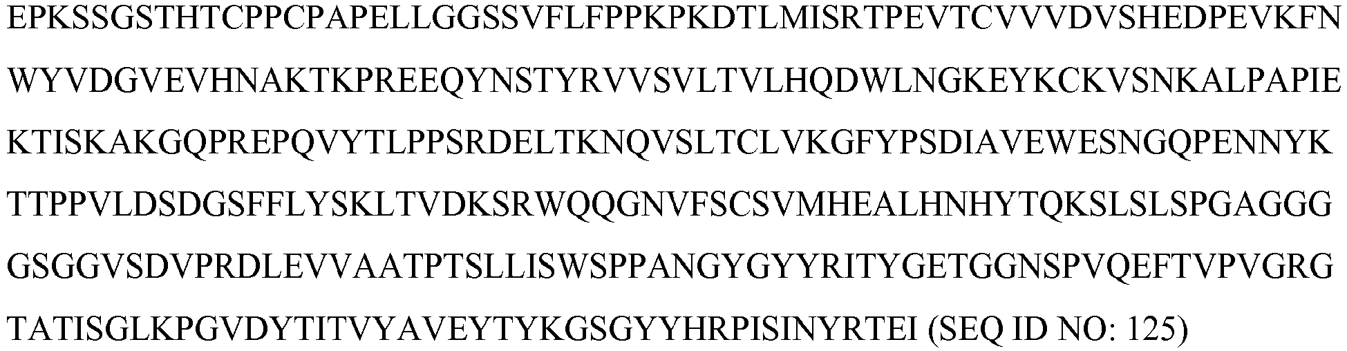
- PRD293 has the following hinge: EPKSSGSTHTCPPCPAPELLGGSS and the following linker: AGGGGSG and a human IgG1 Fc.
- PRD713:


- PRD713 has the following hinge: EPKSSGSTHTCPPCPAPELLGGSS and a human IgG1 Fc. PRD239:

- PRD239 has the following hinge: EPKSSGSTHTCPPCPAPELLGGSS and the following linker AGGGGSG and a human IgG1 Fc.
- C7FL-Fc (PRD1309):

- C7FL-Fc (PRD1309) has the following hinge: EPKSSDKTHTCPPCPAPELLGGSS and a human IgG1 Fc.
- C7FL-Fc (PRD1308):

- C7FL-Fc (PRD1308) has the following hinge: EPKSSDKTHTCPPCPAPELLGGPS and a human IgG1 Fc.
- PRD461 is a fusion protein comprising an Fc linked to the anti-PCSK9 Adnectin 2013E01, whose sequence is provided in
WO2011/130354 . The amino acid sequence for the anti anti-PCSK9 adnectins 1784F03 and 1813E02 are provided inWO2011/130354 . The amino acid sequence of the anti-IL-23 adnectin 1571G04 is provided inWO2011/103105 . - All documents and references described herein are individually incorporated by reference to into this document to the same extent as if there were written in this document in full or in part.
- The present invention further relates to the following embodiments E1 to E22:
- E1. A polypeptide comprising:
- (a) a 10Fn3 domain having an altered amino acid sequence relative to the wild-type sequence, wherein the 10Fn3 domain binds to a target molecule with a KD of less than 500 nM;
- (b) an immunoglobulin (Ig) Fc domain; and
- (c) a hinge sequence.
- E2. The polypeptide of E1, wherein the polypeptide has the following arrangement from N-terminus to C-terminus: 10Fn3 domain-hinge-Fc domain.
- E3. The polypeptide of E1, wherein the polypeptide has the following arrangement from N-terminus to C-terminus: hinge-Fc domain- linker-10Fn3 domain.
- E4. The polypeptide of any one of E1-E3, wherein the polypeptide is a dimer.
- E5. The polypeptide of any one of E1-E4, wherein the polypeptide further comprises a second 10Fn3 domain having an altered amino acid sequence relative to the wild-type sequence and wherein the second 10Fn3 domain binds to a target molecule with a KD of less than 10 µM.
- E6. The polypeptide of E5, wherein the two 10Fn3 domains bind to different targets.
- E7. The polypeptide of any one of E1-E6, wherein the Ig Fc domain is derived from an IgG, IgM, IgD, IgE, or IgA.
- E8. The polypeptide of E7, wherein the Ig Fc domain is derived from IgG.
- E9. The polypeptide of E6, wherein the Ig Fc domain is derived from IgG1.
- E10. The polypeptide of any one of E1-E9, wherein the hinge sequence is derived from a first Ig isotype and the Fc domain is derived from a different Ig isotype.
- E11. The polypeptide of any one of E1-E10, wherein the hinge sequence comprises residues spanning positions 104-119 of SEQ ID NO: 22 or a sequence having at least 90% sequence identity thereto.
- E12. A polypeptide comprising an immunoglobulin Fc domain and a heterologous polypeptide, wherein the heterologous polypeptide is fused to the C-terminus of the Fc domain by a polypeptide linker comprising a sequence derived from the C-terminal tail region of the heavy chain of a membrane bound or secretory immunoglobulin.
- E13. The polypeptide of E12, wherein the polypeptide linker comprises a sequence that is at least 80% identical to any one of SEQ ID NOs: 51-70 or comprises at least 5 contiguous amino acids of any one of SEQ ID NOs: 51-70.
- E14. The polypeptide of E12 or E13, wherein the heterologous polypeptide comprises a 10Fn3 domain.
- E15. The polypeptide of any one of E12-E14, wherein the polypeptide linker comprises at least 10 contiguous amino acids of any one of SEQ ID NOs: 51-70.
- E16. The polypeptide of any one of E12-E15, wherein the polypeptide linker comprises any one of SEQ ID NOs: 51-70.
- E17. The polypeptide of any one of E14-E16, wherein the heterologous polypeptide comprises a second 10Fn3 domain.
- E18. The polypeptide of E17, wherein the 10Fn3 domains bind to different targets.
- E19. A nucleic acid encoding a polypeptide of any one of E1-E18.
- E20. A vector comprising the nucleic acid of E19.
- E21. The vector of E20, wherein the vector is an expression vector.
- E22. A host cell comprising the vector of E21.


Claims (14)
- A polypeptide comprising an immunoglobulin Fc domain and a 10Fn3 domain, wherein the 10Fn3 domain is fused to the C-terminus of the Fc domain by a polypeptide linker comprising an amino acid sequence that is at least 90% identical to any one of SEQ ID NOs: 51-54 and 63-65.
- The polypeptide of claim 1, wherein the 10Fn3 domain has an altered amino acid sequence relative to the wild-type sequence set forth in SEQ ID NO: 1, wherein the altered 10Fn3 domain binds to a target molecule with a KD of less than 500 nM.
- The polypeptide of claim 1 or 2, wherein the polypeptide linker comprises any one of SEQ ID NO: 51-54.
- The polypeptide of claim 1 or 2, wherein the polypeptide linker comprises any one of SEQ ID NOs: 63-65.
- The polypeptide of any one of claims 1-4, wherein the polypeptide has the following arrangement from N-terminus to C-terminus: hinge-Fc domain-linker-10Fn3 domain.
- The polypeptide of any one of claims 1-5, wherein the polypeptide comprises a second 10Fn3 domain.
- The polypeptide of claim 6, wherein the 10Fn3 domains bind to different targets.
- The polypeptide of any one of claims 1-7, wherein the Ig Fc domain is derived from an IgG, IgM, IgD, IgE, or 1gA.
- The polypeptide of claim 8, wherein the Ig Fc domain is derived from IgG.
- The polypeptide of claim 9, wherein the Ig Fc domain is derived from IgG1.
- A nucleic acid encoding a polypeptide of any one of claims 1-10.
- A vector comprising the nucleic acid of claim 11.
- The vector of claim 12, wherein the vector is an expression vector.
- A host cell comprising the vector of claim 13.
Priority Applications (2)
| Application Number | Priority Date | Filing Date | Title |
|---|---|---|---|
| EP21166782.9A EP3896083B1 (en) | 2011-04-13 | 2012-04-13 | Fc fusion proteins comprising novel linkers or arrangements |
| EP18172955.9A EP3415528B1 (en) | 2011-04-13 | 2012-04-13 | Fc fusion proteins comprising novel linkers or arrangements |
Applications Claiming Priority (3)
| Application Number | Priority Date | Filing Date | Title |
|---|---|---|---|
| US201161475004P | 2011-04-13 | 2011-04-13 | |
| PCT/US2012/033665 WO2012142515A2 (en) | 2011-04-13 | 2012-04-13 | Fc fusion proteins comprising novel linkers or arrangements |
| EP12726258.2A EP2697257B1 (en) | 2011-04-13 | 2012-04-13 | Fc fusion proteins comprising novel linkers or arrangements |
Related Parent Applications (1)
| Application Number | Title | Priority Date | Filing Date |
|---|---|---|---|
| EP12726258.2A Division EP2697257B1 (en) | 2011-04-13 | 2012-04-13 | Fc fusion proteins comprising novel linkers or arrangements |
Related Child Applications (3)
| Application Number | Title | Priority Date | Filing Date |
|---|---|---|---|
| EP18172955.9A Division EP3415528B1 (en) | 2011-04-13 | 2012-04-13 | Fc fusion proteins comprising novel linkers or arrangements |
| EP18172955.9A Division-Into EP3415528B1 (en) | 2011-04-13 | 2012-04-13 | Fc fusion proteins comprising novel linkers or arrangements |
| EP21166782.9A Division EP3896083B1 (en) | 2011-04-13 | 2012-04-13 | Fc fusion proteins comprising novel linkers or arrangements |
Publications (3)
| Publication Number | Publication Date |
|---|---|
| EP3144320A1 true EP3144320A1 (en) | 2017-03-22 |
| EP3144320B1 EP3144320B1 (en) | 2018-05-23 |
| EP3144320B9 EP3144320B9 (en) | 2018-08-22 |
Family
ID=46210404
Family Applications (4)
| Application Number | Title | Priority Date | Filing Date |
|---|---|---|---|
| EP18172955.9A Active EP3415528B1 (en) | 2011-04-13 | 2012-04-13 | Fc fusion proteins comprising novel linkers or arrangements |
| EP21166782.9A Active EP3896083B1 (en) | 2011-04-13 | 2012-04-13 | Fc fusion proteins comprising novel linkers or arrangements |
| EP16194148.9A Active EP3144320B9 (en) | 2011-04-13 | 2012-04-13 | Fc fusion proteins comprising novel linkers or arrangements |
| EP12726258.2A Active EP2697257B1 (en) | 2011-04-13 | 2012-04-13 | Fc fusion proteins comprising novel linkers or arrangements |
Family Applications Before (2)
| Application Number | Title | Priority Date | Filing Date |
|---|---|---|---|
| EP18172955.9A Active EP3415528B1 (en) | 2011-04-13 | 2012-04-13 | Fc fusion proteins comprising novel linkers or arrangements |
| EP21166782.9A Active EP3896083B1 (en) | 2011-04-13 | 2012-04-13 | Fc fusion proteins comprising novel linkers or arrangements |
Family Applications After (1)
| Application Number | Title | Priority Date | Filing Date |
|---|---|---|---|
| EP12726258.2A Active EP2697257B1 (en) | 2011-04-13 | 2012-04-13 | Fc fusion proteins comprising novel linkers or arrangements |
Country Status (14)
| Country | Link |
|---|---|
| US (4) | US9469676B2 (en) |
| EP (4) | EP3415528B1 (en) |
| CY (1) | CY1118546T1 (en) |
| DK (1) | DK2697257T3 (en) |
| ES (4) | ES2876421T3 (en) |
| HR (1) | HRP20161531T1 (en) |
| HU (1) | HUE033008T2 (en) |
| LT (1) | LT2697257T (en) |
| PL (1) | PL2697257T3 (en) |
| PT (1) | PT2697257T (en) |
| RS (1) | RS55609B1 (en) |
| SI (1) | SI2697257T1 (en) |
| SM (1) | SMT201700025B (en) |
| WO (1) | WO2012142515A2 (en) |
Cited By (2)
| Publication number | Priority date | Publication date | Assignee | Title |
|---|---|---|---|---|
| WO2020218827A1 (en) * | 2019-04-23 | 2020-10-29 | 주식회사 엘지화학 | Fusion polypeptide comprising fc region of immunoglobulin and gdf15 |
| RU2797520C2 (en) * | 2019-04-23 | 2023-06-06 | ЭлДжи КЕМ, ЛТД. | FUSION POLYPEPTIDE INCLUDING Fc REGION OF IMMUNOGLOBULIN AND GDF15 |
Families Citing this family (84)
| Publication number | Priority date | Publication date | Assignee | Title |
|---|---|---|---|---|
| MXPA06006406A (en) | 2003-12-05 | 2007-03-21 | Adnexus Therapeutics Inc | Inhibitors of type 2 vascular endothelial growth factor receptors. |
| EP3156415A1 (en) | 2006-11-22 | 2017-04-19 | Bristol-Myers Squibb Company | Targeted therapeutics based on engineered proteins for tyrosine kinases receptors, including igf-ir |
| AU2009213141A1 (en) | 2008-02-14 | 2009-08-20 | Bristol-Myers Squibb Company | Targeted therapeutics based on engineered proteins that bind EGFR |
| EP2291399B1 (en) | 2008-05-22 | 2014-06-25 | Bristol-Myers Squibb Company | Multivalent fibronectin based scaffold domain proteins |
| TWI496582B (en) | 2008-11-24 | 2015-08-21 | 必治妥美雅史谷比公司 | Bispecific egfr/igfir binding molecules |
| TW201141508A (en) * | 2010-04-13 | 2011-12-01 | Bristol Myers Squibb Co | Fibronectin based scaffold domain proteins that bind PCSK9 |
| TW201138808A (en) | 2010-05-03 | 2011-11-16 | Bristol Myers Squibb Co | Serum albumin binding molecules |
| WO2011150133A2 (en) | 2010-05-26 | 2011-12-01 | Bristol-Myers Squibb Company | Fibronectin based scaffold proteins having improved stability |
| DK2697257T3 (en) * | 2011-04-13 | 2017-01-30 | Bristol Myers Squibb Co | FC FUSION PROTEINS INCLUDING UNKNOWN LINKERS OR EVENTS |
| EP2709669A1 (en) | 2011-05-17 | 2014-03-26 | Bristol-Myers Squibb Company | Methods for maintaining pegylation of polypeptides |
| AU2012284097B2 (en) | 2011-07-18 | 2017-08-03 | President And Fellows Of Harvard College | Engineered microbe-targeting molecules and uses thereof |
| EP2773659A2 (en) | 2011-10-31 | 2014-09-10 | Bristol-Myers Squibb Company | Fibronectin binding domains with reduced immunogenicity |
| WO2013113008A1 (en) | 2012-01-26 | 2013-08-01 | Amgen Inc. | Growth differentiation factor 15 (gdf-15) polypeptides |
| EP2830646B1 (en) | 2012-03-27 | 2018-03-07 | NGM Biopharmaceuticals, Inc. | Compositions and methods of use for treating metabolic disorders |
| DK2864360T3 (en) | 2012-06-25 | 2017-12-18 | Brigham & Womens Hospital Inc | TARGETED THERAPY |
| MY189682A (en) | 2012-09-13 | 2022-02-25 | Bristol Myers Squibb Co | Fibronectin based scaffold domain proteins that bind to myostatin |
| US9161966B2 (en) | 2013-01-30 | 2015-10-20 | Ngm Biopharmaceuticals, Inc. | GDF15 mutein polypeptides |
| US9828415B2 (en) | 2013-01-30 | 2017-11-28 | Ngm Biopharmaceuticals, Inc. | Compositions and methods of use for treating metabolic disorders |
| WO2014120891A2 (en) | 2013-02-01 | 2014-08-07 | Bristol-Myers Squibb Company | Fibronectin based scaffold proteins |
| US10787498B2 (en) | 2013-02-06 | 2020-09-29 | Bristol-Myers Squibb Company | Fibronectin type III domain proteins with enhanced solubility |
| WO2014126871A1 (en) | 2013-02-12 | 2014-08-21 | Bristol-Myers Squibb Company | Tangential flow filtration based protein refolding methods |
| EP2956467B1 (en) | 2013-02-12 | 2017-09-27 | Bristol-Myers Squibb Company | High ph protein refolding methods |
| US10551379B2 (en) | 2013-03-15 | 2020-02-04 | President And Fellows Of Harvard College | Methods and compositions for improving detection and/or capture of a target entity |
| CA2913155A1 (en) | 2013-05-21 | 2014-11-27 | President And Fellows Of Harvard College | Engineered heme-binding compositions and uses thereof |
| PL3027642T3 (en) * | 2013-07-31 | 2021-01-11 | Amgen Inc. | Growth differentiation factor 15 (gdf-15) constructs |
| US10513546B2 (en) | 2013-12-18 | 2019-12-24 | President And Fellows Of Harvard College | CRP capture/detection of gram positive bacteria |
| EP3572424B1 (en) | 2014-03-20 | 2022-04-20 | Bristol-Myers Squibb Company | Serum albumin-binding fibronectin type iii domains |
| TN2016000578A1 (en) | 2014-07-30 | 2018-04-04 | Ngm Biopharmaceuticals Inc | Compositions and methods of use for treating metabolic disorders |
| UA121973C2 (en) | 2014-10-31 | 2020-08-25 | Нджм Біофармасьютікалз, Інк. | Compositions and methods of use for treating metabolic disorders |
| KR20230125855A (en) | 2014-11-21 | 2023-08-29 | 브리스톨-마이어스 스큅 컴퍼니 | Antibodies against cd73 and uses thereof |
| PT3221346T (en) | 2014-11-21 | 2020-10-23 | Bristol Myers Squibb Co | Antibodies comprising modified heavy constant regions |
| EP3224277B1 (en) | 2014-11-25 | 2020-08-26 | Bristol-Myers Squibb Company | Novel pd-l1 binding polypeptides for imaging |
| CN107207379B (en) | 2014-11-25 | 2021-08-10 | 百时美施贵宝公司 | For biological products18Methods and compositions for F-radiolabelling |
| MY187045A (en) | 2014-12-23 | 2021-08-27 | Bristol Myers Squibb Co | Antibodies to tigit |
| US20180369334A1 (en) * | 2015-01-16 | 2018-12-27 | The Board Of Trustees Of The Leland Stanford Junior University | Vegf variant polypeptide compositions |
| KR101838919B1 (en) * | 2015-03-03 | 2018-03-15 | 에이비온 주식회사 | Immunocytokine conjugated with human interferon beta-mutein and method for preparing thereof |
| CN107969128A (en) | 2015-04-17 | 2018-04-27 | 高山免疫科学股份有限公司 | Immune modulator with adjustable affinity |
| EP3288570A4 (en) * | 2015-04-29 | 2018-11-21 | Fred Hutchinson Cancer Research Center | Modified stem cells and uses thereof |
| CN108137687B (en) | 2015-05-29 | 2021-10-29 | 百时美施贵宝公司 | anti-OX 40 antibodies and uses thereof |
| US11820807B2 (en) * | 2015-06-12 | 2023-11-21 | Ubi Pharma Inc | Immunoglobulin fusion proteins and uses thereof |
| KR20180032551A (en) * | 2015-06-12 | 2018-03-30 | 유비아이 파마 인크. | Immunoglobulin fusion proteins and uses thereof |
| CA3168241A1 (en) | 2015-07-15 | 2017-01-19 | Rutgers. The State University of New Jersey | Nuclease-independent targeted gene editing platform and uses thereof |
| CN108289928A (en) | 2015-08-06 | 2018-07-17 | 哈佛大学校长及研究员协会 | Improved microorganism-binding molecule and its purposes |
| BR112018001353A2 (en) * | 2015-08-07 | 2018-09-11 | Alexo Therapeutics, Inc. | constructs having a sirp-alpha domain or variant thereof |
| CA2996996A1 (en) * | 2015-08-31 | 2017-03-09 | National Research Council Of Canada | Tgf-.beta.-receptor ectodomain fusion molecules and uses thereof |
| ES2781207T3 (en) | 2015-09-23 | 2020-08-31 | Bristol Myers Squibb Co | Serum albumin-binding fibronectin type iii domains with rapid dissociation rate |
| KR20170037247A (en) * | 2015-09-25 | 2017-04-04 | 한미약품 주식회사 | A method for the production of immunoglobulin Fc region comprising initial Methionine residue |
| RU2022102624A (en) * | 2015-10-01 | 2022-03-10 | Хит Байолоджикс, Инк. | COMPOSITIONS AND METHODS FOR CONNECTING TYPE I AND TYPE II EXTRACELLULAR DOMAIN AS HETEROLOGOUS CHIMERIC PROTEINS |
| KR20230038311A (en) | 2016-03-04 | 2023-03-17 | 브리스톨-마이어스 스큅 컴퍼니 | Combination therapy with anti-cd73 antibodies |
| JP7021099B2 (en) | 2016-03-31 | 2022-02-18 | エヌジーエム バイオファーマシューティカルス,インコーポレーテッド | Bound protein and how to use it |
| WO2017174556A1 (en) * | 2016-04-05 | 2017-10-12 | Vib Vzw | Novel tslp inhibitors |
| IL262366B1 (en) | 2016-04-15 | 2024-03-01 | Alpine Immune Sciences Inc | Cd80 variant immunomodulatory proteins and uses thereof |
| KR102397783B1 (en) | 2016-06-01 | 2022-05-12 | 브리스톨-마이어스 스큅 컴퍼니 | PET Imaging with PD-L1 Binding Polypeptides |
| WO2017210335A1 (en) | 2016-06-01 | 2017-12-07 | Bristol-Myers Squibb Company | Imaging methods using 18f-radiolabeled biologics |
| WO2018022945A1 (en) | 2016-07-28 | 2018-02-01 | Alpine Immune Sciences, Inc. | Cd112 variant immunomodulatory proteins and uses thereof |
| US11471488B2 (en) | 2016-07-28 | 2022-10-18 | Alpine Immune Sciences, Inc. | CD155 variant immunomodulatory proteins and uses thereof |
| WO2018089702A1 (en) * | 2016-11-10 | 2018-05-17 | Keros Therapeutics, Inc. | Gdnf fusion polypeptides and methods of use thereof |
| US11459396B2 (en) | 2016-12-02 | 2022-10-04 | The Texas A&M University System | Fusion proteins (Seldegs) for selectively depleting antigen-specific antibodies and methods of use thereof |
| KR20190122667A (en) | 2017-02-27 | 2019-10-30 | 샤턱 랩스 인코포레이티드 | TIGIT- and LIGHT-based chimeric proteins |
| MX2019010173A (en) * | 2017-02-27 | 2019-11-21 | Shattuck Labs Inc | Csf1r-based chimeric proteins. |
| SG11201907867TA (en) | 2017-02-28 | 2019-09-27 | Bristol Myers Squibb Co | Use of anti-ctla-4 antibodies with enhanced adcc to enhance immune response to a vaccine |
| CA3055156A1 (en) * | 2017-03-02 | 2018-09-07 | National Research Council Of Canada | Tgf-.beta.-receptor ectodomain fusion molecules and uses thereof |
| KR20190140924A (en) | 2017-03-16 | 2019-12-20 | 알파인 이뮨 사이언시즈, 인코포레이티드 | CD80 variant immunomodulatory proteins and uses thereof |
| CA3053812A1 (en) | 2017-03-16 | 2018-09-20 | Alpine Immune Sciences, Inc. | Pd-l2 variant immunomodulatory proteins and uses thereof |
| US20200299400A1 (en) | 2017-05-25 | 2020-09-24 | Bristol-Myers Squibb Company | Antibodies comprising modified heavy constant regions |
| JP7250766B2 (en) * | 2017-08-25 | 2023-04-03 | ヤンセン バイオテツク,インコーポレーテツド | FCγRII-Binding Fibronectin Type III Domains, Conjugates Thereof, and Multispecific Molecules Comprising Them |
| US11293923B2 (en) * | 2017-09-01 | 2022-04-05 | Massachusetts Institute Of Technology | S-layer protein 2D lattice coupled detergent-free GPCR bioelectronic interfaces, devices, and methods for the use thereof |
| US11491235B2 (en) | 2017-12-22 | 2022-11-08 | Kb Biomed Inc. | Oral gene carrier and use thereof |
| US10780121B2 (en) | 2018-08-29 | 2020-09-22 | Shattuck Labs, Inc. | FLT3L-based chimeric proteins |
| CR20210166A (en) * | 2018-09-06 | 2021-07-27 | Cidara Therapeutics Inc | Compositions and methods for the treatment of viral infections |
| CN113348177A (en) | 2018-11-28 | 2021-09-03 | 百时美施贵宝公司 | Antibodies comprising modified heavy chain constant regions |
| EP3911746A1 (en) | 2019-01-14 | 2021-11-24 | Institut National de la Santé et de la Recherche Médicale (INSERM) | Methods and kits for generating and selecting a variant of a binding protein with increased binding affinity and/or specificity |
| JP2022529939A (en) | 2019-04-18 | 2022-06-27 | ブリストル-マイヤーズ スクイブ カンパニー | Ipyrimumab variant with enhanced specificity for binding at low pH |
| KR20220044904A (en) | 2019-05-31 | 2022-04-12 | 알렉소 온콜로지 인크. | How to treat cancer with SIRP alpha FC fusion in combination with immune checkpoint inhibitors |
| WO2021150770A1 (en) * | 2020-01-21 | 2021-07-29 | University Of Virginia Patent Foundation | Oncogenic trim37 is a targetable epigenetic driver of metastasis and links chemoresistance and metastatic fate in triple-negative breast cancer |
| EP4100420A1 (en) * | 2020-02-06 | 2022-12-14 | Boehringer Ingelheim Vetmedica GmbH | Polypeptides useful for detecting anti-rhabdovirus antibodies |
| CN111285936A (en) * | 2020-03-11 | 2020-06-16 | 北京双赢科创生物科技有限公司 | Acid sensitive nano peptide segment of targeted tumor and application thereof |
| BR112022022433A2 (en) | 2020-05-08 | 2022-12-13 | Alpine Immune Sciences Inc | INHIBITORY IMMUNOMODULATORY PROTEINS APRIL AND BAFF AND METHODS OF THEIR USE |
| US20230192867A1 (en) | 2020-05-15 | 2023-06-22 | Bristol-Myers Squibb Company | Antibodies to garp |
| EP4179079A1 (en) | 2020-07-10 | 2023-05-17 | Horizon Discovery Limited | Method for producing genetically modified cells |
| CN112375149B (en) * | 2020-10-30 | 2023-04-18 | 沣潮医药科技(上海)有限公司 | ACE2 immune fusion protein and application thereof |
| US20240067954A1 (en) | 2021-01-05 | 2024-02-29 | Horizon Discovery Limited | Method for producing genetically modified cells |
| EP4298120A1 (en) * | 2021-02-25 | 2024-01-03 | Lyell Immunopharma, Inc. | Ror1 targeting chimeric antigen receptor |
| US11945856B2 (en) | 2022-01-28 | 2024-04-02 | 35Pharma Inc. | Activin receptor type IIB variants and uses thereof |
Citations (64)
| Publication number | Priority date | Publication date | Assignee | Title |
|---|---|---|---|---|
| US3773919A (en) | 1969-10-23 | 1973-11-20 | Du Pont | Polylactide-drug mixtures |
| USRE30985E (en) | 1978-01-01 | 1982-06-29 | Serum-free cell culture media | |
| US4560655A (en) | 1982-12-16 | 1985-12-24 | Immunex Corporation | Serum-free cell culture medium and process for making same |
| US4657866A (en) | 1982-12-21 | 1987-04-14 | Sudhir Kumar | Serum-free, synthetic, completely chemically defined tissue culture media |
| US4767704A (en) | 1983-10-07 | 1988-08-30 | Columbia University In The City Of New York | Protein-free culture medium |
| WO1988007054A1 (en) | 1987-03-18 | 1988-09-22 | Research Corporation Limited | Complement-binding peptide |
| WO1988007089A1 (en) | 1987-03-18 | 1988-09-22 | Medical Research Council | Altered antibodies |
| US4927762A (en) | 1986-04-01 | 1990-05-22 | Cell Enterprises, Inc. | Cell culture medium with antioxidant |
| US5122469A (en) | 1990-10-03 | 1992-06-16 | Genentech, Inc. | Method for culturing Chinese hamster ovary cells to improve production of recombinant proteins |
| WO1994011026A2 (en) | 1992-11-13 | 1994-05-26 | Idec Pharmaceuticals Corporation | Therapeutic application of chimeric and radiolabeled antibodies to human b lymphocyte restricted differentiation antigen for treatment of b cell lymphoma |
| WO1994029351A2 (en) | 1993-06-16 | 1994-12-22 | Celltech Limited | Antibodies |
| WO1996027011A1 (en) | 1995-03-01 | 1996-09-06 | Genentech, Inc. | A method for making heteromultimeric polypeptides |
| WO1996032478A1 (en) | 1995-04-14 | 1996-10-17 | Genentech, Inc. | Altered polypeptides with increased half-life |
| US5631144A (en) | 1989-04-28 | 1997-05-20 | Transgene S.A. | Application of novel DNA fragments as a coding sequence for a signal peptide for the secretion of mature proteins by recombinant yeast, expression cassettes, transformed yeast and corresponding process for the preparation of proteins |
| WO1997034631A1 (en) | 1996-03-18 | 1997-09-25 | Board Of Regents, The University Of Texas System | Immunoglobin-like domains with increased half lives |
| US5672502A (en) | 1985-06-28 | 1997-09-30 | Celltech Therapeutics Limited | Animal cell culture |
| WO1998056915A2 (en) | 1997-06-12 | 1998-12-17 | Research Corporation Technologies, Inc. | Artificial antibody polypeptides |
| WO1999051642A1 (en) | 1998-04-02 | 1999-10-14 | Genentech, Inc. | Antibody variants and fragments thereof |
| US6048728A (en) | 1988-09-23 | 2000-04-11 | Chiron Corporation | Cell culture medium for enhanced cell growth, culture longevity, and product expression |
| WO2000034787A1 (en) | 1998-12-10 | 2000-06-15 | The University Of Nottingham | Cancer detection method and reagents |
| WO2000042072A2 (en) | 1999-01-15 | 2000-07-20 | Genentech, Inc. | Polypeptide variants with altered effector function |
| US6194551B1 (en) | 1998-04-02 | 2001-02-27 | Genentech, Inc. | Polypeptide variants |
| WO2001058957A2 (en) | 2000-02-11 | 2001-08-16 | Lexigen Pharmaceuticals Corp. | Enhancing the circulating half-life of antibody-based fusion proteins |
| US6277375B1 (en) | 1997-03-03 | 2001-08-21 | Board Of Regents, The University Of Texas System | Immunoglobulin-like domains with increased half-lives |
| WO2001064942A1 (en) | 2000-02-29 | 2001-09-07 | Phylos, Inc. | Protein scaffolds for antibody mimics and other binding proteins |
| WO2002004523A2 (en) | 2000-07-11 | 2002-01-17 | Research Corporation Technologies, Inc. | Artificial antibody polypeptides |
| WO2002006919A2 (en) | 2000-07-18 | 2002-01-24 | Aegis Analytical Corporation | System, method and computer program product for mapping data of multi-database origins |
| WO2002032925A2 (en) | 2000-10-16 | 2002-04-25 | Phylos, Inc. | Protein scaffolds for antibody mimics and other binding proteins |
| WO2002081497A2 (en) | 2001-04-04 | 2002-10-17 | University Of Rochester | αξβ3 INTEGRIN-BINDING POLYPEPTIDE MONOBODIES AND THEIR USE |
| US6559126B2 (en) | 2000-03-31 | 2003-05-06 | Institut Pasteur | Peptides blocking vascular endothelial growth factor (VEGF)-mediated angiogenesis, polynucleotides encoding said peptides and methods of use thereof |
| WO2003074679A2 (en) | 2002-03-01 | 2003-09-12 | Xencor | Antibody optimization |
| US20030186385A1 (en) | 2000-11-17 | 2003-10-02 | Shohei Koide | Method of identifying polypeptide monobodies which bind to target proteins and use thereof |
| WO2004016750A2 (en) | 2002-08-14 | 2004-02-26 | Macrogenics, Inc. | FcϜRIIB-SPECIFIC ANTIBODIES AND METHODS OF USE THEREOF |
| WO2004029207A2 (en) | 2002-09-27 | 2004-04-08 | Xencor Inc. | Optimized fc variants and methods for their generation |
| WO2004035752A2 (en) | 2002-10-15 | 2004-04-29 | Protein Design Labs, Inc. | ALTERATION OF FcRn BINDING AFFINITIES OR SERUM HALF-LIVES OF ANTIBODIES BY MUTAGENESIS |
| US6737056B1 (en) | 1999-01-15 | 2004-05-18 | Genentech, Inc. | Polypeptide variants with altered effector function |
| WO2004063351A2 (en) | 2003-01-09 | 2004-07-29 | Macrogenics, Inc. | IDENTIFICATION AND ENGINEERING OF ANTIBODIES WITH VARIANT Fc REGIONS AND METHODS OF USING SAME |
| WO2004074455A2 (en) | 2003-02-20 | 2004-09-02 | Applied Molecular Evolution | Fc REGION VARIANTS |
| WO2004099249A2 (en) | 2003-05-02 | 2004-11-18 | Xencor, Inc. | Optimized fc variants and methods for their generation |
| WO2005040217A2 (en) | 2003-10-17 | 2005-05-06 | Cambridge University Technical Services Limited | Antibodies having a mutated amino acid residue at position 268 (ch2 region) in constant regions |
| WO2005070963A1 (en) | 2004-01-12 | 2005-08-04 | Applied Molecular Evolution, Inc | Fc region variants |
| WO2005092925A2 (en) | 2004-03-24 | 2005-10-06 | Xencor, Inc. | Immunoglobulin variants outside the fc region |
| WO2006020114A2 (en) | 2004-08-04 | 2006-02-23 | Applied Molecular Evolution, Inc. | Variant fc regions |
| WO2007110205A2 (en) | 2006-03-24 | 2007-10-04 | Merck Patent Gmbh | Engineered heterodimeric protein domains |
| US7317091B2 (en) | 2002-03-01 | 2008-01-08 | Xencor, Inc. | Optimized Fc variants |
| WO2008012543A1 (en) | 2006-07-25 | 2008-01-31 | Ucb Pharma S.A. | Single chain fc polypeptides |
| WO2008031098A1 (en) | 2006-09-09 | 2008-03-13 | The University Of Chicago | Binary amino acid libraries for fibronectin type iii polypeptide monobodies |
| WO2008066752A2 (en) | 2006-11-22 | 2008-06-05 | Adnexus, A Bristol-Myers Squibb R & D Company | Targeted therapeutics based on engineered proteins for tyrosine kinases receptors, including igf-ir |
| WO2008097497A2 (en) | 2007-02-02 | 2008-08-14 | Adnexus, A Bristol-Myers Squibb R & D Company | Vegf pathway blockade |
| WO2008131242A1 (en) | 2007-04-18 | 2008-10-30 | Zymogenetics, Inc. | Single chain fc, methods of making and methods of treatment |
| WO2008143954A2 (en) | 2007-05-14 | 2008-11-27 | Biogen Idec Ma Inc. | Single-chain fc (scfc) regions, binding polypeptides comprising same, and methods related thereto |
| WO2009006520A1 (en) * | 2007-07-03 | 2009-01-08 | Medimmune, Llc | Hinge domain engineering |
| WO2009083804A2 (en) * | 2007-12-27 | 2009-07-09 | Novartis Ag | Improved fibronectin-based binding molecules and their use |
| WO2009086116A2 (en) | 2007-12-19 | 2009-07-09 | Centocor, Inc. | Alternative scaffold protein fusions phage display via fusion to plx of m13 phage |
| WO2009089004A1 (en) | 2008-01-07 | 2009-07-16 | Amgen Inc. | Method for making antibody fc-heterodimeric molecules using electrostatic steering effects |
| WO2009142773A2 (en) | 2008-05-22 | 2009-11-26 | Bristol-Myers Squibb Company | Multivalent fibronectin based scaffold domain proteins |
| WO2010051274A2 (en) | 2008-10-31 | 2010-05-06 | Centocor Ortho Biotech Inc. | Fibronectin type iii domain based scaffold compositions, methods and uses |
| WO2010051310A2 (en) | 2008-10-31 | 2010-05-06 | Centocor Ortho Biotech Inc. | ANTI-TNFα FIBRONECTIN TYPE III DOMAIN BASED SCAFFOLD COMPOSITIONS, METHODS AND USES |
| WO2010129304A2 (en) | 2009-04-27 | 2010-11-11 | Oncomed Pharmaceuticals, Inc. | Method for making heteromultimeric molecules |
| US20110009323A1 (en) * | 2007-06-15 | 2011-01-13 | Vasgene Therapeutics, Inc. | Non-immunoglobulin antigen binding scaffolds for inhibiting angiogenesis and tumor growth |
| WO2011103105A1 (en) | 2010-02-18 | 2011-08-25 | Bristol-Myers Squibb Company | Fibronectin based scaffold domain proteins that bind il-23 |
| WO2011130354A1 (en) | 2010-04-13 | 2011-10-20 | Bristol-Myers Squibb Company | Fibronectin based scaffold domain proteins that bind pcsk9 |
| US8101720B2 (en) | 2004-10-21 | 2012-01-24 | Xencor, Inc. | Immunoglobulin insertions, deletions and substitutions |
| WO2012016245A2 (en) | 2010-07-30 | 2012-02-02 | Novartis Ag | Fibronectin cradle molecules and libraries thereof |
Family Cites Families (59)
| Publication number | Priority date | Publication date | Assignee | Title |
|---|---|---|---|---|
| US30985A (en) | 1860-12-18 | Thomas l | ||
| US5514581A (en) | 1986-11-04 | 1996-05-07 | Protein Polymer Technologies, Inc. | Functional recombinantly prepared synthetic protein polymer |
| US5641648A (en) | 1986-11-04 | 1997-06-24 | Protein Polymer Technologies, Inc. | Methods for preparing synthetic repetitive DNA |
| US6018030A (en) | 1986-11-04 | 2000-01-25 | Protein Polymer Technologies, Inc. | Peptides comprising repetitive units of amino acids and DNA sequences encoding the same |
| US5770697A (en) | 1986-11-04 | 1998-06-23 | Protein Polymer Technologies, Inc. | Peptides comprising repetitive units of amino acids and DNA sequences encoding the same |
| US5484907A (en) * | 1989-06-21 | 1996-01-16 | Tanox Biosystems, Inc. | Nucleotides coding for the extracellular membrane-bound segment of IgA |
| US5310875A (en) * | 1989-06-21 | 1994-05-10 | Tanox Biosystems, Inc. | Peptides corresponding to membrane-bound IgA |
| US5792742A (en) | 1991-06-14 | 1998-08-11 | New York University | Fibrin-binding peptide fragments of fibronectin |
| WO1994017097A1 (en) | 1993-01-19 | 1994-08-04 | Regents Of The University Of Minnesota | Synthetic fibronectin fragments as inhibitors of retroviral infections |
| US5864020A (en) * | 1994-07-20 | 1999-01-26 | Genentech, Inc. | HTK ligand |
| US6103521A (en) * | 1995-02-06 | 2000-08-15 | Cell Genesys, Inc. | Multispecific chimeric receptors |
| ATE299936T1 (en) * | 1996-05-02 | 2005-08-15 | Mochida Pharm Co Ltd | FAS ANTIGEN DERIVATIVES |
| US20020147326A1 (en) * | 1996-06-14 | 2002-10-10 | Smithkline Beecham Corporation | Hexameric fusion proteins and uses therefor |
| US6261804B1 (en) | 1997-01-21 | 2001-07-17 | The General Hospital Corporation | Selection of proteins using RNA-protein fusions |
| WO1998031700A1 (en) | 1997-01-21 | 1998-07-23 | The General Hospital Corporation | Selection of proteins using rna-protein fusions |
| WO1999051773A1 (en) | 1998-04-03 | 1999-10-14 | Phylos, Inc. | Addressable protein arrays |
| EP1137941B2 (en) | 1998-12-10 | 2013-09-11 | Bristol-Myers Squibb Company | Protein scaffolds for antibody mimics and other binding proteins |
| US7115396B2 (en) | 1998-12-10 | 2006-10-03 | Compound Therapeutics, Inc. | Protein scaffolds for antibody mimics and other binding proteins |
| WO2002038766A2 (en) * | 2000-11-07 | 2002-05-16 | Zymogenetics, Inc. | Human tumor necrosis factor receptor |
| US7829084B2 (en) * | 2001-01-17 | 2010-11-09 | Trubion Pharmaceuticals, Inc. | Binding constructs and methods for use thereof |
| US7754208B2 (en) * | 2001-01-17 | 2010-07-13 | Trubion Pharmaceuticals, Inc. | Binding domain-immunoglobulin fusion proteins |
| MXPA03008031A (en) * | 2001-03-07 | 2003-12-04 | Merck Patent Gmbh | Expression technology for proteins containing a hybrid isotype antibody moiety. |
| US6992174B2 (en) * | 2001-03-30 | 2006-01-31 | Emd Lexigen Research Center Corp. | Reducing the immunogenicity of fusion proteins |
| AU2003243436A1 (en) | 2002-06-06 | 2003-12-22 | Shohei Koide | Reconstituted polypeptides |
| US20080220049A1 (en) | 2003-12-05 | 2008-09-11 | Adnexus, A Bristol-Myers Squibb R&D Company | Compositions and methods for intraocular delivery of fibronectin scaffold domain proteins |
| MXPA06006406A (en) | 2003-12-05 | 2007-03-21 | Adnexus Therapeutics Inc | Inhibitors of type 2 vascular endothelial growth factor receptors. |
| CA2582374A1 (en) | 2004-10-04 | 2006-04-20 | Qlt Usa, Inc. | Ocular delivery of polymeric delivery formulations |
| US20100008917A1 (en) * | 2006-08-25 | 2010-01-14 | Nancy Hosken | Treatment of aplastic anemia |
| EP2125880A1 (en) * | 2007-03-26 | 2009-12-02 | Zymogenetics, Inc. | Soluble il-17ra/rc fusion proteins and related methods |
| US8470966B2 (en) | 2007-08-10 | 2013-06-25 | Protelica, Inc. | Universal fibronectin type III binding-domain libraries |
| JP5781762B2 (en) | 2007-08-10 | 2015-09-24 | プロテリックス、インク | Universal type III fibronectin binding domain library |
| US8680019B2 (en) | 2007-08-10 | 2014-03-25 | Protelica, Inc. | Universal fibronectin Type III binding-domain libraries |
| WO2009025806A2 (en) | 2007-08-20 | 2009-02-26 | Adnexus, A Bristol-Myers Squibb R & D Company | Use of vegfr-2 inhibitors for treating metastatic cancer |
| BRPI0818810A2 (en) | 2007-10-31 | 2014-10-29 | Medimmune Llc | RECOMBINANT AND MULTIMERIC POLYPEPTIDE SKELETONS, ISOLATED N UCLEIC ACID MOLECULE, EXPRESSION VECTOR, HOST STONE POLYPETEID, MUSCLE ESTEXE MUSCLE TELECLE MUSCLE COLLECTION LIBRARY A COMPOUND IN A SAMPLE, TO CAPTURE A COMPOUND IN A SAMPLE, TO PREVENT, TREAT, CONTROL OR IMPROVE A DISEASE, FOR DIAGNOSIS OR IMAGE FORMATION OF A DISEASE TO PURIFY AND PRODUCE A SKELETON, TO TEST OR DELECT A SLAKE TO A TARGET, TO USE SKELETON AND TO PREVENT, TREAT, IMPROVE, OR CONTROL CANCER, STERILE COMPOSITION, PYROGEN FREE, PHARMACEUTICAL COMPOSITION, AND SCALABLE PROCESS. |
| EP2217274A1 (en) | 2007-11-28 | 2010-08-18 | Bristol-Myers Squibb Company | Combination vegfr2 therapy with mtor inhibitors |
| AU2009213141A1 (en) | 2008-02-14 | 2009-08-20 | Bristol-Myers Squibb Company | Targeted therapeutics based on engineered proteins that bind EGFR |
| NZ621443A (en) * | 2008-04-11 | 2015-09-25 | Emergent Product Dev Seattle | Cd37 immunotherapeutic and combination with bifunctional chemotherapeutic thereof |
| ES2620285T3 (en) | 2008-05-02 | 2017-06-28 | Novartis Ag | Binding molecules based on improved fibronectin and their uses |
| TWI496582B (en) | 2008-11-24 | 2015-08-21 | 必治妥美雅史谷比公司 | Bispecific egfr/igfir binding molecules |
| AU2009327189B2 (en) | 2008-12-16 | 2012-09-06 | Novartis Ag | Yeast display systems |
| WO2010093771A1 (en) | 2009-02-11 | 2010-08-19 | Bristol-Myers Squibb Company | Combination vegfr2 therapy with temozolomide |
| AU2010213892B2 (en) | 2009-02-12 | 2014-10-23 | Janssen Biotech, Inc. | Fibronectin type III domain based scaffold compositions, methods and uses |
| US8067201B2 (en) | 2009-04-17 | 2011-11-29 | Bristol-Myers Squibb Company | Methods for protein refolding |
| EP2464663A4 (en) | 2009-08-13 | 2013-05-29 | Massachusetts Inst Technology | Engineered proteins including mutant fibronectin domains |
| ES2691717T3 (en) | 2009-10-30 | 2018-11-28 | Novartis Ag | Universal libraries of the binding domain of the inferior side of type III fibronectin type III |
| WO2011051466A1 (en) | 2009-11-02 | 2011-05-05 | Novartis Ag | Anti-idiotypic fibronectin-based binding molecules and uses thereof |
| US20110123545A1 (en) | 2009-11-24 | 2011-05-26 | Bristol-Myers Squibb Company | Combination of vegfr2 and igf1r inhibitors for the treatment of proliferative diseases |
| WO2011092233A1 (en) | 2010-01-29 | 2011-08-04 | Novartis Ag | Yeast mating to produce high-affinity combinations of fibronectin-based binders |
| US8961977B2 (en) | 2010-02-12 | 2015-02-24 | University Of Rochester | Antigenic mimics of discontinuous epitopes of pathogen recognized by broadly neutralizing antibodies |
| WO2011130324A1 (en) | 2010-04-13 | 2011-10-20 | Medimmune, Llc | Fibronectin type iii domain-based multimeric scaffolds |
| HUE029622T2 (en) | 2010-04-30 | 2017-03-28 | Janssen Biotech Inc | Stabilized fibronectin domain compositions, methods and uses |
| TW201138808A (en) | 2010-05-03 | 2011-11-16 | Bristol Myers Squibb Co | Serum albumin binding molecules |
| WO2011150133A2 (en) | 2010-05-26 | 2011-12-01 | Bristol-Myers Squibb Company | Fibronectin based scaffold proteins having improved stability |
| CN103380143B (en) | 2010-12-22 | 2016-01-06 | 百时美施贵宝公司 | In conjunction with the scaffold domains protein based on fibronectin of IL-23 |
| DK2697257T3 (en) | 2011-04-13 | 2017-01-30 | Bristol Myers Squibb Co | FC FUSION PROTEINS INCLUDING UNKNOWN LINKERS OR EVENTS |
| EP2709669A1 (en) | 2011-05-17 | 2014-03-26 | Bristol-Myers Squibb Company | Methods for maintaining pegylation of polypeptides |
| EP2710382B1 (en) | 2011-05-17 | 2017-10-18 | Bristol-Myers Squibb Company | Improved methods for the selection of binding proteins |
| EP3540100B8 (en) | 2011-09-27 | 2022-11-02 | Janssen Biotech, Inc. | Fibronectin type iii repeat based protein scaffolds with alternative binding surfaces |
| MY189682A (en) | 2012-09-13 | 2022-02-25 | Bristol Myers Squibb Co | Fibronectin based scaffold domain proteins that bind to myostatin |
-
2012
- 2012-04-13 DK DK12726258.2T patent/DK2697257T3/en active
- 2012-04-13 RS RS20170056A patent/RS55609B1/en unknown
- 2012-04-13 HU HUE12726258A patent/HUE033008T2/en unknown
- 2012-04-13 WO PCT/US2012/033665 patent/WO2012142515A2/en active Application Filing
- 2012-04-13 EP EP18172955.9A patent/EP3415528B1/en active Active
- 2012-04-13 ES ES18172955T patent/ES2876421T3/en active Active
- 2012-04-13 PT PT127262582T patent/PT2697257T/en unknown
- 2012-04-13 EP EP21166782.9A patent/EP3896083B1/en active Active
- 2012-04-13 SI SI201230760T patent/SI2697257T1/en unknown
- 2012-04-13 EP EP16194148.9A patent/EP3144320B9/en active Active
- 2012-04-13 LT LTEP12726258.2T patent/LT2697257T/en unknown
- 2012-04-13 US US14/111,327 patent/US9469676B2/en active Active
- 2012-04-13 EP EP12726258.2A patent/EP2697257B1/en active Active
- 2012-04-13 ES ES12726258.2T patent/ES2608835T3/en active Active
- 2012-04-13 PL PL12726258T patent/PL2697257T3/en unknown
- 2012-04-13 ES ES16194148T patent/ES2676499T3/en active Active
- 2012-04-13 ES ES21166782T patent/ES2942455T3/en active Active
-
2016
- 2016-09-06 US US15/257,189 patent/US10214579B2/en active Active
- 2016-11-21 HR HRP20161531TT patent/HRP20161531T1/en unknown
-
2017
- 2017-01-13 CY CY20171100049T patent/CY1118546T1/en unknown
- 2017-01-16 SM SM201700025T patent/SMT201700025B/en unknown
-
2019
- 2019-01-22 US US16/254,114 patent/US20190248872A1/en not_active Abandoned
-
2020
- 2020-11-09 US US17/092,720 patent/US20210122805A1/en active Pending
Patent Citations (68)
| Publication number | Priority date | Publication date | Assignee | Title |
|---|---|---|---|---|
| US3773919A (en) | 1969-10-23 | 1973-11-20 | Du Pont | Polylactide-drug mixtures |
| USRE30985E (en) | 1978-01-01 | 1982-06-29 | Serum-free cell culture media | |
| US4560655A (en) | 1982-12-16 | 1985-12-24 | Immunex Corporation | Serum-free cell culture medium and process for making same |
| US4657866A (en) | 1982-12-21 | 1987-04-14 | Sudhir Kumar | Serum-free, synthetic, completely chemically defined tissue culture media |
| US4767704A (en) | 1983-10-07 | 1988-08-30 | Columbia University In The City Of New York | Protein-free culture medium |
| US5672502A (en) | 1985-06-28 | 1997-09-30 | Celltech Therapeutics Limited | Animal cell culture |
| US4927762A (en) | 1986-04-01 | 1990-05-22 | Cell Enterprises, Inc. | Cell culture medium with antioxidant |
| US5648260A (en) | 1987-03-18 | 1997-07-15 | Scotgen Biopharmaceuticals Incorporated | DNA encoding antibodies with altered effector functions |
| US5624821A (en) | 1987-03-18 | 1997-04-29 | Scotgen Biopharmaceuticals Incorporated | Antibodies with altered effector functions |
| WO1988007089A1 (en) | 1987-03-18 | 1988-09-22 | Medical Research Council | Altered antibodies |
| WO1988007054A1 (en) | 1987-03-18 | 1988-09-22 | Research Corporation Limited | Complement-binding peptide |
| US6048728A (en) | 1988-09-23 | 2000-04-11 | Chiron Corporation | Cell culture medium for enhanced cell growth, culture longevity, and product expression |
| US5631144A (en) | 1989-04-28 | 1997-05-20 | Transgene S.A. | Application of novel DNA fragments as a coding sequence for a signal peptide for the secretion of mature proteins by recombinant yeast, expression cassettes, transformed yeast and corresponding process for the preparation of proteins |
| US5122469A (en) | 1990-10-03 | 1992-06-16 | Genentech, Inc. | Method for culturing Chinese hamster ovary cells to improve production of recombinant proteins |
| WO1994011026A2 (en) | 1992-11-13 | 1994-05-26 | Idec Pharmaceuticals Corporation | Therapeutic application of chimeric and radiolabeled antibodies to human b lymphocyte restricted differentiation antigen for treatment of b cell lymphoma |
| WO1994029351A2 (en) | 1993-06-16 | 1994-12-22 | Celltech Limited | Antibodies |
| WO1996027011A1 (en) | 1995-03-01 | 1996-09-06 | Genentech, Inc. | A method for making heteromultimeric polypeptides |
| US5731168A (en) | 1995-03-01 | 1998-03-24 | Genentech, Inc. | Method for making heteromultimeric polypeptides |
| US5821333A (en) | 1995-03-01 | 1998-10-13 | Genetech, Inc. | Method for making heteromultimeric polypeptides |
| WO1996032478A1 (en) | 1995-04-14 | 1996-10-17 | Genentech, Inc. | Altered polypeptides with increased half-life |
| WO1997034631A1 (en) | 1996-03-18 | 1997-09-25 | Board Of Regents, The University Of Texas System | Immunoglobin-like domains with increased half lives |
| US6277375B1 (en) | 1997-03-03 | 2001-08-21 | Board Of Regents, The University Of Texas System | Immunoglobulin-like domains with increased half-lives |
| WO1998056915A2 (en) | 1997-06-12 | 1998-12-17 | Research Corporation Technologies, Inc. | Artificial antibody polypeptides |
| WO1999051642A1 (en) | 1998-04-02 | 1999-10-14 | Genentech, Inc. | Antibody variants and fragments thereof |
| US6194551B1 (en) | 1998-04-02 | 2001-02-27 | Genentech, Inc. | Polypeptide variants |
| WO2000034787A1 (en) | 1998-12-10 | 2000-06-15 | The University Of Nottingham | Cancer detection method and reagents |
| WO2000042072A2 (en) | 1999-01-15 | 2000-07-20 | Genentech, Inc. | Polypeptide variants with altered effector function |
| US6737056B1 (en) | 1999-01-15 | 2004-05-18 | Genentech, Inc. | Polypeptide variants with altered effector function |
| WO2001058957A2 (en) | 2000-02-11 | 2001-08-16 | Lexigen Pharmaceuticals Corp. | Enhancing the circulating half-life of antibody-based fusion proteins |
| WO2001064942A1 (en) | 2000-02-29 | 2001-09-07 | Phylos, Inc. | Protein scaffolds for antibody mimics and other binding proteins |
| US6559126B2 (en) | 2000-03-31 | 2003-05-06 | Institut Pasteur | Peptides blocking vascular endothelial growth factor (VEGF)-mediated angiogenesis, polynucleotides encoding said peptides and methods of use thereof |
| WO2002004523A2 (en) | 2000-07-11 | 2002-01-17 | Research Corporation Technologies, Inc. | Artificial antibody polypeptides |
| WO2002006919A2 (en) | 2000-07-18 | 2002-01-24 | Aegis Analytical Corporation | System, method and computer program product for mapping data of multi-database origins |
| WO2002032925A2 (en) | 2000-10-16 | 2002-04-25 | Phylos, Inc. | Protein scaffolds for antibody mimics and other binding proteins |
| US20030186385A1 (en) | 2000-11-17 | 2003-10-02 | Shohei Koide | Method of identifying polypeptide monobodies which bind to target proteins and use thereof |
| WO2002081497A2 (en) | 2001-04-04 | 2002-10-17 | University Of Rochester | αξβ3 INTEGRIN-BINDING POLYPEPTIDE MONOBODIES AND THEIR USE |
| US7317091B2 (en) | 2002-03-01 | 2008-01-08 | Xencor, Inc. | Optimized Fc variants |
| WO2003074679A2 (en) | 2002-03-01 | 2003-09-12 | Xencor | Antibody optimization |
| WO2004016750A2 (en) | 2002-08-14 | 2004-02-26 | Macrogenics, Inc. | FcϜRIIB-SPECIFIC ANTIBODIES AND METHODS OF USE THEREOF |
| WO2004029207A2 (en) | 2002-09-27 | 2004-04-08 | Xencor Inc. | Optimized fc variants and methods for their generation |
| WO2004035752A2 (en) | 2002-10-15 | 2004-04-29 | Protein Design Labs, Inc. | ALTERATION OF FcRn BINDING AFFINITIES OR SERUM HALF-LIVES OF ANTIBODIES BY MUTAGENESIS |
| WO2004063351A2 (en) | 2003-01-09 | 2004-07-29 | Macrogenics, Inc. | IDENTIFICATION AND ENGINEERING OF ANTIBODIES WITH VARIANT Fc REGIONS AND METHODS OF USING SAME |
| WO2004074455A2 (en) | 2003-02-20 | 2004-09-02 | Applied Molecular Evolution | Fc REGION VARIANTS |
| WO2004099249A2 (en) | 2003-05-02 | 2004-11-18 | Xencor, Inc. | Optimized fc variants and methods for their generation |
| WO2005040217A2 (en) | 2003-10-17 | 2005-05-06 | Cambridge University Technical Services Limited | Antibodies having a mutated amino acid residue at position 268 (ch2 region) in constant regions |
| WO2005070963A1 (en) | 2004-01-12 | 2005-08-04 | Applied Molecular Evolution, Inc | Fc region variants |
| WO2005092925A2 (en) | 2004-03-24 | 2005-10-06 | Xencor, Inc. | Immunoglobulin variants outside the fc region |
| WO2006020114A2 (en) | 2004-08-04 | 2006-02-23 | Applied Molecular Evolution, Inc. | Variant fc regions |
| US8101720B2 (en) | 2004-10-21 | 2012-01-24 | Xencor, Inc. | Immunoglobulin insertions, deletions and substitutions |
| WO2007110205A2 (en) | 2006-03-24 | 2007-10-04 | Merck Patent Gmbh | Engineered heterodimeric protein domains |
| WO2008012543A1 (en) | 2006-07-25 | 2008-01-31 | Ucb Pharma S.A. | Single chain fc polypeptides |
| WO2008031098A1 (en) | 2006-09-09 | 2008-03-13 | The University Of Chicago | Binary amino acid libraries for fibronectin type iii polypeptide monobodies |
| WO2008066752A2 (en) | 2006-11-22 | 2008-06-05 | Adnexus, A Bristol-Myers Squibb R & D Company | Targeted therapeutics based on engineered proteins for tyrosine kinases receptors, including igf-ir |
| WO2008097497A2 (en) | 2007-02-02 | 2008-08-14 | Adnexus, A Bristol-Myers Squibb R & D Company | Vegf pathway blockade |
| WO2008131242A1 (en) | 2007-04-18 | 2008-10-30 | Zymogenetics, Inc. | Single chain fc, methods of making and methods of treatment |
| WO2008143954A2 (en) | 2007-05-14 | 2008-11-27 | Biogen Idec Ma Inc. | Single-chain fc (scfc) regions, binding polypeptides comprising same, and methods related thereto |
| US20110009323A1 (en) * | 2007-06-15 | 2011-01-13 | Vasgene Therapeutics, Inc. | Non-immunoglobulin antigen binding scaffolds for inhibiting angiogenesis and tumor growth |
| WO2009006520A1 (en) * | 2007-07-03 | 2009-01-08 | Medimmune, Llc | Hinge domain engineering |
| WO2009086116A2 (en) | 2007-12-19 | 2009-07-09 | Centocor, Inc. | Alternative scaffold protein fusions phage display via fusion to plx of m13 phage |
| WO2009083804A2 (en) * | 2007-12-27 | 2009-07-09 | Novartis Ag | Improved fibronectin-based binding molecules and their use |
| WO2009089004A1 (en) | 2008-01-07 | 2009-07-16 | Amgen Inc. | Method for making antibody fc-heterodimeric molecules using electrostatic steering effects |
| WO2009142773A2 (en) | 2008-05-22 | 2009-11-26 | Bristol-Myers Squibb Company | Multivalent fibronectin based scaffold domain proteins |
| WO2010051310A2 (en) | 2008-10-31 | 2010-05-06 | Centocor Ortho Biotech Inc. | ANTI-TNFα FIBRONECTIN TYPE III DOMAIN BASED SCAFFOLD COMPOSITIONS, METHODS AND USES |
| WO2010051274A2 (en) | 2008-10-31 | 2010-05-06 | Centocor Ortho Biotech Inc. | Fibronectin type iii domain based scaffold compositions, methods and uses |
| WO2010129304A2 (en) | 2009-04-27 | 2010-11-11 | Oncomed Pharmaceuticals, Inc. | Method for making heteromultimeric molecules |
| WO2011103105A1 (en) | 2010-02-18 | 2011-08-25 | Bristol-Myers Squibb Company | Fibronectin based scaffold domain proteins that bind il-23 |
| WO2011130354A1 (en) | 2010-04-13 | 2011-10-20 | Bristol-Myers Squibb Company | Fibronectin based scaffold domain proteins that bind pcsk9 |
| WO2012016245A2 (en) | 2010-07-30 | 2012-02-02 | Novartis Ag | Fibronectin cradle molecules and libraries thereof |
Non-Patent Citations (55)
| Title |
|---|
| "Cloning Vectors: A Laboratory Manual", 1985, ELSEVIER |
| "Fundamental Immunology", 1999, LIPPINCOTT-RAVEN |
| "Remington's Pharmaceutical Sciences", 1980 |
| "Solid Phase Peptide Synthesis", 1984, THE PIERCE CHEMICAL CO., ROCKFORD, IL |
| AUSUBEL, F.: "Current Protocols in Molecular Biology", 1987, GREEN PUBLISHING AND WILEY-INTERSCIENCE |
| BARITES ET AL., ANAL. BIOCHEM., vol. 102, 1980, pages 255 |
| BATORI ET AL., PROTEIN ENG., vol. 15, no. 12, 2002, pages 1015 - 20 |
| BLOOM L ET AL: "FN3: a new protein scaffold reaches the clinic", DRUG DISCOVERY TODAY, ELSEVIER, RAHWAY, NJ, US, vol. 14, no. 19-20, 1 October 2009 (2009-10-01), pages 949 - 955, XP026653685, ISSN: 1359-6446, [retrieved on 20090702], DOI: 10.1016/J.DRUDIS.2009.06.007 * |
| CANFIELD; MORRISON, J. EXP. MED., vol. 173, 1991, pages 1483 |
| CAPON ET AL., NATURE, vol. 337, 1989, pages 525 - 31 |
| CONNELL, N.D., CURR. OPIN. BIOTECHNOL, vol. 12, no. 5, October 2001 (2001-10-01), pages 446 - 449 |
| COX ET AL., NAT BIOTECHNOL, vol. 24, no. 12, 2006, pages 1591 - 7 |
| DALL ACQUA ET AL., JOURNAL OF IMMUNOLOGY, vol. 169, 2002, pages 5171 - 5180 |
| DALL'ACQUA ET AL., JOURNAL OF BIOLOGICAL CHEMISTRY, vol. 281, 2006, pages 23514 - 23524 |
| DAVID ET AL., BIOCHEMISTRY, vol. 13, 1974, pages 1014 |
| DAYHOFF ET AL.: "Atlas of Protein Sequence and Structure", vol. 5, 1978, pages: 345 - 352 |
| DICKINSON ET AL., J. MOL. BIOL., vol. 236, 1994, pages 1079 - 1092 |
| DUNCAN ET AL., NATURE, vol. 332, 1988, pages 563 |
| ELLISON ET AL., NUCL. ACIDS RES., vol. 10, 1982, pages 4071 |
| FUKUI ET AL., J. MOL. CELL. IMMUNOL., vol. 1, 1984, pages 321 |
| GILLIES S D ET AL., CANCER RES., vol. 59, 1999, pages 2159 - 66 |
| HAM ET AL., METH. ENZYMOL., vol. 58, 1979, pages 44 |
| HINTON ET AL., J. BIOL. CHEM., vol. 279, no. 8, 2004, pages 6213 - 6216 |
| HINTON ET AL., JOURNAL OF IMMUNOLOGY, vol. 176, 2006, pages 346 - 356 |
| HUNTER ET AL., NATURE, vol. 144, 1962, pages 945 |
| KABAT ET AL.: "EU index", 1991, NIH PUBLICATION 91-3242 |
| KANEKO ET AL., SCIENCE, vol. 313, 2006, pages 670 - 673 |
| KENNETH ET AL.: "Chemical Stability of Pharmaceuticals: A Handbook for Pharmacists", 1986 |
| KOIDE ET AL., BIOCHEMISTRY, vol. 40, no. 34, 2001, pages 10326 - 33 |
| LI ET AL., NATURE BIOTECHNOLOGY, vol. 24, no. 2, 2006, pages 210 - 215 |
| M GIBALDI; D PERRON: "Pharmacokinetics", 1982, MARCEL DEKKER |
| MAKRIDES ET AL., MICI-OBIOL REV., vol. 60, no. 3, September 1996 (1996-09-01), pages 512 - 538 |
| MAYFIELD ET AL., PROC. NATL. ACAD. SCI. USA, vol. 100, no. 2, 21 January 2003 (2003-01-21), pages 438 - 442 |
| MOLEC. IMMUNOL., vol. 29, no. 5, 1992, pages 633 - 9 |
| MORGADO ET AL., EMBO J., vol. 8, 1989, pages 3245 |
| NECHANSKY ET AL., MOL IMMUNJOL, vol. 44, no. 7, 2007, pages 1826 - 8 |
| NYGREN, J. HISTOCHEM. AND CYTOCHEM., vol. 30, 1982, pages 407 |
| PAIN ET AL., J. IMMUNOL. METH., vol. 40, 1981, pages 219 |
| PARHAM ET AL.: "A receptor for the heterodimeric cytokine IL-23 is composed of IL-12Rbetal and a novel cytokine receptor subunit, IL-23R", J IMMUNOL., vol. 168, no. 11, 1 June 2002 (2002-06-01), pages 5699 - 708 |
| PETERS ET AL., PHARMACOKINETE ANALYSIS: A PRACTICAL APPROACH, 1996 |
| SAMBROOK ET AL.: "Molecular Cloning: A Laboratory Manual", vol. 1-3, 1989, COLD SPRING HARBOR LABORATORY PRESS |
| SCALLON ET AL., MOL IMMUNOL., vol. 44, no. 7, 2007, pages 1524 - 34 |
| SCHREIER ET AL., PNAS, vol. 78, 1981, pages 4495 |
| SHARP, YEAST, vol. 7, no. 7, October 1991 (1991-10-01), pages 657 - 678 |
| SHIELDS ET AL., JOURNAL OF BIOLOGICAL CHEMISTRY, vol. 276, no. 9, 2001, pages 6591 - 6604 |
| SINCLAIR ET AL., PROTEIN EXPR. PURIF., vol. 26, no. I, October 2002 (2002-10-01), pages 96 - 105 |
| STROHL, CURRENT OPINION IN BIOTECHNOLOGY, vol. 20, 2009, pages 685 - 691 |
| TAO ET AL., J. EXP. MED., vol. 178, 1993, pages 661 |
| TAO; MORRISON, J. IMMUNOL., vol. 143, 1989, pages 2595 - 2601 |
| XU ET AL., CHEMISTRY & BIOLOGY, vol. 9, 2002, pages 933 - 942 |
| YANIV, NATURE, vol. 297, 1982, pages 17 - 18 |
| YEUNG ET AL., J IMMUNOL, vol. 182, 2010, pages 7663 - 7671 |
| ZHENG ET AL., J IMMUNOL., vol. 163, 1999, pages 4041 |
| ZHENG ET AL.: "Nature", vol. 332, 1988, article "The interaction of IgG and Clq was originally identified", pages: 738 |
| ZOLA: "Monoclonal Antibodies: A Manual of Techniques", 1987, CRC PRESS, INC., pages: 147 - 158 |
Cited By (2)
| Publication number | Priority date | Publication date | Assignee | Title |
|---|---|---|---|---|
| WO2020218827A1 (en) * | 2019-04-23 | 2020-10-29 | 주식회사 엘지화학 | Fusion polypeptide comprising fc region of immunoglobulin and gdf15 |
| RU2797520C2 (en) * | 2019-04-23 | 2023-06-06 | ЭлДжи КЕМ, ЛТД. | FUSION POLYPEPTIDE INCLUDING Fc REGION OF IMMUNOGLOBULIN AND GDF15 |
Also Published As
| Publication number | Publication date |
|---|---|
| EP3896083A1 (en) | 2021-10-20 |
| US20160376346A1 (en) | 2016-12-29 |
| EP3415528A3 (en) | 2019-02-20 |
| WO2012142515A2 (en) | 2012-10-18 |
| EP3415528A2 (en) | 2018-12-19 |
| WO2012142515A3 (en) | 2012-12-06 |
| LT2697257T (en) | 2016-12-27 |
| PT2697257T (en) | 2016-12-28 |
| SI2697257T1 (en) | 2017-02-28 |
| ES2676499T3 (en) | 2018-07-20 |
| US10214579B2 (en) | 2019-02-26 |
| CY1118546T1 (en) | 2017-07-12 |
| HRP20161531T1 (en) | 2016-12-30 |
| US20140113370A1 (en) | 2014-04-24 |
| ES2608835T3 (en) | 2017-04-17 |
| US20190248872A1 (en) | 2019-08-15 |
| SMT201700025B (en) | 2017-03-08 |
| EP3896083B1 (en) | 2023-03-01 |
| EP2697257B1 (en) | 2016-10-19 |
| EP3144320B9 (en) | 2018-08-22 |
| HUE033008T2 (en) | 2017-11-28 |
| EP3415528B1 (en) | 2021-05-26 |
| PL2697257T3 (en) | 2017-04-28 |
| EP3144320B1 (en) | 2018-05-23 |
| EP2697257A2 (en) | 2014-02-19 |
| RS55609B1 (en) | 2017-06-30 |
| US9469676B2 (en) | 2016-10-18 |
| ES2876421T3 (en) | 2021-11-12 |
| ES2942455T3 (en) | 2023-06-01 |
| DK2697257T3 (en) | 2017-01-30 |
| US20210122805A1 (en) | 2021-04-29 |
Similar Documents
| Publication | Publication Date | Title |
|---|---|---|
| US20210122805A1 (en) | Fc FUSION PROTEINS COMPRISING NOVEL LINKERS OR ARRANGEMENTS | |
| RU2628699C2 (en) | Trail r2-specific multimeric scaffolds | |
| EP1888641B1 (en) | Serum albumin binding proteins | |
| TWI526220B (en) | Fgf21 mutants and uses thereof | |
| CN110234355B (en) | Monomeric human IgG1Fc and bispecific antibodies | |
| JP7097293B2 (en) | Fusion protein that binds to human Fc receptors | |
| WO2011095545A1 (en) | Peptides capable of binding to serum albumin and compounds, constructs and polypeptides comprising the same | |
| TWI691512B (en) | Fc fusion high affinity IgE receptor alpha chain | |
| KR20230029622A (en) | APRIL and BAFF inhibitory immunomodulatory proteins and methods of use | |
| KR20230025673A (en) | Antibodies that bind to CD3 and FolR1 | |
| US20230340054A1 (en) | Interleukin-2 muteins and uses thereof | |
| AU2016332062A1 (en) | Treatment of bile acid disorders | |
| WO2015113494A1 (en) | Bifunctional fusion protein, preparation method therefor, and use thereof | |
| WO2024046301A1 (en) | Fusion protein comprising taci polypeptide and use thereof | |
| AU2022329459A1 (en) | Antibodies and igg fusion proteins with an extended half-life | |
| CN117120478A (en) | Antigen binding molecules | |
| JP2023055875A (en) | Polypeptides targeting hiv fusion |
Legal Events
| Date | Code | Title | Description |
|---|---|---|---|
| PUAI | Public reference made under article 153(3) epc to a published international application that has entered the european phase |
Free format text: ORIGINAL CODE: 0009012 |
|
| STAA | Information on the status of an ep patent application or granted ep patent |
Free format text: STATUS: THE APPLICATION HAS BEEN PUBLISHED |
|
| AC | Divisional application: reference to earlier application |
Ref document number: 2697257 Country of ref document: EP Kind code of ref document: P |
|
| AK | Designated contracting states |
Kind code of ref document: A1 Designated state(s): AL AT BE BG CH CY CZ DE DK EE ES FI FR GB GR HR HU IE IS IT LI LT LU LV MC MK MT NL NO PL PT RO RS SE SI SK SM TR |
|
| STAA | Information on the status of an ep patent application or granted ep patent |
Free format text: STATUS: REQUEST FOR EXAMINATION WAS MADE |
|
| 17P | Request for examination filed |
Effective date: 20170918 |
|
| RBV | Designated contracting states (corrected) |
Designated state(s): AL AT BE BG CH CY CZ DE DK EE ES FI FR GB GR HR HU IE IS IT LI LT LU LV MC MK MT NL NO PL PT RO RS SE SI SK SM TR |
|
| GRAP | Despatch of communication of intention to grant a patent |
Free format text: ORIGINAL CODE: EPIDOSNIGR1 |
|
| STAA | Information on the status of an ep patent application or granted ep patent |
Free format text: STATUS: GRANT OF PATENT IS INTENDED |
|
| INTG | Intention to grant announced |
Effective date: 20171120 |
|
| RAP1 | Party data changed (applicant data changed or rights of an application transferred) |
Owner name: BRISTOL-MYERS SQUIBB COMPANY |
|
| GRAS | Grant fee paid |
Free format text: ORIGINAL CODE: EPIDOSNIGR3 |
|
| GRAA | (expected) grant |
Free format text: ORIGINAL CODE: 0009210 |
|
| STAA | Information on the status of an ep patent application or granted ep patent |
Free format text: STATUS: THE PATENT HAS BEEN GRANTED |
|
| AC | Divisional application: reference to earlier application |
Ref document number: 2697257 Country of ref document: EP Kind code of ref document: P |
|
| AK | Designated contracting states |
Kind code of ref document: B1 Designated state(s): AL AT BE BG CH CY CZ DE DK EE ES FI FR GB GR HR HU IE IS IT LI LT LU LV MC MK MT NL NO PL PT RO RS SE SI SK SM TR |
|
| REG | Reference to a national code |
Ref country code: GB Ref legal event code: FG4D |
|
| REG | Reference to a national code |
Ref country code: CH Ref legal event code: EP |
|
| REG | Reference to a national code |
Ref country code: IE Ref legal event code: FG4D |
|
| REG | Reference to a national code |
Ref country code: AT Ref legal event code: REF Ref document number: 1001427 Country of ref document: AT Kind code of ref document: T Effective date: 20180615 |
|
| REG | Reference to a national code |
Ref country code: DE Ref legal event code: R096 Ref document number: 602012046774 Country of ref document: DE |
|
| REG | Reference to a national code |
Ref country code: ES Ref legal event code: FG2A Ref document number: 2676499 Country of ref document: ES Kind code of ref document: T3 Effective date: 20180720 |
|
| REG | Reference to a national code |
Ref country code: CH Ref legal event code: PK Free format text: BERICHTIGUNG B9 |
|
| REG | Reference to a national code |
Ref country code: NL Ref legal event code: MP Effective date: 20180523 |
|
| REG | Reference to a national code |
Ref country code: LT Ref legal event code: MG4D |
|
| PG25 | Lapsed in a contracting state [announced via postgrant information from national office to epo] |
Ref country code: SE Free format text: LAPSE BECAUSE OF FAILURE TO SUBMIT A TRANSLATION OF THE DESCRIPTION OR TO PAY THE FEE WITHIN THE PRESCRIBED TIME-LIMIT Effective date: 20180523 Ref country code: BG Free format text: LAPSE BECAUSE OF FAILURE TO SUBMIT A TRANSLATION OF THE DESCRIPTION OR TO PAY THE FEE WITHIN THE PRESCRIBED TIME-LIMIT Effective date: 20180823 Ref country code: FI Free format text: LAPSE BECAUSE OF FAILURE TO SUBMIT A TRANSLATION OF THE DESCRIPTION OR TO PAY THE FEE WITHIN THE PRESCRIBED TIME-LIMIT Effective date: 20180523 Ref country code: NO Free format text: LAPSE BECAUSE OF FAILURE TO SUBMIT A TRANSLATION OF THE DESCRIPTION OR TO PAY THE FEE WITHIN THE PRESCRIBED TIME-LIMIT Effective date: 20180823 Ref country code: LT Free format text: LAPSE BECAUSE OF FAILURE TO SUBMIT A TRANSLATION OF THE DESCRIPTION OR TO PAY THE FEE WITHIN THE PRESCRIBED TIME-LIMIT Effective date: 20180523 |
|
| PG25 | Lapsed in a contracting state [announced via postgrant information from national office to epo] |
Ref country code: LV Free format text: LAPSE BECAUSE OF FAILURE TO SUBMIT A TRANSLATION OF THE DESCRIPTION OR TO PAY THE FEE WITHIN THE PRESCRIBED TIME-LIMIT Effective date: 20180523 Ref country code: GR Free format text: LAPSE BECAUSE OF FAILURE TO SUBMIT A TRANSLATION OF THE DESCRIPTION OR TO PAY THE FEE WITHIN THE PRESCRIBED TIME-LIMIT Effective date: 20180824 Ref country code: HR Free format text: LAPSE BECAUSE OF FAILURE TO SUBMIT A TRANSLATION OF THE DESCRIPTION OR TO PAY THE FEE WITHIN THE PRESCRIBED TIME-LIMIT Effective date: 20180523 Ref country code: RS Free format text: LAPSE BECAUSE OF FAILURE TO SUBMIT A TRANSLATION OF THE DESCRIPTION OR TO PAY THE FEE WITHIN THE PRESCRIBED TIME-LIMIT Effective date: 20180523 Ref country code: NL Free format text: LAPSE BECAUSE OF FAILURE TO SUBMIT A TRANSLATION OF THE DESCRIPTION OR TO PAY THE FEE WITHIN THE PRESCRIBED TIME-LIMIT Effective date: 20180523 |
|
| REG | Reference to a national code |
Ref country code: AT Ref legal event code: MK05 Ref document number: 1001427 Country of ref document: AT Kind code of ref document: T Effective date: 20180523 |
|
| PG25 | Lapsed in a contracting state [announced via postgrant information from national office to epo] |
Ref country code: CZ Free format text: LAPSE BECAUSE OF FAILURE TO SUBMIT A TRANSLATION OF THE DESCRIPTION OR TO PAY THE FEE WITHIN THE PRESCRIBED TIME-LIMIT Effective date: 20180523 Ref country code: RO Free format text: LAPSE BECAUSE OF FAILURE TO SUBMIT A TRANSLATION OF THE DESCRIPTION OR TO PAY THE FEE WITHIN THE PRESCRIBED TIME-LIMIT Effective date: 20180523 Ref country code: AT Free format text: LAPSE BECAUSE OF FAILURE TO SUBMIT A TRANSLATION OF THE DESCRIPTION OR TO PAY THE FEE WITHIN THE PRESCRIBED TIME-LIMIT Effective date: 20180523 Ref country code: EE Free format text: LAPSE BECAUSE OF FAILURE TO SUBMIT A TRANSLATION OF THE DESCRIPTION OR TO PAY THE FEE WITHIN THE PRESCRIBED TIME-LIMIT Effective date: 20180523 Ref country code: PL Free format text: LAPSE BECAUSE OF FAILURE TO SUBMIT A TRANSLATION OF THE DESCRIPTION OR TO PAY THE FEE WITHIN THE PRESCRIBED TIME-LIMIT Effective date: 20180523 Ref country code: DK Free format text: LAPSE BECAUSE OF FAILURE TO SUBMIT A TRANSLATION OF THE DESCRIPTION OR TO PAY THE FEE WITHIN THE PRESCRIBED TIME-LIMIT Effective date: 20180523 Ref country code: SK Free format text: LAPSE BECAUSE OF FAILURE TO SUBMIT A TRANSLATION OF THE DESCRIPTION OR TO PAY THE FEE WITHIN THE PRESCRIBED TIME-LIMIT Effective date: 20180523 |
|
| REG | Reference to a national code |
Ref country code: DE Ref legal event code: R097 Ref document number: 602012046774 Country of ref document: DE |
|
| PG25 | Lapsed in a contracting state [announced via postgrant information from national office to epo] |
Ref country code: SM Free format text: LAPSE BECAUSE OF FAILURE TO SUBMIT A TRANSLATION OF THE DESCRIPTION OR TO PAY THE FEE WITHIN THE PRESCRIBED TIME-LIMIT Effective date: 20180523 |
|
| PLBE | No opposition filed within time limit |
Free format text: ORIGINAL CODE: 0009261 |
|
| STAA | Information on the status of an ep patent application or granted ep patent |
Free format text: STATUS: NO OPPOSITION FILED WITHIN TIME LIMIT |
|
| 26N | No opposition filed |
Effective date: 20190226 |
|
| PG25 | Lapsed in a contracting state [announced via postgrant information from national office to epo] |
Ref country code: SI Free format text: LAPSE BECAUSE OF FAILURE TO SUBMIT A TRANSLATION OF THE DESCRIPTION OR TO PAY THE FEE WITHIN THE PRESCRIBED TIME-LIMIT Effective date: 20180523 |
|
| PG25 | Lapsed in a contracting state [announced via postgrant information from national office to epo] |
Ref country code: AL Free format text: LAPSE BECAUSE OF FAILURE TO SUBMIT A TRANSLATION OF THE DESCRIPTION OR TO PAY THE FEE WITHIN THE PRESCRIBED TIME-LIMIT Effective date: 20180523 |
|
| REG | Reference to a national code |
Ref country code: CH Ref legal event code: PL |
|
| REG | Reference to a national code |
Ref country code: BE Ref legal event code: MM Effective date: 20190430 |
|
| PG25 | Lapsed in a contracting state [announced via postgrant information from national office to epo] |
Ref country code: MC Free format text: LAPSE BECAUSE OF FAILURE TO SUBMIT A TRANSLATION OF THE DESCRIPTION OR TO PAY THE FEE WITHIN THE PRESCRIBED TIME-LIMIT Effective date: 20180523 Ref country code: LU Free format text: LAPSE BECAUSE OF NON-PAYMENT OF DUE FEES Effective date: 20190413 |
|
| PG25 | Lapsed in a contracting state [announced via postgrant information from national office to epo] |
Ref country code: LI Free format text: LAPSE BECAUSE OF NON-PAYMENT OF DUE FEES Effective date: 20190430 Ref country code: CH Free format text: LAPSE BECAUSE OF NON-PAYMENT OF DUE FEES Effective date: 20190430 |
|
| PG25 | Lapsed in a contracting state [announced via postgrant information from national office to epo] |
Ref country code: BE Free format text: LAPSE BECAUSE OF NON-PAYMENT OF DUE FEES Effective date: 20190430 |
|
| PG25 | Lapsed in a contracting state [announced via postgrant information from national office to epo] |
Ref country code: TR Free format text: LAPSE BECAUSE OF FAILURE TO SUBMIT A TRANSLATION OF THE DESCRIPTION OR TO PAY THE FEE WITHIN THE PRESCRIBED TIME-LIMIT Effective date: 20180523 |
|
| PG25 | Lapsed in a contracting state [announced via postgrant information from national office to epo] |
Ref country code: IE Free format text: LAPSE BECAUSE OF NON-PAYMENT OF DUE FEES Effective date: 20190413 |
|
| PG25 | Lapsed in a contracting state [announced via postgrant information from national office to epo] |
Ref country code: PT Free format text: LAPSE BECAUSE OF FAILURE TO SUBMIT A TRANSLATION OF THE DESCRIPTION OR TO PAY THE FEE WITHIN THE PRESCRIBED TIME-LIMIT Effective date: 20180924 |
|
| PG25 | Lapsed in a contracting state [announced via postgrant information from national office to epo] |
Ref country code: CY Free format text: LAPSE BECAUSE OF FAILURE TO SUBMIT A TRANSLATION OF THE DESCRIPTION OR TO PAY THE FEE WITHIN THE PRESCRIBED TIME-LIMIT Effective date: 20180523 |
|
| PG25 | Lapsed in a contracting state [announced via postgrant information from national office to epo] |
Ref country code: IS Free format text: LAPSE BECAUSE OF FAILURE TO SUBMIT A TRANSLATION OF THE DESCRIPTION OR TO PAY THE FEE WITHIN THE PRESCRIBED TIME-LIMIT Effective date: 20180923 |
|
| PG25 | Lapsed in a contracting state [announced via postgrant information from national office to epo] |
Ref country code: HU Free format text: LAPSE BECAUSE OF FAILURE TO SUBMIT A TRANSLATION OF THE DESCRIPTION OR TO PAY THE FEE WITHIN THE PRESCRIBED TIME-LIMIT; INVALID AB INITIO Effective date: 20120413 Ref country code: MT Free format text: LAPSE BECAUSE OF FAILURE TO SUBMIT A TRANSLATION OF THE DESCRIPTION OR TO PAY THE FEE WITHIN THE PRESCRIBED TIME-LIMIT Effective date: 20180523 |
|
| PG25 | Lapsed in a contracting state [announced via postgrant information from national office to epo] |
Ref country code: MK Free format text: LAPSE BECAUSE OF FAILURE TO SUBMIT A TRANSLATION OF THE DESCRIPTION OR TO PAY THE FEE WITHIN THE PRESCRIBED TIME-LIMIT Effective date: 20180523 |
|
| PGFP | Annual fee paid to national office [announced via postgrant information from national office to epo] |
Ref country code: FR Payment date: 20230309 Year of fee payment: 12 |
|
| PGFP | Annual fee paid to national office [announced via postgrant information from national office to epo] |
Ref country code: IT Payment date: 20230310 Year of fee payment: 12 Ref country code: GB Payment date: 20230302 Year of fee payment: 12 |
|
| P01 | Opt-out of the competence of the unified patent court (upc) registered |
Effective date: 20230417 |
|
| PGFP | Annual fee paid to national office [announced via postgrant information from national office to epo] |
Ref country code: ES Payment date: 20230511 Year of fee payment: 12 Ref country code: DE Payment date: 20230228 Year of fee payment: 12 |