US5989840A - Estimation of active infection by heliobacter pylori - Google Patents
Estimation of active infection by heliobacter pylori Download PDFInfo
- Publication number
- US5989840A US5989840A US08/865,089 US86508997A US5989840A US 5989840 A US5989840 A US 5989840A US 86508997 A US86508997 A US 86508997A US 5989840 A US5989840 A US 5989840A
- Authority
- US
- United States
- Prior art keywords
- diagnostic apparatus
- ammonia
- saliva
- chemical
- pylori
- Prior art date
- Legal status (The legal status is an assumption and is not a legal conclusion. Google has not performed a legal analysis and makes no representation as to the accuracy of the status listed.)
- Expired - Fee Related
Links
Images


Classifications
-
- C—CHEMISTRY; METALLURGY
- C12—BIOCHEMISTRY; BEER; SPIRITS; WINE; VINEGAR; MICROBIOLOGY; ENZYMOLOGY; MUTATION OR GENETIC ENGINEERING
- C12Q—MEASURING OR TESTING PROCESSES INVOLVING ENZYMES, NUCLEIC ACIDS OR MICROORGANISMS; COMPOSITIONS OR TEST PAPERS THEREFOR; PROCESSES OF PREPARING SUCH COMPOSITIONS; CONDITION-RESPONSIVE CONTROL IN MICROBIOLOGICAL OR ENZYMOLOGICAL PROCESSES
- C12Q1/00—Measuring or testing processes involving enzymes, nucleic acids or microorganisms; Compositions therefor; Processes of preparing such compositions
- C12Q1/02—Measuring or testing processes involving enzymes, nucleic acids or microorganisms; Compositions therefor; Processes of preparing such compositions involving viable microorganisms
- C12Q1/04—Determining presence or kind of microorganism; Use of selective media for testing antibiotics or bacteriocides; Compositions containing a chemical indicator therefor
-
- G—PHYSICS
- G01—MEASURING; TESTING
- G01N—INVESTIGATING OR ANALYSING MATERIALS BY DETERMINING THEIR CHEMICAL OR PHYSICAL PROPERTIES
- G01N31/00—Investigating or analysing non-biological materials by the use of the chemical methods specified in the subgroup; Apparatus specially adapted for such methods
- G01N31/22—Investigating or analysing non-biological materials by the use of the chemical methods specified in the subgroup; Apparatus specially adapted for such methods using chemical indicators
- G01N31/221—Investigating or analysing non-biological materials by the use of the chemical methods specified in the subgroup; Apparatus specially adapted for such methods using chemical indicators for investigating pH value
-
- G—PHYSICS
- G01—MEASURING; TESTING
- G01N—INVESTIGATING OR ANALYSING MATERIALS BY DETERMINING THEIR CHEMICAL OR PHYSICAL PROPERTIES
- G01N2333/00—Assays involving biological materials from specific organisms or of a specific nature
- G01N2333/195—Assays involving biological materials from specific organisms or of a specific nature from bacteria
- G01N2333/205—Assays involving biological materials from specific organisms or of a specific nature from bacteria from Campylobacter (G)
-
- Y—GENERAL TAGGING OF NEW TECHNOLOGICAL DEVELOPMENTS; GENERAL TAGGING OF CROSS-SECTIONAL TECHNOLOGIES SPANNING OVER SEVERAL SECTIONS OF THE IPC; TECHNICAL SUBJECTS COVERED BY FORMER USPC CROSS-REFERENCE ART COLLECTIONS [XRACs] AND DIGESTS
- Y10—TECHNICAL SUBJECTS COVERED BY FORMER USPC
- Y10T—TECHNICAL SUBJECTS COVERED BY FORMER US CLASSIFICATION
- Y10T436/00—Chemistry: analytical and immunological testing
- Y10T436/17—Nitrogen containing
- Y10T436/173845—Amine and quaternary ammonium
- Y10T436/175383—Ammonia
Definitions
- the invention relates generally to methods and devices used for determination of analytes in saliva or other body fluid, and in particular, pertains to an integrated device and method to carry out chemical and immunochemical analysis simultaneously and including a sensor strip for determination of ammonia.
- H. pylori (formerly called Campylobacter pylori) was first isolated by Warnell and Shall in 1983 (Marshall B. J., Warren J. R., Lancet 1984:1:1311-5). H. pylori is the most widespread bacteria infection with an estimated worldwide prevalence of 50% (Marshall B. J., Epidemiology of H. pylori in Western countries. In: Hunt R. H., Tytgat G. N. J., eds. Helicobacter pylori: Basic Mechanisms to Clinical Cure. Dordrecht: Kluwer Academic Publishers. 1994:75-84; Hazell S. L., H. pylori in developing countries. In: Hunt R. H. Tytgat G. N.
- H. pylori is a very important pathogen in several diseases of the stomach and duodenum. H. pylori is associated with type B gastritis, duodenal ulcer, gastric ulcer, and gastric cancer.
- U.S. Pat. No. 5,498,528 teaches a method for detection of H. pylori strain comprising the steps of contacting a saliva sample suspected of containing H. pylori directly with a urea containing medium selective for growth of H. pylori and having a pH of about 5.5 to 7.5, and incubating the sample for a time sufficient for detection of H. pylori growth in at least 80% of true positive samples.
- the method is based on hydrolysis of urea contained in the growth medium by urease enzyme produced by H. pylori and detection of a hydrolysis product by release of a radioactive label from urease or by a color change resulting from action on a pH indicator.
- the time required to obtain a result by the method disclosed is a function of temperature, approximately 2-3 days when incubated at 23-25° C. and 4-6 hours when incubated at 35-37° C.
- U.S. Pat. No. 5,479,935 teaches an ambulatory system for recording and analyzing gastroesophageal reflux.
- the system comprises a digital recorder, an analysis software package and a catheter for measurement of changes in esophageal impedance.
- Gastroesophageal reflux can be detected with a pH above 4.
- the invention allows for recording and analysis of reflux on a non-invasive basis, by using pairs of externally worn impedance sensors. Other bio-parameters, such as pH or pressure can be measured simultaneously with impedance measurement.
- U.S. Pat. No. 5,477,854 teaches a system and a method for monitoring intragastrointestinal concentrations of ammonium during prolonged periods, as an indicator of the presence and activity of an intragastrointestinal H. pylori infection.
- the system may be used in the evaluation of treatments for H. pylori infection in the patient.
- U.S. Pat. No. 5,439,801 teaches an improved test for the detection of the presence of urease associated with H. pylori in a biopsy specimen.
- the hydrolysis of urea by urease is detected by a combination of at least two dye indicators showing a color change. Most positive results occur in 2-10 minutes and all occur in no more than four hours.
- U.S. Pat. No. 5,438,985 teaches a method and a system for ambulatory recording of the pH and the presence of various materials in compartments of the gastro-intestinal tract.
- the invention also reports the pH pattern in relation to the prevalence of the materials, and analysis to which degree such materials are in active or inactive states in their normal or foreign compartments. This is useful in situations, for example, when duodenal material is refluxed into the stomach and esophagus.
- the invention involves a gastrointestinal catheter with a pH sensor and a combined light absorption and fluorescence sensor, a signal recorder and processor, and a written report producer.
- U.S. Pat. No. 5,420,016 and U.S. Pat. No. 5,314,804 teach a rapid method for determining the presence of H. pylori in a biological tissue specimen by detecting the presence of urease in the tissue.
- the system employs a multilayer test device for the detection of ammonia generated from urea at the presence of urease in the specimen.
- U.S. Pat. No. 5,420,014 teaches a method of detecting contemporary infection by H. pylori in a mammal. The method is based on the formation of complex between a specific IgG antibody in said mucous secretion and an antigen component from H. pylori. The antigen component is immobilized onto a solid support.
- U.S. Pat. No. 5,262,156 teaches an assay for detecting H. pylori.
- the assay involves an ELISA for urine samples, and includes a kit wherein the antigenic composition is immobilized on a solid support.
- U.S. Pat. No. 4,947,861 teaches a non-invasive method for detection of C. pylori.
- a breath sample is collected from a patient ten minutes after the patient ingests a quantity of urea.
- the sample is dehydrated by passing through a solid-state body of alkaline hygrosopic material and analyzed for ammonia. The presence of ammonia indicates presence of C. pylori in the stomach.
- U.S. Pat. No. 4,882,271 teaches a method for the serological detection of C. pylori.
- An antigen for the detection of C. pylori infections is purified from C. pylori.
- the antigen can be used in a variety of assays including radioimmunoassay, ELISA, latex agglutination, complement fixation, and indirect hemagglutination.
- the urea breath test based on the extremely high endogenous urea activity of H. pylori is a reliable and non-invasive method with high sensitivity and specificity suitable for diagnosis and evaluation tests; however, the application of the urea breath test is restricted by high cost in isotope-labeled material, time, expensive equipment, and undesirable radioactive exposure to C 14 .
- a novel non-invasive method for diagnosis of active Heliobacter pylori infection is based on simultaneous detection of antibody to the H. pylori and abnormal level of an ammonia constituent in a body fluid such as blood or saliva. Since antibody to the H. pylori lasts for a long period of time even with eradication of H. pylori, a serological test alone cannot predicate whether the bacteria infection is active. Ammonia concentration in a body fluid is affected by many diseases, and hence ammonia estimation alone cannot pinpoint exact cause of abnormal ammonia value. Combining the information obtained for ammonia analysis and the serological test, however, affords a novel test for active H. pylori requiring only small samples of body fluid and minimal inconvenience to the patient.
- a diagnostic apparatus for estimating an active Heliobacter pylori infectious agent in saliva comprises in combination an immunoassay chamber in which a first portion of said saliva is subjected to serological test for antibody to said infectious agent and a chemical reaction chamber in which a second portion of said saliva is subjected to chemical analysis for an ammonia constituent thereof.
- the presence of antibody to the H. pylori as shown by serological test coupled with abnormal level of ammonia indicates active H. pylori infection, while a positive serological result with normal ammonia constituent level indicates an H. pylori infection that is inactive or limited.
- quantitative or semi-quantitative analysis of ammonia and antibody levels in saliva can be used to monitor the eradication of H. pylori.
- a method in accordance with this invention of estimating an active H. pylori biological infectious agent in a body fluid such as blood or saliva comprises in combination subjecting a first portion of said fluid to serological test for antibody to said H. pylori infectious agent and subjecting a second portion of said fluid to chemical analysis for an ammonia constituent such as ammonia or ammonium thereof.
- the diagnostic apparatus of the invention can be constructed as a single device with two reaction chambers, one for chemical analysis and one for serological test or immunoassay.
- the two reaction chambers can also be in physically separate devices used to process for chemical analysis and for serological test portions of a single specimen of body fluid.
- a chemical sensor strip can be incorporated as an integrated part of the device or used separately for chemical analysis.
- the sensor strip comprises at least one porous solid layer impregnated with the necessary reagents for analysis including chemical reaction, separation, and detection.
- a sample of the body fluid to be examined is applied to each of the reaction chambers.
- Ammonia in the fluid is detected in one chamber with a chemical sensor strip or other analytical methods such as ion selective electrode, IC, and HPLC etc.
- immunochemical information is obtained from the other chamber with immobilized antigen.
- the immunoassay is based on the formation of antigen-antibody complexes to detect the presence of antibodies or antigens, using antigen to detect antibodies and antibody to detect antigens as required.
- antigens of the infectious agent such as H. pylori are coated on a solid support.
- Antibodies present in the sample being examined and specific to the antigen are captured on the solid support, resulting in the formation of antigen-antibody complex.
- a second antibody labeled with radioactive, enzyme, fluorescent, chemiluminescent or other compound with detectable chemical or physical properties is used to detect the presence of antigen-antibody complex through the formation of an antigen-antibody-anti-antibody complex.
- antigen-antibody complexes are formed after a period of incubation when the fluid sample is applied to the chamber.
- protein-A gold conjugate is added to detect the presence of H. pylori antibodies.
- the Protein-A Gold conjugate binds to the Fc portion of H. pylori antibodies captured by antigens on the support.
- a positive reaction for H. pylori antibodies is confirmed by a visible red or slightly pink spot in the test area.
- FIG. 1 is a perspective view of a double-chamber reaction device for carrying out chemical and immunochemical analysis simultaneously according to the invention, in which the chambers are identified by the numbers 1 and 2.
- FIG. 2A is a cross-sectional view of the device of FIG. 1 without reagents in the chamber for chemical analysis, 3, and the chamber for immunoassay fitted with reaction membrane 4, absorbance material 5, and cylindrical sample reservoir 6.
- FIG. 2B is a cross-sectional view of the device with immobilized reagents 3a at the bottom of the cell for chemical analysis.
- FIG. 3A is a perspective view of an ammonia test strip with multi-layer structure to perform reaction, separation, and detection in one step.
- FIG. 3B is a cross-sectional view of an ammonia test strip of FIG. 3A.
- FIG. 4A is a perspective view of an ammonia test strip capable of performing two individual tests at the same time.
- FIG. 4B is a cross-sectional view of an ammonia test strip of FIG. 4A.
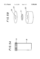
- FIG. 5A is a perspective view of an ammonia test device including reaction cell 18 and sensor cap 19.
- FIG. 5B is an exploded perspective view of top portion of an ammonia test device of FIG. 5A.
- ammonia constituent is used to refer to any one or more of the species ammonia gas, ammonium ion, and ammonium hydroxide. It has been found that elevated levels of ammonia constituent can be detected in body fluids of persons with active H. pylori infection, so that detection and estimation of such levels of ammonia constituent, in combination with serological test for antibody to H. pylori, can serve as a diagnostic method for active H. pylori infection.
- the diagnostic apparatus according to this invention is suitably characterized as having at least one chamber comprising a hollow container with one open end, as illustrated in FIGS. 2A and 2B.
- the container with an open end is the immunoassay chamber, it is suitably characterized as comprising a reaction membrane with immobilized antigen or antibody to said infectious agent, an absorbance layer, and a sample reservoir.
- the reaction membrane illustrated at 4 in FIG. 2A, can be any material, organic or inorganic, with sufficient porosity to allow access by samples to be analyzed and with suitable surface affinity to bind antigens.
- Useful membrane materials include nylon, glass fiber, and natural or synthetic polymers including cellulose esters.
- the porosity of the membrane can vary from 0.2 to 12 microns.
- a nitrocellulose (i.e. cellulose nitrate) membrane has excellent absorption and adsorption qualities, and is preferred. Mechanical strength of nitrocellulose membrane is greatly improved with a paper or polyester support.
- a binding reagent specific to H. pylori antibody such as a commercially available preparation of H. pylori antigen, is immobilized on the membrane and reacts with and captures H. pylori antibody when present in the sample of fluid.
- the thickness of the membrane should be sufficient to immobilize a sufficient amount of antigen to provide adequate sensitivity, but not too thick to block the passage of saliva samples.
- the absorbance layer serves to draw liquid through the reaction membrane and can be made of any kind of porous hydrophilic absorbent material, suitably filter paper.
- the sample reservoir illustrated at 6 in FIG. 2A, is used to apply a sample to the reaction membrane and keep solution running through the center of the reaction membrane containing the immobilized antigen or antibody.
- the container with an open end is the chemical reaction chamber, it is suitably characterized as comprising a hollow reaction chamber as illustrated at 3 in FIG. 2A in which chemical analysis can be carried out in any of several ways.
- a sample such as saliva can be added to the cell and mixed with all necessary reagents for producing a detectable response to an analyte of interest in 3. Reaction is allowed to proceed for a certain period of time, then the product is analyzed by HPLC, ion chromatography, ion selective electrodes, uv-vis spectrophotometric method, fluorescence, laser-induced fluorescence or other analytical techniques.
- saliva sample can be mixed with the reagents such as sodium hydroxide, sodium hypochlorite, sodium salicylate and sodium nitroprusside in the reaction chamber.
- the mixture is incubated at room temperature for 30 minutes.
- the absorbance of the solution is measured at 655 nm using a UV-VIS spectrophotometer.
- the concentration of ammonia constituent in saliva can be obtained from calibration curves of ammonia standard solution.
- the analyte of interest is an ammonia constituent and an ammonia electrode or a test strip affording a detectable response to ammonia can be used for detection and estimation thereof.
- An ammonia electrode commercially available from Omega Engineering, Inc. or other manufacturers can be used directly to measure ammonia concentration in saliva.
- Test strip impregnated with a reagent placed on the top of cell 3 can, for example, also detect ammonia gas generated from an ammonia constituent of a saliva sample placed in the chamber.
- reagents necessary for chemical analysis can also be pre-mixed and added to the bottom of the cell.
- 3a can represent a pure single reagent, a mixture of several reagents, or a reagent impregnated strip prepared by drying porous hydrophilic material such as filter paper soaked with reagent solutions.
- porous hydrophilic material such as filter paper soaked with reagent solutions.
- reagent impregnated strip it can be conveniently attached to the bottom of 3 using small amount of epoxy or other adhesive material which does not interfere with the reaction.
- one step reaction is carried out by adding saliva or other body fluid sample directly to the cell. 3.
- a detectable response to ammonia in accordance with this invention is a change in color of a pH indicator resulting from the reaction of ammonia generated from a sample being examined with the indicator suitably impregnated on a test strip.
- Suitable pH indicators are characterized by a visible color change at a pH in the range from 4 to 12 and include the following which are preferred.
- Another detectable response to ammonia in accordance with this invention is the formation of highly conjugated indophenol dye absorbing strongly at 630-720 nm by reaction of ammonia and a phenol under oxidizing conditions in the so-called Berthelot reaction (see for example P L Searle, Analyst 1984, vol. 109, pages 549-568).
- An ammonia test strip based on the Berthelot reaction according to this invention is constructed, for example, by soaking porous hydrophilic material such as filter paper with a strong alkali such as sodium hydroxide, an alkali metal salicylate such as lithium salicylate, potassium salicylate, or sodium salicylate, and sodium nitroprusside catalyst, and drying the soaked material so as to avoid overheating, as in an incubator.
- a further detectable response to ammonia in accordance with this invention is the formation of a strongly fluorescent product from ammonia and a fluorescence generator reagent such as 4-fluoro-7-nitrobenzo-2-oxa-1,3-diazole (see for example R. Kobayashi et al., Chem. Pharm. Bull. 1992, vol. 40, pages 1327-28).
- a one step detection of ammonia constituent in saliva is achieved by using a multi-layer ammonia test strip according to this invention.
- a test strip can consist of reagent layer 12, gas-permeable layer 13, and sensor layer 14 prepared from porous material. 8 and 9 are supporting layers serving to keep 12, 13, and 14 in position. Paper, such as photocopy paper, is a preferred low cost supporting layer, however, synthetic polymers such as polyvinyl chloride can also be used as supporting layer. Paper glue can be used to stick 8 and 9 together.
- Reagent layer 12 is impregnated with the necessary reagent for chemical reaction, which for ammonia constituent analysis is a strongly basic compound such as sodium hydroxide or potassium hydroxide.
- Gas-permeable layer 13 serves to separate volatile analyte such as ammonia from non-volatile interfering substances such as basic inorganic hydroxides, keeping the latter from reaching sensor layer 14.
- a gas-permeable membrane such as polytetrafluoroethylene, polypropylene, or polyethylene can be used.
- a particularly preferred gas-permeable layer can be conveniently prepared using cellulose acetate butyrate coated filter paper.
- Sensor strip 14 is prepared with pH indicator as illustrated above or with reagents for Berthelot reaction or with fluorescence generator.
- ammonia constituent in the sample reacts with strong base such as sodium hydroxide in reagent layer and releases ammonia gas.
- Ammonia gas diffuses through gas-permeable layer 13 and a distinct color change or fluorescence is observed through hole 11 when ammonia reaches sensor layer 14.
- the ammonia test strip there can be on the ammonia test strip a plurality of sensing units each having an assembly of layers 12, 13, and 14 as shown in FIG. 3B. In this way, two or more samples can be analyzed at the same time using a single test strip as shown in FIG. 4A and FIG. 4B.
- Semi-quantitative information can be obtained by running standard and sample simultaneously. To avoid interference, there should be enough space, suitably 8 millimeters, between two sample wells 15 and 16. A single large piece of hydrophobic gas-permeable layer 17 (FIG. 4B) can be used to avoid diffusion of solution from one cell to another.
- a saliva analyzer for a volatile organic or inorganic analyte such as an ammonia constituent in accordance with this invention includes a reaction cell 18 and a detection cap 19.
- the reaction cell 18 can be made of any material resistant to chemical attack during the chemical analysis, suitably of glass or plastic.
- the reaction cell 18 has an open end.
- the cap 19 can be threaded or clipped onto the cell 18.
- a chemical sensor strip 21 can be fixed onto the cap through a solid backing layer 22 with a hole aligned with 20.
- Backing layer 22 when used is made of plastic or paper with adhesive material on one side. Instead of using backing layer 22, the sensor strip can be glued directly to the base of the cap using epoxy or other adhesive material.
- the sensor strip 21 responds to volatile product, such as ammonia, from the reaction inside the cell 18, resulting in color change, physical property change such as resistance, or the formation of a product which can be determined by various analytical methods such as GC or HPLC.
- volatile product such as ammonia
- a sensor strip is impregnated with a pH indicator such as phenol red or cresol red, a distinct color change can be observed through the hole 23 in the backing layer 22.
- the sensor strip can also be prepared right before the test by applying indicator solution to the strip through top opening 23.
- FIG. 1 a device of the type of FIG. 1 was used to carry out chemical and immunochemical analysis.
- H. pylori antigen was immobilized on the reaction membrane 4 in FIG. 2A.
- Serum or saliva sample containing antibodies to H. pylori was tested using the chamber 2 in FIG. 1. After the sample was absorbed totally through the reaction membrane 4 by absorbance layer 5 in FIG. 2A, washing reagent phosphate buffer containing Tween® 20 surfactant and heat treated normal goat serum was applied. When the washing solution was absorbed, protein A-gold conjugate solution was applied. The treated reaction membrane was incubated at room temperature for 10 minutes and washed again with the washing reagent. A red dot in the middle of the reaction membrane indicated the presence of H. pylori antibodies in the sample.
- the performance of the test strip was tested using 10 mM ammonium chloride solution. 100 microliters 10 mM ammonium chloride solution was mixed with 100 microliters 5% sodium hypochlorite solution (Sigma). The mixture was applied to the test strip. The color of the strip changed from yellow to green within five minutes.
- Ammonia test strip shown in FIG. 3A was fabricated.
- Sensor layer was prepared from 0.02% phenol red or cresol red (Sigma) by soaking and drying a Whatman #4 qualitative filter paper.
- Gas-permeable layer was prepared by coating a piece of Whatman #4 filter paper with cellulose acetate butyrate.
- Reagent layer was prepared from 2 M sodium hydroxide solution. All strips were cut into 1 cm ⁇ 1 cm square pieces.
- a reagent layer piece, a gas-permeable layer piece, and a sensor layer piece were stacked glued together using two pieces of paper with one hole at one side of the end.
- the performance of the test strip was tested using 2 M sodium hydroxide and 10 mM ammonium chloride solution.
- sodium hydroxide solution When one drop of sodium hydroxide solution was applied to hole 10 in FIG. 3B, no color change was observed through hole 11 in FIG. 3B, indicating no leakage of solution through the gas-permeable layer.
- ammonium solution When ammonium solution was applied, almost instant color change was observed resulting from pH change in the sensor strip due to dissolved ammonia.
- Saliva sample was also analyzed with the test strip. The test strip was sensitive enough to detect ammonia constituent in the saliva. Ammonia concentration in the saliva was in the millimolar range.
- Ammonia test strip as shown in FIG. 4A was prepared using similar procedure. 2.0 mM, 4.0 mM, 6.0 mM, and 8.0 mM ammonium chloride solutions were used to evaluate the performance of the test strip. Two samples were analyzed simultaneously. There was distinct difference in color intensity and color development time when concentration changed with 2.0 mM increment, indicating the possibility to use such a kind of test strip to get semi-quantitative information.
- test strips are wetted using small volume of distilled water right before analysis.
Abstract
Disclosed is a diagnostic apparatus for estimating an active Heliobacter pylori infectious agent in saliva, comprising in combination an immunoassay chamber in which a first portion of said saliva is subjected to serological test for antibody to said infectious agent and a chemical reaction chamber in which a second portion of said saliva is subjected to chemical analysis for an ammonia constituent thereof.
Description
1. Field of the Invention
The invention relates generally to methods and devices used for determination of analytes in saliva or other body fluid, and in particular, pertains to an integrated device and method to carry out chemical and immunochemical analysis simultaneously and including a sensor strip for determination of ammonia.
2. Description of the Related Art
Helicobacter pylori (formerly called Campylobacter pylori) was first isolated by Warnell and Shall in 1983 (Marshall B. J., Warren J. R., Lancet 1984:1:1311-5). H. pylori is the most widespread bacteria infection with an estimated worldwide prevalence of 50% (Marshall B. J., Epidemiology of H. pylori in Western countries. In: Hunt R. H., Tytgat G. N. J., eds. Helicobacter pylori: Basic Mechanisms to Clinical Cure. Dordrecht: Kluwer Academic Publishers. 1994:75-84; Hazell S. L., H. pylori in developing countries. In: Hunt R. H. Tytgat G. N. J., eds. Helicobacter pylori: Basic Mechanisms to Clinical Cure, Dordrecht: Kluwer Academic Publishers, 1994:85-94). H. pylori is a very important pathogen in several diseases of the stomach and duodenum. H. pylori is associated with type B gastritis, duodenal ulcer, gastric ulcer, and gastric cancer.
A variety of methods has been developed for diagnosis of H. pylori infection and evaluation of eradication of H. pylori following antibacterial treatment.
U.S. Pat. No. 5,498,528 teaches a method for detection of H. pylori strain comprising the steps of contacting a saliva sample suspected of containing H. pylori directly with a urea containing medium selective for growth of H. pylori and having a pH of about 5.5 to 7.5, and incubating the sample for a time sufficient for detection of H. pylori growth in at least 80% of true positive samples. The method is based on hydrolysis of urea contained in the growth medium by urease enzyme produced by H. pylori and detection of a hydrolysis product by release of a radioactive label from urease or by a color change resulting from action on a pH indicator. The time required to obtain a result by the method disclosed is a function of temperature, approximately 2-3 days when incubated at 23-25° C. and 4-6 hours when incubated at 35-37° C.
U.S. Pat. No. 5,479,935 teaches an ambulatory system for recording and analyzing gastroesophageal reflux. The system comprises a digital recorder, an analysis software package and a catheter for measurement of changes in esophageal impedance. Gastroesophageal reflux can be detected with a pH above 4. The invention allows for recording and analysis of reflux on a non-invasive basis, by using pairs of externally worn impedance sensors. Other bio-parameters, such as pH or pressure can be measured simultaneously with impedance measurement.
U.S. Pat. No. 5,477,854 teaches a system and a method for monitoring intragastrointestinal concentrations of ammonium during prolonged periods, as an indicator of the presence and activity of an intragastrointestinal H. pylori infection. The system may be used in the evaluation of treatments for H. pylori infection in the patient.
U.S. Pat. No. 5,439,801 teaches an improved test for the detection of the presence of urease associated with H. pylori in a biopsy specimen. The hydrolysis of urea by urease is detected by a combination of at least two dye indicators showing a color change. Most positive results occur in 2-10 minutes and all occur in no more than four hours.
U.S. Pat. No. 5,438,985 teaches a method and a system for ambulatory recording of the pH and the presence of various materials in compartments of the gastro-intestinal tract. The invention also reports the pH pattern in relation to the prevalence of the materials, and analysis to which degree such materials are in active or inactive states in their normal or foreign compartments. This is useful in situations, for example, when duodenal material is refluxed into the stomach and esophagus. The invention involves a gastrointestinal catheter with a pH sensor and a combined light absorption and fluorescence sensor, a signal recorder and processor, and a written report producer.
U.S. Pat. No. 5,420,016 and U.S. Pat. No. 5,314,804 teach a rapid method for determining the presence of H. pylori in a biological tissue specimen by detecting the presence of urease in the tissue. The system employs a multilayer test device for the detection of ammonia generated from urea at the presence of urease in the specimen.
U.S. Pat. No. 5,420,014 teaches a method of detecting contemporary infection by H. pylori in a mammal. The method is based on the formation of complex between a specific IgG antibody in said mucous secretion and an antigen component from H. pylori. The antigen component is immobilized onto a solid support.
U.S. Pat. No. 5,262,156 teaches an assay for detecting H. pylori. The assay involves an ELISA for urine samples, and includes a kit wherein the antigenic composition is immobilized on a solid support.
U.S. Pat. No. 4,947,861 teaches a non-invasive method for detection of C. pylori. A breath sample is collected from a patient ten minutes after the patient ingests a quantity of urea. The sample is dehydrated by passing through a solid-state body of alkaline hygrosopic material and analyzed for ammonia. The presence of ammonia indicates presence of C. pylori in the stomach.
U.S. Pat. No. 4,882,271 teaches a method for the serological detection of C. pylori. An antigen for the detection of C. pylori infections is purified from C. pylori. The antigen can be used in a variety of assays including radioimmunoassay, ELISA, latex agglutination, complement fixation, and indirect hemagglutination.
The urea breath test based on the extremely high endogenous urea activity of H. pylori is a reliable and non-invasive method with high sensitivity and specificity suitable for diagnosis and evaluation tests; however, the application of the urea breath test is restricted by high cost in isotope-labeled material, time, expensive equipment, and undesirable radioactive exposure to C14.
Against this background there remains a need for a method of diagnosing C. pylori that is rapid, non-invasive, and able to distinguish infection in an active state from dormant or non-living residues.
In accordance with this invention, it has been found that a novel non-invasive method for diagnosis of active Heliobacter pylori infection is based on simultaneous detection of antibody to the H. pylori and abnormal level of an ammonia constituent in a body fluid such as blood or saliva. Since antibody to the H. pylori lasts for a long period of time even with eradication of H. pylori, a serological test alone cannot predicate whether the bacteria infection is active. Ammonia concentration in a body fluid is affected by many diseases, and hence ammonia estimation alone cannot pinpoint exact cause of abnormal ammonia value. Combining the information obtained for ammonia analysis and the serological test, however, affords a novel test for active H. pylori requiring only small samples of body fluid and minimal inconvenience to the patient.
Also in accordance with this invention, a diagnostic apparatus for estimating an active Heliobacter pylori infectious agent in saliva comprises in combination an immunoassay chamber in which a first portion of said saliva is subjected to serological test for antibody to said infectious agent and a chemical reaction chamber in which a second portion of said saliva is subjected to chemical analysis for an ammonia constituent thereof. The presence of antibody to the H. pylori as shown by serological test coupled with abnormal level of ammonia indicates active H. pylori infection, while a positive serological result with normal ammonia constituent level indicates an H. pylori infection that is inactive or limited. Moreover, in accordance with this invention, quantitative or semi-quantitative analysis of ammonia and antibody levels in saliva can be used to monitor the eradication of H. pylori.
A method in accordance with this invention of estimating an active H. pylori biological infectious agent in a body fluid such as blood or saliva, comprises in combination subjecting a first portion of said fluid to serological test for antibody to said H. pylori infectious agent and subjecting a second portion of said fluid to chemical analysis for an ammonia constituent such as ammonia or ammonium thereof.
The diagnostic apparatus of the invention can be constructed as a single device with two reaction chambers, one for chemical analysis and one for serological test or immunoassay. The two reaction chambers can also be in physically separate devices used to process for chemical analysis and for serological test portions of a single specimen of body fluid.
A chemical sensor strip can be incorporated as an integrated part of the device or used separately for chemical analysis. The sensor strip comprises at least one porous solid layer impregnated with the necessary reagents for analysis including chemical reaction, separation, and detection.
In the method of the present invention, a sample of the body fluid to be examined is applied to each of the reaction chambers. Ammonia in the fluid is detected in one chamber with a chemical sensor strip or other analytical methods such as ion selective electrode, IC, and HPLC etc. In another chamber, immunochemical information is obtained from the other chamber with immobilized antigen. The immunoassay is based on the formation of antigen-antibody complexes to detect the presence of antibodies or antigens, using antigen to detect antibodies and antibody to detect antigens as required. Generally, antigens of the infectious agent such as H. pylori are coated on a solid support. Antibodies present in the sample being examined and specific to the antigen are captured on the solid support, resulting in the formation of antigen-antibody complex. A second antibody labeled with radioactive, enzyme, fluorescent, chemiluminescent or other compound with detectable chemical or physical properties is used to detect the presence of antigen-antibody complex through the formation of an antigen-antibody-anti-antibody complex. Thus, if antibody is present in the sample of a fluid such as saliva, antigen-antibody complexes are formed after a period of incubation when the fluid sample is applied to the chamber. After washing the unbound fluid from the membrane with buffer solution to eliminate non-specific binding, protein-A gold conjugate is added to detect the presence of H. pylori antibodies. The Protein-A Gold conjugate binds to the Fc portion of H. pylori antibodies captured by antigens on the support. A positive reaction for H. pylori antibodies is confirmed by a visible red or slightly pink spot in the test area.
FIG. 1 is a perspective view of a double-chamber reaction device for carrying out chemical and immunochemical analysis simultaneously according to the invention, in which the chambers are identified by the numbers 1 and 2.
FIG. 2A is a cross-sectional view of the device of FIG. 1 without reagents in the chamber for chemical analysis, 3, and the chamber for immunoassay fitted with reaction membrane 4, absorbance material 5, and cylindrical sample reservoir 6.
FIG. 2B is a cross-sectional view of the device with immobilized reagents 3a at the bottom of the cell for chemical analysis.
FIG. 3A is a perspective view of an ammonia test strip with multi-layer structure to perform reaction, separation, and detection in one step.
FIG. 3B is a cross-sectional view of an ammonia test strip of FIG. 3A.
FIG. 4A is a perspective view of an ammonia test strip capable of performing two individual tests at the same time.
FIG. 4B is a cross-sectional view of an ammonia test strip of FIG. 4A.
FIG. 5A is a perspective view of an ammonia test device including reaction cell 18 and sensor cap 19.
FIG. 5B is an exploded perspective view of top portion of an ammonia test device of FIG. 5A.
It should be noted that the sketches are not drawn to scale. All the drawings are for illustration of the present invention without limitation.
Throughout the present specification and claims, "ammonia constituent" is used to refer to any one or more of the species ammonia gas, ammonium ion, and ammonium hydroxide. It has been found that elevated levels of ammonia constituent can be detected in body fluids of persons with active H. pylori infection, so that detection and estimation of such levels of ammonia constituent, in combination with serological test for antibody to H. pylori, can serve as a diagnostic method for active H. pylori infection.
The diagnostic apparatus according to this invention is suitably characterized as having at least one chamber comprising a hollow container with one open end, as illustrated in FIGS. 2A and 2B.
When the container with an open end is the immunoassay chamber, it is suitably characterized as comprising a reaction membrane with immobilized antigen or antibody to said infectious agent, an absorbance layer, and a sample reservoir. The reaction membrane, illustrated at 4 in FIG. 2A, can be any material, organic or inorganic, with sufficient porosity to allow access by samples to be analyzed and with suitable surface affinity to bind antigens. Useful membrane materials include nylon, glass fiber, and natural or synthetic polymers including cellulose esters. The porosity of the membrane can vary from 0.2 to 12 microns. A nitrocellulose (i.e. cellulose nitrate) membrane has excellent absorption and adsorption qualities, and is preferred. Mechanical strength of nitrocellulose membrane is greatly improved with a paper or polyester support. Commercially available paper backed nitrocellulose membrane is conveniently used for easy handling. For the relatively high viscosity of saliva a relatively large porosity grade of membrane is a preferred choice. A binding reagent specific to H. pylori antibody, such as a commercially available preparation of H. pylori antigen, is immobilized on the membrane and reacts with and captures H. pylori antibody when present in the sample of fluid. The thickness of the membrane should be sufficient to immobilize a sufficient amount of antigen to provide adequate sensitivity, but not too thick to block the passage of saliva samples.
The absorbance layer, illustrated at 5 in FIG. 2A, serves to draw liquid through the reaction membrane and can be made of any kind of porous hydrophilic absorbent material, suitably filter paper. The sample reservoir, illustrated at 6 in FIG. 2A, is used to apply a sample to the reaction membrane and keep solution running through the center of the reaction membrane containing the immobilized antigen or antibody.
When the container with an open end is the chemical reaction chamber, it is suitably characterized as comprising a hollow reaction chamber as illustrated at 3 in FIG. 2A in which chemical analysis can be carried out in any of several ways. A sample such as saliva can be added to the cell and mixed with all necessary reagents for producing a detectable response to an analyte of interest in 3. Reaction is allowed to proceed for a certain period of time, then the product is analyzed by HPLC, ion chromatography, ion selective electrodes, uv-vis spectrophotometric method, fluorescence, laser-induced fluorescence or other analytical techniques. For example, saliva sample can be mixed with the reagents such as sodium hydroxide, sodium hypochlorite, sodium salicylate and sodium nitroprusside in the reaction chamber. The mixture is incubated at room temperature for 30 minutes. The absorbance of the solution is measured at 655 nm using a UV-VIS spectrophotometer. The concentration of ammonia constituent in saliva can be obtained from calibration curves of ammonia standard solution.
In a particularly preferred embodiment of this invention, the analyte of interest is an ammonia constituent and an ammonia electrode or a test strip affording a detectable response to ammonia can be used for detection and estimation thereof. An ammonia electrode commercially available from Omega Engineering, Inc. or other manufacturers can be used directly to measure ammonia concentration in saliva. Test strip impregnated with a reagent placed on the top of cell 3 can, for example, also detect ammonia gas generated from an ammonia constituent of a saliva sample placed in the chamber.
As illustrated at 3a in FIG. 2B, reagents necessary for chemical analysis can also be pre-mixed and added to the bottom of the cell. 3a can represent a pure single reagent, a mixture of several reagents, or a reagent impregnated strip prepared by drying porous hydrophilic material such as filter paper soaked with reagent solutions. In the case of reagent impregnated strip, it can be conveniently attached to the bottom of 3 using small amount of epoxy or other adhesive material which does not interfere with the reaction. With reagent or filter paper impregnated with all necessary reagents added to the cell first, one step reaction is carried out by adding saliva or other body fluid sample directly to the cell. 3.
A detectable response to ammonia in accordance with this invention is a change in color of a pH indicator resulting from the reaction of ammonia generated from a sample being examined with the indicator suitably impregnated on a test strip. Suitable pH indicators are characterized by a visible color change at a pH in the range from 4 to 12 and include the following which are preferred.
______________________________________
Indicator           pH range Color Change
______________________________________
2-(2,4-Dinitrophenylazo)-1-naphthol-3,6-
                    6.0-7.0  yellow-blue
disulfonic acid
4,4'-bis(4-amino-1-naphthylazo)-2,2'-
                    8.0-9.0  blue-red
stilbenedisulfonic acid
6,8-dinitro-2,4-(1H)quinazolinedione
                    6.4-8.0  colorless-yellow
alizarin            5.6-7.2  yellow-red
brilliant yellow    6.6-7.8  yellow-red
bromothymol blue    6.0-7.6  yellow-blue
cresol red          7.0-8.8  yellow-red
m-nitrophenol       6.8-8.6  colorless-yellow
metacresol purple   7.4-9.0  yellow-purple
neutral red         6.8-8.0  red-amber
phenol red          6.6-8.0  yellow-red
phenolphthalein      8.2-10.0
                             colorless-pink
rosolic acid        5.0-6.8  yellow-red
thymol blue         8.0-9.6  red-blue
turmeric            7.4-8.6  yellow-red
xylenol blue        8.0-9.8  yellow-violet
______________________________________
Another detectable response to ammonia in accordance with this invention is the formation of highly conjugated indophenol dye absorbing strongly at 630-720 nm by reaction of ammonia and a phenol under oxidizing conditions in the so-called Berthelot reaction (see for example P L Searle, Analyst 1984, vol. 109, pages 549-568). An ammonia test strip based on the Berthelot reaction according to this invention is constructed, for example, by soaking porous hydrophilic material such as filter paper with a strong alkali such as sodium hydroxide, an alkali metal salicylate such as lithium salicylate, potassium salicylate, or sodium salicylate, and sodium nitroprusside catalyst, and drying the soaked material so as to avoid overheating, as in an incubator.
A further detectable response to ammonia in accordance with this invention is the formation of a strongly fluorescent product from ammonia and a fluorescence generator reagent such as 4-fluoro-7-nitrobenzo-2-oxa-1,3-diazole (see for example R. Kobayashi et al., Chem. Pharm. Bull. 1992, vol. 40, pages 1327-28).
Referring to FIG. 3A and FIG. 3B, a one step detection of ammonia constituent in saliva is achieved by using a multi-layer ammonia test strip according to this invention. Such a test strip can consist of reagent layer 12, gas-permeable layer 13, and sensor layer 14 prepared from porous material. 8 and 9 are supporting layers serving to keep 12, 13, and 14 in position. Paper, such as photocopy paper, is a preferred low cost supporting layer, however, synthetic polymers such as polyvinyl chloride can also be used as supporting layer. Paper glue can be used to stick 8 and 9 together. Reagent layer 12 is impregnated with the necessary reagent for chemical reaction, which for ammonia constituent analysis is a strongly basic compound such as sodium hydroxide or potassium hydroxide.
Gas-permeable layer 13 serves to separate volatile analyte such as ammonia from non-volatile interfering substances such as basic inorganic hydroxides, keeping the latter from reaching sensor layer 14. For the fabrication of gas-permeable layer 13, a gas-permeable membrane such as polytetrafluoroethylene, polypropylene, or polyethylene can be used. A particularly preferred gas-permeable layer can be conveniently prepared using cellulose acetate butyrate coated filter paper.
When a sample such as saliva is applied to the reagent layer through the hole 10, ammonia constituent in the sample reacts with strong base such as sodium hydroxide in reagent layer and releases ammonia gas. Ammonia gas diffuses through gas-permeable layer 13 and a distinct color change or fluorescence is observed through hole 11 when ammonia reaches sensor layer 14.
In a particularly preferred embodiment, there can be on the ammonia test strip a plurality of sensing units each having an assembly of layers 12, 13, and 14 as shown in FIG. 3B. In this way, two or more samples can be analyzed at the same time using a single test strip as shown in FIG. 4A and FIG. 4B.
Semi-quantitative information can be obtained by running standard and sample simultaneously. To avoid interference, there should be enough space, suitably 8 millimeters, between two sample wells 15 and 16. A single large piece of hydrophobic gas-permeable layer 17 (FIG. 4B) can be used to avoid diffusion of solution from one cell to another.
Referring to FIG. 5A and FIG. 5B, a saliva analyzer for a volatile organic or inorganic analyte such as an ammonia constituent in accordance with this invention includes a reaction cell 18 and a detection cap 19. The reaction cell 18 can be made of any material resistant to chemical attack during the chemical analysis, suitably of glass or plastic. The reaction cell 18 has an open end. The cap 19 can be threaded or clipped onto the cell 18. There is a hole 20 in cap 19, suitably in the middle thereof. A chemical sensor strip 21 can be fixed onto the cap through a solid backing layer 22 with a hole aligned with 20.
Backing layer 22 when used is made of plastic or paper with adhesive material on one side. Instead of using backing layer 22, the sensor strip can be glued directly to the base of the cap using epoxy or other adhesive material.
The sensor strip 21 responds to volatile product, such as ammonia, from the reaction inside the cell 18, resulting in color change, physical property change such as resistance, or the formation of a product which can be determined by various analytical methods such as GC or HPLC. When a sensor strip is impregnated with a pH indicator such as phenol red or cresol red, a distinct color change can be observed through the hole 23 in the backing layer 22. The sensor strip can also be prepared right before the test by applying indicator solution to the strip through top opening 23.
Further disclosure of the invention is provided by the following examples, offered for purpose of illustration and not of limitation.
In this instance, a device of the type of FIG. 1 was used to carry out chemical and immunochemical analysis.
100 microliters of 10 mM ammonium chloride was added to the cell 1. Then, one drop of 2 M sodium hydroxide was added to the cell. When a test strip impregnated with phenol red indicator was placed on top of the opening, instant color change was observed, indicating the release of ammonia from the reaction.
As an example of immunoassay, H. pylori antigen was immobilized on the reaction membrane 4 in FIG. 2A. Serum or saliva sample containing antibodies to H. pylori was tested using the chamber 2 in FIG. 1. After the sample was absorbed totally through the reaction membrane 4 by absorbance layer 5 in FIG. 2A, washing reagent phosphate buffer containing Tween® 20 surfactant and heat treated normal goat serum was applied. When the washing solution was absorbed, protein A-gold conjugate solution was applied. The treated reaction membrane was incubated at room temperature for 10 minutes and washed again with the washing reagent. A red dot in the middle of the reaction membrane indicated the presence of H. pylori antibodies in the sample.
In this example, preparation of ammonia test strips based on the Berthelot reaction is described. 4.08 g sodium hydroxide (Sigma Chemical Company), 1.19 g sodium salicylate (Aldrich), and 0.05 g sodium nitroprusside (Sigma) were ground into small particles in a porcelain mortar using a porcelain pestle. 5 ml water was added and grinding continued until a fine solid suspension was obtained. The mixture was spread over a piece of Whatman # 4 qualitative filter paper and the paper soaked with the reagent was dried overnight in a 40° C. incubator. The filter paper treated in this way was used as an ammonia test strip.
The performance of the test strip was tested using 10 mM ammonium chloride solution. 100 microliters 10 mM ammonium chloride solution was mixed with 100 microliters 5% sodium hypochlorite solution (Sigma). The mixture was applied to the test strip. The color of the strip changed from yellow to green within five minutes.
Ammonia test strip shown in FIG. 3A was fabricated. Sensor layer was prepared from 0.02% phenol red or cresol red (Sigma) by soaking and drying a Whatman # 4 qualitative filter paper. Gas-permeable layer was prepared by coating a piece of Whatman # 4 filter paper with cellulose acetate butyrate. Reagent layer was prepared from 2 M sodium hydroxide solution. All strips were cut into 1 cm×1 cm square pieces. As shown in FIG. 3B, a reagent layer piece, a gas-permeable layer piece, and a sensor layer piece were stacked glued together using two pieces of paper with one hole at one side of the end.
The performance of the test strip was tested using 2 M sodium hydroxide and 10 mM ammonium chloride solution. When one drop of sodium hydroxide solution was applied to hole 10 in FIG. 3B, no color change was observed through hole 11 in FIG. 3B, indicating no leakage of solution through the gas-permeable layer. When ammonium solution was applied, almost instant color change was observed resulting from pH change in the sensor strip due to dissolved ammonia. Saliva sample was also analyzed with the test strip. The test strip was sensitive enough to detect ammonia constituent in the saliva. Ammonia concentration in the saliva was in the millimolar range.
Ammonia test strip as shown in FIG. 4A was prepared using similar procedure. 2.0 mM, 4.0 mM, 6.0 mM, and 8.0 mM ammonium chloride solutions were used to evaluate the performance of the test strip. Two samples were analyzed simultaneously. There was distinct difference in color intensity and color development time when concentration changed with 2.0 mM increment, indicating the possibility to use such a kind of test strip to get semi-quantitative information.
In order to increase color contrast and increase ammonia collection efficiency, test strips are wetted using small volume of distilled water right before analysis.
Claims (17)
1. A diagnostic apparatus for estimating an active Helicobacter pylori infectious agent in human saliva, comprising in combination an immunoassay chamber comprising a reaction membrane with immobilized antigen to said Helicobacter pylori, an absorbance layer, and a sample reservoir; a chemical reaction chamber in which said saliva is subjected to chemical analysis for an ammonia constituent thereof selected from the group consisting of ammonia, ammonium ion, and ammonium hydroxide, said chemical reaction chamber comprising a solid porous support impregnated with at least one chemical reagent indicating the presence of said ammonia constituent by a detectable change in electrical potential, in color, or in fluorescence, and said analysis comprising performing a reaction to release ammonia, whereby active Helicobacter pylori infection is diagnosed by a positive reaction of said antigen with antibody in said saliva and greater than normal level of ammonia constituent in said saliva.
2. A diagnostic apparatus according to claim 1, in which at least one chamber comprises a hollow container with one open end.
3. A diagnostic apparatus according to claim 1, in which the reaction membrane comprises nitrocellulose.
4. A diagnostic apparatus according to claim 1, in which the absorbance layer comprises porous hydrophilic material.
5. A diagnostic apparatus according to claim 1, in which said chemical reagent is a pH indicator capable of giving rise to a visible change in color at a pH in the range from 4 to 12.
6. A diagnostic apparatus according to claim 7, in which said pH indicator is selected from the group consisting of 2-(2,4-Dinitrophenylazo)-1-naphthol-3,6-disulfonic acid, 4,4'-bis(4-amino-1-naphthylazo)-2,2'-stilbenedisulfonic acid, 6,8-dinitro-2,4-(1H)qunizaolinedione, alizarin, brilliant yellow, bromothymol blue, cresol red, m-nitrophenol, metacresol purple, neutral red, phenol red, phenolphthalein, rosolic acid, thymol blue, turmeric, and xylenol blue.
7. A diagnostic apparatus according to claim 1, in which said chemical reagent responds to the presence of an ammonia constituent by the Berthelot reaction.
8. A diagnostic apparatus according to claim 7, in which said chemical reagent comprises alkali metal hydroxide, alkali metal salicylate, and alkali metal nitroprusside.
9. A diagnostic apparatus according to claim 1, in which said chemical reagent responds to the presence of an ammonia constituent by giving rise to a fluorescent product.
10. A diagnostic apparatus according to claim 9, in which said chemical reagent comprises 4-fluoro-7-nitrobenzo-2-oxa-1,3-diazole.
11. A diagnostic apparatus according to claim 1, in which said porous support comprises an organic polymer.
12. A diagnostic apparatus according to claim 11, in which said porous support comprises cellulose.
13. A diagnostic apparatus according to claim 12, in which said porous support comprises filter paper or cellulose membrane.
14. A diagnostic apparatus according to claim 1, in which the chemical reaction chamber is used to mix saliva with at least one reagent selected from the group consisting of sodium hydroxide, sodium hypochlorite, sodium nitroprusside, and sodium salicylate.
15. A diagnostic apparatus according to claim 1, in which the chemical reaction chamber is used to mix saliva with a reagent for the formation of fluorescent compounds.
16. A diagnostic apparatus according to claim 1, in which the chemical reaction chamber is used to carry out ammonia analysis in saliva using ammonia electrodes.
17. A diagnostic apparatus according to claim 15, in which the reagent for the formation of fluorescent compounds is 4-fluoro-7-nitrobenzo-2-oxo-1,3-diazole.
Priority Applications (3)
| Application Number | Priority Date | Filing Date | Title |
|---|---|---|---|
| US08/865,089 US5989840A (en) | 1997-05-29 | 1997-05-29 | Estimation of active infection by heliobacter pylori |
| AU77223/98A AU7722398A (en) | 1997-05-29 | 1998-05-29 | Estimation of active infection by helicobacter pylori |
| PCT/US1998/011445 WO1998054563A1 (en) | 1997-05-29 | 1998-05-29 | Estimation of active infection by helicobacter pylori |
Applications Claiming Priority (1)
| Application Number | Priority Date | Filing Date | Title |
|---|---|---|---|
| US08/865,089 US5989840A (en) | 1997-05-29 | 1997-05-29 | Estimation of active infection by heliobacter pylori |
Publications (1)
| Publication Number | Publication Date |
|---|---|
| US5989840A true US5989840A (en) | 1999-11-23 |
Family
ID=25344688
Family Applications (1)
| Application Number | Title | Priority Date | Filing Date |
|---|---|---|---|
| US08/865,089 Expired - Fee Related US5989840A (en) | 1997-05-29 | 1997-05-29 | Estimation of active infection by heliobacter pylori |
Country Status (1)
| Country | Link |
|---|---|
| US (1) | US5989840A (en) |
Cited By (27)
| Publication number | Priority date | Publication date | Assignee | Title |
|---|---|---|---|---|
| US6228605B1 (en) * | 1993-10-28 | 2001-05-08 | Barry J. Marshall | Detection of helicobacter pylori in the stomach |
| US6291178B1 (en) | 1997-11-26 | 2001-09-18 | David R. Schneider | Method and apparatus for preserving human saliva for testing |
| US6391261B1 (en) * | 2000-04-14 | 2002-05-21 | Lifepoint, Inc. | Device for detecting analytes related to sample pH |
| US6517809B1 (en) | 1997-05-28 | 2003-02-11 | Barry J. Marshall | Process for preparing a reactive pharmaceutical product for the detection of gastrointestinal disorder caused by bacteria in the gastrointestinal superior tract |
| US20030073155A1 (en) * | 2001-10-15 | 2003-04-17 | Mcmichael Donald J. | Methods for performing multiple diagnostic tests |
| USD484988S1 (en) | 2001-12-17 | 2004-01-06 | Kimberly-Clark Worldwide, Inc. | Diagnostic test kit with specimen-handling tool |
| WO2004006766A2 (en) | 2002-07-12 | 2004-01-22 | Baxter International Inc. | Method and apparatus for the detection of the presence of a bacteria in the gastrointestinal tract of a subject |
| WO2004072614A2 (en) * | 2003-02-11 | 2004-08-26 | Chemsensing Inc. | Method and apparatus for detecting an analyte |
| US6783976B2 (en) | 2001-12-21 | 2004-08-31 | Kimberly-Clark Worldwide, Inc. | Carrier and specimen-handling tool for use in diagnostic testing |
| US6929926B2 (en) | 2001-10-15 | 2005-08-16 | Barry J. Marshall | Composition for the detection of gastrointestinal disorders |
| US20050180879A1 (en) * | 2004-02-03 | 2005-08-18 | Eva Hrboticka | One step tester for ammonia |
| US6998250B2 (en) | 2001-10-15 | 2006-02-14 | Donald J. McMichael | Method for detecting Helicobacter pylori |
| US7008777B2 (en) | 2001-10-15 | 2006-03-07 | Barry J. Marshall | System for the detection of urease and method for using same |
| US7033839B1 (en) * | 1999-03-16 | 2006-04-25 | Hach Company | Quick acting toxic ammonia test for aqueous samples |
| US20060189894A1 (en) * | 2003-02-25 | 2006-08-24 | Antonio Tucci | Method for rapid identification of infections and/or risk situations related to gastroduodenal pathologies and machine for performing the method |
| US7179609B1 (en) * | 2000-10-04 | 2007-02-20 | Atrophus Ab | Screening method for gastritis |
| EP1840566A1 (en) * | 2006-03-30 | 2007-10-03 | Hach Lange GmbH | Device for determining ammonium in a liquid sample |
| US7582485B2 (en) * | 2003-10-16 | 2009-09-01 | Kimberly-Clark Worldride, Inc. | Method and device for detecting ammonia odors and helicobacter pylori urease infection |
| US20100233033A1 (en) * | 2006-06-26 | 2010-09-16 | Nippon Telegraph And Telephone Corporation | Flow cell and method for manufacturing the same |
| US20110092787A1 (en) * | 2008-04-18 | 2011-04-21 | Clemens Bulitta | Endocapsule |
| US8221328B2 (en) | 2003-10-16 | 2012-07-17 | Kimberly-Clark Worldwide, Inc. | Visual indicating device for bad breath |
| US20140364758A1 (en) * | 2011-12-19 | 2014-12-11 | Resmed Limited | Respiratory treatment system including physiological sensors |
| US20150038350A1 (en) * | 2010-12-28 | 2015-02-05 | Arkray, Inc. | Method for Determining Condition of Oral Cavity and Analytical Tool, Apparatus, and Program Used for the Method |
| WO2015026661A3 (en) * | 2013-08-20 | 2015-11-26 | Petter Linda Marie | Tissue tester |
| US20200150048A1 (en) * | 2017-04-28 | 2020-05-14 | Leadway (Hk) Limited | Detection device and sample detection method capable of visually reading test results |
| US11053531B2 (en) | 2019-05-31 | 2021-07-06 | Linda Marie Petter | Tester paper and methods of use thereof for detecting a bacterial infection |
| EP3667300A4 (en) * | 2017-08-08 | 2021-08-25 | Nitto Denko Corporation | Gas detection element |
Citations (36)
| Publication number | Priority date | Publication date | Assignee | Title |
|---|---|---|---|---|
| US4066403A (en) * | 1975-06-20 | 1978-01-03 | Eastman Kodak Company | Multilayer analytical element |
| US4194063A (en) * | 1978-02-24 | 1980-03-18 | Eastman Kodak Company | Method, composition and elements for the detecting of nitrogen-containing compounds |
| US4297173A (en) * | 1980-05-22 | 1981-10-27 | Ajinomoto Company, Incorporated | Method for determining ammonia and sensor therefor |
| US4350660A (en) * | 1980-06-07 | 1982-09-21 | Emi Limited | Ammonia gas sensors |
| US4426451A (en) * | 1981-01-28 | 1984-01-17 | Eastman Kodak Company | Multi-zoned reaction vessel having pressure-actuatable control means between zones |
| US4548906A (en) * | 1981-11-02 | 1985-10-22 | Fuji Shashin Film Kabushiki Kaisha | Integral multilayer analytical element for the analysis of ammonia or an ammonia forming substrate and a method for the detection thereof using the same |
| US4568518A (en) * | 1982-12-06 | 1986-02-04 | Avl Ag | Sensor element for fluorescence-optical measurement |
| US4713165A (en) * | 1986-07-02 | 1987-12-15 | Ilex Corporation | Sensor having ion-selective electrodes |
| US4719085A (en) * | 1985-12-24 | 1988-01-12 | Eastman Kodak Company | Mount for ammonia-sensitive test elements |
| US4748113A (en) * | 1985-06-13 | 1988-05-31 | Marshall Barry J | Compositions and methods for the diagnosis of gastrointestinal disorders involving urease |
| US4769216A (en) * | 1980-12-22 | 1988-09-06 | Commonwealth Serum Laboratories Commission | Device for detecting antigens and antibodies |
| US4769467A (en) * | 1983-09-28 | 1988-09-06 | Oread Laboratories, Inc. | Fluorogenic 2,1,3-benzoxadiazoles and fluorometric amine/thiol assays therewith |
| US5008078A (en) * | 1987-04-16 | 1991-04-16 | Fuji Photo Film Co., Ltd. | Integral multi-layer analysis element |
| US5091080A (en) * | 1990-11-30 | 1992-02-25 | Bend Research, Inc. | Adsorbents for the removal of volatile substances from aqueous systems |
| US5116759A (en) * | 1990-06-27 | 1992-05-26 | Fiberchem Inc. | Reservoir chemical sensors |
| US5198335A (en) * | 1985-06-04 | 1993-03-30 | Fuji Photo Film Co., Ltd. | Integral multilayer analytical element for analysis of ammonia-forming substrate |
| US5238613A (en) * | 1987-05-20 | 1993-08-24 | Anderson David M | Microporous materials |
| US5252292A (en) * | 1989-05-18 | 1993-10-12 | Mitsutoshi Hirata | Ammonia sensor |
| US5286624A (en) * | 1990-10-19 | 1994-02-15 | Fuji Photo Film Co., Ltd. | Integral multilayer analytical element for determination of ammonia or ammonia-producing substance |
| US5344546A (en) * | 1989-04-29 | 1994-09-06 | Dragerwerk Aktiengesellschaft | Electrical measuring cell for determinging ammonia, amines, hydrazine amines, hydrazine and hydrazine derivatives |
| US5393496A (en) * | 1990-12-18 | 1995-02-28 | Saliva Diagnostic Systems, Inc. | Saliva sampling device and sample adequacy system |
| US5411893A (en) * | 1993-03-15 | 1995-05-02 | Difco Laboratories | Dry slide for diagnostic tests |
| US5420014A (en) * | 1992-04-30 | 1995-05-30 | Auspharm International Ltd. | Rapid in vitro test for helicobacter pylori using saliva |
| US5420016A (en) * | 1992-03-24 | 1995-05-30 | Serim Research Corporation | Test device and kit for detecting helicobacter pylori |
| US5439801A (en) * | 1994-02-14 | 1995-08-08 | Chek-Med Systems, Inc. | Test composition for the rapid detection of helicobacter pylori in gastric biopsy tissue |
| US5443080A (en) * | 1993-12-22 | 1995-08-22 | Americate Transtech, Inc. | Integrated system for biological fluid constituent analysis |
| US5492841A (en) * | 1994-02-18 | 1996-02-20 | E. I. Du Pont De Nemours And Company | Quaternary ammonium immunogenic conjugates and immunoassay reagents |
| US5494640A (en) * | 1990-04-12 | 1996-02-27 | Hitachi, Ltd. | Device for identifying at least one gaseous component in a gaseous or liquid sample |
| US5506148A (en) * | 1993-09-03 | 1996-04-09 | Ciba Corning Diagnostics Corp. | Method for activation of polyanionic fluorescent dyes in low dielectric media with quaternary onium compounds |
| US5507289A (en) * | 1993-09-16 | 1996-04-16 | Synectics Medical, Inc. | System and method to diagnose bacterial growth |
| US5552276A (en) * | 1993-03-18 | 1996-09-03 | Mochida Pharmaceutical Co., Ltd. | Apparatus and process for simplified measurement |
| US5554339A (en) * | 1988-11-14 | 1996-09-10 | I-Stat Corporation | Process for the manufacture of wholly microfabricated biosensors |
| US5565328A (en) * | 1988-08-31 | 1996-10-15 | Dade International Inc. | Measurement of color reactions by monitoring a change of fluorescence |
| US5620900A (en) * | 1994-10-15 | 1997-04-15 | Tanzer; Dieter | Means and method for the determination of ammonium ions |
| US5709837A (en) * | 1994-10-19 | 1998-01-20 | Fuji Photo Film Co., Ltd. | Dry analytical element containing ampholyte |
| US5719052A (en) * | 1994-04-27 | 1998-02-17 | Gastec Corporation | Simple examination method of infection with Helicobacter pylori and device therefor |
-
1997
- 1997-05-29 US US08/865,089 patent/US5989840A/en not_active Expired - Fee Related
Patent Citations (37)
| Publication number | Priority date | Publication date | Assignee | Title |
|---|---|---|---|---|
| US4066403A (en) * | 1975-06-20 | 1978-01-03 | Eastman Kodak Company | Multilayer analytical element |
| US4194063A (en) * | 1978-02-24 | 1980-03-18 | Eastman Kodak Company | Method, composition and elements for the detecting of nitrogen-containing compounds |
| US4297173A (en) * | 1980-05-22 | 1981-10-27 | Ajinomoto Company, Incorporated | Method for determining ammonia and sensor therefor |
| US4350660A (en) * | 1980-06-07 | 1982-09-21 | Emi Limited | Ammonia gas sensors |
| US4769216A (en) * | 1980-12-22 | 1988-09-06 | Commonwealth Serum Laboratories Commission | Device for detecting antigens and antibodies |
| US4426451A (en) * | 1981-01-28 | 1984-01-17 | Eastman Kodak Company | Multi-zoned reaction vessel having pressure-actuatable control means between zones |
| US4548906A (en) * | 1981-11-02 | 1985-10-22 | Fuji Shashin Film Kabushiki Kaisha | Integral multilayer analytical element for the analysis of ammonia or an ammonia forming substrate and a method for the detection thereof using the same |
| US4568518A (en) * | 1982-12-06 | 1986-02-04 | Avl Ag | Sensor element for fluorescence-optical measurement |
| US4769467A (en) * | 1983-09-28 | 1988-09-06 | Oread Laboratories, Inc. | Fluorogenic 2,1,3-benzoxadiazoles and fluorometric amine/thiol assays therewith |
| US5198335A (en) * | 1985-06-04 | 1993-03-30 | Fuji Photo Film Co., Ltd. | Integral multilayer analytical element for analysis of ammonia-forming substrate |
| US4748113A (en) * | 1985-06-13 | 1988-05-31 | Marshall Barry J | Compositions and methods for the diagnosis of gastrointestinal disorders involving urease |
| US4719085A (en) * | 1985-12-24 | 1988-01-12 | Eastman Kodak Company | Mount for ammonia-sensitive test elements |
| US4713165A (en) * | 1986-07-02 | 1987-12-15 | Ilex Corporation | Sensor having ion-selective electrodes |
| US5008078A (en) * | 1987-04-16 | 1991-04-16 | Fuji Photo Film Co., Ltd. | Integral multi-layer analysis element |
| US5238613A (en) * | 1987-05-20 | 1993-08-24 | Anderson David M | Microporous materials |
| US5565328A (en) * | 1988-08-31 | 1996-10-15 | Dade International Inc. | Measurement of color reactions by monitoring a change of fluorescence |
| US5554339A (en) * | 1988-11-14 | 1996-09-10 | I-Stat Corporation | Process for the manufacture of wholly microfabricated biosensors |
| US5344546A (en) * | 1989-04-29 | 1994-09-06 | Dragerwerk Aktiengesellschaft | Electrical measuring cell for determinging ammonia, amines, hydrazine amines, hydrazine and hydrazine derivatives |
| US5252292A (en) * | 1989-05-18 | 1993-10-12 | Mitsutoshi Hirata | Ammonia sensor |
| US5494640A (en) * | 1990-04-12 | 1996-02-27 | Hitachi, Ltd. | Device for identifying at least one gaseous component in a gaseous or liquid sample |
| US5116759A (en) * | 1990-06-27 | 1992-05-26 | Fiberchem Inc. | Reservoir chemical sensors |
| US5286624A (en) * | 1990-10-19 | 1994-02-15 | Fuji Photo Film Co., Ltd. | Integral multilayer analytical element for determination of ammonia or ammonia-producing substance |
| US5091080A (en) * | 1990-11-30 | 1992-02-25 | Bend Research, Inc. | Adsorbents for the removal of volatile substances from aqueous systems |
| US5393496A (en) * | 1990-12-18 | 1995-02-28 | Saliva Diagnostic Systems, Inc. | Saliva sampling device and sample adequacy system |
| US5420016A (en) * | 1992-03-24 | 1995-05-30 | Serim Research Corporation | Test device and kit for detecting helicobacter pylori |
| US5420014A (en) * | 1992-04-30 | 1995-05-30 | Auspharm International Ltd. | Rapid in vitro test for helicobacter pylori using saliva |
| US5411893A (en) * | 1993-03-15 | 1995-05-02 | Difco Laboratories | Dry slide for diagnostic tests |
| US5552276A (en) * | 1993-03-18 | 1996-09-03 | Mochida Pharmaceutical Co., Ltd. | Apparatus and process for simplified measurement |
| US5506148A (en) * | 1993-09-03 | 1996-04-09 | Ciba Corning Diagnostics Corp. | Method for activation of polyanionic fluorescent dyes in low dielectric media with quaternary onium compounds |
| US5507289A (en) * | 1993-09-16 | 1996-04-16 | Synectics Medical, Inc. | System and method to diagnose bacterial growth |
| US5462064A (en) * | 1993-12-22 | 1995-10-31 | International Medical Associates, Inc. | Integrated system for biological fluid constituent analysis |
| US5443080A (en) * | 1993-12-22 | 1995-08-22 | Americate Transtech, Inc. | Integrated system for biological fluid constituent analysis |
| US5439801A (en) * | 1994-02-14 | 1995-08-08 | Chek-Med Systems, Inc. | Test composition for the rapid detection of helicobacter pylori in gastric biopsy tissue |
| US5492841A (en) * | 1994-02-18 | 1996-02-20 | E. I. Du Pont De Nemours And Company | Quaternary ammonium immunogenic conjugates and immunoassay reagents |
| US5719052A (en) * | 1994-04-27 | 1998-02-17 | Gastec Corporation | Simple examination method of infection with Helicobacter pylori and device therefor |
| US5620900A (en) * | 1994-10-15 | 1997-04-15 | Tanzer; Dieter | Means and method for the determination of ammonium ions |
| US5709837A (en) * | 1994-10-19 | 1998-01-20 | Fuji Photo Film Co., Ltd. | Dry analytical element containing ampholyte |
Non-Patent Citations (38)
| Title |
|---|
| "Determination on Ammonia in Saliva Using Indophenol, an Ammonium ELectrode and an Enzymatic Method: A Comparative Investigation", Huizenga et al., J. Clin. Chem. Clin. Biochem., vol. 20, 1982, pp. 571-574. |
| Bertocchi, P et al, Brosensors and Bioelectronics, vol. 11 (No. 1/2 ), pp. 1 10, 1996. * |
| Bertocchi, P et al, Brosensors and Bioelectronics, vol. 11 (No. 1/2), pp. 1-10, 1996. |
| De Stoppelaar, J.D., J. Dent. Res, vo. 61 (Spec. issue) 1982, p. 225. * |
| Determination on Ammonia in Saliva Using Indophenol, an Ammonium ELectrode and an Enzymatic Method: A Comparative Investigation , Huizenga et al., J. Clin. Chem. Clin. Biochem., vol. 20, 1982, pp. 571 574. * |
| Dunn, B.E. et al, Infect. Immun., Jun. 1989, vol. 57 (61, pp. 1825 1833. * |
| Dunn, B.E. et al, Infect. Immun., Jun. 1989, vol. 57 (61, pp. 1825-1833. |
| Hopkins, R.J. et al, Am. J. Med., vol. 97, Sep., pp. 265 277, 1994. * |
| Hopkins, R.J. et al, Am. J. Med., vol. 97, Sep., pp. 265-277, 1994. |
| Hurzenga, J.R et al, J. Clin. Chem. Clin. Biochem., 1982, vol. 20, pp. 571 574. * |
| Hurzenga, J.R et al, J. Clin. Chem. Clin. Biochem., 1982, vol. 20, pp. 571-574. |
| Jung, H. et al, The Soul journal of Medicine, vol. 31, No. 4, pp. 231 246, 1990. * |
| Jung, H. et al, The Soul journal of Medicine, vol. 31, No. 4, pp. 231-246, 1990. |
| Karnes, W.E. et al, Gastroenterol, 1991, vol. 101, pp. 167 174, 1991. * |
| Karnes, W.E. et al, Gastroenterol, 1991, vol. 101, pp. 167-174, 1991. |
| Killeen, G. F. et al, Analytical Biochem, vol. 215, 1993, pp. 284 291. * |
| Killeen, G. F. et al, Analytical Biochem, vol. 215, 1993, pp. 284-291. |
| Kobayashi, R. et al, 1992, vol. 40(5), Chem. Pharm. Bull., pp. 1327 1328. * |
| Kobayashi, R. et al, 1992, vol. 40(5), Chem. Pharm. Bull., pp. 1327-1328. |
| Luzza, F et al, FEMS Immunology and Med. Microbiol., vol. 10 (3 4), pp. 285 288, 1995. * |
| Luzza, F et al, FEMS Immunology and Med. Microbiol., vol. 10 (3-4), pp. 285-288, 1995. |
| Matsuura, M. et al, 1990, Microbiol Immunol., vol. 34, #5, pp. 467-470. |
| Matsuura, M. et al, 1990, Microbiol Immunol., vol. 34, 5, pp. 467 470. * |
| Mertz, H. et al, Digestive Diseases and Sci., vol. 36, No. 1, Jan. pp. 1 4, 1991. * |
| Mertz, H. et al, Digestive Diseases and Sci., vol. 36, No. 1, Jan. pp. 1-4, 1991. |
| Meyerowitz, C. et al, 1992, J. Dent. Res., vol. 71, (spec. issue), p. 521. * |
| Patel, P. et al, Gastroenterol., vol. 106 (4), part 2, p. A156 col. 1, Abstract 2, 1994. * |
| Shuto, R. et al, Nippon Rinsho Dec., 1993, vol. 51(12) pp. 3132 3137, see summary (English Abstract). * |
| Shuto, R. et al, Nippon Rinsho Dec., 1993, vol. 51(12) pp. 3132-3137, see summary (English Abstract). |
| Singer, D.L. et al, Arch. orol. Biology, vol. 28(10), 1983, pp. 923 930. * |
| Singer, D.L. et al, Arch. orol. Biology, vol. 28(10), 1983, pp. 923-930. |
| Vijaykumari, S. et al, Cytobios, vol. 82 (331), pp. 251 260, (abstract), 1998. * |
| Vijaykumari, S. et al, Cytobios, vol. 82 (331), pp. 251-260, (abstract), 1998. |
| Weinholt et al, 1993, vol. 694, Annals of the N.Y. Academy of Science, pp. 340 342. * |
| Weinholt et al, 1993, vol. 694, Annals of the N.Y. Academy of Science, pp. 340-342. |
| Wolfbeis, O.S. et al, 1986 Analytica Chemica Acta, pp. 321 327, vol. 185. * |
| Wolfbeis, O.S. et al, 1986 Analytica Chemica Acta, pp. 321-327, vol. 185. |
| Yamada, T. et al, J. Clin. Invest., vol. 52(6), 1973, p. 91. * |
Cited By (38)
| Publication number | Priority date | Publication date | Assignee | Title |
|---|---|---|---|---|
| US6228605B1 (en) * | 1993-10-28 | 2001-05-08 | Barry J. Marshall | Detection of helicobacter pylori in the stomach |
| US6517809B1 (en) | 1997-05-28 | 2003-02-11 | Barry J. Marshall | Process for preparing a reactive pharmaceutical product for the detection of gastrointestinal disorder caused by bacteria in the gastrointestinal superior tract |
| US6291178B1 (en) | 1997-11-26 | 2001-09-18 | David R. Schneider | Method and apparatus for preserving human saliva for testing |
| US7033839B1 (en) * | 1999-03-16 | 2006-04-25 | Hach Company | Quick acting toxic ammonia test for aqueous samples |
| US6391261B1 (en) * | 2000-04-14 | 2002-05-21 | Lifepoint, Inc. | Device for detecting analytes related to sample pH |
| US7179609B1 (en) * | 2000-10-04 | 2007-02-20 | Atrophus Ab | Screening method for gastritis |
| US6998250B2 (en) | 2001-10-15 | 2006-02-14 | Donald J. McMichael | Method for detecting Helicobacter pylori |
| US20030073155A1 (en) * | 2001-10-15 | 2003-04-17 | Mcmichael Donald J. | Methods for performing multiple diagnostic tests |
| US7008777B2 (en) | 2001-10-15 | 2006-03-07 | Barry J. Marshall | System for the detection of urease and method for using same |
| US6929926B2 (en) | 2001-10-15 | 2005-08-16 | Barry J. Marshall | Composition for the detection of gastrointestinal disorders |
| USD484988S1 (en) | 2001-12-17 | 2004-01-06 | Kimberly-Clark Worldwide, Inc. | Diagnostic test kit with specimen-handling tool |
| US6783976B2 (en) | 2001-12-21 | 2004-08-31 | Kimberly-Clark Worldwide, Inc. | Carrier and specimen-handling tool for use in diagnostic testing |
| US20040077093A1 (en) * | 2002-07-12 | 2004-04-22 | Baxter International Inc. | Method and apparatus for the detection of the presence of a bacteria in the gastrointestinal tract of a subject |
| WO2004006766A2 (en) | 2002-07-12 | 2004-01-22 | Baxter International Inc. | Method and apparatus for the detection of the presence of a bacteria in the gastrointestinal tract of a subject |
| WO2004072614A3 (en) * | 2003-02-11 | 2005-11-03 | Chemsensing Inc | Method and apparatus for detecting an analyte |
| WO2004072614A2 (en) * | 2003-02-11 | 2004-08-26 | Chemsensing Inc. | Method and apparatus for detecting an analyte |
| US20100292612A1 (en) * | 2003-02-25 | 2010-11-18 | Antonio Tucci | Method for rapid identification of infections and/or risk situations related to gastroduodenal pathologies and machine for performing the method |
| US8496584B2 (en) | 2003-02-25 | 2013-07-30 | Antonio Tucci | Machine for rapid identification of infections and/or risk situations related to gastroduodenal pathologies |
| US20060189894A1 (en) * | 2003-02-25 | 2006-08-24 | Antonio Tucci | Method for rapid identification of infections and/or risk situations related to gastroduodenal pathologies and machine for performing the method |
| US8221328B2 (en) | 2003-10-16 | 2012-07-17 | Kimberly-Clark Worldwide, Inc. | Visual indicating device for bad breath |
| US8702618B2 (en) | 2003-10-16 | 2014-04-22 | Kimberly-Clark Worldwide, Inc. | Visual indicating device for bad breath |
| US7582485B2 (en) * | 2003-10-16 | 2009-09-01 | Kimberly-Clark Worldride, Inc. | Method and device for detecting ammonia odors and helicobacter pylori urease infection |
| EP1566632A1 (en) * | 2004-02-03 | 2005-08-24 | Precision Laboratories, Inc. | One step tester for ammonia |
| US20050180879A1 (en) * | 2004-02-03 | 2005-08-18 | Eva Hrboticka | One step tester for ammonia |
| EP1840566A1 (en) * | 2006-03-30 | 2007-10-03 | Hach Lange GmbH | Device for determining ammonium in a liquid sample |
| US20100233033A1 (en) * | 2006-06-26 | 2010-09-16 | Nippon Telegraph And Telephone Corporation | Flow cell and method for manufacturing the same |
| US8647589B2 (en) | 2006-06-26 | 2014-02-11 | Nippon Telegraph And Telephone Corporation | Flow cell and method for manufacturing the same |
| US8870767B2 (en) | 2008-04-18 | 2014-10-28 | Siemens Aktiengesellschaft | Endocapsule |
| US20110092787A1 (en) * | 2008-04-18 | 2011-04-21 | Clemens Bulitta | Endocapsule |
| US20150038350A1 (en) * | 2010-12-28 | 2015-02-05 | Arkray, Inc. | Method for Determining Condition of Oral Cavity and Analytical Tool, Apparatus, and Program Used for the Method |
| US9500649B2 (en) * | 2010-12-28 | 2016-11-22 | Arkray, Inc. | Analytical tool and method for determining a condition of an oral cavity |
| US20140364758A1 (en) * | 2011-12-19 | 2014-12-11 | Resmed Limited | Respiratory treatment system including physiological sensors |
| WO2015026661A3 (en) * | 2013-08-20 | 2015-11-26 | Petter Linda Marie | Tissue tester |
| US20200150048A1 (en) * | 2017-04-28 | 2020-05-14 | Leadway (Hk) Limited | Detection device and sample detection method capable of visually reading test results |
| US11604189B2 (en) * | 2017-04-28 | 2023-03-14 | Leadway (Hk) Limited | Detection device capable of visual test results |
| EP3667300A4 (en) * | 2017-08-08 | 2021-08-25 | Nitto Denko Corporation | Gas detection element |
| US11530992B2 (en) | 2017-08-08 | 2022-12-20 | Nitto Denko Corporation | Gas detection element |
| US11053531B2 (en) | 2019-05-31 | 2021-07-06 | Linda Marie Petter | Tester paper and methods of use thereof for detecting a bacterial infection |
Similar Documents
| Publication | Publication Date | Title |
|---|---|---|
| US5989840A (en) | Estimation of active infection by heliobacter pylori | |
| KR920009420B1 (en) | Solid-phase analytical device and method for using same | |
| US7288413B2 (en) | Combined chemical and immunochemical fecal occult blood test | |
| EP0171150B1 (en) | Method and apparatus for assaying with optional reagent quality control | |
| US5126276A (en) | Method for the determination and measurements of more than one unknown material in a single surface of a multianalytic assay | |
| EP0389003B1 (en) | Solid-phase analytical device and method for using same | |
| JP3214854B2 (en) | Trace analysis on card | |
| CA2519504C (en) | Combination assay for alcohol and drugs of abuse | |
| WO2018120854A1 (en) | Time-resolved fluorescent immunochromatographic test strip and kit for detecting ck-mb, and preparation method therefor | |
| US20040018576A1 (en) | Bence Jones protein testing cassette | |
| KR100910982B1 (en) | System for the quantitative measurement of glycohemoglobin and a method for measuring the content of glycohemoglobin using the same | |
| US5250412A (en) | Swab device and method for collecting and analyzing a sample | |
| CN108254563A (en) | Detect time-resolved fluoroimmunoassay chromatograph test strip, kit of cTnI and preparation method thereof | |
| WO1998054563A1 (en) | Estimation of active infection by helicobacter pylori | |
| US5238847A (en) | Test kit and process for the determination of an analyte in a pasty sample | |
| CN101965515A (en) | Methods and device for the detection of occult blood | |
| JP2004004106A (en) | Multi-layer test device and method for fructosamine assay | |
| KR20160129796A (en) | Optical biosensor | |
| US7067264B2 (en) | Test device for detecting human blood and method of use | |
| JP2001124772A (en) | Immunochromatographic test piece and chromatograph analyzing method | |
| CN106645043A (en) | Kit and method for fast quantitatively detecting small molecule compound | |
| JP4990692B2 (en) | Immunochromatographic assay and kit | |
| WO1999061892A1 (en) | Estimation of active infection by helicobacter pylori | |
| US20180321202A1 (en) | Methods and devices for detecting methanol poisoning using formate oxidase | |
| US20220082560A1 (en) | Capture flow assay device and methods |
Legal Events
| Date | Code | Title | Description |
|---|---|---|---|
| AS | Assignment |
Owner name: AMERICARE INTERNATIONAL DIAGNOSTICS, INC., FLORIDA Free format text: ASSIGNMENT OF ASSIGNORS INTEREST;ASSIGNORS:D'ANGELO, JOSEPH P.;ZHE, JIN;REEL/FRAME:010317/0252 Effective date: 19971031 |
|
| REMI | Maintenance fee reminder mailed | ||
| LAPS | Lapse for failure to pay maintenance fees | ||
| FP | Lapsed due to failure to pay maintenance fee |
Effective date: 20031123 |
|
| STCH | Information on status: patent discontinuation |
Free format text: PATENT EXPIRED DUE TO NONPAYMENT OF MAINTENANCE FEES UNDER 37 CFR 1.362 |