US8901129B2 - Methods of facilitating neural cell survival using GDNF family ligand (GFL) mimetics or RET signaling pathway activators - Google Patents
Methods of facilitating neural cell survival using GDNF family ligand (GFL) mimetics or RET signaling pathway activators Download PDFInfo
- Publication number
- US8901129B2 US8901129B2 US13/514,906 US201013514906A US8901129B2 US 8901129 B2 US8901129 B2 US 8901129B2 US 201013514906 A US201013514906 A US 201013514906A US 8901129 B2 US8901129 B2 US 8901129B2
- Authority
- US
- United States
- Prior art keywords
- ret
- gdnf
- compound
- compounds
- cells
- Prior art date
- Legal status (The legal status is an assumption and is not a legal conclusion. Google has not performed a legal analysis and makes no representation as to the accuracy of the status listed.)
- Active, expires
Links
- 0 COc1c(*)cc(CCN(C2)Sc3ccc(C=O)c(N(CC4)CCN4C(c4cc(F)ccc4C(F)(F)F)=O)c3)c2c1 Chemical compound COc1c(*)cc(CCN(C2)Sc3ccc(C=O)c(N(CC4)CCN4C(c4cc(F)ccc4C(F)(F)F)=O)c3)c2c1 0.000 description 9
- TVILVHHJIYOATI-UHFFFAOYSA-N COC1=CC=C(CN2CCC(CC3=CC=CC=C3)CC2)C=C1N1CCN(C(=O)C2=CC(F)=CC=C2C(F)(F)F)CC1 Chemical compound COC1=CC=C(CN2CCC(CC3=CC=CC=C3)CC2)C=C1N1CCN(C(=O)C2=CC(F)=CC=C2C(F)(F)F)CC1 TVILVHHJIYOATI-UHFFFAOYSA-N 0.000 description 3
- QOFFGRNADFJCGV-UHFFFAOYSA-N CC1=CC=C(C(=O)NCC2CC3=CC=CC(F)=C3O2)C=C1 Chemical compound CC1=CC=C(C(=O)NCC2CC3=CC=CC(F)=C3O2)C=C1 QOFFGRNADFJCGV-UHFFFAOYSA-N 0.000 description 2
- HIUGKQQPQABHME-UHFFFAOYSA-N CC1=CC=C2OC(C3=CC=C(C)C(C)=C3)=NC2=C1 Chemical compound CC1=CC=C2OC(C3=CC=C(C)C(C)=C3)=NC2=C1 HIUGKQQPQABHME-UHFFFAOYSA-N 0.000 description 2
- ACASIUSXUDMXBX-UHFFFAOYSA-N CCC1=CC(N2CCN(C(=O)C3=CC=C(C)C=C3C(F)(F)F)CC2)=C(OC)C=C1 Chemical compound CCC1=CC(N2CCN(C(=O)C3=CC=C(C)C=C3C(F)(F)F)CC2)=C(OC)C=C1 ACASIUSXUDMXBX-UHFFFAOYSA-N 0.000 description 2
- BQFXJFMYLWILHP-UHFFFAOYSA-N CCC1=CC=C(OC)C(N2CCN(C(=O)C3=CC(F)=CC=C3C(F)(F)F)CC2)=C1 Chemical compound CCC1=CC=C(OC)C(N2CCN(C(=O)C3=CC(F)=CC=C3C(F)(F)F)CC2)=C1 BQFXJFMYLWILHP-UHFFFAOYSA-N 0.000 description 2
- VYLQEGQBOOWROC-UHFFFAOYSA-N CCCCC(C)NC1=C2C=C(Cl)C=C(Cl)C2=NC2=CC(Cl)=CC=C21 Chemical compound CCCCC(C)NC1=C2C=C(Cl)C=C(Cl)C2=NC2=CC(Cl)=CC=C21 VYLQEGQBOOWROC-UHFFFAOYSA-N 0.000 description 2
- JYMSUVAFXBYSBJ-NTCAYCPXSA-N CCN(CC)CCCC(C)NC1=CC(/C=C/C2=CC=C(Cl)C=C2)=NC2=CC=C(C)C=C21 Chemical compound CCN(CC)CCCC(C)NC1=CC(/C=C/C2=CC=C(Cl)C=C2)=NC2=CC=C(C)C=C21 JYMSUVAFXBYSBJ-NTCAYCPXSA-N 0.000 description 2
- FZEFROZNIAJVKN-UHFFFAOYSA-N CCN1CCN(CC2=CC=C(OC)C(N3CCN(C(=O)C4=CC(F)=CC=C4C(F)(F)F)CC3)=C2)CC1 Chemical compound CCN1CCN(CC2=CC=C(OC)C(N3CCN(C(=O)C4=CC(F)=CC=C4C(F)(F)F)CC3)=C2)CC1 FZEFROZNIAJVKN-UHFFFAOYSA-N 0.000 description 2
- AAGVCIBWFAGFGS-UHFFFAOYSA-N COC1=C(N2CCN(C(=O)C3=CC=C(C)C=C3C(F)(F)F)CC2)C=C(S(=O)(=O)N2=CC(C)CCC2)C=C1 Chemical compound COC1=C(N2CCN(C(=O)C3=CC=C(C)C=C3C(F)(F)F)CC2)C=C(S(=O)(=O)N2=CC(C)CCC2)C=C1 AAGVCIBWFAGFGS-UHFFFAOYSA-N 0.000 description 2
- VBDKIEGICJLCDA-UHFFFAOYSA-N COC1=CC=C(CN2CCC3=CC(CO)=C(CO)C=C3C2)C=C1N1CCN(C(=O)C2=CC(F)=CC=C2C(F)(F)F)CC1 Chemical compound COC1=CC=C(CN2CCC3=CC(CO)=C(CO)C=C3C2)C=C1N1CCN(C(=O)C2=CC(F)=CC=C2C(F)(F)F)CC1 VBDKIEGICJLCDA-UHFFFAOYSA-N 0.000 description 2
- SEZFFOBKPNMQMU-UHFFFAOYSA-N COC1=CC=C(CN2CCC3=CC=CC=C3C2)C=C1N1CCN(C(=O)C2=CC(F)=CC=C2C(F)(F)F)CC1 Chemical compound COC1=CC=C(CN2CCC3=CC=CC=C3C2)C=C1N1CCN(C(=O)C2=CC(F)=CC=C2C(F)(F)F)CC1 SEZFFOBKPNMQMU-UHFFFAOYSA-N 0.000 description 2
- COCHTVCVYADIAQ-WOBUZGKMSA-N CC(=O)CCC(=O)N(CC(=O)NC1=C(C2=CC=CC=C2)C(C)=NN1C1=CC=C(C)C=C1)C(C)C.CCC1=CC=C(OC)C(N2CCN(C(=O)C3=CC(F)=CC=C3C(F)(F)F)CC2)=C1.CCN(CC)CCCC(C)NC1=C2C=CC(Cl)=CC2=NC(/C=C/C2=CC=CC=C2Cl)=C1.COC1=C(N2CCN(C(=O)C3=CC=C(C)C=C3C(F)(F)F)CC2)C=C(CN2CCCC(C)C2)C=C1.O=C=O.[H]C1CCCN1C1=NC(CC2=CC(CO)=CC=C2)=NS1 Chemical compound CC(=O)CCC(=O)N(CC(=O)NC1=C(C2=CC=CC=C2)C(C)=NN1C1=CC=C(C)C=C1)C(C)C.CCC1=CC=C(OC)C(N2CCN(C(=O)C3=CC(F)=CC=C3C(F)(F)F)CC2)=C1.CCN(CC)CCCC(C)NC1=C2C=CC(Cl)=CC2=NC(/C=C/C2=CC=CC=C2Cl)=C1.COC1=C(N2CCN(C(=O)C3=CC=C(C)C=C3C(F)(F)F)CC2)C=C(CN2CCCC(C)C2)C=C1.O=C=O.[H]C1CCCN1C1=NC(CC2=CC(CO)=CC=C2)=NS1 COCHTVCVYADIAQ-WOBUZGKMSA-N 0.000 description 1
- WWYGIZAIIQNKMF-UHFFFAOYSA-N CC(CCCN(C)CC1=CC=CC=C1)NC1=CC=NC2=CC(Cl)=CC=C21 Chemical compound CC(CCCN(C)CC1=CC=CC=C1)NC1=CC=NC2=CC(Cl)=CC=C21 WWYGIZAIIQNKMF-UHFFFAOYSA-N 0.000 description 1
- HQGIYCVTAJVSLJ-UHFFFAOYSA-N CC1=CC2=C(C=C1)C(CCCCCCCNC1=CC=NC3=CC(Cl)=CC=C31)=CC=N2 Chemical compound CC1=CC2=C(C=C1)C(CCCCCCCNC1=CC=NC3=CC(Cl)=CC=C31)=CC=N2 HQGIYCVTAJVSLJ-UHFFFAOYSA-N 0.000 description 1
- GPBDMHOZKSIYPB-NTEUORMPSA-N CC1=CC=C(/C=N/NC(=O)C2=CC=CS2)C=C1 Chemical compound CC1=CC=C(/C=N/NC(=O)C2=CC=CS2)C=C1 GPBDMHOZKSIYPB-NTEUORMPSA-N 0.000 description 1
- DHZMXZUXCRYOPK-UHFFFAOYSA-N CC1=CC=C(C2=NC3=CC(C)=CC=C3O2)C(C)=C1 Chemical compound CC1=CC=C(C2=NC3=CC(C)=CC=C3O2)C(C)=C1 DHZMXZUXCRYOPK-UHFFFAOYSA-N 0.000 description 1
- GZPUXHXUXIIFCA-UHFFFAOYSA-N CC1=CC=C(C2=NC3=CC(C)=CC=C3O2)C(C)=C1.CCN1CCN(CC2=CC=C(OC)C(N3CCN(C(=O)C4=CC(F)=CC=C4C(F)(F)F)CC3)=C2)CC1.COC1=CC=C(CN2CCC(CC3=CC=CC=C3)CC2)C=C1N1CCN(C(=O)C2=CC(F)=CC=C2C(F)(F)F)CC1.COC1=CC=C(CN2CCC3=CC(CO)=C(CO)C=C3C2)C=C1N1CCN(C(=O)C2=CC(F)=CC=C2C(F)(F)F)CC1.COC1=CC=C(CN2CCC3=CC=CC=C3C2)C=C1N1CCN(C(=O)C2=CC(F)=CC=C2C(F)(F)F)CC1 Chemical compound CC1=CC=C(C2=NC3=CC(C)=CC=C3O2)C(C)=C1.CCN1CCN(CC2=CC=C(OC)C(N3CCN(C(=O)C4=CC(F)=CC=C4C(F)(F)F)CC3)=C2)CC1.COC1=CC=C(CN2CCC(CC3=CC=CC=C3)CC2)C=C1N1CCN(C(=O)C2=CC(F)=CC=C2C(F)(F)F)CC1.COC1=CC=C(CN2CCC3=CC(CO)=C(CO)C=C3C2)C=C1N1CCN(C(=O)C2=CC(F)=CC=C2C(F)(F)F)CC1.COC1=CC=C(CN2CCC3=CC=CC=C3C2)C=C1N1CCN(C(=O)C2=CC(F)=CC=C2C(F)(F)F)CC1 GZPUXHXUXIIFCA-UHFFFAOYSA-N 0.000 description 1
- YSYONUMEMQMRRS-UHFFFAOYSA-N CC1=CC=C2OC(C3=CC=C(C)C(C)=C3)=NC2=C1.CCN(CC)S(=O)(=O)C1=CC(N2CCN(C(=O)C3=CC=C(C)C=C3C(F)(F)F)CC2)=C(OC)C=C1.COC1=C(N2CCN(C(=O)C3=CC=C(C)C=C3C(F)(F)F)CC2)C=C(S(=O)(=O)N2=CC(C)CCC2)C=C1.[C-]#[N+]C1=CC(CN2C(=O)C3(NC(C(=O)C4=C(F)C=CC=C4)CS3)C3=C2C=CC(OC)=C3)=CC=C1 Chemical compound CC1=CC=C2OC(C3=CC=C(C)C(C)=C3)=NC2=C1.CCN(CC)S(=O)(=O)C1=CC(N2CCN(C(=O)C3=CC=C(C)C=C3C(F)(F)F)CC2)=C(OC)C=C1.COC1=C(N2CCN(C(=O)C3=CC=C(C)C=C3C(F)(F)F)CC2)C=C(S(=O)(=O)N2=CC(C)CCC2)C=C1.[C-]#[N+]C1=CC(CN2C(=O)C3(NC(C(=O)C4=C(F)C=CC=C4)CS3)C3=C2C=CC(OC)=C3)=CC=C1 YSYONUMEMQMRRS-UHFFFAOYSA-N 0.000 description 1
- USNUQFSZYXTEED-UHFFFAOYSA-N CC1=CC=C2OC(C3=CC=C(N)C(C)=C3)=NC2=C1 Chemical compound CC1=CC=C2OC(C3=CC=C(N)C(C)=C3)=NC2=C1 USNUQFSZYXTEED-UHFFFAOYSA-N 0.000 description 1
- NDSXRFQXOSRDGT-UHFFFAOYSA-N CC1=CC=C2OC(C3=CC=C(N)C=C3C)=NC2=C1.CCC1=CC=C(OC)C(N2CCN(C(=O)C3=CC(F)=CC=C3C(F)(F)F)CC2)=C1.CCN1CCN(CC2=CC=C(OC)C(N3CCN(C(=O)C4=CC(F)=CC=C4C(F)(F)F)CC3)=C2)CC1.COC1=CC=C(CN2CCC(CC3=CC=CC=C3)CC2)C=C1N1CCN(C(=O)C2=CC(F)=CC=C2C(F)(F)F)CC1.COC1=CC=C(CN2CCC3=CC(CO)=C(CO)C=C3C2)C=C1N1CCN(C(=O)C2=CC(F)=CC=C2C(F)(F)F)CC1.COC1=CC=C(CN2CCC3=CC=CC=C3C2)C=C1N1CCN(C(=O)C2=CC(F)=CC=C2C(F)(F)F)CC1 Chemical compound CC1=CC=C2OC(C3=CC=C(N)C=C3C)=NC2=C1.CCC1=CC=C(OC)C(N2CCN(C(=O)C3=CC(F)=CC=C3C(F)(F)F)CC2)=C1.CCN1CCN(CC2=CC=C(OC)C(N3CCN(C(=O)C4=CC(F)=CC=C4C(F)(F)F)CC3)=C2)CC1.COC1=CC=C(CN2CCC(CC3=CC=CC=C3)CC2)C=C1N1CCN(C(=O)C2=CC(F)=CC=C2C(F)(F)F)CC1.COC1=CC=C(CN2CCC3=CC(CO)=C(CO)C=C3C2)C=C1N1CCN(C(=O)C2=CC(F)=CC=C2C(F)(F)F)CC1.COC1=CC=C(CN2CCC3=CC=CC=C3C2)C=C1N1CCN(C(=O)C2=CC(F)=CC=C2C(F)(F)F)CC1 NDSXRFQXOSRDGT-UHFFFAOYSA-N 0.000 description 1
- VHZOQBKYAAQPOH-UHFFFAOYSA-N CCCCC(C)NC1=CC=NC2=CC(Cl)=CC=C21 Chemical compound CCCCC(C)NC1=CC=NC2=CC(Cl)=CC=C21 VHZOQBKYAAQPOH-UHFFFAOYSA-N 0.000 description 1
- QHMKSJWTLFPGDT-UHFFFAOYSA-N CCCCCCCNC1=CC=NC2=CC(Cl)=CC=C21 Chemical compound CCCCCCCNC1=CC=NC2=CC(Cl)=CC=C21 QHMKSJWTLFPGDT-UHFFFAOYSA-N 0.000 description 1
- IWMMRBNLSXLCOM-UHFFFAOYSA-N CCCCCCNC1=C2C=CC=CC2=NC2=CC(Cl)=CC=C21 Chemical compound CCCCCCNC1=C2C=CC=CC2=NC2=CC(Cl)=CC=C21 IWMMRBNLSXLCOM-UHFFFAOYSA-N 0.000 description 1
- CMLVHSVFSYNMGM-WYMLVPIESA-N CCN(CC)CCCC(C)NC1=C2C=CC(Cl)=CC2=NC(/C=C/C2=CC=CC=C2Cl)=C1 Chemical compound CCN(CC)CCCC(C)NC1=C2C=CC(Cl)=CC2=NC(/C=C/C2=CC=CC=C2Cl)=C1 CMLVHSVFSYNMGM-WYMLVPIESA-N 0.000 description 1
- JVLPIGDNMSONRO-FOCLMDBBSA-N CCN(CC)CCCC(C)NC1=CC(/C=C/C2=CC=C(C)C=C2)=NC2=CC(C)=CC=C21 Chemical compound CCN(CC)CCCC(C)NC1=CC(/C=C/C2=CC=C(C)C=C2)=NC2=CC(C)=CC=C21 JVLPIGDNMSONRO-FOCLMDBBSA-N 0.000 description 1
- ILVUPUCXVXSQPY-FOCLMDBBSA-N CCN(CC)CCCC(C)NC1=CC(/C=C/C2=CC=C(C)C=C2)=NC2=CC=C(C)C=C21 Chemical compound CCN(CC)CCCC(C)NC1=CC(/C=C/C2=CC=C(C)C=C2)=NC2=CC=C(C)C=C21 ILVUPUCXVXSQPY-FOCLMDBBSA-N 0.000 description 1
- GTDJOGZANHPKMR-UHFFFAOYSA-N CCN(CC)S(=O)(=O)C1=CC(N2CCN(C(=O)C3=CC=C(F)C=C3C(F)(F)F)CC2)=C(OC)C=C1 Chemical compound CCN(CC)S(=O)(=O)C1=CC(N2CCN(C(=O)C3=CC=C(F)C=C3C(F)(F)F)CC2)=C(OC)C=C1 GTDJOGZANHPKMR-UHFFFAOYSA-N 0.000 description 1
- AARKCBJWTKQSHQ-UHFFFAOYSA-N CCN1CCN(CC2=CC=C(OC)C(N3CCN(C(=O)C4=CC(F)=CC=C4C(F)(F)F)CC3)=C2)CC1.COC1=CC=C(CN2CCC3=CC(CO)=C(CO)C=C3C2)C=C1N1CCN(C(=O)C2=CC(F)=CC=C2C(F)(F)F)CC1 Chemical compound CCN1CCN(CC2=CC=C(OC)C(N3CCN(C(=O)C4=CC(F)=CC=C4C(F)(F)F)CC3)=C2)CC1.COC1=CC=C(CN2CCC3=CC(CO)=C(CO)C=C3C2)C=C1N1CCN(C(=O)C2=CC(F)=CC=C2C(F)(F)F)CC1 AARKCBJWTKQSHQ-UHFFFAOYSA-N 0.000 description 1
- VAFYTWCTJVOAID-UHFFFAOYSA-N CCOC(=O)CCC(=O)N(CC(=O)NC1=C(C2=CC=CC=C2)C(C)=NN1C1=CC=C(C)C=C1)C(C)C Chemical compound CCOC(=O)CCC(=O)N(CC(=O)NC1=C(C2=CC=CC=C2)C(C)=NN1C1=CC=C(C)C=C1)C(C)C VAFYTWCTJVOAID-UHFFFAOYSA-N 0.000 description 1
- PFOJGMBXQKVJOG-UHFFFAOYSA-N COC1=C(N2CCN(C(=O)C3=CC=C(C)C=C3C(F)(F)F)CC2)C=C(CN2CCCC(C)C2)C=C1 Chemical compound COC1=C(N2CCN(C(=O)C3=CC=C(C)C=C3C(F)(F)F)CC2)C=C(CN2CCCC(C)C2)C=C1 PFOJGMBXQKVJOG-UHFFFAOYSA-N 0.000 description 1
- ILUXKGUCWGEILH-UHFFFAOYSA-N COC1=C(N2CCN(C(=O)C3=CC=C(F)C=C3C(F)(F)F)CC2)C=C(CN2CCCC(C)C2)C=C1 Chemical compound COC1=C(N2CCN(C(=O)C3=CC=C(F)C=C3C(F)(F)F)CC2)C=C(CN2CCCC(C)C2)C=C1 ILUXKGUCWGEILH-UHFFFAOYSA-N 0.000 description 1
- DELZBLKCOOEBTD-UHFFFAOYSA-N COC1=C(N2CCN(C(=O)C3=CC=C(F)C=C3C(F)(F)F)CC2)C=C(S(=O)(=O)N2=CC(C)CCC2)C=C1 Chemical compound COC1=C(N2CCN(C(=O)C3=CC=C(F)C=C3C(F)(F)F)CC2)C=C(S(=O)(=O)N2=CC(C)CCC2)C=C1 DELZBLKCOOEBTD-UHFFFAOYSA-N 0.000 description 1
- XVIUNDGPZJUPIW-UHFFFAOYSA-O Cc(cc1)cc2c1[o]c(-c(c(C)c1)ccc1[NH3+])n2 Chemical compound Cc(cc1)cc2c1[o]c(-c(c(C)c1)ccc1[NH3+])n2 XVIUNDGPZJUPIW-UHFFFAOYSA-O 0.000 description 1
- USNUQFSZYXTEED-UHFFFAOYSA-O Cc(cc1)cc2c1[o]c(-c(cc1C)ccc1[NH3+])n2 Chemical compound Cc(cc1)cc2c1[o]c(-c(cc1C)ccc1[NH3+])n2 USNUQFSZYXTEED-UHFFFAOYSA-O 0.000 description 1
- ITLLTMWIKQRQMI-UHFFFAOYSA-N O=C=O.[H]C1CCCN1C1=NC(CC2=CC(CO)=CC=C2)=NS1 Chemical compound O=C=O.[H]C1CCCN1C1=NC(CC2=CC(CO)=CC=C2)=NS1 ITLLTMWIKQRQMI-UHFFFAOYSA-N 0.000 description 1
- QJNYGEQXPRQASA-UHFFFAOYSA-N [C-]#[N+]C1=CC(CN2C(=O)C3(SCCN3C(=O)C3=C(F)C=CC=C3)C3=C2C=CC(OC)=C3)=CC=C1 Chemical compound [C-]#[N+]C1=CC(CN2C(=O)C3(SCCN3C(=O)C3=C(F)C=CC=C3)C3=C2C=CC(OC)=C3)=CC=C1 QJNYGEQXPRQASA-UHFFFAOYSA-N 0.000 description 1
Images


Classifications
-
- C—CHEMISTRY; METALLURGY
- C07—ORGANIC CHEMISTRY
- C07D—HETEROCYCLIC COMPOUNDS
- C07D215/00—Heterocyclic compounds containing quinoline or hydrogenated quinoline ring systems
- C07D215/02—Heterocyclic compounds containing quinoline or hydrogenated quinoline ring systems having no bond between the ring nitrogen atom and a non-ring member or having only hydrogen atoms or carbon atoms directly attached to the ring nitrogen atom
- C07D215/16—Heterocyclic compounds containing quinoline or hydrogenated quinoline ring systems having no bond between the ring nitrogen atom and a non-ring member or having only hydrogen atoms or carbon atoms directly attached to the ring nitrogen atom with hetero atoms or with carbon atoms having three bonds to hetero atoms with at the most one bond to halogen, e.g. ester or nitrile radicals, directly attached to ring carbon atoms
- C07D215/38—Nitrogen atoms
- C07D215/42—Nitrogen atoms attached in position 4
- C07D215/46—Nitrogen atoms attached in position 4 with hydrocarbon radicals, substituted by nitrogen atoms, attached to said nitrogen atoms
-
- A—HUMAN NECESSITIES
- A61—MEDICAL OR VETERINARY SCIENCE; HYGIENE
- A61P—SPECIFIC THERAPEUTIC ACTIVITY OF CHEMICAL COMPOUNDS OR MEDICINAL PREPARATIONS
- A61P25/00—Drugs for disorders of the nervous system
-
- A—HUMAN NECESSITIES
- A61—MEDICAL OR VETERINARY SCIENCE; HYGIENE
- A61P—SPECIFIC THERAPEUTIC ACTIVITY OF CHEMICAL COMPOUNDS OR MEDICINAL PREPARATIONS
- A61P25/00—Drugs for disorders of the nervous system
- A61P25/02—Drugs for disorders of the nervous system for peripheral neuropathies
-
- A—HUMAN NECESSITIES
- A61—MEDICAL OR VETERINARY SCIENCE; HYGIENE
- A61P—SPECIFIC THERAPEUTIC ACTIVITY OF CHEMICAL COMPOUNDS OR MEDICINAL PREPARATIONS
- A61P25/00—Drugs for disorders of the nervous system
- A61P25/04—Centrally acting analgesics, e.g. opioids
-
- A—HUMAN NECESSITIES
- A61—MEDICAL OR VETERINARY SCIENCE; HYGIENE
- A61P—SPECIFIC THERAPEUTIC ACTIVITY OF CHEMICAL COMPOUNDS OR MEDICINAL PREPARATIONS
- A61P25/00—Drugs for disorders of the nervous system
- A61P25/08—Antiepileptics; Anticonvulsants
-
- A—HUMAN NECESSITIES
- A61—MEDICAL OR VETERINARY SCIENCE; HYGIENE
- A61P—SPECIFIC THERAPEUTIC ACTIVITY OF CHEMICAL COMPOUNDS OR MEDICINAL PREPARATIONS
- A61P25/00—Drugs for disorders of the nervous system
- A61P25/14—Drugs for disorders of the nervous system for treating abnormal movements, e.g. chorea, dyskinesia
- A61P25/16—Anti-Parkinson drugs
-
- A—HUMAN NECESSITIES
- A61—MEDICAL OR VETERINARY SCIENCE; HYGIENE
- A61P—SPECIFIC THERAPEUTIC ACTIVITY OF CHEMICAL COMPOUNDS OR MEDICINAL PREPARATIONS
- A61P25/00—Drugs for disorders of the nervous system
- A61P25/18—Antipsychotics, i.e. neuroleptics; Drugs for mania or schizophrenia
-
- A—HUMAN NECESSITIES
- A61—MEDICAL OR VETERINARY SCIENCE; HYGIENE
- A61P—SPECIFIC THERAPEUTIC ACTIVITY OF CHEMICAL COMPOUNDS OR MEDICINAL PREPARATIONS
- A61P25/00—Drugs for disorders of the nervous system
- A61P25/24—Antidepressants
-
- A—HUMAN NECESSITIES
- A61—MEDICAL OR VETERINARY SCIENCE; HYGIENE
- A61P—SPECIFIC THERAPEUTIC ACTIVITY OF CHEMICAL COMPOUNDS OR MEDICINAL PREPARATIONS
- A61P25/00—Drugs for disorders of the nervous system
- A61P25/28—Drugs for disorders of the nervous system for treating neurodegenerative disorders of the central nervous system, e.g. nootropic agents, cognition enhancers, drugs for treating Alzheimer's disease or other forms of dementia
-
- A—HUMAN NECESSITIES
- A61—MEDICAL OR VETERINARY SCIENCE; HYGIENE
- A61P—SPECIFIC THERAPEUTIC ACTIVITY OF CHEMICAL COMPOUNDS OR MEDICINAL PREPARATIONS
- A61P27/00—Drugs for disorders of the senses
- A61P27/16—Otologicals
-
- A—HUMAN NECESSITIES
- A61—MEDICAL OR VETERINARY SCIENCE; HYGIENE
- A61P—SPECIFIC THERAPEUTIC ACTIVITY OF CHEMICAL COMPOUNDS OR MEDICINAL PREPARATIONS
- A61P29/00—Non-central analgesic, antipyretic or antiinflammatory agents, e.g. antirheumatic agents; Non-steroidal antiinflammatory drugs [NSAID]
-
- A—HUMAN NECESSITIES
- A61—MEDICAL OR VETERINARY SCIENCE; HYGIENE
- A61P—SPECIFIC THERAPEUTIC ACTIVITY OF CHEMICAL COMPOUNDS OR MEDICINAL PREPARATIONS
- A61P3/00—Drugs for disorders of the metabolism
-
- A—HUMAN NECESSITIES
- A61—MEDICAL OR VETERINARY SCIENCE; HYGIENE
- A61P—SPECIFIC THERAPEUTIC ACTIVITY OF CHEMICAL COMPOUNDS OR MEDICINAL PREPARATIONS
- A61P3/00—Drugs for disorders of the metabolism
- A61P3/04—Anorexiants; Antiobesity agents
-
- A—HUMAN NECESSITIES
- A61—MEDICAL OR VETERINARY SCIENCE; HYGIENE
- A61P—SPECIFIC THERAPEUTIC ACTIVITY OF CHEMICAL COMPOUNDS OR MEDICINAL PREPARATIONS
- A61P3/00—Drugs for disorders of the metabolism
- A61P3/08—Drugs for disorders of the metabolism for glucose homeostasis
- A61P3/10—Drugs for disorders of the metabolism for glucose homeostasis for hyperglycaemia, e.g. antidiabetics
-
- A—HUMAN NECESSITIES
- A61—MEDICAL OR VETERINARY SCIENCE; HYGIENE
- A61P—SPECIFIC THERAPEUTIC ACTIVITY OF CHEMICAL COMPOUNDS OR MEDICINAL PREPARATIONS
- A61P35/00—Antineoplastic agents
-
- A—HUMAN NECESSITIES
- A61—MEDICAL OR VETERINARY SCIENCE; HYGIENE
- A61P—SPECIFIC THERAPEUTIC ACTIVITY OF CHEMICAL COMPOUNDS OR MEDICINAL PREPARATIONS
- A61P9/00—Drugs for disorders of the cardiovascular system
- A61P9/10—Drugs for disorders of the cardiovascular system for treating ischaemic or atherosclerotic diseases, e.g. antianginal drugs, coronary vasodilators, drugs for myocardial infarction, retinopathy, cerebrovascula insufficiency, renal arteriosclerosis
-
- C—CHEMISTRY; METALLURGY
- C07—ORGANIC CHEMISTRY
- C07D—HETEROCYCLIC COMPOUNDS
- C07D219/00—Heterocyclic compounds containing acridine or hydrogenated acridine ring systems
- C07D219/04—Heterocyclic compounds containing acridine or hydrogenated acridine ring systems with hetero atoms or with carbon atoms having three bonds to hetero atoms with at the most one bond to halogen, e.g. ester or nitrile radicals, directly attached to carbon atoms of the ring system
- C07D219/08—Nitrogen atoms
- C07D219/10—Nitrogen atoms attached in position 9
-
- C—CHEMISTRY; METALLURGY
- C07—ORGANIC CHEMISTRY
- C07D—HETEROCYCLIC COMPOUNDS
- C07D263/00—Heterocyclic compounds containing 1,3-oxazole or hydrogenated 1,3-oxazole rings
- C07D263/52—Heterocyclic compounds containing 1,3-oxazole or hydrogenated 1,3-oxazole rings condensed with carbocyclic rings or ring systems
- C07D263/54—Benzoxazoles; Hydrogenated benzoxazoles
- C07D263/56—Benzoxazoles; Hydrogenated benzoxazoles with only hydrogen atoms, hydrocarbon or substituted hydrocarbon radicals, directly attached in position 2
- C07D263/57—Aryl or substituted aryl radicals
-
- C—CHEMISTRY; METALLURGY
- C07—ORGANIC CHEMISTRY
- C07D—HETEROCYCLIC COMPOUNDS
- C07D285/00—Heterocyclic compounds containing rings having nitrogen and sulfur atoms as the only ring hetero atoms, not provided for by groups C07D275/00 - C07D283/00
- C07D285/01—Five-membered rings
- C07D285/02—Thiadiazoles; Hydrogenated thiadiazoles
- C07D285/04—Thiadiazoles; Hydrogenated thiadiazoles not condensed with other rings
- C07D285/12—1,3,4-Thiadiazoles; Hydrogenated 1,3,4-thiadiazoles
- C07D285/125—1,3,4-Thiadiazoles; Hydrogenated 1,3,4-thiadiazoles with oxygen, sulfur or nitrogen atoms, directly attached to ring carbon atoms, the nitrogen atoms not forming part of a nitro radical
- C07D285/135—Nitrogen atoms
-
- C—CHEMISTRY; METALLURGY
- C07—ORGANIC CHEMISTRY
- C07D—HETEROCYCLIC COMPOUNDS
- C07D295/00—Heterocyclic compounds containing polymethylene-imine rings with at least five ring members, 3-azabicyclo [3.2.2] nonane, piperazine, morpholine or thiomorpholine rings, having only hydrogen atoms directly attached to the ring carbon atoms
- C07D295/16—Heterocyclic compounds containing polymethylene-imine rings with at least five ring members, 3-azabicyclo [3.2.2] nonane, piperazine, morpholine or thiomorpholine rings, having only hydrogen atoms directly attached to the ring carbon atoms acylated on ring nitrogen atoms
- C07D295/18—Heterocyclic compounds containing polymethylene-imine rings with at least five ring members, 3-azabicyclo [3.2.2] nonane, piperazine, morpholine or thiomorpholine rings, having only hydrogen atoms directly attached to the ring carbon atoms acylated on ring nitrogen atoms by radicals derived from carboxylic acids, or sulfur or nitrogen analogues thereof
- C07D295/182—Radicals derived from carboxylic acids
- C07D295/192—Radicals derived from carboxylic acids from aromatic carboxylic acids
-
- C—CHEMISTRY; METALLURGY
- C07—ORGANIC CHEMISTRY
- C07D—HETEROCYCLIC COMPOUNDS
- C07D513/00—Heterocyclic compounds containing in the condensed system at least one hetero ring having nitrogen and sulfur atoms as the only ring hetero atoms, not provided for in groups C07D463/00, C07D477/00 or C07D499/00 - C07D507/00
- C07D513/02—Heterocyclic compounds containing in the condensed system at least one hetero ring having nitrogen and sulfur atoms as the only ring hetero atoms, not provided for in groups C07D463/00, C07D477/00 or C07D499/00 - C07D507/00 in which the condensed system contains two hetero rings
- C07D513/10—Spiro-condensed systems
Definitions
- Neurons as non-dividing cells, require constant survival signals from neighboring cells, from the extracellular matrix (ECM) and from the environment, to remain alive.
- ECM extracellular matrix
- the “stay alive” signals are usually carried by neurotrophic factors promoting neuronal survival.
- neurotrophic factors promoting neuronal survival.
- pathological conditions such as Parkinson's (PD) and Alzheimer's diseases, neurons progressively degenerate. Neurons loose synaptic contacts, undergo axonal degeneration, and eventually die.
- neurotrophic factor-based therapies hold a great promise, because in addition to the promotion of neuronal survival they also induce axonal regeneration, support the formation of synapses and stimulate functional properties of neurons.
- Glial cell line-derived neurotrophic factor is a distant member of the transforming growth factor ⁇ superfamily and a founding member of the GDNF family ligands (GFL). This family consists of four members: GDNF, neurturin (NRTN), artemin (ARTN) and persephin (PSPN) ( FIG. 1 ), all of which are potent neurotrophic factors (Airaksinen and Saarma, 2002). Since its discovery in 1993, GDNF has attracted substantial attention for its ability to support the survival of dopaminergic neurons, induce axonal sprouting and regulate functional dopamine metabolism in these neurons, which degenerate in PD (Lin et al., 1993). In addition, GDNF is one of the few growth factors that not only protects, but also repairs dopamine neurons in animal models of PD (Bespalov and Saarma, 2007; Lindholm et al., 2007).
- GDNF has already shown protective and neurorestorative effects in a number of animal models of Parkinson's disease and demonstrated very promising results in two clinical trials (Gill et al., 2003; Slevin et al., 2005), a recent study failed to show clear clinical benefits of GDNF (Lang et al., 2006). This discrepancy might be explained by differences in trial setups, disease state of the patients and the properties of E. coli expressed GDNF. To date, there is no clear understanding why these three trials resulted in different outcomes.
- GDNF may also be important for the treatment of amyotrophic lateral sclerosis (ALS) as GDNF is supportive for motoneurons (Henderson et al., 1994).
- ALS amyotrophic lateral sclerosis
- GDNF or its mimetics could also be used as male contraceptives (Meng et al., 2000).
- NRTN is a very promising molecule as recent Phase II clinical trials with intraputamenal injections of adenovirus bearing NRTN gene demonstrated a significant improvement in Parkinson's disease patients (Ceregene Inc., press release).
- ARTN is being tested for neuropathy in Phase I trials conducted by Biogen Idec/NsGene as it was demonstrated to be efficient in animal model of chronic pain (Gardell et al., 2003) and being restorative for sensory neurons (Wang et al., 2008).
- PSPN is considered for the treatment of stroke and Alzheimer's disease (Golden et al., 2003; Tomac et al., 2002)
- GFLs-receptor complex While the GFLs-receptor complex is considered as an adequate drug target, the GFLs polypeptides are probably the inappropriate pharmacological agents.
- One hurdle in protein-based therapies is bioavailability.
- GDNF is a basic protein of 134 amino acids, which is unable to penetrate the blood-brain barrier. Therefore, brain surgery is required to deliver it.
- GDNF, NRTN and ARTN interact with heparan sulfates—the components of the extracellular matrix (ECM) (Lin et al., 1993). This interaction dramatically reduces the diffusion of GFLs from the area of its application or production.
- ECM extracellular matrix
- Recombinant GDNF may induce inflammation and formation of anti-GDNF antibodies (Lang et al., 2006) and the price of recombinant GDNF is high.
- the properties of E. coli -produced recombinant GDNF may vary from batch to batch, since it is first produced as an inactive protein followed by in vitro renaturation.
- GDNF is promiscuous; not only can it activate RET through GFR ⁇ 1 (weakly also through GFR ⁇ 2 and GFR ⁇ 3), but GDNF can also activate completely different receptors: neural cell adhesion molecule NCAM and syndecan glycoproteins that carry GDNF-binding heparan sulphate side chains (Bespalov et al., unpublished) (Sariola and Saarma, 2003). These pleiotropic GDNF actions can lead to multiple side-effects.
- GDNF-overexpressing transgenic mice develop testicular cancer (Meng et al., 2001).
- small molecules do not trigger permanent RET activation, since they are delivered at defined time intervals and they are rapidly degraded in the organism.
- the risk of carcinogenesis is further reduced by the fact that GDNF-mimetics are partial agonists.
- the present invention is related to a method of treating a disorder that can be treated by contacting, activating or inhibiting a GFR ⁇ /RET receptor complex in a subject in need of treatment thereof, comprising administering to the subject an effective amount of a compound having binding and/or modulation specificity for a GFR ⁇ 1 receptor molecule, thereby treating the disorder.
- compositions comprising one or more of these compounds and a pharmaceutically acceptable diluent, excipient, or carrier, are an aspect of the invention.
- unit dose formulations of one or more of the compounds are an aspect of the invention.
- a medical device such as a syringe that contains the compound or composition is an aspect of the invention.
- the disorders targeted by the present invention include Alzheimer's disease, Huntington's disease, amyotrophic lateral sclerosis, Rett syndrome, epilepsy, Parkinson's disease, spinal cord injury, stroke, hypoxia, ischemia, brain injury, diabetic neuropathy, peripheral neuropathy, nerve transplantation complications, motor neuron disease, multiple sclerosis, HIV dementia, peripheral nerve injury, hearing loss, depression, obesity, metabolic syndrome, pain, cancer, and other conditions involving degeneration or dysfunction of cells expressing GFR ⁇ /RET.
- the selecting of a human subject shall be construed to be restricted to selecting based on testing of a biological sample that has previously been removed from a human body and/or based on information obtained from a medical history, patient interview, or other activity that is not practiced on the human body; and (2) the administering of a composition to a human subject shall be restricted to prescribing a controlled substance that a human subject will self-administer by any technique (e.g., orally, inhalation, topical application, injection, insertion, etc.); or that a person other than the prescribing authority shall administer to the subject.
- any technique e.g., orally, inhalation, topical application, injection, insertion, etc.
- the invention includes, as an additional aspect, all embodiments of the invention narrower in scope in any way than the variations specifically mentioned above.
- aspects of the invention may have been described by reference to a genus or a range of values for brevity, it should be understood that each member of the genus and each value or sub-range within the range is intended as an aspect of the invention.
- various aspects and features of the invention can be combined, creating additional aspects which are intended to be within the scope of the invention.
- FIG. 1 GDNF family ligands (GFLs) and their receptors.
- GFLs can not bind the signal-transducing receptor RET directly but require glycosylphosphatidylinositol (GPI)-anchored GFR ⁇ .
- GDNF, neurturin (NRTN), artemin (ARTN) and persephin (PSPN) can all interact with and signal through GFR ⁇ /RET receptor complexes.
- TM transmembrane domain. All proteins are represented as solid ribbons.
- FIG. 2 Validation of GFLs-signaling-mimetics using proprietary cell-based luciferase-reporter system. Structures of several compounds screened are as follows: compound 243G7 has a structure of Formula (VIII), described below; compound 299B5 has a structure of formula (X), described below; compound 290A11 has a structure of:
- compound 375F4 has a structure of
- FIG. 3 Detection of GFL-mimetics with RET-ELISA assay.
- GFL-mimetics activate RET via GFR ⁇ 1 and GFR ⁇ 3.
- Compound 219902 has a structure of
- compound 74609 has a structure of
- FIG. 4 Selected GFL-mimetics can potently activate RET via GFR ⁇ as revealed by phosphorylation assay.
- WB Western blotting.
- IP immunoprecipitation.
- pY aniti-phospho-tyrosine antibodies.
- the asterisk indicates GLF-mimetic compounds that induce RET activation.
- the lane numbers and compounds were as follows: 1) dimethyl sulfoxide (DMSO); 2) inactive compound chloroquine having a structure
- active compound 219902 having a structure of
- GDNF GDNF
- MW molecular weight marker
- FIG. 5 shows the dose-response for the compounds applied to the GFR ⁇ 1/RET-expressing cells (upper panel), GFR ⁇ 3/RET- or RET-expressing reporter cell lines (lower panel).
- the signaling cascade activated by GFR ⁇ /RET leads to the activation of MAPK, which leads to induction of the luciferase reporter.
- the compounds BT13, BT16, BT17 and can in a dose-dependent manner induce luciferase expression in GFR ⁇ 1/RET and in GFR ⁇ 3/RET-expressing reporter cell lines. No significant effect was observed in cells expressing RET alone.
- Compound BT18 was active in GFR ⁇ 1/RET-cell line.
- the structure for BT10 is
- FIG. 6 shows RET phosphorylation induced by novel (BT) GDNF-/ARTN mimetics
- FIG. 6A shows activation of GFR ⁇ 1/RET by various BT compounds, the structures for which are shown above: MG87 RET fibroblasts were transfected with GFR ⁇ 1 construct and treated with a BT test compound at 100 ⁇ M concentration.
- the upper panel represents the membrane containing the immunoprecipitated with RET antibodies (IP: RET) lysed that were probed with anti-phosphotyosine antibodies (WB: pY).
- IP RET
- WB anti-phosphotyosine antibodies
- Compounds BT13 and BT16 strongly activated RET via GFR ⁇ 1.
- Compounds BT17 and BT18 are likely the weaker GFR ⁇ 1/RET agonists.
- the lower panel shows the same membrane probed with anti-RET antibodies (WB: RET).
- DMSO Dimethyl sulfoxide
- GDNF 100 ng/ml
- FIG. 6B shows Activation of GFR ⁇ 3/RET by BT compounds.
- MG87 RET fibroblasts were transfected with either GFR ⁇ 3 or GFP constructs and treated with BT compounds, the structures of which are shown above, at 100 ⁇ M concentration.
- the upper panel represents the membrane containing the immunoprecipitated with RET antibodies (IP: anti-RET) lysed that were probed with anti-phosphotyrosine antibodies (WB: pY).
- Compound BT13 strongly activated RET via GFR ⁇ 3.
- Compounds BT16, BT17 and BT18 are likely the weaker GFR ⁇ 3/RET agonists.
- the lower panel shows the same membrane probed with anti-RET antibodies (WB: anti-RET).
- Thirty microliters (30 ⁇ l) of DMSO were used as a negative control in GFR ⁇ 3-transfected cells.
- ARTN 100 ng/ml
- FIG. 7 shows effect of intrastriatal injection of vehicle, GDNF (10 ⁇ g) or GDNF mimetics at indicated concentrations (0.2-5 ⁇ g) on amphetamine-induced rotation.
- Rats received vehicle, GDNF or BT GDNF mimetics compounds, the structures of which are shown above, in the striatum three weeks after the lesion was induced by intrastriatal 6-OHDA (28 micrograms). Amphetamine-induced behavior was measured 6 weeks post lesion.
- FIG. 8 shows the cell growth of murine fibroblasts in the presence of the indicated BT compounds.
- the structures of BT10, 13, 16, 17, 18, and 19 are as shown above. The results are represented as percentage of living cells from DMSO control (which is assumed as viability of 100 ⁇ 5 percent). All compounds showed moderate cell growth inhibition at 50 ⁇ M concentration (except for BT10 that has showed significant toxicity at concentrations above 2 ⁇ M).
- FIG. 9 shows the effect of intrastriatal injection of vehicle or GDNF (10 ⁇ g) or a GDNF mimetic (dose indicated) on amphetamine-induced rotation. Structures of the compounds are as indicated above.
- GDNF mimetic compounds binding and/or modulation specificity for a GFR ⁇ receptor molecules
- RET signaling activating compounds downstream RET signaling
- the compound is administered in a composition that also includes one or more pharmaceutically acceptable diluents, adjuvants, or carriers.
- treating is considered a success if any of the following therapeutic goals are achieved: symptoms of the disease are ameliorated, alleviated, or diminished; progression of the disease or disease symptoms is slowed or arrested; deterioration or injury is alleviated, partially healed, or fully healed; and/or if the subject makes a partial or complete recovery; and/or other standard-of-care therapies that are more expensive, more difficult to administer, or have less acceptable side-effects can be reduced or eliminated while achieving a similar quality of life.
- the disorder can be, for example, Alzheimer's disease, Huntington's disease, amyotrophic lateral sclerosis, Rett syndrome, epilepsy, Parkinson's disease, spinal cord injury, stroke, hypoxia, ischemia, brain injury, diabetic neuropathy, peripheral neuropathy, nerve transplantation complications, motor neuron disease, multiple sclerosis, HIV dementia, peripheral nerve injury, hearing loss, depression, obesity, metabolic syndrome, pain, cancer, or conditions involving degeneration or dysfunction of cells expressing GFR ⁇ and/or RET.
- Alzheimer's disease Huntington's disease, amyotrophic lateral sclerosis, Rett syndrome, epilepsy, Parkinson's disease, spinal cord injury, stroke, hypoxia, ischemia, brain injury, diabetic neuropathy, peripheral neuropathy, nerve transplantation complications, motor neuron disease, multiple sclerosis, HIV dementia, peripheral nerve injury, hearing loss, depression, obesity, metabolic syndrome, pain, cancer, or conditions involving degeneration or dysfunction of cells expressing GFR ⁇ and/or RET.
- the subject can be an animal or a human subject.
- the animal can be a mammal.
- the compound can be a small molecule.
- GDNF mimetic compound has a structure of Formula (I),
- R1 and R2 are independently selected from the group consisting of H, alkyl, aryl, alkylenearyl, acyl, alkoxycarbonyl, aryloxycarbonyl, alkylenearyloxycarbonyl, carbamoyl, alkylcarbamoyl, dialkylcarbamoyl, and alkyleneamino;
- R3 is independently selected from H, fluorine, chlorine, bromine, iodide, alkyl, aryl, alkylenearyl, acyl, alkoxycarbonyl, aryloxycarbonyl, alkylenearyloxycarbonyl, carbamoyl, alkylcarbamoyl, and dialkyl-carbamoyl and
- R4 is selected from the group consisting of H, alkyl, aryl, alkylenearyl, alkenylenearyl, hydroxyl; or a pharmaceutically acceptable salt thereof.
- R1 and R2 are independently selected from the group consisting of alkyleneamino and hydrogen, where the amino group of the alkyleneamino moiety can be further substituted with one or two alkyl or alkylenearyl (e.g., a benzyl) groups.
- R3 is chloro or aminoalkyl.
- R1 is hydrogen and R2 is alkyleneamino.
- the GDNF mimetic compound has a structure of Formula (II)
- R1 and R2 are independently selected from the group consisting of H, alkyl, aryl, alkylenearyl, acyl, alkoxycarbonyl, aryloxycarbonyl, alkylenearyloxycarbonyl, carbamoyl, alkylcarbamoyl, dialkylcarbamoyl, and alkyleneamino;
- R3, R4, R5, and R6 are independently selected from H, fluorine, chlorine, bromine, iodide, alkyl, aryl, alkylenearyl, acyl, alkoxycarbonyl, aryloxycarbonyl, alkylenearyloxycarbonyl, carbamoyl, alkylcarbamoyl, and dialkylcarbamoyl; or a pharmaceutically acceptable salt thereof.
- the GDNF mimetic compound has a structure of Formula (III)
- R1 and R2 are independently selected from the group consisting of H, alkyl, aryl, alkylenearyl, acyl, alkoxycarbonyl, aryloxycarbonyl, alkylenearyloxycarbonyl, carbamoyl, alkylcarbamoyl, dialkylcarbamoyl, and alkyleneamino;
- R3, R4, R5, and R6 are independently selected from H, fluorine, chlorine, bromine, iodide, alkyl, aryl, alkylenearyl, acyl, alkoxycarbonyl, aryloxycarbonyl, alkylenearyloxycarbonyl, carbamoyl, alkylcarbamoyl, and dialkylcarbamoyl; or a pharmaceutically acceptable salt thereof.
- the GDNF mimetic compound has a structure of Formula (IV)
- R1 and R2 are independently selected from the group consisting of H, alkyl, aryl, alkylenearyl, acyl, alkoxycarbonyl, aryloxycarbonyl, alkylenearyloxycarbonyl, carbamoyl, alkylcarbamoyl, dialkylcarbamoyl, alkyleneamino;
- R3, R4, R5, and R6 are independently selected from H, fluorine, chlorine, bromine, iodide, alkyl, aryl, alkylenearyl, acyl, alkoxycarbonyl, aryloxycarbonyl, alkylenearyloxycarbonyl, carbamoyl, alkylcarbamoyl, and dialkylcarbamoyl; or a pharmaceutically acceptable salt thereof.
- the RET signaling activating compound has a structure of Formula (V)
- R1 and R2 are independently selected from the group consisting of H, alkyl, aryl, alkylenearyl, acyl, alkoxycarbonyl, aryloxycarbonyl, alkylenearyloxycarbonyl, carbamoyl, alkylcarbamoyl, dialkylcarbamoyl, alkyleneamino;
- R3 is independently selected from H, fluorine, chlorine, bromine, iodide, alkyl, aryl, alkylenearyl, acyl, alkoxy, alkoxycarbonyl, aryloxycarbonyl, alkylenearyloxycarbonyl, carbamoyl, alkylcarbamoyl, and dialkylcarbamoyl; or a pharmaceutically acceptable salt thereof.
- the RET signaling activating compound has a structure of Formula (VI)
- RET signaling activating compound has a structure of Formula (VII)
- the RET signaling activating compound has a structure of Formula (VIII)
- the RET signaling activating compound has a structure of Formula (IX)
- the RET signaling activating compound has a structure of Formula (X)
- the RET signaling activating compound has a structure of Formula (XI)
- the RET signaling activating compound has a structure of Formula (XII)
- the RET signaling activating compound has a structure of Formula (XIII)
- the RET signaling activating compound has a structure of any one of the following formulae
- the RET signaling activating compound has a structure of Formula (XX):
- R1 is a cyclic or acyclic amino group, or a pharmaceutically acceptable salt thereof.
- R1 groups include dialkyl amino, piperidinyl, substituted piperidinyl, piperazinyl; substituted piperazinyl; tetrahydroisoquinolinyl; and substituted tetrahydroisoquinolinyl.
- alkyl refers to straight chained and branched hydrocarbon groups containing carbon atoms, typically methyl, ethyl, and straight chain and branched propyl and butyl groups. Unless otherwise indicated, the hydrocarbon group can contain up to 20 carbon atoms.
- the term “alkyl” includes “bridged alkyl,” i.e., a C 6 -C 16 bicyclic or polycyclic hydrocarbon group, for example, norbornyl, adamantyl, bicyclo[2.2.2]octyl, bicyclo[2.2.1]heptyl, bicyclo[3.2.1]octyl, or decahydronaphthyl.
- Alkyl groups optionally can be substituted, for example, with hydroxy (OH), halo, amino, and sulfonyl.
- An “alkoxy” group is an alkyl group having an oxygen substituent, e.g., —O-alkyl.
- alkenyl refers to straight chained and branched hydrocarbon groups containing carbon atoms having at least one carbon-carbon double bond. Unless otherwise indicated, the hydrocarbon group can contain up to 20 carbon atoms. Alkenyl groups can optionally be substituted, for example, with hydroxy (OH), halo, amino, and sulfonyl.
- alkylene refers to an alkyl group having a further defined substituent.
- alkylenearyl refers to an alkyl group substituted with an aryl group
- alkyleneamino refers to an alkyl groups substituted with an amino group.
- the amino group of the alkyleneamino can be further substituted with, e.g., an alkyl group, an alkylenearyl group, an aryl group, or combinations thereof.
- alkenylene refers to an alkenyl group having a further defined substituent.
- aryl refers to a monocyclic or polycyclic aromatic group, preferably a monocyclic or bicyclic aromatic group, e.g., phenyl or naphthyl. Unless otherwise indicated, an aryl group can be unsubstituted or substituted with one or more, and in particular one to four groups independently selected from, for example, halo, alkyl, alkenyl, OCF 3 , NO 2 , CN, NC, OH, alkoxy, amino, CO 2 H, CO 2 alkyl, aryl, and heteroaryl.
- aryl groups include, but are not limited to, phenyl, naphthyl, tetrahydronaphthyl, chlorophenyl, methylphenyl, methoxyphenyl, trifluoromethylphenyl, nitrophenyl, 2,4-methoxychlorophenyl, and the like.
- An “aryloxy” group is an aryl group having an oxygen substituent, e.g., —O-aryl.
- acyl refers to a carbonyl group, e.g., C(O).
- the acyl group is further substituted with, for example, hydrogen, an alkyl, an alkenyl, an aryl, an alkenylaryl, an alkoxy, or an amino group.
- acyl groups include, but are not limited to, alkoxycarbonyl (e.g., C(O)—Oalkyl); aryloxycarbonyl (e.g., C(O)—Oaryl); alkylenearyloxycarbonyl (e.g., C(O)—Oalkylenearyl); carbamoyl (e.g., C(O)—NH 2 ); alkylcarbamoyl (e.g., C(O)—NH(alkyl)) or dialkylcarbamoyl (e.g., C(O)—NH(alkyl) 2 ).
- alkoxycarbonyl e.g., C(O)—Oalkyl
- aryloxycarbonyl e.g., C(O)—Oaryl
- alkylenearyloxycarbonyl e.g., C(O)—Oalkylenearyl
- carbamoyl e.g., C(O)—NH 2
- amino refers to a nitrogen containing substituent, which can have zero, one, or two alkyl, alkenyl, aryl, alkylenearyl, or acyl substituents.
- An amino group having zero substituents is —NH 2 .
- halo or halogen refers to fluoride, bromide, iodide, or chloride.
- the term “pharmaceutically acceptable salt” refers to those salts which are, within the scope of sound medical judgment, suitable for use in contact with the tissues of humans and lower animals without undue toxicity, irritation, allergic response and the like, and are commensurate with a reasonable benefit/risk ratio.
- Pharmaceutically acceptable salts are well known in the art. For example, S. M. Berge, et al. describes pharmaceutically acceptable salts in detail in J. Pharmaceutical Sciences, 66: 1-19 (1977).
- the salts can be prepared in situ during the final isolation and purification of the compounds of the invention, or separately by reacting the free base function with a suitable organic acid or inorganic acid.
- nontoxic acid addition salts include, but are not limited to, salts of an amino group formed with inorganic acids such as hydrochloric acid, hydrobromic acid, phosphoric acid, sulfuric acid and perchloric acid or with organic acids such as acetic acid, maleic acid, tartaric acid, citric acid, succinic acid lactobionic acid or malonic acid or by using other methods used in the art such as ion exchange.
- inorganic acids such as hydrochloric acid, hydrobromic acid, phosphoric acid, sulfuric acid and perchloric acid
- organic acids such as acetic acid, maleic acid, tartaric acid, citric acid, succinic acid lactobionic acid or malonic acid or by using other methods used in the art such as ion exchange.
- salts include, but are not limited to, adipate, alginate, ascorbate, aspartate, benzenesulfonate, benzoate, bisulfate, borate, butyrate, camphorate, camphorsulfonate, citrate, cyclopentanepropionate, digluconate, dodecylsulfate, ethanesulfonate, formate, fumarate, glucoheptonate, glycerophosphate, gluconate, hemisulfate, heptanoate, hexanoate, hydroiodide, 2-hydroxy-ethanesulfonate, lactobionate, lactate, laurate, lauryl sulfate, malate, maleate, malonate, methanesulfonate, 2-naphthalenesulfonate, nicotinate, nitrate, oleate, oxalate, palmitate, pamo
- alkali or alkaline earth metal salts include sodium, lithium, potassium, calcium, magnesium, and the like.
- Further pharmaceutically acceptable salts include, when appropriate, nontoxic ammonium, quaternary ammonium, and amine cations formed using counterions such as halide, hydroxide, carboxylate, sulfate, phosphate, nitrate, alkyl having from 1 to 6 carbon atoms, sulfonate and aryl sulfonate.
- the compounds disclosed herein can also be admixed, encapsulated, conjugated or otherwise associated with other molecules, molecule structures or mixtures of compounds, as for example, liposomes, carriers, diluents, receptor-targeted molecules, oral, rectal, topical or other formulations, for assisting in uptake, distribution and/or absorption.
- Representative United States patents that teach the preparation of such uptake, distribution and/or absorption-assisting formulations include, but are not limited to, U.S. Pat. Nos.
- compositions and formulations which include the compounds described.
- the pharmaceutical compositions can be administered in a number of ways depending upon whether local or systemic treatment is desired and upon the area to be treated. Administration may be topical (including ophthalmic and to mucous membranes including vaginal and rectal delivery), pulmonary, e.g., by inhalation or insufflation of powders or aerosols, including by nebulizer; intratracheal, intranasal, epidermal and transdermal), oral or parenteral.
- Parenteral administration includes intravenous, intraarterial, subcutaneous, intraperitoneal or intramuscular injection or infusion; or intracranial, e.g., intrathecal or intraventricular, administration.
- compositions and formulations for topical administration may include transdermal patches, ointments, lotions, creams, gels, drops, suppositories, sprays, liquids and powders.
- Conventional pharmaceutical carriers, aqueous, powder or oily bases, thickeners and the like may be necessary or desirable.
- the pharmaceutical formulations which may conveniently be presented in unit dosage form, can be prepared according to conventional techniques well known in the pharmaceutical industry. Such techniques include the step of bringing into association the active ingredients with the pharmaceutical carrier(s) or excipient(s). In general, the formulations are prepared by uniformly and intimately bringing into association the active ingredients with liquid carriers or finely divided solid carriers or both, and then, if necessary, shaping the product.
- compositions can be formulated into any of many possible dosage forms such as, but not limited to, tablets, capsules, gel capsules, liquid syrups, soft gels, suppositories, and enemas.
- the compositions can also be formulated as suspensions in aqueous, non-aqueous or mixed media.
- Aqueous suspensions can further contain substances which increase the viscosity of the suspension including, for example, sodium carboxymethylcellulose, sorbitol and/or dextran.
- the suspension may also contain stabilizers.
- compositions include, but are not limited to, solutions, emulsions, foams and liposome-containing formulations.
- the pharmaceutical compositions and formulations of the present invention may comprise one or more penetration enhancers, carriers, excipients, diluents, or other active or inactive ingredients.
- Emulsions are typically heterogeneous systems of one liquid dispersed in another in the form of droplets usually exceeding 0.1 ⁇ m in diameter. Emulsions can contain additional components in addition to the dispersed phases, and the active drug which is present as a solution in either the aqueous phase, oily phase, or itself as a separate phase. Microemulsions are included as an embodiment of the disclosure. Emulsions and their uses are well known in the art and are further described in U.S. Pat. No. 6,287,860, which is incorporated herein in its entirety.
- Formulations can include liposomal formulations.
- liposome means a vesicle composed of amphiphilic lipids arranged in a spherical bilayer or bilayers. Liposomes are unilamellar or multilamellar vesicles which have a membrane formed from a lipophilic material and an aqueous interior that contains the composition to be delivered. Liposomes also include “sterically stabilized” liposomes, a term which, as used herein, refers to liposomes comprising one or more specialized lipids that, when incorporated into liposomes, result in enhanced circulation lifetimes relative to liposomes lacking such specialized lipids.
- sterically stabilized liposomes are those in which part of the vesicle-forming lipid portion of the liposome comprises one or more glycolipids or is derivatized with one or more hydrophilic polymers, such as a polyethylene glycol (PEG) moiety.
- PEG polyethylene glycol
- compositions disclosed herein can also include surfactants.
- surfactants used in drug products, formulations and in emulsions is well known in the art. Surfactants and their uses are further described in U.S. Pat. No. 6,287,860, which is incorporated herein in its entirety.
- formulations comprising one or more penetration enhancers to effect the efficient delivery of the compounds disclosed herein.
- penetration enhancers also enhance the permeability of lipophilic drugs.
- Penetration enhancers can be classified as belonging to one of five broad categories, i.e., surfactants, fatty acids, bile salts, chelating agents, and non-chelating non-surfactants. Penetration enhancers and their uses are further described in U.S. Pat. No. 6,287,860, which is incorporated herein in its entirety.
- formulations are routinely designed according to their intended use, i.e. route of administration.
- Preferred formulations for topical administration include those in which the compounds of the invention are in admixture with a topical delivery agent such as lipids, liposomes, fatty acids, fatty acid esters, steroids, chelating agents and surfactants.
- a topical delivery agent such as lipids, liposomes, fatty acids, fatty acid esters, steroids, chelating agents and surfactants.
- Preferred lipids and liposomes include neutral (e.g. dioleoylphosphatidyl DOPE ethanolamine, dimyristoylphosphatidyl choline DMPC, distearolyphosphatidyl choline) negative (e.g. dimyristoylphosphatidyl glycerol DMPG) and cationic (e.g. dioleoyltetramethylaminopropyl DOTAP and dioleoylphosphatidyl ethanolamine DOTMA).
- neutral e.g. dioleoy
- compositions and formulations for oral administration include powders or granules, microparticulates, nanoparticulates, suspensions or solutions in water or non-aqueous media, capsules, gel capsules, sachets, tablets or minitablets. Thickeners, flavoring agents, diluents, emulsifiers, dispersing aids or binders may be desirable.
- Preferred oral formulations are those in which compounds are administered in conjunction with one or more penetration enhancers surfactants and chelators.
- Preferred surfactants include fatty acids and/or esters or salts thereof, bile acids and/or salts thereof. Preferred bile acids/salts and fatty acids and their uses are further described in U.S. Pat. No.
- penetration enhancers for example, fatty acids/salts in combination with bile acids/salts.
- a particularly preferred combination is the sodium salt of lauric acid, capric acid and UDCA.
- Further penetration enhancers include polyoxyethylene-9-lauryl ether, polyoxyethylene-20-cetyl ether.
- Compounds of the invention may be delivered orally, in granular form including sprayed dried particles, or complexed to form micro or nanoparticles. Complexing agents and their uses are further described in U.S. Pat. No. 6,287,860, which is incorporated herein in its entirety. Oral formulations and their preparation are described in detail in U.S. application Ser. Nos. 09/108,673, 09/315,298, and 10/071,822, each of which is incorporated herein by reference in their entirety.
- compositions and formulations for parenteral, intrathecal or intraventricular administration can include sterile aqueous solutions which can also contain buffers, diluents and other suitable additives such as, but not limited to, penetration enhancers, carrier compounds and other pharmaceutically acceptable carriers or excipients.
- the active compounds can be administered by using various now strategies for gaining drug access to the brain.
- Various strategies known in the art for increasing transport across the BBB can be adapted to the compounds of the invention to thereby enhance transport of the modulators across the BBB (for reviews of such strategies, see e.g., Pardridge. Trends in Biotechnol. 12:239-245 (1994); Van Bree, et al. Pharm. World Sci. 15:2-9 (1993); and Pardridge, et al. Pharmacol. Toxicol. 71:3-10 (1992)).
- the compound is chemically modified to form a prodrug with enhanced transmembrane transport.
- Suitable chemical modifications include covalent linking of a fatty acid to the compound through an amide or ester linkage (see e.g., U.S. Pat. No. 4,933,324 and PCT Publication WO 89/07938; U.S. Pat. No. 5,284,876; Toth, et al. J. Drug Target. 2:217-239 (1994); and Shashoua, et al. J. Med. Chem. 27:659-664 (1984)) and glycating the compound (see e.g., U.S. Pat. No. 5,260,308).
- N-acylamino acid derivatives may be used in a modulator to form a “lipidic” prodrug (see e.g., U.S. Pat. No. 5,112,863).
- a peptidic or peptidomimetic compound is conjugated to a second peptide or protein, thereby forming a chimeric protein, wherein the second peptide or protein undergoes absorptive-mediated or receptor-mediated transcytosis through the BBB. Accordingly, by coupling a compound as disclosed herein to this second peptide or protein, the chimeric protein is transported across the BBB.
- the second peptide or protein can be a ligand for a brain capillary endothelial cell receptor ligand.
- a preferred ligand is a monoclonal antibody that specifically binds to the transferrin receptor on brain capillary endothelial cells (see e.g., U.S. Pat. Nos. 5,182,107 and 5,154,924 and PCT Publications WO 93/10819 and WO 95/02421).
- Other suitable peptides or proteins that can mediate transport across the BBB include histones (see e.g., U.S. Pat. No. 4,902,505) and ligands such as biotin, folate, niacin, pantothenic acid, riboflavin, thiamin, pryridoxal and ascorbic acid (see e.g., U.S. Pat.
- glucose transporter GLUT-1 has been reported to transport glycopeptides (L-serinyl- ⁇ -D-glucoside analogues of [Met5]enkephalin) across the BBB (Polt et al. Proc. Natl. Acad. Sci. USA 91:7114-1778 (1994)). Accordingly, a compound can be coupled to such a glycopeptide to target the modulator to the GLUT-1 glucose transporter.
- a compound which is modified at a free amine with the modifying group Aic can be coupled to a glycopeptide through the amino group of Aic by standard methods.
- Chimeric proteins can be formed by recombinant DNA methods (e.g., by formation of a chimeric gene encoding a fusion protein) or by chemical crosslinking of the modulator to the second peptide or protein to form a chimeric protein. Numerous chemical crosslinking agents are known in the art (e.g., commercially available from Pierce, Rockford Ill.).
- a crosslinking agent can be chosen which, allows for high yield coupling of the modulator to the second peptide or protein and for subsequent cleavage of the linker to release bioactive modulator.
- a biotin-avidin-based linker system may be used.
- the compound is encapsulated in a carrier vector which mediates transport across the BBB.
- the compound can be encapsulated in a liposome, such as a positively charged unilamellar liposome (see e.g., PCT Publications WO 88/07851 and WO 88/07852) or in polymeric microspheres (see e.g., U.S. Pat. No. 5,413,797; U.S. Pat. No. 5,271,961; and U.S. Pat. No. 5,019,400).
- the carrier vector can be modified to target it for transport across the BBB.
- the carrier vector e.g., liposome
- the carrier vector can be covalently modified with a molecule which is actively transported across the BBB or with a ligand for brain endothelial cell receptors, such as a monoclonal antibody that specifically binds to transferrin receptors (see e.g., PCT Publications WO 91/04014 and WO 94/02178).
- the compound in still another approach to enhancing transport of the modulator across the BBB, can be coadministered with another agent which functions to permeabilize the BBB.
- BBB “permeabilizers” include bradykinin and bradykinin agonists (see e.g., U.S. Pat. No. 5,112,596) and peptidic compounds disclosed in U.S. Pat. No. 5,268,164.
- Dosing is determined, e.g., by dose-response, toxicity, and pharmacokinetic studies. Dosing is dependent on severity and responsiveness of the disease state to be treated, with the course of treatment lasting from several days to several months, or until a cure is effected, or a diminution of the disease state or disease symptoms is achieved. Dosing may continue indefinitely for chronic disease states or conditions for which diminution but no cure can be achieved. Optimal dosing schedules can be calculated from measurements of drug accumulation in the body of the patient. Persons of ordinary skill can easily determine optimum dosages, dosing methodologies and repetition rates.
- Optimum dosages may vary depending on the relative potency of individual oligonucleotides, and can generally be estimated based on EC 50 s found to be effective in in vitro and in vivo animal models. In general, dosage is from 0.01 ⁇ g to 100 g per kg of body weight, and may be given once or more daily, weekly, monthly or yearly, or even once every 2 to 20 years. Persons of ordinary skill in the art can easily estimate repetition rates for dosing based on measured residence times and concentrations of the drug in bodily fluids or tissues.
- oligonucleotide is administered in maintenance doses, ranging from 0.01 ⁇ g to 100 g per kg of body weight, once or more daily, to once every 20 years.
- the assay can provide high throughput analyses of compounds. For example, cells expressing GFR ⁇ 1 are contacted with a compound of interest and an increase in cell growth indicates whether the compound is a GDNF mimetic, where cell growth of the mimetic assay is compared to a background control experiment with no compound.
- the cells are cells that express luciferase as well as GFR ⁇ 1, and the indicator of cell growth is luciferase. Measurement of cell growth can be by measuring luminescence of the cell-compound mixture.
- assays in which the cells naturally or recombinantly express other receptors of interest, e.g., GFR ⁇ 2, GFR ⁇ 3, GFR ⁇ 4.
- compounds are selected that are specific for a single receptor selected from the group consisting of GFR ⁇ 1, GFR ⁇ 2, GFR ⁇ 3, and GFR ⁇ 4. In other cases, compounds are selected that modulate two, three, or all four of these receptors.
- the assays of the invention also can be practiced using a positive control, e.g., a natural ligand for the receptor(s) of interest.
- a positive control e.g., a natural ligand for the receptor(s) of interest.
- receptor binding compounds can be selected that inhibit ligand-mediated activation of GFR ⁇ 1, GFR ⁇ 2, GFR ⁇ 3, and/or GFR ⁇ 4.
- Preferred ligand receptor combinations are depicted in FIG. 1 . This figure also depicts exemplary disease indications for compounds that mimic or modulate or interfere with specific ligand/receptor interactions.
- Another method disclosed herein comprises a double antibody sandwich ELISA assay to evaluate a compound as a GDNF mimetic, which in some embodiments, can provide quantitative measurement of the mimetic's activity.
- Cells expressing RET and GFR ⁇ 1 are contacted with a compound of interest, then contacted first with an anti-RET antibody then with an anti-pY antibody, such as a mouse anti-pY antibody, to form a double antibody sandwich, where formation of the double antibody sandwich indicates the compound as a GDNF mimetic.
- detection of the sandwich can be by indirect detection, such as, when the anti-pY antibody is a mouse antibody, contacting the sandwich with a secondary anti-mouse HRP-conjugated antibody and a chemiluminescent reagent, such as ECL reagent, and measuring the resulting luminescence.
- indirect detection such as, when the anti-pY antibody is a mouse antibody, contacting the sandwich with a secondary anti-mouse HRP-conjugated antibody and a chemiluminescent reagent, such as ECL reagent, and measuring the resulting luminescence.
- MG87RET murine fibroblasts stably transfected with RET proto-oncogene (Leppänen et al., 2004).
- N18 rat/mouse glioma/neuroblastoma cells were from ATCC.
- Superior cervical ganglia were isolated from P0-P2 Wistar rats. The use of experimental animals was approved by the Committee for Animal Experiments of the University of Helsinki, and the chief veterinarian of the County Administrative Board permission (HY 55-06).
- GDNF ARTN
- NGF was purchased from Promega. Concentration of all GFLs was checked by microBCA kit (Pierce) using BSA as a standard.
- Full-length flag-tagged rat Gfr ⁇ 1 cDNA (Leppänen et al., 2004) subcloned into in pcDNA3 (Invitrogen).
- Full-length human GFR ⁇ 1 cDNA (Sidorova et al., unpublished) subcloned in pcDNA6 (Invitrogen).
- Full-length human GFR ⁇ 3 cDNA (Wang et al., 2006).
- Full-length human RET long isoform) (Runeberg-Roos et al., 2007) in pCR3.1 (Invitrogen).
- MG87RET murine fibroblast were plated on 35 mM dishes and transfected with 4 ⁇ g of G4-Luc, Gal4-Elk (Baloh et al., 2000) and GFR ⁇ 1 expressing plasmid (“ ⁇ LUC” cells) or empty vector (“NO ⁇ ” cells) containing neomycin-resistance gene in ratio 4:1:1 using Lipofectamine 2000 (Invitrogen) for DNA delivery as described by manufacturer. The following day, cells were trypsinized and plated in low density on 10-cm tissue culture dishes. Stable transformants were selected in the presence of 500-750 ⁇ g/ml geneticin (Invitrogen).
- Established cell lines were maintained in DMEM, 10% fetal bovine serum (FBS), 100 ⁇ g/ml Normocin (Invivogene), 2 ⁇ g/ml of puromycin, 500 ⁇ g/ml of geneticin, 15 mM HEPES, pH 7.2.
- Pathdetect Elk-1 system and human GFR ⁇ 1-expressing plasmid (“Strat-Luc” cells) or empty vector (control cell line “NOStratis”) cells were selected and maintained in the DMEM, 10% FBS, 100 ⁇ g/ml Normocin (Invivogene), 2 ⁇ g/ml of puromycin, 500 ⁇ g/ml of G418, 2 ⁇ g/ml of blasticidin S, 15 mM HEPES, pH 7.2.
- Cells were plated on 96-well plates in cell density 20 000 cells per well in DMEM, 10% FBS, 100 ⁇ g/ml Normocin, 2 ⁇ g/ml puromycin, 500 ⁇ g/ml of G418, 15 mM HEPES, pH 7.2 one day before assay. The following day, neurotrophic factors in the final concentration of 100 ng/ml were added to the wells in DMEM, 10% FBS, 100 ⁇ g/ml Normocin. Cell were allowed to produce luciferase for 24 h, than lysed in 20 ⁇ l of 1 ⁇ Cell culture lysis reagent (Promega) and freeze-thawed once to ensure complete lysis.
- 1 ⁇ Cell culture lysis reagent Promega
- 96-well plate OptiPlate 96 F HB, Black, Wallac
- phospho-RET detection 1 ⁇ g/ml of goat anti-RET antibodies (Santa Cruz) were diluted in PBS. The diluted antibodies added at 75 ⁇ l/well, plate sealed and incubated overnight at +4° C.
- Anti-pY antibodies (clone 4G10, Upstate) diluted 1:1000 in Binding buffer (TBS, 1% TritonX100, 2% glycerol, 2% BSA) were added (100 ⁇ l/well) and incubate 1-1.5 h at room temperature. Then washed three times with Washing buffer, 200 ⁇ l/well. Secondary anti-mouse HRP-conjugated antibodies (Dako) at 1:3000 dilution in Binding Buffer were added at 100 ⁇ l/well. Then incubated 30-40 min at room temperature, washed three-four times with 200 ⁇ l/well of Washing buffer.
- Binding buffer TBS, 1% TritonX100, 2% glycerol, 2% BSA
- compound 74609 has a structure
- ⁇ LUC and NO ⁇ cells plated on 35 mm tissue culture dishes were starved for 4 hours in serum-free DMEM and stimulated with 500 ng/ml of PSPN or 100 ng/ml of GDNF (positive control) for 15 min. Then cells were washed once with ice-cold PBS containing 1 mM Na3VO4 and lysed on ice in 1 ml per well of RIPA-modified buffer (50 mM Tris-HCl, pH 7.4, 150 mM NaCl, 1 mM EDTA, 1% NP-40, 1% TX-100, 10% glycerol, EDTA-free protease inhibitor cocktail (Roche), 1 mM Na3VO4, 2.5 mg/ml of sodium deoxycholate, 1 mM PMSF.
- RIPA-modified buffer 50 mM Tris-HCl, pH 7.4, 150 mM NaCl, 1 mM EDTA, 1% NP-40, 1% TX-100, 10%
- RET was immunoprecipitated by anti-RET C-20 antibodies (Santa-Cruz Biotechnology, Inc.). Precipitated proteins were resolved on 7.5% SDS-PAGE and then transferred onto a nitrocellulose membrane. Membrane was blocked for 15 min at room temperature by 10% nonfat dry milk in TBS-T and probed with anti-phosphotyrosine antibodies (clone 4G10, Upstate Biotechnology) diluted 1:1000 in TBS-T containing 3% nonfat dry milk for 2 hours at room temperature.
- Cells were plated on 96-well plates (or 384-well) in cell density 20 000 (or 5 000 for 384-well format) cells per well in DMEM, 10% FBS, 100 ⁇ g/ml Normocin, 2 ⁇ g/ml puromycin, 500 ⁇ g/ml of G418, 15 mM HEPES, pH 7.2 one day before assay. The following day, the compounds were added to a final concentration of 1-50 ⁇ M. Structures of the compounds are shown in the description of FIG. 5 , above. Cells were allowed to produce luciferase for 24 h, then lysed in 20 ⁇ l of 1 ⁇ Cell culture lysis reagent (Promega) and freeze-thawed once to ensure complete lysis.
- 1 ⁇ Cell culture lysis reagent Promega
- MG87RET cells were plated on 35 mm tissue culture dishes the day before the experiment. Cells were transfected with GFR ⁇ 1, GFR ⁇ 3 or with an vector encoding for GFP. On the day of the experiment cells were starved for 4 hours in serum-free DMEM and stimulated with the investigated compounds (structures shown in the description of FIG. 6 , above) at 100 ⁇ M concentration or with 100 ng/ml of GDNF or ARTN (positive control) for 15 min.
- Precipitated proteins were resolved on 7.5% SDS-PAGE and then transferred onto a nitrocellulose membrane.
- Membrane was blocked for 15 min at room temperature by 10% nonfat dry milk in TBS-T and probed with anti-phosphotyrosine antibodies (clone 4G10, Upstate Biotechnology) diluted 1:1000 in TBS-T containing 3% nonfat dry milk for 2 hours at room temperature.
- the membranes were washed 3 times for 5 min in TBS-T and incubated in the 1:3000 solution of secondary anti-mouse antibodies conjugated with HRP (DAKO) diluted in TBS-T containing 3% nonfat dry milk for 45 min at room temperature.
- Membranes were washed with TBS-T for 5 ⁇ 10 min.
- All rats are exposed to a i.p. injections of active compounds (i.e. GFL-mimetics or RET-signaling activators) and/or to stereotaxic microinfusion twice; first rats are given either vehicle (4 ⁇ l), active compounds (1-100 mg/kg) or GDNF (10 ⁇ g) and 6 hours later each animal receives 6-OHDA (8 ⁇ g) to the same site in left dorsal Striatum.
- active compounds i.e. GFL-mimetics or RET-signaling activators
- Behavioral tests are carried out 2 and 4 weeks postlesion.
- the rats are allowed to habituate to the test chamber for 30 min before D-amphetamine (University Pharmacy, Helsinki, Finland; 2.5 mg/kg i.p) is administrated.
- the number of full(360°) ipsilateral and contralateral turns is recorded for a period of 2 h. Net ipsilateral turns to the lesion is calculated by subtracting the turns to the left from the turns to the right.
- the rats are anesthetized with an overdose of natriumpentobarbital (90 mg/kg, i.p, Orion Pharma, Finland) and perfused intracardially with phosphate-buffered saline (PBS) followed by 4% paraformaldehyde in 0.1 M sodium phosphate buffer, pH 7.4.
- PBS phosphate-buffered saline
- the brains are removed, postfixed for 4 h and stored in sodium phosphate buffer containing 20% sucrose at 4° C.
- Serial coronal cryosections of 40 ⁇ m are cut on a sliding microtome. Six sets of sections are collected in cryoprotectant solution (0.5M PB, 30% glycerol and 30% ethylenglycole) and stored at ⁇ 20° C.
- the sections are incubated with 1:200 dilution of biotinylated horse-anti-mouse (Vector, BA2001) and by incubation in the avidin-biotin peroxidase complex using the Elite ABC Vectastain kit (Vector Laboratories).
- the reactions are visualized using DAB as a chromogen.
- Unbiased stereological cell counting procedures are used to count TH-positive cells in the substantia nigra pars compacta (SNpc) by using the optical fractionator method in combination with the dissector principle and unbiased counting rules (West et al., 1991, Anat. Rec. 231, 482-497; Mouton et al., 2002, Brain Res. 956, 30-35).
- the entire SNpc is analyzed with Stereo Investigator platform (MicroBrightField, Germany) attached to Olympus BX51 microscope. From each animal, 3 sections from the central portion of the SNpc, where the medial terminal nucleus (MTN) is present (level A/P ⁇ 5.3), are selected for quantitative analysis.
- Optical fractionator estimation method is optimized to give coefficient of error less than x % per individual brain sample. Each reference space is outlined at low power (4 ⁇ ), and cells are counted using a high magnification (60 ⁇ , oil immersion) objective.
- the coordinates in the left striatum relative to the bregma and dura are A/P+1.0, L/M+2.7, D/V-4 according to the atlas of Paxinos and Watson (Paxinos and Watson, 1997, The rat brain in stereotaxic coordinates, Academic press, San Diego).
- Behavioral tests are carried out twice in all rats. Two and 4 weeks post lesion each rat is given D-amphetamine (2.5 mg/kg, i.p.) in order to induce ipsilateral (to the side of lesion) turning behavior, which is recorded for a period of 2 h. At two weeks post lesion, it is expected that amphetamine (2.5 mg/kg, i.p.) induces significant ipsilateral turning behavior in the control group. On the contrary, it is expected that minimum or no increase in ipsilateral turns is observed in the treatment group (treated with active compounds prior to 6OHDA). At four weeks post-lesion, it is expected that active compounds are able to significantly reverse the amphetamine induced ipsilateral turning, and the immunohistochemical analysis shows significant protection of DAergic cells by the neurotrophic factor.
- TH-immunohistochemistry At 4 weeks post lesion, following the second behavioral experiment, the rats are anesthetized with an overdose of sodium pentobarbital (90 mg/kg) and perfused intracardially with phosphate-buffered saline (PBS) followed by 4% paraformaldehyde in 0.1 M sodium phosphate buffer, pH 7.4. Freefloating sections will be processed for TH-immunohistochemistry. Unbiased stereological cell counting procedures is used to count TH-positive cells in the substantia nigra pars compacta (SNpc) by using the optical fractionator method in combination with the dissector principle and unbiased counting rules (West et al. 1991, Anat. Rec.
- the objective of this study is to investigate the effect of intrastriatally (i.s.) administered small molecule GDNF mimetics (compounds 13, 16, 319H6 and 292651, structures shown in the description of the Figures, above) in a rat model 6-OHDA of Parkinson's disease.
- small molecule GDNF mimetics compounds 13, 16, 319H6 and 292651, structures shown in the description of the Figures, above
- 6-OHDA substantia nigra
- GDNF mimetics compounds GDNF or vehicle is administered to the striatum at 3 weeks after the exposure to the neurotoxin 6-OHDA.
- the selective damage to dopaminergic neurons in the SNc, as well as striatal levels of dopamine and its metabolites are evaluated 12 weeks after the injection of 6-OHDA with tyrosine hydroxylase (TH) immunohistochemistry and HPLC, respectively.
- TH tyrosine hydroxylase
- the behavioral impairment of rats is evaluated by forelimb asymmetry test (cylinder test), amphetamine induced rotation asymmetry test and Y-maze test for stereotypic spontaneous rotation deficits.
- Group 1 12 rats treated with Vehicle (0 mg/kg, i.s.) at 3 weeks after 6-OHDA infusion
- Group 2 12 rats treated with BT13 (5 ug per rat i.e. 0.4 mg/kg, i.s.) at 3 weeks after 6-OHDA infusion
- Group 3 12 rats treated with BT13 (1 ug per rat i.e. 0.1 mg/kg, i.s.) at 3 weeks after 6-OHDA infusion
- Group 4 12 rats treated with BT13 (0.2 ug per rat i.e. 0.2 mg/kg, i.s.) at 3 weeks after 6-OHDA infusion
- Group 5 12 rats treated with BT16 (0.5 ug per rat i.e. 0.2 mg/kg, i.s.) at 3 weeks after 6-OHDA infusion
- Group 6 12 rats treated with BT292651 (5 ug per rat i.e. 0.4 mg/kg, i.s.) at 3 weeks after 6-OHDA infusion
- Group 7 12 rats treated with BT292651 (1 ug per rat i.e. 0.1 mg/kg, i.s.) at 3 weeks after 6-OHDA infusion
- Group 8 12 rats treated with BT18 (1 ug per rat i.e. 0.2 mg/kg, i.s.) at 3 weeks after 6-OHDA infusion
- Group 9 12 rats treated with GDNF (R& D Systems, Ltd.; 10 ug per rat, or about 40 ug/kg, i.s.) at 3 weeks after 6-OHDA infusion
- 6-OHDA lesioning is carried out according to Sauer and Oertel with modifications (Sauer and Oertel, 1994).
- Male Wistar rats are anesthetized with 5% isoflurane (in 70% N 2 O and 30% O 2 ; flow 300 ml/min) and placed in a stereotactic frame.
- concentration of anesthetic is reduced to 1-1.5%.
- the rectal temperature is maintained at 37.0 ⁇ 1.0° C. with a homeothermic blanket system.
- the right brain hemisphere is exposed through a small craniectomy to the skull.
- the dura mater is carefully removed with fine forceps and a stereotaxic injection of 6-OHDA (4 ⁇ g/ ⁇ l) is made into the right striatum.
- a total of 7 ⁇ l of 6-OHDA is infused at a speed of 0.5 ⁇ l/min and is equally distributed between 4 sites at the following coordinates: AP+1.0, ML+2.8, DV ⁇ 6.0, ⁇ 5.5, ⁇ 5.0 and ⁇ 4.4 mm.
- the cannula is left in place for another 5 min before being withdrawn.
- the hole in the skull is subsequently filled with repair material and the skin is closed and disinfected.
- the rats are allowed to recover from anesthesia and are carefully monitored for possible post surgical complications.
- the animals are returned to the home cages with ad libitum access to food and water.
- rats are re-anesthetized with 5% isoflurane (in 70% N 2 O and 30% O 2 ; flow 300 ml/min) and placed in a stereotactic frame. During the operation concentration of anesthetic is reduced to 1-1.5%. The rectal temperature is maintained at 37.0 ⁇ 1.0° C. with a homeothermic blanket system. The right brain hemisphere is exposed through a small craniectomy to the skull. A stereotaxic injection of the test compound, GDNF or vehicle is made into the right striatum.
- Each dosing solution is infused at a speed of 1 ⁇ l/min and is equally distributed between 4 sites at the following coordinates: AP+1.0, ML+2.8, DV ⁇ 6.0, ⁇ 5.5, ⁇ 5.0 and ⁇ 4.4 mm.
- the cannula is left in place for another 5 min before being withdrawn.
- the hole in the skull is subsequently filled with repair material and the skin is closed and disinfected.
- the rats are allowed to recover from anesthesia and are carefully monitored for possible post surgical complications.
- the animals are returned to the home cages with ad libitum access to food and water.
- the cylinder test (modified from Schallert and Tillerson in innovative models of CNS disease: from molecule to therapy. Clifton, N.J., Humana, 1999) is used to quantify the forelimb use for spontaneous rearing on the home cage wall. The test is performed on days 14, 35, 56 and 77 days after the 6-OHDA injection. The rats are monitored as they move freely in their home cage. Contacts made by each forepaw with the cage wall while rearing are scored by a blinded observer to the treatment. A total of 20 contacts are recorded for each animal, and the number of impaired and non-impaired forelimb contacts as percentage of total contacts is calculated.
- the Y-maze is made of black painted plastic. Each arm of the Y-maze is 35 cm long, 25 cm high and 10 cm wide and positioned at an equal angle. 14, 35, 56 and 77 days after 6-OHDA lesioning, each rat is placed at the end of one arm and allowed to move freely through the maze for an 8-min session.
- the sequence of arm entries is recorded manually. Rats with unilateral 6-OHDA lesion tend to turn away from the side of higher dopamine activity (unlesioned, left) and therefore show bias for right turnings.
- the percentage of right turning preference is determined from the data set as the percentage of right turns over all turns ((right/left+right) ⁇ 100%). In addition to right turning preference scores, total number of arm entries is measured.
- the animals are tested for amphetamine-induced rotation behavior on days 42 and 84 days after the 6-OHDA injection.
- Motor asymmetry is monitored in automated rotometer bowls (TSE Systems, Germany) for 45 min after injection of amphetamine (5 mg/kg i.p.).
- the net ipsiversive rotation asymmetry score for each test is calculated by subtracting contralateral turns from the ipsilateral turns to the lesion.
- rat Animals are monitored daily by laboratory personnel. In the case that the general health status of an animal has significantly worsened, the rat is terminated by an overdose of CO 2 , and decapitated. Definitions of acceptable endpoints include: no spontaneous movements and inability to drink or eat in 24-h observation period, massive bleeding, spontaneous inflammation, missing anatomy, swelling or tumors larger than 20 mm, and inability to right itself in 30 sec period.
- animals Eighty-five days after the 6-OHDA exposure, animals are transcardially perfused with heparinized (2.5 IU/ml) saline in order to remove blood from the brains. Thereafter the brains are removed and dissected on ice.
- heparinized 2.5 IU/ml
- Ipsi- and contralateral striatum is dissected out in toto, weighed, snap-frozen on dry ice, and stored at ⁇ 80° C. for the HPLC analysis of dopamine and its metabolites. All ipsilateral striatum samples are subjected to HPLC analysis. Contralateral samples are not subjected to HPLC analysis.
- the posterior brain block containing the SNc is fixed by immersion in 4% paraformaldehyde in 0.1 M phosphate buffer (PB) for 24 hours. Following cryoprotection in 30% sucrose in 0.1M PB for 2-3 days and freezing the blocks in liquid nitrogen, 20- ⁇ m-thick cryosections are prepared with a cryostat. From each rat 4 sections in one SuperfrostTM glass slide (sections 100 ⁇ m apart) are used for TH immunohistochemistry (4 extra glass slides each containing 4 sections are also collected and stored in ⁇ 80° C. as spare samples). The sections are first re-hydrated and then permeabilized in PBS containing 0.5% Tween-20.
- PB phosphate buffer
- the sections are blocked in 5% goat serum in PBST (PBS with 0.05% Tween 20), followed by overnight incubation with 1:1500 rabbit anti-TH (Novus Biologicals cat# NB300-109) polyclonal antibody at RT. Thereafter the sections are washed, and incubated with Alexa Fluor® 594 conjugated goat anti-rabbit IgG secondary antibody (Molecular Probes cat# A11012) for 2 h at RT. Finally, the sections are rinsed, dehydrated, coverslipped and examined with Olympus AX-70 fluorescence microscope. The number of TH-immunofluorescent neurons is counted through the SNc (4 sections per animal).
- DA Dopamine
- DOPAC 3,4-dihydroxyphenylacetic acid
- HVA homovanillic acid
- tissue samples are homogenized (1:10, w/v) in 0.1 M perchloric acid with MSE Soniprep 150 ultrasonic disintegrator (MSE Scientific Instruments, Crawley, UK). Tissue homogenates are centrifuged for 15 min at 15000 g at 4° C. Supernatants are filtered through polypropylene membrane (GHP Acrodisc 13 0.45 um, Pall Corporation, Ann Arbor, Mich., USA) and diluted (1:1) with 0.1 M perchloric acid. The samples are transferred into plastic vials and analyzed immediately.
- MSE Soniprep 150 ultrasonic disintegrator MSE Scientific Instruments, Crawley, UK
- the ESA HPLC system (ESA Inc., Chelmsford, Mass., USA) consists of a 582 solvent delivery system, a DG-1210 vacuum degasser, an 542 autosampler, a 880 thermostatted chamber, an eight-channel CoulArray® 5600 electrochemical array detector equipped with a two-channel 5014B microdialysis cell and a CoulArray® for Windows data acquisition module (version 1.00).
- the applied potentials are ⁇ 175 mV (channel 1), +225 mV (channel 2), +350 mV (channel 3) and +450 mV (channel 4).
- DA and DOPAC are detected on channel 2 and HVA on channel 3.
- Injection volume is 10
- the analytes are separated on a Zorbax SB-Aq reversed-phase column (2.1 ⁇ 100 mm, 3.5 ⁇ m, Agilent Technologies Inc., Little Falls, Wilmington, Del., USA) with a Zorbax SB-Aq precolumn (2.1 ⁇ 12.5 mm, 5 ⁇ m) in an isocratic run.
- the column is maintained at 35° C.
- the mobile phase is 100 mM monobasic sodium phosphate containing 4.75 mM citric acid monohydrate, 7 mM 1-octanesulfonic acid and 50 ⁇ M disodium EDTA-acetonitrile mixture (98:2, v/v).
- the pH of the mobile phase is adjusted to 2.2 with o-phosphoric acid.
- the flow, rate is 0.3 ml/min.
- the levels of DA, DOPAC and HVA are expressed as nmol/g wet tissue.
- GDNF mimetics compounds BT13, BT16, BT18 and BT292651 when injected 3 weeks after the injection to 6-OHDA neurotoxin into the striatum significantly reduce amphetamine-induced ipsilateral rotation of lesioned rats.
- compounds BT13, BT16, BT18 and BT292651 significantly reduce pathological motoric movement in rat model of Parkinson's disease in vivo.
- Rat dopaminergic neurons were cultured as described by Schinelli et al., 1988. Briefly pregnant female rats of 15 days gestation were killed by cervical dislocation (Rats Wistar; Janvier) and the fetuses removed from the uterus. The embryonic midbrains were removed and placed in ice-cold medium of Leibovitz (L15; Invitrogen) containing 1% of Penicillin-Streptomycin (PS; Invitrogen) and 1% of bovine serum albumin (BSA; Sigma). Only the ventral portions of the mesencephalic flexure were used for the cell preparations as this is the region of the developing brain rich in dopaminergic neurons.
- Leibovitz L15; Invitrogen
- PS Penicillin-Streptomycin
- BSA bovine serum albumin
- the midbrains were dissociated by trypsinisation for 20 min at 37° C. (10% of trypsin EDTA 10 ⁇ ; Invitrogen) diluted in PBS without calcium and magnesium. The reaction was stopped by the addition of Dulbecco's modified Eagle's medium (DMEM; Invitrogen) containing DNAase I grade II (0.1 mg/ml; Roche Diagnostic;) and 10% of fetal calf serum (FCS; Invitrogen). Cells were then mechanically dissociated by 3 passages through a 10 ml pipette. Cells were then centrifuged at 180 ⁇ g for 10 min at room temperature on a layer of BSA (3.5%) in L15 medium.
- DMEM Dulbecco's modified Eagle's medium
- FCS fetal calf serum
- the supernatant was discarded and the cells of pellet were re-suspended in a defined culture medium consisting of Neurobasal (Invitrogen) supplemented with B27 (2%; Invitrogen), L-glutamine (0.2 mM; Invitrogen) and 1% of PS solution. Viable cells were counted in a Neubauer cytometer using the trypan blue exclusion test.
- the cells were seeded at a density of 69 000 cells/well in 96 well-plates (wells were pre-coated with poly-L-lysine (10 ⁇ g/ml; Sigma)) and were cultured at 37° C. in a humidified air (95%)/CO 2 (5%) atmosphere. Half of the medium was changed every 2 days with fresh medium. In these conditions, after 5 days of culture, astrocytes were present in the culture and release growth factor allowing neurons differentiation. Three to five percent of the neuronal cell population were dopaminergic neurons.
- the cytoprotection by the compound 319H6 is demonstrated by the data in the Table below. These results clearly demonstrate that the neuroprotective effect of the compound is similar to the native brain-derived neurotrophic factor (BDNF).
- BDNF brain-derived neurotrophic factor
- test compounds were transferred into in 384-well culture plate using Echo acoustic dispenser (Labcyte Inc.) in 2.5-125 nl volume. Trypsinized MG87RET murine fibroblasts were seeded at 20 000 cells/ml (500 cells/well) in the presence of each compound dissolved in DMSO at the indicated concentration. After brief mixing on a horizontal shaker, the plates were centrifuged and transferred into the incubator. Two days (48 hours) after addition of cells to compounds, the viability of the cells was evaluated using CelTiterGlo reagent (Promega). The results are shown in FIG. 8 .
- ALS e.g. SOD1G93A rat model
- cerebral ischemia e.g. middle cerebral artery ligation rodent model
- Alzheimer's disease e.g., A ⁇ -overexpressing transgenic mouse model
- chronic pain e.g. spinal nerve ligation rat model
Abstract
Description

compound 319H6 has a structure of

and
compound 375F4 has a structure of


and compound 74609 has a structure of


3) inactive compound 13005 having a structure

4) active compound 219902, having a structure of

5) active compound 143511 having a structure

6) inactive compound 349051 having a structure

7) inactive compound 108 having a structure

8)

9) GDNF; and 10) molecular weight marker (MW).

the structure for BT13
(alternatively called herein N13) is

the structure for BT16 (alternatively called herein N16) is

the structure for BT17 (alternatively called herein N17) is

the structure for B18 is

and the structure for B19 is


wherein R1 and R2 are independently selected from the group consisting of H, alkyl, aryl, alkylenearyl, acyl, alkoxycarbonyl, aryloxycarbonyl, alkylenearyloxycarbonyl, carbamoyl, alkylcarbamoyl, dialkylcarbamoyl, and alkyleneamino; R3 is independently selected from H, fluorine, chlorine, bromine, iodide, alkyl, aryl, alkylenearyl, acyl, alkoxycarbonyl, aryloxycarbonyl, alkylenearyloxycarbonyl, carbamoyl, alkylcarbamoyl, and dialkyl-carbamoyl and R4 is selected from the group consisting of H, alkyl, aryl, alkylenearyl, alkenylenearyl, hydroxyl; or a pharmaceutically acceptable salt thereof. In some embodiments, R1 and R2 are independently selected from the group consisting of alkyleneamino and hydrogen, where the amino group of the alkyleneamino moiety can be further substituted with one or two alkyl or alkylenearyl (e.g., a benzyl) groups. In various embodiments, R3 is chloro or aminoalkyl. In a specific embodiment, R1 is hydrogen and R2 is alkyleneamino.

wherein R1 and R2 are independently selected from the group consisting of H, alkyl, aryl, alkylenearyl, acyl, alkoxycarbonyl, aryloxycarbonyl, alkylenearyloxycarbonyl, carbamoyl, alkylcarbamoyl, dialkylcarbamoyl, and alkyleneamino; R3, R4, R5, and R6 are independently selected from H, fluorine, chlorine, bromine, iodide, alkyl, aryl, alkylenearyl, acyl, alkoxycarbonyl, aryloxycarbonyl, alkylenearyloxycarbonyl, carbamoyl, alkylcarbamoyl, and dialkylcarbamoyl; or a pharmaceutically acceptable salt thereof.

wherein R1 and R2 are independently selected from the group consisting of H, alkyl, aryl, alkylenearyl, acyl, alkoxycarbonyl, aryloxycarbonyl, alkylenearyloxycarbonyl, carbamoyl, alkylcarbamoyl, dialkylcarbamoyl, and alkyleneamino; R3, R4, R5, and R6 are independently selected from H, fluorine, chlorine, bromine, iodide, alkyl, aryl, alkylenearyl, acyl, alkoxycarbonyl, aryloxycarbonyl, alkylenearyloxycarbonyl, carbamoyl, alkylcarbamoyl, and dialkylcarbamoyl; or a pharmaceutically acceptable salt thereof.

wherein R1 and R2 are independently selected from the group consisting of H, alkyl, aryl, alkylenearyl, acyl, alkoxycarbonyl, aryloxycarbonyl, alkylenearyloxycarbonyl, carbamoyl, alkylcarbamoyl, dialkylcarbamoyl, alkyleneamino; R3, R4, R5, and R6 are independently selected from H, fluorine, chlorine, bromine, iodide, alkyl, aryl, alkylenearyl, acyl, alkoxycarbonyl, aryloxycarbonyl, alkylenearyloxycarbonyl, carbamoyl, alkylcarbamoyl, and dialkylcarbamoyl; or a pharmaceutically acceptable salt thereof.

wherein R1 and R2 are independently selected from the group consisting of H, alkyl, aryl, alkylenearyl, acyl, alkoxycarbonyl, aryloxycarbonyl, alkylenearyloxycarbonyl, carbamoyl, alkylcarbamoyl, dialkylcarbamoyl, alkyleneamino; R3 is independently selected from H, fluorine, chlorine, bromine, iodide, alkyl, aryl, alkylenearyl, acyl, alkoxy, alkoxycarbonyl, aryloxycarbonyl, alkylenearyloxycarbonyl, carbamoyl, alkylcarbamoyl, and dialkylcarbamoyl; or a pharmaceutically acceptable salt thereof.
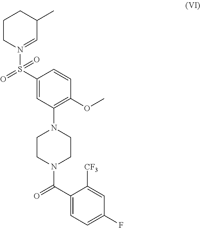
or a pharmaceutically acceptable salt thereof.

or a pharmaceutically acceptable salt thereof.

or a pharmaceutically acceptable salt thereof.

or a pharmaceutically acceptable salt thereof.

or a pharmaceutically acceptable salt thereof.

or a pharmaceutically acceptable salt thereof.

or a pharmaceutically acceptable salt thereof.

or a pharmaceutically acceptable salt thereof.

or a pharmaceutically acceptable salt thereof.

wherein R1 is a cyclic or acyclic amino group, or a pharmaceutically acceptable salt thereof. Non-limiting examples of R1 groups include dialkyl amino, piperidinyl, substituted piperidinyl, piperazinyl; substituted piperazinyl; tetrahydroisoquinolinyl; and substituted tetrahydroisoquinolinyl.

compound 290A11

compound 319H6

and compound 375F4

Detection of GDNF Mimetics Using Ret-ELISA Assay.

and compound 74609 has a structure

Detection of GDNF Mimetics Using Ret Phosphorylation Assay by Immunoprecipitation and Western Blotting.
| TABLE | |||||
| % | |||||
| % TH+ | Cytoprotection | ||||
| Treatment | Dose (μM) | Neurons | vs controls | ||
| DMSO | 0 | 55 | 0 | ||
| 319H6 | 0.1 | 59 | 9 | ||
| | 1 | 70 | 33 | ||
| 319H6 | 10 | 71 | 36 | ||
| BDNF* | 10 | 77 | 49 | ||
| *BDNF concentration is in ng/mL | |||||
Assessment of GDNF=/ARTN-Mimetics on Cell Viability
- Airaksinen, and Saarma. 2002. The GDNF family: signalling, biological functions and therapeutic value. Nat Rev Neurosci. 3:383-94.
- Airavaara, et al. 2004. Increased extracellular dopamine concentrations and FosB/DeltaFosB expression in striatal brain areas of heterozygous GDNF knockout mice. Eur J. Neurosci. 20:2336-44.
- Baloh, et al. 2000. Functional mapping of receptor specific city domains of glial cell line-derived neurotrophic factor (GDNF) family ligands and production of GFRα1 RET-specific agonists. J Biol. Chem. 275:3412-20.
- Bespalov and Saarma. 2007. GDNF family receptor complexes are emerging drug targets. Trends Pharmacol Sci. 28:68-74.
- Enslen, et al. 1996. Regulation of mitogen-activated protein kinases by a calcium/calmodulin-dependent protein kinase cascade. Proceedings of the National Academy of Sciences of the United States of America. 93:10803.
- Garces, et al. 2001. Responsiveness to neurturin of subpopulations of embryonic rat spinal motoneuron does not correlate with expression of GFRα1 or GFRα2. Dev Dyn. 220:189-97.
- Gardell, et al. 2003. Multiple actions of systemic artemin in experimental neuropathy. Nat. Med. 9:1383-9.
- Gill, et al. 2003. Direct brain infusion of glial cell line-derived neurotrophic factor in Parkinson disease. Nat. Med. 9:589-95.
- Golden, et al. 2003. Neurturin and persephin promote the survival of embryonic basal forebrain cholinergic neurons in vitro. Exp Neurol. 184:447-55.
- He, et al. 2005. Glial cell line-derived neurotrophic factor mediates the desirable actions of the anti-addiction drug ibogaine against alcohol consumption. J. Neurosci. 25:619-28.
- Henderson, et al. 1994. GDNF: a potent survival factor for motoneurons present in peripheral nerve and muscle. Science. 266:1062-4.
- Katritzky, et al. 2008. Novel computational models for predicting dopamine interactions. Experimental Neurology. 211:150-171.
- Knowles, et al. 2006. Structure and chemical inhibition of the RET tyrosine kinase domain. J Biol. Chem. 281:33577-87.
- Lang, et al. 2006. Randomized controlled trial of intraputamenal glial cell line-derived neurotrophic factor infusion in Parkinson disease. Ann Neurol. 59:459-66.
- Leppänen, et al. 2004. The structure of
GFRα1 domain 3 reveals new insights into GDNF binding and RET activation. EMBO J. 23:1452-62. - Lin, et al. 1993. GDNF: a glial cell line-derived neurotrophic factor for midbrain dopaminergic neurons. Science. 260:1130-2.
- Lindholm, et al. 2007. Novel neurotrophic factor CDNF protects and rescues midbrain dopamine neurons in vivo. Nature. 448:73-7.
- Longo, et al., Methods of facilitating neural cell survival using non-peptide and peptide BDNF neurotrophin mimetics, US Patent Appl. 20070060526, Mar. 15, 2007.
- Marais, et al. 1993. The SRF accessory protein Elk-1 contains a growth factorregulated transcriptional activation domain. Cell. 73:381-93.
- Marks, et al. 2008. Safety and tolerability of intraputaminal delivery of CERE-120 (adeno-associated virus serotype 2-neurturin) to patients with idiopathic Parkinson's disease: an open-label, phase I trial. Lancet Neurol. 7:400-8.
- Meng, et al. 2001. Promotion of seminomatous tumors by targeted overexpression of glial cell line-derived neurotrophic factor in mouse testis. Cancer Res. 61:3267-71.
- Meng, et al. 2000. Regulation of cell fate decision of undifferentiated spermatogonia by GDNF. Science. 287:1489-93.
- Messer, et al. 2000. Role for GDNF in biochemical and behavioral adaptations to drugs of abuse. Neuron. 26:247-57.
- Mijatovic, et al. 2007. Constitutive Ret activity in knock-in multiple endocrine neoplasia type B mice induces profound elevation of brain dopamine concentration via enhanced synthesis and increases the number of TH-positive cells in the substantia nigra. J. Neurosci. 27:4799-809.
- Parkash, et al. 2008. The structure of the glial cell line-derived neurotrophic factorcoreceptor complex: insights into RET signaling and heparin binding. J Biol. Chem. 283:35164-72.
- Pichel, et al. 1996. Defects in enteric innervation and kidney development in mice lacking GDNF. Nature. 382:73-6.
- Runeberg-Roos, et al. 2007. RET(MEN 2B) is active in the endoplasmic reticulum before reaching the cell surface. Oncogene. 26:7909-7915.
- Santoro, et al. 2004. Minireview: RET: normal and abnormal functions. Endocrinology. 145:5448-51.
- Sariola and Saarma. 2003. Novel functions and signalling pathways for GDNF. J Cell Sci. 116:3855-62.
- Sauer H & Oertel W H (1994) Progressive degeneration of nigrostriatal dopamine neurons following intrastriatal terminal lesions with 6-hydroxydopamine: a combined retrograde tracing and immunocytochemical study in the rat. Neuroscience, 59(2):401-15.
- Sautter, et al. 1998. Implants of polymer-encapsulated genetically modified cells releasing glial cell line-derived neurotrophic factor improve survival, growth, and function of fetal dopaminergic grafts. Exp Neurol. 149:230-6.
- Schinelli S, Zuddas A, Kopin L I, Barker J L, di Porzio U (1988) 1-Methyl-4-phenyl-1,2,3,6-tetrahydropyridine metabolism and 1-methyl-4-phenylpyridinium uptake in dissociated cell cultures from the embryonic mesencephalon. J Neurochem, 50(6):1900-7.
- Sild, et al. 2006. Open computing grid for molecular science and engineering. J Chem Inf Model. 46:953-9.
- Slevin, et al. 2005. Improvement of bilateral motor functions in patients with Parkinson disease through the unilateral intraputaminal infusion of glial cell line-derived neurotrophic factor. J. Neurosurg. 102:216-22.
- Tokugawa, et al. 2003. XIB4035, a novel nonpeptidyl small molecule agonist for GFRα-1. Neurochem Int. 42:81-6.
- Tomac, et al. 2002. Effects of cerebral ischemia in mice deficient in Persephin. Proc Natl Acad Sci USA. 99:9521-6.
- Tovar, et al. 2007. Comparison of 2D Fingerprint Methods for Multiple-Template Similarity Searching on Compound Activity Classes of Increasing Structural Diversity. ChemMedChem. 2:208-217.
- Wang, et al. 2006. Structure of artemin complexed with its receptor GFRα3: convergent recognition of glial cell line-derived neurotrophic factors. Structure. 14:1083-92.
- Wang, et al. 2008. Persistent restoration of sensory function by immediate or delayed systemic artemin after dorsal root injury. Nat. Neurosci. 11:488-96.
Claims (9)


Priority Applications (1)
| Application Number | Priority Date | Filing Date | Title |
|---|---|---|---|
| US13/514,906 US8901129B2 (en) | 2009-12-11 | 2010-12-13 | Methods of facilitating neural cell survival using GDNF family ligand (GFL) mimetics or RET signaling pathway activators |
Applications Claiming Priority (3)
| Application Number | Priority Date | Filing Date | Title |
|---|---|---|---|
| US28585809P | 2009-12-11 | 2009-12-11 | |
| US13/514,906 US8901129B2 (en) | 2009-12-11 | 2010-12-13 | Methods of facilitating neural cell survival using GDNF family ligand (GFL) mimetics or RET signaling pathway activators |
| PCT/EP2010/069535 WO2011070177A2 (en) | 2009-12-11 | 2010-12-13 | Methods of facilitating neural cell survival using gdnf family ligand (gfl) mimetics or ret signaling pathway activators |
Publications (2)
| Publication Number | Publication Date |
|---|---|
| US20130030180A1 US20130030180A1 (en) | 2013-01-31 |
| US8901129B2 true US8901129B2 (en) | 2014-12-02 |
Family
ID=43502911
Family Applications (1)
| Application Number | Title | Priority Date | Filing Date |
|---|---|---|---|
| US13/514,906 Active 2031-01-08 US8901129B2 (en) | 2009-12-11 | 2010-12-13 | Methods of facilitating neural cell survival using GDNF family ligand (GFL) mimetics or RET signaling pathway activators |
Country Status (9)
| Country | Link |
|---|---|
| US (1) | US8901129B2 (en) |
| EP (1) | EP2509953B1 (en) |
| JP (1) | JP2013513588A (en) |
| CN (2) | CN103180297A (en) |
| AU (1) | AU2010329847A1 (en) |
| BR (1) | BR112012014022A2 (en) |
| CA (1) | CA2783934A1 (en) |
| MX (1) | MX2012006580A (en) |
| WO (1) | WO2011070177A2 (en) |
Cited By (3)
| Publication number | Priority date | Publication date | Assignee | Title |
|---|---|---|---|---|
| WO2017195042A1 (en) | 2016-05-13 | 2017-11-16 | Instituto De Medicina Molecular | Methods of treating diseases associated with ilc3 cells |
| WO2022254255A1 (en) | 2021-06-03 | 2022-12-08 | Fundacao D. Anna De Sommerchampalimaud E Dr. | Neuro-mesenchyme units control ilc2 and obesity via a brain-adipose circuit |
| WO2023086894A1 (en) | 2021-11-12 | 2023-05-19 | Axent Biosciences Inc. | Methods for production of functional neurons |
Families Citing this family (9)
| Publication number | Priority date | Publication date | Assignee | Title |
|---|---|---|---|---|
| WO2014041179A1 (en) * | 2012-09-17 | 2014-03-20 | Chemedest Ltd. | Treatment of peripheral neuropathy using gfr(alpha)3 type receptor agonists |
| CN103146798B (en) * | 2013-02-25 | 2015-04-08 | 潍坊易思特生物科技有限公司 | Cytological method for assaying activity of rhGDNF (recombinant human Glial cell line-Derived Neurotrophic Factor) |
| CN105640955B (en) * | 2016-01-28 | 2019-12-24 | 昆明医科大学 | Novel application of Aminoquinol |
| CN105906608B (en) * | 2016-05-03 | 2018-10-19 | 中山大学 | 8- aminoquinolines-epiphysin is miscellaneous conjuncted and its pharmaceutical composition |
| CA3046289A1 (en) * | 2016-12-21 | 2018-06-28 | AlzeCure Pharma AB | Triazinetrione derivatives and their use as modulators of neurotrophin receptor and receptor tyrosine kinases |
| CN107337641B (en) * | 2017-07-01 | 2020-04-28 | 广东医科大学 | 4-flexible amino-2-arylvinyl quinoline derivative and preparation method and application thereof |
| AU2022251923A1 (en) | 2021-03-31 | 2023-11-16 | Cambridge Enterprise Limited | Therapeutic inhibitors of gdf15 signalling |
| WO2024023284A1 (en) | 2022-07-28 | 2024-02-01 | Genecode | Novel sulfonamides and their use as neuroprotective and/or neurorestorative agents |
| WO2024023287A1 (en) * | 2022-07-28 | 2024-02-01 | Genecode | [4-[5-(methylsulfonyl)-phenyl]piperazin-1-yl]-(phenyl)methanone derivatives and similar compounds with gfr alpha 1-ret activity as neuroprotective agents for the treatment of neurological disorders |
Citations (70)
| Publication number | Priority date | Publication date | Assignee | Title |
|---|---|---|---|---|
| US2531011A (en) | 1949-01-22 | 1950-11-21 | Sterling Drug Inc | 3-chloro-9-[2-(2-hydroxyethylamino)-ethylamino] acridine and method for its production |
| US3232945A (en) | 1962-08-13 | 1966-02-01 | S E Massengill Company | 7,8,9,10-tetrahalo-6h-cyclohepta-(b)-quinolines |
| US3458525A (en) | 1966-07-01 | 1969-07-29 | American Home Prod | Spiro(indoline-3,2'-thiazolidine)-2-one and derivatives |
| US4426330A (en) | 1981-07-20 | 1984-01-17 | Lipid Specialties, Inc. | Synthetic phospholipid compounds |
| US4534899A (en) | 1981-07-20 | 1985-08-13 | Lipid Specialties, Inc. | Synthetic phospholipid compounds |
| EP0268871A1 (en) | 1986-10-31 | 1988-06-01 | Sumitomo Pharmaceuticals Company, Limited | Quinoline derivatives |
| WO1988007851A1 (en) | 1987-04-06 | 1988-10-20 | Innovative Therapeutics Corporation | Liposome entrapped n-methyl-d-aspartate receptor blockers |
| WO1988007852A1 (en) | 1987-04-06 | 1988-10-20 | Innovative Therapeutics Corporation | Liposome entrapped magnesium |
| WO1989007938A1 (en) | 1988-02-26 | 1989-09-08 | Shashoua Victor E | Fatty acid-drug conjugate for delivery of the drug across the blood-brain barrier |
| US4902505A (en) | 1986-07-30 | 1990-02-20 | Alkermes | Chimeric peptides for neuropeptide delivery through the blood-brain barrier |
| US4933324A (en) | 1988-02-26 | 1990-06-12 | Shashoua Victor E | Fatty acid-neuroactive drug conjugate as a prodrug |
| EP0411534A2 (en) | 1989-07-31 | 1991-02-06 | Warner-Lambert Company | Derivatives of 1,2,3,4-tetrahydro-9-acridinamine |
| WO1991004014A1 (en) | 1989-09-21 | 1991-04-04 | Synergen, Inc. | Method for transporting compositions across the blood brain barrier |
| US5013556A (en) | 1989-10-20 | 1991-05-07 | Liposome Technology, Inc. | Liposomes with enhanced circulation time |
| EP0427636A2 (en) | 1989-11-08 | 1991-05-15 | Mitsubishi Chemical Corporation | 4-Acylaminopyridine derivatives |
| US5019400A (en) | 1989-05-01 | 1991-05-28 | Enzytech, Inc. | Very low temperature casting of controlled release microspheres |
| US5108921A (en) | 1989-04-03 | 1992-04-28 | Purdue Research Foundation | Method for enhanced transmembrane transport of exogenous molecules |
| US5109921A (en) | 1991-04-29 | 1992-05-05 | Halliburton Company | Controlled weak point for wireline cable |
| US5112863A (en) | 1989-12-05 | 1992-05-12 | Nippon Oil & Fats Co., Ltd. | Antipsychotic drug |
| US5112596A (en) | 1990-04-23 | 1992-05-12 | Alkermes, Inc. | Method for increasing blood-brain barrier permeability by administering a bradykinin agonist of blood-brain barrier permeability |
| US5154924A (en) | 1989-09-07 | 1992-10-13 | Alkermes, Inc. | Transferrin receptor specific antibody-neuropharmaceutical agent conjugates |
| US5182107A (en) | 1989-09-07 | 1993-01-26 | Alkermes, Inc. | Transferrin receptor specific antibody-neuropharmaceutical or diagnostic agent conjugates |
| WO1993010819A1 (en) | 1991-11-26 | 1993-06-10 | Alkermes, Inc. | Process for the preparation of transferrin receptor specific antibody-neuropharmaceutical or diagnostic agent conjugates |
| US5227170A (en) | 1989-06-22 | 1993-07-13 | Vestar, Inc. | Encapsulation process |
| US5260308A (en) | 1991-11-06 | 1993-11-09 | Mayo Foundation For Medical Education And Research | Method to increase permeability of the blood-nerve/brain barriers to proteins |
| US5264221A (en) | 1991-05-23 | 1993-11-23 | Mitsubishi Kasei Corporation | Drug-containing protein-bonded liposome |
| US5268164A (en) | 1990-04-23 | 1993-12-07 | Alkermes, Inc. | Increasing blood-brain barrier permeability with permeabilizer peptides |
| US5271961A (en) | 1989-11-06 | 1993-12-21 | Alkermes Controlled Therapeutics, Inc. | Method for producing protein microspheres |
| WO1994002178A1 (en) | 1992-07-27 | 1994-02-03 | The Government Of The United States Of America As Represented By The Secretary Of The Department Of Health And Human Services | Targeting of liposomes to the blood-brain barrier |
| US5284876A (en) | 1988-02-26 | 1994-02-08 | Neuromedica, Inc. | Method of treating tardive dyskinesia using dopaminergic agents of prodrugs of therapeutic agents |
| US5354844A (en) | 1989-03-16 | 1994-10-11 | Boehringer Ingelheim International Gmbh | Protein-polycation conjugates |
| US5356633A (en) | 1989-10-20 | 1994-10-18 | Liposome Technology, Inc. | Method of treatment of inflamed tissues |
| WO1995002421A1 (en) | 1993-07-16 | 1995-01-26 | Alkermes, Inc. | Transferrin receptor specific ligand-neuropharmaceutical agent fusion proteins |
| US5395619A (en) | 1993-03-03 | 1995-03-07 | Liposome Technology, Inc. | Lipid-polymer conjugates and liposomes |
| US5413797A (en) | 1992-03-12 | 1995-05-09 | Alkermes Controlled Therapeutics, Inc. | Controlled release ACTH containing microspheres |
| US5417978A (en) | 1993-07-29 | 1995-05-23 | Board Of Regents, The University Of Texas System | Liposomal antisense methyl phosphonate oligonucleotides and methods for their preparation and use |
| US5459127A (en) | 1990-04-19 | 1995-10-17 | Vical, Inc. | Cationic lipids for intracellular delivery of biologically active molecules |
| US5462854A (en) | 1993-04-19 | 1995-10-31 | Beckman Instruments, Inc. | Inverse linkage oligonucleotides for chemical and enzymatic processes |
| US5469854A (en) | 1989-12-22 | 1995-11-28 | Imarx Pharmaceutical Corp. | Methods of preparing gas-filled liposomes |
| US5512295A (en) | 1994-11-10 | 1996-04-30 | The Board Of Trustees Of The Leland Stanford Junior University | Synthetic liposomes for enhanced uptake and delivery |
| US5521291A (en) | 1991-09-30 | 1996-05-28 | Boehringer Ingelheim International, Gmbh | Conjugates for introducing nucleic acid into higher eucaryotic cells |
| US5527528A (en) | 1989-10-20 | 1996-06-18 | Sequus Pharmaceuticals, Inc. | Solid-tumor treatment method |
| US5534259A (en) | 1993-07-08 | 1996-07-09 | Liposome Technology, Inc. | Polymer compound and coated particle composition |
| US5543152A (en) | 1994-06-20 | 1996-08-06 | Inex Pharmaceuticals Corporation | Sphingosomes for enhanced drug delivery |
| US5543158A (en) | 1993-07-23 | 1996-08-06 | Massachusetts Institute Of Technology | Biodegradable injectable nanoparticles |
| US5547932A (en) | 1991-09-30 | 1996-08-20 | Boehringer Ingelheim International Gmbh | Composition for introducing nucleic acid complexes into higher eucaryotic cells |
| US5556948A (en) | 1993-01-22 | 1996-09-17 | Mitsubishi Chemical Corporation | Phospholipid derivatized with PEG bifunctional linker and liposome containing it |
| US5580575A (en) | 1989-12-22 | 1996-12-03 | Imarx Pharmaceutical Corp. | Therapeutic drug delivery systems |
| US5583020A (en) | 1992-11-24 | 1996-12-10 | Ribozyme Pharmaceuticals, Inc. | Permeability enhancers for negatively charged polynucleotides |
| US5591721A (en) | 1994-10-25 | 1997-01-07 | Hybridon, Inc. | Method of down-regulating gene expression |
| US5595756A (en) | 1993-12-22 | 1997-01-21 | Inex Pharmaceuticals Corporation | Liposomal compositions for enhanced retention of bioactive agents |
| US6235769B1 (en) | 1997-07-03 | 2001-05-22 | Sugen, Inc. | Methods of preventing and treating neurological disorders with compounds that modulate the function of the C-RET receptor protein tyrosine kinase |
| WO2001064645A2 (en) | 2000-03-01 | 2001-09-07 | Orion Corporation | Derivatives of quinoline as alpha-2 antagonists |
| US6287860B1 (en) | 2000-01-20 | 2001-09-11 | Isis Pharmaceuticals, Inc. | Antisense inhibition of MEKK2 expression |
| US20030027780A1 (en) | 1999-02-23 | 2003-02-06 | Hardee Gregory E. | Multiparticulate formulation |
| WO2004004660A2 (en) | 2002-07-09 | 2004-01-15 | Alpha Research Group, L.L.C. | Compositions and methods for the treatment of parkinson’s disease and tardive dyskinesias |
| US20040034019A1 (en) | 2002-08-08 | 2004-02-19 | Ronald Tomlinson | Piperazine and piperidine derivatives |
| US20040142997A1 (en) | 2002-10-09 | 2004-07-22 | Chen Yuhpyng L. | Pyrazole compounds for treatment of neurodegenerative disorders |
| US6887906B1 (en) | 1997-07-01 | 2005-05-03 | Isispharmaceuticals, Inc. | Compositions and methods for the delivery of oligonucleotides via the alimentary canal |
| US20050282780A1 (en) | 2004-05-20 | 2005-12-22 | Richard Labaudiniere | Compounds, compositions and methods for stabilizing transthyretin and inhibiting transthyretin misfolding |
| WO2006094237A2 (en) | 2005-03-03 | 2006-09-08 | Sirtris Pharmaceuticals, Inc. | Acridine and quinoline dervatives as sirtuin modulators |
| WO2006094843A1 (en) | 2005-03-11 | 2006-09-14 | Glaxo Group Limited | Piperazine derivatives as glyt1 inhibitors |
| WO2006130493A2 (en) | 2005-05-31 | 2006-12-07 | Vertex Pharmaceuticals Incorporated | Heterocycles useful as modulators of ion channels |
| US20070060526A1 (en) | 2005-06-08 | 2007-03-15 | Longo Frank M | Methods of facilitating neural cell survival using non-peptide and peptide BDNF neurotrophin mimetics |
| WO2008010984A2 (en) | 2006-07-17 | 2008-01-24 | The Trustees Of The University Of Pennsylvania | Acridine activation of p53 and uses thereof |
| US20080176308A1 (en) | 2007-01-22 | 2008-07-24 | Biotechnology Research Corporation Limited | Heterodimers and Methods of Using Them |
| US20080234267A1 (en) | 2007-03-20 | 2008-09-25 | Karen Elizabeth Lackey | Compounds and Methods of Treatment |
| WO2009024611A2 (en) | 2007-08-22 | 2009-02-26 | Abbott Gmbh & Co. Kg | 4-benzylaminoquinolines, pharmaceutical compositions containing them and their use |
| US20090203735A1 (en) | 2008-02-12 | 2009-08-13 | Gabriel Corfas | Treatments for neuropathy |
| WO2009148659A2 (en) | 2008-03-05 | 2009-12-10 | Georgetown University | Antimalarial quinolines and methods of use thereof |
Family Cites Families (4)
| Publication number | Priority date | Publication date | Assignee | Title |
|---|---|---|---|---|
| EP1339702A1 (en) * | 2000-03-15 | 2003-09-03 | Warner-Lambert Company | 5-amide substituted diarylamines as mek inhibitors |
| EP1667983A4 (en) * | 2003-09-23 | 2010-07-21 | Merck Sharp & Dohme | Pyrazole modulators of metabotropic glutamate receptors |
| DE202005008426U1 (en) | 2005-05-31 | 2005-09-15 | Ricon Sieb Und Foerdertechnik | Conveyor unit has conveyor belts provided with recesses on inner facing sides for fitting of side edges of conveying apron, whereby conveying apron is connected via edges to conveyor belts in region of recesses |
| US10867398B2 (en) | 2017-11-21 | 2020-12-15 | Reliance Core Consulting LLC | Methods, systems, apparatuses and devices for facilitating motion analysis in an environment |
-
2010
- 2010-12-13 AU AU2010329847A patent/AU2010329847A1/en not_active Abandoned
- 2010-12-13 CN CN2010800637467A patent/CN103180297A/en active Pending
- 2010-12-13 CA CA2783934A patent/CA2783934A1/en not_active Abandoned
- 2010-12-13 US US13/514,906 patent/US8901129B2/en active Active
- 2010-12-13 EP EP10790954.1A patent/EP2509953B1/en not_active Not-in-force
- 2010-12-13 CN CN201410513670.3A patent/CN104529945A/en active Pending
- 2010-12-13 BR BR112012014022A patent/BR112012014022A2/en not_active IP Right Cessation
- 2010-12-13 JP JP2012542580A patent/JP2013513588A/en active Pending
- 2010-12-13 WO PCT/EP2010/069535 patent/WO2011070177A2/en active Application Filing
- 2010-12-13 MX MX2012006580A patent/MX2012006580A/en not_active Application Discontinuation
Patent Citations (72)
| Publication number | Priority date | Publication date | Assignee | Title |
|---|---|---|---|---|
| US2531011A (en) | 1949-01-22 | 1950-11-21 | Sterling Drug Inc | 3-chloro-9-[2-(2-hydroxyethylamino)-ethylamino] acridine and method for its production |
| US3232945A (en) | 1962-08-13 | 1966-02-01 | S E Massengill Company | 7,8,9,10-tetrahalo-6h-cyclohepta-(b)-quinolines |
| US3458525A (en) | 1966-07-01 | 1969-07-29 | American Home Prod | Spiro(indoline-3,2'-thiazolidine)-2-one and derivatives |
| US4426330A (en) | 1981-07-20 | 1984-01-17 | Lipid Specialties, Inc. | Synthetic phospholipid compounds |
| US4534899A (en) | 1981-07-20 | 1985-08-13 | Lipid Specialties, Inc. | Synthetic phospholipid compounds |
| US4902505A (en) | 1986-07-30 | 1990-02-20 | Alkermes | Chimeric peptides for neuropeptide delivery through the blood-brain barrier |
| EP0268871A1 (en) | 1986-10-31 | 1988-06-01 | Sumitomo Pharmaceuticals Company, Limited | Quinoline derivatives |
| WO1988007851A1 (en) | 1987-04-06 | 1988-10-20 | Innovative Therapeutics Corporation | Liposome entrapped n-methyl-d-aspartate receptor blockers |
| WO1988007852A1 (en) | 1987-04-06 | 1988-10-20 | Innovative Therapeutics Corporation | Liposome entrapped magnesium |
| WO1989007938A1 (en) | 1988-02-26 | 1989-09-08 | Shashoua Victor E | Fatty acid-drug conjugate for delivery of the drug across the blood-brain barrier |
| US4933324A (en) | 1988-02-26 | 1990-06-12 | Shashoua Victor E | Fatty acid-neuroactive drug conjugate as a prodrug |
| US5284876A (en) | 1988-02-26 | 1994-02-08 | Neuromedica, Inc. | Method of treating tardive dyskinesia using dopaminergic agents of prodrugs of therapeutic agents |
| US5354844A (en) | 1989-03-16 | 1994-10-11 | Boehringer Ingelheim International Gmbh | Protein-polycation conjugates |
| US5108921A (en) | 1989-04-03 | 1992-04-28 | Purdue Research Foundation | Method for enhanced transmembrane transport of exogenous molecules |
| US5416016A (en) | 1989-04-03 | 1995-05-16 | Purdue Research Foundation | Method for enhancing transmembrane transport of exogenous molecules |
| US5019400A (en) | 1989-05-01 | 1991-05-28 | Enzytech, Inc. | Very low temperature casting of controlled release microspheres |
| US5227170A (en) | 1989-06-22 | 1993-07-13 | Vestar, Inc. | Encapsulation process |
| EP0411534A2 (en) | 1989-07-31 | 1991-02-06 | Warner-Lambert Company | Derivatives of 1,2,3,4-tetrahydro-9-acridinamine |
| US5154924A (en) | 1989-09-07 | 1992-10-13 | Alkermes, Inc. | Transferrin receptor specific antibody-neuropharmaceutical agent conjugates |
| US5182107A (en) | 1989-09-07 | 1993-01-26 | Alkermes, Inc. | Transferrin receptor specific antibody-neuropharmaceutical or diagnostic agent conjugates |
| WO1991004014A1 (en) | 1989-09-21 | 1991-04-04 | Synergen, Inc. | Method for transporting compositions across the blood brain barrier |
| US5527528A (en) | 1989-10-20 | 1996-06-18 | Sequus Pharmaceuticals, Inc. | Solid-tumor treatment method |
| US5213804A (en) | 1989-10-20 | 1993-05-25 | Liposome Technology, Inc. | Solid tumor treatment method and composition |
| US5356633A (en) | 1989-10-20 | 1994-10-18 | Liposome Technology, Inc. | Method of treatment of inflamed tissues |
| US5013556A (en) | 1989-10-20 | 1991-05-07 | Liposome Technology, Inc. | Liposomes with enhanced circulation time |
| US5271961A (en) | 1989-11-06 | 1993-12-21 | Alkermes Controlled Therapeutics, Inc. | Method for producing protein microspheres |
| EP0427636A2 (en) | 1989-11-08 | 1991-05-15 | Mitsubishi Chemical Corporation | 4-Acylaminopyridine derivatives |
| US5112863A (en) | 1989-12-05 | 1992-05-12 | Nippon Oil & Fats Co., Ltd. | Antipsychotic drug |
| US5580575A (en) | 1989-12-22 | 1996-12-03 | Imarx Pharmaceutical Corp. | Therapeutic drug delivery systems |
| US5469854A (en) | 1989-12-22 | 1995-11-28 | Imarx Pharmaceutical Corp. | Methods of preparing gas-filled liposomes |
| US5459127A (en) | 1990-04-19 | 1995-10-17 | Vical, Inc. | Cationic lipids for intracellular delivery of biologically active molecules |
| US5268164A (en) | 1990-04-23 | 1993-12-07 | Alkermes, Inc. | Increasing blood-brain barrier permeability with permeabilizer peptides |
| US5112596A (en) | 1990-04-23 | 1992-05-12 | Alkermes, Inc. | Method for increasing blood-brain barrier permeability by administering a bradykinin agonist of blood-brain barrier permeability |
| US5109921A (en) | 1991-04-29 | 1992-05-05 | Halliburton Company | Controlled weak point for wireline cable |
| US5264221A (en) | 1991-05-23 | 1993-11-23 | Mitsubishi Kasei Corporation | Drug-containing protein-bonded liposome |
| US5547932A (en) | 1991-09-30 | 1996-08-20 | Boehringer Ingelheim International Gmbh | Composition for introducing nucleic acid complexes into higher eucaryotic cells |
| US5521291A (en) | 1991-09-30 | 1996-05-28 | Boehringer Ingelheim International, Gmbh | Conjugates for introducing nucleic acid into higher eucaryotic cells |
| US5260308A (en) | 1991-11-06 | 1993-11-09 | Mayo Foundation For Medical Education And Research | Method to increase permeability of the blood-nerve/brain barriers to proteins |
| WO1993010819A1 (en) | 1991-11-26 | 1993-06-10 | Alkermes, Inc. | Process for the preparation of transferrin receptor specific antibody-neuropharmaceutical or diagnostic agent conjugates |
| US5413797A (en) | 1992-03-12 | 1995-05-09 | Alkermes Controlled Therapeutics, Inc. | Controlled release ACTH containing microspheres |
| WO1994002178A1 (en) | 1992-07-27 | 1994-02-03 | The Government Of The United States Of America As Represented By The Secretary Of The Department Of Health And Human Services | Targeting of liposomes to the blood-brain barrier |
| US5583020A (en) | 1992-11-24 | 1996-12-10 | Ribozyme Pharmaceuticals, Inc. | Permeability enhancers for negatively charged polynucleotides |
| US5556948A (en) | 1993-01-22 | 1996-09-17 | Mitsubishi Chemical Corporation | Phospholipid derivatized with PEG bifunctional linker and liposome containing it |
| US5395619A (en) | 1993-03-03 | 1995-03-07 | Liposome Technology, Inc. | Lipid-polymer conjugates and liposomes |
| US5462854A (en) | 1993-04-19 | 1995-10-31 | Beckman Instruments, Inc. | Inverse linkage oligonucleotides for chemical and enzymatic processes |
| US5534259A (en) | 1993-07-08 | 1996-07-09 | Liposome Technology, Inc. | Polymer compound and coated particle composition |
| WO1995002421A1 (en) | 1993-07-16 | 1995-01-26 | Alkermes, Inc. | Transferrin receptor specific ligand-neuropharmaceutical agent fusion proteins |
| US5543158A (en) | 1993-07-23 | 1996-08-06 | Massachusetts Institute Of Technology | Biodegradable injectable nanoparticles |
| US5417978A (en) | 1993-07-29 | 1995-05-23 | Board Of Regents, The University Of Texas System | Liposomal antisense methyl phosphonate oligonucleotides and methods for their preparation and use |
| US5595756A (en) | 1993-12-22 | 1997-01-21 | Inex Pharmaceuticals Corporation | Liposomal compositions for enhanced retention of bioactive agents |
| US5543152A (en) | 1994-06-20 | 1996-08-06 | Inex Pharmaceuticals Corporation | Sphingosomes for enhanced drug delivery |
| US5591721A (en) | 1994-10-25 | 1997-01-07 | Hybridon, Inc. | Method of down-regulating gene expression |
| US5512295A (en) | 1994-11-10 | 1996-04-30 | The Board Of Trustees Of The Leland Stanford Junior University | Synthetic liposomes for enhanced uptake and delivery |
| US6887906B1 (en) | 1997-07-01 | 2005-05-03 | Isispharmaceuticals, Inc. | Compositions and methods for the delivery of oligonucleotides via the alimentary canal |
| US6235769B1 (en) | 1997-07-03 | 2001-05-22 | Sugen, Inc. | Methods of preventing and treating neurological disorders with compounds that modulate the function of the C-RET receptor protein tyrosine kinase |
| US20030027780A1 (en) | 1999-02-23 | 2003-02-06 | Hardee Gregory E. | Multiparticulate formulation |
| US6287860B1 (en) | 2000-01-20 | 2001-09-11 | Isis Pharmaceuticals, Inc. | Antisense inhibition of MEKK2 expression |
| WO2001064645A2 (en) | 2000-03-01 | 2001-09-07 | Orion Corporation | Derivatives of quinoline as alpha-2 antagonists |
| WO2004004660A2 (en) | 2002-07-09 | 2004-01-15 | Alpha Research Group, L.L.C. | Compositions and methods for the treatment of parkinson’s disease and tardive dyskinesias |
| US20040034019A1 (en) | 2002-08-08 | 2004-02-19 | Ronald Tomlinson | Piperazine and piperidine derivatives |
| US20040142997A1 (en) | 2002-10-09 | 2004-07-22 | Chen Yuhpyng L. | Pyrazole compounds for treatment of neurodegenerative disorders |
| US20050282780A1 (en) | 2004-05-20 | 2005-12-22 | Richard Labaudiniere | Compounds, compositions and methods for stabilizing transthyretin and inhibiting transthyretin misfolding |
| WO2006094237A2 (en) | 2005-03-03 | 2006-09-08 | Sirtris Pharmaceuticals, Inc. | Acridine and quinoline dervatives as sirtuin modulators |
| WO2006094843A1 (en) | 2005-03-11 | 2006-09-14 | Glaxo Group Limited | Piperazine derivatives as glyt1 inhibitors |
| WO2006130493A2 (en) | 2005-05-31 | 2006-12-07 | Vertex Pharmaceuticals Incorporated | Heterocycles useful as modulators of ion channels |
| US20070060526A1 (en) | 2005-06-08 | 2007-03-15 | Longo Frank M | Methods of facilitating neural cell survival using non-peptide and peptide BDNF neurotrophin mimetics |
| WO2008010984A2 (en) | 2006-07-17 | 2008-01-24 | The Trustees Of The University Of Pennsylvania | Acridine activation of p53 and uses thereof |
| US20080176308A1 (en) | 2007-01-22 | 2008-07-24 | Biotechnology Research Corporation Limited | Heterodimers and Methods of Using Them |
| US20080234267A1 (en) | 2007-03-20 | 2008-09-25 | Karen Elizabeth Lackey | Compounds and Methods of Treatment |
| WO2009024611A2 (en) | 2007-08-22 | 2009-02-26 | Abbott Gmbh & Co. Kg | 4-benzylaminoquinolines, pharmaceutical compositions containing them and their use |
| US20090203735A1 (en) | 2008-02-12 | 2009-08-13 | Gabriel Corfas | Treatments for neuropathy |
| WO2009148659A2 (en) | 2008-03-05 | 2009-12-10 | Georgetown University | Antimalarial quinolines and methods of use thereof |
Non-Patent Citations (54)
| Title |
|---|
| Abramochkin et al., Synthesis of n-substituted 4-amino-2,3-pentamethylenequinolines, Pharm. Chem. J., 4(7):372-4 (1970). |
| Airaksinen et al., The GDNF family: signalling, biological functions and therapeutic value, Nat. Rev. Neurosci., 3(5):383-94 (2002). |
| Alonso et al., Donepezil-tacrine hybrid related derivatives as new dual binding site inhibitors of AChE, Bioorg. Medicinal Chem., 13:6588-97 (2005). |
| Baloh et al., Functional mapping of receptor specificity domains of glial cell line-derived neurotrophic factor (GDNF) family ligands and production of GFRalpha1 RET-specific agonists, J. Biol. Chem., 275(5):3412-20 (2000). |
| Bespalov et al., GDNF family receptor complexes are emerging drug targets, Trends in Pharmacol. Sci., 28(2): 68-74 (2007). |
| Bindra et al., Synthesis, pharmacological activities & physico-chemical properties of 4-(substituted amino/N4-arylpiperazinyl/aminocarbonyl)-2, 3-polymethylenequinolines, Ind. J. Chem., 26B:318-29 (1987). |
| Cope et al., Synthesis and SAR study of acridine, 2-methylquinoline and 2-phenylquinazoline analogues as anti-prion agents, Eur. J. Med. Chem., 41(10):1124-43 (2006). |
| Database Accession No. 1008228-16-7 (Mar. 16, 2008). |
| Database Accession Nos. 00174789656, 0010560146, 0011264857, 0009961513, 0014083641, 0009749723, 0016006950, 0010558361, 0009557186, 0011975036, 0012387641, 0009828212, 0010568933, 0012521524, 0015757439 (Jan. 4, 2011). |
| Database Accession Nos. RN: 924577-01-5, 924539-62-8, 924551-57-5, 924564-95-4, 924576-91-0, 924553-86-6, 924537-98-4 (Mar. 2, 2007). |
| De Souza et al., Synthesis and in vitro antitubercular activity of a series of quinoline derivatives, Bioorg. Med. Chem., 17(4):1474-80 (2009). |
| Doucet-Personeni et al., A structure-based design approach to the development of novel, reversible AChE inhibitors, J. Med. Chem., 44:3203-15 (2001). |
| Enslen et al., Regulation of mitogen-activated protein kinases by a calcium/calmodulin-dependent protein kinase cascade, Proc. Natl. Acad. Sci. USA, 93(20):10803-8 (1996). |
| Garces et al., Responsiveness to neurturin of subpopulations of embryonic rat spinal motoneuron does not correlate with expression of GFR alpha 1 or GFR alpha 2, Dev. Dyn., 220(3):189-97 (2001). |
| Gardell et al., Multiple actions of systemic artemin in experimental neuropathy, Nat. Med., 9(11):1383-9 (2003). |
| Gill et al., Direct brain infusion of glial cell line-derived neurotrophic factor in Parkinson disease, Nat. Med., 9(5):589-95 (2003). |
| Girgis et al., Phosphorous pentoxide in organic synthesis; XVII. A new synthesis of 4-arylamino-2,3-polymethylenequinolines, Synthesis, pp. 547-548 (1985). |
| He et al., Glial cell line-derived neurotrophic factor mediates the desirable actions of the anti-addiction drug ibogaine against alcohol consumption, J. Neurosci., 25(3):619-28 (2005). |
| Henderson et al., GDNF: a potent survival factor for motoneurons present in peripheral nerve and muscle, Science, 266(5187):1062-4 (1994). |
| International Preliminary Report on Patentability for corresponding international application No. PCT/EP2010/069535, completion date May 2, 2012. |
| International Search Report and Written Opinion for corresponding international application No. PCT/EP2010/069535, mailing date Jul. 8, 2011. |
| Jurvansuu et al., Recent inventions on receptor tyrosine kinase RET modulation, Recent Patents on Biotechnology, 2:47-54 (2008). |
| Knowles et al., Structure and chemical inhibition of the RET tyrosine kinase domain, J. Biol. Chem., 281(44):33577-87 (2006). |
| Leppanen et al., The structure of GFRalpha1 domain 3 reveals new insights into GDNF binding and RET activation, EMBO J., 23(7):1452-62 (2004). |
| Lin et al., GDNF: a glial cell line-derived neurotrophic factor for midbrain dopaminergic neurons, Science, 260(5111):1130-2 (1993). |
| Lindholm et al., Novel neurotrophic factor CDNF protects and rescues midbrain dopamine neurons in vivo, Nature, 448(7149):73-7 (2007). |
| Marks et al., Safety and tolerability of intraputaminal delivery of CERE-120 (adeno-associated virus serotype 2-neurturin) to patients with idiopathic Parkinson's disease: an open-label, phase I trial, Lancet Neurol., 7(5):400-8 (2008). |
| McKenna et al., Novel tacrine analogues for potential use against Alzheimer's Disease: potent and selective acetylcholinesterase inhibitors and 5-HT uptake inhibitors, J. Med. Chem., 40:3518-23 (1997). |
| Meng et al., Promotion of seminomatous tumors by targeted overexpression of glial cell line-derived neurotrophic factor in mouse testis, Cancer Res., 61(8):3267-71 (2001). |
| Meng et al., Regulation of cell fate decision of undifferentiated spermatogonia by GDNF, Science, 287(5457):1489-93 (2000). |
| Messer et al., Role for GDNF in biochemical and behavioral adaptations to drugs of abuse, Neuron, 26(1):247-57 (2000). |
| Mijatovic et al., Constitutive Ret activity in knock-in multiple endocrine neoplasia type B mice induces profound elevation of brain dopamine concentration via enhanced synthesis and increases the number of TH-positive cells in the substantia nigra, J. Neurosci., 27(18):4799-809 (2007). |
| Nunes et al., Transthyretin knockout mice display decreased susceptibility to AMPA-induced neurodegeneration, Neurochem. Intl., 55:454-7 (2009). |
| Parkash et al., The structure of the glial cell line-derived neurotrophic factor-coreceptor complex: insights into RET signaling and heparin binding, J. Biol. Chem., 283(50):35164-72 (2008). |
| Pichel et al., Defects in enteric innervation and kidney development in mice lacking GDNF, Nature, 382(6586):73-6 (1996). |
| Pinard et al., 4-aminoquinolines as a novel class of NR1/2B subtype selective NMDA receptor antagonists, Bioorg. Medicinal Chem. Lett., 12:2615-9 (2002). |
| Recanatini et al., SAR of 9-amino-1,2,3,4-tetrahydroacridine-based acetylcholinesterase inhibitors: synthesis, enzyme inhibitory activity, QSAR, and structure-based CoMFA of tacrine analogues, J. Med. Chem., 43:2007-18 (2000). |
| Runeberg-Roos et al., RET(MEN 2B) is active in the endoplasmic reticulum before reaching the cell surface, Oncogene, 26(57):7909-15 (2007). |
| Santoro et al., Minireview: RET: normal and abnormal functions, Endocrinology, 145(12):5448-51 (2004). |
| Sariola et al., Novel functions and signalling pathways for GDNF, J. Cell Sci., 116(Pt. 19):3855-62 (2003). |
| Sariola, 2003, Journal of Cell Science, vol. 116, p. 3855-3862. * |
| Shepard et al., Nuclear substituted 9-(4′-diethylamino-1′-methylbutylamino)-acridines, J. Am. Chem. Soc., 70(5):1979-80 (1948). |
| Shepard et al., Nuclear substituted 9-(4'-diethylamino-1'-methylbutylamino)-acridines, J. Am. Chem. Soc., 70(5):1979-80 (1948). |
| Singh et al., Antimalarials. 7-Chloro-4-(substituted amino)quinolines, J. Med. Chem., 14(4):283-6 (1971). |
| Steinberg et al., A hydrophobic binding site in acetylcholinesterase, J. Med. Chem., 18(1):1056-61 (1975). |
| STN Database Accession No. RN: 924811-53-0, 924789-13-09, 924762-99-2, 624759-42-2, 924756-06-9, 924740-23-8, 924730-35-8 (Mar. 5, 2007). |
| Tokugawa et al., XIB4035, a novel nonpeptidyl small molecule agonist for GFRalpha-1, Neurochem. Int., 42:81-6 (2003). |
| Tokugawa et al., XIB4035, a novel nonpeptidyl small molecule agonist for GFRα-1, Neurochem. Int., 42:81-6 (2003). |
| Tomac et al., Effects of cerebral ischemia in mice deficient in Persephin, Proc. Natl. Acad. Sci. USA, 99(14):9521-6 (2002). |
| Tomosaka et al., Enhancement of mutagenic activity of 9-aminoacridine by introducing a nitro group into the molecule, Biosci. Biotech. Biochem., 58(8):1420-3 (1994). |
| Tomosaka et al., The effects of substituents introduced into 0-aminoacridine on frameshift mutagenicity and DNA binding affinity, Biosci. Biotech. Biochem., 71(7):1121-5 (1997). |
| Vennerstrom et al., Bisquinolines. 1. N,N-bis(7-chloroquinolin-4-yl)alkanediamines with potential against chloroquine-resistant malaria, J. Med. Chem., 35(11):2129-34 (1992). |
| Wang et al., Persistent restoration of sensory function by immediate or delayed systemic artemin after dorsal root injury, Nat. Neurosci., 11(4):488-96 (2008). |
| Wang et al., Structure of artemin complexed with its receptor GFRalpha3: convergent recognition of glial cell line-derived neurotrophic factors, Strucutre, 14(6):1083-92 (2006). |
Cited By (3)
| Publication number | Priority date | Publication date | Assignee | Title |
|---|---|---|---|---|
| WO2017195042A1 (en) | 2016-05-13 | 2017-11-16 | Instituto De Medicina Molecular | Methods of treating diseases associated with ilc3 cells |
| WO2022254255A1 (en) | 2021-06-03 | 2022-12-08 | Fundacao D. Anna De Sommerchampalimaud E Dr. | Neuro-mesenchyme units control ilc2 and obesity via a brain-adipose circuit |
| WO2023086894A1 (en) | 2021-11-12 | 2023-05-19 | Axent Biosciences Inc. | Methods for production of functional neurons |
Also Published As
| Publication number | Publication date |
|---|---|
| JP2013513588A (en) | 2013-04-22 |
| EP2509953B1 (en) | 2016-03-30 |
| MX2012006580A (en) | 2012-09-28 |
| BR112012014022A2 (en) | 2017-04-04 |
| WO2011070177A2 (en) | 2011-06-16 |
| CA2783934A1 (en) | 2011-06-16 |
| AU2010329847A1 (en) | 2012-07-26 |
| US20130030180A1 (en) | 2013-01-31 |
| CN103180297A (en) | 2013-06-26 |
| CN104529945A (en) | 2015-04-22 |
| WO2011070177A3 (en) | 2011-09-01 |
| EP2509953A2 (en) | 2012-10-17 |
Similar Documents
| Publication | Publication Date | Title |
|---|---|---|
| US8901129B2 (en) | Methods of facilitating neural cell survival using GDNF family ligand (GFL) mimetics or RET signaling pathway activators | |
| Zhang et al. | Modulation of brain cation-Cl− cotransport via the SPAK kinase inhibitor ZT-1a | |
| US7053116B2 (en) | Methods for protecting cells from amyloid toxicity and for inhibiting amyloid protein production | |
| US6235769B1 (en) | Methods of preventing and treating neurological disorders with compounds that modulate the function of the C-RET receptor protein tyrosine kinase | |
| ES2260033T3 (en) | USE OF P38 MAPK INHIBITORS IN OPHTHALMIC EFERMADADES. | |
| US6174875B1 (en) | Benzoquinoid ansamycins for the treatment of cardiac arrest and stroke | |
| TW201936596A (en) | TLR7/8 antagonists and uses thereof | |
| WO2013070048A1 (en) | Composition for preventing or treating ischaemic heart disease comprising, as an active ingredient, age-albumin synthesis or secretion inhibitor for mononuclear phagocyte system cells | |
| KR20100024951A (en) | Methods and compositions for stimulating cells | |
| US10022357B2 (en) | Amyloid precursor protein MRNA blockers for treating Down syndrome and Alzheimer's disease | |
| US8771682B2 (en) | Methods and compositions for reducing interleukin-4 or interleukin-13 signaling | |
| US20200325148A1 (en) | Compositions and methods of treatment for neurological disorders comprising motor neuron diseases | |
| US20140323455A1 (en) | Methods for preserving photoreceptor cell viability following retinal detachment | |
| JP2019501225A (en) | Inhibition of B cell lymphoma 2 (BCL-2) and related proteins | |
| Xie et al. | A dual role of ATM in ischemic preconditioning and ischemic injury | |
| US20210113600A1 (en) | Restoring physiology with small molecule mimics of missing proteins | |
| US11439608B2 (en) | Roles of modulators of intersectin-CDC42 signaling in Alzheimer's disease | |
| US20210369687A1 (en) | Compositions and methods for treating neurodegenerative, myodegenerative, and lysosomal storage disorders | |
| AU2013291970B2 (en) | Baclofen and acamprosate based therapy of Macular Degeneration disorders | |
| EP1481007B1 (en) | Spheron components useful in determining compounds capable of treating symptoms of alzheimer's disease, treatments and animal models produced therefrom | |
| Liu et al. | Characterization of antineovascularization activity and ocular pharmacokinetics of phosphoinositide 3-kinase/mammalian target of rapamycin inhibitor GNE-947 | |
| TW202120082A (en) | Calpain inhibitors and uses thereof for treating neurological disorders | |
| US20180078607A1 (en) | Alphaa-crystallin mimetic peptides and uses thereof | |
| US20220041667A1 (en) | Targeting of makap-pde4d3 complexes in neurodegenerative disease | |
| CN116075506A (en) | 1-methyl-1H-pyrazol-3-yl derivatives for the treatment of neovascular diseases |
Legal Events
| Date | Code | Title | Description |
|---|---|---|---|
| AS | Assignment |
Owner name: GENECODE AS, ESTONIA Free format text: ASSIGNMENT OF ASSIGNORS INTEREST;ASSIGNORS:SAARMA, MART;KARELSON, MATI;BESPALOV, MAXIM;AND OTHERS;SIGNING DATES FROM 20120702 TO 20120730;REEL/FRAME:028772/0141 |
|
| STCF | Information on status: patent grant |
Free format text: PATENTED CASE |
|
| FEPP | Fee payment procedure |
Free format text: PAYOR NUMBER ASSIGNED (ORIGINAL EVENT CODE: ASPN); ENTITY STATUS OF PATENT OWNER: SMALL ENTITY |
|
| CC | Certificate of correction | ||
| MAFP | Maintenance fee payment |
Free format text: PAYMENT OF MAINTENANCE FEE, 4TH YR, SMALL ENTITY (ORIGINAL EVENT CODE: M2551) Year of fee payment: 4 |
|
| MAFP | Maintenance fee payment |
Free format text: PAYMENT OF MAINTENANCE FEE, 8TH YR, SMALL ENTITY (ORIGINAL EVENT CODE: M2552); ENTITY STATUS OF PATENT OWNER: SMALL ENTITY Year of fee payment: 8 |