US9101671B2 - Methods and compositions related to clot binding compounds - Google Patents
Methods and compositions related to clot binding compounds Download PDFInfo
- Publication number
- US9101671B2 US9101671B2 US11/967,509 US96750907A US9101671B2 US 9101671 B2 US9101671 B2 US 9101671B2 US 96750907 A US96750907 A US 96750907A US 9101671 B2 US9101671 B2 US 9101671B2
- Authority
- US
- United States
- Prior art keywords
- conjugate
- amino acid
- clot
- binding compounds
- peptide
- Prior art date
- Legal status (The legal status is an assumption and is not a legal conclusion. Google has not performed a legal analysis and makes no representation as to the accuracy of the status listed.)
- Expired - Fee Related, expires
Links
Images


Classifications
-
- A61K47/48861—
-
- A61K47/48238—
-
- A61K47/48815—
-
- A61K47/48884—
-
- A—HUMAN NECESSITIES
- A61—MEDICAL OR VETERINARY SCIENCE; HYGIENE
- A61K—PREPARATIONS FOR MEDICAL, DENTAL OR TOILETRY PURPOSES
- A61K47/00—Medicinal preparations characterised by the non-active ingredients used, e.g. carriers or inert additives; Targeting or modifying agents chemically bound to the active ingredient
- A61K47/50—Medicinal preparations characterised by the non-active ingredients used, e.g. carriers or inert additives; Targeting or modifying agents chemically bound to the active ingredient the non-active ingredient being chemically bound to the active ingredient, e.g. polymer-drug conjugates
- A61K47/51—Medicinal preparations characterised by the non-active ingredients used, e.g. carriers or inert additives; Targeting or modifying agents chemically bound to the active ingredient the non-active ingredient being chemically bound to the active ingredient, e.g. polymer-drug conjugates the non-active ingredient being a modifying agent
- A61K47/62—Medicinal preparations characterised by the non-active ingredients used, e.g. carriers or inert additives; Targeting or modifying agents chemically bound to the active ingredient the non-active ingredient being chemically bound to the active ingredient, e.g. polymer-drug conjugates the non-active ingredient being a modifying agent the modifying agent being a protein, peptide or polyamino acid
-
- A—HUMAN NECESSITIES
- A61—MEDICAL OR VETERINARY SCIENCE; HYGIENE
- A61K—PREPARATIONS FOR MEDICAL, DENTAL OR TOILETRY PURPOSES
- A61K47/00—Medicinal preparations characterised by the non-active ingredients used, e.g. carriers or inert additives; Targeting or modifying agents chemically bound to the active ingredient
- A61K47/50—Medicinal preparations characterised by the non-active ingredients used, e.g. carriers or inert additives; Targeting or modifying agents chemically bound to the active ingredient the non-active ingredient being chemically bound to the active ingredient, e.g. polymer-drug conjugates
- A61K47/69—Medicinal preparations characterised by the non-active ingredients used, e.g. carriers or inert additives; Targeting or modifying agents chemically bound to the active ingredient the non-active ingredient being chemically bound to the active ingredient, e.g. polymer-drug conjugates the conjugate being characterised by physical or galenical forms, e.g. emulsion, particle, inclusion complex, stent or kit
- A61K47/6905—Medicinal preparations characterised by the non-active ingredients used, e.g. carriers or inert additives; Targeting or modifying agents chemically bound to the active ingredient the non-active ingredient being chemically bound to the active ingredient, e.g. polymer-drug conjugates the conjugate being characterised by physical or galenical forms, e.g. emulsion, particle, inclusion complex, stent or kit the form being a colloid or an emulsion
- A61K47/6911—Medicinal preparations characterised by the non-active ingredients used, e.g. carriers or inert additives; Targeting or modifying agents chemically bound to the active ingredient the non-active ingredient being chemically bound to the active ingredient, e.g. polymer-drug conjugates the conjugate being characterised by physical or galenical forms, e.g. emulsion, particle, inclusion complex, stent or kit the form being a colloid or an emulsion the form being a liposome
-
- A—HUMAN NECESSITIES
- A61—MEDICAL OR VETERINARY SCIENCE; HYGIENE
- A61K—PREPARATIONS FOR MEDICAL, DENTAL OR TOILETRY PURPOSES
- A61K47/00—Medicinal preparations characterised by the non-active ingredients used, e.g. carriers or inert additives; Targeting or modifying agents chemically bound to the active ingredient
- A61K47/50—Medicinal preparations characterised by the non-active ingredients used, e.g. carriers or inert additives; Targeting or modifying agents chemically bound to the active ingredient the non-active ingredient being chemically bound to the active ingredient, e.g. polymer-drug conjugates
- A61K47/69—Medicinal preparations characterised by the non-active ingredients used, e.g. carriers or inert additives; Targeting or modifying agents chemically bound to the active ingredient the non-active ingredient being chemically bound to the active ingredient, e.g. polymer-drug conjugates the conjugate being characterised by physical or galenical forms, e.g. emulsion, particle, inclusion complex, stent or kit
- A61K47/6921—Medicinal preparations characterised by the non-active ingredients used, e.g. carriers or inert additives; Targeting or modifying agents chemically bound to the active ingredient the non-active ingredient being chemically bound to the active ingredient, e.g. polymer-drug conjugates the conjugate being characterised by physical or galenical forms, e.g. emulsion, particle, inclusion complex, stent or kit the form being a particulate, a powder, an adsorbate, a bead or a sphere
- A61K47/6923—Medicinal preparations characterised by the non-active ingredients used, e.g. carriers or inert additives; Targeting or modifying agents chemically bound to the active ingredient the non-active ingredient being chemically bound to the active ingredient, e.g. polymer-drug conjugates the conjugate being characterised by physical or galenical forms, e.g. emulsion, particle, inclusion complex, stent or kit the form being a particulate, a powder, an adsorbate, a bead or a sphere the form being an inorganic particle, e.g. ceramic particles, silica particles, ferrite or synsorb
-
- A—HUMAN NECESSITIES
- A61—MEDICAL OR VETERINARY SCIENCE; HYGIENE
- A61K—PREPARATIONS FOR MEDICAL, DENTAL OR TOILETRY PURPOSES
- A61K47/00—Medicinal preparations characterised by the non-active ingredients used, e.g. carriers or inert additives; Targeting or modifying agents chemically bound to the active ingredient
- A61K47/50—Medicinal preparations characterised by the non-active ingredients used, e.g. carriers or inert additives; Targeting or modifying agents chemically bound to the active ingredient the non-active ingredient being chemically bound to the active ingredient, e.g. polymer-drug conjugates
- A61K47/69—Medicinal preparations characterised by the non-active ingredients used, e.g. carriers or inert additives; Targeting or modifying agents chemically bound to the active ingredient the non-active ingredient being chemically bound to the active ingredient, e.g. polymer-drug conjugates the conjugate being characterised by physical or galenical forms, e.g. emulsion, particle, inclusion complex, stent or kit
- A61K47/6921—Medicinal preparations characterised by the non-active ingredients used, e.g. carriers or inert additives; Targeting or modifying agents chemically bound to the active ingredient the non-active ingredient being chemically bound to the active ingredient, e.g. polymer-drug conjugates the conjugate being characterised by physical or galenical forms, e.g. emulsion, particle, inclusion complex, stent or kit the form being a particulate, a powder, an adsorbate, a bead or a sphere
- A61K47/6927—Medicinal preparations characterised by the non-active ingredients used, e.g. carriers or inert additives; Targeting or modifying agents chemically bound to the active ingredient the non-active ingredient being chemically bound to the active ingredient, e.g. polymer-drug conjugates the conjugate being characterised by physical or galenical forms, e.g. emulsion, particle, inclusion complex, stent or kit the form being a particulate, a powder, an adsorbate, a bead or a sphere the form being a solid microparticle having no hollow or gas-filled cores
- A61K47/6929—Medicinal preparations characterised by the non-active ingredients used, e.g. carriers or inert additives; Targeting or modifying agents chemically bound to the active ingredient the non-active ingredient being chemically bound to the active ingredient, e.g. polymer-drug conjugates the conjugate being characterised by physical or galenical forms, e.g. emulsion, particle, inclusion complex, stent or kit the form being a particulate, a powder, an adsorbate, a bead or a sphere the form being a solid microparticle having no hollow or gas-filled cores the form being a nanoparticle, e.g. an immuno-nanoparticle
-
- A—HUMAN NECESSITIES
- A61—MEDICAL OR VETERINARY SCIENCE; HYGIENE
- A61K—PREPARATIONS FOR MEDICAL, DENTAL OR TOILETRY PURPOSES
- A61K49/00—Preparations for testing in vivo
- A61K49/06—Nuclear magnetic resonance [NMR] contrast preparations; Magnetic resonance imaging [MRI] contrast preparations
- A61K49/18—Nuclear magnetic resonance [NMR] contrast preparations; Magnetic resonance imaging [MRI] contrast preparations characterised by a special physical form, e.g. emulsions, microcapsules, liposomes
- A61K49/1818—Nuclear magnetic resonance [NMR] contrast preparations; Magnetic resonance imaging [MRI] contrast preparations characterised by a special physical form, e.g. emulsions, microcapsules, liposomes particles, e.g. uncoated or non-functionalised microparticles or nanoparticles
- A61K49/1821—Nuclear magnetic resonance [NMR] contrast preparations; Magnetic resonance imaging [MRI] contrast preparations characterised by a special physical form, e.g. emulsions, microcapsules, liposomes particles, e.g. uncoated or non-functionalised microparticles or nanoparticles coated or functionalised microparticles or nanoparticles
- A61K49/1824—Nuclear magnetic resonance [NMR] contrast preparations; Magnetic resonance imaging [MRI] contrast preparations characterised by a special physical form, e.g. emulsions, microcapsules, liposomes particles, e.g. uncoated or non-functionalised microparticles or nanoparticles coated or functionalised microparticles or nanoparticles coated or functionalised nanoparticles
- A61K49/1827—Nuclear magnetic resonance [NMR] contrast preparations; Magnetic resonance imaging [MRI] contrast preparations characterised by a special physical form, e.g. emulsions, microcapsules, liposomes particles, e.g. uncoated or non-functionalised microparticles or nanoparticles coated or functionalised microparticles or nanoparticles coated or functionalised nanoparticles having a (super)(para)magnetic core, being a solid MRI-active material, e.g. magnetite, or composed of a plurality of MRI-active, organic agents, e.g. Gd-chelates, or nuclei, e.g. Eu3+, encapsulated or entrapped in the core of the coated or functionalised nanoparticle
- A61K49/1851—Nuclear magnetic resonance [NMR] contrast preparations; Magnetic resonance imaging [MRI] contrast preparations characterised by a special physical form, e.g. emulsions, microcapsules, liposomes particles, e.g. uncoated or non-functionalised microparticles or nanoparticles coated or functionalised microparticles or nanoparticles coated or functionalised nanoparticles having a (super)(para)magnetic core, being a solid MRI-active material, e.g. magnetite, or composed of a plurality of MRI-active, organic agents, e.g. Gd-chelates, or nuclei, e.g. Eu3+, encapsulated or entrapped in the core of the coated or functionalised nanoparticle having a (super)(para)magnetic core coated or functionalised with an organic macromolecular compound, i.e. oligomeric, polymeric, dendrimeric organic molecule
- A61K49/1863—Nuclear magnetic resonance [NMR] contrast preparations; Magnetic resonance imaging [MRI] contrast preparations characterised by a special physical form, e.g. emulsions, microcapsules, liposomes particles, e.g. uncoated or non-functionalised microparticles or nanoparticles coated or functionalised microparticles or nanoparticles coated or functionalised nanoparticles having a (super)(para)magnetic core, being a solid MRI-active material, e.g. magnetite, or composed of a plurality of MRI-active, organic agents, e.g. Gd-chelates, or nuclei, e.g. Eu3+, encapsulated or entrapped in the core of the coated or functionalised nanoparticle having a (super)(para)magnetic core coated or functionalised with an organic macromolecular compound, i.e. oligomeric, polymeric, dendrimeric organic molecule the organic macromolecular compound being a polysaccharide or derivative thereof, e.g. chitosan, chitin, cellulose, pectin, starch
-
- A—HUMAN NECESSITIES
- A61—MEDICAL OR VETERINARY SCIENCE; HYGIENE
- A61K—PREPARATIONS FOR MEDICAL, DENTAL OR TOILETRY PURPOSES
- A61K49/00—Preparations for testing in vivo
- A61K49/06—Nuclear magnetic resonance [NMR] contrast preparations; Magnetic resonance imaging [MRI] contrast preparations
- A61K49/18—Nuclear magnetic resonance [NMR] contrast preparations; Magnetic resonance imaging [MRI] contrast preparations characterised by a special physical form, e.g. emulsions, microcapsules, liposomes
- A61K49/1818—Nuclear magnetic resonance [NMR] contrast preparations; Magnetic resonance imaging [MRI] contrast preparations characterised by a special physical form, e.g. emulsions, microcapsules, liposomes particles, e.g. uncoated or non-functionalised microparticles or nanoparticles
- A61K49/1821—Nuclear magnetic resonance [NMR] contrast preparations; Magnetic resonance imaging [MRI] contrast preparations characterised by a special physical form, e.g. emulsions, microcapsules, liposomes particles, e.g. uncoated or non-functionalised microparticles or nanoparticles coated or functionalised microparticles or nanoparticles
- A61K49/1824—Nuclear magnetic resonance [NMR] contrast preparations; Magnetic resonance imaging [MRI] contrast preparations characterised by a special physical form, e.g. emulsions, microcapsules, liposomes particles, e.g. uncoated or non-functionalised microparticles or nanoparticles coated or functionalised microparticles or nanoparticles coated or functionalised nanoparticles
- A61K49/1827—Nuclear magnetic resonance [NMR] contrast preparations; Magnetic resonance imaging [MRI] contrast preparations characterised by a special physical form, e.g. emulsions, microcapsules, liposomes particles, e.g. uncoated or non-functionalised microparticles or nanoparticles coated or functionalised microparticles or nanoparticles coated or functionalised nanoparticles having a (super)(para)magnetic core, being a solid MRI-active material, e.g. magnetite, or composed of a plurality of MRI-active, organic agents, e.g. Gd-chelates, or nuclei, e.g. Eu3+, encapsulated or entrapped in the core of the coated or functionalised nanoparticle
- A61K49/1866—Nuclear magnetic resonance [NMR] contrast preparations; Magnetic resonance imaging [MRI] contrast preparations characterised by a special physical form, e.g. emulsions, microcapsules, liposomes particles, e.g. uncoated or non-functionalised microparticles or nanoparticles coated or functionalised microparticles or nanoparticles coated or functionalised nanoparticles having a (super)(para)magnetic core, being a solid MRI-active material, e.g. magnetite, or composed of a plurality of MRI-active, organic agents, e.g. Gd-chelates, or nuclei, e.g. Eu3+, encapsulated or entrapped in the core of the coated or functionalised nanoparticle the nanoparticle having a (super)(para)magnetic core coated or functionalised with a peptide, e.g. protein, polyamino acid
-
- A—HUMAN NECESSITIES
- A61—MEDICAL OR VETERINARY SCIENCE; HYGIENE
- A61P—SPECIFIC THERAPEUTIC ACTIVITY OF CHEMICAL COMPOUNDS OR MEDICINAL PREPARATIONS
- A61P17/00—Drugs for dermatological disorders
- A61P17/02—Drugs for dermatological disorders for treating wounds, ulcers, burns, scars, keloids, or the like
-
- A—HUMAN NECESSITIES
- A61—MEDICAL OR VETERINARY SCIENCE; HYGIENE
- A61P—SPECIFIC THERAPEUTIC ACTIVITY OF CHEMICAL COMPOUNDS OR MEDICINAL PREPARATIONS
- A61P29/00—Non-central analgesic, antipyretic or antiinflammatory agents, e.g. antirheumatic agents; Non-steroidal antiinflammatory drugs [NSAID]
-
- A—HUMAN NECESSITIES
- A61—MEDICAL OR VETERINARY SCIENCE; HYGIENE
- A61P—SPECIFIC THERAPEUTIC ACTIVITY OF CHEMICAL COMPOUNDS OR MEDICINAL PREPARATIONS
- A61P35/00—Antineoplastic agents
-
- A—HUMAN NECESSITIES
- A61—MEDICAL OR VETERINARY SCIENCE; HYGIENE
- A61P—SPECIFIC THERAPEUTIC ACTIVITY OF CHEMICAL COMPOUNDS OR MEDICINAL PREPARATIONS
- A61P7/00—Drugs for disorders of the blood or the extracellular fluid
- A61P7/04—Antihaemorrhagics; Procoagulants; Haemostatic agents; Antifibrinolytic agents
-
- A—HUMAN NECESSITIES
- A61—MEDICAL OR VETERINARY SCIENCE; HYGIENE
- A61P—SPECIFIC THERAPEUTIC ACTIVITY OF CHEMICAL COMPOUNDS OR MEDICINAL PREPARATIONS
- A61P7/00—Drugs for disorders of the blood or the extracellular fluid
- A61P7/10—Antioedematous agents; Diuretics
-
- A—HUMAN NECESSITIES
- A61—MEDICAL OR VETERINARY SCIENCE; HYGIENE
- A61P—SPECIFIC THERAPEUTIC ACTIVITY OF CHEMICAL COMPOUNDS OR MEDICINAL PREPARATIONS
- A61P9/00—Drugs for disorders of the cardiovascular system
-
- B—PERFORMING OPERATIONS; TRANSPORTING
- B82—NANOTECHNOLOGY
- B82Y—SPECIFIC USES OR APPLICATIONS OF NANOSTRUCTURES; MEASUREMENT OR ANALYSIS OF NANOSTRUCTURES; MANUFACTURE OR TREATMENT OF NANOSTRUCTURES
- B82Y5/00—Nanobiotechnology or nanomedicine, e.g. protein engineering or drug delivery
-
- Y—GENERAL TAGGING OF NEW TECHNOLOGICAL DEVELOPMENTS; GENERAL TAGGING OF CROSS-SECTIONAL TECHNOLOGIES SPANNING OVER SEVERAL SECTIONS OF THE IPC; TECHNICAL SUBJECTS COVERED BY FORMER USPC CROSS-REFERENCE ART COLLECTIONS [XRACs] AND DIGESTS
- Y10—TECHNICAL SUBJECTS COVERED BY FORMER USPC
- Y10T—TECHNICAL SUBJECTS COVERED BY FORMER US CLASSIFICATION
- Y10T428/00—Stock material or miscellaneous articles
- Y10T428/29—Coated or structually defined flake, particle, cell, strand, strand portion, rod, filament, macroscopic fiber or mass thereof
- Y10T428/2982—Particulate matter [e.g., sphere, flake, etc.]
- Y10T428/2984—Microcapsule with fluid core [includes liposome]
Definitions
- the present invention relates generally to the fields of molecular medicine and cancer biology and, more specifically, to clot binding compounds that selectively home to tumor vasculature.
- a major hurdle to advances in treating cancer is the relative lack of agents that can selectively target the cancer while sparing normal tissue.
- radiation therapy and surgery which generally are localized treatments, can cause substantial damage to normal tissue in the treatment field, resulting in scarring and loss of normal tissue.
- Chemotherapy in comparison, which generally is administered systemically, can cause substantial damage to organs such as the bone marrow, mucosae, skin and small intestine, which undergo rapid cell turnover and continuous cell division.
- undesirable side effects such as nausea, loss of hair and drop in blood cell count often occur when a cancer patient is treated intravenously with a chemotherapeutic drug.
- Such undesirable side effects can limit the amount of a drug that can be safely administered, thereby hampering survival rate and impacting the quality of patient life.
- Nanomedicine is an emerging field that uses nanoparticles to facilitate the diagnosis and treatment of diseases. Notable early successes in the clinic include the use of superparamagnetic nanoparticles as a contrast agent in MRI and nanoparticle-based treatment systems (Desai 2006; Weissleder 1995).
- the first generation of nanoparticles used in tumor treatments rely on “leakiness” of tumor vessels for preferential accumulation in tumors; however, this enhanced permeability and retention (EPR) is not a constant feature of tumor vessels (Sinek 2004) and even when present, still leaves the nanoparticles to negotiate the high interstitial fluid pressure in tumors (Sinek 2004; Boucher 1990).
- a conjugate comprising a surface molecule and a plurality of clot binding compounds.
- the clot binding compounds can selectively bind to clotted plasma protein.
- the conjugate can cause clotting and amplify the accumulation of the conjugate in tumors.
- the conjugate can comprise a sufficient number and composition of clot binding compounds such that the conjugate causes clotting and amplifies the accumulation of the conjugate in tumors.
- Sufficiency of the number and composition of clot binding compounds can be determined by assessing clotting and amplification of the accumulation of the conjugate in tumors in a non-human animal.
- the conjugate can comprise a sufficient density and composition of clot binding compounds such that the conjugate causes clotting and amplifies the accumulation of the conjugate in tumors. Sufficiency of the density and composition of clot binding compounds can be determined by assessing clotting and amplification of the accumulation of the conjugate in tumors in a non-human animal.
- a plurality of the clot binding compounds can each be independently selected from an amino acid segment comprising the amino acid sequence REK, a fibrin-binding peptide, a clot binding antibody, and a clot binding small organic molecule.
- a plurality of the clot binding compounds can each independently comprise an amino acid segment comprising the amino acid sequence REK.
- the amino acid segments can each be independently selected from amino acid segments comprising the amino acid sequence CREKA (SEQ ID NO: 1) or a conservative variant thereof, amino acid segments comprising the amino acid sequence CREKA (SEQ ID NO:1), amino acid segments consisting of the amino acid sequence CREKA (SEQ ID NO:1), and amino acid segments consisting of the amino acid sequence REK.
- the amino acid segments can each independently comprise the amino acid sequence CREKA (SEQ ID NO: 1) or a conservative variant thereof.
- the amino acid segments can also each independently comprise the amino acid sequence CREKA (SEQ ID NO:1).
- the amino acid segment can also consist of the amino acid sequence CREKA (SEQ ID NO:1).
- the amino acid segment can consist of the amino acid sequence REK.
- a plurality of the clot binding compounds can each comprise a fibrin-binding peptide.
- the fibrin-binding peptides can independently be selected from the group consisting of fibrin binding proteins and fibrin-binding derivatives thereof.
- a plurality of the clot binding compounds can each comprise a clot binding antibody.
- a plurality of the clot binding compounds can each comprise a clot binding small organic molecule.
- the surface molecule can be a nanoparticle, such as an iron oxide nanoparticle or an albumin nanoparticle.
- the surface molecule can also comprise a liposome, a microparticle, or a fluorocarbon microbubble.
- the surface molecule can be detectable.
- the surface molecule can be a therapeutic agent.
- An example of a therapeutic agent is Abraxane.
- the conjugate can further comprise one or more moieties.
- the moieties can be independently selected from the group consisting of an anti-angiogenic agent, a pro-angiogenic agent, a cancer chemotherapeutic agent, a cytotoxic agent, an anti-inflammatory agent, an anti-arthritic agent, a polypeptide, a nucleic acid molecule, a small molecule, a fluorophore, fluorescein, rhodamine, a radionuclide, indium-111, technetium-99, carbon-11, and carbon-13.
- At least one of the moieties can be a therapeutic agent. Examples of therapeutic agents are paclitaxel and taxol.
- At least one of the moieties can be a detectable agent.
- the conjugate can selectively home to clotted plasma protein.
- the conjugate can selectively home to tumor vasculature, wound sites, or both.
- Disclosed herein is a method comprising administering to a subject the conjugate disclosed herein, wherein the conjugate selectively homes to clotted plasma protein, wherein the conjugate causes clotting and amplifies the accumulation of the conjugate at the site of the clotted plasma protein.
- the conjugate can selectively homes to tumor vasculature, wound sites, or both.
- the conjugate can have a therapeutic effect. This can be achieved by the enhanced clot formation that occurs because of the conjugate. This effect can be enhanced by the delivery of a therapeutic agent to the site of the tumor or wound site.
- the therapeutic effect can be a slowing in the increase of or a reduction of tumor burden.
- the therapeutic effect can also be a reduction or blocking of blood circulation in a tumor.
- the therapeutic effect can also be a reduction or cessation of bleeding at a wound site.
- the therapeutic effect can also be a decrease in the time for bleeding to stop in a wound site.
- the therapeutic effect can also comprises a reduction in inflammation, an increase in speed of wound healing, reduction in amounts of scar tissue, decrease in pain, decrease in swelling, decrease in necrosis, or a combination.
- the clotting itself can have a therapeutic effect, as disclosed elsewhere herein.
- the subject can have one or more sites to be targeted, wherein the conjugate homes to one or more of the sites to be targeted.
- the subject can have multiple tumors or sites of injury.
- FIG. 1 shows tumor homing of CREKA pentapeptide.
- Fluorescein-conjugated CREKA peptide 200 ⁇ g per mouse was injected into mice bearing syngeneic B16 melanoma tumors. Representative microscopic fields are shown to illustrate homing of fluorescein-CREKA to fibrin-like structures in tumors in wild type mice (A, arrow) and lack of homing in fibrinogen null mice (B).
- A, arrow wild type mice
- B fibrinogen null mice
- C The CREKA phage binds to clotted plasma proteins in the tube, while non-recombinant control phage shows little binding.
- (D) Dextran-coated iron oxide nanoparticles conjugated with fluorescein-CREKA bind to clotted plasma proteins, and the binding is inhibited by free CREKA peptide.
- the inset in (D) shows the microscopic appearance of the clot-bound CREKA-SPIO. Magnification: A-B, 200 ⁇ ; D, 600 ⁇ .
- FIG. 2 shows tumor homing of CREKA-conjugated iron oxide particles.
- CREKA-SPIO particles were intravenously injected (4 mg Fe/kg) into Balb/c nude mice bearing MDA-MB-435 human breast cancer xenograft tumors measuring 1-1.5 cm in diameter. The mice were sacrificed by perfusion 5-6 hours later and tissues were examined for CREKA-SPIO fluorescence (green). Nuclei were stained with DAPI (blue).
- A Distribution of CREKA-SPIO in tissues from MDA-MB-435 tumor mice that received 2 hours earlier an injection of PBS (A, upper panels) or Ni/DSPC/CHOL liposomes (Ni-liposomes) containing 0.2 ⁇ mol Ni in 200 ⁇ l of PBS (A, lower panels).
- C Accumulation of CREKA-SPIO nanoparticles in tumor vessels.
- FIG. 3 shows the accumulation of CREKA-SPIO nanoparticles in tumor vessels.
- Mice bearing MDA-MB-435 xenografts were injected with Ni-liposomes and CREKA-SPIO nanoparticles as described in the legend to FIG. 2 .
- the mice were perfused 6 hours after the nanoparticle injection and tissues were collected.
- Inset an image showing CREKA-SPIO distributed along fibrils in a tumor blood vessel; Lower panels: Lack of co-localization of nanoparticle fluorescence with anti-CD41 staining for platelets.
- B Intravital confocal microscopy of tumors using DiI-stained red blood cells as a marker of blood flow. The arrow points to a vessel in which stationary erythrocytes indicate obstruction of blood flow. Blood flow in the vessel above is not obstructed. Six successive frames from a 1-min movie (Movie 2 in Supplementary Material) are shown.
- C CREKA-coated liposomes co-localize with fibrin in tumor vessels. The results are representative of 3 independent experiments. Magnification: A and C, 600 ⁇ , B, 200 ⁇ .
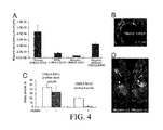
- FIG. 4 shows the effect of blood clotting on nanoparticle accumulation in tumors.
- Mice bearing MDA-MB-435 human breast cancer xenografts were intravenously injected with PBS or a bolus of 800 U/kg of heparin followed 120 min later by Ni-liposomes (or PBS) and CREKA-SPIO (or control nanoparticles). The mice received additional heparin by intraperitoneal injections (a total of 1000 U/kg) or PBS throughout the experiment.
- Tumors were removed 6 hours after the nanoparticle injection, and magnetic signal in the tumor after different treatments was determined with SQUID. Aminated dextran SPIO served as a particle control (control SPIO).
- SPIO nanoparticle concentration in tissues is represented by the saturation magnetization value (electromagnetic unit, emu) of the tissue at 1 T magnetic field after the subtraction of the diamagnetic and the paramagnetic background of blank tissue. The magnetization values were normalized to dry weight of the tissue. Results from 3 experiments are shown.
- C A representative example of the appearance of CREKA-SPIO particles in tumor vessels of mice treated with heparin.
- D Near-infrared imaging of mice that received Ni-liposomes followed by Cy7-labeled CREKA-SPIO with or without heparin pretreatment. The images were acquired 8 hours after the injection of the CREKA-SPIO particles using an Odyssey 2 NIR scanner (Li-COR Biosciences, Lincoln, Nebr.). The images shown are composites of 2 colors, red (700 nm channel, body and chow autofluorescence) and green (800 nm channel, Cy7). Arrows point to the tumors, arrowheads to the liver. Note the strong decrease in signal from the tumor in the heparin-pretreated mouse. A representative experiment out of 3 is shown.
- FIG. 5 shows tumor homing of CREKA peptide.
- A Balb/c nude mice bearing MDA-MB-435 human breast cancer xenograft tumors or transgenic MMTV PyMT mice with breast tumors were intravenously injected with 0.1 mg of fluorescein-CREKA. The animals were sacrificed by perfusion 24 hours post-injection and tissue sections were examined by fluorescent microscopy. Right panel, control organs of MDA-MB 435 tumor mice. Magnification 200 ⁇ .
- B Whole animal imaging of MDA-MB-435 tumor mouse injected 6 hours earlier with 30 ⁇ g of Alexa Fluor 647-labeled CREKA. Maestro imaging system (Cambridge Research Inc., Woburn, Mass.) was used to acquire and process the image. The arrow points to the tumor and the arrowhead to the urinary bladder. Note that the peptide is excreted into the urine and does not accumulate in the liver.
- FIG. 6 shows fluorescence intensity of iron oxide nanoparticles (CREKA-SPIO) coupled to various levels of substitution with fluorescein-labeled CREKA peptide. Fluorescence emitted by the conjugated particles is linearly related to the level of substitution.
- A.U. Arbitrary Units.
- FIG. 7 shows CREKA-SPIO nanoparticles accumulate in tumor tissue, but not in non-RES normal tissues.
- the low magnification (40 ⁇ ) was used to produce these images because only blood vessels in which clotting had concentrated the CREKA-SPIO fluorescence are visible at this magnification.
- the injections were carried out and the tissues prepared for analysis as in FIG. 2 . A representative experiment out of 10 is shown.
- FIG. 8 shows lack of colocalization of fibrin(ogen) staining and CREKA-SPIO in the liver.
- the fibrin(ogen)-positive structures can be background from fibrinogen production by the liver, as it does not co-localize with the nanoparticles (A), and the liver from a non-injected mouse showed similar fibrin(ogen) staining (B). Magnification 600 ⁇ .
- FIG. 9 shows the role of platelets in nanoparticle homing.
- A Blood was drawn 5 min post-injection of 4 mg/kg of CREKA-SPIO into mice and a 50 ⁇ l aliquot was run through a magnetic column. Bound CREKA-SPIO particles were eluted form the column, concentrated on a slide, and stained with anti-CD41 antibody. Some of the particles appear to be associated with platelets.
- B A low-magnification image (40 ⁇ ) showing CREKA-SPIO homing and clot formation in a tumor from a platelet-depleted mouse. Platelet depletion was accomplished by treating mice with 0.1 mg of an anti-CD41 monoclonal antibody as described (Van der Heyde and Gramaglia (2005)). The mice subsequently received Ni-liposomes/CREKA-SPIO as described in the legend of FIG. 2 . The anti-platelet treatment did not decrease the incidence of fluorescent clots (compare with the tumor panel in FIG. 7 ).
- Ranges can be expressed herein as from “about” one particular value, and/or to “about” another particular value. When such a range is expressed, another embodiment includes from the one particular value and/or to the other particular value. Similarly, when values are expressed as approximations, by use of the antecedent “about,” it will be understood that the particular value forms another embodiment. It will be further understood that the endpoints of each of the ranges are significant both in relation to the other endpoint, and independently of the other endpoint. It is also understood that there are a number of values disclosed herein, and that each value is also herein disclosed as “about” that particular value in addition to the value itself. For example, if the value “10” is disclosed, then “about 10” is also disclosed.
- a conjugate comprising a surface molecule and a plurality of clot binding compounds.
- the clot binding compounds can selectively bind to clotted plasma protein.
- the conjugate can cause clotting and amplify the accumulation of the conjugate in tumors.
- the clot binding compounds selectively bind to clotted plasma protein and the conjugate causes clotting and amplifies the accumulation of the conjugate in tumors.
- conjugates that not only home to tumors, but also amplify their own homing.
- the system is based on a clot binding compound that recognizes clotted plasma proteins and selectively homes to tumors, where it binds to vessel walls and tumor stroma.
- Surface molecules coupled with the clot binding compounds can accumulate in tumor vessels or at wound sites, where they induce additional local clotting, thereby producing new binding sites for more particles.
- the system mimics platelets, which also circulate freely but accumulate at a diseased site and amplify their own accumulation at that site.
- the clotting-based amplification greatly enhances tumor imaging, and a drug carrier function is also envisioned.
- Conjugates comprising clot binding compounds are directed to the tumor cells themselves. There, they accumulate and induce additional clotting.
- a number of appropriate clot binding compounds have been identified that are specifically or preferentially expressed, localized, adsorbed to or inducible on the cells or in the environment of the tumor vasculature and/or stroma. These are discussed in more detail below.
- the clot binding compound can be any compound with the ability to interact with clots and/or components of clots such as clotted plasma proteins.
- the conjugate can comprise a sufficient number and composition of clot binding compounds such that the conjugate causes clotting and amplifies the accumulation of the conjugate in tumors and at the site of injury.
- sufficiency of the number and composition of clot binding compounds can be determined by assessing clotting and amplification of the accumulation of the conjugate in tumors in a non-human animal. Such methods are discussed in more detail below.
- a plurality of the clot binding compounds can each be independently selected from, for example, an amino acid segment comprising the amino acid sequence REK, a fibrin-binding peptide, a peptide that binds clots and not fibrin (such as CGLIIQKNEC (CLT1, SEQ ID NO: 2) and CNAGESSKNC (CLT2, SEQ ID NO: 3)).
- a clot binding antibody and a clot binding small organic molecule.
- a plurality of the clot binding compounds can each independently comprise an amino acid segment comprising the amino acid sequence REK.
- the conjugate can comprise any number of clot binding compounds.
- the conjugate can comprise at least 1, 5, 10, 15, 20, 25, 50, 75, 100, 125, 150, 175, 200, 225, 250, 275, 300, 325, 350, 375, 400, 425, 450, 475, 500, 525, 550, 575, 600, 625, 650, 675, 700, 625, 750, 775, 800, 825, 850, 875, 900, 925, 950, 975, 1000, 1100, 1200, 1300, 1400, 1500, 1600, 1700, 1800, 1900, 2000, 2250, 2500, 2750, 3000, 3500, 4000, 4500, 5000, 5500, 6000, 6500, 7000, 7500, 8000, 8500, 9000, 9500, 10,000, 15,000, 20,000, 25,000, 30,000, 35,000, 40,000, 45,000, 50,000, 75,000, or 100,000, or more clot binding compounds.
- the conjugate can also
- homing molecule as used herein, means any molecule that selectively homes in vivo to specified target sites or tissues in preference to normal tissue.
- homoing peptide or “homing peptidomimetic” means a peptide that selectively homes in vivo to specified target sites or tissues in preference to normal tissue. It is understood that a homing molecule that selectively homes in vivo to, for example, tumors can home to all tumors or can exhibit preferential homing to one or a subset of tumor types.
- the homing molecule binds preferentially to the target as compared to non-target.
- the homing molecule can bind preferentially to clotted plasma of one or more tumors, wound tissue, or blood clots, as compared to non-tumoral tissue or non-wound tissue.
- Such a homing molecule can selectively home, for example, to tumors.
- Selective homing to, for example, tumors generally is characterized by at least a two-fold greater localization within tumors (or other target), as compared to several tissue types of non-tumor tissue.
- a homing molecule can be characterized by 5-fold, 10-fold, 20-fold or more preferential localization to tumors (or other target) as compared to several or many tissue types of non-tumoral tissue, or as compared to-most or all non-tumoral tissue.
- a homing molecule homes, in part, to one or more normal organs in addition to homing to the target tissue.
- Selective homing can also be referred to as targeting.
- the clot binding compound can be a peptide or peptidomimetic.
- the disclosed peptides can be in isolated form.
- isolated means a peptide that is in a form that is relatively free from material such as contaminating polypeptides, lipids, nucleic acids and other cellular material that normally is associated with the peptide in a cell or that is associated with the peptide in a library or in a crude preparation.
- the disclosed peptides can have any suitable length.
- the disclosed peptides can have, for example, a relatively short length of less than six, seven, eight, nine, ten, 12, 15, 20, 25, 30, 35 or 40 residues.
- the disclosed peptides also can be useful in the context of a significantly longer sequence.
- the peptides can have, for example, a length of up to 50, 100, 150, 200, 250, 300, 400, 500, 1000 or 2000 residues.
- a peptide can have a length of at least 10, 20, 30, 40, 50, 60, 70, 80, 90, 100 or 200 residues.
- a peptide can have a length of 5 to 200 residues, 5 to 100 residues, 5 to 90 residues, 5 to 80 residues, 5 to 70 residues, 5 to 60 residues, 5 to 50 residues, 5 to 40 residues, 5 to 30 residues, 5 to 20 residues, 5 to 15 residues, 5 to 10 residues, 10 to 200 residues, 10 to 100 residues, 10 to 90 residues, 10 to 80 residues, 10 to 70 residues, 10 to 60 residues, 10 to 50 residues, 10 to 40 residues, 10 to 30 residues, 10 to 20 residues, 20 to 200 residues, 20 to 100 residues, 20 to 90 residues, 20 to 80 residues, 20 to 70 residues, 20 to 60 residues, 20 to 50 residues, 20 to 40 residues or 20 to 30 residues.
- the term “residue” refers to an amino acid or amino acid analog.
- nucleic acids that can encode those protein sequences are also disclosed. This would include all degenerate sequences related to a specific protein sequence, i.e. all nucleic acids having a sequence that encodes one particular protein sequence as well as all nucleic acids, including degenerate nucleic acids, encoding the disclosed variants and derivatives of the protein sequences. Thus, while each particular nucleic acid sequence may not be written out herein, it is understood that each and every sequence is in fact disclosed and described herein through the disclosed protein sequence.
- Molecules can be produced that resemble peptides, but which are not connected via a natural peptide linkage.
- linkages for amino acids or amino acid analogs can include CH 2 NH—, —CH 2 S—, —CH 2 —CH 2 —, —CH ⁇ CH— (cis and trans), —COCH 2 —, —CH(OH)CH 2 —, and —CHH 2 SO— (These and others can be found in Spatola, A. F. in Chemistry and Biochemistry of Amino Acids, Peptides, and Proteins, B. Weinstein, eds., Marcel Dekker, New York, p. 267 (1983); Spatola, A. F., Vega Data (March 1983), Vol.
- bifunctional peptides which contain the clot binding peptide fused to a second peptide having a separate function.
- Such bifunctional peptides have at least two functions conferred by different portions of the full-length molecule and can, for example, display anti-angiogenic activity or pro-apoptotic activity in addition to the ability to enhance clotting.
- isolated multivalent peptides that include at least two subsequences each independently containing a peptide (for example, the amino acid sequence SEQ ID NO: 1, or a conservative variant or peptidomimetic thereof).
- the multivalent peptide can have, for example, at least three, at least five or at least ten of such subsequences each independently containing a peptide.
- the multivalent peptide can have two, three, four, five, six, seven, eight, nine, ten, fifteen or twenty identical or non-identical subsequences. This is in addition to the multiple clot binding compounds that can comprise the conjugate.
- the multivalent peptide can contain identical subsequences, such as repeats of SEQ ID NO: 1. In a further embodiment, the multivalent peptide contains contiguous identical or non-identical subsequences, which are not separated by any intervening amino acids.
- peptide is used broadly to mean peptides, proteins, fragments of proteins and the like.
- peptidomimetic means a peptide-like molecule that has the activity of the peptide upon which it is structurally based. Such peptidomimetics include chemically modified peptides, peptide-like molecules containing non-naturally occurring amino acids, and peptoids and have an activity such as selective interaction with a target of the peptide upon which the peptidomimetic is derived (see, for example, Goodman and Ro, Peptidomimetics for Drug Design, in “Burger's Medicinal Chemistry and Drug Discovery” Vol. 1 (ed. M. E. Wolff; John Wiley & Sons 1995), pages 803-861).
- a variety of peptidomimetics are known in the art including, for example, peptide-like molecules which contain a constrained amino acid, a non-peptide component that mimics peptide secondary structure, or an amide bond isostere.
- a peptidomimetic that contains a constrained, non-naturally occurring amino acid can include, for example, an ⁇ -methylated amino acid; ⁇ , ⁇ .-dialkylglycine or ⁇ -aminocycloalkane carboxylic acid; an N ⁇ —C ⁇ cyclized amino acid; an N ⁇ .-methylated amino acid; a ⁇ - or ⁇ -amino cycloalkane carboxylic acid; an ⁇ , ⁇ -unsaturated amino acid; a ⁇ , ⁇ -dimethyl or ⁇ -methyl amino acid; a ⁇ -substituted-2,3-methano amino acid; an N—C ⁇ or C ⁇ —C ⁇ cyclized amino acid; a substituted pro
- a peptidomimetic which mimics peptide secondary structure can contain, for example, a non-peptidic ⁇ -turn mimic; ⁇ -turn mimic; mimic of ⁇ -sheet structure; or mimic of helical structure, each of which is well known in the art.
- a peptidomimetic also can be a peptide-like molecule which contains, for example, an amide bond isostere such as a retro-inverso modification; reduced amide bond; methylenethioether or methylene-sulfoxide bond; methylene ether bond; ethylene bond; thioamide bond; trans-olefin or fluoroolefin bond; 1,5-disubstituted tetrazole ring; ketomethylene or fluoroketomethylene bond or another amide isostere.
- an amide bond isostere such as a retro-inverso modification
- reduced amide bond such as a retro-inverso modification
- methylenethioether or methylene-sulfoxide bond methylene ether bond
- ethylene bond thioamide bond
- trans-olefin or fluoroolefin bond 1,5-disubstituted tetrazole ring
- Methods for identifying a peptidomimetic include, for example, the screening of databases that contain libraries of potential peptidomimetics.
- the Cambridge Structural Database contains a collection of greater than 300,000 compounds that have known crystal structures (Allen et al., Acta Crystalloqr. Section B, 35:2331 (1979)). This structural depository is continually updated as new crystal structures are determined and can be screened for compounds having suitable shapes, for example, the same shape as a disclosed peptide, as well as potential geometrical and chemical complementarity to a target molecule.
- a structure can be generated using, for example, the program CONCORD (Rusinko et al., J. Chem. Inf. Comput. Sci. 29:251 (1989)).
- CONCORD Rusinko et al., J. Chem. Inf. Comput. Sci. 29:251 (1989)
- Another database the Available Chemicals Directory (Molecular Design Limited, Information Systems; San Leandro Calif.), contains about 100,000 compounds that are commercially available and also can be searched to identify potential peptidomimetics of a peptide, for example, with activity in selectively interacting with cancerous cells.
- amino acid segments can also be independently selected from amino acid segments comprising the amino acid sequence CREKA (SEQ ID NO: 1) or a conservative variant thereof, amino acid segments comprising the amino acid sequence CREKA (SEQ ID NO:1), amino acid segments consisting of the amino acid sequence CREKA (SEQ ID NO:1), and amino acid segments consisting of the amino acid sequence REK.
- the amino acid segments can each independently comprise the amino acid sequence CREKA (SEQ ID NO: 1) or a conservative variant thereof.
- the amino acid segments can also each independently comprise the amino acid sequence CREKA (SEQ ID NO:1).
- the amino acid segment can also consist of the amino acid sequence CREKA (SEQ ID NO:1).
- the amino acid segment can consist of the amino acid sequence REK.
- the clot binding compound can also comprise a fibrin-binding peptide (FBP).
- FBP fibrin-binding peptide
- fibrin-binding peptides are known in the art (Van Rooijen N, Sanders A (1994) J Immunol Methods 174: 83-93; Moghimi S M, Hunter A C, Murray J C (2001) Pharmacol Rev 53: 283-318; U.S. Pat. No. 5,792,742, all herein incorporated by reference in their entirety for their teaching concerning fibrin binding peptides).
- Clot-binding peptides can also bind to proteins other than fibrin.
- Example include peptides that bind to fibronectin that has become incorporated into a clot (Pilch et al., (2006) PNAS, 103: 2800-2804, hereby incorporated in its entirety for its teaching concerning clot binding peptides).
- An example of clot binding peptides include, but is not limited to, CGLIIQKNEC (CLT1, SEQ ID NO: 2) and CNAGESSKNC (CLT2, SEQ ID NO: 3).
- the amino acid segments can also be independently selected from amino acid segments comprising the amino acid sequence CLT1 or CLT2 (SEQ ID NOS: 2 or 3) or a conservative variant thereof, amino acid segments comprising the amino acid sequence CLT1 or CLT2 (SEQ ID NOS: 2 or 3), or amino acid segments consisting of the amino acid sequence CLT1 or CLT2 (SEQ ID NOS: 2 or 3).
- the amino acid segments can each independently comprise the amino acid sequence CLT1 or CLT2 (SEQ ID NOS: 2 or 3) or a conservative variant thereof.
- the amino acid segments can also each independently comprise the amino acid sequence CLT1 or CLT2 (SEQ ID NOS: 2 or 3).
- the amino acid segment can also consist of the amino acid sequence CLT1 or CLT2 (SEQ ID NOS: 2 or 3).
- the clot binding compound can comprise a clot binding antibody.
- clot binding antibodies are known in the art (Holvoet et al. Circulation, Vol 87, 1007-1016, 1993; Bode et al. J. Biol. Chem., Vol. 264, Issue 2, 944-948, January, 1989; Huang et al. Science 1997: Vol. 275. no. 5299, pp. 547-550, all of which are herein incorporated by reference in their entirety for their teaching concerning clot binding antibodies).
- antibodies is used herein in a broad sense and includes both polyclonal and monoclonal antibodies. In addition to intact immunoglobulin molecules, also included in the term “antibodies” are fragments or polymers of those immunoglobulin molecules, and human or humanized versions of immunoglobulin molecules or fragments thereof, as long as they are chosen for their ability to bind to, or otherwise interact with, clots.
- the antibodies can be tested for their desired activity using the in vitro assays described herein, or by analogous methods, after which their in vivo therapeutic and/or prophylactic activities are tested according to known clinical testing methods.
- the term “monoclonal antibody” as used herein refers to an antibody obtained from a substantially homogeneous population of antibodies, i.e., the individual antibodies within the population are identical except for possible naturally occurring mutations that may be present in a small subset of the antibody molecules.
- the monoclonal antibodies herein specifically include “chimeric” antibodies in which a portion of the heavy and/or light chain is identical with or homologous to corresponding sequences in antibodies derived from a particular species or belonging to a particular antibody class or subclass, while the remainder of the chain(s) is identical with or homologous to corresponding sequences in antibodies derived from another species or belonging to another antibody class or subclass, as well as fragments of such antibodies, as long as they exhibit the desired antagonistic activity (See, U.S. Pat. No. 4,816,567 and Morrison et al., Proc. Natl. Acad. Sci. USA, 81:6851-6855 (1984)).
- the disclosed monoclonal antibodies can be made using any procedure which produces monoclonal antibodies.
- disclosed monoclonal antibodies can be prepared using hybridoma methods, such as those described by Kohler and Milstein, Nature, 256:495 (1975).
- a hybridoma method a mouse or other appropriate host animal is typically immunized with an immunizing agent to elicit lymphocytes that produce or are capable of producing antibodies that will specifically bind to the immunizing agent.
- the lymphocytes may be immunized in vitro, e.g., using the HIV Env-CD4-co-receptor complexes described herein.
- the monoclonal antibodies may also be made by recombinant DNA methods, such as those described in U.S. Pat. No. 4,816,567 (Cabilly et al.).
- DNA encoding the disclosed monoclonal antibodies can be readily isolated and sequenced using conventional procedures (e.g., by using oligonucleotide probes that are capable of binding specifically to genes encoding the heavy and light chains of murine antibodies).
- Libraries of antibodies or active antibody fragments can also be generated and screened using phage display techniques, e.g., as described in U.S. Pat. No. 5,804,440 to Burton et al. and U.S. Pat. No. 6,096,441 to Barbas et al.
- In vitro methods are also suitable for preparing monovalent antibodies.
- Digestion of antibodies to produce fragments thereof, particularly, Fab fragments can be accomplished using routine techniques known in the art. For instance, digestion can be performed using papain. Examples of papain digestion are described in WO 94/29348 published Dec. 22, 1994 and U.S. Pat. No. 4,342,566.
- Papain digestion of antibodies typically produces two identical antigen binding fragments, called Fab fragments, each with a single antigen binding site, and a residual Fc fragment. Pepsin treatment yields a fragment that has two antigen combining sites and is still capable of cross-linking antigen.
- the fragments can also include insertions, deletions, substitutions, or other selected modifications of particular regions or specific amino acids residues, provided the activity of the antibody or antibody fragment is not significantly altered or impaired compared to the non-modified antibody or antibody fragment. These modifications can provide for some additional property, such as to remove/add amino acids capable of disulfide bonding, to increase its bio-longevity, to alter its secretory characteristics, etc.
- the antibody or antibody fragment must possess a bioactive property, such as specific binding to its cognate antigen.
- Functional or active regions of the antibody or antibody fragment may be identified by mutagenesis of a specific region of the protein, followed by expression and testing of the expressed polypeptide.
- antibody can also refer to a human antibody and/or a humanized antibody.
- Many non-human antibodies e.g., those derived from mice, rats, or rabbits
- are naturally antigenic in humans and thus can give rise to undesirable immune responses when administered to humans. Therefore, the use of human or humanized antibodies in the methods serves to lessen the chance that an antibody administered to a human will evoke an undesirable immune response.
- Human antibodies can be prepared using any technique. Examples of techniques for human monoclonal antibody production include those described by Cole et al. ( Monoclonal Antibodies and Cancer Therapy , Alan R. Liss, p. 77, 1985) and by Boerner et al. ( J. Immunol., 147(1):86-95, 1991). Human antibodies (and fragments thereof) can also be produced using phage display libraries (Hoogenboom et al., J. Mol. Biol., 227:381, 1991; Marks et al., J. Mol. Biol., 222:581, 1991).
- Human antibodies can also be obtained from transgenic animals.
- transgenic, mutant mice that are capable of producing a full repertoire of human antibodies, in response to immunization, have been described (see, e.g., Jakobovits et al., Proc. Natl. Acad. Sci. USA, 90:2551-255 (1993); Jakobovits et al., Nature, 362:255-258 (1993); Bruggermann et al., Year in Immunol., 7:33 (1993)).
- the homozygous deletion of the antibody heavy chain joining region (J(H)) gene in these chimeric and germ-line mutant mice results in complete inhibition of endogenous antibody production, and the successful transfer of the human germ-line antibody gene array into such germ-line mutant mice results in the production of human antibodies upon antigen challenge.
- Antibodies having the desired activity are selected using Env-CD4-co-receptor complexes as described herein.
- Antibody humanization techniques generally involve the use of recombinant DNA technology to manipulate the DNA sequence encoding one or more polypeptide chains of an antibody molecule.
- a humanized form of a non-human antibody is a chimeric antibody or antibody chain (or a fragment thereof, such as an Fv, Fab, Fab′, or other antigen-binding portion of an antibody) which contains a portion of an antigen binding site from a non-human (donor) antibody integrated into the framework of a human (recipient) antibody.
- a humanized antibody residues from one or more complementarity determining regions (CDRs) of a recipient (human) antibody molecule are replaced by residues from one or more CDRs of a donor (non-human) antibody molecule that is known to have desired antigen binding characteristics (e.g., a certain level of specificity and affinity for the target antigen).
- CDRs complementarity determining regions
- donor non-human antibody molecule that is known to have desired antigen binding characteristics
- Fv framework (FR) residues of the human antibody are replaced by corresponding non-human residues.
- Humanized antibodies may also contain residues which are found neither in the recipient antibody nor in the imported CDR or framework sequences.
- a humanized antibody has one or more amino acid residues introduced into it from a source which is non-human.
- humanized antibodies are typically human antibodies in which some CDR residues and possibly some FR residues are substituted by residues from analogous sites in rodent antibodies.
- Humanized antibodies generally contain at least a portion of an antibody constant region (Fc), typically that of a human antibody (Jones et al., Nature, 321:522-525 (1986), Reichmann et al., Nature, 332:323-327 (1988), and Presta, Curr. Opin. Struct. Biol., 2:593-596 (1992)).
- Fc antibody constant region
- humanized antibodies can be generated according to the methods of Winter and co-workers (Jones et al., Nature, 321:522-525 (1986), Riechmann et al., Nature, 332:323-327 (1988), Verhoeyen et al., Science, 239:1534-1536 (1988)), by substituting rodent CDRs or CDR sequences for the corresponding sequences of a human antibody.
- Methods that can be used to produce humanized antibodies are also described in U.S. Pat. No. 4,816,567 (Cabilly et al.), U.S. Pat. No.
- the clot binding compound can also be a small organic molecule.
- Small organic molecules that are capable of interacting with, or binding to, clots are known in the art. These molecules can also be identified by methods known in the art, such as combinatorial chemistry. Some forms of small organic molecules can be organic molecules having a molecular weight of less than 1000 Daltons.
- Combinatorial chemistry includes but is not limited to all methods for isolating small molecules that are capable of interacting with a clot, molecules associated with a clot such as fibrin or fibronectin, or clotted plasma protein, for example.
- Combinatorial libraries can be made from a wide array of molecules using a number of different synthetic techniques. For example, libraries containing fused 2,4-pyrimidinediones (U.S. Pat. No. 6,025,371) dihydrobenzopyrans (U.S. Pat. Nos. 6,017,768 and 5,821,130), amide alcohols (U.S. Pat. No. 5,976,894), hydroxy-amino acid amides (U.S. Pat. No. 5,972,719) carbohydrates (U.S. Pat. No. 5,965,719), 1,4-benzodiazepin-2,5-diones (U.S. Pat. No. 5,962,337), cyclics (U.S. Pat. No.
- combinatorial methods and libraries included traditional screening methods and libraries as well as methods and libraries used in iterative processes.
- Libraries of small organic molecules generally comprise at least 2 organic compounds, often at least about 25, 100 500 different organic compounds, more usually at least about 1000 different organic compounds, preferably at least about 2500 different organic compounds, more preferably at least about 5000 different organic compounds and most preferably at least about 10,000 or more different organic compounds.
- Libraries may be selected or constructed such that each individual molecule of the library may be spatially separated from the other molecules of the library (e.g., each member of the library is present in a separate microtiter well) or two or more members of the library may be combined if methods for deconvolution are readily available.
- the methods by which the library of organic compounds are prepared will not be critical to the invention.
- the surface molecules can be conjugated with clot binding compounds in such a way that the conjugate is delivered to a clot, where it can accumulate and cause further clotting.
- the surface molecule can be any substance that can be used with the clot binding compounds, and is not restricted by size or substance. Examples include, but are not limited to, nanoparticles (such as iron oxide nanoparticles or albumin nanoparticles), liposomes, small organic molecules, microparticles, or microbubbles, such as fluorocarbon microbubbles.
- nanoparticles such as iron oxide nanoparticles or albumin nanoparticles
- liposomes small organic molecules
- microparticles or microbubbles, such as fluorocarbon microbubbles.
- the term surface molecule is used to identify a component of the disclosed conjugate but is not intended to be limiting.
- the disclosed surface molecules are not limited to substances, compounds, compositions, particles or other materials composed of a single molecule. Rather, the disclosed surface molecules are any substance(s), compound(s), composition(s), particle(s) and/or other material(s) that can be conjugated with a plurality of clot binding compounds such that at least some of the clot binding compounds are presented and/or accessible on the surface of the surface molecule.
- suitable surface molecules are described and disclosed herein.
- the surface molecule can be detectable, or can be a therapeutic agent such as AbraxaneTM.
- a therapeutic agent such as AbraxaneTM.
- nanoparticle refers to a nanoscale particle with a size that is measured in nanometers, for example, a nanoscopic particle that has at least one dimension of less than about 100 nm.
- nanoparticles include paramagnetic nanoparticles, superparamagnetic nanoparticles, metal nanoparticles, fullerene-like materials, inorganic nanotubes, dendrimers (such as with covalently attached metal chelates), nanofibers, nanohoms, nano-onions, nanorods, nanoropes and quantum dots.
- a nanoparticle can produce a detectable signal, for example, through absorption and/or emission of photons (including radio frequency and visible photons) and plasmon resonance.
- Microspheres can also be used with the methods disclosed herein.
- Microspheres containing chromophores have been utilized in an extensive variety of applications, including photonic crystals, biological labeling, and flow visualization in microfluidic channels. See, for example, Y. Lin, et al., Appl. Phys Lett. 2002, 81, 3134; D. Wang, et al., Chem. Mater. 2003, 15, 2724; X. Gao, et al., J. Biomed. Opt. 2002, 7, 532; M. Han, et al., Nature Biotechnology. 2001, 19, 631; V. M. Pai, et al., Mag. & Magnetic Mater. 1999, 194, 262, each of which is incorporated by reference in its entirety. Both the photostability of the chromophores and the monodispersity of the microspheres can be important.
- Nanoparticles such as, for example, metal nanoparticles, metal oxide nanoparticles, or semiconductor nanocrystals can be incorporated into microspheres.
- the optical, magnetic, and electronic properties of the nanoparticles can allow them to be observed while associated with the microspheres and can allow the microspheres to be identified and spatially monitored.
- the high photostability, good fluorescence efficiency and wide emission tunability of colloidally synthesized semiconductor nanocrystals can make them an excellent choice of chromophore.
- nanocrystals that emit different colors i.e. different wavelengths
- Colloidally synthesized semiconductor nanocrystals (such as, for example, core-shell CdSe/ZnS and CdS/ZnS nanocrystals) can be incorporated into microspheres.
- the microspheres can be monodisperse silica microspheres.
- the nanoparticle can be a metal nanoparticle, a metal oxide nanoparticle, or a semiconductor nanocrystal.
- the metal of the metal nanoparticle or the metal oxide nanoparticle can include titanium, zirconium, hafnium, vanadium, niobium, tantalum, chromium, molybdenum, tungsten, manganese, technetium, rhenium, iron, ruthenium, osmium, cobalt, rhodium, iridium, nickel, palladium, platinum, copper, silver, gold, zinc, cadmium, scandium, yttrium, lanthanum, a lanthanide series or actinide series element (e.g., cerium, praseodymium, neodymium, promethium, samarium, europium, gadolinium, terbium, dysprosium, holmium, erbium, thulium, ytterbium,
- the metal can be iron, ruthenium, cobalt, rhodium, nickel, palladium, platinum, silver, gold, cerium or samarium.
- the metal oxide can be an oxide of any of these materials or combination of materials.
- the metal can be gold, or the metal oxide can be an iron oxide, a cobalt oxide, a zinc oxide, a cerium oxide, or a titanium oxide. Preparation of metal and metal oxide nanoparticles is described, for example, in U.S. Pat. Nos. 5,897,945 and 6,759,199, each of which is incorporated by reference in its entirety.
- Liposome refers to a structure comprising an outer lipid bi- or multi-layer membrane surrounding an internal aqueous space. Liposomes can be used to package any biologically active agent for delivery to cells.
- liposomes Materials and procedures for forming liposomes are well-known to those skilled in the art. Upon dispersion in an appropriate medium, a wide variety of phospholipids swell, hydrate and form multilamellar concentric bilayer vesicles with layers of aqueous media separating the lipid bilayers. These systems are referred to as multilamellar liposomes or multilamellar lipid vesicles (“MLVs”) and have diameters within the range of 10 nm to 100 ⁇ m. These MLVs were first described by Bangham, et al., J Mol. Biol. 13:238-252 (1965). In general, lipids or lipophilic substances are dissolved in an organic solvent.
- the lipid residue forms a film on the wall of the container.
- An aqueous solution that typically contains electrolytes or hydrophilic biologically active materials is then added to the film.
- Large MLVs are produced upon agitation.
- the larger vesicles are subjected to sonication, sequential filtration through filters with decreasing pore size or reduced by other forms of mechanical shearing.
- pressurized extrusion Barenholz, et al., FEBS Lett. 99:210-214 (1979)
- Liposomes can also take the form of unilamnellar vesicles, which are prepared by more extensive sonication of MLVs, and consist of a single spherical lipid bilayer surrounding an aqueous solution.
- Unilamellar vesicles (“ULVs”) can be small, having diameters within the range of 20 to 200 nm, while larger ULVs can have diameters within the range of 200 nm to 2 ⁇ m.
- ULVs Unilamellar vesicles
- Small ULVs can also be prepared by the ethanol injection technique described by Batzri, et al., Biochim et Biophys Acta 298:1015-1019 (1973) and the ether injection technique of Deamer, et al., Biochim et Biophys Acta 443:629-634 (1976). These methods involve the rapid injection of an organic solution of lipids into a buffer solution, which results in the rapid formation of unilamellar liposomes. Another technique for making ULVs is taught by Weder, et al. in “Liposome Technology”, ed. G. Gregoriadis, CRC Press Inc., Boca Raton, Fla., Vol. 1, Chapter 7, pg. 79-107 (1984). This detergent removal method involves solubilizing the lipids and additives with detergents by agitation or sonication to produce the desired vesicles.
- Papahadjopoulos, et al., U.S. Pat. No. 4,235,871 describes the preparation of large ULVs by a reverse phase evaporation technique that involves the formation of a water-in-oil emulsion of lipids in an organic solvent and the drug to be encapsulated in an aqueous buffer solution. The organic solvent is removed under pressure to yield a mixture which, upon agitation or dispersion in an aqueous media, is converted to large ULVs.
- Suzuki et al., U.S. Pat. No. 4,016,100 describes another method of encapsulating agents in unilamellar vesicles by freezing/thawing an aqueous phospholipid dispersion of the agent and lipids.
- liposomes can also be multivesicular. Described in Kim, et al., Biochim et Biophys Acta 728:339-348 (1983), these multivesicular liposomes are spherical and contain internal granular structures. The outer membrane is a lipid bilayer and the internal region contains small compartments separated by bilayer septum. Still yet another type of liposomes are oligolamellar vesicles (“OLVs”), which have a large center compartment surrounded by several peripheral lipid layers. These vesicles, having a diameter of 2-15 ⁇ m, are described in Callo, et al., Cryobiology 22(3):251-267 (1985).
- OLVs oligolamellar vesicles
- the conjugate disclosed herein can further comprise one or more moieties.
- the moieties can be independently selected from the group consisting of an anti-angiogenic agent, a pro-angiogenic agent, a cancer chemotherapeutic agent, a cytotoxic agent, an anti-inflammatory agent, an anti-arthritic agent, a polypeptide, a nucleic acid molecule, a small molecule, a fluorophore, fluorescein, rhodamine, a radionuclide, indium-111, technetium-99, carbon-11, and carbon-13.
- At least one of the moieties can be a therapeutic agent. Examples of therapeutic agents are paclitaxel and taxol.
- At least one of the moieties can be a detectable agent.
- the term “moiety” is used broadly to mean a physical, chemical, or biological material that generally imparts a biologically useful function to a linked or conjugated molecule.
- the properties of the moiety can also be found in a surface molecule, or both the surface molecule and the moiety can share one of the traits disclosed herein.
- the surface molecule can comprise a detectable agent, while the moiety can comprise a therapeutic agent.
- the clot binding compound which can also comprise one or more of the properties of moieties as disclosed herein.
- the description of therapeutic and detectable agents which follows is intended to apply to any of moieties, surface molecules, or clot binding compounds.
- moieties can be conjugated to, coupled to, or can be part of the disclosed surface molecules, clot binding compounds, or conjugates of surface molecules and clot binding compounds.
- a moiety can be any natural or nonnatural material including, without limitation, a biological material, such as a cell, phage or other virus; an organic chemical such as a small molecule; a radionuclide; a nucleic acid molecule or oligonucleotide; a polypeptide; or a peptide.
- Useful moieties include, but are not limited to, therapeutic agents such as cancer chemotherapeutic agents, cytotoxic agents, pro-apoptotic agents, and anti-angiogenic agents; detectable labels and imaging agents; and tags or other insoluble supports.
- Useful moieties further include, without limitation, phage and other viruses, cells, liposomes, polymeric matrices, non-polymeric matrices or particles such as gold particles, microdevices and nanodevices, and nano-scale semiconductor materials. These and other moieties known in the art can be components of a conjugate.
- the moiety can be a therapeutic agent.
- therapeutic agent means a molecule which has one or more biological activities in a normal or pathologic tissue.
- a variety of therapeutic agents can be used as a moiety.
- the therapeutic agent can be a cancer chemotherapeutic agent.
- a cancer chemotherapeutic agent is a chemical agent that inhibits the proliferation, growth, life-span or metastatic activity of cancer cells.
- a cancer chemotherapeutic agent can be, without limitation, a taxane such as docetaxel; an anthracyclin such as doxorubicin; an alkylating agent; a vinca alkaloid; an anti-metabolite; a platinum agent such as cisplatin or carboplatin; a steroid such as methotrexate; an antibiotic such as adriamycin; a isofamide; or a selective estrogen receptor modulator; an antibody such as trastuzumab.
- Taxanes are chemotherapeutic agents useful with the conjugates disclosed herein.
- Useful taxanes include, without limitation, docetaxel (Taxotere; Aventis Pharmaceuticals, Inc.; Parsippany, N.J.) and paclitaxel (Taxol; Bristol-Myers Squibb; Princeton, N.J.). See, for example, Chan et al., J. Clin. Oncol. 17:2341-2354 (1999), and Paridaens et al., J. Clin. Oncol. 18:724 (2000).
- a cancer chemotherapeutic agent useful with the conjugates disclosed herein also can be an anthracyclin such as doxorubicin, idarubicin or daunorubicin.
- Doxorubicin is a commonly used cancer chemotherapeutic agent and can be useful, for example, for treating breast cancer (Stewart and Ratain, In: “Cancer: Principles and practice of oncology” 5th ed., chap. 19 (eds. DeVita, Jr., et al.; J. P. Lippincott 1997); Harris et al., In “Cancer: Principles and practice of oncology,” supra, 1997).
- doxorubicin has anti-angiogenic activity (Folkman, Nature Biotechnology 15:510 (1997); Steiner, In “Angiogenesis: Key principles-Science, technology and medicine,” pp. 449-454 (eds. Steiner et al.; Birkhauser Verlag, 1992)), which can contribute to its effectiveness in treating cancer.
- An alkylating agent such as melphalan or chlorambucil also can be a useful cancer chemotherapeutic agent.
- a vinca alkaloid such as vindesine, vinblastine or vinorelbine; or an antimetabolite such as 5-fluorouracil, 5-fluorouridine or a derivative thereof can be a useful cancer chemotherapeutic agent.
- a platinum agent also can be a useful cancer chemotherapeutic agent.
- a platinum agent can be, for example, cisplatin or carboplatin as described, for example, in Crown, Seminars in Oncol. 28:28-37 (2001).
- Other useful cancer chemotherapeutic agents include, without limitation, methotrexate, mitomycin-C, adriamycin, ifosfamide and ansamycins.
- a cancer chemotherapeutic agent useful for treatment of breast cancer and other hormonally-dependent cancers also can be an agent that antagonizes the effect of estrogen, such as a selective estrogen receptor modulator or an anti-estrogen.
- the selective estrogen receptor modulator, tamoxifen is a cancer chemotherapeutic agent that can be used in a conjugate for treatment of breast cancer (Fisher et al., J. Natl. Cancer Instit. 90:1371-1388 (1998)).
- the therapeutic agent can be an antibody such as a humanized monoclonal antibody.
- the anti-epidermal growth factor receptor 2 (HER2) antibody, trastuzumab (Herceptin; Genentech, South San Francisco, Calif.) can be a therapeutic agent useful for treating HER2/neu overexpressing breast cancers (White et al., Annu. Rev. Med. 52:125-141 (2001)).
- Useful therapeutic agents also can be a cytotoxic agent, which, as used herein, can be any molecule that directly or indirectly promotes cell death.
- cytotoxic agents include, without limitation, small molecules, polypeptides, peptides, peptidomimetics, nucleic acid-molecules, cells and viruses.
- useful cytotoxic agents include cytotoxic small molecules such as doxorubicin, docetaxel or trastuzumab; antimicrobial peptides such as those described further below; pro-apoptotic polypeptides such as caspases and toxins, for example, caspase-8; diphtheria toxin A chain, Pseudomonas exotoxin A, cholera toxin, ligand fusion toxins such as DAB389EGF, ricinus communis toxin (ricin); and cytotoxic cells such as cytotoxic T cells. See, for example, Martin et al., Cancer Res.
- a therapeutic agent can be a therapeutic polypeptide.
- a therapeutic polypeptide can be any polypeptide with a biologically useful function.
- Useful therapeutic polypeptides encompass, without limitation, cytokines, antibodies, cytotoxic polypeptides; pro-apoptotic polypeptides; and anti-angiogenic polypeptides.
- useful therapeutic polypeptides can be a cytokine such as tumor necrosis factor- ⁇ (TNF- ⁇ ), tumor necrosis factor- ⁇ (TNF- ⁇ , granulocyte macrophage colony stimulating factor (GM-CSF), granulocyte colony stimulating factor (G-CSF), interferon .alpha.
- IFN- ⁇ interferon .gamma.
- IFN- ⁇ interleukin-1
- IL-2 interleukin-2
- IL-3 interleukin-3
- IL-4 interleukin-4
- IL-6 interleukin-6
- IL-7 interleukin-7
- DC-CK1 dendritic cell chemokine 1
- an anti-HER2 antibody or fragment thereof a cytotoxic polypeptide including a toxin or caspase, for example, diphtheria toxin A chain, Pseudomonas exotoxin A, cholera toxin, a ligand fusion toxin such as DAB389EGF or ricin; or an anti-angiogenic polypeptide such as angiostatin, endostatin, thrombospondin, platelet factor 4; anastellin; or one of those described further herein or
- a therapeutic agent can also be an anti-angiogenic agent.
- anti-angiogenic agent means a molecule that reduces or prevents angiogenesis, which is the growth and development of blood vessels.
- anti-angiogenic agents can be prepared by routine methods.
- Such anti-angiogenic agents include, without limitation, small molecules; proteins such as dominant negative forms of angiogenic factors, transcription factors and antibodies; peptides; and nucleic acid molecules including ribozymes, antisense oligonucleotides, and nucleic acid molecules encoding, for example, dominant negative forms of angiogenic factors and receptors, transcription factors, and antibodies and antigen-binding fragments thereof. See, for example, Hagedorn and Bikfalvi, Crit. Rev. Oncol. Hematol. 34:89-110 (2000), and Kirsch et al., J. Neurooncol. 50:149-163 (2000).
- VEGF Vascular endothelial growth factor
- An anti-angiogenic agent can be, for example, an inhibitor or neutralizing antibody that reduces the expression or signaling of VEGF or another angiogenic factor, for example, an anti-VEGF neutralizing monoclonal antibody (Borgstrom et al., supra, 1999).
- An anti-angiogenic agent also can inhibit another angiogenic factor such as a member of the fibroblast growth factor family such as FGF-1 (acidic), FGF-2 (basic), FGF-4 or FGF-5 (Slavin et al., Cell Biol. Int. 19:431-444 (1995); Folkman and Shing, J. Biol. Chem.
- an angiogenic factor such as angiopoietin-1, a factor that signals through the endothelial cell-specific Tie2 receptor tyrosine kinase (Davis et al., Cell 87:1161-1169 (1996); and Suri et al., Cell 87:1171-1180 (1996)), or the receptor of one of these angiogenic factors. It is understood that a variety of mechanisms can act to inhibit activity of an angiogenic factor including, without limitation, direct inhibition of receptor binding, indirect inhibition by reducing secretion of the angiogenic factor into the extracellular space, or inhibition of expression, function or signaling of the angiogenic factor.
- a variety of other molecules also can function as anti-angiogenic agents including, without limitation, angiostatin; a kringle peptide of angiostatin; endostatin; anastellin, heparin-binding fragments of fibronectin; modified forms of antithrombin; collagenase inhibitors; basement membrane turnover inhibitors; angiostatic steroids; platelet factor 4 and fragments and peptides thereof, thrombospondin and fragments and peptides thereof; and doxorubicin (O'Reilly et al., Cell 79:315-328 (1994)); O'Reilly et al., Cell 88:277-285 (1997); Homandberg et al., Am. J. Path.
- angiostatin a kringle peptide of angiostatin
- endostatin anastellin, heparin-binding fragments of fibronectin
- modified forms of antithrombin collagenase inhibitors
- anti-angiogenic agents include, for example, angiostatin, endostatin, metastatin and 2ME2 (EntreMed; Rockville, Md.); anti-VEGF antibodies such as Avastin (Genentech; South San Francisco, Calif.); and VEGFR-2 inhibitors such as SU5416, a small molecule inhibitor of VEGFR-2 (SUGEN; South San Francisco, Calif.) and SU6668 (SUGEN), a small molecule inhibitor of VEGFR-2, platelet derived growth factor and fibroblast growth factor I receptor. It is understood that these and other anti-angiogenic agents can be prepared by routine methods and are encompassed by the term “anti-angiogenic agent” as used herein.
- the conjugates disclosed herein can also be used at a site of inflammation or injury.
- Moieties useful for this purpose can include therapeutic agents belonging to several basic groups including anti-inflammatory agents which prevent inflammation, restenosis preventing drugs which prevent tissue growth, anti-thrombogenic drugs which inhibit or control formation of thrombus or thrombolytics, and bioactive agents which regulate tissue growth and enhance healing of the tissue.
- useful therapeutic agents include but are not limited to steroids, fibronectin, anti-clotting drugs, anti-platelet function drugs, drugs which prevent smooth muscle cell growth on inner surface wall of vessel, heparin, heparin fragments, aspirin, coumadin, tissue plasminogen activator (TPA), urokinase, hirudin, streptokinase, antiproliferatives (methotrexate, cisplatin, fluorouracil, Adriamycin), antioxidants (ascorbic acid, beta carotene, vitamin E), antimetabolites, thromboxane inhibitors, non-steroidal and steroidal anti-inflammatory drugs, beta and calcium channel blockers, genetic materials including DNA and RNA fragments, complete expression genes, antibodies, lymphokines, growth factors, prostaglandins, leukotrienes, laminin, elastin, collagen, and integrins.
- TPA tissue plasminogen activator
- urokinase hirudin
- Useful therapeutic agents also can be antimicrobial peptides. This can be particularly useful to target a wound or other infected sites.
- moieties comprising an antimicrobial peptide, where the conjugate is selectively internalized and exhibits a high toxicity to the targeted area.
- Useful antimicrobial peptides can have low mammalian cell toxicity when not incorporated into the conjugate.
- antimicrobial peptide means a naturally occurring or synthetic peptide having antimicrobial activity, which is the ability to kill or slow the growth of one or more microbes.
- An antimicrobial peptide can, for example, kill or slow the growth of one or more strains of bacteria including a Gram-positive or Gram-negative bacteria, or a fungi or protozoa.
- an antimicrobial peptide can have, for example, bacteriostatic or bacteriocidal activity against, for example, one or more strains of Escherichia coli, Pseudomonas aeruginosa or Staphylococcus aureus .
- an antimicrobial peptide can have biological activity due to the ability to form ion channels through membrane bilayers as a consequence of self-aggregation.
- An antimicrobial peptide is typically highly basic and can have a linear or cyclic structure. As discussed further below, an antimicrobial peptide can have an amphipathic .alpha.-helical structure (see U.S. Pat. No. 5,789,542; Javadpour et al., J. Med. Chem. 39:3107-3113 (1996); and Blondelle and Houghten, Biochem. 31: 12688-12694 (1992)). An antimicrobial peptide also can be, for example, a ⁇ -strand/sheet-forming peptide as described in Mancheno et al., J. Peptide Res. 51:142-148 (1998).
- An antimicrobial peptide can be a naturally occurring or synthetic peptide.
- Naturally occurring antimicrobial peptides have been isolated from biological sources such as bacteria, insects, amphibians, and mammals and are thought to represent inducible defense proteins that can protect the host organism from bacterial infection.
- Naturally occurring antimicrobial peptides include the gramicidins, magainins, mellitins, defensins and cecropins (see, for example, Maloy and Kari, Biopolymers 37:105-122 (1995); Alvarez-Bravo et al., Biochem. J.
- An antimicrobial peptide also can be an analog of a natural peptide, especially one that retains or enhances amphipathicity (see below).
- An antimicrobial peptide incorporated into the conjugate disclosed herein can have low mammalian cell toxicity when linked to the conjugate. Mammalian cell toxicity readily can be assessed using routine assays. As an example, mammalian cell toxicity can be assayed by lysis of human erythrocytes in vitro as described in Javadpour et al., supra, 1996. An antimicrobial peptide having low mammalian cell toxicity is not lytic to human erythrocytes or requires concentrations of greater than 100 ⁇ M for lytic activity, preferably concentrations greater than 200, 300, 500 or 1000 ⁇ M.
- conjugates in which the antimicrobial peptide portion promotes disruption of mitochondrial membranes when internalized by eukaryotic cells.
- an antimicrobial peptide preferentially disrupts mitochondrial membranes as compared to eukaryotic membranes.
- Mitochondrial membranes like bacterial membranes but in contrast to eukaryotic plasma membranes, have a high content of negatively charged phospholipids.
- An antimicrobial peptide can be assayed for activity in disrupting mitochondrial membranes using, for example, an assay for mitochondrial swelling or another assay well known in the art.
- An antimicrobial peptide that induces significant mitochondrial swelling at, for example, 50 ⁇ M, 40 ⁇ .M, 30 ⁇ M, 20 ⁇ M, 10 ⁇ M, or less, is considered a peptide that promotes disruption of mitochondrial membranes.
- Antimicrobial peptides generally have random coil conformations in dilute aqueous solutions, yet high levels of helicity can be induced by helix-promoting solvents and amphipathic media such as micelles, synthetic bilayers or cell membranes.
- ⁇ -Helical structures are well known in the art, with an ideal ⁇ -helix characterized by having 3.6 residues per turn and a translation of 1.5 ⁇ per residue (5.4 ⁇ per turn; see Creighton, Proteins: Structures and Molecular Properties W. H Freeman, New York (1984)).
- amphipathic ⁇ -helical structure polar and non-polar amino acid residues are aligned into an amphipathic helix, which is an ⁇ -helix in which the hydrophobic amino acid residues are predominantly on one face, with hydrophilic residues predominantly on the opposite face when the peptide is viewed along the helical axis.
- Antimicrobial peptides of widely varying sequence have been isolated, sharing an amphipathic ⁇ -helical structure as a common feature (Saberwal et al., Biochim. Biophys. Acta 1197:109-131 (1994)).
- Analogs of native peptides with amino acid substitutions predicted to enhance amphipathicity and helicity typically have increased antimicrobial activity.
- analogs with increased antimicrobial activity also have increased cytotoxicity against mammalian cells (Maloy et al., Biopolymers 37:105-122 (1995)).
- amphipathic ⁇ -helical structure means an ⁇ -helix with a hydrophilic face containing several polar residues at physiological pH and a hydrophobic face containing nonpolar residues.
- a polar residue can be, for example, a lysine or arginine residue
- a nonpolar residue can be, for example, a leucine or alanine residue.
- An antimicrobial peptide having an amphipathic .alpha.-helical structure generally has an equivalent number of polar and nonpolar residues within the amphipathic domain and a sufficient number of basic residues to give the peptide an overall positive charge at neutral pH (Saberwal et al., Biochim.
- conjugates disclosed herein can contain one or more of such therapeutic agents and that additional components can be included as part of the composition, if desired.
- additional components can be included as part of the composition, if desired.
- thrombolytics include aspirin, anticoagulants, painkillers and tranquilizers, beta-blockers, ace-inhibitors, nitrates, rhythm-stabilizing drugs, and diuretics.
- Agents that limit damage to the heart work best if given within a few hours of the heart attack.
- Thrombolytic agents that break up blood clots and enable oxygen-rich blood to flow through the blocked artery increase the patient's chance of survival if given as soon as possible after the heart attack.
- Thrombolytics given within a few hours after a heart attack are the most effective.
- APSAC anisoylated plasminogen streptokinase activator complex
- r-tPA recombinant tissue-type plasminogen activator
- streptokinase streptokinase activator complex
- the disclosed compounds can use any of these or similar agents.
- the moiety in the disclosed conjugates can also be a detectable agent.
- detectable agents are useful in the disclosed methods.
- the term “detectable agent” refers to any molecule which can be detected.
- Useful detectable agents include compounds and molecules that can be administered in vivo and subsequently detected.
- Detectable agents useful in the disclosed compositions and methods include yet are not limited to radiolabels and fluorescent molecules.
- the detectable agent can be, for example, any molecule that facilitates detection, either directly or indirectly, preferably by a non-invasive and/or in vivo visualization technique.
- a detectable agent can be detectable by any known imaging techniques, including, for example, a radiological technique, a magnetic resonance technique, or an ultrasound technique.
- Detectable agents can include, for example, a contrasting agent, e.g., where the contrasting agent is ionic or non-ionic.
- the detectable agent comprises a tantalum compound and/or a barium compound, e.g., barium sulfate.
- the detectable agent comprises iodine, such as radioactive iodine.
- the detectable agent comprises an organic iodo acid, such as iodo carboxylic acid, triiodophenol, iodoform, and/or tetraiodoethylene.
- the detectable agent comprises a non-radioactive detectable agent, e.g., a non-radioactive isotope.
- Gd can be used as a non-radioactive detectable agent in certain embodiments.
- detectable agents include molecules which emit or can be caused to emit detectable radiation (e.g., fluorescence excitation, radioactive decay, spin resonance excitation, etc.), molecules which affect local electromagnetic fields (e.g., magnetic, ferromagnetic, ferromagnetic, paramagnetic, and/or superparamagnetic species), molecules which absorb or scatter radiation energy (e.g., chromophores and/or fluorophores), quantum dots, heavy elements and/or compounds thereof. See, e.g., detectable agents described in U.S. Publication No. 2004/0009122.
- detectable agents include a proton-emitting molecules, a radiopaque molecules, and/or a radioactive molecules, such as a radionuclide like Tc-99m and/or Xe-13. Such molecules can be used as a radiopharmaceutical.
- the disclosed compositions can comprise one or more different types of detectable agents, including any combination of the detectable agents disclosed herein.
- fluorescent moieties include fluorescein isothiocyanate (FITC), 5,6-carboxymethyl fluorescein, Texas red, nitrobenz-2-oxa-1,3-diazol-4-yl (NBD), coumarin, dansyl chloride, rhodamine, amino-methyl coumarin (AMCA), Eosin, Erythrosin, BODIPY®, Cascade Blue®, Oregon Green®, pyrene, lissamine, xanthenes, acridines, oxazines, phycoerythrin, macrocyclic chelates of lanthanide ions such as quantum DyeTM, fluorescent energy transfer dyes, such as thiazole orange-ethidium heterodimer, and the cyanine dyes Cy3, Cy3.5, Cy5, Cy5.5 and Cy7.
- FITC fluorescein isothiocyanate
- NBD nitrobenz-2-oxa-1,3-diazol-4-yl
- Examples of other specific fluorescent labels include 3-Hydroxypyrene 5,8,10-Tri Sulfonic acid, 5-Hydroxy Tryptamine (5-HT), Acid Fuchsin, Alizarin Complexon, Alizarin Red, Allophycocyanin, Aminocoumarin, Anthroyl Stearate, Astrazon Brilliant Red 4G, Astrazon Orange R, Astrazon Red 6B, Astrazon Yellow 7 GLL, Atabrine, Auramine, Aurophosphine, Aurophosphine G, BAO 9 (Bisaminophenyloxadiazole), BCECF, Berberine Sulphate, Bisbenzamide, Blancophor FFG Solution, Blancophor SV, Bodipy F1, Brilliant Sulphoflavin FF, Calcien Blue, Calcium Green, Calcofluor RW Solution, Calcofluor White, Calcophor White ABT Solution, Calcophor White Standard Solution, Carbostyryl, Cascade Yellow, Catecholamine, Chinacrine, Coriphosphine O, Coumarin
- fluorescent labels include fluorescein (5-carboxyfluorescein-N-hydroxysuccinimide ester), rhodamine (5,6-tetramethyl rhodamine), and the cyanine dyes Cy3, Cy3.5, Cy5, Cy5.5 and Cy7.
- the absorption and emission maxima, respectively, for these fluors are: FITC (490 nm; 520 nm), Cy3 (554 nm; 568 nm), Cy3.5 (581 nm; 588 nm), Cy5 (652 nm: 672 nm), Cy5.5 (682 nm; 703 nm) and Cy7 (755 nm; 778 nm), thus allowing their simultaneous detection.
- fluorescein dyes include 6-carboxyfluorescein (6-FAM), 2′,4′,1,4,-tetrachlorofluorescein (TET), 2′,4′,5′,7′,1,4-hexachlorofluorescein (HEX), 2′,7′-dimethoxy-4′,5′-dichloro-6-carboxyrhodamine (JOE), 2′-chloro-5′-fluoro-7′,8′-fused phenyl-1,4-dichloro-6-carboxyfluorescein (NED), and 2′-chloro-7′-phenyl-1,4-dichloro-6-carboxyfluorescein (VIC).
- 6-FAM 6-carboxyfluorescein
- TET 2′,4′,1,4,-tetrachlorofluorescein
- HEX 2′,4′,5′,7′,1,4-hexachlorofluorescein
- Fluorescent labels can be obtained from a variety of commercial sources, including Amersham Pharmacia Biotech, Piscataway, N.J.; Molecular Probes, Eugene, Oreg.; and Research Organics, Cleveland, Ohio. Fluorescent probes and there use are also described in Handbook of Fluorescent Probes and Research Products by Richard P. Haugland.
- radioactive detectable agents include gamma emitters, e.g., the gamma emitters In-111, I-125 and I-131, Rhenium-186 and 188, and Br-77 (see. e.g., Thakur, M. L. et al., Throm Res. Vol. 9 pg. 345 (1976); Powers et al., Neurology Vol. 32 pg. 938 (1982); and U.S. Pat. No.
- radioactive detectable agents can include, for example tritium, C-14 and/or thallium, as well as Rh-105, I-123, Nd-147, Pm-151, Sm-153, Gd-159, Tb-161, Er-171 and/or Tl-201.
- Tc-99m Technitium-99m
- Tc-99m is a gamma emitter with single photon energy of 140 keV and a half-life of about 6 hours, and can readily be obtained from a Mo-99/Tc-99 generator.
- compositions comprising a radioactive detectable agent can be prepared by coupling a targeting moiety with radioisotopes suitable for detection. Coupling can occur via a chelating agent such as diethylenetriaminepentaacetic acid (DTPA), 4,7,10-tetraazacyclododecane-N—,N′,N′′,N′′′-tetraacetic acid (DOTA) and/or metallothionein, any of which can be covalently attached to the targeting moiety.
- DTPA diethylenetriaminepentaacetic acid
- DOTA 4,7,10-tetraazacyclododecane-N—,N′,N′′,N′′′-tetraacetic acid
- metallothionein any of which can be covalently attached to the targeting moiety.
- an aqueous mixture of technetium-99m, a reducing agent, and a water-soluble ligand can be prepared and then allowed to react with a disclosed targeting moiety.
- compositions comprising radioactive iodine can be prepared using an exchange reaction.
- exchange of hot iodine for cold iodine is well known in the art.
- a radio-iodine labeled compound can be prepared from the corresponding bromo compound via a tributylstannyl intermediate.
- Magnetic detectable agents include paramagnetic contrasting agents, e.g., gadolinium diethylenetriaminepentaacetic acid, e.g., used with magnetic resonance imaging (MRI) (see, e.g., De Roos, A. et al., Int. J. Card. Imaging Vol. 7 pg. 133 (1991)).
- Some preferred embodiments use as the detectable agent paramagnetic atoms that are divalent or trivalent ions of elements with an atomic number 21, 22, 23, 24, 25, 26, 27, 28, 29, 42, 44, 58, 59, 60, 61, 62, 63, 64, 65, 66, 67, 68, 69, or 70.
- Suitable ions include, but are not limited to, chromium(III), manganese(II), iron(II), iron(III), cobalt(II), nickel(II), copper(II), praseodymium(III), neodymium(III), samarium(III) and ytterbium(III), as well as gadolinium(III), terbium(III), dysoprosium(III), holmium(III), and erbium(III).
- Some preferred embodiments use atoms with strong magnetic moments, e.g., gadolinium(III).
- compositions comprising magnetic detectable agents can be prepared by coupling a targeting moiety with a paramagnetic atom.
- a targeting moiety such as a paramagnetic atom
- the metal oxide or a metal salt, such as a nitrate, chloride or sulfate salt, of a suitable paramagnetic atom can be dissolved or suspended in a water/alcohol medium, such as methyl, ethyl, and/or isopropyl alcohol.
- the mixture can be added to a solution of an equimolar amount of the targeting moiety in a similar water/alcohol medium and stirred.
- the mixture can be heated moderately until the reaction is complete or nearly complete.
- Insoluble compositions formed can be obtained by filtering, while soluble compositions can be obtained by evaporating the solvent.
- inorganic bases e.g., hydroxides, carbonates and/or bicarbonates of sodium, potassium and/or lithium
- organic bases e.g., organic bases, and/or basic amino acids
- basic amino acids can be used to neutralize acidic groups, e.g., to facilitate isolation or purification of the composition.
- the detectable agent can be coupled to the conjugate in such a way so as not to interfere with the ability of the clot binding compound to interact with the clotting site.
- the detectable agent can be chemically bound to the clot binding compound.
- the detectable agent can be chemically bound to a moiety that is itself chemically bound to the clot binding compound, indirectly linking the imaging and targeting moieties.
- compositions can be administered in vivo either alone or in a pharmaceutically acceptable carrier.
- pharmaceutically acceptable is meant a material that is not biologically or otherwise undesirable, i.e., the material can be administered to a subject, along with the conjugate disclosed herein, without causing any undesirable biological effects or interacting in a deleterious manner with any of the other components of the pharmaceutical composition in which it is contained.
- the carrier would naturally be selected to minimize any degradation of the active ingredient and to minimize any adverse side effects in the subject, as would be well known to one of skill in the art.
- the materials can be in solution, suspension (for example, incorporated into microparticles, liposomes, or cells).
- conjugates disclosed herein can be used therapeutically in combination with a pharmaceutically acceptable carrier.
- Suitable carriers and their formulations are described in Remington: The Science and Practice of Pharmacy (19th ed.) ed. A. R. Gennaro, Mack Publishing Company, Easton, Pa. 1995.
- an appropriate amount of a pharmaceutically-acceptable salt is used in the formulation to render the formulation isotonic.
- the pharmaceutically-acceptable carrier include, but are not limited to, saline, Ringer's solution and dextrose solution.
- the pH of the solution is preferably from about 5 to about 8, and more preferably from about 7 to about 7.5.
- Further carriers include sustained release preparations such as semipermeable matrices of solid hydrophobic polymers containing the antibody, which matrices are in the form of shaped articles, e.g., films, liposomes or microparticles. It will be apparent to those persons skilled in the art that certain carriers can be more preferable depending upon, for instance, the route of administration and concentration of composition being administered.
- compositions can be administered intramuscularly or subcutaneously. Other compounds will be administered according to standard procedures used by those skilled in the art.
- compositions can include carriers, thickeners, diluents, buffers, preservatives, surface active agents and the like in addition to the molecule of choice.
- Pharmaceutical compositions can also include one or more active ingredients such as antimicrobial agents, antiinflammatory agents, anesthetics, and the like.
- the pharmaceutical composition can be administered in a number of ways depending on whether local or systemic treatment is desired, and on the area to be treated. Administration can be topically (including ophthalmically, vaginally, rectally, intranasally), orally, by inhalation, or parenterally, for example by intravenous drip, subcutaneous, intraperitoneal or intramuscular injection.
- the disclosed antibodies can be administered intravenously, intraperitoneally, intramuscularly, subcutaneously, intracavity, or transdermally.
- Preparations for parenteral administration include sterile aqueous or non-aqueous solutions, suspensions, and emulsions.
- non-aqueous solvents are propylene glycol, polyethylene glycol, vegetable oils such as olive oil, and injectable organic esters such as ethyl oleate.
- Aqueous carriers include water, alcoholic/aqueous solutions, emulsions or suspensions, including saline and buffered media.
- Parenteral vehicles include sodium chloride solution, Ringer's dextrose, dextrose and sodium chloride, lactated Ringer's, or fixed oils.
- Intravenous vehicles include fluid and nutrient replenishers, electrolyte replenishers (such as those based on Ringer's dextrose), and the like. Preservatives and other additives can also be present such as, for example, antimicrobials, anti-oxidants, chelating agents, and inert gases and the like.
- Formulations for topical administration can include ointments, lotions, creams, gels, drops, suppositories, sprays, liquids and powders.
- Conventional pharmaceutical carriers, aqueous, powder or oily bases, thickeners and the like may be necessary or desirable.
- compositions for oral administration include powders or granules, suspensions or solutions in water or non-aqueous media, capsules, sachets, or tablets. Thickeners, flavorings, diluents, emulsifiers, dispersing aids or binders may be desirable.
- compositions can be administered as a pharmaceutically acceptable acid- or base-addition salt, formed by reaction with inorganic acids such as hydrochloric acid, hydrobromic acid, perchloric acid, nitric acid, thiocyanic acid, sulfuric acid, and phosphoric acid, and organic acids such as formic acid, acetic acid, propionic acid, glycolic acid, lactic acid, pyruvic acid, oxalic acid, malonic acid, succinic acid, maleic acid, and fumaric acid, or by reaction with an inorganic base such as sodium hydroxide, ammonium hydroxide, potassium hydroxide, and organic bases such as mono-, di-, trialkyl and aryl amines and substituted ethanolamines.
- inorganic acids such as hydrochloric acid, hydrobromic acid, perchloric acid, nitric acid, thiocyanic acid, sulfuric acid, and phosphoric acid
- organic acids such as formic acid, acetic acid, propionic acid,
- compositions can be used as targets for any molecular modeling technique to identify either the structure of the disclosed compositions or to identify potential or actual molecules, such as small molecules, which interact in a desired way with the disclosed compositions.
- one way to isolate molecules that bind a molecule of choice is through rational design.
- This can be achieved through structural information and computer modeling.
- Computer modeling technology allows visualization of the three-dimensional atomic structure of a selected molecule and the rational design of new compounds that will interact with the molecule.
- the three-dimensional construct typically depends on data from x-ray crystallographic analyses or NMR imaging of the selected molecule.
- the molecular dynamics require force field data.
- the computer graphics systems enable prediction of how a new compound will link to the target molecule and allow experimental manipulation of the structures of the compound and target molecule to perfect binding specificity. Prediction of what the molecule-compound interaction will be when small changes are made in one or both requires molecular mechanics software and computationally intensive computers, usually coupled with user-friendly, menu-driven interfaces between the molecular design program and the user.
- CHARMm performs the energy minimization and molecular dynamics functions.
- QUANTA performs the construction, graphic modeling and analysis of molecular structure. QUANTA allows interactive construction, modification, visualization, and analysis of the behavior of molecules with each other.
- Chem. Soc. 111, 1082-1090 Other computer programs that screen and graphically depict chemicals are available from companies such as BioDesign, Inc., Pasadena, Calif., Allelix, Inc, Mississauga, Ontario, Canada, and Hypercube, Inc., Cambridge, Ontario. Although these are primarily designed for application to drugs specific to particular proteins, they can be adapted to design of molecules specifically interacting with specific regions of DNA or RNA, once that region is identified.
- compositions disclosed herein have certain functions, such as binding to clots or enhancing clot formation.
- Disclosed herein are certain structural requirements for performing the disclosed functions, and it is understood that there are a variety of structures which can perform the same function which are related to the disclosed structures, and that these structures will ultimately achieve the same result, for example stimulation or inhibition.
- kits that are drawn to reagents that can be used in practicing the methods disclosed herein.
- the kits can include any reagent or combination of reagent discussed herein or that would be understood to be required or beneficial in the practice of the disclosed methods.
- the kits can include the conjugates disclosed herein.
- the method involves mixing or bringing into contact compositions or components or reagents
- performing the method creates a number of different mixtures. For example, if the method includes 3 mixing steps, after each one of these steps a unique mixture is formed if the steps are performed separately. In addition, a mixture is formed at the completion of all of the steps regardless of how the steps were performed.
- the present disclosure contemplates these mixtures, obtained by the performance of the disclosed methods as well as mixtures containing any disclosed reagent, composition, or component, for example, disclosed herein.
- Systems useful for performing, or aiding in the performance of, the disclosed method.
- Systems generally comprise combinations of articles of manufacture such as structures, machines, devices, and the like, and compositions, compounds, materials, and the like. Such combinations that are disclosed or that are apparent from the disclosure are contemplated.
- nucleic acids and proteins can be represented as a sequence consisting of the nucleotides of amino acids.
- nucleotide guanosine can be represented by G or g.
- amino acid valine can be represented by Val or V.
- Those of skill in the art understand how to display and express any nucleic acid or protein sequence in any of the variety of ways that exist, each of which is considered herein disclosed.
- display of these sequences on computer readable mediums, such as, commercially available floppy disks, tapes, chips, hard drives, compact disks, and video disks, or other computer readable mediums.
- binary code representations of the disclosed sequences are also disclosed.
- computer readable mediums such as, commercially available floppy disks, tapes, chips, hard drives, compact disks, and video disks, or other computer readable mediums.
- computer readable mediums such as, commercially available floppy disks, tapes, chips, hard drives, compact disks, and video disks, or other computer readable
- compositions disclosed herein and the compositions necessary to perform the disclosed methods can be made using any method known to those of skill in the art for that particular reagent or compound unless otherwise specifically noted.
- One method of producing the disclosed proteins is to link two or more peptides or polypeptides together by protein chemistry techniques.
- peptides or polypeptides can be chemically synthesized using currently available laboratory equipment using either Fmoc (9-fluorenylmethyloxycarbonyl) or Boc (tert-butyloxycarbonyl) chemistry. (Applied Biosystems, Inc., Foster City, Calif.).
- Fmoc 9-fluorenylmethyloxycarbonyl
- Boc tert-butyloxycarbonyl
- a peptide or polypeptide corresponding to the disclosed proteins for example, can be synthesized by standard chemical reactions.
- a peptide or polypeptide can be synthesized and not cleaved from its synthesis resin whereas the other fragment of a peptide or protein can be synthesized and subsequently cleaved from the resin, thereby exposing a terminal group which is functionally blocked on the other fragment.
- peptide condensation reactions these two fragments can be covalently joined via a peptide bond at their carboxyl and amino termini, respectively, to form an antibody, or fragment thereof.
- peptide or polypeptide is independently synthesized in vivo as described herein. Once isolated, these independent peptides or polypeptides can be linked to form a peptide or fragment thereof via similar peptide condensation reactions.
- enzymatic ligation of cloned or synthetic peptide segments allow relatively short peptide fragments to be joined to produce larger peptide fragments, polypeptides or whole protein domains (Abrahmsen L et al., Biochemistry, 30:4151 (1991)).
- native chemical ligation of synthetic peptides can be utilized to synthetically construct large peptides or polypeptides from shorter peptide fragments. This method consists of a two step chemical reaction (Dawson et al. Synthesis of Proteins by Native Chemical Ligation. Science, 266:776-779 (1994)).
- the first step is the chemoselective reaction of an unprotected synthetic peptide—thioester with another unprotected peptide segment containing an amino-terminal Cys residue to give a thioester-linked intermediate as the initial covalent product. Without a change in the reaction conditions, this intermediate undergoes spontaneous, rapid intramolecular reaction to form a native peptide bond at the ligation site (Baggiolini M et al. (1992) FEBS Lett. 307:97-101; Clark-Lewis I et al., J. Biol. Chem., 269:16075 (1994); Clark-Lewis I et al., Biochemistry, 30:3128 (1991); Rajarathnam K et al., Biochemistry 33:6623-30 (1994)).
- unprotected peptide segments are chemically linked where the bond formed between the peptide segments as a result of the chemical ligation is an unnatural (non-peptide) bond (Schnolzer, M et al. Science, 256:221 (1992)).
- This technique has been used to synthesize analogs of protein domains as well as large amounts of relatively pure proteins with full biological activity (deLisle Milton R C et al., Techniques in Protein Chemistry IV. Academic Press, New York, pp. 257-267 (1992)).
- a method comprising administering to a subject the conjugate disclosed herein.
- the conjugate can selectively home to clotted plasma protein.
- the conjugate can cause clotting and amplify the accumulation of the conjugate at the site of the clotted plasma protein.
- Some forms of the method comprise administering to a subject the conjugate disclosed herein, wherein the conjugate selectively homes to clotted plasma protein, wherein the conjugate causes clotting and amplifies the accumulation of the conjugate at the site of the clotted plasma protein.
- the conjugate can selectively homes to tumor vasculature, wound sites, or both.
- the conjugate can have a therapeutic effect. This can be achieved by the enhanced clot formation that occurs because of the conjugate. This effect can be enhanced by the delivery of a therapeutic agent to the site of the tumor or wound site.
- the therapeutic effect can be a slowing in the increase of or a reduction of tumor burden.
- This slowing in the increase of, or reduction in the tumor burden can be 1%, 5%, 10%, 15%, 20%, 25%, 30%, 35%, 40%, 45%, 50%, 55%, 60%, 65%, 70%, 75%, 80%, 85%, 90%, 95%, 100%, 150%, 200%, 300%, 400%, 500%, 600%, 700%, 800%, 900%, or 1000% or more improvement in the increase of, or reduction in the tumor burden of, compared with a non-treated tumor, or a tumor treated by a different method.
- the therapeutic effect can also be a reduction or blocking of blood circulation in a tumor.
- This reduction or blocking of blood circulation in a tumor can be 1%, 5%, 10%, 15%, 20%, 25%, 30%, 35%, 40%, 45%, 50%, 55%, 60%, 65%, 70%, 75%, 80%, 85%, 90%, 95%, 100%, 150%, 200%, 300%, 400%, 500%, 600%, 700%, 800%, 900%, or 1000% or more improvement in effective blocking of blood circulation in a tumor, compared with a non-treated tumor, or a tumor treated by a different method.
- the therapeutic effect can also be a reduction or cessation of bleeding at a wound site.
- This reduction or cessation of bleeding at a wound site can be 1%, 5%, 10%, 15%, 20%, 25%, 30%, 35%, 40%, 45%, 50%, 55%, 60%, 65%, 70%, 75%, 80%, 85%, 90%, 95%, 100%, 150%, 200%, 300%, 400%, 500%, 600%, 700%, 800%, 900%, or 1000% or more reduction or cessation of bleeding at a wound site compared to a non-treated wound, or a wound treated with a different method.
- the therapeutic effect can also be a decrease in the time for bleeding to stop in a wound site.
- This reduction or cessation of bleeding at a wound site can be 1%, 5%, 10%, 15%, 20%, 25%, 30%, 35%, 40%, 45%, 50%, 55%, 60%, 65%, 70%, 75%, 80%, 85%, 90%, 95%, 100%, 150%, 200%, 300%, 400%, 500%, 600%, 700%, 800%, 900%, or 1000% or more decrease in the time for bleeding to stop at a wound site compared to a non-treated wound, or a wound treated with a different method.
- the therapeutic effect can also comprises a reduction in inflammation, an increase in speed of wound healing, reduction in amounts of scar tissue, decrease in pain, decrease in swelling, decrease in necrosis, or a combination.
- This effect can be a 1%, 5%, 10%, 15%, 20%, 25%, 30%, 35%, 40%, 45%, 50%, 55%, 60%, 65%, 70%, 75%, 80%, 85%, 90%, 95%, 100%, 150%, 200%, 300%, 400%, 500%, 600%, 700%, 800%, 900%, or 1000% or more improvement compared to a non-treated subject, or a subject treated with a different method.
- the clotting itself can have a therapeutic effect, as disclosed elsewhere herein.
- the subject can have one or more sites to be targeted, wherein the conjugate homes to one or more of the sites to be targeted.
- the subject can have multiple tumors or sites of injury.
- the disclosed conjugates can be used to treat any disease where uncontrolled cellular proliferation occurs such as cancers.
- a non-limiting list of different types of cancers can be as follows: lymphomas (Hodgkins and non-Hodgkins), leukemias, carcinomas, carcinomas of solid tissues, squamous cell carcinomas, adenocarcinomas, sarcomas, gliomas, high grade gliomas, blastomas, neuroblastomas, plasmacytomas, histiocytomas, melanomas, adenomas, hypoxic tumors, myelomas, AIDS-related lymphomas or sarcomas, metastatic cancers, or cancers in general.
- a representative but non-limiting list of cancers that the disclosed compositions can be used to treat is the following: lymphoma, B cell lymphoma, T cell lymphoma, mycosis fungoides, Hodgkin's Disease, myeloid leukemia, bladder cancer, brain cancer, nervous system cancer, head and neck cancer, squamous cell carcinoma of head and neck, kidney cancer, lung cancers such as small cell lung cancer and non-small cell lung cancer, neuroblastoma/glioblastoma, ovarian cancer, pancreatic cancer, prostate cancer, skin cancer, liver cancer, melanoma, squamous cell carcinomas of the mouth, throat, larynx, and lung, colon cancer, cervical cancer, cervical carcinoma, breast cancer, and epithelial cancer, renal cancer, genitourinary cancer, pulmonary cancer, esophageal carcinoma, head and neck carcinoma, large bowel cancer, hematopoietic cancers; testicular cancer; colon and rectal cancers, prostatic cancer, or pancre
- the disclosed conjugates can also be administered following decoy particle pretreatment to reduce uptake of the conjugates by reticuloendothelial system (RES) tissues.
- RES reticuloendothelial system
- Nanoparticle-based diagnostics and therapeutics are useful because multiple functions can be built into the particles.
- One such function is an ability to home to specific sites in the body. Described herein are biomimetic particles that not only home to tumors, but also amplify their own homing.
- the system is based on a peptide that recognizes clotted plasma proteins and selectively homes to tumors, where it binds to vessel walls and tumor stroma.
- Iron oxide nanoparticles and liposomes coated with this tumor-homing peptide accumulate in tumor vessels, where they induce additional local clotting, thereby producing new binding sites for more particles.
- the system mimics platelets, which also circulate freely but accumulate at a diseased site and amplify their own accumulation at that site.
- the clotting-based amplification greatly enhances tumor imaging, and the addition of a drug carrier function to the particles can also be used.
- a tumor-homing peptide was used to construct targeted nanoparticles. This peptide was identified by in vivo screening of phage-displayed peptide libraries (Hoffman 2003; Pasqualini 1996) for tumor homing in tumor-bearing MMTV-PyMT transgenic breast cancer mice (Hutchinson 2000). The most frequently represented peptide sequence in the selected phage preparation was CREKA (cys-arg-glu-lys-ala; SEQ ID NO:1). The CREKA peptide was synthesized with a fluorescent dye attached to the N-terminus and the in vivo distribution of the peptide was studied in tumor-bearing mice.
- Intravenously injected CREKA peptide was readily detectable in the PyMT tumors, and in MDA-MB-435 human breast cancer xenografts, minutes to hours after the injection.
- the peptide formed a distinct meshwork in the tumor stroma ( FIG. 5 ), and it also highlighted the blood vessels in the tumors.
- the CREKA peptide was essentially undetectable in normal tissues.
- whole body imaging using CREKA peptide labeled with the fluorescent dye Alexa 647 revealed peptide accumulation in the breast cancer xenografts, and in the bladder, reflecting elimination of excess peptide into the urine ( FIG. 5B ).
- Tumors contain a meshwork of clotted plasma proteins in the tumor stroma and the walls of vessels, but no such meshwork is detectable in normal tissues (Dvorak 1985; Abe 1999; Pilch 2006).
- the mesh-like pattern produced by the CREKA peptide in tumors prompted the study of whether clotted plasma proteins can be the target of this peptide.
- the peptide was tested in fibrinogen knockout mice, which lack the fibrin meshwork in their tumors.
- CREKA phage Like previously identified clot-binding peptides (Pilch 2006), intravenously injected CREKA peptide failed to accumulate in B16F1 melanomas grown in the fibrinogen null mice, but formed a brightly fluorescent meshwork in B16F1 tumors grown in normal littermates of the null mice ( FIGS. 1A and B). In agreement with this result, the CREKA phage, but not the control insertless phage, bound to clotted plasma proteins in vitro ( FIG. 1C ). These results establish CREKA as a clot-binding peptide.
- CREKA is linear and contains only 5 amino acids.
- the sulfhydryl group of the single cysteine residue is not required to provide binding activity and can be used to couple the peptide to other moieties.
- Fluorescein-labeled CREKA or fluorescein was coupled onto the surface of 50 nm superparamagnetic, amino dextran-coated iron oxide (SPIO) nanoparticles.
- SPIO superparamagnetic, amino dextran-coated iron oxide
- Such particles have been extensively characterized with regard to their chemistry, pharmacokinetics, and toxicology, and are used as MRI contrast agents (Jung 1995; Jung 1995; Weissleder 1989).
- Coupling of the fluorescein-labeled peptides to SPIO produced strongly fluorescent particles. Releasing the peptide from the particles by hydrolysis increased the fluorescence further by a factor of about 3.
- CREKA-SPIO was used with at least 8,000 peptide molecules per particle in the in vivo experiments.
- the CREKA-SPIO nanoparticles bound to mouse and human plasma clots in vitro, and the binding was inhibited by the free peptide ( FIG. 1D ), The nanoparticles distributed along a fibrillar meshwork in the clots (inset in FIG. 1D ).
- Ni-liposomes prolonged the half-life of the SPIO and CREKA-SPIO in the blood by a factor of about 5 ( FIG. 2B ).
- the local concentration of particles was so high that the brownish color of iron oxide was visible in the optical microscope ( FIG. 2C , right panel), indicating that the fluorescent signal observed in tumor vessels was from intact CREKA-SPIO.
- FIG. 2A Fewer particles were seen in the liver after the Ni-liposome pre-treatment, but accumulation in the spleen was unchanged or even enhanced ( FIG. 2A ).
- Other organs contained minor amounts of CREKA-SPIO particles or no particles at all, whether Ni-liposomes were used or not ( FIG. 1D ).
- Plain liposomes were tested as decoy particles. These liposomes prolonged the blood half-life and tumor homing of subsequently injected CKEKA-SPIO ( FIG. 2B ), showing the existence of a common clearance mechanism for liposomes and SPIO.
- CREKA-SPIO particles administered after liposome pretreatment primarily colocalized with tumor blood vessels, with some particles appearing to have extravasated into the surrounding tissue ( FIG. 3A , top panels). Significantly, up to 20% of tumor vessel lumens were filled with fluorescent masses. These structures stained for fibrin ( FIG. 3A , middle panels), showing that they are blood clots impregnated with nanoparticles. In some of the blood vessels the CREKA-SPIO nanoparticles were distributed along a meshwork (inset), possibly formed of fibrin and associated proteins, and similar to the pattern shown in the inset of FIG. 1D .
- Nanoparticles can cause platelet activation and enhance thrombogenesis (Radomski 2005; Khandoga 2004).
- Some CREKA-SPIO nanoparticles ( ⁇ 1%) recovered from blood were associated with platelets ( FIG. 9A ), but staining for a platelet marker showed no colocalization between the platelets and CREKA-SPIO nanoparticles in tumor vessels ( FIG. 3A , lower panels).
- Thrombocytopenia was also induced by injecting mice with an anti-CD41 monoclonal antibody (Van der Heyde 2005) and no noticeable effect on CREKA-SPIO homing to the MDA-MB-435 tumors was found ( FIG. 9B ). These results indicate that platelets are not involved in the homing pattern of CREKA-SPIO.
- FIG. 4A The SQUID measurements revealed that injecting heparin, which is a strong clotting inhibitor, prior to injection of CREKA-SPIO, reduced tumor accumulation of nanoparticles by more than 50% ( FIG. 4A ).
- Microscopy showed that heparin reduced the fibrin-positive/CREKA-SPIO positive structures within tumor vessels, but that the particles still bound along the walls of the vessels, presumably to preexisting fibrin deposits (a representative image is shown in FIG. 4B ).
- FIG. 4C Separate quantification of the homing pattern showed that heparin did not significantly reduce the number of vessels with nanoparticles bound to the vessel walls, but essentially eliminated the intravascular clotting.
- the binding of CREKA-SPIO to tumor vessels does not require the clotting activity that is associated with these particles, but clotting improves the efficiency of the tumor homing.
- CREKA-SPIO The clotting induced by CREKA-SPIO caused a particularly strong enhancement of tumor signal in whole-body scans.
- the reduction in the tumor signal obtained with heparin appeared greater in the fluorescence measurements than the 50% value determined by SQUID, possibly because the concentrated signal from the clots enhanced optical detection of the fluorescence.
- This example describes an example of a nanoparticle system that provides effective accumulation of the particles in tumors.
- the system is based on four elements: First, coating of the nanoparticles with a tumor-homing peptide that binds to clotted plasma proteins endows the particles with a specific affinity for tumor vessels (and tumor stroma). Second, decoy particle pretreatment prolongs the blood half-life of the particles and increases tumor targeting. Third, the tumor-targeted nanoparticles cause intravascular clotting in tumor blood vessels. Fourth, the intravascular clots attract more nanoparticles into the tumor, amplifying the targeting.
- a peptide with specific affinity for clotted plasma proteins was chosen as the targeting element for the nanoparticles.
- the interstitial spaces of tumors contain fibrin and proteins that become cross-linked to fibrin in blood clotting, such as fibronectin (Dvorak 1985; Pilch 2006).
- fibronectin fibronectin
- the presence of these products in tumors, but not in normal tissues, can be a result of leakiness of tumor vessels, which allows plasma proteins to enter from the blood into tumor tissue, where the leaked fibrinogen is converted to fibrin by tissue procoagulant factors (Dvorak 1985; Abe 1999).
- the clotting creates new binding sites that can be identified and accessed with synthetic peptides (Pilch 2006).
- the present results show that the CREKA-modified nanoparticles not only bind to blood and plasma clots, but can also induce localized tumor clotting.
- the nature of the particle is not limited for this activity, as it was found that both CREKA-coated iron oxide and micron-sized CREKA-coated liposomes cause clotting in tumor vessels.
- the binding of one or more clotting products by the CREKA-modified particles can shift the balance of clotting and clot dissolution in the direction of clot formation, and the presence of this activity at the surface of particles can facilitate contact-dependent coagulation.
- Some nanomaterials are capable of triggering systemic thrombosis (Gorbet 2004), but here the thrombosis induced by the CREKA particles was confined to tumor vessels. The high concentration of the targeted particles in tumor vessels can explain the selective localization of the thrombosis to tumor vessels. However, since no detectable clotting was seen in the liver, where the nanoparticles also accumulate at high concentrations, other factors must be important. The pro-coagulant environment common in tumors can be a major factor contributing to the tumor specificity of the clotting (Boccaccio 2005).
- a major advantage of nanoparticles is that multiple functions can be incorporated onto a single entity. Described herein is an in vivo function for nanoparticles; self-amplifying tumor homing enabled by nanoparticle-induced clotting in tumor vessels and the binding of additional nanoparticles to the clots.
- This nanoparticle system combines several other functions into one particle: specific tumor homing, avoidance of the RES, and effective tumor imaging.
- Optical imaging was used in this work, but the IO platform also enables MRI imaging.
- the clotting caused by CREKA-SPIO nanoparticles in tumor vessels serves to focally concentrate the particles in a manner that appears to improve tumor detection by microscopic and whole-body imaging techniques.
- Another function of the targeted particles is that they cause physical blockade of tumor vessels by local embolism. Blood vessel occlusion by embolism or clotting can reduce tumor growth (Huang 1997; El-Sheikh 2005). To date, a 20% occlusion rate in tumor vessels has been achieved. Due to the modular nature of nanoparticle design, the functions described herein can be incorporated into particles with additional activities. Drug-carrying nanoparticles that accumulate in tumor vessels and slowly release the drug payload while simultaneously occluding the vessels can be used with the methods and compositions disclosed herein.
- Nanoparticles and Liposomes are Nanoparticles and Liposomes.
- mice were anesthetized with intraperitoneal Avertin, and liposomes (2 ⁇ mol DSPC) and/or nanoparticles (1-4 mg Fe/kg body weight) were injected into the tail vein.
- the animals were sacrificed 5-24 h post-injection by cardiac perfusion with PBS under anesthesia, and organs were dissected and analyzed for particle homing.
- mice were intravenously injected with liposomal clodronate suspension (100 ⁇ l per mouse), and the mice were used for experiments 24 hours later.
- Phage binding to clotted plasma proteins was determined as described (Pilch 2006).
- CREKA-SPIO and control SPIO were added to freshly formed plasma clots in the presence or absence of free CREKA peptide. After 10 min incubation, the clots were washed 4 times in PBS, transferred to a new tube and digested in 100 ⁇ l concentrated nitric acid. The digested material was diluted in 2 ml distilled water and the iron concentration was determined using inductively coupled plasma-optical emission spectroscopy (ICP-OES, PerkinElmer, Norwalk, Conn.).
- ICP-OES inductively coupled plasma-optical emission spectroscopy
- the reacted mixture was dialyzed against double-distilled water for 24 hours using a dialysis cassette (10,000 Da cutoff, Pierce, Rockford Ill.). Amino groups were added to the surface of the particles as follows: 0.02 ml of concentrated ammonium hydroxide (30%) was added to 1 ml of colloid ( ⁇ 10 mg Fe/ml). The mixture was agitated at room temperature for 24 hours. The reacted mixture was dialyzed against double-distilled water for 24 hours. To further rinse the particles, the colloid was trapped on a MACS® Midi magnetic separation column (Miltenyi Biotec, Auburn Calif.), rinsed with PBS three times, and eluted from the column with 1 ml PBS.
- MACS® Midi magnetic separation column Miltenyi Biotec, Auburn Calif.
- the particles were re-suspended at a concentration of 1 mg Fe/ml, and heterobifunctional linker N-[a-maleimidoacetoxy]succinimide ester (AMAS; Pierce) was added (2.5 mg linker per 2 mg Fe) under vortexing. After incubation at room temperature for 40 min, the particles were washed 3 times with 10 ml PBS on a MACS column. The peptide with free terminal cysteine was then added (100 ⁇ g peptide per 2 mg Fe). After incubation overnight at 4° C. the particles were washed again and re-suspended in PBS at a concentration of 0.35 mg/ml of Fe).
- a known amount of stock or AMAS-activated particles was incubated with varying amounts of the peptide. After completion of the incubation the particles were pelleted at 100.000 G using Beckman TLA 100.3 ultracentrifuge rotor (30 min) and the amount of the unbound peptide was quantified by fluorescence. To cleave the conjugated peptide from the particles, the particles were incubated at 37° C. overnight at pH 10. The concentration of free peptide in the supernatant was determined by reading fluorescence and by using the calibration curve obtained for the same peptide. The fluorescence intensity of known amounts of particles was plotted as a function of peptide conjugation density, and the slope equation was used to determine conjugation density in different batches.
- DSPC 1,2-Distearoyl-sn-glycero-3-phosphocholine
- cholesterol 1,2-Dioleoyl-sn-glycero-3- ⁇ [N(5-amino-1-carboxypentyl)iminodiacetic acid]succinyl ⁇ (nickel salt) (all from Avanti Polar Lipids, Alabaster Ala.)
- nickel salt All from Avanti Polar Lipids, Alabaster Ala.
- CREKA-decorated liposomes were prepared by reacting PEG-DSPE-maleimide (Avanti) with a 2-fold molar excess of CREKA. The reaction was performed at room temperature under nitrogen in PBS buffer, pH 7.4. After the reaction had been completed in 2 hours, the product (yellow precipitate) was washed by centrifugation and dissolved in ethanol. The ethanol solution was stored at ⁇ 20° C. CREKA-PEG was incorporated by adding a liposome suspension to a dried film of CREKA-PEG-DSPE, heating to 55° C. while vortexing for 1 hour. Control liposomes were prepared as above but using FITC-PEG-DSPE instead. The liposome preparations were kept at 4° C. until used.
- the particles were incubated with citrated mouse plasma at a concentration of 1-2 mg iron/ml plasma.
- the particles were injected into animals and plasma was collected 5-10 min post-injection.
- the particles were washed on the magnetic column to remove non-bound proteins, and the particles were boiled in 10% SDS for 20 min.
- the iron oxide was pelleted by ultracentrifugation (100.000 g, 10 min) and the eluted proteins in the supernatant were precipitated with acetone overnight at ⁇ 20° C.
- the protein pellet was analyzed by SDS-PAGE, and the gels were silver stained (SilverQuest, Invitrogen, Carlsbad, Calif.).
- proteins extracted from the particles were reconstituted in water; a protein aliquot was digested with trypsin and analyzed using Applied Biosystems PE SCIEX QSTARR liquid chromatograph Q-TOF mass spectrometer, Foster City, Calif. The data were analyzed using Mascot search engine (Matrix Science, Boston, Mass.).
- Heparinized capillaries were used to draw 50 ⁇ l of blood from the periorbital plexus at different times after nanoparticle injection, the blood was centrifuged at 300 g for 2 min, and a 10 ⁇ l aliquot of platelet-rich plasma was diluted into 600 ⁇ l 1M Tris solution, pH 8.4. Fluorescence was determined on a PerkinElmer (Norwalk, Conn.) LS50B spectrofluorometer, and plotted as a function of the time the particles had circulated.
- Tumor blood flow in MDA-MB 435 xenograft-bearing mice was observed by intravital microscopy.
- Mice were pre-injected with Ni-liposomes and 5 ⁇ 10 8 of DiI-labeled erythrocytes.
- a skin flap was moved aside to expose the tumors, and the mice were intravenously injected with 4 mg/kg of fluorescein-CREKA-SPIO (time “0”).
- the tumors were scanned with IV-100 intravital laser scanning microscope (Olympus Corp., Tokyo, Japan) using an IV-OB35F22W29 MicroProbe objective (Olympus Corp., Tokyo, Japan). Movies were recorded at 10 min intervals up to 120 min post-injection.
- Tissue samples were frozen immediately upon collection, lyophilized, weighed, and placed in gelatin capsules.
- the capsules were inserted into the middle of transparent plastic straws for magnetic measurements made using a Quantum Design MPMS2 SQUID magnetometer (San Diego, Calif.) operated at 150 K.
- the samples were exposed to direct current magnetic fields in stepwise increments up to one Tesla. Corrections were made for the diamagnetic contribution of tissue, capsule and straw.
Abstract
Description
- 1. Desai N, Trieu V, Yao Z, Louie L, Ci S, Yang A, Tao C, De T, Beals B, Dykes D, Noker P, Yao R, Labao E, Hawkins M, Soon-Shiong P (2006) Clin Cancer Res 12: 1317-1324.
- 2. Weissleder R, Bogdanov A, Jr, Neuwelt E A, Papisov M (1995) Advanced Drug Delivery Reviews 16: 321-334.
- 3. Sinek J, Frieboes H, Zheng X, Cristini V (2004) Biomed Microdevices 6: 297-309.
- 4. Boucher Y, Baxter L T, Jain, R K (1990) Cancer Res 50: 4478-4484.
- 5. Hoffman J A, Giraudo E, Singh M, Zhang L, Inoue M, Porkka K, Hanahan D, Ruoslahti E (2003) Cancer Cell 4: 383-391.
- 6. Oh P, Li Y, Yu J, Durr E, Krasinska K M, Carver L A, Testa J E, Schnitzer J E (2004) Nature 429: 629-635.
- 7. Ruoslahti, E (2002) Nat Rev Cancer 2: 83-90.
- 8. DeNardo S J, DeNardo G L, Miers L A, Natarajan A, Foreman A R, Gruettner C, Adamson G N, Ivkov R (2005) Clin Cancer Res 11: 7087s-7092s.
- 9. Akerman M E, Chan W C, Laakkonen P, Bhatia S N, Ruoslahti E (2002) Proc Natl Acad Sci USA 99: 12617-12621.
- 10. Cai W, Shin D W, Chen K, Gheysens O, Cao Q, Wang S X, Gambhir S S, Chen X (2006) Nano Lett 6: 669-676.
- 11. Pasqualini R, Ruoslahti E (1996) Nature 380: 364-366.
- 12. Hutchinson J N, Muller W J (2000) Oncogene 19: 6130-6137.
- 13. Dvorak H F, Senger D R, Dvorak A M, Harvey V S, McDonagh J (1985) Science 227: 1059-1061.
- 14. Abe K, Shoji M, Chen J, Bierhaus A, Danave I, Micko C, Casper K, Dillehay D L, Nawroth P P, Rickles F R (1999) Proc Natl Acad Sci USA 96: 8663-8668.
- 15. Pilch J, Brown D M, Komatsu M, Jarvinen T A, Yang M, Peters D, Hoffman R M, Ruoslahti E (2006) Proc Natl Acad Sci USA 103: 2800-2804.
- 16. Jung C W, Jacobs P (1995) Magn Reson Imaging 13: 661-674.
- 17. Jung, C W (1995) Magn Reson Imaging 13: 675-691.
- 18. Weissleder R, Stark, D D, Engelstad B L, Bacon B R, Compton C C, White D L, Jacobs P, Lewis J (1989) AJR Am J Roentgenol 152: 167-173.
- 19. Van Rooijen N, Sanders A (1994) J Immunol Methods 174: 83-93.
- 20. Moghimi S M, Hunter A C, Murray J C (2001) Pharmacol Rev 53: 283-318.
- 21. Moore A, Weissleder R, Bogdanov A, Jr (1997) J Magn Reson Imaging 7: 1140-1145.
- 22. Souhami R L, Patel H M, Ryman B E (1981) Biochim Biophys Acta 674: 354-371.
- 23. Fernandez-Urrusuno R, Fattal E, Rodrigues J M, Jr, Feger J, Bedossa P, Couvreur P (1996) J Biomed Mater Res 31: 401-408.
- 24. Radomski A, Jurasz P, Alonso-Escolano D, Drews M, Morandi M, Malinski T, Radomski M W (2005) Br J Pharmacol 146: 882-93.
- 25. Khandoga A, Stampfl A, Takenaka S, Schulz H, Radykewicz R, Kreyling W, Krombach F (2004) Circulation 109: 1320-1325.
- 26. Van der Heyde H C, Gramaglia I, Sun G, Woods C (2005) Blood 105: 1956-1963.
- 27. Gorbet M B, Sefton M V (2004) Biomaterials 25: 5681-5703.
- 28. Boccaccio C, Sabatino G, Medico E, Girolami F, Follenzi A, Reato G, Sottile A, Naldini L, Comoglio P M (2005) Nature 434: 396-400.
- 29. Huang X, Molema G, King S, Watkins L, Edgington T S, Thorpe P E (1997) Science 275: 547-550.
- 30. El-Sheikh A, Borgstrom P, Bhattacharjee G, Belting M, Edgington T S (2005) Cancer Res 65: 11109-11117.
- 31. Hutchinson J N, Muller W J (2000) Oncogene 19: 6130-6137.
- 32. Laakkonen P, Porkka K, Hoffman J A, Ruoslahti E (2002) Nat Med 8: 751-755.
- 33. Laakkonen P, Akerman M E, Biliran H, Yang M, Ferrer F, Karpanen T, Hoffman R M, Ruoslahti E (2004) Proc Natl Acad Sci USA 101: 9381-9386.
- 34. Suh T T, Holmback K, Jensen N J, Daugherty C C, Small K, Simon D I, Potter S, Degen J L (1995) Genes Dev 9: 2020-2033
- 35. Van Rooijen N, Sanders A (1994) J Immunol Methods 174: 83-93.
| Sequences |
| CREKA | SEQ ID NO: 1 | ||
| CGLIIQKNEC | SEQ ID NO: 2 | ||
| CNAGESSKNC | SEQ ID NO: 3 | ||
| CXXXXXXXC, | SEQ ID NO: 4 | ||
| where C is cysteine and X is any amino acid | |||
Claims (48)
Priority Applications (1)
| Application Number | Priority Date | Filing Date | Title |
|---|---|---|---|
| US11/967,509 US9101671B2 (en) | 2007-01-03 | 2007-12-31 | Methods and compositions related to clot binding compounds |
Applications Claiming Priority (3)
| Application Number | Priority Date | Filing Date | Title |
|---|---|---|---|
| US88322907P | 2007-01-03 | 2007-01-03 | |
| US88389007P | 2007-01-08 | 2007-01-08 | |
| US11/967,509 US9101671B2 (en) | 2007-01-03 | 2007-12-31 | Methods and compositions related to clot binding compounds |
Publications (2)
| Publication Number | Publication Date |
|---|---|
| US20080305101A1 US20080305101A1 (en) | 2008-12-11 |
| US9101671B2 true US9101671B2 (en) | 2015-08-11 |
Family
ID=39609247
Family Applications (1)
| Application Number | Title | Priority Date | Filing Date |
|---|---|---|---|
| US11/967,509 Expired - Fee Related US9101671B2 (en) | 2007-01-03 | 2007-12-31 | Methods and compositions related to clot binding compounds |
Country Status (5)
| Country | Link |
|---|---|
| US (1) | US9101671B2 (en) |
| EP (1) | EP2117575A4 (en) |
| JP (1) | JP2010514839A (en) |
| CA (1) | CA2674378A1 (en) |
| WO (1) | WO2008085794A2 (en) |
Families Citing this family (28)
| Publication number | Priority date | Publication date | Assignee | Title |
|---|---|---|---|---|
| JP5366553B2 (en) | 2005-11-09 | 2013-12-11 | クロックス テクノロジーズ インコーポレイテッド | Teeth whitening composition and method |
| EP2510943B1 (en) * | 2006-02-06 | 2016-04-20 | The Burnham Institute for Medical Research | Methods and compositions related to targeting tumors and wounds |
| US20100266989A1 (en) | 2006-11-09 | 2010-10-21 | Klox Technologies Inc. | Teeth whitening compositions and methods |
| WO2010062587A1 (en) | 2008-10-27 | 2010-06-03 | Herman Ira M | Nucleic acids encoding peptides for treating wounds, anti-angiogenic compounds, and uses thereof |
| SI2365829T1 (en) | 2008-11-07 | 2017-10-30 | Klox Technologies, Inc. | Combination of an oxidant and a photoactivator for the healing of wounds |
| US8580240B1 (en) | 2008-11-19 | 2013-11-12 | University Of Kentucky Research Foundation | Compounds and methods for reducing the occurrence of post-surgical adhesions |
| WO2010141946A1 (en) * | 2009-06-05 | 2010-12-09 | Cell Targeting, Inc. | Peptide-coated cell localization to diseased or damaged tissues and methods related thereto |
| PT2453922T (en) | 2009-07-17 | 2017-12-19 | Klox Tech Inc | Antibacterial oral composition |
| CA2775747A1 (en) | 2009-10-07 | 2011-04-14 | Sanford Burnham Medical Research Institute | Methods and compositions related to clot-binding lipid compounds |
| EP2512497A1 (en) * | 2009-12-18 | 2012-10-24 | Sanford-Burnham Medical Research Institute | Methods and compositions related to clot-binding compounds |
| US20110262347A1 (en) * | 2010-04-08 | 2011-10-27 | The Salk Institute For Biological Studies | Methods and compositions for enhanced delivery of compounds |
| US9913998B2 (en) | 2011-03-10 | 2018-03-13 | Adc Tech International Ltd | Air purifier having an electret module |
| WO2013028548A2 (en) * | 2011-08-19 | 2013-02-28 | The Regents Of The University Of California | Compositions and devices for the detection of biomarkers in the gastrointestinal tract and methods for making and using them |
| US20130281913A1 (en) | 2012-04-20 | 2013-10-24 | Klox Technologies Inc. | Biophotonic compositions and methods for providing biophotonic treatment |
| US11116841B2 (en) | 2012-04-20 | 2021-09-14 | Klox Technologies Inc. | Biophotonic compositions, kits and methods |
| WO2014042936A2 (en) | 2012-09-14 | 2014-03-20 | Valeant Pharmaceuticals International, Inc. | Compositions and methods for teeth whitening |
| US20140276354A1 (en) | 2013-03-14 | 2014-09-18 | Klox Technologies Inc. | Biophotonic materials and uses thereof |
| KR20210142011A (en) | 2013-07-03 | 2021-11-23 | 광동 클록스 바이오메디컬 그룹 씨오., 엘티디 | Biophotonic Compositions Comprising a Chromophore and a Gelling Agent for Treating Wounds |
| AR099941A1 (en) | 2014-04-01 | 2016-08-31 | Klox Tech Inc | COMPOSITIONS FOR FILLING FABRIC AND METHODS OF USE |
| US9545383B2 (en) | 2014-04-01 | 2017-01-17 | Massachusetts Institute Of Technology | Blood clotting control |
| EP3212826B1 (en) | 2014-10-31 | 2020-10-14 | Klox Technologies Inc. | Photoactivatable fibers and fabric media |
| US10052394B2 (en) | 2014-11-21 | 2018-08-21 | General Electric Company | Microbubble tether for diagnostic and therapeutic applications |
| WO2016172515A1 (en) | 2015-04-23 | 2016-10-27 | Sanford Burnham Prebys Medical Discovery Institute | Targeted delivery system and methods of use therefor |
| US20190345262A1 (en) * | 2016-11-11 | 2019-11-14 | Northwestern University | Targeted anticoagulant |
| KR102055390B1 (en) * | 2017-01-06 | 2019-12-12 | 경북대학교 산학협력단 | Fusion peptide comprising clot-targeting peptide, ferritin fragment and thrombolytic peptide and its uses |
| CN109316608B (en) * | 2018-10-31 | 2021-09-28 | 重庆医科大学附属第二医院 | Low-intensity focused ultrasound response type phase-change thrombolytic nanoparticles, application and preparation method thereof |
| WO2020118271A1 (en) * | 2018-12-07 | 2020-06-11 | Nanovalent Pharmaceuticals, Inc. | Fibrin-targeted polymerized shell lipid microbubbles |
| CN114699526A (en) * | 2022-04-12 | 2022-07-05 | 茂名市人民医院 | Phycocyanin mineralized nanoparticles and preparation method and application thereof |
Citations (81)
| Publication number | Priority date | Publication date | Assignee | Title |
|---|---|---|---|---|
| US4016100A (en) | 1975-01-27 | 1977-04-05 | Tanabe Seiyaku Co., Ltd. | Method of preparing a controlled release liquid pharmaceutical composition |
| US4089801A (en) | 1974-07-19 | 1978-05-16 | Battelle Memorial Institute | Process for the preparation of liposomes |
| US4235871A (en) | 1978-02-24 | 1980-11-25 | Papahadjopoulos Demetrios P | Method of encapsulating biologically active materials in lipid vesicles |
| EP0045665A1 (en) | 1980-08-06 | 1982-02-10 | Aktiebolaget Hässle | Enzyme inhibitors |
| US4342566A (en) | 1980-02-22 | 1982-08-03 | Scripps Clinic & Research Foundation | Solid phase anti-C3 assay for detection of immune complexes |
| US4418052A (en) | 1980-08-12 | 1983-11-29 | Wong Dennis W | Diagnostic compositions and method for radiologic imaging of fibrinogen deposition in the body |
| US4485054A (en) | 1982-10-04 | 1984-11-27 | Lipoderm Pharmaceuticals Limited | Method of encapsulating biologically active materials in multilamellar lipid vesicles (MLV) |
| US4761288A (en) | 1984-09-24 | 1988-08-02 | Mezei Associates Limited | Multiphase liposomal drug delivery system |
| US4816567A (en) | 1983-04-08 | 1989-03-28 | Genentech, Inc. | Recombinant immunoglobin preparations |
| US4853228A (en) | 1987-07-28 | 1989-08-01 | Micro-Pak, Inc. | Method of manufacturing unilamellar lipid vesicles |
| US5011686A (en) | 1987-09-21 | 1991-04-30 | Creative Biomolecules, Inc. | Thrombus specific conjugates |
| US5013497A (en) | 1988-03-03 | 1991-05-07 | Micro-Pak, Inc. | Method and apparatus for producing lipid vesicles |
| US5024829A (en) | 1988-11-21 | 1991-06-18 | Centocor, Inc. | Method of imaging coronary thrombi |
| US5084824A (en) | 1990-03-29 | 1992-01-28 | National Semiconductor Corporation | Simulation model generation from a physical data base of a combinatorial circuit |
| US5288514A (en) | 1992-09-14 | 1994-02-22 | The Regents Of The University Of California | Solid phase and combinatorial synthesis of benzodiazepine compounds on a solid support |
| US5362831A (en) * | 1992-06-19 | 1994-11-08 | Farmitalia Carlo Erba S.R.L. | Polymer-bound paclitaxel derivatives |
| WO1994029348A2 (en) | 1993-06-03 | 1994-12-22 | Therapeutic Antibodies Inc. | Production of antibody fragments |
| US5449754A (en) | 1991-08-07 | 1995-09-12 | H & N Instruments, Inc. | Generation of combinatorial libraries |
| US5474848A (en) | 1987-03-13 | 1995-12-12 | Micro-Pak, Inc. | Paucilamellar lipid vesicles |
| US5506337A (en) | 1985-03-15 | 1996-04-09 | Antivirals Inc. | Morpholino-subunit combinatorial library and method |
| US5539083A (en) | 1994-02-23 | 1996-07-23 | Isis Pharmaceuticals, Inc. | Peptide nucleic acid combinatorial libraries and improved methods of synthesis |
| US5556762A (en) | 1990-11-21 | 1996-09-17 | Houghten Pharmaceutical Inc. | Scanning synthetic peptide combinatorial libraries: oligopeptide mixture sets having a one predetermined residue at a single, predetermined position, methods of making and using the same |
| US5565324A (en) | 1992-10-01 | 1996-10-15 | The Trustees Of Columbia University In The City Of New York | Complex combinatorial chemical libraries encoded with tags |
| US5565332A (en) | 1991-09-23 | 1996-10-15 | Medical Research Council | Production of chimeric antibodies - a combinatorial approach |
| WO1996033171A1 (en) | 1995-04-21 | 1996-10-24 | Istituto Superiore Di Sanitá | 1h-pyrrol-1-yl and 1h-indol-1-yl aryl sulphones, processes for their preparation and use for the therapy of hiv-1 infections |
| US5573905A (en) | 1992-03-30 | 1996-11-12 | The Scripps Research Institute | Encoded combinatorial chemical libraries |
| US5619680A (en) | 1994-11-25 | 1997-04-08 | Berkovich; Semyon | Methods and apparatus for concurrent execution of serial computing instructions using combinatorial architecture for program partitioning |
| US5618825A (en) | 1994-03-11 | 1997-04-08 | Pharmacopeia, Inc. | Combinatorial sulfonamide library |
| US5627210A (en) | 1995-02-06 | 1997-05-06 | Chiron Corporation | Branched combinatorial libraries |
| US5628936A (en) | 1987-03-13 | 1997-05-13 | Micro-Pak, Inc. | Hybrid paucilamellar lipid vesicles |
| US5646285A (en) | 1995-06-07 | 1997-07-08 | Zymogenetics, Inc. | Combinatorial non-peptide libraries |
| US5653996A (en) | 1993-06-30 | 1997-08-05 | Genentech, Inc. | Method for preparing liposomes |
| US5663046A (en) | 1994-06-22 | 1997-09-02 | Pharmacopeia, Inc. | Synthesis of combinatorial libraries |
| US5670326A (en) | 1994-04-05 | 1997-09-23 | Pharmagenics, Inc. | Reiterative method for screening combinatorial libraries |
| US5677195A (en) | 1991-11-22 | 1997-10-14 | Affymax Technologies N.V. | Combinatorial strategies for polymer synthesis |
| US5683899A (en) | 1994-02-03 | 1997-11-04 | University Of Hawaii | Methods and compositions for combinatorial-based discovery of new multimeric molecules |
| US5688997A (en) | 1994-05-06 | 1997-11-18 | Pharmacopeia, Inc. | Process for preparing intermediates for a combinatorial dihydrobenzopyran library |
| US5688696A (en) | 1994-12-12 | 1997-11-18 | Selectide Corporation | Combinatorial libraries having a predetermined frequency of each species of test compound |
| US5712146A (en) | 1993-09-20 | 1998-01-27 | The Leland Stanford Junior University | Recombinant combinatorial genetic library for the production of novel polyketides |
| US5721367A (en) | 1990-08-29 | 1998-02-24 | Pharming B.V. | Homologous recombination in mammalian cells |
| US5721099A (en) | 1992-10-01 | 1998-02-24 | Trustees Of Columbia University In The City Of New York | Complex combinatorial chemical libraries encoded with tags |
| US5741713A (en) | 1995-06-21 | 1998-04-21 | Martek Biosciences Corporation | Combinatorial libraries of labeled biochemical compounds and methods for producing same |
| US5789542A (en) | 1994-04-22 | 1998-08-04 | Board Of Supervisors Of Louisiana State University And Agricultural And Mechanical College | Amphipathic peptides |
| US5792742A (en) | 1991-06-14 | 1998-08-11 | New York University | Fibrin-binding peptide fragments of fibronectin |
| US5792431A (en) | 1996-05-30 | 1998-08-11 | Smithkline Beecham Corporation | Multi-reactor synthesizer and method for combinatorial chemistry |
| US5804440A (en) | 1992-09-30 | 1998-09-08 | The Scripps Research Institute | Human neutralizing monoclonal antibodies to human immunodeficiency virus |
| US5837243A (en) | 1995-06-07 | 1998-11-17 | Medarex, Inc. | Therapeutic compounds comprised of anti-Fc receptor antibodies |
| US5840500A (en) | 1996-07-11 | 1998-11-24 | Trega Biosciences, Inc. | Quinoline derivatives and quinoline combinatorial libraries |
| US5847150A (en) | 1996-04-24 | 1998-12-08 | Novo Nordisk A/S | Solid phase and combinatorial synthesis of substituted 2-methylene-2, 3-dihydrothiazoles and of arrays of substituted 2-methylene-2, 3-dihydrothiazoles |
| US5856107A (en) | 1997-02-04 | 1999-01-05 | Trega Biosciences, Inc. | Combinatorial libraries of imidazol-pyrido-indole and imidazol-pyrido-benzothiophene derivatives, methods of making the libraries and compounds therein |
| US5856496A (en) | 1996-05-23 | 1999-01-05 | Pharmacia & Upjohn S.P.A. | Combinatorial solid phase synthesis of a library of indole derivatives |
| US5859190A (en) | 1997-02-04 | 1999-01-12 | Trega Biosciences, Inc. | Combinatorial libraries of hydantoin and thiohydantoin derivatives, methods of making the libraries and compounds therein |
| US5897945A (en) | 1996-02-26 | 1999-04-27 | President And Fellows Of Harvard College | Metal oxide nanorods |
| US5916899A (en) | 1996-10-18 | 1999-06-29 | Trega Biosciences, Inc. | Isoquinoline derivatives and isoquinoline combinatorial libraries |
| US5919955A (en) | 1996-05-23 | 1999-07-06 | Pharmacia & Upjohn S.P.A. | Combinatorial solid phase synthesis of a library of benzofuran derivatives |
| US5925527A (en) | 1997-02-04 | 1999-07-20 | Trega Biosciences, Inc. | Tricyclic Tetrahydroquinoline derivatives and tricyclic tetrahydroquinoline combinatorial libraries |
| US5939598A (en) | 1990-01-12 | 1999-08-17 | Abgenix, Inc. | Method of making transgenic mice lacking endogenous heavy chains |
| US5942387A (en) | 1996-08-26 | 1999-08-24 | Eli Lilly And Company | Combinatorial process for preparing substituted thiophene libraries |
| US5948696A (en) | 1997-06-16 | 1999-09-07 | Pharmacopeia, Inc. | Combinatorial biaryl amino acid amide libraries |
| US5958792A (en) | 1995-06-07 | 1999-09-28 | Chiron Corporation | Combinatorial libraries of substrate-bound cyclic organic compounds |
| US5962337A (en) | 1995-06-29 | 1999-10-05 | Pharmacopeia, Inc. | Combinatorial 1,4-benzodiazepin-2,5-dione library |
| US5965719A (en) | 1996-11-15 | 1999-10-12 | Sunsorb Biotech, Inc. | Combinatorial synthesis of carbohydrate libraries |
| US5972719A (en) | 1996-11-05 | 1999-10-26 | Pharmacopeia, Inc. | Combinatorial hydroxy-amino acid amide libraries |
| US5976894A (en) | 1997-04-14 | 1999-11-02 | Pharmacopeia, Inc. | Combinatorial amide alcohol libraries |
| WO1999064446A1 (en) | 1998-06-11 | 1999-12-16 | 3-Dimensional Pharmaceuticals, Inc. | Pyrazinone protease inhibitors |
| US6004555A (en) | 1992-03-05 | 1999-12-21 | Board Of Regents, The University Of Texas System | Methods for the specific coagulation of vasculature |
| US6017768A (en) | 1994-05-06 | 2000-01-25 | Pharmacopeia, Inc. | Combinatorial dihydrobenzopyran library |
| US6025371A (en) | 1996-10-28 | 2000-02-15 | Versicor, Inc. | Solid phase and combinatorial library syntheses of fused 2,4-pyrimidinediones |
| US6130364A (en) | 1995-03-29 | 2000-10-10 | Abgenix, Inc. | Production of antibodies using Cre-mediated site-specific recombination |
| US6180377B1 (en) | 1993-06-16 | 2001-01-30 | Celltech Therapeutics Limited | Humanized antibodies |
| US20040009122A1 (en) | 1997-04-24 | 2004-01-15 | Amersham Health As | Contrast agents |
| US6759199B2 (en) | 1996-07-29 | 2004-07-06 | Nanosphere, Inc. | Nanoparticles having oligonucleotides attached thereto and uses therefor |
| WO2005016919A1 (en) | 2003-08-13 | 2005-02-24 | Kudos Pharmaceuticals Limited | Aminopyrones and their use as atm inhibitors |
| US20050048063A1 (en) * | 2002-08-28 | 2005-03-03 | Erkki Ruoslahti | Collagen-binding molecules that selectively home to tumor vasculature and methods of using same |
| WO2005039495A2 (en) | 2003-10-21 | 2005-05-06 | The Burnham Institute | Molecules that selectively home to vasculature of pre-malignant dysplastic lesions or malignancies |
| WO2005046722A2 (en) | 2003-11-05 | 2005-05-26 | The Government Of The United States Of America, As Represented By The Secretary, Department Of Health And Human Services | Carbohydrate antigen-nanoparticle conjugates and uses thereof as antimetastatic agents in treating cancer |
| US20050196343A1 (en) * | 2004-02-27 | 2005-09-08 | Molecular Therapeutics, Inc. | Degradable nanoparticles |
| WO2007108749A1 (en) | 2006-03-20 | 2007-09-27 | Cepep Iii Ab | Chimeric constructs between cancer-homing peptides and cell-penetrating peptides coupled to anticancer drugs and/or diagnostic agent/agents |
| WO2008057282A1 (en) | 2006-10-26 | 2008-05-15 | Amgen Inc. | Calcium receptor modulating agents |
| US20090036349A1 (en) | 2006-12-06 | 2009-02-05 | Erkki Ruoslahti | Methods and Compositions Related to Targeting Wounds, Regenerating Tissue, and Tumors |
| US20110165064A1 (en) | 2009-12-18 | 2011-07-07 | Burnham Institute For Medical Research | Methods and compositions related to clot-binding compounds |
Family Cites Families (5)
| Publication number | Priority date | Publication date | Assignee | Title |
|---|---|---|---|---|
| CA2376245A1 (en) * | 1999-07-29 | 2001-02-08 | Dyax Corp. | Binding moieties for fibrin |
| US7220401B2 (en) * | 1999-09-24 | 2007-05-22 | Barnes-Jewish Hospital | Blood clot-targeted nanoparticles |
| AU2002248222B2 (en) * | 2000-12-23 | 2006-04-27 | Dyax Corp. | Fibrin binding polypeptides useful inter alia in medical imaging processes |
| WO2005044224A2 (en) * | 2003-05-02 | 2005-05-19 | Case Western Reserve University | Drug delivery system based on polymer nanoshells |
| JP2009543806A (en) * | 2006-07-13 | 2009-12-10 | バーナム インスティテュート フォー メディカル リサーチ | Methods and compositions for targeting gC1qR / p32 |
-
2007
- 2007-12-31 US US11/967,509 patent/US9101671B2/en not_active Expired - Fee Related
- 2007-12-31 WO PCT/US2007/089202 patent/WO2008085794A2/en active Application Filing
- 2007-12-31 CA CA002674378A patent/CA2674378A1/en not_active Abandoned
- 2007-12-31 EP EP07870130.7A patent/EP2117575A4/en not_active Withdrawn
- 2007-12-31 JP JP2009544907A patent/JP2010514839A/en active Pending
Patent Citations (87)
| Publication number | Priority date | Publication date | Assignee | Title |
|---|---|---|---|---|
| US4089801A (en) | 1974-07-19 | 1978-05-16 | Battelle Memorial Institute | Process for the preparation of liposomes |
| US4016100A (en) | 1975-01-27 | 1977-04-05 | Tanabe Seiyaku Co., Ltd. | Method of preparing a controlled release liquid pharmaceutical composition |
| US4235871A (en) | 1978-02-24 | 1980-11-25 | Papahadjopoulos Demetrios P | Method of encapsulating biologically active materials in lipid vesicles |
| US4342566A (en) | 1980-02-22 | 1982-08-03 | Scripps Clinic & Research Foundation | Solid phase anti-C3 assay for detection of immune complexes |
| EP0045665A1 (en) | 1980-08-06 | 1982-02-10 | Aktiebolaget Hässle | Enzyme inhibitors |
| US4418052A (en) | 1980-08-12 | 1983-11-29 | Wong Dennis W | Diagnostic compositions and method for radiologic imaging of fibrinogen deposition in the body |
| US4485054A (en) | 1982-10-04 | 1984-11-27 | Lipoderm Pharmaceuticals Limited | Method of encapsulating biologically active materials in multilamellar lipid vesicles (MLV) |
| US4816567A (en) | 1983-04-08 | 1989-03-28 | Genentech, Inc. | Recombinant immunoglobin preparations |
| US4761288A (en) | 1984-09-24 | 1988-08-02 | Mezei Associates Limited | Multiphase liposomal drug delivery system |
| US5698685A (en) | 1985-03-15 | 1997-12-16 | Antivirals Inc. | Morpholino-subunit combinatorial library and method |
| US5506337A (en) | 1985-03-15 | 1996-04-09 | Antivirals Inc. | Morpholino-subunit combinatorial library and method |
| US5474848A (en) | 1987-03-13 | 1995-12-12 | Micro-Pak, Inc. | Paucilamellar lipid vesicles |
| US5628936A (en) | 1987-03-13 | 1997-05-13 | Micro-Pak, Inc. | Hybrid paucilamellar lipid vesicles |
| US4853228A (en) | 1987-07-28 | 1989-08-01 | Micro-Pak, Inc. | Method of manufacturing unilamellar lipid vesicles |
| US5011686A (en) | 1987-09-21 | 1991-04-30 | Creative Biomolecules, Inc. | Thrombus specific conjugates |
| US5013497A (en) | 1988-03-03 | 1991-05-07 | Micro-Pak, Inc. | Method and apparatus for producing lipid vesicles |
| US5024829A (en) | 1988-11-21 | 1991-06-18 | Centocor, Inc. | Method of imaging coronary thrombi |
| US5939598A (en) | 1990-01-12 | 1999-08-17 | Abgenix, Inc. | Method of making transgenic mice lacking endogenous heavy chains |
| US5084824A (en) | 1990-03-29 | 1992-01-28 | National Semiconductor Corporation | Simulation model generation from a physical data base of a combinatorial circuit |
| US5721367A (en) | 1990-08-29 | 1998-02-24 | Pharming B.V. | Homologous recombination in mammalian cells |
| US5556762A (en) | 1990-11-21 | 1996-09-17 | Houghten Pharmaceutical Inc. | Scanning synthetic peptide combinatorial libraries: oligopeptide mixture sets having a one predetermined residue at a single, predetermined position, methods of making and using the same |
| US5792742A (en) | 1991-06-14 | 1998-08-11 | New York University | Fibrin-binding peptide fragments of fibronectin |
| US5449754A (en) | 1991-08-07 | 1995-09-12 | H & N Instruments, Inc. | Generation of combinatorial libraries |
| US5565332A (en) | 1991-09-23 | 1996-10-15 | Medical Research Council | Production of chimeric antibodies - a combinatorial approach |
| US5677195A (en) | 1991-11-22 | 1997-10-14 | Affymax Technologies N.V. | Combinatorial strategies for polymer synthesis |
| US6004555A (en) | 1992-03-05 | 1999-12-21 | Board Of Regents, The University Of Texas System | Methods for the specific coagulation of vasculature |
| US5723598A (en) | 1992-03-30 | 1998-03-03 | The Scripps Research Institute | Encoded combinatorial chemical libraries |
| US5573905A (en) | 1992-03-30 | 1996-11-12 | The Scripps Research Institute | Encoded combinatorial chemical libraries |
| US5362831A (en) * | 1992-06-19 | 1994-11-08 | Farmitalia Carlo Erba S.R.L. | Polymer-bound paclitaxel derivatives |
| US5545568A (en) | 1992-09-14 | 1996-08-13 | The Regents Of The University Of California | Solid phase and combinatorial synthesis of compounds on a solid support |
| US5288514A (en) | 1992-09-14 | 1994-02-22 | The Regents Of The University Of California | Solid phase and combinatorial synthesis of benzodiazepine compounds on a solid support |
| US5804440A (en) | 1992-09-30 | 1998-09-08 | The Scripps Research Institute | Human neutralizing monoclonal antibodies to human immunodeficiency virus |
| US5721099A (en) | 1992-10-01 | 1998-02-24 | Trustees Of Columbia University In The City Of New York | Complex combinatorial chemical libraries encoded with tags |
| US5565324A (en) | 1992-10-01 | 1996-10-15 | The Trustees Of Columbia University In The City Of New York | Complex combinatorial chemical libraries encoded with tags |
| WO1994029348A2 (en) | 1993-06-03 | 1994-12-22 | Therapeutic Antibodies Inc. | Production of antibody fragments |
| US6180377B1 (en) | 1993-06-16 | 2001-01-30 | Celltech Therapeutics Limited | Humanized antibodies |
| US5653996A (en) | 1993-06-30 | 1997-08-05 | Genentech, Inc. | Method for preparing liposomes |
| US5712146A (en) | 1993-09-20 | 1998-01-27 | The Leland Stanford Junior University | Recombinant combinatorial genetic library for the production of novel polyketides |
| US5683899A (en) | 1994-02-03 | 1997-11-04 | University Of Hawaii | Methods and compositions for combinatorial-based discovery of new multimeric molecules |
| US5831014A (en) | 1994-02-23 | 1998-11-03 | Isis Pharmaceuticals, Inc. | PNA combinatorial libraries and improved methods of synthesis |
| US5539083A (en) | 1994-02-23 | 1996-07-23 | Isis Pharmaceuticals, Inc. | Peptide nucleic acid combinatorial libraries and improved methods of synthesis |
| US5618825A (en) | 1994-03-11 | 1997-04-08 | Pharmacopeia, Inc. | Combinatorial sulfonamide library |
| US5670326A (en) | 1994-04-05 | 1997-09-23 | Pharmagenics, Inc. | Reiterative method for screening combinatorial libraries |
| US5789542A (en) | 1994-04-22 | 1998-08-04 | Board Of Supervisors Of Louisiana State University And Agricultural And Mechanical College | Amphipathic peptides |
| US6017768A (en) | 1994-05-06 | 2000-01-25 | Pharmacopeia, Inc. | Combinatorial dihydrobenzopyran library |
| US5688997A (en) | 1994-05-06 | 1997-11-18 | Pharmacopeia, Inc. | Process for preparing intermediates for a combinatorial dihydrobenzopyran library |
| US5821130A (en) | 1994-05-06 | 1998-10-13 | Pharmacopeia, Inc. | Combinatorial dihydrobenzopyran library |
| US5663046A (en) | 1994-06-22 | 1997-09-02 | Pharmacopeia, Inc. | Synthesis of combinatorial libraries |
| US5619680A (en) | 1994-11-25 | 1997-04-08 | Berkovich; Semyon | Methods and apparatus for concurrent execution of serial computing instructions using combinatorial architecture for program partitioning |
| US5688696A (en) | 1994-12-12 | 1997-11-18 | Selectide Corporation | Combinatorial libraries having a predetermined frequency of each species of test compound |
| US5627210A (en) | 1995-02-06 | 1997-05-06 | Chiron Corporation | Branched combinatorial libraries |
| US6130364A (en) | 1995-03-29 | 2000-10-10 | Abgenix, Inc. | Production of antibodies using Cre-mediated site-specific recombination |
| WO1996033171A1 (en) | 1995-04-21 | 1996-10-24 | Istituto Superiore Di Sanitá | 1h-pyrrol-1-yl and 1h-indol-1-yl aryl sulphones, processes for their preparation and use for the therapy of hiv-1 infections |
| US5958792A (en) | 1995-06-07 | 1999-09-28 | Chiron Corporation | Combinatorial libraries of substrate-bound cyclic organic compounds |
| US5837243A (en) | 1995-06-07 | 1998-11-17 | Medarex, Inc. | Therapeutic compounds comprised of anti-Fc receptor antibodies |
| US5646285A (en) | 1995-06-07 | 1997-07-08 | Zymogenetics, Inc. | Combinatorial non-peptide libraries |
| US5741713A (en) | 1995-06-21 | 1998-04-21 | Martek Biosciences Corporation | Combinatorial libraries of labeled biochemical compounds and methods for producing same |
| US5962337A (en) | 1995-06-29 | 1999-10-05 | Pharmacopeia, Inc. | Combinatorial 1,4-benzodiazepin-2,5-dione library |
| US5897945A (en) | 1996-02-26 | 1999-04-27 | President And Fellows Of Harvard College | Metal oxide nanorods |
| US5847150A (en) | 1996-04-24 | 1998-12-08 | Novo Nordisk A/S | Solid phase and combinatorial synthesis of substituted 2-methylene-2, 3-dihydrothiazoles and of arrays of substituted 2-methylene-2, 3-dihydrothiazoles |
| US5919955A (en) | 1996-05-23 | 1999-07-06 | Pharmacia & Upjohn S.P.A. | Combinatorial solid phase synthesis of a library of benzofuran derivatives |
| US5856496A (en) | 1996-05-23 | 1999-01-05 | Pharmacia & Upjohn S.P.A. | Combinatorial solid phase synthesis of a library of indole derivatives |
| US5792431A (en) | 1996-05-30 | 1998-08-11 | Smithkline Beecham Corporation | Multi-reactor synthesizer and method for combinatorial chemistry |
| US5840500A (en) | 1996-07-11 | 1998-11-24 | Trega Biosciences, Inc. | Quinoline derivatives and quinoline combinatorial libraries |
| US6759199B2 (en) | 1996-07-29 | 2004-07-06 | Nanosphere, Inc. | Nanoparticles having oligonucleotides attached thereto and uses therefor |
| US5942387A (en) | 1996-08-26 | 1999-08-24 | Eli Lilly And Company | Combinatorial process for preparing substituted thiophene libraries |
| US5916899A (en) | 1996-10-18 | 1999-06-29 | Trega Biosciences, Inc. | Isoquinoline derivatives and isoquinoline combinatorial libraries |
| US6025371A (en) | 1996-10-28 | 2000-02-15 | Versicor, Inc. | Solid phase and combinatorial library syntheses of fused 2,4-pyrimidinediones |
| US5972719A (en) | 1996-11-05 | 1999-10-26 | Pharmacopeia, Inc. | Combinatorial hydroxy-amino acid amide libraries |
| US5965719A (en) | 1996-11-15 | 1999-10-12 | Sunsorb Biotech, Inc. | Combinatorial synthesis of carbohydrate libraries |
| US5925527A (en) | 1997-02-04 | 1999-07-20 | Trega Biosciences, Inc. | Tricyclic Tetrahydroquinoline derivatives and tricyclic tetrahydroquinoline combinatorial libraries |
| US5859190A (en) | 1997-02-04 | 1999-01-12 | Trega Biosciences, Inc. | Combinatorial libraries of hydantoin and thiohydantoin derivatives, methods of making the libraries and compounds therein |
| US5856107A (en) | 1997-02-04 | 1999-01-05 | Trega Biosciences, Inc. | Combinatorial libraries of imidazol-pyrido-indole and imidazol-pyrido-benzothiophene derivatives, methods of making the libraries and compounds therein |
| US5976894A (en) | 1997-04-14 | 1999-11-02 | Pharmacopeia, Inc. | Combinatorial amide alcohol libraries |
| US20040009122A1 (en) | 1997-04-24 | 2004-01-15 | Amersham Health As | Contrast agents |
| US5948696A (en) | 1997-06-16 | 1999-09-07 | Pharmacopeia, Inc. | Combinatorial biaryl amino acid amide libraries |
| WO1999064446A1 (en) | 1998-06-11 | 1999-12-16 | 3-Dimensional Pharmaceuticals, Inc. | Pyrazinone protease inhibitors |
| US7488792B2 (en) * | 2002-08-28 | 2009-02-10 | Burnham Institute For Medical Research | Collagen-binding molecules that selectively home to tumor vasculature and methods of using same |
| US20050048063A1 (en) * | 2002-08-28 | 2005-03-03 | Erkki Ruoslahti | Collagen-binding molecules that selectively home to tumor vasculature and methods of using same |
| WO2005016919A1 (en) | 2003-08-13 | 2005-02-24 | Kudos Pharmaceuticals Limited | Aminopyrones and their use as atm inhibitors |
| WO2005039495A2 (en) | 2003-10-21 | 2005-05-06 | The Burnham Institute | Molecules that selectively home to vasculature of pre-malignant dysplastic lesions or malignancies |
| WO2005046722A2 (en) | 2003-11-05 | 2005-05-26 | The Government Of The United States Of America, As Represented By The Secretary, Department Of Health And Human Services | Carbohydrate antigen-nanoparticle conjugates and uses thereof as antimetastatic agents in treating cancer |
| US20050196343A1 (en) * | 2004-02-27 | 2005-09-08 | Molecular Therapeutics, Inc. | Degradable nanoparticles |
| WO2007108749A1 (en) | 2006-03-20 | 2007-09-27 | Cepep Iii Ab | Chimeric constructs between cancer-homing peptides and cell-penetrating peptides coupled to anticancer drugs and/or diagnostic agent/agents |
| WO2008057282A1 (en) | 2006-10-26 | 2008-05-15 | Amgen Inc. | Calcium receptor modulating agents |
| US20090036349A1 (en) | 2006-12-06 | 2009-02-05 | Erkki Ruoslahti | Methods and Compositions Related to Targeting Wounds, Regenerating Tissue, and Tumors |
| US20110165064A1 (en) | 2009-12-18 | 2011-07-07 | Burnham Institute For Medical Research | Methods and compositions related to clot-binding compounds |
Non-Patent Citations (104)
| Title |
|---|
| Abe K, et al. Regulation of vascular endothelial growth factor production and angiogenesis by the cytoplasmic tail of tissue factor. PNAS 96: 8663-8668 (1999). |
| Agemy, et al., "Nanoparticle-induced vascular blockade in human prostrate cancer", Blood, 116(15):2847-56 (2010). |
| Akerman ME, et al. Nanocrystal targeting in vivo. PNAS 99: 12617-12621 (2002). |
| Allam et al. Cholera toxin triggers apoptosis in human lung cancer cell lines. Cancer Res 57: 2615-2618 (1997). |
| Alvarez-Bravo et al. Novel synthetic antimicrobial peptides effective against methicillin-resistant Staphylococcus aureus. Biochem J 302: 535-538 (1994). |
| Bangham, et al. Diffusion of univalent ions across the lamellae of swollen phospholipids. J Mol Biol 13: 238-252 (1965). |
| Barenholz et al. A new method for preparation of phospholipid vesicles (liposomes)-french press. FEBS Lett 99: 210-214 (1979). |
| Beecken, et al., "Effect of antiangiogenic therapy on slowly growing, poorly vascularized tumors in mice", J. Natl. Cancer Inst., 93(5):382-87 (2001). |
| Bessalle et al. All-D-magainin: chirality, antimicrobial activity and proteolytic resistance. FEBS 274: 151-155 (1990). |
| Bieker et al., "Infarction of tumor vessels by NGR': peptide-directed targeting of tissue factor: experimental results and first-in-man experience", Blood, vol. 113, No. 20, pp. 5019-5027 (2009). |
| Blondelle and Houghten in Bristiol (Ed.) Annual Reports in Medicinal Chemistry pp. 159-168 Academic Press, San Diego, 1992. |
| Blondelle and Houghten, Design of model amphipathic peptides having potent antimicrobial activities. Biochem 31: 12688-12694, 1992. |
| Boccaccio C, et al. The MET oncogene drives a genetic programme linking cancer to haemostasis. Nature 434: 396-400 (2005). |
| Bode et al. Antibody-directed fibrinolysis. An antibody specific for both fibrin and tissue plasminogen activator. J Biol Chem 264(2): 944-948 (1989). |
| Borgstrom et al. Importance of VEGF for breast cancer angiogenesis in vivo: implications from intravital microscopy of combination treatments with an anti-VEGF neutralizing monoclonal antibody and doxorubicin. Anticancer Res 19: 4203-4214 (1999). |
| Boucher Y et al. Interstitial pressure gradients in tissue-isolated and subcutaneous tumors: implications for therapy. Cancer Res 50: 4478-4484 (1990). |
| Cai w, et al. Peptide-labeled near-infrared quantum dots for imaging tumor vasculature in living subjects. Nano Lett 6: 669-676 (2006). |
| Callow, et al. Thermodynamic modeling and cryomicroscopy of cell-size, unilamellar, and paucilamellar liposomes. Cryobiology 22(3): 251-267 (1985). |
| Cavaleri et al., Langmuir. Sep. 13, 2005;21(19):8758-64. * |
| Chan, et al. Prospective randomized trial of docetaxel versus doxorubicin in patients with metastatic breast cancer. J Clin Oncol 17: 2341-2354 (1999). |
| Creighton, Proteins: Structures and Molecular Properties WH Freeman, New York (1984). |
| Crown J, The platinum agents: A role in breast cancer treatment? Seminars in Oncol 28: 28-37 (2001). |
| Davis et al. Isolation of angiopoietin-1, a ligand for the TIE2 receptor, by secretion-trap expression cloning. Cell 87: 1161-1169 (1996). |
| De Roos A, et al. Myocardial infarct sizing and assessment of reperfusion by magnetic resonance imaging: a review. Int J Card Imaging 7:133 (1991). |
| DeNardo SJ, et al. Development of tumor targeting bioprobes ((111)In-chimeric L6 monoclonal antibody nanoparticles) for alternating magnetic field cancer therapy. Clin Cancer Res 11: 7087s-7092s (2005). |
| Desai N, et al. Increased antitumor activity, intratumor paclitaxel concentrations, and endothelial cell transport of cremophor-free, albumin-bound paclitaxel, ABI-007, compared with cremophor-based paclitaxel. Clin Cancer Res 12: 1317-1324 (2006). |
| Dvorak HF, et al. Regulation of extravascular coagulation by microvascular permeability. Science 227: 1059-1061 (1985). |
| Ei-Sheikh A, et al. A selective tumor microvasculature thrombogen that targets a novel receptor complex in the tumor angiogenic microenvironment. Cancer Res 65: 11109-11117 (2005). |
| Extended European Search Report corresponding to European Patent Application No. 07 870 130.7 dated May 8, 2013. |
| Fernandez-Urrusuno R, et al. Effect of polymeric nanoparticle administration on the clearance activity of the mononuclear phagocyte system in mice. J Biomed Mater Res 31: 401-408 (1996). |
| Fernandez-Urrusuno R, et al. Effect of polymeric nanoparticle administration on the clearance activity of the mononuclear phagocyte system in mice. J. Biomed. Mater. Res. 31: 401-408 (1996). |
| Fisher et al. Tamoxifen for prevention of breast cancer: report of the National Surgical Adjuvant Breast and Bowel Project P-1 Study. J Natl Cancer Inst 90: 1371-1388 (1998). |
| Fitzpatrick and Garnett, Design, synthesis and in vitro testing of methotrexate carrier conjugates linked via oligopeptide spacers. Anticancer Drug Des 10: 1-9 (1995). |
| Folkman and Shing, Angiogenesis. J Biol Chem 267: 10931-10934 (1992). |
| Folkman, Addressing tumor blood vessels. Nature Biotechnology 15: 510 (1997). |
| Gao X, et al. Quantum-dot nanocrystals for ultrasensitive biological labeling and multicolor optical encoding. J Biomed Opt 7: 532 (2002). |
| Goodman and Ro, Peptidomimetics for Drug Design, in "Burger's Medicinal Chemistry and Drug Discovery" vol. 1 (ed. ME Wolff; John Wiley & Sons 1995) pp. 803-861. |
| Gorbet MB, Sefton MV. Biomaterial-associated thrombosis: roles of coagulation factors, complement, platelets and leukocytes. Biomaterials 25: 5681-5703 (2004). |
| Hagedorn and Bikfalvi, Target molecules for anti-angiogenic therapy: from basic research to clinical trials. Crit Rev Oncol Hematol 34: 89-110 (2000). |
| Haik, et al. Development of Magnetic Device for Cell Separation. Mag & Magnetic Mater 194: 262 (1999). |
| Han M, et al. Quantum-dot-tagged microbeads for multiplexed optical coding of biomolecules. Nature Biotechnology 19: 631 (2001). |
| Hoffman JA, et al. Progressive vascular changes in a transgenic mouse model of squamous cell carcinoma. Cancer Cell 4: 383-391 (2003). |
| Holvoet et al. Thrombolytic profiles of clot-targeted plasminogen activators. Parameters determining potency and initial and maximal rates. Circulation, vol. 87, 1007-1016 (1993). |
| Homandberg et al. Heparin-binding fragments of fibronectin are potent inhibitors of endothelial cell growth. Am J Pathe 120: 327-332 (1985). |
| Homandberg et al. Heparin-binding fragments of fibronectin are potent inhibitors of endothelial cell growth: structure-function correlations. Biochim Biophys Acta 874: 61-71 (1986). |
| Huang X, et al. Tumor infarction in mice by antibody-directed targeting of tissue factor to tumor vasculature. Science 275: 547-550 (1997). |
| Hutchinson JN, Muller WJ. Transgenic mouse models of human breast cancer. Oncogene 19: 6130-6137 (2000). |
| International Preliminary Report on Patentability corresponding to International Patent Application No. PCT/US2010/061302 dated Jun. 19, 2012. |
| International Search Report corresponding to International Patent Application No. PCT/US2010/061302 dated May 3, 2011. |
| International Search Report corresponding to PCT/US2007/89202 dated Sep. 9, 2008. |
| Javadpour et al. De novo antimicrobial peptides with low mammalian cell toxicity. J Med Chem 39: 3107-3113 (1996). |
| Jung CW, Jacobs P. Physical and chemical properties of superparamagnetic iron oxide MR contrast agents: ferumoxides, ferumoxtran, ferumoxsil. Mang Reson Imaging 13: 661-674 (1995). |
| Jung CW. Surface properties of superparamagnetic iron oxide MR contrast agents: ferumoxides, ferumoxtran, ferumoxsil. Magn Reson Imaging 13: 675-691, 1995. |
| Khandoga A, et al. Ultrafine particles exert prothrombotic but not inflammatory effects on the hepatic microcirculation in healthy mice in vivo. Circulation 109: 1320-1325 (2004). |
| Kim, et al. Solid core liposomes with encapsulated colloidal gold particles. Biochim et Biophys Acta 728: 339-348 (1983). |
| Kirsch et al. Anti-angiogenic treatment strategies for malignant brain tumors. J Neurooncol 50: 149-163 (2000). |
| Kreitman and Pastan, Recombinant toxins containing human granulocyte-macrophage colonystimulating factor and either pseudomonas exotoxin or diphtheria toxin kill gastrointestinal cancer and leukemia cells. Blood 90: 252-259 (1997). |
| Laakkonen P, et al. A tumor-homing peptide with a targeting specificity related to lymphatic vessels. Nat Med 8: 751-755 (2002). |
| Laakkonen P, et al. Antitumor activity of a homing peptide that targets tumor lymphatics and tumor cells. PNAS 101: 9381-9386 (2004). |
| Larson et al., Science. May 30, 2003;300(5624):1434-6. * |
| Lin Y, et al. Photonic pseudo-gap-based modification of photoluminescence from CdS nanocrystal satellites around polymer microspheres in a photonic crystal. Appl Phys Lett 81: 3134 (2002). |
| Maloy and Kari, Structure-activity studies on magainins and other host defense peptides. Biopolymers 37: 105-122 (1995). |
| Mancheno et al. A peptide of nine amino acid residues from alpha-sarcin cytotoxin is a membrane-perturbing structure. J Pept Res 51: 142-148 (1998). |
| Martin, et al. Cancer gene therapy by thyroid hormone-mediated expression of toxin genes. Cancer Res 60: 3218-3224 (2000). |
| Merriam-Webster Collegiate Dictionary definition for "plurality", tenth edition, Springfield, Massachusetts, 1997, p. 896. * |
| Moghimi SM, et al. Long-circulating and target-specific nanoparticles: theory to practice. Pharmacol Rev 53: 283-318 (2001). |
| Moore A, et al. Uptake of dextran-coated monocrystalline iron oxides in tumor cells and macrophages. J Magn Reson Imaging 7: 1140-1145 (1997). |
| Morrison et al. Chimeric human antibody molecules: mouse antigen-binding domains with human constant region domains. PNAS 81: 6851-6855 (1984). |
| Notice of Allowance for U.S. Appl. No. 12/973,459 dated Aug. 11, 2014. |
| Office Action corresponding to Canadian Patent Application No. 2,674,378 dated Apr. 28, 2014. |
| Office Action corresponding to European Patent Application No. 10 803 685.6-1456 dated Apr. 17, 2013. |
| Office Action corresponding to Japanese Patent Application No. 2009-544907 dated Oct. 1, 2013. (with Translation). |
| Office Action corresponding to U.S. Appl. No. 12/973,459 dated Jul. 1, 2013. |
| Office Action corresponding to U.S. Appl. No. 12/973,459 dated Nov. 7, 2013. |
| Oh P, et al. Subtractive proteomic mapping of the endothelial surface in lung and solid tumours for tissue-specific therapy. Nature 429: 629-635 (2004). |
| O'Reilly et al. Antiangiogenic activity of the cleaved conformation of the serpin antithrombin. Science 285: 1926-1928 (1999). |
| O'Reilly et al. Endostatin: an endogenous inhibitor of angiogenesis and tumor growth. Cell 88: 277-285 (1997). |
| Osborne and Coronado-Heinsohn, Targeting the epidermal growth factor receptor in breast cancer cell lines with a recombinant ligand fusion toxin (DAB389EGF). Cancer J Sci Am 2: 175 (1996). |
| Pai et al., "Microscopic flow visualization system for fluids in magnetic field", J. of Mag. & Magnetic Mater.,194:262-66 (1999). |
| Papahadjopoulos D, Miller M. Phospholipid model membranes. I. Structural characteristics of hydrated liquid crystals. Biochim et Biophys Acta 135: 624-638 (1967). |
| Paridaens et al. Paclitaxel versus doxorubicin as first-line single-agent chemotherapy for metastatic breast cancer: a European Organization for Research and Treatment of Cancer Randomized Study with cross-over. J Clin Oncol 18: 724 (2000). |
| Pasqualini R, Ruoslahti E. Organ targeting in vivo using phage display peptide libraries. Nature 380: 364-366 (1996). |
| Peters et al., PNAS, vol. 106, No. 24, 9815-9819 (2009). |
| Pilch J, et al. Peptides selected for binding to clotted plasma accumulate in tumor stroma and wounds. PNAS 103: 2800-2804 (2006). |
| Powers et al. Indium-111 platelet scintigraphy in cerebrovascular disease. Neurology 32: 938 (1982). |
| Radomski A, et al. Nanoparticle-induced platelet aggregation and vascular thrombosis. Br J Pharmacol 146: 882-93 (2005). |
| Ratain, In: "Cancer: Principles and practice of oncology" 5th ed. Chap 19 (eds. DeVita, Jr. et al.; JP Lippincott 1997). |
| Ruoslahti E. Specialization of tumour vasculature.Ruoslahti E. Nat Rev Cancer 2: 83-90 (2002). |
| Saberwal et al. Cell-lytic and antibacterial peptides that act by perturbing the barrier function of membranes: facets of their conformational features, structure-function correlations and membrane-perturbing abilities. Biochim Biophys Acta 1197: 109-131 (1994). |
| Silva et al., Toxicological Sci., 85, 983-989 (2005). |
| Simberg et al., "Biomimetic amplification of nanoparticle homing to tumors", PNAS, vol. 104(3), 932-936 (2007). |
| Sinek J, et al. Two-dimensional chemotherapy simulations demonstrate fundamental transport and tumor response limitations involving nanoparticles. Biomed Microdevices 6: 297-309 (2004). |
| Slavin J. Fibroblast growth factors: at the heart of angiogenesis. Cell Biol Int 19: 431-444 (1995). |
| Souhami RL, et al. The effect of reticuloendothelial blockade on the blood clearance and tissue distribution of liposomes. Biochim Biophys Acta 674: 354-371 (1981). |
| Steiner, In "Angiogenesis: Key principles-Science, technology and medicine" pp. 449-454 (eds. Steiner et al.; Birkhauser Verlag, 1992). |
| Suh TT, et al. Resolution of spontaneous bleeding events but failure of pregnancy in fibrinogen-deficient mice. Genes Dev 9: 2020-2033 (1995). |
| Suri et al. Requisite role of angiopoietin-1, a ligand for the TIE2 receptor, during embryonic angiogenesis. Cell 87: 1171-1180 (1996). |
| Thakur ML et al. Indium-LLL labeled platelets: studies on preparation and evaluation of in vitro and in vivo functions. Throm Res 9:345 (1976). |
| Van der Heyde HC, et al. Platelet depletion by anti-CD41 (alphallb) mAb injection early but not late in the course of disease protects against Plasmodium berghei pathogenesis by altering the levels of pathogenic cytokines. Blood 105: 1956-1963 (2005). |
| Van Rooijen N, Sanders A. Liposome mediated depletion of macrophages: mechanism of action, preparation of liposomes and applications. J Immunol Methods 174: 83-93 (1994). |
| Wang, et al., "Composite Photonic Crystals from Semiconductor Nanocrystal/Polyelectrolyte-Coated Colloidal Spheres", Chem. Mater., 15:2724-29 (2003). |
| Weissleder R, et al. Long-circulating Iron Oxides for MR Imaging. Advanced Drug Delivery Reviews 16: 321-334 (1995). |
| Weissleder R, et al. Superparamagnetic iron oxide: pharmacokinetics and toxicity. AJR Am J Roentgenol 152: 167-173 (1989). |
| White et al. Antibody-Targeted Immunotherapy for Treatment of Malignancy. Annu Rev Med 52: 125-141(2001). |
Also Published As
| Publication number | Publication date |
|---|---|
| JP2010514839A (en) | 2010-05-06 |
| EP2117575A4 (en) | 2013-06-05 |
| CA2674378A1 (en) | 2008-07-17 |
| EP2117575A2 (en) | 2009-11-18 |
| WO2008085794A3 (en) | 2009-04-30 |
| WO2008085794A2 (en) | 2008-07-17 |
| US20080305101A1 (en) | 2008-12-11 |
Similar Documents
| Publication | Publication Date | Title |
|---|---|---|
| US9101671B2 (en) | Methods and compositions related to clot binding compounds | |
| US20170246236A1 (en) | Methods and compositions for enhanced delivery of compounds | |
| US20210260202A1 (en) | Methods and Compositions Related to Annexin 1-Binding Compounds | |
| US8753604B2 (en) | Methods and compositions for synaphically-targeted treatment for cancer | |
| US9522198B2 (en) | Methods and compositions related to targeting tumors and wounds | |
| US8912136B2 (en) | Methods and compositions related to clot-binding compounds | |
| US11512110B2 (en) | Targeted delivery system and methods of use therefor | |
| US20110081293A1 (en) | Methods and compositions related to clot-binding lipid compounds | |
| US9061079B2 (en) | Peptides that home to atherosclerotic plaques and methods of use |
Legal Events
| Date | Code | Title | Description |
|---|---|---|---|
| AS | Assignment |
Owner name: BURNHAM INSTITUTE FOR MEDICAL RESEARCH, CALIFORNIA Free format text: ASSIGNMENT OF ASSIGNORS INTEREST;ASSIGNORS:RUOSLAHTI, ERKKI;SIMBERG, DMITRI;REEL/FRAME:021356/0542;SIGNING DATES FROM 20080321 TO 20080505 Owner name: BURNHAM INSTITUTE FOR MEDICAL RESEARCH, CALIFORNIA Free format text: ASSIGNMENT OF ASSIGNORS INTEREST;ASSIGNORS:RUOSLAHTI, ERKKI;SIMBERG, DMITRI;SIGNING DATES FROM 20080321 TO 20080505;REEL/FRAME:021356/0542 |
|
| AS | Assignment |
Owner name: SANFORD-BURNHAM MEDICAL RESEARCH INSTITUTE, CALIFO Free format text: CHANGE OF NAME;ASSIGNOR:BURNHAM INSTITUTE FOR MEDICAL RESEARCH;REEL/FRAME:025442/0676 Effective date: 20100208 |
|
| ZAAA | Notice of allowance and fees due |
Free format text: ORIGINAL CODE: NOA |
|
| ZAAB | Notice of allowance mailed |
Free format text: ORIGINAL CODE: MN/=. |
|
| STCF | Information on status: patent grant |
Free format text: PATENTED CASE |
|
| FEPP | Fee payment procedure |
Free format text: MAINTENANCE FEE REMINDER MAILED (ORIGINAL EVENT CODE: REM.); ENTITY STATUS OF PATENT OWNER: SMALL ENTITY |
|
| FEPP | Fee payment procedure |
Free format text: SURCHARGE FOR LATE PAYMENT, SMALL ENTITY (ORIGINAL EVENT CODE: M2554); ENTITY STATUS OF PATENT OWNER: SMALL ENTITY |
|
| MAFP | Maintenance fee payment |
Free format text: PAYMENT OF MAINTENANCE FEE, 4TH YR, SMALL ENTITY (ORIGINAL EVENT CODE: M2551); ENTITY STATUS OF PATENT OWNER: SMALL ENTITY Year of fee payment: 4 |
|
| AS | Assignment |
Owner name: SANFORD BURNHAM PREBYS MEDICAL DISCOVERY INSTITUTE, CALIFORNIA Free format text: CHANGE OF NAME;ASSIGNOR:SANFORD-BURNHAM MEDICAL RESEARCH INSTITUTE;REEL/FRAME:054033/0296 Effective date: 20150624 |
|
| FEPP | Fee payment procedure |
Free format text: MAINTENANCE FEE REMINDER MAILED (ORIGINAL EVENT CODE: REM.); ENTITY STATUS OF PATENT OWNER: SMALL ENTITY |
|
| LAPS | Lapse for failure to pay maintenance fees |
Free format text: PATENT EXPIRED FOR FAILURE TO PAY MAINTENANCE FEES (ORIGINAL EVENT CODE: EXP.); ENTITY STATUS OF PATENT OWNER: SMALL ENTITY |
|
| STCH | Information on status: patent discontinuation |
Free format text: PATENT EXPIRED DUE TO NONPAYMENT OF MAINTENANCE FEES UNDER 37 CFR 1.362 |
|
| FP | Lapsed due to failure to pay maintenance fee |
Effective date: 20230811 |