US9487587B2 - Bispecific molecules that are immunoreactive with immune effector cells of a companion animal that express an activating receptor and cells that express B7-H3 and uses thereof - Google Patents
Bispecific molecules that are immunoreactive with immune effector cells of a companion animal that express an activating receptor and cells that express B7-H3 and uses thereof Download PDFInfo
- Publication number
- US9487587B2 US9487587B2 US14/196,871 US201414196871A US9487587B2 US 9487587 B2 US9487587 B2 US 9487587B2 US 201414196871 A US201414196871 A US 201414196871A US 9487587 B2 US9487587 B2 US 9487587B2
- Authority
- US
- United States
- Prior art keywords
- seq
- cdr
- cell
- epitope
- cancer
- Prior art date
- Legal status (The legal status is an assumption and is not a legal conclusion. Google has not performed a legal analysis and makes no representation as to the accuracy of the status listed.)
- Active
Links
- CREMABGTGYGIQB-UHFFFAOYSA-N C.C Chemical compound C.C CREMABGTGYGIQB-UHFFFAOYSA-N 0.000 description 1
Images


Classifications
-
- C—CHEMISTRY; METALLURGY
- C07—ORGANIC CHEMISTRY
- C07K—PEPTIDES
- C07K16/00—Immunoglobulins [IGs], e.g. monoclonal or polyclonal antibodies
- C07K16/18—Immunoglobulins [IGs], e.g. monoclonal or polyclonal antibodies against material from animals or humans
- C07K16/28—Immunoglobulins [IGs], e.g. monoclonal or polyclonal antibodies against material from animals or humans against receptors, cell surface antigens or cell surface determinants
- C07K16/2803—Immunoglobulins [IGs], e.g. monoclonal or polyclonal antibodies against material from animals or humans against receptors, cell surface antigens or cell surface determinants against the immunoglobulin superfamily
- C07K16/2827—Immunoglobulins [IGs], e.g. monoclonal or polyclonal antibodies against material from animals or humans against receptors, cell surface antigens or cell surface determinants against the immunoglobulin superfamily against B7 molecules, e.g. CD80, CD86
-
- C—CHEMISTRY; METALLURGY
- C07—ORGANIC CHEMISTRY
- C07K—PEPTIDES
- C07K16/00—Immunoglobulins [IGs], e.g. monoclonal or polyclonal antibodies
- C07K16/18—Immunoglobulins [IGs], e.g. monoclonal or polyclonal antibodies against material from animals or humans
- C07K16/28—Immunoglobulins [IGs], e.g. monoclonal or polyclonal antibodies against material from animals or humans against receptors, cell surface antigens or cell surface determinants
- C07K16/2803—Immunoglobulins [IGs], e.g. monoclonal or polyclonal antibodies against material from animals or humans against receptors, cell surface antigens or cell surface determinants against the immunoglobulin superfamily
- C07K16/2809—Immunoglobulins [IGs], e.g. monoclonal or polyclonal antibodies against material from animals or humans against receptors, cell surface antigens or cell surface determinants against the immunoglobulin superfamily against the T-cell receptor (TcR)-CD3 complex
-
- C—CHEMISTRY; METALLURGY
- C07—ORGANIC CHEMISTRY
- C07K—PEPTIDES
- C07K16/00—Immunoglobulins [IGs], e.g. monoclonal or polyclonal antibodies
- C07K16/18—Immunoglobulins [IGs], e.g. monoclonal or polyclonal antibodies against material from animals or humans
- C07K16/28—Immunoglobulins [IGs], e.g. monoclonal or polyclonal antibodies against material from animals or humans against receptors, cell surface antigens or cell surface determinants
- C07K16/30—Immunoglobulins [IGs], e.g. monoclonal or polyclonal antibodies against material from animals or humans against receptors, cell surface antigens or cell surface determinants from tumour cells
-
- C—CHEMISTRY; METALLURGY
- C07—ORGANIC CHEMISTRY
- C07K—PEPTIDES
- C07K2317/00—Immunoglobulins specific features
- C07K2317/30—Immunoglobulins specific features characterized by aspects of specificity or valency
- C07K2317/31—Immunoglobulins specific features characterized by aspects of specificity or valency multispecific
-
- C—CHEMISTRY; METALLURGY
- C07—ORGANIC CHEMISTRY
- C07K—PEPTIDES
- C07K2317/00—Immunoglobulins specific features
- C07K2317/30—Immunoglobulins specific features characterized by aspects of specificity or valency
- C07K2317/33—Crossreactivity, e.g. for species or epitope, or lack of said crossreactivity
-
- C—CHEMISTRY; METALLURGY
- C07—ORGANIC CHEMISTRY
- C07K—PEPTIDES
- C07K2317/00—Immunoglobulins specific features
- C07K2317/50—Immunoglobulins specific features characterized by immunoglobulin fragments
- C07K2317/52—Constant or Fc region; Isotype
- C07K2317/526—CH3 domain
-
- C—CHEMISTRY; METALLURGY
- C07—ORGANIC CHEMISTRY
- C07K—PEPTIDES
- C07K2317/00—Immunoglobulins specific features
- C07K2317/50—Immunoglobulins specific features characterized by immunoglobulin fragments
- C07K2317/52—Constant or Fc region; Isotype
- C07K2317/528—CH4 domain
-
- C—CHEMISTRY; METALLURGY
- C07—ORGANIC CHEMISTRY
- C07K—PEPTIDES
- C07K2317/00—Immunoglobulins specific features
- C07K2317/60—Immunoglobulins specific features characterized by non-natural combinations of immunoglobulin fragments
- C07K2317/62—Immunoglobulins specific features characterized by non-natural combinations of immunoglobulin fragments comprising only variable region components
- C07K2317/626—Diabody or triabody
-
- C—CHEMISTRY; METALLURGY
- C07—ORGANIC CHEMISTRY
- C07K—PEPTIDES
- C07K2317/00—Immunoglobulins specific features
- C07K2317/70—Immunoglobulins specific features characterized by effect upon binding to a cell or to an antigen
- C07K2317/73—Inducing cell death, e.g. apoptosis, necrosis or inhibition of cell proliferation
Definitions
- the present invention relates to bispecific molecules that are capable of localizing an immune effector cell that expresses an activating receptor to a B7-H3-expressing cancer cell, so as to thereby facilitate the killing of the cancer cell.
- such localization is accomplished using bispecific molecules that are immunoreactive both to an activating receptor of a companion animal immune effector cell and to B7-H3 expressed by a cancer cell.
- the present invention additionally concerns the use of such bispecific molecules in the treatment of cancer in companion animals.
- the invention particularly concerns bispecific diabody molecules that bind to (1) an epitope of an activating receptor of a companion animal immune effector cell and (2) an epitope of B7-H3 expressed by a cancer cell of such companion animal.
- the invention particularly concerns the embodiment wherein such bispecific molecules are capable of mediating, and more preferably enhancing, the activation and targeting of the companion animal's immune effector cells to its B7-H3-expressing cancer cells such that the activated immune effector cells kill the B7-H3-expressing cancer cells.
- tumors The growth and metastasis of tumors depends to a large extent on their capacity to evade host immune surveillance and overcome host defenses. Most tumors express antigens that can be recognized to a variable extent by the host immune system, but in many cases, an inadequate immune response is elicited because of the ineffective activation of effector T cells (Khawli, L. A. et al. (2008) “ Cytokine, Chemokine, and Co - Stimulatory Fusion Proteins for the Immunotherapy of Solid Tumors ,” Exper. Pharmacol. 181:291-328).
- CD4+ T-lymphocytes are the essential organizers of most mammalian immune and autoimmune responses (Dong, C. et al. (2003) “ Immune Regulation by Novel Costimulatory Molecules ,” Immunolog. Res. 28(1):39-48).
- the activation of CD4+ helper T-cells has been found to be mediated through co-stimulatory interactions between Antigen Presenting Cells and naive CD4+ T-lymphocytes. Two interactions are required (Viglietta, V. et al. (2007) “ Modulating Co - Stimulation ,” Neurotherapeutics 4:666-675; Korman, A. J. et al. (2007) “ Checkpoint Blockade in Cancer Immunotherapy ,” Adv.
- an Antigen Presenting Cell In the first interaction, an Antigen Presenting Cell must display the relevant target antigen bound to the cell's major histocompatibility complex so that it can bind to the T-cell Receptor (“TCR”) of a naive CD4+ T-lymphocyte.
- TCR T-cell Receptor
- a ligand of the Antigen Presenting Cell In the second interaction, a ligand of the Antigen Presenting Cell must bind to a CD28 receptor of the CD4+ T-lymphocyte (Dong, C. et al. (2003) “ Immune Regulation by Novel Costimulatory Molecules ,” Immunolog. Res. 28(1):39-48; Lindley, P. S. et al. (2009) “ The Clinical Utility Of Inhibiting CD 28- Mediated Costimulation ,” Immunol. Rev.
- CD4+ helper T-cells experiencing both stimulatory signals are then capable of responding to cytokines (such as Interleukin-2 and Interleukin-12) to develop into Th1 cells.
- cytokines such as Interleukin-2 and Interleukin-12
- Such cells produce interferon-gamma (IFN- ⁇ ) and tumor necrosis factor-alpha (TNF- ⁇ ), which mediate inflammatory responses to target cells expressing the target antigen.
- B-cell activation and proliferation also occurs, resulting in antibody production specific for the target antigen (Bernard, A. et al. (2005) “ T and B Cell Cooperation: A Dance of Life and Death ,” Transplantation 79:S8-S11).
- T cells enter a functionally unresponsive state, referred to as clonal anergy (Khawli, L. A. et al. (2008) “ Cytokine, Chemokine, and Co - Stimulatory Fusion Proteins for the Immunotherapy of Solid Tumors ,” Exper. Pharmacol. 181:291-328).
- Th1 cells are the key players of various organ-specific autoimmune diseases, such as type I diabetes, rheumatoid arthritis, and multiple sclerosis (Dong, C. et al. (2003) “ Immune Regulation by Novel Costimulatory Molecules ,” Immunolog. Res. 28(1):39-48).
- B7 B7.1 (CD80), B7.2 (CD86), the inducible co-stimulator ligand (ICOS-L), the programmed death-1 ligand (PD-L1), the programmed death-2 ligand (PD-L2), B7-H3 and B7-H4 (Collins, M. et al. (2005) “ The B 7 Family Of Immune - Regulatory Ligands ,” Genome Biol. 6:223.1-223.7).
- B7 superfamily members are in the greater immunoglobulin (Ig) superfamily and thus contain an immunoglobulin-V-like and an immunoglobulin-C-like domain (e.g., IgV-IgC) (Sharpe, A. H. et al. (2002) “ The B 7- CD 28 Superfamily ,” Nature Rev. Immunol. 2:116-126).
- the IgV and IgC domains of B7-family members are each encoded by single exons, with additional exons encoding leader sequences, transmembrane and cytoplasmic domains.
- the cytoplasmic domains are short, ranging in length from 19 to 62 amino-acid residues and can be encoded by multiple exons (Collins, M.
- B7-H3 is unique in that the major human form contains two extracellular tandem IgV-IgC domains (i.e., IgV-IgC-IgV-IgC) (Collins, M. et al. (2005) “ The B 7 Family Of Immune - Regulatory Ligands ,” Genome Biol. 6:223.1-223.7).
- Canine B7-H3 similarly contains four extracellular tandem immunoglobulin domains.
- Members of the B7 superfamily are predicted to form back-to-back, non-covalent homodimers at the cell surface, and such homodimers have been found with respect to B7-1 (CD80) and B7-2 (CD86).
- B7-1 (CD80) and B7-2 (CD86) have dual specificity for the stimulatory CD28 receptor and the inhibitory CTLA-4 (CD152) receptor (Sharpe, A. H. et al. (2002) “ The B 7- CD 28 Superfamily ,” Nature Rev. Immunol. 2:116-126).
- B7-H3 was initially thought to comprise only 2 Ig domains (IgV-IgC, 2Ig-B7-H3) (Chapoval, A. et al. (2001) “ B 7- H 3 : A Costimulatory Molecule For T Cell Activation and IFN - ⁇ Production ,” Nature Immunol 2:269-274; Sun, M. et al. (2002) “ Characterization of Mouse and Human B 7-H3 Genes ,” J. Immunol. 168:6294-6297), a four immunoglobulin extracellular domain variant (“4Ig-B7-H3”) was identified and has been found to be the more common human form of the protein (Sharpe, A. H. et al.
- B7-H3 The mode of action of B7-H3 is complex, as the protein mediates both T cell co-stimulation and co-inhibition (Hofineyer, K. et al. (2008) “ The Contrasting Role Of B 7- H 3,” Proc. Natl. Acad. Sci. (U.S.A.) 105(30):10277-10278; Martin-Orozco, N. et al. (2007) “ Inhibitory Costimulation And Anti - Tumor Immunity ,” Semin. Cancer Biol. 17(4):288-298; Subudhi, S. K. et al. (2005) “ The Balance Of Immune Responses: Costimulation Verse Coinhibition ,” J. Mol. Med. 83:193-202).
- B7-H3 binds to (TREM)-like transcript 2 (TLT-2) and co-stimulates T cell activation, but also binds to as yet unidentified receptor(s) to mediate co-inhibition of T cells.
- B7-H3 through interactions with unknown receptor(s), is an inhibitor for natural killer cells and osteoblastic cells (Hofineyer, K. et al. (2008) “ The Contrasting Role Of B 7- H 3,” Proc. Natl. Acad. Sci. (U.S.A.) 105(30):10277-10278).
- the inhibition may operate through interactions with members of the major signaling pathways through which T cell receptor (TCR) regulates gene transcription (e.g., NFTA, NF- ⁇ B, or AP-1 factors).
- TCR T cell receptor
- a B7 molecule that block the ability of a B7 molecule to bind to a T-cell receptor (e.g., CD28) inhibit the immune system and have been proposed as treatments for autoimmune disease (Linsley, P. S. et al. (2009) “ The Clinical Utility Of Inhibiting CD 28- Mediated Co - Stimulation ,” Immunolog. Rev. 229:307-321).
- Neuroblastoma cells expressing 4Ig-B7-H3 treated with anti-4Ig-B7-H3 antibodies were more susceptible to NK cells.
- B7-H3 is not expressed on resting B or T cells, monocytes, or dendritic cells, but it is induced on dendritic cells by IFN- ⁇ and on monocytes by GM-CSF (Sharpe, A. H. et al. (2002) “ The B 7- CD 28 Superfamily ,” Nature Rev. Immunol 2:116-126).
- the receptor(s) that bind B7-H3 have not been fully characterized. Early work suggested that such a receptor would need to be rapidly and transiently up-regulated on T cells after activation (Loke, P. et al. (2004) “ Emerging Mechanisms Of Immune Regulation The Extended B 7 Family And Regulatory T Cells .” Arthritis Res. Ther. 6:208-214).
- TLT-2 TLT-2, or TREML2
- TREM TREM Receptor Family And Signal Integration
- Nat. Immunol 71266-1273 Yi. K. H. et al. (2009) “ Fine Tuning The Immune Response Through B 7- H 3 And B 7- H 4,” Immunol Rev.
- human B7-H3 is also known to be expressed on a variety of other cancer cells (e.g., gastric, ovarian and non-small cell lung cancers).
- B7-H3 protein expression has been immunohistologically detected in tumor cell lines (Chapoval, A. et al. (2001) “ B 7- H 3 : A Costimulatory Molecule For T Cell Activation and IFN - ⁇ Production ,” Nature Immunol 2:269-274; Saatian, B. et al. (2004) “ Expression Of Genes For B 7- H 3 And Other T Cell Ligands By Nasal Epithelial Cells During Differentiation And Activation ,” Amer. J. Physiol.
- B7-H3 is found in human liver, lung, bladder, testis, prostate, breast, placenta, and lymphoid organs (Hofineyer, K. et al. (2008) “ The Contrasting Role Of B 7- H 3,” Proc. Natl. Acad. Sci. (U.S.A.) 105(30):10277-10278).
- antibodies have been shown to be useful as therapeutic agents.
- immunotherapy or the use of antibodies for therapeutic purposes has been used in recent years to treat cancer.
- Passive immunotherapy involves the use of monoclonal antibodies in cancer treatments (see for example, D E V ITA , H ELLMAN, AND R OSENBERG'S C ANCER : P RINCIPLES & P RACTICE OF O NCOLOGY , E IGHTH E DITION (2008), DeVita, V. et al. Eds., Lippincott Williams & Wilkins, Philadelphia, Pa., pp. 537-547, 2979-2990).
- antibodies can have inherent therapeutic biological activity both by direct inhibition of tumor cell growth or survival and by their ability to recruit the natural cell killing activity of the body's immune system. These agents can be administered alone or in conjunction with radiation or chemotherapeutic agents.
- Rituximab and trastuzumab approved for treatment of non-Hodgkin's lymphoma and breast cancer in human patients, respectively, are examples of such therapeutics.
- antibodies can be used to make antibody conjugates in which the antibody is linked to a toxic agent and directs that agent to the tumor by specifically binding to the tumor.
- Gemtuzumab ozogamicin is an example of an approved antibody conjugate used for the treatment of leukemia in human patients.
- Monoclonal antibodies that bind to cancer cells and have potential uses in cancer diagnosis and therapy have been disclosed (see, for example, the following patent applications which disclose, inter alia, some molecular weights of target proteins: U.S. Pat. No. 6,054,561 (200 kD c-erbB-2 (Her2), and other unknown antigens 40-200 KD in size) and U.S. Pat. No. 5,656,444 (50 kD and 55 kD oncofetal protein)).
- Examples of antibodies in clinical trials and/or approved for treatment of solid tumors include: trastuzumab (antigen: 180 kD, HER2/neu), edrecolomab (antigen: 40-50 kD, Ep-CAM), anti-human milk fat globules (HMFG1) (200 kD, HMW Mucin), cetuximab (antigens: 150 kD and 170 kD, EGF receptor), alemtuzumab (antigen: 21-28 kD, CD52), and rituximab (antigen: 35 kD, CD20).
- An ideal therapeutic and/or diagnostic antibody would be specific for an antigen present on a large number of cancers, but absent or present only at low levels on any normal tissue.
- the discovery, characterization, and isolation of a novel antibody capable of binding to an antigen that is specifically associated with cancer(s) would be useful in many ways.
- the antibody would have biological activity against such cancer cells and be able to recruit the immune system's response to thereby treat the disease.
- the antibody could be administered as a therapeutic alone or in combination with current treatments or used to prepare immunoconjugates linked to toxic agents.
- an antibody with the same specificity but with low or no biological activity when administered alone could also be useful in that an antibody could be used to prepare an immunoconjugate with a radioisotope, a toxin, or a chemotherapeutic agent or liposome containing a chemotherapeutic agent, with the conjugated form being biologically active by virtue of the antibody directing the toxin to the antigen-containing cells.
- an ideal therapeutic and/or diagnostic antibody would be the discovery and characterization of novel antibodies capable of mediating, and particularly of enhancing the activation of the immune system against cancer cells (especially canine cancer cells) that are associated with any of a variety of canine cancers.
- Cancer is a major cause of death in domesticated canines. In one study, 45% of dogs that reached 10 years of age or older died of cancer. Withrow, S. J. (2007) (“Why Worry About Cancer In Pets?” in S MALL A NIMAL C LINICAL O NCOLOGY (Withrow, S. J. et al., eds.) 4th Ed., W.B. Saunders, Philadelphia, Pa., pp. xv-xvii). Squamous cell carcinoma is one of the most common forms of canine cancer.
- canine cancers are mammary (51%, female), prostate (male), connective tissue (17%), melanoma (14%), mouth and throat (10%), lymphoma (10%), and bone (4%) (Kelsey, J. L. et al. (1998) “ Epidemiologic Studies Of Risk Factors For Cancer In Pet Dogs ,” Epidemiologic Reviews 20(2):204-217). Greater than 8,000 dogs will die of osteosarcoma each year. Hemangiosarcoma accounts for approximately five percent of all non-skin malignancies. Cancer in cats is not as prevalent as in dogs; however, feline cancer is still a substantial concern. The most common forms of cancer in cats are lymphoma, oral squamous cell carcinoma and fibrosarcoma or soft tissue sarcoma.
- compositions capable of binding to canine and/or feline cancer cells and of facilitating or mediating an immune response against the cancer cells are provided.
- compositions capable of detecting such cancers are identified such compositions. It is another object to provide novel compounds for use in the detection of B7-H3 expression on the surface of canine and/or feline cells.
- the present invention particularly relates to bispecific diabody molecules that bind to (1) an epitope of an activating receptor of a companion animal immune effector cell and (2) an epitope of B7-H3 expressed by a cancer cell of such companion animal
- the invention particularly concerns the embodiment wherein such bispecific molecules are capable of mediating, and more preferably enhancing, the activation and targeting of the companion animal's immune effector cells to its B7-H3-expressing cancer cells such that the activated immune effector cells kill the B7-H3-expressing cancer cells.
- the present invention relates to bispecific molecules that are capable of localizing an immune effector cell that expresses an activating receptor to a B7-H3-expressing cancer cell, so as to thereby facilitate the killing of the cancer cell.
- such localization is accomplished using bispecific molecules that are immunoreactive both to an activating receptor of a companion animal immune effector cell and to B7-H3 expressed by a cancer cell.
- the present invention additionally concerns the use of such bispecific molecules in the treatment of cancer in companion animals.
- the invention particularly concerns bispecific diabody molecules that bind to (1) an epitope of an activating receptor of a companion animal immune effector cell and (2) an epitope of B7-H3 expressed by a cancer cell of such companion animal.
- the invention particularly concerns the embodiment wherein such bispecific molecules are capable of mediating, and more preferably enhancing, the activation and targeting of the companion animal's immune effector cells to B7-H3-expressing cancer cells such that the activated immune effector cells kill the B7-H3-expressing cancer cells.
- the invention concerns a bispecific molecule comprising a first epitope-binding domain and a second epitope binding domain, wherein the first epitope binding domain is capable of binding to an epitope of a molecule present on an immune effector cell of a companion animal that expresses an activating receptor, and the second epitope-binding domain is capable of binding to an epitope of a B7-H3 molecule of a cancer cell of the companion animal
- the invention particularly concerns the embodiment of such bispecific molecule, wherein the first epitope binding domain binds to an epitope of an activating receptor.
- the invention further concerns any of the above-described bispecific molecules, wherein the companion animal is a canine animal.
- the invention further concerns any of the above-described bispecific molecules, wherein the companion animal is a feline animal.
- the invention further concerns any of the above-described bispecific molecules, wherein the immune effector cell is a T-cell.
- the invention further concerns any of the above-described bispecific molecules, wherein the activating receptor is CD3.
- the invention further concerns any of the above-described bispecific molecules, wherein the bispecific molecule is a diabody.
- the invention further concerns any of the above-described bispecific molecules, wherein the bispecific molecule further comprises a wild-type Fc region or an Fc receptor that comprises one, two, three, four or more amino acid modifications with relative to a wild-type Fc region.
- the invention further concerns any of the above-described bispecific molecules, wherein the bispecific molecule further comprises an albumin-binding domain.
- the invention further concerns any of the above-described bispecific molecules, wherein the second epitope-binding domain comprises a variable domain that comprises:
- the invention further concerns any of the above-described bispecific molecules, wherein the first epitope-binding domain comprises a variable domain that comprises:
- the invention further concerns a method of treating cancer in a companion animal which comprises administering to the companion animal an effective amount of a bispecific molecule, wherein the bispecific molecule comprises a first epitope-binding domain, wherein the first epitope binding domain being capable of binding to an epitope of an activating receptor of an immune effector cell of a companion animal, and a second epitope-binding domain, wherein the second epitope-binding domain being capable of binding to an epitope of B7-H3 of the companion animal.
- the invention further concerns any of the above-described methods, wherein the companion animal is a canine animal.
- the invention further concerns any of the above-described methods, wherein the immune effector cell is a T-cell.
- the invention further concerns any of the above-described methods, wherein the activating receptor is CD3.
- the invention further concerns any of the above-described methods, wherein the bispecific molecule is a diabody.
- the invention further concerns any of the above-described methods, wherein the bispecific molecule further comprises an Fc region.
- the invention further concerns any of the above-described methods, wherein the bispecific molecule further comprises an albumin-binding domain.
- the invention further concerns any of the above-described methods, wherein the second epitope-binding domain comprises a variable domain that comprises:
- the invention further concerns any of the above-described methods, wherein the first epitope-binding domain comprises a variable domain that comprises:
- the invention further concerns any of the above methods, wherein the cancer is selected from the group consisting of canine histiocytic sarcoma, canine hemangiosarcoma, canine malignant melanoma, canine mast cell tumor, canine osteosarcoma, canine thyroid carcinoma, canine transitional cell carcinoma, canine squamous cell carcinoma, feline fibrosarcoma, feline mammary sarcoma, and feline squamous cell carcinoma.
- the cancer is selected from the group consisting of canine histiocytic sarcoma, canine hemangiosarcoma, canine malignant melanoma, canine mast cell tumor, canine osteosarcoma, canine thyroid carcinoma, canine transitional cell carcinoma, canine squamous cell carcinoma, feline fibrosarcoma, feline mammary sarcoma, and feline squamous cell carcinoma.
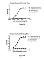
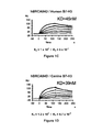
- FIGS. 1A-1F show binding data for antibodies binding to human B7-H3 (“hB7-H3”) versus canine B7-H3 (“cB7-H3”).
- FIGS. 1A-1B show ELISA data.
- FIGS. 1C-1D (hBRCA84D) and FIG. 1E-1F (BRCA69D) show a B7-H3 SPR analysis (Binding of mAbs (6.25-100 nM) to human and canine B7H3(4Ig)-His captured on anti-His surface (normalized; bivalent binding fit)).
- FIG. 2 Panels A-D, show representative IHC biopsies of feline cancers.
- FIG. 3 Panels A-F show representative IHC biopsies of canine cancers.
- FIG. 4 Panels A-F show representative IHC biopsies of canine cancers.
- FIGS. 5A-5D shows PBMCs from canine being redirected to kill canine B7-H3-positive tumor lines or a human B7-H3-positive tumor line.
- the present invention relates to bispecific molecules that are capable of localizing an immune effector cell that expresses an activating receptor to a B7-H3-expressing cancer cell, so as to thereby facilitate the killing of the cancer cell.
- such localization is accomplished using bispecific molecules that are immunoreactive both to an activating receptor of a companion animal immune effector cell and to B7-H3 expressed by a cancer cell.
- the present invention additionally concerns the use of such bispecific molecules in the treatment of cancer in companion animals.
- the invention particularly concerns bispecific diabody molecules that bind to (1) an epitope of an activating receptor of a companion animal immune effector cell and (2) an epitope of B7-H3 expressed by a cancer cell of such companion animal.
- the invention particularly concerns the embodiment wherein the bispecific molecules are capable of mediating, and more preferably enhancing, the activation and targeting of the companion animal's immune effector cells to B7-H3-expressing cancer cells such that the activated immune effector cells kill the B7-H3-expressing cancer cells.
- bispecific molecules to localize a immune effector cell that expresses an activating receptor to a B7-H3-expressing cancer cell permits such bispecific molecules to be used in the treatment of cancers afflicting companion animals.
- such localization can be achieved using bispecific molecules that bind B7-H3 and an activating receptor.
- such localization can be achieved using bispecific molecules that bind B7-H3 and a second molecule that is characteristically present on cells that express an activating receptor.
- the term “companion animal” refers to members of the canine family of animals (i.e., “dogs;” Canis lupus familiaris) and to members of the feline family of animals (i.e., “cats;” Felis catus or Felis silvestris catus ).
- the canine family includes pet dogs, working dogs and sporting dogs.
- the feline family includes house cats, barn cats and show cats.
- B7-H3 refers to a member of the B7 family of proteins, a type I membrane protein with Ig-like domains also known as CD276, and particularly to its canine and feline homologs.
- the term “2Ig-B7-H3” denotes a B7-H3 form that comprises only two Ig-like domains;
- the term “4Ig-B7-H3” denotes a B7-H3 form that comprises four Ig-like domains (see, Sun, M. et al. (2002) “ Characterization of Mouse and Human B 7- H 3 Genes ,” J. Immunol. 168:6294-6297; Steinberger et al.
- an “antibody” is an immunoglobulin molecule capable of specific binding to a target, such as a carbohydrate, polynucleotide, lipid, polypeptide, etc., through at least one antigen recognition site, located in the variable region of the immunoglobulin molecule.
- the term encompasses not only intact polyclonal or monoclonal antibodies, mutants thereof, naturally occurring variants, fusion proteins comprising an antibody portion with an antigen recognition site of the required specificity, humanized antibodies, chimeric antibodies and caninized antibodies, and any other modified configuration of the immunoglobulin molecule that comprises an antigen recognition site of the required specificity.
- the numbering of amino acid residues of the light and heavy chains of antibodies is according to the EU index as in Kabat et al. (1992) S EQUENCES OF P ROTEINS OF I MMUNOLOGICAL I NTEREST , National Institutes of Health Publication No. 91-3242.
- an “antigen binding fragment of an antibody” is a portion of an antibody that possesses an at least one antigen recognition site.
- the term encompasses fragments such as Fab, Fab′, F(ab′) 2 Fv), and single chain (scFv).
- the term “monoclonal antibody” refers to a homogeneous antibody population wherein the monoclonal antibody is comprised of amino acids (naturally occurring and non-naturally occurring) that are involved in the selective binding of an antigen. Monoclonal antibodies are highly specific, being directed against a single antigenic site. It is not intended to be limited as regards to the source of the antibody or the manner in which it is made (e.g., by hybridoma, phage selection, recombinant expression, transgenic animals, etc.). The term includes whole immunoglobulins as well as the fragments etc. described above under the definition of “antibody.”
- humanized antibody refers to a molecule, generally prepared using recombinant techniques, having an antigen binding site derived from an immunoglobulin from a non-human species and the remaining immunoglobulin structure of the molecule based upon the structure and/or sequence of a human immunoglobulin.
- the antigen-binding site may comprise either complete variable domains fused onto constant domains or only the complementarity determining regions (CDRs) grafted onto appropriate framework regions in the variable domains.
- Antigen binding sites may be wild type or modified by one or more amino acid substitutions. This eliminates the constant region as an immunogen in human individuals, but the possibility of an immune response to the foreign variable region remains (LoBuglio, A. F. et al.
- variable regions of both heavy and light chains contain three complementarity-determining regions (CDRs) which vary in response to the antigens in question and determine binding capability, flanked by four framework regions (FRs) which are relatively conserved in a given species and which putatively provide a scaffolding for the CDRs.
- CDRs complementarity-determining regions
- variable regions can be “reshaped” or “humanized” by grafting CDRs derived from non-human antibody on the FRs present in the human antibody to be modified.
- humanized antibodies preserve all CDR sequences (for example, a humanized mouse antibody which contains all six CDRs from the mouse antibodies).
- humanized antibodies have one or more CDRs (one, two, three, four, five or six) which are altered with respect to the original antibody, which are also termed one or more CDRs “derived from” one or more CDRs from the original antibody.
- caninized antibody refer to a chimeric molecule, generally prepared using recombinant techniques, having an antigen binding site derived from an immunoglobulin from a non-canine species and the remaining immunoglobulin structure of the molecule based upon the structure and/or sequence of a canine immunoglobulin.
- the techniques are similar to humanizing an antibody; however, canine framework and constant region sequences are used. See U.S. Pat. No. 7,261,890 for examples of such canine framework and constant region sequences
- BiTEs bi-specific T-cell engagers
- a target cell WO 05/061547; Baeuerle, P et al. (2008) “ BiTE: A New Class Of Antibodies That recruit T Cells ,” Drugs of the Future 33: 137-147; Bargou, et al. 2008) “ Tumor Regression in Cancer Patients by Very Low Doses of a T Cell - Engaging Antibody ,” Science 321: 974-977).
- diabody refers to a molecule that comprises at least two polypeptide chains that associate through a covalent interaction to form at least two epitope binding sites, which may recognize the same or different epitopes.
- Each of the polypeptide chains of a diabody comprises an immunoglobulin light chain variable region and an immunoglobulin heavy chain variable region, but these regions do not interact to form an epitope binding site. Rather, the immunoglobulin heavy chain variable region of one (e.g., the first) of the diabody polypeptide chains interacts with the immunoglobulin light chain variable region of a different (e.g., the second) diabody polypeptide chain to form an epitope binding site.
- the immunoglobulin light chain variable region of one (e.g., the first) of the diabody polypeptide chains interacts with the immunoglobulin heavy chain variable region of a different (e.g., the second) diabody polypeptide chain to form an epitope binding site.
- Diabodies may be monospecific, bispecific, trispecific, etc., thus being able to simultaneously bind one, two, three or more different epitopes (which may be of the same or of different antigens).
- Diabodies may additionally be monovalent, bivalent, trivalent, tetravalent, pentavalent, hexavelent, etc., thus being able to simultaneously bind one, two, three, four, five, six or more molecules.
- diabodies i.e., degree of specificity and valency may be combined, for example to produce bispecific antibodies (i.e., capable of binding two epitopes) that are tetravalent (i.e., capable of binding four sets of epitopes), etc.
- Diabody molecules are disclosed in PCT Publications WO 2006/113665, WO 2008/157379 and WO 2010/080538.
- an antibody or a polypeptide is said to “specifically” bind a region of another molecule (i.e., an epitope) if it reacts or associates more frequently, more rapidly, with greater duration and/or with greater affinity with that epitope relative to alternative epitopes.
- an antibody that specifically binds to a B7-H3 epitope is an antibody that binds this B7-H3 epitope with greater affinity, avidity, more readily, and/or with greater duration than it binds to other B7-H3 epitopes or non-B7-H3 epitopes.
- an antibody or moiety or epitope that specifically binds to a first target may or may not specifically or preferentially bind to a second target.
- “specific binding” does not necessarily require (although it can include) exclusive binding.
- reference to binding means “specific” binding.
- immunologically active in reference to an epitope being or “remaining immunologically active” refers to the ability of an antibody (e.g., an anti-B7-H3 antibody) to bind to the epitope under different conditions, for example, after the epitope has been subjected to reducing and denaturing conditions.
- an antibody e.g., an anti-B7-H3 antibody
- anti-B7-H3 antibodies including, but not limited to one or more of: an ability to specifically bind to B7-H3 (and in particular B7-H3 molecules that are expressed on the surfaces of canine and feline cancer cells (including but not limited to canine histiocytic sarcoma, hemangiosarcoma, malignant melanoma, mast cell tumor, osteosarcoma, thyroid carcinoma, transitional cell carcinoma, and squamous cell carcinoma cancer cells, and feline fibrosarcoma, mammary carcinoma and squamous cell carcinoma cancer cells); an ability to competitively inhibits preferential binding of a known anti-B7-H3-specific antibody to B7-H3, including the ability to preferentially bind to the same B7-H3 epitope to which such antibody preferentially binds; an ability to bind to a portion of B7-H3 that is exposed on the surface of a living cell in vitro or in vivo; an ability to bind
- anti-B7-H3 equivalent antibody or “anti-B7-H3 equivalent polypeptide” refers to an antibody or a polypeptide having one or more biological functions associated with an anti-B7-H3 antibody, such as, for example binding specificity.
- the term “agent” refers to a biological, pharmaceutical, or chemical compound.
- Non-limiting examples include simple or complex organic or inorganic molecule, a peptide, a protein, an oligonucleotide, an antibody, an antibody derivative, antibody fragment, a vitamin derivative, a carbohydrate, a toxin, or a chemotherapeutic compound.
- Various compounds can be synthesized, for example, small molecules and oligomers (e.g., oligopeptides and oligonucleotides), and synthetic organic compounds based on various core structures.
- various natural sources can provide compounds for screening, such as plant or animal extracts, and the like.
- Agents that are employed in the methods of this invention can be “randomly selected” or “rationally selected or designed.” As used herein, an agent is said to be “randomly selected” when the agent is chosen without prior consideration or knowledge of the specific amino acid or other chemical moieties involved in the association of the molecule with its native binding partner(s) or known antibodies.
- An example of a randomly selected agent is an agent that is identified through the use and screening of a chemical library or a peptide combinatorial library.
- an agent is said to be “rationally selected or designed” when the agent is chosen on a non-random basis that takes into account the sequence of the target site and/or its conformation in connection with the agent's action.
- This invention also encompasses agents that act at the sites of interaction between B7-H3 and its native binding partner, although other ligands and their active B7-H3-interactive sites are also encompassed within the scope of this invention, whether currently known or later identified.
- Agents can be rationally selected or rationally designed by utilizing the peptide sequences that make up the contact sites of the receptor/ligand and/or B7-H3/anti-B7-H3 antibody complex.
- a rationally selected peptide agent can be a peptide whose amino acid sequence is identical to an epitope appearing on B7-H3 as it is exposed on the surface of a living cell in its native environment.
- Such an agent will reduce or block the association of the anti-B7-H3 antibody with B7-H3, or the association of B7-H3 with its native ligand, as desired, by binding to the anti-B7-H3 antibody or to the native ligand.
- the term “labeled,” with regard to an antibody, is intended to encompass direct labeling of the antibody by coupling (i.e., physically linking) a detectable substance, such as a radioactive agent or a fluorophore (e.g. phycoerythrin (PE) or fluorescein isothiocyanate (also known as fluoroisothiocyanate or FITC)) to the antibody, as well as indirect labeling of the probe or antibody by reactivity with a detectable substance.
- a detectable substance such as a radioactive agent or a fluorophore (e.g. phycoerythrin (PE) or fluorescein isothiocyanate (also known as fluoroisothiocyanate or FITC)
- association includes covalent and non-covalent attachment or binding of an agent (e.g., chemotherapeutic agent) to the antibody.
- agent e.g., chemotherapeutic agent
- the antibody can be associated with an agent (e.g., chemotherapeutic agent) by direct binding or indirect binding via attachment to a common platform, such that the antibody directs the localization of the agent to the cancerous cell to which the antibody binds and wherein the antibody and agent do not substantially dissociate under physiological conditions such that the agent is not targeted to the same cancerous cell to which the antibody binds or such that the agent's potency is not decreased.
- biological sample encompasses a variety of sample types obtained from a companion animal that can be used in a diagnostic or monitoring assay.
- the definition encompasses saliva, blood and other liquid samples of biological origin, solid tissue samples such as a biopsy specimen or tissue cultures or cells derived therefrom, and the progeny thereof, for example, cells obtained from a tissue sample collected from a companion animal suspected of having cancer.
- the definition also includes samples that have been manipulated in any way after their procurement, such as by treatment with reagents, solubilization, or enrichment for certain components, such as proteins or polynucleotides, or embedding in a semi-solid or solid matrix for sectioning purposes.
- biological sample encompasses a clinical sample, and also includes cells in culture, cell supernatants, cell lysates, serum, plasma, biological fluid, and tissue samples.
- host cell includes an individual cell or cell culture that can be or has been a recipient for vector(s) for incorporation of polynucleotide inserts.
- Host cells include progeny of a single host cell, and the progeny may not necessarily be completely identical (in morphology or in genomic DNA complement) to the original parent cell due to natural, accidental, or deliberate mutation.
- a host cell includes cells transfected in vivo with a polynucleotide(s) of this invention.
- the term “delaying development of metastasis” means to defer, hinder, slow, retard, stabilize, and/or postpone development of metastasis. This delay can be of varying lengths of time, depending on the history of the cancer and/or individual being treated. As is evident to one skilled in the art, a sufficient or significant delay can, in effect, encompass prevention of metastasis, in that the animal does not develop the metastasis.
- an “effective amount” of a pharmaceutical composition in one embodiment, is an amount sufficient to effect beneficial or desired results including, without limitation, clinical results such as shrinking the size of the tumor, retardation of cancerous cell growth, delaying the development of metastasis, decreasing symptoms resulting from the disease, increasing the quality of life of those suffering from the disease, decreasing the dose of other medications required to treat the disease, enhancing the effect of another medication such as via targeting and/or internalization, delaying the progression of the disease, and/or prolonging survival of individuals.
- An effective amount can be administered in one or more administrations.
- an effective amount of drug, compound, or pharmaceutical composition is an amount sufficient to reduce the proliferation of (or destroy) cancerous cells and to reduce and/or delay the development, or growth, of metastases of cancerous cells, either directly or indirectly.
- an effective amount of a drug, compound, or pharmaceutical composition may or may not be achieved in conjunction with another drug, compound, or pharmaceutical composition.
- an “effective amount” may be considered in the context of administering one or more chemotherapeutic agents, and a single agent may be considered to be given in an effective amount if, in conjunction with one or more other agents, a desirable result may be or is achieved. While individual needs vary, determination of optimal ranges of effective amounts of each component is within the skill of the art.
- Typical dosages for antibody administration comprise one or more unit doses between 0.1-to 100 mg/kg/body weight.
- the preferred dosages comprise 1 to 100 mg/kg/body weight.
- the most preferred dosages comprise 10 to 100 mg/kg/body weight.
- Typical doses for bispecific molecule (e.g., antibodies, diabodies (multi-chain or BiTEs) administration comprise one or more unit doses of 0.0001 mg/kg/body weight to 100 mg/kg/body weight.
- the dosage administered is between 0.0001 mg/kg/body weight and 20 mg/kg/body weight, 0.0001 mg/kg/body weight and 10 mg/kg/body weight, 0.0001 mg/kg/body weight and 5 mg/kg/body weight, 0.0001 mg/kg/body weight and 2 mg/kg/body weight, 0.0001 mg/kg/body weight and 1 mg/kg/body weight, or 0.0001 mg/kg/body weight and 0.75 mg/kg/body weight
- nucleic acid molecule or agent, antibody, composition or cell, etc. is said to be “isolated” when that nucleic acid molecule, agent, antibody, composition, or cell, etc. is substantially separated from contaminant nucleic acid molecules, antibodies, agents, compositions, or cells, etc. naturally present in its original source.
- polypeptide “oligopeptide,” “peptide” and “protein” are used interchangeably herein to refer to polymers of amino acids of any length, but especially lengths greater than 5, 10, 15, 20 or 25 amino acid residues.
- the polymer may be linear or branched, it may comprise modified amino acids, and it may be interrupted by non-amino acids.
- the terms also encompass an amino acid polymer that has been modified naturally or by intervention; for example, disulfide bond formation, glycosylation, lipidation, acetylation, phosphorylation, or any other manipulation or modification, such as conjugation with a labeling component.
- polypeptides containing one or more analogs of an amino acid including, for example, unnatural amino acids, etc.
- polypeptides of this invention are based upon an antibody, the polypeptides can occur as single chains or as associated chains.
- peptidomimetics of the bispecific molecules described herein include peptides wherein at least one amino acid residue is substituted with an amino acid residue that is not commonly found in nature, such as the D isomer of the amino acid or an N-alkylated species of the amino acid.
- peptidomimetics are constructed by replacing at least one amide bond (—C( ⁇ O)—NH—) in a B7-H3 peptide agonist, antagonist or modulators with an amide isostere.
- Suitable amide isosteres include: —CH 2 —NH—, —CH 2 —S—, —CH 2 —S(O)—, —CH 2 —S(O) 2 —, —CH 2 —CH 2 —, —CH ⁇ CH— (E or Z form), —C( ⁇ O)—CH 2 —, —CH(CN)—NH—, —C(OH)—CH 2 —, and —O—C( ⁇ O)—NH—.
- amide bonds in a B7-H3 peptide agonist, antagonist or modulator that are suitable candidates for replacement with amide isosteres include bonds that are hydrolyzable by the endogenous esterases or proteases of the intended subject of B7-H3 peptide agonist, antagonist or modulator treatment.
- substantially pure refers to material that is at least 50% pure (i.e., free from contaminants), more preferably at least 90% pure, more preferably at least 95% pure, more preferably at least 98% pure, more preferably at least 99% pure, and most preferably greater than 99% pure.
- toxin refers to any substance, which effects an adverse response within a cell.
- a toxin directed to a cancerous cell would have an adverse, sometimes deleterious effect, on the cancerous cell.
- examples of toxins include, but are not limited to, radioisotopes, calicheamicin, and maytansinoids.
- treatment denote an approach for obtaining a beneficial or desired result including and preferably a beneficial or desired clinical result.
- beneficial or desired clinical results include, but are not limited to, one or more of the following: reducing the proliferation of (or destroying) cancerous cells or other diseased, reducing metastasis of cancerous cells found in cancers, shrinking the size of the tumor, decreasing symptoms resulting from the disease, increasing the quality of life of those suffering from the disease, decreasing the dose of other medications required to treat the disease, delaying the progression of the disease, and/or prolonging survival of companion animal recipients.
- cancer is intended to encompass a disease characterized by the presence of a cancer cell selected from the group consisting of a cell of an adrenal gland tumor, an AIDS-associated cancer, an alveolar sarcoma, an astrocytic tumor, a squamous cell carcinoma of the bladder, a transitional cell carcinoma of the bladder, a bone cancer, adamantinoma, aneurismal bone cysts, osteochondroma, osteosarcoma, a brain and spinal cord cancer, a metastatic brain tumor, a carotid body tumor, a cervical cancer, a chondrosarcoma, a chordoma, a chromophobe renal cell carcinoma, a clear cell carcinoma, a colon cancer, a colorectal cancer, a cutaneous benign fibrous histiocytoma, a desmoplastic small round cell tumor, an ependymoma, an extraskeletal myxoid chondrosarcoma,
- monoclonal antibodies are known in the art.
- One method which may be employed is the method of Kohler, G. et al. (1975) “ Continuous Cultures Of Fused Cells Secreting Antibody Of Predefined Specificity ,” Nature 256:495-497 or a modification thereof.
- monoclonal antibodies are developed in mice, rats or rabbits.
- the antibodies are produced by immunizing mice, rats or rabbits with an immunogenic amount of cells, cell extracts, or protein preparations that contain canine B7-H3 or feline B7-H3.
- the immunogen can be, but is not limited to, primary cells, cultured cell lines, cancerous cells, nucleic acids, or tissue.
- Cells used for immunization may be cultured for a period of time (e.g., at least 24 hours) prior to their use as an immunogen.
- Cells may be used as immunogens by themselves or in combination with a non-denaturing adjuvant, such as Ribi.
- a non-denaturing adjuvant such as Ribi.
- cells should be kept intact and preferably viable when used as immunogens. Intact cells may allow antigens to be better detected than ruptured cells by the immunized animal.
- Use of denaturing or harsh adjuvants, e.g., Freud's adjuvant may rupture cells and therefore is discouraged.
- the immunogen may be administered multiple times at periodic intervals such as, bi weekly, or weekly, or may be administered in such a way as to maintain viability in the animal (e.g., in a tissue recombinant).
- monoclonal antibodies that bind to B7-H3 are obtained by using host cells that over-express B7-H3 as an immunogen.
- host cells include, by way of example and not by limitation, canine histiocytic sarcoma, hemangiosarcoma, malignant melanoma, mast cell tumor, osteosarcoma, tyroid carcinoma, transitional cell carcinoma, squamous cell carcinoma cancer cells and feline fibrosarcoma, mammary carcinoma and squamous cell carcinoma cancer cells.
- a small biological sample e.g., blood
- the spleen and/or several large lymph nodes can be removed and dissociated into single cells.
- the spleen cells may be screened (after removal of non-specifically adherent cells) by applying a cell suspension to a plate or to a well coated with the antigen. B-cells, expressing membrane-bound immunoglobulin specific for the antigen, will bind to the plate, and are not rinsed away with the rest of the suspension.
- Resulting B-cells, or all dissociated spleen cells can then be fused with myeloma cells (e.g., X63-Ag8.653 and those from the Salk Institute, Cell Distribution Center, San Diego, Calif.).
- myeloma cells e.g., X63-Ag8.653 and those from the Salk Institute, Cell Distribution Center, San Diego, Calif.
- PEG Polyethylene glycol
- the hybridoma is then cultured in a selective medium (e.g., hypoxanthine, aminopterin, thymidine medium, otherwise known as “HAT medium”).
- a selective medium e.g., hypoxanthine, aminopterin, thymidine medium, otherwise known as “HAT medium”.
- the resulting hybridomas are then plated by limiting dilution, and are assayed for the production of antibodies that bind specifically to the immunogen, using, for example, FACS (fluorescence activated cell sorting) or immunohistochemistry (IHC) screening.
- FACS fluorescence activated cell sorting
- IHC immunohistochemistry
- Epstein-Barr Virus (EBV)-immortalized B cells may be used to produce monoclonal antibodies of the subject invention.
- the hybridomas are expanded and subcloned, if desired, and supernatants are assayed for anti-immunogen activity by conventional assay procedures (e.g., FACS, IHC, radioimmunoassay, enzyme immunoassay, fluorescence immunoassay, etc.).
- anti-B7-H3 monoclonal antibody and any other equivalent antibodies can be sequenced and produced recombinantly by any means known in the art.
- anti-B7-H3 monoclonal antibody is sequenced and the polynucleotide sequence is then cloned into a vector for expression or propagation.
- the sequence encoding the antibody of interest may be maintained in a vector in a host cell and the host cell can then be expanded and frozen for future use.
- the polynucleotide sequence of anti-B7-H3 monoclonal antibody and any other equivalent antibodies may be used for genetic manipulation to generate the bispecific molecules of the invention as well as a chimeric antibody, a humanized antibody, or a caninized antibody, to improve the affinity, or other characteristics of the antibody.
- the general principle in humanizing or caninizing an antibody involves retaining the basic sequence of the antigen-binding portion of the antibody, while swapping the non-human or non-canine remainder of the antibody with human antibody sequences or canine antibody sequences. There are four general steps to humanize or caninize a monoclonal antibody.
- caninized antibody molecules comprising an antigen-binding site derived from a non-canine immunoglobulin have been described, including chimeric antibodies having rodent or modified rodent V regions and their associated complementarity determining regions (CDRs) fused to canine constant domains (see, for example, U.S. Pat. No. 7,261,890). These caninized molecules are designed to minimize unwanted immunological response toward rodent anti-canine antibody molecules, which limits the duration and effectiveness of therapeutic applications of those moieties in canine recipients.
- CDRs complementarity determining regions
- Single chain variable region fragments may be made by linking light and/or heavy chain variable regions by using a short linking peptide.
- Bird et al. (1988) (“ Single - Chain Antigen - Binding Proteins ,” Science 242:423-426) describes example of linking peptides which bridge approximately 3.5 nm between the carboxy terminus of one variable region and the amino terminus of the other variable region.
- Linkers of other sequences have been designed and used (Bird et al. (1988) “ Single - Chain Antigen - Binding Proteins ,” Science 242:423-426). Linkers can in turn be modified for additional functions, such as attachment of drugs or attachment to solid supports.
- the single chain variants can be produced either recombinantly or synthetically.
- an automated synthesizer can be used for synthetic production of scFv.
- a suitable plasmid containing polynucleotide that encodes the scFv can be introduced into a suitable host cell, either eukaryotic, such as yeast, plant, insect or mammalian cells, or prokaryotic, such as E. coli .
- a suitable host cell either eukaryotic, such as yeast, plant, insect or mammalian cells, or prokaryotic, such as E. coli .
- Polynucleotides encoding the scFv of interest can be made by routine manipulations such as ligation of polynucleotides.
- the resultant scFv can be isolated using standard protein purification techniques known in the art.
- the invention includes modifications to the bispecific molecules of the invention that do not significantly affect their properties and variants that have enhanced or decreased activity. Modification of polypeptides is routine practice in the art and need not be described in detail herein. Examples of modified polypeptides include polypeptides with conservative substitutions of amino acid residues, one or more deletions or additions of amino acids which do not significantly deleteriously change the functional activity, or use of chemical analogs Amino acid residues which can be conservatively substituted for one another include but are not limited to: glycine/alanine; valine/isoleucine/leucine; asparagine/glutamine; aspartic acid/glutamic acid; serine/threonine; lysine/arginine; and phenylalanine/tyrosine.
- polypeptides also include glycosylated and non-glycosylated polypeptides, as well as polypeptides with other post-translational modifications, such as, for example, glycosylation with different sugars, acetylation, and phosphorylation.
- amino acid substitutions would be conservative, i.e., the substituted amino acid would possess similar chemical properties as that of the original amino acid. Such conservative substitutions are known in the art, and examples have been provided above Amino acid modifications can range from changing or modifying one or more amino acids to complete redesign of a region, such as the variable region. Changes in the variable region can alter binding affinity and/or specificity.
- Modifications include using coupling techniques known in the art, including, but not limited to, enzymatic means, oxidative substitution and chelation. Modifications can be used, for example, for attachment of labels for immunoassay, such as the attachment of radioactive moieties for radioimmunoassay. Modified polypeptides are made using established procedures in the art and can be screened using standard assays known in the art.
- the invention also encompasses fusion proteins comprising one or more fragments or regions from the polypeptides and antibodies of this invention.
- a fusion polypeptide is provided that comprises at least 10 contiguous amino acids of variable light chain region and at least 10 amino acids of variable heavy chain region.
- the fusion polypeptide contains a heterologous immunoglobulin constant region.
- the fusion polypeptide contains a light chain variable region and a heavy chain variable region of an antibody produced from a publicly-deposited hybridoma.
- an antibody fusion protein contains one or more polypeptide domains that specifically bind to B7-H3 and another amino acid sequence to which it is not attached in the native molecule, for example, a heterologous sequence or a homologous sequence from another region.
- An anti-B7-H3 polypeptide, and other B7-H3 agonists, antagonists and modulators can be created by methods known in the art, for example, synthetically or recombinantly.
- One method of producing B7-H3 peptide agonists, antagonists and modulators involves chemical synthesis of the polypeptide, followed by treatment under oxidizing conditions appropriate to obtain the native conformation, that is, the correct disulfide bond linkages. This can be accomplished using methodologies well known to those skilled in the art (see, e.g., Kelley, R. F. et al. (1990) In: G ENETIC E NGINEERING P RINCIPLES AND M ETHODS , Setlow, J. K.
- Polypeptides of the invention may be conveniently prepared using solid phase peptide synthesis (Merrifield, B. (1986) “Solid Phase Synthesis,” Science 232(4748):341-347; Houghten, R. A. (1985) “General Method For The Rapid Solid - Phase Synthesis Of Large Numbers Of Peptides: Specificity Of Antigen - Antibody Interaction At The Level Of Individual Amino Acids ,” Proc. Natl. Acad. Sci. (U.S.A.) 82(15):5131-5135; Ganesan, A. (2006) “ Solid - Phase Synthesis In The Twenty - First Century ,” Mini Rev. Med. Chem. 6(1):3-10).
- antibodies may be made recombinantly and expressed using any method known in the art.
- Antibodies may be made recombinantly by first isolating the antibodies made from host animals, obtaining the gene sequence, and using the gene sequence to express the antibody recombinantly in host cells (e.g., CHO cells). Another method that may be employed is to express the antibody sequence in plants (e.g., tobacco) or transgenic milk. Suitable methods for expressing antibodies recombinantly in plants or milk have been disclosed (see, for example, Peeters et al. (2001) “ Production Of Antibodies And Antibody Fragments In Plants ,” Vaccine 19:2756; Lonberg, N. et al.
- the antibodies or protein of interest may be subjected to sequencing by Edman degradation, which is well known to those of skill in the art.
- Edman degradation which is well known to those of skill in the art.
- the peptide information generated from mass spectrometry or Edman degradation can be used to design probes or primers that are used to clone the protein of interest.
- An alternative method of cloning the protein of interest is by “panning” using purified B7-H3 or portions thereof for cells expressing the antibody or protein of interest.
- the amino acid sequence of human B7-H3 is (SEQ ID NO:1):
- the cDNA sequence encoding human B7-H3 is (SEQ ID NO:2):
- amino acid sequence of canine B7-H3 is (SEQ ID NO:3):
- the cDNA encoding canine B7-H3 is (SEQ ID NO:4):
- the amino acid sequence of feline B7-H3 is (SEQ ID NO:5):
- the cDNA sequence of feline B7-H3 is (SEQ ID NO:6):
- the “panning” procedure may be conducted by obtaining a cDNA library from tissues or cells that express B7-H3, over-expressing the cDNAs in a second cell type, and screening the transfected cells of the second cell type for a specific binding to B7-H3.
- Detailed descriptions of the methods used in cloning mammalian genes coding for cell surface proteins by “panning” can be found in the art (see, for example, Aruffo, A. et al. (1987) “ Molecular Cloning Of A CD 28 cDNA By A High - Efficiency COS Cell Expression System ,” Proc. Natl. Acad. Sci. (U.S.A.) 84:8573-8577 and Stephan, J. et al. (1999) “ Selective Cloning Of Cell Surface Proteins Involved In Organ Development: Epithelial Glycoprotein Is Involved In Normal Epithelial Differentiation ,” Endocrinol. 140:5841-5854).
- cDNAs encoding anti-B7-H3 antibodies, and other B7-H3 peptide agonists, antagonists and modulators can be obtained by reverse transcribing the mRNAs from a particular cell type according to standard methods in the art. Specifically, mRNA can be isolated using various lytic enzymes or chemical solutions according to the procedures set forth in Sambrook et al. supra or extracted by commercially available nucleic-acid-binding resins following the accompanying instructions provided by manufacturers (e.g., Qiagen, Invitrogen, Promega). The synthesized cDNAs are then introduced into an expression vector to produce the antibody or protein of interest in cells of a second type.
- Suitable expression vectors include but are not limited to plasmids, viral vectors, including adenoviruses, adeno-associated viruses, retroviruses, and cosmids.
- the vectors containing the polynucleotides of interest can be introduced into the host cell by any of a number of appropriate means, including electroporation, transfection employing calcium chloride, rubidium chloride, calcium phosphate, DEAE-dextran, or other substances; microprojectile bombardment; lipofection; and infection (e.g., where the vector is an infectious agent such as vaccinia virus).
- electroporation employing calcium chloride, rubidium chloride, calcium phosphate, DEAE-dextran, or other substances
- microprojectile bombardment e.g., where the vector is an infectious agent such as vaccinia virus.
- infection e.g., where the vector is an infectious agent such as vaccinia virus.
- the choice of introducing vectors or polynucleotides will often depend on features of the host cell.
- Any host cells capable of over-expressing heterologous DNAs can be used for the purpose of isolating the genes encoding the antibody, polypeptide or protein of interest.
- suitable mammalian host cells include but are not limited to COS, HeLa, and CHO cells.
- the host cells express the cDNAs at a level of about 5-fold higher, more preferably 10-fold higher, even more preferably 20-fold higher than that of the corresponding endogenous antibody or protein of interest, if present, in the host cells.
- Screening the host cells for a specific binding to B7-H3 is effected by an immunoassay or FACS. A cell over-expressing the antibody or protein of interest can be identified.
- mutant B7-H3 peptide agonists, antagonists, and modulators which encodes for additions, deletions, or changes in amino acid sequence of the resultant protein relative to the parent B7-H3 peptide agonist, antagonist or modulator molecule.
- the invention includes polypeptides comprising an amino acid sequence of the antibodies of this invention.
- the polypeptides of this invention can be made by procedures known in the art.
- the polypeptides can be produced by proteolytic or other degradation of the antibodies, by recombinant methods (i.e., single or fusion polypeptides) as described above or by chemical synthesis.
- Polypeptides of the antibodies, especially shorter polypeptides up to about 50 amino acids, are conveniently made by chemical synthesis. Methods of chemical synthesis are known in the art and are commercially available.
- an anti-B7-H3 polypeptide could be produced by an automated polypeptide synthesizer employing the solid phase method.
- binding refers to biologically or immunologically relevant specific binding, and does not refer to non-specific binding that may occur, for example, when an immunoglobulin is used at a very high concentration against a non-specific target.
- monoclonal antibodies are screened for binding to B7-H3 using standard screening techniques. In this manner, anti-B7-H3 monoclonal antibody was obtained.
- the preferred hybridomas of the present invention are those that produce antibodies BRCA69D, BRCA84D or PRCA157 (WO 2011/109400).
- monoclonal antibodies that bind to B7-H3 may be identified. For this purpose, monoclonal antibodies are screened for their differential ability to bind to cancerous tissues but not to non-cancerous cells. In one embodiment, monoclonal antibodies which bind to B7-H3 and that are also cross-reactive to human cancerous cells or tissues, but not to normal cells or tissues to the same degree, are selected.
- One method that may be employed for screening is immunohistochemistry (IHC). Standard immunohistochemical techniques are known to those of average skill in the art. See, for example, A NIMAL C ELL C ULTURE M ETHODS (J. P. Mather and D. Barnes, eds., Academic Press, NY, Vol. 57, Ch. 18 and 19, pp.
- Bio samples e.g., tissues
- tissue may be obtained from biopsies, autopsies, or necropsies.
- anti-B7-H3 antibodies may be used to detect the presence of B7-H3 on tissues from individuals with cancer while other non-cancerous tissues from the individual suffering from cancer or tissues from individuals without cancer are used as a control.
- the tissue can be embedded in a solid or semi-solid substance that prevents damage during freezing (e.g., agarose gel or OCT) and then sectioned for staining Cancers from different organs and at different grades can be used to screen monoclonal antibodies.
- tissues that may be used for screening purposes include but are not limited to ovary, breast, lung, prostate, colon, kidney, skin, thyroid, brain, heart, liver, stomach, nerve, blood vessels, bone, upper digestive tract, and pancreas.
- cancer types that may be used for screening purposes include, but are not limited to, carcinomas, adenocarcinomas, sarcomas, adenosarcomas, lymphomas, and leukemias.
- cancerous cells lines such as canine CF21 and CF32 and human HMEC (BioWhittaker CC-2251), HUVEC (Primary endothelial cells), BT-474 (ATCC#HTB-20), MCF7 (ATCC#HTB22), MDA-MB-175-VII (ATCC#HB-25), MDA-MB-361 (ATCC#HB-27), SKBR3 (ATCC#HTB-30), A549 (ATCC#CCL-185), Calu-3 (ATCC#HTB-55), SKMES-I (ATCC#HTB-58), ES-2 (ATCC#CRL-1978), SKOV3 (ATCC#HTB-77), Panc-1 (ATCC#CRL-1469), AsPC-I (ATCC#CRL-1682), HPAF-II (ATCC#CRL-1997), Hs700T (ATCC#HTB-174), Colo205 (ATCC#CCL-222), HT-29 (ATCC#HTB-38),
- cancerous or non-cancerous cells can be grown on glass slides or coverslips, or on plastic surfaces, or prepared in a CellArrayTM device, as described in WO 01/43869, and screened for the binding of antibody using IHC as described above for tissues.
- cells may be removed from the growth surface using non-proteolytic means and spun into a pellet, which is then embedded and treated as tissues for IHC analysis as described above.
- Cells may be inoculated into immunodeficient animals, a tumor allowed to grow, and then this tumor may be harvested, embedded, and used as a tissue source for IHC analysis.
- single cells may be screened by incubating with the primary antibody, a secondary “reporter” antibody linked to a fluorescent molecule and then analyzed using a fluorescent activated cell-sorting (FACS) machine.
- FACS fluorescent activated cell-sorting
- any of several different detection systems may be utilized to detect binding of antibodies to tissue section.
- immunohistochemistry involves the binding of a primary antibody to the tissue and then a secondary antibody reactive against the species from the primary antibody was generated and conjugated to a detectable marker (e.g., horseradish peroxidase, HRP, or diaminobenzedine, DAB).
- a detectable marker e.g., horseradish peroxidase, HRP, or diaminobenzedine, DAB.
- HRP horseradish peroxidase
- DAB diaminobenzedine
- polyMICATM polyclonal Mirror Image Complementary Antibodies
- the PolyMICATM technique can be used to test binding of primary antibodies (e.g., anti-B7-H3 antibodies) to normal and cancerous tissue.
- primary antibodies e.g., anti-B7-H3 antibodies
- polyMICATM Detection kits are commercially available: Product No. HK004.D is a polyMICATM Detection kit which uses DAB chromagen; Product No. HK004.A is a polyMICATM Detection kit which uses AEC chromagen.
- the primary antibody may be directly labeled with the detectable marker.
- the first step in IHC screening to select for an appropriate antibody is the binding of primary antibodies raised in mice (e.g., anti-B7-H3 antibodies) to one or more immunogens (e.g., cells or tissue samples).
- immunogens e.g., cells or tissue samples.
- the tissue sample is sections of frozen tissue from different organs.
- the cells or tissue samples can be either cancerous or non-cancerous.
- Frozen tissues can be prepared, sectioned, with or without fixation, and IHC performed by any of a number of methods known to one familiar with the art (see, for example, Stephan et al. (1999) “ Distribution And Function Of The Adhesion Molecule BEN During Rat Development ,” Dev. Biol. 212:264-277 and Stephan et al. (1999) “ Selective Cloning Of Cell Surface Proteins Involved In Organ Development Epithelial Glycoprotein Is Involved In Normal Epithelial Differentiation ,” Endocrinology 140:5841-5854).
- Epitope mapping is commercially available from various sources, for example, Pepscan Systems (Lelystad, The Netherlands). Epitope mapping can be used to determine the sequence to which an anti-B7-H3 antibody binds.
- the epitope can be a linear epitope, i.e., contained in a single stretch of amino acids, or a conformational epitope formed by a three-dimensional interaction of amino acids that may not necessarily be contained in a single stretch.
- Peptides of varying lengths can be isolated or synthesized (e.g., chemically or recombinantly) and used for binding assays with anti-B7-H3 antibody.
- the epitope to which anti-B7-H3 antibody binds can be determined in a systematic screening by using overlapping peptides derived from the extracellular sequence and determining binding by anti-B7-H3 antibody.
- Yet another method that can be used to characterize an anti-B7-H3 antibody is to use competition assays with other antibodies known to bind to the same antigen, i.e., B7-H3 to determine if anti-B7-H3 antibodies binds to the same epitope as other antibodies.
- B7-H3 examples of commercially available antibodies to B7-H3 may be available and may be identified using the binding assays taught herein.
- Competition assays are well known to those of skill in the art, and such procedures and illustrative data are detailed further in the Examples.
- Anti-B7-H3 antibodies can be further characterized by the tissues, type of cancer or type of tumor to which they bind.
- a central aspect of the molecules of the present invention relates to their ability to bind canine B7-H3 and/or feline B7-H3.
- such antibodies may be readily identified by screening among human B7-H3 reactive antibodies for those that exhibit the ability to additionally bind to the B7-H3 of a desired companion animal.
- Non-limiting examples of such antibodies include BRCA69D, BRCA84D and PRCA157 (WO 2011/109400)
- Monoclonal antibodies to B7-H3 made by the methods disclosed herein may be used to identify the presence or absence of cancerous cells in a variety of companion animal tissues, including but not limited to, blood, ovary, breast, lung, prostate, colon, kidney, pancreas, skin, thyroid, brain, heart, liver, stomach, nerve, blood vessels, bone, and upper digestive tract, for purposes of diagnosis.
- Monoclonal antibodies to B7-H3 made by the methods disclosed herein may also be used to identify the presence or absence of cancerous cells, or the level thereof, which are circulating in blood after their release from a solid tumor.
- Such circulating antigen may be an intact B7-H3 antigen, or a fragment thereof that retains the ability to be detected according to the methods taught herein.
- Such detection may be effected by FACS analysis using standard methods commonly used in the art. Since such antibodies are capable of binding to the B7-H3 of a canine and/or feline animal, they may be used to diagnose the presence of a B7-H3-associated cancer in such animals.
- the antibody bears a detectable label.
- labels that may be used include a radioactive agent or a fluorophore, such as phycoerythrin or fluorescein isothiocyanate (also known as fluoroisothiocyanate or FITC).
- One method of using the antibodies for diagnosis is in vivo tumor imaging by linking the antibody to a radioactive or radio-opaque agent, administering the antibody to the individual and using an x-ray or other imaging machine to visualize the localization of the labeled antibody at the surface of cancer cells expressing the antigen.
- the antibody is administered at a concentration that promotes binding at physiological conditions.
- ELISAs enzyme linked immunosorbent assays
- IA enzyme immunoassay
- RIA radioimmunoassay
- radiolabeled antibody may be a monoclonal or polyclonal antibody comprising a radiolabel, preferably selected from the group consisting of Technetium-99m, Indium-111, Iodine-131, Rhenium-186, Rhenium-188, Samarium-153, Lutetium-177, Copper-64, Scandium-47, Yttrium-90.
- Radioimaging may be conducted using Single Photon Emission Computer Tomography (SPECT), Position Emission Tomography (PET), Computer Tomography (CT) or Magnetic Resonance Imaging (MRI). Correlative imaging, which permits greater anatomical definition of location of metastases located by radioimmunoimaging, is also contemplated.
- SPECT Single Photon Emission Computer Tomography
- PET Position Emission Tomography
- CT Computer Tomography
- MRI Magnetic Resonance Imaging
- the cancerous tissues or cells are removed from the companion animal and prepared for immunohistochemistry by methods well known in the art (e.g., embedding in a freezing compound, freezing and sectioning, with or without fixation; fixation and paraffin embedding with or without various methods of antigen retrieval and counterstaining).
- the monoclonal antibodies may also be used to identify cancerous tissues or cells at different stages of development.
- the antibodies may also be used to determine which individuals' tumors or cells express the antigen on their surface at a pre-determined level and are thus candidates for immunotherapy using antibodies directed against said antigen.
- the antibodies may recognize both primary and metastasizing cancers of the blood, ovary, breast, lung, prostate, colon, kidney, pancreas, skin, thyroid, brain, heart, liver, stomach, nerve, blood vessels, bone, and upper digestive tract that express B7-H3.
- detection may include qualitative and/or quantitative detection and may include comparing the level measured to a normal cell for an increased level of expression of B7-H3 in cancerous cells.
- the invention also provides methods of aiding diagnosis of cancer (such as but not limited to blood, ovary, breast, lung, prostate, colon, kidney, pancreas, skin, thyroid, brain, heart, liver, stomach, nerve, blood vessels, bone, and upper digestive tract cancers) in a companion animal using the anti-B7-H3 antibodies of the present invention, or any of the derivatives.
- methods for “aiding diagnosis” means that these methods assist in making a clinical determination regarding the classification, or nature, of cancer, and may or may not be conclusive with respect to the definitive diagnosis.
- a method of aiding diagnosis of a companion animal cancer can comprise the step of detecting the level of B7-H3 in a biological sample from the animal and/or determining the level of B7-H3 expression in the sample.
- Antibodies recognizing the antigen or a portion thereof may also be used to create diagnostic immunoassays for detecting canine B7-H3 antigen or feline B7-H3 antigen of such animals released or secreted from living or dying cancer cells in bodily fluids, including but not limited to, blood, saliva, urine, pulmonary fluid, or ascites fluid.
- B7-H3 not all cells in a particular tumor of interest will express B7-H3, and cancerous cells or tissues in other tissues may express B7-H3, thus a companion animal should be screened for the presence or absence of B7-H3 on cancerous cells or tissues to determine the usefulness of immunotherapy in the animals.
- the anti-B7-H3 molecules disclosed herein may be used to determine whether a companion animal diagnosed with cancer may be deemed a candidate for immunotherapy directed against B7-H3.
- a cancerous tumor or a biopsy sample may be tested for expression of B7-H3, using the anti-B7-H3 molecules of the present invention.
- a companion animal whose cancer cells express B7-H3 is a suitable candidate for immunotherapy using the anti-B7-H3 molecules, and particularly the bispecific molecules, of the present invention. Staining with such anti-B7-H3 molecules may also be used to distinguish cancerous tissues from normal tissues in the companion animal.
- Methods of using such anti-B7-H3 antibodies for diagnostic purposes are useful both before and after provision of any form of anti-cancer treatment, e.g., chemotherapy or radiation therapy, to determine which tumors are most likely to respond to a given treatment, to assess prognosis of the disease, to assess tumor subtype or origin of metastatic disease, and to assess the progression of the disease or its response to treatment.
- any form of anti-cancer treatment e.g., chemotherapy or radiation therapy
- Monoclonal antibodies to B7-H3 made by the methods disclosed herein may be used to identify the presence or absence of cancer stem cells in a variety of tissues.
- Cancer stem cells CSCs have been hypothesized to play a role in tumor growth and metastasis (Ghotra, V. P. et al. (2009) “ The Cancer Stem Cell Microenvironment And Anti - Cancer Therapy ,” Int. J. Radiat. Biol. 85(11):955-962; Gupta, P. B. et al. (2009) “ Cancer Stem Cells: Mirage Or Reality? ” Nat. Med. 15(9):1010-1012; Lawson, J. C. et al.
- the CSCs provide a small, distinct subset of cells within each tumor that are capable of indefinite self-renewal and of developing into the more adult tumor cell(s) that are relatively limited in replication capacity. It has been hypothesized that these cancer stem cells might be more resistant to chemotherapeutic agents, radiation or other toxic conditions, and thus, persist after clinical therapies and later grow into secondary tumors, metastases or be responsible for relapse. It has been suggested that CSCs can arise either from ‘normal’ tissue stem cells or from more differentiated tissue progenitor cells. While supporting data for this hypothesis is strong for hematopoietic stem and progenitor cells and hematopoietic tumors, less is known about solid tumors and their respective CSCs.
- Cancer stem cells have several defining characteristics. Such characteristics are described in PCT Publication WO 2008/091908. Monoclonal antibodies to cell surface targets on cancer stem cells can be used to identify the presence or absence of cancer stem cells in a variety of tissues. Monoclonal antibodies to B7-H3 made by the methods disclosed herein may also be used to identify the presence or absence of cancer stem cells, or the level of cancer stem cells in a sample or tissue or in circulation after their release from a solid tumor. Such circulating antigen may be an intact B7-H3 antigen, or a fragment thereof that retains the ability to be detected according to the methods taught herein. Such detection may be effected by FACS analysis using standard methods commonly used in the art. In another embodiment, such detection may be effected by immunohistochemical analysis of tissue samples using standard methods commonly used in the art.
- B7-H3 can involve the formation of a complex between B7-H3 and an antibody that binds specifically to B7-H3 on cancer stem cells.
- antibodies include but are not limited to those anti-B7-H3 monoclonal antibodies produced by the hybridomas BRCA84D, BRCA69D, and PRCA157.
- the formation of such a complex can be in vitro or in vivo.
- anti-B7-H3 molecules described in this application also encompass the use of other B7-H3 agonists, antagonists and modulators as described herein for the use of identification and treatment of cancer stem cells.
- anti-B7-H3 antibodies and other B7-H3 agonists, antagonists and modulators are used for identification, diagnosis or therapeutic treatment of cancer stem cells using similar methods described, and alterations within the scope of the ordinary skilled practitioner are made to tailor the method to the identification/diagnosis or treatment of cancer stem cells.
- compositions including pharmaceutical compositions, comprising the bispecific molecules of the invention, anti-B7-H3 antibodies, polypeptides derived from anti-B7-H3 antibodies, polynucleotides comprising sequence encoding anti-B7-H3 antibodies, and other agents as described herein.
- the invention further provides for conjugates of any B7-H3 peptide agonist, antagonist or modulator, and additional chemical structures that support the intended function or functions of the particular B7-H3 peptide agonist, antagonist or modulator.
- conjugates include B7-H3 peptide agonists, antagonists or modulators covalently bound to a macromolecule such as any insoluble, solid support matrix used in the diagnostic, screening or purification procedures discussed herein.
- Suitable matrix materials include any substance that is chemically inert, has high porosity and has large numbers of functional groups capable of forming covalent linkages with peptide ligands. Examples of matrix materials and procedures for preparation of matrix-ligand conjugates are described in Dean et al. (Eds) A FFINITY C HROMATOGRAPHY : A P RACTICAL A PPROACH , IRL Press (1985); Lowe, “ An Introduction to Affinity Chromatography ”, in Work et al.
- B7-H3 peptide agonist, antagonist or modulator conjugates of B7-H3 peptide agonist, antagonist or modulator and any reporter moiety used in the diagnostic procedures discussed herein.
- the B7-H3 peptide agonist, antagonist or modulator agents, polypeptides and proteins of this invention, including anti-B7-H3 antibodies, are further identified and characterized by an ability to specifically bind to B7-H3 molecules that are expressed on the surfaces of human and companion animal cancer cells, and optionally any (one or more) of the following criteria:
- a preferred antibody of the invention will exhibit differential IHC staining of tumor tissue relative to normal, non-cancerous tissue.
- the bispecific molecules of the invention are made from an antibody that is produced by hybridoma BRCA84D, BRCA69D, or PRCA157, or progeny thereof.
- BRCA84D, BRCA69D, and PRCA157 are the preferred B7-H3 antibodies of the present invention due to their cleaner normal tissue IHC profiles, stronger tumor/normal IHC differential, moderate to strong binding (BIACORETM)/IHC), reactivity to canine B7-H3 and feline B7-H3, cross-reactivity to human B7-H3 and cross-reactivity of cynomolgus monkey B7-H3.
- the invention also provides polypeptides comprising an amino acid sequence of the antibodies of the invention.
- the polypeptide comprises one or more of the light chain and/or heavy chain variable regions of the antibody.
- the polypeptide comprises one or more of the light chain and/or heavy chain CDRs of the antibody.
- the polypeptide comprises three CDRs of the light chain and/or heavy chain of the antibody.
- the polypeptide comprises an amino acid sequence of the antibody that has any of the following: at least 5 contiguous amino acids of a sequence of the original antibody, at least 8 contiguous amino acids, at least about 10 contiguous amino acids, at least about 15 contiguous amino acids, at least about 20 contiguous amino acids, at least about 25 contiguous amino acids, at least about 30 contiguous amino acids, wherein at least 3 of the amino acids are from a variable region of the antibody.
- the variable region is from a light chain of the original antibody.
- the variable region is from a heavy chain of the antibody.
- the 5 (or more) contiguous amino acids are from a complementarity-determining region (CDR) of the antibody.
- variable light chain and variable heavy chain of BRCA84D, BRCA69D and PRCA157 are shown below along with the respective CDR 1 , CDR 2 and CDR 3 domains of each such chain.
- CDR 1 , CDR 2 and CDR 3 domains of each such chain are shown below along with the respective CDR 1 , CDR 2 and CDR 3 domains of each such chain.
- Those of skill in the art will therefore be able to construct bispecific molecules having such CDRs, as well as antibodies and derivatives thereof, capable of binding to the epitopes recognized by BRCA84D, BRCA69D and PRCA157.
- BRCA84D Variable Heavy Chain CDR 2 (SEQ ID NO:19): YISSDSSAIYYADTVK
- BRCA69D Variable Heavy Chain CDR 2 (SEQ ID NO:35): TIYPGDGDTR YTQKFKG
- PRCA157 Variable Light Chain CDR 1 (SEQ ID NO:41): RASESIYSYLA
- PRCA157 Variable Heavy Chain CDR 1 SEQ ID NO:49: SYGMS
- PRCA157 Variable Heavy Chain CDR 2 (SEQ ID NO:51): VATINSGGSN TYYPDSLKG
- PRCA157 Variable Heavy Chain CDR 3 (SEQ ID NO:53): HDGGAMDY
- Monoclonal antibody BRCA84D was humanized in order to produce antibodies (generically designated herein as “hBRCA84D”) offering improved human therapeutic potential.
- the sequences of the variable light chain, and the variable heavy chain, and their respective amino acid and polynucleotide sequences of a resulting humanized antibody are provided below:
- hBRCA84D-1VL or hBRCA84D-1VH polynucleotides encoding the light or heavy chains of hBRCA84D-1 (i.e., hBRCA84D-1VL or hBRCA84D-1VH, respectively) were subjected to mutagenesis, and mutated hBRCA84D-1 light chain derivatives hBRCA84D-2VL, hBRCA84D-3VL, hBRCA84D-4VL, hBRCA84D-5VL, and hBRCA84D-6VL and mutated hBRCA84D-1 heavy chain derivatives hBRCA84D-2VH, hBRCA84D-3VH, and hBRCA84D-4VH were isolated and characterized.
- the amino acid and polynucleotide sequences of the variable light and heavy chains of these antibodies are presented below:
- hBRCA84D-2VL (SEQ ID NO:71):
- hBRCA84D-3VL (SEQ ID NO:73):
- hBRCA84D-4VL (SEQ ID NO:75):
- hBRCA84D-5VL (SEQ ID NO:77):
- hBRCA84D-6VL (SEQ ID NO:79):
- hBRCA84D-2VH (SEQ ID NO:81):
- hBRCA84D-3VH (SEQ ID NO:83):
- hBRCA84D-4VH (SEQ ID NO:85):
- Table 1 list the hBRCA84D variable light chain and variable heavy chain mutations studied; number refers to the kabat numbering system.
- amino acid and encoding polynucleotide sequences of the chimeric BRCA84D-1 are as follows:
- chBRCA84D Heavy Chain (SEQ ID NO:89):
- Antibodies immunoreactive to an activating receptor e.g., CD3, CD16, CD32, CD64, T-cell receptor, NKG2D, etc.
- an activating receptor e.g., CD3, CD16, CD32, CD64, T-cell receptor, NKG2D, etc.
- Particularly preferred anti-CD3 antibodies are K9CD3-1 and K9CD3-2 which are immunoreactive to canine CD3.
- the amino acid and encoding polynucleotide sequences of the variable light chain and variable heavy chain of K9CD3-1 and K9CD3-2 are shown below along with the respective CDR 1 , CDR 2 , and CDR 3 domains of each such chain.
- Those of skill in the art will therefore be able to construct bispecific molecules having such CDRs, as well as antibodies and derivatives thereof, capable of binding to the epitopes recognized by K9CD3-1 and K9CD3-2.
- K9CD3-1 Variable Light Chain CDR 1 (SEQ ID NO:93): RASQSIFKNLH
- K9CD3-1 Variable Light Chain CDR 2 (SEQ ID NO:95): YASDSISGIPS
- K9CD3-1 Variable Light Chain CDR 3 (SEQ ID NO:97): LQAYSTPWT
- K9CD3-1 Variable Heavy Chain CDR 1 (SEQ ID NO:101): DYNMH
- K9CD3-1 Variable Heavy Chain CDR 2 (SEQ ID NO:103): YIYPYNGGTYYNQKFKS
- K9CD3-1 Variable Heavy Chain CDR 3 (SEQ ID NO:105): LVYFDY
- NIVMTQSPKS MSMSVGERVT LNCKASENVD SYVSWYQQKP KQSPTLLIYG ASNRSTGVPD RFTGSGSVTD FTLTITSVQA EDLAYYHCGQ TYSYPFTFGS GTKLEIK
- K9CD3-2 Variable Light Chain CDR 1 (SEQ ID NO:109): KASENVDSYVS
- K9CD3-2 Variable Light Chain CDR 2 (SEQ ID NO:111): GASNRST
- K9CD3-2 Variable Light Chain CDR 3 (SEQ ID NO:113): GQTYSYPFT
- K9CD3-2 Variable Heavy Chain CDR 1 (SEQ ID NO:117): IFAMN
- K9CD3-2 Variable Heavy Chain CDR 2 (SEQ ID NO:119): RIRSKNNYFATYYAASVRD
- K9CD3-2 Variable Heavy Chain CDR 3 (SEQ ID NO:121): RGYFDV
- the present invention additionally encompasses “DART” (dual affinity retargeting reagent) diabody molecules that comprise at least two polypeptide chains which form at least two epitope binding sites, at least one of which specifically binds to B7-H3.
- DART dual affinity retargeting reagent
- the first polypeptide chain of the DART diabody comprises:
- the second polypeptide chain of such a DART diabody comprises:
- the DART diabody domains (A) and (B) do not associate with one another to form an epitope binding site. Similarly, the DART diabody domains (D) and (E) do not associate with one another to form an epitope binding site. Rather, DART diabody domains (A) and (E) associate to form a binding site that binds epitope (1); said DART diabody domains (B) and (D) associate to form a binding site that binds said epitope (2). Domains (C) and (F) are covalently associated together. Methods for forming DART diabody molecules and specific orientations of the DART diabody domains are disclosed in US Patent Publications Nos. 2010/0174053, US 2009/0060910 and US 2007/0004909.
- Each polypeptide chain of the DART diabody molecule comprises a VL domain and a VH domain, which are covalently linked such that the domains are constrained from self-assembly. Interaction of two of the polypeptide chains will produce two VL-VH pairings, forming two eptipope binding sites, i.e., a bivalent molecule.
- Neither the VH or VL domain is constrained to any position within the polypeptide chain, i.e., restricted to the amino (N) or carboxy (C) terminus.
- the VL domain is N-terminal to the VH domain.
- the VH domain is N-terminal to the VL domain.
- VL and VH domains are derived from the same antibody
- the two complimentary polypeptide chains may be identical.
- the binding domains are derived from an antibody specific for epitope A (i.e., the binding domain is formed from a VL A -VH A interaction)
- each polypeptide will comprise a VH A and a VL A .
- Homodimerization of two polypeptide chains of the antibody will result in the formation two VL A -VH A binding sites, resulting in a bivalent monospecific antibody.
- VL and VH domains are derived from antibodies specific for different antigens
- formation of a functional bispecific diabody requires the interaction of two different polypeptide chains, i.e., formation of a heterodimer
- one polypeptide chain will comprise a VL A and a VL B ; homodimerization of said chain will result in the formation of two VL A -VH B binding sites, either of no binding or of unpredictable binding.
- VL A and VH B two differing polypeptide chains are free to interact, e.g., in a recombinant expression system, one comprising a VL A and a VH B and the other comprising a VL B and a VH A
- two differing binding sites will form: VL A -VH A and VL B -VH B .
- One or more of the polypeptide chains of the diabody may optionally comprise an Fc domain or portion thereof (e.g. a CH2 domain, or CH3 domain).
- the Fc domain or portion thereof may be derived from any immunoglobulin isotype or allotype including, but not limited to, IgA, IgD, IgG, IgE and IgM.
- the Fc domain (or portion thereof) is derived from IgG.
- the IgG isotype is IgG1, IgG2, IgG3 or IgG4 or an allotype thereof.
- the diabody molecule comprises an Fc domain, which Fc domain comprises a CH2 domain and CH3 domain independently selected from any immunoglobulin isotype (i.e. an Fc domain comprising the CH2 domain derived from IgG and the CH3 domain derived from IgE, or the CH2 domain derived from IgG1 and the CH3 domain derived from IgG2, etc.).
- Fc domain comprises a CH2 domain and CH3 domain independently selected from any immunoglobulin isotype (i.e. an Fc domain comprising the CH2 domain derived from IgG and the CH3 domain derived from IgE, or the CH2 domain derived from IgG1 and the CH3 domain derived from IgG2, etc.).
- the Fc domain may be engineered into a polypeptide chain comprising the diabody molecule of the invention in any position relative to other domains or portions of said polypeptide chain (e.g., the Fc domain, or portion thereof, may be c-terminal to both the VL and VH domains of the polypeptide of the chain; may be n-terminal to both the VL and VH domains; or may be N-terminal to one domain and c-terminal to another (i.e., between two domains of the polypeptide chain)).
- the Fc domain, or portion thereof may be c-terminal to both the VL and VH domains of the polypeptide of the chain; may be n-terminal to both the VL and VH domains; or may be N-terminal to one domain and c-terminal to another (i.e., between two domains of the polypeptide chain)).
- Fc domains in the polypeptide chains of the diabody molecules preferentially dimerize, resulting in the formation of a diabody molecule that exhibits immunoglobulin-like properties, e.g., Fc-Fc ⁇ R, interactions.
- Fc comprising diabodies may be dimers, e.g., comprised of two polypeptide chains, each comprising a VH domain, a VL domain and an Fc domain. Dimerization of said polypeptide chains results in a bivalent diabody comprising an Fc domain, albeit with a structure distinct from that of an unmodified bivalent antibody.
- diabody molecules will exhibit altered phenotypes relative to a wild-type immunoglobulin, e.g., altered serum half-life, binding properties, etc.
- diabody molecules comprising Fc domains may be tetramers.
- Such tetramers comprise two ‘heavier’ polypeptide chains, i.e., a polypeptide chain comprising a VL, a VH and an Fc domain, and two ‘lighter’ polypeptide chains, i.e., polypeptide chain comprising a VL and a VH.
- the lighter and heavier chains interact to form a monomer, and said monomers interact via their unpaired Fc domains to form an Ig-like molecule.
- Such an Ig-like diabody is tetravalent and may be monospecific, bispecific or tetraspecific.
- Formation of a tetraspecific diabody molecule as described supra requires the interaction of four differing polypeptide chains. Such interactions are difficult to achieve with efficiency within a single cell recombinant production system, due to the many variants of potential chain mispairings.
- One solution to increase the probability of mispairings is to engineer “knobs-into-holes” type mutations into the desired polypeptide chain pairs. Such mutations favor heterodimerization over homodimerization.
- an amino acid substitution (preferably a substitution with an amino acid comprising a bulky side group forming a ‘knob’, e.g., tryptophan) can be introduced into the CH2 or CH3 domain such that steric interference will prevent interaction with a similarly mutated domain and will obligate the mutated domain to pair with a domain into which a complementary, or accommodating mutation has been engineered, i.e., ‘the hole’ (e.g., a substitution with glycine).
- Such sets of mutations can be engineered into any pair of polypeptides comprising the diabody molecule, and further, engineered into any portion of the polypeptides chains of said pair.
- the invention also encompasses diabody molecules comprising variant Fc or variant hinge-Fc domains (or portion thereof), which variant Fc domain comprises at least one amino acid modification (e.g. substitution, insertion deletion) relative to a comparable wild-type Fc domain or hinge-Fc domain (or portion thereof).
- Molecules comprising variant Fc domains or hinge-Fc domains (or portion thereof) e.g., antibodies
- the variant phenotype may be expressed as altered serum half-life, altered stability, altered susceptibility to cellular enzymes or altered effector function as assayed in an NK dependent or macrophage dependent assay.
- Fc domain modifications identified as altering effector function are disclosed above.
- a large number of substitutions in the Fc domain of human IgG1 that increase binding to activating receptors (e.g., Fc ⁇ RIIA (CD16A) and reduce binding to inhibitory receptors (e.g., Fc ⁇ RIIB (CD32B) are known in the art and are described in Stavenhagen, J. B. et al. (2007) “ Fc Optimization Of Therapeutic Antibodies Enhances Their Ability To Kill Tumor Cells In Vitro And Controls Tumor Expansion In Vivo Via Low - Affinity Activating Fcgamma Receptors ,” Cancer Res. 57(18):8882-8890.
- Exemplary variants of human IgG1 Fc domains with reduced binding to CD32B and/or increased binding to CD16A contain F243L, R929P, Y300L, V3051 or P296L substitutions. These amino acid substitutions may be present in a human IgG1 Fc domain in any combination.
- the human IgG1 Fc domain variant contains a F243L, R929P and Y300L substitution.
- the human IgG1 Fc domain variant contains a F243L, R929P, Y300L, V3051 and P296L substitution.
- the human IgG1 Fc domain variant contains an N297Q substitution, as this mutation abolishes FcR binding.
- the present invention also encompasses molecules comprising a hinge domain.
- the hinge domain may be derived from any immunoglobulin isotype or allotype including IgA, IgD, IgG, IgE and IgM.
- the hinge domain is derived from IgG, wherein the IgG isotype is IgG1, IgG2, IgG3 or IgG4, or an allotpye thereof.
- Said hinge domain may be engineered into a polypeptide chain comprising the diabody molecule together with an Fc domain such that the diabody molecule comprises a hinge-Fc domain.
- the hinge and Fc domain are independently selected from any immunoglobulin isotype known in the art or exemplified herein.
- a polypeptide chain of the invention comprises a hinge domain, which hinge domain is at the C-terminus of the polypeptide chain, wherein said polypeptide chain does not comprise an Fc domain.
- a polypeptide chain of the invention comprises a hinge-Fc domain, which hinge-Fc domain is at the C-terminus of the polypeptide chain.
- a polypeptide chain of the invention comprises a hinge-Fc domain, which hinge-Fc domain is at the N-terminus of the polypeptide chain.
- Each domain of the polypeptide chain of the diabody i.e., the VL, VH and Fc domain may be separated by a peptide linker.
- the peptide linker may be 0, 1, 2, 3, 4, 5, 6, 7, 8, or 9. amino acids.
- the amino acid linker sequence is GGGSGGGG (SEQ ID NO:123) encoded by the nucleic acid sequence ggaggcggat ccggaggcgg aggc (SEQ ID NO:124).
- the polypeptide chains of the diabody molecule may be engineered to comprise at least one cysteine residue that will interact with a counterpart cysteine residue on a second polypeptide chain of the diabody to form an inter-chain disulfide bond.
- the cysteine residue may be introduced as a single amino acid or as part of larger amino-acid sequence, e.g. a hinge domain, in any portion of the polypeptide chain.
- the cysteine residue may be engineered to occur at the C-terminus of the polypeptide chain.
- the cysteine residue is introduced into the polypeptide chain within the amino acid sequence LGGC.
- the C-terminus of the polypeptide chains comprising the diabody molecule of the invention comprises the amino acid sequence LGGC (SEQ ID NO:125).
- the cysteine residue is introduced into the polypeptide within an amino acid sequence comprising a hinge domain, e.g. EPKSCDKTHTCPP (SEQ ID NO:126) or ESKYGPPCPS (SEQ ID NO:127).
- the C-terminus of a polypeptide chain of the diabody molecule of the invention comprises the amino acid sequence of an IgG hinge domain, e.g. SEQ ID NO:126 or SEQ ID NO:127.
- the C-terminus of a polypeptide chain of a diabody molecule of the invention comprises the amino acid sequence VEPKSC (SEQ ID NO:128), which can be encoded by nucleotide sequence gttgagccca aatcttgt (SEQ ID NO:129).
- the cysteine residue in introduced into the polypeptide chain within the amino acid sequence LGGCFNRGEC (SEQ ID NO:130), which can be encoded by the nucleotide sequence ctgggaggct gcttcaacag gggagagtgt (SEQ ID NO:131).
- the C-terminus of a polypeptide chain comprising the diabody of the invention comprises the amino acid sequence LGGCFNRGEC (SEQ ID NO:130).
- the cysteine residue in introduced into the polypeptide chain within the amino acid sequence FNRGEC (SEQ ID NO:132), which can be encoded by the nucleotide sequence ttcaacaggg gagagtgt (SEQ ID NO:133).
- the C-terminus of a polypeptide chain comprising the diabody of the invention comprises the amino acid sequence FNRGEC (SEQ ID NO:132).
- the diabody molecule comprises at least two polypeptide chains, each of which comprise the amino acid sequence LGGC (SEQ ID NO:125) and are covalently linked by a disulfide bond between the cysteine residues in the LGGC (SEQ ID NO:125) sequences. In certain embodiments, the diabody molecule comprises at least two polypeptide chains, each of which comprise the amino acid sequence GGCGGG (SEQ ID NO:134) and are covalently linked by a disulfide bond between the cysteine residues in the GGCGGG (SEQ ID NO:134) sequences.
- the diabody molecule comprises at least two polypeptide chains, one of which comprises the sequence FNRGEC (SEQ ID NO:132) while the other comprises a hinge domain (containing at least one cysteine residue), wherein said at least two polypeptide chains are covalently linked by a disulfide bond between the cysteine residue in FNRGEC (SEQ ID NO:132) and a cysteine residue in the hinge domain.
- the cysteine residue responsible for the disulfide bond located in the hinge domain is Cys-128 (as numbered according to Kabat EU; located in the hinge domain of an unmodified, intact IgG heavy chain) and the counterpart cysteine residue is Cys-214 (as numbered according to Kabat EU; located at the C-terminus of an unmodified, intact IgG light chain) (Elkabetz et al. (2005) “ Cysteines In CH 1 Underlie Retention Of Unassembled Ig Heavy Chains ,” J. Biol. Chem. 280:14402-14412).
- the at least one cysteine residue is engineered to occur at the N-terminus of the amino acid chain.
- the at least one cysteine residue is engineered to occur in the linker portion of the polypeptide chain of the diabody molecule.
- the VH or VL domain is engineered to comprise at least one amino acid modification relative to the parental VH or VL domain such that said amino acid modification comprises a substitution of a parental amino acid with cysteine.
- the Domain (C) of the first polypeptide chain comprises the amino acid sequence VEPKSC (SEQ ID NO:128), derived from the hinge domain of a human IgG, and which can be encoded by the nucleotide sequence gttgagccca aatcttgt (SEQ ID NO:129).
- the Domain (F) of the second polypeptide chain comprises the amino acid sequence VEPKSC (SEQ ID NO:128).
- Domain (C) of the first polypeptide chain comprises the C-terminal 6 amino acids of the human kappa light chain, FNRGEC (SEQ ID NO:132); and Domain (F) of the second polypeptide chain comprises the amino acid sequence VEPKSC (SEQ ID NO:128) or a hinge domain.
- Domain (F) of the second polypeptide chain comprises the C-terminal 6 amino acids of the human kappa light chain, FNRGEC (SEQ ID NO:132); and Domain (C) of the first polypeptide chain comprises the amino acid sequence VEPKSC (SEQ ID NO:128) or a hinge domain.
- the individual polypeptides of a bispecific diabody can form two species of homodimers and one species of heterodimer
- a charged polypeptide can be added to the C-terminus of one, or more preferably, both diabody polypeptides.
- a positively charged polypeptide will contain a substantial content of arginine, glutamine, histidine and/or lysine (or mixtures of such amino acids) and a negatively charged polypeptide will contain a substantial content of aspartate or glutamate (or a mixture of such amino acids).
- Positively charged polypeptides containing a substantial content of lysine and negatively charged polypeptides containing a substantial content of glutamate are particularly preferred.
- a positively charged, “E-coil” will be appended to one of the polypeptides being used to form a bispecific diabody and a negatively charged “K-coil” will be appended to the second of the diabody's polypeptides.
- E-coil will have the sequence: (EVAALEK) 4 [i.e. (SEQ ID NO:135) EVAALEKEVAALEKEVAALE KEVAALEK].
- K-coil will have the sequence: (KVAALKE) 4 [i.e. (SEQ ID NO:136) KVAALKEKVAALKEKVAALKEKVAALKE].
- a preferred diabody polypeptide possessing such an E-coil will have the general sequence: [VL Domain]-[GGGSGGGG]-[VH Domain]-[(EVAALEK) 4 ]-GGGNS, where VL is the diabody's variable light Ig domain, GGGSGGGG is SEQ ID NO:123, VH is the diabody's variable heavy Ig domain, (EVAALEK) 4 is SEQ ID NO:135, and GGGNS is SEQ ID NO:137.
- a preferred diabody polypeptide possessing such a K-coil will have the general sequence: [VL Domain]-[GGGSGGGG]-[VH Domain]-[(KVAALKE) 4 ]-GGGNS, where VL is the diabody's variable light Ig domain, GGGSGGGG is SEQ ID NO:123, VH is the diabody's variable heavy Ig domain, (KVAALKE) 4 is SEQ ID NO:136, and GGGNS is SEQ ID NO:137.
- Fc-regions can be linked to the E and/or K coils of E-coil or K-coli diabody. Furthering the separation between the Fc regions and the diabody VH domain of an Fc-containing diabody is desirable in cases in which a less separated arrangement of such domains results in diminished interaction between such domains and their binding ligands or otherwise interferes with diabody assembly.
- separators of any amino acid sequence may be employed, it is preferable to employ separators that form an ⁇ helix coils, so as to maximally extend and project the Fc domain away from the variable domains. Because the above-described coiled polypeptides of opposing charge additionally function to promote heterodimer formation, such molecules are particularly preferred separators.
- Such coil-containing Fc-DART diabody molecules provide benefits similar to those of Fc-DART diabodies, including improved serum half-life and effector function recruitment.
- the above-described E-coil and K-coil polypeptides are particularly preferred for this purpose.
- the E-coil Fc-containing diabody will have the general sequence: [VL Domain]-[GGGSGGGG]-[VH Domain]-[(EVAALEK) 4 ]-GGG-Fc domain starting with D234 (Kabat numbering), where VL is the diabody's variable light Ig domain, GGGSGGGG is SEQ ID NO:123, VH is the diabody's variable heavy Ig domain and (EVAALEK) 4 is SEQ ID NO:135.
- the K-coil Fc-containing DART diabody will have the general sequence: [VL Domain]-[GGGSGGGG]-[VH Domain]-[(KVAALKE) 4 ]-GGG-Fc domain starting with D234 (Kabat numbering), where VL is the diabody's variable light Ig domain, GGGSGGGG is SEQ ID NO:126, VH is the diabody's variable heavy Ig domain and (KVAALKE) 4 is SEQ ID NO:136.
- a coil-containing diabody molecule or a coil-containing Fc-containing diabody molecule may contain only a single such coil separator, or it may contain more than one such separators (e.g., two separators, preferably of opposite charge, of which one is linked to each of the VH domain of the diabody's polypeptides).
- separators e.g., two separators, preferably of opposite charge, of which one is linked to each of the VH domain of the diabody's polypeptides.
- the bispecific molecules of the invention can simultaneously bind two separate and distinct epitopes.
- at least one epitope binding site is specific for a determinant expressed on an immune effector cell (e.g. CD3, CD16, CD32, CD64, T-cell receptor, NKG2D, etc.) which are expressed on T lymphocytes, natural killer (NK) cells or other mononuclear cells.
- an immune effector cell e.g. CD3, CD16, CD32, CD64, T-cell receptor, NKG2D, etc.
- NK natural killer
- the diabody molecule binds to the effector cell determinant and also activates said effector cell.
- the bispecific molecules of the invention may exhibit Ig-like functionality independent of whether they further comprise an Fc domain (e.g., as assayed in any effector function assay known in the art or exemplified herein (e.g., ADCC assay).
- the bispecific diabody of the invention binds both a cancer antigen on a tumor cell and an effector cell determinant while activating said cell.
- the invention further encompasses incorporation of unnatural amino acids to generate the diabodies of the invention.
- unnatural amino acids to generate the diabodies of the invention.
- Such methods are known to those skilled in the art such as those using the natural biosynthetic machinery to allow incorporation of unnatural amino acids into proteins, see, e.g., Wang et al. (2002) “ Expanding The Genetic Code ,” Chem. Comm. 1:1-11; Wang et al. (2001) “ Expanding The Genetic Code Of Escherichia coli ,” Science, 292: 498-500; van Hest et al. (2001) “ Protein - Based Materials, Toward A New Level Of Structural Control ,” Chem. Comm. 19: 1897-1904, each of which is incorporated herein by reference in its entirety.
- the invention encompasses methods of modifying a VL, VH or Fc domain of a molecule of the invention by adding or deleting a glycosylation site.
- Methods for modifying the carbohydrate of proteins are well known in the art and encompassed within the invention, see, e.g., U.S. Pat. No. 6,218,149; EP 0 359 096 B1; U.S. Publication No. US 2002/0028486; WO 03/035835; U.S. Publication No. 2003/0115614; U.S. Pat. Nos. 6,218,149; 6,472,511; all of which are incorporated herein by reference in their entirety.
- the bispecific molecules of the present invention may be used for therapeutic purposes in companion animals with cancer or other proliferative diseases. Therapy with such bispecific molecules can involve formation of complexes both in vitro and in vivo as described above. In one embodiment, such bispecific molecules can bind to and reduce the proliferation of cancerous cells. It is understood that the bispecific molecule is administered at a concentration that promotes binding to both of its recognized epitopes at physiological (e.g., in vivo) conditions. In another embodiment, such bispecific molecules can be used for immunotherapy directed at cancerous cells of different tissues such as colon, lung, breast, prostate, ovary, pancreas, kidney and other types of cancer such as sarcoma.
- such bispecific molecules alone can bind to and reduce cell division in the cancer cell.
- such bispecific molecules can bind to cancerous cells and delay the development of metastasis.
- an individual with cancer is given palliative treatment with such bispecific molecules.
- Palliative treatment of a cancer individual involves treating or lessening the adverse symptoms of the disease, or iatrogenic symptoms resulting from other treatments given for the disease without directly affecting the cancer progression. This includes treatments for easing of pain, nutritional support, sexual problems, psychological distress, depression, fatigue, psychiatric disorders, nausea, vomiting, etc.
- compositions of the present invention may contain suitable pharmaceutically acceptable carriers comprising excipients and auxiliaries that are well known in the art and are relatively inert substances that facilitate administration of a pharmacologically effective substance or which facilitate processing of the active compounds into preparations that can be used pharmaceutically for delivery to the site of action.
- excipients can give form or consistency, or act as a diluent.
- Suitable excipients include but are not limited to stabilizing agents, wetting and emulsifying agents, salts for varying osmolarity, encapsulating agents, buffers, and skin penetration enhancers.
- Suitable formulations for parenteral administration include aqueous solutions of the active compounds in water-soluble form, for example, water-soluble salts.
- suspensions of the active compounds as appropriate for oily injection suspensions may be administered.
- Suitable lipophilic solvents or vehicles include fatty oils, for example, sesame oil, or synthetic fatty acid esters, for example, ethyl oleate or triglycerides.
- Aqueous injection suspensions may contain substances that increase the viscosity of the suspension and include, for example, sodium carboxymethyl cellulose, sorbitol, and/or dextran.
- the suspension may also contain stabilizers. Liposomes can also be used to encapsulate the agent for delivery into the cell.
- the pharmaceutical formulation for systemic administration may be formulated for enteral, parenteral or topical administration. Indeed, all three types of formulation may be used simultaneously to achieve systemic administration of the active ingredient. Excipients as well as formulations for parenteral and non-parenteral drug delivery are set forth in R EMINGTON : T HE S CIENCE A ND P RACTICE O F P HARMACY, 21st Edition, Lippincott Williams & Wilkins Publishing (2005). Suitable formulations for oral administration include hard or soft gelatin capsules, pills, tablets, including coated tablets, elixirs, suspensions, syrups or inhalations and controlled release forms thereof.
- these agents are formulated for administration by injection (e.g., intraperitoneally, intravenously, subcutaneously, intramuscularly, etc.), although other forms of administration (e.g., oral, mucosal, etc.) can be also used.
- the bispecific molecules are preferably combined with pharmaceutically acceptable vehicles such as saline, Ringer's solution, dextrose solution, and the like.
- Empirical considerations such as the biological half-life, generally will contribute to the determination of the dosage. Frequency of administration may be determined and adjusted over the course of therapy, and is based on reducing the number of cancerous cells, maintaining the reduction of cancerous cells, reducing the proliferation of cancerous cells, or delaying the development of metastasis. Alternatively, sustained continuous release formulations of the bispecific molecules may be appropriate. Various formulations and devices for achieving sustained release are known in the art.
- dosages for the bispecific molecules of the present invention may be determined empirically in individuals who have been given one or more administration(s). Individuals are given incremental dosages of the bispecific molecule.
- a marker of the specific cancer disease state can be followed. These include direct measurements of tumor size via palpation or visual observation, indirect measurement of tumor size by x-ray or other imaging techniques; an improvement as assessed by direct tumor biopsy and microscopic examination of the tumor sample; the measurement of an indirect tumor marker (e.g., PSA for prostate cancer), a decrease in pain or paralysis; improved speech, vision, breathing or other disability associated with the tumor; increased appetite; or an increase in quality of life as measured by accepted tests or prolongation of survival. It will be apparent to one of skill in the art that the dosage will vary depending on the individual, the type of cancer, the stage of cancer, whether the cancer has begun to metastasize to other location in the individual, and the past and concurrent treatments being used.
- compositions include suitable delivery forms known in the art including, but not limited to, carriers such as liposomes. See, for example, Mahato et al. (1997) “ Cationic Lipid - Based Gene Delivery Systems: Pharmaceutical Perspectives ,” Pharm. Res. 14:853-859.
- Liposomal preparations include, but are not limited to, cytofectins, multilamellar vesicles and unilamellar vesicles.
- bispecific molecules of the present invention to specifically bind to canine B7-H3 and/or to feline B7-H3 permits evaluations of their pharmacology and pharmkokinetics relevant to human therapeutics to be conducted in non-primate animals.
- Antibodies hBRCA84D and hBRCA69D were tested in an ELISA for binding to canine B7-H3. Briefly, soluble human B7-H3 and canine B7-H3 were expressed in CHO cells. Each soluble molecule was engineered to express a 6 ⁇ His tag at the carboxy terminus to aid in purification of the expressed protein. The molecules were purified by nickel column chromatography using standard techniques. ELISA plates were coated with the soluble B7-H3 and allowed to dry. The plates were washed and blocked using standard ELISA techniques. The wells were probed with either hBRCA84D, hBRCA69D or a control hIgG. Secondary detection reagent was added and binding was determined As shown in FIG.
- Antibody BRCA69D was evaluated for its ability to bind to B7-H3 receptor of canine and feline in tumor tissues.
- the results of IHC analyses for feline cancer tissue are shown in Table 2 and Table 3.
- BRCA69D was found to be able to bind to the feline B7-H3 receptor in tumor tissue samples and to accurately detect feline cancer tissue.
- BRCA69D may be used to diagnose and detect feline cancers associated with B7-H3 expression, in the same manner as it may be used be used to diagnose and detect human cancers associated with B7-H3 expression. Representative biopsy sections are shown in FIG. 2 .
- Table 3 presents the results of IHC studies of the ability of BRCA69D to bind to the canine B7-H3 receptor in canine tumor tissue samples and to accurately detect canine cancer tissue.
- BRCA69D was found to be able to bind to the canine B7-H3 receptor in tumor tissue samples and to accurately detect canine cancer tissue.
- BRCA69D may be used to diagnose and detect canine cancers associated with B7-H3 expression, in the same manner as it may be used be used to diagnose and detect human cancers associated with B7-H3 expression.
- Representative biopsy sections are shown in FIG. 3 and FIG. 4 .
- Bispecific molecules specific for canine CD3 and B7-H3 were prepared as a dual affinity retargeting (DART) diabody reagents.
- Such bispecific molecules have the ability to localize a T-cell (by binding such T-cell to the CD3 portion of a CD3-binding bispecific molecule) to the location of a cancer cell (by binding such cancer cell to the B7-H3 binding portion of the bispecific molecule).
- the localized T-cell can then mediate the killing of the cancer cell in a process termed “redirected” killing.
- the K9CD3 ⁇ B7-H3 bispecific molecule was constructed having the anti-CD3 variable domains of K9CD3-1 and the anti-B7-H3 variable domains of hBRCA84D-2:
- Polynucleotide Encoding Chain 1 (SEQ ID NO:139):
- Polynucleotide Encoding Chain 2 (SEQ ID NO:141):
- the K9CD3 ⁇ B7-H3 bispecific molecule was constructed having the anti-CD3 variable domains of K9CD3-1 and the anti-B7-H3 variable domains of hBRCA69D:
- Polynucleotide Encoding Chain 1 (SEQ ID NO:143):
- Polynucleotide Encoding Chain 2 (SEQ ID NO:145):
- the K9CD3 ⁇ B7-H3 bispecific molecule was constructed having the anti-CD3 variable domains of K9CD3-2 and the anti-B7-H3 variable domains of hBRCA84D-2:
- NIVMTQSPKS MSMSVGERVT LNCKASENVD SYVSWYQQKP KQSPTLLIYG ASNRSTGVPD RFTGSGSVTD FTLTITSVQA EDLAYYHCGQ TYSYPFTFGS GTKLEIKGGG SGGGGEVQLV ESGGGLVQPG GSLRLSCAAS GFTFSSFGMH WVRQAPGKGL EWVAYISSDS SAIYYADTVK GRFTISRDNA KNSLYLQMNS LRDEDTAVYY CGRGRENIYY GSRLDYWGQG TTVTVSSGGC GGGEVAALEK EVAALEKEVA ALEKEVAALE K
- Polynucleotide Encoding Chain 1 (SEQ ID NO:147):
- Polynucleotide Encoding Chain 2 (SEQ ID NO:149):
- the K9CD3 ⁇ B7-H3 bispecific molecule was constructed having the anti-CD3 variable domains of K9CD3-2 and the anti-B7-H3 variable domains of hBRCA69D:
- NIVMTQSPKS MSMSVGERVT LNCKASENVD SYVSWYQQKP KQSPTLLIYG ASNRSTGVPD RFTGSGSVTD FTLTITSVQA EDLAYYHCGQ TYSYPFTFGS GTKLEIKGGG SGGGGQVQLV QSGAEVKKPG ASVKVSCKAS GYTFTSYWMQ WVRQAPGQGL EWMGTIYPGD GDTRYTQKFK GRVTITADKS TSTAYMELSS LRSEDTAVYY CARRGIPRLW YFDVWGQGTT VTVSSGGCGG GEVAALEKEV AALEKEVAAL EKEVAALEK
- Polynucleotide Encoding Chain 1 (SEQ ID NO:151):
- Polynucleotide Encoding Chain 2 (SEQ ID NO: 153):
- Binding of the bispecific molecule (SEQ ID NO:138 and SEQ ID NO:140) to canine B7-H3 was measured by ELISA ( FIG. 5A ).
- the above described K9CD3 ⁇ B7-H3 bispecific molecule (SEQ ID NO:138 and SEQ ID NO:140) was incubated at various concentrations with target cells (canine mammary gland cancer cell lines CF21 and CF32 and human breast tumor cell line JIMT-1) and effector cells (canine PBMCs) at an effector to target ratio of 23:1. Cytotoxicity was determined (LDH assay). The results are shown in FIGS.
- FIG. 5B and 5C (canine cells) and FIG. 5 D (human cells) and demonstrate the ability of the K9CD3 ⁇ B7-H3 bispecific molecule to mediate redirected killing of B7-H3-expressing cancer cells with canine PBMCs.
- Bispecific Molecules Comprising an Fc Region Specific for B7-H3 and CD3 Mediate Potent Redirected T-Cell Killing
- a K9CD3 ⁇ B7-H3 bispecific molecule can be constructed having the anti-CD3 variable domains of K9CD3-1 and the anti-B7-H3 variable domains of hBRCA69D and further comprising an Fc region.
- Such molecule can be constructed by expressing three polypeptide chains in the same cell.
- the first polypeptide chain will comprise the light chain variable domain for K9CD3-1, a short linker, the variable heavy chain domain for hBRCA69D, an E coli domain and last a CH2 and CH3 domain of a canine IgG1 Fc. To ensure that chain 1 heterodimerizes with chain 2, an E coli domain is added.
- Chain 1 sequence (SEQ ID NO:154):
- Polynucleotide Encoding Chain 1 (SEQ ID NO:155):
- the second polypeptide chain will comprise the light chain variable domain for hBRCA69D, a short linker, the variable heavy chain domain for K9CD3-1 and followed by a K coil domain.
- the addition of the K coil domain is used to ensure heterodimerization with chain 1 at its E coli domain.
- Such sequence is shown in SEQ ID NO:156 and the polynucleotide sequence is shown in SEQ ID NO:157.
- Polynucleotide Encoding Chain 2 (SEQ ID NO:157):
- the third polypeptide chain will comprise the CH2 and CH3 domain of a canine IgG1 Fc region.
- a modification at position 435 (H435R) was used to prevent homodimers of Chain 3 from binding to Protein A, thus aiding in purification of the bispecific molecule.
- SEQ ID NO:158 with the relevant residues underlined
- polynucleotide sequence is shown in SEQ ID NO:159.
- Vectors encoding the three chains are transfected into CHO cells. Following appropriate selection, the cells are cultured in medium for 7 days. Culture medium and cells are harvested. The bispecific molecule is purified using chromatography methods well known in the art.
Abstract
The present invention relates to bispecific molecules that are immunoreactive to an activating receptor of a companion animal immune effector cell and to B7-H3, and to the use of such bispecific molecules in the treatment of cancer in companion animals.
Description
The present application claims priority to U.S. Patent Application No. 61/772,931 (filed on Mar. 5, 2013, pending), which application is incorporated by reference herein in its entirety.
This application includes one or more Sequence Listings pursuant to 37 C.F.R. 1.821 et seq., which are disclosed in both paper and computer-readable media, and which paper and computer-readable disclosures are herein incorporated by reference in their entireties.
1. Field of the Invention
The present invention relates to bispecific molecules that are capable of localizing an immune effector cell that expresses an activating receptor to a B7-H3-expressing cancer cell, so as to thereby facilitate the killing of the cancer cell. In a preferred embodiment, such localization is accomplished using bispecific molecules that are immunoreactive both to an activating receptor of a companion animal immune effector cell and to B7-H3 expressed by a cancer cell. The present invention additionally concerns the use of such bispecific molecules in the treatment of cancer in companion animals. The invention particularly concerns bispecific diabody molecules that bind to (1) an epitope of an activating receptor of a companion animal immune effector cell and (2) an epitope of B7-H3 expressed by a cancer cell of such companion animal. The invention particularly concerns the embodiment wherein such bispecific molecules are capable of mediating, and more preferably enhancing, the activation and targeting of the companion animal's immune effector cells to its B7-H3-expressing cancer cells such that the activated immune effector cells kill the B7-H3-expressing cancer cells.
2. Description of Related Art
The growth and metastasis of tumors depends to a large extent on their capacity to evade host immune surveillance and overcome host defenses. Most tumors express antigens that can be recognized to a variable extent by the host immune system, but in many cases, an inadequate immune response is elicited because of the ineffective activation of effector T cells (Khawli, L. A. et al. (2008) “Cytokine, Chemokine, and Co-Stimulatory Fusion Proteins for the Immunotherapy of Solid Tumors,” Exper. Pharmacol. 181:291-328).
CD4+ T-lymphocytes are the essential organizers of most mammalian immune and autoimmune responses (Dong, C. et al. (2003) “Immune Regulation by Novel Costimulatory Molecules,” Immunolog. Res. 28(1):39-48). The activation of CD4+ helper T-cells has been found to be mediated through co-stimulatory interactions between Antigen Presenting Cells and naive CD4+ T-lymphocytes. Two interactions are required (Viglietta, V. et al. (2007) “Modulating Co-Stimulation,” Neurotherapeutics 4:666-675; Korman, A. J. et al. (2007) “Checkpoint Blockade in Cancer Immunotherapy,” Adv. Immunol 90:297-339). In the first interaction, an Antigen Presenting Cell must display the relevant target antigen bound to the cell's major histocompatibility complex so that it can bind to the T-cell Receptor (“TCR”) of a naive CD4+ T-lymphocyte. In the second interaction, a ligand of the Antigen Presenting Cell must bind to a CD28 receptor of the CD4+ T-lymphocyte (Dong, C. et al. (2003) “Immune Regulation by Novel Costimulatory Molecules,” Immunolog. Res. 28(1):39-48; Lindley, P. S. et al. (2009) “The Clinical Utility Of Inhibiting CD28-Mediated Costimulation,” Immunol. Rev. 229:307-321). CD4+ helper T-cells experiencing both stimulatory signals are then capable of responding to cytokines (such as Interleukin-2 and Interleukin-12) to develop into Th1 cells. Such cells produce interferon-gamma (IFN-γ) and tumor necrosis factor-alpha (TNF-α), which mediate inflammatory responses to target cells expressing the target antigen. B-cell activation and proliferation also occurs, resulting in antibody production specific for the target antigen (Bernard, A. et al. (2005) “T and B Cell Cooperation: A Dance of Life and Death,” Transplantation 79:S8-S11). In the absence of both co-stimulatory signals during TCR engagement, T cells enter a functionally unresponsive state, referred to as clonal anergy (Khawli, L. A. et al. (2008) “Cytokine, Chemokine, and Co-Stimulatory Fusion Proteins for the Immunotherapy of Solid Tumors,” Exper. Pharmacol. 181:291-328). In pathologic states, Th1 cells are the key players of various organ-specific autoimmune diseases, such as type I diabetes, rheumatoid arthritis, and multiple sclerosis (Dong, C. et al. (2003) “Immune Regulation by Novel Costimulatory Molecules,” Immunolog. Res. 28(1):39-48).
I. The B7 Superfamily and B7-H3
Investigations into the ligands of the CD28 receptor have led to the characterization of a set of related molecules known as the B7 superfamily (Coyle, A. J. et al. (2001) “The Expanding B7 Superfamily: Increasing Complexity In Costimulatory Signals Regulating T Cell Function,” Nature Immunol 2(3):203-209; Sharpe, A. H. et al. (2002) “The B7-CD28 Superfamily,” Nature Rev. Immunol 2:116-126; Greenwald, R. J. et al. (2005) “The B7 Family Revisited,” Ann. Rev. Immunol. 23:515-548; Collins, M. et al. (2005) “The B7 Family Of Immune-Regulatory Ligands,” Genome Biol. 6:223.1-223.7; Loke, P. et al. (2004) “Emerging Mechanisms Of Immune Regulation: The Extended B7 Family And Regulatory T Cells.” Arthritis Res. Ther. 6:208-214; Korman, A. J. et al. (2007) “Checkpoint Blockade in Cancer Immunotherapy,” Adv. Immunol 90:297-339; Flies, D. B. et al. (2007) “The New B7s: Playing a Pivotal Role in Tumor Immunity,” J. Immunother. 30(3):251-260; Agarwal, A. et al. (2008) “The Role Of Positive Costimulatory Molecules In Transplantation And Tolerance,” Curr. Opin. Organ Transplant. 13:366-372; Lenschow, D. J. et al. (1996) “CD28/B7 System of T Cell Costimulation,” Ann. Rev. Immunol. 14:233-258; Wang, S. et al. (2004) “Co-Signaling Molecules Of The B7-CD28 Family In Positive And Negative Regulation Of T Lymphocyte Responses,” Microbes Infect. 6:759-766). There are currently seven known members of the B7 superfamily: B7.1 (CD80), B7.2 (CD86), the inducible co-stimulator ligand (ICOS-L), the programmed death-1 ligand (PD-L1), the programmed death-2 ligand (PD-L2), B7-H3 and B7-H4 (Collins, M. et al. (2005) “The B7 Family Of Immune-Regulatory Ligands,” Genome Biol. 6:223.1-223.7).
B7 superfamily members are in the greater immunoglobulin (Ig) superfamily and thus contain an immunoglobulin-V-like and an immunoglobulin-C-like domain (e.g., IgV-IgC) (Sharpe, A. H. et al. (2002) “The B7-CD28 Superfamily,” Nature Rev. Immunol. 2:116-126). The IgV and IgC domains of B7-family members are each encoded by single exons, with additional exons encoding leader sequences, transmembrane and cytoplasmic domains. The cytoplasmic domains are short, ranging in length from 19 to 62 amino-acid residues and can be encoded by multiple exons (Collins, M. et al. (2005) “The B7 Family Of Immune-Regulatory Ligands,” Genome Biol. 6:223.1-223.7). B7-H3 is unique in that the major human form contains two extracellular tandem IgV-IgC domains (i.e., IgV-IgC-IgV-IgC) (Collins, M. et al. (2005) “The B7 Family Of Immune-Regulatory Ligands,” Genome Biol. 6:223.1-223.7). Canine B7-H3 similarly contains four extracellular tandem immunoglobulin domains. Members of the B7 superfamily are predicted to form back-to-back, non-covalent homodimers at the cell surface, and such homodimers have been found with respect to B7-1 (CD80) and B7-2 (CD86).
B7-1 (CD80) and B7-2 (CD86) have dual specificity for the stimulatory CD28 receptor and the inhibitory CTLA-4 (CD152) receptor (Sharpe, A. H. et al. (2002) “The B7-CD28 Superfamily,” Nature Rev. Immunol. 2:116-126).
Although B7-H3 was initially thought to comprise only 2 Ig domains (IgV-IgC, 2Ig-B7-H3) (Chapoval, A. et al. (2001) “B7-H3: A Costimulatory Molecule For T Cell Activation and IFN-γ Production,” Nature Immunol 2:269-274; Sun, M. et al. (2002) “Characterization of Mouse and Human B7-H3 Genes,” J. Immunol. 168:6294-6297), a four immunoglobulin extracellular domain variant (“4Ig-B7-H3”) was identified and has been found to be the more common human form of the protein (Sharpe, A. H. et al. (2002) “The B7-CD28 Superfamily,” Nature Rev. Immunol. 2:116-126). No functional difference has been observed between the 2Ig-B7-H3 and the 4Ig-B7-H3 forms, since the natural murine form (2Ig) and the human 4Ig form exhibit similar function (Hofineyer, K. et al. (2008) “The Contrasting Role Of B7-H3,” Proc. Natl. Acad. Sci. (U.S.A.) 105(30):10277-10278). The 4Ig-B7-H3 molecule inhibits the natural killer cell-mediated lysis of cancer cells (Castriconi, R. et al. “Identification Of 4Ig-B7-H3 As A Neuroblastoma-Associated Molecule That Exerts A Protective Role From An NK Cell-Mediated Lysis,” Proc. Natl. Acad. Sci. (U.S.A.) 101(34): 12640-12645). The human B7-H3 (2Ig form) has been found to promote T-cell activation and IFN-γ production by binding to a putative receptor on activated T cells (Chapoval, A. et al. (2001) “B7-H3: A Costimulatory Molecule For T Cell Activation and IFN-γ Production,” Nature Immunol. 2:269-274; Xu, H. et al. (2009) “MicroRNA miR-29 Modulates Expression of Immunoinhibitory Molecule B7-H3: Potential Implications for Immune Based Therapy of Human Solid Tumors,” Cancer Res. 69(15):5275-6281). Both B7-H4 and B7-H1 are potent inhibitors of immune function when expressed on tumor cells (Flies, D. B. et al. (2007) “The New B7s: Playing a Pivotal Role in Tumor Immunity,” J. Immunother. 30(3):251-260).
The mode of action of B7-H3 is complex, as the protein mediates both T cell co-stimulation and co-inhibition (Hofineyer, K. et al. (2008) “The Contrasting Role Of B7-H3,” Proc. Natl. Acad. Sci. (U.S.A.) 105(30):10277-10278; Martin-Orozco, N. et al. (2007) “Inhibitory Costimulation And Anti-Tumor Immunity,” Semin. Cancer Biol. 17(4):288-298; Subudhi, S. K. et al. (2005) “The Balance Of Immune Responses: Costimulation Verse Coinhibition,” J. Mol. Med. 83:193-202). B7-H3 binds to (TREM)-like transcript 2 (TLT-2) and co-stimulates T cell activation, but also binds to as yet unidentified receptor(s) to mediate co-inhibition of T cells. In addition, B7-H3, through interactions with unknown receptor(s), is an inhibitor for natural killer cells and osteoblastic cells (Hofineyer, K. et al. (2008) “The Contrasting Role Of B7-H3,” Proc. Natl. Acad. Sci. (U.S.A.) 105(30):10277-10278). The inhibition may operate through interactions with members of the major signaling pathways through which T cell receptor (TCR) regulates gene transcription (e.g., NFTA, NF-κB, or AP-1 factors).
Molecules that block the ability of a B7 molecule to bind to a T-cell receptor (e.g., CD28) inhibit the immune system and have been proposed as treatments for autoimmune disease (Linsley, P. S. et al. (2009) “The Clinical Utility Of Inhibiting CD28-Mediated Co-Stimulation,” Immunolog. Rev. 229:307-321). Neuroblastoma cells expressing 4Ig-B7-H3 treated with anti-4Ig-B7-H3 antibodies were more susceptible to NK cells. However, it is unclear whether this activity can be attributed to only antibodies against the 4Ig-B7-H3 form because all reported antibodies raised against the 4Ig-B7-H3 also bound 2Ig-B7H3 (Steinberger, P. et al. (2004) “Molecular Characterization of Human 4Ig-B7-H3, a Member of the B7 Family with Four Ig-Like Domains,” J. Immunol. 172(4): 2352-2359 and Castriconi et al. (2004) “Identification Of 4Ig-B7-H3 As A Neuroblastoma-Associated Molecule That Exerts A Protective Role From An NK Cell-Mediated Lysis,” Proc. Natl. Acad. Sci. (U.S.A.) 101(34):12640-12645).
B7-H3 is not expressed on resting B or T cells, monocytes, or dendritic cells, but it is induced on dendritic cells by IFN-γ and on monocytes by GM-CSF (Sharpe, A. H. et al. (2002) “The B7-CD28 Superfamily,” Nature Rev. Immunol 2:116-126). The receptor(s) that bind B7-H3 have not been fully characterized. Early work suggested that such a receptor would need to be rapidly and transiently up-regulated on T cells after activation (Loke, P. et al. (2004) “Emerging Mechanisms Of Immune Regulation The Extended B7 Family And Regulatory T Cells.” Arthritis Res. Ther. 6:208-214). Recently, the (TREM)-like transcript 2 (TLT-2, or TREML2) receptor (King, R. G. et al. (2006) “Trem- Like Transcript 2 Is Expressed On Cells Of The Myeloid/Granuloid And B Lymphoid Lineage And Is Up-Regulated In Response To Inflammation,” J. Immunol. 176:6012-6021; Klesney-Tait, J. et al. (2006) “The TREM Receptor Family And Signal Integration,” Nat. Immunol 71266-1273; Yi. K. H. et al. (2009) “Fine Tuning The Immune Response Through B7-H3 And B7-H4,” Immunol Rev. 229:145-151), which is expressed on myeloid cells has been shown to be capable of binding B7-H3, and of thereby co-stimulating the activation of CD8+ T cells in particular (Zang, X. et al. (2003) “B7x: A Widely Expressed B7 Family Member That Inhibits T Cell Activation,” Proc. Natl. Acad. Sci. (U.S.A.) 100:10388-10392; Hashiguchi, M. et al. (2008) “Triggering Receptor Expressed On Myeloid Cell-Like Transcript 2 (TLT-2) Is A Counter-Receptor For B7-H3 And Enhances T Cell Responses,” Proc. Natl. Acad. Sci. (U.S.A.) 105(30):10495-10500; Hofineyer, K. et al. (2008) “The Contrasting Role Of B7-H3,” Proc. Natl. Acad. Sci. (U.S.A.) 105(30):10277-10278).
In addition to its expression on neuroblastoma cells, human B7-H3 is also known to be expressed on a variety of other cancer cells (e.g., gastric, ovarian and non-small cell lung cancers). B7-H3 protein expression has been immunohistologically detected in tumor cell lines (Chapoval, A. et al. (2001) “B7-H3: A Costimulatory Molecule For T Cell Activation and IFN-γ Production,” Nature Immunol 2:269-274; Saatian, B. et al. (2004) “Expression Of Genes For B7-H3 And Other T Cell Ligands By Nasal Epithelial Cells During Differentiation And Activation,” Amer. J. Physiol. Lung Cell. Mol. Physiol. 287:L217-L225; Castriconi et al. (2004) “Identification Of 4Ig-B7-H3 As A Neuroblastoma-Associated Molecule That Exerts A Protective Role From An NK Cell-Mediated Lysis,” Proc. Natl. Acad. Sci. (U.S.A.) 101(34):12640-12645); Sun, M. et al. (2002) “Characterization of Mouse and Human B7-H3 Genes,” J. Immunol. 168:6294-6297). mRNA expression has been found in heart, kidney, testes, lung, liver, pancreas, prostate, colon, and osteoblast cells (Collins, M. et al. (2005) “The B7 Family Of Immune-Regulatory Ligands,” Genome Biol. 6:223.1-223.7). At the protein level, B7-H3 is found in human liver, lung, bladder, testis, prostate, breast, placenta, and lymphoid organs (Hofineyer, K. et al. (2008) “The Contrasting Role Of B7-H3,” Proc. Natl. Acad. Sci. (U.S.A.) 105(30):10277-10278).
II. Therapeutic Antibodies
In addition to their known uses in diagnostics, antibodies have been shown to be useful as therapeutic agents. For example, immunotherapy, or the use of antibodies for therapeutic purposes has been used in recent years to treat cancer. Passive immunotherapy involves the use of monoclonal antibodies in cancer treatments (see for example, DE VITA , HELLMAN, AND ROSENBERG'S CANCER : PRINCIPLES & PRACTICE OF ONCOLOGY , EIGHTH EDITION (2008), DeVita, V. et al. Eds., Lippincott Williams & Wilkins, Philadelphia, Pa., pp. 537-547, 2979-2990). These antibodies can have inherent therapeutic biological activity both by direct inhibition of tumor cell growth or survival and by their ability to recruit the natural cell killing activity of the body's immune system. These agents can be administered alone or in conjunction with radiation or chemotherapeutic agents. Rituximab and trastuzumab, approved for treatment of non-Hodgkin's lymphoma and breast cancer in human patients, respectively, are examples of such therapeutics. Alternatively, antibodies can be used to make antibody conjugates in which the antibody is linked to a toxic agent and directs that agent to the tumor by specifically binding to the tumor. Gemtuzumab ozogamicin is an example of an approved antibody conjugate used for the treatment of leukemia in human patients.
Monoclonal antibodies that bind to cancer cells and have potential uses in cancer diagnosis and therapy have been disclosed (see, for example, the following patent applications which disclose, inter alia, some molecular weights of target proteins: U.S. Pat. No. 6,054,561 (200 kD c-erbB-2 (Her2), and other unknown antigens 40-200 KD in size) and U.S. Pat. No. 5,656,444 (50 kD and 55 kD oncofetal protein)). Examples of antibodies in clinical trials and/or approved for treatment of solid tumors include: trastuzumab (antigen: 180 kD, HER2/neu), edrecolomab (antigen: 40-50 kD, Ep-CAM), anti-human milk fat globules (HMFG1) (200 kD, HMW Mucin), cetuximab (antigens: 150 kD and 170 kD, EGF receptor), alemtuzumab (antigen: 21-28 kD, CD52), and rituximab (antigen: 35 kD, CD20).
An ideal therapeutic and/or diagnostic antibody would be specific for an antigen present on a large number of cancers, but absent or present only at low levels on any normal tissue. The discovery, characterization, and isolation of a novel antibody capable of binding to an antigen that is specifically associated with cancer(s) would be useful in many ways. First, the antibody would have biological activity against such cancer cells and be able to recruit the immune system's response to thereby treat the disease. The antibody could be administered as a therapeutic alone or in combination with current treatments or used to prepare immunoconjugates linked to toxic agents. An antibody with the same specificity but with low or no biological activity when administered alone could also be useful in that an antibody could be used to prepare an immunoconjugate with a radioisotope, a toxin, or a chemotherapeutic agent or liposome containing a chemotherapeutic agent, with the conjugated form being biologically active by virtue of the antibody directing the toxin to the antigen-containing cells.
As discussed above, antibodies and other molecules that specifically bind to human B7-H3 have been described (see, U.S. Pat. Nos. 7,527,969; 7,368,554; 7,358,354; and 7,279,567; United States Patent Application Publications Nos. US 20090087416; US 20090022747; US 20090018315; US20080081346; US 20050202536; US20030103963; US20020168762; PCT Publications Nos. WO 2008/116219; WO 2006/016276; WO 2004/093894; WO 04/001381; WO 2002/32375; WO 2002/10187 and WO 2001/094413; EP 1292619B; Modak, S. et al. (March 1999) “Disialoganglioside GD2 And Antigen 8H9: Potential Targets For Antibody-Based Immunotherapy Against Desmoplastic Small Round Cell Tumor (DSRCT) And Rhabdomyosarcoma (RMS),” Proceedings Of The American Association For Cancer Research Annual Meeting, Vol. 40:474 (90th Annual Meeting Of The American Association For Cancer Research; Philadelphia, Pa., US; Apr. 10-14, 1999; Modak, S. et al. (March 2000) “Radioimmunotargeting To Human Rhabdomyosarcoma Using Monoclonal Antibody 8H9,” Proc. Am. Assoc. Cancer Res. 41:724; Modak, S. et al. (2001) “Monoclonal Antibody 8H9 Targets A Novel Cell Surface Antigen Expressed By A Wide Spectrum Of Human Solid Tumors,” Cancer Res. 61(10):4048-4054; Steinberger, P. et al. (2004) “Molecular Characterization of Human 4Ig-B7-H3, a Member of the B7 Family with Four Ig-Like Domains,” J. Immunol. 172(4):2352-2359; Xu, H. et al. (2009) “MicroRNA miR-29 Modulates Expression of Immunoinhibitory Molecule B7-H3: Potential Implications for Immune Based Therapy of Human Solid Tumors,” Cancer Res. 69(15):5275-6281).
Nevertheless, one aspect desirable for an ideal therapeutic and/or diagnostic antibody would be the discovery and characterization of novel antibodies capable of mediating, and particularly of enhancing the activation of the immune system against cancer cells (especially canine cancer cells) that are associated with any of a variety of canine cancers.
III. Canine and Feline Cancer
Cancer is a major cause of death in domesticated canines. In one study, 45% of dogs that reached 10 years of age or older died of cancer. Withrow, S. J. (2007) (“Why Worry About Cancer In Pets?” in SMALL ANIMAL CLINICAL ONCOLOGY (Withrow, S. J. et al., eds.) 4th Ed., W.B. Saunders, Philadelphia, Pa., pp. xv-xvii). Squamous cell carcinoma is one of the most common forms of canine cancer. Other common types of canine cancers are mammary (51%, female), prostate (male), connective tissue (17%), melanoma (14%), mouth and throat (10%), lymphoma (10%), and bone (4%) (Kelsey, J. L. et al. (1998) “Epidemiologic Studies Of Risk Factors For Cancer In Pet Dogs,” Epidemiologic Reviews 20(2):204-217). Greater than 8,000 dogs will die of osteosarcoma each year. Hemangiosarcoma accounts for approximately five percent of all non-skin malignancies. Cancer in cats is not as prevalent as in dogs; however, feline cancer is still a substantial concern. The most common forms of cancer in cats are lymphoma, oral squamous cell carcinoma and fibrosarcoma or soft tissue sarcoma.
Despite all prior advances, a need remains for improved compositions capable of binding to canine and/or feline cancer cells and of facilitating or mediating an immune response against the cancer cells. In addition, a need remains for improved compositions capable of detecting such cancers. It is an object of this invention to identify such compositions. It is another object to provide novel compounds for use in the detection of B7-H3 expression on the surface of canine and/or feline cells.
As described in detail below, the present invention particularly relates to bispecific diabody molecules that bind to (1) an epitope of an activating receptor of a companion animal immune effector cell and (2) an epitope of B7-H3 expressed by a cancer cell of such companion animal The invention particularly concerns the embodiment wherein such bispecific molecules are capable of mediating, and more preferably enhancing, the activation and targeting of the companion animal's immune effector cells to its B7-H3-expressing cancer cells such that the activated immune effector cells kill the B7-H3-expressing cancer cells.
The present invention relates to bispecific molecules that are capable of localizing an immune effector cell that expresses an activating receptor to a B7-H3-expressing cancer cell, so as to thereby facilitate the killing of the cancer cell. In a preferred embodiment, such localization is accomplished using bispecific molecules that are immunoreactive both to an activating receptor of a companion animal immune effector cell and to B7-H3 expressed by a cancer cell. The present invention additionally concerns the use of such bispecific molecules in the treatment of cancer in companion animals. The invention particularly concerns bispecific diabody molecules that bind to (1) an epitope of an activating receptor of a companion animal immune effector cell and (2) an epitope of B7-H3 expressed by a cancer cell of such companion animal. The invention particularly concerns the embodiment wherein such bispecific molecules are capable of mediating, and more preferably enhancing, the activation and targeting of the companion animal's immune effector cells to B7-H3-expressing cancer cells such that the activated immune effector cells kill the B7-H3-expressing cancer cells.
In detail, the invention concerns a bispecific molecule comprising a first epitope-binding domain and a second epitope binding domain, wherein the first epitope binding domain is capable of binding to an epitope of a molecule present on an immune effector cell of a companion animal that expresses an activating receptor, and the second epitope-binding domain is capable of binding to an epitope of a B7-H3 molecule of a cancer cell of the companion animal The invention particularly concerns the embodiment of such bispecific molecule, wherein the first epitope binding domain binds to an epitope of an activating receptor.
The invention further concerns any of the above-described bispecific molecules, wherein the companion animal is a canine animal.
The invention further concerns any of the above-described bispecific molecules, wherein the companion animal is a feline animal.
The invention further concerns any of the above-described bispecific molecules, wherein the immune effector cell is a T-cell.
The invention further concerns any of the above-described bispecific molecules, wherein the activating receptor is CD3.
The invention further concerns any of the above-described bispecific molecules, wherein the bispecific molecule is a diabody.
The invention further concerns any of the above-described bispecific molecules, wherein the bispecific molecule further comprises a wild-type Fc region or an Fc receptor that comprises one, two, three, four or more amino acid modifications with relative to a wild-type Fc region.
The invention further concerns any of the above-described bispecific molecules, wherein the bispecific molecule further comprises an albumin-binding domain.
The invention further concerns any of the above-described bispecific molecules, wherein the second epitope-binding domain comprises a variable domain that comprises:
-
- (A) CDR1 (SEQ ID NO:25), CDR2 (SEQ ID NO:27) and CDR3 (SEQ ID NO:29) of the light chain of BRCA69D and CDR1 (SEQ ID NO:33), CDR2 (SEQ ID NO:35) and CDR3 (SEQ ID NO:37) of the heavy chain of BRCA69D;
- or
- (B) CDR1 (SEQ ID NO:9), CDR2 (SEQ ID NO:11) and CDR3 (SEQ ID NO:13) of the light chain of BRCA84D and CDR1 (SEQ ID NO:17), CDR2 (SEQ ID NO:19) and CDR3 (SEQ ID NO:21) of the heavy chain of BRCA84D.
The invention further concerns any of the above-described bispecific molecules, wherein the first epitope-binding domain comprises a variable domain that comprises:
-
- (A) CDR1 (SEQ ID NO:93), CDR2 (SEQ ID NO:95) and CDR3 (SEQ ID NO:97) of the light chain of K9CD3-1 and CDR1 (SEQ ID NO:101), CDR2 (SEQ ID NO:103) and CDR3 (SEQ ID NO:105) of the heavy chain of K9CD3-1;
- or
- (B) CDR1 (SEQ ID NO:109), CDR2 (SEQ ID NO:111) and CDR3 (SEQ ID NO:113) of the light chain of K9CD3-2 and CDR1 (SEQ ID NO:117), CDR2 (SEQ ID NO:119) and CDR3 (SEQ ID NO:121) of the heavy chain of K9CD3-2.
The invention further concerns a method of treating cancer in a companion animal which comprises administering to the companion animal an effective amount of a bispecific molecule, wherein the bispecific molecule comprises a first epitope-binding domain, wherein the first epitope binding domain being capable of binding to an epitope of an activating receptor of an immune effector cell of a companion animal, and a second epitope-binding domain, wherein the second epitope-binding domain being capable of binding to an epitope of B7-H3 of the companion animal.
The invention further concerns any of the above-described methods, wherein the companion animal is a canine animal.
The invention further concerns any of the above-described methods, wherein the immune effector cell is a T-cell.
The invention further concerns any of the above-described methods, wherein the activating receptor is CD3.
The invention further concerns any of the above-described methods, wherein the bispecific molecule is a diabody.
The invention further concerns any of the above-described methods, wherein the bispecific molecule further comprises an Fc region.
The invention further concerns any of the above-described methods, wherein the bispecific molecule further comprises an albumin-binding domain.
The invention further concerns any of the above-described methods, wherein the second epitope-binding domain comprises a variable domain that comprises:
-
- (A) CDR1 (SEQ ID NO:25), CDR2 (SEQ ID NO:27) and CDR3 (SEQ ID NO:29) of the light chain of BRCA69D and CDR1 (SEQ ID NO:33), CDR2 (SEQ ID NO:35) and CDR3 (SEQ ID NO:37) of the heavy chain of BRCA69D;
- or
- (B) CDR1 (SEQ ID NO:9), CDR2 (SEQ ID NO:11) and CDR3 (SEQ ID NO:13) of the light chain of BRCA84D and CDR1 (SEQ ID NO:17), CDR2 (SEQ ID NO:19) and CDR3 (SEQ ID NO:21) of the heavy chain of BRCA84D.
The invention further concerns any of the above-described methods, wherein the first epitope-binding domain comprises a variable domain that comprises:
-
- (A) CDR1 (SEQ ID NO:93), CDR2 (SEQ ID NO:95) and CDR3 (SEQ ID NO:97) of the light chain of K9CD3-1 and CDR1 (SEQ ID NO:101), CDR2 (SEQ ID NO:103) and CDR3 (SEQ ID NO:105) of the heavy chain of K9CD3-1;
- or
- (B) CDR1 (SEQ ID NO:109), CDR2 (SEQ ID NO:111) and CDR3 (SEQ ID NO:113) of the light chain of K9CD3-2 and CDR1 (SEQ ID NO:117), CDR2 (SEQ ID NO:119) and CDR3 (SEQ ID NO:121) of the heavy chain of K9CD3-2.
The invention further concerns any of the above methods, wherein the cancer is selected from the group consisting of canine histiocytic sarcoma, canine hemangiosarcoma, canine malignant melanoma, canine mast cell tumor, canine osteosarcoma, canine thyroid carcinoma, canine transitional cell carcinoma, canine squamous cell carcinoma, feline fibrosarcoma, feline mammary sarcoma, and feline squamous cell carcinoma.
The present invention relates to bispecific molecules that are capable of localizing an immune effector cell that expresses an activating receptor to a B7-H3-expressing cancer cell, so as to thereby facilitate the killing of the cancer cell. In a preferred embodiment, such localization is accomplished using bispecific molecules that are immunoreactive both to an activating receptor of a companion animal immune effector cell and to B7-H3 expressed by a cancer cell. The present invention additionally concerns the use of such bispecific molecules in the treatment of cancer in companion animals. The invention particularly concerns bispecific diabody molecules that bind to (1) an epitope of an activating receptor of a companion animal immune effector cell and (2) an epitope of B7-H3 expressed by a cancer cell of such companion animal. The invention particularly concerns the embodiment wherein the bispecific molecules are capable of mediating, and more preferably enhancing, the activation and targeting of the companion animal's immune effector cells to B7-H3-expressing cancer cells such that the activated immune effector cells kill the B7-H3-expressing cancer cells.
The capacity of such bispecific molecules to localize a immune effector cell that expresses an activating receptor to a B7-H3-expressing cancer cell permits such bispecific molecules to be used in the treatment of cancers afflicting companion animals. As indicated above, such localization can be achieved using bispecific molecules that bind B7-H3 and an activating receptor. Alternatively, such localization can be achieved using bispecific molecules that bind B7-H3 and a second molecule that is characteristically present on cells that express an activating receptor.
I. General Techniques
The practice of the present invention will employ, unless otherwise indicated, conventional techniques of molecular biology (including recombinant techniques), microbiology, cell biology, biochemistry and immunology, which are within the skill of the art. Such techniques are explained fully in the literature, such as, MOLECULAR CLONING : A LABORATORY MANUAL , Third Edition (Sambrook et al. Eds., 2001) Cold Spring Harbor Press, Cold Spring Harbor, N.Y.; OLIGONUCLEOTIDE SYNTHESIS : METHODS AND APPLICATIONS (Methods in Molecular Biology), Herdewijn, P., Ed., Humana Press, Totowa, N.J.; OLIGONUCLEOTIDE SYNTHESIS (Gait, M. J., Ed., 1984); METHODS IN MOLECULAR BIOLOGY , Humana Press, Totowa, N.J.; CELL BIOLOGY : A LABORATORY NOTEBOOK (Cellis, J. E., Ed., 1998) Academic Press, New York, N.Y.; ANIMAL CELL CULTURE (Freshney, R. I., Ed., 1987); INTRODUCTION TO CELL AND TISSUE CULTURE (Mather, J. P. and Roberts, P. E., Eds., 1998) Plenum Press, New York, N.Y.; CELL AND TISSUE CULTURE : LABORATORY PROCEDURES (Doyle, A. et al., Eds., 1993-8) John Wiley and Sons, Hoboken, N.J.; METHODS IN ENZYMOLOGY (Academic Press, Inc.) New York, N.Y.; WEIR'S HANDBOOK OF EXPERIMENTAL IMMUNOLOGY (Herzenberg, L. A. et al. Eds. 1997) Wiley-Blackwell Publishers, New York, N.Y.; GENE TRANSFER VECTORS FOR MAMMALIAN CELLS (Miller, J. M. et al. Eds., 1987) Cold Spring Harbor Press, Cold Spring Harbor, N.Y.; CURRENT PROTOCOLS IN MOLECULAR BIOLOGY (Ausubel, F. M. et al., Eds., 1987) Greene Pub. Associates, New York, N.Y.; PCR: THE POLYMERASE CHAIN REACTION , (Mullis, K. et al., Eds., 1994) Birkhä user, Boston Mass.; CURRENT PROTOCOLS IN IMMUNOLOGY (Coligan, J. E. et al., eds., 1991) John Wiley and Sons, Hoboken, N.J.; SHORT PROTOCOLS IN MOLECULAR BIOLOGY (John Wiley and Sons, 1999) Hoboken, N.J.; IMMUNOBIOLOGY 7 (Janeway, C. A. et al. 2007) Garland Science, London, UK; Antibodies (P. Finch, 1997) Stride Publications, Devoran, UK; ANTIBODIES : A PRACTICAL APPROACH (D. Catty, ed., 1989) Oxford University Press, USA, New York N.Y.); MONOCLONAL ANTIBODIES : A PRACTICAL APPROACH (Shepherd, P. et al. Eds., 2000) Oxford University Press, USA, New York N.Y.; USING ANTIBODIES : A LABORATORY MANUAL (Harlow, E. et al. Eds., 1998) Cold Spring Harbor Laboratory Press, Cold Spring Harbor, N.Y.; THE ANTIBODIES (Zanetti, M. et al. Eds. 1995) Harwood Academic Publishers, London, UK); and DE VITA , HELLMAN, AND ROSENBERG'S CANCER : PRINCIPLES & PRACTICE OF ONCOLOGY , EIGHTH EDITION , DeVita, V. et al. Eds. 2008, Lippincott Williams & Wilkins, Philadelphia, Pa.
II. Definitions
As used herein, the term “companion animal” refers to members of the canine family of animals (i.e., “dogs;” Canis lupus familiaris) and to members of the feline family of animals (i.e., “cats;” Felis catus or Felis silvestris catus). The canine family includes pet dogs, working dogs and sporting dogs. The feline family includes house cats, barn cats and show cats.
As used herein, the term “B7-H3” refers to a member of the B7 family of proteins, a type I membrane protein with Ig-like domains also known as CD276, and particularly to its canine and feline homologs. The term “2Ig-B7-H3” denotes a B7-H3 form that comprises only two Ig-like domains; the term “4Ig-B7-H3” denotes a B7-H3 form that comprises four Ig-like domains (see, Sun, M. et al. (2002) “Characterization of Mouse and Human B7-H3 Genes,” J. Immunol. 168:6294-6297; Steinberger et al. (2004), “Molecular Characterization Of Human 4Ig-B7-H3, A Member Of The B7 Family With Four Ig-Like Domains,” J. Immunol. 2004, 172(4):2352-2359 and Castriconi et al. (2004) “Identification Of 4Ig-B7-H3 As A Neuroblastoma-Associated Molecule That Exerts A Protective Role From An NK Cell-Mediated Lysis,” Proc. Natl. Acad. Sci. (U.S.A.) 101(34):12640-12645).
As used herein, the term “anti-B7-H3 antibody” is intended to denote an antibody that exhibits the abilities to bind to canine B7-H3 and/or feline B7-H3. As used herein, an “antibody” is an immunoglobulin molecule capable of specific binding to a target, such as a carbohydrate, polynucleotide, lipid, polypeptide, etc., through at least one antigen recognition site, located in the variable region of the immunoglobulin molecule. As used herein, the term encompasses not only intact polyclonal or monoclonal antibodies, mutants thereof, naturally occurring variants, fusion proteins comprising an antibody portion with an antigen recognition site of the required specificity, humanized antibodies, chimeric antibodies and caninized antibodies, and any other modified configuration of the immunoglobulin molecule that comprises an antigen recognition site of the required specificity. Throughout this application, the numbering of amino acid residues of the light and heavy chains of antibodies is according to the EU index as in Kabat et al. (1992) SEQUENCES OF PROTEINS OF IMMUNOLOGICAL INTEREST , National Institutes of Health Publication No. 91-3242.
As used herein, an “antigen binding fragment of an antibody” is a portion of an antibody that possesses an at least one antigen recognition site. As used herein, the term encompasses fragments such as Fab, Fab′, F(ab′)2 Fv), and single chain (scFv).
The term “monoclonal antibody” refers to a homogeneous antibody population wherein the monoclonal antibody is comprised of amino acids (naturally occurring and non-naturally occurring) that are involved in the selective binding of an antigen. Monoclonal antibodies are highly specific, being directed against a single antigenic site. It is not intended to be limited as regards to the source of the antibody or the manner in which it is made (e.g., by hybridoma, phage selection, recombinant expression, transgenic animals, etc.). The term includes whole immunoglobulins as well as the fragments etc. described above under the definition of “antibody.”
The term “humanized antibody” refer to a molecule, generally prepared using recombinant techniques, having an antigen binding site derived from an immunoglobulin from a non-human species and the remaining immunoglobulin structure of the molecule based upon the structure and/or sequence of a human immunoglobulin. The antigen-binding site may comprise either complete variable domains fused onto constant domains or only the complementarity determining regions (CDRs) grafted onto appropriate framework regions in the variable domains. Antigen binding sites may be wild type or modified by one or more amino acid substitutions. This eliminates the constant region as an immunogen in human individuals, but the possibility of an immune response to the foreign variable region remains (LoBuglio, A. F. et al. (1989) “Mouse/Human Chimeric Monoclonal Antibody In Man: Kinetics And Immune Response,” Proc. Natl. Acad. Sci. (U.S.A.) 86:4220-4224). Another approach focuses not only on providing human-derived constant regions, but modifying the variable regions as well so as to reshape them as closely as possible to human form. It is known that the variable regions of both heavy and light chains contain three complementarity-determining regions (CDRs) which vary in response to the antigens in question and determine binding capability, flanked by four framework regions (FRs) which are relatively conserved in a given species and which putatively provide a scaffolding for the CDRs. When non-human antibodies are prepared with respect to a particular antigen, the variable regions can be “reshaped” or “humanized” by grafting CDRs derived from non-human antibody on the FRs present in the human antibody to be modified. Application of this approach to various antibodies has been reported by Sato, K. et al. (1993) Cancer Res 53:851-856. Riechmann, L. et al. (1988) “Reshaping Human Antibodies for Therapy,” Nature 332:323-327; Verhoeyen, M. et al. (1988) “Reshaping Human Antibodies: Grafting An Antilysozyme Activity,” Science 239:1534-1536; Kettleborough, C. A. et al. (1991) “Humanization Of A Mouse Monoclonal Antibody By CDR-Grafting: The Importance Of Framework Residues On Loop Conformation,” Protein Engineering 4:773-3783; Maeda, H. et al. (1991) “Construction Of Reshaped Human Antibodies With HIV-Neutralizing Activity,” Human Antibodies Hybridoma 2:124-134; Gorman, S. D. et al. (1991) “Reshaping A Therapeutic CD4 Antibody,” Proc. Natl. Acad. Sci. (U.S.A.) 88:4181-4185; Tempest, P. R. et al. (1991) “Reshaping A Human Monoclonal Antibody To Inhibit Human Respiratory Syncytial Virus Infection in vivo,” Bio/Technology 9:266-271; Co, M. S. et al. (1991) “Humanized Antibodies For Antiviral Therapy,” Proc. Natl. Acad. Sci. (U.S.A.) 88:2869-2873; Carter, P. et al. (1992) “Humanization Of An Anti-p185her2 Antibody For Human Cancer Therapy,” Proc. Natl. Acad. Sci. (U.S.A.) 89:4285-4289; and Co, M. S. et al. (1992) “Chimeric And Humanized Antibodies With Specificity For The CD33 Antigen,” J. Immunol. 148:1149-1154. In some embodiments, humanized antibodies preserve all CDR sequences (for example, a humanized mouse antibody which contains all six CDRs from the mouse antibodies). In other embodiments, humanized antibodies have one or more CDRs (one, two, three, four, five or six) which are altered with respect to the original antibody, which are also termed one or more CDRs “derived from” one or more CDRs from the original antibody.
The term “caninized antibody” refer to a chimeric molecule, generally prepared using recombinant techniques, having an antigen binding site derived from an immunoglobulin from a non-canine species and the remaining immunoglobulin structure of the molecule based upon the structure and/or sequence of a canine immunoglobulin. The techniques are similar to humanizing an antibody; however, canine framework and constant region sequences are used. See U.S. Pat. No. 7,261,890 for examples of such canine framework and constant region sequences
The term “BiTEs” (bi-specific T-cell engagers) refers to a single polypeptide chain molecule having two antigen binding domains, one of which binds to a T-cell antigen and the second of which binds to an antigen present on the surface of a target cell (WO 05/061547; Baeuerle, P et al. (2008) “BiTE: A New Class Of Antibodies That Recruit T Cells,” Drugs of the Future 33: 137-147; Bargou, et al. 2008) “Tumor Regression in Cancer Patients by Very Low Doses of a T Cell-Engaging Antibody,” Science 321: 974-977).
The term “diabody” refers to a molecule that comprises at least two polypeptide chains that associate through a covalent interaction to form at least two epitope binding sites, which may recognize the same or different epitopes. Each of the polypeptide chains of a diabody comprises an immunoglobulin light chain variable region and an immunoglobulin heavy chain variable region, but these regions do not interact to form an epitope binding site. Rather, the immunoglobulin heavy chain variable region of one (e.g., the first) of the diabody polypeptide chains interacts with the immunoglobulin light chain variable region of a different (e.g., the second) diabody polypeptide chain to form an epitope binding site. Similarly, the immunoglobulin light chain variable region of one (e.g., the first) of the diabody polypeptide chains interacts with the immunoglobulin heavy chain variable region of a different (e.g., the second) diabody polypeptide chain to form an epitope binding site. Diabodies may be monospecific, bispecific, trispecific, etc., thus being able to simultaneously bind one, two, three or more different epitopes (which may be of the same or of different antigens). Diabodies may additionally be monovalent, bivalent, trivalent, tetravalent, pentavalent, hexavelent, etc., thus being able to simultaneously bind one, two, three, four, five, six or more molecules. These two attributes of diabodies (i.e., degree of specificity and valency may be combined, for example to produce bispecific antibodies (i.e., capable of binding two epitopes) that are tetravalent (i.e., capable of binding four sets of epitopes), etc. Diabody molecules are disclosed in PCT Publications WO 2006/113665, WO 2008/157379 and WO 2010/080538.
As used herein, an antibody or a polypeptide is said to “specifically” bind a region of another molecule (i.e., an epitope) if it reacts or associates more frequently, more rapidly, with greater duration and/or with greater affinity with that epitope relative to alternative epitopes. For example, an antibody that specifically binds to a B7-H3 epitope is an antibody that binds this B7-H3 epitope with greater affinity, avidity, more readily, and/or with greater duration than it binds to other B7-H3 epitopes or non-B7-H3 epitopes. It is also understood by reading this definition that, for example, an antibody (or moiety or epitope) that specifically binds to a first target may or may not specifically or preferentially bind to a second target. As such, “specific binding” does not necessarily require (although it can include) exclusive binding. Generally, but not necessarily, reference to binding means “specific” binding.
As used herein, the term “immunologically active” in reference to an epitope being or “remaining immunologically active” refers to the ability of an antibody (e.g., an anti-B7-H3 antibody) to bind to the epitope under different conditions, for example, after the epitope has been subjected to reducing and denaturing conditions.
Different biological functions are associated with anti-B7-H3 antibodies, including, but not limited to one or more of: an ability to specifically bind to B7-H3 (and in particular B7-H3 molecules that are expressed on the surfaces of canine and feline cancer cells (including but not limited to canine histiocytic sarcoma, hemangiosarcoma, malignant melanoma, mast cell tumor, osteosarcoma, thyroid carcinoma, transitional cell carcinoma, and squamous cell carcinoma cancer cells, and feline fibrosarcoma, mammary carcinoma and squamous cell carcinoma cancer cells); an ability to competitively inhibits preferential binding of a known anti-B7-H3-specific antibody to B7-H3, including the ability to preferentially bind to the same B7-H3 epitope to which such antibody preferentially binds; an ability to bind to a portion of B7-H3 that is exposed on the surface of a living cell in vitro or in vivo; an ability to bind to a portion of B7-H3 that is exposed on the surface of a living cancer cell, such as, but not limited to, prostate, lung or kidney cancer cells; an ability to deliver a chemotherapeutic agent to a cancer cell (such as a cancer cell of a canine histiocytic sarcoma, hemangiosarcoma, malignant melanoma, mast cell tumor, osteosarcoma, tyroid carcinoma, transitional cell carcinoma, and squamous cell carcinoma, or a cancer cell of a feline fibrosarcoma, mammary carcinoma and squamous cell carcinoma) expressing B7-H3 on its surface; and/or an ability to deliver a therapeutic agent or detectable marker into a cancer cell expressing B7-H3 on its surface. As discussed herein, polypeptides (including antibodies) of the invention may have any one or more of these characteristics, provided that they exhibit activity with respect to canine B7-H3 and/or feline B7-H3.
An “anti-B7-H3 equivalent antibody” or “anti-B7-H3 equivalent polypeptide” refers to an antibody or a polypeptide having one or more biological functions associated with an anti-B7-H3 antibody, such as, for example binding specificity.
As used herein, the term “agent” refers to a biological, pharmaceutical, or chemical compound. Non-limiting examples include simple or complex organic or inorganic molecule, a peptide, a protein, an oligonucleotide, an antibody, an antibody derivative, antibody fragment, a vitamin derivative, a carbohydrate, a toxin, or a chemotherapeutic compound. Various compounds can be synthesized, for example, small molecules and oligomers (e.g., oligopeptides and oligonucleotides), and synthetic organic compounds based on various core structures. In addition, various natural sources can provide compounds for screening, such as plant or animal extracts, and the like.
Agents that are employed in the methods of this invention can be “randomly selected” or “rationally selected or designed.” As used herein, an agent is said to be “randomly selected” when the agent is chosen without prior consideration or knowledge of the specific amino acid or other chemical moieties involved in the association of the molecule with its native binding partner(s) or known antibodies. An example of a randomly selected agent is an agent that is identified through the use and screening of a chemical library or a peptide combinatorial library.
As used herein, an agent is said to be “rationally selected or designed” when the agent is chosen on a non-random basis that takes into account the sequence of the target site and/or its conformation in connection with the agent's action. This invention also encompasses agents that act at the sites of interaction between B7-H3 and its native binding partner, although other ligands and their active B7-H3-interactive sites are also encompassed within the scope of this invention, whether currently known or later identified. Agents can be rationally selected or rationally designed by utilizing the peptide sequences that make up the contact sites of the receptor/ligand and/or B7-H3/anti-B7-H3 antibody complex. For example, a rationally selected peptide agent can be a peptide whose amino acid sequence is identical to an epitope appearing on B7-H3 as it is exposed on the surface of a living cell in its native environment. Such an agent will reduce or block the association of the anti-B7-H3 antibody with B7-H3, or the association of B7-H3 with its native ligand, as desired, by binding to the anti-B7-H3 antibody or to the native ligand.
As used herein, the term “labeled,” with regard to an antibody, is intended to encompass direct labeling of the antibody by coupling (i.e., physically linking) a detectable substance, such as a radioactive agent or a fluorophore (e.g. phycoerythrin (PE) or fluorescein isothiocyanate (also known as fluoroisothiocyanate or FITC)) to the antibody, as well as indirect labeling of the probe or antibody by reactivity with a detectable substance.
As used herein, the term “association”, with regard to an antibody, includes covalent and non-covalent attachment or binding of an agent (e.g., chemotherapeutic agent) to the antibody. The antibody can be associated with an agent (e.g., chemotherapeutic agent) by direct binding or indirect binding via attachment to a common platform, such that the antibody directs the localization of the agent to the cancerous cell to which the antibody binds and wherein the antibody and agent do not substantially dissociate under physiological conditions such that the agent is not targeted to the same cancerous cell to which the antibody binds or such that the agent's potency is not decreased.
The term “biological sample” encompasses a variety of sample types obtained from a companion animal that can be used in a diagnostic or monitoring assay. The definition encompasses saliva, blood and other liquid samples of biological origin, solid tissue samples such as a biopsy specimen or tissue cultures or cells derived therefrom, and the progeny thereof, for example, cells obtained from a tissue sample collected from a companion animal suspected of having cancer. The definition also includes samples that have been manipulated in any way after their procurement, such as by treatment with reagents, solubilization, or enrichment for certain components, such as proteins or polynucleotides, or embedding in a semi-solid or solid matrix for sectioning purposes. The term “biological sample” encompasses a clinical sample, and also includes cells in culture, cell supernatants, cell lysates, serum, plasma, biological fluid, and tissue samples.
The term “host cell” includes an individual cell or cell culture that can be or has been a recipient for vector(s) for incorporation of polynucleotide inserts. Host cells include progeny of a single host cell, and the progeny may not necessarily be completely identical (in morphology or in genomic DNA complement) to the original parent cell due to natural, accidental, or deliberate mutation. A host cell includes cells transfected in vivo with a polynucleotide(s) of this invention.
As used herein, the term “delaying development of metastasis” means to defer, hinder, slow, retard, stabilize, and/or postpone development of metastasis. This delay can be of varying lengths of time, depending on the history of the cancer and/or individual being treated. As is evident to one skilled in the art, a sufficient or significant delay can, in effect, encompass prevention of metastasis, in that the animal does not develop the metastasis.
As used herein, an “effective amount” of a pharmaceutical composition, in one embodiment, is an amount sufficient to effect beneficial or desired results including, without limitation, clinical results such as shrinking the size of the tumor, retardation of cancerous cell growth, delaying the development of metastasis, decreasing symptoms resulting from the disease, increasing the quality of life of those suffering from the disease, decreasing the dose of other medications required to treat the disease, enhancing the effect of another medication such as via targeting and/or internalization, delaying the progression of the disease, and/or prolonging survival of individuals. An effective amount can be administered in one or more administrations. For purposes of this invention, an effective amount of drug, compound, or pharmaceutical composition is an amount sufficient to reduce the proliferation of (or destroy) cancerous cells and to reduce and/or delay the development, or growth, of metastases of cancerous cells, either directly or indirectly. In some embodiments, an effective amount of a drug, compound, or pharmaceutical composition may or may not be achieved in conjunction with another drug, compound, or pharmaceutical composition. Thus, an “effective amount” may be considered in the context of administering one or more chemotherapeutic agents, and a single agent may be considered to be given in an effective amount if, in conjunction with one or more other agents, a desirable result may be or is achieved. While individual needs vary, determination of optimal ranges of effective amounts of each component is within the skill of the art. Typical dosages for antibody administration comprise one or more unit doses between 0.1-to 100 mg/kg/body weight. The preferred dosages comprise 1 to 100 mg/kg/body weight. The most preferred dosages comprise 10 to 100 mg/kg/body weight. Typical doses for bispecific molecule (e.g., antibodies, diabodies (multi-chain or BiTEs) administration comprise one or more unit doses of 0.0001 mg/kg/body weight to 100 mg/kg/body weight. Preferably, the dosage administered is between 0.0001 mg/kg/body weight and 20 mg/kg/body weight, 0.0001 mg/kg/body weight and 10 mg/kg/body weight, 0.0001 mg/kg/body weight and 5 mg/kg/body weight, 0.0001 mg/kg/body weight and 2 mg/kg/body weight, 0.0001 mg/kg/body weight and 1 mg/kg/body weight, or 0.0001 mg/kg/body weight and 0.75 mg/kg/body weight
As used herein, a nucleic acid molecule or agent, antibody, composition or cell, etc., is said to be “isolated” when that nucleic acid molecule, agent, antibody, composition, or cell, etc. is substantially separated from contaminant nucleic acid molecules, antibodies, agents, compositions, or cells, etc. naturally present in its original source.
The terms “polypeptide,” “oligopeptide,” “peptide” and “protein” are used interchangeably herein to refer to polymers of amino acids of any length, but especially lengths greater than 5, 10, 15, 20 or 25 amino acid residues. The polymer may be linear or branched, it may comprise modified amino acids, and it may be interrupted by non-amino acids. The terms also encompass an amino acid polymer that has been modified naturally or by intervention; for example, disulfide bond formation, glycosylation, lipidation, acetylation, phosphorylation, or any other manipulation or modification, such as conjugation with a labeling component. Also included within the definition are, for example, polypeptides containing one or more analogs of an amino acid (including, for example, unnatural amino acids, etc.), as well as other modifications known in the art. It is understood that, because the polypeptides of this invention are based upon an antibody, the polypeptides can occur as single chains or as associated chains.
Also encompassed within the scope of the invention are peptidomimetics of the bispecific molecules described herein. Such peptidomimetics include peptides wherein at least one amino acid residue is substituted with an amino acid residue that is not commonly found in nature, such as the D isomer of the amino acid or an N-alkylated species of the amino acid. In other embodiments, peptidomimetics are constructed by replacing at least one amide bond (—C(═O)—NH—) in a B7-H3 peptide agonist, antagonist or modulators with an amide isostere. Suitable amide isosteres include: —CH2—NH—, —CH2—S—, —CH2—S(O)—, —CH2—S(O)2—, —CH2—CH2—, —CH═CH— (E or Z form), —C(═O)—CH2—, —CH(CN)—NH—, —C(OH)—CH2—, and —O—C(═O)—NH—. The amide bonds in a B7-H3 peptide agonist, antagonist or modulator that are suitable candidates for replacement with amide isosteres include bonds that are hydrolyzable by the endogenous esterases or proteases of the intended subject of B7-H3 peptide agonist, antagonist or modulator treatment.
As used herein, the term “substantially pure” refers to material that is at least 50% pure (i.e., free from contaminants), more preferably at least 90% pure, more preferably at least 95% pure, more preferably at least 98% pure, more preferably at least 99% pure, and most preferably greater than 99% pure.
As used herein, the term “toxin” refers to any substance, which effects an adverse response within a cell. For example, a toxin directed to a cancerous cell would have an adverse, sometimes deleterious effect, on the cancerous cell. Examples of toxins include, but are not limited to, radioisotopes, calicheamicin, and maytansinoids.
As used herein, the terms “treatment” or “treating” denote an approach for obtaining a beneficial or desired result including and preferably a beneficial or desired clinical result. Such beneficial or desired clinical results include, but are not limited to, one or more of the following: reducing the proliferation of (or destroying) cancerous cells or other diseased, reducing metastasis of cancerous cells found in cancers, shrinking the size of the tumor, decreasing symptoms resulting from the disease, increasing the quality of life of those suffering from the disease, decreasing the dose of other medications required to treat the disease, delaying the progression of the disease, and/or prolonging survival of companion animal recipients.
As used herein, the term “cancer” is intended to encompass a disease characterized by the presence of a cancer cell selected from the group consisting of a cell of an adrenal gland tumor, an AIDS-associated cancer, an alveolar sarcoma, an astrocytic tumor, a squamous cell carcinoma of the bladder, a transitional cell carcinoma of the bladder, a bone cancer, adamantinoma, aneurismal bone cysts, osteochondroma, osteosarcoma, a brain and spinal cord cancer, a metastatic brain tumor, a carotid body tumor, a cervical cancer, a chondrosarcoma, a chordoma, a chromophobe renal cell carcinoma, a clear cell carcinoma, a colon cancer, a colorectal cancer, a cutaneous benign fibrous histiocytoma, a desmoplastic small round cell tumor, an ependymoma, an extraskeletal myxoid chondrosarcoma, a fibrogenesis imperfecta ossium, a fibrosarcoma, a fibrous dysplasia of the bone, a gallbladder or bile duct cancer, a gestational trophoblastic disease, a germ cell tumor, a head and neck cancer, a hemangiosarcoma, a histiosarcoma, an islet cell tumor, a kidney cancer (nephroblastoma, papillary renal cell carcinoma), a leukemia, a lipoma/benign lipomatous tumor, a liposarcoma/malignant lipomatous tumor, a liver cancer (hepatoblastoma, hepatocellular carcinoma), a lymphoma, a lung cancer, a mammary carcinoma, a mast cell tumor, a medulloblastoma, a melanoma, a meningioma, a multiple endocrine neoplasia, a multiple myeloma, a myelodysplastic syndrome, a neuroblastoma, a neuroendocrine tumor, an ovarian cancer, an osteosarcoma, a pancreatic cancer, a papillary thyroid carcinoma, a parathyroid tumor, a pediatric cancer, a peripheral nerve sheath tumor, a phaeochromocytoma, a pituitary tumor, a prostate cancer, a posterious uveal melanoma, a rare hematologic disorder, a renal metastatic cancer, a rhabdoid tumor, a rhabdomysarcoma, a sarcoma, a skin cancer, a soft-tissue sarcoma, a squamous cell cancer, a stomach cancer, a synovial sarcoma, a testicular cancer, a thymic carcinoma, a thymoma, a thyroid cancer, a transitional cell carcinoma, and a uterine cancer (carcinoma of the cervix, endometrial carcinoma, and leiomyoma).
III. Methods Of Making Antibodies And Polypeptides
Methods of making monoclonal antibodies are known in the art. One method which may be employed is the method of Kohler, G. et al. (1975) “Continuous Cultures Of Fused Cells Secreting Antibody Of Predefined Specificity,” Nature 256:495-497 or a modification thereof. Typically, monoclonal antibodies are developed in mice, rats or rabbits. The antibodies are produced by immunizing mice, rats or rabbits with an immunogenic amount of cells, cell extracts, or protein preparations that contain canine B7-H3 or feline B7-H3. The immunogen can be, but is not limited to, primary cells, cultured cell lines, cancerous cells, nucleic acids, or tissue. Cells used for immunization may be cultured for a period of time (e.g., at least 24 hours) prior to their use as an immunogen. Cells may be used as immunogens by themselves or in combination with a non-denaturing adjuvant, such as Ribi. In general, cells should be kept intact and preferably viable when used as immunogens. Intact cells may allow antigens to be better detected than ruptured cells by the immunized animal. Use of denaturing or harsh adjuvants, e.g., Freud's adjuvant, may rupture cells and therefore is discouraged. The immunogen may be administered multiple times at periodic intervals such as, bi weekly, or weekly, or may be administered in such a way as to maintain viability in the animal (e.g., in a tissue recombinant).
In one embodiment, monoclonal antibodies that bind to B7-H3 are obtained by using host cells that over-express B7-H3 as an immunogen. Such cells include, by way of example and not by limitation, canine histiocytic sarcoma, hemangiosarcoma, malignant melanoma, mast cell tumor, osteosarcoma, tyroid carcinoma, transitional cell carcinoma, squamous cell carcinoma cancer cells and feline fibrosarcoma, mammary carcinoma and squamous cell carcinoma cancer cells.
To monitor the antibody response, a small biological sample (e.g., blood) may be obtained from the animal and tested for antibody titer against the immunogen. The spleen and/or several large lymph nodes can be removed and dissociated into single cells. If desired, the spleen cells may be screened (after removal of non-specifically adherent cells) by applying a cell suspension to a plate or to a well coated with the antigen. B-cells, expressing membrane-bound immunoglobulin specific for the antigen, will bind to the plate, and are not rinsed away with the rest of the suspension. Resulting B-cells, or all dissociated spleen cells, can then be fused with myeloma cells (e.g., X63-Ag8.653 and those from the Salk Institute, Cell Distribution Center, San Diego, Calif.). Polyethylene glycol (PEG) may be used to fuse spleen or lymphocytes with myeloma cells to form a hybridoma. The hybridoma is then cultured in a selective medium (e.g., hypoxanthine, aminopterin, thymidine medium, otherwise known as “HAT medium”). The resulting hybridomas are then plated by limiting dilution, and are assayed for the production of antibodies that bind specifically to the immunogen, using, for example, FACS (fluorescence activated cell sorting) or immunohistochemistry (IHC) screening. The selected monoclonal antibody-secreting hybridomas are then cultured either in vitro (e.g., in tissue culture bottles or hollow fiber reactors), or in vivo (e.g., as ascites in mice).
As another alternative to the cell fusion technique, Epstein-Barr Virus (EBV)-immortalized B cells may be used to produce monoclonal antibodies of the subject invention. The hybridomas are expanded and subcloned, if desired, and supernatants are assayed for anti-immunogen activity by conventional assay procedures (e.g., FACS, IHC, radioimmunoassay, enzyme immunoassay, fluorescence immunoassay, etc.).
In another alternative, anti-B7-H3 monoclonal antibody and any other equivalent antibodies can be sequenced and produced recombinantly by any means known in the art. In one embodiment, anti-B7-H3 monoclonal antibody is sequenced and the polynucleotide sequence is then cloned into a vector for expression or propagation. The sequence encoding the antibody of interest may be maintained in a vector in a host cell and the host cell can then be expanded and frozen for future use.
The polynucleotide sequence of anti-B7-H3 monoclonal antibody and any other equivalent antibodies may be used for genetic manipulation to generate the bispecific molecules of the invention as well as a chimeric antibody, a humanized antibody, or a caninized antibody, to improve the affinity, or other characteristics of the antibody. The general principle in humanizing or caninizing an antibody involves retaining the basic sequence of the antigen-binding portion of the antibody, while swapping the non-human or non-canine remainder of the antibody with human antibody sequences or canine antibody sequences. There are four general steps to humanize or caninize a monoclonal antibody. These are: (1) determining the nucleotide and predicted amino acid sequence of the starting antibody light and heavy variable domains (2) designing the humanized antibody or caninized antibody, i.e., deciding which antibody framework region to use during the humanizing or caninizing process (3) the actual humanizing or caninizing methodologies/techniques and (4) the transfection and expression of the humanized antibody or caninized antibody. See, for example, U.S. Pat. Nos. 4,816,567; 5,807,715; 5,866,692; and 6,331,415.
A number of caninized antibody molecules comprising an antigen-binding site derived from a non-canine immunoglobulin have been described, including chimeric antibodies having rodent or modified rodent V regions and their associated complementarity determining regions (CDRs) fused to canine constant domains (see, for example, U.S. Pat. No. 7,261,890). These caninized molecules are designed to minimize unwanted immunological response toward rodent anti-canine antibody molecules, which limits the duration and effectiveness of therapeutic applications of those moieties in canine recipients.
Single chain variable region fragments (“scFv”) may be made by linking light and/or heavy chain variable regions by using a short linking peptide. Bird et al. (1988) (“Single-Chain Antigen-Binding Proteins,” Science 242:423-426) describes example of linking peptides which bridge approximately 3.5 nm between the carboxy terminus of one variable region and the amino terminus of the other variable region. Linkers of other sequences have been designed and used (Bird et al. (1988) “Single-Chain Antigen-Binding Proteins,” Science 242:423-426). Linkers can in turn be modified for additional functions, such as attachment of drugs or attachment to solid supports. The single chain variants can be produced either recombinantly or synthetically. For synthetic production of scFv, an automated synthesizer can be used. For recombinant production of scFv, a suitable plasmid containing polynucleotide that encodes the scFv can be introduced into a suitable host cell, either eukaryotic, such as yeast, plant, insect or mammalian cells, or prokaryotic, such as E. coli. Polynucleotides encoding the scFv of interest can be made by routine manipulations such as ligation of polynucleotides. The resultant scFv can be isolated using standard protein purification techniques known in the art.
The invention includes modifications to the bispecific molecules of the invention that do not significantly affect their properties and variants that have enhanced or decreased activity. Modification of polypeptides is routine practice in the art and need not be described in detail herein. Examples of modified polypeptides include polypeptides with conservative substitutions of amino acid residues, one or more deletions or additions of amino acids which do not significantly deleteriously change the functional activity, or use of chemical analogs Amino acid residues which can be conservatively substituted for one another include but are not limited to: glycine/alanine; valine/isoleucine/leucine; asparagine/glutamine; aspartic acid/glutamic acid; serine/threonine; lysine/arginine; and phenylalanine/tyrosine. These polypeptides also include glycosylated and non-glycosylated polypeptides, as well as polypeptides with other post-translational modifications, such as, for example, glycosylation with different sugars, acetylation, and phosphorylation. Preferably, the amino acid substitutions would be conservative, i.e., the substituted amino acid would possess similar chemical properties as that of the original amino acid. Such conservative substitutions are known in the art, and examples have been provided above Amino acid modifications can range from changing or modifying one or more amino acids to complete redesign of a region, such as the variable region. Changes in the variable region can alter binding affinity and/or specificity. Other methods of modification include using coupling techniques known in the art, including, but not limited to, enzymatic means, oxidative substitution and chelation. Modifications can be used, for example, for attachment of labels for immunoassay, such as the attachment of radioactive moieties for radioimmunoassay. Modified polypeptides are made using established procedures in the art and can be screened using standard assays known in the art.
The invention also encompasses fusion proteins comprising one or more fragments or regions from the polypeptides and antibodies of this invention. In one embodiment, a fusion polypeptide is provided that comprises at least 10 contiguous amino acids of variable light chain region and at least 10 amino acids of variable heavy chain region. In another embodiment, the fusion polypeptide contains a heterologous immunoglobulin constant region. In another embodiment, the fusion polypeptide contains a light chain variable region and a heavy chain variable region of an antibody produced from a publicly-deposited hybridoma. For purposes of this invention, an antibody fusion protein contains one or more polypeptide domains that specifically bind to B7-H3 and another amino acid sequence to which it is not attached in the native molecule, for example, a heterologous sequence or a homologous sequence from another region.
An anti-B7-H3 polypeptide, and other B7-H3 agonists, antagonists and modulators can be created by methods known in the art, for example, synthetically or recombinantly. One method of producing B7-H3 peptide agonists, antagonists and modulators involves chemical synthesis of the polypeptide, followed by treatment under oxidizing conditions appropriate to obtain the native conformation, that is, the correct disulfide bond linkages. This can be accomplished using methodologies well known to those skilled in the art (see, e.g., Kelley, R. F. et al. (1990) In: GENETIC ENGINEERING PRINCIPLES AND METHODS , Setlow, J. K. Ed., Plenum Press, N.Y., vol. 12, pp 1-19; Stewart, J. M et al. (1984) SOLID PHASE PEPTIDE SYNTHESIS , Pierce Chemical Co., Rockford, Ill.; see also U.S. Pat. Nos. 4,105,603; 3,972,859; 3,842,067; and 3,862,925).
Polypeptides of the invention may be conveniently prepared using solid phase peptide synthesis (Merrifield, B. (1986) “Solid Phase Synthesis,” Science 232(4748):341-347; Houghten, R. A. (1985) “General Method For The Rapid Solid-Phase Synthesis Of Large Numbers Of Peptides: Specificity Of Antigen-Antibody Interaction At The Level Of Individual Amino Acids,” Proc. Natl. Acad. Sci. (U.S.A.) 82(15):5131-5135; Ganesan, A. (2006) “Solid-Phase Synthesis In The Twenty-First Century,” Mini Rev. Med. Chem. 6(1):3-10).
In an alternative, antibodies may be made recombinantly and expressed using any method known in the art. Antibodies may be made recombinantly by first isolating the antibodies made from host animals, obtaining the gene sequence, and using the gene sequence to express the antibody recombinantly in host cells (e.g., CHO cells). Another method that may be employed is to express the antibody sequence in plants (e.g., tobacco) or transgenic milk. Suitable methods for expressing antibodies recombinantly in plants or milk have been disclosed (see, for example, Peeters et al. (2001) “Production Of Antibodies And Antibody Fragments In Plants,” Vaccine 19:2756; Lonberg, N. et al. (1995) “Human Antibodies From Transgenic Mice,” Int. Rev. Immunol 13:65-93; and Pollock et al. (1999) “Transgenic Milk As A Method For The Production Of Recombinant Antibodies,” J. Immunol. Methods 231:147-157). Suitable methods for making derivatives of antibodies, e.g., humanized, single chain, etc. are known in the art. In another alternative, antibodies may be made recombinantly by phage display technology (see, for example, U.S. Pat. Nos. 5,565,332; 5,580,717; 5,733,743; 6,265,150; and Winter, G. et al. (1994) “Making Antibodies By Phage Display Technology,” Annu. Rev. Immunol 12.433-455).
The antibodies or protein of interest may be subjected to sequencing by Edman degradation, which is well known to those of skill in the art. The peptide information generated from mass spectrometry or Edman degradation can be used to design probes or primers that are used to clone the protein of interest.
An alternative method of cloning the protein of interest is by “panning” using purified B7-H3 or portions thereof for cells expressing the antibody or protein of interest. The amino acid sequence of human B7-H3 is (SEQ ID NO:1):
| MLRRRGSPGM GVHVGAALGA LWFCLTGALE VQVPEDPVVA | |
| LVGTDATLCC SFSPEPGFSL AQLNLIWQLT DTKQLVHSFA | |
| EGQDQGSAYA NRTALFPDLL AQGNASLRLQ RVRVADEGSF | |
| TCFVSIRDFG SAAVSLQVAA PYSKPSMTLE PNKDLRPGDT | |
| VTITCSSYRG YPEAEVFWQD GQGVPLTGNV TTSQMANEQG | |
| LFDVHSVLRV VLGANGTYSC LVRNPVLQQD AHGSVTITGQ | |
| PMTFPPEALW VTVGLSVCLI ALLVALAFVC WRKIKQSCEE | |
| ENAGAEDQDG EGEGSKTALQ PLKHSDSKED DGQEIA |
The cDNA sequence encoding human B7-H3 is (SEQ ID NO:2):
| atgctgcgtc ggcggggcag ccctggcatg ggtgtgcatg | |
| tgggtgcagc cctgggagca ctgtggttct gcctcacagg | |
| agccctggag gtccaggtcc ctgaagaccc agtggtggca | |
| ctggtgggca ccgatgccac cctgtgctgc tccttctccc | |
| ctgagcctgg cttcagcctg gcacagctca acctcatctg | |
| gcagctgaca gataccaaac agctggtgca cagctttgct | |
| gagggccagg accagggcag cgcctatgcc aaccgcacgg | |
| ccctcttccc ggacctgctg gcacagggca acgcatccct | |
| gaggctgcag cgcgtgcgtg tggcggacga gggcagcttc | |
| acctgcttcg tgagcatccg ggatttcggc agcgctgccg | |
| tcagcctgca ggtggccgct ccctactcga agcccagcat | |
| gaccctggag cccaacaagg acctgcggcc aggggacacg | |
| gtgaccatca cgtgctccag ctaccggggc taccctgagg | |
| ctgaggtgtt ctggcaggat gggcagggtg tgcccctgac | |
| tggcaacgtg accacgtcgc agatggccaa cgagcagggc | |
| ttgtttgatg tgcacagcgt cctgcgggtg gtgctgggtg | |
| cgaatggcac ctacagctgc ctggtgcgca accccgtgct | |
| gcagcaggat gcgcacggct ctgtcaccat cacagggcag | |
| cctatgacat tccccccaga ggccctgtgg gtgaccgtgg | |
| ggctgtctgt ctgtctcatt gcactgctgg tggccctggc | |
| tttcgtgtgc tggagaaaga tcaaacagag ctgtgaggag | |
| gagaatgcag gagctgagga ccaggatggg gagggagaag | |
| gctccaagac agccctgcag cctctgaaac actctgacag | |
| caaagaagat gatggacaag aaatagcc |
The amino acid sequence of canine B7-H3 is (SEQ ID NO:3):
| MLRRASADVG VRLAAALAAL WFCITGAVEV QVPEDPVVAL | |
| VGTDATLRCS FLPEPGFSLA QLNLIWQLTD TKQLVHSFAE | |
| GRDQGSAYAN RTALFPDLLA QGNASLRLQR VRVADEGSFT | |
| CFVSIRDFGS AAVSLQVAAP YSKPSMTLEP SKDLRPGDTV | |
| TITCSSYRGY PEAEVLWQDG QGAPLTGNVT TSQMANEQGL | |
| FDVRSVLRVV LGANGTYSCL VRNPVLQQDA RGSVTITPHR | |
| SPTGAVEVQV PEDPVVALVG TDATLCCSFS PEPGFSLAQL | |
| NLIWQLTDTK QLVHSFAEGR DQGSAYANRT ALFPDLLAQG | |
| NASLRLQRVR VADEGSFTCF VSIRDFGSAA VSLQVAAPYS | |
| KPSMTLEPSK DLRPGDTVTI TCSSYRGYPE AEVLWQDGQG | |
| APLTGNVTTS QMANEQGLFD VRSVLRVVLG ANGTYSCLVR | |
| NPVLQQDAHG SVTITGQPMT FPPEALWVTV GLSVCLVALL | |
| VALAFVCWRK IKQSCEEENA GAEDQDGDGE GSKTALRPLK | |
| HSESKEDDGQ EIA |
The cDNA encoding canine B7-H3 is (SEQ ID NO:4):
| atgctgcgtc gggccagcgc ggatgtgggt gtgcgtttgg | |
| ccgcggccct ggcagcgctg tggttctgca tcacaggcgc | |
| cgtggaggtc caggtgcccg aggaccccgt ggtggccctg | |
| gtgggcaccg atgccaccct gcgctgctcc ttcttgcccg | |
| aacccggctt cagcctggcg cagctcaacc tcatctggca | |
| gctgacggac accaagcagc tggtgcacag cttcgccgag | |
| ggccgggacc agggcagcgc gtacgccaac cgcacggccc | |
| tcttccccga cctgctggcc cagggcaatg cgtccctgcg | |
| gctgcagcgc gtgcgcgtgg ccgatgaggg cagcttcacc | |
| tgcttcgtga gcatccggga cttcggcagc gccgcggtca | |
| gcctgcaggt ggcagctccc tactcgaagc ccagcatgac | |
| cctggagccc agcaaggacc tgcggcccgg ggacacggtg | |
| accatcacgt gctccagcta ccggggctat ccggaggccg | |
| aggtgctctg gcaggatggg cagggcgcac ccctgaccgg | |
| caacgtgacc acatcgcaga tggccaacga gcagggcctg | |
| ttcgacgtgc gcagtgtcct gcgggtggtg ctgggcgcta | |
| acggcaccta cagctgcctg gtgcgcaacc cggtgctgca | |
| gcaggacgcc cgtggctccg tcaccatcac gccccacaga | |
| agtcccacag gcgccgtgga ggtccaggtg cccgaggacc | |
| ccgtggtggc cctggtgggc accgatgcca ccctgtgctg | |
| ctccttctcg cctgaacccg gcttcagcct ggcacagctc | |
| aacctcatct ggcagctgac ggacaccaag cagctggtgc | |
| acagcttcgc cgagggccgg gaccagggca gcgcgtacgc | |
| caaccgcacg gccctcttcc ccgacctgct ggcccagggc | |
| aatgcgtccc tgcggctgca gcgcgtgcgc gtggccgatg | |
| agggcagctt cacctgcttc gtgagcatcc gggacttcgg | |
| cagcgccgcg gtcagcctgc aagtggcagc tccctactcg | |
| aagcccagca tgaccctgga gcccagcaag gacctgcggc | |
| ccggggacac ggtgaccatc acgtgctcca gctaccgggg | |
| ctatccggag gccgaggtgc tctggcagga tgggcagggc | |
| gcacccctga ctggcaacgt gaccacatcg cagatggcca | |
| acgagcaggg cctattcgac gtgcgcagtg tcctgcgggt | |
| ggtgctgggc gctaacggca cctacagctg cctggtgcgc | |
| aacccggtgc tgcagcagga cgctcatggc tctgtcacca | |
| tcacagggca gcccatgaca ttcccccctg aggccctgtg | |
| ggtgaccgtg gggctctctg tatgtcttgt cgcactgctg | |
| gtggccctgg ctttcgtgtg ctggaggaag atcaagcaga | |
| gctgtgagga ggagaatgca ggtgctgagg accaggatgg | |
| ggatggagag ggatccaaga ccgccctgcg gcctctgaaa | |
| cactctgaaa gcaaagaaga tgatggacaa gaaatagcc |
The amino acid sequence of feline B7-H3 is (SEQ ID NO:5):
| ALEVQVPEDP VVALVGTDAT LRCSFSPEPD FSLAQLNLIW | |
| QLTDTKQLVH SFSEGRDQGS AYANRTALFP DLLAQGNASL | |
| RLQRVRVADE GSFTCFVSIR DFGSAAVSLQ VAAPYSKPSM | |
| TLEPNKDLRP GDMVTITCSS YRGYPEAEVF WQDGQGAPLT | |
| GNVTTSQMAN EQGLFDVRSV LRVVLGANGT YSCLVRNPVL | |
| QQDAHGSVTI TGQPMTFPPE ALWVTVGLSV CLVALLVALA | |
| FVCWRKIKQS CEEENAGAED QDGDGEGSKT |
The cDNA sequence of feline B7-H3 is (SEQ ID NO:6):
| GCACTGGAGG TCCAGGTCCC CGAAGACCCC GTGGTGGCCC | |
| TGGTGGGCAC CGATGCCACC CTGCGCTGCT CCTTCTCACC | |
| CGAGCCCGAC TTCAGCCTGG CGCAGCTCAA CCTCATCTGG | |
| CAGCTGACGG ACACCAAACA GCTGGTGCAC AGCTTCTCCG | |
| AGGGCCGGGA CCAGGGCAGC GCCTATGCCA ACCGCACCGC | |
| GCTCTTCCCC GACCTGCTGG CGCAGGGCAA CGCGTCCCTG | |
| CGGCTGCAGC GAGTGCGGGT AGCTGATGAG GGCAGCTTCA | |
| CCTGCTTTGT GAGCATCCGG GACTTCGGCA GCGCTGCAGT | |
| CAGCCTGCAG GTGGCGGCTC CTTACTCGAA GCCCAGCATG | |
| ACCCTGGAGC CCAACAAGGA CCTGCGGCCC GGGGACATGG | |
| TGACCATCAC GTGCTCCAGC TACCGGGGCT ACCCGGAGGC | |
| CGAGGTGTTC TGGCAGGACG GGCAGGGTGC GCCCCTGACC | |
| GGCAACGTGA CCACGTCGCA GATGGCCAAT GAGCAGGGCT | |
| TGTTCGACGT GCGGAGTGTC CTGAGGGTGG TGCTGGGCGC | |
| CAATGGCACC TACAGCTGCC TGGTGCGCAA CCCTGTGCTG | |
| CAGCAGGACG CTCATGGCTC CGTCACCATC ACAGGGCAGC | |
| CCATGACATT CCCTCCCGAG GCCCTGTGGG TGACCGTGGG | |
| GCTCTCTGTC TGCCTTGTCG CCCTGCTGGT GGCCCTGGCC | |
| TTCGTGTGCT GGAGAAAGAT CAAGCAGAGC TGTGAGGAGG | |
| AGAATGCAGG TGCCGAGGAC CAGGACGGGG ATGGAGAAGG | |
| ATCCAAAACA |
The “panning” procedure may be conducted by obtaining a cDNA library from tissues or cells that express B7-H3, over-expressing the cDNAs in a second cell type, and screening the transfected cells of the second cell type for a specific binding to B7-H3. Detailed descriptions of the methods used in cloning mammalian genes coding for cell surface proteins by “panning” can be found in the art (see, for example, Aruffo, A. et al. (1987) “Molecular Cloning Of A CD28 cDNA By A High-Efficiency COS Cell Expression System,” Proc. Natl. Acad. Sci. (U.S.A.) 84:8573-8577 and Stephan, J. et al. (1999) “Selective Cloning Of Cell Surface Proteins Involved In Organ Development: Epithelial Glycoprotein Is Involved In Normal Epithelial Differentiation,” Endocrinol. 140:5841-5854).
cDNAs encoding anti-B7-H3 antibodies, and other B7-H3 peptide agonists, antagonists and modulators can be obtained by reverse transcribing the mRNAs from a particular cell type according to standard methods in the art. Specifically, mRNA can be isolated using various lytic enzymes or chemical solutions according to the procedures set forth in Sambrook et al. supra or extracted by commercially available nucleic-acid-binding resins following the accompanying instructions provided by manufacturers (e.g., Qiagen, Invitrogen, Promega). The synthesized cDNAs are then introduced into an expression vector to produce the antibody or protein of interest in cells of a second type. It is implied that an expression vector must be replicable in the host cells either as episomes or as an integral part of the chromosomal DNA. Suitable expression vectors include but are not limited to plasmids, viral vectors, including adenoviruses, adeno-associated viruses, retroviruses, and cosmids.
The vectors containing the polynucleotides of interest can be introduced into the host cell by any of a number of appropriate means, including electroporation, transfection employing calcium chloride, rubidium chloride, calcium phosphate, DEAE-dextran, or other substances; microprojectile bombardment; lipofection; and infection (e.g., where the vector is an infectious agent such as vaccinia virus). The choice of introducing vectors or polynucleotides will often depend on features of the host cell.
Any host cells capable of over-expressing heterologous DNAs can be used for the purpose of isolating the genes encoding the antibody, polypeptide or protein of interest. Non-limiting examples of suitable mammalian host cells include but are not limited to COS, HeLa, and CHO cells. Preferably, the host cells express the cDNAs at a level of about 5-fold higher, more preferably 10-fold higher, even more preferably 20-fold higher than that of the corresponding endogenous antibody or protein of interest, if present, in the host cells. Screening the host cells for a specific binding to B7-H3 is effected by an immunoassay or FACS. A cell over-expressing the antibody or protein of interest can be identified.
Various techniques are also available which may now be employed to produce mutant B7-H3 peptide agonists, antagonists, and modulators which encodes for additions, deletions, or changes in amino acid sequence of the resultant protein relative to the parent B7-H3 peptide agonist, antagonist or modulator molecule.
The invention includes polypeptides comprising an amino acid sequence of the antibodies of this invention. The polypeptides of this invention can be made by procedures known in the art. The polypeptides can be produced by proteolytic or other degradation of the antibodies, by recombinant methods (i.e., single or fusion polypeptides) as described above or by chemical synthesis. Polypeptides of the antibodies, especially shorter polypeptides up to about 50 amino acids, are conveniently made by chemical synthesis. Methods of chemical synthesis are known in the art and are commercially available. For example, an anti-B7-H3 polypeptide could be produced by an automated polypeptide synthesizer employing the solid phase method.
IV. Methods For Screening Polypeptides And Monoclonal Antibodies
Several methods may be used to screen polypeptides and monoclonal antibodies that bind to B7-H3. It is understood that “binding” refers to biologically or immunologically relevant specific binding, and does not refer to non-specific binding that may occur, for example, when an immunoglobulin is used at a very high concentration against a non-specific target. In one embodiment, monoclonal antibodies are screened for binding to B7-H3 using standard screening techniques. In this manner, anti-B7-H3 monoclonal antibody was obtained. The preferred hybridomas of the present invention are those that produce antibodies BRCA69D, BRCA84D or PRCA157 (WO 2011/109400).
Additional monoclonal antibodies that bind to B7-H3 may be identified. For this purpose, monoclonal antibodies are screened for their differential ability to bind to cancerous tissues but not to non-cancerous cells. In one embodiment, monoclonal antibodies which bind to B7-H3 and that are also cross-reactive to human cancerous cells or tissues, but not to normal cells or tissues to the same degree, are selected. One method that may be employed for screening is immunohistochemistry (IHC). Standard immunohistochemical techniques are known to those of average skill in the art. See, for example, ANIMAL CELL CULTURE METHODS (J. P. Mather and D. Barnes, eds., Academic Press, NY, Vol. 57, Ch. 18 and 19, pp. 314-350, 1998). Biological samples (e.g., tissues) may be obtained from biopsies, autopsies, or necropsies. To ascertain if B7-H3 is present only on cancerous cells, anti-B7-H3 antibodies may be used to detect the presence of B7-H3 on tissues from individuals with cancer while other non-cancerous tissues from the individual suffering from cancer or tissues from individuals without cancer are used as a control. The tissue can be embedded in a solid or semi-solid substance that prevents damage during freezing (e.g., agarose gel or OCT) and then sectioned for staining Cancers from different organs and at different grades can be used to screen monoclonal antibodies. Examples of tissues that may be used for screening purposes include but are not limited to ovary, breast, lung, prostate, colon, kidney, skin, thyroid, brain, heart, liver, stomach, nerve, blood vessels, bone, upper digestive tract, and pancreas. Examples of different cancer types that may be used for screening purposes include, but are not limited to, carcinomas, adenocarcinomas, sarcomas, adenosarcomas, lymphomas, and leukemias.
In yet another alternative, cancerous cells lines such as canine CF21 and CF32 and human HMEC (BioWhittaker CC-2251), HUVEC (Primary endothelial cells), BT-474 (ATCC#HTB-20), MCF7 (ATCC#HTB22), MDA-MB-175-VII (ATCC#HB-25), MDA-MB-361 (ATCC#HB-27), SKBR3 (ATCC#HTB-30), A549 (ATCC#CCL-185), Calu-3 (ATCC#HTB-55), SKMES-I (ATCC#HTB-58), ES-2 (ATCC#CRL-1978), SKOV3 (ATCC#HTB-77), Panc-1 (ATCC#CRL-1469), AsPC-I (ATCC#CRL-1682), HPAF-II (ATCC#CRL-1997), Hs700T (ATCC#HTB-174), Colo205 (ATCC#CCL-222), HT-29 (ATCC#HTB-38), SW480 (ATCC#CCL-228), SW948 (ATCC#CCL-237), 293 (ATCC #CRL-1573), 786-O (ATCC#CRL-1932), A498 (ATCC#HTB-44), Caki-2 (ATCC#HTB-47), COS-7 (ATCC#CRL-1651), RL-65 (ATCC #CRL-10345), SV-T2 (ATCC#CCL-163.1), 22RV1 (ATCC#CRL-2505), DU145 (ATCC#HTB-81), LNCaP (ATCC#CRL-1740), PC-3 (ATCC#CRL-1435), HT29 (ATCC#HTB-38), Hs746T (ATCC#HTB-135), NCI-N87 (ATCC#CRL-5822) and normal cells from their respective tissues may be used to screen for monoclonal antibodies which are specific for cancerous tissue. Primary, or low passage, cell cultures derived from normal tissues from different organs, including but not limited to, kidney, ovary, breast, lung, prostate, colon, kidney, skin, thyroid, aortic smooth muscle, and endothelial cells can be used as negative controls. The cancerous or non-cancerous cells can be grown on glass slides or coverslips, or on plastic surfaces, or prepared in a CellArray™ device, as described in WO 01/43869, and screened for the binding of antibody using IHC as described above for tissues. Alternatively, cells may be removed from the growth surface using non-proteolytic means and spun into a pellet, which is then embedded and treated as tissues for IHC analysis as described above. Cells may be inoculated into immunodeficient animals, a tumor allowed to grow, and then this tumor may be harvested, embedded, and used as a tissue source for IHC analysis. In another alternative, single cells may be screened by incubating with the primary antibody, a secondary “reporter” antibody linked to a fluorescent molecule and then analyzed using a fluorescent activated cell-sorting (FACS) machine.
Any of several different detection systems may be utilized to detect binding of antibodies to tissue section. Typically, immunohistochemistry involves the binding of a primary antibody to the tissue and then a secondary antibody reactive against the species from the primary antibody was generated and conjugated to a detectable marker (e.g., horseradish peroxidase, HRP, or diaminobenzedine, DAB). One alternative method that may be used is polyclonal mirror image complementary antibodies or polyMICA™ (polyclonal Mirror Image Complementary Antibodies; The Binding Site Limited, Birmingham, UK; Mangham, D. C. et al. (1999) “A Novel Immunohistochemical Detection System Using Mirror Image Complementary Antibodies (MICA),” Histopathology 35(2):129-33). The PolyMICA™ technique can be used to test binding of primary antibodies (e.g., anti-B7-H3 antibodies) to normal and cancerous tissue. Several kinds of polyMICA™ Detection kits are commercially available: Product No. HK004.D is a polyMICA™ Detection kit which uses DAB chromagen; Product No. HK004.A is a polyMICA™ Detection kit which uses AEC chromagen. Alternatively, the primary antibody may be directly labeled with the detectable marker.
The first step in IHC screening to select for an appropriate antibody is the binding of primary antibodies raised in mice (e.g., anti-B7-H3 antibodies) to one or more immunogens (e.g., cells or tissue samples). In one embodiment, the tissue sample is sections of frozen tissue from different organs. The cells or tissue samples can be either cancerous or non-cancerous.
Frozen tissues can be prepared, sectioned, with or without fixation, and IHC performed by any of a number of methods known to one familiar with the art (see, for example, Stephan et al. (1999) “Distribution And Function Of The Adhesion Molecule BEN During Rat Development,” Dev. Biol. 212:264-277 and Stephan et al. (1999) “Selective Cloning Of Cell Surface Proteins Involved In Organ Development Epithelial Glycoprotein Is Involved In Normal Epithelial Differentiation,” Endocrinology 140:5841-5854).
V. Methods Of Characterizing Anti-B7-H3 Antibodies
Any of several methods can be used to characterize anti-B7-H3 antibodies. One method is to identify the epitope to which it binds. Epitope mapping is commercially available from various sources, for example, Pepscan Systems (Lelystad, The Netherlands). Epitope mapping can be used to determine the sequence to which an anti-B7-H3 antibody binds. The epitope can be a linear epitope, i.e., contained in a single stretch of amino acids, or a conformational epitope formed by a three-dimensional interaction of amino acids that may not necessarily be contained in a single stretch.
Peptides of varying lengths (e.g., preferably at least 4-6 amino acids long) can be isolated or synthesized (e.g., chemically or recombinantly) and used for binding assays with anti-B7-H3 antibody. The epitope to which anti-B7-H3 antibody binds can be determined in a systematic screening by using overlapping peptides derived from the extracellular sequence and determining binding by anti-B7-H3 antibody.
Yet another method that can be used to characterize an anti-B7-H3 antibody is to use competition assays with other antibodies known to bind to the same antigen, i.e., B7-H3 to determine if anti-B7-H3 antibodies binds to the same epitope as other antibodies. Examples of commercially available antibodies to B7-H3 may be available and may be identified using the binding assays taught herein. Competition assays are well known to those of skill in the art, and such procedures and illustrative data are detailed further in the Examples. Anti-B7-H3 antibodies can be further characterized by the tissues, type of cancer or type of tumor to which they bind.
As discussed above, a central aspect of the molecules of the present invention relates to their ability to bind canine B7-H3 and/or feline B7-H3. Surprisingly, such antibodies may be readily identified by screening among human B7-H3 reactive antibodies for those that exhibit the ability to additionally bind to the B7-H3 of a desired companion animal. Non-limiting examples of such antibodies include BRCA69D, BRCA84D and PRCA157 (WO 2011/109400)
VI. Methods Of Diagnosing Cancer Using Anti-b7-h3 Antibodies And B7-H3 Modulators
Monoclonal antibodies to B7-H3 made by the methods disclosed herein may be used to identify the presence or absence of cancerous cells in a variety of companion animal tissues, including but not limited to, blood, ovary, breast, lung, prostate, colon, kidney, pancreas, skin, thyroid, brain, heart, liver, stomach, nerve, blood vessels, bone, and upper digestive tract, for purposes of diagnosis. Monoclonal antibodies to B7-H3 made by the methods disclosed herein may also be used to identify the presence or absence of cancerous cells, or the level thereof, which are circulating in blood after their release from a solid tumor. Such circulating antigen may be an intact B7-H3 antigen, or a fragment thereof that retains the ability to be detected according to the methods taught herein. Such detection may be effected by FACS analysis using standard methods commonly used in the art. Since such antibodies are capable of binding to the B7-H3 of a canine and/or feline animal, they may be used to diagnose the presence of a B7-H3-associated cancer in such animals.
In a preferred embodiment of the diagnostic methods of this invention, the antibody bears a detectable label. Examples of labels that may be used include a radioactive agent or a fluorophore, such as phycoerythrin or fluorescein isothiocyanate (also known as fluoroisothiocyanate or FITC).
One method of using the antibodies for diagnosis is in vivo tumor imaging by linking the antibody to a radioactive or radio-opaque agent, administering the antibody to the individual and using an x-ray or other imaging machine to visualize the localization of the labeled antibody at the surface of cancer cells expressing the antigen. The antibody is administered at a concentration that promotes binding at physiological conditions.
In vitro techniques for detection of B7-H3 are routine in the art and include enzyme linked immunosorbent assays (ELISAs), immunoprecipitations, immunofluorescence, enzyme immunoassay (EIA), radioimmunoassay (RIA), and Western blot analysis.
In aspects of this invention, methods of radioimaging of tumors or neoplasms, or of measuring the effectiveness of a method of treatment with a radiolabeled antibody, comprising the step of administering a radiolabeled, tumor-specific antibody to an individual following the practice of this invention. The radiolabeled antibody may be a monoclonal or polyclonal antibody comprising a radiolabel, preferably selected from the group consisting of Technetium-99m, Indium-111, Iodine-131, Rhenium-186, Rhenium-188, Samarium-153, Lutetium-177, Copper-64, Scandium-47, Yttrium-90. Monoclonal antibodies labeled with therapeutic radionuclides such as Iodine-131, Rhenium-188, Holmium-166, Samarium-153 and Scandium-47, which do not compromise the immunoreactivity of antibodies and are not broken down in vivo, are especially preferred. The person skilled in the art will appreciate that other radioactive isotopes are known, and may be suitable for specific applications. The radioimaging may be conducted using Single Photon Emission Computer Tomography (SPECT), Position Emission Tomography (PET), Computer Tomography (CT) or Magnetic Resonance Imaging (MRI). Correlative imaging, which permits greater anatomical definition of location of metastases located by radioimmunoimaging, is also contemplated.
In other methods, the cancerous tissues or cells are removed from the companion animal and prepared for immunohistochemistry by methods well known in the art (e.g., embedding in a freezing compound, freezing and sectioning, with or without fixation; fixation and paraffin embedding with or without various methods of antigen retrieval and counterstaining). The monoclonal antibodies may also be used to identify cancerous tissues or cells at different stages of development. The antibodies may also be used to determine which individuals' tumors or cells express the antigen on their surface at a pre-determined level and are thus candidates for immunotherapy using antibodies directed against said antigen. The antibodies may recognize both primary and metastasizing cancers of the blood, ovary, breast, lung, prostate, colon, kidney, pancreas, skin, thyroid, brain, heart, liver, stomach, nerve, blood vessels, bone, and upper digestive tract that express B7-H3. As used herein, detection may include qualitative and/or quantitative detection and may include comparing the level measured to a normal cell for an increased level of expression of B7-H3 in cancerous cells.
The invention also provides methods of aiding diagnosis of cancer (such as but not limited to blood, ovary, breast, lung, prostate, colon, kidney, pancreas, skin, thyroid, brain, heart, liver, stomach, nerve, blood vessels, bone, and upper digestive tract cancers) in a companion animal using the anti-B7-H3 antibodies of the present invention, or any of the derivatives. As used herein, methods for “aiding diagnosis” means that these methods assist in making a clinical determination regarding the classification, or nature, of cancer, and may or may not be conclusive with respect to the definitive diagnosis. Accordingly, a method of aiding diagnosis of a companion animal cancer can comprise the step of detecting the level of B7-H3 in a biological sample from the animal and/or determining the level of B7-H3 expression in the sample. Antibodies recognizing the antigen or a portion thereof may also be used to create diagnostic immunoassays for detecting canine B7-H3 antigen or feline B7-H3 antigen of such animals released or secreted from living or dying cancer cells in bodily fluids, including but not limited to, blood, saliva, urine, pulmonary fluid, or ascites fluid.
Not all cells in a particular tumor of interest will express B7-H3, and cancerous cells or tissues in other tissues may express B7-H3, thus a companion animal should be screened for the presence or absence of B7-H3 on cancerous cells or tissues to determine the usefulness of immunotherapy in the animals. The anti-B7-H3 molecules disclosed herein may be used to determine whether a companion animal diagnosed with cancer may be deemed a candidate for immunotherapy directed against B7-H3. In one embodiment, a cancerous tumor or a biopsy sample may be tested for expression of B7-H3, using the anti-B7-H3 molecules of the present invention. A companion animal whose cancer cells express B7-H3 is a suitable candidate for immunotherapy using the anti-B7-H3 molecules, and particularly the bispecific molecules, of the present invention. Staining with such anti-B7-H3 molecules may also be used to distinguish cancerous tissues from normal tissues in the companion animal.
Methods of using such anti-B7-H3 antibodies for diagnostic purposes are useful both before and after provision of any form of anti-cancer treatment, e.g., chemotherapy or radiation therapy, to determine which tumors are most likely to respond to a given treatment, to assess prognosis of the disease, to assess tumor subtype or origin of metastatic disease, and to assess the progression of the disease or its response to treatment.
Monoclonal antibodies to B7-H3 made by the methods disclosed herein may be used to identify the presence or absence of cancer stem cells in a variety of tissues. Cancer stem cells (CSCs) have been hypothesized to play a role in tumor growth and metastasis (Ghotra, V. P. et al. (2009) “The Cancer Stem Cell Microenvironment And Anti-Cancer Therapy,” Int. J. Radiat. Biol. 85(11):955-962; Gupta, P. B. et al. (2009) “Cancer Stem Cells: Mirage Or Reality?” Nat. Med. 15(9):1010-1012; Lawson, J. C. et al. (2009) “Cancer Stem Cells In Breast Cancer And Metastasis,” Breast Cancer Res. Treat. 118(2):241-254; Hermann, P. C. et al. (2009) “Pancreatic Cancer Stem Cells—Insights And Perspectives,” Expert Opin. Biol. Ther. 9(10):1271-1278; Schatton, T. et al. (2009) “Identification And Targeting Of Cancer Stem Cells,” Bioessays 31(10):1038-1049; Mittal, S. et al. (2009) “Cancer Stem Cells The Other Face Of Janus,” Amer. J. Med. Sci. 338(2):107-112; Alison, M. R. et al. (2009) “Stem Cells And Lung Cancer: Future Therapeutic Targets?” Expert Opin. Biol. Ther. 9(9):1127-1141; Charafe-Jauffret, E. et al. (2009) “Breast Cancer Stem Cells: Tools And Models To Rely On,” BMC Cancer 9:202; Scopelliti, A. et al. (2009) “Therapeutic Implications Of Cancer Initiating Cells,” Expert Opin. Biol. Ther. 9(8):1005-1016; PCT Publication WO 2008/091908). Under this hypothesis, the CSCs provide a small, distinct subset of cells within each tumor that are capable of indefinite self-renewal and of developing into the more adult tumor cell(s) that are relatively limited in replication capacity. It has been hypothesized that these cancer stem cells might be more resistant to chemotherapeutic agents, radiation or other toxic conditions, and thus, persist after clinical therapies and later grow into secondary tumors, metastases or be responsible for relapse. It has been suggested that CSCs can arise either from ‘normal’ tissue stem cells or from more differentiated tissue progenitor cells. While supporting data for this hypothesis is strong for hematopoietic stem and progenitor cells and hematopoietic tumors, less is known about solid tumors and their respective CSCs.
Cancer stem cells have several defining characteristics. Such characteristics are described in PCT Publication WO 2008/091908. Monoclonal antibodies to cell surface targets on cancer stem cells can be used to identify the presence or absence of cancer stem cells in a variety of tissues. Monoclonal antibodies to B7-H3 made by the methods disclosed herein may also be used to identify the presence or absence of cancer stem cells, or the level of cancer stem cells in a sample or tissue or in circulation after their release from a solid tumor. Such circulating antigen may be an intact B7-H3 antigen, or a fragment thereof that retains the ability to be detected according to the methods taught herein. Such detection may be effected by FACS analysis using standard methods commonly used in the art. In another embodiment, such detection may be effected by immunohistochemical analysis of tissue samples using standard methods commonly used in the art.
These uses can involve the formation of a complex between B7-H3 and an antibody that binds specifically to B7-H3 on cancer stem cells. Examples of such antibodies include but are not limited to those anti-B7-H3 monoclonal antibodies produced by the hybridomas BRCA84D, BRCA69D, and PRCA157. The formation of such a complex can be in vitro or in vivo.
Uses for anti-B7-H3 molecules described in this application also encompass the use of other B7-H3 agonists, antagonists and modulators as described herein for the use of identification and treatment of cancer stem cells. In such embodiments, anti-B7-H3 antibodies and other B7-H3 agonists, antagonists and modulators are used for identification, diagnosis or therapeutic treatment of cancer stem cells using similar methods described, and alterations within the scope of the ordinary skilled practitioner are made to tailor the method to the identification/diagnosis or treatment of cancer stem cells.
VII. Preferred Compositions Of The Present Invention
The present invention encompasses compositions, including pharmaceutical compositions, comprising the bispecific molecules of the invention, anti-B7-H3 antibodies, polypeptides derived from anti-B7-H3 antibodies, polynucleotides comprising sequence encoding anti-B7-H3 antibodies, and other agents as described herein.
The invention further provides for conjugates of any B7-H3 peptide agonist, antagonist or modulator, and additional chemical structures that support the intended function or functions of the particular B7-H3 peptide agonist, antagonist or modulator.
These conjugates include B7-H3 peptide agonists, antagonists or modulators covalently bound to a macromolecule such as any insoluble, solid support matrix used in the diagnostic, screening or purification procedures discussed herein. Suitable matrix materials include any substance that is chemically inert, has high porosity and has large numbers of functional groups capable of forming covalent linkages with peptide ligands. Examples of matrix materials and procedures for preparation of matrix-ligand conjugates are described in Dean et al. (Eds) AFFINITY CHROMATOGRAPHY : A PRACTICAL APPROACH , IRL Press (1985); Lowe, “An Introduction to Affinity Chromatography”, in Work et al. (eds) LABORATORY TECHNIQUES IN BIOCHEMISTRY AND MOLECULAR BIOLOGY , Vol. 7, Part II, North-Holland (1979); Porath et al., “Biospecific Affinity Chromatography”, in Neurath, H. et al. (eds), THE PROTEINS, 3rd ed., Vol. 1, pp. 95-178 (1975); and Schott, H. AFFINITY CHROMATOGRAPHY , Macel Dekker, Inc. NY (1984).
Also provided herein are conjugates of B7-H3 peptide agonist, antagonist or modulator and any reporter moiety used in the diagnostic procedures discussed herein. The B7-H3 peptide agonist, antagonist or modulator agents, polypeptides and proteins of this invention, including anti-B7-H3 antibodies, are further identified and characterized by an ability to specifically bind to B7-H3 molecules that are expressed on the surfaces of human and companion animal cancer cells, and optionally any (one or more) of the following criteria:
-
- (a) an ability to competitively inhibits preferential binding of a known anti-B7-H3 antibody to B7-H3, including the ability to preferentially bind to the same B7-H3 epitope to which the original antibody preferentially binds;
- (b) an ability to bind to a portion of B7-H3 that is exposed on the surfaces of living canine or feline animal cells in vitro or in vivo;
- (d) an ability to bind to a portion of B7-H3 that is exposed on the surface of living canine or feline animal cancer cells;
- (e) an ability to deliver a chemotherapeutic agent to canine or feline animal cancerous cells expressing B7-H3 on their surface; and/or
- (f) an ability to deliver a therapeutic agent or detectable marker into canine or feline animal cancer cells expressing B7-H3 on their surface.
A preferred antibody of the invention will exhibit differential IHC staining of tumor tissue relative to normal, non-cancerous tissue.
In some embodiments, the bispecific molecules of the invention are made from an antibody that is produced by hybridoma BRCA84D, BRCA69D, or PRCA157, or progeny thereof.
BRCA84D, BRCA69D, and PRCA157 are the preferred B7-H3 antibodies of the present invention due to their cleaner normal tissue IHC profiles, stronger tumor/normal IHC differential, moderate to strong binding (BIACORE™)/IHC), reactivity to canine B7-H3 and feline B7-H3, cross-reactivity to human B7-H3 and cross-reactivity of cynomolgus monkey B7-H3.
The invention also provides polypeptides comprising an amino acid sequence of the antibodies of the invention. In some embodiments, the polypeptide comprises one or more of the light chain and/or heavy chain variable regions of the antibody. In some embodiments, the polypeptide comprises one or more of the light chain and/or heavy chain CDRs of the antibody. In some embodiments, the polypeptide comprises three CDRs of the light chain and/or heavy chain of the antibody. In some embodiments, the polypeptide comprises an amino acid sequence of the antibody that has any of the following: at least 5 contiguous amino acids of a sequence of the original antibody, at least 8 contiguous amino acids, at least about 10 contiguous amino acids, at least about 15 contiguous amino acids, at least about 20 contiguous amino acids, at least about 25 contiguous amino acids, at least about 30 contiguous amino acids, wherein at least 3 of the amino acids are from a variable region of the antibody. In one embodiment, the variable region is from a light chain of the original antibody. In another embodiment, the variable region is from a heavy chain of the antibody. In another embodiment, the 5 (or more) contiguous amino acids are from a complementarity-determining region (CDR) of the antibody.
The amino acid and encoding polynucleotide sequences of the variable light chain and variable heavy chain of BRCA84D, BRCA69D and PRCA157 are shown below along with the respective CDR1, CDR2 and CDR3 domains of each such chain. Those of skill in the art will therefore be able to construct bispecific molecules having such CDRs, as well as antibodies and derivatives thereof, capable of binding to the epitopes recognized by BRCA84D, BRCA69D and PRCA157.
A. Sequences of BRCA84D
(1) BRCA84D Light Chain Sequences
Amino Acid Sequence of BRCA84D Variable Light Chain (SEQ ID NO:7):
| DIAMTQSQKF MSTSVGDRVS VTCKASQNVD TNVAWYQQKP | |
| GQSPKALIYS ASYRYSGVPD RFTGSGSGTD FTLTINNVQS | |
| EDLAEYFCQQ YNNYPFTFGS GTKLEIK |
Polynucleotide Sequence Encoding BRCA84D Variable Light Chain (SEQ ID NO:8):
| gacattgcga tgacccagtc tcaaaaattc atgtccacat | |
| cagtaggaga cagggtcagc gtcacctgca aggccagtca | |
| gaatgtggat actaatgtag cctggtatca acagaaacca | |
| gggcaatctc ctaaagcact gatttactcg gcatcctacc | |
| ggtacagtgg agtccctgat cgcttcacag gcagtggatc | |
| tgggacagat ttcactctca ccatcaacaa tgtgcagtct | |
| gaagacttgg cagagtattt ctgtcagcaa tataacaact | |
| atccattcac gttcggctcg gggacaaagt tggaaataaa a |
BRCA84D Variable Light Chain CDR1 (SEQ ID NO:9): KASQNVDTNVA
Polynucleotide Sequence Encoding BRCA84D Variable Light Chain CDR1 (SEQ ID NO:10): aaggccagtc agaatgtgga tactaatgta gcc
BRCA84D Variable Light Chain CDR2 (SEQ ID NO:11): SASYRYS
Polynucleotide Sequence Encoding BRCA84D Variable Light Chain CDR2 (SEQ ID NO:12): tcggcatcct accggtacag t
BRCA84D Variable Light Chain CDR3 (SEQ ID NO:13): QQYNNYPFT
Polynucleotide Sequence Encoding BRCA84D Variable Light Chain CDR3 (SEQ ID NO:14): cagcaatata acaactatcc attcacg
(2) BRCA84D Heavy Chain Sequences
Amino Acid Sequence of BRCA84D Variable Heavy Chain (SEQ ID NO:15):
| DVQLVESGGG LVQPGGSRKL SCAASGFTFS SFGMHWVRQA | |
| PEKGLEWVAY ISSDSSAIYY ADTVKGRFTI SRDNPKNTLF | |
| LQMTSLRSED TAMYYCGRGR ENIYYGSRLD YWGQGTTLTV | |
| SS |
Polynucleotide Sequence Encoding BRCA84D Variable Heavy Chain (SEQ ID NO:16):
| gatgtgcagc tggtggagtc tgggggaggc ttagtgcagc | |
| ctggagggtc ccggaaactc tcctgtgcag cctctggatt | |
| cactttcagt agctttggaa tgcactgggt tcgtcaggct | |
| ccagagaagg ggctggagtg ggtcgcatac attagtagtg | |
| acagtagtgc catctactat gcagacacag tgaagggccg | |
| attcaccatc tccagagaca atcccaagaa caccctgttc | |
| ctgcaaatga ccagtctaag gtctgaggac acggccatgt | |
| attactgtgg aagagggagg gaaaacattt actacggtag | |
| taggcttgac tactggggcc aaggcaccac tctcacagtc | |
| tcctca |
BRCA84D Variable Heavy Chain CDR1 (SEQ ID NO:17): FGMH
Polynucleotide Sequence Encoding BRCA84D Variable Heavy Chain CDR1 (SEQ ID NO:18): tttggaatgcac
BRCA84D Variable Heavy Chain CDR2 (SEQ ID NO:19): YISSDSSAIYYADTVK
Polynucleotide Sequence Encoding BRCA84D Variable Heavy Chain CDR2 (SEQ ID NO:20): tacattagta gtgacagtag tgccatctac tatgcagaca cagtgaag
BRCA84D Variable Heavy Chain CDR3 (SEQ ID NO:21): GRENIYYGSRLDY
Polynucleotide Sequence Encoding BRCA84D Variable Heavy Chain CDR3 (SEQ ID NO:22): gggagggaaa acatttacta cggtagtagg cttgactac
B. Sequences of BRCA69D
(1) BRCA69D Light Chain Sequences
Amino Acid Sequence of BRCA69D Variable Light Chain (SEQ ID NO:23):
| DIQMTQTTSS LSASLGDRVT ISCRASQDIS NYLNWYQQKP | |
| DGTVKLLIYY TSRLHSGVPS RFSGSGSGTD YSLTIDNLEQ | |
| EDIATYFCQQ GNTLPPTFGG GTKLEIK |
Polynucleotide Sequence Encoding BRCA69D Variable Light Chain (SEQ ID NO:24):
| gatatccaga tgacacagac tacatcctcc ctgtctgcct | |
| ctctgggaga cagagtcacc atcagttgca gggcaagtca | |
| ggacattagt aattatttaa actggtatca gcagaaacca | |
| gatggaactg ttaaactcct gatctactac acatcacgat | |
| tacactcagg agtcccatca aggttcagtg gcagtgggtc | |
| tggaacagat tattctctca ccattgacaa cctggagcaa | |
| gaagatattg ccacttactt ttgccaacag ggtaatacgc | |
| ttcctccgac gttcggtgga ggcaccaaac tggaaatcaa a |
BRCA69D Variable Light Chain CDR1 (SEQ ID NO:25): RASQDISNYLN
Polynucleotide Sequence Encoding BRCA69D Variable Light Chain CDR1 (SEQ ID NO:26): agggcaagtc aggacattag taattattta aac
BRCA69D Variable Light Chain CDR2 (SEQ ID NO:27): YTSRLHS
Polynucleotide Sequence Encoding BRCA69D Variable Light Chain CDR2 (SEQ ID NO:28): tacacatcac gattacactc a
BRCA69D Variable Light Chain CDR3 (SEQ ID NO:29): QQGNTLPPT
Polynucleotide Sequence Encoding BRCA69D Variable Light Chain CDR3 (SEQ ID NO:30): caacagggta atacgcttcc tccgacg
(2) BRCA69D Heavy Chain Sequences
Amino Acid Sequence of BRCA69D Variable Heavy Chain (SEQ ID NO:31):
| QVQLQQSGAE LARPGASVKL SCKASGYTFT SYWMQWVKQR | |
| PGQGLEWIGT IYPGDGDTRY TQKFKGKATL TADKSSSTAY | |
| MQLSSLASED SAVYYCARRG IPRLWYFDVW GAGTTVTVSS |
Polynucleotide Sequence Encoding BRCA69D Variable Heavy Chain (SEQ ID NO:32):
| caggttcagc tccagcagtc tggggctgag ctggcaagac | |
| ctggggcttc agtgaagttg tcctgcaagg cttctggcta | |
| cacctttact agctactgga tgcagtgggt aaaacagagg | |
| cctggacagg gtctggaatg gattgggact atttatcctg | |
| gagatggtga tactaggtac actcagaagt tcaagggcaa | |
| ggccacattg actgcagata aatcctccag cacagcctac | |
| atgcaactca gcagcttggc atctgaggac tctgcggtct | |
| attactgtgc aagaagaggg attccacggc tttggtactt | |
| cgatgtctgg ggcgcaggga ccacggtcac cgtctcctca |
BRCA69D Variable Heavy Chain CDR1 (SEQ ID NO:33): SYWMQ
Polynucleotide Sequence Encoding BRCA69D Variable Heavy Chain CDR1 (SEQ ID NO:34): agctactgga tgcag
BRCA69D Variable Heavy Chain CDR2 (SEQ ID NO:35): TIYPGDGDTR YTQKFKG
Polynucleotide Sequence Encoding BRCA69D Variable Heavy Chain CDR2 (SEQ ID NO:36): actatttatc ctggagatgg tgatactagg tacactcag aagttcaagg gc
BRCA69D Variable Heavy Chain CDR3 (SEQ ID NO:37): RGIPRLWYFD V
Polynucleotide Sequence Encoding BRCA69D Variable Heavy Chain CDR3 (SEQ ID NO:38): agagggattc cacggctttg gtacttcgat gtc
C. Sequences of PRCA157
(1) PRCA157 Light Chain Sequences
Amino Acid Sequence of PRCA157 Variable Light Chain (SEQ ID NO:39):
| DIQMTQSPAS LSVSVGETVT ITCRASESIY SYLAWYQQKQ | |
| GKSPQLLVYN TKTLPEGVPS RFSGSGSGTQ FSLKINSLQP | |
| EDFGRYYCQH HYGTPPWTFG GGTNLEIK |
Polynucleotide Sequence Encoding PRCA157 Variable Light Chain (SEQ ID NO:40):
| gacatccaga tgactcagtc tccagcctcc ctatctgtat | |
| ctgtgggaga aactgtcacc attacatgtc gagcaagtga | |
| gagtatttac agttatttag catggtatca gcagaaacag | |
| ggaaaatctc ctcagctcct ggtctataat acaaaaacct | |
| taccagaggg tgtgccatca aggttcagtg gcagtggatc | |
| aggcacacag ttttctctga agatcaacag cctgcagcct | |
| gaagattttg ggagatatta ctgtcaacat cattatggta | |
| ctcctccgtg gacgttcggt ggaggcacca acctggaaat | |
| caaa |
PRCA157 Variable Light Chain CDR1 (SEQ ID NO:41): RASESIYSYLA
Polynucleotide Sequence Encoding PRCA157 Variable Light Chain CDR1 (SEQ ID NO:42): cgagcaagtg agagtattta cagttattta gca
PRCA157 Variable Light Chain CDR2 (SEQ ID NO:43): NTKTLPE
Polynucleotide Sequence Encoding PRCA157 Variable Light Chain CDR2 (SEQ ID NO:44): aatacaaaaa ccttaccaga g
PRCA157 Variable Light Chain CDR3 (SEQ ID NO:45): QHHYGTPPW
Polynucleotide Sequence Encoding PRCA157 Variable Light Chain CDR3 (SEQ ID NO:46): caacatcatt atggtactcc tccgtgg
(2) PRCA157 Heavy Chain Sequences
Amino Acid Sequence of PRCA157 Variable Heavy Chain (SEQ ID NO:47):
| EVQQVESGGD LVKPGGSLKL SCAASGFTFS SYGMSWVRQT | |
| PDKRLEWVAT INSGGSNTYY PDSLKGRFTI SRDNAKNTLY | |
| LQMRSLKSED TAMYYCARHD GGAMDYWGQG TSVTVSS |
Polynucleotide Sequence Encoding PRCA157 Variable Heavy Chain (SEQ ID NO:48):
| gaggtgcagc aggtggagtc ggggggagac ttagtgaagc | |
| ctggagggtc cctgaaactc tcctgtgcag cctctggatt | |
| cactttcagt tcctatggca tgtcttgggt tcgccagact | |
| ccagacaaga ggctggagtg ggtcgcaacc attaatagtg | |
| gtggaagtaa cacctactat ccagacagtt tgaaggggcg | |
| attcaccatc tccagagaca atgccaagaa caccctttac | |
| ctgcaaatgc gcagtctgaa gtctgaggac acagccatgt | |
| attactgtgc aagacatgac gggggagcta tggactactg | |
| gggtcaagga acctcagtca ccgtctcctc a |
PRCA157 Variable Heavy Chain CDR1 (SEQ ID NO:49): SYGMS
Polynucleotide Sequence Encoding PRCA157 Variable Heavy Chain CDR1 (SEQ ID NO:50): tcctatggca tgtct
PRCA157 Variable Heavy Chain CDR2 (SEQ ID NO:51): VATINSGGSN TYYPDSLKG
Polynucleotide Sequence Encoding PRCA157 Variable Heavy Chain CDR2 (SEQ ID NO:52): gtcgcaacca ttaatagtgg tggaagtaac acctactatc cagacagttt gaagggg
PRCA157 Variable Heavy Chain CDR3 (SEQ ID NO:53): HDGGAMDY
Polynucleotide Sequence Encoding PRCA157 Variable Heavy Chain CDR3 (SEQ ID NO:54): catgacgggg gagctatgga ctac
D. Humanized Antibodies
Monoclonal antibody BRCA84D was humanized in order to produce antibodies (generically designated herein as “hBRCA84D”) offering improved human therapeutic potential. The sequences of the variable light chain, and the variable heavy chain, and their respective amino acid and polynucleotide sequences of a resulting humanized antibody (designated herein as “hBRCA84D-1”) are provided below:
Humanized BRCA84D-1 Variable Light Chain (SEQ ID NO:55):
| DIQLTQSPSF LSASVGDRVT ITCKASQNVD TNVAWYQQKP | |
| GKAPKLLIYS ASYRYSGVPS RFSGSGSGTD FTLTISSLQP | |
| EDFATYYCQQ YNNYPFTFGQ GTKLEIK |
Polynucleotide Sequence Encoding Humanized BRCA84D-1 Variable Light Chain (SEQ ID NO:56):
| gacatccagc tgacccagtc cccctccttc ctgtctgcct | |
| ccgtgggcga cagagtgacc atcacatgca aggcctccca | |
| gaacgtggac accaacgtgg cctggtatca gcagaagcct | |
| ggcaaggccc ctaagctgct gatctactcc gcctcctacc | |
| ggtactccgg cgtgccttcc aggttctccg gctccggctc | |
| tggcaccgac ttcaccctga ccatctccag cctgcagcct | |
| gaggacttcg ccacctacta ctgccagcag tacaacaact | |
| accctttcac cttcggccag ggcaccaagc tggaaatcaa | |
| g |
Humanized BRCA84D-1 Variable Light Chain CDR1 (SEQ ID NO:57): KASQNVDTNVA
Polynucleotide Sequence Encoding Humanized BRCA84D-1 Variable Light Chain CDR1 (SEQ ID NO:58): aaggccagtc agaatgtgga tactaatgta gcc
Humanized BRCA84D-1 Variable Light Chain CDR2 (SEQ ID NO:59): SASYRYS
Polynucleotide Sequence Encoding Humanized BRCA84D-1 Variable Light Chain CDR2 (SEQ ID NO:60): tcggcatcct accggtacag t
Humanized BRCA84D-1 Variable Light Chain CDR3 (SEQ ID NO:61): QQYNNYPFT
Polynucleotide Sequence Encoding Humanized BRCA84D-1 Variable Light Chain CDR3 (SEQ ID NO:62): cagcaatata acaactatcc attcacg
Amino Acid Sequence of Humanized BRCA84D-1 Variable Heavy Chain (SEQ ID NO:63):
| EVQLVESGGG LVQPGGSLRL SCAASGFTFS SFGMHWVRQA | |
| PGKGLEWVAY ISSDSSAIYY ADTVKGRFTI SRDNAKNSLY | |
| LQMNSLRDED TAVYYCARGR ENIYYGSRLD YWGQGTTVTV | |
| SS |
Polynucleotide Sequence Encoding Humanized BRCA84D-1 Variable Heavy Chain (SEQ ID NO:64):
| gaggtgcagc tggtcgagtc tggcggagga ctggtgcagc | |
| ctggcggctc cctgagactg tcttgcgccg cctccggctt | |
| caccttctcc agcttcggca tgcactgggt ccgccaggct | |
| ccaggcaagg gactggaatg ggtggcctac atctcctccg | |
| actcctccgc catctactac gccgacaccg tgaagggcag | |
| gttcaccatc tcccgggaca acgccaagaa ctccctgtac | |
| ctgcagatga actccctgcg ggacgaggac accgccgtgt | |
| actactgcgc cagaggccgg gagaatatct actacggctc | |
| ccggctggat tattggggcc agggcaccac cgtgaccgtg | |
| tcctct |
Humanized BRCA84D-1 Variable Heavy Chain CDR1 (SEQ ID NO:65): FGMH
Polynucleotide Sequence Encoding Humanized BRCA84D-1 Variable Heavy Chain CDR1 (SEQ ID NO:66): tttggaatgcac
Humanized BRCA84D Variable Heavy Chain CDR2 (SEQ ID NO:67): YISSDSSAIYYADTVK
Polynucleotide Sequence Encoding Humanized BRCA84D-1 Variable Heavy Chain CDR2 (SEQ ID NO:68): tacattagta gtgacagtag tgccatctac tatgcagaca cagtgaag
Humanized BRCA84D-1 Variable Heavy Chain CDR3 (SEQ ID NO:69): GRENIYYGSRLDY
Polynucleotide Sequence Encoding Humanized BRCA84D-1 Variable Heavy Chain CDR3 (SEQ ID NO:70): gggagggaaa acatttacta cggtagtagg cttgactac
In order to obtain hBRCA84D species that exhibit improved affinity for human B7-H3, polynucleotides encoding the light or heavy chains of hBRCA84D-1 (i.e., hBRCA84D-1VL or hBRCA84D-1VH, respectively) were subjected to mutagenesis, and mutated hBRCA84D-1 light chain derivatives hBRCA84D-2VL, hBRCA84D-3VL, hBRCA84D-4VL, hBRCA84D-5VL, and hBRCA84D-6VL and mutated hBRCA84D-1 heavy chain derivatives hBRCA84D-2VH, hBRCA84D-3VH, and hBRCA84D-4VH were isolated and characterized. The amino acid and polynucleotide sequences of the variable light and heavy chains of these antibodies are presented below:
hBRCA84D-2VL (SEQ ID NO:71):
| DIQLTQSPSF LSASVGDRVT ITCKASQNVD TNVAWYQQKP | |
| GKAPKALIYS ASYRYSGVPS RFSGSGSGTD FTLTISSLQP | |
| EDFATYYCQQ YNNYPFTFGQ GTKLEIK |
Polynucleotide Encoding hBRCA84D-2VL (SEQ ID NO:72):
| gacatccagc tgacccagtc cccctccttc ctgtctgcct | |
| ccgtgggcga cagagtgacc atcacatgca aggcctccca | |
| gaacgtggac accaacgtgg cctggtatca gcagaagcct | |
| ggcaaggccc ctaaggcgct gatctactcc gcctcctacc | |
| ggtactccgg cgtgccttcc aggttctccg gctccggctc | |
| tggcaccgac ttcaccctga ccatctccag cctgcagcct | |
| gaggacttcg ccacctacta ctgccagcag tacaacaact | |
| accctttcac cttcggccag ggcaccaagc tggaaatcaa | |
| g |
hBRCA84D-3VL (SEQ ID NO:73):
| DIQLTQSPSF LSASVGDRVS VTCKASQNVD TNVAWYQQKP | |
| GKAPKLLIYS ASYRYSGVPS RFSGSGSGTD FTLTISSLQP | |
| EDFATYYCQQ YNNYPFTFGQ GTKLEIK |
Polynucleotide Encoding hBRCA84D-3VL (SEQ ID NO:74):
| gacatccagc tgacccagtc cccctccttc ctgtctgcct | |
| ccgtgggcga cagagtgtcc gtcacatgca aggcctccca | |
| gaacgtggac accaacgtgg cctggtatca gcagaagcct | |
| ggcaaggccc ctaagctgct gatctactcc gcctcctacc | |
| ggtactccgg cgtgccttcc aggttctccg gctccggctc | |
| tggcaccgac ttcaccctga ccatctccag cctgcagcct | |
| gaggacttcg ccacctacta ctgccagcag tacaacaact | |
| accctttcac cttcggccag ggcaccaagc tggaaatcaa | |
| g |
hBRCA84D-4VL (SEQ ID NO:75):
| DIQLTQSPSF LSASVGDRVT ITCKASQNVD TNVAWYQQKP | |
| GQAPKLLIYS ASYRYSGVPS RFSGSGSGTD FTLTISSLQP | |
| EDFATYYCQQ YNNYPFTFGQ GTKLEIK |
Polynucleotide Encoding hBRCA84D-4VL (SEQ ID NO:76):
| gacatccagc tgacccagtc cccctccttc ctgtctgcct | |
| ccgtgggcga cagagtgacc atcacatgca aggcctccca | |
| gaacgtggac accaacgtgg cctggtatca gcagaagcct | |
| ggccaggccc ctaagctgct gatctactcc gcctcctacc | |
| ggtactccgg cgtgccttcc aggttctccg gctccggctc | |
| tggcaccgac ttcaccctga ccatctccag cctgcagcct | |
| gaggacttcg ccacctacta ctgccagcag tacaacaact | |
| accctttcac cttcggccag ggcaccaagc tggaaatcaa g |
hBRCA84D-5VL (SEQ ID NO:77):
| DIQLTQSPSF LSASVGDRVT ITCKASQNVD TNVAWYQQKP | |
| GQAPKALIYS ASYRYSGVPS RFSGSGSGTD FTLTISSLQP | |
| EDFATYYCQQ YNNYPFTFGQ GTKLEIK |
Polynucleotide Encoding hBRCA84D-5VL (SEQ ID NO:78):
| gacatccagc tgacccagtc cccctccttc ctgtctgcct | |
| ccgtgggcga cagagtgacc atcacatgca aggcctccca | |
| gaacgtggac accaacgtgg cctggtatca gcagaagcct | |
| ggccaggccc ctaaggcgct gatctactcc gcctcctacc | |
| ggtactccgg cgtgccttcc aggttctccg gctccggctc | |
| tggcaccgac ttcaccctga ccatctccag cctgcagcct | |
| gaggacttcg ccacctacta ctgccagcag tacaacaact | |
| accctttcac cttcggccag ggcaccaagc tggaaatcaa g |
hBRCA84D-6VL (SEQ ID NO:79):
| DIQLTQSPSF LSASVGDRVT ITCKASQNVD TNVAWYQQKP | |
| GKAPKLLIYS ASYRYSGVPS RFSGSGSGTD FTLTISSLQP | |
| EDFAEYYCQQ YNNYPFTFGQ GTKLEIK |
Polynucleotide Encoding hBRCA84D-6VL (SEQ ID NO:80):
| gacatccagc tgacccagtc cccctccttc ctgtctgcct | |
| ccgtgggcga cagagtgacc atcacatgca aggcctccca | |
| gaacgtggac accaacgtgg cctggtatca gcagaagcct | |
| ggcaaggccc ctaagctgct gatctactcc gcctcctacc | |
| ggtactccgg cgtgccttcc aggttctccg gctccggctc | |
| tggcaccgac ttcaccctga ccatctccag cctgcagcct | |
| gaggacttcg ccgagtacta ctgccagcag tacaacaact | |
| accctttcac cttcggccag ggcaccaagc tggaaatcaa g |
hBRCA84D-2VH (SEQ ID NO:81):
| EVQLVESGGG LVQPGGSLRL SCAASGFTFS SFGMHWVRQA |
| PGKGLEWVAY ISSDSSAIYY ADTVKGRFTI SRDNAKNSLY |
| LQMNSLRDED TAVYYCGRGR ENIYYGSRLD YWGQGTTVTV SS |
Polynucleotide Encoding hBRCA84D-2VH (SEQ ID NO:82):
| gaggtgcagc tggtcgagtc tggcggagga ctggtgcagc | |
| ctggcggctc cctgagactg tcttgcgccg cctccggctt | |
| caccttctcc agcttcggca tgcactgggt ccgccaggct | |
| ccaggcaagg gactggaatg ggtggcctac atctcctccg | |
| actcctccgc catctactac gccgacaccg tgaagggcag | |
| gttcaccatc tcccgggaca acgccaagaa ctccctgtac | |
| ctgcagatga actccctgcg ggacgaggac accgccgtgt | |
| actactgcgg cagaggccgg gagaatatct actacggctc | |
| ccggctggat tattggggcc agggcaccac cgtgaccgtg | |
| tcctct |
hBRCA84D-3VH (SEQ ID NO:83):
| EVQLVESGGG LVQPGGSLRL SCAASGFTFS SFGMHWVRQA |
| PGKGLEWVAY ISSDSSAIYY ADTVKGRFTI SRDNAKNSLY |
| LQMNSLRDED TAMYYCGRGR ENIYYGSRLD YWGQGTTVTV SS |
Polynucleotide Encoding hBRCA84D-3VH (SEQ ID NO:84):
| gaggtgcagc tggtcgagtc tggcggagga ctggtgcagc | |
| ctggcggctc cctgagactg tcttgcgccg cctccggctt | |
| caccttctcc agcttcggca tgcactgggt ccgccaggct | |
| ccaggcaagg gactggaatg ggtggcctac atctcctccg | |
| actcctccgc catctactac gccgacaccg tgaagggcag | |
| gttcaccatc tcccgggaca acgccaagaa ctccctgtac | |
| ctgcagatga actccctgcg ggacgaggac accgccatgt | |
| actactgcgg cagaggccgg gagaatatct actacggctc | |
| ccggctggat tattggggcc agggcaccac cgtgaccgtg | |
| tcctct |
hBRCA84D-4VH (SEQ ID NO:85):
| EVQLVESGGG LVQPGGSLRL SCAASGFTFS SFGMHWVRQA |
| PGKGLEWVAY ISSDSSAIYY ADTVKGRFTI SRDNAKNSLY |
| LQMNSLRSED TAVYYCARGR ENIYYGSRLD YWGQGTTVTV SS |
Polynucleotide Encoding hBRCA84D-4VH (SEQ ID NO:86):
| gaggtgcagc tggtcgagtc tggcggagga ctggtgcagc | |
| ctggcggctc cctgagactg tcttgcgccg cctccggctt | |
| caccttctcc agcttcggca tgcactgggt ccgccaggct | |
| ccaggcaagg gactggaatg ggtggcctac atctcctccg | |
| actcctccgc catctactac gccgacaccg tgaagggcag | |
| gttcaccatc tcccgggaca acgccaagaa ctccctgtac | |
| ctgcagatga actccctgcg gagcgaggac accgccgtgt | |
| actactgcgc cagaggccgg gagaatatct actacggctc | |
| ccggctggat tattggggcc agggcaccac cgtgaccgtg | |
| tcctct |
Table 1 list the hBRCA84D variable light chain and variable heavy chain mutations studied; number refers to the kabat numbering system.
| TABLE 1 |
 |
The amino acid and encoding polynucleotide sequences of the chimeric BRCA84D-1 are as follows:
chBRCA84D Light Chain (SEQ ID NO:87):
| DIAMTQSQKF MSTSVGDRVS VTCKASQNVD TNVAWYQQKP | |
| GQSPKALIYS ASYRYSGVPD RFTGSGSGTD FTLTINNVQS | |
| EDLAEYFCQQ YNNYPFTFGS GTKLEIKRTV AAPSVFIFPP | |
| SDEQLKSGTA SVVCLLNNFY PREAKVQWKV DNALQSGNSQ | |
| ESVTEQDSKD STYSLSSTLT LSKADYEKHK VYACEVTHQG | |
| LSSPVTKSFN RGEC |
Polynucleotide Encoding chBRCA84D Light Chain (SEQ ID NO:88):
| gacattgcga tgacccagtc tcaaaaattc atgtccacat |
| cagtaggaga cagggtcagc gtcacctgca aggccagtca |
| gaatgtggat actaatgtag cctggtatca acagaaacca |
| gggcaatctc ctaaagcact gatttactcg gcatcctacc |
| ggtacagtgg agtccctgat cgcttcacag gcagtggatc |
| tgggacagat ttcactctca ccatcaacaa tgtgcagtct |
| gaagacttgg cagagtattt ctgtcagcaa tataacaact |
| atccattcac gttcggctcg gggacaaagt tggaaataaa |
| acgtacggtg gctgcaccat ctgtcttcat cttcccgcca |
| tctgatgagc agttgaaatc tggaactgcc tctgttgtgt |
| gcctgctgaa taacttctat cccagagagg ccaaagtaca |
| gtggaaggtg gataacgccc tccaatcggg taactcccag |
| gagagtgtca cagagcagga cagcaaggac agcacctaca |
| gcctcagcag caccctgacg ctgagcaaag cagactacga |
| gaaacacaaa gtctacgcct gcgaagtcac ccatcagggc |
| ctgagctcgc ccgtcacaaa gagcttcaac aggggagagt gt |
chBRCA84D Heavy Chain (SEQ ID NO:89):
| DVQLVESGGG LVQPGGSRKL SCAASGFTFS SFGMHWVRQA | |
| PEKGLEWVAY ISSDSSAIYY ADTVKGRFTI SRDNPKNTLF | |
| LQMTSLRSED TAMYYCGRGR ENIYYGSRLD YWGQGTTLTV | |
| SSASTKGPSV FPLAPSSKST SGGTAALGCL VKDYFPEPVT | |
| VSWNSGALTS GVHTFPAVLQ SSGLYSLSSV VTVPSSSLGT | |
| QTYICNVNHK PSNTKVDKRV EPKSCDKTHT CPPCPAPELL | |
| GGPSVFLFPP KPKDTLMISR TPEVTCVVVD VSHEDPEVKF | |
| NWYVDGVEVH NAKTKPREEQ YNSTYRVVSV LTVLHQDWLN | |
| GKEYKCKVSN KALPAPIEKT ISKAKGQPRE PQVYTLPPSR | |
| DELTKNQVSL TCLVKGFYPS DIAVEWESNG QPENNYKTTP | |
| PVLDSDGSFF LYSKLTVDKS RWQQGNVFSC SVMHEALHNH | |
| YTQKSLSLSP GK |
Polynucleotide Encoding chBRCA84D Heavy Chain (SEQ ID NO:90):
| gatgtgcagc tggtggagtc tgggggaggc ttagtgcagc | |
| ctggagggtc ccggaaactc tcctgtgcag cctctggatt | |
| cactttcagt agctttggaa tgcactgggt tcgtcaggct | |
| ccagagaagg ggctggagtg ggtcgcatac attagtagtg | |
| acagtagtgc catctactat gcagacacag tgaagggccg | |
| attcaccatc tccagagaca atcccaagaa caccctgttc | |
| ctgcaaatga ccagtctaag gtctgaggac acggccatgt | |
| attactgtgg aagagggagg gaaaacattt actacggtag | |
| taggcttgac tactggggcc aaggcaccac tctcacagtc | |
| tcctcagcct ccaccaaggg cccatcggtc ttccccctgg | |
| caccctcctc caagagcacc tctgggggca cagcggccct | |
| gggctgcctg gtcaaggact acttccccga accggtgacg | |
| gtgtcgtgga actcaggcgc cctgaccagc ggcgtgcaca | |
| ccttcccggc tgtcctacag tcctcaggac tctactccct | |
| cagcagcgtg gtgaccgtgc cctccagcag cttgggcacc | |
| cagacctaca tctgcaacgt gaatcacaag cccagcaaca | |
| ccaaggtgga caagagagtt gagcccaaat cttgtgacaa | |
| aactcacaca tgcccaccgt gcccagcacc tgaactcctg | |
| gggggaccgt cagtcttcct cttcccccca aaacccaagg | |
| acaccctcat gatctcccgg acccctgagg tcacatgcgt | |
| ggtggtggac gtgagccacg aagaccctga ggtcaagttc | |
| aactggtacg tggacggcgt ggaggtgcat aatgccaaga | |
| caaagccgcg ggaggagcag tacaacagca cgtaccgtgt | |
| ggtcagcgtc ctcaccgtcc tgcaccagga ctggctgaat | |
| ggcaaggagt acaagtgcaa ggtctccaac aaagccctcc | |
| cagcccccat cgagaaaacc atctccaaag ccaaagggca | |
| gccccgagaa ccacaggtgt acaccctgcc cccatcccgg | |
| gatgagctga ccaagaacca ggtcagcctg acctgcctgg | |
| tcaaaggctt ctatcccagc gacatcgccg tggagtggga | |
| gagcaatggg cagccggaga acaactacaa gaccacgcct | |
| cccgtgctgg actccgacgg ctccttcttc ctctacagca | |
| agctcaccgt ggacaagagc aggtggcagc aggggaacgt | |
| cttctcatgc tccgtgatgc atgaggctct gcacaaccac | |
| tacacgcaga agagcctctc cctgtctccg ggtaaa |
E. Canine Anti-CD3 Antibodies
Antibodies immunoreactive to an activating receptor (e.g., CD3, CD16, CD32, CD64, T-cell receptor, NKG2D, etc.) of a companion animal immune effector cell can be obtained using the methods described above. Particularly preferred anti-CD3 antibodies are K9CD3-1 and K9CD3-2 which are immunoreactive to canine CD3. The amino acid and encoding polynucleotide sequences of the variable light chain and variable heavy chain of K9CD3-1 and K9CD3-2 are shown below along with the respective CDR1, CDR2, and CDR3 domains of each such chain. Those of skill in the art will therefore be able to construct bispecific molecules having such CDRs, as well as antibodies and derivatives thereof, capable of binding to the epitopes recognized by K9CD3-1 and K9CD3-2.
1. Sequences of K9CD3-1
a. K9CD3-1 Light Chain Sequences
Amino Acid Sequence of K9CD3-1 Variable Light Chain (SEQ ID NO:91):
| DILLTQSPAT LSVTPGETVS LSCRASQSIF KNLHWYQQKS | |
| HRSPRLLIKY ASDSISGIPS RFTGSGSGTD YTLSINSVKP | |
| EDEGVYYCLQ AYSTPWTFGG GTKLEIK |
Polynucleotide Sequence Encoding K9CD3-1 Variable Light Chain (SEQ ID NO:92):
| gacatcctgc tgacccagtc tccagccacc ctgtctgtga | |
| ctccaggaga aacagtcagt ctttcctgta gggccagcca | |
| gagtattttc aagaacctac actggtatca acagaaatca | |
| catcggtctc caaggcttct catcaagtat gcttctgatt | |
| ccatctctgg gatcccctcc aggttcactg gcagtggatc | |
| ggggacagat tacactctca gtatcaacag tgtgaagccc | |
| gaagatgaag gagtatatta ctgtcttcaa gcttacagca | |
| caccgtggac gttcggtgga ggcaccaaac tggaaatcaa a |
K9CD3-1 Variable Light Chain CDR1 (SEQ ID NO:93): RASQSIFKNLH
Polynucleotide Sequence Encoding K9CD3-1 Variable Light Chain CDR1 (SEQ ID NO:94): agggccagccagagtattttcaagaacctacac
K9CD3-1 Variable Light Chain CDR2 (SEQ ID NO:95): YASDSISGIPS
Polynucleotide Sequence Encoding K9CD3-1 Variable Light Chain CDR2 (SEQ ID NO:96): tatgcttctgattccatctct
K9CD3-1 Variable Light Chain CDR3 (SEQ ID NO:97): LQAYSTPWT
Polynucleotide Sequence Encoding K9CD3-1 Variable Light Chain CDR3 (SEQ ID NO:98): cttcaagcttacagcacaccgtggacg
b. K9CD3-1 Heavy Chain Sequences
Amino Acid Sequence of K9CD3-1 Variable Heavy Chain (SEQ ID NO:99):
| EVQLQQSGPE LVKPGASVKI SCKASGYTFS DYNMHWVKQS | |
| HGESLEWIGY IYPYNGGTYY NQKFKSKATL TVDNSSSTAY | |
| MEFRSLTSED SAVYYCARLV YFDYWGQGTA LTVSS |
Polynucleotide Sequence Encoding K9CD3-1 Variable Heavy Chain (SEQ ID NO:100):
| gaggtccagc ttcagcagtc aggacctgag ctggtgaaac | |
| ctggggcctc agtgaagata tcctgcaagg cttctggata | |
| cacattctct gactacaaca tgcactgggt gaaacagagc | |
| catggagaga gccttgagtg gattggatat atttatcctt | |
| acaatggtgg tacttactac aaccagaaat tcaagagcaa | |
| ggccacattg actgtagaca attcctccag cacagcctac | |
| atggagttcc gcagcctgac atctgaggac tctgcagtct | |
| attactgtgc aaggctggtt tactttgact actggggcca | |
| aggcaccgct ctcacagtct cctcc |
K9CD3-1 Variable Heavy Chain CDR1 (SEQ ID NO:101): DYNMH
Polynucleotide Sequence Encoding K9CD3-1 Variable Heavy Chain CDR1 (SEQ ID NO:102): gactacaacatgcac
K9CD3-1 Variable Heavy Chain CDR2 (SEQ ID NO:103): YIYPYNGGTYYNQKFKS
Polynucleotide Sequence Encoding K9CD3-1 Variable Heavy Chain CDR2 (SEQ ID NO:104): tatatttatccttacaatggtggtacttactacaaccagaaattcaagagc
K9CD3-1 Variable Heavy Chain CDR3 (SEQ ID NO:105): LVYFDY
Polynucleotide Sequence Encoding K9CD3-1 Variable Heavy Chain CDR3 (SEQ ID NO:106): ctggtttactttgactac
2. Sequences of K9CD3-2
a. K9CD3-2 Light Chain Sequences
Amino Acid Sequence of K9CD3-2 Variable Light Chain (SEQ ID NO:107):
| NIVMTQSPKS MSMSVGERVT LNCKASENVD SYVSWYQQKP | |
| KQSPTLLIYG ASNRSTGVPD RFTGSGSVTD FTLTITSVQA | |
| EDLAYYHCGQ TYSYPFTFGS GTKLEIK |
Polynucleotide Sequence Encoding K9CD3-2 Variable Light Chain (SEQ ID NO:108):
| aacattgtga tgacccaatc tcccaaatcc atgtccatgt | |
| cagttggaga gagggtcacc ttgaactgca aggccagtga | |
| gaatgtggat tcttatgtgt cctggtatca gcagaaacca | |
| aaacagtctc ctacattact gatttacggg gcatccaacc | |
| ggtccactgg ggtccccgat cgcttcacag gcagcggatc | |
| tgtaacagat ttcactctga ccatcaccag tgtgcaggct | |
| gaagaccttg catattatca ctgtggacag acttacagtt | |
| atccattcac gttcggctcg gggacaaagt tggaaataaa a |
K9CD3-2 Variable Light Chain CDR1 (SEQ ID NO:109): KASENVDSYVS
Polynucleotide Sequence Encoding K9CD3-2 Variable Light Chain CDR1 (SEQ ID NO:110): aaggccagtgagaatgtggattcttatgtgtcc
K9CD3-2 Variable Light Chain CDR2 (SEQ ID NO:111): GASNRST
Polynucleotide Sequence Encoding K9CD3-2 Variable Light Chain CDR2 (SEQ ID NO:112): ggggcatccaaccggtccact
K9CD3-2 Variable Light Chain CDR3 (SEQ ID NO:113): GQTYSYPFT
Polynucleotide Sequence Encoding K9CD3-2 Variable Light Chain CDR3 (SEQ ID NO:114): ggacagacttacagttatccattcacg
b. K9CD3-2 Heavy Chain Sequences
Amino Acid Sequence of K9CD3-2 Variable Heavy Chain (SEQ ID NO:115):
| EVQLVESGGG LVQPKGSLTL SCAASGFTFN IFAMNWVRQA | |
| PGKGLEWVAR IRSKNNYFAT YYAASVRDRF TISRDDSQSM | |
| VYLQMNNLKT EDTGMYYCVR RGYFDVWGAG TTVTVSS |
Polynucleotide Sequence Encoding K9CD3-2 Variable Heavy Chain (SEQ ID NO:116):
| gaggtgcagc ttgttgagtc tggtggagga ttggtgcagc | |
| ctaaagggtc attgacactc tcatgtgcag cctctggatt | |
| caccttcaat atcttcgcca tgaactgggt ccgccaggct | |
| ccaggaaagg gtttggaatg ggttgctcgt ataagaagta | |
| aaaataatta ttttgcaaca tattatgctg cttcagtgag | |
| agacaggttc accatctcca gagatgattc acaaagcatg | |
| gtctatcttc aaatgaacaa cttgaaaact gaggacacag | |
| gcatgtatta ctgtgtgaga cggggatact tcgatgtctg | |
| gggcgcaggg accacggtca ccgtctcctc c |
K9CD3-2 Variable Heavy Chain CDR1 (SEQ ID NO:117): IFAMN
Polynucleotide Sequence Encoding K9CD3-2 Variable Heavy Chain CDR1 (SEQ ID NO:118): atcttcgccatgaac
K9CD3-2 Variable Heavy Chain CDR2 (SEQ ID NO:119): RIRSKNNYFATYYAASVRD
Polynucleotide Sequence Encoding K9CD3-2 Variable Heavy Chain CDR2 (SEQ ID NO:120): cgtataagaagtaaaaataattattttgcaacatattatgctgcttcagtgagagac
K9CD3-2 Variable Heavy Chain CDR3 (SEQ ID NO:121): RGYFDV
Polynucleotide Sequence Encoding K9CD3-2 Variable Heavy Chain CDR3 (SEQ ID NO:122): cggggatacttcgatgtc
F. B7-H3 DART (Dual Affinity Retargeting Reagent) Diabodies
As discussed above, the present invention additionally encompasses “DART” (dual affinity retargeting reagent) diabody molecules that comprise at least two polypeptide chains which form at least two epitope binding sites, at least one of which specifically binds to B7-H3.
In preferred embodiments, the first polypeptide chain of the DART diabody comprises:
-
- (i) a domain (A) comprising a binding region of a light chain variable domain of a first immunoglobulin (VL1) specific for an epitope (1);
- (ii) a domain (B) comprising a binding region of a heavy chain variable domain of a second immunoglobulin (VH2) specific for an epitope (2); and
- (iii) a domain (C).
The second polypeptide chain of such a DART diabody comprises:
-
- (i) a domain (D) comprising a binding region of a light chain variable domain of the second immunoglobulin (VL2) specific for epitope (2);
- (ii) a domain (E) comprising a binding region of a heavy chain variable domain of the first immunoglobulin (VH1) specific for epitope (1); and
- (iii) a domain (F).
The DART diabody domains (A) and (B) do not associate with one another to form an epitope binding site. Similarly, the DART diabody domains (D) and (E) do not associate with one another to form an epitope binding site. Rather, DART diabody domains (A) and (E) associate to form a binding site that binds epitope (1); said DART diabody domains (B) and (D) associate to form a binding site that binds said epitope (2). Domains (C) and (F) are covalently associated together. Methods for forming DART diabody molecules and specific orientations of the DART diabody domains are disclosed in US Patent Publications Nos. 2010/0174053, US 2009/0060910 and US 2007/0004909.
Each polypeptide chain of the DART diabody molecule comprises a VL domain and a VH domain, which are covalently linked such that the domains are constrained from self-assembly. Interaction of two of the polypeptide chains will produce two VL-VH pairings, forming two eptipope binding sites, i.e., a bivalent molecule. Neither the VH or VL domain is constrained to any position within the polypeptide chain, i.e., restricted to the amino (N) or carboxy (C) terminus. In one embodiment, the VL domain is N-terminal to the VH domain. In a second embodiment, the VH domain is N-terminal to the VL domain. The only restriction to either embodiment is that a complimentary polypeptide chain be available in order to form functional diabody. Where the VL and VH domains are derived from the same antibody, the two complimentary polypeptide chains may be identical. For example, where the binding domains are derived from an antibody specific for epitope A (i.e., the binding domain is formed from a VLA-VHA interaction), each polypeptide will comprise a VHA and a VLA. Homodimerization of two polypeptide chains of the antibody will result in the formation two VLA-VHA binding sites, resulting in a bivalent monospecific antibody. Where the VL and VH domains are derived from antibodies specific for different antigens, formation of a functional bispecific diabody requires the interaction of two different polypeptide chains, i.e., formation of a heterodimer For example, for a bispecific diabody, one polypeptide chain will comprise a VLA and a VLB; homodimerization of said chain will result in the formation of two VLA-VHB binding sites, either of no binding or of unpredictable binding. In contrast, where two differing polypeptide chains are free to interact, e.g., in a recombinant expression system, one comprising a VLA and a VHB and the other comprising a VLB and a VHA, two differing binding sites will form: VLA-VHA and VLB-VHB. For all diabody polypeptide chain pairs, the possibly of misalignment or mis-binding of the two chains is a possibility, i.e., interaction of VL-VL or VH-VH domains; however, purification of functional diabodies is easily managed based on the immunospecificity of the properly dimerized binding site using any affinity based method known in the art or exemplified herein, e.g., affinity chromatography.
One or more of the polypeptide chains of the diabody may optionally comprise an Fc domain or portion thereof (e.g. a CH2 domain, or CH3 domain). The Fc domain or portion thereof may be derived from any immunoglobulin isotype or allotype including, but not limited to, IgA, IgD, IgG, IgE and IgM. In preferred embodiments, the Fc domain (or portion thereof) is derived from IgG. In specific embodiments, the IgG isotype is IgG1, IgG2, IgG3 or IgG4 or an allotype thereof. In one embodiment, the diabody molecule comprises an Fc domain, which Fc domain comprises a CH2 domain and CH3 domain independently selected from any immunoglobulin isotype (i.e. an Fc domain comprising the CH2 domain derived from IgG and the CH3 domain derived from IgE, or the CH2 domain derived from IgG1 and the CH3 domain derived from IgG2, etc.). The Fc domain may be engineered into a polypeptide chain comprising the diabody molecule of the invention in any position relative to other domains or portions of said polypeptide chain (e.g., the Fc domain, or portion thereof, may be c-terminal to both the VL and VH domains of the polypeptide of the chain; may be n-terminal to both the VL and VH domains; or may be N-terminal to one domain and c-terminal to another (i.e., between two domains of the polypeptide chain)).
The Fc domains in the polypeptide chains of the diabody molecules preferentially dimerize, resulting in the formation of a diabody molecule that exhibits immunoglobulin-like properties, e.g., Fc-FcγR, interactions. Fc comprising diabodies may be dimers, e.g., comprised of two polypeptide chains, each comprising a VH domain, a VL domain and an Fc domain. Dimerization of said polypeptide chains results in a bivalent diabody comprising an Fc domain, albeit with a structure distinct from that of an unmodified bivalent antibody. Such diabody molecules will exhibit altered phenotypes relative to a wild-type immunoglobulin, e.g., altered serum half-life, binding properties, etc. In other embodiments, diabody molecules comprising Fc domains may be tetramers. Such tetramers comprise two ‘heavier’ polypeptide chains, i.e., a polypeptide chain comprising a VL, a VH and an Fc domain, and two ‘lighter’ polypeptide chains, i.e., polypeptide chain comprising a VL and a VH. The lighter and heavier chains interact to form a monomer, and said monomers interact via their unpaired Fc domains to form an Ig-like molecule. Such an Ig-like diabody is tetravalent and may be monospecific, bispecific or tetraspecific.
Formation of a tetraspecific diabody molecule as described supra requires the interaction of four differing polypeptide chains. Such interactions are difficult to achieve with efficiency within a single cell recombinant production system, due to the many variants of potential chain mispairings. One solution to increase the probability of mispairings, is to engineer “knobs-into-holes” type mutations into the desired polypeptide chain pairs. Such mutations favor heterodimerization over homodimerization. For example, with respect to Fc-Fc-interactions, an amino acid substitution (preferably a substitution with an amino acid comprising a bulky side group forming a ‘knob’, e.g., tryptophan) can be introduced into the CH2 or CH3 domain such that steric interference will prevent interaction with a similarly mutated domain and will obligate the mutated domain to pair with a domain into which a complementary, or accommodating mutation has been engineered, i.e., ‘the hole’ (e.g., a substitution with glycine). Such sets of mutations can be engineered into any pair of polypeptides comprising the diabody molecule, and further, engineered into any portion of the polypeptides chains of said pair. Methods of protein engineering to favor heterodimerization over homodimerization are well known in the art, in particular with respect to the engineering of immunoglobulin-like molecules, and are encompassed herein (see e.g., Ridgway et al. (1996) “‘Knobs-Into-Holes’ Engineering Of Antibody CH3 Domains For Heavy Chain Heterodimerization,” Protein Engr. 9:617-621, Atwell et al. (1997) “Stable Heterodimers From Remodeling The Domain Interface Of A Homodimer Using A Phage Display Library,” J. Mol. Biol. 270: 26-35, and Xie et al. (2005) “A New Format Of Bispecific Antibody: Highly Efficient Heterodimerization, Expression And Tumor Cell Lysis,” J. Immunol. Methods 296:95-101; each of which is hereby incorporated herein by reference in its entirety.
The invention also encompasses diabody molecules comprising variant Fc or variant hinge-Fc domains (or portion thereof), which variant Fc domain comprises at least one amino acid modification (e.g. substitution, insertion deletion) relative to a comparable wild-type Fc domain or hinge-Fc domain (or portion thereof). Molecules comprising variant Fc domains or hinge-Fc domains (or portion thereof) (e.g., antibodies) normally have altered phenotypes relative to molecules comprising wild-type Fc domains or hinge-Fc domains or portions thereof. The variant phenotype may be expressed as altered serum half-life, altered stability, altered susceptibility to cellular enzymes or altered effector function as assayed in an NK dependent or macrophage dependent assay. Fc domain modifications identified as altering effector function are disclosed above. A large number of substitutions in the Fc domain of human IgG1 that increase binding to activating receptors (e.g., FcγRIIA (CD16A) and reduce binding to inhibitory receptors (e.g., FcγRIIB (CD32B) are known in the art and are described in Stavenhagen, J. B. et al. (2007) “Fc Optimization Of Therapeutic Antibodies Enhances Their Ability To Kill Tumor Cells In Vitro And Controls Tumor Expansion In Vivo Via Low-Affinity Activating Fcgamma Receptors,” Cancer Res. 57(18):8882-8890. Exemplary variants of human IgG1 Fc domains with reduced binding to CD32B and/or increased binding to CD16A contain F243L, R929P, Y300L, V3051 or P296L substitutions. These amino acid substitutions may be present in a human IgG1 Fc domain in any combination. In one embodiment, the human IgG1 Fc domain variant contains a F243L, R929P and Y300L substitution. In another embodiment, the human IgG1 Fc domain variant contains a F243L, R929P, Y300L, V3051 and P296L substitution. In another embodiment, the human IgG1 Fc domain variant contains an N297Q substitution, as this mutation abolishes FcR binding.
The present invention also encompasses molecules comprising a hinge domain. The hinge domain may be derived from any immunoglobulin isotype or allotype including IgA, IgD, IgG, IgE and IgM. In preferred embodiments, the hinge domain is derived from IgG, wherein the IgG isotype is IgG1, IgG2, IgG3 or IgG4, or an allotpye thereof. Said hinge domain may be engineered into a polypeptide chain comprising the diabody molecule together with an Fc domain such that the diabody molecule comprises a hinge-Fc domain. In certain embodiments, the hinge and Fc domain are independently selected from any immunoglobulin isotype known in the art or exemplified herein. In other embodiments the hinge and Fc domain are separated by at least one other domain of the polypeptide chain, e.g., the VL domain. The hinge domain, or optionally the hinge-Fc domain, may be engineered in to a polypeptide of the invention in any position relative to other domains or portions of said polypeptide chain. In certain embodiments, a polypeptide chain of the invention comprises a hinge domain, which hinge domain is at the C-terminus of the polypeptide chain, wherein said polypeptide chain does not comprise an Fc domain. In yet other embodiments, a polypeptide chain of the invention comprises a hinge-Fc domain, which hinge-Fc domain is at the C-terminus of the polypeptide chain. In further embodiments, a polypeptide chain of the invention comprises a hinge-Fc domain, which hinge-Fc domain is at the N-terminus of the polypeptide chain.
Each domain of the polypeptide chain of the diabody, i.e., the VL, VH and Fc domain may be separated by a peptide linker. The peptide linker may be 0, 1, 2, 3, 4, 5, 6, 7, 8, or 9. amino acids. In certain embodiments the amino acid linker sequence is GGGSGGGG (SEQ ID NO:123) encoded by the nucleic acid sequence ggaggcggat ccggaggcgg aggc (SEQ ID NO:124). The polypeptide chains of the diabody molecule may be engineered to comprise at least one cysteine residue that will interact with a counterpart cysteine residue on a second polypeptide chain of the diabody to form an inter-chain disulfide bond. Such interchain disulfide bonds serve to stabilize the diabody molecule, thereby improving expression and recovery in recombinant systems, resulting in a stable and consistent formulation and improving the stability of the isolated and/or purified product in vivo. The cysteine residue may be introduced as a single amino acid or as part of larger amino-acid sequence, e.g. a hinge domain, in any portion of the polypeptide chain. In a specific embodiment, the cysteine residue may be engineered to occur at the C-terminus of the polypeptide chain. In some embodiments, the cysteine residue is introduced into the polypeptide chain within the amino acid sequence LGGC. In a specific embodiment, the C-terminus of the polypeptide chains comprising the diabody molecule of the invention comprises the amino acid sequence LGGC (SEQ ID NO:125). In another embodiment, the cysteine residue is introduced into the polypeptide within an amino acid sequence comprising a hinge domain, e.g. EPKSCDKTHTCPP (SEQ ID NO:126) or ESKYGPPCPS (SEQ ID NO:127). In a specific embodiment, the C-terminus of a polypeptide chain of the diabody molecule of the invention comprises the amino acid sequence of an IgG hinge domain, e.g. SEQ ID NO:126 or SEQ ID NO:127. In another embodiment, the C-terminus of a polypeptide chain of a diabody molecule of the invention comprises the amino acid sequence VEPKSC (SEQ ID NO:128), which can be encoded by nucleotide sequence gttgagccca aatcttgt (SEQ ID NO:129). In other embodiments, the cysteine residue in introduced into the polypeptide chain within the amino acid sequence LGGCFNRGEC (SEQ ID NO:130), which can be encoded by the nucleotide sequence ctgggaggct gcttcaacag gggagagtgt (SEQ ID NO:131). In a specific embodiment, the C-terminus of a polypeptide chain comprising the diabody of the invention comprises the amino acid sequence LGGCFNRGEC (SEQ ID NO:130). In yet other embodiments, the cysteine residue in introduced into the polypeptide chain within the amino acid sequence FNRGEC (SEQ ID NO:132), which can be encoded by the nucleotide sequence ttcaacaggg gagagtgt (SEQ ID NO:133). In a specific embodiment, the C-terminus of a polypeptide chain comprising the diabody of the invention comprises the amino acid sequence FNRGEC (SEQ ID NO:132).
In certain embodiments, the diabody molecule comprises at least two polypeptide chains, each of which comprise the amino acid sequence LGGC (SEQ ID NO:125) and are covalently linked by a disulfide bond between the cysteine residues in the LGGC (SEQ ID NO:125) sequences. In certain embodiments, the diabody molecule comprises at least two polypeptide chains, each of which comprise the amino acid sequence GGCGGG (SEQ ID NO:134) and are covalently linked by a disulfide bond between the cysteine residues in the GGCGGG (SEQ ID NO:134) sequences. In another specific embodiment, the diabody molecule comprises at least two polypeptide chains, one of which comprises the sequence FNRGEC (SEQ ID NO:132) while the other comprises a hinge domain (containing at least one cysteine residue), wherein said at least two polypeptide chains are covalently linked by a disulfide bond between the cysteine residue in FNRGEC (SEQ ID NO:132) and a cysteine residue in the hinge domain. In particular aspects, the cysteine residue responsible for the disulfide bond located in the hinge domain is Cys-128 (as numbered according to Kabat EU; located in the hinge domain of an unmodified, intact IgG heavy chain) and the counterpart cysteine residue is Cys-214 (as numbered according to Kabat EU; located at the C-terminus of an unmodified, intact IgG light chain) (Elkabetz et al. (2005) “Cysteines In CH1 Underlie Retention Of Unassembled Ig Heavy Chains,” J. Biol. Chem. 280:14402-14412). In yet other embodiments, the at least one cysteine residue is engineered to occur at the N-terminus of the amino acid chain. In still other embodiments, the at least one cysteine residue is engineered to occur in the linker portion of the polypeptide chain of the diabody molecule. In further embodiments, the VH or VL domain is engineered to comprise at least one amino acid modification relative to the parental VH or VL domain such that said amino acid modification comprises a substitution of a parental amino acid with cysteine.
In still another aspect of this embodiment, the Domain (C) of the first polypeptide chain comprises the amino acid sequence VEPKSC (SEQ ID NO:128), derived from the hinge domain of a human IgG, and which can be encoded by the nucleotide sequence gttgagccca aatcttgt (SEQ ID NO:129). In another aspect of this embodiment, the Domain (F) of the second polypeptide chain comprises the amino acid sequence VEPKSC (SEQ ID NO:128). In certain aspects of this embodiment, Domain (C) of the first polypeptide chain comprises the C-terminal 6 amino acids of the human kappa light chain, FNRGEC (SEQ ID NO:132); and Domain (F) of the second polypeptide chain comprises the amino acid sequence VEPKSC (SEQ ID NO:128) or a hinge domain. In other aspects of this embodiment, Domain (F) of the second polypeptide chain comprises the C-terminal 6 amino acids of the human kappa light chain, FNRGEC (SEQ ID NO:132); and Domain (C) of the first polypeptide chain comprises the amino acid sequence VEPKSC (SEQ ID NO:128) or a hinge domain.
As will be appreciated in view of the foregoing, the individual polypeptides of a bispecific diabody can form two species of homodimers and one species of heterodimer In one embodiment of the present invention, a charged polypeptide can be added to the C-terminus of one, or more preferably, both diabody polypeptides. By selecting charged polypeptides of opposite charge for the individual polypeptides of the bispecific diabody, the inclusion of such charged polypeptides favors formation of heterodimers and lessens formation of homodimers. Preferably, a positively charged polypeptide will contain a substantial content of arginine, glutamine, histidine and/or lysine (or mixtures of such amino acids) and a negatively charged polypeptide will contain a substantial content of aspartate or glutamate (or a mixture of such amino acids). Positively charged polypeptides containing a substantial content of lysine and negatively charged polypeptides containing a substantial content of glutamate are particularly preferred. In order to maximize the electrostatic attraction between such opposingly charged polypeptides, it is preferred to employ polypeptides capable of spontaneously assuming a helical conformation.
Thus, in a preferred embodiment, a positively charged, “E-coil” will be appended to one of the polypeptides being used to form a bispecific diabody and a negatively charged “K-coil” will be appended to the second of the diabody's polypeptides. A particularly preferred E-coil will have the sequence: (EVAALEK)4 [i.e. (SEQ ID NO:135) EVAALEKEVAALEKEVAALE KEVAALEK]. A particularly preferred K-coil will have the sequence: (KVAALKE)4 [i.e. (SEQ ID NO:136) KVAALKEKVAALKEKVAALKEKVAALKE].
A preferred diabody polypeptide possessing such an E-coil will have the general sequence: [VL Domain]-[GGGSGGGG]-[VH Domain]-[(EVAALEK)4]-GGGNS, where VL is the diabody's variable light Ig domain, GGGSGGGG is SEQ ID NO:123, VH is the diabody's variable heavy Ig domain, (EVAALEK)4 is SEQ ID NO:135, and GGGNS is SEQ ID NO:137. A preferred diabody polypeptide possessing such a K-coil will have the general sequence: [VL Domain]-[GGGSGGGG]-[VH Domain]-[(KVAALKE)4]-GGGNS, where VL is the diabody's variable light Ig domain, GGGSGGGG is SEQ ID NO:123, VH is the diabody's variable heavy Ig domain, (KVAALKE)4 is SEQ ID NO:136, and GGGNS is SEQ ID NO:137.
In a further embodiment, Fc-regions can be linked to the E and/or K coils of E-coil or K-coli diabody. Furthering the separation between the Fc regions and the diabody VH domain of an Fc-containing diabody is desirable in cases in which a less separated arrangement of such domains results in diminished interaction between such domains and their binding ligands or otherwise interferes with diabody assembly. Although separators of any amino acid sequence may be employed, it is preferable to employ separators that form an α helix coils, so as to maximally extend and project the Fc domain away from the variable domains. Because the above-described coiled polypeptides of opposing charge additionally function to promote heterodimer formation, such molecules are particularly preferred separators. Such coil-containing Fc-DART diabody molecules provide benefits similar to those of Fc-DART diabodies, including improved serum half-life and effector function recruitment. The above-described E-coil and K-coil polypeptides are particularly preferred for this purpose. Thus, in a preferred embodiment, the E-coil Fc-containing diabody will have the general sequence: [VL Domain]-[GGGSGGGG]-[VH Domain]-[(EVAALEK)4]-GGG-Fc domain starting with D234 (Kabat numbering), where VL is the diabody's variable light Ig domain, GGGSGGGG is SEQ ID NO:123, VH is the diabody's variable heavy Ig domain and (EVAALEK)4 is SEQ ID NO:135. Similarly, in a preferred embodiment, the K-coil Fc-containing DART diabody will have the general sequence: [VL Domain]-[GGGSGGGG]-[VH Domain]-[(KVAALKE)4]-GGG-Fc domain starting with D234 (Kabat numbering), where VL is the diabody's variable light Ig domain, GGGSGGGG is SEQ ID NO:126, VH is the diabody's variable heavy Ig domain and (KVAALKE)4 is SEQ ID NO:136.
As indicated above, a coil-containing diabody molecule or a coil-containing Fc-containing diabody molecule may contain only a single such coil separator, or it may contain more than one such separators (e.g., two separators, preferably of opposite charge, of which one is linked to each of the VH domain of the diabody's polypeptides). By linking the Fc region to such separator molecule(s), the ability to make bivalent, tetravalent, etc. versions of the Fc-diabody molecules by chain swapping is enhanced. Fc-diabody molecules can thus be produced that form monomers or dimers depending upon whether the Fc domain is linked to one or both of the diabody VH domains
G. Versatility of B7-H3 Bispecific Molecules
The bispecific molecules of the invention can simultaneously bind two separate and distinct epitopes. In preferred embodiments, at least one epitope binding site is specific for a determinant expressed on an immune effector cell (e.g. CD3, CD16, CD32, CD64, T-cell receptor, NKG2D, etc.) which are expressed on T lymphocytes, natural killer (NK) cells or other mononuclear cells. In one embodiment, the diabody molecule binds to the effector cell determinant and also activates said effector cell. In this regard, the bispecific molecules of the invention may exhibit Ig-like functionality independent of whether they further comprise an Fc domain (e.g., as assayed in any effector function assay known in the art or exemplified herein (e.g., ADCC assay). In certain embodiments the bispecific diabody of the invention binds both a cancer antigen on a tumor cell and an effector cell determinant while activating said cell.
The invention further encompasses incorporation of unnatural amino acids to generate the diabodies of the invention. Such methods are known to those skilled in the art such as those using the natural biosynthetic machinery to allow incorporation of unnatural amino acids into proteins, see, e.g., Wang et al. (2002) “Expanding The Genetic Code,” Chem. Comm. 1:1-11; Wang et al. (2001) “Expanding The Genetic Code Of Escherichia coli,” Science, 292: 498-500; van Hest et al. (2001) “Protein-Based Materials, Toward A New Level Of Structural Control,” Chem. Comm. 19: 1897-1904, each of which is incorporated herein by reference in its entirety. Alternative strategies focus on the enzymes responsible for the biosynthesis of amino acyl-tRNA, see, e.g., Tang et al. (2001) “Biosynthesis Of A Highly Stable Coiled-Coil Protein Containing Hexafluoroleucine In An Engineered Bacterial Host,” J. Am. Chem. Soc. 123(44): 11089-11090; Kiick et al. (2001) “Identification Of An Expanded Set Of Translationally Active Methionine Analogues In Escherichia coli,” FEBS Lett. 502(1-2):25-30; each of which is incorporated herein by reference in its entirety. In some embodiments, the invention encompasses methods of modifying a VL, VH or Fc domain of a molecule of the invention by adding or deleting a glycosylation site. Methods for modifying the carbohydrate of proteins are well known in the art and encompassed within the invention, see, e.g., U.S. Pat. No. 6,218,149; EP 0 359 096 B1; U.S. Publication No. US 2002/0028486; WO 03/035835; U.S. Publication No. 2003/0115614; U.S. Pat. Nos. 6,218,149; 6,472,511; all of which are incorporated herein by reference in their entirety.
VIII. Methods Of Using B7-H3 Modulators And Anti-B7-H3 Antibodies For Therapeutic Purposes
The bispecific molecules of the present invention may be used for therapeutic purposes in companion animals with cancer or other proliferative diseases. Therapy with such bispecific molecules can involve formation of complexes both in vitro and in vivo as described above. In one embodiment, such bispecific molecules can bind to and reduce the proliferation of cancerous cells. It is understood that the bispecific molecule is administered at a concentration that promotes binding to both of its recognized epitopes at physiological (e.g., in vivo) conditions. In another embodiment, such bispecific molecules can be used for immunotherapy directed at cancerous cells of different tissues such as colon, lung, breast, prostate, ovary, pancreas, kidney and other types of cancer such as sarcoma. In another embodiment, such bispecific molecules alone can bind to and reduce cell division in the cancer cell. In another embodiment, such bispecific molecules can bind to cancerous cells and delay the development of metastasis. In yet another embodiment, an individual with cancer is given palliative treatment with such bispecific molecules. Palliative treatment of a cancer individual involves treating or lessening the adverse symptoms of the disease, or iatrogenic symptoms resulting from other treatments given for the disease without directly affecting the cancer progression. This includes treatments for easing of pain, nutritional support, sexual problems, psychological distress, depression, fatigue, psychiatric disorders, nausea, vomiting, etc.
Various formulations of the bispecific molecules of the invention may be used for administration. In some embodiments, bispecific molecules or fragments thereof may be administered neat. In addition to the pharmacologically active agent, the compositions of the present invention may contain suitable pharmaceutically acceptable carriers comprising excipients and auxiliaries that are well known in the art and are relatively inert substances that facilitate administration of a pharmacologically effective substance or which facilitate processing of the active compounds into preparations that can be used pharmaceutically for delivery to the site of action. For example, an excipient can give form or consistency, or act as a diluent. Suitable excipients include but are not limited to stabilizing agents, wetting and emulsifying agents, salts for varying osmolarity, encapsulating agents, buffers, and skin penetration enhancers.
Suitable formulations for parenteral administration include aqueous solutions of the active compounds in water-soluble form, for example, water-soluble salts. In addition, suspensions of the active compounds as appropriate for oily injection suspensions may be administered. Suitable lipophilic solvents or vehicles include fatty oils, for example, sesame oil, or synthetic fatty acid esters, for example, ethyl oleate or triglycerides. Aqueous injection suspensions may contain substances that increase the viscosity of the suspension and include, for example, sodium carboxymethyl cellulose, sorbitol, and/or dextran. Optionally, the suspension may also contain stabilizers. Liposomes can also be used to encapsulate the agent for delivery into the cell.
The pharmaceutical formulation for systemic administration according to the invention may be formulated for enteral, parenteral or topical administration. Indeed, all three types of formulation may be used simultaneously to achieve systemic administration of the active ingredient. Excipients as well as formulations for parenteral and non-parenteral drug delivery are set forth in REMINGTON : THE SCIENCE AND PRACTICE OF PHARMACY, 21st Edition, Lippincott Williams & Wilkins Publishing (2005). Suitable formulations for oral administration include hard or soft gelatin capsules, pills, tablets, including coated tablets, elixirs, suspensions, syrups or inhalations and controlled release forms thereof. Generally, these agents are formulated for administration by injection (e.g., intraperitoneally, intravenously, subcutaneously, intramuscularly, etc.), although other forms of administration (e.g., oral, mucosal, etc.) can be also used. Accordingly, the bispecific molecules are preferably combined with pharmaceutically acceptable vehicles such as saline, Ringer's solution, dextrose solution, and the like.
Empirical considerations, such as the biological half-life, generally will contribute to the determination of the dosage. Frequency of administration may be determined and adjusted over the course of therapy, and is based on reducing the number of cancerous cells, maintaining the reduction of cancerous cells, reducing the proliferation of cancerous cells, or delaying the development of metastasis. Alternatively, sustained continuous release formulations of the bispecific molecules may be appropriate. Various formulations and devices for achieving sustained release are known in the art.
In one embodiment, dosages for the bispecific molecules of the present invention may be determined empirically in individuals who have been given one or more administration(s). Individuals are given incremental dosages of the bispecific molecule. To assess efficacy of the bispecific molecules, a marker of the specific cancer disease state can be followed. These include direct measurements of tumor size via palpation or visual observation, indirect measurement of tumor size by x-ray or other imaging techniques; an improvement as assessed by direct tumor biopsy and microscopic examination of the tumor sample; the measurement of an indirect tumor marker (e.g., PSA for prostate cancer), a decrease in pain or paralysis; improved speech, vision, breathing or other disability associated with the tumor; increased appetite; or an increase in quality of life as measured by accepted tests or prolongation of survival. It will be apparent to one of skill in the art that the dosage will vary depending on the individual, the type of cancer, the stage of cancer, whether the cancer has begun to metastasize to other location in the individual, and the past and concurrent treatments being used.
Other formulations include suitable delivery forms known in the art including, but not limited to, carriers such as liposomes. See, for example, Mahato et al. (1997) “Cationic Lipid-Based Gene Delivery Systems: Pharmaceutical Perspectives,” Pharm. Res. 14:853-859. Liposomal preparations include, but are not limited to, cytofectins, multilamellar vesicles and unilamellar vesicles.
The ability of the bispecific molecules of the present invention to specifically bind to canine B7-H3 and/or to feline B7-H3 permits evaluations of their pharmacology and pharmkokinetics relevant to human therapeutics to be conducted in non-primate animals.
Having now generally described the invention, the same will be more readily understood through reference to the following examples, which are provided by way of illustration and are not intended to be limiting of the present invention unless specified.
Antibodies hBRCA84D and hBRCA69D were tested in an ELISA for binding to canine B7-H3. Briefly, soluble human B7-H3 and canine B7-H3 were expressed in CHO cells. Each soluble molecule was engineered to express a 6× His tag at the carboxy terminus to aid in purification of the expressed protein. The molecules were purified by nickel column chromatography using standard techniques. ELISA plates were coated with the soluble B7-H3 and allowed to dry. The plates were washed and blocked using standard ELISA techniques. The wells were probed with either hBRCA84D, hBRCA69D or a control hIgG. Secondary detection reagent was added and binding was determined As shown in FIG. 1A (hBRCA84D) and FIG. 1B (hBRCA69D), the antibodies were able to bind to soluble canine B7-H3 (cB7-H3). To determine binding affinity of the hBRCA84D and hBRCA69D antibodies to soluble canine B7-H3, SPR analysis was conducted. As shown in FIGS. 1C and 1D (hBRCA84D) and FIGS. 1E and 1F (BRCA69D) the binding affinities are similar to the binding of the antibodies to soluble human B7-H3 (shown in FIG. 1A and FIG. 1B , respectively).
Antibody BRCA69D was evaluated for its ability to bind to B7-H3 receptor of canine and feline in tumor tissues. The results of IHC analyses for feline cancer tissue are shown in Table 2 and Table 3.
| TABLE 2 |
| Feline IHC Summary |
| Diagnosis | Positive Rate | ≧2 + | ||
| Fibrosarcoma | ||||
| 20% (1/5) | 0% (0/5) | |||
| |
60% (3/5) | 40% (2/5) | ||
| Squamous Cell Carcinoma | 80% (4/5) | 80% (4/5) | ||
As disclosed in Table 2, BRCA69D was found to be able to bind to the feline B7-H3 receptor in tumor tissue samples and to accurately detect feline cancer tissue. Thus, BRCA69D may be used to diagnose and detect feline cancers associated with B7-H3 expression, in the same manner as it may be used be used to diagnose and detect human cancers associated with B7-H3 expression. Representative biopsy sections are shown in FIG. 2 .
Table 3 presents the results of IHC studies of the ability of BRCA69D to bind to the canine B7-H3 receptor in canine tumor tissue samples and to accurately detect canine cancer tissue.
| TABLE 3 |
| Canine IHC Summary |
| Diagnosis | Positive Rate | ≧2 + Positive |
| Histiocytic Sarcoma | ||
| 60% (3/5) | 40% (2/5) | |
| |
100% (5/5) | 20% (1/5) |
| Malignant Melanoma | 33% (1/3) | 0% (0/3) |
| Mast Cell |
100% (5/5) | 80% (4/5) |
| |
100% (5/5) | 40% (2/5) |
| |
100% (5/5) | 100% (5/5) |
| Transitional Cell Carcinoma | 80% (4/5) | 80% (4/5) |
| |
100% (5/5) | 100% (5/5) |
As disclosed in Table 3, BRCA69D was found to be able to bind to the canine B7-H3 receptor in tumor tissue samples and to accurately detect canine cancer tissue. Thus, BRCA69D may be used to diagnose and detect canine cancers associated with B7-H3 expression, in the same manner as it may be used be used to diagnose and detect human cancers associated with B7-H3 expression. Representative biopsy sections are shown in FIG. 3 and FIG. 4 .
Bispecific molecules specific for canine CD3 and B7-H3 (K9CD3×B7-H3) were prepared as a dual affinity retargeting (DART) diabody reagents. Such bispecific molecules have the ability to localize a T-cell (by binding such T-cell to the CD3 portion of a CD3-binding bispecific molecule) to the location of a cancer cell (by binding such cancer cell to the B7-H3 binding portion of the bispecific molecule). The localized T-cell can then mediate the killing of the cancer cell in a process termed “redirected” killing.
The K9CD3×B7-H3 bispecific molecule was constructed having the anti-CD3 variable domains of K9CD3-1 and the anti-B7-H3 variable domains of hBRCA84D-2:
| DIQLTQSPSF LSASVGDRVT ITCKASQNVD TNVAWYQQKP | |
| GKAPKALIYS ASYRYSGVPS RFSGSGSGTD FTLTISSLQP | |
| EDFATYYCQQ YNNYPFTFGQ GTKLEIKGGG SGGGGEVQLQ | |
| QSGPELVKPG ASVKISCKAS GYTFSDYNMH WVKQSHGESL | |
| EWIGYIYPYN GGTYYNQKFK SKATLTVDNS SSTAYMEFRS | |
| LTSEDSAVYY CARLVYFDYW GQGTALTVSS GGCGGGEVAA | |
| LEKEVAALEK EVAALEKEVA ALEK |
Polynucleotide Encoding Chain 1 (SEQ ID NO:139):
| gacatccagc tgacccagtc cccctccttc ctgtctgcct | |
| ccgtgggcga cagagtgacc atcacatgca aggcctccca | |
| gaacgtggac accaacgtgg cctggtatca gcagaagcct | |
| ggcaaggccc ctaaggcgct gatctactcc gcctcctacc | |
| ggtactccgg cgtgccttcc aggttctccg gctccggctc | |
| tggcaccgac ttcaccctga ccatctccag cctgcagcct | |
| gaggacttcg ccacctacta ctgccagcag tacaacaact | |
| accctttcac cttcggccag ggcaccaagc tggaaatcaa | |
| gggaggcgga tccggcggcg gaggcgaggt ccagcttcag | |
| cagtcaggac ctgagctggt gaaacctggg gcctcagtga | |
| agatatcctg caaggcttct ggatacacat tctctgacta | |
| caacatgcac tgggtgaaac agagccatgg agagagcctt | |
| gagtggattg gatatattta tccttacaat ggtggtactt | |
| actacaacca gaaattcaag agcaaggcca cattgactgt | |
| agacaattcc tccagcacag cctacatgga gttccgcagc | |
| ctgacatctg aggactctgc agtctattac tgtgcaaggc | |
| tggtttactt tgactactgg ggccaaggca ccgctctcac | |
| agtctcctcc ggaggatgtg gcggtggaga agtggccgca | |
| ctggagaaag aggttgctgc tttggagaag gaggtcgctg | |
| cacttgaaaa ggaggtcgca gccctggaga aa |
| DILLTQSPAT LSVTPGETVS LSCRASQSIF KNLHWYQQKS | |
| HRSPRLLIKY ASDSISGIPS RFTGSGSGTD YTLSINSVKP | |
| EDEGVYYCLQ AYSTPWTFGG GTKLEIKGGG SGGGGEVQLV | |
| ESGGGLVQPG GSLRLSCAAS GFTFSSFGMH WVRQAPGKGL | |
| EWVAYISSDS SAIYYADTVK GRFTISRDNA KNSLYLQMNS | |
| LRDEDTAVYY CGRGRENIYY GSRLDYWGQG TTVTVSSGGC | |
| GGGKVAALKE KVAALKEKVA ALKEKVAALK E |
Polynucleotide Encoding Chain 2 (SEQ ID NO:141):
| gacatcctgc tgacccagtc tccagccacc ctgtctgtga | |
| ctccaggaga aacagtcagt ctttcctgta gggccagcca | |
| gagtattttc aagaacctac actggtatca acagaaatca | |
| catcggtctc caaggcttct catcaagtat gcttctgatt | |
| ccatctctgg gatcccctcc aggttcactg gcagtggatc | |
| ggggacagat tacactctca gtatcaacag tgtgaagccc | |
| gaagatgaag gagtatatta ctgtcttcaa gcttacagca | |
| caccgtggac gttcggtgga ggcaccaaac tggaaatcaa | |
| aggaggcgga tccggcggcg gaggcgaggt gcagctggtc | |
| gagtctggcg gaggactggt gcagcctggc ggctccctga | |
| gactgtcttg cgccgcctcc ggcttcacct tctccagctt | |
| cggcatgcac tgggtccgcc aggctccagg caagggactg | |
| gaatgggtgg cctacatctc ctccgactcc tccgccatct | |
| actacgccga caccgtgaag ggcaggttca ccatctcccg | |
| ggacaacgcc aagaactccc tgtacctgca gatgaactcc | |
| ctgcgggacg aggacaccgc cgtgtactac tgcggcagag | |
| gccgggagaa tatctactac ggctcccggc tggattattg | |
| gggccagggc accaccgtga ccgtgtcctc cggaggatgt | |
| ggcggtggaa aagtggccgc actgaaggag aaagttgctg | |
| ctttgaaaga gaaggtcgcc gcacttaagg aaaaggtcgc | |
| agccctgaaa gag |
The K9CD3×B7-H3 bispecific molecule was constructed having the anti-CD3 variable domains of K9CD3-1 and the anti-B7-H3 variable domains of hBRCA69D:
| DILLTQSPAT LSVTPGETVS LSCRASQSIF KNLHWYQQKS | |
| HRSPRLLIKY ASDSISGIPS RFTGSGSGTD YTLSINSVKP | |
| EDEGVYYCLQ AYSTPWTFGG GTKLEIKGGG SGGGGQVQLV | |
| QSGAEVKKPG ASVKVSCKAS GYTFTSYWMQ WVRQAPGQGL | |
| EWMGTIYPGD GDTRYTQKFK GRVTITADKS TSTAYMELSS | |
| LRSEDTAVYY CARRGIPRLW YFDVWGQGTT VTVSSGGCGG | |
| GKVAALKEKV AALKEKVAAL KEKVAALKE |
Polynucleotide Encoding Chain 1 (SEQ ID NO:143):
| gacatcctgc tgacccagtc tccagccacc ctgtctgtga | |
| ctccaggaga aacagtcagt ctttcctgta gggccagcca | |
| gagtattttc aagaacctac actggtatca acagaaatca | |
| catcggtctc caaggcttct catcaagtat gcttctgatt | |
| ccatctctgg gatcccctcc aggttcactg gcagtggatc | |
| ggggacagat tacactctca gtatcaacag tgtgaagccc | |
| gaagatgaag gagtatatta ctgtcttcaa gcttacagca | |
| caccgtggac gttcggtgga ggcaccaaac tggaaatcaa | |
| aggaggcgga tccggcggag ggggtcaggt gcagctggtg | |
| cagagcgggg cagaggtgaa gaaaccaggt gccagcgtga | |
| aggtgtcttg caaagccagt ggctacactt tcacctccta | |
| ttggatgcag tgggtcagac aggctccagg acagggactg | |
| gaatggatgg gcaccatcta ccctggggac ggtgatacta | |
| gatataccca gaagtttaaa ggacgcgtga caattactgc | |
| tgacaagtca acctccacag catacatgga gctgtccagc | |
| ctgcgatctg aagatacagc cgtgtactat tgtgctaggc | |
| ggggcatccc caggctgtgg tatttcgacg tgtggggcca | |
| gggaaccaca gtgactgtgt cttccggagg atgtggcggt | |
| ggaaaagtgg ccgcactgaa ggagaaagtt gctgctttga | |
| aagagaaggt cgccgcactt aaggaaaagg tcgcagccct | |
| gaaagag |
| DIQMTQSPSS LSASVGDRVT ITCRASQDIS NYLNWYQQKP | |
| GKAPKLLIYY TSRLHSGVPS RFSGSGSGTD FTLTISSLQP | |
| EDIATYYCQQ GNTLPPTFGG GTKLEIKGGG GSGGGGEVQL | |
| QQSGPELVKP GASVKISCKA SGYTFSDYNM HWVKQSHGES | |
| LEWIGYIYPY NGGTYYNQKF KSKATLTVDN SSSTAYMEFR | |
| SLTSEDSAVY YCARLVYFDY WGQGTALTVS SGGCGGGEVA | |
| ALEKEVAALE KEVAALEKEV AALEK |
Polynucleotide Encoding Chain 2 (SEQ ID NO:145):
| gacatccaga tgacacagtc tccatccagc ctgtcagcct | |
| ccgtgggaga cagggtgacc atcacatgcc gggctagtca | |
| ggatatcagt aactacctga actggtacca gcagaagcca | |
| ggaaaagcac ccaagctgct gatttactat acctcaaggc | |
| tgcactccgg tgtgcctagc agattcagcg gttctggcag | |
| tggaaccgac tttactctga ccatctctag tctgcagcca | |
| gaggatattg ccacatacta ttgtcagcag ggcaacacac | |
| tgccccctac tttcggcgga gggaccaagc tggaaataaa | |
| aggagggggt ggatccggcg gcggaggcga ggtccagctt | |
| cagcagtcag gacctgagct ggtgaaacct ggggcctcag | |
| tgaagatatc ctgcaaggct tctggataca cattctctga | |
| ctacaacatg cactgggtga aacagagcca tggagagagc | |
| cttgagtgga ttggatatat ttatccttac aatggtggta | |
| cttactacaa ccagaaattc aagagcaagg ccacattgac | |
| tgtagacaat tcctccagca cagcctacat ggagttccgc | |
| agcctgacat ctgaggactc tgcagtctat tactgtgcaa | |
| ggctggttta ctttgactac tggggccaag gcaccgctct | |
| cacagtctcc tccggaggat gtggcggtgg agaagtggcc | |
| gcactggaga aagaggttgc tgctttggag aaggaggtcg | |
| ctgcacttga aaaggaggtc gcagccctgg agaaa |
The K9CD3×B7-H3 bispecific molecule was constructed having the anti-CD3 variable domains of K9CD3-2 and the anti-B7-H3 variable domains of hBRCA84D-2:
| NIVMTQSPKS MSMSVGERVT LNCKASENVD SYVSWYQQKP | |
| KQSPTLLIYG ASNRSTGVPD RFTGSGSVTD FTLTITSVQA | |
| EDLAYYHCGQ TYSYPFTFGS GTKLEIKGGG SGGGGEVQLV | |
| ESGGGLVQPG GSLRLSCAAS GFTFSSFGMH WVRQAPGKGL | |
| EWVAYISSDS SAIYYADTVK GRFTISRDNA KNSLYLQMNS | |
| LRDEDTAVYY CGRGRENIYY GSRLDYWGQG TTVTVSSGGC | |
| GGGEVAALEK EVAALEKEVA ALEKEVAALE K |
Polynucleotide Encoding Chain 1 (SEQ ID NO:147):
| aacattgtga tgacccaatc tcccaaatcc atgtccatgt | |
| cagttggaga gagggtcacc ttgaactgca aggccagtga | |
| gaatgtggat tcttatgtgt cctggtatca gcagaaacca | |
| aaacagtctc ctacattact gatttacggg gcatccaacc | |
| ggtccactgg ggtccccgat cgcttcacag gcagcggatc | |
| tgtaacagat ttcactctga ccatcaccag tgtgcaggct | |
| gaagaccttg catattatca ctgtggacag acttacagtt | |
| atccattcac gttcggctcg gggacaaagt tggaaataaa | |
| aggaggcgga tccggcggcg gaggcgaggt gcagctggtc | |
| gagtctggcg gaggactggt gcagcctggc ggctccctga | |
| gactgtcttg cgccgcctcc ggcttcacct tctccagctt | |
| cggcatgcac tgggtccgcc aggctccagg caagggactg | |
| gaatgggtgg cctacatctc ctccgactcc tccgccatct | |
| actacgccga caccgtgaag ggcaggttca ccatctcccg | |
| ggacaacgcc aagaactccc tgtacctgca gatgaactcc | |
| ctgcgggacg aggacaccgc cgtgtactac tgcggcagag | |
| gccgggagaa tatctactac ggctcccggc tggattattg | |
| gggccagggc accaccgtga ccgtgtcctc cggaggatgt | |
| ggcggtggag aagtggccgc actggagaaa gaggttgctg | |
| ctttggagaa ggaggtcgct gcacttgaaa aggaggtcgc | |
| agccctggag aaa |
| DIQLTQSPSF LSASVGDRVT ITCKASQNVD TNVAWYQQKP | |
| GKAPKALIYS ASYRYSGVPS RFSGSGSGTD FTLTISSLQP | |
| EDFATYYCQQ YNNYPFTFGQ GTKLEIKGGG SGGGGEVQLV | |
| ESGGGLVQPK GSLTLSCAAS GFTFNIFAMN WVRQAPGKGL | |
| EWVARIRSKN NYFATYYAAS VRDRFTISRD DSQSMVYLQM | |
| NNLKTEDTGM YYCVRRGYFD VWGAGTTVTV SSGGCGGGKV | |
| AALKEKVAAL KEKVAALKEK VAALKE |
Polynucleotide Encoding Chain 2 (SEQ ID NO:149):
| gacatccagc tgacccagtc cccctccttc ctgtctgcct | |
| ccgtgggcga cagagtgacc atcacatgca aggcctccca | |
| gaacgtggac accaacgtgg cctggtatca gcagaagcct | |
| ggcaaggccc ctaaggcgct gatctactcc gcctcctacc | |
| ggtactccgg cgtgccttcc aggttctccg gctccggctc | |
| tggcaccgac ttcaccctga ccatctccag cctgcagcct | |
| gaggacttcg ccacctacta ctgccagcag tacaacaact | |
| accctttcac cttcggccag ggcaccaagc tggaaatcaa | |
| gggaggcgga tccggcggcg gaggcgaggt gcagcttgtt | |
| gagtctggtg gaggattggt gcagcctaaa gggtcattga | |
| cactctcatg tgcagcctct ggattcacct tcaatatctt | |
| cgccatgaac tgggtccgcc aggctccagg aaagggtttg | |
| gaatgggttg ctcgtataag aagtaaaaat aattattttg | |
| caacatatta tgctgcttca gtgagagaca ggttcaccat | |
| ctccagagat gattcacaaa gcatggtcta tcttcaaatg | |
| aacaacttga aaactgagga cacaggcatg tattactgtg | |
| tgagacgggg atacttcgat gtctggggcg cagggaccac | |
| ggtcaccgtc tcctccggag gatgtggcgg tggaaaagtg | |
| gccgcactga aggagaaagt tgctgctttg aaagagaagg | |
| tcgccgcact taaggaaaag gtcgcagccc tgaaagag |
The K9CD3×B7-H3 bispecific molecule was constructed having the anti-CD3 variable domains of K9CD3-2 and the anti-B7-H3 variable domains of hBRCA69D:
| NIVMTQSPKS MSMSVGERVT LNCKASENVD SYVSWYQQKP | |
| KQSPTLLIYG ASNRSTGVPD RFTGSGSVTD FTLTITSVQA | |
| EDLAYYHCGQ TYSYPFTFGS GTKLEIKGGG SGGGGQVQLV | |
| QSGAEVKKPG ASVKVSCKAS GYTFTSYWMQ WVRQAPGQGL | |
| EWMGTIYPGD GDTRYTQKFK GRVTITADKS TSTAYMELSS | |
| LRSEDTAVYY CARRGIPRLW YFDVWGQGTT VTVSSGGCGG | |
| GEVAALEKEV AALEKEVAAL EKEVAALEK |
Polynucleotide Encoding Chain 1 (SEQ ID NO:151):
| aacattgtga tgacccaatc tcccaaatcc atgtccatgt | |
| cagttggaga gagggtcacc ttgaactgca aggccagtga | |
| gaatgtggat tcttatgtgt cctggtatca gcagaaacca | |
| aaacagtctc ctacattact gatttacggg gcatccaacc | |
| ggtccactgg ggtccccgat cgcttcacag gcagcggatc | |
| tgtaacagat ttcactctga ccatcaccag tgtgcaggct | |
| gaagaccttg catattatca ctgtggacag acttacagtt | |
| atccattcac gttcggctcg gggacaaagt tggaaataaa | |
| aggaggcgga tccggcggag ggggtcaggt gcagctggtg | |
| cagagcgggg cagaggtgaa gaaaccaggt gccagcgtga | |
| aggtgtcttg caaagccagt ggctacactt tcacctccta | |
| ttggatgcag tgggtcagac aggctccagg acagggactg | |
| gaatggatgg gcaccatcta ccctggggac ggtgatacta | |
| gatataccca gaagtttaaa ggacgcgtga caattactgc | |
| tgacaagtca acctccacag catacatgga gctgtccagc | |
| ctgcgatctg aagatacagc cgtgtactat tgtgctaggc | |
| ggggcatccc caggctgtgg tatttcgacg tgtggggcca | |
| gggaaccaca gtgactgtgt cttccggagg atgtggcggt | |
| ggagaagtgg ccgcactgga gaaagaggtt gctgctttgg | |
| agaargaggt cgctgcactt gaaaaggagg tcgcagccct | |
| ggagaaa |
| DIQMTQSPSS LSASVGDRVT ITCRASQDIS NYLNWYQQKP | |
| GKAPKLLIYY TSRLHSGVPS RFSGSGSGTD FTLTISSLQP | |
| EDIATYYCQQ GNTLPPTFGG GTKLEIKGGG GSGGGGEVQL | |
| VESGGGLVQP KGSLTLSCAA SGFTFNIFAM NWVRQAPGKG | |
| LEWVARIRSK NNYFATYYAA SVRDRFTISR DDSQSMVYLQ | |
| MNNLKTEDTG MYYCVRRGYF DVWGAGTTVT VSSGGCGGGK | |
| VAALKEKVAA LKEKVAALKE KVAALKE |
Polynucleotide Encoding Chain 2 (SEQ ID NO: 153):
| gacatccaga tgacacagtc tccatccagc ctgtcagcct | |
| ccgtgggaga cagggtgacc atcacatgcc gggctagtca | |
| ggatatcagt aactacctga actggtacca gcagaagcca | |
| ggaaaagcac ccaagctgct gatttactat acctcaaggc | |
| tgcactccgg tgtgcctagc agattcagcg gttctggcag | |
| tggaaccgac tttactctga ccatctctag tctgcagcca | |
| gaggatattg ccacatacta ttgtcagcag ggcaacacac | |
| tgccccctac tttcggcgga gggaccaagc tggaaataaa | |
| aggagggggt ggatccggcg gcggaggcga ggtgcagctt | |
| gttgagtctg gtggaggatt ggtgcagcct aaagggtcat | |
| tgacactctc atgtgcagcc tctggattca ccttcaatat | |
| cttcgccatg aactgggtcc gccaggctcc aggaaagggt | |
| ttggaatggg ttgctcgtat aagaagtaaa aataattatt | |
| ttgcaacata ttatgctgct tcagtgagag acaggttcac | |
| catctccaga gatgattcac aaagcatggt ctatcttcaa | |
| atgaacaact tgaaaactga ggacacaggc atgtattact | |
| gtgtgagacg gggatacttc gatgtctggg gcgcagggac | |
| cacggtcacc gtctcctccg gaggatgtgg cggtggaaaa | |
| gtggccgcac tgaaggagaa agttgctgct ttgaaagaga | |
| aggtcgccgc acttaaggaa aaggtcgcag ccctgaaaga | |
| g |
Binding of the bispecific molecule (SEQ ID NO:138 and SEQ ID NO:140) to canine B7-H3 was measured by ELISA (FIG. 5A ). In order to demonstrate the ability of the bispecific molecule to mediate redirected killing of cancer cells, the above described K9CD3×B7-H3 bispecific molecule (SEQ ID NO:138 and SEQ ID NO:140) was incubated at various concentrations with target cells (canine mammary gland cancer cell lines CF21 and CF32 and human breast tumor cell line JIMT-1) and effector cells (canine PBMCs) at an effector to target ratio of 23:1. Cytotoxicity was determined (LDH assay). The results are shown in FIGS. 5B and 5C (canine cells) and FIG. 5D (human cells) and demonstrate the ability of the K9CD3×B7-H3 bispecific molecule to mediate redirected killing of B7-H3-expressing cancer cells with canine PBMCs.
A K9CD3×B7-H3 bispecific molecule can be constructed having the anti-CD3 variable domains of K9CD3-1 and the anti-B7-H3 variable domains of hBRCA69D and further comprising an Fc region. Such molecule can be constructed by expressing three polypeptide chains in the same cell. The first polypeptide chain will comprise the light chain variable domain for K9CD3-1, a short linker, the variable heavy chain domain for hBRCA69D, an E coli domain and last a CH2 and CH3 domain of a canine IgG1 Fc. To ensure that chain 1 heterodimerizes with chain 2, an E coli domain is added. To ensure that chain 1 does not homodimerize at the CH2 and CH3 domains, the sequence has been modified to include a knob at position 366 (T366W modification) (see U.S. Pat. Nos. 5,731,168; 5,807,706; 5,821,333; 7,429,652; 7,642,228; 7,695,936; and 8,216,80). To disable the Fc region alanine residues were incorporated at positions 234 and 235 (see U.S. Pat. No. 5,624,821). Such sequence is shown in SEQ ID NO:154 and the polynucleotide sequence is shown in SEQ ID NO:155 (residues 234-235 and 366 are shown underlined).
| DILLTQSPAT LSVTPGETVS LSCRASQSIF KNLHWYQQKS | |
| HRSPRLLIKY ASDSISGIPS RFTGSGSGTD YTLSINSVKP | |
| EDEGVYYCLQ AYSTPWTFGG GTKLEIKGGG SGGGGQVQLV | |
| QSGAEVKKPG ASVKVSCKAS GYTFTSYWMQ WVRQAPGQGL | |
| EWMGTIYPGD GDTRYTQKFK GRVTITADKS TSTAYMELSS | |
| LRSEDTAVYY CARRGIPRLW YFDVWGQGTT VTVSSGGCGG | |
| GEVAALEKEV AALEKEVAAL EKEVAALEKG GGRVPRPPDC | |
| PKCPAPE AA G GPSVFIFPPK PKDTLLIART PEVTCVVVDL | |
| DPEDPEVQIS WFVDGKQMQT AKTQPREEQF QGTYRVVSVL | |
| PIGHQDWLKG KQFTCKVNNK ALPSPIERTI SKARGQAHQP | |
| SVYVLPPSRE ELSKNTVSL W CLIKDFFPPD IDVEWQSNGQ | |
| QEPESKYRTT PPQLDEDGSY FLYSKLSVDK SRWQRGDTFI | |
| CAVMHEALHN HYTQESLSHS PGK |
Polynucleotide Encoding Chain 1 (SEQ ID NO:155):
| gacatcctgc tgacccagtc tccagccacc ctgtctgtga | |
| ctccaggaga aacagtcagt ctttcctgta gggccagcca | |
| gagtattttc aagaacctac actggtatca acagaaatca | |
| catcggtctc caaggcttct catcaagtat gcttctgatt | |
| ccatctctgg gatcccctcc aggttcactg gcagtggatc | |
| ggggacagat tacactctca gtatcaacag tgtgaagccc | |
| gaagatgaag gagtatatta ctgtcttcaa gcttacagca | |
| caccgtggac gttcggtgga ggcaccaaac tggaaatcaa | |
| aggaggcgga tccggcggag ggggtcaggt gcagctggtg | |
| cagagcgggg cagaggtgaa gaaaccaggt gccagcgtga | |
| aggtgtcttg caaagccagt ggctacactt tcacctccta | |
| ttggatgcag tgggtcagac aggctccagg acagggactg | |
| gaatggatgg gcaccatcta ccctggggac ggtgatacta | |
| gatataccca gaagtttaaa ggacgcgtga caattactgc | |
| tgacaagtca acctccacag catacatgga gctgtccagc | |
| ctgcgatctg aagatacagc cgtgtactat tgtgctaggc | |
| ggggcatccc caggctgtgg tatttcgacg tgtggggcca | |
| gggaaccaca gtgactgtgt cttccggagg atgtggcggt | |
| ggagaagtgg ccgcactgga gaaagaggtt gctgctttgg | |
| agaaggaggt cgctgcactt gaaaaggagg tcgcagccct | |
| ggagaaaggc ggcgggaggg tgcctcgacc acctgattgc | |
| ccaaagtgtc cagctcccga agccgctggc ggaccctccg | |
| tgttcatctt tccacccaag cctaaagaca cactgctgat | |
| tgcaaggacc ccagaggtga catgcgtggt cgtggacctg | |
| gaccccgagg accctgaagt ccagatcagc tggttcgtgg | |
| atgggaagca gatgcagaca gccaaaactc agccaaggga | |
| ggaacagttt caaggtactt accgggtcgt gtctgtgctg | |
| cccattggcc accaggactg gctgaaggga aaacagttta | |
| cctgcaaggt gaacaacaag gccctgcctt ccccaatcga | |
| gcgaacaatt agcaaggcac gtggacaggc acatcagcca | |
| agcgtctacg tgctgcctcc atcccgagag gaactgagca | |
| agaacactgt ctctctgtgg tgtctgatca aagatttctt | |
| tccccctgac attgatgtgg agtggcagtc taatggacag | |
| caggagcctg agagtaagta tcggaccaca ccaccccagc | |
| tggacgaaga tggcagttac ttcctgtata gtaagctgtc | |
| agtggacaaa tccagatggc agcgcggaga taccttcatc | |
| tgcgcagtga tgcacgaagc actgcacaat cactacaccc | |
| aggagtcact gagccatagc ccagggaag |
The second polypeptide chain will comprise the light chain variable domain for hBRCA69D, a short linker, the variable heavy chain domain for K9CD3-1 and followed by a K coil domain. The addition of the K coil domain is used to ensure heterodimerization with chain 1 at its E coli domain. Such sequence is shown in SEQ ID NO:156 and the polynucleotide sequence is shown in SEQ ID NO:157.
| DIQMTQSPSS LSASVGDRVT ITCRASQDIS NYLNWYQQKP |
| GKAPKLLIYY TSRLHSGVPS RFSGSGSGTD FTLTISSLQP |
| EDIATYYCQQ GNTLPPTFGG GTKLEIKGGG GSGGGGEVQL |
| QQSGPELVKP GASVKISCKA SGYTFSDYNM HWVKQSHGES |
| LEWIGYIYPY NGGTYYNQKF KSKATLTVDN SSSTAYMEFR |
| SLTSEDSAVY YCARLVYFDY WGQGTALTVS SGGCGGGKVA |
| ALKEKVAALK EKVAALKEKV AALKE |
Polynucleotide Encoding Chain 2 (SEQ ID NO:157):
| gacatccaga tgacacagtc tccatccagc ctgtcagcct | |
| ccgtgggaga cagggtgacc atcacatgcc gggctagtca | |
| ggatatcagt aactacctga actggtacca gcagaagcca | |
| ggaaaagcac ccaagctgct gatttactat acctcaaggc | |
| tgcactccgg tgtgcctagc agattcagcg gttctggcag | |
| tggaaccgac tttactctga ccatctctag tctgcagcca | |
| gaggatattg ccacatacta ttgtcagcag ggcaacacac | |
| tgccccctac tttcggcgga gggaccaagc tggaaataaa | |
| aggagggggt ggatccggcg gcggaggcga ggtccagctt | |
| cagcagtcag gacctgagct ggtgaaacct ggggcctcag | |
| tgaagatatc ctgcaaggct tctggataca cattctctga | |
| ctacaacatg cactgggtga aacagagcca tggagagagc | |
| cttgagtgga ttggatatat ttatccttac aatggtggta | |
| cttactacaa ccagaaattc aagagcaagg ccacattgac | |
| tgtagacaat tcctccagca cagcctacat ggacttccgc | |
| agcctgacat ctgaggactc tgcactctat tactgtgcaa | |
| ggctggttta ctttgactac tggggccaag gcaccgctct | |
| cacagtctcc tccgaagaat gtggcggtgg aaaagtggcc | |
| gcactgaagg agaaagttgc tgctttgaaa gagaaggtcg | |
| ccgcacttaa ggaaaaggtc gcagccctga aagag |
The third polypeptide chain will comprise the CH2 and CH3 domain of a canine IgG1 Fc region. To accept the knob created in the Fc region of Chain 1, three substitutions were made in Chain 3 to create a hole (T366S, L368A and Y407V). A modification at position 435 (H435R) was used to prevent homodimers of Chain 3 from binding to Protein A, thus aiding in purification of the bispecific molecule. Such sequence is shown in SEQ ID NO:158 (with the relevant residues underlined) and the polynucleotide sequence is shown in SEQ ID NO:159.
K9 IgFc ala ala (residues 234-235; shown underlined) Hole Chain 3 (SEQ ID NO:158):
| RVPRPPDCPK CPAPE AA GGP SVFIFPPKPK DTLLIARTPE | |
| VTCVVVDLDP EDPEVQISWF VDGKQMQTAK TQPREEQFQG | |
| TYRVVSVLPI GHQDWLKGKQ FTCKVNNKAL PSPIERTISK | |
| ARGQAHQPSV YVLPPSREEL SKNTVSL S C A IKDFFPPDID | |
| VEWQSNGQQE PESKYRTTPP QLDEDGSYFL V SKLSVDKSR | |
| WQRGDTFICA VMHEALHN R Y TQESLSHSPG K |
K9 IgFc ala ala Hole Chain 3 (SEQ ID NO:159):
| agggtgcctc gaccacctga ttgcccaaag tgtccagctc | |
| ccgaagccgc tggcggaccc tccgtgttca tctttccacc | |
| caagcctaaa gacacactgc tgattgcaag gaccccagag | |
| gtgacatgcg tggtcgtgga cctggacccc gaggaccctg | |
| aagtccagat cagctggttc gtggatggga agcagatgca | |
| gacagccaaa actcagccaa gggaggaaca gtttcaaggt | |
| acttaccggg tcgtgtctgt gctgcccatt ggccaccagg | |
| actggctgaa gggaaaacag tttacctgca aggtgaacaa | |
| caaggccctg ccttccccaa tcgagcgaac aattagcaag | |
| gcacgtggac aggcacatca gccaagcgtc tacgtgctgc | |
| ctccatcccg agaggaactg agcaagaaca ctgtctctct | |
| gtcctgtgcc atcaaagatt tctttccccc tgacattgat | |
| gtggagtggc agtctaatgg acagcaggag cctgagagta | |
| agtatcggac cacaccaccc cagctggacg aagatggcag | |
| ttacttcctg gtgagtaagc tgtcagtgga caaatccaga | |
| tggcagcgcg gagatacctt catctgcgca gtgatgcacg | |
| aagcactgca caatagatac acccaggagt cactgagcca | |
| tagcccaggg aag |
Vectors encoding the three chains are transfected into CHO cells. Following appropriate selection, the cells are cultured in medium for 7 days. Culture medium and cells are harvested. The bispecific molecule is purified using chromatography methods well known in the art.
All publications and patents mentioned in this specification are herein incorporated by reference to the same extent as if each individual publication or patent application was specifically and individually indicated to be incorporated by reference in its entirety. While the invention has been described in connection with specific embodiments thereof, it will be understood that it is capable of further modifications and this application is intended to cover any variations, uses, or adaptations of the invention following, in general, the principles of the invention and including such departures from the present disclosure as come within known or customary practice within the art to which the invention pertains and as may be applied to the essential features hereinbefore set forth.
Claims (14)
1. A bispecific molecule comprising:
(A) a first epitope-binding domain, said first epitope-binding domain being capable of binding to an epitope of CD3 expressed on the surface of an immune effector cell of a companion animal, wherein said companion animal is a canine animal or a feline animal, and wherein said first epitope-binding domain comprises the amino acid sequences of:
(1) CDR1 (SEQ ID NO:93), CDR2 (SEQ ID NO:95) and CDR3 (SEQ ID NO:97) of the light chain of K9CD3-1 and CDR1 (SEQ ID NO:101), CDR2 (SEQ ID NO:103) and CDR3 (SEQ ID NO:105) of the heavy chain of K9CD3-1;
or
(2) CDR1 (SEQ ID NO:109), CDR2 (SEQ ID NO:111) and CDR3 (SEQ ID NO:113) of the light chain of K9CD3-2 and CDR1 (SEQ ID NO:117), CDR2 (SEQ ID NO:119) and CDR3 (SEQ ID NO:121) of the heavy chain of K9CD3-2; and
(B) a second epitope-binding domain, said second epitope-binding domain being capable of binding to an epitope of a cancer cell of said companion animal that expresses B7-H3.
2. The bispecific molecule of claim 1 , wherein the companion animal is a canine animal.
3. The bispecific molecule of claim 1 , wherein said immune effector cell is a T-cell.
4. The bispecific molecule of claim 1 , wherein said bispecific molecule is a diabody molecule.
5. The bispecific molecule of claim 1 , wherein said bispecific molecule further comprises an Fc region, and/or an albumin-binding domain.
6. The bispecific molecule of claim 1 , wherein said second epitope-binding domain comprises a variable domain that comprises:
(A) CDR1 (SEQ ID NO:25), CDR2 (SEQ ID NO:27) and CDR3 (SEQ ID NO:29) of the light chain of BRCA69D and CDR1 (SEQ ID NO:33), CDR2 (SEQ ID NO:35) and CDR3 (SEQ ID NO:37) of the heavy chain of BRCA69D;
or
(B) CDR1 (SEQ ID NO:9), CDR2 (SEQ ID NO:11) and CDR3 (SEQ ID NO:13) of the light chain of BRCA84D and CDR1 (SEQ ID NO:17), CDR2 (SEQ ID NO:19) and CDR3 (SEQ ID NO:21) of the heavy chain of BRCA84D.
7. A method of treating cancer in a companion animal which comprises administering to said companion animal a bispecific molecule, wherein said bispecific molecule comprises:
(A) a first epitope-binding domain, said first epitope-binding domain being capable of binding to an epitope of CD3 expressed on the surface of an immune effector cell of a companion animal, wherein said companion animal is a canine animal or a feline animal, and wherein said first epitope-binding domain comprises the amino acid sequences of:
(1) CDR1 (SEQ ID NO:93), CDR2 (SEQ ID NO:95) and CDR3 (SEQ ID NO:97) of the light chain of K9CD3-1 and CDR1 (SEQ ID NO:101), CDR2 (SEQ ID NO:103) and CDR3 (SEQ ID NO:105) of the heavy chain of K9CD3-1;
or
(2) CDR1 (SEQ ID NO:109), CDR2 (SEQ ID NO:111) and CDR3 (SEQ ID NO:113) of the light chain of K9CD3-2 and CDR1 (SEQ ID NO:117), CDR2 (SEQ ID NO:119) and CDR3 (SEQ ID NO:121) of the heavy chain of K9CD3-2; and
(B) a second epitope-binding domain, said second epitope-binding domain being capable of binding to an epitope of a cancer cell of said companion animal that expresses B7-H3;
wherein said bispecific molecule is administered to said companion animal in an amount effective to cause said molecule to bind to both said immune effector cell and said cancer cell, to thereby co-localize said cells and facilitate the killing of said cancer cell by said immune effector cell.
8. The method of claim 7 , wherein said companion animal is a canine animal.
9. The method of claim 7 , wherein said immune effector cell is a T-cell.
10. The method of claim 7 , wherein said bispecific molecule is a diabody molecule.
11. The method of claim 7 , wherein said bispecific molecule further comprises an Fc region and/or an albumin-binding domain.
12. The method of claim 7 , wherein said second epitope-binding domain comprises a variable domain that comprises:
(A) CDR1 (SEQ ID NO:25), CDR2 (SEQ ID NO:27) and CDR3 (SEQ ID NO:29) of the light chain of BRCA69D and CDR1 (SEQ ID NO:33), CDR2 (SEQ ID NO:35) and CDR3 (SEQ ID NO:37) of the heavy chain of BRCA69D;
or
(B) CDR1 (SEQ ID NO:9), CDR2 (SEQ ID NO:11) and CDR3 (SEQ ID NO:13) of the light chain of BRCA84D and CDR1 (SEQ ID NO:17), CDR2 (SEQ ID NO:19) and CDR3 (SEQ ID NO:21) of the heavy chain of BRCA84D.
13. The method of claim 7 , wherein the cancer is selected from the group consisting of canine histiocytic sarcoma, canine hemangiosarcoma, canine malignant melanoma, canine mast cell tumor, canine osteosarcoma, canine thyroid carcinoma, canine transitional cell carcinoma, canine squamous cell carcinoma, feline fibrosarcoma, feline mammary sarcoma, and feline squamous cell carcinoma.
14. A K9CD3xB7-H3 bispecific molecule comprising SEQ ID NO:154, SEQ ID NO:156 and SEQ ID NO:158.
Priority Applications (1)
| Application Number | Priority Date | Filing Date | Title |
|---|---|---|---|
| US14/196,871 US9487587B2 (en) | 2013-03-05 | 2014-03-04 | Bispecific molecules that are immunoreactive with immune effector cells of a companion animal that express an activating receptor and cells that express B7-H3 and uses thereof |
Applications Claiming Priority (2)
| Application Number | Priority Date | Filing Date | Title |
|---|---|---|---|
| US201361772931P | 2013-03-05 | 2013-03-05 | |
| US14/196,871 US9487587B2 (en) | 2013-03-05 | 2014-03-04 | Bispecific molecules that are immunoreactive with immune effector cells of a companion animal that express an activating receptor and cells that express B7-H3 and uses thereof |
Publications (2)
| Publication Number | Publication Date |
|---|---|
| US20140255407A1 US20140255407A1 (en) | 2014-09-11 |
| US9487587B2 true US9487587B2 (en) | 2016-11-08 |
Family
ID=51488094
Family Applications (1)
| Application Number | Title | Priority Date | Filing Date |
|---|---|---|---|
| US14/196,871 Active US9487587B2 (en) | 2013-03-05 | 2014-03-04 | Bispecific molecules that are immunoreactive with immune effector cells of a companion animal that express an activating receptor and cells that express B7-H3 and uses thereof |
Country Status (1)
| Country | Link |
|---|---|
| US (1) | US9487587B2 (en) |
Cited By (5)
| Publication number | Priority date | Publication date | Assignee | Title |
|---|---|---|---|---|
| US20170362333A1 (en) * | 2010-03-04 | 2017-12-21 | Macrogenics, Inc. | Antibodies Reactive with B7-H3, Immunologically Active Fragments Thereof and Uses Thereof |
| US10093739B2 (en) * | 2005-04-15 | 2018-10-09 | Macrogenics, Inc. | Covalent diabodies and uses thereof |
| US10100115B2 (en) * | 2014-02-14 | 2018-10-16 | Macrogenics, Inc. | Methods for the treatment of vascularizing cancers |
| US10954301B2 (en) | 2015-12-14 | 2021-03-23 | Macrogenics, Inc. | Bispecific molecules having immunoreactivity with PD-1 and CTLA-4, and methods of use thereof |
| US11254747B2 (en) | 2005-04-15 | 2022-02-22 | Macrogenics, Inc. | Covalent diabodies and uses thereof |
Families Citing this family (13)
| Publication number | Priority date | Publication date | Assignee | Title |
|---|---|---|---|---|
| TWI773646B (en) | 2015-06-08 | 2022-08-11 | 美商宏觀基因股份有限公司 | Lag-3-binding molecules and methods of use thereof |
| EP3328419B1 (en) | 2015-07-30 | 2021-08-25 | MacroGenics, Inc. | Pd-1-binding molecules and methods of use thereof |
| EA201890443A1 (en) * | 2015-08-17 | 2018-09-28 | Макродженикс, Инк. | BISPECIFIC MONOVALENT DIATELS, ABLE TO CONNECT B7-H3 AND CD3 AND THEIR APPLICATION |
| AR108034A1 (en) | 2016-02-17 | 2018-07-11 | Macrogenics Inc | MOLECULES OF UNION TO ROR1, AND METHODS OF USE OF THE SAME |
| CN109069633A (en) | 2016-04-15 | 2018-12-21 | 宏观基因有限公司 | Novel B7-H3- binding molecule, its antibody drug conjugate and its application method |
| BR112018075198A2 (en) * | 2016-06-07 | 2019-03-19 | Macrogenics, Inc. | method for treating cancer or a pathogen-associated disease, pharmaceutical composition, and kit |
| JP7128819B2 (en) | 2016-12-23 | 2022-08-31 | マクロジェニクス,インコーポレーテッド | ADAM9 binding molecules, and methods of use thereof |
| WO2018195302A1 (en) * | 2017-04-19 | 2018-10-25 | Bluefin Biomedicine, Inc. | Anti-vtcn1 antibodies and antibody drug conjugates |
| US11795226B2 (en) | 2017-12-12 | 2023-10-24 | Macrogenics, Inc. | Bispecific CD16-binding molecules and their use in the treatment of disease |
| CN111787949A (en) | 2018-02-15 | 2020-10-16 | 宏观基因有限公司 | Variant CD 3-binding domains and their use in combination therapy for the treatment of disease |
| EP3870601A4 (en) * | 2018-10-25 | 2022-07-13 | Kindred Biosciences, Inc. | Il4/il13 receptor molecule for veterinary use |
| WO2020139984A1 (en) * | 2018-12-27 | 2020-07-02 | Kindred Biosciences, Inc. | Igg fc variants for veterinary use |
| GB202005879D0 (en) * | 2020-04-22 | 2020-06-03 | Petmedix Ltd | Heterodimeric proteins |
Citations (210)
| Publication number | Priority date | Publication date | Assignee | Title |
|---|---|---|---|---|
| US3842067A (en) | 1973-07-27 | 1974-10-15 | American Home Prod | Synthesis of(des-asn5)-srif and intermediates |
| US3862925A (en) | 1973-07-05 | 1975-01-28 | American Home Prod | Preparation of somatotropin release inhibiting factor and intermediates therefor |
| US3972859A (en) | 1974-03-08 | 1976-08-03 | Takeda Chemical Industries, Ltd. | Novel decapeptide amide analogs of leuteinizing hormone-releasing hormone |
| US4105603A (en) | 1977-03-28 | 1978-08-08 | The Salk Institute For Biological Studies | Peptides which effect release of hormones |
| US4179337A (en) | 1973-07-20 | 1979-12-18 | Davis Frank F | Non-immunogenic polypeptides |
| US4752601A (en) | 1983-08-12 | 1988-06-21 | Immunetech Pharmaceuticals | Method of blocking immune complex binding to immunoglobulin FC receptors |
| WO1988007089A1 (en) | 1987-03-18 | 1988-09-22 | Medical Research Council | Altered antibodies |
| US4816567A (en) | 1983-04-08 | 1989-03-28 | Genentech, Inc. | Recombinant immunoglobin preparations |
| WO1989007142A1 (en) | 1988-02-05 | 1989-08-10 | Morrison Sherie L | Domain-modified constant region antibodies |
| EP0332865A2 (en) | 1988-03-17 | 1989-09-20 | American Cyanamid Company | Suppression of B-cells in mammals |
| US5024835A (en) | 1988-02-08 | 1991-06-18 | Ire-Celltarg S.A. | Conjugates of a vinca derivative carrying a detergent chain in the C-3 position |
| US5116964A (en) | 1989-02-23 | 1992-05-26 | Genentech, Inc. | Hybrid immunoglobulins |
| WO1992016562A1 (en) | 1991-03-12 | 1992-10-01 | Lynxvale Limited | Humanised antibodies having modified allotypic determinants |
| US5169933A (en) | 1988-08-15 | 1992-12-08 | Neorx Corporation | Covalently-linked complexes and methods for enhanced cytotoxicity and imaging |
| US5225539A (en) | 1986-03-27 | 1993-07-06 | Medical Research Council | Recombinant altered antibodies and methods of making altered antibodies |
| WO1993022332A3 (en) | 1992-04-24 | 1994-02-17 | Univ Texas | Recombinant production of immunoglobulin-like domains in prokaryotic cells |
| WO1994018330A1 (en) | 1993-02-10 | 1994-08-18 | Unilever N.V. | Immobilized proteins with specific binding capacities and their use in processes and products |
| US5348876A (en) | 1989-07-18 | 1994-09-20 | Dynal As | IgG3 antibodies with shortened hinge region and a complement activation test |
| WO1994029351A3 (en) | 1993-06-16 | 1994-12-22 | Celltech Ltd | Antibodies |
| WO1995005468A1 (en) | 1993-08-16 | 1995-02-23 | Lynxvale Limited | Binding molecules containing at least an immunoglobulin constant domain with modified allotypic determinant |
| EP0629703A3 (en) | 1986-07-07 | 1995-03-22 | Medarex Inc | Monoclonal antibodies to Fc receptors for immunoglobulin G on human mono-nuclear phagocytes, bifunctional antibodies, target specific effector cells, targeted macrophages, and innunoassays. |
| US5565332A (en) | 1991-09-23 | 1996-10-15 | Medical Research Council | Production of chimeric antibodies - a combinatorial approach |
| US5576184A (en) | 1988-09-06 | 1996-11-19 | Xoma Corporation | Production of chimeric mouse-human antibodies with specificity to human tumor antigens |
| US5580717A (en) | 1990-05-01 | 1996-12-03 | Affymax Technologies N.V. | Recombinant library screening methods |
| US5585089A (en) | 1988-12-28 | 1996-12-17 | Protein Design Labs, Inc. | Humanized immunoglobulins |
| WO1996040878A1 (en) | 1995-06-07 | 1996-12-19 | Idec Pharmaceuticals Corporation | Monkey monoclonal antibodies specific to human b7.1 and/or b7.2 primatized forms, pharmaceutical compositions |
| WO1997028267A1 (en) | 1996-02-02 | 1997-08-07 | Repligen Corporation | Antibodies and immunoglobulin fusion proteins having modified effector functions and uses therefor |
| US5656444A (en) | 1994-04-01 | 1997-08-12 | The Ohio State University | Monoclonal antibody to oncofetal protein for treating and detecting cancer |
| WO1997034631A1 (en) | 1996-03-18 | 1997-09-25 | Board Of Regents, The University Of Texas System | Immunoglobin-like domains with increased half lives |
| EP0359096B1 (en) | 1988-09-15 | 1997-11-05 | The Trustees Of Columbia University In The City Of New York | Antibodies having modified carbohydrate content and methods of preparation and use |
| WO1997044362A1 (en) | 1996-05-20 | 1997-11-27 | Protein Design Labs, Inc. | MUTATED NONACTIVATING IgG2 DOMAINS AND ANTI-CD3 ANTIBODIES INCORPORATING THE SAME |
| US5698449A (en) | 1990-10-16 | 1997-12-16 | Northwestern University | Synthetic peptide and its uses |
| US5711944A (en) | 1993-11-10 | 1998-01-27 | Enzon, Inc. | Interferon polymer conjugates |
| WO1998005787A1 (en) | 1996-08-02 | 1998-02-12 | Bristol-Myers Squibb Company | A method for inhibiting immunoglobulin-induced toxicity resulting from the use of immunoglobulins in therapy and in vivo diagnosis |
| US5723584A (en) | 1993-07-30 | 1998-03-03 | Affymax Technologies N.V. | Biotinylation of proteins |
| US5731168A (en) | 1995-03-01 | 1998-03-24 | Genentech, Inc. | Method for making heteromultimeric polypeptides |
| US5733743A (en) | 1992-03-24 | 1998-03-31 | Cambridge Antibody Technology Limited | Methods for producing members of specific binding pairs |
| US5736137A (en) | 1992-11-13 | 1998-04-07 | Idec Pharmaceuticals Corporation | Therapeutic application of chimeric and radiolabeled antibodies to human B lymphocyte restricted differentiation antigen for treatment of B cell lymphoma |
| US5736135A (en) | 1991-07-11 | 1998-04-07 | Genentech, Inc. | Method for making variant secreted proteins with altered properties |
| WO1998023289A1 (en) | 1996-11-27 | 1998-06-04 | The General Hospital Corporation | MODULATION OF IgG BINDING TO FcRn |
| US5807715A (en) | 1984-08-27 | 1998-09-15 | The Board Of Trustees Of The Leland Stanford Junior University | Methods and transformed mammalian lymphocyte cells for producing functional antigen-binding protein including chimeric immunoglobulin |
| US5837243A (en) | 1995-06-07 | 1998-11-17 | Medarex, Inc. | Therapeutic compounds comprised of anti-Fc receptor antibodies |
| WO1998052975A1 (en) | 1997-05-23 | 1998-11-26 | Deutsches Krebsforschungszentrum Stiftung des öffentlichen Rechts | Mutated okt3 antibody |
| US5866692A (en) | 1991-09-18 | 1999-02-02 | Kyowa Hakko Kogyo Co., Ltd. | Process for producing humanized chimera antibody |
| US5877396A (en) | 1993-04-23 | 1999-03-02 | Sloan Kettering Institute For Cancer Research | Mice mutant for functional Fc receptors and method of treating autoimmune diseases |
| US5885573A (en) | 1993-06-01 | 1999-03-23 | Arch Development Corporation | Methods and materials for modulation of the immunosuppressive activity and toxicity of monoclonal antibodies |
| US5888533A (en) | 1995-10-27 | 1999-03-30 | Atrix Laboratories, Inc. | Non-polymeric sustained release delivery system |
| WO1999019362A1 (en) | 1997-10-15 | 1999-04-22 | Medarex, Inc. | BISPECIFIC MOLECULES DIRECTED TO TUMOR ASSOCIATED GLYCOPROTEIN-72 AND Fc RECEPTOR |
| WO1999041285A1 (en) | 1998-02-17 | 1999-08-19 | Medarex, Inc. | Treating and diagnosing macrophage-mediated diseases using fc receptor ligands |
| US5945115A (en) | 1991-10-15 | 1999-08-31 | Atrix Laboratories, Inc. | Polymeric compositions useful as controlled release implants |
| WO1999043713A1 (en) | 1998-02-25 | 1999-09-02 | Lexigen Pharmaceuticals Corporation | Enhancing the circulating half-life of antibody-based fusion proteins |
| WO1999051642A1 (en) | 1998-04-02 | 1999-10-14 | Genentech, Inc. | Antibody variants and fragments thereof |
| EP0953639A1 (en) | 1998-04-30 | 1999-11-03 | Boehringer Ingelheim International GmbH | FAPalpha-specific antibody with improved producibility |
| US5985599A (en) | 1986-05-29 | 1999-11-16 | The Austin Research Institute | FC receptor for immunoglobulin |
| WO1999058572A1 (en) | 1998-05-08 | 1999-11-18 | Cambridge University Technical Services Limited | Binding molecules derived from immunoglobulins which do not trigger complement mediated lysis |
| WO1999046281A9 (en) | 1998-03-10 | 1999-11-18 | Genentech Inc | Novel polypeptides and nucleic acids encoding the same |
| US5997867A (en) | 1991-07-16 | 1999-12-07 | Waldmann; Herman | Method of using humanized antibody against CD18 |
| US6019968A (en) | 1995-04-14 | 2000-02-01 | Inhale Therapeutic Systems, Inc. | Dispersible antibody compositions and methods for their preparation and use |
| US6025485A (en) | 1997-02-14 | 2000-02-15 | Arcaris, Inc. | Methods and compositions for peptide libraries displayed on light-emitting scaffolds |
| US6054561A (en) | 1984-02-08 | 2000-04-25 | Chiron Corporation | Antigen-binding sites of antibody molecules specific for cancer antigens |
| US6054297A (en) | 1991-06-14 | 2000-04-25 | Genentech, Inc. | Humanized antibodies and methods for making them |
| WO2000009560A3 (en) | 1998-08-17 | 2000-05-18 | Abgenix Inc | Generation of modified molecules with increased serum half-lives |
| EP1006183A1 (en) | 1998-12-03 | 2000-06-07 | Max-Planck-Gesellschaft zur Förderung der Wissenschaften e.V. | Recombinant soluble Fc receptors |
| US6132764A (en) | 1994-08-05 | 2000-10-17 | Targesome, Inc. | Targeted polymerized liposome diagnostic and treatment agents |
| US6132992A (en) | 1993-02-01 | 2000-10-17 | Bristol-Myers Squibb Co. | Expression vectors encoding bispecific fusion proteins and methods of producing biologically active bispecific fusion proteins in a mammalian cell |
| EP0343950B1 (en) | 1988-05-27 | 2000-10-18 | Applied Research Systems ARS Holding N.V. | Human receptor Fc(gamma)RIII |
| WO2000068266A1 (en) | 1999-05-11 | 2000-11-16 | Eli Lilly And Company | Amyloid precursor protein protease and related nucleic acid compounds |
| WO2000042072A3 (en) | 1999-01-15 | 2000-11-30 | Genentech Inc | Polypeptide variants with altered effector function |
| US6180377B1 (en) | 1993-06-16 | 2001-01-30 | Celltech Therapeutics Limited | Humanized antibodies |
| US6194551B1 (en) | 1998-04-02 | 2001-02-27 | Genentech, Inc. | Polypeptide variants |
| WO2001018204A1 (en) | 1999-09-03 | 2001-03-15 | Curagen Corporation | Polynucleotides encoding members of the human b lymphocyte activation antigen b7 family and polypeptides encoded thereby |
| US6242195B1 (en) | 1998-04-02 | 2001-06-05 | Genentech, Inc. | Methods for determining binding of an analyte to a receptor |
| WO2001043869A2 (en) | 1999-12-17 | 2001-06-21 | Biomosaic Systems, Inc. | Cell arrays and the uses thereof |
| US6265150B1 (en) | 1995-06-07 | 2001-07-24 | Becton Dickinson & Company | Phage antibodies |
| US6277375B1 (en) | 1997-03-03 | 2001-08-21 | Board Of Regents, The University Of Texas System | Immunoglobulin-like domains with increased half-lives |
| WO2000047625A9 (en) | 1999-02-12 | 2001-10-04 | Genetics Inst | Humanized immunoglobulin reactive with b7 molecules and methods of treatment therewith |
| US6300065B1 (en) | 1996-05-31 | 2001-10-09 | Board Of Trustees Of The University Of Illinois | Yeast cell surface display of proteins and uses thereof |
| WO2001079299A1 (en) | 2000-04-13 | 2001-10-25 | The Rockefeller University | Enhancement of antibody-mediated immune responses |
| WO2002002781A1 (en) | 2000-06-30 | 2002-01-10 | Vlaams Interuniversitair Instituut Voor Biotechnologie Vzw | Heterodimeric fusion proteins |
| US6339069B1 (en) | 1996-10-15 | 2002-01-15 | Elan Pharmaceuticalstechnologies, Inc. | Peptide-lipid conjugates, liposomes and lipsomal drug delivery |
| WO2002010187A1 (en) | 2000-07-27 | 2002-02-07 | Mayo Foundation For Medical Education And Research | B7-h3 and b7-h4, novel immunoregulatory molecules |
| US6420149B1 (en) | 1997-10-31 | 2002-07-16 | Kyowa Hakko Kogyo Co., Ltd. | Polypeptides |
| US6423538B1 (en) | 1996-05-31 | 2002-07-23 | Board Of Trustees Of The University Of Illinois | Yeast cell surface display of proteins and uses thereof |
| WO2001094413A3 (en) | 2000-06-06 | 2002-07-25 | Bristol Myers Squibb Co | B7-related nucleic acids and polypeptides and their uses for immunomodulation |
| US20020127227A1 (en) | 1998-11-19 | 2002-09-12 | Smithkline Beecham Corporation | RHAMM antagonist antibodies |
| US6455263B2 (en) | 1998-03-24 | 2002-09-24 | Rigel Pharmaceuticals, Inc. | Small molecule library screening using FACS |
| US6472511B1 (en) | 1997-02-11 | 2002-10-29 | Immunomedics, Inc. | Stimulation of an immune response with antibodies labeled with the α-galactosyl epitope |
| WO2001018021A9 (en) | 1999-09-03 | 2002-11-28 | Human Genome Sciences Inc | B7-like polynucleotides, polypeptides, and antibodies |
| US6492123B1 (en) | 1992-12-04 | 2002-12-10 | Medical Research Council | Multivalent and multispecific binding proteins and their use |
| WO2002032375A9 (en) | 2000-10-18 | 2003-01-03 | Sloan Kettering Inst Cancer | Uses of monoclonal antibody 8h9 |
| WO2002060919A9 (en) | 2000-12-12 | 2003-01-03 | Medimmune Inc | Molecules with extended half-lives, compositions and uses thereof |
| US6528624B1 (en) | 1998-04-02 | 2003-03-04 | Genentech, Inc. | Polypeptide variants |
| US6548640B1 (en) | 1986-03-27 | 2003-04-15 | Btg International Limited | Altered antibodies |
| US20030077282A1 (en) | 2001-10-12 | 2003-04-24 | Bigler Michael Eric | Use of bispecific antibodies to regulate immune responses |
| US20030103976A1 (en) | 1997-04-01 | 2003-06-05 | Sankyo Company, Limited | Anti-fas antibodies |
| US20030103963A1 (en) | 2000-10-18 | 2003-06-05 | Cheung Nai-Kong V. | Uses of monoclonal antibody 8H9 |
| US20030115614A1 (en) | 2000-10-06 | 2003-06-19 | Yutaka Kanda | Antibody composition-producing cell |
| US20030185827A1 (en) | 1994-04-29 | 2003-10-02 | Mayo Foundation | Human IgM antibodies, and diagnostic and therapeutic uses thereof particularly in the central nervous system |
| US20030190319A1 (en) | 2001-05-18 | 2003-10-09 | Gunther Adolf | Antibodies specific for CD44v6 |
| WO2003035835A3 (en) | 2001-10-25 | 2003-10-16 | Genentech Inc | Glycoprotein compositions |
| EP1354600A1 (en) | 2002-04-19 | 2003-10-22 | Affimed Therapeutics AG | Antibody combination useful for tumor therapy |
| US20030207346A1 (en) | 1997-05-02 | 2003-11-06 | William R. Arathoon | Method for making multispecific antibodies having heteromultimeric and common components |
| WO2003101485A1 (en) | 2002-05-30 | 2003-12-11 | Macrogenics, Inc. | Cd16a binding proteins and use for the treatment of immune disorders |
| WO2003066095A3 (en) | 2002-02-07 | 2003-12-31 | Vereniging Voor Christelijk Wetenschappelijk Onderwijs | MODULATING TOLERANCE BY MODULATING FcϜRIIB RECEPTOR SIGNALLING |
| US20040002587A1 (en) | 2002-02-20 | 2004-01-01 | Watkins Jeffry D. | Fc region variants |
| WO2003074679A3 (en) | 2002-03-01 | 2004-01-22 | Xencor Inc | Antibody optimization |
| US6696550B2 (en) | 1998-07-23 | 2004-02-24 | Millennium Pharmaceuticals, Inc. | Humanized anti-CCR2 antibodies and methods of use therefor |
| US20040038339A1 (en) | 2000-03-24 | 2004-02-26 | Peter Kufer | Multifunctional polypeptides comprising a binding site to an epitope of the nkg2d receptor complex |
| US6737056B1 (en) | 1999-01-15 | 2004-05-18 | Genentech, Inc. | Polypeptide variants with altered effector function |
| US20040132101A1 (en) | 2002-09-27 | 2004-07-08 | Xencor | Optimized Fc variants and methods for their generation |
| US20040185045A1 (en) | 2002-08-14 | 2004-09-23 | Macrogenics, Inc. | FcgammaRIIB-specific antibodies and methods of use thereof |
| EP1327638A4 (en) | 2000-09-12 | 2004-09-29 | Kirin Brewery | Novel dendritic cell wall membrane and use thereof |
| WO2004029207A3 (en) | 2002-09-27 | 2004-10-28 | Xencor Inc | Optimized fc variants and methods for their generation |
| WO2002086070A3 (en) | 2001-04-18 | 2004-11-04 | Dyax Corp | Binding molecules for fc-region polypeptides |
| WO2004065423A3 (en) | 2003-01-23 | 2004-11-25 | Nemod Biotherapeutics Gmbh & C | Recognition molecules for the treatment and detection of tumours |
| US20040235065A1 (en) | 2002-08-01 | 2004-11-25 | Immunomedics, Inc. | Alpha-fetoprotein Immu31 antibodies and fusion proteins and methods of use thereof |
| US20040236078A1 (en) | 1991-06-14 | 2004-11-25 | Genentech, Inc. | Method for making humanized antibodies |
| WO2003094859A3 (en) | 2002-05-10 | 2005-02-03 | Medimmune Inc | Epha2 monoclonal antibodies and methods of use thereof |
| US20050025764A1 (en) | 2003-05-20 | 2005-02-03 | Watkins Jeffry D. | CD20 binding molecules |
| US20050037000A1 (en) | 2003-01-09 | 2005-02-17 | Macrogenics, Inc. | Identification and engineering of antibodies with variant Fc regions and methods of using same |
| WO2005018669A1 (en) | 2003-08-18 | 2005-03-03 | Macrogenics, Inc. | Fcϝriib-specific antibodies and methods of use thereof |
| US20050054832A1 (en) | 2002-03-01 | 2005-03-10 | Xencor, Inc. | Optimized Fc variants and methods for their generation |
| EP1514933A1 (en) | 1999-07-08 | 2005-03-16 | Research Association for Biotechnology | Secretory protein or membrane protein |
| US20050064514A1 (en) | 2003-01-09 | 2005-03-24 | Macrogenics, Inc. | Identification and engineering of antibodies with variant Fc regions and methods of using same |
| US20050090648A1 (en) | 2001-04-30 | 2005-04-28 | Naoya Tsurushita | Humanized antibodies |
| WO2004093894A8 (en) | 2003-04-17 | 2005-05-19 | Wyeth Corp | Use of b7-h3 as an immunoregulatory agent |
| WO2005070963A1 (en) | 2004-01-12 | 2005-08-04 | Applied Molecular Evolution, Inc | Fc region variants |
| US20050260213A1 (en) | 2004-04-16 | 2005-11-24 | Scott Koenig | Fcgamma-RIIB-specific antibodies and methods of use thereof |
| WO2005061547A3 (en) | 2003-12-22 | 2005-11-24 | Micromet Ag | Bispecific antibodies |
| WO2005121179A2 (en) | 2004-06-07 | 2005-12-22 | Raven Biotechnologies, Inc. | Transferrin receptor antibodies |
| US20060013810A1 (en) | 2004-05-10 | 2006-01-19 | Johnson Leslie S | Humanized FcgammaRIIB-specific antibodies and methods of use thereof |
| US20060018899A1 (en) | 2004-07-22 | 2006-01-26 | Genentech, Inc. | HER2 antibody composition |
| US20060024298A1 (en) | 2002-09-27 | 2006-02-02 | Xencor, Inc. | Optimized Fc variants |
| WO2006020114A3 (en) | 2004-08-04 | 2006-04-06 | Applied Molecular Evolution | Variant fc regions |
| US20060073142A1 (en) | 2004-09-02 | 2006-04-06 | Genentech, Inc. | Anti-Fc-gamma RIIB receptor antibody and uses therefor |
| WO2006016276A3 (en) | 2004-08-03 | 2006-05-04 | Innate Pharma Sa | Therapeutic and diagnostic methods and compositions targeting 4ig-b7-h3 and its counterpart nk cell receptor |
| US20060134709A1 (en) | 2004-11-10 | 2006-06-22 | Jeffery Stavenhagen | Engineering Fc antibody regions to confer effector function |
| WO2006053301A9 (en) | 2004-11-12 | 2006-06-29 | Xencor Inc | Fc variants with altered binding to fcrn |
| US20060154313A1 (en) | 2000-04-13 | 2006-07-13 | Immunex Corporation | Human B7 polypeptide B7-H3A |
| WO2004001381A8 (en) | 2002-06-19 | 2006-08-03 | Raven Biotechnologies Inc | Novel raag10 cell surface target and a family of antibodies recognizing that target |
| US20060177439A1 (en) | 2004-12-15 | 2006-08-10 | Scott Koenig | FcgammaRIIB-specific antibodies and methods of use thereof |
| WO2006084092A2 (en) | 2005-02-03 | 2006-08-10 | Raven Biotechnologies, Inc. | Antibodies to oncostatin m receptor |
| US20060193849A1 (en) | 2005-02-25 | 2006-08-31 | Antisoma Plc | Biological materials and uses thereof |
| WO2006113665A2 (en) | 2005-04-15 | 2006-10-26 | Macrogenics, Inc. | Covalent diabodies and uses thereof |
| WO2006084226A3 (en) | 2005-02-04 | 2006-11-30 | Raven Biotechnologies Inc | Antibodies that bind to epha2 and methods of use thereof |
| WO2006083852A3 (en) | 2005-01-31 | 2006-12-21 | Raven Biotechnologies Inc | Luca2 and antibodies that bind thereto |
| WO2007009065A2 (en) | 2005-07-11 | 2007-01-18 | Macrogenics, Inc. | Methods of treating autoimmune disease using humanized anti-cd16a antibodies |
| WO2006076584A3 (en) | 2005-01-12 | 2007-02-08 | Raven Biotechnologies Inc | Kid31 and antibodies that bind thereto |
| US20070036799A1 (en) | 2005-08-10 | 2007-02-15 | Macrogenics, Inc. | Identification and engineering of antibodies with variant Fc regions and methods of using same |
| US7183387B1 (en) | 1999-01-15 | 2007-02-27 | Genentech, Inc. | Polypeptide variants with altered effector function |
| US20070077246A1 (en) | 2005-07-11 | 2007-04-05 | Macrogenics, Inc. | Methods for the treatment of autoimmune disorders using immunosuppressive monoclonal antibodies with reduced toxicity |
| US20070135338A1 (en) | 2004-03-31 | 2007-06-14 | Karyn O'neil | Human GLP-1 mimetibodies, compositions, methods and uses |
| US20070140966A1 (en) | 2005-10-19 | 2007-06-21 | Ibc Pharmaceuticals, Inc. | Multivalent immunoglobulin-based bioactive assemblies |
| FR2894982A1 (en) | 2005-12-16 | 2007-06-22 | Lab Francais Du Fractionnement | Preparation of antibodies selective for activating Fc receptors, useful for treatment of tumors and viral or bacterial infections, by replacing specific histidine residues in the Fc region of a monoclonal antibody |
| WO2006084075A3 (en) | 2005-02-02 | 2007-06-28 | Raven Biotechnologies Inc | Adam-9 modulators |
| US7261890B2 (en) | 2001-12-21 | 2007-08-28 | Idexx Laboratories, Inc. | Methods for using canine immunoglobulin variable domains and caninized antibodies |
| WO2007106707A2 (en) | 2006-03-10 | 2007-09-20 | Macrogenics, Inc. | Identification and engineering of antibodies with variant heavy chains and methods of using same |
| US7276586B2 (en) | 1999-03-08 | 2007-10-02 | Genentech, Inc. | Antibodies to a polypeptide encoded by a nucleic acid under-expressed in stomach and lung tumor |
| US20070253948A1 (en) | 2004-09-02 | 2007-11-01 | Chan Andrew C | Anti-FC-gamma RIIB receptor antibody and uses therefor |
| WO2007147090A2 (en) | 2006-06-14 | 2007-12-21 | Macrogenics, Inc. | Methods for the treatment of autoimmune disorders using monoclonal antibodies with reduced toxicity |
| US7315786B2 (en) | 1998-10-16 | 2008-01-01 | Xencor | Protein design automation for protein libraries |
| WO2008009545A1 (en) | 2006-06-30 | 2008-01-24 | Novo Nordisk A/S | Anti-nkg2a antibodies and uses thereof |
| WO2008019199A2 (en) | 2006-06-26 | 2008-02-14 | Macrogenics, Inc. | FCγRIIB-SPECIFIC ANTIBODIES AND METHODS OF USE THEREOF |
| US20080044417A1 (en) | 2006-05-26 | 2008-02-21 | Macrogenics, Inc. | Humanized Fc.gamma.RIIB-Specific Antibodies and Methods of Use Thereof |
| EP1892251A2 (en) | 2000-06-06 | 2008-02-27 | Brystol-Myers Squibb Company | B7-related nucleic acids and polypeptides and their uses for immunomodulation |
| US20080051563A1 (en) | 2003-03-03 | 2008-02-28 | Xencor, Inc. | Fc Variants with Increased Affinity for FcyRIIc |
| US20080085277A1 (en) | 2004-06-18 | 2008-04-10 | Ambrx, Inc. | Novel antigen-binding polypeptides and their uses |
| US7368554B2 (en) | 2000-06-06 | 2008-05-06 | Bristol-Myers Squibb Company | Polynucleotides encoding BSL2v1c2-Ig |
| US20080112961A1 (en) | 2006-10-09 | 2008-05-15 | Macrogenics, Inc. | Identification and Engineering of Antibodies with Variant Fc Regions and Methods of Using Same |
| WO2008066691A2 (en) | 2006-11-08 | 2008-06-05 | Macrogenics West, Inc. | Tes7 and antibodies that bind thereto |
| US20080138349A1 (en) | 2006-12-08 | 2008-06-12 | Macrogenics, Inc. | Identification and engineering of antibodies with variant Fc regions and methods of using same |
| US20080177045A1 (en) | 2006-12-08 | 2008-07-24 | E-Chiang Lee | Monoclonal antibodies against ANGPTL3 |
| WO2008091908A2 (en) | 2007-01-22 | 2008-07-31 | Raven Biotechnologies | Human cancer stem cells |
| WO2008002933A3 (en) | 2006-06-26 | 2008-07-31 | Macrogenics Inc | Combination of fcgammariib antibodies and cd20-specific antibodies and methods of use thereof |
| WO2007117600A3 (en) | 2006-04-07 | 2008-08-14 | Macrogenics Inc | Combination therapy for treating autoimmune diseases |
| WO2008116219A2 (en) | 2007-03-22 | 2008-09-25 | Sloan-Kettering Institute For Cancer Research | Uses of monoclonal antibody 8h9 |
| US7429652B2 (en) | 2001-08-01 | 2008-09-30 | Abmaxis, Inc. | Compositions and methods for generating chimeric heteromultimers |
| US20080286819A1 (en) | 2005-11-07 | 2008-11-20 | Ravetch Jeffrey V | Reagents, Methods and Systems for Selecting a Cytotoxic Antibody or Variant Thereof |
| US20090017023A1 (en) | 2002-08-14 | 2009-01-15 | Macrogenics, Inc. | FcGammaRIIB Specific Antibodies and Methods of Use Thereof |
| US20090017026A1 (en) | 2002-08-14 | 2009-01-15 | Macrogenics, Inc. | FcGammaRIIB Specific Antibodies and Methods of Use Thereof |
| US20090017027A1 (en) | 2002-08-14 | 2009-01-15 | Macrogenics, Inc. | FcGammaRIIB Specific Antibodies and Methods of Use Thereof |
| WO2009021754A2 (en) | 2007-08-15 | 2009-02-19 | Bayer Schering Pharma Aktiengesellschaft | Monospecific and multispecific antibodies and method of use |
| US20090053218A1 (en) | 2002-08-14 | 2009-02-26 | Macrogenics, Inc. | FcGammaRIIB Specific Antibodies and Methods of Use Thereof |
| WO2008157379A3 (en) | 2007-06-21 | 2009-02-26 | Macrogenics Inc | Covalent diabodies and uses thereof |
| US20090060910A1 (en) | 2005-04-15 | 2009-03-05 | Macrogenics, Inc. | Covalent diabodies and uses thereof |
| US20090074771A1 (en) | 2002-08-14 | 2009-03-19 | Macrogenics, Inc. | FcGammaRIIB Specific Antibodies and Methods of Use Thereof |
| US20090076251A1 (en) | 2002-08-14 | 2009-03-19 | Macrogenics, Inc. | FcGammaRIIB Specific Antibodies and Methods of Use Thereof |
| US20090092610A1 (en) | 2002-08-14 | 2009-04-09 | Macrogenics, Inc. | FcGammaRIIB Specific Antibodies and Methods of Use Thereof |
| WO2006110593A3 (en) | 2005-04-07 | 2009-05-22 | Macrogenics Inc | Biological targets for the diagnosis, treatment and prevention of cancer |
| WO2009073533A2 (en) | 2007-11-30 | 2009-06-11 | Medarex, Inc. | Anti-b7h4 monoclonal antibody-drug conjugate and methods of use |
| WO2009083009A3 (en) | 2008-01-03 | 2009-08-27 | Genmab A/S | Monoclonal antibodies against cd32b |
| WO2009123894A2 (en) | 2008-04-02 | 2009-10-08 | Macrogenics, Inc. | Her2/neu-specific antibodies and methods of using same |
| US20090252732A1 (en) | 2005-05-06 | 2009-10-08 | Siadak Anthony W | Variable region sequences of il-31 monoclonal antibodies and methods of use |
| WO2009151717A2 (en) | 2008-04-02 | 2009-12-17 | Macrogenics, Inc. | Bcr-complex-specific antibodies and methods of using same |
| US20100015142A1 (en) | 2006-12-21 | 2010-01-21 | Macrogenics Inc. | Methods for the treatment of lada and other adult- onset autoimmune using immunosuppressive monoclonal antibodies with reduced toxicity |
| US7662926B2 (en) | 2004-09-02 | 2010-02-16 | Genentech, Inc. | Anti-Fc-gamma receptor antibodies, bispecific variants and uses therefor |
| WO2010027797A1 (en) | 2008-08-26 | 2010-03-11 | Macrogenics Inc. | T-cell receptor antibodies and methods of use thereof |
| WO2010033279A2 (en) | 2008-06-04 | 2010-03-25 | Macrogenics, Inc. | Antibodies with altered binding to fcrn and methods of using same |
| US7700100B2 (en) | 2003-01-13 | 2010-04-20 | Macrogenics, Inc. | FcγRIIB fusion proteins and compositions thereof |
| US20100174053A1 (en) | 2005-04-15 | 2010-07-08 | Macrogenics, Inc. | Covalent diabodies and uses thereof |
| WO2010080538A1 (en) | 2008-12-19 | 2010-07-15 | Macrogenics, Inc. | Covalent diabodies and uses thereof |
| US20100322851A1 (en) | 2003-09-18 | 2010-12-23 | Macrogenics, Inc. | KID3 and KID3 Antibodies That Bind Thereto |
| WO2011044368A1 (en) | 2009-10-07 | 2011-04-14 | Macrogenics, Inc. | Fc region-containing polypeptides that exhibit improved effector function due to alterations of the extent of fucosylation, and methods for their use |
| WO2011109400A2 (en) | 2010-03-04 | 2011-09-09 | Macrogenics,Inc. | Antibodies reactive with b7-h3, immunologically active fragments thereof and uses thereof |
| WO2012018687A1 (en) | 2010-08-02 | 2012-02-09 | Macrogenics, Inc. | Covalent diabodies and uses thereof |
| US8137668B2 (en) | 2000-01-26 | 2012-03-20 | Macrogenics, Inc. | Methods and compositions for generating human monoclonal antibodies |
| US8216800B2 (en) | 2007-05-31 | 2012-07-10 | Biomerieux | Substrates for detecting peptidase activity and methods using the substrates |
| US20120294796A1 (en) * | 2010-03-04 | 2012-11-22 | Macrogenics, Inc. | Antibodies Reactive with B7-H3 and Uses Thereof |
| US8586713B2 (en) | 2009-06-26 | 2013-11-19 | Regeneron Pharmaceuticals, Inc. | Readily isolated bispecific antibodies with native immunoglobulin format |
| US20140099318A1 (en) * | 2011-05-21 | 2014-04-10 | Macrogenics, Inc. | CD3-Binding Molecules Capable of Binding to Human and Non-Human CD3 |
-
2014
- 2014-03-04 US US14/196,871 patent/US9487587B2/en active Active
Patent Citations (325)
| Publication number | Priority date | Publication date | Assignee | Title |
|---|---|---|---|---|
| US3862925A (en) | 1973-07-05 | 1975-01-28 | American Home Prod | Preparation of somatotropin release inhibiting factor and intermediates therefor |
| US4179337A (en) | 1973-07-20 | 1979-12-18 | Davis Frank F | Non-immunogenic polypeptides |
| US3842067A (en) | 1973-07-27 | 1974-10-15 | American Home Prod | Synthesis of(des-asn5)-srif and intermediates |
| US3972859A (en) | 1974-03-08 | 1976-08-03 | Takeda Chemical Industries, Ltd. | Novel decapeptide amide analogs of leuteinizing hormone-releasing hormone |
| US4105603A (en) | 1977-03-28 | 1978-08-08 | The Salk Institute For Biological Studies | Peptides which effect release of hormones |
| US4816567A (en) | 1983-04-08 | 1989-03-28 | Genentech, Inc. | Recombinant immunoglobin preparations |
| US6331415B1 (en) | 1983-04-08 | 2001-12-18 | Genentech, Inc. | Methods of producing immunoglobulins, vectors and transformed host cells for use therein |
| US4752601A (en) | 1983-08-12 | 1988-06-21 | Immunetech Pharmaceuticals | Method of blocking immune complex binding to immunoglobulin FC receptors |
| US6054561A (en) | 1984-02-08 | 2000-04-25 | Chiron Corporation | Antigen-binding sites of antibody molecules specific for cancer antigens |
| US5807715A (en) | 1984-08-27 | 1998-09-15 | The Board Of Trustees Of The Leland Stanford Junior University | Methods and transformed mammalian lymphocyte cells for producing functional antigen-binding protein including chimeric immunoglobulin |
| US5225539A (en) | 1986-03-27 | 1993-07-06 | Medical Research Council | Recombinant altered antibodies and methods of making altered antibodies |
| US6982321B2 (en) | 1986-03-27 | 2006-01-03 | Medical Research Council | Altered antibodies |
| US6548640B1 (en) | 1986-03-27 | 2003-04-15 | Btg International Limited | Altered antibodies |
| US5985599A (en) | 1986-05-29 | 1999-11-16 | The Austin Research Institute | FC receptor for immunoglobulin |
| EP0629703A3 (en) | 1986-07-07 | 1995-03-22 | Medarex Inc | Monoclonal antibodies to Fc receptors for immunoglobulin G on human mono-nuclear phagocytes, bifunctional antibodies, target specific effector cells, targeted macrophages, and innunoassays. |
| WO1988007089A1 (en) | 1987-03-18 | 1988-09-22 | Medical Research Council | Altered antibodies |
| US5648260A (en) | 1987-03-18 | 1997-07-15 | Scotgen Biopharmaceuticals Incorporated | DNA encoding antibodies with altered effector functions |
| US5624821A (en) | 1987-03-18 | 1997-04-29 | Scotgen Biopharmaceuticals Incorporated | Antibodies with altered effector functions |
| WO1989007142A1 (en) | 1988-02-05 | 1989-08-10 | Morrison Sherie L | Domain-modified constant region antibodies |
| EP0327378B1 (en) | 1988-02-05 | 1996-12-11 | The Trustees Of Columbia University In The City Of New York | Domain-modified constant region antibodies |
| US5024835A (en) | 1988-02-08 | 1991-06-18 | Ire-Celltarg S.A. | Conjugates of a vinca derivative carrying a detergent chain in the C-3 position |
| EP0332865A2 (en) | 1988-03-17 | 1989-09-20 | American Cyanamid Company | Suppression of B-cells in mammals |
| EP0343950B1 (en) | 1988-05-27 | 2000-10-18 | Applied Research Systems ARS Holding N.V. | Human receptor Fc(gamma)RIII |
| US5169933A (en) | 1988-08-15 | 1992-12-08 | Neorx Corporation | Covalently-linked complexes and methods for enhanced cytotoxicity and imaging |
| US5576184A (en) | 1988-09-06 | 1996-11-19 | Xoma Corporation | Production of chimeric mouse-human antibodies with specificity to human tumor antigens |
| US6218149B1 (en) | 1988-09-15 | 2001-04-17 | The Trustees Of Columbus University In The City Of New York | Antibodies having modified carbohydrate content and methods of preparation and use |
| EP0359096B1 (en) | 1988-09-15 | 1997-11-05 | The Trustees Of Columbia University In The City Of New York | Antibodies having modified carbohydrate content and methods of preparation and use |
| US20020028486A1 (en) | 1988-09-15 | 2002-03-07 | The Trustees Of Columbia University In The City Of New York | Antibodies having modified carbohydrate content and methods of preparation and use |
| US6180370B1 (en) | 1988-12-28 | 2001-01-30 | Protein Design Labs, Inc. | Humanized immunoglobulins and methods of making the same |
| US5585089A (en) | 1988-12-28 | 1996-12-17 | Protein Design Labs, Inc. | Humanized immunoglobulins |
| US5693761A (en) | 1988-12-28 | 1997-12-02 | Protein Design Labs, Inc. | Polynucleotides encoding improved humanized immunoglobulins |
| US5693762A (en) | 1988-12-28 | 1997-12-02 | Protein Design Labs, Inc. | Humanized immunoglobulins |
| US5116964A (en) | 1989-02-23 | 1992-05-26 | Genentech, Inc. | Hybrid immunoglobulins |
| US5348876A (en) | 1989-07-18 | 1994-09-20 | Dynal As | IgG3 antibodies with shortened hinge region and a complement activation test |
| US5580717A (en) | 1990-05-01 | 1996-12-03 | Affymax Technologies N.V. | Recombinant library screening methods |
| US5698449A (en) | 1990-10-16 | 1997-12-16 | Northwestern University | Synthetic peptide and its uses |
| WO1992016562A1 (en) | 1991-03-12 | 1992-10-01 | Lynxvale Limited | Humanised antibodies having modified allotypic determinants |
| US6054297A (en) | 1991-06-14 | 2000-04-25 | Genentech, Inc. | Humanized antibodies and methods for making them |
| US20040236078A1 (en) | 1991-06-14 | 2004-11-25 | Genentech, Inc. | Method for making humanized antibodies |
| US6623940B1 (en) | 1991-06-27 | 2003-09-23 | Bristol-Myers Squibb Company | Expression vectors encoding bispecific fusion proteins and methods of producing biologically active bispecific fusion proteins in a mammalian cell |
| US5736135A (en) | 1991-07-11 | 1998-04-07 | Genentech, Inc. | Method for making variant secreted proteins with altered properties |
| US5997867A (en) | 1991-07-16 | 1999-12-07 | Waldmann; Herman | Method of using humanized antibody against CD18 |
| US5866692A (en) | 1991-09-18 | 1999-02-02 | Kyowa Hakko Kogyo Co., Ltd. | Process for producing humanized chimera antibody |
| US5565332A (en) | 1991-09-23 | 1996-10-15 | Medical Research Council | Production of chimeric antibodies - a combinatorial approach |
| US5945115A (en) | 1991-10-15 | 1999-08-31 | Atrix Laboratories, Inc. | Polymeric compositions useful as controlled release implants |
| US5733743A (en) | 1992-03-24 | 1998-03-31 | Cambridge Antibody Technology Limited | Methods for producing members of specific binding pairs |
| WO1993022332A3 (en) | 1992-04-24 | 1994-02-17 | Univ Texas | Recombinant production of immunoglobulin-like domains in prokaryotic cells |
| US6165745A (en) | 1992-04-24 | 2000-12-26 | Board Of Regents, The University Of Texas System | Recombinant production of immunoglobulin-like domains in prokaryotic cells |
| US5736137A (en) | 1992-11-13 | 1998-04-07 | Idec Pharmaceuticals Corporation | Therapeutic application of chimeric and radiolabeled antibodies to human B lymphocyte restricted differentiation antigen for treatment of B cell lymphoma |
| US6492123B1 (en) | 1992-12-04 | 2002-12-10 | Medical Research Council | Multivalent and multispecific binding proteins and their use |
| US20040058400A1 (en) | 1992-12-04 | 2004-03-25 | Medical Research Council | Multivalent and multispecific binding proteins, their manufacture and use |
| US6132992A (en) | 1993-02-01 | 2000-10-17 | Bristol-Myers Squibb Co. | Expression vectors encoding bispecific fusion proteins and methods of producing biologically active bispecific fusion proteins in a mammalian cell |
| US6114147A (en) | 1993-02-10 | 2000-09-05 | Unilever Patent Holdings | Immobilized proteins with specific binding capacities and their use in processes and products |
| WO1994018330A1 (en) | 1993-02-10 | 1994-08-18 | Unilever N.V. | Immobilized proteins with specific binding capacities and their use in processes and products |
| US5877396A (en) | 1993-04-23 | 1999-03-02 | Sloan Kettering Institute For Cancer Research | Mice mutant for functional Fc receptors and method of treating autoimmune diseases |
| US5885573A (en) | 1993-06-01 | 1999-03-23 | Arch Development Corporation | Methods and materials for modulation of the immunosuppressive activity and toxicity of monoclonal antibodies |
| US6180377B1 (en) | 1993-06-16 | 2001-01-30 | Celltech Therapeutics Limited | Humanized antibodies |
| WO1994029351A3 (en) | 1993-06-16 | 1994-12-22 | Celltech Ltd | Antibodies |
| US5932433A (en) | 1993-07-30 | 1999-08-03 | Affymax Technologies N.V. | Biotinylation of proteins |
| US5874239A (en) | 1993-07-30 | 1999-02-23 | Affymax Technologies N.V. | Biotinylation of proteins |
| US5723584A (en) | 1993-07-30 | 1998-03-03 | Affymax Technologies N.V. | Biotinylation of proteins |
| WO1995005468A1 (en) | 1993-08-16 | 1995-02-23 | Lynxvale Limited | Binding molecules containing at least an immunoglobulin constant domain with modified allotypic determinant |
| US5711944A (en) | 1993-11-10 | 1998-01-27 | Enzon, Inc. | Interferon polymer conjugates |
| US5656444A (en) | 1994-04-01 | 1997-08-12 | The Ohio State University | Monoclonal antibody to oncofetal protein for treating and detecting cancer |
| US20030185827A1 (en) | 1994-04-29 | 2003-10-02 | Mayo Foundation | Human IgM antibodies, and diagnostic and therapeutic uses thereof particularly in the central nervous system |
| US6132764A (en) | 1994-08-05 | 2000-10-17 | Targesome, Inc. | Targeted polymerized liposome diagnostic and treatment agents |
| US5807706A (en) | 1995-03-01 | 1998-09-15 | Genentech, Inc. | Method for making heteromultimeric polypeptides |
| US5821333A (en) | 1995-03-01 | 1998-10-13 | Genetech, Inc. | Method for making heteromultimeric polypeptides |
| US5731168A (en) | 1995-03-01 | 1998-03-24 | Genentech, Inc. | Method for making heteromultimeric polypeptides |
| US7695936B2 (en) | 1995-03-01 | 2010-04-13 | Genentech, Inc. | Knobs and holes heteromeric polypeptides |
| US7642228B2 (en) | 1995-03-01 | 2010-01-05 | Genentech, Inc. | Method for making heteromultimeric polypeptides |
| US6019968A (en) | 1995-04-14 | 2000-02-01 | Inhale Therapeutic Systems, Inc. | Dispersible antibody compositions and methods for their preparation and use |
| US6265150B1 (en) | 1995-06-07 | 2001-07-24 | Becton Dickinson & Company | Phage antibodies |
| WO1996040878A1 (en) | 1995-06-07 | 1996-12-19 | Idec Pharmaceuticals Corporation | Monkey monoclonal antibodies specific to human b7.1 and/or b7.2 primatized forms, pharmaceutical compositions |
| US5837243A (en) | 1995-06-07 | 1998-11-17 | Medarex, Inc. | Therapeutic compounds comprised of anti-Fc receptor antibodies |
| US5888533A (en) | 1995-10-27 | 1999-03-30 | Atrix Laboratories, Inc. | Non-polymeric sustained release delivery system |
| WO1997028267A1 (en) | 1996-02-02 | 1997-08-07 | Repligen Corporation | Antibodies and immunoglobulin fusion proteins having modified effector functions and uses therefor |
| WO1997034631A1 (en) | 1996-03-18 | 1997-09-25 | Board Of Regents, The University Of Texas System | Immunoglobin-like domains with increased half lives |
| WO1997044362A1 (en) | 1996-05-20 | 1997-11-27 | Protein Design Labs, Inc. | MUTATED NONACTIVATING IgG2 DOMAINS AND ANTI-CD3 ANTIBODIES INCORPORATING THE SAME |
| US6423538B1 (en) | 1996-05-31 | 2002-07-23 | Board Of Trustees Of The University Of Illinois | Yeast cell surface display of proteins and uses thereof |
| US6300065B1 (en) | 1996-05-31 | 2001-10-09 | Board Of Trustees Of The University Of Illinois | Yeast cell surface display of proteins and uses thereof |
| WO1998005787A1 (en) | 1996-08-02 | 1998-02-12 | Bristol-Myers Squibb Company | A method for inhibiting immunoglobulin-induced toxicity resulting from the use of immunoglobulins in therapy and in vivo diagnosis |
| US6339069B1 (en) | 1996-10-15 | 2002-01-15 | Elan Pharmaceuticalstechnologies, Inc. | Peptide-lipid conjugates, liposomes and lipsomal drug delivery |
| WO1998023289A1 (en) | 1996-11-27 | 1998-06-04 | The General Hospital Corporation | MODULATION OF IgG BINDING TO FcRn |
| US6472511B1 (en) | 1997-02-11 | 2002-10-29 | Immunomedics, Inc. | Stimulation of an immune response with antibodies labeled with the α-galactosyl epitope |
| US6025485A (en) | 1997-02-14 | 2000-02-15 | Arcaris, Inc. | Methods and compositions for peptide libraries displayed on light-emitting scaffolds |
| US6277375B1 (en) | 1997-03-03 | 2001-08-21 | Board Of Regents, The University Of Texas System | Immunoglobulin-like domains with increased half-lives |
| US6821505B2 (en) | 1997-03-03 | 2004-11-23 | Board Of Regents, The University Of Texas System | Immunoglobin-like domains with increased half lives |
| US20030103976A1 (en) | 1997-04-01 | 2003-06-05 | Sankyo Company, Limited | Anti-fas antibodies |
| US20030207346A1 (en) | 1997-05-02 | 2003-11-06 | William R. Arathoon | Method for making multispecific antibodies having heteromultimeric and common components |
| WO1998052975A1 (en) | 1997-05-23 | 1998-11-26 | Deutsches Krebsforschungszentrum Stiftung des öffentlichen Rechts | Mutated okt3 antibody |
| WO1999019362A1 (en) | 1997-10-15 | 1999-04-22 | Medarex, Inc. | BISPECIFIC MOLECULES DIRECTED TO TUMOR ASSOCIATED GLYCOPROTEIN-72 AND Fc RECEPTOR |
| US6420149B1 (en) | 1997-10-31 | 2002-07-16 | Kyowa Hakko Kogyo Co., Ltd. | Polypeptides |
| WO1999041285A1 (en) | 1998-02-17 | 1999-08-19 | Medarex, Inc. | Treating and diagnosing macrophage-mediated diseases using fc receptor ligands |
| WO1999043713A1 (en) | 1998-02-25 | 1999-09-02 | Lexigen Pharmaceuticals Corporation | Enhancing the circulating half-life of antibody-based fusion proteins |
| WO1999046281A9 (en) | 1998-03-10 | 1999-11-18 | Genentech Inc | Novel polypeptides and nucleic acids encoding the same |
| US6455263B2 (en) | 1998-03-24 | 2002-09-24 | Rigel Pharmaceuticals, Inc. | Small molecule library screening using FACS |
| US6194551B1 (en) | 1998-04-02 | 2001-02-27 | Genentech, Inc. | Polypeptide variants |
| US6538124B1 (en) | 1998-04-02 | 2003-03-25 | Genentech, Inc. | Polypeptide variants |
| WO1999051642A1 (en) | 1998-04-02 | 1999-10-14 | Genentech, Inc. | Antibody variants and fragments thereof |
| US6242195B1 (en) | 1998-04-02 | 2001-06-05 | Genentech, Inc. | Methods for determining binding of an analyte to a receptor |
| US20030158389A1 (en) | 1998-04-02 | 2003-08-21 | Genentech, Inc. | Polypeptide variants |
| US6528624B1 (en) | 1998-04-02 | 2003-03-04 | Genentech, Inc. | Polypeptide variants |
| EP0953639A1 (en) | 1998-04-30 | 1999-11-03 | Boehringer Ingelheim International GmbH | FAPalpha-specific antibody with improved producibility |
| WO1999058572A1 (en) | 1998-05-08 | 1999-11-18 | Cambridge University Technical Services Limited | Binding molecules derived from immunoglobulins which do not trigger complement mediated lysis |
| US6696550B2 (en) | 1998-07-23 | 2004-02-24 | Millennium Pharmaceuticals, Inc. | Humanized anti-CCR2 antibodies and methods of use therefor |
| WO2000009560A3 (en) | 1998-08-17 | 2000-05-18 | Abgenix Inc | Generation of modified molecules with increased serum half-lives |
| US7315786B2 (en) | 1998-10-16 | 2008-01-01 | Xencor | Protein design automation for protein libraries |
| US20020127227A1 (en) | 1998-11-19 | 2002-09-12 | Smithkline Beecham Corporation | RHAMM antagonist antibodies |
| EP1006183A1 (en) | 1998-12-03 | 2000-06-07 | Max-Planck-Gesellschaft zur Förderung der Wissenschaften e.V. | Recombinant soluble Fc receptors |
| WO2000042072A3 (en) | 1999-01-15 | 2000-11-30 | Genentech Inc | Polypeptide variants with altered effector function |
| US7335742B2 (en) | 1999-01-15 | 2008-02-26 | Genentech, Inc. | Polypeptide variants with altered effector function |
| US6737056B1 (en) | 1999-01-15 | 2004-05-18 | Genentech, Inc. | Polypeptide variants with altered effector function |
| US7371826B2 (en) | 1999-01-15 | 2008-05-13 | Genentech, Inc. | Polypeptide variants with altered effector function |
| US7416727B2 (en) | 1999-01-15 | 2008-08-26 | Genentech, Inc. | Polypeptide variants with altered effector function |
| US7332581B2 (en) | 1999-01-15 | 2008-02-19 | Genentech, Inc. | Polypeptide variants with altered effector function |
| US7122637B2 (en) | 1999-01-15 | 2006-10-17 | Genentech, Inc. | Polypeptide variants with altered effector function |
| US7183387B1 (en) | 1999-01-15 | 2007-02-27 | Genentech, Inc. | Polypeptide variants with altered effector function |
| US20040191244A1 (en) | 1999-01-15 | 2004-09-30 | Genentech, Inc. | Polypeptide variants with altered effector function |
| WO2000047625A9 (en) | 1999-02-12 | 2001-10-04 | Genetics Inst | Humanized immunoglobulin reactive with b7 molecules and methods of treatment therewith |
| US7276586B2 (en) | 1999-03-08 | 2007-10-02 | Genentech, Inc. | Antibodies to a polypeptide encoded by a nucleic acid under-expressed in stomach and lung tumor |
| WO2000068266A1 (en) | 1999-05-11 | 2000-11-16 | Eli Lilly And Company | Amyloid precursor protein protease and related nucleic acid compounds |
| EP1514933A1 (en) | 1999-07-08 | 2005-03-16 | Research Association for Biotechnology | Secretory protein or membrane protein |
| WO2001018204A1 (en) | 1999-09-03 | 2001-03-15 | Curagen Corporation | Polynucleotides encoding members of the human b lymphocyte activation antigen b7 family and polypeptides encoded thereby |
| WO2001018021A9 (en) | 1999-09-03 | 2002-11-28 | Human Genome Sciences Inc | B7-like polynucleotides, polypeptides, and antibodies |
| WO2001043869A3 (en) | 1999-12-17 | 2001-11-29 | Biomosaic Systems Inc | Cell arrays and the uses thereof |
| WO2001043869A2 (en) | 1999-12-17 | 2001-06-21 | Biomosaic Systems, Inc. | Cell arrays and the uses thereof |
| US8137668B2 (en) | 2000-01-26 | 2012-03-20 | Macrogenics, Inc. | Methods and compositions for generating human monoclonal antibodies |
| US20040038339A1 (en) | 2000-03-24 | 2004-02-26 | Peter Kufer | Multifunctional polypeptides comprising a binding site to an epitope of the nkg2d receptor complex |
| WO2001079299A1 (en) | 2000-04-13 | 2001-10-25 | The Rockefeller University | Enhancement of antibody-mediated immune responses |
| US20010036459A1 (en) | 2000-04-13 | 2001-11-01 | Ravetch Jeffrey V. | Enhancement of antibody-mediated immune responses |
| US20060154313A1 (en) | 2000-04-13 | 2006-07-13 | Immunex Corporation | Human B7 polypeptide B7-H3A |
| US6965018B2 (en) | 2000-06-06 | 2005-11-15 | Bristol-Myers Squibb Company | Antibodies directed to B7-related polypeptide, BSL-2 |
| US7368554B2 (en) | 2000-06-06 | 2008-05-06 | Bristol-Myers Squibb Company | Polynucleotides encoding BSL2v1c2-Ig |
| US7279567B2 (en) | 2000-06-06 | 2007-10-09 | Bristol-Myers Squibb Company | Polynucleotides-encoding B7-related polypeptide, BSL2-L165-35B |
| US7358354B2 (en) | 2000-06-06 | 2008-04-15 | Bristol-Myers Squibb Company | Polynucleotides encoding BSL3 |
| EP1892251A2 (en) | 2000-06-06 | 2008-02-27 | Brystol-Myers Squibb Company | B7-related nucleic acids and polypeptides and their uses for immunomodulation |
| WO2001094413A3 (en) | 2000-06-06 | 2002-07-25 | Bristol Myers Squibb Co | B7-related nucleic acids and polypeptides and their uses for immunomodulation |
| EP1292619B1 (en) | 2000-06-06 | 2008-02-06 | Bristol-Myers Squibb Company | B7-related nucleic acids and polypeptides and their uses for immunomodulation |
| WO2002002781A1 (en) | 2000-06-30 | 2002-01-10 | Vlaams Interuniversitair Instituut Voor Biotechnologie Vzw | Heterodimeric fusion proteins |
| US20040220388A1 (en) | 2000-06-30 | 2004-11-04 | Nico Mertens | Novel heterodimeric fusion proteins |
| US20050202536A1 (en) | 2000-07-27 | 2005-09-15 | Mayo Foundation For Medical Education And Research, A Minnesota Corporation | B7-H3 and B7-H4, novel immunoregulatory molecules |
| US20090087416A1 (en) | 2000-07-27 | 2009-04-02 | Mayo Foundation For Medical Education And Research | B7-h3 and b7-h4, novel immunoregulatory molecules |
| WO2002010187A1 (en) | 2000-07-27 | 2002-02-07 | Mayo Foundation For Medical Education And Research | B7-h3 and b7-h4, novel immunoregulatory molecules |
| US20020168762A1 (en) | 2000-07-27 | 2002-11-14 | Lieping Chen | B7-H3 and B7-H4, novel immunoregulatory molecules |
| US6891030B2 (en) | 2000-07-27 | 2005-05-10 | Mayo Foundation For Medical Education And Research | T-cell immunoregulatory molecule |
| US20090018315A1 (en) | 2000-07-27 | 2009-01-15 | Mayo Foundation For Medical Education And Research | B7-H3 and B7-H4, Novel Immunoregulatory Molecules |
| US20090022747A1 (en) | 2000-07-27 | 2009-01-22 | Mayo Foundation For Medical Education And Research | B7-h3 and b7-h4, novel immunoregulatory molecules |
| EP1327638A4 (en) | 2000-09-12 | 2004-09-29 | Kirin Brewery | Novel dendritic cell wall membrane and use thereof |
| US20030115614A1 (en) | 2000-10-06 | 2003-06-19 | Yutaka Kanda | Antibody composition-producing cell |
| US20030103963A1 (en) | 2000-10-18 | 2003-06-05 | Cheung Nai-Kong V. | Uses of monoclonal antibody 8H9 |
| WO2002032375A9 (en) | 2000-10-18 | 2003-01-03 | Sloan Kettering Inst Cancer | Uses of monoclonal antibody 8h9 |
| WO2002060919A9 (en) | 2000-12-12 | 2003-01-03 | Medimmune Inc | Molecules with extended half-lives, compositions and uses thereof |
| WO2002086070A3 (en) | 2001-04-18 | 2004-11-04 | Dyax Corp | Binding molecules for fc-region polypeptides |
| US20050090648A1 (en) | 2001-04-30 | 2005-04-28 | Naoya Tsurushita | Humanized antibodies |
| US20030190319A1 (en) | 2001-05-18 | 2003-10-09 | Gunther Adolf | Antibodies specific for CD44v6 |
| US7429652B2 (en) | 2001-08-01 | 2008-09-30 | Abmaxis, Inc. | Compositions and methods for generating chimeric heteromultimers |
| US20030077282A1 (en) | 2001-10-12 | 2003-04-24 | Bigler Michael Eric | Use of bispecific antibodies to regulate immune responses |
| WO2003035835A3 (en) | 2001-10-25 | 2003-10-16 | Genentech Inc | Glycoprotein compositions |
| US7261890B2 (en) | 2001-12-21 | 2007-08-28 | Idexx Laboratories, Inc. | Methods for using canine immunoglobulin variable domains and caninized antibodies |
| WO2003066095A3 (en) | 2002-02-07 | 2003-12-31 | Vereniging Voor Christelijk Wetenschappelijk Onderwijs | MODULATING TOLERANCE BY MODULATING FcϜRIIB RECEPTOR SIGNALLING |
| US20040002587A1 (en) | 2002-02-20 | 2004-01-01 | Watkins Jeffry D. | Fc region variants |
| WO2003074679A3 (en) | 2002-03-01 | 2004-01-22 | Xencor Inc | Antibody optimization |
| US7317091B2 (en) | 2002-03-01 | 2008-01-08 | Xencor, Inc. | Optimized Fc variants |
| US20040110226A1 (en) | 2002-03-01 | 2004-06-10 | Xencor | Antibody optimization |
| US8124731B2 (en) | 2002-03-01 | 2012-02-28 | Xencor, Inc. | Optimized Fc variants and methods for their generation |
| US20050054832A1 (en) | 2002-03-01 | 2005-03-10 | Xencor, Inc. | Optimized Fc variants and methods for their generation |
| EP1354600A1 (en) | 2002-04-19 | 2003-10-22 | Affimed Therapeutics AG | Antibody combination useful for tumor therapy |
| WO2003094859A3 (en) | 2002-05-10 | 2005-02-03 | Medimmune Inc | Epha2 monoclonal antibodies and methods of use thereof |
| US20080050371A1 (en) | 2002-05-30 | 2008-02-28 | Macrogenics, Inc. | CD16A Binding Proteins and Use for the Treatment of Immune Disorders |
| US20110152504A1 (en) | 2002-05-30 | 2011-06-23 | Macrogenics, Inc. | CD16A Binding Proteins and Use for the Treatment of Immune Disorders |
| US20070244303A1 (en) | 2002-05-30 | 2007-10-18 | Macrogenics, Inc. | CD16A Binding Proteins and Use for the Treatment of Immune Disorders |
| US7351803B2 (en) | 2002-05-30 | 2008-04-01 | Macrogenics, Inc. | CD16A binding proteins and use for the treatment of immune disorders |
| WO2003101485A1 (en) | 2002-05-30 | 2003-12-11 | Macrogenics, Inc. | Cd16a binding proteins and use for the treatment of immune disorders |
| US7838635B2 (en) | 2002-05-30 | 2010-11-23 | Macrogenics, Inc. | CD16A binding proteins and use for the treatment of immune disorders |
| US7618628B2 (en) | 2002-05-30 | 2009-11-17 | Macrogenics Inc. | CD16A binding proteins and use for the treatment of immune disorders |
| WO2004001381A8 (en) | 2002-06-19 | 2006-08-03 | Raven Biotechnologies Inc | Novel raag10 cell surface target and a family of antibodies recognizing that target |
| US7527969B2 (en) | 2002-06-19 | 2009-05-05 | Raven Biotechnologies, Inc. | RAAG10 cell surface target and a family of antibodies recognizing that target |
| US20040235065A1 (en) | 2002-08-01 | 2004-11-25 | Immunomedics, Inc. | Alpha-fetoprotein Immu31 antibodies and fusion proteins and methods of use thereof |
| US20090017023A1 (en) | 2002-08-14 | 2009-01-15 | Macrogenics, Inc. | FcGammaRIIB Specific Antibodies and Methods of Use Thereof |
| US20040185045A1 (en) | 2002-08-14 | 2004-09-23 | Macrogenics, Inc. | FcgammaRIIB-specific antibodies and methods of use thereof |
| US8193318B2 (en) | 2002-08-14 | 2012-06-05 | Macrogenics, Inc. | FcγRIIB specific antibodies and methods of use thereof |
| WO2004016750A3 (en) | 2002-08-14 | 2005-03-17 | Macrogenics Inc | FcϜRIIB-SPECIFIC ANTIBODIES AND METHODS OF USE THEREOF |
| US8187593B2 (en) | 2002-08-14 | 2012-05-29 | Macrogenics, Inc. | FcγRIIB specific antibodies and methods of use thereof |
| US20090076251A1 (en) | 2002-08-14 | 2009-03-19 | Macrogenics, Inc. | FcGammaRIIB Specific Antibodies and Methods of Use Thereof |
| US20090092610A1 (en) | 2002-08-14 | 2009-04-09 | Macrogenics, Inc. | FcGammaRIIB Specific Antibodies and Methods of Use Thereof |
| US20090017027A1 (en) | 2002-08-14 | 2009-01-15 | Macrogenics, Inc. | FcGammaRIIB Specific Antibodies and Methods of Use Thereof |
| US20120009186A1 (en) | 2002-08-14 | 2012-01-12 | Macrogenics, Inc. | FcGammaRIIB Specific Antibodies and Methods of Use Thereof |
| US20050215767A1 (en) | 2002-08-14 | 2005-09-29 | Macrogenics Inc. | Fcgamma riib specific antibodies and methods of use thereof |
| US8044180B2 (en) | 2002-08-14 | 2011-10-25 | Macrogenics, Inc. | FcγRIIB specific antibodies and methods of use thereof |
| US20090074771A1 (en) | 2002-08-14 | 2009-03-19 | Macrogenics, Inc. | FcGammaRIIB Specific Antibodies and Methods of Use Thereof |
| US7425620B2 (en) | 2002-08-14 | 2008-09-16 | Scott Koenig | FcγRIIB-specific antibodies and methods of use thereof |
| US7425619B2 (en) | 2002-08-14 | 2008-09-16 | Macrogenics, Inc. | FcγRIIB specific antibodies and methods of use thereof |
| US20090053218A1 (en) | 2002-08-14 | 2009-02-26 | Macrogenics, Inc. | FcGammaRIIB Specific Antibodies and Methods of Use Thereof |
| US20090017026A1 (en) | 2002-08-14 | 2009-01-15 | Macrogenics, Inc. | FcGammaRIIB Specific Antibodies and Methods of Use Thereof |
| US20040132101A1 (en) | 2002-09-27 | 2004-07-08 | Xencor | Optimized Fc variants and methods for their generation |
| WO2004029207A3 (en) | 2002-09-27 | 2004-10-28 | Xencor Inc | Optimized fc variants and methods for their generation |
| US8039592B2 (en) | 2002-09-27 | 2011-10-18 | Xencor, Inc. | Optimized Fc variants and methods for their generation |
| US8188231B2 (en) | 2002-09-27 | 2012-05-29 | Xencor, Inc. | Optimized FC variants |
| US20060024298A1 (en) | 2002-09-27 | 2006-02-02 | Xencor, Inc. | Optimized Fc variants |
| WO2004063351A3 (en) | 2003-01-09 | 2005-03-31 | Macrogenics Inc | IDENTIFICATION AND ENGINEERING OF ANTIBODIES WITH VARIANT Fc REGIONS AND METHODS OF USING SAME |
| US20080138344A1 (en) | 2003-01-09 | 2008-06-12 | Macrogenics, Inc. | Identification and Engineering of Antibodies with Variant Fc Regions and Methods of Using Same |
| US20080131435A1 (en) | 2003-01-09 | 2008-06-05 | Macrogenics, Inc. | Identification and Engineering of Antibodies With Variant Fc Regions and Methods of Using Same |
| US8192737B2 (en) | 2003-01-09 | 2012-06-05 | Macrogenics, Inc. | Identification and engineering of antibodies with variant Fc regions and methods of using same |
| US20050037000A1 (en) | 2003-01-09 | 2005-02-17 | Macrogenics, Inc. | Identification and engineering of antibodies with variant Fc regions and methods of using same |
| US20050064514A1 (en) | 2003-01-09 | 2005-03-24 | Macrogenics, Inc. | Identification and engineering of antibodies with variant Fc regions and methods of using same |
| US20110305714A1 (en) | 2003-01-09 | 2011-12-15 | Macrogenics, Inc. | Identification and Engineering of Antibodies with Variant Fc Regions and Methods of Using Same |
| US7355008B2 (en) | 2003-01-09 | 2008-04-08 | Macrogenics, Inc. | Identification and engineering of antibodies with variant Fc regions and methods of using same |
| US7960512B2 (en) | 2003-01-09 | 2011-06-14 | Macrogenics, Inc. | Identification and engineering of antibodies with variant Fc regions and methods of using same |
| US20110243941A1 (en) | 2003-01-09 | 2011-10-06 | Macrogenics, Inc. | Identification and Engineering of Antibodies with Variant Fc Regions and Methods of Using Same |
| US8003774B2 (en) | 2003-01-09 | 2011-08-23 | Macrogenics, Inc. | Identification and engineering of antibodies with variant Fc regions and methods of using same |
| US20100196372A1 (en) | 2003-01-13 | 2010-08-05 | Macrogenics, Inc. | FcgammaRIIB Fusion Proteins and Compositions Thereof |
| US7700100B2 (en) | 2003-01-13 | 2010-04-20 | Macrogenics, Inc. | FcγRIIB fusion proteins and compositions thereof |
| WO2004065423A3 (en) | 2003-01-23 | 2004-11-25 | Nemod Biotherapeutics Gmbh & C | Recognition molecules for the treatment and detection of tumours |
| WO2004074455A3 (en) | 2003-02-20 | 2005-02-17 | Applied Molecular Evolution | Fc REGION VARIANTS |
| US20080051563A1 (en) | 2003-03-03 | 2008-02-28 | Xencor, Inc. | Fc Variants with Increased Affinity for FcyRIIc |
| WO2004093894A8 (en) | 2003-04-17 | 2005-05-19 | Wyeth Corp | Use of b7-h3 as an immunoregulatory agent |
| WO2004099249A3 (en) | 2003-05-02 | 2006-01-26 | Xencor Inc | Optimized fc variants and methods for their generation |
| US20050025764A1 (en) | 2003-05-20 | 2005-02-03 | Watkins Jeffry D. | CD20 binding molecules |
| WO2005018669A1 (en) | 2003-08-18 | 2005-03-03 | Macrogenics, Inc. | Fcϝriib-specific antibodies and methods of use thereof |
| US20100322851A1 (en) | 2003-09-18 | 2010-12-23 | Macrogenics, Inc. | KID3 and KID3 Antibodies That Bind Thereto |
| WO2005061547A3 (en) | 2003-12-22 | 2005-11-24 | Micromet Ag | Bispecific antibodies |
| WO2005070963A1 (en) | 2004-01-12 | 2005-08-04 | Applied Molecular Evolution, Inc | Fc region variants |
| US20070135338A1 (en) | 2004-03-31 | 2007-06-14 | Karyn O'neil | Human GLP-1 mimetibodies, compositions, methods and uses |
| WO2005115452A9 (en) | 2004-04-16 | 2006-04-06 | Macrogenics Inc | Fcϝriib-specific antibodies and methods of use thereof |
| US20050260213A1 (en) | 2004-04-16 | 2005-11-24 | Scott Koenig | Fcgamma-RIIB-specific antibodies and methods of use thereof |
| US8133982B2 (en) | 2004-05-10 | 2012-03-13 | Macrogenics, Inc. | FcγRIIB specific antibodies and methods of use thereof |
| WO2005110474A3 (en) | 2004-05-10 | 2007-01-04 | Macrogenics Inc | HUMANIZED FcϜRIIB SPECIFIC ANTIBODIES AND METHODS OF USE THEREOF |
| US7521542B2 (en) | 2004-05-10 | 2009-04-21 | Macrogenics, Inc. | Humanized FcγRIIB-specific antibodies and methods of use thereof |
| US20120141476A1 (en) | 2004-05-10 | 2012-06-07 | Macrogenics, Inc. | FcGammaRIIB Specific Antibodies and Methods of Use Thereof |
| US20090202537A1 (en) | 2004-05-10 | 2009-08-13 | Macrogenics, Inc. | FcGammaRIIB Specific Antibodies and Methods of Use Thereof |
| US20060013810A1 (en) | 2004-05-10 | 2006-01-19 | Johnson Leslie S | Humanized FcgammaRIIB-specific antibodies and methods of use thereof |
| WO2005121179A2 (en) | 2004-06-07 | 2005-12-22 | Raven Biotechnologies, Inc. | Transferrin receptor antibodies |
| US8187594B2 (en) | 2004-06-07 | 2012-05-29 | Macrogenics, Inc. | Transferrin receptor antibodies |
| US20080085277A1 (en) | 2004-06-18 | 2008-04-10 | Ambrx, Inc. | Novel antigen-binding polypeptides and their uses |
| WO2006088494A3 (en) | 2004-07-12 | 2007-08-23 | Macrogenics Inc | Identification and engineering of antibodies with variant fc regions and methods of using the same |
| US20060018899A1 (en) | 2004-07-22 | 2006-01-26 | Genentech, Inc. | HER2 antibody composition |
| US20080081346A1 (en) | 2004-08-03 | 2008-04-03 | Innate Pharma S.A. | Therapeutic and Diagnostic Methods and Compositions Targeting 4Ig-B7-H3 and its Counterpart Nk Cell Receptor |
| WO2006016276A3 (en) | 2004-08-03 | 2006-05-04 | Innate Pharma Sa | Therapeutic and diagnostic methods and compositions targeting 4ig-b7-h3 and its counterpart nk cell receptor |
| WO2006020114A3 (en) | 2004-08-04 | 2006-04-06 | Applied Molecular Evolution | Variant fc regions |
| US7655229B2 (en) | 2004-09-02 | 2010-02-02 | Chan Andrew C | Anti-FC-gamma RIIB receptor antibody and uses therefor |
| US7662926B2 (en) | 2004-09-02 | 2010-02-16 | Genentech, Inc. | Anti-Fc-gamma receptor antibodies, bispecific variants and uses therefor |
| WO2006028956A9 (en) | 2004-09-02 | 2006-05-18 | Genentech Inc | Anti-fc-gamma riib receptor antibody and uses therefor |
| US20070253948A1 (en) | 2004-09-02 | 2007-11-01 | Chan Andrew C | Anti-FC-gamma RIIB receptor antibody and uses therefor |
| US20060073142A1 (en) | 2004-09-02 | 2006-04-06 | Genentech, Inc. | Anti-Fc-gamma RIIB receptor antibody and uses therefor |
| US7632497B2 (en) | 2004-11-10 | 2009-12-15 | Macrogenics, Inc. | Engineering Fc Antibody regions to confer effector function |
| US8216574B2 (en) | 2004-11-10 | 2012-07-10 | Macrogenics, Inc. | Engineering Fc antibody regions to confer effector function |
| US20100196362A1 (en) | 2004-11-10 | 2010-08-05 | Macrogenics, Inc. | Engineering Fc Antibody Regions to Confer Effector Function |
| WO2007024249A3 (en) | 2004-11-10 | 2007-07-12 | Macrogenics Inc | Engineering fc antibody regions to confer effector function |
| US20060134709A1 (en) | 2004-11-10 | 2006-06-22 | Jeffery Stavenhagen | Engineering Fc antibody regions to confer effector function |
| WO2006053301A9 (en) | 2004-11-12 | 2006-06-29 | Xencor Inc | Fc variants with altered binding to fcrn |
| US20060177439A1 (en) | 2004-12-15 | 2006-08-10 | Scott Koenig | FcgammaRIIB-specific antibodies and methods of use thereof |
| WO2006066078A3 (en) | 2004-12-15 | 2006-10-19 | Magrogenics Inc | Fcϝriib-specific antibodies and methods of use thereof |
| WO2006076584A3 (en) | 2005-01-12 | 2007-02-08 | Raven Biotechnologies Inc | Kid31 and antibodies that bind thereto |
| WO2006083852A3 (en) | 2005-01-31 | 2006-12-21 | Raven Biotechnologies Inc | Luca2 and antibodies that bind thereto |
| US20110045006A1 (en) | 2005-01-31 | 2011-02-24 | Macrogenics West, Inc. | LUCA2 and Antibodies That Bind Thereto |
| WO2006084075A3 (en) | 2005-02-02 | 2007-06-28 | Raven Biotechnologies Inc | Adam-9 modulators |
| US8216578B2 (en) | 2005-02-03 | 2012-07-10 | Macrogenics, Inc. | Antibodies to oncostatin M receptor |
| WO2006084092A2 (en) | 2005-02-03 | 2006-08-10 | Raven Biotechnologies, Inc. | Antibodies to oncostatin m receptor |
| US20100086969A1 (en) | 2005-02-04 | 2010-04-08 | Macrogenics, Inc. | Antibodies That Bind to EphA2 and Methods of Use Thereof |
| US8183357B2 (en) | 2005-02-04 | 2012-05-22 | Macrogenics, Inc. | Antibodies that bind to EphA2 and methods of use thereof |
| WO2006084226A3 (en) | 2005-02-04 | 2006-11-30 | Raven Biotechnologies Inc | Antibodies that bind to epha2 and methods of use thereof |
| US20060193849A1 (en) | 2005-02-25 | 2006-08-31 | Antisoma Plc | Biological materials and uses thereof |
| WO2006110593A3 (en) | 2005-04-07 | 2009-05-22 | Macrogenics Inc | Biological targets for the diagnosis, treatment and prevention of cancer |
| US20070004909A1 (en) | 2005-04-15 | 2007-01-04 | Macrogenics, Inc. | Covalent diabodies and uses thereof |
| US20090060910A1 (en) | 2005-04-15 | 2009-03-05 | Macrogenics, Inc. | Covalent diabodies and uses thereof |
| US20100174053A1 (en) | 2005-04-15 | 2010-07-08 | Macrogenics, Inc. | Covalent diabodies and uses thereof |
| WO2006113665A2 (en) | 2005-04-15 | 2006-10-26 | Macrogenics, Inc. | Covalent diabodies and uses thereof |
| US20090252732A1 (en) | 2005-05-06 | 2009-10-08 | Siadak Anthony W | Variable region sequences of il-31 monoclonal antibodies and methods of use |
| WO2007009065A2 (en) | 2005-07-11 | 2007-01-18 | Macrogenics, Inc. | Methods of treating autoimmune disease using humanized anti-cd16a antibodies |
| WO2007009064A3 (en) | 2005-07-11 | 2009-04-23 | Macrogenics Inc | Methods for the treatment of autoimmune disorders using immunosuppressive monoclonal antibodies with reduced toxicity |
| US20070077246A1 (en) | 2005-07-11 | 2007-04-05 | Macrogenics, Inc. | Methods for the treatment of autoimmune disorders using immunosuppressive monoclonal antibodies with reduced toxicity |
| US8217147B2 (en) | 2005-08-10 | 2012-07-10 | Macrogenics, Inc. | Identification and engineering of antibodies with variant Fc regions and methods of using same |
| US20070036799A1 (en) | 2005-08-10 | 2007-02-15 | Macrogenics, Inc. | Identification and engineering of antibodies with variant Fc regions and methods of using same |
| WO2007021841A9 (en) | 2005-08-10 | 2009-08-20 | Macrogenics Inc | Identification and engineering of antibodies with variant fc regions and methods of using same |
| US20070140966A1 (en) | 2005-10-19 | 2007-06-21 | Ibc Pharmaceuticals, Inc. | Multivalent immunoglobulin-based bioactive assemblies |
| US20080286819A1 (en) | 2005-11-07 | 2008-11-20 | Ravetch Jeffrey V | Reagents, Methods and Systems for Selecting a Cytotoxic Antibody or Variant Thereof |
| FR2894982A1 (en) | 2005-12-16 | 2007-06-22 | Lab Francais Du Fractionnement | Preparation of antibodies selective for activating Fc receptors, useful for treatment of tumors and viral or bacterial infections, by replacing specific histidine residues in the Fc region of a monoclonal antibody |
| WO2007080277A1 (en) | 2005-12-16 | 2007-07-19 | Lfb Biotechnologies | Method for preparing antibodies selective for activating fc receptors |
| WO2007106707A2 (en) | 2006-03-10 | 2007-09-20 | Macrogenics, Inc. | Identification and engineering of antibodies with variant heavy chains and methods of using same |
| US20090098124A1 (en) | 2006-03-10 | 2009-04-16 | Macrogenics, Inc. | Identification and engineering of antibodies with variant heavy chains and methods of using same |
| WO2007117600A3 (en) | 2006-04-07 | 2008-08-14 | Macrogenics Inc | Combination therapy for treating autoimmune diseases |
| US20100322924A1 (en) | 2006-05-26 | 2010-12-23 | Macrogenics, Inc. | Humanized Fc gamma RIIB-Specific Antibodies And Methods Of Use Thereof |
| US7786270B2 (en) | 2006-05-26 | 2010-08-31 | Macrogenics, Inc. | Humanized FcγRIIB-specific antibodies and methods of use thereof |
| WO2008105886A3 (en) | 2006-05-26 | 2009-03-05 | Macrogenics Inc | HUMANIZED FCγRIIB-SPECIFIC ANTIBODIES AND METHODS OF USE THEREOF |
| US20080044417A1 (en) | 2006-05-26 | 2008-02-21 | Macrogenics, Inc. | Humanized Fc.gamma.RIIB-Specific Antibodies and Methods of Use Thereof |
| US8216579B2 (en) | 2006-05-26 | 2012-07-10 | Macrogenics, Inc. | Humanized FcγRIIB-specific antibodies and methods of use thereof |
| US20080095766A1 (en) | 2006-06-14 | 2008-04-24 | Macrogenics, Inc. | Methods for the Treatment of Autoimmune Disorders Using Immunosuppressive Monoclonal Antibodies with Reduced Toxicity |
| WO2007147090A2 (en) | 2006-06-14 | 2007-12-21 | Macrogenics, Inc. | Methods for the treatment of autoimmune disorders using monoclonal antibodies with reduced toxicity |
| US20080044429A1 (en) | 2006-06-26 | 2008-02-21 | Macrogenics, Inc. | Fc.gamma.RIIB-Specific Antibodies and Methods of Use Thereof |
| WO2008019199A2 (en) | 2006-06-26 | 2008-02-14 | Macrogenics, Inc. | FCγRIIB-SPECIFIC ANTIBODIES AND METHODS OF USE THEREOF |
| US20090191195A1 (en) | 2006-06-26 | 2009-07-30 | Macrogenics, Inc. | Combination of FcgammaRIIB-Specific Antibodies and CD20-Specific Antibodies and Methods of Use Thereof |
| WO2008002933A3 (en) | 2006-06-26 | 2008-07-31 | Macrogenics Inc | Combination of fcgammariib antibodies and cd20-specific antibodies and methods of use thereof |
| WO2008009545A1 (en) | 2006-06-30 | 2008-01-24 | Novo Nordisk A/S | Anti-nkg2a antibodies and uses thereof |
| US20080112961A1 (en) | 2006-10-09 | 2008-05-15 | Macrogenics, Inc. | Identification and Engineering of Antibodies with Variant Fc Regions and Methods of Using Same |
| US20100183605A1 (en) | 2006-11-08 | 2010-07-22 | Macrogenics, Inc. | Tes7 and antibodies that bind thereto |
| US8216570B2 (en) | 2006-11-08 | 2012-07-10 | Macrogenics, Inc. | TES7 and antibodies that bind thereto |
| US7718774B2 (en) | 2006-11-08 | 2010-05-18 | Macrogenics, Inc. | TES7 and antibodies that bind thereto |
| WO2008066691A2 (en) | 2006-11-08 | 2008-06-05 | Macrogenics West, Inc. | Tes7 and antibodies that bind thereto |
| WO2008140603A2 (en) | 2006-12-08 | 2008-11-20 | Macrogenics, Inc. | METHODS FOR THE TREATMENT OF DISEASE USING IMMUNOGLOBULINS HAVING FC REGIONS WITH ALTERED AFFINITIES FOR FCγR ACTIVATING AND FCγR INHIBITING |
| US20080138349A1 (en) | 2006-12-08 | 2008-06-12 | Macrogenics, Inc. | Identification and engineering of antibodies with variant Fc regions and methods of using same |
| US20080177045A1 (en) | 2006-12-08 | 2008-07-24 | E-Chiang Lee | Monoclonal antibodies against ANGPTL3 |
| US20100015142A1 (en) | 2006-12-21 | 2010-01-21 | Macrogenics Inc. | Methods for the treatment of lada and other adult- onset autoimmune using immunosuppressive monoclonal antibodies with reduced toxicity |
| WO2008091908A2 (en) | 2007-01-22 | 2008-07-31 | Raven Biotechnologies | Human cancer stem cells |
| WO2008116219A2 (en) | 2007-03-22 | 2008-09-25 | Sloan-Kettering Institute For Cancer Research | Uses of monoclonal antibody 8h9 |
| US8216800B2 (en) | 2007-05-31 | 2012-07-10 | Biomerieux | Substrates for detecting peptidase activity and methods using the substrates |
| WO2008157379A3 (en) | 2007-06-21 | 2009-02-26 | Macrogenics Inc | Covalent diabodies and uses thereof |
| WO2009021754A2 (en) | 2007-08-15 | 2009-02-19 | Bayer Schering Pharma Aktiengesellschaft | Monospecific and multispecific antibodies and method of use |
| WO2009073533A2 (en) | 2007-11-30 | 2009-06-11 | Medarex, Inc. | Anti-b7h4 monoclonal antibody-drug conjugate and methods of use |
| WO2009083009A3 (en) | 2008-01-03 | 2009-08-27 | Genmab A/S | Monoclonal antibodies against cd32b |
| US20110117089A1 (en) | 2008-04-02 | 2011-05-19 | Macrogenics, Inc. | BCR-Complex-Specific Antibodies And Methods Of Using Same |
| WO2009123894A2 (en) | 2008-04-02 | 2009-10-08 | Macrogenics, Inc. | Her2/neu-specific antibodies and methods of using same |
| US20110097323A1 (en) | 2008-04-02 | 2011-04-28 | Macrogenics, Inc. | Her2/neu-Specific Antibodies and Methods of Using Same |
| WO2009151717A2 (en) | 2008-04-02 | 2009-12-17 | Macrogenics, Inc. | Bcr-complex-specific antibodies and methods of using same |
| US20110081347A1 (en) | 2008-06-04 | 2011-04-07 | Macrogenics, Inc. | Antibodies with Altered Binding to FcRn and Methods of Using Same |
| WO2010033279A2 (en) | 2008-06-04 | 2010-03-25 | Macrogenics, Inc. | Antibodies with altered binding to fcrn and methods of using same |
| WO2010027797A1 (en) | 2008-08-26 | 2010-03-11 | Macrogenics Inc. | T-cell receptor antibodies and methods of use thereof |
| US20120034221A1 (en) | 2008-08-26 | 2012-02-09 | Macrogenics Inc. | T-Cell Receptor Antibodies And Methods of Use Thereof |
| WO2010080538A1 (en) | 2008-12-19 | 2010-07-15 | Macrogenics, Inc. | Covalent diabodies and uses thereof |
| US8586713B2 (en) | 2009-06-26 | 2013-11-19 | Regeneron Pharmaceuticals, Inc. | Readily isolated bispecific antibodies with native immunoglobulin format |
| WO2011044368A1 (en) | 2009-10-07 | 2011-04-14 | Macrogenics, Inc. | Fc region-containing polypeptides that exhibit improved effector function due to alterations of the extent of fucosylation, and methods for their use |
| WO2011109400A2 (en) | 2010-03-04 | 2011-09-09 | Macrogenics,Inc. | Antibodies reactive with b7-h3, immunologically active fragments thereof and uses thereof |
| US20120294796A1 (en) * | 2010-03-04 | 2012-11-22 | Macrogenics, Inc. | Antibodies Reactive with B7-H3 and Uses Thereof |
| WO2012018687A1 (en) | 2010-08-02 | 2012-02-09 | Macrogenics, Inc. | Covalent diabodies and uses thereof |
| US20140099318A1 (en) * | 2011-05-21 | 2014-04-10 | Macrogenics, Inc. | CD3-Binding Molecules Capable of Binding to Human and Non-Human CD3 |
Non-Patent Citations (493)
Cited By (7)
| Publication number | Priority date | Publication date | Assignee | Title |
|---|---|---|---|---|
| US10093739B2 (en) * | 2005-04-15 | 2018-10-09 | Macrogenics, Inc. | Covalent diabodies and uses thereof |
| US11254747B2 (en) | 2005-04-15 | 2022-02-22 | Macrogenics, Inc. | Covalent diabodies and uses thereof |
| US20170362333A1 (en) * | 2010-03-04 | 2017-12-21 | Macrogenics, Inc. | Antibodies Reactive with B7-H3, Immunologically Active Fragments Thereof and Uses Thereof |
| US10683364B2 (en) * | 2010-03-04 | 2020-06-16 | Macrogenics, Inc. | Antibodies reactive with B7-H3, immunologically active fragments thereof and uses thereof |
| US10100115B2 (en) * | 2014-02-14 | 2018-10-16 | Macrogenics, Inc. | Methods for the treatment of vascularizing cancers |
| US10954301B2 (en) | 2015-12-14 | 2021-03-23 | Macrogenics, Inc. | Bispecific molecules having immunoreactivity with PD-1 and CTLA-4, and methods of use thereof |
| US11840571B2 (en) | 2015-12-14 | 2023-12-12 | Macrogenics, Inc. | Methods of using bispecific molecules having immunoreactivity with PD-1 and CTLA-4 |
Also Published As
| Publication number | Publication date |
|---|---|
| US20140255407A1 (en) | 2014-09-11 |
Similar Documents
| Publication | Publication Date | Title |
|---|---|---|
| US9487587B2 (en) | Bispecific molecules that are immunoreactive with immune effector cells of a companion animal that express an activating receptor and cells that express B7-H3 and uses thereof | |
| JP6294414B2 (en) | Antibodies reactive with B7-H3, immunologically active fragments thereof and uses thereof | |
| US10730945B2 (en) | Antibodies reactive with B7-H3 and users thereof | |
| JP6533534B2 (en) | Composition for use in the treatment of glioblastoma and use thereof |
Legal Events
| Date | Code | Title | Description |
|---|---|---|---|
| AS | Assignment |
Owner name: MACROGENICS, INC., MARYLAND Free format text: ASSIGNMENT OF ASSIGNORS INTEREST;ASSIGNOR:KOENIG, SCOTT;REEL/FRAME:039005/0414 Effective date: 20130628 |
|
| STCF | Information on status: patent grant |
Free format text: PATENTED CASE |
|
| MAFP | Maintenance fee payment |
Free format text: PAYMENT OF MAINTENANCE FEE, 4TH YR, SMALL ENTITY (ORIGINAL EVENT CODE: M2551); ENTITY STATUS OF PATENT OWNER: SMALL ENTITY Year of fee payment: 4 |