WO1993007259A1 - Type c-like human retrovirus linked to multiple sclerosis (ms) - Google Patents
Type c-like human retrovirus linked to multiple sclerosis (ms) Download PDFInfo
- Publication number
- WO1993007259A1 WO1993007259A1 PCT/DK1992/000299 DK9200299W WO9307259A1 WO 1993007259 A1 WO1993007259 A1 WO 1993007259A1 DK 9200299 W DK9200299 W DK 9200299W WO 9307259 A1 WO9307259 A1 WO 9307259A1
- Authority
- WO
- WIPO (PCT)
- Prior art keywords
- retrovirus
- htlv
- cell culture
- antigen
- antibody
- Prior art date
Links
- 241001430294 unidentified retrovirus Species 0.000 title claims abstract description 288
- 201000006417 multiple sclerosis Diseases 0.000 title claims abstract description 163
- 239000000427 antigen Substances 0.000 claims abstract description 161
- 108091007433 antigens Proteins 0.000 claims abstract description 161
- 102000036639 antigens Human genes 0.000 claims abstract description 161
- 238000004113 cell culture Methods 0.000 claims abstract description 112
- 239000000523 sample Substances 0.000 claims abstract description 100
- 241000714260 Human T-lymphotropic virus 1 Species 0.000 claims abstract description 85
- 206010020460 Human T-cell lymphotropic virus type I infection Diseases 0.000 claims abstract description 82
- 238000000034 method Methods 0.000 claims abstract description 70
- 241000701044 Human gammaherpesvirus 4 Species 0.000 claims abstract description 64
- 241000700605 Viruses Species 0.000 claims abstract description 47
- 239000002773 nucleotide Substances 0.000 claims abstract description 46
- 125000003729 nucleotide group Chemical group 0.000 claims abstract description 46
- 229940039227 diagnostic agent Drugs 0.000 claims abstract description 39
- 239000000032 diagnostic agent Substances 0.000 claims abstract description 39
- 238000001514 detection method Methods 0.000 claims abstract description 35
- 238000010185 immunofluorescence analysis Methods 0.000 claims abstract description 32
- 239000000463 material Substances 0.000 claims abstract description 31
- 239000012634 fragment Substances 0.000 claims abstract description 30
- 238000004519 manufacturing process Methods 0.000 claims abstract description 29
- 229960005486 vaccine Drugs 0.000 claims abstract description 23
- XQFRJNBWHJMXHO-RRKCRQDMSA-N IDUR Chemical group C1[C@H](O)[C@@H](CO)O[C@H]1N1C(=O)NC(=O)C(I)=C1 XQFRJNBWHJMXHO-RRKCRQDMSA-N 0.000 claims abstract description 19
- 238000004458 analytical method Methods 0.000 claims abstract description 17
- 230000000694 effects Effects 0.000 claims abstract description 16
- 238000007857 nested PCR Methods 0.000 claims abstract description 12
- 230000036039 immunity Effects 0.000 claims abstract description 8
- 230000001681 protective effect Effects 0.000 claims abstract description 7
- 210000004027 cell Anatomy 0.000 claims description 127
- 238000012360 testing method Methods 0.000 claims description 45
- 208000015181 infectious disease Diseases 0.000 claims description 35
- 108091028043 Nucleic acid sequence Proteins 0.000 claims description 34
- 210000004369 blood Anatomy 0.000 claims description 30
- 239000008280 blood Substances 0.000 claims description 30
- 241001465754 Metazoa Species 0.000 claims description 27
- 108020004707 nucleic acids Proteins 0.000 claims description 27
- 102000039446 nucleic acids Human genes 0.000 claims description 27
- 150000007523 nucleic acids Chemical class 0.000 claims description 27
- 239000000126 substance Substances 0.000 claims description 25
- 239000012528 membrane Substances 0.000 claims description 23
- 229930006000 Sucrose Natural products 0.000 claims description 22
- CZMRCDWAGMRECN-UGDNZRGBSA-N Sucrose Chemical compound O[C@H]1[C@H](O)[C@@H](CO)O[C@@]1(CO)O[C@@H]1[C@H](O)[C@@H](O)[C@H](O)[C@@H](CO)O1 CZMRCDWAGMRECN-UGDNZRGBSA-N 0.000 claims description 22
- 239000005720 sucrose Substances 0.000 claims description 22
- 210000002966 serum Anatomy 0.000 claims description 20
- 208000032420 Latent Infection Diseases 0.000 claims description 19
- 208000037771 disease arising from reactivation of latent virus Diseases 0.000 claims description 19
- 238000010222 PCR analysis Methods 0.000 claims description 17
- 238000000746 purification Methods 0.000 claims description 17
- 210000001124 body fluid Anatomy 0.000 claims description 16
- 239000010839 body fluid Substances 0.000 claims description 16
- 101900057918 Human T-cell leukemia virus 1 Surface protein Proteins 0.000 claims description 15
- 208000037265 diseases, disorders, signs and symptoms Diseases 0.000 claims description 15
- 239000007787 solid Substances 0.000 claims description 15
- 238000004627 transmission electron microscopy Methods 0.000 claims description 15
- 241000124008 Mammalia Species 0.000 claims description 14
- 201000010099 disease Diseases 0.000 claims description 14
- 238000001493 electron microscopy Methods 0.000 claims description 14
- 239000002609 medium Substances 0.000 claims description 14
- 238000002360 preparation method Methods 0.000 claims description 14
- 239000012798 spherical particle Substances 0.000 claims description 13
- 208000024891 symptom Diseases 0.000 claims description 13
- 238000001262 western blot Methods 0.000 claims description 13
- 101001065658 Homo sapiens Leukocyte-specific transcript 1 protein Proteins 0.000 claims description 12
- 101000836287 Homo sapiens Putative solute carrier organic anion transporter family member 1B7 Proteins 0.000 claims description 12
- 101100533803 Homo sapiens SLCO1B3 gene Proteins 0.000 claims description 12
- 101150075741 LST4 gene Proteins 0.000 claims description 12
- 102100027230 Putative solute carrier organic anion transporter family member 1B7 Human genes 0.000 claims description 12
- -1 SK38/SK39 Proteins 0.000 claims description 12
- 102100027233 Solute carrier organic anion transporter family member 1B1 Human genes 0.000 claims description 12
- 102100027239 Solute carrier organic anion transporter family member 1B3 Human genes 0.000 claims description 12
- 238000009833 condensation Methods 0.000 claims description 12
- 230000005494 condensation Effects 0.000 claims description 12
- 101150026176 zfyve28 gene Proteins 0.000 claims description 12
- 208000031504 Asymptomatic Infections Diseases 0.000 claims description 11
- 238000005191 phase separation Methods 0.000 claims description 11
- IDOQDZANRZQBTP-UHFFFAOYSA-N 2-[2-(2,4,4-trimethylpentan-2-yl)phenoxy]ethanol Chemical compound CC(C)(C)CC(C)(C)C1=CC=CC=C1OCCO IDOQDZANRZQBTP-UHFFFAOYSA-N 0.000 claims description 10
- 102000004190 Enzymes Human genes 0.000 claims description 10
- 108090000790 Enzymes Proteins 0.000 claims description 10
- 229920004929 Triton X-114 Polymers 0.000 claims description 10
- 230000003053 immunization Effects 0.000 claims description 10
- 238000000338 in vitro Methods 0.000 claims description 9
- 239000011325 microbead Substances 0.000 claims description 9
- 108090000765 processed proteins & peptides Proteins 0.000 claims description 9
- 238000001179 sorption measurement Methods 0.000 claims description 9
- 238000010186 staining Methods 0.000 claims description 9
- YBJHBAHKTGYVGT-ZKWXMUAHSA-N (+)-Biotin Chemical compound N1C(=O)N[C@@H]2[C@H](CCCCC(=O)O)SC[C@@H]21 YBJHBAHKTGYVGT-ZKWXMUAHSA-N 0.000 claims description 8
- 230000002285 radioactive effect Effects 0.000 claims description 8
- 238000002976 reverse transcriptase assay Methods 0.000 claims description 8
- 101900157405 Human T-cell leukemia virus 1 Transmembrane protein Proteins 0.000 claims description 7
- 108010003533 Viral Envelope Proteins Proteins 0.000 claims description 7
- 230000015572 biosynthetic process Effects 0.000 claims description 7
- 230000008859 change Effects 0.000 claims description 7
- 238000011161 development Methods 0.000 claims description 7
- 238000001502 gel electrophoresis Methods 0.000 claims description 7
- 230000007614 genetic variation Effects 0.000 claims description 7
- 230000002163 immunogen Effects 0.000 claims description 7
- 229920001184 polypeptide Polymers 0.000 claims description 7
- 102000004196 processed proteins & peptides Human genes 0.000 claims description 7
- 238000012163 sequencing technique Methods 0.000 claims description 6
- 102000004163 DNA-directed RNA polymerases Human genes 0.000 claims description 5
- 108090000626 DNA-directed RNA polymerases Proteins 0.000 claims description 5
- 210000004408 hybridoma Anatomy 0.000 claims description 5
- 229960002685 biotin Drugs 0.000 claims description 4
- 235000020958 biotin Nutrition 0.000 claims description 4
- 239000011616 biotin Substances 0.000 claims description 4
- 238000003018 immunoassay Methods 0.000 claims description 3
- 239000013612 plasmid Substances 0.000 claims description 3
- 230000002238 attenuated effect Effects 0.000 claims description 2
- 238000010367 cloning Methods 0.000 claims description 2
- 239000001963 growth medium Substances 0.000 claims description 2
- 238000003306 harvesting Methods 0.000 claims description 2
- 230000002194 synthesizing effect Effects 0.000 claims description 2
- 239000003446 ligand Substances 0.000 claims 2
- 125000006850 spacer group Chemical group 0.000 claims 2
- 125000003275 alpha amino acid group Chemical group 0.000 claims 1
- 238000009877 rendering Methods 0.000 claims 1
- 108010092799 RNA-directed DNA polymerase Proteins 0.000 abstract description 13
- 230000001177 retroviral effect Effects 0.000 abstract description 11
- 238000003745 diagnosis Methods 0.000 abstract description 10
- 102100034343 Integrase Human genes 0.000 abstract 1
- 238000003752 polymerase chain reaction Methods 0.000 description 37
- 239000002245 particle Substances 0.000 description 35
- LFQSCWFLJHTTHZ-UHFFFAOYSA-N Ethanol Chemical compound CCO LFQSCWFLJHTTHZ-UHFFFAOYSA-N 0.000 description 27
- 239000013615 primer Substances 0.000 description 23
- XLYOFNOQVPJJNP-UHFFFAOYSA-N water Chemical compound O XLYOFNOQVPJJNP-UHFFFAOYSA-N 0.000 description 23
- 239000000203 mixture Substances 0.000 description 21
- 108090000623 proteins and genes Proteins 0.000 description 19
- FAPWRFPIFSIZLT-UHFFFAOYSA-M Sodium chloride Chemical compound [Na+].[Cl-] FAPWRFPIFSIZLT-UHFFFAOYSA-M 0.000 description 18
- 102000004169 proteins and genes Human genes 0.000 description 18
- 239000012153 distilled water Substances 0.000 description 16
- 239000000872 buffer Substances 0.000 description 14
- 239000000499 gel Substances 0.000 description 14
- 239000002953 phosphate buffered saline Substances 0.000 description 14
- 239000006228 supernatant Substances 0.000 description 14
- QKNYBSVHEMOAJP-UHFFFAOYSA-N 2-amino-2-(hydroxymethyl)propane-1,3-diol;hydron;chloride Chemical compound Cl.OCC(N)(CO)CO QKNYBSVHEMOAJP-UHFFFAOYSA-N 0.000 description 13
- 238000003556 assay Methods 0.000 description 13
- 210000004379 membrane Anatomy 0.000 description 13
- 239000000123 paper Substances 0.000 description 13
- 210000001519 tissue Anatomy 0.000 description 13
- 102100034349 Integrase Human genes 0.000 description 12
- TWRXJAOTZQYOKJ-UHFFFAOYSA-L Magnesium chloride Chemical compound [Mg+2].[Cl-].[Cl-] TWRXJAOTZQYOKJ-UHFFFAOYSA-L 0.000 description 12
- 238000006243 chemical reaction Methods 0.000 description 12
- 239000000047 product Substances 0.000 description 11
- 230000003612 virological effect Effects 0.000 description 11
- 108091034117 Oligonucleotide Proteins 0.000 description 10
- 210000003719 b-lymphocyte Anatomy 0.000 description 10
- 239000011324 bead Substances 0.000 description 10
- PEDCQBHIVMGVHV-UHFFFAOYSA-N Glycerine Chemical compound OCC(O)CO PEDCQBHIVMGVHV-UHFFFAOYSA-N 0.000 description 9
- 201000006747 infectious mononucleosis Diseases 0.000 description 9
- 238000002372 labelling Methods 0.000 description 9
- 239000011780 sodium chloride Substances 0.000 description 9
- 102000004889 Interleukin-6 Human genes 0.000 description 8
- 108090001005 Interleukin-6 Proteins 0.000 description 8
- 238000012512 characterization method Methods 0.000 description 8
- 229940088598 enzyme Drugs 0.000 description 8
- 229940100601 interleukin-6 Drugs 0.000 description 8
- KCXVZYZYPLLWCC-UHFFFAOYSA-N EDTA Chemical compound OC(=O)CN(CC(O)=O)CCN(CC(O)=O)CC(O)=O KCXVZYZYPLLWCC-UHFFFAOYSA-N 0.000 description 7
- 101000611183 Homo sapiens Tumor necrosis factor Proteins 0.000 description 7
- 238000005119 centrifugation Methods 0.000 description 7
- 210000001175 cerebrospinal fluid Anatomy 0.000 description 7
- 239000008139 complexing agent Substances 0.000 description 7
- 238000002649 immunization Methods 0.000 description 7
- 210000005087 mononuclear cell Anatomy 0.000 description 7
- 229920000136 polysorbate Polymers 0.000 description 7
- 239000000758 substrate Substances 0.000 description 7
- 238000005406 washing Methods 0.000 description 7
- HZAXFHJVJLSVMW-UHFFFAOYSA-N 2-Aminoethan-1-ol Chemical compound NCCO HZAXFHJVJLSVMW-UHFFFAOYSA-N 0.000 description 6
- 102000004127 Cytokines Human genes 0.000 description 6
- 108090000695 Cytokines Proteins 0.000 description 6
- 241000714165 Feline leukemia virus Species 0.000 description 6
- 241000714177 Murine leukemia virus Species 0.000 description 6
- 102100040247 Tumor necrosis factor Human genes 0.000 description 6
- VREFGVBLTWBCJP-UHFFFAOYSA-N alprazolam Chemical compound C12=CC(Cl)=CC=C2N2C(C)=NN=C2CN=C1C1=CC=CC=C1 VREFGVBLTWBCJP-UHFFFAOYSA-N 0.000 description 6
- 239000003795 chemical substances by application Substances 0.000 description 6
- 150000001875 compounds Chemical class 0.000 description 6
- 230000003993 interaction Effects 0.000 description 6
- 230000003902 lesion Effects 0.000 description 6
- 230000007774 longterm Effects 0.000 description 6
- 229910001629 magnesium chloride Inorganic materials 0.000 description 6
- 230000001575 pathological effect Effects 0.000 description 6
- 229920000642 polymer Polymers 0.000 description 6
- 239000000243 solution Substances 0.000 description 6
- YNJBWRMUSHSURL-UHFFFAOYSA-N trichloroacetic acid Chemical compound OC(=O)C(Cl)(Cl)Cl YNJBWRMUSHSURL-UHFFFAOYSA-N 0.000 description 6
- 206010003445 Ascites Diseases 0.000 description 5
- 101150009389 BZLF1 gene Proteins 0.000 description 5
- WSFSSNUMVMOOMR-UHFFFAOYSA-N Formaldehyde Chemical compound O=C WSFSSNUMVMOOMR-UHFFFAOYSA-N 0.000 description 5
- 102000000588 Interleukin-2 Human genes 0.000 description 5
- 108010002350 Interleukin-2 Proteins 0.000 description 5
- 206010028980 Neoplasm Diseases 0.000 description 5
- 208000012902 Nervous system disease Diseases 0.000 description 5
- 241000283973 Oryctolagus cuniculus Species 0.000 description 5
- 210000001744 T-lymphocyte Anatomy 0.000 description 5
- 230000001464 adherent effect Effects 0.000 description 5
- 201000011510 cancer Diseases 0.000 description 5
- 210000000234 capsid Anatomy 0.000 description 5
- 210000000170 cell membrane Anatomy 0.000 description 5
- 230000001684 chronic effect Effects 0.000 description 5
- VHJLVAABSRFDPM-QWWZWVQMSA-N dithiothreitol Chemical compound SC[C@@H](O)[C@H](O)CS VHJLVAABSRFDPM-QWWZWVQMSA-N 0.000 description 5
- 239000012154 double-distilled water Substances 0.000 description 5
- 230000001965 increasing effect Effects 0.000 description 5
- 210000000265 leukocyte Anatomy 0.000 description 5
- 210000004698 lymphocyte Anatomy 0.000 description 5
- 210000002540 macrophage Anatomy 0.000 description 5
- 239000008188 pellet Substances 0.000 description 5
- 229920003023 plastic Polymers 0.000 description 5
- 239000004033 plastic Substances 0.000 description 5
- 230000008569 process Effects 0.000 description 5
- 238000011002 quantification Methods 0.000 description 5
- 239000011347 resin Substances 0.000 description 5
- 229920005989 resin Polymers 0.000 description 5
- FTOAOBMCPZCFFF-UHFFFAOYSA-N 5,5-diethylbarbituric acid Chemical compound CCC1(CC)C(=O)NC(=O)NC1=O FTOAOBMCPZCFFF-UHFFFAOYSA-N 0.000 description 4
- HEDRZPFGACZZDS-UHFFFAOYSA-N Chloroform Chemical compound ClC(Cl)Cl HEDRZPFGACZZDS-UHFFFAOYSA-N 0.000 description 4
- 208000016192 Demyelinating disease Diseases 0.000 description 4
- 206010012305 Demyelination Diseases 0.000 description 4
- 241000713772 Human immunodeficiency virus 1 Species 0.000 description 4
- 206010061296 Motor dysfunction Diseases 0.000 description 4
- 208000025966 Neurological disease Diseases 0.000 description 4
- JLCPHMBAVCMARE-UHFFFAOYSA-N [3-[[3-[[3-[[3-[[3-[[3-[[3-[[3-[[3-[[3-[[3-[[5-(2-amino-6-oxo-1H-purin-9-yl)-3-[[3-[[3-[[3-[[3-[[3-[[5-(2-amino-6-oxo-1H-purin-9-yl)-3-[[5-(2-amino-6-oxo-1H-purin-9-yl)-3-hydroxyoxolan-2-yl]methoxy-hydroxyphosphoryl]oxyoxolan-2-yl]methoxy-hydroxyphosphoryl]oxy-5-(5-methyl-2,4-dioxopyrimidin-1-yl)oxolan-2-yl]methoxy-hydroxyphosphoryl]oxy-5-(6-aminopurin-9-yl)oxolan-2-yl]methoxy-hydroxyphosphoryl]oxy-5-(6-aminopurin-9-yl)oxolan-2-yl]methoxy-hydroxyphosphoryl]oxy-5-(6-aminopurin-9-yl)oxolan-2-yl]methoxy-hydroxyphosphoryl]oxy-5-(6-aminopurin-9-yl)oxolan-2-yl]methoxy-hydroxyphosphoryl]oxyoxolan-2-yl]methoxy-hydroxyphosphoryl]oxy-5-(5-methyl-2,4-dioxopyrimidin-1-yl)oxolan-2-yl]methoxy-hydroxyphosphoryl]oxy-5-(4-amino-2-oxopyrimidin-1-yl)oxolan-2-yl]methoxy-hydroxyphosphoryl]oxy-5-(5-methyl-2,4-dioxopyrimidin-1-yl)oxolan-2-yl]methoxy-hydroxyphosphoryl]oxy-5-(5-methyl-2,4-dioxopyrimidin-1-yl)oxolan-2-yl]methoxy-hydroxyphosphoryl]oxy-5-(6-aminopurin-9-yl)oxolan-2-yl]methoxy-hydroxyphosphoryl]oxy-5-(6-aminopurin-9-yl)oxolan-2-yl]methoxy-hydroxyphosphoryl]oxy-5-(4-amino-2-oxopyrimidin-1-yl)oxolan-2-yl]methoxy-hydroxyphosphoryl]oxy-5-(4-amino-2-oxopyrimidin-1-yl)oxolan-2-yl]methoxy-hydroxyphosphoryl]oxy-5-(4-amino-2-oxopyrimidin-1-yl)oxolan-2-yl]methoxy-hydroxyphosphoryl]oxy-5-(6-aminopurin-9-yl)oxolan-2-yl]methoxy-hydroxyphosphoryl]oxy-5-(4-amino-2-oxopyrimidin-1-yl)oxolan-2-yl]methyl [5-(6-aminopurin-9-yl)-2-(hydroxymethyl)oxolan-3-yl] hydrogen phosphate Polymers Cc1cn(C2CC(OP(O)(=O)OCC3OC(CC3OP(O)(=O)OCC3OC(CC3O)n3cnc4c3nc(N)[nH]c4=O)n3cnc4c3nc(N)[nH]c4=O)C(COP(O)(=O)OC3CC(OC3COP(O)(=O)OC3CC(OC3COP(O)(=O)OC3CC(OC3COP(O)(=O)OC3CC(OC3COP(O)(=O)OC3CC(OC3COP(O)(=O)OC3CC(OC3COP(O)(=O)OC3CC(OC3COP(O)(=O)OC3CC(OC3COP(O)(=O)OC3CC(OC3COP(O)(=O)OC3CC(OC3COP(O)(=O)OC3CC(OC3COP(O)(=O)OC3CC(OC3COP(O)(=O)OC3CC(OC3COP(O)(=O)OC3CC(OC3COP(O)(=O)OC3CC(OC3COP(O)(=O)OC3CC(OC3COP(O)(=O)OC3CC(OC3CO)n3cnc4c(N)ncnc34)n3ccc(N)nc3=O)n3cnc4c(N)ncnc34)n3ccc(N)nc3=O)n3ccc(N)nc3=O)n3ccc(N)nc3=O)n3cnc4c(N)ncnc34)n3cnc4c(N)ncnc34)n3cc(C)c(=O)[nH]c3=O)n3cc(C)c(=O)[nH]c3=O)n3ccc(N)nc3=O)n3cc(C)c(=O)[nH]c3=O)n3cnc4c3nc(N)[nH]c4=O)n3cnc4c(N)ncnc34)n3cnc4c(N)ncnc34)n3cnc4c(N)ncnc34)n3cnc4c(N)ncnc34)O2)c(=O)[nH]c1=O JLCPHMBAVCMARE-UHFFFAOYSA-N 0.000 description 4
- 230000000295 complement effect Effects 0.000 description 4
- 238000007796 conventional method Methods 0.000 description 4
- 238000002405 diagnostic procedure Methods 0.000 description 4
- 230000004069 differentiation Effects 0.000 description 4
- 238000002474 experimental method Methods 0.000 description 4
- 238000009396 hybridization Methods 0.000 description 4
- 239000011159 matrix material Substances 0.000 description 4
- 230000007658 neurological function Effects 0.000 description 4
- BDERNNFJNOPAEC-UHFFFAOYSA-N propan-1-ol Chemical compound CCCO BDERNNFJNOPAEC-UHFFFAOYSA-N 0.000 description 4
- 230000001953 sensory effect Effects 0.000 description 4
- 239000007858 starting material Substances 0.000 description 4
- 238000011282 treatment Methods 0.000 description 4
- 230000000007 visual effect Effects 0.000 description 4
- CSCPPACGZOOCGX-UHFFFAOYSA-N Acetone Chemical compound CC(C)=O CSCPPACGZOOCGX-UHFFFAOYSA-N 0.000 description 3
- 102000002260 Alkaline Phosphatase Human genes 0.000 description 3
- 108020004774 Alkaline Phosphatase Proteins 0.000 description 3
- 108020004414 DNA Proteins 0.000 description 3
- 206010015108 Epstein-Barr virus infection Diseases 0.000 description 3
- SXRSQZLOMIGNAQ-UHFFFAOYSA-N Glutaraldehyde Chemical compound O=CCCCC=O SXRSQZLOMIGNAQ-UHFFFAOYSA-N 0.000 description 3
- DHMQDGOQFOQNFH-UHFFFAOYSA-N Glycine Chemical compound NCC(O)=O DHMQDGOQFOQNFH-UHFFFAOYSA-N 0.000 description 3
- 101900058344 Human T-cell leukemia virus 1 Capsid protein p24 Proteins 0.000 description 3
- 101900347674 Human T-cell leukemia virus 1 Matrix protein p19 Proteins 0.000 description 3
- 241000598436 Human T-cell lymphotropic virus Species 0.000 description 3
- 241000714259 Human T-lymphotropic virus 2 Species 0.000 description 3
- 108060003951 Immunoglobulin Proteins 0.000 description 3
- OKKJLVBELUTLKV-UHFFFAOYSA-N Methanol Chemical compound OC OKKJLVBELUTLKV-UHFFFAOYSA-N 0.000 description 3
- 241000699670 Mus sp. Species 0.000 description 3
- 102000003992 Peroxidases Human genes 0.000 description 3
- 102000004160 Phosphoric Monoester Hydrolases Human genes 0.000 description 3
- 108090000608 Phosphoric Monoester Hydrolases Proteins 0.000 description 3
- 108010090804 Streptavidin Proteins 0.000 description 3
- 206010044696 Tropical spastic paresis Diseases 0.000 description 3
- 108060008682 Tumor Necrosis Factor Proteins 0.000 description 3
- 102000000852 Tumor Necrosis Factor-alpha Human genes 0.000 description 3
- 230000004913 activation Effects 0.000 description 3
- 229910052799 carbon Inorganic materials 0.000 description 3
- 210000003169 central nervous system Anatomy 0.000 description 3
- KRKNYBCHXYNGOX-UHFFFAOYSA-N citric acid Chemical compound OC(=O)CC(O)(C(O)=O)CC(O)=O KRKNYBCHXYNGOX-UHFFFAOYSA-N 0.000 description 3
- 238000010790 dilution Methods 0.000 description 3
- 239000012895 dilution Substances 0.000 description 3
- 238000001962 electrophoresis Methods 0.000 description 3
- 229940031098 ethanolamine Drugs 0.000 description 3
- 239000011521 glass Substances 0.000 description 3
- 230000012010 growth Effects 0.000 description 3
- 210000000987 immune system Anatomy 0.000 description 3
- 102000018358 immunoglobulin Human genes 0.000 description 3
- 238000002955 isolation Methods 0.000 description 3
- VZCYOOQTPOCHFL-UPHRSURJSA-N maleic acid Chemical compound OC(=O)\C=C/C(O)=O VZCYOOQTPOCHFL-UPHRSURJSA-N 0.000 description 3
- 239000003550 marker Substances 0.000 description 3
- AEMBWNDIEFEPTH-UHFFFAOYSA-N n-tert-butyl-n-ethylnitrous amide Chemical compound CCN(N=O)C(C)(C)C AEMBWNDIEFEPTH-UHFFFAOYSA-N 0.000 description 3
- 210000000653 nervous system Anatomy 0.000 description 3
- 238000006386 neutralization reaction Methods 0.000 description 3
- 229920001220 nitrocellulos Polymers 0.000 description 3
- 239000012071 phase Substances 0.000 description 3
- 230000000750 progressive effect Effects 0.000 description 3
- 239000012723 sample buffer Substances 0.000 description 3
- 230000003248 secreting effect Effects 0.000 description 3
- 230000002269 spontaneous effect Effects 0.000 description 3
- 230000000638 stimulation Effects 0.000 description 3
- 238000003786 synthesis reaction Methods 0.000 description 3
- VZCYOOQTPOCHFL-UHFFFAOYSA-N trans-butenedioic acid Natural products OC(=O)C=CC(O)=O VZCYOOQTPOCHFL-UHFFFAOYSA-N 0.000 description 3
- 208000006961 tropical spastic paraparesis Diseases 0.000 description 3
- GZCWLCBFPRFLKL-UHFFFAOYSA-N 1-prop-2-ynoxypropan-2-ol Chemical compound CC(O)COCC#C GZCWLCBFPRFLKL-UHFFFAOYSA-N 0.000 description 2
- UMCMPZBLKLEWAF-BCTGSCMUSA-N 3-[(3-cholamidopropyl)dimethylammonio]propane-1-sulfonate Chemical compound C([C@H]1C[C@H]2O)[C@H](O)CC[C@]1(C)[C@@H]1[C@@H]2[C@@H]2CC[C@H]([C@@H](CCC(=O)NCCC[N+](C)(C)CCCS([O-])(=O)=O)C)[C@@]2(C)[C@@H](O)C1 UMCMPZBLKLEWAF-BCTGSCMUSA-N 0.000 description 2
- 102100031126 6-phosphogluconolactonase Human genes 0.000 description 2
- 108010029731 6-phosphogluconolactonase Proteins 0.000 description 2
- 102000013563 Acid Phosphatase Human genes 0.000 description 2
- 108010051457 Acid Phosphatase Proteins 0.000 description 2
- 208000002109 Argyria Diseases 0.000 description 2
- 108090001008 Avidin Proteins 0.000 description 2
- 102100026189 Beta-galactosidase Human genes 0.000 description 2
- 241000283707 Capra Species 0.000 description 2
- OKTJSMMVPCPJKN-UHFFFAOYSA-N Carbon Chemical compound [C] OKTJSMMVPCPJKN-UHFFFAOYSA-N 0.000 description 2
- 102000003846 Carbonic anhydrases Human genes 0.000 description 2
- 108090000209 Carbonic anhydrases Proteins 0.000 description 2
- 241000819038 Chichester Species 0.000 description 2
- 208000032544 Cicatrix Diseases 0.000 description 2
- 239000003155 DNA primer Substances 0.000 description 2
- 108010031111 EBV-encoded nuclear antigen 1 Proteins 0.000 description 2
- 238000002965 ELISA Methods 0.000 description 2
- 108010015776 Glucose oxidase Proteins 0.000 description 2
- 239000004366 Glucose oxidase Substances 0.000 description 2
- 108010018962 Glucosephosphate Dehydrogenase Proteins 0.000 description 2
- 208000005599 HTLV-I Infections Diseases 0.000 description 2
- 108010001336 Horseradish Peroxidase Proteins 0.000 description 2
- MHAJPDPJQMAIIY-UHFFFAOYSA-N Hydrogen peroxide Chemical compound OO MHAJPDPJQMAIIY-UHFFFAOYSA-N 0.000 description 2
- 108010007403 Immediate-Early Proteins Proteins 0.000 description 2
- 108090000542 Lymphotoxin-alpha Proteins 0.000 description 2
- 102000004083 Lymphotoxin-alpha Human genes 0.000 description 2
- 241000699666 Mus <mouse, genus> Species 0.000 description 2
- 206010028570 Myelopathy Diseases 0.000 description 2
- 208000002454 Nasopharyngeal Carcinoma Diseases 0.000 description 2
- 206010061306 Nasopharyngeal cancer Diseases 0.000 description 2
- 239000000020 Nitrocellulose Substances 0.000 description 2
- 108700020962 Peroxidase Proteins 0.000 description 2
- ISWSIDIOOBJBQZ-UHFFFAOYSA-N Phenol Chemical compound OC1=CC=CC=C1 ISWSIDIOOBJBQZ-UHFFFAOYSA-N 0.000 description 2
- 241000276498 Pollachius virens Species 0.000 description 2
- 239000004793 Polystyrene Substances 0.000 description 2
- 241000700159 Rattus Species 0.000 description 2
- 108010083644 Ribonucleases Proteins 0.000 description 2
- 102000006382 Ribonucleases Human genes 0.000 description 2
- 238000012300 Sequence Analysis Methods 0.000 description 2
- VYPSYNLAJGMNEJ-UHFFFAOYSA-N Silicium dioxide Chemical compound O=[Si]=O VYPSYNLAJGMNEJ-UHFFFAOYSA-N 0.000 description 2
- BQCADISMDOOEFD-UHFFFAOYSA-N Silver Chemical compound [Ag] BQCADISMDOOEFD-UHFFFAOYSA-N 0.000 description 2
- CDBYLPFSWZWCQE-UHFFFAOYSA-L Sodium Carbonate Chemical compound [Na+].[Na+].[O-]C([O-])=O CDBYLPFSWZWCQE-UHFFFAOYSA-L 0.000 description 2
- VMHLLURERBWHNL-UHFFFAOYSA-M Sodium acetate Chemical compound [Na+].CC([O-])=O VMHLLURERBWHNL-UHFFFAOYSA-M 0.000 description 2
- 101800001271 Surface protein Proteins 0.000 description 2
- 101800000385 Transmembrane protein Proteins 0.000 description 2
- 208000025865 Ulcer Diseases 0.000 description 2
- COQLPRJCUIATTQ-UHFFFAOYSA-N Uranyl acetate Chemical compound O.O.O=[U]=O.CC(O)=O.CC(O)=O COQLPRJCUIATTQ-UHFFFAOYSA-N 0.000 description 2
- XSQUKJJJFZCRTK-UHFFFAOYSA-N Urea Chemical compound NC(N)=O XSQUKJJJFZCRTK-UHFFFAOYSA-N 0.000 description 2
- 108010046334 Urease Proteins 0.000 description 2
- 239000008351 acetate buffer Substances 0.000 description 2
- RJURFGZVJUQBHK-UHFFFAOYSA-N actinomycin D Natural products CC1OC(=O)C(C(C)C)N(C)C(=O)CN(C)C(=O)C2CCCN2C(=O)C(C(C)C)NC(=O)C1NC(=O)C1=C(N)C(=O)C(C)=C2OC(C(C)=CC=C3C(=O)NC4C(=O)NC(C(N5CCCC5C(=O)N(C)CC(=O)N(C)C(C(C)C)C(=O)OC4C)=O)C(C)C)=C3N=C21 RJURFGZVJUQBHK-UHFFFAOYSA-N 0.000 description 2
- 230000003213 activating effect Effects 0.000 description 2
- 230000001154 acute effect Effects 0.000 description 2
- 239000002671 adjuvant Substances 0.000 description 2
- 238000001042 affinity chromatography Methods 0.000 description 2
- 150000001413 amino acids Chemical group 0.000 description 2
- 230000003321 amplification Effects 0.000 description 2
- 238000000137 annealing Methods 0.000 description 2
- 230000000890 antigenic effect Effects 0.000 description 2
- 238000013459 approach Methods 0.000 description 2
- 238000011888 autopsy Methods 0.000 description 2
- 229960002319 barbital Drugs 0.000 description 2
- 108010005774 beta-Galactosidase Proteins 0.000 description 2
- 230000005540 biological transmission Effects 0.000 description 2
- 230000001413 cellular effect Effects 0.000 description 2
- 239000002299 complementary DNA Substances 0.000 description 2
- 210000000805 cytoplasm Anatomy 0.000 description 2
- 230000001086 cytosolic effect Effects 0.000 description 2
- 230000018044 dehydration Effects 0.000 description 2
- 238000006297 dehydration reaction Methods 0.000 description 2
- 238000000326 densiometry Methods 0.000 description 2
- 230000001419 dependent effect Effects 0.000 description 2
- 239000003599 detergent Substances 0.000 description 2
- 230000002708 enhancing effect Effects 0.000 description 2
- 238000000605 extraction Methods 0.000 description 2
- 230000006870 function Effects 0.000 description 2
- 229940116332 glucose oxidase Drugs 0.000 description 2
- 235000019420 glucose oxidase Nutrition 0.000 description 2
- 210000005260 human cell Anatomy 0.000 description 2
- 229910052739 hydrogen Inorganic materials 0.000 description 2
- 239000001257 hydrogen Substances 0.000 description 2
- 229940072221 immunoglobulins Drugs 0.000 description 2
- 238000011534 incubation Methods 0.000 description 2
- PGLTVOMIXTUURA-UHFFFAOYSA-N iodoacetamide Chemical compound NC(=O)CI PGLTVOMIXTUURA-UHFFFAOYSA-N 0.000 description 2
- 238000011068 loading method Methods 0.000 description 2
- 238000000386 microscopy Methods 0.000 description 2
- 239000004005 microsphere Substances 0.000 description 2
- 238000002156 mixing Methods 0.000 description 2
- 230000000877 morphologic effect Effects 0.000 description 2
- 229940126619 mouse monoclonal antibody Drugs 0.000 description 2
- 201000005962 mycosis fungoides Diseases 0.000 description 2
- 201000011216 nasopharynx carcinoma Diseases 0.000 description 2
- 230000009251 neurologic dysfunction Effects 0.000 description 2
- 230000003472 neutralizing effect Effects 0.000 description 2
- 238000003199 nucleic acid amplification method Methods 0.000 description 2
- 210000004248 oligodendroglia Anatomy 0.000 description 2
- 210000003463 organelle Anatomy 0.000 description 2
- 230000001717 pathogenic effect Effects 0.000 description 2
- 210000003819 peripheral blood mononuclear cell Anatomy 0.000 description 2
- NMHMNPHRMNGLLB-UHFFFAOYSA-N phloretic acid Chemical compound OC(=O)CCC1=CC=C(O)C=C1 NMHMNPHRMNGLLB-UHFFFAOYSA-N 0.000 description 2
- 229920002223 polystyrene Polymers 0.000 description 2
- 239000004800 polyvinyl chloride Substances 0.000 description 2
- 229920000915 polyvinyl chloride Polymers 0.000 description 2
- 230000002265 prevention Effects 0.000 description 2
- 238000012545 processing Methods 0.000 description 2
- 239000013014 purified material Substances 0.000 description 2
- 238000003753 real-time PCR Methods 0.000 description 2
- 230000004044 response Effects 0.000 description 2
- 238000010839 reverse transcription Methods 0.000 description 2
- 229920006395 saturated elastomer Polymers 0.000 description 2
- 231100000241 scar Toxicity 0.000 description 2
- 230000037387 scars Effects 0.000 description 2
- 229910052709 silver Inorganic materials 0.000 description 2
- 239000004332 silver Substances 0.000 description 2
- 239000001632 sodium acetate Substances 0.000 description 2
- 235000017281 sodium acetate Nutrition 0.000 description 2
- 239000011343 solid material Substances 0.000 description 2
- 210000004989 spleen cell Anatomy 0.000 description 2
- UCSJYZPVAKXKNQ-HZYVHMACSA-N streptomycin Chemical compound CN[C@H]1[C@H](O)[C@@H](O)[C@H](CO)O[C@H]1O[C@@H]1[C@](C=O)(O)[C@H](C)O[C@H]1O[C@@H]1[C@@H](NC(N)=N)[C@H](O)[C@@H](NC(N)=N)[C@H](O)[C@H]1O UCSJYZPVAKXKNQ-HZYVHMACSA-N 0.000 description 2
- 238000012546 transfer Methods 0.000 description 2
- 230000036269 ulceration Effects 0.000 description 2
- 238000005199 ultracentrifugation Methods 0.000 description 2
- MZOFCQQQCNRIBI-VMXHOPILSA-N (3s)-4-[[(2s)-1-[[(2s)-1-[[(1s)-1-carboxy-2-hydroxyethyl]amino]-4-methyl-1-oxopentan-2-yl]amino]-5-(diaminomethylideneamino)-1-oxopentan-2-yl]amino]-3-[[2-[[(2s)-2,6-diaminohexanoyl]amino]acetyl]amino]-4-oxobutanoic acid Chemical compound OC[C@@H](C(O)=O)NC(=O)[C@H](CC(C)C)NC(=O)[C@H](CCCN=C(N)N)NC(=O)[C@H](CC(O)=O)NC(=O)CNC(=O)[C@@H](N)CCCCN MZOFCQQQCNRIBI-VMXHOPILSA-N 0.000 description 1
- BCHIXGBGRHLSBE-UHFFFAOYSA-N (4-methyl-2-oxochromen-7-yl) dihydrogen phosphate Chemical compound C1=C(OP(O)(O)=O)C=CC2=C1OC(=O)C=C2C BCHIXGBGRHLSBE-UHFFFAOYSA-N 0.000 description 1
- QRXMUCSWCMTJGU-UHFFFAOYSA-L (5-bromo-4-chloro-1h-indol-3-yl) phosphate Chemical compound C1=C(Br)C(Cl)=C2C(OP([O-])(=O)[O-])=CNC2=C1 QRXMUCSWCMTJGU-UHFFFAOYSA-L 0.000 description 1
- GEYOCULIXLDCMW-UHFFFAOYSA-N 1,2-phenylenediamine Chemical compound NC1=CC=CC=C1N GEYOCULIXLDCMW-UHFFFAOYSA-N 0.000 description 1
- 102000007445 2',5'-Oligoadenylate Synthetase Human genes 0.000 description 1
- 108010086241 2',5'-Oligoadenylate Synthetase Proteins 0.000 description 1
- JKMHFZQWWAIEOD-UHFFFAOYSA-N 2-[4-(2-hydroxyethyl)piperazin-1-yl]ethanesulfonic acid Chemical compound OCC[NH+]1CCN(CCS([O-])(=O)=O)CC1 JKMHFZQWWAIEOD-UHFFFAOYSA-N 0.000 description 1
- XZKIHKMTEMTJQX-UHFFFAOYSA-N 4-Nitrophenyl Phosphate Chemical compound OP(O)(=O)OC1=CC=C([N+]([O-])=O)C=C1 XZKIHKMTEMTJQX-UHFFFAOYSA-N 0.000 description 1
- HUDPLKWXRLNSPC-UHFFFAOYSA-N 4-aminophthalhydrazide Chemical compound O=C1NNC(=O)C=2C1=CC(N)=CC=2 HUDPLKWXRLNSPC-UHFFFAOYSA-N 0.000 description 1
- WOVKYSAHUYNSMH-RRKCRQDMSA-N 5-bromodeoxyuridine Chemical compound C1[C@H](O)[C@@H](CO)O[C@H]1N1C(=O)NC(=O)C(Br)=C1 WOVKYSAHUYNSMH-RRKCRQDMSA-N 0.000 description 1
- 208000030507 AIDS Diseases 0.000 description 1
- 229920000936 Agarose Polymers 0.000 description 1
- 102100035248 Alpha-(1,3)-fucosyltransferase 4 Human genes 0.000 description 1
- GUBGYTABKSRVRQ-XLOQQCSPSA-N Alpha-Lactose Chemical compound O[C@@H]1[C@@H](O)[C@@H](O)[C@@H](CO)O[C@H]1O[C@@H]1[C@@H](CO)O[C@H](O)[C@H](O)[C@H]1O GUBGYTABKSRVRQ-XLOQQCSPSA-N 0.000 description 1
- 102100038080 B-cell receptor CD22 Human genes 0.000 description 1
- 230000003844 B-cell-activation Effects 0.000 description 1
- 102100024222 B-lymphocyte antigen CD19 Human genes 0.000 description 1
- 102100022005 B-lymphocyte antigen CD20 Human genes 0.000 description 1
- WOVKYSAHUYNSMH-UHFFFAOYSA-N BROMODEOXYURIDINE Natural products C1C(O)C(CO)OC1N1C(=O)NC(=O)C(Br)=C1 WOVKYSAHUYNSMH-UHFFFAOYSA-N 0.000 description 1
- 241000894006 Bacteria Species 0.000 description 1
- 108091003079 Bovine Serum Albumin Proteins 0.000 description 1
- 101100068866 Caenorhabditis elegans glc-2 gene Proteins 0.000 description 1
- CURLTUGMZLYLDI-UHFFFAOYSA-N Carbon dioxide Chemical compound O=C=O CURLTUGMZLYLDI-UHFFFAOYSA-N 0.000 description 1
- 241000700198 Cavia Species 0.000 description 1
- 108010067225 Cell Adhesion Molecules Proteins 0.000 description 1
- 102000016289 Cell Adhesion Molecules Human genes 0.000 description 1
- 241000282693 Cercopithecidae Species 0.000 description 1
- 102100032768 Complement receptor type 2 Human genes 0.000 description 1
- 108091035707 Consensus sequence Proteins 0.000 description 1
- 241000701022 Cytomegalovirus Species 0.000 description 1
- 108010092160 Dactinomycin Proteins 0.000 description 1
- 229920002307 Dextran Polymers 0.000 description 1
- 206010061818 Disease progression Diseases 0.000 description 1
- 108010067770 Endopeptidase K Proteins 0.000 description 1
- 108010008655 Epstein-Barr Virus Nuclear Antigens Proteins 0.000 description 1
- 108010026292 Epstein-Barr viral capsid antigen Proteins 0.000 description 1
- 241000283073 Equus caballus Species 0.000 description 1
- 241000282326 Felis catus Species 0.000 description 1
- 239000004471 Glycine Substances 0.000 description 1
- 208000031886 HIV Infections Diseases 0.000 description 1
- 208000032843 Hemorrhage Diseases 0.000 description 1
- HTTJABKRGRZYRN-UHFFFAOYSA-N Heparin Chemical compound OC1C(NC(=O)C)C(O)OC(COS(O)(=O)=O)C1OC1C(OS(O)(=O)=O)C(O)C(OC2C(C(OS(O)(=O)=O)C(OC3C(C(O)C(O)C(O3)C(O)=O)OS(O)(=O)=O)C(CO)O2)NS(O)(=O)=O)C(C(O)=O)O1 HTTJABKRGRZYRN-UHFFFAOYSA-N 0.000 description 1
- 208000002291 Histiocytic Sarcoma Diseases 0.000 description 1
- 208000017604 Hodgkin disease Diseases 0.000 description 1
- 208000010747 Hodgkins lymphoma Diseases 0.000 description 1
- 101001022185 Homo sapiens Alpha-(1,3)-fucosyltransferase 4 Proteins 0.000 description 1
- 101000884305 Homo sapiens B-cell receptor CD22 Proteins 0.000 description 1
- 101000980825 Homo sapiens B-lymphocyte antigen CD19 Proteins 0.000 description 1
- 101000897405 Homo sapiens B-lymphocyte antigen CD20 Proteins 0.000 description 1
- 101000941929 Homo sapiens Complement receptor type 2 Proteins 0.000 description 1
- 101000599852 Homo sapiens Intercellular adhesion molecule 1 Proteins 0.000 description 1
- 101001057504 Homo sapiens Interferon-stimulated gene 20 kDa protein Proteins 0.000 description 1
- 101001055144 Homo sapiens Interleukin-2 receptor subunit alpha Proteins 0.000 description 1
- 101000878605 Homo sapiens Low affinity immunoglobulin epsilon Fc receptor Proteins 0.000 description 1
- 101001063392 Homo sapiens Lymphocyte function-associated antigen 3 Proteins 0.000 description 1
- 101000764535 Homo sapiens Lymphotoxin-alpha Proteins 0.000 description 1
- 101000851376 Homo sapiens Tumor necrosis factor receptor superfamily member 8 Proteins 0.000 description 1
- 241000701027 Human herpesvirus 6 Species 0.000 description 1
- 241000725303 Human immunodeficiency virus Species 0.000 description 1
- 241000713340 Human immunodeficiency virus 2 Species 0.000 description 1
- DGAQECJNVWCQMB-PUAWFVPOSA-M Ilexoside XXIX Chemical compound C[C@@H]1CC[C@@]2(CC[C@@]3(C(=CC[C@H]4[C@]3(CC[C@@H]5[C@@]4(CC[C@@H](C5(C)C)OS(=O)(=O)[O-])C)C)[C@@H]2[C@]1(C)O)C)C(=O)O[C@H]6[C@@H]([C@H]([C@@H]([C@H](O6)CO)O)O)O.[Na+] DGAQECJNVWCQMB-PUAWFVPOSA-M 0.000 description 1
- 102100037877 Intercellular adhesion molecule 1 Human genes 0.000 description 1
- 102000003996 Interferon-beta Human genes 0.000 description 1
- 108090000467 Interferon-beta Proteins 0.000 description 1
- 102100027268 Interferon-stimulated gene 20 kDa protein Human genes 0.000 description 1
- 102000014150 Interferons Human genes 0.000 description 1
- 108010050904 Interferons Proteins 0.000 description 1
- KDXKERNSBIXSRK-YFKPBYRVSA-N L-lysine Chemical compound NCCCC[C@H](N)C(O)=O KDXKERNSBIXSRK-YFKPBYRVSA-N 0.000 description 1
- GUBGYTABKSRVRQ-QKKXKWKRSA-N Lactose Natural products OC[C@H]1O[C@@H](O[C@H]2[C@H](O)[C@@H](O)C(O)O[C@@H]2CO)[C@H](O)[C@@H](O)[C@H]1O GUBGYTABKSRVRQ-QKKXKWKRSA-N 0.000 description 1
- 101710192602 Latent membrane protein 1 Proteins 0.000 description 1
- 108090001090 Lectins Proteins 0.000 description 1
- 102000004856 Lectins Human genes 0.000 description 1
- 102100038007 Low affinity immunoglobulin epsilon Fc receptor Human genes 0.000 description 1
- 102100030984 Lymphocyte function-associated antigen 3 Human genes 0.000 description 1
- 206010025323 Lymphomas Diseases 0.000 description 1
- 239000004472 Lysine Substances 0.000 description 1
- KDXKERNSBIXSRK-UHFFFAOYSA-N Lysine Natural products NCCCCC(N)C(O)=O KDXKERNSBIXSRK-UHFFFAOYSA-N 0.000 description 1
- 108010090054 Membrane Glycoproteins Proteins 0.000 description 1
- 102000012750 Membrane Glycoproteins Human genes 0.000 description 1
- 108010052285 Membrane Proteins Proteins 0.000 description 1
- 102000018697 Membrane Proteins Human genes 0.000 description 1
- 208000008238 Muscle Spasticity Diseases 0.000 description 1
- 108010083674 Myelin Proteins Proteins 0.000 description 1
- 102000006386 Myelin Proteins Human genes 0.000 description 1
- 208000008457 Neurologic Manifestations Diseases 0.000 description 1
- 208000015914 Non-Hodgkin lymphomas Diseases 0.000 description 1
- 239000004677 Nylon Substances 0.000 description 1
- 238000009004 PCR Kit Methods 0.000 description 1
- 241001494479 Pecora Species 0.000 description 1
- 229930182555 Penicillin Natural products 0.000 description 1
- JGSARLDLIJGVTE-MBNYWOFBSA-N Penicillin G Chemical compound N([C@H]1[C@H]2SC([C@@H](N2C1=O)C(O)=O)(C)C)C(=O)CC1=CC=CC=C1 JGSARLDLIJGVTE-MBNYWOFBSA-N 0.000 description 1
- 108091000080 Phosphotransferase Proteins 0.000 description 1
- 206010035226 Plasma cell myeloma Diseases 0.000 description 1
- ZLMJMSJWJFRBEC-UHFFFAOYSA-N Potassium Chemical compound [K] ZLMJMSJWJFRBEC-UHFFFAOYSA-N 0.000 description 1
- 239000012980 RPMI-1640 medium Substances 0.000 description 1
- 101000999689 Saimiriine herpesvirus 2 (strain 11) Transcriptional regulator ICP22 homolog Proteins 0.000 description 1
- 206010039491 Sarcoma Diseases 0.000 description 1
- 241000700584 Simplexvirus Species 0.000 description 1
- 208000029033 Spinal Cord disease Diseases 0.000 description 1
- 229920002472 Starch Polymers 0.000 description 1
- 241000282898 Sus scrofa Species 0.000 description 1
- 230000024932 T cell mediated immunity Effects 0.000 description 1
- 208000031673 T-Cell Cutaneous Lymphoma Diseases 0.000 description 1
- 108010006785 Taq Polymerase Proteins 0.000 description 1
- 101710120037 Toxin CcdB Proteins 0.000 description 1
- 239000007983 Tris buffer Substances 0.000 description 1
- 102100036857 Tumor necrosis factor receptor superfamily member 8 Human genes 0.000 description 1
- 206010054094 Tumour necrosis Diseases 0.000 description 1
- 102000018594 Tumour necrosis factor Human genes 0.000 description 1
- 108050007852 Tumour necrosis factor Proteins 0.000 description 1
- 238000002835 absorbance Methods 0.000 description 1
- 238000010521 absorption reaction Methods 0.000 description 1
- QPMSXSBEVQLBIL-CZRHPSIPSA-N ac1mix0p Chemical compound C1=CC=C2N(C[C@H](C)CN(C)C)C3=CC(OC)=CC=C3SC2=C1.O([C@H]1[C@]2(OC)C=CC34C[C@@H]2[C@](C)(O)CCC)C2=C5[C@]41CCN(C)[C@@H]3CC5=CC=C2O QPMSXSBEVQLBIL-CZRHPSIPSA-N 0.000 description 1
- RJURFGZVJUQBHK-IIXSONLDSA-N actinomycin D Chemical compound C[C@H]1OC(=O)[C@H](C(C)C)N(C)C(=O)CN(C)C(=O)[C@@H]2CCCN2C(=O)[C@@H](C(C)C)NC(=O)[C@H]1NC(=O)C1=C(N)C(=O)C(C)=C2OC(C(C)=CC=C3C(=O)N[C@@H]4C(=O)N[C@@H](C(N5CCC[C@H]5C(=O)N(C)CC(=O)N(C)[C@@H](C(C)C)C(=O)O[C@@H]4C)=O)C(C)C)=C3N=C21 RJURFGZVJUQBHK-IIXSONLDSA-N 0.000 description 1
- 238000007818 agglutination assay Methods 0.000 description 1
- BFNBIHQBYMNNAN-UHFFFAOYSA-N ammonium sulfate Chemical compound N.N.OS(O)(=O)=O BFNBIHQBYMNNAN-UHFFFAOYSA-N 0.000 description 1
- 229910052921 ammonium sulfate Inorganic materials 0.000 description 1
- 229940072049 amyl acetate Drugs 0.000 description 1
- PGMYKACGEOXYJE-UHFFFAOYSA-N anhydrous amyl acetate Natural products CCCCCOC(C)=O PGMYKACGEOXYJE-UHFFFAOYSA-N 0.000 description 1
- 210000004102 animal cell Anatomy 0.000 description 1
- 230000003302 anti-idiotype Effects 0.000 description 1
- 238000011091 antibody purification Methods 0.000 description 1
- 210000000628 antibody-producing cell Anatomy 0.000 description 1
- 239000008346 aqueous phase Substances 0.000 description 1
- 230000003305 autocrine Effects 0.000 description 1
- WQZGKKKJIJFFOK-VFUOTHLCSA-N beta-D-glucose Chemical compound OC[C@H]1O[C@@H](O)[C@H](O)[C@@H](O)[C@@H]1O WQZGKKKJIJFFOK-VFUOTHLCSA-N 0.000 description 1
- 230000002457 bidirectional effect Effects 0.000 description 1
- 238000004166 bioassay Methods 0.000 description 1
- HOQPTLCRWVZIQZ-UHFFFAOYSA-H bis[[2-(5-hydroxy-4,7-dioxo-1,3,2$l^{2}-dioxaplumbepan-5-yl)acetyl]oxy]lead Chemical compound [Pb+2].[Pb+2].[Pb+2].[O-]C(=O)CC(O)(CC([O-])=O)C([O-])=O.[O-]C(=O)CC(O)(CC([O-])=O)C([O-])=O HOQPTLCRWVZIQZ-UHFFFAOYSA-H 0.000 description 1
- 230000000740 bleeding effect Effects 0.000 description 1
- 230000000903 blocking effect Effects 0.000 description 1
- 229910021538 borax Inorganic materials 0.000 description 1
- 229950004398 broxuridine Drugs 0.000 description 1
- 238000010804 cDNA synthesis Methods 0.000 description 1
- 239000007978 cacodylate buffer Substances 0.000 description 1
- 239000004202 carbamide Substances 0.000 description 1
- 150000001718 carbodiimides Chemical class 0.000 description 1
- 150000001720 carbohydrates Chemical class 0.000 description 1
- 235000014633 carbohydrates Nutrition 0.000 description 1
- 230000015556 catabolic process Effects 0.000 description 1
- 230000004663 cell proliferation Effects 0.000 description 1
- 230000007969 cellular immunity Effects 0.000 description 1
- 230000030570 cellular localization Effects 0.000 description 1
- 239000001913 cellulose Substances 0.000 description 1
- 229920002678 cellulose Polymers 0.000 description 1
- 239000007795 chemical reaction product Substances 0.000 description 1
- 238000004587 chromatography analysis Methods 0.000 description 1
- 238000003759 clinical diagnosis Methods 0.000 description 1
- 239000012141 concentrate Substances 0.000 description 1
- 229920001577 copolymer Polymers 0.000 description 1
- 239000013078 crystal Substances 0.000 description 1
- 201000007241 cutaneous T cell lymphoma Diseases 0.000 description 1
- ATDGTVJJHBUTRL-UHFFFAOYSA-N cyanogen bromide Chemical compound BrC#N ATDGTVJJHBUTRL-UHFFFAOYSA-N 0.000 description 1
- 230000016396 cytokine production Effects 0.000 description 1
- 238000012303 cytoplasmic staining Methods 0.000 description 1
- 231100000433 cytotoxic Toxicity 0.000 description 1
- 230000001472 cytotoxic effect Effects 0.000 description 1
- 231100000135 cytotoxicity Toxicity 0.000 description 1
- 230000003013 cytotoxicity Effects 0.000 description 1
- 238000002784 cytotoxicity assay Methods 0.000 description 1
- 231100000263 cytotoxicity test Toxicity 0.000 description 1
- 229960000640 dactinomycin Drugs 0.000 description 1
- 230000006378 damage Effects 0.000 description 1
- 230000007547 defect Effects 0.000 description 1
- 230000003111 delayed effect Effects 0.000 description 1
- 238000000432 density-gradient centrifugation Methods 0.000 description 1
- 229910003460 diamond Inorganic materials 0.000 description 1
- 239000010432 diamond Substances 0.000 description 1
- LOKCTEFSRHRXRJ-UHFFFAOYSA-I dipotassium trisodium dihydrogen phosphate hydrogen phosphate dichloride Chemical compound P(=O)(O)(O)[O-].[K+].P(=O)(O)([O-])[O-].[Na+].[Na+].[Cl-].[K+].[Cl-].[Na+] LOKCTEFSRHRXRJ-UHFFFAOYSA-I 0.000 description 1
- 230000005750 disease progression Effects 0.000 description 1
- KPUWHANPEXNPJT-UHFFFAOYSA-N disiloxane Chemical class [SiH3]O[SiH3] KPUWHANPEXNPJT-UHFFFAOYSA-N 0.000 description 1
- 238000001035 drying Methods 0.000 description 1
- 230000009977 dual effect Effects 0.000 description 1
- 230000007613 environmental effect Effects 0.000 description 1
- YQGOJNYOYNNSMM-UHFFFAOYSA-N eosin Chemical compound [Na+].OC(=O)C1=CC=CC=C1C1=C2C=C(Br)C(=O)C(Br)=C2OC2=C(Br)C(O)=C(Br)C=C21 YQGOJNYOYNNSMM-UHFFFAOYSA-N 0.000 description 1
- 238000011841 epidemiological investigation Methods 0.000 description 1
- 108700010919 epstein-barr-virus proteins Proteins 0.000 description 1
- 238000011156 evaluation Methods 0.000 description 1
- 230000000763 evoking effect Effects 0.000 description 1
- 239000012894 fetal calf serum Substances 0.000 description 1
- 201000008825 fibrosarcoma of bone Diseases 0.000 description 1
- 238000001914 filtration Methods 0.000 description 1
- 239000013020 final formulation Substances 0.000 description 1
- 239000012530 fluid Substances 0.000 description 1
- 230000004927 fusion Effects 0.000 description 1
- 230000002068 genetic effect Effects 0.000 description 1
- ZDXPYRJPNDTMRX-UHFFFAOYSA-N glutamine Natural products OC(=O)C(N)CCC(N)=O ZDXPYRJPNDTMRX-UHFFFAOYSA-N 0.000 description 1
- 150000004676 glycans Chemical class 0.000 description 1
- 210000003714 granulocyte Anatomy 0.000 description 1
- 229920000669 heparin Polymers 0.000 description 1
- 229960002897 heparin Drugs 0.000 description 1
- MNWFXJYAOYHMED-UHFFFAOYSA-M heptanoate Chemical compound CCCCCCC([O-])=O MNWFXJYAOYHMED-UHFFFAOYSA-M 0.000 description 1
- 102000057041 human TNF Human genes 0.000 description 1
- 230000002209 hydrophobic effect Effects 0.000 description 1
- 230000001900 immune effect Effects 0.000 description 1
- 230000008105 immune reaction Effects 0.000 description 1
- 230000028993 immune response Effects 0.000 description 1
- 238000010166 immunofluorescence Methods 0.000 description 1
- 230000016784 immunoglobulin production Effects 0.000 description 1
- 238000012151 immunohistochemical method Methods 0.000 description 1
- 238000003364 immunohistochemistry Methods 0.000 description 1
- 238000001727 in vivo Methods 0.000 description 1
- 230000006698 induction Effects 0.000 description 1
- 230000001939 inductive effect Effects 0.000 description 1
- 230000001524 infective effect Effects 0.000 description 1
- 239000007924 injection Substances 0.000 description 1
- 238000002347 injection Methods 0.000 description 1
- 229940079322 interferon Drugs 0.000 description 1
- 229960001388 interferon-beta Drugs 0.000 description 1
- 238000007913 intrathecal administration Methods 0.000 description 1
- 239000008101 lactose Substances 0.000 description 1
- 239000004816 latex Substances 0.000 description 1
- 229920000126 latex Polymers 0.000 description 1
- 239000002523 lectin Substances 0.000 description 1
- 210000002751 lymph Anatomy 0.000 description 1
- 239000006166 lysate Substances 0.000 description 1
- 239000012139 lysis buffer Substances 0.000 description 1
- 230000002101 lytic effect Effects 0.000 description 1
- 229940049920 malate Drugs 0.000 description 1
- BJEPYKJPYRNKOW-UHFFFAOYSA-N malic acid Chemical compound OC(=O)C(O)CC(O)=O BJEPYKJPYRNKOW-UHFFFAOYSA-N 0.000 description 1
- 201000006812 malignant histiocytosis Diseases 0.000 description 1
- 210000004962 mammalian cell Anatomy 0.000 description 1
- 230000007246 mechanism Effects 0.000 description 1
- 230000003278 mimic effect Effects 0.000 description 1
- 238000010369 molecular cloning Methods 0.000 description 1
- 210000001616 monocyte Anatomy 0.000 description 1
- 230000035772 mutation Effects 0.000 description 1
- 210000005012 myelin Anatomy 0.000 description 1
- 210000003007 myelin sheath Anatomy 0.000 description 1
- 201000000050 myeloid neoplasm Diseases 0.000 description 1
- 210000005036 nerve Anatomy 0.000 description 1
- 230000007830 nerve conduction Effects 0.000 description 1
- 208000015015 neurological dysfunction Diseases 0.000 description 1
- 229920001778 nylon Polymers 0.000 description 1
- 229910000489 osmium tetroxide Inorganic materials 0.000 description 1
- 239000012285 osmium tetroxide Substances 0.000 description 1
- 230000007170 pathology Effects 0.000 description 1
- 229940049954 penicillin Drugs 0.000 description 1
- 210000004976 peripheral blood cell Anatomy 0.000 description 1
- 108040007629 peroxidase activity proteins Proteins 0.000 description 1
- YBYRMVIVWMBXKQ-UHFFFAOYSA-N phenylmethanesulfonyl fluoride Chemical compound FS(=O)(=O)CC1=CC=CC=C1 YBYRMVIVWMBXKQ-UHFFFAOYSA-N 0.000 description 1
- IYDGMDWEHDFVQI-UHFFFAOYSA-N phosphoric acid;trioxotungsten Chemical compound O=[W](=O)=O.O=[W](=O)=O.O=[W](=O)=O.O=[W](=O)=O.O=[W](=O)=O.O=[W](=O)=O.O=[W](=O)=O.O=[W](=O)=O.O=[W](=O)=O.O=[W](=O)=O.O=[W](=O)=O.O=[W](=O)=O.OP(O)(O)=O IYDGMDWEHDFVQI-UHFFFAOYSA-N 0.000 description 1
- 102000020233 phosphotransferase Human genes 0.000 description 1
- 229920002401 polyacrylamide Polymers 0.000 description 1
- 239000002157 polynucleotide Substances 0.000 description 1
- 229920001282 polysaccharide Polymers 0.000 description 1
- 239000005017 polysaccharide Substances 0.000 description 1
- 229920001296 polysiloxane Polymers 0.000 description 1
- 229920002635 polyurethane Polymers 0.000 description 1
- 239000004814 polyurethane Substances 0.000 description 1
- 229920002689 polyvinyl acetate Polymers 0.000 description 1
- 239000011118 polyvinyl acetate Substances 0.000 description 1
- 230000023603 positive regulation of transcription initiation, DNA-dependent Effects 0.000 description 1
- 229910052700 potassium Inorganic materials 0.000 description 1
- 239000011591 potassium Substances 0.000 description 1
- 238000001556 precipitation Methods 0.000 description 1
- 239000002243 precursor Substances 0.000 description 1
- 208000025638 primary cutaneous T-cell non-Hodgkin lymphoma Diseases 0.000 description 1
- 239000002987 primer (paints) Substances 0.000 description 1
- 230000035755 proliferation Effects 0.000 description 1
- 238000000159 protein binding assay Methods 0.000 description 1
- 150000003230 pyrimidines Chemical class 0.000 description 1
- 239000011541 reaction mixture Substances 0.000 description 1
- 230000009257 reactivity Effects 0.000 description 1
- 108020003175 receptors Proteins 0.000 description 1
- 102000005962 receptors Human genes 0.000 description 1
- 238000011084 recovery Methods 0.000 description 1
- 230000002829 reductive effect Effects 0.000 description 1
- 230000003362 replicative effect Effects 0.000 description 1
- 238000011160 research Methods 0.000 description 1
- 230000002441 reversible effect Effects 0.000 description 1
- 239000003161 ribonuclease inhibitor Substances 0.000 description 1
- 238000012216 screening Methods 0.000 description 1
- BTIHMVBBUGXLCJ-OAHLLOKOSA-N seliciclib Chemical compound C=12N=CN(C(C)C)C2=NC(N[C@@H](CO)CC)=NC=1NCC1=CC=CC=C1 BTIHMVBBUGXLCJ-OAHLLOKOSA-N 0.000 description 1
- 230000035945 sensitivity Effects 0.000 description 1
- 230000000405 serological effect Effects 0.000 description 1
- 239000000377 silicon dioxide Substances 0.000 description 1
- 229920005573 silicon-containing polymer Polymers 0.000 description 1
- SQGYOTSLMSWVJD-UHFFFAOYSA-N silver(I) nitrate Inorganic materials [Ag+].[O-]N(=O)=O SQGYOTSLMSWVJD-UHFFFAOYSA-N 0.000 description 1
- 239000011734 sodium Substances 0.000 description 1
- 229910052708 sodium Inorganic materials 0.000 description 1
- 229910000029 sodium carbonate Inorganic materials 0.000 description 1
- IHQKEDIOMGYHEB-UHFFFAOYSA-M sodium dimethylarsinate Chemical compound [Na+].C[As](C)([O-])=O IHQKEDIOMGYHEB-UHFFFAOYSA-M 0.000 description 1
- FQENQNTWSFEDLI-UHFFFAOYSA-J sodium diphosphate Chemical compound [Na+].[Na+].[Na+].[Na+].[O-]P([O-])(=O)OP([O-])([O-])=O FQENQNTWSFEDLI-UHFFFAOYSA-J 0.000 description 1
- 238000002415 sodium dodecyl sulfate polyacrylamide gel electrophoresis Methods 0.000 description 1
- 239000001488 sodium phosphate Substances 0.000 description 1
- 229910000162 sodium phosphate Inorganic materials 0.000 description 1
- 229940048086 sodium pyrophosphate Drugs 0.000 description 1
- 235000010339 sodium tetraborate Nutrition 0.000 description 1
- 208000018198 spasticity Diseases 0.000 description 1
- 239000008107 starch Substances 0.000 description 1
- 235000019698 starch Nutrition 0.000 description 1
- 229960005322 streptomycin Drugs 0.000 description 1
- 238000000856 sucrose gradient centrifugation Methods 0.000 description 1
- 230000004083 survival effect Effects 0.000 description 1
- 239000000725 suspension Substances 0.000 description 1
- 235000019818 tetrasodium diphosphate Nutrition 0.000 description 1
- 239000001577 tetrasodium phosphonato phosphate Substances 0.000 description 1
- 231100000331 toxic Toxicity 0.000 description 1
- 230000002588 toxic effect Effects 0.000 description 1
- 230000009466 transformation Effects 0.000 description 1
- 238000013519 translation Methods 0.000 description 1
- LENZDBCJOHFCAS-UHFFFAOYSA-N tris Chemical compound OCC(N)(CO)CO LENZDBCJOHFCAS-UHFFFAOYSA-N 0.000 description 1
- BSVBQGMMJUBVOD-UHFFFAOYSA-N trisodium borate Chemical compound [Na+].[Na+].[Na+].[O-]B([O-])[O-] BSVBQGMMJUBVOD-UHFFFAOYSA-N 0.000 description 1
- RYFMWSXOAZQYPI-UHFFFAOYSA-K trisodium phosphate Chemical compound [Na+].[Na+].[Na+].[O-]P([O-])([O-])=O RYFMWSXOAZQYPI-UHFFFAOYSA-K 0.000 description 1
- 210000004881 tumor cell Anatomy 0.000 description 1
- 241001529453 unidentified herpesvirus Species 0.000 description 1
- 238000012795 verification Methods 0.000 description 1
- 210000002845 virion Anatomy 0.000 description 1
- 238000012800 visualization Methods 0.000 description 1
- 230000003442 weekly effect Effects 0.000 description 1
Classifications
-
- G—PHYSICS
- G01—MEASURING; TESTING
- G01N—INVESTIGATING OR ANALYSING MATERIALS BY DETERMINING THEIR CHEMICAL OR PHYSICAL PROPERTIES
- G01N33/00—Investigating or analysing materials by specific methods not covered by groups G01N1/00 - G01N31/00
- G01N33/48—Biological material, e.g. blood, urine; Haemocytometers
- G01N33/50—Chemical analysis of biological material, e.g. blood, urine; Testing involving biospecific ligand binding methods; Immunological testing
- G01N33/53—Immunoassay; Biospecific binding assay; Materials therefor
- G01N33/569—Immunoassay; Biospecific binding assay; Materials therefor for microorganisms, e.g. protozoa, bacteria, viruses
- G01N33/56983—Viruses
- G01N33/56988—HIV or HTLV
-
- C—CHEMISTRY; METALLURGY
- C12—BIOCHEMISTRY; BEER; SPIRITS; WINE; VINEGAR; MICROBIOLOGY; ENZYMOLOGY; MUTATION OR GENETIC ENGINEERING
- C12N—MICROORGANISMS OR ENZYMES; COMPOSITIONS THEREOF; PROPAGATING, PRESERVING, OR MAINTAINING MICROORGANISMS; MUTATION OR GENETIC ENGINEERING; CULTURE MEDIA
- C12N7/00—Viruses; Bacteriophages; Compositions thereof; Preparation or purification thereof
-
- C—CHEMISTRY; METALLURGY
- C12—BIOCHEMISTRY; BEER; SPIRITS; WINE; VINEGAR; MICROBIOLOGY; ENZYMOLOGY; MUTATION OR GENETIC ENGINEERING
- C12Q—MEASURING OR TESTING PROCESSES INVOLVING ENZYMES, NUCLEIC ACIDS OR MICROORGANISMS; COMPOSITIONS OR TEST PAPERS THEREFOR; PROCESSES OF PREPARING SUCH COMPOSITIONS; CONDITION-RESPONSIVE CONTROL IN MICROBIOLOGICAL OR ENZYMOLOGICAL PROCESSES
- C12Q1/00—Measuring or testing processes involving enzymes, nucleic acids or microorganisms; Compositions therefor; Processes of preparing such compositions
- C12Q1/70—Measuring or testing processes involving enzymes, nucleic acids or microorganisms; Compositions therefor; Processes of preparing such compositions involving virus or bacteriophage
- C12Q1/701—Specific hybridization probes
- C12Q1/702—Specific hybridization probes for retroviruses
-
- G—PHYSICS
- G01—MEASURING; TESTING
- G01N—INVESTIGATING OR ANALYSING MATERIALS BY DETERMINING THEIR CHEMICAL OR PHYSICAL PROPERTIES
- G01N33/00—Investigating or analysing materials by specific methods not covered by groups G01N1/00 - G01N31/00
- G01N33/48—Biological material, e.g. blood, urine; Haemocytometers
- G01N33/50—Chemical analysis of biological material, e.g. blood, urine; Testing involving biospecific ligand binding methods; Immunological testing
- G01N33/53—Immunoassay; Biospecific binding assay; Materials therefor
- G01N33/564—Immunoassay; Biospecific binding assay; Materials therefor for pre-existing immune complex or autoimmune disease, i.e. systemic lupus erythematosus, rheumatoid arthritis, multiple sclerosis, rheumatoid factors or complement components C1-C9
-
- A—HUMAN NECESSITIES
- A61—MEDICAL OR VETERINARY SCIENCE; HYGIENE
- A61K—PREPARATIONS FOR MEDICAL, DENTAL OR TOILETRY PURPOSES
- A61K39/00—Medicinal preparations containing antigens or antibodies
-
- C—CHEMISTRY; METALLURGY
- C12—BIOCHEMISTRY; BEER; SPIRITS; WINE; VINEGAR; MICROBIOLOGY; ENZYMOLOGY; MUTATION OR GENETIC ENGINEERING
- C12N—MICROORGANISMS OR ENZYMES; COMPOSITIONS THEREOF; PROPAGATING, PRESERVING, OR MAINTAINING MICROORGANISMS; MUTATION OR GENETIC ENGINEERING; CULTURE MEDIA
- C12N2740/00—Reverse transcribing RNA viruses
- C12N2740/00011—Details
- C12N2740/10011—Retroviridae
- C12N2740/10021—Viruses as such, e.g. new isolates, mutants or their genomic sequences
-
- G—PHYSICS
- G01—MEASURING; TESTING
- G01N—INVESTIGATING OR ANALYSING MATERIALS BY DETERMINING THEIR CHEMICAL OR PHYSICAL PROPERTIES
- G01N2800/00—Detection or diagnosis of diseases
- G01N2800/28—Neurological disorders
- G01N2800/285—Demyelinating diseases; Multipel sclerosis
Definitions
- MS Multiple sclerosis
- the disease is associated with clinical symptoms such as sensory, visual and motor dysfunction because of lesions in the nervous system as mentioned above caused by the break-down of the myelin sheaths.
- the lesions can be ranging from 1 mm to several centimeters.
- Clinical diagnosis of the disease can be made by electrophysiologic evaluation, magnetic resonance (MR) and cerebral spinal fluid examination (CSF) (McFarlin and McFarland, 1982). So far, no specific diagnostic test is available and the diagnosis is based on clinical and pathological criteria.
- the diagnosis may be based on clinical symptoms as mentioned above, that is sensory, visual and motor dysfunction. Futhermore, the diagnose may be based on pathology.
- the documentation of lesions that have occured on more than one occasion and at more than one site, and which are not explained by other mechanisms is considered as a definitive diagnosis (McFarlin and McFarland, 1982).
- TSP Tropical spastic paraparesis
- MS multiple sclerosis
- Similarity between MS and TSP has led to the hypothesis that a retrovirus could be involved in MS.
- antibodies against human retroviruses were claimed to occur more often in MS patients than in controls (Koprowski et al., 1985), but it has not been possible to confirm these findings (Hauser et al., 1986).
- sequences of HTLV-I could be found more often in MS patients than in controls (Reddy et al., 1989), but these results have not been confirmed either (Prayoonwiwat et al., 1991).
- the present inventors have analyzed long-term cultures of cerebrospinal fluid cells and long-term peripheral blood mononuclear cell cultures from MS patients, patients with other neurological diseases and healthy controls for growth characteristics, cell morphology, reverse transcriptase and 2',5'-oligoadenylate synthetase activities. None of these parameters differed between the groups (Höllsberg et al., 1989). In another study (Sommerlund et al., 1991), multinucleated giant macrophages in long-term cultures derived from MS patients and healthy controls were compared. No differences could be found. Electron microscopy (EM) was performed on these cultures, but no signs of retrovirus-like particles were observed.
- EM Electron microscopy
- retrovirus-like particles have been found in blood lymphocyte cultures established from a patient with multiple sclerosis (Haahr et al., 1991). The cells were initially stimulated with phytohaemagglutinin (PHA) and cultured with medium containing interleukin-2 (IL-2), and the established cell line was an IL-2-dependent T-cell line. These cells can only be kept in culture for 1-3 months. Because of this, only marginal amounts of these retrovirus-like particles could be produced, and further characterization of this putative virus could not be performed.
- PHA phytohaemagglutinin
- IL-2 interleukin-2
- MS is caused by an agent similar to HTLV-I, one could imagine that this agent had the same epidemiology as
- LCL B-lymphoblastoid cell line
- EBV Epstein-Barr virus
- PCR polymerase chain reaction
- the LCL is double-infected with EBV and a hitherto uncharacterized human retrovirus.
- the EM pictures was examined by Dr. Hans Gelderblom, who confirmed the observation.
- the spontaneous production of EBV is remarkable because this usually takes place only in cell lines induced by exogenous factors like halogenated pyrimidines or infection by a second virus.
- human retroviruses have been found to induce EBV in cell lines (Lai et al., 1989) .
- the cell line (MS-1533) is now established and growing continuously in the inventors' laboratory, producing 0.5-1 billion cells per week. Production of virus has been followed by transmission EM. Production of retrovirus-like particles takes place for a period of at least 5-6 months after establishing the cultures from frozen ampoules. Production of intact EBV particles only takes place initially, later on defect EBV is produced. Both viruses have been purified by a two-fold sucrose gradient ultracentrifugation as described in Example 5A. Verification of this purification is performed by negative staining EM as described in Example 8.
- Immunoelectron microscopy gives the possibility of capturing virus particles so that they are seen in greater numbers than what would be seen by normal negative staining procedures where the virus solution is spread directly onto the grids. Furthermore, a more "clean" picture is seen since cell organelles or other small cell parts are washed away.
- the antibody was a mouse monoclonal antibody to HTLV-l gp46 which had been shown to react with MS 1533 in Western blotting assays.
- the viral solution was cell supernatant double purified by sucrose gradient centrifugation.
- the reverse transcriptase assay is clearly positive in the gradients where EM shows retrovirus-like particles as described in Example 6. Electrophoresis on material from these gradients has been performed and in material from the gradient where retrovirus is seen, one antigen has been found; this antigen reacts with an antibody raised against an HTLV-I antigen (see Example 7 for details). The fact that polyclonal HTLV-I antibodies detected only one of several antigens clearly indicates that it is not a question of HTLV-I.
- cytokines may play an essential role.
- IL-6 is a cytokine which influences both T and B lympho- cytes, playing a special role for antibody production. This could explain the polyclonal B-cell activation in MS patients and the high amount of antibody in cerebrospinal fluid and serum. TNF has been found toxic to oligodendrocytes and it can cause demyelination (Selmaj et al., 1988). Evidence of intrathecal synthesis of TNF in MS has been found, and the level of TNF in cerebrospinal fluid correlates with the severity and progression of the disease (Sharief et al., 1992).
- RT Reverse transcriptase
- MS 1533 has been characterized antigenetically, using several polyclonal as well as monoclonal antibodies raised against the various HTLV-I antigens.
- the antibodies were from several different sources.
- HTLV-I HTLV-I
- HTLV-I HTLV-I
- Two of the most prominent HTLV-I antigens could not be identified.
- a positive signal has been identified as described in detail in Example 7 using a monoclonal antibody against a specific antigen, which signal cannot be caused by background or unspecific binding.
- This distinct protein shows the expected size in molecular weight, indicating the presence of a retroviral related protein.
- Oligonucleotide primers their sequence either being deduced from the antigen or based on known retroviral "consensus" sequences, is utilized for PCR analysis of cDNA synthesized from RNA isolated from the LCL as described in Example 9.
- the synthesized DNA fragments may be used both as probes for the relevant retroviral sequences in the LCL genome and as templates for a preliminary sequence analysis.
- the oligonucleotides as well as the characterized PCR products can be used as probes for the assessment of viral expression levels, either by quantitative PCR analysis or by various blotting techniques.
- the concomitant use of oligonucleotides specific for either EBV or the retrovirus will help elucidating the double infection and may be used as a diagnostic tool.
- Example 5 The purification and concentration of antigens as described in Example 5 make it possible to use Western Blotting techniques for antibody studies and to develop an ELISA for both the viruses involved in MS, thus greatly facilitating the processing of larger numbers of patient sera.
- Mononuclear peripheral blood cells are continuously being grown, and new cell cultures are being established from both patients with chronic neurological diseases and healthy controls in order to look for retrovirus-like particles in the cells by EM.
- the cells are grown without activating factors but with and without IL-2. In this way, it is possible to establish both T-cell lines and B-lymphoblastoid cell lines.
- oligonucleotides specific for EBV may also be used in order to elucidate the infection in cells with EBV, for example in plaques.
- a new retrovirus has now been found which is associated with MS, and which, therefore, opens up a wide range of possibilities for reliable diagnosis of MS. Also, the production of the retrovirus has been found to co-exist with production of Epstein-Barr virus for which reason it is justified to assume that the retrovirus is activated by Epstein-Barr virus; this gives rise to the provision of a strategy for the development of a vaccine treatment for MS, such as will appear from the following.
- the retrovirus of the invention is found in cells from patients suffering from MS, including early stages of MS. Thus, one cell culture producing the retrovirus is a lymphoblastoid B cell culture from a patient with a chronic progressive neurological disease with a marked spasticity of the legs.
- the cell culture was established from blood mononuclear cells without stimulation with PHA and without interleukin-2 in the medium. The first weeks, the culture appeared as a culture of adherent macrophages with few non-adherent cells in the medium. After several weeks, small clones of larger lymphocytes appeared in the culture. In the beginning, these small clones were adhering to the macrophages. This cell culture is described in greater detail in Example 1 and in the following.
- the retrovirus appears, in transmission electron microscopy performed as described in Examples 2 or 8, to be a type C-like retrovirus, and a number of tests as reported in the following have confirmed non-identity between the retrovirus and a number of known retroviruses.
- the invention relates to a cell culture comprising cells which are infected with a type C- like retrovirus which is present in human patients who have symptoms indicating an early stage of multiple sclerosis, the retrovirus being a retrovirus which can exist in the form of a spherical particle structure with a diameter of 80-120 nm containing a core-like condensation and without visible projections on its outer membrane when studied in transmission electron microscopy at a magnification of 50,000 times, the retrovirus showing the following negative tests:
- HTLV-I/026 and HTLV-I/029 no genomic sequences are detectable
- RD114 do not bind to the cell culture.
- the invention relates to a cell culture comprising cells which are infected with a type C-like human retrovirus, the retrovirus being a retrovirus which can exist in the form of a spherical particle structure with a diameter of 80-120 nm containing a core-like condensation and without visible projections on its outer membrane when studied in transmission electron microscopy at a magnification of 50,000 times, the retrovirus showing the following negative tests: a) in which the retrovirus shows the following negative tests:
- HTLV-I/026 and HTLV-I/029 no genomic sequences are detectable
- the appearance of the retrovirus particles in transmission electron microscopy appears from Fig. 1c and 1d.
- the spherical particles have substantially the appearance as shown in figures 2a and 2b.
- the retrovirus of the invention is identified as a type C-like retrovirus in accordance with the description given in Dalgleish et al. 1990, as the retrovirus possesses various morphologic features characteristic to retroviruses of type C. No visible projections from the outer cell membrane is observed, only an ill defined central core structure is present and no identifiable intracytoplasmic precursor forms are observed, all of these features being key features in the identification of type C viruses. Furthermore, the size of the retrovirus being 80-120 nm is in accordance with the morphological features of a type C virus.
- the cell culture is preferably one which is capable of actively producing the retrovirus, so that the retrovirus can be produced using the cell culture and then isolated, concentrated and purified, and so that antigens and nucleotide sequences characteristic to the retrovirus can be obtained from the culture.
- the cell culture according to the invention constitutes an important source of diagnostic materials and starting materials for diagnostic materials, such as will appear from the following. It has been found that the culture of the invention actively produces the retrovirus when the cell culture is additionally containing a herpes group virus, in particular Epstein-Barr virus. It is believed that the expression/product of the herpes group virus, such as Epstein-Barr virus enhances the production of the retrovirus; it is well known that such an interaction between different types of viruses can occur.
- Another herpes group virus which is contemplated to be particularly interesting for enhancing the production of the retrovirus is human herpes virus-6 (Schonnebeck et al., 1991).
- the cell culture may be a mammalian cell culture, in particular a simian or human cell culture.
- cells useful or contemplated to be useful for establishing the cell culture are lymphoblastoid cells or myelomonocytary cells, in particular lymphoblastoid B cells.
- An interesting cell culture is one in which the cells are capable of growing without adherence to surfaces.
- the cell culture according to the invention may be established on the basis of cells from body fluids, such as blood, or from tissue samples, from patients suffering from MS as established by clinical or definitive diagnosis or from MS-like diseases such as chronic progessive myelo pathies, said patients being infected with the retrovirus, and in particular such patients whose serum does not contain antibodies against other retroviruses.
- the cell culture may be established using conventional techniques, such as described in Example 1, and the presence of the retrovirus in the cell cultures is confirmed by transmission electron microscopy and/or negative staining electron microscopy and the various tests as described herein, in particular the tests described above and PCR or hybridization tests as described below.
- a cell culture of the invention has been deposited with ECACC, European Collection of Animal Cell Cultures, Porton Down, Salisbury, Wilts, SP4 OJG, UK, on 4 October, 1991 and was given the provisional deposit number V 91100401. Because of a low number of cells in the initial deposit, cultivation of cells was made at the
- the cell culture of the invention can also be defined as a cell culture infected with the retrovirus with which said deposited cell cultures is infected or with a retrovirus which is identical therewith except for genetic variations which are commonly found in retroviruses and which do not change the above-defined properties of the retrovirus.
- Another way of characterizing the range of cell cultures according to the invention is by reference to the fact that they contain genomic fragments which can also be found by PCR in blood samples from diagnosed multiple sclerosis patients, but not in blood samples from healthy persons.
- the term “healthy person” is meant a person who is not infected with the retrovirus of the invention.
- a further way of characterizing the range of cell cultures according to the invention is by reference to the fact that they contain antigens capable of binding antibodies which are present in sera from diagnosed multiple sclerosis patients, such antigens being antigens which do not bind antibodies present in sera from healthy persons.
- the invention also relates to the purified retrovirus in the form of whole retrovirus or fragments thereof.
- Such purified retrovirus or fragments thereof may be obtained by known methods, e.g. by rupturing the cells of a cell culture as defined above and concentrating/purifying the retrovirus or fragments thereof, e.g. by affinity chromatography, such as antibody affinity chromatography using immobilized antibodies specific to the retrovirus. The production of such antibodies is described in the following.
- the retrovirus containing material can be purified by the methods described in Example 5 (sucrose gradient purification, Triton X-114 temperature induced phase separation or purification by adsorption to antibody-conjugated microbeads).
- the purified retrovirus or fragments thereof are useful, e.g. as starting material for sequencing purposes, as antigenic/immunogenic material for the production of further antibodies, and as diagnostic agents or starting materials for diagnostic agents, e.g. diagnostic agents as discussed below.
- the invention also relates to an antigen or epitope derived from, produced by, or induced by the retrovirus with which the above-identified cell culture is infected, or derived from the above-mentioned purified retrovirus, the antigen showing the following negative tests: B) in immunofluorescence analysis, antibodies directed against HTLV-I antigens p19 and p24 do not bind to the antigen or epitope,
- antibodies directed against HIV-I, and HIV-II do not bind to the antigen or epitope, the antigen or epitope being obtainable by subjecting cell fragments and/or medium from a cell culture as discussed above or purified retrovirus as described above to gel electrophoresis, applying serum from a diagnosed multiple sclerosis patient to the resulting gel and visualizing bound antibody by means of labelled anti-human antibody, comparing the visualized gel bands with a similar preparation made using sera from a number of healthy persons, identifying the bands which are antibody-bound in the preparation using the serum from a diagnosed patient and which are not bound in the preparations using sera from the healthy persons, isolating such bands containing the antigen or epitope from corresponding gel electrophoresis, and optionally extracting the antigen or epitope from the bands and purifying the antigen or epitope.
- the antigens or epitopes of the invention further show the following positive test:
- the antigens or epitopes of the invention characteristic to the cell culture and/or the retrovirus, are very valuable diagnostic agents for the diagnosis of MS in any infected stage, such as active stage or early stage or prestage or subclinic stage or latent infection, all of which are defined below, in which infected stage antibodies against the antigen are produced in the patient. Thereby, a specific and easy diagnostic test for MS is made avaialable. Likewise, anti-idiotypic antibodies as defined below may be valuable diagnostic agents for the diagnosis of MS in any of the below defined infective stages in patients.
- stage is meant a stage of infection with the retrovirus in a patient, in which the clinical and/or pathological symptoms have not yet developed but in which the immune system has been presented to the retrovirus and in which the retrovirus is activated and will give rise to MS over an unknown period of time.
- the term “early stages of infection” is meant a stage of infection with the retrovirus in a patient, in which the retrovirus is activated and has given rise to clinical symptoms comprising sensory and/or visual and/or motor dysfunction and/or pathological symptoms comprising lesions but where remission to complete or nearly complete return of normal neurological functions occurs. In this stage, lesions of variable dimensions have been observed.
- diagnosisd multiple sclerosis is meant a stage of infection with the retrovirus in a patient, in which the clinical symptoms comprising sensory, visual and/or motor dysfunction and pathological symptoms comprising lesions of the nervous system have been diagnosed, by a definitive diagnosis.
- subclinic infection an infection with the retrovirus in a patient, in which the retrovirus gives rise to only pathological signs such as demyelination shown under autopsy, but where no clinical symptoms characteristic to MS can be observed or was observed.
- lymphosens an infection in a patient with the retrovirus, in which the retrovirus will not give rise to any clinical and/or pathological symptoms indicating MS or an early stage thereof unless activated, possible by an infection with a herpes group virus such as Epstein-Barr virus.
- a herpes group virus such as Epstein-Barr virus.
- the invention also relates to a method of diagnosing multiple sclerosis or a prestage thereof, a latent infection or a subclinical infection with the retrovirus of the culture according to the invention expressing the antibody as defined above, comprising contacting a sample of body fluid, such as a blood sample, or a tissue sample from a suspected multiple sclerosis patient with a diagnostic agent comprising an antigen or epitope of the invention or an anti-idiotypic antibody as defined below, and determining the presence of any antibody from the sample bound to the antigen, epitope, or anti-idiotypic antibody.
- the diagnostic test may be performed using a diagnostic agent which comprises an antigen or epitope of the invention or an anti-idiotypic antibody as described below, bound to a carrier or support as described below. Any antibodies from the sample binding to the antigen, epitope, or anti-idiotypic antibody may be detected using a secondary antibody capable of binding to the first bound antibody and provided with a label as described below.
- the substance used as label may be selected from any substance which is in itself detectable or which may be reacted with another substance to produce a detectable product.
- the label may be selected from radioactive isotopes, enzymes, chromophores, fluorescent or chemiluminescent substances, and complexing agents.
- enzymes useful as labels are ⁇ -galactosidase, urease, glucose oxidase, carbonic anhydrase, peroxidases (e.g. horseradish peroxidase), phosphatases (e.g. alkaline or acid phosphatase), glucose-6-phosphate dehydrogenase and ribonuclease.
- peroxidases e.g. horseradish peroxidase
- phosphatases e.g. alkaline or acid phosphatase
- glucose-6-phosphate dehydrogenase ribonuclease.
- Enzymes are not in themselves detectable, but must be combined with a substrate to catalyze a reaction the end product of which is detectable.
- a substrate may be added to the reaction mixture resulting in a coloured, fluorescent or chemiluminescent product or in a colour change or in a change in the intensity of the colour, fluorescence or chemiluminescence.
- substrates which are useful in the present method as substrates for the enzymes mentioned above are H 2 O 2 , p-nitrophenylphosphate, lactose, urea, ⁇ -D-glucose, CO 2 , RNA, starch, or malate.
- the substrate may be combined with, e.g. a chromophore which is either a donor or acceptor.
- Fluorescent substances which may be used as labels for the detection of the components as used according to the of invention may be 4-methylumbelliferyl-phosphate, 4-methyl- umbelliferyl-D-galactopyranoside, and 3-(p-hydroxyphenyl) propionic acid. These substances may be detected by means of a fluorescence spectrophotometer. Chemiluminescent substances which may be peroxidase/eosin/EDTA, isoluminol/- EDTA/H 2 O 2 and a substrate therefor.
- Chromophores may be o-phenylenediamine or similar compounds. These substances may be detected by means of a spectrophotometer.
- Radioactive isotopes may be any detectable and in a labora- tory acceptable isotope, e.g. 125 I, 131 I, 3 H, 3 2 p , 35 S or 1 4 C.
- the radioactivity may be measured in a ⁇ -counter or a scintillation counter or by radioautography followed by densitometry.
- Complexing agents may be Protein A, Protein G (which forms a complex with immunoglobulins), biotin (which forms a complex with avidin and streptavidin), and lectin (which forms a complex with carbohydrate determinants, e.g. receptors).
- the complex is not in itself directly detectable, necessitating labelling of the substance with which the complexing agent forms a complex.
- the marking may be performed with any of the labelling substances described above.
- this type of diagnostic agent normally comprises the antigen or epitope or the anti-idiotypic antibody bound to a carrier or support so that appropriate washing or other treatments as appropriate may be performed without risk of appreciable loss of the bound antigen, epitope, or anti-idiotypic antibody.
- the carrier or support is normally solid, and the antigen, epitope or anti-idiotypic antibody is bound to the carrier or support by any suitable mode of binding, such as hydrogen bonding, hydrophobic bonding, van der Waals' forces, covalent bonding, etc.
- the antigen, epitope or anti-idiotypic antibody of the invention may be indirectly coupled to a solid support via a bridging compound or
- linker which is designed to link the solid support and the antigen, the epitope may be hydrazide, Protein A, glutaraldehyde, carbodiimide, or lysine.
- the solid support employed is e.g. a polymer or it may be a matrix coated with a polymer.
- the matrix may be of any suitable solid material, e.g. glass, paper or plastic.
- the polymer may be a plastic, cellulose such as specially treated paper, nitrocellulose paper or cyanogenbromide-activated paper, silicone, silica, or a polysaccharide such as agarose or dextran.
- suitable plastics are latex, a polystyrene, polyvinylchloride, polyurethane, polyacrylamide, polyvinylacetate and any suitable copolymer thereof.
- silicone polymers include siloxane.
- the solid support may be in the form of a tray, a plate such as a mitrotiter plate, e.g. a thin layer or, preferably, strip, film, threads, solid particles such as beads, including Protein A-coated bacteria, or paper.
- the invention also relate to a monoclonal or polyclonal antibody which binds to an antigen or epitope as defined above and also to an anti-idiotypic antibody as described below.
- a most useful type of antibody is a monoclonal antibody; however, also a polyclonal antibody may be of great importance provided it shows a sufficient selectivity, which may be obtained, e.g., by means of known absorption methods.
- antibody refers to a substance which is produced by a mammal or more precisely a cell of mammalian origin belonging to the immune system as a response to exposure to the polypeptides of the invention.
- the variant domain of an antibody is composed of variable and constant sequences.
- the variant part of the domain is called the idiotype of the antibody. This part of the antibody is responsible for the interaction with the antigen, the antigen binding.
- the idiotypic structure is antigenic and can thus give rise to specific antibodies directed against the idiotypic structure. Production of such anti-idiotypic antibody has been done in mice.
- the antibodies raised against the idiotype, the anti-idiotypic antibodies may mimic the structure of the original antigen and therefore may function as the original antigen to raise antibodies reactive with the original antigen.
- This approach may be advantageous as it circumvents the problem associated with the characterization and synthesis of the important immunogenic parts of the antigen in question. This is most important in the case of conformational epitopes, which might otherwise be difficult to identify.
- the present invention therefore also relates to an anti-idiotypic antibody which is directed against the site of an antibody which binds the antigen or the epitope according to the invention.
- the antibodies of the present invention may be produced by a method which comprises administering in an immunogenic form at least a part of the antigen or epitope of the invention or an anti-idiotypic antibody as defined above to obtain cells producing antibodies reactive with said polypeptide and isolating the antibody containing material from the organism or the cells.
- the methods of producing antibodies of the invention will be explained further below.
- the invention relates to a diagnostic agent which comprises an antibody as defined above, preferably a monoclonal antibody.
- the diagnostic agent may comprise the antibody coupled to a carrier or support.
- the diagnostic agent may be in the form of a test kit comprising in a container an antibody as defined above.
- the diagnostic agent may be used in the diagnosis of MS or a prestage thereof, a latent infection or a subclinical infection with the retrovirus of the culture according to the invention expressing the antigen or epitope of the invention, comprising contacting a sample of a body fluid, such as a blood sample, or a tissue sample from a suspected multiple sclerosis patient with a diagnostic agent comprising an antibody as defined above, and determining the presence of any antigen or epitope from the sample binding to the antibody.
- a body fluid such as a blood sample, or a tissue sample from a suspected multiple sclerosis patient
- the diagnostic agent may be one which is suited for use in an agglutination assay in which solid particles to which the antibody is coupled agglutinate in the presence of an antigen or an epitope of the invention in the sample subjected to testing. In this type of testing, no labelling of antibody is necessary. For most uses it is, however, preferred that the antibody is bound to a carrier or support, using, e.g., the techniques described above in connection with diagnostic agents based on an antigen, epitope, or anti-idiotypic antibody, and that the binding of antigen or epitope from the sample to the diagnostic agent is detected using a secondary antibody which is capable of binding to the thus bound antigen or epitope, the second antibody being provided with a label for the detection of bound secondary antibody.
- the substance used as label may be selected from any substance which is in itself detectable or which may be reacted with another substance to "produce a detectable product.
- the label may be selected from radioactive isotopes, enzymes, chromophores, fluorescent or chemiluminescent substances, and complexing agents, all of which are described in greater detail above.
- the antibody of the invention may be used in an assay for the identification and/or quantification of at least a form and/or a part of the antigen or epitope of the invention present in a sample.
- the identification and/or quantification performed by the use according to the present invention may be any identification and/or quantification involving the antigen or epitope of the invention.
- the identification and/or quantification may be performed for both a scientific, a clinical and an industrial purpose. As will be further described below, it is especially important in clinical routine to identify or quantify antigens or epitopes of the invention.
- the antigen or epitope or anti-idiotypic antibody may be used in an assay for the identification and/or quantification of antibodies reactive with the antigen or epitope of the invention and being present in a sample, e.g. as defined above.
- This assay may be carried out by use of a method comprising contacting the sample with the antigen or epitope of the invention and detecting the presence of bound antibody resulting from said contacting and correlating the result with a reference value.
- the antibody used in the method of the invention is a monoclonal antibody as this generally provides a higher precision and accuracy of the assay, at the same time possibly requiring less time to perform. Furthermore, a mixture of two or more different monoclonal antibodies may be employed as this may increase the detection limit and sensitivity of the test.
- the monoclonal antibody may be obtained by the method described below. Antibodies possess ing high avidity such as poloclonal antibodies may be selected for catching techniques.
- the antibody used in the present method is preferably in substantially pure form (purified according to suitable techniques or by the methods of the invention, see below) in order to improve the precision and/or accuracy of the assays of the invention.
- Another field of the invention is a method for producing an antibody which binds to the antigen or the epitope of the invention, which comprises immunizing an animal with the antigen or epitope or an anti-idiotypic antibody or an antigen or epitope produced by cultivating cells harbouring a plasmid which contains and is capable of expressing a nucleotide sequence as described below which codes for a polypeptide which has the properties of the antigen or epitope as described above, or by synthetically producing a polypeptide having an amino acid sequence derived from the nucleotide sequence of the retrovirus of the invention, whereby cells producing an antibody specific for the antigen is obtained and the antibody is isolated from the animal or the cells.
- the antibody is preferably a monospecific antibody.
- the monospecific antibody may be prepared by injecting a suitable animal with a substantially pure preparation of the polypeptide of the invention followed by one or more booster injections at suitable intervals (e.g. one or two weeks to a month) up to four or five months before the first bleeding.
- suitable intervals e.g. one or two weeks to a month
- the established immunization schedule is continued, and the animals are bled about one week after each booster immunization, and antibody is isolated from the serum in a suitable manner (cf. e.g. Harboe and Ingild, 1973).
- the antibody may be a polyclonal antibody.
- Polyclonal anti- bodies may be obtained, e.g. as described in Harboe and Ingild, see above. More specifically, when polyclonal antibodies are to be obtained, the compound comprising an antigen or epitope of the invention or an anti-idiotype antibody as described above is prepared and preferably after addition of a suitable adjuvant, such as Freund's incomplete or complete adjuvant, injected into an animal.
- a suitable adjuvant such as Freund's incomplete or complete adjuvant
- the animals are bled regularly, for instance at weekly intervals, and the blood obtained is separated into an antibody containing serum fraction, and optionally said fraction is subjected to further conventional procedures for antibody purification, and/or procedures involving use of purified compounds comprising an antigen or epitope of the invention or idio-typic antibody as described above.
- monoclonal antibodies are obtained.
- the monoclonal antibody may be raised against or directed substantially against an antigen or epitope of the invention as described above or an anti-idiotypic antibody as described above.
- the monoclonal antibody may be produced by conventional techniques (e.g. as described by Kohler and Milstein, 1975), e.g.
- the monoclonal antibody may be produced by fusing cells producing the monoclonal antibody with cells of a suitable cell line, and selecting and cloning the resulting hybridoma cells producing said monoclonal antibody.
- the monoclonal antibody may be produced by immortalizing an unfused cell line producing said monoclonal antibody, subsequently growing the cells in a suitable medium to produce said antibody, and harvesting the monoclonal antibody from the growth medium.
- the immunized animal used for the preparation of antibodies of the invention is preferably selected from the group consisting of rabbit, monkey, sheep, goat, mouse, rat, pig, horse and guinea pigs.
- the cells producing the antibodies of the invention may be spleen cells, lymph cells or peri- pheric lymphocytes.
- hybridoma cells When hybridoma cells are used in the production of antibodies of the invention, these may be grown in vitro or in a body cavity of an animal.
- the antibody-producing cell is injected into an animal such as a mouse resulting in the formation of an ascites tumour which releases high concentrations of the antibody in the ascites of the animal.
- the animals will also produce normal antibodies, these will only amount to a minor percentage of the monoclonal antibodies which may be purified from ascites by standard purification procedures such as centrifugation, filtration, precipitation, chromatography or a combination thereof.
- An example of a suitable manner in which the monoclonal antibody may be produced is as a result of fusing spleen cells from immunized mice (such as Balb/c mice) with myeloma cells using conventional techniques (e.g. as described by Dalchau et al. 1980).
- the fusions obtained are screened by conventional techniques such as binding assays employing compounds comprising antigen or epitope of the invention or an anti-idiotypic antibody as described above isolated by the above-described methods.
- the invention also relates to a nucleic acid having a nucleotide sequence which is characteristic to the above-identified retrovirus.
- the nucleotide sequence is one which is distinct from sequences from known retroviruses, and at the same time is indicative of the presence of the above-identified new retrovirus.
- Such nucleic acids are nucleotide sequences which, when used as a probe on samples of body fluid such as blood samples or tissue samples from a number of healthy persons and blood samples or tissue samples from diagnosed multiple sclerosis patients, respectively, detects nucleotide sequences from diagnosed multiple sclerosis patients, which can not be detected in samples from the healthy persons.
- nucleotide sequences serve as a specific marker of the retrovirus of the invention.
- the nucleo- tide sequences can be obtained by isolating nucleotide sequences derived from the cell culture described above or the purified retrovirus or fragments thereof as described above by the use of retrovirus-specific nucleotide primers recognizing specific regions of the nucleotide sequences from the above-described retrovirus.
- the virus-specific nucleotide primers (which also constitute an aspect of the invention) can be developed using on the one hand a cell culture according to the invention and on the other hand primers which are not specific to the present retrovirus, but which contain nucleotide sequences which will recognize retrovirus generally, such as known conserved regions of nucleotide sequences from various retroviruses, using the following strategy:
- the non-specific, but generally retrovirus-recognizing primers can be used to obtain nucleotide sequences derived from the cell culture according to the invention or the purified retrovirus according to the invention by PCR.
- the nucleotide sequences thus obtained can be sequenced to identify regions thereof which are distinct from sequences from known retroviruses, and these distinct sequences can then be tested by using them as primers on samples from a number of healthy persons and samples from diagnosed MS patients, respectively, and selecting the sequences which give rise to the attainment of oligonucleotide sequences from diagnosed MS patients, but do not give rise to attainment of oligonucleotide sequences from the healthy persons.
- the sequencing could in principle be excluded, but this would make more testing on samples necessary.
- the invention relates to a nucleic acid having a nucleotide sequence (S) obtainable by using a retrovirus-related nucleotide primer recognizing conserved regions of known retroviruses to obtain nucleotide sequences derived from the cell culture according to the invention or, alternatively, the purified retrovirus according to the invention by PCR, optionally sequencing the nucleic acids obtained to identify sequences which are distinct from sequences from known retroviruses, testing the nucleic acids obtained by PCR or the sequences identified by sequencing by using them as primers on blood samples from a number of healthy persons and blood samples from diagnosed multiple sclerosis patients, respectively, and selecting, as the nucleotide sequence (S), the nucleic acids or sequences which give rise to the attainment of nucleotide sequences from diagnosed multiple sclerosis patients in PCR, but do not give rise to attainment of nucleotide sequences from healthy persons, or using such nucleic acids or sequences as primers for an additional PCR obtainment of nucleotide sequence
- nucleotide sequences any nucleotide sequences of various length, preferably an oligonucleotide sequence.
- the nucleotide sequence may be either a RNA nucleotide sequence or a DNA nucleotide sequence.
- the invention relates to a diagnostic agent comprising a nucleotide probe which is capable of detecting a nucleotide sequence according to the invention.
- the invention also relates to a nucleotide probe which is capable of detecting a nucleotide sequence as defined above. Both the nucleotide sequence and the nucleotide probe are valuable for use in diagnostic agents, such as PCR kits, for diagnosing MS.
- probe describes any nucleotide sequence, preferably an oligonucleotide sequence, which may be used to obtain nucleotide sequences complementary to the probe, e.g. nucleotide sequences suitable for use as primers in PCR or for use as probes in hybridization techniques.
- the substance used to label the probe may be selected from any substance which is in itself detectable or which may be reacted with another substance to produce a detectable product.
- the label may be selected from radioactive isotopes, enzymes, chromophores, fluorescent or chemiluminescent substances, and complexing agents.
- enzymes useful as labels are ⁇ -galactosidase, urease, glucose oxidase, carbonic anhydrase, peroxidases (e.g. horseradish peroxidase), phosphatases (e.g. alkaline or acid phosphatase), glucose-6-phosphate dehydrogenase and ribonuclease.
- peroxidases e.g. horseradish peroxidase
- phosphatases e.g. alkaline or acid phosphatase
- glucose-6-phosphate dehydrogenase ribonuclease.
- Radioactive isotopes may be any detectable and in a laboratory acceptable isotope, e.g. 125 I, 131 I, 3 H, 3 2 p , 35 S or 1 4 C.
- the radioactivity may be measured in a ⁇ -counter or a scintillation counter or by radioautography followed by densitometry.
- detection systems based on enzymes are the DIG-system (digoxygenin; Boehringer) and the Tropix-system
- Complexing agents used in the detection may be biotin (which forms a complex with avidin and streptavidin). In this case, the complex is not in itself directly detectable, necessitating labelling of the substance with which the complexing agent forms a complex.
- the mark ing may be performed with any of the labelling substances described above.
- this type of diagnostic agent normally comprises the labelled probe, which reacts with or detects the presence of complementary sequences in a sample of a body fluid, such as a blood sample, or a tissue sample.
- a sample of a body fluid such as a blood sample, or a tissue sample.
- Either probe or sample may be bound to a carrier or support, so that appropriate treatments may be performed without risk of appreciable loss of the bound material.
- the carrier or support is normally solid, and the probe or sample is bound to the carrier or support by any suitable method of binding, such as hydrogen bonding, van der Waal's forces or covalent binding.
- the labelled probe or sample may be indirectly coupled to a solid support via a bridging compound or a linker.
- the linker designed to link the solid support and the labelled probe or sample, may be streptavidin.
- the solid support employed is e.g. a polymer or it may be a matrix coated with polymer.
- the matrix may be of any suitable solid material, e.g. glass, paper or plastic.
- the polymer may be a nylon or nitrocellulose. Examples of suitable plastics are polystyrene or polyvinylchloride.
- the solid support may be in the form of a tube, a thin layer or strip, threads, solid particles such as beads or paper.
- the invention also relates to a method of diagnosing multiple sclerosis, an early stage or a pre-stage thereof, a latent infection or a subclinical infection with the retrovirus of the culture defined above, the method comprising subjecting a sample of a body fluid, such as a blood sample, or a tissue sample from a suspected multiple sclerosis patient to a PCR analysis in which the sample is contacted with a diagnostic agent comprising the nucleotide probe or the nucleotide sequence defined above, and any detected nucleotide sequence is allowed to be amplified, followed by detection of any amplified target nucleotide sequence.
- the PCR analysis is a well-established technique described, e.g., in Sambrook, 1990.
- the invention relates to a method of diagnosing multiple sclerosis, an early stage or a prestage thereof, a latent infection or a subclinical infection with the retrovirus of the culture according to the invention comprising subjecting a sample of a body fluid, such as a blood sample, or a tissue sample from a suspected multiple sclerosis patient to a PCR analysis in which the sample is contacted with a diagnostic agent according to the invention allowing any nucleotide sequence to be amplified followed by detection of any amplified target nucleotide sequence.
- a body fluid such as a blood sample, or a tissue sample from a suspected multiple sclerosis patient
- a diagnostic agent according to the invention allowing any nucleotide sequence to be amplified followed by detection of any amplified target nucleotide sequence.
- the invention relates to a method of in vitro diagnosing multiple sclerosis, an early stage or a pre-stage thereof, a latent infection or a subclinical infection with the retrovirus of the culture according to the invention, optionally combined with a method for detection of an infection with another virus such as a herpes group virus, comprising contacting a sample of a body fluid, such as a blood sample, or a tissue sample from a suspected multiple sclerosis patient with a diagnostic agent according to the invention comprising a nucleic acid according to the invention and determining the presence of any identical or homologous nucleotide sequences in the sample.
- a body fluid such as a blood sample, or a tissue sample from a suspected multiple sclerosis patient
- a diagnostic agent according to the invention comprising a nucleic acid according to the invention and determining the presence of any identical or homologous nucleotide sequences in the sample.
- the invention in another aspect, relates to a method of in vitro diagnosing multiple sclerosis, an early stage or a prestage thereof, a latent infection or a subclinical infection with the retrovirus of the culture according to the invention comprising contacting a sample of body fluid, such as a blood sample, or a tissue sample from a suspected multiple sclerosis patient with a diagnostic agent according to the invention and determining the presence of bound antibody from the sample.
- the invention relates to a method of in vitro diagnosing multiple sclerosis or a prestage thereof, a latent infection or a subclinical infection with the retrovirus of the culture according to the invention comprising contacting a sample of a body fluid, such as a blood sample, or a tissue sample from a suspected multiple sclerosis patient with a diagnostic agent according to the invention and determining the presence of bound antigen from the sample.
- a body fluid such as a blood sample, or a tissue sample from a suspected multiple sclerosis patient
- a diagnostic agent according to the invention
- An important embodiment of the invention relates to a method for producing an antigen or epitope characteristic to the retrovirus, which comprises cultivating cells harbouring a plasmid which contains and is capable of expressing a nucleotide sequence which codes for a polypeptide which has the properties of the antigen or epitope of the invention.
- An alternative method for producing an antigen or epitope characteristic to the retrovirus comprises synthesizing a peptide having an amino acid sequence derived from the nucleotide sequence according to the invention.
- target nucleotide sequence describes any nucleotide sequence as defined above, preferably an oligonucleotide sequence, which contains a nucleotide sequence complementary to the probe used in the various assays such as PCR or hybridization.
- the invention relates to a method for obtaining a protective immunity in an animal, including a human being, against multiple sclerosis caused by the retrovirus of the culture according to the invention, comprising administering, to the animal, an immunogenically effective amount of a vaccine against a herpes group virus such as Epstein-Barr virus, thereby preventing the previously mentioned interaction between a herpes group virus such as Epstein-Barr virus and the retrovirus of the invention which interaction is assumed to result in the activation of the retrovirus.
- a herpes group virus such as Epstein-Barr virus
- the vaccine should be made so as to allow an optimal stimulation of the relevant parts of the immune system, i.e to present the immunogenic agent for a period of time and in a form being optimal with respect to the recognization, the uptake or any other interaction or processing necessary for the stimulation.
- vaccine is to be understood to comprise any preparation containing an immunologically effective part of a herpes group virus, e.g. an antigen or epitope of the herpes group virus suited for administration to living organisms for the prevention of MS by providing an animal, such as a human, with a protective immunity against a herpes group virus such as an Epstein-Barr virus.
- the invention also relates to a method for obtaining a protective immunity in an animal, including a human being, against multiple sclerosis caused by the retrovirus of the invention, comprising administering, to the animal, an immunogenically effective amount of a vaccine comprising any preparation containing an immunologically effective part of a cell culture infected with the retrovirus of the invention, or purified retrovirus or fragments thereof all of which are defined above, e.g. an antigen or epitope of the invention, or comprising an anti-idiotypic antibody of the invention suited for administration to living organisms for the prevention of MS so as to prove the animal, such as a human, with a protective immunity against the retrovirus according to the invention.
- the vaccines described above may be used seperately or the two vaccines may be used in combination.
- immunological is understood to comprise the process of evoking a specific immunologic response with the expectation that this will result in humoral, and/or secre- tory, and/or cell-mediated immunity to a herpes group virus such as Epstein-Barr virus or to the retrovirus of the invention, i.e. immunity is to be understood to comprise the ability of the individual to resist or overcome infection or to overcome infection "more easily” compared to individuals who have not been immunized or to tolerate the infection without being clinically affected or to block transmission.
- the immunization according to the present invention is a process of increasing resistance to infection with a herpes group virus such as Epstein-Barr virus, thereby preventing such a herpes group virus from activating any retrovirus according to the invention present in the same individual.
- An overall aspect in the preparation of the vaccines of the invention is the physiological acceptability of the components and of the total composition of the vaccine.
- the final formulation of the vaccine should be a mixture of substances supporting and enhancing the immune response induced by the specific immunogenic component.
- the immunization according to the present invention is a process of increasing resistance to infection with a retrovirus of the invention.
- the invention relates to a method as explained above, in which the vaccine is a live or dead Epstein-Barr virus administered to the mammal at a stage in the development of the mammal in which the disease caused by Epstein-Barr virus has a mild clinical cause and cannot induce active production of the retrovirus of the invention to result in multiple sclerosis, an early stage or a prestage thereof.
- the vaccine may be administered to a mammal, preferably a human, and the stage in which the administration is performed is the pre-puberty stage.
- the mammal, such as a human, to which the vaccine is administered may be mammal which has been shown to carry the retrovirus of the culture as defined above.
- the vaccine may be an attenuated Epstein-Barr virus or an immunogenic antigen characteristic to Epstein-Barr virus and eliciting the formation of antibodies against Epstein-Barr virus.
- the administration of the vaccine against the retrovirus of the invention to a mammal, such as a human, may be per-formed at any stage in the development of the mammal.
- the vaccine against the retrovirus from the culture of the invention may be administered to a mammal which has been shown to carry the retrovirus of the culture as defined above or which has been shown to carry a herpes group virus such as an Epstein-Barr virus.
- a herpes group virus such as an Epstein-Barr virus.
- Epstein-Barr virus particle with a diameter of 150 nm is seen outside the cell membrane of a B-cell (EBV), i.e. a core surrounded by an icosahedral capsid measuring maximally 100nm in diameter and an outer irregularly shaped envelope with projections.
- EBV B-cell
- EBV.VC The central nucleic acid of the virion is surrounded by an icosahedral capsid (Crawford and Edwards, 1990).
- Retrovirus-like particles located just outside the cell membrane measuring 90-100 nm in diameter (RVLP).
- the particles have a core-like condensation and the envelope does not contain visible projections. Note the variable size of the particles and the diffuse spherical cores as those illustrated in a textbook on human retroviruses (Dalgleish and Weiss, 1990).
- Viral particles together with a mixture of cell organelles and other small "cell-parts" are seen.
- the material is a pelleted fraction of a sucrose gradient centrifuged as described in Example 5. After resuspension of the pellet, the material is spread on grids and stained as described in Example 8. Figure 2b.
- Viral particles captured by monoclonal antibodies to gp46 are seen.
- the material is prepared as described above in Figure 2a. Small cellular fragments have been washed away as described in Example 8. Finally, the material is stained.
- the lymphoblastoid B-cell line was established as follows. Heparinized blood was diluted in phosphate-buffered saline PBS (pH 7.4, 20 i.u./ml heparin), before separating the mononuclear cells by Ficol-Isopaque density gradient centrifugation. The mononuclear cells were harvested and washed twice in cold PBS.
- PBS phosphate-buffered saline
- the mononuclear cells were seeded at a density of 20 ⁇ 10 6 /5 ml and grown in RPMI 1640 (Seromed) supplemented with 200 i.u./ml penicillin (Leo), 0.2 mg/ml streptomycin (Rosco), 0.29 mg/ml glutamine (Sigma), 0.01 M hepes buffer (Bioproduct), 5% human heat inactivated sera in Falcon Primaria bottles. During the first 7 weeks of cultivation, the serum percentage was 5%. After appearances of clones in the culture, the medium contained 10% of human serum.
- the cells were capable of growing in clumps or singly without adherence to the surface of the container in which the cultivation took place.
- the population doubling time was initially 17 hours but rose to 26 hours after 5 months of culture.
- the maximum cell density obtainable was around 2 ⁇ 10 6 cells/ml and a poor cell proliferation was observed at cell densities below 0.35 ⁇ 10 6 cells/ml.
- Fixation 2 ⁇ 10 6 cells were fixed with cold 2.5% glutaraldehyde buffered at pH 7.2 with 0.1 M sodium cacodylate for at least 1 hour. The cells were then pelleted by centrifugation at 1500 rpm for 5 minutes. The pellet was washed 3 times in cacodylate buffer, pH 7.2 and then postfixed for 1 hour in 1% osmium tetroxide buffered at pH 7.2 with veronal acetate buffer and then washed 3 times in veronal acetate buffer. Between these washing steps, pelleting was carried out at 1500 rpm for 5 minutes.
- the cells were then treated for 1 hour in 75% propanol + 25% TAAB812 resin mixture (TAAB Laboratories), for 1 hour in 50% propanol + 50% TAAB812 resin mixture, and for 1 hour in 25% propanol + 75% TAAB812 resin mixture, followed by 24 hours in fresh resin mixture.
- TAAB Laboratories 75% propanol + 25% TAAB812 resin mixture
- 50% propanol + 50% TAAB812 resin mixture for 1 hour in 25% propanol + 75% TAAB812 resin mixture, followed by 24 hours in fresh resin mixture.
- pelleting was carried out at 2000 rpm for 10-15 minutes.
- Sections for electron microscopy of light grey or grey in colour (about 40 nm) were cut on an LKB Ultratome with a diamond knife. They were then stained with uranyl acetate for 10 minutes, followed by lead citrate for 2 minutes. Electron microscopy
- the sections were examined in a Jeol 100 B electron microscope at an accelerating voltage of 60 KV.
- FIG 1a Immature vial capsids were seen in the nucleus ( Figure 1b). Viral capsids without core were also seen.
- the retrovirus according to the invention was seen along the outer cell mem brane in approximately 1-2% of the cells.
- the particles were spherical structures with a diameter of 90-100 nm, containing a core-like condensation as seen in Figures 1c and 1d.
- the outer membrane did not contain visible projections.
- the structures were indistinguishable from virus particles and had a close resemblance to known retrovirus. Due to the morphology of these particles, the retrovirus was characterized as being a type C-like retrovirus in accordance with the description given in Dalgleish et al., 1990.
- the cell culture was examined for production of various products, and the presence of leukocyte differentiation antigens was analyzed.
- interleukin-6 (IL-6) was measured by a biological assay using the IL-6 sensitive B 9 cell-line as described in Rozenberg et al., 1991. Interferon assay was performed as described in Haahr et al., 1976, and neutralization was made with a polyclonal antibody to interferon- ⁇ -(Boehringer Mannheim).
- Tumour necrosis factor activity was measured in a cytotoxicity assay in L-929 cells.
- monolayers of cells seeded the day before (2 ⁇ 10 4 cells/well) were overlayed with two-fold dilutions of the supernatants to be tested, obtained from the supernatant of cell culture of the cell line 1533, and incubated at 38.5°C with actinomycin D
- TNF titers were assessed as the dilu tion resulting in 50% cytotoxicity and compared with standard TNF titrated on each plate.
- the cytotoxic substance was identified as TNF- ⁇ by neutralization by the specific antiserum to human TNF- ⁇ (rabbit anti-human TNF- ⁇ polyclonal antibody 80 ⁇ l/ml (80-800 neutralizing units), Genzyme) and was not neutralized by specific antiserum to human TNF- ⁇ (rabbit anti-human TNF- ⁇ 80 neutralizing units/ml, Walther Fies Gent).
- the cell culture was shown to have an autocrine production of interleukin-6 (2 units/ml) and to produce interferon- ⁇ spontaneously (2.74 units/ml). Tumour necrosis factor- ⁇ (100 units/ml) was also produced by the cell culture spontaneously.
- the identification of the presence of leukocyte differen- tiation antigens was performed by the use of the immunohistochemical method as described in the following.
- CD15 (DAKO-M1) -
- the lymphoblastoid cell culture was examined by an immunofluorescence analysis for the expression of the HTLV-I antigens p19 and p24 by the use of the following monoclonal antibodies directed against HTLV-I p19 and p24: 12G4, MAS 197b, 6G9 and MAS 199b (Sera-Laa). As appears from Table 2 in Example 9 below, no binding of monoclonal antibodies directed against the HTLV-I antigens p19 and p24 was observed, which means that the lymphoblastoid cell culture did not express these antigens. To characterize other possible antigens present in the lymphoblastoid cell culture infected with the retrovirus, the culture was examined for animal virus antigens from murine-leukemia virus (MuLV), simian sarcoma virus-1
- the antibodies present in the serum from the patient from which the lymphoblastoid cell culture was derived were examined in order to further support the above results showing that the retrovirus from the lymphoblastoid cell culture was not an HIV-I, HIV-II or HTLV-I virus.
- the serum from the patient was examined for IgG antibodies to HIV-I and HIV-II, and HTLV antibodies were determined by an immunofluorescence method at the
- sucrose 22.5 g of sucrose in 25 ml of 2xTNE (100 mM
- the gradient mixer was rinsed and the gradient was made up in 9/16 ⁇ 3 1/2 (14 ⁇ 89 mm) tubes.
- the gradient was spun for 16 hours at 38,000 rpm in an SW41 rotor at 4°C, and 6 fractions were taken and diluted three times with TNE (50 mM Tris-HCl pH 7.5, 100 mM NaCl, 1 mM EDTA). Centrifugation was then performed for 3 hours at 38,000 rpm in an SW41 rotor (45,000 ⁇ g) at 4°C, and the retrovirus fraction was then contained in the pellet. No bands were visible in the first gradients which were divided into 6 fractions. Each fraction was pelleted and 6 new gradients were run separately.
- microspheres with rabbit anti-mouse/anti-rat IgG were labelling of microspheres with rabbit anti-mouse/anti-rat IgG as follows:
- Buffer B 100 mM sodium borate; 100 mM ethanol amine, pH 9
- the beads were spun down and washed in double distilled water until pH was about 6-7.
- the mixture was washed and resuspended in PBS.
- the antibodies were conjugated to the labelled microspheres (about 3 ⁇ 10 9 spheres/g were used) in the following manner: incubation was carried out with 1 ml of antibodies: anti-rat HTLV-I: 30g, 1e, 5a, 69b; or anti-mouse HTLV-I 46 (T. Schultz, Chester Beatty Lab., UK) (+), or the following antibodies raised against other HTLV-I antigens: anti-mouse HTLV-I p19 (ascites) or anti-mouse HTLV-I p24 (K. Kaltoft, Bartholin, Aarhus University) (-) 1:3000 ⁇ diluted in PBS for 1 hour at room temperature. The beads were spun down and washed several times in PBS/Tween.
- the incubation with sample was performed by adding 10 4 spheres to 500 ⁇ l of sample supernatant. The mixture was then incubated at 37°C while shaking.
- the cells were resuspended and lysed in 4 ml of 100 mM Tris-HCl, 1% Triton X-114, 10 mM EDTA, 1 mM PMSF (phenylmethylsulfonyl fluoride), pH 8.1 at 0°C for 10 minutes.
- the lysate was clarified by centrifugation at 5000 x g for 10 minutes at 4°C and was transferred to Falcon tubes and incubated for 10 minutes at 37° C to induce the phase separation.
- the detergent phase at the bottom of the tube was collected after centrifugation for 10 minutes at 20° C and 1800 ⁇ g. An equal volume of 100 mM Tris-HCl, pH 8, was added and the procedure was repeated. The detergent phase (100-600 ⁇ l) was suspended in 2 ml of 100 mM Tris-HCl, pH 8, with 0.5% CHAPS (3-[(3-cholamidopro-pyl)dimethylammonio] 1-propanesulfonate; Boehringer) to avoid temperature-induced phase separation during subsequent handling. For Western blots, an equal volume of sample and sample buffer was used, but the final SDS concentration should not exceed 0.1%.
- the viral particles or antigens prepared by these methods were used to further characterize the virus as exemplified in the following.
- the supernatants obtained from cell line 1533 were concen- trated and purified by ultracentrifugation on sucrose gradients, followed by pelleting of the various fractions as described in Example 5 above.
- test tubes were incubated at 37° C for 90 minutes. The reaction was stopped by adding 100 ⁇ l of a cold solution of distilled water containing 12.5% of water saturated with sodium phosphate, 12.5% of water saturated with sodium pyrophosphate, and 20% of trichloroacetic acid (TCA). After 30 minutes on ice, the tubes were emptied onto a Millipore membrane filter (cat.no. GVWP02500), and the membrane filter was washed 4 times with 5 ml of 5% TCA under depression of 2 atm.
- TCA trichloroacetic acid
- Retroviral particles or antigens purified by methods A, B and C as described in Example 5 above were subjected to SDS-PAGE.
- the 10% SDS-PA gels were subsequently used either for direct visualization of the proteins in the samples by silver staining or for Western Blotting. In either case, 5 ⁇ l of prestained M r marker (Sigma) was loaded in one lane of the gel.
- the gels were silver stained by incubating the gels consecutively in the following:
- sample buffer 100 mM Tris-HCl pH 6.8, 10% glycerol, 12% SDS and 5% BPB
- sample buffer 100 mM Tris-HCl pH 6.8, 10% glycerol, 12% SDS and 5% BPB
- the gels were blotted to Immobilon (Millipore) membrans in a Kem-En-Tec wet blotter.
- the gels were equilibrated in transfer buffer for 15 minutes, while the membranes were equilibrated in methanol, water and transfer buffer (25 mM Tris-HCl, pH 8.3; 192 mM glycine; 20% ethanol; 0.1% SDS).
- the gels and membranes were made up as sandwiches with Whatman filter paper and blotted 500 Vh (overnight at
- the sandwich was dismantled and the membrane washed in double distilled water. The membrane was then blocked in TBS/0.1% Tween for 15 minutes at room temperature and could be stored as such in TBS/Tween at 4°C for a week.
- the membranes were screened with the following antibodies, all raised against HTLV-I envelope antigens: anti-rat
- HTLV-I 30g, le, 5a, 69b; and anti-mouse HTLV-I 46 (T.
- the screening procedure was as follows: Antibodies were 1:3000 diluted in PBS with 0.05% Tween, incubated overnight at room temperature on a shaker and washed 3 x 5 minutes in TBS with 0.05% Tween and 350 mM NaCl. The membranes were then incubated for 1 hour with 1:3000 diluted rabbit anti-mouse or anti-rat antibodies and washed 3 x 5 minutes in TBS with 0.05% Tween and 350 mM NaCl. The membranes were then incubated for 1 hour with 1:5000 diluted AP-conjugated goat anti-rabbit antibodies and washed 3 x 5 minutes in TBS with 0.05% Tween and 350 mM NaCl.
- the membranes were then washed for 5 minutes at room temperature in 100 mM ethanol amine, pH 9, and the substrate (1/10 vol NBT (4-nitroblue tetrazolium chloride; Boehringer); 1/100 vol of BCIP (5-bromo-4-chloro-3-indolyl phosphate; Boehringer); 1/500 vol of 2 M MgCl 2 ) in 100 mM ethanol amine, pH 9, was added.
- the membranes were washed in water after the colour reaction. The fact that some, but not all, of the antibodies gave a positive reaction (+) indicates similarity to, but not identity with HTLV-I.
- Electron microscopy - negative staining A small drop of virus containing solution from the double purified fractions as described in Example 5A was placed on carbon coated G400 grids (Gilder) which were initially coated with parlodion 2% in amyl acetate, and a drop of negative stain (1% phosphotungstic acid (PTA) adjusted to pH 6.5 with 1N KOH) was added. After 20 seconds, excess moisture was removed with the torn edge of a filter paper. The grids were then air-dried and examined in an electron microscope (Jeol 100 B, Jeol, Japan). The result of one of the above-described experiments is illustrated in figure 2a.
- PTA phosphotungstic acid
- the grids were floated (carbon side down) on a 15 ⁇ l drop of a solution of protein A (0.01 mg/ml in PBS buffer) for 10 minutes, and excess moisture was removed with the torn edge of a filter paper.
- the grids were washed by transferring them through 3 drops of PBS buffer, 1-2 minutes on top of each drop.
- the grids were floated on 15 ⁇ l of antiserum (mouse monoclonal antibody to HTLV-I gp46 diluted 1:100 in PBS) for 10 minutes. Excess moisture was removed with the torn edge of a filter paper, and the grids were then washed in buffer as described. The grids were then floated on
- cytoplasmatic RNA was purified, subjected to reverse transcription and subjected to PCR analysis. 1st strand cDNA was used as a template in all PCR experiments described herein. Cytoplasmatic RNA was purified and amplified as follows:
- Eppendorf tube and harvested for 10 seconds in an Eppendorf centrifuge after which the supernatant was discarded.
- the cells were lysed in 500 ⁇ l of lysis buffer (140 mM NaCl, 1.5 mM MgCl 2 , 10 mM Tris-HCl pH 8.6, 0.5% NP-40) and were placed on ice for 5 minutes. The mixture was pelleted cold for 5 minutes at 10,000 ⁇ g in the Eppendorf centrifuge.
- lysis buffer 140 mM NaCl, 1.5 mM MgCl 2 , 10 mM Tris-HCl pH 8.6, 0.5% NP-40
- T4PNK buffer commercially available buffer from Boehringer Mannheim
- 5 ⁇ l of ⁇ 32 P-ATP from NEN, New England Nuclear
- 11.4 ⁇ l of distilled water 11.4 ⁇ l of distilled water
- 8 units (1 ⁇ l) of T4-polynucleotide kinase 2 ⁇ l of 10 ⁇ T4PNK buffer (commercially available buffer from Boehringer Mannheim)
- 5 ⁇ l of ⁇ 32 P-ATP from NEN, New England Nuclear
- distilled water 11.4 ⁇ l
- 8 units (1 ⁇ l) of T4-polynucleotide kinase 8 units (1 ⁇ l) of T4-polynucleotide kinase
- the mixture was incubated for 45 minutes at 37° C and for 10 minutes at 68° C.
- To the mixture was added 40 ⁇ l of distilled water, 240 ⁇ l of 7 M NH 4 Ac and 750 ⁇ l of 96% ethanol, and the tube was placed on an ice bath for 30 minutes.
- the mixture was harvested for 20 minutes at 12,000 rpm (15,000 ⁇ g) in an Eppendorf centrifuge at 4°C and then washed with 80% ethanol.
- the mixture was resuspended in 100 ⁇ l of TE (10 mM Tris-HCl, 1 mM EDTA, pH 8).
- First strand cDNA synthesis and PCR were performed with equipment and kits from Perkin Elmer Cetus (USA).
- Reverse transcription was performed in a buffer with 5 mM MgCl 2 , 1 ⁇ PCR, 1 mM of each of the dNTP's, 1 ⁇ / ⁇ l RNase inhibitor, 2,5 ⁇ / ⁇ l reverse transcriptase, 2.5 ⁇ M random hexamers and 1 ⁇ g of RNA.
- the mix was incubated at 42 °C for 15 minutes, at 99° C for 5 minutes, and at 5°C for 5 minutes.
- Amplification was performed by adding MgCl 2 to 2 mM, 1 ⁇ PCR, 2.5 ⁇ Taq DNA polymerase and double distilled water to 100 ⁇ l.
- the annealing temperature was between 37°C and 48° C, in high stringency reactions it was 60°C.
- the lymphoblastoid cell culture infected with the retro- virus was analyzed for the presence of nucleotide sequences specific to the retrovirus HIV-I using high stringency nested PCR as described in Teglbjaerg et al., 1992.
- the following primer pairs and probes were used:
- LST1 5' ATC AAG CAG CCA TGC AAA TG
- LST3 5' AGG AGG AGA TAT GAG GGA CAA TTG
- LST4 5' GGA GCT GTT GAT CCT TTA GGT ATC
- LST5 5' GCC TGG GAG CTC TCT GGC TA
- LST6 5' CGG GCG CCA CTG CTA GAG A
- the lymphoblastoid cell culture was also analyzed for the presence of nucleotide sequences specific to the retrovirus HTLV-I using PCR.
- the following primers were used: HTLV-I/026 and HTLV-I/029
- HTLV-I/026 5' GAG GCA GAT GAC AAT GAC CAY GAR CC
- HTLV-I/029 5' NAG CCA CCT NCT GAA CTG TC
- PCR products were obtained (indicated as positive results in Table 2).
- the results of the PCR experiments are illustrated in Table 2.
- the primer sequences were based on known sequences from other retroviruses.
- the sequences were based on various principles for reverse translation. The following primers were synthesized and used:
- BZLF1 BZ1
- the latent infection protein EBNA 2 was localised to the nuclei, but occasionally, cells showed additional fine granular cytoplasmic labelling.
- the latent infection protein LMP was localised to the cytoplasm and cell membrane.
- the immediate-early proteins BZLF1 were localised to the nucleus but a weaker, diffuse cytoplasmic staining was also seen in many cells, either alone or in combination with nuclear labelling.
- Productive cycle proteins in the form of EA were seen as an intense nuclear and a weaker cytoplasmic reaction, whereas VCA was mainly nuclear and with a granular appearance.
- MA was localised to the cytoplasm.
- lymphoblastoid cell culture expressed latent infection and partly productive cycle proteins, indicating that the cell line was transformed by Epstein-Barr virus with an increased frequency of cells entering the lytic cycle.
- Epstein-Barr virus with an increased frequency of cells entering the lytic cycle.
- EBV Epstein-Barr virus
Abstract
Description

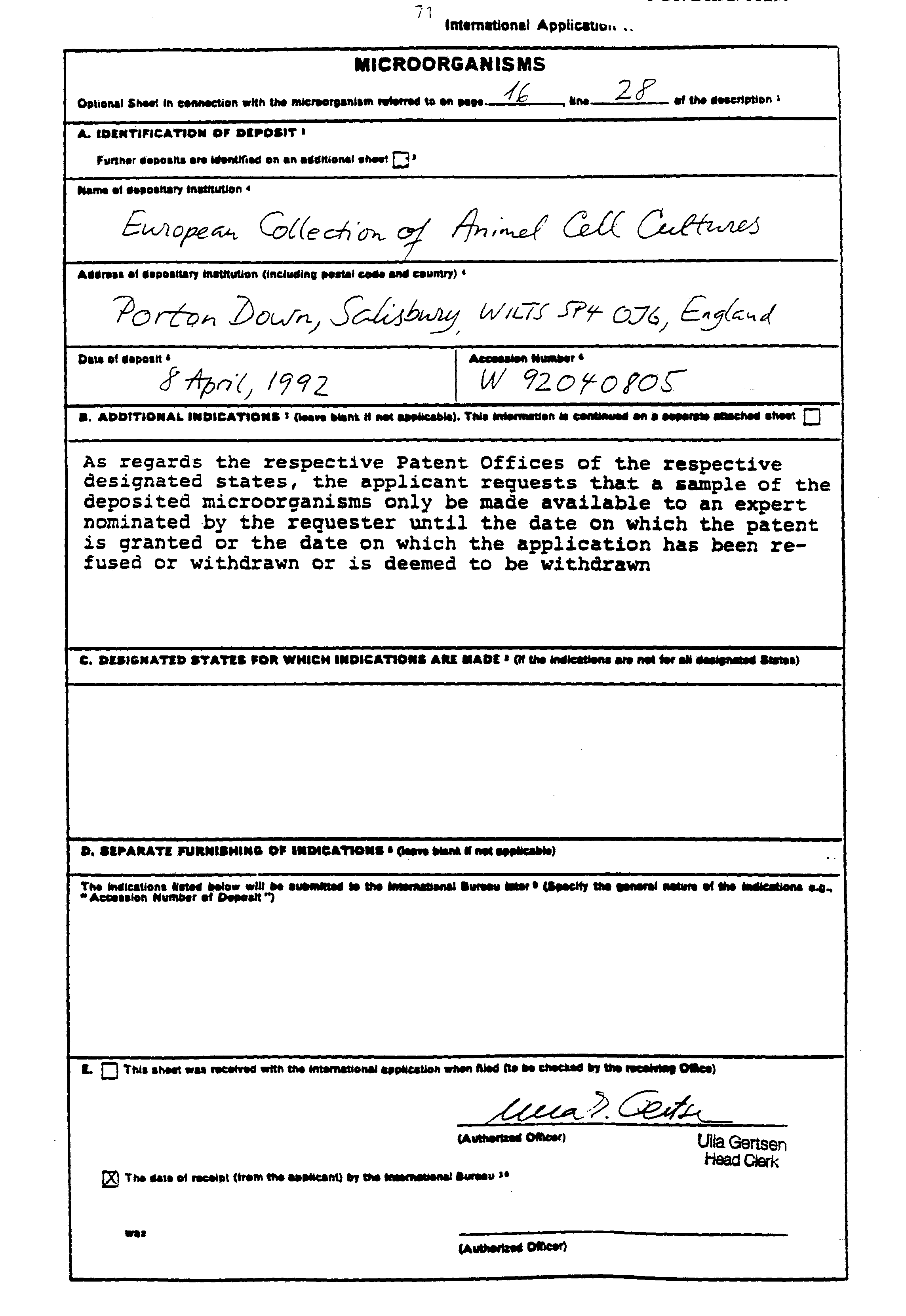
Claims
Priority Applications (2)
| Application Number | Priority Date | Filing Date | Title |
|---|---|---|---|
| AU27709/92A AU664049B2 (en) | 1991-10-11 | 1992-10-12 | Type C-like human retrovirus linked to multiple sclerosis (MS) |
| EP92921610A EP0609305A1 (en) | 1991-10-11 | 1992-10-12 | Type c-like human retrovirus linked to multiple sclerosis (ms) |
Applications Claiming Priority (2)
| Application Number | Priority Date | Filing Date | Title |
|---|---|---|---|
| DK911730A DK173091D0 (en) | 1991-10-11 | 1991-10-11 | BIOLOGICAL MATERIALS |
| DK1730/91 | 1991-10-11 |
Publications (1)
| Publication Number | Publication Date |
|---|---|
| WO1993007259A1 true WO1993007259A1 (en) | 1993-04-15 |
Family
ID=8107599
Family Applications (1)
| Application Number | Title | Priority Date | Filing Date |
|---|---|---|---|
| PCT/DK1992/000299 WO1993007259A1 (en) | 1991-10-11 | 1992-10-12 | Type c-like human retrovirus linked to multiple sclerosis (ms) |
Country Status (5)
| Country | Link |
|---|---|
| EP (1) | EP0609305A1 (en) |
| AU (1) | AU664049B2 (en) |
| CA (1) | CA2121030A1 (en) |
| DK (1) | DK173091D0 (en) |
| WO (1) | WO1993007259A1 (en) |
Cited By (26)
| Publication number | Priority date | Publication date | Assignee | Title |
|---|---|---|---|---|
| EP0617132A2 (en) * | 1993-03-26 | 1994-09-28 | Gen-Probe Incorporated | Probes and method to detect human immunodeficiency virus type 1 |
| DE4416300A1 (en) * | 1993-06-01 | 1994-12-08 | Ortwin Dr Faff | Method for the direct and biochemically functional detection of retroviruses in biological samples |
| WO1994028115A1 (en) * | 1993-06-01 | 1994-12-08 | Ortwin Faff | Direct and biochemically functional detection process of retrovirus in biological samples |
| WO1995010777A1 (en) * | 1993-10-15 | 1995-04-20 | Rakowicz Szulczynska Ewa M | Detection and treatment of breast and gynecological cancer |
| WO1995021256A1 (en) * | 1994-02-04 | 1995-08-10 | Bio Merieux | Msrv1 virus and msrv2 pathogenic and/or infectious agent associated with multiple sclerosis, nucleic acid components and applications of same |
| FR2715938A1 (en) * | 1994-02-04 | 1995-08-11 | Bio Merieux | Two pathogenic or infectious agents associated with multiple sclerosis |
| FR2715937A1 (en) * | 1994-02-04 | 1995-08-11 | Bio Merieux | Two pathogenic or infectious agents associated with multiple sclerosis |
| FR2715936A1 (en) * | 1994-02-04 | 1995-08-11 | Bio Merieux | Two pathogenic or infectious agents associated with multiple sclerosis |
| FR2715939A1 (en) * | 1994-02-04 | 1995-08-11 | Bio Merieux | Two pathogenic or infectious agents associated with multiple sclerosis |
| FR2727428A1 (en) * | 1994-11-24 | 1996-05-31 | Bio Merieux | Two pathogenic or infectious agents associated with multiple sclerosis |
| FR2728585A1 (en) * | 1994-12-23 | 1996-06-28 | Bio Merieux | Two pathogenic or infectious agents associated with multiple sclerosis |
| WO1996030518A1 (en) * | 1995-03-31 | 1996-10-03 | John Innes Centre Innovations Limited | Plant pathogen resistance genes and uses thereof |
| US5712385A (en) * | 1989-07-11 | 1998-01-27 | Gen-Probe Incorporated | Detection of human immunodeficiency virus type 1 |
| US5728540A (en) * | 1994-02-15 | 1998-03-17 | Bio Merieux | Cytotoxic factor as is associated with multiple sclerosis, its detection and its quantification |
| FR2765588A1 (en) * | 1997-07-07 | 1999-01-08 | Bio Merieux | RETROVIRAL NUCLEIC MATERIAL AND NUCLEOTIDE FRAGMENTS ESPECIALLY ASSOCIATED WITH MULTIPLE SCLEROSIS AND / OR RHUMATOID POLYARTHRITIS, FOR DIAGNOSIS, PROPHYLACTICS AND THERAPEUTICS |
| US5925555A (en) * | 1992-04-03 | 1999-07-20 | Bio Merieux | Process for the production of a viable cell culture infected by a multiple sclerosis-associated virus |
| WO1999053103A1 (en) * | 1998-04-08 | 1999-10-21 | Ms Research A/S | Diagnosis of multiple sclerosis and other demyelinating diseases |
| US6001987A (en) * | 1995-08-03 | 1999-12-14 | Bio Merieux | Isolated nucleotide sequences associated with Multiple sclerosis |
| US6071736A (en) * | 1992-04-03 | 2000-06-06 | Bio Merieux | Process and culture medium for the production of cells infected by a multiple sclerosis-associated virus |
| FR2788784A1 (en) * | 1999-01-21 | 2000-07-28 | Bio Merieux | New nucleic acid from human endogenous retrovirus, useful e.g. for diagnosis of autoimmune disease and complications of pregnancy, contains at least part of the gag gene |
| US6287865B1 (en) | 1995-03-31 | 2001-09-11 | Plant Bioscience Limited | Cf-2 plant pathogen resistance genes |
| US6582703B2 (en) | 1996-11-26 | 2003-06-24 | Bio Merieux | Isolated nucleotide sequences associated with multiple sclerosis or rheumatoid arthritis and a process of detecting |
| US6589734B1 (en) | 1989-07-11 | 2003-07-08 | Gen-Probe Incorporated | Detection of HIV |
| US6623920B1 (en) | 1999-07-09 | 2003-09-23 | Gen-Probe Incorporated | Detection of HIV-1 by nucleic acid amplification |
| WO2008124192A3 (en) * | 2007-04-10 | 2008-12-18 | Univ Emory | Novel hiv-1 reverse transcriptase codon deletion and its use in the management and treatment of hiv infections |
| US8697352B2 (en) | 1997-08-08 | 2014-04-15 | Biomerieux, B.V. | Nucleic acid sequences that can be used as primers and probes in the amplification and detection of all subtypes of HIV-1 |
Families Citing this family (1)
| Publication number | Priority date | Publication date | Assignee | Title |
|---|---|---|---|---|
| CN108144745B (en) * | 2017-12-20 | 2020-06-16 | 天康生物股份有限公司 | Separation device and method for reducing endotoxin content of live brucellosis vaccine |
Citations (4)
| Publication number | Priority date | Publication date | Assignee | Title |
|---|---|---|---|---|
| EP0222310A2 (en) * | 1985-11-13 | 1987-05-20 | The Wistar Institute | Methods for diagnosis and therapy of multiple sclerosis |
| EP0326395A2 (en) * | 1988-01-29 | 1989-08-02 | City Of Hope | Method of detecting and identifying certain viral sequences |
| EP0384566A2 (en) * | 1989-01-27 | 1990-08-29 | The Wistar Institute | Amplification of HTLV-1 sequences from multiple sclerosis patients |
| WO1992008785A1 (en) * | 1990-11-14 | 1992-05-29 | The University Company Pty. Ltd. | Diagnosis and treatment of multiple sclerosis |
-
1991
- 1991-10-11 DK DK911730A patent/DK173091D0/en active IP Right Grant
-
1992
- 1992-10-12 WO PCT/DK1992/000299 patent/WO1993007259A1/en not_active Application Discontinuation
- 1992-10-12 AU AU27709/92A patent/AU664049B2/en not_active Ceased
- 1992-10-12 CA CA002121030A patent/CA2121030A1/en not_active Abandoned
- 1992-10-12 EP EP92921610A patent/EP0609305A1/en not_active Ceased
Patent Citations (4)
| Publication number | Priority date | Publication date | Assignee | Title |
|---|---|---|---|---|
| EP0222310A2 (en) * | 1985-11-13 | 1987-05-20 | The Wistar Institute | Methods for diagnosis and therapy of multiple sclerosis |
| EP0326395A2 (en) * | 1988-01-29 | 1989-08-02 | City Of Hope | Method of detecting and identifying certain viral sequences |
| EP0384566A2 (en) * | 1989-01-27 | 1990-08-29 | The Wistar Institute | Amplification of HTLV-1 sequences from multiple sclerosis patients |
| WO1992008785A1 (en) * | 1990-11-14 | 1992-05-29 | The University Company Pty. Ltd. | Diagnosis and treatment of multiple sclerosis |
Non-Patent Citations (6)
| Title |
|---|
| Journal of Neuroimmunology, Vol. 38, (1992) SVEN HAAHR: Conference Report. Second International Symposium on Retrovirus in Multiple Sclerosis and Related Diseases, 26-28 August 1991, pages 163-174, see Abstract by SVEN HAAHR et al. and HERVE PERRON, pages 172-173 and page 164, second paragraph. * |
| Lancet, Vol. 337, April 1991, HAAR S. et al.: "Just another dubious virus in cells from a patient with multiple sclerosis?", see page 863 - page 864. * |
| Lancet, Vol. 337, April 1991, PERONN H. et al.: "Isolation of retrovirus from patients with multiple sclerosis", see page 862 - page 863. * |
| Nature, Vol. 331, February 1988, STEVEN JACOBSON et al.: "Isolation of an HTLV-1-like retrovirus from patients with tropical spastic paraparesis", see page 540 - page 543. * |
| Nucleic Acids Research, Vol. 17, No. 17, 1989, ANDRAS PEARL et al.: "Detection and cloning of new HTLV-related endogenous sequences in man", see page 6841 - 6855. * |
| Proc. Natl. Acad. Sci. USA, Vol. 89, March 1992, K. BANKI et al.: "Human T-cell lymphotropic virus (HTLV)-related endogenous sequence, HRES-1, encodes a 28-kDa protein: A possible autoantigen for HTLV-I gag-reactive autoantibodies", see page 1939 - page 1943. * |
Cited By (56)
| Publication number | Priority date | Publication date | Assignee | Title |
|---|---|---|---|---|
| US5800980A (en) * | 1899-02-04 | 1998-09-01 | Bio Merieux | Detection of MSRV1 virus and MSRV2 pathogen and/or infective agent associated with multiple sclerosis, by nucleic acid hybridization |
| US5712385A (en) * | 1989-07-11 | 1998-01-27 | Gen-Probe Incorporated | Detection of human immunodeficiency virus type 1 |
| US6589734B1 (en) | 1989-07-11 | 2003-07-08 | Gen-Probe Incorporated | Detection of HIV |
| US7083922B2 (en) | 1989-07-11 | 2006-08-01 | Gen-Probe Incorporated | Detection of HIV |
| US5856088A (en) * | 1989-07-11 | 1999-01-05 | Gen-Probe Incorporated | Detection of human immunodeficiency virus type 1 |
| US5925555A (en) * | 1992-04-03 | 1999-07-20 | Bio Merieux | Process for the production of a viable cell culture infected by a multiple sclerosis-associated virus |
| US6291225B1 (en) | 1992-04-03 | 2001-09-18 | Bio Merieux | Process and culture medium for the production of cells infected by a multiple sclerosis-associated virus |
| US6071736A (en) * | 1992-04-03 | 2000-06-06 | Bio Merieux | Process and culture medium for the production of cells infected by a multiple sclerosis-associated virus |
| US6252059B1 (en) | 1993-03-26 | 2001-06-26 | Gen-Probe Incorporated | Detection of human immunodeficiency virus type 1 |
| EP0617132A3 (en) * | 1993-03-26 | 1995-11-29 | Gen Probe Inc | Probes and method to detect human immunodeficiency virus type 1. |
| EP0617132A2 (en) * | 1993-03-26 | 1994-09-28 | Gen-Probe Incorporated | Probes and method to detect human immunodeficiency virus type 1 |
| US6649749B2 (en) | 1993-03-26 | 2003-11-18 | Gen-Probe Incorporated | Detection of human immunodeficiency virus type 1 |
| US6268123B1 (en) * | 1993-06-01 | 2001-07-31 | Retro-Tech Gmbh | Direct and biochemically functional detection process of retrovirus in biological samples |
| DE4416300A1 (en) * | 1993-06-01 | 1994-12-08 | Ortwin Dr Faff | Method for the direct and biochemically functional detection of retroviruses in biological samples |
| WO1994028115A1 (en) * | 1993-06-01 | 1994-12-08 | Ortwin Faff | Direct and biochemically functional detection process of retrovirus in biological samples |
| WO1995010777A1 (en) * | 1993-10-15 | 1995-04-20 | Rakowicz Szulczynska Ewa M | Detection and treatment of breast and gynecological cancer |
| US5939277A (en) * | 1993-10-15 | 1999-08-17 | Rakowicz-Szulczynska; Eva M. | Detection and treatment of breast and gynecological cancer |
| US6342383B1 (en) | 1994-02-04 | 2002-01-29 | Bio Merieux | Cell lines and viral isolates associated with multiple sclerosis |
| FR2715939A1 (en) * | 1994-02-04 | 1995-08-11 | Bio Merieux | Two pathogenic or infectious agents associated with multiple sclerosis |
| FR2715936A1 (en) * | 1994-02-04 | 1995-08-11 | Bio Merieux | Two pathogenic or infectious agents associated with multiple sclerosis |
| US5871996A (en) * | 1994-02-04 | 1999-02-16 | Bio Merieux | Retrovirus agents MSRV1 and MSRV2 associated with multiple sclerosis |
| US5871745A (en) * | 1994-02-04 | 1999-02-16 | Bio Merieux | Multiple sclerosis related virus |
| US6184025B1 (en) | 1994-02-04 | 2001-02-06 | Bio Merieux | Viral isolates associated with multiple sclerosis |
| FR2715938A1 (en) * | 1994-02-04 | 1995-08-11 | Bio Merieux | Two pathogenic or infectious agents associated with multiple sclerosis |
| WO1995021256A1 (en) * | 1994-02-04 | 1995-08-10 | Bio Merieux | Msrv1 virus and msrv2 pathogenic and/or infectious agent associated with multiple sclerosis, nucleic acid components and applications of same |
| EP0674004A1 (en) * | 1994-02-04 | 1995-09-27 | Bio Merieux | MSRV1 virus and pathogenic and/or infections agent MSRV2 linked to multiple sclerosis, their nucleic components and their applications |
| US5962217A (en) * | 1994-02-04 | 1999-10-05 | Bio Merieux | MSRV1 virus and MSRV2 pathogen and/or infective agent associated with multiple sclerosis, and biopolymer constituents thereof |
| FR2715937A1 (en) * | 1994-02-04 | 1995-08-11 | Bio Merieux | Two pathogenic or infectious agents associated with multiple sclerosis |
| US5876954A (en) * | 1994-02-15 | 1999-03-02 | Bio Merieux | Cytotoxic factor as is associated with multiple sclerosis, its detection and its quantification |
| US5728540A (en) * | 1994-02-15 | 1998-03-17 | Bio Merieux | Cytotoxic factor as is associated with multiple sclerosis, its detection and its quantification |
| FR2727428A1 (en) * | 1994-11-24 | 1996-05-31 | Bio Merieux | Two pathogenic or infectious agents associated with multiple sclerosis |
| FR2728585A1 (en) * | 1994-12-23 | 1996-06-28 | Bio Merieux | Two pathogenic or infectious agents associated with multiple sclerosis |
| WO1996030518A1 (en) * | 1995-03-31 | 1996-10-03 | John Innes Centre Innovations Limited | Plant pathogen resistance genes and uses thereof |
| US6287865B1 (en) | 1995-03-31 | 2001-09-11 | Plant Bioscience Limited | Cf-2 plant pathogen resistance genes |
| US6001987A (en) * | 1995-08-03 | 1999-12-14 | Bio Merieux | Isolated nucleotide sequences associated with Multiple sclerosis |
| US6579526B1 (en) | 1995-08-03 | 2003-06-17 | Bio Merieux | Viral material and nucleotide fragments associated with multiple sclerosis, for diagnostic, prophylactic and therapeutic purposes |
| US7674888B2 (en) | 1996-11-26 | 2010-03-09 | Biomerieux | Viral material and nucleotide fragments associated with multiple sclerosis, for diagnostic, prophylactic and therapeutic purposes |
| US6582703B2 (en) | 1996-11-26 | 2003-06-24 | Bio Merieux | Isolated nucleotide sequences associated with multiple sclerosis or rheumatoid arthritis and a process of detecting |
| FR2765588A1 (en) * | 1997-07-07 | 1999-01-08 | Bio Merieux | RETROVIRAL NUCLEIC MATERIAL AND NUCLEOTIDE FRAGMENTS ESPECIALLY ASSOCIATED WITH MULTIPLE SCLEROSIS AND / OR RHUMATOID POLYARTHRITIS, FOR DIAGNOSIS, PROPHYLACTICS AND THERAPEUTICS |
| WO1999002666A3 (en) * | 1997-07-07 | 1999-04-15 | Bio Merieux | Retroviral nucleic material and nucleotide fragments, in particular associated with multiple sclerosis and/or rheumatoid arthritis, for diagnostic, prophylactic and therapeutic uses |
| WO1999002666A2 (en) * | 1997-07-07 | 1999-01-21 | Bio Merieux | Retroviral nucleic material and nucleotide fragments, in particular associated with multiple sclerosis and/or rheumatoid arthritis, for diagnostic, prophylactic and therapeutic uses |
| EP1612270A3 (en) * | 1997-07-07 | 2006-04-05 | Biomerieux | Rétroviral nucleic material and nucleotide fragments, in particular associated with multiple sclerosis and/or rheumatoid arthritis, for diagnostic, prophylactic and therapeutic uses |
| US7771927B1 (en) | 1997-07-07 | 2010-08-10 | Biomerieux | Multiple sclerosis-associated retroviral (MSRV) nucleic acids corresponding to the Env region |
| US8088910B2 (en) | 1997-07-07 | 2012-01-03 | Biomerieux | Isolated multiple sclerosis (MS)-associated retrovirus (MSRV) nucleic acids corresponding to the gag region |
| US9074262B2 (en) | 1997-08-08 | 2015-07-07 | bioMerieux, B. V. | Nucleic acid sequences that can be used as primers and probes in the amplification and detection of all subtypes of HIV-1 |
| US8697352B2 (en) | 1997-08-08 | 2014-04-15 | Biomerieux, B.V. | Nucleic acid sequences that can be used as primers and probes in the amplification and detection of all subtypes of HIV-1 |
| WO1999053103A1 (en) * | 1998-04-08 | 1999-10-21 | Ms Research A/S | Diagnosis of multiple sclerosis and other demyelinating diseases |
| WO2000043521A3 (en) * | 1999-01-21 | 2001-02-22 | Bio Merieux | Endogenetic nucleic fragment associated with an autoimmune disease, marking method and reagent |
| US7632931B2 (en) | 1999-01-21 | 2009-12-15 | Biomerieux | Endogeneous nucleic acid fragment associated with an autoimmune disease, labeling method and reagent |
| FR2788784A1 (en) * | 1999-01-21 | 2000-07-28 | Bio Merieux | New nucleic acid from human endogenous retrovirus, useful e.g. for diagnosis of autoimmune disease and complications of pregnancy, contains at least part of the gag gene |
| US7097979B2 (en) | 1999-07-09 | 2006-08-29 | Gen-Probe Incorporated | Detection of HIV-1 by nucleic acid amplification |
| US7723040B2 (en) | 1999-07-09 | 2010-05-25 | Gen-Probe Incorporated | Detection of HIV-1 by nucleic acid amplification |
| US6623920B1 (en) | 1999-07-09 | 2003-09-23 | Gen-Probe Incorporated | Detection of HIV-1 by nucleic acid amplification |
| US20100130381A1 (en) * | 2007-04-10 | 2010-05-27 | Emory University | Novel hiv-1 reverse transcriptase codon deletion and its use in the management and treatment of hiv infections |
| WO2008124192A3 (en) * | 2007-04-10 | 2008-12-18 | Univ Emory | Novel hiv-1 reverse transcriptase codon deletion and its use in the management and treatment of hiv infections |
| US9428815B2 (en) | 2007-04-10 | 2016-08-30 | Emory University | HIV-1 reverse transcriptase codon deletion and its use in the management and treatment of HIV infections |
Also Published As
| Publication number | Publication date |
|---|---|
| DK173091D0 (en) | 1991-10-11 |
| AU664049B2 (en) | 1995-11-02 |
| EP0609305A1 (en) | 1994-08-10 |
| CA2121030A1 (en) | 1993-04-15 |
| AU2770992A (en) | 1993-05-03 |
Similar Documents
| Publication | Publication Date | Title |
|---|---|---|
| AU664049B2 (en) | Type C-like human retrovirus linked to multiple sclerosis (MS) | |
| Koprowski et al. | Multiple sclerosis and human T-cell lymphotropic retroviruses | |
| Pahwa et al. | Stimulatory and inhibitory influences of human immunodeficiency virus on normal B lymphocytes. | |
| Vilmer et al. | Isolation of new lymphotropic retrovirus from two siblings with haemophilia B, one with AIDS | |
| Hauser et al. | Analysis of human T-lymphotropic virus sequences in multiple sclerosis tissue | |
| Nowak et al. | Characterization of monoclonal antibodies and polyclonal immune sera directed against human cytomegalovirus virion proteins | |
| Webster et al. | Isolation of retroviruses from two patients with" common variable" hypogammaglobulinaemia | |
| JPH08511865A (en) | Parvovirus receptor and method of use | |
| Yamada et al. | Common immunologic determinant between human immunodeficiency virus type 1 gp41 and astrocytes | |
| EP0260691A2 (en) | Immunotherapy for aids patients | |
| Sommerlund et al. | Retrovirus‐like particles in an Epstein‐Barr virus‐producing cell line derived from a patient with chronic progressive myelopathy | |
| CA1290245C (en) | Methods for diagnosis and therapy of multiple sclerosis | |
| Capobianchi et al. | Acid-labile human interferon alpha production by peripheral blood mononuclear cells stimulated by HIV-infected cells | |
| CA2558733A1 (en) | A polypeptide derived from gp41, a vaccine composition comprising said polypeptide, and uses for treating an infection by an hiv virus in an individual | |
| RU2082417C1 (en) | Antiviral agent, vaccine based on thereof, method of its preparing, therapeutic agent and method of detection of rna-viruses | |
| AU591647B2 (en) | Improvements relating to viral isolates and their use | |
| US6924095B2 (en) | Retrovirus isolated from mantle histiocytes in mantle cell lymphoma | |
| Sherman et al. | Identification of human T cell leukemia/lymphoma virus type I antibodies, DNA, and protein in patients with polymyositis | |
| Pelton et al. | A search for retrovirus infection in systemic lupus erythematosus and rheumatoid arthritis. | |
| Klein et al. | Demonstration of “naturally occurring mitochondrial antibodies” in family members of patients with primary biliary cirrhosis | |
| McArthur et al. | Steroid‐responsive myeloneuropathy in a man dually infected with HIV‐1 and HTLV‐I | |
| Gerna et al. | Immunoperoxidase technique for detection of antibodies to human cytomegalovirus | |
| Giordanengo et al. | Autoantibodies directed against CD43 molecules with an altered glycosylation status on human immunodeficiency virus type 1 (HIV-1)-infected CEM cells are found in all HIV-1+ individuals | |
| McFarlin et al. | Neurological disorders associated with HTLV-1 | |
| US4792524A (en) | Adult T cell leukemia associated cell strain |
Legal Events
| Date | Code | Title | Description |
|---|---|---|---|
| AK | Designated states |
Kind code of ref document: A1 Designated state(s): AU BR CA FI HU JP KR NO PL RO RU US |
|
| AL | Designated countries for regional patents |
Kind code of ref document: A1 Designated state(s): AT BE CH DE DK ES FR GB GR IE IT LU MC NL SE |
|
| DFPE | Request for preliminary examination filed prior to expiration of 19th month from priority date (pct application filed before 20040101) | ||
| ENP | Entry into the national phase |
Ref document number: 1994 211589 Country of ref document: US Date of ref document: 19940408 Kind code of ref document: A |
|
| WWE | Wipo information: entry into national phase |
Ref document number: 2121030 Country of ref document: CA |
|
| WWE | Wipo information: entry into national phase |
Ref document number: 1992921610 Country of ref document: EP |
|
| WWP | Wipo information: published in national office |
Ref document number: 1992921610 Country of ref document: EP |
|
| WWR | Wipo information: refused in national office |
Ref document number: 1992921610 Country of ref document: EP |
|
| WWW | Wipo information: withdrawn in national office |
Ref document number: 1992921610 Country of ref document: EP |