WO2004058821A2 - Dual specific single domain antibodies specific for a ligand and for the receptor of the ligand - Google Patents
Dual specific single domain antibodies specific for a ligand and for the receptor of the ligand Download PDFInfo
- Publication number
- WO2004058821A2 WO2004058821A2 PCT/GB2003/005646 GB0305646W WO2004058821A2 WO 2004058821 A2 WO2004058821 A2 WO 2004058821A2 GB 0305646 W GB0305646 W GB 0305646W WO 2004058821 A2 WO2004058821 A2 WO 2004058821A2
- Authority
- WO
- WIPO (PCT)
- Prior art keywords
- dab
- ligand
- dual specific
- binding
- dual
- Prior art date
Links
Classifications
-
- C—CHEMISTRY; METALLURGY
- C07—ORGANIC CHEMISTRY
- C07K—PEPTIDES
- C07K16/00—Immunoglobulins [IGs], e.g. monoclonal or polyclonal antibodies
- C07K16/18—Immunoglobulins [IGs], e.g. monoclonal or polyclonal antibodies against material from animals or humans
-
- A—HUMAN NECESSITIES
- A61—MEDICAL OR VETERINARY SCIENCE; HYGIENE
- A61K—PREPARATIONS FOR MEDICAL, DENTAL OR TOILETRY PURPOSES
- A61K47/00—Medicinal preparations characterised by the non-active ingredients used, e.g. carriers or inert additives; Targeting or modifying agents chemically bound to the active ingredient
- A61K47/50—Medicinal preparations characterised by the non-active ingredients used, e.g. carriers or inert additives; Targeting or modifying agents chemically bound to the active ingredient the non-active ingredient being chemically bound to the active ingredient, e.g. polymer-drug conjugates
- A61K47/51—Medicinal preparations characterised by the non-active ingredients used, e.g. carriers or inert additives; Targeting or modifying agents chemically bound to the active ingredient the non-active ingredient being chemically bound to the active ingredient, e.g. polymer-drug conjugates the non-active ingredient being a modifying agent
- A61K47/56—Medicinal preparations characterised by the non-active ingredients used, e.g. carriers or inert additives; Targeting or modifying agents chemically bound to the active ingredient the non-active ingredient being chemically bound to the active ingredient, e.g. polymer-drug conjugates the non-active ingredient being a modifying agent the modifying agent being an organic macromolecular compound, e.g. an oligomeric, polymeric or dendrimeric molecule
- A61K47/59—Medicinal preparations characterised by the non-active ingredients used, e.g. carriers or inert additives; Targeting or modifying agents chemically bound to the active ingredient the non-active ingredient being chemically bound to the active ingredient, e.g. polymer-drug conjugates the non-active ingredient being a modifying agent the modifying agent being an organic macromolecular compound, e.g. an oligomeric, polymeric or dendrimeric molecule obtained otherwise than by reactions only involving carbon-to-carbon unsaturated bonds, e.g. polyureas or polyurethanes
- A61K47/60—Medicinal preparations characterised by the non-active ingredients used, e.g. carriers or inert additives; Targeting or modifying agents chemically bound to the active ingredient the non-active ingredient being chemically bound to the active ingredient, e.g. polymer-drug conjugates the non-active ingredient being a modifying agent the modifying agent being an organic macromolecular compound, e.g. an oligomeric, polymeric or dendrimeric molecule obtained otherwise than by reactions only involving carbon-to-carbon unsaturated bonds, e.g. polyureas or polyurethanes the organic macromolecular compound being a polyoxyalkylene oligomer, polymer or dendrimer, e.g. PEG, PPG, PEO or polyglycerol
-
- C—CHEMISTRY; METALLURGY
- C07—ORGANIC CHEMISTRY
- C07K—PEPTIDES
- C07K16/00—Immunoglobulins [IGs], e.g. monoclonal or polyclonal antibodies
- C07K16/18—Immunoglobulins [IGs], e.g. monoclonal or polyclonal antibodies against material from animals or humans
- C07K16/24—Immunoglobulins [IGs], e.g. monoclonal or polyclonal antibodies against material from animals or humans against cytokines, lymphokines or interferons
- C07K16/241—Tumor Necrosis Factors
-
- C—CHEMISTRY; METALLURGY
- C07—ORGANIC CHEMISTRY
- C07K—PEPTIDES
- C07K16/00—Immunoglobulins [IGs], e.g. monoclonal or polyclonal antibodies
- C07K16/18—Immunoglobulins [IGs], e.g. monoclonal or polyclonal antibodies against material from animals or humans
- C07K16/28—Immunoglobulins [IGs], e.g. monoclonal or polyclonal antibodies against material from animals or humans against receptors, cell surface antigens or cell surface determinants
- C07K16/2866—Immunoglobulins [IGs], e.g. monoclonal or polyclonal antibodies against material from animals or humans against receptors, cell surface antigens or cell surface determinants against receptors for cytokines, lymphokines, interferons
-
- C—CHEMISTRY; METALLURGY
- C07—ORGANIC CHEMISTRY
- C07K—PEPTIDES
- C07K16/00—Immunoglobulins [IGs], e.g. monoclonal or polyclonal antibodies
- C07K16/18—Immunoglobulins [IGs], e.g. monoclonal or polyclonal antibodies against material from animals or humans
- C07K16/28—Immunoglobulins [IGs], e.g. monoclonal or polyclonal antibodies against material from animals or humans against receptors, cell surface antigens or cell surface determinants
- C07K16/2878—Immunoglobulins [IGs], e.g. monoclonal or polyclonal antibodies against material from animals or humans against receptors, cell surface antigens or cell surface determinants against the NGF-receptor/TNF-receptor superfamily, e.g. CD27, CD30, CD40, CD95
-
- C—CHEMISTRY; METALLURGY
- C07—ORGANIC CHEMISTRY
- C07K—PEPTIDES
- C07K16/00—Immunoglobulins [IGs], e.g. monoclonal or polyclonal antibodies
- C07K16/40—Immunoglobulins [IGs], e.g. monoclonal or polyclonal antibodies against enzymes
-
- A—HUMAN NECESSITIES
- A61—MEDICAL OR VETERINARY SCIENCE; HYGIENE
- A61K—PREPARATIONS FOR MEDICAL, DENTAL OR TOILETRY PURPOSES
- A61K39/00—Medicinal preparations containing antigens or antibodies
- A61K2039/505—Medicinal preparations containing antigens or antibodies comprising antibodies
-
- C—CHEMISTRY; METALLURGY
- C07—ORGANIC CHEMISTRY
- C07K—PEPTIDES
- C07K2317/00—Immunoglobulins specific features
- C07K2317/20—Immunoglobulins specific features characterized by taxonomic origin
- C07K2317/21—Immunoglobulins specific features characterized by taxonomic origin from primates, e.g. man
-
- C—CHEMISTRY; METALLURGY
- C07—ORGANIC CHEMISTRY
- C07K—PEPTIDES
- C07K2317/00—Immunoglobulins specific features
- C07K2317/30—Immunoglobulins specific features characterized by aspects of specificity or valency
- C07K2317/31—Immunoglobulins specific features characterized by aspects of specificity or valency multispecific
-
- C—CHEMISTRY; METALLURGY
- C07—ORGANIC CHEMISTRY
- C07K—PEPTIDES
- C07K2317/00—Immunoglobulins specific features
- C07K2317/30—Immunoglobulins specific features characterized by aspects of specificity or valency
- C07K2317/34—Identification of a linear epitope shorter than 20 amino acid residues or of a conformational epitope defined by amino acid residues
-
- C—CHEMISTRY; METALLURGY
- C07—ORGANIC CHEMISTRY
- C07K—PEPTIDES
- C07K2317/00—Immunoglobulins specific features
- C07K2317/50—Immunoglobulins specific features characterized by immunoglobulin fragments
- C07K2317/55—Fab or Fab'
-
- C—CHEMISTRY; METALLURGY
- C07—ORGANIC CHEMISTRY
- C07K—PEPTIDES
- C07K2317/00—Immunoglobulins specific features
- C07K2317/50—Immunoglobulins specific features characterized by immunoglobulin fragments
- C07K2317/56—Immunoglobulins specific features characterized by immunoglobulin fragments variable (Fv) region, i.e. VH and/or VL
- C07K2317/569—Single domain, e.g. dAb, sdAb, VHH, VNAR or nanobody®
-
- C—CHEMISTRY; METALLURGY
- C07—ORGANIC CHEMISTRY
- C07K—PEPTIDES
- C07K2317/00—Immunoglobulins specific features
- C07K2317/60—Immunoglobulins specific features characterized by non-natural combinations of immunoglobulin fragments
- C07K2317/62—Immunoglobulins specific features characterized by non-natural combinations of immunoglobulin fragments comprising only variable region components
- C07K2317/622—Single chain antibody (scFv)
-
- C—CHEMISTRY; METALLURGY
- C07—ORGANIC CHEMISTRY
- C07K—PEPTIDES
- C07K2317/00—Immunoglobulins specific features
- C07K2317/90—Immunoglobulins specific features characterized by (pharmaco)kinetic aspects or by stability of the immunoglobulin
- C07K2317/92—Affinity (KD), association rate (Ka), dissociation rate (Kd) or EC50 value
-
- C—CHEMISTRY; METALLURGY
- C12—BIOCHEMISTRY; BEER; SPIRITS; WINE; VINEGAR; MICROBIOLOGY; ENZYMOLOGY; MUTATION OR GENETIC ENGINEERING
- C12N—MICROORGANISMS OR ENZYMES; COMPOSITIONS THEREOF; PROPAGATING, PRESERVING, OR MAINTAINING MICROORGANISMS; MUTATION OR GENETIC ENGINEERING; CULTURE MEDIA
- C12N2799/00—Uses of viruses
- C12N2799/02—Uses of viruses as vector
- C12N2799/021—Uses of viruses as vector for the expression of a heterologous nucleic acid
Definitions
- the present invention relates to dual specific ligands.
- the invention provides a method for the preparation of dual-specific ligands comprising a first immunoglobulin single variable domain binding to a first antigen or epitope, and a second immunoglobulin single variable domain binding to a second antigen or epitope.
- the invention relates to dual-specific ligands wherein binding to at least one of the first and second antigens or epitopes acts to increase the half -life of the ligand in vivo. Open and closed conformation ligands comprising more than one binding specificity are described.
- the antigen binding domain of an antibody comprises two separate regions: a heavy chain variable domain (v H ) and a light chain variable domain ( ⁇ L : which can be either V K or V ).
- the antigen binding site itself is formed by six polypeptide loops: three from V H domain (HI, H2 and H3) and three from V domain (LI, L2 and L3).
- V H domain HI, H2 and H3
- V domain LI, L2 and L3
- the V H segment encodes the region of the polypeptide chain which forms the first and second antigen binding loops of the V H domain (HI and H2), whilst the VH > D an JH segments combine to form the third antigen binding loop of the y H domain (H3).
- V L ⁇ ene i s produced by the recombination of only two gene segments, V an JL- In humans, there are approximately 40 functional N ⁇ segments (Schable and Zachau (1993) Biol. Chem. Hoppe-Seyler, 374: 1001), 31 functional N ⁇ segments (Williams et al. (1996) J. Mol. Biol., 264: 220; Kawasaki et al. (1997) Genome Res., 7: 250), 5 functional J ⁇ segments (Hieter et al. (1982) J. Biol. Chem., 257: 1516) and 4 functional J ⁇ segments (Nasicek and Leder (1990) J. Exp.
- the v L segment encodes the region of the polypeptide chain which forms the first and second antigen binding loops of the VL domain (LI and L2), whilst the V an JL segments combine to form the third antigen binding loop of the V L domain (L3).
- Antibodies selected from this primary repertoire are believed to be sufficiently diverse to bind almost all antigens with at least moderate affinity. High affinity antibodies are produced by "affinity maturation" of the rearranged genes, in which point mutations are generated and selected by the immune system on the basis of improved binding.
- H3 region is much more diverse in terms of sequence, length and structure (due to the use of D segments), it also forms a limited number of main-chain conformations for short loop lengths which depend on the length and the presence of particular residues, or types of residue, at key positions in the loop and the antibody framework (Martin et al. (1996) J. Mol. Biol, 263: 800; Shirai et al. (1996) FEBS Letters, 399: 1.
- Bispecific antibodies comprising complementary pairs of v H n d V L regions are known in the art. These bispecific antibodies must comprise two pairs of VH an V L S > eacn V I /V L pair binding to a single antigen or epitope. Methods described involve hybrid hybridomas (Milstein & Cuello AC, Nature 305:537-40), minibodies (Hu et al, (1996) Cancer Res 56:3055-3061;), diabodies (Holliger et al, (1993) Proc. Natl. Acad. Sci. USA 90, 6444- 6448; WO 94/13804), chelating recombinant antibodies (CRAbs; (Neri et al, (1995) J. Mol. Biol.
- each antibody species comprises two antigen-binding sites, each fashioned by a complementary pair of VH an V L domains. Each antibody is thereby able to bind to two different antigens or epitopes at the same time, with the binding to EACH antigen or epitope mediated by a V H an d its complementary V L domain.
- WO 02/02773 (Abbott Laboratories) describes antibody molecules with "dual specificity".
- the antibody molecules referred to are antibodies raised or selected against multiple antigens, such that their specificity spans more than a single antigen.
- Each complementary V H VL P a ⁇ r * n ⁇ e ⁇ tibodies of WO 02/02773 specifies a single binding specificity for two or more structurally related antigens; the VH and VL domains in such complementary pairs do not each possess a separate specificity.
- the antibodies thus have a broad single specificity which encompasses two antigens, which are structurally related.
- natural autoantibodies have been described that are polyreactive (Casali & ⁇ otkins, Ann.
- Protein engineering methods have been suggested that may have a bearing on this.
- a catalytic antibody could be created with a binding activity to a metal ion through one variable domain, and to a hapten (substrate) through contacts with the metal ion and a complementary variable domain (Barbas et al., 1993 Proc. Natl. Acad. Sci USA 90, 6385-6389).
- the binding and catalysis of the substrate is proposed to require the binding of the metal ion (second antigen).
- the binding to the V H ⁇ V L pairing relates to a single but multi- component antigen.
- Single heavy chain variable domains have also been described, derived from natural antibodies which are normally associated with light chains (from monoclonal antibodies or from repertoires of domains; see EP-A-0368684). These heavy chain variable domains have been shown to interact specifically with one or more related antigens but have not been combined with other heavy or light chain variable domains to create a ligand with a specificity for two or more different antigens . Furthermore, these single domains have been shown to have a very short in vivo half-life. Therefore such domains are of limited therapeutic value.
- the inventors have described, in their copending international patent application WO 03/002609 as well as copending unpublished UK patent application 0230203.2, dual specific immunoglobulin ligands which comprise immunoglobulin single variable domains which each have different specificities.
- the domains may act in competition with each other or independently to bind antigens or epitopes on target molecules.
- the present invention provides a further improvement in dual specific ligands as developed by the present inventors, in which one specificity of the ligand is directed towards a protein or polypeptide target, and another specificity is directed to a receptor for the target. Therefore, in a first aspect, the invention provides a dual specific ligand comprising a first dAb specific for a target ligand, and a second dAb specific for a receptor for the target ligand.
- the dual specific ligand is an open conformation ligand and can bind both the target ligand and the target ligand receptor simultaneously.
- Preferred dual specific ligands comprise at least on specificity for TNF alpha and at least one specificity for TNF Receptor 1 (p55).
- the specificities are provided by one or more dAbs arranged in Fab, F(ab') 2 or IgG formats.
- Preferred dAbs are TAR1- 5-19 N ⁇ and TAR2h-10-27 N H as set forth below.
- the invention may also comprise further modifications and configurations of the dual specific ligands as set forth in the accompanying claims and detailed herein.
- a dual-specific ligand comprising a first immunoglobulin single variable domain having a binding specificity to a first antigen or epitope and a second complementary immunoglobulin single variable domain having a binding activity to a second antigen or epitope, wherein one or both of said antigens or epitopes acts to increase the half-life of the ligand in vivo and wherein said first and second domains lack mutually complementary domains which share the same specificity, provided that said dual specific ligand does not consist of an anti-HS A V H domain and an anti- ⁇ galactosidase N ⁇ domain.
- neither of the first or second variable domains binds to human serum albumin (HSA).
- Antigens or epitopes which increase the half-life of a ligand as described herein are advantageously present on proteins or polypeptides found in an organism in vivo. Examples include extracellular matrix proteins, blood proteins, and proteins present in various tissues in the organism. The proteins act to reduce the rate of ligand clearance from the blood, for example by acting as bulking agents, or by anchoring the ligand to a desired site of action. Examples of antigens/epitopes which increase half-life in vivo are given in Annex 1 below. Increased half-life is useful in in vivo applications of immunoglobulins, especially antibodies and most especially antibody fragments of small size.
- Such fragments suffer from rapid clearance from the body; thus, whilst they are able to reach most parts of the body rapidly, and are quick to produce and easier to handle, their in vivo applications have been limited by their only brief persistence in vivo.
- the invention solves this problem by providing increased half-life of the ligands in vivo and consequently longer persistence times in the body of the functional activity of the ligand.
- Half lives (tVz alpha and V beta) and AUC can be determined from a curve of serum concentration of ligand against time.
- the WinNonlin analysis package (available from Pharsight Corp., Mountain View, CA94040, USA) can be used, for example, to model the curve.
- a first phase the alpha phase
- a second phase (beta phase) is the terminal phase when the ligand has been distributed and the serum concentration is decreasing as the ligand is cleared from the patient.
- the t alpha half life is the half life of the first phase and the t beta half life is the half life of the second phase.
- the present invention provides a ligand or a composition comprising a ligand according to the invention having a t ⁇ half-life in the range of 15 minutes or more.
- the lower end of the range is 30 minutes, 45 minutes, 1 hour, 2 hours, 3 hours, 4 hours, 5 hours, 6 hours, 7 hours, 10 hours, 11 hours or 12 hours.
- a ligand or composition according to the invention will have a t ⁇ half life in the range of up to and including 12 hours.
- the upper end of the range is 11, 10, 9, 8, 7, 6 or 5 hours.
- An example of a suitable range is 1 to 6 hours, 2 to 5 hours or 3 to 4 hours.
- the present invention provides a ligand or a composition comprising a ligand according to the invention having a t ⁇ half-life in the range of 2.5 hours or more.
- the lower end of the range is 3 hours, 4 hours, 5 hours, 6 hours, 7 hours, 10 hours , 11 hours, or 12 hours.
- a ligand or composition according to the invention has a t ⁇ half-life in the range of up to and including 21 days.
- the upper end of the range is 12 hours, 24 hours, 2 days, 3 days, 5 days, 10 days, 15 days or 20 days.
- a ligand or composition according to the invention will have a t ⁇ half life in the range 12 to 60 hours. In a further embodiment, it will be in the range 12 to 48 hours. In a further embodiment still, it will be in the range 12 to 26 hours.
- the present invention provides a ligand or a composition comprising a ligand according to the invention having an AUC value (area under the curve) in the range of 1 mg.min ml or more.
- the lower end of the range is 5, 10, 15, 20, 30, 100, 200 or 300mg.min/ml.
- a ligand or composition according to the invention has an AUC in the range of up to 600 mg.min/ml.
- the upper end of the range is 500, 400, 300, 200, 150, 100, 75 or 50 mg.min/ml.
- a ligand according to the invention will have a AUC in the range selected from the group consisting of the following: 15 to 150mg.min/ml, 15 to 100 mg.min/ml, 15 to 75 mg.min/ml, and 15 to 50mg.min/ml.
- the dual specific ligand comprises two complementary variable domains, i.e. two variable domains that, in their natural environment, are capable of operating together as a cognate pair or group even if in the context of the present invention they bind separately to their cognate epitopes.
- the complementary variable domains may be immunoglobulin heavy chain and light chain variable domains (VH and VL).
- V H and N L domains are advantageously provided by scFv or Fab antibody fragments.
- Variable domains may be linked together to form multivalent ligands by, for example: provision of a hinge region at the C-terminus of each N domain and disulphide bonding between cysteines in the hinge regions; or provision of dAbs each with a cysteine at the C-terminus of the domain, the cysteines being disulphide bonded together; or production of N-CH & V-CL to produce a Fab format; or use of peptide linkers (for example Gly 4 Ser linkers discussed hereinbelow) to produce dimers, trimers and further multimers.
- peptide linkers for example Gly 4 Ser linkers discussed hereinbelow
- the inventors have found that the use of complementary variable domains allows the two domain surfaces to pack together and be sequestered from the solvent. Furthermore the complementary domains are able to stabilise each other. In addition, it allows the creation of dual-specific IgG antibodies without the disadvantages of hybrid hybridomas as used in the prior art, or the need to engineer heavy or light chains at the sub-unit interfaces.
- the dual-specific ligands of the first aspect of the present invention have at least one N H /N L pair.
- a bispecific IgG according to this invention will therefore comprise two such pairs, one pair on each arm of the Y-shaped molecule.
- bi-specific molecules can be created in two different ways. Firstly, they can be created by association of two existing V H /V L pairings that each bind to a different antigen or epitope (for example, in a bi-specific IgG). In this case the V H /V pairings must come all together in a 1:1 ratio in order to create a population of molecules all of which are bi-specific. This never occurs (even when complementary CH domain is enhanced by "knobs into holes” engineering) leading to a mixture of bi-specific molecules and molecules that are only able to bind to one antigen or epitope but not the other.
- the second way of creating a bispecific antibody is by the simultaneous association of two different VH chain with two different V L chains (for example in a bi-specific diabody).
- Bi-specific antibodies constructed according to the dual-specific ligand approach according to the first aspect of the present invention overcome all of these problems because the binding to antigen or epitope 1 resides within the VH or V L domain and the binding to antigen or epitope 2 resides with the complementary V L or V H domain, respectively.
- V H and V L domains pair on a 1 : 1 basis all V H /V L pairings will be bispecific and thus all formats constructed using these VH/V L pairings (Fv, scFvs, Fabs, minibodies, IgGs etc) will have 100% bi-specific activity.
- first and second “epitopes” are understood to be epitopes which are not the same and are not bound by a single monospecific ligand. In the first configuration of the invention, they are advantageously on different antigens, one of which acts to increase the half-life of the ligand in vivo. Likewise, the first and second antigens are advantageously not the same.
- the dual specific ligands of the invention do not include ligands as described in WO 02/02773.
- the ligands of the present invention do not comprise complementary V H /V L pairs which bind any one or more antigens or epitopes co-operatively.
- the ligands according to the first aspect of the invention comprise a V H /V L complementary pair, wherein the N domains have different specificities.
- the ligands according to the first aspect of the invention comprise VH/VL complementary pairs having different specificities for non-structurally related epitopes or antigens.
- Structurally related epitopes or antigens are epitopes or antigens which possess sufficient structural similarity to be bound by a conventional V H /VL complementary pair which acts in a co-operative manner to bind an antigen or epitope; in the case of structurally related epitopes, the epitopes are sufficiently similar in structure that they "fit" into the same binding pocket formed at the antigen binding site of the V H /VL dimer.
- the present invention provides a ligand comprising a first immunoglobulin variable domain having a first antigen or epitope binding specificity and a second immunoglobulin variable domain having a second antigen or epitope binding specificity wherein one or both of said first and second variable domains bind to an antigen which increases the half-life of the ligand in vivo, and the variable domains are not complementary to one another.
- binding to one variable domain modulates the binding of the ligand to the second variable domain.
- variable domains may be, for example, pairs of V H domains or pairs of V L domains.
- Binding of antigen at the first site may modulate, such as enhance or inhibit, binding of an antigen at the second site.
- binding at the first site at least partially inhibits binding of an antigen at a second site.
- the ligand may for example be maintained in the body of a subject organism in vivo through binding to a protein which increases the half-life of the ligand until such a time as it becomes bound to the second target antigen and dissociates from the half-life increasing protein.
- Modulation of binding in the above context is achieved as a consequence of the structural proximity of the antigen binding sites relative to one another.
- Such structural proximity can be achieved by the nature of the structural components linking the two or more antigen binding sites, eg by the provision of a ligand with a relatively rigid structure that holds the antigen binding sites in close proximity.
- the two or more antigen binding sites are in physically close proximity to one another such that one site modulates the binding of antigen at another site by a process which involves steric hindrance and/or conformational changes within the immunoglobulin molecule.
- the first and the second antigen binding domains may be associated either covalently or non-covalently.
- Ligands according to the invention may be combined into non-immunoglobulin multi- ligand structures to form multivalent complexes, which bind target molecules with the same antigen, thereby providing superior avidity, while at least one variable domain binds an antigen to increase the half life of the multimer.
- natural bacterial receptors such as SpA have been used as scaffolds for the grafting of CDRs to generate ligands which bind specifically to one or more epitopes. Details of this procedure are described in US 5,831,012.
- Other suitable scaffolds include those based on fibronectin and affibodies. Details of suitable procedures are described in WO 98/58965.
- Suitable scaffolds include lipocallin and CTLA4, as described in van den Beuken et al, J. Mol. Biol. (2001) 310, 591-601, and scaffolds such as those described in WO0069907 (Medical Research Council), which are based for example on the ring structure of bacterial GroEL or other chaperone polypeptides.
- Protein scaffolds may be combined; for example, CDRs may be grafted on to a CTLA4 scaffold and used together with immunoglobulin V H or V L domains to form a ligand. Likewise, fibronectin, lipocallin and other scaffolds may be combmed.
- variable domains are selected from V-gene repertoires selected for instance using phage display technology as herein described, then these variable domains can comprise a universal framework region, such that is they may be recognised by a specific generic ligand as herein defined.
- the use of universal frameworks, generic ligands and the like is described in WO99/20749. • In the present invention, reference to phage display includes the use of both phage and/or phagemids.
- variable domains variation in polypeptide sequence is preferably located within the structural loops of the variable domains.
- the polypeptide sequences of either variable domain may be altered by DNA shuffling or by mutation in order to enhance the interaction of each variable domain with its complementary pair.
- the 'dual-specific ligand' is a single chain Fv fragment.
- the 'dual-specific ligand' consists of a Fab region of an antibody.
- the term "Fab region" includes a Fab-like region where two VH or two VL domains are used.
- variable regions may be derived from antibodies directed against target antigens or epitopes. Alternatively they may be derived from a repertoire of single antibody domains such as those expressed on the surface of filamentous bacteriophage. Selection may be performed as described below.
- the invention provides a method for producing a ligand comprising a first immunoglobulin single variable domain having a first binding specificity and a second single immunoglobulin single variable domain having a second (different) binding specificity, one or both of the binding specificities being specific for an antigen which increases the half-life of the ligand in vivo, the method comprising the steps of: (a) selecting a first variable domain by its ability to bind to a first epitope, (b) selecting a second variable region by its ability to bind to a second epitope,
- the ligand can bind to the first and second epitopes either simultaneously or, where there is competition between the binding domains for epitope binding, the binding of one domain may preclude the binding of another domain to its cognate epitope.
- step (d) above requires simultaneous binding to both first and second (and possibly further) epitopes; in another embodiment, the binding to the first and second epitoes is not simultaneous.
- the epitopes are preferably on separate antigens.
- Ligands advantageously comprise V H V L combinations, or VH/V H or Ni/NL combinations of immunoglobulin variable domains, as described above.
- the ligands may moreover comprise camelid VHH domains, provided that the V H H domain which is specific for an antigen which increases the half-life of the ligand in vivo does not bind Hen egg white lysozyme (HEL), porcine pancreatic alpha-amylase or ⁇ mC-A; hcg, BSA-linked RR6 azo dye or S.
- HEL Hen egg white lysozyme
- HEL Hen egg white lysozyme
- hcg porcine pancreatic alpha-amylase
- hcg BSA-linked RR6 azo dye or S.
- said first variable domain is selected for binding to said first epitope in absence of a complementary variable domain.
- said first variable domain is selected for binding to said first epitope/antigen in the presence of a third variable domain in which said third variable domain is different from said second variable domain and is complementary to the first domain.
- the second domain may be selected in the absence or presence of a complementary variable domain.
- the antigens or epitopes targeted by the ligands of the invention may be any antigen or epitope but advantageously is an antigen or epitope that is targeted with therapeutic benefit.
- the invention provides ligands, including open conformation, closed conformation and isolated dAb monomer ligands, specific for any such target, particularly those targets further identified herein.
- targets may be, or be part of, polypeptides, proteins or nucleic acids, which may be naturally occurring or synthetic, hi this respect, the ligand of the invention may bind the epiotpe or antigen and act as an antagonist or agonist (eg, EPO receptor agonist).
- EPO receptor agonist eg, EPO receptor agonist
- cytokines and growth factors include but are not limited to: ApoE, Apo-SAA, BDNF, Cardiotrophin-1, EGF, EGF receptor, ENA-78, Eotaxin, Eotaxin-2, Exodus-2, EpoR, FGF-acidic, FGF-basic, fibroblast growth factor- 10, FLT3 ligand, Fractalkine (CX3C), GDNF, G-CSF, GM-CSF, GF- ⁇ l, insulin, IFN- ⁇ , IGF-I, IGF-II, IL-l ⁇ , IL-l ⁇ , IL-2, IL-3, IL-4, IL-5, IL-6, IL-7, IL-8 (72 a.a.), IL-8 (77 a.a.), IL-9, IL-10, IL-11, IL-12, IL-13
- variable domains are derived from a respective antibody directed against the antigen or epitope. In a preferred embodiment the variable domains are derived from a repertoire of single variable antibody domains.
- the present invention provides one or more nucleic acid molecules encoding at least a dual-specific ligand as herein defined.
- the dual specific ligand may be encoded on a single nucleic acid molecule; alternatively, each domain may be encoded by a separate nucleic acid molecule.
- the domains may be expressed as a fusion polypeptide, in the manner of a scFv molecule, or may be separately expressed and subsequently linked together, for example using chemical linking agents. Ligands expressed from separate nucleic acids will be linked together by appropriate means.
- the nucleic acid may further encode a signal sequence for export of the polypeptides from a host cell upon expression and may be fused with a surface component of a filamentous bacteriophage particle (or other component of a selection display system) upon expression.
- the present invention provides a vector comprising nucleic acid encoding a dual specific ligand according to the present invention.
- the present invention provides a host cell transfected with a vector encoding a dual specific ligand according to the present invention.
- Expression from such a vector may be configured to produce, for example on the surface of a bacteriophage particle, variable domains for selection. This allows selection of displayed variable regions and thus selection of 'dual-specific ligands' using the method of the present invention.
- the present invention further provides a kit comprising at least a dual-specific ligand according to the present invention.
- Dual-Specific ligands preferably comprise combinations of heavy and light chain domains.
- the dual specific ligand may comprise a V H domain and a V domain, which may be linked together in the form of an scFv.
- the ligands may comprise one or more C H or C L domains.
- the ligands may comprise a jl domain, CH2 or C H 3 domain, and/or a CL domain, C ⁇ l, C ⁇ 2, C ⁇ 3 or C ⁇ 4 domains, or any combination thereof.
- a hinge region domain may also be included.
- Such combinations of domains may, for example, mimic natural antibodies, such as IgG or IgM, or fragments thereof, such as Fv, scFv, Fab or F(ab') molecules.
- Other structures such as a single arm of an IgG molecule comprising V H , VL, C H I and C L domains, are envisaged.
- variable regions are selected from single domain V gene repertoires.
- the repertoire of single antibody domains is displayed on the surface of filamentous bacteriophage.
- each single antibody domain is selected by binding of a phage repertoire to antigen.
- each single variable domain may be selected for binding to its target antigen or epitope in the absence of a complementary variable region.
- the single variable domains may be selected for binding to its target antigen or epitope in the presence of a complementary variable region.
- the first single variable domain may be selected in the presence of a third complementary variable domain
- the second variable domain may be selected in the presence of a fourth complementary variable domain.
- the complementary third or fourth variable domain may be the natural cognate variable domain having the same specificity as the single domain being tested, or a non-cognate complementary domain - such as a "dummy" variable domain.
- the dual specific ligand of the invention comprises only two variable domains although several such ligands may be incorporated together into the same protein, for example two such ligands can be incorporated into an IgG or a multimeric immunoglobulin, such as IgM.
- a plurality of dual specific ligands are combined to form a multimer.
- two different dual specific ligands are combined to create a tetra-specific molecule.
- variable regions of a dual-specific ligand produced according to the method of the present invention may be on the same polypeptide chain, or alternatively, on different polypeptide chains.
- variable regions are on different polypeptide chains, then they may be linked via a linker, generally a flexible linker (such as a polypeptide chain), a chemical linking group, or any other method known in the art.
- the present invention provides a composition comprising a dual- specific ligand, obtainable by a method of the present invention, and a pharmaceutically acceptable carrier, diluent or excipient.
- the present invention provides a method for the treatment and/or prevention of disease using a 'dual-specific ligand' or a composition according to the present invention.
- the present invention provides multispecific ligands which comprise at least two non-complementary variable domains.
- the ligands may comprise a pair of V H domains or a pair of V L domains.
- the domains are of non-camelid origin; preferably they are human domains or comprise human framework regions (FWs) and one or more heterologous CDRs.
- CDRs and framework regions are those regions of an immunoglobulin variable domain as defined in the Kabat database of Sequences of Proteins of Immunological Interest.
- Preferred human framework regions are those encoded by germline gene segments DP47 and DPK9.
- FW1, FW2 and FW3 of a V H or V L domain have the sequence of FW1, FW2 or FW3 from DP47 or DPK9.
- the human frameworks may optionally contain mutations, for example up to about 5 amino acid changes or up to about 10 amino acid changes collectively in the human frameworks used in the ligands of the invention.
- variable domains in the multispecific ligands according to the second configuration of the invention may be arranged in an open or a closed conformation; that is, they may be arranged such that the variable domains can bind their cognate ligands independently and simultaneously, or such that only one of the variable domains may bind its cognate ligand at any one time.
- non-complementary variable domains for example two light chain variable domains or two heavy chain variable domains
- a ligand such that binding of a first epitope to a first variable domain inhibits the binding of a second epitope to a second variable domain, even though such non-complementary domains do not operate together as a cognate pair.
- the ligand comprises two or more pairs of variable domains; that is, it comprises at least four variable domains.
- the four variable domains comprise frameworks of human origin.
- the human frameworks are identical to those of human germline sequences.
- the present invention provides a method for producing a multispecific ligand comprising the steps of: a) selecting a first epitope binding domain by its ability to bind to a first epitope, b) selecting a second epitope binding domain by its ability to bind to a second epitope, c) combining the epitope binding domains; and d) selecting the closed conformation multispecific ligand by its ability to bind to said first second epitope and said second epitope.
- the invention provides method for preparing a closed conformation multi-specific ligand comprising a first epitope binding domain having a first epitope binding specificity and a non-complementary second epitope binding domain having a second epitope binding specificity, wherein the first and second binding specificities compete for epitope binding such that the closed conformation multi-specific ligand may not bind both epitopes simultaneously, said method comprising the steps of:
- a) selecting a first epitope binding domain by its ability to bind to a first epitope b) selecting a second epitope binding domain by its ability to bind to a second epitope, c) combining the epitope binding domains such that the domains are in a closed conformation; and d) selecting the closed conformation multispecific ligand by its ability to bind to said first second epitope and said second epitope, but not to both said first and second epitopes simultaneously.
- the invention provides a closed conformation multi-specific ligand comprising a first epitope binding domain having a first epitope binding specificity and a non- complementary second epitope binding domain having a second epitope binding specificity, wherein the first and second binding specificities compete for epitope binding such that the closed conformation multi-specific ligand may not bind both epitopes simultaneously.
- An alternative embodiment of the above aspect of the of the second configuration of the invention optionally comprises a further step (bl) comprising selecting a third or further epitope binding domain.
- the multi-specific ligand produced whether of open or closed conformation, comprises more than two epitope binding specificities.
- the multi-specific ligand comprises more than two epitope binding domains
- at least two of said domains are in a closed conformation and compete for binding; other domains may compete for binding or may be free to associate independently with their cognate epitope(s).
- the term 'multi-specific ligand' refers to a ligand which possesses more than one epitope binding specificity as herein defined.
- the term 'closed conformation' means that the epitope binding domains of the ligand are attached to or associated with each other, optionally by means of a protein skeleton, such that epitope binding by one epitope binding domain competes with epitope binding by another epitope binding domain. That is, cognate epitopes may be bound by each epitope binding domain individually but not simultaneosuly.
- the closed conformation of the ligand can be achieved using methods herein described.
- Open conformation means that the epitope binding domains of the ligand are attached to or associated with each other, optionally by means of a protein skeleton, such that epitope binding by one epitope binding domain does not compete with epitope binding by another epitope binding domain.
- the term 'competes' means that the binding of a first epitope to its cognate epitope binding domain is inhibited when a second epitope is bound to its cognate epitope binding domain.
- binding may be inhibited sterically, for example by physical blocking of a binding domain or by alteration of the structure or environment of a binding domain such that its affinity or avidity for an epitope is reduced.
- the epitopes may displace each other on binding.
- a first epitope may be present on an antigen which, on binding to its cognate first binding domain, causes steric hindrance of a second binding domain, or a coformational change therein, which displaces the epitope bound to the second binding domain.
- binding is reduced by 25% or more, advantageously 40%, 50%, 60%, 70%, 80%, 90% or more, and preferably up to 100% or nearly so, such that binding is completely inhibited.
- Binding of epitopes can be measured by conventional antigen binding assays, such as ELISA, by fluorescence based techniques, including FRET, or by techniques such as suface plasmon resonance which measure the mass of molecules.
- each epitope binding domain is of a different epitope binding specificity.
- first and second “epitopes” are understood to be epitopes which are not the same and are not bound by a single monospecific ligand. They may be on different antigens or on the same antigen, but separated by a sufficient distance that they do not form a single entity that could be bound by a single mono-specific V H /V binding pair of a conventional antibody.
- domain antibodies or dAbs are separately competed by a monospecific N H N L ligand against two epitopes then those two epitopes are not sufficiently far apart to be considered separate epitopes according to the present invention.
- the closed conformation multispecific ligands of the invention do not include ligands as described in WO 02/02773.
- the ligands of the present invention do not comprise complementary N H /N L P a i rs which bind any one or more antigens or epitopes cooperatively.
- the ligands according to the invention preferably comprise non- complementary V H "V H or V L 'V L pairs.
- each V H or V L domain in each V H "V H O ⁇ V L "V L P a ⁇ * has a different epitope binding specificity, and the epitope binding sites are so arranged that the binding of an epitope at one site competes with the binding of an epitope at another site.
- each epitope binding domain comprises an immunoglobulin variable domain. More advantageously, each immunoglobulin variable domain will be either a variable light chain domain (V ) ° r a variable heavy chain domain V H -
- the immunoglobulin domains when present on a ligand according to the present invention are non-complementary, that is they do not associate to form a VH ⁇ VL antigen binding site.
- multi-specific ligands as defined in the second configuration of the invention comprise immunoglobulin domains of the same sub-type, that is either variable light chain domains (V L ) or variable heavy chain domains (VH)- Moreover, where the ligand according to the invention is in the closed conformation, the immunoglobulin domains may be of the camelid VHH type. In an alternative embodiment, the ligand(s) according to the invention do not comprise a camelid V HH domain. More particularly, the ligand(s) of the invention do not comprise one or more amino acid residues that are specific to camelid V HH domains as compared to human V H domains.
- variable domains are derived from antibodies selected for binding activity against different antigens or epitopes.
- the variable domains may be isolated at least in part by human immunisation. Alternative methods are known in the art, including isolation from human antibody libraries and synthesis of artificial antibody genes.
- variable domains advantageously bind superantigens, such as protein A or protein L. Binding to superantigens is a property of correctly folded antibody variable domains, and allows such domains to be isolated from, for example, libraries of recombinant or mutant domains.
- Epitope binding domains according to the present invention comprise a protein scaffold and epitope interaction sites (which are advantageously on the surface of the protein scaffold).
- Epitope binding domains may also be based on protein scaffolds or skeletons other than immunoglobulin domains.
- natural bacterial receptors such as SpA have been used as scaffolds for the grafting of CDRs to generate ligands which bind specifically to one or more epitopes. Details of this procedure are described in US 5,831,012.
- Other suitable scaffolds include those based on fibronectin and affibodies. Details of suitable procedures are described in WO 98/58965.
- Other suitable scaffolds include lipocallin and CTLA4, as described in van den Beuken et al, J. Mol. Biol.
- Protein scaffolds may be combined; for example, CDRs may be grafted on to a CTLA4 scaffold and used together with immunoglobulin V H or V L domains to form a multivalent ligand. Likewise, fibronectin, lipocallin and other scaffolds may be combined.
- the epitope binding domains of a closed conformation multispecific ligand produced according to the method of the present invention may be on the same polypeptide chain, or alternatively, on different polypeptide chains.
- the variable regions are on different polypeptide chains, then they may be linked via a linker, advantageously a flexible linker (such as a polypeptide chain), a chemical linking group, or any other method known in the art.
- the first and the second epitope binding domains may be associated either covalently or non-covalently. In the case that the domains are covalently associated, then the association may be mediated for example by disulphide bonds.
- the first and the second epitopes are preferably different. They may be, or be part of, polypeptides, proteins or nucleic acids, which may be naturally occurring or synthetic.
- the ligand of the invention may bind an epiotpe or antigen and act as an antagonist or agonist (eg, EPO receptor agonist).
- the epitope binding domains of the ligand in one embodiment have the same epitope specificity, and may for example simultaneously bind their epitope when multiple copies of the epitope are present on the same antigen.
- these epitopes are provided on different antigens such that the ligand can bind the epitopes and bridge the antigens.
- epitopes and antigens may be for instance human or animal proteins, cytokines, cytokine receptors, enzymes co-factors for enzymes or DNA binding proteins.
- Suitable cytokines and growth factors include but are not limited to: ApoE, Apo-SAA, BDNF, Cardiotro ⁇ hin-1, EGF, EGF receptor, ENA-78, Eotaxin, Eotaxin-2, Exodus-2, EpoR, FGF-acidic, FGF-basic, fibroblast growth factor- 10, FLT3 ligand, Fractalkine (CX3C), GDNF, G-CSF, GM-CSF, GF- ⁇ l, insulin, IFN- ⁇ , IGF-I, IGF-H, IL-l ⁇ , IL-l ⁇ , IL-2, IL-3, IL-4, IL-5, IL-6, IL-7, IL-8 (72 a.a.), IL-8 (77
- Cytokine receptors include receptors for the foregoing cytokines, e.g. IL-1 Rl; IL-6R; IL- 10R; IL-18R, as well as receptors for cytokines set forth in Annex 2 or Annex 3 and also receptors disclosed in Annex 2 and 3. It will be appreciated that this list is by no means exhaustive. Where the multispecific ligand binds to two epitopes (on the same or different antigens), the antigen(s) may be selected from this list.
- dual specific ligands may be used to target cytokines and other molecules which cooperate synergistically in therapeutic situations in the body of an organism.
- the invention therefore provides a method for synergising the activity of two or more cytokines, comprising administering a dual specific ligand capable of binding to said two or more cytokines.
- the dual specific ligand may be any dual specific ligand, including a ligand composed of complementary and/or non- complementary domains, a ligand in an open conformation, and a ligand in a closed conformation.
- this aspect of the invention relates to combinations of V H domains and VL domains, V H domains only and V L domains only.
- Synergy in a therapeutic context may be achieved in a number of ways.
- target combinations may be therapeutically active only if both targets are targeted by the ligand, whereas targeting one target alone is not therapeutically effective.
- one target alone may provide some low or minimal therapeutic effect, but together with a second target the combination provides a synergistic increase in therapeutic effect.
- the cytokines bound by the dual specific ligands of this aspect of the invention are sleeted from the list shown in Annex 2.
- dual specific ligands may be used in oncology applications, where one specificity targets CD89, which is expressed by cytotoxic cells, and the other is tumour specific.
- examples of tumour antigens which may be targetted are given in Annex 3.
- variable domains are derived from an antibody directed against the first and/or second antigen or epitope.
- variable domains are derived from a repertoire of single variable antibody domains.
- the repertoire is a repertoire that is not created in an animal or a synthetic repertoire.
- the single variable domains are not isolated (at least in part) by animal immunisation.
- the single domains can be isolated from a na ⁇ ve library.
- the second configuration of the invention in another aspect, provides a multi-specific ligand comprising a first epitope binding domain having a first epitope binding specificity and a non-complementary second epitope binding domain having a second epitope binding specificity.
- the first and second binding specificities may be the same or different.
- the present invention provides a closed conformation multi-specific ligand comprising a first epitope binding domain having a first epitope binding specificity and a non-complementary second epitope binding domain having a second epitope binding specificity wherein the first and second binding specificities are capable of competing for epitope binding such that the closed conformation multi-specific ligand cannot bind both epitopes simultaneously.
- the invention provides open conformation ligands comprising non-complementary binding domains, wherein the deomains are specific for a different epitope on the same target. Such ligands bind to targets with increased avidity.
- the invention provides multivalent ligands comprising non-complementary binding domains specific for the same epitope and directed to targets which comprise multiple copies of said epitope, such as LL-5, PDGF-AA, PDGF-BB, TGF beta, TGF beta2, TGF beta3 and TNF ⁇ , for eample human TNF Receptor 1 and human TNF ⁇ .
- ligands according to the invention can be configured to bind individual epitopes with low affinity, such that binding to individual epitopes is not therapeutically significant; but the increased avidity resulting from binding to two epitopes provides a theapeutic benefit.
- epitopes may be targetted which are present individually on normal cell types, but present together only on abnormal or diseased cells, such as tumour cells. In such a situaton, only the abnormal or diseased cells are effectively targetted by the bispecific ligands according to the invention.
- Ligand specific for multiple copies of the same epitope, or adjacent epitopes, on the same target may also be trimeric or polymeric (tertrameric or more) ligands comprising three, four or more non-complementary binding domains.
- ligands may be constructed comprising three or four V H domains or V L domains.
- ligands are provided which bind to multisubunit targets, wherein each binding domain is specific for a subunit of said target.
- the ligand may be dimeric, trimeric or polymeric.
- the multi-specific ligands according to the above aspects of the invention are obtainable by the method of the first aspect of the invention.
- the first epitope binding domain and the second epitope binding domains are non-complementary immunoglobulin variable domains, as herein defined. That is either V H 'VH or VL"V L variable domains.
- Chelating dAbs in particular may be prepared according to a preferred aspect of the invention, namely the use of anchor dAbs, in which a library of dimeric, trimeric or multimeric dAbs is constructed using a vector which comprises a constant dAb upstream or downstream of a linker sequence, with a repertoire of second, third and further dAbs being inserted on the other side of the linker.
- the anchor or guiding dAb maybe TAR1-5 (VK), TAR1-27(VK), TAR2h-5(VH) or TAR2h-6(N ⁇ ).
- the invention accordingly provides a method for preparing a chelating multimeric Hgand comprising the steps of: (a) providing a vector comprising a nucleic acid sequence encoding a single binding domain specific for a first epitope on a target;
- the first and second epitopes are adjacent such that a multimeric ligand is capable of binding to both epitopes simultaneously. This provides the ligand with the advantages of increased avidity if binding. Where the epitopes are the same, the increased avidity is obtained by the presence of multiple copies of the epitope on the target, allowing at least two copies to be simultaneously bound in order to obtain the increased avidity effect.
- the binding domains may be associated by several methods, as well as the use of linkers.
- the binding domains may comprise cys residues, avidin and streptavidin groups or other means for non-covalent attachment post-synthesis; those combinations which bind to the target efficiently will be isolated.
- a linker may be present between the first and second binding domains, which are expressed as a single polypeptide from a single vector, which comprises the first binding domain, the linker and a repertoire of second binding domains, for instance as described above.
- the first and second binding domains associate naturally when bound to antigen; for example, Nn and N ⁇ domains, when bound to adjacent epitopes, will naturally associate in a three-way interaction to form a stable dimer.
- Such associated proteins can be isolated in a target binding assay. An advantage of this procedure is that only binding domains which bind to closely adjacent epitopes, in the correct conformation, will associate and thus be isolated as a result of their increased avidity for the target.
- At least one epitope binding domain comprises a non-immunoglobulin 'protein scaffold' or 'protein skeleton' as herein defined.
- Suitable non-immunoglobulin protein scaffolds include but are not limited to any of those selected from the group consisting of: SpA, fibronectin, GroEL and other chaperones, lipocallin, CCTLA4 and affibodies, as set forth above.
- a protein skeleton according to the invention is an immunoglobulin skeleton.
- the term 'immunoglobulin skeleton' refers to a protein which comprises at least one immunoglobulin fold and which acts as a nucleus for one or more epitope binding domains, as defined herein.
- Preferred immunoglobulin skeletons as herein defined includes any one or more of those selected from the following: an immunoglobulin molecule comprising at least (i) the CL (kappa or lambda subclass) domain of an antibody; or (ii) the CHI domain of an antibody heavy chain; an immunoglobulin molecule comprising the CHI and CH2 domains of an antibody heavy chain; an immunoglobulin molecule comprising the CHI, CH2 and CH3 domains of an antibody heavy chain; or any of the subset (ii) in conjunction with the CL (kappa or lambda subclass) domain of an antibody.
- a hinge region domain may also be included.
- Such combinations of domains may, for example, mimic natural antibodies, such as IgG or IgM, or fragments thereof, such as Fv, scFv, Fab or F(ab') 2 molecules. Those skilled in the art will be aware that this list is not intended to be exhaustive.
- Linking of the skeleton to the epitope binding domains may be achieved at the polypeptide level, that is after expression of the nucleic acid encoding the skeleton and/or the epitope binding domains. Alternatively, the linking step may be performed at the nucleic acid level.
- Methods of linking a protein skeleton according to the present invention, to the one or more epitope binding domains include the use of protein chemistry and/or molecular biology techniques which will be familiar to those skilled in the art and are described herein.
- the closed conformation multispecific ligand may comprise a first domain capable of binding a target molecule, and a second domain capable of binding a molecule or group which extends the half-life of the ligand.
- the molecule or group may be a bulky agent, such as HSA or a cell matrix protein.
- the phrase "molecule or group which extends the half-life of a ligand" refers to a molecule or chemical group which, when bound by a dual-specific ligand as described herein increases the in vivo half-life of such dual specific ligand when administered to an animal, relative to a ligand that does not bind that molecule or group.
- the closed conformation multispecific ligand may be capable of binding the target molecule only on displacement of the half-life enhancing molecule or group.
- a closed conformation multispecific ligand is maintained in circulation in the bloodstream of a subject by a bulky molecule such as HSA.
- HSA bulky molecule
- Ligands according to any aspect of the present invention, as well as dAb monomers useful in constructing such ligands, may advantageously dissociate from their cognate target(s) with a I of 300nM to 5pM (ie, 3 x 10 "7 to 5 x 10 "12 M), preferably 50nM to20pM, or 5nM to 200pM or InM to lOOpM, 1 x 10 "7 M or less, 1 x 10 "8 M.or less, 1 x 10 "9 M or less, 1 x 10 "10 M or less, 1 x 10 "11 M or less; and/or a K off rate constant of 5 x 10- 1 to 1 x 10 "7 S “1 , preferably 1 x 10 "2 to 1 x 10 '6 S “1 , or 5 x 10 "3 to 1 x 10 "5 S “1 , or 5 x 10 " 1 S “1 or less, or 1 x 10 "2 S “1 or less, or 1 x 10 "3
- the invention provides an anti-TNF ⁇ dAb monomer (or dual specific ligand comprising such a dAb), homodimer, heterodimer or homotrimer ligand, wherein each dAb binds TNF ⁇ .
- the ligand binds to TNF ⁇ with a Kd of 300nM to 5pM (ie, 3 x 10 "7 to 5 x 10 "12 M), preferably 50nM to 20pM, more preferably 5nM to 200 ⁇ M and most preferably InM to lOOpM; expressed in an alternative manner, the K d is 1 x 10 "7 M or less, preferably 1 x 10 " M or less, more preferably 1 x 10 "9 M or less, advantageously 1 x 10 "10 M or less and most preferably 1 x 10 "n M or less; and or a K off rate constant of 5 x 10 "1 to 1 x 10 "7 S “1 , preferably 1 x 10 "2 to 1 x 10 "6 S “1
- the ligand neutralises TNF ⁇ in a standard L929 assay with an ND50 of 500nM to 50pM, preferably or lOOnM to 50pM, advantageously lOnM to lOOpM, more preferably InM to lOOpM; for example 50nM or less, preferably 5nM or less, advantageously 500pM or less, more preferably 200pM or less and most preferably lOOpM or less.
- an ND50 500nM to 50pM, preferably or lOOnM to 50pM, advantageously lOnM to lOOpM, more preferably InM to lOOpM; for example 50nM or less, preferably 5nM or less, advantageously 500pM or less, more preferably 200pM or less and most preferably lOOpM or less.
- the ligand inhibits binding of TNF alpha to TNF alpha Receptor I (p55 receptor) with an IC50 of 500nM to 50pM, preferably lOOnM to 50pM, more preferably lOnM to lOOpM, advantageously InM to lOOpM; for example 50nM or less, preferably 5nM or less, more preferably 500pM or less, advantageously 200pM or less, and most preferably lOOpM or less.
- the TNF ⁇ is Human TNF ⁇ .
- the invention provides a an anti-TNF Receptor I dAb monomer, or dual specific ligand comprising such a dAb, that binds to TNF Receptor I with a K d of 300nM to 5 ⁇ M (ie, 3 x 10 "7 to 5 x 10 "12 M), preferably 50nM to20pM, more preferably 5nM to 200pM and most preferably InM to lOOpM, for example 1 x 10 "7 M or less, preferably 1 x 10 "8 M or less, more preferably 1 x 10 '9 M or less, advantageously 1 x 10 "10 M or less and most preferably 1 x 10 " M or less; and/or a K off rate constant of 5 x 10 to 1 x 10 " S “1 , preferably 1 x 10 "2 to 1 x 10 "6 S “1 , more preferably 5 x 10 "3 to 1 x 10 "5 S “1 , for example 5 x 10 "1 S “1 or less
- the dAb monomeror ligand neutralises TNF ⁇ in a standard assay (eg, the L929 or HeLa assays described herein) with an ND50 of 500nM to 50pM, preferably lOOnM to 50pM, more preferably lOnM to lOOpM, advantageously InM to lOOpM; for example 50nM or less, preferably 5nM or less, more preferably 500pM or less, advantageously 200pM or less, and most preferably lOOpM or less.
- a standard assay eg, the L929 or HeLa assays described herein
- an ND50 500nM to 50pM, preferably lOOnM to 50pM, more preferably lOnM to lOOpM, advantageously InM to lOOpM; for example 50nM or less, preferably 5nM or less, more preferably 500pM or less, advantageously 200pM or less, and most preferably lOOpM or
- the dAb monomer or ligand inhibits binding of TNF alpha to TNF alpha Receptor I (p55 receptor) with an IC50 of 500nM to 50pM, preferably lOOnM to 50pM, more preferably lOnM to lOOpM, advantageously InM to lOOpM; for example 50nM or less, preferably 5nM or less, more preferably 500pM or less, advantageously 200pM or less, and most preferably lOOpM or less.
- the TNF Receptor I target is Human TNF ⁇ .
- the invention provides a dAb monomer(or dual specific ligand comprising such a dAb) that binds to serum albumin (SA) with a K of InM to 500 ⁇ M (ie, x 10 "9 to 5 x 10 "4 ), preferably lOOnM to lO ⁇ M.
- SA serum albumin
- the affinity (eg K d and/or Koff as measured by surface plasmon resonance, eg using BiaCore) of the second dAb for its target is from 1 to 100000 times (preferably 100 to 100000, more preferably 1000 to 100000, or 10000 to 100000 times) the affinity of the first dAb for SA.
- the first dAb binds SA with an affinity of approximately lO ⁇ M
- the second dAb binds its target with an affinity of lOOpM.
- the serum albumin is human serum albumin (HSA).
- the first dAb (or a dAb monomer) binds SA (eg, HSA) with a Kd of approximately 50, preferably 70, and more preferably 100, 150 or 200 nM.
- SA eg, HSA
- the invention moreover provides dimers, trimers and polymers of the aforementioned dAb monomers, in accordance with the foregoing aspect of the present invention.
- Ligands according to the invention can be linked to an antibody Fc region, comprising one or both of C H 2 and C H 3 domains, and optionally a hinge region.
- vectors encoding ligands linked as a single nucleotide sequence to an Fc region may be used to prepare such polypeptides.
- the present invention provides one or more nucleic acid molecules encoding at least a multispecific ligand as herein defined.
- the ligand is a closed conformation ligand. In another embodiment, it is an open conformation ligand.
- the multispecific ligand may be encoded on a single nucleic acid molecule; alternatively, each epitope binding domain may be encoded by a separate nucleic acid molecule. Where the ligand is encoded by a single nucleic acid molecule, the domains may be expressed as a fusion polypeptide, or may be separately expressed and subsequently linked together, for example using chemical linking agents. Ligands expressed from separate nucleic acids will be linked together by appropriate means.
- the nucleic acid may further encode a signal sequence for export of the polypeptides from a host cell upon expression and may be fused with a surface component of a filamentous bacteriophage particle (or other component of a selection display system) upon expression.
- Leader sequences which may be used in bacterial expresion and/or phage or phagemid display, include pelB, stll, ompA, phoA, bla and pelA.
- the present invention provides a vector comprising nucleic acid according to the present invention.
- the present invention provides a host cell transfected with a vector according to the present invention.
- Expression from such a vector may be configured to produce, for example on the surface of a bacteriophage particle, epitope binding domains for selection. This allows selection of displayed domains and thus selection of 'multispecific ligands' using the method of the present invention.
- the epitope binding domains are immunoglobulin variable regions and are selected from single domain N gene repertoires.
- the repertoire of single antibody domains is displayed on the surface of filamentous bacteriophage.
- each single antibody domain is selected by binding of a phage repertoire to antigen.
- kits according to the present invention further provides a kit comprising at least a multispecific ligand according to the present invention, which may be an open conformation or closed conformation ligand.
- Kits according to the invention may be, for example, diagnostic kits, therapeutic kits, kits for the detection of chemical or biological species, and the like.
- the present invention provides a homogenous immunoassay using a ligand according to the present invention.
- the present invention provides a composition comprising a closed conformation multispecific ligand, obtainable by a method of the present invention, and a pharmaceutically acceptable carrier, diluent or excipient.
- the present invention provides a method for the treatment of disease using a 'closed conformation multispecific ligand' or a composition according to the present invention.
- the disease is cancer or an inflammatory disease, eg rheumatoid arthritis, asthma or Crohn's disease.
- the present invention provides a method for the diagnosis, including diagnosis of disease using a closed conformation multispecific ligand, or a composition according to the present invention.
- a closed conformation multispecific ligand may be exploited to displace an agent, which leads to the generation of a signal on displacement.
- binding of analyte (second antigen) could displace an enzyme (first antigen) bound to the antibody providing the basis for an immunoassay, especially if the enzyme were held to the antibody through its active site.
- the present invention provides a method for detecting the presence of a target molecule, comprising: (a) providing a closed conformation multispecific ligand bound to an agent, said ligand being specific for the target molecule and the agent, wherein the agent which is bound by the ligand leads to the generation of a detectable signal on displacement from the ligand;
- the agent is an enzyme, which is inactive when bound by the closed conformation multi-specific ligand.
- the agent may be any one or more selected from the group consisting of the following: the substrate for an enzyme, and a fluorescent, luminescent or chromogenic molecule which is inactive or quenched when bound by the ligand.
- sequences similar or homologous are also part of the invention.
- the sequence identity at the amino acid level can be about 80%, 85%, 90%, 91%, 92%, 93%, 94%, 95%, 96%, 97%, 98%, 99% or higher.
- the sequence identity can be about 70%, 75%, 80%, 85%, 90%, 91%, 92%, 93%, 94%, 95%, 96%, 97%, 98%o, 99% or higher.
- substantial identity exists when the nucleic acid segments will hybridize under selective hybridization conditions (e.g., very high stringency hybridization conditions), to the complement of the strand.
- the nucleic acids may be present in whole cells, in a cell lysate, or in a partially purified or substantially pure form. Calculations of "homology” or “sequence identity” or “similarity” between two sequences (the terms are used interchangeably herein) are performed as follows. The sequences are aligned for optimal comparison purposes (e.g., gaps can be introduced in one or both of a first and a second amino acid or nucleic acid sequence for optimal alignment and non-homologous sequences can be disregarded for comparison purposes).
- the length of a reference sequence aligned for comparison purposes is at least 30%, preferably at least 40%, more preferably at least 50%, even more preferably at least 60%, and even more preferably at least 70%, 80%, 90%, 100% of the length of the reference sequence.
- the amino acid residues or nucleotides at corresponding amino acid positions or nucleotide positions are then compared. When a position in the first sequence is occupied by the same amino acid residue or nucleotide as the corresponding position in the second sequence, then the molecules are identical at that position (as used herein amino acid or nucleic acid "homology” is equivalent to amino acid or nucleic acid "identity").
- the percent identity between the two sequences is a function of the number of identical positions shared by the sequences, taking into account the number of gaps, and the length of each gap, which need to be introduced for optimal alignment of the two sequences.
- the BLAST algorithm (version 2.0) is employed for sequence alignment, with parameters set to default values.
- the BLAST algorithm is described in detail at the world wide web site ("www") of the National Center for Biotechnology Information (“.ncbi”) of the National Institutes of Health (“nih”) of the U.S. government (“.gov”), in the "/Blast/” directory, in the "blast_help.html” file.
- the search parameters are defined as follows, and are advantageously set to the defined default parameters.
- BLAST Basic Local Alignment Search Tool
- blastp, blastn, blastx, tblastn, and tblastx these programs ascribe significance to their findings using the statistical methods of Karlin and Altschul, 1990, Proc. Natl. Acad. Sci. USA 87(6):2264-8 (see the "blast_help.html” file, as described above) with a few enhancements.
- the BLAST programs were tailored for sequence similarity searching, for example to identify homologues to a query sequence. The programs are not generally useful for motif-style searching.
- Altschul et al. (1994) The five BLAST programs available at the National Center for Biotechnology Information web site perform the following tasks:
- blastp compares an amino acid query sequence against a protein sequence database
- blastn compares a nucleotide query sequence against a nucleotide sequence database
- blastx compares the six-frame conceptual translation products of a nucleotide query sequence (both strands) against a protein sequence database
- tblastn compares a protein query sequence against a nucleotide sequence database dynamically translated in all six reading frames (both strands).
- tblastx compares the six-frame translations of a nucleotide query sequence against the six-frame translations of a nucleotide sequence database.
- BLAST uses the following search parameters:
- HISTOGRAM Display a histogram of scores for each search; default is yes. (See parameter H in the BLAST Manual). DESCRIPTIONS Restricts the number of short descriptions of matching sequences reported to the number specified; default limit is 100 descriptions. (See parameter V in the manual page). See also EXPECT and CUTOFF.
- ALIGNMENTS Restricts database sequences to the number specified for which high- scoring segment pairs (HSPs) are reported; the default limit is 50. If more database sequences than this happen to satisfy the statistical significance threshold for reporting (see EXPECT and CUTOFF below), only the matches ascribed the greatest statistical significance are reported. (See parameter B in the BLAST Manual). EXPECT The statistical significance threshold for reporting matches against database sequences; the default value is 10, such that 10 matches are expected to be found merely by chance, according to the stochastic model of Karlin and Altschul (1990). If the statistical significance ascribed to a match is greater than the EXPECT threshold, the match will not be reported. Lower EXPECT thresholds are more stringent, leading to fewer chance matches being reported.
- MATRIX Specify an alternate scoring matrix for BLASTP, BLASTX, TBLASTN and TBLASTX.
- the default matrix is BLOSUM62 (Henikoff & Henikoff, 1992, Proc. Natl. Aacad. Sci. USA 89(22): 10915-9).
- the valid alternative choices include: PAM40, PAM120, PAM250 and IDENTITY.
- No alternate scoring matrices are available for BLASTN; specifying the MATRIX directive in BLASTN requests returns an error response.
- STRAND Restrict a TBLASTN search to just the top or bottom strand of the database sequences; or restrict a BLASTN, BLASTX or TBLASTX search to just reading frames on the top or bottom strand of the query sequence.
- FILTER Mask off segments of the query sequence that have low compositional complexity, as determined by the SEG program of Wootton & Federhen (1993) Computers and Chemistry 17:149-163, or segments consisting of short-periodicity internal repeats, as determined by the XNU program of Claverie & States, 1993, Computers and Chemistry 17:191-201, or, for BLASTN, by the DUST program of Tatusov and Lipman (see the world wide web site of the NCBI). Filtering can eliminate statistically significant but biologically uninteresting reports from the blast output (e.g., hits against common acidic-, basic- or proline-rich regions), leaving the more biologically interesting regions of the query sequence available for specific matching against database sequences.
- Filtering is only applied to the query sequence (or its translation products), not to database sequences. Default filtering is DUST for BLASTN, SEG for other programs. It is not unusual for nothing at all to be masked by SEG, XNU, or both, when applied to sequences in SWISS-PROT, so filtering should not be expected to always yield an effect. Furthermore, in some cases, sequences are masked in their entirety, indicating that the statistical significance of any matches reported against the unfiltered query sequence should be suspect.
- NCBI-gi Causes NCBI gi identifiers to be shown in the output, in addition to the accession and/or locus name.
- sequence comparisons are conducted using the simple BLAST search algorithm provided at the NCBI world wide web site described above, in the "/BLAST" directory.
- the present invention provides a dual specific ligand comprising a first single immunoglobulin variable domain having a binding specificity to a first antigen or epitope and a second immunoglobulin single variable doamin having a binding activity to a second antigen or epitope wherein said first and second domains lack mutually complementary domains which share the same specificity.
- a dual-specific ligand has an IgG format which comprises two complementary pairs of mammalian dAbs wherein each Dab comprising each complementary pair has a different target binding specificity.
- a dual specific molecule according to this embodiment of the invention comprises one or more Dabs which exhibits an epitope binding specificity of of 50nM or more.
- the two different dAbs may be both VH domains, both VL domains or at least one VH and a VL domain.
- the dual-specific ligand comprises at least one pair of Dabs which are complementary to one another.
- a dual-specific ligand having an IgG format as described binds to its respective targets in a non-competitive manner.
- a dual-specific liagnd having an IgG format as described above binds to its respective targets in a competitive manner.
- a dual-specific ligand according to the above aspect of the invention has an IgG format and comprises one pair of identical dAbs which can bind simultaneously to two copies of the corresponding target.
- a dual-specific ligand according to the above aspect of the invention comprise two pairs of identical dAbs wherein both pairs of identical dAbs can bind simultaneously to two copies of the corresponding targets
- a dual-specific ligand according to the above aspect of the invention comprises 4 identical dAbs, preferably mammalian Dabs.
- a dual-specific molecule according to the invention has a Fab format.
- a dual-specific ligand having a Fab format as herein described binds to its respective targets in a non-competitive manner.
- a dual-specific liagnd having a Fab format as described above binds to its respective targets in a competitive manner.
- Suitable targets for the dual-specific ligands according to the aspect of the invention described above inlcude any one or more of those in the list consisting of the following: .
- epitopes and antigens may be for instance human or animal proteins, cytokines, cytokine receptors, enzymes co-factors for enzymes or DNA binding proteins.
- Suitable cytokines and growth factors include but are not limited to: ApoE, Apo-SAA, BDNF, Cardiotrophin-1, EGF, EGF receptor, ENA-78, Eotaxin, Eotaxin-2, Exodus-2, EpoR, FGF-acidic, FGF- basic, fibroblast growth factor-10, FLT3 ligand, Fractalkine (CX3C), GDNF, G-CSF, GM-CSF, GF- ⁇ l, insulin, IFN- ⁇ , IGF-I, IGF-II, IL-l ⁇ , IL-l ⁇ , E -2, IL-3, IL-4, LL-5, IL- 6, IL-7, IL-8 (72 a.a.), IL-8 (77 a.a.), IL-9, IL-10, IL-11, IL-12, IL-13, IL-15, IL-16, IL- 17, IL-18 (IGIF), Inhibin ⁇ , Inhibin ⁇ , IP-10,
- the dual-specifc ligands exhibit the ability to neutralise in vitro or in cell based assays
- Ligands according to any aspect of the present invention, as well as dAb monomers useful in constructing such ligands, may advantageously dissociate from their cognate target(s) with a K d of 300nM to 5 ⁇ M (ie, 3 x 10 "7 to 5 x 10 "12 M), preferably 50nM to20pM, or 5nM to 200pM or InM to lOOpM, 1 x 10 "7 M or less, 1 x 10 "8 M or less, 1 x 10 '9 M or less, 1 x 10 "10 M or less, 1 x 10 "11 M or less; and/or a K off rate constant of 5 x 10 "1 to 1 x 10 "7 S “ ⁇ preferably 1 x 10 "2 to 1 x 10 "6 S “1 , or 5 x 10 "3 to 1 x 10 "5 S “1 , or 5 x 10 "1 S “1 or less, or 1 x 10 "2 S “1 or less, or 1 x 10 "
- the invention provides a dual-specific ligand wherein the affinity of binding to target with a K d of 300nM to 5 ⁇ M (ie, 3 x 10 "7 to 5 x 10 "12 M), preferably 50nM to 20pM, more preferably 5nM to 200pM and most preferably InM to lOOpM; expressed in an alternative manner, the K is 1 x 10 "7 M or less, preferably 1 x 10 "8 M or less, more preferably 1 x 10 "9 M or less, advantageously 1 x 10 "10 M or less and most preferably 1 x 10 "11 M or less; and/or a K o f rate constant of 5 x 10 "1 to 1 x 10 "7 S “1 , preferably 1 x 10 "2 to 1 x 10 "6 S “1 , more preferably 5 x 10 "3 to 1 x 10 "5 S “1 , for example 5 x 10 "1 S “1 or less, preferably 1 x 10 "2 S “1 or less
- the ligand neutralises TNF ⁇ in a standard L929 assay with an ND50 of 500nM to 50pM, preferably or lOOnM to 50pM, advantageously lOnM to lOOpM, more preferably InM to lOOpM; for example 50nM or less, preferably 5nM or less, advantageously 500pM or less, more preferably 200pM or less and most preferably lOOpM or less.
- an ND50 500nM to 50pM, preferably or lOOnM to 50pM, advantageously lOnM to lOOpM, more preferably InM to lOOpM; for example 50nM or less, preferably 5nM or less, advantageously 500pM or less, more preferably 200pM or less and most preferably lOOpM or less.
- the ligand inhibits binding of TNF alpha to TNF alpha Receptor I (p55 receptor) with an IC50 of 500nM to 50pM, preferably lOOnM to 50pM, more preferably lOnM to lOOpM, advantageously InM to lOOpM; for example 50nM or less, preferably 5nM or less, more preferably 500pM or less, advantageously 200pM or less, and most preferably lOOpM or less.
- the TNF ⁇ is Human TNF ⁇ .
- dual-specific ligands preferably exhibit a binding affinity of at least 50nM.
- the invention relates to a dual specific ligand which binds to a target ligand and a receptor for the target ligand.
- the ligand may be TNF ⁇ and the target ligand recpetor may be TNF Receptor 1.
- the dual specific ligand according to the invention is able to bind both the target ligand and the target ligand receptor simultaneously, i.e. is in an open configuration.
- a dual-specific ligand as described herein is a TAR1/TAR2 dual specific Fab, F(ab') 2 or IgG as herein described and is specific for human TNF alpha and the human TNFR1 (p55 receptor).
- each arm comprises a complementary VH/VL pair. More preferably, the VL of each pair is Vk. More preferably still the VK has TNF as target and the V ⁇ of each pair has the p55 receptor as a target.
- the dAbs advantageously bind their targets simunltaneosuly, that is with no significant competition.
- TAR1/TAR2 IgG or Fab format dual-specific ligand is as described herein in the Examples.
- vectors/constructs suitable for use include the following:
- the leader may be mammalian, for example a CD33 or IgG K leader or functional variant/fragment of these, or at least 80% homologous with any of these leaders.
- an expression vector preferably yeast or mammalian in nature comprising a construct as described above in (a).
- a host prefererably mammalian cells such as Cos cells comprising a vector as described above.
- V H dAb monomer designated TAR2h- 10-27 dAb having the amino acid sequence given below (a) and which binds to the human TNF receptorl (p55 receptor):
- TAR2h- 10-27 nucleic acid coding sequence GAGGTGCAGCTGTTGGAGTCTGGGGGAGGCTTGGTACAGCCTGGGGGGTCC CTGCGTCTCTCCTGTGCAGCCTCCGGATTCACCTTTGAGTGGTATTGGATGGG TTGGGTCCGCCAGGCTCCAGGGAAGGGTCTAGAGTGGGTCTCAGCTATCAGT GGTAGTGGTGGTAGCACATACTACGCAGACTCCGTGAAGGGCCGGTTCACCA TCTCCCGCGACAATTCCAAGAACACGCTGTATCTGCAAATGAACAGCCTGCGT GCCGAGGACGCCGCGGTATATTACTGTGCGAAAGTTAAGTTGGGGGGGGGGC CTAATTTTGGCTACCGGGGCCAGGGAACCCTGGTCACCGTCTCGAGCGCGGC CGC
- this dAb is comprised within a dual-specific ligand.
- Dual specific ligands include scFv, Fab and Ig molecules, and may be in open or closed conformations.
- Particularly preferred are dual specific Fab and IgG formats, comprising complementary TAR1-5-19 V ⁇ and TAR2h-10-27 V H domains.
- the polypeptide is in an open conformation.
- Figure 1 shows the diversification of VH/HSA at positions H50, H52, H52a, H53, H55, H56, H58, H95, H96, H97, H98 (DVT or NNK encoded respectively) which are in the antigen binding site of VH HSA.
- the sequence of VK is diversified at positions L50, L53.
- Figure 2 shows Library 1 : Germline V ⁇ /DVT VH, Library 2: Germline V K /NNK VH,
- HSA first round and -gal (second round) or HSA ⁇ -gal selection or on 3-gal (first round) and HSA (second round) 3-gal HSA selection.
- Soluble scFv from these clones of PCR are amplified in the sequence.
- One clone encoding a dual specific antibody K8 was chosen for further work.
- Figure 3 shows an alignment of VH chains and V ⁇ chains.
- Figure 4 shows the characterisation of the binding properties of the K8 antibody, the binding properties of the K8 antibody characterised by monoclonal phage ELISA, the dual specific K8 antibody was found to bind HSA and
- Figure 5 shows soluble scFv ELISA performed using known concentrations of the
- K8 antibody fragment A 96-well plate was coated with lOO ⁇ g of HSA, BSA and /3-gal at 10 ⁇ .g/ml and lOO ⁇ g/ml of Protein A at l ⁇ g/ml concentration. 50/ g of the serial dilutions of the K8 scFv was applied and the bound antibody fragments were detected with Protein L-HRP. ELISA results confirm the dual specific nature of the K8 antibody.
- Figure 6 shows the binding characteristics of the clone K8 V ⁇ /dummy V H analysed using soluble scFv ELISA. Production of the soluble scFv fragments was induced by IPTG as described by Harrison et al, Methods Enzymol.
- Soluble scFv ELISA is performed as described in example 1 and the bound scFvs were detected with Protein L-HRP.
- the ELISA results revealed that this clone was still able to bind /3-gal, whereas binding BSA was abolished.
- Figure 7 shows the sequence of variable domain vectors 1 and 2.
- Figure 8 is a map of the C H vector used to construct a V H 1/V H 2 multipsecific ligand.
- Figure 9 is a map of the V ⁇ vector used to construct a V ⁇ l/V ⁇ 2 multispecific ligand.
- Figure 10 TNF receptor assay comparing TAR1-5 dimer 4, TAR1-5-19 dimer 4 and TAR1 -5-19 monomer.
- FIG. 11 TNF receptor assay comparing TAR1-5 dimers 1-6.A11 dimers have been FPLC purified and the results for the optimal dimeric species are shown.
- FIG. 12 TNF receptor assay of TAR1-5 19 homodimers in different formats: dAb- Hnker-dAb format with 3U, 5U or 7U linker, Fab format and cysteine hinge linker format.
- Figure 13 Dummy VH sequence for library 1.
- Figure 14 Dummy VH sequence for library 2.
- Figure 15 Dummy V/ sequence for library 3.
- Figure 16 Nucleotide and amino acid sequence of anti MSA dAbs MSA 16 and MSA 26.
- Figure 17 Inhibition biacore of MSA 16 and 26.
- Purified dAbs MS Al 6 and MS A26 were analysed by inhibition biacore to determine Kd. Briefly, the dAbs were tested to determine the concentration of dAb required to achieve
- MSA MSA antigen at a range of concentrations around the expected K d was premixed with the dAb and incubated overnight. Binding to the MSA coated biacore chip of dAb in each of the premixes was then measured at a high flow-rate of 30 l/minute.
- FIG. 18 Serum levels of MSA16 following injection. Serum half life of the dAb MSA16 was determined in mouse. MSA16 was dosed as single i.v. injections at approx 1.5mg/kg into CD1 mice. Modelling with a 2 compartment model showed MSA16 had a tl/2 ⁇ of 0.98hr, a tl/2/3 of 36.5hr and an AUC of 913hr.mg/ml. MSA16 had a considerably lengthened half life compared with HEL4 (an anti-hen egg white lysozyme dAb) which had a tl/2c- of 0.06hr and a tl/2/3 of 0.34hr.
- HEL4 an anti-hen egg white lysozyme dAb
- FIG. 19 ELISA (a) and TNF receptor assay (c) showing inhibition of TNF binding with a Fab-like fragment comprising MSA26Ck and TAR1-5-19CH.
- FIG. 20 TNF receptor assay showing inhibiton of TNF binding with a disulphide bonded heterodimer of TAR1-5-19 dAb and MSA16 dAb. Addition of
- the TNF receptor assay (figure 19 (b) was conducted in the presence of a constant concentration of heterodimer (18nM) and a dilution series of MSA and HSA.
- the presence of HSA at a range of concentrations (up to 2 mg/ml) did not cause a reduction in the ability of the dimer to inhibit TNFc.
- the addition of MSA caused a dose dependant reduction in the ability of the dimer to inhibit TNFc. (figure 19a). This demonstrates that MSA and TNFc- compete for binding to the cys bonded TAR1-5-19, MSA16 dimer. MSA and HSA alone did not have an effect on the TNF binding level in the assay.
- Figure 21 Shows the vectors used for Fab construction according to the invention.
- Figure 22 Shows the binding of Fab comprising TAR1/TAR2 Dabs to TNF and TNFRl via an ELISA assay.
- Figure 23 Shows the results of sandwich ELISA to test the ability of TAR1/TAR2 Fab to bind to both TNF and TNFR antigens simultaneously, that is to test whether the Fab is of open or closed conformation.
- Figure 24 Shows the results of competition ELISA to test the ability of TAR1/TAR2 Fab to bind to both antigens simultaneously, that is to test whether the Fab is of open or closed conformation.
- Figure 25 Shows the rresults of cell based assays using Fab dual specific ligands according to the invention: (a) to test human TNF cytotoxicity on murine cells (b) shows a murine TNF cytotoxicity assay on murine cells with human soluble TAR2.
- Figure 26 Shows murine TNF cytoxicity on murine cells with soluble human TNFRl and increasing concentrations of mutant TNF (competition on cells).
- Figure 27 shows the construction of IgG vectors which express IgGl heavy chain constant region and light chain kappa constant region respectively.
- Figure 28 shows the binding of TAR1/TAR2 IgG to TNF and TNFRl in ELISA assay.
- Figure 29 Shows the analysis of TAR1/TAR2 IgG properties in cell assays.
- Figure 30 Shows Human TNF induced IL-8 secretion on human cells
- Figure 31 Shows the amino acid sequence of the Dab designated TAR2 which binds to human TNFRl (p55 receptor).
- Complementary Two immunoglobulin domains are "complementary" where they belong to families of structures which form cognate pairs or groups or are derived from such families and retain this feature. For example, a V H domain and a V domain of an antibody are complementary; two VH domains are not complementary, and two V L domains are not complementary. Complementary domains may be found in other members of the immunoglobulin superfamily, such as the V ⁇ and V ⁇ (or ⁇ and ⁇ ) domains of the T-cell receptor. In the context of the second configuration of the present invention, non-complementary domains do not bind a target molecule cooperatively, but act independently on different target epitopes which may be on the same or different molecules.
- Domains which are artificial such as domains based on protein scaffolds which do not bind epitopes unless engineered to do so, are non-complementary.
- two domains based on (for example) an immunoglobulin domain and a fibronectin domain are not complementary.
- Immunoglobulin This refers to a family of polypeptides which retain the immunoglobulin fold characteristic of antibody molecules, which contains two ⁇ sheets and, usually, a conserved disulphide bond.
- Members of the immunoglobulin superfamily are involved in many aspects of cellular and non-cellular interactions in vivo, including widespread roles in the immune system (for example, antibodies, T-cell receptor molecules and the like), involvement in cell adhesion (for example the ICAM molecules) and intracellular signalling (for example, receptor molecules, such as the PDGF receptor).
- the present invention is applicable to all immunoglobulin superfamily molecules which possess binding domains.
- the present invention relates to antibodies.
- Variable domains are combined to form a group of domains; for example, complementary domains may be combined, such as V L domains being combined with V H domains. Non-complementary domains may also be combined. Domains may be combined in a number of ways, involving linkage of the domains by covalent or non-covalent means.
- Domain A domain is a folded protein structure which retains its tertiary structure independently of the rest of the protein. Generally, domains are responsible for discrete functional properties of proteins, and in many cases may be added, removed or transfe ⁇ ed to other proteins without loss of function of the remainder of the protein and or of the domain.
- single antibody variable domain is meant a folded polypeptide domain comprising sequences characteristic of antibody variable domains.
- variable domains and modified variable domains, for example in which one or more loops have been replaced by sequences which are not characteristic of antibody variable domains, or antibody variable domains which have been truncated or comprise N- or C-terminal extensions, as well as folded fragments of variable domains which retain at least in part the binding activity and specificity of the full-length domain.
- Repertoire A collection of diverse variants, for example polypeptide variants which differ in their primary sequence.
- a library used in the present invention will encompass a repertoire of polypeptides comprising at least 1000 members.
- the term library refers to a mixture of heterogeneous polypeptides or nucleic acids.
- the library is composed of members, each of which have a single polypeptide or nucleic acid sequence. To this extent, library is synonymous with repertoire. Sequence differences between library members are responsible for the diversity present in the library.
- the library may take the form of a simple mixture of polypeptides or nucleic acids, or may be in the form of organisms or cells, for example bacteria, viruses, animal or plant cells and the like, transformed with a library of nucleic acids.
- each individual organism or cell contains only one or a limited number of library members.
- the nucleic acids are incorporated into expression vectors, in order to allow expression of the polypeptides encoded by the nucleic acids.
- a library may take the form of a population of host organisms, each organism containing one or more copies of an expression vector containing a single member of the library in nucleic acid form which can be expressed to produce its corresponding polypeptide member.
- the population of host organisms has the potential to encode a large repertoire of genetically diverse polypeptide variants.
- a 'closed conformation multi-specific ligand' describes a multi-specific ligand as herein defined comprising at least two epitope binding domains as herein defined.
- the term 'closed conformation' means that the epitope binding domains of the ligand are arranged such that epitope binding by one epitope binding domain competes with epitope binding by another epitope binding domain. That is, cognate epitopes may be bound by each epitope binding domain individually but not simultaneosuly.
- the closed conformation of the ligand can be achieved using methods herein described.
- Antibody An antibody (for example IgG, IgM, IgA, IgD or IgE) or fragment (such as a Fab , F(ab') , Fv, disulphide linked Fv, scFv, closed conformation multispecific antibody, disulphide-linked scFv, diabody) whether derived from any species naturally producing an antibody, or created by recombinant DNA technology; whether isolated from serum, B-cells, hybridomas, transfectomas, yeast or bacteria).
- an antibody for example IgG, IgM, IgA, IgD or IgE
- fragment such as a Fab , F(ab') , Fv, disulphide linked Fv, scFv, closed conformation multispecific antibody, disulphide-linked scFv, diabody
- Dual-specific ligand A ligand comprising a first immunoglobulin single variable domain and a second immunoglobulin single variable domain as herein defined, wherein the variable regions are capable of binding to two different antigens or two epitopes on the same antigen which are not normally bound by a monospecific immunoglobulin.
- the two epitopes may be on the same hapten, but are not the same epitope or sufficiently adjacent to be bound by a monospecific ligand.
- the dual specific ligands according to the invention are composed of variable domains which have different specificities, and do not contain mutually complementary variable domain pairs which have the same specificity.
- Antigen A molecule that is bound by a ligand according to the present invention.
- antigens are bound by antibody ligands and are capable of raising an antibody response in vivo. It may be a polypeptide, protein, nucleic acid or other molecule.
- the dual specific ligands according to the invention are selected for target specificity against a particular antigen.
- the antibody binding site defined by the variable loops (LI, L2, L3 and HI, H2, H3) is capable of binding to the antigen.
- Epitope A unit of structure conventionally bound by an immunoglobulin V H N L pair. Epitopes define the minimum binding site for an antibody, and thus represent the target of specificity of an antibody. In the case of a single domain antibody, an epitope represents the unit of structure bound by a variable domain in isolation.
- a first variable domain may be selected for binding to an antigen or epitope in the presence or in the absence of a complementary variable domain.
- Universal framework A single antibody framework sequence corresponding to the regions of an antibody conserved in sequence as defined by Kabat ("Sequences of Proteins of hnmunological Interest", US Department of Health and Human Services) or corresponding to the human germline immunoglobulin repertoire or structure as defined by Chothia and Lesk, (1987) J. Mol. Biol. 196:910-917.
- the invention provides for the use of a single framework, or a set of such frameworks, which has been found to permit the derivation of virtually any binding specificity though variation in the hypervariable regions alone.
- Half-life The time taken for the serum concentration of the ligand to reduce by 50%, in vivo, for example due to degradation of the ligand and/or clearance or sequestration of the ligand by natural mechanisms.
- the ligands of the invention are stabilised in vivo and their half-life increased by binding to molecules which resist degradation and/or clearance or sequestration. Typically, such molecules are naturally occurring proteins which themselves have a long half-life in vivo.
- the half-life of a ligand is increased if its functional activity persists, in vivo, for a longer period than a similar ligand which is not specific for the half-life increasing molecule.
- a ligand specific for HSA and a target molecule is compared with the same ligand wherein the specificity for HSA is not present, that it does not bind HSA but binds another molecule. For example, it may bind a second epitope on the target molecule.
- the half life is increased by 10%, 20%, 30%, 40%, 50% or more. Increases in the range of 2x, 3x, 4x, 5x, lOx, 20x, 30x, 40x, 5 Ox or more of the half life are possible. Alternatively, or in addition, increases in the range of up to 30x, 40x, 50x, 60x, 70x, 80x, 90x, lOOx, 150x of the half life are possible.
- Homogeneous immunoassay An immunoassay in which analyte is detected without need for a step of separating bound and un-bound reagents.
- Substantially identical A first amino acid or nucleotide sequence that contains a sufficient number of identical or equivalent (e.g., with a similar side chain, e.g., conserved amino acid substitutions) amino acid residues or nucleotides to a second amino acid or nucleotide sequence such that the first and second amino acid or nucleotide sequences have similar activities.
- the second antibody has the same binding specificity and has at least 50%o of the affinity of the same.
- low stringency As used herein, the terms “low stringency,” “medium stringency,” “high stringency,” or “very high stringency conditions” describe conditions for nucleic acid hybridization and washing. Guidance for performing hybridization reactions can be found in Current
- TAR2h-10-27 Dab- is a single domain antibody (Dab) specific for human TNF receptor 1 (p55 receptor).
- TAR1/TAR2 Fab, F(ab')2 or IgG are Fab, F(ab') 2 or IgG formatted dual specific antibodies comprising TAR1-5-19 and TAR2h-10-27 Dabs as herein described.
- Dual specific ligands according to the invention may be prepared according to previously established techniques, used in the field of antibody engineering, for the preparation of scFv, "phage" antibodies and other engineered antibody molecules. Techniques for the preparation of antibodies, and in particular bispecific antibodies, are for example described in the following reviews and the references cited therein: Winter & Milstein, (1991) Nature 349:293-299; Plueckthun (1992) Irnmunological Reviews 130:151-188; Wright et al, (1992) Crti. Rev. Immunol.l2:125-168; Holliger, P. & Winter, G. (1993) Curr. Op. Biotechn.
- the invention provides for the selection of variable domains against two different antigens or epitopes, and subsequent combination of the variable domains.
- V H and/or V L libraries may be selected against target antigens or epitopes separately, in which case single domain binding is directly selected for, or together.
- a preferred method for making a dual specific ligand according to the present invention comprises using a selection system in which a repertoire of variable domains is selected for binding to a first antigen or epitope and a repertoire of variable domains is selected for binding to a second antigen or epitope.
- the selected variable first and second variable domains are then combined and the dual-specific ligand selected for binding to both first and second antigen or epitope. Closed conformation ligands are selected for binding both first and second antigen or epitope in isolation but not simultaneously.
- Bacteriophage lambda expression systems may be screened directly as bacteriophage plaques or as colonies of lysogens, both as previously described (Huse et al. (1989) Science, 246: 1275; Caton and Koprowski (1990) Proc. Natl. Acad. Sci. U.S.A., 87; Mullinax et al. (1990) Proc. Natl. Acad. Sci. U.S.A., 87: 8095; Persson et al. (1991) Proc. Natl. Acad. Sci. U.S.A., 88: 2432) and are of use in the invention. Whilst such expression systems can be used to screen up to 10 6 different members of a library, they are not really suited to screening of larger numbers (greater than 10 6 members).
- selection display systems which enable a nucleic acid to be linked to the polypeptide it expresses.
- a selection display system is a system that permits the selection, by suitable display means, of the individual members of the library by binding the generic and/or target ligands.
- Selection protocols for isolating desired members of large libraries are known in the art, as typified by phage display techniques.
- Such systems in which diverse peptide sequences are displayed on the surface of filamentous bacteriophage (Scott and Smith (1990) Science, 249: 386), have proven useful for creating libraries of antibody fragments (and the nucleotide sequences that encoding them) for the in vitro selection and amplification of specific antibody fragments that bind a target antigen (McCafferty et al, WO 92/01047).
- the nucleotide sequences encoding the Nn and N L regions are linked to gene fragments which encode leader signals that direct them to the periplasmic space of E.
- phagebodies lambda phage capsids
- An advantage of phage-based display systems is that, because they are biological systems, selected library members can be amplified simply by growing the phage containing the selected library member in bacterial cells. Furthermore, since the nucleotide sequence that encode the polypeptide library member is contained on a phage or phagemid vector, sequencing, expression and subsequent genetic manipulation is relatively straightforward.
- RNA molecules are selected by alternate rounds of selection against a target ligand and PCR amplification (Tuerk and Gold (1990) Science, 249: 505; Ellington and Szostak (1990) Nature, 346: 818).
- a similar technique may be used to identify DNA sequences which bind a predetermined human transcription factor (Thiesen and Bach (1990) Nucleic Acids Res., 18: 3203; Beaudry and Joyce (1992) Science, 257: 635; WO92/05258 and WO92/14843).
- in vitro translation can be used to synthesise polypeptides as a method for generating large libraries.
- These methods which generally comprise stabilised polysome complexes, are described further in WO88/08453, WO90/05785, WO90/07003, WO91/02076, WO91/05058, and WO92/02536.
- Alternative display systems which are not phage-based, such as those disclosed in WO95/22625 and WO95/11922 (Affymax) use the polysomes to display polypeptides for selection.
- a still further category of techniques involves the selection of repertoires in artificial compartments, which allow the linkage of a gene with its gene product.
- a selection system in which nucleic acids encoding desirable gene products may be selected in microcapsules formed by water-in-oil emulsions is described in WO99/02671, WOOO/40712 and Tawfik & Griffiths (1998) N ⁇ twre Biotechnol 16(7), 652-6.
- Genetic elements encoding a gene product having a desired activity are compartmentalised into microcapsules and then transcribed and/or translated to produce their respective gene products (R ⁇ A or protein) within the microcapsules. Genetic elements which produce gene product having desired activity are subsequently sorted. This approach selects gene products of interest by detecting the desired activity by a variety of means.
- Libraries intended for selection may be constructed using techniques known in the art, for example as set forth above, or may be purchased from commercial sources. Libraries which are useful in the present invention are described, for example, in WO99/20749.
- PCR polymerase chain reaction
- PCR is performed using template DNA (at least lfg; more usefully, 1-1000 ng) and at least 25 pmol of oligonucleotide primers; it may be advantageous to use a larger amount of primer when the primer pool is heavily heterogeneous, as each sequence is represented by only a small fraction of the molecules of the pool, and amounts become limiting in the later amplification cycles.
- a typical reaction mixture includes: 2 ⁇ l of DNA, 25 pmol of oligonucleotide primer, 2.5 ⁇ l of 10X PCR buffer 1 (Perkin-Elmer, Foster City, CA), 0.4 ⁇ l of 1.25 ⁇ M dNTP, 0.15 ⁇ l (or 2.5 units) of Taq DNA polymerase (Perkin Elmer, Foster City, CA) and deionized water to a total volume of 25 ⁇ l.
- Mineral oil is overlaid and the PCR is performed using a programmable thermal cycler. The length and temperature of each step of a PCR cycle, as well as the number of cycles, is adjusted in accordance to the stringency requirements in effect.
- Annealing temperature and timing are determined both by the efficiency with which a primer is expected to anneal to a template and the degree of mismatch that is to be tolerated; obviously, when nucleic acid molecules are simultaneously amplified and mutagenised, mismatch is required, at least in the first round of synthesis.
- the ability to optimise the stringency of primer annealing conditions is well within the knowledge of one of moderate skill in the art.
- An annealing temperature of between 30 °C and 72 °C is used.
- Initial denaturation of the template molecules normally occurs at between 92°C and 99°C for 4 minutes, followed by 20-40 cycles consisting of denaturation (94-99°C for 15 seconds to 1 minute), annealing (temperature determined as discussed above; 1-2 minutes), and extension (72°C for 1-5 minutes, depending on the length of the amplified product).
- Final extension is generally for 4 minutes at 72°C, and may be followed by an indefinite (0-24 hour) step at 4°C.
- Domains useful in the invention may be combined by a variety of methods known in the art, including covalent and non-covalent methods.
- Preferred methods include the use of polypeptide linkers, as described, for example, in connection with scFv molecules (Bird et al, (1988) Science 242:423-426). Discussion of suitable linkers is provided in Bird et al. Science 242, 423-426; Hudson et al , Journal Immunol Methods 231 (1999) 177-189; Hudson et al, Proc Nat Acad Sci USA 85, 5879- 5883. Linkers are preferably flexible, allowing the two single domains to interact.
- the linkers used in diabodies, which are less flexible, may also be employed (Holliger et al, (1993) PNAS (USA) 90:6444-6448).
- the linker employed is not an immunoglobulin hinge region.
- Variable domains may be combined using methods other than linkers. For example, the use of disulphide bridges, provided through naturally-occurring or engineered cysteine residues, may be exploited to stabilise V H ⁇ V H> V L "V L or NH-NL dimers (Reiter et al, (1994) Protein Eng. 7:697-704) or by remodelling the interface between the variable domains to improve the "fit” and thus the stability of interaction (Ridgeway et al, (1996) Protein Eng. 7:617-621; Zhu et al, (1997) Protein Science 6:781-788).
- variable domains of immunoglobulins and in particular antibody VH domains, may be employed as appropriate.
- dual specific ligands can be in "closed” conformations in solution.
- a “closed” configuration is that in which the two domains (for example V H and V L ) are present in associated form, such as that of an associated VH-V L pair which forms an antibody binding site.
- scFv may be in a closed conformation, depending on the arrangement of the linker used to link the V H and VL domains. If this is sufficiently flexible to allow the domains to associate, or rigidly holds them in the associated position, it is likely that the domains will adopt a closed conformation.
- V H domain pairs and V L domain pairs may exist in a closed conformation. Generally, this will be a function of close association of the domains, such as by a rigid linker, in the ligand molecule. Ligands in a closed conformation will be unable to bind both the molecule which increases the half-life of the ligand and a second target molecule. Thus, the ligand will typically only bind the second target molecule on dissociation from the molecule which increases the half-life of the ligand.
- V H /VH, Vi/N L or V H /V L dimers without linkers provides for competition between the domains.
- Ligands according to the invention may moreover be in an open conformation. In such a conformation, the ligands will be able to simultaneously bind both the molecule which increases the half-life of the ligand and the second target molecule.
- variable domains in an open configuration are (in the case of V H -V L pairs) held far enough apart for the domains not to interact and form an antibody binding site and not to compete for binding to their respective epitopes.
- V H /V H or Vi/N L dimers the domains are not forced together by rigid linkers. Naturally, such domain pairings will not compete for antigen binding or form an antibody binding site.
- Fab fragments and whole antibodies will exist primarily in the closed conformation, although it will be appreciated that open and closed dual specific ligands are likely to exist in a variety of equilibria under different circumstances. Binding of the ligand to a target is likely to shift the balance of the equilibrium towards the open configuration.
- certain ligands according to the invention can exist in two conformations in solution, one of which (the open form) can bind two antigens or epitopes independently, whilst the alternative conformation (the closed form) can only bind one antigen or epitope; antigens or epitopes thus compete for binding to the ligand in this conformation.
- the open form of the dual specific ligand may thus exist in equilibrium with the closed form in solution, it is envisaged that the equilibrium will favour the closed form; moreover, the open form can be sequestered by target binding into a closed conformation.
- certain dual specific ligands of the invention are present in an equilibrium between two (open and closed) conformations.
- Dual specific ligands according to the invention may be modified in order to favour an open or closed conformation.
- stabilisation of VH-V L interactions with disulphide bonds stabilises the closed conformation.
- linkers used to join the domains may be constructed such that the open from is favoured; for example, the linkers may sterically hinder the association of the domains, such as by incorporation of large amino acid residues in opportune locations, or the designing of a suitable rigid structure which will keep the domains physically spaced apart.
- binding of the dual-specific ligand to its specific antigens or epitopes can be tested by methods which will be familiar to those skilled in the art and include ELISA. In a preferred embodiment of the invention binding is tested using monoclonal phage ELISA.
- Phage ELISA may be performed according to any suitable procedure: an exemplary protocol is set forth below.
- phage produced at each round of selection can be screened for binding by ELISA to the selected antigen or epitope, to identify "polyclonal" phage antibodies. Phage from single infected bacterial colonies from these populations can then be screened by ELISA to identify "monoclonal” phage antibodies. It is also desirable to screen soluble antibody fragments for binding to antigen or epitope, and this can also be undertaken by ELISA using reagents, for example, against a C- or N-terminal tag (see for example Winter et al. (1994) Ann. Rev. Immunology 12, 433-55 and references cited therein.
- the diversity of the selected phage monoclonal antibodies may also be assessed by gel electrophoresis of PCR products (Marks et al. 1991, supra; Nissim et al. 199 supra), probing (Tomlinson et al, 1992) J. Mol. Biol. 227, 776) or by sequencing of the vector DNA.
- an antibody is herein defined as an antibody (for example IgG, IgM, IgA, IgA, IgE) or fragment (Fab, Fv, disulphide linked Fv, scFv, diabody) which comprises at least one heavy and a light chain variable domain, at least two heavy chain variable domains or at least two light chain variable domains. It may be at least partly derived from any species naturally producing an antibody, or created by recombinant DNA technology; whether isolated from serum, B-cells, hybridomas, transfectomas, yeast or bacteria).
- the dual-specific ligand comprises at least one single heavy chain variable domain of an antibody and one single light chain variable domain of an antibody, or two single heavy or light chain variable domains.
- the ligand may comprise a V H /V L pair, a pair of VH domains or a pair of V L domains.
- the first and the second variable domains of such a ligand may be on the same polypeptide chain. Alternatively they may be on separate polypeptide chains. In the case that they are on the same polypeptide chain they may be linked by a linker, which is preferentially a peptide sequence, as described above.
- the first and second variable domains may be covalently or non-covalently associated.
- the covalent bonds may be disulphide bonds.
- variable domains are selected from V-gene repertoires selected for instance using phage display technology as herein described, then these variable domains comprise a universal framework region, such that is they may be recognised by a specific generic ligand as herein defined.
- the use of universal frameworks, generic ligands and the like is described in WO99/20749.
- variable domains are preferably located within the structural loops of the variable domains.
- the polypeptide sequences of either variable domain may be altered by DNA shuffling or by mutation in order to enhance the interaction of each variable domain with its complementary pair.
- DNA shuffling is known in the art and taught, for example, by Stemmer, 1994, Nature 370: 389-391 and U.S. Patent No. 6,297,053, both of which are incorporated herein by reference.
- Other methods of mutagenesis are well known to those of skill in the art.
- the 'dual-specific ligand' is a single chain Fv fragment.
- the 'dual-specific ligand' consists of a Fab fo ⁇ nat.
- the present invention provides nucleic acid encoding at least a 'dual- specific ligand' as herein defined.
- both antigens or epitopes may bind simultaneously to the same antibody molecule.
- they may compete for binding to the same antibody molecule.
- both variable domains of a dual specific ligand are able to independently bind their target epitopes.
- the domains compete the one variable domain is capable of binding its target, but not at the same time as the other variable domain binds its cognate target; or the first variable domain is capable of binding its target, but not at the same time as the second variable domain binds its cognate target.
- variable regions may be derived from antibodies directed against target antigens or epitopes. Alternatively they may be derived from a repertoire of single antibody domains such as those expressed on the surface of filamentous bacteriophage. Selection may be performed as described below.
- nucleic acid molecules and vector constructs required for the performance of the present invention may be constructed and manipulated as set forth in standard laboratory manuals, such as Sambrook et al. (1989) Molecular Cloning: A Laboratory Manual, Cold Spring Harbor, USA.
- nucleic acids useful in the present invention is typically carried out in recombinant vectors.
- the present invention provides a vector comprising nucleic acid encoding at least a 'dual-specific ligand' as herein defined.
- vector refers to a discrete element that is used to introduce heterologous DNA into cells for the expression and/or replication thereof. Methods by which to select or construct and, subsequently, use such vectors are well known to one of ordinary skill in the art. Numerous vectors are publicly available, including bacterial plasmids, bacteriophage, artificial chromosomes and episomal vectors. Such vectors may be used for simple cloning and mutagenesis; alternatively gene expression vector is employed.
- a vector of use according to the invention may be selected to accommodate a polypeptide coding sequence of a desired size, typically from 0.25 kilobase (kb) to 40 kb or more in length
- a suitable host cell is transformed with the vector after in vitro cloning manipulations.
- Each vector contains various functional components, which generally include a cloning (or "polylinker") site, an origin of replication and at least one selectable marker gene. If given vector is an expression vector, it additionally possesses one or more of the following: enhancer element, promoter, transcription termination and signal sequences, each positioned in the vicinity of the cloning site, such that they are operatively linked to the gene encoding a ligand according to the invention.
- Both cloning and expression vectors generally contain nucleic acid sequences that enable the vector to replicate in one or more selected host cells.
- this sequence is one that enables the vector to replicate independently of the host chromosomal DNA and includes origins of replication or autonomously replicating sequences.
- origins of replication or autonomously replicating sequences are well known for a variety of bacteria, yeast and viruses.
- the origin of replication from the plasmid pBR322 is suitable for most Gram-negative bacteria, the 2 micron plasmid origin is suitable for yeast, and various viral origins (e.g. SV 40, adenovirus) are useful for cloning vectors in mammalian cells.
- the origin of replication is not needed for mammalian expression vectors unless these are used in mammalian cells able to replicate high levels of DNA, such as COS cells.
- a cloning or expression vector may contain a selection gene also referred to as selectable marker.
- This gene encodes a protein necessary for the survival or growth of transformed host cells grown in a selective culture medium. Host cells not transformed with the vector containing the selection gene will therefore not survive in the culture medium.
- Typical selection genes encode proteins that confer resistance to antibiotics and other toxins, e.g. ampicillin, neomycin, methotrexate or tetracycline, complement auxotrophic deficiencies, or supply critical nutrients not available in the growth media.
- an E. co/z-selectable marker for example, the ⁇ - lactamase gene that confers resistance to the antibiotic ampicillin.
- E. coli plasmids such as pBR322 or a pUC plasmid such as pUC18 or pUC19.
- Expression vectors usually contain a promoter that is recognised by the host organism and is operably linked to the coding sequence of interest. Such a promoter may be inducible or constitutive.
- operably linked refers to a juxtaposition wherein the components described are in a relationship permitting them to function in their intended manner.
- a control sequence "operably linked" to a coding sequence is ligated in such a way that expression of the coding sequence is achieved under conditions compatible with the control sequences.
- Promoters suitable for use with prokaryotic hosts include, for example, the ⁇ -lactamase and lactose promoter systems, alkaline phosphatase, the tryptophan (tip) promoter system and hybrid promoters such as the tac promoter. Promoters for use in bacterial systems will also generally contain a Shine-Delgarno sequence operably linked to the coding sequence.
- the preferred vectors are expression vectors that enables the expression of a nucleotide sequence corresponding to a polypeptide library member.
- selection with the first and/or second antigen or epitope can be performed by separate propagation and expression of a single clone expressing the polypeptide library member or by use of any selection display system.
- the preferred selection display system is bacteriophage display.
- phage or phagemid vectors may be used, eg pITl or pIT2.
- Leader sequences useful in the invention include pelB, stJJ, ompA, phoA, bla and pelA.
- phagemid vectors which have an E. coli.
- the vector contains a ⁇ -lactamase gene to confer selectivity on the phagemid and a lac promoter upstream of a expression cassette that consists (N to C terminal) of a pelB leader sequence (which directs the expressed polypeptide to the periplasmic space), a multiple cloning site (for cloning the nucleotide version of the library member), optionally, one or more peptide tag (for detection), optionally, one or more TAG stop codon and the phage protein pill.
- a pelB leader sequence which directs the expressed polypeptide to the periplasmic space
- a multiple cloning site for cloning the nucleotide version of the library member
- one or more peptide tag for detection
- TAG stop codon optionally, one or more TAG stop codon and the phage protein pill.
- the vector is able to replicate as a plasmid with no expression, produce large quantities of the polypeptide library member only or produce phage, some of which contain at least one copy of the polypeptide-plfl fusion on their surface.
- Construction of vectors encoding ligands according to the invention employs conventional ligation techniques. Isolated vectors or DNA fragments are cleaved, tailored, and religated in the form desired to generate the required vector. If desired, analysis to confirm that the correct sequences are present in the constructed vector can be performed in a known fashion. Suitable methods for constructing expression vectors, preparing in vitro transcripts, introducing DNA into host cells, and performing analyses for assessing expression and function are known to those skilled in the art.
- telomere sequence The presence of a gene sequence in a sample is detected, or its amplification and/or expression quantified by conventional methods, such as Southern or Northern analysis, Western blotting, dot blotting of DNA, RNA or protein, in situ hybridisation, immunocytochemistry or sequence analysis of nucleic acid or protein molecules. Those skilled in the art will readily envisage how these methods may be modified, if desired.
- the two or more non-complementary epitope binding domains are linked so that they are in a closed conformation as herein defined.
- they may be further attached to a skeleton which may, as a alternative, or on addition to a linker described herein, facilitate the formation and/or maintenance of the closed conformation of the epitope binding sites with respect to one another.
- (I) Skeletons Skeletons may be based on immunoglobulin molecules or may be non-immunoglobulin in origin as set forth above.
- Preferred immunoglobulin skeletons as herein defined includes any one or more of those selected from the following: an immunoglobulin molecule comprising at least (i) the CL (kappa or lambda subclass) domain of an antibody; or (ii) the CHI domain of an antibody heavy chain; an immunoglobulin molecule comprising the CHI and CH2 domains of an antibody heavy chain; an immunoglobulin molecule comprising the CHI, CH2 and CH3 domains of an antibody heavy chain; or any of the subset (ii) in conjunction with the CL (kappa or lambda subclass) domain of an antibody.
- a hinge region domain may also be included.
- Such combinations of domains may, for example, mimic natural antibodies, such as IgG or IgM, or fragments thereof, such as Fv, scFv, Fab or F(ab') 2 molecules. Those skilled in the art will be aware that this list is not intended to be exhaustive.
- Each epitope binding domain comprises a protein scaffold and one or more CDRs which are involved in the specific interaction of the domain with one or more epitopes.
- an epitope binding domain according to the present invention comprises three CDRs.
- Suitable protein scaffolds include any of those selected from the group consisting of the following: those based on immunoglobulin domains, those based on fibronectin, those based on affibodies, those based on CTLA4, those based on chaperones such as GroEL, those based on lipocallin and those based on the bacterial Fc receptors SpA and SpD. Those skilled in the art will appreciate that this list is not intended to be exhaustive.
- the members of the immunoglobulin superfamily all share a similar fold for_their polypeptide chain.
- antibodies are highly diverse in terms of their primary sequence
- comparison of sequences and crystallographic structures has revealed that, contrary to expectation, five of the six antigen binding loops of antibodies (HI, H2, LI, L2, L3) adopt a limited number of main-chain conformations, or canonical structures (Chothia and Lesk (1987) J. Mol. Biol, 196: 901; Chothia et al. (1989) Nature, 342: 877).
- Analysis of loop lengths and key residues has therefore enabled prediction of the main- chain conformations of HI, H2, LI, L2 and L3 found in the majority of human antibodies (Chothia et al.
- H3 region is much more diverse in terms of sequence, length and structure (due to the use of D segments), it also forms a limited number of main-chain conformations for short loop lengths which depend on the length and the presence of particular residues, or types of residue, at key positions in the loop and the antibody framework (Martin et al. (1996) J. Mol. Biol, 263: 800; Shirai et al. (1996) FEBS Letters, 399: 1).
- the dual specific ligands of the present invention are advantageously assembled from libraries of domains, such as libraries of N H domains and/or libraries of V L domains. Moreover, the dual specific ligands of the invention may themselves be provided in the form of libraries.
- libraries of dual specific ligands and/or domains are designed in which certain loop lengths and key residues have been chosen to ensure that the main-chain conformation of the members is known.
- these are real conformations of immunoglobulin superfamily molecules found in nature, to minimise the chances that they are non-functional, as discussed above.
- Germline V gene segments serve as one suitable basic framework for constructing antibody or T-cell receptor libraries; other sequences are also of use. Variations may occur at a low frequency, such that a small number of functional members may possess an altered main-chain conformation, which does not affect its function.
- Canonical structure theory is also of use to assess the number of different main-chain conformations encoded by ligands, to predict the main-chain conformation based on ligand sequences and to chose residues for diversification which do not affect the canonical structure. It is known that, in the human V ⁇ domain, the LI loop can adopt one of four canonical- structures, the L2 loop has a single canonical structure and that 90% of human V ⁇ domains adopt one of four or five canonical structures for the L3 loop (Tomlinson et al. (1995) supra); thus, in the V ⁇ domain alone, different canonical structures can combine to create a range of different main-chain conformations.
- V ⁇ domain encodes a different range of canonical structures for the LI, L2 and L3 loops and that V ⁇ and V ⁇ domains can pair with any V H domain which can encode several canonical structures for the HI and H2 loops
- the number of canonical structure combinations observed for these five loops is very large. This implies that the generation of diversity in the main-chain conformation may be essential for the production of a wide range of binding specificities.
- by constructing an antibody library based on a single known main-chain conformation it has been found, contrary to expectation, that diversity in the main-chain conformation is not required to generate sufficient diversity to target substantially all antigens.
- the single main-chain conformation need not be a consensus structure - a single naturally occurring conformation can be used as the basis for an entire library.
- the dual-specific ligands of the invention possess a single known main-chain conformation.
- the single main-chain conformation that is chosen is preferably commonplace among molecules of the immunoglobulin superfamily type in question.
- a conformation is commonplace when a significant number of naturally occurring molecules are observed to adopt it.
- the natural occurrence of the different main-chain conformations for each binding loop of an immunoglobulin domain are considered separately and then a naturally occurring variable domain is chosen which possesses the desired combination of main-chain conformations for the different loops. If none is available, the nearest equivalent may be chosen.
- the desired combination of main-chain conformations for the different loops is created by selecting germline gene segments which encode the desired main-chain conformations. It is more preferable, that the selected germline gene segments are frequently expressed in nature, and most preferable that they are the most frequently expressed of all natural germline gene segments.
- the incidence of the different main- chain conformations for each of the six antigen binding loops may be considered separately.
- HI, H2, LI, L2 and L3 a given conformation that is adopted by between 20% and 100% of the antigen binding loops of naturally occurring molecules is chosen.
- its observed incidence is above 35% (i.e. between 35% and 100%) and, ideally, above 50% or even above 65%.
- the natural occurrence of combinations of main-chain conformations is used as the basis for choosing the single main-chain conformation.
- the natural occurrence of canonical structure combinations for any two, three, four, five or for all six of the antigen binding loops can be determined.
- the chosen conformation is commonplace in naturally occurring antibodies and most preferable that it observed most frequently in the natural repertoire.
- the desired diversity is typically generated by varying the selected molecule at one or more positions.
- the positions to be changed can be chosen at random or are preferably selected.
- the variation can then be achieved either by randomisation, during which the resident amino acid is replaced by any amino acid or analogue thereof, natural or synthetic, producing a very large number of variants or by replacing the resident amino acid with one or more of a defined subset of amino acids, producing a more limited number of variants.
- H3 region of a human tetanus toxoid-binding Fab has been randomised to create a range of new binding specificities (Barbas et al. (1992) Proc. Natl. Acad. Sci. USA, 89: 4457). Random or semi-random H3 and L3 regions have been appended to germline V gene segments to produce large libraries with unmutated framework regions (Hoogenboom - & Winter (1992) J Mol. Biol, 227: 381; Barbas et al. (1992) Proc. Natl. Acad. Sci. USA, 89: 4457; Nissim et al. (1994) EMBO J, 13: 692; Griffiths et al. (1994) EMBO J, 13: 3245; De
- loop randomisation has the potential to create approximately more than 10 15 structures for H3 alone and a similarly large number of variants for the other five loops, it is not feasible using current transformation technology or even by using cell free systems to produce a library representing all possible combinations.
- 6 x 10 10 different antibodies which is only a fraction of the potential diversity for a library of this design, were generated (Griffiths et al. (1994) supra).
- the binding site for the target is most often the antigen binding site.
- the invention provides libraries of or for the assembly of antibody dual-specific ligands in which only those residues in the antigen binding site are varied. These residues are extremely diverse in the human antibody repertoire and are known to make contacts in high-resolution antibody/antigen complexes. For example, in L2 it is known that positions 50 and 53 are diverse in naturally occurring antibodies and are observed to make contact with the antigen. In contrast, the conventional approach would have been to diversify all the residues in the corresponding Complementarity Determining Region (CDR1) as defined by Kabat et al.
- CDR1 Complementarity Determining Region
- antibody diversity is the result of two processes: somatic recombination of germline V, D and J gene segments to create a naive primary repertoire (so called germline and junctional diversity) and somatic hypermutation of the resulting rearranged V genes.
- somatic recombination of germline V, D and J gene segments to create a naive primary repertoire (so called germline and junctional diversity)
- somatic hypermutation of the resulting rearranged V genes The analysis of human antibody sequences has shown that diversity in the primary repertoire is focused at the centre of the antigen binding site whereas somatic hypermutation spreads diversity to regions at the periphery of the antigen binding site that are highly conserved in the primary repertoire (see Tomlinson et al. (1996) J. Mol.
- an initial 'naive' repertoire is created where some, but not all, of the residues in the antigen binding site are diversified.
- the term "naive” refers to antibody molecules that have no pre-determined target. These molecules resemble those which are encoded by the immunoglobulin genes of an individual who has not undergone immune diversification, as is the case with fetal and newborn individuals, whose immune systems have not yet been challenged by a wide variety of antigenic stimuli.
- This repertoire is then selected against a range of antigens or epitopes. If required, further diversity can then be introduced outside the region diversified in the initial repertoire. This matured repertoire can be selected for modified function, specificity or affinity.
- the invention provides two different naive repertoires of binding domains for the construction of dual specific ligands, or a naive library of dual specific ligands, in which some or all of the residues in the antigen binding site are varied.
- the "primary" library mimics the natural primary repertoire, with diversity restricted to residues at the centre of the antigen binding site that are diverse in the germline V gene segments (germline diversity) or diversified during the recombination process (junctional diversity).
- residues which are diversified include, but are not limited to, H50, H52, H52a, H53, H55, H56, H58, H95, H96, H97, H98, L50, L53, L91, L92, L93, L94 and L96.
- residues that are diversified during the recombination process include, but are not limited to: H31, H33, H35, H95, H96, H97, H98, L30, L31, L32, L34 and L96. All the residues listed above as suitable for diversification in these libraries are known to make contacts in one or more antibody- antigen complexes. Since in both libraries, not all of the residues in the antigen binding site are varied, additional diversity is incorporated during selection by varying the remaining residues, if it is desired to do so. It shall be apparent to one skilled in the art that any subset of any of these residues (or additional residues which comprise the antigen binding site) can be used for the initial and/or subsequent diversification of the antigen binding site.
- diversification of chosen positions is typically achieved at the nucleic acid level, by altering the coding sequence which specifies the sequence of the polypeptide such that a number of possible amino acids (all 20 or a subset thereof) can be incorporated at that position.
- the most versatile codon is NNK, which encodes all amino acids as well as the TAG stop codon.
- the NNK codon is preferably used in order to introduce the required diversity.
- Other codons which achieve the same ends are also of use, including the NNN codon, which leads to the production of the additional stop codons TGA and TAA.
- a feature of side-chain diversity in the antigen binding site of human antibodies is a pronounced bias which favours certain amino acid residues. If the amino acid composition of the ten most diverse positions in each of the V H , V K and V ⁇ regions are summed, more than 76% of the side-chain diversity comes from only seven different residues, these being, serine (24%), tyrosine (14%), asparagine (11%), glycine (9%), alanine (7%), aspartate (6%) and threonine (6%).
- the codons (AGT)(AGC)T, (AGT)(AGC)C and (AGT)(AGC)(CT) - that is, DVT, DVC and DVY, respectively using IUPAC nomenclature - are those closest to the desired amino acid profile: they encode 22% serine and 11% tyrosine, asparagine, glycine, alanine, aspartate, threonine and cysteine.
- libraries are constructed using either the DVT, DVC or DVY codon at each of the diversified positions.
- the dual specific ligands according to the invention are capable of binding to one or more molecules which can increase the half-life of the ligand in vivo.
- molecules are polypeptides which occur naturally in vivo and which resist degradation or removal by endogenous mechanisms which remove unwanted material from the organism.
- the molecule which increases the half-life of the organism may be selected from the following: Proteins from the extracellular matrix; for example collagen, laminins, integrins and fibronectin. Collagens are the major proteins of the extracellular matrix.
- type I collagen (accounting for 90% of body collagen) found in bone, skin, tendon, ligaments, cornea, internal organs or type II collagen found in cartilage, invertebral disc, notochord, vitreous humour of the eye.
- Proteins found in blood including:
- Plasma proteins such as fibrin, C.-2 macroglobulin, serum albumin, fibrinogen A, fibrinogen B, serum amyloid protein A, heptaglobin, profilin, ubiquitin, uteroglobulin and /3-2-microglobulin;
- Enzymes and inhibitors such as plasminogen, lysozyme, cystatin C, alpha- 1-antitrypsin and pancreatic tiypsin inhibitor.
- Plasminogen is the inactive precursor of the trypsin-like serine protease plasmin. It is normally found circulating through the blood stream. When plasminogen becomes activated and is converted to plasmin, it unfolds a potent enzymatic domain that dissolves the fibrinogen fibers that entgangle the blood cells in a blood clot. This is called fibrinolysis.
- Immune system proteins such as IgE, IgG, IgM.
- Transport proteins such as retinol binding protein, C.-1 micro globulin.
- Defensins such as beta-defensin 1, Neutrophil defensins 1,2 and 3.
- Proteins found at the blood brain barrier or in neural tissues such as melanocortin receptor, myelin, ascorbate transporter.
- Transferrin receptor specific ligand-neuropharmaceutical agent fusion proteins see US5977307; brain capillary endothelial cell receptor, transferrin, transferrin receptor, insulin, insulinlike growth factor 1 (IGF 1) receptor, insulin-like growth factor 2 (IGF 2) receptor, insulin receptor.
- IGF 1 insulinlike growth factor 1
- IGF 2 insulin-like growth factor 2
- Proteins localised to the kidney such as polycystin, type IV collagen, organic anion transporter Kl, Heymann's antigen.
- Proteins localised to the liver for example alcohol dehydrogenase, G250.
- Proteins localised to the lung such as secretory component (binds IgA).
- HSP 27 Proteins localised to the Heart, for example HSP 27. This is associated with dilated cardiomyopathy.
- Proteins localised to the skin for example keratin.
- Bone specific proteins such as bone morphogenic proteins (BMPs), which are a subset of the transforming growth factor ⁇ superfamily that demonstrate osteogenic activity. Examples include BMP-2, -A, -5, -6, -7 (also referred to as osteogenic protein (OP-1) and -8 (OP-2).
- Tumour specific proteins including human trophoblast antigen, herceptin receptor, oestrogen receptor, cathepsins eg cathepsin B (found in liver and spleen).
- Disease-specific proteins such as antigens expressed only on activated T-cells: including LAG-3 (lymphocyte activation gene), osteoprotegerin ligand (OPGL) see Nature 402, 304-309; 1999, OX40 (a member of the TNF receptor family, expressed on activated T cells and the only costimulatory T cell molecule known to be specifically up-regulated in human T cell leukaemia virus type-I (HTLN-I)-producing cells.) See J Immunol 2000 Jul l;165(l):263-70; Metalloproteases (associated with arthritis/cancers), including CG6512 Drosophila, human paraplegin, human FtsH, human AFG3L2, murine ftsH; angiogenic growth factors, including acidic fibroblast growth factor (FGF-1), basic fibroblast growth factor (FGF-2), Vascular endothelial growth factor / vascular permeability factor (NEGF/VPF), transforming growth factor-a (TGF a), tumor nec
- HSPs are normally found intracellularly. When they are found extracellularly, it is an indicator that a cell has died and spilled out its contents. This unprogrammed cell death (necrosis) only occurs when as a result of trauma, disease or injury and therefore in vivo, extracellular HSPs trigger a response from the immune system that will fight infection and disease.
- a dual specific which binds to extracellular HSP can be localised to a disease site.
- Fc transport Brambell receptor also known as FcRB
- FcRB Fc transport Brambell receptor
- Ligands according to the invention may designed to be specific for the above targets without requiring any increase in or increasing half life in vivo.
- ligands according to the invention can be specific for targets selected from the foregoing which are tissue-specific, thereby enabling tissue-specific targeting of the dual specific ligand, or a dAb monomer that binds a tissue-specific therapeutically relevant target, irrespective of any increase in half-life, although this may result.
- the ligand or dAb monomer targets kidney or liver, this may redirect the ligand or dAb monomer to an alternative clearance pathway in vivo (for example, the ligand may be directed away from liver clearance to kidney clearance).
- Multispecific ligands according to the method of the second configuration of the present invention may be employed in in vivo therapeutic and prophylactic applications, in vitro and in vivo diagnostic applications, in vitro assay and reagent applications, and the like.
- antibody molecules may be used in antibody based assay techniques, such as ELISA techniques, according to methods known to those skilled in the art.
- the multispecific ligands according to the invention are of use in diagnostic, prophylactic and therapeutic procedures.
- Multispecific antibodies according to the invention are of use diagnostically in Western analysis and in situ protein detection by standard immunohistochemical procedures; for use in these applications, the ligands may be labelled in accordance with techniques known to the art.
- antibody polypeptides may be used preparatively in affinity chromatography procedures, when complexed to a chromatographic support, such as a resin. All such techniques are well known to one of skill in the art.
- Diagnostic uses of the closed conformation multispecific ligands according to the invention include homogenous assays for analytes which exploit the ability of closed conformation multispecific ligands to bind two targets in competition, such that two targets cannot bind simultaneously (a closed conformation), or alternatively their ability to bind two targets simultaneously (an open conformation).
- a true homogenous immunoassay format has been avidly sought by manufacturers of diagnostics and research assay systems used in drug discovery and development.
- the main diagnostics markets include human testing in hospitals, doctor's offices and clinics, commercial reference laboratories, blood banks, and the home, non-human diagnostics (for example food testing, water testing, environmental testing, bio-defence, and veterinary testing), and finally research (including drug development; basic research and academic research).
- an assay possesses fully quantitative read-outs with high sensitivity and a large dynamic range. Sensitivity is an important requirement, as is reducing the amount of sample required. Both of these features are features that a homogenous system offers. This is very important in point of care testing, and in drug development where samples are precious. Heterogenous systems, as currently available in the art, require large quantities of sample and expensive reagents
- Tests for homogenous assays include cancer testing, where the biggest assay is that for Prostate Specific Antigen, used in screening men for prostate cancer.
- Other applications include fertility testing, which provides a series of tests for women attempting to conceive including beta-hcg for pregnancy.
- Tests for infectious diseases including hepatitis, HIV, rubella, and other viruses and microorganisms and sexually transmitted diseases. Tests are used by blood banks, especially tests for HIN, hepatitis A, B, C, non A non B.
- Therapeutic drug monitoring tests include monitoring levels of prescribed drugs in patients for efficacy and to avoid toxicity, for example digoxin for arrhythmia, and phenobarbital levels in psychotic cases; theophylline for asthma. Diagnostic tests are moreover useful in abused drug testing, such as testing for cocaine, marijuana and the like. Metabolic tests are used for measuring thyroid function, anaemia and other physiological disorders and functions.
- the homogenous immunoassay format is moreover useful in the manufacture of standard clinical chemistry assays.
- the inclusion of immunoassays and chemistry assays on the same instrument is highly advantageous in diagnostic testing.
- Suitable chemical assays include tests for glucose, cholesterol, potassium, and the like.
- a further major application for homogenous immunoassays is drug discovery and development: high throughput screening includes testing combinatorial chemistry libraries versus targets in ultra high volume. Signal is detected, and positive groups then split into smaller groups, and eventually tested in cells and then animals. Homogenous assays may be used in all these types of test. In drug development, especially animal studies and clinical trials heavy use of immunoassays is made. Homogenous assays greatly accelerate and simplify these procedures.
- Other Applications include food and beverage testing: testing meat and other foods for E. coli, salmonella, etc; water testing, including testing at water plants for all types of contaminants including E. coli; and veterinary testing.
- the invention provides a binding assay comprising a detectable agent which is bound to a closed conformation multispecific ligand according to the invention, and whose detectable properties are altered by the binding of an analyte to said closed conformation multispecific ligand.
- a binding assay comprising a detectable agent which is bound to a closed conformation multispecific ligand according to the invention, and whose detectable properties are altered by the binding of an analyte to said closed conformation multispecific ligand.
- Such an assay may be configured in several different ways, each exploiting the above properties of closed conformation multispecific ligands.
- the assay relies on the direct or indirect displacement of an agent by the analyte, resulting in a change in the detectable properties of the agent.
- the agent is an enzyme which is capable of catalysing a reaction which has a detectable end-point
- said enzyme can be bound by the ligand such as to obstruct its active site, thereby inactivating the enzyme.
- the analyte which is also bound by the closed conformation multispecific ligand, displaces the enzyme, rendering it active through freeing of the active site.
- the enzyme is then able to react with a substrate, to give rise to a detectable event.
- the ligand may bind the enzyme outside of the active site, influencing the conformation of the enzyme and thus altering its activity.
- the structure of the active site may be constrained by the binding of the ligand, or the binding of cofactors necessary for activity may be prevented.
- the closed conformation multispecific ligand/enzyme complex may be provided on a test strip; the substrate may be provided in a different region of the test strip, and a solvent containing the analyte allowed to migrate through the ligand/enzyme complex, displacing the enzyme, and carrying it to the substrate region to produce a signal.
- the ligand/enzyme complex may be provided on a test stick or other solid phase, and dipped into an analyte/substrate solution, releasing enzyme into the solution in response to the presence of analyte.
- the assay is quantitative, with the strength of the signal generated in a given time being dependent on the concentration of analyte in the solution.
- the closed conformation multispecific ligand may be configured to bind an enzyme in an allosteric site, thereby activating the enzyme.
- the enzyme is active in the absence of analyte. Addition of the analyte displaces the enzyme and removes allosteric activation, thus inactivating the enzyme.
- activation or inactivation of the enzyme refers to an increase or decrease in the activity of the enzyme, measured as the ability of the enzyme to catalyse a signal-generating reaction.
- the enzyme may catalyse the conversion of an undetectable substrate to a detectable form thereof.
- horseradish peroxidase is widely used in the art together with chromogenic or chemiluminescent substrates, which are available commercially.
- the level of increase or decrease of the activity of the enzyme may between 10% and 100%, such as 20%, 30%, 40%, 50%, 60%, 70%, 80% or 90%; in the case of an increase in activity, the increase may be more than 100%, i.e. 200%), 300%), 500%) or more, or may not be measurable as a percentage if the baseline activity of the inhibited enzyme is undetectable.
- the closed conformation multispecific ligand may bind the substrate of an enzyme/substrate pair, rather than the enzyme.
- the substrate is therefore unavailable to the enzyme until released from the closed conformation multispecific ligand through binding of the analyte.
- the implementations for this configuration are as for the configurations which bind enzyme.
- the assay may be configured to bind a fluorescent molecule, such as a fluorescein or another fluorophore, in a conformation such that the fluorescence is quenched on binding to the ligand.
- a fluorescent molecule such as a fluorescein or another fluorophore
- binding of the analyte to the ligand will displace the fluorescent molecule, thus producing a signal.
- fluorescent molecules which are useful in the present invention include luminescent agents, such as luciferin/luciferase, and chromogenic agents, including agents commonly used in immunoassays such as HRP.
- Multispecificity can allow antibodies to bind to multimeric antigen with great avidity.
- Multispecific ligands can allow thecross- linking of two antigens, for example in recruiting cytotoxic T-cells to mediate the killing of tumour cell lines.
- Substantially pure ligands or binding proteins thereof, for example dAb monomers, of at least 90 to 95% homogeneity are preferred for administration to a mammal, and 98 to 99% or more homogeneity is most preferred for pharmaceutical uses, especially when the mammal is a human.
- the ligands may be used diagnostically or therapeutically (including extracorporeally) or in developing and performing assay procedures, immunofluorescent stainings and the like (Lefkovite and Pernis, (1979 and 1981) hnmunological Methods, Volumes I and II, Academic Press, NY).
- the ligands or binding proteins thereof, for example dAb monomers, of the present invention will typically find use in preventing, suppressing or treating inflammatory states, allergic hypersensitivity, cancer, bacterial or viral infection, and autoimmune disorders (which include, but are not limited to, Type I diabetes, asthma, multiple sclerosis, rheumatoid arthritis, systemic lupus erythematosus, Crohn ' s disease and myasthenia gravis).
- prevention involves administration of the protective composition prior to the induction of the disease.
- suppression refers to administration of the composition after an inductive event, but prior to the clinical appearance of the disease.
- Treatment involves administration of the protective composition after disease symptoms become manifest.
- EAE in mouse and rat serves as a model for MS in human.
- the demyelinating disease is induced by administration of myelin basic protein (see Paterson (1986) Textbook of Immunopathology, Mischer et al., eds., Grune and Stratton, New York, pp. 179-213; McFarlin et al. (1973) Science, 179: 478: and Satoh et al. (1987) J. Immunol, 138: 179).
- the present ligands will be utilised in purified form together with pharmacologically appropriate carriers.
- these carriers include aqueous or alcoholic/aqueous solutions, emulsions or suspensions, any including saline and/or buffered media.
- Parenteral vehicles include sodium chloride solution, Ringer's dextrose, dextrose and sodium chloride and lactated Ringer's.
- Suitable physiologically-acceptable adjuvants, if necessary to keep a polypeptide complex in suspension, may be chosen from thickeners such as carboxymethylcellulose, polyvinylpyrrolidone, gelatin and alginates.
- Intravenous vehicles include fluid and nutrient replenishers and electrolyte replenishers, such as those based on Ringer's dextrose. Preservatives and other additives, such as antimicrobials, antioxidants, chelating agents and inert gases, may also be present (Mack (1982) Remington's Pharmaceutical Sciences, 16th Edition).
- the ligands of the present invention may be used as separately administered compositions or in conjunction with other agents. These can include various immunotherapeutic drugs, such as cylcosporine, methotrexate, adriamycin or cisplatinum, and immunotoxins. Pharmaceutical compositions can include "cocktails" of various cytotoxic or other agents in conjunction with the ligands of the present invention, or even combinations of lignds according to the present invention having different specificities, such as ligands selected using different target antigens or epitopes, whether or not they are pooled prior to administration.
- immunotherapeutic drugs such as cylcosporine, methotrexate, adriamycin or cisplatinum
- Pharmaceutical compositions can include "cocktails" of various cytotoxic or other agents in conjunction with the ligands of the present invention, or even combinations of lignds according to the present invention having different specificities, such as ligands selected using different target antigens or epitopes,
- the route of administration of pharmaceutical compositions according to the invention may be any of those commonly known to those of ordinary skill in the art.
- the selected ligands thereof of the invention can be administered to any patient in accordance with standard techniques.
- the administration can be by any appropriate mode, including parenterally, intravenously, intramuscularly, intraperitoneally, transdermally, via the pulmonary route, or also, appropriately, by direct infusion with a catheter.
- the dosage and frequency of administration will depend on the age, sex and condition of the patient, concurrent administration of other drugs, counterindications and other parameters to be taken into account by the clinician.
- the ligands of this invention can be lyophilised for storage and reconstituted in a suitable carrier prior to use.
- compositions containing the present ligands or a cocktail thereof can be administered for prophylactic and/or therapeutic treatments.
- an adequate amount to accomplish at least partial inhibition, suppression, modulation, killing, or some other measurable parameter, of a population of selected cells is defined as a "therapeutically-effective dose”. Amounts needed to achieve this dosage will depend upon the severity of the disease and the general state of the patient's own immune system, but generally range from 0.005 to 5.0 mg of ligand, e.g. antibody, receptor (e.g. a T-cell receptor) or binding protein thereof per kilogram of body weight, with doses of 0.05 to 2.0 mg/kg/dose being more commonly used.
- compositions containing the present ligands or cocktails thereof may also be administered in similar or slightly lower dosages.
- Treatment performed using the compositions described herein is considered “effective” if one or more symptoms is reduced (e.g., by at least 10% or at least one point on a clinical assessment scale), relative to such symptoms present before treatment, or relative to such symptoms in an individual (human or model animal) not treated with such composition. Symptoms will obviously vary depending upon the disease or disorder targeted, but can be measured by an ordinarily skilled clinician or technician.
- Such symptoms can be measured, for example, by monitoring the level of one or more biochemical indicators of the disease or disorder (e.g., levels of an enzyme or metabolite correlated with the disease, affected cell numbers, etc.), by monitoring physical manifestations (e.g., inflammation, tumor size, etc.), or by an accepted clinical assessment scale, for example, the Expanded Disability Status Scale (for multiple sclerosis), the Irvine Inflammatory Bowel Disease Questionnaire (32 point assessment evaluates quality of life with respect to bowel function, systemic symptoms, social function and emotional status - score ranges from 32 to 224, with higher scores indicating a better quality of life), the Quality of Life Rheumatoid Arthritis Scale, or other accepted clinical assessment scale as known in the field.
- biochemical indicators of the disease or disorder e.g., levels of an enzyme or metabolite correlated with the disease, affected cell numbers, etc.
- physical manifestations e.g., inflammation, tumor size, etc.
- an accepted clinical assessment scale for example, the Expande
- a sustained (e.g., one day or more, preferably longer) reduction in disease or disorder symptoms by at least 10% or by one or more points on a given clinical scale is indicative of "effective” treatment.
- prophylaxis performed using a composition as described herein is "effective” if the onset or severity of one or more symptoms is delayed, reduced or abolished relative to such symptoms in a similar individual (human or animal model) not treated with the composition.
- a composition containing a ligand or cocktail thereof according to the present invention may be utilised in prophylactic and therapeutic settings to aid in the alteration, inactivation, killing or removal of a select target cell population in a mammal.
- the selected repertoires of polypeptides described herein may be used extracorporeally or in vitro selectively to kill, deplete or otherwise effectively remove a target cell population from a heterogeneous collection of cells.
- Blood from a mammal may be combined extracorporeally with the ligands, e.g. antibodies, cell-surface receptors or binding proteins thereof whereby the undesired cells are killed or otherwise removed from the blood for return to the mammal in accordance with standard techniques.
- Dual-specific ligands according to the method of the present invention may be employed in in vivo therapeutic and prophylactic applications, in vivo diagnostic applications and the like.
- Dual specific antibodies according to the invention comprise at least one specificity for a half-life enhancing molecule; one or more further specificities may be directed against target molecules.
- a dual-specific IgG may be specific for four epitopes, one of which is on a half-life enhancing molecule.
- Dual- specificity can allow antibodies to bind to multimeric antigen with great avidity.
- Dual- specific antibodies can allow the cross-linking of two antigens, for example in recruiting cytotoxic T-cells to mediate the killing of tumour cell lines.
- Substantially pure ligands or binding proteins thereof, such as dAb monomers, of at least 90 to 95% homogeneity are preferred for administration to a mammal, and 98 to 99% or more homogeneity is most preferred for pharmaceutical uses, especially when the mammal is a human.
- the ligands may be used diagnostically or therapeutically (including extracorporeally) or in developing and performing assay procedures, immunoffuorescent stainings and the like (Lefkovite and Pernis, (1979 and 1981) Immunological Methods, Volumes I and II, Academic Press, NY).
- the ligands of the present invention will typically find use in preventing, suppressing or treating inflammatory states, allergic hypersensitivity, cancer, bacterial or viral infection, and autoimmune disorders (which include, but are not limited to, Type I diabetes, multiple sclerosis, rheumatoid arthritis, systemic lupus erythematosus, Crohn's disease and myasthenia gravis).
- inflammatory states allergic hypersensitivity, cancer, bacterial or viral infection
- autoimmune disorders which include, but are not limited to, Type I diabetes, multiple sclerosis, rheumatoid arthritis, systemic lupus erythematosus, Crohn's disease and myasthenia gravis.
- prevention involves administration of the protective composition prior to the induction of the disease.
- suppression refers to administration of the composition after an inductive event, but prior to the clinical appearance of the disease.
- Treatment involves administration of the protective composition after disease symptoms become manifest.
- EAE in mouse and rat serves as a model for MS in human.
- the demyelinating disease is induced by administration of myelin basic protein (see Paterson (1986) Textbook of Immunopathology, Mischer et al., eds., Grune and Stratton, New York, pp. 179-213; McFarlin et al. (1973) Science, 179: 478: and Satoh et al. (1987) J. Immunol, 138: 179).
- Dual specific ligands according to the invention and dAb monomers able to bind to extracellular targets involved in endocytosis enable dual specific ligands to be endocytosed, enabling another specificity able to bind to an intracellular target to be delivered to an intracellular environment.
- This strategy requires a dual specific ligand with physical properties that enable it to remain functional inside the cell. Alternatively, if the final destination intracellular compartment is oxidising, a well folding ligand may not need to be disulphide free.
- the present dual specific ligands will be utilised in purified form together with pharmacologically appropriate carriers.
- these carriers include aqueous or alcoholic/aqueous solutions, emulsions or suspensions, any including saline and/or buffered media.
- Parenteral vehicles include sodium chloride solution, Ringer's dextrose, dextrose and sodium chloride and lactated Ringer's.
- Suitable physiologically-acceptable adjuvants, if necessary to keep a polypeptide complex in suspension may be chosen from thickeners such as carboxymethylcellulose, polyvinylpyrrolidone, gelatin and alginates.
- Intravenous vehicles include fluid and nutrient replenishers and electrolyte replenishers, such as those based on Ringer's dextrose. Preservatives and other additives, such as antimicrobials, antioxidants, chelating agents and inert gases, may also be present (Mack (1982) Remington 's Pharmaceutical Sciences, 16th Edition).
- the ligands of the.present invention may be used as separately administered compositions . or in conjunction with other agents. These can include various immunotherapeutic drugs, such as cylcosporine, methotrexate, adriamycin or cisplatinum, and immunotoxins. Pharmaceutical compositions can include "cocktails" of various cytotoxic or other agents in conjunction with the ligands of the present invention.
- the route of administration of pharmaceutical compositions according to the invention may be any of those commonly known to those of ordinary skill in the art.
- the ligands of the invention can be administered to any patient in accordance with standard techniques.
- the administration can be by any appropriate mode, including parenterally, intravenously, intramuscularly, intraperitoneally, tiansdermally, via the pulmonary route, or also, appropriately, by direct infusion with a catheter.
- the dosage and frequency of administration will depend on the age, sex and condition of the patient, concurrent administration of other drugs, counterindications and other parameters to be taken into account by the clinician.
- the ligands of the invention can be lyophiHsed for storage and reconstituted in a suitable carrier prior to use. This technique has been shown to be effective with conventional immunoglobulins and art-known lyophilisation and reconstitution techniques can be employed. It will be appreciated by those skilled in the art that lyophilisation and reconstitution can lead to varying degrees of antibody activity loss (e.g. with conventional immunoglobulins, IgM antibodies tend to have greater activity loss than IgG antibodies) and that use levels may have to be adjusted upward to compensate.
- compositions containing the present ligands or a cocktail thereof can be administered for prophylactic and/or therapeutic treatments.
- an adequate amount to accomplish at least partial inhibition, suppression, modulation, killing, or some other measurable parameter, of a population of selected cells is defined as a "therapeutically-effective dose”. Amounts needed to achieve this dosage will depend upon the severity of the disease and the general state of the patient's own immune system, but generally range from 0.005 to 5.0 mg of ligand per kilogram of body weight, with doses of 0.05 to 2.0 mg/kg/dose being more commonly used.
- compositions containing the present ligands or cocktails thereof may also be administered in similar or slightly lower dosages.
- a composition containing a ligand according to the present invention may be utilised in prophylactic and therapeutic settings to aid in the alteration, inactivation, killing or removal of a select target cell population in a mammal.
- the selected repertoires of polypeptides described herein may be used extracorporeally or in vitro selectively to kill, deplete or otherwise effectively remove a target cell population from a heterogeneous collection of cells.
- Blood from a mammal may be combined extracorporeally with the ligands, e.g. antibodies, cell- surface receptors or binding proteins thereof whereby the undesired cells are killed or otherwise removed from the blood for return to the mammal in accordance with standard techniques.
- TAR1 human TNF ⁇ receptor 1 (p55 receptor) is referred to as TAR2.
- Example 1 Selection of a dual specific scFv antibody (K8) directed against human serum albumin (HSA) and ⁇ -galactosidase ( ⁇ -gal)
- This example explains a method for making a dual specific antibody directed against ⁇ - gal and HSA in which a repertoire of V ⁇ variable domains linked to a germline (dummy) VH domain is selected for binding to ⁇ -gal and a repertoire of V H variable domains linked to a germline (dummy) V ⁇ domain is selected for binding to HSA.
- the selected variable VH HSA and V ⁇ ⁇ -gal domains are then combined and the antibodies selected for binding to ⁇ -gal and HSA.
- HSA is a half-life increasing protein found in human blood.
- Library 1 and Library 2 contain a dummy V ⁇ sequence, whereas the sequence of V H is diversified at positions H50, H52, H52a, H53, H55, H56, H58, H95, H96, H97 and H98
- the libraries are in phagemid pIT2/ScFv format ( Figure 2) and have been preselected for binding to generic ligands, Protein A and Protein L, so that the majority of clones in the unselected libraries are functional.
- the sizes of the libraries shown above correspond to the sizes after preselection.
- Library 1 and Library 2 were mixed prior to selections on antigen to yield a single V H /dummy V ⁇ library and Library 3 and Library 4 were mixed to form a single V ⁇ /dummy V H library.
- Phage particles were produced as described by Harrison et al., Methods Enzymol. 1996;267:83-109.
- 96-well ELISA plates were coated with lOO ⁇ l of HSA or ⁇ -gal at lO ⁇ g/ml concentration in PBS overnight at 4°C.
- a standard ELISA protocol was followed (Hoogenboom et al, 1991) using detection of bound phage with anti-M13-HRP conjugate.
- a selection of clones gave ELISA signals of greater than 1.0 with 50 ⁇ l supernatant.
- DNA preps were made from V H /dummy V ⁇ library selected on HSA and from V ⁇ /dummy V H library selected on ⁇ -gal using the QIAprep Spin Miniprep kit (Qiagen).
- DNA preps were made from each of the three rounds of selections and then pulled together for each of the antigens. DNA preps were then digested with Sal//No1J overnight at 37°C. Following gel purification of the fragments, V ⁇ chains from the V ⁇ /dummy V H library selected on ⁇ -gal were ligated in place of a dummy V ⁇ chain of the V H /dummy V ⁇ library selected on HSA creating a library of 3.3 x 10 ⁇ clones.
- This library was then either selected on HSA (first round) and ⁇ -gal (second round), HSA/ ⁇ -gal selection, or on ⁇ -gal (first round) and HSA (second round), ⁇ -gal/HSA selection. Selections were performed as described above. In each case after the second round 48 clones were tested for binding to HSA and ⁇ -gal by the monoclonal phage
- scFvs from these clones were PCR amplified and sequenced as described by Ignatovich et al, (1999) J Mol Biol 1999 Nov 26;294(2):457-65, using the primers LMB3 and pHENseq.
- the binding properties of the K8 antibody were characterised by the monoclonal phage ELISA.
- a 96-well plate was coated with lOO ⁇ l of HSA and ⁇ -gal alongside with alkaline phosphatase (APS), bovine serum albumin (BSA), peanut agglutinin, lysozyme and cytochrome c (to check for cross-reactivity) at lO ⁇ g/ml concentration in PBS overnight at 4°C.
- the phagemid from K8 clone was rescued with KM13 as described by Harrison et al, (1996) and the supernatant (50 ⁇ l) containing phage assayed directly.
- the binding properties of the K8 antibody were tested in a soluble scFv ELISA.
- Production of the soluble scFv fragment was induced by IPTG as described by Harrison et al, (1996).
- the soluble antibody fragments were purified from the supernatant of 50ml inductions using Protein A- Sepharose columns as described by Harlow and Lane, Antibodies: a Laboratory Manual, (1988) Cold Spring Harbor.
- OD28O wa s then measured and the protein concentration calculated as described by Sambrook et al, (1989). K8 scFv was produced in supernatant at 19mg/l.
- a soluble scFv ELISA was then performed using known concentrations of the K8 antibody fragment.
- a 96-well plate was coated with lOO ⁇ l of HSA, BSA and ⁇ -gal at lO ⁇ g/ml and lOO ⁇ l of Protein A at l ⁇ g/ml concentration.
- 50 ⁇ l of the serial dilutions of the K8 scFv was applied and the bound antibody fragments were detected with Protein L- HRP.
- ELISA results confirmed the dual specific nature of the K8 antibody ( Figure 5). To confirm that binding to ⁇ -gal is determined by the V ⁇ domain and binding to
- Example 3 Selection of single VJJ domain antibodies antigens A and B and single V ⁇ domain antibodies directed against antigens C and D.
- This example describes a method for making single VJJ domain antibodies directed against antigens A and B and single V ⁇ domain antibodies directed against antigens C and D by selecting repertoires of virgin single antibody variable domains for binding to these antigens in the absence of the complementary variable domains.
- Example 4 Creation and characterisation of the dual specific ScFv antibodies (VH1 VH2 directed against antigens A and B and VK1/NK2 directed against antigens C and D).
- VH1 single domain is excised from variable domain vector 1 ( Figure 7) by NcoIIXhol digestion and ligated into NcoVXhoI digested variable domain vector 2 ( Figure 7) to create NH1/ variable domain vector 2.
- VH2 single domain is PCR amplified from variable domain vector 1 using primers to introduce S ⁇ tl restriction site to the 5' end and Notl restriction site to the 3' end. The PCR product is then digested with SaWNotl and ligated into SalTINotl digested VH1/ variable domain vector 2 to create VH1/NH2/ variable domain vector 2.
- VK1/NK2/ variable domain vector 2 is created in a similar way.
- the dual specific nature of the produced VH1/VH2 ScFv and VK1/NK2 ScFv is tested in a soluble ScFv ELISA as described previously (see Example 6, PCT/GB 02/003014). Competition ELISA is performed as described previously (see Example 8, PCT/GB 02/003014).
- Example 5 Construction of dual specific VH1/VH2 Fab and VK1/VK2 Fab and analysis of their binding properties.
- VH1/NH2 Fab NH1 single domain is ligated into NcoT/Xhol digested CH vector ( Figure 8) to create VHl/CH and VH2 single domain is ligated into SalT/Notl digested CK vector ( Figure 9) to create NH2/CK.
- Plasmid D ⁇ A from VHl/CH and VH2/CK is used to co-transform competent E. coli cells as described previously (see Example 8, PCT/GB02/003014).
- VHl/CH and VH2/CK plasmids are then induced by IPTG to produce soluble VH1/NH2 Fab as described previously (see Example 8, PCT/GB 02/003014).
- VK1/VK2 Fab is produced in a similar way.
- Binding properties of the produced Fabs are tested by competition ELISA as described previously (see Example 8, PCT/GB 02/003014).
- -VH1/NH2 Fab is able to bind antigens A and B simultaneously -VK1/NK2 Fab is able to bind antigens C and D simultaneously
- VK1/VK2 Fab binding is competitive (when bound to antigen C, VK1/VK2 Fab cannot bind to antigen D)
- VH and VK homo-dimers are created in a dAb-linker-dAb format using flexible polypeptide linkers.
- Vectors were created in the dAb Hnker-dAb format containing glycine-serine linkers of different lengths 3U:(Gly 4 Ser) 3 , 5U:(Gly 4 Ser) 5 , 7U:(Gly 4 Ser) 7 .
- Dimer libraries were created using guiding dAbs upstream of the linker: TAR1-5 (VK), TAR1-27(VK), TAR2-5(VH) or TAR2-6(VK) and a library of corresponding second dAbs after the linker. Using this method, novel dimeric dAbs were selected. The effect of dimerisation on antigen binding was determined by ELISA and BIAcore studies and in cell neutralisation and receptor binding assays. Dimerisation of both TAR1-5 and TAR1- 27 resulted in significant improvement in binding affinity and neutralisation levels.
- pEDA3U, pEDA5U and pEDA7U vectors were designed to introduce different linker lengths compatible with the dAb-linker-dAb format.
- 73-base pair oligo linkers were annealed using a slow annealing program (95°C-5mins,
- the linkers encompassed 3 (Gly 4 Ser) units and a sniffer region housed between Sail and Notl cloning sites (scheme 1).
- the sniffer region was designed to include 3 stop codons, a Sacl restriction site and a frame shift mutation to put the region out of frame when no second dAb was present.
- overlapping oligo-linkers were designed for each vector, annealed and elongated using Klenow. The fragment was then purified and digested using the appropriate enzymes before cloning using the Xhol and Notl restriction sites.
- VK-DLEBF 5' cggccatggcgtcaacggacat
- VKXholR 5' atgtgcgctcgagcgtttgattt 3'
- TARl-5 (VK), TAR1-27(VK), TAR2-5(VH) or TAR2-6(VK) a library of corresponding second dAbs were cloned after the linker.
- the complimentary dAb libraries were PCR amplified from phage recovered from round 1 selections of either a VK library against Human TNF ⁇ (at approximately 1 x 10 6 diversity after round 1) when TARl-5 or TAR1-27 are the guiding dAbs, or a VH or VK library against human p55 TNF receptor (both at approximately 1 x 10 5 diversity after round 1) when TAR2-5 or TAR2-6 respectively are the guiding dAbs.
- VK libraries PCR amplification was conducted using primers in 30 cycles of PCR amplification using a 2:1 mixture of SuperTaq and pfu turbo.
- VH libraries were PCR amplified using primers in order to introduce a Sail restriction site at the 5' end of the gene.
- the dAb library PCRs were digested with the appropriate restriction enzymes, ligated into the corresponding vectors down stream of the linker, using Sall/Notl restriction sites and electroporated into freshly prepared competent TGI cells.
- the first round selections were carried out in immunotubes using human TNF ⁇ coated at l ⁇ g/ml or 20 ⁇ g/ml with 20 washes in PBS 0.1%Tween.
- TGI cells are infected with the eluted phage and the titres are determined (eg, Marks et al J Mol Biol. 1991 Dec 5;222(3):581-97, Richmann et al Biochemistry. 1993 Aug 31;32(34):8848-55).
- the second round selections were carried out using 3 different methods.
- TARl-27 selections were carried out as described previously with the following modifications.
- the first round selections were carried out in immunotubes using human TNF ⁇ coated at l ⁇ g/ml or 20 ⁇ g/ml with 20 washes in PBS 0.1%Tween.
- the second round selections were carried out in immunotubes using 20 washes with overnight incubation followed by a further 20 washes. Single clones from round 2 selections were picked into 96 well plates and crude supernatant preps were made in 2ml 96 well plate format.
- TARl-27 titres are as follows:
- TNF RECEPTOR 1 (p55 RECEPTOR; TAR2)
- TAR2h-5 titres are as follows:
- Clones were grown in 2xTY with lOO ⁇ g/ml ampicillin and 1% glucose overnight at 37°C. A 1/100 dilution of this culture was inoculated into 2mls of 2xTY with lOO ⁇ g/ml ampicillin and 0.1 % glucose in 2ml, 96 well plate format and grown at 37°C shaking until OD600 was approximately 0.9. The culture was then induced with lmM IPTG overnight at 30°C. The supernatants were clarified by centrifugation at 4000rpm for 15 mins in a sorval plate centrifuge. The supernatant preps the used for initial screening.
- Binding activity of dimeric recombinant proteins was compared to monomer by Protein A L ELISA or by antigen ELISA. Briefly, a 96 well plate is coated with antigen or Protein A L overnight at 4°C. The plate washed with 0.05% Tween-PBS, blocked for 2hrs with 2% Tween-PBS. The sample is added to the plate incubated for 1 hr at room temperature. The plate is washed and incubated with the secondary reagent for lhr at room temperature. The plate is washed and developed with TMB substrate. Protein A/L- HRP or India-HRP was used as a secondary reagent.
- Human TNF ⁇ was coupled to a CM5 chip at high density (approximately 10000 RUs).
- clones supernatants from the round 2 selection were screened by BIAcore.
- Rl l ⁇ g/ml human TNFc. immunotube, R2 l ⁇ g/ml human TNF ⁇ immunotube, overnight wash.
- Rl 20 ⁇ g/ml human TNFc. immunotube, R2 20 ⁇ g/ml human TNF ⁇ immunotube, overnight wash. 5 Rl: l ⁇ g/ml human TNFc. immunotube, R2 33 pmoles biotinylated human TNF ⁇ on beads.
- human p55 TNF receptor was coupled to a CM5 chip at high density 0 (approximately 4000 RUs). 100 ⁇ l of human p55 TNF receptor (10 ⁇ g/ml) was coupled to the chip at 5 ⁇ l/min in acetate buffer - pH5.5. Standard regeneration conditions were examined ( glycine pH2 or pH3) but in each case antigen was removed from the surface of the chip therefore as with TNF ⁇ , therefore after each sample was analysed, the chip was washed for lOmins with buffer. 5 For TAR2-5, clones supernatants from the round 2 selection were screened.
- Rl l ⁇ g/ml human p55 TNF receptor immunotube, R2 l ⁇ g/ml human p55 TNF receptor immunotube, overnight wash.
- Rl lO ⁇ g/ml human p55 TNF receptor immunotube, R2 lO ⁇ g/ml human p55 TNF receptor immunotube, overnight wash.
- Receptor binding Anti-TNF dAbs were tested for the ability to inhibit the binding of TNF to recombinant TNF receptor 1 (p55). Briefly, Maxisorp plates were incubated overnight with 30mg/ml anti-human Fc mouse monoclonal antibody (Zymed, San Francisco, USA). The wells were washed with phosphate buffered saline (PBS) containing 0.05% Tween-20 and then blocked with 1% BSA in PBS before being incubated with lOOng/ml TNF receptor 1 Fc fusion protein (R&D Systems, Minneapolis, USA). Anti-TNF dAb was mixed with TNF which was added to the washed wells at a final concentration of lOng/ml.
- PBS phosphate buffered saline
- TNF binding was detected with 0.2mg/ml biotinylated anti-TNF antibody (HyCult biotechnology, Uben, Netherlands) followed by 1 in 500 dilution of horse radish peroxidase labelled streptavidin (Amersham Biosciences, UK) and then incubation with TMB substrate (KPL, Gaithersburg, USA). The reaction was stopped by the addition of HCl and the absorbance was read at 450nm. Anti-TNF dAb activity lead to a decrease in TNF binding and therefore a decrease in absorbance compared with the TNF only control.
- L929 Cytotoxicity Assay Anti-TNF dAbs were also tested for the ability to neutralise the cytotoxic activity of TNF on mouse L929 fibroblasts (Evans, T. (2000) Molecular Biotechnology 15, 243-248). Briefly, L929 cells plated in microtitre plates were incubated overnight with anti-TNF dAb, lOOpg/ml TNF and lmg/ml actinomycin D (Sigma, Poole, UK).
- HeLa IL-8 assay Anti-TNFRl or anti-TNF alpha dAbs were tested for the ability to neutralise the induction of IL-8 secretion by TNF in HeLa cells (method adapted from that of Akeson, L. et al (1996) Journal of Biological Chemistry 271, 30517-30523, describing the induction of IL-8 by IL-1 in HUVEC; here we look at induction by human TNF alpha and we use HeLa cells instead of the HUVEC cell line). Briefly, HeLa cells plated in microtitre plates were incubated overnight with dAb and 300pg/ml TNF.
- the L929 assay is used throughout the following experiments; however, the use of the HeLa IL-8 assay is preferred to measure anti-TNF receptor 1 (p55) ligands; the presence of mouse p55 in the L929 assay poses certain limitations in its use.
- the TARl-5 dimers that were shown to have good neutralisation properties were reformatted and analysed in the cell and receptor assays.
- the TARl-5 guiding dab was substituted with the affinity matured clone TAR1-5-19.
- To achieve this TARl-5 was cloned out of the individual dimer pair and substituted with TAR1-5-19 that had been amplified by PCR.
- TARl-5- 19 homodimers were also constructed in the 3U, 5U and 7U vectors.
- the N terminal copy of the gene was amplified by PCR and cloned as described above and the C-terminal gene fragment was cloned using existing Sail and Notl restriction sites.
- the dimers containing TARl-5 or TARl-5- 19 were re-formatted into Fab expression vectors.
- dAbs were cloned into expression vectors containing either the CK or CH genes using Sfil and Notl restriction sites and verified by sequence analysis.
- the CK vector is derived from a pUC based ampicillin resistant vector and the CH vector is derived from a pACYC chloramphenicol resistant vector.
- the dAb-CH and dAb-CK constructs were co-transformed into HB2151 cells and grown in 2xTY containing 0.1% glucose, lOO ⁇ g/ml ampicillin and lO ⁇ g/ml chloramphenicol.
- Dimerisation of dAbs via cystine bond formation was examined.
- a short sequence of amino acids EPKSGDKTHTCPPCP a modified form of the human IgGCl hinge was engineered at the C terminal region on the dAb.
- An oligo linker encoding for this sequence was synthesised and annealed, as described previously. The linker was cloned into the pEDA vector containing TARl-5- 19 using Xhol and N ⁇ t7 restriction sites. Dimerisation occurs in situ in the periplasm.
- the cells were removed by centrifugation and the supernatant purified with protein A or L agarose. Fab and cysteine hinge dimers were expressed as periplasmic proteins in HB2152 cells. A 1/100 dilution of an overnight culture was inoculated into 2xTY with 0.1% glucose and the appropriate antibiotics and grown at 30°C shaking until OD600 was approximately 0.9. The culture was then induced with lmM IPTG for 3-4 hours at 25°C. The cells were harvested by centrifugation and the pellet resuspended in periplasmic preparation buffer (30mM Tris-HCl pH8.0, lmM EDTA, 20% sucrose). Following centrifugation the supernatant was retained and the pellet resuspended in 5mM MgSO 4 . The supernatant was harvested again by centrifugation, pooled and purified.
- FPLC purification was carried out by FPLC analysis on the AKTA Explorer 100 system (Amersham Biosciences Ltd). TARl-5 and TARl-5-19 dimers were fractionated by cation exchange chromatography (1ml Resource S - Amersham Biosciences Ltd) eluted with a 0-1M NaCl gradient in 50mM acetate buffer pH4. Hinge dimers were purified by ion exchange (1ml Resource Q Amersham Biosciences Ltd) eluted with a 0-1M NaCl gradient in 25mMTris HCl pH 8.0.
- Fabs were purified by size exclusion chromatography using a superose 12 (Amersham Biosciences Ltd ) column run at a flow rate of 0.5ml/min in PBS with 0.05% tween. Following purification samples were concentrated using vivaspin 5K cut off concentrators (Nivascience Ltd).
- Monomer Koff rate was in the range of 10 _1 M compared with dimer Koff rates which were in the range of 10 "3 - lO ⁇ M. 16 clones that appeared to have very slow off rates were selected, these came from the 3U, 5U and 7U libraries and were sequenced. In addition the supernatants were analysed for the ability to neutralise human TNF ⁇ in the receptor assay.
- Protein was produced from the periplasm and supernatant, purified with protein L agarose and examined in the cell and receptor assays. The levels of neutralisation were variable (Table 1). The optimal conditions for protein preparation were determined. Protein produced from HB2151 cells as supernatants gave the highest yield (approximately lOmgs/L of culture). The supernatants were incubated with protein L agarose for 2hrs at room temperature or overnight at 4°C. The beads were washed with PBS/NaCl and packed onto an FPLC column using a peristaltic pump. The beads were washed with 10 column volumes of PBS/NaCl and eluted with 0.1M glycine pH2.5.
- dimeric protein is eluted after the monomer.
- TARl-5dl-6 dimers were purified by FPLC.
- Three species were obtained, by FPLC purification and were identified by SDS PAGE.
- One species conesponds to monomer and the other two species conesponds to dimers of different sizes. The larger of the two species is possibly due to the presence of C terminal tags.
- These proteins were examined in the receptor assay.
- Table 1 represents the optimum results obtained from the two dimeric species ( Figure 11)
- the three second dAbs from the dimer pairs (ie, dAbl, dAb2 and dAb3) were cloned as monomers and examined by ELISA and in the cell and receptor assay. All three dAbs bind specifically to TNF by antigen ELISA and do not cross react with plastic or BSA. As monomers, none of the dAbs neutralise in the cell or receptor assays.
- TARl-5- 19 was substituted for TARl-5 in the 6 lead clones. Analysis of all TARl-5- 19 dimers in the cell and receptor assays was conducted using total protein (protein L purified only) unless otherwise stated (Table 2). TARl-5-19d4 and TARl-5-19d3 have the best ND 50 ( ⁇ 5nM) in the cell assay, this is consistent with the receptor assay results and is an improvement over TAR1-5-19 monomer (ND 5 o ⁇ 30nM). Although purified TARl-5 dimers give variable results in the receptor and cell assays TARl-5- 19 dimers were more consistent. Variability was shown when using different elution buffers during the protein purification. Elution using 0.1M Phosphate-citrate buffer pH2.6 or 0.2M Glycine pH2.5 although removing all protein from the protein L agarose in most cases rendered it less functional.
- TARl-5-19d4 was expressed in the fermenter and purified on cation exchange FPLC to yield a completely pure dimer. As with TARl-5d4 three species were obtained, by FPLC purification conesponding to monomer and two dimer species. This dimer was amino acid sequenced. TAR1-5-19 monomer and TARl-5-19d4 were then examined in the receptor assay and the resulting IC50 for monomer was 30nM and for dimer was 8nM. The results of the receptor assay comparing TAR1-5-19 monomer, TARl-5-19d4 and TARl-5d4 is shown in figure 10. TARl-5-19 homodimers were made in the 3U, 5U and 7U vectors, expressed and purified on Protein L. The proteins were examined in the cell and receptor assays and the resulting IC 50 S (for receptor assay) and ND 50 s (for cell assay) were determined (table 3, figure 12).
- TARl-5 and TARl-5-19 dimers were also cloned into Fab format, expressed and purified on protein L agarose. Fabs were assessed in the receptor assays (Table 4). The results showed that for both TARl-5-19 and TARl-5 dimers the neutralisation levels were similar to the original Gly 4 Ser linker dimers from which they were derived.
- a TARl-5-19 Fab where TARl-5-19 was displayed on both CH and CK was expressed, protein L purified and assessed in the receptor assay. The resulting IC50 was approximately InM.
- the 12 neutralising clones were expressed as 200ml supernatant preps and purified on protein L. These were assessed by protein L and antigen ELISA, BIAcore and in the receptor assay. Strong positive ELISA signals were obtained in all cases. BIAcore analysis revealed all clones to have fast on and off rates. The off rates were improved compared to monomeric TARl-27, however the off rate of TARl-27 dimers was faster (Koff is approximately in the range of 10 "1 and 10 "2 M) than the TARl-5 dimers examined previously (Koff is approximately in the range of 10 "3 - 10 "4 M).
- 3 x 96 clones were picked from the second round selections encompassing all the libraries and selection conditions. 2ml supernatant preps were made for analysis. Protein A and antigen ELISAs were conducted for each plate. 30 interesting clones were identified as having good off-rates by BIAcore (Koff ranges between 10 "2 - 10 "3 M). The clones were sequenced and 13 unique dimers were identified by sequence analysis.
- dAb2 (Gly 4 Ser) 3
- d3 (Gly 4 Ser)
- dAbl is the partner dAb to dimers 1, 5 and 6.
- dAb3 is the partner dAb to dimer4. None of the partner dAbs neutralise alone.
- FPLC purification is by cation exchange unless otherwise stated. The - - - - optimal dimeric species for each dimer obtained by FPLC was determined in these assays. Table 2: TARl-5-19 dimers
- a free cysteine has been engineered at the C-terminus of the protein.
- the protein When expressed the protein forms a dimer which can be purified by a two step purification method.
- trimer protocol gives rise to a mixture of monomer, dimer and trimer.
- the dimer was purified from the supernatant of the culture by capture on Protein L agarose as outlined in the example 8.
- the mixed monomer/dimer sample was buffer exchanged into 50 mM sodium acetate buffer pH 4.0 using a PD-10 column (Amersham Pharmacia), following the manufacturer's guidelines. The sample was then applied to a lmL Resource S cation exchange column (Amersham Pharmacia), which had been pre- equilibrated with 50 mM sodium acetate pH 4.0. The monomer and dimer were separated using the following salt gradient in 50 mM sodium acetate pH 4.0:
- TNF receptor assay TNF receptor assay and cell assay
- the affinity of the dimer for human TNFc. was determined using the TNF receptor and cell assay. IC50 in the receptor assay was approximately 0.3-0.8 nM; ND50 in the cell assay was approximately 3-8 nM.
- Nektar (Shearwater) offer a range of bi-maleimide PEGs [mPEG2-(MAL)2 or mPEG- (MAL)2] which would allow the monomer to be formatted as a dimer, with a small linker separating the dAbs and both being linked to a PEG ranging in size from 5 to 40 kDa. It has been shown that the 5kDa ⁇ nPEG-(MAL)2 (ie, [TARl-5-19]-Cys-maleimide-PEG x 2, wherein the maleimides are linked together in the dimer) has an affinity in the TNF receptor assay of ⁇ 1-3 nM. Also the dimer can also be produced using TMEA (Tris[2- maleimidoethyl]amine) (Pierce Biotechnology) or other bi-functional linkers.
- TMEA Tris[2- maleimidoethyl]amine
- a free cysteine is required at the C-terminus of the protein.
- the cysteine residue once reduced to give the free thiol, can then be used to specifically couple the protein to a trimeric maleimide molecule, for example TMEA (Tris[2- maleirnidoethyl] amine).
- oligonucleotides were used to specifically PCR TARl-5-19 with a S ⁇ tl and BamEI sites for cloning and also to introduce a C-terminal cysteine residue:
- Trp Ser Ala Ser Thr Asp* lie Gin Met T r Gin Ser Pro Ser Ser Leu Ser Ala Ser Val 1 TGG AGC GCG TCG ACG GAC ATC CAG ATG ACC CAG TCT CCA TCC TCT CTG TCT GCA TCT GTA ACC TCG CGC AGC TGC CTG TAG GTC TAC TGG GTC AGA GGT AGG AGA GAC AGA CGT AGA CAT
- the PCR reaction (50 ⁇ L volume) was set up as follows: 200 ⁇ M dNTPs, 0.4 ⁇ M of each primer, 5 ⁇ L of lOx P/uTurbo buffer (Stratagene), 100 ng of template plasmid (encoding TARl-5-19), l ⁇ L of P/wTurbo enzyme (Stratagene) and the volume adjusted to 50 ⁇ L using sterile water.
- the following PCR conditions were used: initial denaturing step 94 °C for 2 mins, then 25 cycles of 94 °C for 30 sees, 64 °C for 30 sec and 72 °C for 30 sec. A final extension step was also included of 72 °C for 5 mins.
- the PCR product was purified and digested with Sail and BamB ⁇ and ligated into the vector which had also been cut with the same restriction enzymes. Corcect clones were verified by DNA sequencing.
- TAR1-5-19CYS vector was transformed into BL21 (DE3) pLysS chemically competent cells (Novagen) following the manufacturer's protocol.
- Cells carrying the dAb plasmid were selected for using lOO ⁇ g/mL carbenicillin and 37 ⁇ g/mL chloramphenicol. Cultures were set up in 2L baffled flasks containing 500 mL of terrific broth (Sigma-Aldrich), lOO ⁇ g/mL carbenicillin and 37 ⁇ g/mL chloramphenicol.
- the cultures were grown at 30 °C at 200r ⁇ m to an O.D.600 of 1-1.5 and then induced with lmM IPTG (isopropyl-beta- D-thiogalactopyranoside, from Melford Laboratories).
- IPTG isopropyl-beta- D-thiogalactopyranoside, from Melford Laboratories.
- the expression of the dAb was allowed to continue for 12-16 hrs at 30 °C. It was found that most of the dAb was present in the culture media. Therefore, the cells were separated from the media by centrifugation (8,000xg- for 30 mins), and the supernatant used to purify the dAb. Per litre of supernatant, 30 mL of Protein L agarose (Affitech) was added and the dAb allowed to batch bind with stirring for 2 hours.
- the resin was then allowed to settle under gravity for a further hour before the supernatant was siphoned off.
- the agarose was then packed into a XK 50 column (Amersham Phamacia) and was washed with 10 column volumes of PBS.
- the bound dAb was eluted with 100 mM glycine pH 2.0 and protein containing fractions were then neutralized by the addition of 1/5 volume of 1 M Tris pH 8.0. Per litre of culture supernatant 20 mg of pure protein was isolated, which contained a 50:50 ratio of monomer to dimer.
- TMEA concentration of TMEA greater than 3:1 (molar ratio of dAb:TMEA) caused the rapid precipitation and cross-linking of the protein. Also the rate of precipitation and cross- linking was greater as the pH increased. Therefore using 100 ⁇ M reduced TAR1-5- 19CYS, 25 ⁇ M TMEA was added to trimerise the protein and the reaction allowed to proceed at room temperature for two hours. It was found that the addition of additives such as glycerol or ethylene glycol to 20% (v/v), significantly reduced the precipitation of the trimer as the coupling reaction proceeded. After coupling, SDS-PAGE analysis showed the presence of monomer, dimer and trimer in solution.
- the affinity of the trimer for human T ⁇ Fc. was determined using the T ⁇ F receptor and cell assay. IC50 in the receptor assay was 0.3nM; ⁇ D50 in the cell assay was in the range of 3 to lOnM (eg, 3nM).
- TAR1-5-19CYS may also be formatted into a trimer using the following reagents:
- Nektar (Shearwater) offer a range of multi arm PEGs, which can be chemically modified at the terminal end of the PEG. Therefore using a PEG trimer with a maleimide functional group at the end of each arm would allow the trimerisation of the dAb in a manner similar to that outlined above using TMEA.
- the PEG may also have the advantage in increasing the solubility of the trimer thus preventing the problem of aggregation.
- each dAb has a C-terminal cysteine that is linked to a maleimide functional group, the maleimide functional groups being linked to a PEG trimer.
- dAbs single domain antibodies directed against human serum albumin (HSA) and mouse serum albumin (MSA).
- This example explains a method for making a single domain antibody (dAb) directed against serum albumin. Selection of dAbs against both mouse serum albumin (MSA) and human serum albumin (HSA) is described.
- MSA mouse serum albumin
- HSA human serum albumin
- Three human phage display antibody libraries were used in this experiment, each based on a single human framework for N H (see Figure 13: sequence of dummy N H based on N3-23/DP47 and JH4b) or VK (see Figure 15: sequence of dummy VK based on ol2/o2/DPK9 and Jkl) with side chain diversity encoded by ⁇ K codons incorporated in complementarity determining regions (CDR1, CDR2 and CDR3).
- the N H and VK libraries have been preselected for binding to generic ligands protein A and protein L respectively so that the majority of clones in the unselected libraries are functional.
- the sizes of the libraries shown above correspond to the sizes after preselection.
- Two rounds of selection were performed on serum albumin using each of the libraries separately.
- antigen was coated on immunotube (nunc) in 4ml of PBS at a concentration of lOO ⁇ g/ml.
- each of the three libraries was panned separately against HSA (Sigma) and MSA (Sigma).
- phage from each of the six first round selections was panned against (i) the same antigen again (eg 1 st round MSA, 2 nd round MSA) and (ii) against the reciprocal antigen (eg 1 st round MSA, 2 nd round HSA) resulting in a total of twelve 2 nd round selections.
- the same antigen eg 1 st round MSA, 2 nd round MSA
- the reciprocal antigen eg 1 st round MSA, 2 nd round HSA
- VK dAb clones that gave a signal above background indicating binding to MSA, HSA or both were tested in ELISA insoluble form for binding to plastic alone but all were specific for serum albumin. Clones were then sequenced (see table below) revealing that 21 unique dAb sequences had been identified. The minimum similarity (at the amino acid level) between the VK dAb clones selected was 86.25% ((69/80)xl00; the result when all the diversified residues are different, eg clones 24 and 34). The minimum similarity between the V H dAb clones selected was 94 % ((127/136)xl00).
- the serum albumin binding dAbs were tested for their ability to capture biotinylated antigen from solution.
- ELISA protocol (as above) was followed except that ELISA plate was coated with l ⁇ g/ml protein L (for the VK clones) and l ⁇ g/ml protein A (for the V H clones). Soluble dAb was captured from solution as in the protocol and detection was with biotinylated MSA or HSA and streptavidin HRP.
- the biotinylated MSA and HSA had been prepared according to the manufacturer's instructions, with the aim of achieving an average of 2 biotins per serum albumin molecule. Twenty four clones were identified that captured biotinylated MSA from solution in the ELISA.
- MSA16 and MSA26 Two clones that are highly expressed in E. coli (clones MSA16 and MSA26) were chosen for further study (see example 10). Full nucleotide and amino acid sequences for MSA16 and 26 are given in figure 16.
- Example 10 Determination of affinity and serum half-life in mouse of MSA binding dAbs MSA16 and MSA26.
- MSA16 and MSA26 were expressed in the periplasm of E. coli and purified using batch absorbtion to protein L-agarose affinity resin (Affitech, Norway) followed by elution with glycine at pH 2.2. The purified dAbs were then analysed by inhibition biacore to determine Kd. Briefly, purified MSA16 and MSA26 were tested to determine the concentration of dAb required to achieve 200RUs of response on a biacore CM5 chip coated with a high density of MSA. Once the required concentrations of dAb had been determined, MSA antigen at a range of concentrations around the expected Kd was premixed with the dAb and incubated overnight.
- Binding to the MSA coated biacore chip of dAb in each of the premixes was then measured at a high flow-rate of 30 ⁇ l/minute.
- the resulting curves were used to create Klotz plots, which gave an estimated K d of 200nM for MSA16 and 70nM for MSA 26 ( Figure 17 A & B).
- MSA16 and MSA26 were cloned into an expression vector with the HA tag (nucleic acid sequence: TATCCTTATGATGTTCCTGATTATGCA and amino acid sequence: YPYDVPDYA) and 2-10 mg quantities were expressed in E. coli and purified from the supernatant with protein L-agarose affinity resin (Affitech, Norway) and eluted with glycine at pH2.2. Serum half life of the dAbs was determined in mouse. MSA26 and MSA16 were dosed as single i.v. injections at approx 1.5mg/kg into CD1 mice.
- This example describes a method for making V H - N H and V ⁇ -V ⁇ dual specifics as Fab like fragments.
- dAbs that bind to targets of choice were first selected from dAb libraries similar to those described in example 9.
- a N H dAb, HEL4, that binds to hen egg lysozyme (Sigma) was isolated and a second Nn dAb (TAR2h-5) that binds to T ⁇ F ⁇ receptor (R and D systems) was also isolated.
- the sequences of these are given in the sequence listing.
- a VK dAb that binds TNFc- (TARl-5-19) was isolated by selection and affinity maturation and the sequence is also set forth in the sequence listing.
- a second VK dAb (MSA 26) described in example 9 whose sequence is in figure 17B was also used in these experiments.
- D ⁇ A from expression vectors containing the four dAbs described above was digested with enzymes Sail and ⁇ otI to excise the D ⁇ A coding for the dAb.
- a band of the expected size (300-400b ⁇ ) was purified by running the digest on an agarose gel and excising the band, followed by gel purification using the Qiagen gel purification kit (Qiagen, UK).
- the D ⁇ A coding for the dAbs was then inserted into either the CH or CK vectors (Figs 8 and 9) as indicated in the table below.
- V H CH and VH CK constructs were cotransformed into HB2151 cells. Separately, the VK C H and VK CK constructs were cotransformed into HB2151 cells. Cultures of each of the cotransformed cell lines were grown overnight (in 2xTy containing 5% glucose, lO ⁇ g/ml chloramphenicol and lOO ⁇ g/ml ampicillin to maintain antibiotic selection for both CH and CK plasmids). The overnight cultures were used to inoculate fresh media (2xTy, lO ⁇ g/ml chloramphenicol and lOO ⁇ g/ml ampicillin) and grown to OD 0.7-0.9 before induction by the addition of IPTG to express their C H and CK constructs. Expressed Fab like fragment was then purified from the periplasm by protein A purification (for the contransformed VH CH and Nn CK) and MSA affinity resin purification (for the contransformed VK CH and VK CK).
- V H C H and V H CK dual specific were tested by running the protein on a gel. The gel was blotted and a band the expected size for the Fab fragment could be detected on the Western blot via both the myc tag and the flag tag, indicating that both the V H CH and V H CK parts of the Fab like fragment were present.
- an ELISA plate was coated overnight at 4°C with 100 ⁇ l per well of hen egg lysozyme (HEL) at 3 mg/ml in sodium bicarbonate buffer.
- HEL hen egg lysozyme
- the plate was then blocked (as described in example 1) with 2% tween PBS followed by incubation with the V H C H /V H CK dual specific Fab like fragment. Detection of binding of the dual specific to the HEL was via the non cognate chain using 9el0 (a monoclonal antibody that binds the myc tag, Roche) and anti mouse IgG-HRP (Amersham Pharmacia Biotech).
- 9el0 a monoclonal antibody that binds the myc tag, Roche
- IgG-HRP Amersham Pharmacia Biotech.
- the signal for the V H C H /VH CK dual specific Fab like fragment was 0.154 compared to a background signal of 0.069 for the Nn CK chain expressed alone. This demonstrates that the Fab like fragment has binding specificity for target antigen.
- the resulting protein was used to probe an ELISA plate coated with l ⁇ g/ml TNFc. and an ELISA plate coated with lO ⁇ g/ml MSA. As predicted, there was signal above background when detected with protein L-HRP on bot ELISA plates (data not shown). This indicated that the fraction of protein able to bind to MSA (and therefore purified on the MSA affinity column) was also able to bind T ⁇ Fc. in a subsequent ELISA, confirming the dual specificity of the antibody fragment. This fraction of protein was then used for two subsequent experiments.
- an ELISA plate coated with l ⁇ g/ml T ⁇ Fc. was probed with dual specific VK CH and VK CK Fab like fragment and also with a control T ⁇ Fc. binding dAb at a concentration calculated to give a similar signal on the ELISA.
- Both the dual specific and control dAb were used to probe the ELISA plate in the presence and in the absence of 2mg/ml MSA.
- the signal in the dual specific well was reduced by more than 50% but the signal in the dAb well was not reduced at all (see figure 19a).
- the same protein was also put into the receptor assay with and without MSA and competition by MSA was also shown (see figure 19c). This demonstrates that binding of MSA to the dual specific is competitive with binding to TNF ⁇ .
- This example describes a method for making a dual specific antibody fragment specific for both mouse serum albumin and TNFc. by chemical coupling via a disulphide bond.
- MSA16 from example 1
- TARl-5-19 dAbs were recloned into a pET based vector with a C terminal cysteine and no tags.
- the two dAbs were expressed at 4-10 mg levels and purified from the supernatant using protein L-agarose affinity resin (Affitiech, Norway).
- the cysteine tagged dAbs were then reduced with dithiothreitol.
- the TARl-5- 19 dAb was then coupled with dithiodipyridine to block reformation of disulphide bonds resulting in the formation of PEP 1-5-19 homodimers.
- the two different dAbs were then mixed at pH 6.5 to promote disulphide bond formation and the generation of TARl-5-19, MSA16 cys bonded heterodimers.
- This method for producing conjugates of two unlike proteins was originally described by King et al. (King TP, Li Y Kochoumian L Biochemistry. 1978 voll7:1499-506 Preparation of protein conjugates via intermolecular disulfide bond formation.) Heterodimers were separated from monomeric species by cation exchange. Separation was confirmed by the presence of a band of the expected size on a SDS gel. The resulting heterodimeric species was tested in the TNF receptor assay and found to have an IC50 for neutralising TNF of approximately 18 nM.
- the receptor assay was repeated with a constant concentration of heterodimer (18nM) and a dilution series of MSA and HSA.
- HSA at a range of concentrations (up to 2 mg/ml) did not cause a reduction in the ability of the dimer to inhibit TNFc.
- MSA caused a dose dependant reduction in the ability of the dimer to inhibit TNFc. (figure 20). This demonstrates that MSA and TNFc- compete for binding to the cys bonded TARl-5-19, MSA16 dimer.
- TARl-5-19 V ⁇ dAb (specific to human TNF alpha) was cloned into ⁇ DOM3 CK Amp vector ( Figure 21) as a Sall/Notl fragment.
- TAR2h- 10-27 VH dAb (specific to human TNFRl) was cloned into ⁇ DOM3 CH Chlor vector ( Figure 21) as a Sall/Notl fragment.
- the two vectors with cloned in dAbs were used to co-transform competent HB2151 cells.
- Amp/Chlor resistant clones (containing both plasmids) were used to make a large scale (101) fermentor prep of the Fab.
- the produced Fab was isolated from the culture supernatant (after 3 hours induction at 25C) using sequential Protein A/Protein L purification. The yield of the Fab was 1.5mg.
- Binding of the TAR1/TAR2 Fab to TNF and TNFRl was tested in ELISA.
- a 96 well plate was coated with lOOul of TNF and TNFRl at lug/ml concentration in PBS overnight at 4C. 50ul (3uM) of Fab was then added to the wells and bound Fab was detected via non-cognate chain, ie using Protein A-HRP on TNF coated wells and Protein L-HRP on TNFRl coated wells.
- ELISA demonstrated the ability of the Fab to bind both antigens (Figure 22).
- a sandwich ELISA was performed.
- a 96 well plate was coated with mutant TNF (that does not bind to TNFRl, but does bind to PEP 1-5- 19, data no shown; mutant TNF contains a single point mutation (N141Y) which renders it incapable of binding to TNFRl (Yamadisbi et al., 1990, Protein Eng., 3, 713-9)) at lug/ml concentration in PBS overnight at 4C. 50ul of Fab (0.5uM) was then added. This was followed by addition of TNFRI-Fc fusion protein (R&D Systems) and detection with Anti-Fc-HRP.
- a 96 well plate was coated with lOOul of TNFRl at lug/ml concentration in PBS overnight at 4C.
- a dilution of Fab was chosen such that OD450 of 0.3 was achieved upon detection with Protein L-HRP. This concentration was 6nM.
- the Fab was pre-incubated for an hour at room temperature with increasing concentrations of mutant TNF (up to 160x molar excess).
- As a negative control Fab was subjected to the same incubation with BSA. Following these incubations the mixtures were then put onto TNFRl coated ELISA plate and incubated for another hour. Bound TAR1/TAR2 Fab was detected using ProteinL-HRP.
- ELISA demonstrated that TAR1/TAR2 Fab binding to TNFRl was not affected by competing antigen (Figure 24).
- a 96 well plate was coated with lOOul of mutant TNF at lug/ml concentration in PBS overnight at 4C.
- a dilution of Fab was chosen such that OD450 of 0.3 was achieved upon detection with 9E10 (Sigma) followed by anti mo-HRP (Sigma). This concentration was 25nM.
- the Fab was pre-incubated for an hour at room temperature with increasing concentrations of soluble TNFRl (up to lOx molar excess). As a negative control Fab was subjected to the same incubation with BSA. Following these incubations the mixtures were then put onto mutant TNF coated ELISA plate and incubated for another hour. Bound TAR1/TAR2 Fab was detected using 9E10 followed by anti mo-HRP. ELISA demonstrated that TAR1/TAR2 Fab binding to mutant TNF was not affected by competing antigen (Figure 24).
- This assay tests the activity of TAR1 Dab, as TAR2 Dab cannot bind to murine TNF receptor expressed on the surface of the cells.
- TARl-5-19 Dab and TAR2h-10-27 dAbs as well as TARl-5-19 + TAR2h- 10-27 dAb mixture were used as controls in this assay.
- the results demonstrate that TARl-5-19 Dab in a Fab behaves as well as a monomeric TARl- 5-19 dAb ( Figure 25).
- Murine TNF cytotoxicity assay on murine cells with human soluble TNF receptor Murine TNF cytotoxicity assay on murine cells with human soluble TNF receptor.
- This assay tests the activity of TAR2h-10-27 (in this assay binding to soluble human TNFRl). TARl-5-19 and TAR2h-10-27 dAbs as well as TARl-5-19 + TAR2h-10-27 dAb mixture were used as controls in this assay. The results demonstrate that TAR2h- 10-27 in a Fab behaves as well as a monomeric TAR2h- 10-27 dAb ( Figure 25).
- Murine TNF induced IL-8 secretion on human cells Murine TNF induced IL-8 secretion on human cells.
- This assay tests the activity of TAR2h- 10-27 Dab (in this assay binding to membrane bound human TNFRl).
- TARl-5-19 and TAR2h-10-27 dAbs as well as TAR1 + TAR2 dAb mixture were used as controls in this assay.
- the results demonstrate that TAR2h-10- 27 in a Fab behaves as well as a monomeric TAR2h-10-27 dAb ( Figure 25). Human TNF induced IL-8 secretion on human cells.
- This assay tests the activity of both TARl-5-19 and TAR2h-10-27 Dabs.
- TARl-5-19 Dab and TAR2h-10-27 dAbs as well as TARl-5-19 + TAR2h-10-27 dAb mixture were used as controls in this assay.
- the results demonstrate that Fab has a similar effect to the TAR2h-10-27 dAb and TARl-5-19 + TAR2h-10-27 dAb mixture ( Figure 25).
- pcDNA3.1(+) and pcDNA3.1/Zeo(+) backbones were used for cloning IgGl heavy chain constant region and light chain kappa constant region, respectively.
- the overview of the vectors is shown in Figure 27.
- CD33 leader IgG K- chain leader Two alternative types of leaders were used to facilitate secretion of the expressed protein: CD33 leader IgG K- chain leader
- the leaders were assembled by the annealing of the two complementary oligos (Table 5) and were cloned into pcDNA3.1(+) and pcDNA3.1/Zeo(+) as Nhel/Hind ⁇ i fragments ( Figure 27).
- CHI domain was PCR amplified from the CH vector (as described in WO 03/002609) using primers shown in Table 5.
- Hinge region, CH2 and CH3 domains were PCR amplified from plgplus vector (Novagen) using primers shown in Table 5.
- CK domain was PCR amplified from the CK vector (see see WO 03/002609) using primers shown in Table 5.
- Example 17 Cloning of TARl-5-19 and TAR2h-10-27 dAbs into IgG vectors and production of IgG:
- VK dAb (specific to human TNF alpha) was cloned into IgG kappa vectors (with CD33 and IgK leaders) as a HindHI/Notl fragment ( Figure 27).
- TAR2h- 10-27 VH dAb (specific to human TNFRl) was cloned into IgG heavy chain vectors (with CD33 and IgK leaders) as a HindHI/Notl fragment ( Figure 27). Heavy and light chain plasmids were then co-tiansfected into COS7 cells and IgG was expressed transiently for five days. The produced IgG was purified using streamline Protein A. Expression level - 250ng/ml. CD33 and IgG K leaders gave the same level of expression.
- Purified IgG was checked on a reducing and non-reducing SDS gel (produced bands of expected size) (data not shown).
- Binding of the TAR1/TAR2 IgG to TNF and TNFRl was tested in ELISA.
- a 96 well plate was coated with lOOul of TNF and TNFRl at lug/ml concentration in PBS overnight at 4C. 50ul (200nM) of IgG was then added to the wells and bound IgG was detected via anti-Fc-HRP.
- ELISA demonstrated the ability of the IgG to bind both antigens (Figure 28).
- Example 19 Analysis of IgG properties in cell assays.
- This assay tests the activity of TARl-5-19 Dab, as TAR2h-10-27 Dab cannot bind to murine TNF receptor expressed on the surface of the cells.
- TARl-5- 19 and TAR2h- 10-27 dAbs as well as TARl-5-19 + TAR2h-10-27 dAb mixture were used as controls in this assay.
- the results demonstrate that TARl-5-19 in the IgG behaves better than monomeric TARl-5-19 dAb, which indicates that IgG is able to simultaneously engage two molecules of TNF (ND50 of the dimeric molecule) (Figure 29).
- Murine TNF cytotoxicity assay on murine cells with human soluble TNF receptor Murine TNF cytotoxicity assay on murine cells with human soluble TNF receptor.
- This assay tests the activity of TAR2h- 10-27 (in this assay binding to soluble human TNFRl). TARl-5-19 and TAR2h-10-27 dAbs as well as TARl-5-19 + TAR2h-10-27 dAb mixture were used as contiols in this assay. The results demonstrate that TAR2h- 10-27 in IgG behaves as well as a monomeric TAR2h-10-27 dAb ( Figure 29).
- Murine TNF induced IL-8 secretion on human cells Murine TNF induced IL-8 secretion on human cells.
- This assay tests the activity of TAR2h- 10-27 (in this assay binding to membrane bound human TNFRl). TARl-5-19 and TAR2h-10-27 dAbs as well as TARl-5-19 + TAR2h-10- 27 dAb mixture were used as controls in this assay. The results demonstrate that IgG is able to engage two molecules of TNFRl on the surface of the cell (agonistic activity) ( Figure 29). This assay was also repeated with no human TNF present. The results demonstrate that the IgG induces IL-8 release on human cells up to a concentration of 30nM after which the agonistic activity goes down (Figure 30).
- This assay tests the activity of both TARl-5-19 and TAR2h-10-27.
- TARl-5-19 and TAR2h- 10-27 dAbs as well as TARl-5-19 + TAR2h- 10-27 dAb mixture were used as controls in this assay.
- the results demonstrate that IgG has a similar effect to the TARh- 10-27 dAb and TARl-5-19 + TAR2h- 10-27 dAb mixture ( Figure 29).
- This assay was also repeated with no murine TNF present.
- the results demonstrate that the IgG induces IL-8 release on human cells up to a concentration of 30nM after which the agonistic activity goes down (Figure 30).
- Annex 1 polypeptides which enhance half-life in vivo.
- Alpha- 1 Glycoprotein (Orosomucoid) (AAG)
- ACT Alpha- 1 Antichyromotrypsin
- AAT Alpha- 1 Antitrypsin
- Alpha-2 Macroglobulin A2M
- Apolipoprotein A-l (Apo A-l) Apoliprotein B (Apo B)
- Beta-2-microglobulin (B2M)
- Complement Component (C4) Cl Esterase Inhibitor (Cl DSfH)
- CRP C-Reactive Protein
- Fibrinogen Fibrinogen (FIB) Fibronectin (FN)
- Immunoglobulin A (IgA)
- Immunoglobulin D (IgD) Immunoglobulin E (IgE)
- Immunoglobulin G (IgG) lmmunoglobuHn M (IgM) hrimunoglobulin Light Chains (kapa/lambda)
- Lipoprotein(a) [Lp(a)J Mannose-bindign protein (MBP)
- Myoglobin (Myo) Myoglobin
- PAL Prealbumin (Transthyretin)
- RBP Retinol-binding protein
- RF Rheomatoid Factor
- SAA Serum Amyloid A
- SAA Soluble Tranferrin Receptor
- TfR Transferrin
Abstract
Description


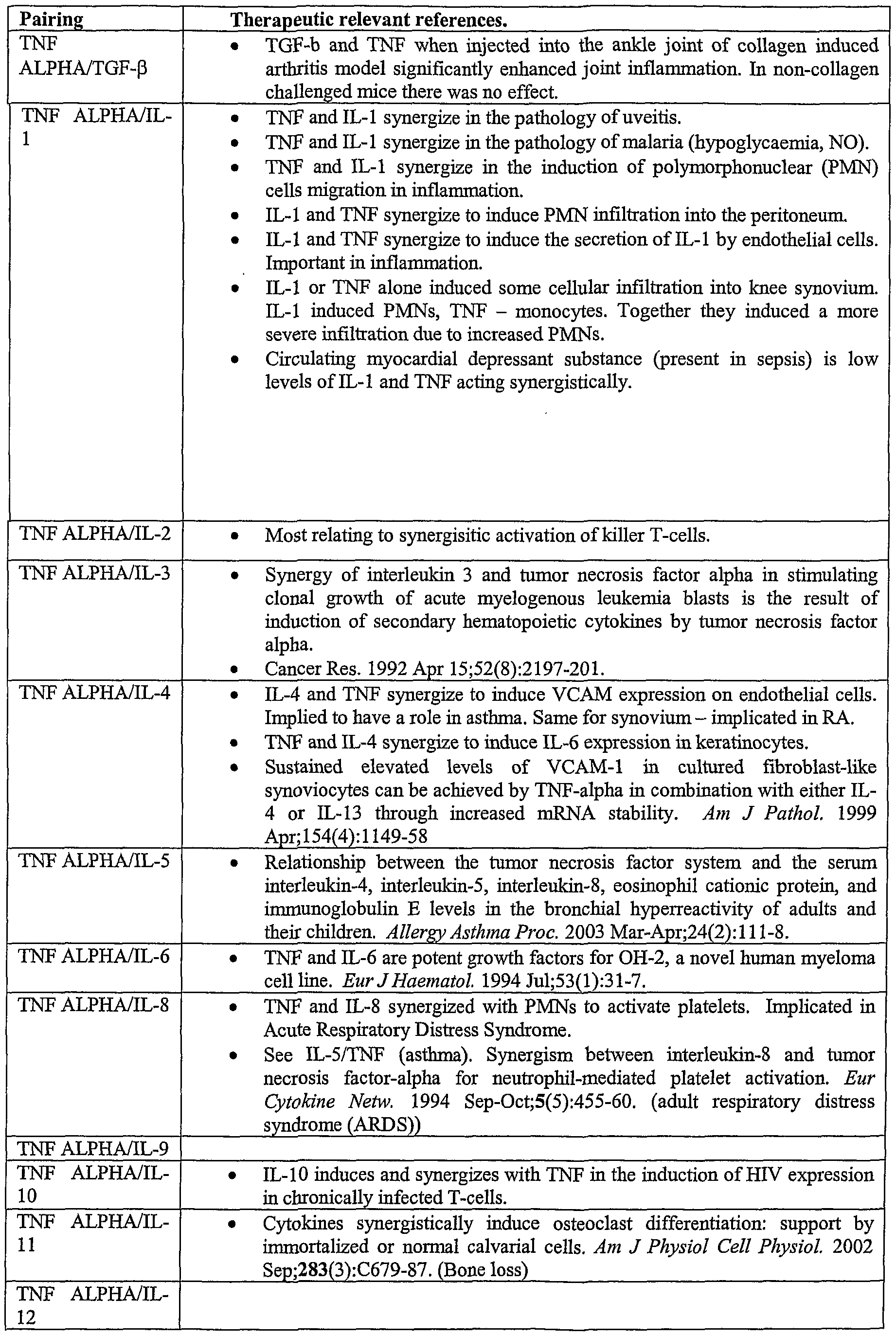


Claims
Priority Applications (16)
| Application Number | Priority Date | Filing Date | Title |
|---|---|---|---|
| CA002511910A CA2511910A1 (en) | 2002-12-27 | 2003-12-24 | Dual specific single domain antibodies specific for a ligand and for the receptor of the ligand |
| JP2005509717A JP2006523090A (en) | 2002-12-27 | 2003-12-24 | Bispecific single domain antibody specific for ligand and for ligand receptor |
| AU2003290330A AU2003290330A1 (en) | 2002-12-27 | 2003-12-24 | Dual specific single domain antibodies specific for a ligand and for the receptor of the ligand |
| EP03782689A EP1578801A2 (en) | 2002-12-27 | 2003-12-24 | Dual specific single domain antibodies specific for a ligand and for the receptor of the ligand |
| US10/925,366 US20050271663A1 (en) | 2001-06-28 | 2004-08-24 | Compositions and methods for treating inflammatory disorders |
| US10/985,847 US20060002935A1 (en) | 2002-06-28 | 2004-11-10 | Tumor Necrosis Factor Receptor 1 antagonists and methods of use therefor |
| US11/098,758 US20060073141A1 (en) | 2001-06-28 | 2005-04-04 | Compositions and methods for treating inflammatory disorders |
| US11/141,661 US20060257406A1 (en) | 2002-12-27 | 2005-05-31 | Ligand |
| US11/664,542 US20080008713A1 (en) | 2002-06-28 | 2005-10-07 | Single domain antibodies against tnfr1 and methods of use therefor |
| US11/667,393 US20070298041A1 (en) | 2002-06-28 | 2005-11-10 | Ligands That Enhance Endogenous Compounds |
| IL183027A IL183027A0 (en) | 2002-06-28 | 2007-05-07 | Ligands that enhance endogenous compounds |
| US11/981,821 US20100081792A1 (en) | 2001-06-28 | 2007-10-31 | Ligand |
| US12/006,933 US20080241166A1 (en) | 2002-06-28 | 2008-01-07 | Ligands that bind a receptor |
| US12/409,617 US20090258012A1 (en) | 2001-06-28 | 2009-03-24 | Compositions and methods for treating inflammatory disorders |
| US13/181,834 US9028822B2 (en) | 2002-06-28 | 2011-07-13 | Antagonists against TNFR1 and methods of use therefor |
| US13/733,675 US20130216538A1 (en) | 2002-12-27 | 2013-01-03 | Compositions and Methods for Treating Inflammatory Disorders |
Applications Claiming Priority (6)
| Application Number | Priority Date | Filing Date | Title |
|---|---|---|---|
| GB0230202A GB0230202D0 (en) | 2002-12-27 | 2002-12-27 | Ligand |
| GB0230202.4 | 2002-12-27 | ||
| GBPCT/GB03/002804 | 2003-06-30 | ||
| PCT/GB2003/002804 WO2004003019A2 (en) | 2002-06-28 | 2003-06-30 | Immunoglobin single variant antigen-binding domains and dual-specific constructs |
| GB0327706.8 | 2003-11-28 | ||
| GB0327706A GB0327706D0 (en) | 2003-11-28 | 2003-11-28 | Ligand |
Related Parent Applications (2)
| Application Number | Title | Priority Date | Filing Date |
|---|---|---|---|
| PCT/GB2003/002804 Continuation-In-Part WO2004003019A2 (en) | 2001-06-28 | 2003-06-30 | Immunoglobin single variant antigen-binding domains and dual-specific constructs |
| US10/744,774 Continuation-In-Part US20040219643A1 (en) | 2001-06-28 | 2003-12-23 | Dual-specific ligand |
Related Child Applications (6)
| Application Number | Title | Priority Date | Filing Date |
|---|---|---|---|
| PCT/GB2004/002829 Continuation-In-Part WO2004081026A2 (en) | 2001-06-28 | 2004-06-30 | Polypeptides |
| US10/925,366 Continuation-In-Part US20050271663A1 (en) | 2001-06-28 | 2004-08-24 | Compositions and methods for treating inflammatory disorders |
| PCT/GB2004/004253 Continuation-In-Part WO2005035572A2 (en) | 2002-06-28 | 2004-10-08 | Antibody compositions and methods |
| US10/985,847 Continuation-In-Part US20060002935A1 (en) | 2002-06-28 | 2004-11-10 | Tumor Necrosis Factor Receptor 1 antagonists and methods of use therefor |
| US11/098,758 Continuation-In-Part US20060073141A1 (en) | 2001-06-28 | 2005-04-04 | Compositions and methods for treating inflammatory disorders |
| US11/141,661 Continuation US20060257406A1 (en) | 2001-06-28 | 2005-05-31 | Ligand |
Publications (2)
| Publication Number | Publication Date |
|---|---|
| WO2004058821A2 true WO2004058821A2 (en) | 2004-07-15 |
| WO2004058821A3 WO2004058821A3 (en) | 2004-08-19 |
Family
ID=34863222
Family Applications (1)
| Application Number | Title | Priority Date | Filing Date |
|---|---|---|---|
| PCT/GB2003/005646 WO2004058821A2 (en) | 2001-06-28 | 2003-12-24 | Dual specific single domain antibodies specific for a ligand and for the receptor of the ligand |
Country Status (7)
| Country | Link |
|---|---|
| US (1) | US20060257406A1 (en) |
| EP (1) | EP1578801A2 (en) |
| JP (1) | JP2006523090A (en) |
| AU (1) | AU2003290330A1 (en) |
| CA (1) | CA2511910A1 (en) |
| IL (1) | IL183027A0 (en) |
| WO (1) | WO2004058821A2 (en) |
Cited By (154)
| Publication number | Priority date | Publication date | Assignee | Title |
|---|---|---|---|---|
| WO2004081026A3 (en) * | 2003-06-30 | 2005-01-27 | Domantis Ltd | Polypeptides |
| WO2006030220A1 (en) * | 2004-09-17 | 2006-03-23 | Domantis Limited | Compositions monovalent for cd40l binding and methods of use |
| WO2006003388A3 (en) * | 2004-06-30 | 2006-04-27 | Domantis Ltd | Compositions and methods for treating inflammatory disorders |
| WO2006089133A2 (en) | 2005-02-15 | 2006-08-24 | Duke University | Anti-cd19 antibodies and uses in oncology |
| GB2427194A (en) * | 2005-06-16 | 2006-12-20 | Domantis Ltd | Single domain Helicobacter pylori adhesin antibodies |
| WO2006038027A3 (en) * | 2004-10-08 | 2007-01-04 | Domantis Ltd | SINGLE DOMAIN ANTIBODIES AGAINST TNFRl AND METHODS OF USE THEREFOR |
| WO2007049017A2 (en) * | 2005-10-24 | 2007-05-03 | Domantis Limited | Agents that bind a target in pulmonary tissue for treating respiratory diseases |
| WO2007056441A2 (en) * | 2005-11-07 | 2007-05-18 | Genentech, Inc. | Binding polypeptides with diversified and consensus vh/vl hypervariable sequences |
| EP1806365A1 (en) | 2006-01-05 | 2007-07-11 | Boehringer Ingelheim International GmbH | Antibody molecules specific for fibroblast activation protein and immunoconjugates containing them |
| WO2007085815A2 (en) * | 2006-01-24 | 2007-08-02 | Domantis Limited | Ligands that bind il-4 and/or il-13 |
| WO2007063311A3 (en) * | 2005-12-01 | 2007-09-20 | Domantis Ltd | Competitive domain antibody formats that bind interleukin 1 receptor type 1 |
| WO2007063308A3 (en) * | 2005-12-01 | 2007-09-20 | Domantis Ltd | Noncompetitive domain antibody formats that bind interleukin 1 receptor type 1 |
| WO2007141280A2 (en) | 2006-06-06 | 2007-12-13 | Oxford Genome Sciences (Uk) Ltd | Proteins |
| WO2008030611A2 (en) | 2006-09-05 | 2008-03-13 | Medarex, Inc. | Antibodies to bone morphogenic proteins and receptors therefor and methods for their use |
| WO2008054603A2 (en) | 2006-10-02 | 2008-05-08 | Amgen Inc. | Il-17 receptor a antigen binding proteins |
| WO2008070569A2 (en) | 2006-12-01 | 2008-06-12 | Medarex, Inc. | Human antibodies that bind cd22 and uses thereof |
| JP2008519813A (en) * | 2004-11-10 | 2008-06-12 | ドマンティス リミテッド | Ligands that enhance endogenous compounds |
| WO2008074004A2 (en) | 2006-12-14 | 2008-06-19 | Medarex, Inc. | Human antibodies that bind cd70 and uses thereof |
| JP2008523083A (en) * | 2004-12-08 | 2008-07-03 | イムノメディクス, インコーポレイテッド | Methods and compositions for immunotherapy and detection of inflammatory and immune dysregulated diseases, infectious diseases, pathological angiogenesis and cancer |
| JP2008523783A (en) * | 2003-12-22 | 2008-07-10 | マイクロメット アクツィエン ゲゼルシャフト | Bispecific antibody |
| US7521425B2 (en) | 2005-03-03 | 2009-04-21 | Covx Technologies Ireland Limited | Anti-angiogenic compounds |
| WO2009054863A2 (en) | 2006-12-13 | 2009-04-30 | Medarex, Inc. | Human antibodies that bind cd19 and uses thereof |
| JP2009518024A (en) * | 2005-12-06 | 2009-05-07 | ドマンティス リミテッド | Ligands having binding specificity for EGFR and / or VEGF and methods of use thereof |
| JP2009523162A (en) * | 2006-01-11 | 2009-06-18 | ドマンティス リミテッド | Ligands having binding specificity for VEGF and / or EGFR and methods of use thereof |
| WO2009089295A2 (en) * | 2008-01-07 | 2009-07-16 | Government Of The United States Of America, As Represented By The Secretary, Dept. Of Health And Human Services | Anti-hiv domain antibodies and method of making and using same |
| WO2009125825A1 (en) | 2008-04-11 | 2009-10-15 | 中外製薬株式会社 | Antigen-binding molecule capable of binding to two or more antigen molecules repeatedly |
| WO2010011944A2 (en) | 2008-07-25 | 2010-01-28 | Wagner Richard W | Protein screeing methods |
| WO2010056804A1 (en) | 2008-11-12 | 2010-05-20 | Medimmune, Llc | Antibody formulation |
| WO2010084408A2 (en) | 2009-01-21 | 2010-07-29 | Oxford Biotherapeutics Ltd. | Pta089 protein |
| EP1399484B1 (en) * | 2001-06-28 | 2010-08-11 | Domantis Limited | Dual-specific ligand and its use |
| EP2221316A1 (en) | 2005-05-05 | 2010-08-25 | Duke University | Anti-CD19 antibody therapy for autoimmune disease |
| JP2010528646A (en) * | 2007-06-06 | 2010-08-26 | ドマンティス リミテッド | Polypeptides, antibody variable domains, and antagonists |
| WO2010094720A2 (en) | 2009-02-19 | 2010-08-26 | Glaxo Group Limited | Improved anti-tnfr1 polypeptides, antibody variable domains & antagonists |
| US7785903B2 (en) | 2004-04-09 | 2010-08-31 | Genentech, Inc. | Variable domain library and uses |
| JP2010529841A (en) * | 2007-06-06 | 2010-09-02 | ドマンティス リミテッド | Polypeptides, antibody variable domains and antagonists |
| WO2010102175A1 (en) | 2009-03-05 | 2010-09-10 | Medarex, Inc. | Fully human antibodies specific to cadm1 |
| WO2010122090A1 (en) | 2009-04-24 | 2010-10-28 | Glaxo Group Limited | Fgfr1c antibody combinations |
| US7846439B2 (en) | 2006-02-01 | 2010-12-07 | Cephalon Australia Pty Ltd | Domain antibody construct |
| WO2011006914A2 (en) | 2009-07-16 | 2011-01-20 | Glaxo Group Limited | Antagonists, uses & methods for partially inhibiting tnfr1 |
| WO2011012609A2 (en) | 2009-07-29 | 2011-02-03 | Glaxo Group Limited | Ligands that bind tgf-beta receptor rii |
| WO2011047083A1 (en) | 2009-10-13 | 2011-04-21 | Oxford Biotherapeutics Ltd. | Antibodies against epha10 |
| WO2011054007A1 (en) | 2009-11-02 | 2011-05-05 | Oxford Biotherapeutics Ltd. | Ror1 as therapeutic and diagnostic target |
| WO2011051217A1 (en) | 2009-10-27 | 2011-05-05 | Glaxo Group Limited | Stable anti-tnfr1 polypeptides, antibody variable domains & antagonists |
| WO2011054893A2 (en) | 2009-11-05 | 2011-05-12 | Novartis Ag | Biomarkers predictive of progression of fibrosis |
| EP2322554A1 (en) * | 2004-06-30 | 2011-05-18 | Domantis Limited | Composition comprising an anti-TNF-alpha domain antibody for the treatment of rheumatoid arthritis |
| US7981414B2 (en) | 2005-12-20 | 2011-07-19 | Cephalon Australia Pty Ltd | Anti-inflammatory dAb |
| WO2011100403A1 (en) | 2010-02-10 | 2011-08-18 | Immunogen, Inc | Cd20 antibodies and uses thereof |
| WO2011098449A1 (en) | 2010-02-10 | 2011-08-18 | Novartis Ag | Methods and compounds for muscle growth |
| EP2366715A2 (en) | 2005-11-14 | 2011-09-21 | Amgen Inc. | Rankl Antibody-PTH/PTHRP Chimeric Molecules |
| EP2368908A2 (en) | 2006-11-10 | 2011-09-28 | CovX Technologies Ireland Limited | Anti- angiogenic compounds |
| WO2011122011A2 (en) | 2010-03-30 | 2011-10-06 | Chugai Seiyaku Kabushiki Kaisha | Antibodies with modified affinity to fcrn that promote antigen clearance |
| WO2011131659A2 (en) | 2010-04-21 | 2011-10-27 | Glaxo Group Limited | Binding domains |
| WO2011138391A1 (en) | 2010-05-06 | 2011-11-10 | Novartis Ag | Compositions and methods of use for therapeutic low density lipoprotein - related protein 6 (lrp6) multivalent antibodies |
| WO2011138392A1 (en) | 2010-05-06 | 2011-11-10 | Novartis Ag | Compositions and methods of use for therapeutic low density lipoprotein -related protein 6 (lrp6) antibodies |
| WO2012020096A1 (en) | 2010-08-13 | 2012-02-16 | Medimmune Limited | Monomeric polypeptides comprising variant fc regions and methods of use |
| WO2012022814A1 (en) | 2010-08-20 | 2012-02-23 | Novartis Ag | Antibodies for epidermal growth factor receptor 3 (her3) |
| WO2012022734A2 (en) | 2010-08-16 | 2012-02-23 | Medimmune Limited | Anti-icam-1 antibodies and methods of use |
| WO2012032433A1 (en) | 2010-09-09 | 2012-03-15 | Pfizer Inc. | 4-1bb binding molecules |
| EP2441775A1 (en) | 2007-02-26 | 2012-04-18 | Oxford Biotherapeutics Ltd. | Protein |
| EP2447719A1 (en) | 2007-02-26 | 2012-05-02 | Oxford Biotherapeutics Ltd. | Proteins |
| WO2012073992A1 (en) | 2010-11-30 | 2012-06-07 | 中外製薬株式会社 | Antigen-binding molecule capable of binding to plurality of antigen molecules repeatedly |
| WO2012093704A1 (en) | 2011-01-07 | 2012-07-12 | 中外製薬株式会社 | Method for improving physical properties of antibody |
| WO2012093125A1 (en) | 2011-01-06 | 2012-07-12 | Glaxo Group Limited | Ligands that bind tgf-beta receptor ii |
| US8236931B2 (en) | 2006-10-30 | 2012-08-07 | Glaxo Group Limited | Prevention of aggregation of immunoglobulin light or heavy chains |
| WO2012104227A1 (en) | 2011-02-02 | 2012-08-09 | Glaxo Group Limited | Novel antigen binding proteins |
| EP2486941A1 (en) | 2006-10-02 | 2012-08-15 | Medarex, Inc. | Human antibodies that bind CXCR4 and uses thereof |
| WO2012115241A1 (en) | 2011-02-25 | 2012-08-30 | 中外製薬株式会社 | Fcγriib-specific fc antibody |
| US8278419B2 (en) | 2008-10-31 | 2012-10-02 | Centocor Ortho Biotech Inc. | Fibronectin type III domain based scaffold compositions, methods and uses |
| US8318159B2 (en) | 2008-12-12 | 2012-11-27 | Boehringer Ingelheim International Gmbh | Anti-IGF antibodies |
| WO2012166906A1 (en) | 2011-05-31 | 2012-12-06 | Massachusetts Institute Of Technology | Cell-directed synthesis of multifunctional nanopatterns and nanomaterials |
| EP2540741A1 (en) | 2006-03-06 | 2013-01-02 | Aeres Biomedical Limited | Humanized anti-CD22 antibodies and their use in treatment of oncology, transplantation and autoimmune disease |
| WO2013001369A2 (en) | 2011-06-28 | 2013-01-03 | Oxford Biotherapeutics Ltd. | Therapeutic and diagnostic target |
| WO2013003625A2 (en) | 2011-06-28 | 2013-01-03 | Oxford Biotherapeutics Ltd. | Antibodies |
| WO2013002362A1 (en) | 2011-06-30 | 2013-01-03 | 中外製薬株式会社 | Heterodimerized polypeptide |
| WO2013016220A1 (en) | 2011-07-22 | 2013-01-31 | Amgen Inc. | Il-17 receptor a is required for il-17c biology |
| WO2013043933A2 (en) | 2011-09-22 | 2013-03-28 | Amgen Inc. | Cd27l antigen binding proteins |
| WO2013047748A1 (en) | 2011-09-30 | 2013-04-04 | 中外製薬株式会社 | Antigen-binding molecule promoting disappearance of antigens having plurality of biological activities |
| WO2013046704A2 (en) | 2011-09-30 | 2013-04-04 | Chugai Seiyaku Kabushiki Kaisha | THERAPEUTIC ANTIGEN-BINDING MOLECULE WITH A FcRn-BINDING DOMAIN THAT PROMOTES ANTIGEN CLEARANCE |
| WO2013051294A1 (en) | 2011-10-05 | 2013-04-11 | 中外製薬株式会社 | Antigen-binding molecule for promoting clearance from plasma of antigen comprising saccharide chain receptor-binding domain |
| WO2013060872A1 (en) | 2011-10-27 | 2013-05-02 | Boehringer Ingelheim International Gmbh | Anticancer combination therapy |
| WO2013067355A1 (en) | 2011-11-04 | 2013-05-10 | Novartis Ag | Low density lipoprotein-related protein 6 (lrp6) - half life extender constructs |
| EP2594590A1 (en) | 2007-12-14 | 2013-05-22 | Bristol-Myers Squibb Company | Binding molecules to the human OX40 receptor |
| WO2013084148A2 (en) | 2011-12-05 | 2013-06-13 | Novartis Ag | Antibodies for epidermal growth factor receptor 3 (her3) directed to domain ii of her3 |
| WO2013084147A2 (en) | 2011-12-05 | 2013-06-13 | Novartis Ag | Antibodies for epidermal growth factor receptor 3 (her3) |
| EP2441838A3 (en) * | 2006-01-24 | 2013-07-10 | Domantis Limited | Fusion proteins that contain natural junctions |
| WO2013118858A1 (en) | 2012-02-09 | 2013-08-15 | 中外製薬株式会社 | Modified fc region of antibody |
| US8569227B2 (en) | 2010-04-30 | 2013-10-29 | Janssen Biotech, Inc. | Stabilized fibronectin domain compositions, methods and uses |
| US8580254B2 (en) | 2007-06-19 | 2013-11-12 | Boehringer Ingelheim International Gmbh | Anti-IGF antibodies |
| WO2013187495A1 (en) | 2012-06-14 | 2013-12-19 | 中外製薬株式会社 | ANTIGEN-BINDING MOLECULE CONTAINING MODIFIED Fc REGION |
| US8623356B2 (en) | 2005-11-29 | 2014-01-07 | The University Of Sydney | Demibodies: dimerization-activated therapeutic agents |
| WO2014020331A1 (en) | 2012-08-01 | 2014-02-06 | Oxford Biotherapeutics Ltd. | Therapeutic and diagnostic target |
| EP2700651A1 (en) | 2008-07-18 | 2014-02-26 | Bristol-Myers Squibb Company | Compositions monovalent for CD28 binding and methods of use |
| WO2014030750A1 (en) | 2012-08-24 | 2014-02-27 | 中外製薬株式会社 | MOUSE FcγRII-SPECIFIC Fc ANTIBODY |
| WO2014030728A1 (en) | 2012-08-24 | 2014-02-27 | 中外製薬株式会社 | Fcγriib-specific fc region variant |
| EP2703011A2 (en) | 2007-05-07 | 2014-03-05 | MedImmune, LLC | Anti-icos antibodies and their use in treatment of oncology, transplantation and autoimmune disease |
| WO2014089335A2 (en) | 2012-12-07 | 2014-06-12 | Amgen Inc. | Bcma antigen binding proteins |
| US20140178388A1 (en) * | 2012-12-26 | 2014-06-26 | Industrial Technology Research Institute | Multivalent antibody fragments and trimerized complexes thereof |
| WO2014104165A1 (en) | 2012-12-27 | 2014-07-03 | 中外製薬株式会社 | Heterodimerized polypeptide |
| WO2014114801A1 (en) | 2013-01-25 | 2014-07-31 | Amgen Inc. | Antibodies targeting cdh19 for melanoma |
| WO2014120916A1 (en) | 2013-02-01 | 2014-08-07 | Bristol-Myers Squibb Company | Pegylated domain antibodies monovalent for cd28 binding and methods of use |
| WO2014133855A1 (en) | 2013-02-28 | 2014-09-04 | Caprion Proteomics Inc. | Tuberculosis biomarkers and uses thereof |
| WO2014159239A2 (en) | 2013-03-14 | 2014-10-02 | Novartis Ag | Antibodies against notch 3 |
| WO2014163101A1 (en) | 2013-04-02 | 2014-10-09 | 中外製薬株式会社 | Fc region variant |
| WO2014177460A1 (en) | 2013-04-29 | 2014-11-06 | F. Hoffmann-La Roche Ag | Human fcrn-binding modified antibodies and methods of use |
| US8921528B2 (en) | 2004-06-01 | 2014-12-30 | Domantis Limited | Bispecific fusion antibodies with enhanced serum half-life |
| WO2015015401A2 (en) | 2013-08-02 | 2015-02-05 | Pfizer Inc. | Anti-cxcr4 antibodies and antibody-drug conjugates |
| WO2015050959A1 (en) | 2013-10-01 | 2015-04-09 | Yale University | Anti-kit antibodies and methods of use thereof |
| WO2015051010A1 (en) | 2013-10-02 | 2015-04-09 | Medimmune, Llc | Neutralizing anti-influenza a antibodies and uses thereof |
| US9028822B2 (en) | 2002-06-28 | 2015-05-12 | Domantis Limited | Antagonists against TNFR1 and methods of use therefor |
| WO2015107026A1 (en) | 2014-01-15 | 2015-07-23 | F. Hoffmann-La Roche Ag | Fc-region variants with modified fcrn- and maintained protein a-binding properties |
| WO2015127134A2 (en) | 2014-02-20 | 2015-08-27 | Allergan, Inc. | Complement component c5 antibodies |
| WO2015130826A1 (en) | 2014-02-27 | 2015-09-03 | Allergan, Inc. | COMPLEMENT FACTOR Bb ANTIBODIES |
| US9200273B2 (en) | 2011-09-27 | 2015-12-01 | Janssen Biotech, Inc. | Fibronectin type III repeat based protein scaffolds with alternative binding surfaces |
| EP3009454A2 (en) | 2009-04-20 | 2016-04-20 | Oxford Bio Therapeutics Limited | Antibodies specific to cadherin-17 |
| WO2017110980A1 (en) | 2015-12-25 | 2017-06-29 | 中外製薬株式会社 | Antibody having enhanced activity, and method for modifying same |
| EP3192807A1 (en) | 2007-11-27 | 2017-07-19 | The University Of British Columbia | 14-3-3 eta antibodies and uses thereof for the diagnosis and treatment of arthritis |
| WO2017175145A1 (en) | 2016-04-05 | 2017-10-12 | Glaxosmithkline Intellectual Property Development Limited | Inhibition of tgfbeta in immunotherapy |
| WO2018020000A1 (en) | 2016-07-29 | 2018-02-01 | INSERM (Institut National de la Santé et de la Recherche Médicale) | Antibodies targeting tumor associated macrophages and uses thereof |
| WO2018129029A1 (en) | 2017-01-04 | 2018-07-12 | Immunogen, Inc. | Met antibodies and immunoconjugates and uses thereof |
| WO2018158398A1 (en) | 2017-03-02 | 2018-09-07 | INSERM (Institut National de la Santé et de la Recherche Médicale) | Antibodies having specificity to nectin-4 and uses thereof |
| WO2018181870A1 (en) | 2017-03-31 | 2018-10-04 | 公立大学法人奈良県立医科大学 | Medicinal composition usable for preventing and/or treating blood coagulation factor ix abnormality, comprising multispecific antigen binding molecule replacing function of blood coagulation factor viii |
| WO2019020480A1 (en) | 2017-07-24 | 2019-01-31 | INSERM (Institut National de la Santé et de la Recherche Médicale) | Antibodies and peptides to treat hcmv related diseases |
| US10202447B2 (en) | 2010-09-10 | 2019-02-12 | Medimmune Limited | Antibody derivatives |
| EP3456743A1 (en) | 2013-05-30 | 2019-03-20 | Kiniksa Pharmaceuticals, Ltd. | Oncostatin m receptor antigen binding proteins |
| US10377828B2 (en) | 2013-03-07 | 2019-08-13 | Boehringer Ingelheim International Gmbh | Combination therapy for neoplasia treatment |
| WO2020010250A2 (en) | 2018-07-03 | 2020-01-09 | Elstar Therapeutics, Inc. | Anti-tcr antibody molecules and uses thereof |
| WO2020014306A1 (en) | 2018-07-10 | 2020-01-16 | Immunogen, Inc. | Met antibodies and immunoconjugates and uses thereof |
| EP3611188A1 (en) | 2014-11-06 | 2020-02-19 | F. Hoffmann-La Roche AG | Fc-region variants with modified fcrn-binding and methods of use |
| WO2020053122A1 (en) | 2018-09-10 | 2020-03-19 | INSERM (Institut National de la Santé et de la Recherche Médicale) | Combination of her2/neu antibody with heme for treating cancer |
| EP3693395A1 (en) | 2012-05-18 | 2020-08-12 | Amgen Inc. | St2 antigen binding proteins |
| WO2020198731A2 (en) | 2019-03-28 | 2020-10-01 | Danisco Us Inc | Engineered antibodies |
| WO2020193520A1 (en) | 2019-03-25 | 2020-10-01 | INSERM (Institut National de la Santé et de la Recherche Médicale) | Treatment of taupathy disorders by targeting new tau species |
| EP3736293A1 (en) | 2013-02-12 | 2020-11-11 | Boehringer Ingelheim International Gmbh | Therapeutic and diagnostic target for cancer comprising dll3 binding reagents |
| WO2021058763A1 (en) | 2019-09-27 | 2021-04-01 | INSERM (Institut National de la Santé et de la Recherche Médicale) | Anti-müllerian inhibiting substance antibodies and uses thereof |
| WO2021058729A1 (en) | 2019-09-27 | 2021-04-01 | INSERM (Institut National de la Santé et de la Recherche Médicale) | Anti-müllerian inhibiting substance type i receptor antibodies and uses thereof |
| WO2021116119A1 (en) | 2019-12-09 | 2021-06-17 | INSERM (Institut National de la Santé et de la Recherche Médicale) | Antibodies having specificity to her4 and uses thereof |
| WO2021160133A1 (en) | 2020-02-13 | 2021-08-19 | 湖南华康恒健生物技术有限公司 | Anti-bcma antibody, pharmaceutical composition of same, and applications thereof |
| EP3878866A1 (en) | 2013-04-29 | 2021-09-15 | F. Hoffmann-La Roche AG | Fc-receptor binding modified asymmetric antibodies and methods of use |
| WO2021228956A1 (en) | 2020-05-12 | 2021-11-18 | INSERM (Institut National de la Santé et de la Recherche Médicale) | New method to treat cutaneous t-cell lymphomas and tfh derived lymphomas |
| WO2021235537A1 (en) | 2020-05-22 | 2021-11-25 | 中外製薬株式会社 | Antibody for neutralizing substance having coagulation factor viii (f.viii) function-substituting activity |
| US11242395B2 (en) | 2017-08-21 | 2022-02-08 | Adagene Inc. | Anti-CD137 molecules and use thereof |
| WO2022042659A1 (en) | 2020-08-26 | 2022-03-03 | 湖南华康恒健生物技术有限公司 | Anti-ox40 antibody, and pharmaceutical composition and application thereof |
| WO2022044573A1 (en) | 2020-08-26 | 2022-03-03 | 国立大学法人熊本大学 | Human antibody or antigen-binding fragment thereof against coronavirus spike protein |
| US11299534B2 (en) | 2016-12-14 | 2022-04-12 | Janssen Biotech, Inc. | CD8A-binding fibronectin type III domains |
| US11345739B2 (en) | 2016-12-14 | 2022-05-31 | Janssen Biotech, Inc | CD137 binding fibronectin type III domains |
| US11359016B2 (en) | 2018-02-02 | 2022-06-14 | Adagene Inc. | Anti-CTLA4 antibodies and methods of making and using the same |
| US11447539B2 (en) | 2016-12-14 | 2022-09-20 | Janssen Biotech, Inc. | PD-L1 binding fibronectin type III domains |
| WO2022216993A2 (en) | 2021-04-08 | 2022-10-13 | Marengo Therapeutics, Inc. | Multifuntional molecules binding to tcr and uses thereof |
| US11628222B2 (en) | 2019-10-14 | 2023-04-18 | Aro Biotherapeutics Company | CD71 binding fibronectin type III domains |
| WO2023175171A1 (en) | 2022-03-18 | 2023-09-21 | Inserm (Institut National De La Sante Et De La Recherche Medicale) | Bk polyomavirus antibodies and uses thereof |
| US11781138B2 (en) | 2019-10-14 | 2023-10-10 | Aro Biotherapeutics Company | FN3 domain-siRNA conjugates and uses thereof |
| WO2024052503A1 (en) | 2022-09-08 | 2024-03-14 | Institut National de la Santé et de la Recherche Médicale | Antibodies having specificity to ltbp2 and uses thereof |
| WO2024056668A1 (en) | 2022-09-12 | 2024-03-21 | Institut National de la Santé et de la Recherche Médicale | New anti-itgb8 antibodies and its uses thereof |
| US11952681B2 (en) | 2018-02-02 | 2024-04-09 | Adagene Inc. | Masked activatable CD137 antibodies |
Families Citing this family (15)
| Publication number | Priority date | Publication date | Assignee | Title |
|---|---|---|---|---|
| US20070104710A1 (en) * | 2002-06-28 | 2007-05-10 | Domants Limited | Ligand that has binding specificity for IL-4 and/or IL-13 |
| US9321832B2 (en) | 2002-06-28 | 2016-04-26 | Domantis Limited | Ligand |
| CA2490280A1 (en) | 2002-07-01 | 2004-01-08 | Human Genome Sciences, Inc. | Antibodies that specifically bind to reg iv |
| US20110223168A1 (en) * | 2002-12-27 | 2011-09-15 | Greg Winter | Ligand that has binding specificity for il-4 and/or il-13 |
| CN104004088B (en) | 2007-09-26 | 2017-11-07 | Ucb医药有限公司 | dual specificity antibody fusions |
| LT2334705T (en) * | 2008-09-26 | 2017-03-27 | Ucb Biopharma Sprl | Biological products |
| GB201005063D0 (en) | 2010-03-25 | 2010-05-12 | Ucb Pharma Sa | Biological products |
| RU2612903C2 (en) | 2010-08-02 | 2017-03-13 | Ридженерон Фармасьютикалз, Инк. | Mouse producing binding proteins containing vl-domains |
| KR20240033183A (en) * | 2011-06-23 | 2024-03-12 | 아블린쓰 엔.브이. | Techniques for predicting, detecting and reducing aspecific protein interference in assays involving immunoglobulin single variable domains |
| PT2793567T (en) | 2011-12-20 | 2019-05-27 | Regeneron Pharma | Humanized light chain mice |
| ES2762640T3 (en) | 2014-03-21 | 2020-05-25 | Regeneron Pharma | VL antigen binding proteins exhibiting different binding characteristics |
| CA2942697A1 (en) | 2014-03-21 | 2015-09-24 | Lynn Macdonald | Non-human animals that make single domain binding proteins |
| CA2971278C (en) * | 2014-12-19 | 2023-09-19 | Ablynx N.V. | Cysteine linked nanobody dimers |
| AU2016232715A1 (en) | 2015-03-19 | 2017-09-28 | Regeneron Pharmaceuticals, Inc. | Non-human animals that select for light chain variable regions that bind antigen |
| US10634680B2 (en) | 2016-04-26 | 2020-04-28 | University Of Utah Research Foundation | Target-binding activated split reporter systems for analyte detection and related components and methods |
Citations (3)
| Publication number | Priority date | Publication date | Assignee | Title |
|---|---|---|---|---|
| EP0368684A1 (en) * | 1988-11-11 | 1990-05-16 | Medical Research Council | Cloning immunoglobulin variable domain sequences. |
| WO1997030084A1 (en) * | 1996-02-15 | 1997-08-21 | Genetics Institute, Inc. | Tnf receptor death domain ligand proteins and inhibitors of ligand binding |
| WO2000029004A1 (en) * | 1998-11-18 | 2000-05-25 | Peptor Ltd. | Small functional units of antibody heavy chain variable regions |
Family Cites Families (67)
| Publication number | Priority date | Publication date | Assignee | Title |
|---|---|---|---|---|
| CU22545A1 (en) * | 1994-11-18 | 1999-03-31 | Centro Inmunologia Molecular | OBTAINING A CHEMICAL AND HUMANIZED ANTIBODY AGAINST THE RECEPTOR OF THE EPIDERMAL GROWTH FACTOR FOR DIAGNOSTIC AND THERAPEUTIC USE |
| US5120712A (en) * | 1986-05-05 | 1992-06-09 | The General Hospital Corporation | Insulinotropic hormone |
| WO1989006692A1 (en) * | 1988-01-12 | 1989-07-27 | Genentech, Inc. | Method of treating tumor cells by inhibiting growth factor receptor function |
| AU4128089A (en) * | 1988-09-15 | 1990-03-22 | Rorer International (Overseas) Inc. | Monoclonal antibodies specific to human epidermal growth factor receptor and therapeutic methods employing same |
| US5116964A (en) * | 1989-02-23 | 1992-05-26 | Genentech, Inc. | Hybrid immunoglobulins |
| US5459061A (en) * | 1990-01-26 | 1995-10-17 | W. Alton Jones Cell Science Center, Inc. | Hybridomas producing monoclonal antibodies which specifically bind to continuous epitope on the human EGF receptor and compete with EGF for binding to the EGF receptor |
| US5726152A (en) * | 1990-09-21 | 1998-03-10 | Merck & Co., Inc. | Vascular endothelial cell growth factor II |
| SK281142B6 (en) * | 1991-03-06 | 2000-12-11 | Merck Patent Gesellschaft Mit Beschr�Nkter Haftung | Humanised monoclonal antibodies, expression vectors and pharmaceutical compositions |
| US6582959B2 (en) * | 1991-03-29 | 2003-06-24 | Genentech, Inc. | Antibodies to vascular endothelial cell growth factor |
| OA10149A (en) * | 1991-03-29 | 1996-12-18 | Genentech Inc | Vascular endothelial cell growth factor antagonists |
| EP0603194A4 (en) * | 1991-07-05 | 1994-12-07 | Seragen Inc | Epidermal growth factor receptor targeted molecules for treatment of inflammatory arthritis. |
| US5965132A (en) * | 1992-03-05 | 1999-10-12 | Board Of Regents, The University Of Texas System | Methods and compositions for targeting the vasculature of solid tumors |
| IL112372A (en) * | 1994-02-07 | 2001-08-26 | Res Dev Foundation | Non-viral vector for the delivery of genetic information to cells |
| GB9410533D0 (en) * | 1994-05-26 | 1994-07-13 | Lynxvale Ltd | In situ hybridisation and immuno-Chemical localisation of a growth factor |
| DK0706799T3 (en) * | 1994-09-16 | 2002-02-25 | Merck Patent Gmbh | Immune Conjugates II |
| US5928939A (en) * | 1995-03-01 | 1999-07-27 | Ludwig Institute For Cancer Research | Vascular endothelial growth factor-b and dna coding therefor |
| US7060808B1 (en) * | 1995-06-07 | 2006-06-13 | Imclone Systems Incorporated | Humanized anti-EGF receptor monoclonal antibody |
| US6410690B1 (en) * | 1995-06-07 | 2002-06-25 | Medarex, Inc. | Therapeutic compounds comprised of anti-Fc receptor antibodies |
| US6020473A (en) * | 1995-08-25 | 2000-02-01 | Genentech, Inc. | Nucleic acids encoding variants of vascular endothelial cell growth factor |
| BR9606706A (en) * | 1995-10-16 | 1999-04-06 | Unilever Nv | Bispecific or bivalent antibody fragment analog use process to produce the same |
| US5664034A (en) * | 1996-05-21 | 1997-09-02 | Lucent Technologies Inc. | Lightwave communication monitoring switch |
| US5922845A (en) * | 1996-07-11 | 1999-07-13 | Medarex, Inc. | Therapeutic multispecific compounds comprised of anti-Fcα receptor antibodies |
| US6013780A (en) * | 1996-09-06 | 2000-01-11 | Technion Research & Development Co. Ltd. | VEGF145 expression vectors |
| US6750044B1 (en) * | 1996-10-17 | 2004-06-15 | Genentech, Inc. | Variants of vascular endothelial cell growth factor having antagonistic properties, nucleic acids encoding the same and host cells comprising those nucleic acids |
| US20030165467A1 (en) * | 1997-01-21 | 2003-09-04 | Technion Research & Development Co., Ltd. | Angiogenic factor and use thereof in treating cardiovascular disease |
| US6485942B1 (en) * | 1997-02-14 | 2002-11-26 | Genentech, Inc. | Variants of vascular endothelial cell growth factor having altered pharmacological properties, and recombinant methods of production |
| US20020032315A1 (en) * | 1997-08-06 | 2002-03-14 | Manuel Baca | Anti-vegf antibodies |
| US20020173629A1 (en) * | 1997-05-05 | 2002-11-21 | Aya Jakobovits | Human monoclonal antibodies to epidermal growth factor receptor |
| US6777534B1 (en) * | 1997-12-09 | 2004-08-17 | Children's Medical Center Corporation | Peptide antagonists of vascular endothelial growth factor |
| US20020192211A1 (en) * | 1998-03-17 | 2002-12-19 | Hudziak Robert M. | Method of treating tumor cells by inhibiting growth factor receptor function |
| US20030224001A1 (en) * | 1998-03-19 | 2003-12-04 | Goldstein Neil I. | Antibody and antibody fragments for inhibiting the growth of tumors |
| ZA200007412B (en) * | 1998-05-15 | 2002-03-12 | Imclone Systems Inc | Treatment of human tumors with radiation and inhibitors of growth factor receptor tyrosine kinases. |
| US20030175271A1 (en) * | 1998-05-20 | 2003-09-18 | Kyowa Hakko Kogyo Co., Ltd. | VEGF activity inhibitor |
| US7264801B2 (en) * | 1998-08-11 | 2007-09-04 | Genentech, Inc. | EG-VEGF nucleic acids and polypeptides and method of use |
| US20020172678A1 (en) * | 2000-06-23 | 2002-11-21 | Napoleone Ferrara | EG-VEGF nucleic acids and polypeptides and methods of use |
| WO2000037502A2 (en) * | 1998-12-22 | 2000-06-29 | Genentech, Inc. | Vascular endothelial cell growth factor antagonists and uses thereof |
| US6703020B1 (en) * | 1999-04-28 | 2004-03-09 | Board Of Regents, The University Of Texas System | Antibody conjugate methods for selectively inhibiting VEGF |
| ES2223705T3 (en) * | 1999-04-28 | 2005-03-01 | Board Of Regents, The University Of Texas System | COMPOSITIONS AND METHODS FOR THE TREATMENT OF CANCER THROUGH INHIBITION SELECTIVE OF VEGF. |
| IL146480A0 (en) * | 1999-05-14 | 2002-07-25 | Imclone Systems Inc | Treatment of refractory human tumors with epidermal growth factor receptor antagonists |
| US7049410B2 (en) * | 1999-05-14 | 2006-05-23 | Majumdar Adhip P N | Antibodies to a novel EGF-receptor related protein (ERRP) |
| WO2000071713A1 (en) * | 1999-05-20 | 2000-11-30 | Scios Inc. | Vascular endothelial growth factor variants |
| GB2353561A (en) * | 1999-08-21 | 2001-02-28 | Cummins Engine Co Ltd | An engine block with machined end faces for receiving a camshaft gear train |
| CA2400948A1 (en) * | 2000-02-25 | 2001-08-30 | Ludwig Institute For Cancer Research | Materials and methods involving hybrid vascular endothelial growth factor dnas and proteins |
| CA2404528A1 (en) * | 2000-03-31 | 2001-10-04 | Institut Pasteur | Peptides blocking vascular endothelial growth factor (vegf)-mediated angiogenesis, polynucleotides encoding said peptides and methods of use thereof |
| WO2001077342A1 (en) * | 2000-04-11 | 2001-10-18 | Genentech, Inc. | Multivalent antibodies and uses therefor |
| JP2004511430A (en) * | 2000-05-24 | 2004-04-15 | イムクローン システムズ インコーポレイティド | Bispecific immunoglobulin-like antigen binding protein and production method |
| DE10038624C2 (en) * | 2000-08-03 | 2002-11-21 | Broekelmann Aluminium F W | Heat transfer tube with twisted inner fins |
| AU2002211658A1 (en) * | 2000-10-13 | 2002-04-22 | Uab Research Foundation | Human anti-epidermal growth factor receptor single-chain antibodies |
| US20030133939A1 (en) * | 2001-01-17 | 2003-07-17 | Genecraft, Inc. | Binding domain-immunoglobulin fusion proteins |
| US7667004B2 (en) * | 2001-04-17 | 2010-02-23 | Abmaxis, Inc. | Humanized antibodies against vascular endothelial growth factor |
| JP2004528368A (en) * | 2001-05-08 | 2004-09-16 | メルク パテント ゲゼルシャフト ミット ベシュレンクテル ハフトング | Combination therapy using anti-EGFR antibody and antihormonal agent |
| CA2450285C (en) * | 2001-06-13 | 2016-08-02 | Genmab A/S | Human monoclonal antibodies to epidermal growth factor receptor (egfr) |
| US20040248196A1 (en) * | 2001-08-03 | 2004-12-09 | Adams Timothy Edward | Methods of screening based on the egf receptor crystal structure |
| BR0213303A (en) * | 2001-10-15 | 2005-06-07 | Immunomedics Inc | Direct Bleach Binding Proteins |
| DE10163459A1 (en) * | 2001-12-21 | 2003-07-03 | Merck Patent Gmbh | Lyophilized preparation containing antibodies to EGF receptor |
| EP1542721A4 (en) * | 2002-06-14 | 2007-05-02 | Centocor Inc | Modified "s" antibodies |
| EP1391311A1 (en) * | 2002-08-19 | 2004-02-25 | Star Coating AG | System for the transfer of images onto dark textiles |
| TW200501960A (en) * | 2002-10-02 | 2005-01-16 | Bristol Myers Squibb Co | Synergistic kits and compositions for treating cancer |
| US20050043233A1 (en) * | 2003-04-29 | 2005-02-24 | Boehringer Ingelheim International Gmbh | Combinations for the treatment of diseases involving cell proliferation, migration or apoptosis of myeloma cells or angiogenesis |
| BRPI0411200A (en) * | 2003-05-30 | 2006-07-18 | Genentech Inc | cancer treatment method, human patient treatment methods, industrialized article and cancer treatment kit |
| EP1631317A2 (en) * | 2003-06-06 | 2006-03-08 | Regeneron Pharmaceuticals, Inc. | Use of vegf inhibitors for tumor regression |
| KR101581040B1 (en) * | 2003-06-27 | 2015-12-30 | 암젠 프레몬트 인코포레이티드 | Antibodies directed to the deletion mutants of epidermal growth factor receptor and uses thereof |
| AR046510A1 (en) * | 2003-07-25 | 2005-12-14 | Regeneron Pharma | COMPOSITION OF A VEGF ANTAGONIST AND AN ANTI-PROLIFERATIVE AGENT |
| US20050196340A1 (en) * | 2003-08-06 | 2005-09-08 | Jocelyn Holash | Use of a VEGF antagonist in combination with radiation therapy |
| EP1651663B1 (en) * | 2003-08-08 | 2017-05-17 | Immunomedics, Inc. | Bispecific antibodies for inducing apoptosis of tumor and diseased cells |
| WO2005070966A2 (en) * | 2004-01-16 | 2005-08-04 | Regeneron Pharmaceuticals, Inc. | Fusion polypeptides capable of activating receptors |
| US7767792B2 (en) * | 2004-02-20 | 2010-08-03 | Ludwig Institute For Cancer Research Ltd. | Antibodies to EGF receptor epitope peptides |
-
2003
- 2003-12-24 EP EP03782689A patent/EP1578801A2/en not_active Ceased
- 2003-12-24 WO PCT/GB2003/005646 patent/WO2004058821A2/en active Application Filing
- 2003-12-24 AU AU2003290330A patent/AU2003290330A1/en not_active Abandoned
- 2003-12-24 JP JP2005509717A patent/JP2006523090A/en active Pending
- 2003-12-24 CA CA002511910A patent/CA2511910A1/en not_active Abandoned
-
2005
- 2005-05-31 US US11/141,661 patent/US20060257406A1/en not_active Abandoned
-
2007
- 2007-05-07 IL IL183027A patent/IL183027A0/en unknown
Patent Citations (3)
| Publication number | Priority date | Publication date | Assignee | Title |
|---|---|---|---|---|
| EP0368684A1 (en) * | 1988-11-11 | 1990-05-16 | Medical Research Council | Cloning immunoglobulin variable domain sequences. |
| WO1997030084A1 (en) * | 1996-02-15 | 1997-08-21 | Genetics Institute, Inc. | Tnf receptor death domain ligand proteins and inhibitors of ligand binding |
| WO2000029004A1 (en) * | 1998-11-18 | 2000-05-25 | Peptor Ltd. | Small functional units of antibody heavy chain variable regions |
Non-Patent Citations (4)
| Title |
|---|
| ELS CONRATH K ET AL: "Camel single-domain antibodies as modular building units in bispecific and bivalent antibody constructs" JOURNAL OF BIOLOGICAL CHEMISTRY, AMERICAN SOCIETY OF BIOLOGICAL CHEMISTS, BALTIMORE, MD, US, vol. 276, no. 10, 9 March 2001 (2001-03-09), pages 7346-7350, XP002248402 ISSN: 0021-9258 * |
| MUYLDERMANS S ET AL: "UNIQUE SINGLE-DOMAIN ANTIGEN BINDING FRAGMENTS DERIVES FROM NATURALLY OCCURRING CAMEL HEAVY-CHAIN ANTIBODIES" JOURNAL OF MOLECULAR RECOGNITION, HEYDEN & SON LTD., LONDON, GB, vol. 12, no. 2, March 1999 (1999-03), pages 131-140, XP009012180 ISSN: 0952-3499 * |
| REITER Y ET AL: "An antibody single-domain phage display library of a native heavy chain variable region: isolation of functional single-domain VH molecules with a unique interface" JOURNAL OF MOLECULAR BIOLOGY, LONDON, GB, vol. 290, no. 3, 1 January 1999 (1999-01-01), pages 685-698, XP004461990 ISSN: 0022-2836 * |
| RIECHMANN L ET AL: "Single domain antibodies: comparison of camel VH and camelised human VH domains" JOURNAL OF IMMUNOLOGICAL METHODS, ELSEVIER SCIENCE PUBLISHERS B.V.,AMSTERDAM, NL, vol. 231, no. 1-2, 10 December 1999 (1999-12-10), pages 25-38, XP004187632 ISSN: 0022-1759 * |
Cited By (262)
| Publication number | Priority date | Publication date | Assignee | Title |
|---|---|---|---|---|
| EP1399484B1 (en) * | 2001-06-28 | 2010-08-11 | Domantis Limited | Dual-specific ligand and its use |
| US9028822B2 (en) | 2002-06-28 | 2015-05-12 | Domantis Limited | Antagonists against TNFR1 and methods of use therefor |
| WO2004081026A3 (en) * | 2003-06-30 | 2005-01-27 | Domantis Ltd | Polypeptides |
| WO2005035572A2 (en) * | 2003-10-08 | 2005-04-21 | Domantis Limited | Antibody compositions and methods |
| WO2005035572A3 (en) * | 2003-10-08 | 2007-01-04 | Domantis Ltd | Antibody compositions and methods |
| JP2008523783A (en) * | 2003-12-22 | 2008-07-10 | マイクロメット アクツィエン ゲゼルシャフト | Bispecific antibody |
| US7785903B2 (en) | 2004-04-09 | 2010-08-31 | Genentech, Inc. | Variable domain library and uses |
| US8921528B2 (en) | 2004-06-01 | 2014-12-30 | Domantis Limited | Bispecific fusion antibodies with enhanced serum half-life |
| EP1777235A1 (en) * | 2004-06-30 | 2007-04-25 | Domantis Limited | Compositions and methods for treating inflammatory disorders |
| EP2322554A1 (en) * | 2004-06-30 | 2011-05-18 | Domantis Limited | Composition comprising an anti-TNF-alpha domain antibody for the treatment of rheumatoid arthritis |
| WO2006003388A3 (en) * | 2004-06-30 | 2006-04-27 | Domantis Ltd | Compositions and methods for treating inflammatory disorders |
| US7829096B2 (en) | 2004-09-17 | 2010-11-09 | Domantis Ltd. | CD40L-specific monovalent polypeptides |
| WO2006030220A1 (en) * | 2004-09-17 | 2006-03-23 | Domantis Limited | Compositions monovalent for cd40l binding and methods of use |
| EP2371862A3 (en) * | 2004-09-17 | 2012-10-24 | Domantis Limited | Compositions monovalent for CD40L binding and methods of use |
| US7563443B2 (en) | 2004-09-17 | 2009-07-21 | Domantis Limited | Monovalent anti-CD40L antibody polypeptides and compositions thereof |
| US7927596B2 (en) | 2004-09-17 | 2011-04-19 | Domantis Limited | Methods of antagonizing binding of CD40 to CD40L with CD40L-specific monovalent polypeptides |
| EP1797126B1 (en) | 2004-09-17 | 2013-10-23 | Domantis Limited | Compositions monovalent for cd40l binding and methods of use |
| US8524236B2 (en) | 2004-09-17 | 2013-09-03 | Domantis Limited | Methods of antagonizing the binding of CD40 to CD40L with CD40L-specific monovalent polypeptides in autoimmune individuals |
| EP2371390A3 (en) * | 2004-10-08 | 2013-03-13 | Domantis Limited | Antagonists and methods of use therefor |
| WO2006038027A3 (en) * | 2004-10-08 | 2007-01-04 | Domantis Ltd | SINGLE DOMAIN ANTIBODIES AGAINST TNFRl AND METHODS OF USE THEREFOR |
| AU2005291017B2 (en) * | 2004-10-08 | 2011-09-15 | Domantis Limited | Single domain antibodies against TNFRl and methods of use therefor |
| JP2008515420A (en) * | 2004-10-08 | 2008-05-15 | ドマンティス リミテッド | Single domain antibody against TNFR1 and method of use thereof |
| EP2420251A2 (en) | 2004-11-10 | 2012-02-22 | Domantis Limited | Ligands that enhance endogenous compounds |
| JP2008519813A (en) * | 2004-11-10 | 2008-06-12 | ドマンティス リミテッド | Ligands that enhance endogenous compounds |
| JP2008523083A (en) * | 2004-12-08 | 2008-07-03 | イムノメディクス, インコーポレイテッド | Methods and compositions for immunotherapy and detection of inflammatory and immune dysregulated diseases, infectious diseases, pathological angiogenesis and cancer |
| JP2013079240A (en) * | 2004-12-08 | 2013-05-02 | Immunomedics Inc | Method and composition for immunotherapy and detection of inflammatory and immune-dysregulatory disease, infectious disease, pathologic angiogenesis and cancer |
| WO2006089133A2 (en) | 2005-02-15 | 2006-08-24 | Duke University | Anti-cd19 antibodies and uses in oncology |
| EP2548575A1 (en) | 2005-02-15 | 2013-01-23 | Duke University | Anti-CD19 antibodies that mediate ADCC for use in treating autoimmune diseases |
| US7521425B2 (en) | 2005-03-03 | 2009-04-21 | Covx Technologies Ireland Limited | Anti-angiogenic compounds |
| EP2292248A2 (en) | 2005-03-03 | 2011-03-09 | CovX Technologies Ireland Limited | Anti-angiogenic compounds |
| EP2221316A1 (en) | 2005-05-05 | 2010-08-25 | Duke University | Anti-CD19 antibody therapy for autoimmune disease |
| GB2427194A (en) * | 2005-06-16 | 2006-12-20 | Domantis Ltd | Single domain Helicobacter pylori adhesin antibodies |
| US9629909B2 (en) | 2005-10-24 | 2017-04-25 | Domantis Limited | Methods for targeting pulmonary diseases with agents that bind a target in pulmonary tissue |
| AU2006307733B2 (en) * | 2005-10-24 | 2012-11-22 | Domantis Limited | Agents that bind a target in pulmonary tissue for treating respiratory diseases |
| WO2007049017A2 (en) * | 2005-10-24 | 2007-05-03 | Domantis Limited | Agents that bind a target in pulmonary tissue for treating respiratory diseases |
| US8129503B2 (en) | 2005-10-24 | 2012-03-06 | Domantis Limited | Agents that bind a target in pulmonary tissue for targeting respiratory diseases |
| WO2007049017A3 (en) * | 2005-10-24 | 2008-01-31 | Domantis Ltd | Agents that bind a target in pulmonary tissue for treating respiratory diseases |
| EA014106B1 (en) * | 2005-10-24 | 2010-10-29 | Домантис Лимитед | Agents that bind a target in pulmonary tissue for treating respiratory diseases |
| US8497244B2 (en) | 2005-10-24 | 2013-07-30 | Domantis Limited | Methods for targeting pulmonary diseases with agents that bind a target in pulmonary tissue |
| JP2009512672A (en) * | 2005-10-24 | 2009-03-26 | ドマンティス リミテッド | Drugs that bind to targets in lung tissue to treat respiratory diseases |
| US20170210814A1 (en) * | 2005-10-24 | 2017-07-27 | Domantis Limited | Methods for targeting pulmonary diseases with agents that bind a target in pulmonary tissue |
| EP2465870A1 (en) * | 2005-11-07 | 2012-06-20 | Genentech, Inc. | Binding polypeptides with diversified and consensus VH/VL hypervariable sequences |
| WO2007056441A2 (en) * | 2005-11-07 | 2007-05-18 | Genentech, Inc. | Binding polypeptides with diversified and consensus vh/vl hypervariable sequences |
| US8679490B2 (en) | 2005-11-07 | 2014-03-25 | Genentech, Inc. | Binding polypeptides with diversified and consensus VH/VL hypervariable sequences |
| WO2007056441A3 (en) * | 2005-11-07 | 2007-07-26 | Genentech Inc | Binding polypeptides with diversified and consensus vh/vl hypervariable sequences |
| EP2366715A2 (en) | 2005-11-14 | 2011-09-21 | Amgen Inc. | Rankl Antibody-PTH/PTHRP Chimeric Molecules |
| EP2816060A1 (en) | 2005-11-14 | 2014-12-24 | Amgen Inc. | Rankl antibody-PTH/PTHRP chimeric molecules |
| US8623356B2 (en) | 2005-11-29 | 2014-01-07 | The University Of Sydney | Demibodies: dimerization-activated therapeutic agents |
| WO2007063311A3 (en) * | 2005-12-01 | 2007-09-20 | Domantis Ltd | Competitive domain antibody formats that bind interleukin 1 receptor type 1 |
| WO2007063308A3 (en) * | 2005-12-01 | 2007-09-20 | Domantis Ltd | Noncompetitive domain antibody formats that bind interleukin 1 receptor type 1 |
| JP2009518024A (en) * | 2005-12-06 | 2009-05-07 | ドマンティス リミテッド | Ligands having binding specificity for EGFR and / or VEGF and methods of use thereof |
| US8263076B2 (en) | 2005-12-20 | 2012-09-11 | Cephalon Australia Pty Ltd. | Anti-inflammatory dAb |
| US7981414B2 (en) | 2005-12-20 | 2011-07-19 | Cephalon Australia Pty Ltd | Anti-inflammatory dAb |
| EP1806365A1 (en) | 2006-01-05 | 2007-07-11 | Boehringer Ingelheim International GmbH | Antibody molecules specific for fibroblast activation protein and immunoconjugates containing them |
| JP2009523162A (en) * | 2006-01-11 | 2009-06-18 | ドマンティス リミテッド | Ligands having binding specificity for VEGF and / or EGFR and methods of use thereof |
| WO2007085815A2 (en) * | 2006-01-24 | 2007-08-02 | Domantis Limited | Ligands that bind il-4 and/or il-13 |
| EP2441838A3 (en) * | 2006-01-24 | 2013-07-10 | Domantis Limited | Fusion proteins that contain natural junctions |
| JP2009523460A (en) * | 2006-01-24 | 2009-06-25 | ドマンティス リミテッド | Ligand binding to IL-4 and / or IL-13 |
| WO2007085815A3 (en) * | 2006-01-24 | 2007-11-15 | Domantis Ltd | Ligands that bind il-4 and/or il-13 |
| US7846439B2 (en) | 2006-02-01 | 2010-12-07 | Cephalon Australia Pty Ltd | Domain antibody construct |
| EP2540741A1 (en) | 2006-03-06 | 2013-01-02 | Aeres Biomedical Limited | Humanized anti-CD22 antibodies and their use in treatment of oncology, transplantation and autoimmune disease |
| WO2007141280A2 (en) | 2006-06-06 | 2007-12-13 | Oxford Genome Sciences (Uk) Ltd | Proteins |
| EP2375255A1 (en) | 2006-06-06 | 2011-10-12 | Oxford Biotherapeutics Ltd. | Proteins |
| WO2008030611A2 (en) | 2006-09-05 | 2008-03-13 | Medarex, Inc. | Antibodies to bone morphogenic proteins and receptors therefor and methods for their use |
| WO2008054603A2 (en) | 2006-10-02 | 2008-05-08 | Amgen Inc. | Il-17 receptor a antigen binding proteins |
| EP3165539A1 (en) | 2006-10-02 | 2017-05-10 | Kirin-Amgen, Inc. | Il-17 receptor a antigen binding proteins |
| EP2486941A1 (en) | 2006-10-02 | 2012-08-15 | Medarex, Inc. | Human antibodies that bind CXCR4 and uses thereof |
| US8236931B2 (en) | 2006-10-30 | 2012-08-07 | Glaxo Group Limited | Prevention of aggregation of immunoglobulin light or heavy chains |
| EP2368908A2 (en) | 2006-11-10 | 2011-09-28 | CovX Technologies Ireland Limited | Anti- angiogenic compounds |
| WO2008070569A2 (en) | 2006-12-01 | 2008-06-12 | Medarex, Inc. | Human antibodies that bind cd22 and uses thereof |
| WO2009054863A2 (en) | 2006-12-13 | 2009-04-30 | Medarex, Inc. | Human antibodies that bind cd19 and uses thereof |
| WO2008074004A2 (en) | 2006-12-14 | 2008-06-19 | Medarex, Inc. | Human antibodies that bind cd70 and uses thereof |
| EP2447719A1 (en) | 2007-02-26 | 2012-05-02 | Oxford Biotherapeutics Ltd. | Proteins |
| EP2441775A1 (en) | 2007-02-26 | 2012-04-18 | Oxford Biotherapeutics Ltd. | Protein |
| EP2737907A2 (en) | 2007-05-07 | 2014-06-04 | MedImmune, LLC | Anti-icos antibodies and their use in treatment of oncology, transplantation and autoimmune disease |
| EP2703011A2 (en) | 2007-05-07 | 2014-03-05 | MedImmune, LLC | Anti-icos antibodies and their use in treatment of oncology, transplantation and autoimmune disease |
| EP2746291A2 (en) | 2007-06-06 | 2014-06-25 | Domantis Limited | Pulmonary formulation comprising an immunoglobulin single variable domain which binds to TNFR1 |
| US10072089B2 (en) | 2007-06-06 | 2018-09-11 | Domantis Limited | Polypeptides, antibody variable domains and antagonists |
| JP2010528646A (en) * | 2007-06-06 | 2010-08-26 | ドマンティス リミテッド | Polypeptides, antibody variable domains, and antagonists |
| JP2010530220A (en) * | 2007-06-06 | 2010-09-09 | ドマンティス リミテッド | Polypeptides, antibody variable domains and antagonists |
| JP2010529841A (en) * | 2007-06-06 | 2010-09-02 | ドマンティス リミテッド | Polypeptides, antibody variable domains and antagonists |
| US8398979B2 (en) | 2007-06-06 | 2013-03-19 | Domantis Limited | Polypeptides, antibody variable domains and antagonists |
| EP2746290A2 (en) | 2007-06-06 | 2014-06-25 | Domantis Limited | Polypeptides, antibody variable domains and antagonists |
| US8580254B2 (en) | 2007-06-19 | 2013-11-12 | Boehringer Ingelheim International Gmbh | Anti-IGF antibodies |
| EP3192807A1 (en) | 2007-11-27 | 2017-07-19 | The University Of British Columbia | 14-3-3 eta antibodies and uses thereof for the diagnosis and treatment of arthritis |
| EP3192808A1 (en) | 2007-11-27 | 2017-07-19 | The University Of British Columbia | 14-3-3 antagonists for the prevention and treatment of arthritis |
| US9562090B2 (en) | 2007-12-13 | 2017-02-07 | Domantis Limited | Polypeptides, antibody variable domains and antagonists |
| EP3239178A1 (en) | 2007-12-14 | 2017-11-01 | Bristol-Myers Squibb Company | Binding molecules to the human ox40 receptor |
| EP2594590A1 (en) | 2007-12-14 | 2013-05-22 | Bristol-Myers Squibb Company | Binding molecules to the human OX40 receptor |
| EP2851374A1 (en) | 2007-12-14 | 2015-03-25 | Bristol-Myers Squibb Company | Binding molecules to the human OX40 receptor |
| US10287340B2 (en) | 2008-01-07 | 2019-05-14 | The United States Of America, As Represented By The Secretary, Department Of Health And Human Services | Anti-HIV domain antibodies and method of making and using same |
| WO2009089295A2 (en) * | 2008-01-07 | 2009-07-16 | Government Of The United States Of America, As Represented By The Secretary, Dept. Of Health And Human Services | Anti-hiv domain antibodies and method of making and using same |
| WO2009089295A3 (en) * | 2008-01-07 | 2009-10-22 | Government Of The United States Of America, As Represented By The Secretary, Dept. Of Health And Human Services | Anti-hiv domain antibodies and method of making and using same |
| US9181327B2 (en) | 2008-01-07 | 2015-11-10 | The United States Of America, As Represented By The Secretary, Department Of Health And Human Services | Anti-HIV domain antibodies and method of making and using same |
| EP2708559A2 (en) | 2008-04-11 | 2014-03-19 | Chugai Seiyaku Kabushiki Kaisha | Antigen-binding molecule capable of binding to two or more antigen molecules repeatedly |
| EP3514180A1 (en) | 2008-04-11 | 2019-07-24 | Chugai Seiyaku Kabushiki Kaisha | Antigen-binding molecule capable of binding to two or more antigen molecules repeatedly |
| EP3056513A1 (en) | 2008-04-11 | 2016-08-17 | Chugai Seiyaku Kabushiki Kaisha | Antigen-binding molecule capable of binding to two or more antigen molecules repeatedly |
| EP3521311A1 (en) | 2008-04-11 | 2019-08-07 | Chugai Seiyaku Kabushiki Kaisha | Antigen-binding molecule capable of binding to two or more antigen molecules repeatedly |
| WO2009125825A1 (en) | 2008-04-11 | 2009-10-15 | 中外製薬株式会社 | Antigen-binding molecule capable of binding to two or more antigen molecules repeatedly |
| EP2708558A2 (en) | 2008-04-11 | 2014-03-19 | Chugai Seiyaku Kabushiki Kaisha | Antigen-binding molecule capable of binding to two or more antigen molecules repeatedly |
| EP4238993A2 (en) | 2008-04-11 | 2023-09-06 | Chugai Seiyaku Kabushiki Kaisha | Antigen-binding molecule capable of binding to two or more antigen molecules repeatedly |
| EP2700651A1 (en) | 2008-07-18 | 2014-02-26 | Bristol-Myers Squibb Company | Compositions monovalent for CD28 binding and methods of use |
| EP3629022A1 (en) | 2008-07-25 | 2020-04-01 | Richard W. Wagner | Protein screening methods |
| WO2010011944A2 (en) | 2008-07-25 | 2010-01-28 | Wagner Richard W | Protein screeing methods |
| US8278419B2 (en) | 2008-10-31 | 2012-10-02 | Centocor Ortho Biotech Inc. | Fibronectin type III domain based scaffold compositions, methods and uses |
| US9200059B2 (en) | 2008-10-31 | 2015-12-01 | Janssen Biotech, Inc. | Fibronectin type III domain based scaffold compositions, methods and uses |
| US11479880B2 (en) | 2008-10-31 | 2022-10-25 | Janssen Biotech, Inc. | Fibronectin type III domain based scaffold compositions, methods and uses |
| US10654913B2 (en) | 2008-10-31 | 2020-05-19 | Janssen Biotech, Inc. | Fibronectin type III domain based scaffold compositions, methods and uses |
| US10040842B2 (en) | 2008-10-31 | 2018-08-07 | Janssen Biotech, Inc. | Fibronectin type III domain based scaffold compositions, methods and uses |
| WO2010056804A1 (en) | 2008-11-12 | 2010-05-20 | Medimmune, Llc | Antibody formulation |
| US10179810B2 (en) | 2008-12-12 | 2019-01-15 | Boehringer Ingelheim International Gmbh | Anti-IGF antibodies |
| US11299538B2 (en) | 2008-12-12 | 2022-04-12 | Boehringer Ingelheim International Gmbh | Anti-IGF antibodies |
| EP3064219A2 (en) | 2008-12-12 | 2016-09-07 | Boehringer Ingelheim International GmbH | Anti-igf antibodies |
| US8318159B2 (en) | 2008-12-12 | 2012-11-27 | Boehringer Ingelheim International Gmbh | Anti-IGF antibodies |
| WO2010084408A2 (en) | 2009-01-21 | 2010-07-29 | Oxford Biotherapeutics Ltd. | Pta089 protein |
| WO2010094720A2 (en) | 2009-02-19 | 2010-08-26 | Glaxo Group Limited | Improved anti-tnfr1 polypeptides, antibody variable domains & antagonists |
| WO2010102175A1 (en) | 2009-03-05 | 2010-09-10 | Medarex, Inc. | Fully human antibodies specific to cadm1 |
| EP3009454A2 (en) | 2009-04-20 | 2016-04-20 | Oxford Bio Therapeutics Limited | Antibodies specific to cadherin-17 |
| WO2010122090A1 (en) | 2009-04-24 | 2010-10-28 | Glaxo Group Limited | Fgfr1c antibody combinations |
| WO2011006914A2 (en) | 2009-07-16 | 2011-01-20 | Glaxo Group Limited | Antagonists, uses & methods for partially inhibiting tnfr1 |
| WO2011012609A2 (en) | 2009-07-29 | 2011-02-03 | Glaxo Group Limited | Ligands that bind tgf-beta receptor rii |
| WO2011047083A1 (en) | 2009-10-13 | 2011-04-21 | Oxford Biotherapeutics Ltd. | Antibodies against epha10 |
| WO2011051217A1 (en) | 2009-10-27 | 2011-05-05 | Glaxo Group Limited | Stable anti-tnfr1 polypeptides, antibody variable domains & antagonists |
| WO2011054007A1 (en) | 2009-11-02 | 2011-05-05 | Oxford Biotherapeutics Ltd. | Ror1 as therapeutic and diagnostic target |
| WO2011054893A2 (en) | 2009-11-05 | 2011-05-12 | Novartis Ag | Biomarkers predictive of progression of fibrosis |
| WO2011098449A1 (en) | 2010-02-10 | 2011-08-18 | Novartis Ag | Methods and compounds for muscle growth |
| WO2011100403A1 (en) | 2010-02-10 | 2011-08-18 | Immunogen, Inc | Cd20 antibodies and uses thereof |
| EP3702368A1 (en) | 2010-03-30 | 2020-09-02 | Chugai Seiyaku Kabushiki Kaisha | Antigen-binding molecules that promote antigen clearance |
| EP3181581A1 (en) | 2010-03-30 | 2017-06-21 | Chugai Seiyaku Kabushiki Kaisha | Antibodies with modified affinity to fcrn that promote antigen clearance |
| WO2011122011A2 (en) | 2010-03-30 | 2011-10-06 | Chugai Seiyaku Kabushiki Kaisha | Antibodies with modified affinity to fcrn that promote antigen clearance |
| WO2011131659A2 (en) | 2010-04-21 | 2011-10-27 | Glaxo Group Limited | Binding domains |
| US9982253B2 (en) | 2010-04-30 | 2018-05-29 | Janssen Biotech, Inc. | Stabilized fibronectin domain compositions, methods and uses |
| US8569227B2 (en) | 2010-04-30 | 2013-10-29 | Janssen Biotech, Inc. | Stabilized fibronectin domain compositions, methods and uses |
| US9234029B2 (en) | 2010-04-30 | 2016-01-12 | Janssen Biotech, Inc. | Stabilized fibronectin domain compositions, methods and uses |
| WO2011138392A1 (en) | 2010-05-06 | 2011-11-10 | Novartis Ag | Compositions and methods of use for therapeutic low density lipoprotein -related protein 6 (lrp6) antibodies |
| EP4234698A2 (en) | 2010-05-06 | 2023-08-30 | Novartis AG | Compositions and methods of use for therapeutic low density lipoprotein-related protein 6 (lrp6) antibodies |
| EP3345926A1 (en) | 2010-05-06 | 2018-07-11 | Novartis AG | Compositions and methods of use for therapeutic low density lipoprotein-related protein 6 (lrp6) antibodies |
| WO2011138391A1 (en) | 2010-05-06 | 2011-11-10 | Novartis Ag | Compositions and methods of use for therapeutic low density lipoprotein - related protein 6 (lrp6) multivalent antibodies |
| WO2012020096A1 (en) | 2010-08-13 | 2012-02-16 | Medimmune Limited | Monomeric polypeptides comprising variant fc regions and methods of use |
| WO2012022734A2 (en) | 2010-08-16 | 2012-02-23 | Medimmune Limited | Anti-icam-1 antibodies and methods of use |
| WO2012022814A1 (en) | 2010-08-20 | 2012-02-23 | Novartis Ag | Antibodies for epidermal growth factor receptor 3 (her3) |
| US10640568B2 (en) | 2010-09-09 | 2020-05-05 | Pfizer Inc. | 4-1BB binding molecules |
| US9468678B2 (en) | 2010-09-09 | 2016-10-18 | Pfizer Inc. | Method of producing 4-1BB binding molecules and associated nucleic acids |
| US8821867B2 (en) | 2010-09-09 | 2014-09-02 | Pfizer Inc | 4-1BB binding molecules |
| EP3441404A1 (en) | 2010-09-09 | 2019-02-13 | Pfizer Inc | 4-1bb binding molecules |
| WO2012032433A1 (en) | 2010-09-09 | 2012-03-15 | Pfizer Inc. | 4-1bb binding molecules |
| US8337850B2 (en) | 2010-09-09 | 2012-12-25 | Pfizer Inc. | 4-1BB binding molecules |
| US10202447B2 (en) | 2010-09-10 | 2019-02-12 | Medimmune Limited | Antibody derivatives |
| WO2012073992A1 (en) | 2010-11-30 | 2012-06-07 | 中外製薬株式会社 | Antigen-binding molecule capable of binding to plurality of antigen molecules repeatedly |
| EP4231014A2 (en) | 2010-11-30 | 2023-08-23 | Chugai Seiyaku Kabushiki Kaisha | Antigen-binding molecule capable of binding to plurality of antigen molecules repeatedly |
| WO2012093125A1 (en) | 2011-01-06 | 2012-07-12 | Glaxo Group Limited | Ligands that bind tgf-beta receptor ii |
| EP3539991A1 (en) | 2011-01-07 | 2019-09-18 | Chugai Seiyaku Kabushiki Kaisha | Method for improving physical properties of antibody |
| WO2012093704A1 (en) | 2011-01-07 | 2012-07-12 | 中外製薬株式会社 | Method for improving physical properties of antibody |
| WO2012104227A1 (en) | 2011-02-02 | 2012-08-09 | Glaxo Group Limited | Novel antigen binding proteins |
| WO2012115241A1 (en) | 2011-02-25 | 2012-08-30 | 中外製薬株式会社 | Fcγriib-specific fc antibody |
| EP3604330A1 (en) | 2011-02-25 | 2020-02-05 | Chugai Seiyaku Kabushiki Kaisha | Fcgammariib-specific fc antibody |
| WO2012166906A1 (en) | 2011-05-31 | 2012-12-06 | Massachusetts Institute Of Technology | Cell-directed synthesis of multifunctional nanopatterns and nanomaterials |
| WO2013003625A2 (en) | 2011-06-28 | 2013-01-03 | Oxford Biotherapeutics Ltd. | Antibodies |
| WO2013001369A2 (en) | 2011-06-28 | 2013-01-03 | Oxford Biotherapeutics Ltd. | Therapeutic and diagnostic target |
| EP4011913A1 (en) | 2011-06-30 | 2022-06-15 | Chugai Seiyaku Kabushiki Kaisha | Heterodimerized polypeptide |
| WO2013002362A1 (en) | 2011-06-30 | 2013-01-03 | 中外製薬株式会社 | Heterodimerized polypeptide |
| WO2013016220A1 (en) | 2011-07-22 | 2013-01-31 | Amgen Inc. | Il-17 receptor a is required for il-17c biology |
| WO2013043933A2 (en) | 2011-09-22 | 2013-03-28 | Amgen Inc. | Cd27l antigen binding proteins |
| US9897612B2 (en) | 2011-09-27 | 2018-02-20 | Janssen Biotech, Inc. | Fibronectin type III repeat based protein scaffolds with alternative binding surfaces |
| US9200273B2 (en) | 2011-09-27 | 2015-12-01 | Janssen Biotech, Inc. | Fibronectin type III repeat based protein scaffolds with alternative binding surfaces |
| US10571472B2 (en) | 2011-09-27 | 2020-02-25 | Janssen Biotech, Inc. | Fibronectin type III repeat based protein scaffolds with alternative binding surfaces |
| WO2013047748A1 (en) | 2011-09-30 | 2013-04-04 | 中外製薬株式会社 | Antigen-binding molecule promoting disappearance of antigens having plurality of biological activities |
| WO2013046704A2 (en) | 2011-09-30 | 2013-04-04 | Chugai Seiyaku Kabushiki Kaisha | THERAPEUTIC ANTIGEN-BINDING MOLECULE WITH A FcRn-BINDING DOMAIN THAT PROMOTES ANTIGEN CLEARANCE |
| EP4324850A2 (en) | 2011-09-30 | 2024-02-21 | Chugai Seiyaku Kabushiki Kaisha | Therapeutic antigen-binding molecule with a fcrn-binding domain that promotes antigen clearance |
| EP3549956A2 (en) | 2011-09-30 | 2019-10-09 | Chugai Seiyaku Kabushiki Kaisha | Therapeutic antigen-binding molecule with a fcrn-binding domain that promotes antigen clearance |
| EP3939996A1 (en) | 2011-09-30 | 2022-01-19 | Chugai Seiyaku Kabushiki Kaisha | Antigen-binding molecule promoting disappearance of antigens having plurality of biological activities |
| WO2013051294A1 (en) | 2011-10-05 | 2013-04-11 | 中外製薬株式会社 | Antigen-binding molecule for promoting clearance from plasma of antigen comprising saccharide chain receptor-binding domain |
| EP3617313A1 (en) | 2011-10-05 | 2020-03-04 | Chugai Seiyaku Kabushiki Kaisha | Antigen-binding molecule for promoting clearance from plasma of antigen comprising saccharide chain receptor-binding domain |
| WO2013060872A1 (en) | 2011-10-27 | 2013-05-02 | Boehringer Ingelheim International Gmbh | Anticancer combination therapy |
| EP3252075A1 (en) | 2011-11-04 | 2017-12-06 | Novartis AG | Low density lipoprotein-related protein 6 (lrp6) - half life extender constructs |
| WO2013067355A1 (en) | 2011-11-04 | 2013-05-10 | Novartis Ag | Low density lipoprotein-related protein 6 (lrp6) - half life extender constructs |
| EP3290442A1 (en) | 2011-11-04 | 2018-03-07 | Novartis AG | Low density lipoprotein-related protein 6 (lrp6) half-life extender constructs |
| EP3590538A1 (en) | 2011-12-05 | 2020-01-08 | Novartis AG | Antibodies for epidermal growth factor receptor 3 (her3) |
| WO2013084148A2 (en) | 2011-12-05 | 2013-06-13 | Novartis Ag | Antibodies for epidermal growth factor receptor 3 (her3) directed to domain ii of her3 |
| WO2013084147A2 (en) | 2011-12-05 | 2013-06-13 | Novartis Ag | Antibodies for epidermal growth factor receptor 3 (her3) |
| WO2013118858A1 (en) | 2012-02-09 | 2013-08-15 | 中外製薬株式会社 | Modified fc region of antibody |
| EP3693395A1 (en) | 2012-05-18 | 2020-08-12 | Amgen Inc. | St2 antigen binding proteins |
| EP4310191A2 (en) | 2012-06-14 | 2024-01-24 | Chugai Seiyaku Kabushiki Kaisha | Antigen-binding molecule containing modified fc region |
| WO2013187495A1 (en) | 2012-06-14 | 2013-12-19 | 中外製薬株式会社 | ANTIGEN-BINDING MOLECULE CONTAINING MODIFIED Fc REGION |
| WO2014020331A1 (en) | 2012-08-01 | 2014-02-06 | Oxford Biotherapeutics Ltd. | Therapeutic and diagnostic target |
| EP3721900A1 (en) | 2012-08-24 | 2020-10-14 | Chugai Seiyaku Kabushiki Kaisha | Fcgammariib-specific fc region variant |
| EP3597747A1 (en) | 2012-08-24 | 2020-01-22 | Chugai Seiyaku Kabushiki Kaisha | Mouse fcgammarii-specific fc antibody |
| WO2014030750A1 (en) | 2012-08-24 | 2014-02-27 | 中外製薬株式会社 | MOUSE FcγRII-SPECIFIC Fc ANTIBODY |
| WO2014030728A1 (en) | 2012-08-24 | 2014-02-27 | 中外製薬株式会社 | Fcγriib-specific fc region variant |
| WO2014089335A2 (en) | 2012-12-07 | 2014-06-12 | Amgen Inc. | Bcma antigen binding proteins |
| US10329350B2 (en) * | 2012-12-26 | 2019-06-25 | Industrial Technology Research Institute | Method for producing a multivalent fab fragment with collagen-like peptide |
| US20140178388A1 (en) * | 2012-12-26 | 2014-06-26 | Industrial Technology Research Institute | Multivalent antibody fragments and trimerized complexes thereof |
| WO2014104165A1 (en) | 2012-12-27 | 2014-07-03 | 中外製薬株式会社 | Heterodimerized polypeptide |
| WO2014114801A1 (en) | 2013-01-25 | 2014-07-31 | Amgen Inc. | Antibodies targeting cdh19 for melanoma |
| WO2014120916A1 (en) | 2013-02-01 | 2014-08-07 | Bristol-Myers Squibb Company | Pegylated domain antibodies monovalent for cd28 binding and methods of use |
| EP3736293A1 (en) | 2013-02-12 | 2020-11-11 | Boehringer Ingelheim International Gmbh | Therapeutic and diagnostic target for cancer comprising dll3 binding reagents |
| WO2014133855A1 (en) | 2013-02-28 | 2014-09-04 | Caprion Proteomics Inc. | Tuberculosis biomarkers and uses thereof |
| US10377828B2 (en) | 2013-03-07 | 2019-08-13 | Boehringer Ingelheim International Gmbh | Combination therapy for neoplasia treatment |
| WO2014159239A2 (en) | 2013-03-14 | 2014-10-02 | Novartis Ag | Antibodies against notch 3 |
| EP3611189A1 (en) | 2013-03-14 | 2020-02-19 | Novartis AG | Antibodies against notch 3 |
| WO2014163101A1 (en) | 2013-04-02 | 2014-10-09 | 中外製薬株式会社 | Fc region variant |
| EP3783017A1 (en) | 2013-04-02 | 2021-02-24 | Chugai Seiyaku Kabushiki Kaisha | Fc region variant |
| WO2014177460A1 (en) | 2013-04-29 | 2014-11-06 | F. Hoffmann-La Roche Ag | Human fcrn-binding modified antibodies and methods of use |
| EP3878866A1 (en) | 2013-04-29 | 2021-09-15 | F. Hoffmann-La Roche AG | Fc-receptor binding modified asymmetric antibodies and methods of use |
| EP3628685A1 (en) | 2013-04-29 | 2020-04-01 | F. Hoffmann-La Roche AG | Human fcrn-binding modified antibodies and methods of use |
| EP3971212A1 (en) | 2013-05-30 | 2022-03-23 | Kiniksa Pharmaceuticals, Ltd. | Oncostatin m receptor antigen binding proteins |
| EP3456743A1 (en) | 2013-05-30 | 2019-03-20 | Kiniksa Pharmaceuticals, Ltd. | Oncostatin m receptor antigen binding proteins |
| EP4349864A2 (en) | 2013-05-30 | 2024-04-10 | Kiniksa Pharmaceuticals, Ltd. | Oncostatin m receptor antigen binding proteins |
| US9708405B2 (en) | 2013-08-02 | 2017-07-18 | Pfizer Inc. | Anti-CXCR4 antibodies and antibody-drug conjugates |
| US10144781B2 (en) | 2013-08-02 | 2018-12-04 | Pfizer Inc. | Anti-CXCR4 antibodies and antibody-drug conjugates |
| WO2015015401A2 (en) | 2013-08-02 | 2015-02-05 | Pfizer Inc. | Anti-cxcr4 antibodies and antibody-drug conjugates |
| EP4050033A1 (en) | 2013-08-02 | 2022-08-31 | Pfizer Inc. | Anti-cxcr4 antibodies and antibody-drug conjugates |
| WO2015050959A1 (en) | 2013-10-01 | 2015-04-09 | Yale University | Anti-kit antibodies and methods of use thereof |
| EP3733244A1 (en) | 2013-10-02 | 2020-11-04 | Medlmmune, LLC | Neutralizing anti-influenza a antibodies and uses thereof |
| WO2015051010A1 (en) | 2013-10-02 | 2015-04-09 | Medimmune, Llc | Neutralizing anti-influenza a antibodies and uses thereof |
| EP3835318A1 (en) | 2014-01-15 | 2021-06-16 | F. Hoffmann-La Roche AG | Fc-region variants with modified fcrn- and maintained protein a-binding properties |
| WO2015107026A1 (en) | 2014-01-15 | 2015-07-23 | F. Hoffmann-La Roche Ag | Fc-region variants with modified fcrn- and maintained protein a-binding properties |
| WO2015127134A2 (en) | 2014-02-20 | 2015-08-27 | Allergan, Inc. | Complement component c5 antibodies |
| US9932395B2 (en) | 2014-02-20 | 2018-04-03 | Allergan, Inc. | Nucleic acids encoding complement component C5 antibodies |
| US10752678B2 (en) | 2014-02-20 | 2020-08-25 | Allergan, Inc. | Complement component C5 antibodies |
| EP3653643A1 (en) | 2014-02-20 | 2020-05-20 | Allergan, Inc. | Complement component c5 antibodies |
| US9701743B2 (en) | 2014-02-20 | 2017-07-11 | Allergan, Inc. | Complement component C5 antibodies |
| EP4008726A1 (en) | 2014-02-20 | 2022-06-08 | Allergan, Inc. | Complement component c5 antibodies |
| WO2015130826A1 (en) | 2014-02-27 | 2015-09-03 | Allergan, Inc. | COMPLEMENT FACTOR Bb ANTIBODIES |
| EP3611188A1 (en) | 2014-11-06 | 2020-02-19 | F. Hoffmann-La Roche AG | Fc-region variants with modified fcrn-binding and methods of use |
| WO2017110980A1 (en) | 2015-12-25 | 2017-06-29 | 中外製薬株式会社 | Antibody having enhanced activity, and method for modifying same |
| WO2017175145A1 (en) | 2016-04-05 | 2017-10-12 | Glaxosmithkline Intellectual Property Development Limited | Inhibition of tgfbeta in immunotherapy |
| WO2018020000A1 (en) | 2016-07-29 | 2018-02-01 | INSERM (Institut National de la Santé et de la Recherche Médicale) | Antibodies targeting tumor associated macrophages and uses thereof |
| US11299534B2 (en) | 2016-12-14 | 2022-04-12 | Janssen Biotech, Inc. | CD8A-binding fibronectin type III domains |
| US11932680B2 (en) | 2016-12-14 | 2024-03-19 | Janssen Biotech, Inc. | CD8A-binding fibronectin type III domains |
| US11447539B2 (en) | 2016-12-14 | 2022-09-20 | Janssen Biotech, Inc. | PD-L1 binding fibronectin type III domains |
| US11345739B2 (en) | 2016-12-14 | 2022-05-31 | Janssen Biotech, Inc | CD137 binding fibronectin type III domains |
| WO2018129029A1 (en) | 2017-01-04 | 2018-07-12 | Immunogen, Inc. | Met antibodies and immunoconjugates and uses thereof |
| WO2018158398A1 (en) | 2017-03-02 | 2018-09-07 | INSERM (Institut National de la Santé et de la Recherche Médicale) | Antibodies having specificity to nectin-4 and uses thereof |
| WO2018181870A1 (en) | 2017-03-31 | 2018-10-04 | 公立大学法人奈良県立医科大学 | Medicinal composition usable for preventing and/or treating blood coagulation factor ix abnormality, comprising multispecific antigen binding molecule replacing function of blood coagulation factor viii |
| WO2019020480A1 (en) | 2017-07-24 | 2019-01-31 | INSERM (Institut National de la Santé et de la Recherche Médicale) | Antibodies and peptides to treat hcmv related diseases |
| US11859003B2 (en) | 2017-08-21 | 2024-01-02 | Adagene Inc. | Method for treating cancer using anti-CD137 antibody |
| US11242395B2 (en) | 2017-08-21 | 2022-02-08 | Adagene Inc. | Anti-CD137 molecules and use thereof |
| US11952681B2 (en) | 2018-02-02 | 2024-04-09 | Adagene Inc. | Masked activatable CD137 antibodies |
| US11359016B2 (en) | 2018-02-02 | 2022-06-14 | Adagene Inc. | Anti-CTLA4 antibodies and methods of making and using the same |
| US11692036B2 (en) | 2018-02-02 | 2023-07-04 | Adagene Inc. | Anti-CTLA4 antibodies and methods of making and using the same |
| WO2020010250A2 (en) | 2018-07-03 | 2020-01-09 | Elstar Therapeutics, Inc. | Anti-tcr antibody molecules and uses thereof |
| US11845797B2 (en) | 2018-07-03 | 2023-12-19 | Marengo Therapeutics, Inc. | Anti-TCR antibody molecules and uses thereof |
| DE202019005887U1 (en) | 2018-07-03 | 2023-06-14 | Marengo Therapeutics, Inc. | Anti-TCR antibody molecules and uses thereof |
| WO2020014306A1 (en) | 2018-07-10 | 2020-01-16 | Immunogen, Inc. | Met antibodies and immunoconjugates and uses thereof |
| WO2020053122A1 (en) | 2018-09-10 | 2020-03-19 | INSERM (Institut National de la Santé et de la Recherche Médicale) | Combination of her2/neu antibody with heme for treating cancer |
| WO2020193520A1 (en) | 2019-03-25 | 2020-10-01 | INSERM (Institut National de la Santé et de la Recherche Médicale) | Treatment of taupathy disorders by targeting new tau species |
| WO2020198731A2 (en) | 2019-03-28 | 2020-10-01 | Danisco Us Inc | Engineered antibodies |
| WO2021058763A1 (en) | 2019-09-27 | 2021-04-01 | INSERM (Institut National de la Santé et de la Recherche Médicale) | Anti-müllerian inhibiting substance antibodies and uses thereof |
| WO2021058729A1 (en) | 2019-09-27 | 2021-04-01 | INSERM (Institut National de la Santé et de la Recherche Médicale) | Anti-müllerian inhibiting substance type i receptor antibodies and uses thereof |
| US11628222B2 (en) | 2019-10-14 | 2023-04-18 | Aro Biotherapeutics Company | CD71 binding fibronectin type III domains |
| US11781138B2 (en) | 2019-10-14 | 2023-10-10 | Aro Biotherapeutics Company | FN3 domain-siRNA conjugates and uses thereof |
| WO2021116119A1 (en) | 2019-12-09 | 2021-06-17 | INSERM (Institut National de la Santé et de la Recherche Médicale) | Antibodies having specificity to her4 and uses thereof |
| WO2021160133A1 (en) | 2020-02-13 | 2021-08-19 | 湖南华康恒健生物技术有限公司 | Anti-bcma antibody, pharmaceutical composition of same, and applications thereof |
| WO2021228956A1 (en) | 2020-05-12 | 2021-11-18 | INSERM (Institut National de la Santé et de la Recherche Médicale) | New method to treat cutaneous t-cell lymphomas and tfh derived lymphomas |
| WO2021235537A1 (en) | 2020-05-22 | 2021-11-25 | 中外製薬株式会社 | Antibody for neutralizing substance having coagulation factor viii (f.viii) function-substituting activity |
| WO2022044573A1 (en) | 2020-08-26 | 2022-03-03 | 国立大学法人熊本大学 | Human antibody or antigen-binding fragment thereof against coronavirus spike protein |
| WO2022042659A1 (en) | 2020-08-26 | 2022-03-03 | 湖南华康恒健生物技术有限公司 | Anti-ox40 antibody, and pharmaceutical composition and application thereof |
| WO2022216993A2 (en) | 2021-04-08 | 2022-10-13 | Marengo Therapeutics, Inc. | Multifuntional molecules binding to tcr and uses thereof |
| WO2023175171A1 (en) | 2022-03-18 | 2023-09-21 | Inserm (Institut National De La Sante Et De La Recherche Medicale) | Bk polyomavirus antibodies and uses thereof |
| WO2024052503A1 (en) | 2022-09-08 | 2024-03-14 | Institut National de la Santé et de la Recherche Médicale | Antibodies having specificity to ltbp2 and uses thereof |
| WO2024056668A1 (en) | 2022-09-12 | 2024-03-21 | Institut National de la Santé et de la Recherche Médicale | New anti-itgb8 antibodies and its uses thereof |
Also Published As
| Publication number | Publication date |
|---|---|
| CA2511910A1 (en) | 2004-07-15 |
| EP1578801A2 (en) | 2005-09-28 |
| IL183027A0 (en) | 2007-09-20 |
| US20060257406A1 (en) | 2006-11-16 |
| AU2003290330A1 (en) | 2004-07-22 |
| JP2006523090A (en) | 2006-10-12 |
| WO2004058821A3 (en) | 2004-08-19 |
Similar Documents
| Publication | Publication Date | Title |
|---|---|---|
| WO2004058821A2 (en) | Dual specific single domain antibodies specific for a ligand and for the receptor of the ligand | |
| EP1517921B1 (en) | Dual specific ligands with increased serum half-life | |
| CA2583417C (en) | Antagonists and methods of use therefor | |
| WO2004003019A9 (en) | Immunoglobin single variant antigen-binding domains and dual-specific constructs | |
| EP1777235A1 (en) | Compositions and methods for treating inflammatory disorders | |
| US20080233129A1 (en) | Ligand that has binding specificity for IL-4 and/or lL-13 | |
| US20080008713A1 (en) | Single domain antibodies against tnfr1 and methods of use therefor | |
| US9028822B2 (en) | Antagonists against TNFR1 and methods of use therefor | |
| JP2008504356A6 (en) | Compositions and methods for treating inflammatory diseases | |
| JP2008504356A (en) | Compositions and methods for treating inflammatory diseases | |
| US20110223168A1 (en) | Ligand that has binding specificity for il-4 and/or il-13 | |
| KR20050024397A (en) | Ligand | |
| MX2007004113A (en) | Antagonists and methods of use threfor. |
Legal Events
| Date | Code | Title | Description |
|---|---|---|---|
| AK | Designated states |
Kind code of ref document: A2 Designated state(s): AE AG AL AM AT AU AZ BA BB BG BR BW BY BZ CA CH CN CO CR CU CZ DE DK DM DZ EC EE EG ES FI GB GD GE GH GM HR HU ID IL IN IS JP KE KG KP KR KZ LC LK LR LS LT LU LV MA MD MG MK MN MW MX MZ NI NO NZ OM PG PH PL PT RO RU SC SD SE SG SK SL SY TJ TM TN TR TT TZ UA UG US UZ VC VN YU ZA ZM ZW |
|
| AL | Designated countries for regional patents |
Kind code of ref document: A2 Designated state(s): BW GH GM KE LS MW MZ SD SL SZ TZ UG ZM ZW AM AZ BY KG KZ MD RU TJ TM AT BE BG CH CY CZ DE DK EE ES FI FR GB GR HU IE IT LU MC NL PT RO SE SI SK TR BF BJ CF CG CI CM GA GN GQ GW ML MR NE SN TD TG |
|
| DFPE | Request for preliminary examination filed prior to expiration of 19th month from priority date (pct application filed before 20040101) | ||
| 121 | Ep: the epo has been informed by wipo that ep was designated in this application | ||
| WWE | Wipo information: entry into national phase |
Ref document number: 10985847 Country of ref document: US |
|
| WWE | Wipo information: entry into national phase |
Ref document number: 11098758 Country of ref document: US |
|
| WWE | Wipo information: entry into national phase |
Ref document number: 11141661 Country of ref document: US |
|
| WWE | Wipo information: entry into national phase |
Ref document number: 2381/DELNP/2005 Country of ref document: IN |
|
| WWE | Wipo information: entry into national phase |
Ref document number: 2003290330 Country of ref document: AU |
|
| WWE | Wipo information: entry into national phase |
Ref document number: 2005509717 Country of ref document: JP |
|
| WWE | Wipo information: entry into national phase |
Ref document number: 2511910 Country of ref document: CA |
|
| WWE | Wipo information: entry into national phase |
Ref document number: 2003782689 Country of ref document: EP |
|
| WWE | Wipo information: entry into national phase |
Ref document number: 20038A9315X Country of ref document: CN |
|
| WWP | Wipo information: published in national office |
Ref document number: 2003782689 Country of ref document: EP |
|
| WWP | Wipo information: published in national office |
Ref document number: 10985847 Country of ref document: US |
|
| WWP | Wipo information: published in national office |
Ref document number: 11098758 Country of ref document: US |
|
| WWP | Wipo information: published in national office |
Ref document number: 11141661 Country of ref document: US |