WO2009032845A2 - Polypeptides and polynucleotides, and uses thereof as a drug target for producing drugs and biologics - Google Patents
Polypeptides and polynucleotides, and uses thereof as a drug target for producing drugs and biologics Download PDFInfo
- Publication number
- WO2009032845A2 WO2009032845A2 PCT/US2008/075122 US2008075122W WO2009032845A2 WO 2009032845 A2 WO2009032845 A2 WO 2009032845A2 US 2008075122 W US2008075122 W US 2008075122W WO 2009032845 A2 WO2009032845 A2 WO 2009032845A2
- Authority
- WO
- WIPO (PCT)
- Prior art keywords
- seq
- amino acid
- acid sequence
- antibody
- fragment
- Prior art date
Links
- 239000003814 drug Substances 0.000 title claims abstract description 51
- 108090000765 processed proteins & peptides Proteins 0.000 title claims description 192
- 102000004196 processed proteins & peptides Human genes 0.000 title claims description 183
- 229920001184 polypeptide Polymers 0.000 title claims description 173
- 102000040430 polynucleotide Human genes 0.000 title claims description 50
- 108091033319 polynucleotide Proteins 0.000 title claims description 50
- 239000002157 polynucleotide Substances 0.000 title claims description 50
- 229940079593 drug Drugs 0.000 title claims description 26
- 239000003596 drug target Substances 0.000 title description 5
- 229960000074 biopharmaceutical Drugs 0.000 title description 2
- 206010028980 Neoplasm Diseases 0.000 claims abstract description 199
- 102100040553 FXYD domain-containing ion transport regulator 3 Human genes 0.000 claims abstract description 162
- 101000893731 Homo sapiens FXYD domain-containing ion transport regulator 3 Proteins 0.000 claims abstract description 158
- 239000000427 antigen Substances 0.000 claims abstract description 125
- 102000036639 antigens Human genes 0.000 claims abstract description 125
- 108091007433 antigens Proteins 0.000 claims abstract description 125
- 208000037265 diseases, disorders, signs and symptoms Diseases 0.000 claims abstract description 89
- 208000020816 lung neoplasm Diseases 0.000 claims abstract description 74
- 206010058467 Lung neoplasm malignant Diseases 0.000 claims abstract description 70
- 201000005202 lung cancer Diseases 0.000 claims abstract description 70
- 206010061535 Ovarian neoplasm Diseases 0.000 claims abstract description 68
- 206010033128 Ovarian cancer Diseases 0.000 claims abstract description 67
- 201000010099 disease Diseases 0.000 claims abstract description 64
- 208000029742 colonic neoplasm Diseases 0.000 claims abstract description 53
- 238000011282 treatment Methods 0.000 claims abstract description 50
- 206010009944 Colon cancer Diseases 0.000 claims abstract description 37
- 230000001225 therapeutic effect Effects 0.000 claims abstract description 27
- 238000002560 therapeutic procedure Methods 0.000 claims abstract description 15
- 125000003275 alpha amino acid group Chemical group 0.000 claims description 288
- 239000012634 fragment Substances 0.000 claims description 258
- 238000000034 method Methods 0.000 claims description 186
- 108090000623 proteins and genes Proteins 0.000 claims description 154
- 102000004169 proteins and genes Human genes 0.000 claims description 149
- 201000011510 cancer Diseases 0.000 claims description 112
- 125000000539 amino acid group Chemical group 0.000 claims description 109
- 150000007523 nucleic acids Chemical group 0.000 claims description 104
- 230000014509 gene expression Effects 0.000 claims description 82
- 230000027455 binding Effects 0.000 claims description 74
- 238000009739 binding Methods 0.000 claims description 74
- 108091028043 Nucleic acid sequence Proteins 0.000 claims description 73
- 210000004027 cell Anatomy 0.000 claims description 67
- 230000000694 effects Effects 0.000 claims description 54
- 108091093088 Amplicon Proteins 0.000 claims description 50
- 208000023275 Autoimmune disease Diseases 0.000 claims description 49
- 230000000683 nonmetastatic effect Effects 0.000 claims description 47
- 230000001394 metastastic effect Effects 0.000 claims description 46
- 206010061289 metastatic neoplasm Diseases 0.000 claims description 46
- 241000282414 Homo sapiens Species 0.000 claims description 41
- 210000004072 lung Anatomy 0.000 claims description 38
- 206010063836 Atrioventricular septal defect Diseases 0.000 claims description 37
- 150000001413 amino acids Chemical class 0.000 claims description 34
- 108091034117 Oligonucleotide Proteins 0.000 claims description 33
- 102000037865 fusion proteins Human genes 0.000 claims description 33
- 108020001507 fusion proteins Proteins 0.000 claims description 33
- 230000001404 mediated effect Effects 0.000 claims description 33
- 208000026278 immune system disease Diseases 0.000 claims description 32
- 206010052779 Transplant rejections Diseases 0.000 claims description 31
- 210000001072 colon Anatomy 0.000 claims description 30
- 210000001672 ovary Anatomy 0.000 claims description 29
- 239000008194 pharmaceutical composition Substances 0.000 claims description 29
- 230000001363 autoimmune Effects 0.000 claims description 28
- 210000001744 T-lymphocyte Anatomy 0.000 claims description 27
- 208000035475 disorder Diseases 0.000 claims description 25
- 210000003679 cervix uteri Anatomy 0.000 claims description 24
- 239000000523 sample Substances 0.000 claims description 24
- 208000026310 Breast neoplasm Diseases 0.000 claims description 23
- 208000002250 Hematologic Neoplasms Diseases 0.000 claims description 23
- 208000015914 Non-Hodgkin lymphomas Diseases 0.000 claims description 23
- 208000024908 graft versus host disease Diseases 0.000 claims description 23
- 230000028993 immune response Effects 0.000 claims description 23
- 210000003734 kidney Anatomy 0.000 claims description 23
- 210000004185 liver Anatomy 0.000 claims description 23
- 210000000496 pancreas Anatomy 0.000 claims description 23
- 210000000952 spleen Anatomy 0.000 claims description 23
- 210000001550 testis Anatomy 0.000 claims description 23
- 210000003932 urinary bladder Anatomy 0.000 claims description 23
- 208000024893 Acute lymphoblastic leukemia Diseases 0.000 claims description 22
- 208000014697 Acute lymphocytic leukaemia Diseases 0.000 claims description 22
- 208000031261 Acute myeloid leukaemia Diseases 0.000 claims description 22
- 208000010839 B-cell chronic lymphocytic leukemia Diseases 0.000 claims description 22
- 208000032791 BCR-ABL1 positive chronic myelogenous leukemia Diseases 0.000 claims description 22
- 208000010833 Chronic myeloid leukaemia Diseases 0.000 claims description 22
- 208000008839 Kidney Neoplasms Diseases 0.000 claims description 22
- 208000031422 Lymphocytic Chronic B-Cell Leukemia Diseases 0.000 claims description 22
- 208000034578 Multiple myelomas Diseases 0.000 claims description 22
- 208000033761 Myelogenous Chronic BCR-ABL Positive Leukemia Diseases 0.000 claims description 22
- 208000033776 Myeloid Acute Leukemia Diseases 0.000 claims description 22
- 206010035226 Plasma cell myeloma Diseases 0.000 claims description 22
- 208000006664 Precursor Cell Lymphoblastic Leukemia-Lymphoma Diseases 0.000 claims description 22
- 208000007097 Urinary Bladder Neoplasms Diseases 0.000 claims description 22
- 210000000988 bone and bone Anatomy 0.000 claims description 22
- 210000004556 brain Anatomy 0.000 claims description 22
- 208000032852 chronic lymphocytic leukemia Diseases 0.000 claims description 22
- 208000014829 head and neck neoplasm Diseases 0.000 claims description 22
- 210000004698 lymphocyte Anatomy 0.000 claims description 22
- 210000002307 prostate Anatomy 0.000 claims description 22
- 210000003491 skin Anatomy 0.000 claims description 22
- 210000002784 stomach Anatomy 0.000 claims description 22
- 210000004291 uterus Anatomy 0.000 claims description 22
- 208000017604 Hodgkin disease Diseases 0.000 claims description 21
- 208000021519 Hodgkin lymphoma Diseases 0.000 claims description 21
- 208000010747 Hodgkins lymphoma Diseases 0.000 claims description 21
- 208000000236 Prostatic Neoplasms Diseases 0.000 claims description 21
- 208000000277 Splenic Neoplasms Diseases 0.000 claims description 21
- 201000010536 head and neck cancer Diseases 0.000 claims description 21
- 230000003993 interaction Effects 0.000 claims description 21
- 201000001514 prostate carcinoma Diseases 0.000 claims description 21
- 201000002471 spleen cancer Diseases 0.000 claims description 21
- 208000009329 Graft vs Host Disease Diseases 0.000 claims description 20
- 201000004681 Psoriasis Diseases 0.000 claims description 20
- 206010039073 rheumatoid arthritis Diseases 0.000 claims description 20
- 238000002054 transplantation Methods 0.000 claims description 20
- 108020004414 DNA Proteins 0.000 claims description 19
- 108060003951 Immunoglobulin Proteins 0.000 claims description 19
- 230000001684 chronic effect Effects 0.000 claims description 19
- 102000018358 immunoglobulin Human genes 0.000 claims description 19
- 201000006417 multiple sclerosis Diseases 0.000 claims description 19
- 208000026872 Addison Disease Diseases 0.000 claims description 18
- 206010002556 Ankylosing Spondylitis Diseases 0.000 claims description 18
- 208000004300 Atrophic Gastritis Diseases 0.000 claims description 18
- 208000023328 Basedow disease Diseases 0.000 claims description 18
- 208000008439 Biliary Liver Cirrhosis Diseases 0.000 claims description 18
- 208000033222 Biliary cirrhosis primary Diseases 0.000 claims description 18
- 206010009900 Colitis ulcerative Diseases 0.000 claims description 18
- 208000027932 Collagen disease Diseases 0.000 claims description 18
- 208000011231 Crohn disease Diseases 0.000 claims description 18
- 208000001640 Fibromyalgia Diseases 0.000 claims description 18
- 208000036495 Gastritis atrophic Diseases 0.000 claims description 18
- 208000024869 Goodpasture syndrome Diseases 0.000 claims description 18
- 208000015023 Graves' disease Diseases 0.000 claims description 18
- 208000030836 Hashimoto thyroiditis Diseases 0.000 claims description 18
- 208000035186 Hemolytic Autoimmune Anemia Diseases 0.000 claims description 18
- 206010021245 Idiopathic thrombocytopenic purpura Diseases 0.000 claims description 18
- 208000003456 Juvenile Arthritis Diseases 0.000 claims description 18
- 206010059176 Juvenile idiopathic arthritis Diseases 0.000 claims description 18
- 208000003250 Mixed connective tissue disease Diseases 0.000 claims description 18
- 206010064584 Myosclerosis Diseases 0.000 claims description 18
- 201000002481 Myositis Diseases 0.000 claims description 18
- 206010028665 Myxoedema Diseases 0.000 claims description 18
- 241000721454 Pemphigus Species 0.000 claims description 18
- 206010034464 Periarthritis Diseases 0.000 claims description 18
- 208000031845 Pernicious anaemia Diseases 0.000 claims description 18
- 206010036030 Polyarthritis Diseases 0.000 claims description 18
- 208000012654 Primary biliary cholangitis Diseases 0.000 claims description 18
- 201000001263 Psoriatic Arthritis Diseases 0.000 claims description 18
- 208000036824 Psoriatic arthropathy Diseases 0.000 claims description 18
- 208000025747 Rheumatic disease Diseases 0.000 claims description 18
- 206010039710 Scleroderma Diseases 0.000 claims description 18
- 208000021386 Sjogren Syndrome Diseases 0.000 claims description 18
- 206010042742 Sympathetic ophthalmia Diseases 0.000 claims description 18
- 201000009594 Systemic Scleroderma Diseases 0.000 claims description 18
- 206010042953 Systemic sclerosis Diseases 0.000 claims description 18
- 206010067584 Type 1 diabetes mellitus Diseases 0.000 claims description 18
- 201000006704 Ulcerative Colitis Diseases 0.000 claims description 18
- 206010003230 arteritis Diseases 0.000 claims description 18
- 206010003246 arthritis Diseases 0.000 claims description 18
- 201000000448 autoimmune hemolytic anemia Diseases 0.000 claims description 18
- 201000003710 autoimmune thrombocytopenic purpura Diseases 0.000 claims description 18
- 201000004982 autoimmune uveitis Diseases 0.000 claims description 18
- 239000003795 chemical substances by application Substances 0.000 claims description 18
- 208000002849 chondrocalcinosis Diseases 0.000 claims description 18
- 208000016644 chronic atrophic gastritis Diseases 0.000 claims description 18
- 208000019069 chronic childhood arthritis Diseases 0.000 claims description 18
- 208000025302 chronic primary adrenal insufficiency Diseases 0.000 claims description 18
- 201000001981 dermatomyositis Diseases 0.000 claims description 18
- 239000003085 diluting agent Substances 0.000 claims description 18
- 208000006454 hepatitis Diseases 0.000 claims description 18
- 231100000283 hepatitis Toxicity 0.000 claims description 18
- 201000002215 juvenile rheumatoid arthritis Diseases 0.000 claims description 18
- 206010025135 lupus erythematosus Diseases 0.000 claims description 18
- 230000000527 lymphocytic effect Effects 0.000 claims description 18
- 206010028417 myasthenia gravis Diseases 0.000 claims description 18
- 208000003786 myxedema Diseases 0.000 claims description 18
- 201000008482 osteoarthritis Diseases 0.000 claims description 18
- 208000005987 polymyositis Diseases 0.000 claims description 18
- 230000000750 progressive effect Effects 0.000 claims description 18
- 201000003068 rheumatic fever Diseases 0.000 claims description 18
- 201000000596 systemic lupus erythematosus Diseases 0.000 claims description 18
- JLCPHMBAVCMARE-UHFFFAOYSA-N [3-[[3-[[3-[[3-[[3-[[3-[[3-[[3-[[3-[[3-[[3-[[5-(2-amino-6-oxo-1H-purin-9-yl)-3-[[3-[[3-[[3-[[3-[[3-[[5-(2-amino-6-oxo-1H-purin-9-yl)-3-[[5-(2-amino-6-oxo-1H-purin-9-yl)-3-hydroxyoxolan-2-yl]methoxy-hydroxyphosphoryl]oxyoxolan-2-yl]methoxy-hydroxyphosphoryl]oxy-5-(5-methyl-2,4-dioxopyrimidin-1-yl)oxolan-2-yl]methoxy-hydroxyphosphoryl]oxy-5-(6-aminopurin-9-yl)oxolan-2-yl]methoxy-hydroxyphosphoryl]oxy-5-(6-aminopurin-9-yl)oxolan-2-yl]methoxy-hydroxyphosphoryl]oxy-5-(6-aminopurin-9-yl)oxolan-2-yl]methoxy-hydroxyphosphoryl]oxy-5-(6-aminopurin-9-yl)oxolan-2-yl]methoxy-hydroxyphosphoryl]oxyoxolan-2-yl]methoxy-hydroxyphosphoryl]oxy-5-(5-methyl-2,4-dioxopyrimidin-1-yl)oxolan-2-yl]methoxy-hydroxyphosphoryl]oxy-5-(4-amino-2-oxopyrimidin-1-yl)oxolan-2-yl]methoxy-hydroxyphosphoryl]oxy-5-(5-methyl-2,4-dioxopyrimidin-1-yl)oxolan-2-yl]methoxy-hydroxyphosphoryl]oxy-5-(5-methyl-2,4-dioxopyrimidin-1-yl)oxolan-2-yl]methoxy-hydroxyphosphoryl]oxy-5-(6-aminopurin-9-yl)oxolan-2-yl]methoxy-hydroxyphosphoryl]oxy-5-(6-aminopurin-9-yl)oxolan-2-yl]methoxy-hydroxyphosphoryl]oxy-5-(4-amino-2-oxopyrimidin-1-yl)oxolan-2-yl]methoxy-hydroxyphosphoryl]oxy-5-(4-amino-2-oxopyrimidin-1-yl)oxolan-2-yl]methoxy-hydroxyphosphoryl]oxy-5-(4-amino-2-oxopyrimidin-1-yl)oxolan-2-yl]methoxy-hydroxyphosphoryl]oxy-5-(6-aminopurin-9-yl)oxolan-2-yl]methoxy-hydroxyphosphoryl]oxy-5-(4-amino-2-oxopyrimidin-1-yl)oxolan-2-yl]methyl [5-(6-aminopurin-9-yl)-2-(hydroxymethyl)oxolan-3-yl] hydrogen phosphate Polymers Cc1cn(C2CC(OP(O)(=O)OCC3OC(CC3OP(O)(=O)OCC3OC(CC3O)n3cnc4c3nc(N)[nH]c4=O)n3cnc4c3nc(N)[nH]c4=O)C(COP(O)(=O)OC3CC(OC3COP(O)(=O)OC3CC(OC3COP(O)(=O)OC3CC(OC3COP(O)(=O)OC3CC(OC3COP(O)(=O)OC3CC(OC3COP(O)(=O)OC3CC(OC3COP(O)(=O)OC3CC(OC3COP(O)(=O)OC3CC(OC3COP(O)(=O)OC3CC(OC3COP(O)(=O)OC3CC(OC3COP(O)(=O)OC3CC(OC3COP(O)(=O)OC3CC(OC3COP(O)(=O)OC3CC(OC3COP(O)(=O)OC3CC(OC3COP(O)(=O)OC3CC(OC3COP(O)(=O)OC3CC(OC3COP(O)(=O)OC3CC(OC3CO)n3cnc4c(N)ncnc34)n3ccc(N)nc3=O)n3cnc4c(N)ncnc34)n3ccc(N)nc3=O)n3ccc(N)nc3=O)n3ccc(N)nc3=O)n3cnc4c(N)ncnc34)n3cnc4c(N)ncnc34)n3cc(C)c(=O)[nH]c3=O)n3cc(C)c(=O)[nH]c3=O)n3ccc(N)nc3=O)n3cc(C)c(=O)[nH]c3=O)n3cnc4c3nc(N)[nH]c4=O)n3cnc4c(N)ncnc34)n3cnc4c(N)ncnc34)n3cnc4c(N)ncnc34)n3cnc4c(N)ncnc34)O2)c(=O)[nH]c1=O JLCPHMBAVCMARE-UHFFFAOYSA-N 0.000 claims description 17
- 206010006187 Breast cancer Diseases 0.000 claims description 16
- 150000001875 compounds Chemical class 0.000 claims description 16
- 206010009887 colitis Diseases 0.000 claims description 15
- 239000012636 effector Substances 0.000 claims description 15
- 229940124597 therapeutic agent Drugs 0.000 claims description 15
- 206010060862 Prostate cancer Diseases 0.000 claims description 14
- 201000010982 kidney cancer Diseases 0.000 claims description 14
- 208000029565 malignant colon neoplasm Diseases 0.000 claims description 14
- 238000003556 assay Methods 0.000 claims description 13
- 230000002401 inhibitory effect Effects 0.000 claims description 13
- 230000036210 malignancy Effects 0.000 claims description 13
- 230000002265 prevention Effects 0.000 claims description 13
- 102000004190 Enzymes Human genes 0.000 claims description 12
- 108090000790 Enzymes Proteins 0.000 claims description 12
- 102000039446 nucleic acids Human genes 0.000 claims description 12
- 108020004707 nucleic acids Proteins 0.000 claims description 12
- 238000001211 electron capture detection Methods 0.000 claims description 11
- 238000001727 in vivo Methods 0.000 claims description 11
- 238000000338 in vitro Methods 0.000 claims description 10
- 238000012544 monitoring process Methods 0.000 claims description 10
- 230000011664 signaling Effects 0.000 claims description 10
- 230000000638 stimulation Effects 0.000 claims description 10
- 108020004459 Small interfering RNA Proteins 0.000 claims description 9
- 238000001514 detection method Methods 0.000 claims description 9
- 201000005296 lung carcinoma Diseases 0.000 claims description 9
- 239000000203 mixture Substances 0.000 claims description 9
- 238000012216 screening Methods 0.000 claims description 9
- 239000003053 toxin Substances 0.000 claims description 9
- 231100000765 toxin Toxicity 0.000 claims description 9
- 108700012359 toxins Proteins 0.000 claims description 9
- 239000013598 vector Substances 0.000 claims description 9
- 108020005544 Antisense RNA Proteins 0.000 claims description 8
- 102000053642 Catalytic RNA Human genes 0.000 claims description 8
- 108090000994 Catalytic RNA Proteins 0.000 claims description 8
- 239000005557 antagonist Substances 0.000 claims description 8
- 230000010261 cell growth Effects 0.000 claims description 8
- 210000003128 head Anatomy 0.000 claims description 8
- 238000004393 prognosis Methods 0.000 claims description 8
- 108091092562 ribozyme Proteins 0.000 claims description 8
- 230000010056 antibody-dependent cellular cytotoxicity Effects 0.000 claims description 7
- 239000002246 antineoplastic agent Substances 0.000 claims description 7
- 230000006907 apoptotic process Effects 0.000 claims description 7
- 229940127089 cytotoxic agent Drugs 0.000 claims description 7
- 230000002708 enhancing effect Effects 0.000 claims description 7
- 239000013604 expression vector Substances 0.000 claims description 7
- 239000003550 marker Substances 0.000 claims description 7
- 230000004913 activation Effects 0.000 claims description 6
- 229940022399 cancer vaccine Drugs 0.000 claims description 6
- 230000009089 cytolysis Effects 0.000 claims description 6
- 238000003018 immunoassay Methods 0.000 claims description 6
- 102000006496 Immunoglobulin Heavy Chains Human genes 0.000 claims description 5
- 108010019476 Immunoglobulin Heavy Chains Proteins 0.000 claims description 5
- 238000002619 cancer immunotherapy Methods 0.000 claims description 5
- 230000006698 induction Effects 0.000 claims description 5
- 230000004936 stimulating effect Effects 0.000 claims description 5
- 102000004127 Cytokines Human genes 0.000 claims description 4
- 108090000695 Cytokines Proteins 0.000 claims description 4
- 206010061818 Disease progression Diseases 0.000 claims description 4
- 239000012472 biological sample Substances 0.000 claims description 4
- 239000002738 chelating agent Substances 0.000 claims description 4
- 230000005750 disease progression Effects 0.000 claims description 4
- 239000007850 fluorescent dye Substances 0.000 claims description 4
- 230000002519 immonomodulatory effect Effects 0.000 claims description 4
- 238000011503 in vivo imaging Methods 0.000 claims description 4
- 230000002757 inflammatory effect Effects 0.000 claims description 4
- 229910052751 metal Inorganic materials 0.000 claims description 4
- 239000002184 metal Substances 0.000 claims description 4
- 239000002773 nucleotide Substances 0.000 claims description 4
- 125000003729 nucleotide group Chemical group 0.000 claims description 4
- 208000035408 type 1 diabetes mellitus 1 Diseases 0.000 claims description 4
- 102000018071 Immunoglobulin Fc Fragments Human genes 0.000 claims description 3
- 108010091135 Immunoglobulin Fc Fragments Proteins 0.000 claims description 3
- 239000002202 Polyethylene glycol Substances 0.000 claims description 3
- 206010068771 Soft tissue neoplasm Diseases 0.000 claims description 3
- 102100021164 Vasodilator-stimulated phosphoprotein Human genes 0.000 claims description 3
- 241000700605 Viruses Species 0.000 claims description 3
- 230000001580 bacterial effect Effects 0.000 claims description 3
- 239000012867 bioactive agent Substances 0.000 claims description 3
- 239000002981 blocking agent Substances 0.000 claims description 3
- 239000003184 complementary RNA Substances 0.000 claims description 3
- 238000012258 culturing Methods 0.000 claims description 3
- 108010057085 cytokine receptors Proteins 0.000 claims description 3
- 102000003675 cytokine receptors Human genes 0.000 claims description 3
- 230000001939 inductive effect Effects 0.000 claims description 3
- 230000002452 interceptive effect Effects 0.000 claims description 3
- 230000002611 ovarian Effects 0.000 claims description 3
- 229920001223 polyethylene glycol Polymers 0.000 claims description 3
- 230000003389 potentiating effect Effects 0.000 claims description 3
- 210000004872 soft tissue Anatomy 0.000 claims description 3
- 108010054220 vasodilator-stimulated phosphoprotein Proteins 0.000 claims description 3
- 229960004854 viral vaccine Drugs 0.000 claims description 3
- 230000003612 virological effect Effects 0.000 claims description 3
- 206010025323 Lymphomas Diseases 0.000 claims description 2
- 210000003739 neck Anatomy 0.000 claims description 2
- 230000002163 immunogen Effects 0.000 claims 1
- 102100038293 V-set and immunoglobulin domain-containing protein 1 Human genes 0.000 abstract description 161
- 101000743485 Homo sapiens V-set and immunoglobulin domain-containing protein 1 Proteins 0.000 abstract description 160
- 238000009169 immunotherapy Methods 0.000 abstract description 36
- 230000004940 costimulation Effects 0.000 abstract description 21
- 102100030713 Immunoglobulin-like domain-containing receptor 1 Human genes 0.000 abstract description 8
- 230000005784 autoimmunity Effects 0.000 abstract description 5
- 238000003745 diagnosis Methods 0.000 abstract description 4
- 238000011275 oncology therapy Methods 0.000 abstract description 4
- 238000009509 drug development Methods 0.000 abstract description 3
- 101001010610 Homo sapiens Immunoglobulin-like domain-containing receptor 1 Proteins 0.000 abstract 2
- 230000000875 corresponding effect Effects 0.000 description 188
- 235000018102 proteins Nutrition 0.000 description 142
- 210000001519 tissue Anatomy 0.000 description 52
- 229940024606 amino acid Drugs 0.000 description 29
- 235000001014 amino acid Nutrition 0.000 description 29
- 102000008394 Immunoglobulin Fragments Human genes 0.000 description 25
- 108010021625 Immunoglobulin Fragments Proteins 0.000 description 25
- 230000000139 costimulatory effect Effects 0.000 description 18
- 230000003211 malignant effect Effects 0.000 description 12
- 101001010614 Homo sapiens Immunoglobulin-like domain-containing receptor 2 Proteins 0.000 description 11
- 102100030712 Immunoglobulin-like domain-containing receptor 2 Human genes 0.000 description 11
- 108010047041 Complementarity Determining Regions Proteins 0.000 description 10
- 230000005867 T cell response Effects 0.000 description 10
- 210000004602 germ cell Anatomy 0.000 description 9
- 210000000987 immune system Anatomy 0.000 description 9
- 206010039491 Sarcoma Diseases 0.000 description 8
- 238000013459 approach Methods 0.000 description 8
- 210000004369 blood Anatomy 0.000 description 8
- 239000008280 blood Substances 0.000 description 8
- OPTASPLRGRRNAP-UHFFFAOYSA-N cytosine Chemical compound NC=1C=CNC(=O)N=1 OPTASPLRGRRNAP-UHFFFAOYSA-N 0.000 description 8
- -1 radioisotopes Substances 0.000 description 8
- 241000699666 Mus <mouse, genus> Species 0.000 description 7
- 230000004071 biological effect Effects 0.000 description 7
- 230000006870 function Effects 0.000 description 7
- UYTPUPDQBNUYGX-UHFFFAOYSA-N guanine Chemical compound O=C1NC(N)=NC2=C1N=CN2 UYTPUPDQBNUYGX-UHFFFAOYSA-N 0.000 description 7
- 208000000587 small cell lung carcinoma Diseases 0.000 description 7
- 239000007787 solid Substances 0.000 description 7
- 101710082278 Immunoglobulin-like domain-containing receptor 1 Proteins 0.000 description 6
- 108010076504 Protein Sorting Signals Proteins 0.000 description 6
- 230000000692 anti-sense effect Effects 0.000 description 6
- 230000030570 cellular localization Effects 0.000 description 6
- 238000011161 development Methods 0.000 description 6
- 230000018109 developmental process Effects 0.000 description 6
- 229940088598 enzyme Drugs 0.000 description 6
- 230000037427 ion transport Effects 0.000 description 6
- 230000004048 modification Effects 0.000 description 6
- 238000012986 modification Methods 0.000 description 6
- 230000002018 overexpression Effects 0.000 description 6
- 230000001737 promoting effect Effects 0.000 description 6
- 150000003384 small molecules Chemical class 0.000 description 6
- 238000003786 synthesis reaction Methods 0.000 description 6
- 101000914514 Homo sapiens T-cell-specific surface glycoprotein CD28 Proteins 0.000 description 5
- 101000914484 Homo sapiens T-lymphocyte activation antigen CD80 Proteins 0.000 description 5
- 241001465754 Metazoa Species 0.000 description 5
- 230000006044 T cell activation Effects 0.000 description 5
- 102100027213 T-cell-specific surface glycoprotein CD28 Human genes 0.000 description 5
- 102100027222 T-lymphocyte activation antigen CD80 Human genes 0.000 description 5
- 102100038929 V-set domain-containing T-cell activation inhibitor 1 Human genes 0.000 description 5
- 210000000612 antigen-presenting cell Anatomy 0.000 description 5
- 230000015572 biosynthetic process Effects 0.000 description 5
- 230000000903 blocking effect Effects 0.000 description 5
- 230000004540 complement-dependent cytotoxicity Effects 0.000 description 5
- 238000003748 differential diagnosis Methods 0.000 description 5
- 238000001943 fluorescence-activated cell sorting Methods 0.000 description 5
- 238000003780 insertion Methods 0.000 description 5
- 230000037431 insertion Effects 0.000 description 5
- 239000003446 ligand Substances 0.000 description 5
- 230000035772 mutation Effects 0.000 description 5
- 229920000642 polymer Polymers 0.000 description 5
- 210000004881 tumor cell Anatomy 0.000 description 5
- 101710198536 FXYD domain-containing ion transport regulator 3 Proteins 0.000 description 4
- 102000003886 Glycoproteins Human genes 0.000 description 4
- 108090000288 Glycoproteins Proteins 0.000 description 4
- 108091093037 Peptide nucleic acid Proteins 0.000 description 4
- 108010029485 Protein Isoforms Proteins 0.000 description 4
- 102000001708 Protein Isoforms Human genes 0.000 description 4
- ISAKRJDGNUQOIC-UHFFFAOYSA-N Uracil Chemical compound O=C1C=CNC(=O)N1 ISAKRJDGNUQOIC-UHFFFAOYSA-N 0.000 description 4
- 125000000217 alkyl group Chemical group 0.000 description 4
- 230000000890 antigenic effect Effects 0.000 description 4
- 210000003719 b-lymphocyte Anatomy 0.000 description 4
- 229940104302 cytosine Drugs 0.000 description 4
- 231100000433 cytotoxic Toxicity 0.000 description 4
- 210000001151 cytotoxic T lymphocyte Anatomy 0.000 description 4
- 230000001472 cytotoxic effect Effects 0.000 description 4
- 238000002405 diagnostic procedure Methods 0.000 description 4
- 230000001575 pathological effect Effects 0.000 description 4
- 230000037361 pathway Effects 0.000 description 4
- 239000013615 primer Substances 0.000 description 4
- 238000010532 solid phase synthesis reaction Methods 0.000 description 4
- 238000006467 substitution reaction Methods 0.000 description 4
- 230000004083 survival effect Effects 0.000 description 4
- RWQNBRDOKXIBIV-UHFFFAOYSA-N thymine Chemical compound CC1=CNC(=O)NC1=O RWQNBRDOKXIBIV-UHFFFAOYSA-N 0.000 description 4
- 238000002255 vaccination Methods 0.000 description 4
- 229930024421 Adenine Natural products 0.000 description 3
- 102100038078 CD276 antigen Human genes 0.000 description 3
- 208000035473 Communicable disease Diseases 0.000 description 3
- 101000955999 Homo sapiens V-set domain-containing T-cell activation inhibitor 1 Proteins 0.000 description 3
- 206010027476 Metastases Diseases 0.000 description 3
- 229960000643 adenine Drugs 0.000 description 3
- 150000001408 amides Chemical group 0.000 description 3
- 210000000481 breast Anatomy 0.000 description 3
- 208000002458 carcinoid tumor Diseases 0.000 description 3
- 230000008859 change Effects 0.000 description 3
- 230000006378 damage Effects 0.000 description 3
- 238000012217 deletion Methods 0.000 description 3
- 230000037430 deletion Effects 0.000 description 3
- 230000036039 immunity Effects 0.000 description 3
- 229940127121 immunoconjugate Drugs 0.000 description 3
- 230000009401 metastasis Effects 0.000 description 3
- 125000000325 methylidene group Chemical group [H]C([H])=* 0.000 description 3
- 208000002154 non-small cell lung carcinoma Diseases 0.000 description 3
- 239000002777 nucleoside Substances 0.000 description 3
- 238000005457 optimization Methods 0.000 description 3
- 244000052769 pathogen Species 0.000 description 3
- 230000035515 penetration Effects 0.000 description 3
- 239000013612 plasmid Substances 0.000 description 3
- 238000003127 radioimmunoassay Methods 0.000 description 3
- 102000005962 receptors Human genes 0.000 description 3
- 108020003175 receptors Proteins 0.000 description 3
- 238000010188 recombinant method Methods 0.000 description 3
- 230000001105 regulatory effect Effects 0.000 description 3
- 230000004044 response Effects 0.000 description 3
- 239000007790 solid phase Substances 0.000 description 3
- 239000000126 substance Substances 0.000 description 3
- 235000000346 sugar Nutrition 0.000 description 3
- 230000008685 targeting Effects 0.000 description 3
- 230000009261 transgenic effect Effects 0.000 description 3
- 208000029729 tumor suppressor gene on chromosome 11 Diseases 0.000 description 3
- 229940035893 uracil Drugs 0.000 description 3
- 229960005486 vaccine Drugs 0.000 description 3
- 239000013603 viral vector Substances 0.000 description 3
- 238000001262 western blot Methods 0.000 description 3
- FZWGECJQACGGTI-UHFFFAOYSA-N 2-amino-7-methyl-1,7-dihydro-6H-purin-6-one Chemical compound NC1=NC(O)=C2N(C)C=NC2=N1 FZWGECJQACGGTI-UHFFFAOYSA-N 0.000 description 2
- RYVNIFSIEDRLSJ-UHFFFAOYSA-N 5-(hydroxymethyl)cytosine Chemical compound NC=1NC(=O)N=CC=1CO RYVNIFSIEDRLSJ-UHFFFAOYSA-N 0.000 description 2
- LRSASMSXMSNRBT-UHFFFAOYSA-N 5-methylcytosine Chemical compound CC1=CNC(=O)N=C1N LRSASMSXMSNRBT-UHFFFAOYSA-N 0.000 description 2
- UJBCLAXPPIDQEE-UHFFFAOYSA-N 5-prop-1-ynyl-1h-pyrimidine-2,4-dione Chemical compound CC#CC1=CNC(=O)NC1=O UJBCLAXPPIDQEE-UHFFFAOYSA-N 0.000 description 2
- PEHVGBZKEYRQSX-UHFFFAOYSA-N 7-deaza-adenine Chemical compound NC1=NC=NC2=C1C=CN2 PEHVGBZKEYRQSX-UHFFFAOYSA-N 0.000 description 2
- HCGHYQLFMPXSDU-UHFFFAOYSA-N 7-methyladenine Chemical compound C1=NC(N)=C2N(C)C=NC2=N1 HCGHYQLFMPXSDU-UHFFFAOYSA-N 0.000 description 2
- KDCGOANMDULRCW-UHFFFAOYSA-N 7H-purine Chemical compound N1=CNC2=NC=NC2=C1 KDCGOANMDULRCW-UHFFFAOYSA-N 0.000 description 2
- LRFVTYWOQMYALW-UHFFFAOYSA-N 9H-xanthine Chemical compound O=C1NC(=O)NC2=C1NC=N2 LRFVTYWOQMYALW-UHFFFAOYSA-N 0.000 description 2
- 208000010507 Adenocarcinoma of Lung Diseases 0.000 description 2
- 206010006895 Cachexia Diseases 0.000 description 2
- 108010062580 Concanavalin A Proteins 0.000 description 2
- 102100039498 Cytotoxic T-lymphocyte protein 4 Human genes 0.000 description 2
- 238000002965 ELISA Methods 0.000 description 2
- 201000009273 Endometriosis Diseases 0.000 description 2
- 101000884279 Homo sapiens CD276 antigen Proteins 0.000 description 2
- 101000889276 Homo sapiens Cytotoxic T-lymphocyte protein 4 Proteins 0.000 description 2
- 208000037147 Hypercalcaemia Diseases 0.000 description 2
- 102100034980 ICOS ligand Human genes 0.000 description 2
- 108010054477 Immunoglobulin Fab Fragments Proteins 0.000 description 2
- 102000001706 Immunoglobulin Fab Fragments Human genes 0.000 description 2
- 108700005091 Immunoglobulin Genes Proteins 0.000 description 2
- 108010067060 Immunoglobulin Variable Region Proteins 0.000 description 2
- 102000017727 Immunoglobulin Variable Region Human genes 0.000 description 2
- 108010002350 Interleukin-2 Proteins 0.000 description 2
- 102000000588 Interleukin-2 Human genes 0.000 description 2
- 241000124008 Mammalia Species 0.000 description 2
- 101100407308 Mus musculus Pdcd1lg2 gene Proteins 0.000 description 2
- 108700026244 Open Reading Frames Proteins 0.000 description 2
- 108700030875 Programmed Cell Death 1 Ligand 2 Proteins 0.000 description 2
- 102100024213 Programmed cell death 1 ligand 2 Human genes 0.000 description 2
- 206010037660 Pyrexia Diseases 0.000 description 2
- 206010041067 Small cell lung cancer Diseases 0.000 description 2
- RYYWUUFWQRZTIU-UHFFFAOYSA-N Thiophosphoric acid Chemical class OP(O)(S)=O RYYWUUFWQRZTIU-UHFFFAOYSA-N 0.000 description 2
- 108700019146 Transgenes Proteins 0.000 description 2
- 206010046798 Uterine leiomyoma Diseases 0.000 description 2
- 108010079206 V-Set Domain-Containing T-Cell Activation Inhibitor 1 Proteins 0.000 description 2
- 230000003213 activating effect Effects 0.000 description 2
- 230000000961 alloantigen Effects 0.000 description 2
- 238000004458 analytical method Methods 0.000 description 2
- 208000022531 anorexia Diseases 0.000 description 2
- 230000009830 antibody antigen interaction Effects 0.000 description 2
- 230000017531 blood circulation Effects 0.000 description 2
- 150000001720 carbohydrates Chemical class 0.000 description 2
- 210000000170 cell membrane Anatomy 0.000 description 2
- 238000002659 cell therapy Methods 0.000 description 2
- 230000001413 cellular effect Effects 0.000 description 2
- HVYWMOMLDIMFJA-DPAQBDIFSA-N cholesterol Chemical group C1C=C2C[C@@H](O)CC[C@]2(C)[C@@H]2[C@@H]1[C@@H]1CC[C@H]([C@H](C)CCCC(C)C)[C@@]1(C)CC2 HVYWMOMLDIMFJA-DPAQBDIFSA-N 0.000 description 2
- 238000003776 cleavage reaction Methods 0.000 description 2
- 238000002648 combination therapy Methods 0.000 description 2
- 230000000295 complement effect Effects 0.000 description 2
- 125000000753 cycloalkyl group Chemical group 0.000 description 2
- 206010061428 decreased appetite Diseases 0.000 description 2
- 210000004443 dendritic cell Anatomy 0.000 description 2
- 238000010494 dissociation reaction Methods 0.000 description 2
- 230000005593 dissociations Effects 0.000 description 2
- 108010048367 enhanced green fluorescent protein Proteins 0.000 description 2
- 210000003722 extracellular fluid Anatomy 0.000 description 2
- 125000001475 halogen functional group Chemical group 0.000 description 2
- 210000004408 hybridoma Anatomy 0.000 description 2
- 230000000148 hypercalcaemia Effects 0.000 description 2
- 208000030915 hypercalcemia disease Diseases 0.000 description 2
- FDGQSTZJBFJUBT-UHFFFAOYSA-N hypoxanthine Chemical compound O=C1NC=NC2=C1NC=N2 FDGQSTZJBFJUBT-UHFFFAOYSA-N 0.000 description 2
- 230000008073 immune recognition Effects 0.000 description 2
- 230000037451 immune surveillance Effects 0.000 description 2
- 230000006058 immune tolerance Effects 0.000 description 2
- 230000002055 immunohistochemical effect Effects 0.000 description 2
- 229960003444 immunosuppressant agent Drugs 0.000 description 2
- 239000003018 immunosuppressive agent Substances 0.000 description 2
- 230000001024 immunotherapeutic effect Effects 0.000 description 2
- 238000011293 immunotherapeutic strategy Methods 0.000 description 2
- 230000002779 inactivation Effects 0.000 description 2
- 230000001965 increasing effect Effects 0.000 description 2
- 201000010260 leiomyoma Diseases 0.000 description 2
- 230000003902 lesion Effects 0.000 description 2
- 201000005249 lung adenocarcinoma Diseases 0.000 description 2
- 201000001142 lung small cell carcinoma Diseases 0.000 description 2
- 201000005243 lung squamous cell carcinoma Diseases 0.000 description 2
- 230000007246 mechanism Effects 0.000 description 2
- 125000002496 methyl group Chemical group [H]C([H])([H])* 0.000 description 2
- 238000002703 mutagenesis Methods 0.000 description 2
- 231100000350 mutagenesis Toxicity 0.000 description 2
- 150000003833 nucleoside derivatives Chemical class 0.000 description 2
- 210000000056 organ Anatomy 0.000 description 2
- 208000025661 ovarian cyst Diseases 0.000 description 2
- 239000012071 phase Substances 0.000 description 2
- 125000004437 phosphorous atom Chemical group 0.000 description 2
- 229910052698 phosphorus Inorganic materials 0.000 description 2
- 238000002360 preparation method Methods 0.000 description 2
- 230000037452 priming Effects 0.000 description 2
- 150000003212 purines Chemical class 0.000 description 2
- 230000008844 regulatory mechanism Effects 0.000 description 2
- 238000011160 research Methods 0.000 description 2
- 230000000284 resting effect Effects 0.000 description 2
- 150000003839 salts Chemical class 0.000 description 2
- 230000007017 scission Effects 0.000 description 2
- 230000028327 secretion Effects 0.000 description 2
- 210000002966 serum Anatomy 0.000 description 2
- 241000894007 species Species 0.000 description 2
- 238000001228 spectrum Methods 0.000 description 2
- 208000024891 symptom Diseases 0.000 description 2
- 229940021747 therapeutic vaccine Drugs 0.000 description 2
- 229940113082 thymine Drugs 0.000 description 2
- 238000012546 transfer Methods 0.000 description 2
- 208000016261 weight loss Diseases 0.000 description 2
- 230000004580 weight loss Effects 0.000 description 2
- BHQCQFFYRZLCQQ-UHFFFAOYSA-N (3alpha,5alpha,7alpha,12alpha)-3,7,12-trihydroxy-cholan-24-oic acid Natural products OC1CC2CC(O)CCC2(C)C2C1C1CCC(C(CCC(O)=O)C)C1(C)C(O)C2 BHQCQFFYRZLCQQ-UHFFFAOYSA-N 0.000 description 1
- QGVQZRDQPDLHHV-DPAQBDIFSA-N (3s,8s,9s,10r,13r,14s,17r)-10,13-dimethyl-17-[(2r)-6-methylheptan-2-yl]-2,3,4,7,8,9,11,12,14,15,16,17-dodecahydro-1h-cyclopenta[a]phenanthrene-3-thiol Chemical compound C1C=C2C[C@@H](S)CC[C@]2(C)[C@@H]2[C@@H]1[C@@H]1CC[C@H]([C@H](C)CCCC(C)C)[C@@]1(C)CC2 QGVQZRDQPDLHHV-DPAQBDIFSA-N 0.000 description 1
- 108091032973 (ribonucleotides)n+m Proteins 0.000 description 1
- FYADHXFMURLYQI-UHFFFAOYSA-N 1,2,4-triazine Chemical class C1=CN=NC=N1 FYADHXFMURLYQI-UHFFFAOYSA-N 0.000 description 1
- UHUHBFMZVCOEOV-UHFFFAOYSA-N 1h-imidazo[4,5-c]pyridin-4-amine Chemical compound NC1=NC=CC2=C1N=CN2 UHUHBFMZVCOEOV-UHFFFAOYSA-N 0.000 description 1
- QSHACTSJHMKXTE-UHFFFAOYSA-N 2-(2-aminopropyl)-7h-purin-6-amine Chemical compound CC(N)CC1=NC(N)=C2NC=NC2=N1 QSHACTSJHMKXTE-UHFFFAOYSA-N 0.000 description 1
- PIINGYXNCHTJTF-UHFFFAOYSA-N 2-(2-azaniumylethylamino)acetate Chemical group NCCNCC(O)=O PIINGYXNCHTJTF-UHFFFAOYSA-N 0.000 description 1
- RTQWWZBSTRGEAV-PKHIMPSTSA-N 2-[[(2s)-2-[bis(carboxymethyl)amino]-3-[4-(methylcarbamoylamino)phenyl]propyl]-[2-[bis(carboxymethyl)amino]propyl]amino]acetic acid Chemical compound CNC(=O)NC1=CC=C(C[C@@H](CN(CC(C)N(CC(O)=O)CC(O)=O)CC(O)=O)N(CC(O)=O)CC(O)=O)C=C1 RTQWWZBSTRGEAV-PKHIMPSTSA-N 0.000 description 1
- GOJUJUVQIVIZAV-UHFFFAOYSA-N 2-amino-4,6-dichloropyrimidine-5-carbaldehyde Chemical group NC1=NC(Cl)=C(C=O)C(Cl)=N1 GOJUJUVQIVIZAV-UHFFFAOYSA-N 0.000 description 1
- OVONXEQGWXGFJD-UHFFFAOYSA-N 4-sulfanylidene-1h-pyrimidin-2-one Chemical compound SC=1C=CNC(=O)N=1 OVONXEQGWXGFJD-UHFFFAOYSA-N 0.000 description 1
- WOVKYSAHUYNSMH-RRKCRQDMSA-N 5-bromodeoxyuridine Chemical compound C1[C@H](O)[C@@H](CO)O[C@H]1N1C(=O)NC(=O)C(Br)=C1 WOVKYSAHUYNSMH-RRKCRQDMSA-N 0.000 description 1
- ZLAQATDNGLKIEV-UHFFFAOYSA-N 5-methyl-2-sulfanylidene-1h-pyrimidin-4-one Chemical compound CC1=CNC(=S)NC1=O ZLAQATDNGLKIEV-UHFFFAOYSA-N 0.000 description 1
- KXBCLNRMQPRVTP-UHFFFAOYSA-N 6-amino-1,5-dihydroimidazo[4,5-c]pyridin-4-one Chemical compound O=C1NC(N)=CC2=C1N=CN2 KXBCLNRMQPRVTP-UHFFFAOYSA-N 0.000 description 1
- DCPSTSVLRXOYGS-UHFFFAOYSA-N 6-amino-1h-pyrimidine-2-thione Chemical compound NC1=CC=NC(S)=N1 DCPSTSVLRXOYGS-UHFFFAOYSA-N 0.000 description 1
- LOSIULRWFAEMFL-UHFFFAOYSA-N 7-deazaguanine Chemical compound O=C1NC(N)=NC2=C1CC=N2 LOSIULRWFAEMFL-UHFFFAOYSA-N 0.000 description 1
- HRYKDUPGBWLLHO-UHFFFAOYSA-N 8-azaadenine Chemical compound NC1=NC=NC2=NNN=C12 HRYKDUPGBWLLHO-UHFFFAOYSA-N 0.000 description 1
- LPXQRXLUHJKZIE-UHFFFAOYSA-N 8-azaguanine Chemical compound NC1=NC(O)=C2NN=NC2=N1 LPXQRXLUHJKZIE-UHFFFAOYSA-N 0.000 description 1
- 229960005508 8-azaguanine Drugs 0.000 description 1
- MSSXOMSJDRHRMC-UHFFFAOYSA-N 9H-purine-2,6-diamine Chemical compound NC1=NC(N)=C2NC=NC2=N1 MSSXOMSJDRHRMC-UHFFFAOYSA-N 0.000 description 1
- 208000004998 Abdominal Pain Diseases 0.000 description 1
- 239000011475 Accrington brick Substances 0.000 description 1
- 206010069754 Acquired gene mutation Diseases 0.000 description 1
- GFFGJBXGBJISGV-UHFFFAOYSA-N Adenine Chemical compound NC1=NC=NC2=C1N=CN2 GFFGJBXGBJISGV-UHFFFAOYSA-N 0.000 description 1
- 206010003445 Ascites Diseases 0.000 description 1
- 230000003844 B-cell-activation Effects 0.000 description 1
- 206010006002 Bone pain Diseases 0.000 description 1
- 241000283690 Bos taurus Species 0.000 description 1
- 101710185679 CD276 antigen Proteins 0.000 description 1
- 108010029697 CD40 Ligand Proteins 0.000 description 1
- 101150013553 CD40 gene Proteins 0.000 description 1
- 102100032937 CD40 ligand Human genes 0.000 description 1
- 101100112922 Candida albicans CDR3 gene Proteins 0.000 description 1
- 241000282472 Canis lupus familiaris Species 0.000 description 1
- 102000000844 Cell Surface Receptors Human genes 0.000 description 1
- 108010001857 Cell Surface Receptors Proteins 0.000 description 1
- 239000004380 Cholic acid Substances 0.000 description 1
- 208000017667 Chronic Disease Diseases 0.000 description 1
- 206010009691 Clubbing Diseases 0.000 description 1
- 108700010070 Codon Usage Proteins 0.000 description 1
- 239000000055 Corticotropin-Releasing Hormone Substances 0.000 description 1
- 206010011224 Cough Diseases 0.000 description 1
- 241000938605 Crocodylia Species 0.000 description 1
- 208000019505 Deglutition disease Diseases 0.000 description 1
- 206010013952 Dysphonia Diseases 0.000 description 1
- 208000000059 Dyspnea Diseases 0.000 description 1
- 206010013975 Dyspnoeas Diseases 0.000 description 1
- 241000196324 Embryophyta Species 0.000 description 1
- 241000283086 Equidae Species 0.000 description 1
- 102000010834 Extracellular Matrix Proteins Human genes 0.000 description 1
- 108010037362 Extracellular Matrix Proteins Proteins 0.000 description 1
- 108091006020 Fc-tagged proteins Proteins 0.000 description 1
- 241000282326 Felis catus Species 0.000 description 1
- 241000287828 Gallus gallus Species 0.000 description 1
- 208000031886 HIV Infections Diseases 0.000 description 1
- 208000037357 HIV infectious disease Diseases 0.000 description 1
- 208000000616 Hemoptysis Diseases 0.000 description 1
- 208000010473 Hoarseness Diseases 0.000 description 1
- 101001019455 Homo sapiens ICOS ligand Proteins 0.000 description 1
- 101000801228 Homo sapiens Tumor necrosis factor receptor superfamily member 1A Proteins 0.000 description 1
- 208000016495 Horner Syndrome Diseases 0.000 description 1
- 208000019025 Hypokalemia Diseases 0.000 description 1
- 206010021036 Hyponatraemia Diseases 0.000 description 1
- 208000029663 Hypophosphatemia Diseases 0.000 description 1
- UGQMRVRMYYASKQ-UHFFFAOYSA-N Hypoxanthine nucleoside Natural products OC1C(O)C(CO)OC1N1C(NC=NC2=O)=C2N=C1 UGQMRVRMYYASKQ-UHFFFAOYSA-N 0.000 description 1
- 101710093458 ICOS ligand Proteins 0.000 description 1
- 208000029462 Immunodeficiency disease Diseases 0.000 description 1
- 206010061218 Inflammation Diseases 0.000 description 1
- 206010050017 Lung cancer metastatic Diseases 0.000 description 1
- 102000018697 Membrane Proteins Human genes 0.000 description 1
- 108010052285 Membrane Proteins Proteins 0.000 description 1
- 206010027480 Metastatic malignant melanoma Diseases 0.000 description 1
- 241000699670 Mus sp. Species 0.000 description 1
- 108091061960 Naked DNA Proteins 0.000 description 1
- REYJJPSVUYRZGE-UHFFFAOYSA-N Octadecylamine Chemical compound CCCCCCCCCCCCCCCCCCN REYJJPSVUYRZGE-UHFFFAOYSA-N 0.000 description 1
- 206010074074 Oesophageal compression Diseases 0.000 description 1
- 206010033799 Paralysis Diseases 0.000 description 1
- 241001494479 Pecora Species 0.000 description 1
- 208000029082 Pelvic Inflammatory Disease Diseases 0.000 description 1
- ABLZXFCXXLZCGV-UHFFFAOYSA-N Phosphorous acid Chemical class OP(O)=O ABLZXFCXXLZCGV-UHFFFAOYSA-N 0.000 description 1
- 206010064964 Phrenic nerve paralysis Diseases 0.000 description 1
- 206010035148 Plague Diseases 0.000 description 1
- 208000002151 Pleural effusion Diseases 0.000 description 1
- 241000288906 Primates Species 0.000 description 1
- CZPWVGJYEJSRLH-UHFFFAOYSA-N Pyrimidine Chemical compound C1=CN=CN=C1 CZPWVGJYEJSRLH-UHFFFAOYSA-N 0.000 description 1
- 238000011529 RT qPCR Methods 0.000 description 1
- 108010008281 Recombinant Fusion Proteins Proteins 0.000 description 1
- 102000007056 Recombinant Fusion Proteins Human genes 0.000 description 1
- 208000037656 Respiratory Sounds Diseases 0.000 description 1
- 208000004337 Salivary Gland Neoplasms Diseases 0.000 description 1
- 208000032023 Signs and Symptoms Diseases 0.000 description 1
- 206010042241 Stridor Diseases 0.000 description 1
- UCKMPCXJQFINFW-UHFFFAOYSA-N Sulphide Chemical compound [S-2] UCKMPCXJQFINFW-UHFFFAOYSA-N 0.000 description 1
- 230000017274 T cell anergy Effects 0.000 description 1
- 230000006052 T cell proliferation Effects 0.000 description 1
- 108010092262 T-Cell Antigen Receptors Proteins 0.000 description 1
- 102000016266 T-Cell Antigen Receptors Human genes 0.000 description 1
- 208000024313 Testicular Neoplasms Diseases 0.000 description 1
- 206010044291 Tracheal obstruction Diseases 0.000 description 1
- 108060008682 Tumor Necrosis Factor Proteins 0.000 description 1
- 108060008683 Tumor Necrosis Factor Receptor Proteins 0.000 description 1
- 102100040245 Tumor necrosis factor receptor superfamily member 5 Human genes 0.000 description 1
- 108050003842 V-set and immunoglobulin domain-containing protein 1 Proteins 0.000 description 1
- 241000251539 Vertebrata <Metazoa> Species 0.000 description 1
- 206010047924 Wheezing Diseases 0.000 description 1
- 241000607479 Yersinia pestis Species 0.000 description 1
- RLXCFCYWFYXTON-JTTSDREOSA-N [(3S,8S,9S,10R,13S,14S,17R)-3-hydroxy-10,13-dimethyl-17-[(2R)-6-methylheptan-2-yl]-2,3,4,7,8,9,11,12,14,15,16,17-dodecahydro-1H-cyclopenta[a]phenanthren-16-yl] N-hexylcarbamate Chemical group C1C=C2C[C@@H](O)CC[C@]2(C)[C@@H]2[C@@H]1[C@@H]1CC(OC(=O)NCCCCCC)[C@H]([C@H](C)CCCC(C)C)[C@@]1(C)CC2 RLXCFCYWFYXTON-JTTSDREOSA-N 0.000 description 1
- 229960003697 abatacept Drugs 0.000 description 1
- XVIYCJDWYLJQBG-UHFFFAOYSA-N acetic acid;adamantane Chemical compound CC(O)=O.C1C(C2)CC3CC1CC2C3 XVIYCJDWYLJQBG-UHFFFAOYSA-N 0.000 description 1
- 239000002253 acid Substances 0.000 description 1
- 230000009471 action Effects 0.000 description 1
- 230000001154 acute effect Effects 0.000 description 1
- 238000007792 addition Methods 0.000 description 1
- 208000009956 adenocarcinoma Diseases 0.000 description 1
- 239000002671 adjuvant Substances 0.000 description 1
- 230000001270 agonistic effect Effects 0.000 description 1
- 125000001931 aliphatic group Chemical group 0.000 description 1
- 150000001336 alkenes Chemical class 0.000 description 1
- 125000005600 alkyl phosphonate group Chemical group 0.000 description 1
- 125000004103 aminoalkyl group Chemical group 0.000 description 1
- 230000003042 antagnostic effect Effects 0.000 description 1
- 230000006023 anti-tumor response Effects 0.000 description 1
- 230000005975 antitumor immune response Effects 0.000 description 1
- 230000003190 augmentative effect Effects 0.000 description 1
- 230000004888 barrier function Effects 0.000 description 1
- 230000037429 base substitution Effects 0.000 description 1
- 229960005347 belatacept Drugs 0.000 description 1
- 230000009286 beneficial effect Effects 0.000 description 1
- 230000008901 benefit Effects 0.000 description 1
- 239000003124 biologic agent Substances 0.000 description 1
- 230000005540 biological transmission Effects 0.000 description 1
- 239000000090 biomarker Substances 0.000 description 1
- 210000004899 c-terminal region Anatomy 0.000 description 1
- 229940112129 campath Drugs 0.000 description 1
- 235000014633 carbohydrates Nutrition 0.000 description 1
- 239000000969 carrier Substances 0.000 description 1
- 230000030833 cell death Effects 0.000 description 1
- 230000003915 cell function Effects 0.000 description 1
- 230000009134 cell regulation Effects 0.000 description 1
- 230000007969 cellular immunity Effects 0.000 description 1
- 230000036755 cellular response Effects 0.000 description 1
- 230000004700 cellular uptake Effects 0.000 description 1
- 238000012512 characterization method Methods 0.000 description 1
- 239000003153 chemical reaction reagent Substances 0.000 description 1
- 238000002512 chemotherapy Methods 0.000 description 1
- 235000013330 chicken meat Nutrition 0.000 description 1
- 235000012000 cholesterol Nutrition 0.000 description 1
- BHQCQFFYRZLCQQ-OELDTZBJSA-N cholic acid Chemical compound C([C@H]1C[C@H]2O)[C@H](O)CC[C@]1(C)[C@@H]1[C@@H]2[C@@H]2CC[C@H]([C@@H](CCC(O)=O)C)[C@@]2(C)[C@@H](O)C1 BHQCQFFYRZLCQQ-OELDTZBJSA-N 0.000 description 1
- 235000019416 cholic acid Nutrition 0.000 description 1
- 229960002471 cholic acid Drugs 0.000 description 1
- 208000013116 chronic cough Diseases 0.000 description 1
- DQLATGHUWYMOKM-UHFFFAOYSA-L cisplatin Chemical compound N[Pt](N)(Cl)Cl DQLATGHUWYMOKM-UHFFFAOYSA-L 0.000 description 1
- 108091008033 coinhibitory receptors Proteins 0.000 description 1
- 238000012875 competitive assay Methods 0.000 description 1
- 230000002860 competitive effect Effects 0.000 description 1
- 230000004154 complement system Effects 0.000 description 1
- 239000002299 complementary DNA Substances 0.000 description 1
- 239000002131 composite material Substances 0.000 description 1
- 238000009833 condensation Methods 0.000 description 1
- 230000005494 condensation Effects 0.000 description 1
- 238000007796 conventional method Methods 0.000 description 1
- 238000011443 conventional therapy Methods 0.000 description 1
- IDLFZVILOHSSID-OVLDLUHVSA-N corticotropin Chemical compound C([C@@H](C(=O)N[C@@H](CO)C(=O)N[C@@H](CCSC)C(=O)N[C@@H](CCC(O)=O)C(=O)N[C@@H](CC=1NC=NC=1)C(=O)N[C@@H](CC=1C=CC=CC=1)C(=O)N[C@@H](CCCNC(N)=N)C(=O)N[C@@H](CC=1C2=CC=CC=C2NC=1)C(=O)NCC(=O)N[C@@H](CCCCN)C(=O)N1[C@@H](CCC1)C(=O)N[C@@H](C(C)C)C(=O)NCC(=O)N[C@@H](CCCCN)C(=O)N[C@@H](CCCCN)C(=O)N[C@@H](CCCNC(N)=N)C(=O)N[C@@H](CCCNC(N)=N)C(=O)N1[C@@H](CCC1)C(=O)N[C@@H](C(C)C)C(=O)N[C@@H](CCCCN)C(=O)N[C@@H](C(C)C)C(=O)N[C@@H](CC=1C=CC(O)=CC=1)C(=O)N1[C@@H](CCC1)C(=O)N[C@@H](CC(N)=O)C(=O)NCC(=O)N[C@@H](C)C(=O)N[C@@H](CCC(O)=O)C(=O)N[C@@H](CC(O)=O)C(=O)N[C@@H](CCC(O)=O)C(=O)N[C@@H](CO)C(=O)N[C@@H](C)C(=O)N[C@@H](CCC(O)=O)C(=O)N[C@@H](C)C(=O)N[C@@H](CC=1C=CC=CC=1)C(=O)N1[C@@H](CCC1)C(=O)N[C@@H](CC(C)C)C(=O)N[C@@H](CCC(O)=O)C(=O)N[C@@H](CC=1C=CC=CC=1)C(O)=O)NC(=O)[C@@H](N)CO)C1=CC=C(O)C=C1 IDLFZVILOHSSID-OVLDLUHVSA-N 0.000 description 1
- 229960000258 corticotropin Drugs 0.000 description 1
- 108091008034 costimulatory receptors Proteins 0.000 description 1
- 230000009260 cross reactivity Effects 0.000 description 1
- 239000007857 degradation product Substances 0.000 description 1
- KXGVEGMKQFWNSR-UHFFFAOYSA-N deoxycholic acid Natural products C1CC2CC(O)CCC2(C)C2C1C1CCC(C(CCC(O)=O)C)C1(C)C(O)C2 KXGVEGMKQFWNSR-UHFFFAOYSA-N 0.000 description 1
- 239000000032 diagnostic agent Substances 0.000 description 1
- 229940039227 diagnostic agent Drugs 0.000 description 1
- 238000009826 distribution Methods 0.000 description 1
- NAGJZTKCGNOGPW-UHFFFAOYSA-N dithiophosphoric acid Chemical class OP(O)(S)=S NAGJZTKCGNOGPW-UHFFFAOYSA-N 0.000 description 1
- 231100000673 dose–response relationship Toxicity 0.000 description 1
- 210000003162 effector t lymphocyte Anatomy 0.000 description 1
- 230000008030 elimination Effects 0.000 description 1
- 238000003379 elimination reaction Methods 0.000 description 1
- 239000006274 endogenous ligand Substances 0.000 description 1
- 210000004696 endometrium Anatomy 0.000 description 1
- 210000003989 endothelium vascular Anatomy 0.000 description 1
- 230000002255 enzymatic effect Effects 0.000 description 1
- 230000001747 exhibiting effect Effects 0.000 description 1
- 238000002474 experimental method Methods 0.000 description 1
- 210000002744 extracellular matrix Anatomy 0.000 description 1
- 230000005714 functional activity Effects 0.000 description 1
- 230000004927 fusion Effects 0.000 description 1
- 229950001109 galiximab Drugs 0.000 description 1
- 210000001102 germinal center b cell Anatomy 0.000 description 1
- 125000003827 glycol group Chemical group 0.000 description 1
- 210000003714 granulocyte Anatomy 0.000 description 1
- 125000005842 heteroatom Chemical group 0.000 description 1
- 125000000623 heterocyclic group Chemical group 0.000 description 1
- 210000005260 human cell Anatomy 0.000 description 1
- 208000033519 human immunodeficiency virus infectious disease Diseases 0.000 description 1
- 229960001001 ibritumomab tiuxetan Drugs 0.000 description 1
- 238000003384 imaging method Methods 0.000 description 1
- 210000002865 immune cell Anatomy 0.000 description 1
- 230000001900 immune effect Effects 0.000 description 1
- 230000008102 immune modulation Effects 0.000 description 1
- 230000000899 immune system response Effects 0.000 description 1
- 230000007813 immunodeficiency Effects 0.000 description 1
- 229940099472 immunoglobulin a Drugs 0.000 description 1
- 229940027941 immunoglobulin g Drugs 0.000 description 1
- 230000001506 immunosuppresive effect Effects 0.000 description 1
- 230000001771 impaired effect Effects 0.000 description 1
- 230000001976 improved effect Effects 0.000 description 1
- 230000006872 improvement Effects 0.000 description 1
- 201000008284 inappropriate ADH syndrome Diseases 0.000 description 1
- 238000010348 incorporation Methods 0.000 description 1
- 230000006882 induction of apoptosis Effects 0.000 description 1
- 208000000509 infertility Diseases 0.000 description 1
- 230000036512 infertility Effects 0.000 description 1
- 231100000535 infertility Toxicity 0.000 description 1
- 208000027866 inflammatory disease Diseases 0.000 description 1
- 230000004054 inflammatory process Effects 0.000 description 1
- 239000003112 inhibitor Substances 0.000 description 1
- 230000005764 inhibitory process Effects 0.000 description 1
- 230000003834 intracellular effect Effects 0.000 description 1
- 229960005386 ipilimumab Drugs 0.000 description 1
- 125000001449 isopropyl group Chemical group [H]C([H])([H])C([H])(*)C([H])([H])[H] 0.000 description 1
- 208000003849 large cell carcinoma Diseases 0.000 description 1
- 125000005647 linker group Chemical group 0.000 description 1
- 150000002632 lipids Chemical group 0.000 description 1
- 230000004807 localization Effects 0.000 description 1
- 230000002101 lytic effect Effects 0.000 description 1
- 229920002521 macromolecule Polymers 0.000 description 1
- 230000014759 maintenance of location Effects 0.000 description 1
- 206010026662 malignant ovarian cyst Diseases 0.000 description 1
- 239000000463 material Substances 0.000 description 1
- 201000001441 melanoma Diseases 0.000 description 1
- 208000021039 metastatic melanoma Diseases 0.000 description 1
- 239000003607 modifier Substances 0.000 description 1
- 230000009149 molecular binding Effects 0.000 description 1
- 125000004573 morpholin-4-yl group Chemical group N1(CCOCC1)* 0.000 description 1
- 239000013642 negative control Substances 0.000 description 1
- 230000001613 neoplastic effect Effects 0.000 description 1
- 210000004882 non-tumor cell Anatomy 0.000 description 1
- 125000003835 nucleoside group Chemical group 0.000 description 1
- 238000002515 oligonucleotide synthesis Methods 0.000 description 1
- 230000002188 osteogenic effect Effects 0.000 description 1
- 201000008968 osteosarcoma Diseases 0.000 description 1
- 201000008016 ovarian benign neoplasm Diseases 0.000 description 1
- 210000003101 oviduct Anatomy 0.000 description 1
- 229910052760 oxygen Inorganic materials 0.000 description 1
- 125000000913 palmityl group Chemical group [H]C([*])([H])C([H])([H])C([H])([H])C([H])([H])C([H])([H])C([H])([H])C([H])([H])C([H])([H])C([H])([H])C([H])([H])C([H])([H])C([H])([H])C([H])([H])C([H])([H])C([H])([H])C([H])([H])[H] 0.000 description 1
- 208000012111 paraneoplastic syndrome Diseases 0.000 description 1
- 230000036961 partial effect Effects 0.000 description 1
- 230000001717 pathogenic effect Effects 0.000 description 1
- 206010034260 pelvic mass Diseases 0.000 description 1
- ONTNXMBMXUNDBF-UHFFFAOYSA-N pentatriacontane-17,18,19-triol Chemical compound CCCCCCCCCCCCCCCCC(O)C(O)C(O)CCCCCCCCCCCCCCCC ONTNXMBMXUNDBF-UHFFFAOYSA-N 0.000 description 1
- 230000035699 permeability Effects 0.000 description 1
- 210000001539 phagocyte Anatomy 0.000 description 1
- 238000009521 phase II clinical trial Methods 0.000 description 1
- 150000004713 phosphodiesters Chemical class 0.000 description 1
- 150000003904 phospholipids Chemical class 0.000 description 1
- 150000008298 phosphoramidates Chemical class 0.000 description 1
- 229920000768 polyamine Polymers 0.000 description 1
- 208000024896 potassium deficiency disease Diseases 0.000 description 1
- 239000002243 precursor Substances 0.000 description 1
- 230000035935 pregnancy Effects 0.000 description 1
- 238000002953 preparative HPLC Methods 0.000 description 1
- 210000004986 primary T-cell Anatomy 0.000 description 1
- 125000002924 primary amino group Chemical group [H]N([H])* 0.000 description 1
- 239000000047 product Substances 0.000 description 1
- 230000002035 prolonged effect Effects 0.000 description 1
- 230000001681 protective effect Effects 0.000 description 1
- 150000003230 pyrimidines Chemical class 0.000 description 1
- 238000002708 random mutagenesis Methods 0.000 description 1
- 210000002416 recurrent laryngeal nerve Anatomy 0.000 description 1
- 230000019908 regulation of T cell activation Effects 0.000 description 1
- 230000000717 retained effect Effects 0.000 description 1
- 229960004641 rituximab Drugs 0.000 description 1
- 238000012163 sequencing technique Methods 0.000 description 1
- 230000019491 signal transduction Effects 0.000 description 1
- 230000008054 signal transmission Effects 0.000 description 1
- 238000002741 site-directed mutagenesis Methods 0.000 description 1
- 239000000243 solution Substances 0.000 description 1
- 230000000392 somatic effect Effects 0.000 description 1
- 230000037439 somatic mutation Effects 0.000 description 1
- 206010041823 squamous cell carcinoma Diseases 0.000 description 1
- 208000017572 squamous cell neoplasm Diseases 0.000 description 1
- 210000002536 stromal cell Anatomy 0.000 description 1
- IIACRCGMVDHOTQ-UHFFFAOYSA-N sulfamic acid Chemical group NS(O)(=O)=O IIACRCGMVDHOTQ-UHFFFAOYSA-N 0.000 description 1
- 150000003456 sulfonamides Chemical group 0.000 description 1
- BDHFUVZGWQCTTF-UHFFFAOYSA-M sulfonate Chemical compound [O-]S(=O)=O BDHFUVZGWQCTTF-UHFFFAOYSA-M 0.000 description 1
- 150000003457 sulfones Chemical group 0.000 description 1
- 150000003462 sulfoxides Chemical class 0.000 description 1
- 229910052717 sulfur Inorganic materials 0.000 description 1
- 238000002198 surface plasmon resonance spectroscopy Methods 0.000 description 1
- 208000011580 syndromic disease Diseases 0.000 description 1
- 230000002195 synergetic effect Effects 0.000 description 1
- 150000003568 thioethers Chemical class 0.000 description 1
- ZEMGGZBWXRYJHK-UHFFFAOYSA-N thiouracil Chemical compound O=C1C=CNC(=S)N1 ZEMGGZBWXRYJHK-UHFFFAOYSA-N 0.000 description 1
- 201000005060 thrombophlebitis Diseases 0.000 description 1
- 229960005267 tositumomab Drugs 0.000 description 1
- 231100000167 toxic agent Toxicity 0.000 description 1
- 239000003440 toxic substance Substances 0.000 description 1
- 238000010361 transduction Methods 0.000 description 1
- 230000026683 transduction Effects 0.000 description 1
- 238000011830 transgenic mouse model Methods 0.000 description 1
- 230000001052 transient effect Effects 0.000 description 1
- 229950007217 tremelimumab Drugs 0.000 description 1
- ZMANZCXQSJIPKH-UHFFFAOYSA-O triethylammonium ion Chemical compound CC[NH+](CC)CC ZMANZCXQSJIPKH-UHFFFAOYSA-O 0.000 description 1
- 102000003390 tumor necrosis factor Human genes 0.000 description 1
- 102000003298 tumor necrosis factor receptor Human genes 0.000 description 1
- 125000002948 undecyl group Chemical group [H]C([*])([H])C([H])([H])C([H])([H])C([H])([H])C([H])([H])C([H])([H])C([H])([H])C([H])([H])C([H])([H])C([H])([H])C([H])([H])[H] 0.000 description 1
- 230000008728 vascular permeability Effects 0.000 description 1
- 229940075420 xanthine Drugs 0.000 description 1
Classifications
-
- C—CHEMISTRY; METALLURGY
- C07—ORGANIC CHEMISTRY
- C07K—PEPTIDES
- C07K16/00—Immunoglobulins [IGs], e.g. monoclonal or polyclonal antibodies
- C07K16/18—Immunoglobulins [IGs], e.g. monoclonal or polyclonal antibodies against material from animals or humans
- C07K16/28—Immunoglobulins [IGs], e.g. monoclonal or polyclonal antibodies against material from animals or humans against receptors, cell surface antigens or cell surface determinants
- C07K16/30—Immunoglobulins [IGs], e.g. monoclonal or polyclonal antibodies against material from animals or humans against receptors, cell surface antigens or cell surface determinants from tumour cells
-
- A—HUMAN NECESSITIES
- A61—MEDICAL OR VETERINARY SCIENCE; HYGIENE
- A61K—PREPARATIONS FOR MEDICAL, DENTAL OR TOILETRY PURPOSES
- A61K31/00—Medicinal preparations containing organic active ingredients
- A61K31/70—Carbohydrates; Sugars; Derivatives thereof
- A61K31/7088—Compounds having three or more nucleosides or nucleotides
-
- A—HUMAN NECESSITIES
- A61—MEDICAL OR VETERINARY SCIENCE; HYGIENE
- A61K—PREPARATIONS FOR MEDICAL, DENTAL OR TOILETRY PURPOSES
- A61K39/00—Medicinal preparations containing antigens or antibodies
-
- A—HUMAN NECESSITIES
- A61—MEDICAL OR VETERINARY SCIENCE; HYGIENE
- A61K—PREPARATIONS FOR MEDICAL, DENTAL OR TOILETRY PURPOSES
- A61K39/00—Medicinal preparations containing antigens or antibodies
- A61K39/0005—Vertebrate antigens
-
- A—HUMAN NECESSITIES
- A61—MEDICAL OR VETERINARY SCIENCE; HYGIENE
- A61K—PREPARATIONS FOR MEDICAL, DENTAL OR TOILETRY PURPOSES
- A61K39/00—Medicinal preparations containing antigens or antibodies
- A61K39/0005—Vertebrate antigens
- A61K39/0008—Antigens related to auto-immune diseases; Preparations to induce self-tolerance
-
- A—HUMAN NECESSITIES
- A61—MEDICAL OR VETERINARY SCIENCE; HYGIENE
- A61K—PREPARATIONS FOR MEDICAL, DENTAL OR TOILETRY PURPOSES
- A61K39/00—Medicinal preparations containing antigens or antibodies
- A61K39/39—Medicinal preparations containing antigens or antibodies characterised by the immunostimulating additives, e.g. chemical adjuvants
-
- A—HUMAN NECESSITIES
- A61—MEDICAL OR VETERINARY SCIENCE; HYGIENE
- A61K—PREPARATIONS FOR MEDICAL, DENTAL OR TOILETRY PURPOSES
- A61K39/00—Medicinal preparations containing antigens or antibodies
- A61K39/395—Antibodies; Immunoglobulins; Immune serum, e.g. antilymphocytic serum
-
- A—HUMAN NECESSITIES
- A61—MEDICAL OR VETERINARY SCIENCE; HYGIENE
- A61K—PREPARATIONS FOR MEDICAL, DENTAL OR TOILETRY PURPOSES
- A61K39/00—Medicinal preparations containing antigens or antibodies
- A61K39/395—Antibodies; Immunoglobulins; Immune serum, e.g. antilymphocytic serum
- A61K39/39533—Antibodies; Immunoglobulins; Immune serum, e.g. antilymphocytic serum against materials from animals
- A61K39/3955—Antibodies; Immunoglobulins; Immune serum, e.g. antilymphocytic serum against materials from animals against proteinaceous materials, e.g. enzymes, hormones, lymphokines
-
- A—HUMAN NECESSITIES
- A61—MEDICAL OR VETERINARY SCIENCE; HYGIENE
- A61K—PREPARATIONS FOR MEDICAL, DENTAL OR TOILETRY PURPOSES
- A61K39/00—Medicinal preparations containing antigens or antibodies
- A61K39/395—Antibodies; Immunoglobulins; Immune serum, e.g. antilymphocytic serum
- A61K39/39533—Antibodies; Immunoglobulins; Immune serum, e.g. antilymphocytic serum against materials from animals
- A61K39/39558—Antibodies; Immunoglobulins; Immune serum, e.g. antilymphocytic serum against materials from animals against tumor tissues, cells, antigens
-
- A—HUMAN NECESSITIES
- A61—MEDICAL OR VETERINARY SCIENCE; HYGIENE
- A61K—PREPARATIONS FOR MEDICAL, DENTAL OR TOILETRY PURPOSES
- A61K45/00—Medicinal preparations containing active ingredients not provided for in groups A61K31/00 - A61K41/00
- A61K45/06—Mixtures of active ingredients without chemical characterisation, e.g. antiphlogistics and cardiaca
-
- A—HUMAN NECESSITIES
- A61—MEDICAL OR VETERINARY SCIENCE; HYGIENE
- A61N—ELECTROTHERAPY; MAGNETOTHERAPY; RADIATION THERAPY; ULTRASOUND THERAPY
- A61N5/00—Radiation therapy
- A61N5/10—X-ray therapy; Gamma-ray therapy; Particle-irradiation therapy
-
- A—HUMAN NECESSITIES
- A61—MEDICAL OR VETERINARY SCIENCE; HYGIENE
- A61P—SPECIFIC THERAPEUTIC ACTIVITY OF CHEMICAL COMPOUNDS OR MEDICINAL PREPARATIONS
- A61P1/00—Drugs for disorders of the alimentary tract or the digestive system
-
- A—HUMAN NECESSITIES
- A61—MEDICAL OR VETERINARY SCIENCE; HYGIENE
- A61P—SPECIFIC THERAPEUTIC ACTIVITY OF CHEMICAL COMPOUNDS OR MEDICINAL PREPARATIONS
- A61P1/00—Drugs for disorders of the alimentary tract or the digestive system
- A61P1/04—Drugs for disorders of the alimentary tract or the digestive system for ulcers, gastritis or reflux esophagitis, e.g. antacids, inhibitors of acid secretion, mucosal protectants
-
- A—HUMAN NECESSITIES
- A61—MEDICAL OR VETERINARY SCIENCE; HYGIENE
- A61P—SPECIFIC THERAPEUTIC ACTIVITY OF CHEMICAL COMPOUNDS OR MEDICINAL PREPARATIONS
- A61P1/00—Drugs for disorders of the alimentary tract or the digestive system
- A61P1/16—Drugs for disorders of the alimentary tract or the digestive system for liver or gallbladder disorders, e.g. hepatoprotective agents, cholagogues, litholytics
-
- A—HUMAN NECESSITIES
- A61—MEDICAL OR VETERINARY SCIENCE; HYGIENE
- A61P—SPECIFIC THERAPEUTIC ACTIVITY OF CHEMICAL COMPOUNDS OR MEDICINAL PREPARATIONS
- A61P11/00—Drugs for disorders of the respiratory system
-
- A—HUMAN NECESSITIES
- A61—MEDICAL OR VETERINARY SCIENCE; HYGIENE
- A61P—SPECIFIC THERAPEUTIC ACTIVITY OF CHEMICAL COMPOUNDS OR MEDICINAL PREPARATIONS
- A61P13/00—Drugs for disorders of the urinary system
- A61P13/12—Drugs for disorders of the urinary system of the kidneys
-
- A—HUMAN NECESSITIES
- A61—MEDICAL OR VETERINARY SCIENCE; HYGIENE
- A61P—SPECIFIC THERAPEUTIC ACTIVITY OF CHEMICAL COMPOUNDS OR MEDICINAL PREPARATIONS
- A61P17/00—Drugs for dermatological disorders
-
- A—HUMAN NECESSITIES
- A61—MEDICAL OR VETERINARY SCIENCE; HYGIENE
- A61P—SPECIFIC THERAPEUTIC ACTIVITY OF CHEMICAL COMPOUNDS OR MEDICINAL PREPARATIONS
- A61P17/00—Drugs for dermatological disorders
- A61P17/06—Antipsoriatics
-
- A—HUMAN NECESSITIES
- A61—MEDICAL OR VETERINARY SCIENCE; HYGIENE
- A61P—SPECIFIC THERAPEUTIC ACTIVITY OF CHEMICAL COMPOUNDS OR MEDICINAL PREPARATIONS
- A61P19/00—Drugs for skeletal disorders
- A61P19/02—Drugs for skeletal disorders for joint disorders, e.g. arthritis, arthrosis
-
- A—HUMAN NECESSITIES
- A61—MEDICAL OR VETERINARY SCIENCE; HYGIENE
- A61P—SPECIFIC THERAPEUTIC ACTIVITY OF CHEMICAL COMPOUNDS OR MEDICINAL PREPARATIONS
- A61P19/00—Drugs for skeletal disorders
- A61P19/04—Drugs for skeletal disorders for non-specific disorders of the connective tissue
-
- A—HUMAN NECESSITIES
- A61—MEDICAL OR VETERINARY SCIENCE; HYGIENE
- A61P—SPECIFIC THERAPEUTIC ACTIVITY OF CHEMICAL COMPOUNDS OR MEDICINAL PREPARATIONS
- A61P19/00—Drugs for skeletal disorders
- A61P19/06—Antigout agents, e.g. antihyperuricemic or uricosuric agents
-
- A—HUMAN NECESSITIES
- A61—MEDICAL OR VETERINARY SCIENCE; HYGIENE
- A61P—SPECIFIC THERAPEUTIC ACTIVITY OF CHEMICAL COMPOUNDS OR MEDICINAL PREPARATIONS
- A61P21/00—Drugs for disorders of the muscular or neuromuscular system
-
- A—HUMAN NECESSITIES
- A61—MEDICAL OR VETERINARY SCIENCE; HYGIENE
- A61P—SPECIFIC THERAPEUTIC ACTIVITY OF CHEMICAL COMPOUNDS OR MEDICINAL PREPARATIONS
- A61P21/00—Drugs for disorders of the muscular or neuromuscular system
- A61P21/04—Drugs for disorders of the muscular or neuromuscular system for myasthenia gravis
-
- A—HUMAN NECESSITIES
- A61—MEDICAL OR VETERINARY SCIENCE; HYGIENE
- A61P—SPECIFIC THERAPEUTIC ACTIVITY OF CHEMICAL COMPOUNDS OR MEDICINAL PREPARATIONS
- A61P25/00—Drugs for disorders of the nervous system
-
- A—HUMAN NECESSITIES
- A61—MEDICAL OR VETERINARY SCIENCE; HYGIENE
- A61P—SPECIFIC THERAPEUTIC ACTIVITY OF CHEMICAL COMPOUNDS OR MEDICINAL PREPARATIONS
- A61P27/00—Drugs for disorders of the senses
- A61P27/02—Ophthalmic agents
-
- A—HUMAN NECESSITIES
- A61—MEDICAL OR VETERINARY SCIENCE; HYGIENE
- A61P—SPECIFIC THERAPEUTIC ACTIVITY OF CHEMICAL COMPOUNDS OR MEDICINAL PREPARATIONS
- A61P29/00—Non-central analgesic, antipyretic or antiinflammatory agents, e.g. antirheumatic agents; Non-steroidal antiinflammatory drugs [NSAID]
-
- A—HUMAN NECESSITIES
- A61—MEDICAL OR VETERINARY SCIENCE; HYGIENE
- A61P—SPECIFIC THERAPEUTIC ACTIVITY OF CHEMICAL COMPOUNDS OR MEDICINAL PREPARATIONS
- A61P3/00—Drugs for disorders of the metabolism
- A61P3/08—Drugs for disorders of the metabolism for glucose homeostasis
- A61P3/10—Drugs for disorders of the metabolism for glucose homeostasis for hyperglycaemia, e.g. antidiabetics
-
- A—HUMAN NECESSITIES
- A61—MEDICAL OR VETERINARY SCIENCE; HYGIENE
- A61P—SPECIFIC THERAPEUTIC ACTIVITY OF CHEMICAL COMPOUNDS OR MEDICINAL PREPARATIONS
- A61P35/00—Antineoplastic agents
-
- A—HUMAN NECESSITIES
- A61—MEDICAL OR VETERINARY SCIENCE; HYGIENE
- A61P—SPECIFIC THERAPEUTIC ACTIVITY OF CHEMICAL COMPOUNDS OR MEDICINAL PREPARATIONS
- A61P35/00—Antineoplastic agents
- A61P35/02—Antineoplastic agents specific for leukemia
-
- A—HUMAN NECESSITIES
- A61—MEDICAL OR VETERINARY SCIENCE; HYGIENE
- A61P—SPECIFIC THERAPEUTIC ACTIVITY OF CHEMICAL COMPOUNDS OR MEDICINAL PREPARATIONS
- A61P35/00—Antineoplastic agents
- A61P35/04—Antineoplastic agents specific for metastasis
-
- A—HUMAN NECESSITIES
- A61—MEDICAL OR VETERINARY SCIENCE; HYGIENE
- A61P—SPECIFIC THERAPEUTIC ACTIVITY OF CHEMICAL COMPOUNDS OR MEDICINAL PREPARATIONS
- A61P37/00—Drugs for immunological or allergic disorders
- A61P37/02—Immunomodulators
-
- A—HUMAN NECESSITIES
- A61—MEDICAL OR VETERINARY SCIENCE; HYGIENE
- A61P—SPECIFIC THERAPEUTIC ACTIVITY OF CHEMICAL COMPOUNDS OR MEDICINAL PREPARATIONS
- A61P37/00—Drugs for immunological or allergic disorders
- A61P37/02—Immunomodulators
- A61P37/06—Immunosuppressants, e.g. drugs for graft rejection
-
- A—HUMAN NECESSITIES
- A61—MEDICAL OR VETERINARY SCIENCE; HYGIENE
- A61P—SPECIFIC THERAPEUTIC ACTIVITY OF CHEMICAL COMPOUNDS OR MEDICINAL PREPARATIONS
- A61P43/00—Drugs for specific purposes, not provided for in groups A61P1/00-A61P41/00
-
- A—HUMAN NECESSITIES
- A61—MEDICAL OR VETERINARY SCIENCE; HYGIENE
- A61P—SPECIFIC THERAPEUTIC ACTIVITY OF CHEMICAL COMPOUNDS OR MEDICINAL PREPARATIONS
- A61P5/00—Drugs for disorders of the endocrine system
- A61P5/14—Drugs for disorders of the endocrine system of the thyroid hormones, e.g. T3, T4
-
- A—HUMAN NECESSITIES
- A61—MEDICAL OR VETERINARY SCIENCE; HYGIENE
- A61P—SPECIFIC THERAPEUTIC ACTIVITY OF CHEMICAL COMPOUNDS OR MEDICINAL PREPARATIONS
- A61P7/00—Drugs for disorders of the blood or the extracellular fluid
-
- A—HUMAN NECESSITIES
- A61—MEDICAL OR VETERINARY SCIENCE; HYGIENE
- A61P—SPECIFIC THERAPEUTIC ACTIVITY OF CHEMICAL COMPOUNDS OR MEDICINAL PREPARATIONS
- A61P7/00—Drugs for disorders of the blood or the extracellular fluid
- A61P7/06—Antianaemics
-
- C—CHEMISTRY; METALLURGY
- C07—ORGANIC CHEMISTRY
- C07K—PEPTIDES
- C07K14/00—Peptides having more than 20 amino acids; Gastrins; Somatostatins; Melanotropins; Derivatives thereof
-
- C—CHEMISTRY; METALLURGY
- C07—ORGANIC CHEMISTRY
- C07K—PEPTIDES
- C07K14/00—Peptides having more than 20 amino acids; Gastrins; Somatostatins; Melanotropins; Derivatives thereof
- C07K14/435—Peptides having more than 20 amino acids; Gastrins; Somatostatins; Melanotropins; Derivatives thereof from animals; from humans
- C07K14/46—Peptides having more than 20 amino acids; Gastrins; Somatostatins; Melanotropins; Derivatives thereof from animals; from humans from vertebrates
- C07K14/47—Peptides having more than 20 amino acids; Gastrins; Somatostatins; Melanotropins; Derivatives thereof from animals; from humans from vertebrates from mammals
- C07K14/4701—Peptides having more than 20 amino acids; Gastrins; Somatostatins; Melanotropins; Derivatives thereof from animals; from humans from vertebrates from mammals not used
- C07K14/4702—Regulators; Modulating activity
-
- C—CHEMISTRY; METALLURGY
- C07—ORGANIC CHEMISTRY
- C07K—PEPTIDES
- C07K14/00—Peptides having more than 20 amino acids; Gastrins; Somatostatins; Melanotropins; Derivatives thereof
- C07K14/435—Peptides having more than 20 amino acids; Gastrins; Somatostatins; Melanotropins; Derivatives thereof from animals; from humans
- C07K14/705—Receptors; Cell surface antigens; Cell surface determinants
- C07K14/70503—Immunoglobulin superfamily
-
- C—CHEMISTRY; METALLURGY
- C07—ORGANIC CHEMISTRY
- C07K—PEPTIDES
- C07K14/00—Peptides having more than 20 amino acids; Gastrins; Somatostatins; Melanotropins; Derivatives thereof
- C07K14/82—Translation products from oncogenes
-
- C—CHEMISTRY; METALLURGY
- C07—ORGANIC CHEMISTRY
- C07K—PEPTIDES
- C07K16/00—Immunoglobulins [IGs], e.g. monoclonal or polyclonal antibodies
- C07K16/18—Immunoglobulins [IGs], e.g. monoclonal or polyclonal antibodies against material from animals or humans
- C07K16/28—Immunoglobulins [IGs], e.g. monoclonal or polyclonal antibodies against material from animals or humans against receptors, cell surface antigens or cell surface determinants
-
- G—PHYSICS
- G01—MEASURING; TESTING
- G01N—INVESTIGATING OR ANALYSING MATERIALS BY DETERMINING THEIR CHEMICAL OR PHYSICAL PROPERTIES
- G01N33/00—Investigating or analysing materials by specific methods not covered by groups G01N1/00 - G01N31/00
- G01N33/48—Biological material, e.g. blood, urine; Haemocytometers
- G01N33/50—Chemical analysis of biological material, e.g. blood, urine; Testing involving biospecific ligand binding methods; Immunological testing
- G01N33/53—Immunoassay; Biospecific binding assay; Materials therefor
- G01N33/564—Immunoassay; Biospecific binding assay; Materials therefor for pre-existing immune complex or autoimmune disease, i.e. systemic lupus erythematosus, rheumatoid arthritis, multiple sclerosis, rheumatoid factors or complement components C1-C9
-
- G—PHYSICS
- G01—MEASURING; TESTING
- G01N—INVESTIGATING OR ANALYSING MATERIALS BY DETERMINING THEIR CHEMICAL OR PHYSICAL PROPERTIES
- G01N33/00—Investigating or analysing materials by specific methods not covered by groups G01N1/00 - G01N31/00
- G01N33/48—Biological material, e.g. blood, urine; Haemocytometers
- G01N33/50—Chemical analysis of biological material, e.g. blood, urine; Testing involving biospecific ligand binding methods; Immunological testing
- G01N33/53—Immunoassay; Biospecific binding assay; Materials therefor
- G01N33/574—Immunoassay; Biospecific binding assay; Materials therefor for cancer
- G01N33/57407—Specifically defined cancers
- G01N33/57419—Specifically defined cancers of colon
-
- G—PHYSICS
- G01—MEASURING; TESTING
- G01N—INVESTIGATING OR ANALYSING MATERIALS BY DETERMINING THEIR CHEMICAL OR PHYSICAL PROPERTIES
- G01N33/00—Investigating or analysing materials by specific methods not covered by groups G01N1/00 - G01N31/00
- G01N33/48—Biological material, e.g. blood, urine; Haemocytometers
- G01N33/50—Chemical analysis of biological material, e.g. blood, urine; Testing involving biospecific ligand binding methods; Immunological testing
- G01N33/53—Immunoassay; Biospecific binding assay; Materials therefor
- G01N33/574—Immunoassay; Biospecific binding assay; Materials therefor for cancer
- G01N33/57407—Specifically defined cancers
- G01N33/57423—Specifically defined cancers of lung
-
- G—PHYSICS
- G01—MEASURING; TESTING
- G01N—INVESTIGATING OR ANALYSING MATERIALS BY DETERMINING THEIR CHEMICAL OR PHYSICAL PROPERTIES
- G01N33/00—Investigating or analysing materials by specific methods not covered by groups G01N1/00 - G01N31/00
- G01N33/48—Biological material, e.g. blood, urine; Haemocytometers
- G01N33/50—Chemical analysis of biological material, e.g. blood, urine; Testing involving biospecific ligand binding methods; Immunological testing
- G01N33/53—Immunoassay; Biospecific binding assay; Materials therefor
- G01N33/574—Immunoassay; Biospecific binding assay; Materials therefor for cancer
- G01N33/57407—Specifically defined cancers
- G01N33/57449—Specifically defined cancers of ovaries
-
- G—PHYSICS
- G01—MEASURING; TESTING
- G01N—INVESTIGATING OR ANALYSING MATERIALS BY DETERMINING THEIR CHEMICAL OR PHYSICAL PROPERTIES
- G01N33/00—Investigating or analysing materials by specific methods not covered by groups G01N1/00 - G01N31/00
- G01N33/48—Biological material, e.g. blood, urine; Haemocytometers
- G01N33/50—Chemical analysis of biological material, e.g. blood, urine; Testing involving biospecific ligand binding methods; Immunological testing
- G01N33/53—Immunoassay; Biospecific binding assay; Materials therefor
- G01N33/574—Immunoassay; Biospecific binding assay; Materials therefor for cancer
- G01N33/57484—Immunoassay; Biospecific binding assay; Materials therefor for cancer involving compounds serving as markers for tumor, cancer, neoplasia, e.g. cellular determinants, receptors, heat shock/stress proteins, A-protein, oligosaccharides, metabolites
- G01N33/57492—Immunoassay; Biospecific binding assay; Materials therefor for cancer involving compounds serving as markers for tumor, cancer, neoplasia, e.g. cellular determinants, receptors, heat shock/stress proteins, A-protein, oligosaccharides, metabolites involving compounds localized on the membrane of tumor or cancer cells
-
- A—HUMAN NECESSITIES
- A61—MEDICAL OR VETERINARY SCIENCE; HYGIENE
- A61K—PREPARATIONS FOR MEDICAL, DENTAL OR TOILETRY PURPOSES
- A61K39/00—Medicinal preparations containing antigens or antibodies
- A61K2039/505—Medicinal preparations containing antigens or antibodies comprising antibodies
-
- A—HUMAN NECESSITIES
- A61—MEDICAL OR VETERINARY SCIENCE; HYGIENE
- A61K—PREPARATIONS FOR MEDICAL, DENTAL OR TOILETRY PURPOSES
- A61K39/00—Medicinal preparations containing antigens or antibodies
- A61K2039/545—Medicinal preparations containing antigens or antibodies characterised by the dose, timing or administration schedule
-
- A—HUMAN NECESSITIES
- A61—MEDICAL OR VETERINARY SCIENCE; HYGIENE
- A61K—PREPARATIONS FOR MEDICAL, DENTAL OR TOILETRY PURPOSES
- A61K38/00—Medicinal preparations containing peptides
-
- C—CHEMISTRY; METALLURGY
- C07—ORGANIC CHEMISTRY
- C07K—PEPTIDES
- C07K2317/00—Immunoglobulins specific features
- C07K2317/20—Immunoglobulins specific features characterized by taxonomic origin
- C07K2317/21—Immunoglobulins specific features characterized by taxonomic origin from primates, e.g. man
-
- C—CHEMISTRY; METALLURGY
- C07—ORGANIC CHEMISTRY
- C07K—PEPTIDES
- C07K2317/00—Immunoglobulins specific features
- C07K2317/70—Immunoglobulins specific features characterized by effect upon binding to a cell or to an antigen
- C07K2317/76—Antagonist effect on antigen, e.g. neutralization or inhibition of binding
-
- C—CHEMISTRY; METALLURGY
- C07—ORGANIC CHEMISTRY
- C07K—PEPTIDES
- C07K2319/00—Fusion polypeptide
-
- C—CHEMISTRY; METALLURGY
- C07—ORGANIC CHEMISTRY
- C07K—PEPTIDES
- C07K2319/00—Fusion polypeptide
- C07K2319/30—Non-immunoglobulin-derived peptide or protein having an immunoglobulin constant or Fc region, or a fragment thereof, attached thereto
-
- C—CHEMISTRY; METALLURGY
- C07—ORGANIC CHEMISTRY
- C07K—PEPTIDES
- C07K2319/00—Fusion polypeptide
- C07K2319/60—Fusion polypeptide containing spectroscopic/fluorescent detection, e.g. green fluorescent protein [GFP]
-
- C—CHEMISTRY; METALLURGY
- C07—ORGANIC CHEMISTRY
- C07K—PEPTIDES
- C07K2319/00—Fusion polypeptide
- C07K2319/70—Fusion polypeptide containing domain for protein-protein interaction
-
- G—PHYSICS
- G01—MEASURING; TESTING
- G01N—INVESTIGATING OR ANALYSING MATERIALS BY DETERMINING THEIR CHEMICAL OR PHYSICAL PROPERTIES
- G01N2500/00—Screening for compounds of potential therapeutic value
- G01N2500/04—Screening involving studying the effect of compounds C directly on molecule A (e.g. C are potential ligands for a receptor A, or potential substrates for an enzyme A)
Definitions
- This invention relates to the discovery of certain proteins that are differentially expressed in specific tissues and their use as therapeutic and diagnostic targets. More specifically the invention relates to a protein VSIGl and its variants, FXYD3 and its variants, ILDRl and its variants, LOC253012 and its variants, AI216611 and its variants, and C1ORF32 and its variants, which are differentially expressed by some cancers, and therefore are suitable targets for immunotherapy, cancer therapy, and drug development.
- This invention further relates to the discovery of extracellular domains of VSIGl and its variants, FXYD3 and its variants, ILDRl and its variants, LOC253012 and its variants, AI216611 and its variants, and C1ORF32 and its variants awhich are suitable targets for immunotherapy, cancer therapy, and drug development
- the invention further relates to the use of these proteins, or drugs which modulate these proteins (agonistic and antagonistic), as immune modulators and for immune therapy, especially for treating cancer and immune related disorders such as cancers and autoimmune disorders. Also, the invention more specifically relates to therapeutic and diagnostic antibodies and therapies and diagnostic methods using same antibodies and antibody fragments that specifically bind to proteins of invention or a soluble or secreted portion thereof, especially the ectodomain.
- Tumor antigens are ideally positioned as biomarkers and drug targets, and they play a critical role in the development of novel strategies for active and passive immunotherapy agents, to be used as stand-alone therapies or in conjunction with conventional therapies for cancer.
- Tumor antigens can be classified as either tumor-specific antigens (TSAs) where the antigens are expressed only in tumor cells and not in normal tissues, or tumor-associated antigens (TAAs) where the antigens are overexpressed in tumor cells but nonetheless also present at low levels in normal tissues.
- TSAs tumor-specific antigens
- TAAs tumor-associated antigens
- TAAs and TSAs are validated as targets for passive (antibody) therapy as well as active immunotherapy using strategies to break immune tolerance and stimulate the immune system.
- the antigenic epitopes that are targeted by these therapeutic approaches are present at the cell surface, overexpressed in tumor cells compared to non-tumor cells, and are targeted by antibodies that block functional activity, inhibit cell prohliferation, or induce cell death.
- Cancer vaccination involves the administration of tumor antigens and is used to break immune tolerance and induce an active T-cell response to the tumor.
- Vaccine therapy includes the use of naked DNA, peptides, recombinant protein, and whole cell therapy, where the patient's own tumor cells are used as the source of the vaccine. With the identification of specific tumor antigens, vaccinations are more often carried out by dendritic cell therapy, whereby dendritic cells are loaded with the relevant protein or peptide, or transfected with vector DNA or RNA.
- anti-TAA antibodies for treatment of cancer are therapy with naked antibody, therapy with a drug-conjugated antibody, and fusion therapy with cellular immunity.
- antibodies were envisioned as "magic bullets" that would deliver toxic agents, such as drugs, toxins, enzymes and radioisotopes, specifically to the diseased site and leaving the non-target normal tissues unaffected.
- toxic agents such as drugs, toxins, enzymes and radioisotopes
- antibodies, and in particular antibody fragments can function as carriers of cytotoxic substances such as radioisotopes, drugs and toxins. Immunotherapy with such immunoconjugates is more effective than with the naked antibody.
- the new wave of optimization strategies involves the use of biological modifiers to modulate the impediments posed by solid tumors.
- various agents are being used to improve the tumor blood flow, enhance vascular permeability, lower tumor interstitial fluid pressure by modulating stromal cells and extracellular matrix components, upregulate expression of target antigens and improve penetration and retention of the therapeutic agent.
- Immunotherapy with antibodies represents an exciting opportunity for combining with standard modalities, such as chemotherapy, as well as combinations with diverse biological agents to obtain a synergistic activity. Indeed, unconjugated mAbs are more effective when used in combination with other therapeutic agents, including other antibodies.
- Another component of the immune system response to immunotherapy is the cellular response, specifically - the T cell response and activation of cytotoxic T cells (CTLs).
- CTLs cytotoxic T cells
- the efficiency of the immune system in mediating tumor regression depends on the induction of antigen-specific T-cell responses through physiologic immune surveillance, priming by vaccination, or following adoptive transfer of T- cells.
- tumor-associated antigens have been identified and many immunotherapeutic strategies have been tested, objective clinical responses are rare. The reasons for this include the inability of current immunotherapy approaches to generate efficient T-cell responses, the presence of regulatory cells that inhibit T-cell responses, and other escape mechanisms that tumors develop, such as inactivation of cytolytic T-cells through expression of negative costimulatory molecules.
- T-cell activation plays a central role in driving both protective and pathogenic immune responses, and it requires the completion of a carefully orchestrated series of specific steps that can be preempted or disrupted by any number of critical events.
- Na ⁇ ve T cells must receive two independent signals from antigen-presenting cells (APC) in order to become productively activated. The first, Signal 1, is antigen-specific and occurs when T cell antigen receptors encounter the appropriate antigen-MHC complex on the APC.
- a second, antigen-independent signal (Signal 2) is delivered through a T cell costimulatory molecule that engages its APC-expressed ligand.
- T-cell activation is impaired or aborted, which may lead to a state of antigen-specific unresponsiveness (known as T-cell anergy), or may result in T-cell apoptotic death.
- Costimulatory signals can be either stimulatory (positive costimulation) or inhibitory (negative costimulation or coinhibition). Positive costimulation is required for optimal activation of na ⁇ ve T cells, while negative costimulation is required for the acquisition of immunologic tolerance to self, as well as the termination of effector T cell functions. Costimulatory signals, particularly positive costimulatory signals, also play a role in the modulation of B cell activity. For example, B cell activation and the survival of germinal center B cells require T cell-derived signals in addition to stimulation by antigen.
- Costimulatory molecule pairs usually consist of ligands expressed on APCs and their cognate receptors expressed on T cells.
- B7/CD28 and CD40/CD40L costimulatory molecules are critical in primary T-cell activation.
- additional costimulatory molecules have been identified, that belong to the B7/CD28 or the TNF/TNF-R gene families.
- the effects of costimulatory TNFR family members can often be functionally, temporally, or spatially segregated from those of CD28 family members and from each other.
- the sequential and transient regulation of T cell activation /survival signals by different costimulators may function to allow longevity of the response while maintaining tight control of T cell survival.
- the B7 family consists of structurally related, cell-surface protein ligands, which bind to receptors on lymphocytes that regulate immune responses. Interaction of B7-family members with their respective costimulatory receptor, usually a member of the CD28-related family, augments immune responses, while interaction with coinhibitory receptors, such as CTLA4, attenuates immune responses. Members of the B7 family share 20-40% amino-acid identity and are structurally related, with the extracellular domain containing tandem domains related to variable and constant immunoglobulin domains.
- B7 CD80
- B7.2 CD86
- B7-H1 PD-Ll
- B7-H2 ICOS-L
- B7-DC PD-L2
- B7-H3 B7-H4
- B7 family members may have opposing effects on an immune response.
- Harnessing the immune system to treat chronic diseases is a major goal of immunotherapy. Active and passive immunotherapies are proving themselves as effective therapeutic strategies. Passive immunotherapy, using monoclonal antibodies or receptor Fc-fusion proteins, has come of age and has shown great clinical success. A growing number of such therapeutic agents have been approved or are in clinical trials to prevent allograft rejection or to treat autoimmune diseases and cancer. Active immunotherapy (i.e. vaccines) has been effective against agents that normally cause acute self-limiting infectious diseases followed by immunity and has been at the forefront of efforts to prevent the infectious diseases that plague humankind. However, active immunotherapy has been much less effective against cancer or chronic infectious diseases primarily because these have developed strategies to escape normal immune responses. Among these are negative costimulators of the B7 family, such as B7-H1 and B7-H4, which are highly expressed in certain tumors, and afford local protection from immune cells-mediated attack.
- the efficiency of the immune system in mediating tumor regression depends on the induction of antigen-specific T-cell responses through physiologic immune surveillance, priming by vaccination, or following adoptive transfer of T- cells.
- T-cell responses Although a variety of tumor-associated antigens have been identified and many immunotherapeutic strategies have been tested, objective clinical responses are rare. The reasons for this include the inability of current immunotherapy approaches to generate efficient T-cell responses, the presence of regulatory cells that inhibit T-cell responses, and other escape mechanisms that tumors develop, such as inactivation of cytolytic T-cells through expression of negative costimulatory molecules.
- Effective immunotherapy for cancer will require the use of appropriate tumor-specific antigens; the optimization of the interaction between the antigenic peptide, the APC and the T cell; and the simultaneous blockade of negative regulatory mechanisms that impede immunotherapeutic effects.
- Costimulators of the B7 family play a critical role in activation and inhibition of antitumor immune responses.
- Novel agents targeting these molecules could find significant use in the modulation of immune responses and the improvement of cancer immunotherapy.
- Such agents could be administered in conjunction with tumor-specific antigens, as an adjuvant that serves to enhance the immune response to the antigen in the patient.
- such agents could be of use in other types of cancer immunotherapy, such as adoptive immunotherapy, in which tumor-specific T cell populations are expanded and directed to attack and kill tumor cells.
- Agents capable of augmenting such anti-tumor response have great therapeutic potential and may be of value in the attempt to overcome the obstacles to tumor immunotherapy.
- Passive tumor immunotherapy uses the extraordinar specificity and lytic capability of the immune system to target tumor specific antigens and treat malignant disease with a minimum of damage to normal tissue.
- Several approaches have been used to identify tumor-associated antigens as target candidates for immunotherapy.
- the identification of novel tumor specific antigens expands the spectrum of tumor antigen targets available for immune recognition and provides new target molecules for the development of therapeutic agents for passive immunotherapy, including monoclonal antibodies, whether unmodified or armed.
- Such novel antigens may also point the way to more effective therapeutic vaccines for active or adoptive immunotherapy.
- Galiximab is a primatized monoclonal antibody targeting CD80, in Phase II for rheumatoid arthritis, psoriasis and Non-Hodgkin's lymphoma.
- drugs include by way of example antibodies, small molecules, peptides, ribozymes, antisense molecules, siRNA's and the like.
- These molecules may directly bind or modulate an activity elicited by the VSIGl, ILDRl, LOC253012, AI216611, C1ORF32, FXYD3 proteins or VSIGl, ILDRl, LOC253012, AI216611, C1ORF32, FXYD3 DNA or portions or variants thereof or may indirectly modulate a VSIGl, ILDRl, LOC253012, AI216611, C1ORF32, FXYD3 associated activity or binding of molecules to VSIGl, ILDRl, LOC253012, AI216611, C1ORF32, FXYD3 and portions and variants thereof such as by modulating the binding of VSIGl, ILDRl, LOC253012, AI216611, C1ORF32, FXYD3 to its counterreceptor or endogenous ligand.
- the present invention provides novel splice variants of a known protein V-set and immunoglobulin domain containing 1 (SEQ ID NO:11) (RefSeq accession identifier NP_872413, synonyms: RP5-889N15.1, 1700062D20Rik, GPA34, MGC44287, dJ889N15.1) or a polynucleotide encoding same, which can be used as diagnostic markers and /or therapeutic agents which agonize or antagonize the binding of other moieties to the VSIGl proteins and/or which modulate (agonize or antagonize) at least one VSIGl related biological activity.
- SEQ ID NO:11 RefSeq accession identifier NP_872413, synonyms: RP5-889N15.1, 1700062D20Rik, GPA34, MGC44287, dJ889N15.1
- a polynucleotide encoding same which can be used
- the novel splice variant is an isolated polynucleotide comprising a nucleic acid having a nucleic acid sequence as set forth in any one of AI581519_T10 (SEQ ID NO:9), AI581519_T11 (SEQ ID NO:10), or a sequence homologous thereto.
- the isolated polynucleotide is at least 95% homologous to any one of AI581519_T10 (SEQ ID NO:9), AI581519_T11 (SEQ ID NO:10).
- the novel splice variant is an isolated protein or polypeptide having an amino acid sequence as set forth in any one of AI581519_P9 (SEQ ID NO:15), AI581519_P10 (SEQ ID NO:16), or a sequence homologous thereto.
- the isolated polypeptide is at least 95% homologous to any one of AI581519_P9 (SEQ ID NO:15), AI581519_P10 (SEQ ID NO: 16).
- ECD soluble ectodomain
- the present invention provides discrete portions of the VSIGl proteins including different portions of the extracellular domain corresponding to residues 23-234 of the VSIGl protein sequence contained in the sequence of AI581519_P3 (SEQ ID NO:11), corresponding to amino acid sequence depicted in SEQ ID NO:138, or residues 23-270 of the of the VSIGl protein sequence contained in the sequence of AI581519_P4 (SEQ ID NO:12), corresponding to amino acid sequence depicted in SEQ ID NO: 139, or residues 23-296 of the VSIGl protein sequence contained in the sequence of AI581519_P5 (SEQ ID NO:13), corresponding to amino acid sequence depicted in SEQ ID NO:140, or residues 23-193 of the VSIGl protein sequence contained in the sequence of AI581519_P7 (SEQ ID NO:14) corresponding to amino acid sequence depicted in SEQ ID NO: 141, or residues 23- 203 of the VSIGl protein
- the present invention provides novel splice variants of a known protein immunoglobulin-like domain containing receptor 1 (SEQ ID NO:21) (RefSeq accession identifier NP_787120, also known as ILDRlalpha, ILDRlbeta, ILDRl), or a polynucleotide encoding same, which can be used as diagnostic markers and /or therapeutic agents which agonize or antagonize the binding of other moieties to the ILDRl proteins and/or which modulate (agonize or antagonize) at least one ILDRl related biological activity.
- SEQ ID NO:21 RefSeq accession identifier NP_787120, also known as ILDRlalpha, ILDRlbeta, ILDRl
- a polynucleotide encoding same which can be used as diagnostic markers and /or therapeutic agents which agonize or antagonize the binding of other moieties to the ILDRl proteins and/
- the novel splice variant is an isolated polynucleotide comprising a nucleic acid having a nucleic acid sequence as set forth in AA424839_1_T7 (SEQ ID NO:20), or a sequence homologous thereto.
- the isolated polynucleotide is at least 95, 96, 97, 98 or 99% homologous to AA424839_1_T7 (SEQ ID NO:20).
- the novel splice variant is an isolated protein or polypeptide having an amino acid sequence as set forth in AA424839_1_P11 (SEQ ID NO:24), or a sequence homologous there, i.e., which possesses at least 80, or 90% sequence identity therewith.
- the isolated polypeptide is at least 95, 96, 97, 98 or 99% homologous to AA424839_1_P11 (SEQ ID NO:24).
- ECD soluble ectodomain
- the present invention provides discrete portions of the ILDRl proteins including different portions of the extracellular domain corresponding to residues 24-162 of sequences AA424839_P3 (SEQ ID NO:22) and AA424839_P5 (SEQ ID NOSl), corresponding to amino acid sequence depicted in SEQ ID NO:75, or residues 24-457 of AA424839_P7 (SEQ ID NO:23), corresponding to amino acid sequence depicted in SEQ ID NO:76, or residues 24-105 of AA424839_1_P11 (SEQ ID NO:24), corresponding to amino acid sequence depicted in SEQ ID NO:296, or residues 50-160 of AA424839_1_P3 (SEQ ID NO:22), corresponding to amino acid sequence depicted in SEQ ID NO:301, or variants thereof possessing at least 80% sequence identity, more preferably at least 90% sequence identity therewith and even more preferably at least 95, 96, 97,
- the present invention provides novel splice variants of a known hypothetical protein LOC253012 isoform 1 (SEQ ID NO:35) (RefSeq accession identifier NP_001034461) or a polynucleotide encoding same, and their use as diagnostic markers and /or as therapeutic agents which agonize or antagonize the binding of other moieties to the LOC253012 proteins and/or which modulate (agonize or antagonize) at least one LOC253012 related biological activity.
- SEQ ID NO:35 RefSeq accession identifier NP_001034461
- a polynucleotide encoding same and their use as diagnostic markers and /or as therapeutic agents which agonize or antagonize the binding of other moieties to the LOC253012 proteins and/or which modulate (agonize or antagonize) at least one LOC253012 related biological activity.
- the novel LOC253012 splice variant is an isolated polynucleotide comprising a nucleic acid having a nucleic acid sequence as set forth in any one of H68654_1_T8 (SEQ ID NO:28), H68654_1_T15 (SEQ ID NO:29), H68654_1_T16 (SEQ ID NO:30), H68654_1_T17 (SEQ ID NO:31), H68654_1_T18 (SEQ ID NO:32), H68654_1_T19 (SEQ ID NO:33), or H68654_1_T20 (SEQ ID NO:34) or a sequence homologous thereto.
- the isolated polynucleotide is at least 95% homologous to any one of H68654_1_T8 (SEQ ID NO:28), H68654_1_T15 (SEQ ID NO:29), H68654_1_T16 (SEQ ID NO:30), H68654_1_T17 (SEQ ID NO:31), H68654_1_T18 (SEQ ID NO:32), H68654_1_T19 (SEQ ID NO:33), or H68654_1_T20 (SEQ ID NO:34).
- the novel LOC253012 splice variant is an isolated protein or polypeptide having an amino acid sequence as set forth in any one of H68654_1_P7 (SEQ ID NO:37), H68654_1_P12 (SEQ ID NO:38), H68654_1_P13 (SEQ ID NO:39), H68654_1_P14 (SEQ ID NO:40) or a sequence homologous thereto.
- the isolated polypeptide is at least 95, 96, 97, 98 or 99% homologous to any one of H68654_1_P7 (SEQ ID NO:37), H68654_1_P12 (SEQ ID NO:38), H68654_l _P13 (SEQ ID NO:39), H68654_1_P14 (SEQ ID NO:40).
- ECD soluble ectodomain
- there are discrete portions of the LOC253012 proteins including different portions of the extracellular domain corresponding to residues 38-349 of the sequence H68654_1_P2 (SEQ ID NO:35), corresponding to amino acid sequence depicted in SEQ ID NO: 144, or residues 19-337 of the sequences H68654_1_P5 (SEQ ID NO:36), H68654_1_P7 (SEQ ID NO:37), H68654_1_P12 (SEQ ID NO:38), H68654_1_P13 (SEQ ID NO:39), H68654___1_P14 (SEQ ID NO:40), corresponding to amino acid sequence depicted in SEQ ID NO:145, or residues 1-335 of the sequences H68654_1_P5 (SEQ ID NO:36), corresponding to amino acid sequence depicted in SEQ ID NO:300, or variants thereof possessing at least 80% sequence identity, more preferably at
- the present invention provides novel splice variants of AI216611, or a polynucleotide encoding same, which can be used as diagnostic markers and /or therapeutic agents which agonize or antagonize the binding of other moieties to the AI216611 proteins and/or which modulate (agonize or antagonize) at least one AI216611 related biological activity.
- the novel AI216611 splice variant is an isolated polynucleotide comprising a nucleic acid having a nucleic acid sequence as set forth in AI216611_T1 (SEQ ID NO.42), or a sequence homologous thereto.
- the isolated polynucleotide is at least 95, 96, 97, 98 or 99% homologous to AI216611_T1 (SEQ ID NO:42).
- the novel AI216611 splice variant is an isolated protein or polypeptide having an amino acid sequence as set forth in AI216611_P1 (SEQ ID NO:44) or a sequence homologous thereto.
- the isolated polypeptide is at least 95, 96, 97, 98 or 99% homologous to AI216611_P1 (SEQ ID NO:44).
- ECD soluble ectodomain
- there are discrete portions of the AI216611 proteins including different portions of the extracellular domain corresponding to residues 29-147 of the sequence AI216611_P0 (SEQ ID NO:43) or AI216611_P1 (SEQ ID NO:44), corresponding to amino acid sequence depicted in SEQ ID NO:146, or residues 1-145 of the sequence AI216611_P0 (SEQ ID NO:43), corresponding to amino acid sequence depicted in SEQ ID NO:298, or variants thereof possessing at least 80% sequence identity, more preferably at least 90% sequence identity therewith and even more preferably at least 95, 96, 97, 98 or 99% sequence identity therewith.
- the present invention provides novel splice variants of a known hypothetical protein LOC387597 (SEQ ID NO:47) (RefSeq accession identifier NP_955383, synonyms: NP_955383; LISCH-like; C1ORF32; RP4- 782G3.2; dJ782G3.1) or a polynucleotide encoding same, which can be used as diagnostic markers and /or therapeutic agents which agonize or antagonize the binding of other moieties to the C1ORF32 proteins and/or which modulate (agonize or antagonize) at least one C1ORF32 related biological activity.
- SEQ ID NO:47 RefSeq accession identifier NP_955383, synonyms: NP_955383; LISCH-like; C1ORF32; RP4- 782G3.2; dJ782G3.1
- a polynucleotide encoding same which can be used as diagnostic markers and /or
- the novel LOC387597 splice variant is an isolated polynucleotide comprising a nucleic acid having a nucleic acid sequence as set forth in any one of H19011_1_T8 (SEQ ID NO:45), H19011_1_T9 (SEQ ID NO:46), or a sequence homologous thereto.
- the isolated polynucleotide is at least 95, 96, 97, 98 or 99% homologous to any one of H19011_1_T8 (SEQ ID NO:45), H19011_1_T9 (SEQ ID NO:46).
- the novel splice LOC387597 variant is an isolated protein or polypeptide having an amino acid sequence as set forth in any one of H19011_1_P8 (SEQ ID NO:48), H19011_1_P9 (SEQ ID NO:50) or a sequence homologous thereto.
- the isolated polypeptide is at least 95, 96, 97, 98 or 99% homologous to any one of H19011_1_P8 (SEQ ID NO:48), H19011_1_P9 (SEQ ID NO:50).
- ECD soluble ectodomain
- there are discrete portions of the C1ORF32 proteins including different portions of the extracellular domain corresponding to residues 21-186 of the sequence H19011_1_P8 (SEQ ID NO:48), corresponding to amino acid sequence depicted in SEQ ID NO: 147, or residues 21-169 of the sequence H19011_1_P9 (SEQ ID NO:50), corresponding to amino acid sequence depicted in SEQ ID NO:148, or residues 1-184 of the sequence H19011_1_P8 (SEQ ID NO:48), corresponding to amino acid sequence depicted in SEQ ID NO:299 or variants thereof possessing at least 80% sequence identity, more preferably at least 90% sequence identity therewith and even more preferably at least 95, 96, 97, 98 or 99% sequence identity therewith.
- the present invention provides novel splice variants of known protein FXYD3, FXYD domain-containing ion transport regulator 3 precursor (SEQ ID NO:70) (SwissProt accession identifier FXYD3jHUMAN; known also according to the synonyms Chloride conductance inducer protein Mat-8; Mammary tumor 8 kDa protein; Phospholemman-like) or a polynucleotide encoding same, which can be used as diagnostic markers and /or therapeutic agents which agonize or antagonize the binding of other moieties to the FXYD3 proteins and /or which modulate (agonize or antagonize) at least one FXYD3 related biological activity.
- SEQ ID NO:70 SwissProt accession identifier FXYD3jHUMAN; known also according to the synonyms Chloride conductance inducer protein Mat-8; Mammary tumor 8 kDa protein; Phospholemman-like
- the novel FXYD3 splice variant is an isolated polynucleotide comprising a nucleic acid having a nucleic acid sequence as set forth in any one of R31375_T19 (SEQ ID NO:65), R31375_T25 (SEQ ID NO:66), R31375_T26 (SEQ ID NO:67), R31375_T29 (SEQ ID NO:68), R31375_T39 (SEQ ID NO: 69), or a sequence homologous thereto.
- the isolated polynucleotide is at least 95, 96, 97, 98 or 99% homologous to any one of R31375_T19 (SEQ ID NO:65), R31375_T25 (SEQ ID NO:66), R31375_T26 (SEQ ID NO:67), R31375_T29 (SEQ ID NO:68), R31375_T39 (SEQ ID NO:69).
- the novel FXYD3 splice variant is an isolated protein or polypeptide having an amino acid sequence as set forth in any one of R31375_P14 (SEQ ID NO:72), R31375_P31 (SEQ ID NO:73), R31375_P33 (SEQ ID NO:74) or a sequence homologous thereto.
- the isolated polypeptide is at least 95, 96, 97, 98 or 99% homologous to any one of R31375_P14 (SEQ ID NO:72), R31375_P31 (SEQ ID NO:73), R31375_P33 (SEQ ID NO:74).
- ECD soluble ectodomain
- discrete portions of the FXYD3 proteins including different portions of the extracellular domain corresponding to residues 21-36 of the sequence R31375_P0 (SEQ ID NO:70) or R31375_P31 (SEQ ID NO:73), corresponding to amino acid sequence depicted in SEQ ID NO:149, or residues 21-65 of the sequence R31375_P14 (SEQ ID NO:72), corresponding to amino acid sequence depicted in SEQ ID NO:150, or residues 21-25 of the sequence R31375_P33 (SEQ ID NO:74), corresponding to amino acid sequence depicted in SEQ ID NO:151, or residues 1-63 of the sequence R31375_P14 (SEQ ID NO:72), corresponding to amino acid sequence depicted in SEQ ID NO:297, or variants thereof possessing at least 80% sequence identity, more preferably at least 90% sequence identity therewith and even more preferably at least 95, 96, 97, 98 or 99% sequence
- these vectors such as plasmids and recombinant viral vectors and host cells containing that express any one of VSIGl, ILDRl, LOC253012, AI216611, C1ORF32, FXYD3, its
- antibodies are potentially useful as therapeutics and/or diagnostic agents (both in vitro and in vivo diagnostic methods). Included in particular are antibodies and fragments that are immune activating or immune suppressing such as antibodies or fragments that target cells via ADCC (antibody dependent cellular cytotoxicity) or CDC (complement dependent cytotoxicity) activities.
- ADCC antibody dependent cellular cytotoxicity
- CDC complement dependent cytotoxicity
- immunohistochemical assay radioimaging assays, in-vivo imaging, radioimmunoassay (RIA), ELISA, slot blot, competitive binding assays, fluorimetric imaging assays, Western blot, FACS, and the like.
- this includes assays which use chimeric or non-human antibodies or fragments that specifically bind the intact VSIGl, ILDRl, LOC253012, AI216611, C1ORF32 or FXYD3 protein, selected from the group consisting of AI581519_P3 (SEQ ID NO:11), AI581519_P4 (SEQ ID NO:12), AI581519_P5 (SEQ ID NO:13), AI581519_P7 (SEQ ID NO:14), AI581519_P9 (SEQ ID NO:15), AI581519_P10 (SEQ ID NO:16), AA424839_P3 (SEQ ID NO:22), AA424839_P5 (SEQ ID NO:21), AA424839_P7 (SEQ ID NO:23), AA424839_1_P11 (SEQ ID NO:24), H68654_1_P2 (SEQ ID NO:35), H68654_1_P5 (SEQ ID NO:36
- the disease is selected from the group consisting of squamous cell lung carcinoma, lung adenocarcinoma, carcinoid, small cell lung cancer or non-small cell lung cancer.
- drugs such as small molecules, peptides, antibodies and fragments that bind anyone of the VSIGl, ILDRl, LOC253012, AI216611, C1ORF32 or FXY
- drugs such as small molecules, peptides, antibodies and fragments that bind the VSIGl, ILDRl, LOC253012, AI216611, C1ORF32 or FXYD3 antigen, as well as ribozymes or antisense or siRNAs which target the FXYD3 nucleic acid sequence or fragments or variants thereof which are useful for treatment or prevention of cancer.
- inventive antibodies or fragments may be attached directly or indirectly to a radioisotope, a metal chelator, an enzyme, a fluorescent compound, a bioluminescent compound or a chemiluminescent compound.
- compositions that comprise a therapeutically or diagnostically effective form of an antibody or antibody fragment according to the invention.
- AI581519_P3 SEQ ID NOrIl
- AI581519_P4 SEQ ID NO:12
- AI581519_P5 SEQ ID NO:13
- AI581519__P7 SEQ ID NO:14
- AI581519_P9 SEQ ID NO:15
- AI581519_P10 SEQ ID NO:16
- a monoclonal antibody that specifically bind AI581519_P3 (SEQ ID NO:11), AI581519_P4 (SEQ ID NO:12), AI581519_P5 (SEQ ID NO:13), AI581519_P7 (SEQ ID NO:14), AI581519_P9 (SEQ ID NO:15), AI581519_P10 (SEQ ID NO:16) or VSIGl.
- the lung cancer or the ovarian cancer is non-metastatic, invasive or metastatic, wherein preferably the antibody has an antigen-binding region specific for the extracellular domain of AI581519_P3 (SEQ ID NO:11), AI581519_P4 (SEQ ID NO:12), AI581519_P5 (SEQ ID NO:13), AI581519_P7 (SEQ ID NOiU), AI581519_P9 (SEQ ID NO:15), AI581519_P10 (SEQ ID NO:16).
- AI581519_P3 SEQ ID NO:11
- AI581519_P4 SEQ ID NO:12
- AI581519_P5 SEQ ID NO:13
- AI581519_P7 SEQ ID NOiU
- AI581519_P9 SEQ ID NO:15
- AI581519_P10 SEQ ID NO:16
- the invention it is a more preferred embodiment of the invention to use these antibodies for treating cancers selected from the group consisting of colon cancer or ovarian cancer, and wherein the colon cancer or the ovarian cancer is non-metastatic, invasive or metastatic wherein preferably the antibody has an antigen-binding region specific for the extracellular domain of AA424839_P3 (SEQ ID NO:22), AA424839_P5 (SEQ ID NO:21), AA424839_P7 (SEQ ID NO:23) or AA424839_1_P11 (SEQ ID NO:24).
- H68654_1_P2 SEQ ID NO:35
- H68654_1_P5 SEQ ID NO:36
- H68654_1_P7 SEQ ID NO:37
- H68654___1_P12 SEQ ID NO:38
- H68654_1_P13 SEQ ID NO:39
- a monoclonal antibody that specifically bind H68654_1_P2 (SEQ ID NO:35), H68654_1_P5 (SEQ ID NO:36), H68654_1_P7 (SEQ ID NO:37), H68654_1_P12 (SEQ ID NO:38), H68654_1_P13 (SEQ ID NO:39), H68654_1_P
- the lung cancer is non-metastatic, invasive or metastatic wherein preferably the antibody has an antigen-binding region specific for the extracellular domain of H68654_1_P2 (SEQ ID NO:35), H68654_1_P5 (SEQ ID NO:36), H68654_1_P7 (SEQ ID NO:37), H68654_1_P12 (SEQ ID NO:38), H68654_1_P13 (SEQ ID NO:39), H68654_1_P14 (SEQ ID NO:40).
- H68654_1_P2 SEQ ID NO:35
- H68654_1_P5 SEQ ID NO:36
- H68654_1_P7 SEQ ID NO:37
- H68654_1_P12 SEQ ID NO:38
- H68654_1_P13 SEQ ID NO:39
- H68654_1_P14 SEQ ID NO:40.
- the ovarian cancer is non-metastatic, invasive or metastatic, wherein preferably the antibody has an antigen-binding region specific for the extracellular domain of R31375_P0 (SEQ ID NO:70), R31375_P14 (SEQ ID NO:72), R31375_P31 (SEQ ID NO:73), R31375_P33 (SEQ ID NO:74).
- the cancer is selected from the group consisting of non-solid and solid tumors, sarcomas, hematological malignancies including but not limited to acute lymphocytic leukemia, chronic lymphocytic leukemia, acute myelogenous leukemia, chronic myelogenous leukemia, multiple myeloma, Hodgkin's lymphoma, Non-Hodgkin's lymphoma, cancer of the lung, ovary, breast, prostate, colon, spleen, kidney, bladder, head and neck, uterus, testicles, stomach, cervix, liver, bone, skin, pancreas, brain and wherein the cancer may be non-metastatic, invasive or metastatic.
- the autoimmune diseases include Multiple sclerosis; Psoriasis; Rheumatoid arthritis; Systemic lupus erythematosus; Ulcerative colitis; Crohn's disease; immune disorders associated with graft transplantation rejection, benign lymphocytic angiitis, lupus erythematosus, Hashimoto's thyroiditis, primary myxedema, Graves' disease, pernicious anemia, autoimmune atrophic gastritis, Addison's disease, insulin dependent diabetes mellitis, good pasture's syndrome, myasthenia gravis, pemphigus, sympathetic ophthalmia, autoimmune uveitis, autoimmune hemolytic anemia, idiopathic thrombocytopenia, primary biliary cirrhosis, chronic action hepatitis, ulceratis colitis, Sjogren's syndrome, rheumatic disease, polymyositis, sclero
- the antibody possess an antigen-binding region specific for the extracellular domain of AI581519_P3 (SEQ ID NO:11), AI581519_P4 (SEQ ID NO:12), AI581519_P5 (SEQ ID NOtB), AI581519_P7 (SEQ ID NO:14), AI581519_P9 (SEQ ID NO:15), AI581519_P10 (SEQ ID NO:16), AA424839_P3 (SEQ ID NO:22), AA424839_P5 (SEQ ID NO:21), AA424839_P7 (SEQ ID NO:23), or AA424839_1_P11 (SEQ ID NO:24), H68654_1_P2 (SEQ ID NO:35), H68654_1_P5 (SEQ ID NO:36), H68654_1_P7 (SEQ ID NO:37), H68654_1_P12 (SEQ ID NO:38), H68
- the detected diseases will include cancers such as lung cancer, ovarian cancer, colon cancer, as well as other non-solid and solid tumors, sarcomas, hematological malignancies including but not limited to acute lymphocytic leukemia, chronic lymphocytic leukemia, acute myelogenous leukemia, chronic myelogenous leukemia, multiple myeloma, Hodgkin's lymphoma, Non-Hodgkin's lymphoma, cancer of the breast, prostate, spleen, kidney, bladder, head and neck, uterus, testicles, stomach, cervix, liver, bone, skin, pancreas, brain and wherein the cancer is non-metastatic, invasive or metastatic.
- cancers such as lung cancer, ovarian cancer, colon cancer, as well as other non-solid and solid tumors, sarcomas, hematological malignancies including but not limited to acute lymphocytic leukemia, chronic lymphocytic leuk
- the disease is selected from the group consisting of non-metastatic, invasive or metastatic lung cancer; squamous cell lung carcinoma, lung adenocarcinoma, carcinoid, small cell lung cancer or non-small cell lung cancer; detection of overexpression in lung metastasis (vs.
- lung cancer for example non small cell lung cancer, for example adenocarcinoma, squamous cell cancer or carcinoid, or large cell carcinoma; identification of a metastasis of unknown origin which originated from a primary lung cancer; assessment of a malignant tissue residing in the lung that is from a non-lung origin, including but not limited to: osteogenic and soft tissue sarcomas; colorectal, uterine, cervix and corpus tumors; head and neck, breast, testis and salivary gland cancers; melanoma; and bladder and kidney tumors; distinguishing between different types of lung cancer, therefore potentially affecting treatment choice (e.g. small cell vs.
- treatment choice e.g. small cell vs.
- non small cell tumors analysis of unexplained dyspnea and/or chronic cough and/or hemoptysis; differential diagnosis of the origin of a pleural effusion; diagnosis of conditions which have similar symptoms, signs and complications as lung cancer and where the differential diagnosis between them and lung cancer is of clinical importance including but not limited to: non- malignant causes of lung symptoms and signs, including but not limited to: lung lesions and infiltrates, wheeze, stridor, tracheal obstruction, esophageal compression, dysphagia, recurrent laryngeal nerve paralysis, hoarseness, phrenic nerve paralysis with elevation of the hemidiaphragm and Horner syndrome; or detecting a cause of any condition suggestive of a malignant tumor including but not limited to anorexia, cachexia, weight loss, fever, hypercalcemia, hypophosphatemia, hyponatremia, syndrome of inappropriate secretion of antidiuretic hormone, elevated ANP, elevated ACTH, hypokalemia, clubb
- the compounds of the present invention can be used in the diagnosis, treatment or prognostic assessment of non-metastatic, invasive or metastatic ovarian cancer; correlating stage and malignant potential; identification of a metastasis of unknown origin which originated from a primary ovarian cancer; differential diagnosis between benign and malignant ovarian cysts; diagnosing a cause of infertility, for example differential diagnosis of various causes thereof; detecting of one or more non-ovarian cancer conditions that may elevate serum levels of ovary related markers, including but not limited to: cancers of the endometrium, cervix, fallopian tubes, pancreas, breast, lung and colon; nonmalignant conditions such as pregnancy, endometriosis, pelvic inflammatory disease and uterine fibroids; diagnosing conditions which have similar symptoms, signs and complications as ovarian cancer and where the differential diagnosis between them and ovarian cancer is of clinical importance including but not limited to: non- malignant causes of pelvic mass, including, but not limited to: benign
- the detected diseases will include autoimmune and neoplastic disorders selected from the group consisting of Multiple sclerosis; Psoriasis; Rheumatoid arthritis; Systemic lupus erythematosus; Ulcerative colitis; Crohn's disease; immune disorders associated with graft transplantation rejection, benign lymphocytic angiitis, lupus erythematosus, Hashimoto's thyroiditis, primary myxedema, Graves' disease, pernicious anemia, autoimmune atrophic gastritis, Addison's disease, insulin dependent diabetes mellitis, good pasture's syndrome, myasthenia gravis, pemphigus, sympathetic ophthalmia, autoimmune uveitis, autoimmune hemolytic anemia, idiopathic thrombocytopenia, primary biliary cirrhosis, chronic action hepatitis, ulceratis colitis, Sjogren's syndrome, rheu
- the detected diseases will include rejection of any organ transplant and/or Graft versus host disease.
- the foregoing assays will detect cells affected by the disease using the antibody that binds specifically to the AI581519_P3 (SEQ ID NO:11), AI581519_P4 (SEQ ID NO:12), AI581519_P5 (SEQ ID NO:13), AI581519_P7 (SEQ ID NO:14), AI581519_P9 (SEQ ID NO:15), AI581519_P10 (SEQ ID NO.16), AA424839_P3 (SEQ ID NO:22), AA424839_P5 (SEQ ID NO:21), AA424839_P7 (SEQ ID NO:23), or AA424839J_P11 (SEQ ID NO:24), H68654_1_P2 (SEQ ID NO:35), H68654_1_P5 (SEQ ID NO:36), H68654_1_P7 (SEQ ID NO:37), H68654_1_P12 (SEQ ID NO:
- polypeptide comprising an amino acid sequence as set forth in any one of SEQ ID NOs: 11-16, 21-34, 35-40, 43-44, 48-50, 70-76, 138-151, 296, 298-302;
- polypeptide comprising a bridge, edge portion, tail or head portion, of any one of SEQ. ID NOs: 284-295, or a homologue or a fragment thereof;
- a polynucleotide comprising a nucleic acid sequence as set forth in any one of SEQ ID NOs: 1-10, 17-20, 25-34, 41-42, 45-46, 51-69;
- a polynucleotide comprising a nucleic acid sequence encoding a polypeptide comprising a bridge, edge portion, tail or head portion, of any one of SEQ. ID NOs: 284-295;
- an oligonucleotide having a nucleic acid sequence as set forth in SEQ. ID NOs: 187, 190, 193, 196, 199, 202, 205, 208, 211, 214, 217, 220, 223, 226, 229, 232, 235, 238, 241, 244, 247, 250, 253.
- detecting a polypeptide of the invention comprises employing an antibody capable of specifically binding to at least one epitope of a polypeptide comprising an amino acid sequence of a polypeptide comprising a bridge, edge portion, tail, or head portion of any one of SEQ. ID NOs: 284-295.
- detecting the presence of the polypeptide or polynucleotide is indicative of the presence of the disease and/or its severity and/or its progress.
- a change in the expression and/or the level of the polynucleotide or polypeptide compared to its expression and /or level in a healthy subject or a sample obtained therefrom is indicative of the presence of the disease and/or its severity and/or its progress.
- a change in the expression and/or level of the polynucleotide or polypeptide compared to its level and/or expression in said subject or in a sample obtained therefrom at earlier stage is indicative of the progress of the disease.
- detecting the presence and /or relative change in the expression and /or level of the polynucleotide or polypeptide is useful for selecting a treatment and /or monitoring a treatment of the disease.
- detecting a polynucleotide of the invention comprises employing a primer pair, comprising a pair of isolated oligonucleotides capable of specifically hybridizing to at least a portion of a polynucleotide having a nucleic acid sequence as set forth in SEQ. ID NOs: 187, 190, 193, 196, 199, 202, 205, 208, 211, 214, 217, 220, 223, 226, 229, 232, 235, 238, 241, 244, 247, 250, 253, or polynucleotides homologous thereto.
- detecting a polynucleotide of the invention comprises employing a primer pair, comprising a pair of isolated oligonucleotides as set forth in SEQ. ID NOs:185-186, 188-189, 191-192, 194-195, 197- 198, 200-201, 203-204, 206-207, 209-210, 212-213, 215-216, 218-219, 221-222, 224-225, 227-228, 230-231, 233-234, 236-237, 239-240, 242-243, 245-246, 248-249, 251-252.
- a primer pair comprising a pair of isolated oligonucleotides as set forth in SEQ. ID NOs:185-186, 188-189, 191-192, 194-195, 197- 198, 200-201, 203-204, 206-207, 209-210, 212-213, 215-216, 218-219, 221-222, 224-225, 227-228, 230-2
- the invention also includes the following specific embodiments.
- the invention includes an isolated polypeptide selected from AI581519_P9 (SEQ ID NO:15), AI581519_P10 (SEQ ID NO:16), AA424839_1_P11 (SEQ ID NO:24), H68654_1_P7 (SEQ ID NO:37), H68654_1_P12 (SEQ ID NO:38), H68654_1_P13 (SEQ ID NO:39), H68654_1_P14 (SEQ ID NO:40), AI216611_P1 (SEQ ID NO:44), H19011_1_P8 (SEQ ID NO:48), H19011_1_P9 (SEQ ID NO:50), R31375_P14 (SEQ ID NO:72), R31375_P31 (SEQ ID NO:73), R31375_P33 (SEQ ID NO:74) or a fragment or variant thereof that possesses at least 95, 96, 97, 98 or 99% sequence identity therewith.
- the invention includes a fragment or conjugate comprising any one of the foregoing polypeptides.
- the invention includes any one of the foregoing polypeptides fused to an immunoglobulin domain. [00160] In another embodiment the invention includes any of the foregoing polypeptides attached to a detectable or therapeutic moiety.
- the invention includes a nucleic acid sequence encoding any of the foregoing polypeptides.
- the invention includes any of the nucleic acid sequences selected from AI581519_T10 (SEQ ID NO:9), AI581519_T11 (SEQ ID NO:10), AA424839_1_T7 (SEQ ID NO:20), H68654_1_T8 (SEQ ID NO:28), H68654_1_T15 (SEQ ID NO:29), H68654_1_T16 (SEQ ID NO:30), H68654_1_T17 (SEQ ID NO:31), H68654_1_T18 (SEQ ID NO:32), H68654_1_T19 (SEQ ID NO:33), H68654_1_T20 (SEQ ID NO:34), AI216611_T1 (SEQ ID NO:42), H19011_1_T8 (SEQ ID NO:45), H19011_1_T9 (SEQ ID NO:46), R31375_T19 (SEQ ID NO:65); R31375_T25
- the invention includes an isolated VSIGl, ILDRl, LOC253012, AI216611, C1ORF32 or FXYD3 ectodomain polypeptide, or fragment or conjugate thereof.
- the invention includes any of the foregoing polypeptides, comprising a sequence of amino acid residues having at least 95, 96, 97, 98 or 99% sequence identity with amino acid residues 23-234 of AI581519_P3 (SEQ ID NO: 11), corresponding to amino acid sequence depicted in SEQ ID NO:138, or amino acid residues 23-270 of AI581519_P4 (SEQ ID NO:12), corresponding to amino acid sequence depicted in SEQ ID NO:139, or amino acid residues 23-296 of AI581519_P5 (SEQ ID NO: 13), corresponding to amino acid sequence depicted in SEQ ID NO: 140, or amino acid residues 23-193 of AI581519_P7 (SEQ ID NO:14), corresponding to amino acid sequence depicted in SEQ ID NO:141, or amino acid residues 23-203 of AI581519_P9 (SEQ ID NO:15), corresponding to amino acid sequence depicted in SEQ ID NO:142, or
- the invention includes any of the foregoing polypeptides, comprising the extracellular domain of AI581519_P3 (SEQ ID NO:11), AI581519_P4 (SEQ ID NO:12), AI581519_P5 (SEQ ID NO:13), AI581519_P7 (SEQ ID NO:14), AI581519_P9 (SEQ ID NO:15), AI581519_P10 (SEQ ID NO:16), AA424839_P3 (SEQ ID NO:22), AA424839_P5 (SEQ ID NO:21), AA424839_P7 (SEQ ID NO:23), AA424839_1_P11 (SEQ ID NO:24), H68654_1_P2 (SEQ ID NO:35), H68654_1_P5 (SEQ ID NO:36), H68654_1_P7 (SEQ ID NO:37), H68654_1_P12 (SEQ ID NO:38), H6865654_1_P
- the invention includes any of the foregoing polypeptides, attached to a detectable or therapeutic moiety.
- the invention includes any of the foregoing nucleic acid sequences encoding any one of the VSIGl, ILDRl, LOC253012, AI216611, C1ORF32, FXYD3 ectodomain polypeptides and conjugates containing.
- the invention includes an expression vector containing any of the foregoing nucleic acid sequences.
- the invention includes a host cell comprising the foregoing expression vector or a virus containing a nucleic acid sequence encoding the VSIGl, ILDRl, LOC253012, AI216611, C1ORF32, FXYD3 ectodomain polypeptide, or fragment or conjugate thereof, wherein the cell expresses the polypeptide encoded by the DNA segment.
- the invention includes a method of producing anyone of the VSIGl, ILDRl, LOC253012, AI216611, C1ORF32, FXYD3 ectodomain polypeptides, or fragment or conjugate thereof, comprising culturing the foregoing host cell, wherein the cell expresses the polypeptide encoded by the DNA segment or nucleic acid and recovering said polypeptide.
- the invention includes any of the foregoing isolated soluble VSIGl, ILDRl, LOC253012, AI216611, C1ORF32, FXYD3 ectodomain wherein said polypeptide blocks or inhibits the interaction of AI581519_P3 (SEQ ID NO:11), AI581519LP4 (SEQ ID NO:12), AI581519_P5 (SEQ ID NO:13), AI581519_P7 (SEQ ID NO:14), AI581519_P9 (SEQ ID NO:15), AI581519_P10 (SEQ ID NO:16), AA424839_P3 (SEQ ID NO:22), AA424839JP5 (SEQ ID NO:21), AA424839_P7 (SEQ ID NO:23), AA424839_1_P11 (SEQ ID NO:24), H68654_1_P2 (SEQ ID NO.35), H68654_1_P5 (SEQ ID NO:
- the invention includes the foregoing isolated soluble VSIGl, ILDRl, LOC253012, AI216611, C1ORF32, FXYD3 ectodomains, wherein said polypeptide replaces or augments the interaction of AI581519_P3 (SEQ ID NO:11), AI581519_P4 (SEQ ID NO.12), AI581519_P5 (SEQ ID NO:13), AI581519_P7 (SEQ ID NO:14), AI581519_P9 (SEQ ID NO:15), AI581519_P10 (SEQ ID NO:16), AA424839_P3 (SEQ ID NO:22), AA424839_P5 (SEQ ID NO:21), AA424839_P7 (SEQ ID NO:23), AA424839_1_P11 (SEQ ID NO:24), H68654_1_P2 (SEQ ID NO:35), H68654_1_P5 (SEQ ID NO:34), H68
- the invention includes a fusion protein comprising any of the foregoing isolated soluble VSIGl, ILDRl, LOC253012, AI216611, C1ORF32, FXYD3 ectodomain joined to a non-VSIGl, non-ILDRl, non-LOC253012, non- AI216611, non-ClORF32, non-FXYD3 protein sequence, correspondingly.
- the invention includes any of the foregoing fusion proteins, wherein the non- VSIGl, non-ILDRl, non-LOC253012, non-AI216611, non- C1ORF32, non-FXYD3 protein is at least a portion of an immunoglobulin molecule.
- the invention includes any of the foregoing fusion proteins, wherein a polyalkyl oxide moiety such as polyethylene glycol is attached to the polypeptide.
- the invention includes any of the foregoing fusion proteins, wherein the immunoglobulin heavy chain constant region is an Fc fragment.
- the invention includes any one of the protein sequences of the VSIGl, ILDRl, LOC253012, AI216611, C1ORF32 and FXYD3 ECDs fused to mouse Fc, as set forth in any one of amino acid sequences as depicted in SEQ ID NOs: 103-108, or nucleic acid sequences encoding the VSIGl, ILDRl, LOC253012, AI216611, C1ORF32 and FXYD3 ECDs fused to mouse Fc.
- the invention further includes the nucleic acid sequences encoding the VSIGl, ILDRl, LOC253012, AI216611, C1ORF32 and FXYD3 ECDs fused to mouse Fc, as set forth in any one of nucleic acid sequences depicted in SEQ ID NOs:97-102.
- the invention includes any of the foregoing fusion proteins wherein the immunoglobulin heavy chain constant region is an isotype selected from the group consisting of an IgGl, IgG2, ⁇ gG3, IgG4, IgM, IgE, IgA and IgD.
- the invention includes any of the foregoing fusion proteins, wherein the polypeptide is fused to a VASP domain.
- the invention includes any of the foregoing fusion proteins, wherein the fusion protein modulates lymphocyte activation.
- the invention includes a pharmaceutical composition comprising any of the foregoing polynucleotide sequences and further comprising a pharmaceutically acceptable diluent or carrier.
- the invention includes a pharmaceutical composition comprising the foregoing vector and further comprising a pharmaceutically acceptable diluent or carrier.
- the invention includes a pharmaceutical composition comprising the foregoing host cell and further comprising a pharmaceutically acceptable diluent or carrier.
- the invention includes a pharmaceutical composition comprising any of the foregoing VSIGl, ILDRl, LOC253012, AI216611, C1ORF32, FXYD3 ectodomains and further comprising a pharmaceutically acceptable diluent or carrier.
- the invention includes a pharmaceutical composition comprising any of the foregoing polypeptides and further comprising a pharmaceutically acceptable diluent or carrier.
- the invention includes a pharmaceutical composition comprising the foregoing fusion protein and further comprising a pharmaceutically acceptable diluent or carrier.
- the invention includes a method for treating or preventing cancer, comprising administering to a subject in need thereof a pharmaceutical composition comprising: a soluble molecule having the extracellular domain of VSIGl, ILDRl, LOC253012, AI216611, C1ORF32, FXYD3 polypeptide, or fragment or conjugate thereof; or polypeptide, comprising a sequence of amino acid residues having at least 95, 96, 97, 98 or 99% sequence identity with amino acid residues 23-234 of AI581519_P3 (SEQ ID NO:11), corresponding to amino acid sequence depicted in SEQ ID NO:138, or amino acid residues 23-270 of AI581519_P4 (SEQ ID NO:12), corresponding to amino acid sequence depicted in SEQ ID NO:139, or amino acid residues 23-296 of AI581519_P5 (SEQ ID NO:13), corresponding to amino acid sequence depicted in SEQ ID NO:140, or amino acid residues 23-19
- the invention includes the foregoing method, wherein the cancer is selected from a group consisting of hematological malignancies such as acute lymphocytic leukemia, chronic lymphocytic leukemia, acute myelogenous leukemia, chronic myelogenous leukemia, multiple myeloma, Hodgkin's lymphoma, Non-Hodgkin's lymphoma, and soft or solid tumors such as cancer of breast, prostate, lung, ovary, colon, spleen, kidney, bladder, head and neck, uterus, testicles, stomach, cervix, liver, bone, skin, pancreas, brain and wherein the cancer is non-metastatic, invasive or metastatic.
- hematological malignancies such as acute lymphocytic leukemia, chronic lymphocytic leukemia, acute myelogenous leukemia, chronic myelogenous leukemia, multiple myeloma, Hodgkin's lymphoma, Non
- the invention includes the foregoing method wherein the cancer is selected from the group consisting of lung cancer, ovarian cancer or colon cancer, and wherein the lung cancer, the ovarian cancer or the colon cancer is non-metastatic, invasive or metastatic.
- the invention includes a method for treating or preventing immune related conditions, such as autoimmune diseases or transplant rejection, comprising administering to a subject in need thereof a pharmaceutical composition comprising: a soluble molecule having the extracellular domain of VSIGl, ILDRl, LOC253012, AI216611, C1ORF32, FXYD3 polypeptide, or fragment or conjugate thereof; or polypeptide, comprising a sequence of amino acid residues having at least 95, 96, 97, 98 or 99% sequence identity with amino acid residues 23- 234 of AI581519_P3 (SEQ ID NO:11), corresponding to amino acid sequence depicted in SEQ ID NO:138, or amino acid residues 23-270 of AI581519_P4 (SEQ ID NO:12), corresponding to amino acid sequence depicted in SEQ ID NO:139, or amino acid residues 23-296 of AI581519_P5 (SEQ ID NO:13), corresponding to amino acid sequence depicted in SEQ
- the invention includes the foregoing method, wherein the autoimmune diseases are selected from a group consisting of multiple sclerosis; psoriasis; rheumatoid arthritis; systemic lupus erythematosus; ulcerative colitis; Crohn's disease; immune disorders associated with graft transplantation rejection; benign lymphocytic angiitis, lupus erythematosus, Hashimoto's thyroiditis, primary myxedema, Graves' disease, pernicious anemia, autoimmune atrophic gastritis, Addison's disease, insulin dependent diabetes mellitis, Good pasture's syndrome, myasthenia gravis, pemphigus, sympathetic ophthalmia, autoimmune uveitis, autoimmune hemolytic anemia, idiopathic thrombocytopenia, primary biliary cirrhosis, chronic action hepatitis, ulceratis colitis, Sjogren's syndrome,
- the invention includes the foregoing method, wherein the immune related disorders are selected from transplant rejection or graft versus host disease.
- the invention includes an siRNA, antisense RNA, or ribozyme that binds the transcript encoding any one of the VSIGl, ILDRl, LOC253012, AI216611, C1ORF32, FXYD3 polypeptides, selected from AI581519_P3 (SEQ ID NO:11), AI581519_P4 (SEQ ID NO:12), AI581519_P5 (SEQ ID NO:13), AI581519_P7 (SEQ ID NO:14), AI581519_P9 (SEQ ID NO:15), AI581519_P10 (SEQ ID NO:16), AA424839_P3 (SEQ ID NO:22), AA424839_P5 (SEQ ID NO:21), AA424839_P7 (SEQ ID NO:23), AA424839_1_P11 (SEQ ID NO:24), H68654_1_P2 (SEQ ID NO:35), H68654_
- the invention includes a polyclonal or monoclonal antibody that specifically binds and /or modulates an activity elicited by any one of the VSIGl, ILDRl, LOC253012, AI216611, C1ORF32, FXYD3 polypeptides, selected from AI581519_P3 (SEQ ID NO:11), AI581519_P4 (SEQ ID NO:12), AI581519_P5 (SEQ ID NO:13), AI581519_P7 (SEQ ID NO:14), AI581519_P9 (SEQ ID NO:15), AI581519_P10 (SEQ ID NO:16), AA424839_P3 (SEQ ID NO:22), AA424839_P5 (SEQ ID NO:21), AA424839J J 7 (SEQ ID NO:23), AA424839_1_P11 (SEQ ID NO:24), H68654_1_P2 (SEQ ID NO:35
- the invention includes a monoclonal or polyclonal antibody or an antigen binding fragment thereof comprising an antigen binding site that binds specifically to any one of the VSIGl, ILDRl, LOC253012, AI216611, C1ORF32, FXYD3 polypeptides comprised in AI581519_P3 (SEQ ID NO:11), AI581519_P4 (SEQ ID NO:12), AI581519_P5 (SEQ ID NO:13), AI581519_P7 (SEQ ID NO:14), AI581519_P9 (SEQ ID NO:15), AI581519_P10 (SEQ ID NO:16), AA424839_P3 (SEQ ID NO:22), AA424839_P5 (SEQ ID NO:21), AA424839_P7 (SEQ ID NO:23), AA424839_1_P11 (SEQ ID NO:24), H68654_1_P2 (SEQ ID NO:
- the invention includes any of the foregoing antibodies or fragments thereof, wherein said antibody blocks or inhibits the interaction of one of AI581519_P3 (SEQ ID NO:11), AI581519_P4 (SEQ ID NO:12), AI581519_P5 (SEQ ID NO:13), AI581519_P7 (SEQ ID NO:14), AI581519_P9 (SEQ ID NO:15), AI581519_P10 (SEQ ID NO:16), AA424839_P3 (SEQ ID NO:22), AA424839_P5 (SEQ ID NO:21), AA424839_P7 (SEQ ID NO:23), AA424839_1_P11 (SEQ ID NO:24), H68654_1_P2 (SEQ ID NO:35), H68654_1_P5 (SEQ ID NO:36), H68654_1_P7 (SEQ ID NO:37), H68654_1_P12 (SEQ ID NO:11), H68
- the invention includes any of the foregoing antibodies or fragments wherein said antibody replaces or augments the interaction of AI581519_P3 (SEQ ID NO:11), AI581519_P4 (SEQ ID NO:12), AI581519_P5 (SEQ ID NOrIS), AI581519_P7 (SEQ ID NO:14), AI581519_P9 (SEQ ID NO:15), AI581519_P10 (SEQ ID NO:16), AA424839_P3 (SEQ ID NO:22), AA424839_P5 (SEQ ID NOSl), AA424839_P7 (SEQ ID NO:23), AA424839_1_P11 (SEQ ID NO:24), H68654_1_P2 (SEQ ID NO:35), H68654_1_P5 (SEQ ID NO:36), H68654_1_P7 (SEQ ID NO:37), H68654_1_P12 (SEQ ID NO:38
- the invention includes a method for modulating lymphocyte activity, comprising contacting a AI581519_P3 (SEQ ID NO:11), AI581519_P4 (SEQ ID NO:12), AI581519_P5 (SEQ ID NO:13), AI581519_P7 (SEQ ID NO:14), AI581519_P9 (SEQ ID NO:15), AI581519_P10 (SEQ ID NO:16), AA424839_P3 (SEQ ID NO:22), AA424839_P5 (SEQ ID NO:21), AA424839_P7 (SEQ ID NO:23), AA424839_1_P11 (SEQ ID NO:24), H68654_1_P2 (SEQ ID NO:35), H68654_1_P5 (SEQ ID NO:36), H68654_1_P7 (SEQ ID NO:37), H68654_1_P12 (SEQ ID NO:38), H68654_
- the invention includes the foregoing method, wherein said agent comprises an antagonist of VSIGl-mediated, ILDRl-mediated, LOC253012-mediated, AI216611-mediated, ClORF32-mediated signaling, or FXYD3- mediated signaling and wherein said contacting inhibits the attenuation of lymphocyte activity mediated by such signaling.
- the invention includes the foregoing method, wherein said contacting increases lymphocyte activity.
- the invention includes the foregoing method wherein said antagonist comprises a blocking agent capable of interfering with the functional interaction of VSIGl, ILDRl, LOC253012, AI216611, C1ORF32, FXYD3 antigen and its counterpart.
- the invention includes the foregoing antibody or fragment which is suitable for treatment or prevention of cancer by modulating the activity of any one of the VSIGl, ILDRl, LOC253012, AI216611, C1ORF32, FXYD3 proteins in a B7-like co-stimulatory system.
- the invention includes the foregoing method wherein the administered antibody or fragment inhibits negative stimulation of T cell activity against cancer cells.
- the invention includes any of the foregoing antibodies or fragments, wherein the cancer is selected from the group consisting of hematological malignancies such as acute lymphocytic leukemia, chronic lymphocytic leukemia, acute myelogenous leukemia, chronic myelogenous leukemia, multiple myeloma, Hodgkin's lymphoma, Non-Hodgkin's lymphoma, and soft tissue or solid tumors such as cancer of breast, prostate, lung, ovary, colon, spleen, kidney, bladder, head and neck, uterus, testicles, stomach, cervix, liver, bone, skin, pancreas, brain and wherein the cancer is non-metastatic, invasive or metastatic.
- hematological malignancies such as acute lymphocytic leukemia, chronic lymphocytic leukemia, acute myelogenous leukemia, chronic myelogenous leukemia, multiple myeloma, Hodgkin's lymph
- the invention includes any of the foregoing antibodies or fragments, which are suitable for treatment or prevention of immune related disorders, such as autoimmune diseases or transplant rejection, by modulating the activity of anyone of the VSIGl, ILDRl, LOC253012, AI216611, C1ORF32, FXYD3 proteins in a B7-like co-stimulatory system.
- the invention includes any of the foregoing antibodies or fragments, which are suitable for treating an autoimmune disease selected from multiple sclerosis; psoriasis; rheumatoid arthritis; Systemic lupus erythematosus; ulcerative colitis; Crohn's disease, immune disorders associated with graft transplantation rejection, benign lymphocytic angiitis, lupus erythematosus, Hashimoto's thyroiditis, primary myxedema, Graves' disease, pernicious anemia, autoimmune atrophic gastritis, Addison's disease, insulin dependent diabetes mellitus, Good pasture's syndrome, myasthenia gravis, pemphigus, sympathetic ophthalmia, autoimmune uveitis, autoimmune hemolytic anemia, idiopathic thrombocytopenia, primary biliary cirrhosis, chronic action hepatitis, Sjogren's syndrome, rheumatic
- the invention includes any of the foregoing antibodies or fragments, suitable for treating transplant rejection or graft versus host disease.
- the invention includes any of the foregoing antibodies or fragments, that specifically binds to amino-acids: 23-234 of AI581519_P3 (SEQ ID NO:11), corresponding to amino acid sequence depicted in SEQ ID NO:138, or amino acid residues 23-270 of AI581519_P4 (SEQ ID NO:12), corresponding to amino acid sequence depicted in SEQ ID NO:139, or amino acid residues 23-296 of AI581519_P5 (SEQ ID NO:13), corresponding to amino acid sequence depicted in SEQ ID NO:140, or amino acid residues 23-193 of AI581519_P7 (SEQ ID NO: 14), corresponding to amino acid sequence depicted in SEQ ID NO: 141, or amino acid residues 23-203 of AI581519_P9 (SEQ ID NO:15), corresponding to amino acid sequence depicted in SEQ ID NO: 142, or amino acid residues 23-231 of AI581519_P10 (SEQ ID NO:11), corresponding
- the invention includes any of the foregoing antibodies or fragments, wherein the antigen binding site contains from about 3-7 contiguous or non-contiguous amino acids, more typically at least 5 contiguous or non-contiguous amino acids. These binding sites include conformational and non- conformational epitopes.
- the invention includes any of the foregoing antibodies or fragments, wherein the antibody is a fully human antibody.
- the invention includes any of the foregoing antibodies or fragments, wherein the antibody is a chimeric antibody.
- the invention includes the foregoing antibodies or fragments wherein the antibody is a humanized or primatized antibody.
- the invention includes any of the foregoing antibodies or fragments, wherein the fragment is selected from the group consisting of Fab, Fab', F(ab')2, F(ab'), F(ab), Fv or scFv fragment and minimal recognition unit.
- the invention includes any of the foregoing antibodies or fragments, wherein the antibody or fragment is coupled to a detectable marker, or to an effector moiety.
- the invention includes any of the foregoing antibodies or fragments, wherein the effector moiety is an enzyme, a toxin, a therapeutic agent, or a chemotherapeutic agent.
- the invention includes any of the foregoing antibodies or fragments, wherein the detectable marker is a radioisotope, a metal chelator, an enzyme, a fluorescent compound, a bioluminescent compound or a chemiluminescent compound.
- the detectable marker is a radioisotope, a metal chelator, an enzyme, a fluorescent compound, a bioluminescent compound or a chemiluminescent compound.
- the invention includes a pharmaceutical composition that comprises any of the foregoing antibodies or a fragment thereof.
- the invention includes a pharmaceutical composition that comprises the foregoing antibodies or a fragment thereof.
- the invention includes a method of inducing or enhancing an immune response, comprising administering to a patient in need thereof any of the foregoing antibodies or fragments and detecting induction or enhancement of said immune response.
- the invention includes a method for potentiating a secondary immune response to an antigen in a patient, which method comprises administering effective amounts any of the foregoing antibodies or fragments.
- the invention includes the foregoing method, wherein the antigen is preferably a cancer antigen, a viral antigen or a bacterial antigen, and the patient has preferably received treatment with an anticancer vaccine or a viral vaccine.
- the invention includes a method of treating a patient with a VSIGl, ILDRl, LOC253012, AI216611, C1ORF32, FXYD3 positive malignancy, comprising administering to the patient an effective amount of any of the foregoing antibodies or fragments.
- the invention includes the foregoing method further comprising co-administering a chemotherapeutic agent.
- the invention includes the foregoing method, wherein said malignancy is selected from a group consisting of hematological malignancies such as acute lymphocytic leukemia, chronic lymphocytic leukemia, acute myelogenous leukemia, chronic myelogenous leukemia, multiple myeloma, Hodgkin's lymphoma, Non-Hodgkin's lymphoma, and soft or solid tumors such as cancer of breast, prostate, lung, ovary, colon, spleen, kidney, bladder, head and neck, uterus, testicles, stomach, cervix, liver, bone, skin, pancreas, brain and wherein the cancer is non-metastatic, invasive or metastatic.
- hematological malignancies such as acute lymphocytic leukemia, chronic lymphocytic leukemia, acute myelogenous leukemia, chronic myelogenous leukemia, multiple myeloma, Hodgkin's lymphoma
- the invention includes the foregoing method, wherein said malignancy is selected from the group consisting of lung cancer, ovarian cancer, colon cancer, and wherein the lung cancer, the ovarian cancer or the colon cancer is non-metastatic, invasive or metastatic.
- the invention includes an assay for detecting the presence of AI581519_P3 (SEQ ID NO:11), AI581519_P4 (SEQ ID NO:12), AI581519_P5 (SEQ ID NO:13), AI581519_P7 (SEQ ID NO:14), AI581519_P9 (SEQ ID NO:15), AI581519_P10 (SEQ ID NO:16), AA424839_P3 (SEQ ID NO:22), AA424839_P5 (SEQ ID NO:21), AA424839_P7 (SEQ ID NO:23), AA424839_1_P11 (SEQ ID NO:24), H68654J_P2 (SEQ ID NO:35), H68654_1_P5 (SEQ ID NO:36), H68654_1_P7 (SEQ ID NO:37), H68654_1_P12 (SEQ ID NO:38), H68654_1_P13 (SEQ ID NO:
- the invention includes a method for detecting a disease, diagnosing a disease, monitoring disease progression or treatment efficacy or relapse of a disease, or selecting a therapy for a disease, comprising detecting expression of a AI581519_P3 (SEQ ID NO:11), AI581519_P4 (SEQ ID NO:12), AI581519_P5 (SEQ ID NO:13), AI581519_P7 (SEQ ID NO:14), AI581519_P9 (SEQ ID NO:15), AI581519_P10 (SEQ ID NO:16), AA424839_P3 (SEQ ID NO:22), AA424839_P5 (SEQ ID NO:21), AA424839_P7 (SEQ ID NO:23), AA424839_1_P11 (SEQ ID NO:24), H68654_1_P2 (SEQ ID NO:35), H68654_1_P5 (SEQ ID NO:36), H6865
- the invention includes the foregoing method wherein detecting expression AI581519_P3 (SEQ ID NO:11), AI581519_P4 (SEQ ID NO:12), AI581519_P5 (SEQ ID NO:13), AI581519_P7 (SEQ ID NO:14), AI581519_P9 (SEQ ID NO:15), AI581519_P10 (SEQ ID NO:16), AA424839_P3 (SEQ ID NO:22), AA424839_P5 (SEQ ID NOSl), AA424839_P7 (SEQ ID NO:23), AA424839_1_P11 (SEQ ID NO:24), H68654_1_P2 (SEQ ID NO:35), H68654_1_P5 (SEQ ID NO:36), H68654_1_P7 (SEQ ID NO:37), H68654_1_P12 (SEQ ID NO:38), H68654 J_P13 (SEQ ID NO:11), AI
- the invention includes the foregoing method, wherein the disease is selected from lung cancer, ovarian cancer, or colon cancer, and wherein the lung cancer, the ovarian cancer or the colon cancer is non-metastatic, invasive or metastatic.
- the invention includes the foregoing method, wherein the disease is multiple sclerosis; psoriasis; rheumatoid arthritis; Systemic lupus erythematosus; ulcerative colitis; Crohn's disease; immune disorders associated with graft transplantation rejection, benign lymphocytic angiitis, lupus erythematosus, Hashimoto's thyroiditis, primary myxedema, Graves' disease, pernicious anemia, autoimmune atrophic gastritis, Addison's disease, insulin dependent diabetes mellitis, good pasture's syndrome, myasthenia gravis, pemphigus, sympathetic ophthalmia, autoimmune uveitis, autoimmune hemolytic anemia, idiopathic thrombocytopenia, primary biliary cirrhosis, chronic action hepatitis, ulceratis colitis, Sjogren's syndrome, rheumatic disease, polymyos
- the invention includes a method of inhibiting growth of cells that express a polypeptide selected from AI581519_P3 (SEQ ID NO:11), AI581519_P4 (SEQ ID NO:12), AI581519_P5 (SEQ ID NO:13), AI581519_P7 (SEQ ID NO:14), AI581519__P9 (SEQ ID NO:15), AI581519_P10 (SEQ ID NO:16), AA424839_P3 (SEQ ID NO:22), AA424839_P5 (SEQ ID NO:21), AA424839_P7 (SEQ ID NO:23), AA424839_1_P11 (SEQ ID NO:24), H68654_I_P2 (SEQ ID NO:35), H68654_1_P5 (SEQ ID NO:36), H68654_1_P7 (SEQ ID NO:37), H68654_1_P12 (SEQ ID NO:38), H
- the invention includes a method of treating or preventing cancer comprising the administration of a therapeutically effective amount of an antibody or binding fragment that specifically binds the AI581519_P3 (SEQ ID NO:11), AI581519_P4 (SEQ ID NO:12), AI581519_P5 (SEQ ID NO:13), AI581519_P7 (SEQ ID NO:14), AI581519_P9 (SEQ ID NO:15), AI581519_P10 (SEQ ID NO:16), AA424839_P3 (SEQ ID NO:22), AA424839_P5 (SEQ ID NO:21), AA424839_P7 (SEQ ID NO:23), AA424839_1_P11 (SEQ ID NO:24), H68654_1_P2 (SEQ ID NO:35), H68654_1_P5 (SEQ ID NO:36), H68654J_P7 (SEQ ID NO:37), H68654_1_
- the invention includes the foregoing method, wherein the cancer is selected from a group consisting of hematological malignancies such as acute lymphocytic leukemia, chronic lymphocytic leukemia, acute myelogenous leukemia, chronic myelogenous leukemia, multiple myeloma, Hodgkin's lymphoma, Non-Hodgkin's lymphoma, and soft or solid tumors such as cancer of breast, prostate, lung, ovary, colon, spleen, kidney, bladder, head and neck, uterus, testicles, stomach, cervix, liver, bone, skin, pancreas, brain and wherein the cancer is non-metastatic, invasive or metastatic.
- hematological malignancies such as acute lymphocytic leukemia, chronic lymphocytic leukemia, acute myelogenous leukemia, chronic myelogenous leukemia, multiple myeloma, Hodgkin's lymphoma, Non
- the invention includes the foregoing method, wherein the cancer is selected from the group consisting of lung cancer, ovarian cancer, or colon cancer, and wherein the lung cancer, the ovarian cancer or the colon cancer is non-metastatic, invasive or metastatic.
- the invention includes the foregoing method wherein the antibody is a human, humanized or chimeric antibody or antigen binding fragment.
- the invention includes the foregoing method wherein the antibody or fragment is attached directly or indirectly to an effector moiety.
- the invention includes the foregoing method, wherein the effector is selected from a drug, toxin, radionuclide, fluorophore and an enzyme.
- the invention includes a method for treating or preventing an immune disorder, such as autoimmune or transplant related disease, comprising administering to a patient a therapeutically effective amount of an antibody that specifically binds to AI581519_P3 (SEQ ID NOrIl), AI581519_P4 (SEQ ID NO:12), AI581519_P5 (SEQ ID NO:13), AI581519_P7 (SEQ ID NO:14), AI581519_P9 (SEQ ID NO:15), AI581519_P10 (SEQ ID NO:16), AA424839_P3 (SEQ ID NO:22), AA424839_P5 (SEQ ID NO:21), AA424839_P7 (SEQ ID NO:23), AA424839_1_P11 (SEQ ID NO:24), H68654_1_P2 (SEQ ID NO:35), H68654_1_P5 (SEQ ID NO:36), H68654_1_P7
- the invention includes the foregoing method, wherein the antibody has an antigen-binding region specific for the extracellular domain of any one of said VSIGl, ILDRl, LOC253012, AI216611, C1ORF32, FXYD3 polypeptides.
- the invention includes the foregoing method, wherein the antibody or fragment modulates the B7/co-stimulatory system in a manner that inhibits positive stimulation of T cell activity that created an autoimmune effect.
- the invention includes the foregoing method, wherein the treatment is combined with a moiety useful for treating autoimmune or transplant rejection conditions.
- the invention includes the foregoing method, wherein the moiety is a cytokine antibody, cytokine receptor antibody, drug, or another immunomodulatory agent.
- the invention includes the foregoing method, wherein the autoimmune diseases are selected from a group consisting of multiple sclerosis; psoriasis; rheumatoid arthritis; systemic lupus erythematosus; ulcerative colitis; Crohn's disease; immune disorders associated with graft transplantation rejection, benign lymphocytic angiitis, lupus erythematosus, Hashimoto's thyroiditis, primary myxedema, Graves' disease, pernicious anemia, autoimmune atrophic gastritis, Addison's disease, insulin dependent diabetes mellitis, Good pasture's syndrome, myasthenia gravis, pemphigus, sympathetic ophthalmia, autoimmune uveitis, autoimmune hemolytic anemia, idiopathic thrombocytopenia, primary biliary cirrhosis, chronic action hepatitis, ulceratis colitis, Sjogren's syndrome,
- the invention includes the foregoing method wherein the immune disorder is transplant rejection or graft versus host disease.
- the invention includes a method of using an antibody or antigen binding fragment that specifically binds AI581519_P3 (SEQ ID NO:11), AI581519_P4 (SEQ ID NO:12), AI581519_P5 (SEQ ID NO:13), AI581519_P7 (SEQ ID NO:14), AI581519_P9 (SEQ ID NO:15), AI581519_P10 (SEQ ID NO:16), AA424839_P3 (SEQ ID NO:22), AA424839_P5 (SEQ ID NO:21), AA424839_P7 (SEQ ID NO:23), AA424839_1_P11 (SEQ ID NO:24), H68654_1_P2 (SEQ ID NO:35), H68654_1_P5 (SEQ ID NO:36), H68654_1_P7 (SEQ ID NO.37), H68654_1_P12 (SEQ ID NO:38), H6865
- the invention includes the foregoing method which is used in assessing cancer prognosis or a treatment protocol.
- the invention includes the foregoing method wherein screening for a disease comprises detecting the presence or severity of the disease, disorder or condition, or prognosis of the subject, or treatment selection for said subject, or treatment monitoring of said subject.
- the invention includes the foregoing method, wherein the disease is a cancer, selected from the group consisting of lung cancer, ovarian cancer, colon cancer, and wherein the lung cancer, the ovarian cancer and the colon cancer is non-metastatic, invasive or metastatic.
- the invention includes the foregoing method wherein the disease is autoimmune disease and is selected from multiple sclerosis; psoriasis; rheumatoid arthritis; systemic lupus erythematosus; ulcerative colitis; Crohn's disease; immune disorders associated with graft transplantation rejection; benign lymphocytic angiitis, lupus erythematosus, Hashimoto's thyroiditis, primary myxedema, Graves' disease, pernicious anemia, autoimmune atrophic gastritis, Addison's disease, insulin dependent diabetes mellitis, Good pasture's syndrome, myasthenia gravis, pemphigus, sympathetic ophthalmia, autoimmune uveitis, autoimmune hemolytic anemia, idiopathic thrombocytopenia, primary biliary cirrhosis, chronic action hepatitis, ulceratis colitis, Sjogren's syndrome, rheu
- the invention includes the foregoing method, wherein the detection is conducted by immunoassay.
- the immunoassay utilizes an antibody which specifically interacts with the polypeptide having a sequence at least 85% homologous to the amino acid sequence as set forth in any one of AI581519_P3 (SEQ ID NOrIl), AI581519_P4 (SEQ ID NO:12), AI581519_P5 (SEQ ID NO:13), AI581519_P7 (SEQ ID NO:14), AI581519_P9 (SEQ ID NO:15), AI581519_P10 (SEQ ID NO:16), AA424839_P3 (SEQ ID NO:22), AA424839_P5 (SEQ ID NO:21), AA424839_P7 (SEQ ID NO:23), AA424839_1_P11 (SEQ ID NO:24), H68654_1_P2
- the invention includes an antibody specific to AI581519_P3 (SEQ ID NOiIl), AI581519_P4 (SEQ ID NO:12), AI581519_P5 (SEQ ID NO:13), AI581519_P7 (SEQ ID NO:14), AI581519_P9 (SEQ ID NO:15), AI581519_P10 (SEQ ID NO:16), AA424839_P3 (SEQ ID NO:22), AA424839_P5 (SEQ ID NO:21), AA424839_P7 (SEQ ID NO:23), AA424839_1_P11 (SEQ ID NO:24), H68654_1_P2 (SEQ ID NO:35), H68654_1_P5 (SEQ ID NO:36), H68654_1_P7 (SEQ ID NO:37), H68654_1_P12 (SEQ ID NO:38), H68654_1_P13 (SEQ ID NO:
- the invention includes any of the foregoing antibodies or fragments, wherein said apoptosis or lysis activity involves CDC or ADCC activity of the antibody.
- the invention includes any of the foregoing antibodies or fragments, wherein the cancer cells are selected from a group consisting of hematological malignancies such as acute lymphocytic leukemia, chronic lymphocytic leukemia, acute myelogenous leukemia, chronic myelogenous leukemia, multiple myeloma, Hodgkin's lymphoma, Non-Hodgkin's lymphoma, and soft or solid tumors such as cancer of breast, prostate, lung, ovary, colon, spleen, kidney, bladder, head and neck, uterus, testicles, stomach, cervix, liver, bone, skin, pancreas, and brain.
- hematological malignancies such as acute lymphocytic leukemia, chronic lymphocytic leukemia, acute myelogenous leukemia, chronic myelogenous leukemia, multiple myeloma, Hodgkin's lymphoma, Non-Hodgkin's lymphom
- the invention includes any of the foregoing antibodies or fragments, wherein the cancer cells are lung, ovarian or colon cancer cells.
- the invention relates to any of the foregoing isolated soluble VSIGl, ILDRl, LOC253012, AI216611, C1ORF32, FXYD3 ectodomain polypeptides, wherein said polypeptide or a fragment or variant thereof is used as an anti-cancer vaccine for cancer immunotherapy.
- the invention relates to any an isolated polypeptide comprising an amino acid sequence having at least 80%, 85%, 90%, 95, 96, 97, 98 or 99%, 100% homologous to the sequence as that set forth in any one of SEQ. ID NOs: 284-295, or a fragment thereof.
- the invention relates to any an isolated polynucleotide, comprising an amplicon having a nucleic acid sequence selected from the group consisting of 187, 190, 193, 196, 199, 202, 205, 208, 211, 214, 217, 220, 223, 226, 229, 232, 235, 238, 241, 244, 247, 250, 253, or polynucleotides homologous thereto.
- the invention relates to any a primer pair, comprising a pair of isolated oligonucleotides capable of amplifying the above mentioned amplicon.
- the primer pair comprising a pair of isolated oligonucleotides having a sequence selected from the group consisting of SEQ. ID NOs: 185-186, 188-189, 191- 192, 194-195, 197-198, 200-201, 203-204, 206-207, 209-210, 212-213, 215-216, 218-219, 221-222, 224-225, 227-228, 230-231, 233-234, 236-237, 239-240, 242-243, 245-246, 248- 249, and 251-252.
- a method for screening for a disease, disorder or condition in a subject comprising detecting in the subject or in a sample obtained from said subject a polynucleotide having a sequence at least 85% homologous to the nucleic acid sequence as set forth in any one of SEQ ID NOs:l-10, 17-20, 25-34, 41-42, 45-46, 51-69, 187, 190, 193, 196, 199, 202, 205, 208, 211, 214, 217, 220, 223, 226, 229, 232, 235, 238, 241, 244, 247, 250, and 253.
- screening for a disease comprises detecting the presence or severity of the disease, disorder or condition, or prognosis of the subject, or treatment selection for said subject, or treatment monitoring.
- the disease is a cancer, selected from the group consisting of lung cancer, colon cancer and ovarian cancer, and wherein the lung cancer, colon cancer and ovarian cancer is non-metastatic, invasive or metastatic.
- Figure 1 shows a schematic summary of quantitative real-time PCR analysis.
- Figure 2 shows a scatter plot, demonstrating the expression of AI581519 transcripts, that encode the VSIGl proteins, on a virtual panel of all tissues and conditions using MED discovery engine, demonstrating overexpression of AI581519 transcripts in lung cancer compared to normal lung samples.
- Figures 3A-3E show alignment comparison of the AI581519_P4 (Figure 3A), AI581519_P5 ( Figure 3B), AI581519_P7 ( Figure 3C), AI581519_P9 ( Figure 3D), and AI581519_P10 ( Figure 3E) proteins to the known VSIGl proteins NP_872413 (SEQ ID NO: 11) and Q86XK7_HUMAN.
- Figure 4 presents a histogram showing expression of V-set and immunoglobulin domain containing 1(VSIGl) AI581519 transcripts which are detectable by amplicon as depicted in sequence name AI581519_seg7 (SEQ ID NO: 190) in normal and cancerous Ovary tissues.
- Figure 5 presents a histogram showing expression of V-set and immunoglobulin domain containing 1 (VSIGl) AI581519 transcripts which are detectable by amplicon as depicted in sequence name AI581519_seg7 (SEQ ID NO: 190) in normal and cancerous lung tissues.
- Figures 6A-6B present histograms showing expression of V-set and immunoglobulin domain containing 1(VSIGl) AI581519 transcripts which are detectable by amplicon as depicted in sequence name AI581519_seg7 (SEQ ID NO: 190) in various normal tissues.
- Figure 6A shows expression of each sample relative to median of the ovary samples
- Figure 6B shows expression of each sample relative to median of the lung samples.
- Figure 7 presents a histogram showing expression of V-set and immunoglobulin domain containing 1(VSIGl) AI581519 transcripts which are detectable by amplicon as depicted in sequence name AI581519_seg7-9 (SEQ ID NO: 187) in normal and cancerous Ovary tissues.
- Figure 8 presents a histogram showing expression of V-set and immunoglobulin domain containing 1(VSIGl) AI581519 transcripts which are detectable by amplicon as depicted in sequence name AI581519_seg7-9 (SEQ ID NO: 187) in normal and cancerous lung tissues.
- Figure 9 presents a histogram showing expression of V-set and immunoglobulin domain containing 1(VSIGl) AI581519 transcripts which are detectable by amplicon as depicted in sequence name AI581519seg7-9 (SEQ ID NO: 196) in blood-specific panel.
- Figure 10 presents a histogram showing expression of V-set and immunoglobulin domain containing 1(VSIGl) AI581519 transcripts which are detectable by amplicon as depicted in sequence name A1581519_junc7-11F2R2 (SEQ ID NO: 193) in normal and cancerous lung tissues.
- Figure 11 presents a histogram showing expression of V-set and immunoglobulin domain containing 1(VSIGl) AI581519 transcripts which are detectable by amplicon as depicted in sequence name A1581519_junc7-11F2R2 (SEQ ID NO: 193) in normal and cancerous ovarian tissues.
- Figures 12A-12B present histograms showing expression of V-set and immunoglobulin domain containing 1(VSIGl) AI581519 transcripts which are detectable by amplicon as depicted in sequence name A1581519_junc7-11F2R2 (SEQ ID NO: 193) in various normal tissues.
- Figure 12A shows expression of each sample relative to median of the lung samples;
- Figure 12B shows expression of each sample relative to median of the ovary samples.
- Figure 13 presents a histogram showing expression of V-set and immunoglobulin domain containing 1(VSIGl) AI581519 transcripts which are detectable by amplicon as depicted in sequence name A1581519_junc7-11F2R2 (SEQ ID NO: 193) in blood-specific panel.
- Figure 14 shows a scatter plot, demonstrating the expression of AA424839 transcripts, that encode the ILDRl proteins, on a virtual panel of all tissues and conditions using MED discovery engine, demonstrating overexpression of AA424839 transcripts in ovarian cancer compared to normal ovary samples.
- Figure 15 shows a scatter plot, demonstrating the expression of AA424839 transcripts, that encode the ILDRl proteins, on a virtual panel of all tissues and conditions using MED discovery engine, demonstrating overexpression of AA424839 transcripts in colon cancer compared to normal colon samples.
- Figures 16A-16C show alignment comparison of the AA424839_P3 ( Figure 16A), AA424839_P7 ( Figure 16B), and AA424839_1_P11 ( Figure 16C), proteins to the known ILDRl proteins Q86SU0_HUMAN and NP_787120 (SEQ ID NO: 21).
- Figure 17 presents a histogram showing expression of immunoglobulin- like domain containing receptor 1 (ILDRl) AA424839 transcripts which are detectable by amplicon as depicted in sequence name AA424839_segl8wt (SEQ ID NO: 199) in normal and cancerous ovary tissues.
- ILDRl immunoglobulin- like domain containing receptor 1
- Figure 18 presents a histogram showing expression of immunoglobulin- like domain containing receptor 1 (ILDRl) AA424839 transcripts which are detectable by amplicon as depicted in sequence name AA424839_segl8wt (SEQ ID NO: 199) in various normal tissues.
- Figure 19 presents a histogram showing expression of immunoglobulin- like domain containing receptor 1 (ILDRl) AA424839 transcripts which are detectable by amplicon as depicted in sequence name AA424839_segl4-16 (SEQ ID NO: 202) in normal and cancerous ovary tissues.
- Figure 20 presents a histogram showing expression of immunoglobulin- like domain containing receptor 1 (ILDRl) AA424839 transcripts which are detectable by amplicon as depicted in sequence name AA424839_segl4-16 (SEQ ID NO: 202) in various normal tissues.
- ILDRl immunoglobulin- like domain containing receptor 1
- Figure 21 presents a histogram showing expression of immunoglobulin- like domain containing receptor 1 (ILDRl) AA424839 transcripts which are detectable by amplicon as depicted in sequence name AA424839segll-14F3R3 (SEQ ID NO: 205) in blood-specific panel.
- ILDRl immunoglobulin- like domain containing receptor 1
- Figure 22 presents a histogram showing expression of AI216611 transcripts which are detectable by amplicon as depicted in sequence name AI216611_junc4- 6F2R2 (SEQ ID NO: 208) in normal and cancerous colon tissues.
- Figure 23 presents a histogram showing expression of AI216611 transcripts which are detectable by amplicon as depicted in sequence name AI216611_junc4- 6F2R2 (SEQ ID NO: 208) in various normal tissues.
- Figure 24 presents a histogram showing expression of AI216611 transcripts which are detectable by amplicon as depicted in sequence name AI216611_seg2WT (SEQ ID NO: 211) in normal and cancerous colon tissues.
- Figure 25 presents a histogram showing expression of AI216611 transcripts which are detectable by amplicon as depicted in sequence name AI216611_seg2WT (SEQ ID NO: 211) in various normal tissues.
- Figure 26 presents a histogram showing expression of AI216611 transcripts which are detectable by amplicon as depicted in sequence name AI216611junc4-6 (SEQ ID NO: 214) in blood-specific pane.
- Figure 27 presents a histogram showing expression of AI216611 transcripts which are detectable by amplicon as depicted in sequence name AI216611 _junc2- 4seg5F2R2 (SEQ ID NO: 220) in blood-specific pane.
- Figure 28 presents a histogram showing expression of AI216611 transcripts which are detectable by amplicon as depicted in sequence name AI216611_junc2- 4seg5F2R2 (SEQ ID NO: 220) in normal and cancerous colon tissues.
- Figure 29 shows a scatter plot, demonstrating the expression of H68654 transcripts, that encode the LOC253012 proteins, on a virtual panel of all tissues and conditions using MED discovery engine, demonstrating overexpression of H68654 transcripts in lung cancer compared to normal lung samples.
- Figures 30A-30H show alignment comparison of the H68654_1_P7 ( Figures 3OA and Figure 30B), H68654_1_P12 ( Figures 3C and 30D), H68654_1_P13 ( Figures 3E and Figure 30F), and H68654_1_P14 ( Figures 3OG and 30H) proteins to the known LOC253012 proteins NP_937794 and Q6UXI0_HUMAN (SEQ ID NO: 36).
- Figure 31 presents a histogram showing expression of hypothetical protein LOC253012 H68654 transcripts which are detectable by amplicon as depicted in sequence name H68654_seg3WTF2R2 (SEQ ID NO: 226) in normal and cancerous Lung tissues.
- Figure 32 presents a histogram showing expression of hypothetical protein LOC253012 H68654 transcripts which are detectable by amplicon as depicted in sequence name H68654_seg3WTF2R2 (SEQ ID NO: 226) in various normal and tissues.
- Figure 33 presents a histogram showing expression of hypothetical protein LOC253012 H68654 transcripts which are detectable by amplicon as depicted in sequence name H68654_seg7-12WT (SEQ ID NO: 223) in normal and cancerous Lung tissues.
- Figure 34 presents a histogram showing expression of hypothetical protein LOC253012 H68654 transcripts which are detectable by amplicon as depicted in sequence name H68654_seg7-12WT (SEQ ID NO: 223) in various normal and tissues.
- Figure 35A presents a histogram showing expression of hypothetical protein LOC253012 H68654 transcripts which are detectable by amplicon as depicted in sequence name H68654seg3F2R2 (SEQ ID NO: 226) in blood-specific panel.
- Figure 35B presents a histogram showing expression of hypothetical protein LOC253012 H68654 transcripts which are detectable by amplicon as depicted in sequence name H68654seg7-12F1R1 (SEQ ID NO: 223) in blood-specific panel.
- Figure 36 presents a histogram showing expression of hypothetical protein LOC253012 H68654 transcripts which are detectable by amplicon as depicted in sequence name H68654_seg0-3 (SEQ ID NO: 229) in normal and cancerous Lung tissues.
- Figure 37 presents a histogram showing expression of hypothetical protein LOC253012 H68654 transcripts which are detectable by amplicon as depicted in sequence name H68654_seg2-3 (SEQ ID NO: 232) in normal and cancerous Lung tissues.
- Figures 38A-38B show alignment comparison of the H19011_1_P8 ( Figure 38A) and H19011_1_P9 ( Figure 38B) proteins to the known C1ORF32 proteins Q71H61_HUMAN and NP_955383 (SEQ ID NO: 47).
- Figure 39 presents a histogram showing expression of C1ORF32, chromosome 1 open reading frame 32, H19011 transcripts which are detectable by amplicon as depicted in sequence name H19011_segl3F2R2 (SEQ ID NO: 235) in normal and cancerous Colon tissues.
- Figure 40 presents a histogram showing expression of C1ORF32, chromosome 1 open reading frame 32, H19011 transcripts which are detectable by amplicon as depicted in sequence name H19011_segl3F2R2 (SEQ ID NO: 235) in normal and cancerous lung tissues.
- Figures 41A-41B present a histogram showing expression of C1ORF32, chromosome 1 open reading frame 32, H19011 transcripts which are detectable by amplicon as depicted in sequence name H19011_segl3F2R2 (SEQ ID NO: 235) in various normal tissues.
- Figure 41A shows expression of each sample relative to median of the colon samples;
- Figure 41B shows expression of each sample relative to median of the lung samples.
- Figure 42 presents a histogram showing expression of C1ORF32, chromosome 1 open reading frame 32, H19011 transcripts which are detectable by amplicon as depicted in sequence name H19011_seg8-13FlRl (SEQ ID NO: 238) in normal and cancerous lung tissues.
- Figure 43 presents a histogram showing expression of C1ORF32, chromosome 1 open reading frame 32, H19011 transcripts which are detectable by amplicon as depicted in sequence name H19011_junc8-10segl3 (SEQ ID NO: 241) in normal and cancerous lung tissues.
- Figure 44 presents a histogram showing expression of C1ORF32, chromosome 1 open reading frame 32, H19011 transcripts which are detectable by amplicon as depicted in sequence name H19011_junc8-10segl3 (SEQ ID NO: 241) in normal and cancerous colon tissues.
- Figures 45A-45B presents a histogram showing expression of C1ORF32, chromosome 1 open reading frame 32, H19011 transcripts which are detectable by amplicon as depicted in sequence name H19011_junc8-10segl3 (SEQ ID NO: 241) in various normal tissues.
- Figure 45A shows expression of each sample relative to median of the colon samples;
- Figure 45B shows expression of each sample relative to median of the lung samples.
- Figure 46 presents a histogram showing expression of C1ORF32, chromosome 1 open reading frame 32, H19011 transcripts which are detectable by amplicon as depicted in sequence name H19011_junc8-10segl3 (SEQ ID NO: 241) in blood-specific panel.
- Figure 47 presents a histogram showing expression of C1ORF32, chromosome 1 open reading frame 32, H19011 transcripts which are detectable by amplicon as depicted in sequence name H19011_junc6-10FlRl (SEQ ID NO: 244) in normal and cancerous lung tissues.
- Figure 48 presents a histogram showing expression of C1ORF32, chromosome 1 open reading frame 32, H19011 transcripts which are detectable by amplicon as depicted in sequence name H19011_junc6-10FlRl (SEQ ID NO: 244) in normal and cancerous colon tissues.
- Figures 49A-49E show alignment comparison of the R31375_P14 ( Figure 49A), R31375_P31 ( Figures 49B and 49C) and R31375_P33 ( Figures 49D and 49E), proteins to the known FXYD3 proteins NP_068710 (SEQ ID NO: 71), FXYD3_HUMAN (SEQ ID NO: 70), NP_005962 and Q6IB59_HUMAN (SEQ ID NO: 70).
- Figure 50 presents a histogram showing expression of FXYD3 domain containing ion transport regulator 3 R31375 transcripts which are detectable by amplicon as depicted in sequence name R31375_junc30-33 (SEQ ID NO: 247) in normal and cancerous ovary tissues.
- Figure 51 presents a histogram showing expression of FXYD3 domain containing ion transport regulator 3 R31375 transcripts which are detectable by amplicon as depicted in sequence name R31375_junc30-33 (SEQ ID NO: 247) in various normal tissues.
- Figure 52 presents a histogram showing expression of FXYD3 domain containing ion transport regulator 3 R31375 transcripts which are detectable by amplicon as depicted in sequence name R31375_seg33junc34-37 (SEQ ID NO: 250) in normal and cancerous ovary tissues.
- Figure 53 presents a histogram showing expression of FXYD3 domain containing ion transport regulator 3 R31375 transcripts which are detectable by amplicon as depicted in sequence name R31375_seg33junc34-37 (SEQ ID NO: 250) in various normal tissues.
- Figure 54 presents a histogram showing expression of FXYD3 domain containing ion transport regulator 3 R31375 transcripts which are detectable by amplicon as depicted in sequence name R31375_junc20-22seg30F6R6 (SEQ ID NO: 253) in normal and cancerous ovary tissues.
- Figure 55 presents a histogram showing expression of FXYD3 domain containing ion transport regulator 3 R31375 transcripts which are detectable by amplicon as depicted in sequence name R31375_junc20-22seg30F6R6 (SEQ ID NO: 253) in various normal tissues.
- Figure 56A-56J presents the nucleotide sequences of the recombinant full length_EGFP ORFs: gene specific sequence correspond to the candidate's full length sequence is marked in bold, EGFP sequence is unbold Italic and known SNPs/ silence mutation are underlined.
- Figure 56A presents the full length_EGFP ORF nucleic acid sequence of FXYD3_T0_P0_EGFP DNA (996bp) (SEQ ID NO:77);
- Figure 56B presents the full length JEGFP ORF nucleic acid sequence of FXYD3_T25_P14_EGFP DNA (1083bp) (SEQ ID NO:78);
- Figure 56C presents the full length JEGFP ORF nucleic acid sequence of AI216611_T0_P0JEGFP DNA (1371bp) (SEQ ID NO:79);
- Figure 56D presents the full length_EGFP ORF nucleic acid sequence of AI216611_T1_P1_EGFP DNA (1332bp) (SEQ ID NO:80);
- Figure 56E presents the full length J ⁇ GFP ORF nucleic acid sequence of C1ORF32_T8J?8_EGFP DNA (1533bp) (SEQ ID NO:81);
- Figure 56F presents the full length_EGFP ORF nu
- Figure 57A-57J presents the sequences of the full lengthJSGFP fusion proteins of invention.
- Candidate's specific sequence corresponding to the full length sequence of the protein is marked in bold, EGFP sequence is unbold Italic and amino acids modified due to known SNPs are underlined.
- Figure 57A presents the full lengthJEGFP ORF amino acid sequence of FXYD3 J 3 OJSGFP protein (331aa) (SEQ ID NO:87);
- Figure 57B presents the full lengthJEGFP ORF amino acid sequence of FXYD3_ P14JSGFP protein (360aa) (SEQ ID NO:88);
- Figure 57C presents the full lengthJSGFP ORF amino acid sequence of AI216611_ P0_EGFP protein (456aa) (SEQ ID NO:89);
- Figure 57D presents the full lengthJSGFP ORF amino acid sequence of AI216611_ PlJEGFP protein (443aa) (SEQ ID NO:90);
- Figure 57E presents the full lengthJEGFP ORF amino acid sequence of C1ORF32_ P8 JEGFP protein (510aa) (SEQ ID NO:91);
- Figure 57F presents the full lengthJEGFP ORF amino acid sequence of LOC253012_ P5 JEGFP protein (694aa) (SEQ
- Figures 58A-58F demonstrate the localization of the proteins of invention to cell membrane:
- Figure 58A shows cellular localization of AI216611-EGFP_T0_P0 and AI216611-EGFP_T1_P1 proteins
- Figure 58B shows cellular localization of FXYD3-EGFP_T0_P0 and FXYD3-EGFP_T25_P14 proteins.
- Figure 58C shows cellular localization of C1ORF32-EGFP_T8_P8 protein.
- Figure 58D shows cellular localization of LOC253012-EGFP_T4_P5 protein.
- Figure 58E shows cellular localization of VSIGl- EGFP_T6_P5 and VSIG1-EGFP_T5_P4 proteins.
- Figure 58F shows cellular localization of ILDRl -EGFP_T0_P3 and ILDRl-EGFP _T2_P5 proteins. All the images were obtained using the 4Ox objective of the confocal microscope.
- Figures 59A-59F present the nucleotide sequences of the extracellular domains of the candidate proteins of the invention, fused to mouse Fc: ECD_mFc ORFs.
- FIG. 59 A shows the FXYD3_T25_P14JECD- _mFc DNA sequence (924bp) (SEQ ID NO:97);
- Figure 59B shows the AI216611_T0_P0_ECD_mFc DNA sequence (1170b ⁇ ) (SEQ ID NO:98),
- Figure 59C shows the ClORF32_T8_P8_ECD_mFc DNA sequence (1287bp) (SEQ ID NO:99);
- Figure 59D shows the LOC253012_T4_P5_ECD_mFc DNA sequence (1740bp) (SEQ ID NO:100),
- Figures 6OA - 6OF present the amino acid sequence of the ECD_mFc fusion proteins.
- Candidate protein's specific sequence corresponding to the ECD sequence is marked in bold, TEV cleavage site sequence is underlined, mFc sequence is unbold Italic and IL6sp sequence is bold Italic.
- Figure 6OA shows the FXYD3_T25_P14_ECD- _mFc amino acid sequence (307aa) (SEQ ID NO:103);
- Figure 6OB shows the AI216611_T0_P0_ECD_mFc amino acid sequence (389aa) (SEQ ID NO: 104),
- Figure 6OC shows the ClORF32_T8_P8_ECD_mFc amino acid sequence (428aa) (SEQ ID NO:105);
- Figure 6OD shows the LOC253012_T4_P5_ECD_mFc amino acid sequence (579aa) (SEQ ID NO:106),
- Figure 6OE shows the ILDRl_T0_P3_ECD_mFc amino acid sequence (388aa) (SEQ ID NO:107), and
- Figure 6OF shows the VSIGl_T6_P5_ECDjmFc amino acid sequence (546aa) (SEQ ID NO:108).
- Figure 61 shows the results of a western blot analysis of the expressed FXYD3_ECD_mFc (SEQ ID NOrI(B), AI216611 ECD_mFc (SEQ ID NO:104), ClORF32_ECD_mFc (SEQ ID NO:105), LOC253012_ECD_mFc (SEQ ID NO:106), ILDRl_ECD_mFc (SEQ ID NO:107), VSIGl_ECD_mFc (SEQ ID NO:108).
- the lanes are as follows: lane 1 Molecular weight markers (Amersham, full range ranbow,catalog number RPN800); lane 2- LOC253012_ECD_mFc; lane 3- FXYD3_ECD_mFc; lane 4- AI216611 ECD_mFc; lane 5- ClORF32JECD_mFc; lane 6- ILDRlJECD_mFc; lane 7- VSIGl_ECD_mFc.
- Figures 62A-62E present the binding of the Fc-fused B7-like proteins ECDs to resting T cells or T cells activated with Con A for different periods of time.
- Figure 62A shows the binding results for Fc-fused VSIGl ECD;
- Figure 62B shows the binding results for Fc-fused LOC253012;
- Figure 62C shows the binding results for Fc- fused C1ORF32 ECD;
- Figure 62D shows the binding results for Fc-fused AI216611 ECD.
- Figure 62E shows the binding results for Fc-fused FXYD3 ECD.
- Figure 63 presents the dose response of the binding of Fc-fused B7-like proteins ECDs to activated T cells.
- Purified T cells were cultured for 48 hours. Con A was added for the last 24 hours. Cells were then harvested and stained with increasing concentrations (3, 6, 12, 25 and 50 ⁇ g/ml) of Fc-fused VSIGl, LOC253012, C1ORF32, AI216611, ILDRl or FXYD3 ECDs.
- As negative controls, mouse IgG2a was used at the same concentrations.
- Figures 64A-64B present the effect of the ECD-Fc fused proteins on T cells proliferation or IL-2 secretion, upon activation with anti-CD3 Ab.
- Figure 64A shows the levels of BrdU incorporation.
- Figure 64B shows the levels of IL-2 secretion.
- Figure 65 illustrates the binding of the Fc-fused ECDs of the VSIGl, ILDRl, LOC253012, AI216611, FXYD3 or C1ORF32 to lymphocytes.
- Figure 66 illustrates the binding of the Fc-fused ECDs of the ILDRl, C1ORF32 and AI216611 to CD4+ T cells.
- FIG. 67 shows the effect of B7-like proteins on T cell activation.
- CD3 means CD3 only without the presence of a costimulatory molecule
- CD3 + B7.2 means CD3 + a known B7 stimulatory control, B7.2
- CD3+B7H4 means CD3 and B7H4 a known B7 inhibitory control
- CD3+B7H3 means CD3 and B7H3 a known B7 stimulatory protein
- CD3 + 702 means CD3 + LOC253012-ECD-Fc fused (SEQ ID NO:106)
- CD3 + 721 means CD3 + AI216611- ECD-Fc fused (SEQ ID NO:104)
- CD3 + 754" means CD3 + C1ORF32-ECD-Fc fused (SEQ ID NO:105)
- CD3 + 768 means CD3 + VSIGl-ECD-Fc fused (SEQ ID NO:108)
- CD3 + 770 means CD3 + IL
- Figure 68B presents FACS results of binding of of binding of ILDRl-ECD- Fc (SEQ ID NO:107), C1ORF32-ECD-Fc (SEQ ID NO:105), AI216611-ECD-Fc (SEQ ID NO:104), LOC253012-ECD-Fc (SEQ ID NO:106), FXYD3-ECD-Fc (SEQ ID NO:103), and VSIGl-ECD-Fc (SEQ ID NO:108) to activated B cells.
- ILDRl-ECD- Fc SEQ ID NO:107
- C1ORF32-ECD-Fc SEQ ID NO:105
- AI216611-ECD-Fc SEQ ID NO:104
- LOC253012-ECD-Fc SEQ ID NO:106
- FXYD3-ECD-Fc SEQ ID NO:103
- VSIGl-ECD-Fc SEQ ID NO:108
- Figure 68C presents FACS results of binding of ILDRl-ECD-Fc (SEQ ID NO:107), C1ORF32-ECD-FC (SEQ ID NO:105), AI216611-ECD-Fc (SEQ ID NO:104), LOC253012-ECD-Fc (SEQ ID NO:106), FXYD3-ECD-Fc (SEQ ID NO:103), and VSIGl- ECD-Fc (SEQ ID NO: 108) to B lymphoma cell lines.
- Figure 69 shows BIACORE results demonstrating interaction between AI216611 and B7H4.
- the present invention relates to any one of the antigens referred to as VSIGl, ILDRl, LOC253012, AI216611, C1ORF32, FXYD3, and its corresponding nucleic acid sequence, and portions and variants thereof and conjugates containing and the use thereof as a therapeutic or diagnostic target.
- the invention uses this antigen and discrete portions thereof as a drug target for therapeutic small molecules, peptides, antibodies, antisense RNAs, siRNAs, ribozymes, and the like.
- the invention relates to diagnostic and therapeutic polyclonal and monoclonal antibodies and fragments thereof that bind VSIGl, ILDRl, LOC253012, AI216611, C1ORF32, FXYD3 and portions and variants thereof, especially those that target the ectodomain or portions or variants thereof particularly human or chimeric monoclonal antibodies, that bind specifically to the antigen AI581519_P3 (SEQ ID NOrIl), AI581519_P4 (SEQ ID NO:12), AI581519_P5 (SEQ ID NO:13), AI581519_P7 (SEQ ID NO:14), AI581519_P9 (SEQ ID NO:15), AI581519_P10 (SEQ ID NO:16), AA424839_P3 (SEQ ID NO:22), AA424839J5 (SEQ ID NO:21), AA424839_P7 (SEQ ID NO:23), AA424839_1_P11 (SEQ ID NO:
- the antibodies of the invention are derived from particular heavy and light chain germline sequences and/or comprise particular structural features such as CDR regions comprising particular amino acid sequences.
- the invention provides isolated antibodies, methods of making such antibodies, immunoconjugates and bispecific molecules comprising such antibodies and pharmaceutical and diagnostic compositions containing the antibodies, immunoconjugates or bispecific molecules of the invention.
- the invention also relates to in vitro and in vivo methods of using the antibodies and fragments, to detect VSIGl, ILDRl, LOC253012, AI216611, C1ORF32, FXYD3, as well as to treat diseases associated with expression of VSIGl, ILDRl, LOC253012, AI216611, C1ORF32, FXYD3, such as malignancies that differentially express VSIGl.
- the invention further relates to methods of using the antibodies and fragments, specific for VSIGl, ILDRl, LOC253012, AI216611, C1ORF32, FXYD3 to treat autoimmune disorders and transplant and graft versus host disease.
- the invention also provides methods of using the anti- VSIGl, anti- ILDRl, anti-LOC253012, anti-AI216611, anti-ClORF32, anti-FXYD3 antibodies of the invention and other drugs that modulate VSIGl, ILDRl, LOC253012, AI216611, C1ORF32, FXYD3 to treat malignancies for example, in the treatment of lung cancer, ovarian cancer, colon cancer, non-solid and solid tumors, sarcomas, hematological malignancies including but not limited to acute lymphocytic leukemia, chronic lymphocytic leukemia, acute myelogenous leukemia, chronic myelogenous leukemia, multiple myeloma, Hodgkin's lymphoma, Non-Hodgkin's lymphoma, cancer of the breast, prostate, spleen, kidney, bladder, head and neck, uterus, testicles, stomach, cervix, liver, bone
- the invention further provides methods of using the anti- VSIGl, anti-ILDRl, anti-LOC253012, anti-AI216611, anti-ClORF32, anti- FXYD3 antibodies of the invention and other drugs that modulate VSIGl, ILDRl, LOC253012, AI216611, C1ORF32, FXYD3 to treat non-malignant disorders such as immune disorders including but not limited to autoimmune diseases, transplant rejection and graft versus host disease.
- these antibodies will possess ADCC or CDC activity against target cells such as cancer cells.
- the invention relates to the VSIGl, ILDRl, LOC253012, AI216611, C1ORF32, FXYD3 antigen and portions thereof including soluble polypeptide conjugates containing the ectodomain of VSIGl, ILDRl, LOC253012, AI216611, C1ORF32, FXYD3 and/or the corresponding DNAs or vectors or cells expressing same for use in immunotherapy.
- the invention provides vectors, cells containing and use thereof for the expression of the VSIGl, ILDRl, LOC253012, AI216611, C1ORF32, FXYD3 antigen, as well as discrete portions and variants thereof Also, the invention provides non-antibody based VSIGl, ILDRl, LOC253012, AI216611, C1ORF32, FXYD3 modulatory agents such as peptides, antisense RNAs, siRNAs, carbohydrates, and other small molecules that specifically bind and/or modulate a VSIGl, ILDRl, LOC253012, AI216611, C1ORF32, FXYD3 related activity.
- VSIGl refers to the protein encoded by any one of the AI581519_T0 (SEQ ID NO:1), AI581519_T1 (SEQ ID NO:2), AI581519_T2 (SEQ ID NO:3), AI581519_T3 (SEQ ID NO:4), AI581519_T4 (SEQ ID NO:5), AI581519_T5 (SEQ ID NO:6), AI581519_T6 (SEQ ID NO:7), AI581519_T8 (SEQ ID NO:8), AI581519_T10 (SEQ ID NO:9), AI581519_T11 (SEQ ID NO:10) transcripts reported herein, particularly to proteins as set forth in any one of AI581519_P3 (SEQ ID NO:11), AI581519_P4 (SEQ ID NO:12), AI58151519_T0 (SEQ ID NO:1), AI581519_T1 (SEQ ID NO:2), AI581519_T2 (SEQ ID NO
- ILDRl refers to the to the protein encoded by any one of the AA424839_T0 (SEQ ID NO:17), AA424839_T2 (SEQ ID NO:18), AA424839_T4 (SEQ ID NO: 19), AA424839_1_T7 (SEQ ID NO:20) transcripts reported herein, particularly to proteins as set forth in any one of AA424839_P3 (SEQ ID NO:22), AA424839_P5 (SEQ ID NO:21), AA424839_P7 (SEQ ID NO:23), AA424839_1_P11 (SEQ ID NO:24), and variants thereof that are differentially expressed e.g., in cancer such as colon cancer and ovarian cancer wherein the cancer may be non-metastatic, invasive or metastatic as well as non-malignant disorders such as immune disorders including but not limited to autoimmune diseases, transplant rejection and graft versus host disease.
- LOC253012 refers to the protein encoded by any one of the H68654_1_T0 (SEQ ID NO:25), H68654_1_T4 (SEQ ID NO:26), H68654_1_T5 (SEQ ID NO:27), H68654_1_T8 (SEQ ID NO:28), H68654_1_T15 (SEQ ID NO:29), H68654_1_T16 (SEQ ID NO:30), H68654_1_T17 (SEQ ID NO.31), H68654_1_T18 (SEQ ID NO:32), H68654_1_T19 (SEQ ID NO:33), H68654_1_T20 (SEQ ID NO:34) transcripts reported herein, particularly to proteins as set forth in any one of H68654_1_P2 (SEQ ID NO:35), H68654_1_P5 (SEQ ID NO:36), H68654_1_P7 (SEQ ID NO:34)
- AI216611 refers to the protein encoded by any one of the AI216611_T0 (SEQ ID NO:41), AI216611_T1 (SEQ ID NO:42) transcripts reported herein, particularly to proteins as set forth in any one of AI216611 _PO (SEQ ID NO:43), AI216611_P1 (SEQ ID NO.44), and variants thereof that are differentially expressed e.g., in cancers such as non-solid and solid tumors, sarcomas, hematological malignancies including but not limited to acute lymphocytic leukemia, chronic lymphocytic leukemia, acute myelogenous leukemia, chronic myelogenous leukemia, multiple myeloma, Hodgkin's lymphoma, Non-Hodgkin's lymphoma, cancer of the breast, prostate, lung, ovary, colon, spleen, kidney, bladder, head and neck, uterus, testicles, stomach
- C1ORF32 refers to the protein encoded by any one of the H19011_1_T8 (SEQ ID NO:45), H19011_1_T9 (SEQ ID NO:46) transcripts reported herein, particularly to proteins as set forth in any one of H19011_1_P8 (SEQ ID NO:48), H19011_1_P9 (SEQ ID NO:50), and variants thereof that are differentially expressed e.g., in cancers such as lung cancer, particularly lung small cell carcinoma, wherein the cancer may be non-metastatic, invasive or metastatic as well as non- malignant disorders such as immune disorders including but not limited to autoimmune diseases, transplant rejection and graft versus host disease.
- FXYD3 refers to the protein encoded by any one of the R31375_T0 (SEQ ID NO:51); R31375_T1 (SEQ ID NO:52); R31375_T10 (SEQ ID NO:61); R31375_T11 (SEQ ID NO:62); R31375_T12 (SEQ ID NO:63); R31375_T13 (SEQ ID NO:64); R31375_T2 (SEQ ID NO:53); R31375_T3 (SEQ ID NO:54); R31375_T4 (SEQ ID NO:55); R31375_T5 (SEQ ID NO:56); R31375_T6 (SEQ ID NO:57); R31375_T7 (SEQ ID NO:58); R31375_T8 (SEQ ID NO:59); R31375_T9 (SEQ ID NO:60): R31375_T19 (SEQ ID NO:65); R31375_T25 (SEQ ID NO:66); R31375_T26
- VSIGl, ILDRl, LOC253012, AI216611, C1ORF32, FXYD3 variants will possess at least 80% sequence identity therewith, more preferably at least 90% sequence identity therewith and even more preferably at least 95% sequence identity therewith.
- any one of the VSIGl, ILDRl, LOC253012, AI216611, C1ORF32 proteins based on its domain structure is predicted to be an immune costimulatory protein, e.g., a B7 protein family member that is involved in B7 immune co-stimulation including for example T cell responses elicited against cancer cells and that elicit effects on immunity such as triggering of autoimmune effects.
- an immune costimulatory protein e.g., a B7 protein family member that is involved in B7 immune co-stimulation including for example T cell responses elicited against cancer cells and that elicit effects on immunity such as triggering of autoimmune effects.
- soluble ectodomain (ECD) or "ectodomain” of VSIGl refers to the polypeptide sequences below or the corresponding nucleic acid sequences (which does not comprise the signal peptide and the TM of VSIGl protein):
- IPDGFVNVTVGSNVTLICIYTTTV ASREQLSIQWSFFHKKEMEPISHSSCLS TEGMEEKA VSQCLKMTHARDARGRCSWTSESPWEEGKWPDVEAVKGTLDGQQ AELQIYFSQGGQA VAIGQFKDRITGSNDPGNASITISHMQPADSGIYICDVNNPPDF LGQNQGILNVSVLVKPSKPLCSVQGRPETGHTISLSCLSALGTPSPVYYWHKLEGR DIVPVKENFNPTTGILVIGNLTNFEQGYYQCT AINRLGNSSCEIDLTSSHP,
- soluble ectodomain (ECD) or "ectodomain” of ILDRl refers to the polypeptide sequences below or the corresponding nucleic acid sequences (which does not comprise the signal peptide and the TM of ILDRl protein:
- soluble ectodomain (ECD) or "ectodomain” of LOC253012 refers to the polypeptide sequences below or the corresponding nucleic acid sequences (which does not comprise the signal peptide and the TM of LOC253012 protein):
- H68654_1_P5 SEQ ID NO:36
- H68654_1_P7 SEQ ID NO:37
- H68654_1_P5 (SEQ ID NO:36) residues 1-335 (SEQ ID NO:300):
- soluble ectodomain (ECD) or "ectodomain” of AI216611 refers to the polypeptide sequences below or the corresponding nucleic acid sequences (which does not comprise the signal peptide and the TM of AI216611 protein):
- soluble ectodomain (ECD) or "ectodomain” of C1ORF32 refers to the polypeptide sequences below or the corresponding nucleic acid sequences (which does not comprise the signal peptide and the TM of C1ORF32 protein):
- soluble ectodomain (ECD) or "ectodomain” of FXYD3 refers to the polypeptide sequences below or the corresponding nucleic acid sequences (which does not comprise the signal peptide and the TM of FXYD3 protein):
- immune response refers to the action of, for example, lymphocytes, antigen presenting cells, phagocytic cells, granulocytes, and soluble macromolecules produced by the above cells or cells produced by the liver or spleen (including antibodies, cytokines, and complement) that results in selective damage to, destruction of, or elimination from the human body of invading pathogens, cells or tissues infected with pathogens, cancerous cells, or, in cases of autoimmunity or pathological inflammation, normal human cells or tissues.
- a "signal, transduction pathway” refers to the biochemical relationship between a variety of signal transduction molecules that play a role in the transmission of a signal from one portion of a cell to another portion of a cell.
- cell surface receptor includes, for example, molecules and complexes of molecules capable of receiving a signal and the transmission of such a signal across the plasma membrane of a cell.
- antibody as referred to herein includes whole polyclonal and monoclonal antibodies and any antigen binding fragment (i.e., "antigen-binding portion") or single chains thereof.
- An “antibody” refers to a glycoprotein comprising at least two heavy (H) chains and two light (L) chains inter-connected by disulfide bonds, or an antigen binding portion thereof.
- Each heavy chain is comprised of a heavy chain variable region (abbreviated herein as VH) and a heavy chain constant region.
- the heavy chain constant region is comprised of three domains, CHl, CH2 and CH3.
- Each light chain is comprised of a light chain variable region (abbreviated herein as VL) and a light chain constant region.
- the light chain constant region is comprised of one domain, CL.
- the VH and VL regions can be further subdivided into regions of hypervariability, termed complementarity determining regions (CDR), interspersed with regions that are more conserved, termed framework regions (FR).
- CDR complementarity determining regions
- FR framework regions
- Each VH and VL is composed of three CDRs and four FRs, arranged from amino-terminus to carboxy-terminus in the following order: FRl, CDRl, FR2, CDR2, FR3, CDR3, FR4.
- the variable regions of the heavy and light chains contain a binding domain that interacts with an antigen.
- the constant regions of the antibodies may mediate the binding of the immunoglobulin to host tissues or factors, including various cells of the immune system (e.g., effector cells) and the first component (CIq) of the classical complement system.
- antigen-binding portion of an antibody refers to one or more fragments of an antibody that retain the ability to specifically bind to an antigen (e.g., VSIGl, ILDRl, LOC253012, AI216611, C1ORF32, FXYD3 proteins or VSIGl, ILDRl, LOC253012, AI216611, C1ORF32, or FXYD3). It has been shown that the antigen-binding function of an antibody can be performed by fragments of a full-length antibody.
- binding fragments encompassed within the term "antigen-binding portion" of an antibody include (i) a Fab fragment, a monovalent fragment consisting of the V Light, V Heavy, Constant light (CL) and CHl domains; (ii) a F(ab').2 fragment, a bivalent fragment comprising two Fab fragments linked by a disulfide bridge at the hinge region; (iii) a Fd fragment consisting of the VH and CHl domains; (iv) a Fv fragment consisting of the VL and VH domains of a single arm of an antibody, (v) a dAb fragment (Ward et al., (1989) Nature 341:544-546), which consists of a VH domain; and (vi) an isolated complementarity determining region (CDR).
- a Fab fragment a monovalent fragment consisting of the V Light, V Heavy, Constant light (CL) and CHl domains
- F(ab').2 fragment a bivalent fragment comprising
- the two domains of the Fv fragment, VL and VH are coded for by separate genes, they can be joined, using recombinant methods, by a synthetic linker that enables them to be made as a single protein chain in which the VL and VH regions pair to form monovalent molecules (known as single chain Fv (scFv); see e.g., Bird et al. (1988) Science 242:423-426; and Huston et al. (1988) Proc. Natl. Acad. Sci. USA 85:5879-5883).
- single chain Fv single chain Fv
- Such single chain antibodies are also intended to be encompassed within the term "antigen-binding portion" of an antibody.
- an "isolated antibody”, as used herein, is intended to refer to an antibody that is substantially free of other antibodies having different antigenic specificities (e.g., an isolated antibody that specifically binds VSIGl, ILDRl, LOC253012, AI216611, C1ORF32, FXYD3 proteins or VSIGl, ILDRl, LOC253012, AI216611, C1ORF32, or FXYD3 is substantially free of antibodies that specifically bind antigens other than VSIGl, ILDRl, LOC253012, AI216611, C1ORF32, FXYD3 proteins or VSIGl, ILDRl, LOC253012, AI216611, C1ORF32, or FXYD3, respectively.
- An isolated antibody that specifically binds VSIGl, ILDRl, LOC253012, AI216611, C1ORF32, FXYD3 proteins or VSIGl, ILDRl, LOC253012, AI216611, C1ORF32, or FXYD3 may, however, have cross-reactivity to other antigens, such as VSIGl, ILDRl, LOC253012, AI216611, C1ORF32, FXYD3 proteins or VSIGl, ILDRl, LOC253012, AI216611, C1ORF32, or FXYD3 molecules from other species, respectively.
- an isolated antibody may be substantially free of other cellular material and /or chemicals.
- monoclonal antibody or “monoclonal antibody composition” as used herein refer to a preparation of antibody molecules of single molecular composition.
- a monoclonal antibody composition displays a single binding specificity and affinity for a particular epitope.
- human antibody is intended to include antibodies having variable regions in which both the framework and CDR regions are derived from human germline immunoglobulin sequences. Furthermore, if the antibody contains a constant region, the constant region also is derived from human germline immunoglobulin sequences.
- the human antibodies of the invention may include amino acid residues not encoded by human germline immunoglobulin sequences (e.g., mutations introduced by random or site-specific mutagenesis in vitro or by somatic mutation in vivo).
- the term "human antibody”, as used herein is not intended to include antibodies in which CDR sequences derived from the germline of another mammalian species, such as a mouse, have been grafted onto human framework sequences.
- human monoclonal antibody refers to antibodies displaying a single binding specificity which have variable regions in which both the framework and CDR regions are derived from human germline immunoglobulin sequences.
- the human monoclonal antibodies are produced by a hybridoma which includes a B cell obtained from a transgenic nonhuman animal, e.g., a transgenic mouse, having a genome comprising a human heavy chain transgene and a light chain transgene fused to an immortalized cell.
- recombinant human antibody includes all human antibodies that are prepared, expressed, created or isolated by recombinant means, such as (a) antibodies isolated from an animal (e.g., a mouse) that is transgenic or transchromosomal for human immunoglobulin genes or a hybridoma prepared therefrom (described further below), (b) antibodies isolated from a host cell transformed to express the human antibody, e.g., from a transfectoma, (c) antibodies isolated from a recombinant, combinatorial human antibody library, and (d) antibodies prepared, expressed, created or isolated by any other means that involve splicing of human immunoglobulin gene sequences to other DNA sequences.
- Such recombinant human antibodies have variable regions in which the framework and CDR regions are derived from human germline immunoglobulin sequences.
- such recombinant human antibodies can be subjected to in vitro mutagenesis (or, when an animal transgenic for human Ig sequences is used, in vivo somatic mutagenesis) and thus the amino acid sequences of the VH and VL regions of the recombinant antibodies are sequences that, while derived from and related to human germline VH and VL sequences, may not naturally exist within the human antibody germline repertoire in vivo.
- isotype refers to the antibody class (e.g., IgM or IgGl) that is encoded by the heavy chain constant region genes.
- an antibody that "specifically binds to human VSIGl, ILDRl, LOC253012, AI216611, C1ORF32, FXYD3 proteins or VSIGl, ILDRl, LOC253012, AI216611, C1ORF32, or FXYD3 is intended to refer to an antibody that binds to human VSIGl, ILDRl, LOC253012, AI216611, C1ORF32, FXYD3 proteins or VSIGl, ILDRl, LOC253012, AI216611, C1ORF32, or FXYD3, respectively, preferably one with a KD of 5X10 -8 M or less, more preferably 3X10 -8 M or less, and even more preferably 1X.10 -9 M or less.
- K-assoc or "Ka”
- Kdiss or "Kd,” as used herein, is intended to refer to the dissociation rate of a particular antibody-antigen interaction
- KD is intended to refer to the dissociation constant, which is obtained from the ratio of Kd to Ka (i.e., Kd /Ka) and is expressed as a molar concentration (M).
- KD values for antibodies can be determined using methods well established in the art. A preferred method for determining the KD of an antibody is by using surface Plasmon resonance, preferably using a biosensor system such as a BiacoreRTM. system.
- high affinity for an IgG antibody refers to an antibody having a KD of 10-8 M or less, more preferably 10 -9 M or less and even more preferably 10 -10 M or less for a target antigen.
- high affinity binding can vary for other antibody isotypes.
- high affinity binding for an IgM isotype refers to an antibody having a KD of 10 -7 M or less, more preferably 10 -8 M or less.
- the term “subject” includes any human or nonhuman animal.
- nonhuman animal includes all vertebrates, e.g., mammals and non-mammals, such as nonhuman primates, sheep, dogs, cats, horses, cows chickens, amphibians, reptiles, etc.
- tail refers to a peptide sequence at the end of an amino acid sequence that is unique to a splice variant according to the present invention. Therefore, a splice variant having such a tail may optionally be considered as a chimera, in that at least a first portion of the splice variant is typically highly homologous (often 100% identical) to a portion of the corresponding known protein, while at least a second portion of the variant comprises the tail.
- the term "head” refers to a peptide sequence at the beginning of an amino acid sequence that is unique to a splice variant according to the present invention. Therefore, a splice variant having such a head may optionally be considered as a chimera, in that at least a first portion of the splice variant comprises the head, while at least a second portion is typically highly homologous (often 100% identical) to a portion of the corresponding known protein.
- an edge portion refers to a connection between two portions of a splice variant according to the present invention that were not joined in the wild type or known protein.
- An edge may optionally arise due to a join between the above "known protein" portion of a variant and the tail, for example, and/or may occur if an internal portion of the wild type sequence is no longer present, such that two portions of the sequence are now contiguous in the splice variant that were not contiguous in the known protein.
- a "bridge” may optionally be an edge portion as described above, but may also include a join between a head and a "known protein” portion of a variant, or a join between a tail and a "known protein” portion of a variant, or a join between an insertion and a "known protein” portion of a variant.
- a bridge between a tail or a head or a unique insertion, and a "known protein" portion of a variant comprises at least about 10 amino acids, or in some embodiments at least about 20 amino acids, or in some embodiments at least about 30 amino acids, or in some embodiments at least about 40 amino acids, in which at least one amino acid is from the tail /head /insertion and at least one amino acid is from the "known protein" portion of a variant.
- the bridge may comprise any number of amino acids from about 10 to about 40 amino acids (for example, 10, 11, 12, 13...37, 38, 39, 40 amino acids in length, or any number in between).
- bridges are described with regard to a sliding window in certain contexts below.
- a bridge portion of CONTIG-N AME_P1 (representing the name of the protein), comprising a polypeptide having a length "n", wherein n is at least about 10 amino acids in length, optionally at least about 20 amino acids in length, preferably at least about 30 amino acids in length, more preferably at least about 40 amino acids in length and most preferably at least about 50 amino acids in length, wherein at least two amino acids comprise XX (2 amino acids in the center of the bridge, one from each end of the edge), having a structure as follows (numbering according to the sequence of CONTIG-NAME_Pl): a sequence starting from any of amino acid numbers 49-x to 49 (for example); and ending at any of amino acid numbers 50 + ((n-
- n is any number of amino acids between 10-50 amino acids in length.
- the bridge polypeptide cannot extend beyond the sequence, so it should be read such that 49-x (for example) is not less than 1, nor 50 + ((n-2) - x) (for example) greater than the total sequence length.
- a "nucleic acid fragment” or an "oligonucleotide” or a “polynucleotide” are used herein interchangeably to refer to a polymer of nucleic acid residues.
- a polynucleotide sequence of the present invention refers to a single or double stranded nucleic acid sequences which is isolated and provided in the form of an RNA sequence, a complementary polynucleotide sequence (cDNA), a genomic polynucleotide sequence and/or a composite polynucleotide sequences (e.g., a combination of the above).
- the present invention encompasses nucleic acid sequences described hereinabove; fragments thereof, sequences hybridizable therewith, sequences homologous thereto [e.g., at least 90%, at least 95, 96, 97, 98 or 99 % or more identical to the nucleic acid sequences set forth herein], sequences encoding similar polypeptides with different codon usage, altered sequences characterized by mutations, such as deletion, insertion or substitution of one or more nucleotides, either naturally occurring or man induced, either randomly or in a targeted fashion.
- the present invention also encompasses homologous nucleic acid sequences (i.e., which form a part of a polynucleotide sequence of the present invention), which include sequence regions unique to the polynucleotides of the present invention.
- the present invention also encompasses novel polypeptides or portions thereof, which are encoded by the isolated polynucleotide and respective nucleic acid fragments thereof described hereinabove.
- the present invention also encompasses polypeptides encoded by the polynucleotide sequences of the present invention.
- the present invention also encompasses homologues of these polypeptides, such homologues can be at least 90 %, at least 95, 96, 97, 98 or 99 % or more homologous to the amino acid sequences set forth below, as can be determined using BlastP software of the National Center of Biotechnology Information (NCBI) using default parameters.
- NCBI National Center of Biotechnology Information
- the present invention also encompasses fragments of the above described polypeptides and polypeptides having mutations, such as deletions, insertions or substitutions of one or more amino acids, either naturally occurring or man induced, either randomly or in a targeted fashion.
- biomolecular sequences of the present invention can be efficiently utilized as tissue or pathological markers and as putative drugs or drug targets for treating or preventing a disease.
- Oligonucleotides designed for carrying out the methods of the present invention for any of the sequences provided herein can be generated according to any oligonucleotide synthesis method known in the art such as enzymatic synthesis or solid phase synthesis.
- Equipment and reagents for executing solid-phase synthesis are commercially available from, for example, Applied Biosystems. Any other means for such synthesis may also be employed; the actual synthesis of the oligonucleotides is well within the capabilities of one skilled in the art.
- Oligonucleotides used according to this aspect of the present invention are those having a length selected from a range of about 10 to about 200 bases preferably about 15 to about 150 bases, more preferably about 20 to about 100 bases, most preferably about 20 to about 50 bases.
- the oligonucleotides of the present invention may comprise heterocyclic nucleosides consisting of purines and the pyrimidines bases, bonded in a 3' to 5' phosphodiester linkage.
- Preferable oligonucleotides are those modified in either backbone, internucleoside linkages or bases, as is broadly described hereinunder. Such modifications can oftentimes facilitate oligonucleotide uptake and resistivity to intracellular conditions.
- oligonucleotides useful according to this aspect of the present invention include oligonucleotides containing modified backbones or non-natural internucleoside linkages. Oligonucleotides having modified backbones include those that retain a phosphorus atom in the backbone, as disclosed in U.S.
- Preferred modified oligonucleotide backbones include, for example, phosphorothioates, chiral phosphorothioates, phosphorodithioates, phosphotriesters, aminoalkyl phosphotriesters, methyl and other alkyl phosphonates including 3'- alkylene phosphonates and chiral phosphonates, phosphinates, phosphoramidates including 3'-amino phosphoramidate and aminoalkylphosphoramidates, thionophosphoramidates, thionoalkylphosphonates, thionoalkylphosphotriesters, and boranophosphates having normal 3'-5' linkages, 2'-5' linked analogs of these, and those having inverted polarity wherein the adjacent pairs of nucleoside units are linked 3'-5' to 5'-3' or 2'-5' to 5'-2'.
- modified oligonucleotide backbones that do not include a phosphorus atom therein have backbones that are formed by short chain alkyl or cycloalkyl internucleoside linkages, mixed heteroatom and alkyl or cycloalkyl internucleoside linkages, or one or more short chain heteroatomic or heterocyclic internucleoside linkages.
- morpholino linkages formed in part from the sugar portion of a nucleoside
- siloxane backbones sulfide, sulfoxide and sulfone backbones
- formacetyl and thioformacetyl backbones methylene formacetyl and thioformacetyl backbones
- alkene containing backbones sulfamate backbones
- sulfonate and sulfonamide backbones amide backbones; and others having mixed N, O, S and CH2 component parts, as disclosed in U.S. Pat. Nos.
- oligonucleotides which can be used according to the present invention, are those modified in both sugar and the internucleoside linkage, i.e., the backbone, of the nucleotide units are replaced with novel groups. The base units are maintained for complementation with the appropriate polynucleotide target.
- An example for such an oligonucleotide mimetic includes peptide nucleic acid (PNA).
- PNA peptide nucleic acid
- a PNA oligonucleotide refers to an oligonucleotide where the sugar-backbone is replaced with an amide containing backbone, in particular an aminoethylglycine backbone.
- Oligonucleotides of the present invention may also include base modifications or substitutions.
- "unmodified” or “natural” bases include the purine bases adenine (A) and guanine (G), and the pyrimidine bases thymine (T), cytosine (C) and uracil (U).
- Modified bases include but are not limited to other synthetic and natural bases such as 5-methylcytosine (5-me-C), 5- hydroxymethyl cytosine, xanthine, hypoxanthine, 2-aminoadenine, 6-methyl and other alkyl derivatives of adenine and guanine, 2-propyl and other alkyl derivatives of adenine and guanine, 2-thiouracil, 2-thiothymine and 2-thiocytosine, 5-halouracil and cytosine, 5-propynyl uracil and cytosine, 6-azo uracil, cytosine and thymine, 5- uracil (pseudouracil), 4-thiouracil, 8-halo, 8-amino, 8-thiol, 8-thioalkyl, 8-hydroxyl and other 8-substituted adenines and guanines, 5-halo particularly 5-bromo, 5- trifluoromethyl and other 5-substituted
- 5-substituted pyrimidines include 5-substituted pyrimidines, 6-azapyrimidines and N-2, N-6 and O-6 substituted purines, including 2- aminopropyladenine, 5-propynyluracil and 5-propynylcytosrne.
- 5-methylcytosine substitutions have been shown to increase nucleic acid duplex stability by 0.6-1.2°C. [Sanghvi YS et al. (1993) Antisense Research and Applications, CRC Press, Boca Raton 276-278] and are presently preferred base substitutions, even more particularly when combined with 2'-O-methoxyethyl sugar modifications.
- Another modification of the oligonucleotides of the invention involves chemically linking to the oligonucleotide one or more moieties or conjugates, which enhance the activity, cellular distribution or cellular uptake of the oligonucleotide.
- Such moieties include but are not limited to lipid moieties such as a cholesterol moiety, cholic acid, a thioether, e.g., hexyl-S-tritylthiol, a thiocholesterol, an aliphatic chain, e.g., dodecandiol or undecyl residues, a phospholipid, e.g., di-hexadecyl-rac- glycerol or triethylammonium l ⁇ -di-O-hexadecyl-rac-glycero-S-H-phosphonate, a polyamine or a polyethylene glycol chain, or adamantane acetic acid, a palmityl moiety, or an octadecylamine or hexylamino-carbonyl-oxycholesterol moiety, as disclosed in U.S. Pat. No: 6,303,374.
- lipid moieties such as
- polypeptide refers to a polymer of amino acid residues.
- the terms apply to amino acid polymers in which one or more amino acid residue is an analog or mimetic of a corresponding naturally occurring amino acid, as well as to naturally occurring amino acid polymers.
- Polypeptides can be modified, e.g., by the addition of carbohydrate residues to form glycoproteins.
- polypeptide include glycoproteins, as well as non-glycoproteins.
- Polypeptide products can be biochemically synthesized such as by employing standard solid phase techniques. Such methods include exclusive solid phase synthesis, partial solid phase synthesis methods, fragment condensation, classical solution synthesis. These methods are preferably used when the peptide is relatively short (i.e., 10 kDa) and/or when it cannot be produced by recombinant techniques (i.e., not encoded by a nucleic acid sequence) and therefore involves different chemistry.
- Synthetic polypeptides can be purified by preparative high performance liquid chromatography [Creighton T. (1983) Proteins, structures and molecular principles. WH Freeman and Co. N.Y.] and the composition of which can be confirmed via amino acid sequencing.
- peptides identified according to the teachings of the present invention may be degradation products, synthetic peptides or recombinant peptides as well as peptidomimetics, typically, synthetic peptides and peptoids and semipeptoids which are peptide analogs, which may have, for example, modifications rendering the peptides more stable while in a body or more capable of penetrating into cells.
- Methods for preparing peptidomimetic compounds are well known in the art and are specified, for example, in Quantitative Drug Design, CA. Ramsden Gd., Chapter 17.2, F. Choplin Pergamon Press (1992), which is incorporated by reference as if fully set forth herein. Further details in this respect are provided hereinunder.
- Natural aromatic amino acids, Trp, Tyr and Phe may be substituted by synthetic non-natural acid such as Phenylglycine, TIC, naphthylelanine (NoI), ring- methylated derivatives of Phe, halogenated derivatives of Phe or o-methyl-Tyr.
- synthetic non-natural acid such as Phenylglycine, TIC, naphthylelanine (NoI), ring- methylated derivatives of Phe, halogenated derivatives of Phe or o-methyl-Tyr.
- the peptides of the present invention may also include one or more modified amino acids or one or more non-amino acid monomers (e.g. fatty acids, complex carbohydrates etc).
- amino acid or “amino acids” is understood to include the 20 naturally occurring amino acids; those amino acids often modified post-translationally in vivo, including, for example, hydroxyproline, phosphoserine and phosphothreonine; and other unusual amino acids including, but not limited to, 2-aminoadipic acid, hydroxy Iy sine, isodesmosine, nor- valine, nor-leucine and ornithine.
- amino acid includes both D- and L-amino acids.
- the peptides of the present invention are preferably utilized in therapeutics which require the peptides to be in soluble form, the peptides of the present invention preferably include one or more non-natural or natural polar amino acids, including but not limited to serine and threonine which are capable of increasing peptide solubility due to their hydroxyl-containing side chain.
- the peptides of the present invention are preferably utilized in a linear form, although it will be appreciated that in cases where cyclization does not severely interfere with peptide characteristics, cyclic forms of the peptide can also be utilized.
- the peptides of the present invention can be biochemically synthesized such as by using standard solid phase techniques. These methods include exclusive solid phase synthesis, partial solid phase synthesis methods, fragment condensation, classical solution synthesis. These methods are preferably used when the peptide is relatively short (i.e., 10 kDa) and/or when it cannot be produced by recombinant techniques (i.e., not encoded by a nucleic acid sequence) and therefore involves different chemistry.
- Synthetic peptides can be purified by preparative high performance liquid chromatography [Creighton T. (1983) Proteins, structures and molecular principles. WH Freeman and Co. N.Y.] and the composition of which can be confirmed via amino acid sequencing.
- the peptides of the present invention can be generated using recombinant techniques such as described by Bitter et al., (1987) Methods in Enzymol. 153:516-544, Studier et al. (1990) Methods in Enzymol. 185:60-89, Brisson et al. (1984) Nature 310:511-514, Takamatsu et al. (1987) EMBO J. 3:1671-1680 and Brogli et al., (1984) Science 224:838-843, Gurley et al. (1986) MoI. Cell. Biol. 6:559-565 and Weissbach & Weissbach, 1988, Methods for Plant Molecular Biology, Academic Press, NY, Section VIII, pp 421-463.
- a nucleic acid construct according to the present invention may be used, which includes at least a coding region of one of the above nucleic acid sequences, and further includes at least one cis acting regulatory element.
- cis acting regulatory element refers to a polynucleotide sequence, preferably a promoter, which binds a trans acting regulator and regulates the transcription of a coding sequence located downstream thereto.
- Any suitable promoter sequence can be used by the nucleic acid construct of the present invention.
- the promoter utilized by the nucleic acid construct of the present invention is active in the specific cell population transformed.
- cell type-specific and /or tissue-specific promoters include promoters such as albumin that is liver specific [Pinkert et al., (1987) Genes Dev. 1:268-277], lymphoid specific promoters [Calame et al., (1988) Adv. Immunol. 43:235-275]; in particular promoters of T-cell receptors [Winoto et al., (1989) EMBO J. 8:729-733] and immunoglobulins; [Banerji et al.
- the nucleic acid construct of the present invention can further include an enhancer, which can be adjacent or distant to the promoter sequence and can function in up regulating the transcription therefrom.
- the nucleic acid construct of the present invention preferably further includes an appropriate selectable marker and/or an origin of replication.
- the nucleic acid construct utilized is a shuttle vector, which can propagate both in E. coli (wherein the construct comprises an appropriate selectable marker and origin of replication) and be compatible for propagation in cells, or integration in a gene and a tissue of choice.
- the construct according to the present invention can be, for example, a plasmid, a bacmid, a phagemid, a cosmid, a phage, a virus or an artificial chromosome.
- suitable constructs include, but are not limited to, pcDNA3, pcDNA3.1 (+/-), pGL3, PzeoSV2 (+/-), pDisplay, pEF/myc/cyto, pCMV/myc/cyto each of which is commercially available from Invitrogen Co. (www.invitrogen.com).
- retroviral vector and packaging systems are those sold by Clontech, San Diego, Calif., including Retro-X vectors pLNCX and pLXSN, which permit cloning into multiple cloning sites and the transgene is transcribed from CMV promoter.
- Vectors derived from Mo-MuLV are also included such as pBabe, where the transgene will be transcribed from the 5'LTR promoter.
- nucleic acid transfer techniques include transfection with viral or non-viral constructs, such as adenovirus, lentivirus, Herpes simplex I virus, or adeno-associated virus (AAV) and lipid-based systems.
- viral or non-viral constructs such as adenovirus, lentivirus, Herpes simplex I virus, or adeno-associated virus (AAV) and lipid-based systems.
- Useful lipids for lipid-mediated transfer of the gene are, for example, DOTMA, DOPE, and DC-Choi [Tonkinson et al., Cancer Investigation, 14(1): 54-65 (1996)].
- the most preferred constructs for use in gene therapy are viruses, most preferably adenoviruses, AAV, lentiviruses, or retroviruses.
- a viral construct such as a retroviral construct includes at least one transcriptional promoter /enhancer or locus- defining elements, or other elements that control gene expression by other means such as alternate splicing, nuclear RNA export, or post-translational modification of messenger.
- Such vector constructs also include a packaging signal, long terminal repeats (LTRs) or portions thereof, and positive and negative strand primer binding sites appropriate to the virus used, unless it is already present in the viral construct.
- LTRs long terminal repeats
- such a construct typically includes a signal sequence for secretion of the peptide from a host cell in which it is placed.
- the signal sequence for this purpose is a mammalian signal sequence or the signal sequence of the polypeptides of the present invention.
- the construct may also include a signal that directs polyadenylation, as well as one or more restriction sites and a translation termination sequence.
- a signal that directs polyadenylation will typically include a 5' LTR, a tRNA binding site, a packaging signal, an origin of second-strand DNA synthesis, and a 3' LTR or a portion thereof.
- Other vectors can be used that are non- viral, such as cationic lipids, polylysine, and dendrimers.
- vectors preferably expression vectors, containing a nucleic acid encoding a protein of the invention, or derivatives, fragments, analogs or homologs thereof.
- vector refers to a nucleic acid molecule capable of transporting another nucleic acid to which it has been linked.
- plasmid refers to a circular double stranded DNA loop into which additional DNA segments can be ligated.
- viral vector is another type of vector, wherein additional DNA segments can be ligated into the viral genome.
- vectors are capable of autonomous replication in a host cell into which they are introduced (e.g., bacterial vectors having a bacterial origin of replication and episomal mammalian vectors).
- Other vectors e.g., non-episomal mammalian vectors
- certain vectors are capable of directing the expression of genes to which they are operatively-linked. Such vectors are referred to herein as "expression vectors”.
- expression vectors of utility in recombinant DNA techniques are often in the form of plasmids.
- plasmid and "vector” can be used interchangeably as the plasmid is the most commonly used form of vector.
- the invention is intended to include such other forms of expression vectors, such as viral vectors (e.g., replication defective retroviruses, adenoviruses and adeno-associated viruses), which serve equivalent functions.
- viral vectors e.g., replication defective retroviruses, adenoviruses and adeno-associated viruses
- the recombinant expression vectors of the invention comprise a nucleic acid of the invention in a form suitable for expression of the nucleic acid in a host cell, which means that the recombinant expression vectors include one or more regulatory sequences, selected on the basis of the host cells to be used for expression, that is operatively-linked to the nucleic acid sequence to be expressed.
- "operably-linked" is intended to mean that the nucleotide sequence of interest is linked to the regulatory sequences in a manner that allows for expression of the nucleotide sequence (e.g., in an in vitro transcription/translation system or in a host cell when the vector is introduced into the host cell).
- regulatory sequence is intended to includes promoters, enhancers and other expression control elements (e.g., polyadenylation signals). Such regulatory sequences are described, for example, in Goeddel, Gene Expression Technology: Methods in Enzymology 185, Academic Press, San Diego, Calif. (1990). Regulatory sequences include those that direct constitutive expression of a nucleotide sequence in many types of host cell and those that direct expression of the nucleotide sequence only in certain host cells (e.g., tissue-specific regulatory sequences). It will be appreciated by those skilled in the art that the design of the expression vector can depend on such factors as the choice of the host cell to be transformed, the level of expression of protein desired, etc.
- the expression vectors of the invention can be introduced into host cells to thereby produce proteins or peptides, including fusion proteins or peptides, encoded by nucleic acids as described herein.
- the recombinant expression vectors of the invention can be designed for production of variant proteins in prokaryotic or eukaryotic cells.
- proteins of the invention can be expressed in bacterial cells such as Escherichia coli, insect cells (using baculovirus expression vectors) yeast cells or mammalian cells. Suitable host cells are discussed further in Goeddel, Gene Expression Technology: Methods in Enzymology 185, Academic Press, San Diego, Calif. (1990).
- the recombinant expression vector can be transcribed and translated in vitro, for example using Tl promoter regulatory sequences and T7 polymerase.
- Fusion vectors add a number of amino acids to a protein encoded therein, to the amino or C terminus of the recombinant protein.
- Such fusion vectors typically serve three purposes: (i) to increase expression of recombinant protein; (ii) to increase the solubility of the recombinant protein; and (iii) to aid in the purification of the recombinant protein by acting as a ligand in affinity purification.
- a proteolytic cleavage site is introduced at the junction of the fusion moiety and the recombinant protein to enable separation of the recombinant protein from the fusion moiety subsequent to purification of the fusion protein.
- enzymes, and their cognate recognition sequences include Factor Xa, thrombin, PreScission, TEV and enterokinase.
- Typical fusion expression vectors include pGEX (Pharmacia Biotech Inc; Smith and Johnson, 1988.
- GST glutathione S-transferase
- Examples of suitable inducible non-fusion E. coli expression vectors include pTrc (Amrann et al., (1988) Gene 69:301-315) and pET Hd (Studier et al., Gene Expression Technology: Methods in Enzymology 185, Academic Press, San Diego, Calif. (1990) 60-89)- not accurate, pETlla-d have N terminal T7 tag.
- One strategy to maximize recombinant protein expression in E. coli is to express the protein in a host bacterium with an impaired capacity to proteolytically cleave the recombinant protein. See, e.g., Gottesman, Gene Expression Technology: Methods in Enzymology 185, Academic Press, San Diego, Calif. (1990) 119-128.
- Another strategy is to alter the nucleic acid sequence of the nucleic acid to be inserted into an expression vector so that the individual codons for each amino acid are those preferentially utilized in E. coli (see, e.g., Wada, et al., 1992. Nucl. Acids Res. 20: 2111-2118).
- nucleic acid sequences of the invention can be carried out by standard DNA synthesis techniques.
- Another strategy to solve codon bias is by using BL21-codon plus bacterial strains (Invitrogen) or Rosetta bacterial strain (Novagen), these strains contain extra copies of rare E.coli tRNA genes.
- the expression vector encoding for the protein of the invention is a yeast expression vector.
- yeast expression vectors for expression in yeast Saccharomyces cerevisiae include pYepSecl (Baldari, et al., 1987. EMBO J. 6: 229-234), pMFa (Kurjan and Herskowitz, 1982. Cell 30: 933-943), pJRY88 (Schultz et al., 1987. Gene 54: 113-123), pYES2 (Invitrogen Corporation, San Diego, Calif.), and picZ (InVitrogen Corp, San Diego, Calif.).
- polypeptides of the present invention can be produced in insect cells using baculovirus expression vectors.
- Baculovirus vectors available for expression of proteins in cultured insect cells include the pAc series (Smith, et al., 1983. MoL Cell Biol. 3: 2156-2165) and the pVL series (Lucklow and Summers, 1989. Virology 170: 31-39).
- a nucleic acid of the invention is expressed in mammalian cells using a mammalian expression vector.
- mammalian expression vectors include pCDM8 (Seed, 1987. Nature 329: 840) and pMT2PC (Kaufman, et al., 1987. EMBO J. 6: 187-195), pIRESpuro (Clontech), pUB6 (Invitrogen), pCEP4 (Invitrogen) pREP4 (Invitrogen), pcDNA3 (Invitrogen).
- the expression vector's control functions are often provided by viral regulatory elements.
- promoters are derived from polyoma, adenovirus 2, cytomegalovirus, Rous Sarcoma Virus, and simian virus 40.
- suitable expression systems for both prokaryotic and eukaryotic cells see, e.g., Chapters 16 and 17 of Sambrook, et al., Molecular Cloning: A Laboratory Manual. 2nd ed., Cold Spring Harbor Laboratory, Cold Spring Harbor Laboratory Press, Cold Spring Harbor, N.Y., 1989.
- the recombinant mammalian expression vector is capable of directing expression of the nucleic acid preferentially in a particular cell type (e.g., tissue-specific regulatory elements are used to express the nucleic acid).
- tissue-specific regulatory elements are known in the art.
- suitable tissue-specific promoters include the albumin promoter (liver-specific; Pinkert, et al., 1987. Genes Dev. 1: 268-277), lymphoid-specific promoters (Calame and Eaton, 1988. Adv. Immunol. 43: 235-275), in particular promoters of T cell receptors (Winoto and Baltimore, 1989. EMBO J.
- the invention further provides a recombinant expression vector comprising a DNA molecule of the invention cloned into the expression vector in an antisense orientation. That is, the DNA molecule is operatively-linked to a regulatory sequence in a manner that allows for expression (by transcription of the DNA molecule) of an RNA molecule that is antisense to mRNA encoding for protein of the invention.
- Regulatory sequences operatively linked to a nucleic acid cloned in the antisense orientation can be chosen that direct the continuous expression of the antisense RNA molecule in a variety of cell types, for instance viral promoters and /or enhancers, or regulatory sequences can be chosen that direct constitutive, tissue specific or cell type specific expression of antisense RNA.
- the antisense expression vector can be in the form of a recombinant plasmid, phagemid or attenuated virus in which antisense nucleic acids are produced under the control of a high efficiency regulatory region, the activity of which can be determined by the cell type into which the vector is introduced.
- Another aspect of the invention pertains to host cells into which a recombinant expression vector of the invention has been introduced.
- host cell and “recombinant host cell” are used interchangeably herein. It is understood that such terms refer not only to the particular subject cell but also to the progeny or potential progeny of such a cell. Because certain modifications may occur in succeeding generations due to either mutation or environmental influences, such progeny may not, in fact, be identical to the parent cell, but are still included within the scope of the term as used herein.
- a host cell can be any prokaryotic or eukaryotic cell.
- protein of the invention can be produced in bacterial cells such as E. coli, insect cells, yeast, plant or mammalian cells (such as Chinese hamster ovary cells (CHO) or COS or 293 cells).
- mammalian cells such as Chinese hamster ovary cells (CHO) or COS or 293 cells.
- Other suitable host cells are known to those skilled in the art.
- Vector DNA can be introduced into prokaryotic or eukaryotic cells via conventional transformation or transfection techniques.
- transformation and “transfection” are intended to refer to a variety of art-recognized techniques for introducing foreign nucleic acid (e.g., DNA) into a host cell, including calcium phosphate or calcium chloride co-precipitation, DEAE-dextran-mediated transfection, lipofection, or electrop oration. Suitable methods for transforming or transfecting host cells can be found in Sambrook, et al. (Molecular Cloning: A Laboratory Manual. 2nd ed., Cold Spring Harbor Laboratory, Cold Spring Harbor Laboratory Press, Cold Spring Harbor, N.Y., 1989), and other laboratory manuals.
- a gene that encodes a selectable marker (e.g., resistance to antibiotics) is generally introduced into the host cells along with the gene of interest.
- selectable markers include those that confer resistance to drugs, such as G418, hygromycin, puromycin, blasticidin and methotrexate.
- Nucleic acids encoding a selectable marker can be introduced into a host cell on the same vector as that encoding protein of the invention or can be introduced on a separate vector. Cells stably transfected with the introduced nucleic acid can be identified by drug selection (e.g., cells that have incorporated the selectable marker gene will survive, while the other cells die).
- a host cell of the invention such as a prokaryotic or eukaryotic host cell in culture, can be used to produce (i.e., express) protein of the invention.
- the invention further provides methods for producing proteins of the invention using the host cells of the invention.
- the method comprises culturing the host cell of the present invention (into which a recombinant expression vector encoding protein of the invention has been introduced) in a suitable medium such that the protein of the invention is produced.
- the method further comprises isolating protein of the invention from the medium or the host cell.
- nucleotide sequences encoding the protein of the invention under the control of expression control sequences optimized for expression in a desired host.
- the sequences may include optimized transcriptional and/or translational regulatory sequences (such as altered Kozak sequences).
- a fusion protein may be prepared from a protein of the invention by fusion with a portion of an immunoglobulin comprising a constant region of an immunoglobulin. More preferably, the portion of the immunoglobulin comprises a heavy chain constant region which is optionally and more preferably a human heavy chain constant region.
- the heavy chain constant region is most preferably an IgG heavy chain constant region, and optionally and most preferably is an Fc chain, most preferably an IgG Fc fragment that comprises CH2 and CH3 domains. Although any IgG subtype may optionally be used, the IgGl subtype is preferred.
- the Fc chain may optionally be a known or "wild type" Fc chain, or alternatively may be mutated.
- Fc chain also optionally comprises any type of Fc fragment.
- Non-limiting, illustrative examples of mutations to Fc which may be made to modulate the activity of the fusion protein include the following changes (given with regard to the Fc sequence nomenclature as given by Kabat, from Kabat EA et al: Sequences of Proteins of Immunological Interest.
- the above mutations may optionally be implemented to enhance desired properties or alternatively to block non-desired properties.
- aglycosylation of antibodies was shown to maintain the desired binding functionality while blocking depletion of T-cells or triggering cytokine release, which may optionally be undesired functions (see M. Clark, "Chemical Immunol and Antibody Engineering", pp 1-31).
- Substitution of 331proline for serine may block the ability to activate complement, which may optionally be considered an undesired function (see M. Clark, "Chemical Immunol and Antibody Engineering", pp 1-31).
- Changing 330alanine to serine in combination with this change may also enhance the desired effect of blocking the ability to activate complement.
- Residues 235 and 237 were shown to be involved in antibody-dependent cell-mediated cytotoxicity (ADCC), such that changing the block of residues from 233-238 as described may also block such activity if ADCC is considered to be an undesirable function.
- ADCC antibody-dependent cell-mediated cytotoxicity
- Residue 220 is normally a cysteine for Fc from IgGl, which is the site at which the heavy chain forms a covalent linkage with the light chain.
- this residue may be changed to a serine, to avoid any type of covalent linkage (see M. Clark, "Chemical Immunol and Antibody Engineering", pp 1-31).
- residues 265 and 434 may optionally be implemented to reduce or block binding to the Fc receptor, which may optionally block undesired functionality of Fc related to its immune system functions (see “Binding site on Human IgGl for Fc Receptors", Shields et al, VoI 276, pp 6591-6604, 2001).
- a protein according to the present invention is a linear molecule
- various functional groups can be added to the termini of linear forms of the protein of the invention.
- the functional groups improve the activity of the protein with regard to one or more characteristics, including but not limited to, improvement in stability, penetration (through cellular membranes and/or tissue barriers), tissue localization, efficacy, decreased clearance, decreased toxicity, improved selectivity, improved resistance to expulsion by cellular pumps, and the like.
- the free N-terminus of one of the sequences contained in the compositions of the invention will be termed as the N-terminus of the composition, and the free C-terminal of the sequence will be considered as the C- terminus of the composition.
- Either the C-terminus or the N-terminus of the sequences, or both, can be linked to a carboxylic acid functional groups or an amine functional group, respectively.
- Non-limiting examples of suitable functional groups are described in Green and Wuts, "Protecting Groups in Organic Synthesis", John Wiley and Sons, Chapters 5 and 7, 1991, the teachings of which are incorporated herein by reference.
- Preferred protecting groups are those that facilitate transport of the active ingredient attached thereto into a cell, for example, by reducing the hydrophilicity and increasing the lipophilicity of the active ingredient, these being an example for "a moiety for transport across cellular membranes”.
- These moieties can optionally and preferably be cleaved in vivo, either by hydrolysis or enzymatically, inside the cell. (Ditter et al., J. Pharm. Sci.
- Hydroxyl protecting groups include esters, carbonates and carbamate protecting groups.
- Amine protecting groups include alkoxy and aryloxy carbonyl groups, as described above for N-terminal protecting groups.
- Carboxylic acid protecting groups include aliphatic, benzylic and aryl esters, as described above for C-terminal protecting groups.
- the carboxylic acid group in the side chain of one or more glutamic acid or aspartic acid residue in a composition of the present invention is protected, preferably with a methyl, ethyl, benzyl or substituted benzyl ester, more preferably as a benzyl ester.
- Non-limiting, illustrative examples of N-terminal protecting groups include acyl groups (-CO-R1) and alkoxy carbonyl or aryloxy carbonyl groups (-CO-O-R1), wherein Rl is an aliphatic, substituted aliphatic, benzyl, substituted benzyl, aromatic or a substituted aromatic group.
- acyl groups include but are not limited to acetyl, (ethyl)-CO-, n-propyl-CO-, iso-propyl-CO-, n-butyl-CO, sec-butyl-CO, t-butyl-CO-, hexyl, lauroyl, palmitoyl, myristoyl, stearyl, oleoyl phenyl-CO-, substituted phenyl-CO-, benzyl-CO- and (substituted benzyl)-CO-.
- alkoxy carbonyl and aryloxy carbonyl groups include CH3-O-CO-, (ethyl)-O-CO-, n-propyl-O-CO-, iso-propyl-O-CO-, n-butyl-O-CO-, sec-butyl-O-CO, t-butyl-O-CO-, phenyl-O- CO-, substituted phenyl-O-CO- and benzyl-O-CO-, (substituted benzyl)- O-CO-, Adamantan, naphtalen, myristoleyl, toluen, biphenyl, cinnamoyl, nitrobenzoy, toluoyl, furoyl, benzoyl, cyclohexane, norbornane, or Z-caproic.
- one to four glycine residues can be present in the N-terminus of the molecule.
- the carboxyl group at the C-terminus of the compound can be protected, for example, by a group including but not limited to an amide (i.e., the hydroxyl group at the C-terminus is replaced with -NH 2, -NHR2 and -NR2R3) or ester (i.e. the hydroxyl group at the C-terminus is replaced with -OR2).
- R2 and R3 are optionally independently an aliphatic, substituted aliphatic, benzyl, substituted benzyl, aryl or a substituted aryl group.
- R2 and R3 can optionally form a C4 to C8 heterocyclic ring with from about 0-2 additional heteroatoms such as nitrogen, oxygen or sulfur.
- suitable heterocyclic rings include piperidinyl, pyrrolidinyl, morpholino, thiomorpholino or piperazinyl.
- C-terminal protecting groups include but are not limited to -NH 2 , -NHCH 3 , -N(CH 3 ) 2 , -NH(ethyl), -N(ethyl) 2 , -N(methyl) (ethyl), -NH(benzyl), -N(C1-C4 alkyl)(benzyl), -NH(phenyl), -N(C1-C4 alkyl) (phenyl), -OCH3, -O-(ethyl), -O-(n-propyl), -O-(n-butyl), -O-(iso-propyl), -O-(sec- butyl), -O-(t-butyl), -O-benzyl and -O-phenyl.
- a "peptidomimetic organic moiety" can optionally be substituted for amino acid residues in the composition of this invention both as conservative and as non-conservative substitutions. These moieties are also termed “non-natural amino acids” and may optionally replace amino acid residues, amino acids or act as spacer groups within the peptides in lieu of deleted amino acids.
- the peptidomimetic organic moieties optionally and preferably have steric, electronic or configurational properties similar to the replaced amino acid and such peptidomimetics are used to replace amino acids in the essential positions, and are considered conservative substitutions. However such similarities are not necessarily required. According to preferred embodiments of the present invention, one or more peptidomimetics are selected such that the composition at least substantially retains its physiological activity as compared to the native protein according to the present invention.
- Peptidomimetics may optionally be used to inhibit degradation of the peptides by enzymatic or other degradative processes.
- the peptidomimetics can optionally and preferably be produced by organic synthetic techniques.
- suitable peptidomimetics include D amino acids of the corresponding L amino acids, tetrazol (Zabrocki et al., J. Am. Chem. Soc. 110:5875-5880 (1988)); isosteres of amide bonds (Jones et al, Tetrahedron Lett.
- Exemplary, illustrative but non-limiting non-natural amino acids include beta-amino acids (beta3 and beta2), homo-amino acids, cyclic amino acids, aromatic amino acids, Pro and Pyr derivatives, 3-substituted Alanine derivatives, Glycine derivatives, ring-substituted Phe and Tyr Derivatives, linear core amino acids or diamino acids. They are available from a variety of suppliers, such as Sigma- Aldrich (USA) for example.
- any part of a protein of the invention may optionally be chemically modified, i.e. changed by addition of functional groups.
- the side amino acid residues appearing in the native sequence may optionally be modified, although as described below alternatively other parts of the protein may optionally be modified, in addition to or in place of the side amino acid residues.
- the modification may optionally be performed during synthesis of the molecule if a chemical synthetic process is followed, for example by adding a chemically modified amino acid.
- chemical modification of an amino acid when it is already present in the molecule (“in situ" modification) is also possible.
- the amino acid of any of the sequence regions of the molecule can optionally be modified according to any one of the following exemplary types of modification (in the peptide conceptually viewed as "chemically modified").
- Non- limiting exemplary types of modification include carboxymethylation, acylation, phosphorylation, glycosylation or fatty acylation.
- Ether bonds can optionally be used to join the serine or threonine hydroxyl to the hydroxyl of a sugar.
- Amide bonds can optionally be used to join the glutamate or aspartate carboxyl groups to an amino group on a sugar (Garg and Jeanloz, Advances in Carbohydrate Chemistry and Biochemistry, Vol. 43, Academic Press (1985); Kunz, Ang. Chem. Int.
- Fatty acid acyl derivatives can optionally be made, for example, by acylation of a free amino group (e.g., lysine) (Toth et al., Peptides: Chemistry, Structure and Biology, Rivier and Marshal, eds., ESCOM Publ., Leiden, 1078-1079 (1990)).
- a free amino group e.g., lysine
- Examples of the numerous known modifications typically include, but are not limited to: acetylation, acylation, amidation, ADP-ribosylation, glycosylation, GPI anchor formation, covalent attachment of a lipid or lipid derivative, methylation, myristylation, pegylation, prenylation, phosphorylation, ubiquitination, or any similar process.
- modifications optionally include the addition of a cycloalkane moiety to a biological molecule, such as a protein, as described in PCT Application No. WO 2006/050262, hereby incorporated by reference as if fully set forth herein. These moieties are designed for use with biomolecules and may optionally be used to impart various properties to proteins.
- any point on a protein may be modified.
- pegylation of a glycosylation moiety on a protein may optionally be performed, as described in PCT Application No. WO 2006/050247, hereby incorporated by reference as if fully set forth herein.
- One or more polyethylene glycol (PEG) groups may optionally be added to O-linked and /or N-linked glycosylation.
- the PEG group may optionally be branched or linear.
- any type of water-soluble polymer may be attached to a glycosylation site on a protein through a glycosyl linker.
- Proteins of the invention may be modified to have an altered glycosylation pattern (i.e., altered from the original or native glycosylation pattern).
- altered means having one or more carbohydrate moieties deleted, and /or having at least one glycosylation site added to the original protein.
- Glycosylation of proteins is typically either N-linked or O-linked.
- N-linked refers to the attachment of the carbohydrate moiety to the side chain of an asparagine residue.
- the tripeptide sequences, asparagine-X-serine and asparagine-X-threonine, where X is any amino acid except proline, are the recognition sequences for enzymatic attachment of the carbohydrate moiety to the asparagine side chain.
- X is any amino acid except proline
- O-linked glycosylation refers to the attachment of one of the sugars N-acetylgalactosamine, galactose, or xylose to a hydroxyamino acid, most commonly serine or threonine, although 5-hydroxyproline or 5-hydroxylysine may also be used.
- Addition of glycosylation sites to proteins of the invention is conveniently accomplished by altering the amino acid sequence of the protein such that it contains one or more of the above-described tripeptide sequences (for N-linked glycosylation sites).
- the alteration may also be made by the addition of, or substitution by, one or more serine or threonine residues in the sequence of the original protein (for O- linked glycosylation sites).
- the protein's amino acid sequence may also be altered by introducing changes at the DNA level.
- Another means of increasing the number of carbohydrate moieties on proteins is by chemical or enzymatic coupling of glycosides to the amino acid residues of the protein.
- the sugars may be attached to (a) arginine and histidine, (b) free carboxyl groups, (c) free sulfhydryl groups such as those of cysteine, (d) free hydroxyl groups such as those of serine, threonine, or hydroxyproline, (e) aromatic residues such as those of phenylalanine, tyrosine, or tryptophan, or (f) the amide group of glutamine.
- Removal of any carbohydrate moieties present on proteins of the invention may be accomplished chemically or enzymatically.
- Chemical deglycosylation requires exposure of the protein to trifluoromethanesulfonic acid, or an equivalent compound. This treatment results in the cleavage of most or all sugars except the linking sugar (N-acetylglucosamine or N-acetylgalactosamine), leaving the amino acid sequence intact.
- cancer including but not limited to non-solid and solid tumors, sarcomas, hematological malignancies including but not limited to acute lymphocytic leukemia, chronic lymphocytic leukemia, acute myelogenous leukemia, chronic myelogenous leukemia, multiple myeloma, Hodgkin's lymphoma, Non-Hodgkin's lymphoma, cancer of the breast, prostate, lung, ovary, colon, spleen, kidney, bladder, head and neck, uterus, testicles, stomach, cervix, liver, bone, skin, pancreas, brain and wherein the cancer may be non-metastatic, invasive or metastatic as well as non-malignant disorders such as immune disorders including but not limited to autoimmune diseases, transplant rejection and graft versus host disease, and/or for blocking or promoting immune costimulation mediated by the VSIGl
- the subject according to the present invention is a mammal, preferably a human which is diagnosed with one of the disease, disorder or conditions described hereinabove, or alternatively is predisposed to at least one type of cancer, including but not limited to non-solid and solid tumors, sarcomas, hematological malignancies including but not limited to acute lymphocytic leukemia, chronic lymphocytic leukemia, acute myelogenous leukemia, chronic myelogenous leukemia, multiple myeloma, Hodgkin's lymphoma, Non-Hodgkin's lymphoma, cancer of the breast, prostate, lung, ovary, colon, spleen, kidney, bladder, head and neck, uterus, testicles, stomach, cervix, liver, bone, skin, pancreas, brain, as well as non-malignant disorders such as immune disorders including but not limited to autoimmune diseases, transplant rejection and graft versus host disease.
- non-malignant disorders such as immune disorders
- treating refers to preventing, curing, reversing, attenuating, alleviating, minimizing, suppressing or halting the deleterious effects of the above-described diseases, disorders or conditions.
- Treating can be effected by specifically upregulating the expression of at least one of the polypeptides of the present invention in the subject.
- upregulation may be effected by administering to the subject at least one of the polypeptides of the present invention (e.g., recombinant or synthetic) or an active portion thereof, as described herein.
- the polypeptides of the present invention e.g., recombinant or synthetic
- administration of polypeptides is preferably confined to small peptide fragments (e.g., about 100 amino acids).
- the polypeptide or peptide may optionally be administered in as part of a pharmaceutical composition, described in more detail below.
- treatment of the above-described diseases according to the present invention may be combined with other treatment methods known in the art (i.e., combination therapy).
- treatment of malignancies using the agents of the present invention may be combined with, for example, radiation therapy, antibody therapy and/or chemotherapy.
- an upregulating method may optionally be effected by specifically upregulating the amount (optionally expression) in the subject of at least one of the polypeptides of the present invention or active portions thereof.
- the biomolecular sequences of this aspect of the present invention may be used as valuable therapeutic tools in the treatment of diseases, disorders or conditions in which altered activity or expression of the wild-type gene product (known protein) is known to contribute to disease, disorder or condition onset or progression.
- a soluble variant thereof may be used as an antagonist which competes with the receptor for binding the ligand, to thereby terminate signaling from the receptor.
- the antibodies of the invention including those having the particular germline sequences, homologous antibodies, antibodies with conservative modifications, engineered and modified antibodies are characterized by particular functional features or properties of the antibodies.
- the antibodies bind specifically to human VSIGl, ILDRl, LOC253012, AI216611, C1ORF32, or FXYD3.
- an antibody of the invention binds to corresponding VSIGl, ILDRl, LOC253012, AI216611, C1ORF32, or FXYD3 with high affinity, for example with a KD of 10 -8 M or less or 10 -9 M or less or even 10 -10 M or less.
- the anti-VSIGl, anti- ILDRl, anti-LOC253012, anti-AI216611, anti-ClORF32, or anti-FXYD3 antibodies of the invention preferably exhibit one or more of the following characteristics:
- (ii) modulates (enhances or inhibits) B7 immune costimulation and related activities and functions such a T cell responses involved in antitumor immunity and autoimmunity, and/or
- (iii) binds to VSIGl, ILDRl, LOC253012, AI216611, C1ORF32, or FXYD3 antigen expressed by cancer cells including for example lung cancer, ovarian cancer, colon cancer, but does not substantially bind to normal cells
- these antibodies and conjugates thereof will be effective in eliciting selective killing of such cancer cells and for modulating immune responses involved in autoimmunity and cancer.
- the antibody binds to corresponding human VSIGl, ILDRl, LOC253012, AI216611, C1ORF32, or FXYD3 antigen with a KD of 3X10 -8 M or less, or with a KD of 1X10 -9 M or less, or with a KD of 0.1. XlO -9 M or less, or with a KD Of 0.05.X10 -9 M or less or with a KD of between 1X10 -9 and 1X10 -11 M.
- Standard assays to evaluate the binding ability of the antibodies toward VSIGl, ILDRl, LOC253012, AI216611, C1ORF32, or FXYD3 are known in the art, including for example, ELISAs, Western blots and RIAs. Suitable assays are described in detail in the Examples.
- the binding kinetics (e.g., binding affinity) of the antibodies also can be assessed by standard assays known in the art, such as by Biacore analysis.
- anti-VSIGl, anti-ILDRl, anti-LOC253012, anti- AI216611, anti-ClORF32, or anti-FXYD3 antibody sequences from antibodies can bind to VSIGl, ILDRl, LOC253012, AI216611, C1ORF32, or FXYD3 the VH and VL sequences can be "mixed and matched" to create other anti-VSIGl, ILDRl, LOC253012, AI216611, C1ORF32, or FXYD3 binding molecules of the invention.
- VSIGl, ILDRl, LOC253012, AI216611, C1ORF32, or FXYD3 binding of such "mixed and matched" antibodies can be tested using the binding assays described above, e.g., ELISAs).
- a VH sequence from a particular VH/VL pairing is replaced with a structurally similar VH sequence.
- a VL sequence from a particular VH/VL pairing is replaced with a structurally similar VL sequence.
- the VH and VL sequences of homologous antibodies are particularly amenable for mixing and matching.
- an antibody of the invention comprises a heavy chain variable region from a particular germline heavy chain immunoglobulin gene and/or a light chain variable region from a particular germline light chain immunoglobulin gene.
- a human antibody comprises heavy or light chain variable regions that is "the product of” or “derived from” a particular germline sequence if the variable regions of the antibody are obtained from a system that uses human germline immunoglobulin genes.
- Such systems include immunizing a transgenic mouse carrying human immunoglobulin genes with the antigen of interest or screening a human immunoglobulin gene library displayed on phage with the antigen of interest.
- a human antibody that is "the product of” or “derived from” a human germline immunoglobulin sequence can be identified as such by comparing the amino acid sequence of the human antibody to the amino acid sequences of human germline immunoglobulins and selecting the human germline immunoglobulin sequence that is closest in sequence (i.e., greatest % identity) to the sequence of the human antibody.
- a human antibody that is "the product of” or “derived from” a particular human germline immunoglobulin sequence may contain amino acid differences as compared to the germline sequence, due to, for example, naturally-occurring somatic mutations or intentional introduction of site-directed mutation.
- a selected human antibody typically is at least 90% identical in amino acids sequence to an amino acid sequence encoded by a human germline immunoglobulin gene and contains amino acid residues that identify the human antibody as being human when compared to the germline immunoglobulin amino acid sequences of other species (e.g., murine germline sequences).
- a human antibody may be at least 95, 96, 97, 98 or 99%, or even at least 96%, 97%, 98%, or 99% identical in amino acid sequence to the amino acid sequence encoded by the germline immunoglobulin gene.
- a human antibody derived from a particular human germline sequence will display no more than 10 amino acid differences from the amino acid sequence encoded by the human germline immunoglobulin gene.
- the human antibody may display no more than 5, or even no more than 4, 3, 2, or 1 amino acid difference from the amino acid sequence encoded by the germline immunoglobulin gene.
- an antibody of the invention comprises heavy and light chain variable regions comprising amino acid sequences that are homologous to isolated anti- VSIGl, anti-ILDRl, anti-LOC253012, anti-AI216611, anti- C1ORF32, or anti-FXYD3 amino acid sequences of preferred anti- VSIGl, anti-ILDRl, anti-LOC253012, anti-AI216611, anti-ClORF32, or anti-FXYD3 antibodies, respectively, wherein the antibodies retain the desired functional properties of the parent anti-VSIGl, anti-ILDRl, anti-LOC253012, anti-AI216611, anti-ClORF32, or anti-FXYD3 antibodies.
- the percent homology between two amino acid sequences is equivalent to the percent identity between the two sequences.
- the comparison of sequences and determination of percent identity between two sequences can be accomplished using a mathematical algorithm, as described in the non-limiting examples below.
- the percent identity between two amino acid sequences can be determined using the algorithm of E. Meyers and W. Miller (Comput. Appl. Biosci., 4:11-17 (1988)) which has been incorporated into the ALIGN program (version 2.0), using a PAM120 weight residue table, a gap length penalty of 12 and a gap penalty of 4.
- the percent identity between two amino acid sequences can be determined using the Needleman and Wunsch (J. MoI. Biol.
- the protein sequences of the present invention can further be used as a "query sequence" to perform a search against public databases to, for example, identify related sequences.
- Such searches can be performed using the XBLAST program (version 2.0) of Altschul, et al. (1990) J MoI. Biol. 215:403-10.
- Gapped BLAST can be utilized as described in Altschul et al., (1997) Nucleic Acids Res. 25(17):3389-3402.
- the default parameters of the respective programs e.g., XBLAST and NBLAST
- an antibody of the invention comprises a heavy chain variable region comprising CDRl, CDR2 and CDR3 sequences and a light chain variable region comprising CDRl, CDR2 and CDR3 sequences, wherein one or more of these CDR sequences comprise specified amino acid sequences based on preferred anti- VSIGl, anti-ILDRl, anti-LOC253012, anti-AI216611, anti-ClORF32, or anti-FXYD3 antibodies isolated and produced using methods herein, or conservative modifications thereof, and wherein the antibodies retain the desired functional properties of the anti-VSIGl, anti-ILDRl, anti-LOC253012, anti-AI216611, anti- C1ORF32, or anti-FXYD3 antibodies of the invention, respectively.
- the anti-VSIGl, anti-ILDRl, anti-LOC253012, anti-AI216611, anti-ClORF32, or anti-FXYD3 antibody can be, for example, human antibodies, humanized antibodies or chimeric antibodies.
- conservative sequence modifications is intended to refer to amino acid modifications that do not significantly affect or alter the binding characteristics of the antibody containing the amino acid sequence. Such conservative modifications include amino acid substitutions, additions and deletions. Modifications can be introduced into an antibody of the invention by standard techniques known in the art, such as site-directed mutagenesis and PCR-mediated mutagenesis. Conservative amino acid substitutions are ones in which the amino acid residue is replaced with an amino acid residue having a similar side chain. Families of amino acid residues having similar side chains have been defined in the art.
- amino acids with basic side chains e.g., lysine, arginine, histidine
- acidic side chains e.g., aspartic acid, glutamic acid
- uncharged polar side chains e.g., glycine, asparagine, glutamine, serine, threonine, tyrosine, cysteine, tryptophan
- nonpolar side chains e.g., alanine, valine, leucine, isoleucine, proline, phenylalanine, methionine
- beta-branched side chains e.g., threonine, valine, isoleucine
- aromatic side chains e.g., tyrosine, phenylalanine, tryptophan, histidine
- one or more amino acid residues within the CDR regions of an antibody of the invention can be replaced with other amino acid residues from the same side chain family and the altered antibody can be tested for retained function (i.e., the functions set forth in (c) through (j) above) using the functional assays described herein.
- Antibodies that Bind to the Same Epitope as Anti-VSIGl, Anti-ILDRl, Anti-LOC253012, Anti-AI216611, Anti-C1ORF32, or Anti-FXYD3 Antibodies of the Invention
- the invention provides antibodies that bind to preferred epitopes on human VSIGl, ILDRl, LOC253012, AI216611, C1ORF32, or FXYD3 which possess desired functional properties such as modulation of B7 co- stimulation and related functions.
- Other antibodies with desired epitope specificity may be selected and will have the ability to cross-compete for binding to VSIGl, ILDRl, LOC253012, AI216611, C1ORF32, or FXYD3 antigen with the desired antibodies.
- An antibody of the invention further can be prepared using an antibody having one or more of the VH and/or VL sequences derived from an anti- VSIGl, anti-ILDRl, anti-LOC253012, anti-AI216611, anti-ClORF32, or anti-FXYD3 antibody starting material to engineer a modified antibody, which modified antibody may have altered properties from the starting antibody.
- An antibody can be engineered by modifying one or more residues within one or both variable regions (i.e., VH and/or VL), for example within one or more CDR regions and/or within one or more framework regions. Additionally or alternatively, an antibody can be engineered by modifying residues within the constant regions, for example to alter the effector functions of the antibody.
- CDR grafting One type of variable region engineering that can be performed is CDR grafting. Antibodies interact with target antigens predominantly through amino acid residues that are located in the six heavy and light chain complementarity determining regions (CDRs). For this reason, the amino acid sequences within CDRs are more diverse between individual antibodies than sequences outside of CDRs. Because CDR sequences are responsible for most antibody-antigen interactions, it is possible to express recombinant antibodies that mimic the properties of specific naturally occurring antibodies by constructing expression vectors that include CDR sequences from the specific naturally occurring antibody grafted onto framework sequences from a different antibody with different properties (see, e.g., Riechmann, L. et al. (1998) Nature 332:323-327; Jones, P.
- Suitable framework sequences can be obtained from public DNA databases or published references that include germline antibody gene sequences.
- germline DNA sequences for human heavy and light chain variable region genes can be found in the "VBase" human germline sequence database (available on the Internet), as well as in Kabat, E. A., et al. (1991) Sequences of Proteins of Immunological Interest, Fifth Edition, U.S. Department of Health and Human Services, NIH Publication No. 91-3242; Tomlinson, I. M., et al. (1992) "The Repertoire of Human Germline VH Sequences Reveals about Fifty Groups of VH Segments with Different Hypervariable Loops" J. MoI. Biol.
- variable region modification is to mutate amino acid residues within the VH and/or VL CDR 1, CDR2 and/or CDR3 regions to thereby improve one or more binding properties (e.g., affinity) of the antibody of interest.
- Site-directed mutagenesis or PCR-mediated mutagenesis can be performed to introduce the mutations and the effect on antibody binding, or other functional property of interest, can be evaluated in appropriate in vitro or in vivo assays.
- Preferably conservative modifications are introduced.
- the mutations may be amino acid substitutions, additions or deletions, but are preferably substitutions.
- typically no more than one, two, three, four or five residues within a CDR region are altered.
- Engineered antibodies of the invention include those in which modifications have been made to framework residues within VH and/ or VL, e.g. to improve the properties of the antibody. Typically such framework modifications are made to decrease the immunogenicity of the antibody. For example, one approach is to "backmutate" one or more framework residues to the corresponding germline sequence. More specifically, an antibody that has undergone somatic mutation may contain framework residues that differ from the germline sequence from which the antibody is derived. Such residues can be identified by comparing the antibody framework sequences to the germline sequences from which the antibody is derived.
- antibodies of the invention may be engineered to include modifications within the Fc region, typically to alter one or more functional properties of the antibody, such as serum half-life, complement fixation, Fc receptor binding, and/ or antigen-dependent cellular cytotoxicity.
- an antibody of the invention may be chemically modified (e.g., one or more chemical moieties can be attached to the antibody) or be modified to alter its glycosylation, again to alter one or more functional properties of the antibody. Such embodiments are described further below.
- the numbering of residues in the Fc region is that of the EU index of Kabat.
- the hinge region of CHl is modified such that the number of cysteine residues in the hinge region is altered, e.g., increased or decreased. This approach is described further in U.S. Pat. No. 5,677,425 by Bodmer et al. The number of cysteine residues in the hinge region of CHl is altered to, for example, facilitate assembly of the light and heavy chains or to increase or decrease the stability of the antibody. [00571] In another embodiment, the Fc hinge region of an antibody is mutated to decrease the biological half life of the antibody.
- one or more amino acid mutations are introduced into the CH2-CH3 domain interface region of the Fc- hinge fragment such that the antibody has impaired Staphylococcyl protein A (SpA) binding relative to native Fc-hinge domain SpA binding.
- SpA Staphylococcyl protein A
- the antibody is modified to increase its biological half life.
- Various approaches are possible. For example, one or more of the following mutations can be introduced: T252L, T254S, T256F, as described in U.S. Pat. No. 6,277,375 to Ward.
- the antibody can be altered within the CHl or CL region to contain a salvage receptor binding epitope taken from two loops of a CH2 domain of an Fc region of an IgG, as described in U.S. Pat. Nos. 5,869,046 and 6,121,022 by Presta et al.
- the Fc region is altered by replacing at least one amino acid residue with a different amino acid residue to alter the effector functions of the antibody.
- one or more amino acids selected from amino acid residues 234, 235, 236, 237, 297, 318, 320 and 322 can be replaced with a different amino acid residue such that the antibody has an altered affinity for an effector ligand but retains the antigen-binding ability of the parent antibody.
- the effector ligand to which affinity is altered can be, for example, an Fc receptor or the Cl component of complement. This approach is described in further detail in U.S. Pat. Nos. 5,624,821 and 5,648,260, both by Winter et al.
- one or more amino acids selected from amino acid residues 329, 331 and 322 can be replaced with a different amino acid residue such that the antibody has altered CIq binding and/or reduced or abolished complement dependent cytotoxicity (CDC).
- CDC complement dependent cytotoxicity
- one or more amino acid residues within amino acid positions 231 and 239 are altered to thereby alter the ability of the antibody to fix complement. This approach is described further in PCT Publication WO 94/29351 by Bodmer et al.
- the Fc region is modified to increase the ability of the antibody to mediate antibody dependent cellular cytotoxicity (ADCC) and/or to increase the affinity of the antibody for an Fey receptor by modifying one or more amino acids at the following positions: 238, 239, 248, 249, 252, 254, 255, 256, 258, 265, 267, 268, 269, 270, 272, 276, 278, 280, 283, 285, 286, 289, 290, 292, 293, 294, 295, 296, 298, 301, 303, 305, 307, 309, 312, 315, 320, 322, 324, 326, 327, 329, 330, 331, 333, 334, 335, 337, 338, 340, 360, 373, 376, 378, 382, 388, 389, 398, 414, 416, 419, 430, 434, 435, 437, 438 or 439.
- ADCC antibody dependent cellular cytotoxicity
- the glycosylation of an antibody is modified.
- an aglycoslated antibody can be made (i.e., the antibody lacks glycosylation).
- Glycosylation can be altered to, for example, increase the affinity of the antibody for antigen.
- carbohydrate modifications can be accomplished by, for example, altering one or more sites of glycosylation within the antibody sequence.
- one or more amino acid substitutions can be made that result in elimination of one or more variable region framework glycosylation sites to thereby eliminate glycosylation at that site.
- Such aglycosylation may increase the affinity of the antibody for antigen.
- Such an approach is described in further detail in U.S. Pat. Nos. 5,714,350 and 6,350,861 by Co et al.
- an antibody can be made that has an altered type of glycosylation, such as a hypofucosylated antibody having reduced amounts of fucosyl residues or an antibody having increased bisecting GlcNac structures.
- altered glycosylation patterns have been demonstrated to increase the ADCC ability of antibodies.
- carbohydrate modifications can be accomplished by, for example, expressing the antibody in a host cell with altered glycosylation machinery. Cells with altered glycosylation machinery have been described in the art and can be used as host cells in which to express recombinant antibodies of the invention to thereby produce an antibody with altered glycosylation.
- the cell lines Ms704, Ms705, and Ms709 lack the fucosyltransferase gene, FUT8 (alpha (1,6) fucosyltransferase), such that antibodies expressed in the Ms704, Ms705, and Ms709 cell lines lack fucose on their carbohydrates.
- the Ms704, Ms705, and Ms709 FUT8.-/- cell lines are created by the targeted disruption of the FUT8 gene in CHO/DG44 cells using two replacement vectors (see U.S. Patent Publication No. 20040110704 by Yamane et al. and Yamane-Ohnuki et al. (2004) Biotechnol Bioeng 87:614-22).
- EP 1,176,195 by Hanai et al. describes a cell line with a functionally disrupted FUT8 gene, which encodes a fucosyl transferase, such that antibodies expressed in such a cell line exhibit hypofucosylation by reducing or eliminating the alpha 1,6 bond-related enzyme.
- Hanai et al. also describe cell lines which have a low enzyme activity for adding fucose to the N-acetylglucosamine that binds to the Fc region of the antibody or does not have the enzyme activity, for example the rat myeloma cell line YB2/0 (ATCC CRL 1662).
- PCT Publication WO 03/035835 by Presta describes a variant CHO cell line, Lecl3 cells, with reduced ability to attach fucose to Asn(297)-linked carbohydrates, also resulting in hypofucosylation of antibodies expressed in that host cell (see also Shields, R. L. et al. (2002) J. Biol. Chem. 277:26733-26740).
- PCT Publication WO 99/54342 by Umana et al.
- glycoprotein-modifying glycosyl transferases e.g., beta(1,4)-N- acetylglucosaminyltransferase III (GnTIII)
- GnTIII glycoprotein-modifying glycosyl transferases
- the fucose residues of the antibody may be cleaved off using a fucosidase enzyme.
- the fucosidase alpha-L-fucosidase removes fucosyl residues from antibodies (Tarentino, A. L. et al.
- an antibody can be pegylated to, for example, increase the biological (e.g., serum) half life of the antibody.
- PEG polyethylene glycol
- the pegylation is carried out via an acylation reaction or an alkylation reaction with a reactive PEG molecule (or an analogous reactive water-soluble polymer).
- polyethylene glycol is intended to encompass any of the forms of PEG that have been used to derivatize other proteins, such as mono (Cl-ClO) alkoxy- or aryloxy-polyethylene glycol or polyethylene glycol- maleimide.
- the antibody to be pegylated is an aglycosylated antibody. Methods for pegylating proteins are known in the art and can be applied to the antibodies of the invention. See for example, EP 0 154 316 by Nishimura et al. and EP 0401 384 by Ishikawa et al.
- the anti-VSIGl, anti-ILDRl, anti-LOC253012, anti- AI216611, anti-ClORF32, or anti-FXYD3 antibodies having VH and VK sequences disclosed herein can be used to create new anti-VSIGl, anti-ILDRl, anti-LOC253012, anti-AI216611, anti-ClORF32, or anti-FXYD3 antibodies, respectively, by modifying the VH and /or VL sequences, or the constant regions attached thereto.
- an anti-VSIGl, anti-ILDRl, anti-LOC253012, anti-AI216611, anti-ClORF32, or anti-FXYD3 antibody of the invention are used to create structurally related anti-VSIGl, anti-ILDRl, anti- LOC253012, anti-AI216611, anti-ClORF32, or anti-FXYD3 antibodies that retain at least one functional property of the antibodies of the invention, such as binding to human VSIGl, ILDRl, LOC253012, AI216611, C1ORF32, or FXYD3, respectively.
- one or more CDR regions of one VSIGl, ILDRl, LOC253012, AI216611, C1ORF32, or FXYD3 antibody or mutations thereof can be combined recombinantly with known framework regions and /or other CDRs to create additional, recombinantly-engineered, anti-VSIGl, anti-ILDRl, anti-LOC253012, anti-AI216611, anti-ClORF32, or anti-FXYD3 antibodies of the invention, as discussed above.
- Other types of modifications include those described in the previous section.
- the starting material for the engineering method is one or more of the VH and /or VK sequences provided herein, or one or more CDR regions thereof.
- the engineered antibody it is not necessary to actually prepare (i.e., express as a protein) an antibody having one or more of the VH and /or VK sequences provided herein, or one or more CDR regions thereof. Rather, the information contained in the sequences is used as the starting material to create a "second generation" sequences derived from the original sequences and then the "second generation” sequences is prepared and expressed as a protein.
- the antibody encoded by the altered antibody sequences is one that retains one, some or all of the functional properties of the anti-VSIGl, anti- ILDRl, anti-LOC253012, anti-AI216611, anti-ClORF32, or anti-FXYD3 antibodies, respectively, produced by methods and with sequences provided herein, which functional properties include binding to VSIGl, ILDRl, LOC253012, AI216611, C1ORF32, or FXYD3 antigen with a specific KD level or less and/or modulating B7 costimulation and /or selectively binding to desired target cells such as lung cancer, ovarian cancer, colon cancer, that express VSIGl, ILDRl, LOC253012, AI216611, C1ORF32, or FXYD3 antigen.
- mutations can be introduced randomly or selectively along all or part of an anti-VSIGl, anti-ILDRl, anti-LOC253012, anti-AI216611, ani-ClORF32, or anti- FXYD3 antibody coding sequence and the resulting modified anti-VSIGl, anti- ILDRl, anti-LOC253012, anti-AI216611, ani-ClORF32, or anti-FXYD3 antibodies can be screened for binding activity and/or other desired functional properties.
- nucleic acid molecules that encode the antibodies of the invention.
- the nucleic acids may be present in whole cells, in a cell lysate, or in a partially purified or substantially pure form.
- a nucleic acid is "isolated” or “rendered substantially pure” when purified away from other cellular components or other contaminants, e.g., other cellular nucleic acids or proteins, by standard techniques, including alkaline/SDS treatment, CsCl banding, column chromatography, agarose gel electrophoresis and others well known in the art. See, F. Ausubel, et al., ed. (1987) Current Protocols in Molecular Biology, Greene Publishing and Wiley Interscience, New York.
- a nucleic acid of the invention can be, for example, DNA or RNA and may or may not contain intronic sequences.
- the nucleic acid is a cDNA molecule.
- Nucleic acids of the invention can be obtained using standard molecular biology techniques.
- hybridomas e.g., hybridomas prepared from transgenic mice carrying human immunoglobulin genes as described further below
- cDNAs encoding the light and heavy chains of the antibody made by the hybridoma can be obtained by standard PCR amplification or cDNA cloning techniques.
- nucleic acid encoding the antibody can be recovered from the library.
- VH and VL segments are obtained, these DNA fragments can be further manipulated by standard recombinant DNA techniques, for example to convert the variable region genes to full-length antibody chain genes, to Fab fragment genes or to a scFv gene. In these manipulations, a VL- or VH-encoding DNA fragment is operatively linked to another DNA fragment encoding another protein, such as an antibody constant region or a flexible linker.
- the isolated DNA encoding the VH region can be converted to a full- length heavy chain gene by operatively linking the VH-encoding DNA to another DNA molecule encoding heavy chain constant regions (CHl, CH2 and CH3).
- CHl, CH2 and CH3 heavy chain constant regions
- the sequences of human heavy chain constant region genes are known in the art (see e.g., Kabat, E. A., el al. (1991) Sequences of Proteins of Immunological Interest, Fifth Edition, U.S. Department of Health and Human Services, NIH Publication No. 91- 3242) and DNA fragments encompassing these regions can be obtained by standard PCR amplification.
- the heavy chain constant region can be an IgGl, IgG2, IgG3, IgG4, IgA, IgE, IgM or IgD constant region, but most preferably is an IgGl or IgG4 constant region.
- the VH-encoding DNA can be operatively linked to another DNA molecule encoding only the heavy chain CHl constant region.
- the isolated DNA encoding the VL region can be converted to a full-length light chain gene (as well as a Fab light chain gene) by operatively linking the VL- encoding DNA to another DNA molecule encoding the light chain constant region, CL.
- the sequences of human light chain constant region genes are known in the art (see e.g., Kabat, E. A., et al. (1991) Sequences of Proteins of Immunological Interest, Fifth Edition, U.S. Department of Health and Human Services, NIH Publication No. 91-3242) and DNA fragments encompassing these regions can be obtained by standard PCR amplification.
- the light chain constant region can be a kappa or lambda constant region, but most preferably is a kappa constant region.
- the VH- and VL-encoding DNA fragments are operatively linked to another fragment encoding a flexible linker, e.g., encoding the amino acid sequence (Gly4-Ser)3, such that the VH and VL sequences can be expressed as a contiguous single-chain protein, with the VL and VH regions joined by the flexible linker (see e.g., Bird et al. (1988) Science 242:423-426; Huston et al. (1988) Proc. Natl. Acad. Sci. USA 85:5879-5883; McCafferty et al., (1990) Nature 348:552-554).
- a flexible linker e.g., encoding the amino acid sequence (Gly4-Ser)3
- Monoclonal antibodies (mAbs) of the present invention can be produced by a variety of techniques, including conventional monoclonal antibody methodology e.g., the standard somatic cell hybridization technique of Kohler and Milstein (1975) Nature 256:495. Although somatic cell hybridization procedures are preferred, in principle, other techniques for producing monoclonal antibody can be employed e.g., viral or oncogenic transformation of B lymphocytes.
- a preferred animal system for preparing hybridomas is the murine system.
- Hybridoma production in the mouse is a very well-established procedure. Immunization protocols and techniques for isolation of immunized splenocytes for fusion are known in the art. Fusion partners (e.g., murine myeloma cells) and fusion procedures are also known.
- Chimeric or humanized antibodies of the present invention can be prepared based on the sequence of a murine monoclonal antibody prepared as described above.
- DNA encoding the heavy and light chain immunoglobulins can be obtained from the murine hybridoma of interest and engineered to contain non- murine (e.g.,. human) immunoglobulin sequences using standard molecular biology techniques.
- the murine variable regions can be linked to human constant regions using methods known in the art (see e.g., U.S. Pat. No. 4,816,567 to Cabilly et al.).
- the murine CDR regions can be inserted into a human framework using methods known in the art (see e.g., U.S. Pat. No. 5,225,539 to Winter, and U.S. Pat. Nos. 5,530,101; 5,585,089; 5,693,762 and 6,180,370 to Queen et al.).
- the antibodies of the invention are human monoclonal antibodies.
- Such human monoclonal antibodies directed against VSIGl can be generated using transgenic or transchromosomic mice carrying parts of the human immune system rather than the mouse system.
- These transgenic and transchromosomic mice include mice referred to herein as the HuMAb Mouse RTM and KM Mouse. RTM. respectively, and are collectively referred to herein as "human Ig mice.”
- the HuMAb Mouse TM. (Medarex. Inc.) contains human immunoglobulin gene miniloci that encode unrearranged human heavy (.mu. and.gamma.) and.kappa. light chain immunoglobulin sequences, together with targeted mutations that inactivate the endogenous.mu.
- mice exhibit reduced expression of mouse IgM or.kappa., and in response to immunization, the introduced human heavy and light chain transgenes undergo class switching and somatic mutation to generate high affinity human IgGkappa. monoclonal (Lonberg, N. et al. (1994), supra; reviewed in Lonberg, N. (1994) Handbook of Experimental Pharmacology 113:49-101; Lonberg, N. and Huszar, D. (1995) Intern. Rev. Immunol. 13: 65-93, and Harding, F. and Lonberg, N.
- human antibodies of the invention can be raised using a mouse that carries human immunoglobulin sequences on transgenes and transchomosomes, such as a mouse that carries a human heavy chain transgene and a human light chain transchromosome.
- KM mice TM Such mice, referred to herein as "KM mice TM.”, are described in detail in PCT Publication WO 02/43478 to Ishida et al.
- transgenic animal systems expressing human immunoglobulin genes are available in the art and can be used to raise anti- VSIGl antibodies of the invention.
- an alternative transgenic system referred to as the Xenomouse (Abgenix, Inc.) can be used; such mice are described in, for example, U.S. Pat. Nos. 5,939,598; 6,075,181; 6,114,598; 6, 150,584 and 6,162,963 to Kucherlapati et al.
- mice carrying both a human heavy chain transchromosome and a human light chain transchromosome referred to as "TC mice” can be used; such mice are described in Tomizuka et al. (2000) Proc. Natl. Acad Sci. USA 97:722-727 ' .
- cows carrying human heavy and light chain transchromosomes have been described in the art (Kuroiwa et al. (2002) Nature Biotechnology 20:889-894) and can be used to raise anti-VSIGl antibodies of the invention.
- Human monoclonal antibodies of the invention can also be prepared using phage display methods for screening libraries of human immunoglobulin genes. Such phage display methods for isolating human antibodies are established in the art. See for example: U.S. Pat. Nos. 5,223,409; 5,403,484; and 5,571,698 to Ladner et al.; U.S. Pat. Nos. 5,427,908 and 5,580,717 to Dower et al.; U.S. Pat. Nos. 5,969,108 and 6,172,197 to McCafferty et al.; and U.S. Pat. Nos. 5,885,793; 6,521,404; 6,544,73 1; 6,555,313; 6,582,915 and 6,593,081 to Griffiths et al.
- Human monoclonal antibodies of the invention can also be prepared using SCID mice into which human immune cells have been reconstituted such that a human antibody response can be generated upon immunization.
- SCID mice into which human immune cells have been reconstituted such that a human antibody response can be generated upon immunization.
- Such mice are described in, for example, U.S. Pat. Nos. 5,476,996 and 5,698,767 to Wilson et al.
- mice When human Ig mice are used to raise human antibodies of the invention, such mice can be immunized with a purified or enriched preparation of VSIGl, ILDRl, LOC253012, AI216611, C1ORF32, or FXYD3 antigen and/or recombinant VSIGl, ILDRl, LOC253012, AI216611, C1ORF32, or FXY, or an VSIGl, ILDRl, LOC253012, AI216611, C1ORF32, or FXYD3 fusion protein, as described by Lonberg, N. et al. (1994) Nature 368(6474): 856-859; Fishwild, D. et al.
- mice will be 6-16 weeks of age upon the first infusion.
- a purified or recombinant preparation (5-50.mu.g) of VSIGl, ILDRl, LOC253012, AI216611, C1ORF32, or FXYD3 antigen can be used to immunize the human Ig mice intraperitoneally.
- the plasma can be screened by ELISA (as described below), and mice with sufficient titers of anti- VSIGl, anti-ILDRl, anti-LOC253012, anti-AI216611, anti-ClORF32, or anti-FXYD3 human immunoglobulin can be used for fusions.
- Mice can be boosted intravenously with antigen 3 days before sacrifice and removal of the spleen. It is expected that 2-3 fusions for each immunization may need to be performed. Between 6 and 24 mice are typically immunized for each antigen.
- both HCo7 and HCol2 strains are used.
- both HCo7 and HCol2 transgene can be bred together into a single mouse having two different human heavy chain transgenes (HCo7/HCo 12).
- the KM Mouse. RTM. strain can be used.
- splenocytes and /or lymph node cells from immunized mice can be isolated and fused to an appropriate immortalized cell line, such as a mouse myeloma cell line.
- an appropriate immortalized cell line such as a mouse myeloma cell line.
- the resulting hybridomas can be screened for the production of antigen-specific antibodies.
- single cell suspensions of splenic lymphocytes from immunized mice can be fused to one-sixth the number of P3X63- Ag8.653 nonsecreting mouse myeloma cells (ATCC, CRL 1580) with 50% PEG.
- Cells are plated at approximately 2 X 10 -5 in flat bottom microtiter plate, followed by a two week incubation in selective medium containing 20% fetal Clone Serum, 18% "653" conditioned media, 5% origen (IGEN), 4 mM L-glutamine, 1 mM sodium pyruvate, 5 mM HEPES, 0.055 mM 2-mercaptoethanol, 50 units/ml penicillin, 50 mg/ml streptomycin, 50 mg/ml gentamycin and IX HAT (Sigma; the HAT is added 24 hours after the fusion). After approximately two weeks, cells can be cultured in medium in which the HAT is replaced with HT.
- selective medium containing 20% fetal Clone Serum, 18% "653" conditioned media, 5% origen (IGEN), 4 mM L-glutamine, 1 mM sodium pyruvate, 5 mM HEPES, 0.055 mM 2-mercaptoethanol, 50 units/ml
- selected hybridomas can be grown in two-liter spinner-flasks for monoclonal antibody purification.
- Supernatants can be filtered and concentrated before affinity chromatography with protein A- Sepharose (Pharmacia, Piscataway, N.J.).
- Eluted IgG can be checked by gel electrophoresis and high performance liquid chromatography to ensure purity.
- the buffer solution can be exchanged into PBS, and the concentration can be determined by OD280 using 1.43 extinction coefficient.
- the monoclonal antibodies can be aliquoted and stored at -80 degrees C.
- Antibodies of the invention also can be produced in a host cell transfectoma using, for example, a combination of recombinant DNA techniques and gene transfection methods as is well known in the art (e.g., Morrison, S. (1985) Science 229:1202).
- DNAs encoding partial or full-length light and heavy chains can be obtained by standard molecular biology techniques (e.g., PCR amplification or cDNA cloning using a hybridoma that expresses the antibody of interest) and the DNAs can be inserted into expression vectors such that the genes are operatively linked to transcriptional and translational control sequences.
- operatively linked is intended to mean that an antibody gene is ligated into a vector such that transcriptional and translational control sequences within the vector serve their intended function of regulating the transcription and translation of the antibody gene.
- the expression vector and expression control sequences are chosen to be compatible with the expression host cell used.
- the antibody light chain gene and the antibody heavy chain gene can be inserted into separate vector or, more typically, both genes are inserted into the same expression vector.
- the antibody genes are inserted into the expression vector by standard methods (e.g., ligation of complementary restriction sites on the antibody gene fragment and vector, or blunt end ligation if no restriction sites are present).
- the light and heavy chain variable regions of the antibodies described herein can be used to create full-length antibody genes of any antibody isotype by inserting them into expression vectors already encoding heavy chain constant and light chain constant regions of the desired isotype such that the VH segment is operatively linked to the CH segments within the vector and the VK segment is operatively linked to the CL segment within the vector.
- the recombinant expression vector can encode a signal peptide that facilitates secretion of the antibody chain from a host cell.
- the antibody chain gene can be cloned into the vector such that the signal peptide is linked in-frame to the amino terminus of the antibody chain gene.
- the signal peptide can be an immunoglobulin signal peptide or a heterologous signal peptide (i.e., a signal peptide from a non-immunoglobulin protein).
- the recombinant expression vectors of the invention carry regulatory sequences that control the expression of the antibody chain genes in a host cell.
- the term "regulatory sequence” is intended to include promoters, enhancers and other expression control elements (e.g., polyadenylation signals) that control the transcription or translation of the antibody chain genes.
- Such regulatory sequences are described, for example, in Goeddel (Gene Expression Technology. Methods in Enzymology 185, Academic Press, San Diego, Calif. (1990)). It will be appreciated by those skilled in the art that the design of the expression vector, including the selection of regulatory sequences, may depend on such factors as the choice of the host cell to be transformed, the level of expression of protein desired, etc.
- Preferred regulatory sequences for mammalian host cell expression include viral elements that direct high levels of protein expression in mammalian cells, such as promoters and /or enhancers derived from cytomegalovirus (CMV), Simian Virus 40 (SV40), adenovirus, (e.g., the adenovirus major late promoter (AdMLP) and polyoma.
- CMV cytomegalovirus
- SV40 Simian Virus 40
- AdMLP adenovirus major late promoter
- nonviral regulatory sequences may be used, such as the ubiquitin promoter or.beta.-globin promoter.
- regulatory elements composed of sequences from different sources such as the SR alpha, promoter system, which contains sequences from the SV40 early promoter and the long terminal repeat of human T cell leukemia virus type 1 (Takebe, Y.
- the recombinant expression vectors of the invention may carry additional sequences, such as sequences that regulate replication of the vector in host cells (e.g., origins of replication) and selectable marker genes.
- the selectable marker gene facilitates selection of host cells into which the vector has been introduced (see, e.g., U.S. Pat. Nos. 4,399,216, 4,634,665 and 5,179,017, all by Axel et al.).
- the selectable marker gene confers resistance to drugs, such as G418, hygromyein or methotrexate, on a host cell into which the vector has been introduced.
- Preferred selectable marker genes include the dihydrofolate reductase (DHFR) gene (for use in dhfr- host cells with methotrexate selection /amplification) and the neo gene (for G418 selection).
- the expression vectors encoding the heavy and light chains is transfected into a host cell by standard techniques.
- the various forms of the term "transfection" are intended to encompass a wide variety of techniques commonly used for the introduction of exogenous DNA into a prokaryotic or eukaryotic host cell, e.g., electroporation, calcium-phosphate precipitation, DEAE-dextran transfection and the like.
- Preferred mammalian host cells for expressing the recombinant antibodies of the invention include Chinese Hamster Ovary (CHO cells) (including dhfr- CHO cells, described in Urlaub and Chasin, (1980) Proc. Natl. Acad. Sci. USA 77:4216-4220, used with a DHFR selectable marker, e.g., as described in R. J. Kaufman and P. A. Sharp (1982) MoI. Biol. 159:601-621), NSO myeloma cells, COS cells and SP2 cells.
- Chinese Hamster Ovary CHO cells
- dhfr- CHO cells described in Urlaub and Chasin, (1980) Proc. Natl. Acad. Sci. USA 77:4216-4220
- a DHFR selectable marker e.g., as described in R. J. Kaufman and P. A. Sharp (1982) MoI. Biol. 159:601-621
- NSO myeloma cells
- another preferred expression system is the GS gene expression system disclosed in WO 87/04462, WO 89/01036 and EP 338,841.
- the antibodies are produced by culturing the host cells for a period of time sufficient to allow for expression of the antibody in the host cells or, more preferably, secretion of the antibody into the culture medium in which the host cells are grown.
- Antibodies can be recovered from the culture medium using standard protein purification methods.
- Antibodies of the invention can be tested for binding to VSIGl, ILDRl, LOC253012, AI216611, C1ORF32, or FXYD3 by, for example, standard ELISA. Briefly, microtiter plates are coated with purified VSIGl at 0.25.mu.g/ml in PBS, and then blocked with 5% bovine serum albumin in PBS. Dilutions of antibody (e.g., dilutions of plasma from VSIGl, ILDRl, LOC253012, AI216611, C1ORF32, or FXYD3- immunized mice) are added to each well and incubated for 1-2 hours at 37 degrees C.
- the plates are washed with PBS /T ween and then incubated with secondary reagent (e.g., for human antibodies, a goat-anti-human IgG Fc-specific polyclonal reagent) conjugated to alkaline phosphatase for 1 hour at 37 degrees C. After washing, the plates are developed with pNPP substrate (1 mg/ml), and analyzed at OD of 405- 650. Preferably, mice which develop the highest titers will be used for fusions.
- secondary reagent e.g., for human antibodies, a goat-anti-human IgG Fc-specific polyclonal reagent conjugated to alkaline phosphatase for 1 hour at 37 degrees C.
- secondary reagent e.g., for human antibodies, a goat-anti-human IgG Fc-specific polyclonal reagent conjugated to alkaline phosphatase for 1 hour at 37 degrees C.
- secondary reagent e.g., for human
- An ELISA assay as described above can also be used to screen for hybridomas that show positive reactivity with VSIGl, ILDRl, LOC253012, AI216611, C1ORF32, or FXYD3 immunogen.
- Hybridomas that bind with high avidity to VSIGl are subcloned and further characterized.
- One clone from each hybridoma, which retains the reactivity of the parent cells (by ELISA) can be chosen for making a 5-10 vial cell bank stored at -140 degrees C, and for antibody purification.
- selected hybridomas can be grown in two-liter spinner-flasks for monoclonal antibody purification.
- Supernatants can be filtered and concentrated before affinity chromatography with protein A-sepharose (Pharmacia, Piscataway, N.J.)- Eluted IgG can be checked by gel electrophoresis and high performance liquid chromatography to ensure purity.
- the buffer solution can be exchanged into PBS, and the concentration can be determined by OD280 using 1.43 extinction coefficient.
- the monoclonal antibodies can be aliquoted and stored at -80 degrees C.
- each antibody can be biotinylated using commercially available reagents (Pierce, Rockford, 111.). Competition studies using unlabeled monoclonal antibodies and biotinylated monoclonal antibodies can be performed using VSIGl, ILDRl, LOC253012, AI216611, C1ORF32, or FXYD3 coated-ELISA plates as described above. Biotinylated mAb binding can be detected with a strep-avidin-alkaline phosphatase probe.
- isotype ELISAs can be performed using reagents specific for antibodies of a particular isotype.
- wells of microtiter plates can be coated with l.mu.g/ml of anti-human immunoglobulin overnight at 4 degrees C. After blocking with 1% BSA, the plates are reacted with lmug /ml or less of test monoclonal antibodies or purified isotype controls, at ambient temperature for one to two hours. The wells can then be reacted with either human IgGl or human IgM- specific alkaline phosphatase-conjugated probes. Plates are developed and analyzed as described above.
- Anti- VSIGl, anti-ILDRl, anti-LOC253012, anti- AI216611, anti-ClORF32, or anti-FXYD3 human IgGs can be further tested for reactivity with VSIGl, ILDRl, LOC253012, AI216611, C1ORF32, or FXYD3 antigen, respectively, by Western blotting. Briefly, VSIGl, ILDRl, LOC253012, AI216611, C1ORF32, or FXYD3 antigen can be prepared and subjected to sodium dodecyl sulfate polyacrylamide gel electrophoresis.
- the separated antigens are transferred to nitrocellulose membranes, blocked with 10% fetal calf serum, and probed with the monoclonal antibodies to be tested.
- Human IgG binding can be detected using anti- human IgG alkaline phosphatase and developed with BCIP /NBT substrate tablets (Sigma Chem. Co., St. Louis, Mo.).
- the present invention encompasses conjugates for use in immune therapy comprising the VSIGl, ILDRl, LOC253012, AI216611, C1ORF32, or FXYD3 antigen and soluble portions thereof including the ectodomain or portions or variants thereof.
- the invention encompasses conjugates wherein the ECD of the VSIGl, ILDRl, LOC253012, AI216611, C1ORF32, or FXYD3 antigen is attached to an immunoglobulin or fragment thereof.
- the invention contemplates the use thereof for promoting or inhibiting VSIGl, ILDRl, LOC253012, AI216611, C1ORF32, or FXYD3 antigen activities such as immune costimulation and the use thereof in treating transplant, autoimmune, and cancer indications described herein.
- the present invention features immunoconjugates comprising an anti-VSIGl, anti-ILDRl, anti-LOC253012, anti-AI216611, anti- C1ORF32, or anti-FXYD3 antibody, or a fragment thereof, conjugated to a therapeutic moiety, such as a cytotoxin, a drug (e.g., an immunosuppressant) or a radiotoxin.
- a therapeutic moiety such as a cytotoxin, a drug (e.g., an immunosuppressant) or a radiotoxin.
- cytotoxin e.g., an immunosuppressant
- a radiotoxin e.g., a radiotoxin.
- Immunoconjugates that include one or more cytotoxins are referred to as "immunotoxins.”
- a cytotoxin or cytotoxic agent includes any agent that is detrimental to (e.g., kills) cells.
- Examples include taxol, cytochalasin B, gramicidin D, ethidium bromide, emetine, mitomycin, etoposide, tenoposide, vincristine, vinblastine, colchicin, doxorubicin, daunorubicin, dihydroxy anthracin dione, mitoxantrone, mithramycin, actinomycin D, 1- dehydrotestosterone, glucocorticoids, procaine, tetracaine, lidocaine, propranolol, and puromycin and analogs or homologs thereof.
- Therapeutic agents also include, for example, antimetabolites (e.g., methotrexate, 6-mercaptopurine, 6-thioguanine, cytarabine, 5-fluorouracil decarbazine), alkylating agents (e.g., mechlorethamine, thioepa chlorambucil, melphalan, carmustine (BSNU) and lomustine (CCNU), cyclothosphamide, busulfan, dibromomannitol, streptozotocin, mitomycin C, and cis-dichlorodiamine platinum (II) (DDP) cisplatin), anthracyclines (e.g., daunorubicin (formerly daunomycin) and doxorubicin), antibiotics (e.g., dactinomycin (formerly actinomycin), bleomycin, mithramycin, and anthramycin (AMC)), and anti-mitotic agents (e.g., vin
- An example of a calicheamicin antibody conjugate is commercially available (Mylotarg.TM.; Wyeth).
- Cytotoxins can be conjugated to antibodies of the invention using linker technology available in the art.
- linker types that have been used to conjugate a cytotoxin to an antibody include, but are not limited to, hydrazones, thioethers, esters, disulfides and peptide-containing linkers.
- a linker can be chosen that is, for example, susceptible to cleavage by low pH within the lysosomal compartment or susceptible to cleavage by proteases, such as proteases preferentially expressed in tumor tissue such as cathepsins (e.g., cathepsins B, C, D).
- Antibodies of the present invention also can be conjugated to a radioactive isotope to generate cytotoxic radiopharmaceuticals, also referred to as radioimmunoconjugates.
- radioactive isotopes that can be conjugated to antibodies for use diagnostically or therapeutically include, but are not limited to, iodine 131, indium 111, yttrium 90 and lutetium 177.
- Method for preparing radioimmunconjugates are established in the art. Examples of radioimmunoconjugates are commercially available, including Zevalin.TM. (IDEC Pharmaceuticals) and Bexxar.TM.
- the antibody conjugates of the invention can be used to modify a given biological response, and the drug moiety is not to be construed as limited to classical chemical therapeutic agents.
- the drug moiety may be a protein or polypeptide possessing a desired biological activity.
- Such proteins may include, for example, an enzymatically active toxin, or active fragment thereof, such as abrin, ricin A, pseudomonas exotoxin, or diphtheria toxin; a protein such as tumor necrosis factor or interferon-.gamma.; or, biological response modifiers such as, for example, lymphokines, interleukin-1 ("IL-I”), interleukin-2 (“IL-2”), interleukin-6 (“IL-6”), granulocyte macrophage colony stimulating factor (“GM-CSF”), granulocyte colony stimulating factor (“G-CSF”), or other growth factors.
- IL-I interleukin-1
- IL-2 interleukin-2
- IL-6 interleukin-6
- GM-CSF granulocyte macrophage colony stimulating factor
- G-CSF granulocyte colony stimulating factor
- the present invention features bispecific molecules comprising an anti- VSIGl, anti-ILDRl, anti-LOC253012, anti-AI216611, anti- C1ORF32, or anti-FXYD3 antibody, or a fragment thereof, of the invention.
- An antibody of the invention, or antigen-binding portions thereof can be derivatized or linked to another functional molecule, e.g., another peptide or protein (e.g., another antibody or ligand for a receptor) to generate a bispecific molecule that binds to at least two different binding sites or target molecules.
- the antibody of the invention may in fact be derivatized or linked to more than one other functional molecule to generate multispecific molecules that bind to more than two different binding sites and /or target molecules; such multispecific molecules are also intended to be encompassed by the term "bispecific molecule" as used herein.
- an antibody of the invention can be functionally linked (e.g., by chemical coupling, genetic fusion, noncovalent association or otherwise) to one or more other binding molecules, such as another antibody, antibody fragment, peptide or binding mimetic, such that a bispecific molecule results.
- the present invention includes bispecific molecules comprising at least one first binding specificity for VSIGl, ILDRl, LOC253012, AI216611, C1ORF32, or FXYD3 and a second binding specificity for a second target epitope.
- the second target epitope is an Fc receptor, e.g., human Fc gamma RI (CD64) or a human Fc alpha receptor (CD89). Therefore, the invention includes bispecific molecules capable of binding both to Fc gamma.
- Fc alpha R or Fc epsilon R expressing effector cells (e.g., monocytes, macrophages or polymorphonuclear cells (PMNs)), and to target cells expressing VSIGl, ILDRl, LOC253012, AI216611, C1ORF32, or FXYD3, respectively.
- effector cells e.g., monocytes, macrophages or polymorphonuclear cells (PMNs)
- PMNs polymorphonuclear cells
- bispecific molecules target VSIGl, ILDRl, LOC253012, AI216611, C1ORF32, or FXYD3 expressing cells to effector cell and trigger Fc receptor-mediated effector cell activities, such as phagocytosis of an VSIGl, ILDRl, LOC253012, AI216611, C1ORF32, or FXYD3 expressing cells, antibody dependent cell-mediated cytotoxicity (ADCC), cytokine release, or generation of superoxide anion.
- ADCC antibody dependent cell-mediated cytotoxicity
- the molecule can further include a third binding specificity, in addition to an anti-Fc binding specificity and an anti-6f binding specificity.
- the third binding specificity is an anti-enhancement factor (EF) portion, e.g., a molecule which binds to a surface protein involved in cytotoxic activity and thereby increases the immune response against the target cell.
- EF anti-enhancement factor
- the "anti-enhancement factor portion” can be an antibody, functional antibody fragment or a ligand that binds to a given molecule, e.g., an antigen or a receptor, and thereby results in an enhancement of the effect of the binding determinants for the Fc receptor or target cell antigen.
- the "anti-enhancement factor portion” can bind an Fc receptor or a target cell antigen.
- the anti- enhancement factor portion can bind to an entity that is different from the entity to which the first and second binding specificities bind.
- the anti- enhancement factor portion can bind a cytotoxic T-cell (e.g., via CD2, CD3, CD8, CD28, CD4, CD40, ICAM-I or other immune cell that results in an increased immune response against the target cell).
- the bispecific molecules of the invention comprise as a binding specificity at least one antibody, or an antibody fragment thereof, including, e.g., an Fab, Fab', F(ab').sub.2, Fv, or a single chain Fv.
- the antibody may also be a light chain or heavy chain dimer, or any minimal fragment thereof such as a Fv or a single chain construct as described in Ladner et al. U.S. Pat. No. 4,946,778, the contents of which is expressly incorporated by reference.
- the binding specificity for an Fey receptor is provided by a monoclonal antibody, the binding of which is not blocked by human immunoglobulin G (IgG).
- IgG receptor refers to any of the eight.gamma.-chain genes located on chromosome 1. These genes encode a total of twelve transmembrane or soluble receptor isoforms which are grouped into three Fc.gamma. receptor classes: Fc gamma Rl (CD64), Fc gamma RII(CD32), and Fc gamma.RIII (CD 16).
- the Fc gamma, receptor a human high affinity Fc.gamma RI.
- the human Fc gammaRI is a 72 kDa molecule, which shows high affinity for monomeric IgG (108-10 -9 M. -1).
- the hybridoma producing mAb 32 is available from the American Type Culture Collection, ATCC Accession No. HB9469.
- the anti-Fey receptor antibody is a humanized form of monoclonal antibody 22 (H22).
- H22 monoclonal antibody 22
- the production and characterization of the H22 antibody is described in Graziano, R.F. et al. (1995) J. Immunol. 155 (10): 4996-5002 and PCT Publication WO 94/10332.
- the H22 antibody producing cell line is deposited at the American Type Culture Collection under the designation HAO22CLI and has the accession no. CRL 11177.
- the binding specificity for an Fc receptor is provided by an antibody that binds to a human IgA receptor, e.g., an Fc- alpha receptor (Fc alpha.RI(CD89)), the binding of which is preferably not blocked by human immunoglobulin A (IgA).
- IgA receptor is intended to include the gene product of one alpha.-gene (Fc alpha.RI) located on chromosome 19. This gene is known to encode several alternatively spliced transmembrane isoforms of 55 to 10 kDa
- Fc.alpha.RI (CD89) is constitutively expressed on monocytes /macrophages, eosinophilic and neutrophilic granulocytes, but not on non-effector cell populations.
- Fc alpha RI has medium affinity (Approximately 5X10- 7 M-I) for both IgAl and Ig A2, which is increased upon exposure to cytokines such as G-CSF or GM-CSF (Morton, H. C. et al. (1996) Critical Reviews in Immunology 16:423-440).
- Fc. alpha. RI and Fc gamma. RI are preferred trigger receptors for use in the bispecific molecules of the invention because they are (1) expressed primarily on immune effector cells, e.g., monocytes, PMNs, macrophages and dendritic cells; (2) expressed at high levels (e.g., 5,000-100,000 per cell); (3) mediators of cytotoxic activities (e.g., ADCC, phagocytosis); (4) mediate enhanced antigen presentation of antigens, including self-antigens, targeted to them.
- human monoclonal antibodies are preferred, other antibodies which can be employed in the bispecific molecules of the invention are murine, chimeric and humanized monoclonal antibodies.
- the bispecific molecules of the present invention can be prepared by conjugating the constituent binding specificities, e.g., the anti-FcR and anti- VSIGl, anti-ILDRl, anti-LOC253012, anti-AI216611, anti-ClORF32, or anti-FXYD3 binding specificities, using methods known in the art.
- each binding specificity of the bispecific molecule can be generated separately and then conjugated to one another.
- the binding specificities are proteins or peptides
- a variety of coupling or cross-linking agents can be used for covalent conjugation.
- cross- linking agents examples include protein A, carbodiimide, N-succinimidyl-S-acetyl-thioacetate (SATA), 5,5'-dithiobis(2-nitrobenzoic acid) (DTNB), o-phenylenedimaleimide (oPDM), N-succinimidyl-3-(2-pyridyld- ithio)propionate (SPDP), and sulfosuccinimidyl 4-(N-maleimidomethyl) cyclohaxane-1-carboxylate (sulfo-SMCC) (see e.g., Karpovsky et al. (1984) J. Exp. Med.
- Preferred conjugating agents are SATA and sulfo-SMCC, both available from Pierce Chemical Co. (Rockford, IU.).
- the binding specificities are antibodies, they can be conjugated via sulfhydryl bonding of the C-terminus hinge regions of the two heavy chains.
- the hinge region is modified to contain an odd number of sulfhydryl residues, preferably one, prior to conjugation.
- both binding specificities can be encoded in the same vector and expressed and assembled in the same host cell.
- This method is particularly useful where the bispecific molecule is a mAbXmAb, mAbXFab, FabXF(ab')2 or ligandXFab fusion protein.
- a bispecific molecule of the invention can be a single chain molecule comprising one single chain antibody and a binding determinant, or a single chain bispecific molecule comprising two binding determinants.
- Bispecific molecules may comprise at least two single chain molecules. Methods for preparing bispecific molecules are described for example in U.S. Pat. No. 5,260,203; U.S. Pat. No. 5,455,030; U.S. Pat. No.
- Binding of the bispecific molecules to their specific targets can be confirmed by, for example, enzyme-linked immunosorbent assay (ELISA), radioimmunoassay (RIA), FACS analysis, bioassay (e.g., growth inhibition), or Western Blot assay.
- ELISA enzyme-linked immunosorbent assay
- RIA radioimmunoassay
- FACS fluorescence-activated cell sorting
- bioassay e.g., growth inhibition
- Western Blot assay Western Blot assay.
- Each of these assays generally detects the presence of protein- antibody complexes of particular interest by employing a labeled reagent (e.g., an antibody) specific for the complex of interest.
- the FcR-antibody complexes can be detected using e.g., an enzyme-linked antibody or antibody fragment which recognizes and specifically binds to the antibody-FcR complexes.
- the complexes can be detected using any of a variety of other immunoassays.
- the antibody can be radioactively labeled and used in a radioimmunoassay (RIA) (see, for example, Weintraub, B., Principles of Radioimmunoassays, Seventh Training Course on Radioligand Assay Techniques, The Endocrine Society, March, 1986, which is incorporated by reference herein).
- RIA radioimmunoassay
- the radioactive isotope can be detected by such means as the use of a gamma, counter or a scintillation counter or by autoradiography.
- the present invention provides a composition, e.g., a pharmaceutical composition, containing one or a combination of monoclonal antibodies, or antigen-binding portions thereof, of the present invention, formulated together with a pharmaceutically acceptable carrier.
- a pharmaceutical composition of the invention can comprise a combination of antibodies (or immunoconjugates or bispecifics) that bind to different epitopes on the target antigen or that have complementary activities.
- VSIGl, ILDRl, LOC253012, AI216611, C1ORF32, or FXYD3 the invention further embraces identifying other molecules such as small organic molecules, peptides, ribozymes, carbohydrates, glycoprotein, siRNAs, antisense RNAs and the like which specifically bind and /or modulate (enhance or inhibit) an activity elicited by the VSIGl, ILDRl, LOC253012, AI216611, C1ORF32, or FXYD3 antigen, respectively.
- These molecules may be identified by known screening methods such as binding assays.
- these assays will be high throughput and will screen a large library of synthesized or native compounds in order to identify putative drug candidates that bind and/or modulate VSIGl, ILDRl, LOC253012, AI216611, C1ORF32, or FXYD3 related activities.
- the invention embraces the development of drugs containing the ectodomain of the VSIGl, ILDRl, LOC253012, AI216611, C1ORF32, or FXYD3 antigen or a fragment or variant thereof or a corresponding nucleic acid sequence encoding.
- conjugates may contain a targeting or other moiety such as an immunoglobulin domain.
- conjugates may be expressed in known vector systems or cells or vectors containing the corresponding nucleic acid sequences may be used for cancer treatment and in immune therapy such as in the treatment of autoimmunity, transplant, GVHD, cancer, and other immune disorders or conditions.
- the present invention features a pharmaceutical composition comprising a therapeutically effective amount of a therapeutic agent according to the present invention.
- the therapeutic agent could be any one of VSIGl, ILDRl, LOC253012, AI216611, C1ORF32, or FXYD3 ectodomain, or a fragment or variant thereof, or a corresponding nucleic acid sequence encoding.
- the pharmaceutical composition according to the present invention is further preferably used for the treatment of cancers including by way of example non-solid and solid tumors, sarcomas, hematological malignancies including but not limited to acute lymphocytic leukemia, chronic lymphocytic leukemia, acute myelogenous leukemia, chronic myelogenous leukemia, multiple myeloma, Hodgkin's lymphoma, Non-Hodgkin's lymphoma, cancer of the breast, prostate, lung, ovary, colon, spleen, kidney, bladder, head and neck, uterus, testicles, stomach, cervix, liver, bone, skin, pancreas, brain and wherein the cancer may be non- metastatic, invasive or metastatic.
- cancers including by way of example non-solid and solid tumors, sarcomas, hematological malignancies including but not limited to acute lymphocytic leukemia, chronic lymphocytic leukemia, acute my
- the pharmaceutical composition according to the present invention is further used for the treatment of autoimmunity and preferably for treating an autoimmune disease selected from: Multiple sclerosis; Psoriasis; Rheumatoid arthritis; Systemic lupus erythematosus; Ulcerative colitis; Crohn's disease; immune disorders associated with graft transplantation rejection, benign lymphocytic angiitis, lupus erythematosus, Hashimoto's thyroiditis, primary myxedema, Graves' disease, pernicious anemia, autoimmune atrophic gastritis, Addison's disease, insulin dependent diabetes mellitis, good pasture's syndrome, myasthenia gravis, pemphigus, sympathetic ophthalmia, autoimmune uveitis, autoimmune hemolytic anemia, idiopathic thrombocytopenia, primary biliary cirrhosis, chronic action hepatitis, ulceratis colitis, Sjogren
- the pharmaceutical composition according to the present invention is preferably used for the treatment of for rejection of any organ transplant and/or Graft versus host disease which might develop after bone marrow transplantation.
- Treatment refers to both therapeutic treatment and prophylactic or preventative measures. Those in need of treatment include those already with the disorder as well as those in which the disorder is to be prevented. Hence, the mammal to be treated herein may have been diagnosed as having the disorder or may be predisposed or susceptible to the disorder.
- “Mammal” for purposes of treatment refers to any animal classified as a mammal, including humans, domestic and farm animals, and zoo, sports, or pet animals, such as dogs, horses, cats, cows, etc. Preferably, the mammal is human.
- terapéuticaally effective amount refers to an amount of agent according to the present invention that is effective to treat a disease or disorder in a mammal.
- the therapeutic agents of the present invention can be provided to the subject alone, or as part of a pharmaceutical composition where they are mixed with a pharmaceutically acceptable carrier.
- compositions of the invention also can be administered in combination therapy, i.e., combined with other agents.
- the combination therapy can include an anti- VSIGl, anti-ILDRl, anti-LOC253012, anti-AI216611, anti- C1ORF32, or anti-FXYD3 antibody or VSIGl, ILDRl, LOC253012, AI216611, C1ORF32, or FXYD3 modulating agent according to the present invention such as a soluble polypeptide conjugate containing the ectodomain of the VSIGl, ILDRl, LOC253012, AI216611, C1ORF32, or FXYD3 antigen or a small molecule such as a peptide, ribozyme, siRNA, or other drug that binds VSIGl, ILDRl, LOC253012, AI216611, C1ORF32, or FXYD3 combined with at least one other therapeutic or immune modulatory agent.
- pharmaceutically acceptable carrier includes any and all solvents, dispersion media, coatings, antibacterial and antifungal agents, isotonic and absorption delaying agents, and the like that are physiologically compatible.
- the carrier is suitable for intravenous, intramuscular, subcutaneous, parenteral, spinal or epidermal administration (e.g., by injection or infusion).
- the active compound i.e., antibody, immunoconjugate, or bispecific molecule
- the pharmaceutical compounds of the invention may include one or more pharmaceutically acceptable salts.
- a “pharmaceutically acceptable salt” refers to a salt that retains the desired biological activity of the parent compound and does not impart any undesired toxicological effects (see e.g., Berge, S. M., et al. (1977) J. Pharm. Sci. 66: 1-19). Examples of such salts include acid addition salts and base addition salts.
- Acid addition salts include those derived from nontoxic inorganic acids, such as hydrochloric, nitric, phosphoric, sulfuric, hydrobromic, hydroiodic, phosphorous and the like, as well as from nontoxic organic acids such as aliphatic mono- and dicarboxylic acids, phenyl-substituted alkanoic acids, hydroxy alkanoic acids, aromatic acids, aliphatic and aromatic sulfonic acids and the like.
- nontoxic inorganic acids such as hydrochloric, nitric, phosphoric, sulfuric, hydrobromic, hydroiodic, phosphorous and the like
- nontoxic organic acids such as aliphatic mono- and dicarboxylic acids, phenyl-substituted alkanoic acids, hydroxy alkanoic acids, aromatic acids, aliphatic and aromatic sulfonic acids and the like.
- Base addition salts include those derived from alkaline earth metals, such as sodium, potassium, magnesium, calcium and the like, as well as from nontoxic organic amines, such as N,N'-dibenzylethylenediamine, N-methylglucamine, chloroprocaine, choline, diethanolamine, ethylenediamine, procaine and the like.
- a pharmaceutical composition of the invention also may include a pharmaceutically acceptable anti-oxidant.
- pharmaceutically acceptable antioxidants include: (1) water soluble antioxidants, such as ascorbic acid, cysteine hydrochloride, sodium bisulfate, sodium metabisulfite, sodium sulfite and the like; (2) oil-soluble antioxidants, such as ascorbyl palmitate, butylated hydroxyanisole (BHA), butylated hydroxytoluene (BHT), lecithin, propyl gallate, alpha-tocopherol, and the like; and (3) metal chelating agents, such as citric acid, ethylenediamine tetraacetic acid (EDTA), sorbitol, tartaric acid, phosphoric acid, and the like.
- water soluble antioxidants such as ascorbic acid, cysteine hydrochloride, sodium bisulfate, sodium metabisulfite, sodium sulfite and the like
- oil-soluble antioxidants such as ascorbyl palmitate, butylated
- aqueous and nonaqueous carriers examples include water, ethanol, polyols (such as glycerol, propylene glycol, polyethylene glycol, and the like), and suitable mixtures thereof, vegetable oils, such as olive oil, and injectable organic esters, such as ethyl oleate.
- polyols such as glycerol, propylene glycol, polyethylene glycol, and the like
- vegetable oils such as olive oil
- injectable organic esters such as ethyl oleate.
- Proper fluidity can be maintained, for example, by the use of coating materials, such as lecithin, by the maintenance of the required particle size in the case of dispersions, and by the use of surfactants.
- compositions may also contain adjuvants such as preservatives, wetting agents, emulsifying agents and dispersing agents. Prevention of presence of microorganisms may be ensured both by sterilization procedures, supra, and by the inclusion of various antibacterial and antifungal agents, for example, paraben, chlorobutanol, phenol sorbic acid, and the like. It may also be desirable to include isotonic agents, such as sugars, sodium chloride, and the like into the compositions. In addition, prolonged absorption of the injectable pharmaceutical form may be brought about by the inclusion of agents which delay absorption such as aluminum monostearate and gelatin.
- adjuvants such as preservatives, wetting agents, emulsifying agents and dispersing agents. Prevention of presence of microorganisms may be ensured both by sterilization procedures, supra, and by the inclusion of various antibacterial and antifungal agents, for example, paraben, chlorobutanol, phenol sorbic acid, and the like. It may also be desirable to include
- Pharmaceutically acceptable carriers include sterile aqueous solutions or dispersions and sterile powders for the extemporaneous preparation of sterile injectable solutions or dispersion.
- sterile aqueous solutions or dispersions and sterile powders for the extemporaneous preparation of sterile injectable solutions or dispersion.
- the use of such media and agents for pharmaceutically active substances is known in the art. Except insofar as any conventional media or agent is incompatible with the active compound, use thereof in the pharmaceutical compositions of the invention is contemplated. Supplementary active compounds can also be incorporated into the compositions.
- compositions typically must be sterile and stable under the conditions of manufacture and storage.
- the composition can be formulated as a solution, microemulsion, liposome, or other ordered structure suitable to high drug concentration.
- the carrier can be a solvent or dispersion medium containing, for example, water, ethanol, polyol (for example, glycerol, propylene glycol, and liquid polyethylene glycol, and the like), and suitable mixtures thereof.
- the proper fluidity can be maintained, for example, by the use of a coating such as lecithin, by the maintenance of the required particle size in the case of dispersion and by the use of surfactants.
- isotonic agents for example, sugars, polyalcohols such as mannitol, sorbitol, or sodium chloride in the composition.
- Prolonged absorption of the injectable compositions can be brought about by including in the composition an agent that delays absorption, for example, monostearate salts and gelatin.
- Sterile injectable solutions can be prepared by incorporating the active compound in the required amount in an appropriate solvent with one or a combination of ingredients enumerated above, as required, followed by sterilization microfiltration.
- dispersions are prepared by incorporating the active compound into a sterile vehicle that contains a basic dispersion medium and the required other ingredients from those enumerated above.
- the preferred methods of preparation are vacuum drying and freeze-drying (lyophilization) that yield a powder of the active ingredient plus any additional desired ingredient from a previously sterile-filtered solution thereof.
- the amount of active ingredient which can be combined with a carrier material to produce a single dosage form will vary depending upon the subject being treated, and the particular mode of administration.
- the amount of active ingredient which can be combined with a carrier material to produce a single dosage form will generally be that amount of the composition which produces a therapeutic effect. Generally, out of one hundred per cent, this amount will range from about 0.01 per cent to about ninety-nine percent of active ingredient, preferably from about 0.1 per cent to about 70 per cent, most preferably from about I per cent to about 30 per cent of active ingredient in combination with a pharmaceutically acceptable carrier.
- Dosage regimens are adjusted to provide the optimum desired response (e.g., a therapeutic response). For example, a single bolus may be administered, several divided doses may be administered over time or the dose may be proportionally reduced or increased as indicated by the exigencies of the therapeutic situation. It is especially advantageous to formulate parenteral compositions in dosage unit form for ease of administration and uniformity of dosage.
- Dosage unit form as used herein refers to physically discrete units suited as unitary dosages for the subjects to be treated; each unit contains a predetermined quantity of active compound calculated to produce the desired therapeutic effect in association with the required pharmaceutical carrier.
- the specification for the dosage unit forms of the invention are dictated by and directly dependent on (a) the unique characteristics of the active compound and the particular therapeutic effect to be achieved, and (b) the limitations inherent in the art of compounding such an active compound for the treatment of sensitivity in individuals.
- the dosage ranges from about 0.0001 to 100 mg/kg, and more usually 0.01 to 5 mg/kg, of the host body weight.
- dosages can be 0.3 mg/kg body weight, 1 mg/kg body weight, 3 mg/kg body weight, 5 mg/kg body weight or 10 mg/kg body weight or within the range of 1-10 mg/kg.
- An exemplary treatment regime entails administration once per week, once every two weeks, once every three weeks, once every four weeks, once a month, once every 3 months or once every three to 6 months.
- Preferred dosage regimens for an anti- VSIGl antibody of the invention include 1 mg/kg body weight or 3 mg/kg body weight via intravenous administration, with the antibody being given using one of the following dosing schedules: (i) every four weeks for six dosages, then every three months; (ii) every three weeks; (iii) 3 mg/kg body weight once followed by 1 mg/kg body weight every three weeks. [00672] In some methods, two or more monoclonal antibodies with different binding specificities are administered simultaneously, in which case the dosage of each antibody administered falls within the ranges indicated. Antibody is usually administered on multiple occasions. Intervals between single dosages can be, for example, weekly, monthly, every three months or yearly.
- Intervals can also be irregular as indicated by measuring blood levels of antibody to the target antigen in the patient.
- dosage is adjusted to achieve a plasma antibody concentration of about 1-1000 mug/ml and in some methods about 25-300.mu.g /ml.
- antibody can be administered as a sustained release formulation, in which case less frequent administration is required. Dosage and frequency vary depending on the half-life of the antibody in the patient. In general, human antibodies show the longest half life, followed by humanized antibodies, chimeric antibodies, and nonhuman antibodies. The dosage and frequency of administration can vary depending on whether the treatment is prophylactic or therapeutic. In prophylactic applications, a relatively low dosage is administered at relatively infrequent intervals over a long period of time. Some patients continue to receive treatment for the rest of their lives. In therapeutic applications, a relatively high dosage at relatively short intervals is sometimes required until progression of the disease is reduced or terminated, and preferably until the patient shows partial or complete amelioration of symptoms of disease. Thereafter, the patient can be administered a prophylactic regime.
- compositions of the present invention may be varied so as to obtain an amount of the active ingredient which is effective to achieve the desired therapeutic response for a particular patient, composition, and mode of administration, without being toxic to the patient.
- the selected dosage level will depend upon a variety of pharmacokinetic factors including the activity of the particular compositions of the present invention employed, or the ester, salt or amide thereof, the route of administration, the time of administration, the rate of excretion of the particular compound being employed, the duration of the treatment, other drugs, compounds and/or materials used in combination with the particular compositions employed, the age, sex, weight, condition, general health and prior medical history of the patient being treated, and like factors well known in the medical arts.
- a "therapeutically effective dosage" of an anti- VSIGl, anti-ILDRl, anti- LOC253012, anti-AI216611, anti-ClORF32, or anti-FXYD3 antibody of the invention preferably results in a decrease in severity of disease symptoms, an increase in frequency and duration of disease symptom-free periods, an increase in lifepan, disease remission, or a prevention of impairment or disability due to the disease affliction.
- a "therapeutically effective dosage" preferably inhibits cell growth or tumor growth by at least about 20%, more preferably by at least about 40%, even more preferably by at least about 60%, and still more preferably by at least about 80% relative to untreated subjects.
- the ability of a compound to inhibit tumor growth can be evaluated in an animal model system predictive of efficacy in human tumors. Alternatively, this property of a composition can be evaluated by examining the ability of the compound to inhibit, such inhibition in vitro by assays known to the skilled practitioner.
- a therapeutically effective amount of a therapeutic compound can decrease tumor size, or otherwise ameliorate symptoms in a subject.
- One of ordinary skill in the art would be able to determine such amounts based on such factors as the subject's size, the severity of the subject's symptoms, and the particular composition or route of administration selected.
- a composition of the present invention can be administered via one or more routes of administration using one or more of a variety of methods known in the art. As will be appreciated by the skilled artisan, the route and /or mode of administration will vary depending upon the desired results. Preferred routes of administration for antibodies of the invention include intravenous, intramuscular, intradermal, intraperitoneal, subcutaneous, spinal or other parenteral routes of administration, for example by injection or infusion.
- parenteral administration means modes of administration other than enteral and topical administration, usually by injection, and includes, without limitation, intravenous, intramuscular, intraarterial, intrathecal, intracapsular, intraorbital, intracardiac, intradermal, intraperitoneal, transtracheal, subcutaneous, subcuticular, intraarticular, subcapsular, subarachnoid, intraspinal, epidural and intrasternal injection and infusion.
- an antibody or other VSIGl, ILDRl, LOC253012, AI216611, C1ORF32, or FXYD3 drug or molecule and their conjugates and combinations thereof that modulates a VSIGl, ILDRl, LOC253012, AI216611, C1ORF32, or FXYD3 antigen activity according to the invention can be administered via a non-parenteral route, such as a topical, epidermal or mucosal route of administration, for example, intranasally, orally, vaginally, rectally, sublingually or topically.
- a non-parenteral route such as a topical, epidermal or mucosal route of administration, for example, intranasally, orally, vaginally, rectally, sublingually or topically.
- the active compounds can be prepared with carriers that will protect the compound against rapid release, such as a controlled release formulation, including implants, transdermal patches, and microencapsulated delivery systems.
- a controlled release formulation including implants, transdermal patches, and microencapsulated delivery systems.
- Biodegradable, biocompatible polymers can be used, such as ethylene vinyl acetate, polyanhydrides, polyglycolic acid, collagen, polyorthoesters, and polylactic acid. Many methods for the preparation of such formulations are patented or generally known to those skilled in the art. See, e.g., Sustained and Controlled Release Drug Delivery Systems, J. R. Robinson, ed., Marcel Dekker, Inc., New York, 1978.
- compositions can be administered with medical devices known in the art.
- a therapeutic composition of the invention can be administered with a needles hypodermic injection device, such as the devices disclosed in U.S. Pat. Nos. 5,399,163; 5,383,851; 5,312,335; 5,064,413; 4,941,880; 4,790,824; or 4,596,556.
- a needles hypodermic injection device such as the devices disclosed in U.S. Pat. Nos. 5,399,163; 5,383,851; 5,312,335; 5,064,413; 4,941,880; 4,790,824; or 4,596,556.
- Examples of well-known implants and modules useful in the present invention include: U.S. Pat. No. 4,487,603, which discloses an implantable micro-infusion pump for dispensing medication at a controlled rate; U.S. Pat. No.
- the antibodies or other VSIGl related drugs of the invention can be formulated to ensure proper distribution in vivo.
- the blood-brain barrier excludes many highly hydrophilic compounds.
- the therapeutic compounds of the invention cross the BBB (if desired)
- they can be formulated, for example, in liposomes.
- liposomes For methods of manufacturing liposomes, see, e.g., U.S. Pat. Nos. 4,522,811; 5,374,548; and 5,399,331.
- the liposomes may comprise one or more moieties which are selectively transported into specific cells or organs, thus enhance targeted drug delivery (see, e.g., V. V. Ranade (1989) J. Clin. Pharmacol.
- targeting moieties include folate or biotin (see, e.g., U.S. Pat. No. 5,416,016 to Low et al.); mannosides (Umezawa et al, (1988) Biochem. Biophys. Res. Commun. 153:1038); antibodies (P. G. Bloeman et al. (1995) FEBS Lett. 357:140; M. Owais et al. (1995) Antimicrob. Agents Chemother. 39:180); surfactant protein A receptor (Briscoe et al. (1995) Am. J Physiol. 1233:134); pl20 (Schreier et al. (1994) J. Biol. Chem. 269:9090); see also K. Keinanen; M. L. Laukkanen (1994) FEBS Lett. 346:123; J. J. Killion; I. J. Fidler (1994) Immunomethods 4:273.
- biotin see,
- the sample taken from a subject (patient) to perform the diagnostic assay according to the present invention is selected from the group consisting of a body fluid or secretion including but not limited to blood, serum, urine, plasma, prostatic fluid, seminal fluid, semen, the external secretions of the skin, respiratory, intestinal, and genitourinary tracts, tears, cerebrospinal fluid, sputum, saliva, milk, peritoneal fluid, pleural fluid, cyst fluid, secretions of the breast ductal system (and/or lavage thereof), broncho alveolar lavage, lavage of the reproductive system and lavage of any other part of the body or system in the body; samples of any organ including isolated cells or tissues, wherein the cell or tissue can be obtained from an organ selected from, but not limited to lung, colon, ovarian and /or breast tissue; stool or a tissue sample, or any combination thereof.
- the term encompasses samples of in vivo cell culture constituents. Prior to be subject
- the phrase "marker" in the context of the present invention refers to a nucleic acid fragment, a peptide, or a polypeptide, which is differentially present in a sample taken from patients (subjects) having one of the herein-described diseases or conditions, as compared to a comparable sample taken from subjects who do not have one the above-described diseases or conditions.
- polypeptide is to be understood to refer to a molecule comprising from at least 2 to several thousand or more amino acids.
- polypeptide is to be understood to include, inter alia, native peptides (either degradation products, synthetically synthesized peptides or recombinant peptides), peptidomimetics, such as peptoids and semipeptoids or peptide analogs, which may comprise, for example, any desirable modification, including, inter alia, modifications rendering the peptides more stable while in a body or more capable of penetrating into cells, or others as will be appreciated by one skilled in the art.
- Such modifications include, but are not limited to N terminus modification, C terminus modification, peptide bond modification, backbone modifications, residue modification, or others. Inclusion of such peptides within the polypeptides of this invention may produce a polypeptide sharing identity with the polypeptides described herein, for example, those provided in the sequence listing.
- the phrase "differentially present” refers to differences in the quantity or quality of a marker present in a sample taken from patients having one of the herein-described diseases or conditions as compared to a comparable sample taken from patients who do not have one of the herein-described diseases or conditions.
- a nucleic acid fragment may optionally be differentially present between the two samples if the amount of the nucleic acid fragment in one sample is significantly different from the amount of the nucleic acid fragment in the other sample, for example as measured by hybridization and /or NAT-based assays.
- a polypeptide is differentially present between the two samples if the amount of the polypeptide in one sample is significantly different from the amount of the polypeptide in the other sample.
- the marker is detectable in one sample and not detectable in the other, then such a marker can be considered to be differentially present.
- a relatively low amount of up- regulation may serve as the marker, as described herein.
- One of ordinary skill in the art could easily determine such relative levels of the markers; further guidance is provided in the description of each individual marker below.
- the phrase "diagnostic” means identifying the presence or nature of a pathologic condition. Diagnostic methods differ in their sensitivity and specificity.
- the "sensitivity” of a diagnostic assay is the percentage of diseased individuals who test positive (percent of "true positives”). Diseased individuals not detected by the assay are “false negatives.” Subjects who are not diseased and who test negative in the assay are termed “true negatives.”
- the "specificity” of a diagnostic assay is 1 minus the false positive rate, where the "false positive” rate is defined as the proportion of those without the disease who test positive. While a particular diagnostic method may not provide a definitive diagnosis of a condition, it suffices if the method provides a positive indication that aids in diagnosis.
- the phrase "qualitative" when in reference to differences in expression levels of a polynucleotide or polypeptide as described herein refers to the presence versus absence of expression, or in some embodiments, the temporal regulation of expression, or in some embodiments, the timing of expression, or in some embodiments, any post-translational modifications to the expressed molecule, and others, as will be appreciated by one skilled in the art.
- the phrase "quantitative" when in reference to differences in expression levels of a polynucleotide or polypeptide as described herein refers to absolute differences in quantity of expression, as determined by any means, known in the art, or in other embodiments, relative differences, which may be statistically significant, or in some embodiments, when viewed as a whole or over a prolonged period of time, etc., indicate a trend in terms of differences in expression.
- the term “diagnosing” refers to classifying a disease or a symptom, determining a severity of the disease, monitoring disease progression, forecasting an outcome of a disease and/or prospects of recovery.
- the term “detecting” may also optionally encompass any of the above.
- Diagnosis of a disease according to the present invention can, in some embodiments, be affected by determining a level of a polynucleotide or a polypeptide of the present invention in a biological sample obtained from the subject, wherein the level determined can be correlated with predisposition to, or presence or absence of the disease.
- a biological sample obtained from the subject may also optionally comprise a sample that has not been physically removed from the subject, as described in greater detail below.
- the term "level” refers to expression levels of RNA and/or protein or to DNA copy number of a marker of the present invention.
- the level of the marker in a biological sample obtained from the subject is different (i.e., increased or decreased) from the level of the same marker in a similar sample obtained from a healthy individual (examples of biological samples are described herein).
- tissue or fluid collection methods can be utilized to collect the biological sample from the subject in order to determine the level of DNA, RNA and/or polypeptide of the marker of interest in the subject.
- Examples include, but are not limited to, fine needle biopsy, needle biopsy, core needle biopsy and surgical biopsy (e.g., brain biopsy), and lavage. Regardless of the procedure employed, once a biopsy/sample is obtained the level of the marker can be determined and a diagnosis can thus be made. [00694] Determining the level of the same marker in normal tissues of the same origin is preferably effected along-side to detect an elevated expression and /or amplification and/or a decreased expression, of the marker as opposed to the normal tissues.
- test amount of a marker refers to an amount of a marker in a subject's sample that is consistent with a diagnosis of a particular disease or condition.
- a test amount can be either in absolute amount (e.g., microgram/ml) or a relative amount (e.g., relative intensity of signals).
- control amount of a marker can be any amount or a range of amounts to be compared against a test amount of a marker.
- a control amount of a marker can be the amount of a marker in a patient with a particular disease or condition or a person without such a disease or condition.
- a control amount can be either in absolute amount (e.g., microgram/ml) or a relative amount (e.g., relative intensity of signals).
- the term “detect” refers to identifying the presence, absence or amount of the object to be detected.
- the term "label” includes any moiety or item detectable by spectroscopic, photo chemical, biochemical, immunochemical, or chemical means.
- useful labels include 32P, 35S, fluorescent dyes, electron-dense reagents, enzymes (e.g., as commonly used in an ELISA), biotin- streptavadin, dioxigenin, haptens and proteins for which antisera or monoclonal antibodies are available, or nucleic acid molecules with a sequence complementary to a target.
- the label often generates a measurable signal, such as a radioactive, chromogenic, or fluorescent signal, that can be used to quantify the amount of bound label in a sample.
- the label can be incorporated in or attached to a primer or probe either covalently, or through ionic, van der Waals or hydrogen bonds, e.g., incorporation of radioactive nucleotides, or biotinylated nucleotides that are recognized by streptavadin.
- the label may be directly or indirectly detectable. Indirect detection can involve the binding of a second label to the first label, directly or indirectly.
- the label can be the ligand of a binding partner, such as biotin, which is a binding partner for streptavadin, or a nucleotide sequence, which is the binding partner for a complementary sequence, to which it can specifically hybridize.
- the binding partner may itself be directly detectable, for example, an antibody may be itself labeled with a fluorescent molecule.
- the binding partner also may be indirectly detectable, for example, a nucleic acid having a complementary nucleotide sequence can be a part of a branched DNA molecule that is in turn detectable through hybridization with other labeled nucleic acid molecules (see, e.g., P. D. Fahrlander and A. Klausner, Bio/Technology 6:1165 (1988)). Quantitation of the signal is achieved by, e.g., scintillation counting, densitometry, or flow cytometry.
- Exemplary detectable labels include but are not limited to magnetic beads, fluorescent dyes, radiolabels, enzymes (e.g., horse radish peroxide, alkaline phosphatase and others commonly used in an ELISA), and calorimetric labels such as colloidal gold or colored glass or plastic beads.
- the marker in the sample can be detected using an indirect assay, wherein, for example, a second, labeled antibody is used to detect bound marker-specific antibody, and/or in a competition or inhibition assay wherein, for example, a monoclonal antibody which binds to a distinct epitope of the marker are incubated simultaneously with the mixture.
- Immunoassay is an assay that uses an antibody to specifically bind an antigen.
- the immunoassay is characterized by the use of specific binding properties of a particular antibody to isolate, target, and /or quantify the antigen.
- the specified antibodies bind to a particular protein at least two times greater than the background (non-specific signal) and do not substantially bind in a significant amount to other proteins present in the sample. Specific binding to an antibody under such conditions may require an antibody that is selected for its specificity for a particular protein.
- polyclonal antibodies raised to seminal basic protein from specific species such as rat, mouse, or human can be selected to obtain only those polyclonal antibodies that are specifically immunoreactive with seminal basic protein and not with other proteins, except for polymorphic variants and alleles of seminal basic protein.
- This selection may be achieved by subtracting out antibodies that cross-react with seminal basic protein molecules from other species.
- a variety of immunoassay formats may be used to select antibodies specifically immunoreactive with a particular protein.
- solid-phase ELISA immunoassays are routinely used to select antibodies specifically immunoreactive with a protein (see, e.g., Harlow & Lane, Antibodies, A Laboratory Manual (1988), for a description of immunoassay formats and conditions that can be used to determine specific immunoreactivity).
- a specific or selective reaction will be at least twice background signal or noise and more typically more than 10 to 100 times background.
- this invention provides a method for detecting the polypeptides of this invention in a biological sample, comprising: contacting a biological sample with an antibody specifically recognizing a polypeptide according to the present invention and detecting said interaction; wherein the presence of an interaction correlates with the presence of a polypeptide in the biological sample.
- the polypeptides described herein are non-limiting examples of markers for diagnosing a disease and/or an indicative condition.
- Each marker of the present invention can be used alone or in combination, for various uses, including but not limited to, prognosis, prediction, screening, early diagnosis, determination of progression, therapy selection and treatment monitoring of a disease and/or an indicative condition.
- the detected diseases will include cancers such as non- solid and solid tumors, sarcomas, hematological malignancies including but not limited to acute lymphocytic leukemia, chronic lymphocytic leukemia, acute myelogenous leukemia, chronic myelogenous leukemia, multiple myeloma, Hodgkin's lymphoma, Non-Hodgkin's lymphoma, cancer of the breast, prostate, lung, ovary, colon, spleen, kidney, bladder, head and neck, uterus, testicles, stomach, cervix, liver, bone, skin, pancreas, brain and wherein the cancer may be non- metastatic, invasive or metastatic.
- cancers such as non- solid and solid tumors, sarcomas, hematological malignancies including but not limited to acute lymphocytic leukemia, chronic lymphocytic leukemia, acute myelogenous leukemia, chronic myelogenous leukemia,
- the detected diseases will include autoimmune and neoplastic disorders selected from the group consisting of Multiple sclerosis; Psoriasis; Rheumatoid arthritis; Systemic lupus erythematosus; Ulcerative colitis; Crohn's disease; immune disorders associated with graft transplantation rejection, benign lymphocytic angiitis, lupus erythematosus, Hashimoto's thyroiditis, primary myxedema, Graves' disease, pernicious anemia, autoimmune atrophic gastritis, Addison's disease, insulin dependent diabetes mellitis, good pasture's syndrome, myasthenia gravis, pemphigus, sympathetic ophthalmia, autoimmune uveitis, autoimmune hemolytic anemia, idiopathic thrombocytopenia, primary biliary cirrhosis, chronic action hepatitis, ulceratis colitis, Sjogren's syndrome, rhe
- the detected diseases will include rejection of any organ transplant and/or Graft versus host disease.
- Each polypeptide/polynucleotide of the present invention can be used alone or in combination, for various uses, including but not limited to, prognosis, prediction, screening, early diagnosis, determination of progression, therapy selection and treatment monitoring of disease and /or an indicative condition, as detailed above.
- Such a combination may optionally comprise any subcombination of markers, and /or a combination featuring at least one other marker, for example a known marker. Furthermore, such a combination may optionally and preferably be used as described above with regard to determining a ratio between a quantitative or semi-quantitative measurement of any marker described herein to any other marker described herein, and/or any other known marker, and/or any other marker.
- markers of the present invention might optionally be used alone or in combination with known markers for lung cancer, including but not limited to CEA, CA15-3, Beta-2- microglobulin, CA19-9, TPA, and/or in combination with the known proteins for the variant marker as described herein.
- markers of the present invention might optionally be used alone or in combination with known markers for ovarian cancer, including but not limited to CEA, CA125 (Mucin 16), CA72-4TAG, CA-50, CA 54-61, CA-195 and CA 19-9 in combination with CA-125, and /or in combination with the known proteins for the variant marker as described herein.
- markers of the present invention might optionally be used alone or in combination with known markers for colon cancer, including but not limited to CEA, CA19-9, CA50, and/or in combination with the known proteins for the variant marker as described herein.
- a plurality of markers may be used with the present invention.
- the plurality of markers may optionally include a markers described herein, and /or one or more known markers.
- the plurality of markers is preferably then correlated with the disease or condition.
- such correlating may optionally comprise determining the concentration of each of the plurality of markers, and individually comparing each marker concentration to a threshold level.
- the marker concentration correlates with the disease or condition.
- a plurality of marker concentrations correlates with the disease or condition.
- such correlating may optionally comprise determining the concentration of each of the plurality of markers, calculating a single index value based on the concentration of each of the plurality of markers, and comparing the index value to a threshold level.
- such correlating may optionally comprise determining a temporal change in at least one of the markers, and wherein the temporal change is used in the correlating step.
- such correlating may optionally comprise determining whether at least "X" number of the plurality of markers has a concentration outside of a predetermined range and /or above or below a threshold (as described above).
- the value of "X" may optionally be one marker, a plurality of markers or all of the markers; alternatively or additionally, rather than including any marker in the count for "X", one or more specific markers of the plurality of markers may optionally be required to correlate with the disease or condition (according to a range and /or threshold).
- such correlating may optionally comprise determining whether a ratio of marker concentrations for two markers is outside a range and /or above or below a threshold. Optionally, if the ratio is above or below the threshold level and/or outside a range, the ratio correlates with the disease or condition.
- a combination of two or more these correlations may be used with a single panel and /or for correlating between a plurality of panels.
- the method distinguishes a disease or condition with a sensitivity of at least 70% at a specificity of at least 85% when compared to normal subjects.
- sensitivity relates to the number of positive (diseased) samples detected out of the total number of positive samples present; specificity relates to the number of true negative (non-diseased) samples detected out of the total number of negative samples present.
- the method distinguishes a disease or condition with a sensitivity of at least 80% at a specificity of at least 90% when compared to normal subjects. More preferably, the method distinguishes a disease or condition with a sensitivity of at least 90% at a specificity of at least 90% when compared to normal subjects. Also more preferably, the method distinguishes a disease or condition with a sensitivity of at least 70% at a specificity of at least 85% when compared to subjects exhibiting symptoms that mimic disease or condition symptoms.
- a marker panel may be analyzed in a number of fashions well known to those of skill in the art. For example, each member of a panel may be compared to a "normal" value, or a value indicating a particular outcome. A particular diagnosis /prognosis may depend upon the comparison of each marker to this value; alternatively, if only a subset of markers is outside of a normal range, this subset may be indicative of a particular diagnosis/prognosis.
- diagnostic markers, differential diagnostic markers, prognostic markers, time of onset markers, disease or condition differentiating markers, etc. may be combined in a single assay or device. Markers may also be commonly used for multiple purposes by, for example, applying a different threshold or a different weighting factor to the marker for the different purposes.
- the panels comprise markers for the following purposes: diagnosis of a disease; diagnosis of disease and indication if the disease is in an acute phase and/or if an acute attack of the disease has occurred; diagnosis of disease and indication if the disease is in a non-acute phase and /or if a non-acute attack of the disease has occurred; indication whether a combination of acute and non-acute phases or attacks has occurred; diagnosis of a disease and prognosis of a subsequent adverse outcome; diagnosis of a disease and prognosis of a subsequent acute or non-acute phase or attack; disease progression (for example for cancer, such progression may include for example occurrence or recurrence of metastasis).
- the above diagnoses may also optionally include differential diagnosis of the disease to distinguish it from other diseases, including those diseases that may feature one or more similar or identical symptoms.
- one or more diagnostic or prognostic indicators are correlated to a condition or disease by merely the presence or absence of the indicators.
- threshold levels of a diagnostic or prognostic indicators can be established, and the level of the indicators in a patient sample can simply be compared to the threshold levels. The sensitivity and specificity of a diagnostic and/or prognostic test depends on more than just the analytical "quality" of the test—they also depend on the definition of what constitutes an abnormal result.
- Receiver Operating Characteristic curves are typically calculated by plotting the value of a variable versus its relative frequency in "normal” and “disease” populations, and/or by comparison of results from a subject before, during and/or after treatment.
- VSIGl, ILDRl, LOC253012, AI216611, C1ORF32, or FXYD3 protein, polynucleotide or a fragment thereof may be featured as a biomarker for detecting disease and /or an indicative condition, as detailed above.
- the present invention optionally and preferably encompasses any amino acid sequence or fragment thereof encoded by a nucleic acid sequence corresponding to VSIGl, ILDRl, LOC253012, AI216611, C1ORF32, or FXYD3 as described herein.
- Any oligopeptide or peptide relating to such an amino acid sequence or fragment thereof may optionally also (additionally or alternatively) be used as a biomarker.
- the present invention provides a method for detecting a polynucleotide of this invention in a biological sample, using NAT based assays, comprising: hybridizing the isolated nucleic acid molecules or oligonucleotide fragments of at least about a minimum length to a nucleic acid material of a biological sample and detecting a hybridization complex; wherein the presence of a hybridization complex correlates with the presence of the polynucleotide in the biological sample.
- NAT based assays comprising: hybridizing the isolated nucleic acid molecules or oligonucleotide fragments of at least about a minimum length to a nucleic acid material of a biological sample and detecting a hybridization complex; wherein the presence of a hybridization complex correlates with the presence of the polynucleotide in the biological sample.
- the present invention also relates to kits based upon such diagnostic methods or assays.
- Detection of a nucleic acid of interest in a biological sample may also optionally be effected by NAT-based assays, which involve nucleic acid amplification technology, such as PCR for example (or variations thereof such as real-time PCR for example).
- a "primer” defines an oligonucleotide which is capable of annealing to (hybridizing with) a target sequence, thereby creating a double stranded region which can serve as an initiation point for DNA synthesis under suitable conditions.
- Amplification of a selected, or target, nucleic acid sequence may be carried out by a number of suitable methods known in the art.
- Non-limiting examples of amplification techniques include polymerase chain reaction (PCR), ligase chain reaction (LCR), strand displacement amplification (SDA), transcription- based amplification, the q3 replicase system and NASBA (Kwoh et al., 1989, Proc. Natl. Acad. Sci. USA 86, 1173-1177; Lizardi et al., 1988, BioTechnology 6:1197-1202; Malek et al., 1994, Methods MoI. Biol, 28:253-260; and Sambrook et al., 1989, supra).
- PCR polymerase chain reaction
- LCR ligase chain reaction
- SDA strand displacement amplification
- Non-limiting examples of Nucleic Acid Technology-based assay is selected from the group consisting of a PCR, Real-Time PCR, LCR, Self-Sustained Synthetic Reaction, Q-Beta Replicase, Cycling probe reaction, Branched DNA, RFLP analysis, DGGE /TGGE, Single-Strand Conformation Polymorphism, Dideoxy fingerprinting, microarrays, Fluorescense In Situ Hybridization and Comparative Genomic Hybridization.
- amplification pair refers herein to a pair of oligonucleotides (oligos) of the present invention, which are selected to be used together in amplifying a selected nucleic acid sequence by one of a number of types of amplification processes, preferably a polymerase chain reaction.
- the oligos are designed to bind to a complementary sequence under selected conditions.
- amplification of a nucleic acid sample from a patient is amplified under conditions which favor the amplification of the most abundant differentially expressed nucleic acid.
- RT-PCR is carried out on an mRNA sample from a patient under conditions which favor the amplification of the most abundant mRNA.
- the amplification of the differentially expressed nucleic acids is carried out simultaneously. It will be realized by a person skilled in the art that such methods could be adapted for the detection of differentially expressed proteins instead of differentially expressed nucleic acid sequences.
- the nucleic acid i.e. DNA or RNA
- for practicing the present invention may be obtained according to well known methods.
- Oligonucleotide primers of the present invention may be of any suitable length, depending on the particular assay format and the particular needs and targeted genomes employed.
- the oligonucleotide primers are at least 12 nucleotides in length, preferably between 15 and 24 molecules, and they may be adapted to be especially suited to a chosen nucleic acid amplification system.
- the oligonucleotide primers can be designed by taking into consideration the melting point of hybridization thereof with its targeted sequence (Sambrook et al., 1989, Molecular Cloning -A Laboratory Manual, 2nd Edition, CSH Laboratories; Ausubel et al., 1989, in Current Protocols in Molecular Biology, John Wiley & Sons Inc., N.Y.).
- an immunoassay can be used to qualitatively or quantitatively detect and analyze markers in a sample.
- This method comprises: providing an antibody that specifically binds to a marker; contacting a sample with the antibody; and detecting the presence of a complex of the antibody bound to the marker in the sample.
- an antibody that specifically binds to a marker can be used.
- Antibodies that specifically bind to a protein marker can be prepared using any suitable methods known in the art.
- a marker can be detected and/ or quantified using any of a number of well recognized immunological binding assays.
- Useful assays include, for example, an enzyme immune assay (EIA) such as enzyme- linked immunosorbent assay (ELISA), a radioimmune assay (RIA), a Western blot assay, or a slot blot assay see, e.g., U.S. Pat. Nos. 4,366,241; 4,376,110; 4,517,288; and 4,837,168).
- EIA enzyme immune assay
- ELISA enzyme- linked immunosorbent assay
- RIA radioimmune assay
- Western blot assay e.g., U.S. Pat. Nos. 4,366,241; 4,376,110; 4,517,288; and 4,837
- the antibody can be fixed to a solid support to facilitate washing and subsequent isolation of the complex, prior to contacting the antibody with a sample.
- solid supports include but are not limited to glass or plastic in the form of, e.g., a microtiter plate, a stick, a bead, or a microbead.
- Antibodies can also be attached to a solid support.
- the mixture is washed and the antibody-marker complex formed can be detected. This can be accomplished by incubating the washed mixture with a detection reagent.
- the marker in the sample can be detected using an indirect assay, wherein, for example, a second, labeled antibody is used to detect bound marker-specific antibody, and /or in a competition or inhibition assay wherein, for example, a monoclonal antibody which binds to a distinct epitope of the marker are incubated simultaneously with the mixture.
- incubation and /or washing steps may be required after each combination of reagents. Incubation steps can vary from about 5 seconds to several hours, preferably from about 5 minutes to about 24 hours. However, the incubation time will depend upon the assay format, marker, volume of solution, concentrations and the like. Usually the assays will be carried out at ambient temperature, although they can be conducted over a range of temperatures, such as 10 °C to 40 °C.
- the immunoassay can be used to determine a test amount of a marker in a sample from a subject.
- a test amount of a marker in a sample can be detected using the immunoassay methods described above. If a marker is present in the sample, it will form an antibody-marker complex with an antibody that specifically binds the marker under suitable incubation conditions described above.
- the amount of an antibody-marker complex can optionally be determined by comparing to a standard.
- the test amount of marker need not be measured in absolute units, as long as the unit of measurement can be compared to a control amount and /or signal.
- Radio-immunoassay In one version, this method involves precipitation of the desired substrate and in the methods detailed herein below, with a specific antibody and radiolabeled antibody binding protein (e.g., protein A labeled with 1125) immobilized on a precipitable carrier such as agarose beads. The number of counts in the precipitated pellet is proportional to the amount of substrate.
- a specific antibody and radiolabeled antibody binding protein e.g., protein A labeled with 1125
- a labeled substrate and an unlabelled antibody binding protein are employed.
- a sample containing an unknown amount of substrate is added in varying amounts.
- the decrease in precipitated counts from the labeled substrate is proportional to the amount of substrate in the added sample.
- Enzyme linked immunosorbent assay This method involves fixation of a sample (e.g., fixed cells or a proteinaceous solution) containing a protein substrate to a surface such as a well of a microtiter plate. A substrate specific antibody coupled to an enzyme is applied and allowed to bind to the substrate. Presence of the antibody is then detected and quantitated by a colorimetric reaction employing the enzyme coupled to the antibody. Enzymes commonly employed in this method include horseradish peroxidase and alkaline phosphatase. If well calibrated and within the linear range of response, the amount of substrate present in the sample is proportional to the amount of color produced. A substrate standard is generally employed to improve quantitative accuracy.
- Western blot This method involves separation of a substrate from other protein by means of an acrylamide gel followed by transfer of the substrate to a membrane (e.g., nylon or PVDF). Presence of the substrate is then detected by antibodies specific to the substrate, which are in turn detected by antibody binding reagents.
- Antibody binding reagents may be, for example, protein A, or other antibodies. Antibody binding reagents may be radiolabeled or enzyme linked as described hereinabove. Detection may be by autoradiography, colorimetric reaction or chemiluminescence. This method allows both quantitation of an amount of substrate and determination of its identity by a relative position on the membrane which is indicative of a migration distance in the acrylamide gel during electrophoresis.
- Immunohistochemical analysis This method involves detection of a substrate in situ in fixed cells by substrate specific antibodies.
- the substrate specific antibodies may be enzyme linked or linked to fluorophores. Detection is by microscopy and subjective evaluation. If enzyme linked antibodies are employed, a colorimetric reaction may be required.
- Fluorescence activated cell sorting This method involves detection of a substrate in situ in cells by substrate specific antibodies.
- the substrate specific antibodies are linked to fluorophores. Detection is by means of a cell sorting machine which reads the wavelength of light emitted from each cell as it passes through a light beam. This method may employ two or more antibodies simultaneously.
- PET positron emission tomography
- SPECT single photon emission computed tomography
- Both of these techniques are non-invasive, and can be used to detect and /or measure a wide variety of tissue events and /or functions, such as detecting cancerous cells for example.
- PET positron emission tomography
- SPECT can optionally be used with two labels simultaneously.
- SPECT has some other advantages as well, for example with regard to cost and the types of labels that can be used.
- US Patent No. 6,696,686 describes the use of SPECT for detection of breast cancer, and is hereby incorporated by reference as if fully set forth herein.
- the term theranostics describes the use of diagnostic testing to diagnose the disease, choose the correct treatment regime according to the results of diagnostic testing and/or monitor the patient response to therapy according to the results of diagnostic testing.
- Theranostic tests can be used to select patients for treatments that are particularly likely to benefit them and unlikely to produce side-effects. They can also provide an early and objective indication of treatment efficacy in individual patients, so that (if necessary) the treatment can be altered with a minimum of delay. For example: DAKO and Genentech together created HercepTest and Herceptin (trastuzumab) for the treatment of breast cancer, the first theranostic test approved simultaneously with a new therapeutic drug.
- HercepTest which is an immunohistochemical test
- other theranostic tests are in development which use traditional clinical chemistry, immunoassay, cell-based technologies and nucleic acid tests.
- PPGx's recently launched TPMT (thiopurine S-methyltransferase) test which is enabling doctors to identify patients at risk for potentially fatal adverse reactions to 6-mercaptopurine, an agent used in the treatment of leukemia.
- TPMT thiopurine S-methyltransferase
- the field of theranostics represents the intersection of diagnostic testing information that predicts the response of a patient to a treatment with the selection of the appropriate treatment for that particular patient.
- a surrogate marker is a marker, that is detectable in a laboratory and/or according to a physical sign or symptom on the patient, and that is used in therapeutic trials as a substitute for a clinically meaningful endpoint.
- the surrogate marker is a direct measure of how a patient feels, functions, or survives which is expected to predict the effect of the therapy.
- the need for surrogate markers mainly arises when such markers can be measured earlier, more conveniently, or more frequently than the endpoints of interest in terms of the effect of a treatment on a patient, which are referred to as the clinical endpoints.
- a surrogate marker should be biologically plausible, predictive of disease progression and measurable by standardized assays (including but not limited to traditional clinical chemistry, immunoassay, cell-based technologies, nucleic acid tests and imaging modalities).
- the polypeptide /polynucleotide expression pattern may serve as a surrogate marker for a particular disease, as will be appreciated by one skilled in the art.
- the VSIGl, ILDRl, LOC253012, AI216611, C1ORF32, or FXYD3 drugs according to the invention especially antibodies, particularly the human antibodies, antibody compositions, and soluble conjugates containing the ectodomain of the VSIGl, ILDRl, LOC253012, AI216611, C1ORF32, or FXYD3 antigen or a fragment or variant thereof, or a corresponding nucleic acid sequence or vector or cell expressing same and methods of the present invention have numerous in vitro and in vivo diagnostic and therapeutic utilities involving the diagnosis and treatment of VSIGl, ILDRl, LOC253012, AI216611, C1ORF32, or FXYD3 antigen related disorders and/or disorders wherein modulation of immune co-stimulation e.g., involving B7 -related immune costimulation involving VSIGl, ILDRl, LOC253012, AI216611, C1ORF32 or FXXYD3 drugs
- these conditions include in particular cancers that differentially express the VSIGl, ILDRl, LOC253012, AI216611, C1ORF32, or FXYD3 antigen such as lung cancer, ovarian cancer, colon cancer, including invasive and metastatic forms thereof, and /or autoimmune conditions wherein modulation of costimulation such as involving B7 is therapeutically desirable.
- the subject anti-VSIGl, anti-ILDRl, anti-LOC253012, anti- AI216611, anti-ClORF32 or anti-FXYD3 antibodies may prevent W mediated negative stimulation of T cell activity against cancer cells and /or prevent positive stimulation of T cell activity.
- Such antibodies may be used in the treatment of conditions including cancers such non-solid and solid tumors, sarcomas, hematological malignancies including but not limited to acute lymphocytic leukemia, chronic lymphocytic leukemia, acute myelogenous leukemia, chronic myelogenous leukemia, multiple myeloma, Hodgkin's lymphoma, Non-Hodgkin's lymphoma, cancer of the breast, prostate, lung, ovary, colon, spleen, kidney, bladder, head and neck, uterus, testicles, stomach, cervix, liver, bone, skin, pancreas, brain and wherein the cancer may be non-metastatic, invasive or metastatic as well as non-malignant disorders such as immune disorders including but not limited to transplant rejection and graft versus host disease, and autoimmune disorders such as afore-mentioned.
- cancers such non-solid and solid tumors, sarcomas, hematological
- these molecules can be administered to cells in culture, in vitro or ex vivo, or to human subjects, e.g., in vivo, to treat, prevent and to diagnose a variety of disorders.
- Preferred subjects include human patients having disorders mediated by cells expressing the VSIGl, ILDRl, LOC253012, AI216611, C1ORF32, or FXYD3 antigen and cells that posses VSIGl, ILDRl, LOC253012, AI216611, C1ORF32 or FXYD3 activity.
- the methods are particularly suitable for treating human patients having a disorder associated with aberrant VSIGl, ILDRl, LOC253012, AI216611, C1ORF32 or FXYD3 antigen expression using antibodies that specifically bind AI581519_P3 (SEQ ID NO:11), AI581519_P4 (SEQ ID NO:12), AI581519_P5 (SEQ ID NO:13), AI581519_P7 (SEQ ID NO:14), AI581519J > 9 (SEQ ID NO:15), AI581519_P10 (SEQ ID NO:16), AA424839J > 3 (SEQ ID NO:22), AA424839_P5 (SEQ ID NO:21), AA424839_P7 (SEQ ID NO:23), or AA424839_1_P11 (SEQ ID NO:24), H68654_1_P2 (SEQ ID NO:35), H68654_1_P5 (SEQ ID NO:36), H68654_1
- VSIGl, ILDRl, LOC253012, AI216611, C1ORF32, or FXYD3 drugs according to the invention are administered together with another agent, the two can be administered in either order or simultaneously.
- the antibodies of the invention can be used to specifically detect VSIGl, ILDRl, LOC253012, AI216611, C1ORF32 or FXYD3 expression on the surface of cells and, moreover, can be used to purify VSIGl, ILDRl, LOC253012, AI216611, C1ORF32 or FXYD3 antigen via immunoaffinity purification.
- the human antibodies, antibody compositions and methods of the present invention can be used to treat a subject with a tumorigenic disorder, e.g., a disorder characterized by the presence of tumor cells expressing VSIGl, ILDRl, LOC253012, AI216611, C1ORF32 or FXYD3 antigen such as lung cancer and ovarian cancer, as mentioned.
- a tumorigenic disorder e.g., a disorder characterized by the presence of tumor cells expressing VSIGl, ILDRl, LOC253012, AI216611, C1ORF32 or FXYD3 antigen such as lung cancer and ovarian cancer, as mentioned.
- the antibodies e.g., human monoclonal antibodies, multispecific and bispecific molecules and compositions
- the antibodies can be used to detect levels of VSIGl, ILDRl, LOC253012, AI216611, C1ORF32 or FXYD3 or levels of cells which contain VSIGl, ILDRl, LOC253012, AI216611, C1ORF32 or FXYD3, respectively, on their membrane surface, which levels can then be linked to certain disease symptoms.
- the antibodies can be used to inhibit or block VSIGl, ILDRl, LOC253012, AI216611, C1ORF32 or FXYD3 function which, in turn, can be linked to the prevention or amelioration of certain disease symptoms, thereby implicating VSIGl, ILDRl, LOC253012, AI216611, C1ORF32 or FXYD3, respectively, as a mediator of the disease.
- the antibodies (e.g., human antibodies, multispecific and bispecific molecules and compositions) of the invention can be initially tested for binding activity associated with therapeutic or diagnostic use in vitro.
- compositions of the invention can be tested using low cytometric assays.
- the antibodies e.g., human antibodies, multispecific and bispecific molecules, immunoconjugates and compositions
- the antibodies have additional utility in therapy and diagnosis of VSIGl, ILDRl, LOC253012, AI216611, C1ORF32 or FXYD3 -related diseases.
- the human monoclonal antibodies, the multispecific or bispecific molecules and the immunoconjugates can be used to elicit in vivo or in vitro one or more of the following biological activities: to inhibit the growth of and/or kill a cell expressing VSIGl, ILDRl, LOC253012, AI216611, C1ORF32 or FXYD3; to mediate phagocytosis or ADCC of a cell expressing VSIGl, ILDRl, LOC253012, AI216611, C1ORF32 or FXYD3 in the presence of human effector cells, or to block VSIGl, ILDRl, LOC253012, AI216611, C1ORF32 or FXYD3 ligand binding to VSIGl, ILDRl, LOC253012, AI216611, C1ORF32 or FXYD3, respectively.
- the antibodies are used in vivo to treat, prevent or diagnose a variety of VSIGl, ILDRl, LOC253012, AI216611, C1ORF32 or FXYD3-related diseases.
- VSIGl, ILDRl, LOC253012, AI216611, C1ORF32 or FXYD3-related diseases include, among others, cancer, such as lung cancer, ovarian cancer, colon cancer, other non-solid and solid tumors, sarcomas, hematological malignancies including but not limited to acute lymphocytic leukemia, chronic lymphocytic leukemia, acute myelogenous leukemia, chronic myelogenous leukemia, multiple myeloma, Hodgkin's lymphoma, Non-Hodgkin's lymphoma, cancer of the breast, prostate, spleen, kidney, bladder, head and neck, uterus, testicles, stomach, cervix, liver, bone, skin, pancreas, brain and wherein the cancer may be non-metastatic, invasive or metastatic.
- cancer such as lung cancer, ovarian cancer, colon cancer, other non-solid and solid tumors, sarcomas, he
- VSIGl, ILDRl, LOC253012, AI216611, C1ORF32 or FXYD3-related diseases include, among others, non-malignant disorders such as immune disorders including but not limited to autoimmune diseases, transplant rejection and graft versus host disease.
- Such disorders include by way of example autoimmune diseases selected from multiple sclerosis; psoriasis; rheumatoid arthritis; Systemic lupus erythematosus; Ulcerative colitis; Crohn's' disease, immune disorders associated with graft transplantation rejection, benign lymphocytic angiitis, lupus erythematosus, Hashimoto's thyroiditis, primary myxedema, Graves' disease, pernicious anemia, autoimmune atrophic gastritis, Addison's disease, insulin dependent diabetes mellitis, Good pasture's syndrome, myasthenia gravis, pemphigus, sympathetic ophthalmia, autoimmune uveitis, autoimmune hemolytic anemia, idiopathic thrombocytopenia, primary biliary cirrhosis, chronic action hepatitis, ulceratis colitis, Sjogren's syndrome, rheumatic disease, polymyositis, sc
- Suitable routes of administering the antibody compositions e.g., human monoclonal antibodies, multispecific and bispecific molecules and immunoconjugates
- the antibody compositions can be administered by injection (e.g., intravenous or subcutaneous).
- Suitable dosages of the molecules used will depend on the age and weight of the subject and the concentration and/or formulation of the antibody composition.
- human anti- VSIGl, anti-ILDRl, anti-LOC253012, anti-AI216611, anti-ClORF32 or anti-FXYD3 antibodies of the invention can be co- administered with one or other more therapeutic agents, e.g., an cytotoxic agent, a radiotoxic agent or an immunosuppressive agent.
- the antibody can be linked to the agent (as an immunocomplex) or can be administered separate from the agent. In the latter case (separate administration), the antibody can be administered before, after or concurrently with the agent or can be co-administered with other known therapies, e.g., an anti-cancer therapy, e.g., radiation.
- Such therapeutic agents include, among others, anti-neoplastic agents such as doxorubicin (adriamycin), cisplatin bleomycin sulfate, carmustine, chlorambucil, and cyclophosphamide hydroxyurea which, by themselves, are only effective at levels which are toxic or subtoxic to a patient.
- anti-neoplastic agents such as doxorubicin (adriamycin), cisplatin bleomycin sulfate, carmustine, chlorambucil, and cyclophosphamide hydroxyurea which, by themselves, are only effective at levels which are toxic or subtoxic to a patient.
- Cisplatin is intravenously administered as a 100 mg/dose once every four weeks and adriamycin is intravenously administered as a 60-75 mg/ml dose once every 21 days.
- Co-administration of the human anti- VSIGl, anti-ILDRl, anti-LOC253012, anti-AI216611, anti-ClORF32 or anti-FXYD3 antibodies, or antigen binding fragments thereof, of the present invention with chemotherapeutic agents provides two anti-cancer agents which operate via different mechanisms which yield a cytotoxic effect to human tumor cells.
- Such co-administration can solve problems due to development of resistance to drugs or a change in the antigenicity of the tumor cells which would render them unreactive with the antibody.
- Target-specific effector cells e.g., effector cells linked to compositions (e.g., human antibodies, multispecific and bispecific molecules) of the invention can also be used as therapeutic agents.
- Effector cells for targeting can be human leukocytes such as macrophages, neutrophils or monocytes. Other cells include eosinophils, natural killer cells and other IgG- or IgA-receptor bearing cells. If desired, effector cells can be obtained from the subject to be treated.
- the target-specific effector cells can be administered as a suspension of cells in a physiologically acceptable solution. The number of cells administered can be in the order of 10 -8 to 10 -9 but will vary depending on the therapeutic purpose.
- the amount will be sufficient to obtain localization at the target cell, e.g., a tumor cell expressing VSIGl, ILDRl, LOC253012, AI216611, C1ORF32 or FXYD3 and to effect cell killing by, e.g., phagocytosis.
- Routes of administration can also vary.
- Therapy with target-specific effector cells can be performed in conjunction with other techniques for removal of targeted cells.
- anti-tumor therapy using the compositions (e.g., human antibodies, multispecific and bispecific molecules) of the invention and/or effector cells armed with these compositions can be used in conjunction with chemotherapy.
- combination immunotherapy may be used to direct two distinct cytotoxic effector populations toward tumor cell rejection.
- anti- VSIGl, anti-ILDRl, anti-LOC253012, anti-AI216611, anti-ClORF32 or anti-FXYD3 antibodies linked to anti-Fc-gamma RI or anti-CD3 may be used in conjunction with IgG- or IgA-receptor specific binding agents.
- Bispecific and multispecific molecules of the invention can also be used to modulate FcgammaR or FcgammaR levels on effector cells, such as by capping and elimination of receptors on the cell surface. Mixtures of anti-Fc receptors can also be used for this purpose.
- compositions e.g., human antibodies, multispecific and bispecific molecules and immunoconjugates
- complement binding sites such as portions from IgGl, -2, or -3 or IgM which bind complement
- ex vivo treatment of a population of cells comprising target cells with a binding agent of the invention and appropriate effector cells can be supplemented by the addition of complement or serum containing complement.
- Phagocytosis of target cells coated with a binding agent of the invention can be improved by binding of complement proteins.
- target cells coated with the compositions (e.g., human antibodies, multispecific and bispecific molecules) of the invention can also be lysed by complement.
- the compositions of the invention do not activate complement.
- compositions e.g., human antibodies, multispecific and bispecific molecules and immunoconjugates
- compositions comprising human antibodies, multispecific or bispecific molecules and serum or complement. These compositions are advantageous in that the complement is located in close proximity to the human antibodies, multispecific or bispecific molecules.
- the human antibodies, multispecific or bispecific molecules of the invention and the complement or serum can be administered separately.
- kits comprising the VSIGl, ILDRl, LOC253012, AI216611, C1ORF32 or FXYD3 antigen or VSIGl, ILDRl, LOC253012, AI216611, C1ORF32 or FXYD3 conjugates or antibody compositions of the invention (e.g., human antibodies, bispecific or multispecific molecules, or immunoconjugates) and instructions for use.
- the kit can further contain one ore more additional reagents, such as an immunosuppressive reagent, a cytotoxic agent or a radiotoxic agent, or one or more additional human antibodies of the invention (e.g., a human antibody having a complementary activity which binds to an epitope in the VSIGl, ILDRl, LOC253012, AI216611, C1ORF32 or FXYD3 antigen distinct from the first human antibody).
- additional reagents such as an immunosuppressive reagent, a cytotoxic agent or a radiotoxic agent
- additional human antibodies of the invention e.g., a human antibody having a complementary activity which binds to an epitope in the VSIGl, ILDRl, LOC253012, AI216611, C1ORF32 or FXYD3 antigen distinct from the first human antibody.
- patients treated with antibody compositions of the invention can be additionally administered (prior to, simultaneously with, or following administration of a human antibody of the invention) with another therapeutic agent, such as a cytotoxic or radiotoxic agent, which enhances or augments the therapeutic effect of the human antibodies.
- another therapeutic agent such as a cytotoxic or radiotoxic agent, which enhances or augments the therapeutic effect of the human antibodies.
- the subject can be additionally treated with an agent that modulates, e.g., enhances or inhibits, the expression or activity of Fey or Fey receptors by, for example, treating the subject with a cytokine.
- cytokines for administration during treatment with the multispecific molecule include of granulocyte colony-stimulating factor (G-CSF), granulocyte- macrophage colony-stimulating factor (GM-CSF), interferon-.gamma. (IFN-.gamma.), and tumor necrosis factor (TNF).
- G-CSF granulocyte colony-stimulating factor
- GM-CSF granulocyte- macrophage colony-stimulating factor
- IFN-.gamma. interferon-.gamma.
- TNF tumor necrosis factor
- compositions e.g., human antibodies, multispecific and bispecific molecules
- the compositions can also be used to target cells expressing Fc gamma R or VSIGl, ILDRl, LOC253012, AI216611, C1ORF32 or FXYD3, for example for labeling such cells.
- the binding agent can be linked to a molecule that can be detected.
- the invention provides methods for localizing ex vivo or in vitro cells expressing Fc receptors, such as FcgammaR, or VSIGl, ILDRl, LOC253012, AI216611, C1ORF32 or FXYD3 antigen.
- the detectable label can be, e.g., a radioisotope, a fluorescent compound, an enzyme, or an enzyme co-factor.
- the invention provides methods for detecting the presence of VSIGl, ILDRl, LOC253012, AI216611, C1ORF32 or FXYD3 antigen in a sample, or measuring the amount of VSIGl, ILDRl, LOC253012, AI216611, C1ORF32 or FXYD3 antigen, respectively, comprising contacting the sample, and a control sample, with a human monoclonal antibody, or an antigen binding portion thereof, which specifically binds to VSIGl, ILDRl, LOC253012, AI216611, C1ORF32 or FXYD3, respectively, under conditions that allow for formation of a complex between the antibody or portion thereof and VSIGl, ILDRl, LOC253012, AI216611, C1ORF32 or FXYD3.
- a difference complex formation between the sample compared to the control sample is indicative the presence of VSIGl, ILDRl, LOC253012, AI216611, C1ORF32 or FXYD3 antigen in the sample.
- the invention in particular embraces assays for detecting VSIGl, ILDRl, LOC253012, AI216611, C1ORF32 or FXYD3 antigen in vitro and in vivo such as immunoassays, radioimmunassays, radioassays, radioimaging assays, ELISAs, Western blot, FACS, slot blot, immunohistochemical assays, and other assays well known to those skilled in the art.
- assays for detecting VSIGl, ILDRl, LOC253012, AI216611, C1ORF32 or FXYD3 antigen in vitro and in vivo such as immunoassays, radioimmunassays, radioassays, radioimaging assays, ELISAs, Western blot, FACS, slot blot, immunohistochemical assays, and other assays well known to those skilled in the art.
- the invention provides methods for treating an VSIGl, ILDRl, LOC253012, AI216611, C1ORF32 or FXYD3 mediated disorder in a subject, e.g., cancer, such as non-solid and solid tumors, sarcomas, hematological malignancies including but not limited to acute lymphocytic leukemia, chronic lymphocytic leukemia, acute myelogenous leukemia, chronic myelogenous leukemia, multiple myeloma, Hodgkin's lymphoma, Non-Hodgkin's lymphoma, cancer of the breast, prostate, lung, ovary, colon, spleen, kidney, bladder, head and neck, uterus, testicles, stomach, cervix, liver, bone, skin, pancreas, brain and wherein the cancer may be non-metastatic, invasive or metastatic, as well as non-malignant disorders such as immune disorders including but not limited
- the soluble VSIGl, ILDRl, LOC253012, AI216611, C1ORF32 or FXYD3 antigen or antigen conjugate or anti-VSIGl, anti-ILDRl, anti- LOC253012, anti-AI216611, anti-ClORF32 or anti-FXYD3 antibody or fragment containing composition or other drug that targets and modulates VSIGl, ILDRl, LOC253012, AI216611, C1ORF32 or FXYD3, can be administered alone or along with another therapeutic agent, such as a cytotoxic or a radiotoxic agent which acts in conjunction with or synergistically with the antibody composition to treat or prevent the VSIGl, ILDRl, LOC253012, AI216611, C1ORF32 or FXYD3 antigen mediated disease.
- another therapeutic agent such as a cytotoxic or a radiotoxic agent which acts in conjunction with or synergistically with the antibody composition to treat or prevent the VSIGl, IL
- immunoconjugates of the invention can be used to target compounds (e.g., therapeutic agents, labels, cytotoxins, radiotoxins immunosuppressants, etc.) to cells which have VSIGl, ILDRl, LOC253012, AI216611, C1ORF32 or FXYD3 cell surface receptors by linking such compounds to the antibody.
- compounds e.g., therapeutic agents, labels, cytotoxins, radiotoxins immunosuppressants, etc.
- the invention also provides methods for localizing ex vivo or in vivo cells expressing VSIGl, ILDRl, LOC253012, AI216611, C1ORF32 or FXYD3 (e.g., with a detectable label, such as a radioisotope, a fluorescent compound, an enzyme, or an enzyme co-factor).
- the immunoconjugates can be used to kill cells which have VSIGl, ILDRl, LOC253012, AI216611, C1ORF32 or FXYD3 cell surface receptors by targeting cytotoxins or radiotoxins to VSIGl, ILDRl, LOC253012, AI216611, C1ORF32 or FXYD3 antigen.
- the present invention is further illustrated by the following sequence characterization of a DNA transcript encoding the VSIGl, ILDRl, LOC253012, AI216611, C1ORF32 or FXYD3 antigen, its domains and expression data in normal and cancerous tissues as well as prophetic examples describing the manufacture of fully human antibodies thereto. This information and examples is illustrative and should not be construed as further limiting. The contents of all figures and all references, patents and published patent applications cited throughout this application are expressly incorporated herein by reference.
- the targets of the present invention were tested with regard to their expression in various cancerous and non-cancerous tissue samples and /or with regard to its expression in a wide panel of human samples which contains various types of immune cells, and hematological malignancies samples and cell lines, as well as several samples of normal tissues.
- the list of the blood specific RNA samples used for the qRT-PCR analysis is provided in Table 1 below.
- a description of the samples used in the normal tissue panel is provided in Table 2.
- a description of the samples used in the lung cancer testing panel is provided in Table 3 below.
- a description of the samples used in the ovary cancer testing panel is provided in Table 4 below.
- a description of the samples used in the colon cancer testing panel is provided in Table 5 below.
- the keys for the table 3, 4 and 5 are given in tables 3_1, 4_1, and 5_1, respectively. Tests were then performed as described in the "Materials and Experimental Procedures" section below.
- RNA was generated from blood cells, cell lines or tissue samples using TRI-Reagent (Molecular Research Center), according to Manufacturer's instructions. Tissue and RNA samples were obtained from patients or from postmortem. Most total RNA samples were treated with DNaseI (Ambion).
- RT PCR - Purified RNA (2-10 ⁇ g) was mixed with 300-1500 ng Random Hexamer primers (Invitrogen) and 500 ⁇ M dNTP in a total volume of 31.2 to 156 ⁇ l. The mixture was incubated for 5 min at 65 °C and then quickly chilled on ice. Thereafter, 10-50 ⁇ l of 5X SuperscriptII first strand buffer (Invitrogen), 4.8 to 24 ⁇ l 0.1M DTT and 80-400 units RNasin (Promega) were added, and the mixture was incubated for 10 min at 25 °C, followed by further incubation at 42 °C for 2 min.
- lung panel - HPRTl GenBank Accession No. NM_000194 (SEQ ID NO: 118); amplicon - HPRTl-amplicon (SEQ ID NO:181)), PBGD (GenBank Accession No. BC019323 (SEQ ID NO: 117); amplicon - PBGD-amplicon (SEQ ID NO:178)), SDHA (GenBank Accession No. NMJ)04168 (SEQ ID NO: 116); amplicon - SDHA-amplicon (SEQ ID NO:175)) and Ubiquitin (GenBank Accession No. BC000449 (SEQ ID NO: 115); amplicon - Ubiquitin-amplicon (SEQ ID NO: 172)).
- NM_004168 (SEQ ID NO: 116); amplicon - SDHA-amplicon (SEQ ID NO:175)), Ubiquitin (GenBank Accession No. BC000449 (SEQ ID NO: 115); amplicon - Ubiquitin- amplicon (SEQ ID NO: 172)), and TATA box (GenBank Accession No. NM_003194 (SEQ ID NO: 114); TATA amplicon (SEQ ID NO: 169)).
- amplicon - SDHA-amplicon SEQ ID NO:175)
- Ubiquitin GenBank Accession No. BC000449
- amplicon - Ubiquitin- amplicon (SEQ ID NO: 172)
- TATA box GenBank Accession No. NM_003194 (SEQ ID NO: 114); TATA amplicon (SEQ ID NO: 169).
- HSB1L_HUMAN (SEQ ID NO: 109) (Accession No. Q9Y450)
- T05337_seg30-34F1-Forward primer (SEQ ID NO: 152): GCTCCAGGCCATAAGGACTTC [00802] T05337_seg30-34Rl (SEQ ID NO: 153)-Reverse primer: CAGCTTCAAACTCTCCCCTGC
Abstract
Description


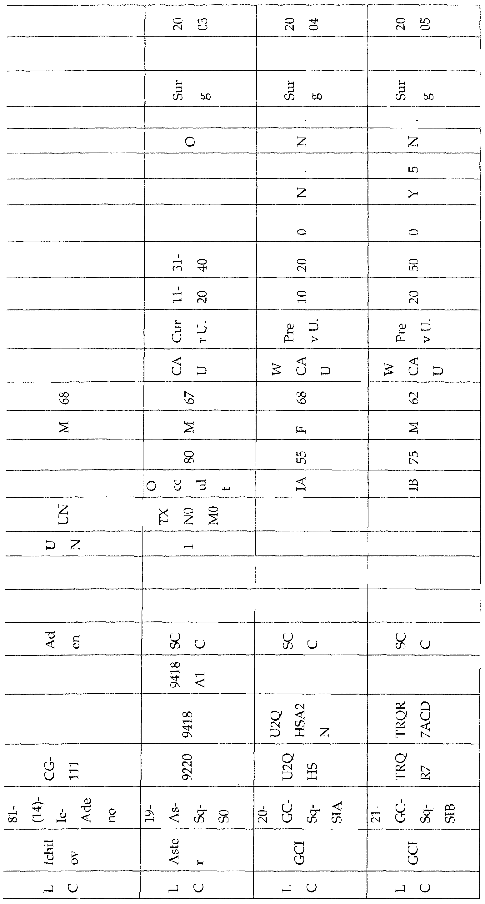


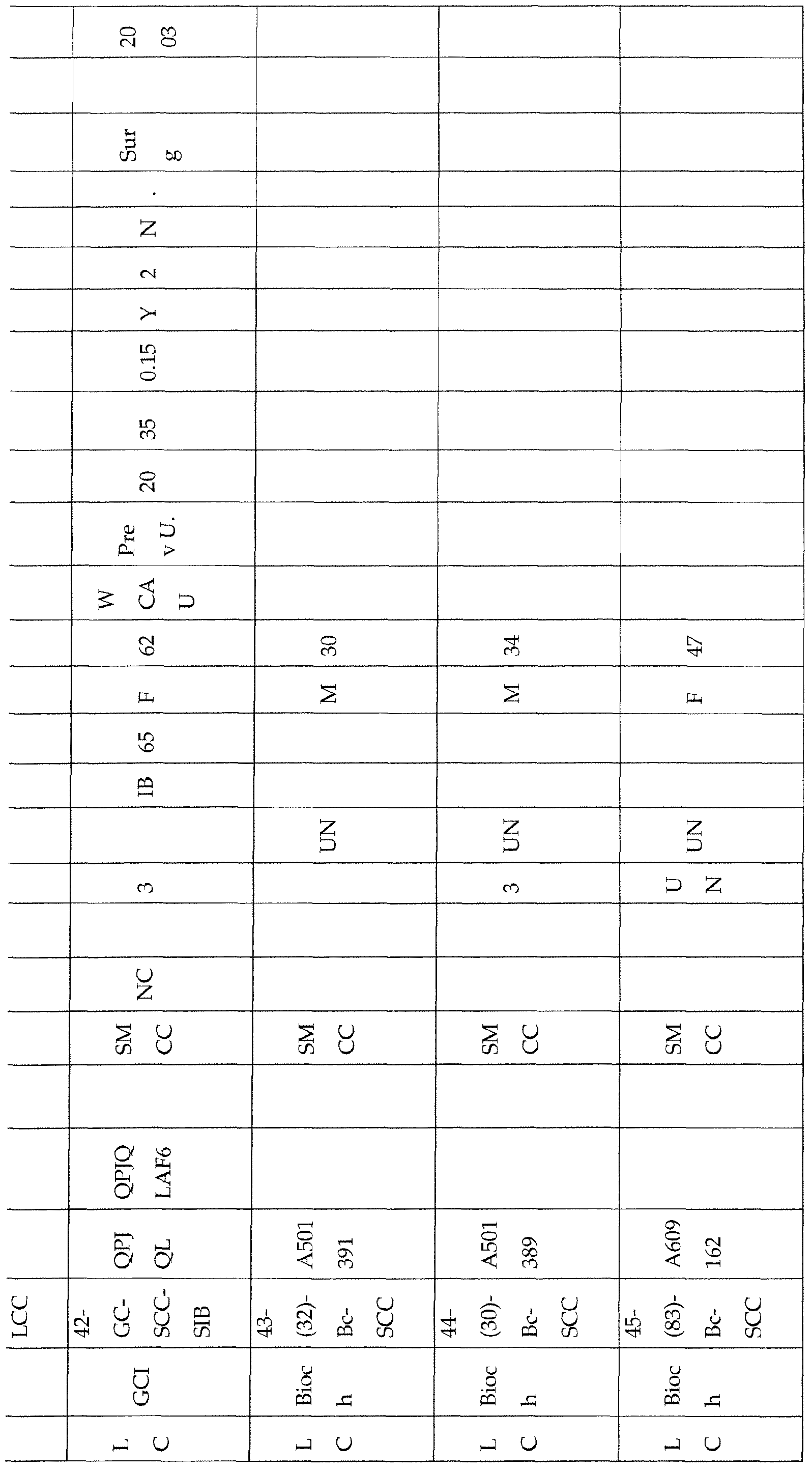


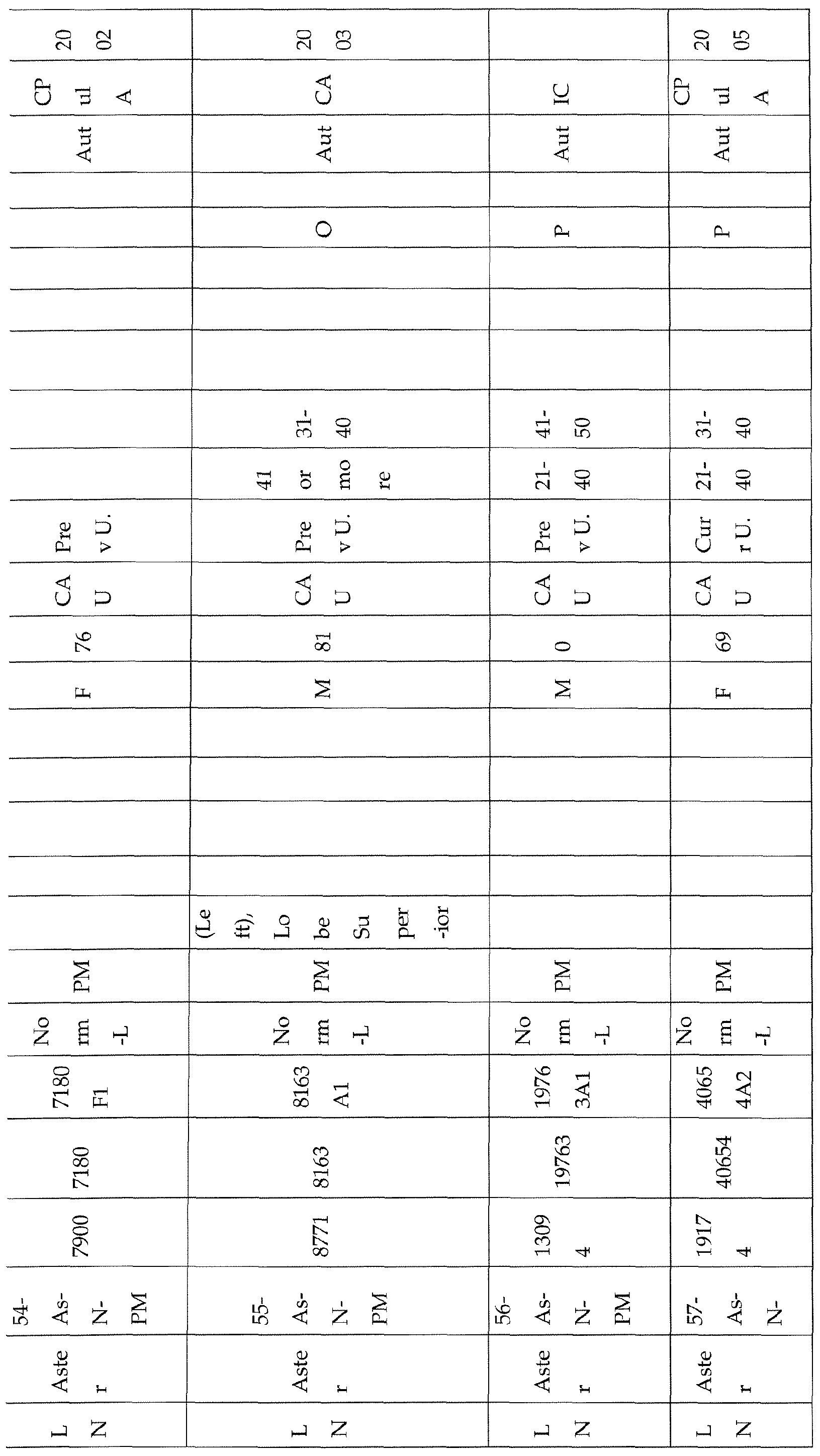


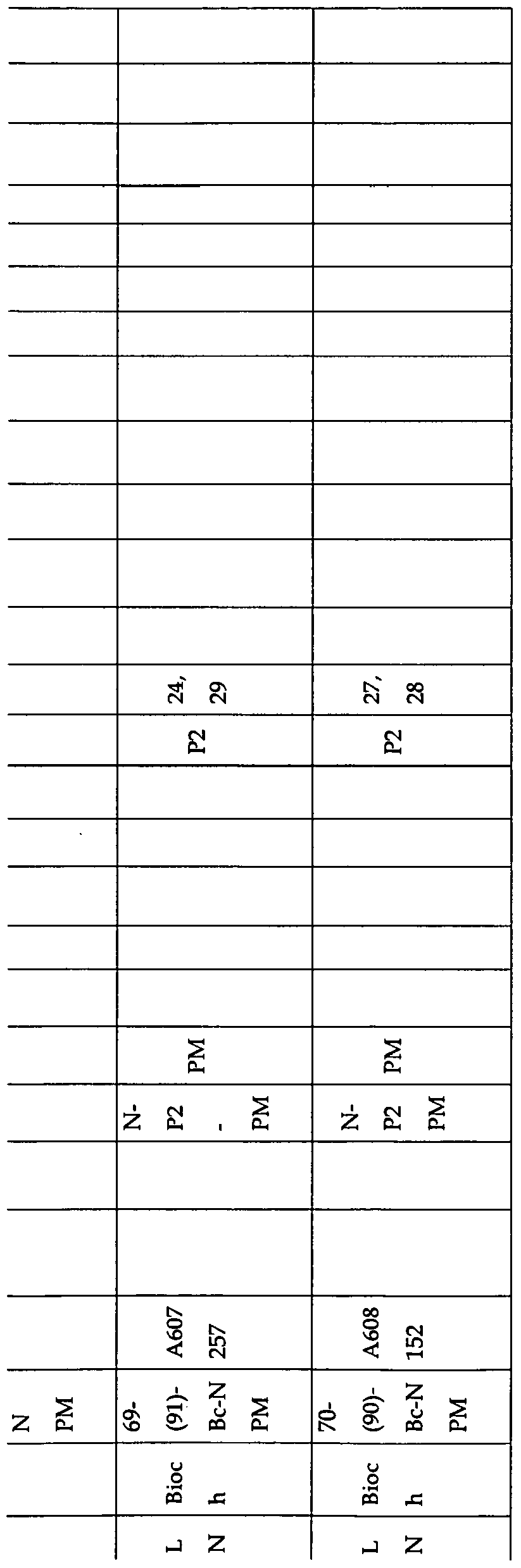


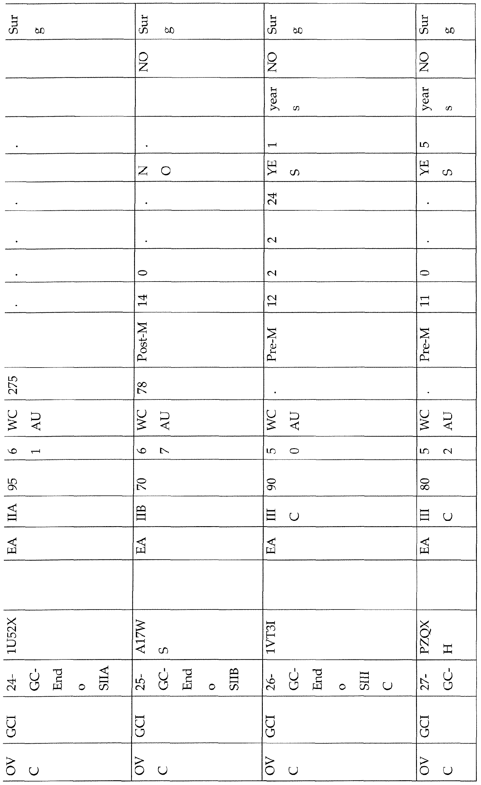


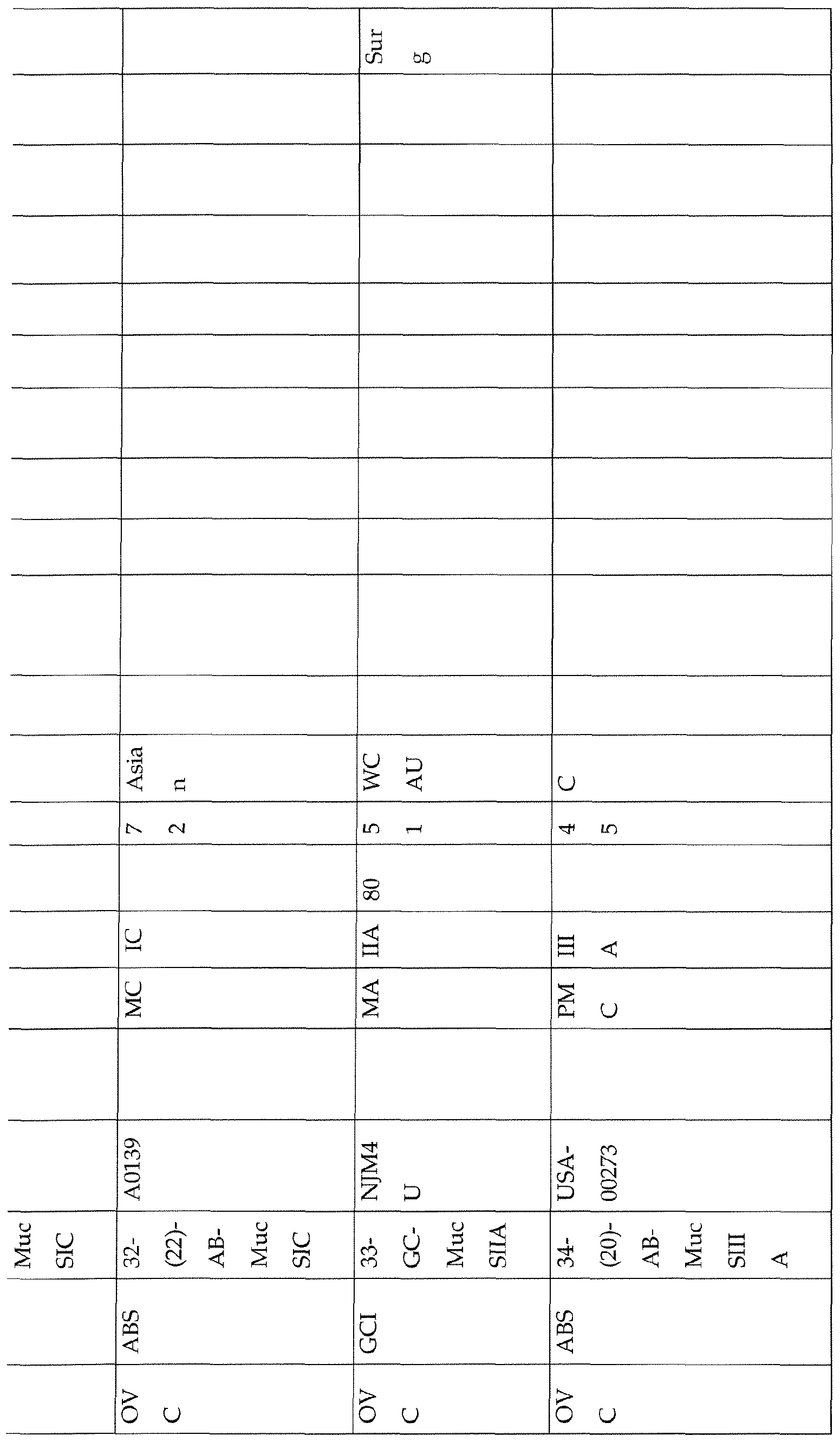


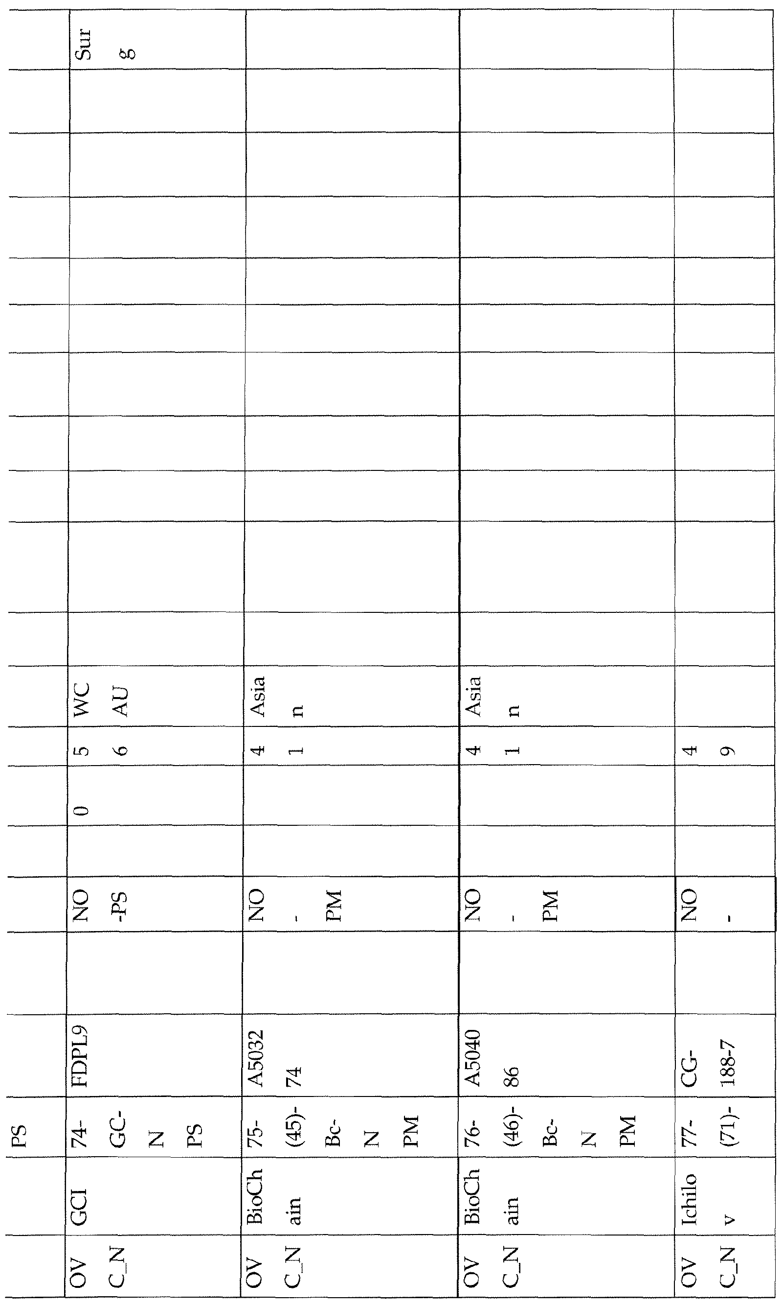


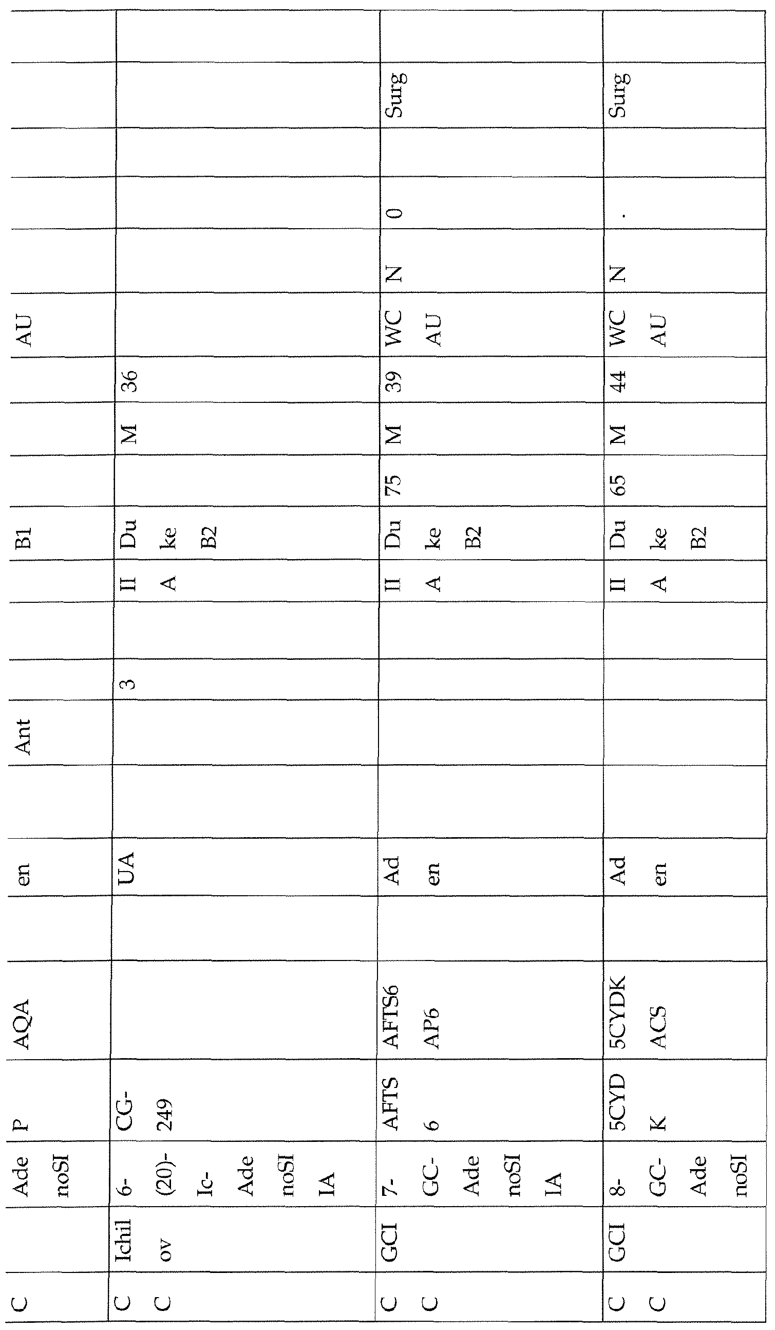


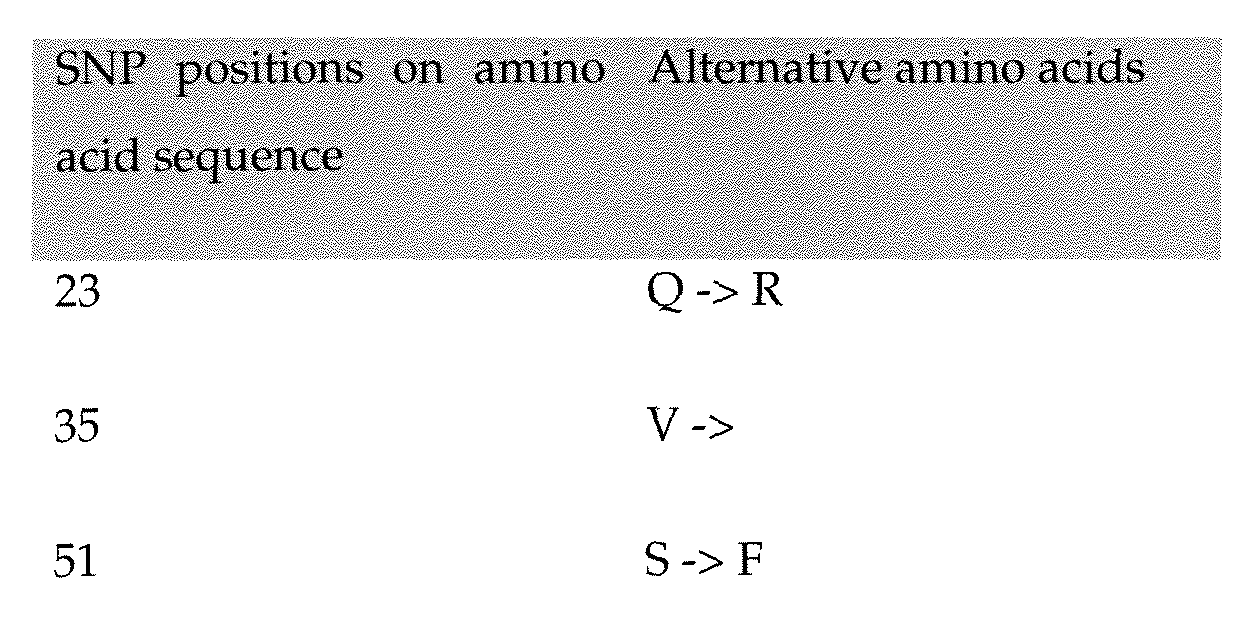


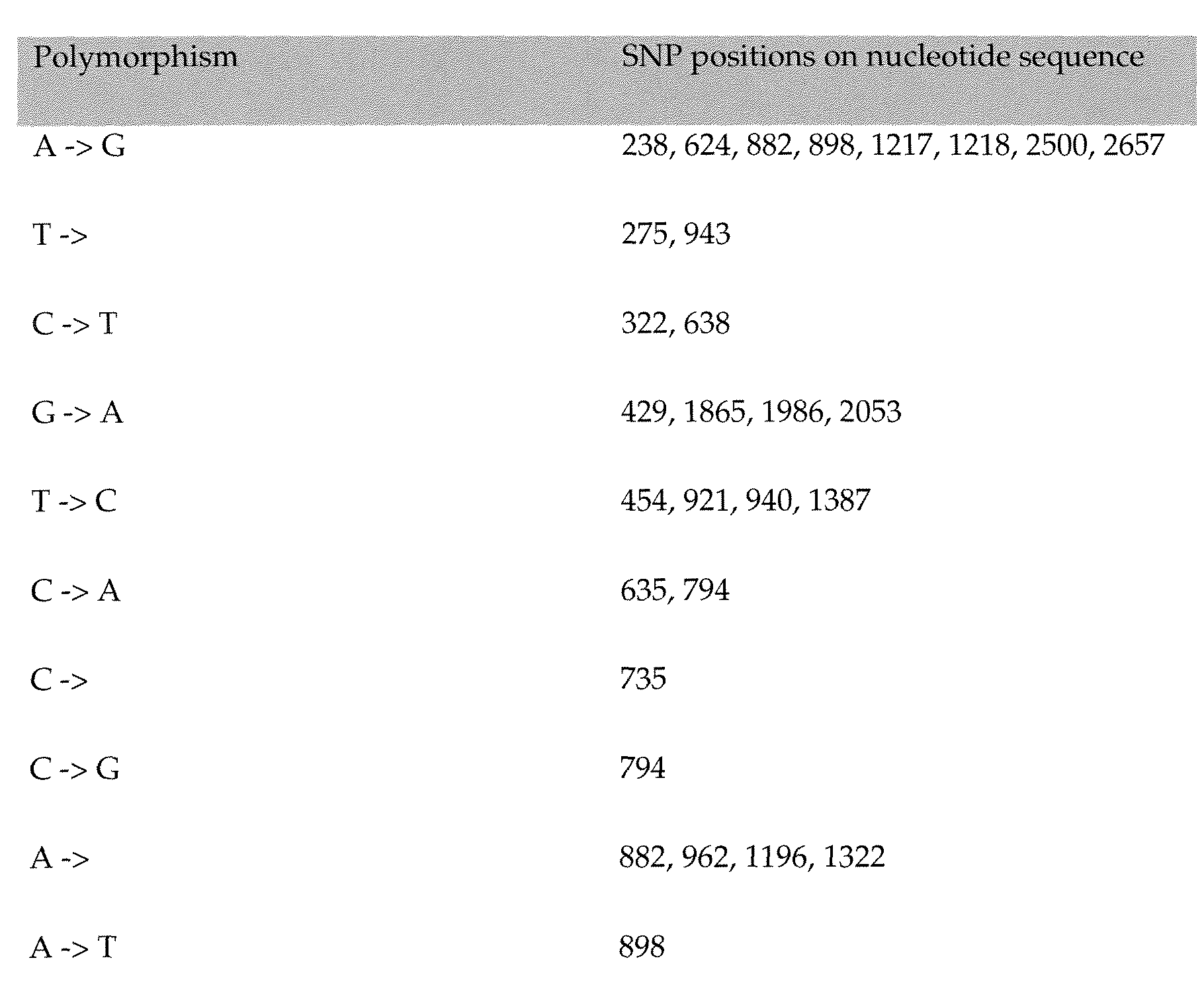


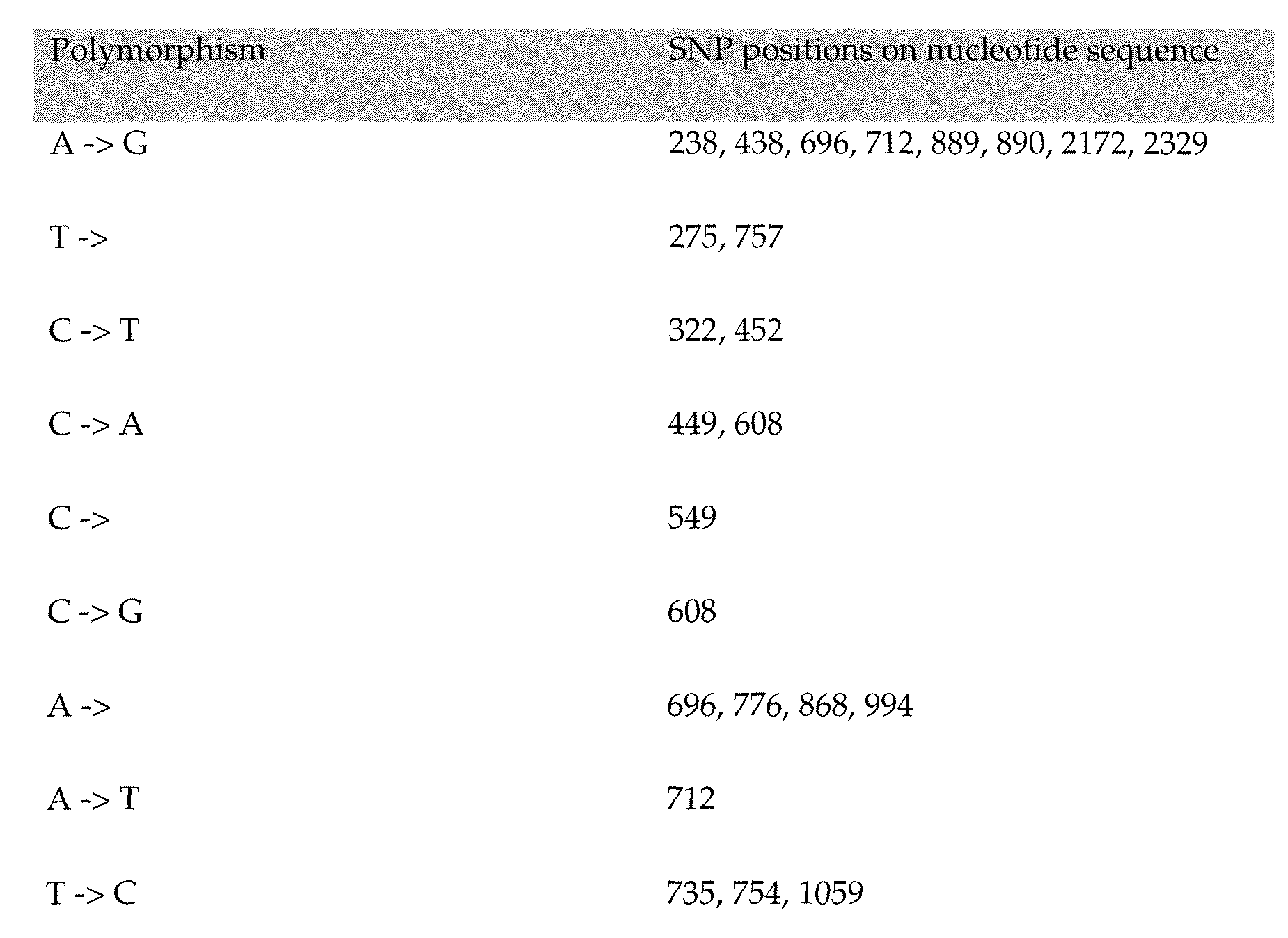


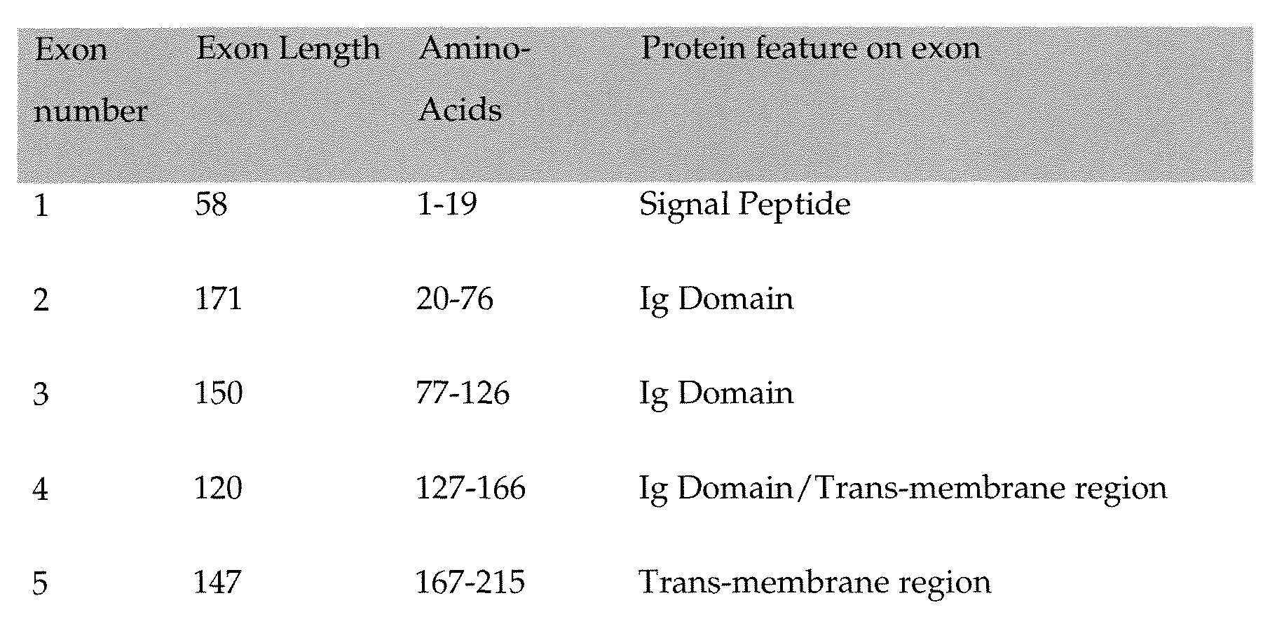


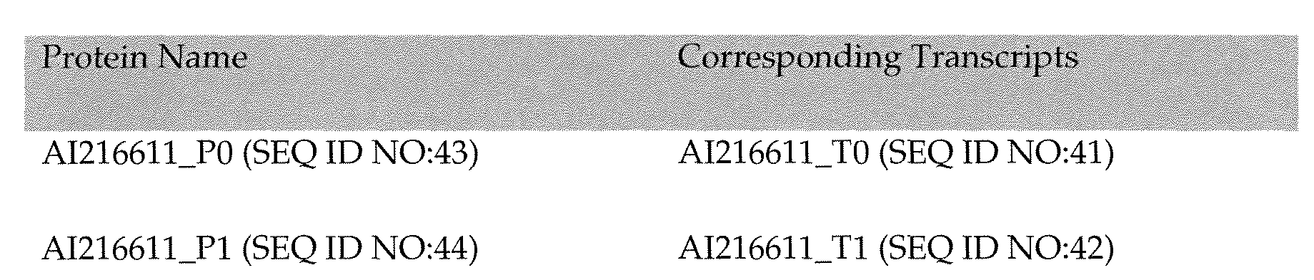


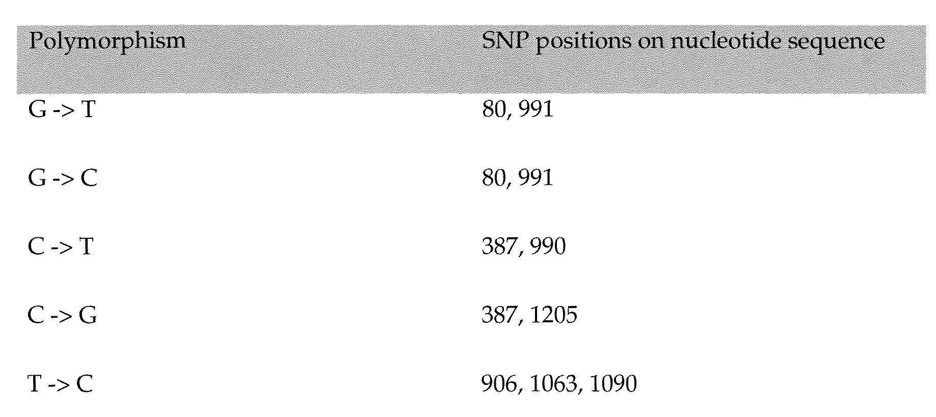


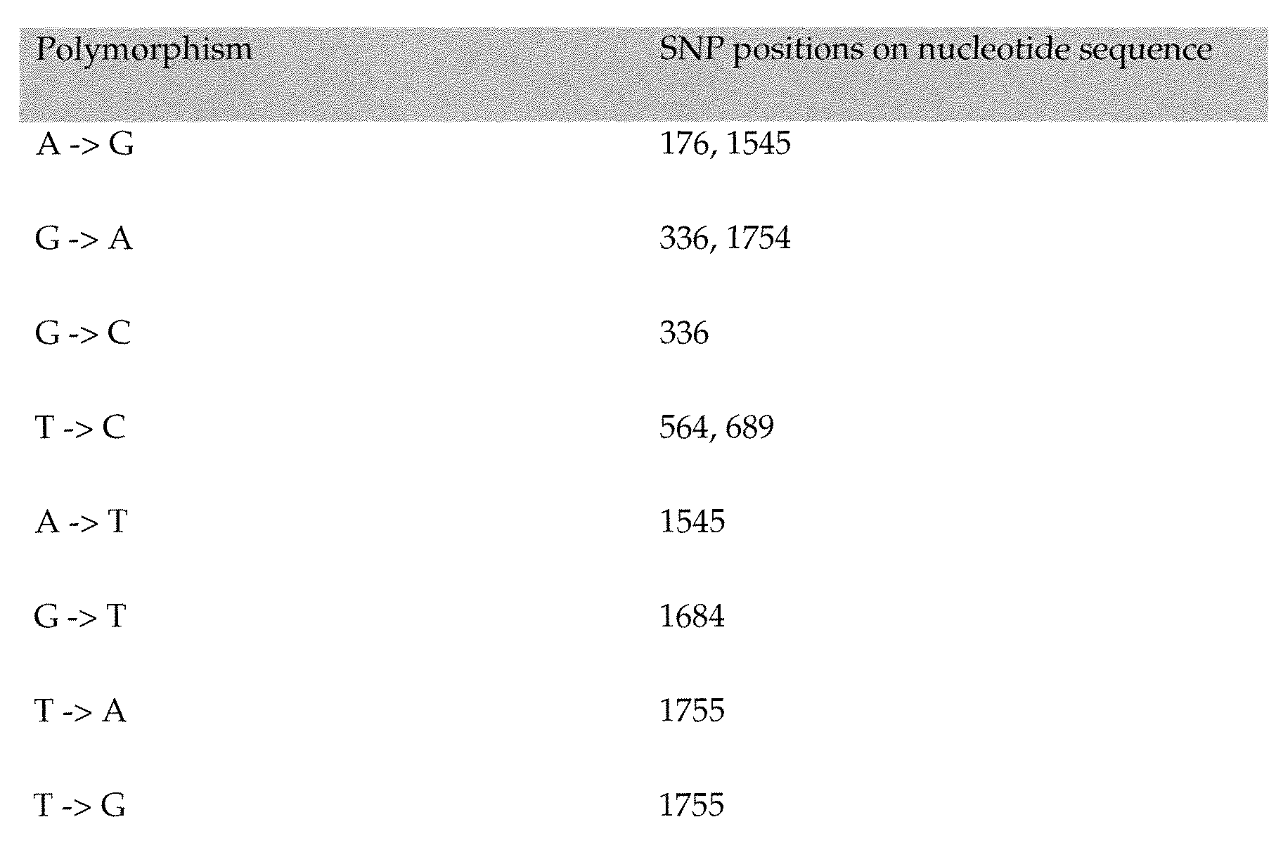


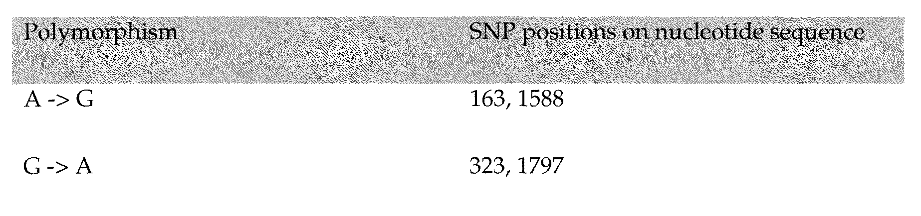


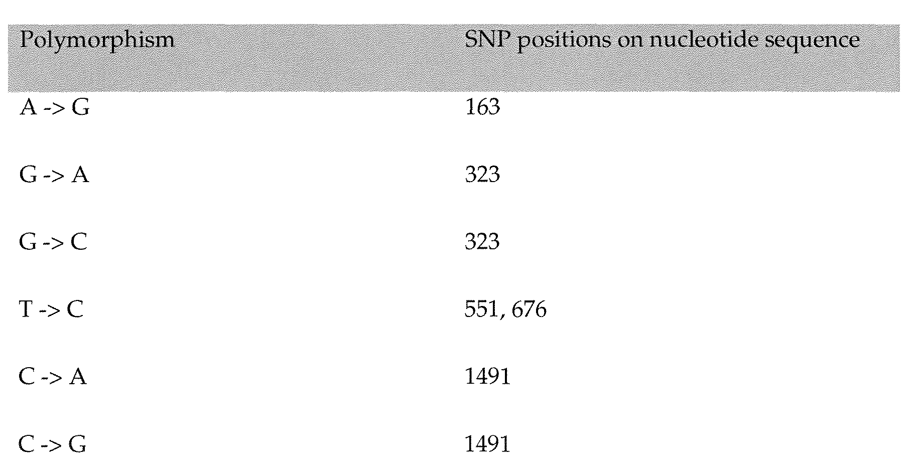

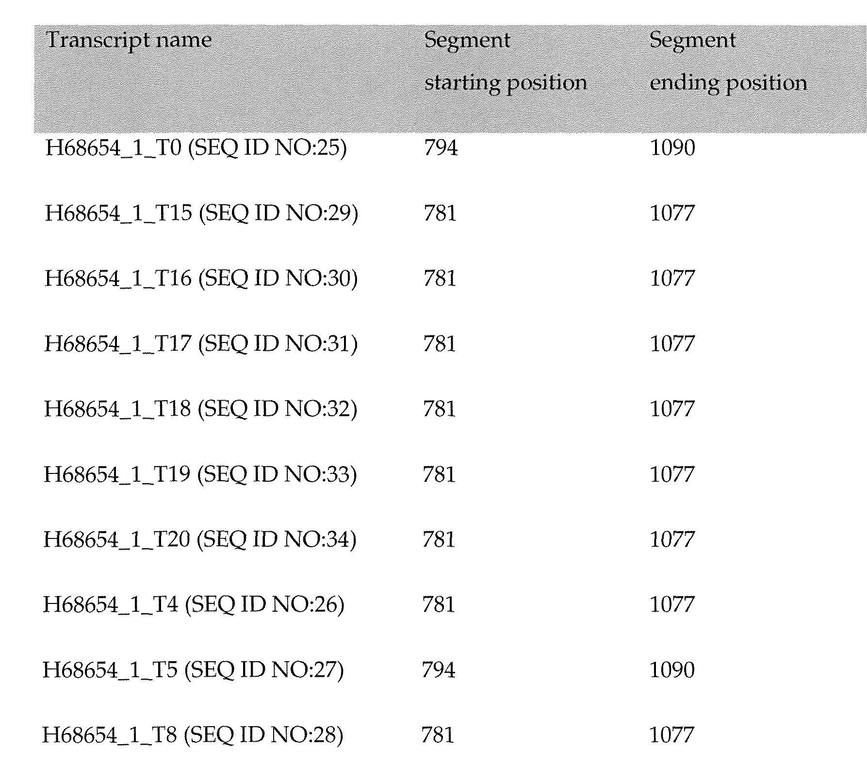


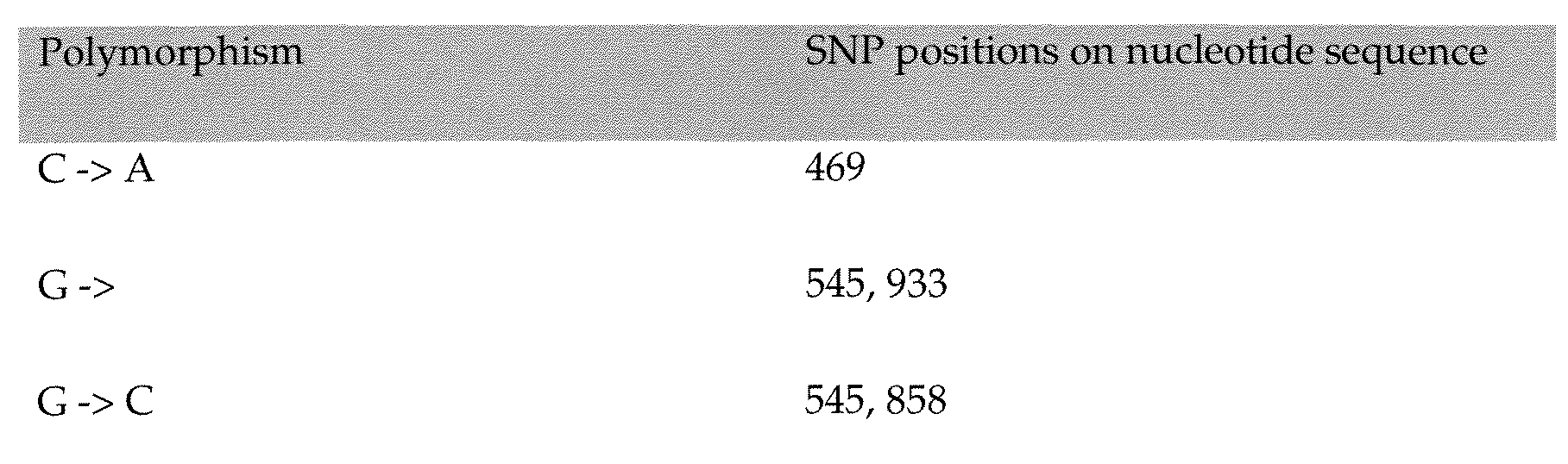


Claims
Priority Applications (30)
| Application Number | Priority Date | Filing Date | Title |
|---|---|---|---|
| CA2698369A CA2698369C (en) | 2007-09-04 | 2008-09-03 | Polypeptides and polynucleotides, and uses thereof as a drug target for producing drugs and biologics |
| EP08829443.4A EP2190469B1 (en) | 2007-09-04 | 2008-09-03 | Polypeptides and polynucleotides, and uses thereof as a drug target for producing drugs and biologics |
| PL14162019T PL2769729T3 (en) | 2007-09-04 | 2008-09-03 | Polypeptides and polynucleotides, and uses thereof as a drug target for producing drugs and biologics |
| JP2010524119A JP5607530B2 (en) | 2007-09-04 | 2008-09-03 | Polypeptides and polynucleotides and their use as drug targets for drug and biologics production |
| DK08829443.4T DK2190469T3 (en) | 2007-09-04 | 2008-09-03 | POLYPEPTIDES AND POLYNUCLEOTIDES AND USES THEREOF AS a drug FOR THE PRODUCTION OF PHARMACEUTICAL AND BIOTECHNOLOGY PRODUCTS |
| US12/676,359 US8415455B2 (en) | 2007-09-04 | 2008-09-03 | Polypeptides and polynucleotides, and uses thereof as a drug target for producing drugs and biologics |
| ES08829443.4T ES2537580T3 (en) | 2007-09-04 | 2008-09-03 | Polypeptides and polynucleotides, and uses thereof as a pharmacological target to produce drugs and biological agents |
| AU2008296361A AU2008296361B2 (en) | 2007-09-04 | 2008-09-03 | Polypeptides and polynucleotides, and uses thereof as a drug target for producing drugs and biologics |
| EP14162019.5A EP2769729B1 (en) | 2007-09-04 | 2008-09-03 | Polypeptides and polynucleotides, and uses thereof as a drug target for producing drugs and biologics |
| PL08829443T PL2190469T3 (en) | 2007-09-04 | 2008-09-03 | Polypeptides and polynucleotides, and uses thereof as a drug target for producing drugs and biologics |
| IL204255A IL204255A (en) | 2007-09-04 | 2010-03-02 | Polypeptides and polynucleotides and uses thereof in the preparation of medicaments for immune related conditions |
| US13/767,891 US9107862B2 (en) | 2007-09-04 | 2013-02-15 | Polypeptides and polynucleotides, and uses thereof as a drug target for producing drugs and biologics |
| US13/845,420 US10098934B2 (en) | 2007-09-04 | 2013-03-18 | Polypeptides and polynucleotides, and uses thereof as a drug target for producing drugs and biologics |
| IL225798A IL225798A (en) | 2007-09-04 | 2013-04-17 | Monoclonal and polyclonal antibodies, fragments, pharmaceutical compositions and uses thereof |
| IL225802A IL225802A (en) | 2007-09-04 | 2013-04-17 | Monoclonal and polyclonal antibodies, fragments, pharmaceutical compositions and uses thereof |
| IL225799A IL225799A0 (en) | 2007-09-04 | 2013-04-17 | Polypeptides and polynucleotides, and uses thereof as a drug target for producing drugs and biologics |
| IL225797A IL225797A0 (en) | 2007-09-04 | 2013-04-17 | Polypeptides and polynucleotides, and uses thereof as a drug target for producing drugs and biologics |
| IL225795A IL225795A (en) | 2007-09-04 | 2013-04-17 | Isolated polypeptides and uses thereof |
| IL225794A IL225794A (en) | 2007-09-04 | 2013-04-17 | Polypeptides and polynucleotides and uses thereof as drug targets for producing drugs and biologics |
| IL225796A IL225796A (en) | 2007-09-04 | 2013-04-17 | Isolated polypeptides and uses thereof |
| IL225804A IL225804A0 (en) | 2007-09-04 | 2013-04-17 | Polypeptides and polynucleotides, and uses thereof as a drug target for producing drugs and biologics |
| IL225801A IL225801A0 (en) | 2007-09-04 | 2013-04-17 | Polypeptides and polynucleotides, and uses thereof as a drug target for producing drugs and biologics |
| IL225800A IL225800A (en) | 2007-09-04 | 2013-04-17 | Monoclonal and polyclonal antibodies, fragments, pharmaceutical compositions and uses thereof |
| IL225803A IL225803A0 (en) | 2007-09-04 | 2013-04-17 | Polypeptides and polynucleotides, and uses thereof as a drug target for producing drugs and biologics |
| US13/904,028 US9375466B2 (en) | 2007-09-04 | 2013-05-29 | Polypeptides and polynucleotides, and uses thereof as a drug target for producing drugs and biologics |
| US14/469,546 US9555087B2 (en) | 2007-09-04 | 2014-08-26 | Polypeptides and polynucleotides, and uses thereof as a drug target for producing drugs and biologics |
| HRP20150512TT HRP20150512T1 (en) | 2007-09-04 | 2015-05-13 | Polypeptides and polynucleotides, and uses thereof as a drug target for producing drugs and biologics |
| US15/011,521 US20170037141A1 (en) | 2007-09-04 | 2016-01-30 | Polypeptides and polynucleotides, and uses thereof as a drug target for producing drugs and biologics |
| US15/882,240 US20180298109A1 (en) | 2007-09-04 | 2018-01-29 | Polypeptides and polynucleotides, and uses thereof as a drug target for producing drugs and biologics |
| CY20191100388T CY1121718T1 (en) | 2007-09-04 | 2019-04-08 | POLYPEPTIDES AND POLYNUCLEOTIDES AND THEIR USES AS A DRUG TARGET FOR THE PRODUCTION OF DRUGS AND BIOLOGICAL AGENTS |
Applications Claiming Priority (12)
| Application Number | Priority Date | Filing Date | Title |
|---|---|---|---|
| US96986507P | 2007-09-04 | 2007-09-04 | |
| US96976907P | 2007-09-04 | 2007-09-04 | |
| US96978807P | 2007-09-04 | 2007-09-04 | |
| US96978007P | 2007-09-04 | 2007-09-04 | |
| US96979907P | 2007-09-04 | 2007-09-04 | |
| US96980607P | 2007-09-04 | 2007-09-04 | |
| US60/969,769 | 2007-09-04 | ||
| US60/969,865 | 2007-09-04 | ||
| US60/969,799 | 2007-09-04 | ||
| US60/769,788 | 2007-09-04 | ||
| US60/969,806 | 2007-09-04 | ||
| US60/969,780 | 2007-09-04 |
Related Child Applications (3)
| Application Number | Title | Priority Date | Filing Date |
|---|---|---|---|
| US12/676,359 A-371-Of-International US8415455B2 (en) | 2007-09-04 | 2008-09-03 | Polypeptides and polynucleotides, and uses thereof as a drug target for producing drugs and biologics |
| US13/767,891 Continuation US9107862B2 (en) | 2007-09-04 | 2013-02-15 | Polypeptides and polynucleotides, and uses thereof as a drug target for producing drugs and biologics |
| US13/845,420 Division US10098934B2 (en) | 2007-09-04 | 2013-03-18 | Polypeptides and polynucleotides, and uses thereof as a drug target for producing drugs and biologics |
Publications (2)
| Publication Number | Publication Date |
|---|---|
| WO2009032845A2 true WO2009032845A2 (en) | 2009-03-12 |
| WO2009032845A3 WO2009032845A3 (en) | 2009-05-28 |
Family
ID=43467008
Family Applications (1)
| Application Number | Title | Priority Date | Filing Date |
|---|---|---|---|
| PCT/US2008/075122 WO2009032845A2 (en) | 2007-09-04 | 2008-09-03 | Polypeptides and polynucleotides, and uses thereof as a drug target for producing drugs and biologics |
Country Status (17)
| Country | Link |
|---|---|
| US (7) | US8415455B2 (en) |
| EP (3) | EP2769728A1 (en) |
| JP (1) | JP5607530B2 (en) |
| AU (1) | AU2008296361B2 (en) |
| CA (1) | CA2698369C (en) |
| CY (1) | CY1121718T1 (en) |
| DK (1) | DK2769729T3 (en) |
| ES (2) | ES2719728T3 (en) |
| HR (2) | HRP20150512T1 (en) |
| HU (1) | HUE042982T2 (en) |
| IL (12) | IL204255A (en) |
| LT (1) | LT2769729T (en) |
| PL (2) | PL2769729T3 (en) |
| PT (1) | PT2769729T (en) |
| SI (1) | SI2769729T1 (en) |
| TR (1) | TR201904866T4 (en) |
| WO (1) | WO2009032845A2 (en) |
Cited By (10)
| Publication number | Priority date | Publication date | Assignee | Title |
|---|---|---|---|---|
| WO2012001647A2 (en) | 2010-06-30 | 2012-01-05 | Compugen Ltd. | Polypeptides and uses thereof as a drug for treatment of multiple sclerosis, rheumatoid arthritis and other autoimmune disorders |
| WO2013001517A1 (en) * | 2011-06-30 | 2013-01-03 | Compugen Ltd. | Polypeptides and uses thereof for treatment of autoimmune disorders and infection |
| CN104185681A (en) * | 2012-02-01 | 2014-12-03 | 卡姆普根有限公司 | C10RF32 antibodies, and uses thereof for treatment of cancer |
| WO2015037005A1 (en) * | 2013-09-11 | 2015-03-19 | Compugen Ltd. | Anti-vstm5 antibodies and the use thereof in therapy and diagnosis |
| WO2015037000A1 (en) * | 2013-09-11 | 2015-03-19 | Compugen Ltd | Vstm5 polypeptides and uses thereof as a drug for treatment of cancer, infectious diseases and immune related diseases |
| US9107862B2 (en) | 2007-09-04 | 2015-08-18 | Compugen Ltd. | Polypeptides and polynucleotides, and uses thereof as a drug target for producing drugs and biologics |
| WO2016030888A1 (en) * | 2014-08-26 | 2016-03-03 | Compugen Ltd. | Polypeptides and uses thereof as a drug for treatment of autoimmune disorders |
| WO2019105972A1 (en) | 2017-11-30 | 2019-06-06 | Bayer Aktiengesellschaft | Ildr2 antagonists and combinations thereof |
| WO2020207961A1 (en) | 2019-04-11 | 2020-10-15 | Bayer Aktiengesellschaft | Combinations of anti-ildr2 antibodies and pd-1 antagonists |
| US11697684B2 (en) | 2014-05-29 | 2023-07-11 | Macrogenics, Inc. | Tri-specific binding molecules that specifically bind to multiple cancer antigens |
Families Citing this family (5)
| Publication number | Priority date | Publication date | Assignee | Title |
|---|---|---|---|---|
| WO2010054096A1 (en) * | 2008-11-07 | 2010-05-14 | The Cleveland Clinic Foundation | Method for detecting and purifying pancreatic beta cells |
| US20130281913A1 (en) | 2012-04-20 | 2013-10-24 | Klox Technologies Inc. | Biophotonic compositions and methods for providing biophotonic treatment |
| CA2916337C (en) | 2013-07-03 | 2022-03-22 | Klox Technologies Inc. | Biophotonic compositions comprising a chromophore and a gelling agent for treating wounds |
| US9682123B2 (en) | 2013-12-20 | 2017-06-20 | The Trustees Of Columbia University In The City Of New York | Methods of treating metabolic disease |
| CN114452390A (en) * | 2022-01-24 | 2022-05-10 | 浙江大学 | Application of FXYD 3protein or coding gene thereof as target in preparation of medicine for preventing and treating psoriasis |
Citations (146)
| Publication number | Priority date | Publication date | Assignee | Title |
|---|---|---|---|---|
| US3687808A (en) | 1969-08-14 | 1972-08-29 | Univ Leland Stanford Junior | Synthetic polynucleotides |
| US4366241A (en) | 1980-08-07 | 1982-12-28 | Syva Company | Concentrating zone method in heterogeneous immunoassays |
| US4376110A (en) | 1980-08-04 | 1983-03-08 | Hybritech, Incorporated | Immunometric assays using monoclonal antibodies |
| US4399216A (en) | 1980-02-25 | 1983-08-16 | The Trustees Of Columbia University | Processes for inserting DNA into eucaryotic cells and for producing proteinaceous materials |
| US4439196A (en) | 1982-03-18 | 1984-03-27 | Merck & Co., Inc. | Osmotic drug delivery system |
| US4447224A (en) | 1982-09-20 | 1984-05-08 | Infusaid Corporation | Variable flow implantable infusion apparatus |
| US4447233A (en) | 1981-04-10 | 1984-05-08 | Parker-Hannifin Corporation | Medication infusion pump |
| US4469863A (en) | 1980-11-12 | 1984-09-04 | Ts O Paul O P | Nonionic nucleic acid alkyl and aryl phosphonates and processes for manufacture and use thereof |
| US4475196A (en) | 1981-03-06 | 1984-10-02 | Zor Clair G | Instrument for locating faults in aircraft passenger reading light and attendant call control system |
| US4476301A (en) | 1982-04-29 | 1984-10-09 | Centre National De La Recherche Scientifique | Oligonucleotides, a process for preparing the same and their application as mediators of the action of interferon |
| US4486194A (en) | 1983-06-08 | 1984-12-04 | James Ferrara | Therapeutic device for administering medicaments through the skin |
| US4487603A (en) | 1982-11-26 | 1984-12-11 | Cordis Corporation | Implantable microinfusion pump system |
| US4517288A (en) | 1981-01-23 | 1985-05-14 | American Hospital Supply Corp. | Solid phase system for ligand assay |
| US4522811A (en) | 1982-07-08 | 1985-06-11 | Syntex (U.S.A.) Inc. | Serial injection of muramyldipeptides and liposomes enhances the anti-infective activity of muramyldipeptides |
| EP0154316A2 (en) | 1984-03-06 | 1985-09-11 | Takeda Chemical Industries, Ltd. | Chemically modified lymphokine and production thereof |
| US4596556A (en) | 1985-03-25 | 1986-06-24 | Bioject, Inc. | Hypodermic injection apparatus |
| US4634665A (en) | 1980-02-25 | 1987-01-06 | The Trustees Of Columbia University In The City Of New York | Processes for inserting DNA into eucaryotic cells and for producing proteinaceous materials |
| WO1987004462A1 (en) | 1986-01-23 | 1987-07-30 | Celltech Limited | Recombinant dna sequences, vectors containing them and method for the use thereof |
| WO1987005330A1 (en) | 1986-03-07 | 1987-09-11 | Michel Louis Eugene Bergh | Method for enhancing glycoprotein stability |
| WO1988000052A1 (en) | 1986-07-07 | 1988-01-14 | Trustees Of Dartmouth College | Monoclonal antibodies to fc receptor |
| EP0264166A1 (en) | 1986-04-09 | 1988-04-20 | Genzyme Corporation | Transgenic animals secreting desired proteins into milk |
| US4790824A (en) | 1987-06-19 | 1988-12-13 | Bioject, Inc. | Non-invasive hypodermic injection device |
| WO1989001036A1 (en) | 1987-07-23 | 1989-02-09 | Celltech Limited | Recombinant dna expression vectors |
| US4816567A (en) | 1983-04-08 | 1989-03-28 | Genentech, Inc. | Recombinant immunoglobin preparations |
| US4837168A (en) | 1985-12-23 | 1989-06-06 | Janssen Pharmaceutica N.V. | Immunoassay using colorable latex particles |
| US4873316A (en) | 1987-06-23 | 1989-10-10 | Biogen, Inc. | Isolation of exogenous recombinant proteins from the milk of transgenic mammals |
| EP0338841A1 (en) | 1988-04-18 | 1989-10-25 | Celltech Limited | Recombinant DNA methods, vectors and host cells |
| US4881175A (en) | 1986-09-02 | 1989-11-14 | Genex Corporation | Computer based system and method for determining and displaying possible chemical structures for converting double- or multiple-chain polypeptides to single-chain polypeptides |
| US4941880A (en) | 1987-06-19 | 1990-07-17 | Bioject, Inc. | Pre-filled ampule and non-invasive hypodermic injection device assembly |
| US4946778A (en) | 1987-09-21 | 1990-08-07 | Genex Corporation | Single polypeptide chain binding molecules |
| EP0401384A1 (en) | 1988-12-22 | 1990-12-12 | Kirin-Amgen, Inc. | Chemically modified granulocyte colony stimulating factor |
| US5013653A (en) | 1987-03-20 | 1991-05-07 | Creative Biomolecules, Inc. | Product and process for introduction of a hinge region into a fusion protein to facilitate cleavage |
| US5023243A (en) | 1981-10-23 | 1991-06-11 | Molecular Biosystems, Inc. | Oligonucleotide therapeutic agent and method of making same |
| US5034506A (en) | 1985-03-15 | 1991-07-23 | Anti-Gene Development Group | Uncharged morpholino-based polymers having achiral intersubunit linkages |
| US5064413A (en) | 1989-11-09 | 1991-11-12 | Bioject, Inc. | Needleless hypodermic injection device |
| US5091513A (en) | 1987-05-21 | 1992-02-25 | Creative Biomolecules, Inc. | Biosynthetic antibody binding sites |
| WO1992003918A1 (en) | 1990-08-29 | 1992-03-19 | Genpharm International, Inc. | Transgenic non-human animals capable of producing heterologous antibodies |
| US5132405A (en) | 1987-05-21 | 1992-07-21 | Creative Biomolecules, Inc. | Biosynthetic antibody binding sites |
| US5166315A (en) | 1989-12-20 | 1992-11-24 | Anti-Gene Development Group | Sequence-specific binding polymers for duplex nucleic acids |
| US5177196A (en) | 1990-08-16 | 1993-01-05 | Microprobe Corporation | Oligo (α-arabinofuranosyl nucleotides) and α-arabinofuranosyl precursors thereof |
| US5179017A (en) | 1980-02-25 | 1993-01-12 | The Trustees Of Columbia University In The City Of New York | Processes for inserting DNA into eucaryotic cells and for producing proteinaceous materials |
| US5185444A (en) | 1985-03-15 | 1993-02-09 | Anti-Gene Deveopment Group | Uncharged morpolino-based polymers having phosphorous containing chiral intersubunit linkages |
| US5188897A (en) | 1987-10-22 | 1993-02-23 | Temple University Of The Commonwealth System Of Higher Education | Encapsulated 2',5'-phosphorothioate oligoadenylates |
| US5214134A (en) | 1990-09-12 | 1993-05-25 | Sterling Winthrop Inc. | Process of linking nucleosides with a siloxane bridge |
| US5216141A (en) | 1988-06-06 | 1993-06-01 | Benner Steven A | Oligonucleotide analogs containing sulfur linkages |
| WO1993012227A1 (en) | 1991-12-17 | 1993-06-24 | Genpharm International, Inc. | Transgenic non-human animals capable of producing heterologous antibodies |
| US5223409A (en) | 1988-09-02 | 1993-06-29 | Protein Engineering Corp. | Directed evolution of novel binding proteins |
| US5225539A (en) | 1986-03-27 | 1993-07-06 | Medical Research Council | Recombinant altered antibodies and methods of making altered antibodies |
| US5235033A (en) | 1985-03-15 | 1993-08-10 | Anti-Gene Development Group | Alpha-morpholino ribonucleoside derivatives and polymers thereof |
| US5258498A (en) | 1987-05-21 | 1993-11-02 | Creative Biomolecules, Inc. | Polypeptide linkers for production of biosynthetic proteins |
| US5260203A (en) | 1986-09-02 | 1993-11-09 | Enzon, Inc. | Single polypeptide chain binding molecules |
| US5264423A (en) | 1987-03-25 | 1993-11-23 | The United States Of America As Represented By The Department Of Health And Human Services | Inhibitors for replication of retroviruses and for the expression of oncogene products |
| US5264564A (en) | 1989-10-24 | 1993-11-23 | Gilead Sciences | Oligonucleotide analogs with novel linkages |
| US5264562A (en) | 1989-10-24 | 1993-11-23 | Gilead Sciences, Inc. | Oligonucleotide analogs with novel linkages |
| US5276019A (en) | 1987-03-25 | 1994-01-04 | The United States Of America As Represented By The Department Of Health And Human Services | Inhibitors for replication of retroviruses and for the expression of oncogene products |
| US5278302A (en) | 1988-05-26 | 1994-01-11 | University Patents, Inc. | Polynucleotide phosphorodithioates |
| WO1994010332A1 (en) | 1992-11-04 | 1994-05-11 | Medarex, Inc. | HUMANIZED ANTIBODIES TO Fc RECEPTORS FOR IMMUNOGLOBULIN G ON HUMAN MONONUCLEAR PHAGOCYTES |
| US5312335A (en) | 1989-11-09 | 1994-05-17 | Bioject Inc. | Needleless hypodermic injection device |
| US5321131A (en) | 1990-03-08 | 1994-06-14 | Hybridon, Inc. | Site-specific functionalization of oligodeoxynucleotides for non-radioactive labelling |
| WO1994025585A1 (en) | 1993-04-26 | 1994-11-10 | Genpharm International, Inc. | Transgenic non-human animals capable of producing heterologous antibodies |
| US5374548A (en) | 1986-05-02 | 1994-12-20 | Genentech, Inc. | Methods and compositions for the attachment of proteins to liposomes using a glycophospholipid anchor |
| WO1994029351A2 (en) | 1993-06-16 | 1994-12-22 | Celltech Limited | Antibodies |
| US5383851A (en) | 1992-07-24 | 1995-01-24 | Bioject Inc. | Needleless hypodermic injection device |
| US5399676A (en) | 1989-10-23 | 1995-03-21 | Gilead Sciences | Oligonucleotides with inverted polarity |
| US5399331A (en) | 1985-06-26 | 1995-03-21 | The Liposome Company, Inc. | Method for protein-liposome coupling |
| US5405938A (en) | 1989-12-20 | 1995-04-11 | Anti-Gene Development Group | Sequence-specific binding polymers for duplex nucleic acids |
| US5405939A (en) | 1987-10-22 | 1995-04-11 | Temple University Of The Commonwealth System Of Higher Education | 2',5'-phosphorothioate oligoadenylates and their covalent conjugates with polylysine |
| US5416016A (en) | 1989-04-03 | 1995-05-16 | Purdue Research Foundation | Method for enhancing transmembrane transport of exogenous molecules |
| US5427908A (en) | 1990-05-01 | 1995-06-27 | Affymax Technologies N.V. | Recombinant library screening methods |
| US5434257A (en) | 1992-06-01 | 1995-07-18 | Gilead Sciences, Inc. | Binding compentent oligomers containing unsaturated 3',5' and 2',5' linkages |
| US5455233A (en) | 1989-11-30 | 1995-10-03 | University Of North Carolina | Oligoribonucleoside and oligodeoxyribonucleoside boranophosphates |
| US5466677A (en) | 1993-03-06 | 1995-11-14 | Ciba-Geigy Corporation | Dinucleoside phosphinates and their pharmaceutical compositions |
| US5470967A (en) | 1990-04-10 | 1995-11-28 | The Dupont Merck Pharmaceutical Company | Oligonucleotide analogs with sulfamate linkages |
| US5476925A (en) | 1993-02-01 | 1995-12-19 | Northwestern University | Oligodeoxyribonucleotides including 3'-aminonucleoside-phosphoramidate linkages and terminal 3'-amino groups |
| US5476786A (en) | 1987-05-21 | 1995-12-19 | Creative Biomolecules, Inc. | Biosynthetic antibody binding sites |
| US5476996A (en) | 1988-06-14 | 1995-12-19 | Lidak Pharmaceuticals | Human immune system in non-human animal |
| US5489677A (en) | 1990-07-27 | 1996-02-06 | Isis Pharmaceuticals, Inc. | Oligonucleoside linkages containing adjacent oxygen and nitrogen atoms |
| US5519126A (en) | 1988-03-25 | 1996-05-21 | University Of Virginia Alumni Patents Foundation | Oligonucleotide N-alkylphosphoramidates |
| US5530101A (en) | 1988-12-28 | 1996-06-25 | Protein Design Labs, Inc. | Humanized immunoglobulins |
| US5539082A (en) | 1993-04-26 | 1996-07-23 | Nielsen; Peter E. | Peptide nucleic acids |
| US5541307A (en) | 1990-07-27 | 1996-07-30 | Isis Pharmaceuticals, Inc. | Backbone modified oligonucleotide analogs and solid phase synthesis thereof |
| US5545806A (en) | 1990-08-29 | 1996-08-13 | Genpharm International, Inc. | Ransgenic non-human animals for producing heterologous antibodies |
| US5545807A (en) | 1988-10-12 | 1996-08-13 | The Babraham Institute | Production of antibodies from transgenic animals |
| US5550111A (en) | 1984-07-11 | 1996-08-27 | Temple University-Of The Commonwealth System Of Higher Education | Dual action 2',5'-oligoadenylate antiviral derivatives and uses thereof |
| US5561225A (en) | 1990-09-19 | 1996-10-01 | Southern Research Institute | Polynucleotide analogs containing sulfonate and sulfonamide internucleoside linkages |
| US5571799A (en) | 1991-08-12 | 1996-11-05 | Basco, Ltd. | (2'-5') oligoadenylate analogues useful as inhibitors of host-v5.-graft response |
| US5587361A (en) | 1991-10-15 | 1996-12-24 | Isis Pharmaceuticals, Inc. | Oligonucleotides having phosphorothioate linkages of high chiral purity |
| US5596086A (en) | 1990-09-20 | 1997-01-21 | Gilead Sciences, Inc. | Modified internucleoside linkages having one nitrogen and two carbon atoms |
| US5602240A (en) | 1990-07-27 | 1997-02-11 | Ciba Geigy Ag. | Backbone modified oligonucleotide analogs |
| US5608046A (en) | 1990-07-27 | 1997-03-04 | Isis Pharmaceuticals, Inc. | Conjugated 4'-desmethyl nucleoside analog compounds |
| US5610289A (en) | 1990-07-27 | 1997-03-11 | Isis Pharmaceuticals, Inc. | Backbone modified oligonucleotide analogues |
| US5618704A (en) | 1990-07-27 | 1997-04-08 | Isis Pharmacueticals, Inc. | Backbone-modified oligonucleotide analogs and preparation thereof through radical coupling |
| WO1997013852A1 (en) | 1995-10-10 | 1997-04-17 | Genpharm International, Inc. | Transgenic non-human animals capable of producing heterologous antibodies |
| US5623070A (en) | 1990-07-27 | 1997-04-22 | Isis Pharmaceuticals, Inc. | Heteroatomic oligonucleoside linkages |
| US5625126A (en) | 1990-08-29 | 1997-04-29 | Genpharm International, Inc. | Transgenic non-human animals for producing heterologous antibodies |
| US5624821A (en) | 1987-03-18 | 1997-04-29 | Scotgen Biopharmaceuticals Incorporated | Antibodies with altered effector functions |
| US5625050A (en) | 1994-03-31 | 1997-04-29 | Amgen Inc. | Modified oligonucleotides and intermediates useful in nucleic acid therapeutics |
| US5633360A (en) | 1992-04-14 | 1997-05-27 | Gilead Sciences, Inc. | Oligonucleotide analogs capable of passive cell membrane permeation |
| US5633425A (en) | 1990-08-29 | 1997-05-27 | Genpharm International, Inc. | Transgenic non-human animals capable of producing heterologous antibodies |
| US5661016A (en) | 1990-08-29 | 1997-08-26 | Genpharm International Inc. | Transgenic non-human animals capable of producing heterologous antibodies of various isotypes |
| US5663312A (en) | 1993-03-31 | 1997-09-02 | Sanofi | Oligonucleotide dimers with amide linkages replacing phosphodiester linkages |
| US5677439A (en) | 1990-08-03 | 1997-10-14 | Sanofi | Oligonucleotide analogues containing phosphate diester linkage substitutes, compositions thereof, and precursor dinucleotide analogues |
| US5677437A (en) | 1990-07-27 | 1997-10-14 | Isis Pharmaceuticals, Inc. | Heteroatomic oligonucleoside linkages |
| US5677425A (en) | 1987-09-04 | 1997-10-14 | Celltech Therapeutics Limited | Recombinant antibody |
| US5714350A (en) | 1992-03-09 | 1998-02-03 | Protein Design Labs, Inc. | Increasing antibody affinity by altering glycosylation in the immunoglobulin variable region |
| US5714331A (en) | 1991-05-24 | 1998-02-03 | Buchardt, Deceased; Ole | Peptide nucleic acids having enhanced binding affinity, sequence specificity and solubility |
| US5719262A (en) | 1993-11-22 | 1998-02-17 | Buchardt, Deceased; Ole | Peptide nucleic acids having amino acid side chains |
| WO1998024884A1 (en) | 1996-12-02 | 1998-06-11 | Genpharm International | Transgenic non-human animals capable of producing heterologous antibodies |
| US5789650A (en) | 1990-08-29 | 1998-08-04 | Genpharm International, Inc. | Transgenic non-human animals for producing heterologous antibodies |
| US5814318A (en) | 1990-08-29 | 1998-09-29 | Genpharm International Inc. | Transgenic non-human animals for producing heterologous antibodies |
| US5869046A (en) | 1995-04-14 | 1999-02-09 | Genentech, Inc. | Altered polypeptides with increased half-life |
| US5874299A (en) | 1990-08-29 | 1999-02-23 | Genpharm International, Inc. | Transgenic non-human animals capable of producing heterologous antibodies |
| US5877397A (en) | 1990-08-29 | 1999-03-02 | Genpharm International Inc. | Transgenic non-human animals capable of producing heterologous antibodies of various isotypes |
| US5885793A (en) | 1991-12-02 | 1999-03-23 | Medical Research Council | Production of anti-self antibodies from antibody segment repertoires and displayed on phage |
| WO1999026972A1 (en) | 1997-11-21 | 1999-06-03 | Genetics Institute, Inc. | Secreted proteins and polynucleotides encoding them |
| US5939598A (en) | 1990-01-12 | 1999-08-17 | Abgenix, Inc. | Method of making transgenic mice lacking endogenous heavy chains |
| WO1999045962A1 (en) | 1998-03-13 | 1999-09-16 | Genpharm International, Inc. | Transgenic non-human animals capable of producing heterologous antibodies |
| US5969108A (en) | 1990-07-10 | 1999-10-19 | Medical Research Council | Methods for producing members of specific binding pairs |
| WO1999054342A1 (en) | 1998-04-20 | 1999-10-28 | Pablo Umana | Glycosylation engineering of antibodies for improving antibody-dependent cellular cytotoxicity |
| WO1999060020A1 (en) | 1998-05-18 | 1999-11-25 | Genetics Institute, Inc. | Secreted proteins and polynucleotides encoding them |
| US6075181A (en) | 1990-01-12 | 2000-06-13 | Abgenix, Inc. | Human antibodies derived from immunized xenomice |
| WO2000042072A2 (en) | 1999-01-15 | 2000-07-20 | Genentech, Inc. | Polypeptide variants with altered effector function |
| US6121022A (en) | 1995-04-14 | 2000-09-19 | Genentech, Inc. | Altered polypeptides with increased half-life |
| US6150584A (en) | 1990-01-12 | 2000-11-21 | Abgenix, Inc. | Human antibodies derived from immunized xenomice |
| US6162963A (en) | 1990-01-12 | 2000-12-19 | Abgenix, Inc. | Generation of Xenogenetic antibodies |
| US6165745A (en) | 1992-04-24 | 2000-12-26 | Board Of Regents, The University Of Texas System | Recombinant production of immunoglobulin-like domains in prokaryotic cells |
| US6172197B1 (en) | 1991-07-10 | 2001-01-09 | Medical Research Council | Methods for producing members of specific binding pairs |
| US6194551B1 (en) | 1998-04-02 | 2001-02-27 | Genentech, Inc. | Polypeptide variants |
| WO2001014424A2 (en) | 1999-08-24 | 2001-03-01 | Medarex, Inc. | Human ctla-4 antibodies and their uses |
| US6277375B1 (en) | 1997-03-03 | 2001-08-21 | Board Of Regents, The University Of Texas System | Immunoglobulin-like domains with increased half-lives |
| US6303374B1 (en) | 2000-01-18 | 2001-10-16 | Isis Pharmaceuticals Inc. | Antisense modulation of caspase 3 expression |
| EP1176195A1 (en) | 1999-04-09 | 2002-01-30 | Kyowa Hakko Kogyo Co., Ltd. | Method for controlling the activity of immunologically functional molecule |
| WO2002043478A2 (en) | 2000-11-30 | 2002-06-06 | Medarex, Inc. | Transgenic transchromosomal rodents for making human antibodies |
| WO2002092780A2 (en) | 2001-05-17 | 2002-11-21 | Diversa Corporation | Novel antigen binding molecules for therapeutic, diagnostic, prophylactic, enzymatic, industrial, and agricultural applications, and methods for generating and screening thereof |
| US20020193567A1 (en) | 1995-08-11 | 2002-12-19 | Genetics Institute, Inc. | Secreted proteins and polynucleotides encoding them |
| WO2003027228A2 (en) | 2001-07-17 | 2003-04-03 | Incyte Genomics, Inc. | Receptors and membrane-associated proteins |
| WO2003035835A2 (en) | 2001-10-25 | 2003-05-01 | Genentech, Inc. | Glycoprotein compositions |
| WO2003074679A2 (en) | 2002-03-01 | 2003-09-12 | Xencor | Antibody optimization |
| US6696686B1 (en) | 1999-06-06 | 2004-02-24 | Elgems Ltd. | SPECT for breast cancer detection |
| WO2004037999A2 (en) | 2002-10-23 | 2004-05-06 | Ludwig Institute For Cancer Research | A34 and a33-like 3 dna, proteins, antibodies thereto and methods of treatment using same |
| US20040110704A1 (en) | 2002-04-09 | 2004-06-10 | Kyowa Hakko Kogyo Co., Ltd. | Cells of which genome is modified |
| WO2004048550A2 (en) | 2002-11-26 | 2004-06-10 | Incyte Corporation | Immune response associated proteins |
| WO2004100774A2 (en) | 2003-05-07 | 2004-11-25 | Incyte Corporation | Receptors and membrane-associated proteins |
| US20060034852A1 (en) | 2001-05-24 | 2006-02-16 | Zymogenetics, Inc. | TACI-immunoglobulin fusion proteins |
| WO2006050247A2 (en) | 2004-10-29 | 2006-05-11 | Neose Technologies, Inc. | Remodeling and glycopegylation of fibroblast growth factor (fgf) |
| WO2006050262A2 (en) | 2004-11-01 | 2006-05-11 | The Regents Of The University Of California | Compositions and methods for modification of biomolecules |
Family Cites Families (43)
| Publication number | Priority date | Publication date | Assignee | Title |
|---|---|---|---|---|
| US4837306A (en) | 1985-02-25 | 1989-06-06 | The Ontario Cancer Institute | Method for selecting hybridomas producing antibodies specific to the P-glycoprotein cell suface antigen and a cDNA clone encoding the C-terminal portion of the antigen |
| US5225538A (en) * | 1989-02-23 | 1993-07-06 | Genentech, Inc. | Lymphocyte homing receptor/immunoglobulin fusion proteins |
| US5625825A (en) | 1993-10-21 | 1997-04-29 | Lsi Logic Corporation | Random number generating apparatus for an interface unit of a carrier sense with multiple access and collision detect (CSMA/CD) ethernet data network |
| US6113898A (en) | 1995-06-07 | 2000-09-05 | Idec Pharmaceuticals Corporation | Human B7.1-specific primatized antibodies and transfectomas expressing said antibodies |
| EP1210372B1 (en) | 1999-07-29 | 2008-01-23 | Medarex, Inc. | Human monoclonal antibodies to her2/neu |
| AU2001253104A1 (en) | 2000-04-03 | 2001-10-15 | Gesellschaft Fur Biotechnologische Forschung Gmbh | Methods for altering t cell and macrophage activation |
| US6531319B1 (en) * | 2000-05-10 | 2003-03-11 | Keshab D. Pant | Method of detecting colon cancer |
| AU2001296301A8 (en) * | 2000-09-26 | 2009-07-30 | Human Genome Sciences Inc | Nucleic acids, proteins, and antibodies |
| US7829084B2 (en) | 2001-01-17 | 2010-11-09 | Trubion Pharmaceuticals, Inc. | Binding constructs and methods for use thereof |
| US20030073105A1 (en) * | 2001-05-31 | 2003-04-17 | Lasek Amy K.W. | Genes expressed in colon cancer |
| US20040009481A1 (en) | 2001-06-11 | 2004-01-15 | Millennium Pharmaceuticals, Inc. | Compositions, kits, and methods for identification, assessment, prevention, and therapy of human prostate cancer |
| US7189507B2 (en) | 2001-06-18 | 2007-03-13 | Pdl Biopharma, Inc. | Methods of diagnosis of ovarian cancer, compositions and methods of screening for modulators of ovarian cancer |
| WO2003000012A2 (en) | 2001-06-21 | 2003-01-03 | Millennium Pharmaceuticals, Inc. | Compositions, kits, and methods for identification, assessment, prevention, and therapy of breast and ovarian cancer |
| US20040142325A1 (en) | 2001-09-14 | 2004-07-22 | Liat Mintz | Methods and systems for annotating biomolecular sequences |
| CA2460621A1 (en) * | 2001-09-19 | 2003-03-27 | Nuvelo, Inc. | Novel nucleic acids and polypeptides |
| WO2003101283A2 (en) | 2002-06-04 | 2003-12-11 | Incyte Corporation | Diagnostics markers for lung cancer |
| US20050197292A1 (en) | 2004-01-30 | 2005-09-08 | Glennda Smithson | Compositions and methods for treating T-cell mediated pathological conditions |
| WO2005108415A2 (en) * | 2004-04-30 | 2005-11-17 | Biogen Idec Ma Inc. | Membrane associated molecules |
| EP1799700A4 (en) | 2004-09-27 | 2009-02-11 | Centocor Inc | Srage mimetibody, compositions, methods and uses |
| ES2336832T3 (en) | 2005-07-22 | 2010-04-16 | Five Prime Therapeutics, Inc. | COMPOSITIONS AND PROCEDURES TO TREAT DISEASES WITH FGFR FUSION PROTEINS. |
| US20070249533A1 (en) | 2005-09-28 | 2007-10-25 | Levin Steven D | Il-17a and il-17f antagonists and methods of using the same |
| US20070122854A1 (en) * | 2005-11-29 | 2007-05-31 | Rnl Bio Co. Ltd. | Method for diagnosing colon cancer |
| EP3181585A3 (en) | 2006-02-08 | 2017-08-30 | Morphotek, Inc. | Antigenic gm-csf peptides and antibodies to gm-csf |
| EP2064237A2 (en) | 2006-10-18 | 2009-06-03 | ZymoGenetics, Inc. | Il-17c antagonists and methods of using the same |
| PL2059534T3 (en) | 2007-02-23 | 2012-09-28 | Merck Sharp & Dohme | Engineered anti-il-23p19 antibodies |
| EP2144930A1 (en) | 2007-04-18 | 2010-01-20 | ZymoGenetics, Inc. | Single chain fc, methods of making and methods of treatment |
| PL2769729T3 (en) | 2007-09-04 | 2019-09-30 | Compugen Ltd. | Polypeptides and polynucleotides, and uses thereof as a drug target for producing drugs and biologics |
| WO2009055074A2 (en) | 2007-10-25 | 2009-04-30 | Wyeth | Erbb2 binding proteins and use thereof |
| CA2705542A1 (en) | 2007-11-13 | 2009-05-22 | Sapphire Energy, Inc. | Production of fc-fusion polypeptides in eukaryotic algae |
| WO2009076651A2 (en) | 2007-12-12 | 2009-06-18 | The Trustees Of Columbia University In The City Of New York | Methods for identifying compounds that modulate lisch-like protein or c1orf32 protein activity and methods of use |
| US7982017B2 (en) | 2007-12-18 | 2011-07-19 | Bioalliance C.V. | Antibodies recognizing a carbohydrate containing epitope on CD-43 and CEA expressed on cancer cells and methods using same |
| CA2732449A1 (en) | 2008-08-04 | 2010-02-11 | Five Prime Therapeutics, Inc. | Fgfr extracellular domain acidic region muteins |
| WO2010062401A2 (en) | 2008-11-26 | 2010-06-03 | Five Prime Therapeutics, Inc. | Treatment of osteolytic disorders and cancer using csf1r extracellular domain fusion molecules |
| WO2010062399A2 (en) | 2008-11-26 | 2010-06-03 | Five Prime Therapeutics, Inc. | Csf1r extracellular domain fusion molecules and treatments using same |
| EA029178B1 (en) | 2008-12-12 | 2018-02-28 | Бёрингер Ингельхайм Интернациональ Гмбх | Anti-insulin-like growth factor antibodies, dna molecule, vector, host cell provided for producing this antibody, method for producing same and use |
| AU2010282340B2 (en) | 2009-08-13 | 2016-12-22 | The Johns Hopkins University | Methods of modulating immune function |
| DK2585098T3 (en) | 2010-06-28 | 2014-11-03 | Five Prime Therapeutics Inc | Extracellular FZD8 domains and fusion molecules from extracellular FZD8 domains for use in the treatment of obesity and obesity-related disorders |
| EP3202415B1 (en) | 2010-06-30 | 2021-12-08 | Compugen Ltd. | Fusion protein comprising a c1orf32 polypeptide and one or more domains of an immunoglobulin heavy chain constant region for treatment of multiple sclerosis, rheumatoid arthritis and other autoimmune disorders. |
| US8481038B2 (en) | 2010-11-15 | 2013-07-09 | Five Prime Therapeutics, Inc. | Treatment of cancer with elevated dosages of soluble FGFR1 fusion proteins |
| JP2014506119A (en) | 2010-12-02 | 2014-03-13 | ニューロテック ユーエスエー, インコーポレイテッド | Cell lines secreting anti-angiogenic antibody scaffolds and soluble receptors and uses thereof |
| KR101589135B1 (en) | 2011-04-20 | 2016-01-29 | 주식회사 셀앤바이오 | Humanized anti-EMAPII antibodies and uses thereof |
| US9428574B2 (en) * | 2011-06-30 | 2016-08-30 | Compugen Ltd. | Polypeptides and uses thereof for treatment of autoimmune disorders and infection |
| RU2014111999A (en) * | 2012-02-01 | 2016-03-27 | Компуджен Лтд. | Antibodies to C10RF32 and their use for the treatment of cancer |
-
2008
- 2008-09-03 PL PL14162019T patent/PL2769729T3/en unknown
- 2008-09-03 LT LTEP14162019.5T patent/LT2769729T/en unknown
- 2008-09-03 ES ES14162019T patent/ES2719728T3/en active Active
- 2008-09-03 EP EP14161046.9A patent/EP2769728A1/en not_active Withdrawn
- 2008-09-03 PL PL08829443T patent/PL2190469T3/en unknown
- 2008-09-03 DK DK14162019.5T patent/DK2769729T3/en active
- 2008-09-03 HU HUE14162019A patent/HUE042982T2/en unknown
- 2008-09-03 WO PCT/US2008/075122 patent/WO2009032845A2/en active Application Filing
- 2008-09-03 JP JP2010524119A patent/JP5607530B2/en not_active Expired - Fee Related
- 2008-09-03 EP EP14162019.5A patent/EP2769729B1/en active Active
- 2008-09-03 EP EP08829443.4A patent/EP2190469B1/en active Active
- 2008-09-03 AU AU2008296361A patent/AU2008296361B2/en active Active
- 2008-09-03 ES ES08829443.4T patent/ES2537580T3/en active Active
- 2008-09-03 TR TR2019/04866T patent/TR201904866T4/en unknown
- 2008-09-03 CA CA2698369A patent/CA2698369C/en active Active
- 2008-09-03 SI SI200832052T patent/SI2769729T1/en unknown
- 2008-09-03 US US12/676,359 patent/US8415455B2/en active Active
- 2008-09-03 PT PT14162019T patent/PT2769729T/en unknown
-
2010
- 2010-03-02 IL IL204255A patent/IL204255A/en active IP Right Grant
-
2013
- 2013-02-15 US US13/767,891 patent/US9107862B2/en not_active Expired - Fee Related
- 2013-03-18 US US13/845,420 patent/US10098934B2/en active Active
- 2013-04-17 IL IL225802A patent/IL225802A/en not_active IP Right Cessation
- 2013-04-17 IL IL225796A patent/IL225796A/en not_active IP Right Cessation
- 2013-04-17 IL IL225799A patent/IL225799A0/en unknown
- 2013-04-17 IL IL225803A patent/IL225803A0/en unknown
- 2013-04-17 IL IL225801A patent/IL225801A0/en unknown
- 2013-04-17 IL IL225795A patent/IL225795A/en not_active IP Right Cessation
- 2013-04-17 IL IL225794A patent/IL225794A/en active IP Right Grant
- 2013-04-17 IL IL225804A patent/IL225804A0/en unknown
- 2013-04-17 IL IL225798A patent/IL225798A/en not_active IP Right Cessation
- 2013-04-17 IL IL225800A patent/IL225800A/en not_active IP Right Cessation
- 2013-04-17 IL IL225797A patent/IL225797A0/en unknown
- 2013-05-29 US US13/904,028 patent/US9375466B2/en not_active Expired - Fee Related
-
2014
- 2014-08-26 US US14/469,546 patent/US9555087B2/en active Active
-
2015
- 2015-05-13 HR HRP20150512TT patent/HRP20150512T1/en unknown
-
2016
- 2016-01-30 US US15/011,521 patent/US20170037141A1/en not_active Abandoned
-
2018
- 2018-01-29 US US15/882,240 patent/US20180298109A1/en not_active Abandoned
-
2019
- 2019-04-05 HR HRP20190656TT patent/HRP20190656T1/en unknown
- 2019-04-08 CY CY20191100388T patent/CY1121718T1/en unknown
Patent Citations (173)
| Publication number | Priority date | Publication date | Assignee | Title |
|---|---|---|---|---|
| US3687808A (en) | 1969-08-14 | 1972-08-29 | Univ Leland Stanford Junior | Synthetic polynucleotides |
| US4634665A (en) | 1980-02-25 | 1987-01-06 | The Trustees Of Columbia University In The City Of New York | Processes for inserting DNA into eucaryotic cells and for producing proteinaceous materials |
| US4399216A (en) | 1980-02-25 | 1983-08-16 | The Trustees Of Columbia University | Processes for inserting DNA into eucaryotic cells and for producing proteinaceous materials |
| US5179017A (en) | 1980-02-25 | 1993-01-12 | The Trustees Of Columbia University In The City Of New York | Processes for inserting DNA into eucaryotic cells and for producing proteinaceous materials |
| US4376110A (en) | 1980-08-04 | 1983-03-08 | Hybritech, Incorporated | Immunometric assays using monoclonal antibodies |
| US4366241A (en) | 1980-08-07 | 1982-12-28 | Syva Company | Concentrating zone method in heterogeneous immunoassays |
| US4366241B1 (en) | 1980-08-07 | 1988-10-18 | ||
| US4469863A (en) | 1980-11-12 | 1984-09-04 | Ts O Paul O P | Nonionic nucleic acid alkyl and aryl phosphonates and processes for manufacture and use thereof |
| US4517288A (en) | 1981-01-23 | 1985-05-14 | American Hospital Supply Corp. | Solid phase system for ligand assay |
| US4475196A (en) | 1981-03-06 | 1984-10-02 | Zor Clair G | Instrument for locating faults in aircraft passenger reading light and attendant call control system |
| US4447233A (en) | 1981-04-10 | 1984-05-08 | Parker-Hannifin Corporation | Medication infusion pump |
| US5023243A (en) | 1981-10-23 | 1991-06-11 | Molecular Biosystems, Inc. | Oligonucleotide therapeutic agent and method of making same |
| US4439196A (en) | 1982-03-18 | 1984-03-27 | Merck & Co., Inc. | Osmotic drug delivery system |
| US4476301A (en) | 1982-04-29 | 1984-10-09 | Centre National De La Recherche Scientifique | Oligonucleotides, a process for preparing the same and their application as mediators of the action of interferon |
| US4522811A (en) | 1982-07-08 | 1985-06-11 | Syntex (U.S.A.) Inc. | Serial injection of muramyldipeptides and liposomes enhances the anti-infective activity of muramyldipeptides |
| US4447224A (en) | 1982-09-20 | 1984-05-08 | Infusaid Corporation | Variable flow implantable infusion apparatus |
| US4487603A (en) | 1982-11-26 | 1984-12-11 | Cordis Corporation | Implantable microinfusion pump system |
| US4816567A (en) | 1983-04-08 | 1989-03-28 | Genentech, Inc. | Recombinant immunoglobin preparations |
| US4486194A (en) | 1983-06-08 | 1984-12-04 | James Ferrara | Therapeutic device for administering medicaments through the skin |
| EP0154316A2 (en) | 1984-03-06 | 1985-09-11 | Takeda Chemical Industries, Ltd. | Chemically modified lymphokine and production thereof |
| US5550111A (en) | 1984-07-11 | 1996-08-27 | Temple University-Of The Commonwealth System Of Higher Education | Dual action 2',5'-oligoadenylate antiviral derivatives and uses thereof |
| US5235033A (en) | 1985-03-15 | 1993-08-10 | Anti-Gene Development Group | Alpha-morpholino ribonucleoside derivatives and polymers thereof |
| US5185444A (en) | 1985-03-15 | 1993-02-09 | Anti-Gene Deveopment Group | Uncharged morpolino-based polymers having phosphorous containing chiral intersubunit linkages |
| US5034506A (en) | 1985-03-15 | 1991-07-23 | Anti-Gene Development Group | Uncharged morpholino-based polymers having achiral intersubunit linkages |
| US4596556A (en) | 1985-03-25 | 1986-06-24 | Bioject, Inc. | Hypodermic injection apparatus |
| US5399331A (en) | 1985-06-26 | 1995-03-21 | The Liposome Company, Inc. | Method for protein-liposome coupling |
| US4837168A (en) | 1985-12-23 | 1989-06-06 | Janssen Pharmaceutica N.V. | Immunoassay using colorable latex particles |
| WO1987004462A1 (en) | 1986-01-23 | 1987-07-30 | Celltech Limited | Recombinant dna sequences, vectors containing them and method for the use thereof |
| WO1987005330A1 (en) | 1986-03-07 | 1987-09-11 | Michel Louis Eugene Bergh | Method for enhancing glycoprotein stability |
| US5225539A (en) | 1986-03-27 | 1993-07-06 | Medical Research Council | Recombinant altered antibodies and methods of making altered antibodies |
| EP0264166A1 (en) | 1986-04-09 | 1988-04-20 | Genzyme Corporation | Transgenic animals secreting desired proteins into milk |
| US5374548A (en) | 1986-05-02 | 1994-12-20 | Genentech, Inc. | Methods and compositions for the attachment of proteins to liposomes using a glycophospholipid anchor |
| WO1988000052A1 (en) | 1986-07-07 | 1988-01-14 | Trustees Of Dartmouth College | Monoclonal antibodies to fc receptor |
| US4954617A (en) | 1986-07-07 | 1990-09-04 | Trustees Of Dartmouth College | Monoclonal antibodies to FC receptors for immunoglobulin G on human mononuclear phagocytes |
| US4881175A (en) | 1986-09-02 | 1989-11-14 | Genex Corporation | Computer based system and method for determining and displaying possible chemical structures for converting double- or multiple-chain polypeptides to single-chain polypeptides |
| US5455030A (en) | 1986-09-02 | 1995-10-03 | Enzon Labs, Inc. | Immunotheraphy using single chain polypeptide binding molecules |
| US5260203A (en) | 1986-09-02 | 1993-11-09 | Enzon, Inc. | Single polypeptide chain binding molecules |
| US5648260A (en) | 1987-03-18 | 1997-07-15 | Scotgen Biopharmaceuticals Incorporated | DNA encoding antibodies with altered effector functions |
| US5624821A (en) | 1987-03-18 | 1997-04-29 | Scotgen Biopharmaceuticals Incorporated | Antibodies with altered effector functions |
| US5013653A (en) | 1987-03-20 | 1991-05-07 | Creative Biomolecules, Inc. | Product and process for introduction of a hinge region into a fusion protein to facilitate cleavage |
| US5286717A (en) | 1987-03-25 | 1994-02-15 | The United States Of America As Represented By The Department Of Health And Human Services | Inhibitors for replication of retroviruses and for the expression of oncogene products |
| US5276019A (en) | 1987-03-25 | 1994-01-04 | The United States Of America As Represented By The Department Of Health And Human Services | Inhibitors for replication of retroviruses and for the expression of oncogene products |
| US5264423A (en) | 1987-03-25 | 1993-11-23 | The United States Of America As Represented By The Department Of Health And Human Services | Inhibitors for replication of retroviruses and for the expression of oncogene products |
| US5482858A (en) | 1987-05-21 | 1996-01-09 | Creative Biomolecules, Inc. | Polypeptide linkers for production of biosynthetic proteins |
| US5091513A (en) | 1987-05-21 | 1992-02-25 | Creative Biomolecules, Inc. | Biosynthetic antibody binding sites |
| US5476786A (en) | 1987-05-21 | 1995-12-19 | Creative Biomolecules, Inc. | Biosynthetic antibody binding sites |
| US5132405A (en) | 1987-05-21 | 1992-07-21 | Creative Biomolecules, Inc. | Biosynthetic antibody binding sites |
| US5258498A (en) | 1987-05-21 | 1993-11-02 | Creative Biomolecules, Inc. | Polypeptide linkers for production of biosynthetic proteins |
| US4790824A (en) | 1987-06-19 | 1988-12-13 | Bioject, Inc. | Non-invasive hypodermic injection device |
| US4941880A (en) | 1987-06-19 | 1990-07-17 | Bioject, Inc. | Pre-filled ampule and non-invasive hypodermic injection device assembly |
| US4873316A (en) | 1987-06-23 | 1989-10-10 | Biogen, Inc. | Isolation of exogenous recombinant proteins from the milk of transgenic mammals |
| WO1989001036A1 (en) | 1987-07-23 | 1989-02-09 | Celltech Limited | Recombinant dna expression vectors |
| US5677425A (en) | 1987-09-04 | 1997-10-14 | Celltech Therapeutics Limited | Recombinant antibody |
| US4946778A (en) | 1987-09-21 | 1990-08-07 | Genex Corporation | Single polypeptide chain binding molecules |
| US5188897A (en) | 1987-10-22 | 1993-02-23 | Temple University Of The Commonwealth System Of Higher Education | Encapsulated 2',5'-phosphorothioate oligoadenylates |
| US5405939A (en) | 1987-10-22 | 1995-04-11 | Temple University Of The Commonwealth System Of Higher Education | 2',5'-phosphorothioate oligoadenylates and their covalent conjugates with polylysine |
| US5519126A (en) | 1988-03-25 | 1996-05-21 | University Of Virginia Alumni Patents Foundation | Oligonucleotide N-alkylphosphoramidates |
| EP0338841A1 (en) | 1988-04-18 | 1989-10-25 | Celltech Limited | Recombinant DNA methods, vectors and host cells |
| US5278302A (en) | 1988-05-26 | 1994-01-11 | University Patents, Inc. | Polynucleotide phosphorodithioates |
| US5453496A (en) | 1988-05-26 | 1995-09-26 | University Patents, Inc. | Polynucleotide phosphorodithioate |
| US5216141A (en) | 1988-06-06 | 1993-06-01 | Benner Steven A | Oligonucleotide analogs containing sulfur linkages |
| US5698767A (en) | 1988-06-14 | 1997-12-16 | Lidak Pharmaceuticals | Human immune system in non-human animal |
| US5476996A (en) | 1988-06-14 | 1995-12-19 | Lidak Pharmaceuticals | Human immune system in non-human animal |
| US5571698A (en) | 1988-09-02 | 1996-11-05 | Protein Engineering Corporation | Directed evolution of novel binding proteins |
| US5223409A (en) | 1988-09-02 | 1993-06-29 | Protein Engineering Corp. | Directed evolution of novel binding proteins |
| US5403484A (en) | 1988-09-02 | 1995-04-04 | Protein Engineering Corporation | Viruses expressing chimeric binding proteins |
| US5545807A (en) | 1988-10-12 | 1996-08-13 | The Babraham Institute | Production of antibodies from transgenic animals |
| EP0401384A1 (en) | 1988-12-22 | 1990-12-12 | Kirin-Amgen, Inc. | Chemically modified granulocyte colony stimulating factor |
| US5530101A (en) | 1988-12-28 | 1996-06-25 | Protein Design Labs, Inc. | Humanized immunoglobulins |
| US5585089A (en) | 1988-12-28 | 1996-12-17 | Protein Design Labs, Inc. | Humanized immunoglobulins |
| US6180370B1 (en) | 1988-12-28 | 2001-01-30 | Protein Design Labs, Inc. | Humanized immunoglobulins and methods of making the same |
| US5693762A (en) | 1988-12-28 | 1997-12-02 | Protein Design Labs, Inc. | Humanized immunoglobulins |
| US5416016A (en) | 1989-04-03 | 1995-05-16 | Purdue Research Foundation | Method for enhancing transmembrane transport of exogenous molecules |
| US5399676A (en) | 1989-10-23 | 1995-03-21 | Gilead Sciences | Oligonucleotides with inverted polarity |
| US5264564A (en) | 1989-10-24 | 1993-11-23 | Gilead Sciences | Oligonucleotide analogs with novel linkages |
| US5264562A (en) | 1989-10-24 | 1993-11-23 | Gilead Sciences, Inc. | Oligonucleotide analogs with novel linkages |
| US5312335A (en) | 1989-11-09 | 1994-05-17 | Bioject Inc. | Needleless hypodermic injection device |
| US5064413A (en) | 1989-11-09 | 1991-11-12 | Bioject, Inc. | Needleless hypodermic injection device |
| US5455233A (en) | 1989-11-30 | 1995-10-03 | University Of North Carolina | Oligoribonucleoside and oligodeoxyribonucleoside boranophosphates |
| US5405938A (en) | 1989-12-20 | 1995-04-11 | Anti-Gene Development Group | Sequence-specific binding polymers for duplex nucleic acids |
| US5166315A (en) | 1989-12-20 | 1992-11-24 | Anti-Gene Development Group | Sequence-specific binding polymers for duplex nucleic acids |
| US5939598A (en) | 1990-01-12 | 1999-08-17 | Abgenix, Inc. | Method of making transgenic mice lacking endogenous heavy chains |
| US6075181A (en) | 1990-01-12 | 2000-06-13 | Abgenix, Inc. | Human antibodies derived from immunized xenomice |
| US6162963A (en) | 1990-01-12 | 2000-12-19 | Abgenix, Inc. | Generation of Xenogenetic antibodies |
| US6150584A (en) | 1990-01-12 | 2000-11-21 | Abgenix, Inc. | Human antibodies derived from immunized xenomice |
| US6114598A (en) | 1990-01-12 | 2000-09-05 | Abgenix, Inc. | Generation of xenogeneic antibodies |
| US5536821A (en) | 1990-03-08 | 1996-07-16 | Worcester Foundation For Biomedical Research | Aminoalkylphosphorothioamidate oligonucleotide deratives |
| US5563253A (en) | 1990-03-08 | 1996-10-08 | Worcester Foundation For Biomedical Research | Linear aminoalkylphosphoramidate oligonucleotide derivatives |
| US5541306A (en) | 1990-03-08 | 1996-07-30 | Worcester Foundation For Biomedical Research | Aminoalkylphosphotriester oligonucleotide derivatives |
| US5321131A (en) | 1990-03-08 | 1994-06-14 | Hybridon, Inc. | Site-specific functionalization of oligodeoxynucleotides for non-radioactive labelling |
| US5470967A (en) | 1990-04-10 | 1995-11-28 | The Dupont Merck Pharmaceutical Company | Oligonucleotide analogs with sulfamate linkages |
| US5580717A (en) | 1990-05-01 | 1996-12-03 | Affymax Technologies N.V. | Recombinant library screening methods |
| US5427908A (en) | 1990-05-01 | 1995-06-27 | Affymax Technologies N.V. | Recombinant library screening methods |
| US5969108A (en) | 1990-07-10 | 1999-10-19 | Medical Research Council | Methods for producing members of specific binding pairs |
| US5489677A (en) | 1990-07-27 | 1996-02-06 | Isis Pharmaceuticals, Inc. | Oligonucleoside linkages containing adjacent oxygen and nitrogen atoms |
| US5677437A (en) | 1990-07-27 | 1997-10-14 | Isis Pharmaceuticals, Inc. | Heteroatomic oligonucleoside linkages |
| US5608046A (en) | 1990-07-27 | 1997-03-04 | Isis Pharmaceuticals, Inc. | Conjugated 4'-desmethyl nucleoside analog compounds |
| US5623070A (en) | 1990-07-27 | 1997-04-22 | Isis Pharmaceuticals, Inc. | Heteroatomic oligonucleoside linkages |
| US5541307A (en) | 1990-07-27 | 1996-07-30 | Isis Pharmaceuticals, Inc. | Backbone modified oligonucleotide analogs and solid phase synthesis thereof |
| US5618704A (en) | 1990-07-27 | 1997-04-08 | Isis Pharmacueticals, Inc. | Backbone-modified oligonucleotide analogs and preparation thereof through radical coupling |
| US5610289A (en) | 1990-07-27 | 1997-03-11 | Isis Pharmaceuticals, Inc. | Backbone modified oligonucleotide analogues |
| US5602240A (en) | 1990-07-27 | 1997-02-11 | Ciba Geigy Ag. | Backbone modified oligonucleotide analogs |
| US5677439A (en) | 1990-08-03 | 1997-10-14 | Sanofi | Oligonucleotide analogues containing phosphate diester linkage substitutes, compositions thereof, and precursor dinucleotide analogues |
| US5177196A (en) | 1990-08-16 | 1993-01-05 | Microprobe Corporation | Oligo (α-arabinofuranosyl nucleotides) and α-arabinofuranosyl precursors thereof |
| US5569825A (en) | 1990-08-29 | 1996-10-29 | Genpharm International | Transgenic non-human animals capable of producing heterologous antibodies of various isotypes |
| US5633425A (en) | 1990-08-29 | 1997-05-27 | Genpharm International, Inc. | Transgenic non-human animals capable of producing heterologous antibodies |
| US5770429A (en) | 1990-08-29 | 1998-06-23 | Genpharm International, Inc. | Transgenic non-human animals capable of producing heterologous antibodies |
| US5545806A (en) | 1990-08-29 | 1996-08-13 | Genpharm International, Inc. | Ransgenic non-human animals for producing heterologous antibodies |
| WO1992003918A1 (en) | 1990-08-29 | 1992-03-19 | Genpharm International, Inc. | Transgenic non-human animals capable of producing heterologous antibodies |
| US5625126A (en) | 1990-08-29 | 1997-04-29 | Genpharm International, Inc. | Transgenic non-human animals for producing heterologous antibodies |
| US5789650A (en) | 1990-08-29 | 1998-08-04 | Genpharm International, Inc. | Transgenic non-human animals for producing heterologous antibodies |
| US5814318A (en) | 1990-08-29 | 1998-09-29 | Genpharm International Inc. | Transgenic non-human animals for producing heterologous antibodies |
| US5877397A (en) | 1990-08-29 | 1999-03-02 | Genpharm International Inc. | Transgenic non-human animals capable of producing heterologous antibodies of various isotypes |
| US5661016A (en) | 1990-08-29 | 1997-08-26 | Genpharm International Inc. | Transgenic non-human animals capable of producing heterologous antibodies of various isotypes |
| US5874299A (en) | 1990-08-29 | 1999-02-23 | Genpharm International, Inc. | Transgenic non-human animals capable of producing heterologous antibodies |
| US5214134A (en) | 1990-09-12 | 1993-05-25 | Sterling Winthrop Inc. | Process of linking nucleosides with a siloxane bridge |
| US5561225A (en) | 1990-09-19 | 1996-10-01 | Southern Research Institute | Polynucleotide analogs containing sulfonate and sulfonamide internucleoside linkages |
| US5596086A (en) | 1990-09-20 | 1997-01-21 | Gilead Sciences, Inc. | Modified internucleoside linkages having one nitrogen and two carbon atoms |
| US5714331A (en) | 1991-05-24 | 1998-02-03 | Buchardt, Deceased; Ole | Peptide nucleic acids having enhanced binding affinity, sequence specificity and solubility |
| US6172197B1 (en) | 1991-07-10 | 2001-01-09 | Medical Research Council | Methods for producing members of specific binding pairs |
| US5571799A (en) | 1991-08-12 | 1996-11-05 | Basco, Ltd. | (2'-5') oligoadenylate analogues useful as inhibitors of host-v5.-graft response |
| US5587361A (en) | 1991-10-15 | 1996-12-24 | Isis Pharmaceuticals, Inc. | Oligonucleotides having phosphorothioate linkages of high chiral purity |
| US6582915B1 (en) | 1991-12-02 | 2003-06-24 | Medical Research Council | Production of anti-self bodies from antibody segment repertories and displayed on phage |
| US5885793A (en) | 1991-12-02 | 1999-03-23 | Medical Research Council | Production of anti-self antibodies from antibody segment repertoires and displayed on phage |
| US6593081B1 (en) | 1991-12-02 | 2003-07-15 | Medical Research Council | Production of anti-self antibodies from antibody segment repertoires and displayed on phage |
| US6521404B1 (en) | 1991-12-02 | 2003-02-18 | Medical Research Council | Production of anti-self antibodies from antibody segment repertoires and displayed on phage |
| US6544731B1 (en) | 1991-12-02 | 2003-04-08 | Medical Research Council | Production of anti-self antibodies from antibody segment repertories and displayed on phage |
| US6555313B1 (en) | 1991-12-02 | 2003-04-29 | Medical Research Council | Production of anti-self antibodies from antibody segment repertoires and displayed on phage |
| WO1993012227A1 (en) | 1991-12-17 | 1993-06-24 | Genpharm International, Inc. | Transgenic non-human animals capable of producing heterologous antibodies |
| US5714350A (en) | 1992-03-09 | 1998-02-03 | Protein Design Labs, Inc. | Increasing antibody affinity by altering glycosylation in the immunoglobulin variable region |
| US6350861B1 (en) | 1992-03-09 | 2002-02-26 | Protein Design Labs, Inc. | Antibodies with increased binding affinity |
| US5633360A (en) | 1992-04-14 | 1997-05-27 | Gilead Sciences, Inc. | Oligonucleotide analogs capable of passive cell membrane permeation |
| US6165745A (en) | 1992-04-24 | 2000-12-26 | Board Of Regents, The University Of Texas System | Recombinant production of immunoglobulin-like domains in prokaryotic cells |
| US5434257A (en) | 1992-06-01 | 1995-07-18 | Gilead Sciences, Inc. | Binding compentent oligomers containing unsaturated 3',5' and 2',5' linkages |
| US5399163A (en) | 1992-07-24 | 1995-03-21 | Bioject Inc. | Needleless hypodermic injection methods and device |
| US5383851A (en) | 1992-07-24 | 1995-01-24 | Bioject Inc. | Needleless hypodermic injection device |
| WO1994010332A1 (en) | 1992-11-04 | 1994-05-11 | Medarex, Inc. | HUMANIZED ANTIBODIES TO Fc RECEPTORS FOR IMMUNOGLOBULIN G ON HUMAN MONONUCLEAR PHAGOCYTES |
| US5476925A (en) | 1993-02-01 | 1995-12-19 | Northwestern University | Oligodeoxyribonucleotides including 3'-aminonucleoside-phosphoramidate linkages and terminal 3'-amino groups |
| US5466677A (en) | 1993-03-06 | 1995-11-14 | Ciba-Geigy Corporation | Dinucleoside phosphinates and their pharmaceutical compositions |
| US5663312A (en) | 1993-03-31 | 1997-09-02 | Sanofi | Oligonucleotide dimers with amide linkages replacing phosphodiester linkages |
| WO1994025585A1 (en) | 1993-04-26 | 1994-11-10 | Genpharm International, Inc. | Transgenic non-human animals capable of producing heterologous antibodies |
| US5539082A (en) | 1993-04-26 | 1996-07-23 | Nielsen; Peter E. | Peptide nucleic acids |
| WO1994029351A2 (en) | 1993-06-16 | 1994-12-22 | Celltech Limited | Antibodies |
| US5719262A (en) | 1993-11-22 | 1998-02-17 | Buchardt, Deceased; Ole | Peptide nucleic acids having amino acid side chains |
| US5625050A (en) | 1994-03-31 | 1997-04-29 | Amgen Inc. | Modified oligonucleotides and intermediates useful in nucleic acid therapeutics |
| US5869046A (en) | 1995-04-14 | 1999-02-09 | Genentech, Inc. | Altered polypeptides with increased half-life |
| US6121022A (en) | 1995-04-14 | 2000-09-19 | Genentech, Inc. | Altered polypeptides with increased half-life |
| US20020193567A1 (en) | 1995-08-11 | 2002-12-19 | Genetics Institute, Inc. | Secreted proteins and polynucleotides encoding them |
| WO1997013852A1 (en) | 1995-10-10 | 1997-04-17 | Genpharm International, Inc. | Transgenic non-human animals capable of producing heterologous antibodies |
| WO1998024884A1 (en) | 1996-12-02 | 1998-06-11 | Genpharm International | Transgenic non-human animals capable of producing heterologous antibodies |
| US6277375B1 (en) | 1997-03-03 | 2001-08-21 | Board Of Regents, The University Of Texas System | Immunoglobulin-like domains with increased half-lives |
| WO1999026972A1 (en) | 1997-11-21 | 1999-06-03 | Genetics Institute, Inc. | Secreted proteins and polynucleotides encoding them |
| WO1999045962A1 (en) | 1998-03-13 | 1999-09-16 | Genpharm International, Inc. | Transgenic non-human animals capable of producing heterologous antibodies |
| US6194551B1 (en) | 1998-04-02 | 2001-02-27 | Genentech, Inc. | Polypeptide variants |
| WO1999054342A1 (en) | 1998-04-20 | 1999-10-28 | Pablo Umana | Glycosylation engineering of antibodies for improving antibody-dependent cellular cytotoxicity |
| WO1999060020A1 (en) | 1998-05-18 | 1999-11-25 | Genetics Institute, Inc. | Secreted proteins and polynucleotides encoding them |
| WO2000042072A2 (en) | 1999-01-15 | 2000-07-20 | Genentech, Inc. | Polypeptide variants with altered effector function |
| EP1176195A1 (en) | 1999-04-09 | 2002-01-30 | Kyowa Hakko Kogyo Co., Ltd. | Method for controlling the activity of immunologically functional molecule |
| US6696686B1 (en) | 1999-06-06 | 2004-02-24 | Elgems Ltd. | SPECT for breast cancer detection |
| WO2001014424A2 (en) | 1999-08-24 | 2001-03-01 | Medarex, Inc. | Human ctla-4 antibodies and their uses |
| US6303374B1 (en) | 2000-01-18 | 2001-10-16 | Isis Pharmaceuticals Inc. | Antisense modulation of caspase 3 expression |
| WO2002043478A2 (en) | 2000-11-30 | 2002-06-06 | Medarex, Inc. | Transgenic transchromosomal rodents for making human antibodies |
| WO2002092780A2 (en) | 2001-05-17 | 2002-11-21 | Diversa Corporation | Novel antigen binding molecules for therapeutic, diagnostic, prophylactic, enzymatic, industrial, and agricultural applications, and methods for generating and screening thereof |
| US20060034852A1 (en) | 2001-05-24 | 2006-02-16 | Zymogenetics, Inc. | TACI-immunoglobulin fusion proteins |
| WO2003027228A2 (en) | 2001-07-17 | 2003-04-03 | Incyte Genomics, Inc. | Receptors and membrane-associated proteins |
| WO2003035835A2 (en) | 2001-10-25 | 2003-05-01 | Genentech, Inc. | Glycoprotein compositions |
| WO2003074679A2 (en) | 2002-03-01 | 2003-09-12 | Xencor | Antibody optimization |
| US20040110704A1 (en) | 2002-04-09 | 2004-06-10 | Kyowa Hakko Kogyo Co., Ltd. | Cells of which genome is modified |
| WO2004037999A2 (en) | 2002-10-23 | 2004-05-06 | Ludwig Institute For Cancer Research | A34 and a33-like 3 dna, proteins, antibodies thereto and methods of treatment using same |
| WO2004048550A2 (en) | 2002-11-26 | 2004-06-10 | Incyte Corporation | Immune response associated proteins |
| WO2004100774A2 (en) | 2003-05-07 | 2004-11-25 | Incyte Corporation | Receptors and membrane-associated proteins |
| WO2006050247A2 (en) | 2004-10-29 | 2006-05-11 | Neose Technologies, Inc. | Remodeling and glycopegylation of fibroblast growth factor (fgf) |
| WO2006050262A2 (en) | 2004-11-01 | 2006-05-11 | The Regents Of The University Of California | Compositions and methods for modification of biomolecules |
Non-Patent Citations (161)
| Title |
|---|
| "Monoclonal Antibodies For Cancer Detection And Therapy", 1985, ACADEMIC PRESS, article "Analysis, Results, And Future Prospective Of The Therapeutic Use Of Radiolabeled Antibody In Cancer Therapy", pages: 303 - 16 |
| ALLEN, T. M., NAT. REV. CANCER, vol. 2, 2002, pages 750 - 763 |
| ALTSCHUL ET AL., J MOL. BIOL., vol. 215, 1990, pages 403 - 10 |
| ALTSCHUL ET AL., NUCLEIC ACIDS RES., vol. 25, no. 17, 1997, pages 3389 - 3402 |
| AMRANN ET AL., GENE, vol. 69, 1988, pages 301 - 315 |
| ANDERSON ET AL., ARCH. BIOCHEM. BIOPHYS., vol. 239, 1985, pages 538 |
| APLIN; WRISTON, CRC CRIT. REV. BIOCHEM., vol. 22, 1981, pages 259 - 306 |
| ARNON ET AL.: "Monoclonal Antibodies And Cancer Therapy", 1985, ALAN R. LISS, INC., article "Monoclonal Antibodies For Immunotargeting Of Drugs In Cancer Therapy", pages: 243 - 56 |
| AUSUBEL ET AL.: "Current Protocols in Molecular Biology", 1989, JOHN WILEY & SONS INC. |
| BALDARI ET AL., EMBO J., vol. 6, 1987, pages 229 - 234 |
| BANERJI ET AL., CELL, 1983, pages 33729 - 740 |
| BANERJI ET AL., CELL, vol. 33, 1983, pages 729 - 740 |
| BERGE, S. M. ET AL., J. PHARM. SCI., vol. 66, 1977, pages 1 - 19 |
| BIRD ET AL., SCIENCE, vol. 242, 1988, pages 423 - 426 |
| BITTER ET AL., METHODS IN ENZYMOL., vol. 153, 1987, pages 516 - 544 |
| BOSS, M. A.; WOOD, C. R., IMMUNOLOGY TODAY, vol. 6, 1985, pages 12 - 13 |
| BRENNAN ET AL., SCIENCE, vol. 229, 1985, pages 81 - 83 |
| BRISCOE ET AL., AM. J PHYSIOL., vol. 1233, 1995, pages 134 |
| BRISSON ET AL., NATURE, vol. 310, 1984, pages 511 - 514 |
| BROGLI ET AL., SCIENCE, vol. 224, 1984, pages 838 - 843 |
| BYRNE ET AL., PROC. NATL. ACAD. SCI. USA, vol. 86, 1989, pages 5473 - 5477 |
| BYRNE; RUDDLE, PROC. NATL. ACAD. SCI. USA, vol. 86, 1989, pages 5473 - 5477 |
| C.A. RAMSDEN GD.,: "Quantitative Drug Design", 1992, PERGAMON PRESS, article "Chapter 17.2," |
| CALAME ET AL., ADV. IMMUNOL., vol. 43, 1988, pages 235 - 275 |
| CALAME; EATON, ADV. IMMUNOL., vol. 43, 1988, pages 235 - 275 |
| CAMPES; TILGHMAN, GENES DEV., vol. 3, 1989, pages 537 - 546 |
| CHEN, J. ET AL., EMBO J., vol. 12, 1993, pages 821 - 830 |
| CHEN, J. ET AL., INTERNATIONAL IMMUNOLOGY, vol. 5, 1993, pages 647 - 656 |
| CHOI ET AL., NATURE GENETICS, vol. 4, 1993, pages 117 - 123 |
| CORUZZI ET AL., EMBO J., vol. 3, 1984, pages 1671 - 1680 |
| COX, J. P. L. ET AL.: "A Directory of Human Germ-line VH Segments Reveals a Strong Bias in their Usage", EUR. J IMMUNOL., vol. 24, 1994, pages 827 - 836 |
| CREIGHTON T.: "Proteins, structures and molecular principles", 1983, WH FREEMAN AND CO. |
| DI MAIO ET AL., J. CHEM. SOC. PERKIN TRANS., 1985, pages 1687 |
| DITTER ET AL., J. PHARM. SCI., vol. 57, 1968, pages 783 |
| DITTER ET AL., J. PHARM. SCI., vol. 57, 1968, pages 828 |
| DITTER ET AL., J. PHARM. SCI., vol. 58, 1969, pages 557 |
| E. MEYERS; W. MILLER, COMPUT. APPL. BIOSCI., vol. 4, 1988, pages 11 - 17 |
| EDGE ET AL., ANAL. BIOCHEM., vol. 118, 1981, pages 131 |
| EDLUNCH ET AL., SCIENCE, vol. 230, 1985, pages 912 - 916 |
| EDLUND ET AL., SCIENCE, vol. 230, 1985, pages 912 - 916 |
| ENGLISCH ET AL.: "Angewandte Chemie", vol. 30, 1991, pages: 613 |
| F. AUSUBEL, ET AL.,: "Current Protocols in Molecular Biology", 1987, GREENE PUBLISHING AND WILEY INTERSCIENCE |
| FISHWILD, D. ET AL., NATURE BIOTECHNOLOGY, vol. 14, 1996, pages 845 - 851 |
| GARG; JEANLOZ: "Advances in Carbohydrate Chemistry and Biochemistry", vol. 43, 1985, ACADEMIC PRESS |
| GARVEY ET AL., J. ORG. CHEM., vol. 56, 1990, pages 436 |
| GLENNIE ET AL., J. IMMUNOL., vol. 139, 1987, pages 2367 - 2375 |
| GOEDDEL: "Gene Expression Technology. Methods in Enzymology", vol. 185, 1990, ACADEMIC PRESS |
| GOEDDEL: "Gene Expression Technology: Methods in Enzymology", vol. 185, 1990, ACADEMIC PRESS |
| GOTTESMAN: "Gene Expression Technology: Methods in Enzymology", vol. 185, 1990, ACADEMIC PRESS, pages: 119 - 128 |
| GRAZIANO, R.F. ET AL., J. IMMUNOL., vol. 155, no. 10, 1995, pages 4996 - 5002 |
| GREEN; WUTS: "Protecting Groups in Organic Synthesis", 1991, JOHN WILEY AND SONS, article "Chapters 5 and 7," |
| GURLEY ET AL., MOL. CELL. BIOL., vol. 6, 1986, pages 559 - 565 |
| HAKIMUDDIN ET AL., ARCH. BIOCHEM. BIOPHYS., vol. 259, 1987, pages 52 |
| HARDING, F.; LONBERG, N., ANN. N.Y. ACAD. SCI., vol. 764, 1995, pages 536 - 546 |
| HARLOW; LANE: "Antibodies, A Laboratory Manual", 1988 |
| HAUGE ET AL., BBRC, vol. 323, 2004, pages 970 - 978 |
| HELLSTROM ET AL.: "Controlled Drug Delivery", 1987, MARCEL DEKKER, INC., article "Antibodies For Drug Delivery", pages: 623 - 53 |
| HUSTON ET AL., PROC. NATL. ACAD. SCI. USA, vol. 85, 1988, pages 5879 - 5883 |
| J. J. KILLION; I. J. FIDLER, IMMUNOMETHODS, vol. 4, 1994, pages 273 |
| J. R. ROBINSON,: "Sustained and Controlled Release Drug Delivery Systems", 1978, MARCEL DEKKER, INC. |
| JOHN MORROW STEWART; JANIS DILLAHA YOUNG: "Solid Phase Peptide Syntheses", 1984, PIERCE CHEMICAL COMPANY |
| JONES ET AL., TETRAHEDRON LETT., vol. 29, 1988, pages 3853 - 3856 |
| JONES, P. ET AL., NATURE, vol. 321, 1986, pages 522 - 525 |
| K. KEINANEN; M. L. LAUKKANEN, FEBS LETT., vol. 346, 1994, pages 123 |
| KABAT EA ET AL.: "Sequences of Proteins of Immunological Interest", 1991, US DEPARTMENT OF HEALTH AND HUMAN SERVICES |
| KABAT, E. A. ET AL.: "Sequences of Proteins of Immunological Interest, Fifth Edition,", 1991, U.S. DEPARTMENT OF HEALTH AND HUMAN SERVICES |
| KAHN ET AL., TETRAHEDRON LETT., vol. 30, 1989, pages 2317 |
| KARPOVSKY ET AL., J. EXP. MED., vol. 160, 1984, pages 1686 |
| KAUFMAN ET AL., EMBO J., vol. 6, 1987, pages 187 - 195 |
| KAZMIERSKI ET AL., J. AM. CHEM. SOC., vol. 133, 1991, pages 2275 - 2283 |
| KAZMIERSKI; HRUBY, TETRAHEDRON LETT., 1991 |
| KEMP ET AL., J. ORG. CHEM., vol. 50, 1985, pages 5834 - 5838 |
| KEMP ET AL., J. ORG. CHEM., vol. 54, 1987, pages 109 - 115 |
| KEMP ET AL., TETRAHEDRON LETT., vol. 29, 1988, pages 4935 - 4938 |
| KEMP ET AL., TETRAHEDRON LETT., vol. 29, 1988, pages 5057 - 5060 |
| KEMP ET AL., TETRAHEDRON LETT., vol. 29, 1988, pages 5081 - 5082 |
| KESSEL; GRUSS, SCIENCE, vol. 249, 1990, pages 374 - 379 |
| KING ET AL., BIOCHEMISTRY, vol. 26, 1987, pages 2294 |
| KOHLER; MILSTEIN, NATURE, vol. 256, 1975, pages 495 |
| KROSCHWITZ, J. I.,: "The Concise Encyclopedia Of Polymer Science and Engineering", 1990, JOHN WILEY & SONS, pages: 858 - 859 |
| KUNZ, ANG. CHEM. INT. ED. ENGLISH, vol. 26, 1987, pages 294 - 308 |
| KURJAN; HERSKOWITZ, CELL, vol. 30, 1982, pages 933 - 943 |
| KUROIWA ET AL., NATURE BIOTECHNOLOGY, vol. 20, 2002, pages 889 - 894 |
| KWOH ET AL., PROC. NATL. ACAD. SCI. USA, vol. 86, 1989, pages 1173 - 1177 |
| LINDBERG ET AL., DRUG METABOLISM AND DISPOSITION, vol. 17, 1989, pages 311 |
| LIU, M A ET AL., PROC. NATL. ACAD. SCI. USA, vol. 82, 1985, pages 8648 |
| LIZARDI ET AL., BIOTECHNOLOGY, vol. 6, 1988, pages 1197 - 1202 |
| LONBERG ET AL., NATURE, vol. 368, no. 6474, 1994, pages 856 - 859 |
| LONBERG, N. ET AL., NATURE, vol. 368, no. 6474, 1994, pages 856 - 859 |
| LONBERG, N., HANDBOOK OF EXPERIMENTAL PHARMACOLOGY, vol. 113, 1994, pages 49 - 101 |
| LONBERG, N.; HUSZAR, D., INTERN. REV. IMMUNOL., vol. 13, 1995, pages 65 - 93 |
| LUCKLOW; SUMMERS, VIROLOGY, vol. 170, 1989, pages 31 - 39 |
| M. CLARK, CHEMICAL IMMUNOL AND ANTIBODY ENGINEERING, pages 1 - 31 |
| M. OWAIS ET AL., ANTIMICROB. AGENTS CHEMOTHER., vol. 39, 1995, pages 180 |
| MALEK ET AL., METHODS MOL. BIOL., vol. 28, 1994, pages 253 - 260 |
| MCCAFFERTY ET AL., NATURE, vol. 348, 1990, pages 552 - 554 |
| MIYAKE ET AL., J. TAKEDA RES. LABS, vol. 43, 1989, pages 53 - 76 |
| MONTEIRO, R. C. ET AL., J. IMMUNOL., vol. 148, 1992, pages 1764 |
| MORRISON, S., SCIENCE, vol. 229, 1985, pages 1202 |
| MORTON, H. C. ET AL., CRITICAL REVIEWS IN IMMUNOLOGY, vol. 16, 1996, pages 423 - 440 |
| NAGAI; SATO, TETRAHEDRON LETT., vol. 26, 1985, pages 647 - 650 |
| NEEDLEMAN; WUNSCH, J. MOL. BIOL., vol. 48, 1970, pages 444 - 453 |
| OLSON ET AL., J. AM. CHEM. SOC., vol. 112, 1990, pages 323 - 333 |
| P. D. FAHRLANDER; A. KLAUSNER, BIO/TECHNOLOGY, vol. 6, 1988, pages 1165 |
| P. G. BLOEMAN ET AL., FEBS LETT., vol. 357, 1995, pages 140 |
| PASTAN, I.; KREITMAN, R. J., CURR. OPIN. INVESTIG. DRUGS, vol. 3, 2002, pages 1089 - 1091 |
| PAULUS, BEHRING INS. MITT. NO. 78, 1985, pages 118 - 132 |
| PAYNE, G., CANCER CELL, vol. 3, 2003, pages 207 - 212 |
| PINKERT ET AL., GENES DEV., vol. 1, 1987, pages 268 - 277 |
| QUEEN, C. ET AL., PROC. NATL. ACAD. SEE. U.S.A., vol. 86, 1989, pages 10029 - 10033 |
| QUEEN; BALTIMORE, CELL, vol. 33, 1983, pages 741 - 748 |
| R. J. KAUFMAN; P. A. SHARP, MOL. BIOL., vol. 159, 1982, pages 601 - 621 |
| RIECHMANN, L. ET AL., NATURE, vol. 332, 1998, pages 323 - 327 |
| SAITO, G. ET AL., ADV. DRUG DELIV. REV., vol. 55, 2003, pages 199 - 215 |
| SAMBROOK ET AL.: "Molecular Cloning -A Laboratory Manual 2nd Edition,", 1989, CSH LABORATORIES |
| SAMBROOK ET AL.: "Molecular Cloning: A Laboratory Manual.", 1989, COLD SPRING HARBOR LABORATORY |
| SAMBROOK ET AL.: "Molecular Cloning: A Laboratory Manual.", 1989, COLD SPRING HARBOR LABORATORY PRESS |
| SANGHVI YS ET AL.: "Antisense Research and Applications", 1993, CRC PRESS, pages: 276 - 278 |
| SANGHVI, Y. S.: "Antisense Research and Applications", 1993, CRC PRESS, pages: 289 - 302 |
| SCHREIER ET AL., J. BIOL. CHEM., vol. 269, 1994, pages 9090 |
| SCHULTZ ET AL., GENE, vol. 54, 1987, pages 113 - 123 |
| See also references of EP2190469A4 |
| SEED, NATURE, vol. 329, 1987, pages 840 |
| SENTER, P. D.; SPRINGER, C. J., ADV. DRUG DELIV. REV., vol. 53, 2001, pages 247 - 264 |
| SHIELDS ET AL., BINDING SITE ON HUMAN IGGL FOR FC RECEPTORS, vol. 276, 2001, pages 6591 - 6604 |
| SHIELDS, R. L. ET AL., J. BIOL. CHEM., vol. 276, 2001, pages 6591 - 6604 |
| SHIELDS, R. L. ET AL., J. BIOL. CHEM., vol. 277, 2002, pages 26733 - 26740 |
| SINGHAL ET AL., FASEB J., vol. 1, 1987, pages 220 |
| SMITH ET AL., MOL. CELL. BIOL., vol. 3, 1983, pages 2156 - 2165 |
| SMITH; JOHNSON, GENE, vol. 67, 1988, pages 31 - 40 |
| STUDIER ET AL., METHODS IN ENZYMOL., vol. 185, 1990, pages 60 - 89 |
| STUDIER ET AL.: "Gene Expression Technology: Methods in Enzymology", vol. 185, 1990, ACADEMIC PRESS, pages: 60 - 89 |
| TAKAMATSU ET AL., EMBO J., vol. 6, 1987, pages 307 - 311 |
| TAKEBE, Y. ET AL., MOL. CELL. BIOL., vol. 8, 1988, pages 466 - 472 |
| TARENTINO, A. L. ET AL., BIOCHEM., vol. 14, 1975, pages 5516 - 23 |
| TAYLOR, L. ET AL., INTERNATIONAL IMMUNOLOGY, vol. 6, 1994, pages 579 - 591 |
| TAYLOR, L. ET AL., NUCLEIC ACIDS RESEARCH, vol. 20, 1992, pages 6287 - 6295 |
| THORPE ET AL.: "Monoclonal Antibodies '84: Biological And Clinical Applications", 1985, article "Antibody Carriers Of Cytotoxic Agents In Cancer Therapy: A Review", pages: 475 - 506 |
| THORPE ET AL.: "The Preparation And Cytotoxic Properties Of Antibody-Toxin Conjugates", IMMUNOL. REV., vol. 62, 1982, pages 119 - 58 |
| THOTAKURA ET AL., METH. ENZYMOL., vol. 138, 1987, pages 350 |
| TOMIZUKA ET AL., PROC. NATL. ACAD SCI. USA, vol. 97, 2000, pages 722 - 727 |
| TOMLINSON, I. M. ET AL.: "The Repertoire of Human Germline VH Sequences Reveals about Fifty Groups of VH Segments with Different Hypervariable Loops", J. MOL. BIOL., vol. 227, 1992, pages 776 - 798 |
| TONKINSON ET AL., CANCER INVESTIGATION, vol. 14, no. 1, 1996, pages 54 - 65 |
| TOTH ET AL.: "Peptides: Chemistry, Structure and Biology", 1990, ESCOM PUBL., pages: 1078 - 1079 |
| TRAIL, P. A. ET AL., CANCER IMMUNOL. IMMUNOTHER., vol. 52, 2003, pages 328 - 337 |
| TUAILLON ET AL., J. IMMUNOL., vol. 152, 1994, pages 2912 - 2920 |
| TUAILLON ET AL., PROC. NATL. ACAD. SCI. USA, vol. 90, 1993, pages 3720 - 3724 |
| TUNEK ET AL., BIOCHEM. PHARM., vol. 37, 1988, pages 3867 |
| UMANA ET AL., NAT. BIOTECH., vol. 17, 1999, pages 176 - 180 |
| UMEZAWA ET AL., BIOCHEM. BIOPHYS. RES. COMMUN., vol. 153, 1988, pages 1038 |
| URLAUB; CHASIN, PROC. NATL. ACAD. SCI. USA, vol. 77, 1980, pages 4216 - 4220 |
| V. V. RANADE, J. CLIN. PHARMACOL., vol. 29, 1989, pages 685 |
| WADA ET AL., NUCL. ACIDS RES., vol. 20, 1992, pages 2111 - 2118 |
| WARD ET AL., NATURE, vol. 341, 1989, pages 544 - 546 |
| WEINTRAUB ET AL.: "Antisense RNA as a molecular tool for genetic analysis", REVIEWS-TRENDS IN GENETICS, vol. 1, no. 1, 1986 |
| WEISSBACH; WEISSBACH: "Methods for Plant Molecular Biology", vol. VIII, 1988, ACADEMIC PRESS, pages: 421 - 463 |
| WINOTO ET AL., EMBO J., vol. 8, 1989, pages 729 - 733 |
| WINOTO; BALTIMORE, EMBO J., vol. 8, 1989, pages 729 - 733 |
| YAMANE-OHNUKI ET AL., BIOTECHNOL BIOENG, vol. 87, 2004, pages 614 - 22 |
| ZABROCKI ET AL., J. AM. CHEM. SOC., vol. 110, 1988, pages 5875 - 5880 |
| ZECHEL ET AL., INT. J. PEP. PROTEIN RES., 1991, pages 43 |
Cited By (27)
| Publication number | Priority date | Publication date | Assignee | Title |
|---|---|---|---|---|
| US9107862B2 (en) | 2007-09-04 | 2015-08-18 | Compugen Ltd. | Polypeptides and polynucleotides, and uses thereof as a drug target for producing drugs and biologics |
| US10098934B2 (en) | 2007-09-04 | 2018-10-16 | Compugen Ltd | Polypeptides and polynucleotides, and uses thereof as a drug target for producing drugs and biologics |
| US9555087B2 (en) | 2007-09-04 | 2017-01-31 | Compugen Ltd | Polypeptides and polynucleotides, and uses thereof as a drug target for producing drugs and biologics |
| US9375466B2 (en) | 2007-09-04 | 2016-06-28 | Compugen Ltd | Polypeptides and polynucleotides, and uses thereof as a drug target for producing drugs and biologics |
| EP3202415A3 (en) * | 2010-06-30 | 2017-11-01 | Compugen Ltd. | C1orf32 fused to the fc fragment for the treatment of multiple sclerosis, rheumatoid arthritis and other autoimmune disorders |
| WO2012001647A3 (en) * | 2010-06-30 | 2012-11-08 | Compugen Ltd. | C10rf32 for the treatment of multiple sclerosis, rheumatoid arthritis and other autoimmune disorders |
| CN107441480A (en) * | 2010-06-30 | 2017-12-08 | 卡姆普根有限公司 | Polypeptide and its purposes as the medicine for treating multiple sclerosis, rheumatoid arthritis and other autoimmune disorders |
| WO2012001647A2 (en) | 2010-06-30 | 2012-01-05 | Compugen Ltd. | Polypeptides and uses thereof as a drug for treatment of multiple sclerosis, rheumatoid arthritis and other autoimmune disorders |
| EP3202415A2 (en) | 2010-06-30 | 2017-08-09 | Compugen Ltd. | C1orf32 fused to the fc fragment for the treatment of multiple sclerosis, rheumatoid arthritis and other autoimmune disorders |
| CN103068399A (en) * | 2010-06-30 | 2013-04-24 | 卡姆普根有限公司 | Polypeptides and uses thereof as a drug for treatment of multiple sclerosis, rheumatoid arthritis and other autoimmune disorders |
| AU2011272941B2 (en) * | 2010-06-30 | 2014-05-29 | Compugen Ltd. | C10RF32 for the treatment of multiple sclerosis, rheumatoid arthritis and other autoimmune disorders |
| US9428574B2 (en) | 2011-06-30 | 2016-08-30 | Compugen Ltd. | Polypeptides and uses thereof for treatment of autoimmune disorders and infection |
| AU2012277376B2 (en) * | 2011-06-30 | 2016-11-24 | Compugen Ltd. | Polypeptides and uses thereof for treatment of autoimmune disorders and infection |
| WO2013001517A1 (en) * | 2011-06-30 | 2013-01-03 | Compugen Ltd. | Polypeptides and uses thereof for treatment of autoimmune disorders and infection |
| CN104185681A (en) * | 2012-02-01 | 2014-12-03 | 卡姆普根有限公司 | C10RF32 antibodies, and uses thereof for treatment of cancer |
| EP2756093A4 (en) * | 2012-02-01 | 2015-07-01 | Compugen Ltd | C10rf32 antibodies, and uses thereof for treatment of cancer |
| US9617336B2 (en) | 2012-02-01 | 2017-04-11 | Compugen Ltd | C10RF32 antibodies, and uses thereof for treatment of cancer |
| US20150158947A1 (en) * | 2012-02-01 | 2015-06-11 | Compugen Ltd. | C10rf32 antibodies, and uses thereof for treatment of cancer |
| AU2017201544B2 (en) * | 2012-02-01 | 2019-06-27 | Compugen Ltd. | C10RF32 Antibodies, And Uses Thereof For Treatment Of Cancer |
| WO2015037005A1 (en) * | 2013-09-11 | 2015-03-19 | Compugen Ltd. | Anti-vstm5 antibodies and the use thereof in therapy and diagnosis |
| WO2015037000A1 (en) * | 2013-09-11 | 2015-03-19 | Compugen Ltd | Vstm5 polypeptides and uses thereof as a drug for treatment of cancer, infectious diseases and immune related diseases |
| US11697684B2 (en) | 2014-05-29 | 2023-07-11 | Macrogenics, Inc. | Tri-specific binding molecules that specifically bind to multiple cancer antigens |
| US11820818B2 (en) | 2014-05-29 | 2023-11-21 | Macrogenics, Inc. | Multi-chain polypeptide-containing tri-specific binding molecules |
| WO2016030888A1 (en) * | 2014-08-26 | 2016-03-03 | Compugen Ltd. | Polypeptides and uses thereof as a drug for treatment of autoimmune disorders |
| WO2019105972A1 (en) | 2017-11-30 | 2019-06-06 | Bayer Aktiengesellschaft | Ildr2 antagonists and combinations thereof |
| US11655297B2 (en) | 2017-11-30 | 2023-05-23 | Bayer Aktiengesellschaft | ILDR2 antagonists and combinations thereof |
| WO2020207961A1 (en) | 2019-04-11 | 2020-10-15 | Bayer Aktiengesellschaft | Combinations of anti-ildr2 antibodies and pd-1 antagonists |
Also Published As
Similar Documents
| Publication | Publication Date | Title |
|---|---|---|
| AU2008296361B2 (en) | Polypeptides and polynucleotides, and uses thereof as a drug target for producing drugs and biologics | |
| JP6659741B2 (en) | Polypeptides and polynucleotides and their use as drug targets for drug and biologic production | |
| AU2009208607B2 (en) | Polypeptides and polynucleotides, and uses thereof as a drug target for producing drugs and biologics | |
| AU2013204925B2 (en) | Polypeptides and polynucleotides related to vsig1, and uses thereof as a drug target for producing drugs and biologics |
Legal Events
| Date | Code | Title | Description |
|---|---|---|---|
| 121 | Ep: the epo has been informed by wipo that ep was designated in this application |
Ref document number: 08829443 Country of ref document: EP Kind code of ref document: A2 |
|
| WWE | Wipo information: entry into national phase |
Ref document number: 204255 Country of ref document: IL Ref document number: 2008296361 Country of ref document: AU |
|
| ENP | Entry into the national phase |
Ref document number: 2010524119 Country of ref document: JP Kind code of ref document: A |
|
| WWE | Wipo information: entry into national phase |
Ref document number: 2698369 Country of ref document: CA |
|
| NENP | Non-entry into the national phase |
Ref country code: DE |
|
| WWE | Wipo information: entry into national phase |
Ref document number: 2008829443 Country of ref document: EP |
|
| ENP | Entry into the national phase |
Ref document number: 2008296361 Country of ref document: AU Date of ref document: 20080903 Kind code of ref document: A |
|
| WWE | Wipo information: entry into national phase |
Ref document number: 12676359 Country of ref document: US |
|
| WWE | Wipo information: entry into national phase |
Ref document number: 225798 Country of ref document: IL Ref document number: 225795 Country of ref document: IL Ref document number: 225802 Country of ref document: IL Ref document number: 225797 Country of ref document: IL Ref document number: 225800 Country of ref document: IL Ref document number: 225796 Country of ref document: IL Ref document number: 225794 Country of ref document: IL Ref document number: 225799 Country of ref document: IL Ref document number: 225804 Country of ref document: IL Ref document number: 225803 Country of ref document: IL Ref document number: 225801 Country of ref document: IL |