WO2010077634A1 - Anti-pd-l1 antibodies and their use to enhance t-cell function - Google Patents
Anti-pd-l1 antibodies and their use to enhance t-cell function Download PDFInfo
- Publication number
- WO2010077634A1 WO2010077634A1 PCT/US2009/067104 US2009067104W WO2010077634A1 WO 2010077634 A1 WO2010077634 A1 WO 2010077634A1 US 2009067104 W US2009067104 W US 2009067104W WO 2010077634 A1 WO2010077634 A1 WO 2010077634A1
- Authority
- WO
- WIPO (PCT)
- Prior art keywords
- antibody
- seq
- hvr
- sequence
- cells
- Prior art date
Links
- 210000001744 T-lymphocyte Anatomy 0.000 claims abstract description 147
- 239000000203 mixture Substances 0.000 claims abstract description 59
- 150000007523 nucleic acids Chemical class 0.000 claims abstract description 56
- 102000039446 nucleic acids Human genes 0.000 claims abstract description 47
- 108020004707 nucleic acids Proteins 0.000 claims abstract description 47
- 208000037265 diseases, disorders, signs and symptoms Diseases 0.000 claims abstract description 46
- 206010028980 Neoplasm Diseases 0.000 claims abstract description 43
- 230000036039 immunity Effects 0.000 claims abstract description 41
- 208000015181 infectious disease Diseases 0.000 claims abstract description 28
- 230000001684 chronic effect Effects 0.000 claims abstract description 12
- 210000004027 cell Anatomy 0.000 claims description 190
- 239000000427 antigen Substances 0.000 claims description 125
- 108091007433 antigens Proteins 0.000 claims description 122
- 102000036639 antigens Human genes 0.000 claims description 122
- 238000000034 method Methods 0.000 claims description 106
- 230000027455 binding Effects 0.000 claims description 94
- 108090000765 processed proteins & peptides Proteins 0.000 claims description 90
- 102000004196 processed proteins & peptides Human genes 0.000 claims description 82
- 241000282414 Homo sapiens Species 0.000 claims description 80
- 229920001184 polypeptide Polymers 0.000 claims description 73
- 230000014509 gene expression Effects 0.000 claims description 63
- 239000012636 effector Substances 0.000 claims description 60
- 238000004519 manufacturing process Methods 0.000 claims description 51
- 102000008394 Immunoglobulin Fragments Human genes 0.000 claims description 44
- 108010021625 Immunoglobulin Fragments Proteins 0.000 claims description 44
- 239000013598 vector Substances 0.000 claims description 42
- 239000012634 fragment Substances 0.000 claims description 31
- 208000035475 disorder Diseases 0.000 claims description 29
- NFGXHKASABOEEW-UHFFFAOYSA-N 1-methylethyl 11-methoxy-3,7,11-trimethyl-2,4-dodecadienoate Chemical compound COC(C)(C)CCCC(C)CC=CC(C)=CC(=O)OC(C)C NFGXHKASABOEEW-UHFFFAOYSA-N 0.000 claims description 28
- 230000000139 costimulatory effect Effects 0.000 claims description 26
- 230000035772 mutation Effects 0.000 claims description 24
- 239000000137 peptide hydrolase inhibitor Substances 0.000 claims description 23
- 239000000556 agonist Substances 0.000 claims description 22
- 244000052769 pathogen Species 0.000 claims description 22
- 230000002829 reductive effect Effects 0.000 claims description 22
- 230000001717 pathogenic effect Effects 0.000 claims description 21
- 229960005486 vaccine Drugs 0.000 claims description 20
- 108091007491 NSP3 Papain-like protease domains Proteins 0.000 claims description 19
- 241000894006 Bacteria Species 0.000 claims description 18
- 239000003795 chemical substances by application Substances 0.000 claims description 18
- 241000588724 Escherichia coli Species 0.000 claims description 17
- 241001529936 Murinae Species 0.000 claims description 15
- 229940124158 Protease/peptidase inhibitor Drugs 0.000 claims description 14
- 241000700605 Viruses Species 0.000 claims description 14
- 229940123066 Polymerase inhibitor Drugs 0.000 claims description 13
- 239000003242 anti bacterial agent Substances 0.000 claims description 13
- 239000003443 antiviral agent Substances 0.000 claims description 13
- 239000005557 antagonist Substances 0.000 claims description 12
- 229910052731 fluorine Inorganic materials 0.000 claims description 12
- 230000008569 process Effects 0.000 claims description 12
- 208000037581 Persistent Infection Diseases 0.000 claims description 11
- 229910052757 nitrogen Inorganic materials 0.000 claims description 11
- 230000003115 biocidal effect Effects 0.000 claims description 10
- 239000002246 antineoplastic agent Substances 0.000 claims description 9
- 201000011510 cancer Diseases 0.000 claims description 9
- 241000233866 Fungi Species 0.000 claims description 8
- 229940127089 cytotoxic agent Drugs 0.000 claims description 8
- 229910052720 vanadium Inorganic materials 0.000 claims description 8
- 229910052727 yttrium Inorganic materials 0.000 claims description 8
- 230000000884 anti-protozoa Effects 0.000 claims description 6
- 229940121375 antifungal agent Drugs 0.000 claims description 6
- SDUQYLNIPVEERB-QPPQHZFASA-N gemcitabine Chemical compound O=C1N=C(N)C=CN1[C@H]1C(F)(F)[C@H](O)[C@@H](CO)O1 SDUQYLNIPVEERB-QPPQHZFASA-N 0.000 claims description 6
- 229960005277 gemcitabine Drugs 0.000 claims description 6
- 210000004185 liver Anatomy 0.000 claims description 6
- 210000004072 lung Anatomy 0.000 claims description 6
- 239000003419 rna directed dna polymerase inhibitor Substances 0.000 claims description 6
- 239000003429 antifungal agent Substances 0.000 claims description 5
- 210000001672 ovary Anatomy 0.000 claims description 5
- 108091035707 Consensus sequence Proteins 0.000 claims description 4
- 239000003937 drug carrier Substances 0.000 claims description 4
- 230000002496 gastric effect Effects 0.000 claims description 4
- 229910052739 hydrogen Inorganic materials 0.000 claims description 4
- 230000002085 persistent effect Effects 0.000 claims description 4
- 229910052698 phosphorus Inorganic materials 0.000 claims description 4
- 229910052717 sulfur Inorganic materials 0.000 claims description 4
- 229910052721 tungsten Inorganic materials 0.000 claims description 4
- 241000699802 Cricetulus griseus Species 0.000 claims description 3
- 238000002512 chemotherapy Methods 0.000 claims description 3
- 238000012258 culturing Methods 0.000 claims description 3
- 201000001441 melanoma Diseases 0.000 claims description 3
- 210000001685 thyroid gland Anatomy 0.000 claims description 3
- 238000011269 treatment regimen Methods 0.000 claims description 3
- 201000009030 Carcinoma Diseases 0.000 claims description 2
- 206010018338 Glioma Diseases 0.000 claims description 2
- 206010061902 Pancreatic neoplasm Diseases 0.000 claims description 2
- 208000005718 Stomach Neoplasms Diseases 0.000 claims description 2
- 208000000728 Thymus Neoplasms Diseases 0.000 claims description 2
- 208000024770 Thyroid neoplasm Diseases 0.000 claims description 2
- 230000033115 angiogenesis Effects 0.000 claims description 2
- 210000000481 breast Anatomy 0.000 claims description 2
- 210000001072 colon Anatomy 0.000 claims description 2
- 206010017758 gastric cancer Diseases 0.000 claims description 2
- 201000010536 head and neck cancer Diseases 0.000 claims description 2
- 208000014829 head and neck neoplasm Diseases 0.000 claims description 2
- 238000001794 hormone therapy Methods 0.000 claims description 2
- 238000009169 immunotherapy Methods 0.000 claims description 2
- 210000003734 kidney Anatomy 0.000 claims description 2
- 208000015486 malignant pancreatic neoplasm Diseases 0.000 claims description 2
- 230000002611 ovarian Effects 0.000 claims description 2
- 238000002638 palliative care Methods 0.000 claims description 2
- 201000002528 pancreatic cancer Diseases 0.000 claims description 2
- 208000008443 pancreatic carcinoma Diseases 0.000 claims description 2
- 238000001959 radiotherapy Methods 0.000 claims description 2
- 210000002784 stomach Anatomy 0.000 claims description 2
- 201000011549 stomach cancer Diseases 0.000 claims description 2
- 238000002626 targeted therapy Methods 0.000 claims description 2
- 230000002992 thymic effect Effects 0.000 claims description 2
- 210000003932 urinary bladder Anatomy 0.000 claims description 2
- 238000011282 treatment Methods 0.000 abstract description 29
- 230000001154 acute effect Effects 0.000 abstract description 11
- 230000001225 therapeutic effect Effects 0.000 abstract description 8
- 230000021633 leukocyte mediated immunity Effects 0.000 abstract description 3
- 108090000623 proteins and genes Proteins 0.000 description 110
- 102000004169 proteins and genes Human genes 0.000 description 76
- 235000018102 proteins Nutrition 0.000 description 64
- 230000006870 function Effects 0.000 description 57
- 210000003719 b-lymphocyte Anatomy 0.000 description 43
- 230000004044 response Effects 0.000 description 40
- -1 B7-S1 Proteins 0.000 description 38
- 101000914514 Homo sapiens T-cell-specific surface glycoprotein CD28 Proteins 0.000 description 38
- 102100027213 T-cell-specific surface glycoprotein CD28 Human genes 0.000 description 38
- 230000004913 activation Effects 0.000 description 36
- 125000003275 alpha amino acid group Chemical group 0.000 description 35
- 101000914484 Homo sapiens T-lymphocyte activation antigen CD80 Proteins 0.000 description 34
- 102100027222 T-lymphocyte activation antigen CD80 Human genes 0.000 description 34
- 230000000694 effects Effects 0.000 description 34
- 230000011664 signaling Effects 0.000 description 33
- 108060003951 Immunoglobulin Proteins 0.000 description 32
- 102000018358 immunoglobulin Human genes 0.000 description 32
- 239000003814 drug Substances 0.000 description 30
- 210000000612 antigen-presenting cell Anatomy 0.000 description 28
- 230000028993 immune response Effects 0.000 description 27
- 108091034117 Oligonucleotide Proteins 0.000 description 26
- 238000009472 formulation Methods 0.000 description 26
- 102000005962 receptors Human genes 0.000 description 26
- 108020003175 receptors Proteins 0.000 description 26
- 108020004414 DNA Proteins 0.000 description 25
- 230000001965 increasing effect Effects 0.000 description 25
- 210000004698 lymphocyte Anatomy 0.000 description 25
- 102000004127 Cytokines Human genes 0.000 description 24
- 108090000695 Cytokines Proteins 0.000 description 24
- 239000003112 inhibitor Substances 0.000 description 24
- 102100039498 Cytotoxic T-lymphocyte protein 4 Human genes 0.000 description 22
- 102000016266 T-Cell Antigen Receptors Human genes 0.000 description 22
- 108010021064 CTLA-4 Antigen Proteins 0.000 description 21
- 229940045513 CTLA4 antagonist Drugs 0.000 description 21
- 108091008874 T cell receptors Proteins 0.000 description 21
- 125000003729 nucleotide group Chemical group 0.000 description 21
- 235000001014 amino acid Nutrition 0.000 description 20
- 102100024213 Programmed cell death 1 ligand 2 Human genes 0.000 description 19
- 102100022153 Tumor necrosis factor receptor superfamily member 4 Human genes 0.000 description 19
- 102100038929 V-set domain-containing T-cell activation inhibitor 1 Human genes 0.000 description 19
- 229940024606 amino acid Drugs 0.000 description 19
- 239000002773 nucleotide Substances 0.000 description 19
- 102100029822 B- and T-lymphocyte attenuator Human genes 0.000 description 18
- 108020004705 Codon Proteins 0.000 description 18
- 108010087819 Fc receptors Proteins 0.000 description 18
- 102000009109 Fc receptors Human genes 0.000 description 18
- 101000864344 Homo sapiens B- and T-lymphocyte attenuator Proteins 0.000 description 18
- 108700030875 Programmed Cell Death 1 Ligand 2 Proteins 0.000 description 18
- 108010076504 Protein Sorting Signals Proteins 0.000 description 18
- 108010079206 V-Set Domain-Containing T-Cell Activation Inhibitor 1 Proteins 0.000 description 18
- 150000001413 amino acids Chemical class 0.000 description 18
- 101710165473 Tumor necrosis factor receptor superfamily member 4 Proteins 0.000 description 17
- 230000001404 mediated effect Effects 0.000 description 17
- 108010002350 Interleukin-2 Proteins 0.000 description 15
- 102000000588 Interleukin-2 Human genes 0.000 description 15
- 201000010099 disease Diseases 0.000 description 15
- 238000001727 in vivo Methods 0.000 description 15
- 230000035755 proliferation Effects 0.000 description 15
- 102100028785 Tumor necrosis factor receptor superfamily member 14 Human genes 0.000 description 14
- 210000001151 cytotoxic T lymphocyte Anatomy 0.000 description 14
- 238000000338 in vitro Methods 0.000 description 14
- 108010074328 Interferon-gamma Proteins 0.000 description 13
- 241000699670 Mus sp. Species 0.000 description 13
- LJBSAUIFGPSHCN-UHFFFAOYSA-N butenafine hydrochloride Chemical compound [Cl-].C=1C=CC2=CC=CC=C2C=1C[NH+](C)CC1=CC=C(C(C)(C)C)C=C1 LJBSAUIFGPSHCN-UHFFFAOYSA-N 0.000 description 13
- 230000002163 immunogen Effects 0.000 description 13
- 230000005764 inhibitory process Effects 0.000 description 13
- 230000003993 interaction Effects 0.000 description 13
- 239000003446 ligand Substances 0.000 description 13
- 108091032973 (ribonucleotides)n+m Proteins 0.000 description 12
- 102100027207 CD27 antigen Human genes 0.000 description 12
- AOJJSUZBOXZQNB-TZSSRYMLSA-N Doxorubicin Chemical compound O([C@H]1C[C@@](O)(CC=2C(O)=C3C(=O)C=4C=CC=C(C=4C(=O)C3=C(O)C=21)OC)C(=O)CO)[C@H]1C[C@H](N)[C@H](O)[C@H](C)O1 AOJJSUZBOXZQNB-TZSSRYMLSA-N 0.000 description 12
- 101000914511 Homo sapiens CD27 antigen Proteins 0.000 description 12
- 230000006044 T cell activation Effects 0.000 description 12
- 125000000539 amino acid group Chemical group 0.000 description 12
- 230000012010 growth Effects 0.000 description 12
- 210000002540 macrophage Anatomy 0.000 description 12
- 230000001105 regulatory effect Effects 0.000 description 12
- 230000000284 resting effect Effects 0.000 description 12
- 239000000126 substance Substances 0.000 description 12
- 238000006467 substitution reaction Methods 0.000 description 12
- 102100034980 ICOS ligand Human genes 0.000 description 11
- 102100037850 Interferon gamma Human genes 0.000 description 11
- 108091028043 Nucleic acid sequence Proteins 0.000 description 11
- 108010038807 Oligopeptides Proteins 0.000 description 11
- 102000015636 Oligopeptides Human genes 0.000 description 11
- 238000003556 assay Methods 0.000 description 11
- 230000000903 blocking effect Effects 0.000 description 11
- 230000000295 complement effect Effects 0.000 description 11
- 230000004927 fusion Effects 0.000 description 11
- 230000006698 induction Effects 0.000 description 11
- 230000002401 inhibitory effect Effects 0.000 description 11
- 150000003384 small molecules Chemical class 0.000 description 11
- 230000000638 stimulation Effects 0.000 description 11
- 102100025221 CD70 antigen Human genes 0.000 description 10
- PEDCQBHIVMGVHV-UHFFFAOYSA-N Glycerine Chemical compound OCC(O)CO PEDCQBHIVMGVHV-UHFFFAOYSA-N 0.000 description 10
- 101001019455 Homo sapiens ICOS ligand Proteins 0.000 description 10
- 101000851370 Homo sapiens Tumor necrosis factor receptor superfamily member 9 Proteins 0.000 description 10
- 108010061593 Member 14 Tumor Necrosis Factor Receptors Proteins 0.000 description 10
- 108091030071 RNAI Proteins 0.000 description 10
- 230000005867 T cell response Effects 0.000 description 10
- 102100036856 Tumor necrosis factor receptor superfamily member 9 Human genes 0.000 description 10
- JLCPHMBAVCMARE-UHFFFAOYSA-N [3-[[3-[[3-[[3-[[3-[[3-[[3-[[3-[[3-[[3-[[3-[[5-(2-amino-6-oxo-1H-purin-9-yl)-3-[[3-[[3-[[3-[[3-[[3-[[5-(2-amino-6-oxo-1H-purin-9-yl)-3-[[5-(2-amino-6-oxo-1H-purin-9-yl)-3-hydroxyoxolan-2-yl]methoxy-hydroxyphosphoryl]oxyoxolan-2-yl]methoxy-hydroxyphosphoryl]oxy-5-(5-methyl-2,4-dioxopyrimidin-1-yl)oxolan-2-yl]methoxy-hydroxyphosphoryl]oxy-5-(6-aminopurin-9-yl)oxolan-2-yl]methoxy-hydroxyphosphoryl]oxy-5-(6-aminopurin-9-yl)oxolan-2-yl]methoxy-hydroxyphosphoryl]oxy-5-(6-aminopurin-9-yl)oxolan-2-yl]methoxy-hydroxyphosphoryl]oxy-5-(6-aminopurin-9-yl)oxolan-2-yl]methoxy-hydroxyphosphoryl]oxyoxolan-2-yl]methoxy-hydroxyphosphoryl]oxy-5-(5-methyl-2,4-dioxopyrimidin-1-yl)oxolan-2-yl]methoxy-hydroxyphosphoryl]oxy-5-(4-amino-2-oxopyrimidin-1-yl)oxolan-2-yl]methoxy-hydroxyphosphoryl]oxy-5-(5-methyl-2,4-dioxopyrimidin-1-yl)oxolan-2-yl]methoxy-hydroxyphosphoryl]oxy-5-(5-methyl-2,4-dioxopyrimidin-1-yl)oxolan-2-yl]methoxy-hydroxyphosphoryl]oxy-5-(6-aminopurin-9-yl)oxolan-2-yl]methoxy-hydroxyphosphoryl]oxy-5-(6-aminopurin-9-yl)oxolan-2-yl]methoxy-hydroxyphosphoryl]oxy-5-(4-amino-2-oxopyrimidin-1-yl)oxolan-2-yl]methoxy-hydroxyphosphoryl]oxy-5-(4-amino-2-oxopyrimidin-1-yl)oxolan-2-yl]methoxy-hydroxyphosphoryl]oxy-5-(4-amino-2-oxopyrimidin-1-yl)oxolan-2-yl]methoxy-hydroxyphosphoryl]oxy-5-(6-aminopurin-9-yl)oxolan-2-yl]methoxy-hydroxyphosphoryl]oxy-5-(4-amino-2-oxopyrimidin-1-yl)oxolan-2-yl]methyl [5-(6-aminopurin-9-yl)-2-(hydroxymethyl)oxolan-3-yl] hydrogen phosphate Polymers Cc1cn(C2CC(OP(O)(=O)OCC3OC(CC3OP(O)(=O)OCC3OC(CC3O)n3cnc4c3nc(N)[nH]c4=O)n3cnc4c3nc(N)[nH]c4=O)C(COP(O)(=O)OC3CC(OC3COP(O)(=O)OC3CC(OC3COP(O)(=O)OC3CC(OC3COP(O)(=O)OC3CC(OC3COP(O)(=O)OC3CC(OC3COP(O)(=O)OC3CC(OC3COP(O)(=O)OC3CC(OC3COP(O)(=O)OC3CC(OC3COP(O)(=O)OC3CC(OC3COP(O)(=O)OC3CC(OC3COP(O)(=O)OC3CC(OC3COP(O)(=O)OC3CC(OC3COP(O)(=O)OC3CC(OC3COP(O)(=O)OC3CC(OC3COP(O)(=O)OC3CC(OC3COP(O)(=O)OC3CC(OC3COP(O)(=O)OC3CC(OC3CO)n3cnc4c(N)ncnc34)n3ccc(N)nc3=O)n3cnc4c(N)ncnc34)n3ccc(N)nc3=O)n3ccc(N)nc3=O)n3ccc(N)nc3=O)n3cnc4c(N)ncnc34)n3cnc4c(N)ncnc34)n3cc(C)c(=O)[nH]c3=O)n3cc(C)c(=O)[nH]c3=O)n3ccc(N)nc3=O)n3cc(C)c(=O)[nH]c3=O)n3cnc4c3nc(N)[nH]c4=O)n3cnc4c(N)ncnc34)n3cnc4c(N)ncnc34)n3cnc4c(N)ncnc34)n3cnc4c(N)ncnc34)O2)c(=O)[nH]c1=O JLCPHMBAVCMARE-UHFFFAOYSA-N 0.000 description 10
- 230000010056 antibody-dependent cellular cytotoxicity Effects 0.000 description 10
- 230000001580 bacterial effect Effects 0.000 description 10
- 230000001472 cytotoxic effect Effects 0.000 description 10
- 201000002491 encephalomyelitis Diseases 0.000 description 10
- 238000005516 engineering process Methods 0.000 description 10
- 230000009368 gene silencing by RNA Effects 0.000 description 10
- 210000002443 helper t lymphocyte Anatomy 0.000 description 10
- 238000002347 injection Methods 0.000 description 10
- 239000007924 injection Substances 0.000 description 10
- 150000003839 salts Chemical class 0.000 description 10
- 239000004055 small Interfering RNA Substances 0.000 description 10
- 108010046080 CD27 Ligand Proteins 0.000 description 9
- 101000851376 Homo sapiens Tumor necrosis factor receptor superfamily member 8 Proteins 0.000 description 9
- 230000006052 T cell proliferation Effects 0.000 description 9
- 102100026890 Tumor necrosis factor ligand superfamily member 4 Human genes 0.000 description 9
- 102100032100 Tumor necrosis factor ligand superfamily member 8 Human genes 0.000 description 9
- 102100036857 Tumor necrosis factor receptor superfamily member 8 Human genes 0.000 description 9
- 230000001363 autoimmune Effects 0.000 description 9
- 230000015572 biosynthetic process Effects 0.000 description 9
- 230000002950 deficient Effects 0.000 description 9
- 238000011161 development Methods 0.000 description 9
- 230000018109 developmental process Effects 0.000 description 9
- 229940042399 direct acting antivirals protease inhibitors Drugs 0.000 description 9
- 230000002708 enhancing effect Effects 0.000 description 9
- 229940088597 hormone Drugs 0.000 description 9
- 239000005556 hormone Substances 0.000 description 9
- 238000012216 screening Methods 0.000 description 9
- 230000028327 secretion Effects 0.000 description 9
- 239000007790 solid phase Substances 0.000 description 9
- 235000000346 sugar Nutrition 0.000 description 9
- 230000004083 survival effect Effects 0.000 description 9
- 230000003612 virological effect Effects 0.000 description 9
- HZAXFHJVJLSVMW-UHFFFAOYSA-N 2-Aminoethan-1-ol Chemical compound NCCO HZAXFHJVJLSVMW-UHFFFAOYSA-N 0.000 description 8
- 241001465754 Metazoa Species 0.000 description 8
- 108010042215 OX40 Ligand Proteins 0.000 description 8
- 229940088710 antibiotic agent Drugs 0.000 description 8
- 210000001185 bone marrow Anatomy 0.000 description 8
- 230000016396 cytokine production Effects 0.000 description 8
- 230000003247 decreasing effect Effects 0.000 description 8
- 210000004443 dendritic cell Anatomy 0.000 description 8
- 230000001419 dependent effect Effects 0.000 description 8
- 238000010494 dissociation reaction Methods 0.000 description 8
- 230000005593 dissociations Effects 0.000 description 8
- 239000013604 expression vector Substances 0.000 description 8
- 229940072221 immunoglobulins Drugs 0.000 description 8
- 230000037361 pathway Effects 0.000 description 8
- 238000003752 polymerase chain reaction Methods 0.000 description 8
- 150000008163 sugars Chemical class 0.000 description 8
- 210000001519 tissue Anatomy 0.000 description 8
- 210000001266 CD8-positive T-lymphocyte Anatomy 0.000 description 7
- 241000712899 Lymphocytic choriomeningitis mammarenavirus Species 0.000 description 7
- 102000035195 Peptidases Human genes 0.000 description 7
- 108091005804 Peptidases Proteins 0.000 description 7
- 102100024216 Programmed cell death 1 ligand 1 Human genes 0.000 description 7
- NKANXQFJJICGDU-QPLCGJKRSA-N Tamoxifen Chemical compound C=1C=CC=CC=1C(/CC)=C(C=1C=CC(OCCN(C)C)=CC=1)/C1=CC=CC=C1 NKANXQFJJICGDU-QPLCGJKRSA-N 0.000 description 7
- 108010022394 Threonine synthase Proteins 0.000 description 7
- 239000002253 acid Substances 0.000 description 7
- 230000000840 anti-viral effect Effects 0.000 description 7
- 210000004369 blood Anatomy 0.000 description 7
- 239000008280 blood Substances 0.000 description 7
- 230000004940 costimulation Effects 0.000 description 7
- 231100000433 cytotoxic Toxicity 0.000 description 7
- 102000004419 dihydrofolate reductase Human genes 0.000 description 7
- LOKCTEFSRHRXRJ-UHFFFAOYSA-I dipotassium trisodium dihydrogen phosphate hydrogen phosphate dichloride Chemical compound P(=O)(O)(O)[O-].[K+].P(=O)(O)([O-])[O-].[Na+].[Na+].[Cl-].[K+].[Cl-].[Na+] LOKCTEFSRHRXRJ-UHFFFAOYSA-I 0.000 description 7
- 238000000855 fermentation Methods 0.000 description 7
- 230000004151 fermentation Effects 0.000 description 7
- 210000000987 immune system Anatomy 0.000 description 7
- 230000001939 inductive effect Effects 0.000 description 7
- 239000003550 marker Substances 0.000 description 7
- 231100000252 nontoxic Toxicity 0.000 description 7
- 230000003000 nontoxic effect Effects 0.000 description 7
- 239000002777 nucleoside Substances 0.000 description 7
- 238000002823 phage display Methods 0.000 description 7
- 239000002953 phosphate buffered saline Substances 0.000 description 7
- 210000001236 prokaryotic cell Anatomy 0.000 description 7
- 230000010076 replication Effects 0.000 description 7
- 230000003248 secreting effect Effects 0.000 description 7
- 230000004936 stimulating effect Effects 0.000 description 7
- 150000005846 sugar alcohols Chemical class 0.000 description 7
- 238000013518 transcription Methods 0.000 description 7
- 230000035897 transcription Effects 0.000 description 7
- IJGRMHOSHXDMSA-UHFFFAOYSA-N Atomic nitrogen Chemical compound N#N IJGRMHOSHXDMSA-UHFFFAOYSA-N 0.000 description 6
- 101100207070 Homo sapiens TNFSF8 gene Proteins 0.000 description 6
- 108090000978 Interleukin-4 Proteins 0.000 description 6
- FBOZXECLQNJBKD-ZDUSSCGKSA-N L-methotrexate Chemical compound C=1N=C2N=C(N)N=C(N)C2=NC=1CN(C)C1=CC=C(C(=O)N[C@@H](CCC(O)=O)C(O)=O)C=C1 FBOZXECLQNJBKD-ZDUSSCGKSA-N 0.000 description 6
- KDXKERNSBIXSRK-UHFFFAOYSA-N Lysine Natural products NCCCCC(N)C(O)=O KDXKERNSBIXSRK-UHFFFAOYSA-N 0.000 description 6
- 108700018351 Major Histocompatibility Complex Proteins 0.000 description 6
- 102000018697 Membrane Proteins Human genes 0.000 description 6
- 108010052285 Membrane Proteins Proteins 0.000 description 6
- 101100207071 Mus musculus Tnfsf8 gene Proteins 0.000 description 6
- 108020004459 Small interfering RNA Proteins 0.000 description 6
- 108060008683 Tumor Necrosis Factor Receptor Proteins 0.000 description 6
- 230000004075 alteration Effects 0.000 description 6
- 230000005784 autoimmunity Effects 0.000 description 6
- 230000004071 biological effect Effects 0.000 description 6
- 150000001875 compounds Chemical class 0.000 description 6
- 230000007423 decrease Effects 0.000 description 6
- 230000004069 differentiation Effects 0.000 description 6
- 239000003623 enhancer Substances 0.000 description 6
- 210000004408 hybridoma Anatomy 0.000 description 6
- 210000004962 mammalian cell Anatomy 0.000 description 6
- 239000002609 medium Substances 0.000 description 6
- 210000003071 memory t lymphocyte Anatomy 0.000 description 6
- 229960000485 methotrexate Drugs 0.000 description 6
- 239000002679 microRNA Substances 0.000 description 6
- 210000003819 peripheral blood mononuclear cell Anatomy 0.000 description 6
- 239000013612 plasmid Substances 0.000 description 6
- 238000002360 preparation method Methods 0.000 description 6
- 239000000047 product Substances 0.000 description 6
- 230000001737 promoting effect Effects 0.000 description 6
- 238000000746 purification Methods 0.000 description 6
- 238000003860 storage Methods 0.000 description 6
- 208000024891 symptom Diseases 0.000 description 6
- 238000003786 synthesis reaction Methods 0.000 description 6
- 210000001541 thymus gland Anatomy 0.000 description 6
- 102000003298 tumor necrosis factor receptor Human genes 0.000 description 6
- HBOMLICNUCNMMY-XLPZGREQSA-N zidovudine Chemical compound O=C1NC(=O)C(C)=CN1[C@@H]1O[C@H](CO)[C@@H](N=[N+]=[N-])C1 HBOMLICNUCNMMY-XLPZGREQSA-N 0.000 description 6
- 108091026890 Coding region Proteins 0.000 description 5
- 102000018713 Histocompatibility Antigens Class II Human genes 0.000 description 5
- 108010027412 Histocompatibility Antigens Class II Proteins 0.000 description 5
- 206010020751 Hypersensitivity Diseases 0.000 description 5
- 108010047761 Interferon-alpha Proteins 0.000 description 5
- 102000006992 Interferon-alpha Human genes 0.000 description 5
- KDXKERNSBIXSRK-YFKPBYRVSA-N L-lysine Chemical compound NCCCC[C@H](N)C(O)=O KDXKERNSBIXSRK-YFKPBYRVSA-N 0.000 description 5
- 239000004365 Protease Substances 0.000 description 5
- 108091027967 Small hairpin RNA Proteins 0.000 description 5
- 108060008682 Tumor Necrosis Factor Proteins 0.000 description 5
- 102000000852 Tumor Necrosis Factor-alpha Human genes 0.000 description 5
- 239000002671 adjuvant Substances 0.000 description 5
- 239000013566 allergen Substances 0.000 description 5
- 208000026935 allergic disease Diseases 0.000 description 5
- 230000007815 allergy Effects 0.000 description 5
- 229960000723 ampicillin Drugs 0.000 description 5
- AVKUERGKIZMTKX-NJBDSQKTSA-N ampicillin Chemical compound C1([C@@H](N)C(=O)N[C@H]2[C@H]3SC([C@@H](N3C2=O)C(O)=O)(C)C)=CC=CC=C1 AVKUERGKIZMTKX-NJBDSQKTSA-N 0.000 description 5
- 230000000890 antigenic effect Effects 0.000 description 5
- 239000000872 buffer Substances 0.000 description 5
- 238000010367 cloning Methods 0.000 description 5
- 230000004540 complement-dependent cytotoxicity Effects 0.000 description 5
- 229960004679 doxorubicin Drugs 0.000 description 5
- JUZYLCPPVHEVSV-LJQANCHMSA-N elvitegravir Chemical compound COC1=CC=2N([C@H](CO)C(C)C)C=C(C(O)=O)C(=O)C=2C=C1CC1=CC=CC(Cl)=C1F JUZYLCPPVHEVSV-LJQANCHMSA-N 0.000 description 5
- 230000001900 immune effect Effects 0.000 description 5
- 230000016784 immunoglobulin production Effects 0.000 description 5
- 230000001771 impaired effect Effects 0.000 description 5
- 238000003780 insertion Methods 0.000 description 5
- 230000037431 insertion Effects 0.000 description 5
- 230000004073 interleukin-2 production Effects 0.000 description 5
- 125000005647 linker group Chemical group 0.000 description 5
- 108091070501 miRNA Proteins 0.000 description 5
- 150000003833 nucleoside derivatives Chemical class 0.000 description 5
- 210000000056 organ Anatomy 0.000 description 5
- 230000003204 osmotic effect Effects 0.000 description 5
- 210000004681 ovum Anatomy 0.000 description 5
- 230000002093 peripheral effect Effects 0.000 description 5
- 102000040430 polynucleotide Human genes 0.000 description 5
- 108091033319 polynucleotide Proteins 0.000 description 5
- 239000002157 polynucleotide Substances 0.000 description 5
- 239000003755 preservative agent Substances 0.000 description 5
- 206010039073 rheumatoid arthritis Diseases 0.000 description 5
- 241000894007 species Species 0.000 description 5
- 230000020382 suppression by virus of host antigen processing and presentation of peptide antigen via MHC class I Effects 0.000 description 5
- RCINICONZNJXQF-MZXODVADSA-N taxol Chemical compound O([C@@H]1[C@@]2(C[C@@H](C(C)=C(C2(C)C)[C@H](C([C@]2(C)[C@@H](O)C[C@H]3OC[C@]3([C@H]21)OC(C)=O)=O)OC(=O)C)OC(=O)[C@H](O)[C@@H](NC(=O)C=1C=CC=CC=1)C=1C=CC=CC=1)O)C(=O)C1=CC=CC=C1 RCINICONZNJXQF-MZXODVADSA-N 0.000 description 5
- 239000003053 toxin Substances 0.000 description 5
- 231100000765 toxin Toxicity 0.000 description 5
- 108700012359 toxins Proteins 0.000 description 5
- 238000011144 upstream manufacturing Methods 0.000 description 5
- MZOFCQQQCNRIBI-VMXHOPILSA-N (3s)-4-[[(2s)-1-[[(2s)-1-[[(1s)-1-carboxy-2-hydroxyethyl]amino]-4-methyl-1-oxopentan-2-yl]amino]-5-(diaminomethylideneamino)-1-oxopentan-2-yl]amino]-3-[[2-[[(2s)-2,6-diaminohexanoyl]amino]acetyl]amino]-4-oxobutanoic acid Chemical compound OC[C@@H](C(O)=O)NC(=O)[C@H](CC(C)C)NC(=O)[C@H](CCCN=C(N)N)NC(=O)[C@H](CC(O)=O)NC(=O)CNC(=O)[C@@H](N)CCCCN MZOFCQQQCNRIBI-VMXHOPILSA-N 0.000 description 4
- 102000040650 (ribonucleotides)n+m Human genes 0.000 description 4
- FPQQSJJWHUJYPU-UHFFFAOYSA-N 3-(dimethylamino)propyliminomethylidene-ethylazanium;chloride Chemical compound Cl.CCN=C=NCCCN(C)C FPQQSJJWHUJYPU-UHFFFAOYSA-N 0.000 description 4
- 108010082808 4-1BB Ligand Proteins 0.000 description 4
- 102000002627 4-1BB Ligand Human genes 0.000 description 4
- 102100033400 4F2 cell-surface antigen heavy chain Human genes 0.000 description 4
- GUBGYTABKSRVRQ-XLOQQCSPSA-N Alpha-Lactose Chemical compound O[C@@H]1[C@@H](O)[C@@H](O)[C@@H](CO)O[C@H]1O[C@@H]1[C@@H](CO)O[C@H](O)[C@H](O)[C@H]1O GUBGYTABKSRVRQ-XLOQQCSPSA-N 0.000 description 4
- 108010074708 B7-H1 Antigen Proteins 0.000 description 4
- 229930186147 Cephalosporin Natural products 0.000 description 4
- CMSMOCZEIVJLDB-UHFFFAOYSA-N Cyclophosphamide Chemical compound ClCCN(CCCl)P1(=O)NCCCO1 CMSMOCZEIVJLDB-UHFFFAOYSA-N 0.000 description 4
- 108010014303 DNA-directed DNA polymerase Proteins 0.000 description 4
- 102000016928 DNA-directed DNA polymerase Human genes 0.000 description 4
- GHASVSINZRGABV-UHFFFAOYSA-N Fluorouracil Chemical compound FC1=CNC(=O)NC1=O GHASVSINZRGABV-UHFFFAOYSA-N 0.000 description 4
- WQZGKKKJIJFFOK-GASJEMHNSA-N Glucose Natural products OC[C@H]1OC(O)[C@H](O)[C@@H](O)[C@@H]1O WQZGKKKJIJFFOK-GASJEMHNSA-N 0.000 description 4
- DHMQDGOQFOQNFH-UHFFFAOYSA-N Glycine Chemical compound NCC(O)=O DHMQDGOQFOQNFH-UHFFFAOYSA-N 0.000 description 4
- 102000003886 Glycoproteins Human genes 0.000 description 4
- 108090000288 Glycoproteins Proteins 0.000 description 4
- 241000282412 Homo Species 0.000 description 4
- 101000800023 Homo sapiens 4F2 cell-surface antigen heavy chain Proteins 0.000 description 4
- 108010073807 IgG Receptors Proteins 0.000 description 4
- 108010054477 Immunoglobulin Fab Fragments Proteins 0.000 description 4
- 102000001706 Immunoglobulin Fab Fragments Human genes 0.000 description 4
- 108010038453 Interleukin-2 Receptors Proteins 0.000 description 4
- 102000010789 Interleukin-2 Receptors Human genes 0.000 description 4
- 102000015696 Interleukins Human genes 0.000 description 4
- 108010063738 Interleukins Proteins 0.000 description 4
- HNDVDQJCIGZPNO-YFKPBYRVSA-N L-histidine Chemical compound OC(=O)[C@@H](N)CC1=CN=CN1 HNDVDQJCIGZPNO-YFKPBYRVSA-N 0.000 description 4
- GUBGYTABKSRVRQ-QKKXKWKRSA-N Lactose Natural products OC[C@H]1O[C@@H](O[C@H]2[C@H](O)[C@@H](O)C(O)O[C@@H]2CO)[C@H](O)[C@@H](O)[C@H]1O GUBGYTABKSRVRQ-QKKXKWKRSA-N 0.000 description 4
- 102100029185 Low affinity immunoglobulin gamma Fc region receptor III-B Human genes 0.000 description 4
- 239000004472 Lysine Substances 0.000 description 4
- 241000124008 Mammalia Species 0.000 description 4
- 241000699666 Mus <mouse, genus> Species 0.000 description 4
- ZDZOTLJHXYCWBA-VCVYQWHSSA-N N-debenzoyl-N-(tert-butoxycarbonyl)-10-deacetyltaxol Chemical compound O([C@H]1[C@H]2[C@@](C([C@H](O)C3=C(C)[C@@H](OC(=O)[C@H](O)[C@@H](NC(=O)OC(C)(C)C)C=4C=CC=CC=4)C[C@]1(O)C3(C)C)=O)(C)[C@@H](O)C[C@H]1OC[C@]12OC(=O)C)C(=O)C1=CC=CC=C1 ZDZOTLJHXYCWBA-VCVYQWHSSA-N 0.000 description 4
- 229930193140 Neomycin Natural products 0.000 description 4
- 229930012538 Paclitaxel Natural products 0.000 description 4
- NQRYJNQNLNOLGT-UHFFFAOYSA-N Piperidine Chemical compound C1CCNCC1 NQRYJNQNLNOLGT-UHFFFAOYSA-N 0.000 description 4
- 239000002202 Polyethylene glycol Substances 0.000 description 4
- 108091000054 Prion Proteins 0.000 description 4
- VYPSYNLAJGMNEJ-UHFFFAOYSA-N Silicium dioxide Chemical compound O=[Si]=O VYPSYNLAJGMNEJ-UHFFFAOYSA-N 0.000 description 4
- 239000004098 Tetracycline Substances 0.000 description 4
- 238000009825 accumulation Methods 0.000 description 4
- MKUXAQIIEYXACX-UHFFFAOYSA-N aciclovir Chemical compound N1C(N)=NC(=O)C2=C1N(COCCO)C=N2 MKUXAQIIEYXACX-UHFFFAOYSA-N 0.000 description 4
- 150000007513 acids Chemical class 0.000 description 4
- 230000003213 activating effect Effects 0.000 description 4
- 230000008485 antagonism Effects 0.000 description 4
- 230000030741 antigen processing and presentation Effects 0.000 description 4
- 239000011324 bead Substances 0.000 description 4
- WPYMKLBDIGXBTP-UHFFFAOYSA-N benzoic acid Chemical compound OC(=O)C1=CC=CC=C1 WPYMKLBDIGXBTP-UHFFFAOYSA-N 0.000 description 4
- 210000004899 c-terminal region Anatomy 0.000 description 4
- RYYVLZVUVIJVGH-UHFFFAOYSA-N caffeine Chemical compound CN1C(=O)N(C)C(=O)C2=C1N=CN2C RYYVLZVUVIJVGH-UHFFFAOYSA-N 0.000 description 4
- 238000004113 cell culture Methods 0.000 description 4
- 229940124587 cephalosporin Drugs 0.000 description 4
- 150000001780 cephalosporins Chemical class 0.000 description 4
- 239000000356 contaminant Substances 0.000 description 4
- 235000018417 cysteine Nutrition 0.000 description 4
- 230000001086 cytosolic effect Effects 0.000 description 4
- 230000007812 deficiency Effects 0.000 description 4
- WHBIGIKBNXZKFE-UHFFFAOYSA-N delavirdine Chemical compound CC(C)NC1=CC=CN=C1N1CCN(C(=O)C=2NC3=CC=C(NS(C)(=O)=O)C=C3C=2)CC1 WHBIGIKBNXZKFE-UHFFFAOYSA-N 0.000 description 4
- 239000003085 diluting agent Substances 0.000 description 4
- 229940079593 drug Drugs 0.000 description 4
- 210000003162 effector t lymphocyte Anatomy 0.000 description 4
- 210000003527 eukaryotic cell Anatomy 0.000 description 4
- 229960002949 fluorouracil Drugs 0.000 description 4
- IRSCQMHQWWYFCW-UHFFFAOYSA-N ganciclovir Chemical compound O=C1NC(N)=NC2=C1N=CN2COC(CO)CO IRSCQMHQWWYFCW-UHFFFAOYSA-N 0.000 description 4
- 235000011187 glycerol Nutrition 0.000 description 4
- 229960005150 glycerol Drugs 0.000 description 4
- HNDVDQJCIGZPNO-UHFFFAOYSA-N histidine Natural products OC(=O)C(N)CC1=CN=CN1 HNDVDQJCIGZPNO-UHFFFAOYSA-N 0.000 description 4
- 229960002885 histidine Drugs 0.000 description 4
- 239000008101 lactose Substances 0.000 description 4
- 230000007775 late Effects 0.000 description 4
- 235000018977 lysine Nutrition 0.000 description 4
- 229960003646 lysine Drugs 0.000 description 4
- 230000035800 maturation Effects 0.000 description 4
- 230000007246 mechanism Effects 0.000 description 4
- 244000005700 microbiome Species 0.000 description 4
- 230000004048 modification Effects 0.000 description 4
- 238000012986 modification Methods 0.000 description 4
- 210000001616 monocyte Anatomy 0.000 description 4
- 238000002703 mutagenesis Methods 0.000 description 4
- 231100000350 mutagenesis Toxicity 0.000 description 4
- OHDXDNUPVVYWOV-UHFFFAOYSA-N n-methyl-1-(2-naphthalen-1-ylsulfanylphenyl)methanamine Chemical compound CNCC1=CC=CC=C1SC1=CC=CC2=CC=CC=C12 OHDXDNUPVVYWOV-UHFFFAOYSA-N 0.000 description 4
- 229960004927 neomycin Drugs 0.000 description 4
- 235000015097 nutrients Nutrition 0.000 description 4
- 229960001592 paclitaxel Drugs 0.000 description 4
- 239000012071 phase Substances 0.000 description 4
- 229920001223 polyethylene glycol Polymers 0.000 description 4
- 238000012545 processing Methods 0.000 description 4
- WKSAUQYGYAYLPV-UHFFFAOYSA-N pyrimethamine Chemical compound CCC1=NC(N)=NC(N)=C1C1=CC=C(Cl)C=C1 WKSAUQYGYAYLPV-UHFFFAOYSA-N 0.000 description 4
- 238000010188 recombinant method Methods 0.000 description 4
- 230000009467 reduction Effects 0.000 description 4
- 230000004043 responsiveness Effects 0.000 description 4
- 108091008146 restriction endonucleases Proteins 0.000 description 4
- 230000019491 signal transduction Effects 0.000 description 4
- 230000001629 suppression Effects 0.000 description 4
- 235000019364 tetracycline Nutrition 0.000 description 4
- 150000003522 tetracyclines Chemical class 0.000 description 4
- 238000002560 therapeutic procedure Methods 0.000 description 4
- WYWHKKSPHMUBEB-UHFFFAOYSA-N tioguanine Chemical compound N1C(N)=NC(=S)C2=C1N=CN2 WYWHKKSPHMUBEB-UHFFFAOYSA-N 0.000 description 4
- 230000014616 translation Effects 0.000 description 4
- OGWKCGZFUXNPDA-UHFFFAOYSA-N vincristine Natural products C1C(CC)(O)CC(CC2(C(=O)OC)C=3C(=CC4=C(C56C(C(C(OC(C)=O)C7(CC)C=CCN(C67)CC5)(O)C(=O)OC)N4C=O)C=3)OC)CN1CCC1=C2NC2=CC=CC=C12 OGWKCGZFUXNPDA-UHFFFAOYSA-N 0.000 description 4
- OGWKCGZFUXNPDA-XQKSVPLYSA-N vincristine Chemical compound C([N@]1C[C@@H](C[C@]2(C(=O)OC)C=3C(=CC4=C([C@]56[C@H]([C@@]([C@H](OC(C)=O)[C@]7(CC)C=CCN([C@H]67)CC5)(O)C(=O)OC)N4C=O)C=3)OC)C[C@@](C1)(O)CC)CC1=C2NC2=CC=CC=C12 OGWKCGZFUXNPDA-XQKSVPLYSA-N 0.000 description 4
- 229960004528 vincristine Drugs 0.000 description 4
- 229960002555 zidovudine Drugs 0.000 description 4
- STQGQHZAVUOBTE-UHFFFAOYSA-N 7-Cyan-hept-2t-en-4,6-diinsaeure Natural products C1=2C(O)=C3C(=O)C=4C(OC)=CC=CC=4C(=O)C3=C(O)C=2CC(O)(C(C)=O)CC1OC1CC(N)C(O)C(C)O1 STQGQHZAVUOBTE-UHFFFAOYSA-N 0.000 description 3
- 239000004475 Arginine Substances 0.000 description 3
- 208000023275 Autoimmune disease Diseases 0.000 description 3
- 230000003844 B-cell-activation Effects 0.000 description 3
- WVDDGKGOMKODPV-UHFFFAOYSA-N Benzyl alcohol Chemical compound OCC1=CC=CC=C1 WVDDGKGOMKODPV-UHFFFAOYSA-N 0.000 description 3
- 241000283690 Bos taurus Species 0.000 description 3
- 108010029697 CD40 Ligand Proteins 0.000 description 3
- 102100032937 CD40 ligand Human genes 0.000 description 3
- 108010047041 Complementarity Determining Regions Proteins 0.000 description 3
- UHDGCWIWMRVCDJ-CCXZUQQUSA-N Cytarabine Chemical compound O=C1N=C(N)C=CN1[C@H]1[C@@H](O)[C@H](O)[C@@H](CO)O1 UHDGCWIWMRVCDJ-CCXZUQQUSA-N 0.000 description 3
- FBPFZTCFMRRESA-JGWLITMVSA-N D-glucitol Chemical compound OC[C@H](O)[C@@H](O)[C@H](O)[C@H](O)CO FBPFZTCFMRRESA-JGWLITMVSA-N 0.000 description 3
- 206010011968 Decreased immune responsiveness Diseases 0.000 description 3
- IAZDPXIOMUYVGZ-UHFFFAOYSA-N Dimethylsulphoxide Chemical compound CS(C)=O IAZDPXIOMUYVGZ-UHFFFAOYSA-N 0.000 description 3
- XQSPYNMVSIKCOC-NTSWFWBYSA-N Emtricitabine Chemical compound C1=C(F)C(N)=NC(=O)N1[C@H]1O[C@@H](CO)SC1 XQSPYNMVSIKCOC-NTSWFWBYSA-N 0.000 description 3
- 101800001466 Envelope glycoprotein E1 Proteins 0.000 description 3
- VWUXBMIQPBEWFH-WCCTWKNTSA-N Fulvestrant Chemical compound OC1=CC=C2[C@H]3CC[C@](C)([C@H](CC4)O)[C@@H]4[C@@H]3[C@H](CCCCCCCCCS(=O)CCCC(F)(F)C(F)(F)F)CC2=C1 VWUXBMIQPBEWFH-WCCTWKNTSA-N 0.000 description 3
- 108091027305 Heteroduplex Proteins 0.000 description 3
- 101000638255 Homo sapiens Tumor necrosis factor ligand superfamily member 8 Proteins 0.000 description 3
- 101000648507 Homo sapiens Tumor necrosis factor receptor superfamily member 14 Proteins 0.000 description 3
- 102100026120 IgG receptor FcRn large subunit p51 Human genes 0.000 description 3
- 101710177940 IgG receptor FcRn large subunit p51 Proteins 0.000 description 3
- 102000006496 Immunoglobulin Heavy Chains Human genes 0.000 description 3
- 108010019476 Immunoglobulin Heavy Chains Proteins 0.000 description 3
- 102000014150 Interferons Human genes 0.000 description 3
- 108010050904 Interferons Proteins 0.000 description 3
- 108090001005 Interleukin-6 Proteins 0.000 description 3
- ODKSFYDXXFIFQN-BYPYZUCNSA-P L-argininium(2+) Chemical compound NC(=[NH2+])NCCC[C@H]([NH3+])C(O)=O ODKSFYDXXFIFQN-BYPYZUCNSA-P 0.000 description 3
- DCXYFEDJOCDNAF-REOHCLBHSA-N L-asparagine Chemical compound OC(=O)[C@@H](N)CC(N)=O DCXYFEDJOCDNAF-REOHCLBHSA-N 0.000 description 3
- OUYCCCASQSFEME-QMMMGPOBSA-N L-tyrosine Chemical compound OC(=O)[C@@H](N)CC1=CC=C(O)C=C1 OUYCCCASQSFEME-QMMMGPOBSA-N 0.000 description 3
- 108090000157 Metallothionein Proteins 0.000 description 3
- 108010006519 Molecular Chaperones Proteins 0.000 description 3
- 102000005431 Molecular Chaperones Human genes 0.000 description 3
- DNIAPMSPPWPWGF-UHFFFAOYSA-N Propylene glycol Chemical compound CC(O)CO DNIAPMSPPWPWGF-UHFFFAOYSA-N 0.000 description 3
- 241000700159 Rattus Species 0.000 description 3
- IIDJRNMFWXDHID-UHFFFAOYSA-N Risedronic acid Chemical compound OP(=O)(O)C(P(O)(O)=O)(O)CC1=CC=CN=C1 IIDJRNMFWXDHID-UHFFFAOYSA-N 0.000 description 3
- 230000018199 S phase Effects 0.000 description 3
- XNKLLVCARDGLGL-JGVFFNPUSA-N Stavudine Chemical compound O=C1NC(=O)C(C)=CN1[C@H]1C=C[C@@H](CO)O1 XNKLLVCARDGLGL-JGVFFNPUSA-N 0.000 description 3
- 230000024932 T cell mediated immunity Effects 0.000 description 3
- ZMANZCXQSJIPKH-UHFFFAOYSA-N Triethylamine Chemical compound CCN(CC)CC ZMANZCXQSJIPKH-UHFFFAOYSA-N 0.000 description 3
- 102100024586 Tumor necrosis factor ligand superfamily member 14 Human genes 0.000 description 3
- 206010067584 Type 1 diabetes mellitus Diseases 0.000 description 3
- 230000009471 action Effects 0.000 description 3
- WOZSCQDILHKSGG-UHFFFAOYSA-N adefovir depivoxil Chemical compound N1=CN=C2N(CCOCP(=O)(OCOC(=O)C(C)(C)C)OCOC(=O)C(C)(C)C)C=NC2=C1N WOZSCQDILHKSGG-UHFFFAOYSA-N 0.000 description 3
- 125000000217 alkyl group Chemical group 0.000 description 3
- 230000003321 amplification Effects 0.000 description 3
- 238000004458 analytical method Methods 0.000 description 3
- 230000000844 anti-bacterial effect Effects 0.000 description 3
- 230000006907 apoptotic process Effects 0.000 description 3
- ODKSFYDXXFIFQN-UHFFFAOYSA-N arginine Natural products OC(=O)C(N)CCCNC(N)=N ODKSFYDXXFIFQN-UHFFFAOYSA-N 0.000 description 3
- 229960003121 arginine Drugs 0.000 description 3
- 235000009697 arginine Nutrition 0.000 description 3
- WQZGKKKJIJFFOK-VFUOTHLCSA-N beta-D-glucose Chemical compound OC[C@H]1O[C@@H](O)[C@H](O)[C@@H](O)[C@@H]1O WQZGKKKJIJFFOK-VFUOTHLCSA-N 0.000 description 3
- 230000033228 biological regulation Effects 0.000 description 3
- HXCHCVDVKSCDHU-LULTVBGHSA-N calicheamicin Chemical compound C1[C@H](OC)[C@@H](NCC)CO[C@H]1O[C@H]1[C@H](O[C@@H]2C\3=C(NC(=O)OC)C(=O)C[C@](C/3=C/CSSSC)(O)C#C\C=C/C#C2)O[C@H](C)[C@@H](NO[C@@H]2O[C@H](C)[C@@H](SC(=O)C=3C(=C(OC)C(O[C@H]4[C@@H]([C@H](OC)[C@@H](O)[C@H](C)O4)O)=C(I)C=3C)OC)[C@@H](O)C2)[C@@H]1O HXCHCVDVKSCDHU-LULTVBGHSA-N 0.000 description 3
- 229930195731 calicheamicin Natural products 0.000 description 3
- 239000000969 carrier Substances 0.000 description 3
- 230000010261 cell growth Effects 0.000 description 3
- 230000004663 cell proliferation Effects 0.000 description 3
- 230000001413 cellular effect Effects 0.000 description 3
- 238000006243 chemical reaction Methods 0.000 description 3
- WHTVZRBIWZFKQO-UHFFFAOYSA-N chloroquine Natural products ClC1=CC=C2C(NC(C)CCCN(CC)CC)=CC=NC2=C1 WHTVZRBIWZFKQO-UHFFFAOYSA-N 0.000 description 3
- DQLATGHUWYMOKM-UHFFFAOYSA-L cisplatin Chemical compound N[Pt](N)(Cl)Cl DQLATGHUWYMOKM-UHFFFAOYSA-L 0.000 description 3
- 229960004316 cisplatin Drugs 0.000 description 3
- 229960004397 cyclophosphamide Drugs 0.000 description 3
- 210000000805 cytoplasm Anatomy 0.000 description 3
- STQGQHZAVUOBTE-VGBVRHCVSA-N daunorubicin Chemical compound O([C@H]1C[C@@](O)(CC=2C(O)=C3C(=O)C=4C=CC=C(C=4C(=O)C3=C(O)C=21)OC)C(C)=O)[C@H]1C[C@H](N)[C@H](O)[C@H](C)O1 STQGQHZAVUOBTE-VGBVRHCVSA-N 0.000 description 3
- 230000034994 death Effects 0.000 description 3
- CYQFCXCEBYINGO-IAGOWNOFSA-N delta1-THC Chemical compound C1=C(C)CC[C@H]2C(C)(C)OC3=CC(CCCCC)=CC(O)=C3[C@@H]21 CYQFCXCEBYINGO-IAGOWNOFSA-N 0.000 description 3
- 230000029087 digestion Effects 0.000 description 3
- 230000004064 dysfunction Effects 0.000 description 3
- XPOQHMRABVBWPR-ZDUSSCGKSA-N efavirenz Chemical compound C([C@]1(C2=CC(Cl)=CC=C2NC(=O)O1)C(F)(F)F)#CC1CC1 XPOQHMRABVBWPR-ZDUSSCGKSA-N 0.000 description 3
- 229950000234 emricasan Drugs 0.000 description 3
- 108020001507 fusion proteins Proteins 0.000 description 3
- 102000037865 fusion proteins Human genes 0.000 description 3
- 239000008103 glucose Substances 0.000 description 3
- 239000001963 growth medium Substances 0.000 description 3
- 229940093915 gynecological organic acid Drugs 0.000 description 3
- 230000036541 health Effects 0.000 description 3
- 238000009396 hybridization Methods 0.000 description 3
- 210000002865 immune cell Anatomy 0.000 description 3
- 230000036737 immune function Effects 0.000 description 3
- 230000003053 immunization Effects 0.000 description 3
- 238000011534 incubation Methods 0.000 description 3
- 239000012678 infectious agent Substances 0.000 description 3
- 229940079322 interferon Drugs 0.000 description 3
- FZWBNHMXJMCXLU-BLAUPYHCSA-N isomaltotriose Chemical class O[C@@H]1[C@@H](O)[C@H](O)[C@@H](CO)O[C@@H]1OC[C@@H]1[C@@H](O)[C@H](O)[C@@H](O)[C@@H](OC[C@@H](O)[C@@H](O)[C@H](O)[C@@H](O)C=O)O1 FZWBNHMXJMCXLU-BLAUPYHCSA-N 0.000 description 3
- 230000002147 killing effect Effects 0.000 description 3
- 210000000265 leukocyte Anatomy 0.000 description 3
- RGLRXNKKBLIBQS-XNHQSDQCSA-N leuprolide acetate Chemical compound CC(O)=O.CCNC(=O)[C@@H]1CCCN1C(=O)[C@H](CCCNC(N)=N)NC(=O)[C@H](CC(C)C)NC(=O)[C@@H](CC(C)C)NC(=O)[C@@H](NC(=O)[C@H](CO)NC(=O)[C@H](CC=1C2=CC=CC=C2NC=1)NC(=O)[C@H](CC=1N=CNC=1)NC(=O)[C@H]1NC(=O)CC1)CC1=CC=C(O)C=C1 RGLRXNKKBLIBQS-XNHQSDQCSA-N 0.000 description 3
- 210000004379 membrane Anatomy 0.000 description 3
- 239000012528 membrane Substances 0.000 description 3
- GLVAUDGFNGKCSF-UHFFFAOYSA-N mercaptopurine Chemical compound S=C1NC=NC2=C1NC=N2 GLVAUDGFNGKCSF-UHFFFAOYSA-N 0.000 description 3
- TWXDDNPPQUTEOV-FVGYRXGTSA-N methamphetamine hydrochloride Chemical compound Cl.CN[C@@H](C)CC1=CC=CC=C1 TWXDDNPPQUTEOV-FVGYRXGTSA-N 0.000 description 3
- 125000002496 methyl group Chemical group [H]C([H])([H])* 0.000 description 3
- VAOCPAMSLUNLGC-UHFFFAOYSA-N metronidazole Chemical compound CC1=NC=C([N+]([O-])=O)N1CCO VAOCPAMSLUNLGC-UHFFFAOYSA-N 0.000 description 3
- HPNSFSBZBAHARI-UHFFFAOYSA-N micophenolic acid Natural products OC1=C(CC=C(C)CCC(O)=O)C(OC)=C(C)C2=C1C(=O)OC2 HPNSFSBZBAHARI-UHFFFAOYSA-N 0.000 description 3
- 230000000813 microbial effect Effects 0.000 description 3
- KKZJGLLVHKMTCM-UHFFFAOYSA-N mitoxantrone Chemical compound O=C1C2=C(O)C=CC(O)=C2C(=O)C2=C1C(NCCNCCO)=CC=C2NCCNCCO KKZJGLLVHKMTCM-UHFFFAOYSA-N 0.000 description 3
- 238000007799 mixed lymphocyte reaction assay Methods 0.000 description 3
- 201000006417 multiple sclerosis Diseases 0.000 description 3
- 229960000951 mycophenolic acid Drugs 0.000 description 3
- HPNSFSBZBAHARI-RUDMXATFSA-N mycophenolic acid Chemical compound OC1=C(C\C=C(/C)CCC(O)=O)C(OC)=C(C)C2=C1C(=O)OC2 HPNSFSBZBAHARI-RUDMXATFSA-N 0.000 description 3
- NQDJXKOVJZTUJA-UHFFFAOYSA-N nevirapine Chemical compound C12=NC=CC=C2C(=O)NC=2C(C)=CC=NC=2N1C1CC1 NQDJXKOVJZTUJA-UHFFFAOYSA-N 0.000 description 3
- 238000003199 nucleic acid amplification method Methods 0.000 description 3
- 150000007524 organic acids Chemical class 0.000 description 3
- 235000005985 organic acids Nutrition 0.000 description 3
- 230000003076 paracrine Effects 0.000 description 3
- 230000001124 posttranscriptional effect Effects 0.000 description 3
- 230000002335 preservative effect Effects 0.000 description 3
- 230000009696 proliferative response Effects 0.000 description 3
- 235000019419 proteases Nutrition 0.000 description 3
- 229940024999 proteolytic enzymes for treatment of wounds and ulcers Drugs 0.000 description 3
- 238000003127 radioimmunoassay Methods 0.000 description 3
- GZUITABIAKMVPG-UHFFFAOYSA-N raloxifene Chemical compound C1=CC(O)=CC=C1C1=C(C(=O)C=2C=CC(OCCN3CCCCC3)=CC=2)C2=CC=C(O)C=C2S1 GZUITABIAKMVPG-UHFFFAOYSA-N 0.000 description 3
- 230000002441 reversible effect Effects 0.000 description 3
- 238000013207 serial dilution Methods 0.000 description 3
- 210000003491 skin Anatomy 0.000 description 3
- 239000000243 solution Substances 0.000 description 3
- 238000002198 surface plasmon resonance spectroscopy Methods 0.000 description 3
- 230000002459 sustained effect Effects 0.000 description 3
- 201000000596 systemic lupus erythematosus Diseases 0.000 description 3
- 229960001603 tamoxifen Drugs 0.000 description 3
- VCMJCVGFSROFHV-WZGZYPNHSA-N tenofovir disoproxil fumarate Chemical compound OC(=O)\C=C\C(O)=O.N1=CN=C2N(C[C@@H](C)OCP(=O)(OCOC(=O)OC(C)C)OCOC(=O)OC(C)C)C=NC2=C1N VCMJCVGFSROFHV-WZGZYPNHSA-N 0.000 description 3
- 229960002180 tetracycline Drugs 0.000 description 3
- 229930101283 tetracycline Natural products 0.000 description 3
- FUSNMLFNXJSCDI-UHFFFAOYSA-N tolnaftate Chemical compound C=1C=C2C=CC=CC2=CC=1OC(=S)N(C)C1=CC=CC(C)=C1 FUSNMLFNXJSCDI-UHFFFAOYSA-N 0.000 description 3
- 230000009466 transformation Effects 0.000 description 3
- 208000035408 type 1 diabetes mellitus 1 Diseases 0.000 description 3
- MYPYJXKWCTUITO-UHFFFAOYSA-N vancomycin Natural products O1C(C(=C2)Cl)=CC=C2C(O)C(C(NC(C2=CC(O)=CC(O)=C2C=2C(O)=CC=C3C=2)C(O)=O)=O)NC(=O)C3NC(=O)C2NC(=O)C(CC(N)=O)NC(=O)C(NC(=O)C(CC(C)C)NC)C(O)C(C=C3Cl)=CC=C3OC3=CC2=CC1=C3OC1OC(CO)C(O)C(O)C1OC1CC(C)(N)C(O)C(C)O1 MYPYJXKWCTUITO-UHFFFAOYSA-N 0.000 description 3
- JXLYSJRDGCGARV-CFWMRBGOSA-N vinblastine Chemical compound C([C@H](C[C@]1(C(=O)OC)C=2C(=CC3=C([C@]45[C@H]([C@@]([C@H](OC(C)=O)[C@]6(CC)C=CCN([C@H]56)CC4)(O)C(=O)OC)N3C)C=2)OC)C[C@@](C2)(O)CC)N2CCC2=C1NC1=CC=CC=C21 JXLYSJRDGCGARV-CFWMRBGOSA-N 0.000 description 3
- XLYOFNOQVPJJNP-UHFFFAOYSA-N water Chemical compound O XLYOFNOQVPJJNP-UHFFFAOYSA-N 0.000 description 3
- XRASPMIURGNCCH-UHFFFAOYSA-N zoledronic acid Chemical compound OP(=O)(O)C(P(O)(O)=O)(O)CN1C=CN=C1 XRASPMIURGNCCH-UHFFFAOYSA-N 0.000 description 3
- XEEQGYMUWCZPDN-DOMZBBRYSA-N (-)-(11S,2'R)-erythro-mefloquine Chemical compound C([C@@H]1[C@@H](O)C=2C3=CC=CC(=C3N=C(C=2)C(F)(F)F)C(F)(F)F)CCCN1 XEEQGYMUWCZPDN-DOMZBBRYSA-N 0.000 description 2
- MTCFGRXMJLQNBG-REOHCLBHSA-N (2S)-2-Amino-3-hydroxypropansäure Chemical compound OC[C@H](N)C(O)=O MTCFGRXMJLQNBG-REOHCLBHSA-N 0.000 description 2
- SCVHJVCATBPIHN-SJCJKPOMSA-N (3s)-3-[[(2s)-2-[[2-(2-tert-butylanilino)-2-oxoacetyl]amino]propanoyl]amino]-4-oxo-5-(2,3,5,6-tetrafluorophenoxy)pentanoic acid Chemical compound N([C@@H](C)C(=O)N[C@@H](CC(O)=O)C(=O)COC=1C(=C(F)C=C(F)C=1F)F)C(=O)C(=O)NC1=CC=CC=C1C(C)(C)C SCVHJVCATBPIHN-SJCJKPOMSA-N 0.000 description 2
- FDKXTQMXEQVLRF-ZHACJKMWSA-N (E)-dacarbazine Chemical compound CN(C)\N=N\c1[nH]cnc1C(N)=O FDKXTQMXEQVLRF-ZHACJKMWSA-N 0.000 description 2
- JLGKQTAYUIMGRK-UHFFFAOYSA-N 1-{2-[(7-chloro-1-benzothiophen-3-yl)methoxy]-2-(2,4-dichlorophenyl)ethyl}imidazole Chemical compound ClC1=CC(Cl)=CC=C1C(OCC=1C2=CC=CC(Cl)=C2SC=1)CN1C=NC=C1 JLGKQTAYUIMGRK-UHFFFAOYSA-N 0.000 description 2
- FRPZMMHWLSIFAZ-UHFFFAOYSA-N 10-undecenoic acid Chemical compound OC(=O)CCCCCCCCC=C FRPZMMHWLSIFAZ-UHFFFAOYSA-N 0.000 description 2
- OWEGMIWEEQEYGQ-UHFFFAOYSA-N 100676-05-9 Natural products OC1C(O)C(O)C(CO)OC1OCC1C(O)C(O)C(O)C(OC2C(OC(O)C(O)C2O)CO)O1 OWEGMIWEEQEYGQ-UHFFFAOYSA-N 0.000 description 2
- DGHHQBMTXTWTJV-BQAIUKQQSA-N 119413-54-6 Chemical compound Cl.C1=C(O)C(CN(C)C)=C2C=C(CN3C4=CC5=C(C3=O)COC(=O)[C@]5(O)CC)C4=NC2=C1 DGHHQBMTXTWTJV-BQAIUKQQSA-N 0.000 description 2
- BFSVOASYOCHEOV-UHFFFAOYSA-N 2-diethylaminoethanol Chemical compound CCN(CC)CCO BFSVOASYOCHEOV-UHFFFAOYSA-N 0.000 description 2
- AOJJSUZBOXZQNB-VTZDEGQISA-N 4'-epidoxorubicin Chemical compound O([C@H]1C[C@@](O)(CC=2C(O)=C3C(=O)C=4C=CC=C(C=4C(=O)C3=C(O)C=21)OC)C(=O)CO)[C@H]1C[C@H](N)[C@@H](O)[C@H](C)O1 AOJJSUZBOXZQNB-VTZDEGQISA-N 0.000 description 2
- WRYLYDPHFGVWKC-UHFFFAOYSA-N 4-terpineol Chemical compound CC(C)C1(O)CCC(C)=CC1 WRYLYDPHFGVWKC-UHFFFAOYSA-N 0.000 description 2
- PVXPPJIGRGXGCY-TZLCEDOOSA-N 6-O-alpha-D-glucopyranosyl-D-fructofuranose Chemical compound O[C@@H]1[C@@H](O)[C@H](O)[C@@H](CO)O[C@@H]1OC[C@@H]1[C@@H](O)[C@H](O)C(O)(CO)O1 PVXPPJIGRGXGCY-TZLCEDOOSA-N 0.000 description 2
- 229920000936 Agarose Polymers 0.000 description 2
- OGSPWJRAVKPPFI-UHFFFAOYSA-N Alendronic Acid Chemical compound NCCCC(O)(P(O)(O)=O)P(O)(O)=O OGSPWJRAVKPPFI-UHFFFAOYSA-N 0.000 description 2
- 102000002260 Alkaline Phosphatase Human genes 0.000 description 2
- 108020004774 Alkaline Phosphatase Proteins 0.000 description 2
- 102100034452 Alternative prion protein Human genes 0.000 description 2
- 101710154825 Aminoglycoside 3'-phosphotransferase Proteins 0.000 description 2
- QGZKDVFQNNGYKY-UHFFFAOYSA-O Ammonium Chemical compound [NH4+] QGZKDVFQNNGYKY-UHFFFAOYSA-O 0.000 description 2
- 208000002267 Anti-neutrophil cytoplasmic antibody-associated vasculitis Diseases 0.000 description 2
- BFYIZQONLCFLEV-DAELLWKTSA-N Aromasine Chemical compound O=C1C=C[C@]2(C)[C@H]3CC[C@](C)(C(CC4)=O)[C@@H]4[C@@H]3CC(=C)C2=C1 BFYIZQONLCFLEV-DAELLWKTSA-N 0.000 description 2
- CIWBSHSKHKDKBQ-JLAZNSOCSA-N Ascorbic acid Chemical compound OC[C@H](O)[C@H]1OC(=O)C(O)=C1O CIWBSHSKHKDKBQ-JLAZNSOCSA-N 0.000 description 2
- DCXYFEDJOCDNAF-UHFFFAOYSA-N Asparagine Natural products OC(=O)C(N)CC(N)=O DCXYFEDJOCDNAF-UHFFFAOYSA-N 0.000 description 2
- 108010019625 Atazanavir Sulfate Proteins 0.000 description 2
- 102000019260 B-Cell Antigen Receptors Human genes 0.000 description 2
- 108010012919 B-Cell Antigen Receptors Proteins 0.000 description 2
- 241000304886 Bacilli Species 0.000 description 2
- 208000023328 Basedow disease Diseases 0.000 description 2
- KWIUHFFTVRNATP-UHFFFAOYSA-N Betaine Natural products C[N+](C)(C)CC([O-])=O KWIUHFFTVRNATP-UHFFFAOYSA-N 0.000 description 2
- 108010006654 Bleomycin Proteins 0.000 description 2
- 241000589968 Borrelia Species 0.000 description 2
- 208000014644 Brain disease Diseases 0.000 description 2
- 102100038078 CD276 antigen Human genes 0.000 description 2
- OYPRJOBELJOOCE-UHFFFAOYSA-N Calcium Chemical compound [Ca] OYPRJOBELJOOCE-UHFFFAOYSA-N 0.000 description 2
- 241000222122 Candida albicans Species 0.000 description 2
- GAGWJHPBXLXJQN-UORFTKCHSA-N Capecitabine Chemical compound C1=C(F)C(NC(=O)OCCCCC)=NC(=O)N1[C@H]1[C@H](O)[C@H](O)[C@@H](C)O1 GAGWJHPBXLXJQN-UORFTKCHSA-N 0.000 description 2
- 108090000565 Capsid Proteins Proteins 0.000 description 2
- OKTJSMMVPCPJKN-UHFFFAOYSA-N Carbon Chemical compound [C] OKTJSMMVPCPJKN-UHFFFAOYSA-N 0.000 description 2
- 206010009900 Colitis ulcerative Diseases 0.000 description 2
- 206010009944 Colon cancer Diseases 0.000 description 2
- 206010010144 Completed suicide Diseases 0.000 description 2
- 208000020406 Creutzfeldt Jacob disease Diseases 0.000 description 2
- 208000003407 Creutzfeldt-Jakob Syndrome Diseases 0.000 description 2
- 208000010859 Creutzfeldt-Jakob disease Diseases 0.000 description 2
- RGSFGYAAUTVSQA-UHFFFAOYSA-N Cyclopentane Chemical compound C1CCCC1 RGSFGYAAUTVSQA-UHFFFAOYSA-N 0.000 description 2
- 241000701022 Cytomegalovirus Species 0.000 description 2
- FBPFZTCFMRRESA-FSIIMWSLSA-N D-Glucitol Natural products OC[C@H](O)[C@H](O)[C@@H](O)[C@H](O)CO FBPFZTCFMRRESA-FSIIMWSLSA-N 0.000 description 2
- FBPFZTCFMRRESA-KVTDHHQDSA-N D-Mannitol Chemical compound OC[C@@H](O)[C@@H](O)[C@H](O)[C@H](O)CO FBPFZTCFMRRESA-KVTDHHQDSA-N 0.000 description 2
- 102000053602 DNA Human genes 0.000 description 2
- 102000004594 DNA Polymerase I Human genes 0.000 description 2
- 108010017826 DNA Polymerase I Proteins 0.000 description 2
- 108010041986 DNA Vaccines Proteins 0.000 description 2
- 229940021995 DNA vaccine Drugs 0.000 description 2
- 108010092160 Dactinomycin Proteins 0.000 description 2
- XBPCUCUWBYBCDP-UHFFFAOYSA-N Dicyclohexylamine Chemical compound C1CCCCC1NC1CCCCC1 XBPCUCUWBYBCDP-UHFFFAOYSA-N 0.000 description 2
- BXZVVICBKDXVGW-NKWVEPMBSA-N Didanosine Chemical compound O1[C@H](CO)CC[C@@H]1N1C(NC=NC2=O)=C2N=C1 BXZVVICBKDXVGW-NKWVEPMBSA-N 0.000 description 2
- 206010059866 Drug resistance Diseases 0.000 description 2
- 102000001301 EGF receptor Human genes 0.000 description 2
- 108060006698 EGF receptor Proteins 0.000 description 2
- XPOQHMRABVBWPR-UHFFFAOYSA-N Efavirenz Natural products O1C(=O)NC2=CC=C(Cl)C=C2C1(C(F)(F)F)C#CC1CC1 XPOQHMRABVBWPR-UHFFFAOYSA-N 0.000 description 2
- 241000508725 Elymus repens Species 0.000 description 2
- 208000032274 Encephalopathy Diseases 0.000 description 2
- 102000004190 Enzymes Human genes 0.000 description 2
- 108090000790 Enzymes Proteins 0.000 description 2
- HTIJFSOGRVMCQR-UHFFFAOYSA-N Epirubicin Natural products COc1cccc2C(=O)c3c(O)c4CC(O)(CC(OC5CC(N)C(=O)C(C)O5)c4c(O)c3C(=O)c12)C(=O)CO HTIJFSOGRVMCQR-UHFFFAOYSA-N 0.000 description 2
- ULGZDMOVFRHVEP-RWJQBGPGSA-N Erythromycin Chemical compound O([C@@H]1[C@@H](C)C(=O)O[C@@H]([C@@]([C@H](O)[C@@H](C)C(=O)[C@H](C)C[C@@](C)(O)[C@H](O[C@H]2[C@@H]([C@H](C[C@@H](C)O2)N(C)C)O)[C@H]1C)(C)O)CC)[C@H]1C[C@@](C)(OC)[C@@H](O)[C@H](C)O1 ULGZDMOVFRHVEP-RWJQBGPGSA-N 0.000 description 2
- 101000867232 Escherichia coli Heat-stable enterotoxin II Proteins 0.000 description 2
- 241001524679 Escherichia virus M13 Species 0.000 description 2
- 108700024394 Exon Proteins 0.000 description 2
- 108010021468 Fc gamma receptor IIA Proteins 0.000 description 2
- 108010021472 Fc gamma receptor IIB Proteins 0.000 description 2
- 102000012673 Follicle Stimulating Hormone Human genes 0.000 description 2
- 108010079345 Follicle Stimulating Hormone Proteins 0.000 description 2
- 108010010803 Gelatin Proteins 0.000 description 2
- 208000003736 Gerstmann-Straussler-Scheinker Disease Diseases 0.000 description 2
- 206010072075 Gerstmann-Straussler-Scheinker syndrome Diseases 0.000 description 2
- 206010018364 Glomerulonephritis Diseases 0.000 description 2
- 239000004471 Glycine Substances 0.000 description 2
- CATMPQFFVNKDEY-YPMHNXCESA-N Golotimod Chemical compound C1=CC=C2C(C[C@H](NC(=O)CC[C@@H](N)C(O)=O)C(O)=O)=CNC2=C1 CATMPQFFVNKDEY-YPMHNXCESA-N 0.000 description 2
- 208000015023 Graves' disease Diseases 0.000 description 2
- 241000590002 Helicobacter pylori Species 0.000 description 2
- 241000711549 Hepacivirus C Species 0.000 description 2
- 208000009889 Herpes Simplex Diseases 0.000 description 2
- 101000884279 Homo sapiens CD276 antigen Proteins 0.000 description 2
- 101000840258 Homo sapiens Immunoglobulin J chain Proteins 0.000 description 2
- 101000620359 Homo sapiens Melanocyte protein PMEL Proteins 0.000 description 2
- 101000764263 Homo sapiens Tumor necrosis factor ligand superfamily member 4 Proteins 0.000 description 2
- 101000679851 Homo sapiens Tumor necrosis factor receptor superfamily member 4 Proteins 0.000 description 2
- 101000955999 Homo sapiens V-set domain-containing T-cell activation inhibitor 1 Proteins 0.000 description 2
- 108010000521 Human Growth Hormone Proteins 0.000 description 2
- 102000002265 Human Growth Hormone Human genes 0.000 description 2
- 239000000854 Human Growth Hormone Substances 0.000 description 2
- 241000725303 Human immunodeficiency virus Species 0.000 description 2
- 241000701806 Human papillomavirus Species 0.000 description 2
- DGAQECJNVWCQMB-PUAWFVPOSA-M Ilexoside XXIX Chemical compound C[C@@H]1CC[C@@]2(CC[C@@]3(C(=CC[C@H]4[C@]3(CC[C@@H]5[C@@]4(CC[C@@H](C5(C)C)OS(=O)(=O)[O-])C)C)[C@@H]2[C@]1(C)O)C)C(=O)O[C@H]6[C@@H]([C@H]([C@@H]([C@H](O6)CO)O)O)O.[Na+] DGAQECJNVWCQMB-PUAWFVPOSA-M 0.000 description 2
- 102000009786 Immunoglobulin Constant Regions Human genes 0.000 description 2
- 108010009817 Immunoglobulin Constant Regions Proteins 0.000 description 2
- 102100029571 Immunoglobulin J chain Human genes 0.000 description 2
- 206010061218 Inflammation Diseases 0.000 description 2
- 102000008070 Interferon-gamma Human genes 0.000 description 2
- 102000003814 Interleukin-10 Human genes 0.000 description 2
- 108090000174 Interleukin-10 Proteins 0.000 description 2
- XEEYBQQBJWHFJM-UHFFFAOYSA-N Iron Chemical compound [Fe] XEEYBQQBJWHFJM-UHFFFAOYSA-N 0.000 description 2
- LPHGQDQBBGAPDZ-UHFFFAOYSA-N Isocaffeine Natural products CN1C(=O)N(C)C(=O)C2=C1N(C)C=N2 LPHGQDQBBGAPDZ-UHFFFAOYSA-N 0.000 description 2
- KJHKTHWMRKYKJE-SUGCFTRWSA-N Kaletra Chemical compound N1([C@@H](C(C)C)C(=O)N[C@H](C[C@H](O)[C@H](CC=2C=CC=CC=2)NC(=O)COC=2C(=CC=CC=2C)C)CC=2C=CC=CC=2)CCCNC1=O KJHKTHWMRKYKJE-SUGCFTRWSA-N 0.000 description 2
- 102100020880 Kit ligand Human genes 0.000 description 2
- 241000588748 Klebsiella Species 0.000 description 2
- XUJNEKJLAYXESH-REOHCLBHSA-N L-Cysteine Chemical compound SC[C@H](N)C(O)=O XUJNEKJLAYXESH-REOHCLBHSA-N 0.000 description 2
- CKLJMWTZIZZHCS-REOHCLBHSA-N L-aspartic acid Chemical compound OC(=O)[C@@H](N)CC(O)=O CKLJMWTZIZZHCS-REOHCLBHSA-N 0.000 description 2
- QIVBCDIJIAJPQS-VIFPVBQESA-N L-tryptophane Chemical compound C1=CC=C2C(C[C@H](N)C(O)=O)=CNC2=C1 QIVBCDIJIAJPQS-VIFPVBQESA-N 0.000 description 2
- 239000005517 L01XE01 - Imatinib Substances 0.000 description 2
- 208000032420 Latent Infection Diseases 0.000 description 2
- 108010000817 Leuprolide Proteins 0.000 description 2
- 241000186781 Listeria Species 0.000 description 2
- 239000006137 Luria-Bertani broth Substances 0.000 description 2
- 102000009151 Luteinizing Hormone Human genes 0.000 description 2
- 108010073521 Luteinizing Hormone Proteins 0.000 description 2
- 208000016604 Lyme disease Diseases 0.000 description 2
- 102000008072 Lymphokines Human genes 0.000 description 2
- 108010074338 Lymphokines Proteins 0.000 description 2
- 230000027311 M phase Effects 0.000 description 2
- CSNNHWWHGAXBCP-UHFFFAOYSA-L Magnesium sulfate Chemical compound [Mg+2].[O-][S+2]([O-])([O-])[O-] CSNNHWWHGAXBCP-UHFFFAOYSA-L 0.000 description 2
- GUBGYTABKSRVRQ-PICCSMPSSA-N Maltose Natural products O[C@@H]1[C@@H](O)[C@H](O)[C@@H](CO)O[C@@H]1O[C@@H]1[C@@H](CO)OC(O)[C@H](O)[C@H]1O GUBGYTABKSRVRQ-PICCSMPSSA-N 0.000 description 2
- NBGXQZRRLOGAJF-UHFFFAOYSA-N Maltulose Natural products OC1C(O)C(O)C(CO)OC1OC1C(O)C(O)(CO)OCC1O NBGXQZRRLOGAJF-UHFFFAOYSA-N 0.000 description 2
- 229930195725 Mannitol Natural products 0.000 description 2
- 102100022430 Melanocyte protein PMEL Human genes 0.000 description 2
- 102000003792 Metallothionein Human genes 0.000 description 2
- BAVYZALUXZFZLV-UHFFFAOYSA-N Methylamine Chemical compound NC BAVYZALUXZFZLV-UHFFFAOYSA-N 0.000 description 2
- 108060004795 Methyltransferase Proteins 0.000 description 2
- YNAVUWVOSKDBBP-UHFFFAOYSA-N Morpholine Chemical compound C1COCCN1 YNAVUWVOSKDBBP-UHFFFAOYSA-N 0.000 description 2
- 101100519207 Mus musculus Pdcd1 gene Proteins 0.000 description 2
- 101000956000 Mus musculus V-set domain containing T-cell activation inhibitor 1 Proteins 0.000 description 2
- 108010083674 Myelin Proteins Proteins 0.000 description 2
- 102000006386 Myelin Proteins Human genes 0.000 description 2
- NWIBSHFKIJFRCO-WUDYKRTCSA-N Mytomycin Chemical compound C1N2C(C(C(C)=C(N)C3=O)=O)=C3[C@@H](COC(N)=O)[C@@]2(OC)[C@@H]2[C@H]1N2 NWIBSHFKIJFRCO-WUDYKRTCSA-N 0.000 description 2
- KWIUHFFTVRNATP-UHFFFAOYSA-O N,N,N-trimethylglycinium Chemical compound C[N+](C)(C)CC(O)=O KWIUHFFTVRNATP-UHFFFAOYSA-O 0.000 description 2
- NQTADLQHYWFPDB-UHFFFAOYSA-N N-Hydroxysuccinimide Chemical compound ON1C(=O)CCC1=O NQTADLQHYWFPDB-UHFFFAOYSA-N 0.000 description 2
- MBBZMMPHUWSWHV-BDVNFPICSA-N N-methylglucamine Chemical compound CNC[C@H](O)[C@@H](O)[C@H](O)[C@H](O)CO MBBZMMPHUWSWHV-BDVNFPICSA-N 0.000 description 2
- 101710163270 Nuclease Proteins 0.000 description 2
- 241000282943 Odocoileus Species 0.000 description 2
- 241000283973 Oryctolagus cuniculus Species 0.000 description 2
- 229910019142 PO4 Inorganic materials 0.000 description 2
- 208000031845 Pernicious anaemia Diseases 0.000 description 2
- 206010057249 Phagocytosis Diseases 0.000 description 2
- ISWSIDIOOBJBQZ-UHFFFAOYSA-N Phenol Natural products OC1=CC=CC=C1 ISWSIDIOOBJBQZ-UHFFFAOYSA-N 0.000 description 2
- 102100032543 Phosphatidylinositol 3,4,5-trisphosphate 3-phosphatase and dual-specificity protein phosphatase PTEN Human genes 0.000 description 2
- 101710132081 Phosphatidylinositol 3,4,5-trisphosphate 3-phosphatase and dual-specificity protein phosphatase PTEN Proteins 0.000 description 2
- GLUUGHFHXGJENI-UHFFFAOYSA-N Piperazine Chemical compound C1CNCCN1 GLUUGHFHXGJENI-UHFFFAOYSA-N 0.000 description 2
- 241000276498 Pollachius virens Species 0.000 description 2
- 229920001213 Polysorbate 20 Polymers 0.000 description 2
- 241000288906 Primates Species 0.000 description 2
- 102000029797 Prion Human genes 0.000 description 2
- LOUPRKONTZGTKE-WZBLMQSHSA-N Quinine Chemical compound C([C@H]([C@H](C1)C=C)C2)C[N@@]1[C@@H]2[C@H](O)C1=CC=NC2=CC=C(OC)C=C21 LOUPRKONTZGTKE-WZBLMQSHSA-N 0.000 description 2
- 102000000574 RNA-Induced Silencing Complex Human genes 0.000 description 2
- 108010016790 RNA-Induced Silencing Complex Proteins 0.000 description 2
- 241000607142 Salmonella Species 0.000 description 2
- 241000607720 Serratia Species 0.000 description 2
- 108010071390 Serum Albumin Proteins 0.000 description 2
- 102000007562 Serum Albumin Human genes 0.000 description 2
- 241000700584 Simplexvirus Species 0.000 description 2
- 208000021386 Sjogren Syndrome Diseases 0.000 description 2
- CDBYLPFSWZWCQE-UHFFFAOYSA-L Sodium Carbonate Chemical compound [Na+].[Na+].[O-]C([O-])=O CDBYLPFSWZWCQE-UHFFFAOYSA-L 0.000 description 2
- VMHLLURERBWHNL-UHFFFAOYSA-M Sodium acetate Chemical compound [Na+].CC([O-])=O VMHLLURERBWHNL-UHFFFAOYSA-M 0.000 description 2
- 108010039445 Stem Cell Factor Proteins 0.000 description 2
- 230000037453 T cell priming Effects 0.000 description 2
- CYQFCXCEBYINGO-UHFFFAOYSA-N THC Natural products C1=C(C)CCC2C(C)(C)OC3=CC(CCCCC)=CC(O)=C3C21 CYQFCXCEBYINGO-UHFFFAOYSA-N 0.000 description 2
- 229940123237 Taxane Drugs 0.000 description 2
- 241001116498 Taxus baccata Species 0.000 description 2
- 210000004241 Th2 cell Anatomy 0.000 description 2
- FOCVUCIESVLUNU-UHFFFAOYSA-N Thiotepa Chemical compound C1CN1P(N1CC1)(=S)N1CC1 FOCVUCIESVLUNU-UHFFFAOYSA-N 0.000 description 2
- 102000011923 Thyrotropin Human genes 0.000 description 2
- 108010061174 Thyrotropin Proteins 0.000 description 2
- DKJJVAGXPKPDRL-UHFFFAOYSA-N Tiludronic acid Chemical compound OP(O)(=O)C(P(O)(O)=O)SC1=CC=C(Cl)C=C1 DKJJVAGXPKPDRL-UHFFFAOYSA-N 0.000 description 2
- HJLSLZFTEKNLFI-UHFFFAOYSA-N Tinidazole Chemical compound CCS(=O)(=O)CCN1C(C)=NC=C1[N+]([O-])=O HJLSLZFTEKNLFI-UHFFFAOYSA-N 0.000 description 2
- QIVBCDIJIAJPQS-UHFFFAOYSA-N Tryptophan Natural products C1=CC=C2C(CC(N)C(O)=O)=CNC2=C1 QIVBCDIJIAJPQS-UHFFFAOYSA-N 0.000 description 2
- 201000006704 Ulcerative Colitis Diseases 0.000 description 2
- 208000024780 Urticaria Diseases 0.000 description 2
- 208000018756 Variant Creutzfeldt-Jakob disease Diseases 0.000 description 2
- 206010047115 Vasculitis Diseases 0.000 description 2
- JXLYSJRDGCGARV-WWYNWVTFSA-N Vinblastine Natural products O=C(O[C@H]1[C@](O)(C(=O)OC)[C@@H]2N(C)c3c(cc(c(OC)c3)[C@]3(C(=O)OC)c4[nH]c5c(c4CCN4C[C@](O)(CC)C[C@H](C3)C4)cccc5)[C@@]32[C@H]2[C@@]1(CC)C=CCN2CC3)C JXLYSJRDGCGARV-WWYNWVTFSA-N 0.000 description 2
- 241000405217 Viola <butterfly> Species 0.000 description 2
- WREGKURFCTUGRC-POYBYMJQSA-N Zalcitabine Chemical compound O=C1N=C(N)C=CN1[C@@H]1O[C@H](CO)CC1 WREGKURFCTUGRC-POYBYMJQSA-N 0.000 description 2
- 229960004150 aciclovir Drugs 0.000 description 2
- RJURFGZVJUQBHK-UHFFFAOYSA-N actinomycin D Natural products CC1OC(=O)C(C(C)C)N(C)C(=O)CN(C)C(=O)C2CCCN2C(=O)C(C(C)C)NC(=O)C1NC(=O)C1=C(N)C(=O)C(C)=C2OC(C(C)=CC=C3C(=O)NC4C(=O)NC(C(N5CCCC5C(=O)N(C)CC(=O)N(C)C(C(C)C)C(=O)OC4C)=O)C(C)C)=C3N=C21 RJURFGZVJUQBHK-UHFFFAOYSA-N 0.000 description 2
- 230000000735 allogeneic effect Effects 0.000 description 2
- ROBVIMPUHSLWNV-UHFFFAOYSA-N aminoglutethimide Chemical compound C=1C=C(N)C=CC=1C1(CC)CCC(=O)NC1=O ROBVIMPUHSLWNV-UHFFFAOYSA-N 0.000 description 2
- 229960003437 aminoglutethimide Drugs 0.000 description 2
- 229960001830 amprenavir Drugs 0.000 description 2
- YMARZQAQMVYCKC-OEMFJLHTSA-N amprenavir Chemical compound C([C@@H]([C@H](O)CN(CC(C)C)S(=O)(=O)C=1C=CC(N)=CC=1)NC(=O)O[C@@H]1COCC1)C1=CC=CC=C1 YMARZQAQMVYCKC-OEMFJLHTSA-N 0.000 description 2
- YBBLVLTVTVSKRW-UHFFFAOYSA-N anastrozole Chemical compound N#CC(C)(C)C1=CC(C(C)(C#N)C)=CC(CN2N=CN=C2)=C1 YBBLVLTVTVSKRW-UHFFFAOYSA-N 0.000 description 2
- 229940046836 anti-estrogen Drugs 0.000 description 2
- 230000001833 anti-estrogenic effect Effects 0.000 description 2
- 230000007503 antigenic stimulation Effects 0.000 description 2
- 229940041181 antineoplastic drug Drugs 0.000 description 2
- 239000003963 antioxidant agent Substances 0.000 description 2
- 235000006708 antioxidants Nutrition 0.000 description 2
- BJDCWCLMFKKGEE-ISOSDAIHSA-N artenimol Chemical compound C([C@](OO1)(C)O2)C[C@H]3[C@H](C)CC[C@@H]4[C@@]31[C@@H]2O[C@H](O)[C@@H]4C BJDCWCLMFKKGEE-ISOSDAIHSA-N 0.000 description 2
- 229960002521 artenimol Drugs 0.000 description 2
- 235000009582 asparagine Nutrition 0.000 description 2
- 229960001230 asparagine Drugs 0.000 description 2
- 208000006673 asthma Diseases 0.000 description 2
- 229960002118 asunaprevir Drugs 0.000 description 2
- XRWSZZJLZRKHHD-WVWIJVSJSA-N asunaprevir Chemical compound O=C([C@@H]1C[C@H](CN1C(=O)[C@@H](NC(=O)OC(C)(C)C)C(C)(C)C)OC1=NC=C(C2=CC=C(Cl)C=C21)OC)N[C@]1(C(=O)NS(=O)(=O)C2CC2)C[C@H]1C=C XRWSZZJLZRKHHD-WVWIJVSJSA-N 0.000 description 2
- AXRYRYVKAWYZBR-GASGPIRDSA-N atazanavir Chemical compound C([C@H](NC(=O)[C@@H](NC(=O)OC)C(C)(C)C)[C@@H](O)CN(CC=1C=CC(=CC=1)C=1N=CC=CC=1)NC(=O)[C@@H](NC(=O)OC)C(C)(C)C)C1=CC=CC=C1 AXRYRYVKAWYZBR-GASGPIRDSA-N 0.000 description 2
- 230000002238 attenuated effect Effects 0.000 description 2
- 230000003305 autocrine Effects 0.000 description 2
- 239000008228 bacteriostatic water for injection Substances 0.000 description 2
- QZPQTZZNNJUOLS-UHFFFAOYSA-N beta-lapachone Chemical compound C12=CC=CC=C2C(=O)C(=O)C2=C1OC(C)(C)CC2 QZPQTZZNNJUOLS-UHFFFAOYSA-N 0.000 description 2
- GUBGYTABKSRVRQ-QUYVBRFLSA-N beta-maltose Chemical compound OC[C@H]1O[C@H](O[C@H]2[C@H](O)[C@@H](O)[C@H](O)O[C@@H]2CO)[C@H](O)[C@@H](O)[C@@H]1O GUBGYTABKSRVRQ-QUYVBRFLSA-N 0.000 description 2
- 229960003237 betaine Drugs 0.000 description 2
- OYVAGSVQBOHSSS-UAPAGMARSA-O bleomycin A2 Chemical class N([C@H](C(=O)N[C@H](C)[C@@H](O)[C@H](C)C(=O)N[C@@H]([C@H](O)C)C(=O)NCCC=1SC=C(N=1)C=1SC=C(N=1)C(=O)NCCC[S+](C)C)[C@@H](O[C@H]1[C@H]([C@@H](O)[C@H](O)[C@H](CO)O1)O[C@@H]1[C@H]([C@@H](OC(N)=O)[C@H](O)[C@@H](CO)O1)O)C=1N=CNC=1)C(=O)C1=NC([C@H](CC(N)=O)NC[C@H](N)C(N)=O)=NC(N)=C1C OYVAGSVQBOHSSS-UAPAGMARSA-O 0.000 description 2
- 229960000517 boceprevir Drugs 0.000 description 2
- 208000005881 bovine spongiform encephalopathy Diseases 0.000 description 2
- 210000004556 brain Anatomy 0.000 description 2
- 239000008366 buffered solution Substances 0.000 description 2
- 229960001948 caffeine Drugs 0.000 description 2
- VJEONQKOZGKCAK-UHFFFAOYSA-N caffeine Natural products CN1C(=O)N(C)C(=O)C2=C1C=CN2C VJEONQKOZGKCAK-UHFFFAOYSA-N 0.000 description 2
- 239000011575 calcium Substances 0.000 description 2
- 229960005069 calcium Drugs 0.000 description 2
- 229910052791 calcium Inorganic materials 0.000 description 2
- PTOJXIKSKSASRB-UHFFFAOYSA-O candicine Chemical compound C[N+](C)(C)CCC1=CC=C(O)C=C1 PTOJXIKSKSASRB-UHFFFAOYSA-O 0.000 description 2
- 150000001720 carbohydrates Chemical class 0.000 description 2
- 235000014633 carbohydrates Nutrition 0.000 description 2
- 229910052799 carbon Inorganic materials 0.000 description 2
- YCIMNLLNPGFGHC-UHFFFAOYSA-N catechol Chemical compound OC1=CC=CC=C1O YCIMNLLNPGFGHC-UHFFFAOYSA-N 0.000 description 2
- 230000022131 cell cycle Effects 0.000 description 2
- 230000006369 cell cycle progression Effects 0.000 description 2
- 230000032823 cell division Effects 0.000 description 2
- 230000003915 cell function Effects 0.000 description 2
- 210000002421 cell wall Anatomy 0.000 description 2
- 230000007969 cellular immunity Effects 0.000 description 2
- 230000008859 change Effects 0.000 description 2
- JQXXHWHPUNPDRT-BQVAUQFYSA-N chembl1523493 Chemical compound O([C@](C1=O)(C)O\C=C/[C@@H]([C@H]([C@@H](OC(C)=O)[C@H](C)[C@H](O)[C@H](C)[C@@H](O)[C@@H](C)/C=C\C=C(C)/C(=O)NC=2C(O)=C3C(O)=C4C)C)OC)C4=C1C3=C(O)C=2C=NN1CCN(C)CC1 JQXXHWHPUNPDRT-BQVAUQFYSA-N 0.000 description 2
- 239000003153 chemical reaction reagent Substances 0.000 description 2
- OEYIOHPDSNJKLS-UHFFFAOYSA-N choline Chemical compound C[N+](C)(C)CCO OEYIOHPDSNJKLS-UHFFFAOYSA-N 0.000 description 2
- 229960001231 choline Drugs 0.000 description 2
- 238000004587 chromatography analysis Methods 0.000 description 2
- 208000017580 chronic wasting disease Diseases 0.000 description 2
- LOUPRKONTZGTKE-UHFFFAOYSA-N cinchonine Natural products C1C(C(C2)C=C)CCN2C1C(O)C1=CC=NC2=CC=C(OC)C=C21 LOUPRKONTZGTKE-UHFFFAOYSA-N 0.000 description 2
- MYSWGUAQZAJSOK-UHFFFAOYSA-N ciprofloxacin Chemical compound C12=CC(N3CCNCC3)=C(F)C=C2C(=O)C(C(=O)O)=CN1C1CC1 MYSWGUAQZAJSOK-UHFFFAOYSA-N 0.000 description 2
- 229960002227 clindamycin Drugs 0.000 description 2
- KDLRVYVGXIQJDK-AWPVFWJPSA-N clindamycin Chemical compound CN1C[C@H](CCC)C[C@H]1C(=O)N[C@H]([C@H](C)Cl)[C@@H]1[C@H](O)[C@H](O)[C@@H](O)[C@@H](SC)O1 KDLRVYVGXIQJDK-AWPVFWJPSA-N 0.000 description 2
- ACSIXWWBWUQEHA-UHFFFAOYSA-N clodronic acid Chemical compound OP(O)(=O)C(Cl)(Cl)P(O)(O)=O ACSIXWWBWUQEHA-UHFFFAOYSA-N 0.000 description 2
- 238000002648 combination therapy Methods 0.000 description 2
- 230000024203 complement activation Effects 0.000 description 2
- 238000004590 computer program Methods 0.000 description 2
- 238000010276 construction Methods 0.000 description 2
- 239000005289 controlled pore glass Substances 0.000 description 2
- 230000001276 controlling effect Effects 0.000 description 2
- COFJBSXICYYSKG-OAUVCNBTSA-N cph2u7dndy Chemical compound OS(O)(=O)=O.C([C@H](C[C@]1(C(=O)OC)C=2C(=CC3=C([C@]45[C@H]([C@@]([C@H](O)[C@]6(CC)C=CCN([C@H]56)CC4)(O)C(N)=O)N3C)C=2)OC)C[C@@](C2)(O)CC)N2CCC2=C1NC1=CC=CC=C21 COFJBSXICYYSKG-OAUVCNBTSA-N 0.000 description 2
- 125000004122 cyclic group Chemical group 0.000 description 2
- XUJNEKJLAYXESH-UHFFFAOYSA-N cysteine Natural products SCC(N)C(O)=O XUJNEKJLAYXESH-UHFFFAOYSA-N 0.000 description 2
- 229960002433 cysteine Drugs 0.000 description 2
- 125000000151 cysteine group Chemical group N[C@@H](CS)C(=O)* 0.000 description 2
- 150000001945 cysteines Chemical class 0.000 description 2
- 230000009089 cytolysis Effects 0.000 description 2
- OPTASPLRGRRNAP-UHFFFAOYSA-N cytosine Chemical compound NC=1C=CNC(=O)N=1 OPTASPLRGRRNAP-UHFFFAOYSA-N 0.000 description 2
- 231100000599 cytotoxic agent Toxicity 0.000 description 2
- 230000003013 cytotoxicity Effects 0.000 description 2
- 231100000135 cytotoxicity Toxicity 0.000 description 2
- 229960003901 dacarbazine Drugs 0.000 description 2
- 229960005449 daclatasvir Drugs 0.000 description 2
- FKRSSPOQAMALKA-CUPIEXAXSA-N daclatasvir Chemical compound COC(=O)N[C@@H](C(C)C)C(=O)N1CCC[C@H]1C1=NC(C=2C=CC(=CC=2)C=2C=CC(=CC=2)C=2N=C(NC=2)[C@H]2N(CCC2)C(=O)[C@@H](NC(=O)OC)C(C)C)=CN1 FKRSSPOQAMALKA-CUPIEXAXSA-N 0.000 description 2
- 229940099385 daraprim Drugs 0.000 description 2
- CJBJHOAVZSMMDJ-HEXNFIEUSA-N darunavir Chemical compound C([C@@H]([C@H](O)CN(CC(C)C)S(=O)(=O)C=1C=CC(N)=CC=1)NC(=O)O[C@@H]1[C@@H]2CCO[C@@H]2OC1)C1=CC=CC=C1 CJBJHOAVZSMMDJ-HEXNFIEUSA-N 0.000 description 2
- 229960000975 daunorubicin Drugs 0.000 description 2
- 230000007547 defect Effects 0.000 description 2
- 230000007123 defense Effects 0.000 description 2
- 230000003111 delayed effect Effects 0.000 description 2
- 239000005547 deoxyribonucleotide Substances 0.000 description 2
- 229960002656 didanosine Drugs 0.000 description 2
- HPNMFZURTQLUMO-UHFFFAOYSA-N diethylamine Chemical compound CCNCC HPNMFZURTQLUMO-UHFFFAOYSA-N 0.000 description 2
- 150000002016 disaccharides Chemical class 0.000 description 2
- AUZONCFQVSMFAP-UHFFFAOYSA-N disulfiram Chemical compound CCN(CC)C(=S)SSC(=S)N(CC)CC AUZONCFQVSMFAP-UHFFFAOYSA-N 0.000 description 2
- 229960003668 docetaxel Drugs 0.000 description 2
- NOPFSRXAKWQILS-UHFFFAOYSA-N docosan-1-ol Chemical compound CCCCCCCCCCCCCCCCCCCCCCO NOPFSRXAKWQILS-UHFFFAOYSA-N 0.000 description 2
- 238000012377 drug delivery Methods 0.000 description 2
- 230000009977 dual effect Effects 0.000 description 2
- 229960003804 efavirenz Drugs 0.000 description 2
- VLCYCQAOQCDTCN-UHFFFAOYSA-N eflornithine Chemical compound NCCCC(N)(C(F)F)C(O)=O VLCYCQAOQCDTCN-UHFFFAOYSA-N 0.000 description 2
- 229960002759 eflornithine Drugs 0.000 description 2
- QDGZDCVAUDNJFG-FXQIFTODSA-N entecavir (anhydrous) Chemical compound C1=2NC(N)=NC(=O)C=2N=CN1[C@H]1C[C@H](O)[C@@H](CO)C1=C QDGZDCVAUDNJFG-FXQIFTODSA-N 0.000 description 2
- 229940088598 enzyme Drugs 0.000 description 2
- YJGVMLPVUAXIQN-UHFFFAOYSA-N epipodophyllotoxin Natural products COC1=C(OC)C(OC)=CC(C2C3=CC=4OCOC=4C=C3C(O)C3C2C(OC3)=O)=C1 YJGVMLPVUAXIQN-UHFFFAOYSA-N 0.000 description 2
- 229960001904 epirubicin Drugs 0.000 description 2
- 239000000328 estrogen antagonist Substances 0.000 description 2
- AEUTYOVWOVBAKS-UWVGGRQHSA-N ethambutol Chemical compound CC[C@@H](CO)NCCN[C@@H](CC)CO AEUTYOVWOVBAKS-UWVGGRQHSA-N 0.000 description 2
- VJJPUSNTGOMMGY-MRVIYFEKSA-N etoposide Chemical compound COC1=C(O)C(OC)=CC([C@@H]2C3=CC=4OCOC=4C=C3[C@@H](O[C@H]3[C@@H]([C@@H](O)[C@@H]4O[C@H](C)OC[C@H]4O3)O)[C@@H]3[C@@H]2C(OC3)=O)=C1 VJJPUSNTGOMMGY-MRVIYFEKSA-N 0.000 description 2
- PYGWGZALEOIKDF-UHFFFAOYSA-N etravirine Chemical compound CC1=CC(C#N)=CC(C)=C1OC1=NC(NC=2C=CC(=CC=2)C#N)=NC(N)=C1Br PYGWGZALEOIKDF-UHFFFAOYSA-N 0.000 description 2
- 230000005284 excitation Effects 0.000 description 2
- 238000013401 experimental design Methods 0.000 description 2
- OVBPIULPVIDEAO-LBPRGKRZSA-N folic acid Chemical compound C=1N=C2NC(N)=NC(=O)C2=NC=1CNC1=CC=C(C(=O)N[C@@H](CCC(O)=O)C(O)=O)C=C1 OVBPIULPVIDEAO-LBPRGKRZSA-N 0.000 description 2
- 229940028334 follicle stimulating hormone Drugs 0.000 description 2
- 238000004108 freeze drying Methods 0.000 description 2
- 229960002258 fulvestrant Drugs 0.000 description 2
- 238000010230 functional analysis Methods 0.000 description 2
- 229960001625 furazolidone Drugs 0.000 description 2
- PLHJDBGFXBMTGZ-WEVVVXLNSA-N furazolidone Chemical compound O1C([N+](=O)[O-])=CC=C1\C=N\N1C(=O)OCC1 PLHJDBGFXBMTGZ-WEVVVXLNSA-N 0.000 description 2
- CHPZKNULDCNCBW-UHFFFAOYSA-N gallium nitrate Chemical compound [Ga+3].[O-][N+]([O-])=O.[O-][N+]([O-])=O.[O-][N+]([O-])=O CHPZKNULDCNCBW-UHFFFAOYSA-N 0.000 description 2
- 229920000159 gelatin Polymers 0.000 description 2
- 239000008273 gelatin Substances 0.000 description 2
- 235000019322 gelatine Nutrition 0.000 description 2
- 235000011852 gelatine desserts Nutrition 0.000 description 2
- QTQAWLPCGQOSGP-GBTDJJJQSA-N geldanamycin Chemical compound N1C(=O)\C(C)=C/C=C\[C@@H](OC)[C@H](OC(N)=O)\C(C)=C/[C@@H](C)[C@@H](O)[C@H](OC)C[C@@H](C)CC2=C(OC)C(=O)C=C1C2=O QTQAWLPCGQOSGP-GBTDJJJQSA-N 0.000 description 2
- 210000001280 germinal center Anatomy 0.000 description 2
- 239000011521 glass Substances 0.000 description 2
- RWSXRVCMGQZWBV-WDSKDSINSA-N glutathione Chemical compound OC(=O)[C@@H](N)CCC(=O)N[C@@H](CS)C(=O)NCC(O)=O RWSXRVCMGQZWBV-WDSKDSINSA-N 0.000 description 2
- 150000004676 glycans Chemical class 0.000 description 2
- 229960002449 glycine Drugs 0.000 description 2
- 229930182470 glycoside Natural products 0.000 description 2
- 230000013595 glycosylation Effects 0.000 description 2
- 238000006206 glycosylation reaction Methods 0.000 description 2
- 108010049353 golotimod Proteins 0.000 description 2
- 229940037467 helicobacter pylori Drugs 0.000 description 2
- ODZBBRURCPAEIQ-PIXDULNESA-N helpin Chemical compound C1[C@H](O)[C@@H](CO)O[C@H]1N1C(=O)NC(=O)C(\C=C\Br)=C1 ODZBBRURCPAEIQ-PIXDULNESA-N 0.000 description 2
- 210000003958 hematopoietic stem cell Anatomy 0.000 description 2
- 230000002440 hepatic effect Effects 0.000 description 2
- 208000002672 hepatitis B Diseases 0.000 description 2
- PPZMYIBUHIPZOS-UHFFFAOYSA-N histamine dihydrochloride Chemical compound Cl.Cl.NCCC1=CN=CN1 PPZMYIBUHIPZOS-UHFFFAOYSA-N 0.000 description 2
- 210000003630 histaminocyte Anatomy 0.000 description 2
- 230000004727 humoral immunity Effects 0.000 description 2
- 229960001101 ifosfamide Drugs 0.000 description 2
- HOMGKSMUEGBAAB-UHFFFAOYSA-N ifosfamide Chemical compound ClCCNP1(=O)OCCCN1CCCl HOMGKSMUEGBAAB-UHFFFAOYSA-N 0.000 description 2
- KTUFNOKKBVMGRW-UHFFFAOYSA-N imatinib Chemical compound C1CN(C)CCN1CC1=CC=C(C(=O)NC=2C=C(NC=3N=C(C=CN=3)C=3C=NC=CC=3)C(C)=CC=2)C=C1 KTUFNOKKBVMGRW-UHFFFAOYSA-N 0.000 description 2
- 229960002411 imatinib Drugs 0.000 description 2
- 230000006058 immune tolerance Effects 0.000 description 2
- 238000002649 immunization Methods 0.000 description 2
- 230000001506 immunosuppresive effect Effects 0.000 description 2
- 230000001976 improved effect Effects 0.000 description 2
- 230000006872 improvement Effects 0.000 description 2
- CBVCZFGXHXORBI-PXQQMZJSSA-N indinavir Chemical compound C([C@H](N(CC1)C[C@@H](O)C[C@@H](CC=2C=CC=CC=2)C(=O)N[C@H]2C3=CC=CC=C3C[C@H]2O)C(=O)NC(C)(C)C)N1CC1=CC=CN=C1 CBVCZFGXHXORBI-PXQQMZJSSA-N 0.000 description 2
- 239000000411 inducer Substances 0.000 description 2
- 230000004054 inflammatory process Effects 0.000 description 2
- 150000007529 inorganic bases Chemical class 0.000 description 2
- NOESYZHRGYRDHS-UHFFFAOYSA-N insulin Chemical compound N1C(=O)C(NC(=O)C(CCC(N)=O)NC(=O)C(CCC(O)=O)NC(=O)C(C(C)C)NC(=O)C(NC(=O)CN)C(C)CC)CSSCC(C(NC(CO)C(=O)NC(CC(C)C)C(=O)NC(CC=2C=CC(O)=CC=2)C(=O)NC(CCC(N)=O)C(=O)NC(CC(C)C)C(=O)NC(CCC(O)=O)C(=O)NC(CC(N)=O)C(=O)NC(CC=2C=CC(O)=CC=2)C(=O)NC(CSSCC(NC(=O)C(C(C)C)NC(=O)C(CC(C)C)NC(=O)C(CC=2C=CC(O)=CC=2)NC(=O)C(CC(C)C)NC(=O)C(C)NC(=O)C(CCC(O)=O)NC(=O)C(C(C)C)NC(=O)C(CC(C)C)NC(=O)C(CC=2NC=NC=2)NC(=O)C(CO)NC(=O)CNC2=O)C(=O)NCC(=O)NC(CCC(O)=O)C(=O)NC(CCCNC(N)=N)C(=O)NCC(=O)NC(CC=3C=CC=CC=3)C(=O)NC(CC=3C=CC=CC=3)C(=O)NC(CC=3C=CC(O)=CC=3)C(=O)NC(C(C)O)C(=O)N3C(CCC3)C(=O)NC(CCCCN)C(=O)NC(C)C(O)=O)C(=O)NC(CC(N)=O)C(O)=O)=O)NC(=O)C(C(C)CC)NC(=O)C(CO)NC(=O)C(C(C)O)NC(=O)C1CSSCC2NC(=O)C(CC(C)C)NC(=O)C(NC(=O)C(CCC(N)=O)NC(=O)C(CC(N)=O)NC(=O)C(NC(=O)C(N)CC=1C=CC=CC=1)C(C)C)CC1=CN=CN1 NOESYZHRGYRDHS-UHFFFAOYSA-N 0.000 description 2
- 229940124524 integrase inhibitor Drugs 0.000 description 2
- 239000002850 integrase inhibitor Substances 0.000 description 2
- 230000002452 interceptive effect Effects 0.000 description 2
- 229940047122 interleukins Drugs 0.000 description 2
- 238000001990 intravenous administration Methods 0.000 description 2
- GURKHSYORGJETM-WAQYZQTGSA-N irinotecan hydrochloride (anhydrous) Chemical compound Cl.C1=C2C(CC)=C3CN(C(C4=C([C@@](C(=O)OC4)(O)CC)C=4)=O)C=4C3=NC2=CC=C1OC(=O)N(CC1)CCC1N1CCCCC1 GURKHSYORGJETM-WAQYZQTGSA-N 0.000 description 2
- 238000006317 isomerization reaction Methods 0.000 description 2
- JJWLVOIRVHMVIS-UHFFFAOYSA-N isopropylamine Chemical compound CC(C)N JJWLVOIRVHMVIS-UHFFFAOYSA-N 0.000 description 2
- 229960000318 kanamycin Drugs 0.000 description 2
- 229930027917 kanamycin Natural products 0.000 description 2
- SBUJHOSQTJFQJX-NOAMYHISSA-N kanamycin Chemical compound O[C@@H]1[C@@H](O)[C@H](O)[C@@H](CN)O[C@@H]1O[C@H]1[C@H](O)[C@@H](O[C@@H]2[C@@H]([C@@H](N)[C@H](O)[C@@H](CO)O2)O)[C@H](N)C[C@@H]1N SBUJHOSQTJFQJX-NOAMYHISSA-N 0.000 description 2
- 229930182823 kanamycin A Natural products 0.000 description 2
- JCQLYHFGKNRPGE-FCVZTGTOSA-N lactulose Chemical compound OC[C@H]1O[C@](O)(CO)[C@@H](O)[C@@H]1O[C@H]1[C@H](O)[C@@H](O)[C@@H](O)[C@@H](CO)O1 JCQLYHFGKNRPGE-FCVZTGTOSA-N 0.000 description 2
- 229960000511 lactulose Drugs 0.000 description 2
- PFCRQPBOOFTZGQ-UHFFFAOYSA-N lactulose keto form Natural products OCC(=O)C(O)C(C(O)CO)OC1OC(CO)C(O)C(O)C1O PFCRQPBOOFTZGQ-UHFFFAOYSA-N 0.000 description 2
- JTEGQNOMFQHVDC-NKWVEPMBSA-N lamivudine Chemical compound O=C1N=C(N)C=CN1[C@H]1O[C@@H](CO)SC1 JTEGQNOMFQHVDC-NKWVEPMBSA-N 0.000 description 2
- BCFGMOOMADDAQU-UHFFFAOYSA-N lapatinib Chemical compound O1C(CNCCS(=O)(=O)C)=CC=C1C1=CC=C(N=CN=C2NC=3C=C(Cl)C(OCC=4C=C(F)C=CC=4)=CC=3)C2=C1 BCFGMOOMADDAQU-UHFFFAOYSA-N 0.000 description 2
- HPJKCIUCZWXJDR-UHFFFAOYSA-N letrozole Chemical compound C1=CC(C#N)=CC=C1C(N1N=CN=C1)C1=CC=C(C#N)C=C1 HPJKCIUCZWXJDR-UHFFFAOYSA-N 0.000 description 2
- 229960004338 leuprorelin Drugs 0.000 description 2
- 206010025135 lupus erythematosus Diseases 0.000 description 2
- 229940040129 luteinizing hormone Drugs 0.000 description 2
- 210000003563 lymphoid tissue Anatomy 0.000 description 2
- JCQLYHFGKNRPGE-HFZVAGMNSA-N maltulose Chemical compound OC[C@H]1O[C@](O)(CO)[C@@H](O)[C@@H]1O[C@@H]1[C@H](O)[C@@H](O)[C@H](O)[C@@H](CO)O1 JCQLYHFGKNRPGE-HFZVAGMNSA-N 0.000 description 2
- 239000000594 mannitol Substances 0.000 description 2
- 235000010355 mannitol Nutrition 0.000 description 2
- 239000000463 material Substances 0.000 description 2
- 238000005259 measurement Methods 0.000 description 2
- HAWPXGHAZFHHAD-UHFFFAOYSA-N mechlorethamine Chemical compound ClCCN(C)CCCl HAWPXGHAZFHHAD-UHFFFAOYSA-N 0.000 description 2
- 229960004961 mechlorethamine Drugs 0.000 description 2
- 229960001962 mefloquine Drugs 0.000 description 2
- 229960001428 mercaptopurine Drugs 0.000 description 2
- 210000002901 mesenchymal stem cell Anatomy 0.000 description 2
- HEBKCHPVOIAQTA-UHFFFAOYSA-N meso ribitol Natural products OCC(O)C(O)C(O)CO HEBKCHPVOIAQTA-UHFFFAOYSA-N 0.000 description 2
- 239000002184 metal Substances 0.000 description 2
- 229910052751 metal Inorganic materials 0.000 description 2
- 150000002739 metals Chemical class 0.000 description 2
- 229960000282 metronidazole Drugs 0.000 description 2
- VKHAHZOOUSRJNA-GCNJZUOMSA-N mifepristone Chemical compound C1([C@@H]2C3=C4CCC(=O)C=C4CC[C@H]3[C@@H]3CC[C@@]([C@]3(C2)C)(O)C#CC)=CC=C(N(C)C)C=C1 VKHAHZOOUSRJNA-GCNJZUOMSA-N 0.000 description 2
- 150000007522 mineralic acids Chemical class 0.000 description 2
- 229960001156 mitoxantrone Drugs 0.000 description 2
- 238000010369 molecular cloning Methods 0.000 description 2
- 238000010172 mouse model Methods 0.000 description 2
- 210000005012 myelin Anatomy 0.000 description 2
- 210000004296 naive t lymphocyte Anatomy 0.000 description 2
- RICZEKWVNZFTNZ-LFGITCQGSA-N narlaprevir Chemical compound N([C@H](C(=O)N1C[C@H]2[C@H](C2(C)C)[C@H]1C(=O)N[C@@H](CCCC)C(=O)C(=O)NC1CC1)C(C)(C)C)C(=O)NC1(CS(=O)(=O)C(C)(C)C)CCCCC1 RICZEKWVNZFTNZ-LFGITCQGSA-N 0.000 description 2
- 210000000822 natural killer cell Anatomy 0.000 description 2
- QZGIWPZCWHMVQL-UIYAJPBUSA-N neocarzinostatin chromophore Chemical compound O1[C@H](C)[C@H](O)[C@H](O)[C@@H](NC)[C@H]1O[C@@H]1C/2=C/C#C[C@H]3O[C@@]3([C@@H]3OC(=O)OC3)C#CC\2=C[C@H]1OC(=O)C1=C(O)C=CC2=C(C)C=C(OC)C=C12 QZGIWPZCWHMVQL-UIYAJPBUSA-N 0.000 description 2
- 108010068617 neonatal Fc receptor Proteins 0.000 description 2
- 210000000440 neutrophil Anatomy 0.000 description 2
- 210000004967 non-hematopoietic stem cell Anatomy 0.000 description 2
- 210000004789 organ system Anatomy 0.000 description 2
- 229960001756 oxaliplatin Drugs 0.000 description 2
- DWAFYCQODLXJNR-BNTLRKBRSA-L oxaliplatin Chemical compound O1C(=O)C(=O)O[Pt]11N[C@@H]2CCCC[C@H]2N1 DWAFYCQODLXJNR-BNTLRKBRSA-L 0.000 description 2
- WRUUGTRCQOWXEG-UHFFFAOYSA-N pamidronate Chemical compound NCCC(O)(P(O)(O)=O)P(O)(O)=O WRUUGTRCQOWXEG-UHFFFAOYSA-N 0.000 description 2
- 239000002245 particle Substances 0.000 description 2
- 108010092853 peginterferon alfa-2a Proteins 0.000 description 2
- 108010092851 peginterferon alfa-2b Proteins 0.000 description 2
- AQIXEPGDORPWBJ-UHFFFAOYSA-N pentan-3-ol Chemical compound CCC(O)CC AQIXEPGDORPWBJ-UHFFFAOYSA-N 0.000 description 2
- 230000008782 phagocytosis Effects 0.000 description 2
- 239000010452 phosphate Substances 0.000 description 2
- BASFCYQUMIYNBI-UHFFFAOYSA-N platinum Chemical compound [Pt] BASFCYQUMIYNBI-UHFFFAOYSA-N 0.000 description 2
- 229960001237 podophyllotoxin Drugs 0.000 description 2
- YJGVMLPVUAXIQN-XVVDYKMHSA-N podophyllotoxin Chemical compound COC1=C(OC)C(OC)=CC([C@@H]2C3=CC=4OCOC=4C=C3[C@H](O)[C@@H]3[C@@H]2C(OC3)=O)=C1 YJGVMLPVUAXIQN-XVVDYKMHSA-N 0.000 description 2
- YVCVYCSAAZQOJI-UHFFFAOYSA-N podophyllotoxin Natural products COC1=C(O)C(OC)=CC(C2C3=CC=4OCOC=4C=C3C(O)C3C2C(OC3)=O)=C1 YVCVYCSAAZQOJI-UHFFFAOYSA-N 0.000 description 2
- 229960001539 poliomyelitis vaccine Drugs 0.000 description 2
- 239000000256 polyoxyethylene sorbitan monolaurate Substances 0.000 description 2
- 235000010486 polyoxyethylene sorbitan monolaurate Nutrition 0.000 description 2
- 229920001282 polysaccharide Polymers 0.000 description 2
- 239000005017 polysaccharide Substances 0.000 description 2
- 239000002243 precursor Substances 0.000 description 2
- 210000004986 primary T-cell Anatomy 0.000 description 2
- 230000037452 priming Effects 0.000 description 2
- 229940002612 prodrug Drugs 0.000 description 2
- 239000000651 prodrug Substances 0.000 description 2
- 230000000770 proinflammatory effect Effects 0.000 description 2
- 230000000069 prophylactic effect Effects 0.000 description 2
- QELSKZZBTMNZEB-UHFFFAOYSA-N propylparaben Chemical compound CCCOC(=O)C1=CC=C(O)C=C1 QELSKZZBTMNZEB-UHFFFAOYSA-N 0.000 description 2
- 230000001681 protective effect Effects 0.000 description 2
- 238000001243 protein synthesis Methods 0.000 description 2
- 244000000040 protozoan parasite Species 0.000 description 2
- 230000005180 public health Effects 0.000 description 2
- 150000003212 purines Chemical class 0.000 description 2
- RXWNCPJZOCPEPQ-NVWDDTSBSA-N puromycin Chemical compound C1=CC(OC)=CC=C1C[C@H](N)C(=O)N[C@H]1[C@@H](O)[C@H](N2C3=NC=NC(=C3N=C2)N(C)C)O[C@@H]1CO RXWNCPJZOCPEPQ-NVWDDTSBSA-N 0.000 description 2
- 229960000611 pyrimethamine Drugs 0.000 description 2
- 238000010791 quenching Methods 0.000 description 2
- 230000000171 quenching effect Effects 0.000 description 2
- LOUPRKONTZGTKE-LHHVKLHASA-N quinidine Chemical compound C([C@H]([C@H](C1)C=C)C2)C[N@@]1[C@H]2[C@@H](O)C1=CC=NC2=CC=C(OC)C=C21 LOUPRKONTZGTKE-LHHVKLHASA-N 0.000 description 2
- 230000002285 radioactive effect Effects 0.000 description 2
- 229960004622 raloxifene Drugs 0.000 description 2
- CZFFBEXEKNGXKS-UHFFFAOYSA-N raltegravir Chemical compound O1C(C)=NN=C1C(=O)NC(C)(C)C1=NC(C(=O)NCC=2C=CC(F)=CC=2)=C(O)C(=O)N1C CZFFBEXEKNGXKS-UHFFFAOYSA-N 0.000 description 2
- 210000003289 regulatory T cell Anatomy 0.000 description 2
- 239000011347 resin Substances 0.000 description 2
- 229920005989 resin Polymers 0.000 description 2
- GHMLBKRAJCXXBS-UHFFFAOYSA-N resorcinol Chemical compound OC1=CC=CC(O)=C1 GHMLBKRAJCXXBS-UHFFFAOYSA-N 0.000 description 2
- 229960001225 rifampicin Drugs 0.000 description 2
- NCDNCNXCDXHOMX-XGKFQTDJSA-N ritonavir Chemical compound N([C@@H](C(C)C)C(=O)N[C@H](C[C@H](O)[C@H](CC=1C=CC=CC=1)NC(=O)OCC=1SC=NC=1)CC=1C=CC=CC=1)C(=O)N(C)CC1=CSC(C(C)C)=N1 NCDNCNXCDXHOMX-XGKFQTDJSA-N 0.000 description 2
- QWAXKHKRTORLEM-UGJKXSETSA-N saquinavir Chemical compound C([C@@H]([C@H](O)CN1C[C@H]2CCCC[C@H]2C[C@H]1C(=O)NC(C)(C)C)NC(=O)[C@H](CC(N)=O)NC(=O)C=1N=C2C=CC=CC2=CC=1)C1=CC=CC=C1 QWAXKHKRTORLEM-UGJKXSETSA-N 0.000 description 2
- 208000008864 scrapie Diseases 0.000 description 2
- 229940095743 selective estrogen receptor modulator Drugs 0.000 description 2
- 239000000333 selective estrogen receptor modulator Substances 0.000 description 2
- 210000002966 serum Anatomy 0.000 description 2
- 239000000377 silicon dioxide Substances 0.000 description 2
- 229960002091 simeprevir Drugs 0.000 description 2
- 229940083542 sodium Drugs 0.000 description 2
- 239000011734 sodium Substances 0.000 description 2
- 229910052708 sodium Inorganic materials 0.000 description 2
- 239000001632 sodium acetate Substances 0.000 description 2
- 235000017281 sodium acetate Nutrition 0.000 description 2
- 238000002415 sodium dodecyl sulfate polyacrylamide gel electrophoresis Methods 0.000 description 2
- 235000010356 sorbitol Nutrition 0.000 description 2
- 239000000600 sorbitol Substances 0.000 description 2
- 210000000952 spleen Anatomy 0.000 description 2
- 238000010561 standard procedure Methods 0.000 description 2
- UCSJYZPVAKXKNQ-HZYVHMACSA-N streptomycin Chemical compound CN[C@H]1[C@H](O)[C@@H](O)[C@H](CO)O[C@H]1O[C@@H]1[C@](C=O)(O)[C@H](C)O[C@H]1O[C@@H]1[C@@H](NC(N)=N)[C@H](O)[C@@H](NC(N)=N)[C@H](O)[C@H]1O UCSJYZPVAKXKNQ-HZYVHMACSA-N 0.000 description 2
- PVYJZLYGTZKPJE-UHFFFAOYSA-N streptonigrin Chemical compound C=1C=C2C(=O)C(OC)=C(N)C(=O)C2=NC=1C(C=1N)=NC(C(O)=O)=C(C)C=1C1=CC=C(OC)C(OC)=C1O PVYJZLYGTZKPJE-UHFFFAOYSA-N 0.000 description 2
- 229940124530 sulfonamide Drugs 0.000 description 2
- 239000013589 supplement Substances 0.000 description 2
- 230000002195 synergetic effect Effects 0.000 description 2
- 230000009885 systemic effect Effects 0.000 description 2
- NHKZSTHOYNWEEZ-AFCXAGJDSA-N taribavirin Chemical compound N1=C(C(=N)N)N=CN1[C@H]1[C@H](O)[C@H](O)[C@@H](CO)O1 NHKZSTHOYNWEEZ-AFCXAGJDSA-N 0.000 description 2
- 229950006081 taribavirin Drugs 0.000 description 2
- RCINICONZNJXQF-XAZOAEDWSA-N taxol® Chemical compound O([C@@H]1[C@@]2(CC(C(C)=C(C2(C)C)[C@H](C([C@]2(C)[C@@H](O)C[C@H]3OC[C@]3(C21)OC(C)=O)=O)OC(=O)C)OC(=O)[C@H](O)[C@@H](NC(=O)C=1C=CC=CC=1)C=1C=CC=CC=1)O)C(=O)C1=CC=CC=C1 RCINICONZNJXQF-XAZOAEDWSA-N 0.000 description 2
- 229940063683 taxotere Drugs 0.000 description 2
- 229960004556 tenofovir Drugs 0.000 description 2
- YAPQBXQYLJRXSA-UHFFFAOYSA-N theobromine Chemical compound CN1C(=O)NC(=O)C2=C1N=CN2C YAPQBXQYLJRXSA-UHFFFAOYSA-N 0.000 description 2
- 229960001196 thiotepa Drugs 0.000 description 2
- 206010043778 thyroiditis Diseases 0.000 description 2
- 229960005053 tinidazole Drugs 0.000 description 2
- 229960003087 tioguanine Drugs 0.000 description 2
- SUJUHGSWHZTSEU-FYBSXPHGSA-N tipranavir Chemical compound C([C@@]1(CCC)OC(=O)C([C@H](CC)C=2C=C(NS(=O)(=O)C=3N=CC(=CC=3)C(F)(F)F)C=CC=2)=C(O)C1)CC1=CC=CC=C1 SUJUHGSWHZTSEU-FYBSXPHGSA-N 0.000 description 2
- 231100000331 toxic Toxicity 0.000 description 2
- 230000002588 toxic effect Effects 0.000 description 2
- 230000005030 transcription termination Effects 0.000 description 2
- 230000009261 transgenic effect Effects 0.000 description 2
- 238000013519 translation Methods 0.000 description 2
- 208000037956 transmissible mink encephalopathy Diseases 0.000 description 2
- IEDVJHCEMCRBQM-UHFFFAOYSA-N trimethoprim Chemical compound COC1=C(OC)C(OC)=CC(CC=2C(=NC(N)=NC=2)N)=C1 IEDVJHCEMCRBQM-UHFFFAOYSA-N 0.000 description 2
- GETQZCLCWQTVFV-UHFFFAOYSA-N trimethylamine Chemical compound CN(C)C GETQZCLCWQTVFV-UHFFFAOYSA-N 0.000 description 2
- 230000004614 tumor growth Effects 0.000 description 2
- 108010087967 type I signal peptidase Proteins 0.000 description 2
- OUYCCCASQSFEME-UHFFFAOYSA-N tyrosine Natural products OC(=O)C(N)CC1=CC=C(O)C=C1 OUYCCCASQSFEME-UHFFFAOYSA-N 0.000 description 2
- 241001515965 unidentified phage Species 0.000 description 2
- 229960003048 vinblastine Drugs 0.000 description 2
- 229960004355 vindesine Drugs 0.000 description 2
- 229960004276 zoledronic acid Drugs 0.000 description 2
- HDTRYLNUVZCQOY-UHFFFAOYSA-N α-D-glucopyranosyl-α-D-glucopyranoside Natural products OC1C(O)C(O)C(CO)OC1OC1C(O)C(O)C(O)C(CO)O1 HDTRYLNUVZCQOY-UHFFFAOYSA-N 0.000 description 1
- NNJPGOLRFBJNIW-HNNXBMFYSA-N (-)-demecolcine Chemical compound C1=C(OC)C(=O)C=C2[C@@H](NC)CCC3=CC(OC)=C(OC)C(OC)=C3C2=C1 NNJPGOLRFBJNIW-HNNXBMFYSA-N 0.000 description 1
- XMAYWYJOQHXEEK-OZXSUGGESA-N (2R,4S)-ketoconazole Chemical compound C1CN(C(=O)C)CCN1C(C=C1)=CC=C1OC[C@@H]1O[C@@](CN2C=NC=C2)(C=2C(=CC(Cl)=CC=2)Cl)OC1 XMAYWYJOQHXEEK-OZXSUGGESA-N 0.000 description 1
- BLSQLHNBWJLIBQ-OZXSUGGESA-N (2R,4S)-terconazole Chemical compound C1CN(C(C)C)CCN1C(C=C1)=CC=C1OC[C@@H]1O[C@@](CN2N=CN=C2)(C=2C(=CC(Cl)=CC=2)Cl)OC1 BLSQLHNBWJLIBQ-OZXSUGGESA-N 0.000 description 1
- RNEACARJKXYVND-KQGZCTBQSA-N (2r)-2-[[(5z)-5-[(5-ethylfuran-2-yl)methylidene]-4-oxo-1,3-thiazol-2-yl]amino]-2-(4-fluorophenyl)acetic acid Chemical compound O1C(CC)=CC=C1\C=C/1C(=O)N=C(N[C@@H](C(O)=O)C=2C=CC(F)=CC=2)S\1 RNEACARJKXYVND-KQGZCTBQSA-N 0.000 description 1
- CIDUJQMULVCIBT-MQDUPKMGSA-N (2r,3r,4r,5r)-2-[(1s,2s,3r,4s,6r)-4-amino-3-[[(2s,3r)-3-amino-6-(aminomethyl)-3,4-dihydro-2h-pyran-2-yl]oxy]-6-(ethylamino)-2-hydroxycyclohexyl]oxy-5-methyl-4-(methylamino)oxane-3,5-diol Chemical compound O([C@@H]1[C@@H](N)C[C@H]([C@@H]([C@H]1O)O[C@@H]1[C@@H]([C@@H](NC)[C@@](C)(O)CO1)O)NCC)[C@H]1OC(CN)=CC[C@H]1N CIDUJQMULVCIBT-MQDUPKMGSA-N 0.000 description 1
- HLQXYDHLDZTWDW-KAWPREARSA-N (2r,4s,5r)-1-(4-tert-butyl-3-methoxybenzoyl)-4-(methoxymethyl)-2-(pyrazol-1-ylmethyl)-5-(1,3-thiazol-2-yl)pyrrolidine-2-carboxylic acid Chemical compound C([C@]1(C[C@@H]([C@@H](N1C(=O)C=1C=C(OC)C(=CC=1)C(C)(C)C)C=1SC=CN=1)COC)C(O)=O)N1C=CC=N1 HLQXYDHLDZTWDW-KAWPREARSA-N 0.000 description 1
- YXTKHLHCVFUPPT-YYFJYKOTSA-N (2s)-2-[[4-[(2-amino-5-formyl-4-oxo-1,6,7,8-tetrahydropteridin-6-yl)methylamino]benzoyl]amino]pentanedioic acid;(1r,2r)-1,2-dimethanidylcyclohexane;5-fluoro-1h-pyrimidine-2,4-dione;oxalic acid;platinum(2+) Chemical compound [Pt+2].OC(=O)C(O)=O.[CH2-][C@@H]1CCCC[C@H]1[CH2-].FC1=CNC(=O)NC1=O.C1NC=2NC(N)=NC(=O)C=2N(C=O)C1CNC1=CC=C(C(=O)N[C@@H](CCC(O)=O)C(O)=O)C=C1 YXTKHLHCVFUPPT-YYFJYKOTSA-N 0.000 description 1
- FLWWDYNPWOSLEO-HQVZTVAUSA-N (2s)-2-[[4-[1-(2-amino-4-oxo-1h-pteridin-6-yl)ethyl-methylamino]benzoyl]amino]pentanedioic acid Chemical compound C=1N=C2NC(N)=NC(=O)C2=NC=1C(C)N(C)C1=CC=C(C(=O)N[C@@H](CCC(O)=O)C(O)=O)C=C1 FLWWDYNPWOSLEO-HQVZTVAUSA-N 0.000 description 1
- HLQXYDHLDZTWDW-JCWFFFCVSA-N (2s,4r,5s)-1-(4-tert-butyl-3-methoxybenzoyl)-4-(methoxymethyl)-2-(pyrazol-1-ylmethyl)-5-(1,3-thiazol-2-yl)pyrrolidine-2-carboxylic acid Chemical compound C([C@@]1(C[C@H]([C@H](N1C(=O)C=1C=C(OC)C(=CC=1)C(C)(C)C)C=1SC=CN=1)COC)C(O)=O)N1C=CC=N1 HLQXYDHLDZTWDW-JCWFFFCVSA-N 0.000 description 1
- CGMTUJFWROPELF-YPAAEMCBSA-N (3E,5S)-5-[(2S)-butan-2-yl]-3-(1-hydroxyethylidene)pyrrolidine-2,4-dione Chemical compound CC[C@H](C)[C@@H]1NC(=O)\C(=C(/C)O)C1=O CGMTUJFWROPELF-YPAAEMCBSA-N 0.000 description 1
- TVIRNGFXQVMMGB-OFWIHYRESA-N (3s,6r,10r,13e,16s)-16-[(2r,3r,4s)-4-chloro-3-hydroxy-4-phenylbutan-2-yl]-10-[(3-chloro-4-methoxyphenyl)methyl]-6-methyl-3-(2-methylpropyl)-1,4-dioxa-8,11-diazacyclohexadec-13-ene-2,5,9,12-tetrone Chemical compound C1=C(Cl)C(OC)=CC=C1C[C@@H]1C(=O)NC[C@@H](C)C(=O)O[C@@H](CC(C)C)C(=O)O[C@H]([C@H](C)[C@@H](O)[C@@H](Cl)C=2C=CC=CC=2)C/C=C/C(=O)N1 TVIRNGFXQVMMGB-OFWIHYRESA-N 0.000 description 1
- SGKRLCUYIXIAHR-AKNGSSGZSA-N (4s,4ar,5s,5ar,6r,12ar)-4-(dimethylamino)-1,5,10,11,12a-pentahydroxy-6-methyl-3,12-dioxo-4a,5,5a,6-tetrahydro-4h-tetracene-2-carboxamide Chemical compound C1=CC=C2[C@H](C)[C@@H]([C@H](O)[C@@H]3[C@](C(O)=C(C(N)=O)C(=O)[C@H]3N(C)C)(O)C3=O)C3=C(O)C2=C1O SGKRLCUYIXIAHR-AKNGSSGZSA-N 0.000 description 1
- FFTVPQUHLQBXQZ-KVUCHLLUSA-N (4s,4as,5ar,12ar)-4,7-bis(dimethylamino)-1,10,11,12a-tetrahydroxy-3,12-dioxo-4a,5,5a,6-tetrahydro-4h-tetracene-2-carboxamide Chemical compound C1C2=C(N(C)C)C=CC(O)=C2C(O)=C2[C@@H]1C[C@H]1[C@H](N(C)C)C(=O)C(C(N)=O)=C(O)[C@@]1(O)C2=O FFTVPQUHLQBXQZ-KVUCHLLUSA-N 0.000 description 1
- GUXHBMASAHGULD-SEYHBJAFSA-N (4s,4as,5as,6s,12ar)-7-chloro-4-(dimethylamino)-1,6,10,11,12a-pentahydroxy-3,12-dioxo-4a,5,5a,6-tetrahydro-4h-tetracene-2-carboxamide Chemical compound C1([C@H]2O)=C(Cl)C=CC(O)=C1C(O)=C1[C@@H]2C[C@H]2[C@H](N(C)C)C(=O)C(C(N)=O)=C(O)[C@@]2(O)C1=O GUXHBMASAHGULD-SEYHBJAFSA-N 0.000 description 1
- XRBSKUSTLXISAB-XVVDYKMHSA-N (5r,6r,7r,8r)-8-hydroxy-7-(hydroxymethyl)-5-(3,4,5-trimethoxyphenyl)-5,6,7,8-tetrahydrobenzo[f][1,3]benzodioxole-6-carboxylic acid Chemical compound COC1=C(OC)C(OC)=CC([C@@H]2C3=CC=4OCOC=4C=C3[C@H](O)[C@@H](CO)[C@@H]2C(O)=O)=C1 XRBSKUSTLXISAB-XVVDYKMHSA-N 0.000 description 1
- WDLWHQDACQUCJR-ZAMMOSSLSA-N (6r,7r)-7-[[(2r)-2-azaniumyl-2-(4-hydroxyphenyl)acetyl]amino]-8-oxo-3-[(e)-prop-1-enyl]-5-thia-1-azabicyclo[4.2.0]oct-2-ene-2-carboxylate Chemical compound C1([C@@H](N)C(=O)N[C@H]2[C@@H]3N(C2=O)C(=C(CS3)/C=C/C)C(O)=O)=CC=C(O)C=C1 WDLWHQDACQUCJR-ZAMMOSSLSA-N 0.000 description 1
- GPYKKBAAPVOCIW-HSASPSRMSA-N (6r,7s)-7-[[(2r)-2-amino-2-phenylacetyl]amino]-3-chloro-8-oxo-1-azabicyclo[4.2.0]oct-2-ene-2-carboxylic acid;hydrate Chemical compound O.C1([C@H](C(=O)N[C@@H]2C(N3C(=C(Cl)CC[C@@H]32)C(O)=O)=O)N)=CC=CC=C1 GPYKKBAAPVOCIW-HSASPSRMSA-N 0.000 description 1
- XRBSKUSTLXISAB-UHFFFAOYSA-N (7R,7'R,8R,8'R)-form-Podophyllic acid Natural products COC1=C(OC)C(OC)=CC(C2C3=CC=4OCOC=4C=C3C(O)C(CO)C2C(O)=O)=C1 XRBSKUSTLXISAB-UHFFFAOYSA-N 0.000 description 1
- AESVUZLWRXEGEX-DKCAWCKPSA-N (7S,9R)-7-[(2S,4R,5R,6R)-4-amino-5-hydroxy-6-methyloxan-2-yl]oxy-6,9,11-trihydroxy-9-(2-hydroxyacetyl)-4-methoxy-8,10-dihydro-7H-tetracene-5,12-dione iron(3+) Chemical compound [Fe+3].COc1cccc2C(=O)c3c(O)c4C[C@@](O)(C[C@H](O[C@@H]5C[C@@H](N)[C@@H](O)[C@@H](C)O5)c4c(O)c3C(=O)c12)C(=O)CO AESVUZLWRXEGEX-DKCAWCKPSA-N 0.000 description 1
- YQUCBFIQSJVCOR-JOCHJYFZSA-N (7r)-14-cyclohexyl-7-{[2-(dimethylamino)ethyl](methyl)amino}-7,8-dihydro-6h-indolo[1,2-e][1,5]benzoxazocine-11-carboxylic acid Chemical compound C([C@@H](CN1C2=CC(=CC=C22)C(O)=O)N(C)CCN(C)C)OC3=CC=CC=C3C1=C2C1CCCCC1 YQUCBFIQSJVCOR-JOCHJYFZSA-N 0.000 description 1
- JXVAMODRWBNUSF-KZQKBALLSA-N (7s,9r,10r)-7-[(2r,4s,5s,6s)-5-[[(2s,4as,5as,7s,9s,9ar,10ar)-2,9-dimethyl-3-oxo-4,4a,5a,6,7,9,9a,10a-octahydrodipyrano[4,2-a:4',3'-e][1,4]dioxin-7-yl]oxy]-4-(dimethylamino)-6-methyloxan-2-yl]oxy-10-[(2s,4s,5s,6s)-4-(dimethylamino)-5-hydroxy-6-methyloxan-2 Chemical compound O([C@@H]1C2=C(O)C=3C(=O)C4=CC=CC(O)=C4C(=O)C=3C(O)=C2[C@@H](O[C@@H]2O[C@@H](C)[C@@H](O[C@@H]3O[C@@H](C)[C@H]4O[C@@H]5O[C@@H](C)C(=O)C[C@@H]5O[C@H]4C3)[C@H](C2)N(C)C)C[C@]1(O)CC)[C@H]1C[C@H](N(C)C)[C@H](O)[C@H](C)O1 JXVAMODRWBNUSF-KZQKBALLSA-N 0.000 description 1
- INAUWOVKEZHHDM-PEDBPRJASA-N (7s,9s)-6,9,11-trihydroxy-9-(2-hydroxyacetyl)-7-[(2r,4s,5s,6s)-5-hydroxy-6-methyl-4-morpholin-4-yloxan-2-yl]oxy-4-methoxy-8,10-dihydro-7h-tetracene-5,12-dione;hydrochloride Chemical compound Cl.N1([C@H]2C[C@@H](O[C@@H](C)[C@H]2O)O[C@H]2C[C@@](O)(CC=3C(O)=C4C(=O)C=5C=CC=C(C=5C(=O)C4=C(O)C=32)OC)C(=O)CO)CCOCC1 INAUWOVKEZHHDM-PEDBPRJASA-N 0.000 description 1
- RCFNNLSZHVHCEK-IMHLAKCZSA-N (7s,9s)-7-(4-amino-6-methyloxan-2-yl)oxy-6,9,11-trihydroxy-9-(2-hydroxyacetyl)-4-methoxy-8,10-dihydro-7h-tetracene-5,12-dione;hydrochloride Chemical compound [Cl-].O([C@H]1C[C@@](O)(CC=2C(O)=C3C(=O)C=4C=CC=C(C=4C(=O)C3=C(O)C=21)OC)C(=O)CO)C1CC([NH3+])CC(C)O1 RCFNNLSZHVHCEK-IMHLAKCZSA-N 0.000 description 1
- NOPNWHSMQOXAEI-PUCKCBAPSA-N (7s,9s)-7-[(2r,4s,5s,6s)-4-(2,3-dihydropyrrol-1-yl)-5-hydroxy-6-methyloxan-2-yl]oxy-6,9,11-trihydroxy-9-(2-hydroxyacetyl)-4-methoxy-8,10-dihydro-7h-tetracene-5,12-dione Chemical compound N1([C@H]2C[C@@H](O[C@@H](C)[C@H]2O)O[C@H]2C[C@@](O)(CC=3C(O)=C4C(=O)C=5C=CC=C(C=5C(=O)C4=C(O)C=32)OC)C(=O)CO)CCC=C1 NOPNWHSMQOXAEI-PUCKCBAPSA-N 0.000 description 1
- FPVKHBSQESCIEP-UHFFFAOYSA-N (8S)-3-(2-deoxy-beta-D-erythro-pentofuranosyl)-3,6,7,8-tetrahydroimidazo[4,5-d][1,3]diazepin-8-ol Natural products C1C(O)C(CO)OC1N1C(NC=NCC2O)=C2N=C1 FPVKHBSQESCIEP-UHFFFAOYSA-N 0.000 description 1
- IEXUMDBQLIVNHZ-YOUGDJEHSA-N (8s,11r,13r,14s,17s)-11-[4-(dimethylamino)phenyl]-17-hydroxy-17-(3-hydroxypropyl)-13-methyl-1,2,6,7,8,11,12,14,15,16-decahydrocyclopenta[a]phenanthren-3-one Chemical compound C1=CC(N(C)C)=CC=C1[C@@H]1C2=C3CCC(=O)C=C3CC[C@H]2[C@H](CC[C@]2(O)CCCO)[C@@]2(C)C1 IEXUMDBQLIVNHZ-YOUGDJEHSA-N 0.000 description 1
- MINDHVHHQZYEEK-UHFFFAOYSA-N (E)-(2S,3R,4R,5S)-5-[(2S,3S,4S,5S)-2,3-epoxy-5-hydroxy-4-methylhexyl]tetrahydro-3,4-dihydroxy-(beta)-methyl-2H-pyran-2-crotonic acid ester with 9-hydroxynonanoic acid Natural products CC(O)C(C)C1OC1CC1C(O)C(O)C(CC(C)=CC(=O)OCCCCCCCCC(O)=O)OC1 MINDHVHHQZYEEK-UHFFFAOYSA-N 0.000 description 1
- RXZBMPWDPOLZGW-XMRMVWPWSA-N (E)-roxithromycin Chemical compound O([C@@H]1[C@@H](C)C(=O)O[C@@H]([C@@]([C@H](O)[C@@H](C)C(=N/OCOCCOC)/[C@H](C)C[C@@](C)(O)[C@H](O[C@H]2[C@@H]([C@H](C[C@@H](C)O2)N(C)C)O)[C@H]1C)(C)O)CC)[C@H]1C[C@@](C)(OC)[C@@H](O)[C@H](C)O1 RXZBMPWDPOLZGW-XMRMVWPWSA-N 0.000 description 1
- LKJPYSCBVHEWIU-KRWDZBQOSA-N (R)-bicalutamide Chemical compound C([C@@](O)(C)C(=O)NC=1C=C(C(C#N)=CC=1)C(F)(F)F)S(=O)(=O)C1=CC=C(F)C=C1 LKJPYSCBVHEWIU-KRWDZBQOSA-N 0.000 description 1
- MPIPASJGOJYODL-SFHVURJKSA-N (R)-isoconazole Chemical compound ClC1=CC(Cl)=CC=C1[C@@H](OCC=1C(=CC=CC=1Cl)Cl)CN1C=NC=C1 MPIPASJGOJYODL-SFHVURJKSA-N 0.000 description 1
- WHTVZRBIWZFKQO-AWEZNQCLSA-N (S)-chloroquine Chemical compound ClC1=CC=C2C(N[C@@H](C)CCCN(CC)CC)=CC=NC2=C1 WHTVZRBIWZFKQO-AWEZNQCLSA-N 0.000 description 1
- XUBOMFCQGDBHNK-JTQLQIEISA-N (S)-gatifloxacin Chemical compound FC1=CC(C(C(C(O)=O)=CN2C3CC3)=O)=C2C(OC)=C1N1CCN[C@@H](C)C1 XUBOMFCQGDBHNK-JTQLQIEISA-N 0.000 description 1
- AGNGYMCLFWQVGX-AGFFZDDWSA-N (e)-1-[(2s)-2-amino-2-carboxyethoxy]-2-diazonioethenolate Chemical compound OC(=O)[C@@H](N)CO\C([O-])=C\[N+]#N AGNGYMCLFWQVGX-AGFFZDDWSA-N 0.000 description 1
- NCCJWSXETVVUHK-ZYSAIPPVSA-N (z)-7-[(2r)-2-amino-2-carboxyethyl]sulfanyl-2-[[(1s)-2,2-dimethylcyclopropanecarbonyl]amino]hept-2-enoic acid;(5r,6s)-3-[2-(aminomethylideneamino)ethylsulfanyl]-6-[(1r)-1-hydroxyethyl]-7-oxo-1-azabicyclo[3.2.0]hept-2-ene-2-carboxylic acid Chemical compound C1C(SCC\N=C/N)=C(C(O)=O)N2C(=O)[C@H]([C@H](O)C)[C@H]21.CC1(C)C[C@@H]1C(=O)N\C(=C/CCCCSC[C@H](N)C(O)=O)C(O)=O NCCJWSXETVVUHK-ZYSAIPPVSA-N 0.000 description 1
- NWUYHJFMYQTDRP-UHFFFAOYSA-N 1,2-bis(ethenyl)benzene;1-ethenyl-2-ethylbenzene;styrene Chemical compound C=CC1=CC=CC=C1.CCC1=CC=CC=C1C=C.C=CC1=CC=CC=C1C=C NWUYHJFMYQTDRP-UHFFFAOYSA-N 0.000 description 1
- FONKWHRXTPJODV-DNQXCXABSA-N 1,3-bis[2-[(8s)-8-(chloromethyl)-4-hydroxy-1-methyl-7,8-dihydro-3h-pyrrolo[3,2-e]indole-6-carbonyl]-1h-indol-5-yl]urea Chemical compound C1([C@H](CCl)CN2C(=O)C=3NC4=CC=C(C=C4C=3)NC(=O)NC=3C=C4C=C(NC4=CC=3)C(=O)N3C4=CC(O)=C5NC=C(C5=C4[C@H](CCl)C3)C)=C2C=C(O)C2=C1C(C)=CN2 FONKWHRXTPJODV-DNQXCXABSA-N 0.000 description 1
- WNXJIVFYUVYPPR-UHFFFAOYSA-N 1,3-dioxolane Chemical compound C1COCO1 WNXJIVFYUVYPPR-UHFFFAOYSA-N 0.000 description 1
- UBCHPRBFMUDMNC-UHFFFAOYSA-N 1-(1-adamantyl)ethanamine Chemical compound C1C(C2)CC3CC2CC1(C(N)C)C3 UBCHPRBFMUDMNC-UHFFFAOYSA-N 0.000 description 1
- KPQZUUQMTUIKBP-UHFFFAOYSA-N 1-(2-methyl-5-nitro-1-imidazolyl)-2-propanol Chemical compound CC(O)CN1C(C)=NC=C1[N+]([O-])=O KPQZUUQMTUIKBP-UHFFFAOYSA-N 0.000 description 1
- AFNXATANNDIXLG-SFHVURJKSA-N 1-[(2r)-2-[(4-chlorophenyl)methylsulfanyl]-2-(2,4-dichlorophenyl)ethyl]imidazole Chemical compound C1=CC(Cl)=CC=C1CS[C@H](C=1C(=CC(Cl)=CC=1)Cl)CN1C=NC=C1 AFNXATANNDIXLG-SFHVURJKSA-N 0.000 description 1
- ZCJYUTQZBAIHBS-UHFFFAOYSA-N 1-[2-(2,4-dichlorophenyl)-2-{[4-(phenylsulfanyl)benzyl]oxy}ethyl]imidazole Chemical compound ClC1=CC(Cl)=CC=C1C(OCC=1C=CC(SC=2C=CC=CC=2)=CC=1)CN1C=NC=C1 ZCJYUTQZBAIHBS-UHFFFAOYSA-N 0.000 description 1
- OCAPBUJLXMYKEJ-UHFFFAOYSA-N 1-[biphenyl-4-yl(phenyl)methyl]imidazole Chemical compound C1=NC=CN1C(C=1C=CC(=CC=1)C=1C=CC=CC=1)C1=CC=CC=C1 OCAPBUJLXMYKEJ-UHFFFAOYSA-N 0.000 description 1
- UHDGCWIWMRVCDJ-UHFFFAOYSA-N 1-beta-D-Xylofuranosyl-NH-Cytosine Natural products O=C1N=C(N)C=CN1C1C(O)C(O)C(CO)O1 UHDGCWIWMRVCDJ-UHFFFAOYSA-N 0.000 description 1
- PXZDLINBBKAIPK-UHFFFAOYSA-N 1-ethyl-6-fluoro-7-(3-methylpiperazin-1-yl)-4-oxoquinoline-3-carboxylic acid Chemical compound C1=C2N(CC)C=C(C(O)=O)C(=O)C2=CC(F)=C1N1CCNC(C)C1 PXZDLINBBKAIPK-UHFFFAOYSA-N 0.000 description 1
- AMMPLVWPWSYRDR-UHFFFAOYSA-N 1-methylbicyclo[2.2.2]oct-2-ene-4-carboxylic acid Chemical compound C1CC2(C(O)=O)CCC1(C)C=C2 AMMPLVWPWSYRDR-UHFFFAOYSA-N 0.000 description 1
- VSNHCAURESNICA-NJFSPNSNSA-N 1-oxidanylurea Chemical compound N[14C](=O)NO VSNHCAURESNICA-NJFSPNSNSA-N 0.000 description 1
- LEZWWPYKPKIXLL-UHFFFAOYSA-N 1-{2-(4-chlorobenzyloxy)-2-(2,4-dichlorophenyl)ethyl}imidazole Chemical compound C1=CC(Cl)=CC=C1COC(C=1C(=CC(Cl)=CC=1)Cl)CN1C=NC=C1 LEZWWPYKPKIXLL-UHFFFAOYSA-N 0.000 description 1
- QXHHHPZILQDDPS-UHFFFAOYSA-N 1-{2-[(2-chloro-3-thienyl)methoxy]-2-(2,4-dichlorophenyl)ethyl}imidazole Chemical compound S1C=CC(COC(CN2C=NC=C2)C=2C(=CC(Cl)=CC=2)Cl)=C1Cl QXHHHPZILQDDPS-UHFFFAOYSA-N 0.000 description 1
- OIIMUKXVVLRCAF-UHFFFAOYSA-N 10-(4,5-dimethoxy-2-methyl-3,6-dioxocyclohexa-1,4-dien-1-yl)decyl-triphenylphosphanium Chemical compound O=C1C(OC)=C(OC)C(=O)C(CCCCCCCCCC[P+](C=2C=CC=CC=2)(C=2C=CC=CC=2)C=2C=CC=CC=2)=C1C OIIMUKXVVLRCAF-UHFFFAOYSA-N 0.000 description 1
- VOXZDWNPVJITMN-ZBRFXRBCSA-N 17β-estradiol Chemical compound OC1=CC=C2[C@H]3CC[C@](C)([C@H](CC4)O)[C@@H]4[C@@H]3CCC2=C1 VOXZDWNPVJITMN-ZBRFXRBCSA-N 0.000 description 1
- YFXGICNMLCGLHJ-RSKRLRQZSA-N 2,2-dimethylpropyl (2s)-2-[[[(2r,3r,4r,5r)-5-(2-amino-6-methoxypurin-9-yl)-3,4-dihydroxy-4-methyloxolan-2-yl]methoxy-naphthalen-1-yloxyphosphoryl]amino]propanoate Chemical compound C1=CC=C2C(OP(=O)(N[C@@H](C)C(=O)OCC(C)(C)C)OC[C@H]3O[C@H]([C@]([C@@H]3O)(C)O)N3C=4N=C(N)N=C(C=4N=C3)OC)=CC=CC2=C1 YFXGICNMLCGLHJ-RSKRLRQZSA-N 0.000 description 1
- AYRXSINWFIIFAE-UHFFFAOYSA-N 2,3,4,5-tetrahydroxy-6-[3,4,5-trihydroxy-6-(hydroxymethyl)oxan-2-yl]oxyhexanal Chemical compound OCC1OC(OCC(O)C(O)C(O)C(O)C=O)C(O)C(O)C1O AYRXSINWFIIFAE-UHFFFAOYSA-N 0.000 description 1
- BTOTXLJHDSNXMW-POYBYMJQSA-N 2,3-dideoxyuridine Chemical compound O1[C@H](CO)CC[C@@H]1N1C(=O)NC(=O)C=C1 BTOTXLJHDSNXMW-POYBYMJQSA-N 0.000 description 1
- BOMZMNZEXMAQQW-UHFFFAOYSA-N 2,5,11-trimethyl-6h-pyrido[4,3-b]carbazol-2-ium-9-ol;acetate Chemical compound CC([O-])=O.C[N+]1=CC=C2C(C)=C(NC=3C4=CC(O)=CC=3)C4=C(C)C2=C1 BOMZMNZEXMAQQW-UHFFFAOYSA-N 0.000 description 1
- VHVPQPYKVGDNFY-DFMJLFEVSA-N 2-[(2r)-butan-2-yl]-4-[4-[4-[4-[[(2r,4s)-2-(2,4-dichlorophenyl)-2-(1,2,4-triazol-1-ylmethyl)-1,3-dioxolan-4-yl]methoxy]phenyl]piperazin-1-yl]phenyl]-1,2,4-triazol-3-one Chemical compound O=C1N([C@H](C)CC)N=CN1C1=CC=C(N2CCN(CC2)C=2C=CC(OC[C@@H]3O[C@](CN4N=CN=C4)(OC3)C=3C(=CC(Cl)=CC=3)Cl)=CC=2)C=C1 VHVPQPYKVGDNFY-DFMJLFEVSA-N 0.000 description 1
- 125000005273 2-acetoxybenzoic acid group Chemical group 0.000 description 1
- QCXJFISCRQIYID-IAEPZHFASA-N 2-amino-1-n-[(3s,6s,7r,10s,16s)-3-[(2s)-butan-2-yl]-7,11,14-trimethyl-2,5,9,12,15-pentaoxo-10-propan-2-yl-8-oxa-1,4,11,14-tetrazabicyclo[14.3.0]nonadecan-6-yl]-4,6-dimethyl-3-oxo-9-n-[(3s,6s,7r,10s,16s)-7,11,14-trimethyl-2,5,9,12,15-pentaoxo-3,10-di(propa Chemical compound C[C@H]1OC(=O)[C@H](C(C)C)N(C)C(=O)CN(C)C(=O)[C@@H]2CCCN2C(=O)[C@H](C(C)C)NC(=O)[C@H]1NC(=O)C1=C(N=C2C(C(=O)N[C@@H]3C(=O)N[C@H](C(N4CCC[C@H]4C(=O)N(C)CC(=O)N(C)[C@@H](C(C)C)C(=O)O[C@@H]3C)=O)[C@@H](C)CC)=C(N)C(=O)C(C)=C2O2)C2=C(C)C=C1 QCXJFISCRQIYID-IAEPZHFASA-N 0.000 description 1
- MSWZFWKMSRAUBD-IVMDWMLBSA-N 2-amino-2-deoxy-D-glucopyranose Chemical compound N[C@H]1C(O)O[C@H](CO)[C@@H](O)[C@@H]1O MSWZFWKMSRAUBD-IVMDWMLBSA-N 0.000 description 1
- VNBAOSVONFJBKP-UHFFFAOYSA-N 2-chloro-n,n-bis(2-chloroethyl)propan-1-amine;hydrochloride Chemical compound Cl.CC(Cl)CN(CCCl)CCCl VNBAOSVONFJBKP-UHFFFAOYSA-N 0.000 description 1
- 229940013085 2-diethylaminoethanol Drugs 0.000 description 1
- YIMDLWDNDGKDTJ-QLKYHASDSA-N 3'-deamino-3'-(3-cyanomorpholin-4-yl)doxorubicin Chemical compound N1([C@H]2C[C@@H](O[C@@H](C)[C@H]2O)O[C@H]2C[C@@](O)(CC=3C(O)=C4C(=O)C=5C=CC=C(C=5C(=O)C4=C(O)C=32)OC)C(=O)CO)CCOCC1C#N YIMDLWDNDGKDTJ-QLKYHASDSA-N 0.000 description 1
- NDMPLJNOPCLANR-UHFFFAOYSA-N 3,4-dihydroxy-15-(4-hydroxy-18-methoxycarbonyl-5,18-seco-ibogamin-18-yl)-16-methoxy-1-methyl-6,7-didehydro-aspidospermidine-3-carboxylic acid methyl ester Natural products C1C(CC)(O)CC(CC2(C(=O)OC)C=3C(=CC4=C(C56C(C(C(O)C7(CC)C=CCN(C67)CC5)(O)C(=O)OC)N4C)C=3)OC)CN1CCC1=C2NC2=CC=CC=C12 NDMPLJNOPCLANR-UHFFFAOYSA-N 0.000 description 1
- XMTQQYYKAHVGBJ-UHFFFAOYSA-N 3-(3,4-DICHLOROPHENYL)-1,1-DIMETHYLUREA Chemical compound CN(C)C(=O)NC1=CC=C(Cl)C(Cl)=C1 XMTQQYYKAHVGBJ-UHFFFAOYSA-N 0.000 description 1
- PWMYMKOUNYTVQN-UHFFFAOYSA-N 3-(8,8-diethyl-2-aza-8-germaspiro[4.5]decan-2-yl)-n,n-dimethylpropan-1-amine Chemical compound C1C[Ge](CC)(CC)CCC11CN(CCCN(C)C)CC1 PWMYMKOUNYTVQN-UHFFFAOYSA-N 0.000 description 1
- QGJZLNKBHJESQX-UHFFFAOYSA-N 3-Epi-Betulin-Saeure Natural products C1CC(O)C(C)(C)C2CCC3(C)C4(C)CCC5(C(O)=O)CCC(C(=C)C)C5C4CCC3C21C QGJZLNKBHJESQX-UHFFFAOYSA-N 0.000 description 1
- CKVYPTIWERNFSU-UHFFFAOYSA-N 3-[(4-hydroxycyclohexyl)-(4-methylcyclohexanecarbonyl)amino]-5-phenylthiophene-2-carboxylic acid Chemical compound C1CC(C)CCC1C(=O)N(C1=C(SC(=C1)C=1C=CC=CC=1)C(O)=O)C1CCC(O)CC1 CKVYPTIWERNFSU-UHFFFAOYSA-N 0.000 description 1
- RZXQBIKGWSLVEK-UHFFFAOYSA-N 3-[(4-methylcyclohexanecarbonyl)-propan-2-ylamino]-5-phenylthiophene-2-carboxylic acid Chemical compound C1=C(C=2C=CC=CC=2)SC(C(O)=O)=C1N(C(C)C)C(=O)C1CCC(C)CC1 RZXQBIKGWSLVEK-UHFFFAOYSA-N 0.000 description 1
- CLOUCVRNYSHRCF-UHFFFAOYSA-N 3beta-Hydroxy-20(29)-Lupen-3,27-oic acid Natural products C1CC(O)C(C)(C)C2CCC3(C)C4(C(O)=O)CCC5(C)CCC(C(=C)C)C5C4CCC3C21C CLOUCVRNYSHRCF-UHFFFAOYSA-N 0.000 description 1
- CLPFFLWZZBQMAO-UHFFFAOYSA-N 4-(5,6,7,8-tetrahydroimidazo[1,5-a]pyridin-5-yl)benzonitrile Chemical compound C1=CC(C#N)=CC=C1C1N2C=NC=C2CCC1 CLPFFLWZZBQMAO-UHFFFAOYSA-N 0.000 description 1
- WRYLYDPHFGVWKC-SNVBAGLBSA-N 4-Terpineol Natural products CC(C)[C@]1(O)CCC(C)=CC1 WRYLYDPHFGVWKC-SNVBAGLBSA-N 0.000 description 1
- QFVHZQCOUORWEI-UHFFFAOYSA-N 4-[(4-anilino-5-sulfonaphthalen-1-yl)diazenyl]-5-hydroxynaphthalene-2,7-disulfonic acid Chemical compound C=12C(O)=CC(S(O)(=O)=O)=CC2=CC(S(O)(=O)=O)=CC=1N=NC(C1=CC=CC(=C11)S(O)(=O)=O)=CC=C1NC1=CC=CC=C1 QFVHZQCOUORWEI-UHFFFAOYSA-N 0.000 description 1
- DODQJNMQWMSYGS-QPLCGJKRSA-N 4-[(z)-1-[4-[2-(dimethylamino)ethoxy]phenyl]-1-phenylbut-1-en-2-yl]phenol Chemical compound C=1C=C(O)C=CC=1C(/CC)=C(C=1C=CC(OCCN(C)C)=CC=1)/C1=CC=CC=C1 DODQJNMQWMSYGS-QPLCGJKRSA-N 0.000 description 1
- ROSNVSQTEGHUKU-UHFFFAOYSA-N 4-[4-(4-chloro-phenoxy)-benzenesulfonylmethyl]-tetrahydro-pyran-4-carboxylic acid hydroxyamide Chemical compound C=1C=C(OC=2C=CC(Cl)=CC=2)C=CC=1S(=O)(=O)CC1(C(=O)NO)CCOCC1 ROSNVSQTEGHUKU-UHFFFAOYSA-N 0.000 description 1
- YLDCUKJMEKGGFI-QCSRICIXSA-N 4-acetamidobenzoic acid;9-[(2r,3r,4s,5r)-3,4-dihydroxy-5-(hydroxymethyl)oxolan-2-yl]-3h-purin-6-one;1-(dimethylamino)propan-2-ol Chemical compound CC(O)CN(C)C.CC(O)CN(C)C.CC(O)CN(C)C.CC(=O)NC1=CC=C(C(O)=O)C=C1.CC(=O)NC1=CC=C(C(O)=O)C=C1.CC(=O)NC1=CC=C(C(O)=O)C=C1.O[C@@H]1[C@H](O)[C@@H](CO)O[C@H]1N1C(NC=NC2=O)=C2N=C1 YLDCUKJMEKGGFI-QCSRICIXSA-N 0.000 description 1
- WZRJTRPJURQBRM-UHFFFAOYSA-N 4-amino-n-(5-methyl-1,2-oxazol-3-yl)benzenesulfonamide;5-[(3,4,5-trimethoxyphenyl)methyl]pyrimidine-2,4-diamine Chemical compound O1C(C)=CC(NS(=O)(=O)C=2C=CC(N)=CC=2)=N1.COC1=C(OC)C(OC)=CC(CC=2C(=NC(N)=NC=2)N)=C1 WZRJTRPJURQBRM-UHFFFAOYSA-N 0.000 description 1
- TVZGACDUOSZQKY-LBPRGKRZSA-N 4-aminofolic acid Chemical compound C1=NC2=NC(N)=NC(N)=C2N=C1CNC1=CC=C(C(=O)N[C@@H](CCC(O)=O)C(O)=O)C=C1 TVZGACDUOSZQKY-LBPRGKRZSA-N 0.000 description 1
- 101710169336 5'-deoxyadenosine deaminase Proteins 0.000 description 1
- WPMJNLCLKAKMLA-UHFFFAOYSA-N 5-(3,3-dimethylbut-1-ynyl)-3-[(4-hydroxycyclohexyl)-[(4-methylcyclohexyl)-oxomethyl]amino]-2-thiophenecarboxylic acid Chemical compound C1CC(C)CCC1C(=O)N(C1=C(SC(=C1)C#CC(C)(C)C)C(O)=O)C1CCC(O)CC1 WPMJNLCLKAKMLA-UHFFFAOYSA-N 0.000 description 1
- IDPUKCWIGUEADI-UHFFFAOYSA-N 5-[bis(2-chloroethyl)amino]uracil Chemical compound ClCCN(CCCl)C1=CNC(=O)NC1=O IDPUKCWIGUEADI-UHFFFAOYSA-N 0.000 description 1
- TZYVRXZQAWPIAB-FCLHUMLKSA-N 5-amino-3-[(2r,3r,4s,5r)-3,4-dihydroxy-5-(hydroxymethyl)oxolan-2-yl]-4h-[1,3]thiazolo[4,5-d]pyrimidine-2,7-dione Chemical compound O=C1SC=2C(=O)NC(N)=NC=2N1[C@@H]1O[C@H](CO)[C@@H](O)[C@H]1O TZYVRXZQAWPIAB-FCLHUMLKSA-N 0.000 description 1
- NMUSYJAQQFHJEW-KVTDHHQDSA-N 5-azacytidine Chemical compound O=C1N=C(N)N=CN1[C@H]1[C@H](O)[C@H](O)[C@@H](CO)O1 NMUSYJAQQFHJEW-KVTDHHQDSA-N 0.000 description 1
- GJOHLWZHWQUKAU-UHFFFAOYSA-N 5-azaniumylpentan-2-yl-(6-methoxyquinolin-8-yl)azanium;dihydrogen phosphate Chemical compound OP(O)(O)=O.OP(O)(O)=O.N1=CC=CC2=CC(OC)=CC(NC(C)CCCN)=C21 GJOHLWZHWQUKAU-UHFFFAOYSA-N 0.000 description 1
- WTDWVLJJJOTABN-UHFFFAOYSA-N 5-cyclopropyl-2-(4-fluorophenyl)-6-[(2-hydroxyethyl)(methylsulfonyl)amino]-n-methyl-1-benzofuran-3-carboxamide Chemical compound C1=C2C(C(=O)NC)=C(C=3C=CC(F)=CC=3)OC2=CC(N(CCO)S(C)(=O)=O)=C1C1CC1 WTDWVLJJJOTABN-UHFFFAOYSA-N 0.000 description 1
- UPPWMBQIDFTBEQ-UHFFFAOYSA-N 6-(3,4-dimethoxyphenyl)-n-[4-(1,2,4-triazol-1-yl)phenyl]quinazolin-4-amine Chemical compound C1=C(OC)C(OC)=CC=C1C1=CC=C(N=CN=C2NC=3C=CC(=CC=3)N3N=CN=C3)C2=C1 UPPWMBQIDFTBEQ-UHFFFAOYSA-N 0.000 description 1
- WYXSYVWAUAUWLD-SHUUEZRQSA-N 6-azauridine Chemical compound O[C@@H]1[C@H](O)[C@@H](CO)O[C@H]1N1C(=O)NC(=O)C=N1 WYXSYVWAUAUWLD-SHUUEZRQSA-N 0.000 description 1
- YCWQAMGASJSUIP-YFKPBYRVSA-N 6-diazo-5-oxo-L-norleucine Chemical compound OC(=O)[C@@H](N)CCC(=O)C=[N+]=[N-] YCWQAMGASJSUIP-YFKPBYRVSA-N 0.000 description 1
- 229960005538 6-diazo-5-oxo-L-norleucine Drugs 0.000 description 1
- ZGXJTSGNIOSYLO-UHFFFAOYSA-N 88755TAZ87 Chemical compound NCC(=O)CCC(O)=O ZGXJTSGNIOSYLO-UHFFFAOYSA-N 0.000 description 1
- FUXVKZWTXQUGMW-FQEVSTJZSA-N 9-Aminocamptothecin Chemical compound C1=CC(N)=C2C=C(CN3C4=CC5=C(C3=O)COC(=O)[C@]5(O)CC)C4=NC2=C1 FUXVKZWTXQUGMW-FQEVSTJZSA-N 0.000 description 1
- HDZZVAMISRMYHH-UHFFFAOYSA-N 9beta-Ribofuranosyl-7-deazaadenin Natural products C1=CC=2C(N)=NC=NC=2N1C1OC(CO)C(O)C1O HDZZVAMISRMYHH-UHFFFAOYSA-N 0.000 description 1
- QTBSBXVTEAMEQO-UHFFFAOYSA-M Acetate Chemical compound CC([O-])=O QTBSBXVTEAMEQO-UHFFFAOYSA-M 0.000 description 1
- 108010059616 Activins Proteins 0.000 description 1
- 102000005606 Activins Human genes 0.000 description 1
- 208000026872 Addison Disease Diseases 0.000 description 1
- 102000055025 Adenosine deaminases Human genes 0.000 description 1
- 230000007730 Akt signaling Effects 0.000 description 1
- 241000282979 Alces alces Species 0.000 description 1
- OLROWHGDTNFZBH-XEMWPYQTSA-N Alisporivir Chemical compound CC[C@@H]1NC(=O)[C@H]([C@H](O)[C@H](C)C\C=C\C)N(C)C(=O)[C@H](C(C)C)N(C)C(=O)[C@H](CC(C)C)N(C)C(=O)[C@H](CC(C)C)N(C)C(=O)[C@@H](C)NC(=O)[C@H](C)NC(=O)[C@H](CC(C)C)N(C)C(=O)[C@H](C(C)C)NC(=O)[C@H](C(C)C)N(CC)C(=O)[C@@H](C)N(C)C1=O OLROWHGDTNFZBH-XEMWPYQTSA-N 0.000 description 1
- CEIZFXOZIQNICU-UHFFFAOYSA-N Alternaria alternata Crofton-weed toxin Natural products CCC(C)C1NC(=O)C(C(C)=O)=C1O CEIZFXOZIQNICU-UHFFFAOYSA-N 0.000 description 1
- 208000024827 Alzheimer disease Diseases 0.000 description 1
- OVCDSSHSILBFBN-UHFFFAOYSA-N Amodiaquine Chemical compound C1=C(O)C(CN(CC)CC)=CC(NC=2C3=CC=C(Cl)C=C3N=CC=2)=C1 OVCDSSHSILBFBN-UHFFFAOYSA-N 0.000 description 1
- APKFDSVGJQXUKY-KKGHZKTASA-N Amphotericin-B Natural products O[C@H]1[C@@H](N)[C@H](O)[C@@H](C)O[C@H]1O[C@H]1C=CC=CC=CC=CC=CC=CC=C[C@H](C)[C@@H](O)[C@@H](C)[C@H](C)OC(=O)C[C@H](O)C[C@H](O)CC[C@@H](O)[C@H](O)C[C@H](O)C[C@](O)(C[C@H](O)[C@H]2C(O)=O)O[C@H]2C1 APKFDSVGJQXUKY-KKGHZKTASA-N 0.000 description 1
- 208000037259 Amyloid Plaque Diseases 0.000 description 1
- 108010064760 Anidulafungin Proteins 0.000 description 1
- 108010005853 Anti-Mullerian Hormone Proteins 0.000 description 1
- 102000006306 Antigen Receptors Human genes 0.000 description 1
- 108010083359 Antigen Receptors Proteins 0.000 description 1
- 208000003343 Antiphospholipid Syndrome Diseases 0.000 description 1
- 108020000948 Antisense Oligonucleotides Proteins 0.000 description 1
- 241000203069 Archaea Species 0.000 description 1
- 102000014654 Aromatase Human genes 0.000 description 1
- 108010078554 Aromatase Proteins 0.000 description 1
- 241000228212 Aspergillus Species 0.000 description 1
- AXRYRYVKAWYZBR-UHFFFAOYSA-N Atazanavir Natural products C=1C=C(C=2N=CC=CC=2)C=CC=1CN(NC(=O)C(NC(=O)OC)C(C)(C)C)CC(O)C(NC(=O)C(NC(=O)OC)C(C)(C)C)CC1=CC=CC=C1 AXRYRYVKAWYZBR-UHFFFAOYSA-N 0.000 description 1
- 201000001320 Atherosclerosis Diseases 0.000 description 1
- 206010003827 Autoimmune hepatitis Diseases 0.000 description 1
- NOWKCMXCCJGMRR-UHFFFAOYSA-N Aziridine Chemical class C1CN1 NOWKCMXCCJGMRR-UHFFFAOYSA-N 0.000 description 1
- 108091008875 B cell receptors Proteins 0.000 description 1
- 102000005738 B7 Antigens Human genes 0.000 description 1
- 108010045634 B7 Antigens Proteins 0.000 description 1
- 235000014469 Bacillus subtilis Nutrition 0.000 description 1
- 108010001478 Bacitracin Proteins 0.000 description 1
- 231100000699 Bacterial toxin Toxicity 0.000 description 1
- 208000009137 Behcet syndrome Diseases 0.000 description 1
- 239000005711 Benzoic acid Substances 0.000 description 1
- VGGGPCQERPFHOB-MCIONIFRSA-N Bestatin Chemical compound CC(C)C[C@H](C(O)=O)NC(=O)[C@@H](O)[C@H](N)CC1=CC=CC=C1 VGGGPCQERPFHOB-MCIONIFRSA-N 0.000 description 1
- DIZWSDNSTNAYHK-XGWVBXMLSA-N Betulinic acid Natural products CC(=C)[C@@H]1C[C@H]([C@H]2CC[C@]3(C)[C@H](CC[C@@H]4[C@@]5(C)CC[C@H](O)C(C)(C)[C@@H]5CC[C@@]34C)[C@@H]12)C(=O)O DIZWSDNSTNAYHK-XGWVBXMLSA-N 0.000 description 1
- 208000008439 Biliary Liver Cirrhosis Diseases 0.000 description 1
- 208000033222 Biliary cirrhosis primary Diseases 0.000 description 1
- GWZYPXHJIZCRAJ-UHFFFAOYSA-N Biliverdin Natural products CC1=C(C=C)C(=C/C2=NC(=Cc3[nH]c(C=C/4NC(=O)C(=C4C)C=C)c(C)c3CCC(=O)O)C(=C2C)CCC(=O)O)NC1=O GWZYPXHJIZCRAJ-UHFFFAOYSA-N 0.000 description 1
- RCNSAJSGRJSBKK-NSQVQWHSSA-N Biliverdin IX Chemical compound N1C(=O)C(C)=C(C=C)\C1=C\C1=C(C)C(CCC(O)=O)=C(\C=C/2C(=C(C)C(=C/C=3C(=C(C=C)C(=O)N=3)C)/N\2)CCC(O)=O)N1 RCNSAJSGRJSBKK-NSQVQWHSSA-N 0.000 description 1
- 229940122361 Bisphosphonate Drugs 0.000 description 1
- 206010005098 Blastomycosis Diseases 0.000 description 1
- 208000019838 Blood disease Diseases 0.000 description 1
- 108091003079 Bovine Serum Albumin Proteins 0.000 description 1
- 108700031361 Brachyury Proteins 0.000 description 1
- 102100026008 Breakpoint cluster region protein Human genes 0.000 description 1
- MBABCNBNDNGODA-LTGLSHGVSA-N Bullatacin Natural products O=C1C(C[C@H](O)CCCCCCCCCC[C@@H](O)[C@@H]2O[C@@H]([C@@H]3O[C@H]([C@@H](O)CCCCCCCCCC)CC3)CC2)=C[C@H](C)O1 MBABCNBNDNGODA-LTGLSHGVSA-N 0.000 description 1
- KGGVWMAPBXIMEM-ZRTAFWODSA-N Bullatacinone Chemical compound O1[C@@H]([C@@H](O)CCCCCCCCCC)CC[C@@H]1[C@@H]1O[C@@H]([C@H](O)CCCCCCCCCC[C@H]2OC(=O)[C@H](CC(C)=O)C2)CC1 KGGVWMAPBXIMEM-ZRTAFWODSA-N 0.000 description 1
- KGGVWMAPBXIMEM-JQFCFGFHSA-N Bullatacinone Natural products O=C(C[C@H]1C(=O)O[C@H](CCCCCCCCCC[C@H](O)[C@@H]2O[C@@H]([C@@H]3O[C@@H]([C@@H](O)CCCCCCCCCC)CC3)CC2)C1)C KGGVWMAPBXIMEM-JQFCFGFHSA-N 0.000 description 1
- 108010037003 Buserelin Proteins 0.000 description 1
- PYMDEDHDQYLBRT-DRIHCAFSSA-N Buserelin acetate Chemical compound CC(O)=O.CCNC(=O)[C@@H]1CCCN1C(=O)[C@H](CCCN=C(N)N)NC(=O)[C@H](CC(C)C)NC(=O)[C@@H](COC(C)(C)C)NC(=O)[C@@H](NC(=O)[C@H](CO)NC(=O)[C@H](CC=1C2=CC=CC=C2NC=1)NC(=O)[C@H](CC=1NC=NC=1)NC(=O)[C@H]1NC(=O)CC1)CC1=CC=C(O)C=C1 PYMDEDHDQYLBRT-DRIHCAFSSA-N 0.000 description 1
- COVZYZSDYWQREU-UHFFFAOYSA-N Busulfan Chemical compound CS(=O)(=O)OCCCCOS(C)(=O)=O COVZYZSDYWQREU-UHFFFAOYSA-N 0.000 description 1
- 238000011740 C57BL/6 mouse Methods 0.000 description 1
- 102000017420 CD3 protein, epsilon/gamma/delta subunit Human genes 0.000 description 1
- 108050005493 CD3 protein, epsilon/gamma/delta subunit Proteins 0.000 description 1
- 101150013553 CD40 gene Proteins 0.000 description 1
- UXVMQQNJUSDDNG-UHFFFAOYSA-L Calcium chloride Chemical compound [Cl-].[Cl-].[Ca+2] UXVMQQNJUSDDNG-UHFFFAOYSA-L 0.000 description 1
- KLWPJMFMVPTNCC-UHFFFAOYSA-N Camptothecin Natural products CCC1(O)C(=O)OCC2=C1C=C3C4Nc5ccccc5C=C4CN3C2=O KLWPJMFMVPTNCC-UHFFFAOYSA-N 0.000 description 1
- 206010007134 Candida infections Diseases 0.000 description 1
- 241000282472 Canis lupus familiaris Species 0.000 description 1
- GAGWJHPBXLXJQN-UHFFFAOYSA-N Capecitabine Natural products C1=C(F)C(NC(=O)OCCCCC)=NC(=O)N1C1C(O)C(O)C(C)O1 GAGWJHPBXLXJQN-UHFFFAOYSA-N 0.000 description 1
- SHHKQEUPHAENFK-UHFFFAOYSA-N Carboquone Chemical compound O=C1C(C)=C(N2CC2)C(=O)C(C(COC(N)=O)OC)=C1N1CC1 SHHKQEUPHAENFK-UHFFFAOYSA-N 0.000 description 1
- AOCCBINRVIKJHY-UHFFFAOYSA-N Carmofur Chemical compound CCCCCCNC(=O)N1C=C(F)C(=O)NC1=O AOCCBINRVIKJHY-UHFFFAOYSA-N 0.000 description 1
- DLGOEMSEDOSKAD-UHFFFAOYSA-N Carmustine Chemical compound ClCCNC(=O)N(N=O)CCCl DLGOEMSEDOSKAD-UHFFFAOYSA-N 0.000 description 1
- 102100027943 Carnitine O-palmitoyltransferase 1, liver isoform Human genes 0.000 description 1
- 101710120614 Carnitine O-palmitoyltransferase 1, liver isoform Proteins 0.000 description 1
- 101710108984 Carnitine O-palmitoyltransferase 1, muscle isoform Proteins 0.000 description 1
- 108010076667 Caspases Proteins 0.000 description 1
- 102000011727 Caspases Human genes 0.000 description 1
- 108010020326 Caspofungin Proteins 0.000 description 1
- JDVVGAQPNNXQDW-WCMLQCRESA-N Castanospermine Natural products O[C@H]1[C@@H](O)[C@H]2[C@@H](O)CCN2C[C@H]1O JDVVGAQPNNXQDW-WCMLQCRESA-N 0.000 description 1
- JDVVGAQPNNXQDW-TVNFTVLESA-N Castinospermine Chemical compound C1[C@H](O)[C@@H](O)[C@H](O)[C@H]2[C@@H](O)CCN21 JDVVGAQPNNXQDW-TVNFTVLESA-N 0.000 description 1
- 102000000844 Cell Surface Receptors Human genes 0.000 description 1
- 108010001857 Cell Surface Receptors Proteins 0.000 description 1
- JWBOIMRXGHLCPP-UHFFFAOYSA-N Chloditan Chemical compound C=1C=CC=C(Cl)C=1C(C(Cl)Cl)C1=CC=C(Cl)C=C1 JWBOIMRXGHLCPP-UHFFFAOYSA-N 0.000 description 1
- XCDXSSFOJZZGQC-UHFFFAOYSA-N Chlornaphazine Chemical compound C1=CC=CC2=CC(N(CCCl)CCCl)=CC=C21 XCDXSSFOJZZGQC-UHFFFAOYSA-N 0.000 description 1
- MKQWTWSXVILIKJ-LXGUWJNJSA-N Chlorozotocin Chemical compound OC[C@@H](O)[C@@H](O)[C@H](O)[C@H](C=O)NC(=O)N(N=O)CCCl MKQWTWSXVILIKJ-LXGUWJNJSA-N 0.000 description 1
- 206010008609 Cholangitis sclerosing Diseases 0.000 description 1
- 102100021809 Chorionic somatomammotropin hormone 1 Human genes 0.000 description 1
- 208000006344 Churg-Strauss Syndrome Diseases 0.000 description 1
- VWFCHDSQECPREK-LURJTMIESA-N Cidofovir Chemical compound NC=1C=CN(C[C@@H](CO)OCP(O)(O)=O)C(=O)N=1 VWFCHDSQECPREK-LURJTMIESA-N 0.000 description 1
- 235000001258 Cinchona calisaya Nutrition 0.000 description 1
- KRKNYBCHXYNGOX-UHFFFAOYSA-K Citrate Chemical compound [O-]C(=O)CC(O)(CC([O-])=O)C([O-])=O KRKNYBCHXYNGOX-UHFFFAOYSA-K 0.000 description 1
- 235000005979 Citrus limon Nutrition 0.000 description 1
- 244000131522 Citrus pyriformis Species 0.000 description 1
- QCDFBFJGMNKBDO-UHFFFAOYSA-N Clioquinol Chemical compound C1=CN=C2C(O)=C(I)C=C(Cl)C2=C1 QCDFBFJGMNKBDO-UHFFFAOYSA-N 0.000 description 1
- 241000223205 Coccidioides immitis Species 0.000 description 1
- 208000015943 Coeliac disease Diseases 0.000 description 1
- 108010078777 Colistin Proteins 0.000 description 1
- 201000003874 Common Variable Immunodeficiency Diseases 0.000 description 1
- 108020004394 Complementary RNA Proteins 0.000 description 1
- 108010060123 Conjugate Vaccines Proteins 0.000 description 1
- RYGMFSIKBFXOCR-UHFFFAOYSA-N Copper Chemical compound [Cu] RYGMFSIKBFXOCR-UHFFFAOYSA-N 0.000 description 1
- 241000699800 Cricetinae Species 0.000 description 1
- 208000011231 Crohn disease Diseases 0.000 description 1
- 229930188224 Cryptophycin Natural products 0.000 description 1
- 208000008953 Cryptosporidiosis Diseases 0.000 description 1
- 206010011502 Cryptosporidiosis infection Diseases 0.000 description 1
- 108010072220 Cyclophilin A Proteins 0.000 description 1
- PMATZTZNYRCHOR-CGLBZJNRSA-N Cyclosporin A Chemical class CC[C@@H]1NC(=O)[C@H]([C@H](O)[C@H](C)C\C=C\C)N(C)C(=O)[C@H](C(C)C)N(C)C(=O)[C@H](CC(C)C)N(C)C(=O)[C@H](CC(C)C)N(C)C(=O)[C@@H](C)NC(=O)[C@H](C)NC(=O)[C@H](CC(C)C)N(C)C(=O)[C@H](C(C)C)NC(=O)[C@H](CC(C)C)N(C)C(=O)CN(C)C1=O PMATZTZNYRCHOR-CGLBZJNRSA-N 0.000 description 1
- 240000004784 Cymbopogon citratus Species 0.000 description 1
- 235000017897 Cymbopogon citratus Nutrition 0.000 description 1
- UHDGCWIWMRVCDJ-PSQAKQOGSA-N Cytidine Natural products O=C1N=C(N)C=CN1[C@@H]1[C@@H](O)[C@@H](O)[C@H](CO)O1 UHDGCWIWMRVCDJ-PSQAKQOGSA-N 0.000 description 1
- QNAYBMKLOCPYGJ-UWTATZPHSA-N D-alanine Chemical compound C[C@@H](N)C(O)=O QNAYBMKLOCPYGJ-UWTATZPHSA-N 0.000 description 1
- QNAYBMKLOCPYGJ-UHFFFAOYSA-N D-alpha-Ala Natural products CC([NH3+])C([O-])=O QNAYBMKLOCPYGJ-UHFFFAOYSA-N 0.000 description 1
- HEBKCHPVOIAQTA-QWWZWVQMSA-N D-arabinitol Chemical compound OC[C@@H](O)C(O)[C@H](O)CO HEBKCHPVOIAQTA-QWWZWVQMSA-N 0.000 description 1
- WQZGKKKJIJFFOK-QTVWNMPRSA-N D-mannopyranose Chemical compound OC[C@H]1OC(O)[C@@H](O)[C@@H](O)[C@@H]1O WQZGKKKJIJFFOK-QTVWNMPRSA-N 0.000 description 1
- QWIZNVHXZXRPDR-UHFFFAOYSA-N D-melezitose Natural products O1C(CO)C(O)C(O)C(O)C1OC1C(O)C(CO)OC1(CO)OC1OC(CO)C(O)C(O)C1O QWIZNVHXZXRPDR-UHFFFAOYSA-N 0.000 description 1
- XUIIKFGFIJCVMT-GFCCVEGCSA-N D-thyroxine Chemical compound IC1=CC(C[C@@H](N)C(O)=O)=CC(I)=C1OC1=CC(I)=C(O)C(I)=C1 XUIIKFGFIJCVMT-GFCCVEGCSA-N 0.000 description 1
- 102000012410 DNA Ligases Human genes 0.000 description 1
- 108010061982 DNA Ligases Proteins 0.000 description 1
- 239000012624 DNA alkylating agent Substances 0.000 description 1
- 229940124087 DNA topoisomerase II inhibitor Drugs 0.000 description 1
- WEAHRLBPCANXCN-UHFFFAOYSA-N Daunomycin Natural products CCC1(O)CC(OC2CC(N)C(O)C(C)O2)c3cc4C(=O)c5c(OC)cccc5C(=O)c4c(O)c3C1 WEAHRLBPCANXCN-UHFFFAOYSA-N 0.000 description 1
- 206010011878 Deafness Diseases 0.000 description 1
- XXGMIHXASFDFSM-UHFFFAOYSA-N Delta9-tetrahydrocannabinol Natural products CCCCCc1cc2OC(C)(C)C3CCC(=CC3c2c(O)c1O)C XXGMIHXASFDFSM-UHFFFAOYSA-N 0.000 description 1
- NNJPGOLRFBJNIW-UHFFFAOYSA-N Demecolcine Natural products C1=C(OC)C(=O)C=C2C(NC)CCC3=CC(OC)=C(OC)C(OC)=C3C2=C1 NNJPGOLRFBJNIW-UHFFFAOYSA-N 0.000 description 1
- FMTDIUIBLCQGJB-UHFFFAOYSA-N Demethylchlortetracyclin Natural products C1C2C(O)C3=C(Cl)C=CC(O)=C3C(=O)C2=C(O)C2(O)C1C(N(C)C)C(O)=C(C(N)=O)C2=O FMTDIUIBLCQGJB-UHFFFAOYSA-N 0.000 description 1
- 108010002156 Depsipeptides Proteins 0.000 description 1
- 229920002307 Dextran Polymers 0.000 description 1
- 239000004375 Dextrin Substances 0.000 description 1
- 229920001353 Dextrin Polymers 0.000 description 1
- AUGQEEXBDZWUJY-ZLJUKNTDSA-N Diacetoxyscirpenol Chemical compound C([C@]12[C@]3(C)[C@H](OC(C)=O)[C@@H](O)[C@H]1O[C@@H]1C=C(C)CC[C@@]13COC(=O)C)O2 AUGQEEXBDZWUJY-ZLJUKNTDSA-N 0.000 description 1
- AUGQEEXBDZWUJY-UHFFFAOYSA-N Diacetoxyscirpenol Natural products CC(=O)OCC12CCC(C)=CC1OC1C(O)C(OC(C)=O)C2(C)C11CO1 AUGQEEXBDZWUJY-UHFFFAOYSA-N 0.000 description 1
- BWGNESOTFCXPMA-UHFFFAOYSA-N Dihydrogen disulfide Chemical compound SS BWGNESOTFCXPMA-UHFFFAOYSA-N 0.000 description 1
- IIUZTXTZRGLYTI-UHFFFAOYSA-N Dihydrogriseofulvin Natural products COC1CC(=O)CC(C)C11C(=O)C(C(OC)=CC(OC)=C2Cl)=C2O1 IIUZTXTZRGLYTI-UHFFFAOYSA-N 0.000 description 1
- 206010061818 Disease progression Diseases 0.000 description 1
- ZQZFYGIXNQKOAV-OCEACIFDSA-N Droloxifene Chemical compound C=1C=CC=CC=1C(/CC)=C(C=1C=C(O)C=CC=1)\C1=CC=C(OCCN(C)C)C=C1 ZQZFYGIXNQKOAV-OCEACIFDSA-N 0.000 description 1
- CYQFCXCEBYINGO-DLBZAZTESA-N Dronabinol Natural products C1=C(C)CC[C@H]2C(C)(C)OC3=CC(CCCCC)=CC(O)=C3[C@H]21 CYQFCXCEBYINGO-DLBZAZTESA-N 0.000 description 1
- 229930193152 Dynemicin Natural products 0.000 description 1
- KCXVZYZYPLLWCC-UHFFFAOYSA-N EDTA Chemical compound OC(=O)CN(CC(O)=O)CCN(CC(O)=O)CC(O)=O KCXVZYZYPLLWCC-UHFFFAOYSA-N 0.000 description 1
- 101150029707 ERBB2 gene Proteins 0.000 description 1
- 108010049047 Echinocandins Proteins 0.000 description 1
- 206010014096 Echinococciasis Diseases 0.000 description 1
- 208000009366 Echinococcosis Diseases 0.000 description 1
- 241000196324 Embryophyta Species 0.000 description 1
- MBYXEBXZARTUSS-QLWBXOBMSA-N Emetamine Natural products O(C)c1c(OC)cc2c(c(C[C@@H]3[C@H](CC)CN4[C@H](c5c(cc(OC)c(OC)c5)CC4)C3)ncc2)c1 MBYXEBXZARTUSS-QLWBXOBMSA-N 0.000 description 1
- 208000017701 Endocrine disease Diseases 0.000 description 1
- AFMYMMXSQGUCBK-UHFFFAOYSA-N Endynamicin A Natural products C1#CC=CC#CC2NC(C=3C(=O)C4=C(O)C=CC(O)=C4C(=O)C=3C(O)=C3)=C3C34OC32C(C)C(C(O)=O)=C(OC)C41 AFMYMMXSQGUCBK-UHFFFAOYSA-N 0.000 description 1
- 108010032976 Enfuvirtide Proteins 0.000 description 1
- SAMRUMKYXPVKPA-VFKOLLTISA-N Enocitabine Chemical compound O=C1N=C(NC(=O)CCCCCCCCCCCCCCCCCCCCC)C=CN1[C@H]1[C@@H](O)[C@H](O)[C@@H](CO)O1 SAMRUMKYXPVKPA-VFKOLLTISA-N 0.000 description 1
- 241000305071 Enterobacterales Species 0.000 description 1
- 208000018428 Eosinophilic granulomatosis with polyangiitis Diseases 0.000 description 1
- 206010014982 Epidermal and dermal conditions Diseases 0.000 description 1
- YQYJSBFKSSDGFO-UHFFFAOYSA-N Epihygromycin Natural products OC1C(O)C(C(=O)C)OC1OC(C(=C1)O)=CC=C1C=C(C)C(=O)NC1C(O)C(O)C2OCOC2C1O YQYJSBFKSSDGFO-UHFFFAOYSA-N 0.000 description 1
- OBMLHUPNRURLOK-XGRAFVIBSA-N Epitiostanol Chemical compound C1[C@@H]2S[C@@H]2C[C@]2(C)[C@H]3CC[C@](C)([C@H](CC4)O)[C@@H]4[C@@H]3CC[C@H]21 OBMLHUPNRURLOK-XGRAFVIBSA-N 0.000 description 1
- 241000283086 Equidae Species 0.000 description 1
- 239000004386 Erythritol Substances 0.000 description 1
- UNXHWFMMPAWVPI-UHFFFAOYSA-N Erythritol Natural products OCC(O)C(O)CO UNXHWFMMPAWVPI-UHFFFAOYSA-N 0.000 description 1
- 241000588722 Escherichia Species 0.000 description 1
- 101100409165 Escherichia coli (strain K12) prc gene Proteins 0.000 description 1
- 241001522878 Escherichia coli B Species 0.000 description 1
- 229930189413 Esperamicin Natural products 0.000 description 1
- 229940102550 Estrogen receptor antagonist Drugs 0.000 description 1
- JOYRKODLDBILNP-UHFFFAOYSA-N Ethyl urethane Chemical compound CCOC(N)=O JOYRKODLDBILNP-UHFFFAOYSA-N 0.000 description 1
- PIICEJLVQHRZGT-UHFFFAOYSA-N Ethylenediamine Chemical compound NCCN PIICEJLVQHRZGT-UHFFFAOYSA-N 0.000 description 1
- DBVJJBKOTRCVKF-UHFFFAOYSA-N Etidronic acid Chemical compound OP(=O)(O)C(O)(C)P(O)(O)=O DBVJJBKOTRCVKF-UHFFFAOYSA-N 0.000 description 1
- 241000282326 Felis catus Species 0.000 description 1
- 229930183931 Filipin Natural products 0.000 description 1
- UIOFUWFRIANQPC-JKIFEVAISA-N Floxacillin Chemical compound N([C@@H]1C(N2[C@H](C(C)(C)S[C@@H]21)C(O)=O)=O)C(=O)C1=C(C)ON=C1C1=C(F)C=CC=C1Cl UIOFUWFRIANQPC-JKIFEVAISA-N 0.000 description 1
- IECPWNUMDGFDKC-UHFFFAOYSA-N Fusicsaeure Natural products C12C(O)CC3C(=C(CCC=C(C)C)C(O)=O)C(OC(C)=O)CC3(C)C1(C)CCC1C2(C)CCC(O)C1C IECPWNUMDGFDKC-UHFFFAOYSA-N 0.000 description 1
- 230000035519 G0 Phase Effects 0.000 description 1
- 230000010190 G1 phase Effects 0.000 description 1
- 230000005526 G1 to G0 transition Effects 0.000 description 1
- 229940126656 GS-4224 Drugs 0.000 description 1
- 229940124897 Gardasil Drugs 0.000 description 1
- 208000018522 Gastrointestinal disease Diseases 0.000 description 1
- JRZJKWGQFNTSRN-UHFFFAOYSA-N Geldanamycin Natural products C1C(C)CC(OC)C(O)C(C)C=C(C)C(OC(N)=O)C(OC)CCC=C(C)C(=O)NC2=CC(=O)C(OC)=C1C2=O JRZJKWGQFNTSRN-UHFFFAOYSA-N 0.000 description 1
- 108700023863 Gene Components Proteins 0.000 description 1
- 108700028146 Genetic Enhancer Elements Proteins 0.000 description 1
- 229930182566 Gentamicin Natural products 0.000 description 1
- CEAZRRDELHUEMR-URQXQFDESA-N Gentamicin Chemical compound O1[C@H](C(C)NC)CC[C@@H](N)[C@H]1O[C@H]1[C@H](O)[C@@H](O[C@@H]2[C@@H]([C@@H](NC)[C@@](C)(O)CO2)O)[C@H](N)C[C@@H]1N CEAZRRDELHUEMR-URQXQFDESA-N 0.000 description 1
- 241000699694 Gerbillinae Species 0.000 description 1
- LLEUXCDZPQOJMY-AAEUAGOBSA-N Glu-Trp Chemical compound C1=CC=C2C(C[C@H](NC(=O)[C@H](CCC(O)=O)N)C(O)=O)=CNC2=C1 LLEUXCDZPQOJMY-AAEUAGOBSA-N 0.000 description 1
- WHUUTDBJXJRKMK-UHFFFAOYSA-N Glutamic acid Natural products OC(=O)C(N)CCC(O)=O WHUUTDBJXJRKMK-UHFFFAOYSA-N 0.000 description 1
- 108010024636 Glutathione Proteins 0.000 description 1
- 108010015899 Glycopeptides Proteins 0.000 description 1
- 102000002068 Glycopeptides Human genes 0.000 description 1
- 239000000579 Gonadotropin-Releasing Hormone Substances 0.000 description 1
- 102000006771 Gonadotropins Human genes 0.000 description 1
- 108010086677 Gonadotropins Proteins 0.000 description 1
- 208000024869 Goodpasture syndrome Diseases 0.000 description 1
- 108010069236 Goserelin Proteins 0.000 description 1
- 108010017213 Granulocyte-Macrophage Colony-Stimulating Factor Proteins 0.000 description 1
- 102100039620 Granulocyte-macrophage colony-stimulating factor Human genes 0.000 description 1
- 206010072579 Granulomatosis with polyangiitis Diseases 0.000 description 1
- UXWOXTQWVMFRSE-UHFFFAOYSA-N Griseoviridin Natural products O=C1OC(C)CC=C(C(NCC=CC=CC(O)CC(O)C2)=O)SCC1NC(=O)C1=COC2=N1 UXWOXTQWVMFRSE-UHFFFAOYSA-N 0.000 description 1
- 108010051696 Growth Hormone Proteins 0.000 description 1
- 229940124683 HCV polymerase inhibitor Drugs 0.000 description 1
- 108010051539 HLA-DR2 Antigen Proteins 0.000 description 1
- 241000606790 Haemophilus Species 0.000 description 1
- 101100508941 Halobacterium salinarum (strain ATCC 700922 / JCM 11081 / NRC-1) ppa gene Proteins 0.000 description 1
- FOHHNHSLJDZUGQ-VWLOTQADSA-N Halofantrine Chemical compound FC(F)(F)C1=CC=C2C([C@@H](O)CCN(CCCC)CCCC)=CC3=C(Cl)C=C(Cl)C=C3C2=C1 FOHHNHSLJDZUGQ-VWLOTQADSA-N 0.000 description 1
- CTETYYAZBPJBHE-UHFFFAOYSA-N Haloprogin Chemical compound ClC1=CC(Cl)=C(OCC#CI)C=C1Cl CTETYYAZBPJBHE-UHFFFAOYSA-N 0.000 description 1
- 208000016621 Hearing disease Diseases 0.000 description 1
- 108010004889 Heat-Shock Proteins Proteins 0.000 description 1
- 102000002812 Heat-Shock Proteins Human genes 0.000 description 1
- 208000006968 Helminthiasis Diseases 0.000 description 1
- 208000035186 Hemolytic Autoimmune Anemia Diseases 0.000 description 1
- 241000700721 Hepatitis B virus Species 0.000 description 1
- 208000007514 Herpes zoster Diseases 0.000 description 1
- 108010088652 Histocompatibility Antigens Class I Proteins 0.000 description 1
- 102000008949 Histocompatibility Antigens Class I Human genes 0.000 description 1
- 241000228404 Histoplasma capsulatum Species 0.000 description 1
- 201000002563 Histoplasmosis Diseases 0.000 description 1
- 101100005713 Homo sapiens CD4 gene Proteins 0.000 description 1
- 101000934356 Homo sapiens CD70 antigen Proteins 0.000 description 1
- 101000889276 Homo sapiens Cytotoxic T-lymphocyte protein 4 Proteins 0.000 description 1
- 101000935043 Homo sapiens Integrin beta-1 Proteins 0.000 description 1
- 101001057504 Homo sapiens Interferon-stimulated gene 20 kDa protein Proteins 0.000 description 1
- 101001055144 Homo sapiens Interleukin-2 receptor subunit alpha Proteins 0.000 description 1
- 101000878605 Homo sapiens Low affinity immunoglobulin epsilon Fc receptor Proteins 0.000 description 1
- 101000611936 Homo sapiens Programmed cell death protein 1 Proteins 0.000 description 1
- 101100046560 Homo sapiens TNFRSF14 gene Proteins 0.000 description 1
- 101100370001 Homo sapiens TNFSF14 gene Proteins 0.000 description 1
- 101000997832 Homo sapiens Tyrosine-protein kinase JAK2 Proteins 0.000 description 1
- 241000701085 Human alphaherpesvirus 3 Species 0.000 description 1
- 241000701044 Human gammaherpesvirus 4 Species 0.000 description 1
- VSNHCAURESNICA-UHFFFAOYSA-N Hydroxyurea Chemical compound NC(=O)NO VSNHCAURESNICA-UHFFFAOYSA-N 0.000 description 1
- 101710093458 ICOS ligand Proteins 0.000 description 1
- XQFRJNBWHJMXHO-RRKCRQDMSA-N IDUR Chemical compound C1[C@H](O)[C@@H](CO)O[C@H]1N1C(=O)NC(=O)C(I)=C1 XQFRJNBWHJMXHO-RRKCRQDMSA-N 0.000 description 1
- MPBVHIBUJCELCL-UHFFFAOYSA-N Ibandronate Chemical compound CCCCCN(C)CCC(O)(P(O)(O)=O)P(O)(O)=O MPBVHIBUJCELCL-UHFFFAOYSA-N 0.000 description 1
- XDXDZDZNSLXDNA-TZNDIEGXSA-N Idarubicin Chemical compound C1[C@H](N)[C@H](O)[C@H](C)O[C@H]1O[C@@H]1C2=C(O)C(C(=O)C3=CC=CC=C3C3=O)=C3C(O)=C2C[C@@](O)(C(C)=O)C1 XDXDZDZNSLXDNA-TZNDIEGXSA-N 0.000 description 1
- XDXDZDZNSLXDNA-UHFFFAOYSA-N Idarubicin Natural products C1C(N)C(O)C(C)OC1OC1C2=C(O)C(C(=O)C3=CC=CC=C3C3=O)=C3C(O)=C2CC(O)(C(C)=O)C1 XDXDZDZNSLXDNA-UHFFFAOYSA-N 0.000 description 1
- 208000010159 IgA glomerulonephritis Diseases 0.000 description 1
- 206010021263 IgA nephropathy Diseases 0.000 description 1
- 102000018071 Immunoglobulin Fc Fragments Human genes 0.000 description 1
- 108010091135 Immunoglobulin Fc Fragments Proteins 0.000 description 1
- 108010067060 Immunoglobulin Variable Region Proteins 0.000 description 1
- 102000017727 Immunoglobulin Variable Region Human genes 0.000 description 1
- 206010062016 Immunosuppression Diseases 0.000 description 1
- 208000022559 Inflammatory bowel disease Diseases 0.000 description 1
- 208000027601 Inner ear disease Diseases 0.000 description 1
- 108090001061 Insulin Proteins 0.000 description 1
- 102000004877 Insulin Human genes 0.000 description 1
- 108010041012 Integrin alpha4 Proteins 0.000 description 1
- 102100025304 Integrin beta-1 Human genes 0.000 description 1
- 108010005714 Interferon beta-1b Proteins 0.000 description 1
- 108010079944 Interferon-alpha2b Proteins 0.000 description 1
- 102000003996 Interferon-beta Human genes 0.000 description 1
- 108090000467 Interferon-beta Proteins 0.000 description 1
- 102100027268 Interferon-stimulated gene 20 kDa protein Human genes 0.000 description 1
- 108010002386 Interleukin-3 Proteins 0.000 description 1
- 108010002616 Interleukin-5 Proteins 0.000 description 1
- 108010002586 Interleukin-7 Proteins 0.000 description 1
- 108090001007 Interleukin-8 Proteins 0.000 description 1
- 108010002335 Interleukin-9 Proteins 0.000 description 1
- 241000764238 Isis Species 0.000 description 1
- UETNIIAIRMUTSM-UHFFFAOYSA-N Jacareubin Natural products CC1(C)OC2=CC3Oc4c(O)c(O)ccc4C(=O)C3C(=C2C=C1)O UETNIIAIRMUTSM-UHFFFAOYSA-N 0.000 description 1
- 241000274177 Juniperus sabina Species 0.000 description 1
- 241000588747 Klebsiella pneumoniae Species 0.000 description 1
- 238000012218 Kunkel's method Methods 0.000 description 1
- ODKSFYDXXFIFQN-BYPYZUCNSA-N L-arginine Chemical compound OC(=O)[C@@H](N)CCCN=C(N)N ODKSFYDXXFIFQN-BYPYZUCNSA-N 0.000 description 1
- WHUUTDBJXJRKMK-VKHMYHEASA-N L-glutamic acid Chemical compound OC(=O)[C@@H](N)CCC(O)=O WHUUTDBJXJRKMK-VKHMYHEASA-N 0.000 description 1
- ZDXPYRJPNDTMRX-VKHMYHEASA-N L-glutamine Chemical compound OC(=O)[C@@H](N)CCC(N)=O ZDXPYRJPNDTMRX-VKHMYHEASA-N 0.000 description 1
- ROHFNLRQFUQHCH-YFKPBYRVSA-N L-leucine Chemical compound CC(C)C[C@H](N)C(O)=O ROHFNLRQFUQHCH-YFKPBYRVSA-N 0.000 description 1
- COLNVLDHVKWLRT-QMMMGPOBSA-N L-phenylalanine Chemical compound OC(=O)[C@@H](N)CC1=CC=CC=C1 COLNVLDHVKWLRT-QMMMGPOBSA-N 0.000 description 1
- AYFVYJQAPQTCCC-GBXIJSLDSA-N L-threonine Chemical compound C[C@@H](O)[C@H](N)C(O)=O AYFVYJQAPQTCCC-GBXIJSLDSA-N 0.000 description 1
- 125000000510 L-tryptophano group Chemical group [H]C1=C([H])C([H])=C2N([H])C([H])=C(C([H])([H])[C@@]([H])(C(O[H])=O)N([H])[*])C2=C1[H] 0.000 description 1
- 239000005551 L01XE03 - Erlotinib Substances 0.000 description 1
- 239000002136 L01XE07 - Lapatinib Substances 0.000 description 1
- 208000017119 Labyrinth disease Diseases 0.000 description 1
- 235000019687 Lamb Nutrition 0.000 description 1
- 241000222722 Leishmania <genus> Species 0.000 description 1
- 208000004554 Leishmaniasis Diseases 0.000 description 1
- 229920001491 Lentinan Polymers 0.000 description 1
- ROHFNLRQFUQHCH-UHFFFAOYSA-N Leucine Natural products CC(C)CC(N)C(O)=O ROHFNLRQFUQHCH-UHFFFAOYSA-N 0.000 description 1
- GSDSWSVVBLHKDQ-JTQLQIEISA-N Levofloxacin Chemical compound C([C@@H](N1C2=C(C(C(C(O)=O)=C1)=O)C=C1F)C)OC2=C1N1CCN(C)CC1 GSDSWSVVBLHKDQ-JTQLQIEISA-N 0.000 description 1
- OJMMVQQUTAEWLP-UHFFFAOYSA-N Lincomycin Natural products CN1CC(CCC)CC1C(=O)NC(C(C)O)C1C(O)C(O)C(O)C(SC)O1 OJMMVQQUTAEWLP-UHFFFAOYSA-N 0.000 description 1
- WHXSMMKQMYFTQS-UHFFFAOYSA-N Lithium Chemical compound [Li] WHXSMMKQMYFTQS-UHFFFAOYSA-N 0.000 description 1
- MEPSBMMZQBMKHM-UHFFFAOYSA-N Lomatiol Natural products CC(=C/CC1=C(O)C(=O)c2ccccc2C1=O)CO MEPSBMMZQBMKHM-UHFFFAOYSA-N 0.000 description 1
- GQYIWUVLTXOXAJ-UHFFFAOYSA-N Lomustine Chemical compound ClCCN(N=O)C(=O)NC1CCCCC1 GQYIWUVLTXOXAJ-UHFFFAOYSA-N 0.000 description 1
- 102100038007 Low affinity immunoglobulin epsilon Fc receptor Human genes 0.000 description 1
- 102100029204 Low affinity immunoglobulin gamma Fc region receptor II-a Human genes 0.000 description 1
- 102100029205 Low affinity immunoglobulin gamma Fc region receptor II-b Human genes 0.000 description 1
- 206010058467 Lung neoplasm malignant Diseases 0.000 description 1
- 208000005777 Lupus Nephritis Diseases 0.000 description 1
- 206010025323 Lymphomas Diseases 0.000 description 1
- 102000004083 Lymphotoxin-alpha Human genes 0.000 description 1
- 108090000542 Lymphotoxin-alpha Proteins 0.000 description 1
- 241000282553 Macaca Species 0.000 description 1
- 241000829100 Macaca mulatta polyomavirus 1 Species 0.000 description 1
- IPWKIXLWTCNBKN-UHFFFAOYSA-N Madelen Chemical compound CC1=NC=C([N+]([O-])=O)N1CC(O)CCl IPWKIXLWTCNBKN-UHFFFAOYSA-N 0.000 description 1
- TYMRLRRVMHJFTF-UHFFFAOYSA-N Mafenide Chemical compound NCC1=CC=C(S(N)(=O)=O)C=C1 TYMRLRRVMHJFTF-UHFFFAOYSA-N 0.000 description 1
- FYYHWMGAXLPEAU-UHFFFAOYSA-N Magnesium Chemical compound [Mg] FYYHWMGAXLPEAU-UHFFFAOYSA-N 0.000 description 1
- VJRAUFKOOPNFIQ-UHFFFAOYSA-N Marcellomycin Natural products C12=C(O)C=3C(=O)C4=C(O)C=CC(O)=C4C(=O)C=3C=C2C(C(=O)OC)C(CC)(O)CC1OC(OC1C)CC(N(C)C)C1OC(OC1C)CC(O)C1OC1CC(O)C(O)C(C)O1 VJRAUFKOOPNFIQ-UHFFFAOYSA-N 0.000 description 1
- 229930126263 Maytansine Natural products 0.000 description 1
- 241000378467 Melaleuca Species 0.000 description 1
- JCYZMTMYPZHVBF-UHFFFAOYSA-N Melarsoprol Chemical compound NC1=NC(N)=NC(NC=2C=CC(=CC=2)[As]2SC(CO)CS2)=N1 JCYZMTMYPZHVBF-UHFFFAOYSA-N 0.000 description 1
- 235000013500 Melia azadirachta Nutrition 0.000 description 1
- 244000237986 Melia azadirachta Species 0.000 description 1
- IVDYZAAPOLNZKG-KWHRADDSSA-N Mepitiostane Chemical compound O([C@@H]1[C@]2(CC[C@@H]3[C@@]4(C)C[C@H]5S[C@H]5C[C@@H]4CC[C@H]3[C@@H]2CC1)C)C1(OC)CCCC1 IVDYZAAPOLNZKG-KWHRADDSSA-N 0.000 description 1
- 206010027476 Metastases Diseases 0.000 description 1
- RJQXTJLFIWVMTO-TYNCELHUSA-N Methicillin Chemical compound COC1=CC=CC(OC)=C1C(=O)N[C@@H]1C(=O)N2[C@@H](C(O)=O)C(C)(C)S[C@@H]21 RJQXTJLFIWVMTO-TYNCELHUSA-N 0.000 description 1
- 108010021062 Micafungin Proteins 0.000 description 1
- BYBLEWFAAKGYCD-UHFFFAOYSA-N Miconazole Chemical compound ClC1=CC(Cl)=CC=C1COC(C=1C(=CC(Cl)=CC=1)Cl)CN1C=NC=C1 BYBLEWFAAKGYCD-UHFFFAOYSA-N 0.000 description 1
- 201000000090 Microsporidiosis Diseases 0.000 description 1
- VFKZTMPDYBFSTM-KVTDHHQDSA-N Mitobronitol Chemical compound BrC[C@@H](O)[C@@H](O)[C@H](O)[C@H](O)CBr VFKZTMPDYBFSTM-KVTDHHQDSA-N 0.000 description 1
- 102000004232 Mitogen-Activated Protein Kinase Kinases Human genes 0.000 description 1
- 108090000744 Mitogen-Activated Protein Kinase Kinases Proteins 0.000 description 1
- 229930192392 Mitomycin Natural products 0.000 description 1
- 102000013967 Monokines Human genes 0.000 description 1
- 108010050619 Monokines Proteins 0.000 description 1
- 241000699660 Mus musculus Species 0.000 description 1
- 101100515942 Mus musculus Nbl1 gene Proteins 0.000 description 1
- 101100407308 Mus musculus Pdcd1lg2 gene Proteins 0.000 description 1
- 241000282339 Mustela Species 0.000 description 1
- 241000186359 Mycobacterium Species 0.000 description 1
- 241000187479 Mycobacterium tuberculosis Species 0.000 description 1
- 102000010168 Myeloid Differentiation Factor 88 Human genes 0.000 description 1
- 108010077432 Myeloid Differentiation Factor 88 Proteins 0.000 description 1
- 240000005125 Myrtus communis Species 0.000 description 1
- 235000013418 Myrtus communis Nutrition 0.000 description 1
- KJHOZAZQWVKILO-UHFFFAOYSA-N N-(diaminomethylidene)-4-morpholinecarboximidamide Chemical compound NC(N)=NC(=N)N1CCOCC1 KJHOZAZQWVKILO-UHFFFAOYSA-N 0.000 description 1
- OVBPIULPVIDEAO-UHFFFAOYSA-N N-Pteroyl-L-glutaminsaeure Natural products C=1N=C2NC(N)=NC(=O)C2=NC=1CNC1=CC=C(C(=O)NC(CCC(O)=O)C(O)=O)C=C1 OVBPIULPVIDEAO-UHFFFAOYSA-N 0.000 description 1
- HTLZVHNRZJPSMI-UHFFFAOYSA-N N-ethylpiperidine Chemical compound CCN1CCCCC1 HTLZVHNRZJPSMI-UHFFFAOYSA-N 0.000 description 1
- 230000004988 N-glycosylation Effects 0.000 description 1
- OLUNPKFOFGZHRT-YGCVIUNWSA-N Naftifine hydrochloride Chemical compound Cl.C=1C=CC2=CC=CC=C2C=1CN(C)C\C=C\C1=CC=CC=C1 OLUNPKFOFGZHRT-YGCVIUNWSA-N 0.000 description 1
- 241001481166 Nautilus Species 0.000 description 1
- DDUHZTYCFQRHIY-UHFFFAOYSA-N Negwer: 6874 Natural products COC1=CC(=O)CC(C)C11C(=O)C(C(OC)=CC(OC)=C2Cl)=C2O1 DDUHZTYCFQRHIY-UHFFFAOYSA-N 0.000 description 1
- 241000588653 Neisseria Species 0.000 description 1
- 208000012902 Nervous system disease Diseases 0.000 description 1
- 208000025966 Neurological disease Diseases 0.000 description 1
- 101100407828 Neurospora crassa (strain ATCC 24698 / 74-OR23-1A / CBS 708.71 / DSM 1257 / FGSC 987) ptr-3 gene Proteins 0.000 description 1
- ARFHIAQFJWUCFH-IZZDOVSWSA-N Nifurtimox Chemical compound CC1CS(=O)(=O)CCN1\N=C\C1=CC=C([N+]([O-])=O)O1 ARFHIAQFJWUCFH-IZZDOVSWSA-N 0.000 description 1
- SYNHCENRCUAUNM-UHFFFAOYSA-N Nitrogen mustard N-oxide hydrochloride Chemical compound Cl.ClCC[N+]([O-])(C)CCCl SYNHCENRCUAUNM-UHFFFAOYSA-N 0.000 description 1
- KGTDRFCXGRULNK-UHFFFAOYSA-N Nogalamycin Natural products COC1C(OC)(C)C(OC)C(C)OC1OC1C2=C(O)C(C(=O)C3=C(O)C=C4C5(C)OC(C(C(C5O)N(C)C)O)OC4=C3C3=O)=C3C=C2C(C(=O)OC)C(C)(O)C1 KGTDRFCXGRULNK-UHFFFAOYSA-N 0.000 description 1
- 108010079246 OMPA outer membrane proteins Proteins 0.000 description 1
- 102000004473 OX40 Ligand Human genes 0.000 description 1
- 108020005187 Oligonucleotide Probes Proteins 0.000 description 1
- 229930187135 Olivomycin Natural products 0.000 description 1
- 108700020796 Oncogene Proteins 0.000 description 1
- 102000043276 Oncogene Human genes 0.000 description 1
- 208000005225 Opsoclonus-Myoclonus Syndrome Diseases 0.000 description 1
- 235000019502 Orange oil Nutrition 0.000 description 1
- 102100021079 Ornithine decarboxylase Human genes 0.000 description 1
- 108700005126 Ornithine decarboxylases Proteins 0.000 description 1
- 241000283868 Oryx Species 0.000 description 1
- 206010061535 Ovarian neoplasm Diseases 0.000 description 1
- 239000004100 Oxytetracycline Substances 0.000 description 1
- 241000609499 Palicourea Species 0.000 description 1
- VREZDOWOLGNDPW-ALTGWBOUSA-N Pancratistatin Chemical compound C1=C2[C@H]3[C@@H](O)[C@H](O)[C@@H](O)[C@@H](O)[C@@H]3NC(=O)C2=C(O)C2=C1OCO2 VREZDOWOLGNDPW-ALTGWBOUSA-N 0.000 description 1
- VREZDOWOLGNDPW-MYVCAWNPSA-N Pancratistatin Natural products O=C1N[C@H]2[C@H](O)[C@H](O)[C@H](O)[C@H](O)[C@@H]2c2c1c(O)c1OCOc1c2 VREZDOWOLGNDPW-MYVCAWNPSA-N 0.000 description 1
- 108090000526 Papain Proteins 0.000 description 1
- 241001057811 Paracoccus <mealybug> Species 0.000 description 1
- 102000003982 Parathyroid hormone Human genes 0.000 description 1
- 108090000445 Parathyroid hormone Proteins 0.000 description 1
- 208000018737 Parkinson disease Diseases 0.000 description 1
- 241000237988 Patellidae Species 0.000 description 1
- 206010034277 Pemphigoid Diseases 0.000 description 1
- 201000011152 Pemphigus Diseases 0.000 description 1
- JNTOCHDNEULJHD-UHFFFAOYSA-N Penciclovir Chemical compound N1C(N)=NC(=O)C2=C1N(CCC(CO)CO)C=N2 JNTOCHDNEULJHD-UHFFFAOYSA-N 0.000 description 1
- 229930182555 Penicillin Natural products 0.000 description 1
- JGSARLDLIJGVTE-MBNYWOFBSA-N Penicillin G Chemical compound N([C@H]1[C@H]2SC([C@@H](N2C1=O)C(O)=O)(C)C)C(=O)CC1=CC=CC=C1 JGSARLDLIJGVTE-MBNYWOFBSA-N 0.000 description 1
- 108010087702 Penicillinase Proteins 0.000 description 1
- 108010057150 Peplomycin Proteins 0.000 description 1
- 102000057297 Pepsin A Human genes 0.000 description 1
- 108090000284 Pepsin A Proteins 0.000 description 1
- 102000015731 Peptide Hormones Human genes 0.000 description 1
- 108010038988 Peptide Hormones Proteins 0.000 description 1
- 108010067902 Peptide Library Proteins 0.000 description 1
- 102100034539 Peptidyl-prolyl cis-trans isomerase A Human genes 0.000 description 1
- 102000004160 Phosphoric Monoester Hydrolases Human genes 0.000 description 1
- 108090000608 Phosphoric Monoester Hydrolases Proteins 0.000 description 1
- 241000709664 Picornaviridae Species 0.000 description 1
- KMSKQZKKOZQFFG-HSUXVGOQSA-N Pirarubicin Chemical compound O([C@H]1[C@@H](N)C[C@@H](O[C@H]1C)O[C@H]1C[C@@](O)(CC=2C(O)=C3C(=O)C=4C=CC=C(C=4C(=O)C3=C(O)C=21)OC)C(=O)CO)[C@H]1CCCCO1 KMSKQZKKOZQFFG-HSUXVGOQSA-N 0.000 description 1
- 108090000316 Pitrilysin Proteins 0.000 description 1
- 108010003044 Placental Lactogen Proteins 0.000 description 1
- 239000000381 Placental Lactogen Substances 0.000 description 1
- 241000223960 Plasmodium falciparum Species 0.000 description 1
- 241000233872 Pneumocystis carinii Species 0.000 description 1
- 240000002505 Pogostemon cablin Species 0.000 description 1
- 235000011751 Pogostemon cablin Nutrition 0.000 description 1
- 108010093965 Polymyxin B Proteins 0.000 description 1
- 206010036105 Polyneuropathy Diseases 0.000 description 1
- 239000004793 Polystyrene Substances 0.000 description 1
- 239000004372 Polyvinyl alcohol Substances 0.000 description 1
- ZLMJMSJWJFRBEC-UHFFFAOYSA-N Potassium Chemical compound [K] ZLMJMSJWJFRBEC-UHFFFAOYSA-N 0.000 description 1
- HFVNWDWLWUCIHC-GUPDPFMOSA-N Prednimustine Chemical compound O=C([C@@]1(O)CC[C@H]2[C@H]3[C@@H]([C@]4(C=CC(=O)C=C4CC3)C)[C@@H](O)C[C@@]21C)COC(=O)CCCC1=CC=C(N(CCCl)CCCl)C=C1 HFVNWDWLWUCIHC-GUPDPFMOSA-N 0.000 description 1
- 208000012654 Primary biliary cholangitis Diseases 0.000 description 1
- 208000024777 Prion disease Diseases 0.000 description 1
- 241000677647 Proba Species 0.000 description 1
- 102100040678 Programmed cell death protein 1 Human genes 0.000 description 1
- 108010076181 Proinsulin Proteins 0.000 description 1
- 102000003946 Prolactin Human genes 0.000 description 1
- 108010057464 Prolactin Proteins 0.000 description 1
- 206010060862 Prostate cancer Diseases 0.000 description 1
- 101710127332 Protease I Proteins 0.000 description 1
- 108010001267 Protein Subunits Proteins 0.000 description 1
- 102000002067 Protein Subunits Human genes 0.000 description 1
- 102100024924 Protein kinase C alpha type Human genes 0.000 description 1
- 101710109947 Protein kinase C alpha type Proteins 0.000 description 1
- 108090000412 Protein-Tyrosine Kinases Proteins 0.000 description 1
- 102000004022 Protein-Tyrosine Kinases Human genes 0.000 description 1
- 241000588769 Proteus <enterobacteria> Species 0.000 description 1
- 102000016971 Proto-Oncogene Proteins c-kit Human genes 0.000 description 1
- 108010014608 Proto-Oncogene Proteins c-kit Proteins 0.000 description 1
- 240000005809 Prunus persica Species 0.000 description 1
- 235000006040 Prunus persica var persica Nutrition 0.000 description 1
- 241000589516 Pseudomonas Species 0.000 description 1
- 101100084022 Pseudomonas aeruginosa (strain ATCC 15692 / DSM 22644 / CIP 104116 / JCM 14847 / LMG 12228 / 1C / PRS 101 / PAO1) lapA gene Proteins 0.000 description 1
- 201000004681 Psoriasis Diseases 0.000 description 1
- 201000001263 Psoriatic Arthritis Diseases 0.000 description 1
- 208000036824 Psoriatic arthropathy Diseases 0.000 description 1
- 238000012228 RNA interference-mediated gene silencing Methods 0.000 description 1
- 229940122277 RNA polymerase inhibitor Drugs 0.000 description 1
- 101800001554 RNA-directed RNA polymerase Proteins 0.000 description 1
- 102000004879 Racemases and epimerases Human genes 0.000 description 1
- 108090001066 Racemases and epimerases Proteins 0.000 description 1
- 208000003782 Raynaud disease Diseases 0.000 description 1
- 208000012322 Raynaud phenomenon Diseases 0.000 description 1
- 108020004511 Recombinant DNA Proteins 0.000 description 1
- 108010008281 Recombinant Fusion Proteins Proteins 0.000 description 1
- 102000007056 Recombinant Fusion Proteins Human genes 0.000 description 1
- 108090000103 Relaxin Proteins 0.000 description 1
- 102000003743 Relaxin Human genes 0.000 description 1
- 108700008625 Reporter Genes Proteins 0.000 description 1
- QNVSXXGDAPORNA-UHFFFAOYSA-N Resveratrol Natural products OC1=CC=CC(C=CC=2C=C(O)C(O)=CC=2)=C1 QNVSXXGDAPORNA-UHFFFAOYSA-N 0.000 description 1
- 208000025747 Rheumatic disease Diseases 0.000 description 1
- OWPCHSCAPHNHAV-UHFFFAOYSA-N Rhizoxin Natural products C1C(O)C2(C)OC2C=CC(C)C(OC(=O)C2)CC2CC2OC2C(=O)OC1C(C)C(OC)C(C)=CC=CC(C)=CC1=COC(C)=N1 OWPCHSCAPHNHAV-UHFFFAOYSA-N 0.000 description 1
- IWUCXVSUMQZMFG-AFCXAGJDSA-N Ribavirin Chemical compound N1=C(C(=O)N)N=CN1[C@H]1[C@H](O)[C@H](O)[C@@H](CO)O1 IWUCXVSUMQZMFG-AFCXAGJDSA-N 0.000 description 1
- AWGBZRVEGDNLDZ-UHFFFAOYSA-N Rimocidin Natural products C1C(C(C(O)C2)C(O)=O)OC2(O)CC(O)CCCC(=O)CC(O)C(CC)C(=O)OC(CCC)CC=CC=CC=CC=CC1OC1OC(C)C(O)C(N)C1O AWGBZRVEGDNLDZ-UHFFFAOYSA-N 0.000 description 1
- AWGBZRVEGDNLDZ-JCUCCFEFSA-N Rimocidine Chemical compound O([C@H]1/C=C/C=C/C=C/C=C/C[C@H](OC(=O)[C@@H](CC)[C@H](O)CC(=O)CCC[C@H](O)C[C@@]2(O)O[C@H]([C@@H]([C@@H](O)C2)C(O)=O)C1)CCC)[C@@H]1O[C@H](C)[C@@H](O)[C@H](N)[C@@H]1O AWGBZRVEGDNLDZ-JCUCCFEFSA-N 0.000 description 1
- NCDNCNXCDXHOMX-UHFFFAOYSA-N Ritonavir Natural products C=1C=CC=CC=1CC(NC(=O)OCC=1SC=NC=1)C(O)CC(CC=1C=CC=CC=1)NC(=O)C(C(C)C)NC(=O)N(C)CC1=CSC(C(C)C)=N1 NCDNCNXCDXHOMX-UHFFFAOYSA-N 0.000 description 1
- NSFWWJIQIKBZMJ-YKNYLIOZSA-N Roridin A Chemical compound C([C@]12[C@]3(C)[C@H]4C[C@H]1O[C@@H]1C=C(C)CC[C@@]13COC(=O)[C@@H](O)[C@H](C)CCO[C@H](\C=C\C=C/C(=O)O4)[C@H](O)C)O2 NSFWWJIQIKBZMJ-YKNYLIOZSA-N 0.000 description 1
- AUVVAXYIELKVAI-UHFFFAOYSA-N SJ000285215 Natural products N1CCC2=CC(OC)=C(OC)C=C2C1CC1CC2C3=CC(OC)=C(OC)C=C3CCN2CC1CC AUVVAXYIELKVAI-UHFFFAOYSA-N 0.000 description 1
- CIEYTVIYYGTCCI-UHFFFAOYSA-N SJ000286565 Natural products C1=CC=C2C(=O)C(CC=C(C)C)=C(O)C(=O)C2=C1 CIEYTVIYYGTCCI-UHFFFAOYSA-N 0.000 description 1
- 241000293869 Salmonella enterica subsp. enterica serovar Typhimurium Species 0.000 description 1
- 241000242678 Schistosoma Species 0.000 description 1
- 206010039710 Scleroderma Diseases 0.000 description 1
- 229920005654 Sephadex Polymers 0.000 description 1
- 239000012507 Sephadex™ Substances 0.000 description 1
- MTCFGRXMJLQNBG-UHFFFAOYSA-N Serine Natural products OCC(N)C(O)=O MTCFGRXMJLQNBG-UHFFFAOYSA-N 0.000 description 1
- 241000607768 Shigella Species 0.000 description 1
- BQCADISMDOOEFD-UHFFFAOYSA-N Silver Chemical compound [Ag] BQCADISMDOOEFD-UHFFFAOYSA-N 0.000 description 1
- 241000256103 Simuliidae Species 0.000 description 1
- 108020004682 Single-Stranded DNA Proteins 0.000 description 1
- 108091060271 Small temporal RNA Proteins 0.000 description 1
- FAPWRFPIFSIZLT-UHFFFAOYSA-M Sodium chloride Chemical compound [Na+].[Cl-] FAPWRFPIFSIZLT-UHFFFAOYSA-M 0.000 description 1
- 102100038803 Somatotropin Human genes 0.000 description 1
- MHFMTUBUVQZIRE-WINRQGAFSA-N Sovaprevir Chemical compound C([C@H](C(=O)N1[C@@H](C[C@H](C1)OC=1C2=CC=C(C=C2N=C(C=1)C=1C=CC=CC=1)OC)C(=O)N[C@]1([C@@H](C1)C=C)C(=O)NS(=O)(=O)C1CC1)C(C)(C)C)C(=O)N1CCCCC1 MHFMTUBUVQZIRE-WINRQGAFSA-N 0.000 description 1
- 101000857870 Squalus acanthias Gonadoliberin Proteins 0.000 description 1
- UQZIYBXSHAGNOE-USOSMYMVSA-N Stachyose Natural products O(C[C@H]1[C@@H](O)[C@H](O)[C@H](O)[C@@H](O[C@@]2(CO)[C@H](O)[C@@H](O)[C@@H](CO)O2)O1)[C@@H]1[C@H](O)[C@@H](O)[C@@H](O)[C@H](CO[C@@H]2[C@@H](O)[C@@H](O)[C@@H](O)[C@H](CO)O2)O1 UQZIYBXSHAGNOE-USOSMYMVSA-N 0.000 description 1
- 241000191940 Staphylococcus Species 0.000 description 1
- 241000191967 Staphylococcus aureus Species 0.000 description 1
- 241000194017 Streptococcus Species 0.000 description 1
- 241000193998 Streptococcus pneumoniae Species 0.000 description 1
- 241000272534 Struthio camelus Species 0.000 description 1
- CZMRCDWAGMRECN-UGDNZRGBSA-N Sucrose Chemical compound O[C@H]1[C@H](O)[C@@H](CO)O[C@@]1(CO)O[C@@H]1[C@H](O)[C@@H](O)[C@H](O)[C@@H](CO)O1 CZMRCDWAGMRECN-UGDNZRGBSA-N 0.000 description 1
- 229930006000 Sucrose Natural products 0.000 description 1
- 241000282887 Suidae Species 0.000 description 1
- NHUHCSRWZMLRLA-UHFFFAOYSA-N Sulfisoxazole Chemical compound CC1=NOC(NS(=O)(=O)C=2C=CC(N)=CC=2)=C1C NHUHCSRWZMLRLA-UHFFFAOYSA-N 0.000 description 1
- PJSFRIWCGOHTNF-UHFFFAOYSA-N Sulphormetoxin Chemical compound COC1=NC=NC(NS(=O)(=O)C=2C=CC(N)=CC=2)=C1OC PJSFRIWCGOHTNF-UHFFFAOYSA-N 0.000 description 1
- 230000020385 T cell costimulation Effects 0.000 description 1
- BXFOFFBJRFZBQZ-QYWOHJEZSA-N T-2 toxin Chemical compound C([C@@]12[C@]3(C)[C@H](OC(C)=O)[C@@H](O)[C@H]1O[C@H]1[C@]3(COC(C)=O)C[C@@H](C(=C1)C)OC(=O)CC(C)C)O2 BXFOFFBJRFZBQZ-QYWOHJEZSA-N 0.000 description 1
- 108010092262 T-Cell Antigen Receptors Proteins 0.000 description 1
- 210000000662 T-lymphocyte subset Anatomy 0.000 description 1
- 108700011582 TER 286 Proteins 0.000 description 1
- 102000003714 TNF receptor-associated factor 6 Human genes 0.000 description 1
- 108090000009 TNF receptor-associated factor 6 Proteins 0.000 description 1
- 241000244155 Taenia Species 0.000 description 1
- WFWLQNSHRPWKFK-UHFFFAOYSA-N Tegafur Chemical compound O=C1NC(=O)C(F)=CN1C1OCCC1 WFWLQNSHRPWKFK-UHFFFAOYSA-N 0.000 description 1
- 108010053950 Teicoplanin Proteins 0.000 description 1
- CGMTUJFWROPELF-UHFFFAOYSA-N Tenuazonic acid Natural products CCC(C)C1NC(=O)C(=C(C)/O)C1=O CGMTUJFWROPELF-UHFFFAOYSA-N 0.000 description 1
- 206010043376 Tetanus Diseases 0.000 description 1
- 238000012338 Therapeutic targeting Methods 0.000 description 1
- 101710137710 Thioesterase 1/protease 1/lysophospholipase L1 Proteins 0.000 description 1
- 206010043561 Thrombocytopenic purpura Diseases 0.000 description 1
- 102000036693 Thrombopoietin Human genes 0.000 description 1
- 108010041111 Thrombopoietin Proteins 0.000 description 1
- 201000007023 Thrombotic Thrombocytopenic Purpura Diseases 0.000 description 1
- IQFYYKKMVGJFEH-XLPZGREQSA-N Thymidine Chemical compound O=C1NC(=O)C(C)=CN1[C@@H]1O[C@H](CO)[C@@H](O)C1 IQFYYKKMVGJFEH-XLPZGREQSA-N 0.000 description 1
- 102000006601 Thymidine Kinase Human genes 0.000 description 1
- 108020004440 Thymidine kinase Proteins 0.000 description 1
- SUJUHGSWHZTSEU-UHFFFAOYSA-N Tipranavir Natural products C1C(O)=C(C(CC)C=2C=C(NS(=O)(=O)C=3N=CC(=CC=3)C(F)(F)F)C=CC=2)C(=O)OC1(CCC)CCC1=CC=CC=C1 SUJUHGSWHZTSEU-UHFFFAOYSA-N 0.000 description 1
- 101710183280 Topoisomerase Proteins 0.000 description 1
- 239000000317 Topoisomerase II Inhibitor Substances 0.000 description 1
- IWEQQRMGNVVKQW-OQKDUQJOSA-N Toremifene citrate Chemical compound OC(=O)CC(O)(C(O)=O)CC(O)=O.C1=CC(OCCN(C)C)=CC=C1C(\C=1C=CC=CC=1)=C(\CCCl)C1=CC=CC=C1 IWEQQRMGNVVKQW-OQKDUQJOSA-N 0.000 description 1
- 241000223996 Toxoplasma Species 0.000 description 1
- 201000005485 Toxoplasmosis Diseases 0.000 description 1
- 241000933173 Tragelaphus angasii Species 0.000 description 1
- 241000283904 Tragelaphus strepsiceros Species 0.000 description 1
- LUKBXSAWLPMMSZ-OWOJBTEDSA-N Trans-resveratrol Chemical compound C1=CC(O)=CC=C1\C=C\C1=CC(O)=CC(O)=C1 LUKBXSAWLPMMSZ-OWOJBTEDSA-N 0.000 description 1
- 102000007238 Transferrin Receptors Human genes 0.000 description 1
- 108010033576 Transferrin Receptors Proteins 0.000 description 1
- 102000004887 Transforming Growth Factor beta Human genes 0.000 description 1
- 108090001012 Transforming Growth Factor beta Proteins 0.000 description 1
- 208000003441 Transfusion reaction Diseases 0.000 description 1
- 108700019146 Transgenes Proteins 0.000 description 1
- 206010052779 Transplant rejections Diseases 0.000 description 1
- HDTRYLNUVZCQOY-WSWWMNSNSA-N Trehalose Natural products O[C@@H]1[C@@H](O)[C@@H](O)[C@@H](CO)O[C@@H]1O[C@@H]1[C@H](O)[C@@H](O)[C@@H](O)[C@@H](CO)O1 HDTRYLNUVZCQOY-WSWWMNSNSA-N 0.000 description 1
- 241000869417 Trematodes Species 0.000 description 1
- 241000589886 Treponema Species 0.000 description 1
- UMILHIMHKXVDGH-UHFFFAOYSA-N Triethylene glycol diglycidyl ether Chemical compound C1OC1COCCOCCOCCOCC1CO1 UMILHIMHKXVDGH-UHFFFAOYSA-N 0.000 description 1
- 239000007983 Tris buffer Substances 0.000 description 1
- 241000223104 Trypanosoma Species 0.000 description 1
- 108010065158 Tumor Necrosis Factor Ligand Superfamily Member 14 Proteins 0.000 description 1
- 102100040245 Tumor necrosis factor receptor superfamily member 5 Human genes 0.000 description 1
- 102100033444 Tyrosine-protein kinase JAK2 Human genes 0.000 description 1
- 229910052770 Uranium Inorganic materials 0.000 description 1
- 206010046851 Uveitis Diseases 0.000 description 1
- HDOVUKNUBWVHOX-QMMMGPOBSA-N Valacyclovir Chemical compound N1C(N)=NC(=O)C2=C1N(COCCOC(=O)[C@@H](N)C(C)C)C=N2 HDOVUKNUBWVHOX-QMMMGPOBSA-N 0.000 description 1
- WPVFJKSGQUFQAP-GKAPJAKFSA-N Valcyte Chemical compound N1C(N)=NC(=O)C2=C1N(COC(CO)COC(=O)[C@@H](N)C(C)C)C=N2 WPVFJKSGQUFQAP-GKAPJAKFSA-N 0.000 description 1
- 108010059993 Vancomycin Proteins 0.000 description 1
- 241000251539 Vertebrata <Metazoa> Species 0.000 description 1
- OIRDTQYFTABQOQ-UHTZMRCNSA-N Vidarabine Chemical compound C1=NC=2C(N)=NC=NC=2N1[C@@H]1O[C@H](CO)[C@@H](O)[C@@H]1O OIRDTQYFTABQOQ-UHTZMRCNSA-N 0.000 description 1
- 241000863480 Vinca Species 0.000 description 1
- 229940122803 Vinca alkaloid Drugs 0.000 description 1
- 108010067390 Viral Proteins Proteins 0.000 description 1
- 241000713325 Visna/maedi virus Species 0.000 description 1
- 241000863000 Vitreoscilla Species 0.000 description 1
- 208000033559 Waldenström macroglobulinemia Diseases 0.000 description 1
- TVXBFESIOXBWNM-UHFFFAOYSA-N Xylitol Natural products OCCC(O)C(O)C(O)CCO TVXBFESIOXBWNM-UHFFFAOYSA-N 0.000 description 1
- HCHKCACWOHOZIP-UHFFFAOYSA-N Zinc Chemical compound [Zn] HCHKCACWOHOZIP-UHFFFAOYSA-N 0.000 description 1
- ZWELIJXAKMASLK-UGKPPGOTSA-N [(2r,3r,4r,5r)-4-acetyloxy-5-(5-amino-2-oxo-[1,3]thiazolo[4,5-d]pyrimidin-3-yl)-2-(hydroxymethyl)oxolan-3-yl] acetate Chemical compound CC(=O)O[C@@H]1[C@H](OC(=O)C)[C@@H](CO)O[C@H]1N1C(=O)SC2=CN=C(N)N=C21 ZWELIJXAKMASLK-UGKPPGOTSA-N 0.000 description 1
- SPJCRMJCFSJKDE-ZWBUGVOYSA-N [(3s,8s,9s,10r,13r,14s,17r)-10,13-dimethyl-17-[(2r)-6-methylheptan-2-yl]-2,3,4,7,8,9,11,12,14,15,16,17-dodecahydro-1h-cyclopenta[a]phenanthren-3-yl] 2-[4-[bis(2-chloroethyl)amino]phenyl]acetate Chemical compound O([C@@H]1CC2=CC[C@H]3[C@@H]4CC[C@@H]([C@]4(CC[C@@H]3[C@@]2(C)CC1)C)[C@H](C)CCCC(C)C)C(=O)CC1=CC=C(N(CCCl)CCCl)C=C1 SPJCRMJCFSJKDE-ZWBUGVOYSA-N 0.000 description 1
- IFJUINDAXYAPTO-UUBSBJJBSA-N [(8r,9s,13s,14s,17s)-17-[2-[4-[4-[bis(2-chloroethyl)amino]phenyl]butanoyloxy]acetyl]oxy-13-methyl-6,7,8,9,11,12,14,15,16,17-decahydrocyclopenta[a]phenanthren-3-yl] benzoate Chemical compound C([C@@H]1[C@@H](C2=CC=3)CC[C@]4([C@H]1CC[C@@H]4OC(=O)COC(=O)CCCC=1C=CC(=CC=1)N(CCCl)CCCl)C)CC2=CC=3OC(=O)C1=CC=CC=C1 IFJUINDAXYAPTO-UUBSBJJBSA-N 0.000 description 1
- IHGLINDYFMDHJG-UHFFFAOYSA-N [2-(4-methoxyphenyl)-3,4-dihydronaphthalen-1-yl]-[4-(2-pyrrolidin-1-ylethoxy)phenyl]methanone Chemical compound C1=CC(OC)=CC=C1C(CCC1=CC=CC=C11)=C1C(=O)C(C=C1)=CC=C1OCCN1CCCC1 IHGLINDYFMDHJG-UHFFFAOYSA-N 0.000 description 1
- XZSRRNFBEIOBDA-CFNBKWCHSA-N [2-[(2s,4s)-4-[(2r,4s,5s,6s)-4-amino-5-hydroxy-6-methyloxan-2-yl]oxy-2,5,12-trihydroxy-7-methoxy-6,11-dioxo-3,4-dihydro-1h-tetracen-2-yl]-2-oxoethyl] 2,2-diethoxyacetate Chemical compound O([C@H]1C[C@](CC2=C(O)C=3C(=O)C4=CC=CC(OC)=C4C(=O)C=3C(O)=C21)(O)C(=O)COC(=O)C(OCC)OCC)[C@H]1C[C@H](N)[C@H](O)[C@H](C)O1 XZSRRNFBEIOBDA-CFNBKWCHSA-N 0.000 description 1
- WWXBHTZSYYGCSG-UHFFFAOYSA-N [4-(carbamoylamino)phenyl]arsonic acid Chemical compound NC(=O)NC1=CC=C([As](O)(O)=O)C=C1 WWXBHTZSYYGCSG-UHFFFAOYSA-N 0.000 description 1
- GLWHPRRGGYLLRV-XLPZGREQSA-N [[(2s,3s,5r)-3-azido-5-(5-methyl-2,4-dioxopyrimidin-1-yl)oxolan-2-yl]methoxy-hydroxyphosphoryl] phosphono hydrogen phosphate Chemical compound O=C1NC(=O)C(C)=CN1[C@@H]1O[C@H](COP(O)(=O)OP(O)(=O)OP(O)(O)=O)[C@@H](N=[N+]=[N-])C1 GLWHPRRGGYLLRV-XLPZGREQSA-N 0.000 description 1
- MCGSCOLBFJQGHM-SCZZXKLOSA-N abacavir Chemical compound C=12N=CN([C@H]3C=C[C@@H](CO)C3)C2=NC(N)=NC=1NC1CC1 MCGSCOLBFJQGHM-SCZZXKLOSA-N 0.000 description 1
- 229960004748 abacavir Drugs 0.000 description 1
- WMHSRBZIJNQHKT-FFKFEZPRSA-N abacavir sulfate Chemical compound OS(O)(=O)=O.C=12N=CN([C@H]3C=C[C@@H](CO)C3)C2=NC(N)=NC=1NC1CC1.C=12N=CN([C@H]3C=C[C@@H](CO)C3)C2=NC(N)=NC=1NC1CC1 WMHSRBZIJNQHKT-FFKFEZPRSA-N 0.000 description 1
- 108010023617 abarelix Proteins 0.000 description 1
- AIWRTTMUVOZGPW-HSPKUQOVSA-N abarelix Chemical compound C([C@@H](C(=O)N[C@H](CC(N)=O)C(=O)N[C@@H](CC(C)C)C(=O)N[C@@H](CCCCNC(C)C)C(=O)N1[C@@H](CCC1)C(=O)N[C@H](C)C(N)=O)N(C)C(=O)[C@H](CO)NC(=O)[C@@H](CC=1C=NC=CC=1)NC(=O)[C@@H](CC=1C=CC(Cl)=CC=1)NC(=O)[C@@H](CC=1C=C2C=CC=CC2=CC=1)NC(C)=O)C1=CC=C(O)C=C1 AIWRTTMUVOZGPW-HSPKUQOVSA-N 0.000 description 1
- 229960002184 abarelix Drugs 0.000 description 1
- 230000001594 aberrant effect Effects 0.000 description 1
- ZOZKYEHVNDEUCO-XUTVFYLZSA-N aceglatone Chemical compound O1C(=O)[C@H](OC(C)=O)[C@@H]2OC(=O)[C@@H](OC(=O)C)[C@@H]21 ZOZKYEHVNDEUCO-XUTVFYLZSA-N 0.000 description 1
- 229950002684 aceglatone Drugs 0.000 description 1
- YQNQNVDNTFHQSW-UHFFFAOYSA-N acetic acid [2-[[(5-nitro-2-thiazolyl)amino]-oxomethyl]phenyl] ester Chemical compound CC(=O)OC1=CC=CC=C1C(=O)NC1=NC=C([N+]([O-])=O)S1 YQNQNVDNTFHQSW-UHFFFAOYSA-N 0.000 description 1
- 229930183665 actinomycin Natural products 0.000 description 1
- RJURFGZVJUQBHK-IIXSONLDSA-N actinomycin D Chemical compound C[C@H]1OC(=O)[C@H](C(C)C)N(C)C(=O)CN(C)C(=O)[C@@H]2CCCN2C(=O)[C@@H](C(C)C)NC(=O)[C@H]1NC(=O)C1=C(N)C(=O)C(C)=C2OC(C(C)=CC=C3C(=O)N[C@@H]4C(=O)N[C@@H](C(N5CCC[C@H]5C(=O)N(C)CC(=O)N(C)[C@@H](C(C)C)C(=O)O[C@@H]4C)=O)C(C)C)=C3N=C21 RJURFGZVJUQBHK-IIXSONLDSA-N 0.000 description 1
- 239000004480 active ingredient Substances 0.000 description 1
- 239000000488 activin Substances 0.000 description 1
- 229940037127 actonel Drugs 0.000 description 1
- 230000006978 adaptation Effects 0.000 description 1
- 229960001997 adefovir Drugs 0.000 description 1
- 229960003205 adefovir dipivoxil Drugs 0.000 description 1
- 229950004955 adozelesin Drugs 0.000 description 1
- BYRVKDUQDLJUBX-JJCDCTGGSA-N adozelesin Chemical compound C1=CC=C2OC(C(=O)NC=3C=C4C=C(NC4=CC=3)C(=O)N3C[C@H]4C[C@]44C5=C(C(C=C43)=O)NC=C5C)=CC2=C1 BYRVKDUQDLJUBX-JJCDCTGGSA-N 0.000 description 1
- 210000004100 adrenal gland Anatomy 0.000 description 1
- 229940009456 adriamycin Drugs 0.000 description 1
- 238000001042 affinity chromatography Methods 0.000 description 1
- 230000009824 affinity maturation Effects 0.000 description 1
- 229940026131 aftate Drugs 0.000 description 1
- 230000002776 aggregation Effects 0.000 description 1
- 238000004220 aggregation Methods 0.000 description 1
- 230000008484 agonism Effects 0.000 description 1
- 108010080374 albuferon Proteins 0.000 description 1
- 108700025316 aldesleukin Proteins 0.000 description 1
- 229940062527 alendronate Drugs 0.000 description 1
- 108010058359 alisporivir Proteins 0.000 description 1
- 229940045714 alkyl sulfonate alkylating agent Drugs 0.000 description 1
- 150000008052 alkyl sulfonates Chemical class 0.000 description 1
- 229940100198 alkylating agent Drugs 0.000 description 1
- 239000002168 alkylating agent Substances 0.000 description 1
- SHGAZHPCJJPHSC-YCNIQYBTSA-N all-trans-retinoic acid Chemical compound OC(=O)\C=C(/C)\C=C\C=C(/C)\C=C\C1=C(C)CCCC1(C)C SHGAZHPCJJPHSC-YCNIQYBTSA-N 0.000 description 1
- 230000002009 allergenic effect Effects 0.000 description 1
- 201000009961 allergic asthma Diseases 0.000 description 1
- 208000028004 allergic respiratory disease Diseases 0.000 description 1
- 230000000961 alloantigen Effects 0.000 description 1
- HDTRYLNUVZCQOY-LIZSDCNHSA-N alpha,alpha-trehalose Chemical compound O[C@@H]1[C@@H](O)[C@H](O)[C@@H](CO)O[C@@H]1O[C@@H]1[C@H](O)[C@@H](O)[C@H](O)[C@@H](CO)O1 HDTRYLNUVZCQOY-LIZSDCNHSA-N 0.000 description 1
- LHAOFBCHXGZGOR-NAVBLJQLSA-N alpha-D-Manp-(1->3)-alpha-D-Manp-(1->2)-alpha-D-Manp Chemical compound O[C@H]1[C@H](O)[C@@H](CO)O[C@H](O)[C@H]1O[C@@H]1[C@@H](O)[C@@H](O[C@@H]2[C@H]([C@@H](O)[C@H](O)[C@@H](CO)O2)O)[C@H](O)[C@@H](CO)O1 LHAOFBCHXGZGOR-NAVBLJQLSA-N 0.000 description 1
- 229960000473 altretamine Drugs 0.000 description 1
- AZDRQVAHHNSJOQ-UHFFFAOYSA-N alumane Chemical class [AlH3] AZDRQVAHHNSJOQ-UHFFFAOYSA-N 0.000 description 1
- 229910052782 aluminium Inorganic materials 0.000 description 1
- XAGFODPZIPBFFR-UHFFFAOYSA-N aluminium Chemical compound [Al] XAGFODPZIPBFFR-UHFFFAOYSA-N 0.000 description 1
- 229960003805 amantadine Drugs 0.000 description 1
- DKNWSYNQZKUICI-UHFFFAOYSA-N amantadine Chemical compound C1C(C2)CC3CC2CC1(N)C3 DKNWSYNQZKUICI-UHFFFAOYSA-N 0.000 description 1
- 230000009435 amidation Effects 0.000 description 1
- 238000007112 amidation reaction Methods 0.000 description 1
- 229960004821 amikacin Drugs 0.000 description 1
- LKCWBDHBTVXHDL-RMDFUYIESA-N amikacin Chemical compound O([C@@H]1[C@@H](N)C[C@H]([C@@H]([C@H]1O)O[C@@H]1[C@@H]([C@@H](N)[C@H](O)[C@@H](CO)O1)O)NC(=O)[C@@H](O)CCN)[C@H]1O[C@H](CN)[C@@H](O)[C@H](O)[C@H]1O LKCWBDHBTVXHDL-RMDFUYIESA-N 0.000 description 1
- 150000001412 amines Chemical class 0.000 description 1
- 229940126575 aminoglycoside Drugs 0.000 description 1
- 229960002749 aminolevulinic acid Drugs 0.000 description 1
- 229960003896 aminopterin Drugs 0.000 description 1
- 238000012870 ammonium sulfate precipitation Methods 0.000 description 1
- 229960001444 amodiaquine Drugs 0.000 description 1
- 229960003022 amoxicillin Drugs 0.000 description 1
- LSQZJLSUYDQPKJ-NJBDSQKTSA-N amoxicillin Chemical compound C1([C@@H](N)C(=O)N[C@H]2[C@H]3SC([C@@H](N3C2=O)C(O)=O)(C)C)=CC=C(O)C=C1 LSQZJLSUYDQPKJ-NJBDSQKTSA-N 0.000 description 1
- APKFDSVGJQXUKY-INPOYWNPSA-N amphotericin B Chemical compound O[C@H]1[C@@H](N)[C@H](O)[C@@H](C)O[C@H]1O[C@H]1/C=C/C=C/C=C/C=C/C=C/C=C/C=C/[C@H](C)[C@@H](O)[C@@H](C)[C@H](C)OC(=O)C[C@H](O)C[C@H](O)CC[C@@H](O)[C@H](O)C[C@H](O)C[C@](O)(C[C@H](O)[C@H]2C(O)=O)O[C@H]2C1 APKFDSVGJQXUKY-INPOYWNPSA-N 0.000 description 1
- 229960003942 amphotericin b Drugs 0.000 description 1
- 229960001220 amsacrine Drugs 0.000 description 1
- XCPGHVQEEXUHNC-UHFFFAOYSA-N amsacrine Chemical compound COC1=CC(NS(C)(=O)=O)=CC=C1NC1=C(C=CC=C2)C2=NC2=CC=CC=C12 XCPGHVQEEXUHNC-UHFFFAOYSA-N 0.000 description 1
- 229960002932 anastrozole Drugs 0.000 description 1
- BBDAGFIXKZCXAH-CCXZUQQUSA-N ancitabine Chemical compound N=C1C=CN2[C@@H]3O[C@H](CO)[C@@H](O)[C@@H]3OC2=N1 BBDAGFIXKZCXAH-CCXZUQQUSA-N 0.000 description 1
- 229950000242 ancitabine Drugs 0.000 description 1
- 239000003098 androgen Substances 0.000 description 1
- 229940030486 androgens Drugs 0.000 description 1
- 229960003348 anidulafungin Drugs 0.000 description 1
- JHVAMHSQVVQIOT-MFAJLEFUSA-N anidulafungin Chemical compound C1=CC(OCCCCC)=CC=C1C1=CC=C(C=2C=CC(=CC=2)C(=O)N[C@@H]2C(N[C@H](C(=O)N3C[C@H](O)C[C@H]3C(=O)N[C@H](C(=O)N[C@H](C(=O)N3C[C@H](C)[C@H](O)[C@H]3C(=O)N[C@H](O)[C@H](O)C2)[C@@H](C)O)[C@H](O)[C@@H](O)C=2C=CC(O)=CC=2)[C@@H](C)O)=O)C=C1 JHVAMHSQVVQIOT-MFAJLEFUSA-N 0.000 description 1
- 210000004102 animal cell Anatomy 0.000 description 1
- 238000010171 animal model Methods 0.000 description 1
- 230000002280 anti-androgenic effect Effects 0.000 description 1
- 230000000922 anti-bactericidal effect Effects 0.000 description 1
- 230000000843 anti-fungal effect Effects 0.000 description 1
- 230000000340 anti-metabolite Effects 0.000 description 1
- 239000000868 anti-mullerian hormone Substances 0.000 description 1
- 230000000798 anti-retroviral effect Effects 0.000 description 1
- 230000001147 anti-toxic effect Effects 0.000 description 1
- 230000005809 anti-tumor immunity Effects 0.000 description 1
- 239000000051 antiandrogen Substances 0.000 description 1
- 229940030495 antiandrogen sex hormone and modulator of the genital system Drugs 0.000 description 1
- 238000011091 antibody purification Methods 0.000 description 1
- 230000005875 antibody response Effects 0.000 description 1
- 210000000628 antibody-producing cell Anatomy 0.000 description 1
- 102000025171 antigen binding proteins Human genes 0.000 description 1
- 108091000831 antigen binding proteins Proteins 0.000 description 1
- 239000013059 antihormonal agent Substances 0.000 description 1
- 239000003430 antimalarial agent Substances 0.000 description 1
- 229940100197 antimetabolite Drugs 0.000 description 1
- 239000002256 antimetabolite Substances 0.000 description 1
- 229940045687 antimetabolites folic acid analogs Drugs 0.000 description 1
- 230000003078 antioxidant effect Effects 0.000 description 1
- 239000003904 antiprotozoal agent Substances 0.000 description 1
- 239000000074 antisense oligonucleotide Substances 0.000 description 1
- 238000012230 antisense oligonucleotides Methods 0.000 description 1
- 229940121357 antivirals Drugs 0.000 description 1
- 230000001640 apoptogenic effect Effects 0.000 description 1
- 229940030139 aptivus Drugs 0.000 description 1
- 239000007864 aqueous solution Substances 0.000 description 1
- 150000008209 arabinosides Chemical class 0.000 description 1
- 229940078010 arimidex Drugs 0.000 description 1
- 229940087620 aromasin Drugs 0.000 description 1
- 239000003886 aromatase inhibitor Substances 0.000 description 1
- 229940046844 aromatase inhibitors Drugs 0.000 description 1
- 229960000981 artemether Drugs 0.000 description 1
- BLUAFEHZUWYNDE-NNWCWBAJSA-N artemisinin Chemical compound C([C@](OO1)(C)O2)C[C@H]3[C@H](C)CC[C@@H]4[C@@]31[C@@H]2OC(=O)[C@@H]4C BLUAFEHZUWYNDE-NNWCWBAJSA-N 0.000 description 1
- NLYNIRQVMRLPIQ-XQLAAWPRSA-N artemotil Chemical compound C1C[C@H]2[C@H](C)CC[C@H]3[C@@H](C)[C@@H](OCC)O[C@H]4[C@]32OO[C@@]1(C)O4 NLYNIRQVMRLPIQ-XQLAAWPRSA-N 0.000 description 1
- 229960002970 artemotil Drugs 0.000 description 1
- 125000003118 aryl group Chemical group 0.000 description 1
- 201000009361 ascariasis Diseases 0.000 description 1
- 235000010323 ascorbic acid Nutrition 0.000 description 1
- 229960005070 ascorbic acid Drugs 0.000 description 1
- 239000011668 ascorbic acid Substances 0.000 description 1
- 235000003704 aspartic acid Nutrition 0.000 description 1
- 229960005261 aspartic acid Drugs 0.000 description 1
- 229960003277 atazanavir Drugs 0.000 description 1
- QVGXLLKOCUKJST-UHFFFAOYSA-N atomic oxygen Chemical compound [O] QVGXLLKOCUKJST-UHFFFAOYSA-N 0.000 description 1
- 229940068561 atripla Drugs 0.000 description 1
- 230000008003 autocrine effect Effects 0.000 description 1
- 201000005000 autoimmune gastritis Diseases 0.000 description 1
- 201000000448 autoimmune hemolytic anemia Diseases 0.000 description 1
- 208000010928 autoimmune thyroid disease Diseases 0.000 description 1
- 229960002756 azacitidine Drugs 0.000 description 1
- VSRXQHXAPYXROS-UHFFFAOYSA-N azanide;cyclobutane-1,1-dicarboxylic acid;platinum(2+) Chemical compound [NH2-].[NH2-].[Pt+2].OC(=O)C1(C(O)=O)CCC1 VSRXQHXAPYXROS-UHFFFAOYSA-N 0.000 description 1
- 229950011321 azaserine Drugs 0.000 description 1
- 150000001541 aziridines Chemical class 0.000 description 1
- 229960003071 bacitracin Drugs 0.000 description 1
- 229930184125 bacitracin Natural products 0.000 description 1
- CLKOFPXJLQSYAH-ABRJDSQDSA-N bacitracin A Chemical compound C1SC([C@@H](N)[C@@H](C)CC)=N[C@@H]1C(=O)N[C@@H](CC(C)C)C(=O)N[C@H](CCC(O)=O)C(=O)N[C@@H]([C@@H](C)CC)C(=O)N[C@@H]1C(=O)N[C@H](CCCN)C(=O)N[C@@H]([C@@H](C)CC)C(=O)N[C@H](CC=2C=CC=CC=2)C(=O)N[C@@H](CC=2N=CNC=2)C(=O)N[C@H](CC(O)=O)C(=O)N[C@@H](CC(N)=O)C(=O)NCCCC1 CLKOFPXJLQSYAH-ABRJDSQDSA-N 0.000 description 1
- 239000000688 bacterial toxin Substances 0.000 description 1
- 230000003385 bacteriostatic effect Effects 0.000 description 1
- 229940002637 baraclude Drugs 0.000 description 1
- 230000004888 barrier function Effects 0.000 description 1
- ZTTKEBYSXUCBSE-QDFUAKMASA-N beclabuvir Chemical compound C1([C@@H]2C[C@@]2(CN2C3=CC(=CC=C33)C(=O)NS(=O)(=O)N(C)C)C(=O)N4[C@@H]5CC[C@H]4CN(C)C5)=CC(OC)=CC=C1C2=C3C1CCCCC1 ZTTKEBYSXUCBSE-QDFUAKMASA-N 0.000 description 1
- 229950010541 beclabuvir Drugs 0.000 description 1
- 230000009286 beneficial effect Effects 0.000 description 1
- 230000008901 benefit Effects 0.000 description 1
- 229960000686 benzalkonium chloride Drugs 0.000 description 1
- UREZNYTWGJKWBI-UHFFFAOYSA-M benzethonium chloride Chemical compound [Cl-].C1=CC(C(C)(C)CC(C)(C)C)=CC=C1OCCOCC[N+](C)(C)CC1=CC=CC=C1 UREZNYTWGJKWBI-UHFFFAOYSA-M 0.000 description 1
- 229960001950 benzethonium chloride Drugs 0.000 description 1
- 235000010233 benzoic acid Nutrition 0.000 description 1
- 235000019445 benzyl alcohol Nutrition 0.000 description 1
- CADWTSSKOVRVJC-UHFFFAOYSA-N benzyl(dimethyl)azanium;chloride Chemical compound [Cl-].C[NH+](C)CC1=CC=CC=C1 CADWTSSKOVRVJC-UHFFFAOYSA-N 0.000 description 1
- MSWZFWKMSRAUBD-UHFFFAOYSA-N beta-D-galactosamine Natural products NC1C(O)OC(CO)C(O)C1O MSWZFWKMSRAUBD-UHFFFAOYSA-N 0.000 description 1
- OQFSQFPPLPISGP-UHFFFAOYSA-N beta-carboxyaspartic acid Natural products OC(=O)C(N)C(C(O)=O)C(O)=O OQFSQFPPLPISGP-UHFFFAOYSA-N 0.000 description 1
- 229940021459 betaseron Drugs 0.000 description 1
- QGJZLNKBHJESQX-FZFNOLFKSA-N betulinic acid Chemical compound C1C[C@H](O)C(C)(C)[C@@H]2CC[C@@]3(C)[C@]4(C)CC[C@@]5(C(O)=O)CC[C@@H](C(=C)C)[C@@H]5[C@H]4CC[C@@H]3[C@]21C QGJZLNKBHJESQX-FZFNOLFKSA-N 0.000 description 1
- 229960000397 bevacizumab Drugs 0.000 description 1
- YJEJKUQEXFSVCJ-WRFMNRASSA-N bevirimat Chemical compound C1C[C@H](OC(=O)CC(C)(C)C(O)=O)C(C)(C)[C@@H]2CC[C@@]3(C)[C@]4(C)CC[C@@]5(C(O)=O)CC[C@@H](C(=C)C)[C@@H]5[C@H]4CC[C@@H]3[C@]21C YJEJKUQEXFSVCJ-WRFMNRASSA-N 0.000 description 1
- 229950002892 bevirimat Drugs 0.000 description 1
- 229960000997 bicalutamide Drugs 0.000 description 1
- 230000002457 bidirectional effect Effects 0.000 description 1
- 229960002206 bifonazole Drugs 0.000 description 1
- QBUVFDKTZJNUPP-UHFFFAOYSA-N biliverdin-IXalpha Natural products N1C(=O)C(C)=C(C=C)C1=CC1=C(C)C(CCC(O)=O)=C(C=C2C(=C(C)C(C=C3C(=C(C=C)C(=O)N3)C)=N2)CCC(O)=O)N1 QBUVFDKTZJNUPP-UHFFFAOYSA-N 0.000 description 1
- 230000008827 biological function Effects 0.000 description 1
- HUTDDBSSHVOYJR-UHFFFAOYSA-H bis[(2-oxo-1,3,2$l^{5},4$l^{2}-dioxaphosphaplumbetan-2-yl)oxy]lead Chemical compound [Pb+2].[Pb+2].[Pb+2].[O-]P([O-])([O-])=O.[O-]P([O-])([O-])=O HUTDDBSSHVOYJR-UHFFFAOYSA-H 0.000 description 1
- 229950008548 bisantrene Drugs 0.000 description 1
- 150000004663 bisphosphonates Chemical class 0.000 description 1
- 229950006844 bizelesin Drugs 0.000 description 1
- 229960001561 bleomycin Drugs 0.000 description 1
- LHHCSNFAOIFYRV-DOVBMPENSA-N boceprevir Chemical compound O=C([C@@H]1[C@@H]2[C@@H](C2(C)C)CN1C(=O)[C@@H](NC(=O)NC(C)(C)C)C(C)(C)C)NC(C(=O)C(N)=O)CC1CCC1 LHHCSNFAOIFYRV-DOVBMPENSA-N 0.000 description 1
- 238000006664 bond formation reaction Methods 0.000 description 1
- 108010006025 bovine growth hormone Proteins 0.000 description 1
- 229940098773 bovine serum albumin Drugs 0.000 description 1
- 229960001169 brivudine Drugs 0.000 description 1
- 229960005520 bryostatin Drugs 0.000 description 1
- MJQUEDHRCUIRLF-TVIXENOKSA-N bryostatin 1 Chemical compound C([C@@H]1CC(/[C@@H]([C@@](C(C)(C)/C=C/2)(O)O1)OC(=O)/C=C/C=C/CCC)=C\C(=O)OC)[C@H]([C@@H](C)O)OC(=O)C[C@H](O)C[C@@H](O1)C[C@H](OC(C)=O)C(C)(C)[C@]1(O)C[C@@H]1C\C(=C\C(=O)OC)C[C@H]\2O1 MJQUEDHRCUIRLF-TVIXENOKSA-N 0.000 description 1
- MUIWQCKLQMOUAT-AKUNNTHJSA-N bryostatin 20 Natural products COC(=O)C=C1C[C@@]2(C)C[C@]3(O)O[C@](C)(C[C@@H](O)CC(=O)O[C@](C)(C[C@@]4(C)O[C@](O)(CC5=CC(=O)O[C@]45C)C(C)(C)C=C[C@@](C)(C1)O2)[C@@H](C)O)C[C@H](OC(=O)C(C)(C)C)C3(C)C MUIWQCKLQMOUAT-AKUNNTHJSA-N 0.000 description 1
- MBABCNBNDNGODA-LUVUIASKSA-N bullatacin Chemical compound O1[C@@H]([C@@H](O)CCCCCCCCCC)CC[C@@H]1[C@@H]1O[C@@H]([C@H](O)CCCCCCCCCC[C@@H](O)CC=2C(O[C@@H](C)C=2)=O)CC1 MBABCNBNDNGODA-LUVUIASKSA-N 0.000 description 1
- 208000000594 bullous pemphigoid Diseases 0.000 description 1
- 229950008349 buparvaquone Drugs 0.000 description 1
- 229960005064 buserelin acetate Drugs 0.000 description 1
- 229960002092 busulfan Drugs 0.000 description 1
- 229960002962 butenafine Drugs 0.000 description 1
- ABJKWBDEJIDSJZ-UHFFFAOYSA-N butenafine Chemical compound C=1C=CC2=CC=CC=C2C=1CN(C)CC1=CC=C(C(C)(C)C)C=C1 ABJKWBDEJIDSJZ-UHFFFAOYSA-N 0.000 description 1
- 229960005074 butoconazole Drugs 0.000 description 1
- SWLMUYACZKCSHZ-UHFFFAOYSA-N butoconazole Chemical compound C1=CC(Cl)=CC=C1CCC(SC=1C(=CC=CC=1Cl)Cl)CN1C=NC=C1 SWLMUYACZKCSHZ-UHFFFAOYSA-N 0.000 description 1
- LRHPLDYGYMQRHN-UHFFFAOYSA-N butyl alcohol Substances CCCCO LRHPLDYGYMQRHN-UHFFFAOYSA-N 0.000 description 1
- 125000000484 butyl group Chemical group [H]C([*])([H])C([H])([H])C([H])([H])C([H])([H])[H] 0.000 description 1
- 230000000981 bystander Effects 0.000 description 1
- PPKJUHVNTMYXOD-PZGPJMECSA-N c49ws9n75l Chemical compound O=C([C@@H]1N(C2=O)CC[C@H]1S(=O)(=O)CCN(CC)CC)O[C@H](C(C)C)[C@H](C)\C=C\C(=O)NC\C=C\C(\C)=C\[C@@H](O)CC(=O)CC1=NC2=CO1.N([C@@H]1C(=O)N[C@@H](C(N2CCC[C@H]2C(=O)N(C)[C@@H](CC=2C=CC(=CC=2)N(C)C)C(=O)N2C[C@@H](CS[C@H]3C4CCN(CC4)C3)C(=O)C[C@H]2C(=O)N[C@H](C(=O)O[C@@H]1C)C=1C=CC=CC=1)=O)CC)C(=O)C1=NC=CC=C1O PPKJUHVNTMYXOD-PZGPJMECSA-N 0.000 description 1
- 108700002839 cactinomycin Proteins 0.000 description 1
- 229950009908 cactinomycin Drugs 0.000 description 1
- 239000001110 calcium chloride Substances 0.000 description 1
- 229910001628 calcium chloride Inorganic materials 0.000 description 1
- PMDQGYMGQKTCSX-HQROKSDRSA-L calcium;[(2r,3s)-1-[(4-aminophenyl)sulfonyl-(2-methylpropyl)amino]-3-[[(3s)-oxolan-3-yl]oxycarbonylamino]-4-phenylbutan-2-yl] phosphate Chemical compound [Ca+2].C([C@@H]([C@H](OP([O-])([O-])=O)CN(CC(C)C)S(=O)(=O)C=1C=CC(N)=CC=1)NC(=O)O[C@@H]1COCC1)C1=CC=CC=C1 PMDQGYMGQKTCSX-HQROKSDRSA-L 0.000 description 1
- 238000004422 calculation algorithm Methods 0.000 description 1
- 229950009823 calusterone Drugs 0.000 description 1
- IVFYLRMMHVYGJH-PVPPCFLZSA-N calusterone Chemical compound C1C[C@]2(C)[C@](O)(C)CC[C@H]2[C@@H]2[C@@H](C)CC3=CC(=O)CC[C@]3(C)[C@H]21 IVFYLRMMHVYGJH-PVPPCFLZSA-N 0.000 description 1
- 229940088954 camptosar Drugs 0.000 description 1
- 229940127093 camptothecin Drugs 0.000 description 1
- VSJKWCGYPAHWDS-FQEVSTJZSA-N camptothecin Chemical compound C1=CC=C2C=C(CN3C4=CC5=C(C3=O)COC(=O)[C@]5(O)CC)C4=NC2=C1 VSJKWCGYPAHWDS-FQEVSTJZSA-N 0.000 description 1
- 230000005907 cancer growth Effects 0.000 description 1
- 229940095731 candida albicans Drugs 0.000 description 1
- 201000003984 candidiasis Diseases 0.000 description 1
- 229960004117 capecitabine Drugs 0.000 description 1
- KHAVLLBUVKBTBG-UHFFFAOYSA-N caproleic acid Natural products OC(=O)CCCCCCCC=C KHAVLLBUVKBTBG-UHFFFAOYSA-N 0.000 description 1
- 229940041011 carbapenems Drugs 0.000 description 1
- 229950000776 carbarsone Drugs 0.000 description 1
- FPPNZSSZRUTDAP-UWFZAAFLSA-N carbenicillin Chemical compound N([C@H]1[C@H]2SC([C@@H](N2C1=O)C(O)=O)(C)C)C(=O)C(C(O)=O)C1=CC=CC=C1 FPPNZSSZRUTDAP-UWFZAAFLSA-N 0.000 description 1
- 229960003669 carbenicillin Drugs 0.000 description 1
- 229960004562 carboplatin Drugs 0.000 description 1
- 229960002115 carboquone Drugs 0.000 description 1
- JLQUFIHWVLZVTJ-UHFFFAOYSA-N carbosulfan Chemical compound CCCCN(CCCC)SN(C)C(=O)OC1=CC=CC2=C1OC(C)(C)C2 JLQUFIHWVLZVTJ-UHFFFAOYSA-N 0.000 description 1
- 230000002612 cardiopulmonary effect Effects 0.000 description 1
- XREUEWVEMYWFFA-CSKJXFQVSA-N carminomycin Chemical compound C1[C@H](N)[C@H](O)[C@H](C)O[C@H]1O[C@@H]1C2=C(O)C(C(=O)C3=C(O)C=CC=C3C3=O)=C3C(O)=C2C[C@@](O)(C(C)=O)C1 XREUEWVEMYWFFA-CSKJXFQVSA-N 0.000 description 1
- 229930188550 carminomycin Natural products 0.000 description 1
- XREUEWVEMYWFFA-UHFFFAOYSA-N carminomycin I Natural products C1C(N)C(O)C(C)OC1OC1C2=C(O)C(C(=O)C3=C(O)C=CC=C3C3=O)=C3C(O)=C2CC(O)(C(C)=O)C1 XREUEWVEMYWFFA-UHFFFAOYSA-N 0.000 description 1
- 229960003261 carmofur Drugs 0.000 description 1
- 229960005243 carmustine Drugs 0.000 description 1
- 229950001725 carubicin Drugs 0.000 description 1
- BBZDXMBRAFTCAA-AREMUKBSSA-N carzelesin Chemical compound C1=2NC=C(C)C=2C([C@H](CCl)CN2C(=O)C=3NC4=CC=C(C=C4C=3)NC(=O)C3=CC4=CC=C(C=C4O3)N(CC)CC)=C2C=C1OC(=O)NC1=CC=CC=C1 BBZDXMBRAFTCAA-AREMUKBSSA-N 0.000 description 1
- 229950007509 carzelesin Drugs 0.000 description 1
- 108010047060 carzinophilin Proteins 0.000 description 1
- 229960003034 caspofungin Drugs 0.000 description 1
- JYIKNQVWKBUSNH-WVDDFWQHSA-N caspofungin Chemical compound C1([C@H](O)[C@@H](O)[C@H]2C(=O)N[C@H](C(=O)N3CC[C@H](O)[C@H]3C(=O)N[C@H](NCCN)[C@H](O)C[C@@H](C(N[C@H](C(=O)N3C[C@H](O)C[C@H]3C(=O)N2)[C@@H](C)O)=O)NC(=O)CCCCCCCC[C@@H](C)C[C@@H](C)CC)[C@H](O)CCN)=CC=C(O)C=C1 JYIKNQVWKBUSNH-WVDDFWQHSA-N 0.000 description 1
- 230000015556 catabolic process Effects 0.000 description 1
- 238000006555 catalytic reaction Methods 0.000 description 1
- 239000003729 cation exchange resin Substances 0.000 description 1
- 229960004841 cefadroxil Drugs 0.000 description 1
- NBFNMSULHIODTC-CYJZLJNKSA-N cefadroxil monohydrate Chemical compound O.C1([C@@H](N)C(=O)N[C@H]2[C@@H]3N(C2=O)C(=C(CS3)C)C(O)=O)=CC=C(O)C=C1 NBFNMSULHIODTC-CYJZLJNKSA-N 0.000 description 1
- XIURVHNZVLADCM-IUODEOHRSA-N cefalotin Chemical compound N([C@H]1[C@@H]2N(C1=O)C(=C(CS2)COC(=O)C)C(O)=O)C(=O)CC1=CC=CS1 XIURVHNZVLADCM-IUODEOHRSA-N 0.000 description 1
- 229960000603 cefalotin Drugs 0.000 description 1
- OLVCFLKTBJRLHI-AXAPSJFSSA-N cefamandole Chemical compound CN1N=NN=C1SCC1=C(C(O)=O)N2C(=O)[C@@H](NC(=O)[C@H](O)C=3C=CC=CC=3)[C@H]2SC1 OLVCFLKTBJRLHI-AXAPSJFSSA-N 0.000 description 1
- 229960003012 cefamandole Drugs 0.000 description 1
- 229960001139 cefazolin Drugs 0.000 description 1
- MLYYVTUWGNIJIB-BXKDBHETSA-N cefazolin Chemical compound S1C(C)=NN=C1SCC1=C(C(O)=O)N2C(=O)[C@@H](NC(=O)CN3N=NN=C3)[C@H]2SC1 MLYYVTUWGNIJIB-BXKDBHETSA-N 0.000 description 1
- RTXOFQZKPXMALH-GHXIOONMSA-N cefdinir Chemical compound S1C(N)=NC(C(=N\O)\C(=O)N[C@@H]2C(N3C(=C(C=C)CS[C@@H]32)C(O)=O)=O)=C1 RTXOFQZKPXMALH-GHXIOONMSA-N 0.000 description 1
- 229960003719 cefdinir Drugs 0.000 description 1
- KMIPKYQIOVAHOP-YLGJWRNMSA-N cefditoren Chemical compound S([C@@H]1[C@@H](C(N1C=1C(O)=O)=O)NC(=O)\C(=N/OC)C=2N=C(N)SC=2)CC=1\C=C/C=1SC=NC=1C KMIPKYQIOVAHOP-YLGJWRNMSA-N 0.000 description 1
- 229960004069 cefditoren Drugs 0.000 description 1
- 229960002100 cefepime Drugs 0.000 description 1
- HVFLCNVBZFFHBT-ZKDACBOMSA-O cefepime(1+) Chemical compound S([C@@H]1[C@@H](C(N1C=1C(O)=O)=O)NC(=O)\C(=N/OC)C=2N=C(N)SC=2)CC=1C[N+]1(C)CCCC1 HVFLCNVBZFFHBT-ZKDACBOMSA-O 0.000 description 1
- GCFBRXLSHGKWDP-XCGNWRKASA-N cefoperazone Chemical compound O=C1C(=O)N(CC)CCN1C(=O)N[C@H](C=1C=CC(O)=CC=1)C(=O)N[C@@H]1C(=O)N2C(C(O)=O)=C(CSC=3N(N=NN=3)C)CS[C@@H]21 GCFBRXLSHGKWDP-XCGNWRKASA-N 0.000 description 1
- 229960004682 cefoperazone Drugs 0.000 description 1
- 229960004261 cefotaxime Drugs 0.000 description 1
- GPRBEKHLDVQUJE-VINNURBNSA-N cefotaxime Chemical compound N([C@@H]1C(N2C(=C(COC(C)=O)CS[C@@H]21)C(O)=O)=O)C(=O)/C(=N/OC)C1=CSC(N)=N1 GPRBEKHLDVQUJE-VINNURBNSA-N 0.000 description 1
- WZOZEZRFJCJXNZ-ZBFHGGJFSA-N cefoxitin Chemical compound N([C@]1(OC)C(N2C(=C(COC(N)=O)CS[C@@H]21)C(O)=O)=O)C(=O)CC1=CC=CS1 WZOZEZRFJCJXNZ-ZBFHGGJFSA-N 0.000 description 1
- 229960002682 cefoxitin Drugs 0.000 description 1
- WYUSVOMTXWRGEK-HBWVYFAYSA-N cefpodoxime Chemical compound N([C@H]1[C@@H]2N(C1=O)C(=C(CS2)COC)C(O)=O)C(=O)C(=N/OC)\C1=CSC(N)=N1 WYUSVOMTXWRGEK-HBWVYFAYSA-N 0.000 description 1
- 229960005090 cefpodoxime Drugs 0.000 description 1
- 229960002580 cefprozil Drugs 0.000 description 1
- 229960000484 ceftazidime Drugs 0.000 description 1
- ORFOPKXBNMVMKC-DWVKKRMSSA-N ceftazidime Chemical compound S([C@@H]1[C@@H](C(N1C=1C([O-])=O)=O)NC(=O)\C(=N/OC(C)(C)C(O)=O)C=2N=C(N)SC=2)CC=1C[N+]1=CC=CC=C1 ORFOPKXBNMVMKC-DWVKKRMSSA-N 0.000 description 1
- UNJFKXSSGBWRBZ-BJCIPQKHSA-N ceftibuten Chemical compound S1C(N)=NC(C(=C\CC(O)=O)\C(=O)N[C@@H]2C(N3C(=CCS[C@@H]32)C(O)=O)=O)=C1 UNJFKXSSGBWRBZ-BJCIPQKHSA-N 0.000 description 1
- 229960004086 ceftibuten Drugs 0.000 description 1
- NNULBSISHYWZJU-LLKWHZGFSA-N ceftizoxime Chemical compound N([C@@H]1C(N2C(=CCS[C@@H]21)C(O)=O)=O)C(=O)\C(=N/OC)C1=CSC(N)=N1 NNULBSISHYWZJU-LLKWHZGFSA-N 0.000 description 1
- 229960001991 ceftizoxime Drugs 0.000 description 1
- VOAZJEPQLGBXGO-SDAWRPRTSA-N ceftobiprole Chemical compound S1C(N)=NC(C(=N\O)\C(=O)N[C@@H]2C(N3C(=C(\C=C/4C(N([C@H]5CNCC5)CC\4)=O)CS[C@@H]32)C(O)=O)=O)=N1 VOAZJEPQLGBXGO-SDAWRPRTSA-N 0.000 description 1
- 229950004259 ceftobiprole Drugs 0.000 description 1
- 229960004755 ceftriaxone Drugs 0.000 description 1
- VAAUVRVFOQPIGI-SPQHTLEESA-N ceftriaxone Chemical compound S([C@@H]1[C@@H](C(N1C=1C(O)=O)=O)NC(=O)\C(=N/OC)C=2N=C(N)SC=2)CC=1CSC1=NC(=O)C(=O)NN1C VAAUVRVFOQPIGI-SPQHTLEESA-N 0.000 description 1
- 229960001668 cefuroxime Drugs 0.000 description 1
- JFPVXVDWJQMJEE-IZRZKJBUSA-N cefuroxime Chemical compound N([C@@H]1C(N2C(=C(COC(N)=O)CS[C@@H]21)C(O)=O)=O)C(=O)\C(=N/OC)C1=CC=CO1 JFPVXVDWJQMJEE-IZRZKJBUSA-N 0.000 description 1
- 229950003414 celgosivir Drugs 0.000 description 1
- 230000030833 cell death Effects 0.000 description 1
- 230000024245 cell differentiation Effects 0.000 description 1
- 230000008614 cellular interaction Effects 0.000 description 1
- 230000036755 cellular response Effects 0.000 description 1
- 230000007541 cellular toxicity Effects 0.000 description 1
- 210000003169 central nervous system Anatomy 0.000 description 1
- 238000005119 centrifugation Methods 0.000 description 1
- ZAIPMKNFIOOWCQ-UEKVPHQBSA-N cephalexin Chemical compound C1([C@@H](N)C(=O)N[C@H]2[C@@H]3N(C2=O)C(=C(CS3)C)C(O)=O)=CC=CC=C1 ZAIPMKNFIOOWCQ-UEKVPHQBSA-N 0.000 description 1
- 229940106164 cephalexin Drugs 0.000 description 1
- 229960005395 cetuximab Drugs 0.000 description 1
- 239000002738 chelating agent Substances 0.000 description 1
- DDTDNCYHLGRFBM-YZEKDTGTSA-N chembl2367892 Chemical compound CC(=O)N[C@H]1[C@@H](O)[C@H](O)[C@H](CO)O[C@H]1O[C@@H]([C@H]1C(N[C@@H](C2=CC(O)=CC(O[C@@H]3[C@H]([C@H](O)[C@H](O)[C@@H](CO)O3)O)=C2C=2C(O)=CC=C(C=2)[C@@H](NC(=O)[C@@H]2NC(=O)[C@@H]3C=4C=C(O)C=C(C=4)OC=4C(O)=CC=C(C=4)[C@@H](N)C(=O)N[C@H](CC=4C=C(Cl)C(O5)=CC=4)C(=O)N3)C(=O)N1)C(O)=O)=O)C(C=C1Cl)=CC=C1OC1=C(O[C@H]3[C@H]([C@@H](O)[C@H](O)[C@H](CO)O3)NC(C)=O)C5=CC2=C1 DDTDNCYHLGRFBM-YZEKDTGTSA-N 0.000 description 1
- KLLIVCPQDTYMLC-HDJSIYSDSA-N chembl292009 Chemical compound C1C[C@@H](C(C)(C)C)CC[C@@H]1CC1=C(O)C(=O)C2=CC=CC=C2C1=O KLLIVCPQDTYMLC-HDJSIYSDSA-N 0.000 description 1
- 238000012412 chemical coupling Methods 0.000 description 1
- 238000010382 chemical cross-linking Methods 0.000 description 1
- 239000003638 chemical reducing agent Substances 0.000 description 1
- 230000000973 chemotherapeutic effect Effects 0.000 description 1
- 229960004630 chlorambucil Drugs 0.000 description 1
- JCKYGMPEJWAADB-UHFFFAOYSA-N chlorambucil Chemical compound OC(=O)CCCC1=CC=C(N(CCCl)CCCl)C=C1 JCKYGMPEJWAADB-UHFFFAOYSA-N 0.000 description 1
- 229960005091 chloramphenicol Drugs 0.000 description 1
- WIIZWVCIJKGZOK-RKDXNWHRSA-N chloramphenicol Chemical compound ClC(Cl)C(=O)N[C@H](CO)[C@H](O)C1=CC=C([N+]([O-])=O)C=C1 WIIZWVCIJKGZOK-RKDXNWHRSA-N 0.000 description 1
- 150000001805 chlorine compounds Chemical class 0.000 description 1
- 229950008249 chlornaphazine Drugs 0.000 description 1
- 229960003677 chloroquine Drugs 0.000 description 1
- 229960001480 chlorozotocin Drugs 0.000 description 1
- 238000011098 chromatofocusing Methods 0.000 description 1
- 230000002759 chromosomal effect Effects 0.000 description 1
- 208000025302 chronic primary adrenal insufficiency Diseases 0.000 description 1
- 229960000724 cidofovir Drugs 0.000 description 1
- PJZPDFUUXKKDNB-KNINVFKUSA-N ciluprevir Chemical compound N([C@@H]1C(=O)N2[C@H](C(N[C@@]3(C[C@H]3\C=C/CCCCC1)C(O)=O)=O)C[C@H](C2)OC=1C2=CC=C(C=C2N=C(C=1)C=1N=C(NC(C)C)SC=1)OC)C(=O)OC1CCCC1 PJZPDFUUXKKDNB-KNINVFKUSA-N 0.000 description 1
- 229960003405 ciprofloxacin Drugs 0.000 description 1
- 239000010632 citronella oil Substances 0.000 description 1
- 229960002626 clarithromycin Drugs 0.000 description 1
- AGOYDEPGAOXOCK-KCBOHYOISA-N clarithromycin Chemical compound O([C@@H]1[C@@H](C)C(=O)O[C@@H]([C@@]([C@H](O)[C@@H](C)C(=O)[C@H](C)C[C@](C)([C@H](O[C@H]2[C@@H]([C@H](C[C@@H](C)O2)N(C)C)O)[C@H]1C)OC)(C)O)CC)[C@H]1C[C@@](C)(OC)[C@@H](O)[C@H](C)O1 AGOYDEPGAOXOCK-KCBOHYOISA-N 0.000 description 1
- 238000003776 cleavage reaction Methods 0.000 description 1
- 229960005228 clioquinol Drugs 0.000 description 1
- 229960002286 clodronic acid Drugs 0.000 description 1
- HJKBJIYDJLVSAO-UHFFFAOYSA-L clodronic acid disodium salt Chemical compound [Na+].[Na+].OP([O-])(=O)C(Cl)(Cl)P(O)([O-])=O HJKBJIYDJLVSAO-UHFFFAOYSA-L 0.000 description 1
- VNFPBHJOKIVQEB-UHFFFAOYSA-N clotrimazole Chemical compound ClC1=CC=CC=C1C(N1C=NC=C1)(C=1C=CC=CC=1)C1=CC=CC=C1 VNFPBHJOKIVQEB-UHFFFAOYSA-N 0.000 description 1
- 229960003326 cloxacillin Drugs 0.000 description 1
- LQOLIRLGBULYKD-JKIFEVAISA-N cloxacillin Chemical compound N([C@@H]1C(N2[C@H](C(C)(C)S[C@@H]21)C(O)=O)=O)C(=O)C1=C(C)ON=C1C1=CC=CC=C1Cl LQOLIRLGBULYKD-JKIFEVAISA-N 0.000 description 1
- 229940047766 co-trimoxazole Drugs 0.000 description 1
- 235000019864 coconut oil Nutrition 0.000 description 1
- 239000003240 coconut oil Substances 0.000 description 1
- 239000005515 coenzyme Substances 0.000 description 1
- 229960003346 colistin Drugs 0.000 description 1
- 229940014461 combivir Drugs 0.000 description 1
- 238000012875 competitive assay Methods 0.000 description 1
- 230000002860 competitive effect Effects 0.000 description 1
- 230000004154 complement system Effects 0.000 description 1
- 239000003184 complementary RNA Substances 0.000 description 1
- 229940031670 conjugate vaccine Drugs 0.000 description 1
- 208000010247 contact dermatitis Diseases 0.000 description 1
- 238000007796 conventional method Methods 0.000 description 1
- 229910052802 copper Inorganic materials 0.000 description 1
- 239000010949 copper Substances 0.000 description 1
- 210000004087 cornea Anatomy 0.000 description 1
- 201000003740 cowpox Diseases 0.000 description 1
- 229940088900 crixivan Drugs 0.000 description 1
- 238000004132 cross linking Methods 0.000 description 1
- 230000005574 cross-species transmission Effects 0.000 description 1
- 201000003278 cryoglobulinemia Diseases 0.000 description 1
- 108010089438 cryptophycin 1 Proteins 0.000 description 1
- PSNOPSMXOBPNNV-VVCTWANISA-N cryptophycin 1 Chemical compound C1=C(Cl)C(OC)=CC=C1C[C@@H]1C(=O)NC[C@@H](C)C(=O)O[C@@H](CC(C)C)C(=O)O[C@H]([C@H](C)[C@@H]2[C@H](O2)C=2C=CC=CC=2)C/C=C/C(=O)N1 PSNOPSMXOBPNNV-VVCTWANISA-N 0.000 description 1
- 108010090203 cryptophycin 8 Proteins 0.000 description 1
- PSNOPSMXOBPNNV-UHFFFAOYSA-N cryptophycin-327 Natural products C1=C(Cl)C(OC)=CC=C1CC1C(=O)NCC(C)C(=O)OC(CC(C)C)C(=O)OC(C(C)C2C(O2)C=2C=CC=CC=2)CC=CC(=O)N1 PSNOPSMXOBPNNV-UHFFFAOYSA-N 0.000 description 1
- 239000013078 crystal Substances 0.000 description 1
- ZXJXZNDDNMQXFV-UHFFFAOYSA-M crystal violet Chemical compound [Cl-].C1=CC(N(C)C)=CC=C1[C+](C=1C=CC(=CC=1)N(C)C)C1=CC=C(N(C)C)C=C1 ZXJXZNDDNMQXFV-UHFFFAOYSA-M 0.000 description 1
- 239000012228 culture supernatant Substances 0.000 description 1
- 208000004921 cutaneous lupus erythematosus Diseases 0.000 description 1
- WZHCOOQXZCIUNC-UHFFFAOYSA-N cyclandelate Chemical compound C1C(C)(C)CC(C)CC1OC(=O)C(O)C1=CC=CC=C1 WZHCOOQXZCIUNC-UHFFFAOYSA-N 0.000 description 1
- HPXRVTGHNJAIIH-UHFFFAOYSA-N cyclohexanol Chemical compound OC1CCCCC1 HPXRVTGHNJAIIH-UHFFFAOYSA-N 0.000 description 1
- 239000001939 cymbopogon martini roxb. stapf. oil Substances 0.000 description 1
- OOTFVKOQINZBBF-UHFFFAOYSA-N cystamine Chemical compound CCSSCCN OOTFVKOQINZBBF-UHFFFAOYSA-N 0.000 description 1
- 229940099500 cystamine Drugs 0.000 description 1
- 229960000684 cytarabine Drugs 0.000 description 1
- 230000001461 cytolytic effect Effects 0.000 description 1
- 229940104302 cytosine Drugs 0.000 description 1
- 239000002254 cytotoxic agent Substances 0.000 description 1
- 239000002619 cytotoxin Substances 0.000 description 1
- 229940087451 cytovene Drugs 0.000 description 1
- SUYVUBYJARFZHO-RRKCRQDMSA-N dATP Chemical compound C1=NC=2C(N)=NC=NC=2N1[C@H]1C[C@H](O)[C@@H](COP(O)(=O)OP(O)(=O)OP(O)(O)=O)O1 SUYVUBYJARFZHO-RRKCRQDMSA-N 0.000 description 1
- SUYVUBYJARFZHO-UHFFFAOYSA-N dATP Natural products C1=NC=2C(N)=NC=NC=2N1C1CC(O)C(COP(O)(=O)OP(O)(=O)OP(O)(O)=O)O1 SUYVUBYJARFZHO-UHFFFAOYSA-N 0.000 description 1
- RGWHQCVHVJXOKC-SHYZEUOFSA-J dCTP(4-) Chemical compound O=C1N=C(N)C=CN1[C@@H]1O[C@H](COP([O-])(=O)OP([O-])(=O)OP([O-])([O-])=O)[C@@H](O)C1 RGWHQCVHVJXOKC-SHYZEUOFSA-J 0.000 description 1
- HAAZLUGHYHWQIW-KVQBGUIXSA-N dGTP Chemical compound C1=NC=2C(=O)NC(N)=NC=2N1[C@H]1C[C@H](O)[C@@H](COP(O)(=O)OP(O)(=O)OP(O)(O)=O)O1 HAAZLUGHYHWQIW-KVQBGUIXSA-N 0.000 description 1
- 229960000640 dactinomycin Drugs 0.000 description 1
- 230000006378 damage Effects 0.000 description 1
- 229960005107 darunavir Drugs 0.000 description 1
- 229960001418 dasabuvir Drugs 0.000 description 1
- NBRBXGKOEOGLOI-UHFFFAOYSA-N dasabuvir Chemical compound C1=C(C(C)(C)C)C(OC)=C(C=2C=C3C=CC(NS(C)(=O)=O)=CC3=CC=2)C=C1N1C=CC(=O)NC1=O NBRBXGKOEOGLOI-UHFFFAOYSA-N 0.000 description 1
- 230000005860 defense response to virus Effects 0.000 description 1
- 229960005319 delavirdine Drugs 0.000 description 1
- BMAIGAHXAJEULY-UKTHLTGXSA-N deleobuvir Chemical compound C12=CC=C(C(=O)NC3(CCC3)C=3N(C4=CC(\C=C\C(O)=O)=CC=C4N=3)C)C=C2N(C)C(C=2N=CC(Br)=CN=2)=C1C1CCCC1 BMAIGAHXAJEULY-UKTHLTGXSA-N 0.000 description 1
- 238000012217 deletion Methods 0.000 description 1
- 230000037430 deletion Effects 0.000 description 1
- 229960002398 demeclocycline Drugs 0.000 description 1
- 229960005052 demecolcine Drugs 0.000 description 1
- 238000004925 denaturation Methods 0.000 description 1
- 230000036425 denaturation Effects 0.000 description 1
- 125000002637 deoxyribonucleotide group Chemical group 0.000 description 1
- 201000001981 dermatomyositis Diseases 0.000 description 1
- 229940075968 desenex Drugs 0.000 description 1
- 238000013461 design Methods 0.000 description 1
- 229950003913 detorubicin Drugs 0.000 description 1
- 230000001627 detrimental effect Effects 0.000 description 1
- 229960002086 dextran Drugs 0.000 description 1
- 235000019425 dextrin Nutrition 0.000 description 1
- 206010012601 diabetes mellitus Diseases 0.000 description 1
- WVYXNIXAMZOZFK-UHFFFAOYSA-N diaziquone Chemical compound O=C1C(NC(=O)OCC)=C(N2CC2)C(=O)C(NC(=O)OCC)=C1N1CC1 WVYXNIXAMZOZFK-UHFFFAOYSA-N 0.000 description 1
- 229950002389 diaziquone Drugs 0.000 description 1
- YFAGHNZHGGCZAX-JKIFEVAISA-N dicloxacillin Chemical compound N([C@@H]1C(N2[C@H](C(C)(C)S[C@@H]21)C(O)=O)=O)C(=O)C1=C(C)ON=C1C1=C(Cl)C=CC=C1Cl YFAGHNZHGGCZAX-JKIFEVAISA-N 0.000 description 1
- 229960001585 dicloxacillin Drugs 0.000 description 1
- 235000015872 dietary supplement Nutrition 0.000 description 1
- 229930016266 dihydroartemisinin Natural products 0.000 description 1
- SXYIRMFQILZOAM-HVNFFKDJSA-N dihydroartemisinin methyl ether Chemical compound C1C[C@H]2[C@H](C)CC[C@H]3[C@@H](C)[C@@H](OC)O[C@H]4[C@]32OO[C@@]1(C)O4 SXYIRMFQILZOAM-HVNFFKDJSA-N 0.000 description 1
- PZXJOHSZQAEJFE-UHFFFAOYSA-N dihydrobetulinic acid Natural products C1CC(O)C(C)(C)C2CCC3(C)C4(C)CCC5(C(O)=O)CCC(C(C)C)C5C4CCC3C21C PZXJOHSZQAEJFE-UHFFFAOYSA-N 0.000 description 1
- 238000010790 dilution Methods 0.000 description 1
- 239000012895 dilution Substances 0.000 description 1
- 239000000539 dimer Substances 0.000 description 1
- 238000006471 dimerization reaction Methods 0.000 description 1
- 230000003292 diminished effect Effects 0.000 description 1
- 206010013023 diphtheria Diseases 0.000 description 1
- 229940042397 direct acting antivirals cyclic amines Drugs 0.000 description 1
- WLOHNSSYAXHWNR-DWIOZXRMSA-N dirithromycin Chemical compound O([C@@H]1[C@@H](C)C(=O)O[C@@H]([C@@]([C@@H]2O[C@H](COCCOC)N[C@H]([C@@H]2C)[C@H](C)C[C@@](C)(O)[C@H](O[C@H]2[C@@H]([C@H](C[C@@H](C)O2)N(C)C)O)[C@H]1C)(C)O)CC)[C@H]1C[C@@](C)(OC)[C@@H](O)[C@H](C)O1 WLOHNSSYAXHWNR-DWIOZXRMSA-N 0.000 description 1
- 208000037771 disease arising from reactivation of latent virus Diseases 0.000 description 1
- 230000005750 disease progression Effects 0.000 description 1
- 150000002019 disulfides Chemical class 0.000 description 1
- 229960002563 disulfiram Drugs 0.000 description 1
- VHJLVAABSRFDPM-ZXZARUISSA-N dithioerythritol Chemical compound SC[C@H](O)[C@H](O)CS VHJLVAABSRFDPM-ZXZARUISSA-N 0.000 description 1
- VHJLVAABSRFDPM-QWWZWVQMSA-N dithiothreitol Chemical compound SC[C@@H](O)[C@H](O)CS VHJLVAABSRFDPM-QWWZWVQMSA-N 0.000 description 1
- VSJKWCGYPAHWDS-UHFFFAOYSA-N dl-camptothecin Natural products C1=CC=C2C=C(CN3C4=CC5=C(C3=O)COC(=O)C5(O)CC)C4=NC2=C1 VSJKWCGYPAHWDS-UHFFFAOYSA-N 0.000 description 1
- 239000003534 dna topoisomerase inhibitor Substances 0.000 description 1
- 229960000735 docosanol Drugs 0.000 description 1
- 125000003438 dodecyl group Chemical group [H]C([H])([H])C([H])([H])C([H])([H])C([H])([H])C([H])([H])C([H])([H])C([H])([H])C([H])([H])C([H])([H])C([H])([H])C([H])([H])C([H])([H])* 0.000 description 1
- AMRJKAQTDDKMCE-UHFFFAOYSA-N dolastatin Chemical compound CC(C)C(N(C)C)C(=O)NC(C(C)C)C(=O)N(C)C(C(C)C)C(OC)CC(=O)N1CCCC1C(OC)C(C)C(=O)NC(C=1SC=CN=1)CC1=CC=CC=C1 AMRJKAQTDDKMCE-UHFFFAOYSA-N 0.000 description 1
- 229930188854 dolastatin Natural products 0.000 description 1
- AVAACINZEOAHHE-VFZPANTDSA-N doripenem Chemical compound C=1([C@H](C)[C@@H]2[C@H](C(N2C=1C(O)=O)=O)[C@H](O)C)S[C@@H]1CN[C@H](CNS(N)(=O)=O)C1 AVAACINZEOAHHE-VFZPANTDSA-N 0.000 description 1
- 229960000895 doripenem Drugs 0.000 description 1
- 230000003828 downregulation Effects 0.000 description 1
- ZWAOHEXOSAUJHY-ZIYNGMLESA-N doxifluridine Chemical compound O[C@@H]1[C@H](O)[C@@H](C)O[C@H]1N1C(=O)NC(=O)C(F)=C1 ZWAOHEXOSAUJHY-ZIYNGMLESA-N 0.000 description 1
- 229950005454 doxifluridine Drugs 0.000 description 1
- 229940115080 doxil Drugs 0.000 description 1
- 229960003722 doxycycline Drugs 0.000 description 1
- 229950004203 droloxifene Drugs 0.000 description 1
- NOTIQUSPUUHHEH-UXOVVSIBSA-N dromostanolone propionate Chemical compound C([C@@H]1CC2)C(=O)[C@H](C)C[C@]1(C)[C@@H]1[C@@H]2[C@@H]2CC[C@H](OC(=O)CC)[C@@]2(C)CC1 NOTIQUSPUUHHEH-UXOVVSIBSA-N 0.000 description 1
- 229960004242 dronabinol Drugs 0.000 description 1
- 229950004683 drostanolone propionate Drugs 0.000 description 1
- 229960005501 duocarmycin Drugs 0.000 description 1
- VQNATVDKACXKTF-XELLLNAOSA-N duocarmycin Chemical compound COC1=C(OC)C(OC)=C2NC(C(=O)N3C4=CC(=O)C5=C([C@@]64C[C@@H]6C3)C=C(N5)C(=O)OC)=CC2=C1 VQNATVDKACXKTF-XELLLNAOSA-N 0.000 description 1
- 229930184221 duocarmycin Natural products 0.000 description 1
- AFMYMMXSQGUCBK-AKMKHHNQSA-N dynemicin a Chemical compound C1#C\C=C/C#C[C@@H]2NC(C=3C(=O)C4=C(O)C=CC(O)=C4C(=O)C=3C(O)=C3)=C3[C@@]34O[C@]32[C@@H](C)C(C(O)=O)=C(OC)[C@H]41 AFMYMMXSQGUCBK-AKMKHHNQSA-N 0.000 description 1
- 210000005069 ears Anatomy 0.000 description 1
- 229960003913 econazole Drugs 0.000 description 1
- FSIRXIHZBIXHKT-MHTVFEQDSA-N edatrexate Chemical compound C=1N=C2N=C(N)N=C(N)C2=NC=1CC(CC)C1=CC=C(C(=O)N[C@@H](CCC(O)=O)C(O)=O)C=C1 FSIRXIHZBIXHKT-MHTVFEQDSA-N 0.000 description 1
- 229950006700 edatrexate Drugs 0.000 description 1
- 229960002030 edoxudine Drugs 0.000 description 1
- XACKNLSZYYIACO-DJLDLDEBSA-N edoxudine Chemical compound O=C1NC(=O)C(CC)=CN1[C@@H]1O[C@H](CO)[C@@H](O)C1 XACKNLSZYYIACO-DJLDLDEBSA-N 0.000 description 1
- 229940121647 egfr inhibitor Drugs 0.000 description 1
- 238000004520 electroporation Methods 0.000 description 1
- XOPYFXBZMVTEJF-PDACKIITSA-N eleutherobin Chemical compound C(/[C@H]1[C@H](C(=CC[C@@H]1C(C)C)C)C[C@@H]([C@@]1(C)O[C@@]2(C=C1)OC)OC(=O)\C=C\C=1N=CN(C)C=1)=C2\CO[C@@H]1OC[C@@H](O)[C@@H](O)[C@@H]1OC(C)=O XOPYFXBZMVTEJF-PDACKIITSA-N 0.000 description 1
- XOPYFXBZMVTEJF-UHFFFAOYSA-N eleutherobin Natural products C1=CC2(OC)OC1(C)C(OC(=O)C=CC=1N=CN(C)C=1)CC(C(=CCC1C(C)C)C)C1C=C2COC1OCC(O)C(O)C1OC(C)=O XOPYFXBZMVTEJF-UHFFFAOYSA-N 0.000 description 1
- 229950000549 elliptinium acetate Drugs 0.000 description 1
- 238000010828 elution Methods 0.000 description 1
- 229960003586 elvitegravir Drugs 0.000 description 1
- 229960002694 emetine Drugs 0.000 description 1
- AUVVAXYIELKVAI-CKBKHPSWSA-N emetine Chemical compound N1CCC2=CC(OC)=C(OC)C=C2[C@H]1C[C@H]1C[C@H]2C3=CC(OC)=C(OC)C=C3CCN2C[C@@H]1CC AUVVAXYIELKVAI-CKBKHPSWSA-N 0.000 description 1
- AUVVAXYIELKVAI-UWBTVBNJSA-N emetine Natural products N1CCC2=CC(OC)=C(OC)C=C2[C@H]1C[C@H]1C[C@H]2C3=CC(OC)=C(OC)C=C3CCN2C[C@H]1CC AUVVAXYIELKVAI-UWBTVBNJSA-N 0.000 description 1
- 229960000366 emtricitabine Drugs 0.000 description 1
- 229940001018 emtriva Drugs 0.000 description 1
- 230000002124 endocrine Effects 0.000 description 1
- 210000000750 endocrine system Anatomy 0.000 description 1
- 229960002062 enfuvirtide Drugs 0.000 description 1
- PEASPLKKXBYDKL-FXEVSJAOSA-N enfuvirtide Chemical compound C([C@@H](C(=O)N[C@@H](CO)C(=O)N[C@@H](CC(C)C)C(=O)N[C@@H]([C@@H](C)CC)C(=O)N[C@@H](CCC(O)=O)C(=O)N[C@@H](CCC(O)=O)C(=O)N[C@@H](CO)C(=O)N[C@@H](CCC(N)=O)C(=O)N[C@@H](CC(N)=O)C(=O)N[C@@H](CCC(N)=O)C(=O)N[C@@H](CCC(N)=O)C(=O)N[C@@H](CCC(O)=O)C(=O)N[C@@H](CCCCN)C(=O)N[C@@H](CC(N)=O)C(=O)N[C@@H](CCC(O)=O)C(=O)N[C@@H](CCC(N)=O)C(=O)N[C@@H](CCC(O)=O)C(=O)N[C@@H](CC(C)C)C(=O)N[C@@H](CC(C)C)C(=O)N[C@@H](CCC(O)=O)C(=O)N[C@@H](CC(C)C)C(=O)N[C@@H](CC(O)=O)C(=O)N[C@@H](CCCCN)C(=O)N[C@@H](CC=1C2=CC=CC=C2NC=1)C(=O)N[C@@H](C)C(=O)N[C@@H](CO)C(=O)N[C@@H](CC(C)C)C(=O)N[C@@H](CC=1C2=CC=CC=C2NC=1)C(=O)N[C@@H](CC(N)=O)C(=O)N[C@@H](CC=1C2=CC=CC=C2NC=1)C(=O)N[C@@H](CC=1C=CC=CC=1)C(N)=O)NC(=O)[C@@H](NC(=O)[C@H](CC(C)C)NC(=O)[C@H](CO)NC(=O)[C@@H](NC(=O)[C@H](CC=1C=CC(O)=CC=1)NC(C)=O)[C@@H](C)O)[C@@H](C)CC)C1=CN=CN1 PEASPLKKXBYDKL-FXEVSJAOSA-N 0.000 description 1
- JOZGNYDSEBIJDH-UHFFFAOYSA-N eniluracil Chemical compound O=C1NC=C(C#C)C(=O)N1 JOZGNYDSEBIJDH-UHFFFAOYSA-N 0.000 description 1
- 229950010213 eniluracil Drugs 0.000 description 1
- 229950011487 enocitabine Drugs 0.000 description 1
- IDYZIJYBMGIQMJ-UHFFFAOYSA-N enoxacin Chemical compound N1=C2N(CC)C=C(C(O)=O)C(=O)C2=CC(F)=C1N1CCNCC1 IDYZIJYBMGIQMJ-UHFFFAOYSA-N 0.000 description 1
- 229960002549 enoxacin Drugs 0.000 description 1
- 229960000980 entecavir Drugs 0.000 description 1
- 238000001976 enzyme digestion Methods 0.000 description 1
- 102000052116 epidermal growth factor receptor activity proteins Human genes 0.000 description 1
- 108700015053 epidermal growth factor receptor activity proteins Proteins 0.000 description 1
- 210000000981 epithelium Anatomy 0.000 description 1
- 229950002973 epitiostanol Drugs 0.000 description 1
- 229940072253 epivir Drugs 0.000 description 1
- 229930013356 epothilone Natural products 0.000 description 1
- 150000003883 epothilone derivatives Chemical class 0.000 description 1
- 229960001433 erlotinib Drugs 0.000 description 1
- AAKJLRGGTJKAMG-UHFFFAOYSA-N erlotinib Chemical compound C=12C=C(OCCOC)C(OCCOC)=CC2=NC=NC=1NC1=CC=CC(C#C)=C1 AAKJLRGGTJKAMG-UHFFFAOYSA-N 0.000 description 1
- 229940041693 ertaczo Drugs 0.000 description 1
- UNXHWFMMPAWVPI-ZXZARUISSA-N erythritol Chemical compound OC[C@H](O)[C@H](O)CO UNXHWFMMPAWVPI-ZXZARUISSA-N 0.000 description 1
- 229940009714 erythritol Drugs 0.000 description 1
- 235000019414 erythritol Nutrition 0.000 description 1
- 229960003276 erythromycin Drugs 0.000 description 1
- ITSGNOIFAJAQHJ-BMFNZSJVSA-N esorubicin Chemical compound O([C@H]1C[C@@](O)(CC=2C(O)=C3C(=O)C=4C=CC=C(C=4C(=O)C3=C(O)C=21)OC)C(=O)CO)[C@H]1C[C@H](N)C[C@H](C)O1 ITSGNOIFAJAQHJ-BMFNZSJVSA-N 0.000 description 1
- 229950002017 esorubicin Drugs 0.000 description 1
- LJQQFQHBKUKHIS-WJHRIEJJSA-N esperamicin Chemical compound O1CC(NC(C)C)C(OC)CC1OC1C(O)C(NOC2OC(C)C(SC)C(O)C2)C(C)OC1OC1C(\C2=C/CSSSC)=C(NC(=O)OC)C(=O)C(OC3OC(C)C(O)C(OC(=O)C=4C(=CC(OC)=C(OC)C=4)NC(=O)C(=C)OC)C3)C2(O)C#C\C=C/C#C1 LJQQFQHBKUKHIS-WJHRIEJJSA-N 0.000 description 1
- 229930182833 estradiol Natural products 0.000 description 1
- 229960005309 estradiol Drugs 0.000 description 1
- 229960001842 estramustine Drugs 0.000 description 1
- FRPJXPJMRWBBIH-RBRWEJTLSA-N estramustine Chemical compound ClCCN(CCCl)C(=O)OC1=CC=C2[C@H]3CC[C@](C)([C@H](CC4)O)[C@@H]4[C@@H]3CCC2=C1 FRPJXPJMRWBBIH-RBRWEJTLSA-N 0.000 description 1
- 229940011871 estrogen Drugs 0.000 description 1
- 239000000262 estrogen Substances 0.000 description 1
- 102000015694 estrogen receptors Human genes 0.000 description 1
- 108010038795 estrogen receptors Proteins 0.000 description 1
- 229960000285 ethambutol Drugs 0.000 description 1
- 238000012869 ethanol precipitation Methods 0.000 description 1
- QCYAXXZCQKMTMO-QFIPXVFZSA-N ethyl (2s)-2-[(2-bromo-3-oxospiro[3.5]non-1-en-1-yl)amino]-3-[4-(2,7-naphthyridin-1-ylamino)phenyl]propanoate Chemical compound N([C@@H](CC=1C=CC(NC=2C3=CN=CC=C3C=CN=2)=CC=1)C(=O)OCC)C1=C(Br)C(=O)C11CCCCC1 QCYAXXZCQKMTMO-QFIPXVFZSA-N 0.000 description 1
- QSRLNKCNOLVZIR-KRWDZBQOSA-N ethyl (2s)-2-[[2-[4-[bis(2-chloroethyl)amino]phenyl]acetyl]amino]-4-methylsulfanylbutanoate Chemical compound CCOC(=O)[C@H](CCSC)NC(=O)CC1=CC=C(N(CCCl)CCCl)C=C1 QSRLNKCNOLVZIR-KRWDZBQOSA-N 0.000 description 1
- 229940012017 ethylenediamine Drugs 0.000 description 1
- 229940009626 etidronate Drugs 0.000 description 1
- 229960005237 etoglucid Drugs 0.000 description 1
- 229960005420 etoposide Drugs 0.000 description 1
- 229960002049 etravirine Drugs 0.000 description 1
- 229940085363 evista Drugs 0.000 description 1
- 229960000255 exemestane Drugs 0.000 description 1
- 230000001747 exhibiting effect Effects 0.000 description 1
- 108010052305 exodeoxyribonuclease III Proteins 0.000 description 1
- 238000002474 experimental method Methods 0.000 description 1
- 239000013613 expression plasmid Substances 0.000 description 1
- 229950011548 fadrozole Drugs 0.000 description 1
- LLGDPTDZOVKFDU-XUHJSTDZSA-N faldaprevir Chemical compound N([C@H](C(=O)N1[C@@H](C[C@H](C1)OC=1C2=CC=C(C(=C2N=C(C=1)C=1N=C(NC(=O)C(C)C)SC=1)Br)OC)C(=O)N[C@]1([C@@H](C1)C=C)C(O)=O)C(C)(C)C)C(=O)OC1CCCC1 LLGDPTDZOVKFDU-XUHJSTDZSA-N 0.000 description 1
- GGXKWVWZWMLJEH-UHFFFAOYSA-N famcyclovir Chemical compound N1=C(N)N=C2N(CCC(COC(=O)C)COC(C)=O)C=NC2=C1 GGXKWVWZWMLJEH-UHFFFAOYSA-N 0.000 description 1
- 229940043168 fareston Drugs 0.000 description 1
- 229940087861 faslodex Drugs 0.000 description 1
- 201000006061 fatal familial insomnia Diseases 0.000 description 1
- 230000002349 favourable effect Effects 0.000 description 1
- 208000037957 feline spongiform encephalopathy Diseases 0.000 description 1
- 229940087476 femara Drugs 0.000 description 1
- 229960001274 fenticonazole Drugs 0.000 description 1
- 230000008175 fetal development Effects 0.000 description 1
- 230000001605 fetal effect Effects 0.000 description 1
- 210000003754 fetus Anatomy 0.000 description 1
- SLVAPEZTBDBAPI-GDLZYMKVSA-N filibuvir Chemical compound CCC1=NC(CC)=CC(CC[C@]2(OC(=O)C(CC3=NN4C(C)=CC(C)=NC4=N3)=C(O)C2)C2CCCC2)=C1 SLVAPEZTBDBAPI-GDLZYMKVSA-N 0.000 description 1
- IMQSIXYSKPIGPD-NKYUYKLDSA-N filipin Chemical compound CCCCC[C@H](O)[C@@H]1[C@@H](O)C[C@@H](O)C[C@@H](O)C[C@@H](O)C[C@@H](O)C[C@@H](O)C[C@H](O)\C(C)=C\C=C\C=C\C=C\C=C\[C@H](O)[C@@H](C)OC1=O IMQSIXYSKPIGPD-NKYUYKLDSA-N 0.000 description 1
- 229950000152 filipin Drugs 0.000 description 1
- IMQSIXYSKPIGPD-UHFFFAOYSA-N filipin III Natural products CCCCCC(O)C1C(O)CC(O)CC(O)CC(O)CC(O)CC(O)CC(O)C(C)=CC=CC=CC=CC=CC(O)C(C)OC1=O IMQSIXYSKPIGPD-UHFFFAOYSA-N 0.000 description 1
- 238000001914 filtration Methods 0.000 description 1
- 229940063190 flagyl Drugs 0.000 description 1
- 238000000684 flow cytometry Methods 0.000 description 1
- 229960004273 floxacillin Drugs 0.000 description 1
- ODKNJVUHOIMIIZ-RRKCRQDMSA-N floxuridine Chemical compound C1[C@H](O)[C@@H](CO)O[C@H]1N1C(=O)NC(=O)C(F)=C1 ODKNJVUHOIMIIZ-RRKCRQDMSA-N 0.000 description 1
- 229960000961 floxuridine Drugs 0.000 description 1
- 229960004884 fluconazole Drugs 0.000 description 1
- RFHAOTPXVQNOHP-UHFFFAOYSA-N fluconazole Chemical compound C1=NC=NN1CC(C=1C(=CC(F)=CC=1)F)(O)CN1C=NC=N1 RFHAOTPXVQNOHP-UHFFFAOYSA-N 0.000 description 1
- XRECTZIEBJDKEO-UHFFFAOYSA-N flucytosine Chemical compound NC1=NC(=O)NC=C1F XRECTZIEBJDKEO-UHFFFAOYSA-N 0.000 description 1
- 229960004413 flucytosine Drugs 0.000 description 1
- 229960000390 fludarabine Drugs 0.000 description 1
- GIUYCYHIANZCFB-FJFJXFQQSA-N fludarabine phosphate Chemical compound C1=NC=2C(N)=NC(F)=NC=2N1[C@@H]1O[C@H](COP(O)(O)=O)[C@@H](O)[C@@H]1O GIUYCYHIANZCFB-FJFJXFQQSA-N 0.000 description 1
- 238000001943 fluorescence-activated cell sorting Methods 0.000 description 1
- 229960002074 flutamide Drugs 0.000 description 1
- MKXKFYHWDHIYRV-UHFFFAOYSA-N flutamide Chemical compound CC(C)C(=O)NC1=CC=C([N+]([O-])=O)C(C(F)(F)F)=C1 MKXKFYHWDHIYRV-UHFFFAOYSA-N 0.000 description 1
- 235000019152 folic acid Nutrition 0.000 description 1
- 239000011724 folic acid Substances 0.000 description 1
- 229960000304 folic acid Drugs 0.000 description 1
- 150000002224 folic acids Chemical class 0.000 description 1
- 229960001447 fomivirsen Drugs 0.000 description 1
- XCWFZHPEARLXJI-UHFFFAOYSA-N fomivirsen Chemical compound C1C(N2C3=C(C(NC(N)=N3)=O)N=C2)OC(CO)C1OP(O)(=S)OCC1OC(N(C)C(=O)\N=C(\N)C=C)CC1OP(O)(=S)OCC1OC(N2C3=C(C(NC(N)=N3)=O)N=C2)CC1OP(O)(=S)OCC1OC(N2C(NC(=O)C(C)=C2)=O)CC1OP(O)(=S)OCC1OC(N2C(NC(=O)C(C)=C2)=O)CC1OP(O)(=S)OCC1OC(N2C(NC(=O)C(C)=C2)=O)CC1OP(O)(=S)OCC1OC(N2C3=C(C(NC(N)=N3)=O)N=C2)CC1OP(O)(=S)OCC1OC(N2C(N=C(N)C=C2)=O)CC1OP(O)(=S)OCC(C(C1)OP(S)(=O)OCC2C(CC(O2)N2C(N=C(N)C=C2)=O)OP(O)(=S)OCC2C(CC(O2)N2C(NC(=O)C(C)=C2)=O)OP(O)(=S)OCC2C(CC(O2)N2C(NC(=O)C(C)=C2)=O)OP(O)(=S)OCC2C(CC(O2)N2C(N=C(N)C=C2)=O)OP(O)(=S)OCC2C(CC(O2)N2C(NC(=O)C(C)=C2)=O)OP(O)(=S)OCC2C(CC(O2)N2C(NC(=O)C(C)=C2)=O)OP(O)(=S)OCC2C(CC(O2)N2C(N=C(N)C=C2)=O)OP(O)(=S)OCC2C(CC(O2)N2C(NC(=O)C(C)=C2)=O)OP(O)(=S)OCC2C(CC(O2)N2C(NC(=O)C(C)=C2)=O)OP(O)(=S)OCC2C(CC(O2)N2C3=C(C(NC(N)=N3)=O)N=C2)OP(O)(=S)OCC2C(CC(O2)N2C(N=C(N)C=C2)=O)OP(O)(=S)OCC2C(CC(O2)N2C3=C(C(NC(N)=N3)=O)N=C2)O)OC1N1C=C(C)C(=O)NC1=O XCWFZHPEARLXJI-UHFFFAOYSA-N 0.000 description 1
- DLKYYJFLRUUGHJ-SSJCJZGYSA-A fomivirsen sodium Chemical compound [Na+].[Na+].[Na+].[Na+].[Na+].[Na+].[Na+].[Na+].[Na+].[Na+].[Na+].[Na+].[Na+].[Na+].[Na+].[Na+].[Na+].[Na+].[Na+].[Na+].O=C1NC(=O)C(C)=CN1[C@@H]1O[C@H](COP([O-])(=S)O[C@@H]2[C@H](O[C@H](C2)N2C(NC(=O)C(C)=C2)=O)COP([O-])(=S)O[C@@H]2[C@H](O[C@H](C2)N2C(N=C(N)C=C2)=O)COP([O-])(=S)O[C@@H]2[C@H](O[C@H](C2)N2C(NC(=O)C(C)=C2)=O)COP([O-])(=S)O[C@@H]2[C@H](O[C@H](C2)N2C(NC(=O)C(C)=C2)=O)COP([O-])(=S)O[C@@H]2[C@H](O[C@H](C2)N2C(N=C(N)C=C2)=O)COP([O-])(=S)O[C@@H]2[C@H](O[C@H](C2)N2C(NC(=O)C(C)=C2)=O)COP([O-])(=S)O[C@@H]2[C@H](O[C@H](C2)N2C(NC(=O)C(C)=C2)=O)COP([O-])(=S)O[C@@H]2[C@H](O[C@H](C2)N2C(N=C(N)C=C2)=O)COP([O-])(=S)O[C@@H]2[C@H](O[C@H](C2)N2C(NC(=O)C(C)=C2)=O)COP([O-])(=S)O[C@@H]2[C@H](O[C@H](C2)N2C(N=C(N)C=C2)=O)COP([O-])(=S)O[C@@H]2[C@H](O[C@H](C2)N2C3=C(C(NC(N)=N3)=O)N=C2)COP([O-])(=S)O[C@@H]2[C@H](O[C@H](C2)N2C(NC(=O)C(C)=C2)=O)COP([O-])(=S)O[C@@H]2[C@H](O[C@H](C2)N2C(NC(=O)C(C)=C2)=O)COP([O-])(=S)O[C@@H]2[C@H](O[C@H](C2)N2C(NC(=O)C(C)=C2)=O)COP([O-])(=S)O[C@@H]2[C@H](O[C@H](C2)N2C3=C(C(NC(N)=N3)=O)N=C2)COP([S-])(=O)O[C@@H]2[C@H](O[C@H](C2)N2C(N=C(N)C=C2)=O)COP([O-])(=S)O[C@@H]2[C@H](O[C@H](C2)N2C3=C(C(NC(N)=N3)=O)N=C2)CO)[C@@H](OP([O-])(=S)OC[C@@H]2[C@H](C[C@@H](O2)N2C3=C(C(NC(N)=N3)=O)N=C2)OP([O-])(=S)OC[C@@H]2[C@H](C[C@@H](O2)N2C(N=C(N)C=C2)=O)OP([O-])(=S)OC[C@@H]2[C@H](C[C@@H](O2)N2C3=C(C(NC(N)=N3)=O)N=C2)O)C1 DLKYYJFLRUUGHJ-SSJCJZGYSA-A 0.000 description 1
- 235000013305 food Nutrition 0.000 description 1
- 229940001490 fosamax Drugs 0.000 description 1
- 229960003142 fosamprenavir Drugs 0.000 description 1
- MLBVMOWEQCZNCC-OEMFJLHTSA-N fosamprenavir Chemical compound C([C@@H]([C@H](OP(O)(O)=O)CN(CC(C)C)S(=O)(=O)C=1C=CC(N)=CC=1)NC(=O)O[C@@H]1COCC1)C1=CC=CC=C1 MLBVMOWEQCZNCC-OEMFJLHTSA-N 0.000 description 1
- 229960000308 fosfomycin Drugs 0.000 description 1
- YMDXZJFXQJVXBF-STHAYSLISA-N fosfomycin Chemical compound C[C@@H]1O[C@@H]1P(O)(O)=O YMDXZJFXQJVXBF-STHAYSLISA-N 0.000 description 1
- 229940112424 fosfonet Drugs 0.000 description 1
- 229960004783 fotemustine Drugs 0.000 description 1
- YAKWPXVTIGTRJH-UHFFFAOYSA-N fotemustine Chemical compound CCOP(=O)(OCC)C(C)NC(=O)N(CCCl)N=O YAKWPXVTIGTRJH-UHFFFAOYSA-N 0.000 description 1
- 238000005194 fractionation Methods 0.000 description 1
- 238000007710 freezing Methods 0.000 description 1
- 230000002538 fungal effect Effects 0.000 description 1
- 229960004675 fusidic acid Drugs 0.000 description 1
- IECPWNUMDGFDKC-MZJAQBGESA-N fusidic acid Chemical compound O[C@@H]([C@@H]12)C[C@H]3\C(=C(/CCC=C(C)C)C(O)=O)[C@@H](OC(C)=O)C[C@]3(C)[C@@]2(C)CC[C@@H]2[C@]1(C)CC[C@@H](O)[C@H]2C IECPWNUMDGFDKC-MZJAQBGESA-N 0.000 description 1
- 230000005021 gait Effects 0.000 description 1
- 229940044658 gallium nitrate Drugs 0.000 description 1
- 229940044627 gamma-interferon Drugs 0.000 description 1
- 229960002963 ganciclovir Drugs 0.000 description 1
- 229940083124 ganglion-blocking antiadrenergic secondary and tertiary amines Drugs 0.000 description 1
- 229960003923 gatifloxacin Drugs 0.000 description 1
- 238000002523 gelfiltration Methods 0.000 description 1
- 229940020967 gemzar Drugs 0.000 description 1
- 230000030279 gene silencing Effects 0.000 description 1
- 238000012226 gene silencing method Methods 0.000 description 1
- 238000001415 gene therapy Methods 0.000 description 1
- BRZYSWJRSDMWLG-CAXSIQPQSA-N geneticin Natural products O1C[C@@](O)(C)[C@H](NC)[C@@H](O)[C@H]1O[C@@H]1[C@@H](O)[C@H](O[C@@H]2[C@@H]([C@@H](O)[C@H](O)[C@@H](C(C)O)O2)N)[C@@H](N)C[C@H]1N BRZYSWJRSDMWLG-CAXSIQPQSA-N 0.000 description 1
- 229960002518 gentamicin Drugs 0.000 description 1
- 229960001235 gentian violet Drugs 0.000 description 1
- 230000012178 germinal center formation Effects 0.000 description 1
- 230000000762 glandular Effects 0.000 description 1
- 239000003635 glucocorticoid antagonist Substances 0.000 description 1
- 229960002442 glucosamine Drugs 0.000 description 1
- 235000013922 glutamic acid Nutrition 0.000 description 1
- 239000004220 glutamic acid Substances 0.000 description 1
- 229960002989 glutamic acid Drugs 0.000 description 1
- ZDXPYRJPNDTMRX-UHFFFAOYSA-N glutamine Natural products OC(=O)C(N)CCC(N)=O ZDXPYRJPNDTMRX-UHFFFAOYSA-N 0.000 description 1
- 229960002743 glutamine Drugs 0.000 description 1
- 235000004554 glutamine Nutrition 0.000 description 1
- 229960003180 glutathione Drugs 0.000 description 1
- 150000002338 glycosides Chemical class 0.000 description 1
- XLXSAKCOAKORKW-AQJXLSMYSA-N gonadorelin Chemical compound C([C@@H](C(=O)NCC(=O)N[C@@H](CC(C)C)C(=O)N[C@@H](CCCNC(N)=N)C(=O)N1[C@@H](CCC1)C(=O)NCC(N)=O)NC(=O)[C@H](CO)NC(=O)[C@H](CC=1C2=CC=CC=C2NC=1)NC(=O)[C@H](CC=1N=CNC=1)NC(=O)[C@H]1NC(=O)CC1)C1=CC=C(O)C=C1 XLXSAKCOAKORKW-AQJXLSMYSA-N 0.000 description 1
- 239000002622 gonadotropin Substances 0.000 description 1
- 229940035638 gonadotropin-releasing hormone Drugs 0.000 description 1
- 229960003690 goserelin acetate Drugs 0.000 description 1
- 210000005256 gram-negative cell Anatomy 0.000 description 1
- 235000015201 grapefruit juice Nutrition 0.000 description 1
- 229960002867 griseofulvin Drugs 0.000 description 1
- DDUHZTYCFQRHIY-RBHXEPJQSA-N griseofulvin Chemical compound COC1=CC(=O)C[C@@H](C)[C@@]11C(=O)C(C(OC)=CC(OC)=C2Cl)=C2O1 DDUHZTYCFQRHIY-RBHXEPJQSA-N 0.000 description 1
- 239000000122 growth hormone Substances 0.000 description 1
- 229960003242 halofantrine Drugs 0.000 description 1
- 229960001906 haloprogin Drugs 0.000 description 1
- 231100000888 hearing loss Toxicity 0.000 description 1
- 230000010370 hearing loss Effects 0.000 description 1
- 208000016354 hearing loss disease Diseases 0.000 description 1
- 108010067006 heat stable toxin (E coli) Proteins 0.000 description 1
- 208000014951 hematologic disease Diseases 0.000 description 1
- 210000000777 hematopoietic system Anatomy 0.000 description 1
- 108010037896 heparin-binding hemagglutinin Proteins 0.000 description 1
- 208000006454 hepatitis Diseases 0.000 description 1
- 231100000283 hepatitis Toxicity 0.000 description 1
- 229940097709 hepsera Drugs 0.000 description 1
- DMEGYFMYUHOHGS-UHFFFAOYSA-N heptamethylene Natural products C1CCCCCC1 DMEGYFMYUHOHGS-UHFFFAOYSA-N 0.000 description 1
- MCAHMSDENAOJFZ-BVXDHVRPSA-N herbimycin Chemical compound N1C(=O)\C(C)=C\C=C/[C@H](OC)[C@@H](OC(N)=O)\C(C)=C\[C@H](C)[C@@H](OC)[C@@H](OC)C[C@H](C)[C@@H](OC)C2=CC(=O)C=C1C2=O MCAHMSDENAOJFZ-BVXDHVRPSA-N 0.000 description 1
- 229930193320 herbimycin Natural products 0.000 description 1
- 125000000623 heterocyclic group Chemical group 0.000 description 1
- UUVWYPNAQBNQJQ-UHFFFAOYSA-N hexamethylmelamine Chemical compound CN(C)C1=NC(N(C)C)=NC(N(C)C)=N1 UUVWYPNAQBNQJQ-UHFFFAOYSA-N 0.000 description 1
- 229960001340 histamine Drugs 0.000 description 1
- 229960004931 histamine dihydrochloride Drugs 0.000 description 1
- 238000002657 hormone replacement therapy Methods 0.000 description 1
- 230000005745 host immune response Effects 0.000 description 1
- 210000005260 human cell Anatomy 0.000 description 1
- 230000008348 humoral response Effects 0.000 description 1
- 229940088013 hycamtin Drugs 0.000 description 1
- XGIHQYAWBCFNPY-AZOCGYLKSA-N hydrabamine Chemical compound C([C@@H]12)CC3=CC(C(C)C)=CC=C3[C@@]2(C)CCC[C@@]1(C)CNCCNC[C@@]1(C)[C@@H]2CCC3=CC(C(C)C)=CC=C3[C@@]2(C)CCC1 XGIHQYAWBCFNPY-AZOCGYLKSA-N 0.000 description 1
- 229920001477 hydrophilic polymer Polymers 0.000 description 1
- 229960001330 hydroxycarbamide Drugs 0.000 description 1
- XXSMGPRMXLTPCZ-UHFFFAOYSA-N hydroxychloroquine Chemical compound ClC1=CC=C2C(NC(C)CCCN(CCO)CC)=CC=NC2=C1 XXSMGPRMXLTPCZ-UHFFFAOYSA-N 0.000 description 1
- UWYVPFMHMJIBHE-OWOJBTEDSA-N hydroxymaleic acid group Chemical group O/C(/C(=O)O)=C/C(=O)O UWYVPFMHMJIBHE-OWOJBTEDSA-N 0.000 description 1
- KNOSIOWNDGUGFJ-UHFFFAOYSA-N hydroxysesamone Natural products C1=CC(O)=C2C(=O)C(CC=C(C)C)=C(O)C(=O)C2=C1O KNOSIOWNDGUGFJ-UHFFFAOYSA-N 0.000 description 1
- 230000004046 hyporesponsiveness Effects 0.000 description 1
- 229960000374 ibacitabine Drugs 0.000 description 1
- WEVJJMPVVFNAHZ-RRKCRQDMSA-N ibacitabine Chemical compound C1=C(I)C(N)=NC(=O)N1[C@@H]1O[C@H](CO)[C@@H](O)C1 WEVJJMPVVFNAHZ-RRKCRQDMSA-N 0.000 description 1
- 229940015872 ibandronate Drugs 0.000 description 1
- CFUQBFQTFMOZBK-QUCCMNQESA-N ibazocine Chemical compound C12=CC(O)=CC=C2C[C@H]2N(CC=C(C)C)CC[C@]1(C)C2(C)C CFUQBFQTFMOZBK-QUCCMNQESA-N 0.000 description 1
- 229960000908 idarubicin Drugs 0.000 description 1
- 229960004716 idoxuridine Drugs 0.000 description 1
- 150000002460 imidazoles Chemical class 0.000 description 1
- 229960002751 imiquimod Drugs 0.000 description 1
- DOUYETYNHWVLEO-UHFFFAOYSA-N imiquimod Chemical compound C1=CC=CC2=C3N(CC(C)C)C=NC3=C(N)N=C21 DOUYETYNHWVLEO-UHFFFAOYSA-N 0.000 description 1
- 230000008073 immune recognition Effects 0.000 description 1
- 208000026278 immune system disease Diseases 0.000 description 1
- 208000014136 immunodeficiency 16 Diseases 0.000 description 1
- 230000005847 immunogenicity Effects 0.000 description 1
- DBIGHPPNXATHOF-UHFFFAOYSA-N improsulfan Chemical compound CS(=O)(=O)OCCCNCCCOS(C)(=O)=O DBIGHPPNXATHOF-UHFFFAOYSA-N 0.000 description 1
- 229950008097 improsulfan Drugs 0.000 description 1
- 238000011065 in-situ storage Methods 0.000 description 1
- 229960001936 indinavir Drugs 0.000 description 1
- 201000006747 infectious mononucleosis Diseases 0.000 description 1
- 208000027866 inflammatory disease Diseases 0.000 description 1
- 206010022000 influenza Diseases 0.000 description 1
- 230000004941 influx Effects 0.000 description 1
- 239000000893 inhibin Substances 0.000 description 1
- 108091008042 inhibitory receptors Proteins 0.000 description 1
- ZPNFWUPYTFPOJU-LPYSRVMUSA-N iniprol Chemical compound C([C@H]1C(=O)NCC(=O)NCC(=O)N[C@H]2CSSC[C@H]3C(=O)N[C@@H](CCCCN)C(=O)N[C@@H](C)C(=O)N[C@@H](CCCNC(N)=N)C(=O)N[C@H](C(N[C@H](C(=O)N[C@@H](CCCNC(N)=N)C(=O)N[C@@H](CC=4C=CC(O)=CC=4)C(=O)N[C@@H](CC=4C=CC=CC=4)C(=O)N[C@@H](CC=4C=CC(O)=CC=4)C(=O)N[C@@H](CC(N)=O)C(=O)N[C@@H](C)C(=O)N[C@@H](CCCCN)C(=O)N[C@@H](C)C(=O)NCC(=O)N[C@@H](CC(C)C)C(=O)N[C@@H](CSSC[C@H](NC(=O)[C@H](CC(O)=O)NC(=O)[C@H](CCC(O)=O)NC(=O)[C@H](C)NC(=O)[C@H](CO)NC(=O)[C@H](CCCCN)NC(=O)[C@H](CC=4C=CC=CC=4)NC(=O)[C@H](CC(N)=O)NC(=O)[C@H](CC(N)=O)NC(=O)[C@H](CCCNC(N)=N)NC(=O)[C@H](CCCCN)NC(=O)[C@H](C)NC(=O)[C@H](CCCNC(N)=N)NC2=O)C(=O)N[C@@H](CCSC)C(=O)N[C@@H](CCCNC(N)=N)C(=O)N[C@@H]([C@@H](C)O)C(=O)N[C@@H](CSSC[C@H](NC(=O)[C@H](CC=2C=CC=CC=2)NC(=O)[C@H](CC(O)=O)NC(=O)[C@H]2N(CCC2)C(=O)[C@@H](N)CCCNC(N)=N)C(=O)N[C@@H](CC(C)C)C(=O)N[C@@H](CCC(O)=O)C(=O)N2[C@@H](CCC2)C(=O)N2[C@@H](CCC2)C(=O)N[C@@H](CC=2C=CC(O)=CC=2)C(=O)N[C@@H]([C@@H](C)O)C(=O)NCC(=O)N2[C@@H](CCC2)C(=O)N3)C(=O)NCC(=O)NCC(=O)N[C@@H](C)C(O)=O)C(=O)N[C@@H](CCC(N)=O)C(=O)N[C@H](C(=O)N[C@@H](CC=2C=CC=CC=2)C(=O)N[C@H](C(=O)N1)C(C)C)[C@@H](C)O)[C@@H](C)CC)=O)[C@@H](C)CC)C1=CC=C(O)C=C1 ZPNFWUPYTFPOJU-LPYSRVMUSA-N 0.000 description 1
- 230000000977 initiatory effect Effects 0.000 description 1
- 230000015788 innate immune response Effects 0.000 description 1
- 229910052816 inorganic phosphate Inorganic materials 0.000 description 1
- 108700043043 inosine glycyl-cysteinyl-glutamate disodium Proteins 0.000 description 1
- 239000002919 insect venom Substances 0.000 description 1
- 229940125396 insulin Drugs 0.000 description 1
- 229940125798 integrin inhibitor Drugs 0.000 description 1
- 229940115474 intelence Drugs 0.000 description 1
- 229960003130 interferon gamma Drugs 0.000 description 1
- 108010045648 interferon omega 1 Proteins 0.000 description 1
- 229960001388 interferon-beta Drugs 0.000 description 1
- 229940076144 interleukin-10 Drugs 0.000 description 1
- 230000031261 interleukin-10 production Effects 0.000 description 1
- 230000003834 intracellular effect Effects 0.000 description 1
- 229940088976 invirase Drugs 0.000 description 1
- 238000005342 ion exchange Methods 0.000 description 1
- 239000003456 ion exchange resin Substances 0.000 description 1
- 229920003303 ion-exchange polymer Polymers 0.000 description 1
- 229960004768 irinotecan Drugs 0.000 description 1
- 229910052742 iron Inorganic materials 0.000 description 1
- 229950003954 isatoribine Drugs 0.000 description 1
- DDFOUSQFMYRUQK-RCDICMHDSA-N isavuconazole Chemical compound C=1SC([C@H](C)[C@](O)(CN2N=CN=C2)C=2C(=CC=C(F)C=2)F)=NC=1C1=CC=C(C#N)C=C1 DDFOUSQFMYRUQK-RCDICMHDSA-N 0.000 description 1
- 229960000788 isavuconazole Drugs 0.000 description 1
- 229940111682 isentress Drugs 0.000 description 1
- 210000004153 islets of langerhan Anatomy 0.000 description 1
- 229960004849 isoconazole Drugs 0.000 description 1
- 229960003350 isoniazid Drugs 0.000 description 1
- QRXWMOHMRWLFEY-UHFFFAOYSA-N isoniazide Chemical compound NNC(=O)C1=CC=NC=C1 QRXWMOHMRWLFEY-UHFFFAOYSA-N 0.000 description 1
- 229960004130 itraconazole Drugs 0.000 description 1
- 229940112586 kaletra Drugs 0.000 description 1
- 210000002510 keratinocyte Anatomy 0.000 description 1
- 229960004125 ketoconazole Drugs 0.000 description 1
- 208000017169 kidney disease Diseases 0.000 description 1
- 238000011813 knockout mouse model Methods 0.000 description 1
- 206010023497 kuru Diseases 0.000 description 1
- 239000000832 lactitol Substances 0.000 description 1
- 235000010448 lactitol Nutrition 0.000 description 1
- VQHSOMBJVWLPSR-JVCRWLNRSA-N lactitol Chemical compound OC[C@H](O)[C@@H](O)[C@@H]([C@H](O)CO)O[C@@H]1O[C@H](CO)[C@H](O)[C@H](O)[C@H]1O VQHSOMBJVWLPSR-JVCRWLNRSA-N 0.000 description 1
- 229960003451 lactitol Drugs 0.000 description 1
- 229940089474 lamisil Drugs 0.000 description 1
- 229960001627 lamivudine Drugs 0.000 description 1
- SIUGQQMOYSVTAT-UHFFFAOYSA-N lapachol Natural products CC(=CCC1C(O)C(=O)c2ccccc2C1=O)C SIUGQQMOYSVTAT-UHFFFAOYSA-N 0.000 description 1
- CWPGNVFCJOPXFB-UHFFFAOYSA-N lapachol Chemical compound C1=CC=C2C(=O)C(=O)C(CC=C(C)C)=C(O)C2=C1 CWPGNVFCJOPXFB-UHFFFAOYSA-N 0.000 description 1
- 229960004891 lapatinib Drugs 0.000 description 1
- 229960001320 lapatinib ditosylate Drugs 0.000 description 1
- 229960000681 leflunomide Drugs 0.000 description 1
- VHOGYURTWQBHIL-UHFFFAOYSA-N leflunomide Chemical compound O1N=CC(C(=O)NC=2C=CC(=CC=2)C(F)(F)F)=C1C VHOGYURTWQBHIL-UHFFFAOYSA-N 0.000 description 1
- 229940115286 lentinan Drugs 0.000 description 1
- 229960003881 letrozole Drugs 0.000 description 1
- 229960003136 leucine Drugs 0.000 description 1
- 229960003376 levofloxacin Drugs 0.000 description 1
- 229940113354 lexiva Drugs 0.000 description 1
- 230000000670 limiting effect Effects 0.000 description 1
- OJMMVQQUTAEWLP-KIDUDLJLSA-N lincomycin Chemical compound CN1C[C@H](CCC)C[C@H]1C(=O)N[C@H]([C@@H](C)O)[C@@H]1[C@H](O)[C@H](O)[C@@H](O)[C@@H](SC)O1 OJMMVQQUTAEWLP-KIDUDLJLSA-N 0.000 description 1
- 229960005287 lincomycin Drugs 0.000 description 1
- TYZROVQLWOKYKF-ZDUSSCGKSA-N linezolid Chemical compound O=C1O[C@@H](CNC(=O)C)CN1C(C=C1F)=CC=C1N1CCOCC1 TYZROVQLWOKYKF-ZDUSSCGKSA-N 0.000 description 1
- 229960003907 linezolid Drugs 0.000 description 1
- 239000002502 liposome Substances 0.000 description 1
- 239000012669 liquid formulation Substances 0.000 description 1
- 229910052744 lithium Inorganic materials 0.000 description 1
- 229960001078 lithium Drugs 0.000 description 1
- 208000019423 liver disease Diseases 0.000 description 1
- 229960002247 lomustine Drugs 0.000 description 1
- 230000007774 longterm Effects 0.000 description 1
- 229960004525 lopinavir Drugs 0.000 description 1
- 229960001977 loracarbef Drugs 0.000 description 1
- YROQEQPFUCPDCP-UHFFFAOYSA-N losoxantrone Chemical compound OCCNCCN1N=C2C3=CC=CC(O)=C3C(=O)C3=C2C1=CC=C3NCCNCCO YROQEQPFUCPDCP-UHFFFAOYSA-N 0.000 description 1
- 229950008745 losoxantrone Drugs 0.000 description 1
- 229940063175 lotrimin Drugs 0.000 description 1
- 229940092413 lotrimin ultra Drugs 0.000 description 1
- CJPLEFFCVDQQFZ-UHFFFAOYSA-N loviride Chemical compound CC(=O)C1=CC=C(C)C=C1NC(C(N)=O)C1=C(Cl)C=CC=C1Cl CJPLEFFCVDQQFZ-UHFFFAOYSA-N 0.000 description 1
- 229950006243 loviride Drugs 0.000 description 1
- 201000005296 lung carcinoma Diseases 0.000 description 1
- 208000037841 lung tumor Diseases 0.000 description 1
- 108010078259 luprolide acetate gel depot Proteins 0.000 description 1
- 229940087857 lupron Drugs 0.000 description 1
- RVFGKBWWUQOIOU-NDEPHWFRSA-N lurtotecan Chemical compound O=C([C@]1(O)CC)OCC(C(N2CC3=4)=O)=C1C=C2C3=NC1=CC=2OCCOC=2C=C1C=4CN1CCN(C)CC1 RVFGKBWWUQOIOU-NDEPHWFRSA-N 0.000 description 1
- 229950002654 lurtotecan Drugs 0.000 description 1
- 210000001165 lymph node Anatomy 0.000 description 1
- 230000001926 lymphatic effect Effects 0.000 description 1
- 210000005210 lymphoid organ Anatomy 0.000 description 1
- 230000002535 lyotropic effect Effects 0.000 description 1
- 125000003588 lysine group Chemical group [H]N([H])C([H])([H])C([H])([H])C([H])([H])C([H])([H])C([H])(N([H])[H])C(*)=O 0.000 description 1
- 230000002101 lytic effect Effects 0.000 description 1
- 108010026228 mRNA guanylyltransferase Proteins 0.000 description 1
- 239000003120 macrolide antibiotic agent Substances 0.000 description 1
- 229940041033 macrolides Drugs 0.000 description 1
- 229920002521 macromolecule Polymers 0.000 description 1
- 229960003640 mafenide Drugs 0.000 description 1
- 239000011777 magnesium Substances 0.000 description 1
- 229910052749 magnesium Inorganic materials 0.000 description 1
- 229910052943 magnesium sulfate Inorganic materials 0.000 description 1
- 235000019341 magnesium sulphate Nutrition 0.000 description 1
- 229940091250 magnesium supplement Drugs 0.000 description 1
- 230000014759 maintenance of location Effects 0.000 description 1
- 201000004792 malaria Diseases 0.000 description 1
- 239000000845 maltitol Substances 0.000 description 1
- 235000010449 maltitol Nutrition 0.000 description 1
- VQHSOMBJVWLPSR-WUJBLJFYSA-N maltitol Chemical compound OC[C@H](O)[C@@H](O)[C@@H]([C@H](O)CO)O[C@H]1O[C@H](CO)[C@@H](O)[C@H](O)[C@H]1O VQHSOMBJVWLPSR-WUJBLJFYSA-N 0.000 description 1
- 229940035436 maltitol Drugs 0.000 description 1
- WPBNNNQJVZRUHP-UHFFFAOYSA-L manganese(2+);methyl n-[[2-(methoxycarbonylcarbamothioylamino)phenyl]carbamothioyl]carbamate;n-[2-(sulfidocarbothioylamino)ethyl]carbamodithioate Chemical compound [Mn+2].[S-]C(=S)NCCNC([S-])=S.COC(=O)NC(=S)NC1=CC=CC=C1NC(=S)NC(=O)OC WPBNNNQJVZRUHP-UHFFFAOYSA-L 0.000 description 1
- MQXVYODZCMMZEM-ZYUZMQFOSA-N mannomustine Chemical compound ClCCNC[C@@H](O)[C@@H](O)[C@H](O)[C@H](O)CNCCCl MQXVYODZCMMZEM-ZYUZMQFOSA-N 0.000 description 1
- 229950008612 mannomustine Drugs 0.000 description 1
- FYGDTMLNYKFZSV-UHFFFAOYSA-N mannotriose Natural products OC1C(O)C(O)C(CO)OC1OC1C(CO)OC(OC2C(OC(O)C(O)C2O)CO)C(O)C1O FYGDTMLNYKFZSV-UHFFFAOYSA-N 0.000 description 1
- 229960004710 maraviroc Drugs 0.000 description 1
- GSNHKUDZZFZSJB-QYOOZWMWSA-N maraviroc Chemical compound CC(C)C1=NN=C(C)N1[C@@H]1C[C@H](N2CC[C@H](NC(=O)C3CCC(F)(F)CC3)C=3C=CC=CC=3)CC[C@H]2C1 GSNHKUDZZFZSJB-QYOOZWMWSA-N 0.000 description 1
- 229940099262 marinol Drugs 0.000 description 1
- 230000008774 maternal effect Effects 0.000 description 1
- 239000011159 matrix material Substances 0.000 description 1
- WKPWGQKGSOKKOO-RSFHAFMBSA-N maytansine Chemical compound CO[C@@H]([C@@]1(O)C[C@](OC(=O)N1)([C@H]([C@@H]1O[C@@]1(C)[C@@H](OC(=O)[C@H](C)N(C)C(C)=O)CC(=O)N1C)C)[H])\C=C\C=C(C)\CC2=CC(OC)=C(Cl)C1=C2 WKPWGQKGSOKKOO-RSFHAFMBSA-N 0.000 description 1
- RQZAXGRLVPAYTJ-GQFGMJRRSA-N megestrol acetate Chemical compound C1=C(C)C2=CC(=O)CC[C@]2(C)[C@@H]2[C@@H]1[C@@H]1CC[C@@](C(C)=O)(OC(=O)C)[C@@]1(C)CC2 RQZAXGRLVPAYTJ-GQFGMJRRSA-N 0.000 description 1
- 229960004296 megestrol acetate Drugs 0.000 description 1
- XOGYVDXPYVPAAQ-SESJOKTNSA-M meglumine antimoniate Chemical compound O[Sb](=O)=O.CNC[C@H](O)[C@@H](O)[C@H](O)[C@H](O)CO XOGYVDXPYVPAAQ-SESJOKTNSA-M 0.000 description 1
- 229940005559 meglumine antimoniate Drugs 0.000 description 1
- 210000002752 melanocyte Anatomy 0.000 description 1
- 229960001728 melarsoprol Drugs 0.000 description 1
- QWIZNVHXZXRPDR-WSCXOGSTSA-N melezitose Chemical compound O([C@@]1(O[C@@H]([C@H]([C@@H]1O[C@@H]1[C@@H]([C@@H](O)[C@H](O)[C@@H](CO)O1)O)O)CO)CO)[C@H]1O[C@H](CO)[C@@H](O)[C@H](O)[C@H]1O QWIZNVHXZXRPDR-WSCXOGSTSA-N 0.000 description 1
- SGDBTWWWUNNDEQ-LBPRGKRZSA-N melphalan Chemical compound OC(=O)[C@@H](N)CC1=CC=C(N(CCCl)CCCl)C=C1 SGDBTWWWUNNDEQ-LBPRGKRZSA-N 0.000 description 1
- 229960001924 melphalan Drugs 0.000 description 1
- 229950009246 mepitiostane Drugs 0.000 description 1
- DMJNNHOOLUXYBV-PQTSNVLCSA-N meropenem Chemical compound C=1([C@H](C)[C@@H]2[C@H](C(N2C=1C(O)=O)=O)[C@H](O)C)S[C@@H]1CN[C@H](C(=O)N(C)C)C1 DMJNNHOOLUXYBV-PQTSNVLCSA-N 0.000 description 1
- 229960002260 meropenem Drugs 0.000 description 1
- 230000009401 metastasis Effects 0.000 description 1
- VJRAUFKOOPNFIQ-TVEKBUMESA-N methyl (1r,2r,4s)-4-[(2r,4s,5s,6s)-5-[(2s,4s,5s,6s)-5-[(2s,4s,5s,6s)-4,5-dihydroxy-6-methyloxan-2-yl]oxy-4-hydroxy-6-methyloxan-2-yl]oxy-4-(dimethylamino)-6-methyloxan-2-yl]oxy-2-ethyl-2,5,7,10-tetrahydroxy-6,11-dioxo-3,4-dihydro-1h-tetracene-1-carboxylat Chemical compound O([C@H]1[C@@H](O)C[C@@H](O[C@H]1C)O[C@H]1[C@H](C[C@@H](O[C@H]1C)O[C@H]1C[C@]([C@@H](C2=CC=3C(=O)C4=C(O)C=CC(O)=C4C(=O)C=3C(O)=C21)C(=O)OC)(O)CC)N(C)C)[C@H]1C[C@H](O)[C@H](O)[C@H](C)O1 VJRAUFKOOPNFIQ-TVEKBUMESA-N 0.000 description 1
- QRMNENFZDDYDEF-GOSISDBHSA-N methyl (8s)-8-(bromomethyl)-2-methyl-4-(4-methylpiperazine-1-carbonyl)oxy-6-(5,6,7-trimethoxy-1h-indole-2-carbonyl)-7,8-dihydro-3h-pyrrolo[3,2-e]indole-1-carboxylate Chemical compound C1([C@H](CBr)CN(C1=C1)C(=O)C=2NC3=C(OC)C(OC)=C(OC)C=C3C=2)=C2C(C(=O)OC)=C(C)NC2=C1OC(=O)N1CCN(C)CC1 QRMNENFZDDYDEF-GOSISDBHSA-N 0.000 description 1
- 239000004292 methyl p-hydroxybenzoate Substances 0.000 description 1
- 235000010270 methyl p-hydroxybenzoate Nutrition 0.000 description 1
- 229960002216 methylparaben Drugs 0.000 description 1
- 229960003085 meticillin Drugs 0.000 description 1
- YPBATNHYBCGSSN-VWPFQQQWSA-N mezlocillin Chemical compound N([C@@H](C(=O)N[C@H]1[C@H]2SC([C@@H](N2C1=O)C(O)=O)(C)C)C=1C=CC=CC=1)C(=O)N1CCN(S(C)(=O)=O)C1=O YPBATNHYBCGSSN-VWPFQQQWSA-N 0.000 description 1
- 229960000198 mezlocillin Drugs 0.000 description 1
- 229960002159 micafungin Drugs 0.000 description 1
- KOOAFHGJVIVFMZ-WZPXRXMFSA-M micafungin sodium Chemical compound [Na+].C1=CC(OCCCCC)=CC=C1C1=CC(C=2C=CC(=CC=2)C(=O)N[C@@H]2C(N[C@H](C(=O)N3C[C@H](O)C[C@H]3C(=O)N[C@H](C(=O)N[C@H](C(=O)N3C[C@H](C)[C@H](O)[C@H]3C(=O)N[C@H](O)[C@H](O)C2)[C@H](O)CC(N)=O)[C@H](O)[C@@H](O)C=2C=C(OS([O-])(=O)=O)C(O)=CC=2)[C@@H](C)O)=O)=NO1 KOOAFHGJVIVFMZ-WZPXRXMFSA-M 0.000 description 1
- 229960002509 miconazole Drugs 0.000 description 1
- 230000004089 microcirculation Effects 0.000 description 1
- 206010063344 microscopic polyangiitis Diseases 0.000 description 1
- 229960003248 mifepristone Drugs 0.000 description 1
- PQLXHQMOHUQAKB-UHFFFAOYSA-N miltefosine Chemical compound CCCCCCCCCCCCCCCCOP([O-])(=O)OCC[N+](C)(C)C PQLXHQMOHUQAKB-UHFFFAOYSA-N 0.000 description 1
- 229960003775 miltefosine Drugs 0.000 description 1
- 229960004023 minocycline Drugs 0.000 description 1
- 229960005485 mitobronitol Drugs 0.000 description 1
- 239000003226 mitogen Substances 0.000 description 1
- 229960003539 mitoguazone Drugs 0.000 description 1
- MXWHMTNPTTVWDM-NXOFHUPFSA-N mitoguazone Chemical compound NC(N)=N\N=C(/C)\C=N\N=C(N)N MXWHMTNPTTVWDM-NXOFHUPFSA-N 0.000 description 1
- VFKZTMPDYBFSTM-GUCUJZIJSA-N mitolactol Chemical compound BrC[C@H](O)[C@@H](O)[C@@H](O)[C@H](O)CBr VFKZTMPDYBFSTM-GUCUJZIJSA-N 0.000 description 1
- 229950010913 mitolactol Drugs 0.000 description 1
- 229960004857 mitomycin Drugs 0.000 description 1
- 229960000350 mitotane Drugs 0.000 description 1
- 230000000394 mitotic effect Effects 0.000 description 1
- 239000003607 modifier Substances 0.000 description 1
- 238000001823 molecular biology technique Methods 0.000 description 1
- 229940041009 monobactams Drugs 0.000 description 1
- 150000002772 monosaccharides Chemical class 0.000 description 1
- LPUQAYUQRXPFSQ-DFWYDOINSA-M monosodium L-glutamate Chemical compound [Na+].[O-]C(=O)[C@@H](N)CCC(O)=O LPUQAYUQRXPFSQ-DFWYDOINSA-M 0.000 description 1
- 239000004223 monosodium glutamate Substances 0.000 description 1
- 235000013923 monosodium glutamate Nutrition 0.000 description 1
- 229960005389 moroxydine Drugs 0.000 description 1
- FABPRXSRWADJSP-MEDUHNTESA-N moxifloxacin Chemical compound COC1=C(N2C[C@H]3NCCC[C@H]3C2)C(F)=CC(C(C(C(O)=O)=C2)=O)=C1N2C1CC1 FABPRXSRWADJSP-MEDUHNTESA-N 0.000 description 1
- 229960003702 moxifloxacin Drugs 0.000 description 1
- 229960003128 mupirocin Drugs 0.000 description 1
- 229930187697 mupirocin Natural products 0.000 description 1
- DDHVILIIHBIMQU-YJGQQKNPSA-L mupirocin calcium hydrate Chemical compound O.O.[Ca+2].C[C@H](O)[C@H](C)[C@@H]1O[C@H]1C[C@@H]1[C@@H](O)[C@@H](O)[C@H](C\C(C)=C\C(=O)OCCCCCCCCC([O-])=O)OC1.C[C@H](O)[C@H](C)[C@@H]1O[C@H]1C[C@@H]1[C@@H](O)[C@@H](O)[C@H](C\C(C)=C\C(=O)OCCCCCCCCC([O-])=O)OC1 DDHVILIIHBIMQU-YJGQQKNPSA-L 0.000 description 1
- 125000001446 muramyl group Chemical group N[C@@H](C=O)[C@@H](O[C@@H](C(=O)*)C)[C@H](O)[C@H](O)CO 0.000 description 1
- 206010028417 myasthenia gravis Diseases 0.000 description 1
- 210000000066 myeloid cell Anatomy 0.000 description 1
- AZBFJBJXUQUQLF-UHFFFAOYSA-N n-(1,5-dimethylpyrrolidin-3-yl)pyrrolidine-1-carboxamide Chemical compound C1N(C)C(C)CC1NC(=O)N1CCCC1 AZBFJBJXUQUQLF-UHFFFAOYSA-N 0.000 description 1
- JORAUNFTUVJTNG-BSTBCYLQSA-N n-[(2s)-4-amino-1-[[(2s,3r)-1-[[(2s)-4-amino-1-oxo-1-[[(3s,6s,9s,12s,15r,18s,21s)-6,9,18-tris(2-aminoethyl)-3-[(1r)-1-hydroxyethyl]-12,15-bis(2-methylpropyl)-2,5,8,11,14,17,20-heptaoxo-1,4,7,10,13,16,19-heptazacyclotricos-21-yl]amino]butan-2-yl]amino]-3-h Chemical compound CC(C)CCCCC(=O)N[C@@H](CCN)C(=O)N[C@H]([C@@H](C)O)CN[C@@H](CCN)C(=O)N[C@H]1CCNC(=O)[C@H]([C@@H](C)O)NC(=O)[C@H](CCN)NC(=O)[C@H](CCN)NC(=O)[C@H](CC(C)C)NC(=O)[C@@H](CC(C)C)NC(=O)[C@H](CCN)NC1=O.CCC(C)CCCCC(=O)N[C@@H](CCN)C(=O)N[C@H]([C@@H](C)O)CN[C@@H](CCN)C(=O)N[C@H]1CCNC(=O)[C@H]([C@@H](C)O)NC(=O)[C@H](CCN)NC(=O)[C@H](CCN)NC(=O)[C@H](CC(C)C)NC(=O)[C@@H](CC(C)C)NC(=O)[C@H](CCN)NC1=O JORAUNFTUVJTNG-BSTBCYLQSA-N 0.000 description 1
- NJSMWLQOCQIOPE-OCHFTUDZSA-N n-[(e)-[10-[(e)-(4,5-dihydro-1h-imidazol-2-ylhydrazinylidene)methyl]anthracen-9-yl]methylideneamino]-4,5-dihydro-1h-imidazol-2-amine Chemical compound N1CCN=C1N\N=C\C(C1=CC=CC=C11)=C(C=CC=C2)C2=C1\C=N\NC1=NCCN1 NJSMWLQOCQIOPE-OCHFTUDZSA-N 0.000 description 1
- BLCLNMBMMGCOAS-UHFFFAOYSA-N n-[1-[[1-[[1-[[1-[[1-[[1-[[1-[2-[(carbamoylamino)carbamoyl]pyrrolidin-1-yl]-5-(diaminomethylideneamino)-1-oxopentan-2-yl]amino]-4-methyl-1-oxopentan-2-yl]amino]-3-[(2-methylpropan-2-yl)oxy]-1-oxopropan-2-yl]amino]-3-(4-hydroxyphenyl)-1-oxopropan-2-yl]amin Chemical compound C1CCC(C(=O)NNC(N)=O)N1C(=O)C(CCCN=C(N)N)NC(=O)C(CC(C)C)NC(=O)C(COC(C)(C)C)NC(=O)C(NC(=O)C(CO)NC(=O)C(CC=1C2=CC=CC=C2NC=1)NC(=O)C(CC=1NC=NC=1)NC(=O)C1NC(=O)CC1)CC1=CC=C(O)C=C1 BLCLNMBMMGCOAS-UHFFFAOYSA-N 0.000 description 1
- YOHYSYJDKVYCJI-UHFFFAOYSA-N n-[3-[[6-[3-(trifluoromethyl)anilino]pyrimidin-4-yl]amino]phenyl]cyclopropanecarboxamide Chemical compound FC(F)(F)C1=CC=CC(NC=2N=CN=C(NC=3C=C(NC(=O)C4CC4)C=CC=3)C=2)=C1 YOHYSYJDKVYCJI-UHFFFAOYSA-N 0.000 description 1
- XMZSTQYSBYEENY-RMKNXTFCSA-N n-[4-[(e)-2-[3-tert-butyl-5-(2,4-dioxopyrimidin-1-yl)-2-methoxyphenyl]ethenyl]phenyl]methanesulfonamide Chemical compound C1=C(N2C(NC(=O)C=C2)=O)C=C(C(C)(C)C)C(OC)=C1\C=C\C1=CC=C(NS(C)(=O)=O)C=C1 XMZSTQYSBYEENY-RMKNXTFCSA-N 0.000 description 1
- WJSGOXONRXFGRY-UHFFFAOYSA-N n-[[4-pentoxy-3-(trifluoromethyl)phenyl]carbamothioyl]pyridine-3-carboxamide Chemical compound C1=C(C(F)(F)F)C(OCCCCC)=CC=C1NC(=S)NC(=O)C1=CC=CN=C1 WJSGOXONRXFGRY-UHFFFAOYSA-N 0.000 description 1
- XGXNTJHZPBRBHJ-UHFFFAOYSA-N n-phenylpyrimidin-2-amine Chemical class N=1C=CC=NC=1NC1=CC=CC=C1 XGXNTJHZPBRBHJ-UHFFFAOYSA-N 0.000 description 1
- GPXLMGHLHQJAGZ-JTDSTZFVSA-N nafcillin Chemical compound C1=CC=CC2=C(C(=O)N[C@@H]3C(N4[C@H](C(C)(C)S[C@@H]43)C(O)=O)=O)C(OCC)=CC=C21 GPXLMGHLHQJAGZ-JTDSTZFVSA-N 0.000 description 1
- 229960000515 nafcillin Drugs 0.000 description 1
- OZGNYLLQHRPOBR-DHZHZOJOSA-N naftifine Chemical compound C=1C=CC2=CC=CC=C2C=1CN(C)C\C=C\C1=CC=CC=C1 OZGNYLLQHRPOBR-DHZHZOJOSA-N 0.000 description 1
- 229960004313 naftifine Drugs 0.000 description 1
- 229940100527 naftin Drugs 0.000 description 1
- 239000002105 nanoparticle Substances 0.000 description 1
- 229950003504 narlaprevir Drugs 0.000 description 1
- 210000000581 natural killer T-cell Anatomy 0.000 description 1
- 229930014626 natural product Natural products 0.000 description 1
- 229940086322 navelbine Drugs 0.000 description 1
- QAGYKUNXZHXKMR-HKWSIXNMSA-N nelfinavir Chemical compound CC1=C(O)C=CC=C1C(=O)N[C@H]([C@H](O)CN1[C@@H](C[C@@H]2CCCC[C@@H]2C1)C(=O)NC(C)(C)C)CSC1=CC=CC=C1 QAGYKUNXZHXKMR-HKWSIXNMSA-N 0.000 description 1
- MQYXUWHLBZFQQO-UHFFFAOYSA-N nepehinol Natural products C1CC(O)C(C)(C)C2CCC3(C)C4(C)CCC5(C)CCC(C(=C)C)C5C4CCC3C21C MQYXUWHLBZFQQO-UHFFFAOYSA-N 0.000 description 1
- 229960000808 netilmicin Drugs 0.000 description 1
- 230000002232 neuromuscular Effects 0.000 description 1
- 208000008795 neuromyelitis optica Diseases 0.000 description 1
- 210000002569 neuron Anatomy 0.000 description 1
- 229960000689 nevirapine Drugs 0.000 description 1
- 229940101771 nexavir Drugs 0.000 description 1
- 229960002644 nifurtimox Drugs 0.000 description 1
- 229960002653 nilutamide Drugs 0.000 description 1
- XWXYUMMDTVBTOU-UHFFFAOYSA-N nilutamide Chemical compound O=C1C(C)(C)NC(=O)N1C1=CC=C([N+]([O-])=O)C(C(F)(F)F)=C1 XWXYUMMDTVBTOU-UHFFFAOYSA-N 0.000 description 1
- 229960001420 nimustine Drugs 0.000 description 1
- VFEDRRNHLBGPNN-UHFFFAOYSA-N nimustine Chemical compound CC1=NC=C(CNC(=O)N(CCCl)N=O)C(N)=N1 VFEDRRNHLBGPNN-UHFFFAOYSA-N 0.000 description 1
- 229960002480 nitazoxanide Drugs 0.000 description 1
- 229960000564 nitrofurantoin Drugs 0.000 description 1
- NXFQHRVNIOXGAQ-YCRREMRBSA-N nitrofurantoin Chemical compound O1C([N+](=O)[O-])=CC=C1\C=N\N1C(=O)NC(=O)C1 NXFQHRVNIOXGAQ-YCRREMRBSA-N 0.000 description 1
- KGTDRFCXGRULNK-JYOBTZKQSA-N nogalamycin Chemical compound CO[C@@H]1[C@@](OC)(C)[C@@H](OC)[C@H](C)O[C@H]1O[C@@H]1C2=C(O)C(C(=O)C3=C(O)C=C4[C@@]5(C)O[C@H]([C@H]([C@@H]([C@H]5O)N(C)C)O)OC4=C3C3=O)=C3C=C2[C@@H](C(=O)OC)[C@@](C)(O)C1 KGTDRFCXGRULNK-JYOBTZKQSA-N 0.000 description 1
- 229950009266 nogalamycin Drugs 0.000 description 1
- 229940085033 nolvadex Drugs 0.000 description 1
- 229940042402 non-nucleoside reverse transcriptase inhibitor Drugs 0.000 description 1
- 239000002736 nonionic surfactant Substances 0.000 description 1
- 231100001160 nonlethal Toxicity 0.000 description 1
- 239000002726 nonnucleoside reverse transcriptase inhibitor Substances 0.000 description 1
- OGJPXUAPXNRGGI-UHFFFAOYSA-N norfloxacin Chemical compound C1=C2N(CC)C=C(C(O)=O)C(=O)C2=CC(F)=C1N1CCNCC1 OGJPXUAPXNRGGI-UHFFFAOYSA-N 0.000 description 1
- 229960001180 norfloxacin Drugs 0.000 description 1
- 229940072250 norvir Drugs 0.000 description 1
- 238000001668 nucleic acid synthesis Methods 0.000 description 1
- 229960000988 nystatin Drugs 0.000 description 1
- VQOXZBDYSJBXMA-NQTDYLQESA-N nystatin A1 Chemical compound O[C@H]1[C@@H](N)[C@H](O)[C@@H](C)O[C@H]1O[C@H]1/C=C/C=C/C=C/C=C/CC/C=C/C=C/[C@H](C)[C@@H](O)[C@@H](C)[C@H](C)OC(=O)C[C@H](O)C[C@H](O)C[C@H](O)CC[C@@H](O)[C@H](O)C[C@](O)(C[C@H](O)[C@H]2C(O)=O)O[C@H]2C1 VQOXZBDYSJBXMA-NQTDYLQESA-N 0.000 description 1
- 229950001189 oglufanide Drugs 0.000 description 1
- 239000003921 oil Substances 0.000 description 1
- 235000019198 oils Nutrition 0.000 description 1
- 210000004248 oligodendroglia Anatomy 0.000 description 1
- 239000002751 oligonucleotide probe Substances 0.000 description 1
- 238000002515 oligonucleotide synthesis Methods 0.000 description 1
- CZDBNBLGZNWKMC-MWQNXGTOSA-N olivomycin Chemical class O([C@@H]1C[C@@H](O[C@H](C)[C@@H]1O)OC=1C=C2C=C3C[C@H]([C@@H](C(=O)C3=C(O)C2=C(O)C=1)O[C@H]1O[C@@H](C)[C@H](O)[C@@H](OC2O[C@@H](C)[C@H](O)[C@@H](O)C2)C1)[C@H](OC)C(=O)[C@@H](O)[C@@H](C)O)[C@H]1C[C@H](O)[C@H](OC)[C@H](C)O1 CZDBNBLGZNWKMC-MWQNXGTOSA-N 0.000 description 1
- 229950011093 onapristone Drugs 0.000 description 1
- 238000011275 oncology therapy Methods 0.000 description 1
- 239000010502 orange oil Substances 0.000 description 1
- 150000007530 organic bases Chemical class 0.000 description 1
- 150000002894 organic compounds Chemical class 0.000 description 1
- 239000011368 organic material Substances 0.000 description 1
- 229960002313 ornidazole Drugs 0.000 description 1
- 229960003752 oseltamivir Drugs 0.000 description 1
- NENPYTRHICXVCS-YNEHKIRRSA-N oseltamivir acid Chemical compound CCC(CC)O[C@@H]1C=C(C(O)=O)C[C@H](N)[C@H]1NC(C)=O NENPYTRHICXVCS-YNEHKIRRSA-N 0.000 description 1
- PGZUMBJQJWIWGJ-ONAKXNSWSA-N oseltamivir phosphate Chemical compound OP(O)(O)=O.CCOC(=O)C1=C[C@@H](OC(CC)CC)[C@H](NC(C)=O)[C@@H](N)C1 PGZUMBJQJWIWGJ-ONAKXNSWSA-N 0.000 description 1
- 229940042443 other antivirals in atc Drugs 0.000 description 1
- 229940043515 other immunoglobulins in atc Drugs 0.000 description 1
- UWYHMGVUTGAWSP-JKIFEVAISA-N oxacillin Chemical compound N([C@@H]1C(N2[C@H](C(C)(C)S[C@@H]21)C(O)=O)=O)C(=O)C1=C(C)ON=C1C1=CC=CC=C1 UWYHMGVUTGAWSP-JKIFEVAISA-N 0.000 description 1
- 229960001019 oxacillin Drugs 0.000 description 1
- 229960003483 oxiconazole Drugs 0.000 description 1
- QRJJEGAJXVEBNE-MOHJPFBDSA-N oxiconazole Chemical compound ClC1=CC(Cl)=CC=C1CO\N=C(C=1C(=CC(Cl)=CC=1)Cl)\CN1C=NC=C1 QRJJEGAJXVEBNE-MOHJPFBDSA-N 0.000 description 1
- 239000001301 oxygen Substances 0.000 description 1
- 229910052760 oxygen Inorganic materials 0.000 description 1
- 229960000625 oxytetracycline Drugs 0.000 description 1
- IWVCMVBTMGNXQD-PXOLEDIWSA-N oxytetracycline Chemical compound C1=CC=C2[C@](O)(C)[C@H]3[C@H](O)[C@H]4[C@H](N(C)C)C(O)=C(C(N)=O)C(=O)[C@@]4(O)C(O)=C3C(=O)C2=C1O IWVCMVBTMGNXQD-PXOLEDIWSA-N 0.000 description 1
- 235000019366 oxytetracycline Nutrition 0.000 description 1
- LSQZJLSUYDQPKJ-UHFFFAOYSA-N p-Hydroxyampicillin Natural products O=C1N2C(C(O)=O)C(C)(C)SC2C1NC(=O)C(N)C1=CC=C(O)C=C1 LSQZJLSUYDQPKJ-UHFFFAOYSA-N 0.000 description 1
- LXCFILQKKLGQFO-UHFFFAOYSA-N p-hydroxybenzoic acid methyl ester Natural products COC(=O)C1=CC=C(O)C=C1 LXCFILQKKLGQFO-UHFFFAOYSA-N 0.000 description 1
- 210000002741 palatine tonsil Anatomy 0.000 description 1
- 229940046231 pamidronate Drugs 0.000 description 1
- VREZDOWOLGNDPW-UHFFFAOYSA-N pancratistatine Natural products C1=C2C3C(O)C(O)C(O)C(O)C3NC(=O)C2=C(O)C2=C1OCO2 VREZDOWOLGNDPW-UHFFFAOYSA-N 0.000 description 1
- 210000000496 pancreas Anatomy 0.000 description 1
- 229940055729 papain Drugs 0.000 description 1
- 235000019834 papain Nutrition 0.000 description 1
- 208000014837 parasitic helminthiasis infectious disease Diseases 0.000 description 1
- 239000000199 parathyroid hormone Substances 0.000 description 1
- 229960001319 parathyroid hormone Drugs 0.000 description 1
- 238000007911 parenteral administration Methods 0.000 description 1
- UAUIUKWPKRJZJV-MDJGTQRPSA-N paritaprevir Chemical compound C1=NC(C)=CN=C1C(=O)N[C@@H]1C(=O)N2C[C@H](OC=3C4=CC=CC=C4C4=CC=CC=C4N=3)C[C@H]2C(=O)N[C@]2(C(=O)NS(=O)(=O)C3CC3)C[C@@H]2\C=C/CCCCC1 UAUIUKWPKRJZJV-MDJGTQRPSA-N 0.000 description 1
- 229960002754 paritaprevir Drugs 0.000 description 1
- 229960005065 paromomycin sulfate Drugs 0.000 description 1
- 230000036961 partial effect Effects 0.000 description 1
- 230000001575 pathological effect Effects 0.000 description 1
- 230000007170 pathology Effects 0.000 description 1
- 229940002988 pegasys Drugs 0.000 description 1
- 229940106366 pegintron Drugs 0.000 description 1
- WBXPDJSOTKVWSJ-ZDUSSCGKSA-N pemetrexed Chemical compound C=1NC=2NC(N)=NC(=O)C=2C=1CCC1=CC=C(C(=O)N[C@@H](CCC(O)=O)C(O)=O)C=C1 WBXPDJSOTKVWSJ-ZDUSSCGKSA-N 0.000 description 1
- 229960005079 pemetrexed Drugs 0.000 description 1
- 201000001976 pemphigus vulgaris Diseases 0.000 description 1
- 229960001179 penciclovir Drugs 0.000 description 1
- 229940049954 penicillin Drugs 0.000 description 1
- 229950009506 penicillinase Drugs 0.000 description 1
- 229960004448 pentamidine Drugs 0.000 description 1
- XDRYMKDFEDOLFX-UHFFFAOYSA-N pentamidine Chemical compound C1=CC(C(=N)N)=CC=C1OCCCCCOC1=CC=C(C(N)=N)C=C1 XDRYMKDFEDOLFX-UHFFFAOYSA-N 0.000 description 1
- 229960002340 pentostatin Drugs 0.000 description 1
- FPVKHBSQESCIEP-JQCXWYLXSA-N pentostatin Chemical compound C1[C@H](O)[C@@H](CO)O[C@H]1N1C(N=CNC[C@H]2O)=C2N=C1 FPVKHBSQESCIEP-JQCXWYLXSA-N 0.000 description 1
- QIMGFXOHTOXMQP-GFAGFCTOSA-N peplomycin Chemical compound N([C@H](C(=O)N[C@H](C)[C@@H](O)[C@H](C)C(=O)N[C@@H]([C@H](O)C)C(=O)NCCC=1SC=C(N=1)C=1SC=C(N=1)C(=O)NCCCN[C@@H](C)C=1C=CC=CC=1)[C@@H](O[C@H]1[C@H]([C@@H](O)[C@H](O)[C@H](CO)O1)O[C@@H]1[C@H]([C@@H](OC(N)=O)[C@H](O)[C@@H](CO)O1)O)C=1NC=NC=1)C(=O)C1=NC([C@H](CC(N)=O)NC[C@H](N)C(N)=O)=NC(N)=C1C QIMGFXOHTOXMQP-GFAGFCTOSA-N 0.000 description 1
- 229950003180 peplomycin Drugs 0.000 description 1
- 229940111202 pepsin Drugs 0.000 description 1
- 239000000813 peptide hormone Substances 0.000 description 1
- XRQDFNLINLXZLB-CKIKVBCHSA-N peramivir Chemical compound CCC(CC)[C@H](NC(C)=O)[C@@H]1[C@H](O)[C@@H](C(O)=O)C[C@H]1NC(N)=N XRQDFNLINLXZLB-CKIKVBCHSA-N 0.000 description 1
- 229960001084 peramivir Drugs 0.000 description 1
- 230000000737 periodic effect Effects 0.000 description 1
- 210000001322 periplasm Anatomy 0.000 description 1
- 239000008194 pharmaceutical composition Substances 0.000 description 1
- 239000000546 pharmaceutical excipient Substances 0.000 description 1
- WLJVXDMOQOGPHL-UHFFFAOYSA-N phenylacetic acid Chemical compound OC(=O)CC1=CC=CC=C1 WLJVXDMOQOGPHL-UHFFFAOYSA-N 0.000 description 1
- COLNVLDHVKWLRT-UHFFFAOYSA-N phenylalanine Natural products OC(=O)C(N)CC1=CC=CC=C1 COLNVLDHVKWLRT-UHFFFAOYSA-N 0.000 description 1
- 235000008729 phenylalanine Nutrition 0.000 description 1
- 229960005190 phenylalanine Drugs 0.000 description 1
- 101150009573 phoA gene Proteins 0.000 description 1
- NBIIXXVUZAFLBC-UHFFFAOYSA-K phosphate Chemical compound [O-]P([O-])([O-])=O NBIIXXVUZAFLBC-UHFFFAOYSA-K 0.000 description 1
- 235000021317 phosphate Nutrition 0.000 description 1
- WTJKGGKOPKCXLL-RRHRGVEJSA-N phosphatidylcholine Chemical compound CCCCCCCCCCCCCCCC(=O)OC[C@H](COP([O-])(=O)OCC[N+](C)(C)C)OC(=O)CCCCCCCC=CCCCCCCCC WTJKGGKOPKCXLL-RRHRGVEJSA-N 0.000 description 1
- XUYJLQHKOGNDPB-UHFFFAOYSA-N phosphonoacetic acid Chemical compound OC(=O)CP(O)(O)=O XUYJLQHKOGNDPB-UHFFFAOYSA-N 0.000 description 1
- 230000000704 physical effect Effects 0.000 description 1
- 230000008288 physiological mechanism Effects 0.000 description 1
- IVBHGBMCVLDMKU-GXNBUGAJSA-N piperacillin Chemical compound O=C1C(=O)N(CC)CCN1C(=O)N[C@H](C=1C=CC=CC=1)C(=O)N[C@@H]1C(=O)N2[C@@H](C(O)=O)C(C)(C)S[C@@H]21 IVBHGBMCVLDMKU-GXNBUGAJSA-N 0.000 description 1
- 229960005141 piperazine Drugs 0.000 description 1
- 229960000952 pipobroman Drugs 0.000 description 1
- NJBFOOCLYDNZJN-UHFFFAOYSA-N pipobroman Chemical compound BrCCC(=O)N1CCN(C(=O)CCBr)CC1 NJBFOOCLYDNZJN-UHFFFAOYSA-N 0.000 description 1
- NUKCGLDCWQXYOQ-UHFFFAOYSA-N piposulfan Chemical compound CS(=O)(=O)OCCC(=O)N1CCN(C(=O)CCOS(C)(=O)=O)CC1 NUKCGLDCWQXYOQ-UHFFFAOYSA-N 0.000 description 1
- 229950001100 piposulfan Drugs 0.000 description 1
- 229960001221 pirarubicin Drugs 0.000 description 1
- 210000002826 placenta Anatomy 0.000 description 1
- 230000003169 placental effect Effects 0.000 description 1
- 229940072689 plaquenil Drugs 0.000 description 1
- 239000013600 plasmid vector Substances 0.000 description 1
- 210000004180 plasmocyte Anatomy 0.000 description 1
- CSOMAHTTWTVBFL-OFBLZTNGSA-N platensimycin Chemical compound C([C@]1([C@@H]2[C@@H]3C[C@@H]4C[C@@]2(C=CC1=O)C[C@@]4(O3)C)C)CC(=O)NC1=C(O)C=CC(C(O)=O)=C1O CSOMAHTTWTVBFL-OFBLZTNGSA-N 0.000 description 1
- CSOMAHTTWTVBFL-UHFFFAOYSA-N platensimycin Natural products O1C2(C)CC3(C=CC4=O)CC2CC1C3C4(C)CCC(=O)NC1=C(O)C=CC(C(O)=O)=C1O CSOMAHTTWTVBFL-UHFFFAOYSA-N 0.000 description 1
- 229910052697 platinum Inorganic materials 0.000 description 1
- 150000003057 platinum Chemical class 0.000 description 1
- 229960000471 pleconaril Drugs 0.000 description 1
- KQOXLKOJHVFTRN-UHFFFAOYSA-N pleconaril Chemical compound O1N=C(C)C=C1CCCOC1=C(C)C=C(C=2N=C(ON=2)C(F)(F)F)C=C1C KQOXLKOJHVFTRN-UHFFFAOYSA-N 0.000 description 1
- 231100000614 poison Toxicity 0.000 description 1
- 239000002574 poison Substances 0.000 description 1
- 230000010287 polarization Effects 0.000 description 1
- 229920001983 poloxamer Polymers 0.000 description 1
- 229920000724 poly(L-arginine) polymer Polymers 0.000 description 1
- 229920002401 polyacrylamide Polymers 0.000 description 1
- 238000002264 polyacrylamide gel electrophoresis Methods 0.000 description 1
- 230000008488 polyadenylation Effects 0.000 description 1
- 229920000768 polyamine Polymers 0.000 description 1
- 150000004291 polyenes Chemical class 0.000 description 1
- 230000000379 polymerizing effect Effects 0.000 description 1
- 208000005987 polymyositis Diseases 0.000 description 1
- 229920000024 polymyxin B Polymers 0.000 description 1
- XDJYMJULXQKGMM-UHFFFAOYSA-N polymyxin E1 Natural products CCC(C)CCCCC(=O)NC(CCN)C(=O)NC(C(C)O)C(=O)NC(CCN)C(=O)NC1CCNC(=O)C(C(C)O)NC(=O)C(CCN)NC(=O)C(CCN)NC(=O)C(CC(C)C)NC(=O)C(CC(C)C)NC(=O)C(CCN)NC1=O XDJYMJULXQKGMM-UHFFFAOYSA-N 0.000 description 1
- KNIWPHSUTGNZST-UHFFFAOYSA-N polymyxin E2 Natural products CC(C)CCCCC(=O)NC(CCN)C(=O)NC(C(C)O)C(=O)NC(CCN)C(=O)NC1CCNC(=O)C(C(C)O)NC(=O)C(CCN)NC(=O)C(CCN)NC(=O)C(CC(C)C)NC(=O)C(CC(C)C)NC(=O)C(CCN)NC1=O KNIWPHSUTGNZST-UHFFFAOYSA-N 0.000 description 1
- 229960005266 polymyxin b Drugs 0.000 description 1
- 230000007824 polyneuropathy Effects 0.000 description 1
- 229920005862 polyol Polymers 0.000 description 1
- 150000003077 polyols Chemical class 0.000 description 1
- 229920001296 polysiloxane Polymers 0.000 description 1
- 229920000136 polysorbate Polymers 0.000 description 1
- 229920002223 polystyrene Polymers 0.000 description 1
- 229920002451 polyvinyl alcohol Polymers 0.000 description 1
- 239000001267 polyvinylpyrrolidone Substances 0.000 description 1
- 229920000036 polyvinylpyrrolidone Polymers 0.000 description 1
- 235000013855 polyvinylpyrrolidone Nutrition 0.000 description 1
- 229960001589 posaconazole Drugs 0.000 description 1
- RAGOYPUPXAKGKH-XAKZXMRKSA-N posaconazole Chemical compound O=C1N([C@H]([C@H](C)O)CC)N=CN1C1=CC=C(N2CCN(CC2)C=2C=CC(OC[C@H]3C[C@@](CN4N=CN=C4)(OC3)C=3C(=CC(F)=CC=3)F)=CC=2)C=C1 RAGOYPUPXAKGKH-XAKZXMRKSA-N 0.000 description 1
- 230000004481 post-translational protein modification Effects 0.000 description 1
- 229910052700 potassium Inorganic materials 0.000 description 1
- 239000011591 potassium Substances 0.000 description 1
- 229960003975 potassium Drugs 0.000 description 1
- RYXIBQLRUHDYEE-UHFFFAOYSA-M potassium;5-(cyclohexen-1-yl)-3-[(4-methoxycyclohexyl)-(4-methylcyclohexanecarbonyl)amino]thiophene-2-carboxylate Chemical compound [K+].C1CC(OC)CCC1N(C1=C(SC(=C1)C=1CCCCC=1)C([O-])=O)C(=O)C1CCC(C)CC1 RYXIBQLRUHDYEE-UHFFFAOYSA-M 0.000 description 1
- 230000003389 potentiating effect Effects 0.000 description 1
- 229960004694 prednimustine Drugs 0.000 description 1
- OIGNJSKKLXVSLS-VWUMJDOOSA-N prednisolone Chemical compound O=C1C=C[C@]2(C)[C@H]3[C@@H](O)C[C@](C)([C@@](CC4)(O)C(=O)CO)[C@@H]4[C@@H]3CCC2=C1 OIGNJSKKLXVSLS-VWUMJDOOSA-N 0.000 description 1
- 229960005205 prednisolone Drugs 0.000 description 1
- XOFYZVNMUHMLCC-ZPOLXVRWSA-N prednisone Chemical compound O=C1C=C[C@]2(C)[C@H]3C(=O)C[C@](C)([C@@](CC4)(O)C(=O)CO)[C@@H]4[C@@H]3CCC2=C1 XOFYZVNMUHMLCC-ZPOLXVRWSA-N 0.000 description 1
- 229960004618 prednisone Drugs 0.000 description 1
- 230000002265 prevention Effects 0.000 description 1
- 229940068586 prezista Drugs 0.000 description 1
- 229960005179 primaquine Drugs 0.000 description 1
- 150000003141 primary amines Chemical class 0.000 description 1
- 210000005211 primary lymphoid organ Anatomy 0.000 description 1
- 201000000742 primary sclerosing cholangitis Diseases 0.000 description 1
- MFDFERRIHVXMIY-UHFFFAOYSA-N procaine Chemical compound CCN(CC)CCOC(=O)C1=CC=C(N)C=C1 MFDFERRIHVXMIY-UHFFFAOYSA-N 0.000 description 1
- 229960004919 procaine Drugs 0.000 description 1
- CPTBDICYNRMXFX-UHFFFAOYSA-N procarbazine Chemical compound CNNCC1=CC=C(C(=O)NC(C)C)C=C1 CPTBDICYNRMXFX-UHFFFAOYSA-N 0.000 description 1
- 229960000624 procarbazine Drugs 0.000 description 1
- 230000003623 progesteronic effect Effects 0.000 description 1
- 238000004393 prognosis Methods 0.000 description 1
- 230000000750 progressive effect Effects 0.000 description 1
- 229960005385 proguanil Drugs 0.000 description 1
- SSOLNOMRVKKSON-UHFFFAOYSA-N proguanil Chemical compound CC(C)\N=C(/N)N=C(N)NC1=CC=C(Cl)C=C1 SSOLNOMRVKKSON-UHFFFAOYSA-N 0.000 description 1
- 229940097325 prolactin Drugs 0.000 description 1
- 229940087463 proleukin Drugs 0.000 description 1
- 230000002062 proliferating effect Effects 0.000 description 1
- ABBQGOCHXSPKHJ-WUKNDPDISA-N prontosil Chemical compound NC1=CC(N)=CC=C1\N=N\C1=CC=C(S(N)(=O)=O)C=C1 ABBQGOCHXSPKHJ-WUKNDPDISA-N 0.000 description 1
- 230000001902 propagating effect Effects 0.000 description 1
- TTZHDVOVKQGIBA-IAAJYNJHSA-N propan-2-yl (2s)-2-[[[(2r,3r,4r,5r)-5-(2,4-dioxopyrimidin-1-yl)-4-fluoro-3-hydroxy-4-methyloxolan-2-yl]methoxy-phenoxyphosphoryl]amino]propanoate Chemical compound N1([C@@H]2O[C@@H]([C@H]([C@]2(F)C)O)COP(=O)(N[C@@H](C)C(=O)OC(C)C)OC=2C=CC=CC=2)C=CC(=O)NC1=O TTZHDVOVKQGIBA-IAAJYNJHSA-N 0.000 description 1
- 238000011321 prophylaxis Methods 0.000 description 1
- 235000010232 propyl p-hydroxybenzoate Nutrition 0.000 description 1
- 239000004405 propyl p-hydroxybenzoate Substances 0.000 description 1
- 229960003415 propylparaben Drugs 0.000 description 1
- 108010087851 prorelaxin Proteins 0.000 description 1
- 210000002307 prostate Anatomy 0.000 description 1
- 201000001514 prostate carcinoma Diseases 0.000 description 1
- 108010043383 protease V Proteins 0.000 description 1
- 238000000159 protein binding assay Methods 0.000 description 1
- 230000012846 protein folding Effects 0.000 description 1
- 238000001742 protein purification Methods 0.000 description 1
- 230000017854 proteolysis Effects 0.000 description 1
- 230000002797 proteolythic effect Effects 0.000 description 1
- WOLQREOUPKZMEX-UHFFFAOYSA-N pteroyltriglutamic acid Chemical compound C=1N=C2NC(N)=NC(=O)C2=NC=1CNC1=CC=C(C(=O)NC(CCC(=O)NC(CCC(=O)NC(CCC(O)=O)C(O)=O)C(O)=O)C(O)=O)C=C1 WOLQREOUPKZMEX-UHFFFAOYSA-N 0.000 description 1
- 229950010131 puromycin Drugs 0.000 description 1
- 229960005206 pyrazinamide Drugs 0.000 description 1
- IPEHBUMCGVEMRF-UHFFFAOYSA-N pyrazinecarboxamide Chemical compound NC(=O)C1=CN=CC=N1 IPEHBUMCGVEMRF-UHFFFAOYSA-N 0.000 description 1
- 150000003230 pyrimidines Chemical class 0.000 description 1
- 229960001404 quinidine Drugs 0.000 description 1
- 229960000948 quinine Drugs 0.000 description 1
- 150000007660 quinolones Chemical class 0.000 description 1
- 229940052337 quinupristin/dalfopristin Drugs 0.000 description 1
- 229960004742 raltegravir Drugs 0.000 description 1
- 238000002708 random mutagenesis Methods 0.000 description 1
- OPAHEYNNJWPQPX-RCDICMHDSA-N ravuconazole Chemical compound C=1SC([C@H](C)[C@](O)(CN2N=CN=C2)C=2C(=CC(F)=CC=2)F)=NC=1C1=CC=C(C#N)C=C1 OPAHEYNNJWPQPX-RCDICMHDSA-N 0.000 description 1
- 229950004154 ravuconazole Drugs 0.000 description 1
- BMKDZUISNHGIBY-UHFFFAOYSA-N razoxane Chemical compound C1C(=O)NC(=O)CN1C(C)CN1CC(=O)NC(=O)C1 BMKDZUISNHGIBY-UHFFFAOYSA-N 0.000 description 1
- 229960000460 razoxane Drugs 0.000 description 1
- 230000007420 reactivation Effects 0.000 description 1
- 230000009257 reactivity Effects 0.000 description 1
- 238000003753 real-time PCR Methods 0.000 description 1
- 238000011084 recovery Methods 0.000 description 1
- 230000022983 regulation of cell cycle Effects 0.000 description 1
- 230000017610 release of virus from host Effects 0.000 description 1
- 239000003488 releasing hormone Substances 0.000 description 1
- 210000005227 renal system Anatomy 0.000 description 1
- 230000003362 replicative effect Effects 0.000 description 1
- 229940063627 rescriptor Drugs 0.000 description 1
- 238000011160 research Methods 0.000 description 1
- 235000021283 resveratrol Nutrition 0.000 description 1
- 229940016667 resveratrol Drugs 0.000 description 1
- 230000000717 retained effect Effects 0.000 description 1
- 229930002330 retinoic acid Natural products 0.000 description 1
- 229940064914 retrovir Drugs 0.000 description 1
- 238000004007 reversed phase HPLC Methods 0.000 description 1
- 238000012552 review Methods 0.000 description 1
- 229940107904 reyataz Drugs 0.000 description 1
- OWPCHSCAPHNHAV-LMONGJCWSA-N rhizoxin Chemical compound C/C([C@H](OC)[C@@H](C)[C@@H]1C[C@H](O)[C@]2(C)O[C@@H]2/C=C/[C@@H](C)[C@]2([H])OC(=O)C[C@@](C2)(C[C@@H]2O[C@H]2C(=O)O1)[H])=C\C=C\C(\C)=C\C1=COC(C)=N1 OWPCHSCAPHNHAV-LMONGJCWSA-N 0.000 description 1
- 229960000329 ribavirin Drugs 0.000 description 1
- HZCAHMRRMINHDJ-DBRKOABJSA-N ribavirin Natural products O[C@@H]1[C@H](O)[C@@H](CO)O[C@H]1N1N=CN=C1 HZCAHMRRMINHDJ-DBRKOABJSA-N 0.000 description 1
- 229960000888 rimantadine Drugs 0.000 description 1
- 229940089617 risedronate Drugs 0.000 description 1
- 229960000311 ritonavir Drugs 0.000 description 1
- 229950004892 rodorubicin Drugs 0.000 description 1
- MBABCNBNDNGODA-WPZDJQSSSA-N rolliniastatin 1 Natural products O1[C@@H]([C@@H](O)CCCCCCCCCC)CC[C@H]1[C@H]1O[C@@H]([C@H](O)CCCCCCCCCC[C@@H](O)CC=2C(O[C@@H](C)C=2)=O)CC1 MBABCNBNDNGODA-WPZDJQSSSA-N 0.000 description 1
- IMUQLZLGWJSVMV-UOBFQKKOSA-N roridin A Natural products CC(O)C1OCCC(C)C(O)C(=O)OCC2CC(=CC3OC4CC(OC(=O)C=C/C=C/1)C(C)(C23)C45CO5)C IMUQLZLGWJSVMV-UOBFQKKOSA-N 0.000 description 1
- 229960005224 roxithromycin Drugs 0.000 description 1
- VHXNKPBCCMUMSW-FQEVSTJZSA-N rubitecan Chemical compound C1=CC([N+]([O-])=O)=C2C=C(CN3C4=CC5=C(C3=O)COC(=O)[C@]5(O)CC)C4=NC2=C1 VHXNKPBCCMUMSW-FQEVSTJZSA-N 0.000 description 1
- 229960001852 saquinavir Drugs 0.000 description 1
- 229930182947 sarcodictyin Natural products 0.000 description 1
- 229920006395 saturated elastomer Polymers 0.000 description 1
- 201000004409 schistosomiasis Diseases 0.000 description 1
- 230000007017 scission Effects 0.000 description 1
- 208000010157 sclerosing cholangitis Diseases 0.000 description 1
- 229960004076 secnidazole Drugs 0.000 description 1
- 238000005204 segregation Methods 0.000 description 1
- 229960001153 serine Drugs 0.000 description 1
- 229960005429 sertaconazole Drugs 0.000 description 1
- DEKOYVOWOVJMPM-RLHIPHHXSA-N setrobuvir Chemical compound N1([C@H]2[C@@H]3CC[C@@H](C3)[C@H]2C(O)=C(C1=O)C=1NC2=CC=C(C=C2S(=O)(=O)N=1)NS(=O)(=O)C)CC1=CC=C(F)C=C1 DEKOYVOWOVJMPM-RLHIPHHXSA-N 0.000 description 1
- 230000035939 shock Effects 0.000 description 1
- 238000004904 shortening Methods 0.000 description 1
- RMAQACBXLXPBSY-UHFFFAOYSA-N silicic acid Chemical compound O[Si](O)(O)O RMAQACBXLXPBSY-UHFFFAOYSA-N 0.000 description 1
- 235000012239 silicon dioxide Nutrition 0.000 description 1
- 229910052709 silver Inorganic materials 0.000 description 1
- 239000004332 silver Substances 0.000 description 1
- JTZZSQYMACOLNN-VDWJNHBNSA-N simeprevir Chemical compound O=C([C@@]12C[C@H]1\C=C/CCCCN(C)C(=O)[C@H]1[C@H](C(N2)=O)C[C@H](C1)OC=1C2=CC=C(C(=C2N=C(C=1)C=1SC=C(N=1)C(C)C)C)OC)NS(=O)(=O)C1CC1 JTZZSQYMACOLNN-VDWJNHBNSA-N 0.000 description 1
- 210000002027 skeletal muscle Anatomy 0.000 description 1
- 229940112726 skelid Drugs 0.000 description 1
- 210000000813 small intestine Anatomy 0.000 description 1
- 229910000029 sodium carbonate Inorganic materials 0.000 description 1
- SSERCMQZZYTNBY-UHFFFAOYSA-M sodium;3-[(4-hydroxycyclohexyl)-(4-methylcyclohexanecarbonyl)amino]-5-phenylthiophene-2-carboxylate Chemical compound [Na+].C1CC(C)CCC1C(=O)N(C1=C(SC(=C1)C=1C=CC=CC=1)C([O-])=O)C1CCC(O)CC1 SSERCMQZZYTNBY-UHFFFAOYSA-M 0.000 description 1
- 238000010532 solid phase synthesis reaction Methods 0.000 description 1
- 239000002904 solvent Substances 0.000 description 1
- 210000001082 somatic cell Anatomy 0.000 description 1
- 238000000527 sonication Methods 0.000 description 1
- 230000009870 specific binding Effects 0.000 description 1
- 229960000268 spectinomycin Drugs 0.000 description 1
- UNFWWIHTNXNPBV-WXKVUWSESA-N spectinomycin Chemical compound O([C@@H]1[C@@H](NC)[C@@H](O)[C@H]([C@@H]([C@H]1O1)O)NC)[C@]2(O)[C@H]1O[C@H](C)CC2=O UNFWWIHTNXNPBV-WXKVUWSESA-N 0.000 description 1
- 238000001228 spectrum Methods 0.000 description 1
- 238000009987 spinning Methods 0.000 description 1
- 229950006315 spirogermanium Drugs 0.000 description 1
- 210000004988 splenocyte Anatomy 0.000 description 1
- ICXJVZHDZFXYQC-UHFFFAOYSA-N spongistatin 1 Natural products OC1C(O2)(O)CC(O)C(C)C2CCCC=CC(O2)CC(O)CC2(O2)CC(OC)CC2CC(=O)C(C)C(OC(C)=O)C(C)C(=C)CC(O2)CC(C)(O)CC2(O2)CC(OC(C)=O)CC2CC(=O)OC2C(O)C(CC(=C)CC(O)C=CC(Cl)=C)OC1C2C ICXJVZHDZFXYQC-UHFFFAOYSA-N 0.000 description 1
- 210000004215 spore Anatomy 0.000 description 1
- 239000003381 stabilizer Substances 0.000 description 1
- UQZIYBXSHAGNOE-XNSRJBNMSA-N stachyose Chemical compound O[C@H]1[C@H](O)[C@@H](CO)O[C@@]1(CO)O[C@@H]1[C@H](O)[C@@H](O)[C@H](O)[C@@H](CO[C@@H]2[C@@H]([C@@H](O)[C@@H](O)[C@@H](CO[C@@H]3[C@@H]([C@@H](O)[C@@H](O)[C@@H](CO)O3)O)O2)O)O1 UQZIYBXSHAGNOE-XNSRJBNMSA-N 0.000 description 1
- 238000010186 staining Methods 0.000 description 1
- 229960001203 stavudine Drugs 0.000 description 1
- SFVFIFLLYFPGHH-UHFFFAOYSA-M stearalkonium chloride Chemical compound [Cl-].CCCCCCCCCCCCCCCCCC[N+](C)(C)CC1=CC=CC=C1 SFVFIFLLYFPGHH-UHFFFAOYSA-M 0.000 description 1
- 210000000130 stem cell Anatomy 0.000 description 1
- 239000008223 sterile water Substances 0.000 description 1
- 238000005728 strengthening Methods 0.000 description 1
- 229940031000 streptococcus pneumoniae Drugs 0.000 description 1
- 229960005322 streptomycin Drugs 0.000 description 1
- 229960001052 streptozocin Drugs 0.000 description 1
- ZSJLQEPLLKMAKR-GKHCUFPYSA-N streptozocin Chemical compound O=NN(C)C(=O)N[C@H]1[C@@H](O)O[C@H](CO)[C@@H](O)[C@@H]1O ZSJLQEPLLKMAKR-GKHCUFPYSA-N 0.000 description 1
- 229940031626 subunit vaccine Drugs 0.000 description 1
- KDYFGRWQOYBRFD-UHFFFAOYSA-L succinate(2-) Chemical compound [O-]C(=O)CCC([O-])=O KDYFGRWQOYBRFD-UHFFFAOYSA-L 0.000 description 1
- 239000005720 sucrose Substances 0.000 description 1
- 229960002607 sulconazole Drugs 0.000 description 1
- 229960002673 sulfacetamide Drugs 0.000 description 1
- SKIVFJLNDNKQPD-UHFFFAOYSA-N sulfacetamide Chemical compound CC(=O)NS(=O)(=O)C1=CC=C(N)C=C1 SKIVFJLNDNKQPD-UHFFFAOYSA-N 0.000 description 1
- SEEPANYCNGTZFQ-UHFFFAOYSA-N sulfadiazine Chemical compound C1=CC(N)=CC=C1S(=O)(=O)NC1=NC=CC=N1 SEEPANYCNGTZFQ-UHFFFAOYSA-N 0.000 description 1
- 229960000654 sulfafurazole Drugs 0.000 description 1
- 229960005158 sulfamethizole Drugs 0.000 description 1
- VACCAVUAMIDAGB-UHFFFAOYSA-N sulfamethizole Chemical compound S1C(C)=NN=C1NS(=O)(=O)C1=CC=C(N)C=C1 VACCAVUAMIDAGB-UHFFFAOYSA-N 0.000 description 1
- FDDDEECHVMSUSB-UHFFFAOYSA-N sulfanilamide Chemical compound NC1=CC=C(S(N)(=O)=O)C=C1 FDDDEECHVMSUSB-UHFFFAOYSA-N 0.000 description 1
- NCEXYHBECQHGNR-QZQOTICOSA-N sulfasalazine Chemical compound C1=C(O)C(C(=O)O)=CC(\N=N\C=2C=CC(=CC=2)S(=O)(=O)NC=2N=CC=CC=2)=C1 NCEXYHBECQHGNR-QZQOTICOSA-N 0.000 description 1
- 229960001940 sulfasalazine Drugs 0.000 description 1
- NCEXYHBECQHGNR-UHFFFAOYSA-N sulfasalazine Natural products C1=C(O)C(C(=O)O)=CC(N=NC=2C=CC(=CC=2)S(=O)(=O)NC=2N=CC=CC=2)=C1 NCEXYHBECQHGNR-UHFFFAOYSA-N 0.000 description 1
- 150000003456 sulfonamides Chemical class 0.000 description 1
- 230000008093 supporting effect Effects 0.000 description 1
- 239000004094 surface-active agent Substances 0.000 description 1
- 229940054565 sustiva Drugs 0.000 description 1
- 210000001179 synovial fluid Anatomy 0.000 description 1
- 229940061367 tamiflu Drugs 0.000 description 1
- 229950007866 tanespimycin Drugs 0.000 description 1
- AYUNIORJHRXIBJ-TXHRRWQRSA-N tanespimycin Chemical compound N1C(=O)\C(C)=C\C=C/[C@H](OC)[C@@H](OC(N)=O)\C(C)=C\[C@H](C)[C@@H](O)[C@@H](OC)C[C@H](C)CC2=C(NCC=C)C(=O)C=C1C2=O AYUNIORJHRXIBJ-TXHRRWQRSA-N 0.000 description 1
- 229940111630 tea tree oil Drugs 0.000 description 1
- 239000010677 tea tree oil Substances 0.000 description 1
- 229960001674 tegafur Drugs 0.000 description 1
- WFWLQNSHRPWKFK-ZCFIWIBFSA-N tegafur Chemical compound O=C1NC(=O)C(F)=CN1[C@@H]1OCCC1 WFWLQNSHRPWKFK-ZCFIWIBFSA-N 0.000 description 1
- 229960001608 teicoplanin Drugs 0.000 description 1
- 229960002935 telaprevir Drugs 0.000 description 1
- 108010017101 telaprevir Proteins 0.000 description 1
- BBAWEDCPNXPBQM-GDEBMMAJSA-N telaprevir Chemical compound N([C@H](C(=O)N[C@H](C(=O)N1C[C@@H]2CCC[C@@H]2[C@H]1C(=O)N[C@@H](CCC)C(=O)C(=O)NC1CC1)C(C)(C)C)C1CCCCC1)C(=O)C1=CN=CC=N1 BBAWEDCPNXPBQM-GDEBMMAJSA-N 0.000 description 1
- LJVAJPDWBABPEJ-PNUFFHFMSA-N telithromycin Chemical compound O([C@@H]1[C@@H](C)C(=O)[C@@H](C)C(=O)O[C@@H]([C@]2(OC(=O)N(CCCCN3C=C(N=C3)C=3C=NC=CC=3)[C@@H]2[C@@H](C)C(=O)[C@H](C)C[C@@]1(C)OC)C)CC)[C@@H]1O[C@H](C)C[C@H](N(C)C)[C@H]1O LJVAJPDWBABPEJ-PNUFFHFMSA-N 0.000 description 1
- 229960003250 telithromycin Drugs 0.000 description 1
- NRUKOCRGYNPUPR-QBPJDGROSA-N teniposide Chemical compound COC1=C(O)C(OC)=CC([C@@H]2C3=CC=4OCOC=4C=C3[C@@H](O[C@H]3[C@@H]([C@@H](O)[C@@H]4O[C@@H](OC[C@H]4O3)C=3SC=CC=3)O)[C@@H]3[C@@H]2C(OC3)=O)=C1 NRUKOCRGYNPUPR-QBPJDGROSA-N 0.000 description 1
- 229960001278 teniposide Drugs 0.000 description 1
- SGOIRFVFHAKUTI-ZCFIWIBFSA-N tenofovir (anhydrous) Chemical compound N1=CN=C2N(C[C@@H](C)OCP(O)(O)=O)C=NC2=C1N SGOIRFVFHAKUTI-ZCFIWIBFSA-N 0.000 description 1
- 229960001355 tenofovir disoproxil Drugs 0.000 description 1
- BWMISRWJRUSYEX-SZKNIZGXSA-N terbinafine hydrochloride Chemical compound Cl.C1=CC=C2C(CN(C\C=C\C#CC(C)(C)C)C)=CC=CC2=C1 BWMISRWJRUSYEX-SZKNIZGXSA-N 0.000 description 1
- 229960000580 terconazole Drugs 0.000 description 1
- IWVCMVBTMGNXQD-UHFFFAOYSA-N terramycin dehydrate Natural products C1=CC=C2C(O)(C)C3C(O)C4C(N(C)C)C(O)=C(C(N)=O)C(=O)C4(O)C(O)=C3C(=O)C2=C1O IWVCMVBTMGNXQD-UHFFFAOYSA-N 0.000 description 1
- 210000001550 testis Anatomy 0.000 description 1
- 229960005353 testolactone Drugs 0.000 description 1
- BPEWUONYVDABNZ-DZBHQSCQSA-N testolactone Chemical compound O=C1C=C[C@]2(C)[C@H]3CC[C@](C)(OC(=O)CC4)[C@@H]4[C@@H]3CCC2=C1 BPEWUONYVDABNZ-DZBHQSCQSA-N 0.000 description 1
- 229960000814 tetanus toxoid Drugs 0.000 description 1
- 229940040944 tetracyclines Drugs 0.000 description 1
- ZRKFYGHZFMAOKI-QMGMOQQFSA-N tgfbeta Chemical compound C([C@H](NC(=O)[C@H](C(C)C)NC(=O)CNC(=O)[C@H](CCC(O)=O)NC(=O)[C@H](CCCNC(N)=N)NC(=O)[C@H](CC(N)=O)NC(=O)[C@H](CC(C)C)NC(=O)[C@H]([C@@H](C)O)NC(=O)[C@H](CCC(O)=O)NC(=O)[C@H]([C@@H](C)O)NC(=O)[C@H](CC(C)C)NC(=O)CNC(=O)[C@H](C)NC(=O)[C@H](CO)NC(=O)[C@H](CCC(N)=O)NC(=O)[C@@H](NC(=O)[C@H](C)NC(=O)[C@H](C)NC(=O)[C@@H](NC(=O)[C@H](CC(C)C)NC(=O)[C@@H](N)CCSC)C(C)C)[C@@H](C)CC)C(=O)N[C@@H]([C@@H](C)O)C(=O)N[C@@H](C(C)C)C(=O)N[C@@H](CC=1C=CC=CC=1)C(=O)N[C@@H](C)C(=O)N1[C@@H](CCC1)C(=O)N[C@@H]([C@@H](C)O)C(=O)N[C@@H](CC(N)=O)C(=O)N[C@@H](CCC(O)=O)C(=O)N[C@@H](C)C(=O)N[C@@H](CC=1C=CC=CC=1)C(=O)N[C@@H](CCCNC(N)=N)C(=O)N[C@@H](C)C(=O)N[C@@H](CC(C)C)C(=O)N1[C@@H](CCC1)C(=O)N1[C@@H](CCC1)C(=O)N[C@@H](CCCNC(N)=N)C(=O)N[C@@H](CCC(O)=O)C(=O)N[C@@H](CCCNC(N)=N)C(=O)N[C@@H](CO)C(=O)N[C@@H](CCCNC(N)=N)C(=O)N[C@@H](CC(C)C)C(=O)N[C@@H](CC(C)C)C(O)=O)C1=CC=C(O)C=C1 ZRKFYGHZFMAOKI-QMGMOQQFSA-N 0.000 description 1
- 229960004559 theobromine Drugs 0.000 description 1
- CWERGRDVMFNCDR-UHFFFAOYSA-M thioglycolate(1-) Chemical compound [O-]C(=O)CS CWERGRDVMFNCDR-UHFFFAOYSA-M 0.000 description 1
- 125000003396 thiol group Chemical group [H]S* 0.000 description 1
- NZVYCXVTEHPMHE-ZSUJOUNUSA-N thymalfasin Chemical compound CC(=O)N[C@@H](CO)C(=O)N[C@@H](CC(O)=O)C(=O)N[C@@H](C)C(=O)N[C@@H](C)C(=O)N[C@@H](C(C)C)C(=O)N[C@@H](CC(O)=O)C(=O)N[C@@H]([C@@H](C)O)C(=O)N[C@@H](CO)C(=O)N[C@@H](CO)C(=O)N[C@@H](CCC(O)=O)C(=O)N[C@@H]([C@@H](C)CC)C(=O)N[C@@H]([C@@H](C)O)C(=O)N[C@@H]([C@@H](C)O)C(=O)N[C@@H](CCCCN)C(=O)N[C@@H](CC(O)=O)C(=O)N[C@@H](CC(C)C)C(=O)N[C@@H](CCCCN)C(=O)N[C@@H](CCC(O)=O)C(=O)N[C@@H](CCCCN)C(=O)N[C@@H](CCCCN)C(=O)N[C@@H](CCC(O)=O)C(=O)N[C@@H](C(C)C)C(=O)N[C@@H](C(C)C)C(=O)N[C@@H](CCC(O)=O)C(=O)N[C@@H](CCC(O)=O)C(=O)N[C@@H](C)C(=O)N[C@@H](CCC(O)=O)C(=O)N[C@@H](CC(N)=O)C(O)=O NZVYCXVTEHPMHE-ZSUJOUNUSA-N 0.000 description 1
- 229940034208 thyroxine Drugs 0.000 description 1
- XUIIKFGFIJCVMT-UHFFFAOYSA-N thyroxine-binding globulin Natural products IC1=CC(CC([NH3+])C([O-])=O)=CC(I)=C1OC1=CC(I)=C(O)C(I)=C1 XUIIKFGFIJCVMT-UHFFFAOYSA-N 0.000 description 1
- YFTWHEBLORWGNI-UHFFFAOYSA-N tiamiprine Chemical compound CN1C=NC([N+]([O-])=O)=C1SC1=NC(N)=NC2=C1NC=N2 YFTWHEBLORWGNI-UHFFFAOYSA-N 0.000 description 1
- 229950011457 tiamiprine Drugs 0.000 description 1
- OHKOGUYZJXTSFX-KZFFXBSXSA-N ticarcillin Chemical compound C=1([C@@H](C(O)=O)C(=O)N[C@H]2[C@H]3SC([C@@H](N3C2=O)C(O)=O)(C)C)C=CSC=1 OHKOGUYZJXTSFX-KZFFXBSXSA-N 0.000 description 1
- 229960004659 ticarcillin Drugs 0.000 description 1
- 229940019375 tiludronate Drugs 0.000 description 1
- 229940035290 tinactin Drugs 0.000 description 1
- 229960004214 tioconazole Drugs 0.000 description 1
- 229960000838 tipranavir Drugs 0.000 description 1
- 101150065732 tir gene Proteins 0.000 description 1
- 230000000451 tissue damage Effects 0.000 description 1
- 231100000827 tissue damage Toxicity 0.000 description 1
- 238000004448 titration Methods 0.000 description 1
- 229960000707 tobramycin Drugs 0.000 description 1
- NLVFBUXFDBBNBW-PBSUHMDJSA-N tobramycin Chemical compound N[C@@H]1C[C@H](O)[C@@H](CN)O[C@@H]1O[C@H]1[C@H](O)[C@@H](O[C@@H]2[C@@H]([C@@H](N)[C@H](O)[C@@H](CO)O2)O)[C@H](N)C[C@@H]1N NLVFBUXFDBBNBW-PBSUHMDJSA-N 0.000 description 1
- 229940044616 toll-like receptor 7 agonist Drugs 0.000 description 1
- 229960004880 tolnaftate Drugs 0.000 description 1
- 229940044693 topoisomerase inhibitor Drugs 0.000 description 1
- 229960000303 topotecan Drugs 0.000 description 1
- XFCLJVABOIYOMF-QPLCGJKRSA-N toremifene Chemical compound C1=CC(OCCN(C)C)=CC=C1C(\C=1C=CC=CC=1)=C(\CCCl)C1=CC=CC=C1 XFCLJVABOIYOMF-QPLCGJKRSA-N 0.000 description 1
- 229960005026 toremifene Drugs 0.000 description 1
- 229940031572 toxoid vaccine Drugs 0.000 description 1
- 230000002103 transcriptional effect Effects 0.000 description 1
- 238000012546 transfer Methods 0.000 description 1
- 230000001131 transforming effect Effects 0.000 description 1
- 238000012250 transgenic expression Methods 0.000 description 1
- 238000011830 transgenic mouse model Methods 0.000 description 1
- 230000007704 transition Effects 0.000 description 1
- 230000014621 translational initiation Effects 0.000 description 1
- 230000005945 translocation Effects 0.000 description 1
- IUCJMVBFZDHPDX-UHFFFAOYSA-N tretamine Chemical compound C1CN1C1=NC(N2CC2)=NC(N2CC2)=N1 IUCJMVBFZDHPDX-UHFFFAOYSA-N 0.000 description 1
- 229950001353 tretamine Drugs 0.000 description 1
- 229960001727 tretinoin Drugs 0.000 description 1
- PXSOHRWMIRDKMP-UHFFFAOYSA-N triaziquone Chemical compound O=C1C(N2CC2)=C(N2CC2)C(=O)C=C1N1CC1 PXSOHRWMIRDKMP-UHFFFAOYSA-N 0.000 description 1
- 229960004560 triaziquone Drugs 0.000 description 1
- 150000003852 triazoles Chemical class 0.000 description 1
- 229930013292 trichothecene Natural products 0.000 description 1
- 150000003327 trichothecene derivatives Chemical class 0.000 description 1
- VSQQQLOSPVPRAZ-RRKCRQDMSA-N trifluridine Chemical compound C1[C@H](O)[C@@H](CO)O[C@H]1N1C(=O)NC(=O)C(C(F)(F)F)=C1 VSQQQLOSPVPRAZ-RRKCRQDMSA-N 0.000 description 1
- 229960003962 trifluridine Drugs 0.000 description 1
- 230000001960 triggered effect Effects 0.000 description 1
- 229960001670 trilostane Drugs 0.000 description 1
- KVJXBPDAXMEYOA-CXANFOAXSA-N trilostane Chemical compound OC1=C(C#N)C[C@]2(C)[C@H]3CC[C@](C)([C@H](CC4)O)[C@@H]4[C@@H]3CC[C@@]32O[C@@H]31 KVJXBPDAXMEYOA-CXANFOAXSA-N 0.000 description 1
- 229960001082 trimethoprim Drugs 0.000 description 1
- 229960001099 trimetrexate Drugs 0.000 description 1
- NOYPYLRCIDNJJB-UHFFFAOYSA-N trimetrexate Chemical compound COC1=C(OC)C(OC)=CC(NCC=2C(=C3C(N)=NC(N)=NC3=CC=2)C)=C1 NOYPYLRCIDNJJB-UHFFFAOYSA-N 0.000 description 1
- 229950000212 trioxifene Drugs 0.000 description 1
- 239000001226 triphosphate Substances 0.000 description 1
- 235000011178 triphosphate Nutrition 0.000 description 1
- YFTHZRPMJXBUME-UHFFFAOYSA-N tripropylamine Chemical compound CCCN(CCC)CCC YFTHZRPMJXBUME-UHFFFAOYSA-N 0.000 description 1
- LENZDBCJOHFCAS-UHFFFAOYSA-N tris Chemical compound OCC(N)(CO)CO LENZDBCJOHFCAS-UHFFFAOYSA-N 0.000 description 1
- 229940111527 trizivir Drugs 0.000 description 1
- 229960000875 trofosfamide Drugs 0.000 description 1
- UMKFEPPTGMDVMI-UHFFFAOYSA-N trofosfamide Chemical compound ClCCN(CCCl)P1(=O)OCCCN1CCCl UMKFEPPTGMDVMI-UHFFFAOYSA-N 0.000 description 1
- 229960005041 troleandomycin Drugs 0.000 description 1
- LQCLVBQBTUVCEQ-QTFUVMRISA-N troleandomycin Chemical compound O1[C@@H](C)[C@H](OC(C)=O)[C@@H](OC)C[C@@H]1O[C@@H]1[C@@H](C)C(=O)O[C@H](C)[C@H](C)[C@H](OC(C)=O)[C@@H](C)C(=O)[C@@]2(OC2)C[C@H](C)[C@H](O[C@H]2[C@@H]([C@H](C[C@@H](C)O2)N(C)C)OC(C)=O)[C@H]1C LQCLVBQBTUVCEQ-QTFUVMRISA-N 0.000 description 1
- UXQDWARBDDDTKG-UHFFFAOYSA-N tromantadine Chemical compound C1C(C2)CC3CC2CC1(NC(=O)COCCN(C)C)C3 UXQDWARBDDDTKG-UHFFFAOYSA-N 0.000 description 1
- 229960000832 tromantadine Drugs 0.000 description 1
- WVPSKSLAZQPAKQ-CDMJZVDBSA-N trovafloxacin Chemical compound C([C@H]1[C@@H]([C@H]1C1)N)N1C(C(=CC=1C(=O)C(C(O)=O)=C2)F)=NC=1N2C1=CC=C(F)C=C1F WVPSKSLAZQPAKQ-CDMJZVDBSA-N 0.000 description 1
- 229960000497 trovafloxacin Drugs 0.000 description 1
- 229950010147 troxacitabine Drugs 0.000 description 1
- RXRGZNYSEHTMHC-BQBZGAKWSA-N troxacitabine Chemical compound O=C1N=C(N)C=CN1[C@H]1O[C@@H](CO)OC1 RXRGZNYSEHTMHC-BQBZGAKWSA-N 0.000 description 1
- 229940008349 truvada Drugs 0.000 description 1
- 101150057627 trxB gene Proteins 0.000 description 1
- 201000002311 trypanosomiasis Diseases 0.000 description 1
- HDZZVAMISRMYHH-LITAXDCLSA-N tubercidin Chemical compound C1=CC=2C(N)=NC=NC=2N1[C@@H]1O[C@@H](CO)[C@H](O)[C@H]1O HDZZVAMISRMYHH-LITAXDCLSA-N 0.000 description 1
- 230000006433 tumor necrosis factor production Effects 0.000 description 1
- 229940121358 tyrosine kinase inhibitor Drugs 0.000 description 1
- 239000005483 tyrosine kinase inhibitor Substances 0.000 description 1
- 150000004917 tyrosine kinase inhibitor derivatives Chemical class 0.000 description 1
- 229950009811 ubenimex Drugs 0.000 description 1
- 229960004626 umifenovir Drugs 0.000 description 1
- KCFYEAOKVJSACF-UHFFFAOYSA-N umifenovir Chemical compound CN1C2=CC(Br)=C(O)C(CN(C)C)=C2C(C(=O)OCC)=C1CSC1=CC=CC=C1 KCFYEAOKVJSACF-UHFFFAOYSA-N 0.000 description 1
- 229960002703 undecylenic acid Drugs 0.000 description 1
- 230000003827 upregulation Effects 0.000 description 1
- 229960001055 uracil mustard Drugs 0.000 description 1
- 229940093257 valacyclovir Drugs 0.000 description 1
- 229960002149 valganciclovir Drugs 0.000 description 1
- MYPYJXKWCTUITO-LYRMYLQWSA-N vancomycin Chemical compound O([C@@H]1[C@@H](O)[C@H](O)[C@@H](CO)O[C@H]1OC1=C2C=C3C=C1OC1=CC=C(C=C1Cl)[C@@H](O)[C@H](C(N[C@@H](CC(N)=O)C(=O)N[C@H]3C(=O)N[C@H]1C(=O)N[C@H](C(N[C@@H](C3=CC(O)=CC(O)=C3C=3C(O)=CC=C1C=3)C(O)=O)=O)[C@H](O)C1=CC=C(C(=C1)Cl)O2)=O)NC(=O)[C@@H](CC(C)C)NC)[C@H]1C[C@](C)(N)[C@H](O)[C@H](C)O1 MYPYJXKWCTUITO-LYRMYLQWSA-N 0.000 description 1
- 229960003165 vancomycin Drugs 0.000 description 1
- 230000002792 vascular Effects 0.000 description 1
- 239000002525 vasculotropin inhibitor Substances 0.000 description 1
- 235000015112 vegetable and seed oil Nutrition 0.000 description 1
- 231100000611 venom Toxicity 0.000 description 1
- 229960003636 vidarabine Drugs 0.000 description 1
- 108700026220 vif Genes Proteins 0.000 description 1
- UGGWPQSBPIFKDZ-KOTLKJBCSA-N vindesine Chemical compound C([C@@H](C[C@]1(C(=O)OC)C=2C(=CC3=C([C@]45[C@H]([C@@]([C@H](O)[C@]6(CC)C=CCN([C@H]56)CC4)(O)C(N)=O)N3C)C=2)OC)C[C@@](C2)(O)CC)N2CCC2=C1N=C1[C]2C=CC=C1 UGGWPQSBPIFKDZ-KOTLKJBCSA-N 0.000 description 1
- GBABOYUKABKIAF-IELIFDKJSA-N vinorelbine Chemical compound C1N(CC=2C3=CC=CC=C3NC=22)CC(CC)=C[C@H]1C[C@]2(C(=O)OC)C1=CC([C@]23[C@H]([C@@]([C@H](OC(C)=O)[C@]4(CC)C=CCN([C@H]34)CC2)(O)C(=O)OC)N2C)=C2C=C1OC GBABOYUKABKIAF-IELIFDKJSA-N 0.000 description 1
- 229960002066 vinorelbine Drugs 0.000 description 1
- CILBMBUYJCWATM-PYGJLNRPSA-N vinorelbine ditartrate Chemical compound OC(=O)[C@H](O)[C@@H](O)C(O)=O.OC(=O)[C@H](O)[C@@H](O)C(O)=O.C1N(CC=2C3=CC=CC=C3NC=22)CC(CC)=C[C@H]1C[C@]2(C(=O)OC)C1=CC([C@]23[C@H]([C@@]([C@H](OC(C)=O)[C@]4(CC)C=CCN([C@H]34)CC2)(O)C(=O)OC)N2C)=C2C=C1OC CILBMBUYJCWATM-PYGJLNRPSA-N 0.000 description 1
- 229940023080 viracept Drugs 0.000 description 1
- 230000029812 viral genome replication Effects 0.000 description 1
- 229960004854 viral vaccine Drugs 0.000 description 1
- 229940098802 viramune Drugs 0.000 description 1
- 229940053728 vitrasert Drugs 0.000 description 1
- 229960004740 voriconazole Drugs 0.000 description 1
- BCEHBSKCWLPMDN-MGPLVRAMSA-N voriconazole Chemical compound C1([C@H](C)[C@](O)(CN2N=CN=C2)C=2C(=CC(F)=CC=2)F)=NC=NC=C1F BCEHBSKCWLPMDN-MGPLVRAMSA-N 0.000 description 1
- 229960001771 vorozole Drugs 0.000 description 1
- XLMPPFTZALNBFS-INIZCTEOSA-N vorozole Chemical compound C1([C@@H](C2=CC=C3N=NN(C3=C2)C)N2N=CN=C2)=CC=C(Cl)C=C1 XLMPPFTZALNBFS-INIZCTEOSA-N 0.000 description 1
- 238000001262 western blot Methods 0.000 description 1
- 229940053867 xeloda Drugs 0.000 description 1
- 239000000811 xylitol Substances 0.000 description 1
- 235000010447 xylitol Nutrition 0.000 description 1
- HEBKCHPVOIAQTA-SCDXWVJYSA-N xylitol Chemical compound OC[C@H](O)[C@@H](O)[C@H](O)CO HEBKCHPVOIAQTA-SCDXWVJYSA-N 0.000 description 1
- 229960002675 xylitol Drugs 0.000 description 1
- 229960000523 zalcitabine Drugs 0.000 description 1
- ARAIBEBZBOPLMB-UFGQHTETSA-N zanamivir Chemical compound CC(=O)N[C@@H]1[C@@H](N=C(N)N)C=C(C(O)=O)O[C@H]1[C@H](O)[C@H](O)CO ARAIBEBZBOPLMB-UFGQHTETSA-N 0.000 description 1
- 229940087450 zerit Drugs 0.000 description 1
- 229940052255 ziagen Drugs 0.000 description 1
- 239000011701 zinc Substances 0.000 description 1
- 229910052725 zinc Inorganic materials 0.000 description 1
- 229950009268 zinostatin Drugs 0.000 description 1
- 229940002005 zometa Drugs 0.000 description 1
- FBTUMDXHSRTGRV-ALTNURHMSA-N zorubicin Chemical compound O([C@H]1C[C@@](O)(CC=2C(O)=C3C(=O)C=4C=CC=C(C=4C(=O)C3=C(O)C=21)OC)C(\C)=N\NC(=O)C=1C=CC=CC=1)[C@H]1C[C@H](N)[C@H](O)[C@H](C)O1 FBTUMDXHSRTGRV-ALTNURHMSA-N 0.000 description 1
- 229960000641 zorubicin Drugs 0.000 description 1
- 229940107931 zovirax Drugs 0.000 description 1
Classifications
-
- C—CHEMISTRY; METALLURGY
- C07—ORGANIC CHEMISTRY
- C07K—PEPTIDES
- C07K16/00—Immunoglobulins [IGs], e.g. monoclonal or polyclonal antibodies
- C07K16/18—Immunoglobulins [IGs], e.g. monoclonal or polyclonal antibodies against material from animals or humans
- C07K16/28—Immunoglobulins [IGs], e.g. monoclonal or polyclonal antibodies against material from animals or humans against receptors, cell surface antigens or cell surface determinants
- C07K16/2803—Immunoglobulins [IGs], e.g. monoclonal or polyclonal antibodies against material from animals or humans against receptors, cell surface antigens or cell surface determinants against the immunoglobulin superfamily
- C07K16/2827—Immunoglobulins [IGs], e.g. monoclonal or polyclonal antibodies against material from animals or humans against receptors, cell surface antigens or cell surface determinants against the immunoglobulin superfamily against B7 molecules, e.g. CD80, CD86
-
- A—HUMAN NECESSITIES
- A61—MEDICAL OR VETERINARY SCIENCE; HYGIENE
- A61K—PREPARATIONS FOR MEDICAL, DENTAL OR TOILETRY PURPOSES
- A61K31/00—Medicinal preparations containing organic active ingredients
- A61K31/70—Carbohydrates; Sugars; Derivatives thereof
- A61K31/7042—Compounds having saccharide radicals and heterocyclic rings
- A61K31/7052—Compounds having saccharide radicals and heterocyclic rings having nitrogen as a ring hetero atom, e.g. nucleosides, nucleotides
- A61K31/706—Compounds having saccharide radicals and heterocyclic rings having nitrogen as a ring hetero atom, e.g. nucleosides, nucleotides containing six-membered rings with nitrogen as a ring hetero atom
- A61K31/7064—Compounds having saccharide radicals and heterocyclic rings having nitrogen as a ring hetero atom, e.g. nucleosides, nucleotides containing six-membered rings with nitrogen as a ring hetero atom containing condensed or non-condensed pyrimidines
- A61K31/7068—Compounds having saccharide radicals and heterocyclic rings having nitrogen as a ring hetero atom, e.g. nucleosides, nucleotides containing six-membered rings with nitrogen as a ring hetero atom containing condensed or non-condensed pyrimidines having oxo groups directly attached to the pyrimidine ring, e.g. cytidine, cytidylic acid
-
- A—HUMAN NECESSITIES
- A61—MEDICAL OR VETERINARY SCIENCE; HYGIENE
- A61K—PREPARATIONS FOR MEDICAL, DENTAL OR TOILETRY PURPOSES
- A61K39/00—Medicinal preparations containing antigens or antibodies
-
- A—HUMAN NECESSITIES
- A61—MEDICAL OR VETERINARY SCIENCE; HYGIENE
- A61K—PREPARATIONS FOR MEDICAL, DENTAL OR TOILETRY PURPOSES
- A61K39/00—Medicinal preparations containing antigens or antibodies
- A61K39/395—Antibodies; Immunoglobulins; Immune serum, e.g. antilymphocytic serum
- A61K39/39533—Antibodies; Immunoglobulins; Immune serum, e.g. antilymphocytic serum against materials from animals
- A61K39/3955—Antibodies; Immunoglobulins; Immune serum, e.g. antilymphocytic serum against materials from animals against proteinaceous materials, e.g. enzymes, hormones, lymphokines
-
- A—HUMAN NECESSITIES
- A61—MEDICAL OR VETERINARY SCIENCE; HYGIENE
- A61K—PREPARATIONS FOR MEDICAL, DENTAL OR TOILETRY PURPOSES
- A61K39/00—Medicinal preparations containing antigens or antibodies
- A61K39/395—Antibodies; Immunoglobulins; Immune serum, e.g. antilymphocytic serum
- A61K39/39533—Antibodies; Immunoglobulins; Immune serum, e.g. antilymphocytic serum against materials from animals
- A61K39/39558—Antibodies; Immunoglobulins; Immune serum, e.g. antilymphocytic serum against materials from animals against tumor tissues, cells, antigens
-
- A—HUMAN NECESSITIES
- A61—MEDICAL OR VETERINARY SCIENCE; HYGIENE
- A61K—PREPARATIONS FOR MEDICAL, DENTAL OR TOILETRY PURPOSES
- A61K45/00—Medicinal preparations containing active ingredients not provided for in groups A61K31/00 - A61K41/00
- A61K45/06—Mixtures of active ingredients without chemical characterisation, e.g. antiphlogistics and cardiaca
-
- A—HUMAN NECESSITIES
- A61—MEDICAL OR VETERINARY SCIENCE; HYGIENE
- A61P—SPECIFIC THERAPEUTIC ACTIVITY OF CHEMICAL COMPOUNDS OR MEDICINAL PREPARATIONS
- A61P31/00—Antiinfectives, i.e. antibiotics, antiseptics, chemotherapeutics
-
- A—HUMAN NECESSITIES
- A61—MEDICAL OR VETERINARY SCIENCE; HYGIENE
- A61P—SPECIFIC THERAPEUTIC ACTIVITY OF CHEMICAL COMPOUNDS OR MEDICINAL PREPARATIONS
- A61P31/00—Antiinfectives, i.e. antibiotics, antiseptics, chemotherapeutics
- A61P31/04—Antibacterial agents
-
- A—HUMAN NECESSITIES
- A61—MEDICAL OR VETERINARY SCIENCE; HYGIENE
- A61P—SPECIFIC THERAPEUTIC ACTIVITY OF CHEMICAL COMPOUNDS OR MEDICINAL PREPARATIONS
- A61P31/00—Antiinfectives, i.e. antibiotics, antiseptics, chemotherapeutics
- A61P31/10—Antimycotics
-
- A—HUMAN NECESSITIES
- A61—MEDICAL OR VETERINARY SCIENCE; HYGIENE
- A61P—SPECIFIC THERAPEUTIC ACTIVITY OF CHEMICAL COMPOUNDS OR MEDICINAL PREPARATIONS
- A61P31/00—Antiinfectives, i.e. antibiotics, antiseptics, chemotherapeutics
- A61P31/12—Antivirals
-
- A—HUMAN NECESSITIES
- A61—MEDICAL OR VETERINARY SCIENCE; HYGIENE
- A61P—SPECIFIC THERAPEUTIC ACTIVITY OF CHEMICAL COMPOUNDS OR MEDICINAL PREPARATIONS
- A61P33/00—Antiparasitic agents
-
- A—HUMAN NECESSITIES
- A61—MEDICAL OR VETERINARY SCIENCE; HYGIENE
- A61P—SPECIFIC THERAPEUTIC ACTIVITY OF CHEMICAL COMPOUNDS OR MEDICINAL PREPARATIONS
- A61P33/00—Antiparasitic agents
- A61P33/02—Antiprotozoals, e.g. for leishmaniasis, trichomoniasis, toxoplasmosis
-
- A—HUMAN NECESSITIES
- A61—MEDICAL OR VETERINARY SCIENCE; HYGIENE
- A61P—SPECIFIC THERAPEUTIC ACTIVITY OF CHEMICAL COMPOUNDS OR MEDICINAL PREPARATIONS
- A61P35/00—Antineoplastic agents
-
- A—HUMAN NECESSITIES
- A61—MEDICAL OR VETERINARY SCIENCE; HYGIENE
- A61P—SPECIFIC THERAPEUTIC ACTIVITY OF CHEMICAL COMPOUNDS OR MEDICINAL PREPARATIONS
- A61P37/00—Drugs for immunological or allergic disorders
-
- A—HUMAN NECESSITIES
- A61—MEDICAL OR VETERINARY SCIENCE; HYGIENE
- A61P—SPECIFIC THERAPEUTIC ACTIVITY OF CHEMICAL COMPOUNDS OR MEDICINAL PREPARATIONS
- A61P37/00—Drugs for immunological or allergic disorders
- A61P37/02—Immunomodulators
-
- A—HUMAN NECESSITIES
- A61—MEDICAL OR VETERINARY SCIENCE; HYGIENE
- A61P—SPECIFIC THERAPEUTIC ACTIVITY OF CHEMICAL COMPOUNDS OR MEDICINAL PREPARATIONS
- A61P37/00—Drugs for immunological or allergic disorders
- A61P37/02—Immunomodulators
- A61P37/04—Immunostimulants
-
- A—HUMAN NECESSITIES
- A61—MEDICAL OR VETERINARY SCIENCE; HYGIENE
- A61P—SPECIFIC THERAPEUTIC ACTIVITY OF CHEMICAL COMPOUNDS OR MEDICINAL PREPARATIONS
- A61P43/00—Drugs for specific purposes, not provided for in groups A61P1/00-A61P41/00
-
- C—CHEMISTRY; METALLURGY
- C07—ORGANIC CHEMISTRY
- C07K—PEPTIDES
- C07K16/00—Immunoglobulins [IGs], e.g. monoclonal or polyclonal antibodies
- C07K16/08—Immunoglobulins [IGs], e.g. monoclonal or polyclonal antibodies against material from viruses
- C07K16/10—Immunoglobulins [IGs], e.g. monoclonal or polyclonal antibodies against material from viruses from RNA viruses
- C07K16/1036—Retroviridae, e.g. leukemia viruses
- C07K16/1045—Lentiviridae, e.g. HIV, FIV, SIV
- C07K16/1063—Lentiviridae, e.g. HIV, FIV, SIV env, e.g. gp41, gp110/120, gp160, V3, PND, CD4 binding site
-
- C—CHEMISTRY; METALLURGY
- C07—ORGANIC CHEMISTRY
- C07K—PEPTIDES
- C07K16/00—Immunoglobulins [IGs], e.g. monoclonal or polyclonal antibodies
- C07K16/18—Immunoglobulins [IGs], e.g. monoclonal or polyclonal antibodies against material from animals or humans
- C07K16/22—Immunoglobulins [IGs], e.g. monoclonal or polyclonal antibodies against material from animals or humans against growth factors ; against growth regulators
-
- C—CHEMISTRY; METALLURGY
- C07—ORGANIC CHEMISTRY
- C07K—PEPTIDES
- C07K16/00—Immunoglobulins [IGs], e.g. monoclonal or polyclonal antibodies
- C07K16/18—Immunoglobulins [IGs], e.g. monoclonal or polyclonal antibodies against material from animals or humans
- C07K16/28—Immunoglobulins [IGs], e.g. monoclonal or polyclonal antibodies against material from animals or humans against receptors, cell surface antigens or cell surface determinants
- C07K16/30—Immunoglobulins [IGs], e.g. monoclonal or polyclonal antibodies against material from animals or humans against receptors, cell surface antigens or cell surface determinants from tumour cells
- C07K16/3046—Stomach, Intestines
-
- A—HUMAN NECESSITIES
- A61—MEDICAL OR VETERINARY SCIENCE; HYGIENE
- A61K—PREPARATIONS FOR MEDICAL, DENTAL OR TOILETRY PURPOSES
- A61K39/00—Medicinal preparations containing antigens or antibodies
- A61K2039/505—Medicinal preparations containing antigens or antibodies comprising antibodies
-
- A—HUMAN NECESSITIES
- A61—MEDICAL OR VETERINARY SCIENCE; HYGIENE
- A61K—PREPARATIONS FOR MEDICAL, DENTAL OR TOILETRY PURPOSES
- A61K39/00—Medicinal preparations containing antigens or antibodies
- A61K2039/505—Medicinal preparations containing antigens or antibodies comprising antibodies
- A61K2039/507—Comprising a combination of two or more separate antibodies
-
- C—CHEMISTRY; METALLURGY
- C07—ORGANIC CHEMISTRY
- C07K—PEPTIDES
- C07K2317/00—Immunoglobulins specific features
- C07K2317/10—Immunoglobulins specific features characterized by their source of isolation or production
- C07K2317/14—Specific host cells or culture conditions, e.g. components, pH or temperature
-
- C—CHEMISTRY; METALLURGY
- C07—ORGANIC CHEMISTRY
- C07K—PEPTIDES
- C07K2317/00—Immunoglobulins specific features
- C07K2317/20—Immunoglobulins specific features characterized by taxonomic origin
- C07K2317/24—Immunoglobulins specific features characterized by taxonomic origin containing regions, domains or residues from different species, e.g. chimeric, humanized or veneered
-
- C—CHEMISTRY; METALLURGY
- C07—ORGANIC CHEMISTRY
- C07K—PEPTIDES
- C07K2317/00—Immunoglobulins specific features
- C07K2317/50—Immunoglobulins specific features characterized by immunoglobulin fragments
- C07K2317/52—Constant or Fc region; Isotype
-
- C—CHEMISTRY; METALLURGY
- C07—ORGANIC CHEMISTRY
- C07K—PEPTIDES
- C07K2317/00—Immunoglobulins specific features
- C07K2317/50—Immunoglobulins specific features characterized by immunoglobulin fragments
- C07K2317/56—Immunoglobulins specific features characterized by immunoglobulin fragments variable (Fv) region, i.e. VH and/or VL
- C07K2317/565—Complementarity determining region [CDR]
-
- C—CHEMISTRY; METALLURGY
- C07—ORGANIC CHEMISTRY
- C07K—PEPTIDES
- C07K2317/00—Immunoglobulins specific features
- C07K2317/50—Immunoglobulins specific features characterized by immunoglobulin fragments
- C07K2317/56—Immunoglobulins specific features characterized by immunoglobulin fragments variable (Fv) region, i.e. VH and/or VL
- C07K2317/567—Framework region [FR]
-
- C—CHEMISTRY; METALLURGY
- C07—ORGANIC CHEMISTRY
- C07K—PEPTIDES
- C07K2317/00—Immunoglobulins specific features
- C07K2317/70—Immunoglobulins specific features characterized by effect upon binding to a cell or to an antigen
- C07K2317/71—Decreased effector function due to an Fc-modification
-
- C—CHEMISTRY; METALLURGY
- C07—ORGANIC CHEMISTRY
- C07K—PEPTIDES
- C07K2317/00—Immunoglobulins specific features
- C07K2317/70—Immunoglobulins specific features characterized by effect upon binding to a cell or to an antigen
- C07K2317/73—Inducing cell death, e.g. apoptosis, necrosis or inhibition of cell proliferation
-
- C—CHEMISTRY; METALLURGY
- C07—ORGANIC CHEMISTRY
- C07K—PEPTIDES
- C07K2317/00—Immunoglobulins specific features
- C07K2317/70—Immunoglobulins specific features characterized by effect upon binding to a cell or to an antigen
- C07K2317/74—Inducing cell proliferation
-
- C—CHEMISTRY; METALLURGY
- C07—ORGANIC CHEMISTRY
- C07K—PEPTIDES
- C07K2317/00—Immunoglobulins specific features
- C07K2317/70—Immunoglobulins specific features characterized by effect upon binding to a cell or to an antigen
- C07K2317/76—Antagonist effect on antigen, e.g. neutralization or inhibition of binding
-
- C—CHEMISTRY; METALLURGY
- C07—ORGANIC CHEMISTRY
- C07K—PEPTIDES
- C07K2317/00—Immunoglobulins specific features
- C07K2317/90—Immunoglobulins specific features characterized by (pharmaco)kinetic aspects or by stability of the immunoglobulin
- C07K2317/92—Affinity (KD), association rate (Ka), dissociation rate (Kd) or EC50 value
-
- Y—GENERAL TAGGING OF NEW TECHNOLOGICAL DEVELOPMENTS; GENERAL TAGGING OF CROSS-SECTIONAL TECHNOLOGIES SPANNING OVER SEVERAL SECTIONS OF THE IPC; TECHNICAL SUBJECTS COVERED BY FORMER USPC CROSS-REFERENCE ART COLLECTIONS [XRACs] AND DIGESTS
- Y02—TECHNOLOGIES OR APPLICATIONS FOR MITIGATION OR ADAPTATION AGAINST CLIMATE CHANGE
- Y02A—TECHNOLOGIES FOR ADAPTATION TO CLIMATE CHANGE
- Y02A50/00—TECHNOLOGIES FOR ADAPTATION TO CLIMATE CHANGE in human health protection, e.g. against extreme weather
- Y02A50/30—Against vector-borne diseases, e.g. mosquito-borne, fly-borne, tick-borne or waterborne diseases whose impact is exacerbated by climate change
Definitions
- This invention relates generally to immune function and to enhancing T-cell function, including the upregulation of cell-mediated immune responses and to the treatment of T cell dysfunctional disorders.
- the primary signal or antigen specific signal
- TCR T- cell receptor
- MHC major histocompatibility-complex
- APCs antigen-presenting cells
- T-cells can become refractory to antigen stimulation, do not mount an effective immune response, and further may result in exhaustion or tolerance to foreign antigens.
- T-cell activation can occur even in the absence of co-stimulatory signal if the TCR signal strength is high. More importantly, T-cells receive both positive and negative secondary co-stimulatory signals. The regulation of such positive and negative signals is critical to maximize the host's protective immune responses, while maintaining immune tolerance and preventing autoimmunity. Negative secondary signals seem necessary for induction of T-cell tolerance, while positive signals promote T-cell activation.
- PD-Ll programmed death ligand 1
- PD-L2 programmed death ligand 2
- T cell immunity for the treatment of cancer (e.g., tumor immunity) and infection, including both acute and chronic (e.g., persistent) infection.
- cancer e.g., tumor immunity
- infection including both acute and chronic (e.g., persistent) infection.
- an optimal therapeutic directed to a target in this pathway has yet to be commercialized, a significant unmet medical need exists.
- the present invention provides for anti-PD-Ll antibodies, including nucleic acid encoding and compositions containing such antibodies, and for their use to enhance T-cell function to upregulate cell-mediated immune responses and for the treatment of T cell dysfunctional disorders, including infection (e.g., actue and chronic) and tumor immunity.
- anti-PD-Ll antibodies including nucleic acid encoding and compositions containing such antibodies, and for their use to enhance T-cell function to upregulate cell-mediated immune responses and for the treatment of T cell dysfunctional disorders, including infection (e.g., actue and chronic) and tumor immunity.
- the invention provides for an isolated heavy chain variable region polypeptide comprising an HVR-Hl, HVR-H2 and HVR-H3 sequence, wherein:
- the HVR-Hl sequence is is GFTFSXiSWIH (SEQ ID NO: 1);
- the HVR-H2 sequence is AWIX 2 PYGGSX 3 YYADSVKG (SEQ ID NO:2);
- HVR-H3 sequence is RHWPGGFDY (SEQ ID NO:3);
- the polypeptide further comprises variable region heavy chain framework sequences juxtaposed between the HVRs according to the formula: (HC-FR1)-(HVR-H1)-(HC-FR2)-(HVR-H2)-(HC-FR3)-(HVR-H3)- (HC-FR4).
- the framework sequences are derived from human consensus framework sequences.
- the framework sequences are VH subgroup III consensus framework.
- at least one of the framework sequences is the following: HC-FRl is EVQLVESGGGLVQPGGSLRLSCAAS (SEQ ID NO:4)
- HC-FR2 is WVRQAPGKGLEWV (SEQ ID NO: 5)
- HC-FR3 is RFTISADTSKNTAYLQMNSLRAEDTAVYYCAR (SEQ ID NO:6)
- HC-FR4 is WGQGTLVTVSA (SEQ ID NO : 7) .
- the heavy chain polypeptide is further combined with a variable region light chain comprising an an HVR-Ll, HVR-L2 and HVR-L3, wherein:
- HVR-Ll sequence is RASQX 4 X 5 X 6 TX 7 X 8 A (SEQ ID NOs:8);
- the HVR-L2 sequence is SASX 9 LX 10 S, (SEQ ID NOs: 9);
- the HVR-L3 sequence is QQX 11 X 12 X 1 3X 14 PX 1 5T (SEQ ID NOs:10);
- the light chain further comprises variable region light chain framework sequences juxtaposed between the HVRs according to the formula: (LC-FR1)-(HVR-L1)-(LC-FR2)-(HVR-L2)-(LC-FR3)-(HVR-L3)-(LC-FR4).
- the framework sequences are derived from human consensus framework sequences.
- the framework sequences are VL kappa I consensus framework.
- at least one of the framework sequence is the following: LC-FRl is DIQMTQSPSSLSASVGDRVTITC (SEQ ID NO: 11)
- LC-FR2 is WYQQKPGKAPKLLIY (SEQ ID NO: 12)
- LC-FR3 is GVPSRFSGSGSGTDFTLTISSLQPEDFATYYC (SEQ ID NO: 13)
- LC-FR4 is FGQGTKVEIKR (SEQ ID NO: 14).
- the invention provides an isolated anti-PD-Ll antibody or antigen binding fragment comprising a heavy chain and a light chain variable region sequence, wherein:
- the heavy chain comprises and HVR-Hl, HVR-H2 and HVR-H3, wherein further: (i) the HVR-Hl sequence is GFTFSX 1 SWIH; (SEQ ID NO: 1)
- the HVR-H2 sequence is AWIX 2 PYGGSX 3 YYADSVKG (SEQ ID N0:2)
- the HVR-H3 sequence is RHWPGGFDY, and (SEQ ID N0:3)
- the light chain comprises and HVR-Ll, HVR-L2 and HVR-L3, wherein further:
- the HVR-Ll sequence is RASQX 4 X 5 X 6 TX 7 X 8 A (SEQ ID NOs: 8)
- the HVR-L2 sequence is SASX 9 LX 10 S; and
- the HVR-L3 sequence is QQX 11 X 12 X 1 3X 14 PX 1 5T; (SEQ ID NOs:10)
- the heavy chain variable region comprises one or more framework sequences juxtaposed between the HVRs as: (HC-FR1)-(HVR-H1)-(HC-FR2)-(HVR-H2)-(HC-FR3)-(HVR- H3)-(HC-FR4), and the light chain variable regions comprises one or more framework sequences juxtaposed between the HVRs as: (LC-FR1)-(HVR-L1)-(LC-FR2)-(HVR-L2)-(LC-FR3)-(HVR- L3)-(LC-FR4).
- the framework sequences are derived from human consensus framework sequences.
- the heavy chain framework sequences are derived from a Kabat subgroup I, II, or III sequence.
- the heavyc chain framework sequence is a VH subgroup III consensus framework.
- one or more of the heavy chain framework sequences is the following:
- the light chain framework sequences are derived from a Kabat kappa I, II, II or IV subgroup sequence. In a still further aspect, the light chain framework sequences are VL kappa I consensus framework. In a still further aspect, one or more of the light chain framework sequences is the following:
- the antibody further comprises a human or murine constant region.
- the human constant region is selected from the group consisting of IgGl, IgG2, IgG2, IgG3, IgG4.
- the human constant region is IgGl.
- the murine constant region is selected from the group consisting of IgGl, IgG2A, IgG2B, IgG3.
- the murine constant region if IgG2A.
- the antibody has reduced or minimal effector function.
- the minimal effector function results from an "effector-less Fc mutation" or aglycosylation.
- the effector-less Fc mutation is an N297A or D265A/N297A substitution in the constant region.
- the invention provides for an anti-PD-Ll antibody comprising a heavy chain and a light chain variable region sequence, wherein:
- the heavy chain further comprises and HVR-Hl , HVR-H2 and an HVR-H3 sequence having at least 85% sequence identity to GFTFSDSWIH (SEQ ID NO: 15), AWISPYGGSTYYADSVKG (SEQ ID NO: 16) and RHWPGGFDY (SEQ ID NO:3), respectively, or
- the light chain further comprises an HVR-Ll, HVR-L2 and an HVR-L3 sequence having at least 85% sequence identity to RASQDVSTAVA (SEQ ID NO: 17), SASFLYS (SEQ ID NO: 18) and QQYLYHPAT (SEQ ID NO: 19), respectively.
- the sequence identity is 86%, 87%, 88%, 89%, 90%, 91%, 92%, 93%, 94%, 95%, 96%, 97%, 98%, 99% or 100%.
- the heavy chain variable region comprises one or more framework sequences juxtaposed between the HVRs as: (HC-FRl)- (HVR-H1)-(HC-FR2)-(HVR-H2)-(HC-FR3)-(HVR-H3)-(HC-FR4)
- the light chain variable regions comprises one or more framework sequences juxtaposed between the HVRs as: (LC- FR1)-(HVR-L1)-(LC-FR2)-(HVR-L2)-(LC-FR3)-(HVR-L3)-(LC-FR4).
- the framework sequences are derived from human consensus framework sequences.
- the heavy chain framework sequences are derived from a Kabat subgroup I, II, or III sequence.
- the heavy chain framework sequence is a VH subgroup III consensus framework.
- one or more of the heavy chain framework sequences is the following:
- the light chain framework sequences are derived from a Kabat kappa I, II, II or IV subgroup sequence.
- the light chain framework sequences are VL kappa I consensus framework.
- one or more of the light chain framework sequences is the following:
- LC-FR3 GVPSRFSGSGSGTDFTLTISSLQPEDFATYYC (SEQ ID NO: 13)
- LC-FR4 FGQGTKVEIKR (SEQ ID NO:14).
- the antibody further comprises a human or murine constant region.
- the human constant region is selected from the group consisting of IgGl, IgG2, IgG2, IgG3, IgG4.
- the human constant region is IgGl.
- the murine constant region is selected from the group consisting of IgGl, IgG2A, IgG2B, IgG3.
- the murine constant region if IgG2A.
- the antibody has reduced or minimal effector function.
- the minimal effector function results from an "effector-less Fc mutation" or aglycosylation.
- the effector-less Fc mutation is an N297A or D265A/N297A substitution in the constant region.
- the invention provides for an isolated anti-PD-Ll antibody comprising a heavy chain and a light chain variable region sequence, wherein:
- the heavy chain sequence has at least 85% sequence identity to the heavy chain sequence: EVQLVESGGGLVQPGGSLRLSCAASGFTFSDSWIHWVRQAPGKGLEWVAWIS PYGGSTYYADSVKGRFTISADTSKNTAYLQMNSLRAEDTAVYYCARRHWPGGFDYWG QGTLVTVSA (SEQ ID NO:20), or
- the light chain sequences has at least 85% sequence identity to the light chain sequence: DIQMTQSPSSLSASVGDRVTITCRASQDVSTAVAWYQQKPGKAPKLLIY SASF LYSGVPSRFSGSGSGTDFTLTISSLQPEDFATYYCQQYLYHPATFGQGTKVEIKR (SEQ ID NO:21).
- the sequence identity is 86%, 87%, 88%, 89%, 90%, 91%, 92%, 93%, 94%, 95%, 96%, 97%, 98%, 99% or 100%.
- the heavy chain variable region comprises one or more framework sequences juxtaposed between the HVRs as: (HC- FR1)-(HVR-H1)-(HC-FR2)-(HVR-H2)-(HC-FR3)-(HVR-H3)-(HC-FR4)
- the light chain variable regions comprises one or more framework sequences juxtaposed between the HVRs as: (LC-FR1)-(HVR-L1)-(LC-FR2)-(HVR-L2)-(LC-FR3)-(HVR-L3)-(LC-FR4).
- the framework sequences are derived from human consensus framework sequences.
- the heavy chain framework sequences are derived from a Kabat subgroup I, II, or III sequence.
- the heavy chain framework sequence is a VH subgroup III consensus framework.
- one or more of the heavy chain framework sequences is the following:
- HC-FR2 WVRQAPGKGLEWV SEQ ID NO: 5
- HC-FR3 RFTISADTSKNTAYLQMNSLRAEDTAVYYCAR SEQ ID NO:6
- the light chain framework sequences are derived from a Kabat kappa I, II, II or IV subgroup sequence. In a still further aspect, the light chain framework sequences are VL kappa I consensus framework. In a still further aspect, one or more of the light chain framework sequences is the following:
- LC-FR2 WYQQKPGKAPKLLIY (SEQ ID NO: 12)
- LC-FR3 GVPSRFSGSGSGTDFTLTISSLQPEDFATYYC (SEQ ID NO: 13)
- the antibody further comprises a human or murine constant region.
- the human constant region is selected from the group consisting of IgGl, IgG2, IgG2, IgG3, IgG4.
- the human constant region is IgGl.
- the murine constant region is selected from the group consisting of IgGl, IgG2A, IgG2B, IgG3.
- the murine constant region if IgG2A.
- the antibody has reduced or minimal effector function.
- the minimal effector function results from production in prokaryotic cells.
- the minimal effector function results from an "effector-less Fc mutation" or aglycosylation.
- the effector-less Fc mutation is an N297A or D265A/N297A substitution in the constant region.
- the invention provides for compositions comprising any of the above described anti-PD-Ll antibodies in combination with at least one pharmaceutically- acceptable carrier.
- the invention provides for isolated nucleic acid encoding a light chain or a heavy chain variable region sequence of an anti-PD-Ll antibody, wherein:
- the heavy chain further comprises and HVR-Hl , HVR-H2 and an HVR-H3 sequence having at least 85% sequence identity to GFTFSDSWIH (SEQ ID NO: 15), AWISPYGGSTYYADSVKG (SEQ ID NO: 16) and RHWPGGFDY
- the light chain further comprises an HVR-Ll, HVR-L2 and an HVR-L3 sequence having at least 85% sequence identity to RASQDVSTAVA (SEQ ID NO: 17), SASFLYS (SEQ ID NO: 18) and QQYLYHPAT (SEQ ID NO: 19), respectively.
- sequence identity is 86%, 87%, 88%, 89%, 90%, 91%, 92%,
- the heavy chain variable region comprises one or more framework sequences juxtaposed between the HVRs as: (HC-FRl)- (HVR-H1)-(HC-FR2)-(HVR-H2)-(HC-FR3)-(HVR-H3)-(HC-FR4), and the light chain variable regions comprises one or more framework sequences juxtaposed between the HVRs as: (LC- FR1)-(HVR-L1)-(LC-FR2)-(HVR-L2)-(LC-FR3)-(HVR-L3)-(LC-FR4).
- the framework sequences are derived from human consensus framework sequences.
- the heavy chain framework sequences are derived from a Kabat subgroup I, II, or III sequence. In a still further aspect, the heavy chain framework sequence is a VH subgroup III consensus framework. In a still further aspect, one or more of the heavy chain framework sequences is the following:
- HC-FR3 RFTISADTSKNTAYLQMNSLRAEDTAVYYCAR (SEQ ID NO:6)
- HC-FR4 WGQGTLVTVSA (SEQ ID NO: 7).
- the light chain framework sequences are derived from a Kabat kappa I, II, II or IV subgroup sequence. In a still further aspect, the light chain framework sequences are VL kappa I consensus framework. In a still further aspect, one or more of the light chain framework sequences is the following:
- the antibody further comprises a human or murine constant region.
- the human constant region is selected from the group consisting of IgGl, IgG2, IgG2, IgG3, IgG4.
- the human constant region is IgGl.
- the murine constant region is selected from the group consisting of IgGl, IgG2A, IgG2B, IgG3.
- the murine constant region if IgG2A.
- the antibody has reduced or minimal effector function.
- the minimal effector function results from production in prokaryotic cells.
- the minimal effector function results from an "effector-less Fc mutation" or aglycosylation.
- the effector-less Fc mutation is an N297A or D265A/N297A substitution in the constant region.
- the nucleic acid further comprises a vector suitable for expression of the nucleic acid encoding any of the previously described anti-PD-Ll antibodies.
- the vector further comprises a host cell suitable for expression of the nucleic acid.
- the host cell is a eukaryotic cell or a prokaryotic cell.
- the eukaryotic cell is a mammalian cell, such as Chinese Hamster Ovary (CHO).
- the invention provides for a process of making an anti-PD- Ll antibody or antigen binding fragment thereof, comprising culturing a host cell containing nucleic acid encoding any of the previously described anti-PD-Ll antibodies or antigen-binding fragment in a form suitable for expression, under conditions suitable to produce such antibody or fragment, and recovering the antibody or fragment.
- the invention provides for a composition comprising an anti-PD-Ll antibody or antigen binding fragment thereos as provided herein and at least one pharmaceutically acceptable carrier.
- the invention provides an article of manufacture comprising a container enclosing a therapeutically effective amount of a composition disclosed herein and a package insert indicating use for the treatment of a T-cell dysfunctional disorder.
- the invention provides for an article of manufacture comprising any of the above described anti-PD-Ll compositions in combination with at least one BNCA molecules.
- the BNCA molecules is an antibody, antigen binding antibody fragment, BNCA oligopeptide, BNCA RNAi or BNCA small molecule.
- the B7 negative costimulatory molecule is selected from the group consisting of: CTLA-4, PD-I, PD-Ll, PD-L2, B7.1, B7-H3 and B7-H4.
- the article of manufacture comprises any of the above described anti-PD-Ll compositions in combination with a chemotherapeutic agent.
- the chemotherapeutic agent is gemcitabine.
- the invention provides for an article of manufacture comprising any of the above described anti-PD-Ll antibodies in combination with one or more agonists of a positive costimulatory molecule.
- a positive costimulatory molecule is a B7 family costimulatory molecule.
- the positive costimulatory molecule is selected from the group consisting of: CD28, CD80, CD86, ICOS/ICOSL.
- the positive costimulatory molecule is a TNFR family costimulatory molecule.
- the TNFR costimulatory molecule is selected form the group consisting of: OX40/OX40L, 4-1BB/4-1BBL, CD27/CD27L, CD30/CD30L and HVEM/LIGHT, and soluble fragments, constructs and agonist antibodies thereof.
- the invention provides for an article of manufacture comprising any of the above described anti-PD-Ll antibodies in combination with one or more antibiotics.
- the antibiotic is selected from the group consisting of an anti-viral agent, anti-bacterial agent, anti-fungal agent, anti-protozoan agent.
- the anti-viral agent is selected from the group consisting of reverse transcriptase inhibitors, protease inhibitors, integrase inhibitors, entry or fusion inhibitors, maturation inhibitors, viral rlease inhibitors, immune response enhancers, anti-viral synergistic enhancers, vaccines, hepatic agonists and herbal therapies.
- the combination comprises one or more categories of anti-viral agents.
- the invention provides for an article of manufacture comprising any of the above described anti-PD-Ll antibodies in combination with one or more vaccines.
- the invention provides for a method of enhancing T-cell function comprising administering an effective amount of any of the above described anti-PD-Ll antibodies or compositions.
- the anti-PD-Ll antibody or composition renders dysfunctional T-cells non-dysfunctional.
- the invention provides for a method of treating a T-cell dysfunctional disorder comprising administering a therapeutically effective amount of any of the above described anti-PD-Ll antibodies or compositions.
- the T-cell dysfunctional disorder is infection or tumor immunity.
- the infection is acute or chronic.
- the chronic infection is persistent, latent or slow.
- the chronic infection results from a pathogen selected from the group consisting of bacteria, virus, fungi and protozoan.
- the pathogen level in the host is reduced.
- the method further comprises treatment with a vaccine.
- the method further comprises treatment with an antibiotic.
- the pathogen is a bacteria, and the method further comprises the adminstration of an antibacterial agent.
- the bacteria is selected from the group consisting of: Mycobacterium spp., Salmonella spp., Listeria spp,, Streptococcus spp., Haemophilus, spp., Neisseria spp., Klebsiella spp., Borrelia spp., Bacterioides fragillis, Treponema spp., and Helicobacter pylori.
- the pathogen is a virus, and the method further comprises the administration of an anti-viral agent.
- the virus is selected from the group consisting of: hepatitis -B, -C, herpes simplex virus -I, -II, human immunodeficiency virus -I, -II, cytomegalovirus, Eppstein Barr virus, human papillomavirus, human T lymphotrophic viruses, -I, -II, varicalla zoster.
- the pathogen is a fungus, and the method further comprises the administration of an anti-fungal agent.
- the disorder is selected from the group consisting of: aspergilosis, blastomycosis, candidiasis albicans, coccidioiodmycosis immitis, histoplasmosis, paracoccidioiomycosis, microsporidiosis.
- the pathogen is a protozoan, and the method further comprises the administration of an anti-protozoan agent.
- the disorder is selected from the group consisting of: leishmaniasis, plasmodiosis (i.e., malaria), cryptosporidiosis, toxoplasmosis, trypanosomiasis, and helminth infections, including those resulting from trematodes (e.g., schistosomiasis), cestodes (e.g., echinococcosis) and nemotodes (e.g., trchinosis, ascariasis, filariosis and strongylodiosis).
- trematodes e.g., schistosomiasis
- cestodes e.g., echinococcosis
- nemotodes e.g., trchinosis, ascariasis, filariosis and strongylodiosis.
- the T-cell dysfunctional disorder is tumor immunity.
- the PD-Ll antibody or composition is combined with a treatment regimen further comprising a traditional therapy selected from the group consisting of: radiation therapy, chemotherapy, targeted therapy, immunotherapy, hormonal therapy, angiogenesis inhibition and palliative care.
- the chemotherapy treatment is selected from the group consisting of: gemcitabine, cyclophosphamide, doxorubicin, paclitaxel, cisplatin.
- the tumor immunity results from a cancer selected from the group consisting of: breast, lung, colon, ovarian, melanoma, bladder, kidney, liver, salivary, stomach, gliomas, thyroid, thymic, epithelial, head and neck cancers, gastric, and pancreatic cancer.
- Figure 1 is a graphical illustration depicting costimulation of T-cells by the B7 family of cell surface molecules.
- Figure 2 is a schematic showing the experimental design of the PMEL/B16 T-cell stimulation assay.
- Figure 3 is a bar graph showing the effect of anti-PD-Ll Ab on antigen-specific T cell function through enhanced IFN- ⁇ production in PMEL CD8+ T cells in response to melanocyte peptide gplOO. Both the percentage of IFN- ⁇ producing CD8+ T-cells and their levels of IFN- ⁇ production are increased during stimulation in the presence of the anti-PD-Ll antibody.
- Figure 4 is a bar graph showing the effect of anti-PD-Ll Ab on antigen-specific T cell function through enhancement in proliferation of Ova-specific CD4+ T cells by the anti-PD-Ll Ab YW243.55.S1 in a secondary stimulation with Ova-pulsed A20 B cells/mPD-Ll APCs.
- Figure 5 is a series of FACS plots showing the enhancement in proliferation of human CD8 T cells by anti-PD-Ll antibody YW243.55S1 in a Mixed Lymphocyte Reaction. The percent of proliferating cells as measured by the dilution in intensity of CFSE is also reported.
- Figure 6 is a schematic of the experimental design of the treatment of chronic LCMV with chimeric form of anti-PD-Ll Ab YW243.55S70. Arrows designate the timing of the 6 doses of anti-PD-Ll begun 14 days post infection with 2 x 10 6 pfu Clone 13 LCMV.
- Figures 7A and 7B are graphs showing in enhanced CD8 effector function in cells ex vivo following in vivo treatment of chronic LCMV infection by anti-PD-Ll Ab, YW243.55.S70.
- Blockade of PD-Ll by YW243.55.S70 increased degranulation of CD8 T cells (as measured by increase in surface CD 107A) (Fig. 7A) and increased the % IFN-gamma producing cells in response to LCMV peptide gp33 (Fig. 7B).
- the frequency of gp33 -specific cells is revealed by staining with H2-Db gp33 pentamers.
- Figures 8A and 8B show the reduction in blood and tissue LCMV titers in chronic
- LCMV infection following in vivo treatment with anti-PD-Ll antibody.
- viral titers from the various indicated tissues are analyzed at Days 21 and 28, one and two weeks after Ab treatment, respectively.
- serum viral titers are analyzed on Days 0, 7, 14, 21 and 28, with LCMV innnoculation occurring on day 0 and treatment commencing on day 14.
- Figure 9A shows a significant reduction in MC38.Ova colon carcinoma tumor growth as a result of application of anti-PD-Ll antibody following therapeutic treatment of established tumors (treatment begun at Day 14, when tumor is 250 mm ).
- Figure 9B is a histogram showing surface levels of PD-Ll expression on MC38.Ova cells in tissue culture as measured by flow cytometry. PD-L2 is not expressed by MC38.Ova cells.
- Figure 10 is a graph showing the effect of PD-Ll blockade treatment alone and in combination with either anti-VEGF or Gemcitabine on the growth of MC38.Ova tumors in C57BL/6 mice.
- Figures HA-B are the heavy and light chain variable region sequences, respectively, of 11 anti-PD-Ll antibodies identified by phage display.
- the shaded bars show CDRs with various definitions, while the boxed areas show the extent of the FfVRs.
- lymphocyte development and activation The two major types of lymphocytes in humans are T (thymus-derived) and B (bone marrow derived. These cells are derived from hematopoietic stem cells in the bone marrow and fetal liver that have committed to the lymphoid development pathway. The progeny of these stem cells follow divergent pathways to mature into either B or T lymphocytes. Human B-lymphocyte development takes place entirely within the bone marrow. T cells, on the other hand, develop from immature precursors that leave the marrow and travel through the bloodstream to the thymus, where they proliferate and differentiate into mature T lymphocytes.
- Mature lymphocytes that emerge from the thymus or bone marrow are in a quiescent, or “resting” state, i.e., they are mitotically inactive. When dispersed into the bloodstream, these "naive” or “virgin” lymphocytes, travel into various secondary or peripheral lymphoid organs, such as the spleen, lymph nodes or tonsils. Most virgin lymphocytes have an inherently short life span and die without a few days after leaving the marrow or thymus. However, if such a cell receives signals that indicate the presence of an antigen, they may activate and undergo successive rounds of cell division.
- progeny cells then revert to the resting state to become memory lymphocytes - B and T cells that are essentially primed for the next encounter with the stimulating allergen.
- the other progeny of activated virgin lymphocytes are effector cells, which survive for only a few days, but carry out specific defensive activities.
- Lymphocyte activation refers to an ordered series of events through which a resting lymphocyte passes as it is stimulated to divide and produce progeny, some of which become effector cells. A full response includes both the induction of cell proliferation (mitogenesis) and the expression of immunologic functions. Lymphocytes become activated when specific ligands bind to receptors on their surfaces. The ligands are different for T cells and B cells, but the resulting intracellular physiological mechanisms are similar.
- These more common antigens activate B cells when they are co-stimulated with nearby activated helper T-lymphocytes.
- Such stimulation may occur from lymphokines secreted by the T-cell, but is transmitted most efficiently by direct contact of the B cell with T-cell surface proteins that interact with certain B-cell surface receptors to generate a secondary signal.
- T lymphocytes do not express immunoglobulins, but, instead detect the presence of foreign substances by way of surface proteins called T-cell receptors (TCR). These receptors recognize antigens by either direct contact or through influencing the activity of other immune cells. Together with macrophages, T cells are the primary cell type involved in the cell-mediated immunity.
- TCR T-cell receptors
- T-cells can detect foreign substances only in specific contexts.
- T-lymphocytes will recognize a foreign protein only if it first cleaved into small peptides, which are then displayed on the surface of a second host cell, called an antigen- presenting cell (APC).
- APC antigen-presenting cell
- Many types of host cells can present antigens under some conditions but certain types are more specifically adapted for this purpose and are particularly important in controlling T-cell activity, including macrophages and other B-cells.
- Antigen presentation depends in part on specific proteins, called major histocompatibility complex (MHC) proteins, on the surface of the presenting cells.
- MHC major histocompatibility complex
- T c cells cytotoxic T lymphocytes
- T H helper T cells
- T c cells are important in viral defense, and can kill viruses directly by recognizing certain cell surface expressed viral peptides.
- T H cells promote proliferation, maturation and immunologic function of other cell types, e.g. , lymphokine secretion to control activities of B cells, macrophages and cytotoxic T cells.
- Both virgin and memory T-lymphocytes ordinarily remain in the resting state, and in this state they do not exhibit significant helper or cytotoxic activity. When activated, these cells undergo several rounds of mitotic division to produce daughter cells.
- CD4+ cells can only product CD4+ progeny, while CD8+ cells yield only CD8+ progeny.
- Effector T-cells express cell surface markers that are not expressed on resting T-cells, such as CD25, CD28, CD29, CD40L, transferrin receptors and class II MHC proteins.
- T-lymphocyte responses to most antigens also require two types of simultaneous stimuli.
- the first is the antigen, which if appropriately displayed by MHC proteins on an antigen-presenting cell, can be recognized and bound by T-cell receptors. While this antigen-MHC complex does send a signal to the cell interior, it is usually insufficient to result in T-cell activation.
- Full activation such as occurs with helper T-cells, requires costimulation with other specific ligands called costimulators that are expressed on the surface of the antigen-presenting cell.
- Activation of a cytotoxic T cell on the other hand, generally requires IL -2, a cytokine secreted by activated helper T cells.
- the three primary functional properties of the mammalian immune system distinguishing it from the other body's defenses include: (1) specificity - the ability to recognize and respond or not to respond individually among a vast number of target molecules, (2) discrimination - the ability to determine self from non-self so as to peacefully coexist with all the innumerable proteins and other organic material, yet still respond vigorously against foreign material that is introduced to the body, and (3) memory - the ability to be molded by experience such that subsequent encounters with a particular foreign pathogen will provoke a more rapid and vigorous response than what occurred at the initial encounter. When one or more of these functions is frustrated, a pathological condition results.
- Virgin lymphocytes are continually released from the primary lymphoid organs into the periphery, each carrying surface receptors that enable antigen binding. Antigen binding in B cells is mediated through surface-bound immunoglobulins, whereas in T-cells it is mediated by T- cell receptors. When virgin lymphocytes are activated, they proliferate, yielding daughter cells that may then undergo further cycles of activation and proliferation. The speed and intensity of response to a given antigen is determined largely by clonal selection: the larger the population of daughter cells or clones specific to a particular antigen, the greater the number of cells that can recognize and participate in the immune response. Every immune response is complex and intricately regulated sequence of events involving several cell types.
- APCs antigen-presenting cells
- processing especially of proteinaceous immunogens involves denaturation and partial proteolytic digestions, so that the immunogen is cleaved into short peptides.
- a limited number of the resulting peptides then associated non-covalently with class II MHC proteins and are transported to the APC surface, a process known as antigen presentation.
- a CD4+ helper T lymphocyte that comes into direct contact with an APC may become activated, but it will do so only if it expressed a T-cell receptor protein that can recognize and bind the particular peptide-MHC complex presented by the APC.
- Helper T (T H ) cells are the principal orchestrators of the immune response because they are needed for activation of the two other lymphatic effector cells: cytotoxic T (Tc) cells and antibody secreting plasma cells.
- T H activation occurs early in an immune response and requires at least two signals.
- One signal is provided by binding of the T-cell antigen receptor to the antigenic peptide -MHC complex on the APC surface that is transmitted through the CD3 protein complex, while the second, costimulatory signal through the APC is thought to result from binding of a separate signal-transmitting protein on the T-cell surface with a specific ligand on the APC.
- One known such interaction is the T-cell protein CD28 and the family of APC surface proteins known as B7.
- anti-PD-Ll antibodies of the present invention are believed to enhance co-stimulation through antagonisism of a negative costimulatory signal provided by signaling through PD-Ll.
- IL-2 cytokine interleukin-2
- IL-2 is a highly potent mitogenic factor for T-lymphocytes and is essential for the proliferative response of activated T-cells.
- the effect of IL-2 on the cell from which it is secreted - a phenomenon known as an autocrine effect. It has further been shown that even if a T-cell has received both signals, it will not proliferate if its own surface IL-2 receptors are blocked. IL-2 can also act on cells in the immediate vicinity, in a so-called paracrine effect.
- Tc cells which generally do not produce enough IL-2 to stimulate their own proliferation.
- activated T H cells secrete other cytokines and promote the growth, differentiation, and functions of B-cells, macrophages and other cell types.
- the contact between an APC and an antigen-specific T H cell also has effect on the APC - one of the most important of which is the release of IL-I.
- This cytokine is believed to act in an autocrine manner to increase surface expression of class II MHC proteins and of various adhesion molecules thereby strengthening the binding of the T H cell and enhancing antigen presentation.
- IL-I functions in a paracrine manner on the T H cell to promote IL-2 secretion and IL-2 receptor expression.
- T H cells During activation of T H cells in the manner previously described, some B-cells may also have been engaging the immunogen through their antigen receptors, which are membrane -bound forms of the antibodies that they will later secrete. Unlike T-cells, B-cells recognize an immunogen in its free, unprocessed form. Specific antigen binding provides one type of signal that can lead to B-cell activation. A second type is provided by activated T H cells, which express proteins that help activate the B cell by binding to non-immunoglobulin receptors on its surface. These T H -derived signals, which act on any B cell regardless of its antigen specificity, are known as helper factors. These helper factors include IL-2, IL-4 and IL-6.
- help is more efficiently achieved through cell-cell contact, which allows proteins on the T-cell surface to directly contact those on the B cell.
- CD40L CD40 ligand
- B cells binds to a protein called CD40 on B cells.
- contact with an activated B cell can even be sufficient to activate resting B cells even though its surface immunoglobulins have not engaged in antigen.
- T c lymphocytes function to eradicate cells that express foreign antigens on their surfaces, such as virus-infected host cells. Most T c cells express CD8 rather than CD4 and hence recognize antigens in association with class I rather than class II MHC proteins.
- a somatic cell When a somatic cell is infected by a virus, some immunogenic viral proteins may undergo processing within the cell, and the resulting peptides may then appear as surface complexes with class I MHC molecules. These peptide-MHC complexes may then be recognized by the T-cell receptor of an antigen-specific clone, providing one of two signals necessary for T c -cell activation. This first signal alone induces high-affinity IL-2 receptors on the T c cell. The second signal is furnished by IL-2 secreted from a nearby activated T H lymphocyte.
- the activated T c cell On receiving both signals, the activated T c cell acquires cytotoxic activity, enabling it to kill the cell to which it is bound, as well as any other cells bearing the same peptide-MHC class I complexes. In some cases, killing occurs because the T c releases specific toxins onto the target cell; in others, the T c induces the target cell to commit suicide by apoptosis.
- the activated T c cell also proliferates, giving rise to additional T c cells with the same antigen specificity.
- T-cell costimulatory pathway is the one that signals through B7.1(CD80)/B7.2(CD86) - CD28/CTLA-4(CD152). This signaling pathway is critical to T-cell activation and tolerance.
- Karandikar et al J. Neuroimmunol. 89: 10-18 (1998); Oosterwegal et al, Curr. Opin. Immunol. JJ_: 294-300 (1999); Salomon et al, Annu. Rev. Immunol. 19: 225-252 (2001); Sansom, D.M., Immunol. 101: 169-177 (2000); Chambers et al, Annu. Rev. Immunol. 19: 565-592 (2001).
- B7.1 Freeman et al, J. Exp. Med. 174: 625-631 (1991); Freedman et al, J. Immunol. 137: 3260-3267 (1987); Yokochi et al, J. Immunol. 128: 823-827 (1982)] and B7.2 [Freeman et al, Science 262: 909-911 (1993); Freeman et al, J. Exp. Med. 178: 2185-2192 (1993); Azuma et al, Nature 366: 76-79 (1993)] have dual specificity for the two stimulatory receptors CD-28 and
- CD28 is constitutively expressed on the surface of T cells [Gross et al, J. Immunol. 149: 380-388 (1992)], while CTLA-4, the higher affinity receptor, has expression that is rapidly upregulated following T-cell activation.
- CTLA-4 the higher affinity receptor
- B7.2 constitutively at low levels, which is rapidly upregulated, while B7.1 is inducibly expressed later after activation.
- Freeman et al Science 262: 909-911 (1993); Hathcock et al, J. Exp. Med. 180: 631-640 (1994).
- the prior expression of B7.2 and mouse knock-out data suggest that B7.2 is the more important co-stimulatory molecule for initiating immune responses, but otherwise the two molecules have largely overlapping functions.
- McAdam et al Immuno. Rev. 165: 631-640 (1994).
- CD28 intereacts with B7.1 and B7.2 to transmit a signal that synergizes with the TCR signal to promote T-cell activation. Lenschow et al, Annu. Rev.
- CD28 signaling regulates the threshold for T-cell activation and significantly decreases the number of TCR engagements needed for T-cell activation. Viola et al, Science 273: 104-106 (1996). CD28 activation sustains T-cell responses by promoting T- cell survival thereby enabling cytokines to initiate T-cell clonal expansion and differentiation. Thompson et al, Proc. Natl. Acad. ScL USA 86: 1333-1337 (1989); Lucas et al, J. Immunol.
- CD28 also optimizes the responses of previously activated T-cells, promoting interleukin 2 (IL-2) production and T- cell survival. While some responses are CD28 independent, it is not yet clear whether this is co- stimulation independence resulting from strong antigenic simuli, or the result of dependence on other, unknown costimulatory pathways.
- IL-2 interleukin 2
- CTLA-4 activation causes a negative signal, which inhibits TCR- and CD-28 mediated signal transduction.
- CTLA-4 engagement results in the inhibition of IL-2 synthesis and progression through the cell cycle and termination of T-cell responses.
- CTLA-4 plays an important role in regulating T-cell responses, including peripheral T-cell tolerance.
- CTLA-4 e.g., antagonist anti-CTLA antibodies
- agonizing B7.1/B7.2/CD28 may be useful to enhance immune response in the treatment of infection (e.g., acute and chronic) and tumor immunity.
- ICOS interleukin-10
- IL-IO interleukin-10
- ICOS In contrast with CD28, ICOS is not expressed constitutively on naive T-cells, but is induced rapidly on T-cells after TCR engagement.
- ICOS provides a co-stimulatory signal to activated T cells. While co-stimulation by CD28 enhances ICOS expression, and ICOS expression is reduced in the absence of B7.1 and B7.2, ICOS is not entirely dependent on CD28 signals. McAdam et al, J. Immunol. 165: 5035-5040 (2000); Aicher et al, J. Immunol. 164: 4689-4696 (2000); Kopf et al, J. Exp. Med. 192: 53-61 (2000). ICOS is upregulated on both T-helper type 1 and 2 T H 1 and T H 2) cells during the initial phase of differentiation, but levels remain high on T H 2 cells and decrease on T H 1 cells.
- ICOS autoimmune encephalomyelitis
- EAE autoimmune encephalomyelitis
- a T H 1 disease mediated by myelin-specific CD4 + T cells shows that the outcome of ICOS blockade might be distinct when costimulation is blocked during T-cell priming, then during the effector phase of EAE.
- EAE induced by mylelin oligodendrocyte glycoprotein (MOG) is greatly exacerbated in ICOS " " knock-out mice, with increased production of IFN- ⁇ compared to wild type.
- ICOS blockade during induction of EAE exacerbated the disease also resulting in increased IFN- ⁇ production.
- ICOS blockade during priming leads to T H 1 polarization of the response.
- the priming of myelin-specific TCR transgenic T cells in vitro in the presence of ICOS-Ig inhibited their ability to induce EAE, in stark contrast to the results of ICOS-Ig blockade observed in vivo. Sporici et al, supra.
- ICOS Yet another potential role for ICOS is in enhancing T-cell dependent B-cell humoral responses.
- ICOS " " and ICOSL " " mice have shown that ICOS is required for T-cell dependent B cell responses.
- ICOS " ' mice also show reduced germinal centers in response to primary immunization, profound defects in germinal center formation in response to secondary challenge, and defects in IgG class switching.
- the role of ICOS in T:B cell interaction was further validated by the identification of homozygous loss of ICOS in T cells in patients with adult onset common variable immunodeficiency disease.
- agonism of ICOS/ICOSL e.g., agonist anti-ICOS antibodies, soluble ICOS/ICOSL ligand
- ICOS/ICOSL e.g., agonist anti-ICOS antibodies, soluble ICOS/ICOSL ligand
- PD-I pathway e.g., agonist anti-ICOS antibodies, soluble ICOS/ICOSL ligand
- PD-1 programmed death - 1 receptor
- B7-H1, CD274 PD-L1
- B7-DC PD-L2
- the negative regulatory role of PD-I was revealed by PD- 1 knock outs (Pdcdl ' '), which are prone to autoimmunity.
- Pdcdl ' ' PD- 1 knock outs
- PD-I is related to CD28 and CTLA-4, but lacks the membrane proximal cysteine that allows homodimerization.
- the cytoplasmic domain of PD-I contains an immunoreceptor tyorine-based inhibition motif (ITIM, V/IxYxxL/V).
- ITIM immunoreceptor tyorine-based inhibition motif
- PD-I only binds to PD-Ll and PD-L2.
- PD-I can be expressed on T cells, B cells, natural killer T cells, activated monocytes and dendritic cells (DCs).
- DCs dendritic cells
- PD-I is expressed by activated, but not by unstimulated human CD4 + and CD8 + T cells, B cells and myeloid cells. This stands in contrast to the more restricted expression of CD28 and CTLA-4. Nishimura et al, Int. Immunol. 8: 773-80 (1996); Boettler et al, J. Virol. 80: 3532-40 (2006).
- PD-I PD-I that have been cloned from activated human T cells, including transcripts lacking (i) exon 2, (ii) exon 3, (iii) exons 2 and 3 or (iv) exons 2 through 4. Nielsen et al, Cell. Immunol. 235: 109-16 (2005). With the exception of PD-l ⁇ ex3, all variants are expressed at similar levels as full length PD-I in resting peripheral blood mononuclear cells (PBMCs). Expression of all variants is significantly induced upon activation of human T cells with anti-CD3 and anti-CD28.
- PBMCs peripheral blood mononuclear cells
- the PD-l ⁇ ex3 variants lacks a transmembrane domain, and resembles soluble CTLA-4, which plays an important role in autoimmunity. Ueda et al, Nature 423: 506-11 (2003). This variant is enriched in the synovial fluid and sera of patients with rheumatoid arthritis. Wan et al, J. Immunol. 177: 8844-50 (2006). The two PD-I ligands differ in their expression patterns. PD-Ll is constitutively expressed on mouse T and B cells, CDs, macrophages, mesenchymal stem cells and bone marrow-derived mast cells. Yamazaki et al, J. Immunol. 169: 5538-45 (2002).
- PD-Ll is expressed on a wide range of nonhematopoietic cells (e.g., cornea, lung, vascular epithelium, liver nonpar enchymal cells, mesenchymal stem cells, pancreatic islets, placental synctiotrophoblasts, keratinocytes, etc.) [Keir et al, Annu. Rev. Immunol. 26: 677-704 (2008)], and is upregulated on a number of cell types after activation. Both type I and type II interferons IFN's) upregulate PD-Ll. Eppihimer et al, Microcirculation 9: 133-45 (2002); Schreiner et al, J.
- nonhematopoietic cells e.g., cornea, lung, vascular epithelium, liver nonpar enchymal cells, mesenchymal stem cells, pancreatic islets, placental synctiotrophoblasts, keratinocytes, etc
- PD-Ll expression in cell lines is decreased when MyD88, TRAF6 and MEK are inhibited. Liu et al, Blood HO: 296-304 (2007). JAK2 has also been implicated in PD-Ll induction. Lee et al, FEBS Lett. 580: 755-62 (2006); Liu et al, Blood HO: 296-304 (2007). Loss or inhibition of phosphatase and tensin homolog (PTEN), a phosphatase that modified phosphatidylinosital 3-kinase (PI3K) and Akt signaling, increased post-transcriptional PD-Ll expression in cancers. Parsa et al, Nat. Med. 13: 84-88 (2007).
- PTEN phosphatase and tensin homolog
- PD-L2 expression is more restricted than PD-Ll.
- PD-L2 is inducibly expressed on DCs, macrophages, and bone marrow-derived mast cells.
- PD-L2 is also expressed on about half to two-thirds of resting peritoneal Bl cells, but not on conventional B2 B cells.
- Zhong et al Eur. J. Immunol. 37: 2405-10 (2007).
- PD-L2+ Bl cells bind phosphatidylcholine and may be important for innate immune responses against bacterial antigens. Induction of PD-L2 by IFN- ⁇ is partially dependent upon NF -KB. Liang et al, Eur. J. Immunol. 33_: 2706-16 (2003).
- PD-L2 can also be induced on monocytes and macrophages by GM-CF, IL-4 and and IFN- ⁇ . Yamazaki et al., J. Immunol. 169: 5538-45 (2002); Loke et al, PNAS 100:5336-41 (2003).
- PD-I signaling typically has a greater effect on cytokine production than on cellular proliferation, with significant effects on IFN- ⁇ , TNF- ⁇ and IL-2 production.
- PD-I mediated inhibitory signaling also depends on the strength of the TCR signaling, with greater inhibition delivered at low levels of TCR stimulation. This reduction can be overcome by costimulation through CD28 [Freeman et al, J. Exp. Med. 192: 1027-34 (2000)] or the presence of IL-2 [Carter et al, Eur. J. Immunol. 32: 634-43 (2002)].
- signaling through PD-Ll and PD-L2 may be bidirectional. That is, in addition to modifying TCR or BCR signaling, signaling may also be delivered back to the cells expressing PD-Ll and PD-L2. While treatment of dendritric cells with a naturally human anti-PD-L2 antibody isolated from a patient with Waldenstrom's macroglobulinemia was not found to upregulate MHC II or B7 costimulatory molecules, such cells did produce greater amount of proinflammatory cytokines, particularly TNF- ⁇ and IL-6, and stimulated T cell proliferation. Nguyen et al, J. Exp. Med. 196: 1393-98 (2002).
- mice with this antibody also (1) enhanced resistance to transplated bl6 melanoma and rapidly induced tumor- specific CTL. Radhakrishnan et al, J. Immunol. 170: 1830-38 (2003); Radhakrishnan et al, Cancer Res. 64: 4965-72 (2004); Heckman et al, Eur. J. Immunol. 37: 1827-35 (2007); (2) blocked development of airway inflammatory disease in a mouse model of allergic asthma. Radhakrishnan et al, J. Immunol. 173: 1360-65 (2004); Radhakrishnan et al, J. Allergy Clin. Immunol. UJy. 668-74 (2005).
- DCs dendritic cells
- B7.1 :PD-L1 interactions can induce an inhibitory signal into T cells.
- Ligation of PD-Ll on CD4+ T cells by B7.1 or ligation of B7.1 on CD4+ T cells by PD-Ll delivers an inhibitory signal.
- T cells lacking CD28 and CTLA-4 show decreased proliferation and cytokine production when stimulated by anti-CD3 plus B7.1 coated beads.
- T cells lacking all the receptors for B7.1 i.e., CD28, CTLA-4 and PD-Ll
- T cell proliferation and cytokine production were no longer inhibited by anti-CD3 plus B7.1 coated beads.
- T cells lacking PD-I showed decreased proliferation and cytokine production when stimulated in the presence of anti-CD3 plus PD-Ll coated beads, demonstrating the inhibitory effect of PD-Ll ligation on B7.1 on T cells.
- T cells lacking all known receptors for PD-Ll i.e., no PD-I and B7.1
- T cell proliferation was no longer impaired by anti-CD3 plus PD-Ll coated beads.
- PD-Ll can exert an inhibitory effect on T cells either through B7.1 or PD-I.
- T cell cytokine production Latchman et al, Proc. Natl. Acad. ScL USA 101: 10691-96 (2004). Because both PD-Ll and B7.1 are expressed on T cells, B cells, DCs and macrophages, there is the potential for directional interactions between B7.1 and PD-Ll on these cells types. Additionally, PD-Ll on non-hematopoietic cells may interact with B7.1 as well as PD-I on T cells, raising the question of whether PD-Ll is involved in their regulation.
- One possible explanation for the inhibitory effect of B7.1 :PD-L1 interaction is that T cell PD-Ll may trap or segregate away APC B7.1 from interaction with CD28.
- the antagonism of signaling through PD-Ll including blocking PD-Ll from interacting with either PD-I, B7.1 or both, thereby preventing PD-Ll from sending a negative co- stimulatory signal to T-cells and other antigen presenting cells is likely to enhance immunity in response to infection (e.g., acute and chronic) and tumor immunity.
- the anti-PD-Ll antibodies of the present invention may be combined with antagonists of other components of PD-1 :PD-L1 signaling, for example, antagonist anti-PD-1 and anti-PD-L2 antibodies.
- B7-H3 B7RP-2, CD276, PRO352
- B7RP-2 CD276, PRO352
- B7-H3 is both a stimulator and an inhibitor of T cell responses.
- Evidence of stimulatory activation is provided by the following: (1) In combination with anti-CD3, B7-H3/Ig fusions costimulated CD4+ and CD8+ T cell proliferation, and stimulated IFN- ⁇ and CD8 lytic activity, Chapoval et al, Nat. Immunol. 2: 269-74 (2001); and (2) Injection of B7-H3 expression plasmid into tumors of an EL-4 lymphoma model resulted in complete regression of 50% of tumors, which was dependent upon CD8+ T cells and NK cells. However, several recent studies have shown an inhibitory role for this molecule.
- B7-H3 " APC knockouts show a two-fold increase in alloreative T cell proliferation in an MLR response. Activation of CD4 T cells by anti-CD3 and anti-CD28 was inhibited in HLA-DR2 transfected with either form of B7-H3. Ling et al, Genomics 82: 365-77 (2003). The result was reduced proliferation and production of IFN- ⁇ , TNF- ⁇ , IL-10 and GM-CSF. The reconsiliation of these studies could lie in the existence of two receptors for B7-H3 with opposing functions, similar to how CD28 and CTLA-4 regulate signaling via B7.1 and B7.2.
- the blockaide of B7-H3 signaling may contribute to enhancing immune response to infection and tumor immunity when combinined with the anti-PD-Ll antibodies of the invention.
- B7-H4 B7x, B7-S1, B7-H.5, VTCNl, PRO1291
- B7x B7x, B7-S1, B7-H.5, VTCNl, PRO1291
- Zang et al. Proc. Natl. Acad. Sci. U.S.A. 100 (18), 10388-10392 (2003)
- Sica et al, Immunity 18 (6), 849-861 (2003) Sica et al, Immunity 18 (6), 849-861 (2003).
- Both human and mouse B7-H4 are expressed broadly in both lyphoid (spleen and thymus) and nonlymphoid organs (including lung, liver, testis, ovary, placenta, skeletal muscle, pancreas and small intestine). B7-H4 is not detected in normal human tissues by IHC or regulation of B7-H4 at the translational level. IHC shows B7-H4 is highly expressed in lung and ovarian tumors, and real-time polymerase chain reaction (PCR) analysis indicate that mouse B7-H4 also is highly expressed in prostate, lung and colon carcinoma cell lines.
- PCR polymerase chain reaction
- B7-H4 binds a yet unknown receptor on activated, but not na ⁇ ve T cells that is distinct from CTLA-4, ICOS, PD-I and the receptor for B7-H3.
- CTLA-4 ICOS
- PD-I the receptor for B7-H3.
- BTLA was initially reported to be the ligand for B7-H4, the reported binding of B7-H4/Ig fusions to wild-type, but not BTLA " " cells compels the conclusion that HVEM, and not BTLA is the unique ligand for B7-H4. Sedy et al, Nat. Immunol. 6: 90-98 (2004).
- B7-H4 transfectants and immobilized B7-H4/Ig fusions demonstrate that B7-H4 delivers a signal that inhibits TCR-mediated CD4 + and CD8 + T cell proliferation, cell- cycle progression in the G0/G1 phase, and IL-2 production.
- B7.1 costimulation cannot overcome B7-H4/Ig induced inhibition. Blocking anti-B7-H4 antibody increased T cell proliferation and IL-2 production in vitro.
- anti-B7-H4 antibody commensurate with administration of kehole limpet hemacyanin (KLH) in complete Freund's adjuvant (CFA) led to a modest increase in anti-KLH antibody IgM production and a two- to three-fold increase in T cell proliferation and IL-2 production upon in vitro restimulation with KLH, suggesting greater T cell priming in vivo in the presence of anti-B7-H4.
- Anti-B7-H4 blocking antibody markedly accelerated the onset and severity of EAE in increased CD4 + and CD8 + T cells and CDl Ib + macrophages in the brain of anti-B7-H4 treated an autoimmune mouse model.
- B7-H4 The combined experimental data available on B7-H4 suggest that it may downegulate immune responses in peripheral tissues and play a role in regulating T cell tolerance.
- the expression of B7-H4 may also play a role in evasion of host immune responses in tumor immunity. Choi et al, J. Immunol. 171 : 4650-54 (2003).
- the antagonism of B7-H4 may be useful to enhance immune response to infection and tumor immunity when combined with the anti-PD-Ll antibodies of the invention.
- the B7 family member BTLA (CD272, BTLA-I) is functionally similar to PD-I and
- CTLA Initially identified as a selective marker for ThI cells, BTLA is only expressed on lymphocytes. Similar to CTLA-4, ICOS and PD-I, BTLA is induced on T cells during activation.
- ThI cells BTLA remains expressed on ThI -cells, but not Th2 -cells. Similar to PD-I, BTLA is also expressed on B-cells. Gavrieli et al, Biochem. Biophys. Res. Commun. 312: 1236-43 (2003).
- BTLA is expressed on both resting and activated B cells, whereas PD-I is upregulated on activated B cells.
- BTLA has two ITIM motifs.
- BTLA exerts inhibitory effects on both B and T lymphocytes. Watanabe et al, Nat.
- BLTA " ' " B cells show modest response to anti-IgM, but an increased response to anti-CD3 in vitro.
- Polarized BTLA " " ThI cells show about a two-fold increase in proliferation in response to antigen exposure, in vitro.
- BTLA " ' " mice show a three-fold increase in hapten-specif ⁇ c antibody responses and enhanced susceptibility to EAE.
- the phenotype of BTLA " ' " mice resembles the phenotype of PD-I " ' “ mice, exhibiting increased susceptibility to autoimmunity, but more subtle phenotypes than CTLA-4 " ' " mice.
- blockade of BTLA may prove useful for enhancing immune response in infection and antitumor immunity when combined with the anti-PD-Ll antibodies of the invention.
- OX40 (CD 134, TXPGlL, TNFRSF4) and OX40L (CD134L, CD252, GP34, TNFSF4, TXGPl) deficient mice have reduced primary CD4+ T-cell responses both to viral and common protein antigens and in contact-sensitivity reactions. Chen et al, Immunity IJ . : 689-698 (1999); Kopf et al, Immunity JJ . : 699-708 (1999); Murata et al, J. Exp. Med. 191: 365-374 (2000); Gramaglia et al, J. Immunol. 165: 3043-3050 (2000). Lower frequencies of antigen-specific effector T cells are generated late in the primary response and fewer memory T cells develop.
- OX40/OX40L activation of OX40/OX40L, such as through the application of agonist antibodies may be useful when combined with the anti-PD-Ll antibodies of the invention to treat T-cell dysfunctional disorders.
- T-cells that are deficient in 4- IBB (CD 137, TNFRSF9) and 4- IBBL (TNFSF9) show fewer antigen-reactive CD8+ T cells accumulate in primary responses when 4-1BBL is absent and fewer memory T cells develop.
- 4- IBB CD 137, TNFRSF9
- 4- IBBL TNFSF9
- blocking 4-1BBL does not alter the initial proliferative response of CD8+ T cells, but suppresses the accumulation of effector CTLs at the peak of the primary response after 3-6 days, owing to apoptosis of cells that have divided several times.
- Cooper et al. Eur. J. Immunol. 22: 521-529 (2002).
- Agonist anti-4-lBB antibodies and anti-4-lBBL- transfected APCs have also produced similar results: CTL and CD4+ T-cell responses are markedly increased in vivo. Melero et al., Nature Med. 3: 682-685 (1997); Melero et al., Eur. J. Immunol.
- 4- IBB is believed to provide a late-acting signal that allow for the survival of newly generated effector cells at the peak of the primary immune respone.
- 4-1BB functions later than CD28 - in addition to increased expression of OX40 and 4-1BB mediated by CD28 signals
- functional analysis of CD28 deficiency versus 4-1BB deficiency have shown that early primary T-cell responses are markedly impaired in the absence of CD28 signals, but only late responses are impaired in the absence of OX40 signals.
- Agonist anti-CD137 antibody can induce tumor regression in cancer wherein CD8+ CTLs play a central role. Melero et al, Nat. Med. 3: 682-5 (1997); Hirano et al, Cancer Res. 65(3): 1089-96 (2005). Constitutive and inducible expression of PD-Ll confers resistance is such tumors, which is reversible upon blockade of PD-Ll . Hirano et al. As a result, it is likely that activation of 4-1BB/4-BBL, such as through the application of agonist antibodies, particularly in combination with PD-Ll antagonists (e.g., anti-PD-Ll antibody) may be useful to treat T-cell dysfunctional disorders.
- PD-Ll antagonists e.g., anti-PD-Ll antibody
- CD27/CD27L The importance of CD27 (TNFRSF7, S 152) and CD27L (CD70, TNFSF7) signaling in the initial stages of a T-cell response has been demonstrated in in vitro blocking studies, wherein CD27/CD70 interactions were disrupted. Oshima et al, Int. Immunol. 10: 517-526 (1998); Agematsu et al, J. Immunol. 153: 1421-1429 (1994); Hintzen et al, J. Immunol. 154: 2612-2623 (1995). T cells that lack CD27 initially divide normally, but then proliferate poorly 3 or more days after activation.
- CD27 participates in promoting the initial expansion of the naive T-cell population, by either early suppression of T-cell death or by acting on the cell cycle to allow sustained division 2-3 days after activation. This is reinforced by in vivo studies of CD27-def ⁇ cient mice, in which lower numbers of antigen-specific responses (days 4-8) and fewer memory T cells develop over 3 or more weeks. Hendriks et al, supra. The expression of CD27 is upregulated early after T-cell activation, suggesting that it mainly delivers signals that maintain early proliferation, before the peak of the effector response.
- CD27/CD27L including through the application of agonist antibodies, particularly in combination with the anti-PD-Ll antibodies described herein, may be useful to treat T-cell dysfunctional disorders.
- CD30 (TNFRSF8, Ki-I) and CD30L (CD153, TNFSF8) signaling is co-stimulatory for several T-cell functions in vitro. Del Prete et al, J. Exp. Med. 182: 1655-1661 (1995), Bowen et al, J. Immunol. 156: 442-449 (1995). Blocking reagents to CD30L suppressed the development of Th2 cells and enhanced the development of ThI cells in vitro. This activity is in agreement with data showing that CD30 is preferentially expressed by Th2 cells and type 2 cytotoxic Tc2 cells. Del Prete et al, supra, Nakamura et al, J. Immunol. 158: 2090-2098 (1996). CD30 is expressed 3-4 days after the activation of naive T cells in unpolarized primary responses. Nakamura et al, supra, indicating that its role is not restricted to type 2 cytokine-dominated responses.
- CD30/CD30L signaling While the exact mechanisms of CD30/CD30L signaling is unclear, it has been suggested that it might be similar to OX40 and 4-1BB.
- adoptively transferred antigen-specific CD8+ T cells When adoptively transferred antigen-specific CD8+ T cells are transferred into CD30L-def ⁇ cient mice, they do not accumulate in high numbers at the peak of a primary response, and fewer memory T cells develop. As a result, CD30 might also provide proliferation and/or survival signals to allow the generation of high numbers of antigen- specific T cells at the peak of primary responses.
- CD27/CD27L including through the application of agonist antibodies, particularly in combination with the anti-PD-Ll antibodies described herein, may be useful to treat T-cell dysfunctional disorders.
- HVEM HVEA, ATAR, LIGHTR, TNFRSF14, PRO509
- LIGHT CD258, HVEML, TR2, TNFSF14, PRO726
- the HVEM/LIGHT such as through the application of agonist antibodies, particularly in combination with the anti-PD-Ll antibodies described herein, may be useful to treat T-cell dysfunctional disorders.
- allergen or "immunogen ' is any molecule that can trigger an immune response.
- the term covers either the antigenic molecule itself, or its source, such as pollen grain, animal dander, insect venom or food product.
- antigen refers to a molecule that can be specifically recognized by an immunoglobulin or T-cell receptor. Any foreign substance capable of inducing an immune response is a potential allergen.
- Many different chemicals of both natural and synthetic origin are known to be allergenic. Complex natural organic chemicals, especially proteins, are likely to cause antibody-mediated allergy, whereas simple organic compounds, inorganic chemicals, and metals more preferentially cause T- cell mediated allergy. In some cases, the same allergen may be responsible for more than one type of allergy. Exposure to the allergen may be through inhalation, injection, injection, or skin contact.
- Disfunction in the context of immune dysfunction, refers to a state of immune reduced responsiveness to antigenic stimulation.
- the term includes the common elements of both exhaustion and/or anergy in which antigen recognition may occur, but the ensuing immune response is ineffective to control infection or tumor growth.
- Tolerance or "immunological tolerance” is the failure of the immune system to mount a defensive immune response to a particular antigen. Tolerance can be natural or self, wherein the body does not attack its own proteins and antigens, or it can be induced, resulting from the manipulation of the immune system. Central tolerance occurs during lymphocyte development and operates in the thymus and bone marrow. During this process, T and B lymphocytes that recognize self antigens are deleted before they develop into fully immunocompetent cells. This process is most active during fetal development, but continues throughout life as immature lymphocytes are generated.
- Peripheral T-cell tolerance refers to a functional unresponsiveness to self-antigens that are present in peripheral tissues, and occurs after T and B cells mature and enter the periphery. These processes include the suppression of autoreactive cells by "regulatory” T cells and the generation of hyporesponsiveness (anergy) in lymphocytes which encounter antigen in the absence of the co-stimulatory signals that accompany inflammation. "Acquired ⁇ or "induced tolerance” refers to the immune system's adaptation to external antigens characterized by a specific non-reactivity of the lymphoid tissues to a given antigen that in other circumstances would likely induce cell-mediated or humoral immunity.
- T-cell function means to induce, cause or stimulate a T-cell to have a sustained or amplified biological function, or renew or reactivate exhausted or inactive T-cells.
- enhancing T-cell function include: increased secretion of ⁇ -interferon from CD8 + T- cells, increased proliferation, increased antigen responsiveness (e.g., viral or pathogen clearance) relative to such levels before the intervention.
- the level of enhancement is as least 50%, alternatively 60%, 70%, 80%, 90%, 100%, 120%, 150%, 200%. The manner of measuring this enhancement is known to one of ordinary skill in the art.
- T cell dysfunctional disorder is a disorder or condition of T-cells characterized by decreased responsiveness to antigenic stimulation.
- a T-cell dysfunctional disorder is a disorder that is specifically associated with inappropriate increased signaling through PD-I.
- T -cell dysfunctional disorder is one in which T-cells are anergic or have decreased ability to secrete cytokines, proliferate, or execute cytolytic activity.
- the decreased responsiveness results in ineffective control of a pathogen or tumor expressing an immunogen.
- T cell dysfunctional disorders characterized by T-cell dysfunction include unresolved acute infection, chronic infection and tumor immunity.
- Chronic infection refers to an infection in which an infectious agent ⁇ e.g., pathogens such as viruses, bacteria, protozoan parasites, fungi, or the like) has induced an immune response in the infected host, but has not been cleared or eliminated from that host as during an acute infection.
- Chronic infections may be persistent, latent, or slow. While acute infections are typically resolved by the immune system within a few days or weeks ⁇ e.g., influenza), persistent infections can persist at a relatively low level for months, years, decades, or a lifetime ⁇ e.g., Hepatitis B).
- a latent infection is characterized by a long period of asymptomatic activity punctuated by a period of rapidly increasing high grade infection and elevated pathogen levels ⁇ e.g., herpes simplex).
- a slow infection is one characterized by a gradual and continuous increase in disease symptoms, such as a long period of incubation followed by a protracted and progressive clinical course beginning after the onset of clinical symptoms.
- slow infection may not begin with an acute period of viral multiplication ⁇ e.g., picornaviruses infection, visnavirus, scrapie, Creutzfeldt-Jakob disease).
- infectious agents capable of inducing a chronic infection include viruses ⁇ e.g., cytomegalovirus, Epstein Barr virus, hepatitis B virus, hepatitis C virus, herpes simplex virus, types I and II, human immunodeficiency virus, types 1 and 2, human papillomavirus, human T lymphotrophic viruses, types 1 and 2, varicella zoster virus and the like), bacteria ⁇ e.g., Mycobacterium tuberculosis , Listeria spp., Klebsiella pneumoniae, Streptococcus pneumoniae, Staphylococcus aureus, Borrelia spp., Helicobacter pylori, and the like), protozoan parasites (e.g., Leishmania spp., Plasmodium falciparum, Schistosoma spp., Toxoplasma spp., Trypanosoma spp., Taenia carssiceps and the like), and
- Additional infectious agents include prions or misfolded proteins that affect the brain or or neuron structure by further propagating protein misfolding in these tissues, resulting in the formation of amyloid plaques which cause cell death, tissue damage and eventual death.
- Example of disease resulting from prion infection include: Creutzfeldt- Jakob disease and its varieties, Gerstmann-Straussler-Scheinker syndrome (GSS), fatal familial insomnia (sFI), kuru, scrapie, Bovine spongiform encephalopathy (BSE) in cattle (aka "mad cow” disease), and various other animal forms of encephalopathy [e.g., transmissible mink encephalopathy (TME), chronic wasting disease (CWD) in white -tailed deer, elk and mule deer, feline spongiform encephalopathy, exotic ungulate encephalopathy (EUE) in nyala, oryx and greater kudu, spongiform encephalopathy of the ostrich].
- Tumor immunity refers to the process in which tumors evade immune recognition and clearance. Thus, as a therapeutic concept, tumor immunity is "treated” when such evasion is attenuated, and the tumors are recognized and attacked by the immune system. Examples of tumor recognition include tumor binding, tumor shrinkage and tumor clearance.
- a "B 7 '-negative costimulatory antagonist” (“BNCA”) is an agent that decreases, blocks, inhibits, abrogates or interferes with the negative co-stimulatory signal mediated by or through cell surface proteins expressed on T lymphocytes mediated by a member of the B7 family.
- a BNCA may either alone, or in combination with the anti-PD-1 antibodies of the invention render a dysfunctional T-cell non-dysfunctional.
- a BNCA may be be an agent that inhibits nucleic acid or protein synthesis, expression, signaling, and/or post- expression processing of a B7-negative costimulatory molecule.
- a BNCA is an antibody, antigen binding antibody fragment, BNCA oligopeptide, BNCA RNAi or BNCA small molecule that decreases, blocks, inhibits, abrogates or interferes with signal transduction by a B7-negative costimulatory molecule.
- Example B7 negative costimulatory molecules includes: CTLA-4, PD-Ll, PD-I, B7.1 (expressed on T-cells), PD-L2, B7-H3 and B7-H4.
- a positive costimulatory agonist is a molecule that increases, enhances, augments or facilitates a co-stimulatory signal mediated by or through cell surface proteins expressed on T lymphocytes.
- a positive costimulatory molecule can be an extracellular domain, soluble construct or agonist antibody which activates a positive costimulatory pathway.
- Example positive costimulatory molecules include the B7 superfamily molecules, e.g., B7.1, B7.2, CD28 and ICOS/ICOSL. Additional examples include the TNFR family costimulatory molecules, e.g., OX40/OX40L, 41-BB/41-BBL, CD27/CD27L, CD30/CD30L and HVEM/LIGHT.
- a "small molecule” or "small organic molecule” is one that has a molecular weight below about 500 Daltons.
- RNAi is RNA of 10 to 50 nucleotides in length which reduces expression of a target gene, wherein portions of the strand are sufficiently complementary (e.g., having at least 80% identity to the target gene).
- the method of RNA interference refers to the target-specific suppression of gene expression (i.e., "gene silencing"), occurring at a post- transcriptional level ⁇ e.g., translation), and includes all posttranscriptional and transcriptional mechanisms of RNA mediated inhibition of gene expression, such as those described in P.D. Zamore, Science 296: 1265 (2002) and Hannan and Rossi, Nature 43J_: 371-378 (2004).
- RNAi can be in the form of small interfering RNA (siRNA), short hairpin RNA (shRNA), and/or micro RNA (miRNA).
- siRNA small interfering RNA
- shRNA short hairpin RNA
- miRNA micro RNA
- Such RNAi molecules are often a double stranded RNA complexes that may be expressed in the form of separate complementary or partially complementary RNA strands.
- Methods are well known in the art for designing double-stranded RNA complexes. For example, the design and synthesis of suitable shRNA and siRNA may be found in Sandy et al, BioTechniques 39: 215-224 (2005).
- siRNA is a double stranded RNA (dsRNA) duplex of 10 to 50 nucleotides in length which reduces expression of a target gene, wherein portions of the first strand is sufficiently complementary (e.g., having at least 80% identity to the target gene).
- siRNAs are designed specifically to avoid the anti-viral response characterized by elevated interferon synthesis, nonspecific protein synthesis inhibition and RNA degredation that often results in suicide or death of the cell associated with the use of RNAi in mammalian cells. Paddison et al, Proc Natl Acad Sci USA 99(3): 1443-8. (2002).
- hairpin refers to a looping RNA structure of 7-20 nucleotides.
- a “short hairpin RNA” or shRNA is a single stranded RNA 10 to 50 nucleotides in length characertized by a hairpin turn which reduces expression of a target gene, wherein portions of the RNA strand are sufficiently complementary (e.g., having at least 80% identity to the target gene).
- stem-loop refers to a pairing between two regions of the same molecule base-pair to form a double helix that ends in a short unpaired loop, giving a lollipop-shaped structure.
- micro RNA or “miRNA” (previously known as stRNA) is a single stranded RNA of about 10 to 70 nucleotides in length that are initially transcribed as pre -miRNA characterized by a "stem-loop" structure, which are subsequently processed into mature miRNA after further processing through the RNA-induced silencing complex (RISC).
- RISC RNA-induced silencing complex
- a "BNCA interfering RNA” or "BNCA RNAi” binds, preferably specifically, to a BNCA nucleic acid and reduces its expression. This means the expression of the B7 negative costimulatory molecule molecule is lower with the BNCA RNAi present as compared to expression of the B7 negative costimulatory molecule in a control where the BNCA RNAi is not present.
- BNCA RNAi may be identified and synthesized using known methods (Shi Y., Trends in Genetics 19(1): 9-12 (2003), WO2003056012, WO2003064621, WO2001/075164, WO2002/044321.
- a "BNCA oligopeptide” is an oligopeptide that binds, preferably specifically, to a B7 negative costimulatory polypeptide, including a receptor, ligand or signaling component, respectively, as described herein.
- Such oligopeptides may be chemically synthesized using known oligopeptide synthesis methodology or may be prepared and purified using recombinant technology.
- Such oligopeptides are usually at least about 5 amino acids in length, alternatively at least about 6, 7, 8, 9, 10, 11, 12, 13, 14, 15, 16, 17, 18, 19, 20, 21, 22, 23, 24, 25, 26, 27, 28, 29, 30, 31, 32, 33, 34, 35, 36, 37, 38, 39, 40, 41, 42, 43, 44, 45, 46, 47, 48, 49, 50, 51, 52, 53, 54, 55, 56, 57, 58, 59, 60, 61, 62, 63, 64, 65, 66, 67, 68, 69, 70, 71, 72, 73, 74, 75, 76, 77, 78, 79, 80, 81, 82, 83, 84, 85, 86, 87, 88, 89, 90, 91, 92, 93, 94, 95, 96, 97, 98, 99, or 100 amino acids in length or more.
- oligopeptides may be identified without undue experimentation using well known techniques.
- techniques for screening oligopeptide libraries for oligopeptides that are capable of specifically binding to a polypeptide target are well known in the art (see, e.g., U.S. Patent Nos. 5,556,762, 5,750,373, 4,708,871, 4,833,092, 5,223,409, 5,403,484, 5,571,689, 5,663,143; PCT Publication Nos. WO 84/03506 and WO84/03564; Geysen et al, Proc. Natl. Acad.
- a "BNCA small molecule antagonist” or "BNCA small molecule” is an organic molecule other than an oligopeptide or antibody as defined herein that inhibits, preferably specifically, a B7 negative costimulatory polypeptide. Such B7 negative co-stimulatory signaling inhibition preferably renders an dysfunctional T-cell responsive to antigen stimulation.
- Example BNCA small molecules may be identified and chemically synthesized using known methodology (see, e.g., PCT Publication Nos. WO2000/00823 and WO2000/39585).
- Such BNCA small molecules are usually less than about 2000 daltons in size, alternatively less than about 1500, 750, 500, 250 or 200 daltons in size, are capable of binding, preferably specifically, to a B7 negative stimulatory polypeptide as described herein, and may be identified without undue experimentation using well known techniques.
- techniques for screening organic molecule libraries for molecules that are capable of binding to a polypeptide target are well known in the art (see, e.g., PCT Publication Nos. WO00/00823 and WO00/39585).
- antibiotic includes any molecule that specifically inhibits or abolishes the growth of micro-organisms, such as virus, bacteria, fungi or protozoa, but is non-lethal to the host at the concentration and dosing interval administered.
- antibiotic includes anti-bacterial agent, anti-viral, agent, anti-fungal agent and anti-protozoan agent.
- an antibiotic is non-toxic to the host at the administered concentration and dosing intervals.
- Anti-bacterial antibiotics or anti-bacterials can be broadly classified as either bactericidal (i.e., directly kills) or bacteriostatic (i.e., prevents division).
- Anti-bactericidal antibiotics can be further subclassified as narrow-spectrum (i.e., only affects a small class of subset of bacteria, e.g., gram-negative, etc.) or broad-spectrum (i.e., affects a broad class).
- antibiotics include: (i) aminoglycosides, e.g., amikacin, gentamicin, kanamycin, neomycin, netilmicin, streptomycin, tobramycin, paromycin, (ii) ansamycins, e.g., geldanamycin, herbimycin, (iii) carbacephems, e.g., loracarbef, (iv), carbapenems, e.g., ertapenum, doripenem, imipenem/cilastatin, meropenem, (v) cephaolsporins (first generation), e.g., cefadroxil, cefazolin, cefalotin, cefalexin, (vi) cephalosporins (second generation), e.g., ceflaclor, cefamandole, cefoxitin, cefprozil, cefuroxime, (vi) cephalosporins,
- axithromycin clarithromycin, dirithromycine, erythromycin, roxithromycin, troleandomycin, telithromycin, spectinomycin
- monobactams e.g., axtreonam
- penicilins e.g., amoxicillin, ampicillin, axlocillin, carbenicillin, cloxacillin, dicloxacillin, flucloxacillin, mezlocillin, meticillin, nafcilin, oxacillin, penicillin, peperacillin, ticarcillin
- antibiotic polypeptides e.g., bacitracin, colistin, polymyxin B
- quinolones e.g., ciprofloxacin, enoxacin, gatifloxacin, levofloxacin, lemefloxacin, moxiflox
- antiviral agent includes any molecule that inhibits or abolishes the growth, morbity and/or survival of viruses.
- This includes anti-retro viral drugs such as (1) reverse transcriptase inhibitors including for example: (a) nucleoside analog reverse transcriptase inhibitors (NRTIs) (E.g., aciclovir/acyclovir (ZOVIRAX ® , ZOVIR ® ), cidofovir, azidothymidine/zidovudine (AZT, RETROVIR ® ), didanosine (ddl, VIDEX®; zalcitabine (ddC, HrVID ® ); stavudine (d4T, ZERIT ® ; lamivudine (3TC, EPIVIR ® ); abacavir (ZIAGEN ® ); emtricitabine (EMTRIVA ® ); brivudine (HELPIN ® ); entecavir (BARACLUDE
- polyene antifungals such as natamyin, rimocidin, filipin, nystatin, Amphotericin B, candicin
- imidazoles such as miconazole, ketoconazole (LOTRIMIN ® ), econazole, bifonazole, butoconazole, fenticonazole, isoconazole, oxiconazole, sertaconazole (ERTACZO®), sulconazole, tioconazole
- triazoles such as fluconazole, itraconazole, isavuconazole, ravuconazole posaconazole, voriconazole, terconazole
- allylamines such as terbinafme (LAMISIL®), amorolfme, naftifine (Naftin ), butenafine (LOTRIMIN ULTRA ® );
- Echinocandins Echinocandins
- anti-protozoan agent or “anti-protozoal agent” includes any molecule that inhibits or abolishes the growth, morbidity and/or survival or protozoan organisms.
- Example anti-protozoan agents include, (1) anti-malarial agents, E.g., quinine, quinimax, quinidine, quinimax, chloroquine (ARALEN®), Hydroxycloroquine (PLAQUENIL®), amodiaquine, pyrimethamine (DARAPRIM®), sulphadoxine, proguanil, mefloquine (LARIAM®), halofantrine, primaquine, artemesinin and it derivatives (e.g., artemether, artensunate, dihydroartemisinin, arteether), clindamycin and combinations thereof; (2) protease inhibitors, and the drugs, benznidaole, buparvaquone, carbarsone, clioquinol, disulfir
- vaccine includes any nonpathogenic immunogen that, when inoculated into a host, induces protective immunity against a specific pathogen.
- Vaccines can take many forms. Vaccines can be whole organisms that share important antigens with the pathogem, but are not pathogenic themselves (e.g., cowpox). Vaccines can also be prepared from killed (e.g., SaIk polio vaccine) or attenuated (lost ability to produce disease - e.g., Sabin polio vaccine). Vaccines can also be prepared from purified macromolecules isolated from the pathogenic organism. For example, toxoid vaccines (e.g.
- Subunit vaccines e.g., Hepatitis B
- Hapten conjugate vaccines attaches certain carbohydrate or polypeptide epitopes isolated from the pathogen of interest to immunogenic carriers, such as tetanus toxoid. These strategies essentially use the epitopes as haptens to induce antibody production, which then recognize the same epitope in the native pathogen.
- anti-viral vaccines examples include: HCV vaccine (virasome) by Pevion Biotech., TG4040 (MVA-HCV by Transgene viron designed to enhance cellular (Cytotoxic T lymphocytes CD4+ and CD8+) immune response against NS3, NS4 and NS5B, CHRONVAC® - a codon-optimized NS3/4a DNA vaccine by Inovio Biomedical, HCV/CpG vaccines by Novartis, GI-5005 - an HCV vaccine by Globeimmune, IC41 a mixture of synthetic peptides having HCV CD4 and CD8 T epitopes in combination with poly-L-arginine by Intercell.
- HCV vaccine virasome
- TG4040 VAA-HCV by Transgene viron designed to enhance cellular (Cytotoxic T lymphocytes CD4+ and CD8+) immune response against NS3, NS4 and NS5B
- Immune adjuvants function in one or more of the following ways: (1) prolonging retention of the immunogen, (2) increased effective size of the immunogen (and hence promoting phagocytosis and presentation to macrophages), (3) stimulating the influx of macrophage or other immune cells to the injection site, or (4) promoting local cytokine production and other immunologic activities.
- Example adjuvants include: complete Freund's adjuvant (CFA), aluminum salts, and mycobacterial derived proteins such as muramyl di- or tri-peptides.
- antibody includes monoclonal antibodies (including full length antibodies which have an immunoglobulin Fc region), antibody compositions with polyepitopic specificity, multispecific antibodies ⁇ e.g., bispecific antibodies, diabodies, and single-chain molecules, as well as antibody fragments (e.g., Fab, F(ab') 2 , and Fv).
- antibody fragments e.g., Fab, F(ab') 2 , and Fv.
- immunoglobulin Ig
- Ig immunoglobulin
- the basic 4-chain antibody unit is a heterotetrameric glycoprotein composed of two identical light (L) chains and two identical heavy (H) chains.
- An IgM antibody consists of 5 of the basic heterotetramer units along with an additional polypeptide called a J chain, and contains 10 antigen binding sites, while IgA antibodies comprise from 2-5 of the basic 4-chain units which can polymerize to form polyvalent assemblages in combination with the J chain.
- the 4-chain unit is generally about 150,000 daltons.
- Each L chain is linked to an H chain by one covalent disulfide bond, while the two H chains are linked to each other by one or more disulfide bonds depending on the H chain isotype.
- Each H and L chain also has regularly spaced intrachain disulfide bridges.
- Each H chain has at the N-terminus, a variable domain (V H ) followed by three constant domains (C H ) for each of the ⁇ and ⁇ chains and four C H domains for ⁇ and ⁇ isotypes.
- Each L chain has at the N-terminus, a variable domain (V L ) followed by a constant domain at its other end.
- the V L is aligned with the V H and the C L is aligned with the first constant domain of the heavy chain (C H 1). Particular amino acid residues are believed to form an interface between the light chain and heavy chain variable domains.
- the pairing of a V H and V L together forms a single antigen-binding site.
- L chain from any vertebrate species can be assigned to one of two clearly distinct types, called kappa and lambda, based on the amino acid sequences of their constant domains.
- immunoglobulins can be assigned to different classes or isotypes.
- immunoglobulins There are five classes of immunoglobulins: IgA, IgD, IgE, IgG and IgM, having heavy chains designated ⁇ , ⁇ , ⁇ , ⁇ and ⁇ , respectively.
- the ⁇ and ⁇ classes are further divided into subclasses on the basis of relatively minor differences in the CH sequence and function, e.g., humans express the following subclasses: IgGl, IgG2A, IgG2B, IgG3, IgG4, IgAl and IgKl.
- an “isolated” antibody is one that has been identified, separated and/or recovered from a component of its production environment (E.g., natural or recombinant).
- a component of its production environment E.g., natural or recombinant
- the isolated polypeptide is free of association with all other components from its production environment.
- Contaminant components of its production environment such as that resulting from recombinant transfected cells, are materials that would typically interfere with research, diagnostic or therapeutic uses for the antibody, and may include enzymes, hormones, and other proteinaceous or non-proteinaceous solutes.
- the polypeptide will be purified: (1) to greater than 95% by weight of antibody as determined by, for example, the Lowry method, and in some embodiments, to greater than 99% by weight; (1) to a degree sufficient to obtain at least 15 residues of N-terminal or internal amino acid sequence by use of a spinning cup sequenator, or (3) to homogeneity by SDS-PAGE under non-reducing or reducing conditions using Coomassie blue or, preferably, silver stain.
- Isolated antibody includes the antibody in situ within recombinant cells since at least one component of the antibody's natural environment will not be present. Ordinarily, however, an isolated polypeptide or antibody will be prepared by at least one purification step.
- variable region refers to the amino-terminal domains of the heavy or light chain of the antibody.
- variable domains of the heavy chain and light chain may be referred to as "VH” and “VL”, respectively. These domains are generally the most variable parts of the antibody (relative to other antibodies of the same class) and contain the antigen binding sites.
- variable refers to the fact that certain segments of the variable domains differ extensively in sequence among antibodies.
- the V domain mediates antigen binding and defines the specificity of a particular antibody for its particular antigen.
- variability is not evenly distributed across the entire span of the variable domains. Instead, it is concentrated in three segments called hypervariable regions (HVRs) both in the light-chain and the heavy chain variable domains.
- HVRs hypervariable regions
- the more highly conserved portions of variable domains are called the framework regions (FR).
- the variable domains of native heavy and light chains each comprise four FR regions, largely adopting a beta-sheet configuration, connected by three HVRs, which form loops connecting, and in some cases forming part of, the beta-sheet structure.
- the HVRs in each chain are held together in close proximity by the FR regions and, with the HVRs from the other chain, contribute to the formation of the antigen binding site of antibodies (see Kabat et al, Sequences of Immunological Interest, Fifth Edition, National Institute of Health, Bethesda, MD (1991)).
- the constant domains are not involved directly in the binding of antibody to an antigen, but exhibit various effector functions, such as participation of the antibody in antibody-dependent cellular toxicity.
- the term "monoclonal antibody” as used herein refers to an antibody obtained from a population of substantially homogeneous antibodies, i.e., the individual antibodies comprising the population are identical except for possible naturally occurring mutations and/or post-translation modifications (e.g., isomerizations, amidations) that may be present in minor amounts.
- Monoclonal antibodies are highly specific, being directed against a single antigenic site.
- polyclonal antibody preparations which typically include different antibodies directed against different determinants (epitopes)
- each monoclonal antibody is directed against a single determinant on the antigen.
- the monoclonal antibodies are advantageous in that they are synthesized by the hybridoma culture, uncontaminated by other immunoglobulins.
- the modifier "monoclonal” indicates the character of the antibody as being obtained from a substantially homogeneous population of antibodies, and is not to be construed as requiring production of the antibody by any particular method.
- the monoclonal antibodies to be used in accordance with the present invention may be made by a variety of techniques, including, for example, the hybridoma method (e.g., Kohler and Milstein., Nature, 256:495-97 (1975); Hongo et al, Hybridoma, 14 (3): 253-260 (1995), Harlow et al, Antibodies: A Laboratory Manual, (Cold Spring Harbor Laboratory Press, 2 nd ed.
- naked antibody refers to an antibody that is not conjugated to a cytotoxic moiety or radiolabel.
- full-length antibody “intact antibody” or “whole antibody” are used interchangeably to refer to an antibody in its substantially intact form, as opposed to an antibody fragment.
- whole antibodies include those with heavy and light chains including an Fc region.
- the constant domains may be native sequence constant domains (e.g. , human native sequence constant domains) or amino acid sequence variants thereof.
- the intact antibody may have one or more effector functions.
- An “antibody fragment” comprises a portion of an intact antibody, preferably the antigen binding and/or the variable region of the intact antibody. Examples of antibody fragments include Fab, Fab', F(ab') 2 and Fv fragments; diabodies; linear antibodies (see U.S.
- Papain digestion of antibodies produced two identical antigen-binding fragments, called "Fab” fragments, and a residual "Fc" fragment, a designation reflecting the ability to crystallize readily.
- the Fab fragment consists of an entire L chain along with the variable region domain of the H chain (V H ), and the first constant domain of one heavy chain (C H 1). Each Fab fragment is monovalent with respect to antigen binding, i.e., it has a single antigen-binding site.
- F(ab') 2 antibody fragments differ from Fab fragments by having a few additional residues at the carboxy terminus of the C H 1 domain including one or more cysteines from the antibody hinge region.
- Fab '-SH is the designation herein for Fab' in which the cysteine residue(s) of the constant domains bear a free thiol group.
- F(ab') 2 antibody fragments originally were produced as pairs of Fab' fragments which have hinge cysteines between them. Other chemical couplings of antibody fragments are also known.
- the Fc fragment comprises the carboxy-terminal portions of both H chains held together by disulfides.
- the effector functions of antibodies are determined by sequences in the Fc region, the region which is also recognized by Fc receptors (FcR) found on certain types of cells.
- Fv is the minimum antibody fragment which contains a complete antigen-recognition and -binding site. This fragment consists of a dimer of one heavy- and one light-chain variable region domain in tight, non-covalent association. From the folding of these two domains emanate six hypervariable loops (3 loops each from the H and L chain) that contribute the amino acid residues for antigen binding and confer antigen binding specificity to the antibody. However, even a single variable domain (or half of an Fv comprising only three HVRs specific for an antigen) has the ability to recognize and bind antigen, although at a lower affinity than the entire binding site.
- Single-chain Fv also abbreviated as “sFv” or “scFv” are antibody fragments that comprise the V H and V L antibody domains connected into a single polypeptide chain.
- the sFv polypeptide further comprises a polypeptide linker between the V H and V L domains which enables the sFv to form the desired structure for antigen binding.
- Fc region of an antibody which retains or has modified FcR binding capability examples include linear antibody, single-chain antibody molecules and multispecific antibodies formed from antibody fragments.
- diabodies refers to small antibody fragments prepared by constructing sFv fragments (see preceding paragraph) with short linkers (about 5-10) residues) between the V H and V L domains such that inter-chain but not intra-chain pairing of the V domains is achieved, thereby resulting in a bivalent fragment, i.e., a fragment having two antigen-binding sites.
- Bispecific diabodies are heterodimers of two "crossover" sFv fragments in which the V H and V L domains of the two antibodies are present on different polypeptide chains.
- Diabodies are described in greater detail in, for example, EP 404,097; WO 93/11161; Hollinger et al, Proc. Natl. Acad. ScL USA 90: 6444-6448 (1993).
- the monoclonal antibodies herein specifically include "chimeric" antibodies (immunoglobulins) in which a portion of the heavy and/or light chain is identical with or homologous to corresponding sequences in antibodies derived from a particular species or belonging to a particular antibody class or subclass, while the remainder of the chain(s) is(are) identical with or homologous to corresponding sequences in antibodies derived from another species or belonging to another antibody class or subclass, as well as fragments of such antibodies, so long as they exhibit the desired biological activity (U.S. Patent No. 4,816,567; Morrison et al, Proc. Natl. Acad. ScL USA, 81:6851-6855 (1984)).
- chimeric antibodies immunoglobulins in which a portion of the heavy and/or light chain is identical with or homologous to corresponding sequences in antibodies derived from a particular species or belonging to a particular antibody class or subclass, while the remainder of the chain(s) is(are) identical with or homologous to
- Chimeric antibodies of interest herein include PRIMATIZED ® antibodies wherein the antigen-binding region of the antibody is derived from an antibody produced by, e.g., immunizing macaque monkeys with an antigen of interest.
- PRIMATIZED ® antibodies wherein the antigen-binding region of the antibody is derived from an antibody produced by, e.g., immunizing macaque monkeys with an antigen of interest.
- humanized antibody is used a subset of “chimeric antibodies.”
- Humanized forms of non-human (e.g., murine) antibodies are chimeric antibodies that contain minimal sequence derived from non-human immunoglobulin.
- a humanized antibody is a human immunoglobulin (recipient antibody) in which residues from an HVR (hereinafter defined) of the recipient are replaced by residues from an HVR of a non-human species (donor antibody) such as mouse, rat, rabbit or non-human primate having the desired specificity, affinity, and/or capacity.
- donor antibody such as mouse, rat, rabbit or non-human primate having the desired specificity, affinity, and/or capacity.
- framework (“FR") residues of the human immunoglobulin are replaced by corresponding non-human residues.
- humanized antibodies may comprise residues that are not found in the recipient antibody or in the donor antibody.
- a humanized antibody will comprise substantially all of at least one, and typically two, variable domains, in which all or substantially all of the hypervariable loops correspond to those of a non-human immunoglobulin sequence, and all or substantially all of the FR regions are those of a human immunoglobulin sequence, although the FR regions may include one or more individual FR residue substitutions that improve antibody performance, such as binding affinity, isomerization, immunogenicity, etc.
- the number of these amino acid substitutions in the FR are typically no more than 6 in the H chain, and in the L chain, no more than 3.
- the humanized antibody optionally will also comprise at least a portion of an immunoglobulin constant region (Fc), typically that of a human immunoglobulin.
- Fc immunoglobulin constant region
- a "human antibody” is an antibody that possesses an amino-acid sequence corresponding to that of an antibody produced by a human and/or has been made using any of the techniques for making human antibodies as disclosed herein. This definition of a human antibody specifically excludes a humanized antibody comprising non-human antigen-binding residues.
- Human antibodies can be produced using various techniques known in the art, including phage-display libraries. Hoogenboom and Winter, J. MoI. Biol, 227:381 (1991); Marks et al, J. MoI. Biol, 222:581 (1991). Also available for the preparation of human monoclonal antibodies are methods described in Cole et al, Monoclonal Antibodies and Cancer Therapy, Alan R. Liss, p.
- Human antibodies can be prepared by administering the antigen to a transgenic animal that has been modified to produce such antibodies in response to antigenic challenge, but whose endogenous loci have been disabled, e.g., immunized xenomice (see, e.g., U.S. Pat. Nos. 6,075,181 and 6,150,584 regarding XENOMOUSETM technology). See also, for example, Li et al, Proc. Natl. Acad. Sci. USA, 103:3557-3562 (2006) regarding human antibodies generated via a human B-cell hybridoma technology.
- hypervariable region when used herein refers to the regions of an antibody variable domain which are hypervariable in sequence and/or form structurally defined loops.
- antibodies comprise six HVRs; three in the VH (Hl, H2, H3), and three in the VL (Ll, L2, L3).
- H3 and L3 display the most diversity of the six HVRs, and H3 in particular is believed to play a unique role in conferring fine specificity to antibodies. See, e.g., Xu et al, Immunity 13:37-45 (2000); Johnson and Wu, in Methods in Molecular Biology 248:1-25 (Lo, ed., Human Press, Totowa, NJ, 2003).
- camelid antibodies consisting of a heavy chain only are functional and stable in the absence of light chain. See, e.g., Hamers-Casterman et al., Nature 363:446-448 (1993); Sheriff et al, Nature Struct. Biol. 3:733-736 (1996).
- CDRs Kabat Complementarity Determining Regions
- Chothia refers instead to the location of the structural loops (Chothia and Lesk, J. MoI. Biol. 196:901-917
- the AbM HVRs represent a compromise between the Kabat HVRs and Chothia structural loops, and are used by Oxford Molecular's AbM antibody modeling software.
- HVRs may comprise "extended HVRs" as follows: 24-36 or 24-34 (Ll), 46-56 or 50-56
- variable domain residues are numbered according to Kabat et al., supra, for each of these definitions.
- variable-domain residue -numbering as in Kabat or “amino-acid- position numbering as in Kabat,” and variations thereof, refers to the numbering system used for heavy-chain variable domains or light-chain variable domains of the compilation of antibodies in Kabat et al., supra. Using this numbering system, the actual linear amino acid sequence may contain fewer or additional amino acids corresponding to a shortening of, or insertion into, a FR or HVR of the variable domain.
- a heavy-chain variable domain may include a single amino acid insert (residue 52a according to Kabat) after residue 52 of H2 and inserted residues (e.g. residues 82a, 82b, and 82c, etc. according to Kabat) after heavy-chain FR residue 82.
- the Kabat numbering of residues may be determined for a given antibody by alignment at regions of homology of the sequence of the antibody with a "standard" Kabat numbered sequence.
- “Framework” or “FR” residues are those variable-domain residues other than the HVR residues as herein defined.
- a "human consensus framework” or “acceptor human framework” is a framework that represents the most commonly occurring amino acid residues in a selection of human immunoglobulin VL or VH framework sequences. Generally, the selection of human immunoglobulin VL or VH sequences is from a subgroup of variable domain sequences. Generally, the subgroup of sequences is a subgroup as in Kabat et al., Sequences of Proteins of Immunological Interest, 5 th Ed. Public Health Service, National Institutes of Health, Bethesda, MD (1991).
- the subgroup may be subgroup kappa I, kappa II, kappa III or kappa IV as in Kabat et al, supra.
- the subgroup may be subgroup I, subgroup II, or subgroup III as in Kabat et al., supra.
- a human consensus framework can be derived from the above in which particular residues, such as when a human framework residue is selected based on its homology to the donor framework by aligning the donor framework sequence with a collection of various human framework sequences.
- An acceptor human framework "derived from" a human immunoglobulin framework or a human consensus framework may comprise the same amino acid sequence thereof, or it may contain preexisting amino acid sequence changes. In some embodiments, the number of pre-existing amino acid changes are 10 or less, 9 or less, 8 or less, 7 or less, 6 or less, 5 or less, 4 or less, 3 or less, or 2 or less.
- VH subgroup III consensus framework comprises the consensus sequence obtained from the amino acid sequences in variable heavy subgroup III of Kabat et al., supra.
- the VH subgroup III consensus framework amino acid sequence comprises at least a portion or all of each of the following sequences: EVQLVESGGGLVQPGGSLRLSCAAS (HC- FRl)(SEQ ID NO:4), WVRQAPGKGLEWV (HC-FR2), (SEQ ID NO:5), RFTISADTSKNTAYLQMNSLRAEDTAVYYCAR (HC-FR3, SEQ ID NO:6), WGQGTLVTVSA (HC-FR4), (SEQ ID NO: 7).
- VL kappa I consensus framework comprises the consensus sequence obtained from the amino acid sequences in variable light kappa subgroup I of Kabat et al., supra.
- the VH subgroup I consensus framework amino acid sequence comprises at least a portion or all of each of the following sequences: DIQMTQSPSSLSASVGDRVTITC (LC-FRl) (SEQ ID NO:11), WYQQKPGKAPKLLIY (LC-FR2) (SEQ ID NO:12), GVPSRFSGSGSGTDFTLTISSLQPEDFATYYC (LC-FR3)(SEQ ID NO: 13), FGQGTKVEIKR (LC-FR4)(SEQ ID NO: 14).
- amino-acid modification at a specified position, e.g. of the Fc region, refers to the substitution or deletion of the specified residue, or the insertion of at least one amino acid residue adjacent the specified residue. Insertion "adjacent" to a specified residue means insertion within one to two residues thereof. The insertion may be N-terminal or C-terminal to the specified residue.
- the preferred amino acid modification herein is a substitution.
- an “affinity-matured” antibody is one with one or more alterations in one or more HVRs thereof that result in an improvement in the affinity of the antibody for antigen, compared to a parent antibody that does not possess those alteration(s).
- an affinity-matured antibody has nanomolar or even picomolar affinities for the target antigen.
- Affinity-matured antibodies are produced by procedures known in the art. For example, Marks et al, Bio/Technology 10:779-783 (1992) describes affinity maturation by VH- and VL-domain shuffling. Random mutagenesis of HVR and/or framework residues is described by, for example: Barbas et al. Proc Nat. Acad.
- the term “specifically binds to” or is "specific for” refers to measurable and reproducible interactions such as binding between a target and an antibody, which is determinative of the presence of the target in the presence of a heterogeneous population of molecules including biological molecules.
- an antibody that specifically binds to a target (which can be an epitope) is an antibody that binds this target with greater affinity, avidity, more readily, and/or with greater duration than it binds to other targets.
- the extent of binding of an antibody to an unrelated target is less than about 10% of the binding of the antibody to the target as measured, e.g., by a radioimmunoassay (RIA).
- an antibody that specifically binds to a target has a dissociation constant (Kd) of ⁇ l ⁇ M, ⁇ 100 nM, ⁇ 10 nM, ⁇ 1 nM, or ⁇ 0.1 nM.
- Kd dissociation constant
- an antibody specifically binds to an epitope on a protein that is conserved among the protein from different species.
- specific binding can include, but does not require exclusive binding.
- a “blocking” antibody or an “antagonist” antibody is one that inhibits or reduces a biological activity of the antigen it binds. In some embodiments, blocking antibodies or antagonist antibodies substantially or completely inhibit the biological activity of the antigen.
- the anti-PD-Ll antibodies of the invention block the signaling through PD-I so as to restore a functional response by T-cells from a dysfunctional state to antigen stimulation.
- An "agonist” or activating antibody is one that enhances or initiates signaling by the antigen to which it binds. In some embodiments, agonist antibodies cause or activate signaling without the presence of the natural ligand.
- solid phase describes a non-aqueous matrix to which the antibody of the present invention can adhere.
- solid phases encompassed herein include those formed partially or entirely of glass (e.g., controlled pore glass), polysaccharides (e.g., agarose), polyacrylamides, polystyrene, polyvinyl alcohol and silicones.
- the solid phase can comprise the well of an assay plate; in others it is a purification column (e.g., an affinity chromatography column).
- This term also includes a discontinuous solid phase of discrete particles, such as those described in U.S. Patent No. 4,275,149.
- Antibody effector functions refer to those biological activities attributable to the Fc region (a native sequence Fc region or amino acid sequence variant Fc region) of an antibody, and vary with the antibody isotype. Examples of antibody effector functions include: CIq binding and complement dependent cytotoxicity; Fc receptor binding; antibody — dependent cell-mediated cytotoxicity (ADCC); phagocytosis; down regulation of cell surface receptors (e.g., B cell receptors); and B cell activation. "Reduced or minimized” antibody effector function means that which is reduced by at least 50% (alternatively 60%, 65%, 70%, 75%, 80%, 85%, 90%, 95%, 96%, 97%, 98%, 99%) from the wild type or unmodified antibody.
- effector effector function is readily determinable and measurable by one of ordinary skill in the art.
- the antibody effector functions of complement binding, complement dependent cytotoxicity and antibody dependent cytotoxicity are affected.
- effector function is eliminated through a mutation in the constant region that eliminated glycosylation, e.g., "effector-less mutation.”
- the effector-less mutation is an N297A or DANA mutation (D265A + N297A) in the CH2 region. Shields et al., J. Biol. Chem. 2J6 (9): 6591-6604 (2001).
- effector function can be reduced or eliminated through production techniques, such as expression in host cells that do not glycosylate (e.g., E. coli.) or in which result in an altered glycolsylation pattern that is ineffective or less effective at promoting effector function (e.g., Shinkawa et al., J. Biol. Chem. 278(5): 3466-3473 (2003).
- ADCC antibody-dependent cell-mediated cytotoxicity
- FcRs Fc receptors
- cytotoxic cells e.g. , natural killer (NK) cells, neutrophils and macrophages
- NK cells natural killer cells
- neutrophils and macrophages enable these cytotoxic effector cells to bind specifically to an antigen-bearing target cell and subsequently kill the target cell with cytotoxins.
- the antibodies “arm" the cytotoxic cells and are required for killing of the target cell by this mechanism.
- the primary cells for mediating ADCC, NK cells express Fc ⁇ RIII only, whereas monocytes express Fc ⁇ RI, Fc ⁇ RII and Fc ⁇ RIII.
- ADCC activity of a molecule of interest is summarized in Table 3 on page 464 of Ravetch and Kinet, Annu. Rev. Immunol. 9: 457-92 (1991).
- an in vitro ADCC assay such as that described in U.S. Patent No. 5,500,362 or 5,821,337 may be performed.
- Useful effector cells for such assays include peripheral blood mononuclear cells (PBMC) and natural killer (NK) cells.
- PBMC peripheral blood mononuclear cells
- NK natural killer
- ADCC activity of the molecule of interest may be assessed in vivo, e.g., in an animal model such as that disclosed in Clynes et al, PNAS USA 95:652-656 (1998).
- the numbering of the residues in an immunoglobulin heavy chain is that of the EU index as in Kabat et al, supra.
- the "EU index as in Kabat” refers to the residue numbering of the human IgGl EU antibody.
- Fc region herein is used to define a C-terminal region of an immunoglobulin heavy chain, including native-sequence Fc regions and variant Fc regions.
- the boundaries of the Fc region of an immunoglobulin heavy chain might vary, the human IgG heavy- chain Fc region is usually defined to stretch from an amino acid residue at position Cys226, or from Pro230, to the carboxyl-terminus thereof.
- the C-terminal lysine (residue 447 according to the EU numbering system) of the Fc region may be removed, for example, during production or purification of the antibody, or by recombinantly engineering the nucleic acid encoding a heavy chain of the antibody.
- a composition of intact antibodies may comprise antibody populations with all K447 residues removed, antibody populations with no K447 residues removed, and antibody populations having a mixture of antibodies with and without the K447 residue.
- Suitable native-sequence Fc regions for use in the antibodies of the invention include human IgGl, IgG2 (IgG2A, IgG2B), IgG3 and IgG4.
- Fc receptor or "FcR” describes a receptor that binds to the Fc region of an antibody.
- the preferred FcR is a native sequence human FcR.
- a preferred FcR is one which binds an IgG antibody (a gamma receptor) and includes receptors of the Fc ⁇ RI, Fc ⁇ RII, and Fc ⁇ RIII subclasses, including allelic variants and alternatively spliced forms of these receptors, Fc ⁇ RII receptors include Fc ⁇ RIIA (an "activating receptor") and Fc ⁇ RIIB (an "inhibiting receptor”), which have similar amino acid sequences that differ primarily in the cytoplasmic domains thereof.
- Activating receptor Fc ⁇ RIIA contains an immunoreceptor tyrosine -based activation motif (ITAM) in its cytoplasmic domain.
- ITAM immunoreceptor tyrosine -based activation motif
- Inhibiting receptor Fc ⁇ RIIB contains an immunoreceptor tyrosine -based inhibition motif (ITIM) in its cytoplasmic domain, (see M. Daeron, Annu. Rev. Immunol. 15:203-234 (1997). FcRs are reviewed in Ravetch and Kinet, Annu. Rev. Immunol. 9: 457-92 (1991); Capel et al, Immunomethods 4: 25-34 (1994); and de Haas et al, J. Lab. Clin. Med. 126: 330-41 (1995). Other FcRs, including those to be identified in the future, are encompassed by the term "FcR" herein.
- ITIM immunoreceptor tyrosine -based inhibition motif
- Fc receptor or “FcR” also includes the neonatal receptor, FcRn, which is responsible for the transfer of maternal IgGs to the fetus.
- FcRn the neonatal receptor
- Methods of measuring binding to FcRn are known (see, e.g., Ghetie and Ward, Immunol. Today 18: (12): 592-8 (1997); Ghetie et al, Nature Biotechnology 15 (7): 637-40 (1997); Hinton et al, J. Biol. Chem.
- Binding to FcRn in vivo and serum half-life of human FcRn high- affinity binding polypeptides can be assayed, e.g., in transgenic mice or transfected human cell lines expressing human FcRn, or in primates to which the polypeptides having a variant Fc region are administered.
- WO 2004/42072 (Presta) describes antibody variants which improved or diminished binding to FcRs. See also, e.g., Shields et al, J. Biol. Chem. 9(2): 6591-6604 (2001).
- “Effector cells” are leukocytes which express one or more FcRs and perform effector functions.
- the effector cells express at least Fc ⁇ RIII and perform ADCC effector function.
- human leukocytes which mediate ADCC include peripheral blood mononuclear cells (PBMC), natural killer (NK) cells, monocytes, cytotoxic T cells and neutrophils.
- PBMC peripheral blood mononuclear cells
- NK natural killer cells
- monocytes cytotoxic T cells
- neutrophils neutrophils.
- the effector cells may be isolated from a native source, e.g., blood. Effector cells generally are lymphocytes associated with the effector phase, and function to produce cytokines (helper T cells), killing cells in infected with pathogens (cytotoxic T cells) or secreting antibodies (differentiated B cells).
- “Complement dependent cytotoxicity” or “CDC” refers to the lysis of a target cell in the presence of complement. Activation of the classical complement pathway is initiated by the binding of the first component of the complement system (CIq) to antibodies (of the appropriate subclass) which are bound to their cognate antigen.
- CIq first component of the complement system
- a CDC assay e.g., as described in Gazzano-Santoro et al, J. Immunol. Methods 202: 163 (1996), may be performed.
- Antibody variants with altered Fc region amino acid sequences and increased or decreased CIq binding capability are described in US patent No. 6,194,551Bl and WO99/51642. The contents of those patent publications are specifically incorporated herein by reference.
- the N-glycosylation site in IgG is at Asn297 in the CH2 domain.
- the present invention also provides compositions of an antigen-binding, humanized antibody having an Fc region with reduced or no effector function.
- One manner in which this can be accomplished is an A297N substitution, which has previously been shown to abolish complement binding and effector function ("effector-less Fc mutant") in an anti-CD20 antibody. Idusgie et al, supra.
- anti-PD-Ll antibodies of the present inventions containing this Fc mutation in mammalian cells such as CHO will not have any glycosylation and, which in turn results in reduced or minimal effector function.
- antibody effector function may be eliminated without CH2 substitution by expression in non-mammalian cells such as E. CoIi.
- Binding affinity generally refers to the strength of the sum total of non-covalent interactions between a single binding site of a molecule ⁇ e.g., an antibody) and its binding partner (e.g., an antigen). Unless indicated otherwise, as used herein, "binding affinity” refers to intrinsic binding affinity that reflects a 1 : 1 interaction between members of a binding pair (e.g. , antibody and antigen).
- the affinity of a molecule X for its partner Y can generally be represented by the dissociation constant (Kd). Affinity can be measured by common methods known in the art, including those described herein.
- Low-affinity antibodies generally bind antigen slowly and tend to dissociate readily, whereas high-affinity antibodies generally bind antigen faster and tend to remain bound longer.
- a variety of methods of measuring binding affinity are known in the art, any of which can be used for purposes of the present invention. Specific illustrative and exemplary embodiments for measuring binding affinity are described in the following.
- the "Kd” or “Kd value” according to this invention is in one embodiment measured by a radiolabeled antigen binding assay (RIA) performed with the Fab version of the antibody and antigen molecule as described by the following assay that measures solution binding affinity of Fabs for antigen by equilibrating Fab with a minimal concentration of ( ⁇ 5 I)-labeled antigen in the presence of a titration series of unlabeled antigen, then capturing bound antigen with an anti- Fab antibody-coated plate (Chen, et al, (1999) J. MoI Biol 293:865-881).
- RIA radiolabeled antigen binding assay
- microtiter plates (Dynex) are coated overnight with 5 ug/ml of a capturing anti-Fab antibody (Cappel Labs) in 50 mM sodium carbonate (pH 9.6), and subsequently blocked with 2% (w/v) bovine serum albumin in PBS for two to five hours at room temperature (approximately 23°C).
- a non-adsorbant plate (Nunc #269620) 100 pM or 26 pM [ 125 I]-antigen are mixed with serial dilutions of a Fab of interest (consistent with assessement of an anti-VEGF antibody, Fab- 12, in Presta et al, (1997) Cancer Res. 57:4593-4599).
- the Fab of interest is then incubated overnight; however, the incubation may continue for a longer period (e.g., 65 hours) to insure that equilibrium is reached. Thereafter, the mixtures are transferred to the capture plate for incubation at room temperature for one hour. The solution is then removed and the plate washed eight times with 0.1% Tween-20 in PBS. When the plates have dried, 150 ul/well of scintillant (Micro Scint- 20; Packard) is added, and the plates are counted on a Topcount gamma counter (Packard) for ten minutes. Concentrations of each Fab that give less than or equal to 20% of maximal binding are chosen for use in competitive binding assays.
- a longer period e.g. 65 hours
- the Kd is measured by using surface-plasmon resonance assays using a BIACORE ® -2000 or a BIACORE ® -3000 instrument (BIAcore, Inc., Piscataway, NJ) at 25°C with immobilized antigen CM5 chips at -10 response units (RU).
- CM5 carboxymethylated dextran biosensor chips
- EDC N- ethyl-N'- (3-dimethylaminopropyl)-carbodiimide hydrochloride
- NHS N- hydroxysuccinimide
- Antigen is diluted with 10 mM sodium acetate, pH 4.8, to 5 ⁇ g/ml (-0.2 ⁇ M) before injection at a flow rate of 5 ⁇ L/minute to achieve approximately 10 response units (RU) of coupled protein. Following the injection of antigen, 1 M ethanolamine is injected to block unreacted groups. For kinetics measurements, two-fold serial dilutions of Fab (0.78 nM to 500 nM) are injected in PBS with 0.05% TWEEN 20TM surfactant (PBST) at 25°C at a flow rate of approximately 25 ⁇ L/min.
- PBST TWEEN 20TM surfactant
- association rates (k on ) and dissociation rates (k o ff) are calculated using a simple one-to-one Langmuir binding model (BIAcore ® Evaluation Software version 3.2) by simultaneously fitting the association and dissociation sensorgrams.
- the equilibrium dissociation constant (Kd) is calculated as the ratio k ofi /k on . See, e.g., Chen et al, J. MoI. Biol. 293:865-881 (1999).
- CM5 carboxymethylated dextran biosensor ships
- ECD N-ethyl-N'-(3-dimethylamino propyl )-carbodiimide hydrochloride
- NHS N-hydroxysuccinimide
- Antigen is diluted with 10 mM sodium acetate, ph 4.8, into 5 mg/ml ( ⁇ 0.2 mM) before injection at a flow rate of 5 ml/min. to achieve approximately 10 response units (RU) of coupled protein.
- IM ethanolamine is added to block unreacted groups.
- two-fold serial dilutions of Fab (0.78 nM to 500 nM) are injected in PBS with 0.05% Tween 20 (PBST) at 25°C at a flow rate of approximately 25ul/min.
- association rates (k on ) and dissociation rates (k o ff) are calculated using a simple one-to-one Langmuir binding model (BIAcore Evaluation Software version 3.2) by simultaneous fitting the association and dissociation sensorgram.
- the equilibrium dissociation constant (Kd) was calculated as the ratio k ofi /k on . See, e.g., Chen, Y., et al, (1999) J. MoI Biol 293:865-881.
- substantially reduced denotes a sufficiently high degree of difference between two numeric values (generally one associated with a molecule and the other associated with a reference/comparator molecule) such that one of skill in the art would consider the difference between the two values to be of statistical significance within the context of the biological characteristic measured by said values (e.g., Kd values).
- the difference between said two values is, for example, greater than about 10%, greater than about 20%, greater than about 30%, greater than about 40%, and/or greater than about 50% as a function of the value for the reference/comparator molecule.
- substantially similar denotes a sufficiently high degree of similarity between two numeric values (for example, one associated with an antibody of the invention and the other associated with a reference/comparator antibody), such that one of skill in the art would consider the difference between the two values to be of little or no biological and/or statistical significance within the context of the biological characteristic measured by said values ⁇ e.g., Kd values).
- the difference between said two values is, for example, less than about 50%, less than about 40%, less than about 30%, less than about 20%, and/or less than about 10% as a function of the reference/comparator value.
- Percent (%) amino acid sequence identity and "homology” with respect to a peptide, polypeptide or antibody sequence are defined as the percentage of amino acid residues in a candidate sequence that are identical with the amino acid residues in the specific peptide or polypeptide sequence, after aligning the sequences and introducing gaps, if necessary, to achieve the maximum percent sequence identity, and not considering any conservative substitutions as part of the sequence identity. Alignment for purposes of determining percent amino acid sequence identity can be achieved in various ways that are within the skill in the art, for instance, using publicly available computer software such as BLAST, BLAST-2, ALIGN or MEGALIGNTM (DNASTAR) software.
- ALIGN-2 % amino acid sequence identity values are generated using the sequence comparison computer program ALIGN-2, authored by Genentech, Inc.
- the source code of ALIGN-2 has been filed with user documentation in the U.S. Copyright Office, Washington D. C, 20559, where it is registered under U.S. Copyright Registration No. TXU510087.
- the ALIGN-2 program is publicly available through Genentech, Inc., South San Francisco, California.
- the ALIGN-2 program should be compiled for use on a UNIX operating system, preferably digital UNIX V4.0D. All sequence comparison parameters are set by the ALIGN-2 program and do not vary.
- % amino acid sequence identity of a given amino acid sequence A to, with, or against a given amino acid sequence B is calculated as follows:
- an "isolated" nucleic acid molecule encoding the antibodies herein is a nucleic acid molecule that is identified and separated from at least one contaminant nucleic acid molecule with which it is ordinarily associated in the environment in which it was produced. Preferably, the isolated nucleic acid is free of association with all components associated with the production environment.
- the isolated nucleic acid molecules encoding the polypeptides and antibodies herein is in a form other than in the form or setting in which it is found in nature. Isolated nucleic acid molecules therefore are distinguished from nucleic acid encoding the polypeptides and antibodies herein existing naturally in cells.
- control sequences refers to DNA sequences necessary for the expression of an operably linked coding sequence in a particular host organism.
- the control sequences that are suitable for prokaryotes include a promoter, optionally an operator sequence, and a ribosome binding site.
- Eukaryotic cells are known to utilize promoters, polyadenylation signals, and enhancers.
- Nucleic acid is "operably linked” when it is placed into a functional relationship with another nucleic acid sequence.
- DNA for a presequence or secretory leader is operably linked to DNA for a polypeptide if it is expressed as a preprotein that participates in the secretion of the polypeptide; a promoter or enhancer is operably linked to a coding sequence if it affects the transcription of the sequence; or a ribosome binding site is operably linked to a coding sequence if it is positioned so as to facilitate translation.
- "operably linked" means that the DNA sequences being linked are contiguous, and, in the case of a secretory leader, contiguous and in reading phase. However, enhancers do not have to be contiguous. Linking is accomplished by ligation at convenient restriction sites. If such sites do not exist, the synthetic oligonucleotide adaptors or linkers are used in accordance with conventional practice.
- epitope tagged when used herein refers to a chimeric polypeptide comprising a polypeptide or antibody described herein fused to a "tag polypeptide".
- the tag polypeptide has enough residues to provide an epitope against which an antibody can be made, yet is short enough such that it does not interfere with activity of the polypeptide to which it is fused.
- the tag polypeptide preferably also is fairly unique so that the antibody does not substantially cross- react with other epitopes.
- Suitable tag polypeptides generally have at least six amino acid residues and usually between about 8 and 50 amino acid residues (preferably, between about 10 and 20 amino acid residues).
- immunoadhesin designates antibody-like molecules which combine the binding specificity of a heterologous protein (an “adhesion") with the effector functions of immunoglobulin constant domains.
- the immunoadhesins comprise a fusion of an amino acid sequence with the desired binding specificity which is other than the antigen recognition and binding site of an antibody ⁇ i.e., is “heterologous"), and an immunoglobulin constant domain sequence.
- the adhesin part of an immunoadhesin molecule typically is a contiguous amino acid sequence comprising at least the binding site of a receptor or a ligand.
- the immunoglobulin constant domain sequence in the immunoadhesin may be obtained from any immunoglobulin, such as IgG-I, IgG-2 (including IgG2A and IgG2B), IgG-3, or IgG-4 subtypes, IgA (including IgA-I and IgA-2), IgE, IgD or IgM.
- the Ig fusions preferably include the substitution of a domain of a polypeptide or antibody described herein in the place of at least one variable region within an Ig molecule.
- the immunoglobulin fusion includes the hinge, CH2 and CH3, or the hinge, CHl, CH2 and CH3 regions of an IgGl molecule.
- useful immunoadhesins as second medicaments useful for combination therapy herein include polypeptides that comprise the extracellular or PD-I binding portions of PD-Ll or PD-L2, or vice versa, fused to a constant domain of an immunoglobulin sequence.
- a “fusion protein” and a “fusion polypeptide” refer to a polypeptide having two portions covalently linked together, where each of the portions is a polypeptide having a different properly.
- the property may be a biological property, such as activity in vitro or in vivo.
- the property may also be simple chemical or physical property, such as binding to a target molecule, catalysis of a reaction, etc.
- the two portions may be linked directly by a single peptide bond or through a peptide linker will be in reading frame with each other.
- a “stable” formulation is one in which the protein therein essentially retains its physical and chemical stability and integrity upon storage.
- Various analytical techniques for measuring protein stability are available in the art and are reviewed in Peptide and Protein Drug Delivery, 247-301, Vincent Lee Ed., Marcel Dekker, Inc., New York, New York, Pubs. (1991) and Jones, A. Adv. Drug Delivery Rev. 10: 29-90 (1993).
- Stability can be measured at a selected temperature for a selected time period. For rapid screening, the formulation may be kept at 40 0 C for 2 weeks to 1 month, at which time stability is measured.
- the formulation should be stable at 30 0 C or 40 0 C for at least 1 month and/or stable at 2-8°C for at least 2 years.
- the formulation should be stable for at least 2 years at 30 0 C and/or stable at 40 0 C for at least 6 months.
- the extent of aggregation during storage can be used as an indicator of protein stability.
- a "stable" formulation may be one wherein less than about 10% and preferably less than about 5% of the protein are present as an aggregate in the formulation. In other embodiments, any increase in aggregate formation during storage of the formulation can be determined.
- a "reconstituted" formulation is one which has been prepared by dissolving a lyophilized protein or antibody formulation in a diluent such that the protein is dispersed throughout.
- the reconstituted formulation is suitable for administration (e.g. subscutaneous administration) to a patient to be treated with the protein of interest and, in certain embodiments of the invention, may be one which is suitable for parenteral or intravenous administration.
- An "isotonic" formulation is one which has essentially the same osmotic pressure as human blood. Isotonic formulations will generally have an osmotic pressure from about 250 to 350 mOsm.
- the term “hypotonic” describes a formulation with an osmotic pressure below that of human blood.
- the term “hypertonic” is used to describe a formulation with an osmotic pressure above that of human blood. Isotonicity can be measured using a vapor pressure or ice-freezing type osmometer, for example.
- the formulations of the present invention are hypertonic as a result of the addition of salt and/or buffer.
- Carriers as used herein include pharmaceutically acceptable carriers, excipients, or stabilizers that are nontoxic to the cell or mammal being exposed thereto at the dosages and concentrations employed. Often the physiologically acceptable carrier is an aqueous pH buffered solution.
- physiologically acceptable carriers include buffers such as phosphate, citrate, and other organic acids; antioxidants including ascorbic acid; low molecular weight (less than about 10 residues) polypeptide; proteins, such as serum albumin, gelatin, or immunoglobulins; hydrophilic polymers such as polyvinylpyrrolidone; amino acids such as glycine, glutamine, asparagine, arginine or lysine; monosaccharides, disaccharides, and other carbohydrates including glucose, mannose, or dextrins; chelating agents such as EDTA; sugar alcohols such as mannitol or sorbitol; salt- forming counterions such as sodium; and/or nonionic surfactants such as TWEENTM, polyethylene glycol (PEG), and PLURONICSTM.
- buffers such as phosphate, citrate, and other organic acids
- antioxidants including ascorbic acid
- proteins such as serum albumin
- a "package insert' refers to instructions customarily included in commercial packages of medicaments that contain information about the indications customarily included in commercial packages of medicaments that contain information about the indications, usage, dosage, administration, contraindications, other medicaments to be combined with the packaged product, and/or warnings concerning the use of such medicaments, etc.
- a “pharmaceutically acceptable acid” includes inorganic and organic acids which are non toxic at the concentration and manner in which they are formulated.
- suitable inorganic acids include hydrochloric, perchloric, hydrobromic, hydroiodic, nitric, sulfuric, sulfonic, sulfuric, sulfanilic, phosphoric, carbonic, etc.
- Suitable organic acids include straight and branched-chain alkyl, aromatic, cyclic, cycloaliphatic, arylaliphatic, heterocyclic, saturated, unsaturated, mono, di- and tri-carboxylic, including for example, formic, acetic, 2-hydroxyacetic, trifluoroacetic, phenylacetic, trimethylacetic, ?-butyl acetic, anthranilic, propanoic, 2- hydroxypropanoic, 2-oxopropanoic, propandioic, cyclopentanepropionic, cyclopentane propionic, 3-phenylpropionic, butanoic, butandioic, benzoic, 3-(4-hydroxybenzoyl)benzoic, 2-acetoxy- benzoic, ascorbic, cinnamic, lauryl sulfuric, stearic, muconic, mandelic, succinic, embonic, fumaric, malic, maleic, hydroxymaleic,
- “Pharmaceutically-acceptable bases '” include inorganic and organic bases which are non-toxic at the concentration and manner in which they are formulated.
- suitable bases include those formed from inorganic base forming metals such as lithium, sodium, potassium, magnesium, calcium, ammonium, iron, zinc, copper, manganese, aluminum, N- methylglucamine, morpholine, piperidine and organic nontoxic bases including, primary, secondary and tertiary amines, substituted amines, cyclic amines and basic ion exchange resins, [e.g., N(R') 4 + (where R' is independently H or Ci -4 alkyl, e.g., ammonium, Tris)], for example, isopropylamine, trimethylamine, diethylamine, triethylamine, tripropylamine, ethanolamine, 2- diethylaminoethanol, trimethamine, dicyclohexylamine, lysine, arginine, histidine,
- organic non-toxic bases are isopropylamine, diethylamine, ethanolamine, trimethamine, dicyclohexylamine, choline, and caffeine.
- Additional pharmaceutically acceptable acids and bases useable with the present invention include those which are derived from the amino acids, for example, histidine, glycine, phenylalanine, aspartic acid, glutamic acid, lysine and asparagine.
- “Pharmaceutically acceptable” buffers and salts include those derived from both acid and base addition salts of the above indicated acids and bases. Specific buffers and/ or salts include histidine, succinate and acetate.
- a “pharmaceutically acceptable sugar” is a molecule which, when combined with a protein of interest, significantly prevents or reduces chemical and/or physical instability of the protein upon storage.
- “pharmaceutically acceptable sugars” may also be known as a "lyoprotectant”.
- Exemplary sugars and their corresponding sugar alcohols include: an amino acid such as monosodium glutamate or histidine; a methylamine such as betaine; a lyotropic salt such as magnesium sulfate; a polyol such as trihydric or higher molecular weight sugar alcohols, e.g.
- glycerin dextran, erythritol, glycerol, arabitol, xylitol, sorbitol, and mannitol; propylene glycol; polyethylene glycol; PLURONICS ® ; and combinations thereof.
- Additional exemplary lyoprotectants include glycerin and gelatin, and the sugars mellibiose, melezitose, raffmose, mannotriose and stachyose.
- reducing sugars include glucose, maltose, lactose, maltulose, iso-maltulose and lactulose.
- non-reducing sugars include non-reducing glycosides of polyhydroxy compounds selected from sugar alcohols and other straight chain polyalcohols.
- Preferred sugar alcohols are monoglycosides, especially those compounds obtained by reduction of disaccharides such as lactose, maltose, lactulose and maltulose.
- the glycosidic side group can be either glucosidic or galactosidic.
- Additional examples of sugar alcohols are glucitol, maltitol, lactitol and iso-maltulose.
- the preferred pharmaceutically-acceptable sugars are the non-reducing sugars trehalose or sucrose.
- Pharmaceutically acceptable sugars are added to the formulation in a "protecting amount" (e.g. pre-lyophilization) which means that the protein essentially retains its physical and chemical stability and integrity during storage (e.g., after reconstitution and storage).
- the "diluent" of interest herein is one which is pharmaceutically acceptable (safe and non-toxic for administration to a human) and is useful for the preparation of a liquid formulation, such as a formulation reconstituted after lyophilization.
- exemplary diluents include sterile water, bacteriostatic water for injection (BWFI), a pH buffered solution (e.g. phosphate-buffered saline), sterile saline solution, Ringer's solution or dextrose solution.
- BWFI bacteriostatic water for injection
- a pH buffered solution e.g. phosphate-buffered saline
- sterile saline solution e.g. phosphate-buffered saline
- Ringer's solution or dextrose solution e.g. phosphate-buffered saline
- diluents can include aqueous solutions of salts and/or buffers.
- a "preservative” is a compound which can be added to the formulations herein to reduce bacterial activity.
- the addition of a preservative may, for example, facilitate the production of a multi-use (multiple-dose) formulation.
- potential preservatives include octadecyldimethylbenzyl ammonium chloride, hexamethonium chloride, benzalkonium chloride (a mixture of alkylbenzyldimethylammonium chlorides in which the alkyl groups are long-chain compounds), and benzethonium chloride.
- preservatives include aromatic alcohols such as phenol, butyl and benzyl alcohol, alkyl parabens such as methyl or propyl paraben, catechol, resorcinol, cyclohexanol, 3-pentanol, and w-cresol.
- aromatic alcohols such as phenol, butyl and benzyl alcohol
- alkyl parabens such as methyl or propyl paraben
- catechol resorcinol
- cyclohexanol 3-pentanol
- w-cresol w-cresol
- Treatment refers to clinical intervention designed to alter the natural course of the individual or cell being treated, and can be performed either for prophylaxis or during the course of clinical pathology. Desirable effects of treatment include preventing occurrence or recurrence of disease, preventing metastasis, decreasing the rate of disease progression, ameliorating or palliating the disease state, and remission or improved prognosis.
- antibodies of the invention are used to delay development of a disease or disorder. A subject is successfully "treated", for example, using the apoptotic anti-PD-Ll antibodies of the invention if one or more symptoms associated with a T-cell dysfunctional disorder is mitigated.
- an “effective amount' refers to at least an amount effective, at dosages and for periods of time necessary, to achieve the desired or indicated effect, including a therapeutic or prophylactic result.
- an effective amount of the anti-PD-Ll antibodies of the present invention is at least the minimum concentration that results in inhibition of signaling from PD-Ll, either through PD-I on T-cells or B7.1 on other APCs or both.
- a “therapeutically effective amount' is at least the minimum concentration required to effect a measurable improvement or prevention of a particular disorder.
- a therapeutically effective amount herein may vary according to factors such as the disease state, age, sex, and weight of the patient, and the ability of the antibody to elicit a desired response in the individual.
- a therapeutically effective amount is also one in which any toxic or detrimental effects of the antibody are outweighed by the therapeutically beneficial effects.
- a therapeutically effective amount of the anti-PD-Ll antibodies of the present invention is at least the minimum concentration that results in inhibition of at least one symptom of a T cell dysfunctional disorder.
- prophylactically effective amount refers to an amount effective, at the dosages and for periods of time necessary, to achieve the desired prophylactic result.
- a prophylactically effective amount of the anti-PD-Ll antibodies of the present invention is at least the minimum concentration that prevents or attenuates the development of at least one symptom of a T cell dysfunctional disorder.
- Chronic administration refers to administration of the medicament(s) in a continuous as opposed to acute mode, so as to main the initial therapeutic effect (activity) for an extended period of time.
- Intermittent administration is treatment that is not consecutively done without interruption, but rather is cyclic in nature.
- “Mammal” for purposes of treatment refers to any animal classified as a mammal, including humans, domestic and farm animals, and zoo, sports, or pet animals, such as dogs, horses, rabbits, cattle, pigs, hamsters, gerbils, mice, ferrets, rats, cats, etc.
- the mammal is human.
- 'pharmaceutical formulation refers to a preparation that is in such form as to permit the biological activity of the active ingredient to be effective, and that contains no additional components that are unacceptably toxic to a subject to which the formulation would be administered. Such formulations are sterile.
- a "sterile" formulation is aseptic or free from all living microorganisms and their spores.
- autoimmune disorder is a disease or disorder arising from and directed against an individual's own tissues or organs or a co-segregation or manifestation thereof or resulting condition therefrom.
- Autoimmune diseases can be an organ-specific disease (i.e., the immune response is specifically directed against an organ system such as the endocrine system, the hematopoietic system, the skin, the cardiopulmonary system, the gastrointestinal and liver systems, the renal system, the thyroid, the ears, the neuromuscular system, the central nervous system, etc.) or a systemic disease that can affect multiple organ systems (for example, systemic lupus erythematosus (SLE), rheumatoid arthritis (RA), polymyositis, etc.).
- SLE systemic lupus erythematosus
- RA rheumatoid arthritis
- polymyositis etc.
- Preferred such diseases include autoimmune rheumatologic disorders (such as, for example, RA, Sjogren's syndrome, scleroderma, lupus such as SLE and lupus nephritis, polymyositis-dermatomyositis, cryoglobulinemia, anti-phospholipid antibody syndrome, and psoriatic arthritis), autoimmune gastrointestinal and liver disorders (such as, for example, inflammatory bowel diseases ⁇ e.g., ulcerative colitis and Crohn's disease), autoimmune gastritis and pernicious anemia, autoimmune hepatitis, primary biliary cirrhosis, primary sclerosing cholangitis, and celiac disease), vasculitis (such as, for example, ANCA-negative vasculitis and ANCA-associated vasculitis, including Churg-Strauss vasculitis, Wegener's granulomatosis, and microscopic polyangiitis), autoimmune neurological disorders (
- More preferred such diseases include, for example, RA, ulcerative colitis, ANCA-associated vasculitis, lupus, multiple sclerosis, Sjogren's syndrome, Graves' disease, IDDM, pernicious anemia, thyroiditis, and glomerulonephritis.
- cytotoxic agent refers to a substance that inhibits or prevents the function of cells and/or causes destruction of cells.
- the term includes radioactive isotopes (e.g. At 211 , 1 131 , 1 125 , Y 90 , Re 186 , Re 188 , Sm 153 , Bi 212 , P 32 and radioactive isotopes of Lu), and toxins such as small-molecule toxins or enzymatically active toxins of bacterial, fungal, plant or animal origin, or fragments thereof.
- chemotherapeutic agenf is a chemical compound useful in the treatment of cancer.
- chemotherapeutic agents include alkylating agents such as thiotepa and cyclophosphamide (CYTOXAN®); alkyl sulfonates such as busulfan, improsulfan, and piposulfan; aziridines such as benzodopa, carboquone, meturedopa, and uredopa; ethylenimines and methylamelamines including altretamine, triethylenemelamine, trietylenephosphoramide, triethiylenethiophosphoramide and trimethylolomelamine; acetogenins (especially bullatacin and bullatacinone); delta-9-tetrahydrocannabinol (dronabinol, MARINOL®); beta-lapachone; lapachol; colchicines; betulinic acid; a camptothecin (including the synthetic analogue topot
- calicheamicin especially calicheamicin gammall and calicheamicin omegall (see, e.g., Nicolaou et ah, Angew. Chem Intl. Ed. Engl., 33: 183-186 (1994)); dynemicin, including dynemicin A; an esperamicin; as well as neocarzinostatin chromophore and related chromoprotein enediyne antibiotic chromophores), aclacinomysins, actinomycin, authramycin, azaserine, bleomycins, cactinomycin, carabicin, carminomycin, carzinophilin, chromomycinis, dactinomycin, daunorubicin, detorubicin, 6-diazo-5-oxo-L-norleucine, doxorubicin (including ADRIAMYCIN®, morpholino
- anti-hormonal agents that act to regulate, reduce, block, or inhibit the effects of hormones that can promote the growth of cancer, and are often in the form of systemic, or whole-body treatment. They may be hormones themselves.
- SERMs selective estrogen receptor modulators
- tamoxifen including NOLVADEX® tamoxifen
- raloxifene EVISTA®
- droloxifene 4-hydroxytamoxifen, trioxifene, keoxifene, LYl 17018, onapristone, and toremifene (FARESTON®
- anti-progesterones include estrogen receptor down-regulators (ERDs); estrogen receptor antagonists such as fulvestrant (FASLODEX®); agents that function to suppress or shut down the ovaries, for example, leutinizing hormone-releasing hormone (LHRH) agonists such as leuprolide acetate (LUPRON® and ELI
- chemotherapeutic agents includes bisphosphonates such as clodronate (for example, BONEFOS® or OSTAC®), etidronate (DIDROCAL®), NE-58095, zoledronic acid/zoledronate (ZOMETA®), alendronate (FOSAMAX®), pamidronate (AREDIA®), tiludronate (SKELID®), or risedronate (ACTONEL®); as well as troxacitabine (a 1,3-dioxolane nucleoside cytosine analog); anti-sense oligonucleotides, particularly those that inhibit expression of genes in signaling pathways implicated in abherant cell proliferation, such as, for example, PKC-alpha, Raf, H-Ras, and epidermal growth factor receptor (EGF-R); vaccines such as THERATOPE® vaccine and gene therapy vaccines, for example, ALLOVECTIN® vaccine, LEUVECTIN® vaccine, and VAXID® vaccine; top
- a “growth-inhibitory agent” refers to a compound or composition that inhibits growth of a cell, which growth depends on receptor activation either in vitro or in vivo.
- the growth- inhibitory agent includes one that significantly reduces the percentage of receptor-dependent cells in S phase.
- growth-inhibitory agents include agents that block cell-cycle progression (at a place other than S phase), such as agents that induce Gl arrest and M-phase arrest.
- Classical M-phase blockers include the vincas and vinca alkaloids (vincristine and vinblastine), taxanes, and topoisomerase II inhibitors such as doxorubicin, epirubicin, daunorubicin, etoposide, and bleomycin.
- DNA alkylating agents such as tamoxifen, prednisone, dacarbazine, mechlorethamine, cisplatin, methotrexate, 5-fluorouracil, and ara-C.
- DNA alkylating agents such as tamoxifen, prednisone, dacarbazine, mechlorethamine, cisplatin, methotrexate, 5-fluorouracil, and ara-C.
- DNA alkylating agents such as tamoxifen, prednisone, dacarbazine, mechlorethamine, cisplatin, methotrexate, 5-fluorouracil, and ara-C.
- Docetaxel (TAXOTERE®, Rhone -Poulenc Rorer), derived from the European yew, is a semisynthetic analogue of paclitaxel (TAXOL®, Bristol-Myers Squibb).
- cytokine is a generic term for proteins released by one cell population that act on another cell as intercellular mediators.
- cytokines are lymphokines, monokines; interleukins (ILs) such as IL-I, IL- l ⁇ , IL-2, IL-3, IL-4, IL-5, IL-6, IL-7, IL-8, IL-9, IL-I l, IL-12, IL-13, IL- 15 . . .
- cytokine includes proteins from natural sources or from recombinant cell culture and biologically active equivalents of the native-sequence cytokines, including synthetically produced small-molecule entities and pharmaceutically acceptable derivatives and salts thereof.
- Cytokines can be classified on the proximal location of the intended target, wherein autocrine refers to action on the same cell from which it is secreted, paracrine refers to action restricted to the immediate vicinity into which the cytokine is secreted, and endocrine refers to action in distant regions of the body. Immune cytokines can also be classified by whether they enhance a type I response, (e.g., IFN- ⁇ , TGF- ⁇ etc), which favor cellular immunity or a type II response (IL-4, IL- 10, IL-13, etc.), which favor antibody or humoral immunity. Immune cytokines play roles in co- stimulation, maturation, proliferation, activation, inflammation, growth, differentiation, cytokines production and secretion, survival of various immune cells.
- a type I response e.g., IFN- ⁇ , TGF- ⁇ etc
- IL-4 IL-4, IL- 10, IL-13, etc.
- hormone refers to polypeptide hormones, which are generally secreted by glandular organs with ducts. Included among the hormones are, for example, growth hormone such as human growth hormone, N-methionyl human growth hormone, and bovine growth hormone; parathyroid hormone; thyroxine; insulin; proinsulin; relaxin; estradiol; hormone- replacement therapy; androgens such as calusterone, dromostanolone propionate, epitiostanol, mepitiostane, or testolactone; prorelaxin; glycoprotein hormones such as follicle stimulating hormone (FSH), thyroid stimulating hormone (TSH), and luteinizing hormone (LH); prolactin, placental lactogen, mouse gonadotropin-associated peptide, gonadotropin-releasing hormone; inhibin; activin; mullerian-inhibiting substance; and thrombopoietin. As used herein, the term hormone includes proteins from natural sources or from re
- the hypervariable region-grafted variants described herein were generated by Kunkel mutagenesis of nucleic acid encoding the human acceptor sequences, using a separate oligonucleotide for each hypervariable region. Kunkel et al, Methods Enzymol. 154:367-382 (1987). Appropriate changes can be introduced within the framework and/or hypervariable region using routine techniques, to correct and re-establish proper hypervariable region-antigen interactions.
- Phage(mid) display also referred to herein as phage display
- Phage(mid) display (also referred to herein as phage display in some contexts) can be used as a convenient and fast method for generating and screening many different potential variant antibodies in a library generated by sequence randomization. However, other methods for making and screening altered antibodies are available to the skilled person.
- Phage(mid) display technology has provided a powerful tool for generating and selecting novel proteins which bind to a ligand, such as an antigen. Using the techniques of phage(mid) display allows the generation of large libraries of protein variants which can be rapidly sorted for those sequences that bind to a target molecule with high affinity.
- Nucleic acids encoding variant polypeptides are generally fused to a nucleic acid sequence encoding a viral coat protein, such as the gene III protein or the gene VIII protein.
- Monovalent phagemid display systems where the nucleic acid sequence encoding the protein or polypeptide is fused to a nucleic acid sequence encoding a portion of the gene III protein have been developed.
- Libraries of antibodies or antigen binding polypeptides have been prepared in a number of ways including by altering a single gene by inserting random DNA sequences or by cloning a family of related genes. Methods for displaying antibodies or antigen binding fragments using phage(mid) display have been described in U.S. Patent Nos. 5,750,373, 5,733,743, 5,837,242, 5,969,108, 6,172,197, 5,580,717, and 5,658,727. The library is then screened for expression of antibodies or antigen binding proteins with the desired characteristics.
- the sequence of oligonucleotides includes one or more of the designed codon sets for the hypervariable region residues to be altered.
- a codon set is a set of different nucleotide triplet sequences used to encode desired variant amino acids. Codon sets can be represented using symbols to designate particular nucleotides or equimolar mixtures of nucleotides as shown in below according to the IUB code.
- D can be nucleotides A or G or T; V can be A or G or C; and K can be G or T.
- This codon set can present 18 different codons and can encode amino acids Ala, Trp, Tyr, Lys, Thr, Asn, Lys, Ser, Arg, Asp, GIu, GIy, and Cys.
- Oligonucleotide or primer sets can be synthesized using standard methods.
- a set of oligonucleotides can be synthesized, for example, by solid phase synthesis, containing sequences that represent all possible combinations of nucleotide triplets provided by the codon set and that will encode the desired group of amino acids. Synthesis of oligonucleotides with selected nucleotide "degeneracy" at certain positions is well known in that art.
- Such sets of nucleotides having certain codon sets can be synthesized using commercial nucleic acid synthesizers (available from, for example, Applied Biosystems, Foster City, CA), or can be obtained commercially (for example, from Life Technologies, Rockville, MD).
- a set of oligonucleotides synthesized having a particular codon set will typically include a plurality of oligonucleotides with different sequences, the differences established by the codon set within the overall sequence.
- Oligonucleotides as used according to the invention, have sequences that allow for hybridization to a variable domain nucleic acid template and also can include restriction enzyme sites for cloning purposes.
- nucleic acid sequences encoding variant amino acids can be created by oligonucleotide-mediated mutagenesis. This technique is well known in the art as described by Zoller et al. Nucleic Acids Res. 10:6487-6504(1987).
- nucleic acid sequences encoding variant amino acids are created by hybridizing an oligonucleotide set encoding the desired codon sets to a DNA template, where the template is the single-stranded form of the plasmid containing a variable region nucleic acid template sequence. After hybridization, DNA polymerase is used to synthesize an entire second complementary strand of the template that will thus incorporate the oligonucleotide primer, and will contain the codon sets as provided by the oligonucleotide set.
- oligonucleotides of at least 25 nucleotides in length are used.
- An optimal oligonucleotide will have 12 to 15 nucleotides that are completely complementary to the template on either side of the nucleotide(s) coding for the mutation(s). This ensures that the oligonucleotide will hybridize properly to the single-stranded DNA template molecule.
- the oligonucleotides are readily synthesized using techniques known in the art such as that described by Crea et al., Proc. Natl Acad. ScL USA, 75:5765 (1978).
- the DNA template is generated by those vectors that are either derived from bacteriophage M13 vectors (the commercially available M13mpl8 and M13mpl9 vectors are suitable), or those vectors that contain a single-stranded phage origin of replication as described by Viera et al, Meth. Enzymol, 153:3 (1987).
- the DNA that is to be mutated can be inserted into one of these vectors in order to generate a single-stranded template. Production of the single-stranded template is described in sections 4.21-4.41 of Sambrook et al., above.
- the oligonucleotide is hybridized to the single stranded template under suitable hybridization conditions.
- a DNA polymerizing enzyme usually T7 DNA polymerase or the Klenow fragment of DNA polymerase I, is then added to synthesize the complementary strand of the template using the oligonucleotide as a primer for synthesis.
- a heteroduplex molecule is thus formed such that one strand of DNA encodes the mutated form of gene 1, and the other strand (the original template) encodes the native, unaltered sequence of gene 1.
- This heteroduplex molecule is then transformed into a suitable host cell, usually a prokaryote such as E. coli JMlOl. After growing the cells, they are plated onto agarose plates and screened using the oligonucleotide primer radiolabeled with a -Phosphate to identify the bacterial colonies that contain the mutated DNA.
- the method described immediately above may be modified such that a homoduplex molecule is created wherein both strands of the plasmid contain the mutation(s).
- the modifications are as follows:
- the single stranded oligonucleotide is annealed to the single- stranded template as described above.
- a mixture of three deoxyribonucleotides, deoxyriboadenosine (dATP), deoxyriboguanosine (dGTP), and deoxyribothymidine (dTT) is combined with a modified thiodeoxyribocytosine called dCTP-(aS) (which can be obtained from Amersham). This mixture is added to the template-oligonucleotide complex.
- this new strand of DNA Upon addition of DNA polymerase to this mixture, a strand of DNA identical to the template except for the mutated bases is generated.
- this new strand of DNA will contain dCTP-(aS) instead of dCTP, which serves to protect it from restriction endonuclease digestion.
- the template strand of the double-stranded heteroduplex is nicked with an appropriate restriction enzyme, the template strand can be digested with ExoIII nuclease or another appropriate nuclease to cut at other than the region that contains the site(s) to be mutagenized. The reaction is then stopped to leave a molecule that is only partially single-stranded.
- a complete double-stranded DNA homoduplex is then formed using DNA polymerase in the presence of all four deoxyribonucleotide triphosphates, ATP, and DNA ligase.
- This homoduplex molecule can then be transformed into a suitable host cell.
- the sequence of the oligonucleotide set is of sufficient length to hybridize to the template nucleic acid and may also, but does not necessarily, contain restriction sites.
- the DNA template can be generated by those vectors that are either derived from bacteriophage M 13 vectors or vectors that contain a single-stranded phage origin of replication as described by Viera et al. Meth. EnzymoL, 153:3 (1987).
- the DNA that is to be mutated must be inserted into one of these vectors in order to generate a single-stranded template. Production of the single-stranded template is described in sections 4.21-4.41 of Sambrook et al, supra.
- a library can be generated by providing upstream and downstream oligonucleotide sets, each set having a plurality of oligonucleotides with different sequences, the different sequences established by the codon sets provided within the sequence of the oligonucleotides.
- the upstream and downstream oligonucleotide sets, along with a variable domain template nucleic acid sequence, can be used in a polymerase chain reaction to generate a "library" of PCR products.
- the PCR products can be referred to as "nucleic acid cassettes", as they can be fused with other related or unrelated nucleic acid sequences, for example, viral coat proteins and dimerization domains, using established molecular biology techniques.
- the sequence of the PCR primers includes one or more of the designed codon sets for the solvent accessible and highly diverse positions in a hypervariable region.
- a codon set is a set of different nucleotide triplet sequences used to encode desired variant amino acids.
- Antibody selectants that meet the desired criteria, as selected through appropriate screening/selection steps can be isolated and cloned using standard recombinant techniques.
- the invention also provides an isolated nucleic acid encoding anti-PD-Ll antibodies, vectors and host cells comprising such nucleic acid, and recombinant techniques for the production of the antibody.
- the nucleic acid encoding it is isolated and inserted into a replicable vector for further cloning (amplification of the DNA) or for expression.
- DNA encoding the monoclonal antibody is readily isolated and sequenced using conventional procedures (e.g., by using oligonucleotide probes that are capable of binding specifically to genes encoding the heavy and light chains of the antibody).
- Many vectors are available. The choice of vector depends in part on the host cell to be used. Generally, preferred host cells are of either prokaryotic or eukaryotic (generally mammalian) origin.
- Polynucleotide sequences encoding polypeptide components of the antibodies of the invention can be obtained using standard recombinant techniques. Desired polynucleotide sequences may be isolated and sequenced from antibody producing cells such as hybridoma cells.
- polynucleotides can be synthesized using nucleotide synthesizer or PCR techniques. Once obtained, sequences encoding the polypeptides are inserted into a recombinant vector capable of replicating and expressing heterologous polynucleotides in prokaryotic hosts. Many vectors that are available and known in the art can be used for the purpose of the present invention. Selection of an appropriate vector will depend mainly on the size of the nucleic acids to be inserted into the vector and the particular host cell to be transformed with the vector. Each vector contains various components, depending on its function (amplification or expression of heterologous polynucleotide, or both) and its compatibility with the particular host cell in which it resides.
- the vector components generally include, but are not limited to: an origin of replication, a selection marker gene, a promoter, a ribosome binding site (RBS), a signal sequence, the heterologous nucleic acid insert and a transcription termination sequence.
- plasmid vectors containing replicon and control sequences which are derived from species compatible with the host cell are used in connection with these hosts.
- the vector ordinarily carries a replication site, as well as marking sequences which are capable of providing phenotypic selection in transformed cells.
- E. coli is typically transformed using pBR322, a plasmid derived from an E. coli species.
- pBR322 contains genes encoding ampicillin (Amp) and tetracycline (T et) resistance and thus provides easy means for identifying transformed cells.
- pBR322 its derivatives, or other microbial plasmids or bacteriophage may also contain, or be modified to contain, promoters which can be used by the microbial organism for expression of endogenous proteins. Examples of pBR322 derivatives used for expression of particular antibodies are described in detail in Carter et al., U.S. Patent No. 5,648,237.
- phage vectors containing replicon and control sequences that are compatible with the host microorganism can be used as transforming vectors in connection with these hosts.
- bacteriophage such as GEM.TM.-l l may be utilized in making a recombinant vector which can be used to transform susceptible host cells such as E. coli LE392.
- the expression vector of the invention may comprise two or more promoter-cistron pairs, encoding each of the polypeptide components.
- a promoter is an untranslated regulatory sequence located upstream (5') to a cistron that modulates its expression.
- Prokaryotic promoters typically fall into two classes, inducible and constitutive. Inducible promoter is a promoter that initiates increased levels of transcription of the cistron under its control in response to changes in the culture condition, e.g. the presence or absence of a nutrient or a change in temperature.
- Inducible promoter is a promoter that initiates increased levels of transcription of the cistron under its control in response to changes in the culture condition, e.g. the presence or absence of a nutrient or a change in temperature.
- a large number of promoters recognized by a variety of potential host cells are well known.
- the selected promoter can be operably linked to cistron DNA encoding the light or heavy chain by removing the promoter from the source DNA via restriction enzyme digestion and inserting the isolated promoter sequence into the vector of the invention.
- Both the native promoter sequence and many heterologous promoters may be used to direct amplification and/or expression of the target genes.
- heterologous promoters are utilized, as they generally permit greater transcription and higher yields of expressed target gene as compared to the native target polypeptide promoter.
- Promoters suitable for use with prokaryotic hosts include the PhoA promoter, the - galactamase and lactose promoter systems, a tryptophan (trp) promoter system and hybrid promoters such as the tac or the trc promoter.
- trp tryptophan
- other promoters that are functional in bacteria such as other known bacterial or phage promoters
- Their nucleotide sequences have been published, thereby enabling a skilled worker operably to ligate them to cistrons encoding the target light and heavy chains (Siebenlist et al. (1980) Cell 20: 269) using linkers or adaptors to supply any required restriction sites.
- each cistron within the recombinant vector comprises a secretion signal sequence component that directs translocation of the expressed polypeptides across a membrane.
- the signal sequence may be a component of the vector, or it may be a part of the target polypeptide DNA that is inserted into the vector.
- the signal sequence selected for the purpose of this invention should be one that is recognized and processed (i.e. cleaved by a signal peptidase) by the host cell.
- the signal sequence is substituted by a prokaryotic signal sequence selected, for example, from the group consisting of the alkaline phosphatase, penicillinase, Ipp, or heat-stable enterotoxin II (STII) leaders, LamB, PhoE, PeIB, OmpA and MBP.
- STII heat-stable enterotoxin II
- LamB, PhoE, PeIB, OmpA and MBP are STII signal sequences or variants thereof.
- the production of the immunoglobulins according to the invention can occur in the cytoplasm of the host cell, and therefore does not require the presence of secretion signal sequences within each cistron.
- immunoglobulin light and heavy chains are expressed, folded and assembled to form functional immunoglobulins within the cytoplasm.
- Certain host strains ⁇ e.g., the E. coli trxB ' strains) provide cytoplasm conditions that are favorable for disulfide bond formation, thereby permitting proper folding and assembly of expressed protein subunits. Proba and Pluckthun Gene, 159:203 (1995).
- the present invention provides an expression system in which the quantitative ratio of expressed polypeptide components can be modulated in order to maximize the yield of secreted and properly assembled antibodies of the invention. Such modulation is accomplished at least in part by simultaneously modulating translational strengths for the polypeptide components.
- One technique for modulating translational strength is disclosed in Simmons et al., U.S. Pat. No. 5,840,523. It utilizes variants of the translational initiation region (TIR) within a cistron. For a given TIR, a series of amino acid or nucleic acid sequence variants can be created with a range of translational strengths, thereby providing a convenient means by which to adjust this factor for the desired expression level of the specific chain.
- TIR translational initiation region
- TIR variants can be generated by conventional mutagenesis techniques that result in codon changes which can alter the amino acid sequence, although silent changes in the nucleotide sequence are preferred. Alterations in the TIR can include, for example, alterations in the number or spacing of Shine-Dalgarno sequences, along with alterations in the signal sequence.
- One method for generating mutant signal sequences is the generation of a "codon bank" at the beginning of a coding sequence that does not change the amino acid sequence of the signal sequence (i.e., the changes are silent). This can be accomplished by changing the third nucleotide position of each codon; additionally, some amino acids, such as leucine, serine, and arginine, have multiple first and second positions that can add complexity in making the bank. This method of mutagenesis is described in detail in Yansura et al. (1992) METHODS: A Companion to Methods in Enzymol. 4:151-158.
- a set of vectors is generated with a range of TIR strengths for each cistron therein.
- This limited set provides a comparison of expression levels of each chain as well as the yield of the desired antibody products under various TIR strength combinations.
- TIR strengths can be determined by quantifying the expression level of a reporter gene as described in detail in Simmons et al. U.S. Pat. No. 5, 840,523. Based on the translational strength comparison, the desired individual TIRs are selected to be combined in the expression vector constructs of the invention.
- Prokaryotic host cells suitable for expressing antibodies of the invention include Archaebacteria and Eubacteria, such as Gram-negative or Gram-positive organisms.
- useful bacteria include Escherichia (e.g., E. col ⁇ ), Bacilli (e.g., B. subtilis), Enterobacteria, Pseudomonas species ⁇ e.g., P. aeruginosa), Salmonella typhimurium, Serratia marcescans, Klebsiella, Proteus, Shigella, Rhizobia, Vitreoscilla, or Paracoccus.
- gram- negative cells are used.
- E. coli cells are used as hosts for the invention. Examples of E.
- coli strains include strain W3110 (Bachmann, Cellular and Molecular Biology, vol. 2 (Washington, D.C.: American Society for Microbiology, 1987), pp. 1190-1219; ATCC Deposit No. 27,325) and derivatives thereof, including strain 33D3 having genotype W3110 y ⁇ uA (ytonA) ptr3 lac Iq lacL8 yompTy(nmpc-fepE) degP41 kan R (U.S. Pat. No. 5,639,635).
- Other strains and derivatives thereof such as E. coli 294 (ATCC 31,446), E. coli B, E. coli 1776 (ATCC 31,537) and E.
- coli RV308(ATCC 31,608) are also suitable. These examples are illustrative rather than limiting. Methods for constructing derivatives of any of the above- mentioned bacteria having defined genotypes are known in the art and described in, for example, Bass et al., Proteins. 8 . :309-314 (1990). It is generally necessary to select the appropriate bacteria taking into consideration replicability of the replicon in the cells of a bacterium. For example, E. coli, Serratia, or Salmonella species can be suitably used as the host when well known plasmids such as pBR322, pBR325, pACYC177, or pKN410 are used to supply the replicon.
- plasmids such as pBR322, pBR325, pACYC177, or pKN410 are used to supply the replicon.
- the host cell should secrete minimal amounts of proteolytic enzymes, and additional protease inhibitors may desirably be incorporated in the cell culture.
- Host cells are transformed with the above-described expression vectors and cultured in conventional nutrient media modified as appropriate for inducing promoters, selecting transformants, or amplifying the genes encoding the desired sequences.
- Transformation means introducing DNA into the prokaryotic host so that the DNA is replicable, either as an extrachromosomal element or by chromosomal integrant.
- transformation is done using standard techniques appropriate to such cells.
- the calcium treatment employing calcium chloride is generally used for bacterial cells that contain substantial cell-wall barriers.
- Another method for transformation employs polyethylene glycol/DMSO.
- Yet another technique used is electroporation.
- Prokaryotic cells used to produce the antibodies of the invention are grown in media known in the art and suitable for culture of the selected host cells.
- suitable media include luria broth (LB) plus necessary nutrient supplements.
- the media also contains a selection agent, chosen based on the construction of the expression vector, to selectively permit growth of prokaryotic cells containing the expression vector. For example, ampicillin is added to media for growth of cells expressing ampicillin resistant gene.
- any necessary supplements besides carbon, nitrogen, and inorganic phosphate sources may also be included at appropriate concentrations introduced alone or as a mixture with another supplement or medium such as a complex nitrogen source.
- the culture medium may contain one or more reducing agents selected from the group consisting of glutathione, cysteine, cystamine, thioglycollate, dithioerythritol and dithiothreitol.
- the prokaryotic host cells are cultured at suitable temperatures.
- the preferred temperature ranges from about 20 0 C to about 39°C, more preferably from about 25°C to about 37°C, even more preferably at about 30 0 C.
- the pH of the medium may be any pH ranging from about 5 to about 9, depending mainly on the host organism.
- the pH is preferably from about 6.8 to about 7.4, and more preferably about 7.0.
- an inducible promoter is used in the expression vector of the invention, protein expression is induced under conditions suitable for the activation of the promoter.
- PhoA promoters are used for controlling transcription of the polypeptides.
- the transformed host cells are cultured in a phosphate-limiting medium for induction.
- the phosphate-limiting medium is the C.R.A.P medium (see, e.g., Simmons et al., J. Immunol. Methods (2002), 263:133-147).
- inducers may be used, according to the vector construct employed, as is known in the art.
- the expressed antibody proteins of the present invention are secreted into and recovered from the periplasm of the host cells.
- Protein recovery typically involves disrupting the microorganism, generally by such means as osmotic shock, sonication or lysis. Once cells are disrupted, cell debris or whole cells may be removed by centrifugation or filtration. The proteins may be further purified, for example, by affinity resin chromatography. Alternatively, proteins can be transported into the culture media and isolated therein. Cells may be removed from the culture and the culture supernatant being filtered and concentrated for further purification of the proteins produced. The expressed polypeptides can be further isolated and identified using commonly known methods such as polyacrylamide gel electrophoresis (PAGE) and Western blot assay. Alternatively, antibody production is conducted in large quantity by a fermentation process.
- PAGE polyacrylamide gel electrophoresis
- Western blot assay Alternatively, antibody production is conducted in large quantity by a fermentation process.
- Large-scale fermentations have at least 1000 liters of capacity, preferably about 1,000 to 100,000 liters of capacity. These fermentors use agitator impellers to distribute oxygen and nutrients, especially glucose (the preferred carbon/energy source). Small scale fermentation refers generally to fermentation in a fermentor that is no more than approximately 100 liters in volumetric capacity, and can range from about 1 liter to about 100 liters.
- induction of protein expression is typically initiated after the cells have been grown under suitable conditions to a desired density, e.g., an OD550 of about 180-220, at which stage the cells are in the early stationary phase.
- a desired density e.g., an OD550 of about 180-220
- inducers may be used, according to the vector construct employed, as is known in the art and described above. Cells may be grown for shorter periods prior to induction. Cells are usually induced for about 12-50 hours, although longer or shorter induction time may be used.
- various fermentation conditions can be modified.
- additional vectors overexpressing chaperone proteins such as Dsb proteins (DsbA, DsbB, DsbC, DsbD and or DsbG) or FkpA (a peptidylprolyl cis,trans-isomerase with chaperone activity) can be used to co-transform the host prokaryotic cells.
- the chaperone proteins have been demonstrated to facilitate the proper folding and solubility of heterologous proteins produced in bacterial host cells. Chen et al.
- host strains deficient for proteolytic enzymes can be used for the present invention.
- host cell strains may be modified to effect genetic mutation(s) in the genes encoding known bacterial proteases such as Protease III, OmpT, DegP,
- E. coli protease-def ⁇ cient strains are available and described in, for example, JoIy et al. (1998), supra; Georgiou et al., U.S. Patent No. 5,264,365; Georgiou et al., U.S. Patent No. 5,508,192; Hara et al., Microbial Drug Resistance, 2:63-72 (1996).
- E. coli strains deficient for proteolytic enzymes and transformed with plasmids overexpressing one or more chaperone proteins may be used as host cells in the expression system encoding the antibodies of the invention.
- the antibody protein produced herein is further purified to obtain preparations that are substantially homogeneous for further assays and uses.
- Standard protein purification methods known in the art can be employed. The following procedures are exemplary of suitable purification procedures: fractionation on immunoaffmity or ion-exchange columns, ethanol precipitation, reverse phase HPLC, chromatography on silica or on a cation-exchange resin such as DEAE, chromatofocusing, SDS-PAGE, ammonium sulfate precipitation, and gel filtration using, for example, Sephadex G-75.
- Protein A immobilized on a solid phase is used for immunoaffmity purification of the full length antibody products of the invention.
- Protein A is a 4IkD cell wall protein from Staphylococcus aureas which binds with a high affinity to the Fc region of antibodies. Lindmark et al (1983) J. Immunol. Meth. 62:1-13.
- the solid phase to which Protein A is immobilized is preferably a column comprising a glass or silica surface, more preferably a controlled pore glass column or a silicic acid column. In some applications, the column has been coated with a reagent, such as glycerol, in an attempt to prevent nonspecific adherence of contaminants. The solid phase is then washed to remove contaminants non-specifically bound to the solid phase. Finally the antibody of interest is recovered from the solid phase by elution. 2. Antibody Production in Eukaryotic Cells
- the vector components generally include, but are not limited to, one or more of the following, a signal sequence, an origin of replication, one or more marker genes, and enhancer element, a promoter, and a transcription termination sequence.
- a signal sequence component an origin of replication, one or more marker genes, and enhancer element, a promoter, and a transcription termination sequence.
- a vector for use in a eukaryotic host may also an insert that encodes a signal sequence or other polypeptide having a specific cleavage site at the N-terminus of the mature protein or polypeptide.
- the heterologous signal sequence selected preferably is one that is recognized and processed (i.e., cleaved by a signal peptidase) by the host cell.
- mammalian signal sequences as well as viral secretory leaders, for example, the herpes simplex gD signal are available.
- the DNA for such precursor region is ligated in reading frame to DNA encoding the antibodies of the invention.
- Origin of replication Generally, the origin of replication component is not needed for mammalian expression vectors (the SV40 origin may typically be used only because it contains the early promoter).
- Selection gene component is not needed for mammalian expression vectors (the SV40 origin may typically be used only because it contains the early promoter).
- Selection genes may contain a selection gene, also termed a selectable marker.
- Typical selection genes encode proteins that (a) confer resistance to antibiotics or other toxins, e.g., ampicillin, neomycin, methotrexate, or tetracycline, (b) complement auxotrophic deficiencies, or (c) supply critical nutrients not available from complex media, e.g., the gene encoding D-alanine racemase for Bacilli.
- One example of a selection scheme utilizes a drug to arrest growth of a host cell. Those cells that are successfully transformed with a heterologous gene produce a protein conferring drug resistance and thus survive the selection regimen. Examples of such dominant selection use the drugs neomycin, mycophenolic acid and hygromycin.
- Suitable selectable markers for mammalian cells are those that enable the identification of cells competent to take up nucleic acid encoding the antibodies of the invention, such as DHFR, thymidine kinase, metallothionein-I and -II, preferably primate metallothionein genes, adenosine deaminase, ornithine decarboxylase, etc.
- cells transformed with the DHFR selection gene are first identified by culturing all of the transformants in a culture medium that contains methotrexate (Mtx), a competitive antagonist of DHFR.
- Mtx methotrexate
- An appropriate host cell when wild-type DHFR is employed is the Chinese hamster ovary (CHO) cell line deficient in DHFR activity (e.g., ATCC CRL-9096).
- host cells transformed or co-transformed with the antibody encoding-DNA sequences, wild-type DHFR protein, and another selectable marker such as aminoglycoside 3 '-phosphotransferase (APH) can be selected by cell growth in medium containing a selection agent for the selectable marker such as an aminoglycosidic antibiotic, e.g., kanamycin, neomycin, or G418. See U.S. Patent No. 4,965,199. d) Promoter component
- Expression and cloning vectors usually contain a promoter that is recognized by the host organism and is operably linked to the nucleic acid encoding the desired antibody sequences.
- Virtually all eukaryotic genes have an AT -rich region located approximately 25 to 30 based upstream from the site where transcription is initiated.
- Another sequence found 70 to 80 bases upstream from the start of the transcription of many genes is a CNCAAT region where N may be any nucletotide.
- a the 3' end of most eukaryotic is an AATAAA sequence that may be the signal for addition of the poly A tail to the 3' end of the coding sequence. All of these sequences may be inserted into eukaryotic expression vectors.
- promoters suitable for use with prokaryotic hosts include the phoA promoter , - lactamase and lactose promoter systems, alkaline phosphatase promoter, a tryptophan (trp) promoter system, and hybrid promoters such as the tac promoter.
- phoA promoter phoA promoter
- lactamase and lactose promoter systems alkaline phosphatase promoter
- trp tryptophan
- hybrid promoters such as the tac promoter.
- Other known bacterial promoters are suitable. Promoters for use in bacterial systems also will contain a Shine - Dalgarno (S. D.) sequence operably linked to the DNA encoding the antibody polypeptide.
- Antibody polypeptide transcription from vectors in mammalian host cells is controlled, for example, by promoters obtained from the genomes of viruses such as polyoma virus, fowlpox virus, adenovirus (such as Adenovirus T), bovine papilloma virus, avian sarcoma virus, cytomegalovirus, a retrovirus, hepatitis-B virus and most preferably Simian Virus 40 (SV40), from heterologous mammalian promoters, e.g., the actin promoter or an immunoglobulin promoter, from heat-shock promoters, provided such promoters are compatible with the host cell systems.
- viruses such as polyoma virus, fowlpox virus, adenovirus (such as Adenovirus T), bovine papilloma virus, avian sarcoma virus, cytomegalovirus, a retrovirus, hepatitis-B virus and most preferably Simian Virus 40
- the early and late promoters of the SV40 virus are conveniently obtained as an SV40 restriction fragment that also contains the SV40 viral origin of replication.
- the immediate early promoter of the human cytomegalovirus is conveniently obtained as a HindIII E restriction fragment.
- a system for expressing DNA in mammalian hosts using the bovine papilloma virus as a vector is disclosed in U.S. Patent No. 4,419,446. A modification of this system is described in U.S. Patent No. 4,601,978. See also Reyes et al, Nature 297:598-601 (1982) on expression of human -interferon cDNA in mouse cells under the control of a thymidine kinase promoter from herpes simplex virus. Alternatively, the Rous Sarcoma Virus long terminal repeat can be used as the promoter.
- Enhancer element component for expressing DNA in mammalian hosts using the bovine papilloma virus as a vector.
- a modification of this system is described
- Enhancer sequences are now known from mammalian genes (globin, elastase, albumin, ⁇ - fetoprotein, and insulin).
- an enhancer from a eukaryotic cell virus examples include the
- SV40 enhancer on the late side of the replication origin (bp 100-270), the cytomegalovirus early promoter enhancer, the polyoma enhancer on the late side of the replication origin, and adenovirus enhancers. See also Yaniv, Nature 297: 17-18 (1982) on enhancing elements for activation of eukaryotic promoters.
- the enhancer may be spliced into the vector at a position 5' or 3' to the antibody encoding sequence, but is preferably located at a site 5' from the promoter. f) Transcription termination component
- Expression vectors used in eukaryotic host cells will also contain sequences necessary for the termination of transcription and for stabilizing the mRNA. Such sequences are commonly available from the 5' and, occasionally 3', untranslated regions of eukaryotic or viral
- DNAs or cDNAs contain nucleotide segments transcribed as polyadenylated fragments in the untranslated portion of the antobdy-encoding mRNA.
- One useful transcription termination component is the bovine growth hormone polyadenylation region. See WO94/11026 and the expression vector disclosed therein. g) Selection and transformation of host cells
- Suitable host cells for cloning or expressing the DNA in the vectors herein include higher eukaryote cells described herein, including vertebrate host cells. Propagation of vertebrate cells in culture (tissue culture) has become a routine procedure. Examples of useful mammalian host cell lines are monkey kidney CVl line transformed by SV40 (COS-7, ATCC CRL 1651); human embryonic kidney line (293 or 293 cells subcloned for growth in suspension culture, Graham et al, J. Gen Virol. 36:59 (1977)) ; baby hamster kidney cells (BHK, ATCC CCL 10); Chinese hamster ovary cells/-DHFR (CHO, Urlaub et al, Proc. Natl. Acad.
- mice Sertoli cells TM4, Mather, Biol. Reprod. 23:243-251 (1980)
- monkey kidney cells CVl ATCC CCL 70); African green monkey kidney cells (VERO-76, ATCC CRL-1587); human cervical carcinoma cells (HELA, ATCC CCL 2); canine kidney cells (MDCK, ATCC CCL 34); buffalo rat liver cells (BRL 3 A, ATCC CRL 1442); human lung cells (Wl 38, ATCC CCL 75); human liver cells (Hep G2, HB 8065); mouse mammary tumor (MMT 060562, ATCC CCL51); TRI cells (Mather et al, Annals KY. Acad. ScL 383:44-68 (1982)); MRC 5 cells; FS4 cells; and a human hepatoma line (Hep G2).
- Host cells are transformed with the above-described expression or cloning vectors for antibody production and cultured in conventional nutrient media modified as appropriate for inducing promoters, selecting transformants, or amplifying the genes encoding the desired sequences.
- Examples of useful mammalian host celllines are h) Culturing the host cells
- the host cells used to produce the antibody of this invention may be cultured in a variety of media.
- Commercially available media such as Ham's FlO (Sigma), Minimal Essential Medium ((MEM), (Sigma), RPMI- 1640 (Sigma), and Dulbecco's Modified Eagle's Medium ((DMEM), Sigma) are suitable for culturing the host cells.
- any of these media may be supplemented as necessary with hormones and/or other growth factors (such as insulin, transferrin, or epidermal growth factor), salts (such as sodium chloride, calcium, magnesium, and phosphate), buffers (such as HEPES), nucleotides (such as adenosine and thymidine), antibiotics (such as GENTAMYCINTM drug), trace elements (defined as inorganic compounds usually present at final concentrations in the micromolar range), and glucose or an equivalent energy source. Any other necessary supplements may also be included at appropriate concentrations that would be known to those skilled in the art.
- the culture conditions such as temperature, pH, and the like, are those previously used with the host cell selected for expression, and will be apparent to the ordinarily skilled artisan.
- the antibody can be produced intracellularly, in the periplasmic space, or directly secreted into the medium. If the antibody is produced intracellularly, as a first step, the particulate debris, either host cells or lysed fragments, are removed, for example, by centrifugation or ultrafiltration. Carter et al, Bio/Technology 10:163- 167 (1992) describe a procedure for isolating antibodies which are secreted to the periplasmic space of E. coll Briefly, cell paste is thawed in the presence of sodium acetate (pH 3.5), EDTA, and phenylmethylsulfonylfluoride (PMSF) over about 30 min.
- sodium acetate pH 3.5
- EDTA EDTA
- PMSF phenylmethylsulfonylfluoride
- Cell debris can be removed by centrifugation.
- supernatants from such expression systems are generally first concentrated using a commercially available protein concentration filter, for example, an Amicon or Millipore Pellicon ultrafiltration unit.
- a protease inhibitor such as PMSF may be included in any of the foregoing steps to inhibit proteolysis and antibiotics may be included to prevent the growth of adventitious contaminants.
- the antibody composition prepared from the cells can be purified using, for example, hydroxylapatite chromatography, gel electrophoresis, dialysis, and affinity chromatography, with affinity chromatography being the preferred purification technique.
- affinity chromatography is the preferred purification technique.
- the suitability of protein A as an affinity ligand depends on the species and isotype of any immunoglobulin Fc domain that is present in the antibody.
- Protein A can be used to purify antibodies that are based on human immunoglobulins containing 1, 2, or 4 heavy chains (Lindmark et al., J. Immunol. Meth. 62: 1-13 (1983)). Protein G is recommended for all mouse isotypes and for human 3 (Guss et al., EMBO J. 5:15671575 (1986)).
- the matrix to which the affinity ligand is attached is most often agarose, but other matrices are available.
- Mechanically stable matrices such as controlled pore glass or poly(styrene-divinyl)benzene allow for faster flow rates and shorter processing times than can be achieved with agarose.
- the antibody comprises a C H 3 domain
- the Bakerbond ABXTMresin J. T. Baker, Phillipsburg, NJ is useful for purification.
- the mixture comprising the antibody of interest and contaminants may be subjected to low pH hydrophobic interaction chromatography using an elution buffer at a pH between about 2.5-4.5, preferably performed at low salt concentrations ⁇ e.g., from about 0-0.25M salt).
- KLH keyhole limpet hemocyanin
- serum albumin serum albumin
- bovine thyroglobulin or
- adjuvants examples include Freund's complete adjuvant and MPL-TDM adjuvant (monophosphoryl Lipid A, synthetic trehalose dicorynomycolate).
- the immunization protocol may be selected by one skilled in the art without undue experimentation.
- the animals are immunized against the antigen, immunogenic conjugates, or derivatives by combining, e.g., 100 ⁇ g or 5 ⁇ g or the protein or conjugate (for rabbits or mice, respectively) with 3 volumes of Freund's complete adjuvant and injecting the solution intradermally at multiple sites.
- the animals are boosted with 1/5 to 1/10 the original amount of peptide or conjugate in Freund's complete adjuvant by subcutaneous injection at multiple sites.
- Conjugates also can be made in recombinant cell culture as protein fusions. Also, aggregating agents such as alum are suitable to enhance the immune response.
- Monoclonal antibodies are obtained from a population of substantially homogeneous antibodies, i.e., the individual antibodies comprising the population are identical except for possible naturally occurring mutations and/or post-translational modifications (e.g., isomerizations, amidations) that may be present in minor amounts.
- the modifier "monoclonal” indicates the character of the antibody as not being a mixture of discrete antibodies.
- the monoclonal antibodies may be made using the hybridoma method first described by Kohler et ah, Nature, 256:495 (1975), or may be made by recombinant DNA methods (U.S. Patent No. 4,816,567).
- a mouse or other appropriate host animal such as a hamster
- lymphocytes may be immunized in vitro.
- Lymphocytes then are fused with myeloma cells using a suitable fusing agent, such as polyethylene glycol, to form a hybridoma cell (Goding, Monoclonal Antibodies: Principles and Practice, pp.59-103 (Academic Press, 1986).
- the immunizing agent will typically include the antigenic protein or a fusion variant thereof.
- peripheral blood lymphocytes are used if cells of human origin are desired, or spleen cells or lymph node cells are used if non-human mammalian sources are desired.
- the lymphocytes are then fused with an immortalized cell line using a suitable fusing agent, such as polyethylene glycol, to form a hybridoma cell.
- a suitable fusing agent such as polyethylene glycol
- Immortalized cell lines are usually transformed mammalian cells, particularly myeloma cells of rodent, bovine and human origin. Usually, rat or mouse myeloma cell lines are employed.
- the hybridoma cells thus prepared are seeded and grown in a suitable culture medium that preferably contains one or more substances that inhibit the growth or survival of the unfused, parental myeloma cells.
- a suitable culture medium that preferably contains one or more substances that inhibit the growth or survival of the unfused, parental myeloma cells.
- the culture medium for the hybridomas typically will include hypoxanthine, aminopterin, and thymidine (HAT medium), which are substances that prevent the growth of HGPRT-def ⁇ cient cells.
- Preferred immortalized myeloma cells are those that fuse efficiently, support stable high- level production of antibody by the selected antibody-producing cells, and are sensitive to a medium such as HAT medium.
- preferred are murine myeloma lines such as those derived from MOPC-21 and MPC-I l mouse tumors available from the SaIk Institute Cell Distribution Center, San Diego, California USA, and SP-2 cells (and derivatives thereof, e.g., X63-Ag8-653) available from the American Type Culture Collection, Manassas, Virginia USA.
- Human myeloma and mouse-human heteromyeloma cell lines also have been described for the production of human monoclonal antibodies (Kozbor, J. Immunol, 133:3001 (1984); Brodeur et al, Monoclonal Antibody Production Techniques and Applications, pp. 51-63 (Marcel Dekker, Inc., New York, 1987)).
- Culture medium in which hybridoma cells are growing is assayed for production of monoclonal antibodies directed against the antigen.
- the binding specificity of monoclonal antibodies produced by hybridoma cells is determined by immunoprecipitation or by an in vitro binding assay, such as radioimmunoassay (RIA) or enzyme-linked immunosorbent assay (ELISA).
- RIA radioimmunoassay
- ELISA enzyme-linked immunosorbent assay
- the culture medium in which the hybridoma cells are cultured can be assayed for the presence of monoclonal antibodies directed against the desired antigen.
- the binding affinity and specificity of the monoclonal antibody can be determined by immunoprecipitation or by an in vitro binding assay, such as radioimmunoassay (RIA) or enzyme-linked assay (ELISA).
- RIA radioimmunoassay
- ELISA enzyme-linked assay
- the clones may be subcloned by limiting dilution procedures and grown by standard methods (Goding, supra). Suitable culture media for this purpose include, for example, D-MEM or RPMI- 1640 medium.
- the hybridoma cells may be grown in vivo as tumors in a mammal.
- the monoclonal antibodies secreted by the subclones are suitably separated from the culture medium, ascites fluid, or serum by conventional immunoglobulin purification procedures such as, for example, protein A-Sepharose, hydroxylapatite chromatography, gel electrophoresis, dialysis, or affinity chromatography.
- Monoclonal antibodies may also be made by recombinant DNA methods, such as those described in U.S. Patent No. 4,816,567, and as described above.
- DNA encoding the monoclonal antibodies is readily isolated and sequenced using conventional procedures (e.g., by using oligonucleotide probes that are capable of binding specifically to genes encoding the heavy and light chains of murine antibodies).
- the hybridoma cells serve as a preferred source of such DNA.
- the DNA may be placed into expression vectors, which are then transfected into host cells such as E.
- antibodies can be isolated from antibody phage libraries generated using the techniques described in McCafferty et al, Nature, 348:552-554 (1990). Clackson et al, Nature, 352:624-628 (1991) and Marks et al, J. MoI Biol, 222:581-597 (1991) describe the isolation of murine and human antibodies, respectively, using phage libraries.
- the DNA also may be modified, for example, by substituting the coding sequence for human heavy- and light-chain constant domains in place of the homologous murine sequences (U.S. Patent No. 4,816,567; Morrison, et al, Proc. Natl Acad. ScL USA, 81:6851 (1984)), or by covalently joining to the immunoglobulin coding sequence all or part of the coding sequence for a non-immunoglobulin polypeptide.
- non-immunoglobulin polypeptides are substituted for the constant domains of an antibody, or they are substituted for the variable domains of one antigen-combining site of an antibody to create a chimeric bivalent antibody comprising one antigen-combining site having specificity for an antigen and another antigen- combining site having specificity for a different antigen.
- the monoclonal antibodies described herein may by monovalent, the preparation of which is well known in the art. For example, one method involves recombinant expression of immunoglobulin light chain and a modified heavy chain. The heavy chain is truncated generally at any point in the Fc region so as to prevent heavy chain crosslinking.
- cysteine residues may be substituted with another amino acid residue or are deleted so as to prevent crosslinking.
- in vitro methods are also suitable for preparing monovalent antibodies. Digestion of antibodies to produce fragments thereof, particularly Fab fragments, can be accomplished using routine techniques known in the art.
- Chimeric or hybrid antibodies also may be prepared in vitro using known methods in synthetic protein chemistry, including those involving crosslinking agents.
- immunotoxins may be constructed using a disulfide-exchange reaction or by forming a thioether bond.
- suitable reagents for this purpose include iminothiolate and methyl-4- mercaptobutyrimidate.
- the antibodies of the invention may further comprise humanized or human antibodies.
- Humanized forms of non-human (e.g., murine) antibodies are chimeric immunoglobulins, immunoglobulin chains or fragments thereof (such as Fv, Fab, Fab', F(ab') 2 or other antigen- binding subsequences of antibodies) which contain minimal sequence derived from non-human immunoglobulin.
- Humanized antibodies include human immunoglobulins (recipient antibody) in which residues from a complementarity determining region (CDR) (HVR as used herein) of the recipient are replaced by residues from a CDR of a non-human species (donor antibody) such as mouse, rat or rabbit having the desired specificity, affinity and capacity.
- CDR complementarity determining region
- Fv framework residues of the human immunoglobulin are replaced by corresponding non-human residues.
- Humanized antibodies may also comprise residues which are found neither in the recipient antibody nor in the imported CDR or framework sequences.
- the humanized antibody will comprise substantially all of at least one, and typically two, variable domain, in which all or substantially all of the CDR regions correspond to those of a non-human immunoglobulin and all or substantially all of the FR regions are those of a human immunoglobulin consensus sequence.
- the humanized antibody optimally also will comprise at least a portion of an immunoglobulin constant region (Fc), typically that of a human immunoglobulin. Jones et al, Nature 321: 522-525 (1986); Riechmann et al, Nature 332: 323- 329 (1988) and Presta, Curr. Opin. Struct. Biol. 2: 593-596 (1992).
- a humanized antibody has one or more amino acid residues introduced into it from a source which is non-human. These non-human amino acid residues are often referred to as "import" residues, which are typically taken from an "import” variable domain. Humanization can be essentially performed following the method of Winter and co-workers, Jones et al, Nature 321 :522-525 (1986); Riechmann et al, Nature 332:323-327 (1988); Verhoeyen et al, Science 239:1534-1536 (1988), or through substituting rodent CDRs or CDR sequences for the corresponding sequences of a human antibody.
- humanized antibodies are chimeric antibodies (U.S. Patent No. 4,816,567), wherein substantially less than an intact human variable domain has been substituted by the corresponding sequence from a non-human species.
- humanized antibodies are typically human antibodies in which some CDR residues and possibly some FR residues are substituted by residues from analogous sites in rodent antibodies.
- variable domains both light and heavy
- sequence of the variable domain of a rodent antibody is screened against the entire library of known human variable-domain sequences.
- the human sequence which is closest to that of the rodent is then accepted as the human framework (FR) for the humanized antibody.
- FR human framework
- Another method uses a particular framework derived from the consensus sequence of all human antibodies of a particular subgroup of light or heavy chains. The same framework may be used for several different humanized antibodies. Carter et al, Proc. Natl Acad. ScL USA, 89:4285 (1992); Presta et al, J. Immunol, 151:2623 (1993).
- humanized antibodies are prepared by a process of analysis of the parental sequences and various conceptual humanized products using three-dimensional models of the parental and humanized sequences.
- Three-dimensional immunoglobulin models are commonly available and are familiar to those skilled in the art.
- Computer programs are available which illustrate and display probable three-dimensional conformational structures of selected candidate immunoglobulin sequences. Inspection of these displays permits analysis of the likely role of the residues in the functioning of the candidate immunoglobulin sequence, i.e., the analysis of residues that influence the ability of the candidate immunoglobulin to bind its antigen.
- the humanized antibody may be an antibody fragment, such as an Fab, which is optionally conjugated with one or more cytotoxic agent(s) in order to generate an immunoconjugate.
- the humanized antibody may be an intact antibody, such as an intact IgGl antibody.
- human antibodies can be generated.
- transgenic animals e.g., mice
- transgenic animals e.g., mice
- J H antibody heavy-chain joining region
- transfer of the human germ- line immunoglobulin gene array in such germ- line mutant mice will result in the production of human antibodies upon antigen challenge. See, e.g., Jakobovits et al, Proc. Natl. Acad.
- phage display technology can be used to produce human antibodies and antibody fragments in vitro, from immunoglobulin variable (V) domain gene repertoires from unimmunized donors. McCafferty et al, Nature 348:552-553 (1990); Hoogenboom and Winter, J. MoI. Biol. 227: 381 (1991).
- V domain genes are cloned in- frame into either a major or minor coat protein gene of a filamentous bacteriophage, such as Ml 3 or fd, and displayed as functional antibody fragments on the surface of the phage particle.
- the filamentous particle contains a single-stranded DNA copy of the phage genome, selections based on the functional properties of the antibody also result in selection of the gene encoding the antibody exhibiting those properties.
- the phage mimics some of the properties of the B-cell.
- Phage display can be performed in a variety of formats, reviewed in, e.g., Johnson, Kevin S. and Chiswell, David J., Curr. Opin Struct. Biol. 3:564-571 (1993).
- V-gene segments can be used for phage display.
- human antibodies can be made by introducing human immunoglobulin loci into transgenic animals, e.g., mice in which the endogenous immunoglobulin genes have been partially or completely inactivated. Upon challenge, human antibody production is observed, which closely resembles that seen in humans in all respects, including gene rearrangement, assembly and antibody repertoire. This approach is described, for example, in U.S.
- human antibodies may also be generated in vitro by activated B cells (see U.S. Patent Nos 5,567,610 and 5,229,275).
- F(ab') 2 fragments can be isolated directly from recombinant host cell culture.
- Fab and F(ab') 2 with increase in vivo half-life is described in U.S. Patent No. 5,869,046.
- the antibody of choice is a single chain Fv fragment (scFv). See WO 93/16185; U.S. Patent No. 5,571,894 and U.S. Patent No. 5,587,458.
- the antibody fragment may also be a "linear antibody", e.g., as described in U.S. Patent 5,641,870. Such linear antibody fragments may be monospecific or bispecific.
- ADPT Antibody Dependent Enzyme-Mediated Prodrug Therapy
- the antibodies of the present invention may also be used in ADEPT by conjugating the antibody to a prodrug-activating enzyme which converts a prodrug ⁇ e.g. a peptidyl chemotherapeutic agent, see WO 81/01145) to an active anti-cancer drug. See, for example, WO 88/07378 and U. S. Patent No. 4,975,278.
- the enzyme component of the immunoconjugate useful for ADEPT includes any enzyme capable of acting on a prodrug in such a way so as to convert it into its more active, cytotoxic form.
- Enzymes that are useful in the method of this invention include, but are not limited to, glycosidase, glucose oxidase, human lysozyme, human glucuronidase, alkaline phosphatase useful for converting phosphate-containing prodrugs into free drugs; arylsulfatase useful for converting sulfate -containing prodrugs into free drugs; cytosine deaminase useful for converting non-toxic 5-fluorocytosine into the anti-cancer drug 5-fluorouracil; proteases, such as serratia protease, thermolysin, subtilisin, carboxypeptidases (e.g., carboxypeptidase G2 and carboxypeptidase A) and cathepsins (such as cathepsins B and L), that are useful for converting peptide-containing prodrugs into free drugs; D-alanylcarboxypeptidases, useful for converting prodrugs that contain D-amino
- antibodies with enzymatic activity can be used to convert the prodrugs of the invention into free active drugs (see, e.g., Massey, Nature 328: 457-458 (1987)).
- Antibody-abzyme conjugates can be prepared as described herein for delivery of the abzyme to a tumor cell population.
- fusion proteins comprising at least the antigen binding region of the antibody of the invention linked to at least a functionally active portion of an enzyme of the invention can be constructed using recombinant DNA techniques well known in the art (see, e.g. Neuberger et al, Nature 312: 604-608 (1984)).
- Bispecific antibodies are antibodies that have binding specificities for at least two different epitopes, including those on the same or another protein.
- one arm can bind to the target antigen, and another arm can be combined with an arm that binds to a triggering molecule on a leukocyte such as a T-cell receptor molecule (e.g., CD3), or Fc receptors for IgG (Fc ⁇ R) such as Fc ⁇ Rl (CD64), Fc ⁇ RII (CD32) and Fc ⁇ RIII (CD 16), so as to focus and localize cellular defense mechanisms to the target antigen-expressing cell.
- a leukocyte such as a T-cell receptor molecule (e.g., CD3)
- Fc receptors for IgG Fc ⁇ R
- Fc ⁇ Rl CD64
- Fc ⁇ RII CD32
- Fc ⁇ RIII CD 16
- Bispecific antibodies may also be used to localize cytotoxic agents to cells which express the target antigen. Such antibodies possess one arm that binds the desired antigen and another arm that binds the cytotoxic agent (e.g., saporin, anti-interferon- ⁇ , vinca alkoloid, ricin A chain, methotrexate or radioactive isotope hapten).
- cytotoxic agent e.g., saporin, anti-interferon- ⁇ , vinca alkoloid, ricin A chain, methotrexate or radioactive isotope hapten.
- Examples of known bispecific antibodies include anti-ErbB2/anti-FcgRIII (WO 96/16673), anti-ErbB2/anti-FcgRI (U.S.P. 5,837,234), anti- ErbB2/anti-CD3 (U.S.P. 5,821,337).
- bispecific antibodies are known in the art. Traditional production of full length bispecific antibodies is based on the coexpression of two immunoglobulin heavy- chain/light chain pairs, where the two chains have different specificities. Millstein et al, Nature, 305:537-539 (1983). Because of the random assortment of immunoglobulin heavy and light chains, these hybridomas (quadromas) produce a potential mixture of 10 different antibody molecules, of which only one has the correct bispecific structure. Purification of the correct molecule, which is usually done by affinity chromatography steps, is rather cumbersome, and the product yields are low. Similar procedures are disclosed in WO 93/08829 and in Traunecker et al., EMBO J., 10:3655-3659 (1991).
- antibody variable domains with the desired binding specificities are fused to immunoglobulin constant domain sequences.
- the fusion preferably is with an immunoglobulin heavy chain constant domain, comprising at least part of the hinge, CH2, and CH3 regions. It is preferred to have the first heavy-chain constant region (CHl) containing the site necessary for light chain binding, present in at least one of the fusions.
- DNAs encoding the immunoglobulin heavy chain fusions and, if desired, the immunoglobulin light chain are inserted into separate expression vectors, and are co- transfected into a suitable host organism.
- the bispecific antibodies are composed of a hybrid immunoglobulin heavy chain with a first binding specificity in one arm, and a hybrid immunoglobulin heavy chain-light chain pair (providing a second binding specificity) in the other arm. It was found that this asymmetric structure facilitates the separation of the desired bispecific compound from unwanted immunoglobulin chain combinations, as the presence of an immunoglobulin light chain in only one half of the bispecific molecules provides for an easy way of separation. This approach is disclosed in WO 94/04690. For further details of generating bispecific antibodies, see, for example, Suresh et al., Methods in Enzymology 121 : 210 (1986).
- the interface between a pair of antibody molecules can be engineered to maximize the percentage of heterodimers which are recovered from recombinant cell culture.
- the preferred interface comprises at least a part of the CH3 region of an antibody constant domain.
- one or more small amino acid side chains from the interface of the first antibody molecule are replaced with larger side chains (e.g., tyrosine or tryptophan).
- Compensatory "cavities" of identical or similar size to the large side chains(s) are created on the interface of the second antibody molecule by replacing large amino acid side chains with smaller ones (e.g., alanine or threonine). This provides a mechanism for increasing the yield of the heterodimer over other unwanted end-products such as homodimers.
- bispecific antibodies can be prepared using chemical linkage.
- Brennan et al., Science 229: 81 (1985) describe a procedure wherein intact antibodies are proteolytically cleaved to generate F(ab') 2 fragments. These fragments are reduced in the presence of the dithiol complexing agent sodium arsenite to stabilize vicinal dithiols and prevent intermolecular disulfide formation.
- the Fab' fragments generated are then converted to thionitrobenzoate (TNB) derivatives.
- TAB thionitrobenzoate
- One of the Fab '-TNB derivatives is then reconverted to the Fab'-TNB derivative to form the bispecific antibody.
- the bispecific antibodies produced can be used as agents for the selective immobilization of enzymes.
- Fab' fragments may be directly recovered from E. coli and chemically coupled to form bispecific antibodies.
- Shalaby et al., J. Exp. Med. 175: 217-225 (1992) describes the production of fully humanized bispecific antibody F(ab') 2 molecules.
- Each Fab' fragment was separately secreted from E. coli and subjected to directed chemical coupling in vitro to form the bispecific antibody.
- the bispecific antibody thus formed was able to bind to cells overexpressing the ErbB2 receptor and normal human T cells, as well as trigger the lytic activity of human cytotoxic lymphocytes against human breast tumor targets.
- bivalent heterodimers have been produced using leucine zippers.
- the leucine zipper peptides from the Fos and Jun proteins were linked to the Fab' portions of two different antibodies by gene fusion.
- the antibody homodimers were reduced at the hinge region to form monomers and then re-oxidized to form the antibody heterodimers.
- the "diabody" technology described by Hollinger et al, Proc. Natl. Acad.
- the fragments comprise a heavy-chain variable domain (V H ) connected to a light-chain variable domain (V L ) by a linker which is too short to allow pairing between the two domains on the same chain. Accordingly, the V H and V L domains of one fragment are forced to pair with the complementary V L and V H domains of another fragment, thereby forming two antigen-binding sites.
- V H and V L domains of one fragment are forced to pair with the complementary V L and V H domains of another fragment, thereby forming two antigen-binding sites.
- sFv single-chain Fv
- Antibodies with more than two valencies are contemplated.
- trispecific antibodies can be prepared. Tutt et al., J. Immunol. 147: 60 (1991).
- bispecific antibodies may bind to two different epitopes on a given molecule.
- an anti-protein arm may be combined with an arm which binds to a triggering molecule on a leukocyte such as a T-cell receptor molecule (e.g., CD2, CD3, CD28 or B7), or Fc receptors for IgG (Fc ⁇ R), such as Fc ⁇ RI (CD64), Fc ⁇ RII (CD32) and Fc ⁇ RIII (CD 16) so as to focus cellular defense mechanisms to the cell expressing the particular protein.
- Bispecific antibodies may also be used to localize cytotoxic agents to cells which express a particular protein.
- Such antibodies possess a protein-binding arm and an arm which binds a cytotoxic agent or a radionuclide chelator, such as EOTUBE, DPTA, DOTA or TETA.
- a cytotoxic agent or a radionuclide chelator such as EOTUBE, DPTA, DOTA or TETA.
- Another bispecific antibody of interest binds the protein of interest and further binds tissue factor (TF).
- a multivalent antibody may be internalized (and/or catabolized) faster than a bivalent antibody by a cell expressing an antigen to which the antibodies bind.
- the antibodies of the present invention can be multivalent antibodies (which are other than of the IgM class) with three or more antigen binding sites (e.g. tetravalent antibodies), which can be readily produced by recombinant expression of nucleic acid encoding the polypeptide chains of the antibody.
- the multivalent antibody can comprise a dimerization domain and three or more antigen binding sites.
- the preferred dimerization domain comprises (or consists of) an Fc region or a hinge region.
- the antibody will comprise an Fc region and three or more antigen binding sites amino-terminal to the Fc region.
- the preferred multivalent antibody herein comprises (or consists of) three to about eight, but preferably four, antigen binding sites.
- the multivalent antibody comprises at least one polypeptide chain (and preferably two polypeptide chains), wherein the polypeptide chain(s) comprise two or more variable domains.
- the polypeptide chain(s) may comprise VDl-(Xl) n -VD2-(X2) n -Fc, wherein VDl is a first variable domain, VD2 is a second variable domain, Fc is one polypeptide chain of an Fc region, Xl and X2 represent an amino acid or polypeptide, and n is 0 or 1.
- the polypeptide chain(s) may comprise: VH-CHl -flexible linker-VH-CHl-Fc region chain; or VH-CHl -VH-CHl -Fc region chain.
- the multivalent antibody herein preferably further comprises at least two (and preferably four) light chain variable domain polypeptides.
- the multivalent antibody herein may, for instance, comprise from about two to about eight light chain variable domain polypeptides.
- the light chain variable domain polypeptides contemplated here comprise a light chain variable domain and, optionally, further comprise a CL domain.
- Heteroconjugate antibodies are also within the scope of the present invention.
- Heteroconjugate antibodies are composed of two coval entry joined antibodies.
- one of the antibodies in the heteroconjugate can be coupled to avidin, the other to biotin.
- Such antibodies have, for example, been proposed to target immune system cells to unwanted cells, U.S.P. 4,676,980, and for treatment of HIV infection.
- WO 91/00360, WO 92/200373 and EP 0308936 It is contemplated that the antibodies may be prepared in vitro using known methods in synthetic protein chemistry, including those involving crosslinking agents.
- immunotoxins may be constructed using a disulfide exchange reaction or by forming a thioether bond.
- Suitable reagents for this purpose include iminothiolate and methyl-4- mercaptobutyrimidate and those disclosed, for example, in U.S. Patent No. 4,676,980.
- Heteroconjugate antibodies may be made using any convenient cross-linking methods. Suitable cross-linking agents are well known in the art, and are disclosed in US Patent No. 4,676,980, along with a number of cross-linking techniques.
- Fc effector function of the anti-PD-Ll antibodies is reduced or eliminated. This may be achieved by introducing one or more amino acid substitutions in an Fc region of the antibody. Alternatively or additionally, cysteine residue(s) may be introduced in the Fc region, thereby allowing interchain disulfide bond formation in this region.
- the homodimeric antibody thus generated may have improved internalization capability and/or increased complement-mediated cell killing and antibody-dependent cellular cytotoxicity (ADCC).
- Homodimeric antibodies with enhanced anti-tumor activity may also be prepared using heterobifunctional cross-linkers as described in Wolff et al., Cancer Research 53:2560-2565 (1993).
- an antibody can be engineered which has dual Fc regions and may thereby have enhanced complement lysis and ADCC capabilities. See Stevenson et al., Anti-Cancer Drug Design 3:219-230 (1989).
- the term "salvage receptor binding epitope” refers to an epitope of the Fc region of an IgG molecule (e.g., IgGi, IgG 2 , IgG 3 , or IgG 4 ) that is responsible for increasing the in vivo serum half- life of the IgG molecule.
- Amino acid sequence modification(s) of the antibodies described herein are contemplated. For example, it may be desirable to improve the binding affinity and/or other biological properties of the antibody.
- Amino acid sequence variants of the antibody are prepared by introducing appropriate nucleotide changes into the antibody nucleic acid, or by peptide synthesis. Such modifications include, for example, deletions from, and/or insertions into and/or substitutions of, residues within the amino acid sequences of the antibody. Any combination of deletion, insertion, and substitution is made to arrive at the final construct, provided that the final construct possesses the desired characteristics.
- the amino acid changes also may alter post- translational processes of the antibody, such as changing the number or position of glycosylation sites.
- a useful method for identification of certain residues or regions of the antibody that are preferred locations for mutagenesis is called "alanine scanning mutagenesis" as described by Cunningham and Wells in Science, 244:1081-1085 (1989).
- a residue or group of target residues are identified ⁇ e.g., charged residues such as arg, asp, his, lys, and glu) and replaced by a neutral or negatively charged amino acid (most preferably alanine or polyalanine) to affect the interaction of the amino acids antigen.
- Those amino acid locations demonstrating functional sensitivity to the substitutions then are refined by introducing further or other variants at, or for, the sites of substitution.
- Amino acid sequence insertions include amino- and/or carboxyl-terminal fusions ranging in length from one residue to polypeptides containing a hundred or more residues, as well as intrasequence insertions of single or multiple amino acid residues.
- terminal insertions include an antibody with an N-terminal methionyl residue or the antibody fused to a cytotoxic polypeptide.
- Other insertional variants of the antibody molecule include the fusion to the N- or C-terminus of the antibody to an enzyme (e.g. for ADEPT) or a polypeptide which increases the serum half-life of the antibody.
- variants are an amino acid substitution variant. These variants have at least one amino acid residue in the antibody molecule replaced by a different residue.
- the sites of greatest interest for substitutional mutagenesis include the hypervariable regions, but FR alterations are also contemplated. Conservative substitutions are shown in the Table A below under the heading of "preferred substitutions". If such substitutions result in a change in biological activity, then more substantial changes, denominated "exemplary substitutions" in Table A, or as further described below in reference to amino acid classes, may be introduced and the products screened.
- Substantial modifications in the biological properties of the antibody are accomplished by selecting substitutions that differ significantly in their effect on maintaining (a) the structure of the polypeptide backbone in the area of the substitution, for example, as a sheet or helical conformation, (b) the charge or hydrophobicity of the molecule at the target site, or (c) the bulk of the side chain.
- Naturally occurring residues are divided into groups based on common side- chain properties:
- hydrophobic norleucine, met, ala, val, leu, ile
- Non-conservative substitutions will entail exchanging a member of one of these classes for another class.
- cysteine residue not involved in maintaining the proper conformation of the antibody also may be substituted, generally with serine, to improve the oxidative stability of the molecule and prevent aberrant crosslinking.
- cysteine bond(s) may be added to the antibody to improve its stability (particularly where the antibody is an antibody fragment such as an Fv fragment).
- a particularly preferred type of substitutional variant involves substituting one or more hypervariable region residues of a parent antibody (e.g. a humanized or human antibody).
- a parent antibody e.g. a humanized or human antibody
- the resulting variant(s) selected for further development will have improved biological properties relative to the parent antibody from which they are generated.
- a convenient way for generating such substitutional variants involves affinity maturation using phage display. Briefly, several hypervariable region sites (e.g. 6-7 sites) are mutated to generate all possible amino substitutions at each site.
- the antibody variants thus generated are displayed in a monovalent fashion from filamentous phage particles as fusions to the gene III product of Ml 3 packaged within each particle. The phage-displayed variants are then screened for their biological activity (e.g. binding affinity) as herein disclosed.
- alanine scanning mutagenesis can be performed to identify hypervariable region residues contributing significantly to antigen binding.
- contact residues and neighboring residues are candidates for substitution according to the techniques elaborated herein.
- Another type of amino acid variant of the antibody alters the original glycosylation pattern of the antibody. By altering is meant deleting one or more carbohydrate moieties found in the antibody, and/or adding one or more glycosylation sites that are not present in the antibody.
- N-linked refers to the attachment of the carbohydrate moiety to the side chain of an asparagine residue.
- the tripeptide sequences asparagine -X-serine and asparagine-X-threonine, where X is any amino acid except proline, are the recognition sequences for enzymatic attachment of the carbohydrate moiety to the asparagine side chain.
- X is any amino acid except proline
- O-linked glycosylation refers to the attachment of one of the sugars N-aceylgalactosamine, galactose, or xylose to a hydroxyamino acid, most commonly serine or threonine, although 5-hydroxyproline or 5 -hydroxy lysine may also be used.
- glycosylation sites to the antibody is conveniently accomplished by altering the amino acid sequence such that it contains one or more of the above-described tripeptide sequences (for N-linked glycosylation sites).
- the alteration may also be made by the addition of, or substitution by, one or more serine or threonine residues to the sequence of the original antibody (for O-linked glycosylation sites).
- Nucleic acid molecules encoding amino acid sequence variants to the antibodies of the invention are prepared by a variety of methods known in the art. These methods include, but are not limited to, isolation from a natural source (in the case of naturally occurring amino acid sequence variants) or preparation by oligonucleotide-mediated (or site-directed) mutagenesis, PCR mutagenesis, and cassette mutagenesis of an earlier prepared variant or a non-variant versions.
- the antibodies of the present invention can be further modified to contain additional nonproteinaceous moieties that are known in the art and readily available.
- the moieties suitable for derivatization of the antibody are water-soluble polymers.
- water-soluble polymers include, but are not limited to, polyethylene glycol (PEG), copolymers of ethylene glycol/propylene glycol, carboxymethylcellulose, dextran, polyvinyl alcohol, polyvinyl pyrrolidone, poly-1, 3-dioxolane, poly-l,3,6-trioxane, ethylene/maleic anhydride copolymer, polyaminoacids (either homopolymers or random copolymers), and dextran or poly(n-vinyl pyrrolidone)polyethylene glycol, polypropylene glycol homopolymers, polypropylene oxide/ethylene oxide co-polymers, polyoxyethylated polyols (e.g., glycerol), polyvin
- Polyethylene glycol propionaldehyde may have advantages in manufacturing due to its stability in water.
- the polymer may be of any molecular weight, and may be branched or unbranched.
- the number of polymers attached to the antibody may vary, and if more than one polymer is attached, they can be the same or different molecules. In general, the number and/or type of polymers used for derivatization can be determined based on considerations including, but not limited to, the particular properties or functions of the antibody to be improved, whether the antibody derivative will be used in a therapy under defined conditions, etc.
- Such techniques and other suitable formulations are disclosed in Remington: The Science and Practice of Pharmacy, 20th Ed., Alfonso Gennaro, Ed., Philadelphia College of Pharmacy and Science (2000).
- Therapeutic formulations are prepared for storage by mixing the active ingredient having the desired degree of purity with optional pharmaceutically acceptable carriers, excipients or stabilizers (Remington: The Science and Practice of Pharmacy, 20th Ed., Lippincott Williams & Wiklins, Pub., Gennaro Ed., Philadelphia, PA 2000).
- Acceptable carriers, excipients, or stabilizers are nontoxic to recipients at the dosages and concentrations employed, and include buffers, antioxidants including ascorbic acid, methionine, Vitamin E, sodium metabisulf ⁇ te; preservatives, isotonicifiers, stabilizers, metal complexes (e.g. Zn-protein complexes); chelating agents such as EDTA and/or non-ionic surfactants.
- the therapeutic agent is an antibody fragment
- the smallest inhibitory fragment which specifically binds to the binding domain of the target protein is preferred.
- antibody fragments or even peptide molecules can be designed which retain the ability to bind the target protein sequence.
- Such peptides can be synthesized chemically and/or produced by recombinant DNA technology (see, e.g., Marasco et al., Proc. Natl. Acad. ScL USA 90: 7889-7893 [1993]).
- Buffers are used to control the pH in a range which optimizes the therapeutic effectiveness, especially if stability is pH dependent. Buffers are preferably present at concentrations ranging from about 50 mM to about 250 mM.
- Suitable buffering agents for use with the present invention include both organic and inorganic acids and salts thereof. For example, citrate, phosphate, succinate, tartrate, fumarate, gluconate, oxalate, lactate, acetate. Additionally, buffers may be comprised of histidine and trimethylamine salts such as Tris.
- Preservatives are added to retard microbial growth, and are typically present in a range from 0.2% - 1.0% (w/v).
- Suitable preservatives for use with the present invention include octadecyldimethylbenzyl ammonium chloride; hexamethonium chloride; benzalkonium halides (e.g., chloride, bromide, iodide), benzethonium chloride; thimerosal, phenol, butyl or benzyl alcohol; alkyl parabens such as methyl or propyl paraben; catechol; resorcinol; cyclohexanol, 3- pentanol, and w-cresol.
- octadecyldimethylbenzyl ammonium chloride hexamethonium chloride
- benzalkonium halides e.g., chloride, bromide, iodide
- Tonicity agents sometimes known as “stabilizers” are present to adjust or maintain the tonicity of liquid in a composition. When used with large, charged biomolecules such as proteins and antibodies, they are often termed “stabilizers” because they can interact with the charged groups of the amino acid side chains, thereby lessening the potential for inter and intra-molecular interactions. Tonicity agents can be present in any amount between 0.1% to 25% by weight, preferably 1 to 5%, taking into account the relative amounts of the other ingredients.
- Preferred tonicity agents include polyhydric sugar alcohols, preferably trihydric or higher sugar alcohols, such as glycerin, erythritol, arabitol, xylitol, sorbitol and mannitol.
- excipients include agents which can serve as one or more of the following: (1) bulking agents, (2) solubility enhancers, (3) stabilizers and (4) and agents preventing denaturation or adherence to the container wall.
- excipients include: polyhydric sugar alcohols (enumerated above); amino acids such as alanine, glycine, glutamine, asparagine, histidine, arginine, lysine, ornithine, leucine, 2-phenylalanine, glutamic acid, threonine, etc.; organic sugars or sugar alcohols such as sucrose, lactose, lactitol, trehalose, stachyose, mannose, sorbose, xylose, ribose, ribitol, myoinisitose, myoinisitol, galactose, galactitol, glycerol, cyclitols (e.g., inositol),
- Non-ionic surfactants or detergents are present to help solubilize the therapeutic agent as well as to protect the therapeutic protein against agitation- induced aggregation, which also permits the formulation to be exposed to shear surface stress without causing denaturation of the active therapeutic protein or antibody.
- Non-ionic surfactants are present in a range of about 0.05 mg/ml to about 1.0 mg/ml, preferably about 0.07 mg/ml to about 0.2 mg/ml.
- Suitable non-ionic surfactants include polysorbates (20, 40, 60, 65, 80, etc.), polyoxamers (184, 188, etc.), PLURONIC ® polyols, TRITON ® , polyoxyethylene sorbitan monoethers (TWEEN ® -20, TWEEN ® -80, etc.), lauromacrogol 400, polyoxyl 40 stearate, polyoxyethylene hydrogenated castor oil 10, 50 and 60, glycerol monostearate, sucrose fatty acid ester, methyl celluose and carboxymethyl cellulose.
- Anionic detergents that can be used include sodium lauryl sulfate, dioctyle sodium sulfosuccinate and dioctyl sodium sulfonate.
- Cationic detergents include benzalkonium chloride or benzethonium chloride. In order for the formulations to be used for in vivo administration, they must be sterile.
- the formulation may be rendered sterile by filtration through sterile filtration membranes.
- the therapeutic compositions herein generally are placed into a container having a sterile access port, for example, an intravenous solution bag or vial having a stopper pierceable by a hypodermic injection needle.
- the route of administration is in accordance with known and accepted methods, such as by single or multiple bolus or infusion over a long period of time in a suitable manner, e.g., injection or infusion by subcutaneous, intravenous, intraperitoneal, intramuscular, intraarterial, intralesional or intraarticular routes, topical administration, inhalation or by sustained release or extended-release means.
- the formulation herein may also contain more than one active compound as necessary for the particular indication being treated, preferably those with complementary activities that do not adversely affect each other.
- the composition may comprise a cytotoxic agent, cytokine or growth inhibitory agent.
- cytotoxic agent cytokine or growth inhibitory agent.
- Such molecules are suitably present in combination in amounts that are effective for the purpose intended.
- the active ingredients may also be entrapped in microcapsules prepared, for example, by coascervation techniques or by interfacial polymerization, for example, hydroxymethylcellulose or gelatin-microcapsules and poly-(methylmethacylate) microcapsules, respectively, in colloidal drug delivery systems (for example, liposomes, albumin microspheres, microemulsions, nano- particles and nanocapsules) or in macroemulsions.
- colloidal drug delivery systems for example, liposomes, albumin microspheres, microemulsions, nano- particles and nanocapsules
- Stability of the proteins and antibodies described herein may be enhanced through the use of non-toxic "water-soluble polyvalent metal salts".
- water-soluble polyvalent metal salts examples include Ca 2+ , Mg 2+ , Zn 2+ , Fe 2+ , Fe 3+ , Cu 2+ , Sn 2+ , Sn 4+ , Al 2+ and Al 3+ .
- Example anions that can form water soluble salts with the above polyvalent metal cations include those formed from inorganic acids and/or organic acids.
- Such water-soluble salts have a solubility in water (at 20 0 C) of at least about 20 mg/ml, alternatively at least about 100 mg/ml, alternatively at least about 200 mg/ml.
- Suitable inorganic acids that can be used to form the "water soluble polyvalent metal salts" include hydrochloric, acetic, sulfuric, nitric, thiocyanic and phosphoric acid.
- Suitable organic acids that can be used include aliphatic carboxylic acid and aromatic acids. Aliphatic acids within this definition may be defined as saturated or unsaturated C 2 -9 carboxylic acids (e.g., aliphatic mono-, di- and tri-carboxylic acids).
- exemplary monocarboxylic acids within this definition include the saturated C 2 -9 monocarboxylic acids acetic, proprionic, butyric, valeric, caproic, enanthic, caprylic pelargonic and capryonic, and the unsaturated C 2 -9 monocarboxylic acids acrylic, propriolic methacrylic, crotonic and isocrotonic acids.
- exemplary dicarboxylic acids include the saturated C 2 -9 dicarboxylic acids malonic, succinic, glutaric, adipic and pimelic, while unsaturated C 2 -9 dicarboxylic acids include maleic, fumaric, citraconic and mesaconic acids.
- Exemplary tricarboxylic acids include the saturated C 2 -9 tricarboxylic acids tricarballylic and 1,2,3-butanetricarboxylic acid. Additionally, the carboxylic acids of this definition may also contain one or two hydroxyl groups to form hydroxy carboxylic acids. Exemplary hydroxy carboxylic acids include glycolic, lactic, glyceric, tartronic, malic, tartaric and citric acid. Aromatic acids within this definition include benzoic and salicylic acid.
- Commonly employed water soluble polyvalent metal salts which may be used to help stabilize the encapsulated polypeptides of this invention include, for example: (1) the inorganic acid metal salts of halides (e.g., zinc chloride, calcium chloride), sulfates, nitrates, phosphates and thiocyanates; (2) the aliphatic carboxylic acid metal salts (e.g., calcium acetate, zinc acetate, calcium proprionate, zinc glycolate, calcium lactate, zinc lactate and zinc tartrate); and (3) the aromatic carboxylic acid metal salts of benzoates (e.g., zinc benzoate) and salicylates.
- halides e.g., zinc chloride, calcium chloride
- sulfates e.g., nitrates, phosphates and thiocyanates
- aliphatic carboxylic acid metal salts e.g., calcium acetate, zinc acetate, calcium proprionate, zinc glycolate, calcium lac
- the appropriate dosage of an active agent will depend on the type of disease to be treated, as defined above, the severity and course of the disease, whether the agent is administered for preventive or therapeutic purposes, previous therapy, the patient's clinical history and response to the agent, and the discretion of the attending physician.
- the agent is suitably administered to the patient at one time or over a series of treatments.
- the invention relates to costimulation resulting from attenuating signaling through PD-I, specifically by the application of PD-Ll antibodies that prevent binding to PD-I and/or B7.1, as well to the therapeutic treatment of T-cell dysfunctional disorders.
- PD-I and its ligands plays an important role in regulating immune defenses against pathogens that cause acute and chronic infections.
- PD-1 :PD-L signaling plays a key role in regulating the balance between an effective antimicrobial immune defense and immune -mediated tissue damage. For example, while PD-I knock-out mice clear adenovirus infection more rapidly than their wild type counterparts, they develop more severe hepatocellular injury. Iwai et al, J. Exp. Med. 198: 39-50 (2003).
- Microorganisms that cause chronic infection have exploited the PD-1 :PD-L signaling pathway to evade the host immune responses that results in chronic infections. Viruses that cause chronic infection can render virus-specific T cells non-functional and thereby silence the antiviral T cell response. Barber et al, Nature 439: 682-87 (2006); Wherry et al, J. Virol 78: 5535-45 (2004).
- Exhaustion of T cells or anergy, of CD8 + T cells is an important reason for ineffective viral control during chronic infections and is characteristic of chronic LCMV infections in mice as well as HIV, HBV, HCV and HTLV infection in human and SIV infection in primates. There appears to be a hierarchical, progressive loss of function within the phenotype of exhausted virus- specific CD8 + T cells, with cytotoxicity and IL-2 production lost first, followed by effector cytokine production.
- PD-I is upregulated upon activation, and expression is maintained at a high level by exhausted CD8 + T cells in mice with LCMV chronic infection. Barber et al, supra.
- Administration of antibodies that blocked PD-I : PD-Ll binding resulted in enhanced T cell responses and a substantial reduction in viral burden.
- blockade of PD-1 :PD-L1 restored CD8 + T cells from an dysfunctional state resulting in proliferation, secretion of cytokines, killing of infected cells, and decreased viral load, strongly suggesting a therapeutic approach for the treatment of chronic viral infections.
- PD-I expression is high on HIV-specific [Petrovas et al, J. Exp. Med. 203: 2281-92 (2006); Day et al, Nature 443: 350- 54 (2006); Traumann et al, Nat. Med. 12: 1198-202 (2006)], HBV-specific [Boettler et al, J. Virol 80: 3532-40 (2006); Boni et al, J. Virol 81: 4215-25 (2007)], and HCV-specific T cells [Urbani et al, J. Virol 80: 11398-403 (2006)].
- PD-Ll is also upregulated on peripheral blood CD14 + monocytes and myeloid DCs in patients with chronic HBV infection [Chen et al, J. Immunol. 178: 6634-41 (2007); Geng et al, J. Viral Hepat. 13: 725-33 (2006)], and on CD14+ cells and T cells in HIV patients [Trabattoni et al, Blood ML: 2514-20 (2003)].
- Blocking PD- 1 :PD-L1 interactions in vitro reverses the exhaustion of HIV-specific, HBV-specific, HCV- specific and SIV-specific CD8+ and CD4+ T cells and restores proliferation and cytokine production.
- the degree of PD-I expression may also be a useful diagnostic marker on virus-specific CD8 + T cells to indicate the degree of T cell exhaustion and disease severity.
- the level of PD-I expression on HIV-specific CD8 + T cells correlates with viral load, declining CD4 + counts, and decreased capacity of CD8 + T cells to prolifeate in response to HIV antigen in vitro.
- the PD-1 :PD-L pathway has also been implicated in the chronicity of bacterial infections.
- Helicobacter pylori causes chronic gastritis and gastroduodenal ulcers and is a risk factor for the development of gastric cancer.
- T cell responses are insufficient to clear infection, leading to persistent infection.
- PD-Ll is upregulated on gastric epithelial cells.
- Gastric epithelial cells express MHC class II molecules and are thought to play in important APC function during H. pylori infection.
- Anti-PD-Ll antibodies that block PD-I to PD-Ll interaction enhance T cell proliferation and IL-2 production in cultures of gastric epithelial cells explosed to H. pylori and CD4 T cells.
- Blocking PD-Ll with either antibodies or siRNA prevented the generation of the regulatory T cells, suggesting that PD-Ll may promote T cell suppression and persisting infections by controling the dynamic between regulatory and effector T cells during H. pylori infection.
- Parasitic worms have also exploited the PD-1 :PD-L1 pathway to induce macrophages that suppress the immune response.
- PD-I and PD-L2 are upregulated on activated macrophages, and CD4+ T cells express PD- 1.
- Blockade of PD-I, PD-Ll or PD-L2 significantly decreased suppression of in vitro T cell proliferation by macrophages from tapeworm infected mice.
- macrophages express high levels of PD-Ll and more modest levels of PD-L2.
- Anti-PD-Ll removed the ability of these macrophages to suppress T cell proliferation in vitro, whereas anti-PD-L2 had no effect.
- PD-Ll expression on macrophages from infected mice declines after 12 weeks of infection, correlating with a break in T cell anergy. Smith et al, J. Immunol. 173 : 1240-48 (2004).
- Empirical evidence for tumor immunity includes (i) the observance of spontaneous remission, (ii) the presence of detectable, but ineffective host immune response to tumors, (iii) the increased prevalence of primary and secondary malignancies in immunodef ⁇ cient patients, (iv) the detection of increased levels of antibodies and T-lymphocytes in tumor patients, and (v) the observation that test animals can be immunized against various types of tumors.
- TAAs tumor-associated antigens
- TGF- ⁇ transforming growth factor ⁇
- IL interleukin
- TRAIL tumor necrosis factor-related apoptosis-inducing ligand
- PD-Ll programmed death receptor ligand 1
- PD-Ll expression on many tumors is a component to this suppression and may act in concert with other immunosuppressive signals.
- PD-Ll negatively regulates T-cell receptor signaling.
- PD-Ll expression has been shown in situ on a wide varity of solid tumors, including breast, lung, colon, ovarian, melanoma, bladder, liver, salivary, stomach, gliomas, thyroid, thymic, epithelian, head and neck cancers. Brown et al, J. Immunol. 170: 1257-66 (2003); Dong et al, Nat. Med. 8: 793-800 (2002); Hamanishi et al, PNAS 104: 3360-65 (2007); Strome et al, Cancer Res.
- Immunological staining also reveals the expression of PD-1 :PD-L expression on various cancers.
- cancer has also been characterized as a chronic inflammatory disease.
- Coussens et al Nature 420: 860-867 (2002). While up to 15% of cancers worldwide have a direct infectious origin [Kuper et al, J. Intern. Med. 248: 171-183 (2000)], many human tumors are related to chronic irritation and inflammation. Zou et al, Ntu. Rev. Cancer 5: 263-274 (2005).
- the PD-I :PD-L pathway may also play a role in hematologic malignancies.
- PD-I or PD- Ll are rarely expressed B cell malignancies, but PD-L2 is overexpressed in mantle cell malignancies. Brown et al, supra; Rosenwald et al, J. Exp. Med. 198: 851-62 (2003).
- PD-Ll is expressed on multiple myeloma cells, but not on normal plasma cells. T cell expansion in response to myeloma cells is enhanced in vitro by PD-Ll blockade. Liu et al, Blood 110: 296- 304 (2007).
- PD-Ll is expressed on some primary T cell lymphomas, particularly anaplastic large cell T lymphomas, and PD-Ll is expressed on the associated follicular dendritic cell network.
- Dorfman et al Am. J. Surg. Pathol. 30: 802-10 (2006).
- Microarray analysis further suggests that tumor-associated T cells are responding to PD-I signals in situ in Hodgkin lymphoma. Chemnitz et al, Blood JiO: 3226-33 (2007).
- PD-I and PD-Ll are expressed on CD4 + T cells in HTLV-I mediated adult T cell leukemia and lymphoma. Shimauchi et al, Int. J. Cancer 121 : 2585-90 (2007).
- tumor cells are hyporesponsive to TCR signals, and PD-I blockade increased their expression of TNF- ⁇ , but not IFN- ⁇ .
- PD-Ll expression on tumors inhibits T cell activation and lysis of tumor cells and in some cases leads to increased tumor-specific T cell death.
- the suppression of signaling through PD-Ll with the anti-PD-Ll antibodies of the invention shows promise to attenuate tumor immunity, and as a result, can be effective treatment for cancer.
- the method of the invention can be combined with known methods of treatment chronic infection or cancer, either as combined or additional treatment steps or as additional components of a therapeutic formulation.
- Enhancing the host's immune function to combat tumors is the subject of increasing interest.
- Conventional methods include (i) APC enchancement, such as (a) injection into the tumor of DNA encoding foreign MHC alloantigens, or (b) transfecting biopsied tumor cells with genes that increase the probability of immune antigen recognition ⁇ e.g., immune stimulatory cytokines, GM-CSF, co-stimulatory molecules B7.1, B7.2) of the tumor, (iii) adoptive cellular immunotherapy, or treatment with activated tumor-specific T-cells.
- adoptive cellular immunotherapy includes isolating tumor-infiltrating host T-lymphocytes, expanding the population in vitro, such as through stimulation by IL -2 or tumor or both. Additionally, isolated
- T-cells that are dysfunctional may be also be activated by in vitro application of the anti-PD-Ll antibodies of the invention. T-cells that are so-activated may then be readministered to the host.
- Radiotherapy e.g., radiotherapy, X-ray therapy, irradiation
- ionizing radiation to kill cancer cells and shrink tumors.
- Radiation therapy can be administered either externally via external beam radiotherapy (EBRT) or internally via brachytherapy;
- EBRT external beam radiotherapy
- chemotherapy or the application of cytoxic drug which generally affect rapidly dividing cells;
- targeted therapies or agents which specifically affect the deregulated proteins of cancer cells (e.g., tyrosine kinase inhibitors imatinib, gefitinib; monoclonal antibodies, photodynamic therapy);
- immunotherapy or enhancement of the host's immune response (e.g., vaccine);
- hormonal therapy or blockaide of hormone (e.g., when tumor is hormone sensitive),
- angiogenesis inhibitor or blockaide of blood vessel formation and growth
- palliative care or treatment directed to improving the quality of care to reduce pain, nausea, vomiting, diarrhea and
- any of the previously described conventional treatments for the treatment of cancer immunity may be conducted, prior, subsequent or simultaneous with the administration of the anti-PD-Ll antibodies of the invention.
- the anti-PD-Ll antibodies of the invention may be adminstrered prior, subsequent or simultaneous with conventional cancer treatments, such as the adminstration of tumor-binding antibodies (e.g., monoclonal antibodies, toxin-conjugated monoclonal antibodies) and/or the administration of chemotherapeutic agents.
- administering can be combined with conventional treatments in addition to or in lieu of stimulating natural host immune defenses to infection.
- Natural host immune defenses to infection include, but are not limited to inflammation, fever, antibody-mediated host defense, T- lymphocyte-mediated host defenses, including lymphokine secretion and cytotoxic T-cells (especially during viral infection), complement mediated lysis and opsonization (facilitated phagocytosis), and phagocytosis.
- the ability of the anti-PD-Ll antibodies of the invention to reactivate dysfunctional T-cells would be particularly useful to treat chronic infections, in particular those in which cell-mediated immunity is critical for complete recovery.
- the anti-PD-Ll antibodies of the invention may be combined by administration simultaneous with, prior or subsequent to standard therapies for treating bacterial infection.
- Bacterial infections are most commonly treated today with antibacterial antibiotics, but serum containing pathogen-specific antibodies from immunized hosts can also be effective.
- Bacteria that are pathogenic as a result of the secretion of toxins, (toxogenic bacteria), vaccination with inactive toxin and/or the administration of therapeutic agents that block the toxicity of the toxins are usually effective (e.g., polyclonal serum, antibodies, antibiotics etc.).
- Clostridium spp. Clostridium spp., bacillus spp., Corynebacterium spp., Vibrio chloerae,
- Bordetella pertussis, Staphylococcus spp., Streptococcus spp. Gram negative bacteria that also typically respond to such traditional therapies include Enter obacteria ⁇ E.g., Escherichia,
- Encapsulated bacteria which are resistant to phagocytosis and opsonization, and thus often prevent a more significant challenge to immune clearance include: Streptococcus spp., Haemophilus spp. Neisseria spp., Klebsiella spp. and Bacterioides fragillis.
- the clearance of these infections is almost entirely dependent upon T-lymphocyte mediated immunity, and are especially prone to becoming chronic infections.
- Specific examples include Salmonella (S. typhi, S. choleraesuis , S. enteritidis), Legionella spp., Listeria spp., Brucella spp. and Mycobacterium, including M. tuberculosis, M. avium and M. leprae.
- Treponema spp. are bacteria that cause persistent and latent infections.
- Treponema palladium the pathogen causing the disease syphilis is a sexually transmitted disease which can have severe pathological consequences if left untreated. The disease progresses through distinct stages. The initial clinical stage is an ulcer or chancre at the site treponemal inoculation. Following this is a period of spirochetemia and metastatic distribution of microorganisms that continues, including repeating cycles of infection and resolution in a condition known as secondary syphilis.
- Tertiary syphilis may manifest in (i) the heart as aortisis with aneurysis formuation and secondary aortic value insufficiency, (ii) central nervous system (tabes dorsalis, general paresis), (iii) eyes (interstitial keratitis) or (iv) ears (nerve deafness).
- Non- venereal forms resemble the clinical manifestations of the venereal forms, but are transmitted primary by direct contact and poor hygiene. They include yaws (T. pallidum subp. permur,) pinta (T carateum) and bejel (T pallidum subsp. endemicum).
- Treatments for syphilis include penicillin (E.g., penicillin G.), tetracycline, doxycycline , ceftriaxone and azithromycin.
- penicillin E.g., penicillin G.
- tetracycline tetracycline
- doxycycline doxycycline
- ceftriaxone ceftriaxone
- azithromycin a group consisting of penicillin G.
- the anti-PD-Ll antibodies of the invention would be most advantaneously administered to treat the latent infection period.
- Lyme disease caused by Borrelia burgdorferi is transmitted into humans through tick bites.
- the disease manifests initially as a localized rash, followed by flu-like symptoms including malaise, fever, headache, stiff neck and arthralgias. Later manifestations can include migratory and polyarticular arthritis, neurologic and cardiac involvement with cranial nerve palsies and radiculopathy, myocarditis and arrhythmias.
- Some cases of Lyme disease become persistent, resulting in irreversible damage analogous to tertiary syphilis.
- Antibiotic -resistent strains may be treated with hydroxychloroquine or methotrexate.
- Antibiotic refractory patients with neuropathic pain can be treated with gabapentin.
- Minocycline may be helpful in late/chronic Lyme disease with neurological or other inflammatory manifestations.
- the anti-PD-Ll antibodies would be most advantaneously administered to treat the latent infection period.
- borreliois such as those resulting from B. recurentis, B. hermsii, B. turicatae, B. parikeri., B. hispanica, B. duttonii and B. persica, as well leptospirosis (E.g., L. interrogans), typically resolve spontaneously unless blood titers reach concentrations to cause intrahepatic obstruction.
- the anti-PD-Ll antibodies of the invention may be combined by application simulatenous with, prior to or subsequent to application of standard therapies for treating viral infections.
- standard therapies vary depending upon type of virus, although in almost all cases, adminstration of human serum containing antitibodies (e.g., IgA, IgG) specific to the virus can be effective.
- Influenza infection results in fever, cough, myalgia, headache and malaise, which often occur in seasonal epidemics. Influenza is also associated with a number of postinfectious disorders, such as encephalitis, myopericarditis, Goodpasture's syndrome, and Reye's syndrome. Influenza infection also suppresses normal pulmonary antibacterial defenses, such that patient's recovering from influenza have an increased risk of developing bacterial pneumonia.
- Influenza viral surface proteins show marked antigenic variation, resulting from mutation and recombination.
- cytolytic T lymphocytes are the host's primary vehicle for the elimination of virus after infection.
- Influenza is classified into three primary types: A, B and C.
- Influenza A is unique in that it infects both humans and many other animals (e.g., pigs, horses, birds and seals) and is the principal cause of pandemic influenza.
- the segmented RNA genomes of two two parental virus types mix during replication to create a hybrid replicant, resulting in new epidemic strains.
- Influenza B does not replicate in animals and thus has less genetic variation and influenza C has only a single serotype.
- Influenza A may be treated both clinically and prophylactically by the administration of the cyclic amines inhibitors amantadine and rimantadine, which inhibit viral replication.
- the clinical utility of these drugs is limited due to the relatively high incidence of adverse reactions, their narrow anti-viral spectrum (influenza A only), and the propensity of the virus to become resistant.
- the most effective current treatment for influenza is vaccination with the administration of virus inactivated with formalin or ⁇ -propiolactone.
- Treatment for measles includes the passive administration of pooled human IgG, which can prevent infection in non-immune subjects, even if given up to one week after exposure.
- SSPE is a chronic progressive neurologic disorder resulting from a persistent infection of the central nervous system.
- SSPE is caused by clonal variants of measles virus with defects that interfere with virion assembly and budding.
- reactivation of T-cells with the anti-PD-Ll antibodies of the invention so as to facilitate viral clearance would be desirable.
- Hepatitis B virus is the most infectious known bloodborne pathogen. It is a major cause of acute and chronic heptatis and hepatic carcinoma, as well as life-long, chronic infection. Following infection, the virus replicates in hepatocytes, which also then shed the surface antigen HBsAg. The detection of excessive levels of HBsAg in serum is used a standard method for diagnosing a hepatitis B infection. An acute infection may resolve or it can develop into a chronic persistent infection.
- HBV human leukocyte antigen
- ⁇ -inteferon increases the expression of class I human leukocyte antigen (HLA) on the surface of hepatocytes, thereby facilitating their recognition by cytotoxic T lymphocytes.
- HLA human leukocyte antigen
- nucleoside analogs ganciclovir, famciclovir and lamivudine have also shown some efficacy in the treatment of HBV infection in in clinical trial.
- Additional treatments for HBV include pegylated ⁇ -interferon, adenfovir, entecavir and telbivudine. While passive immunity can be conferred through parental administration of anti-HBsAg serum antibodies, vaccination with inactivated or recombinant HBsAg also confers resistance to infection.
- the anti-PD-Ll antibodies of the invention may be combined with conventional treatments for hepatitis B infections for therapeutic advantage.
- Hepatitis C virus (HC-V) infection may lead to a chronic form of hepatitis, resulting in cirrosis. While symptoms are similar to infections resulting from Hepatitis B, in distinct contrast to HB-V, infected hosts can be asymptomatic for 10-20 years.
- Treatment for HC-V infection includes the administration of a combination of ⁇ -interferon and ribavirin.
- a promising potential therapy for HC-V infection is the protease inhibitor telaprevir (VX-960).
- Additional treatments include: anti-PD-1 antibody (MDX-1106, Medarex), bavituximab (an antibody that binds anionic phospholipid phosphatidylserine in a B2-glycoprotein I dependent manner, Peregrine Pharmaceuticals), anti-HPV viral coat protein E2 antibod(y)(ies) ⁇ E.g., ATL 6865 - Ab68 + Ab65, XTL Pharmaceuticals) and Civacir ® (polyclonal anti-HCV human immune globulin).
- the anti-PD-Ll antibodies of the invention may be combined with one or more of these treatments for hepatitis C infections for therapeutic advantage.
- Protease, polymerase and NS5A inhibitors which may be used in combination with the anti-PD-Ll antibodies of the invention to specifically treat Hepatitis C infection include the following identified in Table B
- HIV Human Immunodeficiency Virus
- CD4+ cells including T-lymphocytes, monocyte-macrophages, follicular dendritic cells and Langerhan's cells, and CD4+ helper/inducer cells are depleted. As a result, the host acquires a severe defect in cell-mediated immunity. Infection with HIV results in AIDS in at least 50% of inviduals, and is transmitted via sexual contact, administration of infected blood or blood products, artificial insemination with infected semen, exposure to blood- containing needles or syringes and transmission from an infected mother to infant during childbirth.
- a host infected with HIV may be asymptomatic, or may develop an acute illness that resembling mononucleosis - fever, headache, sore throat, malaise and rash. Symptoms can progress to progressive immune dysfunction, including persistent fever, night sweats, weight loss, unexplained diarrhea, eczema, psoriasis, seborrheic dermatitis, herpes zoster, oral candidiasis and oral hary leukoplakia. Opportunistic infections by a host of parasites are common in patients whose infections develop into AIDS.
- Treatments for HIV include antiviral therapies including including nucleoside analogs, zidovudine (AST) either alone or in combination with didanosine or zalcitabine, dideoxyinosine, dideoxycytidine, lamidvudine, stavudine; reverse transcriptive inhibitors such as delavirdine, nevirapine, loviride, and proteinase inhibitors such as saquinavir, ritonavir, indinavir and nelfinavir.
- the anti-PD-Ll antibodies of the invention may be combined with conventional treatments for HIV infections for therapeutic advantage.
- Cytomegalovirus (CMV) infection is often associated with persistent, latent and recurrent infection.
- CMV infects and remains latent in monocytes and granulocyte -monocyte progenitor cells.
- the clinical symptoms of CMV include mononucleosis-like symptoms (i.e., fever, swollen glands, malaise), and a tendancy to develop allergic skin rashes to antibiotics.
- the virus is spread by direct contact. The virus is shed in the urine, saliva, semen and to a lesser extent in other body fluids. Transmission can also occur from an infected mother to her fetus or newborn and by blood transfusion and organ transplants.
- CMV infection results in general impairment of cellular immunity, characterized by impaired blastogenic responses to nonspecific mitogens and specific CMV antigens, diminished cytotoxic ability and elevation of CD8 lymphocyte number of CD4+ lymphocytes.
- Treatments of CMV infection include the anti-virals ganciclovir, foscarnet and cidovir, but these druges are typically only prescribed in immunocompromised patients.
- the anti-PD-Ll antibodies of the invention may be combined with conventional treatments for cytomegalovirus infections for therapeutic advantage.
- Epstein-Barr virus can establish persistent and latent infections and primarily attacks B cells. Infection with EBV results in the clinical condition of infectious mononucleosis, which includes fever, sore throat, often with exudate, generalized lymphadenopathy and splenomegaly. Hepatitis is also present, which can develop into jaundice.
- EBV is associated with the development of certain cancers such as Burkitt's lymphoma and nasopharyngeal cancer. Thus, clearance of viral infection before these complications result would be of great benefit.
- the anti-PD-Ll antibodies of the invention may be combined with conventional treatments for Epstein-Barr virus infections for therapeutic advantage.
- Herpes simplex virus is transmitted by direct contact with an infected host.
- a direct infection may be asymptomatic, but typically result in blisters containing infectious particles.
- the disease manifests as cycles of active periods of disease, in which lesions appear and disappear as the viral latently infect the nerve ganglion for subsequent outbreaks. Lesions may be on the face, genitals, eyes and/or hands. In some case, an infection can also cause encephalitis.
- Treatments for herpes infections are directed primarily to resolving the symptomatic outbreaks, and include systemic antiviral medicines such as: acyclovir (e.g., Zovirax®), valaciclovir, famciclovir, penciclovir, and topical medications such as docosanol (Abreva®), tromantadine and zilactin.
- acyclovir e.g., Zovirax®
- valaciclovir e.g., valaciclovir
- famciclovir famciclovir
- penciclovir e.g., penciclovir
- topical medications such as docosanol (Abreva®), tromantadine and zilactin.
- the clearance of latent infections of herpes would be of great clinical benefit.
- the anti-PD-Ll antibodies of the invention may be combined with conventional treatments for herpes virus infections for therapeutic advantage.
- HTLV-I Human T-lymphotrophic virus
- ThI cells a subset of T H cells called ThI cells, resulting in their overproliferation and overproduction of ThI related cytokines (e.g., IFN- ⁇ and TNF- ⁇ ).
- Th2 lymphocytes e.g., IL -4, IL-5, IL-IO and IL-13
- Th2 cytokine production e.g., IL -4, IL-5, IL-IO and IL-13
- HTLV infections cause lead to opportunistic infections resulting in bronchiectasis, dermatitis and superinfections with Staphylococcus spp. and Strongyloides spp. resulting in death from polymicrobial sepsis.
- HTLV infection can also lead directly to adult T-cell leukemia/lymphoma and progressive demyelinating upper motor neuron disease known as HAM/TSP.
- the clearance of HTLV latent infections would be of great clinical benefit.
- the anti-PD-Ll antibodies of the invention may be combined with conventional treatments for HTLV infections for therapeutic advantage.
- HPV Human papilloma virus
- cutaneous and genital HPV infection can result in warts and latent infections and sometimes recurring infections, which are controlled by host immunity which controls the symptoms and blocks the appearance of warts, but leaves the host capable of transmitting the infection to others. Infection with HPV can also lead to certain cancers, such as cervical, anal, vulvar, penile and oropharynial cancer.
- Imiquimod which stimulates the immune system to attack the affected area.
- the clearance of HPV latent infections would be of great clinical benefit.
- the anti-PD-Ll antibodies of the invention may be combined with conventional treatments for HPV infections for therapeutic advantage.
- Fungal infections, or mycoses can result as either a primary infection or as opportunistic colonization of hosts with compromised immune systems by endogenous flora. Immunity to mycoses is principally cellular, involving neutrophils, macrophages, lymphocytes and probably natural killer (NK) cells. Mycoses are typically not susceptible to direct killing by antibody and complement. Systemic invasive mycoses resulting from primary infection include blastomycosis, coccidioiodomycosis, histoplamosis, and paracoccidioiodmycosis. For chronic infections results from fungal infections, the anti-PD-Ll antibodies of the invention may be administered prior to simultaneous with or subsequent to any of the conventionally known treatments for these mycoses.
- Blastomycosis caused by Blastomyces dermatitis is inhalation-acquired and produces a primary pulmonary infection or hematogenously disseminated disease involving predominantly skin, bones, and the male genitourinary tract.
- Primary exposure may be asymptomatic, or it may produce an influenza-like syndrome. This disease can manifest in a chronic indolent form. The disease is also associated with compromised immune such as in patients with AIDS.
- Conventional therapy for B. dermatitis infection include itraconazole, ketoconazole or intravenous injection of amphotericin B.
- Coccidioiodmycosis caused by Coccidioides immitis, is inhalation-acquired and can cause primary pulmonary infection, progressive pulmonary disease, or hematogenously disseminated disease involving predominantly skin, subcutaneous tissues, bones, joints, and meninges.
- Primary exposure may be asymptomatic (60%) or associated with an influenza-like syndrome.
- Pneumonia, pleuritis, and pulmonary cavitation may occur.
- Metastatic manisfestations include skin lesions, including nodules, ulcers, sinus tracts from deeper loci and verrucouse granulomas, bones, joints, tendon sheaths and meninges, including meningitis.
- the disease is also associated with compromised immunity such as in patients with AIDS.
- Treatment for coccidioidiomycosis includes ketoconazole, intraconazole and fluconazole, especially for long-term maintenance therapy of nonmeningial disease. Meningial forms are treated usually with intrathecal administration of Amphotericin B.
- Histoplasmosis caused by Histoplasma capsulatum, is an inhalation-acquired disease of the reticuloendothelial system in which tiny yeasts reside in macrophages. It can produce primary pulmonary infection, progressive pulmonary disease or hematogenously disseminated disease involving predominantly the reticuloendothelial system, mucosal surfaces, and adrenal glands. Reactivation of latent infections often occur in patients with compromised immunity, such as in patients with AIDS.
- Primary exposure may be asymptomatic or associated with a flu- like syndrome, including pneumonia, pleuritis, pulmonary cavitation and mediastinal adenopathy.
- Metastatic sites include the reticuloendothelial system (hepatosplenomegaly, lymphadenopathy, anemia, leucopenia and thrombocytopenia), mucrous membranes (oronasopharnygeal ulcerations), gastrointestinal tract (malabsorption), and adrenal insufficiency.
- Paracoccidioiomycosis caused by Paracoccidioides brasiliensis, is an inhalation- acquired mycosis that can produce primary pulmonary infection or hematogenously disseminated disease involving predominantly the skin, mucouse membranes, reticulendothelial system and adrenals. Infection may be initially asymptomatic but dormant, and then revive. Treatment of this infection uses ketoconazole, intraconzole and sulfonamides.
- Systemic invasive mycoses resulting from opportunistic pathogens include candidiasis, cryptococcosis, aspergillossi, mucomycosis and pneumocystosis.
- Treatments for candidiasis (caused by Candida albicans, C. tropicalis, C. glabrata), crytococcosis (caused by Cryptococcus neoformans), aspergillosis (caused by Aspergillus flavus, A. fumigatus, A. tereus and A. nigex) and mucormycosis (caused by Rhizopus arrhizus, Rhizomuco, Absidia, Cunninghamella, Mortierella, Saksenaea spp.) may be treated by one or more of the following imidazole, ketoconazole, intraconazole, fluconazole, amphotericin B with and without flucytosine.
- TMP-SMZ trimethoprim-sulfamethoxole
- Pneumocystitis (caused by penumocystis carnii) recently reclassified from protozoan to fungi is treated with trimethoprim-sulfamethoxole (TMP-SMZ) and intravenous pentamidine isethionate, as well as dapsone, TMP-dapson, trimetrexate, clindamycin- primaquine and atovagnone.
- TMP-SMZ trimethoprim-sulfamethoxole
- Microsporidiosis caused by Microsporidia parasites was recently reclassified from protozoan to fungi. They are unicellular organisms which have mitosomes instead of mitochondria.
- Organisims that can cause disease in humans include: Enterocytozoon bieneusi, Encephalitozoon hellem, Encephalitozoon intestinalis, Encephalitozoon cuniculi, Pleistophora spp, Trachipleistophora hominis, Trachipleistophora anthropophthera, Nosema connori, Nosema ocularum, Brachiola vesicularum, Vittaforma corneae, Microsporidium ceylonensis, Microsporidium africanum, Brachiola algerae.
- Infections are believed to be transmitted to humans from direct contact with animals, contaminated water or another infected host. After infecting host cells, the sporoplasm grows, dividing or forming a multinucleate Plasmodium which can have complex life cycles including both asexual and sexual reproduction. Autoinfection by successive generations and chronic, debilitating diseases often characterize Microsporidial infections.
- the clinical manifestations of the disease can vary depending on the species and the host's immune status, and include conjunctivitis (e.g., V. corneae), chronic diarrhea, malabsorption and wasting ⁇ e.g., E. bieneusi, E. intestinalis).
- conjunctivitis e.g., V. corneae
- chronic diarrhea e.g., malabsorption and wasting ⁇ e.g., E. bieneusi, E. intestinalis.
- Treatments for ocular, intestinal and disseminated microsporosis includes administration of albendazole. Topical application of fumagillin may also be used effectively to treat microsporidial keratoconjunctivitis.
- Other drugs include antihelminthics (e.g., albendazole), antibiotics (e.g., fumagillin), immunomodulators (e.g., thalidomide), antiprotozoals, (e.g., metronidazole).
- immunosuppressive drugs in the treatment of cancer and in conjunction with organ transplants, as well the global prevalence of AIDs can reactivate latent or subclinical infections from Plasmodium spp., Toxoplasma spp., Leishmania spp., Cryptosporidium spp., Trypanosoma spp. and helminths.
- the anti-PD-Ll antibodies of the invention may be combined by administration in combination with, prior to or subsequent with standard anti-protozoan therapies.
- Malaria caused by parasites of genus Plasmodium ⁇ E.g., P. ovale, P. malariae, P. falciparum, P. vivax), begins the infectious cycle as a sporozite which developes in the gut of the female anopheline mosquito. Upon transmission into humans, these sporozites invade and multiply within hepatic cells without inducing an inflammatory reaction.
- merozoites The progeny of these organisms, called merozoites, then invade erythrocytic cells and initiate the clinical phase of the disease, typically characterized by fever and chills.
- erythrocytic cells typically characterized by fever and chills.
- IgG antibodies providing protection from merozoite entry into erythrocytes.
- anti-malarial drugs for both treatment of clinical disease and prophylaxic include: Artemether-lumefantrine (therapy, E.g., Coartem® and Riamet®), artesunate-amodiaquine (therapy), artesunate -mefloquine (therapy), artesunate- Sulfadoxine/pyrimethamine (therapy), atovaquone-proguanil, (therapy and prophylaxis, E.g., Malarone®), quinine (therapy), chloroquine (therapy and prophylaxis), cotrifazid (therapy and prophylaxis), doxycycline (therapy and prophylaxis), mefloquine, (therapy and prophylaxis, E.g., Lariam®), primaquine (Therapy in P.
- Artemether-lumefantrine (therapy, E.g., Coartem® and Riamet®)
- artesunate-amodiaquine therapy
- prophylaxis vivax and P. ovale only; not for prophylaxis
- proguanil proguanil
- sulfadoxine-pyrimethamine therapy and prophylaxis
- hydroxychloroquine (therapy and prophylaxis, E.g., Plaquenil®)
- the anti-PD-Ll antibodies of the invention may particularly therapeutic in aiding clearance of malarial parasites.
- Toxoplasmosis caused by parasites of the genus Toxoplasma, is often asymptomatic, but a small fraction can develop clinical disease, which can range from benign lymphadenopathy acute to fatal infections of the central nervous system.
- the sources of infection include cysts in raw or partially cooked pork or mutton and oocytes passed in feces of infected cats.
- Infection occus in humans usually through the gastrointestinal tract, and the protozoa can penetrate and proliferate (as tachyzoites) in virtually every cell of the body.
- These tachyzoites can produce cysts filled with minute slow-growing infective bodies (bradyzoites) that remain viable for long periods of time, resulting in a latent chronic infection.
- Hosts with compromised immune systems such as those taking immunosuppressive drugs or suffering from HIV are particularly prone to suffering from toxicoplasmosis.
- Medications that are used to treat primary toxoplasmosis include the following: pyrimethamine, both with and without an accompanying antibiotics (E.g., sulfadiazine, clindamycin, spiramycin and minocycline). Latent toxoplasmosis may be treated with the antibiotics atovaquone, both with and without clindamycin.
- Leishmaniasis caused by by parasites of the genus Leishmania, infect macrophages of the skin and viscera, and is transmitted into humans through sandflies. As there is little or no specific serum antibody, cell-mediation immunity through activated T-cells appears to be a critical route by which infection is cleared.
- Old World Leishmaniasis also known as tropical sore, is caused by several species of Leishmania: L. tropica, L. major and L. aethiopica.
- New World Leishmaniasis is caused by various subspecies of L. Mexicana and L. braziliensis . These parasites induce a strong cell-mediated immune response, but the outcome of the clinical disease results also in part to the host response. If the host mounts in a suppressed or inadequate cell- mediated response, the result is diffuse chronic cutaneous leishmaniasis, with little hope for spontaneous cure (E.g. L. aethiopica, L. Mexicana).
- the response is a lupoid or recidiva leishmaniasis, with persistent nonulcerated lymphoid nodules appearing at the edge of primary lesions (E.g., L. tropica).
- Recidiva leishmaniasis can appear form 1 to 10 years after the initial lesion.
- Miltefosine E.g., Impavido®
- paramyocin are currently available treatments for both cutaneous and visceral leishmaniasis.
- Crytosporidiosis caused by infections from protozoans of the genus Cryptosporidia and results from direct human contact with fecal excrement of infected hosts.
- the infection of intestinal mucosal tissue can result in diarrhea.
- the disease typically manifests as an acute infection, but it can become chronic, especially in immunocompromised individuals.
- Treatments are typically palliative, especially hydration, but paromomycin, azithromycin and serum Ig (e.g. , Lactobin-R ) have been successful in clearing infection.
- Trypanosomiasis caused by the parasite Trypanosoma (E.g., T. Brucei, subsp. gambiense, rodesiense infects humans and cattle through bites from the Tsetse-fly.
- T. Brucei parasite Trypanosoma
- subsp. gambiense rodesiense infects humans and cattle through bites from the Tsetse-fly.
- the challenge that this pathogen poses results from successive generations of populations with displaying different surface antigens. Infections are characterized by elevated levels of non- specific and non-protective serum immunoglobulins.
- Treatments for Trypanosomiasis include intravenous administration of the following: pentamidine (for Tb. gambiense), intravenous suramin (for Tb. rhodesiense), eflornithine, melarsoprol both with and without nifurtimox.
- pentamidine for Tb. gambiense
- intravenous suramin for Tb. rhodesiense
- eflornithine for Tb. rhodesiense
- melarsoprol both with and without nifurtimox.
- Helminthic infection resulting from trematodes (E.g., Schistomsoma spp.)
- cestodes and nemotodes share the common immune responses of eosinophila and reaginic antibody, responses which are T-cell dependent.
- Schistosomiasis (aka bilharzia), caused by Shistosoma mansoni, S. japonicum, S. haematobium and S. mekongi start their life cyle as eggs in water, which then hatch into miracidia, which penetrate snails and create multiple generations of sporocysts. These in turn produce fork-tailed cercariae which can infect the bloodstream of a human host as a shistosomula, which migrate initially to the lungs, and then to the liver. These flukes eventually pair, mate, and lay eggs in the mesenteric venules.
- Treatments for schistosomiasis include adminisriaton of Praziquantel®, antimony, Oxamniquine (S. mansoni) and Mirazid®.
- Cestode infections can be classified into two groups, one is the intestinal dwelling adult tapeworms such as Diphyllobothrium latum and Taenia saginata, which have a restricted, non- humoral immune effect.
- the second group describes a migratory tissue-encysting larval tapeworms such as Hymenolepis nana, Echinococcus granulosus and Taenia solium, which induce strong parenteral host responses and protective serum antibodies.
- the most serious cestode infection in human is Echinococcosis, which when implanted in the liver, lungs, brain, kidneys or other parts of the body can result in the formation of hydatid cysts.
- Treatments for Echinococcosis include adminstration of metronidazole, albendazole and surgical intervention, such as removal, aspiration, marsupialization or omentopexy.
- Nematodes are the most common varied and widely distributed helminths that infect humans, caused disorders such as trichnosis, ascariasis, filariosis and strongylodiosis.
- Trichinosis caused by Trichinella spiralis, can result from ingestion of the larvae of T. spiralis in raw meat or partially cooked meat such as pork.
- infections elicit strong humoral response with elevated IgM, followed by IgG production, followed by rapid expulsion of antibody-damaged worms by T-lymphocytes.
- the only known treatment for killing adult worms in the intestine is thiabendazole, while there is no known treatment to kill the larvae.
- Ascaris also known as giant roundworm (Ascaris lumbricoides)
- Ascaris lumbricoides is a common parasite in humans resulting from ingestion of fecally-contaminated substances. While patients can remain asymptomatic for very long periods of time, as larval stages travel through the body, they may cause visceral damage, peritonitis and inflammation, enlargement of the liver or spleen, toxicity, and pneumonia.
- Treatments for ascariasis include administration of mebendazole (E.g., Vermox®), piperazine, pyrantel pamoate (E.g., Antiminth®, Pin-Rid®, Pin-X®), albendazole, thiabendazole with or without piperazine, hexylresorcinol, santonin and oil of Chenopodium.
- mebendazole E.g., Vermox®
- piperazine E.g., pyrantel pamoate
- pyrantel pamoate E.g., Antiminth®, Pin-Rid®, Pin-X®
- albendazole thiabendazole with or without piperazine
- hexylresorcinol hexylresorcinol
- santonin oil of Chenopodium
- Filariosis caused by filarid nematodes, are introduced into humans by insect vectors.
- Onchocerca volvulus which caused onchoceriasis or river blindness, is transmitted by bites from the blackfly. Infectious larvae lodge themselves subcutaneously and develop into adults, induce a fibrogenic host response, and shed large amount of microfilariae, which disperse subcutaneously and throughout the eyes, further inducing a keratisis or retininitis which then causes the cornea to become opaque. Lymphatic filariasis results from infection by Brugia spp. and Wuchereria spp.
- the primary treatment for filariosis is the administration of the antibiotic ivermectin, abendazole and diethylcarbamazine citrate (DEC, Hetrazan®) with or without ivermectin or albendazole.
- Other treatment prospects includes doxycycline, which kills a symbiotic bacteria, wolbochia.
- Strongylodiosis caused by parasites of the genus Strongyloides (E.g., S. stercoralis, S. f ⁇ lleborni), is a disease that is passed to humans through fecally contaminated soil.
- Infections may be aymptomatic, or can be characterized by pain and diarrhea in the gastrointestinal tract, L ⁇ ffler's syndrome in the lungs (i.e., eosinophila) and urticaria. Blood eosinophila may also be present.
- Strongyloides can mimic peptic ulcer, gallbladder disease and Crohn's disease, misdiagnosis is common. It is a particular problem in immunocompromised hosts.
- Vaccination or the administration of antigenic material to induce immunity to disease is routinely used to prevent or ameliorate the effects of infection by a pathogen.
- Enhancing host immunity can be used on undesired antigens found not only on infectious pathogens, but also host tissue that has become diseased (e.g., cancerous).
- vaccines are derived from weakened or dead whole pathogens, but they can also be peptides representing epitopes on the intact pathogen that are specifically recognized by by human class I or class II major histocompatability complex (MHC) molecules.
- MHC major histocompatability complex
- Peptide antigens of particular interest are those which are specifically recognized by T cells.
- the anti-PD-Ll antibodies described herein may also be combined with antigen vaccination (e.g., administered prior, simultaneous or after) to treat infection (e.g., acute and chronic) resulting from viral, bacterial, fungal or protozoan invasion as well as tumor immunity.
- antigen vaccination e.g., administered prior, simultaneous or after
- infection e.g., acute and chronic
- Dosages and desired drug concentration of pharmaceutical compositions of the present invention may vary depending on the particular use envisioned. The determination of the appropriate dosage or route of administration is well within the skill of an ordinary artisan. Animal experiments provide reliable guidance for the determination of effective doses for human therapy. Interspecies scaling of effective doses can be performed following the principles laid down by Mordenti, J. and Chappell, W. "The Use of Interspecies Scaling in Toxicokinetics," In Toxicokinetics and New Drug Development, Yacobi et al. , Eds, Pergamon Press, New York 1989, pp.42-46.
- normal dosage amounts may vary from about 10 ng/kg up to about 100 mg/kg of mammal body weight or more per day, preferably about 1 mg/kg/day to 10 mg/kg/day, depending upon the route of administration.
- Guidance as to particular dosages and methods of delivery is provided in the literature; see, for example, U.S. Pat. Nos. 4,657,760; 5,206,344; or 5,225,212. It is within the scope of the invention that different formulations will be effective for different treatments and different disorders, and that administration intended to treat a specific organ or tissue may necessitate delivery in a manner different from that to another organ or tissue.
- dosages may be administered by one or more separate administrations, or by continuous infusion. For repeated administrations over several days or longer, depending on the condition, the treatment is sustained until a desired suppression of disease symptoms occurs.
- other dosage regimens may be useful. The progress of this therapy is easily monitored by conventional techniques and assays.
- the formulations of the present invention are administered to a mammal in need of treatment with the anti-PD-Ll antibodies, preferably a human, in accord with known methods, such as intravenous administration as a bolus or by continuous infusion over a period of time, by intramuscular, intraperitoneal, intracerobrospinal, subcutaneous, intra-articular, intrasynovial, intrathecal, oral, topical, or inhalation routes.
- the formulations are administered to the mammal by subcutaneous (i.e. beneath the skin) administration.
- the formulation may be injected using a syringe.
- other devices for administration of the formulation are available such as injection devices (e.g. the INJECT -EASETM and GENJECTTMdevices); injector pens (such as the GENPENTM); auto-injector devices, needleless devices (e.g. MEDIJECTORTM and BIOJECTORTM); and subcutaneous patch delivery systems.
- kits for a single dose- administration unit comprise a container of an aqueous formulation of therapeutic protein or antibody, including both single or multi-chambered pre-filled syringes.
- exemplary pre-filled syringes are available from Vetter GmbH, Ravensburg, Germany.
- the appropriate dosage (therapeutically effective amount) of the protein will depend, for example, on the condition to be treated, the severity and course of the condition, whether the protein is administered for preventive or therapeutic purposes, previous therapy, the patient's clinical history and response to anti-PD-Ll antibody, the format of the formulation used, and the discretion of the attending physician.
- the anti-PD-Ll antibody is suitably administered to the patient at one time or over a series of treatments and may be administered to the patient at any time from diagnosis onwards.
- the anti-PD-Ll antibody may be administered as the sole treatment or in conjunction with other drugs or therapies useful in treating the condition in question.
- an initial candidate dosage can range from about 0.1-20 mg/kg for administration to the patient, which can take the form of one or more separate administrations.
- other dosage regimens may be useful. The progress of such therapy is easily monitored by conventional techniques.
- an article of manufacture which contains the formulation and preferably provides instructions for its use.
- the article of manufacture comprises a container.
- Suitable containers include, for example, bottles, vials (e.g. dual chamber vials), syringes (such as single or dual chamber syringes) and test tubes.
- the container may be formed from a variety of materials such as glass or plastic.
- the container holds the formulation.
- the label, which is on, or associated with the container may indicate directions for reconstitution and/or use. The label may further indicate that the formulation is useful or intended for subcutaneous administration, and/or for the treatment of a T-cell dysfunctional disorder.
- the container holding the formulation may be a multi-use vial, which allows for repeat administrations ⁇ e.g.
- the article of manufacture may further comprise a second container comprising a suitable diluent (e.g. BWFI).
- a suitable diluent e.g. BWFI
- the final protein concentration in the reconstituted formulation will generally be at least 50 mg/ml.
- the article of manufacture may further include other materials desirable from a commercial and user standpoint, including other buffers, diluents, filters, needles, syringes, and package inserts with instructions for use.
- the invention provides for an article of manufacture comprising the formulations described herein for administration in an auto-injector device.
- An auto-injector can be described as an injection device that upon activation, will deliver its contents without additional necessary action from the patient or administrator. They are particularly suited for self-medication of therapeutic formulations when the delivery rate must be constant and the time of delivery is greater than a few moments.
- PBST phosphate-buffered saline
- BSA bovine serum albumin
- Antibody phage libraries VH (see, e.g., Lee et al, J. Immunol. Meth. 284:119-132, 2004) and VH/VL (see Liang et al, J. MoI Biol. 366: 815-829, 2007) were added to antigen plates separately and incubated overnight at room temperature. The following day antigen-coated plates were washed ten times with PBT (PBS with 0.05% Tween- 20), and bound phage were eluted with 50 mM HCl and 500 mM NaCl for 30 minutes and neutralized with an equal volume of 1 M Tris base (pH 7.5). Recovered phages were amplified in E. coli XL-I Blue cells. During the subsequent selection rounds, incubation of antibody phage with the antigen-coated plates was reduced to 2-3 hours, and the stringency of plate washing was gradually increased.
- 96 clones were picked each from VH and VH/VL library sorting to determine whether they specifically bound to both human and murine PD-Ll-Fc.
- the variable regions of these clones were PCR sequenced to identify unique sequence clones.
- the parental clones of interest were reformatted into IgGs by cloning V L and V H regions of individual clones into the LPG3 and LPG4 vector (Lee et al., supra), respectively, transiently expressing in mammalian CHO cells, and purifying with a protein A column.
- the 13 Phage antibodies were evaluated for their ability to block the interaction between soluble PD-I-Fc fusion protein and human or mouse PD-Ll expressed in 293 cells (IC 50 values are designated in Table 1 -upper half).
- IC 50 values are designated in Table 1 -upper half.
- YW243.55 the antibody with the lowest IC 50 for blocking human PD-Ll binding to PD-I was selected for subsequent affinity maturation to improve its affinity for both human and mouse PD-Ll. (Table 1).
- An antibody with comparable cross reactivity against both primate and murine species (as well as retaining affinity to human) would provide for a therapeutic of enhanced value, in that the same antibody that has been well characterized in experimental models can be used in human clinical trials. This avoids the uncertainly resulting from the use of a model-specific surrogate.
- Phagemid pW0703 (derived from phagemid pV0350-2b (Lee et al, J. MoI Biol 340: 1073-1093 (2004)), containing stop codon (TAA) in all CDR-L3 positions and displaying monovalent Fab on the surface of Ml 3 bacteriophage) served as the library template for grafting heavy chain variable domains (V H ) of clones of interest from the V H library for affinity maturation. Both hard and soft randomization strategies were used for affinity maturation. For hard randomization, one light chain library with selected positions of the three light chain CDRs was randomized using amino acids designed to mimic natural human antibodies and the designed DNA degeneracy was as described in Lee et al. (J. MoI.
- the phage clones previously identified were subjected to plate sorting for the first round, followed by five or six rounds of solution sorting.
- the libraries were sorted against human and murine PD-Ll-Fc separately (R&D Systems, cat. # 156-B7, cat # 1019-B7, respectively).
- human PD-Ll-Fc target at the first round of plate sorting, three libraries were sorted against target coated plate (NUNC Maxisorp ® plate) separately with phage input about 3 O.D./ml in 1% BSA and 0.05% Tween 20 for 2 hours at room temperature.
- solution sorting was performed to increase the stringency of selection.
- phage propagated from the first round of plate sorting were incubated with 20 nM biotinylated target protein (the concentration is based on parental clone phage IC 5 O value) in 100 ⁇ L buffer containing 1% Superblock (Pierce Biotechnology) and 0.05% Tween-20 for 30 minutes at room temperature.
- the mixture was further diluted 1OX with 1% Superblock, and 100 ⁇ L/well was applied to neutravidin-coated wells (5 ⁇ g/ml) for 15 minutes at room temperature with gentle shaking such that biotinylated target bound phage.
- the wells were washed 1OX with PBS-0.05% Tween-20.
- phage sorting method is similar to the one described above for the human PD- Ll Fc antigen, with a few modifications. Specifically, 100 nM biotinylated murine PD-Ll-Fc was used for solution panning immediately after first round of plate panning. In the four subsequent rounds of solution panning, biotinylated target was reduced from 10 nM to 1 nM, and 200-500 fold excess of non-biotinylated target was added at room temperature.
- the affinity matured clones were then further screened with the High Throughput Affinity Screening ELISA procedure described in the following example.
- Colonies were picked from the seventh and sixth round screens for the human and murine PD-Ll target, respectively. Colonies were grown overnight at 37°C in 150 ⁇ L/well of 2YT media with 50 ⁇ g/ml carbenicillin and lE10/ml KO7 in 96-well plate (Falcon). From the same plate, a colony of XL-I infected parental phage was picked as control. 96-well Nunc Maxisorp ® plates were coated with lOO ⁇ L/well of human and murine PD-Ll-Fc protein (2 ⁇ g/ml) separately in PBS at 4°C overnight or room temperature for 2 hours. The plates were blocked with 65 ⁇ L of 1% BSA for 30 min and 40 ⁇ L of 1% Tween 20 for another 30 minutes.
- the phage supernatant was diluted 1 : 10 in ELISA (enzyme linked immunosorbent assay) buffer (PBS with 0.5% BSA, 0.05% Tween-20) with or without 10 nM target protein in 100 ⁇ L total volume and incubated at least 1 hour at room temperature in an F plate (NUNC). 75 ⁇ L of mixture with or without target protein was transferred side by side to the target protein coated plates. The plate was gently shaken for 15 min to allow the capture of unbound phage to the target protein-coated plate. The plate was washed at least five times with PBS-0.05% Tween-20.
- ELISA enzyme linked immunosorbent assay
- the binding was quantified by adding horseradish peroxidase (HRP)-conjugated anti-M13 antibody in ELISA buffer (1 :5000) and incubated for 30 minutes at room temperature. The plates were washed with PBS-0.05% Tween 20 at least five times. Next, 100 ⁇ L/well of a 1 :1 ratio of 3,3',5,5'-tetramethylbenzidine (TMB) Peroxidase substrate and Peroxidase Solution B (H 2 O 2 ) (Kirkegaard-Perry Laboratories (Gaithersburg, MD)) was added to the well and incubated for 5 minutes at room temperature.
- TMB 3,3',5,5'-tetramethylbenzidine
- the reaction was stopped by adding 100 ⁇ L IM Phosphoric Acid (H3PO 4 ) to each well and allowed to incubate for 5 minutes at room temperature.
- the OD (optical density) of the yellow color in each well was determined using a standard ELISA plate reader at 450 nm. The OD reduction (%) was calculated by the following equation.
- OD 45Omn reduction (%) [(OD 45 o nm of wells with competitor) / (OD 450nm of well with no competitor)] x 100
- hPD-l-Fc Materials hPD-l-Fc, hPD-Ll-Fc, hB7.1-Fc, mPD-l-Fc, mPD-Ll-Fc, and mB7.1 were purchased from R & D Systems. hPD-Ll expressing 293 cells were generated at Genentech using conventional techniques. F(ab') 2 goat anti-human IgG Fc was purchased from Jackson ImmunoResearch Laboratories.
- PD-I-Fc, and B7.1-Fc proteins were biotinylated with EZ-Link sulfo-NHS-LC-LC-biotin (Pierce) for 30 minutes at room temperature as described by the manufacturer. Excess non- reacted biotin was removed with with Quick Spin High Capacity Columns, G50-Sephadex (Roche) as described by the manufacturer. F(ab') 2 goat anti-human IgG Fc was Ruthenium labeled with MSD Sulfo-Tag NHS-Ester
- ECL cell-binding assay for testing phage antibodies
- Antibody concentrations resulting in 50% inhibition (IC 50 ) of the binding of hPD-l-Fc to hPD-Ll expressing 293 cells were measured by electrochemiluminescent (ECL) cell-binding assay.
- hPD-Ll expression 293 cells were washed with phosphate buffered saline (PBS) and seeded at 25,000 cells per well in 25 ⁇ L PBS on 96 well High Bind plate (Meso Scale Discovery). Incubate plate at room temperature to allow the cells to attach to the carbon surface of the plate. Add 25 ⁇ L of 30% FBS to each well and incubate the plate for 30 minutes with mild agitation to block non-specific binding sites.
- PBS phosphate buffered saline
- FBS phosphate buffered saline
- Binding affinities of anti-PD-Ll phage antibodies YW243.55 and YW243.55S70 against recombinant human and mouse PD-Ll were measured by surface plasmon resonance (SRP) using a BIAcoreTM-3000 instrument.
- Recombinant human PD-Ll-Fc (R&D Systems, cat # 156-B7) and recombinant mouse PD-Ll-Fc (R&D Systems, cat# 1019-B7) were directly coated on CM5 biosensor chips to achieve approximately 500 response units (RU).
- RU response units
- two-fold serial dilutions (3.9 nm to 500 nm) were injected in PBT buffer (PBS with 0.05%
- This example shows the specificity for the anti-PD-Ll antibody of the invention for human, rhesus and mouse PD-Ll. In addition, it shows the affinity of the Ab for mouse and human PD-Ll expressed at the cell membrane on 293-transfected cells.
- Human and mouse PD-Ll were stably transfected into 293 cells. Cells were harvested and plated at 150,000 cells per well in a 96-well plate for binding studies.
- Mononuclear cells were lysed of red blood cells using erythrocyte lysis buffer (Qiagen) and cultured overnight at 1.5 x 10 6 cells/ml with 5 ng/ml PMA plus 1 ⁇ M ionomycin in 6-well plates. Culture media was RPMI 1640 with
- the PD-Ll antibody YW243.55.S70 or Herceptin ® antibody control were titrated starting at lO ⁇ g/ml, in three-fold serial dilutions and bound to cells in 50 ⁇ l volumes for 25 minutes on ice. Cells were washed and then bound with anti-human IgG PE (Caltag) at 20 ⁇ g/ml for 25 minutes on ice. Rhesus cells were also co-stained with CD3 FITC and CD4 APC (BD
- the anti-PD-Ll antibody YW243.55.S70.hIgG
- the radiolabeled anti- PD-Ll antibodies were purified from free 125 I-NA by gel filtration using a NAP-5 column; the purified Ab had a specific activity of 17.41 ⁇ Ci/ ⁇ g.
- Competition reaction mixtures of 50 ⁇ L volume containing a fixed concentration of iodinated antibody and decreasing concentrations of serially dliuted unlabeled antibody were placed into 96-well plates.
- 293 stable transfectant cell lines expressing human PD-Ll and murine PD-Ll were cultured in growth media, which consisted of 50:50 DMEM/F12 media supplemented with 10% fetal bovine serum (FBS), 2 mM L-glutamine, IX penicillin-streptomycin, at 37°C in 5% CO 2 .
- FBS fetal bovine serum
- 2 mM L-glutamine 1X penicillin-streptomycin
- the final concentration of the iodinated antibody in each competition reaction with cells was -150 pM (-120,000 cpms per 0.25 mL) and the final concentration of the unlabeled antibody in the competition reaction with cells varied, starting at 500 nM and then decreasing by 2 fold for 10 concentrations.
- Competition reactions with cells were incubated for 2 hours at room temperature.
- Competition reaction with cells for each concentration of unlabeled antibody was assayed in triplicate. After the 2 hour incubation, the competition reactions were transferred to a Millipore Multiscreen filter plate and washed 4X with binding buffer to separate the free from bound iodinated antibody. The filters were counted on a Wallac Wizard 1470 gamma counter (PerkinElmer Life and Analytical Sciences Inc.
- This example shows the binding selectivity and affinity (as IC50) assay used to evaluate the full-length anti-PD-Ll antibodies of the present invention for their ability to block binding of PD-Ll to both PD-I and B7.1.
- hB7.1-Fc-biotin and hPD-1-Fc-biotin binding to hPD-Ll-Fc ELISA (Format 4): Nunc Maxisorp 384 well plate was coated with 25 ⁇ L of 250 ng/mL hPD-Ll-Fc in PBS overnight. Wash wells three times with 0.05% Tween in PBS (Wash Buffer) on a microplate washer and block wells with 0.5% BSA in PBS.
- hPD-1-Fc-biotin binding to hPD-Ll-Fc (Format 5), the format is similar to the above assay except hPD-1-Fc-biotin was used instead of hB7.1-Fc-biotin for binding.
- the TMB substrate reaction time was 17 minutes.
- mB7.1-Fc-biotin binding to mPD-Ll-Fc (Format 6), the format is similar to Format 5, except that mPD-Ll-Fc was used to coat plate instead of hPD-Ll-Fc and mB7.1-Fc-biotin was used for binding instead of hB7.1-Fc-biotin.
- the TMB substrate reaction time was 7 minutes.
- mPD-1-Fc-biotin binding to mPD-Ll-Fc the format is similar to the mouse ELISA mentioned above except mPD-1-Fc-biotin was used for binding instead of mB7.1- Fc-biotin.
- the TMB substrate reaction time was 5 minutes.
- YW243.55.S70 to block interactions between the designated binding pairs is reported in Table 4.
- YW243.55S70 was able to block binding of human PD-Ll to hB7.1 Fc with a half-maximal inhibitory concentration of 38 pM, a concentration relatively comparable to its IC 5 O value for blocking the PD-Ll/PD-1 interaction (42 pM).
- Biacore studies measuring the capacity of YW243.55S70 to block both interactions of PD-Ll with PD-I and B7.1 were consistent with these ELISA results (data not shown).
- This example shows the effect of the anti-PD-Ll antibodies of the invention upon activation of PMEL T cell receptor transgenic CD8 + T cells, as measured by enhancement of ⁇ - IFN production in response to melanocyte peptide, gplOO.
- CD8+ T cells are obtained from PMEL TCR transgenic mice whose CD8+ T cells express a TCR specific for the gplOO peptide.
- multiple rounds of stimulation are performed to generate and expand the activated CD8+ T-cells, which will then in turn upregulate PD-I expression.
- B16 melanoma cells are treated with IFN- ⁇ to upregulate their PD- Ll expression.
- Bl 6 cells were chosen for the tertiary stimulation because they endogenously express low levels of gplOO peptide (as opposed to exogenous application of the peptide). Moreover, as these cells also do not express PD-L2, B7.1 or B7.2, the effect of additional signaling unrelated to PD-Ll (e.g. signaling through CD28 or CTLA-4 or PD-L2 induced signaling through PD-I) is minimized.
- anti-PD-Ll antibodies enhance both the percentage of IFN- ⁇ - producing PMEL CD8 + T cells and the average levels of IFN- ⁇ produced in response to the designated amounts of gplOO peptide.
- a similar assay utilizing Ova-specific TCR Tg CD4+ T cells shows enhanced T cell proliferation in the presence of the anti-PD-Ll Ab following prior stimulation with Ova peptide to induce expression of PD-I (Fig 4).
- irradiated A20 B cells that express PD-Ll were used to present the designated concentrations of Ova peptide to the DO.11.10 T cells.
- the contribution of the PD-1/PD-Ll axis is more pronounced at lower degrees of antigen receptor stimulation, levels that more closely reflect the physiologically relevant magnitude of stimulation.
- Spleen and mesenteric lymph nodes were harvested from PMEL transgenic T cell receptor mice. Organs were crushed into single cell suspensions and lysed of red blood cells. CD8 + T cells were isolated using the CD8 + T cell isolation kit and AutoMACS cell separator (Miltenyi Biotec) as per manufacturer's instructions.
- Spleen was isolated from a non-transgenic sex-matched mouse and crushed into a single cell suspension and red blood cell lysed. Cells were pulsed with O.l ⁇ g/ml of gplOO-peptide for two hours at 37°C and washed.
- PMEL cultures were spun down and the media was aspirated using a multi-channel pipet.
- Fresh media was added and mixed to wash the cells, followed by another spin. Majority of the media was removed and antibodies (Herceptin , YW243.55.S70, or none) were added for a final concentration of lO ⁇ g/ml. Conditions were set up in duplicate wells such that the average IFN- ⁇ production could be assessed at the endpoint.
- DC-I cells were pulsed with 0.1 ⁇ g/ml gplOO peptide for 2 hours at 37°C and washed. GplOO-pulsed DC-I cells were added to washed PMEL cultures at 40,000 cells/well. PMEL and DC-I + antibody were co-cultured for 3 days. Third stimulation (day 7-8)
- Bl 6 melanoma cells were incubated with 20ng/ml of mouse IFN- ⁇ (R&D Systems) overnight to upregulate their PD-Ll expression.
- PMEL cultures were spun down and the media was aspirated using a multichannel pipet. Fresh media was added and mixed, followed by another spin. Majority of the media was removed and antibodies were added for a final concentration of 10 ⁇ g/ml.
- Bl 6 cells were washed and split into three groups for a two hour incubation with either no gplOO, gplOO at 1 ng/ml (gplOO low), and gplOO at lOng/ml (gplOO high). Cells were washed and then added to the washed PMEL + Ab cultures at 40,000 cells per well and incubated together overnight.
- Cytofix/Cytoperm Fixation/Permeabilization Solution kit as per manufacturer's instructions and all staining antibodies were also from BD Biosciences. Cells were surface stained with CD8a PE and Thyl.l FITC and intracellular stained with IFN- ⁇ APC at saturating concentrations.
- Spleen and mesenteric lymph nodes from DOl 1.10 transgenic mice were harvested, crushed into single cell suspensions, and lysed of red blood cells. Cells were cultured for 72 hours at a density of 1 x 10 6 cells per ml in 6 well plates with Ova peptide at 0.3 ⁇ M. Culture media was RPMI 1640 + 10% fetal bovine serum + 20 ⁇ M HEPES, and 1 :100 dilutions of the following supplements from Gibco: Gluta-MAX, sodium pyruvate, penicillin/streptomycin, and non-essential amino acids.
- CD4 + T cells were harvested and purified for CD4 + T cells using a mouse CD4 T cell purification kit as per manufacturer's instructions (Miltenyi Biotec). Purified CD4 + T cells were then rested overnight.
- T cells to 40,000 A20 cells with titrated Ova peptide and antibody at a final concentration of 20 ⁇ g/ml. After 48 hours, cultures were pulsed overnight with l ⁇ Ci/well of 3H-thymidine and frozen the next day. Plates were later thawed, harvested on a cell harvester, and read on a beta- counter.
- EXAMPLE 6 Enhanced proliferation of human CD8+ T cells in a Mixed Lymphocyte Reaction by Anti-
- Figure 5 demonstrates the ability of anti-PD-Ll (e.g., YW243.55.S1) to enhance proliferation of human CD8 T cells in response to cells from an MHC -mismatched donor.
- Responding CD8+ T cells were enriched from whole blood of Donor A by first using CD8+ T cell RosetteSep ® (StemCell Technologies) as per manufacturer's instructions. Cells were then diluted by an equal volume of phosphate buffered saline (PBS) and separated by gradient centrifugation by overlaying on Ficoll-Paque Plus (GE Healthcare). After separation, cells were stained with CD8 APC (BD Biosciences) and found to be 78% CD8+ T cells.
- PBS phosphate buffered saline
- Cells were fluorescently labeled with 2.5 ⁇ M CFSE tracer dye (Molecular Probes).
- APCs allogeneic antigen presenting cells
- mononuclear cells were first isolated from whole blood from Donor B and then depleted of CD3+T cells. Blood was diluted with an equal volume of PBS and mononuclear cells were isolated after gradient centrifugation over Ficoll. Cells were stained with CD3 FITC (BD Biosciences), washed, and then incubated with anti-FITC microbeads (Miltenyi Biotec). CD3 FITC positive cells were then depleted on the AutoMACS cell separator (Miltenyi Biotec). Cells were then irradiated 2500 rads in a cesium irradiator.
- Cells were co-cultured in a 96-well flat-bottom plate with 150,000 CD8+ T cells and 150,000 APCs for 5 days with antibodies at lO ⁇ g/ml.
- Culture media was RPMI 1640 + 10% fetal bovine serum + 20 ⁇ M HEPES, and 1 :100 dilutions of the following supplements from Gibco: Gluta-MAX, sodium pyruvate, penicillin/streptomycin, and non-essential amino acids.
- T cells under conditions of chronic stimulation have been shown to upregulate and sustain expression of the inhibitory receptor PD-I.
- Ligation of PD-I by either of its two ligands PD-Ll and PD-L2 contributes to the refractory state of the chronically activated T cell, attenuating its response to its cognate antigen.
- LCMV lymphocytic choriomeningitis virus
- mice were infected at Day 0 with 2 x 10 6 plaque forming units (pfu) of Clone 13 LCMV or the Armstrong strain of LCMV as a reference control.
- the schematic of the experimental design appears in Figure 6.
- Infection with Clone 13 results in a chronic infection, characterized by T cells that expand but are unable to effectively clear the virus, while Armstrong LCMV is cleared within 8- 10 days of infection.
- mice began treatment with either anti-PD-Ll or control mlgG delivered at 10mg/kg doses 3x/week.
- analysis of CD8 T cell function and viral titers in blood and tissues were performed.
- Figure 7A shows the % of CD8 T cells that express CD107a on their cell surface in response to gp33 LCMV-specific peptide. Plasma membrane expression of CD 107a, normally expressed intracellularly, accompanies the degranulation process and therefore serves as a surrogate marker for degranulation.
- the graphs show log virus titers in the indicated tissue of control Ig and PD-Ll treated animals at day 21 and 28 after infection with Clone 13 LCMV. Antibody treatment was initiated at Day 14 post-infection. Blockade of PD-Ll resulted in highly significant reduction in viral titers in blood, liver, brain, lung, and kidney. Impressively, in 3 of 5 mice, ⁇ -PD-Ll Ab reduced blood LCMV titers to levels below detection ( ⁇ 1 x 10 '5 ). In a subsequent experiment of comparable design, virus eradication in blood and liver was observed in 5/5 mice treated for 2 weeks with anti-PD-Ll at doses of either 10mg/kg or 2 mg/kg 3x/week (data not shown).
- the lower graph shows the kinetics of reduction of viral titers in the blood and demonstrates an average reduction of 96.8% in the anti-PD-Ll treated group at Day 28 relative to control.
- Spleens were isolated from infected mice and a single cell suspension was generated by crushing the organs in complete media: IMDM (Invitrogen Inc., Carlsbad, CA) containing 10% heat inactivated fetal bovine serum, 2 mM L-glutamine, 100 U/ml Penicillin/Streptomycin and 10 mM 2-mercaptoethanol.
- IMDM Invitrogen Inc., Carlsbad, CA
- the red blood cells were lysed using ACK lysis buffer (0.15 M NH 4 Cl, 10 mM KHCO 3 , 0.1 mM EDTA).
- the splenocytes were washed in complete media and restimulated in vitro for 4 hours with the LCMV peptide GP33 (KAVYNFATC, Prolmmune Inc., Bradenton, FL). 1 x 10 6 splenocytes were cultured in 96 well flat bottom plates with 100 ng/ml of GP33 peptide in the presence of 100 units/ml of human interleukin-2 (Sigma-Aldrich, St.
- GP33 specific CD8 T cells fresh splenocytes were stained with GP33 pentamers (H2-Db linked to APC, Prolmmune Inc., Bradenton, FL) according to the manufacturer's instructions. Data was collected using a BD FACSAria (BD Biosciences, San Jose, CA) and analyzed with FlowJo Software (Tree Star Inc. Ashland OR).
- MC57 fibrosarcoma cells are infected with 10-fold serial dilutions of LCMV-containing blood or tissue homogenate in complete IMDM. The reaction is then incubated for 2-6 hours in at 37°C in a tissue culture incubator, then overlayed with DMEM containing 1% methylcellulose. This is followed by incubation for 3-5 days, then the methylcellulose layer is removed by aspiration. The cells are fixed with PBS/4% paraformaldehyde, then permeabilized with 0.5% Triton-x for 20 minutes, washed in PBS, then blocked in 10% FCS for 1 hour with mild rocking.
- Staining for LCMV is done with VL4 antibody (1 hour), washed 2x, then developed with anti-rat HRP (1 :400) in blocking buffer. This followed by washing 3x, then adding o-phenylenediamine substrate (SIGMA P8806-50TAB 3mg/tablet) to wells to develop.
- mice/group were treated with 10mg/kg anti-PD-Ll (YW243.55S70-mouse IgG2a-DANA), control Ig, or blocking anti-CTLA4 Ab, (UC10-4F10-11) 3x/week for the duration of the study.
- Blockade of PD-Ll either early or in late intervention is highly effective as a single agent therapy at preventing tumor growth.
- blockade of CTLA4 another inhibitory molecule expressed on T cells showed no evidence of inhibiting tumor growth.
- MC38.Ova syngeneic tumor model methods. On Day 0, 70 animals were inoculated subcutaneously with 0.5 million MC38.Ova cell in 100 microliters of HBSS+matrigel. Beginning on Dl, 20 mice were recruited into one of 2 treatment groups (see below: group 1 or group 2).
- mice were allowed to grow tumors until Day 14. Of these 40, 30 mice with similar-sized tumors were recruited into one of 3 treatment groups (Groups 3-5). The tumors were measured and the mice weighed 2x / week. Mice not recruited into below treatment groups, due to dissimilar tumor volume were euthanized:
- Group 1 anti-gpl20 antibody, 10 mg/kg IP, 100 ⁇ L, Dl, 3x/week Group 2: anti-PD-Ll antibody, 10 mg/kg IP, 100 ⁇ L, Dl, 3x/week Group 3: anti-gpl20 antibody, 10 mg/kg IP, 100 ⁇ L, D 14, 3x/week Group 4: anti-PD-Ll antibody, 10 mg/kg IP, 100 ⁇ L, D14, 3x/week Group 5: anti-CTLA-4 antibody, 10 mg/kg IP, 100 ⁇ L, D 14, 3x/week
- mice are inoculated subcutaneously with 0.5 million MC38.Ova cell in 100 microliters of HBSS + matrigel. Mice are allowed to grow tumors. Mice are weighed and measured 2 x/week until Day 11 (when the tumor volume is between 100-200 mm 3 ). On Day 11, following tumor measurement, mice are recruited into 1 of the 12 treatment groups below. Mice not recruited into below treatment groups, due to dissimilar tumor volume are euthanized. Gemcitabine (Group 4) treatment starts on day 12, while treatment for the remaining antibody groups starts on day 14. All volumes are 100 ⁇ l in inert vehicle, with additional details as reported below:
- Group 7 anti-PD-Ll antibody + Gemcitabine
- n 10
- mice from group 1 are bled (100 microliters) retro-orb itally under anaesthesia for CBC analysis.
- mice from group 4 are bled (100 microliters) retro-orbitally under anaesthesia for CBC analysis.
- mice except group 4, are bled (100 microliters) retro-orbitally under anaesthesia for mice, except group 4, are bled (100 microliters) retro-orbitally under anaesthesia for mice, except group 4, are bled (100 microliters) retro-orbitally under anaesthesia for mice, except group 4, are bled (100 microliters) retro-orbitally under anaesthesia for mice, except group 4, are bled (100 microliters) retro-orbitally under anaesthesia for
- mice All mice, except group 4, are bled (100 microliters) retro-orbitally under anaesthesia for
- Tumors are measured and mice weighed 2X/week. Animals exhibiting weight loss of >15% will be weighed daily and euthanized if they lose >20% body weight. Mice will be euthanized when tumor volumes exceed 3,000 mm 3 , or after 3 months if tumors do not form.
- This example illustrates preparation of potentially glycosylated forms of anti-PD-Ll antibody by recombinant expression in mammalian cells.
- the vector, pRK5 (see EP 307,247, published March 15, 1989), is employed as the expression vector.
- DNA encoding the light and/or heavy chain of the antibody is ligated into pRK5 with selected restriction enzymes to allow insertion such DNA using ligation methods such as described in Sambrook et ah, supra.
- the selected host cells may be 293 cells. Human 293 cells (ATCC
- CCL 1573) are grown to confluence in tissue culture plates in medium such as DMEM supplemented with fetal calf serum and optionally, nutrient components and/or antibiotics.
- medium such as DMEM supplemented with fetal calf serum and optionally, nutrient components and/or antibiotics.
- the culture medium is removed and replaced with culture medium (alone) or culture medium containing 200 ⁇ Ci/ml 5 S-cysteine and 200 ⁇ Ci/ml 35 S-methionine.
- culture medium alone
- culture medium containing 200 ⁇ Ci/ml 5 S-cysteine and 200 ⁇ Ci/ml 35 S-methionine After a 12 hour incubation, the conditioned medium is collected, concentrated on a spin filter, and loaded onto a 15% SDS gel. The processed gel may be dried and exposed to film for a selected period of time to reveal the presence of the antibody.
- the cultures containing transfected cells may undergo further incubation (in serum free medium) and the medium is tested in selected bioassays.
- the antibody may be introduced into 293 cells transiently using the dextran sulfate method described by Somparyrac et al, Proc. Natl. Acad. ScL, 12:7575 (1981). 293 cells are grown to maximal density in a spinner flask and 700 ⁇ g DNA encoding the pRK5-antibody is added. The cells are first concentrated from the spinner flask by centrifugation and washed with PBS. The DNA-dextran precipitate is incubated on the cell pellet for four hours.
- the cells are treated with 20% glycerol for 90 seconds, washed with tissue culture medium, and re -introduced into the spinner flask containing tissue culture medium, 5 ⁇ g/ml bovine insulin and 0.1 ⁇ g/ml bovine transferrin. After about four days, the conditioned media is centrifuged and filtered to remove cells and debris. The sample containing the expressed antibody can then be concentrated and purified by any selected method, such as dialysis and/or column chromatography.
- the antibody in another embodiment, can be expressed in CHO cells.
- the DNA encoding the antibody ligated into pRK5 can be transfected into CHO cells using known reagents such as CaPO 4 or DEAE-dextran.
- the cell cultures can be incubated, and the medium replaced with culture medium (alone) or medium containing a radiolabel such as 35 S- methionine.
- the culture medium may be replaced with serum free medium.
- the cultures are incubated for about 6 days, and then the conditioned medium is harvested.
- the medium containing the expressed antibody can then be concentrated and purified by any selected method.
- Epitope-tagged variants of the antibody may also be expressed in host CHO cells.
- the DNA encoding the antibody ligated into pRK5 may be subcloned out of the pRK5 vector.
- the subclone insert can undergo PCR to fuse in frame with a selected epitope tag such as a poly-his tag into a Baculovirus expression vector.
- the poly-his tagged DNA encoding the antibody insert can then be subcloned into a SV40 driven vector containing a selection marker such as DHFR for selection of stable clones.
- the CHO cells can be transfected (as described above) with the SV40 driven vector. Labeling may be performed, as described above, to verify expression.
- the culture medium containing the expressed poly-His tagged antibody can then be concentrated and purified by any selected method, such as by Ni + -chelate affinity chromatography.
- the antibody may also be expressed in CHO and/or COS cells by a transient expression procedure or in CHO cells by another stable expression procedure.
- Stable expression in CHO cells is performed using the following procedure.
- the proteins are expressed as an IgG construct (immunoadhesin), in which the coding sequences for the soluble forms (e.g. extracellular domains) of the respective proteins are fused to an IgGl constant region sequence containing the hinge, CH2 and CH2 domains and/or is a poly-His tagged form.
- CHO expression vectors are constructed to have compatible restriction sites 5' and 3' of the DNA of interest to allow the convenient shuttling of cDNA's.
- the vector used expression in CHO cells is as described in Lucas et al, Nucl. Acids Res. 24:9 (1774-1779 (1996), and uses the SV40 early promoter/enhancer to drive expression of the cDNA of interest and dihydro folate reductase (DHFR).
- DHFR expression permits selection for stable maintenance of the plasmid following transfection.
- Twelve micrograms of the desired plasmid DNA is introduced into approximately 10 million CHO cells using commercially available transfection reagents SUPERFECT ® (Quiagen), DOSPER ® or FUGENE ® (Boehringer Mannheim). The cells are grown as described in Lucas et al, supra. Approximately 3 x 10 ⁇ 7 cells are frozen in an ampule for further growth and production as described below. The ampules containing the plasmid DNA are thawed by placement into water bath and mixed by vortexing. The contents are pipetted into a centrifuge tube containing 10 mLs of media and centrifuged at 1000 rpm for 5 minutes.
- the supernatant is aspirated and the cells are resuspended in 10 mL of selective media (0.2 ⁇ m filtered PS20 with 5% 0.2 ⁇ m diafiltered fetal bovine serum).
- the cells are then aliquoted into a 100 mL spinner containing 90 mL of selective media. After 1-2 days, the cells are transferred into a 250 mL spinner filled with 150 mL selective growth medium and incubated at 37°C. After another 2-3 days, 250 mL, 500 mL and 2000 mL spinners are seeded with 3 x 10 5 cells/mL.
- the cell media is exchanged with fresh media by centrifugation and resuspension in production medium.
- a 3L production spinner is seeded at 1.2 x 10 6 cells/mL.
- the cell number and pH is determined.
- the spinner is sampled and sparging with filtered air is commenced.
- the spinner is sampled, the temperature shifted to 33°C, and 30 mL of 500 g/L glucose and 0.6 mL of 10% antifoam (e.g., 35% polydimethylsiloxane emulsion, Dow Corning 365 Medical Grade Emulsion) taken.
- the pH is adjusted as necessary to keep it at around 7.2.
- the cell culture is harvested by centrifugation and filtering through a 0.22 ⁇ m filter. The filtrate was either stored at 4°C or immediately loaded onto columns for purification.
- the proteins are purified using a Ni-NTA column (Qiagen). Before purification, imidazole is added to the conditioned media to a concentration of 5 mM. The conditioned media is pumped onto a 6 ml Ni-NTA column equilibrated at 4°C, in 20 mM Hepes, pH 7.4, buffer containing 0.3 M NaCl and 5 mM imidazole at a flow rate of 4-5 ml/min. After loading, the column is washed with additional equilibration buffer and the protein eluted with equilibration buffer containing 0.25 M imidazole.
- Ni-NTA column Qiagen
- the highly purified protein is subsequently desalted into a storage buffer containing 10 mM Hepes, 0.14 M NaCl and 4% mannitol, pH 6.8, with a 25 ml G25 Superfine (Pharmacia) column and stored at -80 0 C.
- Immunoadhesin (Fc-containing) constructs are purified from the conditioned media as follows.
- the conditioned medium is pumped onto a 5 ml Protein A column (Pharmacia) which had been equilibrated in 20 mM Na phosphate buffer, pH 6.8. After loading, the column is washed extensively with equilibration buffer before elution with 100 mM citric acid, pH 3.5.
- the eluted protein is immediately neutralized by collecting 1 ml fractions into tubes containing 275 ⁇ ⁇ L of 1 M Tris buffer, pH 9.
- the highly purified protein is subsequently desalted into storage buffer as described above for the poly-His tagged proteins. The homogeneity is assessed by SDS polyacrylamide gels and by N-terminal amino acid sequencing by Edman degradation.
- This example illustrates preparation of an unglycosylated form of anti-PD-Ll antibody by recombinant expression in E. coli.
- the DNA sequence encoding the anti-PD-Ll antibody is initially amplified using selected PCR primers.
- the primers should contain restriction enzyme sites which correspond to the restriction enzyme sites on the selected expression vector.
- a variety of expression vectors may be employed.
- An example of a suitable vector is pBR322 (derived from E. coli; see Bolivar et al., Gene, 2:95 (1977)) which contains genes for ampicillin and tetracycline resistance.
- the vector is digested with restriction enzyme and dephosphorylated.
- the PCR amplified sequences are then ligated into the vector.
- the vector will preferably include sequences which encode for an antibiotic resistance gene, a trp promoter, a polyhis leader (including the first six STII codons, polyhis sequence, and enterokinase cleavage site), the NPOR coding region, lambda transcriptional terminator, and an argU gene.
- the ligation mixture is then used to transform a selected E. coli strain using the methods described in Sambrook et al, supra. Transformants are identified by their ability to grow on LB plates and antibiotic resistant colonies are then selected. Plasmid DNA can be isolated and confirmed by restriction analysis and DNA sequencing.
- Selected clones can be grown overnight in liquid culture medium such as LB broth supplemented with antibiotics.
- the overnight culture may subsequently be used to inoculate a larger scale culture.
- the cells are then grown to a desired optical density, during which the expression promoter is turned on.
- the cells After culturing the cells for several more hours, the cells can be harvested by centrifugation.
- the cell pellet obtained by the centrifugation can be solubilized using various agents known in the art, and the solubilized antibody can then be purified using a metal chelating column under conditions that allow tight binding of the antibody.
- Anti-PD-Ll antibody may also be expressed in E. coli in a poly-His tagged form, using the following procedure.
- the DNA encoding antibody is initially amplified using selected PCR primers.
- the primers contain restriction enzyme sites which correspond to the restriction enzyme sites on the selected expression vector, and other useful sequences providing for efficient and reliable translation initiation, rapid purification on a metal chelation column, and proteolytic removal with enterokinase.
- the PCR-amplified, poly-His tagged sequences are then ligated into an expression vector, which is used to transform an E. coli host based on strain 52 (W3110 fuhA(tonA) Ion galE rpoHts(htpRts) clpP(lacIq).
- Transformants are first grown in LB containing 50 mg/ml carbenicillin at 30°C with shaking until an O. D.600 of 3-5 is reached. Cultures are then diluted 50-100 fold into CRAP media (prepared by mixing 3.57 g (NH 4 ) 2 SC>4, 0.71 g sodium citrate-2H 2 O, 1.07 g KCl, 5.36 g Difco yeast extract, 5.36 g Sheffield hycase SF in 500 mL water, as well as 110 mM MPOS, pH 7.3, 0.55% (w/v) glucose and 7 mM MgSO 4 ) and grown for approximately 20-30 hours at 30°C with shaking. Samples are removed to verify expression by SDS-PAGE analysis, and the bulk culture is centrifuged to pellet the cells. Cell pellets are frozen until purification and refolding.
- CRAP media prepared by mixing 3.57 g (NH 4 ) 2 SC>4, 0.71 g sodium citrate-2H 2 O, 1.07 g KCl, 5.36
- E. coli paste from 0.5 to 1 L fermentations (6-10 g pellets) is resuspended in 10 volumes (w/v) in 7 M guanidine, 20 mM Tris, pH 8 buffer.
- Solid sodium sulfite and sodium tetrathionate is added to make final concentrations of 0.1 M and 0.02 M, respectively, and the solution is stirred overnight at 4°C. This step results in a denatured protein with all cysteine residues blocked by sulfitolization.
- the solution is centrifuged at 40,000 rpm in a Beckman Ultracentifuge for 30 min.
- the supernatant is diluted with 3-5 volumes of metal chelate column buffer (6 M guanidine, 20 mM Tris, pH 7.4) and filtered through 0.22 micron filters to clarify.
- the clarified extract is loaded onto a 5 ml Qiagen Ni-NTA metal chelate column equilibrated in the metal chelate column buffer.
- the column is washed with additional buffer containing 50 mM imidazole (Calbiochem, Utrol grade), pH 7.4.
- the protein is eluted with buffer containing 250 mM imidazole. Fractions containing the desired protein were pooled and stored at 4°C.
- Protein concentration is estimated by its absorbance at 280 nm using the calculated extinction coefficient based on its amino acid sequence.
- the proteins are refolded by diluting sample slowly into freshly prepared refolding buffer consisting of: 20 mM Tris, pH 8.6, 0.3 M NaCl, 2.5 M urea, 5 mM cysteine, 20 mM glycine and 1 mM EDTA. Refolding volumes are chosen so that the final protein concentration is between 50 to 100 micrograms/ml.
- the refolding solution is stirred gently at 4°C for 12-36 hours.
- the refolding reaction is quenched by the addition of TFA to a final concentration of 0.4% (pH of approximately 3).
- the solution is filtered through a 0.22 micron filter and acetonitrile is added to 2-10% final concentration.
- the refolded protein is chromatographed on a Poros Rl/H reversed phase column using a mobile buffer of 0.1% TFA with elution with a gradient of acetonitrile from 10 to 80%. Aliquots of fractions with A280 absorbance are analyzed on SDS polyacrylamide gels and fractions containing homogeneous refolded protein are pooled.
- the properly refolded species of most proteins are eluted at the lowest concentrations of acetonitrile since those species are the most compact with their hydrophobic interiors shielded from interaction with the reversed phase resin. Aggregated species are usually eluted at higher acetonitrile concentrations.
- the reversed phase step also removes endotoxin from the samples.
- Fractions containing the desired folded anti-PD-Ll antibodies are pooled and the acetonitrile removed using a gentle stream of nitrogen directed at the solution. Proteins are formulated into 20 mM Hepes, pH 6.8 with 0.14 M sodium chloride and 4% mannitol by dialysis or by gel filtration using G25 Superfine (Pharmacia) resins equilibrated in the formulation buffer and sterile filtered.
Abstract
Description

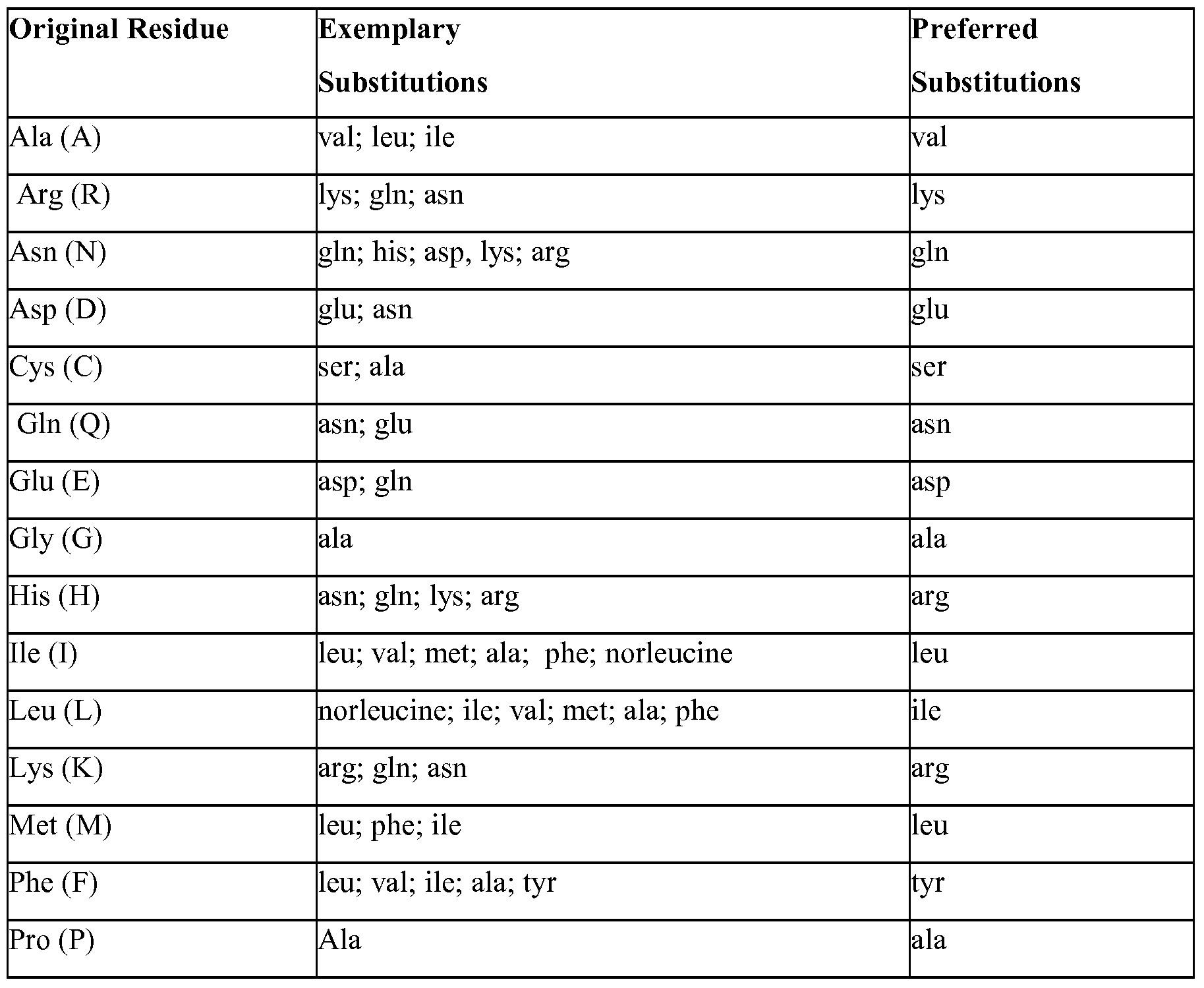


Claims
Priority Applications (53)
| Application Number | Priority Date | Filing Date | Title |
|---|---|---|---|
| KR1020197016653A KR20190069615A (en) | 2008-12-09 | 2009-12-08 | Anti-pd-l1 antibodies and their use to enhance t-cell function |
| MX2013001859A MX342591B (en) | 2008-12-09 | 2009-12-08 | Anti-pd-l1 antibodies and their use to enhance t-cell function. |
| EP09764997.4A EP2376535B9 (en) | 2008-12-09 | 2009-12-08 | Anti-pd-l1 antibodies and their use to enhance t-cell function |
| CN202210551253.2A CN114835812A (en) | 2008-12-09 | 2009-12-08 | anti-PD-L1 antibodies and their use for enhancing T cell function |
| CR20160570A CR20160570A (en) | 2008-12-09 | 2009-12-08 | ANTI-PLD1 ANTIBODIES AND THEIR USE TO IMPROVE THE FUNCTION OF T-CELLS |
| EP18177113.0A EP3447073A1 (en) | 2008-12-09 | 2009-12-08 | Anti-pd-l1 antibodies and their use to enhance t-cell function |
| KR1020207012431A KR20200047793A (en) | 2008-12-09 | 2009-12-08 | Anti-pd-l1 antibodies and their use to enhance t-cell function |
| KR1020187022311A KR20180089573A (en) | 2008-12-09 | 2009-12-08 | Anti-pd-l1 antibodies and their use to enhance t-cell function |
| KR1020227011061A KR20220047668A (en) | 2008-12-09 | 2009-12-08 | Anti-pd-l1 antibodies and their use to enhance t-cell function |
| MX2016009486A MX356367B (en) | 2008-12-09 | 2009-12-08 | Anti-pd-l1 antibodies and their use to enhance t-cell function. |
| EP23214687.8A EP4331604A2 (en) | 2008-12-09 | 2009-12-08 | Anti-pd-l1 antibodies and their use to enhance t-cell function |
| DK09764997.4T DK2376535T3 (en) | 2008-12-09 | 2009-12-08 | ANTI-PD-L1 ANTIBODIES AND THEIR USE TO PROMOTE T CELL FUNCTION |
| CN200980149532.9A CN102245640B (en) | 2008-12-09 | 2009-12-08 | Anti-PD-L1 antibodies and their use to enhance T-cell function |
| AU2009333580A AU2009333580B2 (en) | 2008-12-09 | 2009-12-08 | Anti-PD-L1 antibodies and their use to enhance T-cell function |
| SG2011042173A SG172059A1 (en) | 2008-12-09 | 2009-12-08 | Anti-pd-l1 antibodies and their use to enhance t-cell function |
| KR1020177026686A KR20170113681A (en) | 2008-12-09 | 2009-12-08 | Anti-pd-l1 antibodies and their use to enhance t-cell function |
| CA2740806A CA2740806C (en) | 2008-12-09 | 2009-12-08 | Anti-pd-l1 antibodies and their use to enhance t-cell function |
| NZ592119A NZ592119A (en) | 2008-12-09 | 2009-12-08 | Anti-pd-l1 antibodies and their use to enhance t-cell function |
| UAA201108595A UA109108C2 (en) | 2008-12-09 | 2009-12-08 | Anti-pd-ll antibody and its use to enhance t-cell function |
| KR1020217015160A KR20210060670A (en) | 2008-12-09 | 2009-12-08 | Anti-pd-l1 antibodies and their use to enhance t-cell function |
| ES09764997.4T ES2628095T3 (en) | 2008-12-09 | 2009-12-08 | Anti-PD-L1 antibodies and their use to enhance the function of T cells |
| SI200931672A SI2376535T1 (en) | 2008-12-09 | 2009-12-08 | Anti-pd-l1 antibodies and their use to enhance t-cell function |
| MX2011005853A MX2011005853A (en) | 2008-12-09 | 2009-12-08 | Anti-pd-l1 antibodies and their use to enhance t-cell function. |
| LTEP09764997.4T LT2376535T (en) | 2008-12-09 | 2009-12-08 | Anti-pd-l1 antibodies and their use to enhance t-cell function |
| EP21169871.7A EP3929216A1 (en) | 2008-12-09 | 2009-12-08 | Anti-pd-l1 antibodies and their use to enhance t-cell function |
| JP2011540819A JP5681638B2 (en) | 2008-12-09 | 2009-12-08 | Anti-PD-L1 antibodies and their use to enhance T cell function |
| EP22180770.4A EP4169951A1 (en) | 2008-12-09 | 2009-12-08 | Anti-pd-l1 antibodies and their use to enhance t-cell function |
| KR1020237015324A KR20230070055A (en) | 2008-12-09 | 2009-12-08 | Anti-pd-l1 antibodies and their use to enhance t-cell function |
| RU2011128399A RU2636023C2 (en) | 2008-12-09 | 2009-12-08 | Antibodies to pd-l1 and their application for t-cells function strengthening |
| BRPI0917592-0A BRPI0917592B1 (en) | 2008-12-09 | 2009-12-08 | ANTI-PD-L1 ANTIBODY, COMPOSITION, MANUFACTURED ARTICLES AND USES OF A COMPOSITION |
| EP22215991.5A EP4209510B1 (en) | 2008-12-09 | 2009-12-08 | Anti-pd-l1 antibodies and their use to enhance t-cell function |
| KR1020117013128A KR101782570B1 (en) | 2008-12-09 | 2009-12-08 | Anti-pd-l1 antibodies and their use to enhance t-cell function |
| MA33995A MA32948B1 (en) | 2008-12-09 | 2009-12-08 | ANTI-PD-L1 ANTIBODIES AND THEIR USE FOR IMPROVING T CELL FUNCTION |
| MYPI2011002606A MY188826A (en) | 2008-12-09 | 2009-12-08 | Anti-pd-l1 antibodies and their use to enhance t-cell function |
| ZA2011/02417A ZA201102417B (en) | 2008-12-09 | 2011-03-31 | Anti-pd-l1 antibodies and their use to enhance t-cell function |
| IL213353A IL213353A (en) | 2008-12-09 | 2011-06-05 | Anti-pd-l1 antibodies and their use to enhance t-cell function |
| HK12103694.9A HK1163130A1 (en) | 2008-12-09 | 2012-04-16 | Anti-pd-l1 antibodies and their use to enhance t-cell function -pd-l1 t |
| AU2016203867A AU2016203867B2 (en) | 2008-12-09 | 2016-06-09 | Anti-PD-L1 antibodies and their use to enhance T-cell function |
| IL249127A IL249127B (en) | 2008-12-09 | 2016-11-22 | Anti-pd-l1 antibodies and their use to enhance t-cell function |
| HRP20170908TT HRP20170908T1 (en) | 2008-12-09 | 2017-06-14 | Anti-pd-l1 antibodies and their use to enhance t-cell function |
| CY20171100689T CY1118943T1 (en) | 2008-12-09 | 2017-06-28 | ANTI-PD-L1 ANTIBODIES AND USE OF THESE TO ENHANCE T-CELL FUNCTIONING |
| FR17C1050C FR17C1050I2 (en) | 2008-12-09 | 2017-11-27 | ANTI-PD-L1 ANTIBODIES AND THEIR USE TO IMPROVE T LYMPHOCYTE FUNCTION |
| HUS1700049C HUS1700049I1 (en) | 2008-12-09 | 2017-11-28 | Anti-pd-l1 antibodies and their use to enhance t-cell function |
| NL300914C NL300914I2 (en) | 2008-12-09 | 2017-11-29 | Atezolizumab |
| LU00051C LUC00051I2 (en) | 2008-12-09 | 2017-11-29 | |
| CY2017043C CY2017043I2 (en) | 2008-12-09 | 2017-12-06 | ANTI-PD-L1 ANTIBODIES AND THEIR USE TO ENHANCE T-CELL FUNCTION |
| LTPA2017041C LTC2376535I2 (en) | 2008-12-09 | 2017-12-19 | Anti-PD-L1 Antibodies and Their Use for Improvement of T Cell Function |
| NO2018006C NO2018006I1 (en) | 2008-12-09 | 2018-02-14 | Atezolizumab |
| AU2018203226A AU2018203226B2 (en) | 2008-12-09 | 2018-05-09 | Anti-PD-L1 antibodies and their use to enhance T-cell function |
| IL263931A IL263931B (en) | 2008-12-09 | 2018-12-24 | Anti-pd-l1 antibodies and their use to enhance t-cell function |
| AU2020204593A AU2020204593B2 (en) | 2008-12-09 | 2020-07-09 | Anti-PD-L1 antibodies and their use to enhance T-cell function |
| IL278007A IL278007A (en) | 2008-12-09 | 2020-10-13 | Anti-pd-l1 antibodies and their use to enhance t-cell function |
| AU2024200864A AU2024200864A1 (en) | 2008-12-09 | 2024-02-12 | Anti-PD-L1 antibodies and their use to enhance T-cell function |
Applications Claiming Priority (2)
| Application Number | Priority Date | Filing Date | Title |
|---|---|---|---|
| US12109208P | 2008-12-09 | 2008-12-09 | |
| US61/121,092 | 2008-12-09 |
Publications (1)
| Publication Number | Publication Date |
|---|---|
| WO2010077634A1 true WO2010077634A1 (en) | 2010-07-08 |
Family
ID=42097246
Family Applications (1)
| Application Number | Title | Priority Date | Filing Date |
|---|---|---|---|
| PCT/US2009/067104 WO2010077634A1 (en) | 2008-12-09 | 2009-12-08 | Anti-pd-l1 antibodies and their use to enhance t-cell function |
Country Status (40)
| Country | Link |
|---|---|
| US (11) | US8217149B2 (en) |
| EP (7) | EP3447073A1 (en) |
| JP (6) | JP5681638B2 (en) |
| KR (8) | KR20170113681A (en) |
| CN (4) | CN102245640B (en) |
| AR (2) | AR074563A1 (en) |
| AU (5) | AU2009333580B2 (en) |
| BR (1) | BRPI0917592B1 (en) |
| CA (2) | CA3120172A1 (en) |
| CL (1) | CL2011001382A1 (en) |
| CO (1) | CO6390023A2 (en) |
| CR (2) | CR20160570A (en) |
| CY (2) | CY1118943T1 (en) |
| DK (1) | DK2376535T3 (en) |
| EC (1) | ECSP11011115A (en) |
| ES (1) | ES2628095T3 (en) |
| FI (1) | FI4209510T3 (en) |
| FR (1) | FR17C1050I2 (en) |
| HK (3) | HK1163130A1 (en) |
| HR (1) | HRP20170908T1 (en) |
| HU (2) | HUE034832T2 (en) |
| IL (4) | IL213353A (en) |
| LT (3) | LT4209510T (en) |
| LU (1) | LUC00051I2 (en) |
| MA (1) | MA32948B1 (en) |
| MX (3) | MX342591B (en) |
| MY (1) | MY188826A (en) |
| NL (1) | NL300914I2 (en) |
| NO (1) | NO2018006I1 (en) |
| NZ (2) | NZ717213A (en) |
| PE (2) | PE20141722A1 (en) |
| PL (1) | PL2376535T3 (en) |
| PT (1) | PT2376535T (en) |
| RU (2) | RU2636023C2 (en) |
| SG (3) | SG196798A1 (en) |
| SI (1) | SI2376535T1 (en) |
| TW (5) | TWI605828B (en) |
| UA (1) | UA109108C2 (en) |
| WO (1) | WO2010077634A1 (en) |
| ZA (1) | ZA201102417B (en) |
Cited By (868)
| Publication number | Priority date | Publication date | Assignee | Title |
|---|---|---|---|---|
| AU2010324757A1 (en) * | 2009-11-24 | 2012-05-24 | Medimmune Limited | Targeted binding agents against B7-H1 |
| WO2013019906A1 (en) | 2011-08-01 | 2013-02-07 | Genentech, Inc. | Methods of treating cancer using pd-1 axis binding antagonists and mek inhibitors |
| WO2013079174A1 (en) | 2011-11-28 | 2013-06-06 | Merck Patent Gmbh | Anti-pd-l1 antibodies and uses thereof |
| CN103304638A (en) * | 2013-07-08 | 2013-09-18 | 郑州大学 | PD-L1 affinity peptide with anti-tumour activity and application for same |
| WO2013181452A1 (en) | 2012-05-31 | 2013-12-05 | Genentech, Inc. | Methods of treating cancer using pd-l1 axis binding antagonists and vegf antagonists |
| WO2014008218A1 (en) | 2012-07-02 | 2014-01-09 | Bristol-Myers Squibb Company | Optimization of antibodies that bind lymphocyte activation gene-3 (lag-3), and uses thereof |
| WO2014055648A1 (en) | 2012-10-02 | 2014-04-10 | Bristol-Myers Squibb Company | Combination of anti-kir antibodies and anti-pd-1 antibodies to treat cancer |
| WO2014083178A1 (en) | 2012-11-30 | 2014-06-05 | F. Hoffmann-La Roche Ag | Identification of patients in need of pd-l1 inhibitor cotherapy |
| CN103936835A (en) * | 2014-04-29 | 2014-07-23 | 郑州大学 | PD-L1IgV-targeting affinity peptide D1 with anti-tumor activity |
| CN103936836A (en) * | 2014-04-29 | 2014-07-23 | 郑州大学 | Targeted PD-L1IgV affinity peptide D2 with anti-tumor activity |
| WO2014130635A1 (en) | 2013-02-20 | 2014-08-28 | Novartis Ag | Effective targeting of primary human leukemia using anti-cd123 chimeric antigen receptor engineered t cells |
| WO2014130657A1 (en) | 2013-02-20 | 2014-08-28 | The Trustees Of The University Of Pennsylvania | Treatment of cancer using humanized anti-egfrviii chimeric antigen receptor |
| WO2014153270A1 (en) | 2013-03-16 | 2014-09-25 | Novartis Ag | Treatment of cancer using humanized anti-cd19 chimeric antigen receptor |
| WO2014151006A2 (en) | 2013-03-15 | 2014-09-25 | Genentech, Inc. | Biomarkers and methods of treating pd-1 and pd-l1 related conditions |
| CN104098651A (en) * | 2014-06-30 | 2014-10-15 | 郑州大学 | PD-L1 IgV affinity peptide with antineoplastic activity, and preparation method and application of thereof |
| WO2015009856A2 (en) * | 2013-07-16 | 2015-01-22 | Genentech, Inc. | Methods of treating cancer using pd-1 axis binding antagonists and tigit inhibitors |
| JP2015504894A (en) * | 2012-01-16 | 2015-02-16 | アトックス バイオ リミテッド | Synthetic peptides for the treatment of bacterial infections |
| WO2015026634A1 (en) | 2013-08-20 | 2015-02-26 | Merck Sharp & Dohme Corp. | Treating cancer with a combination of a pd-1 antagonist and dinaciclib |
| WO2015026684A1 (en) | 2013-08-20 | 2015-02-26 | Merck Sharp & Dohme Corp. | Modulation of tumor immunity |
| WO2015033299A1 (en) | 2013-09-06 | 2015-03-12 | Aurigene Discovery Technologies Limited | 1,2,4-oxadiazole derivatives as immunomodulators |
| WO2015033301A1 (en) | 2013-09-06 | 2015-03-12 | Aurigene Discovery Technologies Limited | 1,3,4-oxadiazole and 1,3,4-thiadiazole derivatives as immunomodulators |
| WO2015033303A1 (en) | 2013-09-06 | 2015-03-12 | Aurigene Discovery Technologies Limited | Cyclic peptidomimetic compounds as immunomodulators |
| WO2015036511A1 (en) * | 2013-09-12 | 2015-03-19 | F. Hoffmann-La Roche Ag | Combination therapy of antibodies against human csf-1r and antibodies against human pd-l1 |
| WO2015048520A1 (en) | 2013-09-27 | 2015-04-02 | Genentech, Inc. | Anti-pdl1 antibody formulations |
| CN104558177A (en) * | 2013-10-25 | 2015-04-29 | 苏州思坦维生物技术有限责任公司 | Monoclonal antibody capable of antagonizing and inhibiting bonding of programmed death-1 receptor (PD-1) and ligand thereof as well as encoding sequence and use of monoclonal antibody |
| WO2015066413A1 (en) | 2013-11-01 | 2015-05-07 | Novartis Ag | Oxazolidinone hydroxamic acid compounds for the treatment of bacterial infections |
| WO2015073644A1 (en) | 2013-11-13 | 2015-05-21 | Novartis Ag | Mtor inhibitors for enhancing the immune response |
| WO2015095423A2 (en) | 2013-12-17 | 2015-06-25 | Genentech, Inc. | Combination therapy comprising ox40 binding agonists and pd-1 axis binding antagonists |
| WO2015094992A1 (en) | 2013-12-17 | 2015-06-25 | Merck Sharp & Dohme Corp. | Ifn-gamma gene signature biomarkers of tumor response to pd-1 antagonists |
| WO2015095418A1 (en) | 2013-12-17 | 2015-06-25 | Genentech, Inc. | Methods of treating her2-positive cancers using pd-1 axis binding antagonists and anti-her2 antibodies |
| WO2015090230A1 (en) | 2013-12-19 | 2015-06-25 | Novartis Ag | Human mesothelin chimeric antigen receptors and uses thereof |
| WO2015095410A1 (en) | 2013-12-17 | 2015-06-25 | Genentech, Inc. | Methods of treating cancer using pd-1 axis binding antagonists and an anti-cd20 antibody |
| WO2015100282A1 (en) | 2013-12-24 | 2015-07-02 | Bristol-Myers Squibb Company | Tricyclic compounds as anticancer agents |
| WO2015107495A1 (en) | 2014-01-17 | 2015-07-23 | Novartis Ag | N-azaspirocycloalkane substituted n-heteroaryl compounds and compositions for inhibiting the activity of shp2 |
| WO2015112805A1 (en) | 2014-01-23 | 2015-07-30 | Regeneron Pharmaceuticals, Inc. | Human antibodies to pd-l1 |
| WO2015119944A1 (en) | 2014-02-04 | 2015-08-13 | Incyte Corporation | Combination of a pd-1 antagonist and an ido1 inhibitor for treating cancer |
| WO2015138920A1 (en) | 2014-03-14 | 2015-09-17 | Novartis Ag | Antibody molecules to lag-3 and uses thereof |
| WO2015142675A2 (en) | 2014-03-15 | 2015-09-24 | Novartis Ag | Treatment of cancer using chimeric antigen receptor |
| WO2015148379A1 (en) | 2014-03-24 | 2015-10-01 | Novartis Ag | Monobactam organic compounds for the treatment of bacterial infections |
| WO2015118175A3 (en) * | 2014-02-10 | 2015-10-08 | Merck Patent Gmbh | TARGETED TGFβ INHIBITION |
| WO2015153513A1 (en) | 2014-03-31 | 2015-10-08 | Genentech, Inc. | Anti-ox40 antibodies and methods of use |
| WO2015153514A1 (en) | 2014-03-31 | 2015-10-08 | Genentech, Inc. | Combination therapy comprising anti-angiogenesis agents and ox40 binding agonists |
| WO2015157252A1 (en) | 2014-04-07 | 2015-10-15 | BROGDON, Jennifer | Treatment of cancer using anti-cd19 chimeric antigen receptor |
| DE202014010421U1 (en) | 2013-12-17 | 2015-11-12 | Kymab Limited | Human goals |
| US9192667B2 (en) | 2013-04-22 | 2015-11-24 | Hoffmann-La Roche Inc. | Method of treating cancer by administering CSF-1R antibodies and a TLR9 agonist |
| WO2015181624A2 (en) | 2014-05-28 | 2015-12-03 | Idenix Pharmaceuticals, Inc | Nucleoside derivatives for the treatment of cancer |
| US9226958B2 (en) | 2010-10-01 | 2016-01-05 | University Of Georgia Research Foundation, Inc. | Use of Listeria vaccine vectors to reverse vaccine unresponsiveness in parasitically infected individuals |
| WO2016007235A1 (en) | 2014-07-11 | 2016-01-14 | Genentech, Inc. | Anti-pd-l1 antibodies and diagnostic uses thereof |
| WO2016011160A1 (en) | 2014-07-15 | 2016-01-21 | Genentech, Inc. | Compositions for treating cancer using pd-1 axis binding antagonists and mek inhibitors |
| WO2016014530A1 (en) | 2014-07-21 | 2016-01-28 | Novartis Ag | Combinations of low, immune enhancing. doses of mtor inhibitors and cars |
| WO2016014553A1 (en) | 2014-07-21 | 2016-01-28 | Novartis Ag | Sortase synthesized chimeric antigen receptors |
| WO2016020836A1 (en) | 2014-08-06 | 2016-02-11 | Novartis Ag | Quinolone derivatives as antibacterials |
| WO2016025880A1 (en) | 2014-08-14 | 2016-02-18 | Novartis Ag | Treatment of cancer using gfr alpha-4 chimeric antigen receptor |
| WO2016030350A1 (en) * | 2014-08-29 | 2016-03-03 | F. Hoffmann-La Roche Ag | Combination therapy of tumor-targeted il-2 variant immunocytokines and antibodies against human pd-l1 |
| WO2016033555A1 (en) | 2014-08-28 | 2016-03-03 | Halozyme, Inc. | Combination therapy with a hyaluronan-degrading enzyme and an immune checkpoint inhibitor |
| WO2016040892A1 (en) | 2014-09-13 | 2016-03-17 | Novartis Ag | Combination therapies |
| WO2016044605A1 (en) | 2014-09-17 | 2016-03-24 | Beatty, Gregory | Targeting cytotoxic cells with chimeric receptors for adoptive immunotherapy |
| WO2016054555A2 (en) | 2014-10-03 | 2016-04-07 | Novartis Ag | Combination therapies |
| WO2016057846A1 (en) | 2014-10-08 | 2016-04-14 | Novartis Ag | Compositions and methods of use for augmented immune response and cancer therapy |
| WO2016057705A1 (en) | 2014-10-08 | 2016-04-14 | Novartis Ag | Biomarkers predictive of therapeutic responsiveness to chimeric antigen receptor therapy and uses thereof |
| WO2016061142A1 (en) | 2014-10-14 | 2016-04-21 | Novartis Ag | Antibody molecules to pd-l1 and uses thereof |
| WO2016061286A2 (en) | 2014-10-14 | 2016-04-21 | Halozyme, Inc. | Compositions of adenosine deaminase-2 (ada2), variants thereof and methods of using same |
| WO2016073378A1 (en) | 2014-11-03 | 2016-05-12 | Genentech, Inc. | Assays for detecting t cell immune subsets and methods of use thereof |
| WO2016075670A1 (en) | 2014-11-14 | 2016-05-19 | Novartis Ag | Antibody drug conjugates |
| WO2016081384A1 (en) | 2014-11-17 | 2016-05-26 | Genentech, Inc. | Combination therapy comprising ox40 binding agonists and pd-1 axis binding antagonists |
| EP2854843A4 (en) * | 2012-05-31 | 2016-06-01 | Sorrento Therapeutics Inc | Antigen binding proteins that bind pd-l1 |
| WO2016086200A1 (en) | 2014-11-27 | 2016-06-02 | Genentech, Inc. | 4,5,6,7-tetrahydro-1 h-pyrazolo[4,3-c]pyridin-3-amine compounds as cbp and/or ep300 inhibitors |
| EP3027210A1 (en) * | 2013-08-02 | 2016-06-08 | Aduro Biotech Holdings, Europe B.V. | Combining cd27 agonists and immune checkpoint inhibition for immune stimulation |
| WO2016089830A1 (en) | 2014-12-05 | 2016-06-09 | Merck Sharp & Dohme Corp. | Novel tricyclic compounds as inhibitors of mutant idh enzymes |
| WO2016090034A2 (en) | 2014-12-03 | 2016-06-09 | Novartis Ag | Methods for b cell preconditioning in car therapy |
| WO2016090300A1 (en) | 2014-12-05 | 2016-06-09 | Genentech, Inc. | Methods and compositions for treating cancer using pd-1 axis antagonists and hpk1 antagonists |
| WO2016089797A1 (en) | 2014-12-05 | 2016-06-09 | Merck Sharp & Dohme Corp. | Novel tricyclic compounds as inhibitors of mutant idh enzymes |
| WO2016089833A1 (en) | 2014-12-05 | 2016-06-09 | Merck Sharp & Dohme Corp. | Novel tricyclic compounds as inhibitors of mutant idh enzymes |
| WO2016094377A1 (en) | 2014-12-09 | 2016-06-16 | Merck Sharp & Dohme Corp. | System and methods for deriving gene signature biomarkers of response to pd-1 antagonists |
| WO2016097995A1 (en) | 2014-12-16 | 2016-06-23 | Novartis Ag | Isoxazole hydroxamic acid compounds as lpxc inhibitors |
| WO2016100882A1 (en) | 2014-12-19 | 2016-06-23 | Novartis Ag | Combination therapies |
| WO2016106266A1 (en) | 2014-12-22 | 2016-06-30 | Bristol-Myers Squibb Company | TGFβ RECEPTOR ANTAGONISTS |
| WO2016111645A1 (en) * | 2015-01-09 | 2016-07-14 | Agency For Science, Technology And Research | Anti-pd-l1 antibodies |
| WO2016126608A1 (en) | 2015-02-02 | 2016-08-11 | Novartis Ag | Car-expressing cells against multiple tumor antigens and uses thereof |
| WO2016140884A1 (en) | 2015-03-02 | 2016-09-09 | Rigel Pharmaceuticals, Inc. | TGF-β INHIBITORS |
| WO2016141209A1 (en) | 2015-03-04 | 2016-09-09 | Merck Sharp & Dohme Corp. | Combination of a pd-1 antagonist and eribulin for treating cancer |
| WO2016140717A1 (en) | 2015-03-04 | 2016-09-09 | Merck Sharp & Dohme Corp. | Combination of a pd-1 antagonist and a vegfr/fgfr/ret tyrosine kinase inhibitor for treating cancer |
| EP3067062A1 (en) | 2015-03-13 | 2016-09-14 | Ipsen Pharma S.A.S. | Combination of tasquinimod or a pharmaceutically acceptable salt thereof and a pd1 and/or pdl1 inhibitor, for use as a medicament |
| WO2016145085A2 (en) | 2015-03-09 | 2016-09-15 | Celldex Therapeutics, Inc. | Cd27 agonists |
| WO2016145102A1 (en) | 2015-03-10 | 2016-09-15 | Aduro Biotech, Inc. | Compositions and methods for activating "stimulator of interferon gene" -dependent signalling |
| WO2016142833A1 (en) | 2015-03-10 | 2016-09-15 | Aurigene Discovery Technologies Limited | 1,2,4-oxadiazole and thiadiazole compounds as immunomodulators |
| EP3070102A1 (en) | 2015-03-18 | 2016-09-21 | F. Hoffmann-La Roche AG | Combination therapy of antibodies human cd40 activating antibodies and anti human pld-1 antibodies |
| WO2016161279A1 (en) | 2015-04-03 | 2016-10-06 | Bristol-Myers Squibb Company | Inhibitors of indoleamine 2,3-dioxygenase for the treatment of cancer |
| US9463227B2 (en) | 2011-03-11 | 2016-10-11 | Advaxis, Inc. | Listeria-based adjuvants |
| WO2016164580A1 (en) | 2015-04-07 | 2016-10-13 | Novartis Ag | Combination of chimeric antigen receptor therapy and amino pyrimidine derivatives |
| WO2016164480A1 (en) | 2015-04-07 | 2016-10-13 | Genentech, Inc. | Antigen binding complex having agonistic activity and methods of use |
| WO2016162505A1 (en) | 2015-04-08 | 2016-10-13 | F-Star Biotechnology Limited | Her2 binding agent therapies |
| WO2016168595A1 (en) | 2015-04-17 | 2016-10-20 | Barrett David Maxwell | Methods for improving the efficacy and expansion of chimeric antigen receptor-expressing cells |
| WO2016168133A1 (en) | 2015-04-17 | 2016-10-20 | Merck Sharp & Dohme Corp. | Blood-based biomarkers of tumor sensitivity to pd-1 antagonists |
| WO2016170040A1 (en) * | 2015-04-23 | 2016-10-27 | F. Hoffmann-La Roche Ag | Combination therapy of antibody binding to angiopoietin 2 with antibody binding to programmed death ligand 1 |
| WO2016172583A1 (en) | 2015-04-23 | 2016-10-27 | Novartis Ag | Treatment of cancer using chimeric antigen receptor and protein kinase a blocker |
| WO2016183118A1 (en) | 2015-05-12 | 2016-11-17 | Bristol-Myers Squibb Company | Tricyclic compounds as anticancer agents |
| WO2016183114A1 (en) | 2015-05-11 | 2016-11-17 | Bristol-Myers Squibb Company | Tricyclic compounds as anticancer agents |
| WO2016183115A1 (en) | 2015-05-12 | 2016-11-17 | Bristol-Myers Squibb Company | 5h-pyrido[3,2-b]indole compounds as anticancer agents |
| US9499596B2 (en) | 2008-04-09 | 2016-11-22 | Genentech, Inc. | Compositions and methods for the treatment of immune related diseases |
| WO2016189055A1 (en) | 2015-05-27 | 2016-12-01 | Idenix Pharmaceuticals Llc | Nucleotides for the treatment of cancer |
| WO2016196298A1 (en) | 2015-05-29 | 2016-12-08 | Genentech, Inc. | Therapeutic and diagnolstic methods for cancer |
| WO2016196381A1 (en) | 2015-05-29 | 2016-12-08 | Genentech, Inc. | Pd-l1 promoter methylation in cancer |
| WO2016196173A1 (en) | 2015-05-29 | 2016-12-08 | Merck Sharp & Dohme Corp. | Combination of a pd-1 antagonist and cpg-c type oligonucleotide for treating cancer |
| WO2016197367A1 (en) | 2015-06-11 | 2016-12-15 | Wuxi Biologics (Shanghai) Co. Ltd. | Novel anti-pd-l1 antibodies |
| WO2016200835A1 (en) | 2015-06-08 | 2016-12-15 | Genentech, Inc. | Methods of treating cancer using anti-ox40 antibodies and pd-1 axis binding antagonists |
| WO2016200836A1 (en) | 2015-06-08 | 2016-12-15 | Genentech, Inc. | Methods of treating cancer using anti-ox40 antibodies |
| WO2016205320A1 (en) | 2015-06-17 | 2016-12-22 | Genentech, Inc. | Methods of treating locally advanced or metastatic breast cancers using pd-1 axis binding antagonists and taxanes |
| WO2016203432A1 (en) | 2015-06-17 | 2016-12-22 | Novartis Ag | Antibody drug conjugates |
| WO2016205200A1 (en) | 2015-06-16 | 2016-12-22 | Genentech, Inc. | Anti-cll-1 antibodies and methods of use |
| EP3108897A1 (en) | 2015-06-24 | 2016-12-28 | F. Hoffmann-La Roche AG | Antibodies against human csf-1r for use in inducing lymphocytosis in lymphomas or leukemias |
| US9535074B2 (en) | 2014-09-08 | 2017-01-03 | Merck Sharp & Dohme Corp. | Immunoassay for soluble PD-L1 |
| WO2017007700A1 (en) | 2015-07-06 | 2017-01-12 | Iomet Pharma Ltd. | Pharmaceutical compound |
| WO2017009842A2 (en) | 2015-07-16 | 2017-01-19 | Biokine Therapeutics Ltd. | Compositions and methods for treating cancer |
| WO2017015427A1 (en) | 2015-07-21 | 2017-01-26 | Novartis Ag | Methods for improving the efficacy and expansion of immune cells |
| WO2017017624A1 (en) | 2015-07-29 | 2017-02-02 | Novartis Ag | Combination of pd-1 antagonist with an egfr inhibitor |
| WO2017019757A1 (en) | 2015-07-28 | 2017-02-02 | Bristol-Myers Squibb Company | Tgf beta receptor antagonists |
| WO2017019897A1 (en) | 2015-07-29 | 2017-02-02 | Novartis Ag | Combination therapies comprising antibody molecules to tim-3 |
| WO2017019894A1 (en) | 2015-07-29 | 2017-02-02 | Novartis Ag | Combination therapies comprising antibody molecules to lag-3 |
| WO2017020858A1 (en) * | 2015-08-06 | 2017-02-09 | Wuxi Biologics (Shanghai) Co. Ltd. | Novel anti-pd-l1 antibodies |
| WO2017027645A1 (en) | 2015-08-13 | 2017-02-16 | Merck Sharp & Dohme Corp. | Cyclic di-nucleotide compounds as sting agonists |
| WO2017035118A1 (en) | 2015-08-25 | 2017-03-02 | Bristol-Myers Squibb Company | Tgf beta receptor antagonists |
| WO2017040930A2 (en) | 2015-09-03 | 2017-03-09 | The Trustees Of The University Of Pennsylvania | Biomarkers predictive of cytokine release syndrome |
| WO2017040990A1 (en) | 2015-09-03 | 2017-03-09 | Aileron Therapeutics, Inc. | Peptidomimetic macrocycles and uses thereof |
| US9598422B2 (en) | 2014-11-05 | 2017-03-21 | Flexus Biosciences, Inc. | Immunoregulatory agents |
| WO2017058780A1 (en) | 2015-09-30 | 2017-04-06 | Merck Patent Gmbh | Combination of a pd-1 axis binding antagonist and an alk inhibitor for treating alk-negative cancer |
| WO2017055484A1 (en) | 2015-09-29 | 2017-04-06 | INSERM (Institut National de la Santé et de la Recherche Médicale) | Methods for determining the metabolic status of lymphomas |
| EP2376535B1 (en) | 2008-12-09 | 2017-04-12 | F. Hoffmann-La Roche AG | Anti-pd-l1 antibodies and their use to enhance t-cell function |
| WO2017064043A1 (en) | 2015-10-12 | 2017-04-20 | Innate Pharma | Cd73 blocking agents |
| WO2017069291A1 (en) | 2015-10-23 | 2017-04-27 | Canbas Co., Ltd. | Peptides and peptidomimetics in combination with t cell activating and/or checkpoint inhibiting agents for cancer treatment |
| WO2017072662A1 (en) | 2015-10-29 | 2017-05-04 | Novartis Ag | Antibody conjugates comprising toll-like receptor agonist |
| US9643972B2 (en) | 2014-11-05 | 2017-05-09 | Flexus Biosciences, Inc. | Immunoregulatory agents |
| US9644212B2 (en) | 2008-05-19 | 2017-05-09 | Advaxis, Inc. | Dual delivery system for heterologous antigens |
| WO2017079116A2 (en) | 2015-11-03 | 2017-05-11 | Janssen Biotech, Inc. | Antibodies specifically binding pd-1 and tim-3 and their uses |
| WO2017077382A1 (en) | 2015-11-06 | 2017-05-11 | Orionis Biosciences Nv | Bi-functional chimeric proteins and uses thereof |
| US9650639B2 (en) | 2008-05-19 | 2017-05-16 | Advaxis, Inc. | Dual delivery system for heterologous antigens |
| WO2017087851A1 (en) | 2015-11-19 | 2017-05-26 | Genentech, Inc. | Methods of treating cancer using b-raf inhibitors and immune checkpoint inhibitors |
| WO2017087280A1 (en) | 2015-11-16 | 2017-05-26 | Genentech, Inc. | Methods of treating her2-positive cancer |
| WO2017093933A1 (en) | 2015-12-03 | 2017-06-08 | Glaxosmithkline Intellectual Property Development Limited | Cyclic purine dinucleotides as modulators of sting |
| EP3178848A1 (en) | 2015-12-09 | 2017-06-14 | F. Hoffmann-La Roche AG | Type ii anti-cd20 antibody for reducing formation of anti-drug antibodies |
| WO2017098421A1 (en) | 2015-12-08 | 2017-06-15 | Glaxosmithkline Intellectual Property Development Limited | Benzothiadiazine compounds |
| US9683048B2 (en) | 2014-01-24 | 2017-06-20 | Novartis Ag | Antibody molecules to PD-1 and uses thereof |
| WO2017103283A1 (en) | 2015-12-17 | 2017-06-22 | Photocure Asa | Neoadjuvant therapy for bladder cancer |
| WO2017106630A1 (en) | 2015-12-18 | 2017-06-22 | The General Hospital Corporation | Polyacetal polymers, conjugates, particles and uses thereof |
| WO2017106656A1 (en) | 2015-12-17 | 2017-06-22 | Novartis Ag | Antibody molecules to pd-1 and uses thereof |
| WO2017106291A1 (en) | 2015-12-15 | 2017-06-22 | Bristol-Myers Squibb Company | Cxcr4 receptor antagonists |
| WO2017106062A1 (en) | 2015-12-15 | 2017-06-22 | Merck Sharp & Dohme Corp. | Novel compounds as indoleamine 2,3-dioxygenase inhibitors |
| WO2017103895A1 (en) | 2015-12-18 | 2017-06-22 | Novartis Ag | Antibodies targeting cd32b and methods of use thereof |
| WO2017112741A1 (en) | 2015-12-22 | 2017-06-29 | Novartis Ag | Mesothelin chimeric antigen receptor (car) and antibody against pd-l1 inhibitor for combined use in anticancer therapy |
| WO2017118634A1 (en) | 2016-01-04 | 2017-07-13 | INSERM (Institut National de la Santé et de la Recherche Médicale) | Use of pd-1 and tim-3 as a measure for cd8+ cells in predicting and treating renal cell carcinoma |
| US9708396B2 (en) | 2008-10-08 | 2017-07-18 | Hoffmann-La Roche Inc. | Bispecific anti-VEGF/anti-ANG-2 antibodies |
| WO2017125532A1 (en) | 2016-01-21 | 2017-07-27 | Innate Pharma | Neutralization of inhibitory pathways in lymphocytes |
| WO2017129763A1 (en) | 2016-01-28 | 2017-08-03 | INSERM (Institut National de la Santé et de la Recherche Médicale) | Methods and pharmaceutical compositions for the treatment of signet ring cell gastric cancer |
| WO2017134305A1 (en) | 2016-02-05 | 2017-08-10 | Orionis Biosciences Nv | Bispecific signaling agents and uses thereof |
| WO2017140821A1 (en) | 2016-02-19 | 2017-08-24 | Novartis Ag | Tetracyclic pyridone compounds as antivirals |
| WO2017144681A1 (en) | 2016-02-25 | 2017-08-31 | Cell Medica Switzerland Ag | Binding members to pd-l1 |
| USRE46534E1 (en) | 2002-09-11 | 2017-09-05 | Genentech, Inc. | Composition and methods for the diagnosis of immune related diseases involving the PRO52254 polypeptide |
| WO2017149515A1 (en) | 2016-03-04 | 2017-09-08 | Novartis Ag | Cells expressing multiple chimeric antigen receptor (car) molecules and uses therefore |
| WO2017151502A1 (en) | 2016-02-29 | 2017-09-08 | Genentech, Inc. | Therapeutic and diagnostic methods for cancer |
| WO2017149143A1 (en) | 2016-03-04 | 2017-09-08 | Agency For Science, Technology And Research | Anti-lag-3 antibodies |
| WO2017153952A1 (en) | 2016-03-10 | 2017-09-14 | Glaxosmithkline Intellectual Property Development Limited | 5-sulfamoyl-2-hydroxybenzamide derivatives |
| WO2017153433A1 (en) | 2016-03-08 | 2017-09-14 | Innate Pharma | Siglec neutralizing antibodies |
| WO2017160754A1 (en) | 2016-03-15 | 2017-09-21 | Mersana Therapeutics,Inc. | Napi2b-targeted antibody-drug conjugates and methods of use thereof |
| WO2017159699A1 (en) | 2016-03-15 | 2017-09-21 | Chugai Seiyaku Kabushiki Kaisha | Methods of treating cancers using pd-1 axis binding antagonists and anti-gpc3 antibodies |
| WO2017160599A1 (en) | 2016-03-14 | 2017-09-21 | The United States Of America, As Represented By The Secretary, Department Of Health And Human Services | Use of cd300b antagonists to treat sepsis and septic shock |
| US9771425B2 (en) | 2014-10-27 | 2017-09-26 | Agency For Science, Technology And Research | Anti-PD-1 antibodies |
| WO2017165778A1 (en) | 2016-03-24 | 2017-09-28 | Millennium Pharmaceuticals, Inc. | Methods of treating gastrointestinal immune-related adverse events in immune oncology treatments |
| WO2017165742A1 (en) | 2016-03-24 | 2017-09-28 | Millennium Pharmaceuticals, Inc. | Methods of treating gastrointestinal immune-related adverse events in anti-ctla4 anti-pd-1 combination treatments |
| WO2017163186A1 (en) | 2016-03-24 | 2017-09-28 | Novartis Ag | Alkynyl nucleoside analogs as inhibitors of human rhinovirus |
| WO2017167921A1 (en) | 2016-03-30 | 2017-10-05 | Centre Léon-Bérard | Lymphocytes expressing cd73 in cancerous patient dictates therapy |
| WO2017175147A1 (en) | 2016-04-07 | 2017-10-12 | Glaxosmithkline Intellectual Property Development Limited | Heterocyclic amides useful as protein modulators |
| WO2017175156A1 (en) | 2016-04-07 | 2017-10-12 | Glaxosmithkline Intellectual Property Development Limited | Heterocyclic amides useful as protein modulators |
| WO2017178572A1 (en) | 2016-04-13 | 2017-10-19 | Vivia Biotech, S.L | Ex vivo bite-activated t cells |
| WO2017181111A2 (en) | 2016-04-15 | 2017-10-19 | Genentech, Inc. | Methods for monitoring and treating cancer |
| WO2017181079A2 (en) | 2016-04-15 | 2017-10-19 | Genentech, Inc. | Methods for monitoring and treating cancer |
| WO2017192874A1 (en) | 2016-05-04 | 2017-11-09 | The United States Of America, As Represented By The Secretary, Department Of Health And Human Services | Albumin-binding immunomodulatory compositions and methods of use thereof |
| WO2017191545A1 (en) | 2016-05-05 | 2017-11-09 | Glaxosmithkline Intellectual Property (No.2) Limited | Enhancer of zeste homolog 2 inhibitors |
| WO2017194783A1 (en) | 2016-05-13 | 2017-11-16 | Orionis Biosciences Nv | Targeted mutant interferon-beta and uses thereof |
| WO2017194265A1 (en) | 2016-05-10 | 2017-11-16 | Agency For Science, Technology And Research | Anti-CTLA-4 Antibodies |
| WO2017194782A2 (en) | 2016-05-13 | 2017-11-16 | Orionis Biosciences Nv | Therapeutic targeting of non-cellular structures |
| WO2017201036A1 (en) | 2016-05-17 | 2017-11-23 | Genentech, Inc. | Stromal gene signatures for diagnosis and use in immunotherapy |
| WO2017200969A1 (en) | 2016-05-20 | 2017-11-23 | Eli Lilly And Company | Combination therapy with notch and pd-1 or pd-l1 inhibitors |
| WO2017205536A2 (en) | 2016-05-24 | 2017-11-30 | Genentech, Inc. | Therapeutic compounds and uses thereof |
| WO2017205538A1 (en) | 2016-05-24 | 2017-11-30 | Genentech, Inc. | Pyrazolopyridine derivatives for the treatment of cancer |
| EP3252078A1 (en) | 2016-06-02 | 2017-12-06 | F. Hoffmann-La Roche AG | Type ii anti-cd20 antibody and anti-cd20/cd3 bispecific antibody for treatment of cancer |
| WO2017212423A1 (en) | 2016-06-08 | 2017-12-14 | Glaxosmithkline Intellectual Property Development Limited | Chemcical compounds |
| WO2017212425A1 (en) | 2016-06-08 | 2017-12-14 | Glaxosmithkline Intellectual Property Development Limited | Chemical compounds as atf4 pathway inhibitors |
| WO2017216686A1 (en) | 2016-06-16 | 2017-12-21 | Novartis Ag | 8,9-fused 2-oxo-6,7-dihydropyrido-isoquinoline compounds as antivirals |
| WO2017216685A1 (en) | 2016-06-16 | 2017-12-21 | Novartis Ag | Pentacyclic pyridone compounds as antivirals |
| WO2017216705A1 (en) | 2016-06-14 | 2017-12-21 | Novartis Ag | Crystalline form of (r)-4-(5-(cyclopropylethynyl)isoxazol-3-yl)-n-hydroxy-2-methyl-2-(methylsulfonyl)butanamide as an antibacterial agent |
| WO2017220990A1 (en) | 2016-06-20 | 2017-12-28 | Kymab Limited | Anti-pd-l1 antibodies |
| WO2017223422A1 (en) | 2016-06-24 | 2017-12-28 | Infinity Pharmaceuticals, Inc. | Combination therapies |
| KR20180004277A (en) * | 2016-03-04 | 2018-01-10 | 쓰촨 케룬-바이오테크 바이오파마수티컬 컴퍼니 리미티드 | PDL-1 antibody, pharmaceutical composition thereof and uses thereof |
| EP3160505A4 (en) * | 2014-07-03 | 2018-01-24 | BeiGene, Ltd. | Anti-pd-l1 antibodies and their use as therapeutics and diagnostics |
| WO2018015879A1 (en) | 2016-07-20 | 2018-01-25 | Glaxosmithkline Intellectual Property Development Limited | Isoquinoline derivatives as perk inhibitors |
| WO2018017633A1 (en) | 2016-07-21 | 2018-01-25 | Bristol-Myers Squibb Company | TGF Beta RECEPTOR ANTAGONISTS |
| WO2018022438A1 (en) | 2016-07-29 | 2018-02-01 | Eli Lilly And Company | Combination therapy with merestinib and anti-pd-l1 or anti-pd-1 inhibitors for use in the treatment of cancer |
| US9885721B2 (en) | 2014-05-29 | 2018-02-06 | Spring Bioscience Corporation | PD-L1 antibodies and uses thereof |
| WO2018026249A1 (en) * | 2016-08-05 | 2018-02-08 | 주식회사 와이바이오로직스 | Antibody against programmed death-ligand 1 (pd-l1), and use thereof |
| WO2018027204A1 (en) | 2016-08-05 | 2018-02-08 | Genentech, Inc. | Multivalent and multiepitopic anitibodies having agonistic activity and methods of use |
| WO2018026606A1 (en) | 2016-08-01 | 2018-02-08 | Threshold Pharmaceuticals, Inc. | Administration of hypoxia activated prodrugs in combination with immune modulatory agents for treating cancer |
| WO2018031865A1 (en) | 2016-08-12 | 2018-02-15 | Genentech, Inc. | Combination therapy with a mek inhibitor, a pd-1 axis inhibitor, and a vegf inhibitor |
| WO2018029474A2 (en) | 2016-08-09 | 2018-02-15 | Kymab Limited | Anti-icos antibodies |
| WO2018029124A1 (en) | 2016-08-08 | 2018-02-15 | F. Hoffmann-La Roche Ag | Therapeutic and diagnostic methods for cancer |
| WO2018033798A1 (en) | 2016-08-17 | 2018-02-22 | Compugen Ltd. | Anti-tigit antibodies, anti-pvrig antibodies and combinations thereof |
| US9907849B2 (en) | 2014-07-18 | 2018-03-06 | Advaxis, Inc. | Combination of a PD-1 antagonist and a listeria-based vaccine for treating prostate cancer |
| WO2018049263A1 (en) | 2016-09-09 | 2018-03-15 | Tg Therapeutics, Inc. | Combination of an anti-cd20 antibody, pi3 kinase-delta inhibitor, and anti-pd-1 or anti-pd-l1 antibody for treating hematological cancers |
| WO2018047109A1 (en) | 2016-09-09 | 2018-03-15 | Novartis Ag | Polycyclic pyridone compounds as antivirals |
| WO2018057585A1 (en) | 2016-09-21 | 2018-03-29 | The United States Of America, As Represented By The Secretary, Department Of Health And Human Services | Chimeric antigen receptor (car) that targets chemokine receptor ccr4 and its use |
| WO2018055145A1 (en) | 2016-09-26 | 2018-03-29 | F. Hoffmann-La Roche Ag | Predicting response to pd-1 axis inhibitors |
| WO2018056821A1 (en) | 2016-09-23 | 2018-03-29 | Merus N.V. | Binding molecules that modulate a biological activity expressed by a cell |
| WO2018057955A1 (en) | 2016-09-23 | 2018-03-29 | Elstar Therapeutics, Inc. | Multispecific antibody molecules comprising lambda and kappa light chains |
| WO2018064299A1 (en) | 2016-09-29 | 2018-04-05 | Genentech, Inc. | Combination therapy with a mek inhibitor, a pd-1 axis inhibitor, and a taxane |
| WO2018060926A1 (en) | 2016-09-28 | 2018-04-05 | Novartis Ag | Beta-lactamase inhibitors |
| WO2018068028A1 (en) | 2016-10-06 | 2018-04-12 | Genentech, Inc. | Therapeutic and diagnostic methods for cancer |
| WO2018067992A1 (en) | 2016-10-07 | 2018-04-12 | Novartis Ag | Chimeric antigen receptors for the treatment of cancer |
| WO2018067423A1 (en) | 2016-10-04 | 2018-04-12 | Merck Sharp & Dohme Corp. | BENZO[b]THIOPHENE COMPOUNDS AS STING AGONISTS |
| WO2018071576A1 (en) | 2016-10-14 | 2018-04-19 | The United States Of America, As Represented By The Secretary, Department Of Health And Human Services | Treatment of tumors by inhibition of cd300f |
| WO2018071792A1 (en) | 2016-10-14 | 2018-04-19 | Merck Sharp & Dohme Corp. | Combination of a pd-1 antagonist and eribulin for treating urothelial cancer |
| WO2018073753A1 (en) | 2016-10-18 | 2018-04-26 | Novartis Ag | Fused tetracyclic pyridone compounds as antivirals |
| US9957323B2 (en) | 2016-06-20 | 2018-05-01 | Kymab Limited | Anti-ICOS antibodies |
| WO2018081648A2 (en) | 2016-10-29 | 2018-05-03 | Genentech, Inc. | Anti-mic antibidies and methods of use |
| WO2018077893A1 (en) | 2016-10-24 | 2018-05-03 | Orionis Biosciences Nv | Targeted mutant interferon-gamma and uses thereof |
| WO2018083204A1 (en) | 2016-11-02 | 2018-05-11 | Engmab Sàrl | Bispecific antibody against bcma and cd3 and an immunological drug for combined use in treating multiple myeloma |
| WO2018091542A1 (en) | 2016-11-21 | 2018-05-24 | Idenix Pharmaceuticals Llc | Cyclic phosphate substituted nucleoside derivatives for the treatment of liver diseases |
| WO2018093821A1 (en) | 2016-11-15 | 2018-05-24 | Genentech, Inc. | Dosing for treatment with anti-cd20/anti-cd3 bispecific antibodies |
| WO2018098269A2 (en) | 2016-11-23 | 2018-05-31 | Mersana Therapeutics, Inc. | Peptide-containing linkers for antibody-drug conjugates |
| US9987500B2 (en) | 2014-01-23 | 2018-06-05 | Regeneron Pharmaceuticals, Inc. | Human antibodies to PD-1 |
| EP2482849B1 (en) | 2009-09-30 | 2018-06-06 | Memorial Sloan-Kettering Cancer Center | Combination immunotherapy for the treatment of cancer |
| WO2018100534A1 (en) | 2016-12-01 | 2018-06-07 | Glaxosmithkline Intellectual Property Development Limited | Combination therapy |
| WO2018100535A1 (en) | 2016-12-01 | 2018-06-07 | Glaxosmithkline Intellectual Property Development Limited | Combination therapy |
| WO2018102786A1 (en) | 2016-12-03 | 2018-06-07 | Juno Therapeutics, Inc. | Methods for modulation of car-t cells |
| WO2018106738A1 (en) | 2016-12-05 | 2018-06-14 | Massachusetts Institute Of Technology | Brush-arm star polymers, conjugates and particles, and uses thereof |
| WO2018111890A1 (en) | 2016-12-12 | 2018-06-21 | Genentech, Inc. | Methods of treating cancer using anti-pd-l1 antibodies and antiandrogens |
| WO2018110515A1 (en) | 2016-12-12 | 2018-06-21 | 第一三共株式会社 | Combination of antibody-drug conjugate and immune checkpoint inhibitor |
| WO2018112364A1 (en) | 2016-12-16 | 2018-06-21 | Evelo Biosciences, Inc. | Combination therapies for treating melanoma |
| WO2018112360A1 (en) | 2016-12-16 | 2018-06-21 | Evelo Biosciences, Inc. | Combination therapies for treating cancer |
| WO2018115458A1 (en) | 2016-12-23 | 2018-06-28 | Virttu Biologics Limited | Treatment of cancer |
| WO2018114754A1 (en) | 2016-12-19 | 2018-06-28 | F. Hoffmann-La Roche Ag | Combination therapy with targeted 4-1bb (cd137) agonists |
| US10016617B2 (en) | 2009-11-11 | 2018-07-10 | The Trustees Of The University Of Pennsylvania | Combination immuno therapy and radiotherapy for the treatment of Her-2-positive cancers |
| US10017572B2 (en) | 2015-09-25 | 2018-07-10 | Genentech, Inc. | Anti-tigit antibodies and methods of use |
| WO2018127570A1 (en) | 2017-01-05 | 2018-07-12 | Netris Pharma | Combined treatment with netrin-1 interfering drug and immune checkpoint inhibitors drugs |
| WO2018132279A1 (en) | 2017-01-05 | 2018-07-19 | Bristol-Myers Squibb Company | Tgf beta receptor antagonists |
| WO2018136700A1 (en) | 2017-01-20 | 2018-07-26 | Arcus Biosciences, Inc. | Azolopyrimidine for the treatment of cancer-related disorders |
| WO2018141964A1 (en) | 2017-02-06 | 2018-08-09 | Orionis Biosciences Nv | Targeted chimeric proteins and uses thereof |
| WO2018141959A1 (en) | 2017-02-06 | 2018-08-09 | Innate Pharma | Immunomodulatory antibody drug conjugates binding to a human mica polypeptide |
| WO2018144999A1 (en) | 2017-02-06 | 2018-08-09 | Orionis Biosciences, Inc. | Targeted engineered interferon and uses thereof |
| WO2018142322A1 (en) | 2017-02-03 | 2018-08-09 | Novartis Ag | Anti-ccr7 antibody drug conjugates |
| WO2018146612A1 (en) | 2017-02-10 | 2018-08-16 | Novartis Ag | 1-(4-amino-5-bromo-6-(1 h-pyrazol-1-yl)pyrimidin-2-yl)-1 h-pyrazol-4-ol and use thereof in the treatment of cancer |
| WO2018150224A1 (en) | 2017-02-16 | 2018-08-23 | Shenzhen Runshin Bioscience | Anti-programmed death-ligand 1 (pd-l1) antibodies and therapeutic uses thereof |
| WO2018151820A1 (en) | 2017-02-16 | 2018-08-23 | Elstar Therapeutics, Inc. | Multifunctional molecules comprising a trimeric ligand and uses thereof |
| US10058599B2 (en) | 2012-03-12 | 2018-08-28 | Advaxis, Inc. | Suppressor cell function inhibition following Listeria vaccine treatment |
| WO2018154529A1 (en) | 2017-02-27 | 2018-08-30 | Novartis Ag | Dosing schedule for a combination of ceritinib and an anti-pd-1 antibody molecule |
| WO2018154520A1 (en) | 2017-02-27 | 2018-08-30 | Glaxosmithkline Intellectual Property Development Limited | Heterocyclic amides as kinase inhibitors |
| US10064898B2 (en) | 2011-03-11 | 2018-09-04 | Advaxis, Inc. | Listeria-based adjuvants |
| WO2018160538A1 (en) | 2017-02-28 | 2018-09-07 | Mersana Therapeutics, Inc. | Combination therapies of her2-targeted antibody-drug conjugates |
| WO2018160841A1 (en) | 2017-03-01 | 2018-09-07 | Genentech, Inc. | Diagnostic and therapeutic methods for cancer |
| WO2018162749A1 (en) | 2017-03-09 | 2018-09-13 | Genmab A/S | Antibodies against pd-l1 |
| WO2018167147A1 (en) | 2017-03-15 | 2018-09-20 | F. Hoffmann-La Roche Ag | Azaindoles as inhibitors of hpk1 |
| WO2018167267A1 (en) | 2017-03-16 | 2018-09-20 | Innate Pharma | Compositions and methods for treating cancer |
| WO2018172508A1 (en) | 2017-03-24 | 2018-09-27 | INSERM (Institut National de la Santé et de la Recherche Médicale) | Methods and compositions for treating melanoma |
| WO2018183956A1 (en) | 2017-03-30 | 2018-10-04 | Genentech, Inc. | Naphthyridines as inhibitors of hpk1 |
| WO2018178040A1 (en) | 2017-03-30 | 2018-10-04 | Merck Patent Gmbh | Combination of an anti-pd-l1 antibody and a dna-pk inhibitor for the treatment of cancer |
| WO2018177220A1 (en) | 2017-03-25 | 2018-10-04 | 信达生物制药(苏州)有限公司 | Anti-ox40 antibody and use thereof |
| WO2018191660A1 (en) | 2017-04-14 | 2018-10-18 | Genentech, Inc. | Diagnostic and therapeutic methods for cancer |
| WO2018189220A1 (en) | 2017-04-13 | 2018-10-18 | F. Hoffmann-La Roche Ag | An interleukin-2 immunoconjugate, a cd40 agonist, and optionally a pd-1 axis binding antagonist for use in methods of treating cancer |
| WO2018194496A2 (en) | 2017-04-17 | 2018-10-25 | Закрытое Акционерное Общество "Биокад" | Monoclonal antibody to pd-l1 |
| WO2018195397A2 (en) | 2017-04-21 | 2018-10-25 | Kyn Therapeutics | Indole ahr inhibitors and uses thereof |
| WO2018195283A1 (en) | 2017-04-19 | 2018-10-25 | Elstar Therapeutics, Inc. | Multispecific molecules and uses thereof |
| WO2018198091A1 (en) | 2017-04-28 | 2018-11-01 | Novartis Ag | Antibody conjugates comprising toll-like receptor agonist and combination therapies |
| WO2018201047A1 (en) | 2017-04-28 | 2018-11-01 | Elstar Therapeutics, Inc. | Multispecific molecules comprising a non-immunoglobulin heterodimerization domain and uses thereof |
| WO2018201051A1 (en) | 2017-04-28 | 2018-11-01 | Novartis Ag | Bcma-targeting agent, and combination therapy with a gamma secretase inhibitor |
| WO2018198076A1 (en) | 2017-04-28 | 2018-11-01 | Aduro Biotech, Inc. | Bis 2'-5'-rr-(3'f-a)(3'f-a) cyclic dinucleotide compound and uses thereof |
| WO2018201056A1 (en) | 2017-04-28 | 2018-11-01 | Novartis Ag | Cells expressing a bcma-targeting chimeric antigen receptor, and combination therapy with a gamma secretase inhibitor |
| WO2018198079A1 (en) | 2017-04-27 | 2018-11-01 | Novartis Ag | Fused indazole pyridone compounds as antivirals |
| WO2018203302A1 (en) | 2017-05-05 | 2018-11-08 | Novartis Ag | Tricyclic 2-quinolinones as antibacterials |
| WO2018209049A1 (en) | 2017-05-12 | 2018-11-15 | Bristol-Myers Squibb Company | Inhibitors of indoleamine 2,3-dioxygenase and methods of their use |
| WO2018213377A1 (en) | 2017-05-17 | 2018-11-22 | Arcus Biosciences, Inc. | Quinazoline-pyrazole derivatives for the treatment of cancer-related disorders |
| WO2018220099A1 (en) | 2017-06-02 | 2018-12-06 | F. Hoffmann-La Roche Ag | Type ii anti-cd20 antibody and anti-cd20/cd3 bispecific antibody for treatment of cancer |
| WO2018223004A1 (en) | 2017-06-01 | 2018-12-06 | Xencor, Inc. | Bispecific antibodies that bind cd20 and cd3 |
| WO2018220446A1 (en) | 2017-06-01 | 2018-12-06 | Compugen Ltd. | Triple combination antibody therapies |
| WO2018220546A1 (en) | 2017-05-31 | 2018-12-06 | Novartis Ag | Crystalline forms of 5-bromo-2,6-di(1 h-pyrazol-1-yl)pyrimidin-4-amine and new salts |
| WO2018223002A1 (en) | 2017-06-01 | 2018-12-06 | Xencor, Inc. | Bispecific antibodies that bind cd 123 cd3 |
| WO2018223101A1 (en) | 2017-06-02 | 2018-12-06 | Juno Therapeutics, Inc. | Articles of manufacture and methods for treatment using adoptive cell therapy |
| WO2018222685A1 (en) | 2017-05-31 | 2018-12-06 | Stcube & Co., Inc. | Methods of treating cancer using antibodies and molecules that immunospecifically bind to btn1a1 |
| WO2018222901A1 (en) | 2017-05-31 | 2018-12-06 | Elstar Therapeutics, Inc. | Multispecific molecules that bind to myeloproliferative leukemia (mpl) protein and uses thereof |
| WO2018225093A1 (en) | 2017-06-07 | 2018-12-13 | Glaxosmithkline Intellectual Property Development Limited | Chemical compounds as atf4 pathway inhibitors |
| WO2018226336A1 (en) | 2017-06-09 | 2018-12-13 | Providence Health & Services - Oregon | Utilization of cd39 and cd103 for identification of human tumor reactive cells for treatment of cancer |
| WO2018225033A1 (en) | 2017-06-09 | 2018-12-13 | Glaxosmithkline Intellectual Property Development Limited | Combination therapy |
| WO2018226671A1 (en) | 2017-06-06 | 2018-12-13 | Stcube & Co., Inc. | Methods of treating cancer using antibodies and molecules that bind to btn1a1 or btn1a1-ligands |
| WO2019006427A1 (en) | 2017-06-29 | 2019-01-03 | Juno Therapeutics, Inc. | Mouse model for assessing toxicities associated with immunotherapies |
| WO2019006283A1 (en) | 2017-06-30 | 2019-01-03 | Bristol-Myers Squibb Company | Amorphous and crystalline forms of ido inhibitors |
| US10174095B2 (en) | 2014-07-21 | 2019-01-08 | Novartis Ag | Nucleic acid encoding a humanized anti-BCMA chimeric antigen receptor |
| WO2019008506A1 (en) | 2017-07-03 | 2019-01-10 | Glaxosmithkline Intellectual Property Development Limited | N-(3-(2-(4-chlorophenoxy)acetamido)bicyclo[1.1.1]pentan-1-yl)-2-cyclobutane-1-carboxamide derivatives and related compounds as atf4 inhibitors for treating cancer and other diseases |
| WO2019009726A1 (en) | 2017-07-06 | 2019-01-10 | Merus N.V. | Binding molecules that modulate a biological activity expressed by a cell |
| WO2019008507A1 (en) | 2017-07-03 | 2019-01-10 | Glaxosmithkline Intellectual Property Development Limited | 2-(4-chlorophenoxy)-n-((1 -(2-(4-chlorophenoxy)ethynazetidin-3-yl)methyl)acetamide derivatives and related compounds as atf4 inhibitors for treating cancer and other diseases |
| WO2019011855A1 (en) | 2017-07-10 | 2019-01-17 | Innate Pharma | Siglec-9-neutralizing antibodies |
| WO2019016174A1 (en) | 2017-07-18 | 2019-01-24 | Institut Gustave Roussy | Method for assessing the response to pd-1/pdl-1 targeting drugs |
| WO2019018757A1 (en) | 2017-07-21 | 2019-01-24 | Genentech, Inc. | Therapeutic and diagnostic methods for cancer |
| WO2019021208A1 (en) | 2017-07-27 | 2019-01-31 | Glaxosmithkline Intellectual Property Development Limited | Indazole derivatives useful as perk inhibitors |
| WO2019023525A1 (en) * | 2017-07-28 | 2019-01-31 | Dana-Farber Cancer Institute, Inc. | Enhanced immunotherapy of cancer using targeted transcriptional modulators |
| WO2019025545A1 (en) | 2017-08-04 | 2019-02-07 | Genmab A/S | Binding agents binding to pd-l1 and cd137 and use thereof |
| WO2019032431A1 (en) | 2017-08-07 | 2019-02-14 | Amgen Inc. | Treatment of triple negative breast cancer or colorectal cancer with liver metastases with an anti pd-l1 antibody and an oncolytic virus |
| EP3444271A1 (en) | 2013-08-08 | 2019-02-20 | Cytune Pharma | Il-15 and il-15raplha sushi domain based modulokines |
| WO2019035938A1 (en) | 2017-08-16 | 2019-02-21 | Elstar Therapeutics, Inc. | Multispecific molecules that bind to bcma and uses thereof |
| US10214586B2 (en) | 2015-08-24 | 2019-02-26 | Eli Lilly And Company | PD-L1 antibodies |
| WO2019049061A1 (en) | 2017-09-07 | 2019-03-14 | Glaxosmithkline Intellectual Property Development Limited | 5-(1 h-benzo[d]imidazo-2-yl)-pyridin-2-amine and 5-(3h-imidazo[4,5-b]pyridin-6-yl)-pyridin-2-amine derivatives as c-myc and p300/cbp histone acetyltransferase inhibitors for treating cancer |
| WO2019053617A1 (en) | 2017-09-12 | 2019-03-21 | Glaxosmithkline Intellectual Property Development Limited | Chemical compounds |
| US10241115B2 (en) | 2013-12-10 | 2019-03-26 | Merck Sharp & Dohme Corp. | Immunohistochemical proximity assay for PD-1 positive cells and PD-ligand positive cells in tumor tissue |
| EP2944323B1 (en) | 2013-01-11 | 2019-03-27 | Dingfu Biotarget Co., Ltd | Agents for treating tumours, use and method thereof |
| WO2019059411A1 (en) | 2017-09-20 | 2019-03-28 | Chugai Seiyaku Kabushiki Kaisha | Dosage regimen for combination therapy using pd-1 axis binding antagonists and gpc3 targeting agent |
| EP3348571A4 (en) * | 2015-07-31 | 2019-04-10 | Jiangsu Alphamab Biopharmaceuticals Co., Ltd. | Single domain antibody for programmed death-ligand (pd-l1) and derived protein thereof |
| WO2019068907A1 (en) | 2017-10-06 | 2019-04-11 | Innate Pharma | Restoration of t cell activity via the cd39/cd73 axis |
| WO2019069269A1 (en) | 2017-10-05 | 2019-04-11 | Glaxosmithkline Intellectual Property Development Limited | Modulators of stimulator of interferon genes (sting) useful in treating hiv |
| WO2019069270A1 (en) | 2017-10-05 | 2019-04-11 | Glaxosmithkline Intellectual Property Development Limited | Modulators of stimulator of interferon genes (sting) |
| EP3470426A1 (en) | 2017-10-10 | 2019-04-17 | Numab Therapeutics AG | Multispecific antibody |
| WO2019072870A1 (en) | 2017-10-10 | 2019-04-18 | Numab Innovation Ag | Antibodies targeting cd137 and methods of use thereof |
| WO2019072868A1 (en) | 2017-10-10 | 2019-04-18 | Numab Therapeutics AG | Multispecific antibody |
| WO2019074824A1 (en) | 2017-10-09 | 2019-04-18 | Bristol-Myers Squibb Company | Inhibitors of indoleamine 2,3-dioxygenase and methods of their use |
| WO2019074822A1 (en) | 2017-10-09 | 2019-04-18 | Bristol-Myers Squibb Company | Inhibitors of indoleamine 2,3-dioxygenase and methods of their use |
| WO2019077132A1 (en) | 2017-10-19 | 2019-04-25 | Debiopharm International S.A. | Combination product for the treatment of cancer |
| WO2019077062A1 (en) | 2017-10-18 | 2019-04-25 | Vivia Biotech, S.L. | Bite-activated car-t cells |
| WO2019079520A2 (en) | 2017-10-18 | 2019-04-25 | Alpine Immune Sciences, Inc. | Variant icos ligand immunomodulatory proteins and related compositions and methods |
| WO2019089753A2 (en) | 2017-10-31 | 2019-05-09 | Compass Therapeutics Llc | Cd137 antibodies and pd-1 antagonists and uses thereof |
| WO2019090198A1 (en) | 2017-11-06 | 2019-05-09 | Bristol-Myers Squibb Company | Isofuranone compounds useful as hpk1 inhibitors |
| WO2019090263A1 (en) | 2017-11-06 | 2019-05-09 | Genentech, Inc. | Diagnostic and therapeutic methods for cancer |
| WO2019089858A2 (en) | 2017-11-01 | 2019-05-09 | Juno Therapeutics, Inc. | Methods of assessing or monitoring a response to a cell therapy |
| WO2019089969A2 (en) | 2017-11-01 | 2019-05-09 | Juno Therapeutics, Inc. | Antibodies and chimeric antigen receptors specific for b-cell maturation antigen |
| WO2019090003A1 (en) | 2017-11-01 | 2019-05-09 | Juno Therapeutics, Inc. | Chimeric antigen receptors specific for b-cell maturation antigen (bcma) |
| WO2019089412A1 (en) | 2017-11-01 | 2019-05-09 | Merck Sharp & Dohme Corp. | Novel substituted tetrahydroquinolin compounds as indoleamine 2,3-dioxygenase (ido) inhibitors |
| WO2019097479A1 (en) | 2017-11-17 | 2019-05-23 | Novartis Ag | Novel dihydroisoxazole compounds and their use for the treatment of hepatitis b |
| WO2019099294A1 (en) | 2017-11-14 | 2019-05-23 | Merck Sharp & Dohme Corp. | Novel substituted biaryl compounds as indoleamine 2,3-dioxygenase (ido) inhibitors |
| WO2019097369A1 (en) | 2017-11-14 | 2019-05-23 | Pfizer Inc. | Ezh2 inhibitor combination therapies |
| WO2019099314A1 (en) | 2017-11-14 | 2019-05-23 | Merck Sharp & Dohme Corp. | Novel substituted biaryl compounds as indoleamine 2,3-dioxygenase (ido) inhibitors |
| WO2019099597A2 (en) | 2017-11-17 | 2019-05-23 | Merck Sharp & Dohme Corp. | Antibodies specific for immunoglobulin-like transcript 3 (ilt3) and uses thereof |
| WO2019104289A1 (en) | 2017-11-27 | 2019-05-31 | Mersana Therapeutics, Inc. | Pyrrolobenzodiazepine antibody conjugates |
| WO2019113464A1 (en) | 2017-12-08 | 2019-06-13 | Elstar Therapeutics, Inc. | Multispecific molecules and uses thereof |
| US10323004B2 (en) | 2016-05-04 | 2019-06-18 | Bristol-Myers Squibb Company | Inhibitors of indoleamine 2,3-dioxygenase and methods of their use |
| EP3498734A1 (en) | 2014-02-04 | 2019-06-19 | Pfizer Inc | Combination of a pd-1 antagonist and a vegfr inhibitor for treating cancer |
| WO2019118937A1 (en) | 2017-12-15 | 2019-06-20 | Juno Therapeutics, Inc. | Anti-cct5 binding molecules and methods of use thereof |
| WO2019118839A1 (en) | 2017-12-15 | 2019-06-20 | Janssen Biotech, Inc. | Cyclic dinucleotides as sting agonists |
| WO2019126691A1 (en) | 2017-12-21 | 2019-06-27 | Mersana Therapeutics, Inc. | Pyrrolobenzodiazepine antibody conjugates |
| WO2019122884A1 (en) | 2017-12-19 | 2019-06-27 | Kymab Limited | Antibodies to icos |
| WO2019123285A1 (en) | 2017-12-20 | 2019-06-27 | Novartis Ag | Fused tricyclic pyrazolo-dihydropyrazinyl-pyridone compounds as antivirals |
| WO2019129137A1 (en) | 2017-12-27 | 2019-07-04 | 信达生物制药(苏州)有限公司 | Anti-lag-3 antibody and uses thereof |
| WO2019129054A1 (en) | 2017-12-27 | 2019-07-04 | 信达生物制药(苏州)有限公司 | Triabody, preparation method and use thereof |
| WO2019129136A1 (en) | 2017-12-27 | 2019-07-04 | 信达生物制药(苏州)有限公司 | Anti-pd-l1 antibody and uses thereof |
| WO2019129211A1 (en) * | 2017-12-28 | 2019-07-04 | Nanjing Legend Biotech Co., Ltd. | Antibodies and variants thereof against pd-l1 |
| WO2019136112A1 (en) | 2018-01-05 | 2019-07-11 | Bristol-Myers Squibb Company | Inhibitors of indoleamine 2,3-dioxygenase and methods of their use |
| WO2019134946A1 (en) | 2018-01-04 | 2019-07-11 | INSERM (Institut National de la Santé et de la Recherche Médicale) | Methods and compositions for treating melanoma resistant |
| RU2695332C2 (en) * | 2014-05-15 | 2019-07-23 | Бристол-Маерс Сквибб Компани | Treating lung cancer by combination of anti-pd-1 and other anti-cancer agents |
| EP3378871A4 (en) * | 2015-11-17 | 2019-08-07 | Suzhou Suncadia Biopharmaceuticals Co., Ltd. | Pd-l1 antibody, antigen fragment binding thereof and pharmaceutical use thereof |
| WO2019152743A1 (en) | 2018-01-31 | 2019-08-08 | Celgene Corporation | Combination therapy using adoptive cell therapy and checkpoint inhibitor |
| WO2019165434A1 (en) | 2018-02-26 | 2019-08-29 | Genentech, Inc. | Dosing for treatment with anti-tigit and anti-pd-l1 antagonist antibodies |
| WO2019165315A1 (en) | 2018-02-23 | 2019-08-29 | Syntrix Biosystems Inc. | Method for treating cancer using chemokine antagonists alone or in combination |
| WO2019162325A1 (en) | 2018-02-21 | 2019-08-29 | INSERM (Institut National de la Santé et de la Recherche Médicale) | Use of sk1 as biomarker for predicting response to immunecheckpoint inhibitors |
| WO2019166951A1 (en) | 2018-02-28 | 2019-09-06 | Novartis Ag | Indole-2-carbonyl compounds and their use for the treatment of hepatitis b |
| US10407502B2 (en) | 2014-01-15 | 2019-09-10 | Kadmon Corporation, Llc | Immunomodulatory agents |
| WO2019178269A2 (en) | 2018-03-14 | 2019-09-19 | Surface Oncology, Inc. | Antibodies that bind cd39 and uses thereof |
| WO2019175113A1 (en) | 2018-03-12 | 2019-09-19 | INSERM (Institut National de la Santé et de la Recherche Médicale) | Use of caloric restriction mimetics for potentiating chemo-immunotherapy for the treatment of cancers |
| WO2019178364A2 (en) | 2018-03-14 | 2019-09-19 | Elstar Therapeutics, Inc. | Multifunctional molecules and uses thereof |
| WO2019175243A1 (en) | 2018-03-14 | 2019-09-19 | Merck Patent Gmbh | Compounds and uses thereof to treat tumors in a subject |
| WO2019178362A1 (en) | 2018-03-14 | 2019-09-19 | Elstar Therapeutics, Inc. | Multifunctional molecules that bind to calreticulin and uses thereof |
| WO2019184909A1 (en) | 2018-03-27 | 2019-10-03 | 信达生物制药(苏州)有限公司 | Novel antibody molecule, and preparation method and use thereof |
| WO2019185792A1 (en) | 2018-03-29 | 2019-10-03 | Philogen S.P.A | Cancer treatment using immunoconjugates and immune check-point inhibitors |
| EP3550019A1 (en) | 2014-10-24 | 2019-10-09 | Astrazeneca AB | Combination |
| WO2019193541A1 (en) | 2018-04-06 | 2019-10-10 | Glaxosmithkline Intellectual Property Development Limited | Bicyclic aromatic ring derivatives of formula (i) as atf4 inhibitors |
| WO2019193540A1 (en) | 2018-04-06 | 2019-10-10 | Glaxosmithkline Intellectual Property Development Limited | Heteroaryl derivatives of formula (i) as atf4 inhibitors |
| WO2019204743A1 (en) | 2018-04-19 | 2019-10-24 | Checkmate Pharmaceuticals, Inc. | Synthetic rig-i-like receptor agonists |
| WO2019204257A1 (en) | 2018-04-16 | 2019-10-24 | Arrys Therapeutics, Inc. | Ep4 inhibitors and use thereof |
| WO2019201195A1 (en) | 2018-04-16 | 2019-10-24 | 上海岸阔医药科技有限公司 | Method for preventing or treating side effects of cancer therapy |
| US10457725B2 (en) | 2016-05-13 | 2019-10-29 | Regeneron Pharmaceuticals, Inc. | Methods of treating skin cancer by administering a PD-1 inhibitor |
| WO2019210153A1 (en) | 2018-04-27 | 2019-10-31 | Novartis Ag | Car t cell therapies with enhanced efficacy |
| WO2019213340A1 (en) | 2018-05-03 | 2019-11-07 | Bristol-Myers Squibb Company | Uracil derivatives as mer-axl inhibitors |
| WO2019211489A1 (en) | 2018-05-04 | 2019-11-07 | Merck Patent Gmbh | COMBINED INHIBITION OF PD-1/PD-L1, TGFβ AND DNA-PK FOR THE TREATMENT OF CANCER |
| WO2019211492A1 (en) | 2018-05-04 | 2019-11-07 | Tollys | Tlr3 ligands that activate both epithelial and myeloid cells |
| WO2019213282A1 (en) | 2018-05-01 | 2019-11-07 | Novartis Ag | Biomarkers for evaluating car-t cells to predict clinical outcome |
| US10472419B2 (en) | 2014-01-31 | 2019-11-12 | Novartis Ag | Antibody molecules to TIM-3 and uses thereof |
| US10478439B2 (en) | 2010-06-03 | 2019-11-19 | Pharmacyclics Llc | Use of inhibitors of bruton's tyrosine kinase (Btk) |
| WO2019219820A1 (en) | 2018-05-16 | 2019-11-21 | Ctxt Pty Limited | Substituted condensed thiophenes as modulators of sting |
| WO2019219658A1 (en) | 2018-05-15 | 2019-11-21 | Medimmune Limited | Treatment of cancer |
| WO2019231870A1 (en) | 2018-05-31 | 2019-12-05 | Merck Sharp & Dohme Corp. | Novel substituted [1.1.1] bicyclo compounds as indoleamine 2,3-dioxygenase inhibitors |
| WO2019232319A1 (en) | 2018-05-31 | 2019-12-05 | Peloton Therapeutics, Inc. | Compositions and methods for inhibiting cd73 |
| WO2019232528A1 (en) | 2018-06-01 | 2019-12-05 | Xencor, Inc. | Dosing of a bispecific antibody that bind cd123 and cd3 |
| WO2019229699A1 (en) | 2018-05-31 | 2019-12-05 | Novartis Ag | Hepatitis b antibodies |
| WO2019234576A1 (en) | 2018-06-03 | 2019-12-12 | Lamkap Bio Beta Ltd. | Bispecific antibodies against ceacam5 and cd47 |
| WO2019241730A2 (en) | 2018-06-15 | 2019-12-19 | Flagship Pioneering Innovations V, Inc. | Increasing immune activity through modulation of postcellular signaling factors |
| WO2019241426A1 (en) | 2018-06-13 | 2019-12-19 | Novartis Ag | Bcma chimeric antigen receptors and uses thereof |
| WO2019243833A1 (en) | 2018-06-22 | 2019-12-26 | Bicycletx Limited | Bicyclic peptide ligands specific for nectin-4 |
| WO2019243252A1 (en) | 2018-06-18 | 2019-12-26 | Innate Pharma | Compositions and methods for treating cancer |
| WO2019246557A1 (en) | 2018-06-23 | 2019-12-26 | Genentech, Inc. | Methods of treating lung cancer with a pd-1 axis binding antagonist, a platinum agent, and a topoisomerase ii inhibitor |
| WO2020006018A1 (en) | 2018-06-27 | 2020-01-02 | Bristol-Myers Squibb Company | Substituted naphthyridinone compounds useful as t cell activators |
| WO2020006016A1 (en) | 2018-06-27 | 2020-01-02 | Bristol-Myers Squibb Company | Naphthyridinone compounds useful as t cell activators |
| WO2020010250A2 (en) | 2018-07-03 | 2020-01-09 | Elstar Therapeutics, Inc. | Anti-tcr antibody molecules and uses thereof |
| WO2020007817A1 (en) | 2018-07-04 | 2020-01-09 | F. Hoffmann-La Roche Ag | Novel bispecific agonistic 4-1bb antigen binding molecules |
| KR20200004801A (en) * | 2017-05-12 | 2020-01-14 | 지앙수 헨그루이 메디슨 컴퍼니 리미티드 | Fusion Proteins Containing TVF-β Receptors and Their Pharmaceutical Uses |
| WO2020012339A1 (en) | 2018-07-09 | 2020-01-16 | Glaxosmithkline Intellectual Property Development Limited | Chemical compounds |
| WO2020012337A1 (en) | 2018-07-10 | 2020-01-16 | Novartis Ag | 3-(5-amino-1-oxoisoindolin-2-yl)piperidine-2,6-dione derivatives and their use in the treatment of i karos family zinc finger 2 (ikzf2)-dependent diseases |
| WO2020014543A2 (en) | 2018-07-11 | 2020-01-16 | Actym Therapeutics, Inc. | Engineered immunostimulatory bacterial strains and uses thereof |
| WO2020012334A1 (en) | 2018-07-10 | 2020-01-16 | Novartis Ag | 3-(5-hydroxy-1-oxoisoindolin-2-yl)piperidine-2,6-dione derivatives and their use in the treatment of ikaros family zinc finger 2 (ikzf2)-dependent diseases |
| WO2020018789A1 (en) | 2018-07-18 | 2020-01-23 | Genentech, Inc. | Methods of treating lung cancer with a pd-1 axis binding antagonist, an antimetabolite, and a platinum agent |
| US10544099B2 (en) | 2016-05-04 | 2020-01-28 | Bristol-Myers Squibb Company | Inhibitors of indoleamine 2,3-dioxygenase and methods of their use |
| WO2020023268A1 (en) | 2018-07-24 | 2020-01-30 | Amgen Inc. | Combination of lilrb1/2 pathway inhibitors and pd-1 pathway inhibitors |
| WO2020023356A1 (en) | 2018-07-23 | 2020-01-30 | Bristol-Myers Squibb Company | Inhibitors of indoleamine 2,3-dioxygenase and methods of their use |
| WO2020023355A1 (en) | 2018-07-23 | 2020-01-30 | Bristol-Myers Squibb Company | Inhibitors of indoleamine 2,3-dioxygenase and methods of their use |
| WO2020023551A1 (en) | 2018-07-24 | 2020-01-30 | Genentech, Inc. | Naphthyridine compounds and uses thereof |
| WO2020023560A1 (en) | 2018-07-24 | 2020-01-30 | F. Hoffmann-La Roche Ag | Isoquinoline compounds and uses thereof |
| WO2020031107A1 (en) | 2018-08-08 | 2020-02-13 | Glaxosmithkline Intellectual Property Development Limited | Chemical compounds |
| CN110831972A (en) * | 2017-06-25 | 2020-02-21 | 西雅图免疫公司 | anti-PD-L1 antibodies and methods of making and using the same |
| US10570204B2 (en) | 2013-09-26 | 2020-02-25 | The Medical College Of Wisconsin, Inc. | Methods for treating hematologic cancers |
| WO2020044206A1 (en) | 2018-08-29 | 2020-03-05 | Glaxosmithkline Intellectual Property Development Limited | Heterocyclic amides as kinase inhibitors for use in the treatment cancer |
| WO2020043683A1 (en) | 2018-08-27 | 2020-03-05 | Pieris Pharmaceuticals Gmbh | Combination therapies comprising cd137/her2 bispecific agents and pd-1 axis inhibitors and uses thereof |
| WO2020048942A1 (en) | 2018-09-04 | 2020-03-12 | INSERM (Institut National de la Santé et de la Recherche Médicale) | Methods and pharmaceutical compositions for enhancing cytotoxic t lymphocyte-dependent immune responses |
| WO2020051424A1 (en) | 2018-09-07 | 2020-03-12 | Pic Therapeutics | Eif4e inhibitors and uses thereof |
| WO2020051333A1 (en) | 2018-09-07 | 2020-03-12 | Pfizer Inc. | Anti-avb8 antibodies and compositions and uses thereof |
| WO2020051099A1 (en) | 2018-09-03 | 2020-03-12 | Genentech, Inc. | Carboxamide and sulfonamide derivatives useful as tead modulators |
| WO2020053742A2 (en) | 2018-09-10 | 2020-03-19 | Novartis Ag | Anti-hla-hbv peptide antibodies |
| WO2020053654A1 (en) | 2018-09-12 | 2020-03-19 | Novartis Ag | Antiviral pyridopyrazinedione compounds |
| US10596257B2 (en) | 2016-01-08 | 2020-03-24 | Hoffmann-La Roche Inc. | Methods of treating CEA-positive cancers using PD-1 axis binding antagonists and anti-CEA/anti-CD3 bispecific antibodies |
| EP3626740A1 (en) | 2014-08-14 | 2020-03-25 | F. Hoffmann-La Roche AG | Combination therapy of antibodies activating human cd40 and antibodies against human pd-l1 |
| WO2020061060A1 (en) | 2018-09-19 | 2020-03-26 | Genentech, Inc. | Therapeutic and diagnostic methods for bladder cancer |
| WO2020061377A1 (en) | 2018-09-19 | 2020-03-26 | Genentech, Inc. | Spirocyclic 2,3-dihydro-7-azaindole compounds and uses thereof |
| WO2020061349A1 (en) | 2018-09-21 | 2020-03-26 | Genentech, Inc. | Diagnostic methods for triple-negative breast cancer |
| WO2020058372A1 (en) | 2018-09-19 | 2020-03-26 | INSERM (Institut National de la Santé et de la Recherche Médicale) | Methods and pharmaceutical composition for the treatment of cancers resistant to immune checkpoint therapy |
| US10604574B2 (en) | 2016-06-30 | 2020-03-31 | Oncorus, Inc. | Oncolytic viral delivery of therapeutic polypeptides |
| WO2020069409A1 (en) | 2018-09-28 | 2020-04-02 | Novartis Ag | Cd19 chimeric antigen receptor (car) and cd22 car combination therapies |
| WO2020064971A1 (en) | 2018-09-26 | 2020-04-02 | Merck Patent Gmbh | Combination of a pd-1 antagonist, an atr inhibitor and a platinating agent for the treatment of cancer |
| WO2020069405A1 (en) | 2018-09-28 | 2020-04-02 | Novartis Ag | Cd22 chimeric antigen receptor (car) therapies |
| WO2020069372A1 (en) | 2018-09-27 | 2020-04-02 | Elstar Therapeutics, Inc. | Csf1r/ccr2 multispecific antibodies |
| WO2020065453A1 (en) | 2018-09-29 | 2020-04-02 | Novartis Ag | Process of manufacture of a compound for inhibiting the activity of shp2 |
| WO2020069402A1 (en) | 2018-09-30 | 2020-04-02 | Genentech, Inc. | Cinnoline compounds and for the treatment of hpk1-dependent disorders such as cancer |
| WO2020072627A1 (en) | 2018-10-02 | 2020-04-09 | Genentech, Inc. | Isoquinoline compounds for the treatment of cancer |
| WO2020072695A1 (en) | 2018-10-03 | 2020-04-09 | Genentech, Inc. | 8-aminoisoquinoline compounds and uses thereof |
| WO2020070053A1 (en) | 2018-10-01 | 2020-04-09 | INSERM (Institut National de la Santé et de la Recherche Médicale) | Use of inhibitors of stress granule formation for targeting the regulation of immune responses |
| US10620211B2 (en) | 2015-02-03 | 2020-04-14 | Ventana Medical Systems, Inc. | Histochemical assay for evaluating expression of programmed death ligand 1 (PD-L1) |
| WO2020081767A1 (en) | 2018-10-18 | 2020-04-23 | Genentech, Inc. | Diagnostic and therapeutic methods for sarcomatoid kidney cancer |
| US10633342B2 (en) | 2016-05-04 | 2020-04-28 | Bristol-Myers Squibb Company | Inhibitors of indoleamine 2,3-dioxygenase and methods of their use |
| WO2020086479A1 (en) | 2018-10-22 | 2020-04-30 | Glaxosmithkline Intellectual Property Development Limited | Dosing |
| WO2020092848A2 (en) | 2018-11-01 | 2020-05-07 | Juno Therapeutics, Inc. | Methods for treatment using chimeric antigen receptors specific for b-cell maturation antigen |
| WO2020092304A1 (en) | 2018-10-29 | 2020-05-07 | Wisconsin Alumni Research Foundation | Dendritic polymers complexed with immune checkpoint inhibitors for enhanced cancer immunotherapy |
| WO2020092183A1 (en) | 2018-11-01 | 2020-05-07 | Merck Sharp & Dohme Corp. | Novel substituted pyrazole compounds as indoleamine 2,3-dioxygenase inhibitors |
| WO2020089811A1 (en) | 2018-10-31 | 2020-05-07 | Novartis Ag | Dc-sign antibody drug conjugates |
| WO2020092854A2 (en) | 2018-11-01 | 2020-05-07 | Juno Therapeutics, Inc. | Chimeric antigen receptors specific for g protein-coupled receptor class c group 5 member d (gprc5d) |
| WO2020092385A1 (en) | 2018-10-29 | 2020-05-07 | Mersana Therapeutics, Inc. | Cysteine engineered antibody-drug conjugates with peptide-containing linkers |
| WO2020094744A1 (en) | 2018-11-06 | 2020-05-14 | Genmab A/S | Antibody formulation |
| WO2020096871A1 (en) | 2018-11-06 | 2020-05-14 | Merck Sharp & Dohme Corp. | Novel substituted tricyclic compounds as indoleamine 2,3-dioxygenase inhibitors |
| WO2020102375A1 (en) | 2018-11-14 | 2020-05-22 | Regeneron Pharmaceuticals, Inc. | Intralesional administration of pd-1 inhibitors for treating skin cancer |
| WO2020102770A1 (en) | 2018-11-16 | 2020-05-22 | Juno Therapeutics, Inc. | Methods of dosing engineered t cells for the treatment of b cell malignancies |
| WO2020102804A2 (en) | 2018-11-16 | 2020-05-22 | Arqule, Inc. | Pharmaceutical combination for treatment of cancer |
| US10660909B2 (en) | 2016-11-17 | 2020-05-26 | Syntrix Biosystems Inc. | Method for treating cancer using chemokine antagonists |
| US10662253B2 (en) | 2008-01-31 | 2020-05-26 | Inserm (Institut National De La Sante Et De La Recherche Medicale) | Antibodies against human CD39 and use thereof for inhibiting T regulatory cells activity |
| WO2020106560A1 (en) | 2018-11-20 | 2020-05-28 | Merck Sharp & Dohme Corp. | Substituted amino triazolopyrimidine and amino triazolopyrazine adenosine receptor antagonists, pharmaceutical compositions and their use |
| WO2020104496A1 (en) | 2018-11-20 | 2020-05-28 | INSERM (Institut National de la Santé et de la Recherche Médicale) | Bispecific antibody targeting transferrin receptor 1 and soluble antigen |
| WO2020104479A1 (en) | 2018-11-20 | 2020-05-28 | INSERM (Institut National de la Santé et de la Recherche Médicale) | Methods and compositions for treating cancers and resistant cancers with anti transferrin receptor 1 antibodies |
| WO2020106558A1 (en) | 2018-11-20 | 2020-05-28 | Merck Sharp & Dohme Corp. | Substituted amino triazolopyrimidine and amino triazolopyrazine adenosine receptor antagonists, pharmaceutical compositions and their use |
| EP3660042A1 (en) | 2014-07-31 | 2020-06-03 | Novartis AG | Subset-optimized chimeric antigen receptor-containing t-cells |
| EP3659622A1 (en) | 2013-08-08 | 2020-06-03 | Cytune Pharma | Combined pharmaceutical composition |
| WO2020109328A1 (en) | 2018-11-26 | 2020-06-04 | Debiopharm International S.A. | Combination treatment of hiv infections |
| WO2020109355A1 (en) | 2018-11-28 | 2020-06-04 | INSERM (Institut National de la Santé et de la Recherche Médicale) | Methods and kit for assaying lytic potential of immune effector cells |
| WO2020110056A1 (en) | 2018-11-30 | 2020-06-04 | Glaxosmithkline Intellectual Property Development Limited | Compounds useful in hiv therapy |
| WO2020113194A2 (en) | 2018-11-30 | 2020-06-04 | Juno Therapeutics, Inc. | Methods for treatment using adoptive cell therapy |
| WO2020112700A1 (en) | 2018-11-30 | 2020-06-04 | Merck Sharp & Dohme Corp. | 9-substituted amino triazolo quinazoline derivatives as adenosine receptor antagonists, pharmaceutical compositions and their use |
| WO2020112581A1 (en) | 2018-11-28 | 2020-06-04 | Merck Sharp & Dohme Corp. | Novel substituted piperazine amide compounds as indoleamine 2, 3-dioxygenase (ido) inhibitors |
| WO2020117952A2 (en) | 2018-12-05 | 2020-06-11 | Genentech, Inc. | Diagnostic methods and compositions for cancer immunotherapy |
| WO2020115261A1 (en) | 2018-12-07 | 2020-06-11 | INSERM (Institut National de la Santé et de la Recherche Médicale) | Methods and compositions for treating melanoma |
| WO2020115262A1 (en) | 2018-12-07 | 2020-06-11 | INSERM (Institut National de la Santé et de la Recherche Médicale) | Use of cd26 and cd39 as new phenotypic markers for assessing maturation of foxp3+ t cells and uses thereof for diagnostic purposes |
| US10683345B2 (en) | 2012-07-13 | 2020-06-16 | Roche Glycart Ag | Bispecific anti-VEGF/anti-ANG-2 antibodies and their use in the treatment of ocular vascular diseases |
| EP3433277A4 (en) * | 2016-03-23 | 2020-06-17 | Mabspace Biosciences (Suzhou) Co., Ltd | Novel anti-pd-l1 antibodies |
| WO2020120592A1 (en) | 2018-12-12 | 2020-06-18 | INSERM (Institut National de la Santé et de la Recherche Médicale) | Methods and compositions for predicting and treating melanoma |
| WO2020123453A2 (en) | 2018-12-11 | 2020-06-18 | Theravance Biopharma R&D Ip, Llc | Alk5 inhibitors |
| EP3670659A1 (en) | 2018-12-20 | 2020-06-24 | Abivax | Biomarkers, and uses in treatment of viral infections, inflammations, or cancer |
| WO2020127059A1 (en) | 2018-12-17 | 2020-06-25 | INSERM (Institut National de la Santé et de la Recherche Médicale) | Use of sulconazole as a furin inhibitor |
| WO2020127885A1 (en) | 2018-12-21 | 2020-06-25 | INSERM (Institut National de la Santé et de la Recherche Médicale) | Compositions for treating cancers and resistant cancers |
| WO2020128612A2 (en) | 2018-12-21 | 2020-06-25 | Novartis Ag | Antibodies to pmel17 and conjugates thereof |
| WO2020127411A1 (en) | 2018-12-19 | 2020-06-25 | INSERM (Institut National de la Santé et de la Recherche Médicale) | Methods and compositions for treating cancers by immuno-modulation using antibodies against cathespin-d |
| WO2020128893A1 (en) | 2018-12-21 | 2020-06-25 | Pfizer Inc. | Combination treatments of cancer comprising a tlr agonist |
| WO2020132560A2 (en) | 2018-12-21 | 2020-06-25 | Aim Immunotech Inc. | Compositions and methods for cancer therapy |
| WO2020128972A1 (en) | 2018-12-20 | 2020-06-25 | Novartis Ag | Dosing regimen and pharmaceutical combination comprising 3-(1-oxoisoindolin-2-yl)piperidine-2,6-dione derivatives |
| US10696650B2 (en) | 2017-08-17 | 2020-06-30 | Ikena Oncology, Inc. | AHR inhibitors and uses thereof |
| US10695426B2 (en) | 2014-08-25 | 2020-06-30 | Pfizer Inc. | Combination of a PD-1 antagonist and an ALK inhibitor for treating cancer |
| US10696648B2 (en) | 2016-05-04 | 2020-06-30 | Bristol-Myers Squibb Company | Inhibitors of indoleamine 2,3-dioxygenase and methods of their use |
| WO2020148338A1 (en) | 2019-01-15 | 2020-07-23 | INSERM (Institut National de la Santé et de la Recherche Médicale) | Mutated interleukin-34 (il-34) polypeptides and uses thereof in therapy |
| WO2020150152A1 (en) | 2019-01-14 | 2020-07-23 | Genentech, Inc. | Methods of treating cancer with a pd-1 axis binding antagonist and an rna vaccine |
| US10723799B2 (en) | 2016-06-13 | 2020-07-28 | I-Mab Biopharma Us Limited | Anti-PD-L1 antibodies and uses thereof |
| EP3689910A2 (en) | 2014-09-23 | 2020-08-05 | F. Hoffmann-La Roche AG | Method of using anti-cd79b immunoconjugates |
| WO2020160050A1 (en) | 2019-01-29 | 2020-08-06 | Juno Therapeutics, Inc. | Antibodies and chimeric antigen receptors specific for receptor tyrosine kinase like orphan receptor 1 (ror1) |
| WO2020157131A1 (en) | 2019-01-30 | 2020-08-06 | INSERM (Institut National de la Santé et de la Recherche Médicale) | Methods and compositions for identifying whether a subject suffering from a cancer will achieve a response with an immune-checkpoint inhibitor |
| WO2020163589A1 (en) | 2019-02-08 | 2020-08-13 | Genentech, Inc. | Diagnostic and therapeutic methods for cancer |
| WO2020161083A1 (en) | 2019-02-04 | 2020-08-13 | INSERM (Institut National de la Santé et de la Recherche Médicale) | Methods and compositions for modulating blood-brain barrier |
| EP3559045A4 (en) * | 2016-12-23 | 2020-08-19 | REMD Biotherapeutics, Inc. | Immunotherapy using antibodies that bind programmed death ligand-1 (pd-l1) |
| EP3495391A4 (en) * | 2016-08-05 | 2020-08-19 | Y-Biologics Inc. | Antibody against programmed death-ligand 1 (pd-l1), and use thereof |
| WO2020165370A1 (en) | 2019-02-13 | 2020-08-20 | INSERM (Institut National de la Santé et de la Recherche Médicale) | Methods and compositions for selecting a cancer treatment in a subject suffering from cancer |
| WO2020165833A1 (en) | 2019-02-15 | 2020-08-20 | Novartis Ag | 3-(1-oxo-5-(piperidin-4-yl)isoindolin-2-yl)piperidine-2,6-dione derivatives and uses thereof |
| WO2020165834A1 (en) | 2019-02-15 | 2020-08-20 | Novartis Ag | Substituted 3-(1-oxoisoindolin-2-yl)piperidine-2,6-dione derivatives and uses thereof |
| WO2020169472A2 (en) | 2019-02-18 | 2020-08-27 | INSERM (Institut National de la Santé et de la Recherche Médicale) | Methods of inducing phenotypic changes in macrophages |
| WO2020176699A1 (en) | 2019-02-28 | 2020-09-03 | Regeneron Pharmaceuticals, Inc. | Administration of pd-1 inhibitors for treating skin cancer |
| US10767232B2 (en) | 2014-11-03 | 2020-09-08 | Genentech, Inc. | Methods and biomarkers for predicting efficacy and evaluation of an OX40 agonist treatment |
| WO2020180727A1 (en) | 2019-03-06 | 2020-09-10 | Regeneron Pharmaceuticals, Inc. | Il-4/il-13 pathway inhibitors for enhanced efficacy in treating cancer |
| WO2020186176A1 (en) | 2019-03-14 | 2020-09-17 | Genentech, Inc. | Treatment of cancer with her2xcd3 bispecific antibodies in combination with anti-her2 mab |
| EP3712171A1 (en) | 2014-08-19 | 2020-09-23 | Novartis AG | Treatment of cancer using a cd123 chimeric antigen receptor |
| WO2020187998A1 (en) | 2019-03-19 | 2020-09-24 | Fundació Privada Institut D'investigació Oncològica De Vall Hebron | Combination therapy with omomyc and an antibody binding pd-1 or ctla-4 for the treatment of cancer |
| US10793563B2 (en) | 2018-01-29 | 2020-10-06 | Merck Patent Gmbh | GCN2 inhibitors and uses thereof |
| US10793557B2 (en) | 2018-04-03 | 2020-10-06 | Merck Sharp & Dohme Corp. | Sting agonist compounds |
| WO2020201753A1 (en) | 2019-04-02 | 2020-10-08 | Bicycletx Limited | Bicycle toxin conjugates and uses thereof |
| WO2020205688A1 (en) | 2019-04-04 | 2020-10-08 | Merck Sharp & Dohme Corp. | Inhibitors of histone deacetylase-3 useful for the treatment of cancer, inflammation, neurodegeneration diseases and diabetes |
| WO2020201362A2 (en) | 2019-04-02 | 2020-10-08 | INSERM (Institut National de la Santé et de la Recherche Médicale) | Methods of predicting and preventing cancer in patients having premalignant lesions |
| WO2020205626A1 (en) | 2019-03-29 | 2020-10-08 | Genentech, Inc. | Modulators of cell surface protein interactions and methods and compositions related to same |
| US10800846B2 (en) | 2015-02-26 | 2020-10-13 | Merck Patent Gmbh | PD-1/PD-L1 inhibitors for the treatment of cancer |
| EP3722316A1 (en) | 2014-07-21 | 2020-10-14 | Novartis AG | Treatment of cancer using a cd33 chimeric antigen receptor |
| WO2020208060A1 (en) | 2019-04-09 | 2020-10-15 | INSERM (Institut National de la Santé et de la Recherche Médicale) | Use of sk2 inhibitors in combination with immune checkpoint blockade therapy for the treatment of cancer |
| EP3725370A1 (en) * | 2019-04-19 | 2020-10-21 | ImmunoBrain Checkpoint, Inc. | Modified anti-pd-l1 antibodies and methods and uses for treating a neurodegenerative disease |
| WO2020212484A1 (en) | 2019-04-17 | 2020-10-22 | INSERM (Institut National de la Santé et de la Recherche Médicale) | Methods and compositions for treatment of nlrp3 inflammasome mediated il-1beta dependent disorders |
| WO2020214995A1 (en) | 2019-04-19 | 2020-10-22 | Genentech, Inc. | Anti-mertk antibodies and their methods of use |
| WO2020216697A1 (en) | 2019-04-23 | 2020-10-29 | Innate Pharma | Cd73 blocking antibodies |
| WO2020216379A1 (en) | 2019-04-26 | 2020-10-29 | I-Mab | Human pd-l1 antibodies |
| WO2020223233A1 (en) | 2019-04-30 | 2020-11-05 | Genentech, Inc. | Prognostic and therapeutic methods for colorectal cancer |
| WO2020221796A1 (en) | 2019-04-30 | 2020-11-05 | INSERM (Institut National de la Santé et de la Recherche Médicale) | Methods and compositions for treating melanoma |
| EP3736294A2 (en) | 2014-10-10 | 2020-11-11 | Innate Pharma | Cd73 blockade |
| WO2020227159A2 (en) | 2019-05-03 | 2020-11-12 | Flagship Pioneering Innovations V, Inc. | Methods of modulating immune activity |
| WO2020226986A2 (en) | 2019-05-03 | 2020-11-12 | Genentech, Inc. | Methods of treating cancer with an anti-pd-l1 antibody |
| WO2020225552A1 (en) | 2019-05-06 | 2020-11-12 | Medimmune Limited | Combination of monalizumab, durvalumab, chemotherapy and bevacizumab or cetuximab for the treatment of colorectal cancer |
| WO2020232378A1 (en) | 2019-05-16 | 2020-11-19 | Silicon Swat, Inc. | Benzo[b][1,8]naphthyridine acetic acid derivatives and methods of use |
| WO2020232375A1 (en) | 2019-05-16 | 2020-11-19 | Silicon Swat, Inc. | Oxoacridinyl acetic acid derivatives and methods of use |
| EP3532100A4 (en) * | 2016-10-30 | 2020-11-25 | Shanghai Henlius Biotech, Inc. | Anti-pd-l1 antibodies and variants |
| WO2020239558A1 (en) | 2019-05-24 | 2020-12-03 | Pfizer Inc. | Combination therapies using cdk inhibitors |
| WO2020243423A1 (en) | 2019-05-31 | 2020-12-03 | Ikena Oncology, Inc. | Tead inhibitors and uses thereof |
| US10864203B2 (en) | 2016-07-05 | 2020-12-15 | Beigene, Ltd. | Combination of a PD-1 antagonist and a RAF inhibitor for treating cancer |
| US10869924B2 (en) | 2015-06-16 | 2020-12-22 | Merck Patent Gmbh | PD-L1 antagonist combination treatments |
| US10874743B2 (en) | 2017-12-26 | 2020-12-29 | Kymera Therapeutics, Inc. | IRAK degraders and uses thereof |
| WO2020259605A1 (en) | 2019-06-25 | 2020-12-30 | 信达生物制药(苏州)有限公司 | Formulations containing anti-cd47/pd-l1 bispecific antibody and preparation method therefor and use thereof |
| WO2020260547A1 (en) | 2019-06-27 | 2020-12-30 | Rigontec Gmbh | Design method for optimized rig-i ligands |
| WO2021009362A1 (en) | 2019-07-18 | 2021-01-21 | Ctxt Pty Limited | Benzothiophene, thienopyridine and thienopyrimidine derivatives for the modulation of sting |
| WO2021009365A1 (en) | 2019-07-18 | 2021-01-21 | Ctxt Pty Limited | Benzothiophene, thienopyridine and thienopyrimidine derivatives for the modulation of sting |
| US10899840B2 (en) | 2014-02-04 | 2021-01-26 | Pfizer Inc. | Combination of a PD-1 antagonist and a 4-1BB agonist for treating cancer |
| WO2021023698A1 (en) | 2019-08-02 | 2021-02-11 | Lanthiopep B.V | Angiotensin type 2 (at2) receptor agonists for use in the treatment of cancer |
| WO2021025177A1 (en) | 2019-08-06 | 2021-02-11 | Astellas Pharma Inc. | Combination therapy involving antibodies against claudin 18.2 and immune checkpoint inhibitors for treatment of cancer |
| WO2021030251A1 (en) | 2019-08-12 | 2021-02-18 | Purinomia Biotech, Inc. | Methods and compositions for promoting and potentiating t-cell mediated immune responses through adcc targeting of cd39 expressing cells |
| EP3783029A1 (en) | 2015-05-12 | 2021-02-24 | F. Hoffmann-La Roche AG | Therapeutic and diagnostic methods for cancer |
| WO2021041588A1 (en) | 2019-08-28 | 2021-03-04 | Bristol-Myers Squibb Company | Substituted pyridopyrimidinonyl compounds useful as t cell activators |
| EP3789402A1 (en) | 2014-11-20 | 2021-03-10 | F. Hoffmann-La Roche AG | Combination therapy of t cell activating bispecific antigen binding molecules and pd-1 axis binding antagonists |
| WO2021048292A1 (en) | 2019-09-11 | 2021-03-18 | INSERM (Institut National de la Santé et de la Recherche Médicale) | Methods and compositions for treating melanoma |
| US10954567B2 (en) | 2012-07-24 | 2021-03-23 | Pharmacyclics Llc | Mutations associated with resistance to inhibitors of Bruton's Tyrosine Kinase (BTK) |
| WO2021053587A1 (en) | 2019-09-18 | 2021-03-25 | Klaus Strein | Bispecific antibodies against ceacam5 and cd3 |
| WO2021053556A1 (en) | 2019-09-18 | 2021-03-25 | Novartis Ag | Nkg2d fusion proteins and uses thereof |
| WO2021053207A1 (en) | 2019-09-20 | 2021-03-25 | Transgene | Combination of a poxvirus encoding hpv polypeptides and il-2 with an anti-pd-l1 antibody |
| US10959986B2 (en) | 2018-08-29 | 2021-03-30 | Bristol-Myers Squibb Company | Inhibitors of indoleamine 2,3-dioxygenase and methods of their use |
| WO2021062244A1 (en) | 2019-09-25 | 2021-04-01 | Surface Oncology, Inc. | Anti-il-27 antibodies and uses thereof |
| WO2021062085A1 (en) | 2019-09-27 | 2021-04-01 | Genentech, Inc. | Dosing for treatment with anti-tigit and anti-pd-l1 antagonist antibodies |
| WO2021058711A2 (en) | 2019-09-27 | 2021-04-01 | Glaxosmithkline Intellectual Property Development Limited | Antigen binding proteins |
| EP3799885A1 (en) | 2014-09-16 | 2021-04-07 | Innate Pharma | Neutralization of inhibitory pathways in lymphocytes |
| EP3800201A1 (en) | 2019-10-01 | 2021-04-07 | INSERM (Institut National de la Santé et de la Recherche Médicale) | Cd28h stimulation enhances nk cell killing activities |
| EP3626266A4 (en) * | 2017-05-16 | 2021-04-07 | Jiangsu Hengrui Medicine Co., Ltd. | Pd-l1 antibody pharmaceutical composition and use thereof |
| WO2021064180A1 (en) | 2019-10-03 | 2021-04-08 | INSERM (Institut National de la Santé et de la Recherche Médicale) | Methods and compositions for modulating macrophages polarization |
| WO2021064184A1 (en) | 2019-10-04 | 2021-04-08 | INSERM (Institut National de la Santé et de la Recherche Médicale) | Methods and pharmaceutical composition for the treatment of ovarian cancer, breast cancer or pancreatic cancer |
| US10981994B2 (en) | 2014-07-22 | 2021-04-20 | Apollomics Inc. | Anti PD-1 antibodies |
| WO2021074391A1 (en) | 2019-10-17 | 2021-04-22 | INSERM (Institut National de la Santé et de la Recherche Médicale) | Methods for diagnosing nasal intestinal type adenocarcinomas |
| US20210113594A1 (en) * | 2018-03-13 | 2021-04-22 | Osaka University | Tumor immunomodulator |
| US10987322B2 (en) | 2014-06-06 | 2021-04-27 | Flexus Biosciences, Inc. | Immunoregulatory agents |
| US10988477B2 (en) | 2018-01-29 | 2021-04-27 | Merck Patent Gmbh | GCN2 inhibitors and uses thereof |
| WO2021081353A1 (en) | 2019-10-23 | 2021-04-29 | Checkmate Pharmaceuticals, Inc. | Synthetic rig-i-like receptor agonists |
| WO2021079958A1 (en) | 2019-10-25 | 2021-04-29 | 第一三共株式会社 | Combination of anti-garp antibody and immunoregulator |
| WO2021083959A1 (en) | 2019-10-29 | 2021-05-06 | INSERM (Institut National de la Santé et de la Recherche Médicale) | Methods and compositions for treating uveal melanoma |
| WO2021083060A1 (en) | 2019-10-28 | 2021-05-06 | 中国科学院上海药物研究所 | Five-membered heterocyclic oxocarboxylic acid compound and medical use thereof |
| WO2021086909A1 (en) | 2019-10-29 | 2021-05-06 | Eisai R&D Managment Co., Ltd. | Combination of a pd-1 antagonist, a vegfr/fgfr/ret tyrosine kinase inhibitor and a cbp/beta-catenin inhibitor for treating cancer |
| WO2021092171A1 (en) | 2019-11-06 | 2021-05-14 | Genentech, Inc. | Diagnostic and therapeutic methods for treatment of hematologic cancers |
| WO2021097110A1 (en) | 2019-11-13 | 2021-05-20 | Genentech, Inc. | Therapeutic compounds and methods of use |
| WO2021102468A1 (en) | 2019-11-22 | 2021-05-27 | Theravance Biopharma R&D Ip, Llc | Substituted 1,5-naphthyridines or quinolines as alk5 inhibitors |
| WO2021101919A1 (en) | 2019-11-19 | 2021-05-27 | Bristol-Myers Squibb Company | Compounds useful as inhibitors of helios protein |
| US11021511B2 (en) | 2017-01-27 | 2021-06-01 | Janssen Biotech, Inc. | Cyclic dinucleotides as sting agonists |
| US11021481B2 (en) | 2019-09-13 | 2021-06-01 | Nimbus Saturn, Inc. | Substituted isoindolin-1-ones and 2,3-dihydro-1h-pyrrolo[3,4-c]pyridin-1-ones as HPK1 antagonists |
| WO2021108288A1 (en) | 2019-11-26 | 2021-06-03 | Bristol-Myers Squibb Company | Salts/cocrystals of (r)-n-(4-chlorophenyl)-2-((1s,4s)-4-(6-fluoroquinolin-4-yl)cyclohexyl)propanamide |
| WO2021108613A1 (en) | 2019-11-26 | 2021-06-03 | Novartis Ag | Cd19 and cd22 chimeric antigen receptors and uses thereof |
| WO2021106978A1 (en) | 2019-11-27 | 2021-06-03 | サイトリミック株式会社 | Pharmaceutical composition |
| WO2021108528A1 (en) | 2019-11-26 | 2021-06-03 | Ikena Oncology, Inc. | Polymorphic carbazole derivatives and uses thereof |
| EP3831849A1 (en) | 2019-12-02 | 2021-06-09 | LamKap Bio beta AG | Bispecific antibodies against ceacam5 and cd47 |
| WO2021113679A1 (en) | 2019-12-06 | 2021-06-10 | Mersana Therapeutics, Inc. | Dimeric compounds as sting agonists |
| US11034771B2 (en) | 2018-07-25 | 2021-06-15 | I-Mab Biopharma Us Limited | Anti-CD73 anti-PD-L1 bispecific antibodies |
| WO2021118930A2 (en) | 2019-12-09 | 2021-06-17 | Genentech, Inc. | Anti-pd-l1 antibody formulations |
| US11040948B2 (en) | 2017-09-29 | 2021-06-22 | Curis, Inc. | Crystal forms of immunomodulators |
| WO2021127217A1 (en) | 2019-12-17 | 2021-06-24 | Flagship Pioneering Innovations V, Inc. | Combination anti-cancer therapies with inducers of iron-dependent cellular disassembly |
| WO2021123243A1 (en) | 2019-12-19 | 2021-06-24 | INSERM (Institut National de la Santé et de la Recherche Médicale) | Methods and vaccine compositions to treat cancers |
| WO2021133750A1 (en) | 2019-12-23 | 2021-07-01 | Bristol-Myers Squibb Company | Substituted bicyclic piperidine derivatives useful as t cell activators |
| WO2021133749A1 (en) | 2019-12-23 | 2021-07-01 | Bristol-Myers Squibb Company | Substituted piperazine derivatives useful as t cell activators |
| WO2021133751A1 (en) | 2019-12-23 | 2021-07-01 | Bristol-Myers Squibb Company | Substituted quinazolinyl compounds useful as t cell activators |
| WO2021133752A1 (en) | 2019-12-23 | 2021-07-01 | Bristol-Myers Squibb Company | Substituted heteroaryl compounds useful as t cell activators |
| WO2021133748A1 (en) | 2019-12-23 | 2021-07-01 | Bristol-Myers Squibb Company | Substituted quinolinonyl piperazine compounds useful as t cell activators |
| WO2021129872A1 (en) | 2019-12-27 | 2021-07-01 | 高诚生物医药(香港)有限公司 | Anti-ox40 antibody and use thereof |
| EP3587453A4 (en) * | 2017-02-21 | 2021-07-07 | Shanghai Junshi Bioscience Co., Ltd. | Anti-pd-l1 antibody and application thereof |
| WO2021138407A2 (en) | 2020-01-03 | 2021-07-08 | Marengo Therapeutics, Inc. | Multifunctional molecules that bind to cd33 and uses thereof |
| WO2021139682A1 (en) | 2020-01-07 | 2021-07-15 | Hifibio (Hk) Limited | Anti-galectin-9 antibody and uses thereof |
| WO2021140130A1 (en) | 2020-01-09 | 2021-07-15 | F. Hoffmann-La Roche Ag | New 4-1bbl trimer-containing antigen binding molecules |
| WO2021141907A1 (en) | 2020-01-06 | 2021-07-15 | Hifibio (Hong Kong) Limited | Anti-tnfr2 antibody and uses thereof |
| US11066383B2 (en) | 2016-05-04 | 2021-07-20 | Bristol-Myers Squibb Company | Inhibitors of indoleamine 2,3-dioxygenase and methods of their use |
| US11066478B2 (en) | 2015-12-17 | 2021-07-20 | Photocure Asa | Intravesical therapy for bladder cancer |
| WO2021146370A1 (en) | 2020-01-15 | 2021-07-22 | Blueprint Medicines Corporation | Map4k1 inhibitors |
| WO2021144426A1 (en) | 2020-01-17 | 2021-07-22 | INSERM (Institut National de la Santé et de la Recherche Médicale) | Methods and compositions for treating melanoma |
| WO2021155149A1 (en) | 2020-01-31 | 2021-08-05 | Genentech, Inc. | Methods of inducing neoepitope-specific t cells with a pd-1 axis binding antagonist and an rna vaccine |
| US11083705B2 (en) | 2019-07-26 | 2021-08-10 | Eisai R&D Management Co., Ltd. | Pharmaceutical composition for treating tumor |
| WO2021156360A1 (en) | 2020-02-05 | 2021-08-12 | INSERM (Institut National de la Santé et de la Recherche Médicale) | Methods for discontinuing a treatment with a tyrosine kinase inhibitor (tki) |
| WO2021158635A1 (en) | 2020-02-07 | 2021-08-12 | Al Therapeutics, Inc. | Anti-viral compositions and methods of use |
| US11098077B2 (en) | 2016-07-05 | 2021-08-24 | Chinook Therapeutics, Inc. | Locked nucleic acid cyclic dinucleotide compounds and uses thereof |
| WO2021171264A1 (en) | 2020-02-28 | 2021-09-02 | Novartis Ag | Dosing of a bispecific antibody that binds cd123 and cd3 |
| WO2021170777A1 (en) | 2020-02-28 | 2021-09-02 | INSERM (Institut National de la Santé et de la Recherche Médicale) | Methods for diagnosing, prognosing and managing treatment of breast cancer |
| US11111300B2 (en) | 2014-08-05 | 2021-09-07 | Apollomics Inc. | Anti PD-L1 antibodies |
| WO2021177980A1 (en) | 2020-03-06 | 2021-09-10 | Genentech, Inc. | Combination therapy for cancer comprising pd-1 axis binding antagonist and il6 antagonist |
| WO2021178488A1 (en) | 2020-03-03 | 2021-09-10 | PIC Therapeutics, Inc. | Eif4e inhibitors and uses thereof |
| US11117972B2 (en) | 2016-01-11 | 2021-09-14 | Inhibrx, Inc. | Multivalent and multispecific OX40-binding fusion proteins |
| US11117889B1 (en) | 2018-11-30 | 2021-09-14 | Kymera Therapeutics, Inc. | IRAK degraders and uses thereof |
| US20210284733A1 (en) * | 2016-08-22 | 2021-09-16 | Arbutus Biopharma Corporation | Anti-pd-1 antibodies, or fragments thereof, for treating hepatitis b |
| WO2021188769A1 (en) | 2020-03-19 | 2021-09-23 | Arcus Biosciences, Inc. | Tetralin and tetrahydroquinoline compounds as inhibitors of hif-2alpha |
| WO2021194481A1 (en) | 2020-03-24 | 2021-09-30 | Genentech, Inc. | Dosing for treatment with anti-tigit and anti-pd-l1 antagonist antibodies |
| WO2021194914A1 (en) | 2020-03-23 | 2021-09-30 | Bristol-Myers Squibb Company | Substituted oxoisoindoline compounds for the treatment of cancer |
| US11136300B2 (en) | 2017-10-11 | 2021-10-05 | Aurigene Discovery Technologies Limited | Crystalline forms of 3-substituted 1,2,4-oxadiazole |
| US11136384B2 (en) | 2016-11-30 | 2021-10-05 | Mereo Biopharma 5, Inc. | Methods for treatment of cancer comprising TIGIT-binding agents |
| WO2021203131A1 (en) | 2020-03-31 | 2021-10-07 | Theravance Biopharma R&D Ip, Llc | Substituted pyrimidines and methods of use |
| WO2021202959A1 (en) | 2020-04-03 | 2021-10-07 | Genentech, Inc. | Therapeutic and diagnostic methods for cancer |
| WO2021207689A2 (en) | 2020-04-10 | 2021-10-14 | Juno Therapeutics, Inc. | Methods and uses related to cell therapy engineered with a chimeric antigen receptor targeting b-cell maturation antigen |
| WO2021209358A1 (en) | 2020-04-14 | 2021-10-21 | Glaxosmithkline Intellectual Property Development Limited | Combination treatment for cancer based upon an icos antibody and a pd-l1 antibody tgf-beta-receptor fusion protein |
| WO2021220199A1 (en) | 2020-04-30 | 2021-11-04 | Novartis Ag | Ccr7 antibody drug conjugates for treating cancer |
| WO2021222188A1 (en) | 2020-04-27 | 2021-11-04 | Seagen Inc. | Anti-cd40 antibody combination treatment for cancer |
| WO2021222167A1 (en) | 2020-04-28 | 2021-11-04 | Genentech, Inc. | Methods and compositions for non-small cell lung cancer immunotherapy |
| WO2021226003A1 (en) | 2020-05-06 | 2021-11-11 | Merck Sharp & Dohme Corp. | Il4i1 inhibitors and methods of use |
| WO2021224215A1 (en) | 2020-05-05 | 2021-11-11 | F. Hoffmann-La Roche Ag | Predicting response to pd-1 axis inhibitors |
| US11186637B2 (en) | 2013-09-13 | 2021-11-30 | Beigene Switzerland Gmbh | Anti-PD1 antibodies and their use as therapeutics and diagnostics |
| WO2021239838A2 (en) | 2020-05-26 | 2021-12-02 | INSERM (Institut National de la Santé et de la Recherche Médicale) | Severe acute respiratory syndrome coronavirus 2 (sars-cov-2) polypeptides and uses thereof for vaccine purposes |
| WO2021242728A1 (en) | 2020-05-26 | 2021-12-02 | Regeneron Pharmaceuticals, Inc. | Methods of treating cervical cancer by administering the pd-1 inhibitor antibody cemiplimab |
| WO2021247591A1 (en) | 2020-06-02 | 2021-12-09 | Arcus Biosciences, Inc. | Antibodies to tigit |
| US11198731B2 (en) | 2017-01-20 | 2021-12-14 | Exelixis, Inc. | Combinations of cabozantinib and atezolizumab to treat cancer |
| WO2021252977A1 (en) | 2020-06-12 | 2021-12-16 | Genentech, Inc. | Methods and compositions for cancer immunotherapy |
| WO2021249969A1 (en) | 2020-06-10 | 2021-12-16 | Merck Patent Gmbh | Combination product for the treatment of cancer diseases |
| WO2021253041A1 (en) | 2020-06-10 | 2021-12-16 | Theravance Biopharma R&D Ip, Llc | Naphthyridine derivatives useful as alk5 inhibitors |
| WO2021258010A1 (en) | 2020-06-19 | 2021-12-23 | Gossamer Bio Services, Inc. | Oxime compounds useful as t cell activators |
| WO2021257124A1 (en) | 2020-06-18 | 2021-12-23 | Genentech, Inc. | Treatment with anti-tigit antibodies and pd-1 axis binding antagonists |
| WO2021257503A1 (en) | 2020-06-16 | 2021-12-23 | Genentech, Inc. | Methods and compositions for treating triple-negative breast cancer |
| WO2021260528A1 (en) | 2020-06-23 | 2021-12-30 | Novartis Ag | Dosing regimen comprising 3-(1-oxoisoindolin-2-yl)piperidine-2,6-dione derivatives |
| WO2022006179A1 (en) | 2020-06-29 | 2022-01-06 | Flagship Pioneering Innovations V, Inc. | Viruses engineered to promote thanotransmission and their use in treating cancer |
| WO2022003554A1 (en) | 2020-07-01 | 2022-01-06 | Pfizer Inc. | Biomarkers for pd-1 axis binding antagonist therapy |
| US11219635B2 (en) | 2016-02-19 | 2022-01-11 | City Of Hope | Bi-specific aptamer |
| WO2022008519A1 (en) | 2020-07-07 | 2022-01-13 | BioNTech SE | Therapeutic rna for hpv-positive cancer |
| WO2022020716A1 (en) | 2020-07-24 | 2022-01-27 | Genentech, Inc. | Heterocyclic inhibitors of tead for treating cancer |
| WO2022023379A1 (en) | 2020-07-28 | 2022-02-03 | INSERM (Institut National de la Santé et de la Recherche Médicale) | Methods and compositions for preventing and treating a cancer |
| US11242319B2 (en) | 2014-11-05 | 2022-02-08 | Flexus Biosciences, Inc. | Immunoregulatory agents |
| WO2022029573A1 (en) | 2020-08-03 | 2022-02-10 | Novartis Ag | Heteroaryl substituted 3-(1-oxoisoindolin-2-yl)piperidine-2,6-dione derivatives and uses thereof |
| US11248047B2 (en) | 2013-12-20 | 2022-02-15 | Intervet Inc. | Caninized antibodies |
| WO2022033419A2 (en) | 2020-08-10 | 2022-02-17 | Shanghai Xbh Biotechnology Co., Ltd. | Compositions and methods for treating autoimmune diseases and cancers by targeting igsf8 |
| WO2022036146A1 (en) | 2020-08-12 | 2022-02-17 | Genentech, Inc. | Diagnostic and therapeutic methods for cancer |
| US11253525B2 (en) | 2018-08-29 | 2022-02-22 | Bristol-Myers Squibb Company | Inhibitors of indoleamine 2,3-dioxygenase and methods of their use |
| WO2022038158A1 (en) | 2020-08-17 | 2022-02-24 | Bicycletx Limited | Bicycle conjugates specific for nectin-4 and uses thereof |
| WO2022046833A1 (en) | 2020-08-26 | 2022-03-03 | Regeneron Pharmaceuticals, Inc. | Methods of treating cancer by administering a pd-1 inhibitor |
| WO2022047046A1 (en) | 2020-08-26 | 2022-03-03 | Marengo Therapeutics, Inc. | Methods of detecting trbc1 or trbc2 |
| WO2022049526A1 (en) | 2020-09-02 | 2022-03-10 | Pharmabcine Inc. | Combination therapy of a pd-1 antagonist and an antagonist for vegfr-2 for treating patients with cancer |
| WO2022050954A1 (en) | 2020-09-04 | 2022-03-10 | Genentech, Inc. | Dosing for treatment with anti-tigit and anti-pd-l1 antagonist antibodies |
| WO2022051448A1 (en) | 2020-09-03 | 2022-03-10 | Regeneron Pharmaceuticals, Inc. | Methods of treating cancer pain by administering a pd-1 inhibitor |
| US11274154B2 (en) | 2016-10-06 | 2022-03-15 | Pfizer Inc. | Dosing regimen of avelumab for the treatment of cancer |
| US11279766B2 (en) | 2016-04-14 | 2022-03-22 | Ose Immunotherapeutics | Anti-SIRPa antibodies and their therapeutic applications |
| US11279762B2 (en) | 2017-01-18 | 2022-03-22 | Genentech, Inc. | Idiotypic antibodies against anti-PD-L1 antibodies and uses thereof |
| US11285131B2 (en) | 2017-08-04 | 2022-03-29 | Merck Sharp & Dohme Corp. | Benzo[b]thiophene STING agonists for cancer treatment |
| US11292792B2 (en) | 2018-07-06 | 2022-04-05 | Kymera Therapeutics, Inc. | Tricyclic CRBN ligands and uses thereof |
| US11292842B2 (en) | 2017-02-21 | 2022-04-05 | Regeneron Pharmaceuticals, Inc. | Anti-PD-1 antibodies for treatment of lung cancer |
| US11291718B2 (en) | 2016-10-11 | 2022-04-05 | Cytlimic Inc. | Method for treating cancer by administering a toll-like receptor agonist and LAG-3 IgG fusion protein |
| US11292843B2 (en) | 2017-11-30 | 2022-04-05 | Genentech, Inc. | Anti-PD-L1 antibodies and methods of using the same for detection of PD-L1 |
| US11312772B2 (en) | 2017-08-04 | 2022-04-26 | Merck Sharp & Dohme Corp. | Combinations of PD-1 antagonists and benzo [b] thiophene STING agonists for cancer treatment |
| WO2022084531A1 (en) | 2020-10-23 | 2022-04-28 | INSERM (Institut National de la Santé et de la Recherche Médicale) | Methods and compositions for treating glioma |
| WO2022086957A1 (en) | 2020-10-20 | 2022-04-28 | Genentech, Inc. | Peg-conjugated anti-mertk antibodies and methods of use |
| WO2022084210A1 (en) | 2020-10-20 | 2022-04-28 | F. Hoffmann-La Roche Ag | Combination therapy of pd-1 axis binding antagonists and lrrk2 inhitibors |
| WO2022093981A1 (en) | 2020-10-28 | 2022-05-05 | Genentech, Inc. | Combination therapy comprising ptpn22 inhibitors and pd-l1 binding antagonists |
| WO2022098628A2 (en) | 2020-11-04 | 2022-05-12 | Genentech, Inc. | Subcutaneous dosing of anti-cd20/anti-cd3 bispecific antibodies |
| WO2022098648A2 (en) | 2020-11-04 | 2022-05-12 | Genentech, Inc. | Dosing for treatment with anti-cd20/anti-cd3 bispecific antibodies and anti-cd79b antibody drug conjugates |
| WO2022098638A2 (en) | 2020-11-04 | 2022-05-12 | Genentech, Inc. | Dosing for treatment with anti-cd20/anti-cd3 bispecific antibodies |
| US11332524B2 (en) | 2018-03-22 | 2022-05-17 | Surface Oncology, Inc. | Anti-IL-27 antibodies and uses thereof |
| WO2022101481A1 (en) | 2020-11-16 | 2022-05-19 | INSERM (Institut National de la Santé et de la Recherche Médicale) | Methods and compositions for predicting and treating uveal melanoma |
| WO2022101484A1 (en) | 2020-11-16 | 2022-05-19 | INSERM (Institut National de la Santé et de la Recherche Médicale) | Methods and compositions for predicting and treating uveal melanoma |
| WO2022101302A1 (en) | 2020-11-12 | 2022-05-19 | INSERM (Institut National de la Santé et de la Recherche Médicale) | Antibodies conjugated or fused to the receptor-binding domain of the sars-cov-2 spike protein and uses thereof for vaccine purposes |
| WO2022101463A1 (en) | 2020-11-16 | 2022-05-19 | INSERM (Institut National de la Santé et de la Recherche Médicale) | Use of the last c-terminal residues m31/41 of zikv m ectodomain for triggering apoptotic cell death |
| WO2022103904A1 (en) | 2020-11-13 | 2022-05-19 | Genentech, Inc. | Methods and compositions comprising a krasg12c inhibitor and a pd-l1 binding antagonist for treating lung cancer |
| RU2773052C2 (en) * | 2016-01-11 | 2022-05-30 | Инхибркс, Инк. | Polyvalent and polyspecific ox40-binding fused proteins |
| US11351164B2 (en) | 2016-08-26 | 2022-06-07 | Bristol-Myers Squibb Company | Inhibitors of indoleamine 2,3-dioxygenase and methods of their use |
| WO2022119830A1 (en) | 2020-12-02 | 2022-06-09 | Genentech, Inc. | Methods and compositions for neoadjuvant and adjuvant urothelial carcinoma therapy |
| WO2022120353A1 (en) | 2020-12-02 | 2022-06-09 | Ikena Oncology, Inc. | Tead inhibitors and uses thereof |
| WO2022120354A1 (en) | 2020-12-02 | 2022-06-09 | Ikena Oncology, Inc. | Tead inhibitors and uses thereof |
| US11358948B2 (en) | 2017-09-22 | 2022-06-14 | Kymera Therapeutics, Inc. | CRBN ligands and uses thereof |
| WO2022125497A1 (en) | 2020-12-08 | 2022-06-16 | Infinity Pharmaceuticals, Inc. | Eganelisib for use in the treatment of pd-l1 negative cancer |
| US11365252B2 (en) | 2016-07-20 | 2022-06-21 | University Of Utah Research Foundation | CD229 CAR T cells and methods of use thereof |
| WO2022128546A1 (en) | 2020-12-16 | 2022-06-23 | Merus N.V. | Multispecific antibodies for the treatment of cancer |
| WO2022133083A1 (en) | 2020-12-16 | 2022-06-23 | Gossamer Bio Services, Inc. | Compounds useful as t cell activators |
| WO2022130348A1 (en) | 2020-12-18 | 2022-06-23 | Lamkap Bio Beta Ag | Bispecific antibodies against ceacam5 and cd47 |
| WO2022136257A1 (en) | 2020-12-21 | 2022-06-30 | BioNTech SE | Therapeutic rna for treating cancer |
| WO2022136255A1 (en) | 2020-12-21 | 2022-06-30 | BioNTech SE | Treatment schedule for cytokine proteins |
| WO2022136266A1 (en) | 2020-12-21 | 2022-06-30 | BioNTech SE | Therapeutic rna for treating cancer |
| US11384131B2 (en) | 2014-04-24 | 2022-07-12 | The Board Of Trustees Of The Leland Stanford Junior University | Superagonists, partial agonists and antagonists of interleukin-2 |
| EP4026848A1 (en) | 2015-12-09 | 2022-07-13 | F. Hoffmann-La Roche AG | Type ii anti-cd20 antibody for reducing the cytokine release syndrome |
| WO2022148979A1 (en) | 2021-01-11 | 2022-07-14 | Bicycletx Limited | Methods for treating cancer |
| EP3783035A4 (en) * | 2018-04-16 | 2022-07-20 | Institute of Biophysics Chinese Academy of Sciences | Fusion protein of ifn and anti-pd-l1 antibody and application thereof |
| EP4029508A1 (en) | 2014-10-10 | 2022-07-20 | Idera Pharmaceuticals, Inc. | Treatment of cancer using tlr9 agonists and checkpoint inhibitors |
| WO2022152701A1 (en) | 2021-01-13 | 2022-07-21 | F. Hoffmann-La Roche Ag | Combination therapy |
| US11401328B2 (en) | 2018-07-09 | 2022-08-02 | Five Prime Therapeutics, Inc. | Antibodies binding to ILT4 |
| EP3841126A4 (en) * | 2018-08-20 | 2022-08-10 | 1Globe Biomedical Co., Ltd. | Novel cancer immunotherapy antibody compositions |
| WO2022167457A1 (en) | 2021-02-02 | 2022-08-11 | Liminal Biosciences Limited | Gpr84 antagonists and uses thereof |
| WO2022167445A1 (en) | 2021-02-02 | 2022-08-11 | Liminal Biosciences Limited | Gpr84 antagonists and uses thereof |
| WO2022171745A1 (en) | 2021-02-12 | 2022-08-18 | F. Hoffmann-La Roche Ag | Bicyclic tetrahydroazepine derivatives for the treatment of cancer |
| WO2022171121A1 (en) | 2021-02-10 | 2022-08-18 | 同润生物医药(上海)有限公司 | Method and combination for treating tumors |
| WO2022184937A1 (en) | 2021-03-05 | 2022-09-09 | Leadartis, S.L. | Trimeric polypeptides and uses thereof in the treatment of cancer |
| WO2022185160A1 (en) | 2021-03-02 | 2022-09-09 | Glaxosmithkline Intellectual Property Development Limited | Substituted pyridines as dnmt1 inhibitors |
| US11440960B2 (en) | 2017-06-20 | 2022-09-13 | Kymab Limited | TIGIT antibodies, encoding nucleic acids and methods of using said antibodies in vivo |
| WO2022192145A1 (en) | 2021-03-08 | 2022-09-15 | Blueprint Medicines Corporation | Map4k1 inhibitors |
| US11447556B2 (en) | 2018-08-13 | 2022-09-20 | Inhibex, Inc. | OX40-binding polypeptides and uses thereof |
| WO2022197641A1 (en) | 2021-03-15 | 2022-09-22 | Rapt Therapeutics, Inc. | 1h-pyrazolo[3,4-d]pyrimidin-6-yl-amine derivatives as hematopoietic progenitor kinase 1 (hpk1) modulators and/or inhibitors for the treatment of cancer and other diseases |
| WO2022194908A1 (en) | 2021-03-17 | 2022-09-22 | INSERM (Institut National de la Santé et de la Recherche Médicale) | Methods and compositions for treating melanoma |
| US11453697B1 (en) | 2015-08-13 | 2022-09-27 | Merck Sharp & Dohme Llc | Cyclic di-nucleotide compounds as sting agonists |
| WO2022204672A1 (en) | 2021-03-23 | 2022-09-29 | Regeneron Pharmaceuticals, Inc. | Methods of treating cancer in immunosuppressed or immunocompromised patients by administering a pd-1 inhibitor |
| WO2022203090A1 (en) | 2021-03-25 | 2022-09-29 | Astellas Pharma Inc. | Combination therapy involving antibodies against claudin 18.2 for treatment of cancer |
| WO2022212784A1 (en) | 2021-03-31 | 2022-10-06 | Flagship Pioneering Innovations V, Inc. | Thanotransmission polypeptides and their use in treating cancer |
| WO2022208353A1 (en) | 2021-03-31 | 2022-10-06 | Glaxosmithkline Intellectual Property Development Limited | Antigen binding proteins and combinations thereof |
| US11466047B2 (en) | 2017-05-12 | 2022-10-11 | Merck Sharp & Dohme Llc | Cyclic di-nucleotide compounds as sting agonists |
| WO2022214652A1 (en) | 2021-04-09 | 2022-10-13 | Ose Immunotherapeutics | Scaffold for bifunctioanl molecules comprising pd-1 or cd28 and sirp binding domains |
| WO2022216573A1 (en) | 2021-04-05 | 2022-10-13 | Bristol-Myers Squibb Company | Pyridinyl substituted oxoisoindoline compounds for the treatment of cancer |
| WO2022216993A2 (en) | 2021-04-08 | 2022-10-13 | Marengo Therapeutics, Inc. | Multifuntional molecules binding to tcr and uses thereof |
| WO2022216898A1 (en) | 2021-04-09 | 2022-10-13 | Genentech, Inc. | Combination therapy with a raf inhibitor and a pd-1 axis inhibitor |
| WO2022217123A2 (en) | 2021-04-08 | 2022-10-13 | Nurix Therapeutics, Inc. | Combination therapies with cbl-b inhibitor compounds |
| WO2022214653A1 (en) | 2021-04-09 | 2022-10-13 | Ose Immunotherapeutics | New scaffold for bifunctional molecules with improved properties |
| WO2022216644A1 (en) | 2021-04-06 | 2022-10-13 | Bristol-Myers Squibb Company | Pyridinyl substituted oxoisoindoline compounds |
| WO2022221866A1 (en) | 2021-04-16 | 2022-10-20 | Ikena Oncology, Inc. | Mek inhibitors and uses thereof |
| WO2022221720A1 (en) | 2021-04-16 | 2022-10-20 | Novartis Ag | Antibody drug conjugates and methods for making thereof |
| WO2022219080A1 (en) | 2021-04-14 | 2022-10-20 | INSERM (Institut National de la Santé et de la Recherche Médicale) | New method to improve nk cells cytotoxicity |
| WO2022223791A1 (en) | 2021-04-23 | 2022-10-27 | INSERM (Institut National de la Santé et de la Recherche Médicale) | Methods and compositions for treating cell senescence accumulation related disease |
| US11485750B1 (en) | 2019-04-05 | 2022-11-01 | Kymera Therapeutics, Inc. | STAT degraders and uses thereof |
| US11485743B2 (en) | 2018-01-12 | 2022-11-01 | Kymera Therapeutics, Inc. | Protein degraders and uses thereof |
| WO2022228705A1 (en) | 2021-04-30 | 2022-11-03 | F. Hoffmann-La Roche Ag | Dosing for combination treatment with anti-cd20/anti-cd3 bispecific antibody and anti-cd79b antibody drug conjugate |
| WO2022232503A1 (en) | 2021-04-30 | 2022-11-03 | Genentech, Inc. | Therapeutic and diagnostic methods and compositions for cancer |
| WO2022232333A1 (en) | 2021-04-30 | 2022-11-03 | Merck Sharp & Dohme Llc | Il4i1 inhibitors and methods of use |
| US11492383B2 (en) | 2011-06-24 | 2022-11-08 | Stephen D. Gillies | Light chain immunoglobulin fusion proteins and methods of use thereof |
| US11492405B2 (en) | 2014-03-12 | 2022-11-08 | Yeda Research And Development Co. Ltd | Reducing systemic regulatory t cell levels or activity for treatment of disease and injury of the CNS |
| US11492367B2 (en) | 2017-01-27 | 2022-11-08 | Janssen Biotech, Inc. | Cyclic dinucleotides as sting agonists |
| US11491204B2 (en) | 2015-04-07 | 2022-11-08 | Cytlimic Inc. | Composition comprising poly I:C and LAG-3-IGG fusion protein |
| WO2022236134A1 (en) | 2021-05-07 | 2022-11-10 | Surface Oncology, Inc. | Anti-il-27 antibodies and uses thereof |
| US11498968B2 (en) | 2016-12-22 | 2022-11-15 | Hoffmann-La Roche Inc. | Treatment of tumors with an anti-CSF-1R antibody in combination with an anti-PD-L1 antibody after failure of anti-PD-L1/PD1 treatment |
| US11497734B2 (en) | 2017-11-03 | 2022-11-15 | Aurigene Discovery Technologies Limited | Dual inhibitors of TIM-3 and PD-1 pathways |
| US11497735B2 (en) | 2017-11-06 | 2022-11-15 | Aurigene Discovery Technologies Limited | Conjoint therapies for immunomodulation |
| WO2022243378A1 (en) | 2021-05-18 | 2022-11-24 | Kymab Limited | Uses of anti-icos antibodies |
| WO2022243846A1 (en) | 2021-05-18 | 2022-11-24 | Novartis Ag | Combination therapies |
| WO2022242737A1 (en) | 2021-05-21 | 2022-11-24 | 天津立博美华基因科技有限责任公司 | Pharmaceutical combination and use thereof |
| WO2022246179A1 (en) | 2021-05-21 | 2022-11-24 | Arcus Biosciences, Inc. | Axl inhibitor compounds |
| WO2022246177A1 (en) | 2021-05-21 | 2022-11-24 | Arcus Biosciences, Inc. | Axl compounds |
| US11512080B2 (en) | 2018-01-12 | 2022-11-29 | Kymera Therapeutics, Inc. | CRBN ligands and uses thereof |
| US11512131B2 (en) | 2017-12-27 | 2022-11-29 | Innovent Biologies (Suzhou) Co., Ltd. | Anti-PD-L1 antibody and uses thereof |
| WO2022251359A1 (en) | 2021-05-26 | 2022-12-01 | Theravance Biopharma R&D Ip, Llc | Bicyclic inhibitors of alk5 and methods of use |
| WO2022254337A1 (en) | 2021-06-01 | 2022-12-08 | Novartis Ag | Cd19 and cd22 chimeric antigen receptors and uses thereof |
| WO2022254227A1 (en) | 2021-06-04 | 2022-12-08 | Kymab Limited | Treatment of pd-l1 negative or low expressing cancer with anti-icos antibodies |
| WO2022261018A1 (en) | 2021-06-07 | 2022-12-15 | Providence Health & Services - Oregon | Cxcr5, pd-1, and icos expressing tumor reactive cd4 t cells and their use |
| US11542335B2 (en) | 2016-08-25 | 2023-01-03 | Hoffmann-La Roche Inc. | Method of treating cancer in a patient by administering an antibody which binds colony stimulating factor-1 receptor (CSF-1R) |
| US11542312B2 (en) | 2017-06-19 | 2023-01-03 | Medicenna Therapeutics, Inc. | IL-2 superagonists in combination with anti-PD-1 antibodies |
| US11542505B1 (en) | 2018-04-20 | 2023-01-03 | Merck Sharp & Dohme Llc | Substituted RIG-I agonists: compositions and methods thereof |
| WO2023279092A2 (en) | 2021-07-02 | 2023-01-05 | Genentech, Inc. | Methods and compositions for treating cancer |
| WO2023278641A1 (en) | 2021-06-29 | 2023-01-05 | Flagship Pioneering Innovations V, Inc. | Immune cells engineered to promote thanotransmission and uses thereof |
| WO2023280790A1 (en) | 2021-07-05 | 2023-01-12 | INSERM (Institut National de la Santé et de la Recherche Médicale) | Gene signatures for predicting survival time in patients suffering from renal cell carcinoma |
| US11555038B2 (en) | 2017-01-25 | 2023-01-17 | Beigene, Ltd. | Crystalline forms of (S)-7-(1-(but-2-ynoyl)piperidin-4-yl)-2-(4-phenoxyphenyl)-4,5,6,7-tetrahydropyrazolo[1,5-a]pyrimidine-3-carboxamide, preparation, and uses thereof |
| WO2023288254A1 (en) | 2021-07-14 | 2023-01-19 | Blueprint Medicines Corporation | Heterocyclic compounds as map4k1 inhibitors |
| WO2023288264A1 (en) | 2021-07-15 | 2023-01-19 | Blueprint Medicines Corporation | Map4k1 inhibitors |
| WO2023285552A1 (en) | 2021-07-13 | 2023-01-19 | BioNTech SE | Multispecific binding agents against cd40 and cd137 in combination therapy for cancer |
| WO2023004287A1 (en) | 2021-07-19 | 2023-01-26 | Regeneron Pharmaceuticals, Inc. | Combination of checkpoint inhibitors and an oncolytic virus for treating cancer |
| US11564995B2 (en) | 2018-10-29 | 2023-01-31 | Wisconsin Alumni Research Foundation | Peptide-nanoparticle conjugates |
| US11564986B2 (en) | 2015-07-16 | 2023-01-31 | Onkosxcel Therapeutics, Llc | Approach for treatment of cancer via immunomodulation by using talabostat |
| US11566078B2 (en) | 2016-01-11 | 2023-01-31 | Inhibrx, Inc. | PDL1-binding proteins |
| WO2023010095A1 (en) | 2021-07-28 | 2023-02-02 | F. Hoffmann-La Roche Ag | Methods and compositions for treating cancer |
| WO2023010094A2 (en) | 2021-07-28 | 2023-02-02 | Genentech, Inc. | Methods and compositions for treating cancer |
| WO2023010080A1 (en) | 2021-07-30 | 2023-02-02 | Seagen Inc. | Treatment for cancer |
| US11572412B2 (en) | 2021-06-04 | 2023-02-07 | Boehringer Ingelheim International Gmbh | Anti-SIRP-alpha antibodies |
| US11578372B2 (en) | 2012-11-05 | 2023-02-14 | Foundation Medicine, Inc. | NTRK1 fusion molecules and uses thereof |
| US11591332B2 (en) | 2019-12-17 | 2023-02-28 | Kymera Therapeutics, Inc. | IRAK degraders and uses thereof |
| WO2023028238A1 (en) | 2021-08-25 | 2023-03-02 | PIC Therapeutics, Inc. | Eif4e inhibitors and uses thereof |
| WO2023028235A1 (en) | 2021-08-25 | 2023-03-02 | PIC Therapeutics, Inc. | Eif4e inhibitors and uses thereof |
| US11597768B2 (en) | 2017-06-26 | 2023-03-07 | Beigene, Ltd. | Immunotherapy for hepatocellular carcinoma |
| EP3929213A4 (en) * | 2019-02-21 | 2023-03-08 | Eucure (Beijing) Biopharma Co., Ltd | Anti-pd-l1 antibody and use thereof |
| US11603407B2 (en) | 2017-04-06 | 2023-03-14 | Regeneron Pharmaceuticals, Inc. | Stable antibody formulation |
| WO2023039089A1 (en) | 2021-09-08 | 2023-03-16 | Twentyeight-Seven, Inc. | Papd5 and/or papd7 inhibiting 4-oxo-1,4-dihydroquinoline-3-carboxylic acid derivatives |
| US11607453B2 (en) | 2017-05-12 | 2023-03-21 | Harpoon Therapeutics, Inc. | Mesothelin binding proteins |
| WO2023051926A1 (en) | 2021-09-30 | 2023-04-06 | BioNTech SE | Treatment involving non-immunogenic rna for antigen vaccination and pd-1 axis binding antagonists |
| WO2023056403A1 (en) | 2021-09-30 | 2023-04-06 | Genentech, Inc. | Methods for treatment of hematologic cancers using anti-tigit antibodies, anti-cd38 antibodies, and pd-1 axis binding antagonists |
| US11623932B2 (en) | 2017-09-22 | 2023-04-11 | Kymera Therapeutics, Inc. | Protein degraders and uses thereof |
| US11623958B2 (en) | 2016-05-20 | 2023-04-11 | Harpoon Therapeutics, Inc. | Single chain variable fragment CD3 binding proteins |
| US11629189B2 (en) | 2017-12-19 | 2023-04-18 | Kymab Limited | Bispecific antibody for ICOS and PD-L1 |
| WO2023061930A1 (en) | 2021-10-11 | 2023-04-20 | BioNTech SE | Therapeutic rna for lung cancer |
| US11643464B2 (en) | 2014-03-12 | 2023-05-09 | Yeda Research and Develpment & Co. Ltd | Reducing systemic regulatory T cell levels or activity for treatment of a retinal degeneration disorder |
| WO2023078900A1 (en) | 2021-11-03 | 2023-05-11 | INSERM (Institut National de la Santé et de la Recherche Médicale) | Methods and compositions for treating triple negative breast cancer (tnbc) |
| WO2023080900A1 (en) | 2021-11-05 | 2023-05-11 | Genentech, Inc. | Methods and compositions for classifying and treating kidney cancer |
| WO2023083439A1 (en) | 2021-11-09 | 2023-05-19 | BioNTech SE | Tlr7 agonist and combinations for cancer treatment |
| US11655303B2 (en) | 2019-09-16 | 2023-05-23 | Surface Oncology, Inc. | Anti-CD39 antibody compositions and methods |
| WO2023088968A1 (en) | 2021-11-17 | 2023-05-25 | INSERM (Institut National de la Santé et de la Recherche Médicale) | Universal sarbecovirus vaccines |
| WO2023097194A2 (en) | 2021-11-24 | 2023-06-01 | Genentech, Inc. | Therapeutic compounds and methods of use |
| WO2023097195A1 (en) | 2021-11-24 | 2023-06-01 | Genentech, Inc. | Therapeutic indazole compounds and methods of use in the treatment of cancer |
| US11667613B2 (en) | 2019-09-26 | 2023-06-06 | Novartis Ag | Antiviral pyrazolopyridinone compounds |
| WO2023104910A1 (en) | 2021-12-08 | 2023-06-15 | Tessa Therapeutics Ltd. | Treatment of lymphoma |
| US11680090B2 (en) | 2013-09-24 | 2023-06-20 | Medicenna Therapeutics, Inc. | Interleukin-2 fusion proteins and uses thereof |
| US11679109B2 (en) | 2019-12-23 | 2023-06-20 | Kymera Therapeutics, Inc. | SMARCA degraders and uses thereof |
| WO2023114984A1 (en) | 2021-12-17 | 2023-06-22 | Ikena Oncology, Inc. | Tead inhibitors and uses thereof |
| US11685787B2 (en) | 2017-05-16 | 2023-06-27 | Bristol-Myers Squibb Company | Treatment of cancer with anti-GITR agonist antibodies |
| US11685761B2 (en) | 2017-12-20 | 2023-06-27 | Merck Sharp & Dohme Llc | Cyclic di-nucleotide compounds as sting agonists |
| US11685750B2 (en) | 2020-06-03 | 2023-06-27 | Kymera Therapeutics, Inc. | Crystalline forms of IRAK degraders |
| WO2023122777A1 (en) | 2021-12-22 | 2023-06-29 | Gossamer Bio Services, Inc. | Oxime derivatives useful as t cell activators |
| WO2023118165A1 (en) | 2021-12-21 | 2023-06-29 | INSERM (Institut National de la Santé et de la Recherche Médicale) | Methods and compositions for treating melanoma |
| WO2023122772A1 (en) | 2021-12-22 | 2023-06-29 | Gossamer Bio Services, Inc. | Oxime derivatives useful as t cell activators |
| WO2023122778A1 (en) | 2021-12-22 | 2023-06-29 | Gossamer Bio Services, Inc. | Pyridazinone derivatives useful as t cell activators |
| WO2023129438A1 (en) | 2021-12-28 | 2023-07-06 | Wisconsin Alumni Research Foundation | Hydrogel compositions for use for depletion of tumor associated macrophages |
| US11701357B2 (en) | 2016-08-19 | 2023-07-18 | Beigene Switzerland Gmbh | Treatment of B cell cancers using a combination comprising Btk inhibitors |
| CN116444675A (en) * | 2023-05-11 | 2023-07-18 | 亲和(武汉)生命科技有限责任公司 | Pfu DNA polymerase nano antibody and preparation method and application thereof |
| US11702430B2 (en) | 2018-04-03 | 2023-07-18 | Merck Sharp & Dohme Llc | Aza-benzothiophene compounds as STING agonists |
| WO2023137161A1 (en) | 2022-01-14 | 2023-07-20 | Amgen Inc. | Triple blockade of tigit, cd112r, and pd-l1 |
| US11707457B2 (en) | 2019-12-17 | 2023-07-25 | Kymera Therapeutics, Inc. | IRAK degraders and uses thereof |
| US11713356B2 (en) | 2017-10-13 | 2023-08-01 | Ose Immunotherapeutics | Modified bifunctional anti-human signal regulatory protein alpha (SIRPa) antibody and method of use thereof for treating cancer |
| WO2023150186A1 (en) | 2022-02-01 | 2023-08-10 | Arvinas Operations, Inc. | Dgk targeting compounds and uses thereof |
| US11725247B2 (en) | 2016-02-29 | 2023-08-15 | Foundation Medicine, Inc. | Methods of treating cancer |
| WO2023154799A1 (en) | 2022-02-14 | 2023-08-17 | The United States Of America, As Represented By The Secretary, Department Of Health And Human Services | Combination immunotherapy for treating cancer |
| WO2023154905A1 (en) | 2022-02-14 | 2023-08-17 | Gilead Sciences, Inc. | Antiviral pyrazolopyridinone compounds |
| US11732044B2 (en) | 2017-12-27 | 2023-08-22 | Innovent Biologics (Suzhou) Co., Ltd. | Anti-LAG-3 antibody and use thereof |
| WO2023159102A1 (en) | 2022-02-17 | 2023-08-24 | Regeneron Pharmaceuticals, Inc. | Combinations of checkpoint inhibitors and oncolytic virus for treating cancer |
| US11746148B2 (en) | 2018-03-27 | 2023-09-05 | Innovent Biologics (Suzhou) Co., Ltd. | Antibody molecules comprising a single-domain antigen-binding site and Fab fragments |
| WO2023166418A2 (en) | 2022-03-03 | 2023-09-07 | Pfizer Inc. | Multispecific antibodies and uses thereof |
| US11753479B2 (en) | 2014-03-04 | 2023-09-12 | Kymab Limited | Nucleic acids encoding anti-OX40L antibodies |
| WO2023173053A1 (en) | 2022-03-10 | 2023-09-14 | Ikena Oncology, Inc. | Mek inhibitors and uses thereof |
| WO2023173057A1 (en) | 2022-03-10 | 2023-09-14 | Ikena Oncology, Inc. | Mek inhibitors and uses thereof |
| WO2023178192A1 (en) | 2022-03-15 | 2023-09-21 | Compugen Ltd. | Il-18bp antagonist antibodies and their use in monotherapy and combination therapy in the treatment of cancer |
| US11771698B2 (en) | 2013-01-18 | 2023-10-03 | Foundation Medicine, Inc. | Methods of treating cholangiocarcinoma |
| US11773103B2 (en) | 2021-02-15 | 2023-10-03 | Kymera Therapeutics, Inc. | IRAK4 degraders and uses thereof |
| WO2023191816A1 (en) | 2022-04-01 | 2023-10-05 | Genentech, Inc. | Dosing for treatment with anti-fcrh5/anti-cd3 bispecific antibodies |
| US11779604B2 (en) | 2016-11-03 | 2023-10-10 | Kymab Limited | Antibodies, combinations comprising antibodies, biomarkers, uses and methods |
| US11786529B2 (en) | 2017-11-29 | 2023-10-17 | Beigene Switzerland Gmbh | Treatment of indolent or aggressive B-cell lymphomas using a combination comprising BTK inhibitors |
| WO2023201299A1 (en) | 2022-04-13 | 2023-10-19 | Genentech, Inc. | Pharmaceutical compositions of therapeutic proteins and methods of use |
| WO2023201291A1 (en) | 2022-04-13 | 2023-10-19 | Genentech, Inc. | Pharmaceutical compositions of mosunetuzumab and methods of use |
| US11802162B2 (en) | 2014-06-06 | 2023-10-31 | Bristol-Myers Squibb Company | Antibodies against glucocorticoid-induced tumor necrosis factor receptor (GITR) and uses thereof |
| WO2023211889A1 (en) | 2022-04-25 | 2023-11-02 | Ikena Oncology, Inc. | Polymorphic compounds and uses thereof |
| US11807692B2 (en) | 2018-09-25 | 2023-11-07 | Harpoon Therapeutics, Inc. | DLL3 binding proteins and methods of use |
| WO2023214325A1 (en) | 2022-05-05 | 2023-11-09 | Novartis Ag | Pyrazolopyrimidine derivatives and uses thereof as tet2 inhibitors |
| WO2023220703A1 (en) | 2022-05-12 | 2023-11-16 | Genentech, Inc. | Methods and compositions comprising a shp2 inhibitor and a pd-l1 binding antagonist |
| WO2023219613A1 (en) | 2022-05-11 | 2023-11-16 | Genentech, Inc. | Dosing for treatment with anti-fcrh5/anti-cd3 bispecific antibodies |
| WO2023222854A1 (en) | 2022-05-18 | 2023-11-23 | Kymab Limited | Uses of anti-icos antibodies |
| WO2023230541A1 (en) | 2022-05-27 | 2023-11-30 | Viiv Healthcare Company | Piperazine derivatives useful in hiv therapy |
| WO2023228095A1 (en) | 2022-05-24 | 2023-11-30 | Daiichi Sankyo Company, Limited | Dosage regimen of an anti-cdh6 antibody-drug conjugate |
| WO2023230205A1 (en) | 2022-05-25 | 2023-11-30 | Ikena Oncology, Inc. | Mek inhibitors and uses thereof |
| US11840567B2 (en) | 2017-10-03 | 2023-12-12 | Joint Stock Company “Biocad” | Bispecific antibodies with specific binding to CD47 and PD-L1 |
| WO2023240058A2 (en) | 2022-06-07 | 2023-12-14 | Genentech, Inc. | Prognostic and therapeutic methods for cancer |
| WO2023242351A1 (en) | 2022-06-16 | 2023-12-21 | Lamkap Bio Beta Ag | Combination therapy of bispecific antibodies against ceacam5 and cd47 and bispecific antibodies against ceacam5 and cd3 |
| WO2023250400A1 (en) | 2022-06-22 | 2023-12-28 | Juno Therapeutics, Inc. | Treatment methods for second line therapy of cd19-targeted car t cells |
| US11857535B2 (en) | 2020-07-30 | 2024-01-02 | Kymera Therapeutics, Inc. | Methods of treating mutant lymphomas |
| US11858996B2 (en) | 2016-08-09 | 2024-01-02 | Kymab Limited | Anti-ICOS antibodies |
| US11865081B2 (en) | 2017-12-29 | 2024-01-09 | Virogin Biotech Canada Ltd. | Oncolytic viral delivery of therapeutic polypeptides |
| WO2024015897A1 (en) | 2022-07-13 | 2024-01-18 | Genentech, Inc. | Dosing for treatment with anti-fcrh5/anti-cd3 bispecific antibodies |
| WO2024020432A1 (en) | 2022-07-19 | 2024-01-25 | Genentech, Inc. | Dosing for treatment with anti-fcrh5/anti-cd3 bispecific antibodies |
| US11884723B2 (en) | 2018-03-13 | 2024-01-30 | Ose Immunotherapeutics | Use of anti-human SIRPa v1 antibodies and method for producing anti-SIRPa v1 antibodies |
| WO2024023750A1 (en) | 2022-07-28 | 2024-02-01 | Astrazeneca Uk Limited | Combination of antibody-drug conjugate and bispecific checkpoint inhibitor |
| WO2024028363A1 (en) | 2022-08-02 | 2024-02-08 | Liminal Biosciences Limited | Heteroaryl carboxamide and related gpr84 antagonists and uses thereof |
| WO2024028365A1 (en) | 2022-08-02 | 2024-02-08 | Liminal Biosciences Limited | Substituted pyridone gpr84 antagonists and uses thereof |
| WO2024028364A1 (en) | 2022-08-02 | 2024-02-08 | Liminal Biosciences Limited | Aryl-triazolyl and related gpr84 antagonists and uses thereof |
| WO2024031091A2 (en) | 2022-08-05 | 2024-02-08 | Juno Therapeutics, Inc. | Chimeric antigen receptors specific for gprc5d and bcma |
| WO2024033400A1 (en) | 2022-08-10 | 2024-02-15 | Institut National de la Santé et de la Recherche Médicale | Sk2 inhibitor for the treatment of pancreatic cancer |
| WO2024036100A1 (en) | 2022-08-08 | 2024-02-15 | Bristol-Myers Squibb Company | Substituted tetrazolyl compounds useful as t cell activators |
| WO2024033388A1 (en) | 2022-08-11 | 2024-02-15 | F. Hoffmann-La Roche Ag | Bicyclic tetrahydrothiazepine derivatives |
| WO2024033458A1 (en) | 2022-08-11 | 2024-02-15 | F. Hoffmann-La Roche Ag | Bicyclic tetrahydroazepine derivatives |
| WO2024033457A1 (en) | 2022-08-11 | 2024-02-15 | F. Hoffmann-La Roche Ag | Bicyclic tetrahydrothiazepine derivatives |
| WO2024033389A1 (en) | 2022-08-11 | 2024-02-15 | F. Hoffmann-La Roche Ag | Bicyclic tetrahydrothiazepine derivatives |
| WO2024033399A1 (en) | 2022-08-10 | 2024-02-15 | Institut National de la Santé et de la Recherche Médicale | Sigmar1 ligand for the treatment of pancreatic cancer |
| WO2024036101A1 (en) | 2022-08-09 | 2024-02-15 | Bristol-Myers Squibb Company | Tertiary amine substituted bicyclic compounds useful as t cell activators |
| WO2024049949A1 (en) | 2022-09-01 | 2024-03-07 | Genentech, Inc. | Therapeutic and diagnostic methods for bladder cancer |
| US11926625B2 (en) | 2021-03-05 | 2024-03-12 | Nimbus Saturn, Inc. | HPK1 antagonists and uses thereof |
| WO2024052356A1 (en) | 2022-09-06 | 2024-03-14 | Institut National de la Santé et de la Recherche Médicale | Inhibitors of the ceramide metabolic pathway for overcoming immunotherapy resistance in cancer |
| US11932624B2 (en) | 2020-03-19 | 2024-03-19 | Kymera Therapeutics, Inc. | MDM2 degraders and uses thereof |
| US11931354B2 (en) | 2013-04-09 | 2024-03-19 | Lixte Biotechnology, Inc. | Formulations of oxabicycloheptanes and oxabicycloheptenes |
| WO2024056716A1 (en) | 2022-09-14 | 2024-03-21 | Institut National de la Santé et de la Recherche Médicale | Methods and pharmaceutical compositions for the treatment of dilated cardiomyopathy |
| US11939343B2 (en) | 2019-08-02 | 2024-03-26 | Mersana Therapeutics, Inc. | Sting agonist compounds and methods of use |
Families Citing this family (621)
| Publication number | Priority date | Publication date | Assignee | Title |
|---|---|---|---|---|
| US7030219B2 (en) | 2000-04-28 | 2006-04-18 | Johns Hopkins University | B7-DC, Dendritic cell co-stimulatory molecules |
| EP1629105B1 (en) | 2003-04-29 | 2010-09-01 | AVI BioPharma, Inc. | Compositions for enhancing transport and antisense efficacy of nucleic acid analog into cells |
| US8067571B2 (en) | 2005-07-13 | 2011-11-29 | Avi Biopharma, Inc. | Antibacterial antisense oligonucleotide and method |
| US20100016215A1 (en) | 2007-06-29 | 2010-01-21 | Avi Biopharma, Inc. | Compound and method for treating myotonic dystrophy |
| US9090894B2 (en) * | 2007-12-17 | 2015-07-28 | California Institute Of Technology | Modulating immune system development and function through microRNA MIR-146 |
| CN102014937A (en) | 2008-03-03 | 2011-04-13 | 迈阿密大学 | Allogeneic cancer cell-based immunotherapy |
| AU2009288730B2 (en) | 2008-08-25 | 2013-06-20 | Amplimmune, Inc. | Compositions of PD-1 antagonists and methods of use |
| US8741591B2 (en) | 2009-10-09 | 2014-06-03 | The Research Foundation For The State University Of New York | pH-insensitive glucose indicator protein |
| JP2013512251A (en) * | 2009-11-24 | 2013-04-11 | アンプリミューン、インコーポレーテッド | Simultaneous inhibition of PD-L1 / PD-L2 |
| HUE044179T2 (en) | 2009-12-10 | 2019-10-28 | Hoffmann La Roche | Antibodies binding preferentially human csf1r extracellular domain 4 and their use |
| US9133436B2 (en) | 2010-02-04 | 2015-09-15 | The Trustees Of The University Of Pennsylvania | ICOS critically regulates the expansion and function of inflammatory human Th17 cells |
| EP2569431B1 (en) * | 2010-05-13 | 2015-09-23 | Sarepta Therapeutics, Inc. | Methods for identifying compounds which modulate interleukins 17 and 23 signaling activity |
| AR083847A1 (en) | 2010-11-15 | 2013-03-27 | Novartis Ag | FC VARIANTS (CONSTANT FRAGMENT) SILENCERS OF ANTI-CD40 ANTIBODIES |
| EP2701699B1 (en) | 2011-04-28 | 2019-10-16 | The Broad Institute, Inc. | Inhibitors of histone deacetylase |
| US9161948B2 (en) | 2011-05-05 | 2015-10-20 | Sarepta Therapeutics, Inc. | Peptide oligonucleotide conjugates |
| AU2012255962B2 (en) * | 2011-05-16 | 2016-05-12 | Romark Laboratories, L.C. | Use of thiazolide compounds for the prevention and treatment of viral diseases, cancer and diseases caused by intracellular infections |
| AU2016201747B2 (en) * | 2011-05-16 | 2017-06-01 | Romark Laboratories, L.C. | Use of thiazolide compounds for the prevention and treatment of viral diseases, cancer and diseases caused by intracellular infections |
| US8609625B2 (en) * | 2011-06-24 | 2013-12-17 | Taipei Veterans General Hospital | Method for enhancing immune response in the treatment of infectious and malignant diseases |
| US9416132B2 (en) | 2011-07-21 | 2016-08-16 | Tolero Pharmaceuticals, Inc. | Substituted imidazo[1,2-b]pyridazines as protein kinase inhibitors |
| US10711056B2 (en) * | 2011-12-28 | 2020-07-14 | Novelmed Therapeutics, Inc. | Aglycosylated anti-properdin antibodies |
| US20140004131A1 (en) * | 2012-05-04 | 2014-01-02 | Novartis Ag | Antibody formulation |
| AU2013263076B2 (en) | 2012-05-15 | 2017-08-31 | Bristol-Myers Squibb Company | Cancer immunotherapy by disrupting PD-1/PD-L1 signaling |
| HUE042374T2 (en) | 2012-06-13 | 2019-06-28 | Incyte Holdings Corp | Substituted tricyclic compounds as fgfr inhibitors |
| US9790184B2 (en) | 2012-07-27 | 2017-10-17 | The Broad Institute, Inc. | Inhibitors of histone deacetylase |
| WO2014028502A1 (en) * | 2012-08-13 | 2014-02-20 | ImmunGene Inc. | Engineered antibody-interferon fusion molecules for treatment of autoimmune diseases |
| AU2013308595C1 (en) | 2012-08-30 | 2019-01-17 | Amgen Inc. | A method for treating melanoma using a herpes simplex virus and an immune checkpoint inhibitor |
| US9657082B2 (en) | 2013-01-31 | 2017-05-23 | Thomas Jefferson University | PD-L1 and PD-L2-based fusion proteins and uses thereof |
| PT2964638T (en) | 2013-03-06 | 2017-11-14 | Astrazeneca Ab | Quinazoline inhibitors of activating mutant forms of epidermal growth factor receptor |
| AR095363A1 (en) * | 2013-03-15 | 2015-10-14 | Genentech Inc | BIOMARKERS AND METHODS FOR THE TREATMENT OF CONDITIONS RELATED TO PD-1 AND PD-L1 |
| CA2908380A1 (en) | 2013-04-09 | 2014-10-16 | Boston Biomedical, Inc. | Methods for treating cancer |
| EA035095B1 (en) | 2013-04-19 | 2020-04-27 | Инсайт Холдингс Корпорейшн | Bicyclic heterocycles as fgfr inhibitors |
| CN105658206A (en) | 2013-06-03 | 2016-06-08 | 诺华股份有限公司 | Combinations of anti-pd-l1 antibody and mek inhibitor and/or braf inhibitor |
| EP3004877A4 (en) | 2013-06-06 | 2017-04-19 | Dana-Farber Cancer Institute, Inc. | Compositions and methods for identification, assessment prevention, and treatment of cancer using pd-l1 isoforms |
| WO2015038538A1 (en) * | 2013-09-10 | 2015-03-19 | Medimmune, Llc | Compositions and methods for treating sepsis |
| ES2947575T3 (en) | 2013-09-20 | 2023-08-11 | Bristol Myers Squibb Co | Combination of anti-LAG-3 antibodies and anti-PD-1 antibodies to treat tumors |
| CA2926856A1 (en) | 2013-10-25 | 2015-04-30 | Dana-Farber Cancer Institute, Inc. | Anti-pd-l1 monoclonal antibodies and fragments thereof |
| SG10201907841UA (en) | 2013-11-22 | 2019-10-30 | Dnatrix Inc | Adenovirus expressing immune cell stimulatory receptor agonist(s) |
| US10689432B2 (en) | 2013-12-18 | 2020-06-23 | Albert Einstein College Of Medicine | B7X and its derivatives for treating and preventing cardiovascular disease |
| WO2015131176A1 (en) * | 2014-02-28 | 2015-09-03 | Podack Eckhard R | Compositions, methods, and kits for treatment of cancer |
| EP2915569A1 (en) | 2014-03-03 | 2015-09-09 | Cytune Pharma | IL-15/IL-15Ralpha based conjugates purification method |
| CN106255510A (en) | 2014-03-05 | 2016-12-21 | 百时美施贵宝公司 | Use the combined therapy renal carcinoma of anti-PD 1 antibody and another anticarcinogen |
| CN108025068A (en) * | 2014-03-12 | 2018-05-11 | 耶达研究与开发有限公司 | Systemic regulatory T cells level or activity is reduced to treat the disease of CNS and damage |
| US20150307620A1 (en) * | 2014-04-16 | 2015-10-29 | University Of Connecticut | Linked immunotherapeutic agonists that costimulate multiple pathways |
| EP3134085A1 (en) | 2014-05-23 | 2017-03-01 | Eisai R&D Management Co., Ltd. | Combination therapies for the treatment of cancer |
| NZ726513A (en) | 2014-05-28 | 2023-07-28 | Memorial Sloan Kettering Cancer Center | Anti-gitr antibodies and methods of use thereof |
| KR102482295B1 (en) | 2014-06-19 | 2022-12-30 | 리제너론 파마슈티칼스 인코포레이티드 | Non-human animals having a humanized programmed cell death 1 gene |
| EP3179992B1 (en) | 2014-08-11 | 2022-05-11 | Acerta Pharma B.V. | Therapeutic combinations of a btk inhibitor, a pd-1 inhibitor and/or a pd-l1 inhibitor |
| AU2015308818B2 (en) | 2014-08-28 | 2021-02-25 | Bioatla Llc | Conditionally active chimeric antigen receptors for modified T-cells |
| AU2015339306B2 (en) | 2014-10-29 | 2021-07-22 | Bristol-Myers Squibb Company | Combination therapy for cancer |
| EP3223865A4 (en) * | 2014-10-31 | 2018-10-03 | Jounce Therapeutics, Inc. | Methods of treating conditions with antibodies that bind b7-h4 |
| WO2016073759A1 (en) | 2014-11-05 | 2016-05-12 | The Regents Of The University Of California | Combination immunotherapy |
| US20190076452A1 (en) * | 2014-11-11 | 2019-03-14 | Medimmune Limited | Therapeutic combinations for treating neoplasia |
| US20160169869A1 (en) | 2014-11-20 | 2016-06-16 | Promega Corporation | Systems and methods for assessing modulators of immune checkpoints |
| CN113773388A (en) | 2014-11-21 | 2021-12-10 | 百时美施贵宝公司 | anti-CD 73 antibodies and uses thereof |
| CN107250157B (en) | 2014-11-21 | 2021-06-29 | 百时美施贵宝公司 | Antibodies comprising modified heavy chain constant regions |
| CA2971201A1 (en) | 2014-12-18 | 2016-06-23 | Amgen Inc. | Stable frozen herpes simplex virus formulation |
| EP3234193B1 (en) | 2014-12-19 | 2020-07-15 | Massachusetts Institute of Technology | Molecular biomarkers for cancer immunotherapy |
| TWI708786B (en) | 2014-12-23 | 2020-11-01 | 美商必治妥美雅史谷比公司 | Antibodies to tigit |
| US10983128B2 (en) | 2015-02-05 | 2021-04-20 | Bristol-Myers Squibb Company | CXCL11 and SMICA as predictive biomarkers for efficacy of anti-CTLA4 immunotherapy |
| RU2714157C2 (en) | 2015-02-06 | 2020-02-12 | Хит Байолоджикс, Инк. | Vector, co-expressing molecules for vaccination and costimulatory molecules |
| WO2016128912A1 (en) | 2015-02-12 | 2016-08-18 | Acerta Pharma B.V. | Therapeutic combinations of a btk inhibitor, a pi3k inhibitor, a jak-2 inhibitor, a pd-1 inhibitor, and/or a pd-l1 inhibitor |
| MA41551A (en) | 2015-02-20 | 2017-12-26 | Incyte Corp | BICYCLIC HETEROCYCLES USED AS FGFR4 INHIBITORS |
| CN113004278B (en) | 2015-02-20 | 2023-07-21 | 因赛特控股公司 | Bicyclic heterocycles as FGFR inhibitors |
| WO2016144635A1 (en) | 2015-03-06 | 2016-09-15 | Beyondspring Pharmaceuticals, Inc. | Method of treating cancer associated with a ras mutation |
| KR102623404B1 (en) | 2015-03-06 | 2024-01-11 | 비욘드스프링 파마수티컬스, 인코포레이티드. | How to Treat Brain Tumors |
| CN108112254B (en) | 2015-03-13 | 2022-01-28 | 西托姆克斯治疗公司 | anti-PDL 1 antibodies, activatable anti-PDL 1 antibodies, and methods of use thereof |
| US20180133327A1 (en) | 2015-03-16 | 2018-05-17 | Amal Therapeutics Sa | Cell Penetrating Peptides and Complexes Comprising the Same |
| CN107743495B (en) | 2015-03-23 | 2021-05-14 | 拜耳制药股份公司 | anti-CEACAM 6 antibodies and uses thereof |
| EP3277320A4 (en) * | 2015-03-30 | 2018-08-01 | Stcube, Inc. | Antibodies specific to glycosylated pd-l1 and methods of use thereof |
| ES2820768T3 (en) | 2015-04-03 | 2021-04-22 | Xoma Technology Ltd | Cancer treatment using TGF-beta and PD-1 inhibitors |
| EP3280439A1 (en) * | 2015-04-07 | 2018-02-14 | INSERM - Institut National de la Santé et de la Recherche Médicale | Anti-pd-l1 immunotoxin for use in therapy |
| US20180094058A1 (en) * | 2015-04-17 | 2018-04-05 | Bioxcel Corporation | Compositions and methods for preventing tumor growth and treating cancer by targeting lectin galactoside-binding soluble 3 binding protein |
| EP3283107B1 (en) | 2015-04-17 | 2020-05-27 | Bristol-Myers Squibb Company | Compositions comprising a combination of ipilimumab and nivolumab |
| US20160362489A1 (en) | 2015-04-28 | 2016-12-15 | Bristol-Myers Squibb Company | Treatment of PD-L1-Positive Melanoma Using an Anti-PD-1 Antibody |
| JP2018514550A (en) | 2015-04-28 | 2018-06-07 | ブリストル−マイヤーズ スクイブ カンパニーBristol−Myers Squibb Company | Treatment of PD-L1 negative melanoma using anti-PD-1 antibody and anti-CTLA-4 antibody |
| US20180155429A1 (en) | 2015-05-28 | 2018-06-07 | Bristol-Myers Squibb Company | Treatment of pd-l1 positive lung cancer using an anti-pd-1 antibody |
| HUE061253T2 (en) | 2015-05-29 | 2023-06-28 | Bristol Myers Squibb Co | Antibodies against ox40 and uses thereof |
| HUE050750T2 (en) | 2015-05-29 | 2021-01-28 | Agenus Inc | Anti-ctla-4 antibodies and methods of use thereof |
| WO2016196389A1 (en) | 2015-05-29 | 2016-12-08 | Bristol-Myers Squibb Company | Treatment of renal cell carcinoma |
| EP3302462A1 (en) | 2015-06-03 | 2018-04-11 | Boston Biomedical, Inc. | Compositions comprising a cancer stemness inhibitor and an immunotherapeutic agent for use in treating cancer |
| EP3302489A4 (en) | 2015-06-04 | 2019-02-06 | Sarepta Therapeutics, Inc. | Methods and compounds for treatment of lymphocyte-related diseases and conditions |
| WO2016201354A1 (en) * | 2015-06-11 | 2016-12-15 | Globavir Biosciences, Inc. | Methods and compositions for treating cancer |
| AU2016275574B2 (en) | 2015-06-11 | 2019-07-11 | Bionomics Limited | Pharmaceutical combination and uses thereof |
| US20180179282A1 (en) | 2015-06-12 | 2018-06-28 | Bristol-Myers Squibb Company | Treatment of cancer by combined blockade of the pd-1 and cxcr4 signaling pathways |
| WO2016209936A1 (en) | 2015-06-22 | 2016-12-29 | Duke University | Synergistic nanotherapy systems and methods of use thereof |
| PL3319635T3 (en) | 2015-06-24 | 2021-10-25 | Immodulon Therapeutics Limited | A checkpoint inhibitor and a whole cell mycobacterium for use in cancer therapy |
| PE20181090A1 (en) | 2015-06-24 | 2018-07-09 | Janssen Biotech Inc | IMMUNE MODULATION AND TREATMENT OF SOLID TUMORS WITH ANTIBODIES THAT SPECIFICALLY BIND CD38 |
| MX2017016502A (en) | 2015-06-29 | 2018-03-12 | Univ Rockefeller | Antibodies to cd40 with enhanced agonist activity. |
| AU2016288246A1 (en) | 2015-07-02 | 2018-02-01 | Celgene Corporation | Combination therapy for treatment of hematological cancers and solid tumors |
| AU2016291708B2 (en) | 2015-07-13 | 2020-12-24 | Beyondspring Pharmaceuticals, Inc | Plinabulin compositions |
| EP3322731B1 (en) | 2015-07-14 | 2021-01-13 | Bristol-Myers Squibb Company | Method of treating cancer using immune checkpoint inhibitor; antibody that binds to programmed death-1 receptor (pd-1) or programmed death ligand 1 (pd-l1) |
| JP2018520688A (en) | 2015-07-21 | 2018-08-02 | ザ チルドレンズ メディカル センター コーポレーション | PD-L1 expressing hematopoietic stem cells and use thereof |
| CN106432494B9 (en) * | 2015-08-11 | 2022-02-15 | 广州誉衡生物科技有限公司 | Novel anti-PD-1 antibodies |
| KR102055396B1 (en) | 2015-08-11 | 2019-12-12 | 우시 바이올로직스 (케이만) 인코포레이티드 | Novel Anti-PD-1 Antibodies |
| WO2017032867A1 (en) | 2015-08-27 | 2017-03-02 | INSERM (Institut National de la Santé et de la Recherche Médicale) | Methods for predicting the survival time of patients suffering from a lung cancer |
| MA44909A (en) | 2015-09-15 | 2018-07-25 | Acerta Pharma Bv | THERAPEUTIC ASSOCIATION OF A CD19 INHIBITOR AND A BTK INHIBITOR |
| WO2017055322A1 (en) | 2015-09-29 | 2017-04-06 | INSERM (Institut National de la Santé et de la Recherche Médicale) | Methods for quantifying the population of neutrophils in a tissue sample |
| WO2017055327A1 (en) | 2015-09-29 | 2017-04-06 | INSERM (Institut National de la Santé et de la Recherche Médicale) | Methods for quantifying the population of endothelial cells in a tissue sample |
| WO2017055325A1 (en) | 2015-09-29 | 2017-04-06 | INSERM (Institut National de la Santé et de la Recherche Médicale) | Methods for quantifying the population of nk cells in a tissue sample |
| WO2017055324A1 (en) | 2015-09-29 | 2017-04-06 | INSERM (Institut National de la Santé et de la Recherche Médicale) | Methods for quantifying the population of cells of monocytic origin in a tissue sample |
| WO2017055319A1 (en) | 2015-09-29 | 2017-04-06 | INSERM (Institut National de la Santé et de la Recherche Médicale) | Methods for quantifying the population of b cells in a tissue sample |
| WO2017055320A1 (en) | 2015-09-29 | 2017-04-06 | INSERM (Institut National de la Santé et de la Recherche Médicale) | Methods for quantifying the population of cytotoxic lymphocytes in a tissue sample |
| WO2017055326A1 (en) | 2015-09-29 | 2017-04-06 | INSERM (Institut National de la Santé et de la Recherche Médicale) | Methods for quantifying the population of myeloid dendritic cells in a tissue sample |
| WO2017055321A1 (en) | 2015-09-29 | 2017-04-06 | INSERM (Institut National de la Santé et de la Recherche Médicale) | Methods for quantifying the population of fibroblasts in a tissue sample |
| US20190054090A1 (en) | 2015-10-01 | 2019-02-21 | Gilead Sciences, Inc. | Combination of a btk inhibitor and a checkpoint inhibitor for treating cancers |
| RU2761115C1 (en) | 2015-10-02 | 2021-12-06 | Ф. Хоффманн-Ля Рош Аг | Bispecific antibodies specific relatively to costimulatory tnf-receptor |
| WO2017060397A1 (en) | 2015-10-09 | 2017-04-13 | INSERM (Institut National de la Santé et de la Recherche Médicale) | Methods for predicting the survival time of subjects suffering from melanoma metastases |
| CN106565836B (en) * | 2015-10-10 | 2020-08-18 | 中国科学院广州生物医药与健康研究院 | High affinity soluble PDL-1 molecules |
| EP3365062A4 (en) | 2015-10-19 | 2019-07-17 | Cold Genesys, Inc. | Methods of treating solid or lymphatic tumors by combination therapy |
| KR20180071386A (en) | 2015-11-18 | 2018-06-27 | 브리스톨-마이어스 스큅 컴퍼니 | Treatment of lung cancer using a combination of anti-PD-1 antibody and anti-CTLA-4 antibody |
| CN108738324B (en) | 2015-11-19 | 2022-06-21 | 百时美施贵宝公司 | Anti-glucocorticoid-induced tumor necrosis factor receptor (GITR) antibodies and uses thereof |
| CA3007233A1 (en) | 2015-12-02 | 2017-06-08 | Agenus Inc. | Antibodies and methods of use thereof |
| MX2018006875A (en) * | 2015-12-07 | 2018-11-09 | Merck Patent Gmbh | Aqueous pharmaceutical formulation comprising anti-pd-1 antibody avelumab. |
| MX2018007774A (en) | 2015-12-22 | 2018-11-09 | Incyte Corp | Heterocyclic compounds as immunomodulators. |
| HRP20220525T1 (en) | 2015-12-23 | 2022-05-27 | Modernatx, Inc. | Methods of using ox40 ligand encoding polynucleotides |
| CN106939047B (en) * | 2016-01-04 | 2021-08-31 | 江苏怀瑜药业有限公司 | PD-L1 antibody and preparation method thereof |
| HUE051231T2 (en) | 2016-01-08 | 2021-03-01 | Celgene Corp | Antiproliferative compounds, and their pharmaceutical compositions and uses |
| ES2956767T3 (en) | 2016-01-08 | 2023-12-27 | Celgene Corp | 2-(4-Chlorophenyl)-N-((2-(2,6-dioxopiperidin-3-yl)-1-oxoisoindolin-5-yl)methyl)-2,2-difluoroacetamide formulations |
| RU2747480C2 (en) * | 2016-01-08 | 2021-05-05 | Тайхо Фармасьютикал Ко., Лтд. | Antitumor agent containing immunomodulator and antitumor effect enhancer |
| CN108712904B (en) | 2016-01-08 | 2022-08-02 | 细胞基因公司 | Solid forms of 2- (4-chlorophenyl) -N- ((2- (2, 6-dioxopiperidin-3-yl) -1-oxoisoindolin-5-yl) methyl) -2, 2-difluoroacetamide and pharmaceutical compositions and uses thereof |
| HUE052893T2 (en) | 2016-01-13 | 2021-05-28 | Acerta Pharma Bv | Therapeutic combinations of an antifolate and a btk inhibitor |
| ES2924741T3 (en) | 2016-01-28 | 2022-10-10 | Inst Nat Sante Rech Med | Methods to Increase the Potency of Immune Checkpoint Inhibitors |
| EP4035681A1 (en) | 2016-01-28 | 2022-08-03 | Institut National de la Santé et de la Recherche Médicale (INSERM) | Methods and pharmaceutical composition for the treatment of cancer |
| IL260933B2 (en) | 2016-02-08 | 2023-04-01 | Beyondspring Pharmaceuticals Inc | Plinabulin compositions |
| MX2018010295A (en) | 2016-02-26 | 2019-06-06 | Inst Nat Sante Rech Med | Antibodies having specificity for btla and uses thereof. |
| CN109069078A (en) | 2016-03-01 | 2018-12-21 | 北卡罗来纳州立大学 | Enhance immunotherapy for cancer by the delivering that microneedle patch assists |
| AU2017228470A1 (en) | 2016-03-04 | 2018-08-30 | Bristol-Myers Squibb Company | Combination therapy with anti-CD73 antibodies |
| WO2017156349A1 (en) | 2016-03-10 | 2017-09-14 | Cold Genesys, Inc. | Methods of treating solid or lymphatic tumors by combination therapy |
| KR102417646B1 (en) * | 2016-03-14 | 2022-07-07 | 에프. 호프만-라 로슈 아게 | Oligonucleotides for reduction of pd-l1 expression |
| CN109069527B (en) | 2016-03-15 | 2022-05-03 | 北卡罗来纳州立大学 | Nanoparticles, controlled release dosage forms, and methods for delivering immunotherapeutic agents |
| JP7189021B2 (en) | 2016-03-16 | 2022-12-13 | アマル セラピューティクス エスエー | Combination of an immune checkpoint modulator and a conjugate comprising a cell penetrating peptide, a cargo and a TLR peptide agonist for use in medicine |
| WO2017161154A2 (en) | 2016-03-16 | 2017-09-21 | H. Lee Moffitt Cancer Center & Research Institute, Inc. | Pd1 and pdl-1 expression during progression from myelodysplastic syndrome to acute myelogenous leukemia |
| EP3225253A1 (en) | 2016-04-01 | 2017-10-04 | Deutsches Krebsforschungszentrum Stiftung des Öffentlichen Rechts | Cancer therapy with an oncolytic virus combined with a checkpoint inhibitor |
| WO2017176925A1 (en) | 2016-04-05 | 2017-10-12 | Bristol-Myers Squibb Company | Cytokine profiling analysis for predicting prognosis of a patient in need of an anti-cancer treatment |
| JP2019513737A (en) | 2016-04-08 | 2019-05-30 | ギリアード サイエンシーズ, インコーポレイテッド | Compositions and methods for treating cancer, inflammatory diseases and autoimmune diseases |
| MA44723A (en) | 2016-04-18 | 2019-02-27 | Celldex Therapeutics Inc | HUMAN CD40 BINDING AGONIST ANTIBODIES AND THEIR USES |
| KR20180135475A (en) * | 2016-04-25 | 2018-12-20 | 메디뮨 엘엘씨 | Compositions comprising a co-form of anti-PD-L1 and anti-CTLA-4 antibodies |
| CN105695406A (en) * | 2016-04-27 | 2016-06-22 | 天津普瑞赛尔生物科技有限公司 | Method for preparing DC-CIK immune cells with high-efficiency tumor killing property and prepared DC-CIK immune cells |
| CN109311985B (en) * | 2016-05-09 | 2022-09-30 | Igm生物科学股份有限公司 | anti-PD-L1 antibody |
| WO2017197243A1 (en) * | 2016-05-13 | 2017-11-16 | Ohio State Innovation Foundation | Cblb inhibition for treating fungal infections |
| IL308504A (en) | 2016-05-13 | 2024-01-01 | Bioatla Llc | Anti-ror2 antibodies, antibody fragments, their immunoconjugates and uses thereof |
| EP4137509A1 (en) | 2016-05-18 | 2023-02-22 | ModernaTX, Inc. | Combinations of mrnas encoding immune modulating polypeptides and uses thereof |
| CA3024470A1 (en) | 2016-05-18 | 2017-11-23 | Modernatx, Inc. | Polynucleotides encoding interleukin-12 (il12) and uses thereof |
| SG11201809669RA (en) | 2016-05-20 | 2018-11-29 | Biohaven Pharm Holding Co Ltd | Use of glutamate modulating agents with immunotherapies to treat cancer |
| EP3463452A1 (en) | 2016-05-24 | 2019-04-10 | Institut National de la Sante et de la Recherche Medicale (INSERM) | Methods and pharmaceutical compositions for the treatment of non small cell lung cancer (nsclc) that coexists with chronic obstructive pulmonary disease (copd) |
| US11458194B2 (en) | 2016-05-25 | 2022-10-04 | Inserm (Institut National De La Sante Et De La Recherche Medicale) | Methods and compositions for treating cancers |
| CN109789177A (en) | 2016-05-27 | 2019-05-21 | 德那翠丝有限公司 | Adenovirus and immunomodulatory agents treatment |
| KR102366813B1 (en) | 2016-05-27 | 2022-02-24 | 아게누스 인코포레이티드 | Anti-TIM-3 Antibodies and Methods of Using Same |
| WO2017210335A1 (en) | 2016-06-01 | 2017-12-07 | Bristol-Myers Squibb Company | Imaging methods using 18f-radiolabeled biologics |
| LT3463457T (en) | 2016-06-02 | 2023-09-11 | Bristol-Myers Squibb Company | Pd-1 blockade with nivolumab in refractory hodgkin's lymphoma |
| CA3026172A1 (en) | 2016-06-02 | 2017-12-07 | Ultimovacs As | A vaccine in combination with an immune checkpoint inhibitor for use in treating cancer |
| EP4248989A3 (en) | 2016-06-02 | 2023-12-27 | Bristol-Myers Squibb Company | Use of an anti-pd-1 antibody in combination with an anti-cd30 antibody in lymphoma treatment |
| EP3463454A1 (en) | 2016-06-03 | 2019-04-10 | Bristol-Myers Squibb Company | Anti-pd-1 antibody for use in a method of treatment of recurrent small cell lung cancer |
| EP3988570A1 (en) | 2016-06-03 | 2022-04-27 | Bristol-Myers Squibb Company | Use of anti-pd-1 antibody in the treatment of patients with colorectal cancer |
| JP2019517511A (en) | 2016-06-03 | 2019-06-24 | ブリストル−マイヤーズ スクイブ カンパニーBristol−Myers Squibb Company | Anti-PD-1 antibody for use in a method of treating a tumor |
| EP3463337A4 (en) | 2016-06-06 | 2020-02-12 | Beyondspring Pharmaceuticals, Inc. | Composition and method for reducing neutropenia |
| US11472856B2 (en) | 2016-06-13 | 2022-10-18 | Torque Therapeutics, Inc. | Methods and compositions for promoting immune cell function |
| IL263825B (en) | 2016-06-20 | 2022-08-01 | Incyte Corp | Heterocyclic compounds as immunomodulators |
| WO2018011166A2 (en) | 2016-07-12 | 2018-01-18 | INSERM (Institut National de la Santé et de la Recherche Médicale) | Methods for quantifying the population of myeloid dendritic cells in a tissue sample |
| JP2019532017A (en) | 2016-07-14 | 2019-11-07 | フレッド ハッチンソン キャンサー リサーチ センター | Multiple bispecific binding domain constructs exhibiting different epitope binding for treating cancer |
| PE20190418A1 (en) | 2016-07-14 | 2019-03-19 | Bristol Myers Squibb Co | ANTIBODIES AGAINST PROTEIN 3 CONTAINING THE MUCIN AND IMMUNOGLOBULIN T-LYMPHOCYTE DOMAIN (TIM3) AND THEIR USES |
| GB201612520D0 (en) | 2016-07-19 | 2016-08-31 | F-Star Beta Ltd | Binding molecules |
| CN106243223B (en) * | 2016-07-28 | 2019-03-05 | 北京百特美博生物科技有限公司 | Anti-human PDL1 antibody and application thereof |
| CN107686520B (en) * | 2016-08-04 | 2023-01-03 | 信达生物制药(苏州)有限公司 | anti-PD-L1 nano antibody and application thereof |
| JP2019527706A (en) | 2016-08-12 | 2019-10-03 | メルク パテント ゲーエムベーハー | Cancer combination therapy |
| WO2018029336A1 (en) | 2016-08-12 | 2018-02-15 | INSERM (Institut National de la Santé et de la Recherche Médicale) | Methods for determining whether a subject was administered with an activator of the ppar beta/delta pathway. |
| WO2018041118A1 (en) | 2016-08-31 | 2018-03-08 | Beijing Biocytogen Co., Ltd | Genetically modified non-human animal with human or chimeric pd-l1 |
| CN107815466B (en) | 2016-08-31 | 2020-03-13 | 百奥赛图江苏基因生物技术有限公司 | Preparation method and application of humanized gene modified animal model |
| EP4342978A2 (en) | 2016-09-01 | 2024-03-27 | Chimera Bioengineering Inc. | Gold optimized car t-cells |
| US20190218294A1 (en) | 2016-09-09 | 2019-07-18 | Bristol-Myers Squibb Company | Use of an anti-pd-1 antibody in combination with an anti-mesothelin antibody in cancer treatment |
| WO2018046738A1 (en) | 2016-09-12 | 2018-03-15 | INSERM (Institut National de la Santé et de la Recherche Médicale) | Methods for predicting the survival time of patients suffering from cancer |
| WO2018046736A1 (en) | 2016-09-12 | 2018-03-15 | INSERM (Institut National de la Santé et de la Recherche Médicale) | Methods for predicting the survival time of patients suffering from cancer |
| AU2017327360A1 (en) * | 2016-09-13 | 2019-03-28 | North Carolina State University | Platelet compositions and methods for the delivery of therapeutic agents |
| US11655304B2 (en) | 2016-09-16 | 2023-05-23 | Bionomics Limited | Antibody and checkpoint inhibitor combination therapy |
| IL265479B (en) | 2016-09-21 | 2022-09-01 | Amal Therapeutics Sa | Fusion comprising a cell penetrating peptide, a multi epitope and a tlr peptide agonist for treatment of cancer |
| US20200016177A1 (en) | 2016-09-22 | 2020-01-16 | Inserm (Institut National De La Sante Et De La Recherche Medicale) | Methods and pharmaceutical compositions for reprograming immune environment in a subject in need thereof |
| EP3519827A1 (en) | 2016-09-27 | 2019-08-07 | Oncologie, Inc. | Methods for treating cancer with bavituximab based on levels of 2-glycoprotein 1, and assays therefor |
| KR20240007316A (en) | 2016-10-07 | 2024-01-16 | 엔터롬 에스.에이. | Immunogenic compounds for cancer therapy |
| US11712465B2 (en) | 2016-10-07 | 2023-08-01 | Enterome S.A. | Microbiota sequence variants of tumor-related antigenic epitopes |
| WO2018065625A2 (en) | 2016-10-07 | 2018-04-12 | Enterome | Immunogenic compounds for cancer therapy |
| IL265800B2 (en) | 2016-10-11 | 2023-10-01 | Agenus Inc | Anti-lag-3 antibodies and methods of use thereof |
| CA3040123A1 (en) | 2016-10-11 | 2018-04-19 | University Of Miami | Vectors and vaccine cells for immunity against zika virus |
| WO2018075447A1 (en) | 2016-10-19 | 2018-04-26 | The Trustees Of Columbia University In The City Of New York | Combination of braf inhibitor, talimogene laherparepvec, and immune checkpoint inhibitor for use in the treatment cancer (melanoma) |
| WO2018081531A2 (en) | 2016-10-28 | 2018-05-03 | Ariad Pharmaceuticals, Inc. | Methods for human t-cell activation |
| US20190315865A1 (en) | 2016-10-28 | 2019-10-17 | Bristol-Myers Squibb Company | Methods of treating urothelial carcinoma using an anti-pd-1 antibody |
| TWI788307B (en) | 2016-10-31 | 2023-01-01 | 美商艾歐凡斯生物治療公司 | Engineered artificial antigen presenting cells for tumor infiltrating lymphocyte expansion |
| CA3042679A1 (en) | 2016-11-03 | 2018-05-11 | Bristol-Myers Squibb Company | Activatable anti-ctla-4 antibodies and uses thereof |
| EP3534933A4 (en) | 2016-11-04 | 2020-06-24 | Aximmune, Inc. | Beta-alethine, immune modulators, and uses thereof |
| US10342785B2 (en) | 2016-11-04 | 2019-07-09 | Askat Inc. | Use of EP4 receptor antagonists for the treatment of NASH-associated liver cancer |
| US20190345500A1 (en) | 2016-11-14 | 2019-11-14 | |Nserm (Institut National De La Santé Et De La Recherche Médicale) | Methods and pharmaceutical compositions for modulating stem cells proliferation or differentiation |
| US11279694B2 (en) | 2016-11-18 | 2022-03-22 | Sumitomo Dainippon Pharma Oncology, Inc. | Alvocidib prodrugs and their use as protein kinase inhibitors |
| WO2018098352A2 (en) | 2016-11-22 | 2018-05-31 | Jun Oishi | Targeting kras induced immune checkpoint expression |
| WO2018099539A1 (en) | 2016-11-29 | 2018-06-07 | Horst Lindhofer | Combination of t-cell redirecting multifunctional antibodies with immune checkpoint modulators and uses thereof |
| US11299469B2 (en) | 2016-11-29 | 2022-04-12 | Sumitomo Dainippon Pharma Oncology, Inc. | Naphthofuran derivatives, preparation, and methods of use thereof |
| KR102603681B1 (en) | 2016-12-07 | 2023-11-17 | 아게누스 인코포레이티드 | Antibodies and methods of using them |
| MA50949B1 (en) | 2016-12-07 | 2023-12-29 | Memorial Sloan Kettering Cancer Center | ANTI-CTLA-4 ANTIBODIES AND METHODS OF USE THEREOF |
| EP3554535A4 (en) | 2016-12-14 | 2020-10-21 | Janssen Biotech, Inc. | Pd-l1 binding fibronectin type iii domains |
| JP7104703B2 (en) | 2016-12-14 | 2022-07-21 | ヤンセン バイオテツク,インコーポレーテツド | CD8A-binding fibronectin type III domain |
| EP3554561B1 (en) | 2016-12-14 | 2023-06-28 | Janssen Biotech, Inc. | Cd137 binding fibronectin type iii domains |
| CA3047986A1 (en) | 2016-12-22 | 2018-06-28 | Incyte Corporation | Benzooxazole derivatives as immunomodulators |
| WO2018115262A1 (en) | 2016-12-23 | 2018-06-28 | Innate Pharma | Heterodimeric antigen binding proteins |
| BR112019012986A2 (en) | 2016-12-23 | 2019-12-03 | Univ Johns Hopkins | tumor and immune cell imaging based on pd-11 expression |
| WO2018122245A1 (en) | 2016-12-28 | 2018-07-05 | INSERM (Institut National de la Santé et de la Recherche Médicale) | Methods of predicting the survival time of patients suffering from cms3 colorectal cancer |
| WO2018122249A1 (en) | 2016-12-28 | 2018-07-05 | INSERM (Institut National de la Santé et de la Recherche Médicale) | Methods for predicting the survival time of patients suffering from a microsatellite stable colorectal cancer |
| US11633393B2 (en) | 2017-01-06 | 2023-04-25 | Beyondspring Pharmaceuticals, Inc. | Tubulin binding compounds and therapeutic use thereof |
| US11584733B2 (en) | 2017-01-09 | 2023-02-21 | Shuttle Pharmaceuticals, Inc. | Selective histone deacetylase inhibitors for the treatment of human disease |
| ES2914123T3 (en) | 2017-01-09 | 2022-06-07 | Shuttle Pharmaceuticals Inc | Selective histone deacetylase inhibitors for the treatment of a human disease |
| EP3565560A4 (en) | 2017-01-09 | 2021-01-13 | Bioxcel Therapeutics, Inc. | Predictive and diagnostic methods for prostate cancer |
| EP3568412A2 (en) | 2017-01-13 | 2019-11-20 | Agenus Inc. | T cell receptors that bind to ny-eso-1 and methods of use thereof |
| EP3573979A1 (en) | 2017-01-27 | 2019-12-04 | Celgene Corporation | 3-(1-oxo-4-((4-((3-oxomorpholino) methyl)benzyl)oxy)isoindolin-2-yl)piperidine-2,6-dione and isotopologues thereof |
| WO2018144764A1 (en) | 2017-02-01 | 2018-08-09 | Beyondspring Pharmaceuticals, Inc. | Method of reducing neutropenia |
| WO2018146128A1 (en) | 2017-02-07 | 2018-08-16 | INSERM (Institut National de la Santé et de la Recherche Médicale) | Detection of kit polymorphism for predicting the response to checkpoint blockade cancer immunotherapy |
| WO2018146148A1 (en) | 2017-02-07 | 2018-08-16 | INSERM (Institut National de la Santé et de la Recherche Médicale) | A method for predicting the response to checkpoint blockade cancer immunotherapy |
| DK3581645T3 (en) * | 2017-02-10 | 2021-11-15 | Chiome Bioscience Inc | Method for promoting diversification of an antibody variable region |
| TWI674261B (en) | 2017-02-17 | 2019-10-11 | 美商英能腫瘤免疫股份有限公司 | Nlrp3 modulators |
| CA3056392A1 (en) | 2017-03-15 | 2018-09-20 | Amgen Inc. | Use of oncolytic viruses, alone or in combination with a checkpoint inhibitor, for the treatment of cancer |
| SG11201908391XA (en) | 2017-03-15 | 2019-10-30 | Cue Biopharma Inc | Methods for modulating an immune response |
| US20200048321A1 (en) * | 2017-03-24 | 2020-02-13 | Orpheus Bioscience Inc. | Pantids for treatment of autoimmune disorders |
| JP7029822B2 (en) * | 2017-03-29 | 2022-03-04 | タイペイ・メディカル・ユニバーシティ | Antigen-specific T cells and their use |
| KR20190133213A (en) | 2017-03-31 | 2019-12-02 | 브리스톨-마이어스 스큅 컴퍼니 | How to treat a tumor |
| JP7021665B2 (en) * | 2017-03-31 | 2022-02-17 | ソニーグループ株式会社 | Information processing equipment, information processing methods, and cell analysis systems |
| JP2020515637A (en) | 2017-04-03 | 2020-05-28 | オンコロジー、インコーポレイテッド | Method for treating cancer using PS targeting antibody with immunotumor agent |
| CA3058938A1 (en) | 2017-04-04 | 2018-10-11 | Heat Biologics, Inc. | Intratumoral vaccination |
| TWI788340B (en) | 2017-04-07 | 2023-01-01 | 美商必治妥美雅史谷比公司 | Anti-icos agonist antibodies and uses thereof |
| TW201841942A (en) | 2017-04-13 | 2018-12-01 | 美商艾吉納斯公司 | Anti-CD137 antibodies and methods of use thereof |
| WO2018195226A1 (en) * | 2017-04-18 | 2018-10-25 | R-Pharm Overseas, Inc. | Anti-pd-l1 antibody and use thereof |
| EP3612517B1 (en) | 2017-04-18 | 2022-03-02 | Tempest Therapeutics, Inc. | Bicyclic compounds and their use in the treatment of cancer |
| CN106939049B (en) | 2017-04-20 | 2019-10-01 | 苏州思坦维生物技术股份有限公司 | The monoclonal antibody and the preparation method and application thereof of antagonism inhibition people PD-1 antigen and its ligand binding |
| KR20190137911A (en) | 2017-04-21 | 2019-12-11 | 신라젠(주) | Anticancer Vaccinia Virus and Gateway Inhibitor Combination Therapy |
| CN110621341A (en) | 2017-04-26 | 2019-12-27 | 百时美施贵宝公司 | Antibody production method that minimizes disulfide bond reduction |
| MA50957A (en) | 2017-05-01 | 2020-10-14 | Agenus Inc | ANTI-TIGIT ANTIBODIES AND THEIR METHODS OF USE |
| WO2018213424A1 (en) | 2017-05-17 | 2018-11-22 | Boston Biomedical, Inc. | Methods for treating cancer |
| EP3624850A1 (en) | 2017-05-18 | 2020-03-25 | Tesaro, Inc. | Combination therapies for treating cancer |
| WO2018213731A1 (en) | 2017-05-18 | 2018-11-22 | Modernatx, Inc. | Polynucleotides encoding tethered interleukin-12 (il12) polypeptides and uses thereof |
| CN116333129A (en) | 2017-05-25 | 2023-06-27 | 百时美施贵宝公司 | Antibodies comprising modified heavy chain constant regions |
| AR111960A1 (en) | 2017-05-26 | 2019-09-04 | Incyte Corp | CRYSTALLINE FORMS OF A FGFR INHIBITOR AND PROCESSES FOR ITS PREPARATION |
| US20200148777A1 (en) | 2017-05-29 | 2020-05-14 | Gamamabs Pharma | Cancer-associated immunosuppression inhibitor |
| KR20200010500A (en) | 2017-05-30 | 2020-01-30 | 브리스톨-마이어스 스큅 컴퍼니 | A composition comprising a combination of anti-LAG-3 antibodies, PD-1 pathway inhibitors, and immunotherapy agents |
| EP3631454B1 (en) | 2017-05-30 | 2023-09-13 | Bristol-Myers Squibb Company | Treatment of lag-3 positive tumors |
| MX2019012076A (en) | 2017-05-30 | 2019-12-09 | Bristol Myers Squibb Co | Compositions comprising an anti-lag-3 antibody or an anti-lag-3 antibody and an anti-pd-1 or anti-pd-l1 antibody. |
| KR20200014363A (en) | 2017-06-01 | 2020-02-10 | 브리스톨-마이어스 스큅 컴퍼니 | How to Treat Tumors Using Anti-PD-1 Antibodies |
| KR20200016899A (en) | 2017-06-01 | 2020-02-17 | 싸이톰스 테라퓨틱스, 인크. | Activatable anti-PDL1 antibody, and methods of using the same |
| WO2018226685A2 (en) * | 2017-06-06 | 2018-12-13 | Dana-Farber Cancer Institute, Inc. | Methods for sensitizing cancer cells to t cell-mediated killing by modulating molecular pathways |
| WO2018229715A1 (en) | 2017-06-16 | 2018-12-20 | Novartis Ag | Compositions comprising anti-cd32b antibodies and methods of use thereof |
| JP2020524157A (en) | 2017-06-20 | 2020-08-13 | アンスティテュート キュリー | Inhibitors of SUV39H1 histone methyltransferase for use in cancer combination therapy |
| US11312783B2 (en) | 2017-06-22 | 2022-04-26 | Novartis Ag | Antibody molecules to CD73 and uses thereof |
| WO2018235056A1 (en) | 2017-06-22 | 2018-12-27 | Novartis Ag | Il-1beta binding antibodies for use in treating cancer |
| US20200172628A1 (en) | 2017-06-22 | 2020-06-04 | Novartis Ag | Antibody molecules to cd73 and uses thereof |
| US20190048072A1 (en) | 2017-06-22 | 2019-02-14 | Novartis Ag | USE OF IL-1beta BINDING ANTIBODIES |
| BR112019027402A2 (en) | 2017-06-22 | 2020-07-07 | Celgene Corporation | treatment of hepatocellular carcinoma characterized by infection with the hepatitis b virus |
| CA3067416A1 (en) | 2017-06-27 | 2019-01-03 | Neuracle Science Co., Ltd. | Use of anti-fam19a5 antibodies for treating cancers |
| WO2019006007A1 (en) | 2017-06-27 | 2019-01-03 | Novartis Ag | Dosage regimens for anti-tim-3 antibodies and uses thereof |
| KR20200020899A (en) | 2017-06-30 | 2020-02-26 | 셀진 코포레이션 | 2- (4-chlorophenyl) -N-((2- (2,6-dioxopiperidin-3-yl) -1-oxoisoindolin-5-yl) methyl) -2,2-difluoro Compositions and Methods of Use of Acetamide |
| US10357489B2 (en) | 2017-07-10 | 2019-07-23 | Celgene Corporation | Antiproliferative compounds and methods of use thereof |
| NZ761519A (en) | 2017-07-14 | 2023-07-28 | Innate Tumor Immunity Inc | Nlrp3 modulators |
| CN111163798A (en) | 2017-07-20 | 2020-05-15 | 诺华股份有限公司 | Dosing regimens for anti-LAG-3 antibodies and uses thereof |
| EP3658173A1 (en) | 2017-07-25 | 2020-06-03 | INSERM (Institut National de la Santé et de la Recherche Médicale) | Methods and pharmaceutical compositions for modulating monocytopoiesis |
| KR20200033930A (en) | 2017-07-28 | 2020-03-30 | 브리스톨-마이어스 스큅 컴퍼니 | Predictive peripheral blood biomarkers for checkpoint inhibitors |
| EP3668876B1 (en) | 2017-08-18 | 2024-01-24 | Cothera Bioscience, Inc. | Polymorphic form of tg02 |
| CN111094982A (en) | 2017-08-28 | 2020-05-01 | 百时美施贵宝公司 | TIM-3 antagonists for the treatment and diagnosis of cancer |
| AU2018326875A1 (en) | 2017-09-04 | 2020-03-19 | Agenus Inc. | T cell receptors that bind to mixed lineage leukemia (MLL)-specific phosphopeptides and methods of use thereof |
| JP7196160B2 (en) | 2017-09-12 | 2022-12-26 | スミトモ ファーマ オンコロジー, インコーポレイテッド | Treatment Regimens for Cancers Insensitive to BCL-2 Inhibitors Using the MCL-1 Inhibitor Albocidib |
| WO2019057744A1 (en) | 2017-09-19 | 2019-03-28 | Institut Curie | Agonist of aryl hydrocarbon receptor for use in cancer combination therapy |
| WO2019060708A1 (en) * | 2017-09-22 | 2019-03-28 | The Children's Medical Center Corporation | Treatment of type 1 diabetes and autoimmune diseases or disorders |
| WO2019067978A1 (en) | 2017-09-30 | 2019-04-04 | Tesaro, Inc. | Combination therapies for treating cancer |
| AU2018346688A1 (en) | 2017-10-06 | 2020-04-23 | Tesaro, Inc. | Combination therapies and uses thereof |
| JP7232825B2 (en) | 2017-10-09 | 2023-03-03 | エンテローム エスエー | Microbiota of Tumor-Associated Antigen Epitopes (MICROBIOTA) Sequence Variants |
| WO2019075090A1 (en) | 2017-10-10 | 2019-04-18 | Tilos Therapeutics, Inc. | Anti-lap antibodies and uses thereof |
| US20200239577A1 (en) | 2017-10-15 | 2020-07-30 | Bristol-Myers Squibb Company | Methods of treating tumor |
| EP3700933A1 (en) | 2017-10-25 | 2020-09-02 | Novartis AG | Antibodies targeting cd32b and methods of use thereof |
| CN111542544A (en) | 2017-11-01 | 2020-08-14 | 百时美施贵宝公司 | Immunostimulatory antibodies for the treatment of cancer |
| CN111246886B (en) * | 2017-11-02 | 2022-03-15 | 正大天晴药业集团股份有限公司 | Pharmaceutical composition of anti-PD-L1 humanized monoclonal antibody |
| BR112020008316A2 (en) | 2017-11-06 | 2020-10-20 | Bristol-Myers Squibb Company | methods for treating a tumor |
| JP2021502405A (en) * | 2017-11-08 | 2021-01-28 | エピアクシス セラピューティクス プロプライエタリー リミテッド | Immunogenic compositions and their use |
| CN111315398A (en) | 2017-11-10 | 2020-06-19 | 阿尔莫生物科技股份有限公司 | Compositions and methods of use of interleukin-10 in combination with an immune checkpoint pathway inhibitor |
| US20200354458A1 (en) * | 2017-11-20 | 2020-11-12 | Taizhou Mabtech Pharmaceutical Co., Ltd. | Bifunctional Fusion Protein Targeting CD47 and PD-L1 |
| CA3083158A1 (en) | 2017-11-24 | 2019-05-31 | Institut National De La Sante Et De La Recherche Medicale (Inserm) | Methods and compositions for treating cancers |
| AU2018375738A1 (en) | 2017-11-30 | 2020-06-11 | Novartis Ag | BCMA-targeting chimeric antigen receptor, and uses thereof |
| AU2018377785B2 (en) * | 2017-12-01 | 2020-10-01 | National Center For Child Health And Development | Pharmaceutical composition for enhancing antitumor effect by immune checkpoint inhibitor |
| KR20200093639A (en) * | 2017-12-04 | 2020-08-05 | 북경한미약품 유한공사 | Anti-PD-L1/anti-CD47 bispecific antibody in the form of a natural antibody structure-like heterodimer and preparation thereof |
| AU2018387741A1 (en) | 2017-12-19 | 2020-07-23 | F-Star Therapeutics Limited | FC binding fragments comprising a PD-L1 antigen-binding site |
| KR20200103761A (en) | 2017-12-27 | 2020-09-02 | 브리스톨-마이어스 스큅 컴퍼니 | Anti-CD40 antibody and uses thereof |
| CN111867619A (en) * | 2018-01-03 | 2020-10-30 | 曲生物制品公司 | Innate targeting for adoptive cell therapy |
| CN112218651A (en) | 2018-01-08 | 2021-01-12 | 诺华公司 | Immunopotentiating RNA for combination with chimeric antigen receptor therapy |
| CA3087565A1 (en) | 2018-01-09 | 2019-07-18 | Shuttle Pharmaceuticals, Inc. | Selective histone deacetylase inhibitors for the treatment of human disease |
| WO2019137397A1 (en) * | 2018-01-10 | 2019-07-18 | 江苏恒瑞医药股份有限公司 | Pd-l1 antibody, antigen-binding fragment thereof, and pharmaceutical use thereof |
| JP2021510733A (en) | 2018-01-12 | 2021-04-30 | ケーディーエーシー セラピューティクス,インコーポレーテッドKdac Therapeutics, Inc. | Combination of selective histone deacetylase 3 (HDAC3) inhibitor and immunotherapeutic agent for the treatment of cancer |
| AU2019207812A1 (en) | 2018-01-12 | 2020-07-23 | Bristol-Myers Squibb Company | Combination therapy with anti-IL-8 antibodies and anti-PD-1 antibodies for treating cancer |
| CN111886255A (en) | 2018-01-12 | 2020-11-03 | 百时美施贵宝公司 | anti-TIM 3 antibodies and uses thereof |
| EP3740506A1 (en) | 2018-01-16 | 2020-11-25 | Bristol-Myers Squibb Company | Methods of treating cancer with antibodies against tim3 |
| EP3740509A4 (en) | 2018-01-17 | 2022-06-22 | Apexigen, Inc. | Anti-pd-l1 antibodies and methods of use |
| JP2021511344A (en) | 2018-01-22 | 2021-05-06 | ブリストル−マイヤーズ スクイブ カンパニーBristol−Myers Squibb Company | Compositions and Methods for Treating Cancer |
| US20210038559A1 (en) | 2018-01-22 | 2021-02-11 | Thomas Richard Gadek | Cannabinoids and derivatives for promoting immunogenicity of tumor and infected cells |
| CN112135614A (en) | 2018-01-24 | 2020-12-25 | 大连万春布林医药有限公司 | Compositions and methods for reducing thrombocytopenia by administering plinabulin |
| US20210038659A1 (en) | 2018-01-31 | 2021-02-11 | Novartis Ag | Combination therapy using a chimeric antigen receptor |
| WO2019152979A1 (en) | 2018-02-05 | 2019-08-08 | Orionis Biosciences, Inc. | Fibroblast binding agents and use thereof |
| US20200405806A1 (en) | 2018-02-08 | 2020-12-31 | Bristol-Myers Squibb Company | Combination of a tetanus toxoid, anti-ox40 antibody and/or anti-pd-1 antibody to treat tumors |
| WO2019160956A1 (en) | 2018-02-13 | 2019-08-22 | Novartis Ag | Chimeric antigen receptor therapy in combination with il-15r and il15 |
| MA52422A (en) | 2018-02-27 | 2021-01-06 | Incyte Corp | IMIDAZOPYRIMIDINES AND TRIAZOLOPYRIMIDINES AS A2A / A2B INHIBITORS |
| WO2019168914A1 (en) * | 2018-02-27 | 2019-09-06 | The Methodist Hospital System | Irf-4 engineered t cells and uses thereof in treating cancer |
| WO2019170727A1 (en) | 2018-03-06 | 2019-09-12 | Institut Curie | Inhibitor of setdb1 histone methyltransferase for use in cancer combination therapy |
| TW201945393A (en) | 2018-03-21 | 2019-12-01 | 美商戊瑞治療有限公司 | Antibodies binding to VISTA at acidic pH |
| TW202003565A (en) | 2018-03-23 | 2020-01-16 | 美商必治妥美雅史谷比公司 | Antibodies against MICA and/or MICB and uses thereof |
| CN108546299B (en) * | 2018-03-29 | 2019-09-24 | 中国人民解放军军事科学院军事医学研究院 | Release the targeted molecular that PD-1 inhibits immunity of organism |
| CN108546298B (en) * | 2018-03-29 | 2021-05-14 | 中国人民解放军军事科学院军事医学研究院 | Monoclonal antibody specifically binding to PD-1 |
| US11952424B2 (en) | 2018-03-30 | 2024-04-09 | Merus N.V. | Multivalent antibody |
| WO2019191676A1 (en) | 2018-03-30 | 2019-10-03 | Bristol-Myers Squibb Company | Methods of treating tumor |
| CN112292399A (en) | 2018-04-04 | 2021-01-29 | 百时美施贵宝公司 | anti-CD27 antibodies and uses thereof |
| US11874276B2 (en) | 2018-04-05 | 2024-01-16 | Dana-Farber Cancer Institute, Inc. | STING levels as a biomarker for cancer immunotherapy |
| KR20200142542A (en) | 2018-04-12 | 2020-12-22 | 브리스톨-마이어스 스큅 컴퍼니 | Anticancer combination therapy with CD73 antagonist antibody and PD-1/PD-L1 axis antagonist antibody |
| US20210147547A1 (en) | 2018-04-13 | 2021-05-20 | Novartis Ag | Dosage Regimens For Anti-Pd-L1 Antibodies And Uses Thereof |
| BR112020021042A2 (en) | 2018-04-17 | 2021-01-19 | Tempest Therapeutics, Inc. | BICYCLIC CARBOXAMIDES AND METHODS OF USE OF THE SAME |
| CN112105645A (en) | 2018-04-18 | 2020-12-18 | Xencor股份有限公司 | IL-15/IL-15RA heterodimer FC fusion protein and application thereof |
| WO2019204665A1 (en) | 2018-04-18 | 2019-10-24 | Xencor, Inc. | Pd-1 targeted heterodimeric fusion proteins containing il-15/il-15ra fc-fusion proteins and pd-1 antigen binding domains and uses thereof |
| MX2020011234A (en) | 2018-04-25 | 2020-11-11 | Innate Tumor Immunity Inc | Nlrp3 modulators. |
| MA52363A (en) | 2018-04-26 | 2021-03-03 | Agenus Inc | THERMAL SHOCK PROTEIN (HSP) PEPTIDIC COMPOSITIONS AND THEIR METHODS OF USE |
| WO2019207030A1 (en) | 2018-04-26 | 2019-10-31 | INSERM (Institut National de la Santé et de la Recherche Médicale) | Methods for predicting a response with an immune checkpoint inhibitor in a patient suffering from a lung cancer |
| AU2019262579A1 (en) | 2018-05-04 | 2020-12-24 | Incyte Corporation | Salts of an FGFR inhibitor |
| AU2019262195A1 (en) | 2018-05-04 | 2020-12-24 | Incyte Corporation | Solid forms of an FGFR inhibitor and processes for preparing the same |
| MA52940A (en) | 2018-05-18 | 2021-04-28 | Incyte Corp | FUSION PYRIMIDINE DERIVATIVES USED AS A2A / A2B INHIBITORS |
| US10969381B2 (en) | 2018-05-23 | 2021-04-06 | Celgene Corporation | Methods for treating multiple myeloma and the use of companion biomarkers for 4-(4-(4-(((2-(2,6-dioxopiperidin-3-yl)-1-oxoisoindolin-4-yl)oxy)methyl)benzyl)piperazin-1-yl)-3-fluorobenzonitrile |
| AU2019274530A1 (en) | 2018-05-23 | 2020-12-10 | Celgene Corporation | Antiproliferative compounds and bispecific antibody against BCMA and CD3 for combined use |
| AR126019A1 (en) | 2018-05-30 | 2023-09-06 | Novartis Ag | ANTIBODIES AGAINST ENTPD2, COMBINATION THERAPIES AND METHODS OF USE OF ANTIBODIES AND COMBINATION THERAPIES |
| US20210214459A1 (en) | 2018-05-31 | 2021-07-15 | Novartis Ag | Antibody molecules to cd73 and uses thereof |
| EP3802611A2 (en) | 2018-06-01 | 2021-04-14 | Novartis AG | Binding molecules against bcma and uses thereof |
| CN110563842B (en) * | 2018-06-06 | 2022-07-29 | 浙江博锐生物制药有限公司 | Antibodies to programmed death ligand (PD-L1) and uses thereof |
| WO2019236857A1 (en) * | 2018-06-08 | 2019-12-12 | Harrow Health, Inc. | Pyrimethamine-based pharmaceutical compositions and methods for fabricating thereof |
| US11266615B2 (en) | 2018-06-08 | 2022-03-08 | Harrow Ip, Llc | Pyrimethamine-based pharmaceutical compositions and methods for fabricating thereof |
| CN112533629A (en) | 2018-06-19 | 2021-03-19 | 阿尔莫生物科技股份有限公司 | Compositions and methods for combined use of IL-10 agents with chimeric antigen receptor cell therapy |
| CA3104536A1 (en) | 2018-07-11 | 2020-01-16 | Bristol-Myers Squibb Company | Antibodies binding to vista at acidic ph |
| JP7397055B2 (en) | 2018-07-12 | 2023-12-12 | エフ-スター セラピューティクス リミテッド | Antibody molecules that bind to CD137 and OX40 |
| GB201811408D0 (en) | 2018-07-12 | 2018-08-29 | F Star Beta Ltd | CD137 Binding Molecules |
| US20220049007A1 (en) | 2018-07-12 | 2022-02-17 | F-Star Beta Limited | Antibody molecules that bind pd-l1 and cd137 |
| WO2020014583A1 (en) | 2018-07-13 | 2020-01-16 | Bristol-Myers Squibb Company | Ox-40 agonist, pd-1 pathway inhibitor and ctla-4 inhibitor combination for use in a mehtod of treating a cancer or a solid tumor |
| EP3823673A4 (en) | 2018-07-20 | 2022-05-11 | Surface Oncology, Inc. | Anti-cd112r compositions and methods |
| JP2021532143A (en) | 2018-07-26 | 2021-11-25 | ブリストル−マイヤーズ スクイブ カンパニーBristol−Myers Squibb Company | LAG-3 combination therapy for the treatment of cancer |
| CN112703011A (en) | 2018-08-06 | 2021-04-23 | 国家医疗保健研究所 | Methods and compositions for treating cancer |
| WO2020033791A1 (en) | 2018-08-09 | 2020-02-13 | Verseau Therapeutics, Inc. | Oligonucleotide compositions for targeting ccr2 and csf1r and uses thereof |
| WO2020037091A1 (en) | 2018-08-16 | 2020-02-20 | Innate Tumor Immunity, Inc. | Imidazo[4,5-c]quinoline derived nlrp3-modulators |
| EP3837015B1 (en) | 2018-08-16 | 2024-02-14 | Innate Tumor Immunity, Inc. | Imidazo[4,5-c]quinoline derived nlrp3-modulators |
| AU2019321470A1 (en) | 2018-08-16 | 2021-04-01 | Innate Tumor Immunity, Inc. | Substitued 4-amino-1H-Imidazo[4,5-C]Quinoline Compounds And Improved Methods For Their Preparation |
| US11518792B2 (en) | 2018-08-30 | 2022-12-06 | HCW Biologics, Inc. | Multi-chain chimeric polypeptides and uses thereof |
| EP3843788A1 (en) | 2018-08-30 | 2021-07-07 | HCW Biologics, Inc. | Single-chain chimeric polypeptides and uses thereof |
| JP2021534835A (en) | 2018-08-30 | 2021-12-16 | エイチシーダブリュー バイオロジックス インコーポレイテッド | Single-chain and multi-chain chimeric polypeptides and how to use them |
| TW202031273A (en) | 2018-08-31 | 2020-09-01 | 美商艾歐凡斯生物治療公司 | Treatment of nsclc patients refractory for anti-pd-1 antibody |
| US20210332359A1 (en) * | 2018-09-07 | 2021-10-28 | The General Hospital Corporation | Compositions and methods for immune checkpoint inhibition |
| IL281423B1 (en) | 2018-09-20 | 2024-04-01 | Iovance Biotherapeutics Inc | Expansion of tils from cryopreserved tumor samples |
| CN110960679A (en) * | 2018-09-28 | 2020-04-07 | 江苏康缘药业股份有限公司 | Anti-tumor pharmaceutical composition and application thereof |
| WO2020072821A2 (en) | 2018-10-03 | 2020-04-09 | Xencor, Inc. | Il-12 heterodimeric fc-fusion proteins |
| EP3864046A1 (en) | 2018-10-09 | 2021-08-18 | Bristol-Myers Squibb Company | Anti-mertk antibodies for treating cancer |
| WO2020076969A2 (en) | 2018-10-10 | 2020-04-16 | Tilos Therapeutics, Inc. | Anti-lap antibody variants and uses thereof |
| US11066404B2 (en) | 2018-10-11 | 2021-07-20 | Incyte Corporation | Dihydropyrido[2,3-d]pyrimidinone compounds as CDK2 inhibitors |
| MA53862A (en) | 2018-10-12 | 2022-01-19 | Xencor Inc | FC FUSION PROTEINS OF IL-15/IL-15RALPHA TARGETTING PD-1 AND USES IN COMBINATION THERAPIES INVOLVING THE SAME |
| JP2022504905A (en) | 2018-10-16 | 2022-01-13 | ノバルティス アーゲー | Tumor mutation loading alone or in combination with immune markers as a biomarker to predict response to targeted therapy |
| WO2020081493A1 (en) | 2018-10-16 | 2020-04-23 | Molecular Templates, Inc. | Pd-l1 binding proteins |
| EP3867269A1 (en) | 2018-10-18 | 2021-08-25 | INSERM (Institut National de la Santé et de la Recherche Médicale) | Combination of a big-h3 antagonist and an immune checkpoint inhibitor for the treatment of solid tumor |
| CA3117016A1 (en) | 2018-10-19 | 2020-04-23 | Bristol-Myers Squibb Company | Combination therapy for melanoma |
| KR20210081384A (en) | 2018-10-23 | 2021-07-01 | 브리스톨-마이어스 스큅 컴퍼니 | how to treat a tumor |
| WO2020097409A2 (en) | 2018-11-08 | 2020-05-14 | Modernatx, Inc. | Use of mrna encoding ox40l to treat cancer in human patients |
| TW202028222A (en) | 2018-11-14 | 2020-08-01 | 美商Ionis製藥公司 | Modulators of foxp3 expression |
| CN112996814A (en) | 2018-11-14 | 2021-06-18 | 拜耳公司 | Pharmaceutical combination of anti-CEACAM 6 and anti-PD-1 or anti-PD-L1 antibodies for the treatment of cancer |
| PE20211284A1 (en) | 2018-11-16 | 2021-07-19 | Bristol Myers Squibb Co | ANTI-NKG2A ANTIBODIES AND USES OF THEM |
| US20220008515A1 (en) | 2018-11-16 | 2022-01-13 | Neoimmunetech, Inc. | Method of treating a tumor with a combination of il-7 protein and an immune checkpoint inhibitor |
| MX2021005808A (en) | 2018-11-20 | 2021-07-02 | Univ Cornell | Macrocyclic complexes of radionuclides and their use in radiotherapy of cancer. |
| WO2020112781A1 (en) | 2018-11-28 | 2020-06-04 | Bristol-Myers Squibb Company | Antibodies comprising modified heavy constant regions |
| US20220162705A1 (en) | 2018-11-30 | 2022-05-26 | Gbg Forschungs Gmbh | Method for predicting the response to cancer immunotherapy in cancer patients |
| AU2019391097A1 (en) | 2018-12-04 | 2021-05-20 | Sumitomo Pharma Oncology, Inc. | CDK9 inhibitors and polymorphs thereof for use as agents for treatment of cancer |
| KR20210098504A (en) | 2018-12-04 | 2021-08-10 | 브리스톨-마이어스 스큅 컴퍼니 | Analytical Methods Using In-Sample Calibration Curves by Monitoring Multiple Isotope Responses |
| TW202039542A (en) | 2018-12-19 | 2020-11-01 | 美商庫爾生物製藥有限公司 | Multimeric t-cell modulatory polypeptides and methods of use thereof |
| CN109627336A (en) * | 2018-12-20 | 2019-04-16 | 南京昂科利医药科技创新研究院有限公司 | A kind of preparation method and application of newcastle disease oncolytic virus that expressing PD-L1 single-chain antibody |
| WO2020132646A1 (en) | 2018-12-20 | 2020-06-25 | Xencor, Inc. | Targeted heterodimeric fc fusion proteins containing il-15/il-15ra and nkg2d antigen binding domains |
| JP2022514087A (en) | 2018-12-21 | 2022-02-09 | ノバルティス アーゲー | Use of IL-1β binding antibody |
| US20220025036A1 (en) | 2018-12-21 | 2022-01-27 | Novartis Ag | Use of il-1beta binding antibodies |
| WO2020128637A1 (en) | 2018-12-21 | 2020-06-25 | Novartis Ag | Use of il-1 binding antibodies in the treatment of a msi-h cancer |
| SG11202104699TA (en) | 2018-12-21 | 2021-07-29 | Novartis Ag | Use of il-1 beta antibodies in the treatment or prevention of myelodysplastic syndrome |
| EP3902529A1 (en) | 2018-12-27 | 2021-11-03 | Amgen Inc. | Lyophilized virus formulations |
| CA3124773A1 (en) | 2018-12-28 | 2020-07-02 | Transgene | M2-defective poxvirus |
| CA3125476A1 (en) | 2019-01-03 | 2020-07-09 | Inserm (Institut National De La Sante Et De La Recherche Medicale) | Methods and pharmaceutical compositions for enhancing cd8+ t cell-dependent immune responses in subjects suffering from cancer |
| EP3908254A1 (en) | 2019-01-09 | 2021-11-17 | Celgene Corporation | Pharmaceutical compositions comprising (s)-4-(4-(4-(((2-(2,6-dioxopiperidin-3-yl)-1-oxoisoindolin-4-yl)oxy)methyl) benzyl)piperazin-1-yl)-3-fluorobenzonitrile and methods of using the same |
| MX2021008294A (en) | 2019-01-09 | 2021-10-13 | Celgene Corp | Antiproliferative compounds and second active agents for use in treating multiple myeloma. |
| JP2022518175A (en) | 2019-01-09 | 2022-03-14 | セルジーン コーポレイション | (S) -4- (4-((((2- (2,6-dioxopiperidine-3-yl) -1-oxoisoindoline-4-yl) oxy) methyl) benzyl) piperazine-1 -Il) -3-A solid form containing fluorobenzonitrile and a salt thereof, and a composition containing them and a method for using the same. |
| JP7373571B2 (en) | 2019-01-14 | 2023-11-02 | イネイト・テューマー・イミュニティ・インコーポレイテッド | Substituted quinazolines as NLRP3 modulators for use in cancer therapy |
| CN113286786A (en) | 2019-01-14 | 2021-08-20 | 先天肿瘤免疫公司 | NLRP3 modulators |
| ES2930151T3 (en) | 2019-01-14 | 2022-12-07 | Innate Tumor Immunity Inc | NLRP3 heterocyclic modulators, for use in cancer treatment |
| KR20210114983A (en) | 2019-01-14 | 2021-09-24 | 인네이트 튜머 이뮤니티, 인코포레이티드 | NLRP3 modulators |
| TWI829857B (en) | 2019-01-29 | 2024-01-21 | 美商英塞特公司 | Pyrazolopyridines and triazolopyridines as a2a / a2b inhibitors |
| CN113412262A (en) | 2019-02-12 | 2021-09-17 | 大日本住友制药肿瘤公司 | Formulations comprising heterocyclic protein kinase inhibitors |
| US11384083B2 (en) | 2019-02-15 | 2022-07-12 | Incyte Corporation | Substituted spiro[cyclopropane-1,5′-pyrrolo[2,3-d]pyrimidin]-6′(7′h)-ones as CDK2 inhibitors |
| TW202045181A (en) | 2019-02-15 | 2020-12-16 | 美商英塞特公司 | Cyclin-dependent kinase 2 biomarkers and uses thereof |
| EP3924521A4 (en) | 2019-02-15 | 2023-03-29 | IncellDx, Inc. | Assaying bladder-associated samples, identifying and treating bladder-associated neoplasia, and kits for use therein |
| CA3130578A1 (en) | 2019-02-19 | 2020-08-27 | Myst Therapeutics, Llc | Methods for producing autologous t cells useful to treat cancers and compositions thereof |
| WO2020180959A1 (en) | 2019-03-05 | 2020-09-10 | Incyte Corporation | Pyrazolyl pyrimidinylamine compounds as cdk2 inhibitors |
| CA3131529A1 (en) | 2019-03-05 | 2020-09-10 | Amgen Inc. | Use of oncolytic viruses for the treatment of cancer |
| US11628162B2 (en) | 2019-03-08 | 2023-04-18 | Incyte Corporation | Methods of treating cancer with an FGFR inhibitor |
| JP2022525149A (en) | 2019-03-20 | 2022-05-11 | スミトモ ダイニッポン ファーマ オンコロジー, インコーポレイテッド | Treatment of Acute Myeloid Leukemia (AML) with Venetoclax Failure |
| JP2022519923A (en) | 2019-03-22 | 2022-03-25 | スミトモ ダイニッポン ファーマ オンコロジー, インコーポレイテッド | Compositions comprising a PKM2 modulator and methods of treatment using it |
| US20230083015A1 (en) | 2019-03-26 | 2023-03-16 | The Regents Of The University Of Michigan | Small molecule degraders of stat3 |
| JP2022527177A (en) | 2019-03-28 | 2022-05-31 | ブリストル-マイヤーズ スクイブ カンパニー | How to treat a tumor |
| EP3946628A1 (en) | 2019-03-28 | 2022-02-09 | Bristol-Myers Squibb Company | Methods of treating tumor |
| JP2022526565A (en) | 2019-03-29 | 2022-05-25 | ウニヴェルズィテート チューリッヒ | Fc-modified biopharmacy for topical delivery to compartments, especially CNS |
| TW202102543A (en) | 2019-03-29 | 2021-01-16 | 美商安進公司 | Use of oncolytic viruses in the neoadjuvant therapy of cancer |
| US20220185831A1 (en) | 2019-03-29 | 2022-06-16 | The Regents Of The University Of Michigan | Stat3 protein degraders |
| WO2020205560A1 (en) | 2019-03-29 | 2020-10-08 | Incyte Corporation | Sulfonylamide compounds as cdk2 inhibitors |
| CN114269370A (en) | 2019-03-29 | 2022-04-01 | 迈斯特治疗公司 | Methods of ex vivo production of T cell therapeutics and related compositions and methods |
| SG11202109274TA (en) | 2019-04-03 | 2021-10-28 | Targimmune Therapeutics Ag | Immunotherapy for the treatment of cancer |
| US20220185891A1 (en) | 2019-04-12 | 2022-06-16 | Vascular Biogenics Ltd. | Methods of anti-tumor therapy |
| EP3955925A4 (en) * | 2019-04-18 | 2023-03-22 | QLSF Biotherapeutics Inc. | Humanized anti-pd-l1 antibodies |
| WO2020223558A1 (en) | 2019-05-01 | 2020-11-05 | Incyte Corporation | Tricyclic amine compounds as cdk2 inhibitors |
| US11440914B2 (en) | 2019-05-01 | 2022-09-13 | Incyte Corporation | Tricyclic amine compounds as CDK2 inhibitors |
| EP3965821A1 (en) | 2019-05-07 | 2022-03-16 | Immunicom, Inc. | Increasing responses to checkpoint inhibitors by extracorporeal apheresis |
| IL266728B (en) | 2019-05-19 | 2020-11-30 | Yeda Res & Dev | Identification of recurrent mutated neopeptides |
| US20210317143A9 (en) | 2019-05-20 | 2021-10-14 | Massachusetts Institute Of Technology | Boronic ester prodrugs and uses thereof |
| EP3976831A1 (en) | 2019-05-30 | 2022-04-06 | Bristol-Myers Squibb Company | Multi-tumor gene signatures for suitability to immuno-oncology therapy |
| KR20220016155A (en) | 2019-05-30 | 2022-02-08 | 브리스톨-마이어스 스큅 컴퍼니 | Methods of Identifying Suitable Subjects for Immuno-Oncology (I-O) Therapy |
| KR20220016157A (en) | 2019-05-30 | 2022-02-08 | 브리스톨-마이어스 스큅 컴퍼니 | Cell localization signatures and combination therapies |
| US11246906B2 (en) | 2019-06-11 | 2022-02-15 | Alkermes Pharma Ireland Limited | Compositions and methods for subcutaneous administration of cancer immunotherapy |
| JP2022536419A (en) | 2019-06-12 | 2022-08-16 | ヴァンダービルト ユニバーシティー | Amino acid transport inhibitor and use thereof |
| EP3983377A1 (en) | 2019-06-12 | 2022-04-20 | Vanderbilt University | Dibenzylamines as amino acid transport inhibitors |
| WO2020248156A1 (en) * | 2019-06-12 | 2020-12-17 | 苏州工业园区唯可达生物科技有限公司 | Pd-l1-targeting binding agent and use thereof |
| WO2020257639A1 (en) | 2019-06-21 | 2020-12-24 | HCW Biologics, Inc. | Multi-chain chimeric polypeptides and uses thereof |
| CA3138188A1 (en) | 2019-07-02 | 2021-01-07 | Fred Hutchinson Cancer Research Center | Recombinant ad35 vectors and related gene therapy improvements |
| CN114302878A (en) | 2019-07-03 | 2022-04-08 | 大日本住友制药肿瘤公司 | Tyrosine kinase non-receptor 1(TNK1) inhibitors and uses thereof |
| CA3146490A1 (en) | 2019-07-09 | 2021-01-14 | Takeda Pharmaceutical Company Limited | Administration of sting agonist and checkpoint inhibitors |
| US11591329B2 (en) | 2019-07-09 | 2023-02-28 | Incyte Corporation | Bicyclic heterocycles as FGFR inhibitors |
| CN114127073A (en) | 2019-07-16 | 2022-03-01 | 美国密歇根州立大学试剂中心 | Imidazopyrimidines as EED inhibitors and uses thereof |
| EP4004548A1 (en) | 2019-07-29 | 2022-06-01 | Yeda Research and Development Co. Ltd | Methods of treating and diagnosing lung cancer |
| CN116348458A (en) | 2019-08-14 | 2023-06-27 | 因赛特公司 | Imidazolylpyrimidinylamine compounds as CDK2 inhibitors |
| GB201912107D0 (en) | 2019-08-22 | 2019-10-09 | Amazentis Sa | Combination |
| WO2021041532A1 (en) | 2019-08-26 | 2021-03-04 | Dana-Farber Cancer Institute, Inc. | Use of heparin to promote type 1 interferon signaling |
| JP2022545735A (en) | 2019-08-27 | 2022-10-28 | ザ リージェンツ オブ ザ ユニヴァシティ オブ ミシガン | cereblon E3 ligase inhibitor |
| SG11202103221QA (en) | 2019-08-29 | 2021-04-29 | Remegen Co Ltd | Anti pd-l1 antibody and use thereof |
| WO2021042019A1 (en) | 2019-08-30 | 2021-03-04 | Agenus Inc. | Anti-cd96 antibodies and methods of use thereof |
| US20220411388A1 (en) | 2019-09-17 | 2022-12-29 | Bial - R&D Investments, S.A. | Substituted imidazole carboxamides and their use in the treatment of medical disorders |
| CN114761386A (en) | 2019-09-17 | 2022-07-15 | 比亚尔R&D投资股份公司 | Substituted N-heterocyclic carboxamides as acid ceramidase inhibitors and their use as pharmaceuticals |
| KR20220100860A (en) | 2019-09-17 | 2022-07-18 | 비알 - 알&디 인베스트먼츠, 에스.에이. | Substituted, saturated and unsaturated N-heterocyclic carboxamides and related compounds for use in the treatment of diseases |
| EP4031578A1 (en) | 2019-09-18 | 2022-07-27 | Novartis AG | Entpd2 antibodies, combination therapies, and methods of using the antibodies and combination therapies |
| TW202124446A (en) | 2019-09-18 | 2021-07-01 | 瑞士商諾華公司 | Combination therapies with entpd2 antibodies |
| US11918649B2 (en) | 2019-09-18 | 2024-03-05 | Molecular Templates, Inc. | PD-L1-binding molecules comprising Shiga toxin a subunit scaffolds |
| CA3149621A1 (en) | 2019-09-18 | 2021-03-25 | Molecular Templates, Inc. | Pd-l1 binding molecules comprising shiga toxin a subunit scaffolds |
| EP4031575A1 (en) | 2019-09-19 | 2022-07-27 | Bristol-Myers Squibb Company | Antibodies binding to vista at acidic ph |
| CN115023267A (en) | 2019-09-19 | 2022-09-06 | 密歇根大学董事会 | Spirocyclic androgen receptor protein degrading agents |
| KR20220066334A (en) | 2019-09-22 | 2022-05-24 | 브리스톨-마이어스 스큅 컴퍼니 | Quantitative spatial profiling for LAG-3 antagonist therapy |
| AU2020353079A1 (en) | 2019-09-25 | 2022-04-14 | Bristol-Myers Squibb Company | Composite biomarker for cancer therapy |
| EP4037693A1 (en) | 2019-09-30 | 2022-08-10 | Astrazeneca AB | Combination treatment for cancer |
| WO2021067863A2 (en) | 2019-10-03 | 2021-04-08 | Xencor, Inc. | Targeted il-12 heterodimeric fc-fusion proteins |
| WO2021072349A1 (en) * | 2019-10-11 | 2021-04-15 | Tobebio Novel Drug Co., Ltd. | Synergistic combination of chemotherapy and peptide for treating cancer |
| TW202128757A (en) | 2019-10-11 | 2021-08-01 | 美商建南德克公司 | Pd-1 targeted il-15/il-15ralpha fc fusion proteins with improved properties |
| WO2021072232A1 (en) | 2019-10-11 | 2021-04-15 | Incyte Corporation | Bicyclic amines as cdk2 inhibitors |
| WO2021076574A2 (en) | 2019-10-14 | 2021-04-22 | Aro Biotherapeutics Company | Fn3 domain-sirna conjugates and uses thereof |
| WO2021076546A1 (en) | 2019-10-14 | 2021-04-22 | Aro Biotherapeutics Company | Cd71 binding fibronectin type iii domains |
| BR112022007163A2 (en) | 2019-10-14 | 2022-08-23 | Incyte Corp | BICYCLIC HETEROCYCLES AS FGFR INHIBITORS |
| US11566028B2 (en) | 2019-10-16 | 2023-01-31 | Incyte Corporation | Bicyclic heterocycles as FGFR inhibitors |
| JP2022553293A (en) | 2019-10-21 | 2022-12-22 | ノバルティス アーゲー | Combination therapy with venetoclax and a TIM-3 inhibitor |
| CA3157665A1 (en) | 2019-10-21 | 2021-04-29 | Novartis Ag | Tim-3 inhibitors and uses thereof |
| JP2023500506A (en) | 2019-11-04 | 2023-01-06 | アストラゼネカ・アクチエボラーグ | Combination therapy to treat cancer |
| WO2021092220A1 (en) | 2019-11-06 | 2021-05-14 | Bristol-Myers Squibb Company | Methods of identifying a subject with a tumor suitable for a checkpoint inhibitor therapy |
| WO2021092221A1 (en) | 2019-11-06 | 2021-05-14 | Bristol-Myers Squibb Company | Methods of identifying a subject with a tumor suitable for a checkpoint inhibitor therapy |
| CA3151629A1 (en) | 2019-11-07 | 2021-05-14 | Laura E. BENJAMIN | Classification of tumor microenvironments |
| US20220411499A1 (en) | 2019-11-08 | 2022-12-29 | Bristol-Myers Squibb Company | LAG-3 Antagonist Therapy for Melanoma |
| JP2023500395A (en) | 2019-11-11 | 2023-01-05 | インサイト・コーポレイション | Salts and Crystal Forms of PD-1/PD-L1 Inhibitors |
| US20220395553A1 (en) | 2019-11-14 | 2022-12-15 | Cohbar, Inc. | Cxcr4 antagonist peptides |
| JP2023502264A (en) | 2019-11-22 | 2023-01-23 | スミトモ ファーマ オンコロジー, インコーポレイテッド | Solid dose pharmaceutical composition |
| TW202132342A (en) * | 2019-11-26 | 2021-09-01 | 大陸商北京軒義醫藥科技有限公司 | Modulators of t-cell activity |
| JP2023504042A (en) | 2019-11-27 | 2023-02-01 | ミスト セラピューティクス リミテッド ライアビリティ カンパニー | Methods of generating tumor-reactive T-cell compositions using modulators |
| JP2023505258A (en) | 2019-12-04 | 2023-02-08 | インサイト・コーポレイション | Tricyclic heterocycles as FGFR inhibitors |
| KR20220131900A (en) | 2019-12-04 | 2022-09-29 | 인사이트 코포레이션 | Derivatives of FGFR inhibitors |
| IL294085A (en) | 2019-12-19 | 2022-08-01 | Bristol Myers Squibb Co | Combinations of dgk inhibitors and checkpoint antagonists |
| JP2023507190A (en) | 2019-12-20 | 2023-02-21 | ノバルティス アーゲー | Use of anti-TGFβ antibodies and checkpoint inhibitors to treat proliferative diseases |
| CN110974958B (en) * | 2019-12-25 | 2020-08-21 | 北京东方百泰生物科技有限公司 | Injection preparation of anti-PD-L1 monoclonal antibody |
| CA3166549A1 (en) | 2020-01-03 | 2021-07-08 | Incyte Corporation | Combination therapy comprising a2a/a2b and pd-1/pd-l1 inhibitors |
| EP4087842A1 (en) | 2020-01-10 | 2022-11-16 | Innate Tumor Immunity, Inc. | Nlrp3 modulators |
| IL293752A (en) | 2020-01-17 | 2022-08-01 | Novartis Ag | Combination comprising a tim-3 inhibitor and a hypomethylating agent for use in treating myelodysplastic syndrome or chronic myelomonocytic leukemia |
| KR20220133196A (en) | 2020-01-29 | 2022-10-04 | 메뤼스 엔.페. | Means and methods for modulating the effects of immune cell involvement |
| WO2021152400A1 (en) | 2020-01-30 | 2021-08-05 | Gnubiotics Sciences Sa | Compositions comprising pig stomach mucins and uses thereof |
| IL295093A (en) | 2020-01-30 | 2022-09-01 | Ona Therapeutics S L | Combination therapy for treatment of cancer and cancer metastasis |
| JP2023514152A (en) | 2020-02-06 | 2023-04-05 | ブリストル-マイヤーズ スクイブ カンパニー | IL-10 and its uses |
| EP4106813A4 (en) * | 2020-02-21 | 2024-03-27 | Macrogenics Inc | Cd137 binding molecules and uses thereof |
| MX2022010549A (en) | 2020-02-26 | 2022-11-16 | Biograph 55 Inc | C19 c38 bispecific antibodies. |
| MX2022010517A (en) | 2020-02-27 | 2022-11-14 | Myst Therapeutics Llc | Methods for ex vivo enrichment and expansion of tumor reactive t cells and related compositions thereof. |
| JP2023515633A (en) | 2020-02-28 | 2023-04-13 | ブリストル-マイヤーズ スクイブ カンパニー | Radiolabeled fibronectin-based scaffolds and antibodies and their theranostic uses |
| AU2021357520A1 (en) | 2020-03-05 | 2022-09-29 | Neotx Therapeutics Ltd. | Methods and compositions for treating cancer with immune cells |
| JP2023516724A (en) | 2020-03-06 | 2023-04-20 | オーエヌエー セラピューティクス エセ.エレ. | Anti-CD36 antibodies and their use to treat cancer |
| IL296065A (en) | 2020-03-06 | 2022-10-01 | Incyte Corp | Combination therapy comprising axl/mer and pd-1/pd-l1 inhibitors |
| IL296564A (en) | 2020-03-06 | 2022-11-01 | Celgene Quanticel Res Inc | Combination of an lsd-1 inhibitor and nivolumab for use in treating sclc or sqnsclc |
| US20230140384A1 (en) | 2020-03-09 | 2023-05-04 | Bristol-Myers Squibb Company | Antibodies to cd40 with enhanced agonist activity |
| WO2021183318A2 (en) | 2020-03-09 | 2021-09-16 | President And Fellows Of Harvard College | Methods and compositions relating to improved combination therapies |
| EP3878446A1 (en) | 2020-03-09 | 2021-09-15 | Universite De Geneve | Hsd11b1 inhibitors for use in immunotherapy and uses thereof |
| PE20230821A1 (en) | 2020-03-23 | 2023-05-19 | Bristol Myers Squibb Co | ANTI-CCR8 ANTIBODIES FOR THE TREATMENT OF CANCER |
| WO2021195481A1 (en) | 2020-03-26 | 2021-09-30 | The Regents Of The University Of Michigan | Small molecule stat protein degraders |
| EP4133107A1 (en) | 2020-04-06 | 2023-02-15 | Yeda Research and Development Co. Ltd | Methods of diagnosing cancer and predicting responsiveness to therapy |
| EP4132971A1 (en) | 2020-04-09 | 2023-02-15 | Merck Sharp & Dohme LLC | Affinity matured anti-lap antibodies and uses thereof |
| CR20220584A (en) | 2020-04-16 | 2023-02-15 | Incyte Corp | Fused tricyclic kras inhibitors |
| IL297442A (en) | 2020-04-22 | 2022-12-01 | Iovance Biotherapeutics Inc | Systems and methods for coordinating manufacturing of cells for patient-specific immunotherapy |
| JP2023526783A (en) | 2020-05-05 | 2023-06-23 | テオン セラピューティクス,インク. | Cannabinoid receptor type 2 (CB2) modulators and uses thereof |
| US11739102B2 (en) | 2020-05-13 | 2023-08-29 | Incyte Corporation | Fused pyrimidine compounds as KRAS inhibitors |
| US20230192867A1 (en) | 2020-05-15 | 2023-06-22 | Bristol-Myers Squibb Company | Antibodies to garp |
| US20230233474A1 (en) | 2020-05-28 | 2023-07-27 | Modernatx, Inc. | Use of mrnas encoding ox40l, il-23 and il-36gamma for treating cancer |
| US20230173095A1 (en) | 2020-05-29 | 2023-06-08 | President And Fellows Of Harvard College | Living cells engineered with polyphenol-functionalized biologically active nanocomplexes |
| KR20230031280A (en) | 2020-06-01 | 2023-03-07 | 에이치씨더블유 바이올로직스, 인크. | Methods of treating age-related disorders |
| WO2021247003A1 (en) | 2020-06-01 | 2021-12-09 | HCW Biologics, Inc. | Methods of treating aging-related disorders |
| CN115768885A (en) | 2020-06-03 | 2023-03-07 | Mv生物治疗股份有限公司 | Combinations of ATP hydrolase and immune checkpoint modulators and uses thereof |
| WO2021262969A1 (en) | 2020-06-24 | 2021-12-30 | The General Hospital Corporation | Materials and methods of treating cancer |
| BR112022026090A2 (en) | 2020-06-25 | 2023-01-17 | Celgene Corp | METHODS FOR TREATING CANCER WITH COMBINATION THERAPIES |
| CR20230025A (en) | 2020-06-26 | 2023-10-05 | Amgen Inc | Il-10 muteins and fusion proteins thereof |
| EP4173636A1 (en) * | 2020-06-30 | 2023-05-03 | Shionogi & Co., Ltd | Combined use of anti-ccr8 antibody and chemotherapeutic agent |
| US20230266322A1 (en) | 2020-06-30 | 2023-08-24 | INSERM (Institut National de la Santé et de la Recherche Médicale) | Methods for predicting the risk of recurrence and/or death of patients suffering from a solid cancer after preoperative adjuvant therapy and radical surgery |
| CN115997123A (en) | 2020-06-30 | 2023-04-21 | 国家医疗保健研究所 | Methods for predicting risk of recurrence and/or death of solid cancer patients after preoperative adjuvant therapy |
| IL299657A (en) | 2020-07-07 | 2023-03-01 | Celgene Corp | Pharmaceutical compositions comprising (s)-4-(4-(4-(((2-(2,6-dioxopiperidin-3-yl)-1-oxoisoindolin-4-yl)oxy)m ethyl) benzyl)piperazin-1-yl)-3-fluorobenzonitrile and methods of using the same |
| WO2022011204A1 (en) | 2020-07-10 | 2022-01-13 | The Regents Of The University Of Michigan | Small molecule androgen receptor protein degraders |
| US20230233690A1 (en) | 2020-07-10 | 2023-07-27 | The Regents Of The University Of Michigan | Androgen receptor protein degraders |
| EP4192878A2 (en) * | 2020-08-04 | 2023-06-14 | Exelixis, Inc. | Multispecific binding agents and uses thereof |
| CN116322754A (en) | 2020-08-05 | 2023-06-23 | 辛德凯因股份有限公司 | IL10RA binding molecules and methods of use |
| WO2022031884A2 (en) | 2020-08-05 | 2022-02-10 | Synthekine, Inc. | Il2rg binding molecules and methods of use |
| CA3190420A1 (en) | 2020-08-05 | 2022-02-10 | Synthekine, Inc. | Compositions and methods related to il27 receptor binding |
| US20230416364A1 (en) | 2020-08-13 | 2023-12-28 | Bristol-Myers Squibb Company | Methods of redirecting of il-2 to target cells of interest |
| MX2023002332A (en) | 2020-08-28 | 2023-03-21 | Bristol Myers Squibb Co | Lag-3 antagonist therapy for hepatocellular carcinoma. |
| WO2022047093A1 (en) | 2020-08-28 | 2022-03-03 | Incyte Corporation | Vinyl imidazole compounds as inhibitors of kras |
| JP2023538955A (en) | 2020-08-31 | 2023-09-12 | ブリストル-マイヤーズ スクイブ カンパニー | Cellular localization signatures and immunotherapy |
| CN111808196B (en) * | 2020-08-31 | 2020-12-29 | 北京百奥赛图基因生物技术有限公司 | anti-PD-1 antibodies and uses thereof |
| KR20230069956A (en) | 2020-09-14 | 2023-05-19 | 베링거 인겔하임 인터내셔날 게엠베하 | Heterogeneous Prime Boost Vaccine |
| CN114195901A (en) | 2020-09-17 | 2022-03-18 | 上海霖羲致企业管理有限公司 | Bispecific recombinant proteins and uses thereof |
| CN111920948B (en) * | 2020-09-25 | 2021-02-02 | 安可瑞(山西)生物细胞有限公司 | Pharmaceutical composition comprising immune cells for treating cancer |
| WO2022072783A1 (en) | 2020-10-02 | 2022-04-07 | Incyte Corporation | Bicyclic dione compounds as inhibitors of kras |
| EP4225770A1 (en) | 2020-10-05 | 2023-08-16 | Bristol-Myers Squibb Company | Methods for concentrating proteins |
| US20230364127A1 (en) | 2020-10-06 | 2023-11-16 | Codiak Biosciences, Inc. | Extracellular vesicle-aso constructs targeting stat6 |
| CN116348142A (en) | 2020-10-08 | 2023-06-27 | 塔尔格免疫治疗有限公司 | Immunotherapy for the treatment of cancer |
| EP4232019A1 (en) | 2020-10-23 | 2023-08-30 | Bristol-Myers Squibb Company | Lag-3 antagonist therapy for lung cancer |
| JP2023549678A (en) | 2020-10-28 | 2023-11-29 | イケナ オンコロジー, インコーポレイテッド | Combination of AHR inhibitors with PDx inhibitors or doxorubicin |
| US20240025993A1 (en) | 2020-11-06 | 2024-01-25 | Novartis Ag | Cd19 binding molecules and uses thereof |
| KR20230117573A (en) | 2020-11-06 | 2023-08-08 | 인사이트 코포레이션 | Methods for preparing PD-1 and PD-L1 inhibitors and salts and crystalline forms thereof |
| US11760756B2 (en) | 2020-11-06 | 2023-09-19 | Incyte Corporation | Crystalline form of a PD-1/PD-L1 inhibitor |
| US11780836B2 (en) | 2020-11-06 | 2023-10-10 | Incyte Corporation | Process of preparing a PD-1/PD-L1 inhibitor |
| WO2022098972A1 (en) | 2020-11-08 | 2022-05-12 | Seagen Inc. | Combination-therapy antibody drug conjugate with immune cell inhibitor |
| AU2021378152A1 (en) | 2020-11-11 | 2023-06-22 | Daiichi Sankyo Company, Limited | COMBINATION OF AN ANTIBODY-DRUG CONJUGATE WITH ANTI-SIRPα ANTIBODY |
| CA3200671A1 (en) | 2020-11-17 | 2022-05-27 | Seagen Inc. | Methods of treating cancer with a combination of tucatinib and an anti-pd-1/anti-pd-l1 antibody |
| WO2022107027A1 (en) | 2020-11-18 | 2022-05-27 | Takeda Pharmaceutical Company Limited | Administration of sting agonist, checkpoint inhibitors, and radiation |
| WO2022120179A1 (en) | 2020-12-03 | 2022-06-09 | Bristol-Myers Squibb Company | Multi-tumor gene signatures and uses thereof |
| US20220218614A1 (en) | 2020-12-04 | 2022-07-14 | Tidal Therapeutics, Inc. | Ionizable cationic lipids and lipid nanoparticles, and methods of synthesis and use thereof |
| TW202237119A (en) | 2020-12-10 | 2022-10-01 | 美商住友製藥腫瘤公司 | Alk-5 inhibitors and uses thereof |
| CA3201348A1 (en) | 2020-12-28 | 2022-07-07 | Masano HUANG | Antibody compositions and methods of use thereof |
| EP4267172A1 (en) | 2020-12-28 | 2023-11-01 | Bristol-Myers Squibb Company | Subcutaneous administration of pd1/pd-l1 antibodies |
| TW202241441A (en) | 2020-12-29 | 2022-11-01 | 美商英塞特公司 | Combination therapy comprising a2a/a2b inhibitors, pd-1/pd-l1 inhibitors, and anti-cd73 antibodies |
| WO2022148736A1 (en) | 2021-01-05 | 2022-07-14 | Transgene | Vectorization of muc1 t cell engager |
| EP4274850A1 (en) | 2021-01-08 | 2023-11-15 | Bristol-Myers Squibb Company | Combination therapy using an anti-fucosyl-gm1 antibody |
| CA3204162A1 (en) | 2021-01-11 | 2022-07-14 | Robert Kastelein | Compositions and methods related to receptor pairing |
| WO2022156727A1 (en) | 2021-01-21 | 2022-07-28 | 浙江养生堂天然药物研究所有限公司 | Composition and method for treating tumors |
| EP4284919A1 (en) | 2021-01-29 | 2023-12-06 | Iovance Biotherapeutics, Inc. | Methods of making modified tumor infiltrating lymphocytes and their use in adoptive cell therapy |
| EP4284510A1 (en) | 2021-01-29 | 2023-12-06 | Novartis AG | Dosage regimes for anti-cd73 and anti-entpd2 antibodies and uses thereof |
| WO2022187423A1 (en) | 2021-03-03 | 2022-09-09 | The Regents Of The University Of Michigan | Cereblon ligands |
| WO2022187419A1 (en) | 2021-03-03 | 2022-09-09 | The Regents Of The University Of Michigan | Small molecule degraders of androgen receptor |
| JP2024511373A (en) | 2021-03-18 | 2024-03-13 | ノバルティス アーゲー | Biomarkers and their use for cancer |
| BR112023018950A2 (en) | 2021-03-19 | 2024-02-27 | Icahn School Med Mount Sinai | COMPOUND, NANOBIOLOGICAL AND PHARMACEUTICAL COMPOSITIONS, METHODS FOR TREATING A CELL PROLIFERATION DISORDER, FOR TREATING SEPSIS AND FOR ACTIVATING A NOD2 RECEPTOR, PROCESS FOR MANUFACTURING A NANOBIOLOGICAL COMPOSITION AND KIT |
| JP2024511166A (en) | 2021-03-25 | 2024-03-12 | オンクセルナ セラピューティクス,インコーポレイテッド | Targeted therapy for cancer |
| CA3207652A1 (en) | 2021-03-26 | 2022-09-29 | Stephanie Cornen | Cytokine anchors for nkp46-binding nk cell engager proteins |
| CN117083083A (en) * | 2021-03-29 | 2023-11-17 | 正大天晴药业集团股份有限公司 | Pharmaceutical combination of Toll-like receptor 7 agonist and anti-PD-L1 antibody |
| KR20240005700A (en) | 2021-03-29 | 2024-01-12 | 주노 쎄러퓨티크스 인코퍼레이티드 | Dosing and Treatment Methods Using Combination of Checkpoint Inhibitor Therapy and CAR T Cell Therapy |
| EP4314068A1 (en) | 2021-04-02 | 2024-02-07 | The Regents Of The University Of California | Antibodies against cleaved cdcp1 and uses thereof |
| TW202304979A (en) | 2021-04-07 | 2023-02-01 | 瑞士商諾華公司 | USES OF ANTI-TGFβ ANTIBODIES AND OTHER THERAPEUTIC AGENTS FOR THE TREATMENT OF PROLIFERATIVE DISEASES |
| JP2024514816A (en) | 2021-04-09 | 2024-04-03 | シージェン インコーポレイテッド | Cancer treatment method using anti-TIGIT antibody |
| CA3215903A1 (en) | 2021-04-12 | 2022-10-20 | Incyte Corporation | Combination therapy comprising an fgfr inhibitor and a nectin-4 targeting agent |
| AU2022257621A1 (en) | 2021-04-13 | 2023-11-23 | Nuvalent, Inc. | Amino-substituted heterocycles for treating cancers with egfr mutations |
| EP4074835A1 (en) | 2021-04-15 | 2022-10-19 | Deutsches Krebsforschungszentrum - Stiftung des öffentlichen Rechts / Universität Heidelberg | H-1 pv expressing rnai effectors |
| CA3212610A1 (en) | 2021-04-20 | 2022-10-27 | Philip MOQUIST | Modulation of antibody-dependent cellular cytotoxicity |
| CA3216276A1 (en) | 2021-04-29 | 2022-11-03 | Yardena Samuels | T cell receptors directed against ras-derived recurrent neoantigens and methods of identifying same |
| WO2022251853A1 (en) | 2021-05-25 | 2022-12-01 | Edelweiss Immune Inc | C-x-c motif chemokine receptor 6 (cxcr6) binding molecules, and methods of using the same |
| CA3220191A1 (en) | 2021-05-26 | 2022-12-01 | Tania Crombet Ramos | Use of therapeutic compositions for the treatment of patients with tumours of epithelial origin |
| CA3220274A1 (en) | 2021-06-09 | 2022-12-15 | Incyte Corporation | Tricyclic heterocycles as fgfr inhibitors |
| EP4352098A1 (en) | 2021-06-09 | 2024-04-17 | Innate Pharma | Multispecific proteins binding to nkp46, a cytokine receptor, a tumour antigen and cd16a |
| WO2022258678A1 (en) | 2021-06-09 | 2022-12-15 | Innate Pharma | Multispecific proteins binding to nkp30, a cytokine receptor, a tumour antigen and cd16a |
| WO2022258691A1 (en) | 2021-06-09 | 2022-12-15 | Innate Pharma | Multispecific proteins binding to nkg2d, a cytokine receptor, a tumour antigen and cd16a |
| TW202313610A (en) | 2021-06-09 | 2023-04-01 | 美商英塞特公司 | Tricyclic heterocycles as fgfr inhibitors |
| WO2023283213A1 (en) | 2021-07-07 | 2023-01-12 | Incyte Corporation | Tricyclic compounds as inhibitors of kras |
| WO2023287896A1 (en) | 2021-07-14 | 2023-01-19 | Incyte Corporation | Tricyclic compounds as inhibitors of kras |
| AU2022320051A1 (en) | 2021-07-30 | 2024-01-25 | ONA Therapeutics S.L. | Anti-cd36 antibodies and their use to treat cancer |
| CN113413464A (en) * | 2021-08-09 | 2021-09-21 | 中国人民解放军海军军医大学第一附属医院 | Application of anti-PD-L1 antibody in drugs for treating acute respiratory distress syndrome |
| CA3228311A1 (en) | 2021-08-12 | 2023-02-16 | Ke HE | Bispecific recombinant protein and use thereof |
| CA3229448A1 (en) | 2021-08-23 | 2023-03-02 | Immunitas Therapeutics, Inc. | Anti-cd161 antibodies and uses thereof |
| WO2023034290A1 (en) | 2021-08-31 | 2023-03-09 | Incyte Corporation | Naphthyridine compounds as inhibitors of kras |
| TW202325306A (en) | 2021-09-02 | 2023-07-01 | 美商天恩治療有限公司 | Methods of improving growth and function of immune cells |
| CA3230117A1 (en) | 2021-09-02 | 2023-03-09 | Mark Trautwein | Anti-cecam6 antibodies with reduced side-effects |
| US20230151005A1 (en) | 2021-09-21 | 2023-05-18 | Incyte Corporation | Hetero-tricyclic compounds as inhibitors of kras |
| US20230143938A1 (en) | 2021-10-01 | 2023-05-11 | Incyte Corporation | Pyrazoloquinoline kras inhibitors |
| WO2023057882A1 (en) | 2021-10-05 | 2023-04-13 | Pfizer Inc. | Combinations of azalactam compounds with a pd-1 axis binding antagonist for the treatment of cancer |
| US11939328B2 (en) | 2021-10-14 | 2024-03-26 | Incyte Corporation | Quinoline compounds as inhibitors of KRAS |
| WO2023077034A1 (en) | 2021-10-28 | 2023-05-04 | Lyell Immunopharma, Inc. | Methods for culturing immune cells |
| CA3224890A1 (en) | 2021-10-29 | 2023-05-04 | Bristol-Myers Squibb Company | Lag-3 antagonist therapy for hematological cancer |
| WO2023081730A1 (en) | 2021-11-03 | 2023-05-11 | Teon Therapeutics, Inc. | 4-hydroxy-2-oxo-1,2-dihydro-1,8-naphthyridine-3-carboxamide derivatives as cannabinoid cb2 receptor modulators for the treatment of cancer |
| WO2023079428A1 (en) | 2021-11-03 | 2023-05-11 | Pfizer Inc. | Combination therapies using tlr7/8 agonist |
| US20230226040A1 (en) | 2021-11-22 | 2023-07-20 | Incyte Corporation | Combination therapy comprising an fgfr inhibitor and a kras inhibitor |
| WO2023097211A1 (en) | 2021-11-24 | 2023-06-01 | The University Of Southern California | Methods for enhancing immune checkpoint inhibitor therapy |
| US20230203010A1 (en) | 2021-12-03 | 2023-06-29 | Incyte Corporation | Bicyclic amine cdk12 inhibitors |
| WO2023107705A1 (en) | 2021-12-10 | 2023-06-15 | Incyte Corporation | Bicyclic amines as cdk12 inhibitors |
| AR128043A1 (en) | 2021-12-22 | 2024-03-20 | Incyte Corp | SALTS AND SOLID FORMS OF AN FGFR INHIBITOR AND PROCESSES FOR THEIR PREPARATION |
| WO2023130081A1 (en) | 2021-12-30 | 2023-07-06 | Neoimmunetech, Inc. | Method of treating a tumor with a combination of il-7 protein and vegf antagonist |
| WO2023147371A1 (en) | 2022-01-26 | 2023-08-03 | Bristol-Myers Squibb Company | Combination therapy for hepatocellular carcinoma |
| WO2023143478A1 (en) * | 2022-01-27 | 2023-08-03 | Crown Bioscience Inc. | Novel anti-cd4 and anti-pd-l1 bispecific antibodies |
| JP7444182B2 (en) | 2022-01-28 | 2024-03-06 | トヨタ自動車株式会社 | Vehicle slope deployment device |
| WO2023147488A1 (en) | 2022-01-28 | 2023-08-03 | Iovance Biotherapeutics, Inc. | Cytokine associated tumor infiltrating lymphocytes compositions and methods |
| CN114181310B (en) * | 2022-02-14 | 2022-07-05 | 中山康方生物医药有限公司 | anti-TIGIT antibody, and pharmaceutical composition and use thereof |
| US20230277669A1 (en) | 2022-02-24 | 2023-09-07 | Amazentis Sa | Uses of urolithins |
| WO2023164638A1 (en) | 2022-02-25 | 2023-08-31 | Bristol-Myers Squibb Company | Combination therapy for colorectal carcinoma |
| WO2023168404A1 (en) | 2022-03-04 | 2023-09-07 | Bristol-Myers Squibb Company | Methods of treating a tumor |
| US20230279004A1 (en) | 2022-03-07 | 2023-09-07 | Incyte Corporation | Solid forms, salts, and processes of preparation of a cdk2 inhibitor |
| WO2023170606A1 (en) | 2022-03-08 | 2023-09-14 | Alentis Therapeutics Ag | Use of anti-claudin-1 antibodies to increase t cell availability |
| WO2023174210A1 (en) | 2022-03-14 | 2023-09-21 | Laekna Limited | Combination treatment for cancer |
| WO2023178329A1 (en) | 2022-03-18 | 2023-09-21 | Bristol-Myers Squibb Company | Methods of isolating polypeptides |
| WO2023192478A1 (en) | 2022-04-01 | 2023-10-05 | Bristol-Myers Squibb Company | Combination therapy with anti-il-8 antibodies and anti-pd-1 antibodies for treating cancer |
| WO2023196987A1 (en) | 2022-04-07 | 2023-10-12 | Bristol-Myers Squibb Company | Methods of treating tumor |
| WO2023196988A1 (en) | 2022-04-07 | 2023-10-12 | Modernatx, Inc. | Methods of use of mrnas encoding il-12 |
| WO2023194656A1 (en) | 2022-04-08 | 2023-10-12 | Tilt Biotherapeutics Oy | Monoclonal pd-l1 antibodies |
| US20230326022A1 (en) | 2022-04-08 | 2023-10-12 | Bristol-Myers Squibb Company | Machine Learning Identification, Classification, and Quantification of Tertiary Lymphoid Structures |
| WO2023235847A1 (en) | 2022-06-02 | 2023-12-07 | Bristol-Myers Squibb Company | Antibody compositions and methods of use thereof |
| WO2023240156A1 (en) | 2022-06-08 | 2023-12-14 | Tidal Therapeutics, Inc. | Ionizable cationic lipids and lipid nanoparticles, and methods of synthesis and use thereof |
| TW202402279A (en) | 2022-06-08 | 2024-01-16 | 美商英塞特公司 | Tricyclic triazolo compounds as dgk inhibitors |
| WO2023250430A1 (en) | 2022-06-22 | 2023-12-28 | Incyte Corporation | Bicyclic amine cdk12 inhibitors |
| WO2024015803A2 (en) | 2022-07-11 | 2024-01-18 | Autonomous Therapeutics, Inc. | Encrypted rna and methods of its use |
| US20240101557A1 (en) | 2022-07-11 | 2024-03-28 | Incyte Corporation | Fused tricyclic compounds as inhibitors of kras g12v mutants |
| WO2024015372A1 (en) | 2022-07-14 | 2024-01-18 | Teon Therapeutics, Inc. | Adenosine receptor antagonists and uses thereof |
| WO2024023740A1 (en) | 2022-07-27 | 2024-02-01 | Astrazeneca Ab | Combinations of recombinant virus expressing interleukin-12 with pd-1/pd-l1 inhibitors |
| WO2024054992A1 (en) | 2022-09-09 | 2024-03-14 | Bristol-Myers Squibb Company | Methods of separating chelator |
| WO2024069009A1 (en) | 2022-09-30 | 2024-04-04 | Alentis Therapeutics Ag | Treatment of drug-resistant hepatocellular carcinoma |
Citations (101)
| Publication number | Priority date | Publication date | Assignee | Title |
|---|---|---|---|---|
| WO1981001145A1 (en) | 1979-10-18 | 1981-04-30 | Univ Illinois | Hydrolytic enzyme-activatible pro-drugs |
| US4275149A (en) | 1978-11-24 | 1981-06-23 | Syva Company | Macromolecular environment control in specific receptor assays |
| USRE30985E (en) | 1978-01-01 | 1982-06-29 | Serum-free cell culture media | |
| US4419446A (en) | 1980-12-31 | 1983-12-06 | The United States Of America As Represented By The Department Of Health And Human Services | Recombinant DNA process utilizing a papilloma virus DNA as a vector |
| WO1984003564A1 (en) | 1983-03-08 | 1984-09-13 | Commw Serum Lab Commission | Method of determining antigenically active amino acid sequences |
| WO1984003506A1 (en) | 1983-03-08 | 1984-09-13 | Commw Serum Lab Commission | Antigenically active amino acid sequences |
| US4560655A (en) | 1982-12-16 | 1985-12-24 | Immunex Corporation | Serum-free cell culture medium and process for making same |
| US4601978A (en) | 1982-11-24 | 1986-07-22 | The Regents Of The University Of California | Mammalian metallothionein promoter system |
| WO1987000195A1 (en) | 1985-06-28 | 1987-01-15 | Celltech Limited | Animal cell culture |
| US4657866A (en) | 1982-12-21 | 1987-04-14 | Sudhir Kumar | Serum-free, synthetic, completely chemically defined tissue culture media |
| US4657760A (en) | 1979-03-20 | 1987-04-14 | Ortho Pharmaceutical Corporation | Methods and compositions using monoclonal antibody to human T cells |
| US4676980A (en) | 1985-09-23 | 1987-06-30 | The United States Of America As Represented By The Secretary Of The Department Of Health And Human Services | Target specific cross-linked heteroantibodies |
| US4708871A (en) | 1983-03-08 | 1987-11-24 | Commonwealth Serum Laboratories Commission | Antigenically active amino acid sequences |
| US4767704A (en) | 1983-10-07 | 1988-08-30 | Columbia University In The City Of New York | Protein-free culture medium |
| WO1988007378A1 (en) | 1987-03-09 | 1988-10-06 | Cancer Research Campaign Technology Ltd. | Improvements relating to drug delivery systems |
| EP0307247A2 (en) | 1987-09-11 | 1989-03-15 | Genentech, Inc. | A method for culturing recombinant cells |
| US4816567A (en) | 1983-04-08 | 1989-03-28 | Genentech, Inc. | Recombinant immunoglobin preparations |
| EP0308936A2 (en) | 1987-09-23 | 1989-03-29 | Bristol-Myers Squibb Company | Antibody heteroconjugates for the killing of HIV-infected cells |
| US4833092A (en) | 1985-04-22 | 1989-05-23 | Commonwealth Serum Laboratories Commission | Method for determining mimotopes |
| WO1990003430A1 (en) | 1988-09-23 | 1990-04-05 | Cetus Corporation | Cell culture medium for enhanced cell growth, culture longevity and product expression |
| US4927762A (en) | 1986-04-01 | 1990-05-22 | Cell Enterprises, Inc. | Cell culture medium with antioxidant |
| US4965199A (en) | 1984-04-20 | 1990-10-23 | Genentech, Inc. | Preparation of functional human factor VIII in mammalian cells using methotrexate based selection |
| US4975278A (en) | 1988-02-26 | 1990-12-04 | Bristol-Myers Company | Antibody-enzyme conjugates in combination with prodrugs for the delivery of cytotoxic agents to tumor cells |
| EP0404097A2 (en) | 1989-06-22 | 1990-12-27 | BEHRINGWERKE Aktiengesellschaft | Bispecific and oligospecific, mono- and oligovalent receptors, production and applications thereof |
| WO1991000360A1 (en) | 1989-06-29 | 1991-01-10 | Medarex, Inc. | Bispecific reagents for aids therapy |
| WO1991010741A1 (en) | 1990-01-12 | 1991-07-25 | Cell Genesys, Inc. | Generation of xenogeneic antibodies |
| US5122469A (en) | 1990-10-03 | 1992-06-16 | Genentech, Inc. | Method for culturing Chinese hamster ovary cells to improve production of recombinant proteins |
| WO1992020373A1 (en) | 1991-05-14 | 1992-11-26 | Repligen Corporation | Heteroconjugate antibodies for treatment of hiv infection |
| US5206344A (en) | 1985-06-26 | 1993-04-27 | Cetus Oncology Corporation | Interleukin-2 muteins and polymer conjugation thereof |
| WO1993008829A1 (en) | 1991-11-04 | 1993-05-13 | The Regents Of The University Of California | Compositions that mediate killing of hiv-infected cells |
| WO1993011161A1 (en) | 1991-11-25 | 1993-06-10 | Enzon, Inc. | Multivalent antigen-binding proteins |
| US5223409A (en) | 1988-09-02 | 1993-06-29 | Protein Engineering Corp. | Directed evolution of novel binding proteins |
| US5225212A (en) | 1989-10-20 | 1993-07-06 | Liposome Technology, Inc. | Microreservoir liposome composition and method |
| US5229275A (en) | 1990-04-26 | 1993-07-20 | Akzo N.V. | In-vitro method for producing antigen-specific human monoclonal antibodies |
| WO1993016185A2 (en) | 1992-02-06 | 1993-08-19 | Creative Biomolecules, Inc. | Biosynthetic binding protein for cancer marker |
| US5264365A (en) | 1990-11-09 | 1993-11-23 | Board Of Regents, The University Of Texas System | Protease-deficient bacterial strains for production of proteolytically sensitive polypeptides |
| WO1994004690A1 (en) | 1992-08-17 | 1994-03-03 | Genentech, Inc. | Bispecific immunoadhesins |
| WO1994011026A2 (en) | 1992-11-13 | 1994-05-26 | Idec Pharmaceuticals Corporation | Therapeutic application of chimeric and radiolabeled antibodies to human b lymphocyte restricted differentiation antigen for treatment of b cell lymphoma |
| US5428130A (en) | 1989-02-23 | 1995-06-27 | Genentech, Inc. | Hybrid immunoglobulins |
| US5427908A (en) | 1990-05-01 | 1995-06-27 | Affymax Technologies N.V. | Recombinant library screening methods |
| US5432018A (en) | 1990-06-20 | 1995-07-11 | Affymax Technologies N.V. | Peptide library and screening systems |
| US5498530A (en) | 1991-10-16 | 1996-03-12 | Affymax Technologies, N.V. | Peptide library and screening method |
| US5500362A (en) | 1987-01-08 | 1996-03-19 | Xoma Corporation | Chimeric antibody with specificity to human B cell surface antigen |
| US5508192A (en) | 1990-11-09 | 1996-04-16 | Board Of Regents, The University Of Texas System | Bacterial host strains for producing proteolytically sensitive polypeptides |
| WO1996016673A1 (en) | 1994-12-02 | 1996-06-06 | Chiron Corporation | Method of promoting an immune response with a bispecific antibody |
| US5545807A (en) | 1988-10-12 | 1996-08-13 | The Babraham Institute | Production of antibodies from transgenic animals |
| US5545806A (en) | 1990-08-29 | 1996-08-13 | Genpharm International, Inc. | Ransgenic non-human animals for producing heterologous antibodies |
| WO1996027011A1 (en) | 1995-03-01 | 1996-09-06 | Genentech, Inc. | A method for making heteromultimeric polypeptides |
| US5556762A (en) | 1990-11-21 | 1996-09-17 | Houghten Pharmaceutical Inc. | Scanning synthetic peptide combinatorial libraries: oligopeptide mixture sets having a one predetermined residue at a single, predetermined position, methods of making and using the same |
| US5565332A (en) | 1991-09-23 | 1996-10-15 | Medical Research Council | Production of chimeric antibodies - a combinatorial approach |
| US5567610A (en) | 1986-09-04 | 1996-10-22 | Bioinvent International Ab | Method of producing human monoclonal antibodies and kit therefor |
| US5569825A (en) | 1990-08-29 | 1996-10-29 | Genpharm International | Transgenic non-human animals capable of producing heterologous antibodies of various isotypes |
| WO1996033735A1 (en) | 1995-04-27 | 1996-10-31 | Abgenix, Inc. | Human antibodies derived from immunized xenomice |
| WO1996034096A1 (en) | 1995-04-28 | 1996-10-31 | Abgenix, Inc. | Human antibodies derived from immunized xenomice |
| US5571894A (en) | 1991-02-05 | 1996-11-05 | Ciba-Geigy Corporation | Recombinant antibodies specific for a growth factor receptor |
| US5571689A (en) | 1988-06-16 | 1996-11-05 | Washington University | Method of N-acylating peptide and proteins with diheteroatom substituted analogs of myristic acid |
| US5573905A (en) | 1992-03-30 | 1996-11-12 | The Scripps Research Institute | Encoded combinatorial chemical libraries |
| US5587458A (en) | 1991-10-07 | 1996-12-24 | Aronex Pharmaceuticals, Inc. | Anti-erbB-2 antibodies, combinations thereof, and therapeutic and diagnostic uses thereof |
| US5591669A (en) | 1988-12-05 | 1997-01-07 | Genpharm International, Inc. | Transgenic mice depleted in a mature lymphocytic cell-type |
| US5625126A (en) | 1990-08-29 | 1997-04-29 | Genpharm International, Inc. | Transgenic non-human animals for producing heterologous antibodies |
| WO1997017852A1 (en) | 1995-11-15 | 1997-05-22 | Hoechst Schering Agrevo Gmbh | Synergetic herbicidal mixtures |
| US5633425A (en) | 1990-08-29 | 1997-05-27 | Genpharm International, Inc. | Transgenic non-human animals capable of producing heterologous antibodies |
| US5639635A (en) | 1994-11-03 | 1997-06-17 | Genentech, Inc. | Process for bacterial production of polypeptides |
| US5641870A (en) | 1995-04-20 | 1997-06-24 | Genentech, Inc. | Low pH hydrophobic interaction chromatography for antibody purification |
| US5648237A (en) | 1991-09-19 | 1997-07-15 | Genentech, Inc. | Expression of functional antibody fragments |
| US5658727A (en) | 1991-04-10 | 1997-08-19 | The Scripps Research Institute | Heterodimeric receptor libraries using phagemids |
| US5661016A (en) | 1990-08-29 | 1997-08-26 | Genpharm International Inc. | Transgenic non-human animals capable of producing heterologous antibodies of various isotypes |
| US5663143A (en) | 1988-09-02 | 1997-09-02 | Dyax Corp. | Engineered human-derived kunitz domains that inhibit human neutrophil elastase |
| US5733743A (en) | 1992-03-24 | 1998-03-31 | Cambridge Antibody Technology Limited | Methods for producing members of specific binding pairs |
| US5739277A (en) | 1995-04-14 | 1998-04-14 | Genentech Inc. | Altered polypeptides with increased half-life |
| US5750373A (en) | 1990-12-03 | 1998-05-12 | Genentech, Inc. | Enrichment method for variant proteins having altered binding properties, M13 phagemids, and growth hormone variants |
| WO1998024893A2 (en) | 1996-12-03 | 1998-06-11 | Abgenix, Inc. | TRANSGENIC MAMMALS HAVING HUMAN IG LOCI INCLUDING PLURAL VH AND Vλ REGIONS AND ANTIBODIES PRODUCED THEREFROM |
| US5821337A (en) | 1991-06-14 | 1998-10-13 | Genentech, Inc. | Immunoglobulin variants |
| US5837242A (en) | 1992-12-04 | 1998-11-17 | Medical Research Council | Multivalent and multispecific binding proteins, their manufacture and use |
| US5837234A (en) | 1995-06-07 | 1998-11-17 | Cytotherapeutics, Inc. | Bioartificial organ containing cells encapsulated in a permselective polyether suflfone membrane |
| US5840523A (en) | 1995-03-01 | 1998-11-24 | Genetech, Inc. | Methods and compositions for secretion of heterologous polypeptides |
| US5869046A (en) | 1995-04-14 | 1999-02-09 | Genentech, Inc. | Altered polypeptides with increased half-life |
| WO1999051642A1 (en) | 1998-04-02 | 1999-10-14 | Genentech, Inc. | Antibody variants and fragments thereof |
| US5969108A (en) | 1990-07-10 | 1999-10-19 | Medical Research Council | Methods for producing members of specific binding pairs |
| WO2000000823A1 (en) | 1998-06-26 | 2000-01-06 | Sunesis Pharmaceuticals, Inc. | Methods for rapidly identifying small organic ligands |
| US6027888A (en) | 1996-04-05 | 2000-02-22 | Board Of Regents, The University Of Texas System | Methods for producing soluble, biologically-active disulfide-bond containing eukaryotic proteins in bacterial cells |
| US6075181A (en) | 1990-01-12 | 2000-06-13 | Abgenix, Inc. | Human antibodies derived from immunized xenomice |
| US6083715A (en) | 1997-06-09 | 2000-07-04 | Board Of Regents, The University Of Texas System | Methods for producing heterologous disulfide bond-containing polypeptides in bacterial cells |
| WO2000039585A1 (en) | 1998-12-28 | 2000-07-06 | Sunesis Pharmaceuticals, Inc. | Identifying small organic molecule ligands for binding |
| US6150584A (en) | 1990-01-12 | 2000-11-21 | Abgenix, Inc. | Human antibodies derived from immunized xenomice |
| US6172197B1 (en) | 1991-07-10 | 2001-01-09 | Medical Research Council | Methods for producing members of specific binding pairs |
| US6194551B1 (en) | 1998-04-02 | 2001-02-27 | Genentech, Inc. | Polypeptide variants |
| WO2001075164A2 (en) | 2000-03-30 | 2001-10-11 | Whitehead Institute For Biomedical Research | Rna sequence-specific mediators of rna interference |
| WO2002044321A2 (en) | 2000-12-01 | 2002-06-06 | MAX-PLANCK-Gesellschaft zur Förderung der Wissenschaften e.V. | Rna interference mediating small rna molecules |
| US20030039653A1 (en) * | 2001-04-20 | 2003-02-27 | Lieping Chen | Methods of enhancing T cell responsiveness |
| WO2003042402A2 (en) * | 2001-11-13 | 2003-05-22 | Dana-Farber Cancer Institute, Inc. | Agents that modulate immune cell activation and methods of use thereof |
| WO2003056012A1 (en) | 2001-12-24 | 2003-07-10 | Cancer Research Technology Limited | A system for stable expression of sirnas in mammalian cells |
| WO2003064621A2 (en) | 2002-02-01 | 2003-08-07 | Ambion, Inc. | HIGH POTENCY siRNAS FOR REDUCING THE EXPRESSION OF TARGET GENES |
| WO2004042072A2 (en) | 2002-11-01 | 2004-05-21 | The Regents Of The University Of Colorado, A Body Corporate | Quantitative analysis of protein isoforms using matrix-assisted laser desorption/ionization time of flight mass spectrometry |
| WO2004092219A2 (en) | 2003-04-10 | 2004-10-28 | Protein Design Labs, Inc | Alteration of fcrn binding affinities or serum half-lives of antibodies by mutagenesis |
| EP1537878A1 (en) * | 2002-07-03 | 2005-06-08 | Ono Pharmaceutical Co., Ltd. | Immunopotentiating compositions |
| US6982321B2 (en) | 1986-03-27 | 2006-01-03 | Medical Research Council | Altered antibodies |
| WO2006042237A2 (en) * | 2004-10-06 | 2006-04-20 | Mayo Foundation For Medical Education And Research | B7-h1 and methods of diagnosis, prognosis, and treatment of cancer |
| US7087409B2 (en) | 1997-12-05 | 2006-08-08 | The Scripps Research Institute | Humanization of murine antibody |
| WO2008071447A2 (en) * | 2006-12-15 | 2008-06-19 | Ablynx N.V. | Amino acid sequences that modulate the interaction between cells of the immune system |
| WO2008085562A2 (en) * | 2006-09-20 | 2008-07-17 | The Johns Hopkins University | Combinatorieal therapy of cancer and infectious diseases with anti-b7-h1 antibodies |
Family Cites Families (320)
| Publication number | Priority date | Publication date | Assignee | Title |
|---|---|---|---|---|
| US18A (en) | 1836-08-31 | Thomas blanchard | ||
| US30985A (en) | 1860-12-18 | Thomas l | ||
| US5432A (en) | 1848-02-01 | Improvement in machinery for knitting | ||
| US5859205A (en) * | 1989-12-21 | 1999-01-12 | Celltech Limited | Humanised antibodies |
| DE69132709T2 (en) | 1990-06-29 | 2002-06-20 | Large Scale Biology Corp | MELANINE PRODUCTION BY TRANSFORMED MICROORGANISMS |
| JPH07291996A (en) * | 1994-03-01 | 1995-11-07 | Yuu Honshiyo | Polypeptide related to programmed cell death in human, dna coding the same, vector consisting of the same dna, host cell transformed with the same vector, antibody of the same polypeptide and pharmaceutical composition containing the same polypeptide or the same antibody |
| EP1051429A1 (en) | 1998-10-30 | 2000-11-15 | Millennium Pharmaceuticals, Inc. | Ldl related protein and uses thereof |
| US20030027998A1 (en) | 1998-10-30 | 2003-02-06 | Holtzman Douglas A. | Novel genes encoding proteins having prognostic, diagnostic, preventive, therapeutic, and other uses |
| US20060205034A1 (en) | 1998-10-30 | 2006-09-14 | Millennium Pharmaceuticals, Inc. | Novel genes encoding proteins having prognostic, diagnostic, preventive, therapeutic, and other uses |
| US6406884B1 (en) | 1999-06-18 | 2002-06-18 | Millennium Pharmaceuticals, Inc. | Secreted proteins and uses thereof |
| AU2396600A (en) | 1998-12-30 | 2000-07-31 | Millennium Pharmaceuticals, Inc. | Secreted proteins and uses thereof |
| US20080213778A1 (en) | 1998-12-30 | 2008-09-04 | Millennium Pharmaceuticals, Inc. | Novel genes encoding proteins having prognostic, diagnostic, preventive, therapeutic, and other uses |
| WO2000039150A2 (en) | 1998-12-30 | 2000-07-06 | Millennium Pharmaceuticals, Inc. | Secreted proteins and uses thereof |
| US20120270219A1 (en) | 1998-12-30 | 2012-10-25 | Millennium Pharmaceuticals, Inc. | Novel genes encoding proteins having prognostic, diagnostic, preventive, therapeutic, and other uses |
| US7041474B2 (en) * | 1998-12-30 | 2006-05-09 | Millennium Pharmaceuticals, Inc. | Nucleic acid encoding human tango 509 |
| EP1710299A3 (en) | 1998-12-30 | 2007-01-10 | Millennium Pharmaceuticals, Inc. | Secreted proteins and nucleic acids encoding them |
| WO2000050442A2 (en) | 1999-02-26 | 2000-08-31 | Millennium Pharmaceuticals, Inc. | Secreted proteins and uses thereof |
| AU3386800A (en) | 1999-03-01 | 2000-09-21 | Millennium Pharmaceuticals, Inc. | Secreted proteins and nucleic acids encoding them |
| AU5017700A (en) | 1999-05-14 | 2000-12-05 | Millennium Pharmaceuticals, Inc. | Secreted proteins and uses thereof |
| AU5907200A (en) | 1999-06-29 | 2001-01-31 | Millennium Pharmaceuticals, Inc. | Secreted proteins and uses thereof |
| WO2001000673A1 (en) | 1999-06-30 | 2001-01-04 | Millennium Pharmaceuticals, Inc. | Membrane-associated and secreted proteins and uses thereof |
| US6908748B2 (en) | 1999-07-29 | 2005-06-21 | Kenji Sobue | Genes associated with the maintenance of differentiation of smooth muscle cells |
| JP2002171977A (en) | 1999-07-29 | 2002-06-18 | Herikkusu Kenkyusho:Kk | New human sh2 protein |
| WO2001009315A1 (en) | 1999-07-29 | 2001-02-08 | Helix Research Institute | Novel gene participating in the maintenance of smooth muscle cell differentiation |
| AU6181000A (en) | 1999-07-29 | 2001-02-19 | Chugai Research Institute For Molecular Medicine, Inc. | Novel genes encoding protein kinase/protein phosphatase |
| EP1074617A3 (en) | 1999-07-29 | 2004-04-21 | Research Association for Biotechnology | Primers for synthesising full-length cDNA and their use |
| AU6181600A (en) | 1999-07-29 | 2001-02-19 | Helix Research Institute | Guanosine triphosphate-binding protein coupled receptors, genes thereof and production and use of the same |
| JP2002191363A (en) | 1999-07-29 | 2002-07-09 | Herikkusu Kenkyusho:Kk | PRIMER FOR WHOLE LENGTH cDNA SYNTHESIS AND ITS USE |
| WO2001009317A1 (en) | 1999-07-29 | 2001-02-08 | Helix Research Institute | Stomach cancer-associated gene |
| WO2001009346A1 (en) | 1999-07-29 | 2001-02-08 | Ota, Toshio | Gene encoding novel adenylate kinase 3 (ak3)-like protein |
| EP1205549A4 (en) | 1999-07-29 | 2004-10-20 | Kyowa Hakko Kogyo Kk | Gene specifically expressed in human fetal cardiac muscle |
| EP1268506A4 (en) | 1999-07-30 | 2004-07-28 | Millennium Pharm Inc | Secreted proteins and uses thereof |
| PL362804A1 (en) * | 1999-08-23 | 2004-11-02 | Dana-Farber Cancer Institute | Novel b7-4 molecules and uses therefor |
| EP2360254A1 (en) | 1999-08-23 | 2011-08-24 | Dana-Farber Cancer Institute, Inc. | Assays for screening anti-pd-1 antibodies and uses thereof |
| AU7602400A (en) | 1999-09-20 | 2001-04-24 | Millennium Pharmaceuticals, Inc. | Secreted proteins and uses thereof |
| WO2001023523A2 (en) | 1999-09-30 | 2001-04-05 | Millennium Pharmaceuticals, Inc. | Secreted proteins and uses thereof |
| JP5583308B2 (en) | 1999-11-12 | 2014-09-03 | ヒューマン ジノーム サイエンシーズ, インコーポレイテッド | 21 human secreted proteins |
| WO2001036632A2 (en) | 1999-11-17 | 2001-05-25 | Compugen Ltd. | Variants of alternative splicing |
| US20070219125A1 (en) | 1999-11-17 | 2007-09-20 | Cojocaru Gad S | Novel thrombospondin-1 polynucleotides encoding variant thrombospondin-1 polypeptides and methods using same |
| EP3225632B1 (en) | 1999-11-30 | 2020-05-06 | Mayo Foundation for Medical Education and Research | Antibodies binding to b7-h1, a novel immunoregulatory molecule |
| US6803192B1 (en) * | 1999-11-30 | 2004-10-12 | Mayo Foundation For Medical Education And Research | B7-H1, a novel immunoregulatory molecule |
| DE60127933T2 (en) * | 2000-01-03 | 2008-02-07 | Mark L. Merion Station Tykocinski | CHIMERIC PROTEINS AND APPLICATIONS |
| WO2001068134A2 (en) * | 2000-03-14 | 2001-09-20 | Genetics Institute, Inc. | Therapies that improve graft survival, using antibodies against a b7 antigen |
| US6936450B2 (en) | 2000-04-12 | 2005-08-30 | Compugen Ltd. | Variants of protein kinases |
| EP1274720A4 (en) * | 2000-04-12 | 2004-08-18 | Human Genome Sciences Inc | Albumin fusion proteins |
| US7030219B2 (en) * | 2000-04-28 | 2006-04-18 | Johns Hopkins University | B7-DC, Dendritic cell co-stimulatory molecules |
| US6965018B2 (en) * | 2000-06-06 | 2005-11-15 | Bristol-Myers Squibb Company | Antibodies directed to B7-related polypeptide, BSL-2 |
| US20030031675A1 (en) | 2000-06-06 | 2003-02-13 | Mikesell Glen E. | B7-related nucleic acids and polypeptides useful for immunomodulation |
| US7323174B1 (en) * | 2000-06-12 | 2008-01-29 | Arizona Board Of Regents On Behalf Of The University Of Arizona | Modulation of immune response and methods based thereon |
| AU2001273096B8 (en) * | 2000-06-28 | 2005-10-13 | Dana-Farber Cancer Institute, Inc. | PD-L2 molecules: novel PD-1 ligands and uses therefor |
| US6635750B1 (en) * | 2000-07-20 | 2003-10-21 | Millennium Pharmaceuticals, Inc. | B7-H2 nucleic acids, members of the B7 family |
| AU2001291087A1 (en) * | 2000-09-20 | 2002-04-02 | Amgen Inc. | B7-like molecules and uses thereof |
| KR20030066657A (en) * | 2000-11-15 | 2003-08-09 | 오노 야꾸힝 고교 가부시키가이샤 | Pd-1-lacking mouse and use thereof |
| US7601692B2 (en) | 2000-11-28 | 2009-10-13 | Compugen Ltd. | MCP-1 splice variants and methods of using same |
| EP1916303B1 (en) | 2000-11-30 | 2013-02-27 | Medarex, Inc. | Nucleic acids encoding rearranged human immunoglobulin sequences from transgenic transchromosomal mice |
| US20060057651A1 (en) | 2000-12-08 | 2006-03-16 | Bowdish Katherine S | Polypeptides and antibodies derived from chronic lymphocytic leukemia cells and uses thereof |
| US20040198661A1 (en) | 2000-12-08 | 2004-10-07 | Bowdish Katherine S. | Polypeptides and antibodies derived from chronic lymphocytic leukemia cells and uses thereof |
| US9249229B2 (en) | 2000-12-08 | 2016-02-02 | Alexion Pharmaceuticals, Inc. | Polypeptides and antibodies derived from chronic lymphocytic leukemia cells and uses thereof |
| CA2436595C (en) | 2000-12-08 | 2011-11-08 | Alexion Pharmaceuticals, Inc. | Chronic lymphocytic leukemia cell line and its use for producing an antibody |
| US7408041B2 (en) | 2000-12-08 | 2008-08-05 | Alexion Pharmaceuticals, Inc. | Polypeptides and antibodies derived from chronic lymphocytic leukemia cells and uses thereof |
| US20030180309A1 (en) | 2001-01-08 | 2003-09-25 | Baum Peter R. | Human B7 polypeptides |
| US20040005557A1 (en) | 2001-01-16 | 2004-01-08 | Muralidhara Padigaru | Proteins, polynucleotides encoding them and methods of using the same |
| WO2002068647A2 (en) | 2001-01-16 | 2002-09-06 | Curagen Corporation | Proteins, polynucleotides encoding them and methods of using the same |
| US7754208B2 (en) * | 2001-01-17 | 2010-07-13 | Trubion Pharmaceuticals, Inc. | Binding domain-immunoglobulin fusion proteins |
| AU2002240818C1 (en) | 2001-03-14 | 2008-11-06 | Agilent Technologies, Inc. | MHC molecule constructs and their uses for diagnosis and therapy |
| WO2002077208A1 (en) | 2001-03-27 | 2002-10-03 | Genethor Gmbh | Antigen-dependent reduction of specific immune reactions by influencing the co-stimulation |
| AR036993A1 (en) | 2001-04-02 | 2004-10-20 | Wyeth Corp | USE OF AGENTS THAT MODULATE THE INTERACTION BETWEEN PD-1 AND ITS LINKS IN THE SUBMODULATION OF IMMUNOLOGICAL ANSWERS |
| MXPA03008959A (en) | 2001-04-02 | 2004-10-15 | Wyeth Corp | Pd-1, a receptor for b7-4, and uses therefor. |
| AU2002247847A1 (en) | 2001-04-09 | 2002-10-21 | Lorantis Limited | Therapeutic use and identification of modulators of a hedgehog signalling pathway or one of its target pathways |
| FR2824567B1 (en) * | 2001-05-11 | 2003-08-08 | Inst Nat Sante Rech Med | PROCESS FOR OBTAINING TR1 LYMPHOCYTES SPECIFIC ANTIGEN REGULATORS |
| WO2002092792A2 (en) | 2001-05-16 | 2002-11-21 | Genethor Gmbh | Antigen-dependent reduction of specific immune responses by influencing co-stimulation |
| US20060276422A1 (en) * | 2001-05-18 | 2006-12-07 | Nassim Usman | RNA interference mediated inhibition of B7-H1 gene expression using short interfering nucleic acid (siNA) |
| US20030003953A1 (en) * | 2001-06-18 | 2003-01-02 | Comverse Network Systems Ltd. | Multi-user chat service in a cellular network |
| WO2003011911A1 (en) | 2001-07-31 | 2003-02-13 | Ono Pharmaceutical Co., Ltd. | Substance specific to pd-1 |
| US20040033497A1 (en) * | 2002-08-13 | 2004-02-19 | Alarcon-Riquelme Marta E. | Polymorphisms of PD-1 |
| US20040248157A1 (en) | 2001-09-14 | 2004-12-09 | Michal Ayalon-Soffer | Novel polynucleotides encoding soluble polypeptides and methods using same |
| US7678769B2 (en) | 2001-09-14 | 2010-03-16 | Compugen, Ltd. | Hepatocyte growth factor receptor splice variants and methods of using same |
| US20040142325A1 (en) | 2001-09-14 | 2004-07-22 | Liat Mintz | Methods and systems for annotating biomolecular sequences |
| US20100183573A1 (en) | 2001-09-14 | 2010-07-22 | Compugen Ltd. | Hepatocyte growth factor receptor splice variants and methods of using same |
| WO2007036945A2 (en) | 2005-09-30 | 2007-04-05 | Compugen Ltd. | Hepatocyte growth factor receptor splice variants and methods of using same |
| US20040101876A1 (en) | 2002-05-31 | 2004-05-27 | Liat Mintz | Methods and systems for annotating biomolecular sequences |
| WO2003034984A2 (en) | 2001-10-19 | 2003-05-01 | Genentech, Inc. | Compositions and methods for the diagnosis and treatment of inflammatory bowel disorders |
| US7638326B2 (en) * | 2002-01-03 | 2009-12-29 | The Trustees Of The University Of Pennsylvania | Activation and expansion of T-cells using an engineered multivalent signaling platform |
| AR038568A1 (en) | 2002-02-20 | 2005-01-19 | Hoffmann La Roche | ANTI-A BETA ANTIBODIES AND ITS USE |
| ATE477276T1 (en) | 2002-03-01 | 2010-08-15 | Immunomedics Inc | INTERNALIZATION OF ANTI CD74 MONOCLONAL ANTIBODIES AND THEIR USES |
| US8535672B2 (en) * | 2002-04-04 | 2013-09-17 | Yissum Research Development Of The Hebrew University Of Jerusalem | Broad-spectrum in-vivo effective superantigen toxin antagonists based on the interaction between CD28 and the superantigen and uses thereof |
| IL148993A0 (en) | 2002-04-04 | 2002-11-10 | Yissum Res Dev Co | Broad-spectrum in-vivo effective superantigen toxin antagonists based on the interaction between cd28 and the superantigen and uses thereof |
| EP2305710A3 (en) * | 2002-06-03 | 2013-05-29 | Genentech, Inc. | Synthetic antibody phage libraries |
| CA2388441A1 (en) * | 2002-06-10 | 2003-12-10 | Wei-Ping Min | Immunomodulation using rna interference |
| JP2004121218A (en) * | 2002-08-06 | 2004-04-22 | Jenokkusu Soyaku Kenkyusho:Kk | Method for testing bronchial asthma or chronic obstructive pulmonary disease |
| US7449300B2 (en) * | 2002-11-21 | 2008-11-11 | Mayo Foundation For Medical Education And Research | Detection of antibodies specific for B7-H1 in subjects with diseases or pathological conditions mediated by activated T cells |
| US7439042B2 (en) * | 2002-12-16 | 2008-10-21 | Globeimmune, Inc. | Yeast-based therapeutic for chronic hepatitis C infection |
| BR0316880A (en) * | 2002-12-23 | 2005-10-25 | Wyeth Corp | Pd-1 Antibodies and Uses |
| US6808748B2 (en) | 2003-01-23 | 2004-10-26 | Applied Materials, Inc. | Hydrogen assisted HDP-CVD deposition process for aggressive gap-fill technology |
| EP2270051B1 (en) * | 2003-01-23 | 2019-05-15 | Ono Pharmaceutical Co., Ltd. | Antibody specific for human PD-1 and CD3 |
| CA2787818C (en) | 2003-03-04 | 2014-07-22 | Katherine S. Bowdish | Polypeptides and antibodies derived from chronic lymphocytic leukemia cells and uses thereof |
| AU2003272065A1 (en) | 2003-04-03 | 2004-10-25 | Yissum Research Development Company Of The Hebrew University Of Jerusalem | Broad-spectrum in-vivo effective superantigen toxin antagonists based on the interaction between cd28 and the superantigen and uses thereof |
| US20040248205A1 (en) * | 2003-04-16 | 2004-12-09 | Stern Lawrence J. | Major histocompatibility complex (MHC)-peptide arrays |
| US7232867B2 (en) * | 2003-12-26 | 2007-06-19 | Shin-Etsu Chemical Co., Ltd. | Process for producing vinyl chloride polymer |
| EP1721257A2 (en) | 2004-01-27 | 2006-11-15 | Compugen Ltd. | Differential expression of markers in ovarian cancer |
| US7345142B2 (en) | 2004-01-27 | 2008-03-18 | Compugen Ltd. | Nucleotide and amino acid sequences, and assays and methods of use thereof for diagnosis of cardiac disease |
| US20060263786A1 (en) | 2004-01-27 | 2006-11-23 | Rotem Sorek | Novel nucleotide and amino acid sequences, and assays and methods of use thereof for diagnosis of colon cancer |
| US20060040278A1 (en) | 2004-01-27 | 2006-02-23 | Cojocaru Gad S | Novel nucleotide and amino acid sequences, and assays and methods of use thereof for diagnosis of ovarian cancer |
| WO2005072049A2 (en) | 2004-01-27 | 2005-08-11 | Compugen Usa, Inc. | Novel nucleotide and amino acid sequences, and assays and methods of use thereof for diagnosis of endometriosis |
| EP1735342A2 (en) | 2004-01-27 | 2006-12-27 | Compugen USA, Inc. | Novel nucleotide and amino acid sequences, and assays and methods of use thereof for diagnosis |
| US20090075257A1 (en) | 2004-01-27 | 2009-03-19 | Compugen Ltd. | Novel nucleic acid sequences and methods of use thereof for diagnosis |
| US7842459B2 (en) | 2004-01-27 | 2010-11-30 | Compugen Ltd. | Nucleotide and amino acid sequences, and assays and methods of use thereof for diagnosis |
| WO2005072340A2 (en) | 2004-01-27 | 2005-08-11 | Compugen Ltd. | Novel polynucleotides encoding polypeptides and methods using same |
| US7667001B1 (en) | 2004-01-27 | 2010-02-23 | Compugen Ltd. | Nucleotide and amino acid sequences, and assays and methods of use thereof for diagnosis of lung cancer |
| WO2006131783A2 (en) | 2004-01-27 | 2006-12-14 | Compugen Usa, Inc. | Polynucleotides, polypeptides, and diagnosing lung cancer |
| US7569662B2 (en) | 2004-01-27 | 2009-08-04 | Compugen Ltd | Nucleotide and amino acid sequences, and assays and methods of use thereof for diagnosis of lung cancer |
| CA2554718A1 (en) | 2004-01-27 | 2006-04-06 | Michal Ayalon-Soffer | Novel nucleotide and amino acid sequences, and assays and methods of use thereof for diagnosis |
| US7714100B2 (en) | 2004-01-27 | 2010-05-11 | Compugen Ltd | Nucleotide and amino acid sequences, and assays and methods of use thereof for diagnosis of cardiac disease |
| US20060183131A1 (en) | 2004-01-27 | 2006-08-17 | Amir Toporik | Novel nucleotide and amino acid sequences, and assays and methods of use thereof for diagnosis of breast cancer |
| WO2006021874A2 (en) | 2004-01-27 | 2006-03-02 | Compugen Usa, Inc. | Novel nucleotide and amino acid sequences, and assays and methods of use thereof for diagnosis of prostate cancer |
| US20080182299A1 (en) | 2004-01-27 | 2008-07-31 | Compugent Ltd. | Novel brain natriuretic peptide variants and methods of use thereof |
| US7368548B2 (en) | 2004-01-27 | 2008-05-06 | Compugen Ltd. | Nucleotide and amino acid sequences, and assays and methods of use thereof for diagnosis of prostate cancer |
| US20090215046A1 (en) | 2004-01-27 | 2009-08-27 | Compugen Ltd. | Novel nucleotide and amino acid sequences, and assays methods of use thereof for diagnosis of colon cancer |
| EP1713827A2 (en) | 2004-01-27 | 2006-10-25 | Compugen Ltd. | Novel nucleotide and amino acid sequences, and assays and methods of use thereof for diagnosis of cardiac disease |
| EP1749025A2 (en) | 2004-01-27 | 2007-02-07 | Compugen Ltd. | Novel nucleotide and amino acid sequences, and assays and methods of use thereof for diagnosis of colon cancer |
| US7332569B2 (en) | 2004-01-27 | 2008-02-19 | Compugen Ltd. | Brain natriuretic peptide spliced variant |
| CA2554599A1 (en) | 2004-01-27 | 2005-08-11 | Compugen Usa, Inc. | Novel brain natriuretic peptide variants and methods of use thereof |
| WO2005071059A2 (en) | 2004-01-27 | 2005-08-04 | Compugen Ltd. | Methods of identifying putative gene products by interspecies sequence comparison and biomolecular sequences uncovered thereby |
| EP1713900A4 (en) | 2004-01-27 | 2009-06-17 | Compugen Ltd | Methods and systems for annotating biomolecular sequences |
| CA2554440A1 (en) | 2004-01-27 | 2005-08-11 | Compugen Usa, Inc. | Novel nucleotide and amino acid sequences, and assays and methods of use thereof for diagnosis of breast cancer |
| KR20050082389A (en) * | 2004-02-18 | 2005-08-23 | 메덱스젠 주식회사 | Pharmaceutical composition for treatment of transplantation rejection comprising concatameric immunoadhesin |
| EP1765988B1 (en) * | 2004-05-27 | 2017-09-20 | The Trustees of The University of Pennsylvania | Novel artificial antigen presenting cells and uses therefor |
| ES2427147T3 (en) | 2004-06-30 | 2013-10-29 | Mayo Foundation For Medical Education And Research | SHlgM12 antibody useful for treating multiple sclerosis |
| US20060099203A1 (en) | 2004-11-05 | 2006-05-11 | Pease Larry R | B7-DC binding antibody |
| US7501119B2 (en) | 2004-06-30 | 2009-03-10 | Mayo Foundation For Medical Education And Research | Methods and molecules for modulating an immune response |
| AU2005262329A1 (en) * | 2004-07-01 | 2006-01-19 | Virxsys Corporation | Vector packaging cell line |
| WO2006043271A1 (en) | 2004-10-22 | 2006-04-27 | Compugen Ltd. | Novel nucleotide and amino acid sequences, and assays and methods of use thereof for diagnosis |
| WO2006054297A2 (en) | 2004-11-17 | 2006-05-26 | Compugen Ltd. | Novel nucleotide and amino acid sequences, and assays and methods of use thereof for diagnosis |
| SG158089A1 (en) * | 2004-12-17 | 2010-01-29 | Genentech Inc | Antiangiogenesis therapy of autoimmune disease in patients who have failed prior therapy |
| MX2007008017A (en) * | 2004-12-31 | 2007-09-12 | Genentech Inc | Polypeptides that bind br3 and uses thereof. |
| WO2006072954A2 (en) | 2005-01-05 | 2006-07-13 | Compugen Ltd. | Novel il-6 polynucleotides encoding variant il-6 polypeptides and methods using same |
| US7906635B2 (en) | 2005-01-27 | 2011-03-15 | Compugen Ltd. | Nucleotide and amino acid sequences, and assays and methods of use thereof for diagnosis of ovarian cancer |
| EP1851543A2 (en) | 2005-02-24 | 2007-11-07 | Compugen Ltd. | Novel diagnostic markers, especially for in vivo imaging, and assays and methods of use thereof |
| EP1896582A4 (en) | 2005-05-09 | 2009-04-08 | Ono Pharmaceutical Co | Human monoclonal antibodies to programmed death 1(pd-1) and methods for treating cancer using anti-pd-1 antibodies alone or in combination with other immunotherapeutics |
| WO2006124667A2 (en) * | 2005-05-12 | 2006-11-23 | Zymogenetics, Inc. | Compositions and methods for modulating immune responses |
| US7553661B2 (en) * | 2005-05-31 | 2009-06-30 | Mcgill University | Stromal antigen-presenting cells and use thereof |
| CA2611409A1 (en) | 2005-06-08 | 2006-12-14 | Compugen Ltd. | Novel nucleotide and amino acid sequences, and assays and methods of use thereof for diagnosis |
| LT2397156T (en) * | 2005-06-08 | 2017-02-27 | Dana-Farber Cancer Institute, Inc. | Methods and compositions for the treatment of persistent infections and cancer by inhibiting the programmed cell death 1 (PD-1)pathway |
| JP2006345852A (en) * | 2005-06-16 | 2006-12-28 | Virxsys Corp | Antibody conjugate |
| CN101248089A (en) | 2005-07-01 | 2008-08-20 | 米德列斯公司 | Human monoclonal antibodies to programmed death ligand 1(PD-L1) |
| EP1777523A1 (en) * | 2005-10-19 | 2007-04-25 | INSERM (Institut National de la Santé et de la Recherche Médicale) | An in vitro method for the prognosis of progression of a cancer and of the outcome in a patient and means for performing said method |
| ES2618543T3 (en) * | 2005-11-23 | 2017-06-21 | Genentech, Inc. | Methods and compositions related to B lymphocyte assays |
| KR20080090411A (en) * | 2005-12-08 | 2008-10-08 | 유니버시티 오브 루이빌 리서치 파운데이션, 인코포레이티드 | Immunostimulatory compositions and methods |
| WO2007082154A2 (en) | 2006-01-05 | 2007-07-19 | Mayo Foundation For Medical Education And Research | B7-h1 and b7-h4 in cancer |
| WO2007082144A2 (en) | 2006-01-05 | 2007-07-19 | Mayo Foundation For Medical Education And Research | B7-h1 and survivin in cancer |
| JP2009530391A (en) | 2006-03-22 | 2009-08-27 | インペリアル・イノベ−ションズ・リミテッド | Compositions and methods relating to modulation of immune system factors |
| US20070243548A1 (en) * | 2006-03-28 | 2007-10-18 | Elias Georges | Calumenin-directed diagnostics and therapeutics for cancer and chemotherapeutic drug resistance |
| WO2007124361A2 (en) | 2006-04-20 | 2007-11-01 | Mayo Foundation For Medical Education And Research | Soluble b7-h1 |
| WO2008008482A2 (en) | 2006-07-13 | 2008-01-17 | Genentech, Inc. | Altered br3-binding polypeptides |
| US20100035973A1 (en) | 2006-07-17 | 2010-02-11 | Nationwide Children's Hospital, Inc. | Disruption of programmed death 1 (pd-1) ligand to adjuvant adeno-associated virus vector vaccines |
| GB0615662D0 (en) * | 2006-08-07 | 2006-09-13 | Affitech As | Antibody |
| WO2008034076A2 (en) | 2006-09-15 | 2008-03-20 | The Johns Hopkins University | Cyclophosphamide in combination with immune therapeutics |
| CA2669260C (en) | 2006-11-09 | 2019-01-15 | The Government Of The United States Of America As Represented By The Secretary Of The Department Of Health And Human Services | Differential gene expression in physiological and pathological angiogenesis |
| EP2962697A1 (en) | 2006-11-27 | 2016-01-06 | diaDexus, Inc. | Ovr110 antibody compositions and methods of use |
| NZ619576A (en) | 2006-12-27 | 2014-07-25 | Harvard College | Compositions and methods for the treatment of infections and tumors |
| WO2008091643A2 (en) | 2007-01-23 | 2008-07-31 | Altarex Medical Corp. | In vitro culture system to evaluate synergy in targeting immune suppressive pathways concurrent to immunotherapy |
| JP2010516786A (en) | 2007-01-26 | 2010-05-20 | ユニバーシティー オブ ルイヴィル リサーチ ファウンデーション,インコーポレーテッド | Modification of exosome components for use as a vaccine |
| RU2009133784A (en) | 2007-02-09 | 2011-03-20 | Дженентек, Инк. (Us) | ANTI-Robo4-ANTIBODIES AND THEIR APPLICATIONS |
| EP2361930A3 (en) | 2007-03-26 | 2011-10-26 | Dako Denmark A/S | Multimers of MHC-peptide complexes and uses thereof in Borrelia infectious diseases |
| WO2009003493A2 (en) | 2007-07-03 | 2009-01-08 | Dako Denmark A/S | Mhc multimers, methods for their generation, labeling and use |
| EP2195347A1 (en) | 2007-08-17 | 2010-06-16 | INSERM (Institut National de la Santé et de la Recherche Médicale) | Method for treating and diagnosing hematologic malignancies |
| WO2009030285A1 (en) | 2007-09-07 | 2009-03-12 | Ablynx N.V. | Binding molecules with multiple binding sites, compositions comprising the same and uses thereof |
| US8062852B2 (en) | 2007-10-01 | 2011-11-22 | The Children's Hospital And Regional Medical Center | Detection and treatment of autoimmune disorders |
| EP2215477B1 (en) | 2007-10-31 | 2019-01-16 | IDEXX Laboratories Inc | Ehrlichia canis diva (differentiate infection from vaccinated animals) |
| EP2225397A4 (en) | 2007-11-28 | 2011-08-17 | Univ Montreal | Pd-1 modulation and uses thereof |
| WO2009089149A1 (en) | 2008-01-03 | 2009-07-16 | The Johns Hopkins University | B7-h1 (cd274) antagonists induce apoptosis of tumor cells |
| US8513208B2 (en) | 2008-02-28 | 2013-08-20 | Argos Therapeutics, Inc. | Transient expression of immunomodulatory polypeptides for the prevention and treatment of autoimmune disease, allergy and transplant rejection |
| WO2009111315A2 (en) | 2008-02-29 | 2009-09-11 | Mayo Foundation For Medical Education And Research | Methods for reducing granulomatous inflammation |
| US8168757B2 (en) | 2008-03-12 | 2012-05-01 | Merck Sharp & Dohme Corp. | PD-1 binding proteins |
| US8246016B2 (en) | 2008-07-18 | 2012-08-21 | Uop Llc | Downcomer for a gas-liquid contacting device |
| WO2010014784A2 (en) | 2008-08-01 | 2010-02-04 | Bristol-Myers Squibb Company | Combination of anti-ctla4 antibody with diverse therapeutic regimens for the synergistic treatment of proliferative diseases |
| US20110159023A1 (en) | 2008-08-25 | 2011-06-30 | Solomon Langermann | Pd-1 antagonists and methods for treating infectious disease |
| AU2009288730B2 (en) | 2008-08-25 | 2013-06-20 | Amplimmune, Inc. | Compositions of PD-1 antagonists and methods of use |
| KR101050829B1 (en) | 2008-10-02 | 2011-07-20 | 서울대학교산학협력단 | Anticancer agents comprising an anti-PD-1 antibody or an anti-PD-L1 antibody |
| WO2010056735A1 (en) | 2008-11-11 | 2010-05-20 | The Trustees Of The University Of Pennsylvania | Compositions and methods for inhibiting an oncogenic protein to enhance immunogenicity |
| WO2010061246A1 (en) | 2008-11-27 | 2010-06-03 | Institut Gustave Roussy | Use of p2x7 pathway for assessing the sensitivity of a subject to a cancer treatment |
| CN102282265B (en) | 2008-11-28 | 2020-07-24 | 埃默里大学 | Methods for treating infectious diseases and tumors |
| CN102245640B (en) | 2008-12-09 | 2014-12-31 | 霍夫曼-拉罗奇有限公司 | Anti-PD-L1 antibodies and their use to enhance T-cell function |
| ES2629167T3 (en) | 2009-07-20 | 2017-08-07 | Bristol-Myers Squibb Company | Combination of anti-CTLA4 antibody with etoposide for the synergistic treatment of proliferative diseases |
| NZ599405A (en) | 2009-11-24 | 2014-09-26 | Medimmune Ltd | Targeted binding agents against b7-h1 |
| JP2013512251A (en) | 2009-11-24 | 2013-04-11 | アンプリミューン、インコーポレーテッド | Simultaneous inhibition of PD-L1 / PD-L2 |
| IL300733A (en) | 2010-03-05 | 2023-04-01 | Univ Johns Hopkins | Compositions and methods for targeted immunomodulatory antibodies and fusion proteins |
| US20130064831A1 (en) | 2010-05-17 | 2013-03-14 | Bristol-Myers Squibb Company | Immunotherapeutic dosing regimens and combinations thereof |
| WO2012037551A2 (en) | 2010-09-17 | 2012-03-22 | Irx Therapeutics, Inc. | Primary cell-derived biologic and wt1 synthetic long peptide vaccine |
| HUE051674T2 (en) | 2010-09-24 | 2021-03-29 | Niels Grabe | Means and methods for the prediction of treatment response of a cancer patient |
| EP2699264B1 (en) | 2011-04-20 | 2018-03-14 | Medlmmune, LLC | Antibodies and other molecules that bind b7-h1 and pd-1 |
| TW202114735A (en) | 2011-08-01 | 2021-04-16 | 美商建南德克公司 | Methods of treating cancer using pd-1 axis binding antagonists and mek inhibitors |
| AU2012344260B2 (en) | 2011-11-28 | 2017-09-07 | Merck Patent Gmbh | Anti-PD-L1 antibodies and uses thereof |
| US20140212425A1 (en) | 2011-12-05 | 2014-07-31 | Immunomedics, Inc. | Therapeutic use of anti-cd22 antibodies for inducing trogocytosis |
| US9757458B2 (en) | 2011-12-05 | 2017-09-12 | Immunomedics, Inc. | Crosslinking of CD22 by epratuzumab triggers BCR signaling and caspase-dependent apoptosis in hematopoietic cancer cells |
| WO2013085893A1 (en) | 2011-12-05 | 2013-06-13 | Immunomedics, Inc. | Therapeutic use of anti-cd22 antibodies for inducing trogocytosis |
| US20150004175A1 (en) | 2011-12-13 | 2015-01-01 | Yale University | Compositions and Methods for Reducing CTL Exhaustion |
| EP3578198A1 (en) | 2012-01-25 | 2019-12-11 | DNAtrix, Inc. | Biomarkers and combination therapies using oncolytic virus and immunomodulation |
| PE20142406A1 (en) | 2012-05-04 | 2015-01-23 | Pfizer | ANTIGENS ASSOCIATED WITH PROSTATE AND VACCINE-BASED IMMUNOTHERAPY REGIMES |
| WO2013172926A1 (en) | 2012-05-14 | 2013-11-21 | Yale University | Immune biomarkers and assays predictive of clinical response to immunotherapy for cancer |
| AU2013263076B2 (en) | 2012-05-15 | 2017-08-31 | Bristol-Myers Squibb Company | Cancer immunotherapy by disrupting PD-1/PD-L1 signaling |
| AU2013262655A1 (en) | 2012-05-16 | 2014-12-04 | John Wayne Cancer Institute | Immunological markers for adjuvant therapy in melanoma |
| MY186099A (en) | 2012-05-31 | 2021-06-22 | Genentech Inc | Methods of treating cancer using pd-l1 axis binding antagonists and vegf antagonists |
| KR102163408B1 (en) | 2012-05-31 | 2020-10-08 | 소렌토 쎄라퓨틱스, 인코포레이티드 | Antigen binding proteins that bind pd-l1 |
| CN111499755A (en) | 2012-08-03 | 2020-08-07 | 丹娜法伯癌症研究院 | anti-PD-L1 and PD-L2 double-binding antibody single reagents and methods of use thereof |
| SG11201500922RA (en) | 2012-08-06 | 2015-04-29 | Inserm Inst Nat De La Santé Et De La Rech Médicale | Methods and kits for screening patients with a cancer |
| US20150231241A1 (en) | 2012-08-14 | 2015-08-20 | Ibc Pharmaceuticals, Inc. | Combination therapy for inducing immune response to disease |
| US9682143B2 (en) | 2012-08-14 | 2017-06-20 | Ibc Pharmaceuticals, Inc. | Combination therapy for inducing immune response to disease |
| CA2874864C (en) | 2012-08-14 | 2023-02-21 | Ibc Pharmaceuticals, Inc. | T-cell redirecting bispecific antibodies for treatment of disease |
| US9382329B2 (en) | 2012-08-14 | 2016-07-05 | Ibc Pharmaceuticals, Inc. | Disease therapy by inducing immune response to Trop-2 expressing cells |
| MX363188B (en) | 2012-11-30 | 2019-03-13 | Hoffmann La Roche | Identification of patients in need of pd-l1 inhibitor cotherapy. |
| AR093984A1 (en) | 2012-12-21 | 2015-07-01 | Merck Sharp & Dohme | ANTIBODIES THAT JOIN LEGEND 1 OF SCHEDULED DEATH (PD-L1) HUMAN |
| US20150344577A1 (en) | 2013-01-11 | 2015-12-03 | Dingfu Biotarget Co., Ltd | Agents for treating tumors, use and method thereof |
| US20150377891A1 (en) | 2013-02-07 | 2015-12-31 | (Institute National De La Santé Et De La Rechere Médicale) | Methods for Predicting the Survival Time of Patients Suffering from Diffuse Large B-Cell Lymphomas |
| EP2958571B1 (en) | 2013-02-21 | 2024-04-10 | Michele Maio | Dna hypomethylating agents for cancer therapy |
| WO2014165082A2 (en) | 2013-03-13 | 2014-10-09 | Medimmune, Llc | Antibodies and methods of detection |
| AU2014235453A1 (en) | 2013-03-15 | 2015-10-08 | Genentech, Inc. | Biomarkers and methods of treating PD-1 and PD-L1 related conditions |
| EP4219761A1 (en) | 2013-03-15 | 2023-08-02 | Veracyte, Inc. | Biomarkers for diagnosis of lung diseases and methods of use thereof |
| WO2014165422A1 (en) | 2013-04-02 | 2014-10-09 | Merck Sharp & Dohme Corp. | Immunohistochemical assay for detecting expression of programmed death ligand 1 (pd-l1) in tumor tissue |
| CN107252485A (en) | 2013-04-03 | 2017-10-17 | Ibc药品公司 | For inducing the combination treatment to the immune response of disease |
| WO2014194293A1 (en) | 2013-05-30 | 2014-12-04 | Amplimmune, Inc. | Improved methods for the selection of patients for pd-1 or b7-h4 targeted therapies, and combination therapies thereof |
| WO2014193898A1 (en) | 2013-05-31 | 2014-12-04 | Merck Sharp & Dohme Corp. | Combination therapies for cancer |
| EP3004877A4 (en) | 2013-06-06 | 2017-04-19 | Dana-Farber Cancer Institute, Inc. | Compositions and methods for identification, assessment prevention, and treatment of cancer using pd-l1 isoforms |
| WO2015035112A1 (en) | 2013-09-05 | 2015-03-12 | The Johns Hopkins University | Cancer therapy via a combination of epigenetic modulation and immune modulation |
| WO2015035365A1 (en) | 2013-09-09 | 2015-03-12 | The Nohns Hopkins University | Targeting the m2-tumor associated macrophage for cancer therapy |
| GB201316024D0 (en) | 2013-09-09 | 2013-10-23 | Almac Diagnostics Ltd | Molecular diagnostic test for lung cancer |
| WO2015038538A1 (en) | 2013-09-10 | 2015-03-19 | Medimmune, Llc | Compositions and methods for treating sepsis |
| TWI714022B (en) | 2013-09-27 | 2020-12-21 | 美商建南德克公司 | Anti-pdl1 antibody formulations |
| EP3470081A1 (en) | 2013-10-01 | 2019-04-17 | Mayo Foundation for Medical Education and Research | Methods for treating cancer in patients with elevated levels of bim |
| US20160237159A1 (en) | 2013-10-11 | 2016-08-18 | Sloan-Kettering Institute For Cancer Research | Methods and compositions for regulatory t-cell ablation |
| CN104558177B (en) | 2013-10-25 | 2020-02-18 | 苏州思坦维生物技术股份有限公司 | Monoclonal antibody for antagonizing and inhibiting programmed death receptor PD-1and ligand combination thereof, and coding sequence and application thereof |
| CA2926856A1 (en) | 2013-10-25 | 2015-04-30 | Dana-Farber Cancer Institute, Inc. | Anti-pd-l1 monoclonal antibodies and fragments thereof |
| US20150118247A1 (en) | 2013-10-25 | 2015-04-30 | Nodality, Inc. | Methods and compositions for immunomodulation |
| MX2016005925A (en) | 2013-11-05 | 2016-11-28 | Cognate Bioservices Inc | Combinations of checkpoint inhibitors and therapeutics to treat cancer. |
| WO2015077414A1 (en) | 2013-11-20 | 2015-05-28 | Dana-Farber Cancer Institute, Inc. | Kynurenine pathway biomarkers predictive of anti-immune checkpoint inhibitor response |
| WO2015081158A1 (en) | 2013-11-26 | 2015-06-04 | Bristol-Myers Squibb Company | Method of treating hiv by disrupting pd-1/pd-l1 signaling |
| EP3079772B1 (en) | 2013-12-10 | 2020-02-05 | Merck Sharp & Dohme Corp. | Immunohistochemical proximity assay for pd-1 positive cells and pd-ligand positive cells in tumor tissue |
| EP3079699A4 (en) | 2013-12-11 | 2017-07-19 | Glaxosmithkline LLC | Treating cancer with a combination of a pd-1 antagonist and a vegfr inhibitor |
| SG11201604738TA (en) | 2013-12-12 | 2016-07-28 | Shanghai Hengrui Pharm Co Ltd | Pd-1 antibody, antigen-binding fragment thereof, and medical application thereof |
| US9017678B1 (en) | 2014-07-15 | 2015-04-28 | Kymab Limited | Method of treating rheumatoid arthritis using antibody to IL6R |
| US9045545B1 (en) | 2014-07-15 | 2015-06-02 | Kymab Limited | Precision medicine by targeting PD-L1 variants for treatment of cancer |
| EP2886557A1 (en) | 2013-12-17 | 2015-06-24 | Kymab Limited | Antibodies for use in treating conditions related to specific PCSK9 variants in specific patient populations |
| MX2016007972A (en) | 2013-12-17 | 2016-10-28 | Genentech Inc | Methods of treating cancers using pd-1 axis binding antagonists and taxanes. |
| WO2015092394A1 (en) | 2013-12-17 | 2015-06-25 | Kymab Limited | Antibodies for use in treating conditions related to specific pcsk9 variants in specific patients populations |
| US9914769B2 (en) | 2014-07-15 | 2018-03-13 | Kymab Limited | Precision medicine for cholesterol treatment |
| EP2975058A1 (en) | 2014-07-15 | 2016-01-20 | Kymab Limited | Antibodies for use in treating conditions related to specific PCSK9 variants in specific patient populations |
| US9034332B1 (en) | 2014-07-15 | 2015-05-19 | Kymab Limited | Precision medicine by targeting rare human PCSK9 variants for cholesterol treatment |
| RU2016128726A (en) | 2013-12-17 | 2018-01-23 | Дженентек, Инк. | METHODS FOR TREATING MALIGNANT TUMORS USING PD-1 BINDING ANTAGONISTS AND ANTIBODIES AGAINST CD20 |
| US8986691B1 (en) | 2014-07-15 | 2015-03-24 | Kymab Limited | Method of treating atopic dermatitis or asthma using antibody to IL4RA |
| US8883157B1 (en) | 2013-12-17 | 2014-11-11 | Kymab Limited | Targeting rare human PCSK9 variants for cholesterol treatment |
| US9023359B1 (en) | 2014-07-15 | 2015-05-05 | Kymab Limited | Targeting rare human PCSK9 variants for cholesterol treatment |
| US9045548B1 (en) | 2014-07-15 | 2015-06-02 | Kymab Limited | Precision Medicine by targeting rare human PCSK9 variants for cholesterol treatment |
| US8992927B1 (en) | 2014-07-15 | 2015-03-31 | Kymab Limited | Targeting human NAV1.7 variants for treatment of pain |
| US8986694B1 (en) | 2014-07-15 | 2015-03-24 | Kymab Limited | Targeting human nav1.7 variants for treatment of pain |
| US20150190506A1 (en) | 2013-12-17 | 2015-07-09 | Genentech, Inc. | Combination therapy comprising ox40 binding agonists and pd-1 axis binding antagonists |
| US9067998B1 (en) | 2014-07-15 | 2015-06-30 | Kymab Limited | Targeting PD-1 variants for treatment of cancer |
| US8945560B1 (en) | 2014-07-15 | 2015-02-03 | Kymab Limited | Method of treating rheumatoid arthritis using antibody to IL6R |
| US9051378B1 (en) | 2014-07-15 | 2015-06-09 | Kymab Limited | Targeting rare human PCSK9 variants for cholesterol treatment |
| GB201322725D0 (en) | 2013-12-20 | 2014-02-05 | Immodulon Therapeutics Ltd | Cancer therapy |
| WO2015095868A1 (en) | 2013-12-20 | 2015-06-25 | Wake Forest University Health Sciences | Methods and compositions for increasing protective antibody levels induced by pneumococcal polysaccharide vaccines |
| EP3094736A4 (en) | 2014-01-14 | 2017-10-25 | Dana-Farber Cancer Institute, Inc. | Compositions and methods for identification, assessment, prevention, and treatment of melanoma using pd-l1 isoforms |
| PT3094351T (en) | 2014-01-15 | 2022-02-22 | Kadmon Corp Llc | Immunomodulatory agents |
| TWI680138B (en) | 2014-01-23 | 2019-12-21 | 美商再生元醫藥公司 | Human antibodies to pd-l1 |
| TWI681969B (en) | 2014-01-23 | 2020-01-11 | 美商再生元醫藥公司 | Human antibodies to pd-1 |
| AU2015210886A1 (en) | 2014-01-29 | 2016-09-01 | Caris Mpi, Inc. | Molecular profiling of immune modulators |
| EP3102697A1 (en) | 2014-02-03 | 2016-12-14 | Myriad Genetics, Inc. | Method for predicting the response to an anti-her2 containing therapy and/or chemotherapy in patients with breast cancer |
| WO2015117164A1 (en) | 2014-02-03 | 2015-08-06 | Memorial Sloan-Kettering Cancer Center | Tumor-associated macrophages and methods and compositions for targeting cancer therapy and identifying potential responders |
| CN105979961B (en) | 2014-02-05 | 2020-12-18 | 西达-赛奈医疗中心 | Methods and compositions for treating cancer and infectious diseases |
| US10619210B2 (en) | 2014-02-07 | 2020-04-14 | The Johns Hopkins University | Predicting response to epigenetic drug therapy |
| MX2016010683A (en) | 2014-02-21 | 2017-05-11 | Ibc Pharmaceuticals Inc | Disease therapy by inducing immune response to trop-2 expressing cells. |
| CN106661614A (en) | 2014-03-11 | 2017-05-10 | 昆士兰医学研究所理事会 | Determining cancer agressiveness, prognosis and responsiveness to treatment |
| KR20230097209A (en) | 2014-03-12 | 2023-06-30 | 예다 리서치 앤드 디벨럽먼트 캄파니 리미티드 | Reducing systemic regulatory t cell levels or activity for treatment of disease and injury of the cns |
| WO2015157623A1 (en) | 2014-04-11 | 2015-10-15 | H. Lee Moffitt Cancer Center And Research Institute, Inc. | Immune gene signatures in urothelial carcinoma (uc) |
| US11624093B2 (en) | 2014-04-24 | 2023-04-11 | Dana-Farber Cancer Institute, Inc. | Tumor suppressor and oncogene biomarkers predictive of anti-immune checkpoint inhibitor response |
| WO2015162596A1 (en) | 2014-04-25 | 2015-10-29 | Immunid | Use of immune diversity as a predictive marker for identifying patients likely to respond to an anti-ctla4 treatment |
| WO2015179654A1 (en) | 2014-05-22 | 2015-11-26 | Mayo Foundation For Medical Education And Research | Distinguishing antagonistic and agonistic anti b7-h1 antibodies |
| WO2015178746A1 (en) | 2014-05-23 | 2015-11-26 | 주식회사 제넥신 | Pd-l1 fusion protein and use thereof |
| US20170115291A1 (en) | 2014-05-28 | 2017-04-27 | Dana-Farber Cancer Institute, Inc. | Activating JAK Kinase Biomarkers Predictive of Anti-Immune Checkpoint Inhibitor Response |
| WO2015181342A1 (en) | 2014-05-29 | 2015-12-03 | Spring Bioscience Corporation | Pd-l1 antibodies and uses thereof |
| CN106456764A (en) | 2014-05-29 | 2017-02-22 | 免疫医疗有限公司 | Antagonists of PDl-1 and PD-1 for the treatment of HPV-negative cancers |
| KR102482295B1 (en) | 2014-06-19 | 2022-12-30 | 리제너론 파마슈티칼스 인코포레이티드 | Non-human animals having a humanized programmed cell death 1 gene |
| TWI726608B (en) | 2014-07-03 | 2021-05-01 | 英屬開曼群島商百濟神州有限公司 | Anti-pd-l1 antibodies and their use as therapeutics and diagnostics |
| CN112546231A (en) | 2014-07-09 | 2021-03-26 | 博笛生物科技有限公司 | Combination therapeutic compositions and combination therapeutic methods for treating cancer |
| DK3166976T3 (en) | 2014-07-09 | 2022-04-11 | Birdie Biopharmaceuticals Inc | ANTI-PD-L1 COMBINATIONS FOR TREATMENT OF TUMORS |
| EP3168236B1 (en) | 2014-07-09 | 2019-09-04 | Nippon Zenyaku Kogyo Co., Ltd. | Anti-canine pd-1 antibody or anti-canine pd-l1 antibody |
| WO2016008899A1 (en) | 2014-07-15 | 2016-01-21 | Kymab Limited | Targeting human pcsk9 for cholesterol treatment |
| EP3332790A1 (en) | 2014-07-15 | 2018-06-13 | Kymab Limited | Antibodies for use in treating conditions related to specific pcsk9 variants in specific patients populations |
| US9150660B1 (en) | 2014-07-15 | 2015-10-06 | Kymab Limited | Precision Medicine by targeting human NAV1.8 variants for treatment of pain |
| US9139648B1 (en) | 2014-07-15 | 2015-09-22 | Kymab Limited | Precision medicine by targeting human NAV1.9 variants for treatment of pain |
| GB201413162D0 (en) | 2014-07-24 | 2014-09-10 | Immusmol Sas | Prediction of cancer treatment based on determination of enzymes or metabolites of the kynurenine pathway |
| WO2016015095A1 (en) | 2014-07-31 | 2016-02-04 | The University Of Western Australia | A method for the identification of immunotherapy-drug combinations using a network approach |
| CN110964108B (en) | 2014-08-05 | 2023-07-07 | 中美冠科生物技术(太仓)有限公司 | anti-PD-L1 antibodies |
| US9982053B2 (en) | 2014-08-05 | 2018-05-29 | MabQuest, SA | Immunological reagents |
| EP3177321B1 (en) | 2014-08-08 | 2020-11-25 | Oncoquest Pharmaceuticals Inc. | Tumor antigen specific antibodies and tlr3 stimulation to enhance the performance of checkpoint interference therapy of cancer |
| WO2016023916A1 (en) | 2014-08-12 | 2016-02-18 | Kymab Limited | Treatment of disease using ligand binding to targets of interest |
| CN106659780A (en) | 2014-08-14 | 2017-05-10 | 豪夫迈·罗氏有限公司 | Combination therapy of antibodies activating human cd40 and antibodies against human pd-l1 |
| WO2016027273A1 (en) | 2014-08-19 | 2016-02-25 | Yissum Research Development Company Of The Hebrew University Of Jerusalem Ltd | Methods for predicting and monitoring cancer patients' response to treatment by measuring myeloid derived suppressor cells (mdscs) |
| PL3186283T3 (en) | 2014-08-29 | 2020-05-18 | F. Hoffmann-La Roche Ag | Combination therapy of tumor-targeted il-2 variant immunocytokines and antibodies against human pd-l1 |
| CN112587672A (en) | 2014-09-01 | 2021-04-02 | 博笛生物科技有限公司 | anti-PD-L1 conjugates for the treatment of tumors |
| EP3189080A1 (en) | 2014-09-05 | 2017-07-12 | Medimmune Limited | Methods for identifying patients responsive to anti-pd-l1 antibody therapy using markers (cxcl9, krt8.trim29, and ifngamma) |
| US9535074B2 (en) | 2014-09-08 | 2017-01-03 | Merck Sharp & Dohme Corp. | Immunoassay for soluble PD-L1 |
| MX2017003227A (en) | 2014-09-13 | 2017-12-04 | Novartis Ag | Combination therapies of alk inhibitors. |
| WO2016044736A1 (en) | 2014-09-18 | 2016-03-24 | Cell Signaling Technology, Inc. | Altered antibodies and methods of making the same |
| WO2016049385A1 (en) | 2014-09-24 | 2016-03-31 | Apellis Pharmaceuticals, Inc. | Methods and compositions for cancer treatment and treatment selection |
| CA2961987A1 (en) | 2014-09-30 | 2016-04-07 | Intervet International B.V. | Pd-l1 antibodies binding canine pd-l1 |
| ES2774448T3 (en) | 2014-10-03 | 2020-07-21 | Novartis Ag | Combination therapies |
| US20180265874A1 (en) | 2014-10-10 | 2018-09-20 | Global Biopharma, Inc. | Methods for treating and/or preventing a tumor growth, invasion and/or metastasis |
| WO2016061256A1 (en) | 2014-10-14 | 2016-04-21 | The University Of Chicago | Nanoparticles for photodynamic therapy, x-ray induced photodynamic therapy, radiotherapy, chemotherapy, immunotherapy, and any combination thereof |
| TWI716362B (en) | 2014-10-14 | 2021-01-21 | 瑞士商諾華公司 | Antibody molecules to pd-l1 and uses thereof |
| EP3207377A4 (en) | 2014-10-15 | 2018-05-02 | Epic Sciences, Inc. | Circulating tumor cell diagnostics for therapy targeting pd-l1 |
| WO2016059602A2 (en) | 2014-10-16 | 2016-04-21 | Glaxo Group Limited | Methods of treating cancer and related compositions |
| AU2015343425A1 (en) | 2014-11-04 | 2017-05-25 | Dana-Farber Cancer Institute, Inc. | Anti-galectin antibody biomarkers predictive of anti-immune checkpoint and anti-angiogenesis responses |
| CN107109459A (en) | 2014-11-05 | 2017-08-29 | 加利福尼亚大学董事会 | For the method for the non-responder for being layered the therapy to blocking PD1/PDL1 axles |
| WO2016071701A1 (en) | 2014-11-07 | 2016-05-12 | Kymab Limited | Treatment of disease using ligand binding to targets of interest |
| US20190076452A1 (en) | 2014-11-11 | 2019-03-14 | Medimmune Limited | Therapeutic combinations for treating neoplasia |
| SG10201807625PA (en) | 2014-11-17 | 2018-10-30 | Genentech Inc | Combination therapy comprising ox40 binding agonists and pd-1 axis binding antagonists |
| MA40737A (en) | 2014-11-21 | 2017-07-04 | Memorial Sloan Kettering Cancer Center | DETERMINANTS OF CANCER RESPONSE TO PD-1 BLOCKED IMMUNOTHERAPY |
| CN107205979A (en) | 2014-12-02 | 2017-09-26 | 细胞基因公司 | Combination treatment |
| US20170000885A1 (en) | 2015-06-08 | 2017-01-05 | Genentech, Inc. | Methods of treating cancer using anti-ox40 antibodies and pd-1 axis binding antagonists |
| CN107847568B (en) | 2015-06-16 | 2022-12-20 | 豪夫迈·罗氏有限公司 | anti-CLL-1 antibodies and methods of use |
-
2009
- 2009-12-08 CN CN200980149532.9A patent/CN102245640B/en active Active
- 2009-12-08 LT LTEP22215991.5T patent/LT4209510T/en unknown
- 2009-12-08 CR CR20160570A patent/CR20160570A/en unknown
- 2009-12-08 SG SG2014001614A patent/SG196798A1/en unknown
- 2009-12-08 PT PT97649974T patent/PT2376535T/en unknown
- 2009-12-08 MA MA33995A patent/MA32948B1/en unknown
- 2009-12-08 KR KR1020177026686A patent/KR20170113681A/en active Application Filing
- 2009-12-08 CA CA3120172A patent/CA3120172A1/en active Pending
- 2009-12-08 RU RU2011128399A patent/RU2636023C2/en not_active Application Discontinuation
- 2009-12-08 AU AU2009333580A patent/AU2009333580B2/en active Active
- 2009-12-08 FI FIEP22215991.5T patent/FI4209510T3/en active
- 2009-12-08 BR BRPI0917592-0A patent/BRPI0917592B1/en active IP Right Grant
- 2009-12-08 LT LTEP09764997.4T patent/LT2376535T/en unknown
- 2009-12-08 CN CN201810947420.9A patent/CN108997498A/en active Pending
- 2009-12-08 EP EP18177113.0A patent/EP3447073A1/en not_active Withdrawn
- 2009-12-08 TW TW102134843A patent/TWI605828B/en active
- 2009-12-08 KR KR1020117013128A patent/KR101782570B1/en active IP Right Grant
- 2009-12-08 KR KR1020227011061A patent/KR20220047668A/en not_active Application Discontinuation
- 2009-12-08 JP JP2011540819A patent/JP5681638B2/en active Active
- 2009-12-08 RU RU2017132160A patent/RU2017132160A/en not_active Application Discontinuation
- 2009-12-08 KR KR1020237015324A patent/KR20230070055A/en not_active Application Discontinuation
- 2009-12-08 TW TW098141907A patent/TWI419705B/en active
- 2009-12-08 EP EP21169871.7A patent/EP3929216A1/en not_active Withdrawn
- 2009-12-08 PE PE2014001134A patent/PE20141722A1/en active IP Right Grant
- 2009-12-08 UA UAA201108595A patent/UA109108C2/en unknown
- 2009-12-08 EP EP22180770.4A patent/EP4169951A1/en not_active Withdrawn
- 2009-12-08 MX MX2013001859A patent/MX342591B/en unknown
- 2009-12-08 DK DK09764997.4T patent/DK2376535T3/en active
- 2009-12-08 KR KR1020217015160A patent/KR20210060670A/en not_active Application Discontinuation
- 2009-12-08 PE PE2011001188A patent/PE20120341A1/en not_active Application Discontinuation
- 2009-12-08 KR KR1020207012431A patent/KR20200047793A/en active Application Filing
- 2009-12-08 WO PCT/US2009/067104 patent/WO2010077634A1/en active Application Filing
- 2009-12-08 KR KR1020197016653A patent/KR20190069615A/en active Application Filing
- 2009-12-08 EP EP22215991.5A patent/EP4209510B1/en active Active
- 2009-12-08 NZ NZ717213A patent/NZ717213A/en unknown
- 2009-12-08 HU HUE09764997A patent/HUE034832T2/en unknown
- 2009-12-08 EP EP17162438.0A patent/EP3255060A1/en not_active Withdrawn
- 2009-12-08 CA CA2740806A patent/CA2740806C/en active Active
- 2009-12-08 US US12/633,339 patent/US8217149B2/en active Active
- 2009-12-08 MX MX2011005853A patent/MX2011005853A/en not_active Application Discontinuation
- 2009-12-08 SI SI200931672A patent/SI2376535T1/en unknown
- 2009-12-08 MX MX2016009486A patent/MX356367B/en unknown
- 2009-12-08 KR KR1020187022311A patent/KR20180089573A/en active Application Filing
- 2009-12-08 CN CN202210551253.2A patent/CN114835812A/en active Pending
- 2009-12-08 TW TW110115088A patent/TW202206454A/en unknown
- 2009-12-08 SG SG10201708690SA patent/SG10201708690SA/en unknown
- 2009-12-08 ES ES09764997.4T patent/ES2628095T3/en active Active
- 2009-12-08 CN CN201410743500.4A patent/CN104479018B/en active Active
- 2009-12-08 SG SG2011042173A patent/SG172059A1/en unknown
- 2009-12-08 TW TW105118618A patent/TWI686405B/en active
- 2009-12-08 EP EP23214687.8A patent/EP4331604A2/en active Pending
- 2009-12-08 MY MYPI2011002606A patent/MY188826A/en unknown
- 2009-12-08 TW TW108135242A patent/TWI729512B/en active
- 2009-12-08 EP EP09764997.4A patent/EP2376535B9/en not_active Revoked
- 2009-12-08 PL PL09764997T patent/PL2376535T3/en unknown
- 2009-12-08 NZ NZ592119A patent/NZ592119A/en unknown
- 2009-12-09 AR ARP090104766A patent/AR074563A1/en active IP Right Grant
-
2011
- 2011-03-31 ZA ZA2011/02417A patent/ZA201102417B/en unknown
- 2011-05-24 CO CO11064204A patent/CO6390023A2/en active IP Right Grant
- 2011-06-05 IL IL213353A patent/IL213353A/en active Protection Beyond IP Right Term
- 2011-06-07 CR CR20110316A patent/CR20110316A/en unknown
- 2011-06-08 EC EC2011011115A patent/ECSP11011115A/en unknown
- 2011-06-08 CL CL2011001382A patent/CL2011001382A1/en unknown
-
2012
- 2012-04-16 HK HK12103694.9A patent/HK1163130A1/en unknown
- 2012-05-23 US US13/478,421 patent/US20130045200A1/en not_active Abandoned
- 2012-05-23 US US13/478,511 patent/US20130045201A1/en not_active Abandoned
- 2012-05-24 US US13/479,470 patent/US20130045202A1/en not_active Abandoned
-
2013
- 2013-07-30 US US13/954,796 patent/US20140065135A1/en not_active Abandoned
-
2014
- 2014-11-11 US US14/538,072 patent/US20150322153A1/en not_active Abandoned
-
2015
- 2015-01-09 JP JP2015003298A patent/JP6178349B2/en active Active
- 2015-08-17 HK HK15107929.4A patent/HK1207387A1/en unknown
- 2015-09-04 US US14/846,457 patent/US20160222117A1/en not_active Abandoned
-
2016
- 2016-06-09 AU AU2016203867A patent/AU2016203867B2/en active Active
- 2016-10-26 US US15/335,278 patent/US9920123B2/en active Active
- 2016-11-22 IL IL249127A patent/IL249127B/en active IP Right Grant
-
2017
- 2017-04-06 JP JP2017075787A patent/JP6509935B2/en active Active
- 2017-06-14 HR HRP20170908TT patent/HRP20170908T1/en unknown
- 2017-06-28 CY CY20171100689T patent/CY1118943T1/en unknown
- 2017-11-27 FR FR17C1050C patent/FR17C1050I2/en active Active
- 2017-11-28 HU HUS1700049C patent/HUS1700049I1/en unknown
- 2017-11-29 LU LU00051C patent/LUC00051I2/fr unknown
- 2017-11-29 NL NL300914C patent/NL300914I2/en unknown
- 2017-12-06 CY CY2017043C patent/CY2017043I2/en unknown
- 2017-12-19 LT LTPA2017041C patent/LTC2376535I2/en unknown
-
2018
- 2018-01-26 US US15/881,611 patent/US20190016807A1/en not_active Abandoned
- 2018-02-14 NO NO2018006C patent/NO2018006I1/en unknown
- 2018-05-09 AU AU2018203226A patent/AU2018203226B2/en active Active
- 2018-05-30 HK HK18107040.5A patent/HK1247621A1/en unknown
- 2018-12-24 IL IL263931A patent/IL263931B/en active IP Right Grant
-
2019
- 2019-04-03 JP JP2019071460A patent/JP2019122409A/en active Pending
-
2020
- 2020-04-01 AR ARP200100913A patent/AR118559A2/en unknown
- 2020-04-21 US US16/854,707 patent/US20210032345A1/en not_active Abandoned
- 2020-07-09 AU AU2020204593A patent/AU2020204593B2/en active Active
- 2020-10-13 IL IL278007A patent/IL278007A/en unknown
-
2021
- 2021-12-02 JP JP2021196323A patent/JP7332671B2/en active Active
-
2023
- 2023-01-26 US US18/160,094 patent/US20230287127A1/en active Pending
- 2023-08-10 JP JP2023131222A patent/JP2023154034A/en active Pending
-
2024
- 2024-02-12 AU AU2024200864A patent/AU2024200864A1/en active Pending
Patent Citations (105)
| Publication number | Priority date | Publication date | Assignee | Title |
|---|---|---|---|---|
| USRE30985E (en) | 1978-01-01 | 1982-06-29 | Serum-free cell culture media | |
| US4275149A (en) | 1978-11-24 | 1981-06-23 | Syva Company | Macromolecular environment control in specific receptor assays |
| US4657760A (en) | 1979-03-20 | 1987-04-14 | Ortho Pharmaceutical Corporation | Methods and compositions using monoclonal antibody to human T cells |
| WO1981001145A1 (en) | 1979-10-18 | 1981-04-30 | Univ Illinois | Hydrolytic enzyme-activatible pro-drugs |
| US4419446A (en) | 1980-12-31 | 1983-12-06 | The United States Of America As Represented By The Department Of Health And Human Services | Recombinant DNA process utilizing a papilloma virus DNA as a vector |
| US4601978A (en) | 1982-11-24 | 1986-07-22 | The Regents Of The University Of California | Mammalian metallothionein promoter system |
| US4560655A (en) | 1982-12-16 | 1985-12-24 | Immunex Corporation | Serum-free cell culture medium and process for making same |
| US4657866A (en) | 1982-12-21 | 1987-04-14 | Sudhir Kumar | Serum-free, synthetic, completely chemically defined tissue culture media |
| US4708871A (en) | 1983-03-08 | 1987-11-24 | Commonwealth Serum Laboratories Commission | Antigenically active amino acid sequences |
| WO1984003506A1 (en) | 1983-03-08 | 1984-09-13 | Commw Serum Lab Commission | Antigenically active amino acid sequences |
| WO1984003564A1 (en) | 1983-03-08 | 1984-09-13 | Commw Serum Lab Commission | Method of determining antigenically active amino acid sequences |
| US4816567A (en) | 1983-04-08 | 1989-03-28 | Genentech, Inc. | Recombinant immunoglobin preparations |
| US4767704A (en) | 1983-10-07 | 1988-08-30 | Columbia University In The City Of New York | Protein-free culture medium |
| US4965199A (en) | 1984-04-20 | 1990-10-23 | Genentech, Inc. | Preparation of functional human factor VIII in mammalian cells using methotrexate based selection |
| US4833092A (en) | 1985-04-22 | 1989-05-23 | Commonwealth Serum Laboratories Commission | Method for determining mimotopes |
| US5206344A (en) | 1985-06-26 | 1993-04-27 | Cetus Oncology Corporation | Interleukin-2 muteins and polymer conjugation thereof |
| WO1987000195A1 (en) | 1985-06-28 | 1987-01-15 | Celltech Limited | Animal cell culture |
| US4676980A (en) | 1985-09-23 | 1987-06-30 | The United States Of America As Represented By The Secretary Of The Department Of Health And Human Services | Target specific cross-linked heteroantibodies |
| US6982321B2 (en) | 1986-03-27 | 2006-01-03 | Medical Research Council | Altered antibodies |
| US4927762A (en) | 1986-04-01 | 1990-05-22 | Cell Enterprises, Inc. | Cell culture medium with antioxidant |
| US5567610A (en) | 1986-09-04 | 1996-10-22 | Bioinvent International Ab | Method of producing human monoclonal antibodies and kit therefor |
| US5500362A (en) | 1987-01-08 | 1996-03-19 | Xoma Corporation | Chimeric antibody with specificity to human B cell surface antigen |
| WO1988007378A1 (en) | 1987-03-09 | 1988-10-06 | Cancer Research Campaign Technology Ltd. | Improvements relating to drug delivery systems |
| EP0307247A2 (en) | 1987-09-11 | 1989-03-15 | Genentech, Inc. | A method for culturing recombinant cells |
| EP0308936A2 (en) | 1987-09-23 | 1989-03-29 | Bristol-Myers Squibb Company | Antibody heteroconjugates for the killing of HIV-infected cells |
| US4975278A (en) | 1988-02-26 | 1990-12-04 | Bristol-Myers Company | Antibody-enzyme conjugates in combination with prodrugs for the delivery of cytotoxic agents to tumor cells |
| US5571689A (en) | 1988-06-16 | 1996-11-05 | Washington University | Method of N-acylating peptide and proteins with diheteroatom substituted analogs of myristic acid |
| US5663143A (en) | 1988-09-02 | 1997-09-02 | Dyax Corp. | Engineered human-derived kunitz domains that inhibit human neutrophil elastase |
| US5223409A (en) | 1988-09-02 | 1993-06-29 | Protein Engineering Corp. | Directed evolution of novel binding proteins |
| US5403484A (en) | 1988-09-02 | 1995-04-04 | Protein Engineering Corporation | Viruses expressing chimeric binding proteins |
| WO1990003430A1 (en) | 1988-09-23 | 1990-04-05 | Cetus Corporation | Cell culture medium for enhanced cell growth, culture longevity and product expression |
| US5545807A (en) | 1988-10-12 | 1996-08-13 | The Babraham Institute | Production of antibodies from transgenic animals |
| US5591669A (en) | 1988-12-05 | 1997-01-07 | Genpharm International, Inc. | Transgenic mice depleted in a mature lymphocytic cell-type |
| US5428130A (en) | 1989-02-23 | 1995-06-27 | Genentech, Inc. | Hybrid immunoglobulins |
| EP0404097A2 (en) | 1989-06-22 | 1990-12-27 | BEHRINGWERKE Aktiengesellschaft | Bispecific and oligospecific, mono- and oligovalent receptors, production and applications thereof |
| WO1991000360A1 (en) | 1989-06-29 | 1991-01-10 | Medarex, Inc. | Bispecific reagents for aids therapy |
| US5225212A (en) | 1989-10-20 | 1993-07-06 | Liposome Technology, Inc. | Microreservoir liposome composition and method |
| WO1991010741A1 (en) | 1990-01-12 | 1991-07-25 | Cell Genesys, Inc. | Generation of xenogeneic antibodies |
| US6150584A (en) | 1990-01-12 | 2000-11-21 | Abgenix, Inc. | Human antibodies derived from immunized xenomice |
| US6075181A (en) | 1990-01-12 | 2000-06-13 | Abgenix, Inc. | Human antibodies derived from immunized xenomice |
| US5229275A (en) | 1990-04-26 | 1993-07-20 | Akzo N.V. | In-vitro method for producing antigen-specific human monoclonal antibodies |
| US5427908A (en) | 1990-05-01 | 1995-06-27 | Affymax Technologies N.V. | Recombinant library screening methods |
| US5580717A (en) | 1990-05-01 | 1996-12-03 | Affymax Technologies N.V. | Recombinant library screening methods |
| US5432018A (en) | 1990-06-20 | 1995-07-11 | Affymax Technologies N.V. | Peptide library and screening systems |
| US5723286A (en) | 1990-06-20 | 1998-03-03 | Affymax Technologies N.V. | Peptide library and screening systems |
| US5969108A (en) | 1990-07-10 | 1999-10-19 | Medical Research Council | Methods for producing members of specific binding pairs |
| US5633425A (en) | 1990-08-29 | 1997-05-27 | Genpharm International, Inc. | Transgenic non-human animals capable of producing heterologous antibodies |
| US5625126A (en) | 1990-08-29 | 1997-04-29 | Genpharm International, Inc. | Transgenic non-human animals for producing heterologous antibodies |
| US5545806A (en) | 1990-08-29 | 1996-08-13 | Genpharm International, Inc. | Ransgenic non-human animals for producing heterologous antibodies |
| US5661016A (en) | 1990-08-29 | 1997-08-26 | Genpharm International Inc. | Transgenic non-human animals capable of producing heterologous antibodies of various isotypes |
| US5569825A (en) | 1990-08-29 | 1996-10-29 | Genpharm International | Transgenic non-human animals capable of producing heterologous antibodies of various isotypes |
| US5122469A (en) | 1990-10-03 | 1992-06-16 | Genentech, Inc. | Method for culturing Chinese hamster ovary cells to improve production of recombinant proteins |
| US5264365A (en) | 1990-11-09 | 1993-11-23 | Board Of Regents, The University Of Texas System | Protease-deficient bacterial strains for production of proteolytically sensitive polypeptides |
| US5508192A (en) | 1990-11-09 | 1996-04-16 | Board Of Regents, The University Of Texas System | Bacterial host strains for producing proteolytically sensitive polypeptides |
| US5556762A (en) | 1990-11-21 | 1996-09-17 | Houghten Pharmaceutical Inc. | Scanning synthetic peptide combinatorial libraries: oligopeptide mixture sets having a one predetermined residue at a single, predetermined position, methods of making and using the same |
| US5750373A (en) | 1990-12-03 | 1998-05-12 | Genentech, Inc. | Enrichment method for variant proteins having altered binding properties, M13 phagemids, and growth hormone variants |
| US5571894A (en) | 1991-02-05 | 1996-11-05 | Ciba-Geigy Corporation | Recombinant antibodies specific for a growth factor receptor |
| US5658727A (en) | 1991-04-10 | 1997-08-19 | The Scripps Research Institute | Heterodimeric receptor libraries using phagemids |
| WO1992020373A1 (en) | 1991-05-14 | 1992-11-26 | Repligen Corporation | Heteroconjugate antibodies for treatment of hiv infection |
| US5821337A (en) | 1991-06-14 | 1998-10-13 | Genentech, Inc. | Immunoglobulin variants |
| US6172197B1 (en) | 1991-07-10 | 2001-01-09 | Medical Research Council | Methods for producing members of specific binding pairs |
| US5648237A (en) | 1991-09-19 | 1997-07-15 | Genentech, Inc. | Expression of functional antibody fragments |
| US5565332A (en) | 1991-09-23 | 1996-10-15 | Medical Research Council | Production of chimeric antibodies - a combinatorial approach |
| US5587458A (en) | 1991-10-07 | 1996-12-24 | Aronex Pharmaceuticals, Inc. | Anti-erbB-2 antibodies, combinations thereof, and therapeutic and diagnostic uses thereof |
| US5498530A (en) | 1991-10-16 | 1996-03-12 | Affymax Technologies, N.V. | Peptide library and screening method |
| WO1993008829A1 (en) | 1991-11-04 | 1993-05-13 | The Regents Of The University Of California | Compositions that mediate killing of hiv-infected cells |
| WO1993011161A1 (en) | 1991-11-25 | 1993-06-10 | Enzon, Inc. | Multivalent antigen-binding proteins |
| WO1993016185A2 (en) | 1992-02-06 | 1993-08-19 | Creative Biomolecules, Inc. | Biosynthetic binding protein for cancer marker |
| US5733743A (en) | 1992-03-24 | 1998-03-31 | Cambridge Antibody Technology Limited | Methods for producing members of specific binding pairs |
| US5573905A (en) | 1992-03-30 | 1996-11-12 | The Scripps Research Institute | Encoded combinatorial chemical libraries |
| WO1994004690A1 (en) | 1992-08-17 | 1994-03-03 | Genentech, Inc. | Bispecific immunoadhesins |
| WO1994011026A2 (en) | 1992-11-13 | 1994-05-26 | Idec Pharmaceuticals Corporation | Therapeutic application of chimeric and radiolabeled antibodies to human b lymphocyte restricted differentiation antigen for treatment of b cell lymphoma |
| US5837242A (en) | 1992-12-04 | 1998-11-17 | Medical Research Council | Multivalent and multispecific binding proteins, their manufacture and use |
| US5639635A (en) | 1994-11-03 | 1997-06-17 | Genentech, Inc. | Process for bacterial production of polypeptides |
| WO1996016673A1 (en) | 1994-12-02 | 1996-06-06 | Chiron Corporation | Method of promoting an immune response with a bispecific antibody |
| US5840523A (en) | 1995-03-01 | 1998-11-24 | Genetech, Inc. | Methods and compositions for secretion of heterologous polypeptides |
| WO1996027011A1 (en) | 1995-03-01 | 1996-09-06 | Genentech, Inc. | A method for making heteromultimeric polypeptides |
| US5731168A (en) | 1995-03-01 | 1998-03-24 | Genentech, Inc. | Method for making heteromultimeric polypeptides |
| US5869046A (en) | 1995-04-14 | 1999-02-09 | Genentech, Inc. | Altered polypeptides with increased half-life |
| US5739277A (en) | 1995-04-14 | 1998-04-14 | Genentech Inc. | Altered polypeptides with increased half-life |
| US5641870A (en) | 1995-04-20 | 1997-06-24 | Genentech, Inc. | Low pH hydrophobic interaction chromatography for antibody purification |
| WO1996033735A1 (en) | 1995-04-27 | 1996-10-31 | Abgenix, Inc. | Human antibodies derived from immunized xenomice |
| WO1996034096A1 (en) | 1995-04-28 | 1996-10-31 | Abgenix, Inc. | Human antibodies derived from immunized xenomice |
| US5837234A (en) | 1995-06-07 | 1998-11-17 | Cytotherapeutics, Inc. | Bioartificial organ containing cells encapsulated in a permselective polyether suflfone membrane |
| WO1997017852A1 (en) | 1995-11-15 | 1997-05-22 | Hoechst Schering Agrevo Gmbh | Synergetic herbicidal mixtures |
| US6027888A (en) | 1996-04-05 | 2000-02-22 | Board Of Regents, The University Of Texas System | Methods for producing soluble, biologically-active disulfide-bond containing eukaryotic proteins in bacterial cells |
| WO1998024893A2 (en) | 1996-12-03 | 1998-06-11 | Abgenix, Inc. | TRANSGENIC MAMMALS HAVING HUMAN IG LOCI INCLUDING PLURAL VH AND Vλ REGIONS AND ANTIBODIES PRODUCED THEREFROM |
| US6083715A (en) | 1997-06-09 | 2000-07-04 | Board Of Regents, The University Of Texas System | Methods for producing heterologous disulfide bond-containing polypeptides in bacterial cells |
| US7087409B2 (en) | 1997-12-05 | 2006-08-08 | The Scripps Research Institute | Humanization of murine antibody |
| WO1999051642A1 (en) | 1998-04-02 | 1999-10-14 | Genentech, Inc. | Antibody variants and fragments thereof |
| US6194551B1 (en) | 1998-04-02 | 2001-02-27 | Genentech, Inc. | Polypeptide variants |
| WO2000000823A1 (en) | 1998-06-26 | 2000-01-06 | Sunesis Pharmaceuticals, Inc. | Methods for rapidly identifying small organic ligands |
| WO2000039585A1 (en) | 1998-12-28 | 2000-07-06 | Sunesis Pharmaceuticals, Inc. | Identifying small organic molecule ligands for binding |
| WO2001075164A2 (en) | 2000-03-30 | 2001-10-11 | Whitehead Institute For Biomedical Research | Rna sequence-specific mediators of rna interference |
| WO2002044321A2 (en) | 2000-12-01 | 2002-06-06 | MAX-PLANCK-Gesellschaft zur Förderung der Wissenschaften e.V. | Rna interference mediating small rna molecules |
| US20030039653A1 (en) * | 2001-04-20 | 2003-02-27 | Lieping Chen | Methods of enhancing T cell responsiveness |
| WO2003042402A2 (en) * | 2001-11-13 | 2003-05-22 | Dana-Farber Cancer Institute, Inc. | Agents that modulate immune cell activation and methods of use thereof |
| WO2003056012A1 (en) | 2001-12-24 | 2003-07-10 | Cancer Research Technology Limited | A system for stable expression of sirnas in mammalian cells |
| WO2003064621A2 (en) | 2002-02-01 | 2003-08-07 | Ambion, Inc. | HIGH POTENCY siRNAS FOR REDUCING THE EXPRESSION OF TARGET GENES |
| EP1537878A1 (en) * | 2002-07-03 | 2005-06-08 | Ono Pharmaceutical Co., Ltd. | Immunopotentiating compositions |
| WO2004042072A2 (en) | 2002-11-01 | 2004-05-21 | The Regents Of The University Of Colorado, A Body Corporate | Quantitative analysis of protein isoforms using matrix-assisted laser desorption/ionization time of flight mass spectrometry |
| WO2004092219A2 (en) | 2003-04-10 | 2004-10-28 | Protein Design Labs, Inc | Alteration of fcrn binding affinities or serum half-lives of antibodies by mutagenesis |
| WO2006042237A2 (en) * | 2004-10-06 | 2006-04-20 | Mayo Foundation For Medical Education And Research | B7-h1 and methods of diagnosis, prognosis, and treatment of cancer |
| WO2008085562A2 (en) * | 2006-09-20 | 2008-07-17 | The Johns Hopkins University | Combinatorieal therapy of cancer and infectious diseases with anti-b7-h1 antibodies |
| WO2008071447A2 (en) * | 2006-12-15 | 2008-06-19 | Ablynx N.V. | Amino acid sequences that modulate the interaction between cells of the immune system |
Non-Patent Citations (393)
| Title |
|---|
| "Basic and Clinical Immunology", 1994, APPLETON & LANGE, pages: 71 |
| "Methods in Enzymology", ACADEMIC PRESS, INC. |
| AGEMATSU ET AL., J. IMMUNOL., vol. 153, 1994, pages 1421 - 1429 |
| AICHER ET AL., J. IMMUNOL, vol. 164, 2000, pages 4689 - 4696 |
| ALFONSO GENNARO,: "Remington: The Science and Practice of Pharmacy, 20th Ed.,", 2000, COLLEGE OF PHARMACY AND SCIENCE |
| ARIE ET AL., MOL. MICROBIOL., vol. 39, 2001, pages 199 - 210 |
| ARUFFO ET AL., PROC. NATL. ACAD. SCI. USA, vol. 84, 1987, pages 8573 - 8577 |
| AUSUBEL ET AL.: "Current Protocols of Molecular Biology", 1997, JOHN WILEY AND SONS |
| AZUMA ET AL., NATURE, vol. 366, 1993, pages 76 - 79 |
| BACHMANN: "Cellular and Molecular Biology", vol. 2, 1987, AMERICAN SOCIETY FOR MICROBIOLOGY, pages: 1190 - 1219 |
| BANSAI-PAKALA ET AL., NATURE MED., vol. 7, 2001, pages 907 - 912 |
| BANSAL-PAKALA ET AL., J. IMMUNOL., vol. 169, 2002, pages 5005 - 5009 |
| BARBAS ET AL., PROC NAT. ACAD. SCI. USA, vol. 91, 1994, pages 3809 - 3813 |
| BARBAS ET AL.: "Phage Display: A Laboratory Manual", 2001 |
| BARBER DANIEL L ET AL: "Restoring function in exhausted CD8 T cells during chronic viral infection", NATURE, NATURE PUBLISHING GROUP, LONDON, GB LNKD- DOI:10.1038/NATURE04444, vol. 439, no. 7077, 9 February 2006 (2006-02-09), pages 682 - 687, XP002419629, ISSN: 0028-0836 * |
| BARBER ET AL., NATURE, vol. 439, 2006, pages 682 - 87 |
| BARBER, NATURE, vol. 439, 2006, pages 682 - 7 |
| BARNES ET AL., ANAL. BIOCHEM., vol. 102, 1980, pages 255 |
| BASS ET AL., PROTEINS, vol. 8, 1990, pages 309 - 314 |
| BASS, S., PROTEINS, vol. 8, 1990, pages 309 |
| BEIER ET AL., EUR. J. IMMUNOL., vol. 30, 2000, pages 3707 - 3717 |
| BERTRAM ET AL., J. IMMUNOL., vol. 168, 2002, pages 3777 - 3785 |
| BESWICK ET AL., INFECT. IMMUN., vol. 75, 2007, pages 4334 - 41 |
| BOERNER ET AL., J. IMMUNOL., vol. 147, no. 1, 1991, pages 86 - 95 |
| BOERNER ET AL., J. IMMUNOT., vol. 147, no. 1, 1991, pages 86 - 95 |
| BOETTLER ET AL., J. VIROL., vol. 80, 2006, pages 3532 - 40 |
| BOETTLER ET AL., J. VIROT., vol. 80, 2006, pages 3532 - 40 |
| BOISE ET AL., IMMUNITY, vol. 1, 1995, pages 87 - 98 |
| BOLIVAR ET AL., GENE, vol. 2, 1977, pages 95 |
| BONI ET AL., 1. VIROL., vol. 81, 2007, pages 4215 - 25 |
| BOON ET AL., IMMUNOL. TODAY, vol. 16, 1995, pages 334 - 336 |
| BOTHMANN; PLUCKTHUN, J. BIOL. CHEM., vol. 275, 2000, pages 17100 - 17105 |
| BOWEN ET AL., 1. IMMUNOL., vol. 156, 1995, pages 442 - 449 |
| BRENNAN ET AL., SCIENCE, vol. 229, 1985, pages 81 |
| BRETSCHER ET AL., SCIENCE, vol. 169, 1970, pages 1042 - 1049 |
| BRETSCHER, P.A., P.N.A.S. USA, vol. 96, 1999, pages 185 - 190 |
| BRODEUR ET AL.: "Monoclonal Antibody Production Techniques and Applications", 1987, MARCEL DEKKER, INC., pages: 51 - 63 |
| BROWN ET AL., J. IMMUNOL., vol. 170, 2003, pages 1257 - 66 |
| BRUCKER ET AL., EUR.J. IMMUNOL., vol. 29, 1999, pages 1610 - 1616 |
| BRUGGEMANN ET AL., YEAR IN IMMUNOL., vol. 7, 1993, pages 33 |
| BRUGGERMANN ET AL., YEAR IN IMMUNO., vol. 7, 1993, pages 33 |
| BRUNNER ET AL., J. IMMUNOL., vol. 162, 1999, pages 5813 - 5820 |
| BUTTE ET AL., IMMUNITY, vol. 27, 2007, pages 111 - 22 |
| CAPEL ET AL., IMMUNOMETHODS, vol. 4, 1994, pages 25 - 34 |
| CARON ET AL., J. EXP MED., vol. 176, 1992, pages 1191 - 1195 |
| CARTER ET AL., BIOLTECHNOLOGY, vol. 10, 1992, pages 163 - 167 |
| CARTER ET AL., BIVLTECHNOLOGY, vol. 10, 1992, pages 163 - 167 |
| CARTER ET AL., EUR. J. IMMUNOL, vol. 32, 2002, pages 634 - 43 |
| CARTER ET AL., PROC. NATL. ACAD. SCI. USA, vol. 89, 1992, pages 4285 |
| CHAMBERS ET AL., ANNU. REV. IMMUNOL., vol. 19, 2001, pages 565 - 592 |
| CHAPOVAL ET AL., NAT. IMMUNOL., vol. 2, 2001, pages 269 - 74 |
| CHCN ET AL., J. IMMUNOL., vol. 178, 2007, pages 6634 - 41 |
| CHEMNITZ ET AL., BLOOD, vol. 110, 2007, pages 3226 - 33 |
| CHEN ET AL., IMMUNITY, vol. 11, 1999, pages 689 - 698 |
| CHEN ET AL., J BIO CHEM, vol. 274, 1999, pages 19601 - 19605 |
| CHEN ET AL., J. MOL BIOL, vol. 293, 1999, pages 865 - 881 |
| CHEN ET AL., J. MOL. BIOL., vol. 293, 1999, pages 865 - 881 |
| CHEN, Y. ET AL., J. MOL BIOL, vol. 293, 1999, pages 865 - 881 |
| CHOI ET AL., 1. IMMUNNL., vol. 171, 2003, pages 4650 - 54 |
| CHOTHIA, J. MOL. BIOL., vol. 196, 1987, pages 901 |
| CHOTHIA; LCSK, J. MOL. BIOL., vol. 196, 1987, pages 901 - 917 |
| CLACKSON ET AL., NATURE, vol. 352, 1991, pages 624 - 628 |
| CLACKSON, T. ET AL., NATURE, vol. 352, 1991, pages 624 |
| CLYNES, PNAS USA, vol. 95, 1998, pages 652 - 656 |
| COLE ET AL.: "Monoclonal Antibodies and Cancer Therapy", 1985, ALAN R. LISS, pages: 77 |
| COOPER ET AL., EUR. J. IMMUNOL., vol. 32, 2002, pages 521 - 529 |
| COUSSENS ET AL., NATURE, vol. 420, 2002, pages 860 - 867 |
| COYLE ET AL., IMMUNITY, vol. 13, 2000, pages 95 - 105 |
| CREA ET AL., PROC. NAT'L. ACAD. SCI. USA, vol. 75, 1978, pages 5765 |
| CROFT ET AL., NAT., vol. 3, 2003, pages 609 - 620 |
| CUNNINGHAM; WELLS, SCIENCE, vol. 244, 1989, pages 1 081 - 1 085 |
| CWIRLA, S. E. ET AL., PROC. NATL. ACAD. SCI. USA, vol. 87, 1990, pages 6378 |
| DAVIES J ET AL: "Affinity improvement of single antibody VH domains: residues in all three hypervariable regions affect antigen binding", IMMUNOTECHNOLOGY, ELSEVIER SCIENCE PUBLISHERS BV, NL LNKD- DOI:10.1016/S1380-2933(96)00045-0, vol. 2, no. 3, 1 September 1996 (1996-09-01), pages 169 - 179, XP004070292, ISSN: 1380-2933 * |
| DAY ET AL., NATURE, vol. 443, 2006, pages 350 - 54 |
| DE SMEDT ET AL., J. IMMUNOL., vol. 168, 2002, pages 661 - 670 |
| DEBENEDETTE ET AL., J. IMMUNOL., vol. 163, 1999, pages 4833 - 4841 |
| DEL PRETE ET AL., J. EXP. MED., vol. 182, 1995, pages 1655 - 1661 |
| DONG ET AL., J. IMMUNOL., vol. 166, 2001, pages 3659 - 3662 |
| DONG ET AL., NAT. MED., vol. 8, 2002, pages 793 - 800 |
| DONG ET AL., NAT. MED., vol. 8, 2006, pages 793 - 800 |
| DONG ET AL., NATURE MED., vol. 5, 1999, pages 1365 - 1369 |
| DONG ET AL., NATURE, vol. 409, 2001, pages 97 - 101 |
| DONG HAIDONG ET AL: "B7-H1 pathway and its role in the evasion of tumor immunity", JOURNAL OF MOLECULAR MEDICINE, SPRINGER VERLAG, DE LNKD- DOI:10.1007/S00109-003-0430-2, vol. 81, no. 5, 1 May 2003 (2003-05-01), pages 281 - 287, XP002520498, ISSN: 0946-2716, [retrieved on 20030430] * |
| DONG HAIDONG ET AL: "Tumor-associated B7-H1 promotes T-cell apoptosis: a potential mechanism of immune evasion", NATURE MEDICINE, NATURE PUBLISHING GROUP, NEW YORK, NY, US, vol. 8, no. 8, 24 June 2002 (2002-06-24), pages 793 - 800, XP002397368, ISSN: 1078-8956 * |
| DORFMAN ET AL., AM..J. SURG. PATHOL., vol. 30, 2006, pages 802 - 10 |
| D'SOUZA ET AL., J. IMMUNOL., vol. 179, 2007, pages 1979 - 87 |
| DUDLEY ET AL., SCIENCE, vol. 298, 2002, pages 850 - 854 |
| EPPIHIMER ET AL., MICROCIRCULATION, vol. 9, 2002, pages 133 - 45 |
| F.M. AUSUBEL ET AL.,: "Current Protocols in Molecular Biology", 1987 |
| FELLOUSE, PROC. NATL. ACAD. SCI. USA, vol. 101, no. 34, 2004, pages 12467 - 12472 |
| FISHWILD ET AL., NATURE BIOTECHNOL., vol. 14, 1996, pages 845 - 851 |
| FISHWILD ET AL., NATURE BIOTECHNOLOGY, vol. 14, 1996, pages 845 - 51 |
| FREEDMAN ET AL., J. IMMUNOL., vol. 137, 1987, pages 3260 - 3267 |
| FREEMAN ET AL., J. EXP. MED., vol. 174, 1991, pages 625 - 631 |
| FREEMAN ET AL., J. EXP. MED., vol. 178, 1993, pages 2185 - 2192 |
| FREEMAN ET AL., J. EXP. MED., vol. 192, 2000, pages 1 - 9 |
| FREEMAN ET AL., J. EXP. MED., vol. 192, 2000, pages 1027 - 34 |
| FREEMAN ET AL., SCIENCE, vol. 262, 1993, pages 909 - 911 |
| GAJEWSKI ET AL., CLIN. CANCER RES., vol. 7, 2001, pages 895S - 901S |
| GALLOP ET AL., JOURNAL OF MEDICINAL CHEMISTRY, vol. 37, 1994, pages 1233 - 1251 |
| GANSS ET AL., CANCER RES., vol. 58, 1998, pages 4673 - 4681 |
| GAVRIELI ET AL., BIOCHEM. BIOPHYS. RES. COMMUN., vol. 312, 2003, pages 1236 - 43 |
| GAZZANO-SANTORO ET AL., J. IMMUNOL. METHODS, vol. 202, 1996, pages 163 |
| GCYSEN ET AL., J. IMMUNOL. METH., vol. 102, 1987, pages 259 - 274 |
| GENG ET AL., J. VIRAL HEPAT., vol. 13, 2006, pages 725 - 33 |
| GEYSEN ET AL., PROC. NATL. ACAD. SCI. U.S.A, vol. 81, 1984, pages 3998 - 4002 |
| GEYSEN ET AL., PROC. NATL. ACAD. SCI. US.A., vol. 82, 1985, pages 178 - 182 |
| GEYSEN ET AL., SYNTHETIC PEPTIDES AS ANTIGENS, 1986, pages 130 - 149 |
| GHETIE ET AL., NATURE BIOTECHNOLOGY, vol. 15, no. 7, 1997, pages 637 - 40 |
| GHETIE; WARD, IMMUNOL. TODAY, vol. 18, no. 12, 1997, pages 592 - 8 |
| GODING: "Monoclonal Antibodies: Principles and Practice", 1986, ACADEMIC PRESS, pages: 59 - 103 |
| GONZALCZ ET AL., PROC. NATL. ACAD. SCI. USA, vol. 102, 2005, pages 1116 - 1121 |
| GRAHAM ET AL., J. GEN VIROL., vol. 36, 1977, pages 59 |
| GRAMAGLIA ET AL., J. IMMUNOL., vol. 165, 2000, pages 3043 - 3050 |
| GREENWALD ET AL., IMMUNITY, vol. 14, 2001, pages 145 - 155 |
| GRIFFITH ET AL., EMBO J., vol. 12, 1993, pages 725 - 734 |
| GRIMBACHER ET AL., NAT. IMMUNOL., vol. 4, 2003, pages 261 - 68 |
| GROSS ET AL., J. IMMUNOL., vol. 144, 1990, pages 3201 - 3210 |
| GROSS ET AL., J. IMMUNOL., vol. 149, 1992, pages 380 - 388 |
| GRUBER ET AL., J. IMMUNOL., vol. 152, 1994, pages 5368 |
| GUINN ET AL., J. IMMUNOL., vol. 162, 1999, pages 5003 - 5010 |
| GUSS ET AL., EMBO J., vol. 5, 1986, pages 15671575 |
| GUYER, J. IMMUNOL., vol. 117, 1976, pages 587 |
| HA ET AL., J.EXP.MED., vol. 205, no. 3, 2008, pages 543 - 555 |
| HAAS ET AL., J. LAB. CLIN. MED., vol. 126, 1995, pages 330 - 41 |
| HALSTEAD ET AL., NATURE IMMUNOL., vol. 3, 2002, pages 536 - 541 |
| HAM ET AL., METH. ENZ., vol. 58, 1979, pages 44 |
| HAMANISHI ET AL., PNAS, vol. 104, 2007, pages 3360 - 65 |
| HAMANISHI ET AL., PROC. NATL. ACAD. SCI. USA, vol. 104, 2007, pages 3360 - 65 |
| HAMERS-CASTERMAN ET AL., NATURE, vol. 363, 1993, pages 446 - 448 |
| HAMMERLING ET AL.: "Monoclonal Antibodies and T-Cell.Hybridomas", 1981, ELSEVIER, pages: 563 - 681 |
| HANNAN; ROSSI, NATURE, vol. 431, 2004, pages 371 - 378 |
| HARA ET AL., MICROBIAL DRUG RESISTANCE, vol. 2, 1996, pages 63 - 72 |
| HARLOW ET AL.: "Antibodies: A Laboratory Manual, 2nd ed.", 1988, COLD SPRING HARBOR LABORATORY PRESS |
| HARRIS, BIOCHEM. SOC. TRANSACTIONS, vol. 23, 1995, pages 1035 - 1038 |
| HARROP ET AL., J. IMMUNOL., vol. 161, 1998, pages 1786 - 1794 |
| HATHCOCK ET AL., J. EXP. MED., vol. 180, 1994, pages 631 - 640 |
| HAWKINS ET AL., J. MOL. BIOL., vol. 226, 1992, pages 889 - 896 |
| HECKMAN ET AL., EUR. J. IMMUNOL., vol. 37, 2007, pages 1827 - 35 |
| HENDRIKS ET AL., NATURE IMMUNOL., vol. 1, 2000, pages 433 - 440 |
| HINTON ET AL., J. BIOL. CHEM., vol. 279, no. 8, 2004, pages 6213 - 6 |
| HINTZEN ET AL., J. IMMUNOL., vol. 154, 1995, pages 2612 - 2623 |
| HIRANO ET AL., ANCER RES., vol. 65, no. 3, 2005, pages 1089 - 96 |
| HIRANO ET AL., CANCER RES., vol. 65, 2005, pages 1089 - 96 |
| HIRANO FUMIYA ET AL: "Blockade of B7-H1 and PD-1 by monoclonal antibodies potentiates cancer therapeutic immunity", CANCER RESEARCH, AMERICAN ASSOCIATION FOR CANCER REREARCH, US, vol. 65, no. 3, 1 February 2005 (2005-02-01), pages 1089 - 1096, XP002419626, ISSN: 0008-5472 * |
| HOLLINGER ET AL., PROC. NATL. ACAD. SCI. USA, vol. 90, 1993, pages 6444 - 6448 |
| HONGO ET AL., HYBRIDOMA, vol. 14, no. 3, 1995, pages 253 - 260 |
| HOOGENBOOM; WINTER, J. MOL. BIOL., vol. 227, 1991, pages 381 |
| HURLE; GROSS, CURR. OP. BIOTECH., vol. 5, 1994, pages 428 - 433 |
| HUTLOFF ET AL., NATURE, vol. 397, 1999, pages 263 - 266 |
| HUTLOFF ET AL., NATURE, vol. 397, 1999, pages 263 - 66 |
| IDUSOGIE ET AL., J. IMMUNOL., vol. 164, 2000, pages 4178 - 4184 |
| INMAN ET AL., CANCER, vol. 109, 2007, pages 1499 - 505 |
| JACKSON ET AL., J. IMMUNOL., vol. 154, no. 7, 1995, pages 3310 - 9 |
| JAKOBOVITS ET AL., NATURE, vol. 362, 1993, pages 255 - 258 |
| JAKOBOVITS ET AL., PROC. NATL. ACAD. SCI. USA, vol. 90, 1993, pages 2551 |
| JENKINS ET AL., J. EXP. MED., vol. 165, 1987, pages 302 - 319 |
| JOHNSON, KEVIN S.; CHISWELL, DAVID J., CURR. OPIN STRUCT. BIOL., vol. 3, 1993, pages 564 - 571 |
| JOHNSON; WU: "Methods in Molecular Biology", vol. 248, 2003, HUMAN PRESS, pages: 1 - 25 |
| JONES ET AL., NATURE, vol. 321, 1986, pages 522 - 525 |
| JONES, A., ADV. DRUG DELIVERY REV., vol. 10, 1993, pages 29 - 90 |
| JUN ET AL., FEBS LETT., vol. 579, 2005, pages 6259 - 64 |
| KABAT ET AL.: "Sequences of Immunological Interest, Fifth Edition,", 1991, NATIONAL INSTITUTE OF HEALTH |
| KABAT ET AL.: "Sequences of Proteins of Immunological Interest, 5th Ed.", 1991, NATIONAL INSTITUTES OF HEALTH |
| KANG, A.S. ET AL., PROC. NATL. ACAD. SCI. USA, vol. 88, 1991, pages 8363 |
| KARANDIKAR ET AL., J. NEUROIMMUNOL., vol. 89, 1998, pages 10 - 18 |
| KEIR ET AL., ANNU. REV. IMMUNOL., vol. 26, 2008, pages 677 - 704 |
| KIM ET AL., J. IMMUNOL., vol. 24, 1994, pages 249 |
| KINSLEY ET AL., IMMUNITY, vol. 1, 1994, pages 793 - 801 |
| KOHLER ET AL., NATURE, vol. 256, 1975, pages 495 |
| KOHLER; MILSTEIN., NATURE, vol. 256, 1975, pages 495 - 97 |
| KONISHI ET AL., CLIN. CANCER RES., vol. 10, 2004, pages 5094 - 100 |
| KOPF ET AL., IMMUNITY, vol. 11, 1999, pages 699 - 708 |
| KOPF ET AL., J. EXP. MED., vol. 192, 2000, pages 53 - 61 |
| KOSTELNY ET AL., J. IMMUNOL., vol. 148, no. 5, 1992, pages 1547 - 1553 |
| KOZBOR, J., IMMUNOL., vol. 133, 1984, pages 3001 |
| KRUMMEL ET AL., J. EXP. MED., vol. 182, 1995, pages 459 - 466 |
| KUIPERS ET AL., EUR..1. IMMUNOL., vol. 36, 2006, pages 2472 - 82 |
| KUNKEL ET AL., METHODS ENZYMOL., vol. 154, 1987, pages 367 - 382 |
| KUPER ET AL., J. INTERN. MED., vol. 248, 2000, pages 171 - 183 |
| KWON ET AL., J. BIOL. CHEM., vol. 272, 1997, pages 14272 - 14276 |
| LAFFERTY ET AL., AUST. J. EXP. BIOL. MED. SCI., vol. 53, 1975, pages 27 - 42 |
| LANZAVECCHIA ET AL., CELL, vol. 96, 1999, pages 1 - 4 |
| LATCHMAN ET AL., NATURE IMMUNOL., vol. 2, 2001, pages 261 - 268 |
| LATCHMAN ET AL., PROC. NATL. ACAD. SCI. USA, vol. 101, 2004, pages 10691 - 96 |
| LATCHMAN YVETTE ET AL: "PD-L2 is a second ligand for PD-1 and inhibits T cell activation", NATURE IMMUNOLOGY, NATURE PUBLISHING GROUP, GB LNKD- DOI:10.1038/85330, vol. 2, no. 3, 1 March 2001 (2001-03-01), pages 261 - 268, XP001064842, ISSN: 1529-2908 * |
| LEE ET AL., FEBS LETT., vol. 580, 2006, pages 755 - 62 |
| LEE ET AL., J. IMMUNOL. METH., vol. 284, 2004, pages 119 - 132 |
| LEE ET AL., J. IMMUNOL. METHODS, vol. 284, no. 1-2, 2004, pages 119 - 132 |
| LEE ET AL., J. MOL. BIOL, vol. 340, 2004, pages 1073 - 1093 |
| LEE ET AL., J. MOL. BIOL., vol. 340, no. 5, 2004, pages 1073 - 1093 |
| LENSCHOW ET AL., ANN. REV. IMMUNOL., vol. 14, 1996, pages 233 |
| LENSCHOW ET AL., ANNU. REV. IMMUNOL., vol. 165, 1996, pages 233 - 258 |
| LI ET AL., PROC. NATL. ACAD. SCI. USA, vol. 103, 2006, pages 3557 - 3562 |
| LIANG ET AL., EUR. J. IMMUNOL., vol. 33, 2003, pages 2706 - 16 |
| LIANG ET AL., J. MOL. BIOL., vol. 366, 2007, pages 815 - 829 |
| LINDMARK ET AL., J. IMMUNOL. METH., vol. 62, 1983, pages 1 - 13 |
| LINDMARK ET AL., J. LMMUNOL. METH., vol. 62, 1983, pages 1 - 13 |
| LING ET AL., GENOMICS, vol. 82, 2003, pages 365 - 77 |
| LING, GENOMICS, vol. 82, 2003, pages 365 - 77 |
| LINSLEY ET AL., IMMUNITY, vol. 4, 1996, pages 535 - 543 |
| LINSLEY ET AL., J. EXP. MED., vol. 176, 1992, pages 1595 - 1604 |
| LIU ET AL., BLOOD I, vol. 10, 2007, pages 296 - 304 |
| LIU ET AL., BLOOD, vol. 110, 2007, pages 296 - 304 |
| LOKE ET AL., PNAS, vol. 100, 2003, pages 5336 - 41 |
| LONBERG ET AL., NATURE, vol. 368, 1994, pages 856 - 859 |
| LONBERG; HUSZAR, INTERN. REV. IMMUNOL., vol. 13, 1995, pages 65 - 93 |
| LOWMAN, H.B. ET AL., BIOCHEMISTRY, vol. 30, 1991, pages 10832 |
| LOWMAN; WELLS, METHODS: A COMPANION TO METHODS IN ENZYMOLOGY, vol. 3, 1991, pages 205 |
| LUCAS ET AL., J. IMMUNOL, vol. 154, 1995, pages 5757 - 5768 |
| LUCAS ET AL., NUCL. ACIDS RES., vol. 24, no. 9, 1996, pages 1774 - 1779 |
| LWAI ET AL., EXP. MED., vol. 198, 2003, pages 39 - 50 |
| M. DAERON, ANNU. REV. IMMUNOL., vol. 15, 1997, pages 203 - 234 |
| M.J. GAIT: "Oligonucleotide Synthesis", 1984 |
| MACKENSEN ET AL., EUR. CYTOKINE NETW, vol. 10, 1999, pages 329 - 336 |
| MAGES ET AL., EUR. J. IMMUNOL., vol. 30, 2000, pages 1040 - 1047 |
| MARASCO ET AL., PROC. NATL. ACAD. SCI. USA, vol. 90, 1993, pages 7889 - 7893 |
| MARINCOLA ET AL., ADV. LMMUNOL., vol. 74, 2000, pages 181 - 273 |
| MARKS ET AL., BIO/TECHNOLOGY, vol. 10, 1992, pages 779 - 783 |
| MARKS ET AL., BIOLTECHNOLOGY, vol. 10, 1992, pages 779 - 783 |
| MARKS ET AL., J. MOL. BIOL., vol. 222, 1991, pages 581 |
| MARKS ET AL., J. MOL. BIOL., vol. 222, 1991, pages 581 - 597 |
| MARKS ET AL., J. MOL. BIOL., vol. 222, 1992, pages 581 - 597 |
| MARKS, J. D. ET AL., J. MOL. BIOL., vol. 222, 1991, pages 581 |
| MASSEY, NATURE, vol. 328, 1987, pages 457 - 458 |
| MATHER ET AL., ANNALS N.Y. ACAD. SCI., vol. 383, 1982, pages 44 - 68 |
| MATHER, BIOL. REPROD., vol. 23, 1980, pages 243 - 251 |
| MAXWELL ET AL., J. IMMUNOL., vol. 164, 2000, pages 107 - 112 |
| MCADAM ET AL., J. IMMUNOL., vol. 165, 2000, pages 5035 - 5040 |
| MCADAM ET AL., LMMUNO. REV., vol. 165, 1994, pages 631 - 640 |
| MCADAM ET AL., NATURE, vol. 409, 2001, pages 102 - 105 |
| MCADAM ET AL., NATURE, vol. 409, 2001, pages 102 - 5 |
| MCCAFFERTY ET AL., NATURE, vol. 348, 1990, pages 552 - 553 |
| MCCAFFERTY ET AL., NATURE, vol. 348, 1990, pages 552 - 554 |
| MEIDENBAUER ET AL., J. IMMUNOL., vol. 170, 2003, pages 2161 - 2169 |
| MELERO ET AL., EUR. J. IMMUNOL., vol. 28, 1998, pages 1116 - 1121 |
| MELERO ET AL., NAT. MED., vol. 3, 1997, pages 682 - 5 |
| MELERO ET AL., NATURE MED., vol. 3, 1997, pages 682 - 685 |
| MILLSTEIN ET AL., NATURE, vol. 305, 1983, pages 537 - 539 |
| MORDENTI, J.; CHAPPELL, W. ET AL.: "Toxicokinetics and New Drug Development", 1989, PERGAMON PRESS, article "The Use of Interspecies Scaling in Toxicokinetics", pages: 42 - 46 |
| MORIMOTO ET AL., J BIOCHEM BIOPHYS. METHOD., vol. 24, 1992, pages 107 - 117 |
| MORRISON ET AL., PROC. NATL ACAD. SCI. USA, vol. 81, 1984, pages 6851 |
| MORRISON ET AL., PROC. NATL. ACAD. SCI. USA, vol. 81, 1984, pages 6851 - 6855 |
| MORRISON, NATURE, vol. 368, 1994, pages 812 - 13 |
| MORRISON, NATURE, vol. 368, 1994, pages 812 - 813 |
| MULLIS ET AL.,: "PCR: The Polymerase Chain Reaction", 1994 |
| MUNSON ET AL., ANAL. BIOCHEM., vol. 107, 1980, pages 220 |
| MURATA ET AL., J. EXP. MED., vol. 191, 2000, pages 365 - 374 |
| MURATA ET AL., J. IMMUNOL., vol. 169, 2002, pages 4628 - 4636 |
| MUSSON ET AL., ANAL. BIOCHEM., vol. 107, 1980, pages 220 - 39 |
| NAKAMURA ET AL., J. IMMUNOL., vol. 158, 1996, pages 2090 - 2098 |
| NAKANISHI ET AL., CANCER IMMUNOL. IMMUNOTHER., vol. 56, 2007, pages 1173 - 82 |
| NEUBERGER ET AL., NATURE, vol. 312, 1984, pages 604 - 608 |
| NEUBERGER, NATURE BIOTECHNOL., vol. 14, 1996, pages 826 |
| NEUBERGER, NATURE BIOTECHNOLOGY, vol. 14, 1996, pages 826 |
| NGUYEN ET AL., J. EXP. MED., vol. 196, 2002, pages 1393 - 98 |
| NICOLAOU ET AL., ANGEW. CHEM INTL. ED. ENGL., vol. 33, 1994, pages 183 - 186 |
| NIELSEN ET AL., CELL. IMMUNOL., vol. 235, 2005, pages 109 - 16 |
| NISHIMURA ET AL., IMMUNITY, vol. 11, 1999, pages 141 - 51 |
| NISHIMURA ET AL., INT. IMMUNOL., vol. 8, 1996, pages 773 - 80 |
| NISHIMURA ET AL., SCIENCE, vol. 291, 2001, pages 319 - 22 |
| NOMI ET AL., CANCER RES., vol. 13, 2004, pages 2151 - 57 |
| NOMI ET AL., CLIN. CANCER RES., vol. 13, 2007, pages 2151 - 57 |
| OOSTERWEGAL ET AL., CURR. OPIN. IMMUNOL., vol. 11, 1999, pages 294 - 300 |
| OSHIMA ET AL., INT. IMMUNOL., vol. 10, 1998, pages 517 - 526 |
| P.D. ZAMORE, SCIENCE, vol. 296, 2002, pages 1265 |
| PADDISON ET AL., PROC NATL ACAD SCI USA, vol. 99, no. 3, 2002, pages 1443 - 8 |
| PARSA ET AL., NAT. MED., vol. 13, 2007, pages 84 - 88 |
| PEACH ET AL., J. EXP. MED., vol. 180, 1994, pages 2049 - 2058 |
| PERBAL BERNARD V.: "A Practical Guide to Molecular Cloning", 1988 |
| PETERSON ET AL., J. CLIN. ONCOL., vol. 21, 2003, pages 2342 - 2348 |
| PETROVAS ET AL., J. EXP. MED., vol. 203, 2006, pages 2281 - 92 |
| PLÜCKTHUN, IMMUNOL. REVS., vol. 130, 1992, pages 151 - 188 |
| PLUCKTHUN: "The Pharmacology of Monoclonal Antibodies", vol. 113, 1994, SPRINGER-VERLAG, pages: 269 - 315 |
| PRASAD ET AL., IMMUNITY, vol. 18, 2003, pages 863 - 73 |
| PRASAD ET AL., IMMUNITY, vol. 18, no. 6, 2003, pages 863 - 873 |
| PRCSTA, C'URR. OPIN. STRUCT. BIOL., vol. 2, 1992, pages 593 - 596 |
| PRESTA ET AL., J. IMMUNOL., vol. 151, 1993, pages 2623 |
| PRESTA, CANCER RES., vol. 57, 1997, pages 4593 - 4599 |
| PRESTA, CURR. OP. STRUCT. BIOL., vol. 2, 1992, pages 593 - 596 |
| PROBA; PLUCKTHUN, GENE, vol. 159, 1995, pages 203 |
| R.I. FRESHNEY,: "Animal Cell Culture", 1987 |
| RADHAKRISHNAN ET AL., CANCER RES., vol. 64, 2004, pages 4965 - 72 |
| RADHAKRISHNAN ET AL., J. ALLERGY CLIN. IMMUNOL., vol. 116, 2005, pages 668 - 74 |
| RADHAKRISHNAN ET AL., J. IMMUNOL., vol. 170, 2003, pages 1830 - 38 |
| RADHAKRISHNAN ET AL., J. IMMUNOL., vol. 173, 2004, pages 1360 - 65 |
| RAMM; PLUCKTHUN, J. BIOL. CHEM., vol. 275, 2000, pages 17106 - 17113 |
| RAVETCH; KINET, ANNU. REV. IMMUNOL., vol. 9, 1991, pages 457 - 92 |
| REYES ET AL., NATURE, vol. 297, 1982, pages 598 - 601 |
| RIECHMANN ET AL., NATURE, vol. 332, 1988, pages 323 - 327 |
| RIECHMANN ET AL., NATURE, vol. 332, 1988, pages 323 - 329 |
| ROGERS ET AL., IMMUNITY, vol. 15, 2001, pages 445 - 455 |
| ROSENWALD ET AL., J. EXP. MED., vol. 198, 2003, pages 851 - 62 |
| ROTTMAN ET AL., NATURE IMMUNOL., vol. 2, 2001, pages 605 - 611 |
| SALOMON ET AL., ANNU. REV. IMMUNOL., vol. 19, 2001, pages 225 - 252 |
| SAMBROOK ET AL.: "Molecular Cloning: A Laboratory Manual, second edition", 1989 |
| SANDY ET AL., BIOTECHNIQUES, vol. 39, 2005, pages 215 - 224 |
| SANSOM, D.M., IMMUNOL., vol. 101, 2000, pages 169 - 177 |
| SCHEU ET AL., J. EXP. MED., vol. 195, 2002, pages 1613 - 1624 |
| SCHIER ET AL., GENE, vol. 169, 1995, pages 147 - 155 |
| SCHOOFS ET AL., J. IMMUNOL., vol. 140, 1988, pages 611 - 616 |
| SCHREINER ET AL., J. NEUROIMMUNOL., vol. 155, 2004, pages 172 - 82 |
| SEDY ET AL., NAT. IMMUNOL., vol. 6, 2004, pages 90 - 98 |
| SEDY ET AL., NAT. IMMUNOL., vol. 6, 2005, pages 90 - 98 |
| SEGAL ET AL., J. EXP. MED., vol. 187, 1998, pages 537 - 546 |
| SHAHINIAN ET AL., SCIENCE, vol. 261, 1993, pages 609 - 612 |
| SHALABY ET AL., J. EXP. MED., vol. 175, 1992, pages 217 - 225 |
| SHERIFF, NATURE STRUCT. BIOL., vol. 3, 1996, pages 733 - 736 |
| SHI Y., TRENDS IN GENETICS, vol. 19, no. 1, 2003, pages 9 - 12 |
| SHIELDS ET AL., J. BIOL. CHEM., vol. 276, no. 9, 2001, pages 6591 - 6604 |
| SHIELDS ET AL., J. BIOL. CHEM., vol. 9, no. 2, 2001, pages 6591 - 6604 |
| SHIELDS R L ET AL: "High resolution mapping of the binding site on human IgG1 for Fc gamma RI, Fc gamma RII, Fc gamma RIII, and FcRn and design of IgG1 variants with improved binding to the Fc gamma R", JOURNAL OF BIOLOGICAL CHEMISTRY, AMERICAN SOCIETY FOR BIOCHEMISTRY AND MOLECULAR BIOLOGY, INC, US LNKD- DOI:10.1074/JBC.M009483200, vol. 276, no. 9, 2 March 2001 (2001-03-02), pages 6591 - 6604, XP002495886, ISSN: 0021-9258, [retrieved on 20001128] * |
| SHIELDS, RL ET AL., J. BIOL CHEM, vol. 276, no. 9, 2001, pages 6591 - 6604 |
| SHIMAUCHI ET AL., INT. J. CANCER, vol. 121, 2007, pages 2585 - 90 |
| SHINKAWA ET AL., J. BIOL. CHENZ., vol. 278, no. 5, 2003, pages 3466 - 3473 |
| SHOPES, B., J. IMMUNOL., vol. 148, 1992, pages 2918 - 2922 |
| SICA ET AL., IMMUNITY, vol. 18, 2003, pages 849 - 61 |
| SICA, IMMUNITY, vol. 18, no. 6, 2003, pages 849 - 861 |
| SICBCNLIST ET AL., CELL, vol. 20, 1980, pages 269 |
| SIDHU ET AL., J. MOL. BIOL., vol. 338, no. 2, 2004, pages 299 - 310 |
| SIMMONS ELLLL., J. IMMUNOL. METHODS, vol. 263, 2002, pages 133 - 147 |
| SIMS ET AL., J. IMMUNOL., vol. 151, 1993, pages 2296 |
| SINGH ET AL., J. EXP. MED., vol. 175, 1992, pages 139 - 146 |
| SKERRA ET AL., CURR. OPINION IN IMMUNOL., vol. 5, 1993, pages 256 - 262 |
| SLOAN-LANCASTER, NATURE, vol. 363, 1993, pages 156 - 159 |
| SMITH ET AL., J. IMMUNOL., vol. 173, 2004, pages 1240 - 48 |
| SMITH, G. P., CURRENT OPIN. BIOTECHNOL., vol. 2, 1991, pages 668 |
| SOMPARYRAC ET AL., PROC. NATL. ACAD. SCI., vol. 12, 1981, pages 7575 |
| SPERLING ET AL., J. IMMUNOL., vol. 157, 1996, pages 3909 - 3917 |
| SPORICI ET AL., CLIN. IMMUNOL., vol. 100, 2001, pages 277 - 288 |
| STEINBERGER ET AL., J. IMMUNOL, vol. 172, 2004, pages 2352 - 59 |
| STEVENSON ET AL., ANTI-CANCER DRUG DESIGN, vol. 3, 1989, pages 219 - 230 |
| STROME ET AL., CANCER RES., vol. 63, 2003, pages 6501 - 5 |
| SUH ET AL., NAT. IMMUNOL., vol. 4, 2003, pages 899 - 906 |
| SUN ET AL., J. IMMUNOL., vol. 168, 2002, pages 6294 - 97 |
| SURESH ET AL., METHODS IN ENZYMOLOGY, vol. 121, 1986, pages 210 |
| TAFURI ET AL., NATURE, vol. 409, 2001, pages 105 - 109 |
| TAFURI ET AL., NATURE, vol. 409, 2001, pages 105 - 9 |
| TAKAHASHI ET AL., IMMUNOL. LETT., vol. 76, 2001, pages 183 - 191 |
| TAKAHASHI ET AL., J. IMMUNOL., vol. 162, 1999, pages 5037 - 5040 |
| TAMADA ET AL., J. IMMUNOL., vol. 164, 2000, pages 4105 - 4110 |
| TAMADA ET AL., NATURE MED., vol. 6, 2000, pages 283 - 289 |
| TAN ET AL., J. IMMUNOL., vol. 163, 1999, pages 4859 - 4868 |
| TAN ET AL., J. IMMUNOL., vol. 164, 2000, pages 2320 - 2325 |
| TERRAZAS ET AL., INT. J. PARASITOL., vol. 35, 2005, pages 1349 - 58 |
| TESCIUBA ET AL., J. IMMUNOL., vol. 167, 2001, pages 1996 - 2003 |
| TEZUKA ET AL., BIOCHEM. BIOPHYS. RES. COMMUN., vol. 276, 2000, pages 335 - 345 |
| THIMMAPPAYA ET AL., CELL, vol. 31, 1982, pages 543 |
| THOMPSON ET AL., PNAS, vol. 101, 2004, pages 17174 - 79 |
| THOMPSON ET AL., PROC. NATL. ACAD. SCI. USA, vol. 101, 2004, pages 17174 - 79 |
| THOMPSON ET AL., PROC. NATL. ACAD. SCI. USA, vol. 86, 1989, pages 1333 - 1337 |
| TRABATTONI ET AL., BLOOD, vol. 101, 2003, pages 2514 - 20 |
| TRAUMANN ET AL., NAT. MED., vol. 12, 2006, pages 1198 - 202 |
| TRAUNECKER ET AL., EMBOJ., vol. 10, 1991, pages 3655 - 3659 |
| TSENG ET AL., J. EXP. MED., vol. 193, 2001, pages 839 - 846 |
| TUTT ET AL., J. IMMUNOL., vol. 147, 1991, pages 60 |
| UEDA ET AL., NATURE, vol. 423, 2003, pages 506 - 11 |
| URBANI ET AL., J. VIROL., vol. 80, 2006, pages 11398 - 403 |
| URLAUB ET AL., PROC. NATL. ACAD. SCI. USA, vol. 77, 1980, pages 4216 |
| VASWANI; HAMILTON, ANN. ALLERGY, ASTHMA & IMMUNOL., vol. 1, 1998, pages 105 - 115 |
| VELU ET AL., J. VIROL., vol. 81, 2007, pages 58L9 - 28 |
| VERHOEYEN ET AL., SCIENCE, vol. 239, 1988, pages 1534 - 1536 |
| VIERA ET AL., METH. ENZYMOL., vol. 153, 1987, pages 3 |
| VINCENT LEE: "Peptide and Protein Drug Delivery", 1991, MARCEL DEKKER, INC., pages: 247 - 301 |
| VIOLA ET AL., SCIENCE, vol. 273, 1996, pages 104 - 106 |
| WALUNAS ET AL., IMMUNITY, vol. 1, 1994, pages 405 - 413 |
| WALUNAS ET AL., J. EXP. MED., vol. 183, 1996, pages 2541 - 2550 |
| WAN ET AL., J. IMMUNOL., vol. 177, 2006, pages 8844 - 50 |
| WARK K L ET AL: "Latest technologies for the enhancement of antibody affinity", ADVANCED DRUG DELIVERY REVIEWS, ELSEVIER BV, AMSTERDAM, NL LNKD- DOI:10.1016/J.ADDR.2006.01.025, vol. 58, no. 5-6, 7 August 2006 (2006-08-07), pages 657 - 670, XP024892147, ISSN: 0169-409X, [retrieved on 20060807] * |
| WATANABE ET AL., NAT. IMMUNOL., vol. 4, 2003, pages 670 - 79 |
| WATANABE ET AL., NAT. IMMUNOL., vol. 4, no. 7, 2003, pages 670 - 679 |
| WATERHOUSE ET AL., NUCL. ACIDS RES., vol. 21, 1993, pages 2265 - 2266 |
| WEATHERILL ET AL., CELL. IMMUNOL., vol. 209, 2001, pages 63 - 75 |
| WHERRY ET AL., J. VIROL., vol. 78, 2004, pages 5535 - 45 |
| WINKEL, CURR. OPIN. PHARMACOT., vol. 5, 2001, pages 368 - 74 |
| WOLFF ET AL., CANCER RESEARCH, vol. 53, 1993, pages 2560 - 2565 |
| WU ET AL., ACTA HISTOCHEM., vol. 108, 2006, pages 19 - 24 |
| XU ET AL., IMMUNITY, vol. 13, 2000, pages 37 - 45 |
| YAMAZAKI ET AL., J. IMMUNOL., vol. 169, 2002, pages 5538 - 45 |
| YAMAZAKI TOMOHIDE ET AL: "Expression of programmed death 1 ligands by murine T cells and APC", JOURNAL OF IMMUNOLOGY, AMERICAN ASSOCIATION OF IMMUNOLOGISTS, US, vol. 169, no. 10, 15 November 2002 (2002-11-15), pages 5538 - 5545, XP002398158, ISSN: 0022-1767 * |
| YANIV, NATURE, vol. 297, 1982, pages 17 - 18 |
| YANSURA ET AL., METHODS: A COMPANION TO METHODS IN ENZYMOL., vol. 4, 1992, pages 151 - 158 |
| YE ET AL., J. EXP. MED., vol. 195, 2002, pages 795 - 800 |
| YELTON ET AL., J. IMMUNOL., vol. 155, 1995, pages 1994 - 2004 |
| YOKOCHI, IMMUNOL., vol. 128, 1982, pages 823 - 827 |
| YOSHINAGA ET AL., NATURE, vol. 402, 1999, pages 827 - 832 |
| ZANG ET AL., PNAS, vol. 100, 2003, pages 10388 - 92 |
| ZANG ET AL., PROC. NATL. ACAD. SCI. U.S.A., vol. 100, no. 18, 2003, pages 10388 - 10392 |
| ZAPATA ET AL., PROTEIN ENG., vol. 8, no. 10, 1995, pages 1057 - 1062 |
| ZHANG ET AL., BLOOD, vol. 109, 2007, pages 4671 - 78 |
| ZHONG ET AL., EUR. J. IMMUNOL., vol. 37, 2007, pages 2405 - 10 |
| ZOLLER ET AL., NUCLEIC ACIDS RES., vol. 10, 1987, pages 6487 - 6504 |
| ZOU ET AL., NTU. REV. CANCER, vol. 5, 2005, pages 263 - 274 |
Cited By (1241)
| Publication number | Priority date | Publication date | Assignee | Title |
|---|---|---|---|---|
| USRE46805E1 (en) | 2002-09-11 | 2018-04-24 | Genentech, Inc. | Composition and methods for the diagnosis of immune related diseases involving the PRO52254 polypeptide |
| USRE46816E1 (en) | 2002-09-11 | 2018-05-01 | Genentech, Inc. | Composition and methods for the diagnosis of immune related diseases involving the PRO52254 polypeptide |
| USRE46534E1 (en) | 2002-09-11 | 2017-09-05 | Genentech, Inc. | Composition and methods for the diagnosis of immune related diseases involving the PRO52254 polypeptide |
| US10662253B2 (en) | 2008-01-31 | 2020-05-26 | Inserm (Institut National De La Sante Et De La Recherche Medicale) | Antibodies against human CD39 and use thereof for inhibiting T regulatory cells activity |
| US11685792B2 (en) | 2008-01-31 | 2023-06-27 | Inserm (Institut National De La Sante Et De La Recherche Medicale) | Antibodies against human CD39 and use thereof for inhibiting T regulatory cells activity |
| US11390678B2 (en) | 2008-04-09 | 2022-07-19 | Genentech, Inc. | Compositions and methods for the treatment of immune related diseases |
| US20170145093A1 (en) | 2008-04-09 | 2017-05-25 | Genentech, Inc. | Novel compositions and methods for the treatment of immune related diseases |
| US9499596B2 (en) | 2008-04-09 | 2016-11-22 | Genentech, Inc. | Compositions and methods for the treatment of immune related diseases |
| US9650639B2 (en) | 2008-05-19 | 2017-05-16 | Advaxis, Inc. | Dual delivery system for heterologous antigens |
| US9644212B2 (en) | 2008-05-19 | 2017-05-09 | Advaxis, Inc. | Dual delivery system for heterologous antigens |
| US9708396B2 (en) | 2008-10-08 | 2017-07-18 | Hoffmann-La Roche Inc. | Bispecific anti-VEGF/anti-ANG-2 antibodies |
| US9920123B2 (en) | 2008-12-09 | 2018-03-20 | Genentech, Inc. | Anti-PD-L1 antibodies, compositions and articles of manufacture |
| EP2376535B1 (en) | 2008-12-09 | 2017-04-12 | F. Hoffmann-La Roche AG | Anti-pd-l1 antibodies and their use to enhance t-cell function |
| EP2482849B1 (en) | 2009-09-30 | 2018-06-06 | Memorial Sloan-Kettering Cancer Center | Combination immunotherapy for the treatment of cancer |
| US10016617B2 (en) | 2009-11-11 | 2018-07-10 | The Trustees Of The University Of Pennsylvania | Combination immuno therapy and radiotherapy for the treatment of Her-2-positive cancers |
| AU2010324757C1 (en) * | 2009-11-24 | 2018-05-17 | Medimmune Limited | Targeted binding agents against B7-H1 |
| US10400039B2 (en) | 2009-11-24 | 2019-09-03 | Medimmune Limited | Targeted binding agents against B7-H1 |
| EP2504364A4 (en) * | 2009-11-24 | 2013-11-13 | Medimmune Ltd | Targeted binding agents against b7-h1 |
| EP2504364A1 (en) * | 2009-11-24 | 2012-10-03 | Medimmune Limited | Targeted binding agents against b7-h1 |
| US11518809B2 (en) | 2009-11-24 | 2022-12-06 | Medimmune Limited | Targeted binding agents against B7-H1 |
| US9493565B2 (en) | 2009-11-24 | 2016-11-15 | Medimmune Limited | Targeted binding agents against B7-H1 |
| AU2016203758B2 (en) * | 2009-11-24 | 2017-12-21 | Medimmune Limited | Targeted binding agents against b7-h1 |
| US8779108B2 (en) | 2009-11-24 | 2014-07-15 | Medimmune, Limited | Targeted binding agents against B7-H1 |
| AU2010324757A1 (en) * | 2009-11-24 | 2012-05-24 | Medimmune Limited | Targeted binding agents against B7-H1 |
| AU2016203758C1 (en) * | 2009-11-24 | 2018-04-12 | Medimmune Limited | Targeted binding agents against b7-h1 |
| EP3279215A1 (en) * | 2009-11-24 | 2018-02-07 | MedImmune Limited | Targeted binding agents against b7-h1 |
| US10751342B2 (en) | 2010-06-03 | 2020-08-25 | Pharmacyclics Llc | Use of inhibitors of Bruton's tyrosine kinase (Btk) |
| US11672803B2 (en) | 2010-06-03 | 2023-06-13 | Pharmacyclics Llc | Use of inhibitors of Brutons tyrosine kinase (Btk) |
| US10478439B2 (en) | 2010-06-03 | 2019-11-19 | Pharmacyclics Llc | Use of inhibitors of bruton's tyrosine kinase (Btk) |
| US10653696B2 (en) | 2010-06-03 | 2020-05-19 | Pharmacyclics Llc | Use of inhibitors of bruton's tyrosine kinase (BTK) |
| US9943590B2 (en) | 2010-10-01 | 2018-04-17 | The Trustees Of The University Of Pennsylvania | Use of Listeria vaccine vectors to reverse vaccine unresponsiveness in parasitically infected individuals |
| US9226958B2 (en) | 2010-10-01 | 2016-01-05 | University Of Georgia Research Foundation, Inc. | Use of Listeria vaccine vectors to reverse vaccine unresponsiveness in parasitically infected individuals |
| US9463227B2 (en) | 2011-03-11 | 2016-10-11 | Advaxis, Inc. | Listeria-based adjuvants |
| US10064898B2 (en) | 2011-03-11 | 2018-09-04 | Advaxis, Inc. | Listeria-based adjuvants |
| US11492383B2 (en) | 2011-06-24 | 2022-11-08 | Stephen D. Gillies | Light chain immunoglobulin fusion proteins and methods of use thereof |
| CN103842030A (en) * | 2011-08-01 | 2014-06-04 | 霍夫曼-拉罗奇有限公司 | Methods of treating cancer using pd-1 axis binding antagonists and mek inhibitors |
| CN103842030B (en) * | 2011-08-01 | 2018-07-31 | 霍夫曼-拉罗奇有限公司 | Use the method for PD-1 axis binding antagonists and mek inhibitor treating cancer |
| US10646567B2 (en) | 2011-08-01 | 2020-05-12 | Genentech, Inc. | Methods of treating cancer using PD-1 axis binding antagonists and MEK inhibitors |
| KR102049817B1 (en) | 2011-08-01 | 2019-12-02 | 제넨테크, 인크. | Methods of treating cancer using pd-1 axis binding antagonists and mek inhibitors |
| EA026924B1 (en) * | 2011-08-01 | 2017-05-31 | Дженентек, Инк. | Methods of treating cancer using pd-1 axis binding antagonists and mek inhibitors |
| KR20140063643A (en) * | 2011-08-01 | 2014-05-27 | 제넨테크, 인크. | Methods of treating cancer using pd-1 axis binding antagonists and mek inhibitors |
| PH12018500365A1 (en) * | 2011-08-01 | 2019-03-25 | Genentech Inc | Methods of treating cancer using pd-1 axis binding antagonists and mek inhibitors |
| WO2013019906A1 (en) | 2011-08-01 | 2013-02-07 | Genentech, Inc. | Methods of treating cancer using pd-1 axis binding antagonists and mek inhibitors |
| US9724413B2 (en) | 2011-08-01 | 2017-08-08 | Genentech, Inc. | Methods of treating cancer using PD-1 axis binding antagonists and MEK inhibitors |
| JP2014525918A (en) * | 2011-08-01 | 2014-10-02 | ジェネンテック, インコーポレイテッド | Method for treating cancer using PD-1 axis binding antagonist and MEK inhibitor |
| TWI835048B (en) | 2011-08-01 | 2024-03-11 | 美商建南德克公司 | Methods of treating cancer using pd-1 axis binding antagonists and mek inhibitors |
| US11884724B2 (en) | 2011-11-28 | 2024-01-30 | Merck Patent Gmbh | Anti-PD-L1 antibodies and uses thereof |
| JP2017158553A (en) * | 2011-11-28 | 2017-09-14 | メルク パテント ゲゼルシャフト ミット ベシュレンクテ | Anti-pd-l1 antibodies and uses thereof |
| WO2013079174A1 (en) | 2011-11-28 | 2013-06-06 | Merck Patent Gmbh | Anti-pd-l1 antibodies and uses thereof |
| EP3763741A1 (en) | 2011-11-28 | 2021-01-13 | Merck Patent GmbH | Anti-pd-l1 antibodies and uses thereof |
| US9624298B2 (en) | 2011-11-28 | 2017-04-18 | Merck Patent Gmbh | Anti-PD-L1 antibodies and uses thereof |
| EA036814B1 (en) * | 2011-11-28 | 2020-12-23 | Мерк Патент Гмбх | Anti-pd-l1 antibody (embodiments), composition comprising this antibody and use thereof |
| US10759856B2 (en) | 2011-11-28 | 2020-09-01 | Merck Patent Gmbh | Anti-PD-L1 antibodies and uses thereof |
| JP2015500207A (en) * | 2011-11-28 | 2015-01-05 | メルク パテント ゲゼルシャフト ミット ベシュレンクテ | Anti-PD-L1 antibody and use thereof |
| US10487147B2 (en) | 2011-11-28 | 2019-11-26 | Merck Patent Gmbh | Anti-PD-L1 antibodies and uses thereof |
| JP2015504894A (en) * | 2012-01-16 | 2015-02-16 | アトックス バイオ リミテッド | Synthetic peptides for the treatment of bacterial infections |
| JP2019038838A (en) * | 2012-01-16 | 2019-03-14 | アトックス バイオ リミテッド | Synthetic peptides for treatment of bacterial infection |
| US10058599B2 (en) | 2012-03-12 | 2018-08-28 | Advaxis, Inc. | Suppressor cell function inhibition following Listeria vaccine treatment |
| CN104271601A (en) * | 2012-05-31 | 2015-01-07 | 霍夫曼-拉罗奇有限公司 | Methods of treating cancer using PD-l1 axis binding antagonists and VEGF antagonists |
| EP3556776A1 (en) | 2012-05-31 | 2019-10-23 | F. Hoffmann-La Roche AG | Methods of treating cancer using pd-1 axis binding antagonists and vegf antagonists |
| EP2854843A4 (en) * | 2012-05-31 | 2016-06-01 | Sorrento Therapeutics Inc | Antigen binding proteins that bind pd-l1 |
| EP3553086A1 (en) * | 2012-05-31 | 2019-10-16 | Sorrento Therapeutics Inc. | Antigen binding proteins that bind pd-l1 |
| US11878058B2 (en) | 2012-05-31 | 2024-01-23 | Sorrento Therapeutics, Inc. | Antigen binding proteins that bind PD-L1 |
| AU2013267267B2 (en) * | 2012-05-31 | 2017-10-26 | Genentech, Inc. | Methods of treating cancer using PD-L1 axis binding antagonists and VEGF antagonists |
| WO2013181452A1 (en) | 2012-05-31 | 2013-12-05 | Genentech, Inc. | Methods of treating cancer using pd-l1 axis binding antagonists and vegf antagonists |
| US10058609B2 (en) | 2012-05-31 | 2018-08-28 | Sorrento Therapeutics, Inc. | Antigen binding proteins that bind PD-L1 |
| US9895441B2 (en) | 2012-05-31 | 2018-02-20 | Genentech, Inc. | Methods of treating cancer using PD-L1 axis binding antagonists and VEGF antagonists |
| US11027012B2 (en) | 2012-05-31 | 2021-06-08 | Sorrento Therapeutics, Inc. | Antigen binding proteins that bind PD-L1 |
| EP3275899A1 (en) | 2012-07-02 | 2018-01-31 | Bristol-Myers Squibb Company | Optimization of antibodies that bind lymphocyte activation gene-3 (lag-3), and uses thereof |
| WO2014008218A1 (en) | 2012-07-02 | 2014-01-09 | Bristol-Myers Squibb Company | Optimization of antibodies that bind lymphocyte activation gene-3 (lag-3), and uses thereof |
| EP3795592A1 (en) | 2012-07-02 | 2021-03-24 | Bristol-Myers Squibb Company | Optimization of antibodies that bind lymphocyte activation gene-3 (lag-3), and uses thereof |
| US10683345B2 (en) | 2012-07-13 | 2020-06-16 | Roche Glycart Ag | Bispecific anti-VEGF/anti-ANG-2 antibodies and their use in the treatment of ocular vascular diseases |
| US10954567B2 (en) | 2012-07-24 | 2021-03-23 | Pharmacyclics Llc | Mutations associated with resistance to inhibitors of Bruton's Tyrosine Kinase (BTK) |
| WO2014055648A1 (en) | 2012-10-02 | 2014-04-10 | Bristol-Myers Squibb Company | Combination of anti-kir antibodies and anti-pd-1 antibodies to treat cancer |
| EP3263601A1 (en) | 2012-10-02 | 2018-01-03 | Bristol-Myers Squibb Company | Combination of anti-kir antibodies and anti-pd-1 antibodies to treat cancer |
| US11578372B2 (en) | 2012-11-05 | 2023-02-14 | Foundation Medicine, Inc. | NTRK1 fusion molecules and uses thereof |
| WO2014083178A1 (en) | 2012-11-30 | 2014-06-05 | F. Hoffmann-La Roche Ag | Identification of patients in need of pd-l1 inhibitor cotherapy |
| EP3511718A1 (en) | 2012-11-30 | 2019-07-17 | F. Hoffmann-La Roche AG | Pd-l1 inhibitor |
| EP2926142B1 (en) | 2012-11-30 | 2018-11-07 | F.Hoffmann-La Roche Ag | Identification of patients in need of pd-l1 inhibitor cotherapy |
| EP2944323B1 (en) | 2013-01-11 | 2019-03-27 | Dingfu Biotarget Co., Ltd | Agents for treating tumours, use and method thereof |
| US11771698B2 (en) | 2013-01-18 | 2023-10-03 | Foundation Medicine, Inc. | Methods of treating cholangiocarcinoma |
| WO2014130635A1 (en) | 2013-02-20 | 2014-08-28 | Novartis Ag | Effective targeting of primary human leukemia using anti-cd123 chimeric antigen receptor engineered t cells |
| EP3744736A1 (en) | 2013-02-20 | 2020-12-02 | Novartis AG | Effective targeting of primary human leukemia using anti-cd123 chimeric antigen receptor engineered t cells |
| EP3626741A1 (en) | 2013-02-20 | 2020-03-25 | The Trustees Of The University Of Pennsylvania | Treatment of cancer using humanized anti-egfrviii chimeric antigen receptor |
| WO2014130657A1 (en) | 2013-02-20 | 2014-08-28 | The Trustees Of The University Of Pennsylvania | Treatment of cancer using humanized anti-egfrviii chimeric antigen receptor |
| US11299544B2 (en) | 2013-03-15 | 2022-04-12 | Genentech, Inc. | Biomarkers and methods of treating PD-1 and PD-L1 related conditions |
| CN110079599A (en) * | 2013-03-15 | 2019-08-02 | 豪夫迈·罗氏有限公司 | The biomarker and method for treating PD-1 and PD-L1 related disorders |
| CN105209919A (en) * | 2013-03-15 | 2015-12-30 | 豪夫迈·罗氏有限公司 | Biomarkers and methods of treating PD-1 and PD-L1 related conditions |
| EP3633377A1 (en) | 2013-03-15 | 2020-04-08 | F. Hoffmann-La Roche AG | Biomarkers and methods of treating pd-1 and pd-l1 related conditions |
| WO2014151006A2 (en) | 2013-03-15 | 2014-09-25 | Genentech, Inc. | Biomarkers and methods of treating pd-1 and pd-l1 related conditions |
| WO2014153270A1 (en) | 2013-03-16 | 2014-09-25 | Novartis Ag | Treatment of cancer using humanized anti-cd19 chimeric antigen receptor |
| EP3539986A1 (en) | 2013-03-16 | 2019-09-18 | Novartis AG | Treatment of cancer using humanized anti-cd19 chimeric antigen receptor |
| EP4067382A1 (en) | 2013-03-16 | 2022-10-05 | Novartis AG | Treatment of cancer using humanized anti-cd19 chimeric antigen receptor |
| US11931354B2 (en) | 2013-04-09 | 2024-03-19 | Lixte Biotechnology, Inc. | Formulations of oxabicycloheptanes and oxabicycloheptenes |
| US9192667B2 (en) | 2013-04-22 | 2015-11-24 | Hoffmann-La Roche Inc. | Method of treating cancer by administering CSF-1R antibodies and a TLR9 agonist |
| CN103304638A (en) * | 2013-07-08 | 2013-09-18 | 郑州大学 | PD-L1 affinity peptide with anti-tumour activity and application for same |
| RU2702108C2 (en) * | 2013-07-16 | 2019-10-04 | Дженентек, Инк. | Methods of treating cancer using antagonists binding to pd-1 axis and tigit inhibitors |
| US10611836B2 (en) | 2013-07-16 | 2020-04-07 | Genentech, Inc. | Methods of treating cancer using PD-1 axis binding antagonists and tigit inhibitors |
| WO2015009856A3 (en) * | 2013-07-16 | 2015-04-16 | Genentech, Inc. | Methods of treating cancer using pd-1 axis binding antagonists and tigit inhibitors |
| AU2014290069B2 (en) * | 2013-07-16 | 2019-01-03 | Genentech, Inc. | Methods of treating cancer using PD-1 axis binding antagonists and TIGIT inhibitors |
| WO2015009856A2 (en) * | 2013-07-16 | 2015-01-22 | Genentech, Inc. | Methods of treating cancer using pd-1 axis binding antagonists and tigit inhibitors |
| EP3789036A1 (en) | 2013-07-16 | 2021-03-10 | F. Hoffmann-La Roche AG | Methods of treating cancer using pd-1 axis binding antagonists and tigit inhibitors |
| US10626174B2 (en) | 2013-07-16 | 2020-04-21 | Genentech, Inc. | Methods of treating cancer using PD-1 axis binding antagonists and TIGIT inhibitors |
| US9873740B2 (en) | 2013-07-16 | 2018-01-23 | Genentech, Inc. | Methods of treating cancer using PD-1 axis binding antagonists and TIGIT inhibitors |
| EP3021869B1 (en) | 2013-07-16 | 2020-07-15 | F. Hoffmann-La Roche AG | Methods of treating cancer using pd-1 axis binding antagonists and tigit inhibitors |
| EP3027210A1 (en) * | 2013-08-02 | 2016-06-08 | Aduro Biotech Holdings, Europe B.V. | Combining cd27 agonists and immune checkpoint inhibition for immune stimulation |
| EP4269441A2 (en) | 2013-08-08 | 2023-11-01 | Cytune Pharma | Il-15 and il-15ralpha sushi domain based on modulokines |
| EP3659622A1 (en) | 2013-08-08 | 2020-06-03 | Cytune Pharma | Combined pharmaceutical composition |
| EP3995507A1 (en) | 2013-08-08 | 2022-05-11 | Cytune Pharma | Il-15 and il-15ralpha sushi domain based on modulokines |
| EP3444271A1 (en) | 2013-08-08 | 2019-02-20 | Cytune Pharma | Il-15 and il-15raplha sushi domain based modulokines |
| US9827309B2 (en) | 2013-08-20 | 2017-11-28 | Merck Sharp & Dohme Corp. | Treating cancer with a combination of a PD-1 antagonist and dinaciclib |
| EP3770175A2 (en) | 2013-08-20 | 2021-01-27 | Merck Sharp & Dohme Corp. | Modulation of tumor immunity |
| WO2015026634A1 (en) | 2013-08-20 | 2015-02-26 | Merck Sharp & Dohme Corp. | Treating cancer with a combination of a pd-1 antagonist and dinaciclib |
| WO2015026684A1 (en) | 2013-08-20 | 2015-02-26 | Merck Sharp & Dohme Corp. | Modulation of tumor immunity |
| US10961205B2 (en) | 2013-09-06 | 2021-03-30 | Aurigene Discovery Technologies Limited | 1,2,4-oxadiazole derivatives as immunomodulators |
| EP3366289A1 (en) | 2013-09-06 | 2018-08-29 | Aurigene Discovery Technologies Limited | Cyclic peptidomimetic compounds as immunomodulators |
| WO2015033301A1 (en) | 2013-09-06 | 2015-03-12 | Aurigene Discovery Technologies Limited | 1,3,4-oxadiazole and 1,3,4-thiadiazole derivatives as immunomodulators |
| WO2015033303A1 (en) | 2013-09-06 | 2015-03-12 | Aurigene Discovery Technologies Limited | Cyclic peptidomimetic compounds as immunomodulators |
| US10106581B2 (en) | 2013-09-06 | 2018-10-23 | Aurigene Discovery Technologies Limited | Cyclic peptidomimetic compounds as immunomodulators |
| US9776978B2 (en) | 2013-09-06 | 2017-10-03 | Aurigene Discovery Technologies Limited | 1,3,4-oxadiazole and 1,3,4-thiadiazole derivatives as immunomodulators |
| EP3385257A1 (en) | 2013-09-06 | 2018-10-10 | Aurigene Discovery Technologies Limited | 1,3,4-oxadiazole and 1,3,4-thiadiazole derivatives as immunomodulators |
| US10590093B2 (en) | 2013-09-06 | 2020-03-17 | Aurigene Discovery Technologies Limited | 1,2,4-oxadiazole derivatives as immunomodulators |
| WO2015033299A1 (en) | 2013-09-06 | 2015-03-12 | Aurigene Discovery Technologies Limited | 1,2,4-oxadiazole derivatives as immunomodulators |
| US9771338B2 (en) | 2013-09-06 | 2017-09-26 | Auirgene Discovery Technologies Limited | 1,2,4-oxadiazole derivatives as immunomodulators |
| EP3363790A1 (en) | 2013-09-06 | 2018-08-22 | Aurigene Discovery Technologies Limited | 1,2,4-oxadiazole derivatives as immunomodulators |
| US10160736B2 (en) | 2013-09-06 | 2018-12-25 | Aurigene Discovery Technologies Limited | 1,3,4-oxadiazole and 1,3,4-thiadiazole derivatives as immunomodulators |
| US10173989B2 (en) | 2013-09-06 | 2019-01-08 | Aurigene Discovery Technologies Limited | 1,2,4-oxadiazole derivatives as immunomodulators |
| US11512060B2 (en) | 2013-09-06 | 2022-11-29 | Aurigene Discovery Technologies Limited | 1,2,4-oxadiazole derivatives as immunomodulators |
| CN105473617A (en) * | 2013-09-12 | 2016-04-06 | 豪夫迈·罗氏有限公司 | Combination therapy of antibodies against human CSF-1R and antibodies against human PD-L1 |
| US11512133B2 (en) | 2013-09-12 | 2022-11-29 | Hoffmann-La Roche Inc. | Methods for treating colon cancer or inhibiting cell proliferation by administering a combination of antibodies against human CSF-1R and antibodies against human PD-L1 |
| WO2015036511A1 (en) * | 2013-09-12 | 2015-03-19 | F. Hoffmann-La Roche Ag | Combination therapy of antibodies against human csf-1r and antibodies against human pd-l1 |
| US11673951B2 (en) | 2013-09-13 | 2023-06-13 | Beigene Switzerland Gmbh | Anti-PD1 antibodies and their use as therapeutics and diagnostics |
| US11186637B2 (en) | 2013-09-13 | 2021-11-30 | Beigene Switzerland Gmbh | Anti-PD1 antibodies and their use as therapeutics and diagnostics |
| US11680090B2 (en) | 2013-09-24 | 2023-06-20 | Medicenna Therapeutics, Inc. | Interleukin-2 fusion proteins and uses thereof |
| US11708412B2 (en) | 2013-09-26 | 2023-07-25 | Novartis Ag | Methods for treating hematologic cancers |
| US10570204B2 (en) | 2013-09-26 | 2020-02-25 | The Medical College Of Wisconsin, Inc. | Methods for treating hematologic cancers |
| EP3757130A1 (en) | 2013-09-26 | 2020-12-30 | Costim Pharmaceuticals Inc. | Methods for treating hematologic cancers |
| WO2015048520A1 (en) | 2013-09-27 | 2015-04-02 | Genentech, Inc. | Anti-pdl1 antibody formulations |
| US10875922B2 (en) | 2013-09-27 | 2020-12-29 | Genentech, Inc. | Anti-PDL1 antibody formulations |
| EP3626742A1 (en) | 2013-09-27 | 2020-03-25 | F. Hoffmann-La Roche AG | Anti-pdl1 antibody formulations |
| CN104558177A (en) * | 2013-10-25 | 2015-04-29 | 苏州思坦维生物技术有限责任公司 | Monoclonal antibody capable of antagonizing and inhibiting bonding of programmed death-1 receptor (PD-1) and ligand thereof as well as encoding sequence and use of monoclonal antibody |
| CN104558177B (en) * | 2013-10-25 | 2020-02-18 | 苏州思坦维生物技术股份有限公司 | Monoclonal antibody for antagonizing and inhibiting programmed death receptor PD-1and ligand combination thereof, and coding sequence and application thereof |
| WO2015066413A1 (en) | 2013-11-01 | 2015-05-07 | Novartis Ag | Oxazolidinone hydroxamic acid compounds for the treatment of bacterial infections |
| WO2015073644A1 (en) | 2013-11-13 | 2015-05-21 | Novartis Ag | Mtor inhibitors for enhancing the immune response |
| US10241115B2 (en) | 2013-12-10 | 2019-03-26 | Merck Sharp & Dohme Corp. | Immunohistochemical proximity assay for PD-1 positive cells and PD-ligand positive cells in tumor tissue |
| DE202014010421U1 (en) | 2013-12-17 | 2015-11-12 | Kymab Limited | Human goals |
| RU2719487C2 (en) * | 2013-12-17 | 2020-04-17 | Дженентек, Инк. | Methods of treating cancer using antagonists binding to components of the signal path pd-1, and taxanes |
| EP3647324A1 (en) | 2013-12-17 | 2020-05-06 | F. Hoffmann-La Roche AG | Methods of treating cancers using pd-1 axis binding antagonists and taxanes |
| WO2015095423A2 (en) | 2013-12-17 | 2015-06-25 | Genentech, Inc. | Combination therapy comprising ox40 binding agonists and pd-1 axis binding antagonists |
| WO2015094992A1 (en) | 2013-12-17 | 2015-06-25 | Merck Sharp & Dohme Corp. | Ifn-gamma gene signature biomarkers of tumor response to pd-1 antagonists |
| WO2015095418A1 (en) | 2013-12-17 | 2015-06-25 | Genentech, Inc. | Methods of treating her2-positive cancers using pd-1 axis binding antagonists and anti-her2 antibodies |
| EP3680254A1 (en) | 2013-12-17 | 2020-07-15 | F. Hoffmann-La Roche AG | Methods of treating her2-positive cancers using pd-1 axis binding antagonists and anti-her2 antibodies |
| US10570213B2 (en) | 2013-12-17 | 2020-02-25 | Genentech, Inc. | Methods of treating cancers using PD-1 axis binding antagonists and taxanes |
| EP3527587A1 (en) | 2013-12-17 | 2019-08-21 | F. Hoffmann-La Roche AG | Combination therapy comprising ox40 binding agonists and pd-l1 binding antagonists |
| IL245857B2 (en) * | 2013-12-17 | 2023-02-01 | Genentech Inc | Methods of treating cancers using pd-1 axis binding antagonists and taxanes |
| WO2015095410A1 (en) | 2013-12-17 | 2015-06-25 | Genentech, Inc. | Methods of treating cancer using pd-1 axis binding antagonists and an anti-cd20 antibody |
| IL245857B (en) * | 2013-12-17 | 2022-10-01 | Genentech Inc | Methods of treating cancers using pd-1 axis binding antagonists and taxanes |
| EP3083686B1 (en) * | 2013-12-17 | 2019-10-23 | F.Hoffmann-La Roche Ag | Methods of treating cancers using pd-1 axis binding antagonists and taxanes |
| EP4026909A1 (en) | 2013-12-19 | 2022-07-13 | Novartis AG | Human mesothelin chimeric antigen receptors and uses thereof |
| WO2015090230A1 (en) | 2013-12-19 | 2015-06-25 | Novartis Ag | Human mesothelin chimeric antigen receptors and uses thereof |
| US11248047B2 (en) | 2013-12-20 | 2022-02-15 | Intervet Inc. | Caninized antibodies |
| WO2015100282A1 (en) | 2013-12-24 | 2015-07-02 | Bristol-Myers Squibb Company | Tricyclic compounds as anticancer agents |
| EP3466949A1 (en) | 2013-12-24 | 2019-04-10 | Bristol-Myers Squibb Company | Tricyclic compound as anticancer agents |
| US10407502B2 (en) | 2014-01-15 | 2019-09-10 | Kadmon Corporation, Llc | Immunomodulatory agents |
| WO2015107495A1 (en) | 2014-01-17 | 2015-07-23 | Novartis Ag | N-azaspirocycloalkane substituted n-heteroaryl compounds and compositions for inhibiting the activity of shp2 |
| US10737113B2 (en) | 2014-01-23 | 2020-08-11 | Regeneron Pharmaceuticals, Inc. | Human antibodies to PD-1 |
| EA034695B1 (en) * | 2014-01-23 | 2020-03-06 | Регенерон Фармасьютикалс, Инк. | Human antibodies to pd-l1 |
| WO2015112805A1 (en) | 2014-01-23 | 2015-07-30 | Regeneron Pharmaceuticals, Inc. | Human antibodies to pd-l1 |
| AU2015209238B2 (en) * | 2014-01-23 | 2019-09-12 | Regeneron Pharmaceuticals, Inc. | Human antibodies to PD-L1 |
| EP3473648A1 (en) | 2014-01-23 | 2019-04-24 | Regeneron Pharmaceuticals, Inc. | Human antibodies to pd-l1 |
| US11117970B2 (en) | 2014-01-23 | 2021-09-14 | Regeneron Pharmaceuticals, Inc. | Human antibodies to PD-L1 |
| US9938345B2 (en) | 2014-01-23 | 2018-04-10 | Regeneron Pharmaceuticals, Inc. | Human antibodies to PD-L1 |
| US9987500B2 (en) | 2014-01-23 | 2018-06-05 | Regeneron Pharmaceuticals, Inc. | Human antibodies to PD-1 |
| TWI680138B (en) * | 2014-01-23 | 2019-12-21 | 美商再生元醫藥公司 | Human antibodies to pd-l1 |
| US9815898B2 (en) | 2014-01-24 | 2017-11-14 | Novartis Ag | Antibody molecules to PD-1 and uses thereof |
| US11827704B2 (en) | 2014-01-24 | 2023-11-28 | Novartis Ag | Antibody molecules to PD-1 and uses thereof |
| EP3514179A1 (en) | 2014-01-24 | 2019-07-24 | Dana-Farber Cancer Institute, Inc. | Antibody molecules to pd-1 and uses thereof |
| US10752687B2 (en) | 2014-01-24 | 2020-08-25 | Novartis Ag | Antibody molecules to PD-1 and uses thereof |
| US9683048B2 (en) | 2014-01-24 | 2017-06-20 | Novartis Ag | Antibody molecules to PD-1 and uses thereof |
| EP4324518A2 (en) | 2014-01-31 | 2024-02-21 | Novartis AG | Antibody molecules to tim-3 and uses thereof |
| US10472419B2 (en) | 2014-01-31 | 2019-11-12 | Novartis Ag | Antibody molecules to TIM-3 and uses thereof |
| US11155620B2 (en) | 2014-01-31 | 2021-10-26 | Novartis Ag | Method of detecting TIM-3 using antibody molecules to TIM-3 |
| US10981990B2 (en) | 2014-01-31 | 2021-04-20 | Novartis Ag | Antibody molecules to TIM-3 and uses thereof |
| EP3971209A1 (en) | 2014-02-04 | 2022-03-23 | Pfizer Inc. | Combination of a pd-1 antagonist and a vegfr inhibitor for treating cancer |
| EP3498734A1 (en) | 2014-02-04 | 2019-06-19 | Pfizer Inc | Combination of a pd-1 antagonist and a vegfr inhibitor for treating cancer |
| US10899840B2 (en) | 2014-02-04 | 2021-01-26 | Pfizer Inc. | Combination of a PD-1 antagonist and a 4-1BB agonist for treating cancer |
| WO2015119944A1 (en) | 2014-02-04 | 2015-08-13 | Incyte Corporation | Combination of a pd-1 antagonist and an ido1 inhibitor for treating cancer |
| US10570202B2 (en) | 2014-02-04 | 2020-02-25 | Pfizer Inc. | Combination of a PD-1 antagonist and a VEGFR inhibitor for treating cancer |
| CN113549159A (en) * | 2014-02-10 | 2021-10-26 | 默克专利有限公司 | Targeted TGF-beta inhibition |
| JP2017506217A (en) * | 2014-02-10 | 2017-03-02 | メルク パテント ゲーエムベーハー | Target TGFβ inhibition |
| US11370819B2 (en) | 2014-02-10 | 2022-06-28 | Merck Patent Gmbh | Targeted TGFβ inhibition |
| RU2752424C2 (en) * | 2014-02-10 | 2021-07-28 | Мерк Патент Гмбх | Targeted tgf beta inhibition |
| WO2015118175A3 (en) * | 2014-02-10 | 2015-10-08 | Merck Patent Gmbh | TARGETED TGFβ INHIBITION |
| EP3889172A1 (en) * | 2014-02-10 | 2021-10-06 | Merck Patent GmbH | Targeted tgf beta inhibition |
| US11753479B2 (en) | 2014-03-04 | 2023-09-12 | Kymab Limited | Nucleic acids encoding anti-OX40L antibodies |
| US11773175B2 (en) | 2014-03-04 | 2023-10-03 | Kymab Limited | Antibodies, uses and methods |
| US11492405B2 (en) | 2014-03-12 | 2022-11-08 | Yeda Research And Development Co. Ltd | Reducing systemic regulatory t cell levels or activity for treatment of disease and injury of the CNS |
| US11643464B2 (en) | 2014-03-12 | 2023-05-09 | Yeda Research and Develpment & Co. Ltd | Reducing systemic regulatory T cell levels or activity for treatment of a retinal degeneration disorder |
| WO2015138920A1 (en) | 2014-03-14 | 2015-09-17 | Novartis Ag | Antibody molecules to lag-3 and uses thereof |
| EP3660050A1 (en) | 2014-03-14 | 2020-06-03 | Novartis AG | Antibody molecules to lag-3 and uses thereof |
| WO2015142675A2 (en) | 2014-03-15 | 2015-09-24 | Novartis Ag | Treatment of cancer using chimeric antigen receptor |
| EP3511328A1 (en) | 2014-03-24 | 2019-07-17 | Novartis AG | Monobactam organic compounds for the treatment of bacterial infections |
| WO2015148379A1 (en) | 2014-03-24 | 2015-10-01 | Novartis Ag | Monobactam organic compounds for the treatment of bacterial infections |
| US9975957B2 (en) | 2014-03-31 | 2018-05-22 | Genentech, Inc. | Anti-OX40 antibodies and methods of use |
| US10730951B2 (en) | 2014-03-31 | 2020-08-04 | Genentech, Inc. | Anti-OX40 antibodies and methods of use |
| EP3632934A1 (en) | 2014-03-31 | 2020-04-08 | F. Hoffmann-La Roche AG | Anti-ox40 antibodies and methods of use |
| WO2015153513A1 (en) | 2014-03-31 | 2015-10-08 | Genentech, Inc. | Anti-ox40 antibodies and methods of use |
| WO2015153514A1 (en) | 2014-03-31 | 2015-10-08 | Genentech, Inc. | Combination therapy comprising anti-angiogenesis agents and ox40 binding agonists |
| EP3888674A1 (en) | 2014-04-07 | 2021-10-06 | Novartis AG | Treatment of cancer using anti-cd19 chimeric antigen receptor |
| WO2015157252A1 (en) | 2014-04-07 | 2015-10-15 | BROGDON, Jennifer | Treatment of cancer using anti-cd19 chimeric antigen receptor |
| US11384131B2 (en) | 2014-04-24 | 2022-07-12 | The Board Of Trustees Of The Leland Stanford Junior University | Superagonists, partial agonists and antagonists of interleukin-2 |
| CN103936836B (en) * | 2014-04-29 | 2016-03-30 | 郑州大学 | There is the target PD-L1IgV affinity peptide D2 of anti-tumor activity |
| CN103936835A (en) * | 2014-04-29 | 2014-07-23 | 郑州大学 | PD-L1IgV-targeting affinity peptide D1 with anti-tumor activity |
| CN103936836A (en) * | 2014-04-29 | 2014-07-23 | 郑州大学 | Targeted PD-L1IgV affinity peptide D2 with anti-tumor activity |
| CN103936835B (en) * | 2014-04-29 | 2016-03-30 | 郑州大学 | There is the target PD-L1IgV affinity peptide D1 of anti-tumor activity |
| RU2695332C2 (en) * | 2014-05-15 | 2019-07-23 | Бристол-Маерс Сквибб Компани | Treating lung cancer by combination of anti-pd-1 and other anti-cancer agents |
| WO2015181624A2 (en) | 2014-05-28 | 2015-12-03 | Idenix Pharmaceuticals, Inc | Nucleoside derivatives for the treatment of cancer |
| US10775383B2 (en) | 2014-05-29 | 2020-09-15 | Ventana Medical Systems, Inc. | PD-L1 antibodies and uses thereof |
| US9885721B2 (en) | 2014-05-29 | 2018-02-06 | Spring Bioscience Corporation | PD-L1 antibodies and uses thereof |
| US10987322B2 (en) | 2014-06-06 | 2021-04-27 | Flexus Biosciences, Inc. | Immunoregulatory agents |
| US11802162B2 (en) | 2014-06-06 | 2023-10-31 | Bristol-Myers Squibb Company | Antibodies against glucocorticoid-induced tumor necrosis factor receptor (GITR) and uses thereof |
| CN104098651A (en) * | 2014-06-30 | 2014-10-15 | 郑州大学 | PD-L1 IgV affinity peptide with antineoplastic activity, and preparation method and application of thereof |
| US11512132B2 (en) | 2014-07-03 | 2022-11-29 | Beigene, Ltd. | Anti-PD-L1 antibodies and their use as therapeutics and diagnostics |
| EP3160505A4 (en) * | 2014-07-03 | 2018-01-24 | BeiGene, Ltd. | Anti-pd-l1 antibodies and their use as therapeutics and diagnostics |
| US10544225B2 (en) | 2014-07-03 | 2020-01-28 | Beigene, Ltd. | Anti-PD-L1 antibodies and their use as therapeutics and diagnostics |
| US11530269B2 (en) | 2014-07-11 | 2022-12-20 | Ventana Medical Systems, Inc. | Anti-PD-L1 antibodies and diagnostic uses thereof |
| WO2016007235A1 (en) | 2014-07-11 | 2016-01-14 | Genentech, Inc. | Anti-pd-l1 antibodies and diagnostic uses thereof |
| US10689445B2 (en) | 2014-07-11 | 2020-06-23 | Ventana Medical Systems, Inc. | Anti-PD-L1 antibodies and diagnostic uses thereof |
| EP3309174A1 (en) | 2014-07-11 | 2018-04-18 | Genentech, Inc. | Anti-pd-l1 antibodies and diagnostic uses thereof |
| EP3563870A1 (en) | 2014-07-15 | 2019-11-06 | F. Hoffmann-La Roche AG | Methods of treating cancer using pd-1 axis binding antagonists and mek inhibitors |
| WO2016011160A1 (en) | 2014-07-15 | 2016-01-21 | Genentech, Inc. | Compositions for treating cancer using pd-1 axis binding antagonists and mek inhibitors |
| US10946093B2 (en) | 2014-07-15 | 2021-03-16 | Genentech, Inc. | Methods of treating cancer using PD-1 axis binding antagonists and MEK inhibitors |
| US9907849B2 (en) | 2014-07-18 | 2018-03-06 | Advaxis, Inc. | Combination of a PD-1 antagonist and a listeria-based vaccine for treating prostate cancer |
| EP3722316A1 (en) | 2014-07-21 | 2020-10-14 | Novartis AG | Treatment of cancer using a cd33 chimeric antigen receptor |
| US11084880B2 (en) | 2014-07-21 | 2021-08-10 | Novartis Ag | Anti-BCMA chimeric antigen receptor |
| WO2016014530A1 (en) | 2014-07-21 | 2016-01-28 | Novartis Ag | Combinations of low, immune enhancing. doses of mtor inhibitors and cars |
| WO2016014553A1 (en) | 2014-07-21 | 2016-01-28 | Novartis Ag | Sortase synthesized chimeric antigen receptors |
| US10174095B2 (en) | 2014-07-21 | 2019-01-08 | Novartis Ag | Nucleic acid encoding a humanized anti-BCMA chimeric antigen receptor |
| US10981994B2 (en) | 2014-07-22 | 2021-04-20 | Apollomics Inc. | Anti PD-1 antibodies |
| US11560429B2 (en) | 2014-07-22 | 2023-01-24 | Apollomics Inc. | Anti PD-1 antibodies |
| EP4205749A1 (en) | 2014-07-31 | 2023-07-05 | Novartis AG | Subset-optimized chimeric antigen receptor-containing cells |
| EP3660042A1 (en) | 2014-07-31 | 2020-06-03 | Novartis AG | Subset-optimized chimeric antigen receptor-containing t-cells |
| US11827707B2 (en) | 2014-08-05 | 2023-11-28 | Apollomics Inc. | Anti PD-L1 antibodies |
| US11111300B2 (en) | 2014-08-05 | 2021-09-07 | Apollomics Inc. | Anti PD-L1 antibodies |
| WO2016020836A1 (en) | 2014-08-06 | 2016-02-11 | Novartis Ag | Quinolone derivatives as antibacterials |
| WO2016025880A1 (en) | 2014-08-14 | 2016-02-18 | Novartis Ag | Treatment of cancer using gfr alpha-4 chimeric antigen receptor |
| EP3626740A1 (en) | 2014-08-14 | 2020-03-25 | F. Hoffmann-La Roche AG | Combination therapy of antibodies activating human cd40 and antibodies against human pd-l1 |
| EP3712171A1 (en) | 2014-08-19 | 2020-09-23 | Novartis AG | Treatment of cancer using a cd123 chimeric antigen receptor |
| US10695426B2 (en) | 2014-08-25 | 2020-06-30 | Pfizer Inc. | Combination of a PD-1 antagonist and an ALK inhibitor for treating cancer |
| US11414489B2 (en) | 2014-08-28 | 2022-08-16 | Halozyme, Inc. | Combination therapy with a hyaluronan-degrading enzyme and an immune checkpoint inhibitor |
| WO2016033555A1 (en) | 2014-08-28 | 2016-03-03 | Halozyme, Inc. | Combination therapy with a hyaluronan-degrading enzyme and an immune checkpoint inhibitor |
| AU2015308527B2 (en) * | 2014-08-29 | 2021-03-11 | F. Hoffmann-La Roche Ag | Combination therapy of tumor-targeted IL-2 variant immunocytokines and antibodies against human PD-L1 |
| CN106794245B (en) * | 2014-08-29 | 2021-06-01 | 豪夫迈·罗氏有限公司 | Combination therapy of tumor-targeted IL-2 variant immunocytokines and antibodies against human PD-L1 |
| CN106794245A (en) * | 2014-08-29 | 2017-05-31 | 豪夫迈·罗氏有限公司 | The combination treatment of the variant immune cell factors of tumor-targeting IL 2 and the antibody for people PD L1 |
| RU2710354C2 (en) * | 2014-08-29 | 2019-12-25 | Ф.Хоффманн-Ля Рош Аг | Combined therapy based on tumour-targeted immunocytokines containing variant il-2, and antibodies to human pd-l1 |
| AU2015308527C1 (en) * | 2014-08-29 | 2021-07-15 | F. Hoffmann-La Roche Ag | Combination therapy of tumor-targeted IL-2 variant immunocytokines and antibodies against human PD-L1 |
| WO2016030350A1 (en) * | 2014-08-29 | 2016-03-03 | F. Hoffmann-La Roche Ag | Combination therapy of tumor-targeted il-2 variant immunocytokines and antibodies against human pd-l1 |
| US9535074B2 (en) | 2014-09-08 | 2017-01-03 | Merck Sharp & Dohme Corp. | Immunoassay for soluble PD-L1 |
| WO2016040892A1 (en) | 2014-09-13 | 2016-03-17 | Novartis Ag | Combination therapies |
| EP3925622A1 (en) | 2014-09-13 | 2021-12-22 | Novartis AG | Combination therapies |
| EP3659621A1 (en) | 2014-09-13 | 2020-06-03 | Novartis AG | Combination therapies for cancer |
| US11344620B2 (en) | 2014-09-13 | 2022-05-31 | Novartis Ag | Combination therapies |
| WO2016040880A1 (en) | 2014-09-13 | 2016-03-17 | Novartis Ag | Combination therapies of alk inhibitors |
| EP3799885A1 (en) | 2014-09-16 | 2021-04-07 | Innate Pharma | Neutralization of inhibitory pathways in lymphocytes |
| WO2016044605A1 (en) | 2014-09-17 | 2016-03-24 | Beatty, Gregory | Targeting cytotoxic cells with chimeric receptors for adoptive immunotherapy |
| EP3967709A1 (en) | 2014-09-17 | 2022-03-16 | Novartis AG | Targeting cytotoxic cells with chimeric receptors for adoptive immunotherapy |
| EP3689910A2 (en) | 2014-09-23 | 2020-08-05 | F. Hoffmann-La Roche AG | Method of using anti-cd79b immunoconjugates |
| WO2016054555A2 (en) | 2014-10-03 | 2016-04-07 | Novartis Ag | Combination therapies |
| EP3662903A2 (en) | 2014-10-03 | 2020-06-10 | Novartis AG | Combination therapies |
| WO2016057846A1 (en) | 2014-10-08 | 2016-04-14 | Novartis Ag | Compositions and methods of use for augmented immune response and cancer therapy |
| WO2016057705A1 (en) | 2014-10-08 | 2016-04-14 | Novartis Ag | Biomarkers predictive of therapeutic responsiveness to chimeric antigen receptor therapy and uses thereof |
| WO2016057841A1 (en) | 2014-10-08 | 2016-04-14 | Novartis Ag | Compositions and methods of use for augmented immune response and cancer therapy |
| EP3736294A2 (en) | 2014-10-10 | 2020-11-11 | Innate Pharma | Cd73 blockade |
| EP4029508A1 (en) | 2014-10-10 | 2022-07-20 | Idera Pharmaceuticals, Inc. | Treatment of cancer using tlr9 agonists and checkpoint inhibitors |
| US9988452B2 (en) | 2014-10-14 | 2018-06-05 | Novartis Ag | Antibody molecules to PD-L1 and uses thereof |
| US9969998B2 (en) | 2014-10-14 | 2018-05-15 | Halozyme, Inc. | Compositions of adenosine deaminase-2 (ADA2), variants thereof and methods of using same |
| WO2016061142A1 (en) | 2014-10-14 | 2016-04-21 | Novartis Ag | Antibody molecules to pd-l1 and uses thereof |
| EP4245376A2 (en) | 2014-10-14 | 2023-09-20 | Novartis AG | Antibody molecules to pd-l1 and uses thereof |
| WO2016061286A2 (en) | 2014-10-14 | 2016-04-21 | Halozyme, Inc. | Compositions of adenosine deaminase-2 (ada2), variants thereof and methods of using same |
| US11584923B2 (en) | 2014-10-14 | 2023-02-21 | Halozyme, Inc. | Compositions of adenosine deaminase-2 (ADA2), variants thereof and methods of using same |
| EP4245376A3 (en) * | 2014-10-14 | 2023-12-13 | Novartis AG | Antibody molecules to pd-l1 and uses thereof |
| US10851165B2 (en) | 2014-10-14 | 2020-12-01 | Novartis Ag | Antibody molecules to PD-L1 and methods of treating cancer |
| EP3550019A1 (en) | 2014-10-24 | 2019-10-09 | Astrazeneca AB | Combination |
| US11072659B2 (en) | 2014-10-27 | 2021-07-27 | Agency For Science, Technology And Research | Anti-PD-1 antibodies |
| US10280224B2 (en) | 2014-10-27 | 2019-05-07 | Agency For Science, Technology And Research | Anti-PD-1 antibodies |
| US9771425B2 (en) | 2014-10-27 | 2017-09-26 | Agency For Science, Technology And Research | Anti-PD-1 antibodies |
| US10767232B2 (en) | 2014-11-03 | 2020-09-08 | Genentech, Inc. | Methods and biomarkers for predicting efficacy and evaluation of an OX40 agonist treatment |
| WO2016073378A1 (en) | 2014-11-03 | 2016-05-12 | Genentech, Inc. | Assays for detecting t cell immune subsets and methods of use thereof |
| US10845364B2 (en) | 2014-11-03 | 2020-11-24 | Genentech, Inc. | Assays for detecting T cell immune subsets and methods of use thereof |
| EP3854394A1 (en) | 2014-11-05 | 2021-07-28 | Flexus Biosciences, Inc. | Immunoregulatory agents |
| US10206893B2 (en) | 2014-11-05 | 2019-02-19 | Flexus Biosciences, Inc. | Immunoregulatory agents |
| US10106546B2 (en) | 2014-11-05 | 2018-10-23 | Flexus Biosciences, Inc. | Immunoregulatory agents |
| US10533014B2 (en) | 2014-11-05 | 2020-01-14 | Flexus Biosciences, Inc. | Immunoregulatory agents |
| US11932601B2 (en) | 2014-11-05 | 2024-03-19 | Flexus Biosciences, Inc. | Immunoregulatory agents |
| US9598422B2 (en) | 2014-11-05 | 2017-03-21 | Flexus Biosciences, Inc. | Immunoregulatory agents |
| US9643972B2 (en) | 2014-11-05 | 2017-05-09 | Flexus Biosciences, Inc. | Immunoregulatory agents |
| US11242319B2 (en) | 2014-11-05 | 2022-02-08 | Flexus Biosciences, Inc. | Immunoregulatory agents |
| WO2016075670A1 (en) | 2014-11-14 | 2016-05-19 | Novartis Ag | Antibody drug conjugates |
| WO2016081384A1 (en) | 2014-11-17 | 2016-05-26 | Genentech, Inc. | Combination therapy comprising ox40 binding agonists and pd-1 axis binding antagonists |
| EP3789402A1 (en) | 2014-11-20 | 2021-03-10 | F. Hoffmann-La Roche AG | Combination therapy of t cell activating bispecific antigen binding molecules and pd-1 axis binding antagonists |
| EP4141032A1 (en) | 2014-11-20 | 2023-03-01 | F. Hoffmann-La Roche AG | Combination therapy of t cell activating bispecific antigen binding molecules and pd-1 axis binding antagonists |
| EP3632915A1 (en) | 2014-11-27 | 2020-04-08 | Genentech, Inc. | 4,5,6,7-tetrahydro-1 h-pyrazolo[4,3-c]pyridin-3-amine compounds as cbp and/or ep300 inhibitors |
| WO2016086200A1 (en) | 2014-11-27 | 2016-06-02 | Genentech, Inc. | 4,5,6,7-tetrahydro-1 h-pyrazolo[4,3-c]pyridin-3-amine compounds as cbp and/or ep300 inhibitors |
| WO2016090034A2 (en) | 2014-12-03 | 2016-06-09 | Novartis Ag | Methods for b cell preconditioning in car therapy |
| WO2016089833A1 (en) | 2014-12-05 | 2016-06-09 | Merck Sharp & Dohme Corp. | Novel tricyclic compounds as inhibitors of mutant idh enzymes |
| WO2016089797A1 (en) | 2014-12-05 | 2016-06-09 | Merck Sharp & Dohme Corp. | Novel tricyclic compounds as inhibitors of mutant idh enzymes |
| WO2016089830A1 (en) | 2014-12-05 | 2016-06-09 | Merck Sharp & Dohme Corp. | Novel tricyclic compounds as inhibitors of mutant idh enzymes |
| WO2016090300A1 (en) | 2014-12-05 | 2016-06-09 | Genentech, Inc. | Methods and compositions for treating cancer using pd-1 axis antagonists and hpk1 antagonists |
| WO2016094377A1 (en) | 2014-12-09 | 2016-06-16 | Merck Sharp & Dohme Corp. | System and methods for deriving gene signature biomarkers of response to pd-1 antagonists |
| WO2016097995A1 (en) | 2014-12-16 | 2016-06-23 | Novartis Ag | Isoxazole hydroxamic acid compounds as lpxc inhibitors |
| WO2016100882A1 (en) | 2014-12-19 | 2016-06-23 | Novartis Ag | Combination therapies |
| WO2016106266A1 (en) | 2014-12-22 | 2016-06-30 | Bristol-Myers Squibb Company | TGFβ RECEPTOR ANTAGONISTS |
| US9789183B1 (en) | 2015-01-09 | 2017-10-17 | Agency For Science, Technology And Research | Anti-PD-L1 antibodies |
| WO2016111645A1 (en) * | 2015-01-09 | 2016-07-14 | Agency For Science, Technology And Research | Anti-pd-l1 antibodies |
| AU2016205518B2 (en) * | 2015-01-09 | 2021-02-04 | Agency For Science, Technology And Research | Anti-PD-L1 antibodies |
| US11534489B2 (en) | 2015-01-09 | 2022-12-27 | Agency For Science, Technology And Research | Anti-PD-L1 antibodies |
| EP3242894B1 (en) * | 2015-01-09 | 2020-11-04 | Agency For Science, Technology And Research | Anti-pd-l1 antibodies |
| US10517949B2 (en) | 2015-01-09 | 2019-12-31 | Agency For Science, Technology And Research | Anti-PD-L1 antibodies |
| WO2016126608A1 (en) | 2015-02-02 | 2016-08-11 | Novartis Ag | Car-expressing cells against multiple tumor antigens and uses thereof |
| US10620211B2 (en) | 2015-02-03 | 2020-04-14 | Ventana Medical Systems, Inc. | Histochemical assay for evaluating expression of programmed death ligand 1 (PD-L1) |
| US10800846B2 (en) | 2015-02-26 | 2020-10-13 | Merck Patent Gmbh | PD-1/PD-L1 inhibitors for the treatment of cancer |
| WO2016140884A1 (en) | 2015-03-02 | 2016-09-09 | Rigel Pharmaceuticals, Inc. | TGF-β INHIBITORS |
| WO2016140717A1 (en) | 2015-03-04 | 2016-09-09 | Merck Sharp & Dohme Corp. | Combination of a pd-1 antagonist and a vegfr/fgfr/ret tyrosine kinase inhibitor for treating cancer |
| US11547705B2 (en) | 2015-03-04 | 2023-01-10 | Merck Sharp & Dohme Llc | Combination of a PD-1 antagonist and a VEGF-R/FGFR/RET tyrosine kinase inhibitor for treating cancer |
| WO2016141218A1 (en) | 2015-03-04 | 2016-09-09 | Merck Sharp & Dohme Corp. | Combination of a pd-1 antagonist and a vegfr/fgfr/ret tyrosine kinase inhibitor for treating cancer |
| US10945990B2 (en) | 2015-03-04 | 2021-03-16 | Eisai R&D Management Co., Ltd. | Combination of a PD-1 antagonist and eribulin for treating cancer |
| WO2016141209A1 (en) | 2015-03-04 | 2016-09-09 | Merck Sharp & Dohme Corp. | Combination of a pd-1 antagonist and eribulin for treating cancer |
| WO2016145085A2 (en) | 2015-03-09 | 2016-09-15 | Celldex Therapeutics, Inc. | Cd27 agonists |
| US10449211B2 (en) | 2015-03-10 | 2019-10-22 | Aduro Biotech, Inc. | Compositions and methods for activating “stimulator of interferon gene”—dependent signalling |
| EP4023645A1 (en) | 2015-03-10 | 2022-07-06 | Aurigene Discovery Technologies Limited | 1,2,4-oxadiazole and thiadiazole compounds as immunomodulators |
| US11040053B2 (en) | 2015-03-10 | 2021-06-22 | Chinook Therapeutics, Inc. | Compositions and methods for activating “stimulator of interferon gene”13 dependent signalling |
| US11465976B2 (en) | 2015-03-10 | 2022-10-11 | Aurigene Discovery Technologies Limited | 1,2,4-oxadiazole and thiadiazole compounds as immunomodulators |
| US10781189B2 (en) | 2015-03-10 | 2020-09-22 | Aurigene Discovery Technologies Limited | 1,2,4-Oxadiazole and thiadiazole compounds as immunomodulators |
| WO2016145102A1 (en) | 2015-03-10 | 2016-09-15 | Aduro Biotech, Inc. | Compositions and methods for activating "stimulator of interferon gene" -dependent signalling |
| WO2016142833A1 (en) | 2015-03-10 | 2016-09-15 | Aurigene Discovery Technologies Limited | 1,2,4-oxadiazole and thiadiazole compounds as immunomodulators |
| EP3067062A1 (en) | 2015-03-13 | 2016-09-14 | Ipsen Pharma S.A.S. | Combination of tasquinimod or a pharmaceutically acceptable salt thereof and a pd1 and/or pdl1 inhibitor, for use as a medicament |
| EP3070102A1 (en) | 2015-03-18 | 2016-09-21 | F. Hoffmann-La Roche AG | Combination therapy of antibodies human cd40 activating antibodies and anti human pld-1 antibodies |
| US10399933B2 (en) | 2015-04-03 | 2019-09-03 | Bristol-Myers Squibb Company | Inhibitors of indoleamine-2,3-dioxygenase for the treatment of cancer |
| US9790169B2 (en) | 2015-04-03 | 2017-10-17 | Bristol-Myers Squibb Company | IDO inhibitors |
| US10167254B2 (en) | 2015-04-03 | 2019-01-01 | Bristol-Myers Squibb Company | IDO inhibitors |
| WO2016161269A1 (en) | 2015-04-03 | 2016-10-06 | Bristol-Myers Squibb Company | Inhibitors of indoleamine 2,3-dioxygenase for the treatment of cancer |
| WO2016161279A1 (en) | 2015-04-03 | 2016-10-06 | Bristol-Myers Squibb Company | Inhibitors of indoleamine 2,3-dioxygenase for the treatment of cancer |
| US10399932B2 (en) | 2015-04-03 | 2019-09-03 | Bristol-Myers Squibb Company | Inhibitors of indoleamine-2,3-dioxygenase for the treatment of cancer |
| US10865248B2 (en) | 2015-04-07 | 2020-12-15 | Genentech, Inc. | Antigen binding complex having agonistic activity and methods of use |
| US11491204B2 (en) | 2015-04-07 | 2022-11-08 | Cytlimic Inc. | Composition comprising poly I:C and LAG-3-IGG fusion protein |
| WO2016164480A1 (en) | 2015-04-07 | 2016-10-13 | Genentech, Inc. | Antigen binding complex having agonistic activity and methods of use |
| WO2016164580A1 (en) | 2015-04-07 | 2016-10-13 | Novartis Ag | Combination of chimeric antigen receptor therapy and amino pyrimidine derivatives |
| WO2016162505A1 (en) | 2015-04-08 | 2016-10-13 | F-Star Biotechnology Limited | Her2 binding agent therapies |
| US11326211B2 (en) | 2015-04-17 | 2022-05-10 | Merck Sharp & Dohme Corp. | Blood-based biomarkers of tumor sensitivity to PD-1 antagonists |
| EP3839510A2 (en) | 2015-04-17 | 2021-06-23 | Merck Sharp & Dohme Corp. | Blood-based biomarkers of tumor sensitivity to pd-1 antagonists |
| WO2016168133A1 (en) | 2015-04-17 | 2016-10-20 | Merck Sharp & Dohme Corp. | Blood-based biomarkers of tumor sensitivity to pd-1 antagonists |
| WO2016168595A1 (en) | 2015-04-17 | 2016-10-20 | Barrett David Maxwell | Methods for improving the efficacy and expansion of chimeric antigen receptor-expressing cells |
| EP4234685A2 (en) | 2015-04-17 | 2023-08-30 | Novartis AG | Methods for improving the efficacy and expansion of chimeric antigen receptor-expressing cells |
| WO2016170040A1 (en) * | 2015-04-23 | 2016-10-27 | F. Hoffmann-La Roche Ag | Combination therapy of antibody binding to angiopoietin 2 with antibody binding to programmed death ligand 1 |
| WO2016172583A1 (en) | 2015-04-23 | 2016-10-27 | Novartis Ag | Treatment of cancer using chimeric antigen receptor and protein kinase a blocker |
| WO2016183114A1 (en) | 2015-05-11 | 2016-11-17 | Bristol-Myers Squibb Company | Tricyclic compounds as anticancer agents |
| EP3783029A1 (en) | 2015-05-12 | 2021-02-24 | F. Hoffmann-La Roche AG | Therapeutic and diagnostic methods for cancer |
| WO2016183115A1 (en) | 2015-05-12 | 2016-11-17 | Bristol-Myers Squibb Company | 5h-pyrido[3,2-b]indole compounds as anticancer agents |
| WO2016183118A1 (en) | 2015-05-12 | 2016-11-17 | Bristol-Myers Squibb Company | Tricyclic compounds as anticancer agents |
| US10815264B2 (en) | 2015-05-27 | 2020-10-27 | Southern Research Institute | Nucleotides for the treatment of cancer |
| WO2016189055A1 (en) | 2015-05-27 | 2016-12-01 | Idenix Pharmaceuticals Llc | Nucleotides for the treatment of cancer |
| EP3763827A1 (en) | 2015-05-29 | 2021-01-13 | F. Hoffmann-La Roche AG | Pd-l1 promoter methylation in cancer |
| US10751412B2 (en) | 2015-05-29 | 2020-08-25 | Merck Sharp & Dohme Corp. | Combination of a PD-1 antagonist and CPG-C type oligonucleotide for treating cancer |
| WO2016196173A1 (en) | 2015-05-29 | 2016-12-08 | Merck Sharp & Dohme Corp. | Combination of a pd-1 antagonist and cpg-c type oligonucleotide for treating cancer |
| US11254987B2 (en) | 2015-05-29 | 2022-02-22 | Genentech, Inc. | PD-L1 promoter methylation in cancer |
| EP3892284A1 (en) | 2015-05-29 | 2021-10-13 | Merck Sharp & Dohme Corp. | Combination of a pd-1 antagonist and cpg-c type oligonucleotide for treating cancer |
| US11918648B2 (en) | 2015-05-29 | 2024-03-05 | Merck Sharp & Dohme Llc | Combination of a PD-1 antagonist and CpG-C type oligonucleotide for treating cancer |
| WO2016196298A1 (en) | 2015-05-29 | 2016-12-08 | Genentech, Inc. | Therapeutic and diagnolstic methods for cancer |
| WO2016196381A1 (en) | 2015-05-29 | 2016-12-08 | Genentech, Inc. | Pd-l1 promoter methylation in cancer |
| EP4335931A2 (en) | 2015-05-29 | 2024-03-13 | F. Hoffmann-La Roche AG | Therapeutic and diagnostic methods for cancer |
| EP3708681A1 (en) | 2015-05-29 | 2020-09-16 | F. Hoffmann-La Roche AG | Therapeutic and diagnostic methods for cancer |
| WO2016200835A1 (en) | 2015-06-08 | 2016-12-15 | Genentech, Inc. | Methods of treating cancer using anti-ox40 antibodies and pd-1 axis binding antagonists |
| WO2016200836A1 (en) | 2015-06-08 | 2016-12-15 | Genentech, Inc. | Methods of treating cancer using anti-ox40 antibodies |
| US10696745B2 (en) | 2015-06-11 | 2020-06-30 | Wuxi Biologics (Shanghai) Co. Ltd. | Anti-PD-L1 antibodies |
| WO2016197367A1 (en) | 2015-06-11 | 2016-12-15 | Wuxi Biologics (Shanghai) Co. Ltd. | Novel anti-pd-l1 antibodies |
| EP3307777A4 (en) * | 2015-06-11 | 2019-02-13 | Wuxi Biologics (Shanghai) Co. Ltd. | Novel anti-pd-l1 antibodies |
| US10869924B2 (en) | 2015-06-16 | 2020-12-22 | Merck Patent Gmbh | PD-L1 antagonist combination treatments |
| US11466087B2 (en) | 2015-06-16 | 2022-10-11 | Genentech, Inc. | Anti-CLL-1 antibodies and methods of use |
| WO2016205200A1 (en) | 2015-06-16 | 2016-12-22 | Genentech, Inc. | Anti-cll-1 antibodies and methods of use |
| WO2016205320A1 (en) | 2015-06-17 | 2016-12-22 | Genentech, Inc. | Methods of treating locally advanced or metastatic breast cancers using pd-1 axis binding antagonists and taxanes |
| US11154616B2 (en) | 2015-06-17 | 2021-10-26 | Genentech, Inc. | Methods of treating locally advanced or metastatic breast cancers using PD-1 axis binding antagonists and taxanes |
| WO2016203432A1 (en) | 2015-06-17 | 2016-12-22 | Novartis Ag | Antibody drug conjugates |
| EP3108897A1 (en) | 2015-06-24 | 2016-12-28 | F. Hoffmann-La Roche AG | Antibodies against human csf-1r for use in inducing lymphocytosis in lymphomas or leukemias |
| WO2017007700A1 (en) | 2015-07-06 | 2017-01-12 | Iomet Pharma Ltd. | Pharmaceutical compound |
| EP3744340A2 (en) | 2015-07-16 | 2020-12-02 | Biokine Therapeutics Ltd. | Compositions and methods for treating cancer |
| US11564986B2 (en) | 2015-07-16 | 2023-01-31 | Onkosxcel Therapeutics, Llc | Approach for treatment of cancer via immunomodulation by using talabostat |
| WO2017009842A2 (en) | 2015-07-16 | 2017-01-19 | Biokine Therapeutics Ltd. | Compositions and methods for treating cancer |
| EP3943098A2 (en) | 2015-07-16 | 2022-01-26 | Biokine Therapeutics Ltd. | Compositions and methods for treating cancer |
| WO2017015427A1 (en) | 2015-07-21 | 2017-01-26 | Novartis Ag | Methods for improving the efficacy and expansion of immune cells |
| WO2017019757A1 (en) | 2015-07-28 | 2017-02-02 | Bristol-Myers Squibb Company | Tgf beta receptor antagonists |
| WO2017019897A1 (en) | 2015-07-29 | 2017-02-02 | Novartis Ag | Combination therapies comprising antibody molecules to tim-3 |
| EP3878465A1 (en) | 2015-07-29 | 2021-09-15 | Novartis AG | Combination therapies comprising antibody molecules to tim-3 |
| WO2017017624A1 (en) | 2015-07-29 | 2017-02-02 | Novartis Ag | Combination of pd-1 antagonist with an egfr inhibitor |
| WO2017019894A1 (en) | 2015-07-29 | 2017-02-02 | Novartis Ag | Combination therapies comprising antibody molecules to lag-3 |
| EP3964528A1 (en) | 2015-07-29 | 2022-03-09 | Novartis AG | Combination therapies comprising antibody molecules to lag-3 |
| EP3330290A4 (en) * | 2015-07-31 | 2019-08-28 | Suzhou Alphamab Co., Ltd | Single domain antibody for programmed death-ligand (pd-l1) and derived protein thereof |
| EP3348571A4 (en) * | 2015-07-31 | 2019-04-10 | Jiangsu Alphamab Biopharmaceuticals Co., Ltd. | Single domain antibody for programmed death-ligand (pd-l1) and derived protein thereof |
| US11225522B2 (en) | 2015-07-31 | 2022-01-18 | Suzhou Alphamab Co., Ltd. | Single domain antibody and derivative proteins thereof against programmed death-ligand (PDL1) |
| US10981995B2 (en) | 2015-08-06 | 2021-04-20 | Wuxi Biologies Ireland Limited | Anti-PD-L1 antibodies |
| WO2017020858A1 (en) * | 2015-08-06 | 2017-02-09 | Wuxi Biologics (Shanghai) Co. Ltd. | Novel anti-pd-l1 antibodies |
| EP3332006A4 (en) * | 2015-08-06 | 2019-01-09 | Wuxi Biologics (Shanghai) Co. Ltd. | Novel anti-pd-l1 antibodies |
| US10106574B2 (en) | 2015-08-13 | 2018-10-23 | Merck Sharp & Dohme Corp. | Cyclic di-nucleotide compounds as sting agonists |
| US10766919B2 (en) | 2015-08-13 | 2020-09-08 | Merck Sharp & Dohme Corp. | Cyclic di-nucleotide compounds as sting agonists |
| WO2017027646A1 (en) | 2015-08-13 | 2017-02-16 | Merck Sharp & Dohme Corp. | Cyclic di-nucleotide compounds as sting agonists |
| US10759825B2 (en) | 2015-08-13 | 2020-09-01 | Merck Sharp & Dohme Corp. | Cyclic di-nucleotide compounds as STING agonists |
| WO2017027645A1 (en) | 2015-08-13 | 2017-02-16 | Merck Sharp & Dohme Corp. | Cyclic di-nucleotide compounds as sting agonists |
| US11453697B1 (en) | 2015-08-13 | 2022-09-27 | Merck Sharp & Dohme Llc | Cyclic di-nucleotide compounds as sting agonists |
| US10738074B2 (en) | 2015-08-13 | 2020-08-11 | Merck Sharp & Dohme Corp. | Cyclic di-nucleotide compounds as STING agonists |
| US10214586B2 (en) | 2015-08-24 | 2019-02-26 | Eli Lilly And Company | PD-L1 antibodies |
| WO2017035118A1 (en) | 2015-08-25 | 2017-03-02 | Bristol-Myers Squibb Company | Tgf beta receptor antagonists |
| WO2017040990A1 (en) | 2015-09-03 | 2017-03-09 | Aileron Therapeutics, Inc. | Peptidomimetic macrocycles and uses thereof |
| WO2017040930A2 (en) | 2015-09-03 | 2017-03-09 | The Trustees Of The University Of Pennsylvania | Biomarkers predictive of cytokine release syndrome |
| US10047158B2 (en) | 2015-09-25 | 2018-08-14 | Genentech, Inc. | Anti-TIGIT antibodies and methods of use |
| US10017572B2 (en) | 2015-09-25 | 2018-07-10 | Genentech, Inc. | Anti-tigit antibodies and methods of use |
| WO2017055484A1 (en) | 2015-09-29 | 2017-04-06 | INSERM (Institut National de la Santé et de la Recherche Médicale) | Methods for determining the metabolic status of lymphomas |
| WO2017058780A1 (en) | 2015-09-30 | 2017-04-06 | Merck Patent Gmbh | Combination of a pd-1 axis binding antagonist and an alk inhibitor for treating alk-negative cancer |
| WO2017064043A1 (en) | 2015-10-12 | 2017-04-20 | Innate Pharma | Cd73 blocking agents |
| WO2017069291A1 (en) | 2015-10-23 | 2017-04-27 | Canbas Co., Ltd. | Peptides and peptidomimetics in combination with t cell activating and/or checkpoint inhibiting agents for cancer treatment |
| WO2017072662A1 (en) | 2015-10-29 | 2017-05-04 | Novartis Ag | Antibody conjugates comprising toll-like receptor agonist |
| EP3797797A1 (en) | 2015-10-29 | 2021-03-31 | Novartis AG | Antibody conjugates comprising toll-like receptor agonist |
| EP4046655A1 (en) | 2015-11-03 | 2022-08-24 | Janssen Biotech, Inc. | Antibodies specifically binding pd-1 and their uses |
| US10894830B2 (en) | 2015-11-03 | 2021-01-19 | Janssen Biotech, Inc. | Antibodies specifically binding PD-1, TIM-3 or PD-1 and TIM-3 and their uses |
| WO2017079115A1 (en) | 2015-11-03 | 2017-05-11 | Janssen Biotech, Inc. | Antibodies specifically binding tim-3 and their uses |
| WO2017079116A2 (en) | 2015-11-03 | 2017-05-11 | Janssen Biotech, Inc. | Antibodies specifically binding pd-1 and tim-3 and their uses |
| WO2017079112A1 (en) | 2015-11-03 | 2017-05-11 | Janssen Biotech, Inc. | Antibodies specifically binding pd-1 and their uses |
| WO2017077382A1 (en) | 2015-11-06 | 2017-05-11 | Orionis Biosciences Nv | Bi-functional chimeric proteins and uses thereof |
| WO2017087280A1 (en) | 2015-11-16 | 2017-05-26 | Genentech, Inc. | Methods of treating her2-positive cancer |
| AU2016357901B2 (en) * | 2015-11-17 | 2023-05-25 | Jiangsu Hengrui Medicine Co., Ltd. | Pd-l1 antibody, antigen-binding fragment thereof and medical application thereof |
| EP3378871A4 (en) * | 2015-11-17 | 2019-08-07 | Suzhou Suncadia Biopharmaceuticals Co., Ltd. | Pd-l1 antibody, antigen fragment binding thereof and pharmaceutical use thereof |
| WO2017087851A1 (en) | 2015-11-19 | 2017-05-26 | Genentech, Inc. | Methods of treating cancer using b-raf inhibitors and immune checkpoint inhibitors |
| WO2017093933A1 (en) | 2015-12-03 | 2017-06-08 | Glaxosmithkline Intellectual Property Development Limited | Cyclic purine dinucleotides as modulators of sting |
| EP3366691A1 (en) | 2015-12-03 | 2018-08-29 | GlaxoSmithKline Intellectual Property Development Limited | Cyclic purine dinucleotides as modulators of sting |
| WO2017098421A1 (en) | 2015-12-08 | 2017-06-15 | Glaxosmithkline Intellectual Property Development Limited | Benzothiadiazine compounds |
| EP4026848A1 (en) | 2015-12-09 | 2022-07-13 | F. Hoffmann-La Roche AG | Type ii anti-cd20 antibody for reducing the cytokine release syndrome |
| EP3178848A1 (en) | 2015-12-09 | 2017-06-14 | F. Hoffmann-La Roche AG | Type ii anti-cd20 antibody for reducing formation of anti-drug antibodies |
| WO2017106062A1 (en) | 2015-12-15 | 2017-06-22 | Merck Sharp & Dohme Corp. | Novel compounds as indoleamine 2,3-dioxygenase inhibitors |
| WO2017106291A1 (en) | 2015-12-15 | 2017-06-22 | Bristol-Myers Squibb Company | Cxcr4 receptor antagonists |
| US11066478B2 (en) | 2015-12-17 | 2021-07-20 | Photocure Asa | Intravesical therapy for bladder cancer |
| WO2017106656A1 (en) | 2015-12-17 | 2017-06-22 | Novartis Ag | Antibody molecules to pd-1 and uses thereof |
| WO2017103283A1 (en) | 2015-12-17 | 2017-06-22 | Photocure Asa | Neoadjuvant therapy for bladder cancer |
| US11311620B2 (en) | 2015-12-17 | 2022-04-26 | Photocure Asa | Neoadjuvant therapy for bladder cancer |
| US10556010B2 (en) | 2015-12-17 | 2020-02-11 | Photocure Asa | Neoadjuvant therapy for bladder cancer |
| WO2017106630A1 (en) | 2015-12-18 | 2017-06-22 | The General Hospital Corporation | Polyacetal polymers, conjugates, particles and uses thereof |
| WO2017103895A1 (en) | 2015-12-18 | 2017-06-22 | Novartis Ag | Antibodies targeting cd32b and methods of use thereof |
| WO2017112741A1 (en) | 2015-12-22 | 2017-06-29 | Novartis Ag | Mesothelin chimeric antigen receptor (car) and antibody against pd-l1 inhibitor for combined use in anticancer therapy |
| WO2017118634A1 (en) | 2016-01-04 | 2017-07-13 | INSERM (Institut National de la Santé et de la Recherche Médicale) | Use of pd-1 and tim-3 as a measure for cd8+ cells in predicting and treating renal cell carcinoma |
| US10596257B2 (en) | 2016-01-08 | 2020-03-24 | Hoffmann-La Roche Inc. | Methods of treating CEA-positive cancers using PD-1 axis binding antagonists and anti-CEA/anti-CD3 bispecific antibodies |
| EP3862365A1 (en) | 2016-01-08 | 2021-08-11 | F. Hoffmann-La Roche AG | Methods of treating cea-positive cancers using pd-1 axis binding antagonists and anti-cea/anti-cd3 bispecific antibodies |
| US11117972B2 (en) | 2016-01-11 | 2021-09-14 | Inhibrx, Inc. | Multivalent and multispecific OX40-binding fusion proteins |
| RU2773052C2 (en) * | 2016-01-11 | 2022-05-30 | Инхибркс, Инк. | Polyvalent and polyspecific ox40-binding fused proteins |
| US11566078B2 (en) | 2016-01-11 | 2023-01-31 | Inhibrx, Inc. | PDL1-binding proteins |
| WO2017125532A1 (en) | 2016-01-21 | 2017-07-27 | Innate Pharma | Neutralization of inhibitory pathways in lymphocytes |
| EP3868787A1 (en) | 2016-01-21 | 2021-08-25 | Innate Pharma | Neutralization of inhibitory pathways in lymphocytes |
| WO2017129763A1 (en) | 2016-01-28 | 2017-08-03 | INSERM (Institut National de la Santé et de la Recherche Médicale) | Methods and pharmaceutical compositions for the treatment of signet ring cell gastric cancer |
| WO2017134302A2 (en) | 2016-02-05 | 2017-08-10 | Orionis Biosciences Nv | Targeted therapeutic agents and uses thereof |
| EP3998281A1 (en) | 2016-02-05 | 2022-05-18 | Orionis Biosciences BV | Cd8 binding agents |
| WO2017134305A1 (en) | 2016-02-05 | 2017-08-10 | Orionis Biosciences Nv | Bispecific signaling agents and uses thereof |
| EP3909978A1 (en) | 2016-02-05 | 2021-11-17 | Orionis Biosciences BV | Clec9a binding agents and use thereof |
| EP4059957A1 (en) | 2016-02-05 | 2022-09-21 | Orionis Biosciences BV | Bispecific signaling agents and uses thereof |
| WO2017140821A1 (en) | 2016-02-19 | 2017-08-24 | Novartis Ag | Tetracyclic pyridone compounds as antivirals |
| US11219635B2 (en) | 2016-02-19 | 2022-01-11 | City Of Hope | Bi-specific aptamer |
| US11434294B2 (en) | 2016-02-25 | 2022-09-06 | Cell Medica, Inc. | Binding members to PD-L1 |
| CN108699153A (en) * | 2016-02-25 | 2018-10-23 | 细胞医学瑞士公司 | The binding members of PD-L1 |
| EP4086285A1 (en) | 2016-02-25 | 2022-11-09 | Cell Medica Switzerland AG | Binding members to pd-l1 |
| WO2017144681A1 (en) | 2016-02-25 | 2017-08-31 | Cell Medica Switzerland Ag | Binding members to pd-l1 |
| EP4155415A1 (en) | 2016-02-29 | 2023-03-29 | Genentech, Inc. | Therapeutic and diagnostic methods for cancer |
| WO2017151502A1 (en) | 2016-02-29 | 2017-09-08 | Genentech, Inc. | Therapeutic and diagnostic methods for cancer |
| US11725247B2 (en) | 2016-02-29 | 2023-08-15 | Foundation Medicine, Inc. | Methods of treating cancer |
| KR102068600B1 (en) | 2016-03-04 | 2020-01-21 | 쓰촨 케룬-바이오테크 바이오파마수티컬 컴퍼니 리미티드 | PDL-1 antibody, pharmaceutical composition thereof and use thereof |
| US11136413B2 (en) | 2016-03-04 | 2021-10-05 | Sichuan Kelun-Biotech Biopharmaceutical Co., Ltd. | PDL-1 antibody, pharmaceutical composition thereof, and uses thereof |
| KR20180004277A (en) * | 2016-03-04 | 2018-01-10 | 쓰촨 케룬-바이오테크 바이오파마수티컬 컴퍼니 리미티드 | PDL-1 antibody, pharmaceutical composition thereof and uses thereof |
| WO2017149515A1 (en) | 2016-03-04 | 2017-09-08 | Novartis Ag | Cells expressing multiple chimeric antigen receptor (car) molecules and uses therefore |
| US10465014B2 (en) | 2016-03-04 | 2019-11-05 | Sichuan Kelun-Biotech Biopharmaceutical Co., Ltd. | PDL-1 antibody, pharmaceutical composition thereof, and uses thereof |
| WO2017149143A1 (en) | 2016-03-04 | 2017-09-08 | Agency For Science, Technology And Research | Anti-lag-3 antibodies |
| EP3309177A4 (en) * | 2016-03-04 | 2018-10-10 | Sichuan Kelun-Biotech Biopharmaceutical Co., Ltd. | Pdl-1 antibody, pharmaceutical composition thereof, and uses thereof |
| WO2017153433A1 (en) | 2016-03-08 | 2017-09-14 | Innate Pharma | Siglec neutralizing antibodies |
| WO2017153952A1 (en) | 2016-03-10 | 2017-09-14 | Glaxosmithkline Intellectual Property Development Limited | 5-sulfamoyl-2-hydroxybenzamide derivatives |
| WO2017160599A1 (en) | 2016-03-14 | 2017-09-21 | The United States Of America, As Represented By The Secretary, Department Of Health And Human Services | Use of cd300b antagonists to treat sepsis and septic shock |
| WO2017160754A1 (en) | 2016-03-15 | 2017-09-21 | Mersana Therapeutics,Inc. | Napi2b-targeted antibody-drug conjugates and methods of use thereof |
| WO2017159699A1 (en) | 2016-03-15 | 2017-09-21 | Chugai Seiyaku Kabushiki Kaisha | Methods of treating cancers using pd-1 axis binding antagonists and anti-gpc3 antibodies |
| EP4112641A1 (en) | 2016-03-15 | 2023-01-04 | Chugai Seiyaku Kabushiki Kaisha | Methods of treating cancers using pd-1 axis binding antagonists and anti-gpc3 antibodies |
| EP4302782A2 (en) | 2016-03-15 | 2024-01-10 | Mersana Therapeutics, Inc. | Napi2b-targeted antibody-drug conjugates and methods of use thereof |
| US11753473B2 (en) | 2016-03-23 | 2023-09-12 | Suzhou Transcenta Therapeutics Co., Ltd. | Anti-PD-L1 antibodies |
| EP3433277A4 (en) * | 2016-03-23 | 2020-06-17 | Mabspace Biosciences (Suzhou) Co., Ltd | Novel anti-pd-l1 antibodies |
| US10822416B2 (en) | 2016-03-23 | 2020-11-03 | Mabspace Biosciences (Suzhou) Co., Ltd | Anti-PD-L1 antibodies |
| WO2017163186A1 (en) | 2016-03-24 | 2017-09-28 | Novartis Ag | Alkynyl nucleoside analogs as inhibitors of human rhinovirus |
| EP4292658A2 (en) | 2016-03-24 | 2023-12-20 | Novartis AG | Alkynyl nucleoside analogs as inhibitors of human rhinovirus |
| WO2017165778A1 (en) | 2016-03-24 | 2017-09-28 | Millennium Pharmaceuticals, Inc. | Methods of treating gastrointestinal immune-related adverse events in immune oncology treatments |
| WO2017165742A1 (en) | 2016-03-24 | 2017-09-28 | Millennium Pharmaceuticals, Inc. | Methods of treating gastrointestinal immune-related adverse events in anti-ctla4 anti-pd-1 combination treatments |
| WO2017167921A1 (en) | 2016-03-30 | 2017-10-05 | Centre Léon-Bérard | Lymphocytes expressing cd73 in cancerous patient dictates therapy |
| EP4032885A1 (en) | 2016-04-07 | 2022-07-27 | GlaxoSmithKline Intellectual Property Development Limited | Heterocyclic amides useful as protein modulators |
| WO2017175156A1 (en) | 2016-04-07 | 2017-10-12 | Glaxosmithkline Intellectual Property Development Limited | Heterocyclic amides useful as protein modulators |
| WO2017175147A1 (en) | 2016-04-07 | 2017-10-12 | Glaxosmithkline Intellectual Property Development Limited | Heterocyclic amides useful as protein modulators |
| WO2017178572A1 (en) | 2016-04-13 | 2017-10-19 | Vivia Biotech, S.L | Ex vivo bite-activated t cells |
| US11279766B2 (en) | 2016-04-14 | 2022-03-22 | Ose Immunotherapeutics | Anti-SIRPa antibodies and their therapeutic applications |
| WO2017181111A2 (en) | 2016-04-15 | 2017-10-19 | Genentech, Inc. | Methods for monitoring and treating cancer |
| WO2017181079A2 (en) | 2016-04-15 | 2017-10-19 | Genentech, Inc. | Methods for monitoring and treating cancer |
| WO2017192874A1 (en) | 2016-05-04 | 2017-11-09 | The United States Of America, As Represented By The Secretary, Department Of Health And Human Services | Albumin-binding immunomodulatory compositions and methods of use thereof |
| US10544099B2 (en) | 2016-05-04 | 2020-01-28 | Bristol-Myers Squibb Company | Inhibitors of indoleamine 2,3-dioxygenase and methods of their use |
| US11066383B2 (en) | 2016-05-04 | 2021-07-20 | Bristol-Myers Squibb Company | Inhibitors of indoleamine 2,3-dioxygenase and methods of their use |
| US10633342B2 (en) | 2016-05-04 | 2020-04-28 | Bristol-Myers Squibb Company | Inhibitors of indoleamine 2,3-dioxygenase and methods of their use |
| US10323004B2 (en) | 2016-05-04 | 2019-06-18 | Bristol-Myers Squibb Company | Inhibitors of indoleamine 2,3-dioxygenase and methods of their use |
| US10696648B2 (en) | 2016-05-04 | 2020-06-30 | Bristol-Myers Squibb Company | Inhibitors of indoleamine 2,3-dioxygenase and methods of their use |
| WO2017191545A1 (en) | 2016-05-05 | 2017-11-09 | Glaxosmithkline Intellectual Property (No.2) Limited | Enhancer of zeste homolog 2 inhibitors |
| WO2017194265A1 (en) | 2016-05-10 | 2017-11-16 | Agency For Science, Technology And Research | Anti-CTLA-4 Antibodies |
| WO2017194783A1 (en) | 2016-05-13 | 2017-11-16 | Orionis Biosciences Nv | Targeted mutant interferon-beta and uses thereof |
| WO2017194782A2 (en) | 2016-05-13 | 2017-11-16 | Orionis Biosciences Nv | Therapeutic targeting of non-cellular structures |
| US11505600B2 (en) | 2016-05-13 | 2022-11-22 | Regeneron Pharmaceuticals, Inc. | Methods of treating skin cancer by administering a PD-1 inhibitor |
| US10457725B2 (en) | 2016-05-13 | 2019-10-29 | Regeneron Pharmaceuticals, Inc. | Methods of treating skin cancer by administering a PD-1 inhibitor |
| WO2017201036A1 (en) | 2016-05-17 | 2017-11-23 | Genentech, Inc. | Stromal gene signatures for diagnosis and use in immunotherapy |
| US11623958B2 (en) | 2016-05-20 | 2023-04-11 | Harpoon Therapeutics, Inc. | Single chain variable fragment CD3 binding proteins |
| US10688104B2 (en) | 2016-05-20 | 2020-06-23 | Eli Lilly And Company | Combination therapy with Notch and PD-1 or PD-L1 inhibitors |
| US11826317B2 (en) | 2016-05-20 | 2023-11-28 | Eli Lilly And Company | Combination therapy with notch and PD-1 or PD-L1 inhibitors |
| WO2017200969A1 (en) | 2016-05-20 | 2017-11-23 | Eli Lilly And Company | Combination therapy with notch and pd-1 or pd-l1 inhibitors |
| WO2017205536A2 (en) | 2016-05-24 | 2017-11-30 | Genentech, Inc. | Therapeutic compounds and uses thereof |
| EP4067347A1 (en) | 2016-05-24 | 2022-10-05 | Genentech, Inc. | Heterocyclic inhibitors of cbp/ep300 for the treatment of cancer |
| WO2017205538A1 (en) | 2016-05-24 | 2017-11-30 | Genentech, Inc. | Pyrazolopyridine derivatives for the treatment of cancer |
| EP3252078A1 (en) | 2016-06-02 | 2017-12-06 | F. Hoffmann-La Roche AG | Type ii anti-cd20 antibody and anti-cd20/cd3 bispecific antibody for treatment of cancer |
| WO2017212425A1 (en) | 2016-06-08 | 2017-12-14 | Glaxosmithkline Intellectual Property Development Limited | Chemical compounds as atf4 pathway inhibitors |
| WO2017212423A1 (en) | 2016-06-08 | 2017-12-14 | Glaxosmithkline Intellectual Property Development Limited | Chemcical compounds |
| US10723799B2 (en) | 2016-06-13 | 2020-07-28 | I-Mab Biopharma Us Limited | Anti-PD-L1 antibodies and uses thereof |
| WO2017216705A1 (en) | 2016-06-14 | 2017-12-21 | Novartis Ag | Crystalline form of (r)-4-(5-(cyclopropylethynyl)isoxazol-3-yl)-n-hydroxy-2-methyl-2-(methylsulfonyl)butanamide as an antibacterial agent |
| US10071973B2 (en) | 2016-06-14 | 2018-09-11 | Novartis Ag | Crystalline isoxazole hydroxamic acid compounds |
| WO2017216685A1 (en) | 2016-06-16 | 2017-12-21 | Novartis Ag | Pentacyclic pyridone compounds as antivirals |
| WO2017216686A1 (en) | 2016-06-16 | 2017-12-21 | Novartis Ag | 8,9-fused 2-oxo-6,7-dihydropyrido-isoquinoline compounds as antivirals |
| WO2017220990A1 (en) | 2016-06-20 | 2017-12-28 | Kymab Limited | Anti-pd-l1 antibodies |
| WO2017220988A1 (en) | 2016-06-20 | 2017-12-28 | Kymab Limited | Multispecific antibodies for immuno-oncology |
| US9957323B2 (en) | 2016-06-20 | 2018-05-01 | Kymab Limited | Anti-ICOS antibodies |
| WO2017220989A1 (en) | 2016-06-20 | 2017-12-28 | Kymab Limited | Anti-pd-l1 and il-2 cytokines |
| US10604576B2 (en) | 2016-06-20 | 2020-03-31 | Kymab Limited | Antibodies and immunocytokines |
| WO2017223422A1 (en) | 2016-06-24 | 2017-12-28 | Infinity Pharmaceuticals, Inc. | Combination therapies |
| US10604574B2 (en) | 2016-06-30 | 2020-03-31 | Oncorus, Inc. | Oncolytic viral delivery of therapeutic polypeptides |
| US11078280B2 (en) | 2016-06-30 | 2021-08-03 | Oncorus, Inc. | Oncolytic viral delivery of therapeutic polypeptides |
| US11534431B2 (en) | 2016-07-05 | 2022-12-27 | Beigene Switzerland Gmbh | Combination of a PD-1 antagonist and a RAF inhibitor for treating cancer |
| US11098077B2 (en) | 2016-07-05 | 2021-08-24 | Chinook Therapeutics, Inc. | Locked nucleic acid cyclic dinucleotide compounds and uses thereof |
| US10864203B2 (en) | 2016-07-05 | 2020-12-15 | Beigene, Ltd. | Combination of a PD-1 antagonist and a RAF inhibitor for treating cancer |
| WO2018015879A1 (en) | 2016-07-20 | 2018-01-25 | Glaxosmithkline Intellectual Property Development Limited | Isoquinoline derivatives as perk inhibitors |
| US11365252B2 (en) | 2016-07-20 | 2022-06-21 | University Of Utah Research Foundation | CD229 CAR T cells and methods of use thereof |
| WO2018017633A1 (en) | 2016-07-21 | 2018-01-25 | Bristol-Myers Squibb Company | TGF Beta RECEPTOR ANTAGONISTS |
| WO2018022438A1 (en) | 2016-07-29 | 2018-02-01 | Eli Lilly And Company | Combination therapy with merestinib and anti-pd-l1 or anti-pd-1 inhibitors for use in the treatment of cancer |
| WO2018026606A1 (en) | 2016-08-01 | 2018-02-08 | Threshold Pharmaceuticals, Inc. | Administration of hypoxia activated prodrugs in combination with immune modulatory agents for treating cancer |
| US10919966B2 (en) | 2016-08-05 | 2021-02-16 | Y-Biologics Inc. | Antibody to programmed death-ligand 1 (PD-L1) and use thereof |
| EP3495391A4 (en) * | 2016-08-05 | 2020-08-19 | Y-Biologics Inc. | Antibody against programmed death-ligand 1 (pd-l1), and use thereof |
| WO2018027204A1 (en) | 2016-08-05 | 2018-02-08 | Genentech, Inc. | Multivalent and multiepitopic anitibodies having agonistic activity and methods of use |
| US11046776B2 (en) | 2016-08-05 | 2021-06-29 | Genentech, Inc. | Multivalent and multiepitopic antibodies having agonistic activity and methods of use |
| WO2018026249A1 (en) * | 2016-08-05 | 2018-02-08 | 주식회사 와이바이오로직스 | Antibody against programmed death-ligand 1 (pd-l1), and use thereof |
| WO2018029124A1 (en) | 2016-08-08 | 2018-02-15 | F. Hoffmann-La Roche Ag | Therapeutic and diagnostic methods for cancer |
| WO2018029474A2 (en) | 2016-08-09 | 2018-02-15 | Kymab Limited | Anti-icos antibodies |
| US11858996B2 (en) | 2016-08-09 | 2024-01-02 | Kymab Limited | Anti-ICOS antibodies |
| WO2018031865A1 (en) | 2016-08-12 | 2018-02-15 | Genentech, Inc. | Combination therapy with a mek inhibitor, a pd-1 axis inhibitor, and a vegf inhibitor |
| WO2018033798A1 (en) | 2016-08-17 | 2018-02-22 | Compugen Ltd. | Anti-tigit antibodies, anti-pvrig antibodies and combinations thereof |
| EP3617232A1 (en) | 2016-08-17 | 2020-03-04 | Compugen Ltd. | Anti-tigit antibodies, anti-pvrig antibodies and combinations thereof |
| US11701357B2 (en) | 2016-08-19 | 2023-07-18 | Beigene Switzerland Gmbh | Treatment of B cell cancers using a combination comprising Btk inhibitors |
| US20210284733A1 (en) * | 2016-08-22 | 2021-09-16 | Arbutus Biopharma Corporation | Anti-pd-1 antibodies, or fragments thereof, for treating hepatitis b |
| US11542335B2 (en) | 2016-08-25 | 2023-01-03 | Hoffmann-La Roche Inc. | Method of treating cancer in a patient by administering an antibody which binds colony stimulating factor-1 receptor (CSF-1R) |
| US11351164B2 (en) | 2016-08-26 | 2022-06-07 | Bristol-Myers Squibb Company | Inhibitors of indoleamine 2,3-dioxygenase and methods of their use |
| WO2018047109A1 (en) | 2016-09-09 | 2018-03-15 | Novartis Ag | Polycyclic pyridone compounds as antivirals |
| WO2018049263A1 (en) | 2016-09-09 | 2018-03-15 | Tg Therapeutics, Inc. | Combination of an anti-cd20 antibody, pi3 kinase-delta inhibitor, and anti-pd-1 or anti-pd-l1 antibody for treating hematological cancers |
| WO2018057585A1 (en) | 2016-09-21 | 2018-03-29 | The United States Of America, As Represented By The Secretary, Department Of Health And Human Services | Chimeric antigen receptor (car) that targets chemokine receptor ccr4 and its use |
| WO2018057955A1 (en) | 2016-09-23 | 2018-03-29 | Elstar Therapeutics, Inc. | Multispecific antibody molecules comprising lambda and kappa light chains |
| WO2018056821A1 (en) | 2016-09-23 | 2018-03-29 | Merus N.V. | Binding molecules that modulate a biological activity expressed by a cell |
| EP4342912A2 (en) | 2016-09-23 | 2024-03-27 | Merus N.V. | Binding molecules that modulate a biological activity expressed by a cell |
| US11685786B2 (en) | 2016-09-23 | 2023-06-27 | Merus N.V. | Binding molecules that bind CD137 and PD-L1 |
| US11673971B2 (en) | 2016-09-23 | 2023-06-13 | Marengo Therapeutics, Inc. | Multispecific antibody molecules comprising lambda and kappa light chains |
| WO2018055145A1 (en) | 2016-09-26 | 2018-03-29 | F. Hoffmann-La Roche Ag | Predicting response to pd-1 axis inhibitors |
| US11513122B2 (en) | 2016-09-26 | 2022-11-29 | Hoffmann-La Roche Inc. | Predicting response to PD-1 axis inhibitors |
| WO2018060926A1 (en) | 2016-09-28 | 2018-04-05 | Novartis Ag | Beta-lactamase inhibitors |
| EP3698796A1 (en) | 2016-09-28 | 2020-08-26 | Novartis AG | Pharmaceutical combination of a tricyclic beta-lactamase inhibitor with specific beta-lactam antibiotics |
| WO2018064299A1 (en) | 2016-09-29 | 2018-04-05 | Genentech, Inc. | Combination therapy with a mek inhibitor, a pd-1 axis inhibitor, and a taxane |
| US10703738B2 (en) | 2016-10-04 | 2020-07-07 | Merck Sharp & Dohme Corp. | Benzo[b]thiophene compounds as STING agonists |
| WO2018067423A1 (en) | 2016-10-04 | 2018-04-12 | Merck Sharp & Dohme Corp. | BENZO[b]THIOPHENE COMPOUNDS AS STING AGONISTS |
| US10730849B2 (en) | 2016-10-04 | 2020-08-04 | Merck Sharp & Dohme Corp. | Benzo[b]thiophene compounds as STING agonists |
| US10414747B2 (en) | 2016-10-04 | 2019-09-17 | Merck Sharp & Dohme Corp. | Benzo[b]thiophene compounds as sting agonists |
| US11274154B2 (en) | 2016-10-06 | 2022-03-15 | Pfizer Inc. | Dosing regimen of avelumab for the treatment of cancer |
| WO2018068028A1 (en) | 2016-10-06 | 2018-04-12 | Genentech, Inc. | Therapeutic and diagnostic methods for cancer |
| WO2018067992A1 (en) | 2016-10-07 | 2018-04-12 | Novartis Ag | Chimeric antigen receptors for the treatment of cancer |
| US11759518B2 (en) | 2016-10-11 | 2023-09-19 | Nec Corporation | Medicine for treating cancer by administering a toll-like receptor agonist and LAG-3 IgG fusion protein |
| US11291718B2 (en) | 2016-10-11 | 2022-04-05 | Cytlimic Inc. | Method for treating cancer by administering a toll-like receptor agonist and LAG-3 IgG fusion protein |
| WO2018071792A1 (en) | 2016-10-14 | 2018-04-19 | Merck Sharp & Dohme Corp. | Combination of a pd-1 antagonist and eribulin for treating urothelial cancer |
| WO2018071576A1 (en) | 2016-10-14 | 2018-04-19 | The United States Of America, As Represented By The Secretary, Department Of Health And Human Services | Treatment of tumors by inhibition of cd300f |
| WO2018073753A1 (en) | 2016-10-18 | 2018-04-26 | Novartis Ag | Fused tetracyclic pyridone compounds as antivirals |
| WO2018077893A1 (en) | 2016-10-24 | 2018-05-03 | Orionis Biosciences Nv | Targeted mutant interferon-gamma and uses thereof |
| WO2018081648A2 (en) | 2016-10-29 | 2018-05-03 | Genentech, Inc. | Anti-mic antibidies and methods of use |
| EP3532100A4 (en) * | 2016-10-30 | 2020-11-25 | Shanghai Henlius Biotech, Inc. | Anti-pd-l1 antibodies and variants |
| US11618786B2 (en) | 2016-10-30 | 2023-04-04 | Shanghai Henlius Biotech Inc. | Anti-PD-L1 antibodies and variants |
| TWI812600B (en) * | 2016-10-30 | 2023-08-21 | 中國商上海復宏漢霖生物技術股份有限公司 | Anti-pd-l1 antibodies and variants |
| US11274155B2 (en) | 2016-10-30 | 2022-03-15 | Shanghai Henlius Biotech Inc. | Anti-PD-L1 antibodies and variants |
| US11124577B2 (en) | 2016-11-02 | 2021-09-21 | Engmab Sàrl | Bispecific antibody against BCMA and CD3 and an immunological drug for combined use in treating multiple myeloma |
| EP4295918A2 (en) | 2016-11-02 | 2023-12-27 | Bristol-Myers Squibb Company | Bispecific antibody against bcma and cd3 and an immunological drug for combined use in treating multiple myeloma |
| WO2018083204A1 (en) | 2016-11-02 | 2018-05-11 | Engmab Sàrl | Bispecific antibody against bcma and cd3 and an immunological drug for combined use in treating multiple myeloma |
| US11779604B2 (en) | 2016-11-03 | 2023-10-10 | Kymab Limited | Antibodies, combinations comprising antibodies, biomarkers, uses and methods |
| WO2018093821A1 (en) | 2016-11-15 | 2018-05-24 | Genentech, Inc. | Dosing for treatment with anti-cd20/anti-cd3 bispecific antibodies |
| US10660909B2 (en) | 2016-11-17 | 2020-05-26 | Syntrix Biosystems Inc. | Method for treating cancer using chemokine antagonists |
| WO2018091542A1 (en) | 2016-11-21 | 2018-05-24 | Idenix Pharmaceuticals Llc | Cyclic phosphate substituted nucleoside derivatives for the treatment of liver diseases |
| US11730748B2 (en) | 2016-11-21 | 2023-08-22 | Msd International Gmbh | Cyclic phosphate substituted nucleoside derivatives for the treatment of liver diseases |
| WO2018098269A2 (en) | 2016-11-23 | 2018-05-31 | Mersana Therapeutics, Inc. | Peptide-containing linkers for antibody-drug conjugates |
| US11136384B2 (en) | 2016-11-30 | 2021-10-05 | Mereo Biopharma 5, Inc. | Methods for treatment of cancer comprising TIGIT-binding agents |
| US11230596B2 (en) | 2016-11-30 | 2022-01-25 | Mereo Biopharma 5, Inc. | Methods for treatment of cancer comprising TIGIT-binding agents |
| WO2018100534A1 (en) | 2016-12-01 | 2018-06-07 | Glaxosmithkline Intellectual Property Development Limited | Combination therapy |
| WO2018100535A1 (en) | 2016-12-01 | 2018-06-07 | Glaxosmithkline Intellectual Property Development Limited | Combination therapy |
| WO2018102786A1 (en) | 2016-12-03 | 2018-06-07 | Juno Therapeutics, Inc. | Methods for modulation of car-t cells |
| WO2018106738A1 (en) | 2016-12-05 | 2018-06-14 | Massachusetts Institute Of Technology | Brush-arm star polymers, conjugates and particles, and uses thereof |
| WO2018111890A1 (en) | 2016-12-12 | 2018-06-21 | Genentech, Inc. | Methods of treating cancer using anti-pd-l1 antibodies and antiandrogens |
| KR20190095280A (en) | 2016-12-12 | 2019-08-14 | 다이이찌 산쿄 가부시키가이샤 | Combination of Antibody-Drug Conjugates and Immune Checkpoint Inhibitors |
| WO2018110515A1 (en) | 2016-12-12 | 2018-06-21 | 第一三共株式会社 | Combination of antibody-drug conjugate and immune checkpoint inhibitor |
| JPWO2018110515A1 (en) * | 2016-12-12 | 2019-10-24 | 第一三共株式会社 | Combination of antibody-drug conjugate and immune checkpoint inhibitor |
| US11273155B2 (en) | 2016-12-12 | 2022-03-15 | Daiichi Sankyo Company, Limited | Combination of antibody-drug conjugate and immune checkpoint inhibitor |
| WO2018112360A1 (en) | 2016-12-16 | 2018-06-21 | Evelo Biosciences, Inc. | Combination therapies for treating cancer |
| WO2018112364A1 (en) | 2016-12-16 | 2018-06-21 | Evelo Biosciences, Inc. | Combination therapies for treating melanoma |
| WO2018114754A1 (en) | 2016-12-19 | 2018-06-28 | F. Hoffmann-La Roche Ag | Combination therapy with targeted 4-1bb (cd137) agonists |
| US11498968B2 (en) | 2016-12-22 | 2022-11-15 | Hoffmann-La Roche Inc. | Treatment of tumors with an anti-CSF-1R antibody in combination with an anti-PD-L1 antibody after failure of anti-PD-L1/PD1 treatment |
| EP3559045A4 (en) * | 2016-12-23 | 2020-08-19 | REMD Biotherapeutics, Inc. | Immunotherapy using antibodies that bind programmed death ligand-1 (pd-l1) |
| WO2018115458A1 (en) | 2016-12-23 | 2018-06-28 | Virttu Biologics Limited | Treatment of cancer |
| WO2018132279A1 (en) | 2017-01-05 | 2018-07-19 | Bristol-Myers Squibb Company | Tgf beta receptor antagonists |
| WO2018127570A1 (en) | 2017-01-05 | 2018-07-12 | Netris Pharma | Combined treatment with netrin-1 interfering drug and immune checkpoint inhibitors drugs |
| US11279762B2 (en) | 2017-01-18 | 2022-03-22 | Genentech, Inc. | Idiotypic antibodies against anti-PD-L1 antibodies and uses thereof |
| US11198731B2 (en) | 2017-01-20 | 2021-12-14 | Exelixis, Inc. | Combinations of cabozantinib and atezolizumab to treat cancer |
| WO2018136700A1 (en) | 2017-01-20 | 2018-07-26 | Arcus Biosciences, Inc. | Azolopyrimidine for the treatment of cancer-related disorders |
| EP4310082A2 (en) | 2017-01-20 | 2024-01-24 | Arcus Biosciences, Inc. | Azolopyrimidine for the treatment of cancer-related disorders |
| US11555038B2 (en) | 2017-01-25 | 2023-01-17 | Beigene, Ltd. | Crystalline forms of (S)-7-(1-(but-2-ynoyl)piperidin-4-yl)-2-(4-phenoxyphenyl)-4,5,6,7-tetrahydropyrazolo[1,5-a]pyrimidine-3-carboxamide, preparation, and uses thereof |
| US11021511B2 (en) | 2017-01-27 | 2021-06-01 | Janssen Biotech, Inc. | Cyclic dinucleotides as sting agonists |
| US11492367B2 (en) | 2017-01-27 | 2022-11-08 | Janssen Biotech, Inc. | Cyclic dinucleotides as sting agonists |
| WO2018142322A1 (en) | 2017-02-03 | 2018-08-09 | Novartis Ag | Anti-ccr7 antibody drug conjugates |
| WO2018141964A1 (en) | 2017-02-06 | 2018-08-09 | Orionis Biosciences Nv | Targeted chimeric proteins and uses thereof |
| WO2018144999A1 (en) | 2017-02-06 | 2018-08-09 | Orionis Biosciences, Inc. | Targeted engineered interferon and uses thereof |
| WO2018141959A1 (en) | 2017-02-06 | 2018-08-09 | Innate Pharma | Immunomodulatory antibody drug conjugates binding to a human mica polypeptide |
| WO2018146612A1 (en) | 2017-02-10 | 2018-08-16 | Novartis Ag | 1-(4-amino-5-bromo-6-(1 h-pyrazol-1-yl)pyrimidin-2-yl)-1 h-pyrazol-4-ol and use thereof in the treatment of cancer |
| US11325976B2 (en) | 2017-02-16 | 2022-05-10 | Ying Zhang | Anti-programmed death-ligand 1 (PD-L1) antibodies and therapeutic uses thereof |
| EP3583127A4 (en) * | 2017-02-16 | 2021-02-24 | Ying Zhang | Anti-programmed death-ligand 1 (pd-l1) antibodies and therapeutic uses thereof |
| WO2018150224A1 (en) | 2017-02-16 | 2018-08-23 | Shenzhen Runshin Bioscience | Anti-programmed death-ligand 1 (pd-l1) antibodies and therapeutic uses thereof |
| WO2018151820A1 (en) | 2017-02-16 | 2018-08-23 | Elstar Therapeutics, Inc. | Multifunctional molecules comprising a trimeric ligand and uses thereof |
| US11267890B2 (en) | 2017-02-21 | 2022-03-08 | Shanghai Junshi Biosciences Inc. | Anti-PD-L1 antibody and application thereof |
| US11292842B2 (en) | 2017-02-21 | 2022-04-05 | Regeneron Pharmaceuticals, Inc. | Anti-PD-1 antibodies for treatment of lung cancer |
| US11926668B2 (en) | 2017-02-21 | 2024-03-12 | Regeneron Pharmaceuticals Inc. | Anti-PD-1 antibodies for treatment of lung cancer |
| EP3587453A4 (en) * | 2017-02-21 | 2021-07-07 | Shanghai Junshi Bioscience Co., Ltd. | Anti-pd-l1 antibody and application thereof |
| WO2018154520A1 (en) | 2017-02-27 | 2018-08-30 | Glaxosmithkline Intellectual Property Development Limited | Heterocyclic amides as kinase inhibitors |
| WO2018154529A1 (en) | 2017-02-27 | 2018-08-30 | Novartis Ag | Dosing schedule for a combination of ceritinib and an anti-pd-1 antibody molecule |
| WO2018160538A1 (en) | 2017-02-28 | 2018-09-07 | Mersana Therapeutics, Inc. | Combination therapies of her2-targeted antibody-drug conjugates |
| WO2018160841A1 (en) | 2017-03-01 | 2018-09-07 | Genentech, Inc. | Diagnostic and therapeutic methods for cancer |
| WO2018162749A1 (en) | 2017-03-09 | 2018-09-13 | Genmab A/S | Antibodies against pd-l1 |
| WO2018167147A1 (en) | 2017-03-15 | 2018-09-20 | F. Hoffmann-La Roche Ag | Azaindoles as inhibitors of hpk1 |
| US11578136B2 (en) | 2017-03-16 | 2023-02-14 | Innate Pharma | Compositions and methods for treating cancer |
| WO2018167267A1 (en) | 2017-03-16 | 2018-09-20 | Innate Pharma | Compositions and methods for treating cancer |
| WO2018172508A1 (en) | 2017-03-24 | 2018-09-27 | INSERM (Institut National de la Santé et de la Recherche Médicale) | Methods and compositions for treating melanoma |
| WO2018177220A1 (en) | 2017-03-25 | 2018-10-04 | 信达生物制药(苏州)有限公司 | Anti-ox40 antibody and use thereof |
| US11498972B2 (en) | 2017-03-25 | 2022-11-15 | Innovent Biologics (Suzhou) Co., Ltd. | Anti-OX40 antibody and use thereof |
| WO2018178040A1 (en) | 2017-03-30 | 2018-10-04 | Merck Patent Gmbh | Combination of an anti-pd-l1 antibody and a dna-pk inhibitor for the treatment of cancer |
| WO2018183956A1 (en) | 2017-03-30 | 2018-10-04 | Genentech, Inc. | Naphthyridines as inhibitors of hpk1 |
| US11603407B2 (en) | 2017-04-06 | 2023-03-14 | Regeneron Pharmaceuticals, Inc. | Stable antibody formulation |
| WO2018189220A1 (en) | 2017-04-13 | 2018-10-18 | F. Hoffmann-La Roche Ag | An interleukin-2 immunoconjugate, a cd40 agonist, and optionally a pd-1 axis binding antagonist for use in methods of treating cancer |
| WO2018191660A1 (en) | 2017-04-14 | 2018-10-18 | Genentech, Inc. | Diagnostic and therapeutic methods for cancer |
| EP3613770A4 (en) * | 2017-04-17 | 2021-01-27 | Joint Stock Company "Biocad" | Monoclonal antibody to pd-l1 |
| WO2018194496A2 (en) | 2017-04-17 | 2018-10-25 | Закрытое Акционерное Общество "Биокад" | Monoclonal antibody to pd-l1 |
| US11236167B2 (en) | 2017-04-17 | 2022-02-01 | Joint Stock Company “Biocad” | Monoclonal antibody to PD-L1 |
| WO2018195283A1 (en) | 2017-04-19 | 2018-10-25 | Elstar Therapeutics, Inc. | Multispecific molecules and uses thereof |
| US10570138B2 (en) | 2017-04-21 | 2020-02-25 | Kyn Therapeutics | Indole AHR inhibitors and uses thereof |
| US10689388B1 (en) | 2017-04-21 | 2020-06-23 | Ikena Oncology, Inc. | Indole AHR inhibitors and uses thereof |
| WO2018195397A2 (en) | 2017-04-21 | 2018-10-25 | Kyn Therapeutics | Indole ahr inhibitors and uses thereof |
| US11358969B2 (en) | 2017-04-21 | 2022-06-14 | Ikena Oncology, Inc. | Indole AHR inhibitors and uses thereof |
| EP3998269A1 (en) | 2017-04-27 | 2022-05-18 | Novartis AG | Fused indazole pyridone compounds as antivirals |
| WO2018198079A1 (en) | 2017-04-27 | 2018-11-01 | Novartis Ag | Fused indazole pyridone compounds as antivirals |
| US10301312B2 (en) | 2017-04-27 | 2019-05-28 | Novartis Ag | Fused indazole pyridone compounds as antivirals |
| US10975078B2 (en) | 2017-04-27 | 2021-04-13 | Novartis Ag | Fused indazole pyridone compounds as antivirals |
| EP4328241A2 (en) | 2017-04-28 | 2024-02-28 | Marengo Therapeutics, Inc. | Multispecific molecules comprising a non-immunoglobulin heterodimerization domain and uses thereof |
| WO2018201051A1 (en) | 2017-04-28 | 2018-11-01 | Novartis Ag | Bcma-targeting agent, and combination therapy with a gamma secretase inhibitor |
| WO2018201056A1 (en) | 2017-04-28 | 2018-11-01 | Novartis Ag | Cells expressing a bcma-targeting chimeric antigen receptor, and combination therapy with a gamma secretase inhibitor |
| WO2018201047A1 (en) | 2017-04-28 | 2018-11-01 | Elstar Therapeutics, Inc. | Multispecific molecules comprising a non-immunoglobulin heterodimerization domain and uses thereof |
| US10975114B2 (en) | 2017-04-28 | 2021-04-13 | Chinook Therapeutics, Inc. | Bis 2′-5′-RR-(3′F-A)(3′F-A) cyclic dinucleotide compound and uses thereof |
| WO2018198076A1 (en) | 2017-04-28 | 2018-11-01 | Aduro Biotech, Inc. | Bis 2'-5'-rr-(3'f-a)(3'f-a) cyclic dinucleotide compound and uses thereof |
| WO2018198091A1 (en) | 2017-04-28 | 2018-11-01 | Novartis Ag | Antibody conjugates comprising toll-like receptor agonist and combination therapies |
| WO2018203302A1 (en) | 2017-05-05 | 2018-11-08 | Novartis Ag | Tricyclic 2-quinolinones as antibacterials |
| US11607453B2 (en) | 2017-05-12 | 2023-03-21 | Harpoon Therapeutics, Inc. | Mesothelin binding proteins |
| KR20200004801A (en) * | 2017-05-12 | 2020-01-14 | 지앙수 헨그루이 메디슨 컴퍼니 리미티드 | Fusion Proteins Containing TVF-β Receptors and Their Pharmaceutical Uses |
| US11066392B2 (en) | 2017-05-12 | 2021-07-20 | Bristol-Myers Squibb Company | Inhibitors of indoleamine 2,3-dioxygenase and methods of their use |
| KR102629503B1 (en) | 2017-05-12 | 2024-01-24 | 지앙수 헨그루이 파마슈티컬스 컴퍼니 리미티드 | Fusion protein containing TGF-β receptor and pharmaceutical use thereof |
| WO2018209049A1 (en) | 2017-05-12 | 2018-11-15 | Bristol-Myers Squibb Company | Inhibitors of indoleamine 2,3-dioxygenase and methods of their use |
| US11466047B2 (en) | 2017-05-12 | 2022-10-11 | Merck Sharp & Dohme Llc | Cyclic di-nucleotide compounds as sting agonists |
| US11654194B2 (en) | 2017-05-16 | 2023-05-23 | Jiangsu Hengrui Medicine Co., Ltd. | PD-L1 antibody pharmaceutical composition and use thereof |
| RU2766590C2 (en) * | 2017-05-16 | 2022-03-15 | Цзянсу Хэнжуй Медисин Ко., Лтд. | Pharmaceutical composition based on antibody to pd-l1 and its application |
| EP3626266A4 (en) * | 2017-05-16 | 2021-04-07 | Jiangsu Hengrui Medicine Co., Ltd. | Pd-l1 antibody pharmaceutical composition and use thereof |
| US11685787B2 (en) | 2017-05-16 | 2023-06-27 | Bristol-Myers Squibb Company | Treatment of cancer with anti-GITR agonist antibodies |
| WO2018213377A1 (en) | 2017-05-17 | 2018-11-22 | Arcus Biosciences, Inc. | Quinazoline-pyrazole derivatives for the treatment of cancer-related disorders |
| WO2018220546A1 (en) | 2017-05-31 | 2018-12-06 | Novartis Ag | Crystalline forms of 5-bromo-2,6-di(1 h-pyrazol-1-yl)pyrimidin-4-amine and new salts |
| WO2018222685A1 (en) | 2017-05-31 | 2018-12-06 | Stcube & Co., Inc. | Methods of treating cancer using antibodies and molecules that immunospecifically bind to btn1a1 |
| WO2018222901A1 (en) | 2017-05-31 | 2018-12-06 | Elstar Therapeutics, Inc. | Multispecific molecules that bind to myeloproliferative leukemia (mpl) protein and uses thereof |
| WO2018223002A1 (en) | 2017-06-01 | 2018-12-06 | Xencor, Inc. | Bispecific antibodies that bind cd 123 cd3 |
| WO2018220446A1 (en) | 2017-06-01 | 2018-12-06 | Compugen Ltd. | Triple combination antibody therapies |
| WO2018223004A1 (en) | 2017-06-01 | 2018-12-06 | Xencor, Inc. | Bispecific antibodies that bind cd20 and cd3 |
| US11413310B2 (en) | 2017-06-02 | 2022-08-16 | Juno Therapeutics, Inc. | Articles of manufacture and methods for treatment using adoptive cell therapy |
| WO2018223101A1 (en) | 2017-06-02 | 2018-12-06 | Juno Therapeutics, Inc. | Articles of manufacture and methods for treatment using adoptive cell therapy |
| WO2018220099A1 (en) | 2017-06-02 | 2018-12-06 | F. Hoffmann-La Roche Ag | Type ii anti-cd20 antibody and anti-cd20/cd3 bispecific antibody for treatment of cancer |
| US11944647B2 (en) | 2017-06-02 | 2024-04-02 | Juno Therapeutics, Inc. | Articles of manufacture and methods for treatment using adoptive cell therapy |
| US11542331B2 (en) | 2017-06-06 | 2023-01-03 | Stcube & Co., Inc. | Methods of treating cancer using antibodies and molecules that bind to BTN1A1 or BTN1A1-ligands |
| WO2018226671A1 (en) | 2017-06-06 | 2018-12-13 | Stcube & Co., Inc. | Methods of treating cancer using antibodies and molecules that bind to btn1a1 or btn1a1-ligands |
| WO2018225093A1 (en) | 2017-06-07 | 2018-12-13 | Glaxosmithkline Intellectual Property Development Limited | Chemical compounds as atf4 pathway inhibitors |
| WO2018226336A1 (en) | 2017-06-09 | 2018-12-13 | Providence Health & Services - Oregon | Utilization of cd39 and cd103 for identification of human tumor reactive cells for treatment of cancer |
| WO2018225033A1 (en) | 2017-06-09 | 2018-12-13 | Glaxosmithkline Intellectual Property Development Limited | Combination therapy |
| US11542312B2 (en) | 2017-06-19 | 2023-01-03 | Medicenna Therapeutics, Inc. | IL-2 superagonists in combination with anti-PD-1 antibodies |
| US11440960B2 (en) | 2017-06-20 | 2022-09-13 | Kymab Limited | TIGIT antibodies, encoding nucleic acids and methods of using said antibodies in vivo |
| CN110831972B (en) * | 2017-06-25 | 2023-05-12 | 西雅图免疫公司 | anti-PD-L1 antibodies and methods of making and using the same |
| CN110831972A (en) * | 2017-06-25 | 2020-02-21 | 西雅图免疫公司 | anti-PD-L1 antibodies and methods of making and using the same |
| US11597768B2 (en) | 2017-06-26 | 2023-03-07 | Beigene, Ltd. | Immunotherapy for hepatocellular carcinoma |
| WO2019006427A1 (en) | 2017-06-29 | 2019-01-03 | Juno Therapeutics, Inc. | Mouse model for assessing toxicities associated with immunotherapies |
| US11236049B2 (en) | 2017-06-30 | 2022-02-01 | Bristol-Myers Squibb Company | Amorphous and crystalline forms of IDO inhibitors |
| WO2019006283A1 (en) | 2017-06-30 | 2019-01-03 | Bristol-Myers Squibb Company | Amorphous and crystalline forms of ido inhibitors |
| WO2019008507A1 (en) | 2017-07-03 | 2019-01-10 | Glaxosmithkline Intellectual Property Development Limited | 2-(4-chlorophenoxy)-n-((1 -(2-(4-chlorophenoxy)ethynazetidin-3-yl)methyl)acetamide derivatives and related compounds as atf4 inhibitors for treating cancer and other diseases |
| WO2019008506A1 (en) | 2017-07-03 | 2019-01-10 | Glaxosmithkline Intellectual Property Development Limited | N-(3-(2-(4-chlorophenoxy)acetamido)bicyclo[1.1.1]pentan-1-yl)-2-cyclobutane-1-carboxamide derivatives and related compounds as atf4 inhibitors for treating cancer and other diseases |
| AU2018296067B2 (en) * | 2017-07-06 | 2021-06-10 | Merus N.V. | Binding molecules that modulate a biological activity expressed by a cell |
| WO2019009726A1 (en) | 2017-07-06 | 2019-01-10 | Merus N.V. | Binding molecules that modulate a biological activity expressed by a cell |
| US11667714B2 (en) | 2017-07-06 | 2023-06-06 | Merus N.V. | Binding molecules that modulate a biological activity expressed by a cell |
| WO2019011855A1 (en) | 2017-07-10 | 2019-01-17 | Innate Pharma | Siglec-9-neutralizing antibodies |
| WO2019016174A1 (en) | 2017-07-18 | 2019-01-24 | Institut Gustave Roussy | Method for assessing the response to pd-1/pdl-1 targeting drugs |
| WO2019018757A1 (en) | 2017-07-21 | 2019-01-24 | Genentech, Inc. | Therapeutic and diagnostic methods for cancer |
| WO2019021208A1 (en) | 2017-07-27 | 2019-01-31 | Glaxosmithkline Intellectual Property Development Limited | Indazole derivatives useful as perk inhibitors |
| US11285149B2 (en) | 2017-07-28 | 2022-03-29 | Dana-Farber Cancer Institute, Inc. | Enhanced immunotherapy of cancer using targeted transcriptional modulators |
| WO2019023525A1 (en) * | 2017-07-28 | 2019-01-31 | Dana-Farber Cancer Institute, Inc. | Enhanced immunotherapy of cancer using targeted transcriptional modulators |
| US11285131B2 (en) | 2017-08-04 | 2022-03-29 | Merck Sharp & Dohme Corp. | Benzo[b]thiophene STING agonists for cancer treatment |
| US11312772B2 (en) | 2017-08-04 | 2022-04-26 | Merck Sharp & Dohme Corp. | Combinations of PD-1 antagonists and benzo [b] thiophene STING agonists for cancer treatment |
| WO2019025545A1 (en) | 2017-08-04 | 2019-02-07 | Genmab A/S | Binding agents binding to pd-l1 and cd137 and use thereof |
| WO2019032431A1 (en) | 2017-08-07 | 2019-02-14 | Amgen Inc. | Treatment of triple negative breast cancer or colorectal cancer with liver metastases with an anti pd-l1 antibody and an oncolytic virus |
| WO2019035938A1 (en) | 2017-08-16 | 2019-02-21 | Elstar Therapeutics, Inc. | Multispecific molecules that bind to bcma and uses thereof |
| US11555026B2 (en) | 2017-08-17 | 2023-01-17 | Ikena Oncology, Inc. | AHR inhibitors and uses thereof |
| US10696650B2 (en) | 2017-08-17 | 2020-06-30 | Ikena Oncology, Inc. | AHR inhibitors and uses thereof |
| WO2019049061A1 (en) | 2017-09-07 | 2019-03-14 | Glaxosmithkline Intellectual Property Development Limited | 5-(1 h-benzo[d]imidazo-2-yl)-pyridin-2-amine and 5-(3h-imidazo[4,5-b]pyridin-6-yl)-pyridin-2-amine derivatives as c-myc and p300/cbp histone acetyltransferase inhibitors for treating cancer |
| WO2019053617A1 (en) | 2017-09-12 | 2019-03-21 | Glaxosmithkline Intellectual Property Development Limited | Chemical compounds |
| WO2019059411A1 (en) | 2017-09-20 | 2019-03-28 | Chugai Seiyaku Kabushiki Kaisha | Dosage regimen for combination therapy using pd-1 axis binding antagonists and gpc3 targeting agent |
| US11623932B2 (en) | 2017-09-22 | 2023-04-11 | Kymera Therapeutics, Inc. | Protein degraders and uses thereof |
| US11358948B2 (en) | 2017-09-22 | 2022-06-14 | Kymera Therapeutics, Inc. | CRBN ligands and uses thereof |
| US11040948B2 (en) | 2017-09-29 | 2021-06-22 | Curis, Inc. | Crystal forms of immunomodulators |
| US11643401B2 (en) | 2017-09-29 | 2023-05-09 | Curis, Inc. | Crystal forms of immunomodulators |
| US11939306B2 (en) | 2017-09-29 | 2024-03-26 | Curis, Inc. | Crystal forms of immunomodulators |
| US11840567B2 (en) | 2017-10-03 | 2023-12-12 | Joint Stock Company “Biocad” | Bispecific antibodies with specific binding to CD47 and PD-L1 |
| WO2019069270A1 (en) | 2017-10-05 | 2019-04-11 | Glaxosmithkline Intellectual Property Development Limited | Modulators of stimulator of interferon genes (sting) |
| WO2019069269A1 (en) | 2017-10-05 | 2019-04-11 | Glaxosmithkline Intellectual Property Development Limited | Modulators of stimulator of interferon genes (sting) useful in treating hiv |
| WO2019068907A1 (en) | 2017-10-06 | 2019-04-11 | Innate Pharma | Restoration of t cell activity via the cd39/cd73 axis |
| WO2019074824A1 (en) | 2017-10-09 | 2019-04-18 | Bristol-Myers Squibb Company | Inhibitors of indoleamine 2,3-dioxygenase and methods of their use |
| WO2019074822A1 (en) | 2017-10-09 | 2019-04-18 | Bristol-Myers Squibb Company | Inhibitors of indoleamine 2,3-dioxygenase and methods of their use |
| US11203592B2 (en) | 2017-10-09 | 2021-12-21 | Bristol-Myers Squibb Company | Inhibitors of indoleamine 2,3-dioxygenase and methods of their use |
| WO2019072868A1 (en) | 2017-10-10 | 2019-04-18 | Numab Therapeutics AG | Multispecific antibody |
| WO2019072870A1 (en) | 2017-10-10 | 2019-04-18 | Numab Innovation Ag | Antibodies targeting cd137 and methods of use thereof |
| EP3470426A1 (en) | 2017-10-10 | 2019-04-17 | Numab Therapeutics AG | Multispecific antibody |
| US11680051B2 (en) | 2017-10-11 | 2023-06-20 | Aurigene Discovery Technologies Limited | Crystalline forms of 3-substituted 1,2,4-oxadiazole |
| US11136300B2 (en) | 2017-10-11 | 2021-10-05 | Aurigene Discovery Technologies Limited | Crystalline forms of 3-substituted 1,2,4-oxadiazole |
| US11713356B2 (en) | 2017-10-13 | 2023-08-01 | Ose Immunotherapeutics | Modified bifunctional anti-human signal regulatory protein alpha (SIRPa) antibody and method of use thereof for treating cancer |
| WO2019079520A2 (en) | 2017-10-18 | 2019-04-25 | Alpine Immune Sciences, Inc. | Variant icos ligand immunomodulatory proteins and related compositions and methods |
| US11613566B2 (en) | 2017-10-18 | 2023-03-28 | Alpine Immune Sciences, Inc. | Variant ICOS ligand immunomodulatory proteins and related compositions and methods |
| WO2019077062A1 (en) | 2017-10-18 | 2019-04-25 | Vivia Biotech, S.L. | Bite-activated car-t cells |
| WO2019077132A1 (en) | 2017-10-19 | 2019-04-25 | Debiopharm International S.A. | Combination product for the treatment of cancer |
| WO2019089753A2 (en) | 2017-10-31 | 2019-05-09 | Compass Therapeutics Llc | Cd137 antibodies and pd-1 antagonists and uses thereof |
| US11066475B2 (en) | 2017-11-01 | 2021-07-20 | Juno Therapeutics, Inc. | Chimeric antigen receptors specific for B-cell maturation antigen and encoding polynucleotides |
| WO2019089858A2 (en) | 2017-11-01 | 2019-05-09 | Juno Therapeutics, Inc. | Methods of assessing or monitoring a response to a cell therapy |
| WO2019089969A2 (en) | 2017-11-01 | 2019-05-09 | Juno Therapeutics, Inc. | Antibodies and chimeric antigen receptors specific for b-cell maturation antigen |
| WO2019090003A1 (en) | 2017-11-01 | 2019-05-09 | Juno Therapeutics, Inc. | Chimeric antigen receptors specific for b-cell maturation antigen (bcma) |
| US11623961B2 (en) | 2017-11-01 | 2023-04-11 | Juno Therapeutics, Inc. | Antibodies and chimeric antigen receptors specific for B-cell maturation antigen |
| WO2019089412A1 (en) | 2017-11-01 | 2019-05-09 | Merck Sharp & Dohme Corp. | Novel substituted tetrahydroquinolin compounds as indoleamine 2,3-dioxygenase (ido) inhibitors |
| US11497734B2 (en) | 2017-11-03 | 2022-11-15 | Aurigene Discovery Technologies Limited | Dual inhibitors of TIM-3 and PD-1 pathways |
| US11497735B2 (en) | 2017-11-06 | 2022-11-15 | Aurigene Discovery Technologies Limited | Conjoint therapies for immunomodulation |
| WO2019090263A1 (en) | 2017-11-06 | 2019-05-09 | Genentech, Inc. | Diagnostic and therapeutic methods for cancer |
| WO2019090198A1 (en) | 2017-11-06 | 2019-05-09 | Bristol-Myers Squibb Company | Isofuranone compounds useful as hpk1 inhibitors |
| WO2019099294A1 (en) | 2017-11-14 | 2019-05-23 | Merck Sharp & Dohme Corp. | Novel substituted biaryl compounds as indoleamine 2,3-dioxygenase (ido) inhibitors |
| WO2019097369A1 (en) | 2017-11-14 | 2019-05-23 | Pfizer Inc. | Ezh2 inhibitor combination therapies |
| WO2019099314A1 (en) | 2017-11-14 | 2019-05-23 | Merck Sharp & Dohme Corp. | Novel substituted biaryl compounds as indoleamine 2,3-dioxygenase (ido) inhibitors |
| WO2019097479A1 (en) | 2017-11-17 | 2019-05-23 | Novartis Ag | Novel dihydroisoxazole compounds and their use for the treatment of hepatitis b |
| WO2019099597A2 (en) | 2017-11-17 | 2019-05-23 | Merck Sharp & Dohme Corp. | Antibodies specific for immunoglobulin-like transcript 3 (ilt3) and uses thereof |
| US11111297B2 (en) | 2017-11-17 | 2021-09-07 | Merck Sharp & Dohme Corp. | Antibodies specific for immunoglobulin-like transcript 3 (ILT3) and uses thereof |
| WO2019104289A1 (en) | 2017-11-27 | 2019-05-31 | Mersana Therapeutics, Inc. | Pyrrolobenzodiazepine antibody conjugates |
| US11638760B2 (en) | 2017-11-27 | 2023-05-02 | Mersana Therapeutics, Inc. | Pyrrolobenzodiazepine antibody conjugates |
| US11786529B2 (en) | 2017-11-29 | 2023-10-17 | Beigene Switzerland Gmbh | Treatment of indolent or aggressive B-cell lymphomas using a combination comprising BTK inhibitors |
| US11292843B2 (en) | 2017-11-30 | 2022-04-05 | Genentech, Inc. | Anti-PD-L1 antibodies and methods of using the same for detection of PD-L1 |
| WO2019113464A1 (en) | 2017-12-08 | 2019-06-13 | Elstar Therapeutics, Inc. | Multispecific molecules and uses thereof |
| WO2019118937A1 (en) | 2017-12-15 | 2019-06-20 | Juno Therapeutics, Inc. | Anti-cct5 binding molecules and methods of use thereof |
| WO2019118839A1 (en) | 2017-12-15 | 2019-06-20 | Janssen Biotech, Inc. | Cyclic dinucleotides as sting agonists |
| WO2019122884A1 (en) | 2017-12-19 | 2019-06-27 | Kymab Limited | Antibodies to icos |
| US11629189B2 (en) | 2017-12-19 | 2023-04-18 | Kymab Limited | Bispecific antibody for ICOS and PD-L1 |
| US11685761B2 (en) | 2017-12-20 | 2023-06-27 | Merck Sharp & Dohme Llc | Cyclic di-nucleotide compounds as sting agonists |
| US11234977B2 (en) | 2017-12-20 | 2022-02-01 | Novartis Ag | Fused tricyclic pyrazolo-dihydropyrazinyl-pyridone compounds as antivirals |
| WO2019123285A1 (en) | 2017-12-20 | 2019-06-27 | Novartis Ag | Fused tricyclic pyrazolo-dihydropyrazinyl-pyridone compounds as antivirals |
| WO2019126691A1 (en) | 2017-12-21 | 2019-06-27 | Mersana Therapeutics, Inc. | Pyrrolobenzodiazepine antibody conjugates |
| US11723980B2 (en) | 2017-12-26 | 2023-08-15 | Kymera Therapeutics, Inc. | IRAK degraders and uses thereof |
| US10874743B2 (en) | 2017-12-26 | 2020-12-29 | Kymera Therapeutics, Inc. | IRAK degraders and uses thereof |
| US11318205B1 (en) | 2017-12-26 | 2022-05-03 | Kymera Therapeutics, Inc. | IRAK degraders and uses thereof |
| US11512131B2 (en) | 2017-12-27 | 2022-11-29 | Innovent Biologies (Suzhou) Co., Ltd. | Anti-PD-L1 antibody and uses thereof |
| US11732044B2 (en) | 2017-12-27 | 2023-08-22 | Innovent Biologics (Suzhou) Co., Ltd. | Anti-LAG-3 antibody and use thereof |
| WO2019129137A1 (en) | 2017-12-27 | 2019-07-04 | 信达生物制药(苏州)有限公司 | Anti-lag-3 antibody and uses thereof |
| WO2019129136A1 (en) | 2017-12-27 | 2019-07-04 | 信达生物制药(苏州)有限公司 | Anti-pd-l1 antibody and uses thereof |
| WO2019129054A1 (en) | 2017-12-27 | 2019-07-04 | 信达生物制药(苏州)有限公司 | Triabody, preparation method and use thereof |
| WO2019129211A1 (en) * | 2017-12-28 | 2019-07-04 | Nanjing Legend Biotech Co., Ltd. | Antibodies and variants thereof against pd-l1 |
| US11865081B2 (en) | 2017-12-29 | 2024-01-09 | Virogin Biotech Canada Ltd. | Oncolytic viral delivery of therapeutic polypeptides |
| WO2019134946A1 (en) | 2018-01-04 | 2019-07-11 | INSERM (Institut National de la Santé et de la Recherche Médicale) | Methods and compositions for treating melanoma resistant |
| WO2019136112A1 (en) | 2018-01-05 | 2019-07-11 | Bristol-Myers Squibb Company | Inhibitors of indoleamine 2,3-dioxygenase and methods of their use |
| US11447449B2 (en) | 2018-01-05 | 2022-09-20 | Bristol-Myers Squibb Company | Inhibitors of indoleamine 2,3-dioxygenase and methods of their use |
| US11932635B2 (en) | 2018-01-12 | 2024-03-19 | Kymera Therapeutics, Inc. | CRBN ligands and uses thereof |
| US11512080B2 (en) | 2018-01-12 | 2022-11-29 | Kymera Therapeutics, Inc. | CRBN ligands and uses thereof |
| US11485743B2 (en) | 2018-01-12 | 2022-11-01 | Kymera Therapeutics, Inc. | Protein degraders and uses thereof |
| US10988477B2 (en) | 2018-01-29 | 2021-04-27 | Merck Patent Gmbh | GCN2 inhibitors and uses thereof |
| US10793563B2 (en) | 2018-01-29 | 2020-10-06 | Merck Patent Gmbh | GCN2 inhibitors and uses thereof |
| WO2019152743A1 (en) | 2018-01-31 | 2019-08-08 | Celgene Corporation | Combination therapy using adoptive cell therapy and checkpoint inhibitor |
| WO2019162325A1 (en) | 2018-02-21 | 2019-08-29 | INSERM (Institut National de la Santé et de la Recherche Médicale) | Use of sk1 as biomarker for predicting response to immunecheckpoint inhibitors |
| WO2019165315A1 (en) | 2018-02-23 | 2019-08-29 | Syntrix Biosystems Inc. | Method for treating cancer using chemokine antagonists alone or in combination |
| WO2019165434A1 (en) | 2018-02-26 | 2019-08-29 | Genentech, Inc. | Dosing for treatment with anti-tigit and anti-pd-l1 antagonist antibodies |
| WO2019166951A1 (en) | 2018-02-28 | 2019-09-06 | Novartis Ag | Indole-2-carbonyl compounds and their use for the treatment of hepatitis b |
| WO2019175113A1 (en) | 2018-03-12 | 2019-09-19 | INSERM (Institut National de la Santé et de la Recherche Médicale) | Use of caloric restriction mimetics for potentiating chemo-immunotherapy for the treatment of cancers |
| US20210113594A1 (en) * | 2018-03-13 | 2021-04-22 | Osaka University | Tumor immunomodulator |
| US11884723B2 (en) | 2018-03-13 | 2024-01-30 | Ose Immunotherapeutics | Use of anti-human SIRPa v1 antibodies and method for producing anti-SIRPa v1 antibodies |
| WO2019178364A2 (en) | 2018-03-14 | 2019-09-19 | Elstar Therapeutics, Inc. | Multifunctional molecules and uses thereof |
| US10738128B2 (en) | 2018-03-14 | 2020-08-11 | Surface Oncology, Inc. | Antibodies that bind CD39 and uses thereof |
| US10793637B2 (en) | 2018-03-14 | 2020-10-06 | Surface Oncology, Inc. | Antibodies that bind CD39 and uses thereof |
| WO2019175243A1 (en) | 2018-03-14 | 2019-09-19 | Merck Patent Gmbh | Compounds and uses thereof to treat tumors in a subject |
| WO2019178362A1 (en) | 2018-03-14 | 2019-09-19 | Elstar Therapeutics, Inc. | Multifunctional molecules that bind to calreticulin and uses thereof |
| WO2019178269A2 (en) | 2018-03-14 | 2019-09-19 | Surface Oncology, Inc. | Antibodies that bind cd39 and uses thereof |
| EP4043496A1 (en) | 2018-03-14 | 2022-08-17 | Surface Oncology, Inc. | Antibodies that bind cd39 and uses thereof |
| US11332524B2 (en) | 2018-03-22 | 2022-05-17 | Surface Oncology, Inc. | Anti-IL-27 antibodies and uses thereof |
| US11746148B2 (en) | 2018-03-27 | 2023-09-05 | Innovent Biologics (Suzhou) Co., Ltd. | Antibody molecules comprising a single-domain antigen-binding site and Fab fragments |
| WO2019184909A1 (en) | 2018-03-27 | 2019-10-03 | 信达生物制药(苏州)有限公司 | Novel antibody molecule, and preparation method and use thereof |
| WO2019185792A1 (en) | 2018-03-29 | 2019-10-03 | Philogen S.P.A | Cancer treatment using immunoconjugates and immune check-point inhibitors |
| US11702430B2 (en) | 2018-04-03 | 2023-07-18 | Merck Sharp & Dohme Llc | Aza-benzothiophene compounds as STING agonists |
| US10793557B2 (en) | 2018-04-03 | 2020-10-06 | Merck Sharp & Dohme Corp. | Sting agonist compounds |
| WO2019193541A1 (en) | 2018-04-06 | 2019-10-10 | Glaxosmithkline Intellectual Property Development Limited | Bicyclic aromatic ring derivatives of formula (i) as atf4 inhibitors |
| WO2019193540A1 (en) | 2018-04-06 | 2019-10-10 | Glaxosmithkline Intellectual Property Development Limited | Heteroaryl derivatives of formula (i) as atf4 inhibitors |
| WO2019201195A1 (en) | 2018-04-16 | 2019-10-24 | 上海岸阔医药科技有限公司 | Method for preventing or treating side effects of cancer therapy |
| WO2019204257A1 (en) | 2018-04-16 | 2019-10-24 | Arrys Therapeutics, Inc. | Ep4 inhibitors and use thereof |
| EP3783035A4 (en) * | 2018-04-16 | 2022-07-20 | Institute of Biophysics Chinese Academy of Sciences | Fusion protein of ifn and anti-pd-l1 antibody and application thereof |
| WO2019204743A1 (en) | 2018-04-19 | 2019-10-24 | Checkmate Pharmaceuticals, Inc. | Synthetic rig-i-like receptor agonists |
| US11542505B1 (en) | 2018-04-20 | 2023-01-03 | Merck Sharp & Dohme Llc | Substituted RIG-I agonists: compositions and methods thereof |
| WO2019210153A1 (en) | 2018-04-27 | 2019-10-31 | Novartis Ag | Car t cell therapies with enhanced efficacy |
| WO2019213282A1 (en) | 2018-05-01 | 2019-11-07 | Novartis Ag | Biomarkers for evaluating car-t cells to predict clinical outcome |
| WO2019213340A1 (en) | 2018-05-03 | 2019-11-07 | Bristol-Myers Squibb Company | Uracil derivatives as mer-axl inhibitors |
| WO2021089765A1 (en) | 2018-05-04 | 2021-05-14 | Tollys | Tlr3 ligands that activate both epithelial and myeloid cells |
| WO2019211489A1 (en) | 2018-05-04 | 2019-11-07 | Merck Patent Gmbh | COMBINED INHIBITION OF PD-1/PD-L1, TGFβ AND DNA-PK FOR THE TREATMENT OF CANCER |
| WO2019211492A1 (en) | 2018-05-04 | 2019-11-07 | Tollys | Tlr3 ligands that activate both epithelial and myeloid cells |
| WO2019219658A1 (en) | 2018-05-15 | 2019-11-21 | Medimmune Limited | Treatment of cancer |
| WO2019219820A1 (en) | 2018-05-16 | 2019-11-21 | Ctxt Pty Limited | Substituted condensed thiophenes as modulators of sting |
| US11613525B2 (en) | 2018-05-16 | 2023-03-28 | Ctxt Pty Limited | Substituted condensed thiophenes as modulators of sting |
| WO2019231870A1 (en) | 2018-05-31 | 2019-12-05 | Merck Sharp & Dohme Corp. | Novel substituted [1.1.1] bicyclo compounds as indoleamine 2,3-dioxygenase inhibitors |
| WO2019232319A1 (en) | 2018-05-31 | 2019-12-05 | Peloton Therapeutics, Inc. | Compositions and methods for inhibiting cd73 |
| WO2019229699A1 (en) | 2018-05-31 | 2019-12-05 | Novartis Ag | Hepatitis b antibodies |
| US11932681B2 (en) | 2018-05-31 | 2024-03-19 | Novartis Ag | Hepatitis B antibodies |
| WO2019232528A1 (en) | 2018-06-01 | 2019-12-05 | Xencor, Inc. | Dosing of a bispecific antibody that bind cd123 and cd3 |
| WO2019234576A1 (en) | 2018-06-03 | 2019-12-12 | Lamkap Bio Beta Ltd. | Bispecific antibodies against ceacam5 and cd47 |
| US11939389B2 (en) | 2018-06-13 | 2024-03-26 | Novartis Ag | BCMA chimeric antigen receptors and uses thereof |
| US11952428B2 (en) | 2018-06-13 | 2024-04-09 | Novartis Ag | BCMA chimeric antigen receptors and uses thereof |
| WO2019241426A1 (en) | 2018-06-13 | 2019-12-19 | Novartis Ag | Bcma chimeric antigen receptors and uses thereof |
| WO2019241730A2 (en) | 2018-06-15 | 2019-12-19 | Flagship Pioneering Innovations V, Inc. | Increasing immune activity through modulation of postcellular signaling factors |
| WO2019243252A1 (en) | 2018-06-18 | 2019-12-26 | Innate Pharma | Compositions and methods for treating cancer |
| US11377503B2 (en) | 2018-06-18 | 2022-07-05 | Innate Pharma | Antibodies that bind human CD39 and inhibit ATPase activity of a soluble extracellular domain human CD39 polypeptide |
| US11453702B2 (en) | 2018-06-22 | 2022-09-27 | Bicycletx Limited | Bicyclic peptide ligands specific for Nectin-4 |
| US11912792B2 (en) | 2018-06-22 | 2024-02-27 | Bicycletx Limited | Bicyclic peptide ligands specific for nectin-4 |
| US11180531B2 (en) | 2018-06-22 | 2021-11-23 | Bicycletx Limited | Bicyclic peptide ligands specific for Nectin-4 |
| WO2019243833A1 (en) | 2018-06-22 | 2019-12-26 | Bicycletx Limited | Bicyclic peptide ligands specific for nectin-4 |
| WO2019243832A1 (en) | 2018-06-22 | 2019-12-26 | Bicycletx Limited | Bicyclic peptide ligands specific for nectin-4 |
| WO2019246557A1 (en) | 2018-06-23 | 2019-12-26 | Genentech, Inc. | Methods of treating lung cancer with a pd-1 axis binding antagonist, a platinum agent, and a topoisomerase ii inhibitor |
| WO2020006018A1 (en) | 2018-06-27 | 2020-01-02 | Bristol-Myers Squibb Company | Substituted naphthyridinone compounds useful as t cell activators |
| WO2020006016A1 (en) | 2018-06-27 | 2020-01-02 | Bristol-Myers Squibb Company | Naphthyridinone compounds useful as t cell activators |
| WO2020010250A2 (en) | 2018-07-03 | 2020-01-09 | Elstar Therapeutics, Inc. | Anti-tcr antibody molecules and uses thereof |
| DE202019005887U1 (en) | 2018-07-03 | 2023-06-14 | Marengo Therapeutics, Inc. | Anti-TCR antibody molecules and uses thereof |
| US11845797B2 (en) | 2018-07-03 | 2023-12-19 | Marengo Therapeutics, Inc. | Anti-TCR antibody molecules and uses thereof |
| WO2020007817A1 (en) | 2018-07-04 | 2020-01-09 | F. Hoffmann-La Roche Ag | Novel bispecific agonistic 4-1bb antigen binding molecules |
| US11897882B2 (en) | 2018-07-06 | 2024-02-13 | Kymera Therapeutics, Inc. | Tricyclic crbn ligands and uses thereof |
| US11292792B2 (en) | 2018-07-06 | 2022-04-05 | Kymera Therapeutics, Inc. | Tricyclic CRBN ligands and uses thereof |
| US11401328B2 (en) | 2018-07-09 | 2022-08-02 | Five Prime Therapeutics, Inc. | Antibodies binding to ILT4 |
| WO2020012339A1 (en) | 2018-07-09 | 2020-01-16 | Glaxosmithkline Intellectual Property Development Limited | Chemical compounds |
| WO2020012337A1 (en) | 2018-07-10 | 2020-01-16 | Novartis Ag | 3-(5-amino-1-oxoisoindolin-2-yl)piperidine-2,6-dione derivatives and their use in the treatment of i karos family zinc finger 2 (ikzf2)-dependent diseases |
| EP4306111A2 (en) | 2018-07-10 | 2024-01-17 | Novartis AG | 3-(5-hydroxy-1-oxoisoindolin-2-yl)piperidine-2,6-dione derivatives and uses thereof |
| WO2020012334A1 (en) | 2018-07-10 | 2020-01-16 | Novartis Ag | 3-(5-hydroxy-1-oxoisoindolin-2-yl)piperidine-2,6-dione derivatives and their use in the treatment of ikaros family zinc finger 2 (ikzf2)-dependent diseases |
| WO2020014543A2 (en) | 2018-07-11 | 2020-01-16 | Actym Therapeutics, Inc. | Engineered immunostimulatory bacterial strains and uses thereof |
| WO2020018789A1 (en) | 2018-07-18 | 2020-01-23 | Genentech, Inc. | Methods of treating lung cancer with a pd-1 axis binding antagonist, an antimetabolite, and a platinum agent |
| WO2020023356A1 (en) | 2018-07-23 | 2020-01-30 | Bristol-Myers Squibb Company | Inhibitors of indoleamine 2,3-dioxygenase and methods of their use |
| WO2020023355A1 (en) | 2018-07-23 | 2020-01-30 | Bristol-Myers Squibb Company | Inhibitors of indoleamine 2,3-dioxygenase and methods of their use |
| WO2020023551A1 (en) | 2018-07-24 | 2020-01-30 | Genentech, Inc. | Naphthyridine compounds and uses thereof |
| WO2020023560A1 (en) | 2018-07-24 | 2020-01-30 | F. Hoffmann-La Roche Ag | Isoquinoline compounds and uses thereof |
| WO2020023268A1 (en) | 2018-07-24 | 2020-01-30 | Amgen Inc. | Combination of lilrb1/2 pathway inhibitors and pd-1 pathway inhibitors |
| US11034771B2 (en) | 2018-07-25 | 2021-06-15 | I-Mab Biopharma Us Limited | Anti-CD73 anti-PD-L1 bispecific antibodies |
| WO2020031107A1 (en) | 2018-08-08 | 2020-02-13 | Glaxosmithkline Intellectual Property Development Limited | Chemical compounds |
| US11447556B2 (en) | 2018-08-13 | 2022-09-20 | Inhibex, Inc. | OX40-binding polypeptides and uses thereof |
| EP3841126A4 (en) * | 2018-08-20 | 2022-08-10 | 1Globe Biomedical Co., Ltd. | Novel cancer immunotherapy antibody compositions |
| WO2020043683A1 (en) | 2018-08-27 | 2020-03-05 | Pieris Pharmaceuticals Gmbh | Combination therapies comprising cd137/her2 bispecific agents and pd-1 axis inhibitors and uses thereof |
| WO2020044206A1 (en) | 2018-08-29 | 2020-03-05 | Glaxosmithkline Intellectual Property Development Limited | Heterocyclic amides as kinase inhibitors for use in the treatment cancer |
| US10959986B2 (en) | 2018-08-29 | 2021-03-30 | Bristol-Myers Squibb Company | Inhibitors of indoleamine 2,3-dioxygenase and methods of their use |
| US11253525B2 (en) | 2018-08-29 | 2022-02-22 | Bristol-Myers Squibb Company | Inhibitors of indoleamine 2,3-dioxygenase and methods of their use |
| WO2020051099A1 (en) | 2018-09-03 | 2020-03-12 | Genentech, Inc. | Carboxamide and sulfonamide derivatives useful as tead modulators |
| WO2020048942A1 (en) | 2018-09-04 | 2020-03-12 | INSERM (Institut National de la Santé et de la Recherche Médicale) | Methods and pharmaceutical compositions for enhancing cytotoxic t lymphocyte-dependent immune responses |
| WO2020051333A1 (en) | 2018-09-07 | 2020-03-12 | Pfizer Inc. | Anti-avb8 antibodies and compositions and uses thereof |
| WO2020051424A1 (en) | 2018-09-07 | 2020-03-12 | Pic Therapeutics | Eif4e inhibitors and uses thereof |
| WO2020053742A2 (en) | 2018-09-10 | 2020-03-19 | Novartis Ag | Anti-hla-hbv peptide antibodies |
| WO2020053654A1 (en) | 2018-09-12 | 2020-03-19 | Novartis Ag | Antiviral pyridopyrazinedione compounds |
| US11072610B2 (en) | 2018-09-12 | 2021-07-27 | Novartis Ag | Antiviral pyridopyrazinedione compounds |
| WO2020061377A1 (en) | 2018-09-19 | 2020-03-26 | Genentech, Inc. | Spirocyclic 2,3-dihydro-7-azaindole compounds and uses thereof |
| WO2020061060A1 (en) | 2018-09-19 | 2020-03-26 | Genentech, Inc. | Therapeutic and diagnostic methods for bladder cancer |
| WO2020058372A1 (en) | 2018-09-19 | 2020-03-26 | INSERM (Institut National de la Santé et de la Recherche Médicale) | Methods and pharmaceutical composition for the treatment of cancers resistant to immune checkpoint therapy |
| EP4249917A2 (en) | 2018-09-21 | 2023-09-27 | F. Hoffmann-La Roche AG | Diagnostic methods for triple-negative breast cancer |
| WO2020061349A1 (en) | 2018-09-21 | 2020-03-26 | Genentech, Inc. | Diagnostic methods for triple-negative breast cancer |
| US11807692B2 (en) | 2018-09-25 | 2023-11-07 | Harpoon Therapeutics, Inc. | DLL3 binding proteins and methods of use |
| WO2020064971A1 (en) | 2018-09-26 | 2020-04-02 | Merck Patent Gmbh | Combination of a pd-1 antagonist, an atr inhibitor and a platinating agent for the treatment of cancer |
| WO2020069372A1 (en) | 2018-09-27 | 2020-04-02 | Elstar Therapeutics, Inc. | Csf1r/ccr2 multispecific antibodies |
| WO2020069405A1 (en) | 2018-09-28 | 2020-04-02 | Novartis Ag | Cd22 chimeric antigen receptor (car) therapies |
| WO2020069409A1 (en) | 2018-09-28 | 2020-04-02 | Novartis Ag | Cd19 chimeric antigen receptor (car) and cd22 car combination therapies |
| WO2020065453A1 (en) | 2018-09-29 | 2020-04-02 | Novartis Ag | Process of manufacture of a compound for inhibiting the activity of shp2 |
| EP4282416A2 (en) | 2018-09-29 | 2023-11-29 | Novartis AG | Process of manufacture of a compound for inhibiting the activity of shp2 |
| WO2020069402A1 (en) | 2018-09-30 | 2020-04-02 | Genentech, Inc. | Cinnoline compounds and for the treatment of hpk1-dependent disorders such as cancer |
| WO2020070053A1 (en) | 2018-10-01 | 2020-04-09 | INSERM (Institut National de la Santé et de la Recherche Médicale) | Use of inhibitors of stress granule formation for targeting the regulation of immune responses |
| WO2020072627A1 (en) | 2018-10-02 | 2020-04-09 | Genentech, Inc. | Isoquinoline compounds for the treatment of cancer |
| WO2020072695A1 (en) | 2018-10-03 | 2020-04-09 | Genentech, Inc. | 8-aminoisoquinoline compounds and uses thereof |
| WO2020081767A1 (en) | 2018-10-18 | 2020-04-23 | Genentech, Inc. | Diagnostic and therapeutic methods for sarcomatoid kidney cancer |
| WO2020086479A1 (en) | 2018-10-22 | 2020-04-30 | Glaxosmithkline Intellectual Property Development Limited | Dosing |
| WO2020086476A1 (en) | 2018-10-22 | 2020-04-30 | Glaxosmithkline Intellectual Property Development Limited | Dosing |
| US11564995B2 (en) | 2018-10-29 | 2023-01-31 | Wisconsin Alumni Research Foundation | Peptide-nanoparticle conjugates |
| WO2020092385A1 (en) | 2018-10-29 | 2020-05-07 | Mersana Therapeutics, Inc. | Cysteine engineered antibody-drug conjugates with peptide-containing linkers |
| WO2020092304A1 (en) | 2018-10-29 | 2020-05-07 | Wisconsin Alumni Research Foundation | Dendritic polymers complexed with immune checkpoint inhibitors for enhanced cancer immunotherapy |
| WO2020089811A1 (en) | 2018-10-31 | 2020-05-07 | Novartis Ag | Dc-sign antibody drug conjugates |
| WO2020092854A2 (en) | 2018-11-01 | 2020-05-07 | Juno Therapeutics, Inc. | Chimeric antigen receptors specific for g protein-coupled receptor class c group 5 member d (gprc5d) |
| WO2020092848A2 (en) | 2018-11-01 | 2020-05-07 | Juno Therapeutics, Inc. | Methods for treatment using chimeric antigen receptors specific for b-cell maturation antigen |
| WO2020092183A1 (en) | 2018-11-01 | 2020-05-07 | Merck Sharp & Dohme Corp. | Novel substituted pyrazole compounds as indoleamine 2,3-dioxygenase inhibitors |
| WO2020096871A1 (en) | 2018-11-06 | 2020-05-14 | Merck Sharp & Dohme Corp. | Novel substituted tricyclic compounds as indoleamine 2,3-dioxygenase inhibitors |
| WO2020094744A1 (en) | 2018-11-06 | 2020-05-14 | Genmab A/S | Antibody formulation |
| WO2020102375A1 (en) | 2018-11-14 | 2020-05-22 | Regeneron Pharmaceuticals, Inc. | Intralesional administration of pd-1 inhibitors for treating skin cancer |
| WO2020102770A1 (en) | 2018-11-16 | 2020-05-22 | Juno Therapeutics, Inc. | Methods of dosing engineered t cells for the treatment of b cell malignancies |
| WO2020102804A2 (en) | 2018-11-16 | 2020-05-22 | Arqule, Inc. | Pharmaceutical combination for treatment of cancer |
| WO2020106558A1 (en) | 2018-11-20 | 2020-05-28 | Merck Sharp & Dohme Corp. | Substituted amino triazolopyrimidine and amino triazolopyrazine adenosine receptor antagonists, pharmaceutical compositions and their use |
| WO2020106560A1 (en) | 2018-11-20 | 2020-05-28 | Merck Sharp & Dohme Corp. | Substituted amino triazolopyrimidine and amino triazolopyrazine adenosine receptor antagonists, pharmaceutical compositions and their use |
| WO2020104479A1 (en) | 2018-11-20 | 2020-05-28 | INSERM (Institut National de la Santé et de la Recherche Médicale) | Methods and compositions for treating cancers and resistant cancers with anti transferrin receptor 1 antibodies |
| WO2020104496A1 (en) | 2018-11-20 | 2020-05-28 | INSERM (Institut National de la Santé et de la Recherche Médicale) | Bispecific antibody targeting transferrin receptor 1 and soluble antigen |
| WO2020109328A1 (en) | 2018-11-26 | 2020-06-04 | Debiopharm International S.A. | Combination treatment of hiv infections |
| WO2020112581A1 (en) | 2018-11-28 | 2020-06-04 | Merck Sharp & Dohme Corp. | Novel substituted piperazine amide compounds as indoleamine 2, 3-dioxygenase (ido) inhibitors |
| WO2020109355A1 (en) | 2018-11-28 | 2020-06-04 | INSERM (Institut National de la Santé et de la Recherche Médicale) | Methods and kit for assaying lytic potential of immune effector cells |
| WO2020112700A1 (en) | 2018-11-30 | 2020-06-04 | Merck Sharp & Dohme Corp. | 9-substituted amino triazolo quinazoline derivatives as adenosine receptor antagonists, pharmaceutical compositions and their use |
| US11117889B1 (en) | 2018-11-30 | 2021-09-14 | Kymera Therapeutics, Inc. | IRAK degraders and uses thereof |
| US11807636B2 (en) | 2018-11-30 | 2023-11-07 | Kymera Therapeutics, Inc. | IRAK degraders and uses thereof |
| EP4342473A2 (en) | 2018-11-30 | 2024-03-27 | GlaxoSmithKline Intellectual Property Development Limited | Compounds useful in hiv therapy |
| US11352350B2 (en) | 2018-11-30 | 2022-06-07 | Kymera Therapeutics, Inc. | IRAK degraders and uses thereof |
| WO2020110056A1 (en) | 2018-11-30 | 2020-06-04 | Glaxosmithkline Intellectual Property Development Limited | Compounds useful in hiv therapy |
| WO2020113194A2 (en) | 2018-11-30 | 2020-06-04 | Juno Therapeutics, Inc. | Methods for treatment using adoptive cell therapy |
| EP4198057A1 (en) | 2018-12-05 | 2023-06-21 | F. Hoffmann-La Roche AG | Diagnostic methods and compositions for cancer immunotherapy |
| WO2020117952A2 (en) | 2018-12-05 | 2020-06-11 | Genentech, Inc. | Diagnostic methods and compositions for cancer immunotherapy |
| WO2020115261A1 (en) | 2018-12-07 | 2020-06-11 | INSERM (Institut National de la Santé et de la Recherche Médicale) | Methods and compositions for treating melanoma |
| WO2020115262A1 (en) | 2018-12-07 | 2020-06-11 | INSERM (Institut National de la Santé et de la Recherche Médicale) | Use of cd26 and cd39 as new phenotypic markers for assessing maturation of foxp3+ t cells and uses thereof for diagnostic purposes |
| WO2020123453A2 (en) | 2018-12-11 | 2020-06-18 | Theravance Biopharma R&D Ip, Llc | Alk5 inhibitors |
| WO2020120592A1 (en) | 2018-12-12 | 2020-06-18 | INSERM (Institut National de la Santé et de la Recherche Médicale) | Methods and compositions for predicting and treating melanoma |
| WO2020127059A1 (en) | 2018-12-17 | 2020-06-25 | INSERM (Institut National de la Santé et de la Recherche Médicale) | Use of sulconazole as a furin inhibitor |
| WO2020127411A1 (en) | 2018-12-19 | 2020-06-25 | INSERM (Institut National de la Santé et de la Recherche Médicale) | Methods and compositions for treating cancers by immuno-modulation using antibodies against cathespin-d |
| WO2020127853A1 (en) | 2018-12-20 | 2020-06-25 | Abivax | Biomarkers, and uses in treatment of viral infections, inflammations, or cancer |
| EP3670659A1 (en) | 2018-12-20 | 2020-06-24 | Abivax | Biomarkers, and uses in treatment of viral infections, inflammations, or cancer |
| WO2020128972A1 (en) | 2018-12-20 | 2020-06-25 | Novartis Ag | Dosing regimen and pharmaceutical combination comprising 3-(1-oxoisoindolin-2-yl)piperidine-2,6-dione derivatives |
| WO2020132560A2 (en) | 2018-12-21 | 2020-06-25 | Aim Immunotech Inc. | Compositions and methods for cancer therapy |
| WO2020127885A1 (en) | 2018-12-21 | 2020-06-25 | INSERM (Institut National de la Santé et de la Recherche Médicale) | Compositions for treating cancers and resistant cancers |
| WO2020128612A2 (en) | 2018-12-21 | 2020-06-25 | Novartis Ag | Antibodies to pmel17 and conjugates thereof |
| WO2020128893A1 (en) | 2018-12-21 | 2020-06-25 | Pfizer Inc. | Combination treatments of cancer comprising a tlr agonist |
| US11779612B2 (en) | 2019-01-08 | 2023-10-10 | Actym Therapeutics, Inc. | Engineered immunostimulatory bacterial strains and uses thereof |
| WO2020150152A1 (en) | 2019-01-14 | 2020-07-23 | Genentech, Inc. | Methods of treating cancer with a pd-1 axis binding antagonist and an rna vaccine |
| WO2020148338A1 (en) | 2019-01-15 | 2020-07-23 | INSERM (Institut National de la Santé et de la Recherche Médicale) | Mutated interleukin-34 (il-34) polypeptides and uses thereof in therapy |
| WO2020160050A1 (en) | 2019-01-29 | 2020-08-06 | Juno Therapeutics, Inc. | Antibodies and chimeric antigen receptors specific for receptor tyrosine kinase like orphan receptor 1 (ror1) |
| WO2020157131A1 (en) | 2019-01-30 | 2020-08-06 | INSERM (Institut National de la Santé et de la Recherche Médicale) | Methods and compositions for identifying whether a subject suffering from a cancer will achieve a response with an immune-checkpoint inhibitor |
| WO2020161083A1 (en) | 2019-02-04 | 2020-08-13 | INSERM (Institut National de la Santé et de la Recherche Médicale) | Methods and compositions for modulating blood-brain barrier |
| WO2020163589A1 (en) | 2019-02-08 | 2020-08-13 | Genentech, Inc. | Diagnostic and therapeutic methods for cancer |
| WO2020165370A1 (en) | 2019-02-13 | 2020-08-20 | INSERM (Institut National de la Santé et de la Recherche Médicale) | Methods and compositions for selecting a cancer treatment in a subject suffering from cancer |
| WO2020165834A1 (en) | 2019-02-15 | 2020-08-20 | Novartis Ag | Substituted 3-(1-oxoisoindolin-2-yl)piperidine-2,6-dione derivatives and uses thereof |
| WO2020165833A1 (en) | 2019-02-15 | 2020-08-20 | Novartis Ag | 3-(1-oxo-5-(piperidin-4-yl)isoindolin-2-yl)piperidine-2,6-dione derivatives and uses thereof |
| WO2020169472A2 (en) | 2019-02-18 | 2020-08-27 | INSERM (Institut National de la Santé et de la Recherche Médicale) | Methods of inducing phenotypic changes in macrophages |
| EP3929213A4 (en) * | 2019-02-21 | 2023-03-08 | Eucure (Beijing) Biopharma Co., Ltd | Anti-pd-l1 antibody and use thereof |
| WO2020176699A1 (en) | 2019-02-28 | 2020-09-03 | Regeneron Pharmaceuticals, Inc. | Administration of pd-1 inhibitors for treating skin cancer |
| WO2020180727A1 (en) | 2019-03-06 | 2020-09-10 | Regeneron Pharmaceuticals, Inc. | Il-4/il-13 pathway inhibitors for enhanced efficacy in treating cancer |
| WO2020186176A1 (en) | 2019-03-14 | 2020-09-17 | Genentech, Inc. | Treatment of cancer with her2xcd3 bispecific antibodies in combination with anti-her2 mab |
| WO2020187998A1 (en) | 2019-03-19 | 2020-09-24 | Fundació Privada Institut D'investigació Oncològica De Vall Hebron | Combination therapy with omomyc and an antibody binding pd-1 or ctla-4 for the treatment of cancer |
| WO2020205626A1 (en) | 2019-03-29 | 2020-10-08 | Genentech, Inc. | Modulators of cell surface protein interactions and methods and compositions related to same |
| WO2020201753A1 (en) | 2019-04-02 | 2020-10-08 | Bicycletx Limited | Bicycle toxin conjugates and uses thereof |
| WO2020201362A2 (en) | 2019-04-02 | 2020-10-08 | INSERM (Institut National de la Santé et de la Recherche Médicale) | Methods of predicting and preventing cancer in patients having premalignant lesions |
| WO2020205688A1 (en) | 2019-04-04 | 2020-10-08 | Merck Sharp & Dohme Corp. | Inhibitors of histone deacetylase-3 useful for the treatment of cancer, inflammation, neurodegeneration diseases and diabetes |
| US11746120B2 (en) | 2019-04-05 | 2023-09-05 | Kymera Therapeutics, Inc. | Stat degraders and uses thereof |
| US11485750B1 (en) | 2019-04-05 | 2022-11-01 | Kymera Therapeutics, Inc. | STAT degraders and uses thereof |
| WO2020208060A1 (en) | 2019-04-09 | 2020-10-15 | INSERM (Institut National de la Santé et de la Recherche Médicale) | Use of sk2 inhibitors in combination with immune checkpoint blockade therapy for the treatment of cancer |
| WO2020212484A1 (en) | 2019-04-17 | 2020-10-22 | INSERM (Institut National de la Santé et de la Recherche Médicale) | Methods and compositions for treatment of nlrp3 inflammasome mediated il-1beta dependent disorders |
| WO2020214995A1 (en) | 2019-04-19 | 2020-10-22 | Genentech, Inc. | Anti-mertk antibodies and their methods of use |
| EP3955961A4 (en) * | 2019-04-19 | 2023-06-14 | ImmunoBrain Checkpoint, Inc. | Modified anti-pd-l1 antibodies and methods and uses for treating a neurodegenerative disease |
| US10995141B2 (en) | 2019-04-19 | 2021-05-04 | ImmunoBrain Checkpoint, Inc. | Modified anti-PD-L1 antibody and methods and uses for treating a neurodegenerative disease |
| EP3725370A1 (en) * | 2019-04-19 | 2020-10-21 | ImmunoBrain Checkpoint, Inc. | Modified anti-pd-l1 antibodies and methods and uses for treating a neurodegenerative disease |
| WO2020216697A1 (en) | 2019-04-23 | 2020-10-29 | Innate Pharma | Cd73 blocking antibodies |
| WO2020216379A1 (en) | 2019-04-26 | 2020-10-29 | I-Mab | Human pd-l1 antibodies |
| EP3833691A4 (en) * | 2019-04-26 | 2022-05-18 | I-Mab Biopharma US Limited | Human pd-l1 antibodies |
| WO2020223233A1 (en) | 2019-04-30 | 2020-11-05 | Genentech, Inc. | Prognostic and therapeutic methods for colorectal cancer |
| WO2020221796A1 (en) | 2019-04-30 | 2020-11-05 | INSERM (Institut National de la Santé et de la Recherche Médicale) | Methods and compositions for treating melanoma |
| WO2020227159A2 (en) | 2019-05-03 | 2020-11-12 | Flagship Pioneering Innovations V, Inc. | Methods of modulating immune activity |
| WO2020226986A2 (en) | 2019-05-03 | 2020-11-12 | Genentech, Inc. | Methods of treating cancer with an anti-pd-l1 antibody |
| WO2020225552A1 (en) | 2019-05-06 | 2020-11-12 | Medimmune Limited | Combination of monalizumab, durvalumab, chemotherapy and bevacizumab or cetuximab for the treatment of colorectal cancer |
| WO2020232378A1 (en) | 2019-05-16 | 2020-11-19 | Silicon Swat, Inc. | Benzo[b][1,8]naphthyridine acetic acid derivatives and methods of use |
| WO2020232375A1 (en) | 2019-05-16 | 2020-11-19 | Silicon Swat, Inc. | Oxoacridinyl acetic acid derivatives and methods of use |
| WO2020239558A1 (en) | 2019-05-24 | 2020-12-03 | Pfizer Inc. | Combination therapies using cdk inhibitors |
| WO2020243423A1 (en) | 2019-05-31 | 2020-12-03 | Ikena Oncology, Inc. | Tead inhibitors and uses thereof |
| WO2020259605A1 (en) | 2019-06-25 | 2020-12-30 | 信达生物制药(苏州)有限公司 | Formulations containing anti-cd47/pd-l1 bispecific antibody and preparation method therefor and use thereof |
| WO2020260547A1 (en) | 2019-06-27 | 2020-12-30 | Rigontec Gmbh | Design method for optimized rig-i ligands |
| WO2021009362A1 (en) | 2019-07-18 | 2021-01-21 | Ctxt Pty Limited | Benzothiophene, thienopyridine and thienopyrimidine derivatives for the modulation of sting |
| WO2021009365A1 (en) | 2019-07-18 | 2021-01-21 | Ctxt Pty Limited | Benzothiophene, thienopyridine and thienopyrimidine derivatives for the modulation of sting |
| US11083705B2 (en) | 2019-07-26 | 2021-08-10 | Eisai R&D Management Co., Ltd. | Pharmaceutical composition for treating tumor |
| US11939343B2 (en) | 2019-08-02 | 2024-03-26 | Mersana Therapeutics, Inc. | Sting agonist compounds and methods of use |
| WO2021023698A1 (en) | 2019-08-02 | 2021-02-11 | Lanthiopep B.V | Angiotensin type 2 (at2) receptor agonists for use in the treatment of cancer |
| WO2021024020A1 (en) | 2019-08-06 | 2021-02-11 | Astellas Pharma Inc. | Combination therapy involving antibodies against claudin 18.2 and immune checkpoint inhibitors for treatment of cancer |
| WO2021025177A1 (en) | 2019-08-06 | 2021-02-11 | Astellas Pharma Inc. | Combination therapy involving antibodies against claudin 18.2 and immune checkpoint inhibitors for treatment of cancer |
| WO2021030251A1 (en) | 2019-08-12 | 2021-02-18 | Purinomia Biotech, Inc. | Methods and compositions for promoting and potentiating t-cell mediated immune responses through adcc targeting of cd39 expressing cells |
| WO2021041588A1 (en) | 2019-08-28 | 2021-03-04 | Bristol-Myers Squibb Company | Substituted pyridopyrimidinonyl compounds useful as t cell activators |
| WO2021048292A1 (en) | 2019-09-11 | 2021-03-18 | INSERM (Institut National de la Santé et de la Recherche Médicale) | Methods and compositions for treating melanoma |
| US11078201B2 (en) | 2019-09-13 | 2021-08-03 | Nimbus Saturn, Inc. | Substituted isoindolin-1-ones and 2,3-dihydro-1H-pyrrol[3,4-c]pyridin-1-ones as HPK1 antagonists |
| US11028085B2 (en) | 2019-09-13 | 2021-06-08 | Nimbus Saturn, Inc. | Substituted isoindolin-1-ones and 2,3-dihydro-1h-pyrrolo[3,4-c]pyridin-1-ones as hpk1 antagonists |
| US11034694B2 (en) | 2019-09-13 | 2021-06-15 | Nimbus Saturn, Inc. | Substituted isoindolin-1-ones and 2,3-dihydro-1H-pyrrolo[3,4-c]pyridin-1-ones as HPK1 antagonists |
| US11548890B1 (en) | 2019-09-13 | 2023-01-10 | Nimbus Saturn, Inc. | HPK1 antagonists and uses thereof |
| US11021481B2 (en) | 2019-09-13 | 2021-06-01 | Nimbus Saturn, Inc. | Substituted isoindolin-1-ones and 2,3-dihydro-1h-pyrrolo[3,4-c]pyridin-1-ones as HPK1 antagonists |
| US11655303B2 (en) | 2019-09-16 | 2023-05-23 | Surface Oncology, Inc. | Anti-CD39 antibody compositions and methods |
| WO2021053556A1 (en) | 2019-09-18 | 2021-03-25 | Novartis Ag | Nkg2d fusion proteins and uses thereof |
| WO2021053587A1 (en) | 2019-09-18 | 2021-03-25 | Klaus Strein | Bispecific antibodies against ceacam5 and cd3 |
| WO2021053207A1 (en) | 2019-09-20 | 2021-03-25 | Transgene | Combination of a poxvirus encoding hpv polypeptides and il-2 with an anti-pd-l1 antibody |
| WO2021062244A1 (en) | 2019-09-25 | 2021-04-01 | Surface Oncology, Inc. | Anti-il-27 antibodies and uses thereof |
| US11667613B2 (en) | 2019-09-26 | 2023-06-06 | Novartis Ag | Antiviral pyrazolopyridinone compounds |
| WO2021062085A1 (en) | 2019-09-27 | 2021-04-01 | Genentech, Inc. | Dosing for treatment with anti-tigit and anti-pd-l1 antagonist antibodies |
| WO2021058711A2 (en) | 2019-09-27 | 2021-04-01 | Glaxosmithkline Intellectual Property Development Limited | Antigen binding proteins |
| EP3800201A1 (en) | 2019-10-01 | 2021-04-07 | INSERM (Institut National de la Santé et de la Recherche Médicale) | Cd28h stimulation enhances nk cell killing activities |
| WO2021064180A1 (en) | 2019-10-03 | 2021-04-08 | INSERM (Institut National de la Santé et de la Recherche Médicale) | Methods and compositions for modulating macrophages polarization |
| WO2021064184A1 (en) | 2019-10-04 | 2021-04-08 | INSERM (Institut National de la Santé et de la Recherche Médicale) | Methods and pharmaceutical composition for the treatment of ovarian cancer, breast cancer or pancreatic cancer |
| WO2021074391A1 (en) | 2019-10-17 | 2021-04-22 | INSERM (Institut National de la Santé et de la Recherche Médicale) | Methods for diagnosing nasal intestinal type adenocarcinomas |
| WO2021081353A1 (en) | 2019-10-23 | 2021-04-29 | Checkmate Pharmaceuticals, Inc. | Synthetic rig-i-like receptor agonists |
| KR20220088425A (en) | 2019-10-25 | 2022-06-27 | 다이이찌 산쿄 가부시키가이샤 | Combination of anti-GARP antibodies and immunomodulators |
| WO2021079958A1 (en) | 2019-10-25 | 2021-04-29 | 第一三共株式会社 | Combination of anti-garp antibody and immunoregulator |
| WO2021083060A1 (en) | 2019-10-28 | 2021-05-06 | 中国科学院上海药物研究所 | Five-membered heterocyclic oxocarboxylic acid compound and medical use thereof |
| WO2021086909A1 (en) | 2019-10-29 | 2021-05-06 | Eisai R&D Managment Co., Ltd. | Combination of a pd-1 antagonist, a vegfr/fgfr/ret tyrosine kinase inhibitor and a cbp/beta-catenin inhibitor for treating cancer |
| WO2021083959A1 (en) | 2019-10-29 | 2021-05-06 | INSERM (Institut National de la Santé et de la Recherche Médicale) | Methods and compositions for treating uveal melanoma |
| WO2021092171A1 (en) | 2019-11-06 | 2021-05-14 | Genentech, Inc. | Diagnostic and therapeutic methods for treatment of hematologic cancers |
| WO2021097110A1 (en) | 2019-11-13 | 2021-05-20 | Genentech, Inc. | Therapeutic compounds and methods of use |
| WO2021101919A1 (en) | 2019-11-19 | 2021-05-27 | Bristol-Myers Squibb Company | Compounds useful as inhibitors of helios protein |
| WO2021102468A1 (en) | 2019-11-22 | 2021-05-27 | Theravance Biopharma R&D Ip, Llc | Substituted 1,5-naphthyridines or quinolines as alk5 inhibitors |
| US11591339B2 (en) | 2019-11-26 | 2023-02-28 | Ikena Oncology, Inc. | Solid forms of (R)-N-(2-(5-fluoropyridin-3-yl)-8-isopropylpyrazolo[ 1,5-a][1,3,5]triazin-4-yl)-2,3,4,9-tetrahydro-1H-carbazol-3-amine maleate as aryl hydrocarbon receptor (AHR) inhibitors |
| WO2021108288A1 (en) | 2019-11-26 | 2021-06-03 | Bristol-Myers Squibb Company | Salts/cocrystals of (r)-n-(4-chlorophenyl)-2-((1s,4s)-4-(6-fluoroquinolin-4-yl)cyclohexyl)propanamide |
| WO2021108613A1 (en) | 2019-11-26 | 2021-06-03 | Novartis Ag | Cd19 and cd22 chimeric antigen receptors and uses thereof |
| WO2021108528A1 (en) | 2019-11-26 | 2021-06-03 | Ikena Oncology, Inc. | Polymorphic carbazole derivatives and uses thereof |
| WO2021106978A1 (en) | 2019-11-27 | 2021-06-03 | サイトリミック株式会社 | Pharmaceutical composition |
| WO2021110647A1 (en) | 2019-12-02 | 2021-06-10 | Lamkap Bio Beta Ag | Bispecific antibodies against ceacam5 and cd47 |
| EP3831849A1 (en) | 2019-12-02 | 2021-06-09 | LamKap Bio beta AG | Bispecific antibodies against ceacam5 and cd47 |
| WO2021113679A1 (en) | 2019-12-06 | 2021-06-10 | Mersana Therapeutics, Inc. | Dimeric compounds as sting agonists |
| WO2021118930A2 (en) | 2019-12-09 | 2021-06-17 | Genentech, Inc. | Anti-pd-l1 antibody formulations |
| US11707457B2 (en) | 2019-12-17 | 2023-07-25 | Kymera Therapeutics, Inc. | IRAK degraders and uses thereof |
| US11779578B2 (en) | 2019-12-17 | 2023-10-10 | Kymera Therapeutics, Inc. | IRAK degraders and uses thereof |
| WO2021127217A1 (en) | 2019-12-17 | 2021-06-24 | Flagship Pioneering Innovations V, Inc. | Combination anti-cancer therapies with inducers of iron-dependent cellular disassembly |
| US11591332B2 (en) | 2019-12-17 | 2023-02-28 | Kymera Therapeutics, Inc. | IRAK degraders and uses thereof |
| WO2021123243A1 (en) | 2019-12-19 | 2021-06-24 | INSERM (Institut National de la Santé et de la Recherche Médicale) | Methods and vaccine compositions to treat cancers |
| WO2021133750A1 (en) | 2019-12-23 | 2021-07-01 | Bristol-Myers Squibb Company | Substituted bicyclic piperidine derivatives useful as t cell activators |
| WO2021133749A1 (en) | 2019-12-23 | 2021-07-01 | Bristol-Myers Squibb Company | Substituted piperazine derivatives useful as t cell activators |
| WO2021133748A1 (en) | 2019-12-23 | 2021-07-01 | Bristol-Myers Squibb Company | Substituted quinolinonyl piperazine compounds useful as t cell activators |
| US11679109B2 (en) | 2019-12-23 | 2023-06-20 | Kymera Therapeutics, Inc. | SMARCA degraders and uses thereof |
| WO2021133752A1 (en) | 2019-12-23 | 2021-07-01 | Bristol-Myers Squibb Company | Substituted heteroaryl compounds useful as t cell activators |
| WO2021133751A1 (en) | 2019-12-23 | 2021-07-01 | Bristol-Myers Squibb Company | Substituted quinazolinyl compounds useful as t cell activators |
| WO2021129872A1 (en) | 2019-12-27 | 2021-07-01 | 高诚生物医药(香港)有限公司 | Anti-ox40 antibody and use thereof |
| WO2021138407A2 (en) | 2020-01-03 | 2021-07-08 | Marengo Therapeutics, Inc. | Multifunctional molecules that bind to cd33 and uses thereof |
| WO2021141907A1 (en) | 2020-01-06 | 2021-07-15 | Hifibio (Hong Kong) Limited | Anti-tnfr2 antibody and uses thereof |
| WO2021139682A1 (en) | 2020-01-07 | 2021-07-15 | Hifibio (Hk) Limited | Anti-galectin-9 antibody and uses thereof |
| WO2021140130A1 (en) | 2020-01-09 | 2021-07-15 | F. Hoffmann-La Roche Ag | New 4-1bbl trimer-containing antigen binding molecules |
| WO2021146370A1 (en) | 2020-01-15 | 2021-07-22 | Blueprint Medicines Corporation | Map4k1 inhibitors |
| WO2021144426A1 (en) | 2020-01-17 | 2021-07-22 | INSERM (Institut National de la Santé et de la Recherche Médicale) | Methods and compositions for treating melanoma |
| WO2021155149A1 (en) | 2020-01-31 | 2021-08-05 | Genentech, Inc. | Methods of inducing neoepitope-specific t cells with a pd-1 axis binding antagonist and an rna vaccine |
| WO2021156360A1 (en) | 2020-02-05 | 2021-08-12 | INSERM (Institut National de la Santé et de la Recherche Médicale) | Methods for discontinuing a treatment with a tyrosine kinase inhibitor (tki) |
| WO2021158635A1 (en) | 2020-02-07 | 2021-08-12 | Al Therapeutics, Inc. | Anti-viral compositions and methods of use |
| WO2021171264A1 (en) | 2020-02-28 | 2021-09-02 | Novartis Ag | Dosing of a bispecific antibody that binds cd123 and cd3 |
| WO2021170777A1 (en) | 2020-02-28 | 2021-09-02 | INSERM (Institut National de la Santé et de la Recherche Médicale) | Methods for diagnosing, prognosing and managing treatment of breast cancer |
| WO2021178488A1 (en) | 2020-03-03 | 2021-09-10 | PIC Therapeutics, Inc. | Eif4e inhibitors and uses thereof |
| WO2021177980A1 (en) | 2020-03-06 | 2021-09-10 | Genentech, Inc. | Combination therapy for cancer comprising pd-1 axis binding antagonist and il6 antagonist |
| US11932624B2 (en) | 2020-03-19 | 2024-03-19 | Kymera Therapeutics, Inc. | MDM2 degraders and uses thereof |
| WO2021188769A1 (en) | 2020-03-19 | 2021-09-23 | Arcus Biosciences, Inc. | Tetralin and tetrahydroquinoline compounds as inhibitors of hif-2alpha |
| WO2021194914A1 (en) | 2020-03-23 | 2021-09-30 | Bristol-Myers Squibb Company | Substituted oxoisoindoline compounds for the treatment of cancer |
| WO2021194481A1 (en) | 2020-03-24 | 2021-09-30 | Genentech, Inc. | Dosing for treatment with anti-tigit and anti-pd-l1 antagonist antibodies |
| WO2021203131A1 (en) | 2020-03-31 | 2021-10-07 | Theravance Biopharma R&D Ip, Llc | Substituted pyrimidines and methods of use |
| WO2021202959A1 (en) | 2020-04-03 | 2021-10-07 | Genentech, Inc. | Therapeutic and diagnostic methods for cancer |
| WO2021207689A2 (en) | 2020-04-10 | 2021-10-14 | Juno Therapeutics, Inc. | Methods and uses related to cell therapy engineered with a chimeric antigen receptor targeting b-cell maturation antigen |
| WO2021209358A1 (en) | 2020-04-14 | 2021-10-21 | Glaxosmithkline Intellectual Property Development Limited | Combination treatment for cancer based upon an icos antibody and a pd-l1 antibody tgf-beta-receptor fusion protein |
| WO2021222188A1 (en) | 2020-04-27 | 2021-11-04 | Seagen Inc. | Anti-cd40 antibody combination treatment for cancer |
| WO2021222167A1 (en) | 2020-04-28 | 2021-11-04 | Genentech, Inc. | Methods and compositions for non-small cell lung cancer immunotherapy |
| WO2021220199A1 (en) | 2020-04-30 | 2021-11-04 | Novartis Ag | Ccr7 antibody drug conjugates for treating cancer |
| WO2021224215A1 (en) | 2020-05-05 | 2021-11-11 | F. Hoffmann-La Roche Ag | Predicting response to pd-1 axis inhibitors |
| WO2021226003A1 (en) | 2020-05-06 | 2021-11-11 | Merck Sharp & Dohme Corp. | Il4i1 inhibitors and methods of use |
| WO2021242728A1 (en) | 2020-05-26 | 2021-12-02 | Regeneron Pharmaceuticals, Inc. | Methods of treating cervical cancer by administering the pd-1 inhibitor antibody cemiplimab |
| WO2021239838A2 (en) | 2020-05-26 | 2021-12-02 | INSERM (Institut National de la Santé et de la Recherche Médicale) | Severe acute respiratory syndrome coronavirus 2 (sars-cov-2) polypeptides and uses thereof for vaccine purposes |
| WO2021247591A1 (en) | 2020-06-02 | 2021-12-09 | Arcus Biosciences, Inc. | Antibodies to tigit |
| US11685750B2 (en) | 2020-06-03 | 2023-06-27 | Kymera Therapeutics, Inc. | Crystalline forms of IRAK degraders |
| WO2021253041A1 (en) | 2020-06-10 | 2021-12-16 | Theravance Biopharma R&D Ip, Llc | Naphthyridine derivatives useful as alk5 inhibitors |
| WO2021249969A1 (en) | 2020-06-10 | 2021-12-16 | Merck Patent Gmbh | Combination product for the treatment of cancer diseases |
| WO2021252977A1 (en) | 2020-06-12 | 2021-12-16 | Genentech, Inc. | Methods and compositions for cancer immunotherapy |
| WO2021257503A1 (en) | 2020-06-16 | 2021-12-23 | Genentech, Inc. | Methods and compositions for treating triple-negative breast cancer |
| WO2021257124A1 (en) | 2020-06-18 | 2021-12-23 | Genentech, Inc. | Treatment with anti-tigit antibodies and pd-1 axis binding antagonists |
| WO2021258010A1 (en) | 2020-06-19 | 2021-12-23 | Gossamer Bio Services, Inc. | Oxime compounds useful as t cell activators |
| WO2021260528A1 (en) | 2020-06-23 | 2021-12-30 | Novartis Ag | Dosing regimen comprising 3-(1-oxoisoindolin-2-yl)piperidine-2,6-dione derivatives |
| WO2022006179A1 (en) | 2020-06-29 | 2022-01-06 | Flagship Pioneering Innovations V, Inc. | Viruses engineered to promote thanotransmission and their use in treating cancer |
| WO2022003554A1 (en) | 2020-07-01 | 2022-01-06 | Pfizer Inc. | Biomarkers for pd-1 axis binding antagonist therapy |
| WO2022008519A1 (en) | 2020-07-07 | 2022-01-13 | BioNTech SE | Therapeutic rna for hpv-positive cancer |
| WO2022020716A1 (en) | 2020-07-24 | 2022-01-27 | Genentech, Inc. | Heterocyclic inhibitors of tead for treating cancer |
| WO2022023379A1 (en) | 2020-07-28 | 2022-02-03 | INSERM (Institut National de la Santé et de la Recherche Médicale) | Methods and compositions for preventing and treating a cancer |
| US11857535B2 (en) | 2020-07-30 | 2024-01-02 | Kymera Therapeutics, Inc. | Methods of treating mutant lymphomas |
| WO2022029573A1 (en) | 2020-08-03 | 2022-02-10 | Novartis Ag | Heteroaryl substituted 3-(1-oxoisoindolin-2-yl)piperidine-2,6-dione derivatives and uses thereof |
| WO2022033419A2 (en) | 2020-08-10 | 2022-02-17 | Shanghai Xbh Biotechnology Co., Ltd. | Compositions and methods for treating autoimmune diseases and cancers by targeting igsf8 |
| WO2022036146A1 (en) | 2020-08-12 | 2022-02-17 | Genentech, Inc. | Diagnostic and therapeutic methods for cancer |
| WO2022038158A1 (en) | 2020-08-17 | 2022-02-24 | Bicycletx Limited | Bicycle conjugates specific for nectin-4 and uses thereof |
| WO2022046833A1 (en) | 2020-08-26 | 2022-03-03 | Regeneron Pharmaceuticals, Inc. | Methods of treating cancer by administering a pd-1 inhibitor |
| WO2022047046A1 (en) | 2020-08-26 | 2022-03-03 | Marengo Therapeutics, Inc. | Methods of detecting trbc1 or trbc2 |
| WO2022049526A1 (en) | 2020-09-02 | 2022-03-10 | Pharmabcine Inc. | Combination therapy of a pd-1 antagonist and an antagonist for vegfr-2 for treating patients with cancer |
| WO2022051448A1 (en) | 2020-09-03 | 2022-03-10 | Regeneron Pharmaceuticals, Inc. | Methods of treating cancer pain by administering a pd-1 inhibitor |
| US11932692B2 (en) | 2020-09-03 | 2024-03-19 | Regeneron Pharmaceuticals, Inc. | Methods of treating cancer pain by administering a PD-1 inhibitor |
| WO2022050954A1 (en) | 2020-09-04 | 2022-03-10 | Genentech, Inc. | Dosing for treatment with anti-tigit and anti-pd-l1 antagonist antibodies |
| WO2022086957A1 (en) | 2020-10-20 | 2022-04-28 | Genentech, Inc. | Peg-conjugated anti-mertk antibodies and methods of use |
| WO2022084210A1 (en) | 2020-10-20 | 2022-04-28 | F. Hoffmann-La Roche Ag | Combination therapy of pd-1 axis binding antagonists and lrrk2 inhitibors |
| WO2022084531A1 (en) | 2020-10-23 | 2022-04-28 | INSERM (Institut National de la Santé et de la Recherche Médicale) | Methods and compositions for treating glioma |
| WO2022093981A1 (en) | 2020-10-28 | 2022-05-05 | Genentech, Inc. | Combination therapy comprising ptpn22 inhibitors and pd-l1 binding antagonists |
| WO2022098628A2 (en) | 2020-11-04 | 2022-05-12 | Genentech, Inc. | Subcutaneous dosing of anti-cd20/anti-cd3 bispecific antibodies |
| WO2022098648A2 (en) | 2020-11-04 | 2022-05-12 | Genentech, Inc. | Dosing for treatment with anti-cd20/anti-cd3 bispecific antibodies and anti-cd79b antibody drug conjugates |
| WO2022098638A2 (en) | 2020-11-04 | 2022-05-12 | Genentech, Inc. | Dosing for treatment with anti-cd20/anti-cd3 bispecific antibodies |
| WO2022101302A1 (en) | 2020-11-12 | 2022-05-19 | INSERM (Institut National de la Santé et de la Recherche Médicale) | Antibodies conjugated or fused to the receptor-binding domain of the sars-cov-2 spike protein and uses thereof for vaccine purposes |
| WO2022103904A1 (en) | 2020-11-13 | 2022-05-19 | Genentech, Inc. | Methods and compositions comprising a krasg12c inhibitor and a pd-l1 binding antagonist for treating lung cancer |
| WO2022101463A1 (en) | 2020-11-16 | 2022-05-19 | INSERM (Institut National de la Santé et de la Recherche Médicale) | Use of the last c-terminal residues m31/41 of zikv m ectodomain for triggering apoptotic cell death |
| WO2022101481A1 (en) | 2020-11-16 | 2022-05-19 | INSERM (Institut National de la Santé et de la Recherche Médicale) | Methods and compositions for predicting and treating uveal melanoma |
| WO2022101484A1 (en) | 2020-11-16 | 2022-05-19 | INSERM (Institut National de la Santé et de la Recherche Médicale) | Methods and compositions for predicting and treating uveal melanoma |
| WO2022119830A1 (en) | 2020-12-02 | 2022-06-09 | Genentech, Inc. | Methods and compositions for neoadjuvant and adjuvant urothelial carcinoma therapy |
| WO2022120353A1 (en) | 2020-12-02 | 2022-06-09 | Ikena Oncology, Inc. | Tead inhibitors and uses thereof |
| WO2022120354A1 (en) | 2020-12-02 | 2022-06-09 | Ikena Oncology, Inc. | Tead inhibitors and uses thereof |
| WO2022125497A1 (en) | 2020-12-08 | 2022-06-16 | Infinity Pharmaceuticals, Inc. | Eganelisib for use in the treatment of pd-l1 negative cancer |
| WO2022128546A1 (en) | 2020-12-16 | 2022-06-23 | Merus N.V. | Multispecific antibodies for the treatment of cancer |
| WO2022133083A1 (en) | 2020-12-16 | 2022-06-23 | Gossamer Bio Services, Inc. | Compounds useful as t cell activators |
| WO2022130348A1 (en) | 2020-12-18 | 2022-06-23 | Lamkap Bio Beta Ag | Bispecific antibodies against ceacam5 and cd47 |
| US11753481B2 (en) | 2020-12-18 | 2023-09-12 | Lamkap Bio Beta Ltd | Bispecific antibodies against CEACAM5 and CD47 |
| WO2022135666A1 (en) | 2020-12-21 | 2022-06-30 | BioNTech SE | Treatment schedule for cytokine proteins |
| WO2022136266A1 (en) | 2020-12-21 | 2022-06-30 | BioNTech SE | Therapeutic rna for treating cancer |
| WO2022135667A1 (en) | 2020-12-21 | 2022-06-30 | BioNTech SE | Therapeutic rna for treating cancer |
| WO2022136255A1 (en) | 2020-12-21 | 2022-06-30 | BioNTech SE | Treatment schedule for cytokine proteins |
| WO2022136257A1 (en) | 2020-12-21 | 2022-06-30 | BioNTech SE | Therapeutic rna for treating cancer |
| WO2022148979A1 (en) | 2021-01-11 | 2022-07-14 | Bicycletx Limited | Methods for treating cancer |
| WO2022152701A1 (en) | 2021-01-13 | 2022-07-21 | F. Hoffmann-La Roche Ag | Combination therapy |
| WO2022167445A1 (en) | 2021-02-02 | 2022-08-11 | Liminal Biosciences Limited | Gpr84 antagonists and uses thereof |
| WO2022167457A1 (en) | 2021-02-02 | 2022-08-11 | Liminal Biosciences Limited | Gpr84 antagonists and uses thereof |
| WO2022171121A1 (en) | 2021-02-10 | 2022-08-18 | 同润生物医药(上海)有限公司 | Method and combination for treating tumors |
| WO2022171745A1 (en) | 2021-02-12 | 2022-08-18 | F. Hoffmann-La Roche Ag | Bicyclic tetrahydroazepine derivatives for the treatment of cancer |
| US11773103B2 (en) | 2021-02-15 | 2023-10-03 | Kymera Therapeutics, Inc. | IRAK4 degraders and uses thereof |
| WO2022185160A1 (en) | 2021-03-02 | 2022-09-09 | Glaxosmithkline Intellectual Property Development Limited | Substituted pyridines as dnmt1 inhibitors |
| US11926625B2 (en) | 2021-03-05 | 2024-03-12 | Nimbus Saturn, Inc. | HPK1 antagonists and uses thereof |
| WO2022184937A1 (en) | 2021-03-05 | 2022-09-09 | Leadartis, S.L. | Trimeric polypeptides and uses thereof in the treatment of cancer |
| WO2022192145A1 (en) | 2021-03-08 | 2022-09-15 | Blueprint Medicines Corporation | Map4k1 inhibitors |
| WO2022197641A1 (en) | 2021-03-15 | 2022-09-22 | Rapt Therapeutics, Inc. | 1h-pyrazolo[3,4-d]pyrimidin-6-yl-amine derivatives as hematopoietic progenitor kinase 1 (hpk1) modulators and/or inhibitors for the treatment of cancer and other diseases |
| WO2022194908A1 (en) | 2021-03-17 | 2022-09-22 | INSERM (Institut National de la Santé et de la Recherche Médicale) | Methods and compositions for treating melanoma |
| WO2022204672A1 (en) | 2021-03-23 | 2022-09-29 | Regeneron Pharmaceuticals, Inc. | Methods of treating cancer in immunosuppressed or immunocompromised patients by administering a pd-1 inhibitor |
| WO2022203090A1 (en) | 2021-03-25 | 2022-09-29 | Astellas Pharma Inc. | Combination therapy involving antibodies against claudin 18.2 for treatment of cancer |
| WO2022212784A1 (en) | 2021-03-31 | 2022-10-06 | Flagship Pioneering Innovations V, Inc. | Thanotransmission polypeptides and their use in treating cancer |
| WO2022208353A1 (en) | 2021-03-31 | 2022-10-06 | Glaxosmithkline Intellectual Property Development Limited | Antigen binding proteins and combinations thereof |
| WO2022216573A1 (en) | 2021-04-05 | 2022-10-13 | Bristol-Myers Squibb Company | Pyridinyl substituted oxoisoindoline compounds for the treatment of cancer |
| WO2022216644A1 (en) | 2021-04-06 | 2022-10-13 | Bristol-Myers Squibb Company | Pyridinyl substituted oxoisoindoline compounds |
| WO2022216993A2 (en) | 2021-04-08 | 2022-10-13 | Marengo Therapeutics, Inc. | Multifuntional molecules binding to tcr and uses thereof |
| WO2022217123A2 (en) | 2021-04-08 | 2022-10-13 | Nurix Therapeutics, Inc. | Combination therapies with cbl-b inhibitor compounds |
| WO2022216898A1 (en) | 2021-04-09 | 2022-10-13 | Genentech, Inc. | Combination therapy with a raf inhibitor and a pd-1 axis inhibitor |
| WO2022214652A1 (en) | 2021-04-09 | 2022-10-13 | Ose Immunotherapeutics | Scaffold for bifunctioanl molecules comprising pd-1 or cd28 and sirp binding domains |
| WO2022214653A1 (en) | 2021-04-09 | 2022-10-13 | Ose Immunotherapeutics | New scaffold for bifunctional molecules with improved properties |
| WO2022219080A1 (en) | 2021-04-14 | 2022-10-20 | INSERM (Institut National de la Santé et de la Recherche Médicale) | New method to improve nk cells cytotoxicity |
| WO2022221866A1 (en) | 2021-04-16 | 2022-10-20 | Ikena Oncology, Inc. | Mek inhibitors and uses thereof |
| WO2022221720A1 (en) | 2021-04-16 | 2022-10-20 | Novartis Ag | Antibody drug conjugates and methods for making thereof |
| WO2022223791A1 (en) | 2021-04-23 | 2022-10-27 | INSERM (Institut National de la Santé et de la Recherche Médicale) | Methods and compositions for treating cell senescence accumulation related disease |
| WO2022232333A1 (en) | 2021-04-30 | 2022-11-03 | Merck Sharp & Dohme Llc | Il4i1 inhibitors and methods of use |
| WO2022232503A1 (en) | 2021-04-30 | 2022-11-03 | Genentech, Inc. | Therapeutic and diagnostic methods and compositions for cancer |
| WO2022228705A1 (en) | 2021-04-30 | 2022-11-03 | F. Hoffmann-La Roche Ag | Dosing for combination treatment with anti-cd20/anti-cd3 bispecific antibody and anti-cd79b antibody drug conjugate |
| WO2022236134A1 (en) | 2021-05-07 | 2022-11-10 | Surface Oncology, Inc. | Anti-il-27 antibodies and uses thereof |
| WO2022243846A1 (en) | 2021-05-18 | 2022-11-24 | Novartis Ag | Combination therapies |
| WO2022243378A1 (en) | 2021-05-18 | 2022-11-24 | Kymab Limited | Uses of anti-icos antibodies |
| WO2022242737A1 (en) | 2021-05-21 | 2022-11-24 | 天津立博美华基因科技有限责任公司 | Pharmaceutical combination and use thereof |
| WO2022246179A1 (en) | 2021-05-21 | 2022-11-24 | Arcus Biosciences, Inc. | Axl inhibitor compounds |
| WO2022246177A1 (en) | 2021-05-21 | 2022-11-24 | Arcus Biosciences, Inc. | Axl compounds |
| WO2022251359A1 (en) | 2021-05-26 | 2022-12-01 | Theravance Biopharma R&D Ip, Llc | Bicyclic inhibitors of alk5 and methods of use |
| WO2022254337A1 (en) | 2021-06-01 | 2022-12-08 | Novartis Ag | Cd19 and cd22 chimeric antigen receptors and uses thereof |
| WO2022254227A1 (en) | 2021-06-04 | 2022-12-08 | Kymab Limited | Treatment of pd-l1 negative or low expressing cancer with anti-icos antibodies |
| US11572412B2 (en) | 2021-06-04 | 2023-02-07 | Boehringer Ingelheim International Gmbh | Anti-SIRP-alpha antibodies |
| WO2022261018A1 (en) | 2021-06-07 | 2022-12-15 | Providence Health & Services - Oregon | Cxcr5, pd-1, and icos expressing tumor reactive cd4 t cells and their use |
| WO2023278641A1 (en) | 2021-06-29 | 2023-01-05 | Flagship Pioneering Innovations V, Inc. | Immune cells engineered to promote thanotransmission and uses thereof |
| WO2023279092A2 (en) | 2021-07-02 | 2023-01-05 | Genentech, Inc. | Methods and compositions for treating cancer |
| WO2023280790A1 (en) | 2021-07-05 | 2023-01-12 | INSERM (Institut National de la Santé et de la Recherche Médicale) | Gene signatures for predicting survival time in patients suffering from renal cell carcinoma |
| WO2023285552A1 (en) | 2021-07-13 | 2023-01-19 | BioNTech SE | Multispecific binding agents against cd40 and cd137 in combination therapy for cancer |
| WO2023288254A1 (en) | 2021-07-14 | 2023-01-19 | Blueprint Medicines Corporation | Heterocyclic compounds as map4k1 inhibitors |
| WO2023288264A1 (en) | 2021-07-15 | 2023-01-19 | Blueprint Medicines Corporation | Map4k1 inhibitors |
| WO2023004287A1 (en) | 2021-07-19 | 2023-01-26 | Regeneron Pharmaceuticals, Inc. | Combination of checkpoint inhibitors and an oncolytic virus for treating cancer |
| WO2023010095A1 (en) | 2021-07-28 | 2023-02-02 | F. Hoffmann-La Roche Ag | Methods and compositions for treating cancer |
| WO2023010094A2 (en) | 2021-07-28 | 2023-02-02 | Genentech, Inc. | Methods and compositions for treating cancer |
| WO2023010080A1 (en) | 2021-07-30 | 2023-02-02 | Seagen Inc. | Treatment for cancer |
| WO2023028238A1 (en) | 2021-08-25 | 2023-03-02 | PIC Therapeutics, Inc. | Eif4e inhibitors and uses thereof |
| WO2023028235A1 (en) | 2021-08-25 | 2023-03-02 | PIC Therapeutics, Inc. | Eif4e inhibitors and uses thereof |
| WO2023039089A1 (en) | 2021-09-08 | 2023-03-16 | Twentyeight-Seven, Inc. | Papd5 and/or papd7 inhibiting 4-oxo-1,4-dihydroquinoline-3-carboxylic acid derivatives |
| WO2023051926A1 (en) | 2021-09-30 | 2023-04-06 | BioNTech SE | Treatment involving non-immunogenic rna for antigen vaccination and pd-1 axis binding antagonists |
| WO2023056403A1 (en) | 2021-09-30 | 2023-04-06 | Genentech, Inc. | Methods for treatment of hematologic cancers using anti-tigit antibodies, anti-cd38 antibodies, and pd-1 axis binding antagonists |
| WO2023052531A1 (en) | 2021-09-30 | 2023-04-06 | BioNTech SE | Treatment involving non-immunogenic rna for antigen vaccination and pd-1 axis binding antagonists |
| WO2023061930A1 (en) | 2021-10-11 | 2023-04-20 | BioNTech SE | Therapeutic rna for lung cancer |
| WO2023078900A1 (en) | 2021-11-03 | 2023-05-11 | INSERM (Institut National de la Santé et de la Recherche Médicale) | Methods and compositions for treating triple negative breast cancer (tnbc) |
| WO2023080900A1 (en) | 2021-11-05 | 2023-05-11 | Genentech, Inc. | Methods and compositions for classifying and treating kidney cancer |
| WO2023083868A1 (en) | 2021-11-09 | 2023-05-19 | BioNTech SE | Tlr7 agonist and combinations for cancer treatment |
| WO2023083439A1 (en) | 2021-11-09 | 2023-05-19 | BioNTech SE | Tlr7 agonist and combinations for cancer treatment |
| WO2023088968A1 (en) | 2021-11-17 | 2023-05-25 | INSERM (Institut National de la Santé et de la Recherche Médicale) | Universal sarbecovirus vaccines |
| WO2023097194A2 (en) | 2021-11-24 | 2023-06-01 | Genentech, Inc. | Therapeutic compounds and methods of use |
| WO2023097195A1 (en) | 2021-11-24 | 2023-06-01 | Genentech, Inc. | Therapeutic indazole compounds and methods of use in the treatment of cancer |
| WO2023104910A1 (en) | 2021-12-08 | 2023-06-15 | Tessa Therapeutics Ltd. | Treatment of lymphoma |
| WO2023114984A1 (en) | 2021-12-17 | 2023-06-22 | Ikena Oncology, Inc. | Tead inhibitors and uses thereof |
| WO2023118165A1 (en) | 2021-12-21 | 2023-06-29 | INSERM (Institut National de la Santé et de la Recherche Médicale) | Methods and compositions for treating melanoma |
| WO2023122777A1 (en) | 2021-12-22 | 2023-06-29 | Gossamer Bio Services, Inc. | Oxime derivatives useful as t cell activators |
| WO2023122778A1 (en) | 2021-12-22 | 2023-06-29 | Gossamer Bio Services, Inc. | Pyridazinone derivatives useful as t cell activators |
| WO2023122772A1 (en) | 2021-12-22 | 2023-06-29 | Gossamer Bio Services, Inc. | Oxime derivatives useful as t cell activators |
| WO2023129438A1 (en) | 2021-12-28 | 2023-07-06 | Wisconsin Alumni Research Foundation | Hydrogel compositions for use for depletion of tumor associated macrophages |
| WO2023137161A1 (en) | 2022-01-14 | 2023-07-20 | Amgen Inc. | Triple blockade of tigit, cd112r, and pd-l1 |
| WO2023150186A1 (en) | 2022-02-01 | 2023-08-10 | Arvinas Operations, Inc. | Dgk targeting compounds and uses thereof |
| WO2023154905A1 (en) | 2022-02-14 | 2023-08-17 | Gilead Sciences, Inc. | Antiviral pyrazolopyridinone compounds |
| WO2023154799A1 (en) | 2022-02-14 | 2023-08-17 | The United States Of America, As Represented By The Secretary, Department Of Health And Human Services | Combination immunotherapy for treating cancer |
| WO2023159102A1 (en) | 2022-02-17 | 2023-08-24 | Regeneron Pharmaceuticals, Inc. | Combinations of checkpoint inhibitors and oncolytic virus for treating cancer |
| WO2023166418A2 (en) | 2022-03-03 | 2023-09-07 | Pfizer Inc. | Multispecific antibodies and uses thereof |
| WO2023166420A1 (en) | 2022-03-03 | 2023-09-07 | Pfizer Inc. | Multispecific antibodies and uses thereof |
| WO2023173053A1 (en) | 2022-03-10 | 2023-09-14 | Ikena Oncology, Inc. | Mek inhibitors and uses thereof |
| WO2023173057A1 (en) | 2022-03-10 | 2023-09-14 | Ikena Oncology, Inc. | Mek inhibitors and uses thereof |
| WO2023178192A1 (en) | 2022-03-15 | 2023-09-21 | Compugen Ltd. | Il-18bp antagonist antibodies and their use in monotherapy and combination therapy in the treatment of cancer |
| WO2023191816A1 (en) | 2022-04-01 | 2023-10-05 | Genentech, Inc. | Dosing for treatment with anti-fcrh5/anti-cd3 bispecific antibodies |
| WO2023201291A1 (en) | 2022-04-13 | 2023-10-19 | Genentech, Inc. | Pharmaceutical compositions of mosunetuzumab and methods of use |
| WO2023201299A1 (en) | 2022-04-13 | 2023-10-19 | Genentech, Inc. | Pharmaceutical compositions of therapeutic proteins and methods of use |
| WO2023211889A1 (en) | 2022-04-25 | 2023-11-02 | Ikena Oncology, Inc. | Polymorphic compounds and uses thereof |
| WO2023214325A1 (en) | 2022-05-05 | 2023-11-09 | Novartis Ag | Pyrazolopyrimidine derivatives and uses thereof as tet2 inhibitors |
| WO2023219613A1 (en) | 2022-05-11 | 2023-11-16 | Genentech, Inc. | Dosing for treatment with anti-fcrh5/anti-cd3 bispecific antibodies |
| WO2023220703A1 (en) | 2022-05-12 | 2023-11-16 | Genentech, Inc. | Methods and compositions comprising a shp2 inhibitor and a pd-l1 binding antagonist |
| WO2023222854A1 (en) | 2022-05-18 | 2023-11-23 | Kymab Limited | Uses of anti-icos antibodies |
| WO2023228095A1 (en) | 2022-05-24 | 2023-11-30 | Daiichi Sankyo Company, Limited | Dosage regimen of an anti-cdh6 antibody-drug conjugate |
| WO2023230205A1 (en) | 2022-05-25 | 2023-11-30 | Ikena Oncology, Inc. | Mek inhibitors and uses thereof |
| WO2023230541A1 (en) | 2022-05-27 | 2023-11-30 | Viiv Healthcare Company | Piperazine derivatives useful in hiv therapy |
| WO2023240058A2 (en) | 2022-06-07 | 2023-12-14 | Genentech, Inc. | Prognostic and therapeutic methods for cancer |
| WO2023242351A1 (en) | 2022-06-16 | 2023-12-21 | Lamkap Bio Beta Ag | Combination therapy of bispecific antibodies against ceacam5 and cd47 and bispecific antibodies against ceacam5 and cd3 |
| WO2023250400A1 (en) | 2022-06-22 | 2023-12-28 | Juno Therapeutics, Inc. | Treatment methods for second line therapy of cd19-targeted car t cells |
| WO2024015897A1 (en) | 2022-07-13 | 2024-01-18 | Genentech, Inc. | Dosing for treatment with anti-fcrh5/anti-cd3 bispecific antibodies |
| WO2024020432A1 (en) | 2022-07-19 | 2024-01-25 | Genentech, Inc. | Dosing for treatment with anti-fcrh5/anti-cd3 bispecific antibodies |
| WO2024023750A1 (en) | 2022-07-28 | 2024-02-01 | Astrazeneca Uk Limited | Combination of antibody-drug conjugate and bispecific checkpoint inhibitor |
| WO2024028365A1 (en) | 2022-08-02 | 2024-02-08 | Liminal Biosciences Limited | Substituted pyridone gpr84 antagonists and uses thereof |
| WO2024028364A1 (en) | 2022-08-02 | 2024-02-08 | Liminal Biosciences Limited | Aryl-triazolyl and related gpr84 antagonists and uses thereof |
| WO2024028363A1 (en) | 2022-08-02 | 2024-02-08 | Liminal Biosciences Limited | Heteroaryl carboxamide and related gpr84 antagonists and uses thereof |
| WO2024031091A2 (en) | 2022-08-05 | 2024-02-08 | Juno Therapeutics, Inc. | Chimeric antigen receptors specific for gprc5d and bcma |
| WO2024036100A1 (en) | 2022-08-08 | 2024-02-15 | Bristol-Myers Squibb Company | Substituted tetrazolyl compounds useful as t cell activators |
| WO2024036101A1 (en) | 2022-08-09 | 2024-02-15 | Bristol-Myers Squibb Company | Tertiary amine substituted bicyclic compounds useful as t cell activators |
| WO2024033399A1 (en) | 2022-08-10 | 2024-02-15 | Institut National de la Santé et de la Recherche Médicale | Sigmar1 ligand for the treatment of pancreatic cancer |
| WO2024033400A1 (en) | 2022-08-10 | 2024-02-15 | Institut National de la Santé et de la Recherche Médicale | Sk2 inhibitor for the treatment of pancreatic cancer |
| WO2024033389A1 (en) | 2022-08-11 | 2024-02-15 | F. Hoffmann-La Roche Ag | Bicyclic tetrahydrothiazepine derivatives |
| WO2024033458A1 (en) | 2022-08-11 | 2024-02-15 | F. Hoffmann-La Roche Ag | Bicyclic tetrahydroazepine derivatives |
| WO2024033388A1 (en) | 2022-08-11 | 2024-02-15 | F. Hoffmann-La Roche Ag | Bicyclic tetrahydrothiazepine derivatives |
| WO2024033457A1 (en) | 2022-08-11 | 2024-02-15 | F. Hoffmann-La Roche Ag | Bicyclic tetrahydrothiazepine derivatives |
| WO2024049949A1 (en) | 2022-09-01 | 2024-03-07 | Genentech, Inc. | Therapeutic and diagnostic methods for bladder cancer |
| WO2024052356A1 (en) | 2022-09-06 | 2024-03-14 | Institut National de la Santé et de la Recherche Médicale | Inhibitors of the ceramide metabolic pathway for overcoming immunotherapy resistance in cancer |
| WO2024056716A1 (en) | 2022-09-14 | 2024-03-21 | Institut National de la Santé et de la Recherche Médicale | Methods and pharmaceutical compositions for the treatment of dilated cardiomyopathy |
| CN116444675A (en) * | 2023-05-11 | 2023-07-18 | 亲和(武汉)生命科技有限责任公司 | Pfu DNA polymerase nano antibody and preparation method and application thereof |
| CN116444675B (en) * | 2023-05-11 | 2023-12-22 | 亲和(武汉)生命科技有限责任公司 | Pfu DNA polymerase nano antibody and preparation method and application thereof |
Also Published As
Similar Documents
| Publication | Publication Date | Title |
|---|---|---|
| AU2020204593B2 (en) | Anti-PD-L1 antibodies and their use to enhance T-cell function |
Legal Events
| Date | Code | Title | Description |
|---|---|---|---|
| WWE | Wipo information: entry into national phase |
Ref document number: 200980149532.9 Country of ref document: CN |
|
| 121 | Ep: the epo has been informed by wipo that ep was designated in this application |
Ref document number: 09764997 Country of ref document: EP Kind code of ref document: A1 |
|
| DPE1 | Request for preliminary examination filed after expiration of 19th month from priority date (pct application filed from 20040101) | ||
| WWE | Wipo information: entry into national phase |
Ref document number: 592119 Country of ref document: NZ |
|
| WWE | Wipo information: entry into national phase |
Ref document number: 2740806 Country of ref document: CA |
|
| ENP | Entry into the national phase |
Ref document number: 2009333580 Country of ref document: AU Date of ref document: 20091208 Kind code of ref document: A |
|
| REEP | Request for entry into the european phase |
Ref document number: 2009764997 Country of ref document: EP |
|
| WWE | Wipo information: entry into national phase |
Ref document number: 2009764997 Country of ref document: EP |
|
| WWE | Wipo information: entry into national phase |
Ref document number: 11064204 Country of ref document: CO |
|
| WWE | Wipo information: entry into national phase |
Ref document number: 12011501088 Country of ref document: PH |
|
| WWE | Wipo information: entry into national phase |
Ref document number: MX/A/2011/005853 Country of ref document: MX |
|
| WWE | Wipo information: entry into national phase |
Ref document number: CR2011-000316 Country of ref document: CR |
|
| ENP | Entry into the national phase |
Ref document number: 20117013128 Country of ref document: KR Kind code of ref document: A |
|
| WWE | Wipo information: entry into national phase |
Ref document number: 2011540819 Country of ref document: JP Ref document number: 3968/CHENP/2011 Country of ref document: IN Ref document number: 001188-2011 Country of ref document: PE |
|
| NENP | Non-entry into the national phase |
Ref country code: DE |
|
| WWE | Wipo information: entry into national phase |
Ref document number: a201108595 Country of ref document: UA |
|
| WWE | Wipo information: entry into national phase |
Ref document number: 2011128399 Country of ref document: RU |
|
| WWE | Wipo information: entry into national phase |
Ref document number: 001134-2014 Country of ref document: PE |
|
| ENP | Entry into the national phase |
Ref document number: PI0917592 Country of ref document: BR Kind code of ref document: A2 Effective date: 20110608 |
|
| WWE | Wipo information: entry into national phase |
Ref document number: 249127 Country of ref document: IL |