WO2011070045A1 - Monoclonal antibodies against the rgm a protein for use in the treatment of retinal nerve fiber layer degeneration - Google Patents
Monoclonal antibodies against the rgm a protein for use in the treatment of retinal nerve fiber layer degeneration Download PDFInfo
- Publication number
- WO2011070045A1 WO2011070045A1 PCT/EP2010/069120 EP2010069120W WO2011070045A1 WO 2011070045 A1 WO2011070045 A1 WO 2011070045A1 EP 2010069120 W EP2010069120 W EP 2010069120W WO 2011070045 A1 WO2011070045 A1 WO 2011070045A1
- Authority
- WO
- WIPO (PCT)
- Prior art keywords
- antibody
- seq
- rgm
- cdr
- binding
- Prior art date
Links
Classifications
-
- A—HUMAN NECESSITIES
- A61—MEDICAL OR VETERINARY SCIENCE; HYGIENE
- A61K—PREPARATIONS FOR MEDICAL, DENTAL OR TOILETRY PURPOSES
- A61K39/00—Medicinal preparations containing antigens or antibodies
- A61K39/395—Antibodies; Immunoglobulins; Immune serum, e.g. antilymphocytic serum
-
- C—CHEMISTRY; METALLURGY
- C07—ORGANIC CHEMISTRY
- C07K—PEPTIDES
- C07K16/00—Immunoglobulins [IGs], e.g. monoclonal or polyclonal antibodies
- C07K16/18—Immunoglobulins [IGs], e.g. monoclonal or polyclonal antibodies against material from animals or humans
- C07K16/22—Immunoglobulins [IGs], e.g. monoclonal or polyclonal antibodies against material from animals or humans against growth factors ; against growth regulators
-
- A—HUMAN NECESSITIES
- A61—MEDICAL OR VETERINARY SCIENCE; HYGIENE
- A61P—SPECIFIC THERAPEUTIC ACTIVITY OF CHEMICAL COMPOUNDS OR MEDICINAL PREPARATIONS
- A61P25/00—Drugs for disorders of the nervous system
-
- A—HUMAN NECESSITIES
- A61—MEDICAL OR VETERINARY SCIENCE; HYGIENE
- A61P—SPECIFIC THERAPEUTIC ACTIVITY OF CHEMICAL COMPOUNDS OR MEDICINAL PREPARATIONS
- A61P25/00—Drugs for disorders of the nervous system
- A61P25/02—Drugs for disorders of the nervous system for peripheral neuropathies
-
- A—HUMAN NECESSITIES
- A61—MEDICAL OR VETERINARY SCIENCE; HYGIENE
- A61P—SPECIFIC THERAPEUTIC ACTIVITY OF CHEMICAL COMPOUNDS OR MEDICINAL PREPARATIONS
- A61P25/00—Drugs for disorders of the nervous system
- A61P25/28—Drugs for disorders of the nervous system for treating neurodegenerative disorders of the central nervous system, e.g. nootropic agents, cognition enhancers, drugs for treating Alzheimer's disease or other forms of dementia
-
- A—HUMAN NECESSITIES
- A61—MEDICAL OR VETERINARY SCIENCE; HYGIENE
- A61P—SPECIFIC THERAPEUTIC ACTIVITY OF CHEMICAL COMPOUNDS OR MEDICINAL PREPARATIONS
- A61P27/00—Drugs for disorders of the senses
- A61P27/02—Ophthalmic agents
-
- A—HUMAN NECESSITIES
- A61—MEDICAL OR VETERINARY SCIENCE; HYGIENE
- A61P—SPECIFIC THERAPEUTIC ACTIVITY OF CHEMICAL COMPOUNDS OR MEDICINAL PREPARATIONS
- A61P3/00—Drugs for disorders of the metabolism
- A61P3/08—Drugs for disorders of the metabolism for glucose homeostasis
- A61P3/10—Drugs for disorders of the metabolism for glucose homeostasis for hyperglycaemia, e.g. antidiabetics
-
- A—HUMAN NECESSITIES
- A61—MEDICAL OR VETERINARY SCIENCE; HYGIENE
- A61P—SPECIFIC THERAPEUTIC ACTIVITY OF CHEMICAL COMPOUNDS OR MEDICINAL PREPARATIONS
- A61P43/00—Drugs for specific purposes, not provided for in groups A61P1/00-A61P41/00
-
- A—HUMAN NECESSITIES
- A61—MEDICAL OR VETERINARY SCIENCE; HYGIENE
- A61P—SPECIFIC THERAPEUTIC ACTIVITY OF CHEMICAL COMPOUNDS OR MEDICINAL PREPARATIONS
- A61P9/00—Drugs for disorders of the cardiovascular system
- A61P9/10—Drugs for disorders of the cardiovascular system for treating ischaemic or atherosclerotic diseases, e.g. antianginal drugs, coronary vasodilators, drugs for myocardial infarction, retinopathy, cerebrovascula insufficiency, renal arteriosclerosis
-
- C—CHEMISTRY; METALLURGY
- C07—ORGANIC CHEMISTRY
- C07K—PEPTIDES
- C07K16/00—Immunoglobulins [IGs], e.g. monoclonal or polyclonal antibodies
- C07K16/18—Immunoglobulins [IGs], e.g. monoclonal or polyclonal antibodies against material from animals or humans
-
- A—HUMAN NECESSITIES
- A61—MEDICAL OR VETERINARY SCIENCE; HYGIENE
- A61K—PREPARATIONS FOR MEDICAL, DENTAL OR TOILETRY PURPOSES
- A61K39/00—Medicinal preparations containing antigens or antibodies
- A61K2039/505—Medicinal preparations containing antigens or antibodies comprising antibodies
-
- C—CHEMISTRY; METALLURGY
- C07—ORGANIC CHEMISTRY
- C07K—PEPTIDES
- C07K2317/00—Immunoglobulins specific features
- C07K2317/20—Immunoglobulins specific features characterized by taxonomic origin
- C07K2317/24—Immunoglobulins specific features characterized by taxonomic origin containing regions, domains or residues from different species, e.g. chimeric, humanized or veneered
-
- C—CHEMISTRY; METALLURGY
- C07—ORGANIC CHEMISTRY
- C07K—PEPTIDES
- C07K2317/00—Immunoglobulins specific features
- C07K2317/50—Immunoglobulins specific features characterized by immunoglobulin fragments
- C07K2317/56—Immunoglobulins specific features characterized by immunoglobulin fragments variable (Fv) region, i.e. VH and/or VL
- C07K2317/565—Complementarity determining region [CDR]
-
- C—CHEMISTRY; METALLURGY
- C07—ORGANIC CHEMISTRY
- C07K—PEPTIDES
- C07K2317/00—Immunoglobulins specific features
- C07K2317/50—Immunoglobulins specific features characterized by immunoglobulin fragments
- C07K2317/56—Immunoglobulins specific features characterized by immunoglobulin fragments variable (Fv) region, i.e. VH and/or VL
- C07K2317/567—Framework region [FR]
-
- C—CHEMISTRY; METALLURGY
- C07—ORGANIC CHEMISTRY
- C07K—PEPTIDES
- C07K2317/00—Immunoglobulins specific features
- C07K2317/70—Immunoglobulins specific features characterized by effect upon binding to a cell or to an antigen
- C07K2317/76—Antagonist effect on antigen, e.g. neutralization or inhibition of binding
-
- C—CHEMISTRY; METALLURGY
- C07—ORGANIC CHEMISTRY
- C07K—PEPTIDES
- C07K2317/00—Immunoglobulins specific features
- C07K2317/90—Immunoglobulins specific features characterized by (pharmaco)kinetic aspects or by stability of the immunoglobulin
- C07K2317/92—Affinity (KD), association rate (Ka), dissociation rate (Kd) or EC50 value
Definitions
- RGM A binding proteins particularly monoclonal antibodies, and in particular CDR grafted, humanized versions thereof, which have the ability to bind to RGM A and prevent binding of RGM proteins to RGM A receptor and other RGM A binding proteins, and therefore neutralize the function of RGM A, for use in the treatment of retinal nerve fiber layer (RNFL) degeneration as well as methods of therapeutically or prophylactically treating a mammal against RNFL degeneration.
- RNFL retinal nerve fiber layer
- Axonal regeneration after injury, inflammatory attacks, or neurodegenerative diseases within the mammalian central nervous system (CNS) is almost always impossible; the outcome depends on the balance between the intrinsic ability of the nerve fibers in the CNS to re-grow, and the inhibitory factors within the CNS, localized in the micro environment of the lesion or damage site, which actively prevent the re-growth, and thus the regeneration of the injured fiber tracts.
- CNS myelin generated by oligodendrocytes, and the lesional scar are the most relevant non-permissive structures for axonal growth in the early phase of an injury, by causing growth cone collapse and neurite growth inhibition in vitro as well as in vivo, thereby resulting in direct inhibition of axon regrowth.
- RGM proteins, major inhibitory factors on CNS myelin and scar tissue have been identified (Monnier et al, Nature 419: 392-395, 2002; Schwab et al, Arch. Neurol. 62: 1561-8, 2005a; Schwab et al. Eur. J. Neurosci. 21 : 1569-76, 2005 b; Hata et al. J.
- RGM proteins are up-regulated at damage or lesion sites in humans dying from brain trauma or ischemic insult, (Schwab et al, Arch. Neurol. 62: 1561-8, 2005a) and are up-regulated at lesion sites in rats with spinal cord injury (Schwab et al. Eur. J. Neurosci. 21 : 1569-76, 2005 b; Hata et al. J.
- the antibodies were administered in a moderate-to-severe model of spinal cord injury, where approximately 60 % of the spinal cord at thoracal level 9/10 was transected. The histological examination revealed that such a lesion severed all dorsal and lateral fibers of the corticospinal tract.
- the rgm gene family encompasses three different genes, two of them, rgm a and b, are expressed in the mammalian CNS originating RGM A and RGM B proteins, whereas the third member, rgm c, is expressed in the periphery (Mueller et al, Philos. Trans. R. Soc. Lond. B Biol. Sci. 361 : 1513-29, 2006), where RGM C plays an important role in iron metabolism.
- RGM A inhibits neurite outgrowth by binding to Neogenin, which has been identified as an RGM receptor (Rajagopalan et al. Nat Cell Biol:. 6(8), 756-62, 2004).
- Neogenin had first been described as a netrin-binding protein (Keino-Masu et al. Cell, 87(2): 175-85, 1996). This is an important finding because binding of Netrin-1 to Neogenin or to its closely related receptor DCC (deleted in colorectal cancer) has been reported to stimulate rather than to inhibit neurite growth (Braisted et al. J. Neurosci. 20: 5792-801, 2000).
- Blocking RGM A therefore releases the RGM-mediated growth inhibition by enabling Neogenin to bind its neurite growth-stimulating ligand Netrin.
- neutralizing RGM A can be assumed to be superior to neutralizing Neogenin in models of human spinal cord injury.
- the binding of RGM A or B to the bone morphogenetic proteins BMP-2 and BMP-4 could represent another obstacle to successful neuroregeneration and functional recovery (Mueller et al, Philos. Trans. R. Soc. Lond. B Biol. Sci. 361 : 1513-29, 2006).
- the RNFL is the innermost layer of the retina and is principally composed of axons from ganglion cell neurons that compose the optic nerve.
- the axons within the RNFL are not myelinated until they pass through the lamina cribrosa of the eye. This structural
- RNFL thickness reflects the contribution of axons without potential structural effects of myelin degeneration (Frohman et al., Arch. Neurol. 65(1): 26-35, 2008) (See, also Figure 19 ).
- the RNFL In healthy individuals, the RNFL is only about 110-120 ⁇ thick by the age of 15 years and most normal individuals will lose about 0,017 % per year in retinal thickness which equates to about 10-20 ⁇ over 60 years (Kanamori. A. et al., Ophthalmologica 217: 273- 278, 2003). Contrary to this about 75 % of patients with MS who had experienced acute optic neuritis (AON) showed 10 to 40 ⁇ RNFL thickness loss within a period of only about 3-6 months following the initial inflammatory event. It is also reported that thinning of the RNFL below a level of about 75 ⁇ causes a decay of visual function (Costello, et al, Ann. Neurol. 59: 963-969, 2006).
- RNFL for the purpose of modeling neuroprotection in response to MS therapies was suggested by Frohman et al, who also describe optical coherence tomography (OTC) as a reproducible imaging technique allowing measurements of RNFL thickness (Frohman et al, see above; and Frohman et al; Nature Clinical Practice Neurology 4: 12, 664-675, 2008).
- OCT optical coherence tomography
- Degeneration of the RNFL is also observed during the course of numerous other diseases like; diabetic retinopathy, ischemic optic neuropathy, X-chromosome linked retinoschisis, drug-induced optic neuropathy, retinal dystrophy, age-related macula degeneration, eye diseases characterized by optic nerve head drusen, eye disease
- the problem to be solved by the present invention was, therefore, to provide means allowing the direct treatment, in particular neuroprotective treatment of RNFL degeneration as observed in a multiplicity of disease conditions.
- An embodiment of the invention relates to neuroprotective treatment of RNFL degeneration as observed in a multiplicity of disease conditions using antibodies capable of binding RGM A.
- Another embodiment relates to the use of a monoclonal antibody that blocks RGM A and prevents the interaction between RGM A and its receptor and/or binding proteins, i.e. Neogenin and BMP-2, BMP-4, for the treatment of RNFL degeneration.
- a monoclonal antibody that blocks RGM A and prevents the interaction between RGM A and its receptor and/or binding proteins, i.e. Neogenin and BMP-2, BMP-4, for the treatment of RNFL degeneration.
- Another embodiment is a method of treating RNFL degeneration, comprising the step of administering to a mammal in need thereof an effective amount of a composition comprising antibodies capable of binding RGM A.
- Figure 1A shows the monoclonal antibodies binding to hRGM A in ELISA assay.
- Figure IB depict the monoclonal antibodies binding to hRGM A expressed in HEK 293 cells.
- Figure 1C depict the monoclonal antibodies binding to rat RGM A expressed in HEK 293 cells.
- Figure 2 shows the full length RGM A binding to Neogenin.
- MAB 5F9 inhibits binding of full length, fc-coupled hRGM A to Neogenin .
- Figure 3 depicts the full length RGM A binding to BMP-4.
- MAB 5F9 inhibits binding of fc- coupled full length hRGM A fragment (47- 422) to BMP-4.
- Figure 4 depicts RGM A fragment 0 binding to BMP-4.
- MAB 5F9 inhibits binding of fc- coupled hRGM A fragment 0 (47-168) to BMP-4.
- Figure 5 shows full length RGM A binding to BMP-2.
- MAB 5F9 inhibits binding of fc- coupled full length hRGM A fragment (47- 422) to BMP-2.
- Figure 6 is a combination of micropho to graphs showing mAb5F9 neutralization of RGM A fragment in NT era cell neurite outgrowth assay.
- MAB 5F9 neutralizes the outgrowth inhibitory activity of an fc-conjugated, potent hRGM A inhibitor fragment in neurite growth assays with human Ntera aggregates.
- A. Control culture, growth of Ntera neurons on laminin, B. on a laminin-hRGM A fragment (47 - 168) substrate, C. - E. on a laminin-hRGM A fragment (47 - 168) substrate in the presence of 0.1 ⁇ MAB 5F9 (C), 1 ⁇ MAB 5F9 (D.), 10 ⁇ g/ml MAB 5F9 (E).
- FIG. 7 shows the quantitative analysis of NTera 2 assay results.
- MAB 5F9 neutralizes dose-dependently the outgrowth inhibitory activity of an fc-conjugated, potent hRGM A inhibitor fragment (fragment 0, 47 - 168) in neurite growth assays with human Ntera aggregates.
- FIG 8 shows the quantitative analysis of SH-SY5Y stripe assay.
- MAB 5F9 neutralizes repulsion, induced by stripes consisting of full length human RGM A of human SH-SY5Y neuronal cells in stripe membrane carpets.
- SH-SY5Y neurons prefer to avoid the RGM A stripes. This behaviour is reversed by increasing concentrations of the MAB 5F9.
- B to D At the highest MAB concentration (10 ⁇ g/ml)
- E SH-SY5Y neurons show a strong preference for the RGM A stripes in comparison with the Collagen I stripes.
- Figure 9 summarizes the quantitative analysis of mABs 5F9 and 8D1 binding characteristics.
- MABs 5F9 and 8Dl are evaluated in hRGM A - neogenin, hRGM A - BMP-2 and hRGM A - BMP-4 binding assays at different concentrations.
- Figure 10 shows neutralizing activity for hRGM A's chemorepulsive activity of humanized 5F9 antibodies (h5F9.21, h5F9.23, h5F9.25) in an SH-SY5Y chemotaxis assay.
- Figure 11 shows the in vivo neuroregenerative activity of local 5F9 application in an animal model of optic nerve injury.
- Local application of MAB 5F9 neutralizes RGM A and stimulates regenerative growth of damaged optic nerve axons in a rat animal model of optic nerve crush.
- many GAP-43 positive fibers are extending beyond the crush site in contrast to the control MAB 8D1 (B), which does not bind to rat RGM A.
- Figures 12 A and Figures 12 B show the quantitative analysis of local 5F9 application in an animal model of optic nerve injury.
- 5F9 but not the control MAB 8D1 significantly increased the number of regenerating GAP-43 positive fibers.
- B 5F9 significantly increased the GAP-43 positive area at the optic nerve lesion site in comparison with the control antibody 8D1 and the vehicle control PBS.
- the area of regenerative growth was measured using the Axiovision software (Zeiss).
- Figure 13 shows the in vivo neuroregenerative activity of systemic 5F9 application in an animal model of optic nerve injury.
- Animals were treated with 5F9 at day 0 and day 21 with 2 mg/kg and 10 mg/kg, respectively.
- Antibody or vehicle were given intraperitoneally or intravenously.
- In the 5F9 treated animals (A) many GAP-43 positive fibers are extending beyond the crush site in contrast to control animals treated with PBS (B). The crush site is located at the left margin and regenerating fibers are stained with an antibody to GAP-43. Many fibers are observed at the upper and lower rim of the optic nerve in 5F9-treated animals but not in PBS animals.
- Figure 14 A and Figure 14 B shows the quantitative analysis of systemic 5F9 application in an animal model of optic nerve injury.
- Figure 15 shows the in vivo remyelinating activity of systemic 5F9 application in an animal model of optic nerve injury.
- Animals were treated with 5F9 at day 0 and d21 with 2 mg/kg and 10 mg/kg, respectively.
- Antibody or vehicle were given intraperitoneally or
- Figure 16 shows the quantitative effect on remyelination of systemic 5F9 application in an animal model of optic nerve injury.
- Figure 17 illustrates the superior protective effect of antibody 5F9 on R FL from eyes of animals with optic nerve crush. A significantly higher density of nerve fiber bundles is observed in retinae of animals systemically treated with 5F9.
- Figure 18 illustrates the influence of antibody 5F9 on intraretinal neurons from eyes of animals with optic nerve crush. A significantly higher number of sprouting intraretinal neurons is observed in retinae of animals systemically treated with the 5F9 antibody.
- Figure 19 illustrates the retinal nerve fiber layer (R FL), which is the layer directly adjacent to the vitreous bulb. It is formed by the axons of retinal ganglion cells. The other layers are the inner plexiform layer (IPL), where the amacrine and bipolar neurons form synaptic contacts with the retinal ganglion cells. Photoreceptors, horizontal neurons and bipolar neurons makes synapses in the outer plexiform layer (OPL). Photoreceptors (rods and cones) are located in the photoreceptor layer (PRL).
- RPE is the retinal pigment epithelium (scheme is from Frohman et al. Arch. Neurol. 65, 26-35, 2008).
- acceptor and acceptor antibody refer to the antibody or nucleic acid sequence providing or encoding at least 80 %, at least 85 %, at least 90 %, at least 95 %, at least 98 % or 100 % of the amino acid sequences of one or more of the framework regions.
- acceptor refers to the antibody amino acid or nucleic acid sequence providing or encoding the constant region(s).
- acceptor refers to the antibody amino acid or nucleic acid sequence providing or encoding one or more of the framework regions and the constant region(s).
- the term "acceptor” refers to a human antibody amino acid or nucleic acid sequence that provides or encodes at least 80 %, particularly, at least 85 %, at least 90 %, at least 95 %, at least 98 %, or 100 % of the amino acid sequences of one or more of the framework regions.
- an acceptor may contain at least 1, at least 2, at least 3, least 4, at least 5, or at least 10 amino acid residues that does (do) not occur at one or more specific positions of a human antibody.
- acceptor framework region and/or acceptor constant region(s) may be, e.g., derived or obtained from a germline antibody gene, a mature antibody gene, a functional antibody (e.g., antibodies well-known in the art, antibodies in development, or antibodies commercially available).
- antibody broadly refers to any immunoglobulin (Ig) molecule comprised of four polypeptide chains, two heavy (H) chains and two light (L) chains, or any functional fragment, mutant, variant, or derivation thereof, which retains the essential epitope binding features of an Ig molecule.
- Ig immunoglobulin
- Such mutant, variant, or derivative antibody formats are known in the art. Non-limiting embodiments of which are discussed below.
- An antibody is said to be “capable of binding" a molecule if it is capable of specifically reacting with the molecule to thereby bind the molecule to the antibody.
- antibody conjugate refers to a binding protein, such as an antibody, chemically linked to a second chemical moiety, such as a therapeutic or cytotoxic agent.
- agent denotes a chemical compound, a mixture of chemical compounds, a biological macro molecule, or an extract made from biological materials.
- the therapeutic or cytotoxic agents include, but are not limited to, pertussis toxin, taxol, cytochalasin B, gramicidin D, ethidium bromide, emetine, mitomycin, etoposide, tenoposide, vincristine, vinblastine, colchicin, doxorubicin, daunorubicin, dihydroxy anthracin dione, mitoxantrone, mithramycin, actinomycin D, 1-dehydrotestosterone, glucocorticoids, procaine, tetracaine, lidocaine, propranolol, and puromycin and analogues or homo logs thereof.
- antibody construct refers to a polypeptide comprising one or more the antigen binding portions of the invention linked to a linker polypeptide or an immunoglobulin constant domain.
- Linker polypeptides comprise two or more amino acid residues joined by peptide bonds and are used to link one or more antigen binding portions.
- Such linker polypeptides are well known in the art (see e.g., Holliger, P., et al. (1993) Proc. Natl. Acad. Sci. USA 90: 6444-6448; Poljak, R.J., et al. (1994) Structure 2: 1121-1123).
- An immunoglobulin constant domain refers to a heavy or light chain constant domain. Human IgG heavy chain and light chain constant domain amino acid sequences are known in the art and represented in Table 1.
- an antibody or antigen-binding portion thereof may be part of a larger immunoadhesion molecules, formed by covalent or noncovalent association of the antibody or antibody portion with one or more other proteins or peptides. Examples of such
- immunoadhesion molecules include use of the streptavidin core region to make a tetrameric scFv molecule (Kipriyanov, S.M., et al. (1995) Human Antibodies and Hybridomas 6: 93- 101) and use of a cysteine residue, a marker peptide and a C-terminal polyhistidine tag to make bivalent and biotinylated scFv molecules (Kipriyanov, S.M., et al. (1994) Mol.
- Antibody portions such as Fab and F(ab')2 fragments, can be prepared from whole antibodies using conventional techniques, such as papain or pepsin digestion, respectively, of whole antibodies.
- antibodies, antibody portions and immunoadhesion molecules can be obtained using standard recombinant DNA techniques, as described herein.
- antigen-binding portion or "antigen-binding fragment” of an antibody (or simply “antibody portion” or “antibody fragment”), as used herein, refers to one or more fragments of an antibody that retain the ability to specifically bind to an antigen ⁇ e.g., hRGM A). It has been shown that the antigen-binding function of an antibody can be performed by fragments of a full-length antibody. Such antibody embodiments may also be bispecific, dual specific, or multi-specific formats; specifically binding to two or more different antigens.
- binding fragments encompassed within the term "antigen-binding portion" of an antibody include (i) a Fab fragment, a monovalent fragment consisting of the VL, VH, CL and CHI domains; (ii) a F(ab')2 fragment, a bivalent fragment comprising two Fab fragments linked by a disulfide bridge at the hinge region; (iii) a Fd fragment consisting of the VH and CHI domains; (iv) a Fv fragment consisting of the VL and VH domains of a single arm of an antibody, (v) a dAb fragment (Ward et al, (1989) Nature 341 : 544-546, Winter et al, PCT publication WO 90/05144 Al herein incorporated by reference), which comprises a single variable domain; and (vi) an isolated complementarity determining region (CDR).
- CDR complementarity determining region
- the two domains of the Fv fragment, VL and VH are coded for by separate genes, they can be joined, using recombinant methods, by a synthetic linker that enables them to be made as a single protein chain in which the VL and VH regions pair to form monovalent molecules (known as single chain Fv (scFv); see e.g., Bird et al. (1988) Science 242:423-426; and Huston et al. (1988) Proc. Natl. Acad. Sci. USA 85:5879-5883).
- single chain Fv single chain Fv
- Such single chain antibodies are also intended to be encompassed within the term "antigen- binding portion" of an antibody.
- Other forms of single chain antibodies, such as diabodies are also encompassed.
- Diabodies are bivalent, bispecific antibodies in which VH and VL domains are expressed on a single polypeptide chain, but using a linker that is too short to allow for pairing between the two domains on the same chain, thereby forcing the domains to pair with complementary domains of another chain and creating two antigen binding sites (see e.g., Holliger, P., et al. (1993) Proc. Natl. Acad. Sci. USA 90: 6444-6448; Poljak, R.J., et al. (1994) Structure 2: 1121-1123).
- Such antibody binding portions are known in the art (Kontermann and Dubel Eds., Antibody Engineering (2001) Springer- Ver lag. New York. 790 pp. (ISBN 3-540-41354-5).
- antigenic determinant includes any polypeptide determinant capable of specific binding to an immunoglobulin or T-cell receptor.
- epitope determinants include chemically active surface groupings of molecules such as amino acids, sugar side chains, phosphoryl, or sulfonyl, and, in certain embodiments, may have specific three dimensional structural characteristics, and/or specific charge characteristics.
- An epitope is a region of an antigen that is bound by an antibody.
- an antibody is said to specifically bind an antigen when it preferentially recognizes its target antigen in a complex mixture of proteins and/or macromolecules.
- biological activity refers to all inherent biological properties of RGM A as defined herein.
- canonical residue refers to a residue in a CDR or framework that defines a particular canonical CDR structure as defined by Chothia et al. (J. Mol. Biol. 196: 901-907 (1987); Chothia et al, J. Mol. Biol. 227: 799 (1992), both are incorporated herein by reference). According to Chothia et al, critical portions of the CDRs of many antibodies have nearly identical peptide backbone confirmations despite great diversity at the level of amino acid sequence. Each canonical structure specifies primarily a set of peptide backbone torsion angles for a contiguous segment of amino acid residues forming a loop.
- chimeric antibody refers to antibodies which comprise heavy and light chain variable region sequences from one species and constant region sequences from another species, such as antibodies having murine heavy and light chain variable regions linked to human constant regions.
- the chimeric antibody can be produced through recombinant molecular biological techniques, or may be physically conjugated together.
- CDR refers to the complementarity determining region within antibody variable sequences. There are three CDRs in each of the variable regions of the heavy chain and the light chain, which are designated CDR1, CDR2 and CDR3, for each of the variable regions.
- CDR set refers to a group of three CDRs that occur in a single variable region capable of binding the antigen. The exact boundaries of these CDRs have been defined differently according to different systems. The system described by Kabat (Kabat et al, Sequences of Proteins of Immunological Interest (National Institutes of Health, Bethesda, Md. (1987) and (1991)) not only provides an unambiguous residue numbering system applicable to any variable region of an antibody, but also provides precise residue boundaries defining the three CDRs. These CDRs may be referred to as Kabat CDRs.
- Chothia and coworkers found that certain sub-portions within Kabat CDRs adopt nearly identical peptide backbone conformations, despite having great diversity at the level of amino acid sequence. These sub-portions were designated as LI , L2 and L3 or HI , H2 and H3 where the "L” and the "H” designates the light chain and the heavy chains regions, respectively. These regions may be referred to as Chothia CDRs, which have boundaries that overlap with Kabat CDRs.
- CDR-grafted antibody refers to antibodies which comprise heavy and light chain variable region sequences from one species but in which the sequences of one or more of the CDR regions of VH and/or VL are replaced with CDR sequences of another species, such as antibodies having murine heavy and light chain variable regions in which one or more of the murine CDRs (e.g., CDR3) has been replaced with human CDR sequences.
- crystal refers to an antibody, or antigen binding portion thereof, that exists in the form of a crystal.
- Crystals are one form of the solid state of matter, which is distinct from other forms such as the amorphous solid state or the liquid crystalline state.
- Crystals are composed of regular, repeating, three-dimensional arrays of atoms, ions, molecules (e.g., proteins such as antibodies), or molecular assemblies (e.g., antigen/antibody complexes). These three-dimensional arrays are arranged according to specific mathematical relationships that are well-understood in the field.
- the fundamental unit, or building block, that is repeated in a crystal is called the asymmetric unit. Repetition of the asymmetric unit in an arrangement that conforms to a given, well-defined
- crystallographic symmetry provides the "unit cell" of the crystal. Repetition of the unit cell by regular translations in all three dimensions provides the crystal. See Giege, R. and Ducruix, A. Barrett, Crystallization of Nucleic Acids and Proteins, a Practical Approach, 2nd ea., pp. 20, 1-16, Oxford University Press, New York, New York, (1999)."
- donor and donor antibody refer to an antibody providing one or more CDRs.
- the donor antibody is an antibody from a species different from the antibody from which the framework regions are obtained or derived.
- donor antibody refers to a non-human antibody providing one or more CDRs.
- the term "effective amount” refers to the amount of a therapy which is sufficient to reduce or ameliorate the severity and/or duration of a disorder or one or more symptoms thereof, prevent the advancement of a disorder, cause regression of a disorder, prevent the recurrence, development, onset or progression of one or more symptoms associated with a disorder, detect a disorder, or enhance or improve the prophylactic or therapeutic effect(s) of another therapy (e.g. , prophylactic or therapeutic agent).
- framework refers to the remaining sequences of a variable region minus the CDRs. Because the exact definition of a CDR sequence can be determined by different systems, the meaning of a framework sequence is subject to correspondingly different interpretations.
- the six CDRs (CDR-L1, -L2, and -L3 of light chain and CDR-Hl, -H2, and -H3 of heavy chain) also divide the framework regions on the light chain and the heavy chain into four sub-regions (FR1, FR2, FR3 and FR4) on each chain, in which CDR1 is positioned between FR1 and FR2, CDR2 between FR2 and FR3, and CDR3 between FR3 and FR4.
- a framework region represents the combined FR's within the variable region of a single, naturally occurring immunoglobulin chain.
- a FR represents one of the four sub- regions, and FRs represents two or more of the four sub- regions constituting a framework region.
- Human heavy chain and light chain acceptor sequences are known in the art.
- the human heavy chain and light chain acceptor sequences are selected from the sequences described in Table 2 and Table 3. Different combinations for human framework sequences FR1 to FR4 are stated in said tables. TABLE 2: HUMAN HEAVY CHAIN ACCEPTOR SEQUENCES
- germline antibody gene or “gene fragment” refers to an immunoglobulin sequence encoded by non-lymphoid cells that have not undergone the maturation process that leads to genetic rearrangement and mutation for expression of a particular immunoglobulin.
- a particular immunoglobulin See, e.g., Shapiro et al, Crit. Rev. Immunol. 22(3): 183-200 (2002); Marchalonis et al, Adv Exp Med Biol. 484: 13-30 (2001)).
- One of the advantages provided by various embodiments of the present invention stems from the recognition that germline antibody genes are more likely than mature antibody genes to conserve essential amino acid sequence structures characteristic of individuals in the species, hence less likely to be recognized as from a foreign source when used therapeutically in that species.
- human antibody is intended to include antibodies having variable and constant regions derived from human germline immunoglobulin sequences.
- the human antibodies of the invention may include amino acid residues not encoded by human germline immunoglobulin sequences (e.g., mutations introduced by random or site-specific mutagenesis in vitro or by somatic mutation in vivo), for example in the CDRs and in particular CDR3.
- human antibody as used herein, is not intended to include antibodies in which CDR sequences derived from the germline of another
- mammalian species such as a mouse
- human framework sequences have been grafted onto human framework sequences.
- humanized antibody refers to antibodies which comprise heavy and light chain variable region sequences from a non-human species (e.g., a mouse) but in which at least a portion of the VH and/or VL sequence has been altered to be more "human- like", i.e., more similar to human germline variable sequences.
- a non-human species e.g., a mouse
- human CDR-grafted antibody in which human CDR sequences are introduced into non-human VH and VL sequences to replace the corresponding nonhuman CDR sequences.
- Humanized antibody includes an antibody or a variant, derivative, analog or fragment thereof which immunospecifically binds to an antigen of interest and which comprises a framework (FR) region having substantially the amino acid sequence of a human antibody and a
- FR framework
- CDR complementary determining region having substantially the amino acid sequence of a non-human antibody.
- substantially in the context of a CDR refers to a CDR having an amino acid sequence at least 50, 55, 60, 65, 70, 75 or 80 %, particularly at least 85 %, at least 90 %, at least 95 %, at least 98 % or at least 99 % identical to the amino acid sequence of a non-human antibody CDR.
- a humanized antibody comprises substantially all of at least one, and typically two, variable domains (Fab, Fab', F(ab') 2, FabC, Fv) in which all or substantially all of the CDR regions correspond to those of a non-human immunoglobulin (i.e., donor antibody) and all or substantially all of the framework regions are those of a human immunoglobulin consensus sequence.
- a humanized antibody also comprises at least a portion of an immunoglobulin constant region (Fc), typically that of a human immunoglobulin.
- a humanized antibody contains both the light chain as well as at least the variable domain of a heavy chain.
- the antibody also may include the CHI, hinge, CH2, CH3, and CH4 regions of the heavy chain.
- a humanized antibody only contains a humanized light chain. In some embodiments, a humanized antibody only contains a humanized heavy chain. In specific embodiments, a humanized antibody only contains a humanized variable domain of a light chain and/or humanized heavy chain.
- the humanized antibody can be selected from any class of immunoglobulins, including IgY, IgM, IgG, IgD, IgA and IgE, and any isotype, including without limitation IgAl, IgA2, IgGl, IgG2, IgG3 and IgG4.
- the humanized antibody may comprise sequences from more than one class or isotype, and particular constant domains may be selected to optimize desired effector functions using techniques well-known in the art.
- the framework and CDR regions of a humanized antibody need not correspond precisely to the parental sequences, e.g., the donor antibody CDR or the consensus framework may be mutagenized by substitution, insertion and/or deletion of at least one amino acid residue so that the CDR or framework residue at that site does not correspond to either the donor antibody or the consensus framework. In a particular embodiment, such mutations, however, will not be extensive. Usually, at least 50, 55, 60, 65, 70, 75 or 80 %, particularly at least 85 %, more particularly at least 90 %, and most particularly at least 95 % of the humanized antibody residues will correspond to those of the parental FR and CDR sequences.
- the term "consensus framework” refers to the framework region in the consensus immunoglobulin sequence.
- the term “consensus immunoglobulin sequence” refers to the sequence formed from the most frequently occurring amino acids (or nucleotides) in a family of related immunoglobulin sequences (See e.g., Winnaker, From Genes to Clones (Verlagsgesellschaft, Weinheim, Germany 1987). In a family of immunoglobulins, each position in the consensus sequence is occupied by the amino acid occurring most frequently at that position in the family. If two amino acids occur equally frequently, either can be included in the consensus sequence.
- human RGM A (abbreviated herein as hRGM A) refers to a
- glycosylphosphatidyl-inositol (gpi)-anchored glycoprotein with 450 amino acids was first described as a neurite growth repellent or neurite growth inhibitor during development of topographic projections (Stahl et al. Neuron 5: 735-43, 1990; Mueller, in Molecular Basis of Axon Growth and Nerve Pattern Formation, Edited by H. Fujisawa, Japan Scientific Societies Press, 215-229, 1997).
- the rgm gene family encompasses three different genes, two of them, rgm a and b, are expressed in the mammalian CNS, whereas the third member, rgm c, is expressed in the periphery (Mueller et al, Philos. Trans. R. Soc. Lond. B Biol. Sci. 361 :
- Human RGM proteins have a sequence identity of 43 %-50 %; the amino acid homology of human and rat RGM A is 89 %. Human RGM proteins share no significant sequence homology with any other known protein. They are pro line-rich proteins containing an RGD region and have structural homology to the Von-Willebrand-Factor domain and are cleaved at the N-terminal amino acid 168 by an unknown protease to yield the functionally active protein (Mueller et al, Philos. Trans. R. Soc. Lond. B Biol. Sci. 361 : 1513-29, 2006).
- RGM A inhibits neurite outgrowth at picomolar concentrations by binding to Neogenin, which has been identified as an RGM receptor (Rajagopalan et al. Nat Cell Biol: 6(8), 756-62, 2004).
- Neogenin had first been described as a netrin-binding protein (Keino- Masu et al. Cell, 87(2): 175-85, 1996), but its affinity for Netrin (3 ⁇ 4 2nM) is an order of magnitude lower than that for RGM (3 ⁇ 4 0.2 nM) (Rajagopalan et al. Nat Cell Biol: 6(8), 756-62, 2004).
- BMP-2, BMP-6, BMP-7 have been reported in response to injury and insult (Lai et al, Neuroreport 8: 2691 - 94, 1997; Martinez et al. Brain Res. 894: 1-11, 2001; Hall and Miller, J. Neurosci. Res. 76: 1-8, 2004; Setoguchi et al, Exp. Neurol. 189: 33-44, 2004).
- EAE experimental autoimmune encephalomyeltis
- BMP-4, BMP-6 and BMP-7 were upregulated in mouse spinal cord (Ara et al, J. Neurosci.Res. 86: 125-35, 2008).
- BMP-2 has been reported to inhibit neurite growth by binding to cell surface RGM A, BMP -receptors I and II and by directly activating LIM-kinase (Matsuura et al. Biochem Biophys Res Commun., 360: 868-73, 2007) and it is therefore expected that blocking the RGM A-BMP-2 interaction will further increase functional recovery after CNS injury.
- RGM staining In healthy human brain, pan RGM staining (RGM A & B immunoreactivity) was detected on white matter fibers, oligodendrocytes, perikarya of few neurons, some vascular smooth muscle and few endothelial cells. No staining of astrocytes was observed.
- the RGM staining pattern in adult healthy human brain is very similar to the staining pattern observed in adult rat spinal cords (Schwab et al. Eur. J. Neurosci. 21 : 1569-76, 2005 b; Hata et al. J. Cell Biol. 173: 47- 58, 2006).
- the protein will exert neurite growth inhibitory activity and its neutralization by antibodies, or antigen- binding fragment thereof that bind to at least one epitope of the human RGM A might result in improved regrowth of injured nerve fibers and in an enhancement of functional recovery in indications characterized by nerve fiber injury and RGM accumulation.
- RGM A also encompasses RGM A molecules isolated or obtained from other species, as for example, rodents, like mice or rats; specifically, the rat derived molecule is designated herein as "rat RGM A”.
- inhibitors of binding of RGM to one of its receptors encompasses partial (as for example by about 20 %, 40 %, 60 %, 80 %, 85 %, 90 %, 95 % or more) or complete reduction of said receptor binding activity.
- Said “inhibition of binding” may be determined by any suitable method available in the art, particularly by any method as exemplified herein, as for example ELISA based binding assays.
- an "isolated antibody”, as used herein, is intended to refer to an antibody that is substantially free of other antibodies having different antigenic specificities (e.g., an isolated antibody that specifically binds hRGM A is substantially free of antibodies that specifically bind antigens other than hRGM A).
- An isolated antibody that specifically binds hRGM A may, however, have cross-reactivity to other antigens, such as RGM A molecules from other species.
- an isolated antibody may be substantially free of other cellular material and/or chemicals.
- isolated polynucleotide shall mean a polynucleotide (e.g., of genomic, cDNA, or synthetic origin, or some combination thereof) that, by virtue of its origin , the "isolated polynucleotide” : is not associated with all or a portion of a polynucleotide with which the "isolated polynucleotide” is found in nature; is operably linked to a polynucleotide that it is not linked to in nature; or does not occur in nature as part of a larger sequence.
- isolated polynucleotide e.g., of genomic, cDNA, or synthetic origin, or some combination thereof
- isolated protein or "isolated polypeptide” is a protein or polypeptide that by virtue of its origin or source of derivation is not associated with naturally associated components that accompany it in its native state; is substantially free of other proteins from the same species; is expressed by a cell from a different species; or does not occur in nature.
- a polypeptide that is chemically synthesized or synthesized in a cellular system different from the cell from which it naturally originates will be “isolated” from its naturally associated components.
- a protein may also be rendered substantially free of naturally associated components by isolation, using protein purification techniques well known in the art.
- Kabat numbering “Kabat definitions” and “Kabat labeling” are used interchangeably herein. These terms, which are recognized in the art, refer to a system of numbering amino acid residues which are more variable (i.e. hypervariable) than other amino acid residues in the heavy and light chain variable regions of an antibody, or an antigen binding portion thereof (Kabat et al. (1971) Ann. NY Acad, Sci. 190: 382-391 and Kabat, E.A., et al. (1991) Sequences of Proteins of Immunological Interest, Fifth Edition, U.S.
- the hypervariable region ranges from amino acid positions 31 to 35 for CDR1, amino acid positions 50 to 65 for CDR2, and amino acid positions 95 to 102 for CDR3.
- the hypervariable region ranges from amino acid positions 24 to 34 for CDR1, amino acid positions 50 to 56 for CDR2, and amino acid positions 89 to 97 for CDR3.
- the term refers to the dissociation constant of a particular antibody-antigen interaction as is known in the art.
- key residues refer to certain residues within the variable region that have more impact on the binding specificity and/or affinity of an antibody, in particular a humanized antibody.
- a key residue includes, but is not limited to, one or more of the following: a residue that is adjacent to a CDR, a potential glycosylation site (can be either N- or O-glycosylation site), a rare residue, a residue capable of interacting with the antigen, a residue capable of interacting with a CDR, a canonical residue, a contact residue between heavy chain variable region and light chain variable region, a residue within the Vernier zone, and a residue in the region that overlaps between the Chothia definition of a variable heavy chain CDR1 and the Kabat definition of the first heavy chain framework.
- k on refers to the on rate constant for association of an antibody to the antigen to form the antibody/antigen complex as is known in the art.
- k Q fj refers to the off rate constant for dissociation of an antibody from the antibody/antigen complex as is known in the art.
- labelled binding protein refers to a protein with a label incorporated that provides for the identification of the binding protein.
- the label is a detectable marker, e.g., incorporation of a radiolabeled amino acid or attachment to a polypeptide of biotinyl moieties that can be detected by marked avidin (e.g., streptavidin containing a fluorescent marker or enzymatic activity that can be detected by optical or colorimetric methods).
- labels for polypeptides include, but are not limited to, the following: radioisotopes or radionuclides (e.g., 3 H 14 C 35 S, 90 Y, 99 Tc, m In, 125 1, 131 1, 177 Lu, 166 Ho, or 153 Sm); fluorescent labels (e.g., FITC, rhodamine, lanthanide phosphors), enzymatic labels (e.g., horseradish peroxidase, luciferase, alkaline phosphatase); chemiluminescent markers; biotinyl groups; predetermined polypeptide epitopes recognized by a secondary reporter (e.g., leucine zipper pair sequences, binding sites for secondary antibodies, metal binding domains, epitope tags); and magnetic agents, such as gadolinium chelates.
- radioisotopes or radionuclides e.g., 3 H 14 C 35 S, 90 Y, 99 Tc, m In, 125 1,
- local treatment refers to any form of administration of the binding protein of the invention or of a formulation containing the same directly at the intended region of the body of a mammal to be treated where the desired neuroprotective or neuroregenerative action is intended to be induced.
- modulate and “regulate” are used interchangeably and refer to a change or an alteration in the activity of a molecule of interest (e.g., the biological activity of hRGM A). Modulation may be an increase or a decrease in the magnitude of a certain activity or function of the molecule of interest.
- exemplary activities and functions of a molecule include, but are not limited to, binding characteristics, enzymatic activity, cell receptor activation, and signal transduction.
- the term “modulator” is a compound capable of changing or altering an activity or function of a molecule of interest (e.g., the biological activity of hRGM A).
- a modulator may cause an increase or decrease in the magnitude of a certain activity or function of a molecule compared to the magnitude of the activity or function observed in the absence of the modulator.
- agonist refers to a modulator that, when contacted with a molecule of interest, causes an increase in the magnitude of a certain activity or function of the molecule compared to the magnitude of the activity or function observed in the absence of the agonist.
- agonists of interest may include, but are not limited to, hRGM A polypeptides or polypeptides, nucleic acids, carbohydrates, or any other molecules that bind to hRGM A.
- antagonist refer to a modulator that, when contacted with a molecule of interest causes a decrease in the magnitude of a certain activity or function of the molecule compared to the magnitude of the activity or function observed in the absence of the antagonist.
- exemplary antagonists include, but are not limited to, proteins, peptides, antibodies, peptibodies, carbohydrates or small organic molecules. Peptibodies are described, e.g., in WO01/83525.
- Antagonists of hRGM A may include, but are not limited to, proteins, nucleic acids, carbohydrates, or any other molecules, which bind to hRGM A, like monoclonal antibodies that interact with the RGM A molecule. It should be noted that the interaction with RGM A may result in binding and neutralization of other ligands/cell membrane components, and may be useful for additive or synergistic functioning against multiple diseases.
- monoclonal antibody refers to a preparation of antibody molecules, which share a common heavy chain and common light chain amino acid sequence, in contrast with
- polyclonal antibody preparations that contain a mixture of different antibodies.
- Monoclonal antibodies can be generated by several novel technologies like phage, bacteria, yeast or ribosomal display, as well as classical methods exemplified by hybridoma-derived antibodies (e.g., an antibody secreted by a hybridoma prepared by hybridoma technology, such as the standard Kohler and Milstein hybridoma methodology ((1975) Nature 256: 495-
- each heavy chain is comprised of a heavy chain variable region (abbreviated herein as HCVR or VH) and a heavy chain constant region.
- the heavy chain constant region is comprised of three domains, CHI, CH2 and CH3.
- Each light chain is comprised of a light chain variable region (abbreviated herein as LCVR or VL) and a light chain constant region.
- the light chain constant region is comprised of one domain, CL.
- the VH and VL regions can be further subdivided into regions of hypervariability, termed complementarity determining regions (CDR), interspersed with regions that are more conserved, termed framework regions (FR).
- CDR complementarity determining regions
- Each VH and VL is composed of three CDRs and four FRs, arranged from amino -terminus to carboxy-terminus in the following order: FR1, CDR1, FR2, CDR2, FR3, CDR3, FR4.
- Immunoglobulin molecules can be of any type (e.g., IgG, IgE, IgM, IgD, IgA and IgY), class (e.g., IgGl, IgG2, IgG3, IgG4, IgAl and IgA2) or subclass.
- neuroprotective or “neuroprotection” refers to a treatment which prevents neuronal cells, in particular cells comprising cells of the RNFL from damage or degeneration, (analyzable by observing preservation of RGC (Retinal Ganglion Cell) axons in the retina from degeneration; or inhibition of "abnormal" RNFL degeneration measurable via RNFL thickness losses as defined above.
- neuronal or “neuroregeneration” refers to a treatment according to the invention which results in a repair or re-growth of neuronal cells, in particular cells comprising cells of a damaged RNFL, (analyzable by, for example, Optic Coherence
- OCT Tomography
- neutralizing refers to neutralization of biological activity of a target protein when a binding protein specifically binds the target protein.
- Neutralizing may be the result of different ways of binding of said binding protein to the target.
- neutralizing may be caused by binding of the binding protein in a region of the target which does not affect receptor binding to the target molecule.
- binding of a binding protein may result in a blockade of the receptor binding to the target, which blockade finally neutralizes the target protein activity.
- a neutralizing binding protein is a neutralizing antibody whose binding to hRGM A results in neutralization of a biological activity of hPvGM A.
- the neutralizing binding protein binds hRGM A and decreases a biologically activity of hRGM A by at least about 20 %, 40 %, 60 %, 80 %, 85 % or more.
- Neutralization of a biological activity of hRGM A by a neutralizing binding protein can be assessed by measuring one or more indicators of hRGM A biological activity well known in the art. For example neutralization of hRGM A reverses the inhibition in a Ntera neuronal outgrowth assay (see Example 3, below). The Ntera neurite growth assay addresses inhibition of neurite outgrowth.
- neuronal NT era aggregates show an extensive and dense network of outgrowing neurites.
- RGM A or RGM A fragments inhibit neurite outgrowth, resulting in reduced length and numbers of neurites.
- Function-blocking RGM A antagonists or MABs like mAb 5F9 neutralized the neurite outgrowth inhibitory activity of the potent fc-conjugated hRGM A light chain fragment
- amino acids 47-168 of the human RGM A protein in neurite growth assays with aggregates of differentiated human NT era neurons, resulting in a strong increase in neurite length and numbers.
- neutralizing monoclonal antibody refers to a preparation of antibody molecules, which upon binding to the specific antigen are able to compete and inhibit the binding of the natural ligand for said antigen.
- the neutralizing antibodies of the present invention are capable of competing with RGM A for binding to Neogenin and/or to BMP-2 and/or BMP-4, and to prevent RGM A biological activity or function.
- the neutralizing antibodies of the present invention are capable of binding with RGM A and to prevent binding to Neogenin and/or to BMP-2 and/or BMP-4, and to prevent RGM A biological activity or function.
- activity includes activities such as the binding specificity/affinity of an antibody for an antigen, for example, an anti-hRGM A antibody that binds to an RGM A antigen and/or the neutralizing potency of an antibody, for example, an anti-hRGM A antibody whose binding to hRGM A inhibits the biological activity of hRGM A, e.g. as determined in a hRGM A- Neogenin binding assay, hRGM A-BMP-2 binding assay or hRGM A-BMP-4 binding assay as described below in the experimental section.
- the biologic activity of RGM A can be described as regulating cellular migration.
- a special example of cellular migration is neurite growth, which is impeded or inhibited by RGM A proteins.
- RGM proteins have been shown to modulate activity of BMP- proteins.
- published examples describe a synergizing, potentiating activity of RGM proteins on the BMP -pathway on one side and an inhibitory activity of RGM proteins on the BMP -pathway, which is important for regulation of iron metabolism, bone and cartilage regeneration and in the CNS for remyelination and regeneration.
- operably linked refers to a juxtaposition wherein the components described are in a relationship permitting them to function in their intended manner.
- a control sequence "operably linked" to a coding sequence is ligated in such a way that expression of the coding sequence is achieved under conditions compatible with the control sequences.
- "Operably linked” sequences include both expression control sequences that are contiguous with the gene of interest and expression control sequences that act in trans or at a distance to control the gene of interest.
- expression control sequence refers to polynucleotide sequences, which are necessary to effect the expression and processing of coding sequences to which they are ligated.
- Expression control sequences include appropriate transcription initiation, termination, promoter and enhancer sequences; efficient RNA processing signals such as splicing and polyadenylation signals; sequences that stabilize cytoplasmic mRNA; sequences that enhance translation efficiency (i.e., Kozak consensus sequence); sequences that enhance protein stability; and when desired, sequences that enhance protein secretion.
- the nature of such control sequences differs depending upon the host organism; in prokaryotes, such control sequences generally include promoter, ribosomal binding site, and transcription termination sequence; in eukaryotes, generally, such control sequences include promoters and transcription termination sequence.
- control sequences is intended to include components whose presence is essential for expression and processing, and can also include additional components whose presence is advantageous, for example, leader sequences and fusion partner sequences.
- polynucleotide means a polymeric form of two or more nucleotides, either ribonucleotides or deoxynucleotides or a modified form of either type of nucleotide.
- the term includes single and double stranded forms of DNA but particularly is double- stranded DNA.
- polypeptide refers to any polymeric chain of amino acids.
- peptide and protein are used interchangeably with the term polypeptide and also refer to a polymeric chain of amino acids.
- polypeptide encompasses native or artificial proteins, protein fragments and polypeptide analogs of a protein sequence.
- a polypeptide may be monomeric or polymeric.
- recombinant host cell (or simply “host cell”) is intended to refer to a cell into which exogenous DNA has been introduced. It should be understood that such terms are intended to refer not only to the particular subject cell, but, to the progeny of such a cell. Because certain modifications may occur in succeeding generations due to either mutation or environmental influences, such progeny may not, in fact, be identical to the parent cell, but are still included within the scope of the term "host cell” as used herein.
- Particularly host cells include prokaryotic and eukaryotic cells selected from any of the Kingdoms of life.
- Particular eukaryotic cells include protist, fungal, plant and animal cells. Most particularly host cells include but are not limited to the prokaryotic cell line E. coli; mammalian cell lines CHO, HEK 293 and COS; the insect cell line Sf9; and the fungal cell Saccharomyces cerevisiae.
- Standard techniques may be used for recombinant DNA, oligonucleotide synthesis, and tissue culture and transformation (e.g., electroporation, lipofection).
- Enzymatic reactions and purification techniques may be performed according to manufacturer's specifications or as commonly accomplished in the art or as described herein.
- the foregoing techniques and procedures may be generally performed according to conventional methods well known in the art and as described in various general and more specific references that are cited and discussed throughout the present specification. See e.g., Sambrook et al. Molecular Cloning: A Laboratory Manual (2d ed., Cold Spring Harbor Laboratory Press, Cold Spring Harbor, N.Y. (1989)), which is incorporated herein by reference for any purpose.
- recombinant human antibody is intended to include all human antibodies that are prepared, expressed, created or isolated by recombinant means, such as antibodies expressed using a recombinant expression vector transfected into a host cell (described further below), antibodies isolated from a recombinant, combinatorial human antibody library (Hoogenboom H.R., (1997) TIB Tech. 15: 62-70; Azzazy H., and Highsmith W.E., (2002) Clin. Biochem. 35: 425-445; Gavilondo J.V., and Larrick J.W. (2002)
- Such recombinant human antibodies have variable and constant regions derived from human germline immunoglobulin sequences. In certain embodiments, however, such recombinant human antibodies are subjected to in vitro mutagenesis (or, when an animal transgenic for human Ig sequences is used, in vivo somatic mutagenesis) and thus the amino acid sequences of the VH and VL regions of the
- recombinant antibodies are sequences that, while derived from and related to human germline VH and VL sequences, may not naturally exist within the human antibody germline repertoire in vivo.
- recovering refers to the process of rendering a chemical species such as a polypeptide substantially free of naturally associated components by isolation, e.g., using protein purification techniques well known in the art.
- RNFL is the innermost layer of the retina and is principally composed of axons from ganglion cell neurons that compose the optic nerve.
- the rentinal layer structure comprises said innermost RNFL followed by inner plexiform layer (IPL), the outer plexiform layer (OPL), the photoreceptor layer (PRL) and the retinal pigment epithelium (RPE).
- IPL inner plexiform layer
- OPL outer plexiform layer
- PRL photoreceptor layer
- RPE retinal pigment epithelium
- the RNFL is only about 110-120 ⁇ thick by the age of 15 years and most normal individuals will lose about 0,017 % per year in retinal thickness which equates to about 10-20 ⁇ over 60 years (changes in the thickness of RNFL may, for example, be determined by the OCT method as described by Frohman et al, above). Said loss of thickness may also be designated "normal" RNFL degeneration.
- a degeneration or loss of thickness above or greater than said "normal" degeneration may also be designated as “abnormal” or “disease-related" RNFL degeneration.
- abnormal losses of thickness may be above 0, 017% , as for example above 0,02 % , or above 0,1 %, or above 1%, or above 5%, or above 10 % or above 20 % per year, as for example 1 to 10 % or 2 to 5 % per months.
- R FL degeneration encompasses both normal and, in particular, abnormal, disease-related thickness losses of the RNFL.
- sample is used in its broadest sense.
- an antibody recognizes and binds to a specific protein structure rather than to proteins generally. If an antibody is specific for epitope "A”, the presence of a molecule containing epitope A (or free, unlabeled A), in a reaction containing labeled "A” and the antibody, will reduce the amount of labeled A bound to the antibody.
- surface plasmon resonance refers to an optical phenomenon that allows for the analysis of real-time biospecific interactions by detection of alterations in protein concentrations within a biosensor matrix, for example using the BIAcore system (Pharmacia Biosensor AB, Uppsala, Sweden and Piscataway, NJ).
- BIAcore Pharmaacia Biosensor AB, Uppsala, Sweden and Piscataway, NJ.
- systemic treatment refers to any form of administration of the binding protein of the invention or of a formulation containing the same to the body of a mammal to be treated so that the binding protein reaches the intended place of neuroprotective or neuroregenerative action via the bloodstream.
- systemic administration encompasses infusion or injection of the binding protein of the invention or a formulation containing the same.
- transformation refers to any process by which exogenous DNA enters a host cell. Transformation may occur under natural or artificial conditions using various methods well known in the art. Transformation may rely on any known method for the insertion of foreign nucleic acid sequences into a prokaryotic or eukaryotic host cell. The method is selected based on the host cell being transformed and may include, but is not limited to, viral infection, electroporation, lipofection, and particle bombardment.
- Such "transformed” cells include stably transformed cells in which the inserted DNA is capable of replication either as an autonomously replicating plasmid or as part of the host chromosome. They also include cells which transiently express the inserted DNA or RNA for limited periods of time.
- transgenic organism refers to an organism having cells that contain a transgene, wherein the transgene introduced into the organism (or an ancestor of the organism) expresses a polypeptide not naturally expressed in the organism.
- transgene is a DNA construct, which is stably and operably integrated into the genome of a cell from which a transgenic organism develops, directing the expression of an encoded gene product in one or more cell types or tissues of the transgenic organism.
- treatment of RNFL degeneration refers to both the therapeutic (i.e. acute) and the prophylactic, treatment of RNFL degeneration. Said treatment may be
- neurodegenerative or “neuroprotective”; said treatment may be either in the form of a “local” or a “systemic” treatment.
- vector refers to a nucleic acid molecule capable of transporting another nucleic acid to which it has been linked.
- plasmid refers to a circular double stranded DNA loop into which additional DNA segments may be ligated.
- viral vector Another type of vector is a viral vector, wherein additional DNA segments may be ligated into the viral genome.
- Certain vectors are capable of autonomous replication in a host cell into which they are introduced (e.g., bacterial vectors having a bacterial origin of replication and episomal mammalian vectors).
- vectors e.g., non-episomal mammalian vectors
- vectors can be integrated into the genome of a host cell upon introduction into the host cell, and thereby are replicated along with the host genome.
- certain vectors are capable of directing the expression of genes to which they are operatively linked.
- Such vectors are referred to herein as "recombinant expression vectors" (or simply, "expression vectors”).
- expression vectors of utility in recombinant DNA techniques are often in the form of plasmids.
- plasmid and vector may be used interchangeably as the plasmid is the most commonly used form of vector.
- the invention is intended to include such other forms of expression vectors, such as viral vectors (e.g., replication defective retroviruses, adenoviruses and adeno-associated viruses), which serve equivalent functions.
- vernier zone refers to a subset of framework residues that may adjust CDR structure and fine-tune the fit to antigen as described by Foote and Winter (1992, J. Mol. Biol. 224: 487-499, which is incorporated herein by reference). Vernier zone residues form a layer underlying the CDRs and may impact on the structure of CDRs and the affinity of the antibody.
- the neutralizing monoclonal antibody against RGM A selectively inhibits binding of
- RGM A to its receptor Neogenin and to bone morphogenetic proteins 2 and 4 (BMP-2, BMP- 4).
- the neutralizing monoclonal antibodies stimulate regrowth of injured or damaged nerve fibers and formation of functional synapses of regenerating nerve fibers, and to induce longdistance regeneration in an in vivo rat model of optic nerve injury and it also enhances remyelination of lesioned and regenerating nerve fibers (see PCT/EP2009/001437).
- the antibodies against RGM A are associated with an additional not yet recognized direct therapeutic effect on the RNFL.
- patients suffering from an ocular disease associated with RNFL degeneration or bearing the risk of RNFL degeneration may be treated with an antibody molecule as defined herein.
- the present invention is also based on the surprising finding that systemically administered antibodies of the present invention are localized in the retina and exert their beneficial effect directly at the diseased region of the eye.
- a method of directed treatment of a specific group of patients aiming at the protection against RNFL degeneration or aiming at regeneration of already degenerated RNFL using antibodies capable of binding RGM A is provided herein.
- Another embodiment relates to neuroprotective treatment of RNFL degeneration as observed in a multiplicity of disease conditions using antibodies capable of binding RGM A.
- Another embodiment relates to the use of a monoclonal antibody that blocks RGM A and prevents the interaction between RGM A and its receptor and/or binding proteins, i.e. Neogenin and BMP-2, BMP-4, for the treatment of RNFL degeneration.
- a monoclonal antibody that blocks RGM A and prevents the interaction between RGM A and its receptor and/or binding proteins, i.e. Neogenin and BMP-2, BMP-4, for the treatment of RNFL degeneration.
- Another embodiment of the invention relates to binding protein for human RGM A for use in the treatment of RNFL degeneration.
- said treatment is an therapeutic or prophylactic, neuroregenerative or neuroprotective, local or systemic treatment.
- RGC Retinal Ganglion Cell
- the present invention provides a binding protein that dissociates from human RGM A (hRGM A) with a KQ of 1 X 10 " ⁇ M or less and a k 0 ff rate constant of 1 x 10 " ⁇ s ⁇ l or less, both determined by surface plasmon resonance for use in the treatment of RNFL degeneration.
- the invention relates to a binding protein, as for example a binding protein showing the above kinetic features, that binds to human RGM A and neutralizes the neurite outgrowth inhibitory activity of human RGM A as determined in a standard in vitro assay, as for example the Ntera neuronal outgrowth assay as exemplified in Example 3, below for use in the treatment of RNFL degeneration.
- a binding protein as for example a binding protein showing the above kinetic features, that binds to human RGM A and neutralizes the neurite outgrowth inhibitory activity of human RGM A as determined in a standard in vitro assay, as for example the Ntera neuronal outgrowth assay as exemplified in Example 3, below for use in the treatment of RNFL degeneration.
- Another embodiment also relates to a binding protein for a use as defined above, having at least one of the following additional functional characteristics:
- the binding protein as defined herein modulates the ability of RGM to bind to at least one of its receptors.
- Such binding protein binds to a receptor binding domain of human RGM A.
- RGM A a N- and a C-terminal receptor binding domains have been identified.
- Particular embodiments of the binding proteins of the invention bind to the N-terminal receptor binding domain of RGM A, as illustrated by the inhibition of binding between an N-terminal hRGM A fragment, as for example 47-168 and receptor molecules, like Neogenin and BMP-4.
- Said N-terminal hRGM A fragment may have a total length of about 30 to about 150 or about 30 to about 122 amino acid residues.
- Fragment 0 (corresponding to the N-terminal residues 47-168) of hRGM A as described herein or any shorter receptor binding fragment may be mentioned.
- binding protein modulates or inhibits, at least one of the following interactions:
- the binding protein as herein defined is a humanized antibody.
- the binding protein as described above may have an antigen binding domain, said binding protein capable of binding an epitope of an RGM molecule, said antigen binding domain comprising at least one CDR comprising an amino acid sequence selected from the group consisting of
- the present invention relates to a binding protein, comprising an antigen binding domain, said binding protein being capable of binding an epitope of an RGM molecule, said antigen binding domain comprising at least one CDR comprising an amino acid sequence selected from the group consisting of:
- modified CDR amino acid sequences having a sequence identity of at least 50%, as for example at least 55, 60, 65, 70, 75, 80, 85, 90, 95 % identity to one of said sequences.
- said binding protein may comprise two of said CDRs, as for example SEQ ID NO:59 and 62; or SEQ ID NO:65 and 68; wherein at least one of said CDRs may be modified, having a sequence identity of at least 50 %, as for example at least 55, 60, 65, 70, 75, 80, 85, 90, 95 % identity to one of said sequences.
- Said binding protein may further comprise at least one CDR comprise an amino acid sequence selected from the group consisting of SEQ ID NO:57, 58, 60, 61, 63, 64, 66, 67 and modified CDR amino acid sequences having a sequence identity of at least 50 %, as for example at least 55, 60, 65, 70, 75, 80, 85, 90, 95 % identity, to one of said sequences.
- said at least one CDR comprises an amino acid sequence selected from the group consisting of: SEQ ID NO 57 Residues 31-35 of SEQ ID NO.:34
- SEQ ID NO 64 Residues 50-66 of SEQ ID NO.:55
- SEQ ID NO 65 Residues 97-110 of SEQ ID NO.:55
- SEQ ID NO 68 Residues 89-97 of SEQ ID NO. 56
- said binding protein comprises at least 3 CDRs which are selected from a variable domain CDR set consisting of:
- variable domain set wherein at least one of said 3 CDRs is a modified CDR amino acid sequence having a sequence identity of at least 50 %, as for example at least 55, 60, 65, 70, 75, 80, 85, 90, 95 % identity, to the parent sequence.
- the binding protein comprises at least two variable domain CDR sets.
- Said at least two variable domain CDR sets are selected from a group consisting of: VH 5F9 set& VL 5F9 set; and
- the binding protein as used according to another embodiment of the invention further comprises a human acceptor framework.
- Said human acceptor framework may comprise at least one amino acid sequence selected from the group consisting of SEQ ID NO: 15, 16, 17, 18, 19, 20, 21, 22, 23, 24, 25, 26, 27, 28, 29, 30, 31, 32 and 33.
- the binding protein of the invention may comprise at least one set of framework sequences selected from the group consisting of the sets:
- VH3-48 set (SEQ ID NO: 15, 16 and 17)
- VH3-33 set (SEQ ID NO: 21, 22 and 23)
- VH3-23 set (SEQ ID NO: 24, 25 and 26)
- A18 set (SEQ ID NO:27, 28 and 29)
- A17 set (SEQ ID NO:31, 32 and 33)
- the binding protein as defined above comprising at least one CDR- grafted heavy chain variable domain selected from SEQ ID NO:35, 36, 37, 38, 39, 40, 41, 42, and 43; and/or at least one CDR-grafted light chain variable domain selected from SEQ ID NO:44, 45, and 46.
- the binding protein as used according to another embodiment of the invention comprises a combination of two variable domains, wherein said two variable domains have amino acid sequences selected from :
- said human acceptor framework of the binding protein as used according to the invention comprises at least one framework region amino acid substitution at a key residue, said key residue selected from the group consisting of:
- Said key residues are selected from the group consisting of
- the binding protein as used according to another embodiment of the invention is or comprises a consensus human variable domain.
- said human acceptor framework comprises at least one framework region amino acid substitution, wherein the amino acid sequence of the framework is at least 65 %, as for example at least 70, 75, 80, 85, 90, 95, 96, 97, 98, or 99 %, identical to the sequence of said human acceptor framework and comprises at least 70 amino acid residues, as for example at least 75, 80, or 85 residues, identical to said human acceptor framework.
- the binding protein as used according to another embodiment of the invention comprises at least one framework mutated variable domain having an amino acid sequence selected from the group consisting of:
- said binding protein comprises two optionally framework mutated variable domains, wherein said two variable domains have amino acid sequences selected from the groups consisting of:
- the binding proteins as used according to another embodiment of the invention as described herein are capable of binding at least one target, selected from RGM molecules. They are capable of binding to human RGM A, and optionally at least one further RGM molecule of human origin or originating from cynomolgus monkeys, rat, chick, frog, and fish. For example they may additionally bind to rat RGM A, human RGM C, and /or rat RGM C.
- the binding protein as used according to another embodiment of the invention is capable of modulating, in particular, capable of neutralizing or inhibiting a biological function of a target, selected from RGM molecules as defined above.
- the binding protein as used according to another embodiment of the invention modulates, in particular inhibits, the ability of RGM to bind to at least one of its receptors, as for example Neogenin, and BMP, like BMP-2 and BMP-4.
- said binding protein modulates, in particular diminishes and particularly inhibits at least one of the following interactions:
- profile 1 is met by antibody 5F9 as provided by the present invention and its derivatives described herein.
- profile 2 is met by antibody 8D1 as provided by the present invention and its derivatives as disclosed herein.
- a binding protein as used according to another embodiment of the invention is capable of inhibiting at least one biological activity of RGM, in particular RGM A, wherein said RGM A is selected from human, cynomolgus monkeys, rat, chick, frog, and fish.
- an on rate constant (k on ) to said target selected from the group consisting of: at least about 10 2 M l s l ; at least about K ⁇ M ' V 1 ; at least about K ⁇ M ' V 1 ; at least about 10 5 M " V 1 ; at least about 10 6 M _1 s _1 , and at least about 10 7 M _1 s _1 , as measured by surface plasmon resonance;
- an off rate constant (k 0ff ) to said target selected from the group consisting of: at most about 10 ' V 1 , at most about 10 ' V 1 ; at most about 10 ' V 1 ; at most about 10 ' V 1 ; and at most about 10 ' V 1 , as measured by surface plasmon resonance; or
- a dissociation constant (K D ) to said target selected from the group consisting of: at most about 10 ⁇ 7 M; at most about 10 ⁇ 8 M; at most about 10 ⁇ 9 M; at most about 10 ⁇ 10 M; at most about 10 "1 1 M; at most about 10 "12 M; and at most 10 "13 M.
- the present invention makes use of an antibody construct comprising a binding protein described above, said antibody construct further comprising a linker polypeptide or an immunoglobulin constant domain.
- Said antibody construct or binding protein may be selected from the group consisting of: an immunoglobulin molecule, a monoclonal antibody, a chimeric antibody, a CDR- grafted antibody, a humanized antibody, a Fab, a Fab', a F(ab')2, a Fv, a disulfide linked Fv, a scFv, a single domain antibody, a diabody, a multispecific antibody, a dual specific antibody, a dual variable domain immunoglobulin, and a bispecific antibody.
- said binding protein comprises a heavy chain immunoglobulin constant domain selected from the group consisting of;
- An antibody construct as used according to another embodiment ofthe invention comprises an immunoglobulin constant domain having an amino acid sequence selected from the group consisting of: SEQ ID NO: 11, 12, 13 and 14
- the present invention makes use of an antibody conjugate comprising an antibody construct described herein, said antibody conjugate further comprising an agent selected from the group consisting of; an immunoadhesion molecule, an imaging agent, a therapeutic agent, and a cytotoxic agent, each of which agent being conjugated, of example covalently bound to said binding protein.
- said agent is an imaging agent selected from the group consisting of a radiolabel, an enzyme, a fluorescent label, a luminescent label, a bioluminescent label, a magnetic label, and biotin.
- said imaging agent is a radiolabel selected from the group consisting of: 3 H 14 C 35 S, 90 Y, 99 Tc, m In, 125 I, 131 I, 177 Lu, 166 Ho, and 153 Sm.
- said agent is a therapeutic or cytotoxic agent selected from the group consisting of; an anti-metabolite, an alkylating agent, an antibiotic, a growth factor, a cytokine, an anti-angiogenic agent, an anti-mitotic agent, an anthracycline, toxin, and an apoptotic agent.
- said binding protein as used according to another embodiment of the invention possesses a human glycosylation pattern.
- binding proteins, antibody constructs and antibody conjugates as used according to another embodiment of the invention may exist as a crystal (in crystalline form), particularly retaining biological activity.
- Said crystal is a carrier-free pharmaceutical controlled release crystal.
- the binding protein, antibody construct or antibody conjugate may have a greater half life in vivo than the corresponding soluble counterpart.
- the present invention provides an isolated nucleic acid encoding a binding protein amino acid sequence, antibody construct amino acid sequence, and antibody conjugate amino acid sequence as described herein.
- Another embodiment of the invention also relates to a vector comprising an isolated nucleic acid as described herein.
- the vector is selected from the group consisting of pcDNA, pTT, pTT3, pEFBOS, pBV, pJV, and pBJ.
- Another embodiment of the invention also relates to a host cell comprising such a vector.
- said host cell is a prokaryotic cell, as for example E. coli; or is a eukaryotic cell, and may be selected from the group consisting of protist cell, animal cell, plant cell and fungal cell.
- said eukaryotic cell is an animal cell selected from the group consisting of a mammalian cell, an avian cell, and an insect cell.
- Said host cell is selected from HEK cells, CHO cells, COS cells and yeast cells.
- the yeast cell may be Saccharomyces cerevisiae and said insect cell may be a Sf9 cell.
- Another embodiment of the invention also provides a method of producing a protein capable of binding RGM, comprising culturing a host cell as defined herein in culture medium under conditions sufficient to produce a binding protein capable of binding RGM.
- Another embodiment of the invention also relates to a protein produced according to said method.
- Said polymeric carrier may be a polymer selected from one or more of the group consisting of: poly (acrylic acid), poly (cyanoacrylates), poly (amino acids), poly
- the present invention provides a method for treating a mammal comprising the step of administering to the mammal an effective amount of the composition as defined herein.
- the present invention provides a pharmaceutical composition
- a pharmaceutical composition comprising the product (in particular, a binding protein, construct or conjugate as scribed herein above), and a pharmaceutically acceptable carrier.
- Said pharmaceutically acceptable carrier may function as adjuvant useful to increase the absorption, or dispersion of said binding protein.
- said adjuvant is hyaluronidase.
- said pharmaceutical further comprises at least one additional therapeutic agent for treating a disorder in which RGM activity is detrimental.
- said agent is selected from the group consisting of: therapeutic agent, imaging agent, cytotoxic agent, angiogenesis inhibitors; kinase inhibitors; co-stimulation molecule blockers; adhesion molecule blockers; anti-cytokine antibody or functional fragment thereof;
- methotrexate methotrexate
- cyclosporin rapamycin
- FK506 detectable label or reporter
- TNF antagonist an antirheumatic
- muscle relaxant a narcotic, a non-steroid anti-inflammatory drug
- NSAID an analgesic, an anesthetic, a sedative, a local anesthetic, a neuromuscular blocker, an antimicrobial, an antipsoriatic, a corticosteriod, an anabolic steroid, an erythropoietin, an immunization, an immunoglobulin, an immunosuppressive, a growth hormone, a hormone replacement drug, a radiopharmaceutical, an antidepressant, an antipsychotic, a stimulant, an asthma medication, a beta agonist, an inhaled steroid, an epinephrine or analogue, a cytokine, and a cytokine antagonist.
- the present invention also relates to a method for treating a subject for a disorder associated with RNFL degeneration comprising the step of administering alone or in combination with other therapeutic agents a product of the invention (in particular, a binding protein, construct or conjugate as described herein above).
- a product of the invention in particular, a binding protein, construct or conjugate as described herein above.
- Said disorder particularly comprises diseases selected from the group comprising diabetic retinopathy, ischemic optic neuropathy, X-chromosome linked retinoschisis, drug- induced optic neuropathy, retinal dystrophy, age-related macula degeneration, eye diseases characterized by optic nerve head drusen, eye disease characterized by genetic determinants of photoreceptor degeneration, autosomal recessive cone-rod dystrophy, and mitochondrial disorders with optic neuropathy.
- diseases selected from the group comprising diabetic retinopathy, ischemic optic neuropathy, X-chromosome linked retinoschisis, drug- induced optic neuropathy, retinal dystrophy, age-related macula degeneration, eye diseases characterized by optic nerve head drusen, eye disease characterized by genetic determinants of photoreceptor degeneration, autosomal recessive cone-rod dystrophy, and mitochondrial disorders with optic neuropathy.
- Another embodiment of the present application comprises the above-identified use of isolated proteins or polypeptides that specifically bind to at least one epitope of a RGM A protein.
- the isolated proteins or polypeptides that specifically bind to at least one epitope of a RGM A protein are capable of inhibiting binding of RGM A to its receptor Neogenin and/or to bone morphogenetic proteins 2 and 4 (BMP-2, BMP-4), in particular antibodies that bind to RGM Aor antigen-binding portions or fragments thereof.
- Anti-RGM A antibodies of the present invention exhibit a high capacity to reduce or to neutralize RGM A activity, e.g., as assessed by any one of several in vitro and in vivo assays known in the art or described below.
- the present application in particular makes use of neutralizing monoclonal antibodies against RGM A, which selectively prevent binding of RGM A to its receptor Neogenin and to bone morphogenetic proteins 2 and 4 (BMP-2, BMP-4), and the generation of a neutralizing monoclonal antibody against RGM A, which selectively prevents binding of RGM A to its coreceptors bone morphogenetic proteins 2 and 4 (BMP-2, BMP-4).
- the monoclonal neutralizing antibody of the present application is a human antibody or humanized antibody.
- human antibody refers to antibodies having variable and constant regions corresponding to, or derived from, human germline immunoglobulin sequences (e.g., see Kabat et al. Sequences of Proteins of Immunological Interest, Fifth Edition, U.S. Department of Health and Human Services, NIH Publication No. 91-3242, 1991).
- the human antibodies of the present application may include amino acid residues not encoded by human germline immunoglobulin sequences (e.g., mutations introduced by random or site-specific mutagenesis in vitro or by somatic mutation in vivo), for example, in the CDRs and, in particular, CDR3.
- the antibody is a recombinant antibody or a monoclonal antibody.
- neutralizing antibodies of the present application are referred to herein as mAb5F9 and mAb8Dl and functional antibody fragments thereof, and other antibodies and functional antibody fragments with equivalent properties to mAb5F9 and mAb8Dl, such as high affinity binding to RGM A with low dissociation kinetics and high neutralizing capacity, are intended as part of the present invention.
- the binding affinity and dissociation rate of an anti-RGM A antibody of the present application to an immunogenic RGM A polypeptide or fragment thereof may be determined by any method known in the art.
- the binding affinity can be measured by competitive ELISAs, c RIAs, BIAcore or KinExA technology.
- the dissociation rate also can be measured by BIAcore or KinExA technology.
- the binding affinity and dissociation rate are measured by surface plasmon resonance using, e.g., a BIAcore.
- the mAb5F9 antibody has at least 90 % amino acid sequence identity with a sequence comprising a heavy chain variable region (VH region) comprising the sequence of SEQ ID NO:9 or 34 and a light chain variable region (VL region) comprising the sequence of SEQ ID NO: 10.
- VH region heavy chain variable region
- VL region light chain variable region
- the isolated monoclonal antibodies that interact with RGM A of the present application may be a glycosylated binding protein wherein the antibody or antigen-binding portion thereof comprises one or more carbohydrate residues.
- Nascent in vivo protein production may undergo further processing, known as post-translational modification.
- sugar (glycosyl) residues may be added enzymatically, a process known as glycosylation.
- glycosylation The resulting proteins bearing covalently linked oligosaccharide side chains are known as glycosylated proteins or glycoproteins. Protein glycosylation depends on the amino acid sequence of the protein of interest, as well as the host cell in which the protein is expressed.
- glycosylation enzymes eg., glycosyltransferases and glycosidases
- substrates nucleotide sugars
- protein glycosylation pattern, and composition of glycosyl residues may differ depending on the host system in which the particular protein is expressed.
- Glycosyl residues useful in the invention may include, but are not limited to, glucose, galactose, mannose, fucose, n-acetylglucosamine and sialic acid.
- the glycosylated binding protein comprises glycosyl residues such that the glycosylation pattern is human.
- the antibodies as used according to the invention comprise a heavy chain constant region, such as an IgGl, IgG2, IgG3, IgG4, IgA, IgE, IgM, IgY or IgD constant region.
- a heavy chain constant region such as an IgGl, IgG2, IgG3, IgG4, IgA, IgE, IgM, IgY or IgD constant region.
- the antibody can comprise a light chain constant region, either a kappa light chain constant region or a lambda light chain constant region.
- the antibody comprises a kappa light chain constant region.
- the antibody portion can be, for example, a Fab fragment or a single chain Fv fragment. Replacements of amino acid residues in the Fc portion to alter antibody effector's function are known in the art (Winter, et al. U.S. Pat. Nos. 5,648,260; 5,624,821).
- the Fc portion of an antibody mediates several important effector's functions e.g.
- cytokine induction ADCC
- phagocytosis phagocytosis
- complement dependent cytotoxicity CDC
- half- life/clearance rate of antibody and antigen-antibody complexes In some cases these effector's functions are desirable for therapeutic antibody but in other cases might be unnecessary or even deleterious, depending on the therapeutic objectives.
- Certain human IgG isotypes particularly IgGl and IgG3, mediate ADCC and CDC via binding to Fey Rs and complement Clq, respectively.
- Neonatal Fc receptors are the critical components determining the circulating half- life of antibodies.
- at least one amino acid residue is replaced in the constant region of the antibody, for example the Fc region of the antibody, such that effector's functions of the antibody are altered.
- Antibodies of the invention can be generated by immunization of a suitable host (e.g., vertebrates, including humans, mice, rats, sheep, goats, pigs, cattle, horses, reptiles, fishes, amphibians, and in eggs of birds, reptiles and fish).
- a suitable host e.g., vertebrates, including humans, mice, rats, sheep, goats, pigs, cattle, horses, reptiles, fishes, amphibians, and in eggs of birds, reptiles and fish.
- the host is immunized with an immunogenic RGM A polypeptide or fragment thereof of the invention.
- immunogenic RGM A polypeptide or fragment thereof of the invention refers herein to the process of presenting an antigen to an immune repertoire whether that repertoire exists in a natural genetically unaltered organism, or a transgenic organism, including those modified to display an artificial human immune repertoire.
- an "immunogenic preparation” is a formulation of antigen that contains adjuvants or other additives that would
- Immunization of animals may be done by any method known in the art. See, e.g., Harlow and Lane, Antibodies: A Laboratory Manual, New York: Cold Spring Harbor Press, 1990. Methods for immunizing non- human animals such as mice, rats, sheep, goats, pigs, cattle and horses are well known in the art. See, e.g., Harlow and Lane and U.S. Pat. No. 5, 994,619.
- the RGM A antigen is administered with an adjuvant to stimulate the immune response.
- adjuvants include complete or incomplete Freund's adjuvant, RIBI (muramyl dipeptides) or ISCOM (immuno stimulating complexes).
- Such adjuvants may protect the polypeptide from rapid dispersal by sequestering it in a local deposit, or they may contain substances that stimulate the host to secrete factors that are chemotactic for macrophages and other components of the immune system. Particularly, if a polypeptide is being administered, the immunization schedule will involve two or more administrations of the polypeptide, spread out over several weeks.
- the animal host is immunized with the antigen associated with the cell membrane of an intact or disrupted cell and antibodies of the present application are identified by binding to an immunogenic polypeptide of the invention.
- antibodies may be obtained from the animal.
- the antibody- containing serum is obtained from the animal by bleeding or sacrificing the animal.
- the serum may be used as it is obtained from the animal, an immunoglobulin fraction may be obtained from the serum, or the antibodies may be purified from the serum. Serum or immunoglobulins obtained in this manner are polyclonal, thus having a heterogeneous array of properties.
- Monoclonal antibodies can be prepared using a wide variety of techniques known in the art including the use of hybridoma, recombinant, and phage display technologies, or a combination thereof.
- monoclonal antibodies can be produced using hybridoma techniques including those known in the art and taught, for example, in Harlow et al. , Antibodies: A Laboratory Manual, (Cold Spring Harbor Laboratory Press, 2nd ed. 1988); Hammerling, et al, in: Monoclonal Antibodies and T-Cell Hybridomas 563-681 (Elsevier, N.Y., 1981) (said references incorporated by reference in their entireties).
- “monoclonal antibody” as used herein is not limited to antibodies produced through hybridoma technology.
- the term “monoclonal antibody” refers to an antibody that is derived from a single clone, including any eukaryotic, prokaryotic, or phage clone, and not the method by which it is produced.
- the present invention provides methods of generating monoclonal antibodies as well as antibodies produced by the method comprising culturing a hybridoma cell secreting an antibody of the invention wherein, the hybridoma is generated by fusing splenocytes isolated from a mouse immunized with an antigen of the invention with myeloma cells and then screening the hybridomas resulting from the fusion for hybridoma clones that secrete an antibody able to bind a polypeptide of the invention.
- mice can be immunized with an RGM A antigen.
- the RGM A antigen is administered with a adjuvant to stimulate the immune response.
- adjuvants include complete or incomplete Freund's adjuvant, RIBI (muramyl dipeptides) or ISCOM (immuno stimulating complexes).
- RIBI muramyl dipeptides
- ISCOM immuno stimulating complexes
- Such adjuvants may protect the polypeptide from rapid dispersal by sequestering it in a local deposit, or they may contain substances that stimulate the host to secrete factors that are chemotactic for macrophages and other components of the immune system.
- the immunization schedule will involve two or more administrations of the polypeptide, spread out over several weeks.
- the mouse spleen is harvested and splenocytes isolated.
- the splenocytes are then fused by well-known techniques to any suitable myeloma cells, for example cells from cell line SP20 available from the ATCC.
- Hybridomas are selected and cloned by limited dilution.
- the hybridoma clones are then assayed by methods known in the art for cells that secrete antibodies capable of binding RGM A. Ascites fluid, which generally contains high levels of antibodies, can be generated by immunizing mice with positive hybridoma clones.
- antibody-producing immortalized hybridomas may be prepared from the immunized animal. After immunization, the animal is sacrificed and the splenic B cells are fused to immortalized myeloma cells as is well known in the art. See, e.g., Harlow and Lane, supra.
- the myeloma cells do not secrete immunoglobulin polypeptides (a non-secretory cell line).
- the hybridomas are screened using RGM A, or a portion thereof, or a cell expressing RGM A.
- the initial screening is performed using an enzyme- linked immunoassay (ELISA) or a radioimmunoassay (RIA), An ELISA.
- ELISA enzyme- linked immunoassay
- RIA radioimmunoassay
- Anti- RGM A antibody-producing hybridomas are selected, cloned and further screened for desirable characteristics, including robust hybridoma growth, high antibody production and desirable antibody characteristics, as discussed further below.
- Hybridomas may be cultured and expanded in vivo in syngeneic animals, in animals that lack an immune system, e.g., nude mice, or in cell culture in vitro. Methods of selecting, cloning and expanding hybridomas are well known to those of ordinary skill in the art.
- the hybridomas are mouse hybridomas, as described above.
- the hybridomas are produced in a non-human, non- mouse species such as rats, sheep, pigs, goats, cattle or horses.
- the hybridomas are human hybridomas, in which a human non-secretory myeloma is fused with a human cell expressing an anti- RGM A antibody.
- Antibody fragments that recognize specific epitopes may be generated by known techniques.
- Fab and F(ab')2 fragments of the invention may be produced by proteolytic cleavage of immunoglobulin molecules, using enzymes such as papain (to produce Fab fragments) or pepsin (to produce F(ab')2 fragments).
- F(ab')2 fragments contain the variable region, the light chain constant region and the CHI domain of the heavy chain.
- recombinant antibodies are generated from single, isolated lymphocytes using a procedure referred to in the art as the selected lymphocyte antibody method (SLAM), as described in U.S. Patent No. 5,627,052, PCT Publication WO 92/02551 and Babcock, J.S. et al. (1996) Proc. Natl. Acad. Sci. USA 93:7843-7848.
- SAM selected lymphocyte antibody method
- single cells secreting antibodies of interest e.g., lymphocytes derived from any one of the immunized animals described above
- an antigen-specific hemolytic plaque assay wherein the antigen RGM A, a subunit of RGM A, or a fragment thereof, is coupled to sheep red blood cells using a linker, such as biotin, and used to identify single cells that secrete antibodies with specificity for RGM A.
- variable region cDNAs are rescued from the cells by reverse transcriptase-PCR and these variable regions can then be expressed, in the context of appropriate immunoglobulin constant regions ⁇ e.g., human constant regions), in mammalian host cells, such as COS or CHO cells.
- the host cells transfected with the amplified immunoglobulin sequences, derived from in vivo selected lymphocytes, can then undergo further analysis and selection in vitro, for example by panning the transfected cells to isolate cells expressing antibodies to RGM A.
- the amplified immunoglobulin sequences derived from in vivo selected lymphocytes, can then undergo further analysis and selection in vitro, for example by panning the transfected cells to isolate cells expressing antibodies to RGM A.
- immunoglobulin sequences further can be manipulated in vitro, such as by in vitro affinity maturation methods such as those described in PCT Publication WO 97/29131 and PCT Publication WO 00/56772.
- in vitro affinity maturation methods such as those described in PCT Publication WO 97/29131 and PCT Publication WO 00/56772.
- antibodies are produced by immunizing a non-human animal comprising some, or all, of the human immunoglobulin locus with an RGM A antigen.
- the non-human animal is a XENOMOUSE transgenic mouse, an engineered mouse strain that comprises large fragments of the human immunoglobulin loci and is deficient in mouse antibody production. See, e.g., Green et al. Nature Genetics 7: 13-21 (1994) and United States Patents 5,916,771, 5,939,598, 5,985,615, 5,998,209, 6,075,181, 6,091,001, 6,114,598 and
- the XENOMOUSE transgenic mouse produces an adult-like human repertoire of fully human antibodies, and generates antigen- specific human Mabs.
- the XENOMOUSE transgenic mouse contains approximately 80% of the human antibody repertoire through introduction of megabase sized, germline configuration YAC fragments of the human heavy chain loci and x light chain loci. See Mendez et al, Nature Genetics 15: 146-156 (1997), Green and Jakobovits J. Exp. Med. 188: 483-495 (1998), the disclosures of which are hereby incorporated by reference.
- In vitro methods also can be used to make the antibodies of the invention, wherein an antibody library is screened to identify an antibody having the desired binding specificity.
- Methods for such screening of recombinant antibody libraries are well known in the art and include methods described in, for example, Ladner et al. U.S. Patent No. 5,223,409; Kang et al. PCT Publication No. WO 92/18619; Dower et al. PCT Publication No. WO 91/17271; Winter et al. PCT Publication No. WO 92/20791; Markland et al. PCT Publication No. WO 92/15679; Breitling et al. PCT Publication No.
- the recombinant antibody library may be from a subject immunized with RGM A, or a portion of RGM A.
- the recombinant antibody library may be from a na ' ive subject, i.e., one who has not been immunized with RGM A, such as a human antibody library from a human subject who has not been immunized with human RGM A.
- Antibodies of the invention are selected by screening the recombinant antibody library with the peptide comprising human RGM A to thereby select those antibodies that recognize RGM A.
- the invention pertains to an isolated antibody, or an antigen-binding portion thereof, that binds human RGM A.
- the antibody is a neutralizing antibody.
- the antibody is a recombinant antibody or a monoclonal antibody.
- the antibodies of the present invention can also be generated using various phage display methods known in the art.
- phage display methods functional antibody domains are displayed on the surface of phage particles which carry the
- phage can be utilized to display antigen-binding domains expressed from a repertoire or combinatorial antibody library (e. g., human or murine).
- Phage expressing an antigen binding domain that binds the antigen of interest can be selected or identified with antigen, e.g., using labeled antigen or antigen bound or captured to a solid surface or bead.
- Phage used in these methods are typically filamentous phage including fd and Ml 3 binding domains expressed from phage with Fab, Fv or disulfide stabilized Fv antibody domains recombinantly fused to either the phage gene III or gene VIII protein.
- the antibody coding regions from the phage can be isolated and used to generate whole antibodies including human antibodies or any other desired antigen binding fragment, and expressed in any desired host, including mammalian cells, insect cells, plant cells, yeast, and bacteria, e.g., as described in detail below.
- RNA- protein fusions as described in PCT Publication No. WO 98/31700 by Szostak and Roberts, and in Roberts, R.W. and Szostak, J.W. (1997) Proc. Natl. Acad. Sci. USA 94: 12297-12302.
- a covalent fusion is created between an mRNA and the peptide or protein that it encodes by in vitro translation of synthetic mRNAs that carry puromycin, a peptidyl acceptor antibiotic, at their 3' end.
- a specific mRNA can be enriched from a complex mixture of mRNAs ⁇ e.g., a combinatorial library) based on the properties of the encoded peptide or protein, e.g., antibody, or portion thereof, such as binding of the antibody, or portion thereof, to the dual specificity antigen.
- Nucleic acid sequences encoding antibodies, or portions thereof, recovered from screening of such libraries can be expressed by recombinant means as described above ⁇ e.g., in mammalian host cells) and, moreover, can be subjected to further affinity maturation by either additional rounds of screening of mRNA- peptide fusions in which mutations have been introduced into the originally selected sequence(s), or by other methods for affinity maturation in vitro of recombinant antibodies, as described above.
- the antibodies of the present invention can also be generated using yeast display methods known in the art.
- yeast display methods genetic methods are used to tether antibody domains to the yeast cell wall and display them on the surface of yeast.
- yeast can be utilized to display antigen-binding domains expressed from a repertoire or combinatorial antibody library (e. g., human or murine).
- yeast display methods that can be used to make the antibodies of the present invention include those disclosed Wittrup, et al. U.S. Patent No. 6,699,658 incorporated herein by reference.
- Antibodies of the present invention may be produced by any of a number of techniques known in the art. For example, expression from host cells, wherein expression vector(s) encoding the heavy and light chains is (are) transfected into a host cell by standard techniques.
- transfection are intended to encompass a wide variety of techniques commonly used for the introduction of exogenous DNA into a prokaryotic or eukaryotic host cell, e.g., electroporation, calcium-phosphate precipitation, DEAE-dextran transfection and the like.
- Particular mammalian host cells for expressing the recombinant antibodies of the invention include Chinese Hamster Ovary (CHO cells) (including dhfr- CHO cells, described in Urlaub and Chasin, (1980) Proc. Natl. Acad. Sci. USA 77: 4216-4220, used with a DHFR selectable marker, e.g., as described in R.J. Kaufman and P.A. Sharp (1982) Mol. Biol. 159: 601-621), NS0 myeloma cells, COS cells and SP2 cells.
- Chinese Hamster Ovary CHO cells
- dhfr- CHO cells described in Urlaub and Chasin, (1980) Proc. Natl. Acad. Sci. USA 77: 4216-4220
- a DHFR selectable marker e.g., as described in R.J. Kaufman and P.A. Sharp (1982) Mol. Biol. 159: 601-621
- NS0 myeloma cells
- the antibodies When recombinant expression vectors encoding antibody genes are introduced into mammalian host cells, the antibodies are produced by culturing the host cells for a period of time sufficient to allow for expression of the antibody in the host cells or, more particularly, secretion of the antibody into the culture medium in which the host cells are grown. Antibodies can be recovered from the culture medium using standard protein purification methods. Host cells can also be used to produce functional antibody fragments, such as Fab fragments or scFv molecules. It will be understood that variations on the above procedure are within the scope of the present invention. For example, it may be desirable to transfect a host cell with DNA encoding functional fragments of either the light chain and/or the heavy chain of an antibody of this invention.
- Recombinant DNA technology may also be used to remove some, or all, of the DNA encoding either or both of the light and heavy chains that is not necessary for binding to the antigens of interest.
- the molecules expressed from such truncated DNA molecules are also encompassed by the antibodies of the invention.
- bifunctional antibodies may be produced in which one heavy and one light chain are an antibody of the invention and the other heavy and light chain are specific for an antigen other than the antigens of interest by crosslinking an antibody of the invention to a second antibody by standard chemical crosslinking methods.
- a recombinant expression vector encoding both the antibody heavy chain and the antibody light chain is introduced into dhfr- CHO cells by calcium phosphate-mediated transfection.
- the antibody heavy and light chain genes are each operatively linked to CMV enhancer/ AdMLP promoter regulatory elements to drive high levels of transcription of the genes.
- the recombinant expression vector also carries a DHFR gene, which allows for selection of CHO cells that have been transfected with the vector using methotrexate selection/amplification.
- the selected transformant host cells are cultured to allow for expression of the antibody heavy and light chains and intact antibody is recovered from the culture medium.
- Standard molecular biology techniques are used to prepare the recombinant expression vector, transfect the host cells, select for transformants, culture the host cells and recover the antibody from the culture medium.
- the invention provides a method of synthesizing a recombinant antibody of the invention by culturing a host cell of the invention in a suitable culture medium until a recombinant antibody of the invention is synthesized. The method can further comprise isolating the recombinant antibody from the culture medium.
- Table 5 is a list of amino acid sequences of VH and VL regions of particular anti- hRGM A antibodies of the invention.
- a chimeric antibody is a molecule in which different portions of the antibody are derived from different animal species, such as antibodies having a variable region derived from a murine monoclonal antibody and a human immunoglobulin constant region.
- Methods for producing chimeric antibodies are known in the art. See e.g., Morrison, Science 229: 1202 (1985); Oi et al, BioTechniques 4: 214 (1986); Gillies et al, (1989) J. Immunol. Methods 125: 191-202; U.S. Pat. Nos. 5,807,715; 4,816,567; and 4,816,397, which are incorporated herein by reference in their entireties.
- the chimeric antibodies of the invention are produced by replacing the heavy chain constant region of the murine monoclonal anti human RGM A antibodies described herein with a human IgGl constant region.
- CDR-grafted antibodies of the invention comprise heavy and light chain variable region sequences from a human antibody wherein one or more of the CDR regions of V H and/or V L are replaced with CDR sequences of non-human, as for example murine antibodies of the invention.
- a framework sequence from any human antibody may serve as the template for CDR grafting.
- straight chain replacement onto such a framework often leads to some loss of binding affinity to the antigen.
- the more homologous a human antibody is to the original murine antibody the less likely the possibility that combining the murine CDRs with the human framework will introduce distortions in the CDRs that could reduce affinity.
- the human variable framework that is chosen to replace the murine variable framework apart from the CDRs have at least a 65 % sequence identity with the murine antibody variable region framework. It is of more particular interest that the human and murine variable regions apart from the CDRs have at least 70 % sequence identify. It is of even more particular interest that the human and murine variable regions apart from the CDRs have at least 75 % sequence identity. It is of most particular interest that the human and murine variable regions apart from the CDRs have at least 80 % sequence identity.
- Methods for producing CDR-grafted antibodies are known in the art (Jones et al., Nature 321 : 522-525 (1986); U.S. Pat. Nos. 5,225,539). In a specific embodiment the invention provides CDR grafted antibodies with V H and/or V L chains as described in Table 6.
- Humanized antibodies are antibody molecules from non-human species antibody that binds the desired antigen having one or more complementarity determining regions (CDRs) from the non-human species and framework regions from a human immunoglobulin molecule.
- CDRs complementarity determining regions
- Known human Ig sequences are disclosed, e.g., www.ncbi.nlm.nih.gov/entrez- /query.fcgi; www.atcc.org/phage/hdb.html; www.sciquest.com/; www.abcam.com/;
- baserv.uci.kun.nl/.about.jraats/linksl.html www.recab.uni-hd.de/immuno.bme.nwu.edu/; www.mrc-cpe.cam.ac.uk/imt-doc/pu- blic/INTRO.html; www.ibt.unam.mx/vir/V_mice.html; imgt.cnusc.fr:8104/; www.biochem.ucl.ac.uk/.about.martin/abs/index.html;
- Framework residues in the human framework regions may be substituted with the corresponding residue from the CDR donor antibody to alter, particularly improve, antigen binding.
- These framework substitutions are identified by methods well known in the art, e.g., by modeling of the interactions of the CDR and framework residues to identify framework residues important for antigen binding and sequence comparison to identify unusual framework residues at particular positions. (See, e.g., Queen et al, U.S. Pat. No. 5,585,089; Riechmann et al., Nature 332: 323 (1988), which are incorporated herein by reference in their entireties.) Three-dimensional immunoglobulin models are commonly available and are familiar to those skilled in the art.
- Antibodies can be humanized using a variety of techniques known in the art, such as but not limited to those described in Jones et al, Nature 321 : 522 (1986); Verhoeyen et al, Science 239: 1534 (1988)), Sims et al, J. Immunol. 151 : 2296 (1993); Chothia and Lesk, J. Mol. Biol. 196: 901 (1987), Carter et al, Proc. Natl. Acad. Sci. U.S.A. 89: 4285 (1992); Presta et al, J. Immunol.
- the present application also describes a fusion antibody or immunoadhesin that may be made which comprises all or a portion of a RGM A antibody of the present application linked to another polypeptide.
- a fusion antibody or immunoadhesin that may be made which comprises all or a portion of a RGM A antibody of the present application linked to another polypeptide.
- only the variable region of the RGM A antibody is linked to the polypeptide.
- the VH domain of a RGM A antibody of this application is linked to a first polypeptide, while the VL domain of the antibody is linked to a second polypeptide that associates with the first polypeptide in a manner that permits the VH and VL domains to interact with one another to form an antibody binding site.
- the VH domain is separated from the VL domain by a linker that permits the VH and VL domains to interact with one another (see below under Single Chain Antibodies).
- the VH -linker- VL antibody is then linked to a polypeptide of interest.
- the fusion antibody is useful to directing a polypeptide to a cell or tissue that expresses a RGM A.
- the polypeptide of interest may be a therapeutic agent, such as a toxin, or may be a diagnostic agent, such as an enzyme; that may be easily visualized, such as horseradish peroxidase.
- fusion antibodies can be created in which two (or more) single-chain antibodies are linked to one another. This is useful if one wants to create a divalent or polyvalent antibody on a single polypeptide chain, or if one wants to create a bispecific antibody.
- a labelled binding protein wherein an antibody or antibody portion of the present application is derivatized or linked to another functional molecule (e.g., another peptide or protein).
- a labelled binding protein of the present application can be derived by functionally linking an antibody or antibody portion of the present application (by chemical coupling, genetic fusion, noncovalent association or otherwise) to one or more other molecular entities, such as a nucleic acid, another antibody (e.g., a bispecific antibody or a diabody), a detectable agent, a cytotoxic agent, a pharmaceutical agent, and/or a protein or peptide that can mediate association of the antibody or antibody portion with another molecule (such as a streptavidin core region or a polyhistidine tag).
- Useful detectable agents with which an antibody or antibody portion of the present application may be derivatized include fluorescent compounds.
- Exemplary fluorescent detectable agents include fluorescein, fluorescein isothiocyanate, rhodamine, 5- dimethylamine-l-napthalenesulfonyl chloride, phycoerythrin and the like.
- An antibody may also be derivatized with detectable enzymes, such as alkaline phosphatase, horseradish peroxidase, glucose oxidase and the like. When an antibody is derivatized with a detectable enzyme, it is detected by adding additional reagents that the enzyme uses to produce a detectable reaction product.
- an antibody may also be derivatized with a nucleic acid, biotin, and detected through indirect measurement of avidin or streptavidin binding.
- the present application includes a single chain antibody (scFv) that binds an immunogenic RGM A of the invention.
- scFv single chain antibody
- VH- and V-encoding DNA is operatively linked to DNA encoding a flexible linker, e.g., encoding the amino acid sequence (Gly4-Ser), such that the VH and VL sequences can be expressed as a contiguous single- chain protein, with the VL and VH regions joined by the flexible linker (see e.g., Bird et al. (1988) Science 242: 423-42 6; Huston et al. (1988) Proc. Natl. Acad. Sci.
- a flexible linker e.g., encoding the amino acid sequence (Gly4-Ser)
- the single chain antibody may be monovalent, if only a single VH and VL are used, bivalent, if two VH and VL are used, or polyvalent, if more than two VH and VL are used.
- Two of said scFv fragments coupled via a linker are called "diabody" which form is also encompassed by the invention.
- the present application further includes a bispecific antibody or antigen-binding fragment thereof in which one specificity is for an immunogenic RGM A polypeptide of the present application.
- a bispecific antibody can be generated that specifically binds to an immunogenic RGM A polypeptide of the invention through one binding domain and to a second molecule through a second binding domain.
- a single chain antibody containing more than one VH and VL may be generated that binds specifically to an immunogenic polypeptide of the invention and to another molecule that is associated with attenuating myelin mediated growth cone collapse and inhibition of neurite outgrowth and sprouting.
- Such bispecific antibodies can be generated using techniques that are well known for example, Fanger et al. Immunol Methods 4: 72-81 (1994) and Wright and Harris, 20 (supra).
- the bispecific antibodies are prepared using one or more of the variable regions from an antibody of the invention. In another embodiment, the bispecific antibody is prepared using one or more CDR regions from said antibody.
- an antibody or an antigen- binding fragment of the present application can be derivatized or linked to another molecule (e.g., another peptide or protein).
- another molecule e.g., another peptide or protein.
- the antibody or antigen-binding fragment is derivatized such that binding to an immunogenic polypeptide of the invention is not affected adversely by the derivatization or labeling.
- an antibody or antibody portion of the present application can be functionally linked (by chemical coupling, genetic fusion, noncovalent association or otherwise) to one or more other molecular entities, such as another antibody (e.g., a bispecific antibody or a diabody), a detection reagent, a cytotoxic agent, a pharmaceutical agent, and/or a protein or peptide that can mediate association of the antibody or antigen-binding fragment with another molecule (such as a streptavidin core region or a polyhistidine tag).
- another antibody e.g., a bispecific antibody or a diabody
- detection reagent e.g., a cytotoxic agent, a pharmaceutical agent
- a protein or peptide that can mediate association of the antibody or antigen-binding fragment with another molecule (such as a streptavidin core region or a polyhistidine tag).
- an antibody or antigen-binding portion thereof may be part of a larger immunoadhesion molecule, formed by covalent or non-covalent association of the antibody or antibody portion with one or more other or different proteins or peptides.
- immunoadhesion molecules include use of the streptavidin core region to make a tetrameric scFv molecule (Kipriyanov et al. (1995) Human Antibodies and Hybridomas 6:93-101) and use of a cysteine residue, a marker peptide and a C-terminal polyhistidine tag to make bivalent and
- Antibody portions such as Fab and F(ab')2 fragments, can be prepared from whole antibodies using conventional techniques, such as papain or pepsin digestion, respectively, of whole antibodies. Moreover, antibodies, antibody portions and immunoadhesion molecules can be obtained using standard recombinant DNA techniques.
- a derivatized antibody may be produced by crosslinking two or more antibodies (of the same type or of different types, e. g., to create bispecific antibodies).
- Suitable crosslinkers include those that are heterobifunctional, having two distinctly reactive groups separated by an appropriate spacer (e.g. m-maleimidobenzoyl-N-hydroxysuccinimide ester) or
- a derivatized antibody may also be a labeled antibody.
- detection agents with which an antibody or antibody portion of the invention may be derivatized are fluorescent compounds, including fluorescein, fluorescein isothiocyanate, rhodamine, 5- dimethylamine-l-napthalenesulfonyl chloride, phycoerythrin, lanthanide phosphors and the like.
- An antibody also may be labeled with enzymes that are useful for detection, such as horseradish peroxidase, galactosidase, luciferase, alkaline phosphatase, glucoseoxidase and the like.
- the antibody is detected by adding additional reagents that the enzyme uses to produce a detectable reaction product.
- additional reagents that the enzyme uses to produce a detectable reaction product.
- an antibody also may be labeled with biotin, and detected through indirect measurement of avidin or streptavidin binding.
- An antibody may also be labeled with a predetermined polypeptide epitope recognized by a secondary reporter (e. g., leucine zipper pair sequences, binding sites for secondary antibodies, metal binding domains, epitope: tags).
- An RGM A antibody or an antigen fragment thereof also may be labeled with a radio-labeled amino acid. The radiolabel may be used for both diagnostic and therapeutic purposes.
- the radio-labeled RGM A antibody may be used diagnostically, for example, for determining RGM A receptor levels in a subject. Further, the radio-labeled RGM A antibody may be used therapeutically for treating spinal cord injury.
- labels for polypeptides include, but are not limited to, the following radioisotopes or radionucleotides 15 N, 35 S, 90 Y, 99 Tc, m In, 125 1, 131 I, 177 Lu, 166 Ho, 153 Sm.
- a RGM A antibody or an antigen fragment thereof may also be derivatized with a chemical group such as polyethylene glycol (PEG), a methyl or ethyl group, or a carbohydrate group.
- a label for polypeptides can include a nucleic acid, for example DNA for detection by PCR, or enhancing gene expression, or siRNA to suppress gene expression in RGM A-bearing cells or tissues.
- the class and subclass of RGM A antibodies may be determined by any method known in the art.
- the class and subclass of an antibody may be determined using antibodies that are specific for a particular class and subclass of antibody. Such antibodies are available commercially.
- the class and subclass can be determined by ELISA, Western Blot as well as other techniques.
- the class and subclass may be determined by sequencing all or a portion of the constant domains of the heavy and/or light chains of the antibodies, comparing their amino acid sequences to the known amino acid sequences of various classes and subclasses of immunoglobulins, and determining the class and subclass of the antibodies.
- Dual variable domain (DVD) binding proteins or immunoglobulins as used herein are binding proteins that comprise two or more antigen binding sites and are multivalent binding proteins, as for example divalent and tetravalent.
- the term "multivalent binding protein” is used in this specification to denote a binding protein comprising two or more antigen binding sites.
- the multivalent binding protein is particularly engineered to have the two or more antigen binding sites, and is generally not a naturally occurring antibody.
- multispecific binding protein refers to a binding protein capable of binding two or more related or unrelated targets. Such DVDs may be monospecific, i.e capable of binding one antigen or multispecific, i.e. capable of binding two or more antigens.
- DVD binding proteins comprising two heavy chain DVD polypeptides and two light chain DVD polypeptides are referred to a DVD Ig. Each half of a DVD Ig comprises a heavy chain DVD polypeptide, and a light chain DVD polypeptide, and two antigen binding sites. Each binding site comprises a heavy chain variable domain and a light chain variable domain with a total of 6 CDRs involved in antigen binding per antigen binding site. DVD binding proteins and methods of making DVD binding proteins are disclosed in US. Patent Application No.
- the present invention comprises a DVD binding protein comprising binding proteins capable of binding RGM A.
- the DVD binding protein is capable of binding RGM A and a second target.
- the second target is selected from the group consisting of anti inflammatory MAB activities (IL-1, IL-6, IL-8, IL- 11, IL-12.IL-17, IL-18, IL-23,TNF alpha/beta, IFN-beta, gamma, LIF, OSM, CNTF, PF-4, Platelet basic protein (PBP), NAP-2, beta-TG, MIP-1, MCP2/3, RANTES, lymphotactin), of transport-mediating proteins (insulin receptor, transferrin receptor, thrombin receptor, leptin receptor, LDL receptor) of other neuroregenerative MABs (NgR, Lingo, p75, CSPG (e.g.
- NG-2 neurocan, brevican, versican, aggrecan) hyaluronic acid, mAG, tenascin, NI-35, NI- 250, IMP, perlecan, neurocan, phosphacan, nogo-A, OMGP, Sema4D, Sema 3A, ephrin B3, ephrin A2, ephrin A5, MAG, EphA4,plexin Bl, TROY, wnts, ryk rec, BMP-2, BMP-4, BMP-7), of neuroprotective MAB activities(EGF, EGFR, Sema 3), of anti-amyloid beta MABs (e.g.
- m266, 3D6 (bapineuzumab), anti-globulomer MABs 7C6), of CNS located receptors and transporters (serotonin receptors, dopamine receptors, DAT, Asc-1, GlyTl).
- Dual-specific antibodies may serve as agonists, antagonists, or both in different combinations.
- Dual-specific antibodies are antibodies in which the VH chain binds to a first antigen and the VL chain binds to another antigen as exemplified in WO2008082651.
- Crystallized refers to an antibody, or antigen binding portion thereof, that exists in the form of a crystal.
- Crystals are one form of the solid state of matter, which is distinct from other forms such as the amorphous solid state or the liquid crystalline state. Crystals are composed of regular, repeating, three-dimensional arrays of atoms, ions, molecules (e.g., proteins such as antibodies), or molecular assemblies (e.g., antigen/antibody complexes). These three-dimensional arrays are arranged according to specific mathematical relationships that are well understood in the field.
- the fundamental unit, or building block, that is repeated in a crystal is called the asymmetric unit. Repetition of the asymmetric unit in an arrangement that conforms to a given, well-defined
- crystallographic symmetry provides the "unit cell" of the crystal. Repetition of the unit cell by regular translations in all three dimensions provides the crystal. See Giege, R. and Ducruix, A. Barrett, Crystallization of Nucleic Acids and Proteins, a Practical Approach, 2 n ed., pp. 20 1-16, Oxford University Press, New York, New York, (1999).
- the present application describes crystals of whole RGM A antibodies and fragments thereof as disclosed herein, and formulations and compositions comprising such crystals.
- the crystallized binding protein has a greater half-life in vivo than the soluble counterpart of the binding protein.
- the binding protein retains biological activity after crystallization.
- Crystallized binding protein of the invention may be produced according methods known in the art and as disclosed in WO 02072636, incorporated herein by reference.
- Another embodiment of the invention provides a glycosylated binding protein wherein the antibody or antigen-binding portion thereof comprises one or more carbohydrate residues. Nascent in vivo protein production may undergo further processing, known as post- translational modification. In particular, sugar (glycosyl) residues may be added
- glycosylation a process known as glycosylation.
- the resulting proteins bearing covalently linked oligosaccharide side chains are known as glycosylated proteins or glycoproteins.
- Antibodies are glycoproteins with one or more carbohydrate residues in the Fc domain, as well as the variable domain.
- Carbohydrate residues in the Fc domain have important effect on the effector function of the Fc domain, with minimal effect on antigen binding or half-life of the antibody (R. Jefferis, Biotechnol. Prog. 21 (2005), pp. 11-16).
- R. Jefferis, Biotechnol. Prog. 21 (2005), pp. 11-16 In contrast,
- glycosylation of the variable domain may have an effect on the antigen binding activity of the antibody. Glycosylation in the variable domain may have a negative effect on antibody binding affinity, likely due to steric hindrance (Co, M.S., et al, Mol. Immunol. (1993) 30: 1361- 1367), or result in increased affinity for the antigen (Wallick, S.C., et al, Exp. Med. (1988) 168: 1099-1109; Wright, A., et al, EMBO J. (1991) 10:2717 2723).
- One aspect of the present invention is directed to generating glycosylation site mutants in which the O- or N-linked glycosylation site of the binding protein has been mutated.
- One skilled in the art can generate such mutants using standard well-known technologies.
- Glycosylation site mutants that retain the biological activity but have increased or decreased binding activity are another object of the present invention.
- the glycosylation of the antibody or antigen-binding portion of the invention is modified.
- an aglycoslated antibody can be made (i.e., the antibody lacks glycosylation).
- Glycosylation can be altered to, for example, increase the affinity of the antibody for antigen.
- carbohydrate modifications can be accomplished by, for example, altering one or more sites of glycosylation within the antibody sequence.
- one or more amino acid substitutions can be made that result in elimination of one or more variable region glycosylation sites to thereby eliminate glycosylation at that site.
- Such aglycosylation may increase the affinity of the antibody for antigen.
- Such an approach is described in further detail in PCT Publication WO2003016466A2, and U.S. Pat. Nos. 5,714,350 and 6,350,861, each of which is incorporated herein by reference in its entirety.
- a modified antibody of the invention can be made that has an altered type of glycosylation, such as a hypofucosylated antibody having reduced amounts of fucosyl residues or an antibody having increased bisecting GlcNAc structures.
- altered glycosylation patterns have been demonstrated to increase the ADCC ability of antibodies.
- carbohydrate modifications can be accomplished by, for example, expressing the antibody in a host cell with altered glycosylation machinery. Cells with altered glycosylation machinery have been described in the art and can be used as host cells in which to express recombinant antibodies of the invention to thereby produce an antibody with altered glycosylation. See, for example, Shields, R. L. et al. (2002) J. Biol. Chem. 277:
- Protein glycosylation depends on the amino acid sequence of the protein of interest, as well as the host cell in which the protein is expressed. Different organisms may produce different glycosylation enzymes (eg., glycosyltransferases and glycosidases), and have different substrates (nucleotide sugars) available. Due to such factors, protein glycosylation pattern, and composition of glycosyl residues, may differ depending on the host system in which the particular protein is expressed. Glycosyl residues useful in the invention may include, but are not limited to, glucose, galactose, mannose, fucose, n-acetylglucosamine and sialic acid. Particularly the glycosylated binding protein comprises glycosyl residues such that the glycosylation pattern is human.
- a therapeutic protein produced in a microorganism host such as yeast
- glycosylated utilizing the yeast endogenous pathway may be reduced compared to that of the same protein expressed in a mammalian cell, such as a CHO cell line.
- Such glycoproteins may also be immunogenic in humans and show reduced half-life in vivo after administration.
- Specific receptors in humans and other animals may recognize specific glycosyl residues and promote the rapid clearance of the protein from the bloodstream.
- a practitioner may prefer a therapeutic protein with a specific composition and pattern of glycosylation, for example glycosylation composition and pattern identical, or at least similar, to that produced in human cells or in the species-specific cells of the intended subject animal.
- glycosylated proteins different from that of a host cell may be achieved by genetically modifying the host cell to express heterologous glycosylation enzymes. Using techniques known in the art a practitioner may generate antibodies or antigen-binding portions thereof exhibiting human protein glycosylation. For example, yeast strains have been genetically modified to express non-naturally occurring glycosylation enzymes such that glycosylated proteins (glycoproteins) produced in these yeast strains exhibit protein glycosylation identical to that of animal cells, especially human cells (U.S patent applications 20040018590 and 20020137134 and PCT publication WO2005100584 A2).
- a protein of interest may be expressed using a library of host cells genetically engineered to express various glycosylation enzymes, such that member host cells of the library produce the protein of interest with variant glycosylation patterns. A practitioner may then select and isolate the protein of interest with particular novel glycosylation patterns. Particularly, the protein having a particularly selected novel glycosylation pattern exhibits improved or altered biological properties
- an anti- idiotypic (anti-Id) antibody specific for such binding proteins of the invention is an anti- idiotypic (anti-Id) antibody specific for such binding proteins of the invention.
- An anti-Id antibody is an antibody, which recognizes unique determinants generally associated with the antigen-binding region of another antibody.
- the anti-Id can be prepared by immunizing an animal with the binding protein or a CDR containing region thereof. The immunized animal will recognize, and respond to the idiotypic determinants of the immunizing antibody and produce an anti-Id antibody.
- the anti-Id antibody may also be used as an "immunogen" to induce an immune response in yet another animal, producing a so-called anti-anti-Id antibody. 6.
- the invention also provides pharmaceutical compositions comprising an antibody, or antigen-binding portion thereof, of the invention and a pharmaceutically acceptable carrier.
- the pharmaceutical compositions comprising antibodies of the invention are for use in, but not limited to, diagnosing, detecting, or monitoring a disorder, in preventing, treating, managing, or ameliorating of a disorder or one or more symptoms thereof, and/or in research.
- a composition comprises one or more antibodies of the invention.
- the pharmaceutical composition comprises one or more antibodies of the invention and one or more prophylactic or therapeutic agents other than antibodies of the invention for treating a disorder in which RGM A activity is detrimental.
- the composition may further comprise of a carrier, diluent or excipient.
- the antibodies and antibody-portions of the invention can be incorporated into pharmaceutical compositions suitable for administration to a subject.
- the pharmaceutical composition comprises an antibody or antibody portion of the invention and a pharmaceutically acceptable carrier.
- pharmaceutically acceptable carrier includes any and all solvents, dispersion media, coatings, antibacterial and antifungal agents, isotonic and absorption delaying agents, and the like that are physiologically compatible.
- pharmaceutically acceptable carriers include one or more of water, saline, phosphate buffered saline, dextrose, glycerol, ethanol and the like, as well as combinations thereof.
- isotonic agents for example, sugars, polyalcohols such as mannitol, sorbitol, or sodium chloride in the composition.
- Pharmaceutically acceptable carriers may further comprise minor amounts of auxiliary substances such as wetting or emulsifying agents, preservatives or buffers, which enhance the shelf life or effectiveness of the antibody or antibody portion.
- Various delivery systems are known and can be used to administer one or more antibodies of the invention or the combination of one or more antibodies of the invention and a prophylactic agent or therapeutic agent useful for preventing, managing, treating, or ameliorating a disorder or one or more symptoms thereof, e.g., encapsulation in liposomes, microparticles, microcapsules, recombinant cells capable of expressing the antibody or antibody fragment, receptor- mediated endocytosis (see, e. g., Wu and Wu, J. Biol. Chem. 262:4429-4432 (1987)), construction of a nucleic acid as part of a retroviral or other vector, etc.
- Methods of administering a prophylactic or therapeutic agent of the invention include, but are not limited to, parenteral administration (e.g., intradermal, intramuscular,
- pulmonary administration can be employed, e.g., by use of an inhaler or nebulizer, and formulation with an aerosolizing agent. See, e.g., U.S. Pat. Nos. 6, 019,968, 5,985, 320, 5,985,309, 5,934, 272, 5,874,064, 5,855,913, 5,290, 540, and 4,880,078; and PCT Publication Nos.
- an antibody of the invention, combination therapy, or a composition of the invention is administered using Alkermes AIR® pulmonary drug delivery technology (Alkermes, Inc., Cambridge, Mass.).
- prophylactic or therapeutic agents of the invention are administered intramuscularly, intravenously, intratumorally, orally, intranasally, pulmonary, or subcutaneously.
- the prophylactic or therapeutic agents may be administered by any convenient route, for example by infusion or bolus injection, by absorption through epithelial or mucocutaneous linings (e.g., oral mucosa, rectal and intestinal mucosa, etc.) and may be administered together with other biologically active agents. Administration can be systemic or local.
- the prophylactic or therapeutic agents of the invention may be desirable to administer the prophylactic or therapeutic agents of the invention locally to the area in need of treatment; this may be achieved by, for example, and not by way of limitation, local infusion, by injection, or by means of an implant, said implant being of a porous or non-porous material, including membranes and matrices, such as sialastic membranes, polymers, fibrous matrices (e.g., Tisseel®), or collagen matrices.
- an effective amount of one or more antibodies of the invention antagonists is administered locally to the affected area to a subject to prevent, treat, manage, and/or ameliorate a disorder or a symptom thereof.
- an effective amount of one or more antibodies of the invention is administered locally to the affected area in combination with an effective amount of one or more therapies (e. g., one or more prophylactic or therapeutic agents) other than an antibody of the invention of a subject to prevent, treat, manage, and/or ameliorate a disorder or one or more symptoms thereof.
- therapies e. g., one or more prophylactic or therapeutic agents
- the prophylactic or therapeutic agent can be delivered in a controlled release or sustained release system.
- a pump may be used to achieve controlled or sustained release (see Langer, supra; Sefton, 1987, CRC Crit. Ref. Biomed. Eng. 14: 20; Buchwald et al, 1980, Surgery 88: 507; Saudek et al, 1989, N. Engl. J. Med. 321 : 574).
- polymeric materials can be used to achieve controlled or sustained release of the therapies of the invention (see e.g., Medical
- polymers used in sustained release formulations include, but are not limited to, poly(2-hydroxy ethyl methacrylate), poly(methyl methacrylate), poly( acrylic acid), poly(ethylene-co-vinyl acetate), poly(methacrylic acid), polyglycolides (PLG),
- polyanhydrides poly(N- vinyl pyrrolidone), poly( vinyl alcohol), polyacrylamide,
- the polymer used in a sustained release formulation is inert, free of leachable impurities, stable on storage, sterile, and biodegradable.
- a controlled or sustained release system can be placed in proximity of the prophylactic or therapeutic target, thus requiring only a fraction of the systemic dose (see, e.g., Goodson, in Medical Applications of Controlled Release, supra, vol. 2, pp. 115-138 (1984)).
- Controlled release systems are discussed in the review by Langer (1990, Science 249: 1527-1533). Any technique known to one of skill in the art can be used to produce sustained release formulations comprising one or more therapeutic agents of the invention. See, e.g., U. S. Pat. No. 4,526,938, PCT publication WO 91/05548, PCT publication WO 96/20698, Ning et al., 1996, "Intratumoral Radioimmunotheraphy of a Human Colon Cancer Xenograft Using a Sustained-Release Gel," Radiotherapy &Oncology 39: 179-189, Song et al, 1995,
- composition of the invention is a nucleic acid encoding a prophylactic or therapeutic agent
- the nucleic acid can be administered in vivo to promote expression of its encoded prophylactic or therapeutic agent, by constructing it as part of an appropriate nucleic acid expression vector and administering it so that it becomes intracellular, e.g., by use of a retroviral vector (see U. S. Pat. No.
- a nucleic acid can be introduced intracellularly and incorporated within host cell DNA for expression by homologous recombination.
- a pharmaceutical composition of the invention is formulated to be compatible with its intended route of administration.
- routes of administration include, but are not limited to, parenteral, e.g., intravenous, intradermal, subcutaneous, oral, intranasal (e.g., inhalation), transdermal (e.g., topical), transmucosal, and rectal administration.
- the composition is formulated in accordance with routine procedures as a pharmaceutical composition adapted for intravenous, subcutaneous, intramuscular, oral, intranasal, or topical administration to human beings.
- compositions for intravenous administration are solutions in sterile isotonic aqueous buffer.
- the composition may also include a solubilizing agent and a local anesthetic such as lignocamne to ease pain at the site of the injection.
- compositions of the invention are to be administered topically, the compositions can be formulated in the form of an ointment, cream, transdermal patch, lotion, gel, shampoo, spray, aerosol, solution, emulsion, or other form well known to one of skill in the art. See, e.g., Remington's Pharmaceutical Sciences and Introduction to Pharmaceutical Dosage
- viscous to semi-solid or solid forms comprising a carrier or one or more excipients compatible with topical application and having a dynamic viscosity particularly greater than water are typically employed.
- suitable formulations include, without limitation, solutions, suspensions, emulsions, creams, ointments, powders, liniments, salves, and the like, which are, if desired, sterilized or mixed with auxiliary agents (e.g., preservatives, stabilizers, wetting agents, buffers, or salts) for influencing various properties, such as, for example, osmotic pressure.
- suitable topical dosage forms include sprayable aerosol preparations wherein the active ingredient, particularly in combination with a solid or liquid inert carrier, is packaged in a mixture with a pressurized volatile (e.g., a gaseous propellant, such as freon) or in a squeeze bottle.
- a pressurized volatile e.g., a gaseous propellant, such as freon
- Moisturizers or humectants can also be added to pharmaceutical compositions and dosage forms if desired. Examples of such additional ingredients are well known in the art.
- the composition can be formulated in an aerosol form, spray, mist or in the form of drops.
- prophylactic or therapeutic agents for use according to the present invention can be conveniently delivered in the form of an aerosol spray presentation from pressurized packs or a nebuliser, with the use of a suitable propellant (e.g., dichlorodifluoromethane,
- the dosage unit may be determined by providing a valve to deliver a metered amount.
- Capsules and cartridges (composed of, e.g., gelatin) for use in an inhaler or insufflator may be formulated containing a powder mix of the compound and a suitable powder base such as lactose or starch.
- compositions can be formulated orally in the form of tablets, capsules, cachets, gelcaps, solutions, suspensions, and the like. Tablets or capsules can be prepared by conventional means with
- binding agents e.g., pregelatinised maize starch, polyvinylpyrrolidone, or hydroxypropyl methylcellulose
- fillers e.g., lactose, micro crystalline cellulose, or calcium hydrogen phosphate
- lubricants e.g., magnesium stearate, talc, or silica
- disintegrants e.g., potato starch or sodium starch glycolate
- wetting agents e.g., sodium lauryl sulphate.
- the tablets may be coated by methods well- known in the art.
- Liquid preparations for oral administration may take the form of, but not limited to, solutions, syrups or suspensions, or they may be presented as a dry product for constitution with water or other suitable vehicle before use.
- Such liquid preparations may be prepared by conventional means with pharmaceutically acceptable additives such as suspending agents (e.g., sorbitol syrup, cellulose derivatives, or hydrogenated edible fats); emulsifying agents (e.g., lecithin or acacia); non-aqueous vehicles (e.g., almond oil, oily esters, ethyl alcohol, or fractionated vegetable oils); and preservatives (e.g., methyl or propyl- p- hydroxybenzoates or sorbic acid).
- the preparations may also contain buffer salts, flavoring, coloring, and sweetening agents as appropriate.
- administration may be suitably formulated for slow release, controlled release, or sustained release of a prophylactic or therapeutic agent(s).
- the method of the invention may comprise pulmonary administration, e.g., by use of an inhaler or nebulizer, of a composition formulated with an aerosolizing agent. See, e.g., U.S. Pat. Nos. 6,019, 968, 5,985, 320, 5, 985,309, 5,934,272, 5,874,064, 5,855,913,
- an antibody of the invention, combination therapy, and/or composition of the invention is administered using Alkermes AIR® pulmonary drug delivery technology (Alkermes, Inc., Cambridge, Mass.).
- the method of the invention may comprise administration of a composition formulated for parenteral administration by injection (e. g., by bolus injection or continuous infusion).
- Formulations for injection may be presented in unit dosage form (e.g., in ampoules or in multi-dose containers) with an added preservative.
- the compositions may take such forms as suspensions, solutions or emulsions in oily or aqueous vehicles, and may contain formulatory agents such as suspending, stabilizing and/or dispersing agents.
- the active ingredient may be in powder form for constitution with a suitable vehicle (e.g., sterile pyrogen- free water) before use.
- the methods of the invention may additionally comprise of administration of compositions formulated as depot preparations.
- compositions may be administered by implantation (e.g., subcutaneously or intramuscularly) or by intramuscular injection.
- the compositions may be formulated with suitable polymeric or hydrophobic materials (e.g., as an emulsion in an acceptable oil) or ion exchange resins, or as sparingly soluble derivatives (e.g., as a sparingly soluble salt).
- compositions formulated as neutral or salt forms include those formed with anions such as those derived from hydrochloric, phosphoric, acetic, oxalic, tartaric acids, etc., and those formed with cations such as those derived from sodium, potassium, ammonium, calcium, ferric hydroxides, isopropylamine, triethylamine, 2-ethylamino ethanol, histidine, procaine, etc.
- compositions are supplied either separately or mixed together in unit dosage form, for example, as a dry lyophilized powder or water free concentrate in a hermetically sealed container such as an ampoule or sachette indicating the quantity of active agent.
- a hermetically sealed container such as an ampoule or sachette indicating the quantity of active agent.
- composition can be dispensed with an infusion bottle containing sterile pharmaceutical grade water or saline.
- an ampoule of sterile water for injection or saline can be provided so that the ingredients may be mixed prior to administration.
- one or more of the prophylactic or therapeutic agents, or pharmaceutical compositions of the invention is packaged in a hermetically sealed container such as an ampoule or sachette indicating the quantity of the agent.
- a hermetically sealed container such as an ampoule or sachette indicating the quantity of the agent.
- one or more of the prophylactic or therapeutic agents, or pharmaceutical compositions of the invention is supplied as a dry sterilized lyophilized powder or water free concentrate in a hermetically sealed container and can be reconstituted (e.g., with water or saline) to the appropriate concentration for administration to a subject.
- one or more of the prophylactic or therapeutic agents or pharmaceutical compositions of the invention is supplied as a dry sterile lyophilized powder in a hermetically sealed container at a unit dosage of at least 5 mg, more particularly at least 10 mg, at least 15 mg, at least 25 mg, at least 35 mg, at least 45 mg, at least 50 mg, at least 75 mg, or at least 100 mg.
- the lyophilized prophylactic or therapeutic agents or pharmaceutical compositions of the invention should be stored at between 2 °C. and 8 °C.
- the prophylactic or therapeutic agents, or pharmaceutical compositions of the invention should be administered within 1 week, particularly within 5 days, within 72 hours, within 48 hours, within 24 hours, within 12 hours, within 6 hours, within 5 hours, within 3 hours, or within 1 hour after being reconstituted.
- one or more of the prophylactic or therapeutic agents or pharmaceutical compositions of the invention is supplied in liquid form in a hermetically sealed container indicating the quantity and concentration of the agent.
- the liquid form of the administered composition is supplied in a hermetically sealed container at least 0.25 mg/ml, more particularly at least 0.5 mg/ml, at least 1 mg/ml, at least 2.5 mg/ml, at least 5 mg/ml, at least 8 mg/ml, at least 10 mg/ml, at least 15 mg/kg, at least 25 mg/ml, at least 50 mg/ml, at least 75 mg/ml or at least 100 mg/ml.
- the liquid form should be stored at between 2 °C. and 8 °C. in its original container.
- the antibodies and antibody-portions of the invention can be incorporated into a pharmaceutical composition suitable for parenteral administration.
- the antibody or antibody-portions will be prepared as an injectable solution containing 0.1-250 mg/ml antibody.
- the injectable solution can be composed of either a liquid or lyophilized dosage form in a flint or amber vial, ampoule or pre-filled syringe.
- the buffer can be L-histidine (1- 50 mM), optimally 5-10mM, at pH 5.0 to 7.0 (optimally pH 6.0).
- Other suitable buffers include but are not limited to, sodium succinate, sodium citrate, sodium phosphate or potassium phosphate.
- Sodium chloride can be used to modify the toxicity of the solution at a concentration of 0-300 mM (optimally 150 mM for a liquid dosage form).
- Cryoprotectants can be included for a lyophilized dosage form, principally 0-10% sucrose (optimally 0.5- 1.0%).
- Other suitable cryoprotectants include trehalose and lactose.
- Bulking agents can be included for a lyophilized dosage form, principally 1-10 % mannitol (optimally 2-4%).
- Stabilizers can be used in both liquid and lyophilized dosage forms, principally 1-50 mM L- Methionine (optimally 5-10 mM).
- Other suitable bulking agents include glycine, arginine, can be included as 0-0.05 % polysorbate-80 (optimally 0.005-0.01 %).
- surfactants include but are not limited to polysorbate 20 and BRIJ surfactants.
- compositions comprising the antibodies and antibody-portions of the invention prepared as an injectable solution for parenteral administration, can further comprise an agent useful as an adjuvant, such as those used to increase the absorption, or dispersion of a therapeutic protein (e.g., antibody).
- a particularly useful adjuvant is hyaluronidase, such as Hylenex® (recombinant human hyaluronidase). Addition of hyaluronidase in the injectable solution improves human bioavailability following parenteral administration, particularly subcutaneous administration. It also allows for greater injection site volumes (i.e. greater than 1 ml) with less pain and discomfort, and minimum incidence of injection site reactions, (see WO2004078140, US2006104968 incorporated herein by reference).
- compositions of this invention may be in a variety of forms. These include, for example, liquid, semi-solid and solid dosage forms, such as liquid solutions (e.g., injectable and infusible solutions), dispersions or suspensions, tablets, pills, powders, liposomes and suppositories.
- liquid solutions e.g., injectable and infusible solutions
- dispersions or suspensions tablets, pills, powders, liposomes and suppositories.
- the particular form depends on the intended mode of administration and therapeutic application. Typical particular compositions are in the form of injectable or infusible solutions, such as compositions similar to those used for passive immunization of humans with other antibodies.
- the particular mode of administration is parenteral (e.g., intravenous, subcutaneous, intraperitoneal, intramuscular).
- the antibody is administered by intravenous infusion or injection.
- another particular mode of administration is parenteral (e.g., intravenous, subcutaneous, intraperitoneal, intramus
- the antibody is administered by intramuscular or subcutaneous injection.
- compositions typically must be sterile and stable under the conditions of manufacture and storage.
- the composition can be formulated as a solution, microemulsion, dispersion, liposome, or other ordered structure suitable to high drug concentration.
- Sterile injectable solutions can be prepared by incorporating the active compound (i.e., antibody or antibody portion) in the required amount in an appropriate solvent with one or a combination of ingredients enumerated above, as required, followed by filtered sterilization.
- dispersions are prepared by incorporating the active compound into a sterile vehicle that contains a basic dispersion medium and the required other ingredients from those enumerated above.
- sterile, lyophilized powders for the preparation of sterile injectable solutions the particular methods of preparation are vacuum drying and spray-drying that yields a powder of the active ingredient plus any additional desired ingredient from a previously sterile- filtered solution thereof.
- the proper fluidity of a solution can be maintained, for example, by the use of a coating such as lecithin, by the maintenance of the required particle size in the case of dispersion and by the use of surfactants.
- Prolonged absorption of injectable compositions can be brought about by including, in the composition, an agent that delays absorption, for example, monostearate salts and gelatin.
- the antibodies and antibody-portions of the present invention can be administered by a variety of methods known in the art, although for many therapeutic applications, the particular route/mode of administration is subcutaneous injection, intravenous injection or infusion. As will be appreciated by the skilled artisan, the route and/or mode of administration will vary depending upon the desired results.
- the active compound may be prepared with a carrier that will protect the compound against rapid release, such as a controlled release formulation, including implants, transdermal patches, and microencapsulated delivery systems.
- a carrier such as a controlled release formulation, including implants, transdermal patches, and microencapsulated delivery systems.
- Biodegradable, biocompatible polymers can be used, such as ethylene vinyl acetate, polyanhydrides, polyglycolic acid, collagen, polyorthoesters, and polylactic acid.
- an antibody or antibody portion of the invention may be orally administered, for example, with an inert diluent or an assimilable edible carrier.
- the compound (and other ingredients, if desired) may also be enclosed in a hard or soft shell gelatin capsule, compressed into tablets, or incorporated directly into the subject's diet.
- the compounds may be incorporated with excipients and used in the form of ingestible tablets, buccal tablets, troches, capsules, elixirs, suspensions, syrups, wafers, and the like.
- To administer a compound of the invention by other than parenteral administration it may be necessary to coat the compound with, or co-administer the compound with, a material to prevent its inactivation.
- an antibody or antibody portion of the invention is coformulated with and/or coadministered with one or more additional therapeutic agents that are useful for treating disorders in which RGM A activity is detrimental.
- an anti-RGM A antibody or antibody portion of the invention may be co formulated and/or coadministered with one or more additional antibodies that bind other targets (e.g., antibodies that bind cytokines or that bind cell surface molecules).
- one or more antibodies of the invention may be used in combination with two or more of the foregoing therapeutic agents.
- Such combination therapies may advantageously utilize lower dosages of the administered therapeutic agents, thus avoiding possible toxicities or complications associated with the various monotherapies.
- an antibody to RGM A or fragment thereof is linked to a half-life extending vehicle known in the art.
- vehicles include, but are not limited to, the Fc domain, polyethylene glycol, and dextran.
- Such vehicles are described, e.g., in U.S.
- nucleic acid sequences comprising nucleotide sequences encoding an antibody of the invention or another prophylactic or therapeutic agent of the invention are administered to treat, prevent, manage, or ameliorate a disorder or one or more symptoms thereof by way of gene therapy.
- Gene therapy refers to therapy performed by the administration to a subject of an expressed or expressible nucleic acid.
- the nucleic acids produce their encoded antibody or prophylactic or therapeutic agent of the invention that mediates a prophylactic or therapeutic effect.
- Any neuroprotective agent being it an antioxidant, a radical scavengers, an anticonvulsive drug like Phenytoin or the anemia drug Erythropoetin is suitable for a
- the antibodies, and antibody portions of the invention can be used to treat humans suffering from such diseases.
- the antibodies of the invention or antigen binding portion thereof can be used alone or in combination with an additional agent, e.g., a therapeutic agent, said additional agent being selected by the skilled artisan for its intended purpose.
- the additional agent can be a therapeutic agent art-recognized as being useful to treat the disease or condition being treated by the antibody of the present invention.
- the additional agent also can be an agent that imparts a beneficial attribute to the therapeutic composition e.g., an agent, which effects the viscosity of the composition.
- the combinations which are to be included within this invention are those combinations useful for their intended purpose.
- the agents set forth below are illustrative for purposes and not intended to be limited.
- the combinations, which are part of this invention can be the antibodies of the present invention and at least one additional agent selected from the lists below.
- the combination can also include more than one additional agent, e.g., two or three additional agents if the combination is such that the formed composition can perform its intended function.
- Non-limiting examples of therapeutic agents for multiple sclerosis with which an antibody, or antibody portion, of the invention can be combined include the following: corticosteroids; prednisolone; methylprednisolone; azathioprine; cyclophosphamide;
- cyclosporine methotrexate
- 4-aminopyridine tizanidine
- interferon- ia AVONEX
- Interferon- ib BETASERON; Chiron/Berlex
- interferon a-n3 Interferon Sciences/Fujimoto
- interferon-a Alfa Wassermann/J&J
- interferon ⁇ -IF Surono/Inhale Therapeutics
- Peginterferon a 2b Enzon/Schering-Plough
- Copolymer 1 Cop-1;
- COPAXONE Teva Pharmaceutical Industries, Inc.
- hyperbaric oxygen intravenous immunoglobulin; cladribine; antibodies to or antagonists of other human cytokines or growth factors and their receptors, for example, TNF, LT, IL-1, IL-2, IL-6, IL-7, IL-8, IL-23, IL-15, IL-16, IL-18, EMAP-II, GM-CSF, FGF, and PDGF.
- Antibodies of the invention, or antigen binding portions thereof can be combined with antibodies to cell surface molecules such as CD2, CD3, CD4, CD8, CD19, CD20, CD25, CD28, CD30, CD40, CD45, CD69, CD80, CD86, CD90 or their ligands.
- the antibodies of the invention, or antigen binding portions thereof, may also be combined with agents, such as methotrexate, cyclosporine, FK506, rapamycin, mycophenolate mofetil, leflunomide, NSAIDs, for example, ibuprofen, corticosteroids such as prednisolone, phosphodiesterase inhibitors, adenosine agonists, antithrombotic agents, complement inhibitors, adrenergic agents, agents which interfere with signalling by proinflammatory cytokines such as TNFaor IL-1 (e.g.
- agents such as methotrexate, cyclosporine, FK506, rapamycin, mycophenolate mofetil, leflunomide, NSAIDs, for example, ibuprofen, corticosteroids such as prednisolone, phosphodiesterase inhibitors, adenosine agonists, antithrombotic agents, complement inhibitors, adrenergic agents, agents which
- IL- ⁇ converting enzyme inhibitors such as kinase inhibitors, metalloproteinase inhibitors, sulfasalazine, azathioprine, 6-mercaptopurines, angiotensin converting enzyme inhibitors, soluble cytokine receptors and derivatives thereof (e.g. soluble p55 or p75 TNF receptors, sIL-lRI, sIL-lRII, sIL-6Pv) and antiinflammatory cytokines (e.g. IL-4, IL-10, IL-13 and TGF ).
- TACE inhibitors such as kinase inhibitors, metalloproteinase inhibitors, sulfasalazine, azathioprine, 6-mercaptopurines, angiotensin converting enzyme inhibitors, soluble cytokine receptors and derivatives thereof (e.g. soluble p55 or p75 TNF receptors, sIL-lRI, sIL-lRII,
- therapeutic agents for multiple sclerosis in which the antibody or antigen binding portion thereof can be combined to include interferon- ⁇ , for example, IFN ia and IFN ib; Copaxone, corticosteroids, caspase inhibitors, for example inhibitors of caspase- 1, IL-1 inhibitors, TNF inhibitors, and antibodies to CD40 ligand and CD80.
- interferon- ⁇ for example, IFN ia and IFN ib
- Copaxone corticosteroids
- caspase inhibitors for example inhibitors of caspase- 1, IL-1 inhibitors, TNF inhibitors, and antibodies to CD40 ligand and CD80.
- the antibodies of the invention, or antigen binding portions thereof, may also be combined with agents, such as alemtuzumab, dronabinol, Unimed, daclizumab, mitoxantrone, xaliproden hydrochloride, fampridine, glatiramer acetate, natalizumab, sinnabidol, a- immunokine NNS03, ABR-215062, AnergiX.MS, chemokine receptor antagonists, BBR- 2778, calagualine, CPI-1189, LEM (liposome encapsulated mitoxantrone), THC.CBD
- agents such as alemtuzumab, dronabinol, Unimed, daclizumab, mitoxantrone, xaliproden hydrochloride, fampridine, glatiramer acetate, natalizumab, sinnabidol, a- immunokine NNS03, ABR-215062, An
- compositions of the invention may include a "therapeutically effective amount” or a “prophylactically effective amount” of an antibody or antibody portion of the invention.
- a “therapeutically effective amount” refers to an amount effective, at dosages and for periods of time necessary, to achieve the desired therapeutic result.
- a therapeutically effective amount of the antibody or antibody portion may be determined by a person skilled in the art and may vary according to factors such as the disease state, age, sex, and weight of the individual, and the ability of the antibody or antibody portion to elicit a desired response in the individual.
- a therapeutically effective amount is also one in which any toxic or detrimental effects of the antibody, or antibody portion, are outweighed by the therapeutically beneficial effects.
- prophylactically effective amount refers to an amount effective, at dosages and for periods of time necessary, to achieve the desired prophylactic result. Typically, since a prophylactic dose is used in subjects prior to or at an earlier stage of disease, the prophylactically effective amount will be less than the therapeutically effective amount.
- Dosage regimens may be adjusted to provide the optimum desired response (e.g. , a therapeutic or prophylactic response). For example, a single bolus may be administered, several divided doses may be administered over time or the dose may be proportionally reduced or increased as indicated by the exigencies of the therapeutic situation. It is especially advantageous to formulate parenteral compositions in dosage unit form for ease of administration and uniformity of dosage.
- Dosage unit form as used herein refers to physically discrete units suited as unitary dosages for the mammalian subjects to be treated; each unit containing a predetermined quantity of active compound calculated to produce the desired therapeutic effect in association with the required pharmaceutical carrier.
- An exemplary, non-limiting range for a therapeutically or prophylactically effective amount of an antibody or antibody portion of the invention is 0.1-20 mg/kg, more particularly 1-10 mg/kg. It is to be noted that dosage values may vary with the type and severity of the condition to be alleviated. It is to be further understood that for any particular subject, specific dosage regimens should be adjusted over time according to the individual need and the professional judgment of the person administering or supervising the administration of the compositions, and that dosage ranges set forth herein are exemplary only and are not intended to limit the scope or practice of the claimed composition.
- ELISA plates (Immuno Plate Cert. Maxi Sorb. F96 NUNC, 439454) were coated for 1 h at 37 °C with a concentration of 2.5 ⁇ g/ml of the extracellular domain of the His-tagged human Neogenin protein (concentration of stock solution: 30 ⁇ g/ml). After the incubation, unbound Neogenin was removed in 3 separate wash steps with PBS containing 0.02 % Tween 20. Blocking of the Neogenin-coated plates was done by adding 200 ⁇ per well of a 3 % Bovine serum albumin (BSA), PBS, Tween 20 (0.02 %)blocking solution.
- BSA Bovine serum albumin
- ELISA plates (Immuno Plate Cert. Maxi Sorb. F96 NUNC, 439454) were coated for 1 h at 37 °C with a solution containing a concentration of 2.5 ⁇ g/ml of recombinant human BMP-4 protein (R&D Systems, # 314-BP, Lot # BEM316061). After the incubation, unbound BMP-4 was removed in 3 separate wash steps with PBS containing 0.02 % Tween 20.
- Blocking of the BMP-4 coated plates was done by adding 200 ⁇ per well of a 3 % Bovine serum albumin (BSA), PBS, Tween 20 (0.02 %)blocking solution. After incubation for 1 h at 37 °C, the blocking solution was removed and RGM A fragments or full length protein, conjugated with a human fc tag, with or without antibody, was added. In some experiments antibodies were preincubated with the fc-conjugated hRGM A proteins for 1 h at room temperature. The BMP-4 coated plates were incubated with hRGM A with or without antibodies for 1 h at 37 °C.
- BSA Bovine serum albumin
- PBS PBS
- Tween 20 0.02 %blocking solution
- ELISA plates (Immuno Plate Cert. Maxi Sorb. F96 NUNC, 439454) were coated for 1 h at 37 °C with a solution containing a concentration of 2.5 ⁇ g/ml of recombinant human BMP-2 protein (R&D Systems, # 355-BM, Lot # MSA04). After the incubation, unbound BMP-2 was removed in 3 separate wash steps with PBS containing 0.02 % Tween 20. Blocking of the BMP-2 coated plates was done by adding 200 ⁇ per well of a 3 % Bovine serum albumin (BSA), PBS, Tween 20 (0.02 %)blocking solution.
- BSA Bovine serum albumin
- antibodies were preincubated with the fc-conjugated hRGM A proteins for 1 h at room temperature.
- the BMP-2 coated plates were incubated with hRGM A with or without antibodies for 1 h at 37 °C.
- Human Ntera-2 cells were obtained from the German Collection of Microorganisms and Cell Cultures (DMSZ, Braunschweig). Frozen stocks of undifferentiated Ntera-2 cells were thawed in DMEM medium containing 10 % fetal bovine serum (FBS; JRH Bioscience, Kansas, USA) and 5 % horse serum (HS; Sigma, Germany). Cells were grown in culture flasks (Greiner, Germany) until they reached confluence of 80 %.
- FBS fetal bovine serum
- HS horse serum
- Ntera-2 cells were seeded at a density of 2.5 x 10 6 cells/175 cm 2 in differentiation medium (DMEM medium containing 10 % FBS, 5 % HS, 1 % penicillin-streptomycin, retinoic acid 10 ⁇ ). Cells were differentiated for 3 weeks and the medium was exchanged twice a week.
- differentiation medium DMEM medium containing 10 % FBS, 5 % HS, 1 % penicillin-streptomycin, retinoic acid 10 ⁇ .
- Ntera-2 cells were detached with trypsin-EDTA and split at a ratio of 1 :6. 48h later neuronal cells were separated by tapping from the underlying cells. Dislodged cells were transferred for aggregation in new medium into new shaking culture flasks (Corning, USA). Differentiated Ntera-2 cells were allowed to aggregate under smooth horizontal shaking conditions at 37 °C, for 24h in Neurobasal medium (Gibco) supplemented with B27 (Gibco), glutamine (Gibco) and penicillin-streptomycin. Ntera-2 aggregates were seeded at a density of approximately 20-30 aggregates per cover slip in 24-well plates.
- the poly-lysine precoated cover slips were coated with laminin (20 ⁇ / ⁇ 1, Sigma) and with the recombinant fc-coupled human RGM A fragment #786 (amino acids 47-168) at a concentration of 10 ⁇ g/ml.
- cultures were treated with the 5F9 MAB, added at three different concentrations (0.1 ⁇ g/ml; 1 ⁇ g/ml; 10 ⁇ g/ml) to the culture medium and were further incubated for 24h at 37 °C in Neurobasal medium. Aggregates were then fixed in 4 % paraformaldehyde (2h, room temperature) and permeabilized by addition of 0.1 % Triton X- 100 in PBS (20 min. room temperature).
- SH-SY5Y cells are human neuroblastoma cells derived from a metastatic brain tumor. These cells were grown in a medium consisting of 50 % Earle's Balanced Salt Solution(Invitrogen Life Technologies, Cat. # 24010-043) and 50 % F12
- SH-SY5Y cells were cultured in medium supplemented with 10 ⁇ retinoic acid (RA, Sigma- Aldrich Cat. # R2625-050MG)) for several days. Differentiated SH-SY5Y cells were grown in tissue culture flasks and were removed by careful trypsination and were plated on glass coverslips coated with a striped pattern of RGM A protein or fragment of it and Collagen I.
- RA retinoic acid
- Nuclei were counterstained using bisbenzimide H33258 (Riedel-De-Haen, Cat. # A-0207). Cells were finally embedded in Fluoromount G (Southern Biotechnology Associates Inc.: Cat. # 010001). Cells were analyzed using an Axioplan2 fluorescence microscope (Zeiss).
- the DNA encoding the cDNA fragments of the heavy chain variable region of rat anti-human RGMA monoclonal antibodies 5F9 and 8D1 was cloned into a pHybE expression vector containing the human IgGl constant region, which contains 2 hinge-region amino acid mutations, by homologous recombination in bacteria. These mutations are a leucine to alanine change at positions 234 and 235 (EU numbering, Lund et al, 1991, J. Immunol, 147: 2657).
- the light chain variable region of the 5F9 and 8D1 monoclonal antibodies were cloned into pHybE vector containing a human kappa constant region.
- Exemplary pHyb-E vectors include the pHybE-hCk, and pHybE-hCgl,z,non-a (see US Patent Application Serial No. 61/021,282).
- Full-length antibodies were transiently expressed in 293E cells by co- transfection of chimeric heavy and light chain cDNAs ligated into the pHybE expression plasmid.
- Cell supernatants containing recombinant antibody were purified by Protein A Sepharose chromatography and bound antibody was eluted by addition of acid buffer.
- Antibodies were neutralized and dialyzed into PBS. The purified anti-human RGMA monoclonal antibodies were then tested for their ability to bind RGMA by ELISA as described in Example 1 and competition ELISA as described in Example 7.
- Anti human RGMA rat monoclonal antibodies were obtained as follows:
- Example 1 A Immunization of rats with human RGMA antigen
- Splenocytes obtained from the immunized rats described in Example 1.2.A were fused with SP2/0- cells at a ratio of 2: 1 according to the established method described in Kohler, G. and Milstein 1975, Nature, 256: 495 to generate hybridomas. Fusion products were plated in selection media containing azaserine and hypoxanthine in 96-well plates at a density of 1.5x10 5 spleen cells per well. Seven to ten days post fusion, macroscopic hybridoma colonies were observed. Supernatant from each well containing hybridoma colonies was tested by direct ELISA (see Example 2) for the presence of antibody to human RGMA.
- ELISA positive cell lines were tested in FACS against stable transfected HEK293 cells expressing human and/or rat RGMA. These rat hybridoma cell lines were subsequently tested in Direct ELISA for crossreactivity with rat RGMA, and ELISA binding to HuRGMA 47-168 fusion protein.
- Example 2 Direct ELISA binding of mABs 5F9 and 8D1.
- MABs 5F9 and 8D1 bind to hRGM A with similar titers, as described in above section(i).
- MAB 5F9 was also shown to bind to ratRGM A in ELISA, while 8D1 is not capable of binding to ratRGM A (data not shown).
- Figure IB shows that MABs 5F9 and 8D1 bind to HEK293 cells overexpressing hRGM A in FACS.
- Figure 1C shows that 5F9 but not 8D1 is capable of binding BAF3 cells overexpressing ratRGM A in FACS. FACS was carried out as described in section (ii).
- Solid phase ELISA assays were used to evaluate MAB 5F9 binding in competitive hRGM A-neogenin binging assays.
- ELISA plates were prepared and used as described in section (iii) of the present application.
- hRGM A was added at a concentration of 0.5 ⁇ g/ml with 5F9 antibodies for 1 h at 37 °C.
- MAB 5F9 was used at the following concentrations: 1.25 ⁇ g/ml; 0.63 ⁇ g/ml; 0.32 ⁇ g/ml; 0.16 ⁇ g/ml; 0.08 ⁇ g/ml; 0.04 ⁇ g/ml; 0.02 ⁇ g/ml; 0.01 ⁇ g/ml.
- Binding of hRGM A was visualized using a Biotin-labeled anti-fc antibody and a Streptavidin-Peroxidase complex. Plates were analyzed (OD determination) at a wave length of 450 nm using an Anthos photometer. As shown in Figure 2, the three highest antibody concentrations, dose-dependently inhibited binding of full length human RGM A to
- Solid phase ELISA assays were used also for evaluating MAB 5F9 in competitive hRGM A-BMP-4 binding assays.
- ELISA plates were prepared and used as described in section (iv) of the present application.
- hRGM A was added at a concentration of 0.5 ⁇ g/ml with 5F9 antibodies for 1 h at 37 °C.
- MAB 5F9 was used at the following concentrations: 1.25 ⁇ g/ml; 0.63 ⁇ g/ml; 0.32 ⁇ g/ml; 0.16 ⁇ g/ml; 0.08 ⁇ g/ml; 0.04 ⁇ g/ml; 0.02 ⁇ g/ml; 0.01 ⁇ g/ml.. Binding of hRGM A was visualized using a Biotin-labeled anti-fc antibody and a
- Streptavidin-Peroxidase complex Plates were analyzed (OD determination) at a wave length of 450 nm using an Anthos photometer. As shown in Figure 3, the four highest antibody concentrations, dose-dependently inhibited binding of full length human RGM A to BMP-4.
- Solid phase ELISA assays were also used for evaluating MAB 5F9 binding inhibition of fragment 0 (47-168) hRGM A to BMP-4.
- ELISA plates were coated for 1 h at 37 °C with a concentration of 2.5 ⁇ g/ml of the human recombinant BMP-4 protein.
- hRGM A light chain fragment 0, 47-168 was added at a concentration of 0.5 ⁇ g/ml with 5F9 antibodies for 1 h at 37 °C.
- MAB 5F9 was used at the following concentrations: 1.25 ⁇ g/ml; 0.63 ⁇ g/ml; 0.32 ⁇ g/ml; 0.16 ⁇ g/ml; 0.08 ⁇ g/ml; 0.04 ⁇ g/ml; 0.02 ⁇ g/ml; 0.01 ⁇ g/ml.
- Binding of hRGM A was visualized using a Biotin-labeled anti-fc antibody and a Streptavidin-Peroxidase complex. Plates were analyzed (OD determination) at a wave length of 450 nm using an Anthos photometer.
- Figure 4 depicts the antibody concentrations of 1.25 ⁇ g/ml ,0.63 ⁇ g/ml and 0.32 ⁇ g/ml dose-dependently inhibiting binding of the human RGM A light chain to BMP-4.
- Solid phase ELISA assays were also used for evaluating MAB 5F9 binding in competitive hRGM A-BMP-2 binding assays.
- ELISA plates were prepared and used as described in section (v) of the present application.
- Full length hRGM A was added at a concentration of 0.5 ⁇ g/ml with 5F9 antibodies for 1 h at 37 °C.
- MAB 5F9 was used at the following concentrations: 5 ⁇ g/ml; 2.5 ⁇ g/ml; 1.25 ⁇ g/ml; 0.63 ⁇ g/ml; 0.32 ⁇ g/ml; 0.16 ⁇ g/ml. Binding of hRGM A was visualized using a Biotin-labeled anti-fc antibody and a
- Streptavidin-Peroxidase complex Plates were analyzed (OD determination) at a wave length of 450 nm using an Anthos photometer.
- Figure 5 depicts antibody concentrations 5 ⁇ g/ml, 2.5 ⁇ g/ml, 1.25 ⁇ g/ml, 0.63 ⁇ g/ml, inhibiting the binding of full length human RGM A to BMP- 2.
- Solid phase ELISA assays were also used to evaluate MABs 5F9 and 8D1 in hRGM A-Neogenin, hRGM A-BMP-2 and hRGM A-BMP-4 binding assays.
- Binding of hRGM A was visualized using a Biotin-labeled anti-fc antibody and a Streptavidin- Peroxidase complex. Plates were analyzed (OD determination) at a wave length of 450 nm using an Anthos photometer. As shown in Figure 9, the rat monoclonal antibody 8D1 inhibits or reduces binding of human RGM A to BMP-2 and to BMP-4 but is not able to inhibit its binding to Neogenin.
- Example 3 mAb 5F9 activity in neurite growth assays with aggregates of differentiated human NTera neurons.
- Ntera cells were obtained and cultured as described in Method section (vi) of the present application.
- mAb 5F9 neutralized the neurite outgrowth inhibitory activity of the potent fc-conjugated light chain (amino acids 47 - 168) of the human RGM A protein in neurite growth assays with aggregates of differentiated human NTera neurons.
- the neuronal NTera aggregates show an extensive and dense network of outgrowing neurites (A).
- Example 4 mAb 5F9 activity in hRGM Al Collagen I stripes.
- SH-SY5Y cells were cultured and used as described in section (vii) of the present application. Striped glass coverslips with RGM A and Collagen I were prepared as described in section (viii) of the present application. A carpet with alternating stripes of hRGM
- the CDR sequences of VH and VL chains of monoclonal antibody 5F9 are grafted into different human heavy and light chain acceptor sequences.
- the following known human sequences are selected: a) VH3-48, VH3-33 and VH3-23 as well as the joining sequences hJH3, hJH4 and hJH6 for constructing heavy chain acceptor sequences (according to Table 3 above);
- mutations are introduced into the CDR-grafted antibody sequences as prepared according to Example 5, by de novo synthesis of the variable domain and/or using mutagenic primers and PCR, and methods well known in the art. Different combinations of back mutations and other mutations are constructed for each of the CDR-grafts as follows.
- VH 5F9.1-GL, VH 5F9.2-GL, and VH 5F9.3-GL one or more of the following Vernier and VH/VL interfacing residues are back mutated as follows: V37-M, V48-H S49 ⁇ G, and/or R98 ⁇ K
- VH 5F9.4-GL, VH 5F9.5-GL, and VH 5F9.6-GL one or more of the following Vernier and VH/VL interfacing residues are back mutated as follows: V37-M, V48-H A49 ⁇ G, R98 ⁇ K.
- VH 5F9.7-GL, VH 5F9.8-GL, and VH 5F9.9-GL one or more of the following Vernier and VH/VL interfacing residues are back mutated as follows: V37-M, V48-H S49 ⁇ G.
- Additional mutations include the following:
- VH 5F9.7-GL, VH 5F9.8-GL, and VH 5F9.9-GL L5 ⁇ V.
- Vernier and VH/VL interfacing residues are back mutated as follows: 12 V, M4- L, Y41 - F.
- Vernier and VH/VL interfacing residues are back mutated as follows: M4- L, R51 - ⁇ L.
- VL 5F9.3-GL one or more of the following Vernier and VH/VL interfacing residues are back mutated as follows: M4- L, Y41 - F.
- EXAMPLE 7 CONSTRUCTION AND EXPRESSION OF RECOMBINANT HUMANIZED ANTI RGMA ANTIBODIES
- pHybE expression vectors harboring heavy and light chains containing framework back mutations were co-transfected into 293-6E cells to transiently produce full-length humanized antibodies as described in section ix above. Mutations were introduced into the CDR-grafted antibody sequences as prepared according to Example 5, by de novo synthesis of the variable domain and/or using mutagenic primers and PCR, and methods well known in the art.
- the amino acid sequences of the VH and VL regions of the humanized antibodies are disclosed in Table 8.
- VH 5F9.1, VH 5F9.5, and VH 5F9.9 contain VH 5F9.4-GL with a Ql ⁇ E mutation.
- VH 5F9.2, VH 5F9.6, VH 5F9.10, VH 5F9.19, VH 5F9.20, VH 5F9.21, and VH 5F9.22 contain VH 5F9.4-GL with a Q1 - E mutation and the following Vernier and VH/VL interfacing residue back mutations: V37-»I, V48-»I, A49 ⁇ G, R98 ⁇ K.
- VH 5F9.3, VH 5F9.7, and VH 5F9.11 contain VH 5F9.7-GL with a L5 ⁇ V mutation.
- VH 5F9.4, VH 5F9.8, VH 5F9.12, VH 5F9.23, VH 5F9.24, VH 5F9.25, and VH 5F9.26 contain VH 5F9.7-GL with a L5- ⁇ V mutation and the following Vernier and VH/VL interfacing residue back mutations: V37-M, V48-M, S49- G.
- VL 5F9.1, VL 5F9.2, VL 5F9.3, and VL 5F9.4 are identical to VL 5F9.1-GL.
- VL 5F9.5, VL 5F9.6, VL 5F9.7, and VL 5F9.8 are identical to VL 5F9.2-GL.
- VL 5F9.9, VL 5F9.10, VL 5F9.11, and VL 5F9.12 are identical to VL 5F9.3-GL.
- VL 5F9.19 and VL 5F9.23 contain VL 5F9.2-GL with the following Vernier and VH/VL interfacing residue back mutations: M4 ⁇ L, R51 ⁇ L.
- VL 5F9.20 and VL 5F9.24 contain VL 5F9.2-GL with the following Vernier and VH/VL interfacing residue back mutation: M4- L.
- VL 5F9.21 and VL 5F9.25 contain VL 5F9.3-GL with the following Vernier and VH/VL interfacing residue back mutations: M4 ⁇ L, Y41 ⁇ F.
- VL 5F9.22 and VL 5F9.26 contain VL 5F9.3-GL with the following Vernier and VH/VL interfacing residue back mutation: M4- L.
- ADSVKGRFT I SRDNSKNTLYLQMNSLRAED TAVYYCAKGTTPDYWGQGTMVTVSS
- ELISA plates (Costar 3369) were coated overnight at 4 °C with 50 ⁇ /well of 0.25 ⁇ g/ml hRGMA in 0.2 M sodium carbonate-bicarbonate buffer, pH 9.4, washed with Wash Buffer (PBS containing 0.1 % Tween 20), and blocked for 1 hr at room temperature with 200 ⁇ /well of 2 % nonfat dry milk in PBS. After washing with Wash Buffer, a mixture of a biotinylated chimeric 5F9 (0.1 ⁇ g/ml final concentration) and unlabelled competitor test antibody starting at 50 ⁇ g/ml final concentration and serially diluted 5 -fold) in 50 ⁇ /well of ELISA buffer was added in duplicate. After incubating the plates for 1 hr at room
- Table 9 shows the IC 50 values of humanized 5F9 antibodies obtained using the computer software GraphPad Prism (GraphPad Software Inc., San Diego, CA).
- Example 9 Affinity Determinations of chimeric and humanized antibodies using BIACORE technology
- the BIACORE assay (Biacore, Inc, Piscataway, NJ) determines the affinity of antibodies with kinetic measurements of on-, off-rate constants. Binding of antibodies to recombinant purified human RGMA was determined by surface plasmon resonance-based measurements with a Biacore® 3000 instrument (Biacore® AB, Uppsala, Sweden) using running HBS-EP (10 mM HEPES [pH 7.4], 150 mM NaCl, 3 mM EDTA, and 0.005 % surfactant P20) at 25° C. All chemicals were obtained from Biacore® AB (Uppsala, Sweden).
- Example 10 The humanised 5F9 antibodies neutralise chemorepulsive activity of human RGM A in a neuronal SH-SY5Y chemotaxis assay.
- the chemotaxis assay measures chemotactic behaviour of cells in response to diffusible factors which can exert chemo attractive or chemorepulsive activities.
- RGM A has been described as a protein acting in both membrane-bound (contact-dependent repulsion) and in soluble, diffusible form (chemorepulsive) and has therefore been evaluated in an hRGM A chemotaxis assay.
- RGM A -sensitive human neuroblastoma cells SH- SY5Y, carrying the RGM receptor Neogenin were used (Schaffar et al. J. Neuro chemistry: 107: 418-431, 2008).
- SH-SY5Y cells were grown in Earle's Balanced Salt Solution/F12 (EBSS/F12) medium supplemented with 10 % fetal bovine serum and 1 % non-essential amino acids (MEM-NEAA).
- EBSS/F12 Earle's Balanced Salt Solution/F12
- MEM-NEAA non-essential amino acids
- For induction of neurite outgrowth cells were cultured in medium supplemented with 10 ⁇ retinoic acid (RA). 5-6 hours later, cells were trypsinized and counted for plating in 24-well Boyden Chambers (BD Falcon 351185, HTS Multiwell System). 500 ⁇ of the cell suspension (corresponding to lxlO 5 cells) was added to the inner circle of each well. This inner circle is separated from the larger outer circle of each well by a PET membrane with 8 ⁇ pore diameter.
- EXAMPLE 11 5F9 induces regeneration of crushed, damaged myelinated optic nerve axons in a rat model of optic nerve injury.
- the model of Optic Nerve Crush provides an animal model to test various substances that stimulate regeneration of the optic nerve fibers and reduce the massive cell death of retinal ganglion cells.
- the left eye is carefully freed of ligament and connective tissue.
- a microsurgical cut (2-3 mm) of the adjacent tissue in the outer corner of the eye is performed.
- the optic nerve is exposed by using a pair of forceps to move to the side the eye muscles and lacrimal gland, thus sparing it.
- the meninges were longitudinally opened by using microscissors to expose the optic nerve.
- Optic nerves were isolated and were transferred into a 4% paraformaldeyde solution for 1 h at room temperature to ensure proper fixation of the tissue. Following postfixation, rat optic nerves were stored over night in a 30 % sucrose solution (4 °C). On the following day optic nerves were embedded in Tissue Tek, frozen and longitudinal sections with a thickness of 16 ⁇ were made using a Cryostat.
- optic nerve sections were fixed with cold (-20 °C) Acetone (10 min), washed 3x (5 min) with Tris Buffered Saline (TBS, Fluka 93312) and were blocked and permeabilised with TBS, containing 5 % Bovine Serum Albumin and 1 % Triton-X-100 (30 min), at room temperature). Residual BSA and detergent was removed by 2 separate wash steps (5 min each) with TBS. Sections were incubated for lh at room temperature with a polyclonal rabbit anti-GAP-43 antibody (Abeam, ab 7562) diluted 1 :100 in 5 % BSA/TBS solution. After 3 wash steps with TBS, 0,1 % Tween, sections were incubated for 1 h at room temperature with an Alexa Fluor 488-conjugated goat anti-rabbit secondary antibody
- Rats were injected once per week with 2 mg/kg of antibody given iv and injections were started immediately after optic nerve crush. All rats received 4 injections and animals were euthanized 5 weeks after crush injury. Experimenters were blinded and tissue processing and quantitative analysis was done as described before. 5F9 but not the vehicle control PBS significantly increased the number of regenerating GAP-43 positive fibers. Significantly more fibers (p ⁇ 0.001) were found in animals treated with 5F9 at distances 200 ⁇ to 1400 ⁇ , than in vehicle or control antibody treated animals. Animals were treated iv once per week for 4 weeks starting at day 0 with 5F9 (2 mg/kg per dose), with the control antibody 8D1 (2 mg/kg per dose) or with PBS. ( Figure 14B)
- EXAMPLE 12 5F9 induces remyelination of crushed, damaged optic nerve axons in a rat model of optic nerve injury.
- a marker for oligodendrocytes and myelin is myelin-basic protein (MBP).
- MBP myelin-basic protein
- An antibody directed against MBP was used to answer the question if differences occurred in remyelination in the different treatment groups.
- optic nerve sections of animals treated systemically were fixed with cold (-20 °C) Acetone (10 min), washed 3x (5 min) with Tris Buffered Saline (TBS, Fluka 93312) and were blocked and permeabilised with TBS, containing 5 % Bovine Serum Albumin and 1 % Triton-X-100 (30 min), at room temperature). Residual BSA and detergent was removed by 2 separate wash steps (5 min each) with TBS.
- Sections were incubated for 3h or over night at 4°C with a polyclonal rabbit anti-MBP antibody (Abeam, ab 2404) diluted 1 :50 in 5 % BS A/TBS solution. After 3 wash steps with TBS, 0,1 % Tween, sections were incubated for 1 h at room temperature with an Alexa Fluor 488-conjugated goat anti-rabbit secondary antibody (Molecular Probes Al 1034), diluted 1 : 1000 in 5 % BSA/TBS, containing a 1 : 100 dilution of Bisbenzimid (H33258, 50 ⁇ g/ml) to visualize cell nuclei.
- a polyclonal rabbit anti-MBP antibody Abeam, ab 2404
- diluted 1 :50 in 5 % BS A/TBS solution After 3 wash steps with TBS, 0,1 % Tween, sections were incubated for 1 h at room temperature with an Alexa Fluor 488-conjugated goat anti-rabbit secondary antibody
- Myelination is visualized using an antibody directed against the myelin marker myelin basic protein MBP.
- the MBP area was measured using the Zeiss Axiovison software.
- Ml and M2 are two independent measurements and M is the average measured MPB-positive area. 5F9 increases significantly (p ⁇ 0.001 versus the vehicle control) the MBP-area of the optic nerve crush site by a factor of 3,5. ( Figure 16)
- EXAMPLE 13 5F9 protects the retinal nerve fiber layer (RNLF) from degeneration and induces sprouting of retinal neurons in the degenerating RNFL
- HBSS Hank's Balanced Salt Solution
- the lens and the ciliary body were carefully removed through the opening, in most cases together with the retina. In case the retina was still attached to the sclera - it was gently detached and pulled out.
- the half-sphere of retina was cut in four points, opened and spread on a gray nitrocellulose membrane (Sartorius, #13006-50-N). If it was necessary the membrane with retina on it was allowed to air dry for 5-10 sec.
- the retina preparation was washed with TBS, followed by blocking and
Abstract
Description


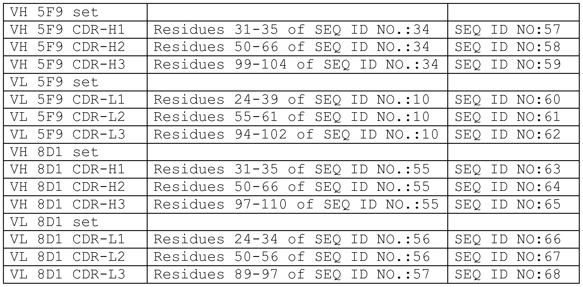


Claims
Priority Applications (13)
| Application Number | Priority Date | Filing Date | Title |
|---|---|---|---|
| PL10794938T PL2510001T3 (en) | 2009-12-08 | 2010-12-08 | Monoclonal antibodies against the rgm a protein for use in the treatment of retinal nerve fiber layer degeneration |
| BR112012013734A BR112012013734A2 (en) | 2009-12-08 | 2010-12-08 | monoclonal antibodies against the rgm a protein for use in the treatment of retinal nerve fiber layer degeneration. |
| CN2010800552755A CN102656190A (en) | 2009-12-08 | 2010-12-08 | Monoclonal antibodies against the RGM A protein for use in the treatment of retinal nerve fiber layer degeneration |
| MX2012006560A MX2012006560A (en) | 2009-12-08 | 2010-12-08 | Monoclonal antibodies against the rgm a protein for use in the treatment of retinal nerve fiber layer degeneration. |
| CA2780069A CA2780069C (en) | 2009-12-08 | 2010-12-08 | Monoclonal antibodies against the rgm a protein for use in the treatment of retinal nerve fiber layer degeneration |
| EP10794938.0A EP2510001B1 (en) | 2009-12-08 | 2010-12-08 | Monoclonal antibodies against the rgm a protein for use in the treatment of retinal nerve fiber layer degeneration |
| DK10794938.0T DK2510001T3 (en) | 2009-12-08 | 2010-12-08 | MONOCLONAL ANTIBODIES AGAINST RGM A PROTEIN USED TO TREAT DEGENERATION OF THE RETINAL NERVE FIBER LAYER |
| AU2010329955A AU2010329955A1 (en) | 2009-12-08 | 2010-12-08 | Monoclonal antibodies against the RGM A protein for use in the treatment of retinal nerve fiber layer degeneration |
| JP2012542530A JP5951498B2 (en) | 2009-12-08 | 2010-12-08 | Monoclonal antibody against RGMA protein for use in the treatment of retinal nerve fiber layer degeneration |
| ES10794938.0T ES2562832T3 (en) | 2009-12-08 | 2010-12-08 | Monoclonal antibodies against the RGM protein for use in the treatment of degeneration of the retinal nerve fiber layer |
| SG2012041935A SG181563A1 (en) | 2009-12-08 | 2010-12-08 | Monoclonal antibodies against the rgm a protein for use in the treatment of retinal nerve fiber layer degeneration |
| IL219766A IL219766A0 (en) | 2009-12-08 | 2012-05-13 | Monoclonal antibodies against the rgm a protein for use in the treatment of retinal nerve fiber layer degeneration |
| ZA2012/04106A ZA201204106B (en) | 2009-12-08 | 2012-06-05 | Monoclonal antibodies against the rgm a protein for use in the treatment of retinal nerve fiber layer degeneration |
Applications Claiming Priority (2)
| Application Number | Priority Date | Filing Date | Title |
|---|---|---|---|
| US26744609P | 2009-12-08 | 2009-12-08 | |
| US61/267,446 | 2009-12-08 |
Publications (1)
| Publication Number | Publication Date |
|---|---|
| WO2011070045A1 true WO2011070045A1 (en) | 2011-06-16 |
Family
ID=43806839
Family Applications (1)
| Application Number | Title | Priority Date | Filing Date |
|---|---|---|---|
| PCT/EP2010/069120 WO2011070045A1 (en) | 2009-12-08 | 2010-12-08 | Monoclonal antibodies against the rgm a protein for use in the treatment of retinal nerve fiber layer degeneration |
Country Status (17)
| Country | Link |
|---|---|
| US (4) | US9175075B2 (en) |
| EP (1) | EP2510001B1 (en) |
| JP (5) | JP5951498B2 (en) |
| KR (1) | KR20120118002A (en) |
| CN (2) | CN102656190A (en) |
| AU (1) | AU2010329955A1 (en) |
| BR (1) | BR112012013734A2 (en) |
| CA (1) | CA2780069C (en) |
| DK (1) | DK2510001T3 (en) |
| EC (1) | ECSP12012029A (en) |
| ES (1) | ES2562832T3 (en) |
| IL (1) | IL219766A0 (en) |
| MX (1) | MX2012006560A (en) |
| PL (1) | PL2510001T3 (en) |
| SG (1) | SG181563A1 (en) |
| WO (1) | WO2011070045A1 (en) |
| ZA (1) | ZA201204106B (en) |
Cited By (9)
| Publication number | Priority date | Publication date | Assignee | Title |
|---|---|---|---|---|
| WO2013113107A1 (en) * | 2012-02-03 | 2013-08-08 | University Health Network | Methods for promoting neuron survival, axonal growth and/or regeneration |
| KR20140125825A (en) * | 2012-01-27 | 2014-10-29 | 아비에 도이치란트 게엠베하 운트 콤파니 카게 | Composition and method for the diagnosis and treatment of disease associated with neurite degeneration |
| US8906864B2 (en) | 2005-09-30 | 2014-12-09 | AbbVie Deutschland GmbH & Co. KG | Binding domains of proteins of the repulsive guidance molecule (RGM) protein family and functional fragments thereof, and their use |
| US8962803B2 (en) | 2008-02-29 | 2015-02-24 | AbbVie Deutschland GmbH & Co. KG | Antibodies against the RGM A protein and uses thereof |
| WO2015039212A1 (en) * | 2013-09-17 | 2015-03-26 | University Health Network (Uhn): Technology Development And Commercialization | Agents directed against a cis rgma/neogenin interaction or lipid rafts and use of the same in methods of treatment |
| US9175075B2 (en) | 2009-12-08 | 2015-11-03 | AbbVie Deutschland GmbH & Co. KG | Methods of treating retinal nerve fiber layer degeneration with monoclonal antibodies against a retinal guidance molecule (RGM) protein |
| CN107459576A (en) * | 2011-12-14 | 2017-12-12 | 艾伯维德国有限责任两合公司 | For diagnosing and treating the composition and method of iron phase related disorders |
| EP3290441A4 (en) * | 2015-04-28 | 2018-10-17 | Mitsubishi Tanabe Pharma Corporation | RGMa BINDING PROTEIN AND USE THEREOF |
| WO2019136043A1 (en) * | 2018-01-03 | 2019-07-11 | Acceleron Pharma Inc. | Single-arm co-receptor fusion proteins and uses thereof |
Families Citing this family (6)
| Publication number | Priority date | Publication date | Assignee | Title |
|---|---|---|---|---|
| WO2002051438A2 (en) | 2000-12-22 | 2002-07-04 | MAX-PLANCK-Gesellschaft zur Förderung der Wissenschaften e.V. | Use of repulsive guidance molecule (rgm) and its modulators |
| CN104136462B (en) | 2011-12-14 | 2017-06-09 | 艾伯维德国有限责任两合公司 | Composition and method for diagnosing and treating iron phase related disorders |
| CN113267630A (en) * | 2014-09-10 | 2021-08-17 | 艾伯维德国有限责任两合公司 | RGMa fragment-based diagnostic assays |
| CA3106074A1 (en) | 2018-07-10 | 2020-01-16 | Mitsubishi Tanabe Pharma Corporation | Prevention or treatment method for peripheral neuropathy or pain accompanying disease in which peripheral neuropathy or astrocyte disorder is recognized |
| EP4138779A1 (en) * | 2020-04-21 | 2023-03-01 | University of Massachusetts | Methods and compositions for treatment of age-related macular degeneration |
| US20230382978A1 (en) * | 2020-10-15 | 2023-11-30 | The United States Of America, As Represented By The Secretary, Department Of Health & Human Services | Antibody specific for sars-cov-2 receptor binding domain and therapeutic methods |
Citations (104)
| Publication number | Priority date | Publication date | Assignee | Title |
|---|---|---|---|---|
| US2128208A (en) | 1937-02-27 | 1938-08-23 | Du Pont | Alkoxy derivatives of methacrylic acid esters and method of producing them |
| US4526938A (en) | 1982-04-22 | 1985-07-02 | Imperial Chemical Industries Plc | Continuous release formulations |
| EP0229246A2 (en) | 1986-01-15 | 1987-07-22 | ANT Nachrichtentechnik GmbH | Method for decoding digital signals, as well as a Viterbi decoder and applications |
| EP0239400A2 (en) | 1986-03-27 | 1987-09-30 | Medical Research Council | Recombinant antibodies and methods for their production |
| US4816397A (en) | 1983-03-25 | 1989-03-28 | Celltech, Limited | Multichain polypeptides or proteins and processes for their production |
| US4816567A (en) | 1983-04-08 | 1989-03-28 | Genentech, Inc. | Recombinant immunoglobin preparations |
| US4880078A (en) | 1987-06-29 | 1989-11-14 | Honda Giken Kogyo Kabushiki Kaisha | Exhaust muffler |
| WO1990002809A1 (en) | 1988-09-02 | 1990-03-22 | Protein Engineering Corporation | Generation and selection of recombinant varied binding proteins |
| WO1990005144A1 (en) | 1988-11-11 | 1990-05-17 | Medical Research Council | Single domain ligands, receptors comprising said ligands, methods for their production, and use of said ligands and receptors |
| US4946778A (en) | 1987-09-21 | 1990-08-07 | Genex Corporation | Single polypeptide chain binding molecules |
| WO1990014430A1 (en) | 1989-05-16 | 1990-11-29 | Scripps Clinic And Research Foundation | A new method for tapping the immunological repertoire |
| WO1990014443A1 (en) | 1989-05-16 | 1990-11-29 | Huse William D | Co-expression of heteromeric receptors |
| WO1990014424A1 (en) | 1989-05-16 | 1990-11-29 | Scripps Clinic And Research Foundation | Method for isolating receptors having a preselected specificity |
| US4980286A (en) | 1985-07-05 | 1990-12-25 | Whitehead Institute For Biomedical Research | In vivo introduction and expression of foreign genetic material in epithelial cells |
| WO1991005548A1 (en) | 1989-10-10 | 1991-05-02 | Pitman-Moore, Inc. | Sustained release composition for macromolecular proteins |
| WO1991009967A1 (en) | 1989-12-21 | 1991-07-11 | Celltech Limited | Humanised antibodies |
| WO1991010741A1 (en) | 1990-01-12 | 1991-07-25 | Cell Genesys, Inc. | Generation of xenogeneic antibodies |
| WO1991010737A1 (en) | 1990-01-11 | 1991-07-25 | Molecular Affinities Corporation | Production of antibodies using gene libraries |
| WO1991017271A1 (en) | 1990-05-01 | 1991-11-14 | Affymax Technologies N.V. | Recombinant library screening methods |
| WO1992001047A1 (en) | 1990-07-10 | 1992-01-23 | Cambridge Antibody Technology Limited | Methods for producing members of specific binding pairs |
| WO1992002551A1 (en) | 1990-08-02 | 1992-02-20 | B.R. Centre Limited | Methods for the production of proteins with a desired function |
| WO1992009690A2 (en) | 1990-12-03 | 1992-06-11 | Genentech, Inc. | Enrichment method for variant proteins with altered binding properties |
| US5128326A (en) | 1984-12-06 | 1992-07-07 | Biomatrix, Inc. | Drug delivery systems based on hyaluronans derivatives thereof and their salts and methods of producing same |
| WO1992015679A1 (en) | 1991-03-01 | 1992-09-17 | Protein Engineering Corporation | Improved epitode displaying phage |
| WO1992018619A1 (en) | 1991-04-10 | 1992-10-29 | The Scripps Research Institute | Heterodimeric receptor libraries using phagemids |
| WO1992019244A2 (en) | 1991-05-01 | 1992-11-12 | Henry M. Jackson Foundation For The Advancement Of Military Medicine | A method for treating infectious respiratory diseases |
| WO1992020791A1 (en) | 1990-07-10 | 1992-11-26 | Cambridge Antibody Technology Limited | Methods for producing members of specific binding pairs |
| WO1992022324A1 (en) | 1991-06-14 | 1992-12-23 | Xoma Corporation | Microbially-produced antibody fragments and their conjugates |
| EP0519596A1 (en) | 1991-05-17 | 1992-12-23 | Merck & Co. Inc. | A method for reducing the immunogenicity of antibody variable domains |
| WO1993001288A1 (en) | 1991-07-08 | 1993-01-21 | Deutsches Krebsforschungszentrum Stiftung des öffentlichen Rechts | Phagemide for screening antibodies |
| WO1993011236A1 (en) | 1991-12-02 | 1993-06-10 | Medical Research Council | Production of anti-self antibodies from antibody segment repertoires and displayed on phage |
| US5223409A (en) | 1988-09-02 | 1993-06-29 | Protein Engineering Corp. | Directed evolution of novel binding proteins |
| US5225539A (en) | 1986-03-27 | 1993-07-06 | Medical Research Council | Recombinant altered antibodies and methods of making altered antibodies |
| US5258498A (en) | 1987-05-21 | 1993-11-02 | Creative Biomolecules, Inc. | Polypeptide linkers for production of biosynthetic proteins |
| WO1994002602A1 (en) | 1992-07-24 | 1994-02-03 | Cell Genesys, Inc. | Generation of xenogeneic antibodies |
| EP0592106A1 (en) | 1992-09-09 | 1994-04-13 | Immunogen Inc | Resurfacing of rodent antibodies |
| WO1995015982A2 (en) | 1993-12-08 | 1995-06-15 | Genzyme Corporation | Process for generating specific antibodies |
| WO1995020401A1 (en) | 1994-01-31 | 1995-08-03 | Trustees Of Boston University | Polyclonal antibody libraries |
| US5516637A (en) | 1994-06-10 | 1996-05-14 | Dade International Inc. | Method involving display of protein binding pairs on the surface of bacterial pili and bacteriophage |
| US5530101A (en) | 1988-12-28 | 1996-06-25 | Protein Design Labs, Inc. | Humanized immunoglobulins |
| WO1996020698A2 (en) | 1995-01-05 | 1996-07-11 | The Board Of Regents Acting For And On Behalf Of The University Of Michigan | Surface-modified nanoparticles and method of making and using same |
| US5565332A (en) | 1991-09-23 | 1996-10-15 | Medical Research Council | Production of chimeric antibodies - a combinatorial approach |
| WO1996034096A1 (en) | 1995-04-28 | 1996-10-31 | Abgenix, Inc. | Human antibodies derived from immunized xenomice |
| WO1996033735A1 (en) | 1995-04-27 | 1996-10-31 | Abgenix, Inc. | Human antibodies derived from immunized xenomice |
| US5624821A (en) | 1987-03-18 | 1997-04-29 | Scotgen Biopharmaceuticals Incorporated | Antibodies with altered effector functions |
| WO1997029131A1 (en) | 1996-02-09 | 1997-08-14 | Basf Aktiengesellschaft | HUMAN ANTIBODIES THAT BIND HUMAN TNF$g(a) |
| WO1997032572A2 (en) | 1996-03-04 | 1997-09-12 | The Penn State Research Foundation | Materials and methods for enhancing cellular internalization |
| US5679377A (en) | 1989-11-06 | 1997-10-21 | Alkermes Controlled Therapeutics, Inc. | Protein microspheres and methods of using them |
| WO1997044013A1 (en) | 1996-05-24 | 1997-11-27 | Massachusetts Institute Of Technology | Aerodynamically light particles for pulmonary drug delivery |
| US5698426A (en) | 1990-09-28 | 1997-12-16 | Ixsys, Incorporated | Surface expression libraries of heteromeric receptors |
| US5714350A (en) | 1992-03-09 | 1998-02-03 | Protein Design Labs, Inc. | Increasing antibody affinity by altering glycosylation in the immunoglobulin variable region |
| US5714352A (en) | 1996-03-20 | 1998-02-03 | Xenotech Incorporated | Directed switch-mediated DNA recombination |
| US5723323A (en) | 1985-03-30 | 1998-03-03 | Kauffman; Stuart Alan | Method of identifying a stochastically-generated peptide, polypeptide, or protein having ligand binding property and compositions thereof |
| US5733743A (en) | 1992-03-24 | 1998-03-31 | Cambridge Antibody Technology Limited | Methods for producing members of specific binding pairs |
| WO1998016654A1 (en) | 1996-10-11 | 1998-04-23 | Japan Tobacco, Inc. | Production of a multimeric protein by cell fusion method |
| US5750753A (en) | 1996-01-24 | 1998-05-12 | Chisso Corporation | Method for manufacturing acryloxypropysilane |
| US5763192A (en) | 1986-11-20 | 1998-06-09 | Ixsys, Incorporated | Process for obtaining DNA, RNA, peptides, polypeptides, or protein, by recombinant DNA technique |
| WO1998024893A2 (en) | 1996-12-03 | 1998-06-11 | Abgenix, Inc. | TRANSGENIC MAMMALS HAVING HUMAN IG LOCI INCLUDING PLURAL VH AND Vλ REGIONS AND ANTIBODIES PRODUCED THEREFROM |
| US5766886A (en) | 1991-12-13 | 1998-06-16 | Xoma Corporation | Modified antibody variable domains |
| US5780225A (en) | 1990-01-12 | 1998-07-14 | Stratagene | Method for generating libaries of antibody genes comprising amplification of diverse antibody DNAs and methods for using these libraries for the production of diverse antigen combining molecules |
| WO1998031346A1 (en) | 1997-01-16 | 1998-07-23 | Massachusetts Institute Of Technology | Preparation of particles for inhalation |
| WO1998031700A1 (en) | 1997-01-21 | 1998-07-23 | The General Hospital Corporation | Selection of proteins using rna-protein fusions |
| US5807715A (en) | 1984-08-27 | 1998-09-15 | The Board Of Trustees Of The Leland Stanford Junior University | Methods and transformed mammalian lymphocyte cells for producing functional antigen-binding protein including chimeric immunoglobulin |
| WO1998050433A2 (en) | 1997-05-05 | 1998-11-12 | Abgenix, Inc. | Human monoclonal antibodies to epidermal growth factor receptor |
| US5855913A (en) | 1997-01-16 | 1999-01-05 | Massachusetts Instite Of Technology | Particles incorporating surfactants for pulmonary drug delivery |
| WO1999015154A1 (en) | 1997-09-24 | 1999-04-01 | Alkermes Controlled Therapeutics, Inc. | Methods for fabricating polymer-based controlled release preparations |
| WO1999020253A1 (en) | 1997-10-23 | 1999-04-29 | Bioglan Therapeutics Ab | Encapsulation method |
| WO1999025044A1 (en) | 1997-11-07 | 1999-05-20 | Nathan Cohen | Microstrip patch antenna with fractal structure |
| US5912015A (en) | 1992-03-12 | 1999-06-15 | Alkermes Controlled Therapeutics, Inc. | Modulated release from biocompatible polymers |
| US5916597A (en) | 1995-08-31 | 1999-06-29 | Alkermes Controlled Therapeutics, Inc. | Composition and method using solid-phase particles for sustained in vivo release of a biologically active agent |
| US5934272A (en) | 1993-01-29 | 1999-08-10 | Aradigm Corporation | Device and method of creating aerosolized mist of respiratory drug |
| WO1999045031A2 (en) | 1998-03-03 | 1999-09-10 | Abgenix, Inc. | Cd147 binding molecules as therapeutics |
| WO1999053049A1 (en) | 1998-04-15 | 1999-10-21 | Abgenix, Inc. | Epitope-driven human antibody production and gene expression profiling |
| WO1999054342A1 (en) | 1998-04-20 | 1999-10-28 | Pablo Umana | Glycosylation engineering of antibodies for improving antibody-dependent cellular cytotoxicity |
| US5985309A (en) | 1996-05-24 | 1999-11-16 | Massachusetts Institute Of Technology | Preparation of particles for inhalation |
| US5998209A (en) | 1995-04-21 | 1999-12-07 | Abgenix, Inc. | Generation of large genomic DNA deletions |
| WO1999066903A2 (en) | 1998-06-24 | 1999-12-29 | Advanced Inhalation Research, Inc. | Large porous particles emitted from an inhaler |
| US6019968A (en) | 1995-04-14 | 2000-02-01 | Inhale Therapeutic Systems, Inc. | Dispersible antibody compositions and methods for their preparation and use |
| WO2000009560A2 (en) | 1998-08-17 | 2000-02-24 | Abgenix, Inc. | Generation of modified molecules with increased serum half-lives |
| US6075181A (en) | 1990-01-12 | 2000-06-13 | Abgenix, Inc. | Human antibodies derived from immunized xenomice |
| WO2000037504A2 (en) | 1998-12-23 | 2000-06-29 | Pfizer Inc. | Human monoclonal antibodies to ctla-4 |
| US6091001A (en) | 1995-03-29 | 2000-07-18 | Abgenix, Inc. | Production of antibodies using Cre-mediated site-specific recombination |
| WO2000056772A1 (en) | 1999-03-25 | 2000-09-28 | Knoll Gmbh | Human antibodies that bind human il-12 and methods for producing |
| US6130364A (en) | 1995-03-29 | 2000-10-10 | Abgenix, Inc. | Production of antibodies using Cre-mediated site-specific recombination |
| US6204023B1 (en) | 1985-11-01 | 2001-03-20 | Xoma Ltd. | Modular assembly of antibody genes, antibodies prepared thereby and use |
| EP1176195A1 (en) | 1999-04-09 | 2002-01-30 | Kyowa Hakko Kogyo Co., Ltd. | Method for controlling the activity of immunologically functional molecule |
| WO2002072636A2 (en) | 2000-12-28 | 2002-09-19 | Altus Biologics Inc. | Crystals of whole antibodies and fragments thereof and methods for making and using them |
| US20020137134A1 (en) | 2000-06-28 | 2002-09-26 | Gerngross Tillman U. | Methods for producing modified glycoproteins |
| WO2003016466A2 (en) | 2001-08-17 | 2003-02-27 | Eli Lilly And Company | ANTI-Aβ ANTIBODIES |
| WO2003035835A2 (en) | 2001-10-25 | 2003-05-01 | Genentech, Inc. | Glycoprotein compositions |
| US20030186374A1 (en) | 2001-10-01 | 2003-10-02 | Hufton Simon E. | Multi-chain eukaryotic display vectors and uses thereof |
| US20040018590A1 (en) | 2000-06-28 | 2004-01-29 | Gerngross Tillman U. | Combinatorial DNA library for producing modified N-glycans in lower eukaryotes |
| US6699658B1 (en) | 1996-05-31 | 2004-03-02 | Board Of Trustees Of The University Of Illinois | Yeast cell surface display of proteins and uses thereof |
| WO2004078140A2 (en) | 2003-03-05 | 2004-09-16 | Halozyme, Inc. | SOLUBLE HYALURONIDASE GLYCOPROTEIN (sHASEGP), PROCESS FOR PREPARING THE SAME, USES AND PHARMACEUTICAL COMPOSITIONS COMPRISING THEREOF |
| US20050042664A1 (en) | 2003-08-22 | 2005-02-24 | Medimmune, Inc. | Humanization of antibodies |
| WO2005100584A2 (en) | 2004-04-15 | 2005-10-27 | Glycofi, Inc. | Production of galactosylated glycoproteins in lower eukaryotes |
| US20060104968A1 (en) | 2003-03-05 | 2006-05-18 | Halozyme, Inc. | Soluble glycosaminoglycanases and methods of preparing and using soluble glycosaminogly ycanases |
| WO2008082651A2 (en) | 2006-12-29 | 2008-07-10 | Abbott Laboratories | Dual-specific il-1a/ il-1b antibodies |
| WO2009106356A1 (en) * | 2008-02-29 | 2009-09-03 | Abbott Gmbh & Co. Kg | Monoclonal antibodies against the rgm a protein and uses thereof |
| US9105939B2 (en) | 2008-03-10 | 2015-08-11 | Nissan Motor Co., Ltd. | Battery with battery electrode and method of manufacturing same |
| US9109630B2 (en) | 2013-01-15 | 2015-08-18 | Jtekt Corporation | Rolling bearing unit |
| US9401234B2 (en) | 2013-03-22 | 2016-07-26 | Polytronics Technology Corp. | Over-current protection device |
| US9618978B2 (en) | 2013-10-03 | 2017-04-11 | Acer Incorporated | Methods for controlling a touch panel and portable computers using the same |
| US9816280B1 (en) | 2016-11-02 | 2017-11-14 | Matthew Reitnauer | Portable floor |
Family Cites Families (241)
| Publication number | Priority date | Publication date | Assignee | Title |
|---|---|---|---|---|
| US4394448A (en) * | 1978-02-24 | 1983-07-19 | Szoka Jr Francis C | Method of inserting DNA into living cells |
| US5179017A (en) * | 1980-02-25 | 1993-01-12 | The Trustees Of Columbia University In The City Of New York | Processes for inserting DNA into eucaryotic cells and for producing proteinaceous materials |
| US4399216A (en) | 1980-02-25 | 1983-08-16 | The Trustees Of Columbia University | Processes for inserting DNA into eucaryotic cells and for producing proteinaceous materials |
| US4634665A (en) * | 1980-02-25 | 1987-01-06 | The Trustees Of Columbia University In The City Of New York | Processes for inserting DNA into eucaryotic cells and for producing proteinaceous materials |
| US4554101A (en) | 1981-01-09 | 1985-11-19 | New York Blood Center, Inc. | Identification and preparation of epitopes on antigens and allergens on the basis of hydrophilicity |
| US4510245A (en) * | 1982-11-18 | 1985-04-09 | Chiron Corporation | Adenovirus promoter system |
| US5168062A (en) | 1985-01-30 | 1992-12-01 | University Of Iowa Research Foundation | Transfer vectors and microorganisms containing human cytomegalovirus immediate-early promoter-regulatory DNA sequence |
| US4968615A (en) | 1985-12-18 | 1990-11-06 | Ciba-Geigy Corporation | Deoxyribonucleic acid segment from a virus |
| US5635600A (en) | 1986-07-07 | 1997-06-03 | Trustees Of Dartmouth College | Bifunctional and heteroantibodies specific for the high affinity Fc receptor for immunoglobulin G on human mononuclear phagocytes |
| US4704692A (en) | 1986-09-02 | 1987-11-03 | Ladner Robert C | Computer based system and method for determining and displaying possible chemical structures for converting double- or multiple-chain polypeptides to single-chain polypeptides |
| DE3750503T2 (en) | 1986-10-22 | 1995-02-09 | Abbott Lab | Chemiluminescent acridinium and phenantridinium salts. |
| US4987071A (en) | 1986-12-03 | 1991-01-22 | University Patents, Inc. | RNA ribozyme polymerases, dephosphorylases, restriction endoribonucleases and methods |
| EP0279582A3 (en) * | 1987-02-17 | 1989-10-18 | Pharming B.V. | Dna sequences to target proteins to the mammary gland for efficient secretion |
| US4873316A (en) | 1987-06-23 | 1989-10-10 | Biogen, Inc. | Isolation of exogenous recombinant proteins from the milk of transgenic mammals |
| ATE115999T1 (en) | 1987-12-15 | 1995-01-15 | Gene Shears Pty Ltd | RIBOZYMES. |
| US5006309A (en) * | 1988-04-22 | 1991-04-09 | Abbott Laboratories | Immunoassay device with liquid transfer between wells by washing |
| US5089424A (en) * | 1988-06-14 | 1992-02-18 | Abbott Laboratories | Method and apparatus for heterogeneous chemiluminescence assay |
| CA1340323C (en) | 1988-09-20 | 1999-01-19 | Arnold E. Hampel | Rna catalyst for cleaving specific rna sequences |
| US5241070A (en) | 1988-09-26 | 1993-08-31 | Ciba Corning Diagnostics Corp. | Nucleophilic polysubstituted aryl acridinium esters and uses thereof |
| GB8826530D0 (en) | 1988-11-12 | 1988-12-14 | Ped Capacitors Ltd | Electrical capacitors |
| US5063081A (en) | 1988-11-14 | 1991-11-05 | I-Stat Corporation | Method of manufacturing a plurality of uniform microfabricated sensing devices having an immobilized ligand receptor |
| US5939272A (en) | 1989-01-10 | 1999-08-17 | Biosite Diagnostics Incorporated | Non-competitive threshold ligand-receptor assays |
| US5028535A (en) | 1989-01-10 | 1991-07-02 | Biosite Diagnostics, Inc. | Threshold ligand-receptor assay |
| GB8901334D0 (en) | 1989-01-21 | 1989-03-15 | Oakland Design Products Ltd | Improvements relating to broadhead arrows |
| US5703055A (en) | 1989-03-21 | 1997-12-30 | Wisconsin Alumni Research Foundation | Generation of antibodies through lipid mediated DNA delivery |
| DE3920358A1 (en) | 1989-06-22 | 1991-01-17 | Behringwerke Ag | BISPECIFIC AND OLIGO-SPECIFIC, MONO- AND OLIGOVALENT ANTI-BODY CONSTRUCTS, THEIR PRODUCTION AND USE |
| US5139400A (en) | 1989-10-11 | 1992-08-18 | Ide Russell D | Progressive cavity drive train |
| FR2656431B1 (en) | 1989-12-22 | 1994-06-10 | Essilor Int | METHOD AND SOLUTION FOR DECONTAMINATING A FLEXIBLE LENS, ESPECIALLY OF THE HYDROPHILIC TYPE. |
| US5922615A (en) | 1990-03-12 | 1999-07-13 | Biosite Diagnostics Incorporated | Assay devices comprising a porous capture membrane in fluid-withdrawing contact with a nonabsorbent capillary network |
| US5135875A (en) | 1990-08-15 | 1992-08-04 | Abbott Laboratories | Protein precipitation reagent |
| CA2048302A1 (en) | 1990-08-15 | 1992-02-16 | Victoria P. Meucci | Solubilization reagent for biological test samples |
| US5814318A (en) | 1990-08-29 | 1998-09-29 | Genpharm International Inc. | Transgenic non-human animals for producing heterologous antibodies |
| US5633425A (en) * | 1990-08-29 | 1997-05-27 | Genpharm International, Inc. | Transgenic non-human animals capable of producing heterologous antibodies |
| US5625126A (en) * | 1990-08-29 | 1997-04-29 | Genpharm International, Inc. | Transgenic non-human animals for producing heterologous antibodies |
| JP2938569B2 (en) | 1990-08-29 | 1999-08-23 | ジェンファーム インターナショナル,インコーポレイティド | Method for producing xenogeneic immunoglobulin and transgenic mouse |
| US5770429A (en) * | 1990-08-29 | 1998-06-23 | Genpharm International, Inc. | Transgenic non-human animals capable of producing heterologous antibodies |
| US5545806A (en) | 1990-08-29 | 1996-08-13 | Genpharm International, Inc. | Ransgenic non-human animals for producing heterologous antibodies |
| US5661016A (en) | 1990-08-29 | 1997-08-26 | Genpharm International Inc. | Transgenic non-human animals capable of producing heterologous antibodies of various isotypes |
| US6255458B1 (en) | 1990-08-29 | 2001-07-03 | Genpharm International | High affinity human antibodies and human antibodies against digoxin |
| US5877397A (en) | 1990-08-29 | 1999-03-02 | Genpharm International Inc. | Transgenic non-human animals capable of producing heterologous antibodies of various isotypes |
| JPH05504841A (en) * | 1990-09-14 | 1993-07-22 | バイオサイト・ダイアグノスティックス・インコーポレイテッド | Antibodies against ligand receptors and ligand complexes and their use in ligand-receptor assays |
| GB9101134D0 (en) | 1991-01-18 | 1991-02-27 | R B S | Improvements in and relating to accommodation for animals |
| EP0585310B1 (en) * | 1991-04-10 | 1999-03-17 | Biosite Diagnostics Inc. | Crosstalk inhibitors and their uses |
| DE69231382T2 (en) | 1991-04-12 | 2001-01-25 | Biosite Diagnostics Inc | NEW CONJUGATES AND TEST PROCEDURES FOR THE SIMULTANEOUS DETERMINATION OF MULTIPLE LIGANDS |
| CA2069530A1 (en) * | 1991-06-03 | 1992-12-04 | Cass J. Grandone | Reagent pack for immunoassays |
| EP0605522B1 (en) | 1991-09-23 | 1999-06-23 | Medical Research Council | Methods for the production of humanized antibodies |
| GB9201755D0 (en) | 1992-01-28 | 1992-03-11 | British Bio Technology | Compounds |
| WO1993020442A1 (en) * | 1992-03-30 | 1993-10-14 | Abbott Laboratories | Reagents and methods for the detection and quantification of thyroxine in fluid samples |
| US5352803A (en) | 1992-03-30 | 1994-10-04 | Abbott Laboratories | 5(6)-methyl substituted fluorescein derivatives |
| US6143576A (en) | 1992-05-21 | 2000-11-07 | Biosite Diagnostics, Inc. | Non-porous diagnostic devices for the controlled movement of reagents |
| US6019944A (en) | 1992-05-21 | 2000-02-01 | Biosite Diagnostics, Inc. | Diagnostic devices and apparatus for the controlled movement of reagents without membranes |
| ATE174824T1 (en) | 1992-07-10 | 1999-01-15 | Horst Warneke | PRODUCTION LINE FOR PRODUCING A STEEL CASSETTE FOR CEILING AND/OR WALL CONSTRUCTIONS FROM A METAL SHEET |
| ATE301201T1 (en) | 1993-06-07 | 2005-08-15 | Vical Inc | PLASMIDS USABLE FOR GENE THERAPY |
| US5824799A (en) | 1993-09-24 | 1998-10-20 | Biosite Diagnostics Incorporated | Hybrid phthalocyanine derivatives and their uses |
| US5565352A (en) | 1993-11-24 | 1996-10-15 | Arch Development Corporation | Deubiquitinating enzyme: compositions and methods |
| US5827690A (en) | 1993-12-20 | 1998-10-27 | Genzyme Transgenics Corporatiion | Transgenic production of antibodies in milk |
| JPH09510201A (en) | 1994-03-07 | 1997-10-14 | メダレツクス・インコーポレーテツド | Bispecific molecules with clinical utility |
| US5525490A (en) * | 1994-03-29 | 1996-06-11 | Onyx Pharmaceuticals, Inc. | Reverse two-hybrid method |
| US5541109A (en) | 1994-04-19 | 1996-07-30 | Rhone-Poulenc Rorer Pharmaceuticals Inc. | Expression cloning of c-src SH3-domain binding proteins |
| WO1996002561A1 (en) | 1994-07-20 | 1996-02-01 | The General Hospital Corporation | Interaction trap systems for detecting protein interactions |
| US6111166A (en) | 1994-09-19 | 2000-08-29 | Medarex, Incorporated | Transgenic mice expressing human Fcα and β receptors |
| CA2203888C (en) | 1994-10-27 | 2011-08-23 | Ingrid W. Caras | Al-1 neurotrophic factor, a ligand for an eph-related tyrosine kinase receptor |
| GB9425232D0 (en) | 1994-12-14 | 1995-02-08 | Secr Defence | Method of authenticating watermarked paper |
| US6410690B1 (en) | 1995-06-07 | 2002-06-25 | Medarex, Inc. | Therapeutic compounds comprised of anti-Fc receptor antibodies |
| AU723325B2 (en) | 1995-06-23 | 2000-08-24 | President And Fellows Of Harvard College | Transcriptional regulation of genes encoding vascular endothelial growth factor receptors |
| EP0859841B1 (en) | 1995-08-18 | 2002-06-19 | MorphoSys AG | Protein/(poly)peptide libraries |
| US5747262A (en) | 1995-10-16 | 1998-05-05 | The Regents Of The University Of California | Neurological drug screens |
| US20020136725A1 (en) | 1996-01-17 | 2002-09-26 | Smithkline Beecham Corporation | Antithrombotic agents |
| US5994619A (en) | 1996-04-01 | 1999-11-30 | University Of Massachusetts, A Public Institution Of Higher Education Of The Commonwealth Of Massachusetts, As Represented By Its Amherst Campus | Production of chimeric bovine or porcine animals using cultured inner cell mass cells |
| US6300065B1 (en) | 1996-05-31 | 2001-10-09 | Board Of Trustees Of The University Of Illinois | Yeast cell surface display of proteins and uses thereof |
| US5922845A (en) | 1996-07-11 | 1999-07-13 | Medarex, Inc. | Therapeutic multispecific compounds comprised of anti-Fcα receptor antibodies |
| FR2754461B1 (en) | 1996-10-16 | 1999-02-12 | Husson Olivier | MANUALLY REMOVABLE ATTACHMENT FOR IN-LINE SKATE WITH A HOLDING LEG THAT CONTROLS A BRAKING SYSTEM |
| US6113855A (en) | 1996-11-15 | 2000-09-05 | Biosite Diagnostics, Inc. | Devices comprising multiple capillarity inducing surfaces |
| CA2274608C (en) | 1996-12-11 | 2007-06-26 | Bristol-Myers Squibb Company | Prokaryotic two-hybrid system |
| US6017517A (en) | 1996-12-18 | 2000-01-25 | The Dial Corporation | Method for treating human nails |
| US7368531B2 (en) | 1997-03-07 | 2008-05-06 | Human Genome Sciences, Inc. | Human secreted proteins |
| US5947124A (en) | 1997-03-11 | 1999-09-07 | Biosite Diagnostics Incorporated | Diagnostic for determining the time of a heart attack |
| JP2002514919A (en) | 1997-04-04 | 2002-05-21 | バイオサイト ダイアグノスティックス,インコーポレイテッド | Multivalent and polyclonal libraries |
| AU7171098A (en) | 1997-05-01 | 1998-11-24 | Board Of Regents, The University Of Texas System | Directed evolution of enzymes and antibodies |
| IT1294449B1 (en) | 1997-07-02 | 1999-03-24 | F B C Di Giuliano Frati & C Sn | SPORTS FOOTWEAR STRUCTURE AND METHODS FOR IMPLEMENTING THE SAME IN PARTICULAR FOR SINGLE-ROW AND SHORTRACKING SKATES. |
| US6440455B1 (en) | 1997-09-02 | 2002-08-27 | Children's Medical Center Corporation | Methods for modulating the axonal outgrowth of central nervous system neurons |
| CA2311574A1 (en) | 1997-12-02 | 1999-06-10 | Medarex, Inc. | Cells expressing anti-fc receptor binding components |
| US6464686B1 (en) * | 1998-01-21 | 2002-10-15 | Abbott Laboratories | Polyurethane feeding tube and associated adaptors |
| JP2002510490A (en) | 1998-04-03 | 2002-04-09 | キュラジェン コーポレイション | LYST protein complex and LYST interacting protein |
| TWI242563B (en) | 1998-04-30 | 2005-11-01 | Tanox Inc | Monoclonal agonist antibodies which specifically bind to or interact with human G-CSF receptor |
| JP2002521004A (en) | 1998-07-10 | 2002-07-16 | キュラゲン コーポレイション | Interaction of human β-amidoid precursor protein (β-APP) with human LON protease-like protein (HsLON) |
| IL128017A0 (en) | 1998-07-22 | 1999-11-30 | Technion Res & Dev Foundation | Method for detecting protein-protein interactions and a kit therefor |
| CA2341314A1 (en) | 1998-09-03 | 2000-03-16 | Loma Linda University | Method for studying protein interactions in vivo |
| WO2000017221A1 (en) | 1998-09-24 | 2000-03-30 | Duke University | Method of measuring protein-protein interactions in living cells |
| US6660843B1 (en) | 1998-10-23 | 2003-12-09 | Amgen Inc. | Modified peptides as therapeutic agents |
| EP1135498B1 (en) | 1998-11-18 | 2008-01-23 | Genentech, Inc. | Antibody variants with higher binding affinity compared to parent antibodies |
| US6231768B1 (en) | 1999-01-19 | 2001-05-15 | Nalco Chemical Company | Rheology modification of settled solids in mineral processing |
| US6914128B1 (en) | 1999-03-25 | 2005-07-05 | Abbott Gmbh & Co. Kg | Human antibodies that bind human IL-12 and methods for producing |
| JP2003518364A (en) | 1999-05-28 | 2003-06-10 | ルードヴィッヒ インスティテュート フォー キャンサー リサーチ | Breast, gastric and prostate cancer-related antigens and uses thereof |
| EP1130094A3 (en) | 1999-07-08 | 2001-11-21 | Helix Research Institute | Primers for synthesizing full length cDNA clones and their use |
| EP2829609A1 (en) | 1999-08-24 | 2015-01-28 | E. R. Squibb & Sons, L.L.C. | Human CTLA-4 antibodies and their uses |
| GB9930001D0 (en) * | 1999-12-21 | 2000-02-09 | Phaeton Research Ltd | An ingestible device |
| US7119165B2 (en) | 2000-01-12 | 2006-10-10 | Yale University | Nogo receptor-mediated blockade of axonal growth |
| CA2396719A1 (en) | 2000-01-31 | 2001-08-02 | Human Genome Sciences, Inc. | 22 human secreted proteins |
| EP2360184A3 (en) | 2000-02-10 | 2011-11-16 | Abbott Laboratories | Antibodies that bind human interleukin-18 and methods of making and using |
| US20030235584A1 (en) | 2000-02-28 | 2003-12-25 | Kloetzer William S. | Method for preparing anti-MIF antibodies |
| US6800455B2 (en) | 2000-03-31 | 2004-10-05 | Scios Inc. | Secreted factors |
| WO2001077342A1 (en) * | 2000-04-11 | 2001-10-18 | Genentech, Inc. | Multivalent antibodies and uses therefor |
| AU2001259432B2 (en) | 2000-05-03 | 2005-04-21 | Amgen Inc. | Modified peptides, comprising an FC domain, as therapeutic agents |
| AU2001274888A1 (en) * | 2000-05-19 | 2001-12-03 | Human Genome Sciences, Inc. | Nucleic acids, proteins, and antibodies |
| ATE420958T1 (en) | 2000-06-29 | 2009-01-15 | Abbott Lab | ANTIBODIES WITH DUAL SPECIFICITIES AND METHOD FOR THE PRODUCTION AND USE THEREOF |
| MXPA03000506A (en) | 2000-07-18 | 2004-09-10 | Correlogic Systems Inc | A process for discriminating between biological states based on hidden patterns from biological data. |
| CA2427310C (en) * | 2000-11-06 | 2013-12-17 | Cancer Research Technology Limited | Imaging, diagnosis and treatment of angiogenesis-related diseases comprising ecsm4 |
| WO2002051438A2 (en) | 2000-12-22 | 2002-07-04 | MAX-PLANCK-Gesellschaft zur Förderung der Wissenschaften e.V. | Use of repulsive guidance molecule (rgm) and its modulators |
| US20060084794A1 (en) | 2001-04-12 | 2006-04-20 | Human Genome Sciences, Inc. | Albumin fusion proteins |
| US6890763B2 (en) | 2001-04-30 | 2005-05-10 | Syn X Pharma, Inc. | Biopolymer marker indicative of disease state having a molecular weight of 1350 daltons |
| US20100036502A1 (en) * | 2008-08-07 | 2010-02-11 | Exogenesis Corporation | Medical device for bone implant and method for producing such device |
| CA2388761A1 (en) * | 2001-06-18 | 2002-12-18 | Pfizer Inc. | Wound healing biomarkers |
| US20040248249A1 (en) | 2001-07-05 | 2004-12-09 | Tran Uyen K | Secreted proteins |
| WO2003020894A2 (en) * | 2001-08-31 | 2003-03-13 | Joslin Diabetes Center, Inc. | Insulin related transcription factor and uses thereof |
| US20040142325A1 (en) | 2001-09-14 | 2004-07-22 | Liat Mintz | Methods and systems for annotating biomolecular sequences |
| JP2005536180A (en) | 2001-12-03 | 2005-12-02 | アブジェニックス・インコーポレーテッド | Anti-CD45RB antibodies for use in the treatment of autoimmune diseases and the treatment of transplant rejection |
| US8435529B2 (en) * | 2002-06-14 | 2013-05-07 | Immunomedics, Inc. | Combining radioimmunotherapy and antibody-drug conjugates for improved cancer therapy |
| US7419821B2 (en) | 2002-03-05 | 2008-09-02 | I-Stat Corporation | Apparatus and methods for analyte measurement and immunoassay |
| US7193069B2 (en) | 2002-03-22 | 2007-03-20 | Research Association For Biotechnology | Full-length cDNA |
| WO2003080659A1 (en) | 2002-03-27 | 2003-10-02 | Theratechnologies Inc. | Methods and compounds for prevention and treatment of elevated intraocular pressure and related conditions |
| WO2003089608A2 (en) * | 2002-04-18 | 2003-10-30 | The General Hospital Corporation | Drg11-responsive (dragon) gene family |
| AU2003239409A1 (en) | 2002-05-09 | 2003-11-11 | Surromed, Inc. | Methods for time-alignment of liquid chromatography-mass spectrometry data |
| IL149820A0 (en) | 2002-05-23 | 2002-11-10 | Curetech Ltd | Humanized immunomodulatory monoclonal antibodies for the treatment of neoplastic disease or immunodeficiency |
| US7439063B2 (en) * | 2002-06-11 | 2008-10-21 | Burnham Institute For Medical Research | Neuroprotective synergy of erythropoietin and insulin-like growth factors |
| WO2004003150A2 (en) | 2002-06-26 | 2004-01-08 | Yale University | Modulators and modulation of the interacton between rgm and neogenin |
| EP1380644A1 (en) | 2002-07-08 | 2004-01-14 | Kylix B.V. | The use of specified TCF target genes to identify drugs for the treatment of cancer, in particular colorectal cancer, in which TCF/beta-catenin/WNT signalling plays a central role |
| US20040018577A1 (en) * | 2002-07-29 | 2004-01-29 | Emerson Campbell John Lewis | Multiple hybrid immunoassay |
| TWI323265B (en) | 2002-08-06 | 2010-04-11 | Glaxo Group Ltd | Antibodies |
| US7541440B2 (en) * | 2002-09-30 | 2009-06-02 | Immunomedics, Inc. | Chimeric, human and humanized anti-granulocyte antibodies and methods of use |
| US20050013809A1 (en) * | 2002-12-02 | 2005-01-20 | Owens Samuel M. | Antibodies against drugs of abuse |
| CN1878795A (en) * | 2002-12-02 | 2006-12-13 | 阿布格尼克斯公司 | Antibodies directed to phospholipase A2 and uses thereof |
| EP1440981A3 (en) | 2003-01-21 | 2005-11-23 | Research Association for Biotechnology | Full-length human cdna |
| DE10303974A1 (en) | 2003-01-31 | 2004-08-05 | Abbott Gmbh & Co. Kg | Amyloid β (1-42) oligomers, process for their preparation and their use |
| GB0306309D0 (en) | 2003-03-19 | 2003-04-23 | Glaxo Group Ltd | Method of treatment |
| JP2006523440A (en) * | 2003-04-15 | 2006-10-19 | ゼノン ファーマスーティカルス,インコーポレイテッド | Juvenile hemochromatosis gene (HFE2A), its expression product and method of use |
| PL2829283T3 (en) | 2003-04-30 | 2017-12-29 | Universität Zürich | Methods for treating cancer using an immunotoxin |
| EA008253B1 (en) | 2003-08-07 | 2007-04-27 | Байоджен Айдек Ма Инк. | Nogo receptor antagonists |
| CA2535007A1 (en) | 2003-08-07 | 2005-02-24 | Biogen Idec Ma Inc. | Nogo receptor antagonists |
| CA2535156A1 (en) | 2003-08-08 | 2005-02-24 | Abgenix, Inc. | Antibodies directed to parathyroid hormone (pth) and uses thereof |
| US20060003391A1 (en) | 2003-08-11 | 2006-01-05 | Ring Brian Z | Reagents and methods for use in cancer diagnosis, classification and therapy |
| US7723099B2 (en) | 2003-09-10 | 2010-05-25 | Abbott Point Of Care Inc. | Immunoassay device with immuno-reference electrode |
| US7682833B2 (en) * | 2003-09-10 | 2010-03-23 | Abbott Point Of Care Inc. | Immunoassay device with improved sample closure |
| WO2005047328A2 (en) * | 2003-11-07 | 2005-05-26 | Curagen Corporation | Antibodies against secretoryleukocyte protease inhibitor |
| RU2362780C2 (en) | 2003-12-22 | 2009-07-27 | Глаксо Груп Лимитед | Nogo-a-neutralising immunoglobulins for treatment of neurological diseases |
| WO2005061554A1 (en) | 2003-12-24 | 2005-07-07 | Idemitsu Kosan Co., Ltd. | Method and apparatus for manufacturing polyolefin |
| CA2559069A1 (en) | 2004-03-11 | 2005-09-22 | Bioclues, Inc. | Axon regeneration promoter |
| US20060063208A1 (en) * | 2004-08-02 | 2006-03-23 | Woolf Clifford J | DRG11-responsive (DRAGON) gene and uses thereof |
| WO2006021955A2 (en) * | 2004-08-23 | 2006-03-02 | Mor Research Applications Ltd. | Use of bat monoclonal antibody for immunotherapy |
| CA2577102A1 (en) | 2004-08-25 | 2006-03-09 | Genentech, Inc. | Novel gene disruptions, compositions and methods relating thereto |
| AU2005307765C1 (en) * | 2004-11-16 | 2012-07-05 | Trustees Of Boston University | Roles for Dual Endothelin-1/Angiotensin II Receptor (DEAR) in hypertension and angiogenesis |
| EP2329838A3 (en) | 2004-11-22 | 2013-02-13 | Centre National de la Recherche Scientifique | Mutated netrin 4, fragments thereof and uses thereof as drugs |
| TW200635608A (en) | 2004-12-15 | 2006-10-16 | Neuralab Ltd | Aβ antibodies for use in improving cognition |
| EP1677113A1 (en) | 2004-12-29 | 2006-07-05 | Max-Delbrück-Centrum für Molekulare Medizin (MDC) | Method for the identification of protein-protein interactions in disease related protein networks |
| US7968091B2 (en) * | 2005-02-16 | 2011-06-28 | The General Hospital Corporation | Methods and compositions to regulate iron metabolism |
| EP1861422B1 (en) | 2005-03-05 | 2010-02-24 | Abbott GmbH & Co. KG | Screening method, process for purifying of non-diffusible a-beta oligomers, selective antibodies against said non-diffusible a-beta oligomers and a process for manufacturing of said antibodies |
| US7504225B2 (en) | 2005-05-12 | 2009-03-17 | Applied Genomics, Inc. | Reagents and methods for use in cancer diagnosis, classification and therapy |
| US8609345B2 (en) | 2005-05-25 | 2013-12-17 | Expression Pathology Incorporated | Diagnosis of diseases and conditions by analysis of histopathologically processed biological samples using liquid tissue preparations |
| EP2478917A1 (en) * | 2005-07-08 | 2012-07-25 | Biogen Idec MA Inc. | SP35 antibodies and uses thereof |
| EP2500352A1 (en) | 2005-08-19 | 2012-09-19 | Abbott Laboratories | Dual variable domain immunoglobulin and uses thereof |
| US7612181B2 (en) * | 2005-08-19 | 2009-11-03 | Abbott Laboratories | Dual variable domain immunoglobulin and uses thereof |
| CN101277974A (en) | 2005-09-30 | 2008-10-01 | 阿伯特有限及两合公司 | Binding domains of proteins of the repulsive guidance molecule (RGM) protein family and functional fragments thereof, and their use |
| WO2007042809A2 (en) | 2005-10-11 | 2007-04-19 | Domantis Limited | Antibody polypeptide library screening and selected antibody polypeptides |
| US8865763B2 (en) * | 2005-10-14 | 2014-10-21 | Alltech, Inc. | Methods and compositions for altering cell function |
| US8871715B2 (en) * | 2005-10-14 | 2014-10-28 | Alltech, Inc. | Use of selenium compounds, especially selenium yeasts for altering cognitive function |
| US20070155687A1 (en) | 2005-10-14 | 2007-07-05 | Alltech, Inc. | Methods and compositions for altering cell function |
| US20080004255A1 (en) * | 2005-10-14 | 2008-01-03 | Alltech, Inc. | Methods and compositions for altering cell function |
| JP2009514555A (en) | 2005-11-08 | 2009-04-09 | ユークリッド ダイアグノスティックス リミテッド ライアビリティー カンパニー | Materials and Methods for Gene-Related CpG Island Methylation Assays for Cancer Evaluation |
| WO2007058671A1 (en) | 2005-11-21 | 2007-05-24 | West Michael D | Novel uses of cells with prenatal patterns of gene expression |
| PT1954718E (en) | 2005-11-30 | 2014-12-16 | Abbvie Inc | Anti-a globulomer antibodies, antigen-binding moieties thereof, corresponding hybridomas, nucleic acids, vectors, host cells, methods of producing said antibodies, compositions comprising said antibodies, uses of said antibodies and methods of using said antibodies |
| AR056857A1 (en) | 2005-12-30 | 2007-10-24 | U3 Pharma Ag | DIRECTED ANTIBODIES TO HER-3 (RECEIVER OF THE HUMAN EPIDERMAL GROWTH FACTOR-3) AND ITS USES |
| US7420040B2 (en) | 2006-02-24 | 2008-09-02 | Arius Research Inc. | Cytotoxicity mediation of cells evidencing surface expression of TROP-2 |
| CN104623692A (en) | 2006-03-08 | 2015-05-20 | 阿切埃米克斯股份有限公司 | Complement binding aptamers and anti-C5 agents useful in the treatment of ocular disorders |
| WO2007106507A2 (en) | 2006-03-14 | 2007-09-20 | Petrie Howard T | Detection of gene expression in mixed sample or tissue |
| EP1865071A1 (en) | 2006-06-09 | 2007-12-12 | Rheinische Friedrich-Wilhelms-Universität Bonn | Method for early diagnosis of proliferative diabetic retinopathy |
| US7883855B2 (en) * | 2006-07-21 | 2011-02-08 | Abbott Laboratories | Immunosuppressant drug extraction reagent for immunoassays |
| CA2659231A1 (en) | 2006-07-28 | 2008-01-31 | Chundsell Medicals Ab | Embryonic stem cell markers for cancer diagnosis and prognosis |
| JPWO2008038599A1 (en) | 2006-09-25 | 2010-01-28 | 国立大学法人 千葉大学 | Axon regeneration promoter |
| US8066154B2 (en) * | 2006-10-02 | 2011-11-29 | Norman Schmidt | Device for controlled metering of semi solid food products |
| CA2607490C (en) * | 2006-10-18 | 2018-04-10 | Norman G. Schmidt | Device to allow for cleaning access in semi-solid metering machines |
| WO2008133722A2 (en) * | 2006-11-10 | 2008-11-06 | Ucb Pharma S.A. | Anti human sclerostin antibodies |
| US20100036091A1 (en) * | 2006-11-10 | 2010-02-11 | Amgen Inc. | Antibody-based diagnostics and therapeutics |
| US7906120B2 (en) | 2006-11-21 | 2011-03-15 | Abbott Laboratories | Neutralizing monoclonal antibodies against the Nogo-66 receptor (NgR) |
| EP2104734A2 (en) | 2006-12-08 | 2009-09-30 | Asuragen, INC. | Mir-20 regulated genes and pathways as targets for therapeutic intervention |
| AU2007333110A1 (en) | 2006-12-08 | 2008-06-19 | Asuragen, Inc. | miRNA regulated genes and pathways as targets for therapeutic intervention |
| KR101343916B1 (en) | 2006-12-21 | 2013-12-20 | 삼성전자주식회사 | Marker for diagnosing lymph node micrometastasis of lung cancer, kit comprising primer for the marker, microarray comprising the marker or antibody against the marker, and method for diagnosing lymph node micrometastasis of lung cancer |
| US8128926B2 (en) | 2007-01-09 | 2012-03-06 | Biogen Idec Ma Inc. | Sp35 antibodies and uses thereof |
| EP1947114A1 (en) | 2007-01-19 | 2008-07-23 | INSERM (Institut National de la Santé et de la Recherche Médicale) | Mutated netrin 4, fragments thereof and uses thereof as drugs |
| EP4059964A1 (en) * | 2007-04-03 | 2022-09-21 | Amgen Research (Munich) GmbH | Cross-species-specific binding domain |
| US7906293B2 (en) | 2007-04-09 | 2011-03-15 | Abbott Laboratories | Acridinium phenyl esters useful in the analysis of biological |
| US8163279B2 (en) | 2007-04-13 | 2012-04-24 | Stemline Therapeutics, Inc. | IL3Rα antibody conjugates and uses thereof |
| US20100279289A1 (en) | 2007-05-24 | 2010-11-04 | Fanqing Frank Chen | Size-dependent biological effect of nanoparticles |
| US8070827B2 (en) * | 2007-07-03 | 2011-12-06 | Histogenics Corporation | Method for use of a double-structured tissue implant for treatment of tissue defects |
| US20090054984A1 (en) * | 2007-08-20 | 2009-02-26 | Histogenics Corporation | Method For Use Of A Double-Structured Tissue Implant For Treatment Of Tissue Defects |
| US9421304B2 (en) * | 2007-07-03 | 2016-08-23 | Histogenics Corporation | Method for improvement of differentiation of mesenchymal stem cells using a double-structured tissue implant |
| EP2033971A1 (en) | 2007-09-06 | 2009-03-11 | Abbott GmbH & Co. KG | Bone Morphogenetic Protein (BMP) binding domains of proteins of the Repulsive Guidance Molecule (RGM) protein family and functional fragments thereof and their application |
| US7981415B2 (en) | 2007-09-07 | 2011-07-19 | Cisthera, Inc. | Humanized PAI-1 antibodies |
| WO2009048605A1 (en) | 2007-10-11 | 2009-04-16 | Biogen Idec Ma Inc. | Methods for treating pressure induced optic neuropathy, preventing neuronal degeneration and promoting neuronal cell, survival via administration of lingo-1 antagonists and trkb agonists |
| JP5745854B2 (en) | 2007-11-13 | 2015-07-08 | テバ バイオファーマシューティカルズ ユーエスエー インコーポレーティッド | Humanized antibody against TL1A |
| GB2456390A (en) | 2008-01-15 | 2009-07-22 | Glaxo Group Ltd | Bipolar disorder treatments |
| EP2581460A3 (en) | 2008-01-17 | 2013-07-17 | Suregene LLC | Genetic markers of mental illness |
| WO2009094592A2 (en) | 2008-01-23 | 2009-07-30 | Perlegen Sciences, Inc. | Genetic basis of alzheimer's disease and diagnosis and treatment thereof |
| CN104548091A (en) * | 2008-02-11 | 2015-04-29 | 治疗科技公司 | Monoclonal antibodies for tumor treatment |
| WO2009105624A2 (en) | 2008-02-21 | 2009-08-27 | Massachusetts Institute Of Technology | Simultaneous delivery of receptors and/or co-receptors for growth factor stability and activity |
| WO2009111375A2 (en) | 2008-03-01 | 2009-09-11 | Abraxis Bioscience, Llc | Treatment, diagnostic, and method for discovering antagonist using sparc specific mirnas |
| DE102008014880A1 (en) | 2008-03-12 | 2009-09-17 | Eberhard-Karls-Universität Tübingen | Use of a polypeptide with the activity of repulsive guidance molecule A as an antiinflammatory agent |
| EP2280997A2 (en) * | 2008-04-18 | 2011-02-09 | Xencor, Inc. | Human equivalent monoclonal antibodies engineered from nonhuman variable regions |
| WO2009140383A2 (en) | 2008-05-13 | 2009-11-19 | Archemix Corp. | Aptamers that bind to p-selectin and their use as coagulation, thrombotic, inflammatory, and metastatic disease therapeutics |
| US9035027B2 (en) | 2008-06-03 | 2015-05-19 | Abbvie Inc. | Dual variable domain immunoglobulins and uses thereof |
| NZ589436A (en) * | 2008-06-03 | 2012-12-21 | Abbott Lab | Dual variable domain immunoglobulins and uses thereof |
| NZ590074A (en) | 2008-07-08 | 2012-12-21 | Abbott Lab | Prostaglandin e2 dual variable domain immunoglobulins and uses thereof |
| WO2010006189A2 (en) | 2008-07-11 | 2010-01-14 | Emory University | Small-molecule inhibitors of hif and angiogenesis |
| WO2010006184A2 (en) | 2008-07-11 | 2010-01-14 | Emory University | Small-molecule inhibitors of hif and angiogenesis |
| US20100184033A1 (en) | 2008-07-16 | 2010-07-22 | West Michael D | Methods to accelerate the isolation of novel cell strains from pluripotent stem cells and cells obtained thereby |
| CA2731296A1 (en) | 2008-07-18 | 2010-01-21 | Centre National De La Recherche Scientifique | New mutated netrin 4 proteins, fragments thereof and their uses as drugs |
| US20110201519A1 (en) | 2008-08-18 | 2011-08-18 | Sarwal Minnie M | Methods and Compositions for Determining a Graft Tolerant Phenotype in a Subject |
| TW201024731A (en) * | 2008-09-30 | 2010-07-01 | Abbott Lab | Improved methods of RNA display |
| AU2009298501A1 (en) * | 2008-09-30 | 2010-04-08 | Abbvie Inc. | Improved antibody libraries |
| US8268314B2 (en) * | 2008-10-08 | 2012-09-18 | Hoffmann-La Roche Inc. | Bispecific anti-VEGF/anti-ANG-2 antibodies |
| KR101039751B1 (en) | 2008-10-16 | 2011-06-09 | 한국생명공학연구원 | TMPRSS4?specific human antibody |
| WO2010059861A1 (en) | 2008-11-20 | 2010-05-27 | University Of Southern California | Compositions and methods to modulate hair growth |
| US10927415B2 (en) | 2008-11-26 | 2021-02-23 | The Johns Hopkins University | Methods for identifying cancer risk |
| PL2374883T3 (en) | 2008-12-26 | 2017-05-31 | Kyowa Hakko Kirin Co., Ltd. | Anti-cd4 antibody |
| WO2010088688A2 (en) | 2009-02-02 | 2010-08-05 | Biotheranostics, Inc. | Diagnosis of in situ and invasive breast cancer |
| US20100254979A1 (en) | 2009-03-06 | 2010-10-07 | Cisthera, Incorporated | Humanized PAI-1 Antibodies and Uses Thereof |
| US20120064100A1 (en) | 2009-03-18 | 2012-03-15 | Simon Barry | Biomarkers and uses thereof |
| US20110020221A1 (en) * | 2009-04-09 | 2011-01-27 | The Johns Hopkins University | Cancer stem cell expression patterns and compounds to target cancer stem cells |
| TW201042040A (en) | 2009-05-01 | 2010-12-01 | Abbott Lab | Dual variable domain immunoglobulins and uses thereof |
| JP5683581B2 (en) | 2009-06-30 | 2015-03-11 | リサーチ ディベロップメント ファウンデーション | Immunoglobulin Fc polypeptide |
| CA2775978A1 (en) | 2009-09-29 | 2011-04-07 | Protagen Ag | Marker sequences for pancreatic cancer diseases, pancreatic carcinoma and use thereof |
| WO2011039734A2 (en) | 2009-10-02 | 2011-04-07 | Enzo Medico | Use of genes involved in anchorage independence for the optimization of diagnosis and treatment of human cancer |
| TWI583394B (en) * | 2009-10-23 | 2017-05-21 | 千禧製藥公司 | Anti-gcc antibody molecules and related compositions and methods |
| JP2013512000A (en) | 2009-12-01 | 2013-04-11 | コンペンディア バイオサイエンス, インコーポレイテッド | Classification of cancer |
| UA109888C2 (en) * | 2009-12-07 | 2015-10-26 | ANTIBODY OR ANTIBODILITY ANTIBODY OR ITS BINDING TO THE β-CLOTE, FGF RECEPTORS AND THEIR COMPLEXES | |
| CN102656190A (en) * | 2009-12-08 | 2012-09-05 | 雅培股份有限两合公司 | Monoclonal antibodies against the RGM A protein for use in the treatment of retinal nerve fiber layer degeneration |
| CN106237331A (en) | 2009-12-09 | 2016-12-21 | 田边三菱制药株式会社 | T cell activation inhibitor, containing its pharmaceutical composition and the screening technique of material of suppression T cell activation |
| EP2523680A4 (en) | 2010-01-11 | 2013-06-19 | Ct Molecular Med & Immunology | Enhanced cytotoxicity of anti-cd74 and anti-hla-dr antibodies with interferon-gamma |
| KR20130009760A (en) | 2010-02-10 | 2013-01-23 | 이뮤노젠 아이엔씨 | Cd20 antibodies and uses thereof |
| WO2011123428A1 (en) | 2010-04-01 | 2011-10-06 | Immunomedics, Inc. | Antibody-based depletion of antigen-presenting cells and dendritic cells |
| SG10202006762PA (en) * | 2012-01-27 | 2020-08-28 | Abbvie Deutschland | Composition and method for diagnosis and treatment of diseases associated with neurite degeneration |
| CN113267630A (en) * | 2014-09-10 | 2021-08-17 | 艾伯维德国有限责任两合公司 | RGMa fragment-based diagnostic assays |
-
2010
- 2010-12-08 CN CN2010800552755A patent/CN102656190A/en active Pending
- 2010-12-08 CN CN202110660157.7A patent/CN113717286A/en active Pending
- 2010-12-08 US US12/963,461 patent/US9175075B2/en active Active
- 2010-12-08 CA CA2780069A patent/CA2780069C/en active Active
- 2010-12-08 MX MX2012006560A patent/MX2012006560A/en active IP Right Grant
- 2010-12-08 PL PL10794938T patent/PL2510001T3/en unknown
- 2010-12-08 EP EP10794938.0A patent/EP2510001B1/en active Active
- 2010-12-08 SG SG2012041935A patent/SG181563A1/en unknown
- 2010-12-08 ES ES10794938.0T patent/ES2562832T3/en active Active
- 2010-12-08 JP JP2012542530A patent/JP5951498B2/en active Active
- 2010-12-08 BR BR112012013734A patent/BR112012013734A2/en not_active IP Right Cessation
- 2010-12-08 WO PCT/EP2010/069120 patent/WO2011070045A1/en active Application Filing
- 2010-12-08 KR KR20127017655A patent/KR20120118002A/en not_active Application Discontinuation
- 2010-12-08 DK DK10794938.0T patent/DK2510001T3/en active
- 2010-12-08 AU AU2010329955A patent/AU2010329955A1/en not_active Abandoned
-
2012
- 2012-05-13 IL IL219766A patent/IL219766A0/en unknown
- 2012-06-05 ZA ZA2012/04106A patent/ZA201204106B/en unknown
- 2012-07-06 EC ECSP12012029 patent/ECSP12012029A/en unknown
-
2015
- 2015-09-21 US US14/860,499 patent/US20160075773A1/en not_active Abandoned
-
2016
- 2016-06-07 JP JP2016113428A patent/JP6430438B2/en active Active
- 2016-11-02 US US15/341,774 patent/US20170114129A1/en not_active Abandoned
-
2018
- 2018-10-30 JP JP2018203686A patent/JP2019059731A/en not_active Ceased
-
2020
- 2020-09-21 US US17/026,710 patent/US20210277097A1/en not_active Abandoned
-
2021
- 2021-03-23 JP JP2021048171A patent/JP2021100954A/en active Pending
-
2023
- 2023-03-14 JP JP2023039323A patent/JP2023075266A/en active Pending
Patent Citations (129)
| Publication number | Priority date | Publication date | Assignee | Title |
|---|---|---|---|---|
| US2128208A (en) | 1937-02-27 | 1938-08-23 | Du Pont | Alkoxy derivatives of methacrylic acid esters and method of producing them |
| US4526938A (en) | 1982-04-22 | 1985-07-02 | Imperial Chemical Industries Plc | Continuous release formulations |
| US4816397A (en) | 1983-03-25 | 1989-03-28 | Celltech, Limited | Multichain polypeptides or proteins and processes for their production |
| US4816567A (en) | 1983-04-08 | 1989-03-28 | Genentech, Inc. | Recombinant immunoglobin preparations |
| US5807715A (en) | 1984-08-27 | 1998-09-15 | The Board Of Trustees Of The Leland Stanford Junior University | Methods and transformed mammalian lymphocyte cells for producing functional antigen-binding protein including chimeric immunoglobulin |
| US5128326A (en) | 1984-12-06 | 1992-07-07 | Biomatrix, Inc. | Drug delivery systems based on hyaluronans derivatives thereof and their salts and methods of producing same |
| US5976862A (en) | 1985-03-30 | 1999-11-02 | Ixsys Corporation | Process for obtaining DNA, RNA, peptides, polypeptides, or proteins, by recombinant DNA technique |
| US5824514A (en) | 1985-03-30 | 1998-10-20 | Stuart A. Kauffman | Process for the production of expression vectors comprising at least one stochastic sequence of polynucleotides |
| US5817483A (en) | 1985-03-30 | 1998-10-06 | Stuart Kauffman | Process for the production of stochastically-generated peptides,polypeptides or proteins having a predetermined property |
| US5814476A (en) | 1985-03-30 | 1998-09-29 | Stuart Kauffman | Process for the production of stochastically-generated transcription or translation products |
| US5723323A (en) | 1985-03-30 | 1998-03-03 | Kauffman; Stuart Alan | Method of identifying a stochastically-generated peptide, polypeptide, or protein having ligand binding property and compositions thereof |
| US4980286A (en) | 1985-07-05 | 1990-12-25 | Whitehead Institute For Biomedical Research | In vivo introduction and expression of foreign genetic material in epithelial cells |
| US6204023B1 (en) | 1985-11-01 | 2001-03-20 | Xoma Ltd. | Modular assembly of antibody genes, antibodies prepared thereby and use |
| EP0229246A2 (en) | 1986-01-15 | 1987-07-22 | ANT Nachrichtentechnik GmbH | Method for decoding digital signals, as well as a Viterbi decoder and applications |
| EP0239400A2 (en) | 1986-03-27 | 1987-09-30 | Medical Research Council | Recombinant antibodies and methods for their production |
| US5225539A (en) | 1986-03-27 | 1993-07-06 | Medical Research Council | Recombinant altered antibodies and methods of making altered antibodies |
| US5763192A (en) | 1986-11-20 | 1998-06-09 | Ixsys, Incorporated | Process for obtaining DNA, RNA, peptides, polypeptides, or protein, by recombinant DNA technique |
| US5624821A (en) | 1987-03-18 | 1997-04-29 | Scotgen Biopharmaceuticals Incorporated | Antibodies with altered effector functions |
| US5648260A (en) | 1987-03-18 | 1997-07-15 | Scotgen Biopharmaceuticals Incorporated | DNA encoding antibodies with altered effector functions |
| US5258498A (en) | 1987-05-21 | 1993-11-02 | Creative Biomolecules, Inc. | Polypeptide linkers for production of biosynthetic proteins |
| US4880078A (en) | 1987-06-29 | 1989-11-14 | Honda Giken Kogyo Kabushiki Kaisha | Exhaust muffler |
| US4946778A (en) | 1987-09-21 | 1990-08-07 | Genex Corporation | Single polypeptide chain binding molecules |
| US5571698A (en) | 1988-09-02 | 1996-11-05 | Protein Engineering Corporation | Directed evolution of novel binding proteins |
| WO1990002809A1 (en) | 1988-09-02 | 1990-03-22 | Protein Engineering Corporation | Generation and selection of recombinant varied binding proteins |
| US5223409A (en) | 1988-09-02 | 1993-06-29 | Protein Engineering Corp. | Directed evolution of novel binding proteins |
| US5403484A (en) | 1988-09-02 | 1995-04-04 | Protein Engineering Corporation | Viruses expressing chimeric binding proteins |
| WO1990005144A1 (en) | 1988-11-11 | 1990-05-17 | Medical Research Council | Single domain ligands, receptors comprising said ligands, methods for their production, and use of said ligands and receptors |
| US5585089A (en) | 1988-12-28 | 1996-12-17 | Protein Design Labs, Inc. | Humanized immunoglobulins |
| US5693762A (en) | 1988-12-28 | 1997-12-02 | Protein Design Labs, Inc. | Humanized immunoglobulins |
| US5530101A (en) | 1988-12-28 | 1996-06-25 | Protein Design Labs, Inc. | Humanized immunoglobulins |
| US6180370B1 (en) | 1988-12-28 | 2001-01-30 | Protein Design Labs, Inc. | Humanized immunoglobulins and methods of making the same |
| WO1990014424A1 (en) | 1989-05-16 | 1990-11-29 | Scripps Clinic And Research Foundation | Method for isolating receptors having a preselected specificity |
| WO1990014443A1 (en) | 1989-05-16 | 1990-11-29 | Huse William D | Co-expression of heteromeric receptors |
| WO1990014430A1 (en) | 1989-05-16 | 1990-11-29 | Scripps Clinic And Research Foundation | A new method for tapping the immunological repertoire |
| WO1991005548A1 (en) | 1989-10-10 | 1991-05-02 | Pitman-Moore, Inc. | Sustained release composition for macromolecular proteins |
| US5679377A (en) | 1989-11-06 | 1997-10-21 | Alkermes Controlled Therapeutics, Inc. | Protein microspheres and methods of using them |
| WO1991009967A1 (en) | 1989-12-21 | 1991-07-11 | Celltech Limited | Humanised antibodies |
| WO1991010737A1 (en) | 1990-01-11 | 1991-07-25 | Molecular Affinities Corporation | Production of antibodies using gene libraries |
| US5780225A (en) | 1990-01-12 | 1998-07-14 | Stratagene | Method for generating libaries of antibody genes comprising amplification of diverse antibody DNAs and methods for using these libraries for the production of diverse antigen combining molecules |
| US5939598A (en) | 1990-01-12 | 1999-08-17 | Abgenix, Inc. | Method of making transgenic mice lacking endogenous heavy chains |
| US6075181A (en) | 1990-01-12 | 2000-06-13 | Abgenix, Inc. | Human antibodies derived from immunized xenomice |
| WO1991010741A1 (en) | 1990-01-12 | 1991-07-25 | Cell Genesys, Inc. | Generation of xenogeneic antibodies |
| US6114598A (en) | 1990-01-12 | 2000-09-05 | Abgenix, Inc. | Generation of xenogeneic antibodies |
| WO1991017271A1 (en) | 1990-05-01 | 1991-11-14 | Affymax Technologies N.V. | Recombinant library screening methods |
| US5427908A (en) | 1990-05-01 | 1995-06-27 | Affymax Technologies N.V. | Recombinant library screening methods |
| US5580717A (en) | 1990-05-01 | 1996-12-03 | Affymax Technologies N.V. | Recombinant library screening methods |
| WO1992001047A1 (en) | 1990-07-10 | 1992-01-23 | Cambridge Antibody Technology Limited | Methods for producing members of specific binding pairs |
| WO1992020791A1 (en) | 1990-07-10 | 1992-11-26 | Cambridge Antibody Technology Limited | Methods for producing members of specific binding pairs |
| US5969108A (en) | 1990-07-10 | 1999-10-19 | Medical Research Council | Methods for producing members of specific binding pairs |
| WO1992002551A1 (en) | 1990-08-02 | 1992-02-20 | B.R. Centre Limited | Methods for the production of proteins with a desired function |
| US5627052A (en) | 1990-08-02 | 1997-05-06 | B.R. Centre, Ltd. | Methods for the production of proteins with a desired function |
| US5698426A (en) | 1990-09-28 | 1997-12-16 | Ixsys, Incorporated | Surface expression libraries of heteromeric receptors |
| US5821047A (en) | 1990-12-03 | 1998-10-13 | Genentech, Inc. | Monovalent phage display |
| WO1992009690A2 (en) | 1990-12-03 | 1992-06-11 | Genentech, Inc. | Enrichment method for variant proteins with altered binding properties |
| WO1992015679A1 (en) | 1991-03-01 | 1992-09-17 | Protein Engineering Corporation | Improved epitode displaying phage |
| US5658727A (en) | 1991-04-10 | 1997-08-19 | The Scripps Research Institute | Heterodimeric receptor libraries using phagemids |
| WO1992018619A1 (en) | 1991-04-10 | 1992-10-29 | The Scripps Research Institute | Heterodimeric receptor libraries using phagemids |
| US5290540A (en) | 1991-05-01 | 1994-03-01 | Henry M. Jackson Foundation For The Advancement Of Military Medicine | Method for treating infectious respiratory diseases |
| WO1992019244A2 (en) | 1991-05-01 | 1992-11-12 | Henry M. Jackson Foundation For The Advancement Of Military Medicine | A method for treating infectious respiratory diseases |
| EP0519596A1 (en) | 1991-05-17 | 1992-12-23 | Merck & Co. Inc. | A method for reducing the immunogenicity of antibody variable domains |
| WO1992022324A1 (en) | 1991-06-14 | 1992-12-23 | Xoma Corporation | Microbially-produced antibody fragments and their conjugates |
| WO1993001288A1 (en) | 1991-07-08 | 1993-01-21 | Deutsches Krebsforschungszentrum Stiftung des öffentlichen Rechts | Phagemide for screening antibodies |
| US5565332A (en) | 1991-09-23 | 1996-10-15 | Medical Research Council | Production of chimeric antibodies - a combinatorial approach |
| WO1993011236A1 (en) | 1991-12-02 | 1993-06-10 | Medical Research Council | Production of anti-self antibodies from antibody segment repertoires and displayed on phage |
| US5766886A (en) | 1991-12-13 | 1998-06-16 | Xoma Corporation | Modified antibody variable domains |
| US5714350A (en) | 1992-03-09 | 1998-02-03 | Protein Design Labs, Inc. | Increasing antibody affinity by altering glycosylation in the immunoglobulin variable region |
| US6350861B1 (en) | 1992-03-09 | 2002-02-26 | Protein Design Labs, Inc. | Antibodies with increased binding affinity |
| US5912015A (en) | 1992-03-12 | 1999-06-15 | Alkermes Controlled Therapeutics, Inc. | Modulated release from biocompatible polymers |
| US5733743A (en) | 1992-03-24 | 1998-03-31 | Cambridge Antibody Technology Limited | Methods for producing members of specific binding pairs |
| WO1994002602A1 (en) | 1992-07-24 | 1994-02-03 | Cell Genesys, Inc. | Generation of xenogeneic antibodies |
| EP0592106A1 (en) | 1992-09-09 | 1994-04-13 | Immunogen Inc | Resurfacing of rodent antibodies |
| US5934272A (en) | 1993-01-29 | 1999-08-10 | Aradigm Corporation | Device and method of creating aerosolized mist of respiratory drug |
| WO1995015982A2 (en) | 1993-12-08 | 1995-06-15 | Genzyme Corporation | Process for generating specific antibodies |
| WO1995020401A1 (en) | 1994-01-31 | 1995-08-03 | Trustees Of Boston University | Polyclonal antibody libraries |
| US5516637A (en) | 1994-06-10 | 1996-05-14 | Dade International Inc. | Method involving display of protein binding pairs on the surface of bacterial pili and bacteriophage |
| WO1996020698A2 (en) | 1995-01-05 | 1996-07-11 | The Board Of Regents Acting For And On Behalf Of The University Of Michigan | Surface-modified nanoparticles and method of making and using same |
| US6130364A (en) | 1995-03-29 | 2000-10-10 | Abgenix, Inc. | Production of antibodies using Cre-mediated site-specific recombination |
| US6091001A (en) | 1995-03-29 | 2000-07-18 | Abgenix, Inc. | Production of antibodies using Cre-mediated site-specific recombination |
| US6019968A (en) | 1995-04-14 | 2000-02-01 | Inhale Therapeutic Systems, Inc. | Dispersible antibody compositions and methods for their preparation and use |
| US5998209A (en) | 1995-04-21 | 1999-12-07 | Abgenix, Inc. | Generation of large genomic DNA deletions |
| WO1996033735A1 (en) | 1995-04-27 | 1996-10-31 | Abgenix, Inc. | Human antibodies derived from immunized xenomice |
| WO1996034096A1 (en) | 1995-04-28 | 1996-10-31 | Abgenix, Inc. | Human antibodies derived from immunized xenomice |
| US5916597A (en) | 1995-08-31 | 1999-06-29 | Alkermes Controlled Therapeutics, Inc. | Composition and method using solid-phase particles for sustained in vivo release of a biologically active agent |
| US5750753A (en) | 1996-01-24 | 1998-05-12 | Chisso Corporation | Method for manufacturing acryloxypropysilane |
| WO1997029131A1 (en) | 1996-02-09 | 1997-08-14 | Basf Aktiengesellschaft | HUMAN ANTIBODIES THAT BIND HUMAN TNF$g(a) |
| US5985320A (en) | 1996-03-04 | 1999-11-16 | The Penn State Research Foundation | Materials and methods for enhancing cellular internalization |
| WO1997032572A2 (en) | 1996-03-04 | 1997-09-12 | The Penn State Research Foundation | Materials and methods for enhancing cellular internalization |
| US5714352A (en) | 1996-03-20 | 1998-02-03 | Xenotech Incorporated | Directed switch-mediated DNA recombination |
| US5985615A (en) | 1996-03-20 | 1999-11-16 | Abgenix, Inc. | Directed switch-mediated DNA recombination |
| WO1997044013A1 (en) | 1996-05-24 | 1997-11-27 | Massachusetts Institute Of Technology | Aerodynamically light particles for pulmonary drug delivery |
| US5985309A (en) | 1996-05-24 | 1999-11-16 | Massachusetts Institute Of Technology | Preparation of particles for inhalation |
| US5874064A (en) | 1996-05-24 | 1999-02-23 | Massachusetts Institute Of Technology | Aerodynamically light particles for pulmonary drug delivery |
| US6699658B1 (en) | 1996-05-31 | 2004-03-02 | Board Of Trustees Of The University Of Illinois | Yeast cell surface display of proteins and uses thereof |
| WO1998016654A1 (en) | 1996-10-11 | 1998-04-23 | Japan Tobacco, Inc. | Production of a multimeric protein by cell fusion method |
| US5916771A (en) | 1996-10-11 | 1999-06-29 | Abgenix, Inc. | Production of a multimeric protein by cell fusion method |
| WO1998024893A2 (en) | 1996-12-03 | 1998-06-11 | Abgenix, Inc. | TRANSGENIC MAMMALS HAVING HUMAN IG LOCI INCLUDING PLURAL VH AND Vλ REGIONS AND ANTIBODIES PRODUCED THEREFROM |
| US5855913A (en) | 1997-01-16 | 1999-01-05 | Massachusetts Instite Of Technology | Particles incorporating surfactants for pulmonary drug delivery |
| WO1998031346A1 (en) | 1997-01-16 | 1998-07-23 | Massachusetts Institute Of Technology | Preparation of particles for inhalation |
| WO1998031700A1 (en) | 1997-01-21 | 1998-07-23 | The General Hospital Corporation | Selection of proteins using rna-protein fusions |
| WO1998050433A2 (en) | 1997-05-05 | 1998-11-12 | Abgenix, Inc. | Human monoclonal antibodies to epidermal growth factor receptor |
| US5989463A (en) | 1997-09-24 | 1999-11-23 | Alkermes Controlled Therapeutics, Inc. | Methods for fabricating polymer-based controlled release devices |
| WO1999015154A1 (en) | 1997-09-24 | 1999-04-01 | Alkermes Controlled Therapeutics, Inc. | Methods for fabricating polymer-based controlled release preparations |
| WO1999020253A1 (en) | 1997-10-23 | 1999-04-29 | Bioglan Therapeutics Ab | Encapsulation method |
| WO1999025044A1 (en) | 1997-11-07 | 1999-05-20 | Nathan Cohen | Microstrip patch antenna with fractal structure |
| WO1999045031A2 (en) | 1998-03-03 | 1999-09-10 | Abgenix, Inc. | Cd147 binding molecules as therapeutics |
| WO1999053049A1 (en) | 1998-04-15 | 1999-10-21 | Abgenix, Inc. | Epitope-driven human antibody production and gene expression profiling |
| WO1999054342A1 (en) | 1998-04-20 | 1999-10-28 | Pablo Umana | Glycosylation engineering of antibodies for improving antibody-dependent cellular cytotoxicity |
| WO1999066903A2 (en) | 1998-06-24 | 1999-12-29 | Advanced Inhalation Research, Inc. | Large porous particles emitted from an inhaler |
| WO2000009560A2 (en) | 1998-08-17 | 2000-02-24 | Abgenix, Inc. | Generation of modified molecules with increased serum half-lives |
| WO2000037504A2 (en) | 1998-12-23 | 2000-06-29 | Pfizer Inc. | Human monoclonal antibodies to ctla-4 |
| WO2000056772A1 (en) | 1999-03-25 | 2000-09-28 | Knoll Gmbh | Human antibodies that bind human il-12 and methods for producing |
| EP1176195A1 (en) | 1999-04-09 | 2002-01-30 | Kyowa Hakko Kogyo Co., Ltd. | Method for controlling the activity of immunologically functional molecule |
| US20020137134A1 (en) | 2000-06-28 | 2002-09-26 | Gerngross Tillman U. | Methods for producing modified glycoproteins |
| US20040018590A1 (en) | 2000-06-28 | 2004-01-29 | Gerngross Tillman U. | Combinatorial DNA library for producing modified N-glycans in lower eukaryotes |
| WO2002072636A2 (en) | 2000-12-28 | 2002-09-19 | Altus Biologics Inc. | Crystals of whole antibodies and fragments thereof and methods for making and using them |
| WO2003016466A2 (en) | 2001-08-17 | 2003-02-27 | Eli Lilly And Company | ANTI-Aβ ANTIBODIES |
| US20030186374A1 (en) | 2001-10-01 | 2003-10-02 | Hufton Simon E. | Multi-chain eukaryotic display vectors and uses thereof |
| WO2003035835A2 (en) | 2001-10-25 | 2003-05-01 | Genentech, Inc. | Glycoprotein compositions |
| WO2004078140A2 (en) | 2003-03-05 | 2004-09-16 | Halozyme, Inc. | SOLUBLE HYALURONIDASE GLYCOPROTEIN (sHASEGP), PROCESS FOR PREPARING THE SAME, USES AND PHARMACEUTICAL COMPOSITIONS COMPRISING THEREOF |
| US20060104968A1 (en) | 2003-03-05 | 2006-05-18 | Halozyme, Inc. | Soluble glycosaminoglycanases and methods of preparing and using soluble glycosaminogly ycanases |
| US20050042664A1 (en) | 2003-08-22 | 2005-02-24 | Medimmune, Inc. | Humanization of antibodies |
| WO2005100584A2 (en) | 2004-04-15 | 2005-10-27 | Glycofi, Inc. | Production of galactosylated glycoproteins in lower eukaryotes |
| WO2008082651A2 (en) | 2006-12-29 | 2008-07-10 | Abbott Laboratories | Dual-specific il-1a/ il-1b antibodies |
| WO2009106356A1 (en) * | 2008-02-29 | 2009-09-03 | Abbott Gmbh & Co. Kg | Monoclonal antibodies against the rgm a protein and uses thereof |
| US9105939B2 (en) | 2008-03-10 | 2015-08-11 | Nissan Motor Co., Ltd. | Battery with battery electrode and method of manufacturing same |
| US9109630B2 (en) | 2013-01-15 | 2015-08-18 | Jtekt Corporation | Rolling bearing unit |
| US9401234B2 (en) | 2013-03-22 | 2016-07-26 | Polytronics Technology Corp. | Over-current protection device |
| US9618978B2 (en) | 2013-10-03 | 2017-04-11 | Acer Incorporated | Methods for controlling a touch panel and portable computers using the same |
| US9816280B1 (en) | 2016-11-02 | 2017-11-14 | Matthew Reitnauer | Portable floor |
Non-Patent Citations (167)
| Title |
|---|
| "Antibody Engineering", 2001, SPRINGER-VERLAG, pages: 790 |
| "Current Protocols in Molecular Biology", 1993, JOHN WILEY & SONS |
| "Drug Product Design and Performance", 1984, WILEY, article "Controlled Drug Bioavailability" |
| "Medical Applications of Controlled Release", 1974, CRC PRES. |
| "Remington's Pharmaceutical Sciences and Introduction to Pharmaceutical Dosage Forms", 1995, MACK PUB. CO. |
| "Sustained and Controlled Release Drug Delivery Systems", 1978, MARCEL DEKKER, INC. |
| AMES ET AL., J. IMMUNOL. METHODS, vol. 184, 1995, pages 177 - 186 |
| ARA ET AL., J. NEUROSCI.RES., vol. 86, 2008, pages 125 - 35 |
| AZZAZY H.; HIGHSMITH W.E., CLIN. BIOCHEM., vol. 35, 2002, pages 425 - 445 |
| BABCOCK, J.S. ET AL., PROC. NATL. ACAD. SCI. USA, vol. 93, 1996, pages 7843 - 7848 |
| BARBAS ET AL., PNAS, vol. 88, 1991, pages 7978 - 7982 |
| BETTER ET AL., SCIENCE, vol. 240, 1988, pages 1041 - 1043 |
| BIRD ET AL., SCIENCE, vol. 242, 1988, pages 423 - 42 6 |
| BIRD ET AL., SCIENCE, vol. 242, 1988, pages 423 - 426 |
| BRAISTED ET AL., J. NEUROSCI., vol. 20, 2000, pages 5792 - 801 |
| BRINKMAN ET AL., J. IMMUNOL. METHODS, vol. 182, 1995, pages 41 - 50 |
| BUCHWALD ET AL., SURGERY, vol. 88, 1980, pages 507 |
| BURTON ET AL., ADVANCES IN IMMUNOLOGY, vol. 57, 1994, pages 191 - 280 |
| CARTER ET AL., PROC. NATL. ACAD. SCI. U.S.A., vol. 89, 1992, pages 4285 |
| CHOTHIA ET AL., J. MOL. BIOL., vol. 196, 1987, pages 901 - 907 |
| CHOTHIA ET AL., J. MOL. BIOL., vol. 227, 1992, pages 799 |
| CHOTHIA ET AL., NATURE, vol. 342, 1989, pages 877 - 883 |
| CHOTHIA; LESK, J. MOL. BIOL., vol. 196, 1987, pages 901 |
| CHOTHIA; LESK, J. MOL. BIOL., vol. 196, 1987, pages 901 - 917 |
| CLACKSON ET AL., NATURE, vol. 352, 1991, pages 624 - 628 |
| CLEEK ET AL.: "Biodegradable Polymeric Carriers for a bFGF Antibody for Cardiovascular Application", PRO. INT'L. SYMP. CONTROL. REL. BIOACT. MATER., vol. 24, 1997, pages 853 - 854 |
| CO, M.S. ET AL., MOL. IMMUNOL., vol. 30, 1993, pages 1361 - 1367 |
| COSTELLO ET AL., ANN. NEUROL., vol. 59, 2006, pages 963 - 969 |
| DURING ET AL., ANN. NEUROL., vol. 25, 1989, pages 351 |
| FANGER ET AL., IMMUNOL METHODS, vol. 4, 1994, pages 72 - 81 |
| FOOTE; WINTER, J. MOL. BIOL., vol. 224, 1992, pages 487 - 499 |
| FRISEN, L.; HOYL, W.F., ARCH. OPTHALMOL., vol. 92, 1974, pages 91 - 97 |
| FROHMAN ET AL., ARCH. NEUROL., vol. 65, 2008, pages 26 - 35 |
| FROHMAN ET AL., ARCH. NEUROL., vol. 65, no. 1, 2008, pages 26 - 35 |
| FROHMAN ET AL., NATURE CLINICAL PRACTICE NEUROLOGY, vol. 4, no. 12, 2008, pages 664 - 675 |
| FUCHS ET AL., BIOLTECHNOLOGY, vol. 9, 1991, pages 1370 - 1372 |
| GARRAD ET AL., BIOLTECHNOLOGY, vol. 2, 1991, pages 1373 - 1377 |
| GAVILONDO J.V.; LARRICK J.W., BIOTECHNIQUES, vol. 29, 2002, pages 128 - 145 |
| GHEITH MOATAZ E ET AL: "Managing refractory glaucoma with a fixed combination of bimatoprost (0.03%) and timolol (0.5%).", CLINICAL OPHTHALMOLOGY (AUCKLAND, N.Z.) MAR 2008 LNKD- PUBMED:19668385, vol. 2, no. 1, March 2008 (2008-03-01), pages 15 - 20, XP002632176, ISSN: 1177-5467 * |
| GIEGE, R.; DUCRUIX, A. BARRETT: "Crystallization of Nucleic Acids and Proteins, a Practical Approach", vol. 20, 1999, OXFORD UNIVERSITY PRESS, pages: 1 - 16 |
| GIEGE, R.; DUCRUIX, A. BARRETT: "Crystallization of Nucleic Acids and Proteins, a Practical Approach, 2nd ea., pp. 20", 1999, OXFORD UNIVERSITY PRESS, pages: 1 - 16 |
| GILLIES ET AL., J. IMMUNOL. METHODS, vol. 125, 1989, pages 191 - 202 |
| GOLDSPIEL ET AL., CLINICAL PHARMACY, vol. 12, 1993, pages 488 - 505 |
| GOODSON, MEDICAL APPLICATIONS OF CONTROLLED RELEASE, vol. 2, 1984, pages 115 - 138 |
| GRAM ET AL., PNAS, vol. 89, 1992, pages 3576 - 3580 |
| GREEN ET AL., NATURE GENETICS, vol. 7, 1994, pages 13 - 21 |
| GREEN; JAKOBOVITS, J. EXP. MED., vol. 188, 1998, pages 483 - 495 |
| GRIFFITHS ET AL., EMBO J, vol. 12, 1993, pages 725 - 734 |
| HALL; MILLER, J. NEUROSCI. RES., vol. 76, 2004, pages 1 - 8 |
| HAMMERLING ET AL.: "Monoclonal Antibodies and T-Cell Hybridomas", 1981, ELSEVIER, pages: 563 - 681 |
| HARLOW ET AL.: "Antibodies: A Laboratory Manual", 1988, COLD SPRING HARBOR LABORATORY PRESS |
| HATA ET AL., J. CELL BIOL., vol. 173, 2006, pages 47 - 58 |
| HAWKINS ET AL., JMOL BIOL, vol. 226, 1992, pages 889 - 896 |
| HAY ET AL., HUM ANTIBOD HYBRIDOMAS, vol. 3, 1992, pages 81 - 85 |
| HOLLIGER, P. ET AL., PROC. NATL. ACAD. SCI. USA, vol. 90, 1993, pages 6444 - 6448 |
| HOOGENBOOM ET AL., NUC ACID RES, vol. 19, 1991, pages 4133 - 4137 |
| HOOGENBOOM H.; CHAMES P., IMMUNOLOGY TODAY, vol. 21, 2000, pages 371 - 378 |
| HOOGENBOOM H.R., TIB TECH., vol. 15, 1997, pages 62 - 70 |
| HORSLEY MICHAEL B ET AL: "Retinal nerve fiber layer thickness in patients receiving chronic anti-vascular endothelial growth factor therapy.", AMERICAN JOURNAL OF OPHTHALMOLOGY OCT 2010 LNKD- PUBMED:20643396, vol. 150, no. 4, October 2010 (2010-10-01), pages 558 - 561.E1, XP002632182, ISSN: 1879-1891 * |
| HOWARD ET AL., J. NEUROSURG., vol. 7 1, 1989, pages 105 |
| HUSE ET AL., SCIENCE, vol. 246, 1989, pages 1275 - 1281 |
| HUSTON ET AL., METHODS IN ENZYMOLOGY, vol. 203, 1991, pages 46 - 88 |
| HUSTON ET AL., PROC. NATL. ACAD. SCI. USA, vol. 85, 1988, pages 5879 - 5883 |
| J6NSSON, U. ET AL., ANN. BIOL. CLIN., vol. 51, 1993, pages 19 - 26 |
| J6NSSON, U. ET AL., BIOTECHNIQUES, vol. 11, 1991, pages 620 - 627 |
| JOHNNSON, B. ET AL., ANAL. BIOCHEM., vol. 198, 1991, pages 268 - 277 |
| JOHNSSON, B. ET AL., J. MOL. RECOGNIT., vol. 8, 1995, pages 125 - 131 |
| JOLIOT ET AL., PROC. NATL. ACAD. SCI. USA, vol. 88, 1991, pages 1864 - 1868 |
| JONES ET AL., NATURE, vol. 321, 1986, pages 522 |
| JONES ET AL., NATURE, vol. 321, 1986, pages 522 - 525 |
| KABAT ET AL., ANN. NYACAD, SCI., vol. 190, 1971, pages 382 - 391 |
| KABAT ET AL., SEQUENCES OF PROTEINS OF IMMUNOLOGICAL INTEREST, 1983 |
| KABAT ET AL.: "Sequences of Proteins of Immunological Interest", 1987, NATIONAL INSTITUTES OF HEALTH |
| KABAT ET AL.: "Sequences of Proteins of Immunological Interest", 1991, NIH PUBLICATION NO. 91-3242 |
| KABAT, E.A. ET AL.: "Sequences of Proteins of Immunological Interest", 1991, NIH PUBLICATION NO. 91-3242 |
| KALLENBACH K.; FREDERIKSEN J., EUR. J. NEUROL., vol. 14, 2007, pages 841 - 849 |
| KANAMORI. A. ET AL., OPHTHALMOLOGICA, vol. 217, 2003, pages 273 - 278 |
| KEINO-MASU ET AL., CELL, vol. 87, no. 2, 1996, pages 175 - 85 |
| KELLERMANN S-A.; GREEN L.L., CURRENT OPINION IN BIOTECHNOLOGY, vol. 13, 2002, pages 593 - 597 |
| KETTLEBOROUGH ET AL., EUR. J. IMMUNOL., vol. 24, 1994, pages 952 - 958 |
| KIPRIYANOV ET AL., HUMAN ANTIBODIES AND HYBRIDOMAS, vol. 6, 1995, pages 93 - 101 |
| KIPRIYANOV ET AL., MOLECULAR IMMUNOLOGY, vol. 31, 1994, pages 1047 - 1058 |
| KIPRIYANOV, S.M. ET AL., HUMAN ANTIBODIES AND HYBRIDOMAS, vol. 6, 1995, pages 93 - 101 |
| KIPRIYANOV, S.M. ET AL., MOL. IMMUNOL., vol. 31, 1994, pages 1047 - 1058 |
| KNOELL ET AL., NATURE PROTOCOLS, vol. 2, 2007, pages 1216 - 1224 |
| KOEBERLE P D ET AL: "The repulsive guidance molecule, RGMa, promotes retinal ganglion cell survival in vitro and in vivo.", NEUROSCIENCE 11 AUG 2010 LNKD- PUBMED:20457227, vol. 169, no. 1, 11 August 2010 (2010-08-11), pages 495 - 504, XP002632181, ISSN: 1873-7544 * |
| KOHLER, G.; MILSTEIN, NATURE, vol. 256, 1975, pages 495 |
| KOHLER; MILSTEIN: "hybridoma methodology", NATURE, vol. 256, 1975, pages 495 - 497 |
| KRIEGLER: "Gene Transfer and Expression, A Laboratory Manual", 1990, STOCKTON PRESS |
| KYOTO ET AL., BRAIN RES., vol. 1186, 2007, pages 74 - 86 |
| LAI ET AL., NEUROREPORT, vol. 8, 1997, pages 2691 - 94 |
| LAM ET AL.: "Microencapsulation of Recombinant Humanized Monoclonal Antibody for Local Delivery", PROC. INT'L. SYMP. CONTROL REL. BIOACT. MATER., vol. 24, 1997, pages 759 - 760 |
| LANGER, SCIENCE, vol. 249, 1990, pages 1527 - 1533 |
| LAURA MARIE CAMUS ET AL: "Molecular Evolution of Hemojuvelin and the Repulsive Guidance Molecule Family", JOURNAL OF MOLECULAR EVOLUTION, SPRINGER-VERLAG, NE, vol. 65, no. 1, 25 June 2007 (2007-06-25), pages 68 - 81, XP019538398, ISSN: 1432-1432, DOI: DOI:10.1007/S00239-006-0241-5 * |
| LEVY ET AL., SCIENCE, vol. 228, 1985, pages 190 |
| LINGOR ET AL., J. NEUROCHEM., vol. 103, 2007, pages 181 - 189 |
| LITTLE M. ET AL., IMMUNOLOGY TODAY, vol. 21, 2000, pages 364 - 370 |
| LUND ET AL., J. IMMUNOL., vol. 147, 1991, pages 2657 |
| MACCALLUM, J MOL BIOL, vol. 262, no. 5, 1996, pages 732 - 45 |
| MARCHALONIS ET AL., ADV EXP MED BIOL., vol. 484, 2001, pages 13 - 30 |
| MARTINEZ ET AL., BRAIN RES., vol. 894, 2001, pages 1 - 11 |
| MATSUNAGA EIJI ET AL: "Repulsive guidance molecule/neogenin: a novel ligand-receptor system playing multiple roles in neural development.", DEVELOPMENT, GROWTH & DIFFERENTIATION DEC 2004 LNKD- PUBMED:15610137, vol. 46, no. 6, December 2004 (2004-12-01), pages 481 - 486, XP002632180, ISSN: 0012-1592 * |
| MATSUURA ET AL., BIOCHEM BIOPHYS RES COMMUN., vol. 360, 2007, pages 868 - 73 |
| MCCAFFERTY ET AL., 30 NATURE, vol. 34, no. 8, 1990, pages 552 - 554 |
| MCCAFFERTY ET AL., NATURE, vol. 348, 1990, pages 552 - 554 |
| MENDEZ ET AL., NATURE GENETICS, vol. 15, 1997, pages 146 - 156 |
| MONNIER ET AL., NATURE, vol. 419, 2002, pages 392 - 395 |
| MONNIER PHILIPPE P ET AL: "RGM is a repulsive guidance molecule for retinal axons.", NATURE 26 SEP 2002 LNKD- PUBMED:12353034, vol. 419, no. 6905, 26 September 2002 (2002-09-26), pages 392 - 395, XP002632179, ISSN: 0028-0836 * |
| MORGAN; ANDERSON, ANN. REV. BIOCHEM., vol. 62, 1993, pages 191 - 217 |
| MORRIS RW ET AL., OPTOMETRY, vol. 80, 2009, pages 83 - 100 |
| MORRISON ET AL., PROC. NATL. ACAD. SCI., vol. 81, 1984, pages 851 - 855 |
| MORRISON, SCIENCE, vol. 229, 1985, pages 1202 |
| MUELLER ET AL., PHILOS. TRANS. R. SOC. LOND. B BIOL. SCI., vol. 361, 2006, pages 1513 - 29 |
| MUELLER: "Molecular Basis of Axon Growth and Nerve Pattern Formation", 1997, JAPAN SCIENTIFIC SOCIETIES PRESS, pages: 215 - 229 |
| MULLIGAN, SCIENCE, vol. 260, 1993, pages 926 - 932 |
| MULLINAX ET AL., BIOTECHNIQUES, vol. 12, no. 6, 1992, pages 864 - 869 |
| NEUBERGER ET AL., NATURE, vol. 312, 1984, pages 604 - 608 |
| NING ET AL.: "Intratumoral Radioimmunotheraphy of a Human Colon Cancer Xenograft Using a Sustained-Release Gel", RADIOTHERAPY &ONCOLOGY, vol. 39, 1996, pages 179 - 189 |
| OI ET AL., BIOTECHNIQUES, vol. 4, 1986, pages 214 |
| PADLAN, FASEB J., vol. 9, 1995, pages 133 - 139 |
| PADLAN, MOLECULAR IMMUNOLOGY, vol. 28, no. 4/5, 1991, pages 489 - 498 |
| PERSIC ET AL., GENE, vol. 187, 1997, pages 9 - 18 |
| POLJAK, R.J. ET AL., STRUCTURE, vol. 2, 1994, pages 1121 - 1123 |
| PRESTA ET AL., J. IMMUNOL., vol. 151, 1993, pages 2623 |
| R. JEFFERIS, BIOTECHNOL. PROG., vol. 21, 2005, pages 11 - 16 |
| R.J. KAUFMAN; P.A. SHARP, MOL. BIOL., vol. 159, 1982, pages 601 - 621 |
| RAJAGOPALAN ET AL., NAT CELL BIOL., vol. 6, no. 8, 2004, pages 756 - 62 |
| RAJAGOPALAN SRIKANTH ET AL: "Neogenin mediates the action of repulsive guidance molecule", NATURE CELL BIOLOGY, NATURE PUBLISHING GROUP, GB, vol. 6, no. 8, 1 August 2004 (2004-08-01), pages 756 - 762, XP002424999, ISSN: 1465-7392, DOI: DOI:10.1038/NCB1156 * |
| RANGER; PEPPAS, J., MACROMOL. SCI. REV. MACROMOL. CHEM., vol. 23, 1983, pages 61 |
| RIECHMANN ET AL., NATURE, vol. 332, 1988, pages 323 |
| ROBERTS, R.W.; SZOSTAK, J.W., PROC. NATL. ACAD. SCI. USA, vol. 94, 1997, pages 12297 - 12302 |
| ROGUSKA ET AL., PNAS, vol. 91, 1994, pages 969 - 973 |
| SAKATA LM ET AL., CLIN. EXPERIMENT OPHTALMOL., vol. 37, 2009, pages 90 - 99 |
| SAMBROOK ET AL.: "Molecular Cloning: A Laboratory Manual", 1989, COLD SPRING HARBOR LABORATORY PRESS |
| SAUDEK ET AL., N. ENGL. J. MED., vol. 321, 1989, pages 574 |
| SAWAI ET AL., AJRI, vol. 34, 1995, pages 26 - 34 |
| SCHAFFAR ET AL., J. NEUROCHEMISTRY, vol. 107, 2008, pages 418 - 431 |
| SCHNICHELS SVEN ET AL: "Gene expression of the repulsive guidance molecules/neogenin in the developing and mature mouse visual system: C57BL/6J vs. the glaucoma model DBA/2J.", GENE EXPRESSION PATTERNS : GEP DEC 2007 LNKD- PUBMED:17942375, vol. 8, no. 1, December 2007 (2007-12-01), pages 1 - 11, XP002632178, ISSN: 1567-133X * |
| SCHWAB ET AL., ARCH. NEUROL., vol. 62, 2005, pages 1561 - 8 |
| SCHWAB ET AL., EUR. J. NEUROSCI., vol. 21, 2005, pages 1569 - 76 |
| SEFTON, CRC CRIT. REF. BIOMED. ENG., vol. 14, 1987, pages 20 |
| SETOGUCHI ET AL., EXP. NEUROL., vol. 189, 2004, pages 33 - 44 |
| SHAPIRO ET AL., CRIT. REV. IMMUNOL., vol. 22, no. 3, 2002, pages 183 - 200 |
| SHIELDS, R. L. ET AL., J. BIOL. CHEM., vol. 277, 2002, pages 26733 - 26740 |
| SHU ET AL., PNAS, vol. 90, 1993, pages 7995 - 7999 |
| SIMS ET AL., J. IMMUNOL., vol. 151, 1993, pages 2296 |
| SKERRA ET AL., SCIENCE, vol. 240, 1988, pages 1038 - 1040 |
| SONG ET AL.: "Antibody Mediated Lung Targeting of Long- Circulating Emulsions", PDA JOURNAL OF PHARMACEUTICAL SCIENCE &TECHNOLOGY, vol. 50, 1995, pages 372 - 397 |
| STAHL ET AL., NEURON, vol. 5, 1990, pages 735 - 43 |
| STUDNICKA ET AL., PROTEIN ENGINEERING, vol. 7, no. 6, 1994, pages 805 - 814 |
| TAKEDA ET AL., NATURE, vol. 314, 1985, pages 452 - 454 |
| TANTRI A. ET AL., SURV. OPHTALMOL., vol. 49, pages 214 - 230 |
| TASSEW NARDOS G ET AL: "Intraretinal RGMa is involved in retino-tectal mapping.", MOLECULAR AND CELLULAR NEUROSCIENCES APR 2008 LNKD- PUBMED:18280178, vol. 37, no. 4, April 2008 (2008-04-01), pages 761 - 769, XP002632177, ISSN: 1095-9327 * |
| TAYLOR, L. D. ET AL., NUCL. ACIDS RES., vol. 20, 1992, pages 6287 - 6295 |
| TIBTECH, vol. 11, no. 5, May 1993 (1993-05-01), pages 155 - 215 |
| TOLSTOSHEV, ANN. REV. PHARMACOL. TOXICOL., vol. 32, 1993, pages 573 - 596 |
| TRICK ET AL., J. NEUROOPHTTHALMOL., vol. 26, 2006, pages 284 - 295 |
| UMANA ET AL., NAT. BIOTECH., vol. 17, 1999, pages 176 - 1 |
| URLAUB; CHASIN, PROC. NATL. ACAD. SCI. USA, vol. 77, 1980, pages 4216 - 4220 |
| VERHOEYEN ET AL., SCIENCE, vol. 239, 1988, pages 1534 |
| WALLICK, S.C. ET AL., EXP. MED., vol. 168, 1988, pages 1099 - 1109 |
| WARD ET AL., NATURE, vol. 341, 1989, pages 544 - 546 |
| WINNAKER: "From Genes to Clones", 1987, VERLAGSGESELLSCHAFT |
| WRIGHT, A. ET AL., EMBO J., vol. 10, 1991, pages 2717 - 2723 |
| WU; WU, BIOTHERAPY, vol. 3, 1991, pages 87 - 95 |
| WU; WU, J. BIOL. CHEM., vol. 262, 1987, pages 4429 - 4432 |
| YAMASHITA ET AL., CURR. OPIN. NEUROBIOL., vol. 17, 2007, pages 29 - 34 |
Cited By (21)
| Publication number | Priority date | Publication date | Assignee | Title |
|---|---|---|---|---|
| US8906864B2 (en) | 2005-09-30 | 2014-12-09 | AbbVie Deutschland GmbH & Co. KG | Binding domains of proteins of the repulsive guidance molecule (RGM) protein family and functional fragments thereof, and their use |
| US8962803B2 (en) | 2008-02-29 | 2015-02-24 | AbbVie Deutschland GmbH & Co. KG | Antibodies against the RGM A protein and uses thereof |
| US9605069B2 (en) | 2008-02-29 | 2017-03-28 | AbbVie Deutschland GmbH & Co. KG | Antibodies against the RGM a protein and uses thereof |
| US9175075B2 (en) | 2009-12-08 | 2015-11-03 | AbbVie Deutschland GmbH & Co. KG | Methods of treating retinal nerve fiber layer degeneration with monoclonal antibodies against a retinal guidance molecule (RGM) protein |
| US10822403B2 (en) | 2011-12-14 | 2020-11-03 | AbbVie Deutschland GmbH & Co. KG | Composition and method for the diagnosis and treatment of iron-related disorders |
| CN107459576A (en) * | 2011-12-14 | 2017-12-12 | 艾伯维德国有限责任两合公司 | For diagnosing and treating the composition and method of iron phase related disorders |
| RU2644337C2 (en) * | 2012-01-27 | 2018-02-08 | Эббви Дойчланд Гмбх Унд Ко. Кг | Compositions and methods for diagnostics and treatment of diseases associated with neurites degeneration |
| US10106602B2 (en) | 2012-01-27 | 2018-10-23 | AbbVie Deutschland GmbH & Co. KG | Isolated monoclonal anti-repulsive guidance molecule A antibodies and uses thereof |
| US9102722B2 (en) | 2012-01-27 | 2015-08-11 | AbbVie Deutschland GmbH & Co. KG | Composition and method for the diagnosis and treatment of diseases associated with neurite degeneration |
| KR20140125825A (en) * | 2012-01-27 | 2014-10-29 | 아비에 도이치란트 게엠베하 운트 콤파니 카게 | Composition and method for the diagnosis and treatment of disease associated with neurite degeneration |
| US9365643B2 (en) | 2012-01-27 | 2016-06-14 | AbbVie Deutschland GmbH & Co. KG | Antibodies that bind to repulsive guidance molecule A (RGMA) |
| KR102129234B1 (en) * | 2012-01-27 | 2020-07-02 | 아비에 도이치란트 게엠베하 운트 콤파니 카게 | Composition and method for the diagnosis and treatment of disease associated with neurite degeneration |
| WO2013113107A1 (en) * | 2012-02-03 | 2013-08-08 | University Health Network | Methods for promoting neuron survival, axonal growth and/or regeneration |
| WO2015039212A1 (en) * | 2013-09-17 | 2015-03-26 | University Health Network (Uhn): Technology Development And Commercialization | Agents directed against a cis rgma/neogenin interaction or lipid rafts and use of the same in methods of treatment |
| US11497796B2 (en) | 2013-09-17 | 2022-11-15 | University Health Network | Agents directed against a cis RGMa/neogenin interaction or lipid rafts and use of the same in methods of treatment |
| US10287346B2 (en) | 2015-04-28 | 2019-05-14 | Mitsubishi Tanabe Pharma Corporation | RGMa binding protein and use thereof |
| EP3584260A1 (en) * | 2015-04-28 | 2019-12-25 | Mitsubishi Tanabe Pharma Corporation | Rgma binding protein and use thereof |
| EP3290441A4 (en) * | 2015-04-28 | 2018-10-17 | Mitsubishi Tanabe Pharma Corporation | RGMa BINDING PROTEIN AND USE THEREOF |
| US11008388B2 (en) | 2015-04-28 | 2021-05-18 | Mitsubishi Tanabe Pharma Corporation | RGMa binding protein and use thereof |
| IL287291B (en) * | 2015-04-28 | 2022-09-01 | Univ Osaka | Rgma binding protein and use thereof |
| WO2019136043A1 (en) * | 2018-01-03 | 2019-07-11 | Acceleron Pharma Inc. | Single-arm co-receptor fusion proteins and uses thereof |
Also Published As
| Publication number | Publication date |
|---|---|
| US20210277097A1 (en) | 2021-09-09 |
| JP2016198099A (en) | 2016-12-01 |
| US20160075773A1 (en) | 2016-03-17 |
| PL2510001T3 (en) | 2016-06-30 |
| EP2510001A1 (en) | 2012-10-17 |
| JP2019059731A (en) | 2019-04-18 |
| KR20120118002A (en) | 2012-10-25 |
| ECSP12012029A (en) | 2012-08-31 |
| AU2010329955A1 (en) | 2012-05-24 |
| BR112012013734A2 (en) | 2017-01-10 |
| SG181563A1 (en) | 2012-07-30 |
| CA2780069A1 (en) | 2011-06-16 |
| JP6430438B2 (en) | 2018-11-28 |
| CN113717286A (en) | 2021-11-30 |
| US20110135664A1 (en) | 2011-06-09 |
| ZA201204106B (en) | 2013-02-27 |
| JP2023075266A (en) | 2023-05-30 |
| US20170114129A1 (en) | 2017-04-27 |
| CN102656190A (en) | 2012-09-05 |
| CA2780069C (en) | 2018-07-17 |
| DK2510001T3 (en) | 2016-02-29 |
| ES2562832T3 (en) | 2016-03-08 |
| EP2510001B1 (en) | 2015-12-02 |
| MX2012006560A (en) | 2012-10-05 |
| JP2021100954A (en) | 2021-07-08 |
| JP2013512942A (en) | 2013-04-18 |
| US9175075B2 (en) | 2015-11-03 |
| JP5951498B2 (en) | 2016-07-13 |
| IL219766A0 (en) | 2012-07-31 |
Similar Documents
| Publication | Publication Date | Title |
|---|---|---|
| US20210277097A1 (en) | Monoclonal antibodies against the rgm a protein for use in the treatment of retinal nerve fiber layer degeneration | |
| US20210230260A1 (en) | Antibodies against the rgm a protein and uses thereof | |
| AU2015200167A1 (en) | Monoclonal antibodies against the RGM A protein and uses thereof |
Legal Events
| Date | Code | Title | Description |
|---|---|---|---|
| WWE | Wipo information: entry into national phase |
Ref document number: 201080055275.5 Country of ref document: CN |
|
| 121 | Ep: the epo has been informed by wipo that ep was designated in this application |
Ref document number: 10794938 Country of ref document: EP Kind code of ref document: A1 |
|
| WWE | Wipo information: entry into national phase |
Ref document number: 3539/DELNP/2012 Country of ref document: IN |
|
| WWE | Wipo information: entry into national phase |
Ref document number: 2010329955 Country of ref document: AU |
|
| ENP | Entry into the national phase |
Ref document number: 2780069 Country of ref document: CA |
|
| WWE | Wipo information: entry into national phase |
Ref document number: 219766 Country of ref document: IL |
|
| ENP | Entry into the national phase |
Ref document number: 2010329955 Country of ref document: AU Date of ref document: 20101208 Kind code of ref document: A |
|
| WWE | Wipo information: entry into national phase |
Ref document number: 12012501070 Country of ref document: PH |
|
| WWE | Wipo information: entry into national phase |
Ref document number: 2012542530 Country of ref document: JP Ref document number: 000784-2012 Country of ref document: PE Ref document number: MX/A/2012/006560 Country of ref document: MX |
|
| NENP | Non-entry into the national phase |
Ref country code: DE |
|
| WWE | Wipo information: entry into national phase |
Ref document number: 1201002632 Country of ref document: TH |
|
| WWE | Wipo information: entry into national phase |
Ref document number: 2010794938 Country of ref document: EP |
|
| WWE | Wipo information: entry into national phase |
Ref document number: CR2012-000350 Country of ref document: CR |
|
| ENP | Entry into the national phase |
Ref document number: 20127017655 Country of ref document: KR Kind code of ref document: A |
|
| WWE | Wipo information: entry into national phase |
Ref document number: 2012128491 Country of ref document: RU |
|
| REG | Reference to national code |
Ref country code: BR Ref legal event code: B01A Ref document number: 112012013734 Country of ref document: BR |
|
| ENP | Entry into the national phase |
Ref document number: 112012013734 Country of ref document: BR Kind code of ref document: A2 Effective date: 20120606 |