WO2013055998A1 - ANTI-HtrA1 ANTIBODIES AND METHODS OF USE - Google Patents
ANTI-HtrA1 ANTIBODIES AND METHODS OF USE Download PDFInfo
- Publication number
- WO2013055998A1 WO2013055998A1 PCT/US2012/059878 US2012059878W WO2013055998A1 WO 2013055998 A1 WO2013055998 A1 WO 2013055998A1 US 2012059878 W US2012059878 W US 2012059878W WO 2013055998 A1 WO2013055998 A1 WO 2013055998A1
- Authority
- WO
- WIPO (PCT)
- Prior art keywords
- seq
- antibody
- hvr
- amino acid
- acid sequence
- Prior art date
Links
Classifications
-
- C—CHEMISTRY; METALLURGY
- C07—ORGANIC CHEMISTRY
- C07K—PEPTIDES
- C07K16/00—Immunoglobulins [IGs], e.g. monoclonal or polyclonal antibodies
- C07K16/40—Immunoglobulins [IGs], e.g. monoclonal or polyclonal antibodies against enzymes
-
- A—HUMAN NECESSITIES
- A61—MEDICAL OR VETERINARY SCIENCE; HYGIENE
- A61P—SPECIFIC THERAPEUTIC ACTIVITY OF CHEMICAL COMPOUNDS OR MEDICINAL PREPARATIONS
- A61P27/00—Drugs for disorders of the senses
-
- A—HUMAN NECESSITIES
- A61—MEDICAL OR VETERINARY SCIENCE; HYGIENE
- A61P—SPECIFIC THERAPEUTIC ACTIVITY OF CHEMICAL COMPOUNDS OR MEDICINAL PREPARATIONS
- A61P27/00—Drugs for disorders of the senses
- A61P27/02—Ophthalmic agents
-
- A—HUMAN NECESSITIES
- A61—MEDICAL OR VETERINARY SCIENCE; HYGIENE
- A61P—SPECIFIC THERAPEUTIC ACTIVITY OF CHEMICAL COMPOUNDS OR MEDICINAL PREPARATIONS
- A61P43/00—Drugs for specific purposes, not provided for in groups A61P1/00-A61P41/00
-
- A—HUMAN NECESSITIES
- A61—MEDICAL OR VETERINARY SCIENCE; HYGIENE
- A61P—SPECIFIC THERAPEUTIC ACTIVITY OF CHEMICAL COMPOUNDS OR MEDICINAL PREPARATIONS
- A61P9/00—Drugs for disorders of the cardiovascular system
- A61P9/10—Drugs for disorders of the cardiovascular system for treating ischaemic or atherosclerotic diseases, e.g. antianginal drugs, coronary vasodilators, drugs for myocardial infarction, retinopathy, cerebrovascula insufficiency, renal arteriosclerosis
-
- A—HUMAN NECESSITIES
- A61—MEDICAL OR VETERINARY SCIENCE; HYGIENE
- A61K—PREPARATIONS FOR MEDICAL, DENTAL OR TOILETRY PURPOSES
- A61K39/00—Medicinal preparations containing antigens or antibodies
- A61K2039/505—Medicinal preparations containing antigens or antibodies comprising antibodies
-
- C—CHEMISTRY; METALLURGY
- C07—ORGANIC CHEMISTRY
- C07K—PEPTIDES
- C07K2317/00—Immunoglobulins specific features
- C07K2317/30—Immunoglobulins specific features characterized by aspects of specificity or valency
- C07K2317/34—Identification of a linear epitope shorter than 20 amino acid residues or of a conformational epitope defined by amino acid residues
-
- C—CHEMISTRY; METALLURGY
- C07—ORGANIC CHEMISTRY
- C07K—PEPTIDES
- C07K2317/00—Immunoglobulins specific features
- C07K2317/70—Immunoglobulins specific features characterized by effect upon binding to a cell or to an antigen
- C07K2317/76—Antagonist effect on antigen, e.g. neutralization or inhibition of binding
-
- C—CHEMISTRY; METALLURGY
- C07—ORGANIC CHEMISTRY
- C07K—PEPTIDES
- C07K2317/00—Immunoglobulins specific features
- C07K2317/90—Immunoglobulins specific features characterized by (pharmaco)kinetic aspects or by stability of the immunoglobulin
- C07K2317/92—Affinity (KD), association rate (Ka), dissociation rate (Kd) or EC50 value
Definitions
- the present invention relates to anti-HtrAl antibodies and methods of using the same.
- HtrAl The serine protease HtrAl (PRSS11; Clan PA, family SI) belongs to an evolutionarily conserved family of HtrA proteins (Clausen, T., et al, Nat Rev Mol Cell Biol 12: 152-62 (2011); Clausen, T., et al, Mol Cell 10:443-55 (2002)).
- HtrAl, 3, and 4 share the same domain architecture: an N-terminal IGFBP-like module and a Kazal-like module, a protease domain with trypsin-like fold, and a C-terminal PDZ domain.
- HtrAl The physiological relevance of HtrAl has been firmly established by the identification of human loss-of- function mutations causing familial ischemic cerebral small-vessel disease (Hara, K., et al., N EnglJ Med 360: 1729-39 (2009)).
- the molecular mechanism involves deficient TGF- ⁇ inhibition by HtrAl resulting in increased TGF- ⁇ signaling (Hara et al., 2009).
- Dysregulated TGF- ⁇ signaling by aberrant HtrAl expression may also contribute to arthritic disease (Oka, C, et al., Development 131 : 1041-53 (2004); Tsuchiya, A., et al, Bone 37:323-36 (2005)), perhaps in conjunction with HtrAl -mediated degradation of various extracellular matrix components (Chamberland et al, J Biol Chem 284:27352-9 (2009); Grau, S., et al, J Biol Chem 281 :6124-9 (2006); Hadfield, K.D., et al, J Biol Chem 283:5928-38 (2008); Tocharus, J., et al, Dev Growth Differ 46:257-74 (2004); Tsuchiya et al., 2005)), or indirectly via up-regulation of matrix metalloproteases (Grau et al., 2006).
- the invention provides anti-HtrAl antibodies and methods of using the same for diagnostic and therapeutic purposes.
- the invention provides an isolated antibody that binds to HtrAl competitively with antibody comprising a VH sequence of SEQ ID NO: 8 and a VL sequence of SEQ ID NO:7.
- Competitive binding may be determined, for example, using an ELISA assay.
- the invention provides an isolated antibody that bind to HtrAl having one or more of the following properties: (i) an IC 50 of less than 50 nM, 30 nM, 25 nM, 20 nM, 15 nM, 10 nM, 5 nM, 3 nM, 2.5 nM, 2 nM, 1 nM, or less for one or more HtrAl substrates; (ii) binds to HtrAl with a ratio of 1 variable domain to one subunit of an HtrAl trimer (e.g., a Fab binds to an HtrAl trimer with a ratio of 3 Fab to 1 HtrAl trimer, and an IgG binds to an HtrAl trimer with a ratio of 3 IgG to 2 HtrAl trimers), (iii) for antibodies comprising two variable domains, binds to HtrAl in a manner that results in the forming a "cage
- the invention provides an isolated antibody that binds to HtrAl , wherein the antibody (i) binds to an epitope that includes N224, K248, or both of HtrAl , and (ii) inhibits HtrAl with an IC 50 of ⁇ 30 nM.
- the epitope further includes one or more of the following residues of HtrAl : V201 , T223, K243, K225, E247 and H220.
- the antibody may further comprise one or more of the following properties: (i) binds to HtrAl with a ratio of 1 variable domain to one subunit of an HtrAl trimer, or (ii) does not prevent trimer formation of HtrAl .
- the IC 5 o is determined using a serine protease assay with a substrate having SEQ ID NO: 12, e.g., such as the FRET assay described herein.
- an antibody described herein does not cross-react with one or more of HtrA2, HtrA3 and HtrA4.
- an antibody described herein has a dissociation constant of ⁇ 500 nM.
- the dissociation constant may be determined, for example, by BIAcore using a Fab.
- an antibody described herein is a monoclonal antibody.
- an antibody described herein is a human, humanized, or chimeric antibody.
- an antibody described herein is an antibody fragment that binds HtrAl.
- an antibody described herein comprises (a) HVR-H3 comprising the amino acid sequence GTFLTX P WGHYFDY, wherein X p is S or T (SEQ ID NO: 27); (b) HVR-L3 comprising the amino acid sequence QQXgXhXiXjPXkT, wherein X g is S, V or D; X h is Y, D or S; X ; is T, S, A, D or N; X j is T, H, N, S, A, L or R; and X k is P, T, A or S (SEQ ID NO: 24); and (c) HVR-H2 comprising the amino acid sequence
- WIDPYGGDTXQYADSVKG wherein X 0 is N or D (SEQ ID NO: 26).
- an antibody described herein comprises (a) HVR-H3 comprising the amino acid sequence of SEQ ID NO:6; (b) HVR-L3 comprising the amino acid sequence of SEQ ID NO:3; and (c) HVR-H2 comprising the amino acid sequence of SEQ ID NO:5.
- an antibody described herein comprises (a) HVR-H3 comprising the amino acid sequence of SEQ ID NO:6; (b) HVR-L3 comprising the amino acid sequence of SEQ ID NO: 19; and (c) HVR-H2 comprising the amino acid sequence of SEQ ID NO:5.
- an antibody described herein comprises (a) HVR-H3 comprising the amino acid sequence of SEQ ID NO:6; (b) HVR-L3 comprising the amino acid sequence of SEQ ID NO:22; and (c) HVR-H2 comprising the amino acid sequence of SEQ ID NO:5.
- an antibody described herein comprises (a) HVR-H1 comprising the amino acid sequence GFXiIX m X n YYIH, wherein Xi is N, S or T; X m is S, D, Y or A; and X n is G or D (SEQ ID NO: 25); (b) HVR-H2 comprising the amino acid sequence WIDPYGGDTXQYADSVKG, wherein X 0 is N or D (SEQ ID NO:26); and (c) HVR-H3 comprising the amino acid sequence GTFLTX P WGHYFDY, wherein X P is S or T (SEQ ID NO: 27).
- an antibody described herein further comprises (a) HVR-L1 comprising the amino acid sequence RASQX a X b X c X d X e X f A, wherein X A is D, S or V; X b is V or I; X C is S, N or G; X D is T or N; X E is A or Y; and X f is V or L (SEQ ID NO: 23); (b) HVR- L2 comprising the amino acid sequence of SEQ ID NO: 2; and (c) HVR-L3 comprising the amino acid sequence QQX g X h XiX j PX k , wherein X G is S, V or D; X is Y, D or S; Xi is T, S, A, D or N; X j is T, H, N, S, A, L or R; and X K is P, T, A or S (SEQ ID NO: 24).
- an antibody described herein comprises (a) HVR-H1 comprising the amino acid sequence of SEQ ID NO:4; (b) HVR-H2 comprising the amino acid sequence of SEQ ID NO:5; and (c) HVR-H3 comprising the amino acid sequence of SEQ ID NO:6.
- an antibody described herein further comprises (a) HVR-L1 comprising the amino acid sequence of SEQ ID NO: 1 ; (b) HVR-L2 comprising the amino acid sequence of SEQ ID NO:2; and (c) HVR-L3 comprising the amino acid sequence of SEQ ID NO:3.
- an antibody described herein comprises (a) HVR-H1 comprising the amino acid sequence of SEQ ID NO:20; (b) HVR-H2 comprising the amino acid sequence of SEQ ID NO:5; and (c) HVR-H3 comprising the amino acid sequence of SEQ ID NO:6.
- an antibody described herein further comprises (a) HVR-L1 comprising the amino acid sequence of SEQ ID NO: 18; (b) HVR-L2 comprising the amino acid sequence of SEQ ID NO:2; and (c) HVR-L3 comprising the amino acid sequence of SEQ ID NO: 19.
- an antibody described herein further comprises (a) HVR-L1 comprising the amino acid sequence of SEQ ID NO:21 ; (b) HVR-L2 comprising the amino acid sequence of SEQ ID NO:2; and (c) HVR-L3 comprising the amino acid sequence of SEQ ID NO:22.
- an antibody described herein comprises (a) HVR-L1 comprising the amino acid sequence of SEQ ID NO: l; (b) HVR-L2 comprising the amino acid sequence of SEQ ID NO:2; and (c) HVR-L3 comprising the amino acid sequence of SEQ ID NO:3.
- an antibody described herein comprises (a) HVR-L1 comprising the amino acid sequence of SEQ ID NO: 18; (b) HVR-L2 comprising the amino acid sequence of SEQ ID NO:2; and (c) HVR-L3 comprising the amino acid sequence of SEQ ID NO: 19.
- an antibody described herein comprises (a) HVR-L1 comprising the amino acid sequence of SEQ ID NO:21; (b) HVR-L2 comprising the amino acid sequence of SEQ ID NO:2; and (c) HVR-L3 comprising the amino acid sequence of SEQ ID NO:22.
- an antibody described herein further comprises a heavy chain variable domain framework (FR2) sequence of SEQ ID NO: 17.
- FR2 heavy chain variable domain framework
- an antibody described herein comprises (a) a VH sequence having at least 95% sequence identity to the amino acid sequence of SEQ ID NO: 8; (b) a VL sequence having at least 95% sequence identity to the amino acid sequence of SEQ ID NO:7; or (c) a VH sequence as in (a) and a VL sequence as in (b).
- an antibody described herein comprises (a) a VH sequence having at least 95% sequence identity to the amino acid sequence of SEQ ID NO:29; (b) a VL sequence having at least 95% sequence identity to the amino acid sequence of SEQ ID NO:7; or (c) a VH sequence as in (a) and a VL sequence as in (b).
- an antibody described herein comprises (a) a VH sequence having at least 95% sequence identity to the amino acid sequence of SEQ ID NO:29; (b) a VL sequence having at least 95% sequence identity to the amino acid sequence of SEQ ID NO:28; or (c) a VH sequence as in (a) and a VL sequence as in (b).
- an antibody described herein comprises (a) a VH sequence having at least 95% sequence identity to the amino acid sequence of SEQ ID NO:29; (b) a VL sequence having at least 95% sequence identity to the amino acid sequence of SEQ ID NO:30; or (c) a VH sequence as in (a) and a VL sequence as in (b).
- an antibody described herein comprises (a) a VH sequence comprising SEQ ID NO: 32; (b) a VL sequence comprising SEQ ID NO: 31; or (c) a VH sequence comprising SEQ ID NO: 32 and a VL sequence comprising SEQ ID NO: 31.
- an antibody described herein comprises a VH sequence of SEQ
- an antibody described herein comprises a VL sequence of SEQ ID NO:7. In certain embodiments, an antibody described herein comprises a VH sequence of SEQ ID NO:30.
- an antibody described herein comprises a VL sequence of SEQ ID NO:28.
- an antibody described herein comprises a VL sequence of SEQ ID NO: 1
- an antibody described herein comprises a VH sequence of SEQ ID NO:8 and a VL sequence of SEQ ID NO:7.
- an antibody described herein comprises a VH sequence of SEQ ID NO:30 and a VL sequence of SEQ ID NO:28.
- an antibody described herein comprises a VH sequence of SEQ ID NO:30 and a VL sequence of SEQ ID NO:29.
- an antibody described herein comprises (a) HVR-H1 comprising the amino acid sequence GFXiIX m X n YYIH, wherein Xi is N, S or T; X m is S, D, Y or A; and X n is G or D (SEQ ID NO: 25); (b) HVR-H2 comprising the amino acid sequence WIDPYGGDTXoYADSVKG, wherein X 0 is N or D (SEQ ID NO:26); (c) HVR-H3
- HVR-L1 comprising the amino acid sequence RASQX a XbX c XdXeX f A, wherein X a is D, S or V; X b is V or I; X c is S, N or G; X d is T or N; X e is A or Y; and X f is V or L (SEQ ID NO: 23); (e) HVR-L2 comprising the amino acid sequence of SEQ ID NO:2; and (f) HVR- L3 comprising the amino acid sequence QQX g X XiX j PX k , wherein X g is S, V or D; X h is Y, D or S; 3 ⁇ 4 is T, S, A, D or N; X j is T, H, N, S, A, L or R; and
- an antibody described herein comprises (a) HVR-H1 comprising an amino acid sequence selected from: SEQ ID NO:4, 20,and 47-51; (b) HVR-H2 comprising an amino acid sequence selected from: SEQ ID NO:5 and 52; (c) HVR-H3 comprising an amino acid sequence selected from: SEQ ID NO:6 and 53; (d) HVR-L1 comprising an amino acid sequence selected from: SEQ ID NO: l, 18, 21 and 33; (e) HVR-L2 comprising the amino acid sequence of SEQ ID NO:2; and (f) HVR-L3 comprising an amino acid sequence selected from: SEQ ID NO:3, 19, 22, and 34-46.
- an antibody described herein comprises (a) HVR-H1 comprising the amino acid sequence of SEQ ID NO:4; (b) HVR-H2 comprising the amino acid sequence of SEQ ID NO:5; (c) HVR-H3 comprising the amino acid sequence of SEQ ID NO:6; (d) HVR-L1 comprising the amino acid sequence of SEQ ID NO: l; (e) HVR-L2 comprising the amino acid sequence of SEQ ID NO:2; and (f) HVR-L3 comprising the amino acid sequence of SEQ ID NO:3.
- an antibody described herein comprises (a) HVR-H1 comprising the amino acid sequence of SEQ ID NO:20; (b) HVR-H2 comprising the amino acid sequence of SEQ ID NO:5; (c) HVR-H3 comprising the amino acid sequence of SEQ ID NO:6; (d) HVR-L1 comprising the amino acid sequence of SEQ ID NO: 18; (e) HVR-L2 comprising the amino acid sequence of SEQ ID NO:2; and (f) HVR-L3 comprising the amino acid sequence of SEQ ID NO: 19.
- an antibody described herein comprises (a) HVR-H1 comprising the amino acid sequence of SEQ ID NO:20; (b) HVR-H2 comprising the amino acid sequence of SEQ ID NO:5; (c) HVR-H3 comprising the amino acid sequence of SEQ ID NO:6; (d) HVR-L1 comprising the amino acid sequence of SEQ ID NO:21; (e) HVR-L2 comprising the amino acid sequence of SEQ ID NO:2; and (f) HVR-L3 comprising the amino acid sequence of SEQ ID NO:22.
- an antibody described herein comprises (a) HVR-H3 comprising the amino acid sequence GTFLTX P WGHY, wherein X p is S or T (SEQ ID NO: 89); (b) HVR-L3 comprising the amino acid sequence X g X XiX j PX k , wherein X g is S, V or D; X h is Y, D or S; 3 ⁇ 4 is T, S, A, D or N; X j is T, H, N, S, A, L or R; and X k is P, T, A or S (SEQ ID NO: 86); and (c) HVR-H2 comprising the amino acid sequence WIDPYGGDTX 0 , wherein X 0 is N or D (SEQ ID NO:88).
- an antibody described herein comprises (a) HVR-H1 comprising the amino acid sequence GFXiIX m X n YY, wherein Xi is N, S or T; X m is S, D, Y or A; and X n is G or D (SEQ ID NO: 87); (b) HVR-H2 comprising the amino acid sequence WIDPYGGDTX o , wherein X 0 is N or D (SEQ ID NO:88); and (c) HVR-H3 comprising the amino acid sequence GTFLTX P WGHY, wherein X p is S or T (SEQ ID NO: 89).
- an antibody described herein further comprises (a) HVR-L1 comprising the amino acid sequence X a X b X c XdX e X f , wherein X a is D, S or V; Xb is V or I; X c is S, N or G; X d is T or N; X e is A or Y; and X f is V or L (SEQ ID NO: 85); (b) HVR-L2 comprising the amino acid sequence of SEQ ID NO:58; and (c) HVR-L3 comprising the amino acid sequence X g X h XiX j PX k , wherein X g is S, V or D; X h is Y, D or S; Xi is T, S, A, D or N; X j is T, H, N, S, A, L or R; and X k is P, T, A or S (SEQ ID NO: 86).
- an antibody described herein comprises (a) HVR-H1 comprising the amino acid sequence GFXi ⁇ X m X n YY, wherein Xi is N, S or T; X m is S, D, Y or A; and X n is G or D (SEQ ID NO: 87); (b) HVR-H2 comprising the amino acid sequence WIDPYGGDTX o , wherein X 0 is N or D (SEQ ID NO:88); (c) HVR-H3 comprising the amino acid sequence GTFLTX P WGHY, wherein X p is S or T (SEQ ID NO: 89); (d) HVR-L1 comprising the amino acid sequence X a X b X c XdX e X f , wherein X a is D, S or V; Xb is V or I; X c is S, N or G; X d is T or N; X e is A or
- an antibody described herein comprises (a) HVR-H1 comprising an amino acid sequence selected from: SEQ ID NO:4, 20,47-51, and 75-80; (b) HVR-H2 comprising an amino acid sequence selected from: SEQ ID NO:5, 52, and 81-82; (c) HVR-H3 comprising an amino acid sequence selected from: SEQ ID NO:6, 53 and 83-84; (d) HVR-L1 comprising an amino acid sequence selected from: SEQ ID NO: l, 18, 21, 33, and 54- 57; (e) HVR-L2 comprising an amino acid sequence selected from: SEQ ID NO:2 and 58; and (f) HVR-L3 comprising an amino acid sequence selected from: SEQ ID NO:3, 19, 22, 34-46, and 59-74.
- an antibody described herein is a full length IgGl or IgG4 antibody.
- an isolated nucleic acid encoding an anti-HtrAl antibody described herein is provided.
- a host cell comprising an isolated nucleic acid encoding an anti-
- HtrAl antibody described herein is provided.
- a method of producing an antibody may comprise culturing a host cell comprising an isolated nucleic acid encoding an anti-HtrAl antibody under conditions suitable for expression of the nucleic acid encoding the anti-HtrAl antibody.
- the method may further comprise recovering the anti-HtrAl antibody from the host cell culture, purifying the anti-HtrAl antibody, or formulating the anti-HtrAl antibody with a pharmaceutically acceptable excipient.
- an immunoconjugate comprising an anti-HtrAl antibody and a cytotoxic agent is provided.
- a pharmaceutical formulation comprising an anti-HtrAl antibody and a pharmaceutically acceptable carrier is provided.
- the application provides an anti-HtrAl antibody for use as a medicament, e.g., for use in treating age-related macular degeneration (wet or dry), geographic atrophy, diabetic retinopathy, retinopathy of prematurity, or polypoidal choroidal vasculopathy, for inhibiting degeneration of retinal or photoreceptor cells, or for inhibiting HtrAl protease activity in an eye.
- age-related macular degeneration wet or dry
- geographic atrophy diabetic retinopathy
- retinopathy of prematurity or polypoidal choroidal vasculopathy
- the application provides use of an anti-HtrAl antibody for the manufacture of a medicament, e.g., a medicament for the treatment of age-related macular degeneration (AMD, wet or dry), geographic atrophy (GA), diabetic retinopathy (DR), retinopathy of prematurity (ROP), or polypoidal choroidal vasculopathy (PCV), for inhibiting degeneration of retinal or photoreceptor cells, or for inhibiting HtrAl protease activity in an eye.
- AMD age-related macular degeneration
- GA geographic atrophy
- DR diabetic retinopathy
- ROP retinopathy of prematurity
- PCV polypoidal choroidal vasculopathy
- the application provides a method of treating an individual having age-related macular degeneration (wet or dry), geographic atrophy, diabetic retinopathy, retinopathy of prematurity, or polypoidal choroidal vasculopathy, comprising administering to the individual an effective amount of an anti-HtrAl antibody as described herein.
- the application provides a method for inhibiting retinal or
- the application provides a method of inhibiting HtrAl serine protease activityin an eye of an individual comprising administering to the individual an effective amount of an anti-HtrAl antibody as described herein to inhibit HtrAl serine protease activity in the eye.
- Figs. 1A-B show the light chain variable domain sequence of anti-HtrAl antibody94 (YW505.94) (SEQ ID NO:7).
- Fig. IB shows the heavy chain variable domain sequence of anti-HtrAl antibody94 (YW505.94) (SEQ ID NO:8).
- the residues are numbered according to the Kabat numbering system (Kabat, E.A., et al, 1991, In: Sequences of proteins of immunological interest, fifth edition. National Institutes of Health, Bethesda, MD).
- the YW505.94 light chain variable domain sequence is aligned with human Kappal light chain consensus sequence (SEQ ID NO: 9) and the YW505.94 heavy chain variable domain sequence is aligned with the human subgroup III heavy chain sequence (SEQ ID NO: 10).
- the boxed sequences are CDRs according to Kabat definitions.
- the sequence differences between anti- HtrAl (YW505.94) and consensus sequences are shaded.
- Fig. 2 Screening of panel of 13 phage derived antibodies (IgG). Single concentrations (0.08 - 0.28mg/ml final) of IgG were incubated with HuHtrAl or HuHtrAl PD and enzyme activity measured in the FRET assay. Of the 13 antibodies tested, antibodies YW503.57, YW504.57, YW504.61 and YW505.94 (also referred to as Ab94 or IgG94) strongly inhibited both HuHtrAl and HuHtrAl PD activities.
- Fig. 3 Inhibition of HuHtrAl by IgG94 and Fab94.
- HuHtrAl was incubated with IgG94 and Fab94 for 20 min at 37 °C. Enzyme activity towards the peptide substrate H2-Opt was measured and fractional activities (vi/v 0 ) calculated from the determined linear velocities.
- IgG94 at 300 nM did not inhibit H2-Opt hydrolysis by trypsin (1 nM) or elastase (1 nM).
- IgG94 inhibits hydrolysis of fluorescent dye-labeled casein (BODIPY FL) by HuHtrAl and MuHtrAl .
- HuHtrAl and MuHtrAl were incubated with IgG94 for 15 min at 37 °C.
- Hydrolysis of casein BODIPY FL reagent was measured on a microplate reader at 37 °C and the linear rates of fluorescence increase determined and expressed as percent of uninhibited rates (% of control).
- Fig. 6 Inhibition of HuHtrAl -mediated macromolecular substrate cleavage by IgG94.
- IgG94 2.3-150nM for ⁇ -casein; 2-125nM for decorin; 2.3-150nM for biglycan
- HuHtrAl 10 nM for ⁇ -casein, 125 nM for decorin, 75 nM for biglycan
- the substrates ⁇ -casein, decorin and biglycan 50 ⁇ g/ml
- SDS-sample buffer the digests were analyzed by SDS-PAGE (non-reducing conditions) and stained by SimplyBlue Safestain.
- Figs. 7A-C Mapping the functional epitope of IgG94 on HuHtrAl PD.
- Fig. 7A shows the results of an ELISA measuring the binding of IgG94 to HuHtrAl PD mutants with alanine substitutions of residues surrounding the active site. The shaded rows indicate alanine substitutions that resulted in a greater than 5-fold decrease in binding.
- Fig. 7B shows the structure of HuHtrAl PD (PDB 3NWU) (Clausen, T., et al, Nat Rev Mol Cell Biol. 12: 152-62 (2011)) with Mutated residues indicated.
- Fig. 7C shows a close-up view of the loops harboring the epitope residues N224 and K248 on monomer (light gray, as cartoon) of HuHtrAl PD (PDB 3NWU). Also included is the catalytic residue H220.
- Figs. 8A-B SEC-MALLS (size exclusion chromatography - multi-angle laser light scattering) of the Fab94:HuHtrAl_PD(S328A) complex (Fig. 8A) and the
- IgG94:HuHtrAl_PD(S328A) complex (Fig. 8B). Shown are the elution peaks by SEC (x-axis) and the masses of individual proteins and complexes (y-axis; dotted lines across the elution peaks).
- Fig. 9 Hypothetical 'cage model' of the IgG:HuHtrAl-PD complex.
- Figs. 10A-B Alignment of human HtrAl (SEQ ID NO: 13), murine HtrAl (SEQ ID NO: 14), murine HtrA3 (SEQ ID NO: 15), and murine HtrA4 (SEQ ID NO: 16).
- HtrAl mRNA expression increases in the retina in a mouse model of constant light exposure (top left panel). HtrA2 levels do not vary significantly in the same model (top right panel). HtrAl expression is significantly higher than HtrA2, HtrA3 and HtrA4 expression levels in the mouse retina at baseline (bottom panel).
- Fig. 12 Increased bi-polar cell/Muller cell responses in the absence of HtrAl expression in a mouse model of constant light exposure.
- Fig. 13 Sparing of retina in the absence of HtrAl expression in a mouse model of constant light exposure.
- Fig. 14 Sparing of outer nuclear layer (ONL) photoreceptor cells in the absence of HtrAl expression in a mouse model of constant light exposure.
- Fig. 15 Light chain HVR sequences for affinity-improved variants of anti-HtrAl antibody YW505.94a.
- Figs. 19A-C Results of ELISA assay showing levels of HtrAl protein in rat tissues (Fig. 19A), mouse eye fluid (Fig. 19B) and mouse retina tissue (Fig. 19C). DETAILED DESCRIPTION OF EMBODIMENTS OF THE INVENTION
- acceptor human framework for the purposes herein is a framework comprising the amino acid sequence of a light chain variable domain (VL) framework or a heavy chain variable domain (VH) framework derived from a human immunoglobulin framework or a human consensus framework, as defined below.
- An acceptor human framework "derived from” a human immunoglobulin framework or a human consensus framework may comprise the same amino acid sequence thereof, or it may contain amino acid sequence changes. In some embodiments, the number of amino acid changes are 10 or less, 9 or less, 8 or less, 7 or less, 6 or less, 5 or less, 4 or less, 3 or less, or 2 or less.
- the VL acceptor human framework is identical in sequence to the VL human immunoglobulin framework sequence or human consensus framework sequence.
- Bind refers to the strength of the sum total of noncovalent interactions between a single binding site of a molecule (e.g., an antibody) and its binding partner (e.g., an antigen).
- binding affinity refers to intrinsic binding affinity which refiects a 1 : 1 interaction between members of a binding pair (e.g., antibody and antigen).
- the affinity of a molecule X for its partner Y can generally be represented by the dissociation constant (Kd). Affinity can be measured by common methods known in the art, including those described herein. Specific illustrative and exemplary embodiments for measuring binding affinity are described in the following.
- an “affinity matured” antibody refers to an antibody with one or more alterations in one or more hypervariable regions (HVRs), compared to a parent antibody which does not possess such alterations, such alterations resulting in an improvement in the affinity of the antibody for antigen.
- HVRs hypervariable regions
- anti-HtrAl antibody and "an antibody that binds to HtrAl” refer to an antibody that is capable of binding HtrAl with sufficient affinity such that the antibody is useful as a diagnostic and/or therapeutic agent in targeting HtrAl .
- the extent of binding of an anti- HtrAl antibody to an unrelated, non- HtrAl protein is less than about 10% of the binding of the antibody to HtrAl as measured, e.g., by a radioimmunoassay (RIA).
- RIA radioimmunoassay
- an antibody that binds to HtrAl has a dissociation constant
- an anti- HtrAl antibody binds to an epitope of HtrAl that is conserved among HtrAl from different species.
- antibody herein is used in the broadest sense and encompasses various antibody structures, including but not limited to monoclonal antibodies, polyclonal antibodies, multispecific antibodies (e.g., bispecific antibodies), and antibody fragments so long as they exhibit the desired antigen-binding activity.
- antibody fragment refers to a molecule other than an intact antibody that comprises a portion of an intact antibody that binds the antigen to which the intact antibody binds.
- antibody fragments include but are not limited to Fv, Fab, Fab', Fab'-SH, F(ab') 2 ; diabodies; linear antibodies; single-chain antibody molecules (e.g. scFv); and multispecific antibodies formed from antibody fragments.
- an "antibody that binds to the same epitope” as a reference antibody refers to an antibody that blocks binding of the reference antibody to its antigen in a competition assay by 50% or more, and conversely, the reference antibody blocks binding of the antibody to its antigen in a competition assay by 50% or more.
- An exemplary competition assay is provided herein.
- chimeric antibody refers to an antibody in which a portion of the heavy and/or light chain is derived from a particular source or species, while the remainder of the heavy and/or light chain is derived from a different source or species.
- the "class" of an antibody refers to the type of constant domain or constant region possessed by its heavy chain.
- the heavy chain constant domains that correspond to the different classes of immunoglobulins are called ⁇ , ⁇ , ⁇ , ⁇ , and ⁇ , respectively.
- cytotoxic agent refers to a substance that inhibits or prevents a cellular function and/or causes cell death or destruction. Cytotoxic agents include, but are not
- radioactive isotopes e.g., At , 1 , 1 , Y , Re , Re , Sm , Bi , P ,
- chemotherapeutic agents or drugs e.g., methotrexate, adriamicin, vinca alkaloids (vincristine, vinblastine, etoposide), doxorubicin, melphalan, mitomycin C, chlorambucil, daunorubicin or other intercalating agents
- growth inhibitory agents e.g., methotrexate, adriamicin, vinca alkaloids (vincristine, vinblastine, etoposide), doxorubicin, melphalan, mitomycin C, chlorambucil, daunorubicin or other intercalating agents
- growth inhibitory agents e.g., enzymes and fragments thereof such as nucleolytic enzymes; antibiotics; toxins such as small molecule toxins or enzymatically active toxins of bacterial, fungal, plant or animal origin, including fragments and/or variants thereof; and the various antitumor or anticancer agents disclosed below.
- Antibody effector functions refer to those biological activities attributable to the Fc region of an antibody, which vary with the antibody isotype. Examples of antibody effector functions include: Clq binding and complement dependent cytotoxicity (CDC); Fc receptor binding; antibody-dependent cell-mediated cytotoxicity (ADCC); phagocytosis; down regulation of cell surface receptors (e.g. B cell receptor); and B cell activation.
- an "effective amount" of an agent refers to an amount effective, at dosages and for periods of time necessary, to achieve the desired therapeutic or prophylactic result.
- Fc region herein is used to define a C-terminal region of an
- immunoglobulin heavy chain that contains at least a portion of the constant region.
- the term includes native sequence Fc regions and variant Fc regions.
- a human IgG heavy chain Fc region extends from Cys226, or from Pro230, to the carboxyl-terminus of the heavy chain.
- the C-terminal lysine (Lys447) of the Fc region may or may not be present.
- numbering of amino acid residues in the Fc region or constant region is according to the EU numbering system, also called the EU index, as described in Kabat et al., Sequences of Proteins of Immunological Interest, 5th Ed. Public Health Service, National Institutes of Health, Bethesda, MD, 1991.
- Framework region or "FR” refers to variable domain residues other than
- hypervariable region (HVR) residues The FR of a variable domain generally consists of four FR domains: FR1, FR2, FR3, and FR4. Accordingly, the HVR and FR sequences generally appear in the following sequence in VH (or VL): FR1-H1(L1)-FR2-H2(L2)-FR3-H3(L3)-FR4.
- full length antibody “intact antibody,” and “whole antibody” are used herein interchangeably to refer to an antibody having a structure substantially similar to a native antibody structure or having heavy chains that contain an Fc region as defined herein.
- host cell refers to cells into which exogenous nucleic acid has been introduced, including the progeny of such cells.
- Host cells include “transformants” and “transformed cells,” which include the primary transformed cell and progeny derived therefrom without regard to the number of passages. Progeny may not be completely identical in nucleic acid content to a parent cell, but may contain mutations. Mutant progeny that have the same function or biological activity as screened or selected for in the originally transformed cell are included herein.
- a "human antibody” is one which possesses an amino acid sequence which corresponds to that of an antibody produced by a human or a human cell or derived from a non-human source that utilizes human antibody repertoires or other human antibody-encoding sequences. This definition of a human antibody specifically excludes a humanized antibody comprising non-human antigen-binding residues.
- a "human consensus framework” is a framework which represents the most commonly occurring amino acid residues in a selection of human immunoglobulin VL or VH framework sequences.
- the selection of human immunoglobulin VL or VH sequences is from a subgroup of variable domain sequences.
- the subgroup of sequences is a subgroup as in Kabat et al., Sequences of Proteins of Immunological Interest, Fifth Edition, NIH
- the subgroup is subgroup kappa I as in Kabat et al, supra. In one embodiment, for the VH, the subgroup is subgroup III as in Kabat et al, supra.
- a “humanized” antibody refers to a chimeric antibody comprising amino acid residues from non-human HVRs and amino acid residues from human FRs.
- a humanized antibody will comprise substantially all of at least one, and typically two, variable domains, in which all or substantially all of the HVRs (e.g., CDRs) correspond to those of a non-human antibody, and all or substantially all of the FRs correspond to those of a human antibody.
- a humanized antibody optionally may comprise at least a portion of an antibody constant region derived from a human antibody.
- a "humanized form" of an antibody, e.g., a non-human antibody refers to an antibody that has undergone humanization.
- hypervariable region refers to each of the regions of an antibody variable domain which are hypervariable in sequence and/or form structurally defined loops ("hypervariable loops").
- native four-chain antibodies comprise six HVRs; three in the VH (HI , H2, H3), and three in the VL (LI , L2, L3).
- HVRs generally comprise amino acid residues from the hypervariable loops and/or from the "complementarity determining regions" (CDRs), the latter being of highest sequence variability and/or involved in antigen recognition.
- CDRs complementarity determining regions
- An HVR region as used herein comprise any number of residues located within positions 24-36 (for LI), 46-56 (for L2), 89-97 (for L3), 26-35B (for HI), 47-65 (for H2), and 93-102 (for H3). Therefore, an HVR includes residues in positions described previously:
- CDRs generally comprise the amino acid residues that form the hypervariable loops.
- CDRs also comprise "specificity determining residues,” or "SDRs,” which are residues that contact antigen. SDRs are contained within regions of the CDRs called abbreviated-CDRs, or a-CDRs.
- Exemplary a-CDRs (a-CDR-Ll, a-CDR-L2, a- CDR-L3, a-CDR-Hl, a-CDR-H2, and a-CDR-H3) occur at amino acid residues 31-34 of LI, 50-55 of L2, 89-96 of L3, 31-35B of HI, 50-58 of H2, and 95-102 of H3.
- HVR residues and other residues in the variable domain are numbered herein according to Kabat et al., supra.
- an “immunoconjugate” is an antibody conjugated to one or more heterologous molecule(s), including but not limited to a cytotoxic agent.
- mammals include, but are not limited to, domesticated animals (e.g., cows, sheep, cats, dogs, and horses), primates (e.g., humans and non-human primates such as monkeys), rabbits, and rodents (e.g., mice and rats).
- domesticated animals e.g., cows, sheep, cats, dogs, and horses
- primates e.g., humans and non-human primates such as monkeys
- rabbits e.g., mice and rats
- rodents e.g., mice and rats.
- the individual or subject is a human.
- an “isolated” antibody is one which has been separated from a component of its natural environment.
- an antibody is purified to greater than 95% or 99% purity as determined by, for example, electrophoretic (e.g., SDS-PAGE, isoelectric focusing (IEF), capillary electrophoresis) or chromatographic (e.g., ion exchange or reverse phase HPLC).
- electrophoretic e.g., SDS-PAGE, isoelectric focusing (IEF), capillary electrophoresis
- chromatographic e.g., ion exchange or reverse phase HPLC
- nucleic acid refers to a nucleic acid molecule that has been separated from a component of its natural environment.
- An isolated nucleic acid includes a nucleic acid molecule contained in cells that ordinarily contain the nucleic acid molecule, but the nucleic acid molecule is present extrachromosomally, at a chromosomal location that is different from its natural chromosomal location, or contains only coding sequences.
- isolated nucleic acid encoding an anti-HtrAl antibody refers to one or more nucleic acid molecules encoding antibody heavy and light chains (or fragments thereof), including such nucleic acid molecule(s) in a single vector or separate vectors, and such nucleic acid molecule(s) present at one or more locations in a host cell.
- monoclonal antibody refers to an antibody obtained from a population of substantially homogeneous antibodies, i.e., the individual antibodies comprising the population are identical and/or bind the same epitope, except for possible variant antibodies, e.g., containing naturally occurring mutations or arising during production of a monoclonal antibody preparation, such variants generally being present in minor amounts.
- polyclonal antibody preparations typically include different antibodies directed against different determinants (epitopes)
- each monoclonal antibody of a monoclonal antibody preparation is directed against a single determinant on an antigen.
- the modifier "monoclonal” indicates the character of the antibody as being obtained from a substantially homogeneous population of antibodies, and is not to be construed as requiring production of the antibody by any particular method.
- the monoclonal antibodies to be used in accordance with the present invention may be made by a variety of techniques, including but not limited to the hybridoma method, recombinant DNA methods, phage-display methods, and methods utilizing transgenic animals containing all or part of the human immunoglobulin loci, such methods and other exemplary methods for making monoclonal antibodies being described herein.
- naked antibody refers to an antibody that is not conjugated to a heterologous moiety (e.g., a cytotoxic moiety) or radiolabel.
- the naked antibody may be present in a pharmaceutical formulation.
- Native antibodies refer to naturally occurring immunoglobulin molecules with varying structures.
- native IgG antibodies are heterotetrameric glycoproteins of about 150,000 daltons, composed of two identical light chains and two identical heavy chains that are disulfide-bonded. From N- to C-terminus, each heavy chain has a variable region
- VH variable heavy domain
- CHI heavy chain variable domain
- CL constant light domain
- ⁇ variable light domain
- ⁇ lambda
- package insert is used to refer to instructions customarily included in commercial packages of therapeutic products, that contain information about the indications, usage, dosage, administration, combination therapy, contraindications and/or warnings concerning the use of such therapeutic products.
- Percent (%) amino acid sequence identity with respect to a reference polypeptide sequence is defined as the percentage of amino acid residues in a candidate sequence that are identical with the amino acid residues in the reference polypeptide sequence, after aligning the sequences and introducing gaps, if necessary, to achieve the maximum percent sequence identity, and not considering any conservative substitutions as part of the sequence identity. Alignment for purposes of determining percent amino acid sequence identity can be achieved in various ways that are within the skill in the art, for instance, using publicly available computer software such as BLAST, BLAST-2, ALIGN or Megalign (DNASTAR) software. Those skilled in the art can determine appropriate parameters for aligning sequences, including any algorithms needed to achieve maximal alignment over the full length of the sequences being compared.
- % amino acid sequence identity values are generated using the sequence comparison computer program ALIGN-2.
- the ALIGN-2 sequence comparison computer program was authored by Genentech, Inc., and the source code has been filed with user documentation in the U.S. Copyright Office, Washington D.C., 20559, where it is registered under U.S. Copyright Registration No. TXU510087.
- the ALIGN-2 program is publicly available from Genentech, Inc., South San Francisco, California, or may be compiled from the source code.
- the ALIGN-2 program should be compiled for use on a UNLX operating system, including digital UNIX V4.0D. All sequence comparison parameters are set by the ALIGN-2 program and do not vary.
- % amino acid sequence identity of a given amino acid sequence A to, with, or against a given amino acid sequence B is calculated as follows:
- pharmaceutical formulation refers to a preparation which is in such form as to permit the biological activity of an active ingredient contained therein to be effective, and which contains no additional components which are unacceptably toxic to a subject to which the formulation would be administered.
- a “pharmaceutically acceptable carrier” refers to an ingredient in a pharmaceutical formulation, other than an active ingredient, which is nontoxic to a subject.
- pharmaceutically acceptable carrier includes, but is not limited to, a buffer, excipient, stabilizer, or preservative.
- HtrAl refers to any native HtrAl from any vertebrate source, including mammals such as primates (e.g. humans) and rodents (e.g., mice and rats), unless otherwise indicated.
- the term encompasses "full-length,” unprocessed HtrAl as well as any form of HtrAl that results from processing in the cell.
- the term also encompasses naturally occurring variants of HtrAl, e.g., splice variants or allelic variants.
- the amino acid sequence of an exemplary HtrAl is shown in SEQ ID NO: 13.
- Exemplary fragments of human HtrAl include fragments comprising, consisting essentially of, or consistingof amino acids Q23-P480 or P161-K379.
- treatment refers to clinical intervention in an attempt to alter the natural course of the individual being treated, and can be performed either for prophylaxis or during the course of clinical pathology. Desirable effects of treatment include, but are not limited to, preventing occurrence or recurrence of disease, alleviation of symptoms, diminishment of any direct or indirect pathological consequences of the disease, preventing metastasis, decreasing the rate of disease progression, amelioration or palliation of the disease state, and remission or improved prognosis.
- antibodies of the invention are used to delay development of a disease or to slow the progression of a disease.
- variable region refers to the domain of an antibody heavy or light chain that is involved in binding the antibody to antigen.
- the variable domains of the heavy chain and light chain (VH and VL, respectively) of a native antibody generally have similar structures, with each domain comprising four conserved framework regions (FRs) and three hypervariable regions (HVRs).
- FRs conserved framework regions
- HVRs hypervariable regions
- antibodies that bind a particular antigen may be isolated using a VH or VL domain from an antibody that binds the antigen to screen a library of complementary VL or VH domains, respectively. See, e.g., Portolano et al., J.
- vector refers to a nucleic acid molecule capable of propagating another nucleic acid to which it is linked.
- the term includes the vector as a self- replicating nucleic acid structure as well as the vector incorporated into the genome of a host cell into which it has been introduced.
- Certain vectors are capable of directing the expression of nucleic acids to which they are operatively linked. Such vectors are referred to herein as "expression vectors.” II. COMPOSITIONS AND METHODS
- the invention is based, in part, on the discovery that a reduction of HtrAl activity has protective effects on photoreceptor cells in the eye, the outer nuclear layer, and on elecroretinogram functionality.
- antibodies that bind to HtrAl are provided.
- Antibodies of the invention are useful, e.g., for the diagnosis or treatment of various diseases associated with HtrAl activity, including ocular disorders such as age-related macular degeneration or geographic atrophy.
- an anti-HtrAl antibody has one or more of the following properties: (i) has an IC 50 of less than 50 nM, 30 nM, 25 nM, 20 nM, 15 nM, 10 nM, 5 nM, 3 nM, 2.5 nM, 2 nM, 1 nM, or less for one or more HtrAl substrates; (ii) binds to HtrAl with a ratio of 1 variable domain to one subunit of an HtrAl trimer (e.g., a Fab binds to an HtrAl trimer with a ratio of 3 Fab to 1 HtrAl trimer, and an IgG binds to an HtrAl trimer with a ratio of 3 IgG to 2 HtrAl trimers), (iii) for antibodies comprising two variable domains, binds to HtrA
- the invention provides an anti-HtrAl antibody comprising at least one, two, three, four, five, or six HVRs selected from (a) HVR-Hl comprising the amino acid sequence of SEQ ID NO: 87; (b) HVR-H2 comprising the amino acid sequence of SEQ ID NO:88; (c) HVR-H3 comprising the amino acid sequence of SEQ ID NO:89; (d) HVR-L1 comprising the amino acid sequence of SEQ ID NO: 85; (e) HVR-L2 comprising the amino acid sequence of SEQ ID NO:58; and (f) HVR-L3 comprising the amino acid sequence of SEQ ID NO:86.
- HVR-Hl comprising the amino acid sequence of SEQ ID NO: 87
- HVR-H2 comprising the amino acid sequence of SEQ ID NO:88
- HVR-H3 comprising the amino acid sequence of SEQ ID NO:89
- HVR-L1 comprising the amino acid sequence of SEQ ID NO: 85
- the invention provides an anti-HtrAl antibody comprising at least one, two, three, four, five, or six HVRs selected from (a) HVR-Hl comprising the amino acid sequence of SEQ ID NO:25; (b) HVR-H2 comprising the amino acid sequence of SEQ ID NO:26; (c) HVR-H3 comprising the amino acid sequence of SEQ ID NO:27; (d) HVR-L1 comprising the amino acid sequence of SEQ ID NO:23; (e) HVR-L2 comprising the amino acid sequence of SEQ ID NO:2; and (f) HVR-L3 comprising the amino acid sequence of SEQ ID NO:24.
- HVR-Hl comprising the amino acid sequence of SEQ ID NO:25
- HVR-H2 comprising the amino acid sequence of SEQ ID NO:26
- HVR-H3 comprising the amino acid sequence of SEQ ID NO:27
- HVR-L1 comprising the amino acid sequence of SEQ ID NO:23
- HVR-L2 compris
- the invention provides an anti-HtrAl antibody comprising at least one, two, three, four, five, or six HVRs selected from (a) HVR-Hl comprising an amino acid sequence selected from: SEQ ID NO:4, 20, 47-51 and 75-80; (b) HVR-H2 comprising an amino acid sequence selected from: SEQ ID NO:5, 52 and 81-82; (c) HVR-H3 comprising an amino acid sequence selected from: SEQ ID NO:6, 53 and 83-84; (d) HVR-L1 comprising an amino acid sequence selected from: SEQ ID NO: l, 18, 21, 33 and 54-57; (e) HVR-L2 comprising an amino acid sequence selected from: SEQ ID NO:2 and 58; and (f) HVR-L3 comprising an amino acid sequence selected from: SEQ ID NO:3, 19, 22, 34-46 and 59-74.
- HVR-Hl comprising an amino acid sequence selected from: SEQ ID NO:4, 20, 47-51 and 75-80
- the invention provides an anti-HtrAl antibody comprising at least one, two, three, four, five, or six HVRs selected from (a) HVR-Hl comprising the amino acid sequence of SEQ ID NO:4; (b) HVR-H2 comprising the amino acid sequence of SEQ ID NO:5; (c) HVR-H3 comprising the amino acid sequence of SEQ ID NO:6; (d) HVR-L1 comprising the amino acid sequence of SEQ ID NO: l; (e) HVR-L2 comprising the amino acid sequence of SEQ ID NO:2; and (f) HVR-L3 comprising the amino acid sequence of SEQ ID NO:3.
- HVR-Hl comprising the amino acid sequence of SEQ ID NO:4
- HVR-H2 comprising the amino acid sequence of SEQ ID NO:5
- HVR-H3 comprising the amino acid sequence of SEQ ID NO:6
- HVR-L1 comprising the amino acid sequence of SEQ ID NO: l
- HVR-L2
- the invention provides an anti-HtrAl antibody comprising at least one, two, three, four, five, or six HVRs selected from (a) HVR-Hl comprising the amino acid sequence of SEQ ID NO:20; (b) HVR-H2 comprising the amino acid sequence of SEQ ID NO:5; (c) HVR-H3 comprising the amino acid sequence of SEQ ID NO:6; (d) HVR-L1 comprising the amino acid sequence of SEQ ID NO: 18; (e) HVR-L2 comprising the amino acid sequence of SEQ ID NO:2; and (f) HVR-L3 comprising the amino acid sequence of SEQ ID NO: 19.
- HVR-Hl comprising the amino acid sequence of SEQ ID NO:20
- HVR-H2 comprising the amino acid sequence of SEQ ID NO:5
- HVR-H3 comprising the amino acid sequence of SEQ ID NO:6
- HVR-L1 comprising the amino acid sequence of SEQ ID NO: 18
- HVR-L2 comprising
- the invention provides an anti-HtrAl antibody comprising at least one, two, three, four, five, or six HVRs selected from (a) HVR-Hl comprising the amino acid sequence of SEQ ID NO:20; (b) HVR-H2 comprising the amino acid sequence of SEQ ID NO:5; (c) HVR-H3 comprising the amino acid sequence of SEQ ID NO:6; (d) HVR-L1 comprising the amino acid sequence of SEQ ID NO:21; (e) HVR-L2 comprising the amino acid sequence of SEQ ID NO:2; and (f) HVR-L3 comprising the amino acid sequence of SEQ ID NO:22.
- HVR-Hl comprising the amino acid sequence of SEQ ID NO:20
- HVR-H2 comprising the amino acid sequence of SEQ ID NO:5
- HVR-H3 comprising the amino acid sequence of SEQ ID NO:6
- HVR-L1 comprising the amino acid sequence of SEQ ID NO:21
- HVR-L2 compris
- the invention provides an antibody comprising at least one, at least two, or all three VH HVR sequences selected from (a) HVR-Hl comprising the amino acid sequence of SEQ ID NO: 87; (b) HVR-H2 comprising the amino acid sequence of SEQ ID NO:88; and (c) HVR-H3 comprising the amino acid sequence of SEQ ID NO:89.
- the antibody comprises HVR-H3 comprising the amino acid sequence of SEQ ID NO:89.
- the antibody comprises HVR-H3 comprising the amino acid sequence of SEQ ID NO: 89 and HVR-L3 comprising the amino acid sequence of SEQ ID
- the antibody comprises HVR-H3 comprising the amino acid sequence of SEQ ID NO: 89, HVR-L3 comprising the amino acid sequence of SEQ ID NO: 86, and HVR-H2 comprising the amino acid sequence of SEQ ID NO: 88.
- HVR-H3 comprising the amino acid sequence of SEQ ID NO: 89
- HVR-L3 comprising the amino acid sequence of SEQ ID NO: 86
- HVR-H2 comprising the amino acid sequence of SEQ ID NO: 88.
- the antibody comprises (a) HVR-Hl comprising the amino acid sequence of SEQ ID NO:87; (b) HVR-H2 comprising the amino acid sequence of SEQ ID NO:88; and (c) HVR- H3 comprising the amino acid sequence of SEQ ID NO:89.
- the invention provides an antibody comprising at least one, at least two, or all three VH HVR sequences selected from (a) HVR-Hl comprising the amino acid sequence of SEQ ID NO:25; (b) HVR-H2 comprising the amino acid sequence of SEQ ID NO:26; and (c) HVR-H3 comprising the amino acid sequence of SEQ ID NO:27.
- the antibody comprises HVR-H3 comprising the amino acid sequence of SEQ ID NO:27.
- the antibody comprises HVR-H3 comprising the amino acid sequence of SEQ ID NO:27 and HVR-L3 comprising the amino acid sequence of SEQ ID NO:24.
- the antibody comprises HVR-H3 comprising the amino acid sequence of SEQ ID NO:27, HVR-L3 comprising the amino acid sequence of SEQ ID NO:24, and HVR-H2 comprising the amino acid sequence of SEQ ID NO:26.
- HVR-H3 comprising the amino acid sequence of SEQ ID NO:27
- HVR-L3 comprising the amino acid sequence of SEQ ID NO:24
- HVR-H2 comprising the amino acid sequence of SEQ ID NO:26.
- the antibody comprises (a) HVR-Hl comprising the amino acid sequence of SEQ ID NO:25; (b) HVR-H2 comprising the amino acid sequence of SEQ ID NO:26; and (c) HVR- H3 comprising the amino acid sequence of SEQ ID NO:27.
- the invention provides an antibody comprising at least one, at least two, or all three VH HVR sequences selected from (a) HVR-Hl comprising an amino acid sequence selected from: SEQ ID NO:4, 20, 47-51 and 75-80; (b) HVR-H2 comprising an amino acid sequence selected from SEQ ID NO:5, 52 and 81-82; and (c) HVR-H3 comprising an amino acid sequence selected from: SEQ ID NO:6, 53 and 83-84.
- the antibody comprises HVR-H3 comprising an amino acid sequence selected from SEQ ID NO: 6 and 53.
- the antibody comprises HVR-H3 comprising an amino acid sequence selected from SEQ ID NO:6, 53 and 83-84 and HVR-L3 comprising an amino acid sequence selected from SEQ ID NO:3, 19, 22, 34-46 and 59-74.
- the antibody comprises HVR-H3 comprising an amino acid sequence selected from SEQ ID NO:6, 53 and 83-84, HVR-L3 comprising an amino acid sequence selected from SEQ ID NO: 3, 19, 22, 34-46 and 59-74, and HVR-H2 comprising an amino acid sequence selected from SEQ ID NO:5, 52 and 81-82.
- the antibody comprises (a) HVR-Hl comprising an amino acid sequence selected from SEQ ID NO:4, 20, 47-51 and 75-80; (b) HVR-H2 comprising an amino acid sequence selected from SEQ ID NO:5, 52 and 81-82; and (c) HVR- H3 comprising an amino acid sequence selected from SEQ ID NO:6, 53 and 83-84.
- the invention provides an antibody comprising at least one, at least two, or all three VH HVR sequences selected from (a) HVR-Hl comprising the amino acid sequence of SEQ ID NO:4; (b) HVR-H2 comprising the amino acid sequence of SEQ ID NO:5; and (c) HVR-H3 comprising the amino acid sequence of SEQ ID NO:6.
- the antibody comprises HVR-H3 comprising the amino acid sequence of SEQ ID NO:6.
- the antibody comprises HVR-H3 comprising the amino acid sequence of SEQ ID NO: 6 and HVR-L3 comprising the amino acid sequence of SEQ ID NO:3.
- the antibody comprises HVR-H3 comprising the amino acid sequence of SEQ ID NO:6, HVR-L3 comprising the amino acid sequence of SEQ ID NO:3, and HVR- H2 comprising the amino acid sequence of SEQ ID NO:5.
- the antibody comprises (a) HVR-Hl comprising the amino acid sequence of SEQ ID NO:4; (b) HVR-H2 comprising the amino acid sequence of SEQ ID NO:5; and (c) HVR-H3 comprising the amino acid sequence of SEQ ID NO:6.
- the invention provides an antibody comprising at least one, at least two, or all three VH HVR sequences selected from (a) HVR-Hl comprising the amino acid sequence of SEQ ID NO:20; (b) HVR-H2 comprising the amino acid sequence of SEQ ID NO:5; and (c) HVR-H3 comprising the amino acid sequence of SEQ ID NO:6.
- the antibody comprises HVR-H3 comprising the amino acid sequence of SEQ ID NO:20.
- the antibody comprises HVR-H3 comprising the amino acid sequence of SEQ ID NO:20 and HVR-L3 comprising the amino acid sequence of SEQ ID NO: 19.
- the antibody comprises HVR-H3 comprising the amino acid sequence of SEQ ID NO:20, HVR-L3 comprising the amino acid sequence of SEQ ID NO: 19, and HVR-H2 comprising the amino acid sequence of SEQ ID NO:5.
- the antibody comprises HVR-H3 comprising the amino acid sequence of SEQ ID NO:20 and HVR-L3 comprising the amino acid sequence of SEQ ID NO:22.
- the antibody comprises HVR-H3 comprising the amino acid sequence of SEQ ID NO:20, HVR-L3 comprising the amino acid sequence of SEQ ID NO:22, and HVR-H2 comprising the amino acid sequence of SEQ ID NO:5.
- the antibody comprises (a) HVR-H1 comprising the amino acid sequence of SEQ ID NO:20; (b) HVR-H2 comprising the amino acid sequence of SEQ ID NO:5; and (c) HVR-H3 comprising the amino acid sequence of SEQ ID NO:6.
- the invention provides an antibody comprising at least one, at least two, or all three VL HVR sequences selected from (a) HVR-Ll comprising the amino acid sequence of SEQ ID NO: 85; (b) HVR-L2 comprising the amino acid sequence of SEQ ID NO:58; and (c) HVR-L3 comprising the amino acid sequence of SEQ ID NO:86.
- the invention provides an antibody comprising at least one, at least two, or all three VL HVR sequences selected from (a) HVR-Ll comprising the amino acid sequence of SEQ ID NO:23; (b) HVR-L2 comprising the amino acid sequence of SEQ ID NO:2; and (c) HVR-L3 comprising the amino acid sequence of SEQ ID NO:24.
- the antibody comprises (a) HVR-Ll comprising the amino acid sequence of SEQ ID NO: 85; (b) HVR-L2 comprising the amino acid sequence of SEQ ID NO:58; and (c) HVR-L3 comprising the amino acid sequence of SEQ ID NO:86.
- the antibody comprises (a) HVR-Ll comprising the amino acid sequence of SEQ ID NO:23; (b) HVR-L2 comprising the amino acid sequence of SEQ ID NO:2; and (c) HVR-L3 comprising the amino acid sequence of SEQ ID NO:24.
- the invention provides an antibody comprising at least one, at least two, or all three VL HVR sequences selected from (a) HVR-Ll comprising an amino acid sequence selected from SEQ ID NO: l, 18, 21, 33 and 54-57; (b) HVR-L2 comprising an amino acid sequence selected from SEQ ID NO:2 and 58; and (c) HVR-L3 comprising an amino acid sequence selected from SEQ ID NO:3, 19, 22, 34-46 and 59-74.
- the antibody comprises (a) HVR-Ll comprising an amino acid sequence selected from SEQ ID NO: l, 18, 21, 33 and 54-57; (b) HVR-L2 comprising an amino acid sequence selected from SEQ ID NO:2 and 58; and (c) HVR-L3 comprising an amino acid sequence selected from SEQ ID NO:3, 19, 22, 34-46 and 59-74.
- the invention provides an antibody comprising at least one, at least two, or all three VL HVR sequences selected from (a) HVR-Ll comprising the amino acid sequence of SEQ ID NO: 1 ; (b) HVR-L2 comprising the amino acid sequence of SEQ ID NO:2; and (c) HVR-L3 comprising the amino acid sequence of SEQ ID NO:3.
- the antibody comprises (a) HVR-Ll comprising the amino acid sequence of SEQ ID NO: l; (b) HVR-L2 comprising the amino acid sequence of SEQ ID NO:2; and (c) HVR-L3 comprising the amino acid sequence of SEQ ID NO:3.
- the invention provides an antibody comprising at least one, at least two, or all three VL HVR sequences selected from (a) HVR-Ll comprising the amino acid sequence of SEQ ID NO: 18; (b) HVR-L2 comprising the amino acid sequence of SEQ ID NO:2; and (c) HVR-L3 comprising the amino acid sequence of SEQ ID NO: 19.
- the antibody comprises (a) HVR-Ll comprising the amino acid sequence of SEQ ID NO: 18; (b) HVR-L2 comprising the amino acid sequence of SEQ ID NO:2; and (c) HVR- L3 comprising the amino acid sequence of SEQ ID NO: 19.
- the invention provides an antibody comprising at least one, at least two, or all three VL HVR sequences selected from (a) HVR-Ll comprising the amino acid sequence of SEQ ID NO:21; (b) HVR-L2 comprising the amino acid sequence of SEQ ID NO:2; and (c) HVR-L3 comprising the amino acid sequence of SEQ ID NO:22.
- the antibody comprises (a) HVR-Ll comprising the amino acid sequence of SEQ ID NO:21; (b) HVR-L2 comprising the amino acid sequence of SEQ ID NO:2; and (c) HVR- L3 comprising the amino acid sequence of SEQ ID NO:22.
- an antibody of the invention comprises (a) a VH domain comprising at least one, at least two, or all three VH HVR sequences selected from (i) HVR-Hl comprising the amino acid sequence of SEQ ID NO: 87, (ii) HVR-H2 comprising the amino acid sequence of SEQ ID NO:88, and (iii) HVR-H3 comprising the amino acid sequence of SEQ ID NO:89; and (b) a VL domain comprising at least one, at least two, or all three VL HVR sequences selected from (i) HVR-Ll comprising the amino acid sequence of SEQ ID NO:85, (ii) HVR-L2 comprising the amino acid sequence of SEQ ID NO:58, and (c) HVR-L3 comprising the amino acid sequence of SEQ ID NO:86.
- an antibody of the invention comprises (a) a VH domain comprising at least one, at least two, or all three VH HVR sequences selected from (i) HVR-Hl comprising the amino acid sequence of SEQ ID NO:25, (ii) HVR-H2 comprising the amino acid sequence of SEQ ID NO:26, and (iii) HVR-H3 comprising the amino acid sequence of SEQ ID NO:27; and (b) a VL domain comprising at least one, at least two, or all three VL HVR sequences selected from (i) HVR-Ll comprising the amino acid sequence of SEQ ID NO:23, (ii) HVR-L2 comprising the amino acid sequence of SEQ ID NO:2, and (c) HVR-L3 comprising the amino acid sequence of SEQ ID NO:24.
- an antibody of the invention comprises (a) a VH domain comprising at least one, at least two, or all three VH HVR sequences selected from (i) HVR-Hl comprising an amino acid sequence selected from SEQ ID NO:4, 20, 47-51 and 75-80, (ii) HVR-H2 comprising an amino acid sequence selected from SEQ ID NO:5, 52 and 81-82, and (iii) HVR-H3 comprising an amino acid sequence selected from SEQ ID NO:6, 53 and 83-84; and (b) a VL domain comprising at least one, at least two, or all three VL HVR sequences selected from (i) HVR-Ll comprising an amino acid sequence selected from SEQ ID NO: l, 18, 21, 33 and 54-57, (ii) HVR-L2 comprising an amino acid sequence selected from SEQ ID NO:2 and 58, and (c) HVR-L3 comprising an amino acid sequence selected from SEQ ID NO:3, 19, 22, 34-46 and 59-74.
- an antibody of the invention comprises (a) a VH domain comprising at least one, at least two, or all three VH HVR sequences selected from (i) HVR-Hl comprising the amino acid sequence of SEQ ID NO:4, (ii) HVR-H2 comprising the amino acid sequence of SEQ ID NO:5, and (iii) HVR-H3 comprising the amino acid sequence of SEQ ID NO:6; and (b) a VL domain comprising at least one, at least two, or all three VL HVR sequences selected from (i) HVR-Ll comprising the amino acid sequence of SEQ ID NO: 1 , (ii) HVR-L2 comprising the amino acid sequence of SEQ ID NO:2, and (c) HVR-L3 comprising the amino acid sequence of SEQ ID NO:3.
- an antibody of the invention comprises (a) a VH domain comprising at least one, at least two, or all three VH HVR sequences selected from (i) HVR-Hl comprising the amino acid sequence of SEQ ID NO:20, (ii) HVR-H2 comprising the amino acid sequence of SEQ ID NO:5, and (iii) HVR-H3 comprising the amino acid sequence of SEQ ID NO:6; and (b) a VL domain comprising at least one, at least two, or all three VL HVR sequences selected from (i) HVR-Ll comprising the amino acid sequence of SEQ ID NO: 18, (ii) HVR-L2 comprising the amino acid sequence of SEQ ID NO:2, and (c) HVR-L3 comprising the amino acid sequence of SEQ ID NO: 19.
- an antibody of the invention comprises (a) a VH domain comprising at least one, at least two, or all three VH HVR sequences selected from (i) HVR-Hl comprising the amino acid sequence of SEQ ID NO:20, (ii) HVR-H2 comprising the amino acid sequence of SEQ ID NO:5, and (iii) HVR-H3 comprising the amino acid sequence of SEQ ID NO:6; and (b) a VL domain comprising at least one, at least two, or all three VL HVR sequences selected from (i) HVR-L1 comprising the amino acid sequence of SEQ ID NO:21, (ii) HVR-L2 comprising the amino acid sequence of SEQ ID NO:2, and (c) HVR-L3 comprising the amino acid sequence of SEQ ID NO:22.
- the invention provides an antibody comprising (a) HVR-H1 comprising the amino acid sequence of SEQ ID NO: 87; (b) HVR-H2 comprising the amino acid sequence of SEQ ID NO: 88; (c) HVR-H3 comprising the amino acid sequence of SEQ ID NO:89; (d) HVR-L1 comprising the amino acid sequence of SEQ ID NO:85; (e) HVR-L2 comprising the amino acid sequence of SEQ ID NO:58; and (f) HVR-L3 comprising the amino acid sequence of SEQ ID NO:86.
- the invention provides an antibody comprising (a) HVR-H1 comprising the amino acid sequence of SEQ ID NO:25; (b) HVR-H2 comprising the amino acid sequence of SEQ ID NO:26; (c) HVR-H3 comprising the amino acid sequence of SEQ ID NO:27; (d) HVR-L1 comprising the amino acid sequence of SEQ ID NO:23; (e) HVR-L2 comprising the amino acid sequence of SEQ ID NO:2; and (f) HVR-L3 comprising the amino acid sequence of SEQ ID NO:24.
- the invention provides an antibody comprising (a) HVR-H1 comprising an amino acid sequence selected from SEQ ID NO:4, 20, 47-51 and 75-80; (b) HVR-H2 comprising an amino acid sequence selected from SEQ ID NO:5, 52 and 81-82; (c) HVR-H3 comprising an amino acid sequence selected from SEQ ID NO:6, 53 and 83-84; (d) HVR-L1 comprising an amino acid sequence selected from SEQ ID NO: l, 18, 21, 33 and 54- 57; (e) HVR-L2 comprising an amino acid sequence selected from SEQ ID NO:2 and 58; and (f) HVR-L3 comprising an amino acid sequence selected from SEQ ID NO:3, 19, 22, 34-46 and 59-74.
- the invention provides an antibody comprising (a) HVR-H1 comprising the amino acid sequence of SEQ ID NO:4; (b) HVR-H2 comprising the amino acid sequence of SEQ ID NO:5; (c) HVR-H3 comprising the amino acid sequence of SEQ ID NO:6; (d) HVR-L1 comprising the amino acid sequence of SEQ ID NO: l; (e) HVR-L2 comprising the amino acid sequence of SEQ ID NO:2; and (f) HVR-L3 comprising the amino acid sequence of SEQ ID NO:3.
- the invention provides an antibody comprising (a) HVR-H1 comprising the amino acid sequence of SEQ ID NO:20; (b) HVR-H2 comprising the amino acid sequence of SEQ ID NO:5; (c) HVR-H3 comprising the amino acid sequence of SEQ ID NO:6; (d) HVR-L1 comprising the amino acid sequence of SEQ ID NO: 18; (e) HVR-L2 comprising the amino acid sequence of SEQ ID NO:2; and (f) HVR-L3 comprising the amino acid sequence of SEQ ID NO: 19.
- the invention provides an antibody comprising (a) HVR-H1 comprising the amino acid sequence of SEQ ID NO:20; (b) HVR-H2 comprising the amino acid sequence of SEQ ID NO:5; (c) HVR-H3 comprising the amino acid sequence of SEQ ID NO:6; (d) HVR-L1 comprising the amino acid sequence of SEQ ID NO:21; (e) HVR-L2 comprising the amino acid sequence of SEQ ID NO:2; and (f) HVR-L3 comprising the amino acid sequence of SEQ ID NO:22.
- an anti-HtrAl antibody is humanized.
- an anti-HtrAl antibody comprises HVRs as in any of the above embodiments, and further comprises an acceptor human framework, e.g. a human immunoglobulin framework or a human consensus framework.
- an anti-HtrAl antibody comprises HVRs as in any of the above embodiments, and further comprises a VH comprising an FR2 sequence of SEQ ID NO: 17.
- an anti-HtrAl antibody comprises a heavy chain variable domain
- VH sequence having at least 90%, 91%, 92%, 93%, 94%, 95%, 96%, 97%, 98%, 99%, or 100% sequence identity to the amino acid sequence of any one of SEQ ID NO: 8 or 29.
- a VH sequence having at least 90%, 91%, 92%, 93%, 94%, 95%, 96%, 97%o, 98%o, or 99% identity contains substitutions (e.g., conservative substitutions), insertions, or deletions relative to the reference sequence, but an anti-HtrAl antibody comprising that sequence retains the ability to bind to HtrAl .
- the anti-HtrAl antibody comprises the VH sequence in SEQ ID NO:8, 29 or 32, including post-translational modifications of that sequence.
- the VH comprises one, two or three HVRs selected from: (a) HVR-H1 comprising an amino acid sequence selected from: SEQ ID NO:4, 20, 25 and 47-51, (b) HVR- H2 comprising an amino acid sequence selected from SEQ ID NO:5, 26 and 52, and (c) HVR- H3 comprising an amino acid sequence selected from SEQ ID NO:6, 27 and 53.
- an anti-HtrAl antibody comprising a light chain variable domain (VL) having at least 90%, 91%, 92%, 93%, 94%, 95%, 96%, 97%, 98%o, 99%), or 100% sequence identity to the amino acid sequence of any one of SEQ ID NO:7, 28 or 30.
- VL light chain variable domain
- a VL sequence having at least 90%>, 91%>, 92%, 93%>, 94%>, 95%), 96%o, 97%), 98%o, or 99% identity contains substitutions (e.g., conservative substitutions), insertions, or deletions relative to the reference sequence, but an anti-HtrAl antibody comprising that sequence retains the ability to bind to HtrAl .
- a total of 1 to 10 amino acids have been substituted, inserted and/or deleted in SEQ ID NO:7, 28 or 30.
- the substitutions, insertions, or deletions occur in regions outside the HVRs (i.e., in the FRs).
- the anti-HtrAl antibody comprises the VL sequence in SEQ ID NO:7, 28, 30 or 31, including post-translational modifications of that sequence.
- the VL comprises one, two or three HVRs selected from (a) HVR-L1 comprising an amino acid sequence selected from SEQ ID NO: l, 18, 21, 23 and 33; (b) HVR- L2 comprising the amino acid sequence of SEQ ID NO:2; and (c) HVR-L3 comprising an amino acid sequence selected from SEQ ID NO:3, 19, 22, 24 and 34-36.
- an anti-HtrAl antibody comprising a VH as in any of the embodiments provided above, and a VL as in any of the embodiments provided above.
- the antibody comprises the VH and VL sequences in SEQ ID NO:32 and SEQ ID NO:31, respectively, including post-translational modifications of those sequences.
- the antibody comprises the VH and VL sequences in SEQ ID NO:8 and SEQ ID NO:7, respectively, including post-translational modifications of those sequences.
- the antibody comprises the VH and VL sequences in SEQ ID NO:29 and SEQ ID NO:28, respectively, including post-translational modifications of those sequences.
- the antibody comprises the VH and VL sequences in SEQ ID NO:29 and SEQ ID NO:30, respectively, including post-translational modifications of those sequences.
- the invention provides an antibody that binds to the same epitope as an anti-HtrAl antibody provided herein.
- an antibody is provided that binds to the same epitope as an anti-HtrAl antibody comprising a VH sequence of SEQ ID NO:8 and a VL sequence of SEQ ID NO:7.
- an antibody is provided that binds to an epitope of HtrAl containing residue N224, K248 or both of SEQ ID NO: 13, or residues equivalent thereto in a different HtrAl sequence.
- the epitope of HtrAl further comprising one or more of the following residues V201, T223, K243, K225, E247 and H220 of SEQ ID NO: 13, or amino acids equivalent thereto in a different HtrAl sequence.
- an antibody is provided that binds to an epitope of HtrAl containing one or more of residues N224, K248 and V201 or all of the foregoing of SEQ ID NO: 13, or residues equivalent thereto in a different HtrAl sequence.
- an antibody that binds to an epitope of HtrAl containing one or more of residues N224, K248, V201, T223 and K243 or all of the foregoing of SEQ ID NO: 13, or residues equivalent thereto in a different HtrAl sequence.
- an antibody is provided that binds to an epitope of HtrAl containing one or more of residues N224, K248, V201, T223, K243, K225, E247 and H22A or all of the foregoing of SEQ ID NO: 13, or residues equivalent thereto in a different HtrAl sequence.
- the epitope is a linear epitope. In other embodiments, the epitope is a conformational epitope.
- an anti-HtrAl antibody is a monoclonal antibody, including a chimeric, humanized or human antibody.
- an anti-HtrAl antibody is an antibody fragment, e.g., a Fv, Fab, Fab', scFv, diabody, or F(ab') 2 fragment.
- the antibody is a full length antibody, e.g., an intact IgGl antibody or other antibody class or isotype as defined herein.
- an anti-HtrAl antibody according to any of the above embodiments is not an antibody having a VH sequence of SEQ ID NO: 8 and a VL sequence of SEQ ID NO:7.
- an anti-HtrAl antibody may incorporate any of the features, singly or in combination, as described in Sections 1-7 below:
- an antibody provided herein has a dissociation constant (Kd) of ⁇ ⁇ , ⁇ lOO nM, ⁇ lO nM, ⁇ 1 nM, ⁇ 0.1 nM, ⁇ 0.01 nM, or ⁇ 0.001 nM (e.g. 10 "8 M or less, e.g. from 10 "8 M to 10 "13 M, e.g., from 10 "9 M to 10 "13 M).
- Kd dissociation constant
- Kd is measured by a radiolabeled antigen binding assay (RIA) performed with the Fab version of an antibody of interest and its antigen as described by the following assay.
- Solution binding affinity of Fabs for antigen is measured by equilibrating Fab with a minimal concentration of ( 125 I)-labeled antigen in the presence of a titration series of unlabeled antigen, then capturing bound antigen with an anti-Fab antibody-coated plate (see, e.g., Chen et al, J. Mol. Biol. 293:865-881(1999)).
- MICROTITER multi-well plates (Thermo Scientific) are coated overnight with 5 ⁇ g/ml of a capturing anti-Fab antibody (Cappel Labs) in 50 mM sodium carbonate (pH 9.6), and subsequently blocked with 2% (w/v) bovine serum albumin in PBS for two to five hours at room temperature (approximately 23°C).
- a capturing anti-Fab antibody Cappel Labs
- bovine serum albumin in PBS for two to five hours at room temperature (approximately 23°C).
- a non-adsorbent plate (Nunc #269620), 100 pM or
- 26 pM [ 125 I]-antigen are mixed with serial dilutions of a Fab of interest (e.g., consistent with assessment of the anti-VEGF antibody, Fab-12, in Presta et al, Cancer Res. 57:4593-4599 (1997)).
- the Fab of interest is then incubated overnight; however, the incubation may continue for a longer period (e.g., about 65 hours) to ensure that equilibrium is reached. Thereafter, the mixtures are transferred to the capture plate for incubation at room temperature (e.g., for one hour). The solution is then removed and the plate washed eight times with 0.1% polysorbate 20 (TWEEN-20 ® ) in PBS. When the plates have dried, 150 ⁇ /well of scintillant
- Kd is measured using surface plasmon resonance assays using a BIACORE ® -2000 or a BIACORE ® -3000 (BIAcore, Inc., Piscataway, NJ) at 25°C with immobilized antigen CM5 chips at -10 response units (RU).
- carboxymethylated dextran biosensor chips (CM5, BIACORE, Inc.) are activated with N-ethyl- N'- (3-dimethylaminopropyl)-carbodiimide hydrochloride (EDC) and N-hydroxysuccinimide (NHS) according to the supplier's instructions.
- EDC N-ethyl- N'- (3-dimethylaminopropyl)-carbodiimide hydrochloride
- NHS N-hydroxysuccinimide
- Antigen is diluted with 10 mM sodium acetate, pH 4.8, to 5 ⁇ g/ml (-0.2 ⁇ ) before injection at a flow rate of 5 ⁇ /minute to achieve approximately 10 response units (RU) of coupled protein. Following the injection of antigen, 1 M ethanolamine is injected to block unreacted groups.
- association rates (k on ) and dissociation rates (k 0 ff) are calculated using a simple one-to-one Langmuir binding model (BIACORE Evaluation Software version 3.2) by simultaneously fitting the association and dissociation sensorgrams.
- the equilibrium dissociation constant (Kd) is calculated as the ratio k 0 ff/k on See, e.g., Chen et al, J. Mol. Biol. 293:865-881 (1999).
- an antibody provided herein is an antibody fragment.
- Antibody fragments include, but are not limited to, Fab, Fab', Fab'-SH, F(ab') 2 , Fv, and scFv fragments, and other fragments described below.
- Fab fragment antigen binding protein
- Fab' fragment antigen binding protein
- Fv fragment antigen binding protein
- scFv fragments fragment antigen binding protein fragments
- U.S. Patent No. 5,869,046 For discussion of Fab and F(ab') 2 fragments comprising salvage receptor binding epitope residues and having increased in vivo half- life, see U.S. Patent No. 5,869,046.
- Diabodies are antibody fragments with two antigen-binding sites that may be bivalent or bispecific. See, for example, EP 404,097; WO 1993/01161; Hudson et al, Nat. Med. 9: 129- 134 (2003); and Hollinger et al, Proc. Natl. Acad. Sci. USA 90: 6444-6448 (1993). Triabodies and tetrabodies are also described in Hudson et al, Nat. Med. 9: 129-134 (2003).
- Single-domain antibodies are antibody fragments comprising all or a portion of the heavy chain variable domain or all or a portion of the light chain variable domain of an antibody.
- a single-domain antibody is a human single-domain antibody (Domantis, Inc., Waltham, MA; see, e.g., U.S. Patent No. 6,248,516 Bl).
- Antibody fragments can be made by various techniques, including but not limited to proteolytic digestion of an intact antibody as well as production by recombinant host cells (e.g. E. coli or phage), as described herein.
- recombinant host cells e.g. E. coli or phage
- an antibody provided herein is a chimeric antibody.
- Certain chimeric antibodies are described, e.g., in U.S. Patent No. 4,816,567; and Morrison et al, Proc. Natl. Acad. Sci. USA, 81 :6851-6855 (1984)).
- a chimeric antibody comprises a non-human variable region (e.g., a variable region derived from a mouse, rat, hamster, rabbit, or non-human primate, such as a monkey) and a human constant region.
- a chimeric antibody is a "class switched" antibody in which the class or subclass has been changed from that of the parent antibody. Chimeric antibodies include antigen-binding fragments thereof.
- a chimeric antibody is a humanized antibody.
- a non-human antibody is humanized to reduce immunogenicity to humans, while retaining the specificity and affinity of the parental non-human antibody.
- a humanized antibody comprises one or more variable domains in which HVRs, e.g., CDRs, (or portions thereof) are derived from a non-human antibody, and FRs (or portions thereof) are derived from human antibody sequences.
- HVRs e.g., CDRs, (or portions thereof) are derived from a non-human antibody
- FRs or portions thereof
- a humanized antibody optionally will also comprise at least a portion of a human constant region.
- some FR residues in a humanized antibody are substituted with corresponding residues from a non-human antibody (e.g., the antibody from which the HVR residues are derived), e.g., to restore or improve antibody specificity or affinity.
- a non-human antibody e.g., the antibody from which the HVR residues are derived
- Human framework regions that may be used for humanization include but are not limited to: framework regions selected using the "best-fit" method (see, e.g., Sims et al. J. Immunol. 151 :2296 (1993)); framework regions derived from the consensus sequence of human antibodies of a particular subgroup of light or heavy chain variable regions (see, e.g., Carter et al. Proc. Natl. Acad. Sci. USA, 89:4285 (1992); and Presta et al. J. Immunol, 151 :2623 (1993)); human mature (somatically mutated) framework regions or human germline framework regions (see, e.g., Almagro and Fransson, Front. Biosci.
- an antibody provided herein is a human antibody.
- Human antibodies can be produced using various techniques known in the art. Human antibodies are described generally in van Dijk and van de Winkel, Curr. Opin. Pharmacol. 5: 368-74 (2001) and Lonberg, Curr. Opin. Immunol. 20:450-459 (2008).
- Human antibodies may be prepared by administering an immunogen to a transgenic animal that has been modified to produce intact human antibodies or intact antibodies with human variable regions in response to antigenic challenge. Such animals typically contain all or a portion of the human immunoglobulin loci, which replace the endogenous
- Human antibodies can also be made by hybridoma-based methods. Human myeloma and mouse-human heteromyeloma cell lines for the production of human monoclonal antibodies have been described. (See, e.g., Kozbor J. Immunol , 133 : 3001 (1984); Brodeur et al., Monoclonal Antibody Production Techniques and Applications, pp. 5 1 -63 (Marcel Dekker, Inc., New York, 1987); and Boerner et al, J. Immunol., 147: 86 (1991).) Human antibodies generated via human B-cell hybridoma technology are also described in Li et al., Proc. Natl. Acad. Sci.
- Human antibodies may also be generated by isolating Fv clone variable domain sequences selected from human-derived phage display libraries. Such variable domain sequences may then be combined with a desired human constant domain. Techniques for selecting human antibodies from antibody libraries are described below.
- Antibodies of the invention may be isolated by screening combinatorial libraries for antibodies with the desired activity or activities. For example, a variety of methods are known in the art for generating phage display libraries and screening such libraries for antibodies possessing the desired binding characteristics. Such methods are reviewed, e.g., in
- repertoires of VH and VL genes are separately cloned by polymerase chain reaction (PCR) and recombined randomly in phage libraries, which can then be screened for antigen-binding phage as described in Winter et al, Ann. Rev. Immunol, 12: 433-455 (1994).
- Phage typically display antibody fragments, either as single-chain Fv (scFv) fragments or as Fab fragments.
- naive repertoire can be cloned (e.g., from human) to provide a single source of antibodies to a wide range of non-self and also self antigens without any immunization as described by Griffiths et al., EMBO J, 12: 725-734 (1993).
- naive libraries can also be made synthetically by cloning unrearranged V-gene segments from stem cells, and using PCR primers containing random sequence to encode the highly variable CDR3 regions and to accomplish rearrangement in vitro, as described by Hoogenboom and Winter, J. Mol. Biol, 227: 381-388 (1992).
- Patent publications describing human antibody phage libraries include, for example: US Patent No. 5,750,373, and US Patent Publication Nos. 2005/0079574, 2005/0119455, 2005/0266000, 2007/0117126, 2007/0160598, 2007/0237764, 2007/0292936, and 2009/0002360.
- Antibodies or antibody fragments isolated from human antibody libraries are considered human antibodies or human antibody fragments herein.
- an antibody provided herein is a multispecific antibody, e.g. a bispecific antibody.
- Multispecific antibodies are monoclonal antibodies that have binding specificities for at least two different sites. In certain embodiments, one of the binding specificities is for HtrAl and the other is for any other antigen.
- bispecific antibodies may bind to two different epitopes of HtrAl . Bispecific antibodies may also be used to localize cytotoxic agents to cells which express HtrAl . Bispecific antibodies can be prepared as full length antibodies or antibody fragments.
- Multispecific antibodies include, but are not limited to, recombinant co-expression of two immunoglobulin heavy chain-light chain pairs having different specificities (see Milstein and Cuello, Nature 305: 537 (1983)), WO 93/08829, and Traunecker et al, EMBO J. 10: 3655 (1991)), and "knob-in-hole” engineering (see, e.g., U.S. Patent No. 5,731,168).
- Multi-specific antibodies may also be made by engineering electrostatic steering effects for making antibody Fc-heterodimeric molecules (WO 2009/089004A1); cross- linking two or more antibodies or fragments (see, e.g., US Patent No.
- the antibody or fragment herein also includes a "Dual Acting FAb” or “DAF” comprising an antigen binding site that binds to HtrAl as well as another, different antigen (see, US 2008/0069820, for example).
- a “Dual Acting FAb” or “DAF” comprising an antigen binding site that binds to HtrAl as well as another, different antigen (see, US 2008/0069820, for example).
- amino acid sequence variants of the antibodies provided herein are contemplated.
- Amino acid sequence variants of an antibody may be prepared by introducing appropriate modifications into the nucleotide sequence encoding the antibody, or by peptide synthesis. Such modifications include, for example, deletions from, and/or insertions into and/or substitutions of residues within the amino acid sequences of the antibody. Any combination of deletion, insertion, and substitution can be made to arrive at the final construct, provided that the final construct possesses the desired characteristics, e.g., antigen-binding. a) Substitution, Insertion, and Deletion Variants
- antibody variants having one or more amino acid substitutions are provided.
- Sites of interest for substitutional mutagenesis include the HVRs and FRs.
- amino acid side chain classes may be introduced into an antibody of interest and the products screened for a desired activity, e.g., retained/improved antigen binding, decreased immunogenicity, or improved ADCC or CDC. TABLE 1
- Amino acids may be grouped according to common side-chain properties:
- Non-conservative substitutions will entail exchanging a member of one of these classes for another class.
- substitutional variant involves substituting one or more hypervariable region residues of a parent antibody (e.g. a humanized or human antibody).
- a parent antibody e.g. a humanized or human antibody
- the resulting variant(s) selected for further study will have modifications (e.g., improvements) in certain biological properties (e.g., increased affinity, reduced immunogenicity) relative to the parent antibody and/or will have substantially retained certain biological properties of the parent antibody.
- An exemplary substitutional variant is an affinity matured antibody, which may be conveniently generated, e.g., using phage display-based affinity maturation techniques such as those described herein. Briefly, one or more HVR residues are mutated and the variant antibodies displayed on phage and screened for a particular biological activity (e.g. binding affinity).
- Alterations may be made in HVRs, e.g., to improve antibody affinity. Such alterations may be made in HVR "hotspots," i.e., residues encoded by codons that undergo mutation at high frequency during the somatic maturation process (see, e.g., Chowdhury, Methods Mol. Biol. 207: 179-196 (2008)), and/or SDRs (a-CDRs), with the resulting variant VH or VL being tested for binding affinity.
- HVR "hotspots” i.e., residues encoded by codons that undergo mutation at high frequency during the somatic maturation process (see, e.g., Chowdhury, Methods Mol. Biol. 207: 179-196 (2008)
- SDRs a-CDRs
- affinity maturation diversity is introduced into the variable genes chosen for maturation by any of a variety of methods (e.g., error-prone PCR, chain shuffling, or oligonucleotide-directed mutagenesis).
- a secondary library is then created. The library is then screened to identify any antibody variants with the desired affinity.
- HVR-directed approaches in which several HVR residues (e.g., 4-6 residues at a time) are randomized.
- HVR residues involved in antigen binding may be specifically identified, e.g., using alanine scanning mutagenesis or modeling.
- CDR-H3 and CDR-L3 in particular are often targeted.
- substitutions, insertions, or deletions may occur within one or more HVRs so long as such alterations do not substantially reduce the ability of the antibody to bind antigen.
- conservative alterations e.g., conservative substitutions as provided herein
- Such alterations may be outside of HVR "hotspots" or SDRs.
- each HVR either is unaltered, or contains no more than one, two or three amino acid substitutions.
- a useful method for identification of residues or regions of an antibody that may be targeted for mutagenesis is called "alanine scanning mutagenesis" as described by Cunningham and Wells (1989) Science, 244: 1081-1085.
- a residue or group of target residues e.g., charged residues such as arg, asp, his, lys, and glu
- a neutral or negatively charged amino acid e.g., alanine or polyalanine
- a crystal structure of an antigen-antibody complex to identify contact points between the antibody and antigen.
- Such contact residues and neighboring residues may be targeted or eliminated as candidates for substitution.
- Variants may be screened to determine whether they contain the desired properties.
- Amino acid sequence insertions include amino- and/or carboxyl-terminal fusions ranging in length from one residue to polypeptides containing a hundred or more residues, as well as intrasequence insertions of single or multiple amino acid residues.
- terminal insertions include an antibody with an N-terminal methionyl residue.
- Other insertional variants of the antibody molecule include the fusion to the N- or C -terminus of the antibody to an enzyme (e.g. for ADEPT) or a polypeptide which increases the serum half-life of the antibody.
- an antibody provided herein is altered to increase or decrease the extent to which the antibody is glycosylated.
- Addition or deletion of glycosylation sites to an antibody may be conveniently accomplished by altering the amino acid sequence such that one or more glycosylation sites is created or removed.
- the carbohydrate attached thereto may be altered.
- Native antibodies produced by mammalian cells typically comprise a branched, biantennary oligosaccharide that is generally attached by an N-linkage to Asn297 of the CH2 domain of the Fc region. See, e.g., Wright et al. TIBTECH 15:26-32 (1997).
- oligosaccharide may include various carbohydrates, e.g., mannose, N-acetyl glucosamine (GlcNAc), galactose, and sialic acid, as well as a fucose attached to a GlcNAc in the "stem" of the biantennary oligosaccharide structure.
- modifications of the oligosaccharide in an antibody of the invention may be made in order to create antibody variants with certain improved properties.
- antibody variants having a carbohydrate structure that lacks fucose attached (directly or indirectly) to an Fc region.
- the amount of fucose in such antibody may be from 1% to 80%, from 1% to 65%, from 5% to 65% or from 20% to 40%.
- the amount of fucose is determined by calculating the average amount of fucose within the sugar chain at Asn297, relative to the sum of all glycostructures attached to Asn 297 (e. g. complex, hybrid and high mannose structures) as measured by MALDI-TOF mass spectrometry, as described in WO 2008/077546, for example.
- Asn297 refers to the asparagine residue located at about position 297 in the Fc region (Eu numbering of Fc region residues); however, Asn297 may also be located about ⁇ 3 amino acids upstream or downstream of position 297, i.e., between positions 294 and 300, due to minor sequence variations in antibodies.
- Such fucosylation variants may have improved ADCC function. See, e.g., US Patent Publication Nos. US 2003/0157108 (Presta, L.); US 2004/0093621 (Kyowa Hakko Kogyo Co., Ltd). Examples of publications related to "defucosylated” or "fucose-deficient" antibody variants include: US 2003/0157108; WO 2000/61739; WO 2001/29246; US
- Examples of cell lines capable of producing defucosylated antibodies include Lecl3 CHO cells deficient in protein fucosylation (Ripka et al. Arch. Biochem. Biophys. 249:533-545 (1986); US Pat Appl No US 2003/0157108 Al, Presta, L; and WO 2004/056312 Al, Adams et al, especially at Example 11), and knockout cell lines, such as alpha- 1,6-fucosyltransferase gene, FUT8, knockout CHO cells (see, e.g., Yamane-Ohnuki et al. Biotech. Bioeng. 87: 614 (2004); Kanda, Y. et al, Biotechnol. Bioeng., 94(4):680-688 (2006); and WO2003/085107).
- Antibody variants are further provided with bisected oligosaccharides, e.g., in which a biantennary oligosaccharide attached to the Fc region of the antibody is bisected by GlcNAc. Such antibody variants may have reduced fucosylation and/or improved ADCC function. Examples of such antibody variants are described, e.g., in WO 2003/011878 (Jean-Mairet et al); US Patent No. 6,602,684 (Umana et al); and US 2005/0123546 (Umana et al). Antibody variants with at least one galactose residue in the oligosaccharide attached to the Fc region are also provided. Such antibody variants may have improved CDC function.
- one or more amino acid modifications may be introduced into the Fc region of an antibody provided herein, thereby generating an Fc region variant.
- the Fc region variant may comprise a human Fc region sequence ⁇ e.g., a human IgGl, IgG2, IgG3 or IgG4 Fc region) comprising an amino acid modification (e.g. a substitution) at one or more amino acid positions.
- the invention contemplates an antibody variant that possesses some but not all effector functions, which make it a desirable candidate for applications in which the half life of the antibody in vivo is important yet certain effector functions (such as complement and ADCC) are unnecessary or deleterious.
- In vitro and/or in vivo cytotoxicity assays can be conducted to confirm the reduction/depletion of CDC and/or ADCC activities.
- Fc receptor (FcR) binding assays can be conducted to ensure that the antibody lacks FcyR binding (hence likely lacking ADCC activity), but retains FcRn binding ability.
- NK cells express FcyRIII only, whereas monocytes express FcyRI, FcyRII and FcyRIII.
- FcR expression on hematopoietic cells is summarized in Table 3 on page 464 of Ravetch and Kinet, Annu. Rev. Immunol. 9:457-492 (1991).
- Non- limiting examples of in vitro assays to assess ADCC activity of a molecule of interest is described in U.S. Patent No. 5,500,362 (see, e.g. Hellstrom, I. et al. Proc. Nat 7 Acad. Sci. USA 83:7059-7063 (1986)) and Hellstrom, I et al, Proc.
- non-radioactive assay methods may be employed (see, for example, ACTITM non-radioactive cytotoxicity assay for flow cytometry (CellTechnology, Inc. Mountain View, CA; and CytoTox 96 ® non-radioactive cytotoxicity assay (Promega, Madison, WI).
- Useful effector cells for such assays include peripheral blood mononuclear cells (PBMC) and Natural Killer (NK) cells.
- ADCC activity of the molecule of interest may be assessed in vivo, e.g., in a animal model such as that disclosed in Clynes et al. Proc. Nat 'l Acad. Sci. USA 95:652-656 (1998).
- Clq binding assays may also be carried out to confirm that the antibody is unable to bind Clq and hence lacks CDC activity. See, e.g., Clq and C3c binding ELISA in WO 2006/029879 and WO 2005/100402.
- a CDC assay may be performed (see, for example, Gazzano-Santoro et al., J. Immunol.
- FcRn binding and in vivo clearance/half life determinations can also be performed using methods known in the art (see, e.g., Petkova, S.B. et al, Int'l. Immunol. 18(12): 1759-1769 (2006)).
- Antibodies with reduced effector function include those with substitution of one or more of Fc region residues 238, 265, 269, 270, 297, 327 and 329 (U.S. Patent No. 6,737,056).
- Fc mutants include Fc mutants with substitutions at two or more of amino acid positions 265, 269, 270, 297 and 327, including the so-called "DANA" Fc mutant with substitution of residues 265 and 297 to alanine (US Patent No. 7,332,581).
- an antibody variant comprises an Fc region with one or more amino acid substitutions which improve ADCC, e.g., substitutions at positions 298, 333, and/or 334 of the Fc region (EU numbering of residues).
- alterations are made in the Fc region that result in altered (i.e., either improved or diminished) Clq binding and/or Complement Dependent Cytotoxicity (CDC), e.g., as described in US Patent No. 6,194,551, WO 99/51642, and Idusogie et al. J. Immunol. 164: 4178-4184 (2000).
- CDC Complement Dependent Cytotoxicity
- FcRn neonatal Fc receptor
- Those antibodies comprise an Fc region with one or more substitutions therein which improve binding of the Fc region to FcRn.
- Fc variants include those with substitutions at one or more of Fc region residues: 238, 256, 265, 272, 286, 303, 305, 307, 311, 312, 317, 340, 356, 360, 362, 376, 378, 380, 382, 413, 424 or 434, e.g., substitution of Fc region residue 434 (US Patent No. 7,371 ,826).
- cysteine engineered antibodies e.g., "thioMAbs”
- one or more residues of an antibody are substituted with cysteine residues.
- the substituted residues occur at accessible sites of the antibody.
- reactive thiol groups are thereby positioned at accessible sites of the antibody and may be used to conjugate the antibody to other moieties, such as drug moieties or linker-drug moieties, to create an immunoconjugate, as described further herein.
- any one or more of the following residues may be substituted with cysteine: V205 (Kabat numbering) of the light chain; Al 18 (EU numbering) of the heavy chain; and S400 (EU numbering) of the heavy chain Fc region.
- Cysteine engineered antibodies may be generated as described, e.g., in U.S. Patent No.
- an antibody provided herein may be further modified to contain additional nonproteinaceous moieties that are known in the art and readily available.
- the moieties suitable for derivatization of the antibody include but are not limited to water soluble polymers.
- water soluble polymers include, but are not limited to, polyethylene glycol (PEG), copolymers of ethylene glycol/propylene glycol, carboxymethylcellulose, dextran, polyvinyl alcohol, polyvinyl pyrrolidone, poly-1, 3- dioxolane, poly-1, 3, 6-trioxane, ethylene/maleic anhydride copolymer, polyaminoacids (either homopolymers or random copolymers), and dextran or poly(n- vinyl pyrrolidone)poly ethylene glycol, propropylene glycol homopolymers, prolypropylene oxide/ethylene oxide co-polymers, polyoxyethylated polyols (e.g., glycerol),
- PEG poly
- Polyethylene glycol propionaldehyde may have advantages in manufacturing due to its stability in water.
- the polymer may be of any molecular weight, and may be branched or unbranched.
- the number of polymers attached to the antibody may vary, and if more than one polymer are attached, they can be the same or different molecules. In general, the number and/or type of polymers used for derivatization can be determined based on considerations including, but not limited to, the particular properties or functions of the antibody to be improved, whether the antibody derivative will be used in a therapy under defined conditions, etc.
- conjugates of an antibody and nonproteinaceous moiety that may be selectively heated by exposure to radiation are provided.
- the nonproteinaceous moiety is a carbon nanotube (Kam et al., Proc. Natl. Acad. Sci. USA 102: 11600-11605 (2005)).
- the radiation may be of any wavelength, and includes, but is not limited to, wavelengths that do not harm ordinary cells, but which heat the nonproteinaceous moiety to a temperature at which cells proximal to the antibody-nonproteinaceous moiety are killed.
- Antibodies may be produced using recombinant methods and compositions, e.g., as described in U.S. Patent No. 4,816,567.
- isolated nucleic acid encoding an anti-HtrAl antibody described herein is provided.
- Such nucleic acid may encode an amino acid sequence comprising the VL and/or an amino acid sequence comprising the VH of the antibody (e.g., the light and/or heavy chains of the antibody).
- one or more vectors e.g., expression vectors
- a host cell comprising such nucleic acid is provided.
- a host cell comprises (e.g., has been transformed with): (1) a vector comprising a nucleic acid that encodes an amino acid sequence comprising the VL of the antibody and an amino acid sequence comprising the VH of the antibody, or (2) a first vector comprising a nucleic acid that encodes an amino acid sequence comprising the VL of the antibody and a second vector comprising a nucleic acid that encodes an amino acid sequence comprising the VH of the antibody.
- the host cell is eukaryotic, e.g. a Chinese Hamster Ovary (CHO) cell or lymphoid cell (e.g., Y0, NSO, Sp20 cell).
- a method of making an anti-HtrAl antibody comprises culturing a host cell comprising a nucleic acid encoding the antibody, as provided above, under conditions suitable for expression of the antibody, and optionally recovering the antibody from the host cell (or host cell culture medium).
- nucleic acid encoding an antibody is isolated and inserted into one or more vectors for further cloning and/or expression in a host cell.
- nucleic acid may be readily isolated and sequenced using conventional procedures (e.g., by using oligonucleotide probes that are capable of binding specifically to genes encoding the heavy and light chains of the antibody).
- Suitable host cells for cloning or expression of antibody-encoding vectors include prokaryotic or eukaryotic cells described herein.
- antibodies may be produced in bacteria, in particular when glycosylation and Fc effector function are not needed.
- U.S. Patent Nos. 5,648,237, 5,789,199, and 5,840,523. See also Charlton, Methods in Molecular Biology, Vol. 248 (B.K.C. Lo, ed., Humana Press, Totowa, NJ, 2003), pp. 245-254, describing expression of antibody fragments in E. coli.
- the antibody may be isolated from the bacterial cell paste in a soluble fraction and can be further purified.
- eukaryotic microbes such as filamentous fungi or yeast are suitable cloning or expression hosts for antibody-encoding vectors, including fungi and yeast strains whose glycosylation pathways have been "humanized,” resulting in the production of an antibody with a partially or fully human glycosylation pattern. See Gerngross, Nat. Biotech. 22: 1409-1414 (2004), and Li et al, Nat. Biotech. 24:210-215 (2006).
- Suitable host cells for the expression of glycosylated antibody are also derived from multicellular organisms (invertebrates and vertebrates). Examples of invertebrate cells include plant and insect cells. Numerous baculoviral strains have been identified which may be used in conjunction with insect cells, particularly for transfection of Spodoptera frugiperda cells.
- Plant cell cultures can also be utilized as hosts. See, e.g., US Patent Nos. 5,959,177, 6,040,498, 6,420,548, 7,125,978, and 6,417,429 (describing PLANTIBODIESTM technology for producing antibodies in transgenic plants).
- Vertebrate cells may also be used as hosts.
- mammalian cell lines that are adapted to grow in suspension may be useful.
- Other examples of useful mammalian host cell lines are monkey kidney CV1 line transformed by SV40 (COS-7); human embryonic kidney line (293 or 293 cells as described, e.g., in Graham et al., J. Gen Virol. 36:59 (1977)); baby hamster kidney cells (BHK); mouse Sertoli cells (TM4 cells as described, e.g., in Mather, Biol. Reprod.
- monkey kidney cells (CV1); African green monkey kidney cells (VERO-76); human cervical carcinoma cells (HELA); canine kidney cells (MDCK; buffalo rat liver cells (BRL 3 A); human lung cells (W138); human liver cells (Hep G2); mouse mammary tumor (MMT 060562); TRI cells, as described, e.g., in Mather et al, Annals N. Y. Acad. Sci. 383:44-68 (1982); MRC 5 cells; and FS4 cells.
- Other useful mammalian host cell lines include Chinese hamster ovary (CHO) cells, including DHFR " CHO cells (Urlaub et al., Proc. Natl. Acad. Sci.
- Anti-HtrAl antibodies provided herein may be identified, screened for, or characterized for their physical/chemical properties and/or biological activities by various assays known in the art. 1. Binding assays and other assays
- an antibody of the invention is tested for its antigen binding activity, e.g., by known methods such as ELISA, Western blot, etc.
- competition assays may be used to identify an antibody that competes with Fab94 or IgG94 for binding to HtrAl .
- a competing antibody binds to the same epitope (e.g., a linear or a conformational epitope) that is bound by Fab94 or IgG94.
- epitope e.g., a linear or a conformational epitope
- Detailed exemplary methods for mapping an epitope to which an antibody binds are provided in Morris (1996) "Epitope Mapping Protocols," in Methods in Molecular Biology vol. 66 (Humana Press, Totowa, NJ).
- immobilized HtrAl is incubated in a solution comprising a first labeled antibody that binds to HtrAl (e.g., Fab94 or IgG94) and a second unlabeled antibody that is being tested for its ability to compete with the first antibody for binding to HtrAl .
- the second antibody may be present in a hybridoma supernatant.
- immobilized HtrAl is incubated in a solution comprising the first labeled antibody but not the second unlabeled antibody.
- assays are provided for identifying anti-HtrAl antibodies thereof having biological activity.
- Biological activity may include, e.g., blocking, antagonizing, suppressing, interfering, modulating and/or reducing one or more biological activities of HtrAl .
- Antibodies having such biological activity in vivo and/or in vitro are also provided.
- an antibody of the invention is tested for such biological activity.
- an anti-HtrAl antibody binds to HtrAl and reduces or inhibits its serine protease activity for one or more HtrAl substrates, including, for example, the H2-Opt peptide, ⁇ -casein or BODIPY FL casein substrates as described in the Examples below, or any other suitable HtrAl substrate.
- an anti-HtrAl antibody inhibits HtrAl serine protease activity with an IC 50 of less than 50 nM, 30 nM, 25 nM, 20 nM, 15 nM, 10 nM, 5 nM, 3 nM, 2.5 nM, 2 nM, 1 nM, or less for one or more HtrAl substrates.
- an anti-HtrAl antibody protects photoreceptor cells from degredation, protects the thickness of the outer nuclear layer, or protects electroretinogram functional activity in an ocular disease model, such as the constant light exposure mouse model descrbied in the Examples below. D. Immunoconjugates
- the invention also provides immunoconjugates comprising an anti-HtrAl antibody herein conjugated to one or more cytotoxic agents, such as chemotherapeutic agents or drugs, growth inhibitory agents, toxins (e.g., protein toxins, enzymatically active toxins of bacterial, fungal, plant, or animal origin, or fragments thereof), or radioactive isotopes.
- cytotoxic agents such as chemotherapeutic agents or drugs, growth inhibitory agents, toxins (e.g., protein toxins, enzymatically active toxins of bacterial, fungal, plant, or animal origin, or fragments thereof), or radioactive isotopes.
- an immunoconjugate is an antibody-drug conjugate (ADC) in which an antibody is conjugated to one or more drugs, including but not limited to a maytansinoid (see U.S. Patent Nos. 5,208,020, 5,416,064 and European Patent EP 0 425 235 Bl); an auristatin such as monomethylauristatin drug moieties DE and DF (MMAE and MMAF) (see U.S. Patent Nos. 5,635,483 and 5,780,588, and 7,498,298); a dolastatin; a calicheamicin or derivative thereof (see U.S. Patent Nos. 5,712,374, 5,714,586, 5,739,116, 5,767,285, 5,770,701, 5,770,710, 5,773,001, and 5,877,296; Hinman et al, Cancer Res.
- ADC antibody-drug conjugate
- an immunoconjugate comprises an antibody as described herein conjugated to an enzymatically active toxin or fragment thereof, including but not limited to diphtheria A chain, nonbinding active fragments of diphtheria toxin, exotoxin A chain (from Pseudomonas aeruginosa), ricin A chain, abrin A chain, modeccin A chain, alpha- sarcin, Aleurites fordii proteins, dianthin proteins, Phytolaca americana proteins (PAPI, PAPII, and PAP-S), momordica charantia inhibitor, curcin, crotin, sapaonaria officinalis inhibitor, gelonin, mitogellin, restrictocin, phenomycin, enomycin, and the tricothecenes.
- an enzymatically active toxin or fragment thereof including but not limited to diphtheria A chain, nonbinding active fragments of diphtheria toxin, exotoxin A chain
- an immunoconjugate comprises an antibody as described herein conjugated to a radioactive atom to form a radioconjugate.
- a radioactive atom to form a radioconjugate.
- isotopes are available for the production of radioconjugates. Examples include At , 1 , 1 , Y 90 , Re 186 , Re 188 , Sm 153 , Bi 212 , P 32 , Pb 212 and radioactive isotopes of Lu.
- radioconjugate is used for detection, it may comprise a radioactive atom for scintigraphic studies, for example Tc 99m or I 123 , or a spin label for nuclear magnetic resonance (NMR) imaging (also known as magnetic resonance imaging, mri), such as iodine- 123 again, iodine - 131, indium-I l l, fluorine- 19, carbon- 13, nitrogen-15, oxygen- 17, gadolinium, manganese or iron.
- NMR nuclear magnetic resonance
- Conjugates of an antibody and cytotoxic agent may be made using a variety of bifunctional protein coupling agents such as N-succinimidyl-3-(2-pyridyldithio) propionate (SPDP), succinimidyl-4-(N-maleimidomethyl) cyclohexane-l-carboxylate (SMCC), iminothiolane (IT), bifunctional derivatives of imidoesters (such as dimethyl adipimidate HC1), active esters (such as disuccinimidyl suberate), aldehydes (such as glutaraldehyde), bis-azido compounds (such as bis (p-azidobenzoyl) hexanediamine), bis-diazonium derivatives (such as bis-(p-diazoniumbenzoyl)-ethylenediamine), diisocyanates (such as toluene 2,6-diisocyanate), and bis-active fluorine compounds (such
- a ricin immunotoxin can be prepared as described in Vitetta et al, Science 238: 1098 (1987).
- Carbon- 14-labeled l-isothiocyanatobenzyl-3-methyldiethylene triaminepentaacetic acid (MX- DTPA) is an exemplary chelating agent for conjugation of radionucleotide to the antibody. See W094/11026.
- the linker may be a "cleavable linker" facilitating release of a cytotoxic drug in the cell.
- an acid-labile linker, peptidase-sensitive linker, photolabile linker, dimethyl linker or disulfide-containing linker (Chari et al., Cancer Res. 52: 127-131 (1992); U.S. Patent No. 5,208,020) may be used.
- immunuoconjugates or ADCs herein expressly contemplate, but are not limited to such conjugates prepared with cross-linker reagents including, but not limited to, BMPS,
- EMCS GMBS, HBVS, LC-SMCC, MBS, MPBH, SBAP, SIA, SIAB, SMCC, SMPB, SMPH, sulfo-EMCS, sulfo-GMBS, sulfo-KMUS, sulfo-MBS, sulfo-SIAB, sulfo-SMCC, and sulfo- SMPB, and SVSB (succinimidyl-(4-vinylsulfone)benzoate) which are commercially available (e.g., from Pierce Biotechnology, Inc., Rockford, IL., U.S.A).
- SVSB succinimidyl-(4-vinylsulfone)benzoate
- any of the anti-HtrAl antibodies provided herein is useful for detecting the presence of HtrAl in a biological sample.
- the term "detecting" as used herein encompasses quantitative or qualitative detection.
- a biological sample comprises a cell or tissue, such as a sample comprising photoreceptor cells, retinal pigment epithelium cells, cells of the outer nuclear layer, the inner nuclear layer, Muller cells, ciliary epithelium, or retinal tissue.
- an anti-HtrAl antibody for use in a method of diagnosis or detection is provided.
- a method of detecting the presence of HtrAl in a biological sample comprises contacting the biological sample with an anti-HtrAl antibody as described herein under conditions permissive for binding of the anti-HtrAl antibody to HtrAl, and detecting whether a complex is formed between the anti-HtrAl antibody and HtrAl .
- Such method may be an in vitro or in vivo method.
- an anti-HtrAl antibody is used to select subjects eligible for therapy with an anti-HtrAl antibody, e.g. where HtrAl is a biomarker for selection of patients.
- a patient suitable for treatment with an anti-HtrAl antibody may be identified by detecting one or more polymorphisms in the HtrAl gene or HtrAl control sequence, such as the HtrAl promoter polymorphism rsl 1200638(G/A) (see e.g., A. DeWan, et al, Science 314: 989-992 (2006)).
- Exemplary disorders that may be diagnosed using an antibody of the invention include ocular disorders, such as, for example, wet age-related macular degeneration (AMD), dry age- related macular degeneration, geographic atrophy (GA), diabetic retinopathy (DA), retinopathy of prematurity (ROP), or polypoidal choroidal vasculopathy (PCV).
- AMD wet age-related macular degeneration
- GA geographic atrophy
- DA diabetic retinopathy
- ROP retinopathy of prematurity
- PCV polypoidal choroidal vasculopathy
- labeled anti-HtrAl antibodies include, but are not limited to, labels or moieties that are detected directly (such as fluorescent, chromophoric, electron-dense, chemiluminescent, and radioactive labels), as well as moieties, such as enzymes or ligands, that are detected indirectly, e.g., through an enzymatic reaction or
- Exemplary labels include, but are not limited to, the radioisotopes P, 14 C, 125 1, 3 H, and 131 I, fiuorophores such as rare earth chelates or fluorescein and its derivatives, rhodamine and its derivatives, dansyl, umbelliferone, luceriferases, e.g., firefly luciferase and bacterial luciferase (U.S. Patent No.
- luciferin 2,3-dihydrophthalazinediones
- horseradish peroxidase HRP
- alkaline phosphatase alkaline phosphatase
- ⁇ -galactosidase glucoamylase
- lysozyme saccharide oxidases, e.g., glucose oxidase, galactose oxidase, and glucose-6-phosphate dehydrogenase
- heterocyclic oxidases such as uricase and xanthine oxidase, coupled with an enzyme that employs hydrogen peroxide to oxidize a dye precursor such as HRP
- HRP horseradish peroxidase
- lactoperoxidase or microperoxidase, biotin/avidin, spin labels, bacteriophage labels, stable free radicals, and the like.
- compositions of an anti-HtrAl antibody as described herein are prepared by mixing such antibody having the desired degree of purity with one or more optional pharmaceutically acceptable carriers ⁇ Remington 's Pharmaceutical Sciences 16th edition, Osol, A. Ed. (1980)), in the form of lyophilized formulations or aqueous solutions.
- Pharmaceutically acceptable carriers are generally nontoxic to recipients at the dosages and concentrations employed, and include, but are not limited to: buffers such as phosphate, citrate, and other organic acids; antioxidants including ascorbic acid and methionine; preservatives (such as octadecyldimethylbenzyl ammonium chloride; hexamethonium chloride; benzalkonium chloride; benzethonium chloride; phenol, butyl or benzyl alcohol; alkyl parabens such as methyl or propyl paraben; catechol; resorcinol; cyclohexanol; 3-pentanol; and m-cresol); low molecular weight (less than about 10 residues) polypeptides; proteins, such as serum albumin, gelatin, or immunoglobulins; hydrophilic polymers such as polyvinylpyrrolidone; amino acids such as glycine, glutamine, asparagine, histidine, arg
- sHASEGP soluble neutral-active hyaluronidase glycoproteins
- rHuPH20 HYLENEX®, Baxter International, Inc.
- Certain exemplary sHASEGPs and methods of use, including rHuPH20, are described in US Patent Publication Nos. 2005/0260186 and 2006/0104968.
- a sHASEGP is combined with one or more additional glycosaminoglycanases such as chondroitinases.
- Exemplary lyophilized antibody formulations are described in US Patent No. 6,267,958.
- Aqueous antibody formulations include those described in US Patent No. 6, 171 ,586 and WO2006/044908, the latter formulations including a histidine-acetate buffer.
- the formulation herein may also contain more than one active ingredient as necessary for the particular indication being treated, preferably those with complementary activities that do not adversely affect each other.
- an ocular disorder associated undesired neovascularization such as wet AMD
- an anti- angiogenic therapy such as an anti-VEGF therapy like LUCENTISTM (ranibizumab).
- active ingredients are suitably present in combination in amounts that are effective for the purpose intended.
- treatment of a disease or disorder associated undesirable ocular neovascularization may involve a combination of an anti-HtrAl antibody and photodynamic therapy (e.g., with MACUGENTM or VISUDYNETM).
- Active ingredients may be entrapped in microcapsules prepared, for example, by coacervation techniques or by interfacial polymerization, for example, hydroxymethylcellulose or gelatin-microcapsules and poly-(methylmethacylate) microcapsules, respectively, in colloidal drug delivery systems (for example, liposomes, albumin microspheres, microemulsions, nano-particles and nanocapsules) or in macroemulsions.
- colloidal drug delivery systems for example, liposomes, albumin microspheres, microemulsions, nano-particles and nanocapsules
- Sustained-release preparations may be prepared. Suitable examples of sustained- release preparations include semipermeable matrices of solid hydrophobic polymers containing the antibody, which matrices are in the form of shaped articles, e.g. films, or microcapsules.
- the formulations to be used for in vivo administration are generally sterile. Sterility may be readily accomplished, e.g., by filtration through sterile filtration membranes.
- anti-HtrAl antibodies Any of the anti-HtrAl antibodies provided herein may be used in therapeutic methods.
- an anti-HtrAl antibody is useful for inhibiting the
- an anti-HtrAl antibody may be used for slowing the progression to late stage geographic atrophy or dry age-related macular
- a patient suitable for treatment with an anti-HtrAl antibody may be identified by detecting the onset of disease in at least one eye of the patient. For example, certain patients may be sleeted by detecting drusen, geographic atrophy, or choroidal neovascularization in one eye, while the other eye is symptom free. Such patients may be good candidates for
- preventative treatment in the symptom free eye e.g., to delay the onset or reduce the severity of such symptoms (e.g., drusen, geographic atrophy, and/or choroidal neovascularization) in the symptom free eye.
- the symptomatic eye may be treated, or both the symptomatic and symptom free eye may be treated in accordance with the methods described herein.
- an anti-HtrAl antibody may be used for preventing or inhibiting the progression of an ocular disorder in an eye of patient, wherein the patient has developed drusen, wet AMD, or geographic atrophy in the other eye, but the eye being treated is not yet symptomatic.
- the symptomatic eye is treated, or both the symptomatic and the symptom free eye are treated.
- an anti-HtrAl antibody is useful for treating arthritis.
- an anti-HtrAl antibody for use as a medicament is provided.
- an anti-HtrAl antibody for use in treating an ocular disease or disorder such as, for example, AMD (wet or dry), GA, DR, PCV or ROP, is provided.
- an anti-HtrAl antibody for use in a method of treatment is provided.
- the invention provides an anti-HtrAl antibody for use in a method of treating an individual having an ocular disease or disorder comprising administering to the individual an effective amount of the anti-HtrAl antibody.
- the method further comprises administering to the individual an effective amount of at least one additional therapeutic agent, e.g., as described below.
- the invention provides an anti-HtrAl antibody for use in inhibiting degeneration of retinal cells (such as, for example, RPE cells) or
- the invention provides an anti-HtrAl antibody for use in a method of inhibiting degeneration of retinal or photoreceptor cells in an individual comprising administering to the individual an effective amount of the anti-HtrAl antibody to inhibit degeneration of retinal or photoreceptor cells.
- the invention provides an anti-HtrAl antibody for use in inhibiting HtrAl serine protease activity in an eye of a patient.
- the invention provides an anti- HtrAl antibody for use in a method of inhibiting HtrAl serine protease activity in an eye of an individual comprising administering to the individual an effective amount of the anti-HtrAl antibody to inhibit HtrAl serine protease activity in the eye.
- An "individual" according to any of the above embodiments is preferably a human.
- the invention provides an anti-HtrAl antibody for use in inhibiting PCV in a patient in need thereof, e.g., a patient having choroidal vascular networks with polyp-like aneurysmal dilations.
- the invention provides for the use of an anti-HtrAl antibody in the manufacture or preparation of a medicament.
- the medicament is for treatment of an ocular disorder, such as, for example, AMD (wet or dry), GA, DR, PCV or ROP.
- the medicament is for use in a method of treating an ocular disorder comprising administering to an individual having the ocular disorder an effective amount of the medicament.
- the method further comprises administering to the individual an effective amount of at least one additional therapeutic agent, e.g., as described below.
- the medicament is for inhibiting the degredation of retinal cells or photoreceptor cells in an eye of an individual.
- the medicament is for use in a method of inhibiting degeneration of retinal cells or photoreceptor cells in an individual comprising administering to the individual an amount effective of the medicament to inhibit degeneration of retinal or photoreceptor cells in the individual.
- the medicament is for inhibiting HtrAl serine protease activity in the eye of an individual.
- the medicament is for use in a method of inhibiting HtrAl serine protease activity in an eye of an individual comprising administering to the individual an amount effective of the medicament to inhibit HtrAl serine protease activity in the eye.
- An "individual" according to any of the above embodiments may be a human.
- the invention provides a method for treating an ocular disorder, such as, for example, AMD (wet or dry), GA, DR, PCV or ROP.
- an ocular disorder such as, for example, AMD (wet or dry), GA, DR, PCV or ROP.
- the method comprises administering to an individual having such an ocular disorder an effective amount of an anti-HtrAl antibody.
- the method further comprises administering to the individual an effective amount of at least one additional therapeutic agent, as described below.
- An "individual" according to any of the above embodiments may be a human.
- the invention provides a method for inhibiting degeneration of retinal or photoreceptor cells in an individual.
- the method comprises administering to the individual an effective amount of an anti-HtrAl antibody to inhibit degeneration of retinal or photoreceptor cells in the individual.
- an "individual" is a human.
- the invention provides a method for inhibiting HtrAl serine protease activity in an eye of an individual.
- the method comprises administering to the individual an effective amount of an anti-HtrAl antibody to inhibit HtrAl serine protease activity in an eye of the individual.
- an "individual" is a human.
- the efficacy of the treatment of an ocular disorder using an anti-HtrAl antibody can be measured by various endpoints commonly used in evaluating intraocular diseases, such as, for example, performing an eye exam, measuring intraocular pressure, assessing visual acuity, measuring slitlamp pressure, assessing intraocular inflammation, measuring the size of CNV, measuring the leakage of CNV (e.g., by Fluorescein angiography), measuring the amount of drusen, measuring the location of drusen, etc.
- vision loss can be assessed, for example, by measuring the mean change in best correction visual acuity (BCVA) from baseline to a desired time point (e.g., where the BCVA is based on Early Treatment Diabetic Retinopathy Study (ETDRS) visual acuity chart and assessment at a test distance of 4 meters), measuring the proportion of subjects who lose fewer than 15 letters in visual acuity at a desired time point compared to baseline, measuring the proportion of subjects who gain greater than or equal to 15 letters in visual acuity at a desired tine point compared to baseline, measuring the proportion of subjects with a visual-acuity Snellen equivalent of 20/2000 or worse at a desired time point, or measuring the NEI Visual Functioning Questionnaire.
- BCVA mean change in best correction visual acuity
- EDRS Early Treatment Diabetic Retinopathy Study
- the invention provides pharmaceutical formulations comprising any of the anti-HtrAl antibodies provided herein, e.g., for use in any of the above therapeutic methods.
- a pharmaceutical formulation comprises any of the anti-HtrAl antibodies provided herein and a pharmaceutically acceptable carrier.
- a pharmaceutical formulation comprises any of the anti-HtrAl antibodies provided herein and at least one additional therapeutic agent, e.g., as described below.
- Antibodies of the invention can be used either alone or in combination with other agents in a therapy.
- an antibody of the invention may be co-administered with at least one additional therapeutic agent.
- an additional therapeutic agent is a therapeutic agent suitable for treatment of an ocular disorder associated with undesirable neovascularization in the eye, such as, for example, wet AMD.
- Suitable therapeutic agents include, for example, anti-angiogenic therapies such as an anti-VEGF therapy like
- LUCENTISTM (ranibizumab).
- combination therapies noted above encompass combined administration (where two or more therapeutic agents are included in the same or separate formulations), and separate administration, in which case, administration of the antibody of the invention can occur prior to, simultaneously, and/or following, administration of the additional therapeutic agent and/or adjuvant.
- Antibodies of the invention can also be used in combination with photdynamic therapy (e.g., with MACUGENTM or VISUDYNETM).
- An antibody of the invention can be administered by any suitable means, including parenteral, intrapulmonary, and intranasal, and, if desired for local treatment, intralesional administration.
- Parenteral infusions include intramuscular, intravenous, intraarterial, intraperitoneal, or subcutaneous administration.
- an antibody of the invention (and any additional therapeutic agent) can be administered by intravitreal injection. Dosing can be by any suitable route, e.g. by injections, such as intravenous, intravitreal or subcutaneous injections, depending in part on whether the administration is brief or chronic.
- Various dosing schedules including but not limited to single or multiple administrations over various time-points, bolus administration, and pulse infusion are contemplated herein.
- Antibodies of the invention would be formulated, dosed, and administered in a fashion consistent with good medical practice. Factors for consideration in this context include the particular disorder being treated, the particular mammal being treated, the clinical condition of the individual patient, the cause of the disorder, the site of delivery of the agent, the method of administration, the scheduling of administration, and other factors known to medical practitioners.
- the antibody need not be, but is optionally formulated with one or more agents currently used to prevent or treat the disorder in question. The effective amount of such other agents depends on the amount of antibody present in the formulation, the type of disorder or treatment, and other factors discussed above. These are generally used in the same dosages and with administration routes as described herein, or about from 1 to 99% of the dosages described herein, or in any dosage and by any route that is empirically/clinically determined to be appropriate.
- an antibody of the invention when used alone or in combination with one or more other additional therapeutic agents, will depend on the type of disease to be treated, the type of antibody, the severity and course of the disease, whether the antibody is administered for preventive or therapeutic purposes, previous therapy, the patient's clinical history and response to the antibody, and the discretion of the attending physician.
- the antibody is suitably administered to the patient at one time or over a series of treatments.
- about 1 ⁇ g/kg to 15 mg/kg (e.g. O. lmg/kg-lOmg/kg) of antibody can be an initial candidate dosage for administration to the patient, whether, for example, by one or more separate administrations, or by continuous infusion.
- One typical daily dosage might range from about 1 ⁇ g/kg to 100 mg/kg or more, depending on the factors mentioned above. For repeated administrations over several days or longer, depending on the condition, the treatment would generally be sustained until a desired suppression of disease symptoms occurs.
- One exemplary dosage of the antibody would be in the range from about 0.05 mg/kg to about 10 mg/kg.
- one or more doses of about 0.5 mg/kg, 2.0 mg/kg, 4.0 mg/kg or 10 mg/kg (or any combination thereof) may be administered to the patient.
- Such doses may be administered intermittently, e.g. every week or every three weeks (e.g. such that the patient receives from about two to about twenty, or e.g. about six doses of the antibody).
- An initial higher loading dose, followed by one or more lower doses may be administered.
- an article of manufacture containing materials useful for the treatment, prevention and/or diagnosis of the disorders described above comprises a container and a label or package insert on or associated with the container.
- Suitable containers include, for example, bottles, vials, syringes, IV solution bags, etc.
- the containers may be formed from a variety of materials such as glass or plastic.
- the container holds a composition which is by itself or combined with another composition effective for treating, preventing and/or diagnosing the condition and may have a sterile access port (for example the container may be an intravenous solution bag or a vial having a stopper pierceable by a hypodermic injection needle).
- At least one active agent in the composition is an antibody of the invention.
- the label or package insert indicates that the composition is used for treating the condition of choice.
- the article of manufacture may comprise (a) a first container with a composition contained therein, wherein the composition comprises an antibody of the invention; and (b) a second container with a composition contained therein, wherein the composition comprises a further cytotoxic or otherwise therapeutic agent.
- the article of manufacture in this embodiment of the invention may further comprise a package insert indicating that the compositions can be used to treat a particular condition.
- the article of manufacture may further comprise a second (or third) container comprising a pharmaceutically-acceptable buffer, such as bacteriostatic water for injection (BWFI), phosphate-buffered saline, Ringer's solution and dextrose solution. It may further include other materials desirable from a commercial and user standpoint, including other buffers, diluents, filters, needles, and syringes.
- BWFI bacteriostatic water for injection
- phosphate-buffered saline such as bac
- any of the above articles of manufacture may include an immunoconjugate of the invention in place of or in addition to an anti-HtrAl antibody.
- HVR LI RASQDVSTAVA (SEQ ID NO: 1)
- HVR L2 SASFLYS (SEQ ID NO: 2)
- HVR L3 QQSYTTPPT (SEQ ID NO: 3)
- HVR HI GFNISGYYIH (SEQ ID NO: 4)
- HVR H2 WIDP YGGDTN Y AD S VKG (SEQ ID NO: 5)
- HVR H3 GTFLTSWGHYFDY (SEQ ID NO: 6)
- Murine HtrAl full length sequence in caps, protease domain is underlined
- HVR LI RASQSINTYLA (SEQ ID NO: 18)
- HVR L2 SASFLYS (SEQ ID NO: 2)
- HVR L3 QQSDDTPPT (SEQ ID NO: 19)
- HVR HI GFSISGYYIH (SEQ ID NO: 20)
- HVR H2 WIDP YGGDTN Y AD S VKG (SEQ ID NO : 5)
- HVR H3 GTFLTSWGHYFDY (SEQ ID NO: 6)
- HVR LI RASQWGNYLA (SEQ ID NO: 21)
- HVR L2 SASFLYS (SEQ ID NO: 2)
- HVR L3 QQSDDHPPT (SEQ ID NO: 22)
- HVR HI GFSISGYYIH (SEQ ID NO: 20)
- HVR H2 WIDP YGGDTN Y ADS VKG (SEQ ID NO: 5)
- HVR H3 GTFLTSWGHYFDY (SEQ ID NO: 6)
- HVR LI RASQXaXbXcXdXeX f A, wherein X a is D, S or V; X b is V or I; X c is S, N or G; X d is T or N; X e is A or Y; and X f is V or L (SEQ ID NO: 23);
- HVR L2 SASFLYS (SEQ ID NO: 2)
- HVR L3 QQX g X h XiX j PX k T, wherein X g is S, V or D; X h is Y, D or S; X ; is T, S, A, D or N; X j is T, H, N, S, A, L or R; and X k is P, T, A or S (SEQ ID NO: 24);
- HVR HI GFXiLX m X n YYIH, wherein 3 ⁇ 4 is N, S or T; X m is S, D, Y or A; and X propel is G or D (SEQ ID NO: 25);
- HVR H2 WIDPYGGDTX 0 YADS VKG, wherein X 0 is N or D (SEQ ID NO : 26);
- HVR H3 GTFLTX P WGHYFDY, wherein X p is S or T (SEQ ID NO: 27).
- HVR L3 QQSYSSPAT (SEQ ID NO:37)
- HVR L3 QQVYTTPPT (SEQ ID NO:38)
- HVR L3 QQVYATPST (SEQ ID NO:39)
- HVR L3 QQSYSTPAT (SEQ ID NO:41)
- HVR L3 QQSYSTPPT (SEQ ID NO:45) YW505.94a.89 HVR L3: QQSYTRPPT (SEQ ID NO:46)
- HVR H2 WIDP YGGDTD Y AD S VKG (SEQ ID NO: 52)
- Phage Display Synthetic antibody libraries displayed bivalent Fab fragments on the Ml 3 phage and the diversity was generated by use of oligo-directed mutagenesis in three complementarity determining regions (CDRs) of the heavy chain. The details of the Fab libraries were described previously (Lee, C.V., et al, JMol Biol. 340: 1073-93 (2004); Lee, C.V., et al., J Immunol Methods. 284: 119-32 (2004)).
- Recovered phage was amplified in E. coli XL-1 blue cells. During subsequent selection rounds, incubation of antibody phage with the antigen-coated plates was reduced to 2-3 hours and the stringency of plate washing was gradually increased. 13 identified clones were reformatted to IgG (as described below for Fab94), expressed in 293 cells and purified by Protein A affinity chromatography. The 13 purified IgG bound to MuHtrAl PD in a binding ELISA.
- Carbenicilin 50 ⁇ g/ml and grown at 30 °C for 24 h.
- the Fab94 protein was purified using protein A agarose resin.
- variable domains of both the light chain and heavy chain of Fab94 were cloned into a pR 5 -based plasmid with human light chain or heavy chain (human IgGl) constant domain for transient expression in Chinese hamster ovary (CHO) cells.
- the IgG94 protein was purified by use of protein A agarose chromatography.
- Full length human HtrAl (HuHtrAl) (Q23-P480) and full length murine HtrAl (MuHtrAl) (P24-P480) were cloned by PCR amplification from full length clones using Taq polymerase.
- the primers contained the nucleotides required for adding portions of the restriction sites BamHI and EcoRI to the resulting PCR products.
- the PCR fragments were ligated into a modified pAcGP67 baculovirus transfer vector (BD Pharmingen) containing a His 6 sequence (SEQ ID NO: 90) upstream of the BamHI restriction site resulting in an N-terminal His 6 tag (SEQ ID NO: 90) on the HtrAl proteins.
- BD Pharmingen modified pAcGP67 baculovirus transfer vector
- HtrAl _PD human HtrAl
- the protease domain of human HtrAl (HtrAl _PD) lacking the N-domain and the PDZ domain (e.g., containing residues D161-K379) was cloned by PCR amplification.
- the amplified DNA was inserted into a modified pET vector resulting in an N-terminal His 6 tag (SEQ ID NO: 90) and a thrombin cleavage site fused upstream of the D161 codon of HtrAl .
- HtrAl _PD Ala mutants were generated using the QuikChange XL site-directed mutagenesis kit according to the manufacturer's protocol (Agilent Technologies, Santa Clara CA). Constructs were verified by DNA sequencing.
- the protease domains of murine HtrAl (MuHtrAl _PD) (G156-S367), murine HtrA3 (MuHtrA3_PD) (P133-F350) and murine HtrA4 (MuHtrA4_PD) (E159-Y371) were cloned from full length clones using Pfu (Agilent) polymerase.
- the primers contained the nucleotides required for adding the restriction sites Not I and Stu I to the resulting PCR products.
- the PCR fragments were ligated into pST23 of an expression vector with a phoA promoter. This expression vector contains the amino acid sequences
- MK(HQ) 6 MHQSTAA upstream of the Not I restriction site resulting in N- terminal fusion of unizyme tag to the protease domains.
- amino acid residues of huHtrAl, muHtrAl, muHtrA3, and muHtrA4 are made with reference to SEQ ID NOs: 13-16, respectively.
- Amino acids positions are specified by the one letter amino acid code followed by its position within one of SEQ ID NOs: 13-16.
- full length huHtrAl comprises a sequence starting at glutamine at position 23 of SEQ ID NO: 13 and ending at proline at position 480 of SEQ ID NO: 13, e.g., Q23-P480.
- mutations at a particular position within an HtrA protein are designated by the starting amino acid, followed by the position within one of SEQ ID NOs: 13-16, followed by the substituted amino acid.
- a mutation of Lysine at position 248 of HuHtrAl (SEQ ID NO: 13) to alanine is referred to as K248A.
- HuHtrAl and MuHtrAl were expressed in Trichoplusia ni insect cells (Expression Systems LLC, Woodland, CA). Harvested insect cell media (after pH was adjusted to 6.8) had NiCl 2 , CaCl 2 , and NaCl added to final concentrations of 1.0, 2.5 and 150 mM respectively.
- HuHtrAl PD constructs (wildtype and S328A mutant) were expressed in E. coli BL21 (Stratagene). E. coli cultures were grown at 37 °C in LB medium containing 50 ⁇ g/ml carbenicillin until A 6 oo reached 0.8 to 1.0 and then induced with 0.4 mM IPTG and grown at 16 °C for 24 h. The bacterial cell pellets were resuspended in 1/10 th culture volume of 50 mM Tris pH 8.0, 500 mM NaCl and disrupted using a Microfluidizer. Lysates were centrifuged at 30,000 x g for 30 min.
- HuHtrAl , MuHtrAl , HuHtrAl PD and HuHtrAl_PD(S328A) were purified using nickel-nitrilo-triacetic acid resin (Qiagen). After loading insect cell media or E. coli lysates, columns were washed with 10 column volumes (CV) of 50 mM Tris pH 8.0, 500 mM NaCl followed by 10 CV of 50 mM Tris pH 8.0, 1 M NaCl, 20 mM imidazole. Proteins were eluted with 50 mM Tris pH 8.0, 200 mM NaCl, 10% glycerol, 0.25% CHAPS, 300 mM imidazole.
- CV column volumes
- HuHtrAl PD Ala mutants (panel of 15 mutants except S328A) were expressed as described for the wildtype form of HuHtrAl PD (see above).
- Lysis buffer PopCulture, EMD Chemical, San Diego CA
- the pellet was suspended completely in lysis buffer with a polytron and stirred at room temperature for 30 min.
- the cell lysate was centrifuged (Beckman, LA-16.250 rotor) at 33,700 x g for 30 min at 4 °C and the resulting supernatant was filtered through a 0.22 ⁇ filter unit (Nalgene).
- the filtered supernatant was loaded onto a Ni-NTA column (3 ml, Qiagen) pre- equilibrated with 50 mM Tris, 300 mM NaCl, 10 mM imidazole, 10% glycerol pH 8.0 (buffer A). Once loaded the column was washed with 12 CV of buffer A followed by 20 CV of buffer A+ 0.1% Triton X-l 14. The column was washed again with 15 CV of buffer A to remove detergent. Proteins were eluted with buffer A + 200 mM imidazole, 0.25% CHAPS.
- lysis buffer 50 mM Tris pH 8.5, 500 mM NaCl, 10 mM imidazole, 10% glycerol
- the pellet was homogenized in lysis buffer with a polytron and cells disrupted with a mircofludizer.
- the cell lysate was centrifuged and the resulting supernatant filtered through a 0.22 ⁇ filter unit (Nalgene).
- the filtered supernatant was loaded onto a 3 ml Ni-NTA column (Qiagen) pre- equilibrated with 25 mM Tris pH 8.5, 500 mM NaCl, 20 mM imidazole, 10% glycerol (buffer B). After loading, the column was washed with 12 CV of buffer B followed by 20 CV of buffer B + 0.1% Triton X-l 14. The column was washed again with 15 CV of buffer B to remove detergent. Proteins were eluted with 25 mM Tris pH 8.5, 500 mM NaCl, 250 mM imidazole, 10% glycerol, 0.25% CHAPS.
- EXAMPLE 3 Determination of Inhibitory Activity of Anti-HtrAl Antibodies Using a FRET Assay
- peptide substrate The peptide H2-Opt (Mca-IRRVSYSF(Dnp)K ) (SEQ ID NO: 12), originally described as a substrate for HtrA2 (Martins, L.M., et al., J Biol Chem. 278:49417-27 (2003)), was synthesized on Fmoc-Lys(Boc)-wang resin using standard coupling procedures with HBTU. Fmoc-Lys(DNP)-OH (Anaspec) was incorporated in the P5 ' position.
- the peptide was synthesized up to P5 (Mca, 7-Methoxy-coumarin, Aldrich) and then cleaved from the solid support using trifluoroacetic acid, triisoproplysilane and water for 2 hours at room temperature. Peptide was precipitated from ethyl ether, extracted with acetic acid, acetonitrile, water and lyophilized. Crude labeled peptide was dissolved and purified on preparative reverse phase CI 8 column using acetonitrile/water. Purified fractions were pooled, lyophilized and analyzed by liquid chromatography/mass spectrometry (PE/Sciex) and found to be consistent with their calculated masses. Enzymatic assays with peptide substrate.
- HtrAl was incubated in 96-well black optical bottom plates (Nalge Nunc Int., Rochester, NY) with IgG94 or Fab94 serially diluted in 50 mM Tris-HCl, pH 8.0, 200 mM NaCl, 0.25% CHAPS (assay buffer) for 20 min at 37 °C.
- IgGs final concentrations 0.16 mg/ml - 0.28 mg/ml
- a panel of 13 phage derived antibodies was incubated with HuHtrAl or HuHtrAl _PD and enzyme activity measured.
- HuHtrAl or HuHtrAl _PD enzyme activity measured.
- antibodies YW503.57, YW504.57, YW504.61 and YW505.94 also referred to as Fab94, antibody 94, Ab94 or IgG94 strongly inhibited both HuHtrAl and HuHtrAl PD activities.
- IgG94 inhibited the enzymatic activity of HuHtrAl towards the
- H2-Opt substrate in a concentration-dependent fashion with an IC 50 value of 1.8 nM. Complete inhibition was achieved above 30 nM IgG94.
- two other members of the trypsin-like serine protease family, trypsin and elastase were not inhibited by IgG94 at 300 nM, indicating that IgG94 has specificity towards HuHtrAl .
- Fab94 also inhibited HuHtrAl in a concentration- dependent manner, but less potently than IgG94 as indicated by the IC 50 value of 29.1 nM.
- the inhibitory potenciesof affinity-improved variant antibody YW505.94a.28 was also determined using the assay described above with the fluorescence quenched peptide substrate H2-Opt and 1.0 nM of HuHtrAl or 1.5 nM of HuHtrAl _PD. The results are shown below.
- Bovine ⁇ -casein (Sigma-Aldrich) was repurified on a MonoQ ion exchange column to yield highly purified material.
- HuHtrAl was incubated in Eppendorf tubes together with increasing concentrations of IgG94 in assay buffer for 15 min at 37 °C after which the macromolecular substrates were added.
- the final concentration of reactants were: 10 nM HuHtrAl, 50 ⁇ g/ml ⁇ -casein, 2.3-150 nM IgG94.
- the final concentrations were: 75 nM HuHtrAl, 50 ⁇ g/ml biglycan (R & D Systems), 2.3 - 150 nM IgG94. After incubation at 37 °C (2 h for ⁇ -casein, 14 h for decorin, 6 h for biglycan), SDS sample buffer was added and samples boiled and analyzed by SDS-PAGE (non-reducing).
- the increase of fluorescence signal (excitation 484 nm, emission 535 nm) was measured on a SPECTRAmax M5 microplate reader (Molecular Devices, Sunnyvale, CA) and the linear rates of BODIPY FL cleavage (mRFU/min) determined and expressed as percentage of uninhibited rates (control).
- HuHtrAl degraded the macromolecular substrates ⁇ -casein, decorin and biglycan (lane 2). In the absence of HuHtrAl, the substrates remained intact during the experimental period (Fig. 6, lane 1). Preincubation of HuHtrAl with increasing
- IgG94 concentrations of IgG94 (Fig. 6, lane 9 - lane 3) resulted in a concentration-dependent inhibition of substrate degradation. At the highest IgG94 concentrations tested, degradation of ⁇ -casein, decorin and biglycan was completely prevented (Fig. 6, lane 3). The results show that IgG94 potently and effectively inhibited HuHtrAl activity towards three macromolecular substrates.
- IgG94 The specificity of IgG94 was assessed by measuring its activity for inhibiting muHtrAl PD as compared to its ability to inhibit the structurally related proteases
- MuHtrA3_PD and MuHtrA4_PD see Fig. 5).
- the specificity was determined using the FRET assay as described above in Example 3, except that the concentrations of MuHtrAl PD, MuHtrA3_PD and MuHtrA4_PD 6nM, 20nM and 20nM, respectively, and the concentration range of IgG94 was 0.2 - 400 nM.
- the specificity was also analyzed using the BODIPY FL casein substrate as described above in Example 4, except that the concentration of
- MuHtrAl PD, MuHtrA3_PD and MuHtrA4_PD was 50 nM and the concentration range of IgG94 was 1.4 - 1000 nM.
- IgG94 inhibited MuHtrAl PD cleavage of H2-Opt (Fig. 5, top panel) and BODIPY FL (Fig. 5, bottom panel), but did not inhibit cleavage of the same substrates by MuHtrA3_PD and MuHtrA4_PD up to concentrations of 400 nM and 1000 nM for H2-Opt and BODIPY FL substrates, respectively.
- SRP Resonance Resonance
- CM5 was activated with EDC and NHS reagents according to the supplier's instructions, and streptavidin (Pierce) was coupled to achieve approximately 2000 response units (RU), followed by blocking unreacted groups with 1M ethanolamine.
- biotinylated HuHtrAl PD or MuHtrAl PD were first injected at ⁇ /min flow rate to capture approximately 100 RU at 3 different flow cells (FC), except for FC1 (reference cell), and then 5-fold serial dilutions of Fab94 (0.48 nM - 300 nM) in 0.01M HEPES pH 7.4, 0.15M NaCl, 0.005% surfactant P20 were injected (flow rate:
- Binding was determined using an ELISA assay, which was conducted using a MAXISOPvPTM microtiter plate coated with HuHtrAl PD mutants at 2 ⁇ g/ml in PBS for 1 h, followed by blocking with PBST buffer (0.5% BSA and 0.05% Tween 20 in PBS) for 1 h at room temperature. Five-fold serially diluted Ig94 (50 nM to 0.0006 nM) in PBST buffer was added and incubated for 30 min.
- the plates were washed with PBT buffer (0.05% Tween 20 in PBS), and HRP- conjugated goat anti-human IgG (H+L) (Invitrogen) was added (1 :5000 in PBST buffer) and incubated for lh.
- the plates were washed with PBT buffer and developed by adding tetramethylbenzidine substrate (Kirkegaard and Perry Laboratories, Gaithersburg, MD).
- the absorbance at 450 nm was plotted as a function of antibody concentration in solution to determine EC50 values. The location of mutants effecting binding were then mapped onto the structure of HtrAl .
- IgG94 showed the greatest reduction in binding to the mutants HuHtrAl_PD(N224A) and HuHtrAl_PD(K248A) (see Fig. 7A).
- IgG94 Upon repetition of the experiment using a lower concentration of HtrAl to coat the plates (1 ⁇ g/ml) and a shortened incubation time (reduced from 1 hour to 20 minutes incubation), IgG94 also showed at least a 5 -fold reduction in binding to HuHtrAl mutants V201A, H220A, T223A, K225A, K243A and E247A (see Fig. 7A).
- IgG94 binds to an epitope that includes residues N224 and K248, which are located on Loop B and Loop C (see Figs. 7B and 7C). In trypsin- like serine proteases these loops are known to be part of the specificity determining region that interacts with substrates. K248 (Loop C) is very close to the catalytic D250 and N224 to the catalytic H220.
- binding of IgG94 to these residues may impair substrate access to the catalytic cleft either by direct steric hindrance or by an indirect allosteric mechanism as found for neutralizing anti-HGFA antibodies that also recognize residues in Loop C (Ganesan, R., et al, Structure 17: 1614-1624 (2009); Wu, Y., et al, Proc Natl Acad Sci USA 104: 19784-9 (2007)).
- antibody binding may inhibit catalysis by directly influencing the catalytic triad residues D250 and/or H220.
- EXAMPLE 8 Stoichiometry of Complexes of HtrAl PD with Fab94 and IgG94 by Size Exclusion Chromatography - Multi-angle Light Scattering (SEC-MALLS)
- a catalytically inactive form HuHtrAl_PD(S328A) was used for complex formation.
- the Fab94:HuHtrAl_PD(S328A) and IgG94:HuHtrAl_PD(S328A) complexes were formed in Tris buffer (50 mM Tris-HCl, pH 8.0, 200 mM NaCl, 10% glycerol, 0.25% CHAPS). The complexes and the individual components alone were incubated at 4 °C overnight and then injected and resolved on a S200 Superdex 10/300 GL column (GE Healthcare) in Tris buffer with a flow rate of 0.5 mL/min. Data was collected with an 18-angle light scatter detector (Dawn Helios II with QELS) and a refractive index detector (Optilab reX) from Wyatt Technologies. Analysis was done with Astra 5 software to yield molar mass independently of elution time. Normalization was performed with a control IgG.
- Tris buffer 50 mM Tris-HCl, pH 8.0, 200 mM NaCl, 10% gly
- HuHtrAl_PD(S328A) that have distinct masses.
- the determined masses and deduced stoichiometry of the complexes are shown in Table 2.
- HuHtrAl PD was HuHtrAl PD with the catalytic serine mutated to alanine
- Clone YW505.94a was derived from YW505.94 [Ab94] by changing Kabat residue N28 to serine within CDR HI in order to remove a potential N-linked glycosylation site.
- Phagemid pW0703 derived from phagemid pV0350-2b (Lee et al, J. Mol. Biol 340, 1073-1093 (2004)), which contains a stop codon (TAA) in all CDR-L3 positions and displays monovalent Fab on the surface of Ml 3
- V H heavy chain variable domain
- clone YW505.94a heavy chain variable domain
- Both hard and soft randomization strategies were used for affinity maturation.
- hard randomization one light chain library (Ll/L2/L3hard) with selected positions at three light chain CDRs was randomized using amino acids designed to mimick a natural human antibody and the designed DNA degeneracy was as described (Lee et al, 2004).
- Phage sorting strategy to isolate affinity-improved variants For affinity improvement selection, phage libraries were subjected to plate sorting for the first round, followed by four rounds of solution sorting. For the first round of plate sorting, four libraries (Ll/L2/L3hard, L3/Hlsoft, L3/H2soft and L3/H3soft) were sorted against a human HtrAl (HuHtrAl) coated plate (NUNC Maxisorp plate) separately with phage input at about 3 O.D./ml in 1% BSA and 0.05% Tween 20 for 1 hour at room temperature (RT). After the first round of plate sorting, four rounds of solution sorting were performed to increase the stringency of selection.
- control wells containing phage without biotinylated HuHtrAl selection were captured on neutravidin-coated plates. Bound phage was eluted with 0.1N HC1 for 20 min, neutralized by 1/10 volume of 1M Tris pHl 1, titered, and propagated for the next round.
- three more rounds of solution sorting were carried out using two methods of increasing selection stringency simultaneously. The first method, which is for on-rate selection, decreases biotinylated target protein concentration from ⁇ to 0. InM. The second method, which is for off-rate selection, adds excess amounts of non-biotinylated HuHtrAl protein (100-1000 fold more) to compete off weaker binders. Also, the phage input was decreased (0.1-0.5 O.D/ml) to lower the background phage binding.
- High throughput affinity screening ELISA single spot competition
- Colonies were picked from the fifth round screening and grown overnight at 37 °C in 350ul/well of 2YT media with 50ug/ml carbenicillin and le 10/ml K07 in 96-well block (QIAgene). From the same plate, a colony of XL-1 infected parental phage was picked as a control.
- 96-well Nunc Maxisorp plates were coated with lOOul/well of HuHtrAl protein (2ug/ml) in PBS at RT for 2 hours. The plates were blocked with lOOul of 0.5% BSA and 0.05% Tween in PBS (PBST buffer) for one hour.
- the phage supernatant was diluted 1 : 5 in PBST buffer with or without 5nM HuHtrAl in lOOul total volume and incubated at least 1 hour at RT. Then, 75ul of mixture were transferred to the HuHtrAl coated plates. The plate was gently shacken for 15 min to allow the capture of unbound phage to the HuHtrAl coated plate. The plate was washed five times with 0.05% Tween20 in PBS (PBT buffer). The binding was quantified by adding horse radish peroxidase (HRP)-conjugated anti-M13 antibody in ELISA buffer (1 :5000) and incubated for 30 min at RT. The plates were washed with PBT buffer five times.
- HRP horse radish peroxidase
- OD450nm reduction (%) [(OD4 5 o n m of wells with competitor) / (OD4 5 o n m of well with no competitor)] * 100
- Phage competition ELISA to determine ICso- MAXISORPTM microtiter plates were coated with HuHtrAl at 2 ⁇ g/ml in PBS for 2 hr at RT and then blocked with PBST buffer for another hour at RT. Purified phage from culture supernatants were incubated with serially diluted HuHtrAl or MuHtrAl in PBST buffer in a tissue-culture microtiter plate for an hour, after which 80 ⁇ of the mixture was transferred to the HuHtrAl -coated wells for 15 minutes to capture unbound phage.
- the plate was washed with PBT buffer, and HRP-conjugated anti- M13 (Amersham Pharmacia Biotech) was added (1 :5000 in PBST buffer) for one hour.
- the plate was washed with PBT buffer and developed by adding tetramethylbenzidine substrate (Kirkegaard and Perry Laboratories, Gaithersburg, MD).
- the absorbance at 450 nm was plotted as a function of HuHtrAl or MuHtrAl concentration in solution to determine phage IC 50 . This was used as an affinity estimate for the Fab clone displayed on the surface of the phage.
- FIG. 17 shows results from a phage competition assay demonstrating the binding of YW505.94a affinity-improved variants against HuHtrAl .
- Fig. 18 shows results from a phage competition assay demonstrating the binding of YW505.94a affinity-improved variants against MuHtrAl .
- Antibody affinity determinations by BIAcore To determine the binding affinity of HtrAl antibodies by single cycle kinetics, Surface Plasmon Resonance (SRP) measurement with a BIAcoreTM T100 instrument was used.
- SRP Surface Plasmon Resonance
- CM5 was activated with EDC and NHS reagents according to the supplier's instructions, and streptavidin (Pierce) was coupled to achieve approximately 2000 response units (RU), followed by blocking un- reacted groups with 1M ethanolamine.
- biotinylated human or murine HtrAl was first injected at lOul/min flow rate to capture approximately 100 RU at 3 different flow cells (FC), except for FC1 (reference), and then 5-fold serial dilutions of anti-HtrAl Fab in HBS-P buffer (0.01M HEPES pH 7.4, 0.15M NaCl, 0.005% surfactant P20) from low (0.48nM) to high (300nM) [5 points] were injected (flow rate: 30 ⁇ 1/ ⁇ ) one after the other in the same cycle with no regeneration between injections.
- the sensorgram was recorded and subject to reference and buffer subtraction before evaluation by BIAcoreTM T100 Evaluation Software (version 2.0).
- HtrAl Knockout Mouse To generate HtrAl(+/-) embryonic stem cells, linearized targeting vector DNA was electroporated into ES cells of Balb/c background to introduce Flox sites in the region 5 ' of exon 1 and 3 ' of exon 1 and an introduced neomycin- resistance gene (Neo). ES clones resistant to neomycin were selected, and exon 1 plus the neomycin cassette was excised by electroporating the ES cells with a cre-recombinase- expressing expression plasmid. Homologous recombination was confirmed by sequencing of the entire HtrAl locus and upstream promoter region in the targeted ES cell. Targeted clones were injected into C57BL/6 blastocysts to generate male chimeric mice of germline
- mice Male Balb/c Htral wt/wt or ko/ko mice, 8-13 weeks old, are in normal housing ( ⁇ 100 lux in cage) until start of constant light exposure (CLE).
- CLE constant light exposure
- mice are housed singly in normal cages covered with a wire rack only, without a filter top.
- Food pellets and Hydrogels are placed on the bottom of the cage, and not on the wire top, for nourishment so as to not impede light entering the cage.
- Ten cages are placed on a Metro rack outfitted with fluorescent lights and enclosed in white panels to deliver -1200 lux to mice for up to 14 days. Cages are rotated counterclockwise around the rack daily during CLE to ensure equal light exposure. If multiple shelves are used, cages are also rotated between shelves to ensure equal light exposure.
- OCT Optical Coherence Tomography
- OCT optical coherence tomography
- OCT is performed 4- 7 days before light exposure to provide a retinal thickness baseline measurement.
- OCT is performed using a Heidelberg Spectralis HRA+OCT camera.
- Animals are anesthetized with 70-80 mg/kg ketamine/15 mg/kg xylazine in 150-300 ul sterile saline, eyes are dilated with 1% tropicamide drops and retina thickness is measured by OCT. Artificial tears are used to keep eyes moist to prevent cataracts. After OCT, animals are allowed to recover from anesthesia and returned to their cages and light rack.
- Electroretinogram Electroretinograms (ERG) ERGs are performed 7 days before light exposure and at 15 days post CLE. ERGs are performed and recorded using a Diagnosys LLC Espion 2 visual electrophysiology system and a Colordome desktop Ganzfeld as a light source. Mice are dark adapted overnight in a dark room to equilibrate photoreceptors. Once dark adapted, all subsequent procedures are performed in the dark with only a red light for illumination. Animals are anesthetized with Ketamine, 75-80 mg/kg, & Xylazine, 7.5-15 mg/kg, IP in 200-300 ul PBS. Mouse body temperature is maintained at 37 °C using a homeothermic plate connected to its control unit.
- Pupils are dilated with 1% tropicamide. ERGs from both eyes will be recorded simultaneously with Burian- Allen silver or platinum wire loop electrodes. Mice are placed on a platform, a reference electrode is inserted subcutaneously through the forehead, and a ground electrode is inserted subcutaneously at the base of the tail. Gonak hypermellose solution is placed on the cornea to establish an electrical contact between the cornea and the electrode, and protect eyes from drying during the experiment. Electrodes are placed on the left and right eyes and mouse inserted into a ColorDome light stimulator. Eyes will be stimulated with white light (7 flash intensities 4e-5, 2e-5, 0.5, 2, 5, 10, 20 cd.s/m ) and signals bandpass-filtered at 0.15-1000 Hz and sampled at 2 kHz.
- HtrA mRNA levels in the mouse retina and ovary were determined. Whole eyes were enucleated, the retina removed and placed in RLT Buffer (Qiagen) and frozen at -80 °C until homoginization. Retina's were homogenized and shredded by gentleMACS M tubes (Miltinyi Biotec). Similar to the retina, ovarys were isolated from female HtrAl wt, het and ko mice and homogenized as described for the retina.
- HtrA expression was normalized to 18s expression (primer from Applied Biosystem).
- HtrAl mRNA expression increases in a mouse model of constant light exposure.
- HtrA2 levels do not increase in the retina.
- HtrAl levels are are significantly higher than HtrA2, HtrA3 and HtrA4 levels in the retina of a non-exposed mouse.
- mice lacking HtrAl expression show a sparing of photoreceptors (Fig. 13), the outer nuclear layer (ONL) (Fig. 14), and functional responses (ERG) (Fig. 12) in a mouse model of light-induced degeneration.
- EXAMPLE 11 Effects of an Anti-HtrAl Antibody in a Rat Model of Constant Light Exposure
- Rats will be subjected to constant light exposure as described above for mice.
- the efficacy of an anti-HtrAl antibody to protect the rat eye from degeneration caused by the light exposure will be evaluated.
- Rat eyes will be subject to intravitreal injections of an anti-HtrAl antibody at a dose and frequency suitable to maintain an effective concentration of the anti- HtrAl antibody in the eye during the course of the experiment.
- the rat eyes will be monitored following light exposure to determine retinal thickness by OCT and functionality by ERG, as described above for the mouse model.
- EXAMPLE 12 HtrAl Expression is Abundant in Rat Vitreous and Accumulates in Mouse Eye Fluid and Eye Tissue Upon Light Stress
- ELISA Assay For analysis of rat tissues, an anti-muHtrAl rabbit polyclonal antibody (Genentech) was diluted to 125 ng/mL, while for analysis of mouse eye fluid and retina tissue, an anti-huHtrAl mouse monoclonal antibody (Genentech) was diluted to 250 ng/mL, both in PBS, and coated onto 384-well ELISA plates (Nunc; Neptune, NJ) during an overnight incubation at 4°C. Plates were washed with PBS plus 0.05% Tween-20 and blocked during a two hour incubation with PBS plus 0.5% bovine serum albumin (BSA). This and all subsequent incubations were performed at room temperature with gentle agitation.
- Plate-bound HtrAl was detected during a 1-hour incubation with a biotinylated anti-muHtrAl rabbit polyclonal antibody (Genentech) diluted to 100 ng/mL for rat tissues, and for mouse eye fluid and retina tissue, biotinylated anti-huHtrAl mouse
- Fig. 19 A expression of HtrAl was low in rat ovary, brain, spleen, and total eye tissue as compared to the levels of expression of HtrAl in rat vitreous.
- Fig. 19B the level of HtrAl in mouse eye fluid increased in a model of constant light exposure as compared to a control.
- Fig. 19C the level of HtrAl in mouse retinal tissue increased in a model of contant light exposure as compared to a control.
Abstract
Description
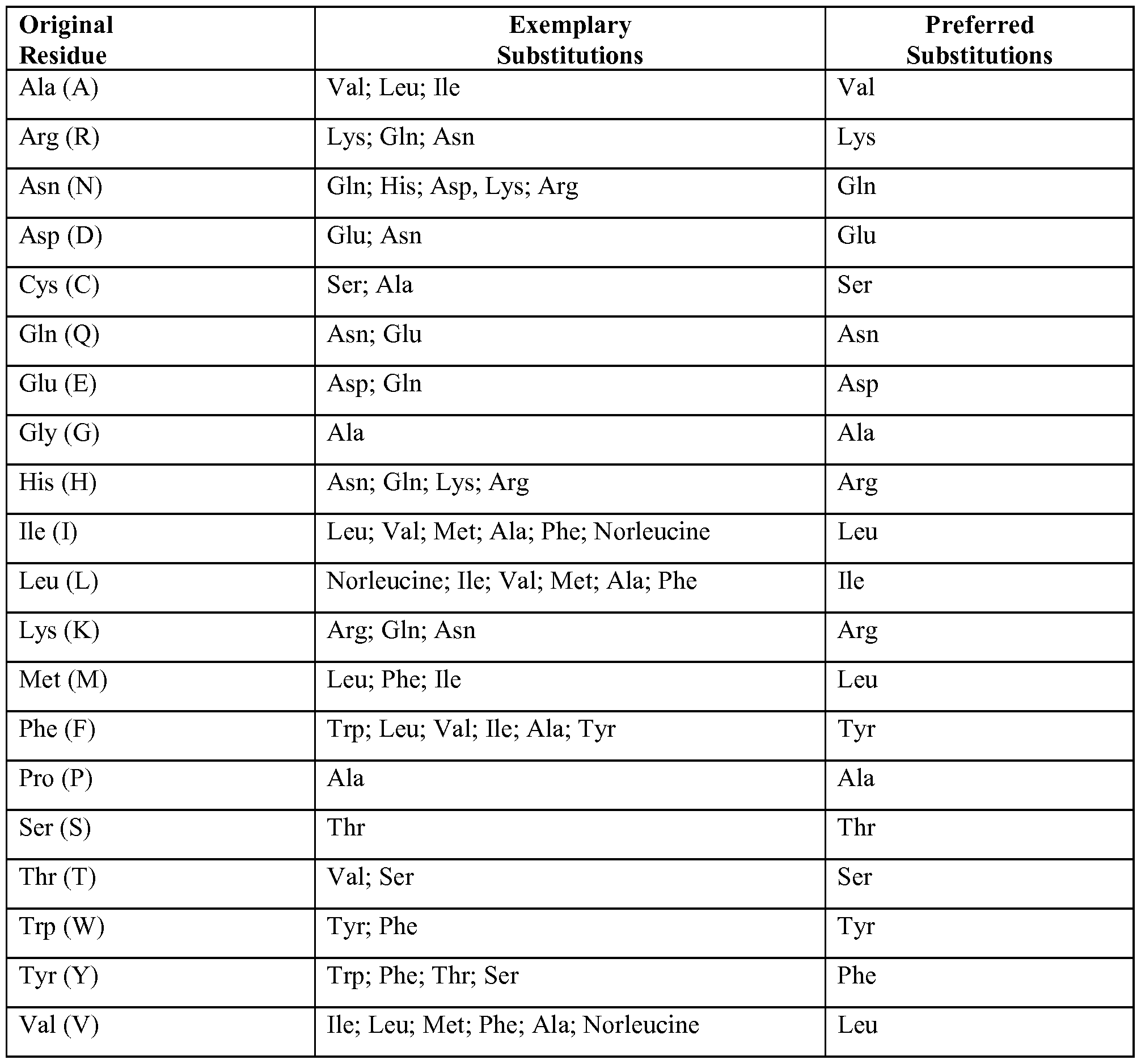


Claims
Priority Applications (24)
| Application Number | Priority Date | Filing Date | Title |
|---|---|---|---|
| ES12840084.3T ES2687951T3 (en) | 2011-10-14 | 2012-10-12 | Anti-HtrA1 antibodies and procedures for use |
| SG11201401477XA SG11201401477XA (en) | 2011-10-14 | 2012-10-12 | ANTI-HtrA1 ANTIBODIES AND METHODS OF USE |
| PL12840084T PL2766393T3 (en) | 2011-10-14 | 2012-10-12 | ANTI-HtrA1 ANTIBODIES AND METHODS OF USE |
| AU2012322618A AU2012322618A1 (en) | 2011-10-14 | 2012-10-12 | Anti-HtrA1 antibodies and methods of use |
| KR1020207001304A KR102102862B1 (en) | 2011-10-14 | 2012-10-12 | ANTI-HtrA1 ANTIBODIES AND METHODS OF USE |
| EA201490778A EA201490778A1 (en) | 2011-10-14 | 2012-10-12 | ANTIBODIES AGAINST HtrA1 AND METHODS OF APPLICATION |
| EP12840084.3A EP2766393B1 (en) | 2011-10-14 | 2012-10-12 | ANTI-HtrA1 ANTIBODIES AND METHODS OF USE |
| NZ623346A NZ623346B2 (en) | 2011-10-14 | 2012-10-12 | ANTI-HtrA1 ANTIBODIES AND METHODS OF USE |
| CA2850032A CA2850032C (en) | 2011-10-14 | 2012-10-12 | Anti-htra1 antibodies and methods of use |
| KR1020147012525A KR20140082796A (en) | 2011-10-14 | 2012-10-12 | ANTI-HtrA1 ANTIBODIES AND METHODS OF USE |
| EP18182184.4A EP3461839A1 (en) | 2011-10-14 | 2012-10-12 | Anti-htra1 antibodies and methods of use |
| LTEP12840084.3T LT2766393T (en) | 2011-10-14 | 2012-10-12 | ANTI-HtrA1 ANTIBODIES AND METHODS OF USE |
| CN201280050549.0A CN103917556B (en) | 2011-10-14 | 2012-10-12 | Anti- HtrA1 antibody and application method |
| RS20181070A RS57645B1 (en) | 2011-10-14 | 2012-10-12 | Anti-htra1 antibodies and methods of use |
| BR112014008862A BR112014008862A2 (en) | 2011-10-14 | 2012-10-12 | isolated antibody that binds to htra1, isolated nucleic acid, host cell, immunoconjugate, pharmaceutical formulation, methods and uses |
| JP2014535903A JP6532678B2 (en) | 2011-10-14 | 2012-10-12 | Anti-HtrA1 antibody and method of use |
| SI201231378T SI2766393T1 (en) | 2011-10-14 | 2012-10-12 | ANTI-HtrA1 ANTIBODIES AND METHODS OF USE |
| MX2014004022A MX2014004022A (en) | 2011-10-14 | 2012-10-12 | ANTI-HtrA1 ANTIBODIES AND METHODS OF USE. |
| DK12840084.3T DK2766393T3 (en) | 2011-10-14 | 2012-10-12 | Anti-HtrA1 antibodies and methods of use |
| ZA2014/02172A ZA201402172B (en) | 2011-10-14 | 2014-03-24 | Anti-htra1 antibodies and methods of use |
| IL232055A IL232055B (en) | 2011-10-14 | 2014-04-10 | Anti-htra1 antibodies and methods of use |
| MA36984A MA35712B1 (en) | 2011-10-14 | 2014-05-06 | Anti-htra1 antibodies and methods of use |
| AU2017236046A AU2017236046C1 (en) | 2011-10-14 | 2017-10-02 | Anti-HtrA1 antibodies and methods of use |
| HRP20181457TT HRP20181457T1 (en) | 2011-10-14 | 2018-09-11 | ANTI-HtrA1 ANTIBODIES AND METHODS OF USE |
Applications Claiming Priority (2)
| Application Number | Priority Date | Filing Date | Title |
|---|---|---|---|
| US201161547649P | 2011-10-14 | 2011-10-14 | |
| US61/547,649 | 2011-10-14 |
Publications (1)
| Publication Number | Publication Date |
|---|---|
| WO2013055998A1 true WO2013055998A1 (en) | 2013-04-18 |
Family
ID=48082468
Family Applications (1)
| Application Number | Title | Priority Date | Filing Date |
|---|---|---|---|
| PCT/US2012/059878 WO2013055998A1 (en) | 2011-10-14 | 2012-10-12 | ANTI-HtrA1 ANTIBODIES AND METHODS OF USE |
Country Status (32)
| Country | Link |
|---|---|
| US (3) | US9738727B2 (en) |
| EP (2) | EP3461839A1 (en) |
| JP (2) | JP6532678B2 (en) |
| KR (2) | KR20140082796A (en) |
| CN (2) | CN103917556B (en) |
| AR (1) | AR088322A1 (en) |
| AU (2) | AU2012322618A1 (en) |
| BR (1) | BR112014008862A2 (en) |
| CA (1) | CA2850032C (en) |
| CL (1) | CL2014000888A1 (en) |
| CO (1) | CO7020870A2 (en) |
| CR (1) | CR20140212A (en) |
| DK (1) | DK2766393T3 (en) |
| EA (1) | EA201490778A1 (en) |
| ES (1) | ES2687951T3 (en) |
| HK (1) | HK1255608A1 (en) |
| HR (1) | HRP20181457T1 (en) |
| HU (1) | HUE039133T2 (en) |
| IL (1) | IL232055B (en) |
| LT (1) | LT2766393T (en) |
| MA (1) | MA35712B1 (en) |
| MX (1) | MX2014004022A (en) |
| PE (1) | PE20141562A1 (en) |
| PL (1) | PL2766393T3 (en) |
| PT (1) | PT2766393T (en) |
| RS (1) | RS57645B1 (en) |
| SG (1) | SG11201401477XA (en) |
| SI (1) | SI2766393T1 (en) |
| TR (1) | TR201811270T4 (en) |
| TW (1) | TW201321414A (en) |
| WO (1) | WO2013055998A1 (en) |
| ZA (1) | ZA201402172B (en) |
Cited By (16)
| Publication number | Priority date | Publication date | Assignee | Title |
|---|---|---|---|---|
| WO2016077381A1 (en) | 2014-11-10 | 2016-05-19 | Genentech, Inc. | Anti-interleukin-33 antibodies and uses thereof |
| WO2017075212A1 (en) * | 2015-10-30 | 2017-05-04 | Genentech, Inc. | Anti-htra1 antibodies and methods of use thereof |
| WO2017075259A1 (en) | 2015-10-30 | 2017-05-04 | Genentech, Inc. | Anti-factor d antibody formulations |
| US10093978B2 (en) | 2013-08-12 | 2018-10-09 | Genentech, Inc. | Compositions for detecting complement factor H (CFH) and complement factor I (CFI) polymorphisms |
| US10179821B2 (en) | 2014-05-01 | 2019-01-15 | Genentech, Inc. | Anti-factor D antibodies |
| US10407510B2 (en) | 2015-10-30 | 2019-09-10 | Genentech, Inc. | Anti-factor D antibodies and conjugates |
| US10654932B2 (en) | 2015-10-30 | 2020-05-19 | Genentech, Inc. | Anti-factor D antibody variant conjugates and uses thereof |
| WO2020102680A1 (en) * | 2018-11-16 | 2020-05-22 | Gemini Therapeutics Inc. | Antibody-based therapeutics for targeting htra1 and methods of use |
| WO2020109343A1 (en) | 2018-11-29 | 2020-06-04 | F. Hoffmann-La Roche Ag | Combination therapy for treatment of macular degeneration |
| WO2020139834A1 (en) * | 2018-12-24 | 2020-07-02 | Visterra, Inc. | Methods for identifying epitopes and paratopes |
| CN111512160A (en) * | 2017-12-21 | 2020-08-07 | 豪夫迈·罗氏有限公司 | Companion diagnostics for HTRA1RNA antagonists |
| WO2022031680A1 (en) * | 2020-08-04 | 2022-02-10 | Exelixis, Inc. | Cd47 binding agents and uses thereof |
| US11267803B2 (en) | 2016-06-21 | 2022-03-08 | Orion Ophthalmology LLC | Carbocyclic prolinamide derivatives |
| EP3811963A4 (en) * | 2018-06-21 | 2022-03-30 | Daiichi Sankyo Company, Limited | Peptide for treating retinitis pigmentosa |
| US11377439B2 (en) | 2016-06-21 | 2022-07-05 | Orion Ophthalmology LLC | Heterocyclic prolinamide derivatives |
| EP3953485A4 (en) * | 2019-04-10 | 2023-05-17 | University of Utah Research Foundation | Htra1 modulation for treatment of amd |
Families Citing this family (5)
| Publication number | Priority date | Publication date | Assignee | Title |
|---|---|---|---|---|
| JP2018096930A (en) * | 2016-12-16 | 2018-06-21 | 公益財団法人ヒューマンサイエンス振興財団 | Diagnostic marker for age-related macular degeneration, diagnosis kit, method for supporting diagnosis, and method for determining risk of development |
| RS65011B1 (en) * | 2016-12-22 | 2024-01-31 | Daiichi Sankyo Co Ltd | Peptide for treating age-related macular degeneration |
| WO2018183216A1 (en) | 2017-03-27 | 2018-10-04 | The Schepens Eye Research Institute, Inc. | Notch3 agonist compositions and methods for treating small vessel diseases |
| CA3174680A1 (en) | 2020-03-13 | 2021-09-16 | Genentech, Inc. | Anti-interleukin-33 antibodies and uses thereof |
| US20220251241A1 (en) * | 2021-02-08 | 2022-08-11 | Ngm Biopharmaceuticals, Inc. | Htra1-binding agents and methods of use thereof |
Citations (94)
| Publication number | Priority date | Publication date | Assignee | Title |
|---|---|---|---|---|
| US4676980A (en) | 1985-09-23 | 1987-06-30 | The United States Of America As Represented By The Secretary Of The Department Of Health And Human Services | Target specific cross-linked heteroantibodies |
| US4737456A (en) | 1985-05-09 | 1988-04-12 | Syntex (U.S.A.) Inc. | Reducing interference in ligand-receptor binding assays |
| US4816567A (en) | 1983-04-08 | 1989-03-28 | Genentech, Inc. | Recombinant immunoglobin preparations |
| EP0404097A2 (en) | 1989-06-22 | 1990-12-27 | BEHRINGWERKE Aktiengesellschaft | Bispecific and oligospecific, mono- and oligovalent receptors, production and applications thereof |
| WO1993001161A1 (en) | 1991-07-11 | 1993-01-21 | Pfizer Limited | Process for preparing sertraline intermediates |
| US5208020A (en) | 1989-10-25 | 1993-05-04 | Immunogen Inc. | Cytotoxic agents comprising maytansinoids and their therapeutic use |
| WO1993008829A1 (en) | 1991-11-04 | 1993-05-13 | The Regents Of The University Of California | Compositions that mediate killing of hiv-infected cells |
| WO1993016185A2 (en) | 1992-02-06 | 1993-08-19 | Creative Biomolecules, Inc. | Biosynthetic binding protein for cancer marker |
| WO1994011026A2 (en) | 1992-11-13 | 1994-05-26 | Idec Pharmaceuticals Corporation | Therapeutic application of chimeric and radiolabeled antibodies to human b lymphocyte restricted differentiation antigen for treatment of b cell lymphoma |
| WO1994029351A2 (en) | 1993-06-16 | 1994-12-22 | Celltech Limited | Antibodies |
| US5500362A (en) | 1987-01-08 | 1996-03-19 | Xoma Corporation | Chimeric antibody with specificity to human B cell surface antigen |
| EP0425235B1 (en) | 1989-10-25 | 1996-09-25 | Immunogen Inc | Cytotoxic agents comprising maytansinoids and their therapeutic use |
| US5571894A (en) | 1991-02-05 | 1996-11-05 | Ciba-Geigy Corporation | Recombinant antibodies specific for a growth factor receptor |
| US5587458A (en) | 1991-10-07 | 1996-12-24 | Aronex Pharmaceuticals, Inc. | Anti-erbB-2 antibodies, combinations thereof, and therapeutic and diagnostic uses thereof |
| US5624821A (en) | 1987-03-18 | 1997-04-29 | Scotgen Biopharmaceuticals Incorporated | Antibodies with altered effector functions |
| US5635483A (en) | 1992-12-03 | 1997-06-03 | Arizona Board Of Regents Acting On Behalf Of Arizona State University | Tumor inhibiting tetrapeptide bearing modified phenethyl amides |
| US5648237A (en) | 1991-09-19 | 1997-07-15 | Genentech, Inc. | Expression of functional antibody fragments |
| WO1997030087A1 (en) | 1996-02-16 | 1997-08-21 | Glaxo Group Limited | Preparation of glycosylated antibodies |
| US5712374A (en) | 1995-06-07 | 1998-01-27 | American Cyanamid Company | Method for the preparation of substantiallly monomeric calicheamicin derivative/carrier conjugates |
| US5714586A (en) | 1995-06-07 | 1998-02-03 | American Cyanamid Company | Methods for the preparation of monomeric calicheamicin derivative/carrier conjugates |
| US5731168A (en) | 1995-03-01 | 1998-03-24 | Genentech, Inc. | Method for making heteromultimeric polypeptides |
| US5739116A (en) | 1994-06-03 | 1998-04-14 | American Cyanamid Company | Enediyne derivatives useful for the synthesis of conjugates of methyltrithio antitumor agents |
| US5750373A (en) | 1990-12-03 | 1998-05-12 | Genentech, Inc. | Enrichment method for variant proteins having altered binding properties, M13 phagemids, and growth hormone variants |
| US5770429A (en) | 1990-08-29 | 1998-06-23 | Genpharm International, Inc. | Transgenic non-human animals capable of producing heterologous antibodies |
| US5770701A (en) | 1987-10-30 | 1998-06-23 | American Cyanamid Company | Process for preparing targeted forms of methyltrithio antitumor agents |
| US5770710A (en) | 1987-10-30 | 1998-06-23 | American Cyanamid Company | Antitumor and antibacterial substituted disulfide derivatives prepared from compounds possessing a methlytrithio group |
| US5780588A (en) | 1993-01-26 | 1998-07-14 | Arizona Board Of Regents | Elucidation and synthesis of selected pentapeptides |
| US5789199A (en) | 1994-11-03 | 1998-08-04 | Genentech, Inc. | Process for bacterial production of polypeptides |
| US5821337A (en) | 1991-06-14 | 1998-10-13 | Genentech, Inc. | Immunoglobulin variants |
| US5840523A (en) | 1995-03-01 | 1998-11-24 | Genetech, Inc. | Methods and compositions for secretion of heterologous polypeptides |
| WO1998058964A1 (en) | 1997-06-24 | 1998-12-30 | Genentech, Inc. | Methods and compositions for galactosylated glycoproteins |
| US5869046A (en) | 1995-04-14 | 1999-02-09 | Genentech, Inc. | Altered polypeptides with increased half-life |
| WO1999022764A1 (en) | 1997-10-31 | 1999-05-14 | Genentech, Inc. | Methods and compositions comprising glycoprotein glycoforms |
| US5959177A (en) | 1989-10-27 | 1999-09-28 | The Scripps Research Institute | Transgenic plants expressing assembled secretory antibodies |
| WO1999051642A1 (en) | 1998-04-02 | 1999-10-14 | Genentech, Inc. | Antibody variants and fragments thereof |
| US6040498A (en) | 1998-08-11 | 2000-03-21 | North Caroline State University | Genetically engineered duckweed |
| US6075181A (en) | 1990-01-12 | 2000-06-13 | Abgenix, Inc. | Human antibodies derived from immunized xenomice |
| WO2000061739A1 (en) | 1999-04-09 | 2000-10-19 | Kyowa Hakko Kogyo Co., Ltd. | Method for controlling the activity of immunologically functional molecule |
| US6150584A (en) | 1990-01-12 | 2000-11-21 | Abgenix, Inc. | Human antibodies derived from immunized xenomice |
| US6171586B1 (en) | 1997-06-13 | 2001-01-09 | Genentech, Inc. | Antibody formulation |
| US6194551B1 (en) | 1998-04-02 | 2001-02-27 | Genentech, Inc. | Polypeptide variants |
| WO2001029246A1 (en) | 1999-10-19 | 2001-04-26 | Kyowa Hakko Kogyo Co., Ltd. | Process for producing polypeptide |
| US6248516B1 (en) | 1988-11-11 | 2001-06-19 | Medical Research Council | Single domain ligands, receptors comprising said ligands methods for their production, and use of said ligands and receptors |
| US6267958B1 (en) | 1995-07-27 | 2001-07-31 | Genentech, Inc. | Protein formulation |
| WO2002031140A1 (en) | 2000-10-06 | 2002-04-18 | Kyowa Hakko Kogyo Co., Ltd. | Cells producing antibody compositions |
| US6420548B1 (en) | 1999-10-04 | 2002-07-16 | Medicago Inc. | Method for regulating transcription of foreign genes |
| US20020164328A1 (en) | 2000-10-06 | 2002-11-07 | Toyohide Shinkawa | Process for purifying antibody |
| WO2003011878A2 (en) | 2001-08-03 | 2003-02-13 | Glycart Biotechnology Ag | Antibody glycosylation variants having increased antibody-dependent cellular cytotoxicity |
| US20030115614A1 (en) | 2000-10-06 | 2003-06-19 | Yutaka Kanda | Antibody composition-producing cell |
| US6602684B1 (en) | 1998-04-20 | 2003-08-05 | Glycart Biotechnology Ag | Glycosylation engineering of antibodies for improving antibody-dependent cellular cytotoxicity |
| US20030157108A1 (en) | 2001-10-25 | 2003-08-21 | Genentech, Inc. | Glycoprotein compositions |
| US6630579B2 (en) | 1999-12-29 | 2003-10-07 | Immunogen Inc. | Cytotoxic agents comprising modified doxorubicins and daunorubicins and their therapeutic use |
| WO2003085119A1 (en) | 2002-04-09 | 2003-10-16 | Kyowa Hakko Kogyo Co., Ltd. | METHOD OF ENHANCING ACTIVITY OF ANTIBODY COMPOSITION OF BINDING TO FcϜ RECEPTOR IIIa |
| WO2003085107A1 (en) | 2002-04-09 | 2003-10-16 | Kyowa Hakko Kogyo Co., Ltd. | Cells with modified genome |
| WO2003084570A1 (en) | 2002-04-09 | 2003-10-16 | Kyowa Hakko Kogyo Co., Ltd. | DRUG CONTAINING ANTIBODY COMPOSITION APPROPRIATE FOR PATIENT SUFFERING FROM FcϜRIIIa POLYMORPHISM |
| US20040093621A1 (en) | 2001-12-25 | 2004-05-13 | Kyowa Hakko Kogyo Co., Ltd | Antibody composition which specifically binds to CD20 |
| US6737056B1 (en) | 1999-01-15 | 2004-05-18 | Genentech, Inc. | Polypeptide variants with altered effector function |
| US20040110282A1 (en) | 2002-04-09 | 2004-06-10 | Kyowa Hakko Kogyo Co., Ltd. | Cells in which activity of the protein involved in transportation of GDP-fucose is reduced or lost |
| US20040109865A1 (en) | 2002-04-09 | 2004-06-10 | Kyowa Hakko Kogyo Co., Ltd. | Antibody composition-containing medicament |
| US20040132140A1 (en) | 2002-04-09 | 2004-07-08 | Kyowa Hakko Kogyo Co., Ltd. | Production process for antibody composition |
| WO2004056312A2 (en) | 2002-12-16 | 2004-07-08 | Genentech, Inc. | Immunoglobulin variants and uses thereof |
| US20050014934A1 (en) | 2002-10-15 | 2005-01-20 | Hinton Paul R. | Alteration of FcRn binding affinities or serum half-lives of antibodies by mutagenesis |
| US20050079574A1 (en) | 2003-01-16 | 2005-04-14 | Genentech, Inc. | Synthetic antibody phage libraries |
| WO2005035586A1 (en) | 2003-10-08 | 2005-04-21 | Kyowa Hakko Kogyo Co., Ltd. | Fused protein composition |
| WO2005035778A1 (en) | 2003-10-09 | 2005-04-21 | Kyowa Hakko Kogyo Co., Ltd. | PROCESS FOR PRODUCING ANTIBODY COMPOSITION BY USING RNA INHIBITING THE FUNCTION OF α1,6-FUCOSYLTRANSFERASE |
| US20050123546A1 (en) | 2003-11-05 | 2005-06-09 | Glycart Biotechnology Ag | Antigen binding molecules with increased Fc receptor binding affinity and effector function |
| WO2005053742A1 (en) | 2003-12-04 | 2005-06-16 | Kyowa Hakko Kogyo Co., Ltd. | Medicine containing antibody composition |
| WO2005100402A1 (en) | 2004-04-13 | 2005-10-27 | F.Hoffmann-La Roche Ag | Anti-p-selectin antibodies |
| US20050260186A1 (en) | 2003-03-05 | 2005-11-24 | Halozyme, Inc. | Soluble glycosaminoglycanases and methods of preparing and using soluble glycosaminoglycanases |
| US20050266000A1 (en) | 2004-04-09 | 2005-12-01 | Genentech, Inc. | Variable domain library and uses |
| US6982321B2 (en) | 1986-03-27 | 2006-01-03 | Medical Research Council | Altered antibodies |
| US20060025576A1 (en) | 2000-04-11 | 2006-02-02 | Genentech, Inc. | Multivalent antibodies and uses therefor |
| WO2006029879A2 (en) | 2004-09-17 | 2006-03-23 | F.Hoffmann-La Roche Ag | Anti-ox40l antibodies |
| WO2006044908A2 (en) | 2004-10-20 | 2006-04-27 | Genentech, Inc. | Antibody formulation in histidine-acetate buffer |
| US7041870B2 (en) | 2000-11-30 | 2006-05-09 | Medarex, Inc. | Transgenic transchromosomal rodents for making human antibodies |
| US20060104968A1 (en) | 2003-03-05 | 2006-05-18 | Halozyme, Inc. | Soluble glycosaminoglycanases and methods of preparing and using soluble glycosaminogly ycanases |
| US7087409B2 (en) | 1997-12-05 | 2006-08-08 | The Scripps Research Institute | Humanization of murine antibody |
| US7125978B1 (en) | 1999-10-04 | 2006-10-24 | Medicago Inc. | Promoter for regulating expression of foreign genes |
| US7189826B2 (en) | 1997-11-24 | 2007-03-13 | Institute For Human Genetics And Biochemistry | Monoclonal human natural antibodies |
| US20070061900A1 (en) | 2000-10-31 | 2007-03-15 | Murphy Andrew J | Methods of modifying eukaryotic cells |
| US20070117126A1 (en) | 1999-12-15 | 2007-05-24 | Genentech, Inc. | Shotgun scanning |
| US20070160598A1 (en) | 2005-11-07 | 2007-07-12 | Dennis Mark S | Binding polypeptides with diversified and consensus vh/vl hypervariable sequences |
| US20070237764A1 (en) | 2005-12-02 | 2007-10-11 | Genentech, Inc. | Binding polypeptides with restricted diversity sequences |
| US20070292936A1 (en) | 2006-05-09 | 2007-12-20 | Genentech, Inc. | Binding polypeptides with optimized scaffolds |
| US20080069820A1 (en) | 2006-08-30 | 2008-03-20 | Genentech, Inc. | Multispecific antibodies |
| US7371826B2 (en) | 1999-01-15 | 2008-05-13 | Genentech, Inc. | Polypeptide variants with altered effector function |
| WO2008077546A1 (en) | 2006-12-22 | 2008-07-03 | F. Hoffmann-La Roche Ag | Antibodies against insulin-like growth factor i receptor and uses thereof |
| WO2008103299A2 (en) * | 2007-02-16 | 2008-08-28 | Massachusetts Eye & Ear Infirmary | Methods and compositions for prognosing, detecting, and treating age-related macular degeneration |
| US20090002360A1 (en) | 2007-05-25 | 2009-01-01 | Innolux Display Corp. | Liquid crystal display device and method for driving same |
| US7498298B2 (en) | 2003-11-06 | 2009-03-03 | Seattle Genetics, Inc. | Monomethylvaline compounds capable of conjugation to ligands |
| WO2009046405A2 (en) * | 2007-10-05 | 2009-04-09 | University Of Utah Research Foundation | Antibodies to htra1 and methods of using the same |
| US7521541B2 (en) | 2004-09-23 | 2009-04-21 | Genetech Inc. | Cysteine engineered antibodies and conjugates |
| US7527791B2 (en) | 2004-03-31 | 2009-05-05 | Genentech, Inc. | Humanized anti-TGF-beta antibodies |
| WO2009089004A1 (en) | 2008-01-07 | 2009-07-16 | Amgen Inc. | Method for making antibody fc-heterodimeric molecules using electrostatic steering effects |
Family Cites Families (127)
| Publication number | Priority date | Publication date | Assignee | Title |
|---|---|---|---|---|
| US3687808A (en) | 1969-08-14 | 1972-08-29 | Univ Leland Stanford Junior | Synthetic polynucleotides |
| US4376110A (en) | 1980-08-04 | 1983-03-08 | Hybritech, Incorporated | Immunometric assays using monoclonal antibodies |
| US4486530A (en) | 1980-08-04 | 1984-12-04 | Hybritech Incorporated | Immunometric assays using monoclonal antibodies |
| US4469863A (en) | 1980-11-12 | 1984-09-04 | Ts O Paul O P | Nonionic nucleic acid alkyl and aryl phosphonates and processes for manufacture and use thereof |
| US4485045A (en) | 1981-07-06 | 1984-11-27 | Research Corporation | Synthetic phosphatidyl cholines useful in forming liposomes |
| CA1188246A (en) | 1981-08-31 | 1985-06-04 | Jerry L. Gregory | Immobilized microbe recycle apparatus |
| US5023243A (en) | 1981-10-23 | 1991-06-11 | Molecular Biosystems, Inc. | Oligonucleotide therapeutic agent and method of making same |
| US4476301A (en) | 1982-04-29 | 1984-10-09 | Centre National De La Recherche Scientifique | Oligonucleotides, a process for preparing the same and their application as mediators of the action of interferon |
| US4522811A (en) | 1982-07-08 | 1985-06-11 | Syntex (U.S.A.) Inc. | Serial injection of muramyldipeptides and liposomes enhances the anti-infective activity of muramyldipeptides |
| NZ207394A (en) | 1983-03-08 | 1987-03-06 | Commw Serum Lab Commission | Detecting or determining sequence of amino acids |
| US4544545A (en) | 1983-06-20 | 1985-10-01 | Trustees University Of Massachusetts | Liposomes containing modified cholesterol for organ targeting |
| US5118800A (en) | 1983-12-20 | 1992-06-02 | California Institute Of Technology | Oligonucleotides possessing a primary amino group in the terminal nucleotide |
| US5550111A (en) | 1984-07-11 | 1996-08-27 | Temple University-Of The Commonwealth System Of Higher Education | Dual action 2',5'-oligoadenylate antiviral derivatives and uses thereof |
| FR2567892B1 (en) | 1984-07-19 | 1989-02-17 | Centre Nat Rech Scient | NOVEL OLIGONUCLEOTIDES, THEIR PREPARATION PROCESS AND THEIR APPLICATIONS AS MEDIATORS IN DEVELOPING THE EFFECTS OF INTERFERONS |
| US5367066A (en) | 1984-10-16 | 1994-11-22 | Chiron Corporation | Oligonucleotides with selectably cleavable and/or abasic sites |
| FR2575751B1 (en) | 1985-01-08 | 1987-04-03 | Pasteur Institut | NOVEL ADENOSINE DERIVATIVE NUCLEOSIDES, THEIR PREPARATION AND THEIR BIOLOGICAL APPLICATIONS |
| US5235033A (en) | 1985-03-15 | 1993-08-10 | Anti-Gene Development Group | Alpha-morpholino ribonucleoside derivatives and polymers thereof |
| US5034506A (en) | 1985-03-15 | 1991-07-23 | Anti-Gene Development Group | Uncharged morpholino-based polymers having achiral intersubunit linkages |
| US5405938A (en) | 1989-12-20 | 1995-04-11 | Anti-Gene Development Group | Sequence-specific binding polymers for duplex nucleic acids |
| US5185444A (en) | 1985-03-15 | 1993-02-09 | Anti-Gene Deveopment Group | Uncharged morpolino-based polymers having phosphorous containing chiral intersubunit linkages |
| US4868105A (en) | 1985-12-11 | 1989-09-19 | Chiron Corporation | Solution phase nucleic acid sandwich assay |
| US4987071A (en) | 1986-12-03 | 1991-01-22 | University Patents, Inc. | RNA ribozyme polymerases, dephosphorylases, restriction endoribonucleases and methods |
| US5264423A (en) | 1987-03-25 | 1993-11-23 | The United States Of America As Represented By The Department Of Health And Human Services | Inhibitors for replication of retroviruses and for the expression of oncogene products |
| US5276019A (en) | 1987-03-25 | 1994-01-04 | The United States Of America As Represented By The Department Of Health And Human Services | Inhibitors for replication of retroviruses and for the expression of oncogene products |
| US4924624A (en) | 1987-10-22 | 1990-05-15 | Temple University-Of The Commonwealth System Of Higher Education | 2,',5'-phosphorothioate oligoadenylates and plant antiviral uses thereof |
| US5188897A (en) | 1987-10-22 | 1993-02-23 | Temple University Of The Commonwealth System Of Higher Education | Encapsulated 2',5'-phosphorothioate oligoadenylates |
| IE61148B1 (en) | 1988-03-10 | 1994-10-05 | Ici Plc | Method of detecting nucleotide sequences |
| JPH03503894A (en) | 1988-03-25 | 1991-08-29 | ユニバーシィティ オブ バージニア アランミ パテンツ ファウンデイション | Oligonucleotide N-alkylphosphoramidate |
| US5278302A (en) | 1988-05-26 | 1994-01-11 | University Patents, Inc. | Polynucleotide phosphorodithioates |
| US5216141A (en) | 1988-06-06 | 1993-06-01 | Benner Steven A | Oligonucleotide analogs containing sulfur linkages |
| US5175273A (en) | 1988-07-01 | 1992-12-29 | Genentech, Inc. | Nucleic acid intercalating agents |
| GB8823869D0 (en) | 1988-10-12 | 1988-11-16 | Medical Res Council | Production of antibodies |
| US5047335A (en) | 1988-12-21 | 1991-09-10 | The Regents Of The University Of Calif. | Process for controlling intracellular glycosylation of proteins |
| CA2039718C (en) | 1989-08-31 | 2003-02-25 | John J. Rossi | Chimeric dna-rna catalytic sequences |
| US5591722A (en) | 1989-09-15 | 1997-01-07 | Southern Research Institute | 2'-deoxy-4'-thioribonucleosides and their antiviral activity |
| US5013556A (en) | 1989-10-20 | 1991-05-07 | Liposome Technology, Inc. | Liposomes with enhanced circulation time |
| US5399676A (en) | 1989-10-23 | 1995-03-21 | Gilead Sciences | Oligonucleotides with inverted polarity |
| EP0497875B1 (en) | 1989-10-24 | 2000-03-22 | Isis Pharmaceuticals, Inc. | 2' modified oligonucleotides |
| US5264562A (en) | 1989-10-24 | 1993-11-23 | Gilead Sciences, Inc. | Oligonucleotide analogs with novel linkages |
| US5264564A (en) | 1989-10-24 | 1993-11-23 | Gilead Sciences | Oligonucleotide analogs with novel linkages |
| US5177198A (en) | 1989-11-30 | 1993-01-05 | University Of N.C. At Chapel Hill | Process for preparing oligoribonucleoside and oligodeoxyribonucleoside boranophosphates |
| US5130302A (en) | 1989-12-20 | 1992-07-14 | Boron Bilogicals, Inc. | Boronated nucleoside, nucleotide and oligonucleotide compounds, compositions and methods for using same |
| US5587470A (en) | 1990-01-11 | 1996-12-24 | Isis Pharmaceuticals, Inc. | 3-deazapurines |
| US5646265A (en) | 1990-01-11 | 1997-07-08 | Isis Pharmceuticals, Inc. | Process for the preparation of 2'-O-alkyl purine phosphoramidites |
| US5670633A (en) | 1990-01-11 | 1997-09-23 | Isis Pharmaceuticals, Inc. | Sugar modified oligonucleotides that detect and modulate gene expression |
| US5681941A (en) | 1990-01-11 | 1997-10-28 | Isis Pharmaceuticals, Inc. | Substituted purines and oligonucleotide cross-linking |
| US5459255A (en) | 1990-01-11 | 1995-10-17 | Isis Pharmaceuticals, Inc. | N-2 substituted purines |
| US5587361A (en) | 1991-10-15 | 1996-12-24 | Isis Pharmaceuticals, Inc. | Oligonucleotides having phosphorothioate linkages of high chiral purity |
| US5321131A (en) | 1990-03-08 | 1994-06-14 | Hybridon, Inc. | Site-specific functionalization of oligodeoxynucleotides for non-radioactive labelling |
| US5470967A (en) | 1990-04-10 | 1995-11-28 | The Dupont Merck Pharmaceutical Company | Oligonucleotide analogs with sulfamate linkages |
| GB9009980D0 (en) | 1990-05-03 | 1990-06-27 | Amersham Int Plc | Phosphoramidite derivatives,their preparation and the use thereof in the incorporation of reporter groups on synthetic oligonucleotides |
| ES2116977T3 (en) | 1990-05-11 | 1998-08-01 | Microprobe Corp | SOLID SUPPORTS FOR NUCLEIC ACID HYBRIDIZATION TESTS AND METHODS TO IMMOBILIZE OLIGONUCLEOTIDES IN A COVALENT WAY. |
| US5610289A (en) | 1990-07-27 | 1997-03-11 | Isis Pharmaceuticals, Inc. | Backbone modified oligonucleotide analogues |
| US5677437A (en) | 1990-07-27 | 1997-10-14 | Isis Pharmaceuticals, Inc. | Heteroatomic oligonucleoside linkages |
| US5541307A (en) | 1990-07-27 | 1996-07-30 | Isis Pharmaceuticals, Inc. | Backbone modified oligonucleotide analogs and solid phase synthesis thereof |
| US5608046A (en) | 1990-07-27 | 1997-03-04 | Isis Pharmaceuticals, Inc. | Conjugated 4'-desmethyl nucleoside analog compounds |
| US5602240A (en) | 1990-07-27 | 1997-02-11 | Ciba Geigy Ag. | Backbone modified oligonucleotide analogs |
| US5623070A (en) | 1990-07-27 | 1997-04-22 | Isis Pharmaceuticals, Inc. | Heteroatomic oligonucleoside linkages |
| US5489677A (en) | 1990-07-27 | 1996-02-06 | Isis Pharmaceuticals, Inc. | Oligonucleoside linkages containing adjacent oxygen and nitrogen atoms |
| US5618704A (en) | 1990-07-27 | 1997-04-08 | Isis Pharmacueticals, Inc. | Backbone-modified oligonucleotide analogs and preparation thereof through radical coupling |
| EP0541722B1 (en) | 1990-08-03 | 1995-12-20 | Sterling Winthrop Inc. | Compounds and methods for inhibiting gene expression |
| US5177196A (en) | 1990-08-16 | 1993-01-05 | Microprobe Corporation | Oligo (α-arabinofuranosyl nucleotides) and α-arabinofuranosyl precursors thereof |
| US5661016A (en) | 1990-08-29 | 1997-08-26 | Genpharm International Inc. | Transgenic non-human animals capable of producing heterologous antibodies of various isotypes |
| JP2938569B2 (en) | 1990-08-29 | 1999-08-23 | ジェンファーム インターナショナル,インコーポレイティド | Method for producing xenogeneic immunoglobulin and transgenic mouse |
| US5625126A (en) | 1990-08-29 | 1997-04-29 | Genpharm International, Inc. | Transgenic non-human animals for producing heterologous antibodies |
| US5633425A (en) | 1990-08-29 | 1997-05-27 | Genpharm International, Inc. | Transgenic non-human animals capable of producing heterologous antibodies |
| US5545806A (en) | 1990-08-29 | 1996-08-13 | Genpharm International, Inc. | Ransgenic non-human animals for producing heterologous antibodies |
| US5214134A (en) | 1990-09-12 | 1993-05-25 | Sterling Winthrop Inc. | Process of linking nucleosides with a siloxane bridge |
| US5561225A (en) | 1990-09-19 | 1996-10-01 | Southern Research Institute | Polynucleotide analogs containing sulfonate and sulfonamide internucleoside linkages |
| US5596086A (en) | 1990-09-20 | 1997-01-21 | Gilead Sciences, Inc. | Modified internucleoside linkages having one nitrogen and two carbon atoms |
| US5432272A (en) | 1990-10-09 | 1995-07-11 | Benner; Steven A. | Method for incorporating into a DNA or RNA oligonucleotide using nucleotides bearing heterocyclic bases |
| ATE147098T1 (en) | 1990-10-12 | 1997-01-15 | Max Planck Gesellschaft | MODIFIED RIBOZYMES |
| US5278299A (en) | 1991-03-18 | 1994-01-11 | Scripps Clinic And Research Foundation | Method and composition for synthesizing sialylated glycosyl compounds |
| US5539082A (en) | 1993-04-26 | 1996-07-23 | Nielsen; Peter E. | Peptide nucleic acids |
| US5714331A (en) | 1991-05-24 | 1998-02-03 | Buchardt, Deceased; Ole | Peptide nucleic acids having enhanced binding affinity, sequence specificity and solubility |
| DE4216134A1 (en) | 1991-06-20 | 1992-12-24 | Europ Lab Molekularbiolog | SYNTHETIC CATALYTIC OLIGONUCLEOTIDE STRUCTURES |
| US5571799A (en) | 1991-08-12 | 1996-11-05 | Basco, Ltd. | (2'-5') oligoadenylate analogues useful as inhibitors of host-v5.-graft response |
| EP0605522B1 (en) | 1991-09-23 | 1999-06-23 | Medical Research Council | Methods for the production of humanized antibodies |
| DE59208572D1 (en) | 1991-10-17 | 1997-07-10 | Ciba Geigy Ag | Bicyclic nucleosides, oligonucleotides, processes for their preparation and intermediates |
| US5594121A (en) | 1991-11-07 | 1997-01-14 | Gilead Sciences, Inc. | Enhanced triple-helix and double-helix formation with oligomers containing modified purines |
| WO1993010260A1 (en) | 1991-11-21 | 1993-05-27 | The Board Of Trustees Of The Leland Stanford Junior University | Controlling degradation of glycoprotein oligosaccharides by extracellular glycosisases |
| US5484908A (en) | 1991-11-26 | 1996-01-16 | Gilead Sciences, Inc. | Oligonucleotides containing 5-propynyl pyrimidines |
| US5652094A (en) | 1992-01-31 | 1997-07-29 | University Of Montreal | Nucleozymes |
| FR2687679B1 (en) | 1992-02-05 | 1994-10-28 | Centre Nat Rech Scient | OLIGOTHIONUCLEOTIDES. |
| US5434257A (en) | 1992-06-01 | 1995-07-18 | Gilead Sciences, Inc. | Binding compentent oligomers containing unsaturated 3',5' and 2',5' linkages |
| EP0577558A2 (en) | 1992-07-01 | 1994-01-05 | Ciba-Geigy Ag | Carbocyclic nucleosides having bicyclic rings, oligonucleotides therefrom, process for their preparation, their use and intermediates |
| US5476925A (en) | 1993-02-01 | 1995-12-19 | Northwestern University | Oligodeoxyribonucleotides including 3'-aminonucleoside-phosphoramidate linkages and terminal 3'-amino groups |
| GB9304618D0 (en) | 1993-03-06 | 1993-04-21 | Ciba Geigy Ag | Chemical compounds |
| ATE155467T1 (en) | 1993-03-30 | 1997-08-15 | Sanofi Sa | ACYCLIC NUCLEOSIDE ANALOGUES AND OLIGONUCLEOTIDE SEQUENCES CONTAINING THEM |
| JPH08508491A (en) | 1993-03-31 | 1996-09-10 | スターリング ウインスロップ インコーポレイティド | Oligonucleotides with phosphodiester bonds replaced by amide bonds |
| DE4311944A1 (en) | 1993-04-10 | 1994-10-13 | Degussa | Coated sodium percarbonate particles, process for their preparation and detergent, cleaning and bleaching compositions containing them |
| EP0717766B1 (en) | 1993-09-09 | 1998-04-01 | The Procter & Gamble Company | Automatic dishwashing detergent with alkoxy or aryloxy amide surfactant |
| US5502177A (en) | 1993-09-17 | 1996-03-26 | Gilead Sciences, Inc. | Pyrimidine derivatives for labeled binding partners |
| US5446137B1 (en) | 1993-12-09 | 1998-10-06 | Behringwerke Ag | Oligonucleotides containing 4'-substituted nucleotides |
| US5596091A (en) | 1994-03-18 | 1997-01-21 | The Regents Of The University Of California | Antisense oligonucleotides comprising 5-aminoalkyl pyrimidine nucleotides |
| US5627053A (en) | 1994-03-29 | 1997-05-06 | Ribozyme Pharmaceuticals, Inc. | 2'deoxy-2'-alkylnucleotide containing nucleic acid |
| US5625050A (en) | 1994-03-31 | 1997-04-29 | Amgen Inc. | Modified oligonucleotides and intermediates useful in nucleic acid therapeutics |
| US5525711A (en) | 1994-05-18 | 1996-06-11 | The United States Of America As Represented By The Secretary Of The Department Of Health And Human Services | Pteridine nucleotide analogs as fluorescent DNA probes |
| US5891628A (en) | 1994-06-03 | 1999-04-06 | Brigham And Women's Hospital | Identification of polycystic kidney disease gene, diagnostics and treatment |
| US5739277A (en) | 1995-04-14 | 1998-04-14 | Genentech Inc. | Altered polypeptides with increased half-life |
| DK0839152T3 (en) | 1995-06-21 | 2002-07-08 | Martek Biosciences Corp | Combinatorial libraries of labeled biochemical compounds and processes for their preparation |
| US5837492A (en) | 1995-12-18 | 1998-11-17 | Myriad Genetics, Inc. | Chromosome 13-linked breast cancer susceptibility gene |
| US6274376B1 (en) | 1996-02-20 | 2001-08-14 | Smithkline Beecham Corporation | ClpL |
| EP0828003A3 (en) | 1996-09-06 | 2003-01-15 | SmithKline Beecham plc | Human serine protease |
| US5800998A (en) | 1996-11-12 | 1998-09-01 | Millennium Pharmaceuticals, Inc. | Assays for diagnosing type II diabetes in a subject |
| JP2001505060A (en) | 1997-01-14 | 2001-04-17 | ヒューマン ジノーム サイエンシーズ,インコーポレイテッド | Tumor necrosis factor receptor 5 |
| TW589189B (en) | 1997-08-04 | 2004-06-01 | Scras | Kit containing at least one double-stranded RNA combined with at least one anti-viral agent for therapeutic use in the treatment of a viral disease, notably of viral hepatitis |
| US6506559B1 (en) | 1997-12-23 | 2003-01-14 | Carnegie Institute Of Washington | Genetic inhibition by double-stranded RNA |
| US6274720B1 (en) | 1997-12-31 | 2001-08-14 | Incyte Genomics, Inc. | Human preproneurotensin/neuromedin N |
| WO1999055885A2 (en) | 1998-04-28 | 1999-11-04 | Axys Pharmaceuticals, Inc. | Novel serine protease capable of selective cleavage of insulin-like growth factor binding protein |
| GB9827152D0 (en) | 1998-07-03 | 1999-02-03 | Devgen Nv | Characterisation of gene function using double stranded rna inhibition |
| AU5290499A (en) | 1998-08-03 | 2000-02-28 | Novartis Ag | Human htra serine protease |
| WO2000044914A1 (en) | 1999-01-28 | 2000-08-03 | Medical College Of Georgia Research Institute, Inc. | Composition and method for in vivo and in vitro attenuation of gene expression using double stranded rna |
| DE19956568A1 (en) | 1999-01-30 | 2000-08-17 | Roland Kreutzer | Method and medicament for inhibiting the expression of a given gene |
| CA2386270A1 (en) | 1999-10-15 | 2001-04-26 | University Of Massachusetts | Rna interference pathway genes as tools for targeted genetic interference |
| GB9927444D0 (en) | 1999-11-19 | 2000-01-19 | Cancer Res Campaign Tech | Inhibiting gene expression |
| AUPR670701A0 (en) | 2001-07-30 | 2001-08-23 | Prince Henry's Institute Of Medical Research | Novel serine protease |
| JP2003135075A (en) | 2001-11-05 | 2003-05-13 | Research Association For Biotechnology | NEW FULL-LENGTH cDNA |
| WO2003102157A2 (en) | 2002-06-03 | 2003-12-11 | Genentech, Inc. | Synthetic antibody phage libraries |
| JP2005154399A (en) * | 2003-11-24 | 2005-06-16 | Nara Institute Of Science & Technology | USE OF SERINE PROTEASE HtrA |
| EP2570491A1 (en) | 2005-06-08 | 2013-03-20 | University of Pittsburgh of the Commonwealth System of Higher Education | Susceptibility genes for age-related maculopathy (ARM) on chromosome 10q26 |
| US20100166743A1 (en) | 2006-10-06 | 2010-07-01 | University Of Utah Research Foundation | Method of detecting ocular diseases and pathologic conditions and treatment of same |
| EP2913341A1 (en) | 2006-12-22 | 2015-09-02 | University of Utah Research Foundation | Method of detecting ocular diseases and pathologic conditions and treatment of same |
| PE20081795A1 (en) | 2007-02-16 | 2009-01-08 | Genentech Inc | HTRA1-PDZ AND HTRA3-PDZ MODULATORS |
| CN101550451B (en) * | 2008-03-04 | 2011-08-31 | 四川省医学科学院(四川省人民医院) | Reagent kit for detecting agedness yellow spot degenerative disease |
| US9925398B2 (en) * | 2008-04-23 | 2018-03-27 | Novacell Technology Inc. | Angiogenic peptide |
| JP5701216B2 (en) * | 2008-11-05 | 2015-04-15 | ジェネンテック, インコーポレイテッド | Genetic polymorphism in age-related macular degeneration |
-
2012
- 2012-10-12 ES ES12840084.3T patent/ES2687951T3/en active Active
- 2012-10-12 EP EP18182184.4A patent/EP3461839A1/en not_active Withdrawn
- 2012-10-12 LT LTEP12840084.3T patent/LT2766393T/en unknown
- 2012-10-12 KR KR1020147012525A patent/KR20140082796A/en not_active Application Discontinuation
- 2012-10-12 JP JP2014535903A patent/JP6532678B2/en not_active Expired - Fee Related
- 2012-10-12 KR KR1020207001304A patent/KR102102862B1/en active IP Right Grant
- 2012-10-12 SG SG11201401477XA patent/SG11201401477XA/en unknown
- 2012-10-12 PT PT12840084T patent/PT2766393T/en unknown
- 2012-10-12 US US13/651,289 patent/US9738727B2/en active Active
- 2012-10-12 DK DK12840084.3T patent/DK2766393T3/en active
- 2012-10-12 TW TW101137846A patent/TW201321414A/en unknown
- 2012-10-12 CN CN201280050549.0A patent/CN103917556B/en active Active
- 2012-10-12 EP EP12840084.3A patent/EP2766393B1/en active Active
- 2012-10-12 WO PCT/US2012/059878 patent/WO2013055998A1/en active Application Filing
- 2012-10-12 AR ARP120103805A patent/AR088322A1/en unknown
- 2012-10-12 RS RS20181070A patent/RS57645B1/en unknown
- 2012-10-12 CN CN201810028448.2A patent/CN108373506A/en active Pending
- 2012-10-12 MX MX2014004022A patent/MX2014004022A/en active IP Right Grant
- 2012-10-12 BR BR112014008862A patent/BR112014008862A2/en not_active Application Discontinuation
- 2012-10-12 PE PE2014000524A patent/PE20141562A1/en not_active Application Discontinuation
- 2012-10-12 PL PL12840084T patent/PL2766393T3/en unknown
- 2012-10-12 TR TR2018/11270T patent/TR201811270T4/en unknown
- 2012-10-12 CA CA2850032A patent/CA2850032C/en active Active
- 2012-10-12 EA EA201490778A patent/EA201490778A1/en unknown
- 2012-10-12 HU HUE12840084A patent/HUE039133T2/en unknown
- 2012-10-12 SI SI201231378T patent/SI2766393T1/en unknown
- 2012-10-12 AU AU2012322618A patent/AU2012322618A1/en not_active Abandoned
-
2014
- 2014-03-24 ZA ZA2014/02172A patent/ZA201402172B/en unknown
- 2014-04-09 CL CL2014000888A patent/CL2014000888A1/en unknown
- 2014-04-10 IL IL232055A patent/IL232055B/en active IP Right Grant
- 2014-05-06 MA MA36984A patent/MA35712B1/en unknown
- 2014-05-09 CR CR20140212A patent/CR20140212A/en unknown
- 2014-05-13 CO CO14102608A patent/CO7020870A2/en unknown
-
2017
- 2017-07-18 US US15/653,201 patent/US20170342163A1/en not_active Abandoned
- 2017-10-02 AU AU2017236046A patent/AU2017236046C1/en active Active
- 2017-10-13 JP JP2017199087A patent/JP2018046832A/en active Pending
-
2018
- 2018-09-11 HR HRP20181457TT patent/HRP20181457T1/en unknown
- 2018-09-26 US US16/143,098 patent/US20190016826A1/en not_active Abandoned
- 2018-11-19 HK HK18114739.7A patent/HK1255608A1/en unknown
Patent Citations (102)
| Publication number | Priority date | Publication date | Assignee | Title |
|---|---|---|---|---|
| US4816567A (en) | 1983-04-08 | 1989-03-28 | Genentech, Inc. | Recombinant immunoglobin preparations |
| US4737456A (en) | 1985-05-09 | 1988-04-12 | Syntex (U.S.A.) Inc. | Reducing interference in ligand-receptor binding assays |
| US4676980A (en) | 1985-09-23 | 1987-06-30 | The United States Of America As Represented By The Secretary Of The Department Of Health And Human Services | Target specific cross-linked heteroantibodies |
| US6982321B2 (en) | 1986-03-27 | 2006-01-03 | Medical Research Council | Altered antibodies |
| US5500362A (en) | 1987-01-08 | 1996-03-19 | Xoma Corporation | Chimeric antibody with specificity to human B cell surface antigen |
| US5624821A (en) | 1987-03-18 | 1997-04-29 | Scotgen Biopharmaceuticals Incorporated | Antibodies with altered effector functions |
| US5648260A (en) | 1987-03-18 | 1997-07-15 | Scotgen Biopharmaceuticals Incorporated | DNA encoding antibodies with altered effector functions |
| US5770701A (en) | 1987-10-30 | 1998-06-23 | American Cyanamid Company | Process for preparing targeted forms of methyltrithio antitumor agents |
| US5770710A (en) | 1987-10-30 | 1998-06-23 | American Cyanamid Company | Antitumor and antibacterial substituted disulfide derivatives prepared from compounds possessing a methlytrithio group |
| US6248516B1 (en) | 1988-11-11 | 2001-06-19 | Medical Research Council | Single domain ligands, receptors comprising said ligands methods for their production, and use of said ligands and receptors |
| EP0404097A2 (en) | 1989-06-22 | 1990-12-27 | BEHRINGWERKE Aktiengesellschaft | Bispecific and oligospecific, mono- and oligovalent receptors, production and applications thereof |
| US5416064A (en) | 1989-10-25 | 1995-05-16 | Immunogen, Inc. | Cytotoxic agents comprising maytansinoids and their therapeutic use |
| EP0425235B1 (en) | 1989-10-25 | 1996-09-25 | Immunogen Inc | Cytotoxic agents comprising maytansinoids and their therapeutic use |
| US5208020A (en) | 1989-10-25 | 1993-05-04 | Immunogen Inc. | Cytotoxic agents comprising maytansinoids and their therapeutic use |
| US5959177A (en) | 1989-10-27 | 1999-09-28 | The Scripps Research Institute | Transgenic plants expressing assembled secretory antibodies |
| US6417429B1 (en) | 1989-10-27 | 2002-07-09 | The Scripps Research Institute | Transgenic plants expressing assembled secretory antibodies |
| US6150584A (en) | 1990-01-12 | 2000-11-21 | Abgenix, Inc. | Human antibodies derived from immunized xenomice |
| US6075181A (en) | 1990-01-12 | 2000-06-13 | Abgenix, Inc. | Human antibodies derived from immunized xenomice |
| US5770429A (en) | 1990-08-29 | 1998-06-23 | Genpharm International, Inc. | Transgenic non-human animals capable of producing heterologous antibodies |
| US5750373A (en) | 1990-12-03 | 1998-05-12 | Genentech, Inc. | Enrichment method for variant proteins having altered binding properties, M13 phagemids, and growth hormone variants |
| US5571894A (en) | 1991-02-05 | 1996-11-05 | Ciba-Geigy Corporation | Recombinant antibodies specific for a growth factor receptor |
| US5821337A (en) | 1991-06-14 | 1998-10-13 | Genentech, Inc. | Immunoglobulin variants |
| WO1993001161A1 (en) | 1991-07-11 | 1993-01-21 | Pfizer Limited | Process for preparing sertraline intermediates |
| US5648237A (en) | 1991-09-19 | 1997-07-15 | Genentech, Inc. | Expression of functional antibody fragments |
| US5587458A (en) | 1991-10-07 | 1996-12-24 | Aronex Pharmaceuticals, Inc. | Anti-erbB-2 antibodies, combinations thereof, and therapeutic and diagnostic uses thereof |
| WO1993008829A1 (en) | 1991-11-04 | 1993-05-13 | The Regents Of The University Of California | Compositions that mediate killing of hiv-infected cells |
| WO1993016185A2 (en) | 1992-02-06 | 1993-08-19 | Creative Biomolecules, Inc. | Biosynthetic binding protein for cancer marker |
| WO1994011026A2 (en) | 1992-11-13 | 1994-05-26 | Idec Pharmaceuticals Corporation | Therapeutic application of chimeric and radiolabeled antibodies to human b lymphocyte restricted differentiation antigen for treatment of b cell lymphoma |
| US5635483A (en) | 1992-12-03 | 1997-06-03 | Arizona Board Of Regents Acting On Behalf Of Arizona State University | Tumor inhibiting tetrapeptide bearing modified phenethyl amides |
| US5780588A (en) | 1993-01-26 | 1998-07-14 | Arizona Board Of Regents | Elucidation and synthesis of selected pentapeptides |
| WO1994029351A2 (en) | 1993-06-16 | 1994-12-22 | Celltech Limited | Antibodies |
| US5739116A (en) | 1994-06-03 | 1998-04-14 | American Cyanamid Company | Enediyne derivatives useful for the synthesis of conjugates of methyltrithio antitumor agents |
| US5773001A (en) | 1994-06-03 | 1998-06-30 | American Cyanamid Company | Conjugates of methyltrithio antitumor agents and intermediates for their synthesis |
| US5767285A (en) | 1994-06-03 | 1998-06-16 | American Cyanamid Company | Linkers useful for the synthesis of conjugates of methyltrithio antitumor agents |
| US5877296A (en) | 1994-06-03 | 1999-03-02 | American Cyanamid Company | Process for preparing conjugates of methyltrithio antitumor agents |
| US5789199A (en) | 1994-11-03 | 1998-08-04 | Genentech, Inc. | Process for bacterial production of polypeptides |
| US5840523A (en) | 1995-03-01 | 1998-11-24 | Genetech, Inc. | Methods and compositions for secretion of heterologous polypeptides |
| US5731168A (en) | 1995-03-01 | 1998-03-24 | Genentech, Inc. | Method for making heteromultimeric polypeptides |
| US5869046A (en) | 1995-04-14 | 1999-02-09 | Genentech, Inc. | Altered polypeptides with increased half-life |
| US5712374A (en) | 1995-06-07 | 1998-01-27 | American Cyanamid Company | Method for the preparation of substantiallly monomeric calicheamicin derivative/carrier conjugates |
| US5714586A (en) | 1995-06-07 | 1998-02-03 | American Cyanamid Company | Methods for the preparation of monomeric calicheamicin derivative/carrier conjugates |
| US6267958B1 (en) | 1995-07-27 | 2001-07-31 | Genentech, Inc. | Protein formulation |
| WO1997030087A1 (en) | 1996-02-16 | 1997-08-21 | Glaxo Group Limited | Preparation of glycosylated antibodies |
| US6171586B1 (en) | 1997-06-13 | 2001-01-09 | Genentech, Inc. | Antibody formulation |
| WO1998058964A1 (en) | 1997-06-24 | 1998-12-30 | Genentech, Inc. | Methods and compositions for galactosylated glycoproteins |
| WO1999022764A1 (en) | 1997-10-31 | 1999-05-14 | Genentech, Inc. | Methods and compositions comprising glycoprotein glycoforms |
| US7189826B2 (en) | 1997-11-24 | 2007-03-13 | Institute For Human Genetics And Biochemistry | Monoclonal human natural antibodies |
| US7087409B2 (en) | 1997-12-05 | 2006-08-08 | The Scripps Research Institute | Humanization of murine antibody |
| US6194551B1 (en) | 1998-04-02 | 2001-02-27 | Genentech, Inc. | Polypeptide variants |
| WO1999051642A1 (en) | 1998-04-02 | 1999-10-14 | Genentech, Inc. | Antibody variants and fragments thereof |
| US6602684B1 (en) | 1998-04-20 | 2003-08-05 | Glycart Biotechnology Ag | Glycosylation engineering of antibodies for improving antibody-dependent cellular cytotoxicity |
| US6040498A (en) | 1998-08-11 | 2000-03-21 | North Caroline State University | Genetically engineered duckweed |
| US7371826B2 (en) | 1999-01-15 | 2008-05-13 | Genentech, Inc. | Polypeptide variants with altered effector function |
| US6737056B1 (en) | 1999-01-15 | 2004-05-18 | Genentech, Inc. | Polypeptide variants with altered effector function |
| US7332581B2 (en) | 1999-01-15 | 2008-02-19 | Genentech, Inc. | Polypeptide variants with altered effector function |
| WO2000061739A1 (en) | 1999-04-09 | 2000-10-19 | Kyowa Hakko Kogyo Co., Ltd. | Method for controlling the activity of immunologically functional molecule |
| US6420548B1 (en) | 1999-10-04 | 2002-07-16 | Medicago Inc. | Method for regulating transcription of foreign genes |
| US7125978B1 (en) | 1999-10-04 | 2006-10-24 | Medicago Inc. | Promoter for regulating expression of foreign genes |
| WO2001029246A1 (en) | 1999-10-19 | 2001-04-26 | Kyowa Hakko Kogyo Co., Ltd. | Process for producing polypeptide |
| US20070117126A1 (en) | 1999-12-15 | 2007-05-24 | Genentech, Inc. | Shotgun scanning |
| US6630579B2 (en) | 1999-12-29 | 2003-10-07 | Immunogen Inc. | Cytotoxic agents comprising modified doxorubicins and daunorubicins and their therapeutic use |
| US20060025576A1 (en) | 2000-04-11 | 2006-02-02 | Genentech, Inc. | Multivalent antibodies and uses therefor |
| WO2002031140A1 (en) | 2000-10-06 | 2002-04-18 | Kyowa Hakko Kogyo Co., Ltd. | Cells producing antibody compositions |
| US20030115614A1 (en) | 2000-10-06 | 2003-06-19 | Yutaka Kanda | Antibody composition-producing cell |
| US20020164328A1 (en) | 2000-10-06 | 2002-11-07 | Toyohide Shinkawa | Process for purifying antibody |
| US20070061900A1 (en) | 2000-10-31 | 2007-03-15 | Murphy Andrew J | Methods of modifying eukaryotic cells |
| US7041870B2 (en) | 2000-11-30 | 2006-05-09 | Medarex, Inc. | Transgenic transchromosomal rodents for making human antibodies |
| WO2003011878A2 (en) | 2001-08-03 | 2003-02-13 | Glycart Biotechnology Ag | Antibody glycosylation variants having increased antibody-dependent cellular cytotoxicity |
| US20030157108A1 (en) | 2001-10-25 | 2003-08-21 | Genentech, Inc. | Glycoprotein compositions |
| US20040093621A1 (en) | 2001-12-25 | 2004-05-13 | Kyowa Hakko Kogyo Co., Ltd | Antibody composition which specifically binds to CD20 |
| US20040109865A1 (en) | 2002-04-09 | 2004-06-10 | Kyowa Hakko Kogyo Co., Ltd. | Antibody composition-containing medicament |
| US20040132140A1 (en) | 2002-04-09 | 2004-07-08 | Kyowa Hakko Kogyo Co., Ltd. | Production process for antibody composition |
| WO2003085119A1 (en) | 2002-04-09 | 2003-10-16 | Kyowa Hakko Kogyo Co., Ltd. | METHOD OF ENHANCING ACTIVITY OF ANTIBODY COMPOSITION OF BINDING TO FcϜ RECEPTOR IIIa |
| WO2003085107A1 (en) | 2002-04-09 | 2003-10-16 | Kyowa Hakko Kogyo Co., Ltd. | Cells with modified genome |
| WO2003084570A1 (en) | 2002-04-09 | 2003-10-16 | Kyowa Hakko Kogyo Co., Ltd. | DRUG CONTAINING ANTIBODY COMPOSITION APPROPRIATE FOR PATIENT SUFFERING FROM FcϜRIIIa POLYMORPHISM |
| US20040110704A1 (en) | 2002-04-09 | 2004-06-10 | Kyowa Hakko Kogyo Co., Ltd. | Cells of which genome is modified |
| US20040110282A1 (en) | 2002-04-09 | 2004-06-10 | Kyowa Hakko Kogyo Co., Ltd. | Cells in which activity of the protein involved in transportation of GDP-fucose is reduced or lost |
| US20050014934A1 (en) | 2002-10-15 | 2005-01-20 | Hinton Paul R. | Alteration of FcRn binding affinities or serum half-lives of antibodies by mutagenesis |
| WO2004056312A2 (en) | 2002-12-16 | 2004-07-08 | Genentech, Inc. | Immunoglobulin variants and uses thereof |
| US20050079574A1 (en) | 2003-01-16 | 2005-04-14 | Genentech, Inc. | Synthetic antibody phage libraries |
| US20060104968A1 (en) | 2003-03-05 | 2006-05-18 | Halozyme, Inc. | Soluble glycosaminoglycanases and methods of preparing and using soluble glycosaminogly ycanases |
| US20050260186A1 (en) | 2003-03-05 | 2005-11-24 | Halozyme, Inc. | Soluble glycosaminoglycanases and methods of preparing and using soluble glycosaminoglycanases |
| WO2005035586A1 (en) | 2003-10-08 | 2005-04-21 | Kyowa Hakko Kogyo Co., Ltd. | Fused protein composition |
| WO2005035778A1 (en) | 2003-10-09 | 2005-04-21 | Kyowa Hakko Kogyo Co., Ltd. | PROCESS FOR PRODUCING ANTIBODY COMPOSITION BY USING RNA INHIBITING THE FUNCTION OF α1,6-FUCOSYLTRANSFERASE |
| US20050123546A1 (en) | 2003-11-05 | 2005-06-09 | Glycart Biotechnology Ag | Antigen binding molecules with increased Fc receptor binding affinity and effector function |
| US7498298B2 (en) | 2003-11-06 | 2009-03-03 | Seattle Genetics, Inc. | Monomethylvaline compounds capable of conjugation to ligands |
| WO2005053742A1 (en) | 2003-12-04 | 2005-06-16 | Kyowa Hakko Kogyo Co., Ltd. | Medicine containing antibody composition |
| US7527791B2 (en) | 2004-03-31 | 2009-05-05 | Genentech, Inc. | Humanized anti-TGF-beta antibodies |
| US20050266000A1 (en) | 2004-04-09 | 2005-12-01 | Genentech, Inc. | Variable domain library and uses |
| WO2005100402A1 (en) | 2004-04-13 | 2005-10-27 | F.Hoffmann-La Roche Ag | Anti-p-selectin antibodies |
| WO2006029879A2 (en) | 2004-09-17 | 2006-03-23 | F.Hoffmann-La Roche Ag | Anti-ox40l antibodies |
| US7521541B2 (en) | 2004-09-23 | 2009-04-21 | Genetech Inc. | Cysteine engineered antibodies and conjugates |
| WO2006044908A2 (en) | 2004-10-20 | 2006-04-27 | Genentech, Inc. | Antibody formulation in histidine-acetate buffer |
| US20070160598A1 (en) | 2005-11-07 | 2007-07-12 | Dennis Mark S | Binding polypeptides with diversified and consensus vh/vl hypervariable sequences |
| US20070237764A1 (en) | 2005-12-02 | 2007-10-11 | Genentech, Inc. | Binding polypeptides with restricted diversity sequences |
| US20070292936A1 (en) | 2006-05-09 | 2007-12-20 | Genentech, Inc. | Binding polypeptides with optimized scaffolds |
| US20080069820A1 (en) | 2006-08-30 | 2008-03-20 | Genentech, Inc. | Multispecific antibodies |
| WO2008077546A1 (en) | 2006-12-22 | 2008-07-03 | F. Hoffmann-La Roche Ag | Antibodies against insulin-like growth factor i receptor and uses thereof |
| WO2008103299A2 (en) * | 2007-02-16 | 2008-08-28 | Massachusetts Eye & Ear Infirmary | Methods and compositions for prognosing, detecting, and treating age-related macular degeneration |
| US20090002360A1 (en) | 2007-05-25 | 2009-01-01 | Innolux Display Corp. | Liquid crystal display device and method for driving same |
| WO2009046405A2 (en) * | 2007-10-05 | 2009-04-09 | University Of Utah Research Foundation | Antibodies to htra1 and methods of using the same |
| WO2009089004A1 (en) | 2008-01-07 | 2009-07-16 | Amgen Inc. | Method for making antibody fc-heterodimeric molecules using electrostatic steering effects |
Non-Patent Citations (125)
| Title |
|---|
| "Remington's Pharmaceutical Sciences", 1980 |
| A. DEWAN ET AL., SCIENCE, vol. 314, 2006, pages 989 - 992 |
| ALMAGRO; FRANSSON, FRONT. BIOSCI, vol. 13, 2008, pages 1619 - 1633 |
| ALMAGRO; FRANSSON, FRONT. BIOSCI., vol. 13, 2008, pages 1619 - 1633 |
| BACA ET AL., J. BIOL. CHEM., vol. 272, 1997, pages 10678 - 10684 |
| BOERNER ET AL., J. IMMUNOL., vol. 147, 1991, pages 86 |
| BRENNAN ET AL., SCIENCE, vol. 229, 1985, pages 81 |
| BRODEUR ET AL.: "Monoclonal Antibody Production Techniques and Applications", 1987, MARCEL DEKKER, INC., pages: 51 - 63 |
| BRUGGEMANN, M. ET AL., J. EXP. MED., vol. 166, 1987, pages 1351 - 1361 |
| C.V. ET AL., J MOL BIOL., vol. 340, 2004, pages 1073 - 1093 |
| CARTER ET AL., PROC. NATL. ACAD. SCI. USA, vol. 89, 1992, pages 4285 |
| CHAMBERLAND ET AL., J BIOL CHEM, vol. 284, 2009, pages 27352 - 27359 |
| CHARI ET AL., CANCER RES., vol. 52, 1992, pages 127 - 131 |
| CHARLTON: "Methods in Molecular Biology", vol. 248, 2003, HUMANA PRESS, pages: 245 - 254 |
| CHEN ET AL., J. MOL. BIOL., vol. 293, 1999, pages 865 - 881 |
| CHOTHIA; LESK, J. MOL. BIOL., vol. 196, 1987, pages 901 - 917 |
| CHOWDHURY, METHODS MOL. BIOL., vol. 207, 2008, pages 179 - 196 |
| CLACKSON ET AL., NATURE, vol. 352, 1991, pages 624 - 628 |
| CLARKSON ET AL., NATURE, vol. 352, 1991, pages 624 - 628 |
| CLAUSEN, T. ET AL., MOL CELL, vol. 10, 2002, pages 443 - 455 |
| CLAUSEN, T. ET AL., NAT REV MOL CELL BIOL, vol. 12, 2011, pages 152 - 162 |
| CLYNES ET AL., PROC. NAT'L ACAD. SCI. USA, vol. 95, 1998, pages 652 - 656 |
| CRAGG, M.S. ET AL., BLOOD, vol. 101, 2003, pages 1045 - 1052 |
| CRAGG, M.S.; M.J. GLENNIE, BLOOD, vol. 103, 2004, pages 2738 - 2743 |
| CUNNINGHAM; WELLS, SCIENCE, vol. 244, 1989, pages 1081 - 1085 |
| DALL'ACQUA ET AL., METHODS, vol. 36, 2005, pages 43 - 60 |
| DEWAN, A. ET AL., SCIENCE, vol. 314, 2006, pages 989 - 992 |
| DUBOWCHIK ET AL., BIOORG. & MED. CHEM. LETTERS, vol. 12, 2002, pages 1529 - 1532 |
| DUNCAN; WINTER, NATURE, vol. 322, 1988, pages 738 - 740 |
| FELLOUSE, PROC. NATL. ACAD. SCI. USA, vol. 101, no. 34, 2004, pages 12467 - 12472 |
| FLATMAN ET AL., J. CHROMATOGR. B, vol. 848, 2007, pages 79 - 87 |
| GALLOP ET AL., J. OF MED. CHEM., vol. 37, 1994, pages 1233 - 1251 |
| GANESAN, R. ET AL., STRUCTURE, vol. 17, 2009, pages 1614 - 1624 |
| GAZZANO-SANTORO ET AL., J. IMMUNOL. METHODS, vol. 202, 1996, pages 163 |
| GERNGROSS, NAT. BIOTECH, vol. 22, 2004, pages 1409 - 1414 |
| GRAHAM ET AL., J. GEN VIROL., vol. 36, 1977, pages 59 |
| GRAU, S. ET AL., J BIOL CHEM, vol. 281, 2006, pages 6124 - 6129 |
| GRIFFITHS ET AL., EMBO J, vol. 12, 1993, pages 725 - 734 |
| GRUBER ET AL., J. IMMUNOL., vol. 152, 1994, pages 5368 |
| GUYER ET AL., J. IMMUNOL., vol. 117, 1976, pages 587 |
| HADFIELD, K.D. ET AL., J BIOL CHEM, vol. 283, 2008, pages 5928 - 5938 |
| HARA, K. ET AL., N ENGL J MED, vol. 360, 2009, pages 1729 - 1739 |
| HARLOW; LANE: "Antibodies: A Laboratory Manual", 1988, COLD SPRING HARBOR LABORATORY |
| HELLSTROM, I ET AL., PROC. NAT'L ACAD. SCI. USA, vol. 82, 1985, pages 1499 - 1502 |
| HELLSTROM, I. ET AL., NAT'L ACAD. SCI. USA, vol. 83, 1986, pages 7059 - 7063 |
| HINMAN ET AL., CANCER RES., vol. 53, 1993, pages 3336 - 3342 |
| HOLLINGER ET AL., PROC. NATL. ACAD. SCI. USA, vol. 90, 1993, pages 6444 - 6448 |
| HOOGENBOOM ET AL., METHODS IN MOLECULAR BIOLOGY, vol. 178, pages 1 - 37 |
| HOOGENBOOM ET AL.: "Methods in Molecular Biology", vol. 178, 2001, HUMAN PRESS, pages: 1 - 37 |
| HUDSON ET AL., NAT. MED., vol. 9, 2003, pages 129 - 134 |
| IDUSOGIE ET AL., J. IMMUNOL., vol. 164, 2000, pages 4178 - 4184 |
| JEFFREY ET AL., BIOORGANIC & MED. CHEM. LETTERS, vol. 16, 2006, pages 358 - 362 |
| KABAT ET AL.: "Sequences of Proteins of Immunological Interest", vol. 1-3, 1991, NIH PUBLICATION 91-3242 |
| KAM ET AL., PROC. NATL. ACAD. SCI. USA, vol. 102, 2005, pages 11600 - 11605 |
| KANDA, Y ET AL., BIOTECHNOL. BIOENG., vol. 94, no. 4, 2006, pages 680 - 688 |
| KASHMIRI ET AL., METHODS, vol. 36, 2005, pages 25 - 34 |
| KIM ET AL., J. IMMUNOL., vol. 24, 1994, pages 249 |
| KINDT ET AL.: "Kuby Immunology,", 2007, W.H. FREEMAN AND CO., pages: 91 |
| KING ET AL., J. MED. CHEM., vol. 45, 2002, pages 4336 - 4343 |
| KLIMKA ET AL., BR. J. CANCER, vol. 83, 2000, pages 252 - 260 |
| KOSTELNY ET AL., J. IMMUNOL., vol. 148, no. 5, 1992, pages 1547 - 1553 |
| KOZBOR, J. IMMUNOL, vol. 133, 1984, pages 3001 |
| KRATZ ET AL., CURRENT MED. CHEM., vol. 13, 2006, pages 477 - 523 |
| LEE ET AL., J. IMMUNOL. METHODS, vol. 284, no. 1-2, 2004, pages 119 - 132 |
| LEE ET AL., J. MOL. BIOL, vol. 340, 2004, pages 1073 - 1093 |
| LEE ET AL., J. MOL. BIOL., vol. 340, no. 5, 2004, pages 1073 - 1093 |
| LEE, C.V. ET AL., J IMMUNOL METHODS, vol. 284, 2004, pages 119 - 132 |
| LI ET AL., NAT. BIOTECH., vol. 24, 2006, pages 210 - 215 |
| LI ET AL., PROC. NATL. ACAD. SCI. USA, vol. 103, 2006, pages 3557 - 3562 |
| LO: "Methods in Molecular Biology", 2003, HUMAN PRESS |
| LODE ET AL., CANCER RES., vol. 58, 1998, pages 2925 - 2928 |
| LONBERG, CURRO OPIN. IMMUNOL, vol. 20, 2008, pages 450 - 459 |
| LONBERG, NAT. BIOTECH., vol. 23, 2005, pages 1117 - 1125 |
| MACCALLUM ET AL., J. MOL. BIOL., vol. 262, 1996, pages 732 - 745 |
| MARKS ET AL., J. MOL. BIOL., vol. 222, 1992, pages 581 - 597 |
| MARKS; BRADBURY, METHODS IN MOLECULAR BIOLOGY, vol. 248, pages 161 - 175 |
| MARTINS, L.M. ET AL., J BIOL CHEM., vol. 278, 2003, pages 49417 - 49427 |
| MATHER ET AL., ANNALS N.Y. ACAD. SCI., vol. 383, 1982, pages 44 - 68 |
| MATHER, BIOL. REPROD., vol. 23, 1980, pages 243 - 251 |
| MCCAFFERTY ET AL., NATURE, vol. 348, pages 552 - 554 |
| MILSTEIN; CUELLO, NATURE, vol. 305, 1983, pages 537 |
| MOMS: "Methods in Molecular Biology", vol. 66, 1996, HUMANA PRESS, article "Epitope Mapping Protocols" |
| MORRISON ET AL., PROC. NATL. ACAD. SCI. USA, vol. 81, 1984, pages 6851 - 6855 |
| NAGY ET AL., PROC. NATL. ACAD. SCI. USA, vol. 97, 2000, pages 829 - 834 |
| NI, XIANDAI MIANYIXUE, vol. 26, no. 4, 2006, pages 265 - 268 |
| O'BRIEN ET AL.: "ethods in Molecular Biology", 2001, HUMAN PRESS, TOTOWA |
| OKA, C. ET AL., DEVELOPMENT, vol. 131, 2004, pages 1041 - 1053 |
| OKAZAKI ET AL., J. MOL. BIOL., vol. 336, 2004, pages 1239 - 1249 |
| OSBOURN ET AL., METHODS, vol. 36, 2005, pages 61 - 68 |
| PADLAN, MOL. IMMUNOL., vol. 28, 1991, pages 489 - 498 |
| PETKOVA, S.B. ET AL., INT'L. IMMUNOL, vol. 18, no. 12, 2006, pages 1759 - 1769 |
| PLUCKTHUN, THE PHARMACOLOGY OF MONOCLONAL ANTIBODIES, vol. 113 |
| PORTOLANO ET AL., J. IMMUNOL., vol. 150, 1993, pages 880 - 887 |
| PRESTA ET AL., CANCER RES., vol. 57, 1997, pages 4593 - 4599 |
| PRESTA ET AL., J. IMMITNOL.,, vol. 151, 1993, pages 2623 |
| QUEEN ET AL., PROC. NAT'I ACAD. SCI. USA, vol. 86, 1989, pages 10029 - 10033 |
| RAVETCH; KINET, ANNU. REV. IMMUNOL, vol. 9, 1991, pages 457 - 492 |
| RIECHMANN ET AL., NATURE, vol. 332, 1988, pages 323 - 329 |
| RIPKA ET AL., ARCH. BIOCHEM. BIOPHYS., vol. 249, 1986, pages 533 - 545 |
| ROSENBURG; MOORE: "The Pharmacology of Monoclonal Antibodies", 1994, SPRINGER-VERLAG, pages: 269 - 315 |
| ROSOK ET AL., J. BIOL. CHEM., vol. 271, 1996, pages 22611 - 22618 |
| See also references of EP2766393A4 |
| SHIELDS ET AL., J. BIOL. CHEM., vol. 9, no. 2, 2001, pages 6591 - 6604 |
| SIDHU ET AL., J. MOL. BIOL., vol. 338, no. 2, 2004, pages 299 - 310 |
| SIMMONS, L.C. ET AL., J IMMUNOL METHODS, vol. 263, 2002, pages 133 - 147 |
| SIMS ET AL., J. IMMUNOL., vol. 151, 1993, pages 2296 |
| SZETO, W. ET AL., CANCER RES., vol. 61, 2001, pages 4197 - 4205 |
| TOCHARUS, J. ET AL., DEV GROWTH DIFFER, vol. 46, 2004, pages 257 - 274 |
| TORGOV ET AL., BIOCONJ. CHEM., vol. 16, 2005, pages 717 - 721 |
| TRAUNECKER ET AL., EMBO J., vol. 10, 1991, pages 3655 |
| TSUCHIYA ET AL., DEV, 2005 |
| TSUCHIYA, A. ET AL., BONE, vol. 37, 2005, pages 323 - 336 |
| TUTT ET AL., J. IMMUNOL., vol. 147, 1991, pages 60 |
| URLAUB ET AL., PROC. NATL. ACAD. SCI. USA, vol. 77, 1980, pages 4216 |
| VAN DIJK; VAN DE WINKEL, CURRO OPIN. PHARMACOL, vol. 5, 2001, pages 368 - 374 |
| VITETTA ET AL., SCIENCE, vol. 238, 1987, pages 1098 |
| VOLLMERS; BRANDLEIN, HISTOLOGY AND HISTOPATHOLOGY, vol. 20, no. 3, 2005, pages 927 - 937 |
| VOLLMERS; BRANDLEIN, METHODS AND FINDINGS IN EXPERIMENTAL AND CLINICAL PHARMACOLOGY, vol. 27, no. 3, 2005, pages 185 - 191 |
| WINTER ET AL., ANN. REV. IMMUNOL., vol. 12, 1994, pages 433 - 455 |
| WINTER, J. MOL. BIOL., vol. 227, 1992, pages 381 - 388 |
| WRIGHT ET AL., TIBTECH, vol. 15, 1997, pages 26 - 32 |
| WU, Y. ET AL., PROC NATL ACAD SCI USA, vol. 104, 2007, pages 19784 - 19789 |
| YAMANE-OHNUKI ET AL., BIOTECH. BIOENG., vol. 87, 2004, pages 614 |
| YANG, Z. ET AL., SCIENCE, vol. 314, 2006, pages 992 - 993 |
| YAZAKI; WU: "Methods in Molecular Biology", vol. 248, 2003, HUMANA PRESS, pages: 255 - 268 |
Cited By (33)
| Publication number | Priority date | Publication date | Assignee | Title |
|---|---|---|---|---|
| US10093978B2 (en) | 2013-08-12 | 2018-10-09 | Genentech, Inc. | Compositions for detecting complement factor H (CFH) and complement factor I (CFI) polymorphisms |
| US10947591B2 (en) | 2013-08-12 | 2021-03-16 | Genentech, Inc. | Compositions and method for treating complement-associated conditions |
| US10179821B2 (en) | 2014-05-01 | 2019-01-15 | Genentech, Inc. | Anti-factor D antibodies |
| EP3783023A1 (en) | 2014-11-10 | 2021-02-24 | H. Hoffnabb-La Roche Ag | Anti-interleukin-33 antibodies and uses thereof |
| WO2016077381A1 (en) | 2014-11-10 | 2016-05-19 | Genentech, Inc. | Anti-interleukin-33 antibodies and uses thereof |
| US11512143B2 (en) | 2015-10-30 | 2022-11-29 | Genentech, Inc. | Anti-HtrA1 antibodies and methods of use thereof |
| US10654932B2 (en) | 2015-10-30 | 2020-05-19 | Genentech, Inc. | Anti-factor D antibody variant conjugates and uses thereof |
| IL258627A (en) * | 2015-10-30 | 2018-06-28 | Genentech Inc | Anti-htra1 antibodies and methods of use thereof |
| US10407510B2 (en) | 2015-10-30 | 2019-09-10 | Genentech, Inc. | Anti-factor D antibodies and conjugates |
| US10421822B2 (en) | 2015-10-30 | 2019-09-24 | Genetech, Inc. | Anti-HtrA1 antibodies and methods of use thereof |
| US10421821B2 (en) | 2015-10-30 | 2019-09-24 | Genentech, Inc. | Anti-HtrA1 antibodies and methods of use thereof |
| KR20190131624A (en) * | 2015-10-30 | 2019-11-26 | 제넨테크, 인크. | Anti-htra1 antibodies and methods of use thereof |
| AU2019204537B2 (en) * | 2015-10-30 | 2021-04-22 | Genentech, Inc. | Anti-HtrA1 antibodies and methods of use thereof |
| AU2016344399B2 (en) * | 2015-10-30 | 2019-03-28 | Genentech, Inc. | Anti-HtrA1 antibodies and methods of use thereof |
| WO2017075259A1 (en) | 2015-10-30 | 2017-05-04 | Genentech, Inc. | Anti-factor d antibody formulations |
| KR20180069069A (en) * | 2015-10-30 | 2018-06-22 | 제넨테크, 인크. | Anti-HtrA1 antibodies and methods of use thereof |
| KR102440160B1 (en) | 2015-10-30 | 2022-09-02 | 제넨테크, 인크. | Anti-HtrA1 antibodies and methods of use thereof |
| KR102162324B1 (en) | 2015-10-30 | 2020-10-07 | 제넨테크, 인크. | Anti-HtrA1 antibodies and methods of use thereof |
| EP3922649A1 (en) | 2015-10-30 | 2021-12-15 | F. Hoffmann-La Roche AG | Anti-htra1 antibodies and methods of use thereof |
| WO2017075212A1 (en) * | 2015-10-30 | 2017-05-04 | Genentech, Inc. | Anti-htra1 antibodies and methods of use thereof |
| US10961313B2 (en) | 2015-10-30 | 2021-03-30 | Genentech, Inc. | Anti-factor D antibody variant conjugates and uses thereof |
| US11377439B2 (en) | 2016-06-21 | 2022-07-05 | Orion Ophthalmology LLC | Heterocyclic prolinamide derivatives |
| US11267803B2 (en) | 2016-06-21 | 2022-03-08 | Orion Ophthalmology LLC | Carbocyclic prolinamide derivatives |
| US11866422B2 (en) | 2016-06-21 | 2024-01-09 | Orion Ophthalmology LLC | Carbocyclic prolinamide derivatives |
| CN111512160A (en) * | 2017-12-21 | 2020-08-07 | 豪夫迈·罗氏有限公司 | Companion diagnostics for HTRA1RNA antagonists |
| CN111512160B (en) * | 2017-12-21 | 2024-04-09 | 豪夫迈·罗氏有限公司 | Companion diagnosis of HTRA1 RNA antagonists |
| EP3811963A4 (en) * | 2018-06-21 | 2022-03-30 | Daiichi Sankyo Company, Limited | Peptide for treating retinitis pigmentosa |
| WO2020102680A1 (en) * | 2018-11-16 | 2020-05-22 | Gemini Therapeutics Inc. | Antibody-based therapeutics for targeting htra1 and methods of use |
| WO2020109343A1 (en) | 2018-11-29 | 2020-06-04 | F. Hoffmann-La Roche Ag | Combination therapy for treatment of macular degeneration |
| CN113874395A (en) * | 2018-12-24 | 2021-12-31 | 威特拉公司 | Method for identifying epitopes and paratopes |
| WO2020139834A1 (en) * | 2018-12-24 | 2020-07-02 | Visterra, Inc. | Methods for identifying epitopes and paratopes |
| EP3953485A4 (en) * | 2019-04-10 | 2023-05-17 | University of Utah Research Foundation | Htra1 modulation for treatment of amd |
| WO2022031680A1 (en) * | 2020-08-04 | 2022-02-10 | Exelixis, Inc. | Cd47 binding agents and uses thereof |
Also Published As
Similar Documents
| Publication | Publication Date | Title |
|---|---|---|
| AU2017236046C1 (en) | Anti-HtrA1 antibodies and methods of use | |
| US10035854B2 (en) | Method of treating retinopathy with an anti-LRP5 antibody | |
| US9309306B2 (en) | Anti-MCSP antibodies | |
| US20120195910A1 (en) | Anti-pcsk9 antibodies and methods of use | |
| EP3207057A2 (en) | Anti-alpha-synuclein antibodies and methods of use | |
| US20200377610A1 (en) | Anti-Jagged Antibodies and Methods of Use | |
| NZ623346B2 (en) | ANTI-HtrA1 ANTIBODIES AND METHODS OF USE |
Legal Events
| Date | Code | Title | Description |
|---|---|---|---|
| 121 | Ep: the epo has been informed by wipo that ep was designated in this application |
Ref document number: 12840084 Country of ref document: EP Kind code of ref document: A1 |
|
| ENP | Entry into the national phase |
Ref document number: 2850032 Country of ref document: CA |
|
| WWE | Wipo information: entry into national phase |
Ref document number: MX/A/2014/004022 Country of ref document: MX |
|
| WWE | Wipo information: entry into national phase |
Ref document number: 2012840084 Country of ref document: EP |
|
| WWE | Wipo information: entry into national phase |
Ref document number: 232055 Country of ref document: IL |
|
| WWE | Wipo information: entry into national phase |
Ref document number: 000524-2014 Country of ref document: PE |
|
| ENP | Entry into the national phase |
Ref document number: 2014535903 Country of ref document: JP Kind code of ref document: A |
|
| NENP | Non-entry into the national phase |
Ref country code: DE |
|
| ENP | Entry into the national phase |
Ref document number: 20147012525 Country of ref document: KR Kind code of ref document: A |
|
| WWE | Wipo information: entry into national phase |
Ref document number: CR2014-000212 Country of ref document: CR |
|
| WWE | Wipo information: entry into national phase |
Ref document number: A201405090 Country of ref document: UA Ref document number: 201490778 Country of ref document: EA Ref document number: 14102608 Country of ref document: CO |
|
| ENP | Entry into the national phase |
Ref document number: 2012322618 Country of ref document: AU Date of ref document: 20121012 Kind code of ref document: A |
|
| REG | Reference to national code |
Ref country code: BR Ref legal event code: B01A Ref document number: 112014008862 Country of ref document: BR |
|
| REG | Reference to national code |
Ref country code: BR Ref legal event code: B01E Ref document number: 112014008862 Country of ref document: BR |
|
| ENP | Entry into the national phase |
Ref document number: 112014008862 Country of ref document: BR Kind code of ref document: A2 Effective date: 20140411 |