WO2013076186A1 - Anti-fgfr2 antibodies and uses thereof - Google Patents
Anti-fgfr2 antibodies and uses thereof Download PDFInfo
- Publication number
- WO2013076186A1 WO2013076186A1 PCT/EP2012/073325 EP2012073325W WO2013076186A1 WO 2013076186 A1 WO2013076186 A1 WO 2013076186A1 EP 2012073325 W EP2012073325 W EP 2012073325W WO 2013076186 A1 WO2013076186 A1 WO 2013076186A1
- Authority
- WO
- WIPO (PCT)
- Prior art keywords
- seq
- presented
- antibody
- fgfr2
- tpp
- Prior art date
Links
Classifications
-
- C—CHEMISTRY; METALLURGY
- C07—ORGANIC CHEMISTRY
- C07K—PEPTIDES
- C07K16/00—Immunoglobulins [IGs], e.g. monoclonal or polyclonal antibodies
- C07K16/18—Immunoglobulins [IGs], e.g. monoclonal or polyclonal antibodies against material from animals or humans
- C07K16/28—Immunoglobulins [IGs], e.g. monoclonal or polyclonal antibodies against material from animals or humans against receptors, cell surface antigens or cell surface determinants
- C07K16/2863—Immunoglobulins [IGs], e.g. monoclonal or polyclonal antibodies against material from animals or humans against receptors, cell surface antigens or cell surface determinants against receptors for growth factors, growth regulators
-
- A—HUMAN NECESSITIES
- A61—MEDICAL OR VETERINARY SCIENCE; HYGIENE
- A61K—PREPARATIONS FOR MEDICAL, DENTAL OR TOILETRY PURPOSES
- A61K39/00—Medicinal preparations containing antigens or antibodies
- A61K39/395—Antibodies; Immunoglobulins; Immune serum, e.g. antilymphocytic serum
- A61K39/39533—Antibodies; Immunoglobulins; Immune serum, e.g. antilymphocytic serum against materials from animals
- A61K39/3955—Antibodies; Immunoglobulins; Immune serum, e.g. antilymphocytic serum against materials from animals against proteinaceous materials, e.g. enzymes, hormones, lymphokines
-
- A—HUMAN NECESSITIES
- A61—MEDICAL OR VETERINARY SCIENCE; HYGIENE
- A61K—PREPARATIONS FOR MEDICAL, DENTAL OR TOILETRY PURPOSES
- A61K45/00—Medicinal preparations containing active ingredients not provided for in groups A61K31/00 - A61K41/00
- A61K45/06—Mixtures of active ingredients without chemical characterisation, e.g. antiphlogistics and cardiaca
-
- A—HUMAN NECESSITIES
- A61—MEDICAL OR VETERINARY SCIENCE; HYGIENE
- A61K—PREPARATIONS FOR MEDICAL, DENTAL OR TOILETRY PURPOSES
- A61K47/00—Medicinal preparations characterised by the non-active ingredients used, e.g. carriers or inert additives; Targeting or modifying agents chemically bound to the active ingredient
- A61K47/50—Medicinal preparations characterised by the non-active ingredients used, e.g. carriers or inert additives; Targeting or modifying agents chemically bound to the active ingredient the non-active ingredient being chemically bound to the active ingredient, e.g. polymer-drug conjugates
- A61K47/51—Medicinal preparations characterised by the non-active ingredients used, e.g. carriers or inert additives; Targeting or modifying agents chemically bound to the active ingredient the non-active ingredient being chemically bound to the active ingredient, e.g. polymer-drug conjugates the non-active ingredient being a modifying agent
- A61K47/68—Medicinal preparations characterised by the non-active ingredients used, e.g. carriers or inert additives; Targeting or modifying agents chemically bound to the active ingredient the non-active ingredient being chemically bound to the active ingredient, e.g. polymer-drug conjugates the non-active ingredient being a modifying agent the modifying agent being an antibody, an immunoglobulin or a fragment thereof, e.g. an Fc-fragment
- A61K47/6835—Medicinal preparations characterised by the non-active ingredients used, e.g. carriers or inert additives; Targeting or modifying agents chemically bound to the active ingredient the non-active ingredient being chemically bound to the active ingredient, e.g. polymer-drug conjugates the non-active ingredient being a modifying agent the modifying agent being an antibody, an immunoglobulin or a fragment thereof, e.g. an Fc-fragment the modifying agent being an antibody or an immunoglobulin bearing at least one antigen-binding site
- A61K47/6849—Medicinal preparations characterised by the non-active ingredients used, e.g. carriers or inert additives; Targeting or modifying agents chemically bound to the active ingredient the non-active ingredient being chemically bound to the active ingredient, e.g. polymer-drug conjugates the non-active ingredient being a modifying agent the modifying agent being an antibody, an immunoglobulin or a fragment thereof, e.g. an Fc-fragment the modifying agent being an antibody or an immunoglobulin bearing at least one antigen-binding site the antibody targeting a receptor, a cell surface antigen or a cell surface determinant
-
- A—HUMAN NECESSITIES
- A61—MEDICAL OR VETERINARY SCIENCE; HYGIENE
- A61P—SPECIFIC THERAPEUTIC ACTIVITY OF CHEMICAL COMPOUNDS OR MEDICINAL PREPARATIONS
- A61P35/00—Antineoplastic agents
-
- C—CHEMISTRY; METALLURGY
- C07—ORGANIC CHEMISTRY
- C07K—PEPTIDES
- C07K16/00—Immunoglobulins [IGs], e.g. monoclonal or polyclonal antibodies
- C07K16/18—Immunoglobulins [IGs], e.g. monoclonal or polyclonal antibodies against material from animals or humans
- C07K16/28—Immunoglobulins [IGs], e.g. monoclonal or polyclonal antibodies against material from animals or humans against receptors, cell surface antigens or cell surface determinants
- C07K16/30—Immunoglobulins [IGs], e.g. monoclonal or polyclonal antibodies against material from animals or humans against receptors, cell surface antigens or cell surface determinants from tumour cells
-
- A—HUMAN NECESSITIES
- A61—MEDICAL OR VETERINARY SCIENCE; HYGIENE
- A61K—PREPARATIONS FOR MEDICAL, DENTAL OR TOILETRY PURPOSES
- A61K39/00—Medicinal preparations containing antigens or antibodies
- A61K2039/505—Medicinal preparations containing antigens or antibodies comprising antibodies
-
- C—CHEMISTRY; METALLURGY
- C07—ORGANIC CHEMISTRY
- C07K—PEPTIDES
- C07K2317/00—Immunoglobulins specific features
- C07K2317/20—Immunoglobulins specific features characterized by taxonomic origin
- C07K2317/21—Immunoglobulins specific features characterized by taxonomic origin from primates, e.g. man
-
- C—CHEMISTRY; METALLURGY
- C07—ORGANIC CHEMISTRY
- C07K—PEPTIDES
- C07K2317/00—Immunoglobulins specific features
- C07K2317/30—Immunoglobulins specific features characterized by aspects of specificity or valency
- C07K2317/33—Crossreactivity, e.g. for species or epitope, or lack of said crossreactivity
-
- C—CHEMISTRY; METALLURGY
- C07—ORGANIC CHEMISTRY
- C07K—PEPTIDES
- C07K2317/00—Immunoglobulins specific features
- C07K2317/30—Immunoglobulins specific features characterized by aspects of specificity or valency
- C07K2317/34—Identification of a linear epitope shorter than 20 amino acid residues or of a conformational epitope defined by amino acid residues
-
- C—CHEMISTRY; METALLURGY
- C07—ORGANIC CHEMISTRY
- C07K—PEPTIDES
- C07K2317/00—Immunoglobulins specific features
- C07K2317/50—Immunoglobulins specific features characterized by immunoglobulin fragments
- C07K2317/55—Fab or Fab'
-
- C—CHEMISTRY; METALLURGY
- C07—ORGANIC CHEMISTRY
- C07K—PEPTIDES
- C07K2317/00—Immunoglobulins specific features
- C07K2317/50—Immunoglobulins specific features characterized by immunoglobulin fragments
- C07K2317/56—Immunoglobulins specific features characterized by immunoglobulin fragments variable (Fv) region, i.e. VH and/or VL
- C07K2317/565—Complementarity determining region [CDR]
-
- C—CHEMISTRY; METALLURGY
- C07—ORGANIC CHEMISTRY
- C07K—PEPTIDES
- C07K2317/00—Immunoglobulins specific features
- C07K2317/70—Immunoglobulins specific features characterized by effect upon binding to a cell or to an antigen
- C07K2317/75—Agonist effect on antigen
-
- C—CHEMISTRY; METALLURGY
- C07—ORGANIC CHEMISTRY
- C07K—PEPTIDES
- C07K2317/00—Immunoglobulins specific features
- C07K2317/90—Immunoglobulins specific features characterized by (pharmaco)kinetic aspects or by stability of the immunoglobulin
- C07K2317/92—Affinity (KD), association rate (Ka), dissociation rate (Kd) or EC50 value
Definitions
- the present invention provides recombinant antigen-binding regions and antibodies and functional fragments containing such antigen-binding regions that are specific for the fibroblast growth factor receptor 2 (FGFR2).
- FGFR2 fibroblast growth factor receptor 2
- the antibodies accordingly, can be used to treat tumors and other disorders and conditions associated with expression of FGFR2.
- the invention also provides nucleic acid sequences encoding the foregoing antibodies, vectors containing the same, pharmaceutical compositions and kits with instructions for use.
- Antibody-based therapy is proving very effective in the treatment of various cancers, including solid tumors.
- HERCEPTIN® has been used successfully to treat breast cancer and RITUXA ® is effective in B-cel! related cancer types.
- RITUXA ® is effective in B-cel! related cancer types.
- Central to the development of a successful antibody-based therapy is isolation of antibodies against cell-surface proteins found to be preferentially expressed on tumor cells.
- Fibroblast growth factor receptors are tyrosine receptor kinases (RTKs), from which four are known (FGFR1 , FGFR2, FGFR3, FGFR4) in mammals.
- RTKs tyrosine receptor kinases
- FGFs human fibroblast growth factors
- FGFRs consist of three extracellular immunoglobulin (Ig)-like domains, D1-D3, whereby domains 2 and 3 are required for ligand binding, a single transmembrane domain and a cytoplasmic domain containing the catalytic protein tyrosine kinase core (for a schematic representation see Figure 1).
- the extracellular part harbors in addition the acidic box (AB) and the heparin binding site ( 1 1 B S ) (see Figure 1 ).
- AB acidic box
- 1 1 B S heparin binding site
- An important hallmark of the FGFR family of RTKs is that a variety of alternatively spliced variants exist.
- Full length FGFR 2 is called FGFR2 alpha, while the isoform lacking D l is termed FGFR2 beta ( Figure 1 ).
- FGFs Upon binding of FGFs to their receptors, subsequently dimerization and phosphorylation of FGFRs and downstream signaling via FRS-GRB2 docking protein complex to RAS-MAPK signaling cascade and PI3K-AKT signaling cascade occurs.
- the first signaling cascade is implicated in cell growth and differentiation, the latter in cell survival and fate determination (Katoh and atoh, Int J Oncol 2006, 29: 163-168).
- FGFR2 signaling is involved in wound healing, epithelial repair and cytoprotection of skin and mucosa ( Braun et al., Phil Trans R Soc Lond B 2004, 359:753-757) and in regeneration of injured liver (Steiling et al., Oncogene 2003, 22:4380-4388; Bohin. dissertation, Swiss Federal Institute of Technology Zurich, 2009).
- FGFR2 and/or GF are associated with expansive growth of gastric cancer and shorter survival of patients (Matsunobu et al., Int J Cancer 2006, 28:307- 314; Toyokawa et al, Oncol Reports 2009, 21 :875-880). Overexpression of FGFR2 was thereby detected in 31 -36.5% of all gastric cancer samples tested (Matsunobu et al., Int J Cancer 2006, 28:307-314; Toyokawa et al., Oncol Reports 2009, 21 :875-880). Adenocarcinoma (70% of all gastric cancer) are further divided into two distinct pathological types, namely the intestinal- and the diffuse-type gastric cancer.
- FGFR2 protein was found in all tested invasive cervical cancers with strong expression at the invasive front of tumors (Kawase et al., Int J Oncol 2010, 36:331-340).
- FGFR2 expression was up-regulated by 4.7 times in poorly differentiated tumors. This expression is associated with incidence of portal vein invasion and lower disease free survival times (Harimoto et al., Oncology 2010, 78:361 - 368).
- FGF7 which solely activates FGFR2
- increases proliferation of gastric Shin et al., J Cancer Res Clin Oncol 2002, 128:596-602
- breast Zhang et al., Anticancer Res 1998, 18:2541-2546
- ovarian Cold et al., Cancer Biol Ther 2010, 10:495-504
- knock-down of FGFR2 in endometrial cancer cell lines harboring FGFR2 with activating mutations also resulted in cell cycle arrest and induction of cell death (Byron et al., Cancer Res 2008, 68:6902-6907).
- FGFR2 signaling promotes migration and invasion of gastric (Shin et al., J Cancer Res Clin Oncol 2002, 128:596-602), breast (Zhang et al., Anticancer Res 1998, 18:2541- 2546) and pancreatic cancer cell lines in vitro (Nomura et al., Br J Cancer 2008, 99:305- 13; Niu et al., J Biol Chem 2007, 282:6601-6011).
- FGFR2 is the highest up-regulated gene in tumor- associated fibroblasts. Isolated tumor-associated fibroblasts released a soluble factor that promotes proliferation of esophageal cancer cells (Zhang et al., hum Cancer Biol 2009, 15:4017-4022), demonstrating that also FGFR2 expressed by stromal cells can promote tumor progression.
- F GFR2 splice variants are known. Furthermore, it is known that FGFR2 -related diseases are due to aberrant expression, e.g. overexpression or amplification of FGFR2, or due to various mutated FGFR2 proteins. Flowever, a therapy is lacking which addresses a plurality of different FG FR2 related diseases.
- FGFR2 after binding to FGFR2 in both cells overexpressing FGFR2 and cells expressing mutated FGFR2.
- antibody-based therapies for FGFR2-related diseases or conditions such as cancer, in particular for FGFR2 expressing tumors, such as gastric cancer, breast cancer, pancreatic cancer, colorectal cancer, renal cell carcinoma, prostate cancer, ovarian cancer, cervical cancer, lung cancer, non-small-cell lung cancer (NSCLC), endometrial cancer, esophageal cancer, head and neck cancer, hepatocellular carcinoma, melanoma and bladder cancer.
- cancer in particular for FGFR2 expressing tumors, such as gastric cancer, breast cancer, pancreatic cancer, colorectal cancer, renal cell carcinoma, prostate cancer, ovarian cancer, cervical cancer, lung cancer, non-small-cell lung cancer (NSCLC), endometrial cancer, esophageal cancer, head and neck cancer, hepatocellular carcinoma, melanoma and bladder cancer.
- NSCLC non-
- the invention is also related to polynucleotides encoding the antibodies of the invention, or antigen-binding fragments thereof, cells expressing the antibodies of the invention, or antigen-binding fragments thereof, methods for producing the antibodies of the invention, or antigen-binding fragments thereof, methods for inhibiting the growth of dysplastic cells using the antibodies of the invention, or antigen-binding fragments thereof, and methods for treating and detecting cancer using the antibodies of the invention, or antigen-binding fragments thereof.
- the invention describes antibodies that are distinguished from existing FGFR2 antibodies in that they reduce the surface expression of FGFR2 after binding to FGFR2 in cells overexpressing FGFR2 as well as in cells expressing mutated FGFR2.
- An embodiment of the invention is an antibody or antigen-binding fragment thereof that binds to the extracellular N-terminal epitope ('RPSFSLVEDTTLEPE 15 ) of FGFR2 (SEQ ID NO:63).
- the antibodies or antigen-binding fragment thereof of the invention a) activate FGFR2 on the short term, b) induce internalization of FGFR2 c) resulting in efficient degradation, d) de-sensibilization of the FGFR2-expressing cancer cells or tumor cells and e) finally resulting in an anti-tumor activity of these antibodies in in vivo tumor experiments.
- an antibody of the invention might be co-administered with known medicaments, and in some instances the antibody might itself be modified.
- an antibody could be conjugated to a cytotoxic agent, immunotoxin, toxophore or radioisotope to potentially further increase efficacy.
- the invention further provides antibodies which constitute a tool for diagnosis of malignant or dysplastic conditions in which FGFR2 expression is elevated compared to normal tissue or where FGFR2 is shed from the cell surface and becoming detectable in serum.
- anti-FGFR2 antibodies conjugated to a detectable marker are a radiolabel, an enzyme, a chromophore or a fluorescer.
- the invention is also related to polynucleotides encoding the antibodies of the invention, or antigen-binding fragments thereof, cells expressing the antibodies of the invention, or antigen-binding fragments thereof, methods for producing the antibodies of the invention, or antigen-binding fragments thereof, methods for inhibiting the growth of dysplastic cells using the antibodies of the invention, or antigen-binding fragments thereof, and methods for treating and detecting cancer using the antibodies of the invention, or antigen-binding fragments thereof.
- the invention also is related to isolated nucleic acid sequences, each of which can encode an aforementioned antibody or antigen-binding fragment thereof that is specific f r an epitope of FGFR2. Nucleic acids of the invention are suitable for recombinant production of antibodies or antigen-binding antibody fragments. Thus, the invention also relates to vectors and host cells containing a nucleic acid sequence of the invention.
- compositions of the invention may be used for therapeutic or prophylactic applications.
- the invention therefore, includes a pharmaceutical composition comprising an inventive antibody or antigen-binding fragment thereof and a pharmaceutically acceptable carrier or excipient therefore.
- the invention provides a method for treating a disorder or condition associated with the undesired presence of FGFR2 expressing cells.
- the aforementioned disorder is cancer.
- Such method contains the steps of administering to a subject in need thereof an effective amount of the pharmaceutical composition that contains an inventive antibody as described or contemplated herein.
- the invention also provides instructions for using an antibody library to isolate one or more members of such library that binds specifically to FGFR2. DESCRIPTION OF THE FIGURES
- Figure 1 Schematic diagram of the structure of FGFR2.
- Alpha (SEQ ID NO:61) and beta (SEQ ID NO: 62) splice variants are shown in comparison.
- the diagram shows the three Ig- like domains (Dl , D2 and D3), the transmembrane domain (TM), and the intracellular kinase domain.
- acidic box (AB), and the alternative I l ib I He partial domains are indicated.
- the amino terminus is marked by an N, the carboxy terminus by an C.
- the binding epitope of the antibodies of this invention is depicted striped.
- FIG. 2 Induction of phosphorylated FGFR2 (P-FGFR2) levels after short term (15 min) incubation with anti FGFR2 antibodies at 1 ( ⁇ g/ml in MFM223 cells. Y is "% of untreated control cells". As shown antibodies M048-D01 -hIgGl and M047-D08-hIgGl increase the ELISA signal of P-FGFR2 by a factor greater 4 fold compared with untreated control cells. In contrast neither the control IgG antibody nor anti FGFR2 antibodies commercially available from R&D (MAB665, MAB684, MAB6843) showed any significant effect on P- FGFR2 levels after short-term incubation. These results reveal an agonistic effect of anti FGFR2 antibodies described within this invention on FGFR2 after short-term incubation.
- FIG. 3 Desensitizing of MFM223 cells against FGF7 (25ng/ml, 15min) mediated induction of P-FGFR2 levels after long term (24h) incubation with anti FGFR2 antibodies at 1 C ⁇ g/ml.
- Y is "% f untreated control cells”.
- the antibodies M048-D0I-hIgGl and M047-D08-hIgGl reduce the level of P-FGFR2 which can be achieved after FGF7 stimulation very pronounced.
- In cells treated without antibody treatment as well as in cells treated with isotype control IgG stimulation with FGF7 lead to an about 4fold increase of P- FGFR2 levels.
- FIG. 4 Downregulation of FGFR2 surface expression in cell lines with FGFR2 overexpression (MFM223, SNU16) or FGFR2 mutations (AN3-CA, MFE-296) 4.5 h after incubation with anti FGFR2 antibodies at K ⁇ g/ml measured by FACS analysis. Y is "% of control ceils”. As shown antibodies M048-D01-hIgGl and M047-D08-hIgGl are the only antibodies that reduce FGFR2 surface expression with FGFR2 overexpressing cell lines (MFM223, SNU16) and cells lines having FGFR2 mutations (AN3-CA, MFE-296).
- Antibodies like MAB684 and MAB6843 only reduce FGFR2 surface expression with cell lines which do not overexpress FGFR2.
- Antibodies like GAL-FR21 do not reduce FGFR2 surface expression with cell lines having FGFR2 mutations.
- Figure 5 Downregulation of total FGFR2 levels after long term (96h) incubation with anti
- FGFR2 antibodies in S U16 cells are “% of control cells”.
- Y is "% of control cells”.
- X is "Antibody concentration [ ⁇ g/ml]”.
- antibodies M048-D01-hIgGl (white) and M047-D08-hIgGl (striped) decrease the total FGFR2 levels significantly after 96h in a dose dependent manner.
- a non- binding control antibody (black) does not show any effects.
- FGFR2 antibodies M048-D01 -hIgGl and M047-D08-hIgGl do not only lead to a short term decrease in surface FGFR2 levels but also a long term reduction of total FGFR2 levels.
- FIG. 6 Microscopic evaluation of the time course of specific internalization of M048- DOl-hlgGl and M047-D08-hIgGl upon binding to endogenous FGFR2 expressing cells.
- Y is "granule counts per cell”.
- X is "time [min]”.
- Internalization of antibodies was investigated on breast cancer cell line SUM 52PE. The granule counts per cell were measured in a kinetic fashion. As shown antibodies M048-D01 -hIgGl (black squares and solid line) and M047-D08-hIgG l (black triangles and dashed line) show a rapid internalization as indicated by increasing granule count per cell. An isotype control antibody (stars and dashed line) does not show any internalization.
- FIG. 7 Internalization of M048-D01-hIgG! (A, B) and M047-D08-hIgG! (C, D) in SUM 52PE cells showed co-staining as indicated with Rab 7 (A, C) and not with Rab 1 1 (B, D). Internalization of GAL-FR21 (E, F) and GAL-FR22 (G,H) in SUM 52PE cells showed co-staining as indicated with Rab 1 1 (F, I I ) and not with Rab 7 (E, G).
- Figure 8 Growth of subcutaneous SNU-16 xenografts under intraperitoneal treatment with 2 mg/kg of M017-B02-hIgGl (open triangles, solid line) in comparison to PBS (filled circles, solid line) and control IgG treatment (filled triangles, solid line). Mean + standard deviation are plotted.
- X is "time after tumor inoculation [days]”.
- Y is "tumor area [mm 2 ]”. Treatment with M017-B02-hIgGl resulted in a very significant tumor growth inhibition.
- M021-H02-hIgGl 2 mg/kg of M021-H02-hIgGl (open ti'iangles, solid line) in comparison to PBS (filled circles, solid line) and control IgG treatment (filled triangles, solid line). Mean + standard deviation are plotted.
- X is "time after tumor inoculation [days]”.
- Y is "tumor area [mm 2 ]”. Treatment with M021 -H02-hIgGl resulted in a very significant tumor growth inhibition.
- Figure 10 Growth of subcutaneous SNU-16 xenografts under intraperitoneal treatment with 2 mg/kg of M048-D0I -hIgGl (open triangles, solid line) in comparison to PBS (filled circles, solid line) and control IgG treatment (filled triangles, solid line). Mean + standard deviation are plotted.
- X is "time after tumor inoculation [days]”.
- Y is "tumor area [mm 2 ]”.
- Treatment with M048-D0 I -hlgG I resulted in a very significant tumor growth inhibition.
- Figure 1 1 Growth of subcutaneous SNU-16 xenografts under intraperitoneal treatment with 2 mg/kg of M054-A05-hIgGl (open triangles, solid line) in comparison to PBS (filled circles, solid line) and control I Ci treatment (filled triangles, solid line). Mean + standard deviation are plotted.
- X is "time after tumor inoculation [days]”.
- Y is "tumor area [mm 2 ]”.
- Treatment with M054-A05-hIgGl resulted in a very significant tumor growth inhibition.
- Figure 12 Growth of subcutaneous SNU-16 xenografts under intraperitoneal treatment with 2 mg kg of M054-D03-hIgGl (open triangles, solid line) in comparison to PBS (tilled circles, solid line). Mean + standard deviation are plotted.
- X is "time after tumor inoculation [days]”.
- Y is "tumor area [mm 2 ]”. Treatment with M054-D03-hIgGl resulted in a very significant tumor growth inhibition.
- Figure 13 Growth of subcutaneous SNU-16 xenografts under intraperitoneal treatment with 2 mg/kg of M047-D08-hIgGl (open triangles, solid line) in comparison to PBS (filled circles, solid line). Mean + standard deviation are plotted.
- X is "time after tumor inoculation [days]”.
- Y is "tumor area [mm 2 ]”. Treatment with M047-D08-hIgGl resulted in a very significant tumor growt inhibition.
- Figure 14 Dot plots of the tumor area of subcutaneous 4T 1 tumors at day 13 after tumor cell inoculation, the last time point before tumors became necrotic. At this time point mice recived treatment with PBS alone (A), 5 mg/kg of M048-D01 -hIgGl twice weekly i.v. (B), 100 mg/kg Lapatinib p.o. (C) or with 5 mg/kg of M048-D01 -hIgGl twice weekly i.v. and 100 mg/kg Lapatinib p.o. (D).
- Y is tumor area [mm 2 ] at day 13
- dotted lines indicate the mean values
- solid lines indicate the medians.
- M048-D01-higGl Treatment with M048-D01-higGl alone resulted in a significant reduction of tumor area, while Lapatinib alone did not significantly affect tumor area. Combination of M048-D01 -hIgGl with Lapatinib resulted in a significantly additive anti-tumor activity.
- Figure 15 Dot plots of the tumor area of subcutaneous 4T 1 tumors at day 13 after tumor cell inoculation, the last time point before tumors became necrotic. At this time point mice recived treatment with PBS alone (A), 5 mg kg of M048-D01 -hlgG 1 twice weekly i.v. (B), 24 mg/kg Taxol once weekly i.v. (C) or with 5 mg/kg of M048-D01 -hIgGl twice weekly i.v. and 24 mg/kg Taxol once weekly i.v. (D).
- Y is tumor area [mm 2 ] at day 13
- dotted lines indicate the mean values
- solid lines indicate the medians.
- Figure 16 Growth of subcutaneous patient-derived GC 10-0608 xenografts under intraperitoneal treatment with 5 mg/kg (filled triangles, solid line), 2 mg/kg (filled circles, dashed line) and 1 mg/kg (filled squares, dotted line) of M048-D01-hIgGl in comparison to PBS (open diamonds, solid line). Mean ⁇ standard error of the means are plotted.
- X is "time under treatment [days]”.
- Y is "tumor volume [mm 3 ]”. Treatment with all three doses of M048-D01-hIgGl resulted in a significant tumor growth inhibition.
- Figure 17 Growth of subcutaneous patient-derived GC12-081 1 xenografts under intraperitoneal treatment with 5 mg/kg (filled triangles, solid line), 2 mg/kg (filled circles, dashed line) and 1 mg/kg (filled squares, dotted line) of M048-D01-hIgGl in comparison to PBS (open diamonds, solid line). Mean ⁇ standard error of the means are plotted.
- X is "time under treatment [days]”.
- Y is "tumor volume [mm 3 ]”. Treatment with doses of 5 and 1 mg/kg M048-D01 -hIgGl resulted in a significant tumor growth inhibition.
- Figure 18 Downregulation of total FGFR2 [total FGFR2] and phosphorylated FGFR2 [P- FGFR2] after long term treatment of SNU16 xenografts with anti FGFR2 antibodies M048- D0! ⁇ hIgGl and M047-D08-hIgGl in comparison with a control antibody (2mg/kg, twice weekly, i.p., samples were taken 24h after the last dose). As shown after treatment with M048-D01 -hIgGl and M047-D08-hIgGl total FGFR2 [total FGFR2] and phosphorylated FGFR2 [P-FGFR2] were reduced significantly in comparison with treatment with control
- the present invention is based on the discovery of novel antibodies that have a specific affinity for FGFR2 and can deliver a therapeutic benefit to a subject.
- the antibodies f the invention which may be human, humanized or chimeric, can be used in many contexts, which are more fully described herein.
- a "human” antibody or antigen -binding fragment thereof is hereby defined as one that is not chimeric (e.g., not “humanized”) and not from (either in whole or in part) a non- human species.
- a human antibody or antigen-binding fragment thereof can be derived from a human or can be a synthetic human antibody.
- a "synthetic human antibody” is defined herein as an antibody having a sequence derived, in whole or in pari, in silico from synthetic sequences that are based on the analysis of known human antibody sequences. In silico design of a human antibody sequence or fragment thereof can be achieved, for example, by analyzing a database of human antibody or antibody fragment sequences and devising a polypeptide sequence utilizing the data obtained there from.
- human antibody or antigen-binding fragment thereof is one that is encoded by nucleic acid isolated from a library of antibody sequences of human origin (e.g.., such library being based on antibodies taken from a human natural source).
- libraries of antibody sequences of human origin e.g.., such library being based on antibodies taken from a human natural source.
- human antibodies include antibodies as described in Soderlind et al., Nature Biotech. 2000, 18:853-856.
- a “humanized antibody” or humanized antigen-binding fragment thereof is defined herein as one that is (i) derived from a non-human source (e.g., a transgenic mouse which bears a heterologous immune system), which antibody is based on a human germline sequence; (ii) where amino acids of the framework regions of a non human antibody are partially exchanged to human amino acid sequences by genetic engineering or (iii) CDR- grafted, wherein the CDRs of the variable domain are from a non-human origin, while one or more frameworks of the variable domain are of human origin and the constant domain (if any) is of human origin.
- a non-human source e.g., a transgenic mouse which bears a heterologous immune system
- CDR- grafted wherein the CDRs of the variable domain are from a non-human origin, while one or more frameworks of the variable domain are of human origin and the constant domain (if any) is of human origin.
- a “chimeric antibody” or antigen-binding fragment thereof is defined herein as one, wherein the variable domains are derived from a non-human origin and some or all constant domains are derived from a human origin.
- the term "monoclonal antibody” as used herein refers to an antibody obtained from a population of substantially homogeneous antibodies, i.e., the individual antibodies comprising the population are identical except for possible mutations, e.g., naturally occurring mutations, that may be present in minor amounts. Thus, the term “monoclonal” indicates the character of the antibody as not being a mixture of discrete antibodies.
- each monoclonal antibody of a monoclonal antibody preparation is directed against a single determinant on an antigen.
- monoclonal antibody preparations are advantageous in that they are typically uncontaminated by other immunoglobulins .
- the term "monoclonal” is not to be construed as to require production of the antibody by any particular method.
- the term monoclonal antibody specifically includes chimeric, humanized and human antibodies.
- the term “specifically recognizes” or “binds specifically to” or is “specific to/for” a particular polypeptide or an epitope on a particular polypeptide target as used herein can be exhibited, for example, by an antibody, or antigen-binding fragment thereof, having a monovalent Kp for the antigen of less than about 10 A M, alternatively less than about 10 ⁇ 5 M, alternatively less than about I CT 6 M, alternatively less than about l O "7 M, alternatively less than about 10 "8 M, alternatively less than about 10 ⁇ 9 M, alternatively less than about ICT 10 M, alternatively less than about 10 " " M, alternatively less than about 10 ⁇ 12 M, or less.
- “specific binding”, “binds specifically to”, is “specific to/for” or “specifically recognizes” is referring to the ability of the antibody to discriminate between the antigen of interest and an unrelated antigen, as determined, for example, in accordance with one of the following methods.
- Such methods comprise, but are not limited to Western blots, ELISA-, RIA-, ECL-, IRMA-tests and peptide scans.
- a standard E L ISA assay can be earned out.
- the scoring may be carried out by standard color development (e.g.
- the reaction in certain wells is scored by the optical density, for example, at 450 nm.
- determination of binding specificity is performed by using not a single reference antigen, but a set of about three to five unrelated antigens, such as milk powder, BSA, transferrin or the like.
- Binding affinity refers to the strength of the sum total of noncovalent interactions between a single binding site f a molecule and its binding partner. Unless indicated otherwise, as used herein, “binding affinity” refers to intrinsic binding affinity which reflects a 1 : 1 interaction between members of a binding pair (e.g. an antibody and an antigen).
- the dissociation constant “KD” is commonly used to describe the affinity between a molecule (such as an antibody) and its binding partner (such as an antigen) i.e. how tightly a ligand binds to a particular protein.
- Ligand-protein affinities are influenced by noncovalent intermolecular interactions between the two molecules Affinity can be measured by c mm on methods kn own in the art, including those described herein.
- the "K D " or "K D value” according to this invention is measured by using surface plasmon resonance assays using a Biacore T 100 instrument (GE Healthcare Biacore, inc.) according to Example 7.
- Biacore T 100 instrument GE Healthcare Biacore, inc.
- Reagents from the "Human Antibody Capture Kit” (BR-1008-39, GE Healthcare Biacore, Inc.) were used as described by the manufacturer.
- epitope fine mapping can be performed, using for example Alanine-scanning of peptides. Therefore, each amino acid of the binding epitope is replaced by an Alanine residue and the binding of representative antibodies of the invention is tested in an ELISA-based assay. Thereby, a residue is regarded as critical for binding when the antibody loses more than 50% of its EL ISA signal by changing this residue into an Alanine as described in example 6.
- antibody is intended to refer to immunglobulin molecules, preferably comprised of four polypeptide chains, two heavy (H) chains and two light (L) chains which are typically inter-connected by disulfide bonds.
- Each heavy chain is comprised of a heavy chain variable region (abbreviated herein as VH) and a heavy chain constant region.
- the heavy chain constant region can comprise e.g. three domains CHI , CH2 and CH3.
- Each light chain is comprised of a light chain variable region (abbreviated herein as VL) and a light chain constant region.
- the light chain constant region is comprised of one domain (CL).
- VH and VL regions can be further subdivided into regions of hyp ervariability, termed complementarity determining regions (CDR), interspersed with regions that are more conserved, termed framework regions (FR).
- CDR complementarity determining regions
- FR framework regions
- Each VH and VL is typically composed of three CDRs and up to four FRs. arranged from amino terminus to carboxy-terminus e.g. in the following order: FR1 , DR i . FR2, CDR2, FR3, CDR3, FR4.
- the term "Complementarity Determining Regions (CDRs; e.g., CDR1 , CDR2, and CDR3) refers to the amino acid residues of an antibody variable domain the presence of which are necessary for antigen binding.
- Each variable domain typically has three CDR regions identified as CDR1 , CDR2 and CDR3.
- Each complementarity determining region may comprise amino ac id residues from a " complementarity determining region" as defined by abat (e.g. about residues 24-34 (LI), 50-56 (L2) and 89-97 (L3) in the light chain variable domain and 31-35 (HI), 50-65 (H2) and 95-102 (H3) in the heavy chain variable domain; (Kabat et al., Sequences of Proteins of Immulological Interest, 5th Ed. Public Health Service, National Institutes of Health, Bethesda, MD. (1991)) and/ or those residues from a "hypervariable loop" (e.g.
- a complementarity determining region can include amino acids from both a CDR region defined according to Kabat and a hypervariable loop.
- intact antibodies can be assigned to different "classes". There are five major classes of intact antibodies: IgA, IgD, IgE, IgG, and IgM, and several of these maybe further divided into “subclasses” (isotypes), e.g., IgGl, IgG 2. I G . IgG4, IgA, and I A 2.
- the heavy-chain constant domains that correspond to the different classes of antibodies are called [alpha], [delta], [epsilon], [gamma], and [mu], respectively.
- the subunit structures and three-dimensional configurations of different classes of immunglobulins are well known. As used herein antibodies are conventionally known antibodies and functional fragments thereof.
- a “functional fragment” or "antigen-binding antibody fragment” of an antib ody/immunoglobulin hereby is defined as a fragment of an antibody /immunoglobulin (e.g., a variable region of an IgG) that retains the antigen-binding region.
- An "antigen- binding region" of an antibody typically is found in one or more hyper variable region(s) of an antibody, e.g., the CDR1, -2, and/or -3 regions; however, the variable "framework” regions can also play an important role in antigen binding, such as by providing a scaffold for the CDRs.
- the "antigen-binding region” comprises at least amino acid residues 4 to 103 of the variable light (VL) chain and 5 to 109 of the variable heavy (VH) chain, more preferably amino acid residues 3 to 1 07 of VL and 4 to 1 I I VH . and particularly preferred are the complete VL and VH chains (amino acid positions 1 to 109 of
- a preferred class of immunoglobulins for use in the present invention is IgG.
- “Functional fragments” or “antigen-binding antibody fragments” of the invention include Fab, Fab', F(ab') 2 , and Fv fragments; diabodies; single domain antibodies (DAbs), linear antibodies; single-chain antibody molecules (scFv); and multispecific, such as bi- and tri-specific, antibodies formed from antibody fragments (C. A. K Borrebaeck, editor (1995) Antibody Engineering (Breakthroughs in Molecular Biology), Oxford University Press; R. Kontermann & S. Duebel, editors (2001 ) Antibody Engineering (Springer Laboratory Manual), Springer Verlag).
- An antibody other than a "multi-specific” or “multi-functional” antibody is understood to have each of its binding sites identical.
- the F(ab') 2 or Fab may be engineered to mini mize or completely remove the intermolecular disulphide interactions that occur between the CHI and CL domains.
- Variants of the antibodies or antigen-binding antibody fragments contemplated in the invention are molecules in which the binding activity of the antibody or antigen-binding antibody fragment for FGFR2 is maintained.
- Binding proteins contemplated in the invention are for example antibody mimetics, such as Affibodies, Adnectins, ,Anticalins, DA R ins. Avimers, Nanobodies (reviewed by Gebauer M. et al., Curr. Opinion in ( " hem. Biol. 2009; 13 :245-255; Nuttall S.D. et al., Curr. Opinion in Pharmacology 2008; 8 :608-617).
- the term 'epitope' includes any protein determinant capable of specific binding to an immunoglobulin or T-cell receptors.
- Epitopic determinants usually consist of chemically active surface groupings of molecules such as amino acids or sugar side chains, or combinations thereof and usually have specific three dimensional structural characteristics, as well as specific charge characteristics.
- Two antibodies are said to 'bind the same epitope' if one antibody is shown to compete with the second antibody in a competitive binding assay, by any of the methods well known to those of skill in the art.
- an “isolated” antibody is one that has been identified and separated from a component of the cell that expressed it. Contaminant components of the cell are materials that would interfere with diagnostic or therapeutic uses of the antibody, and may include enzymes, hormones, and other proteinaceous or nonproteinaceous solutes.
- the antibody is purified (1) to greater than 95% by weight of antibody as determined e.g. by the Lowry method, UV-Vis spectroscopy or by by SDS-Capillary Gel electrophoresis (for example on a Caliper LabChip GXII.
- Isolated naturally occurring antibody includes the antibody in situ within recombinant cells since at least one component of the antibody's natural environment will not be present. Ordinarily, however, isolated antibody will be prepared by at least one purification step.
- Immunoconjugates allow for the targeted delivery of a drug moiety to a tumor, and intracellular accumulation therein, where systemic administration of unconjugated drugs may result in unacceptable levels of toxicity to normal cells and/or tissues.
- Toxins used in antibody-toxin conjugates include bacterial toxins such as diphtheria toxin, plant toxins such as ricin, small molecule toxins such as geldanamycin. The toxins may exert their cytotoxic effects by mechanisms including tubulin binding, DNA binding, or topoisomerase inhibition.
- Percent (%) sequence identity with respect to a reference polynucleotide or polypeptide sequence, respectively, is defined as the percentage of nucleic acid or amino acid residues, respectively, in a candidate sequence that are identical with the nucleic acid or amino acid residues, respectively, in the reference polynucleotide or polypeptide sequence, respectively, after aligning the sequences and introducing gaps, if necessary, to achieve the maximum percent sequence identity. Conservative substitutions are not considered as part of the sequence identity. Preferred are un-gapped alignments.
- Alignment for purposes of determining percent amino acid sequence identity can be achieved in various ways that are within the skill in the art, for instance, using publicly available computer software such as BLAST, BLAST-2, ALIGN or Megalign ( DNA STAR ) software. Those skilled in the art can determine appropriate parameters for aligning sequences, including any algorithms needed to achieve maximal alignment over the full length of the sequences being compared.
- the term 'maturated antibodies' or 'maturated antigen-binding fragments' such as maturated Fab variants includes derivatives of an antibody or antibody fragment exhibiting stronger binding - i. e. binding with increased affinity - to a given antigen such as the extracellular domain of the FGFR2.
- Maturation is the process of identifying a small number of mutations within the six CDRs of an antibody or antibody fragment leading to this affinity increase.
- the maturation process is the combination of molecular biology methods for introduction of mutations into the antibody and screening for identifying the improved binders.
- the present invention relates to methods to inhibit growth of FGFR2 -positive cancer cells and the progression of neoplastic disease by providing anti-FGFR2 antibodies.
- FGFR2 epitope which is present in di ferent forms of the mature human FGFR2 polypeptide (for example see SEQ ID N():6 ! for FGFR2 alpha Iilb, and SEQ ID NO: 62 for FGFR2 beta Iilb), which is presented by FGFR2 expressing cancer cell lines/cancer cells, and/ or which is bound by these antibodies with high affinities.
- different 'forms' of FGFR2 include, but are not restricted to, different isoforms, different splice variants, different giycoforms or FGFR2 polypeptides which undergo different translational and posttranslational modifications.
- the FGFR2 polypeptide is named 'FGFR2' herein.
- said other species is a rodent, such as for example mouse or rat.
- the antibodies, or antigen-binding antibody fragments thereof, or variants thereof bind to human FGFR2 and are cross-reactive to murine FGFR2.
- An antibody of the invention might be co-administered with known medicaments, and in some instances the antibody might itself be modified .
- an antibody could be conjugated to a cytotoxic agent, immunotoxin, toxophore or radioisotope to potentially further increase efficacy.
- anti-FGFR2 antibodies conjugated to a detectable marker are a radiolabel, an enzyme, a chromophore or a fluorescer.
- the invention provides an isolated antibody or antigen-binding fragment thereof that contains an antigen-binding region that binds to cell surface expressed FGFR2 and reduce after binding to FGFR2 the cell surface expression of FGFR2 in both a cell overexpressing FGFR2 and a cell expressing mutated FGFR2.
- the invention provides an isolated antibody or antigen-binding fragment thereof that contains an antigen-binding region that specifically binds to native, cell surface expressed FGFR2 and reduces after binding to FGFR2 the cell surface expression of FGFR2 in both a cell overexpressing FGFR2 and a cell expressing mutated FGFR2, I n one embodiment, the isolated antibody or antigen-binding fragment that binds specifically to native, cell surface expressed FGFR2 and reduces after binding to FGFR2 the cell surface expression of FGFR2 in both at least two different cells overexpressing FGFR2 and at least two different cells expressing mutated FGFR2.
- the antibody or antigen-binding fragment thereof specifically binds to native, cell surface expressed FGFR2 and (i) reduces after binding to FGFR2 the cell surface expression of FGFR2 in both, a cell overexpressing FGFR2 and a cell expressing mutated FGFR2 and (ii) induces FGFR2 phosphorylation.
- the antibody or antigen-binding fragment thereof specifically binds to native, cell surface expressed FGFR2 and (i) reduces after binding to FGFR2 the ceil surface expression of FGFR2 in both, a cell overexpressing FGFR2 and a cell expressing mutated FGFR2 and (ii) induces FGFR2 phosphorylation, wherein the antibody desensitizes a FGFR2 expressing cell for stimulation with FGF7.
- the desensitization is the desensitization of a FGFR2 overexpressing cell.
- the antibody or antigen-binding fragment thereof specifically binds to native, cell surface expressed FGFR2 and (i) reduces after binding to FGFR2 the cell surface expression of FGFR2 in both, a cell overexpressing FGFR2 and a cell expressing mutated FGFR2 and (ii) induces internalization of FGFR2 resulting in
- the antibody or antigen-binding fragment thereof specifically binds to native, cell surface expressed FGFR2 and (i) reduces after binding to FGFR2 the cell surface expression of FGFR2 in both a ceil overexpressing FGFR2 and a cell expressing mutated FGFR2 and (ii) reduces tumor-growth in xenograft tumor experiments.
- the antibody or antigen-binding fragment thereof is capable to reduce the FGFR2 cell surface expression in different cell lines including, but not limited to S U16 (ATCC-CRL-5974) and MFM223 (ECACC-98050130) which overexpress FGFR2 and in cell lines AN3-CA (DSMZ-ACC 267) and MFE-296 (ECACC- 98031101) which express mutated FGFR2.
- the antibody or antigen-binding fragment thereof is capable to reduce after binding to FGFR2 the FGFR2 cell surface expression in SNU16 (ATCC-CRL-5974) and MFM223 (ECACC-98050130) cells which overexpress FGFR2 and in the cell lines AN3-CA (DSMZ-ACC 267) and MFE-296 (ECACC-98031101) which express mutated FGFR2.
- the cell surface reduction is at least 10%, 15%, 20%, 25% or 30% compared to the FGFR2 cell surface expression of the non-treated or the control treated cell.
- the cell surface reduction after 96 hours is at least 10%, 15%, 20%, 25% or 30% compared to the FGFR2 cell surface expression of the non-treated or the control treated cell.
- the antibody or antigen-binding fragment thereof binds specifically to the extracellular N-terminal epitope ('RPSFSLVEDTTLEPE 15 ) of FGFR2 (SEQ ID NO:63).
- Critical residues for binding of the antibody or antigen-binding fragment thereof within the N-terminal epitope ('RPSFSLVEDTTLEPE' 5 ) of FGFR2 include, but are not limited to, Arg 1, Pro 2 , Phe 4, Ser 5, Leu 6 and Glu 8. !n a further embodiment the binding of the antibody or antigen-binding fragment thereof of the invention to the extracellular N-terminal epitope (SEQ ID NO:63) is mediated by at least one epitope residue selected from the group of residues consisting of
- the binding of the antibody or antigen-binding fragment thereof of the invention to the extracellular N-terminal epitope is reduced by substitution of at least one epitope residue selected from the group of residues consisting of Arg 1 , Pro 2, Phe 4, Ser 5, Leu 6, and Glu 8 by the amino acid Alanine.
- the binding f the antibody or antigen-binding fragment thereof of the invention to the extracellular N-terminal epitope (SEQ ID NO:63) is mediated by at least one epitope residue selected from the group of residues consisting of Pro 2, Leu 6 and Glu 8.
- the binding of the antibody or antigen-binding fragment thereof of the invention to the extracellular N-terminal epitope is reduced by substitution of at least one epitope residue selected from the group of residues consisting of Pro 2, Leu 6 and Glu 8 by the amino acid Alanine.
- the binding of the antibody or antigen-binding fragment thereof of the invention to the extracellular N-terminal epitope is mediated by at least one epitope residue selected from the group of residues consisting of Pro 2, Leu 6 and Glu 8 and the binding to the epitope is invariant to sequence alterations of position 5 of the epitope.
- the binding f the antibody or antigen-binding fragment thereof of the invention to the extracellular N-terminal epitope is reduced by substitution of at least one epitope residue selected from the group of residues consisting f Pro 2, Leu 6 and Glu 8 by the amino acid Alanine and the binding to the epitope is invariant to sequence alterations of position 5 of the epitope.
- the antibody or antigen-binding fragment thereof loses more than 50% of its EI .
- ISA signal by changing of at least one of the amino acid residues in the N-terminal epitope ('RPSFSLVEDTTLEPE 15 ) of FGFR2 into an Alanine, (i) said residue selected from the group Pro 2, Leu 6 and Glu 8, or (ii) said residue selected from the group Arg 1, Pro 2, Phe 4 and Ser 5.
- the isolated antibodies or antigen-binding fragments thereof lose more than 50% of their EL ISA signal by changing of at least one of the amino acid residues within the N-terminai epitope ('RPSFSLVEDTTLEPE 15 ) of FGFR2 into an Alanine wherein said residue is selected from the groups including, but not limited to a) Pro 2, Leu 6 and Glu 8 or b) Arg 1, Pro 2, Phe 4 and Ser 5, as depicted in Table 7.
- the antibodies or antigen-binding fragments compete in binding to FGFR2 with at least one antibody selected from the group "M048-D01", “M047- D08", “M017-B02", “M021-H02", “M054-A05”, “M054-D03", “TPP-1397”, “TPP-1398”, “TPP-1399”, “TPP-1400", "TPP-1401”, “TPP-1402”, “TPP-1403”, “TPP-1406", “TPP- 1407", “TPP-1408", “TPP-1409”, “TPP-1410", “TPP-1411", “TPP-1412", and "TPP-1415”
- M017-B02 represents an antibody comprising a variable heavy chain region corresponding to SEQ ID NO: 3 (DNA)/SEQ ID NO: 1 (protein) and a variable light chain region corresponding to SEQ ID NO: 4 (DNA)/SEQ ID NO: 2 (protein).
- M021-H02 represents an antibody comprising a variable heavy chain region corresponding to SEQ ID NO: 13 (DNA)/SEQ ID NO: 1 1 (protein) and a variable light chain region corresponding to SEQ ID NO: 14 (DNA)/SEQ ID NO: 12 (protein).
- M047-D08 represents an antibody comprising a variable heavy chain region corresponding to SEQ ID NO: 23 (DNA)/SEQ ID NO: 21 (protein) and a variable light chain region corresponding to SEQ ID NO: 24 (DNA)/SEQ ID NO: 22 (protein).
- M048-D01 represents an antibody comprising a variable heavy chain region corresponding to SEQ ID NO: 33 (DNA)/SEQ I D NO: 31 (protein) and a variable light chain region corresponding to SEQ ID NO: 34 (DNA)/SEQ ID NO: 32 (protein).
- M 054- 1)03 represents an antibody comprising a variable heavy chain region corresponding to SEQ ID NO: 43 (DNAVSEQ ID NO: 41 (protein) and a variable light chain region corresponding to SEQ ID NO: 44 (DNA)/SEQ ID NO: 42 (protein).
- M054-A05 represents an antibody comprising a variable heavy chain region corresponding to SEQ ID NO: 53 (DNA)/SEQ I NO: 5 1 (protein) and a variable light chain region corresponding to SEQ ID NO: 54 (DNA)/SEQ ID NO: 52 (protein).
- TPP-1397 represents an antibody comprising a variable heavy chain region corresponding to SEQ ID NO: 83 (protein) and a variable light chain region corresponding to SEQ ID NO: 84 (protein).
- TPP-1398 represents an antibody comprising a variable heavy chain region corresponding to SEQ ID NO: 93 (protein) and a variable light chain region corresponding to SEQ ID NO: 94 (protein).
- TPP-1399 represents an antibody comprising a variable heavy chain region corresponding to SEQ ID NO: 103 (protein) and a variable light chain region corresponding to SEQ ID NO: 104 (protein).
- TPP-1400 represents an antibody comprising a variable heavy chain region corresponding to SEQ ID NO: 1 13 (protein) and a variable light chain region corresponding to SEQ ID NO: 114 (protein).
- TPP-1401 represents an antibody comprising a variable heavy chain region corresponding to SEQ ID NO: 123 (protein) and a variable light chain region corresponding to SEQ ID NO: 124 (protein).
- TPP-1402 represents an antibody compri sing a variable heavy chain region corresponding to SEQ ID NO: 1 3 (protein) and a variable light chain region corresponding to SEQ ID NO: 134 (protein).
- TPP-1403 represents an antibody comprising a variable heavy chain region corresponding to SEQ ID NO: 73 (protein) and a variable light chain region corresponding to SEQ ID NO: 74 (protein).
- TPP-1406 represents an antibody comprising a variable heavy chain region corresponding to SEQ ID NO: 153 (protein) and a variable light chain region corresponding to SEQ ID NO: 154 (protein).
- TPP- 1407 represents an antibody comprising a variable heavy chain region corresponding to SEQ ID NO: 163 (protein) and a variable light chain region corresponding to SEQ ID NO: 164 (protein).
- TPP-1408 represents an antibody comprising a variable heavy chain region corresponding to SEQ ID NO: 1 73 (protein) and a variable light chain region corresponding to SEQ ID NO: 174 (protein).
- TPP- 1409 represents an antibody comprising a variable heavy chain region corresponding to SEQ ID NO: 183 (protein) and a variable light chain region corresponding to SEQ ID NO: 184 (protein).
- TPP- 14 ! 0 represents an antibody comprising a variable heavy chain region corresponding to SEQ ID NO: 193 (protein) and a variable light chain region corresponding to SEQ ID NO: 194 (protein).
- TPP- 1 4 1 1 represents an antibody comprising a variable heavy chain region corresponding to SEQ ID NO: 203 (protein) and a variable light chain region corresponding to SEQ ID NO: 204 (protein).
- TPP- 1 4 1 2 represents an antibody comprising a variable heavy chain region corresponding to SEQ ID NO: 213 (protein) and a variable light chain region corresponding to SEQ ID NO: 214 (protein).
- TPP- 1415 represents an antibody comprising a variable heavy chain region corresponding to SEQ ID NO: 143 (protein) and a variable light chain region corresponding to SEQ ID NO: 144 (protein).
- the antibodies or antigen- binding fragments comprise heavy or light chain CDR sequences which are at least 50%, 55%, 60% 70%, 80%, 90, or 95% identical to at least one, preferably corresponding, CDR sequence of the antibodies "M048-D01", “M047-D08", “M017-B02", “M021-H02", “ ⁇ 054- ⁇ 05", “M054-D03", “TPP-1397”, “TPP-1398", “TPP-1399", “TPP-1400", "TPP- 1401", “TPP-1402", “TPP-1403", “TPP-1406", “TPP-1407”, “TPP-1408", “TPP-1409”, “TPP-1410", “TPP-1411", “TPP-1412” or “TPP-1415” or at least 50%,
- the antibody or antigen-binding fragment of the invention comprises at least one CDR sequence or at least one variable heavy chain or light chain sequence as depicted in Table 9 and Table 10.
- the antibody of the invention or antigen-binding fragment thereof comprises a heavy chain antigen-binding region that comprises SEQ ID NO:5 (H-CDRl), SEQ ID NO:6 (H-CDR2) and SEQ ID NO: 7 (H-CDR3) and comprises a light chain antigen-binding region that comprises SEQ ID NO: 8 (L-CDR1), SEQ ID NO:9 (L-CDR2) and SEQ ID NO: 10 (L-CDR3).
- the antibody of the invention or antigen-binding fragment thereof comprises a heavy chain antigen-binding region that comprises SEQ ID NO: 15 (H-CDRl), SEQ ID NO: 16 (11-CDR2 ) and SEQ ID NO: 1 7 (H-CDR3) and comprises a light chain antigen-binding region that comprises SEQ ID NO: 18 (L-CDR1), SEQ ID NO: 19 (L-CDR2) and SEQ ID NO:20 (L-CDR3).
- the antibody of the invention or antigen-binding fragment thereof comprises a heavy chain antigen-binding region that comprises SEQ I D NO:35 (H-CDRl), SEQ ID NO:36 ( 1 1-C DR2 ) and SEQ I D NO:37 (H-CDR3) and comprises a light chain antigen-binding region that comprises SEQ ID NO:38 (L-CDRl), SEQ ID NO:39 (L-CDR2) and SEQ ID NO:40 (L-CDR3).
- the antibody of the invention or antigen-binding fragment thereof comprises a heavy chain antigen-binding region that comprises SEQ I D NO:45 (H-CDRl), SEQ ID NO:46 (H-CDR2) and SEQ I D NO:47 (H-CDR3) and comprises a light chain antigen-binding region that comprises SEQ ID NO:48 (L-CDRl), SEQ ID N():49 (L-CDR2) and SEQ ID NO:50 (L-CDR3).
- the antibody of the invention or antigen-binding fragment thereof comprises a heavy chain antigen-binding region that comprises SEQ I D NO:55 ( H-CDR l ).
- SEQ ID NO:56 1 I-C DR2
- SEQ I D NO:57 H-CDR3
- the antibody of the invention or antigen-binding fragment thereof comprises a heavy chain antigen-binding region that comprises SEQ I D NO:75 (H-CDRl), SEQ ID NO: 76 ( I I-C DR2 ) and SEQ I D N( ) :77 (H-CDR3) and comprises a light chain antigen-binding region that comprises SEQ ID NO:78 (L-CDRl ), SEQ ID NO: 79 (L-CDR2) and SEQ ID NO:80 (L-CDR3).
- the antibody of the invention or antigen-binding fragment thereof comprises a heavy chain antigen-binding region that comprises SEQ 11) NO:85 (H-CDRl), SEQ ID NO:86 (H-CDR2) and SEQ I D NO:87 (H-CDR3) and comprises a light chain antigen-binding region that comprises SEQ ID NO:88 (L-CDRl), SEQ ID NO:89 ( L-CDR2 ) and SEQ ID NO:90 (L-CDR3).
- the antibody of the invention or antigen-binding fragment thereof comprises a heavy chain antigen-binding region that comprises SEQ ID NO:95 (H-CDRl), SEQ ID NO:96 ( 1 I-C DR2 ) and SEQ I D NO:97 (H-CDR3) and comprises a light chain antigen-binding region that comprises SEQ ID NO:98 (L-CDRl), SEQ ID NO:99 (L-CDR2) and SEQ ID NO: 100 (L-CDR3).
- the antibody of the invention or antigen-binding fragment thereof comprises a heavy chain antigen-binding region that comprises SEQ I D NO: 105 (H-CDRl), SEQ ID NO: 106 (H-CDR2) and SEQ ID NO: 107 (H-CDR3) and comprises a light chain antigen-binding region that comprises SEQ ID NO: 108 (L-CDR1), SEQ ID NO: 109 (L-CDR2) and SEQ ID NO: 110 (I.-CDR3).
- the antibody of the invention or antigen-binding fragment thereof comprises a heavy chain antigen-binding region that comprises SEQ ID NO: 115 (H-CDRl), SEQ ID NO: 116 (H-CDR2) and SEQ ID NO: I 17 (H-CDR3) and comprises a light chain antigen-binding region that comprises SEQ ID NO: 118 (L-CDRl), SEQ ID NO:l 19 (L-CDR2) and SEQ ID NO: 120 (L-CDR3).
- the antibody of the invention or antigen-binding fragment thereof comprises a heavy chain antigen-binding region that comprises SEQ I D NO: 125 (H-CDRl), SEQ ID NO: 126 (H-CDR2) and SEQ ID NO: 127 (H-CDR3) and comprises a light chain antigen-binding region that comprises SEQ ID NO: 128 (L-CDRl), SEQ ID NO: 129 (L-CDR2) and SEQ ID NO: 130 (L-CDR3).
- the antibody of the invention or antigen-binding fragment thereof comprises a heavy chain antigen-binding region that comprises SEQ I D NO: 135 (H-CDRl), SEQ ID NO:136 (II-CDR2) and SEQ ID NO: 137 (II-CDR3) and comprises a light chain antigen-binding region that comprises SEQ ID NO: 138 (L-CDRl), SEQ ID NO: 139 (L-CDR2) and SEQ ID NO: 140 (I.-CDR3).
- the antibody of the invention or antigen-binding fragment thereof comprises a heavy chain antigen-binding region that comprises SEQ I D NO: 145 (H-CDRl), SEQ ID NO: 146 (II-CDR2) and SEQ ID NO: 147 (II-CDR3) and comprises a light chain antigen-binding region that comprises SEQ ID NO: 148 (L-CDRl), SEQ ID NO: 149 (L-CDR2) and SEQ ID NO: 150 (L-CDR3).
- the antibody of the invention or antigen-binding fragment thereof comprises a heavy chain antigen-binding region that comprises SEQ ID NO: 155 (H-CDRl ).
- SEQ ID NO: 156 H-CDR2 and SEQ ID NO: 157 (H-CDR3) and comprises a light chain antigen-binding region that comprises SEQ ID NO: 158 (L-CDR1), SEQ ID NO: 159 (L-CDR2) and SEQ ID NO: 160 (L-CDR3).
- the antibody of the invention or antigen-binding fragment thereof comprises a heavy chain antigen-binding region that comprises SEQ I D NO: 165 (ll-CDRl SEQ ID NO: 166 (H-CDR2) and SEQ ID NO: 167 (H-CDR3) and comprises a light chain antigen-binding region that comprises SEQ ID NO: 168 (L-CDR1), SEQ ID NO: 169 (L-CDR2) and SEQ ID NO: 170 (L-CDR3).
- the antibody of the invention or antigen-binding fragment thereof comprises a heavy chain antigen-binding region that comprises SEQ ID NO: 175 (H-CDRl), SEQ ID NO: 176 (H-CDR2) and SEQ ID NO: 177 (H-CDR3) and comprises a light chain antigen-binding region that comprises SEQ ID NO: 178 (L-CDR1), SEQ ID NO: 179 (L-CDR2) and SEQ ID NO: 180 (L-CDR3).
- the antibody of the invention or antigen-binding fragment thereof comprises a heavy chain antigen-binding region that comprises SEQ I D NO: 1 5 (ll-CDRl).
- the antibody of the invention or antigen-binding fragment thereof comprises a heavy chain antigen-binding region that comprises SEQ ID NO: 195 (ll-CDRl SEQ ID NO: 196 (II-CDR2) and SEQ ID NO: 197 (II-CDR3) and comprises a light chain antigen-binding region that comprises SEQ ID NO: 198 (L-CDR1), SEQ ID NO: 199 (L-CDR2) and SEQ ID NO:200 (L-CDR3).
- the antibody of the invention or antigen-binding fragment thereof comprises a heavy chain antigen-binding region that comprises SEQ ID NO:205 (H-CDRl), SEQ ID NO:206 (II-CDR2) and SEQ ID NO:207 (II-CDR3) and comprises a light chain antigen-binding region that comprises SEQ ID NO:208 (L-CDR1), SEQ ID NO:209 (L-CDR2) and SEQ ID NO:210 (L-CDR3).
- the antibody of the invention or antigen-binding fragment thereof comprises a heavy chain antigen-binding region that comprises SEQ I D NO:215 ( I I-CDR l ).
- An antibody of the invention may be an IgG (e.g., IgGl IgG2, IgG3, IgG4), while an antibody fragment may be a Fab, Fab', F(ab')2 or scFv, for example.
- An inventive antibody fragment accordingly, may be. or may contain, an antigen-binding region that behaves in one or more ways as described herein.
- the antibody Fab fragment M048-D01 (SEQ I D N(): 1 for VFI chain, and SEQ ID NO:32 for VI. chain) was expressed as human IgGl M048-D01 -hIgGl (SEQ ID NO:67 for heavy chain, and SEQ ID NO:68 for light chain) and Fab fragment M047- D08 (SEQ ID NO:21 for VI I chain, and SEQ ID NO:22 for VL chain) was expressed as human IgGl M047-D08-hIgGl (SEQ ID NO:69 for heavy chain, and SEQ I D NO:70 for light chain).
- the first 3 amino acids of the N-terminus of the heavy chains [EVQ] (SEQ ID NO:67 and SEQ ID NO:69) can also alternatively be expressed as [QVE], for example as a variant of the heavy chain of human IgGl M 048 - DO I -h IgG l (SEQ ID NO:222).
- QVE amino acid residues e.g. Alanin.
- the antibodies or antigen-binding antibody fragments of the invention are monoclonal. In a further preferred embodiment the antibodies or antigen- binding antibody fragments of the invention are human, humanized or chimeric.
- the invention provides antibodies or antigen-binding fragments having an antigen-binding region that bind specifically to and/or has a high affinity for
- An antibody or antigen-binding fragment is said to have a "high affinity" for an antigen if the affinity measurement is less than 250 iiM (monovalent affinity of the antibody or antigen-binding fragment).
- An inventive antibody or antigen-binding region preferably can bind to human FGFR2 with an affinity of less than 250 nM. preferably less than 150 nM, determined as monovalent affinity to human FGFR2.
- the affinity of an antibody of the invention against FGFR2 from different species may be around 100 nM (monovalent affinity of the antibody or antigen-binding fragment) as shown in Table 8 exemplarily for M048-D1 and M047-D08.
- the I G I format was used for the cell-based affinity determination by fluorescence- activated cell sorting (FACS).
- An IgG 1 is said to have a "high affinity" for an antigen if the affinity measurement measured by FACS is less than 100 nM (apparent affinity of IgG).
- An inventive bivalent antibody or antigen-binding fragment preferably can bind to FGFR2 with an affinity of less than 100 nM, more preferably less than 50 nM, and still more preferably less than 10 nM.
- Further preferred are bivalent antibodies that bind to FGFR2 with an affinity of less than 5 nM, and more preferably less than 1 nM determined as apparent affinity of an IgG to FGFR2.
- the apparent affinity of an antibody of the invention against FGFR2 may be about 89.5 nM or less than 0.1 nM on different tumor cell lines of human, murine and rat origin as determined by FACS analysis as depicted in Table 6.
- An antibody or antigen-binding fragment of the invention internalizes "efficiently" when its time of half maximal internalization (t 1 ⁇ 2) into FGFR2 expressing tumor cells is shorter than 180 min or more preferably shorter than 120 min and still more preferably shorter than 90 min. Further preferred are antibodies or antigen-binding fragments with half maximal internalization times (t 1 ⁇ 2) of 60 minutes or less as determined by the protocol described in example 12. Co-staining of small G-proteins can be used for a more detailed evaluation of the trafficking pathway of antibodies after internalization. For instance Rab GTPases which regulate many steps of membrane traffic, including vesicle formation, vesicle movement along actin and tubulin networks, and membrane fusion can be used to distinguish between different pathways.
- Rab GTPases which regulate many steps of membrane traffic, including vesicle formation, vesicle movement along actin and tubulin networks, and membrane fusion can be used to distinguish between different pathways.
- Internalizable antibodies or antigen-binding fragments of the invention are suitable as targeting moiety of an antibody-drug conjugate (ADC).
- ADC antibody-drug conjugate
- An antibody or antigen-binding fragment is suitable in an in vitro or in vivo method to deliver a compound, preferably a cytotoxic agent, into a FGFR2 expressing cell.
- the antibody, antigen-binding fragment thereof, or derivative thereof or nucleic acid encoding the same is isolated.
- An isolated biological component such as a nucleic acid molecule or protein such as an antibody
- Nucleic acids and proteins that have been "isolated” include nucleic acids and proteins purified by standard purification methods as described for example in Sambrook et al., 1989 (Sambrook, J., Fritsch, E. F. and Maniatis, T.
- An antibody of the invention may be derived from a recombinant antibody library that is based on amino acid sequences that have been isolated from the antibodies of a large number of healthy volunteers. Using the n-CoDeR® technology the fully human CDRs are recombined into new antibody molecules. The unique recombination process allows the library to contain a wider variety of antibodies than could have been created naturally by the human immune system.
- a fully human N-CoDeR antibody phage display library was used to isolate FGFR2- specific, human monoclonal antibodies of the present invention by a combination of whole cell and protein panning and through the development of specific methods. These methods include the development of panning procedures and screening assays capable of identifying antibodies that preferentially bind to FGFR2 displayed on the cell surface and that are cross-reactive to murine FGFR2 and FGFR2 from other species and have a binding and functional activity which is independent of FGFR2 over-expression and common mutations of FGFR2 found in FGFR2 -related diseases such as, cancer.
- Antibodies to the cell-surface FGFR2 were developed by a combination of three non- conventional approaches in phage-display technology (PDT). First, selections were performed with recombinant, soluble, human and murine FGFR2 Fc-fusion proteins of several splice variants (alpha, beta, I l ib and IIIc) to select for a very broad splice variant cross-reactivity. Second, in addition cell-surface selections were performed with KATO III cells expressing FGFR2 on their cell-surface.
- the first 3 amino acids of the N-terminus of the heavy chains [EVQ] (SEQ I D NO: 67 and SEQ 11) NO: 69) can also alternatively be expressed as [QVE], for example as a variant of the heavy chain of human IgGl M04X-D I -hlgG I (SEQ ID NO:222).
- the N-terminus of light chains can be extended by amino acid residues e.g. Alanin Theses constructs were for example transiently expressed in mammalian cells as described in Tom et al., Chapter 12 in Methods Express: Expression Systems edited by Micheal R. Dyson and Yves Durocher, Scion Publishing Ltd, 2007.
- a CMV-Promoter based expression plasm id was transfected into HEK293-6E cells and incubated in Fernbach -Flasks or Wave-Bags. Expression was at 37°C for 5 to 6 days in F 1 7 Medium (Invitrogen). 5 g 'l Tryptone TN 1 (Organotechnie), 1 % Ultra-Low IgG FCS (Invitrogen) and 0.5 mM Valproic acid (Sigma) were supplemented 24 h post-transfection.
- Antibodies or antigen-binding fragments o the invention are not limited to the specific peptide sequences provided herein. Rather, the invention also embodies variants of these polypeptides. With reference to the instant disclosure and conventionally available technologies and references, the skilled worker will be able to prepare, test and utilize functional variants of the antibodies disclosed herein, while appreciating these variants having the ability to bind to FGFR2 fall within the scope of the present invention.
- a variant can include, for example, an antibody that has at least one altered complementary determining region (CDR) (hyper-variable) and/or framework (FR) (variable) domain/position, vis-a-vis a peptide sequence disclosed herein.
- CDR complementary determining region
- FR framework
- An antibody is composed of two peptide chains, each containing one (light chain) or three (heavy chain) constant domains and a variable region (VL, VH), the latter of which is in each case made up of four FR regions and three interspaced CDRs.
- the antigen-binding site is formed by one or more CDRs, yet the FR regions provide the structural framework for the CDRs and, hence, play an important role in antigen binding.
- the skilled worker routinely can generate mutated or diversified antibody sequences, which can be screened against the antigen, for new or improved properties, for example.
- variants may be obtained by using one antibody as starting point for optimization by diversifying one or more amino acid residues in the antibody, preferably amino acid residues in one or more CDRs, and by screening the resulting collection of antibody variants for variants with improved properties. Particularly preferred is diversification of one or more amino acid residues in CDR3 of VL and/or V 1 1. Diversification can be done by synthesizing a collection of DNA molecules using trinucleotide mutagenesis (TRIM) technology (Virnekas B. et al., Nucl. Acids Res. 1994, 22: 5600.). Antibodies or antigen-binding fragments thereof include molecules with modifications/variations including but not limited to e.g. modifications leading to altered half-life (e.g. modification of the Fc part or attachment of further molecules such as PEG), altered binding affinity or altered DC C or CDC activity.
- TAM trinucleotide mutagenesis
- variants of antibodies are given for M048-D01 (TPP-1397, TPP-1398, TPP-1399, TPP-1400, TPP-1401, TPP-1402 and TPP-1403) and M047-D08 (TPP-1406, TPP-1407, TPP-1408, TPP-1409, TPP-1410, TPP-141 1, TPP-1412, and TPP-1415) as depicted in Table 10.
- M048-D01 TPP-1397, TPP-1398, TPP-1399, TPP-1400, TPP-1401, TPP-1402 and TPP-1403
- M047-D08 TPP-1406, TPP-1407, TPP-1408, TPP-1409, TPP-1410, TPP-141 1, TPP-1412, and TPP-1415
- Polypeptide variants may be made that conserve the overall molecular structure of an antibody peptide sequence described herein. Given the properties of the individual amino acids, some rational substitutions will be recognized by the skilled worker. Amino acid substitutions, i.e., "conservative substitutions,” may be made, for instance, on the basis of similarity in polarity, charge, solubility, hydrophobicity, hydrophilicity, and/or the amphipathic nature of the residues involved.
- nonpolar (hydrophobic) amino acids include alanine, leucine, isoleucine, valine, proline, phenylalanine, tryptophane, and methionine;
- polar neutral amino acids include glycine, serine, threonine, cysteine, tyrosine, asparagine, and glutamine;
- positively charged (basic) amino acids include arginine, lysine, and histidine; and
- negatively charged (acidic) amino acids include aspartic acid and glutamic acid. Substitutions typically may be made within groups (a)-(d).
- glycine and proline may be substituted for one another based on their ability to disrupt a-helices.
- certain amino acids such as alanine, cysteine, leucine, methionine, glutamic acid, glutamine, histidine and lysine are more commonly found in a-helices
- valine, isoleucine, phenylalanine, tyrosine, tryptophan and threonine are more commonly found in ⁇ -pleated sheets.
- Glycine, serine, aspartic acid, asparagine, and proline are commonly found in turns.
- sequence identity between two polypeptide sequences, indicates the percentage of amino acids that are identical between the sequences.
- sequence homology indicates the percentage of amino acids that either is identical or that represent conservative amino acid substitutions.
- the present invention also relates to the DNA molecules that encode an antibody of the invention or antigen-binding fragment thereof.
- sequences include, but are not limited to, those DNA molecules set forth in SEQ IDs 3, 4, 13, 14. 23, 24, 33, 34, 43, 44, 53 and 54.
- DNA molecules of the invention are not limited to the sequences disclosed herein, but also include variants thereof. DNA variants within the invention may be described by reference to their physical properties in hybridization. The skilled worker will recognize that DNA can be used to identify its complement and. since DNA is double stranded, its equivalent or homolog, using nucleic acid hybridization techniques. It also will be recognized that hybridization can occur with less than 100% complementarity. However, given appropriate choice of conditions, hybridization techniques can be used to differentiate among DNA sequences based on their structural relatedness to a particular probe. For guidance regarding such conditions see, Sambrook et al., 1989 supra and Ausubel et al., 1995 (Ausubel, F. M., Brent, R., guitarist, R. E., Moore, D. D., Sedman, J. G., Smith, J. A., & Struhl, K. eds. (1995). Current Protocols in Molecular Biology. New York : John Wiley and Sons).
- Structural similarity between two polynucleotide sequences can be expressed as a function of "stringency" of the conditions under which the two sequences will hybridize with one another.
- ⁇ and ⁇ 2 are the ionic strengths of two solutions.
- Hybridization stringency is a function of many factors, including overall DNA concentration, ionic strength, temperature, probe size and the presence of agents which disrupt hydrogen bonding. Factors promoting hybridization include high D NA concentrations, high ionic strengths, low temperatures, longer probe size and the absence of agents that disrupt hydrogen bonding. Hybridization typically is performed in two phases: the "binding" phase and the “washing” phase. Fu fictionally Equivalent Variants
- DNA variants within the scope of the invention may be described with reference to the product they encode.
- These functionally equivalent polynucleotides are characterized by the fact that they encode the same peptide sequences found in SEQ I D NOS: 1, 2, 5-12, 15-22, 25-32, 35-42, 45-52. 55-60 due to the degeneracy of the genetic code.
- variants of DNA molecules provided herein can be constructed in several different ways. For example, they may be constructed as completely synthetic DNAs. Methods f efficiently synthesizing oligonucleotides in the range of 20 to about 150 nucleotides are widely available. See Ausubel et al., section 2.1 1 , Supplement 21 (1993). Overlapping oligonucleotides may be synthesized and assembled in a fashion first reported by Khorana et al, J. Mol. Biol. 72:209-217 (1971); see also Ausubel et ah, supra, Section 8.2. Synthetic DNAs preferably are designed with convenient restriction sites engineered at the 5' and 3' ends of the gene to facilitate cloning into an appropriate vector.
- a method of generating variants is to start with one of the DNAs disclosed herein and then to conduct site-directed mutagenesis. See Ausubel et al., supra, chapter 8, Supplement 7 ( 1997).
- a target DNA is cloned into a single-stranded DNA bacteriophage vehicle.
- Single-stranded DNA is isolated and hybridized with an oligonucleotide containing the desired nucleotide alteration(s).
- the complementary strand is synthesized and the double stranded phage is introduced into a host.
- Some of the resulting progeny will contain the desired mutant, which can be confirmed using DNA sequencing.
- various methods are available that increase the probability that the progeny phage will be the desired mutant. These methods are well known to those in the field and kits are commercially available for generating such mutants.
- the present invention further provides recombinant DNA constructs comprising one or more of the nucleotide sequences of the present invention.
- the recombinant constructs of the present invention are used in connection with a vector, such as a plasmid, phagemid. phage or viral vector, into which a DNA molecule encoding an antibody of the invention or antigen-binding fragment thereof is inserted.
- An antibody, antigen binding portion, or derivative thereof provided herein can be prepared by recombinant expression of nucleic acid sequences encoding light and heavy chains or portions thereof in a host cell.
- a host cell can be transfected with one or more recombinant expression vectors carrying DNA fragments encoding the light and/or heavy chains or portions thereof such that the light and heavy chains are expressed in the host cell.
- Standard recombinant DNA methodologies are used prepare and/or obtain nucleic acids encoding the heavy and light chains, incorporate these nucleic acids into recombinant expression vectors and introduce the vectors into host cells, such as those described in Sambrook, Frits ch and Maniatis (eds.), Molecular Cloning; A Laboratory Manual, Second Edition, Cold Spring Harbor, N.Y., (1989), Ausubel, F. M. et al. (eds.) Current Protocols in Molecular Biology, Greene Publishing Associates, (1989) and in U.S. Pat. No. 4,816,397 by Boss et al..
- nucleic acid sequences encoding variable regions of the heavy and/or light chains can be converted, for example, to nucleic acid sequences encoding full- length antibody chains, Fab fragments, or to scFv.
- the VL- or VH-encoding DNA fragment can be operatively linked, (such that the amino acid sequences encoded by the two DNA fragments are in-frame) to another DNA fragment encoding, for example, an antibody constant region or a flexible linker.
- sequences of human heavy chain and light chain constant regions are known in the art (see e.g., Kabat, E. A., el al. (1991) Sequences of Proteins of Immunological Interest, Fifth Edition, U.S. Department of Health and Human Services, NIH Publication No. 91-3242) and DNA fragments encompassing these regions can be obtained by standard PGR amplification.
- an expression of the antibodies of this invention as murine IgG is preferred, e.g. immunohistochemistry with human samples can be analyzed more easily by using murine antibodies. Therefore, for example the antibody Fab fragment M048-D01 (SEQ I D NO:31 for VH chain, and SEQ ID NO:32 for VL chain) was expressed as murine IgG2a called M048-D01-mIgG2a (SEQ ID NO:221 for heavy chain). This antibody was also used in Example 17 as control.
- the VI I- and VL-encoding nucleic acids can be operatively linked to another fragment encoding a flexible linker such that the VH and V'L sequences can be expressed as a contiguous single-chain protein, with the VL and VH regions joined by the flexible linker (see e.g., Bird et al. (1988) Science 242:423-426; Huston et al. (1988) Proc. Natl. Acad. Sci. USA 85:5879-5883; McCafferty et al., Nature (1990) 348:552-554).
- DNA encoding the desired polypeptide can be inserted into an expression vector which is then transfected into a suitable host cell.
- suitable host cells are prokaryotic and eukaryotic cells. Examples for prokaryotic host cells are e.g. bacteria, examples for eukaryotic host cells are yeast, insect or mammalian cells.
- the DNAs encoding the heavy and light chains are inserted into separate vectors.
- the DNA encoding the heavy and light chains is inserted into the same vector. It is understood that the design of the expression vector, including the selection of regulatory sequences is affected by factors such as the choice of the host cell, the level of expression of protein desired and whether expression is constitutive or inducible.
- Useful expression vectors for bacterial use are constructed by inserting a structural DNA sequence encoding a desired protein together with suitable translation initiation and termination signals in operable reading phase with a functional promoter.
- the vector will comprise one or more phenotypic selectable markers and an origin of replication to ensure maintenance of the vector and, i f desirable, to provide amplification within the host.
- Suitable prokaryotic hosts for transformation include E. coli, Bacillus subtilis, Salmonella typhimurium and various species within the genera Pseudomonas, Streptomyces, and
- Bacterial vectors may be, for example, bacteriophage-, plasmid- or phagemid- based. These vectors can contain a selectable marker and bacterial origin of replication derived from commercially available piasmids typically containing elements of the well- known cloning vector pBR322 (ATCC 37017). Following transformation of a suitable host strain and growth of the host strain to an appropriate cell density, the selected promoter is de-repressed/induced by appropriate means (e.g., temperature shift or chemical induction) and cells are cultured for an additional period. Cells are typically harvested by centrifugation, disrupted by physical or chemical means, and the resulting crude extract retained for further purification.
- appropriate means e.g., temperature shift or chemical induction
- a number of expression vectors may be advantageously selected depending upon the use intended for the protein being expressed. For example, when a large quantity of such a protein is to be produced, for the generation of antibodies or to screen peptide libraries, for example, vectors which direct the expression of high levels of fusion protein products that are readily purified may be desirable.
- an embodiment of the present invention is an expression vector comprising a nucleic acid sequence encoding for the novel antibodies of the present invention. See Example 2 for an exemplary description.
- Antibodies of the present invention or antigen-binding fragment thereof include naturally puri i ed products, products of chemical synthetic procedures, and products produced by recombinant techniques from a prokaryotic host, including, for example, E. coli, Bacillus subtilis, Salmonella typhimurium and various species within the genera Pseudomonas, Streptomyces, and Staphylococcus, preferably, from E. coli cells.
- Preferred regulatory sequences for mammalian host cell expression include viral elements that direct high levels of protein expression in mammalian cells, such as promoters and/or enhancers derived from cytomeg al ovirus (C MV) ( su ch as the CMV promoter/enhancer), Simian Virus 40 (SV40) (such as the SV40 promoter/enhancer), adenovirus, (e.g., the adenovirus major late promoter (AdMLP)) and polyoma.
- C MV cytomeg al ovirus
- SV40 Simian Virus 40
- AdMLP adenovirus major late promoter
- the first 3 amino acids of the N-terminus of the heavy chains [EVQ] (SEQ ID NO:67 and SEQ ID NO:69) can also alternatively be expressed as [QVE], for example as a variant of the heavy chain of human IgGl M048- DOl-hlgGl (SEQ ID NO:222).
- QVE amino acid residues e.g. Alanin.
- Transfection of the expression vector into a host cell can be carried out using standard techniques such as electroporation, calcium-phosphate precipitation, and DEAE- dextran transfection.
- Suitable mammalian host cells for expressing the antibodies, antigen binding portions, or derivatives thereof provided herein include Chinese Hamster Ovary (CHO cells) [including dhfr- CHO cells, described in Urlaub and Chasin, (1980) Proc. Natl. Acad. Sci. USA 77:4216-4220, used with a DHFR selectable marker, e.g., as described in R. J. Kaufman and P. A. Sharp (1982) Mol. Biol. 159:601-621]], NSO myeloma cells, COS cells and SP2 cells.
- the expression vector is designed such that the expressed protein is secreted into the culture medium in which the host cells are grown.
- the antibodies, antigen binding portions, or derivatives thereof can be recovered from the culture medium using standard protein purification methods.
- Antibodies of the invention or an antigen-binding fragment thereof can be recovered and purified from recombinant cell cultures by well-known methods including, but not limited to ammonium sulfate or ethanol precipitation, acid extraction, Protein A chromatography, Protein G chromatography, anion or cation exchange chromatography, phospho-cellulose chromatography, hydrophobic interaction chromatography, affinity chromatography, hydroxylapatite chromatography and lectin chromatography. High performance liquid chromatography (“HPLC”) can also be employed for purification.
- HPLC high performance liquid chromatography
- Antibodies of the present invention or antigen-binding fragment thereof include naturally puri ied products, products of chemical synthetic procedures, and products produced by recombinant techniques from a eukaryotic host, including, for example, yeast, higher plant, insect and mammalian cells. Depending upon the host employed in a recombinant production procedure, the antibody of the present invention can be glycosylated or can be non-glycosylated. Such methods are described in many standard laboratory manuals, such as Sambrook, supra, Sections 17.37-17.42; Ausubel, supra, Chapters 10, 12, 13, 16, 18 and 20.
- Another embodiment of the present invention is the production of the antibodies according to this invention (for example antibody M048-D01-hIgGl) with the host cells of the present invention and purification of these antibodies to at least 95% homogeneity by weight.
- Therapeutic Methods for example antibody M048-D01-hIgGl
- Therapeutic methods involve administering to a subj ect in need of treatment a therapeutically effective amount of an antibody or antigen-binding fragment thereof contemplated by the invention.
- a "therapeutically effective" amount hereby is defined as the amount of an antibody or antigen-binding fragment that is of sufficient quantity to deplete FGFR2 -positive cells in a treated area of a subject - either as a single dose or according to a multiple dose regimen, alone or in combination with other agents, which leads to the alleviation of an adverse condition, yet which amount is toxicologically tolerable.
- the subject may be a human or non-human animal (e.g., rabbit, rat, mouse, dog, monkey or other lower-order primate).
- an antibody of the invention or antigen-binding fragment thereof might be coadministered with known medicaments, and in some instances the antibody might itself be modified.
- an antibody could be conjugated to a cytotoxic agent or radioisotope to potentially furt her increase efficacy.
- Antibodies of the present invention may be administered as the sole pharmaceutical agent or in combination with one or more additional therapeutic agents where the combination causes no unacceptable adverse effects.
- This combination therapy includes administration of a single pharmaceutical dosage formulation which contains an antibody of the invention and one or more additional therapeutic agents, as well as administration of an antibody of the invention and each additional therapeutic agent in its own separate pharmaceutical dosage formulation.
- an antibody of the invention and a therapeutic agent may be administered to the patient together in a single oral dosage composition such as a tablet or capsule, or each agent may be administered in separate dosage formulations.
- an antibody of the invention and one or more additional therapeutic agents may be administered at essentially the same time (e.g., concurrently) or at separately staggered times (e.g., sequentially).
- antibodies of the present invention may be used in fixed or separate combination with other anti-tumor agents such as alkylating agents, anti-metabolites, plant- derived anti-tumor agents, hormonal therapy agents, topoisomerase inhibitors, camptothecin derivatives, kinase inhibitors, targeted drugs, antibodies, interferons and/or biological response modifiers, anti-angiogenic compounds, and other anti-tumor drugs.
- anti-tumor agents such as alkylating agents, anti-metabolites, plant- derived anti-tumor agents, hormonal therapy agents, topoisomerase inhibitors, camptothecin derivatives, kinase inhibitors, targeted drugs, antibodies, interferons and/or biological response modifiers, anti-angiogenic compounds, and other anti-tumor drugs.
- secondary agents that may be
- Alkylating agents include, but are not limited to, nitrogen mustard N-oxide, cyclophosph am i d e , ifosfamide, thiotepa, ranimustine, nimustine, temozolomide, altretamine, apaziquone, brostallicin, bendamustine, carmustine, estramustine, fotemustine, glufosfamide, mafosfamide, bendamustin, and mitolactol; platinum-coordinated alkylating compounds include, but are not limited to, cisplatin, carb op latin, eptaplatin, lobaplatin, nedaplatin, oxaliplatin, and satraplatin;
- Cytotoxic topoisomerase inhibiting agents include, but are not limited to, aclarubicin, doxorubicin, amonafide, belotecan, camptothecin, 10-hydroxycamptothecin, 9- aminocamptothecin, difiomotecan, irinotecan, topotecan, edotecarin, epimbicin, etoposide, exatecan, gimatecan, lurtotecan, mitoxantrone, pirambicin. pixantrone, rubitecan, sobuzoxane, tafluposide, and combinations thereof;
- Immunologicals include interferons such as interferon alpha, interferon alpha-2a, interferon alpha-2b, interferon beta, interferon gamma- la and interferon gamma-nl, and other immune enhancing agents such as L19-IL2 and other IL2 derivatives, filgrastim, lentinan, sizofilan, TheraCys, ubenimex, aldesleukin, alemtuzumab, BAM-002, dacarbazine, daclizumab, denileukin, gemtuzumab, ozogamicin, ibritumomab, imiquimod, lenograstim, lentinan, melanoma vaccine (Corixa), molgramostim, sargramostim, tasonermin, tecleukin, thymalasin, tositumomab, Vimlizin, epratuzumab, mitumo
- Biological response modifiers are agents that modify defense mechanisms of living organisms or biological responses such as survival, growth or differentiation of tissue cells to direct them to have anti-tumor activity; such agents include, e.g., krestin, lentinan, sizofiran, picibanil, ProMune, and ubenimex;
- Anti-angiogenic compounds include, but are not limited to, acitretin, aflibercept, angiostatin, aplidine, asentar, axitinib, bevacizumab, brivanib alaninat, cilengtide, combretastatin, endo statin, fenretinide, halofuginone, pazopanib, ranibi/umab. rebimastat, recentin, regorafenib, removab, revlimid, sorafenib, squalamine, sunitinib, telatinib, thalidomide, ukrain, vatalanib, and vitaxin;
- Antibodies include, but are not limited to, trastuzumab, cetuximab, bevacizumab, rituximab, ticilimumab, ipilimumab, lumiliximab, catumaxomab, atacicept, oregovomab, and alemtuzumab;
- V ECiF inhibitors such as, e.g., sorafenib, regorafenib, bevacizumab, sunitinib, recentin, axitinib, aflibercept, telatinib, brivanib alaninate, vatalanib, pazopanib, and ranibizumab;
- EGFR ( H E R ! ) inhibitors such as, e.g., cetuximab, panitumumab, vectibix, gefitinib, erlotinib, and Zactima;
- HER2 inhibitors such as, e.g., lapatinib, tratuzumab, and pertuzumab
- mTOR inhibitors such as, e.g., temsiroiimus, sirolimus/Rapamycin, and everolimus
- c-Met inhibitors e.g., c-Met inhibitors
- CDK inhibitors such as roscovitine and flavopiridol
- PLK inhibitors include Aurora inhibitors (e.g. Hesperadin), checkpoint kinase inhibitors, and KSP inhibitors;
- HDAC inhibitors such as, e.g., panobinostat, vorinostat, MS 275. belinostat, and
- Serine/threonine kinase inhibitors including MEK inhibitors and Raf inhibitors such as sorafenib;
- Farnesyl transferase inhibitors such as, e.g., tipifarnib;
- Cluster of differentiation 20 receptor antagonists such as, e.g., rituximab;
- Ribonucleotide reductase inhibitors such as, e.g., gemcitabine;
- Tum r necrosis apoptosis inducing ligand receptor 1 agonists such as, e.g., mapatumumab;
- 5-Hydroxytryptamine receptor antagonists such as, e.g., rEV598, xaliprode, palonosetron hydrochloride, granisetron, Zindol, and AB-1001 ;
- Integrin inhibitors including alpha5-betal integrin inhibitors such as, e.g., E7820, JSM 6425, volociximab, and endostatin;
- Androgen receptor antagonists including, e.g., nandrolone decanoate, fluoxymesterone, Android. Prost-aid, andromustine, bicalutamide, flutamide, apo- cyproterone, apo-flutamide, chlormadinone acetate, Androcur, Tabi. cyproterone acetate, and nilutamide;
- anti-cancer agents including, e.g., alitretinoin, ampligen, atrasentan bexarotene, bortezomib, bosentan, calcitriol, exisulind, fotemustine, ibandronic acid, miltefosine, mitoxantrone, I-asparaginase, procarbazine, dacarbazine, hydroxycarbamide, pegaspargase, pentostatin, tazaroten, velcade, gallium nitrate, canfosfamide, compactsin, and tretinoin.
- the antibodies of the present invention may be used in combination with chemotherapy (i.e. cytotoxic agents), anti-hormones and/or targeted therapies such as other kinase inhibitors (for example, ECiFR inhibitors), mTOR inhibitors and angiogenesis inhibitors.
- chemotherapy i.e. cytotoxic agents
- anti-hormones and/or targeted therapies such as other kinase inhibitors (for example, ECiFR inhibitors), mTOR inhibitors and angiogenesis inhibitors.
- targeted therapies such as other kinase inhibitors (for example, ECiFR inhibitors), mTOR inhibitors and angiogenesis inhibitors.
- com ounds of the present invention may also be employed in cancer treatment in conjunction with radiation therapy and/or surgical intervention.
- an antibody of the invention or antigen-binding fragment thereof might in some instances itself be modified.
- an antibody could be conjugated to any of but not limited to the compounds mentioned above or any radioisotope to potentially further increase efficacy.
- the antibodies of the invention may be utilized, as such or in compositions, in research and diagnostics, or as analytical reference standards, and the like, which are well known in the art.
- inventive antibodies or antigen-binding fragments thereof can be used as a therapeutic or a diagnostic tool in a variety of situations with aberrant FGFR2 -signaling, e.g. cell proliferative disorders such as cancer or fibrotic diseases.
- Disorders and conditions particularly suitable for treatment with an antibody of the inventions are solid tumors, such as cancers f the breast, respiratory tract, brain, reproductive organs, digestive tract, urinary tract, eye, liver, skin, head and neck, thyroid, parathyroid, and their distant metastases.
- Those disorders also include lymphomas, sarcomas and leukemias.
- Tumors of the digestive tract include, but are not limited to anal, colon, colorectal, esophageal, gallbladder, gastric, pancreatic, rectal, small-intestine, and salivary gland cancers.
- Examples o esophageal cancer include, but are not limited to esophageal cell carcinomas and adenocarcinomas, as well as squamous cell carcinomas, leiomyosarcoma, malignant melanoma, rhabdomyosarcoma and lymphoma.
- gastric cancer examples include, but are not limited to intestinal type and diffuse type gastric adenocarcinoma.
- pancreatic cancer examples include, but are not limited to ductal adenocarcinoma, adenosquamous carcinomas and pancreatic endocrine tumors.
- breast cancer examples include, but are not limited to triple negative breast cancer, invasive ductal carcinoma, invasive lobular carcinoma, ductal carcinoma in situ, and lobular carcinoma in situ.
- cancers of the respiratory tract include, but are not limited to small- cell and non-small-cell lung carcinoma, as well as bronchial adenoma and pleuropulmonary blastoma.
- bra in cancers include, but are not limited to brain stem and hypophtalmic glioma, cerebellar and cerebral astrocytoma, glioblastoma, meduUoblastoma, ependymoma, as well as neuroectodermal and pineal tumor.
- Tumors of the male reproductive organs include, but are not limited to prostate and testicular cancer.
- Tumors of the female reproductive organs include, but are not limited to endometrial, cervical, ovarian, vaginal and vulvar cancer, as well as sarcoma of the uterus.
- ovarian cancer examples include, but are not limited to serous tumour, endometrioid tumor, mucinous cystadenocarcinoma, granulosa cell tumor, Sertoli-Leydig cell tumor and arrhenoblastoma
- cervical cancer examples include, but are not limited to squamous cell carcinoma, adenocarcinoma, adenosquamous carcinoma, small cell carcinoma, neuroendocrine tumour, glassy cell carcinoma and villoglandular adenocarcinoma.
- Tumors of the urinary tract include, but are not limited to bladder, penile, kidney, renal pelvis, ureter, urethral, and hereditary and sporadic papillary renal cancers.
- kidney cancer examples include, but are not limited to renal cell carcinoma, urothelial cell carcinoma, juxtaglomerular cell tumor (reninoma), angiomyolipoma, renal oncocytoma, Bellini duct carcinoma, clear-cell sarcoma of the kidney, mesoblastic nephroma and Wilms' tumor.
- reninoma juxtaglomerular cell tumor
- angiomyolipoma renal oncocytoma
- Bellini duct carcinoma clear-cell sarcoma of the kidney
- mesoblastic nephroma and Wilms' tumor examples include, but are not limited to renal cell carcinoma, urothelial cell carcinoma, juxtaglomerular cell tumor (reninoma), angiomyolipoma, renal oncocytoma, Bellini duct carcinoma, clear-cell sarcoma of the kidney, mesoblastic nephroma and Wilms' tumor.
- bladder cancer examples include, but are not limited to transitional cell carcinoma, squamous cell carcinoma, adenocarcinoma, sarcoma and small cell carcinoma.
- Eye cancers include, but are not limited to intraocular melanoma an d retinoblastoma.
- liver cancers include, but are not limited to hepatocellular carcinoma (liver cell carcinomas with or without fibrolamellar variant), cholangiocarcinoma (intrahepatic bile duct carcinoma), and mixed hepatocellular cholangiocarcinoma.
- Skin cancers include, but are not limited to squamous cell carcinoma, Kaposi's sarcoma, malignant melanoma, Merkel cell skin cancer, and non-melanoma skin cancer.
- 1 lead-and-neck cancers include, but are not limited to squamous cell cancer of the head and neck, laryngeal, hypopharyngeal, nasopharyngeal, oropharyngeal cancer, lip and oral cavity cancer, and squamous cell cancer.
- Lymphomas include, but are not limited to AIDS-related lymphoma, non-Hodgkin's lymphoma, cutaneous T-cell lymphoma, Burk it lymphoma, Hodgkin's disease, and lymphoma of the central nervous system.
- Sarcomas include, but are not limited to sarcoma of the soft tissue, osteosarcoma, malignant fibrous histiocytoma, lymphosarcoma, and rhabdomyosarcoma.
- Leukemias include, but are not limited to acute myeloid leukemia, acute lymphoblastic leukemia, chronic lymphocytic leukemia, chronic myelogenous leukemia, and hairy cell leukemia.
- the antibodies or antigen-binding fragments thereof of the invention are suitable for a therapeutic or diagnostic method for the treatment or diagnosis of a cancer disease comprised in a group consisting of gastric cancer, breast cancer, pancreatic cancer, colorectal cancer, kidney cancer, prostate cancer, ovarian cancer, cervical cancers, lung cancer, endometrial cancer, esophageal cancer, head and neck cancer, hepatocellular carcinoma, melanoma and bladder cancer.
- inventive antibodies or antigen-binding fragments thereof can also be used as a therapeutic or a diagnostic tool in a variety f other disorders wherein FGFR2 is involved such as, but not limited to fibrotic diseases such as intraalveolar fibrosis, silica-induced pulmonary fibrosis, experimental lung fibrosis, idiopathic lung fibrosis, renal fibrosis, as well as lymphangioleiomyomatosis, polycystic ovary syndrome, acne, psoriasis, cholesteatoma, cholesteatomatous chronic otitis media, periodontitis, solar lentigines, bowel disease, atherosclerosis or endometriosis.
- fibrotic diseases such as intraalveolar fibrosis, silica-induced pulmonary fibrosis, experimental lung fibrosis, idiopathic lung fibrosis, renal fibrosis, as well as lymphangioleiomyomatosis, polycystic ovary syndrome, acne
- compositions for use in accordance with the present invention may be formulated in a conventional manner using one or more physiologically acceptable carriers or excipients.
- An antibody of the invention or antigen-binding fragment thereof can be administered by any suitable means, which can vary, depending on the type of disorder being treated. Possible administration routes include parenteral (e.g., intramuscular, intravenous, intra-arterial, intraperitoneal, or subcutaneous), intrapulmonary and intranasal, and, if desired for local immunosuppressive treatment, intralesional administration.
- an antibody of the invention might be administered by pulse infusion, with, e.g., declining doses of the antibody.
- the dosing is given by injections, most preferably intravenous or subcutaneous injections, depending in part on whether the administration is brief or chronic.
- the amount to be administered will depend on a variety of factors such as the clinical symptoms, weight of the individual, whether other drugs are administered. The skilled artisan will recognize that the route of administration will vary depending on the disorder or condition to be treated.
- Determining a therapeutically effective amount of the novel polypeptide largely will depend on particular patient characteristics, route of administration, and the nature of the disorder being treated. General guidance can be found, for example, in the publications of the International Conference on Harmonization and in REMINGTON'S PHARMACEUTICAL SCIENCES, chapters 27 and 28, pp. 484-528 (18th ed., Alfonso R. Gennaro, Ed., Easton, Pa.: Mack Pub. Co., 1 990). More specifically, determining a therapeutically effective amount will depend on such factors as toxicity and efficacy of the medicament. Toxicity may be determined using methods well known in the art and found in the foregoing references. Efficacy may be determined utilizing the same guidance in conjunction with the methods described below in the Examples.
- FGFR2 antibodies or antigen-binding fragments thereof can be used f r detecting the presence of FGFR2-expressing tumors.
- the presence of FGFR2-containing cells or shed FGFR2 within various biological samples, including serum, and tissue biopsy specimens, may be detected with FGFR2 antibodies.
- FGFR2 antibodies may be used in various imaging methodologies such as immunoscintigraphy with a "Tc (or other isotope) conjugated antibody.
- an imaging protocol similar to the one recently described using a '"in conjugated anti-PSMA antibody may be used to detect pancreatic or ovarian carcinomas (Sodee et al., Clin. Nuc. Med. 21 : 759-766, 1997).
- Another method of detection that can be used is positron emitting tomography by conjugating the antibodies of the invention with a suitable isotope (see Herzog et al., J. Nucl. Med. 34:2222-2226, 1993).
- An embodiment of the present invention are pharmaceutical compositions which comprise FGFR2 antibodies or antigen-binding fragment thereof, alone or in combination with at least one other agent, such as stabilizing compound, which may be administered in any sterile, biocompatible pharmaceutical carrier, including, but not limited to, saline, buffered saline, dextrose, and water.
- a further embodiment are pharmaceutical compositions comprising a FGFR2 binding antibody or antigen-binding fragment thereof and a further pharmaceutically active compound that is suitable to treat FGFR2 related diseases such as cancer. Any of these molecules can be administered to a patient alone, or in combination with other agents, drugs or hormones, in pharmaceutical compositions where it is mixed with excipient(s) or pharmaceutically acceptable carriers.
- the pharmaceutically acceptable carrier is pharmaceutically inert.
- the present invention also relates to the administration of pharmaceutical compositions. Such administration is accomplished orally or parenterally.
- Methods of parenteral delivery include topical, intra-arterial (directly to the tumor), intramuscular, subcutaneous, intramedullary, intrathecal, intraventricular, intravenous, intraperitoneal, or intranasal administration.
- these pharmaceutical compositions may contain suitable pharmaceutically acceptable carriers comprising excipients and auxiliaries which facilitate processing of the active compounds into preparations which can be used pharmaceutically. Further details on techniques for formulation and administration may be found in the latest edition of Remington's Pharmaceutical Sciences (Ed. Maack Publishing Co, Easton, Pa.).
- compositions for oral use can be obtained through combination of active compounds with solid excipient, optionally grinding a resulting mixture, and processing the mixture of granules, after adding suitable auxiliaries, if desired, to obtain tablets or dragee cores.
- Suitable excipients are carbohydrate or protein fillers such as sugars, including lactose, sucrose, mannitol, or sorbitol; starch from corn, wheat, rice, potato, or other plants; cellulose such as methyl, cellulose, hydroxypropylmethylcellulose, or sodium carboxymethyl cellulose; and gums including arabic and tragacanth; and proteins such as gelatin and collagen.
- disintegrating or solubilizing agents may be added, such as the cross-linked polyvinyl pyrrolidone, agar, alginic acid, or a salt thereof, such as sodium alginate.
- Dragee cores are provided with suitable coatings such as concentrated sugar solutions, which may also contain gum arabic, talc, polyvinyl pyrrolidone, carbopol gel, polyethylene glycol and/or titanium dioxide, lacquer solutions, and suitable organic solvents or solvent mixtures.
- Dyestuffs or pigments may be added to the tablets or dragee coatings for product identification or to characterize the quantity of active compound, i.e. dosage.
- Pharmaceutical preparations that can be used orally include push-fit capsules made of gelatin, as well as soft, sealed capsules made of gelatin and a coating such as glycerol or sorbitol.
- Push-fit capsules can contain active ingredients mixed with a filler or binders such as lactose or starches, lubricants such as talc or magnesium stearate, and optionally, stabilizers, in soft capsules, the active compounds may be dissolved or suspended in suitable liquids, such as fatty oils, liquid paraffin, or liquid polyethylene glycol with or without stabilizers.
- a filler or binders such as lactose or starches
- lubricants such as talc or magnesium stearate
- stabilizers in soft capsules
- suitable liquids such as fatty oils, liquid paraffin, or liquid polyethylene glycol with or without stabilizers.
- compositions for parenteral administration include aqueous solutions of active compounds.
- the pharmaceutical compositions of the invention may be formulated in aqueous solutions, preferably in physiologically compatible buffers such as Hank's solution, Ringer's solution, or physiologically buffered saline.
- Aqueous injection suspensions may contain substances that increase viscosity of the suspension, such as sodium carboxymethyl cellulose, sorbitol, or dextran.
- suspensions of the active compounds may be prepared as appropriate oily injection suspensions.
- Suitable lipophilic solvents or vehicles include fatty oils such as sesame oil, or synthetic fatty acid esters, such as ethyl oleate or triglycerides, or liposomes.
- the suspension may also contain suitable stabilizers or agents which increase the solubility of the compounds to allow for the preparation of highly concentrated solutions.
- penetrants appropriate to the particular barrier to be permeated are used in the formulation.
- penetrants are generally known in the art.
- kits comprising one or more containers filled with one or more of the ingredients of the aforementioned compositions of the invention.
- container(s) can be a notice in the form prescribed by a governmental agency regulating the manufacture, use or sale of pharmaceuticals or biological products, reflecting approval by the agency of the manufacture, use or sale of the product for human administration.
- the kits may contain DNA sequences encoding the antibodies of the invention.
- the DNA sequences encoding these antibodies are provided in a plasmid suitable for trans fection into and expression by a host cell.
- the plasmid may contain a promoter (often an inducible promoter) to regulate expression of the DNA in the host cell.
- the plasmid may also contain appropriate restriction sites to facilitate the insertion of other DNA sequences into the plasmid to produce various antibodies.
- the plasmids may also contain numerous other elements to facilitate cloning and expression of the encoded proteins. Such elements are well known to those of skill in the art and include, for example, selectable markers, initiation codons, termination codons, and the like.
- compositions of the present invention may be manufactured in a manner that is known in the art, e.g., by means of conventional mixing, dissolving, granulating, dragee-making, levigating, emulsifying, encapsulating, entrapping or lyophilizing processes.
- the pharmaceutical composition may be provided as a salt and can be formed with acids, including by not limited to hydrochloric, sulfuric, acetic, lactic, tartaric, malic, succinic, etc. Salts tend to be more soluble in aqueous or other protonic solvents that are the corresponding free base forms.
- the preferred preparation may be a lyophilized powder in I mM-50 mM histidine, 0.1 %-2% sucrose, 2%-7% mannitol at a pH range of 4.5 to 5.5 that is combined with buffer prior to use.
- compositions comprising a compound of the invention formulated in an acceptable carrier
- they can be placed in an appropriate container and labeled for treatment of an indicated condition.
- labeling would include amount, frequency and method of administration.
- compositions suitable for use in the present invention include compositions wherein the active ingredients are contained in an effective amount to achieve the intended purpose, i.e. treatment of a particular disease state characterized by FGFR2 expression.
- the determination of an effective dose is well within the capability of those skilled in the art.
- the therapeutically effective dose can be estimated initially either in cell culture assays, e.g., neoplastic cells, or in animal models, usually mice, rabbits, dogs, pigs or monkeys.
- the animal model is also used to achieve a desirable concentration range and route of administration. Such information can then be used to determine useful doses and routes for administration in humans.
- a therapeutically effective dose refers to that amount of antibody or antigen-binding fragment thereof, that ameliorate the symptoms or condition.
- Therapeutic efficacy and toxicity of such compounds can be determined by standard pharmaceutical procedures in cell cultures or experimental animals, e.g., ED50 (the dose therapeutically effective in 50% of the population) and LD50 (the dose lethal to 50% of the population). The dose ratio between therapeutic and toxic effects is the therapeutic index, and it can be expressed as the ratio, ED50/LD50. Pharmaceutical compositions that exhibit large therapeutic indices are preferred. The data obtained from cell culture assays and animal studies are used in formulating a range of dosage for human use. The dosage of such compounds lies preferably within a range of circulating concentrations what include the ED50 with little or no toxicity. The dosage varies within this range depending upon the dosage form employed, sensitivity of the patient, and the route of administration.
- the exact dosage is chosen by the individual physician in view of the patient to be treated. Dosage and administration are adjusted to provide sufficient levels of the active moiety or to maintain the desired effect. Additional factors that may be taken into account include the severity of the disease state, e.g., tumor size and location; age, weight and gender of the patient; diet, time and frequency of administration, drug combination(s), reaction sensitivities, and tolerance/response to therapy. Long acting pharmaceutical compositions might be administered every 3 to 4 days, every week, or once every two weeks depending on half-life and clearance rate f the particular formulation. Normal dosage amounts may vary from 0.1 to 100,000 micrograms, up to a total dose of about 2 g, depending upon the route of administration.
- the antibody or antigen-binding fragment according to claim E wherein the amino acid sequence of the antibody or antigen-binding fragment is at least 50%, 55%, 60% 70%, 80%, 90, or 95% identical to at least one CDR sequence of "M048-D01", “M047-D08", “M017-B02", “M021 -H02", “M054-A05", “M054- D03", “TPP-1397”, “TPP-1398", “TPP-1399", “TPP-1400", "TPP-1401", “TPP- 1402", “TPP-1403", “TPP-1406", “TPP-1407”, “TPP-1408", “TPP-1409”, “TPP- 1410", “TPP-141 1", “TPP-1412", or “TPP-1415", or at least 50%, 60%, 70%, 80%, 90%, 92% or 95% identical to the VI I or VL sequence of "M048-D01", “M047-D08", “M017-B02", "
- the antibody or antigen-binding fragment according to any one of claims E to F, wherein the antibody or antigen-binding fragment comprises at least one CDR sequence or at least one variable heavy chain or light chain sequence as depicted in Table 9 and Table 10.
- variable heavy chain CDR sequences as presented by SEQ ID NO:
- variable light chain CDR sequences presented by SEQ ID NO: 8-10, or
- variable heavy chain CDR sequences as presented by SEQ ID NO:
- variable heavy chain CDR sequences as presented by SEQ ID NO:
- variable heavy chain CDR sequences as presented by SEQ ID NO:
- variable heavy chain CDR sequences as presented by SEQ ID NO:
- variable heavy chain CDR sequences as presented by SEQ ID NO:
- variable heavy chain CDR sequences as presented by SEQ ID NO:
- variable heavy chain CDR sequences as presented by SEQ I D NO:
- variable light chain CDR sequences presented by SEQ ID NO: 98-100, or
- variable heavy chain CDR sequences as presented by SEQ ID NO:
- variable light chain CDR sequences presented by SEQ ID NO: 108-110, or
- variable heavy chain CDR sequences as presented by SEQ ID NO:
- variable light chain CDR sequences presented by SEQ ID NO: 118-120, or
- variable heavy chain CDR sequences as presented by SEQ ID NO:
- variable light chain CDR sequences presented by SEQ ID NO: 128-130, or
- variable heavy chain CDR sequences as presented by SEQ I D NO:
- variable light chain CDR sequences presented by SEQ ID NO: 138-140, or
- variable heavy chain CDR sequences as presented by SEQ ID NO:
- variable light chain CDR sequences presented by SEQ ID NO: 148-150, or
- variable heavy chain CDR sequences as presented by SEQ ID NO:
- variable heavy chain CDR sequences as presented by SEQ ID NO:
- variable light chain CDR sequences presented by SEQ ID NO: 178-180, or
- variable heavy chain CDR sequences as presented by SEQ ID NO:
- variable light chain CDR sequences presented by SEQ ID NO: 188-190, or
- variable heavy chain CDR sequences as presented by SEQ ID NO:
- variable light chain DR sequences presented by SEQ ID NO: 198-200, or
- variable heavy chain CDR sequences as presented by SEQ ID NO:
- variable light chain CDR sequences presented by SEQ ID NO: 208-210, or
- variable heavy chain CDR sequences as presented by SEQ ID NO:
- ibody or antigen-binding fragment according to claims A - H comprising a) a variable heavy chain sequence as presented by SEQ ID NO: 1 and a variable light chain sequences as presented by SEQ ID NO:2, or b) a variable heavy chain sequence as presented by SEQ ID NO: 1 1 and a variable light chain sequences as presented by SEQ ID NO: 12, or c) a variable heavy chain sequence as presented by SEQ ID NO: 2 1 and a variable light chain sequences as presented by SEQ ID NO:22, or d) a variable heavy chain sequence as presented by SEQ ID NO:31 and a variable light chain sequences as presented by SEQ ID NO:32, or e) a variable heavy chain sequence as presented by SEQ I D NO: 4 1 and a variable light chain sequences as presented by SEQ ID NO:42, or f) a variable heavy chain sequence as presented by SEQ I D NO:51 and a variable light chain sequences as presented by SEQ ID NO:52, or g) a variable heavy chain sequence as
- the antigen-binding fragment according to any one of the preceding claims, which is an scFv, Fab, Fab' fragment or a F(ab')2 fragment.
- the antibody or antigen-binding fragment according to any one of the preceding claims, which is a monoclonal antibody or antigen-binding fragment.
- the antibody or antigen-binding fragment according to any one of the preceding claims, which is human, humanized or chimeric antibody or antigen-binding fragment.
- An antibody-drug conjugate comprising an antibody or antigen binding fragment thereof according to claims A to M.
- a vector comprising a nucleic acid sequence according to claim O.
- An isolated cell expressing an antibody or antigen-binding fragment according to any one of the claims A - M and /'or comprising a nucleic acid according to claim O or a vector according to claim P.
- An isolated cell according to claim Q wherein said cell is a prokaryotic or an eukaryotic cell.
- a method of producing an antibody or antigen-binding fragment according to any one of the claims A - M comprising culturing of a cell according to claim R and purification of the antibody or antigen-binding fragment.
- a pharmaceutical composition comprising an antibody or antigen-binding fragment according to claims A - M or an antibody-drug conjugate according to claim N.
- a combination of a pharmaceutical composition according to claim W and one or more therapeutically active compounds is provided.
- a method for treating a disorder or condition associated with the undesired presence of FGFR2, comprising administering to a subject in need thereof an effective amount of the pharmaceutical composition according to claim W or a combination according to claim X.
- Recombinant proteins used for the isolation of human antibodies of the present invention were obtained from R& D Systems and are listed in Table 1 . All variants used were present as Fc-fusion proteins in carrier free preparations. liTRAIL-Fc served as depletion agent to avoid Fc binder. Proteins were biotinylated according to manufacturer ' s instructions using an approximately 2-fold molar excess of biotin-LC-NHS (Pierce; Cat. No. 21347) and desalted using Zeba desalting columns (Pierce; Cat. No. 89889).
- Table 1 List of recombinant proteins used in phage selections and screening
- Standard buffers used in this example are:
- PBST lx PBS supplemented with 0.05% Tween20 (Sigma,
- Blocking buffer PBST supplemented with 3% BSA (Sigma A4503)
- Precipitation buffer 20% PEG (Caibiochem, 528877) in 2.5 M NaCi
- FACS-buffer PB S supplemented with 3 % FBS (GIBCO, 10082) and
- streptavidin-coated Dynabeads M280 (Invitrogen, 1 1206D) were prepared by washing 3 times with PBS for 30 min on an end-to-end rotator. After that some aliquots were mixed with 200 nM biotinylated TRAIL-Fc protein while the remaining were mixed with the biotinylated target protein as indicated in Table 2. The mixtures were incubated at r.t. on an end-to-end rotator for 30 mi and subsequently washed 3 times in 1 ml PB S. Coated beads were finally blocked by resuspension in 1 ml blocking bu fer followed by collection of the beads and removal of the supernatant.
- the blocked library (described above) was added to blocked Dynabeads coated with TRAIL-Fc and incubated at r.t. for 30 min while rotating. After collection of the beads on a magnetic rack, the supernatant was mixed with blocked Dynabeads coated with target protein. After 60 min incubation on an end-to-end rotator the samples were washed 3 times with blocking buffer followed by 5 times washing with PBST. Bound phages were eluted by adding 100 ⁇ triethanolamine solution (TEA, 100 mM). After 10 min incubation at r.t., samples were neutralized by adding 400 ⁇ 1M Tris-Cl, pi I 7.5.
- TAA triethanolamine solution
- Panning strategy I included 2 rounds of panning on whole cells as a source of target protein (see Table 2). For this purpose, ⁇ III cells were resuspended in ice cold FACS buffer at a density of 10 7 cells per ml. An aliquot of rescued phages were added to 1 ml cell suspension and incubated at 4°C by end-over-end rotation. Subsequently, cells were washed 10 times with 2.5 ml FACS buffer followed by an elution of bound phages with 300 ⁇ 76 nM citric acid (pi 1 2.5).
- phage expression was performed by adding 10 ⁇ of over night culture (in LB-medium supplemented with 100 g/ml ampicillin (Sigma, A5354), 1 % glucose) to 100 ⁇ fresh medium ( LB-medium supplemented with 100 ⁇ ampicillin and 0.1 % glucose (Sigma, G8769) and shaking at 250 rpm and 37°C in 96-well MTP until an OD600 of 0.5 was reached. Subsequently 40 ⁇ helper phage M 13K07 (Invitrogen, 420311) was added and samples were incubated for another 15 min at 37°C without shaking. After addition of IPTG (f.c. of 0.5 mM; final volume 200 ⁇ ) cells were incubated over night at 30°C while shaking at 200 rpm.
- IPTG f.c. of 0.5 mM; final volume 200 ⁇
- 96-well ELISA-plates pre-coated with streptavidin (Pierce, 15500) were coated over night at 4°C with l ⁇ g/ml biotinylated FGFR2-2
- streptavidin Piereptavidin
- the next day plates were washed 3 times with PBST, treated with blocking reagent, and washed again 3 times with PBST. Meanwhile, phage cultures were briefly centrifuged, than 125 ⁇ of the supernatant was removed and mixed with 125 ⁇ blocking buffer. After that 100 ⁇ of the blocked phages were transferred per well and incubated for 1 h at r.t.
- sFabs soluble Fab fragments
- phagemid DNA from the selection rounds 3 and 4 was isolated and digested with restriction enzymes Eagl (Fermentas, FD0334) and EcoRI (NEB, R0101L ) according to the providers instructions in order to remove the gene II I sequence.
- Eagl Fermentas, FD0334
- EcoRI EcoRI
- the resulting fragment was re-ligated and constructs were transformed into chemically competent E. coli Top 10 using standard methods. Single clones were picked, transferred to 96-well plates containing LB-media ( 100 ⁇ g/ml ampicillin (Sigma, A5354), 1 % glucose) and shaken ON at 250 rpm and 37°C.
- the matrix was washed 3 times with buffer I (50 mM Na-phosphate buffer, pi I 7.4, 300 mM NaCl, 5 mM imidazol, 0.01 % Tween-20) followed by a single wash step in buffer 2 (PBS containing 0.005% Tween-20).
- buffer I 50 mM Na-phosphate buffer, pi I 7.4, 300 mM NaCl, 5 mM imidazol, 0.01 % Tween-20
- buffer 2 PBS containing 0.005% Tween-20
- Fabs were eluted with buffer E (10 mM Na-phosphate buffer, pi I 7.4, 300 mM NaCl, 300 mM imidazol) and concentrated in Vivaspin 500 (cut-off 10000; from GE; 28-9322-25) using PBS-buffer.
- Fabs were analysed for protein content and for purity by SDS-PAGE.
- Tabie 3 List of recombinant proteins used in EL ISA for cross-reactivity profiling o binder
- the antibodies of this invention bind to human and murine FGFR2 independent of alpha and beta as well as Illb and IIIc splice form.
- the antibodies of this invention do not bind to FGFR1, FGFR3, and FGFR4 as shown in Table 4.
- Geo Mean - Geo Mean of secondary antibody alone >10: +, >100: ++, >1000: +++, 10000: ++++, close to category border in ().
- EC50 values were determined using Graph Pad Prism Software and are presented in Table 6. Three antibodies with highest affinity (M017-B02-hIgGl, M048-D0i-hIgGl , M047-D08-hIgGl ) show subnanomolar to low nanomolar EC50 values in human (SNU-16, MFM223), murine (4T1 ) and rat (Ruca) cell lines.
- Table 6 EC50 values of anti FGFR2 antibodies binding to cell lines of human (SNU1 6, MFM223), murine (4T1) and rat (RUCA) origin analyzed by FACS
- CLIPS Chemically Linked Peptides on Scaffolds
- a 0.5 mM solution of the T2 CLIPS template 1,3-bis (bromomethyl) benzene was dissolved in ammonium bicarbonate (20 mM, pH 7.9)/acetonitrile (1 : l (v/v)). This solution was added onto the peptide arrays.
- the CLIPS template bound to side-chains of two cysteines as present in the solid-phase bound peptides of the peptide-arrays (455 wells plate with 3 ⁇ wells). The peptide arrays were gently shaken in the solution for 30 to 60 minutes while completely covered in solution.
- peptide arrays were washed extensively with excess of FLO and sonicated in disrupt- buffer containing 1 percent SDS/0.1 percent beta-mercaptoethanol in PBS (pi I 7.2) at 70°C for 30 minutes, followed by soni cation in FLO for another 45 minutes.
- the T3 CLIPS carrying peptides were made in a similar way but now with three cysteines.
- the binding of antibody to each peptide was tested in a PEPSCAN-based ELISA (Slootstra et al., Molecular Diversity 1996, 1 : 87-96).
- the peptide arrays were pre- incubated with 5% to 100%-binding buffer (1 hr, 20°C).
- the binding bu fer was composed of 1% Tween-80, 4% horse-serum, 5% Ovalbumin (w/v) and was diluted with PBS. After washing the peptide arrays were incubated with primary antibody solution (1 to 5 ug/ml) in PBS containing 1% Tween-80 (overnight at 4°C).
- the raw data are optical values obtained by a CCD-camera.
- the values range from 0 to 3000 mAU, similar to a standard 96-well plate ELISA-reader.
- the binding values were extracted for analysis. Occasionally, a well contains an air -bubble resulting in a false- positive value, the cards were manually inspected and any values caused by an air-bubble were scored as 0.
- All antibodies of this invention bind to the same epitope, which comprises of the N- terminal residues of FGFR2 ('RPSFSLVEDTTLEPE 15 ). Analysis of 1257 CLIPS and linear peptides showed consistent high ELISA values for N-terminal peptides.
- N-terminal residues ('RPSFSLVEDTTLEPE 15 ) are present in all splice variants of human FGFR2 independent of alternative splicing in D3 resulting in Illb and 111 c isoforms (see Figure 1).
- the epitope is also present if domain D 1 is spliced out of the full length FGFR2 (SEQ ID NO:61 ; FGFR2 alpha) resulting in the shorter beta form of FGFR2 (SEQ ID NO:62). In this case the epitope is directly in front of domain D2 (see Figure 1).
- N-terminal sequence is conserved in human, mouse, rat and macaca mulatta. This enables broad inter species cross reactivity.
- Table 7 shows for the antibodies of this invention the critical residues in the N- terminai part ('RPSFSLVEDTTLEPE 15 ) of FGFR2. Tab!e 7: Critical residues in the N-terminal part ('RPSFSLVEDTTLEPE 15 ) of FGFR2 for binding of antibodies of this invention
- Antibodies M048-D01 and MO 2 1 -1 102 are of special interest because they are binding independently of variations at position Ser-5. This enables them to bind in addition to human, mouse, rat and macaca mulatta FGFR2 (SEQ ID NO:63) to rabbit (SEQ ID NO:64), pig (SEQ ID NO:65) and dog (SEQ I D NO:66) FGFR2 making it possible to use even more species for preclinical development.
- EXAMPLE 7 Affinity of antibodies for the N-terminal epitope analyzed by Biacore
- Binding affinities of anti FGFR2 antibodies were determined by surface plasmon resonance analysis on a Biacore T100 instrument (GE Healthcare Biacore, Inc.). Antibodies as human IgGI were immobilized onto a CMS sensor chip through an indirect capturing reagent, anti-human IgG(Fc). Reagents from the "Human Antibody Capture Kit” (BR-1008- 39, GE Healthcare Biacore, Inc.) were used as described by the manufacturer. Approximately 5000 RU monoclonal mouse anti-human IgG (Fc) antibody were immobilized per cell. Anti FGFR2 antibodies were injected at a concentration of 5 ⁇ 1 at ⁇ /min for 10 sec.
- HEPES-EP buffer HEPES-EP buffer
- pep! ides derived from the first 15 amino acids of FGFR2 of different species human, mouse, rat, macaca mulatta FGFR2 (SEQ ID NO:63), rabbit (SEQ ID NO:64), pig (SEQ ID NO:65) and dog (SEQ ID NO: 66)
- HEPES-EP buffer HEPES-EP buffer
- pep! ides derived from the first 15 amino acids of FGFR2 of different species human, mouse, rat, macaca mulatta FGFR2 (SEQ ID NO:63), rabbit (SEQ ID NO:64), pig (SEQ ID NO:65) and dog (SEQ ID NO: 66)
- Sensograms were generated after in-line reference cell correction followed by buffer sample subtraction.
- the dissociation equilibrium constant (KD) was calculated based on the ratio of association (k on ) and dissociation rated (koff) constants, obtained by fitting sensograms with a first order 1 : 1 binding model using Biavaluation Software (version 4.0).
- Table 8 Monovalent K D values of antibodies M048-D01 and M047-D08 as measured by Biacore wit h 15 amiiioacid long pept ides.
- P-FGFR2 ELISAS were performed. MFM223 cells were plated at 7000 cells per well in growth medium (MEM Earle (Biochrom; F0315) + 10% FCS + 2mM Glutamin) in 96well plates. 24h after plating cells were incubated with antibodies ( ⁇ ⁇ /ml) for 15m in.
- MFM223 cells were plated at 7000 cells per well in growth medium (MEM Earle (Biochrom; F0315) + 10% FCS + 2mM Glutamin) in 96well plates. 24h after plating cells were incubated with antibodies ( ⁇ ⁇ /ml) for 15m in.
- OD was measured at 450 nM (Tecan Spectra, Rainbow) with background correction.
- Levels of P-FGFR2 were calculated as % of untreated control levels.
- parallel samples were incubated with non-cell binding control IgGs of the same isotype.
- Results are shown in Figure 2 and indicate a pronounced induction of P-FGFR2 levels by anti FGFR2 antibodies M048-D01-hIgGl and M047-D08-hIgGl .
- the control IgG antibody nor anti FGFR 2 antibodies commercially available from R&D show any significant effect on P-FGFR2 levels after short-term incubation.
- EXAMPLE 9 Desensitizing of FG FR2 overexpressing cells against stimulation of P- FGFR2 by FGF7 after long-term incubation with anti FGFR2 antibodies
- P-FGFR2 ELISAS were performed.
- MFM223 cells were plated at 7000 cells per well in growth medium (MEM Earle (Biochrom; F0315) + 10% FCS + 2mM Glutamin) in 96well plates. 24h after plating cells were incubated with antibodies ( ⁇ ⁇ /ml) for 24min, followed by incubation in the presence or absence of FGF7 (R&D Systems, 25ng/ml) for 15 min.
- Optical density was measured at 450 nM (Tecan Spectra, Rainbow) with background coiTection. Levels of P-FGFR2 were calculated as % of untreated control levels. To control for non-specific effects of the antibody format, parallel samples were incubated with non-cell binding control IgGs of the same isotype.
- EXAMPLE 10 Down regulation of FGFR2 surface expression after incubation of eel! lines with anti FGFR2 antibodies
- FACS analysis was carried out in different cell lines with FGFR2 overexpression (MFM223, SNU16) or FGFR2 mutation (AN3-CA, MFE-296).
- FMM223, SNU16 FGFR2 overexpression
- AN3-CA FGFR2 mutation
- Adherent cells were washed twice with PBS (without Ca and Mg) and detached by enzyme-free PB S based cell dissociation bu fer (Invitrogen).
- Results are depicted in Figure 4.
- Incubation of cells with control IgG leads to no decrease of FGFR2 surface expression, whereas anti FGFR2 antibodies M048-D01 -hIgGl and M047-D08-hIgGl downregulated FGFR2 surface levels significantly by 39-60% in all 4 cell lines independent of FGFR2 overexpression or mutation.
- no other anti FGFR2 antibody either commercially available from R&D (MAB665, MAB684, MAB6843) or described elsewhere for example (GAL-FR21, GAL-FR22; WO2010 054265 and Zhao et al. (Clin Cancer Res.
- GAL-FR21 downregulated FGFR2 surface levels in cell lines with FGFR2 amplification (SNU16 and MFM223), but had no impact on cell lines with FGFR2 mutation.
- GAL-FR22 reached 73 and 21 % downregulation of FGFR2 surface expression in FGFR2 mutated cell lines (AN3-CA and MFE-296 respectively), but had no significant impact on surface FGFR2 levels in SNU16 and MFM223 cells.
- MAB684 and MAB6843 again i nduced around 60% reduction of FGFR2 surface levels in FGFR2 mutated cell lines without major effects on FGFR2 overexpressing cell lines. Finally, MAB665 did not show any impact on FGFR2 surface levels at all.
- anti FGFR2 antibodies M048-D01 -hIgGl and M047-D08-hIgGl are the only anti FGFR2 antibodies inducing FGFR2 surface downregulation in cancer cell lines independent of FGFR2 over expression or mutation.
- EXAMPLE 11 Donwregulation of total FGFR2 levels after long-term incubation of cancer cells with anti FGFR2 antibodies
- FGFR2 ELISA To analyze whether FGFR2 surface downregulation induced by anti FGFR2 antibodies leads to long-term decrease in total FGFR2 levels, total protein levels of FGFR2 were analyzed by FGFR2 ELISA. SNU16 cells were plated at 5000 cells/well in 96well plates in growth medium (RPMI 1640 (Biochrome, FG1215) + 10% FBS). 2h later cells were incubated with anti FGFR2 antibodies at various concentrations as indicated or corresponding isotype control IgG.
- Anti FGFR2 antibodies of this invention were analyzed for their capability to internalize after binding to the FGFR2 antigen.
- the FGFR2 specific antibodies M048-D01 -hIgGi and M047-D08-hIgG! and an isotype control antibody were selected.
- the antibodies were conjugated in the presence of a two molar excess of CypHer 5E mono NHS ester (batch 357392, GE Healthcare) at pi I 8.3. After the conjugation the reaction mixture was dialyzed (slide-A-Lyser Dialysis Cassettes MWCD l OkD, Fa. Pierce) overnight at 4°C to eliminate excess dye and adjusting the pH-value. Afterwards the protein solution was concentrated (VIVA SPIN 500, Fa Sartorius stedim biotec).
- the ph-independent dye Alexa 488 was used.
- the dye load of the antibody was determined with a spectrophotometer (Fa. NanoDrop).
- the dye load of M048-D01 - gGl and M047-D08-hIgGl and the isotype control (MO 14) were in a similar range.
- the affinity of the labeled antibodies was tested in a cell binding-assay to ensure that labeling did not alter the binding to FGFR2. These labeled antibodies were used in the following internalization assays.
- Rab GTPases regulate many steps of membrane traffic, including vesicle formation, vesicle movement along actin and tubulins networks, and membrane fusion. T distinguish between different pathways two R ab proteins were selected for staining - Rab7, which is expressed in late endo somes and lysosomes and Rab 1 1 , which is expressed in early and recycling endosomes. After a 6h internalization of labeled antibodies the cells were fixed and permeabilized with methanol prior to staining with Rab 7- and Rab 1 1 - antibodies. The results are shown in Figure 7.
- M048-D01 -hIgGl and M047-D08-hIgGl show a significant co-staining with Rab 7, whereas the co-staining with Rab 1 1 is only minor.
- EXAMPLE 13 Test of ant! FGFR2 antibodies of this invention in experimental tumors in mouse model
- mice were therefore subcutaneously inoculated with tumor cells, which express the target FGFR2. Afterwards, tumor-bearing mice were either treated with FGFR2 -targeting antibodies of this invention, non-binding isotype control or phosphate-buffered saline (PBS). Application of antibodies was carried out intraperitoneally or intravenously two times weekly.
- PBS phosphate-buffered saline
- the FGFR2 Abs of this invention were combined with common standard of cares and compared to the single agent efficacies. Tumor growth was monitored by frequent measurement of tumor area via a caliper. After tumor growth and treatment for some weeks, tumors were harvested and tumor weights or tumor sizes (tumor area calculated by the formula length x width) of animals treated with the anti FGFR2 antibodies of this invention were compared to those treated with PBS or isotype control antibodies. Mice treated with the anti FGFR2 antibodies of this invention displayed significantly smaller tumors. Human or murine tumor cells that express FGFR2 were subcutaneously inoculated onto the flank of immunocompromised mice, for example Nude- or SCI D- mice.
- Per mouse 0.25-10 million cells were detached from cell culture flasks, centrifuged and suspended in 100 ⁇ PBS. 50% medium / 50% Matrigel, or 100% Matrigel, respectively. Cells were than inoculated subcutaneously beneath the skin onto the flank of mice.
- tumors harvested from gastric cancer patients were subcutaneously passaged on immunocompromised mice.
- tumor pieces of a defined size (2 x 2 mm) were subcutaneously transplanted onto the flank of mice. Within a couple of days a tumor was established.
- Treatment started earliest if tumors reached a size of 20 mm 2 (cell line-derived tumors) or 100 mm 3 (patient-derived tumors), whereby tumor area (mm 2 ) was calculated by the formula length x width and tumor volume (mm 3 ) by the formul length x width 2 / 2..
- Treatment with the antibodies was performed either intraperitoneally or intravenously via tail vein injection.
- Antibodies were either solved in PBS or 50 mM Na-acetat, 150 mM NaCl.
- Antibodies were applied in a volume of 10 ml kg. Treatment schedule was based on the pharmacokinetic behavior of the antibody. As standard, antibodies were applied twice weekly (alternating every third and fourth day). As standard, treatment was performed until control group reaches the maximal possible tumor size.
- T/C ratios were calculated based on tumor area of the last common measurement time point.
- Mio human gastric cancer SNU-16 cells in 50% medium / 50% Matrigel were subcutaneously inoculated onto the flank of female nodSCID mice.
- Intraperitoneal treatment with the anti-FGFR2 antibodies started when tumors reach a mean size of 20-30 mm 2 and was continued twice weekly until study end. I tumors of control group reached the maximal acceptable size, study was terminated and tumors are harvested and weighed.
- All tested anti FGFR2 antibodies of this invention reduced significantly tumor growth as compared to control.
- Treatment with a dose of 2 g kg of M0!7-B02-hIgGl, M021-H02-hIgGl, M048-D01-hIgGl, M054-A05-hIgGl, M054-D03-hIgGl and M047- DOS-h!gG l resulted in T/Cs of 0.19, 0.22, 0.17, 0.19, 0.21 and 0.22, respectively (see Figures 8 to 13).
- mice 2.5 x 10 5 murine 4T1 breast cancer cells were subcutaneously inoculated in 100% PBS onto the flank of N MR I nu/nu mice. Immunocompromised instead of syngeneic mice were chosen in order to avoid the development of neutralizing antibodies against the human IgG protein. Treatment of tumors started at the time point at which tumors have reached a mean size of 24 mm 2 . I order to test for possible additive anti-tumor efficacy of M048- DOl-hlgGl mice were either treated with M048-D01-hIgGl, Lapatinib or Taxol, respectively, alone and in combination with M048-D 1 -hlgG I and Taxol or Lapatinib.
- mice were treated with PBS alone.
- Treatment with M048-D01-hIgGl was carried out twice weekly intravenously (i.v.), Lapatinib once daily per os (p.o.) and Taxol once weekly intravenously. All treatments were performed until end of the study. Since tumors became necrotic at the end of the study, tumor area at day 13 after tumor cell inoculation was used to determine anti-tumor efficacy.
- Treatment was performed intravenously twice weekly with doses of 5, 2 and 1 mg/kg M048-D01-hIgGl.
- a tumor model with high FGFR2 protein expression GC 10-0608
- all three doses resulted in significant reduction of tumor growth resulting in T/C values based on final tumor weight of 0.55, 0,60 and 0.41 (see Figure 16).
- a model with markedly lower FGFR2 protein expression GC12-0811
- 5 and 2 mg kg of M048-D01-hIgGl resulted in significant reduction of tumor weight resulting in T/Cs 0.70 and 0.67 (see Figure 17).
- EXAMPLE 14 Downregulation of P-FGFR and total FGFR2 levels in xenograft tumors after treatment with and FGFR2 antibodies
- frozen tumors Prior to Western Blot analysis frozen tumors were cut in slices of around 5mm diameter and each slice deposited in a 2ml Eppendorf tube together with a precooled 5mm steel bull (Qiagen) and 500 ⁇ 1 lysis buffer (50mM Hepes p! I 7.2, 150mM NaCl, ImM MgCl 2 , lOmM a4P 2 0 7 , lOOmM NaF, 10% Glycerin, 1.5% Triton X-100, freshly added Complete Protease Inhibitor cocktail (Roche No. 1873580001), 4mM Na 3 V0 4 , pi I adjusted to 7.4 with NaOH).
- 500 ⁇ 1 lysis buffer 50mM Hepes p! I 7.2, 150mM NaCl, ImM MgCl 2 , lOmM a4P 2 0 7 , lOOmM NaF, 10% Glycerin, 1.5% Triton X-100, freshly added Complete Protease Inhibi
- Samples were lysed for 3 min at 3001 Iz in a Tissuelyzer (Qiagen) followed by incubation on ice for 30min. In the following, samples were centrifuged for l Omin at 13000 rpm at 4°C in a Micro-centrifuge (Eppendorf) and supernatants from slices coming from one original tumor pooled back together. Protein levels in the tumor lysates were determined by using the BCA protein assay kit (Novagen, lysates 1 :50 diluted in H 2 O).
- Samples were diluted to a final concentration of 5mg/ml and 50 ⁇ 1 of sample were mixed with 7.7 ⁇ of (10*) Sample Reducing agend and 19.2 ⁇ 1 (4*) NuPAGE Sample Buffer (Invitrogen). Samples corresponding to 1 1 5ug of protein were applied to NuPage 4-12% SDS page gels from Invitrogen and run for 2h45min at 120V. Blotting was carried out by an iBlot system (Invitrogen) according to the manufacturer's recommendations. Membranes were blocked for 2h at room temperature in 5% BLOT QuickBlocker in PBST (Invitrogen), followed by incubation with primary antibodies over night at 4°C.
- Primary antibodies were as follows: P-FGFR: #AF3285, R&D Systems, 0 ⁇ g/m; total FGFR2: M017-B02-hIgGl, 4 ⁇ 1 in in 3% BLOT QuickBlocker in PBST.
- membranes were washed four times for 10 min with PB T and signals were detected by chemoluminescence after incubation with ECL reagent.
- stripping solution strong (1 : 10 in M i!ipore-1 120 ) for 1 5 min shaking at room temperature, followed by blocking and detection with Anti-Actin antibody #A2066 (Sigma) 1 : 1000 in 3% QuickBlocker PBST.
- Anti FGFR2 antibodies can be conjugated to cytotoxic small molecules using protocols that are known in the art (e.g. Liu et al., Proc Natl. Acad. Sci. (1996), 93, 8618- 8623).
- A431 cells are maintained as adherent cultures in DM E M supplemented with 10 % FBS.
- NOD SCiD or other immunocompromised mice of 6-7 weeks age will be inoculated subcutaneously in the right flank with 1 -5 x 10e6 cells in 0.1 ml of medium.
- tumor sizes reach ca. 25 mm 2 antibody drug conjugates will be administered intraperitoneal 3x every 4, 7 or 10 days at a dose of 1 -10 mg/kg.
- Control mice will be treated with PBS or an irrelevant monoclonal antibody conjugated with the same toxophore, Tumor size will be measured twice weekly with a sliding caliper. Anti-tumor efficacy will be evaluated by comparing tumor size of anti FGFR2 antibody drug conjugate treatment versus control treatment.
- EXAMPLE 16 Generation of matured variants of selected antibodies with improved affinities
- Anti FGFR2 antibodies of this invention discovered by phage display as depicted in Table 9 were further optimized by affinity maturation.
- Antibody affinity maturation is a two-step process where saturation mutagenesis and well-based high throughput screening are combined to identify a small number of mutations resulting in affinity increases.
- positional diversification of wild-type antibody was introduced by site-directed mutagenesis using NNK-trinucleotide cassettes (whereby N represents a 25% mix each of adenine, thymine, guanine, and cytosine nucleotides and K represents a 50% mix each of thymine and gu anine nucleotides) according to BMC Biotechnology 7: 65, 2007.
- N represents a 25% mix each of adenine, thymine, guanine, and cytosine nucleotides
- K represents a 50% mix each of thymine and gu anine nucleotides
- Table 10 Sequences of variant antibodies derived from M048-D01 and M047-D08, respectively
- Peptide Binding ELISA a synthetic peptide comprising the amino acid sequence of the epitope linked C-terminally to a biotinylated lysine RPSFSLVEDTTLEPEG-Ttds- Lys(Biotin) (peptide sequence derived from SEQ ID NO:63, synthesized by JPT Peptide Technology GmbH, Berlin. Germany), and
- Recombinant Protein Binding ELISA recombinant human FGFR2 (DNA sequence of human FGFR2 ( NP 000132.3 ) Met 1 - Glu 377, fused with a polyhistidine tag at the C- terminus; # 10824-1 1081 1. Sino Biological Inc., Beijing, China).
- MTP plates (384well Maxisorp, Nunc) were coated with 20 ⁇ , anti-human IgG Fc specific (# 12136; sigma) at 2.2 ⁇ g/ml for 2.5 h at 37°C in coating buffer (# 121 125 Candor Bioscience GmbH).
- Anti-FGFR2 variants were immobilized in concentrations of 0.035 ⁇ g/ml (peptide based assay) or 0.2 ⁇ g/ml (recombinant human FGFR2 protein based assay) in 10 % Smart Block in PBST depending on the format and variants to be analyzed by incubation of 20 ⁇ for one hour at 20 - 22 °C. After one washing step using 50 ⁇ PBST. 20 ⁇ quadruplets of the antigen dilution series in 10 % SmartBlock in PBST with a maximum concentration of 100 nM were added and incubated for Ih at 20 - 22 °C and the washing step was repeated 3 times.
- Table 10 Provided in Table 10 are several examples of variants with amino acid substitutions generated in the heavy and light chains of M048-D01 (TPP-1403). All variants showed strong improvement in antigen binding evaluated in two ELISA formats with different forms of antigen compared to the non CDR changed variant (Table 1 1).
- Table 10 Provided in Table 10 are several examples of variants with amino acid substitutions generated in the heavy and light chains of M047-D08 (TPP- 1415 ). All variants showed significant improvement in antigen binding compared to the non CDR changed variant (Table 1 1).
- the di ferences may be caused by deviations in the interaction of the anti-FGFR2 antibody with both antigens despite their identical sequence over a stretch of 15 amino acids; firstly the chemistry of the C -terminal following part of the molecules is very different, secondly the I 5 amino acids might take a 3D conformation not identical to the corresponding region in the FGFR2 protein. Both explanations could refer to the differences between the peptide and protein based ELISA showing smaller EC50 values in the peptide ELISA format.
- substitution N102I is present in five of the six other variants of TPP- 1403 accompanied by several other substitutions in CDR-Ll , -L2, -L3, -1 12 and/or -1 13. but not in TPP-1399 showing surprisingly a lysine (K) at position I K - 102.
- the data sets in Table 1 1 indicate that M047-D08 (TPP-1415) binds FGFR2 at its N-terminal sequence as represented in the epitope peptide, and that several variants with amino acid substitutions in the CDRs surprisingly do the same even with higher affinity. Variants of M047-D08 (TPP-1415) with multiple amino acid substitutions showed approximately four- to forty-fold improved binding, TPP-1409 least (2.1 nM) and TPP- 1406 (0.22 nM) most.
- Table 11 Peptide binding EL ISA results, Protein binding ELISA results and internalization efficacy data of variant antibodies derived from M048-D01 and M047-D08, respectively
- TPP-1403 >10 1 8.0 1 1
- TPP-1409 2.1 3.8 1 s TPP-1410 0.66 1 2 0.9 z TPP-141 1 0.61 13 0.7
- TPP-1415 >10 1 1 8.0 1 1
- variants provided for M 47 -DO 8 and M048-D01 it could clearly be demonstrated that variants of these antibodies can have similar or impr ved properties if the epitope is maintained.
- MTP plates (384well Maxisorp, Nunc) were coated with 20 ⁇ of 2 ⁇ g/ml anti- human IgG (Fc specific (# 12136; sigma) in coating buffer (# 121125 Candor Bioscience GmbH) at 4°C over night.
- coating buffer # 121125 Candor Bioscience GmbH
- 50 ⁇ PBST phosphat buffered saline, 137 niM NaCl, 2.7 mM KC1, 10 mM Na2HP04, 2 mM KH2P04, pi I 7.4.
- 0.05 % Tween20 plates were blocked with 50 ⁇ of 100 % Smart Block (# 1 13500, Candor Bioscience GmbH) for lh at 20 - 22 °C and the washing step was repeated 3 times.
- M048- DOl-hlgGl was immobilized in a concentrations of 1 ⁇ g/ml in 10 % Smart Block in PBST by incubation f 20 ⁇ for one hour at 20 - 22 °C (indicated in Table 12; row 2 with M048- DOl-hlgGl capture yes); control wells without M048-D01 -hIgGl were incubated with 10 % Smart Block in PBST only (indicated in Table 12; row 2 with M048-D01-hIgGl capture no).
- the immobilization step was followed by three washing steps using 50 ⁇ PBST.
- GAL-FR21 Three different FGFR2 binding antibodies called GAL-FR21 , GAL-FR22 and GAL-FR23 (described in WO2010/054265 and Zhao et al. (Clin Cancer Res. 2010,16:5750- 5758)) have been described to bind to different domain epitopes. For evaluation of difference with these antibodies competition assays were performed.
- the competition EL ISA format Due to the different isotypes of the analyzed antibodies the competition EL ISA format has to ensure an equally and directly comparable detection of the competition situation without superposition of additional effects due to use of di fferent detection antibodies or different affinities of a single detection antibody to the different IgG-isotypes.
- the ELISA format described above fulfills this criterion by detection of the FGFR2 antigen via its His-tag instead of the detection of bound mouse or human IgG 1 or IgG2a.
- M048-D01 -hIgGl The immobilization of M048-D01 -hIgGl is specific with respect to its human Fc portion, otherwise significant amounts of FGFR2 would have been detected in ELISA plate wells coated with anti-human IgG (Fc specific), but not supplied with M048-D01-hIgGl ; a potential binding of mouse anti-FGFR2-IgG to anti-human IgG (Fc specific) and subsequent binding of FGFR2 was not detected (Table 12, columns 8 - 11). Additionally, no significant unspecific binding of FGFR2 to the immobilized anti-human IgG (Fc specific) was observed (column 2).
- Gal-FR22 binds to an epitope in D2-D3HIa
- GAL-FR23 binds to one all or partly located in D 1 ; both regions represented in the used recombinant human FGFR2-IIIc molecule.
- the epitope is described to be located in D3-IIIb, a sequence stretch not represented in this FGFR2-IIIc isoform; consequently GAL-FR21 is not able to bind the antigen and mediate an avidity effect.
- competition between M048-D01 -hIgGl and one of the GAL antibodies was observed.
- Tablc 12 Antibody Competition ELISA. The average signals are given in relative to the corresponding value for 10 nM FGFR2 determined in the calibration series (column 1)
- anti-mouse IgG (Fc specific) POD conjugate (# 715-35-15, jakson) was used, checked positively for its ability to detect GAL-FR21, -FR22, -FR23 and M048-D01 -mIgG2a bound to FGFR2-IIIb alpha.
Abstract
Description


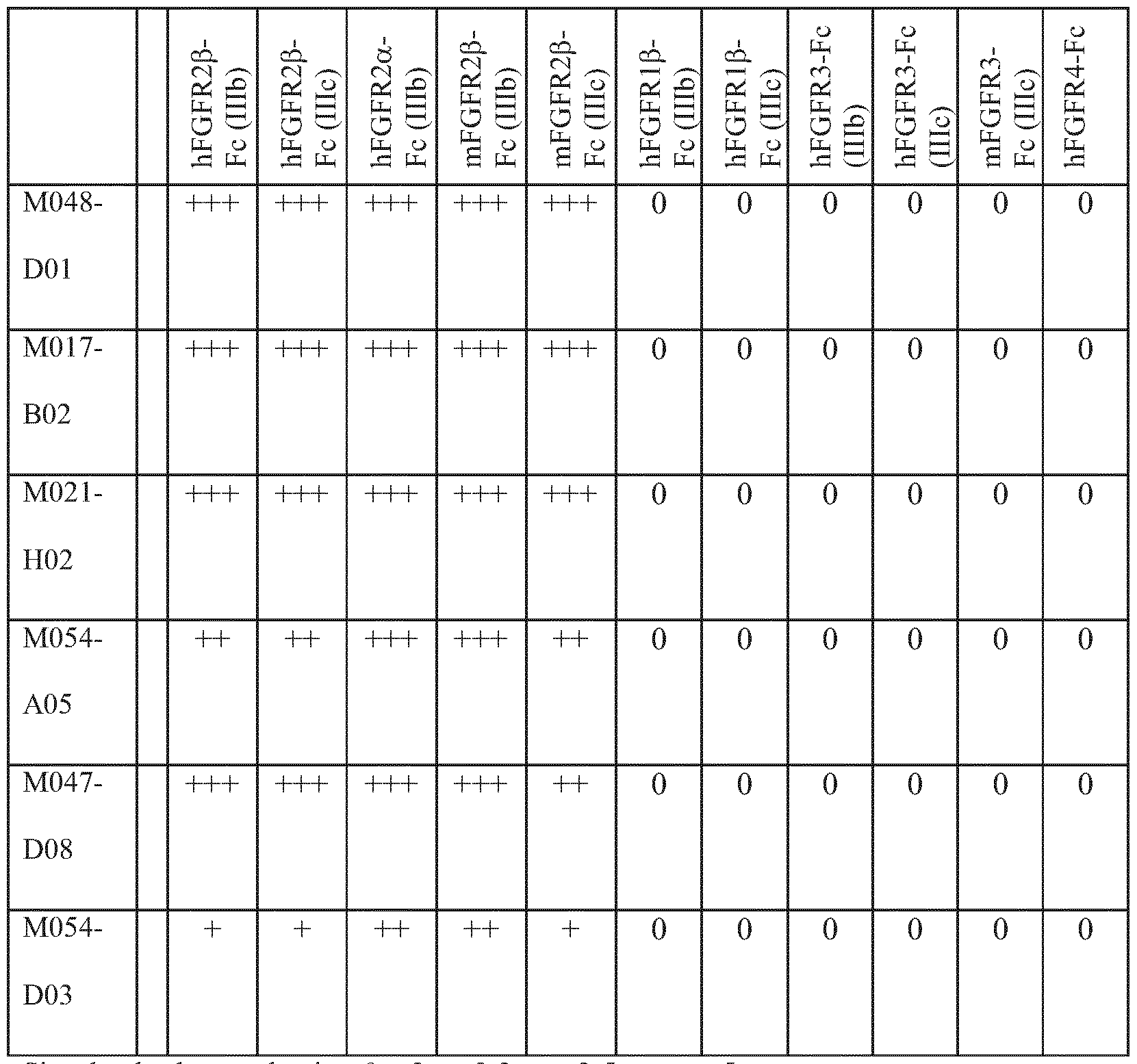


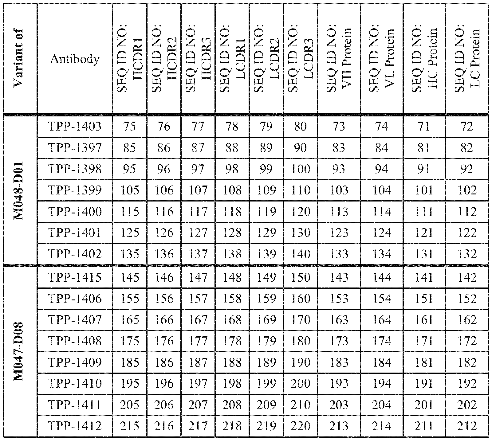

Claims
Priority Applications (13)
| Application Number | Priority Date | Filing Date | Title |
|---|---|---|---|
| KR1020147016644A KR20140102698A (en) | 2011-11-23 | 2012-11-22 | Anti-fgfr2 antibodies and uses thereof |
| NZ624534A NZ624534B2 (en) | 2011-11-23 | 2012-11-22 | Anti-fgfr2 antibodies and uses thereof |
| RU2014125143/10A RU2014125143A (en) | 2011-11-23 | 2012-11-22 | ANTI-FGFR2- ANTIBODIES AND THEIR APPLICATION |
| CA2856325A CA2856325A1 (en) | 2011-11-23 | 2012-11-22 | Anti-fgfr2 antibodies and uses thereof |
| MX2014005855A MX2014005855A (en) | 2011-11-23 | 2012-11-22 | Anti-fgfr2 antibodies and uses thereof. |
| EP12794685.3A EP2782934A1 (en) | 2011-11-23 | 2012-11-22 | Anti-fgfr2 antibodies and uses thereof |
| BR112014012137A BR112014012137A2 (en) | 2011-11-23 | 2012-11-22 | anti-fgfr2 antibodies and their uses |
| US14/359,663 US20140322220A1 (en) | 2011-11-23 | 2012-11-22 | Anti-FGFR2 Antibodies and Uses Thereof |
| AU2012342474A AU2012342474A1 (en) | 2011-11-23 | 2012-11-22 | Anti-FGFR2 antibodies and uses thereof |
| CN201280067865.9A CN104066750B (en) | 2011-11-23 | 2012-11-22 | Anti-FGFR2 antibodies and uses thereof |
| SG11201401973TA SG11201401973TA (en) | 2011-11-23 | 2012-11-22 | Anti-fgfr2 antibodies and uses thereof |
| JP2014542825A JP2015504434A (en) | 2011-11-23 | 2012-11-22 | Anti-FGFR2 antibody and use thereof |
| IL232399A IL232399A0 (en) | 2011-11-23 | 2014-04-30 | Anti-fgfr2 antibodies and uses thereof |
Applications Claiming Priority (2)
| Application Number | Priority Date | Filing Date | Title |
|---|---|---|---|
| EP11190227.6 | 2011-11-23 | ||
| EP11190227 | 2011-11-23 |
Publications (1)
| Publication Number | Publication Date |
|---|---|
| WO2013076186A1 true WO2013076186A1 (en) | 2013-05-30 |
Family
ID=47278276
Family Applications (1)
| Application Number | Title | Priority Date | Filing Date |
|---|---|---|---|
| PCT/EP2012/073325 WO2013076186A1 (en) | 2011-11-23 | 2012-11-22 | Anti-fgfr2 antibodies and uses thereof |
Country Status (14)
| Country | Link |
|---|---|
| US (1) | US20140322220A1 (en) |
| EP (1) | EP2782934A1 (en) |
| JP (1) | JP2015504434A (en) |
| KR (1) | KR20140102698A (en) |
| CN (1) | CN104066750B (en) |
| AR (1) | AR088941A1 (en) |
| AU (1) | AU2012342474A1 (en) |
| BR (1) | BR112014012137A2 (en) |
| CA (1) | CA2856325A1 (en) |
| IL (1) | IL232399A0 (en) |
| MX (1) | MX2014005855A (en) |
| RU (1) | RU2014125143A (en) |
| SG (2) | SG11201401973TA (en) |
| WO (1) | WO2013076186A1 (en) |
Cited By (31)
| Publication number | Priority date | Publication date | Assignee | Title |
|---|---|---|---|---|
| WO2014113729A3 (en) * | 2013-01-18 | 2014-09-12 | Foundation Medicine, Inc. | Methods of treating cholangiocarcinoma |
| WO2016048833A2 (en) | 2014-09-26 | 2016-03-31 | Janssen Pharmaceutica Nv | Use of fgfr mutant gene panels in identifying cancer patients that will be responsive to treatment with an fgfr inhibitor |
| WO2016207089A1 (en) | 2015-06-22 | 2016-12-29 | Bayer Pharma Aktiengesellschaft | Antibody drug conjugates (adcs) and antibody prodrug conjugates (apdcs) with enzymatically cleavable groups |
| WO2017060322A2 (en) | 2015-10-10 | 2017-04-13 | Bayer Pharma Aktiengesellschaft | Ptefb-inhibitor-adc |
| WO2017100642A1 (en) | 2015-12-11 | 2017-06-15 | Regeneron Pharmaceuticals, Inc. | Methods for reducing or preventing growth of tumors resistant to egfr and/or erbb3 blockade |
| WO2017162663A1 (en) | 2016-03-24 | 2017-09-28 | Bayer Pharma Aktiengesellschaft | Prodrugs of cytotoxic active agents having enzymatically cleavable groups |
| US10000814B2 (en) | 2011-10-21 | 2018-06-19 | Foundation Medicine, Inc. | ALK and NTRK1 fusion molecules and uses thereof |
| WO2018114578A1 (en) | 2016-12-21 | 2018-06-28 | Bayer Pharma Aktiengesellschaft | Antibody drug conjugates (adcs) having enzymatically cleavable groups |
| WO2018114798A1 (en) | 2016-12-21 | 2018-06-28 | Bayer Aktiengesellschaft | Prodrugs of cytotoxic active agents having enzymatically cleavable groups |
| US10138295B2 (en) | 2014-05-13 | 2018-11-27 | The Trustees Of The University Of Pennsylvania | Compositions comprising AAV expressing dual antibody constructs and uses thereof |
| WO2018220206A1 (en) | 2017-06-02 | 2018-12-06 | Janssen Pharmaceutica Nv | Fgfr2 inhibitors for the treatment of cholangiocarcinoma |
| US10172937B2 (en) | 2013-08-01 | 2019-01-08 | Five Prime Therapeutics, Inc. | Method of treatment of malignant solid tumors with afucosylated anti-FGFR2IIIb antibodies |
| EP3287522A4 (en) * | 2015-04-20 | 2019-03-27 | Daiichi Sankyo Company, Limited | Detection of fgfr2 |
| US10478494B2 (en) | 2015-04-03 | 2019-11-19 | Astex Therapeutics Ltd | FGFR/PD-1 combination therapy for the treatment of cancer |
| WO2019246572A1 (en) * | 2018-06-22 | 2019-12-26 | Children's Medical Center Corporation | Compositions and methods for treating primary aldosteonism |
| WO2020058432A1 (en) | 2018-09-21 | 2020-03-26 | Janssen Pharmaceutica Nv | Treatment of cholangiocarcinoma |
| US10716787B2 (en) | 2014-03-26 | 2020-07-21 | Astex Therapeutics Ltd | Combinations |
| US10759870B2 (en) | 2017-09-29 | 2020-09-01 | Chugai Seiyaku Kabushiki Kaisha | Multispecific antigen-binding molecules having blood coagulation factor VIII (FVIII) cofactor function-substituting activity and pharmaceutical formulations containing such a molecule as an active ingredient |
| US10780182B2 (en) | 2014-04-25 | 2020-09-22 | The Trustees Of The University Of Pennsylvania | Methods and compositions for treating metastatic breast cancer and other cancers in the brain |
| US10898482B2 (en) | 2015-02-10 | 2021-01-26 | Astex Therapeutics Ltd | Pharmaceutical compositions comprising N-(3,5-dimethoxyphenyl)-N'-1 methylethyl)-N-[3-(1-methyl-1H-pyrazol-4-yl)quinoxalin-6-yl]ethane-1,2-diamine |
| US11001636B2 (en) | 2016-06-15 | 2021-05-11 | Bayer Pharma Aktiengesellschaft | Specific antibody-drug-conjugates (ADCs) with KSP inhibitors and anti-CD123-antibodies |
| US11091555B2 (en) | 2017-05-16 | 2021-08-17 | Five Prime Therapeutics, Inc. | Method of treating gastric cancer with anti-FGFR2-IIIb antibodies and modified FOLFOX6 chemotherapy |
| US11230589B2 (en) | 2012-11-05 | 2022-01-25 | Foundation Medicine, Inc. | Fusion molecules and uses thereof |
| WO2022087243A1 (en) * | 2020-10-22 | 2022-04-28 | Regeneron Pharmaceuticals, Inc. | Anti-fgfr2 antibodies and methods of use thereof |
| US11433140B2 (en) | 2016-12-21 | 2022-09-06 | Bayer Pharma Aktiengesellschaft | Specific antibody drug conjugates (ADCs) having KSP inhibitors |
| US11447553B2 (en) | 2015-11-23 | 2022-09-20 | Five Prime Therapeutics, Inc. | FGFR2 inhibitors alone or in combination with immune stimulating agents in cancer treatment |
| WO2022221193A1 (en) | 2021-04-12 | 2022-10-20 | Affinia Therapeutics Inc. | Recombinant aav for treatment of neural disease |
| WO2022232289A1 (en) | 2021-04-27 | 2022-11-03 | Generation Bio Co. | Non-viral dna vectors expressing therapeutic antibodies and uses thereof |
| US11578372B2 (en) | 2012-11-05 | 2023-02-14 | Foundation Medicine, Inc. | NTRK1 fusion molecules and uses thereof |
| WO2023113806A1 (en) | 2021-12-16 | 2023-06-22 | Affinia Therapeutics, Inc. | Recombinant aav for treatment of neural disease |
| WO2023177655A1 (en) | 2022-03-14 | 2023-09-21 | Generation Bio Co. | Heterologous prime boost vaccine compositions and methods of use |
Families Citing this family (7)
| Publication number | Priority date | Publication date | Assignee | Title |
|---|---|---|---|---|
| MX2014007121A (en) * | 2011-12-14 | 2014-09-04 | Seattle Genetics Inc | Fgfr antibody drug conjugates (adcs) and the use thereof. |
| WO2016163433A1 (en) * | 2015-04-08 | 2016-10-13 | 第一三共株式会社 | Composition comprising anti-fgfr2 antibody and another agent |
| CN107513106A (en) * | 2016-06-17 | 2017-12-26 | 艾托金生物医药(苏州)有限公司 | Anti- FGFR2-IIIc monoclonal antibodies, hybridoma cell strain and application |
| AU2017303205A1 (en) * | 2016-07-29 | 2019-01-17 | Chugai Seiyaku Kabushiki Kaisha | Bispecific antibody exhibiting increased alternative FVIII-cofactor-function activity |
| CA3037461A1 (en) * | 2016-09-27 | 2018-04-05 | The University Of The Highlands And Islands | Antigen biomarkers |
| CN114901687A (en) * | 2019-12-24 | 2022-08-12 | 迪哲(江苏)医药股份有限公司 | Novel anti-FGFR 2B antibodies |
| EP4161566A1 (en) * | 2020-06-03 | 2023-04-12 | The Broad Institute, Inc. | Antagonistic biparatopic antibodies that specifically bind fibroblast growth factor receptor 2 and methods of using same |
Citations (21)
| Publication number | Priority date | Publication date | Assignee | Title |
|---|---|---|---|---|
| US4399216A (en) | 1980-02-25 | 1983-08-16 | The Trustees Of Columbia University | Processes for inserting DNA into eucaryotic cells and for producing proteinaceous materials |
| US4510245A (en) | 1982-11-18 | 1985-04-09 | Chiron Corporation | Adenovirus promoter system |
| US4634665A (en) | 1980-02-25 | 1987-01-06 | The Trustees Of Columbia University In The City Of New York | Processes for inserting DNA into eucaryotic cells and for producing proteinaceous materials |
| US4657760A (en) | 1979-03-20 | 1987-04-14 | Ortho Pharmaceutical Corporation | Methods and compositions using monoclonal antibody to human T cells |
| US4816397A (en) | 1983-03-25 | 1989-03-28 | Celltech, Limited | Multichain polypeptides or proteins and processes for their production |
| US4968615A (en) | 1985-12-18 | 1990-11-06 | Ciba-Geigy Corporation | Deoxyribonucleic acid segment from a virus |
| US5168062A (en) | 1985-01-30 | 1992-12-01 | University Of Iowa Research Foundation | Transfer vectors and microorganisms containing human cytomegalovirus immediate-early promoter-regulatory DNA sequence |
| US5179017A (en) | 1980-02-25 | 1993-01-12 | The Trustees Of Columbia University In The City Of New York | Processes for inserting DNA into eucaryotic cells and for producing proteinaceous materials |
| US5206344A (en) | 1985-06-26 | 1993-04-27 | Cetus Oncology Corporation | Interleukin-2 muteins and polymer conjugation thereof |
| US5225212A (en) | 1989-10-20 | 1993-07-06 | Liposome Technology, Inc. | Microreservoir liposome composition and method |
| US5500362A (en) | 1987-01-08 | 1996-03-19 | Xoma Corporation | Chimeric antibody with specificity to human B cell surface antigen |
| WO1997008320A1 (en) | 1995-08-18 | 1997-03-06 | Morphosys Gesellschaft Für Proteinoptimierung Mbh | Protein/(poly)peptide libraries |
| US5821337A (en) | 1991-06-14 | 1998-10-13 | Genentech, Inc. | Immunoglobulin variants |
| WO1999051642A1 (en) | 1998-04-02 | 1999-10-14 | Genentech, Inc. | Antibody variants and fragments thereof |
| US6194551B1 (en) | 1998-04-02 | 2001-02-27 | Genentech, Inc. | Polypeptide variants |
| US6737056B1 (en) | 1999-01-15 | 2004-05-18 | Genentech, Inc. | Polypeptide variants with altered effector function |
| WO2005066211A2 (en) * | 2003-12-19 | 2005-07-21 | Five Prime Therapeutics, Inc. | Fibroblast growth factor receptors 1, 2, 3, and 4 as targets for therapeutic intervention |
| WO2007144893A2 (en) | 2006-06-15 | 2007-12-21 | Fibron Ltd. | Antibodies blocking fibroblast growth factor receptor activation and methods of use thereof |
| WO2010054265A2 (en) | 2008-11-07 | 2010-05-14 | Galaxy Biotech, Llc. | Monoclonal antibodies to fibroblast growth factor receptor 2 |
| WO2010105426A1 (en) | 2009-03-19 | 2010-09-23 | 华为技术有限公司 | Method and device for linear block code coding |
| WO2011143318A2 (en) * | 2010-05-11 | 2011-11-17 | Aveo Pharmaceuticals, Inc. | Anti-fgfr2 antibodies |
-
2012
- 2012-11-21 AR ARP120104379A patent/AR088941A1/en active Pending
- 2012-11-22 WO PCT/EP2012/073325 patent/WO2013076186A1/en active Application Filing
- 2012-11-22 CN CN201280067865.9A patent/CN104066750B/en not_active Expired - Fee Related
- 2012-11-22 AU AU2012342474A patent/AU2012342474A1/en not_active Abandoned
- 2012-11-22 EP EP12794685.3A patent/EP2782934A1/en not_active Withdrawn
- 2012-11-22 SG SG11201401973TA patent/SG11201401973TA/en unknown
- 2012-11-22 MX MX2014005855A patent/MX2014005855A/en unknown
- 2012-11-22 KR KR1020147016644A patent/KR20140102698A/en not_active Application Discontinuation
- 2012-11-22 US US14/359,663 patent/US20140322220A1/en not_active Abandoned
- 2012-11-22 JP JP2014542825A patent/JP2015504434A/en not_active Ceased
- 2012-11-22 BR BR112014012137A patent/BR112014012137A2/en not_active IP Right Cessation
- 2012-11-22 CA CA2856325A patent/CA2856325A1/en not_active Abandoned
- 2012-11-22 RU RU2014125143/10A patent/RU2014125143A/en not_active Application Discontinuation
- 2012-11-22 SG SG10201603880WA patent/SG10201603880WA/en unknown
-
2014
- 2014-04-30 IL IL232399A patent/IL232399A0/en unknown
Patent Citations (21)
| Publication number | Priority date | Publication date | Assignee | Title |
|---|---|---|---|---|
| US4657760A (en) | 1979-03-20 | 1987-04-14 | Ortho Pharmaceutical Corporation | Methods and compositions using monoclonal antibody to human T cells |
| US4399216A (en) | 1980-02-25 | 1983-08-16 | The Trustees Of Columbia University | Processes for inserting DNA into eucaryotic cells and for producing proteinaceous materials |
| US4634665A (en) | 1980-02-25 | 1987-01-06 | The Trustees Of Columbia University In The City Of New York | Processes for inserting DNA into eucaryotic cells and for producing proteinaceous materials |
| US5179017A (en) | 1980-02-25 | 1993-01-12 | The Trustees Of Columbia University In The City Of New York | Processes for inserting DNA into eucaryotic cells and for producing proteinaceous materials |
| US4510245A (en) | 1982-11-18 | 1985-04-09 | Chiron Corporation | Adenovirus promoter system |
| US4816397A (en) | 1983-03-25 | 1989-03-28 | Celltech, Limited | Multichain polypeptides or proteins and processes for their production |
| US5168062A (en) | 1985-01-30 | 1992-12-01 | University Of Iowa Research Foundation | Transfer vectors and microorganisms containing human cytomegalovirus immediate-early promoter-regulatory DNA sequence |
| US5206344A (en) | 1985-06-26 | 1993-04-27 | Cetus Oncology Corporation | Interleukin-2 muteins and polymer conjugation thereof |
| US4968615A (en) | 1985-12-18 | 1990-11-06 | Ciba-Geigy Corporation | Deoxyribonucleic acid segment from a virus |
| US5500362A (en) | 1987-01-08 | 1996-03-19 | Xoma Corporation | Chimeric antibody with specificity to human B cell surface antigen |
| US5225212A (en) | 1989-10-20 | 1993-07-06 | Liposome Technology, Inc. | Microreservoir liposome composition and method |
| US5821337A (en) | 1991-06-14 | 1998-10-13 | Genentech, Inc. | Immunoglobulin variants |
| WO1997008320A1 (en) | 1995-08-18 | 1997-03-06 | Morphosys Gesellschaft Für Proteinoptimierung Mbh | Protein/(poly)peptide libraries |
| WO1999051642A1 (en) | 1998-04-02 | 1999-10-14 | Genentech, Inc. | Antibody variants and fragments thereof |
| US6194551B1 (en) | 1998-04-02 | 2001-02-27 | Genentech, Inc. | Polypeptide variants |
| US6737056B1 (en) | 1999-01-15 | 2004-05-18 | Genentech, Inc. | Polypeptide variants with altered effector function |
| WO2005066211A2 (en) * | 2003-12-19 | 2005-07-21 | Five Prime Therapeutics, Inc. | Fibroblast growth factor receptors 1, 2, 3, and 4 as targets for therapeutic intervention |
| WO2007144893A2 (en) | 2006-06-15 | 2007-12-21 | Fibron Ltd. | Antibodies blocking fibroblast growth factor receptor activation and methods of use thereof |
| WO2010054265A2 (en) | 2008-11-07 | 2010-05-14 | Galaxy Biotech, Llc. | Monoclonal antibodies to fibroblast growth factor receptor 2 |
| WO2010105426A1 (en) | 2009-03-19 | 2010-09-23 | 华为技术有限公司 | Method and device for linear block code coding |
| WO2011143318A2 (en) * | 2010-05-11 | 2011-11-17 | Aveo Pharmaceuticals, Inc. | Anti-fgfr2 antibodies |
Non-Patent Citations (98)
| Title |
|---|
| "Current Protocols in Protein Science", 1997, JOHN WILEY & SONS, article "Chapters 1, 4, 6, 8, 9, 10" |
| "Remington's Pharmaceutical Sciences", MAACK PUBLISHING CO |
| ABBAS, LICHTMAN AND POBER,: "Cellular and Molecular Immunology, 2nd Edition,", W.B. SAUNDERS COMPANY |
| ALESSANDRO ET AL., OTOL NEUROTOL., 2010 |
| ALFONSO R. GENNARO,: "REMINGTON'S PHARMACEUTICAL SCIENCES(18th ed.,", 1990, MACK PUB. CO., article "chapters 27 and 28", pages: 484 - 528 |
| AUSUBEL ET AL., J. MOL. BIOL, 1993, pages 21 |
| AUSUBEL ET AL., J. MOL. BIOL., 1997, pages 37 |
| AUSUBEL, F. M. ET AL.: "Current Protocols in Molecular Biology", 1989, GREENE PUBLISHING ASSOCIATES |
| AUSUBEL, F. M., BRENT, R., KINGSTON, R. E., MOORE, D. D., SEDMAN, J. G., SMITH, J. A., & STRUHL, K.: "Current Protocols in Molecular Biology", 1995, JOHN WILEY AND SONS |
| BAI ET AL., CANCER RES., vol. 70, 2010, pages 7630 - 7639 |
| BBHM, DISSERTATION, 2009 |
| BIRD ET AL., SCIENCE, vol. 242, 1988, pages 423 - 426 |
| BMC BIOTECHNOLOGY, vol. 7, 2007, pages 65 |
| BRAUCHLE ET AL., J PATHOL, vol. 149, 1996, pages 521 - 529 |
| BRAUN ET AL., PHIL TRANS R SOC LOND B, vol. 359, 2004, pages 753 - 757 |
| BYRON ET AL., CANCER RES, vol. 68, 2008, pages 6902 - 6907 |
| C. A. K BORREBAECK,: "Antibody Engineering (Breakthroughs in Molecular Biology", 1995, OXFORD UNIVERSITY PRESS |
| CHE ET AL., AM J PHYSIOL HEART CIRC PHYSIOL, vol. 300, 2011, pages H154 - H161 |
| CHO ET AL., AM J PATHOL, vol. 170, pages 1964 - 1974 |
| CHOTHIA; LESK, J MOL BIOL, vol. 196, 1987, pages 901 - 917 |
| CICORTAS GUNNARSSON ET AL., PROTEIN ENG DES SEL, vol. 17, no. 3, 2004, pages 213 - 21 |
| COLE ET AL., CANCER BIOL THER, vol. 10, 2010, pages 495 - 504 |
| ESWARAKUMAR; SCHLESSINGER, CYTOKINE & GROWTH FACTOR REVIEWS, vol. 16, 2005, pages 139 - 149 |
| FINCH ET AL., AM J PATHOL, vol. 151, 1997, pages 1619 - 1628 |
| FORTIN ET AL., J. NEUROSCI., vol. 25, 2005, pages 7470 - 7479 |
| GAZZANO-SANTORO ET AL., J. IMMUNOL. METHODS, vol. 202, 1996, pages 163 |
| GEBAUER M. ET AL., CURR. OPINION IN CHEM. BIOL., vol. 13, 2009, pages 245 - 255 |
| GOEDDEL: "Gene Expression Technology. Methods in Enzymology", 1990, ACADEMIC PRESS, pages: 185 |
| HALE; MARHAM, THE HARPER COLLINS DICTIONARY OF BIOLOGY, 1991 |
| HARIMOTO ET AL., ONCOLOGY, vol. 78, 2010, pages 361 - 368 |
| HERZOG ET AL., J. NUCL. MED., vol. 34, 1993, pages 2222 - 2226 |
| HUNTER ET AL., NATURE GENETICS, vol. 6, 2007, pages 870 - 874 |
| HUSTON ET AL., PROC. NATL. ACAD. SCI. USA, vol. 85, 1988, pages 5879 - 5883 |
| ISHIWATA ET AL., AM J PATHOL, vol. 153, 1998, pages 213 - 222 |
| KABAT ET AL.: "Sequences of Proteins of Immulological Interest, 5th Ed.", 1991, NATIONAL INSTITUTES OF HEALTH |
| KABAT, E. A.: "Sequences of Proteins of Immunological Interest, Fifth Edition,", 1991, NIH PUBLICATION NO. 91-3242 |
| KATOH, EXPERT REVIEWS, vol. 10, 2010, pages 1375 - 1379 |
| KATOH, J, INVEST DERMATOL, vol. 129, 2009, pages 1861 - 1867 |
| KATOH; KATOH, INT J ONCOL, vol. 29, 2006, pages 163 - 168 |
| KAWASE ET AL., INT J ONCOL, vol. 36, 2010, pages 331 - 340 |
| KHORANA ET AL., J. MOL. BIOL., vol. 72, 1971, pages 209 - 217 |
| KNAPPIK A. ET AL., JMB, vol. 296, 2000, pages 57 - 86 |
| KUNII ET AL., CANCER RES, vol. 68, 2008, pages 23 - 40,2348 |
| KUNII ET AL., CANCER RES, vol. 68, 2008, pages 2340 - 2348 |
| LACKIE ET AL.: "The Dictionary of Cell & Molecular Biology(3d ed.", 1999 |
| LI ET AL., J PERIDONTAL RES, vol. 40, 2005, pages 128 - 138 |
| LIN ET AL., JOURNAL DENNATOL SCI, vol. 59, 2010, pages 91 - 97 |
| LIU ET AL., PROC NATL. ACAD. SCI., vol. 93, 1996, pages 8618 - 8623 |
| MARSHALL ET AL., CLIN CANCER RES, vol. 17, 2011, pages 5016 - 5025 |
| MATSUNOBU ET AL., INT J CANCER, vol. 28, 2006, pages 307 - 314 |
| MCCAFFERTY ET AL., NATURE, vol. 348, 1990, pages 552 - 554 |
| NIU ET AL., J BIOL CHEM, vol. 282, 2007, pages 6601 - 6011 |
| NOMURA ET AL., BR J CANCER, vol. 99, 2008, pages 305 - 313 |
| NUTTALL S.D. ET AL., CURR. OPINION IN PHARMACOLOGY, vol. 8, 2008, pages 608 - 617 |
| ORNITZ ET AL., GENOME BIOL, vol. 2, 2001, pages 3005 |
| POLLOCK ET AL., ONCOGENE, vol. 26, 2007, pages 7158 - 7162 |
| R. J. KAUFMAN; P. A. SHARP, MOL. BIOL., vol. 159, 1982, pages 601 - 621 |
| R. KONTERMANN & S. DUEBEL,: "Antibody Engineering (Springer Laboratory Manual", 2001, SPRINGER VERLAG |
| RIVA ET AL., CLIN. CANCER RES., vol. 5, 1999, pages 3275 - 3280 |
| ROBERT K. SCOPES ET AL.: "Protein Purification, - Principles and Practice", 1994, SPRINGER SCIENCE AND BUSINESS MEDIA LLC |
| ROBIN ET AL.: "Gene Reviews", 1993, NCBI BOOKSHELF WASHINGTON |
| SAMBROOK ET AL.: "Molecular Cloning: A Laboratory Manual, 2nd Ed.", 1989, COLD SPRING HARBOR LABORATORY PRESS |
| SAMBROOK, FRITSCH AND MANIATIS: "Molecular Cloning; A Laboratory Manual, Second Edition,", 1989, COLD SPRING HARBOR |
| SAMBROOK, J.; FRITSCH, E. F.; MANIATIS, T.: "Molecular Cloning: A laboratory manual", 1989, COLD SPRING HARBOR LABORATORY PRESS |
| SCP, vol. 31, no. 7, pages 1163 - 9 |
| SHIMADA ET AL., PROC NATL ACAD SCI USA, vol. 98, 2001, pages 6500 - 6505 |
| SHIN ET AL., J CANCER RES CLIN ONCOL, vol. 128, 2002, pages 596 - 602 |
| SINGLETON: "Dictionary of Microbiology and Molecular Biology,2d ed.", 1994 |
| SLOOTSTRA ET AL., MOLECULAR DIVERSITY, vol. 1, 1996, pages 87 - 96 |
| SODEE ET AL., CLIN. NUC. MED., vol. 21, 1997, pages 759 - 766 |
| SODERLIND ET AL., NATURE BIOTECH., vol. 18, 2000, pages 853 - 856 |
| SÖDERLING ET AL., NAT. BIOTECH., vol. 18, 2000, pages 853 - 856 |
| STEELE ET AL., ONCOGENE, vol. 20, pages 5878 - 5887 |
| STEILING ET AL., ONCOGENE, vol. 22, 2003, pages 4380 - 4388 |
| TAKEDA ET AL., CLIN CANCER RES, vol. 13, 2007, pages 3051 - 3057 |
| TANIGUCHI ET AL., FERTIL STERIL, vol. 89, 2008, pages 478 - 480 |
| TIMMERMAN ET AL., J. MOL. RECOGNIT., vol. 20, 2007, pages 283 - 99 |
| TOM ET AL.: "Methods Express: Expression Systems", 2007, SCION PUBLISHING LTD |
| TOYOKAWA ET AL., ONCOL REPORTS, vol. 21, 2009, pages 875 - 880 |
| TURNER ET AL., ONCOGENE, vol. 29, 2010, pages 2013 - 2023 |
| ULANER ET AL., RADIOLOGY, vol. 246, no. 3, 2008, pages 895 - 902 |
| URIAUB; CHASIN, PROC. NATL. ACAD. SCI. USA, vol. 77, 1980, pages 4216 - 4220 |
| VEGA-HERNANDEZ ET AL., DEVELOPMENT, 2011, pages 3331 - 3340 |
| VIRNEKAS B., vol. 22, 1994, pages 5600, Retrieved from the Internet <URL:Nucl. Acids Res> |
| WALKER: "The Cambridge Dictionary of Science and Technology", 1988 |
| WEI ET AL., HYBRIDOMA, vol. 25, 2006, pages 115 - 124 |
| WERNER ET AL., J CANCER RES CLIN ONCOL, vol. 127, 2001, pages 207 - 216 |
| WINTER; DE GROOT, CELL MOL LIFE SCI, vol. 64, 2007, pages 692 - 703 |
| XU ET AL., J INVEST DERMATOL, vol. 131, 2011, pages 1521 - 1529 |
| YAMAMOTO-FUKUDA ET AL., EUR ARCH OTORHINOLARYNGOL, vol. 265, 2008, pages 1173 - 1178 |
| YAMAMOTO-FUKUDA ET AL., OTOL NEUROTOL., vol. 31, no. 5, July 2010 (2010-07-01), pages 745 - 51 |
| YAMASHITA ET AL., SURG TODAY, vol. 41, 2011, pages 24 - 3 8 |
| YAMASHITA ET AL., SURG TODAY, vol. 41, 2011, pages 24 - 38 |
| YAMAYOSHI ET AL., J PATHOL, vol. 204, 2004, pages 110 - 118 |
| YOSHINO ET AL., INT J ONCOL, vol. 31, 2007, pages 721 - 728 |
| ZHANG ET AL., ANTICANCER RES, vol. 18, 1998, pages 2541 - 2546 |
| ZHANG ET AL., HUM CANCER BIOL, vol. 15, 2009, pages 4017 - 4022 |
| ZHAO ET AL., CLIN CANCER RES., vol. 16, 2010, pages 5750 - 5758 |
Cited By (52)
| Publication number | Priority date | Publication date | Assignee | Title |
|---|---|---|---|---|
| US11098368B2 (en) | 2011-10-21 | 2021-08-24 | Foundation Medicine, Inc. | ALK and NTRK1 fusion molecules and uses thereof |
| US10000814B2 (en) | 2011-10-21 | 2018-06-19 | Foundation Medicine, Inc. | ALK and NTRK1 fusion molecules and uses thereof |
| US11578372B2 (en) | 2012-11-05 | 2023-02-14 | Foundation Medicine, Inc. | NTRK1 fusion molecules and uses thereof |
| US11230589B2 (en) | 2012-11-05 | 2022-01-25 | Foundation Medicine, Inc. | Fusion molecules and uses thereof |
| US11771698B2 (en) | 2013-01-18 | 2023-10-03 | Foundation Medicine, Inc. | Methods of treating cholangiocarcinoma |
| US10980804B2 (en) | 2013-01-18 | 2021-04-20 | Foundation Medicine, Inc. | Methods of treating cholangiocarcinoma |
| WO2014113729A3 (en) * | 2013-01-18 | 2014-09-12 | Foundation Medicine, Inc. | Methods of treating cholangiocarcinoma |
| AU2014207342B2 (en) * | 2013-01-18 | 2018-12-06 | Foundation Medicine, Inc. | Methods of treating cholangiocarcinoma |
| AU2014207342C1 (en) * | 2013-01-18 | 2019-04-04 | Foundation Medicine, Inc. | Methods of treating cholangiocarcinoma |
| US11235059B2 (en) | 2013-08-01 | 2022-02-01 | Five Prime Therapeutics, Inc. | Afucosylated anti-FGFR2IIIB antibodies |
| US10172937B2 (en) | 2013-08-01 | 2019-01-08 | Five Prime Therapeutics, Inc. | Method of treatment of malignant solid tumors with afucosylated anti-FGFR2IIIb antibodies |
| US10716787B2 (en) | 2014-03-26 | 2020-07-21 | Astex Therapeutics Ltd | Combinations |
| US11918576B2 (en) | 2014-03-26 | 2024-03-05 | Astex Therapeutics Ltd | Combination of an FGFR inhibitor and a CMET inhibitor |
| US11865187B2 (en) | 2014-04-25 | 2024-01-09 | The Trustees Of The University Of Pennsylvania | Methods and compositions for treating metastatic breast cancer and other cancers in the brain |
| US10780182B2 (en) | 2014-04-25 | 2020-09-22 | The Trustees Of The University Of Pennsylvania | Methods and compositions for treating metastatic breast cancer and other cancers in the brain |
| US10975140B2 (en) | 2014-05-13 | 2021-04-13 | The Trustees Of The University Of Pennsylvania | Compositions comprising AAV expressing dual antibody constructs and uses thereof |
| US10138295B2 (en) | 2014-05-13 | 2018-11-27 | The Trustees Of The University Of Pennsylvania | Compositions comprising AAV expressing dual antibody constructs and uses thereof |
| US10647758B2 (en) | 2014-05-13 | 2020-05-12 | The Trustees Of The University Of Pennsylvania | Compositions comprising AAV expressing dual antibody constructs and uses thereof |
| US10385119B2 (en) | 2014-05-13 | 2019-08-20 | Trustees Of The University Of Pennsylvania | Compositions comprising AAV expressing dual antibody constructs and uses thereof |
| WO2016048833A2 (en) | 2014-09-26 | 2016-03-31 | Janssen Pharmaceutica Nv | Use of fgfr mutant gene panels in identifying cancer patients that will be responsive to treatment with an fgfr inhibitor |
| EP4063516A1 (en) | 2014-09-26 | 2022-09-28 | Janssen Pharmaceutica NV | Use of fgfr mutant gene panels in identifying cancer patients that will be responsive to treatment with an fgfr inhibitor |
| US10898482B2 (en) | 2015-02-10 | 2021-01-26 | Astex Therapeutics Ltd | Pharmaceutical compositions comprising N-(3,5-dimethoxyphenyl)-N'-1 methylethyl)-N-[3-(1-methyl-1H-pyrazol-4-yl)quinoxalin-6-yl]ethane-1,2-diamine |
| US11684620B2 (en) | 2015-02-10 | 2023-06-27 | Astex Therapeutics Ltd | Pharmaceutical compositions comprising N-(3,5-dimethoxyphenyl)-N′-(1-methylethyl)-N-[3-(1-methyl-1H-pyrazol-4-yl)quinoxalin-6-yl]ethane-1,2-diamine |
| US10478494B2 (en) | 2015-04-03 | 2019-11-19 | Astex Therapeutics Ltd | FGFR/PD-1 combination therapy for the treatment of cancer |
| EP3287522A4 (en) * | 2015-04-20 | 2019-03-27 | Daiichi Sankyo Company, Limited | Detection of fgfr2 |
| US10881734B2 (en) | 2015-04-20 | 2021-01-05 | Daiichi Sankyo Company, Limited | Monoclonal antibodies to human fibroblast growth factor receptor 2 (HFGFR2) and methods of use thereof |
| WO2016207089A1 (en) | 2015-06-22 | 2016-12-29 | Bayer Pharma Aktiengesellschaft | Antibody drug conjugates (adcs) and antibody prodrug conjugates (apdcs) with enzymatically cleavable groups |
| US11123439B2 (en) | 2015-06-22 | 2021-09-21 | Bayer Pharma Aktiengesellschaft | Antibody drug conjugates (ADCS) and antibody prodrug conjugates (APDCS) with enzymatically cleavable groups |
| WO2017060322A2 (en) | 2015-10-10 | 2017-04-13 | Bayer Pharma Aktiengesellschaft | Ptefb-inhibitor-adc |
| US11447553B2 (en) | 2015-11-23 | 2022-09-20 | Five Prime Therapeutics, Inc. | FGFR2 inhibitors alone or in combination with immune stimulating agents in cancer treatment |
| WO2017100642A1 (en) | 2015-12-11 | 2017-06-15 | Regeneron Pharmaceuticals, Inc. | Methods for reducing or preventing growth of tumors resistant to egfr and/or erbb3 blockade |
| US11685714B2 (en) | 2016-03-24 | 2023-06-27 | Bayer Pharma Aktiengesellschaft | Prodrugs of cytotoxic active agents having enzymatically cleavable groups |
| WO2017162663A1 (en) | 2016-03-24 | 2017-09-28 | Bayer Pharma Aktiengesellschaft | Prodrugs of cytotoxic active agents having enzymatically cleavable groups |
| US11001636B2 (en) | 2016-06-15 | 2021-05-11 | Bayer Pharma Aktiengesellschaft | Specific antibody-drug-conjugates (ADCs) with KSP inhibitors and anti-CD123-antibodies |
| US11643469B2 (en) | 2016-06-15 | 2023-05-09 | Bayer Pharma Aktiengesellschaft | Specific antibody-drug-conjugates (ADCs) with KSP inhibitors and anti-CD123-antibodies |
| US11433140B2 (en) | 2016-12-21 | 2022-09-06 | Bayer Pharma Aktiengesellschaft | Specific antibody drug conjugates (ADCs) having KSP inhibitors |
| WO2018114578A1 (en) | 2016-12-21 | 2018-06-28 | Bayer Pharma Aktiengesellschaft | Antibody drug conjugates (adcs) having enzymatically cleavable groups |
| WO2018114798A1 (en) | 2016-12-21 | 2018-06-28 | Bayer Aktiengesellschaft | Prodrugs of cytotoxic active agents having enzymatically cleavable groups |
| US11478554B2 (en) | 2016-12-21 | 2022-10-25 | Bayer Pharma Aktiengesellschaft | Antibody drug conjugates (ADCS) having enzymatically cleavable groups |
| US11660351B2 (en) | 2016-12-21 | 2023-05-30 | Bayer Aktiengesellschaft | Antibody drug conjugates (ADCs) having enzymatically cleavable groups |
| US11091555B2 (en) | 2017-05-16 | 2021-08-17 | Five Prime Therapeutics, Inc. | Method of treating gastric cancer with anti-FGFR2-IIIb antibodies and modified FOLFOX6 chemotherapy |
| WO2018220206A1 (en) | 2017-06-02 | 2018-12-06 | Janssen Pharmaceutica Nv | Fgfr2 inhibitors for the treatment of cholangiocarcinoma |
| US11707463B2 (en) | 2017-06-02 | 2023-07-25 | Janssen Pharmaceutica Nv | FGFR2 inhibitors for the treatment of cholangiocarcinoma |
| US10759870B2 (en) | 2017-09-29 | 2020-09-01 | Chugai Seiyaku Kabushiki Kaisha | Multispecific antigen-binding molecules having blood coagulation factor VIII (FVIII) cofactor function-substituting activity and pharmaceutical formulations containing such a molecule as an active ingredient |
| WO2019246572A1 (en) * | 2018-06-22 | 2019-12-26 | Children's Medical Center Corporation | Compositions and methods for treating primary aldosteonism |
| WO2020058432A1 (en) | 2018-09-21 | 2020-03-26 | Janssen Pharmaceutica Nv | Treatment of cholangiocarcinoma |
| WO2022087243A1 (en) * | 2020-10-22 | 2022-04-28 | Regeneron Pharmaceuticals, Inc. | Anti-fgfr2 antibodies and methods of use thereof |
| US11866502B2 (en) | 2020-10-22 | 2024-01-09 | Regeneron Pharmaceuticals, Inc. | Anti-FGFR2 antibodies and methods of use thereof |
| WO2022221193A1 (en) | 2021-04-12 | 2022-10-20 | Affinia Therapeutics Inc. | Recombinant aav for treatment of neural disease |
| WO2022232289A1 (en) | 2021-04-27 | 2022-11-03 | Generation Bio Co. | Non-viral dna vectors expressing therapeutic antibodies and uses thereof |
| WO2023113806A1 (en) | 2021-12-16 | 2023-06-22 | Affinia Therapeutics, Inc. | Recombinant aav for treatment of neural disease |
| WO2023177655A1 (en) | 2022-03-14 | 2023-09-21 | Generation Bio Co. | Heterologous prime boost vaccine compositions and methods of use |
Also Published As
| Publication number | Publication date |
|---|---|
| CN104066750A (en) | 2014-09-24 |
| SG10201603880WA (en) | 2016-07-28 |
| JP2015504434A (en) | 2015-02-12 |
| RU2014125143A (en) | 2015-12-27 |
| US20140322220A1 (en) | 2014-10-30 |
| EP2782934A1 (en) | 2014-10-01 |
| AU2012342474A1 (en) | 2014-05-22 |
| MX2014005855A (en) | 2014-07-30 |
| AR088941A1 (en) | 2014-07-16 |
| NZ624534A (en) | 2016-06-24 |
| KR20140102698A (en) | 2014-08-22 |
| SG11201401973TA (en) | 2014-09-26 |
| IL232399A0 (en) | 2014-06-30 |
| CN104066750B (en) | 2017-05-17 |
| BR112014012137A2 (en) | 2017-05-30 |
| CA2856325A1 (en) | 2013-05-30 |
Similar Documents
| Publication | Publication Date | Title |
|---|---|---|
| US20140322220A1 (en) | Anti-FGFR2 Antibodies and Uses Thereof | |
| US11866495B2 (en) | Anti-CEACAM6 antibodies and uses thereof | |
| JP6240696B2 (en) | Anti-C4.4a antibody and use thereof | |
| US20160237160A1 (en) | Anti-tweakr antibodies and uses thereof | |
| WO2015189143A1 (en) | Aglycosyl anti-tweakr antibodies and uses thereof | |
| TW201335185A (en) | Anti-FGFR2 antibodies and uses thereof | |
| NZ624534B2 (en) | Anti-fgfr2 antibodies and uses thereof |
Legal Events
| Date | Code | Title | Description |
|---|---|---|---|
| 121 | Ep: the epo has been informed by wipo that ep was designated in this application |
Ref document number: 12794685 Country of ref document: EP Kind code of ref document: A1 |
|
| WWE | Wipo information: entry into national phase |
Ref document number: 232399 Country of ref document: IL |
|
| REEP | Request for entry into the european phase |
Ref document number: 2012794685 Country of ref document: EP |
|
| WWE | Wipo information: entry into national phase |
Ref document number: 2012794685 Country of ref document: EP |
|
| WWE | Wipo information: entry into national phase |
Ref document number: MX/A/2014/005855 Country of ref document: MX |
|
| ENP | Entry into the national phase |
Ref document number: 2856325 Country of ref document: CA |
|
| WWE | Wipo information: entry into national phase |
Ref document number: 14359663 Country of ref document: US |
|
| ENP | Entry into the national phase |
Ref document number: 2012342474 Country of ref document: AU Date of ref document: 20121122 Kind code of ref document: A Ref document number: 2014542825 Country of ref document: JP Kind code of ref document: A |
|
| NENP | Non-entry into the national phase |
Ref country code: DE |
|
| ENP | Entry into the national phase |
Ref document number: 20147016644 Country of ref document: KR Kind code of ref document: A |
|
| ENP | Entry into the national phase |
Ref document number: 2014125143 Country of ref document: RU Kind code of ref document: A |
|
| REG | Reference to national code |
Ref country code: BR Ref legal event code: B01A Ref document number: 112014012137 Country of ref document: BR |
|
| ENP | Entry into the national phase |
Ref document number: 112014012137 Country of ref document: BR Kind code of ref document: A2 Effective date: 20140520 |