WO2013081645A2 - Erbb3 mutations in cancer - Google Patents
Erbb3 mutations in cancer Download PDFInfo
- Publication number
- WO2013081645A2 WO2013081645A2 PCT/US2012/000568 US2012000568W WO2013081645A2 WO 2013081645 A2 WO2013081645 A2 WO 2013081645A2 US 2012000568 W US2012000568 W US 2012000568W WO 2013081645 A2 WO2013081645 A2 WO 2013081645A2
- Authority
- WO
- WIPO (PCT)
- Prior art keywords
- erbb3
- cancer
- mutation
- subject
- nucleic acid
- Prior art date
Links
Classifications
-
- C—CHEMISTRY; METALLURGY
- C12—BIOCHEMISTRY; BEER; SPIRITS; WINE; VINEGAR; MICROBIOLOGY; ENZYMOLOGY; MUTATION OR GENETIC ENGINEERING
- C12Q—MEASURING OR TESTING PROCESSES INVOLVING ENZYMES, NUCLEIC ACIDS OR MICROORGANISMS; COMPOSITIONS OR TEST PAPERS THEREFOR; PROCESSES OF PREPARING SUCH COMPOSITIONS; CONDITION-RESPONSIVE CONTROL IN MICROBIOLOGICAL OR ENZYMOLOGICAL PROCESSES
- C12Q1/00—Measuring or testing processes involving enzymes, nucleic acids or microorganisms; Compositions therefor; Processes of preparing such compositions
- C12Q1/68—Measuring or testing processes involving enzymes, nucleic acids or microorganisms; Compositions therefor; Processes of preparing such compositions involving nucleic acids
- C12Q1/6813—Hybridisation assays
-
- C—CHEMISTRY; METALLURGY
- C12—BIOCHEMISTRY; BEER; SPIRITS; WINE; VINEGAR; MICROBIOLOGY; ENZYMOLOGY; MUTATION OR GENETIC ENGINEERING
- C12Q—MEASURING OR TESTING PROCESSES INVOLVING ENZYMES, NUCLEIC ACIDS OR MICROORGANISMS; COMPOSITIONS OR TEST PAPERS THEREFOR; PROCESSES OF PREPARING SUCH COMPOSITIONS; CONDITION-RESPONSIVE CONTROL IN MICROBIOLOGICAL OR ENZYMOLOGICAL PROCESSES
- C12Q1/00—Measuring or testing processes involving enzymes, nucleic acids or microorganisms; Compositions therefor; Processes of preparing such compositions
- C12Q1/68—Measuring or testing processes involving enzymes, nucleic acids or microorganisms; Compositions therefor; Processes of preparing such compositions involving nucleic acids
- C12Q1/6876—Nucleic acid products used in the analysis of nucleic acids, e.g. primers or probes
- C12Q1/6883—Nucleic acid products used in the analysis of nucleic acids, e.g. primers or probes for diseases caused by alterations of genetic material
- C12Q1/6886—Nucleic acid products used in the analysis of nucleic acids, e.g. primers or probes for diseases caused by alterations of genetic material for cancer
-
- A—HUMAN NECESSITIES
- A61—MEDICAL OR VETERINARY SCIENCE; HYGIENE
- A61K—PREPARATIONS FOR MEDICAL, DENTAL OR TOILETRY PURPOSES
- A61K31/00—Medicinal preparations containing organic active ingredients
- A61K31/33—Heterocyclic compounds
- A61K31/395—Heterocyclic compounds having nitrogen as a ring hetero atom, e.g. guanethidine or rifamycins
- A61K31/495—Heterocyclic compounds having nitrogen as a ring hetero atom, e.g. guanethidine or rifamycins having six-membered rings with two or more nitrogen atoms as the only ring heteroatoms, e.g. piperazine or tetrazines
- A61K31/505—Pyrimidines; Hydrogenated pyrimidines, e.g. trimethoprim
- A61K31/517—Pyrimidines; Hydrogenated pyrimidines, e.g. trimethoprim ortho- or peri-condensed with carbocyclic ring systems, e.g. quinazoline, perimidine
-
- A—HUMAN NECESSITIES
- A61—MEDICAL OR VETERINARY SCIENCE; HYGIENE
- A61K—PREPARATIONS FOR MEDICAL, DENTAL OR TOILETRY PURPOSES
- A61K39/00—Medicinal preparations containing antigens or antibodies
- A61K39/395—Antibodies; Immunoglobulins; Immune serum, e.g. antilymphocytic serum
- A61K39/39533—Antibodies; Immunoglobulins; Immune serum, e.g. antilymphocytic serum against materials from animals
- A61K39/39558—Antibodies; Immunoglobulins; Immune serum, e.g. antilymphocytic serum against materials from animals against tumor tissues, cells, antigens
-
- A—HUMAN NECESSITIES
- A61—MEDICAL OR VETERINARY SCIENCE; HYGIENE
- A61P—SPECIFIC THERAPEUTIC ACTIVITY OF CHEMICAL COMPOUNDS OR MEDICINAL PREPARATIONS
- A61P35/00—Antineoplastic agents
-
- C—CHEMISTRY; METALLURGY
- C12—BIOCHEMISTRY; BEER; SPIRITS; WINE; VINEGAR; MICROBIOLOGY; ENZYMOLOGY; MUTATION OR GENETIC ENGINEERING
- C12Q—MEASURING OR TESTING PROCESSES INVOLVING ENZYMES, NUCLEIC ACIDS OR MICROORGANISMS; COMPOSITIONS OR TEST PAPERS THEREFOR; PROCESSES OF PREPARING SUCH COMPOSITIONS; CONDITION-RESPONSIVE CONTROL IN MICROBIOLOGICAL OR ENZYMOLOGICAL PROCESSES
- C12Q1/00—Measuring or testing processes involving enzymes, nucleic acids or microorganisms; Compositions therefor; Processes of preparing such compositions
- C12Q1/68—Measuring or testing processes involving enzymes, nucleic acids or microorganisms; Compositions therefor; Processes of preparing such compositions involving nucleic acids
- C12Q1/6844—Nucleic acid amplification reactions
- C12Q1/686—Polymerase chain reaction [PCR]
-
- C—CHEMISTRY; METALLURGY
- C12—BIOCHEMISTRY; BEER; SPIRITS; WINE; VINEGAR; MICROBIOLOGY; ENZYMOLOGY; MUTATION OR GENETIC ENGINEERING
- C12Q—MEASURING OR TESTING PROCESSES INVOLVING ENZYMES, NUCLEIC ACIDS OR MICROORGANISMS; COMPOSITIONS OR TEST PAPERS THEREFOR; PROCESSES OF PREPARING SUCH COMPOSITIONS; CONDITION-RESPONSIVE CONTROL IN MICROBIOLOGICAL OR ENZYMOLOGICAL PROCESSES
- C12Q2600/00—Oligonucleotides characterized by their use
- C12Q2600/112—Disease subtyping, staging or classification
-
- C—CHEMISTRY; METALLURGY
- C12—BIOCHEMISTRY; BEER; SPIRITS; WINE; VINEGAR; MICROBIOLOGY; ENZYMOLOGY; MUTATION OR GENETIC ENGINEERING
- C12Q—MEASURING OR TESTING PROCESSES INVOLVING ENZYMES, NUCLEIC ACIDS OR MICROORGANISMS; COMPOSITIONS OR TEST PAPERS THEREFOR; PROCESSES OF PREPARING SUCH COMPOSITIONS; CONDITION-RESPONSIVE CONTROL IN MICROBIOLOGICAL OR ENZYMOLOGICAL PROCESSES
- C12Q2600/00—Oligonucleotides characterized by their use
- C12Q2600/156—Polymorphic or mutational markers
Definitions
- the present invention concerns somatic ErbB3 mutations in cancer including methods of identifying, diagnosing, and prognosing ErbB3 cancers, as well as methods of treating cancer, including certain subpopulations of patients.
- HER human epidermal growth factor receptor
- RT receptor tyrosine kinases
- ERBB2/HER2, ERBB3/HER3 and ERBB4/HER4 (Hynes et al. Nature Reviews Cancer 5, 341- 354 (2005); Baselga et al. Nature Reviews Cancer 9, 463-475 (2009)).
- the ERBB family members contain an extracellular domain (ECD), a single-span transmembrane region, an intracellular tyrosine kinase domain, and a C-terminal signaling tail (Burgess et al. Mol Cell 12, 541-552 (2003); Ferguson. Annual Review of Biophysics 37, 353-373 (2008)).
- ERBB2 does not bind any of the known ERBB family ligands and is constitutively in an "untethered" (open) conformation suitable for dimerization (Garrett et al. Mol Cell 1 1 , 495-505 (2003).
- ERBB3 though capable of ligand binding,
- ERBB2 and ERBB3 are functionally incomplete on their own, their heterodimers are potent activators of cellular signaling (Pinkas-Kramarski et al. The EMBO Journal 15, 2452-2467 (1996); Tzahar et al. Molecular and Cellular Biology 16, 5276-5287
- ERBB receptors are critical regulators of normal growth and development, their deregulation has also been implicated in development and progression of cancers (Baselga et al. Nature Reviews Cancer 9, 463-475 (2009); Sithanandam et al. Cancer Gene Ther 15, 413- 448 (2008); Hynes et al. Current Opinion in Cell Biology 21, 177-184 (2009)).
- gene amplification leading to receptor overexpression and activating somatic mutations are known to occur in ERBB2 and EGFR in various cancers(Sithanandam et al. Cancer Gene Ther 15, 413-448 (2008); Hynes et al. Current Opinion in Cell Biology 21, 177-184 (2009); Wang et al.
- the present invention is based at least in part on the discovery of multiple somatic mutational events in the ERBB3 receptor of the human epidermal growth factor receptor (HER) family of receptor tyrosine kinases (RT ), that are associated with various human tumors including, without limitation, gastric and colon tumors. It is believed that these mutations predispose and/or directly contribute to human tumorigenesis. Indeed, as described herein, there is evidence that some of the mutations promote oncogenesis in vivo.
- HER human epidermal growth factor receptor
- RT receptor tyrosine kinases
- the present invention provides ErbB3 cancer detecting agents.
- the ErbB3 cancer detecting agent is an ErbB3 gastrointestinal cancer detecting agent.
- the detecting agent comprises a reagent capable of specifically binding to an ErbB3 mutation in an ErbB3 nucleic acid sequence.
- the ErbB3 nucleic acid sequence comprises SEQ ID NO:3 or 1.
- the reagent comprises a polynucleotide of formula
- X is any nucleic acid and a is between about 0 and about 250;
- Y is an ErbB3 mutation codon
- Z is any nucleic acid and b is between about 0 and about 250.
- the mutation codon encodes (i) an amino acid at a position of SEQ ID NO:2 selected from the group consisting of 104, 809, 232, 262, 284, 325, 846, 928, 60, 1 11, 135, 295, 406, 453, 498, 1089, and 1 164; or (ii) a stop codon at position 193.
- SEQ ID NO:2 selected from the group consisting of 104, 809, 232, 262, 284, 325, 846, 928, 60, 1 11, 135, 295, 406, 453, 498, 1089, and 1 164; or (ii) a stop codon at position 193.
- the gastrointestinal cancer is gastric cancer or colon cancer.
- the present invention provides a method of determining the presence of ErbB3 gastrointestinal cancer in a subject.
- the method comprises detecting in a biological sample obtained from the subject a mutation in a nucleic acid sequence encoding ErbB3, wherein the mutation results in an amino acid change at at least one position of the ErbB3 amino acid sequence and wherein the mutation is indicative of an ErbB3 gastrointestinal cancer in the subject.
- the mutation resulting in an amino acid change is at a position of SEQ ID NO:2 selected from the group consisting of 104, 809, 232, 262, 284, 325, 846, 928, 60, 11 1, 135, 295, 406, 453, 498, 1089, 1 164, and 193.
- the gastrointestinal cancer is gastric cancer or colon cancer.
- the present invention provides a method of determining the presence of ErbB3 cancer in a subject.
- the method comprises detecting in a biological sample obtained from the subject the presence or absence of an amino acid mutation in a nucleic acid sequence encoding ErbB3, wherein the mutation results in an amino acid change at at least one position in SEQ ID NO: 2 selected from the group consisting of 104, 809, 232, 262, 284, 325, 846, 928, 60, 1 1 1, 135, 295, 406, 453, 498, 1089, 1 164, 193, 492, and 714, and wherein the presence of the mutation is indicative of an ErbB3 cancer in the subject.
- the ErbB3 cancer is selected from the group consisting of gastric, colon, esophageal, rectal, cecum, non-small-cell lung (NSCLC) adenocarinoma, NSCLC (Squamous carcinoma), renal carcinoma, melanoma, ovarian, lung large cell, small-cell lung cancer (SCLC), hepatocellular (HCC), lung, and pancreatic.
- the determining methods further comprise one of the following additional steps: administering a therapeutic agent to said subject, identifying the subject in need, obtaining the sample from a subject in need, or any combination thereof.
- the therapeutic agent is an ErbB inhibitor.
- the ErbB inhibitor is selected from the group consisting of an EGFR antagonist, an ErbB2 antagonist, an ErbB3 antagonist, an ErbB4 antagonist, and an EGFR/ErbB3 antagonist.
- the detecting step comprises amplifying or sequencing.
- the detecting comprises amplifying or sequencing the mutation and detecting the mutation or sequence thereof.
- the amplifying comprises admixing an amplification primer or amplification primer pair with a nucleic acid template isolated from the sample.
- the primer or primer pair is complementary or partially complementary to a region proximal to or including said mutation, and is capable of initiating nucleic acid polymerization by a polymerase on the nucleic acid template.
- the amplifying further comprises extending the primer or primer pair in a DNA polymerization reaction comprising a polymerase and the template nucleic acid to generate an amplicon.
- the mutation in the amplifying or sequencing, is detected by a process that includes one or more of: sequencing the mutation in a genomic DNA isolated from the biological sample, hybridizing the mutation or an amplicon thereof to an array, digesting the mutation or an amplicon thereof with a restriction enzyme, or real-time PCR amplification of the mutation.
- the amplifying or sequencing further comprises partially or fully sequencing the mutation in a nucleic acid isolated from the biological sample.
- the amplifying comprises performing a polymerase chain reaction (PCR), reverse transcriptase PCR (RT-PCR), or ligase chain reaction (LCR) using a nucleic acid isolated from the biological sample as a template in the PCR, RT-PCR, or LCR.
- PCR polymerase chain reaction
- RT-PCR reverse transcriptase PCR
- LCR ligase chain reaction
- the present invention provides a method of treating gastrointestinal cancer in a subject in need.
- the method comprises a) detecting in a biological sample obtained from the subject a mutation in a nucleic acid sequence encoding ErbB3, wherein the mutation results in an amino acid change at at least one position of the ErbB3 amino acid sequence and wherein the mutation is indicative of an ErbB3 gastrointestinal cancer in the subject.
- the method further comprises b) administering a therapeutic agent to said subject.
- the mutation resulting in an amino acid change is at a position of SEQ ID NO:2 selected from the group consisting of 104, 809, 232, 262, 284, 325, 846, 928, 60, 1 1 1 , 135, 295, 406, 453, 498, 1089, 1 164, and 193.
- the gastrointestinal cancer is gastric cancer or colon cancer.
- the present invention provides a method of treating an ErbB3 cancer in a subject.
- the method comprises of a) detecting in a biological sample obtained from the subject the presence or absence of an amino acid mutation in a nucleic acid sequence encoding ErbB3, wherein the mutation results in an amino acid change at at least one position in SEQ ID NO: 2 selected from the group consisting of 104, 809, 232, 262, 284, 325, 846, 928, 60, 1 1 1 , 135, 295, 406, 453, 498, 1089, 1 164, 193, 492, and 714, and wherein the presence of the mutation is indicative of an ErbB3 cancer in the subject.
- the method further comprises b) administering a therapeutic agent to said subject.
- the ErbB3 cancer is selected from the group consisting of gastric, colon, esophageal, rectal, cecum, colorectal, non-small-cell lung (NSCLC) adenocarinoma, NSCLC (Squamous carcinoma), renal carcinoma, melanoma, ovarian, lung large cell, small-cell lung cancer (SCLC), hepatocellular (HCC), lung, and pancreatic.
- the methods of treatment involve ErbB3 inhibitors.
- the therapeutic agent is an ErbB inhibitor.
- the ErbB inhibitor is selected from the group consisting of an EGFR antagonist, an ErbB2 antagonist, an ErbB3 antagonist, an ErbB4 antagonist, and an EGFR/ErbB3 antagonist.
- the antagonist is a small molecule inhibitor.
- the antagonist is an antagonist antibody.
- the antibody is selected from the group consisting of a monoclonal antibody, a bispecific antibody, a chimeric antibody, a human antibody, a humanized antibody and an antibody fragment. Additional embodiments
- the present invention provides methods of determining the presence of ErbB3 cancer in a subject in need.
- the method comprises the step of detecting in a biological sample obtained from the subject the presence or absence of an amino acid mutation in a nucleic acid sequence encoding ErbB3, wherein the mutation results in an amino acid change at at least one position selected from the group consisting of M60, R193, A232, P262, V295, G325, M406, D492, V714, Q809, R1089, Tl 164.
- the method further comprises administering a therapeutic agent to the subject.
- the method further comprises identifying the subject in need.
- the method further comprises obtaining the sample from a subject in need.
- the ErbB3 cancer is selected from the group consisting of gastric, colon, esophageal, rectal, cecum, non-small-cell lung (NSCLC) adenocarinoma, NSCLC (Squamous carcinoma), renal carcinoma, melanoma, ovarian, lung large cell, small-cell lung cancer (SCLC), hepatocellular (HCC), lung, and pancreatic.
- NSCLC non-small-cell lung
- SCLC small-cell lung cancer
- HCC hepatocellular
- the present invention provides methods of determining the presence of
- ErbB3 gastrointestinal cancer in a subject in need comprising detecting in a biological sample obtained from the subject a mutation in a nucleic acid sequence encoding ErbB3, wherein the mutation results in an amino acid change at at least one position selected from the group consisting of V104, Yl 11, A232, P262, G284, T3.89, and Q809.
- the - method further comprises administering a therapeutic agent to the subject.
- the method further comprises identifying the subject in need.
- the method further comprises obtaining the sample from a subject in need.
- the ErbB3 gastrointestinal cancer is gastric cancer or colon cancer.
- the present invention provides methods of identifying ErbB3 gastrointestinal cancer in a subject in need that is likely to respond to an ErbB antagonist, said method comprising detecting in a gastrointestinal cancer cell obtained from the subject a mutation in a nucleic acid sequence encoding ErbB3, wherein the mutation at at least one position selected from the group consisting of V104, Yl 1 1, A232, P262, G284, T389, and Q809.
- the method further comprises administering a therapeutic agent to the subject.
- the method further comprises obtaining the sample from a subject in need.
- the ErbB3 gastrointestinal cancer is gastric cancer or colon cancer.
- the present invention provides methods of treating ErbB3 cancer in a subject in need.
- the method comprises the step of detecting in a biological sample obtained from the subject the presence or absence of an amino acid mutation in a nucleic acid sequence encoding ErbB3, wherein the mutation results in an amino acid change at at least one position selected from the group consisting of M60, R193, A232, P262, V295, G325, M406, D492, V714, Q809, R1089, Tl 164.
- the method further comprises the step of administering a therapeutic agent to said subject.
- the present invention provides methods of treating ErbB3
- the method comprises the step of detecting in a biological sample obtained from the subject a mutation in a nucleic acid sequence encoding ErbB3, wherein the mutation results in an amino acid change at at least one position selected from the group consisting of V104, Yl 1 1, A232, P262, G284, T389, and Q809.
- the method further comprises the step of administering a therapeutic agent to said subject.
- the therapeutic agent administered in the methods of the present invention is an ErbB inhibitor.
- the ErbB inhibitor is selected from the group consisting of an EGFR antagonist, an ErbB2 antagonist, an ErbB3 antagonist, an ErbB4 antagonist, and an EGFR/ErbB3 antagonist.
- the inhibitor is a small molecule inhibitor.
- the ErbB inhibitor is an EGFR antagonist.
- the ErbB inhibitor is an ErbB2 antagonist.
- the ErbB inhibitor is an ErbB3 antagonist.
- the ErbB inhibitor is an ErbB4 antagonist.
- the ErbB inhibitor is an EGFR/ErbB3 antagonist.
- the antagonist is an antagonist antibody.
- the antibody is selected from the group consisting of a monoclonal antibody, a bispecific antibody, a chimeric antibody, a human antibody, a humanized antibody and an antibody fragment.
- the methods of the present invention comprise a detecting step in which the nucleic acid sequence obtained from the sample is analyzed for the presence or absence of the mutation(s).
- the detecting comprises amplifying or sequencing the mutation and detecting the mutation or sequence thereof.
- the amplifying comprises admixing an amplification primer or amplification primer pair with a nucleic acid template isolated from the sample.
- the primer or primer pair is complementary or partially complementary to a region proximal to or including said mutation, and is capable of initiating nucleic acid polymerization by a polymerase on the nucleic acid template.
- the method further comprises extending the primer or primer pair in a DNA polymerization reaction comprising a polymerase and the template nucleic acid to generate an amplicon.
- the mutation is detected by a process that includes one or more of: sequencing the mutation in a genomic DNA isolated from the biological sample, hybridizing the mutation or an amplicon thereof to an array, digesting the mutation or an amplicon thereof with a restriction enzyme, or real-time PCR amplification of the mutation.
- the method comprises partially or fully sequencing the mutation in a nucleic acid isolated from the biological sample.
- the amplifying comprises performing a polymerase chain reaction (PCR), reverse transcriptase PCR (RT-PCR), or ligase chain reaction (LCR) using a nucleic acid isolated from the biological sample as a template in the PCR, RT-PCR, or LCR.
- PCR polymerase chain reaction
- RT-PCR reverse transcriptase PCR
- LCR ligase chain reaction
- Figure 1 Samples. Provides a list of the human tissue samples used in the study of ERBB3 in human cancers.
- Figure 2 Representative wild-type ERBB3 nucleic acid sequence (Accession No.
- ERBB3 somatic mutations mapped over the ERBB3 protein domains are shown. Hotspot mutations depicted as repeating amino acid changes in a light red background. Height of the background vertical bar around the mutated residue is proportional to the frequency of mutation at that particular position, (c-d) ERBB3 non-synonymous somatic mutations (inverted triangles; red triangles depict hotspots) depicted over ERBB3 protein domains. The histogram on the top represents count of mutations at each position detected observed in samples in this study and other published studies (red bars indicate hot spot mutations and blue bars represent additional non-hotspot mutants tested for activity), (e-f) Expanded and supplemented view of Figure 4 (a- b).
- Figure 4 (a-f) provides a linear view of ErbB3 where Figure 4a, c, and e show an N-terminal half, and Figure 4b, d, and f show an C-terminal half.
- lupus (XP_538226.2 (SEQ ID NO: 131)), B.taurus (NP_001096575.1 (Full length sequence is disclosed as SEQ ID NO: 129 and the various regions are disclosed as SEQ ID NOS 192-211, respectively, in order of appearance)), M.musculus (NP 034283.1 (Full length sequence is disclosed as SEQ ID NO: 127 and the various regions are disclosed as SEQ ID NOS 152-171, respectively, in order of appearance)) and R.norvegicus (NP_058914.2 (Full length sequence is disclosed as SEQ ID NO: 128 and the various regions are disclosed as SEQ ID NOS 172-191, respectively, in order of appearance)) were aligned using Clustal W (Larkin, M. A. et al. Bioinformatics (Oxford, England) 23, 2947-2948 (2007)). Mutated residues are show in a red oval background.
- ERBB3/ERBB2 ECD heterodimer based on EGFR ECD dimer using ERBB3 [pdb 1M6B] and ERBB2 [pdb 1N8Z].
- ERBB3 kinase domain somatic mutations shown in red mapped on to a structure of the ERBB3 kinase domain [pdb 3LMG]. * stop codon.
- ERBB3 mutants support EGF-independent proliferation of MCFIOA cells in 3D culture.
- MCFIOA cells stably expressing ERBB3 mutants either alone or together with either EGFR or ERBB2 show EGF-independent proliferation.
- Studies involving MCFIOA were performed in the absence of serum, EGF and NRG1.
- MCFIOA cells stably expressing ERBB3 mutants either alone (A) or together with either EGFR (B) or ERBB2 (C) show elevated downstream signaling as assessed by western blot. Studies involving MCFIOA were performed in the absence of serum, EGF and NRG1. EV - empty vector.
- ERBB3 mutants support EGF-independent proliferation of MCF10A cells in 3D culture.
- MCF10A cells stably expressing ERBB3 mutants either alone or together with ' either EGFR or ERBB2 show large acinar architecture, increased i67 staining and increased migration index compared to ERBB3/ ERBB2 expressing MCF10A cells.
- Data represents mean ⁇ SEM of the three independent experiments. Studies involving MCF10A were performed in the absence of serum, EGF and NRG1. EV - empty vector.
- Figure 13A shows representative images of MCF10A cells expressing the indicated ERBB3 mutants along with ERBB2 following migration from a transwell in the migration assay (a), and quantitation of this migration effect (b).
- Figure 13B (a-e) shows that ERBB3 mutants support anchorage independent growth of IMCE colonic epithelial cells.
- IMCE colonic epithelial cells expressing either ERBB3 by itself or in combination with ERBB2 showed anchorage independent growth (a), increased number of colonies (b), elevated phospho signaling (c, d) and in vivo growth (e) compared to ERBB3- WT/ERBB2 expressing IMCE cells.
- ERBB3 mutants transform and promote IL3 -independent survival of BaF3 cells.
- BaF3 cells stably expressing ERBB3 mutants either alone or together with either EGFR or ERBB2 promotes IL3 -independent survival.
- BaF3 studies were performed in the absence of IL- 3 and NRG1.
- FIG. 15A-C ERBB3 mutants transform and promote IL3 -independent survival of
- Anti-NRGl a NRG1 neutralizing antibody, does not affect IL-3 -independent survival of BaF3 cells promoted by ERBB3 mutants co-expressed with ERBB2. BaF3 studies were performed in the absence of IL-3 and NRG1.
- Figure 20A-C Quantitation of ERBB3-ERBB2 heterodimers. Images from Proximity ligation assay (Figure 17) were analyzed using Duolink image software tool (Uppsala, Sweden). At least 100 cells from 5 to 6 image fields for the indicated combination of ERBB3 and ERBB2 expressing cells were analyzed for signal (red dots) resulting from ERBB2/ERBB3 dimers. The assay was performed with FLAG (ERBB3) and gD (ERBB2) antibody (A) or native ERBB3 and ERBB3 antibodies (B). Data are show as Mean ⁇ SEM. Figure 20C shows that NRGl was unable to support survival of BaF3 cells expressing ERBB3-WT or mutants alone.
- FIG. 21 ERBB3 ECD mutants show increased IL-3 independent BaF3 survival in response to different dose of exongenous ligand NRGl .
- BaF3 studies were performed in the absence of IL-3.
- FIG 22 ERBB3 mutants promote oncogenesis and lead to reduced overall survival.
- FIG. 23 Flow cytometric analysis of total bone marrow cells (A) and spleen cells (B) isolated from mice receiving GFP-tagged BaF3 cells expressing the various ERBB3
- FIG 26 Representative H&E-stained bone marrow (top), spleen (middle) and liver (bottom) sections from the same mice analyzed in Figure 21.
- the bone marrow from empty vector animals consists of normal hematopoietic cells.
- * infiltrating tumor cells
- R red pulp
- W lymphoid follicles of white pulp.
- the scale bar corresponds to ⁇ ⁇ .
- FIG. 27 Representative images of spleen and liver from mice transplanted with ERBB3 mutant expressing BaF3 cells are shown.
- Figure 28 Efficacy of anti-ERBB antibodies and small molecule inhibitors on oncogenic activity of ERBB3 mutants. Effect of targeted therapeutics on IL-3 independent proliferation of BaF3 cells stably expressing ERBB3 mutants together with ERBB2 as indicated in the figure.
- Figure 29 Representative images of the effect of targeted therapeutics on anchorage- independent growth of BaF3 cells stably expressing ERBB3 mutants together with ERBB2 as indicated in the figure.
- Figure 30 Schematic depicting the ERBB receptors and various targeted agents that were tested in this study.
- Anti-ERBB3 antibodies are effectively targeting ERBB3 mutants in vivo.
- Control antibody-treated group receive 40 mg/kg QW anti-Ragweed antibody.
- FIG 32 Effect of targeted therapeutics on BaF3 cells stably expressing ERBB3 mutants together with ERBB2 as indicated in the figure. Concentration of antibodies and small molecule inhibitors used for treatment is same as indicated in Figure 27.
- FIG. 33 Effect of ERBB antibodies and small molecule inhibitors on phosphorylation of ERBB3 and downstream signaling molecules in BaF3 at 8 h after treatment is shown. Effect of these same agents at 24 h is shown in Fig. 30.
- Figure 34 Proportion of infiltrating BaF3 cells expressing mutant ERBB3, G284R (A) and Q809R (B), in bone marrow (BM) and spleen following treatment with the antibodies as indicated in the figure.
- FIG. 35 Liver and spleen weight from animal implanted with ERBB3 mutant cells, G284R (A) and Q809R (B), following treatment with the antibodies as indicated.
- FIG. 36 Infiltrating GFP positive BaF3 cell expressing ERBB3 mutant isolated from spleen and bone marrow of mice implanted with these cells are shown.
- FIG 37A-H ERBB3 mutants transform and promote IL3 -independent survival of BaF3 cells.
- A IL3 -independent survival of BaF3 cells stably expressing ERBB3 mutants either alone or together with ERBB2 or ERBB2-KD.
- B A representative image of anchorage- independent growth of BaF3 cells stably expressing ERBB3 mutants either alone or in combination with either ERBB2 or ERBB2-KD.
- C Bar graph showing the number of colonies formed by BaF3 cells expressing the ERBB3 mutants along with ERBB2 show in (B). Very few colonies were formed by cells expressing ERBB3 mutants alone or in combination with ERBB2- KD.
- FIG 38A-J shRNA-mediated ERBB3 knockdown delays tumor growth.
- A- J CW-2 and DV-90 stably expressing inducible ERBB3 targeting shRNA upon dox-induction showed lower levels of ERBB3 and pERK (A, B), anchorage independent growth (C-F) and reduced in vivo growth (H, J) compared to uninduced cells (A-F) or cells expressing luciferase targeting shRNA (A-F, G & I).
- Data in (E, F) represent the number of anchorage independent colonies formed quantitated from multiple filed of images like the one show in (C, D). Data are shown as Mean ⁇ SEM.
- Figure 39 provides a nucleic acid sequence (SEQ ID NO: 3) and amino acid sequence
- polynucleotide or “nucleic acid,” as used interchangeably herein, refers to polymers of nucleotides of any length, and include DNA and RNA.
- the nucleotides can be deoxyribonucleotides, ribonucleotides, modified nucleotides or bases, and/or their analogs, or any substrate that can be incorporated into a polymer by DNA or RNA polymerase.
- a polynucleotide may comprise modified nucleotides, such as methylated nucleotides and their analogs. If present, modification to the nucleotide structure may be imparted before or after assembly of the polymer.
- the sequence of nucleotides may be interrupted by non-nucleotide components.
- a polynucleotide may be further modified after polymerization, such as by conjugation with a labeling component.
- Other types of modifications include, for example, "caps", substitution of one or more of the naturally occurring nucleotides with an analog, intemucleotide modifications such as, for example, those with uncharged linkages (e.g., methyl phosphonates, phosphotriesters, phosphoamidates, carbamates, etc.) and with charged linkages (e.g., phosphorothioates, phosphorodithioates, etc.), those containing pendant moieties, such as, for example, proteins (e.g., nucleases, toxins, antibodies, signal peptides, poly-L-lysine, etc.), those with intercalators (e.g., acridine, psoralen, etc.), those containing chelators (e.g.,
- any of the hydroxyl groups ordinarily present in the sugars may be replaced, for example, by phosphonate groups, phosphate groups, protected by standard protecting groups, or activated to prepare additional linkages to additional nucleotides, or may be conjugated to solid supports.
- the 5' and 3' terminal OH can be phosphorylated or substituted with amines or organic capping groups moieties of from 1 to 20 carbon atoms.
- Other hydroxyls may also be derivatized to standard protecting groups.
- Polynucleotides can also contain analogous forms of ribose or deoxyribose sugars that are generally known in the art, including, for example, 2'-0-methyl-2'-0-allyl, 2'-fluoro- or 2'-azido-ribose, carbocyclic sugar analogs, .alpha. -anomeric sugars, epimeric sugars such as arabinose, xyloses or lyxoses, pyranose sugars, furanose sugars, sedoheptuloses, acyclic analogs and abasic nucleoside analogs such as methyl riboside.
- One or more phosphodiester linkages may be replaced by alternative linking groups.
- linking groups include, but are not limited to, embodiments wherein phosphate is replaced by P(0)S("thioate”), P(S)S ("dithioate”), "(O)NR 2 ("amidate”), P(0)R, P(0)OR * , CO or CH2 ("formacetal"), in which each R or R' is independently H or substituted or unsubstituted alkyl (1-20 C) optionally containing an ether (— O--) linkage, aryl, alkenyl, cycloalkyl, cycloalkenyl or araldyl. Not all linkages in a polynucleotide need be identical. The preceding description applies to all polynucleotides referred to herein, including RNA and DNA.
- Oligonucleotide refers to short, single stranded polynucleotides that are at least about seven nucleotides in length and less than about 250 nucleotides in length.
- Oligonucleotides may be synthetic.
- the terms “oligonucleotide” and “polynucleotide” are notmutually exclusive. The description above for polynucleotides is equally and fully applicable to oligonucleotides.
- primer refers to a single stranded polynucleotide that is capable of hybridizing to a nucleic acid and allowing the polymerization of a complementary nucleic acid, generally by providing a free 3' ⁇ OH group.
- the term “gene” refers to a DNA sequence that encodes through its template or messenger RNA a sequence of amino acids characteristic of a specific peptide, polypeptide, or protein.
- the term “gene” also refers to a DNA sequence that encodes an RNA product.
- the term gene as used herein with reference to genomic DNA includes intervening, non-coding regions as well as regulatory regions and can include 5' and 3' ends.
- the term “somatic mutation” or “somatic variation” refers to a change in a nucleotide sequence (e.g., an insertion, deletion, inversion, or substitution of one or more nucleotides), which is acquired in a cell of the body as opposed to a germ line cell.
- amino acid variation refers to a change in an amino acid sequence (e.g., an insertion, substitution, or deletion of one or more amino acids, such as an internal deletion or an N- or C-terminal truncation) relative to a reference sequence.
- variant refers to either a nucleotide variation or an amino acid variation.
- a genetic variation at a nucleotide position corresponding to a somatic mutation refers to a nucleotide variation in a polynucleotide sequence at the relative corresponding DNA position occupied by said somatic mutation.
- the term also encompasses the corresponding variation in the complement of the nucleotide sequence, unless otherwise indicated.
- array refers to an ordered arrangement of hybridizable array elements, preferably polynucleotide probes (e.g., oligonucleotides), on a substrate.
- the substrate can be a solid substrate, such as a glass slide, or a semi-solid substrate, such as nitrocellulose membrane.
- Amplification refers to the process of producing one or more copies of a reference nucleic acid sequence or its complement. Amplification may be linear or exponential (e.g., the polymerase chain reaction (PCR)).
- a "copy” does not necessarily mean perfect sequence complementarity or identity relative to the template sequence. For example, copies can include nucleotide analogs such as deoxyinosine, intentional sequence alterations (such as sequence alterations introduced through a primer comprising a sequence that is hybridizable, but not fully complementary, to the template), and/or sequence errors that occur during
- mutant-specific oligonucleotide refers to an oligonucleotide that hybridizes to a region of a target nucleic acid that comprises a nucleotide variation (often a substitution).
- “Somatic mutation-specific hybridization” means that, when a mutation-specific oligonucleotide is hybridized to its target nucleic acid, a nucleotide in the mutation-specific oligonucleotide specifically base pairs with the nucleotide variation.
- oligonucleotide capable of mutation-specific hybridization with respect to a particular nucleotide variation is said to be "specific for” that variation.
- mutation-specific primer refers to an mutation-specific oligonucleotide that is a primer.
- primer extension assay refers to an assay in which nucleotides are added to a nucleic acid, resulting in a longer nucleic acid, or “extension product,” that is detected directly or indirectly.
- the nucleotides can be added to extend the 5' or 3' end of the nucleic acid.
- mutation-specific nucleotide incorporation assay refers to a primer extension assay in which a primer is (a) hybridized to target nucleic acid at a region that is 3' or 5' of a nucleotide variation and (b) extended by a polymerase, thereby incorporating into the extension product a nucleotide that is complementary to the nucleotide variation.
- mutant-specific primer extension assay refers to a primer extension assay in which a mutation-specific primer is hybridized to a target nucleic acid and extended.
- mutant-specific oligonucleotide hybridization assay refers to an assay in which (a) a mutation-specific oligonucleotide is hybridized to a target nucleic acid and (b) hybridization is detected directly or indirectly.
- 5' nuclease assay refers to an assay in which hybridization of a mutation- specific oligonucleotide to a target nucleic acid allows for nucleolytic cleavage of the hybridized probe, resulting in a detectable signal.
- assay employing molecular beacons refers to an assay in which hybridization of a mutation-specific oligonucleotide to a target nucleic acid results in a level of detectable signal that is higher than the level of detectable signal emitted by the free oligonucleotide.
- oligonucleotide ligation assay refers to an assay in which a mutation -specific oligonucleotide and a second oligonucleotide are hybridized adjacent to one another on a target nucleic acid and ligated together (either directly or indirectly through intervening nucleotides), and the ligation product is detected directly or indirectly.
- target sequence refers generally to a polynucleotide sequence of interest in which a nucleotide variation is suspected or known to reside, including copies of such target nucleic acid generated by amplification.
- detection includes any means of detecting, including direct and indirect detection.
- cancer and “cancerous” refer to or describe the physiological condition in mammals that is typically characterized by unregulated cell growth.
- the cancer diagnosed in accordance with the present invention is any type of cancer characterized by the presence of an ErbB3 mutation, specifically including metastatic or locally advanced non-resectable cancer, including, without limitation, gastric, colon, esophageal, rectal, cecum, colorectal, non-small-cell lung (NSCLC) adenocarinoma, NSCLC (Squamous carcinoma), renal carcinoma, melanoma, ovarian, lung large cell, small-cell lung cancer (SCLC), hepatocellular (HCC), lung cancer, head & neck cancer, and pancreatic cancer.
- NSCLC non-small-cell lung
- SCLC small-cell lung cancer
- HCC hepatocellular
- a subject “at risk” of developing cancer may or may not have detectable disease or symptoms of disease, and may or may not have displayed detectable disease or symptoms of disease prior to the diagnostic methods described herein.
- “At risk” denotes that a subject has one or more risk factors, which are measurable parameters that correlate with development of cancer, as described herein and known in the art. A subject having one or more of these risk factors has a higher probability of developing cancer than a subject without one or more of these risk factor(s).
- diagnosis is used herein to refer to the identification or classification of a molecular or pathological state, disease or condition, for example, cancer.
- Diagnosis may also refer to the classification of a particular sub-type of cancer, e.g., by molecular features (e.g., a patient subpopulation characterized by nucleotide variation(s) in a particular gene or nucleic acid region.).
- a method of aiding diagnosis of cancer can comprise measuring the presence of absence of one or more genetic markers indicative of cancer or an increased risk of having cancer in a biological sample from an individual.
- prognosis is used herein to refer to the prediction of the likelihood of developing cancer.
- prediction is used herein to refer to the likelihood that a patient will respond either favorably or unfavorably to a drug or set of drugs.
- the prediction relates to the extent of those responses.
- the prediction relates to whether and/or the probability that a patient will survive or improve following treatment, for example treatment with a particular therapeutic agent, and for a certain period of time without disease recurrence.
- the predictive methods of the invention can be used clinically to make treatment decisions by choosing the most appropriate treatment modalities for any particular patient.
- the predictive methods of the present invention are valuable tools in predicting if a patient is likely to respond favorably to a treatment regimen, such as a given therapeutic regimen, including for example, administration of a given therapeutic agent or combination, surgical intervention, steroid treatment, etc., or whether long-term survival of the patient, following a therapeutic regimen is likely.
- a treatment regimen such as a given therapeutic regimen, including for example, administration of a given therapeutic agent or combination, surgical intervention, steroid treatment, etc., or whether long-term survival of the patient, following a therapeutic regimen is likely.
- treatment refers to clinical intervention in an attempt to alter the natural course of the individual or cell being treated, and can be performed before or during the course of clinical pathology. Desirable effects of treatment include preventing the occurrence or recurrence of a disease or a condition or symptom thereof, alleviating a condition or symptom of the disease, diminishing any direct or indirect pathological consequences of the disease, decreasing the rate of disease progression, ameliorating or palliating the disease state, and achieving remission or improved prognosis.
- methods and compositions of the invention are useful in attempts to delay development of a disease or disorder.
- cancer therapeutic agent refers to an agent that when provided in an effective amount is known, clinically shown, or expected by clinicians to provide a therapeutic benefit in a subject who has cancer.
- the phrase includes any agent that is marketed by a manufacturer, or otherwise used by licensed clinicians, as a clinically-accepted agent that when provided in an effective amount would be expected to provide a therapeutic effect in a subject who has cancer.
- a cancer therapeutic agent comprises chemotherapy agents, HER dimerization inhibitors, HER antibodies, antibodies directed against tumor associated antigens, anti-hormonal compounds, cytokines, EGFR-targeted drugs, anti-angiogenic agents, tyrosine kinase inhibitors, growth inhibitory agents and antibodies, cytotoxic agents, antibodies that induce apoptosis, COX inhibitors, farnesyl transferase inhibitors, antibodies that binds oncofetal protein CA 125, HER2 vaccines, Raf or ras inhibitors, liposomal doxorubicin, topotecan, taxene, dual tyrosine kinase inhibitors, TLK286, EMD-7200, pertuzumab, trastuzumab, erlotinib, and bevacizumab.
- chemotherapeutic agents used in chemotherapy, include alkylating agents such as thiotepa and CYTOXAN® cyclosphosphamide; alkyl sulfonates such as busulfan, improsulfan and piposulfan; aziridines such as benzodopa, carboquone, meturedopa, and uredopa;
- ethylenimines and methylamelamines including altretamine, triethylenemelamine,
- trietylenephosphoramide, triethiylenethiophosphoramide and trimethylolomelamine TL 286 (TELCYTATM); acetogenins (especially bullatacin and bullatacinone); delta-9- tetrahydrocannabinol (dronabinol, MARINOL®); beta-lapachone; lapachol; colchicines;
- camptothecin including the synthetic analogue topotecan (HYCAMTIN®), CPT-1 1 (irinotecan, CAMPTOSAR®), acetylcamptothecin, scopolectin, and 9- aminocamptothecin
- bryostatin callystatin; CC-1065 (including its adozelesin, carzelesin and bizelesin synthetic analogues); podophyllotoxin; podophyllinic acid; teniposide; cryptophycins (particularly cryptophycin 1 and cryptophycin 8); dolastatin; duocarmycin (including the synthetic analogues, KW-2189 and CB1-TM1); eleutherobin; pancratistatin; a sarcodictyin; spongistatin; nitrogen mustards such as chlorambucil, chlornaphazine, cholophosphamide, estramustine, ifosfamide
- bisphosphonates such as clodronate
- antibiotics such as the enediyne antibiotics (e. g., calicheamicin, especially calicheamicin gamma II and calicheamicin omegall (see, e.g., Agnew, Chem Intl. Ed.
- anthracyclines such as annamycin, AD 32, alcarubicin, daunorubicin, dexrazoxane, DX-52-1 , epirubicin, GPX-100, idarubicin, KRN5500, menogaril, dynemicin, including dynemicin A, an esperamicin, neocarzinostatin chromophore and related chromoprotein enediyne antiobiotic chromophores, aclacinomysins, actinomycin, authramycin, azaserine, bleomycins, cactinomycin, carabicin, carminomycin, carzinophilin, chromomycinis, dactinomycin, detorubicin, 6-diazo-5-oxo-L-norleucine, ADRIAMYCIN® doxorubicin (including morph
- thioguanine pyrimidine analogs such as ancitabine, azacitidine, 6-azauridine, carmofur, cytarabine, dideoxyuridine, doxifluridine, enocitabine, and floxuridine; androgens such as calusterone, dromostanolone propionate, epitiostanol, mepitiostane, and testolactone; anti- adrenals such as aminoglutethimide, mitotane, and trilostane; folic acid replenisher such as folinic acid (leucovorin); aceglatone; anti-folate anti-neoplastic agents such as ALIMTA®, LY231514 pemetrexed, dihydro folate reductase inhibitors such as methotrexate, anti-metabolites such as 5-fluorouracil (5-FU) and its prodrugs such as UFT, S-l and capecitabine, and thymidylate synthas
- pharmaceutical formulation refers to a preparation which is in such form as to permit the biological activity of an active ingredient contained therein to be effective, and which contains no additional components which are unacceptably toxic to a subject to which the formulation would be administered.
- a “pharmaceutically acceptable carrier” refers to an ingredient in a pharmaceutical formulation, other than an active ingredient, which is nontoxic to a subject.
- a pharmaceutically acceptable carrier includes, but is not limited to, a buffer, excipient, stabilizer, or preservative.
- an “effective amount” refers to an amount effective, at dosages and for periods of time necessary, to achieve the desired therapeutic or prophylactic result.
- a “therapeutically effective amount” of a therapeutic agent may vary according to factors such as the disease state, age, sex, and weight of the individual, and the ability of the antibody to elicit a desired response in the individual.
- a therapeutically effective amount is also one in which any toxic or detrimental effects of the therapeutic agent are outweighed by the therapeutically beneficial effects.
- the therapeutically effective amount of the drug may reduce the number of cancer cells; reduce the tumor size; inhibit (i.e., slow to some extent and preferably stop) cancer cell infiltration into peripheral organs; inhibit (i.e., slow to some extent and preferably stop) tumor metastasis; inhibit, to some extent, tumor growth; and/or relieve to some extent one or more of the symptoms associated with the cancer.
- the drug may prevent growth and/or kill existing cancer cells, it may be cytostatic and/or cytotoxic.
- a "prophylactically effective amount” refers to an amount effective, at dosages and for periods of time necessary, to achieve the desired prophylactic result. Typically but not necessarily, since a prophylactic dose is used in subjects prior to or at an earlier stage of disease, the prophylactically effective amount will be less than the therapeutically effective amount.
- an “individual,” “subject” or “patient” is a vertebrate.
- the vertebrate is a mammal.
- Mammals include, but are not limited to, primates (including human and non-human primates) and rodents (e.g., mice and rats).
- rodents e.g., mice and rats.
- a mammal is a human.
- a "patient subpopulation,” and grammatical variations thereof, as used herein, refers to a patient subset characterized as having one or more distinctive measurable and/or identifiable characteristics that distinguishes the patient subset from others in the broader disease category to which it belongs. Such characteristics include disease subcategories, gender, lifestyle, health history, organs/tissues involved, treatment history, etc.
- a patient in one embodiment, a patient
- subpopulation is characterized by nucleic acid signatures, including nucleotide variations in particular nucleotide positions and/or regions (such as somatic mutations).
- control subject refers to a healthy subject who has not been diagnosed as having cancer and who does not suffer from any sign or symptom associated with cancer.
- sample refers to a composition that is obtained or derived from a subject of interest that contains a cellular and/or other molecular entity that is to be characterized and/or identified, for example based on physical, biochemical, chemical and/or physiological characteristics.
- disease sample and variations thereof refers to any sample obtained from a subject of interest that would be expected or is known to contain the cellular and/or molecular entity that is to be characterized.
- tissue or cell sample is meant a collection of similar cells obtained from a tissue of a subject or patient.
- the source of the tissue or cell sample may be solid tissue as from a fresh, frozen and/or preserved organ or tissue sample or biopsy or aspirate; blood or any blood constituents; bodily fluids such as serum, urine, sputum, or saliva.
- the tissue sample may also be primary or cultured cells or cell lines.
- the tissue or cell sample is obtained from a disease tissue/organ.
- the tissue sample may contain compounds which are not naturally intermixed with the tissue in nature such as preservatives, anticoagulants, buffers, fixatives, nutrients, antibiotics, or the like.
- a “reference sample”, “reference cell”, “reference tissue”, “control sample”, “control cell”, or “control tissue”, as used herein, refers to a sample, cell or tissue obtained from a source known, or believed, not to be afflicted with the disease or condition for which a method or composition of the invention is being used to identify.
- a reference sample, reference cell, reference tissue, control sample, control cell, or control tissue is obtained from a healthy part of the body of the same subject or patient in whom a disease or condition is being identified using a composition or method of the invention.
- a reference sample, reference cell, reference tissue, control sample, control cell, or control tissue is obtained from a healthy part of the body of an individual who is not the subject or patient in whom a disease or condition is being identified using a composition or method of the invention.
- a "section" of a tissue sample is meant a single part or piece of a tissue sample, e.g. a thin slice of tissue or cells cut from a tissue sample. It is understood that multiple sections of tissue samples may be taken and subjected to analysis according to the present invention, provided that it is understood that the present invention comprises a method whereby the same section of tissue sample is analyzed at both morphological and molecular levels, or is analyzed with respect to both protein and nucleic acid.
- correlate or “correlating” is meant comparing, in any way, the performance and/or results of a first analysis or protocol with the performance and/or results of a second analysis or protocol. For example, one may use the results of a first analysis or protocol in carrying out a second protocol and/or one may use the results of a first analysis or protocol to determine whether a second analysis or protocol should be performed. With respect to the embodiment of gene expression analysis or protocol, one may use the results of the gene expression analysis or protocol to determine whether a specific therapeutic regimen should be performed.
- a “small molecule” or “small organic molecule” is defined herein as an organic molecule having a molecular weight below about 500 Daltons.
- label when used herein refers to a detectable compound or composition.
- the label may be detectable by itself (e.g., radioisotope labels or fluorescent labels) or, in the case of an enzymatic label, may catalyze chemical alteration of a substrate compound or composition which results in a detectable product.
- Radionuclides that can serve as detectable labels include, for example, 1-131, 1-123, 1-125, Y-90, Re-188, Re-186, At-21 1, Cu-67, Bi-212, and Pd-109.
- references to "about” a value or parameter herein includes (and describes) embodiments that are directed to that value or parameter per se. For example, description referring to "about X” includes description of "X.”
- package insert is used to refer to instructions customarily included in commercial packages of therapeutic products, that contain information about the indications, usage, dosage, administration, combination therapy, contraindications and/or warnings concerning the use of such therapeutic products.
- antibody and “immunoglobulin” are used interchangeably in the broadest sense and include monoclonal antibodies (e.g., full length or intact monoclonal antibodies), polyclonal antibodies, monovalent antibodies, multivalent antibodies, multispecific antibodies (e.g., bispecific antibodies so long as they exhibit the desired biological activity) and may also include certain antibody fragments (as described in greater detail herein).
- An antibody can be chimeric, human, humanized and/or affinity matured.
- Antibody fragments comprise a portion of an intact antibody, preferably comprising the antigen binding region thereof. Examples of antibody fragments include Fab, Fab', F(ab') 2 , and Fv fragments; diabodies; linear antibodies; single-chain antibody molecules; and multispecific antibodies formed from antibody fragments.
- an antibody of this invention "which binds" an antigen of interest is one that binds the antigen with sufficient affinity such that the antibody is useful as a diagnostic and/or therapeutic agent in targeting a protein or a cell or tissue expressing the antigen.
- the term "specific binding” or “specifically binds to” or is “specific for” a particular polypeptide or an epitope on a particular polypeptide target means binding that is measurably different from a non-specific interaction. Specific binding can be measured, for example, by determining binding of a molecule compared to binding of a control molecule.
- specific binding can be determined by competition with a control molecule that is similar to the target, for example, an excess of non-labeled target. In this case, specific binding is indicated if the binding of the labeled target to a probe is competitively inhibited by excess non-labeled target.
- "specifically binds" refers to binding of an antibody to its specified target HER receptors and not other specified non-target HER receptors.
- an anti-HER3 antibody specifically binds to HER3 but does not specifically bind to EGFR, HER2, or HER4.
- An EGFR/HER3 bispecific antibody specifically binds to EGFR and HER3 but does not specifically bind to HER2 or HER4.
- a "HER receptor” or "ErbB receptor” is a receptor protein tyrosine kinase which belongs to the HER receptor family and includes EGFR (ErbBl, HERl), HER2 (ErbB2), HER3 (ErbB3) and HER4 (ErbB4) receptors.
- the HER receptor will generally comprise an extracellular domain, which may bind an HER ligand and/or dimerize with another HER receptor molecule; a lipophilic transmembrane domain; a conserved intracellular tyrosine kinase domain; and a carboxyl-terminal signaling domain harboring several tyrosine residues which can be
- the HER receptor may be a "native sequence” HER receptor or an “amino acid sequence variant” thereof.
- the HER receptor is a native sequence human HER receptor.
- the "HER pathway” refers to the signaling network mediated by the HER receptor family.
- ErbBl ErbBl
- HER1 epidermal growth factor receptor
- EGFR epidermal growth factor receptor
- Variants of EGFR also include deletional, substitutional and insertional variants, for example those described in Lynch et al (New England Journal of Medicine 2004, 350:2129), Paez et al (Science 2004, 304:1497), and Pao et al (PNAS 2004, 101 :13306).
- EGFR extracellular domain or "EGFR ECD” refers to a domain of EGFR that is outside of a cell, either anchored to a cell membrane, or in circulation, including fragments thereof.
- the extracellular domain of EGFR may comprise four domains: "Domain I” (amino acid residues from about 1- 158, "Domain II” (amino acid residues 159-336), “Domain III” (amino acid residues 337-470), and “Domain IV” (amino acid residues 471-645), where the boundaries are approximate, and may vary by about 1-3 amino acids.
- ErbB2 and HER2 are used interchangeably herein and refer to human HER2 protein described, for example, in Semba et al, PNAS (USA) 82:6497-6501 (1985) and Yamamoto et al. Nature 319:230-234 (1986) (GenBank accession number X03363).
- the term “er£B2” refers to the gene encoding human HER2 and "neu " refers to the gene encoding rat pi 85"ea.
- Preferred HER2 is native sequence human HER2.
- HER2 extracellular domain refers to a domain of HER2 that is outside of a cell, either anchored to a cell membrane, or in circulation, including fragments thereof.
- the extracellular domain of HER2 may comprise four domains: "Domain I” (amino acid residues from about 1-195, "Domain ⁇ ” (amino acid residues from about 196-319), “Domain III” (amino acid residues from about 320-488), and "Domain IV” (amino acid residues from about 489-630) (residue numbering without signal peptide). See Garrett et al. Mol. Cell.
- ErbB3 and HER3 refer to the receptor polypeptide as disclosed, for example, in US Pat. Nos. 5,183,884 and 5,480,968 as well as Kraus et al. PNAS (USA) 86:9193-9197 (1989) (see also Figures 2 and 3)
- HER3 extracellular domain or "HER3 ECD” or “ErbB3 extracellular domain” refers to a domain of HER3 that is outside of a cell, either anchored to a cell membrane, or in circulation, including fragments thereof.
- the extracellular domain of HER3 may comprise four domains: Domain I, Domain II, Domain III, and Domain IV.
- the HER3 ECD comprises amino acids 1-636 (numbering including signal peptide).
- HER3 domain III comprises amino acids 328-532 (numbering including signal peptide.
- ErbB4 and HER4 herein refer to the receptor polypeptide as disclosed, for example, in EP Pat Appln No 599,274; Plowman et al, Proc. Natl. Acad. ScL USA, 90: 1746- 1750 (1993); and Plowman et al, Nature, 366:473-475 (1993), including isoforms thereof, e.g., as disclosed in W099/19488, published April 22, 1999.
- HER ligand is meant a polypeptide which binds to and/or activates a HER receptor.
- the HER ligand of particular interest herein is a native sequence human HER ligand such as epidermal growth factor (EGF) (Savage et al, J. Biol Chem. 247:7612-7621 (1972)); transforming growth factor alpha (TGF-a) (Marquardt et al, Science 223: 1079-1082 (1984)); amphiregulin also known as schwanoma or keratinocyte autocrine growth factor (Shoyab et al Science 243:1074-1076 (1989); Kimura et al Nature
- EGF epidermal growth factor
- TGF-a transforming growth factor alpha
- amphiregulin also known as schwanoma or keratinocyte autocrine growth factor
- HB-EGF heparin-binding epidermal growth factor
- epiregulin Toyoda et al, J. Biol. Chem. 270:7495-7500 (1995); and Komurasaki et al Oncogene 15:2841-2848 (1997)); a heregulin (see below); neuregulin-2 (NRG-2) (Carraway et al, Nature 387:512-516 (1997)); neuregulin-3 (NRG-3) (Zhang et al, Proc. Natl. Acad.
- HER ligands which bind EGFR include EGF, TGF-a, amphiregulin, betacellulin, HB-EGF and epiregulin.
- HER ligands which bind HER3 include heregulins and NRG-2.
- HER ligands capable of binding HER4 include betacellulin, epiregulin, HB-EGF, NRG-2, NRG-3, NRG-4, and heregulins.
- Heregulin when used herein refers to a polypeptide encoded by the heregulin gene product as disclosed in U.S. Patent No. 5,641,869, or Marchionni et al, Nature, 362:312- 318 (1993).
- heregulins include heregulin-a, heregulin- ⁇ , heregulin-P2 and heregulin- ⁇ 3 (Holmes et al, Science, 256: 1205-1210 (1992); and U.S. Patent No. 5,641 ,869); neu differentiation factor (NDF) (Peles et al Cell 69: 205-216 (1992)); acetylcholine receptor- inducing activity (ARIA) (Falls et al.
- NDF neu differentiation factor
- ARIA acetylcholine receptor- inducing activity
- GGFs glial growth factors
- SMDF motor neuron derived factor
- a "HER dimer” herein is a noncovalently associated dimer comprising at least two HER receptors. Such complexes may form when a cell expressing two or more HER receptors is exposed to an HER ligand and can be isolated by immunoprecipitation and analyzed by SDS-PAGE as described in Sliwkowski et al, J. Biol. Chem., 269(20):14661 - 14665 (1994), for example. Other proteins, such as a cytokine receptor subunit (e.g. gpl30) may be associated with the dimer.
- a cytokine receptor subunit e.g. gpl30
- HER heterodimer herein is a noncovalently associated heterodimer comprising at least two different HER receptors, such as EGFR-HER2, EGFR-HER3, EGFR-HER4, HER2- HER3 or HER2-HER4 heterodimers.
- HER inhibitor or "ErbB inhibitor” or “ErbB antagonist” is an agent which interferes with HER activation or function.
- HER inhibitors include HER antibodies (e.g. EGFR, HER2, HER3, or HER4 antibodies); EGFR-targeted drugs; small molecule HER antagonists; HER tyrosine kinase inhibitors; HER2 and EGFR dual tyrosine kinase inhibitors such as lapatinib/GW572016; antisense molecules (see, for example, WO2004/87207); and/or agents that bind to, or interfere with function of, downstream signaling molecules, such as MAPK or Akt.
- HER antibodies e.g. EGFR, HER2, HER3, or HER4 antibodies
- EGFR-targeted drugs small molecule HER antagonists
- HER tyrosine kinase inhibitors HER2 and EGFR dual tyrosine kinase inhibitors such as lapatin
- the HER inhibitor is an antibody which binds to a HER receptor.
- a HER inhibitor refers to those compounds that specifically bind to a particular HER receptor and prevent or reduce its signaling activity, but do not specifically bind to other HER receptors.
- a HER3 antagonist specifically binds to reduce its activity, but does not specifically bind to EGFR, HER2, or HER4.
- HER dimerization inhibitor or “HDI” is an agent which inhibits formation of a HER homodimer or HER heterodimer.
- the HER dimerization inhibitor is an antibody.
- HER dimerization inhibitors also include peptide and non-peptide small molecules, and other chemical entities which inhibit the formation of HER homo- or heterodimers.
- An antibody which "inhibits HER dimerization” is an antibody which inhibits, or interferes with, formation of a HER dimer, regardless of the underlying mechanism. In one embodiment, such an antibody binds to HER2 at the heterodimeric binding site thereof.
- a dimerization inhibiting antibody is pertuzumab (Pmab), or MAb 2C4.
- Other examples of HER dimerization inhibitors include antibodies which bind to EGFR and inhibit dimerization thereof with one or more other HER receptors (for example EGFR monoclonal antibody 806, MAb 806, which binds to activated or "untethered” EGFR; see Johns et al, J. Biol. Chem.
- HER2 antagonist or "EGFR inhibitor” refer to those compounds that specifically bind to EGFR and prevent or reduce its signaling activity, and do not specifically bind to HER2, HER3, or HER4.
- examples of such agents include antibodies and small molecules that bind to EGFR.
- antibodies which bind to EGFR include
- EGFR antagonist or "EGFR inhibitor” refer to those compounds that specifically bind to EGFR and prevent or reduce its signaling activity, and do not specifically bind to HER2, HER3, or HER4.
- examples of such agents include antibodies and small molecules that bind to EGFR.
- antibodies which bind to EGFR include MAb 579 (ATCC CRL HB 8506), MAb 455 (ATCC CRL HB8507), MAb 225 (ATCC CRL 8508), MAb 528 (ATCC CRL 8509) (see, US Patent No.
- EMD7200 a humanized EGFR antibody directed against EGFR that competes with both EGF and TGF-alpha for EGFR binding (EMD/Merck); human EGFR antibody, HuMax- EGFR (GenMab); fully human antibodies known as El .1 , E2.4, E2.5, E6.2, E6.4, E2.1 1, E6. 3 and E7.6. 3 and described in US 6,235,883; MDX-447 (Medarex Inc); and mAb 806 or humanized mAb 806 (Johns et al, J. Biol. Chem. 279(29):30375-30384 (2004)).
- the anti-EGFR antibody may be conjugated with a cytotoxic agent, thus generating an immunoconjugate (see, e.g., EP659,439A2, Merck Patent GmbH).
- EGFR antagonists include small molecules such as compounds described in US Patent Nos: 5,616,582, 5,457,105, 5,475,001, 5,654,307, 5,679,683, 6,084,095, 6,265,410, 6,455,534, 6,521,620, 6,596,726, 6,713,484, 5,770,599, 6,140,332, 5,866,572, 6,399,602, 6,344,459, 6,602,863, 6,391,874, 6,344,455, 5,760,041, 6,002,008, and 5,747,498, as well as the following PCT publications: W098/14451 , WO98/50038,
- EGFR antagonists include OSI-774 (CP-358774, erlotinib, TARCEVA® Genentech/OSI Pharmaceuticals); PD 183805 (CI 1033, 2- propenamide, N-[4-[(3-chloro-4-fluorophenyl)amino]-7-[3-(4-morpholinyl)propoxy]-6- quinazolinyl]-, dihydrochloride, Pfizer Inc.); ZD 1839, gefitinib (IRESSA®) 4-(3'-Chloro-4'- fluoroanilino)-7-methoxy-6-(3-morpholinopropoxy)quinazoline, AstraZeneca); ZM 105180 ((6- amino-4-(3-methylphenyl-amino)-quinazoline, Zeneca); BIBX-1382 (N8-(3-chloro-4-fluoro-
- HER antibody is an antibody that binds to a HER receptor.
- the HER antibody further interferes with HER activation or function.
- Particular HER2 antibodies include pertuzumab and trastuzumab.
- Examples of particular EGFR antibodies include cetuximab and panitumumab.
- Patent publications related to HER antibodies include: US 5,677,171, US
- HER activation refers to activation, or phosphorylation, of any one or more HER receptors. Generally, HER activation results in signal transduction (e.g. that caused by an intracellular kinase domain of a HER receptor phosphorylating tyrosine residues in the HER receptor or a substrate polypeptide). HER activation may be mediated by HER ligand binding to a HER dimer comprising the HER receptor of interest. HER ligand binding to a HER dimer may activate a kinase domain of one or more of the HER receptors in the dimer and thereby results in phosphorylation of tyrosine residues in one or more of the HER receptors and/or
- Akt Akt or MAPK intracellular kinases
- Phosphorylation refers to the addition of one or more phosphate group(s) to a protein, such as a HER receptor, or substrate thereof.
- a "heterodimeric binding site" on HER2 refers to a region in the extracellular domain of HER2 that contacts, or interfaces with, a region in the extracellular domain of EGFR, HER3 or HER4 upon formation of a dimer therewith. The region is found in Domain II of HER2. Franklin et al. Cancer Cell 5:317-328 (2004).
- An antibody that "binds to domain ⁇ " of HER2 binds to residues in domain II and optionally residues in other domain(s) of HER2, such as domains I and III.
- Isolated when used to describe the various antibodies disclosed herein, means an antibody that has been identified and separated and/or recovered from a cell or cell culture from which it was expressed. Contaminant components of its natural environment are materials that would typically interfere with diagnostic or therapeutic uses for the polypeptide, and can include enzymes, hormones, and other proteinaceous or non-proteinaceous solutes.
- the antibody will be purified (1) to a degree sufficient to obtain at least 15 residues of N-terminal or internal amino acid sequence by use of a spinning cup sequenator, or (2) to homogeneity by SDS-PAGE under non-reducing or reducing conditions using Coomassie blue or, preferably, silver stain.
- Isolated antibody includes antibodies in situ within recombinant cells, because at least one component of the polypeptide natural environment will not be present. Ordinarily, however, isolated polypeptide will be prepared by at least one purification step.
- an "ErbB3 cancer detecting agent” refers to an agent that is capable of detecting a mutation associated with an ErbB3 cancer within an ERBB3 nucleic acid sequence or amino acid sequence.
- the detecting agent comprises a reagent capable of specifically binding to an ERBB3 sequence.
- the reagent is capable of specifically binding to an ErbB3 mutation in an ERBB3 nucleic acid sequence.
- the detecting agent comprises a polynucleotide capable of specifically hybridizing to an ERBB3 nucleic acid sequence (e.g., SEQ ID NO: l or 3).
- the polynucleotide is a probe comprising a nucleic acid sequence that specifically hybridizes to an ErbB3 sequence comprising a mutation.
- the detecting agent comprises a reagent capable of specifically binding to an ERBB3 amino acid sequence.
- the amino acid sequence comprises a mutation as described herein.
- the detecting agents may further comprise a label.
- the ErbB3 cancer detecting agent is an ErbB3 gastro-intestinal cancer detecting agent. ErbB3 Somatic Mutations
- the invention provides methods of detecting the presence or absence of ErbB3 somatic mutations associated with cancer in a sample from a subject, as well as methods of diagnosing and prognosing cancer by detecting the presence or absence of one or more of these somatic mutations in a sample from a subject, wherein the presence of the somatic mutation indicates that the subject has cancer.
- ErbB3 somatic mutations associated with cancer risk were identified using strategies including genome-wide association studies, modifier screens, and family-based screening.
- Somatic mutations or variations for use in the methods of the invention include variations in ErbB3, or the genes encoding this protein.
- the somatic mutation is in genomic DNA that encodes a gene (or its regulatory region).
- the somatic mutation is a substitution, an insertion, or a deletion in a nucleic acid coding for ErbB3 (SEQ ID NO: 1 ; Accession No. NM_001982).
- the variation is a mutation that results in an amino acid substitution at one or more of M60, G69, M91, VI 04, Yl 1 1, R135, R193, A232, P262, Q281, G284, V295, Q298, G325, T389, R453, M406, V438, D492, K498, V714, Q809, S846, E928, S1046, R1089, Tl 164, and Dl 194 in the amino acid sequence of ErbB3 (SEQ ID NO:2; Accession No. NP 001973).
- the substitution is at least one of M60K, G69R, M91I, V104L, V104M, Yl 11C, R135L, R193*, A232V, P262S, P262H, Q281H, G284R, V295A, Q298*, G325R, T389K, M406K, V438I, R453H, D492H, K498I, V714M, Q809R, S846I, E928G, S1046N, R1089W, Tl 164A, and Dl 194E (* indicates a stop codon).
- the at least one variation is an amino acid substitution, insertion, truncation, or deletion in ErbB3.
- the variation is an amino acid substitution. Identification of ErbB3 mutations
- a cluster of ErbB3 amino acid residues has been identified as a mutational hotspot.
- ErbB3 comprising at least one substitution in the interface between domains I (positions 1 to 213 of SEQ ID NO:2) and II (positions 214 to 284 of SEQ ID NO:2) is indicative of an ErbB3 cancer.
- a remarkable extracellular domain (ECD) cluster of somatic mutations has been found at the domain I/II interface determined at least by ErbB3 amino acid residues 104, 232, and 284.
- the domain is further determined by amino acid residue 60.
- the cluster of somatic mutations includes VI 04 to L or M; A232 to V; and G284 to R.
- the cluster further includes M60 to K.
- the present invention provides methods of determining the presence of gastrointestinal cancer in a subject in need comprising detecting in a biological sample obtained from the subject the presence or absence of an amino acid mutation at the interface, determined by amino acid positions 104, 232 and 284, between domains II and III of human ErbB3.
- the interface may further be determined by position 60.
- Nucleic acid as used in any of the detection methods described herein, may be genomic DNA; RNA transcribed from genomic DNA; or cDNA generated from RNA. Nucleic acid may be derived from a vertebrate, e.g., a mammal. A nucleic acid is said to be "derived from” a particular source if it is obtained directly from that source or if it is a copy of a nucleic acid found in that source.
- Nucleic acid includes copies of the nucleic acid, e.g., copies that result from
- Amplification may be desirable in certain instances, e.g., in order to obtain a desired amount of material for detecting variations.
- the amplicons may then be subjected to a variation detection method, such as those described below, to determine whether a variation is present in the amplicon.
- Somatic mutations or variations may be detected by certain methods known to those skilled in the art. Such methods include, but are not limited to, DNA sequencing; primer extension assays, including somatic mutation-specific nucleotide incorporation assays and somatic mutation-specific primer extension assays (e.g., somatic mutation-specific PCR, somatic mutation-specific ligation chain reaction (LCR), and gap-LCR); mutation-specific
- oligonucleotide hybridization assays e.g., oligonucleotide ligation assays
- cleavage protection assays in which protection from cleavage agents is used to detect mismatched bases in nucleic acid duplexes
- analysis of MutS protein binding electrophoretic analysis comparing the mobility of variant and wild type nucleic acid molecules
- denaturing-gradient gel electrophoresis DGGE, as in, e.g., Myers et al.
- Detection of variations in target nucleic acids may be accomplished by molecular cloning and sequencing of the target nucleic acids using techniques well known in the art.
- amplification techniques such as the polymerase chain reaction (PCR) can be used to amplify target nucleic acid sequences directly from a genomic DNA preparation from tumor tissue. The nucleic acid sequence of the amplified sequences can then be determined and variations identified therefrom.
- Amplification techniques are well known in the art, e.g., the polymerase chain reaction is described in Saiki et al., Science 239:487, 1988; U.S. Pat. Nos. 4,683,203 and 4,683,195.
- the ligase chain reaction which is known in the art, can also be used to amplify target nucleic acid sequences. See, e.g., Wu et al., Genomics 4:560-569 (1989).
- a technique known as allele-specific PCR can also modified and used to detect somatic mutations (e.g., substitutions). See, e.g., Ruano and Kidd (1989) Nucleic Acids Research 17:8392; McClay et al. (2002) Analytical Biochem. 301 :200 ⁇ 206.
- a mutation-specific primer is used wherein the 3' terminal nucleotide of the primer is complementary to (i.e., capable of specifically base-pairing with) a particular variation in the target nucleic acid. If the particular variation is not present, an amplification product is not observed!
- Amplification Refractory Mutation System can also be used to detect variations (e.g., substitutions). ARMS is described, e.g., in European Patent Application
- mutation-specific nucleotide incorporation assays such as single base extension assays (see, e.g., Chen et al. (2000) Genome Res. 10:549-557; Fan et al. (2000) Genome Res. 10:853-860; Pastinen et al. (1997) Genome Res. 7:606-614; and Ye et al. (2001) Hum. Mut. 17:305-316); (2) mutation-specific primer extension assays (see, e.g., Ye et al. (2001) Hum. Mut. 17:305-316; and Shen et al. Genetic Engineering News, vol. 23, Mar.
- Mismatches are hybridized nucleic acid duplexes which are not 100% complementary. The lack of total complementarity may be due to deletions, insertions, inversions, or substitutions.
- MRD Mismatch Repair Detection
- Another example of a mismatch cleavage technique is the RNase protection method, which is described in detail in Winter et al., Proc. Natl. Acad. Sci.
- a method of the invention may involve the use of a labeled riboprobe which is complementary to the human wild-type target nucleic acid.
- the riboprobe and target nucleic acid derived from the tissue sample are annealed (hybridized) together and subsequently digested with the enzyme RNase A which is able to detect some mismatches in a duplex RNA structure. If a mismatch is detected by RNase A, it cleaves at the site of the mismatch.
- RNA product when the annealed RNA preparation is separated on an electrophoretic gel matrix, if a mismatch has been detected and cleaved by RNase A, an RNA product will be seen which is smaller than the full-length duplex RNA for the riboprobe and the mRNA or DNA.
- the riboprobe need not be the full length of the target nucleic acid, but can a portion of the, target nucleic acid, provided it encompasses the position suspected of having a variation.
- DNA probes can be used to detect mismatches, for example through enzymatic or chemical cleavage. See, e.g., Cotton et al., Proc. Natl. Acad. Sci. USA, 85:4397, 1988; and Shenk et al., Proc. Natl. Acad. Sci. USA, 72:989, 1975.
- mismatches can be detected by shifts in the electrophoretic mobility of mismatched duplexes relative to matched duplexes. See, e.g., Cariello, Human Genetics, 42:726, 1988.
- the target nucleic acid suspected of comprising a variation may be amplified before hybridization. Changes in target nucleic acid can also be detected using Southern hybridization, especially if the changes are gross rearrangements, such as deletions and insertions.
- Restriction fragment length polymorphism (RFLP) probes for the target nucleic acid or surrounding marker genes can be used to detect variations, e.g., insertions or deletions.
- Insertions and deletions can also be detected by cloning, sequencing and amplification of a target nucleic acid.
- Single stranded conformation polymorphism (SSCP) analysis can also be used to detect base change variants of an allele. See, e.g. Orita et al., Proc. Natl. Acad. Sci. USA
- SSCP can be modified for the detection of ErbB3 somatic mutations.
- SSCP identifies base differences by alteration in electrophoretic migration of single stranded PCR products.
- Single-stranded PCR products can be generated by heating or otherwise denaturing double stranded PCR products.
- Single-stranded nucleic acids may refold or form secondary structures that are partially dependent on the base sequence.
- the different electrophoretic mobilities of single-stranded amplification products are related to base- sequence differences at SNP positions.
- Denaturing gradient gel electrophoresis (DGGE) differentiates SNP alleles based on the different sequence-dependent stabilities and melting properties inherent in polymorphic DNA and the corresponding differences in electrophoretic migration patterns in a denaturing gradient gel.
- DGGE Denaturing gradient gel electrophoresis
- a microarray is a multiplex technology that typically uses an arrayed series of thousands of nucleic acid probes to hybridize with, e.g, a cDNA or cRNA sample under high-stringency conditions.
- Probe-target hybridization is typically detected and quantified by detection of fluorophore-, silver-, or chemiluminescence-labeled targets to determine relative abundance of nucleic acid sequences in the target.
- the probes are attached to a solid surface by a covalent bond to a chemical matrix (via epoxy-silane, amino-silane, lysine, polyacrylamide or others).
- the solid surface is for example, glass, a silicon chip, or microscopic beads.
- Various microarrays are commercially available, including those manufactured, for example, by
- Mass spectrometry takes advantage of the unique mass of each of the four nucleotides of DNA.
- the potential mutation-containing ErbB3 nucleic acids can be unambiguously analyzed by mass spectrometry by measuring the differences in the mass of nucleic acids having a somatic mutation.
- MALDI-TOF Microx Assisted Laser Desorption Ionization-Time of Flight mass spectrometry technology is useful for extremely precise determinations of molecular mass, such the nucleic acids containing a somatic mutation.
- Numerous approaches to nucleic acid analysis have been developed based on mass spectrometry.
- Exemplary mass spectrometry-based methods include primer extension assays, which can also be utilized in combination with other approaches, such as traditional gel-based formats and microarrays.
- Sequence-specific ribozymes can also be used to detect somatic mutations based on the development or loss of a ribozyme cleavage site. Perfectly matched sequences can be distinguished from mismatched sequences by nuclease cleavage digestion assays or by differences in melting temperature. If the mutation affects a restriction enzyme cleavage site, the mutation can be identified by alterations in restriction enzyme digestion patterns, and the corresponding changes in nucleic acid fragment lengths determined by gel electrophoresis.
- protein-based detection techniques are used to detect variant proteins encoded by the genes having genetic variations as disclosed herein.
- Determination of the presence of the variant form of the protein can be carried out using any suitable technique known in the art, for example, electrophoresis (e.g, denaturing or non- denaturing polyacrylamide gel electrophoresis, 2 -dimensional gel electrophoresis, capillary electrophoresis, and isoelectrofocusing), chromatrography (e.g., sizing chromatography, high performance liquid chromatography (HPLC), and cation-exchange HPLC), and mass
- spectroscopy e.g., MALDI-TOF mass spectroscopy, electrospray ionization (ESI) mass spectroscopy, and tandem mass spectroscopy. See, e.g., Ahrer and Jungabauer (2006) J.
- Suitable techniques may be chosen based in part upon the nature of the variation to be detected. For example, variations resulting in amino acid substitutions where the substituted amino acid has a different charge than the original amino acid, can be detected by isoelectric focusing. Isoelectric focusing of the polypeptide through a gel having a pH gradient at high voltages separates proteins by their pi. The pH gradient gel can be compared to a simultaneously run gel containing the wild-type protein.
- the sample may be subjected to proteolytic digestion followed by peptide mapping using an appropriate electrophoretic, chromatographic or, or mass spectroscopy technique.
- proteolytic digestion followed by peptide mapping using an appropriate electrophoretic, chromatographic or, or mass spectroscopy technique.
- the presence of a variation may also be detected using protein sequencing techniques such as Edman degradation or certain forms of mass spectroscopy.
- the protein may be isolated from the sample using a reagent, such as antibody or peptide that specifically binds the protein, and then further analyzed to determine the presence or absence of the genetic variation using any of the techniques disclosed above.
- a reagent such as antibody or peptide that specifically binds the protein
- the presence of the variant protein in a sample may be detected by immunoaffinity assays based on antibodies specific to proteins having genetic variations according to the present invention, that is, antibodies which specifically bind to the protein having the variation, but not to a form of the protein which lacks the variation.
- antibodies can be produced by any suitable technique known in the art.
- Antibodies can be used to immunoprecipitate specific proteins from solution samples or to immunoblot proteins separated by, e.g., polyacrylamide gels. Immunocytochemical methods can also be used in detecting specific protein variants in tissues or cells.
- antibody-based techniques can also be used including, e.g., enzyme-linked immunosorbent assay (ELISA), radioimmuno-assay (RIA), immunoradiometric assays (IRJVIA) and immunoenzymatic assays (IEMA), including sandwich assays using monoclonal or polyclonal antibodies. See e.g., U.S. Pat. Nos. 4,376,110 and 4,486,530. Identification of Genetic Markers
- somatic mutations The relationship between somatic mutations and germline mutations has investigated in cancer (see e.g. devis et al. J. Pathol. 2003 Feb; 199(2): 146-51).
- the ErbB3 somatic mutations disclosed herein are useful for identifying genetic markers associated with the development of cancer.
- the somatic mutations disclosed herein can be used to identify single nucleotide polymorphisms (SNPs) in the germline and any additional SNPs that are in linkage disequilibrium. Indeed, any additional SNP in linkage disequilibrium with a first SNP associated with cancer will be associated with cancer. Once the association has been demonstrated between a given SNP and cancer, the discovery of additional SNPs associated with cancer can be of great interest in order to increase the density of SNPs in this particular region.
- SNPs single nucleotide polymorphisms
- disequilibrium with the SNPs disclosed herein can involve the steps of: (a) amplifying a fragment from the genomic region comprising or surrounding a first SNP from a plurality of individuals; (b) identifying of second SNPs in the genomic region harboring or surrounding said first SNP; (c) conducting a linkage disequilibrium analysis between said first SNP and second SNPs; and (d) selecting said second SNPs as being in linkage disequilibrium with said first marker.
- This method may be modified to include certain steps preceding step (a), such as amplifying a fragment from the genomic region comprising or surrounding a somatic mutation from a plurality of individuals, and identifying SNPs in the genomic region harboring or surrounding said somatic mutation.
- the present invention provides ErbB3 cancer detecting agents.
- the detecting agent comprises a reagent capable of specifically binding to an ErbB3 sequence shown in Figure 39 (amino acid sequence of SEQ ID NO: 2 or nucleic acid sequence of SEQ ID NO:3).
- the detecting agent comprises a polynucleotide capable of specifically hybridizing to an ERBB3 nucleic acid sequence shown in Figure 2 (SEQ ID NO: 1) or Figure 39 (SEQ ID NO:3).
- the polynucleotide comprises a nucleic acid sequence that specifically hybridizes to an ErbB3 nucleic acid sequence comprising a mutation shown in Figure 39 (SEQ ID NO:3).
- the ErbB3 cancer detecting agents comprise a polynucleotide having a particular formula.
- the polynucleotide formula is 5' Xa-Y-Z b 3' Formula I
- X is any nucleic acid and a is between about 0 and about 250 (i.e., in the 5' direction); Y represents an ErbB3 mutation codon; and Z is any nucleic acid and b is between about 0 and about 250 (i.e.,in the 3' direction).
- a or b is about 250 or less in the 5' (if a) or 3' (if b) direction. In some embodiments, a or b is between about 0 and about 250, a or b is between about 0 and about 245, about 0 and about 240, between about 0 and about 230, between about 0 and about 220, between about 0 and about 210, between about 0 and about 200, between about 0 and about 190, between about 0 and about 180, between about 0 and about 170, between about 0 and about 160, between about 0 and about 150, between about 0 and about 140, between about 0 and about 130, between about 0 and about 120, between about 0 and about 1 10, between about 0 and about 100, between about 0 and about 90, between about 0 and about 80, between about 0 and about 70, between about 0 and about 60, between about 0 and about 50, between about 0 and about 45, between about 0 and about 40, between about 0 and about 35,
- a or b is about 35 or less. In some embodiments, a or b is between about 0 and about 35, between about 0 and about 34, between about 0 and about 33, between about 0 and about 32, between about 0 and about 31, between about 0 and about 30, between about 0 and about 29, between about 0 and about 28, between about 0 and about 27, between about 0 and about 26, between about 0 and about 25, between about 0 and about 24, between about 0 and about 23, between about 0 and about 22, between about 0 and about 21, between about 0 and about 20, between about 0 and about 19, between about 0 and about 18, between about 0 and about 17, between about 0 and about 16, between about 0 and about 15, between about 0 and about 14, between about 0 and about 13, between about 0 and about 12, between about 0 and about 11, between about 0 and about 10, between about 0 and about 9, between about 0 and about 8, between about 0 and about 7, between about 0 and about 6, between about 0 and about 5, between about 0 and about 4,
- the polynucleotide hybridizes to an ErbB3 nucleic acid sequence encoding an amino acid at position 60 of SEQ ID NO:2, wherein Y is selected from the group consisting of AAA and AAG. This corresponds to the M60K mutation associated with colon cancer.
- the polynucleotide hybridizes to an ErbB3 nucleic acid sequence encoding an amino acid at position 104 of SEQ ID NO:2, wherein Y is selected from the group consisting of ATG, CTT, CTC, CTA, CTG, TTA, and TTG. This corresponds to the V104M or V104L mutation associated with colon, gastric, ovarian, and breast cancer.
- the polynucleotide hybridizes to an ErbB3 nucleic acid sequence encoding an amino acid at position 1 1 1 of SEQ ID NO:2, wherein Y is selected from the group consisting of TGT and TGC. This corresponds to the Yl 1 1C mutation associated with gastric cancer.
- the polynucleotide hybridizes to an ErbB3 nucleic acid sequence encoding an amino acid at position 135 of SEQ ID NO:2, wherein Y is selected from the group consisting of CTT, CTC, CTA, CTG, TTA, and TTG. This corresponds to the R135L mutation associated with gastric cancer.
- the polynucleotide hybridizes to an ErbB3 nucleic acid sequence encoding an amino acid at position 193 of SEQ ID NO:2, wherein Y is selected from the group consisting of TAA, TAG, and TGA. This corresponds to the R193* (where * is a stop codon) mutation associated with colon cancer.
- the polynucleotide hybridizes to an ErbB3 nucleic acid sequence encoding an amino acid at position 232 of SEQ ID NO:2, wherein Y is selected from the group consisting of GTT, GTC, GTA, and GTG. This corresponds to the A232V mutation associated with gastric cancer.
- the polynucleotide hybridizes to an ErbB3 nucleic acid sequence encoding an amino acid at position 262 of SEQ ID NO:2, wherein Y is selected from the group consisting of CAT, CAC, TCT, TCC, TCA, TCG, AGT, and AGC. This corresponds to the P262H or P262S mutation associated with colon and/or gastric cancer.
- the polynucleotide hybridizes to an ErbB3 nucleic acid sequence encoding an amino acid at position 284 of SEQ ID NO:2, wherein Y is selected from the group consisting of CGT, CGC, CGA, CGG, AGA, and AGG. This corresponds to the G284R mutation associated with colon or lung (NSCLC adenocarcinoma.) cancer.
- the polynucleotide hybridizes to an ErbB3 nucleic acid sequence encoding an amino acid at position 295 of SEQ ID NO:2, wherein Y is selected from the group consisting of GCT, GCC, GCA, and GCG. This corresponds to the V295A mutation associated with colon cancer.
- the polynucleotide hybridizes to an ErbB3 nucleic acid sequence encoding an amino acid at position 325 of SEQ ID NO:2, wherein Y is selected from the group consisting of CGT, CGC, CGA, CGG, AGA, and AGG. This corresponds to the G325R mutation associated with colon cancer.
- the polynucleotide hybridizes to an ErbB3 nucleic acid sequence encoding an amino acid at position 406 of SEQ ID NO:2, wherein Y is selected from the group consisting of ACT, ACC, ACA, ACG, AAA and AAG. This corresponds to the M406K or M406T mutation associated with gastric cancer.
- the polynucleotide hybridizes to an ErbB3 nucleic acid sequence encoding an amino acid at position 453 of SEQ ID NO:2, wherein Y is selected from the group consisting of CAT and CAC. This corresponds to the R453H mutation associated with gastric cancer.
- the polynucleotide hybridizes to an ErbB3 nucleic acid sequence encoding an amino acid at position 498 of SEQ ID NO:2, wherein Y is selected from the group consisting of ATT, ATC, and ATA. This corresponds to the 498I mutation associated with gastric cancer.
- the polynucleotide hybridizes to an ErbB3 nucleic acid sequence encoding an amino acid at position 809 of SEQ ID NO:2, wherein Y is selected from the group consisting of CGT, CGC, CGA, CGG, AGA, and AGG. This corresponds to the Q809R mutation associated with gastric cancer.
- the polynucleotide hybridizes to an ErbB3 nucleic acid sequence encoding an amino acid at position 846 of SEQ ID NO:2, wherein Y is selected from the group consisting of ATT, ATC, and ATA. This corresponds to the S846I mutation associated with colon cancer.
- the polynucleotide hybridizes to an ErbB3 nucleic acid sequence encoding an amino acid at position 928 of SEQ ID NO:2, wherein Y is selected from the group consisting of GGT, GGC, GGA, and GGG. This corresponds to the E928G mutation associated with gastric cancer and breast cancer.
- the polynucleotide hybridizes to an ErbB3 nucleic acid sequence encoding an amino acid at position 1089 of SEQ ID NO:2, wherein Y is TGG. This corresponds to the R1089W mutation associate with gastric cancer.
- the polynucleotide hybridizes to an ErbB3 nucleic acid sequence encoding an amino acid at position 1 164 of SEQ ID NO:2, wherein Y is selected from the group consisting of GCT, GCC, GCA, and GCG. This corresponds to the Tl 164 A mutation associated with colon cancer.
- the polynucleotide hybridizes to an ErbB3 nucleic acid sequence encoding an amino acid at position 492 of SEQ ID NO:2, wherein Y is selected from the group consisting of CAT and CAC. This corresponds to the D492H mutation associated with lung (NSCLC adenocarcinoma) cancer. In one other embodiment, the polynucleotide hybridizes to an ErbB3 nucleic acid sequence encoding an amino acid at position 714 of SEQ ID NO:2, wherein Y is ATG. This corresponds to the V714M mutation associated with lung (NSCLC squamous carcinoma) cancer. Diagnosis, Prognosis and Treatment of Cancer
- the invention provides methods for the diagnosis or prognosis of cancer in a subject by detecting the presence in a sample from the subject of one or more somatic mutations or variations associated with cancer as disclosed herein.
- Somatic mutations or variations for use in the methods of the invention include variations in ErbB3, or the genes encoding this protein.
- the somatic mutation is in genomic DNA that encodes a gene (or its regulatory region).
- the somatic mutation is a substitution, an insertion, or a deletion in the gene coding for ErbB3.
- the variation is a mutation that results in an amino acid substitution at one or more of M60, G69, M91, VI 04, Yl 1 1, R135, R193, A232, P262, Q281 , G284, V295, Q298, G325, T389, M406, V438, R453, D492, K498, V714, Q809, S846, E928, S1046, R1089, Tl 164, and Dl 194 in the amino acid sequence of ErbB3 (SEQ ID NO:2).
- the substitution is at least one of M60K, G69R, M91I, V104L, V104M, Y11 1C, R135L, R193*, A232V, P262S, P262H, Q281H, G284R, V295A, Q298*, G325R, T389K, M406K, V438I, R453H, D492H, K498I, V714M, Q809R, S846I, E928G, S1046N, R1089W, Tl 164A, and Dl 194E (* indicates a stop codon) in the amino acid sequence of ErbB3 (SEQ ID NO:2).
- the mutation indicates the presence of an ErbB3 cancer selected from the group consisting of gastric, colon, esophageal, rectal, cecum, colorectal, non-small-cell lung (NSCLC) adenocarinoma, NSCLC (Squamous carcinoma), renal carcinoma, melanoma, ovarian, lung large cell, small-cell lung cancer (SCLC), hepatocellular (HCC), lung cancer, and pancreatic cancer.
- NSCLC non-small-cell lung
- SCLC small carcinoma
- HCC hepatocellular
- the variation is a mutation that results in an amino acid substitution at one or more of M60, V104, Yl l l , R153, R193, A232, P262, V295, G325, M406, R453, D492, K498, V714, Q809, R1089, and Tl 164 in the amino acid sequence of ErbB3 (SEQ ID NO:2).
- the substitution is at least one of M60K, V104M, V104L, Yl 1 1C, R153L, R193*, A232V, P262S, P262H, V295A, G325R, M406 , R453H, D492H, K498I, V714M, Q809R, R1089W, and Dl 194E (* indicates a stop codon) in the amino acid sequence of ErbB3 (SEQ ID NO:2).
- the mutation indicates the presence of an ErbB3 cancer selected from the group consisting of gastric, colon, esophageal, rectal, cecum, colorectal, non-small-cell lung (NSCLC) adenocarinoma, NSCLC (Squamous carcinoma), renal carcinoma, melanoma, ovarian, lung large cell, small-cell lung cancer (SCLC), hepatocellular (HCC), lung cancer, and pancreatic cancer.
- NSCLC non-small-cell lung
- SCLC small carcinoma
- HCC hepatocellular
- the variation is a mutation that results in an amino acid substitution at one or more of V104, Yl 1 1 , R153, A232, P262, G284, T389, R453, K498, and Q809 in the amino acid sequence of ErbB3 (SEQ ID NO:2).
- the substitution is at least one of V104L, V104M, Yl 1 IC, R153L, A232V, P262S, P262H, G284R, T389K, R453H, K498I, and Q809R in the amino acid sequence of ErbB3 (SEQ ID NO:2).
- the ErbB3 mutation indicates the presence of gastrointestinal cancer.
- a gastrointestinal cancer is one or more of gastric, colon, esophageal, rectal, cecum, and colorectal cancer.
- the ErbB3 substitution is at M60. In another embodiment, the substitution is M60K. In one other embodiment, the mutation indicates the presence of colon cancer.
- the ErbB3 substitution is at VI 04. In another embodiment, the substitution is V104L or V104M. In one other embodiment, the mutation indicates the presence of gastric cancer or colon cancer.
- the ErbB3 substitution is at VI 11. In another embodiment, the substitution is VI 1 IC. In one other embodiment, the mutation indicates the presence of gastric cancer.
- the ErbB3 substitution is at R135. In another embodiment, the substitution is R135L. In one other embodiment, the mutation indicates the presence of gastric cancer.
- the ErbB3 substitution is at R193. In another embodiment, the substitution is R 193*. In one other embodiment, the mutation indicates the presence of colon cancer.
- the ErbB3 substitution is at A232. In another embodiment, the substitution is A232V. In one other embodiment, the mutation indicates the presence of gastric cancer.
- the ErbB3 substitution is at P262. In another embodiment, the substitution is P262S or P262H. In one other embodiment, the mutation indicates the presence of colon cancer or gastric cancer.
- the ErbB3 substitution is at G284. In another embodiment, the substitution is G284R. In one other embodiment, the mutation indicates the presence of lung cancer (non-small-cell lung (NSCLC) adenocarinoma) or colon cancer.
- NSCLC non-small-cell lung
- the ErbB3 substitution is at V295. In another embodiment, the substitution is V295A. In one other embodiment, the mutation indicates the presence of colon cancer.
- the ErbB3 substitution is at G325. In another embodiment, the substitution is G325R. In one other embodiment, the mutation indicates the presence of colon cancer.
- the ErbB3 substitution is at M406. In another embodiment, the substitution is M406K. In one other embodiment, the mutation indicates the presence of gastric cancer.
- the ErbB3 substitution is at R453. In another embodiment, the substitution is R453H. In one other embodiment, the mutation indicates the presence of gastric cancer or colon cancer.
- the ErbB3 substitution is at K498. In another embodiment, the substitution is K498I. In one other embodiment, the mutation indicates the presence of gastric cancer.
- the ErbB3 substitution is at D492. In another embodiment, the substitution is D492H. In one other embodiment, the mutation indicates the presence of lung cancer (non-small-cell lung (NSCLC) adenocarinoma).
- NSCLC non-small-cell lung
- the ErbB3 substitution is at V714. In another embodiment, the substitution is V714M. In one other embodiment, the mutation indicates the presence of lung cancer (non-small-cell lung (NSCLC) squamous carcinoma).
- NSCLC non-small-cell lung
- the ErbB3 substitution is at Q809. In another embodiment, the substitution is Q809R. In one other embodiment, the mutation indicates the presence of gastric cancer.
- the ErbB3 substitution is at S846. In another embodiment, the substitution is S846I. In one other embodiment, the mutation indicates the presence of colon cancer.
- the ErbB3 substitution is at R1089. In another embodiment, the substitution is R1089W. In one other embodiment, the mutation indicates the presence of gastric cancer.
- the ErbB3 substitution is at Tl 164. In another embodiment, the substitution is Tl 164A. In one other embodiment, the mutation indicates the presence of colon cancer.
- the at least one variation is an amino acid substitution, insertion, truncation, or deletion in ErbB3. In some embodiments, the variation is an amino acid substitution. Any one or more of these variations may be used in any of the methods of detection, diagnosis and prognosis described below.
- the invention provides a method for detecting the presence or absence of a somatic mutation indicative of cancer in a subject, comprising: (a) contacting a sample from the subject with a reagent capable of detecting the presence or absence of a somatic mutation in an ErbB3 gene; and (b) determining the presence or absence of the mutation, wherein the presence of the mutation indicates that the subject is afflicted with, or at risk of developing, cancer.
- the reagent for use in the method may be an oligonucleotide, a DNA probe, an RNA probe, and a ribozyme.
- the reagent is labeled.
- Labels may include, for example, radioisotope labels, fluorescent labels, bioluminescent labels or enzymatic labels.
- Radionuclides that can serve as detectable labels include, for example, 1-131 , 1-123, 1-125, Y-90, Re- 88, Re-186, At-21 1, Cu-67, Bi-212, and Pd-109.
- Also provided is a method for detecting a somatic mutation indicative of cancer in a subject comprising: determining the presence or absence of a somatic mutation in an ErbB3 gene in a biological sample from a subject, wherein the presence of the mutation indicates that the subject is afflicted with, or at risk of developing, cancer.
- detection of the presence of the one or more somatic mutations is carried out by a process selected from the group consisting of direct sequencing, mutation-specific probe hybridization, mutation-specific primer extension, mutation-specific amplification, mutation- specific nucleotide incorporation, 5' nuclease digestion, molecular beacon assay, oligonucleotide ligation assay, size analysis, and single-stranded conformation polymorphism.
- nucleic acids from the sample are amplified prior to determining the presence of the one or more mutations.
- the invention further provides a method for diagnosing or prognosing cancer in a subject, comprising: (a) contacting a sample from the subject with a reagent capable of detecting the presence or absence of a somatic mutation in an ErbB3 gene; and (b) determining the presence or absence of the mutation, wherein the presence of the mutation indicates that the subject is afflicted with, or at risk of developing, cancer.
- the invention further provides a method of diagnosing or prognosing cancer in a subject, comprising: determining the presence or absence of a somatic mutation in an ErbB3 gene in a biological sample from a subject, wherein the presence of the genetic variation indicates that the subject is afflicted with, or at risk of developing, cancer.
- the invention also provides a method of diagnosing or prognosing cancer in a subject, comprising: (a) obtaining a nucleic-acid containing sample from the subject, and (b) analyzing the sample to detect the presence of at least one somatic mutation in an ErbB3 gene, wherein the presence of the genetic variation indicates that the subject is afflicted with, or at risk of developing, cancer.
- the method of diagnosis or prognosis further comprises subjecting the subject to one or more additional diagnostic tests for cancer, for example, screening for one or more additional markers, or subjecting the subject to imaging procedures.
- any of the above methods may further comprise treating the subject for cancer based on the results of the method.
- the above methods further comprise detecting in the sample the presence of at least one somatic mutation.
- the presence of a first somatic mutation together with the presence of at least one additional somatic mutation is indicative of an increased risk of cancer compared to a subject having the first somatic mutation and lacking the presence of the at least one additional somatic mutation.
- Also provided is a method of identifying a subject having an increased risk of the diagnosis of cancer comprising: (a) determining the presence or absence of a first somatic mutation in an ErbB3 gene in a biological sample from a subject; and (b) determining the presence or absence of at least one additional somatic mutation, wherein the presence of the first and at least one additional somatic mutations indicates that the subject has an increased risk of the diagnosis of cancer as compared to a subject lacking the presence of the first and at least one additional somatic mutation.
- Also provided is a method of aiding diagnosis and/or prognosis of a sub-phenotype of cancer in a subject comprising detecting in a biological sample derived from the subject the presence of a somatic mutation in a gene encoding ErbB3.
- the somatic mutation results in the amino acid substitution G284R in the amino acid sequence of
- ErbB3 (SEQ ID NO: 2)
- the sub-phenotype of cancer is characterized at least in part by HER ligand-independent signaling of a cell expressing the G284R mutant ErbB3.
- the somatic mutation results in the amino acid substitution Q809R in the amino acid sequence of ErbB3 (SEQ ID NO: 2)
- the sub-phenotype of cancer is characterized at least in part by HER ligand-independent signaling of a cell expressing the Q809R mutant ErbB3.
- the invention further provides a method of predicting the response of a subject to a cancer therapeutic agent that targets an ErbB receptor, comprising detecting in a biological sample obtained from the subject a somatic mutation that results in an amino acid variation in the amino acid sequence of ErbB3 (SEQ ID NO: 2), wherein the presence of the somatic mutation is indicative of a response to a therapeutic agent that targets an ErbB receptor.
- the therapeutic agent is an ErbB antagonist or binding agent, for example, an anti- ErbB antibody.
- a biological sample for use in any of the methods described above may be obtained using certain methods known to those skilled in the art.
- Biological samples may be obtained from vertebrate animals, and in particular, mammals.
- a biological sample comprises a cell or tissue.
- Variations in target nucleic acids (or encoded polypeptides) may be detected from a tissue sample or from other body samples such as blood, serum, urine, sputum, saliva, mucosa, and tissue.
- body samples such as blood, serum, urine, sputum, saliva, mucosa, and tissue.
- the biological sample is obtained from an individual suspected of having cancer.
- a subject or biological sample obtained from the subject, comprises a somatic mutation disclosed herein, it is contemplated that an effective amount of an appropriate cancer therapeutic agent may be administered to the subject to treat cancer in the subject.
- a method for predicting whether a subject with cancer will respond to a therapeutic agent by determining whether the subject comprises a somatic mutation in ErbB3, according to the method described above.
- the method further comprises a clinical intervention step based on results of the assessment of the presence or absence of an ErbB3 somatic mutation associated with cancer as disclosed herein.
- appropriate intervention may involve prophylactic and treatment steps, or adjustment(s) of any then-current prophylactic or treatment steps based on genetic information obtained by a method of the invention.
- Still further methods include methods of treating cancer in a mammal, comprising the steps of obtaining a biological sample from the mammal, examining the biological sample for the presence or absence of an ErbB3 somatic mutation as disclosed herein, and upon determining the presence or absence of the mutation in said tissue or cell sample, administering an effective amount of an appropriate therapeutic agent to said mammal.
- the methods comprise administering an effective amount of a targeted cancer therapeutic agent to said mammal.
- Also provided are methods of treating a subject having cancer the method comprising administering to the subject a therapeutic agent previously shown to be effective to treat said cancer in at least one clinical study wherein the agent was administered to at least five human subjects who each had an ErbB3 somatic mutation.
- the at least five subjects had two or more different somatic mutations in total for the group of at least five subjects.
- the at least five subjects had the same somatic mutations for the entire group of at least five subjects.
- Also provided are methods of treating a cancer subject who is of a specific cancer patient subpopulation comprising administering to the subject an effective amount of a therapeutic agent that is approved as a therapeutic agent for said subpopulation, wherein the subpopulation is characterized at least in part by association with an ErbB3 somatic mutation.
- the subpopulation is of European ancestry.
- the invention provides a method comprising manufacturing a cancer therapeutic agent, and packaging the agent with instruction to administer the agent to a subject who has or is believed to have cancer and who has an ErbB3 somatic mutation.
- Also provided are methods for selecting a patient suffering from cancer for treatment with a cancer therapeutic agent comprising detecting the presence of an ErbB3 somatic mutation.
- compositions which in some embodiments are suitable for pharmaceutical use.
- Such compositions typically comprise the peptide or polypeptide, and an acceptable carrier, for example one that is pharmaceutically acceptable.
- an acceptable carrier for example one that is pharmaceutically acceptable.
- a “pharmaceutically acceptable carrier” includes any and all solvents, dispersion media, coatings, antibacterial and antifungal agents, isotonic and absorption delaying agents, and the like, compatible with pharmaceutical administration (Gennaro,
- compositions examples include, but are not limited to, water, saline, Finger's solutions, dextrose solution, and 5% uman serum albumin. Liposomes and non-aqueous vehicles such as fixed oils may also be used. Except when a conventional media or agent is incompatible with an active compound, use of these compositions is contemplated. Supplementary active compounds can also be incorporated into the compositions.
- a therapeutic agent of the invention can be administered by any suitable means, including parenteral, intrapulmonary, intrathecal and intranasal, and, if desired for local treatment, intralesional administration.
- Parenteral infusions include, e.g., intramuscular, intravenous, intraarterial, intraperitoneal, or subcutaneous administration.
- Dosing can be by any suitable route, e.g. by injections, such as intravenous or subcutaneous injections, depending in part on whether the administration is brief or chronic.
- Various dosing schedules including but not limited to single or multiple administrations over various time-points, bolus administration, and pulse infusion are contemplated herein.
- Effective dosages and schedules for administering cancer therapeutic agents may be determined empirically, and making such determinations is within the skill in the art. Single or multiple dosages may be employed. When in vivo administration of a cancer therapeutic agent is employed, normal dosage amounts may vary from about 10 ng kg to up to 100 mg/kg of mammal body weight or more per day, preferably about 1 ⁇ g/kg/day to 10 mg/kg/day, depending upon the route of administration. Guidance as to particular dosages and methods of delivery is provided in the literature; see, for example, U.S. Pat. Nos. 4,657,760; 5,206,344; or 5,225,212.
- One aspect of the invention provides a method of treating an individual having an HER3/ErbB3 cancer identified by one or more of the somatic mutations described herein.
- the method comprises the step of administering to the individual an effective amount of a HER inhibitor.
- the HER inhibitor is an antibody which binds to a HER receptor.
- the antibody binds to an ErbB3 receptor.
- the HER antibody is a multispecific antibody comprising an antigen-binding domain that specifically binds to HER3 and at least one additional HER receptor, such as those described in Fuh et al. WO 10/ 108127 incorporated herein by reference in its entirety.
- the ErbB3 cancer treated by the HER inhibitor comprises cells that express HER3.
- the cancer treated by the HER inhibitor is gastric, colon, esophageal, rectal, cecum, colorectal, non-small-cell lung (NSCLC) adenocar c inoma, NSCLC (Squamous carcinoma), renal carcinoma, melanoma, ovarian, lung large cell, small-cell lung cancer (SCLC), hepatocellular (HCC), lung cancer, and pancreatic cancer.
- NSCLC non-small-cell lung
- SCLC small carcinoma
- HCC hepatocellular
- lung cancer pancreatic cancer.
- Another aspect of the invention provides for a method of inhibiting a biological activity of a HER receptor in an individual comprising administering to the individual an effective amount of a HER inhibitor.
- the HER receptor is a HER3 receptor expressed by cancer cells in the individual.
- the HER inhibitor is a HER antibody comprising an antigen-binding domain that specifically binds to at least HER3.
- One aspect of the invention provides for a HER antibody for use as a medicament.
- Another aspect of the invention provides for a HER antibody for use in the manufacture of a medicament.
- the medicament can be used, in one embodiment, to treat an ErbB3/HER3 cancer identified by one or more of the somatic mutations described herein.
- the medicament is for inhibiting a biological activity of the HER3 receptor.
- the HER3 receptor In one embodiment, the
- HER antibody comprises an antigen-binding domain that specifically binds to HER3, or to
- HER3 and at least one additional HER receptor.
- the present invention provides several different types of suitable HER inhibitor for the methods of treatment.
- the HER inhibitor is selected from the group consisting of trastuzumab - an anti-ERBB2 antibody that binds ERBB2 domain IV; pertuzumab - an anti-ERBB2 antibody that binds ERBB2 domain II and prevents dimerization; anti-ERBB3.1- an anti-ERBB3 that blocks ligand binding (binds domain III); anti-ERBB3.2-an anti-ERBB3 antibody, that binds domain III and blocks ligand binding; MEHD7945 A - a dual
- ERBB3/EGFR antibody that blocks ligand binding (binds domain III of EGFR and ERBB3); cetuximab - an EGFR antibody that blocks ligand binding (binds to domain III of EGFR);
- the present invention provides an anti-cancer therapeutic agent for use in a method of treating an ErbB3 cancer in a subject, said method comprising (i) detecting in a biological sample obtained from the subject the presence or absence of an amino acid mutation in a nucleic acid sequence encoding ErbB3, wherein the mutation results in an amino acid change at at least one position of the ErbB3 amino acid sequence (as described herein), wherein the presence of the mutation is indicative of the presence of cancer in the subject from which the sample was obtained; and (ii) if a mutation is detected in the nucleic acid sequence,
- combination therapies may be employed in the methods.
- the combination therapy may include but are not limited to, administration of two or more cancer therapeutic agents. Administration of the therapeutic agents in combination typically is carried out over a defined time period (usually minutes, hours, days or weeks depending upon the combination selected). Combination therapy is intended to embrace administration of these therapeutic agents in a sequential manner, that is, wherein each therapeutic agent is administered at a different time, as well as administration of these therapeutic agents, or at least two of the therapeutic agents, in a substantially simultaneous manner.
- the therapeutic agent can be administered by the same route or by different routes.
- an ErbB antagonist in the combination may be administered by intravenous injection while a chemotherapeutic agent in the combination may be administered orally.
- both of the therapeutic agents may be administered orally, or both therapeutic agents may be administered by intravenous injection, depending on the specific therapeutic agents.
- the sequence in which the therapeutic agents are administered also varies depending on the specific agents.
- the present invention provides a method of treating an individual having an HER3/ErbB3 cancer identified by one or more of the somatic mutations described herein, wherein the method of treatment comprises administering more than one ErbB inhibitor.
- the method comprises administering an ErbB3 inhibitor, e.g., an ErbB3 antagonist, and at least one additional ErbB inhibitor, e.g., an EGFR, an ErbB2, or an ErbB4 antagonist.
- the method comprises administering an ErbB3 antagonist and an EGFR antagonist.
- the method comprises administering an ErbB3 antagonist and an ErbB2 antagonist.
- the method comprises administering an ErbB3 antagonist and an ErbB4 antagonist.
- at least one of the ErbB antagonists is an antibody.
- each of the ErbB antagonists is an antibody.
- kits or articles of manufacture are also provided.
- Such kits may comprise a carrier means being compartmentalized to receive in close confinement one or more container means such as vials, tubes, and the like, each of the container means comprising one of the separate elements to be used in the method.
- one of the container means may comprise a probe that is or can be detectably labeled.
- probe may be a polynucleotide specific for a polynucleotide comprising an ErbB3 somatic mutation associated with cancer as disclosed herein.
- the kit may also have containers containing nucleotide(s) for amplification of the target nucleic acid sequence and/or a container comprising a reporter means, such as a biotin-binding protein, such as avidin or streptavidin, bound to a reporter molecule, such as an enzymatic, florescent, or radioisotope label.
- a reporter means such as a biotin-binding protein, such as avidin or streptavidin
- a reporter molecule such as an enzymatic, florescent, or radioisotope label.
- the kits of the present invention comprise one or more ErbB3 cancer detecting agents as described herein.
- the kit comprises one or more ErbB3
- kits further comprises a therapeutic agent (e.g., an ErbB3 inhibitor), as described herein.
- a therapeutic agent e.g., an ErbB3 inhibitor
- the kit may comprise a labeled agent capable of detecting a polypeptide comprising an ErbB3 somatic mutation associated with cancer as disclosed herein.
- agent may be an antibody which binds the polypeptide.
- agent may be a peptide which binds the polypeptide.
- the kit may comprise, for example, a first antibody (e.g., attached to a solid support) which binds to a polypeptide comprising a genetic variant as disclosed herein; and, optionally, a second, different antibody which binds to either the polypeptide or the first antibody and is conjugated to a detectable label.
- Kits will typically comprise the container described above and one or more other containers comprising materials desirable from a commercial and user standpoint, including buffers, diluents, filters, needles, syringes, and package inserts with instructions for use.
- a label may be present on the container to indicate that the composition is used for a specific therapy or non-therapeutic application, and may also indicate directions for either in vivo or in vitro use, such as those described above.
- kits include one or more buffers (e.g., block buffer, wash buffer, substrate buffer, etc), other reagents such as substrate (e.g., chromogen) which is chemically altered by an enzymatic label, epitope retrieval solution, control samples (positive and/or negative controls), control slide(s) etc.
- buffers e.g., block buffer, wash buffer, substrate buffer, etc
- substrate e.g., chromogen
- the present invention provides the use of an ErbB3 cancer detecting agent in the manufacture of a kit for detecting cancer in a subject.
- the detection of an ErbB3 cancer comprises detecting in a biological sample obtained from the subject the presence or absence of an amino acid mutation in a nucleic acid sequence encoding ErbB3, wherein the mutation results in an amino acid change at at least one position of the ErbB3 amino acid sequence (as described herein), wherein the presence of the mutation is indicative of the presence of cancer in the subject from which the sample was obtained.
- the invention herein also encompasses a method for marketing the disclosed methods of diagnosis or prognosis of cancer comprising advertising to, instructing, and/or specifying to a target audience, the use of the disclosed methods.
- Marketing is generally paid communication through a non-personal medium in which the sponsor is identified and the message is controlled.
- Marketing for purposes herein includes publicity, public relations, product placement, sponsorship, underwriting, and the like. This term also includes sponsored informational public notices appearing in any of the print
- the marketing of the diagnostic method herein may be accomplished by any means.
- marketing media used to deliver these messages include television, radio, movies, magazines, newspapers, the internet, and billboards, including commercials, which are messages appearing in the broadcast media.
- the type of marketing used will depend on many factors, for example, on the nature of the target audience to be reached, e.g., hospitals, insurance companies, clinics, doctors, nurses, and patients, as well as cost considerations and the relevant jurisdictional laws and regulations governing marketing of medicaments and diagnostics.
- the marketing may be individualized or customized based on user characterizations defined by service interaction and/or other data such as user demographics and geographical location.
- ERBB3 Given the importance of ERBB3 in human cancers, we systematically surveyed human cancers and identified recurring somatic mutations and also show that these mutations are transforming. Further, we evaluated targeted therapeutics in ERBB3 -mutant driven animal models of cancer and show that a majority of them are effective in blocking ERBB3-mutant driven oncogenesis.
- PCR products were generated using two pairs of primers, an outer pair and an inner pair to increase the specificity (Table 1), using standard PCR conditions were sequenced using 3730x1 ABI sequencer.
- the sequencing data was analyzed for presence of variants not present in the dbSNP database using Mutation Surveyor (Softgenetics, PA) and additional automated sequence alignment programs.
- the putative variants identified were confirmed by DNA sequencing or mass spectrometry analysis (Sequenom, CA) of the original tumor DNA followed by confirmation of its absence in the adjacent matched normal DNA by a similar process applied to the tumor DNA.
- Representative normal ERBB3 nucleic acid and amino acid sequences are provided in Figures 2 and 3, respectively.
- Table 1 discloses the "5p Outer Primer” sequences as SEQ ID NOS 3-32, the “3p Outer Primer” sequences as SEQ ID NOS 33-62, the “5p Inner Pri sequences as SEQ ID NOS 63-92, the “3p Inner Primer” sequences as SEQ ID NOS 93-122, and the "Fl,” “Rl,” and “R2” sequences as SEQ ID NOS 123-12 respectively, in order of appearance.
- the IL-3 -dependent mouse pro-B cell line BaF3 and MCFIOA a mammary epithelial cell, was purchased from ATCC (American Type Culture Collection, Manassas, VA). BaF3 cells were maintained in RPMI 1640 supplemented with 10% (v/v) fetal bovine serum (Thermo Fisher Scientific, IL), 2 mM L-glutamine, 100 U/ml penicillin, 100 mg/ml streptomycin (complete RPMI) and 2 ng/mL mouse IL-3.
- ATCC American Type Culture Collection, Manassas, VA
- BaF3 cells were maintained in RPMI 1640 supplemented with 10% (v/v) fetal bovine serum (Thermo Fisher Scientific, IL), 2 mM L-glutamine, 100 U/ml penicillin, 100 mg/ml streptomycin (complete RPMI) and 2 ng/mL mouse IL-3.
- MCFIOA cells were maintained in DMEM: F12 supplemented with 5% (v/v) horse serum, 0.5 ⁇ g/ml hydrocortisone, 100 ng/ml cholera toxin, 10 ⁇ g/ml insulin, 20 ng/ml EGF, 2 mM L-glutamine, 100 U/ml penicillin and 100 mg/ml streptomycin.
- ERBB3 mutants used in the study were generated using Quick Change Site-Directed Mutagenesis Kit (Stratagene, CA).
- Retroviral constructs that express full length ERBB2 with an herpes simplex signal sequence of glycoprotein D (gD) N-terminal tag or EGFR fused to gD coding sequence after removing the native secretion signal sequence, as done with ERBB2 previously, was expressed using pLPCX retroviral vector (Clontech, CA) (Schaefer et al. J Biol Chem 274, 859-866 (1999)).
- gD glycoprotein D
- Retroviral constructs encoding wild type or mutants ERBB3-FLAG and gD-EGFR or gD ERBB2 were transfected into Pheonix amphoteric cells using Fugene 6 (Roche, Basal). The resulting virus was then transduced into either BaF3 or MCFIOA cells. Top 10% of the either empty vector, wild type or ERBB3 mutant retrovirus infected cells based on the expression of retroviral IRES driven GFP was sterile sorted by flow cytometry and characterized for expression of proteins by western blot.
- FACS sorted ERBB3 wild type or mutants expressing cells were infected with either wild type EGFR or ERBB2 virus. Infected cells were then selected with 1 ⁇ g/ml puromycin for 7 days. Pools of these cells were then used in further studies.
- Cell Titer-Glo luminescence cell viability kit Promega Corp., WI
- Synergy 2 Biotek Instrument, CA
- lysis buffer I 50mM TrisHCl pH 7.5, 150mM NaCl, ImM EDTA, 1% Triton X-100.
- clarified lysated were incubated overnight at 4°C with anti-FLAG : M2 antibody coupled beads (Sigma, MO).
- the FLAG beads were washed three times using the lysis buffer I.
- the immunoprecipitated proteins remaining on the beads were boiled in SDS-PAGE loading buffer, resolved on a 4-12% SDS-PAGE (In vitro gen, CA) and transferred onto a nitrocellulose membrane. Immunoprecipitated proteins or proteins from lysates were detected using appropriate primary, HRP-conjugated secondary antibody and chemiluminescences Super signal West Dura chemiluminescence detection substrate (Thermo Fisher Scientific, IL).
- EGF EGF or NRG1.
- status of ERBB receptors and downstream signaling components were assessed in BaF3 cells grown in the absence of IL-3.
- BaF3 cell lines stably expressing wild type or P262H, G284R and Q809R ERBB3 mutants along with ERBB2 were grown to subconfluency. Cells were washed twice with PBS and incubated overnight in IL3-free RPMI medium. Cytospin preparations of these cells were made, air dried and fixed with 4% paraformaldehyde for 15 min and then permeabilized with 0.05% Triton in PBS for 10 min. After blocking for 60 min with Duolink blocking solution (Soderberg et al.
- MCF10A colony formation 20,000 MCF10A cells stably expressing ERBB3-WT or mutants alone or in combination with EGFR or ERBB2 were mixed with 0.35% agar in DMEM: F12 lacking serum, EGF, and NRG1 and plated on 0.5% base agar. Plates were then incubated at 37°C for 3 weeks. The presence of colonies was assessed using Gel count imager (Oxford Optronix Ltd, UK). The number of colonies in each plate was quantified using Gel count software (Oxford Optronix Ltd, UK).
- MCF10A cells stably expressing ERBB3 wild type or mutants either alone or in combination of either EGFR or ERBB2 were seeded on growth factor reduced Matrigel (BD Biosciences, CA) in 8-well chamber slides following the protocol described previously (Debnath et al. Methods 30, 256-268 (2003)). Morphogenesis of acini was photographed on day 12-15 using zeiss microscope using lOx objective.
- MCF-IOA cells stably expressing empty vector, wildtype ERBB3 or various mutants of
- ERBB3 (50,000 cells) were seeded on to 8 ⁇ transwell migration chambers (Corning, #3422). The cells were allowed to migrate for 20 h in serum-free assay medium. Cells on the upper part of the membrane were scraped using a cotton swab and the migrated cells were fixed in 3.7 % (v/v) paraformaldehyde and stained with 0.1 % Crystal Violet. From every transwell, images were taken from five different fields under a phase contrast microscope at 20X magnification and the number of migrated cells was counted. The numbers obtained were also verified by staining the nuclei by Hoechst dye. The fold increase in migration observed in ERBB3 mutant expressing cells in comparison to the wild type ERBB3 expressing cells was calculated and Student t-test was performed to test for the significance with prism pad software.
- BaF3 cells (2 x 10 6 ) expressing the ERBB3 wild-type or mutants along with ERBB2 were implanted into 8-12 week old Balb/C nude mice by tail vein injection.
- mice were treated with 40 mg/kg QW anti-Ragweed (control), lOmg/kg QW trastuzumab, 50mg/kg QW anti-ERBB3.1 and lOOmg/kg QW anti-ERBB3.2 starting on day 4 after cell implant. A total of 13 animals per treatment were injected. Of this 10 mice were followed for survival and 3 were used for necropsy at day 20 to assess disease progression by histological analysis of bone marrow, spleen and liver.
- Bone marrow and spleen single cell suspension obtained from these animals was also analyzed for the presence and proportion of GFP positive BaF3 cells by FACS analysis. When possible dead or moribund animals in the survival study were dissected to confirm the cause of death. Morphologic and histological analyses of spleen, liver and bone marrow was also done on these animals. Bone marrow, spleen and liver were fixed in 10% neutral buffered formalin, then processed in an automated tissue processor (TissueTek, CA) and embedded in paraffin. Four-micron thick sections were stained with H&E (Sigma, MO), and analyzed histologically for presence of infiltrating tumor cells. Photographs of histology were taken on a Nikon 80i compound microscope with a Nikon DS-R camera. All animal studies were performed under Genentech's Institutional Animal Care and Use Committee (IACUC) approved protocols.
- IACUC Institutional Animal Care and Use Committee
- V104/A232/G284 mutations may shift one or more of these modules and cause an altered phenotype.
- the mutation at P262 is at the base of domain II, close to Q271 involved in the domain II/IV interaction required for the tethered, closed confirmation.
- Kinase domain mutations at residues 809 and 846 are homologous to positions proximal to the path taken by the C-terminal tail in the EGFR kinase structure, a segment that has been assigned a role in endocytosis. Sites of other mutations appear in Figure 8.
- ERBB3 mutants promote ligand-independent proliferation of MCF10A mammary epithelial cells
- MCF-IOA mammary epithelial cells require EGF for proliferation (Soule, H. D. et al. Cancer Res 50, 6075-6086 (1990); Petersen et al. Proceedings of the National Academy of Sciences of the United States of America 89, 9064-9068 (1992)).
- Oncogenes when expressed in MCF10A cells can render them EGF-independent (Debnath et al. The Journal of cell biology 163, 315-326 (2003); Muthuswamy et al. Nat Cell Biol 3, 785-792 (2001)).
- ERBB3 ECD mutants including the four ECD-hotspot mutants and two (V714M and Q809R) ERBB3 kinase-domain mutants for their effects on cell proliferation, signaling, acinar formation, anchorage-independent growth and migration by stably expressing them in MCF10A cells. Since ERBB family members function as heterodimers in signaling and cellular transformation, we also tested the functional effects of ERBB3 mutants by co-expressing them with wild-type (WT) EGFR or ERBB2.
- WT wild-type
- MCF10A cells form acinar-cell spheroids when cultured on reconstituted three dimensional (3D) basement membrane gel cultures, in the presence of EGF (Muthuswamy et al. Nat Cell Biol 3, 785-792 (2001); Muthuswamy Breast Cancer Research 13, 103 (201 1)).
- EGF EGF-independent and also result in complex multiacinar structures (Debnath et al. The Journal of cell biology 163, 315-326 (2003); Brummer et al. Journal of Biological Chemistry 281, 626-637 (2005); Bundy et al. Molecular Cancer 4, 43 (2005)).
- ERBB3 mutants promote anchorage-independent growth of colonic epithelial cells
- IMCE are immortalized mouse colonic epithelial cells that can be transformed by expression of oncogenic Ras (D'Abaco et al. (1996). Mol Cell Biol 16, 884-891 ; Whitehead et al. (1993). PNAS 90, 587-591).
- ERBB3 mutants for anchorage- independent growth, signaling and in vivo tumorigenesis by stably expressing the ERBB3 mutants either alone or in combination with ERBB2.
- Figure 13B (a-b) we found that the ERBB3-WT or the mutants on their own, when expressed did not promote anchorage independent growth.
- ERBB3 mutants promote IL3-independent cell survival and transformation
- BaF3 is an interleukin (IL)-3 dependent pro-B cell line that has been widely used to study oncogenic activity of genes and development of drugs that target oncogenic drivers (Lee et al. (2006). PLoS medicine 3, e485; Warmuth et al. (2007) Current opinion in oncology 19, 55-60).
- IL interleukin
- ERBB3 mutants promoted little or no IL-3 -independent survival of BaF3 cells when expressed alone, they were far more effective than WT-ERBB3, when co- expressed in combination with EGFR-WT or ERBB2-WT ( Figure 14 and Figure 15A,B).
- ERBB3 mutants, co-expressed with ERBB2 were ⁇ 10-50 fold more effective in promoting IL-3 independence survival than when co-expressed with EGFR ( Figure 14). This is consistent with previous studies that show ERBB3-ERBB2 heterodimers, formed following activation, to be among the most potent activators of cell signaling (Pinkas-Kramarski et al. The EMBO journal 15, 2452-2467 (1996); Tzahar et al.
- the ERBB3 mutants co-expressed with ERBB2 showed elevated p-ERBB2 (Y 1221/2), compared to the ERBB3-WT ( Figure 15C). Also, in combination with EGFR or ERBB2, a majority of the ERBB3 mutations showed elevated p- AKT and p-ERK levels, consistent with constitutive downstream signaling by the ERBB3 mutants ( Figure 15B,C). Having established the ability of the ERBB3 mutants to promote IL3- independent survival of BaF3 cells, we next investigated the ability of these mutants to promote anchorage-independent growth.
- the BaF3 system was used to test several ERBB3 ECD mutants (V104M, A232V, P262H, P262S, G284R and, T389K) that included six ECD-hotspot mutants and four ERBB3 kinase-domain mutants (V714M, Q809R, S846I and E928G) for their effects on IL-3 independent cell survival, signaling, and anchorage-independent growth by stably expressing the ERBB3 mutants either alone or in combination with ERBB2.
- ERBB3 is kinase impaired and following ligand binding it preferentially forms heterodimers with ERBB2 to promote signaling (Holbro et al.
- the ERBB3 mutants unlike ERBB3-WT, promoted IL3 -independent BaF3 cell survival when co-expressed with ERBB2 (Figure 37A), indicting the ERBB3 mutants may function in a ligand independent fashion.
- the cell survival activity of ERBB3 mutants was abrogated when they were co-expressed with a kinase dead (KD) ERBB2 K753M mutant, confirming the requirement for a kinase active ERBB2 ( Figure 37A).
- KD kinase dead
- ERBB2 K753M mutant confirming the requirement for a kinase active ERBB2
- G284R and Q809R did not show a significant increase in survival following ligand addition when compared to untreated cells expressing these mutants.
- the minimal response to ligand addition by G284R ECD and Q809R kinase domain mutants suggests a dominant role for the ligand-independent mode of signaling by these mutants ( Figure 21).
- the G284R ECD mutant and the Q809R kinase domain mutant showed only a modest increase in heterodimer formation when compared to the unstimulated cells ( Figure 18).
- ERBB3 mutants promote oncogenesis in vivo
- BaF3 cells rendered IL-3-independent by ectopic expression of oncogenes, promote leukemia-like disease when implanted in mice and lead to reduced overall survival (Horn et al. Oncogene 27, 4096-4106 (2008); Jaiswal et al. Cancer Cell 16, 463-474 (2009)).
- ERBB3-WT ECD-mutants
- Q809R kinase domain ERBB3-mutant
- mice transplanted with BaF3 cells expressing ERBB3 mutants together with ERBB2 showed a median survival of 22 to 27 days (Figure 22).
- mice receiving BaF3 cells expressing either ERBB3-WT alone or ERBB2 with empty vector were all alive at the end of the 60-day study period.
- animals receiving BaF3 cells co-expressing ERBB3-WT and ERBB2 developed leukemia like disease with a significantly longer latency (39 days; Figure 22).
- ERBB3-WT/ERBB2 BaF3 cells in vitro did not show IL-3 independence, their activity in the animal model is likely due to the presence of growth factors and cytokines in the in vivo environment that can activate ERBB3-WT/ERBB2 dimers and in part due to ligand-dependent signaling reported for ERBB3-ERBB2 heterodimers (Junttila et al. Cancer Cell 15, 429-440 (2009)).
- necropsies at 20 days on an additional cohort of three mice per treatment. Bone marrow, spleen, and liver samples from these animals were reviewed for pathological abnormalities.
- Targeted therapeutics are effective against ERBB3 mutants
- pertuzumab , anti-ERBB3.1 and GDC-0941 though very effective in blocking proliferation and colony formation induced by ERBB3 ECD mutants, were only modestly effective against the Q809R kinase domain ERBB3 mutant ( Figures 28 and 29). Consistent with this, in vitro in BaF3 cells co-expressing mutant ERBB3 and ERBB2, when efficacious, these agents, blocked or reduced pAKT and/or pERK levels, and also the levels of ERBB3 and/or pERBB3 ( Figure 32 and Figure 33).
- trastuzumab anti-ERBB3.1 and anti-ERBB3.2 against G284R and Q809R ERBB3 mutants using the BaF3 system in vivo ( Figures 31, 34 and 35).
- trastuzumab was very effective in blocking leukemia-like disease in mice receiving BaF3 expressing G284R or Q809R ERBB3/ERBB2 ( Figure 31 A).
- both anti-ERBB3.1 and anti-ERBB3.2 blocked the development of leukemia-like disease in mice receiving BaF3 co- expressing G284R ERBB3-ECD and ERBB2 ( Figure 31 A).
- ECD and kinase domain ERBB3 mutations were generated by ligand titration experiments. Further, using ligand titration experiments we show that some of the ECD mutants, V104M, P262H, Q284R and T389K, while oncogenic in the absence of ERBB3 ligand NRG1, can be further stimulated by addition of NRG1. ECD mutations may shift the equilibrium between tethered and untethered ERBB3 ECD towards an untethered confirmation relative to WT.
- trastuzumab is more effective in blocking ligand-independent ERBB2/ERBB3 dimer formation (Junttila, T. T. et al. Cancer Cell 15, 429-440 (2009)). Consistent with this, the ligand non- responsive kinase domain ERBB3 mutant Q809R is much more responsive to inhibition by trastuzumab compared to pertuzumab suggesting a potential role for a non-liganded heterodimeric complex in Q809R ERBB3 signaling.
- PI3K inhibitor GDC-0941 is quite active against most of the ERBB3 mutants tested, its reduced efficacy in blocking kinase domain mutant Q809R, suggest the engagement of other downstream signaling molecules, besides the PI3 Kinase.
- shKNA-mediated ERBB3-knock-down affects in vivo growth
Abstract
Description
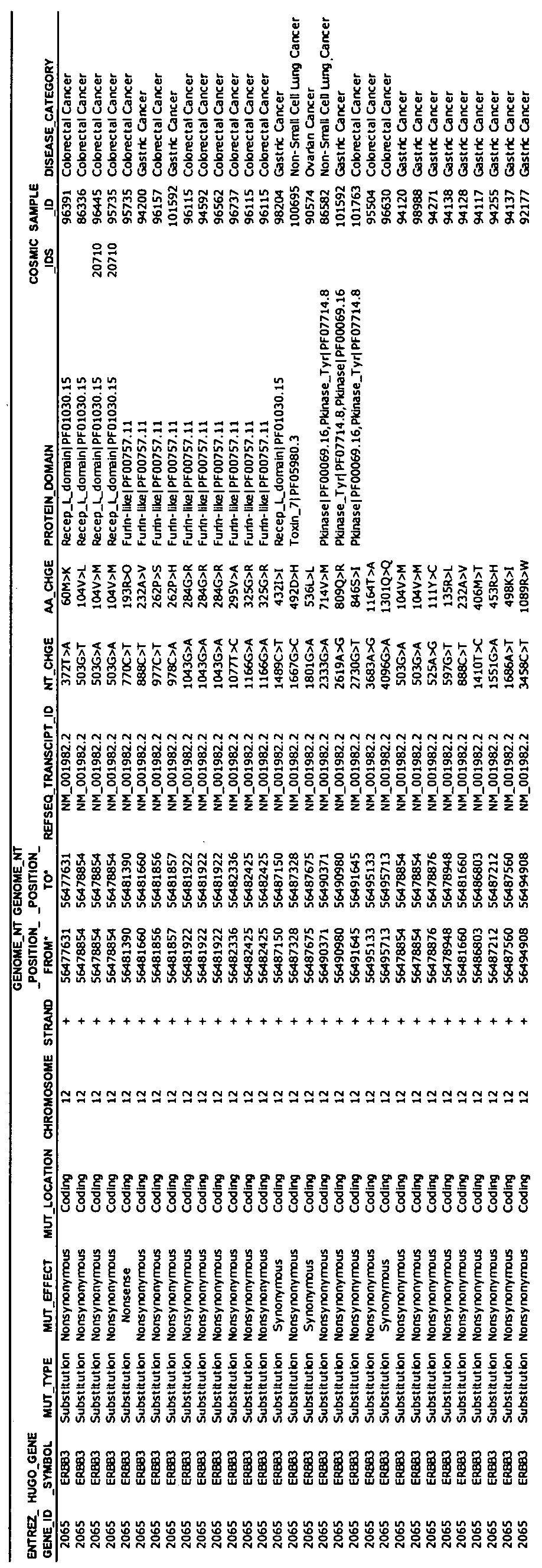

Claims
Priority Applications (13)
| Application Number | Priority Date | Filing Date | Title |
|---|---|---|---|
| CA2857114A CA2857114A1 (en) | 2011-11-30 | 2012-11-29 | Erbb3 mutations in cancer |
| NZ625380A NZ625380B2 (en) | 2011-11-30 | 2012-11-29 | Erbb3 mutations in cancer |
| EP12801651.6A EP2785864A2 (en) | 2011-11-30 | 2012-11-29 | Erbb3 mutations in cancer |
| AU2012346540A AU2012346540C1 (en) | 2011-11-30 | 2012-11-29 | ErbB3 mutations in cancer |
| SG11201402510TA SG11201402510TA (en) | 2011-11-30 | 2012-11-29 | Erbb3 mutations in cancer |
| JP2014544721A JP2015500638A (en) | 2011-11-30 | 2012-11-29 | ERBB3 mutations in cancer |
| KR1020147017927A KR20140098834A (en) | 2011-11-30 | 2012-11-29 | Erbb3 mutations in cancer |
| BR112014012979-7A BR112014012979A2 (en) | 2011-11-30 | 2012-11-29 | erbb3 mutations in cancer |
| MX2014006529A MX2014006529A (en) | 2011-11-30 | 2012-11-29 | Erbb3 mutations in cancer. |
| CN201280067318.0A CN104271761B (en) | 2011-11-30 | 2012-11-29 | Erbb3 mutation in cancer |
| RU2014126098A RU2014126098A (en) | 2011-11-30 | 2012-11-29 | Mutations of ErbB3 in Cancer |
| IL232713A IL232713A0 (en) | 2011-11-30 | 2014-05-20 | Erbb3 mutations in cancer |
| HK15101705.7A HK1201301A1 (en) | 2011-11-30 | 2015-02-16 | Erbb3 mutations in cancer erbb3 |
Applications Claiming Priority (2)
| Application Number | Priority Date | Filing Date | Title |
|---|---|---|---|
| US201161629951P | 2011-11-30 | 2011-11-30 | |
| US61/629,951 | 2011-11-30 |
Publications (2)
| Publication Number | Publication Date |
|---|---|
| WO2013081645A2 true WO2013081645A2 (en) | 2013-06-06 |
| WO2013081645A3 WO2013081645A3 (en) | 2013-07-25 |
Family
ID=47358263
Family Applications (1)
| Application Number | Title | Priority Date | Filing Date |
|---|---|---|---|
| PCT/US2012/000568 WO2013081645A2 (en) | 2011-11-30 | 2012-11-29 | Erbb3 mutations in cancer |
Country Status (14)
| Country | Link |
|---|---|
| US (1) | US20130195870A1 (en) |
| EP (1) | EP2785864A2 (en) |
| JP (2) | JP2015500638A (en) |
| KR (1) | KR20140098834A (en) |
| CN (2) | CN104271761B (en) |
| AU (1) | AU2012346540C1 (en) |
| BR (1) | BR112014012979A2 (en) |
| CA (1) | CA2857114A1 (en) |
| HK (1) | HK1201301A1 (en) |
| IL (1) | IL232713A0 (en) |
| MX (1) | MX2014006529A (en) |
| RU (2) | RU2019103083A (en) |
| SG (1) | SG11201402510TA (en) |
| WO (1) | WO2013081645A2 (en) |
Families Citing this family (6)
| Publication number | Priority date | Publication date | Assignee | Title |
|---|---|---|---|---|
| WO2014131019A2 (en) * | 2013-02-25 | 2014-08-28 | Ohio State Innovation Foundation | Her-1, her-3 and igf-1r compositions and uses thereof |
| WO2016038610A1 (en) | 2014-09-08 | 2016-03-17 | Yeda Research And Development Co. Ltd. | Compositions and methods for treating cancer resistant to a tyrosine kinase inhibitor (tki) |
| CA2959716A1 (en) | 2014-09-08 | 2016-03-17 | Yeda Research And Development Co. Ltd. | Anti-her3 antibodies and uses of same |
| CA3033694C (en) * | 2016-09-15 | 2023-06-27 | Universitat Stuttgart | Antigen binding protein against her3 |
| TW202024133A (en) * | 2018-09-20 | 2020-07-01 | 日商第一三共股份有限公司 | Treatment for her3 mutated cancer comprising administering anti-her3 antibody-drug conjugate |
| CN111187835B (en) * | 2019-02-02 | 2023-03-31 | 中国科学院上海营养与健康研究所 | Pancreatic cancer target ERBB2 and application thereof in diagnosis and treatment |
Citations (154)
| Publication number | Priority date | Publication date | Assignee | Title |
|---|---|---|---|---|
| US2003211A (en) | 1934-09-05 | 1935-05-28 | Royal Typewriter Co Inc | Typewriting machine |
| US4376110A (en) | 1980-08-04 | 1983-03-08 | Hybritech, Incorporated | Immunometric assays using monoclonal antibodies |
| US4486530A (en) | 1980-08-04 | 1984-12-04 | Hybritech Incorporated | Immunometric assays using monoclonal antibodies |
| US4657760A (en) | 1979-03-20 | 1987-04-14 | Ortho Pharmaceutical Corporation | Methods and compositions using monoclonal antibody to human T cells |
| US4683203A (en) | 1984-04-14 | 1987-07-28 | Redco N.V. | Immobilized enzymes, processes for preparing same, and use thereof |
| US4683195A (en) | 1986-01-30 | 1987-07-28 | Cetus Corporation | Process for amplifying, detecting, and/or-cloning nucleic acid sequences |
| WO1987007646A2 (en) | 1986-06-04 | 1987-12-17 | Whitehead Institute For Biomedical Research | Detection of point mutations in neu genes |
| EP0332435A2 (en) | 1988-03-10 | 1989-09-13 | Zeneca Limited | Method of detecting nucleotide sequences |
| WO1989010412A1 (en) | 1988-04-18 | 1989-11-02 | Applied Biotechnology, Inc. | Detection of neu gene expression and products |
| US4943533A (en) | 1984-03-01 | 1990-07-24 | The Regents Of The University Of California | Hybrid cell lines that produce monoclonal antibodies to epidermal growth factor receptor |
| US4968603A (en) | 1986-12-31 | 1990-11-06 | The Regents Of The University Of California | Determination of status in neoplastic disease |
| WO1991002062A2 (en) | 1989-08-04 | 1991-02-21 | Triton Biosciences, Inc. | C-erbb-2 external domain: gp75 |
| WO1991005264A1 (en) | 1989-09-29 | 1991-04-18 | Oncogenetics Partners | Detection and quantification of neu related proteins in the biological fluids of humans |
| US5183884A (en) | 1989-12-01 | 1993-02-02 | United States Of America | Dna segment encoding a gene for a receptor related to the epidermal growth factor receptor |
| WO1993003741A1 (en) | 1991-08-22 | 1993-03-04 | Becton, Dickinson & Company | Methods and compositions for cancer therapy and for prognosticating responses to cancer therapy |
| US5206344A (en) | 1985-06-26 | 1993-04-27 | Cetus Oncology Corporation | Interleukin-2 muteins and polymer conjugation thereof |
| US5212290A (en) | 1989-09-08 | 1993-05-18 | The Johns Hopkins University | Antibodies specific for type II mutant EGTR |
| WO1993012220A1 (en) | 1991-12-12 | 1993-06-24 | Berlex Laboratories, Inc. | RECOMBINANT AND CHIMERIC ANTIBODIES TO c-erbB-2 |
| US5225212A (en) | 1989-10-20 | 1993-07-06 | Liposome Technology, Inc. | Microreservoir liposome composition and method |
| WO1993016185A2 (en) | 1992-02-06 | 1993-08-19 | Creative Biomolecules, Inc. | Biosynthetic binding protein for cancer marker |
| WO1993021319A1 (en) | 1992-04-08 | 1993-10-28 | Cetus Oncology Corporation | HUMANIZED C-erbB-2 SPECIFIC ANTIBODIES |
| WO1993021232A1 (en) | 1992-04-10 | 1993-10-28 | Research Development Foundation | IMMUNOTOXINS DIRECTED AGAINST c-erbB-2 (HER-2/neu) RELATED SURFACE ANTIGENS |
| US5288477A (en) | 1991-09-27 | 1994-02-22 | Becton, Dickinson And Company | Method for prognosticating response to cancer therapy |
| EP0599274A1 (en) | 1992-11-24 | 1994-06-01 | Bristol-Myers Squibb Company | HER4, a human receptor tyrosine kinase of the epidermal growth factor receptor family |
| WO1994022478A1 (en) | 1993-03-30 | 1994-10-13 | The Trustees Of The University Of Pennsylvania | PREVENTION OF TUMORS WITH MONOCLONAL ANTIBODIES AGAINST $i(NEU) |
| US5401638A (en) | 1986-06-04 | 1995-03-28 | Oncogene Science, Inc. | Detection and quantification of neu related proteins in the biological fluids of humans |
| EP0659439A2 (en) | 1993-12-24 | 1995-06-28 | MERCK PATENT GmbH | Immunoconjugates |
| US5457105A (en) | 1992-01-20 | 1995-10-10 | Zeneca Limited | Quinazoline derivatives useful for treatment of neoplastic disease |
| US5475001A (en) | 1993-07-19 | 1995-12-12 | Zeneca Limited | Quinazoline derivatives |
| US5498531A (en) | 1993-09-10 | 1996-03-12 | President And Fellows Of Harvard College | Intron-mediated recombinant techniques and reagents |
| WO1996007321A1 (en) | 1994-09-06 | 1996-03-14 | The Uab Research Foundation | Methods for modulating protein function in cells using intracellular antibody homologues |
| EP0711565A1 (en) | 1994-11-10 | 1996-05-15 | Eberhard-Karls-Universität Tübingen Universitätsklinikum | Inhibiting growth of leukemic cells by targeting HER-2 protein |
| WO1996016673A1 (en) | 1994-12-02 | 1996-06-06 | Chiron Corporation | Method of promoting an immune response with a bispecific antibody |
| EP0502812B1 (en) | 1991-02-05 | 1996-08-14 | Ciba-Geigy Ag | Recombinant antibodies specific for a growth factor receptor |
| US5571894A (en) | 1991-02-05 | 1996-11-05 | Ciba-Geigy Corporation | Recombinant antibodies specific for a growth factor receptor |
| WO1996040789A1 (en) | 1995-06-07 | 1996-12-19 | Medarex, Inc. | THERAPEUTIC COMPOUNDS COMPRISED OF ANTI-Fc RECEPTOR ANTIBODIES |
| WO1996040210A1 (en) | 1995-06-07 | 1996-12-19 | Imclone Systems Incorporated | Antibody and antibody fragments for inhibiting the growth of tumors |
| US5587458A (en) | 1991-10-07 | 1996-12-24 | Aronex Pharmaceuticals, Inc. | Anti-erbB-2 antibodies, combinations thereof, and therapeutic and diagnostic uses thereof |
| WO1997000271A1 (en) | 1995-06-14 | 1997-01-03 | The Regents Of The University Of California | Novel high affinity human antibodies to tumor antigens |
| WO1997020858A1 (en) | 1995-12-05 | 1997-06-12 | Amgen Inc. | Apoptosis induced by monoclonal antibody anti-her2 |
| US5641869A (en) | 1991-05-24 | 1997-06-24 | Genentech, Inc. | Method for purifying heregulin |
| US5648237A (en) | 1991-09-19 | 1997-07-15 | Genentech, Inc. | Expression of functional antibody fragments |
| US5654307A (en) | 1994-01-25 | 1997-08-05 | Warner-Lambert Company | Bicyclic compounds capable of inhibiting tyrosine kinases of the epidermal growth factor receptor family |
| US5677171A (en) | 1988-01-12 | 1997-10-14 | Genentech, Inc. | Monoclonal antibodies directed to the HER2 receptor |
| WO1997038731A1 (en) | 1996-04-18 | 1997-10-23 | The Regents Of The University Of California | Immunoliposomes that optimize internalization into target cells |
| US5705157A (en) | 1989-07-27 | 1998-01-06 | The Trustees Of The University Of Pennsylvania | Methods of treating cancerous cells with anti-receptor antibodies |
| WO1998002463A1 (en) | 1996-07-11 | 1998-01-22 | Medarex, Inc. | THERAPEUTIC MULTISPECIFIC COMPOUNDS COMPRISED OF ANTI-FCα RECEPTOR ANTIBODIES |
| US5720937A (en) | 1988-01-12 | 1998-02-24 | Genentech, Inc. | In vivo tumor detection assay |
| WO1998014451A1 (en) | 1996-10-02 | 1998-04-09 | Novartis Ag | Fused pyrazole derivative and process for its preparation |
| WO1998017797A1 (en) | 1996-10-18 | 1998-04-30 | Genentech, Inc. | ANTI-ErbB2 ANTIBODIES |
| US5747261A (en) | 1986-03-05 | 1998-05-05 | The United States Of America As Represented By The Department Of Health And Human Services | Protein related to but distinct from EGF receptor and antibodies reactive therewith |
| US5747498A (en) | 1996-05-28 | 1998-05-05 | Pfizer Inc. | Alkynyl and azido-substituted 4-anilinoquinazolines |
| WO1998018489A1 (en) | 1996-10-30 | 1998-05-07 | The Uab Research Foundation | Enhancement of tumor cell chemosensitivity and radiosensitivity using single chain intracellular antibodies |
| US5760041A (en) | 1996-02-05 | 1998-06-02 | American Cyanamid Company | 4-aminoquinazoline EGFR Inhibitors |
| US5770599A (en) | 1995-04-27 | 1998-06-23 | Zeneca Limited | Quinazoline derivatives |
| US5783404A (en) | 1995-04-13 | 1998-07-21 | Amgen Inc. | Methods and compositions for determining HER-2/neu expression using monoclonal antibodies |
| WO1998033914A1 (en) | 1997-01-31 | 1998-08-06 | University Of Rochester | Chimeric antibody fusion proteins for the recruitment and stimulation of an antitumor immune response |
| US5804396A (en) | 1994-10-12 | 1998-09-08 | Sugen, Inc. | Assay for agents active in proliferative disorders |
| US5821337A (en) | 1991-06-14 | 1998-10-13 | Genentech, Inc. | Immunoglobulin variants |
| WO1998045479A1 (en) | 1997-04-04 | 1998-10-15 | Albany Medical College | Method for assessing prostate cancer |
| US5824311A (en) | 1987-11-30 | 1998-10-20 | Trustees Of The University Of Pennsylvania | Treatment of tumors with monoclonal antibodies against oncogene antigens |
| WO1998050038A1 (en) | 1997-05-06 | 1998-11-12 | American Cyanamid Company | Use of quinazoline compounds for the treatment of polycystic kidney disease |
| WO1998050433A2 (en) | 1997-05-05 | 1998-11-12 | Abgenix, Inc. | Human monoclonal antibodies to epidermal growth factor receptor |
| US5846749A (en) | 1994-10-12 | 1998-12-08 | The Regents Of The University Of California | Quantitative measurement of tissue protein identified by immunohistochemistry and standardized protein determination |
| US5856089A (en) | 1992-10-09 | 1999-01-05 | Oncor, Inc. | Method for the detection of chromosome structural abnormalities by in situ hybridization to fixed tissue |
| US5866572A (en) | 1996-02-14 | 1999-02-02 | Zeneca Limited | Quinazoline derivatives |
| WO1999009016A1 (en) | 1997-08-01 | 1999-02-25 | American Cyanamid Company | Substituted quinazoline derivatives and their use as tyrosine kinase inhibitors |
| US5891996A (en) | 1972-09-17 | 1999-04-06 | Centro De Inmunologia Molecular | Humanized and chimeric monoclonal antibodies that recognize epidermal growth factor receptor (EGF-R); diagnostic and therapeutic use |
| WO1999019488A1 (en) | 1997-10-15 | 1999-04-22 | Children's Medical Center Corporation | Novel human egf receptors and use thereof |
| WO1999024037A1 (en) | 1997-11-06 | 1999-05-20 | American Cyanamid Company | Use of quinazoline derivatives as tyrosine kinase inhibitors for treating colonic polyps |
| WO1999031140A1 (en) | 1997-12-12 | 1999-06-24 | Genentech, Inc. | TREATMENT WITH ANTI-ErbB2 ANTIBODIES |
| US5925519A (en) | 1996-06-03 | 1999-07-20 | The Regents Of The University Of California | Genetic alterations associated with prostate cancer |
| US5939531A (en) | 1991-07-15 | 1999-08-17 | Novartis Corp. | Recombinant antibodies specific for a growth factor receptor |
| WO1999048527A1 (en) | 1998-03-27 | 1999-09-30 | Genentech, Inc. | Apo-2 ligand-anti-her-2 antibody synergism |
| EP0616812B1 (en) | 1993-03-24 | 1999-11-03 | Berlex Biosciences | Combination with anti-hormonal compounds and binding molecules for the treatment of cancer |
| WO1999055367A1 (en) | 1998-04-24 | 1999-11-04 | The Regents Of The University Of California | INTERNALIZING ErbB2 ANTIBODIES |
| US6002008A (en) | 1997-04-03 | 1999-12-14 | American Cyanamid Company | Substituted 3-cyano quinolines |
| US6015567A (en) | 1989-05-19 | 2000-01-18 | Genentech, Inc. | HER2 extracellular domain |
| US6027889A (en) | 1996-05-29 | 2000-02-22 | Cornell Research Foundation, Inc. | Detection of nucleic acid sequence differences using coupled ligase detection and polymerase chain reactions |
| US6054297A (en) | 1991-06-14 | 2000-04-25 | Genentech, Inc. | Humanized antibodies and methods for making them |
| US6084095A (en) | 1994-01-25 | 2000-07-04 | Warner-Lambert Company | Substituted pyrido[3,2-d]pyrimidines capable of inhibiting tyrosine kinases of the epidermal growth factor receptor family |
| US6123939A (en) | 1989-08-04 | 2000-09-26 | Berlex Laboratories, Inc. | Anti-neoplastic drugs in cancer therapy |
| WO2000061185A1 (en) | 1999-04-09 | 2000-10-19 | Aventis Pharma S.A | Docetaxel in combination with rhumab her2 for the treatment of cancers |
| WO2000061145A1 (en) | 1999-04-09 | 2000-10-19 | Schering Corporation | Methods of inducing cancer cell death and tumor regression |
| US6140332A (en) | 1995-07-06 | 2000-10-31 | Novartis Ag | Pyrrolopyrimidines and processes for the preparation thereof |
| WO2000069460A1 (en) | 1999-05-14 | 2000-11-23 | Genentech, Inc. | TREATMENT WITH ANTI-ErbB2 ANTIBODIES |
| WO2000078347A1 (en) | 1999-06-18 | 2000-12-28 | The Victor Chang Cardiac Research Institute Limited | Cell growth inhibition |
| WO2001000244A2 (en) | 1999-06-25 | 2001-01-04 | Genentech, Inc. | METHODS OF TREATMENT USING ANTI-ErbB ANTIBODY-MAYTANSINOID CONJUGATES |
| WO2001000238A1 (en) | 1999-06-25 | 2001-01-04 | Genentech, Inc. | TREATING PROSTATE CANCER WITH ANTI-ErbB2 ANTIBODIES |
| WO2001000245A2 (en) | 1999-06-25 | 2001-01-04 | Genentech, Inc. | HUMANIZED ANTI-ErbB2 ANTIBODIES AND TREATMENT WITH ANTI-ErbB2 ANTIBODIES |
| WO2001005425A2 (en) | 1999-07-20 | 2001-01-25 | Pharmacia & Upjohn S.P.A. | Combined preparations comprising daunorubicin derivatives and her2 antibodies |
| WO2001009187A2 (en) | 1999-07-29 | 2001-02-08 | Medarex, Inc. | Human monoclonal antibodies to her2/neu |
| WO2001015730A1 (en) | 1999-08-27 | 2001-03-08 | Genentech, Inc. | DOSAGES FOR TREATMENT WITH ANTI-ErbB2 ANTIBODIES |
| WO2001020033A1 (en) | 1998-09-14 | 2001-03-22 | The Albany Medical College | Methods for selecting prostate cancer treatments and treating prostate cancer |
| WO2001021192A2 (en) | 1999-09-22 | 2001-03-29 | Corixa Corporation | Methods for diagnosis and therapy of hematological and virus-associated malignancies |
| WO2001032155A2 (en) | 1999-11-02 | 2001-05-10 | The University Of Manchester | Use of egfr tyrosine kinase inhibitors for treating breast cancer |
| WO2001053354A2 (en) | 2000-01-20 | 2001-07-26 | Chiron Corporation | Methods for treating tumors using a fusion protein comprising il-2- polypeptides and p185-specific binding molecules |
| US6267958B1 (en) | 1995-07-27 | 2001-07-31 | Genentech, Inc. | Protein formulation |
| WO2001056604A1 (en) | 2000-02-07 | 2001-08-09 | Uab Research Foundation | Enhancement of tumor cell chemosensitivity and radiosensitivity using single chain secretory antibodies |
| WO2001064246A2 (en) | 2000-02-29 | 2001-09-07 | Janssen Pharmaceutica N.V. | Farnesyl protein transferase inhibitor combinations with an her2 antibody |
| WO2001076586A1 (en) | 2000-04-06 | 2001-10-18 | Astrazeneca Ab | Combination product comprising a non-steroidal antiandrogen and an egfr tyrosine kinase inhibitor |
| WO2001076630A1 (en) | 2000-04-06 | 2001-10-18 | Kyowa Hakko Kogyo Co., Ltd. | Diagnostics and remedies for rheumatoid arthritis |
| WO2001087334A1 (en) | 2000-05-15 | 2001-11-22 | Pharmacia Italia S.P.A. | Aromatase inhibitors and monoclonal anti-her2 antibodies as antitumors agents |
| WO2001087336A1 (en) | 2000-05-15 | 2001-11-22 | Health Research, Inc. | Cancer treatments by using a combination of an antibody against her2 and interleukin -2 |
| WO2001089566A1 (en) | 2000-05-19 | 2001-11-29 | Genentech, Inc. | Gene detection assay for improving the likelihood of an effective response to an erbb antagonist cancer therapy |
| WO2001092579A2 (en) | 2000-05-30 | 2001-12-06 | Pe Corporation (Ny) | Methods for detecting target nucleic acids using coupled ligation and amplification |
| US6333398B1 (en) | 1996-11-27 | 2001-12-25 | Genentech, Inc. | Protein purification |
| US6339142B1 (en) | 1998-05-06 | 2002-01-15 | Genentech, Inc. | Protein purification |
| WO2002005791A2 (en) | 2000-07-18 | 2002-01-24 | Pharmacia Italia Spa | Antitumor combination comprising an aromatase inhibitor and an egfr antagonist or inhibitor |
| US6344455B1 (en) | 1998-11-19 | 2002-02-05 | Warner-Lambert Company | N-[4-(3-chloro-4-fluoro-phenylamino)-7-(3-morpholin-4-yl-propoxy)-quinazolin-6-yl]-acrylamide, and irreversible inhibitor of tyrosine kinases |
| US6344459B1 (en) | 1996-04-12 | 2002-02-05 | Warner-Lambert Company | Irreversible inhibitors of tyrosine kinases |
| WO2002009754A1 (en) | 2000-07-28 | 2002-02-07 | Sumitomo Pharmaceuticals Co., Ltd. | Novel remedies for cancer |
| WO2002011677A2 (en) | 2000-08-09 | 2002-02-14 | Imclone Systems Incorporated | Treatment of hyperproliferative diseases with epidermal growth factor receptor antagonists |
| US20020019221A1 (en) | 2000-07-27 | 2002-02-14 | Elmar Wagner | Multiplier circuit with offset compensation and quadricorrelator |
| US6358682B1 (en) | 1998-01-26 | 2002-03-19 | Ventana Medical Systems, Inc. | Method and kit for the prognostication of breast cancer |
| US20020051785A1 (en) | 2000-03-20 | 2002-05-02 | The Regents Of The University Of California And Amgen Inc. | HER -2/neu overexpression abrogates growth inhibitory pathways |
| US6391874B1 (en) | 1996-07-13 | 2002-05-21 | Smithkline Beecham Corporation | Fused heterocyclic compounds as protein tyrosine kinase inhibitors |
| WO2002044413A2 (en) | 2000-12-01 | 2002-06-06 | Response Genetics, Inc. | Method of determining epidermal growth factor receptor and her2-neu gene expression and correlation of levels thereof with survival rates |
| US6403630B1 (en) | 1999-01-27 | 2002-06-11 | Cornell Research Foundation, Inc. | Treating cancers associated with overexpression of HER-2/neu |
| WO2002045653A2 (en) | 2000-12-08 | 2002-06-13 | Uab Research Foundation | Combination radiation therapy and chemotherapy in conjuction with administration of growth factor receptor antibody |
| US6417168B1 (en) | 1998-03-04 | 2002-07-09 | The Trustees Of The University Of Pennsylvania | Compositions and methods of treating tumors |
| US20020090662A1 (en) | 2000-08-15 | 2002-07-11 | Peter Ralph | Analytical method |
| WO2002055106A2 (en) | 2001-01-09 | 2002-07-18 | Merck Patent Gmbh | Combination therapy using receptor tyrosine kinase inhibitors and angiogenesis inhibitors |
| WO2002070008A1 (en) | 2001-03-02 | 2002-09-12 | Imclone Systems Incorporated | Combination methods of inhibiting tumor growth with a vascular endothelial growth factor receptor antagonist |
| US20020142328A1 (en) | 2000-12-01 | 2002-10-03 | Danenberg Kathleen D. | Method of determining a chemotherapeutic regimen by assaying gene expression in primary tumors |
| WO2002087619A1 (en) | 2001-04-27 | 2002-11-07 | Takeda Chemical Industries, Ltd. | Preventive/therapeutic method for cancer |
| WO2002089842A1 (en) | 2001-05-08 | 2002-11-14 | Merck Patent Gmbh | Combination therapy using anti-egfr antibodies and anti-hormonal agents |
| US20020192652A1 (en) | 2001-06-11 | 2002-12-19 | Danenberg Kathleen D. | Method of determining epidermal growth factor receptor and HER2-neu gene expression and correlation of levels thereof with survival rates |
| WO2003006509A2 (en) | 2001-07-10 | 2003-01-23 | Universita Degli Studi Di Napoli Federico Ii | Human mini-antibody cytotoxic for tumor cells which express the erbb2 receptor |
| WO2003012072A2 (en) | 2001-08-03 | 2003-02-13 | The Trustees Of The University Of Pennsylvania | Monoclonal antibodies to activated erbb family members and methods of use thereof |
| WO2003028638A2 (en) | 2001-09-28 | 2003-04-10 | The Board Of Trustees Of The University Of Arkansas | Treatment of uterine serous papillary cancer |
| US20030086924A1 (en) | 1999-06-25 | 2003-05-08 | Genentech, Inc. | Treatment with anti-ErbB2 antibodies |
| WO2003041736A2 (en) | 2001-11-16 | 2003-05-22 | Pharmacia & Upjohn Company | Method for the treatment of cardiotoxicity induced by antitumor compounds |
| US6573043B1 (en) | 1998-10-07 | 2003-06-03 | Genentech, Inc. | Tissue analysis and kits therefor |
| US20030108545A1 (en) | 1994-02-10 | 2003-06-12 | Patricia Rockwell | Combination methods of inhibiting tumor growth with a vascular endothelial growth factor receptor antagonist |
| US20030119004A1 (en) | 2001-12-05 | 2003-06-26 | Wenz H. Michael | Methods for quantitating nucleic acids using coupled ligation and amplification |
| US6596726B1 (en) | 1994-01-25 | 2003-07-22 | Warner Lambert Company | Tricyclic compounds capable of inhibiting tyrosine kinases of the epidermal growth factor receptor family |
| US6602670B2 (en) | 2000-12-01 | 2003-08-05 | Response Genetics, Inc. | Method of determining a chemotherapeutic regimen based on ERCC1 expression |
| US20030165840A1 (en) | 2001-03-02 | 2003-09-04 | Kathleen Danenberg | Method of determining dihydropyrimidine dehydrogenase gene expression |
| US20030175845A1 (en) | 2002-03-13 | 2003-09-18 | Kalbag Suresh M. | Use of sulfitolysis in high performance peptide mapping |
| US6632979B2 (en) | 2000-03-16 | 2003-10-14 | Genentech, Inc. | Rodent HER2 tumor model |
| WO2003086467A1 (en) | 2002-04-08 | 2003-10-23 | Smithkline Beecham Corporation | Cancer treatment method comprising administering an erb-family inhibitor and a raf and/or ras inhibitor |
| WO2003087131A2 (en) | 2002-04-10 | 2003-10-23 | Genentech, Inc | Anti-her2 antibody variants |
| EP1357132A2 (en) | 2002-04-19 | 2003-10-29 | BIOTHER di Contardi Gabriella & C. Società in accomandita semplice | Detection of the her2 receptor by means of biotinylated humanized antibody |
| US20030202973A1 (en) | 2002-04-29 | 2003-10-30 | Dr. George Pieczenik | Treatment of refractory human tumors with epidermal growth factor receptor and HER1 mitogenic ligand (EGFRML) antagonists |
| WO2004008099A2 (en) | 2002-07-15 | 2004-01-22 | Genentech, Inc. | METHODS FOR IDENTIFYING TUMORS THAT ARE RESPONSIVE TO TREATMENT WITH ANTI-ErbB2 ANTIBODIES |
| US20040013667A1 (en) | 1999-06-25 | 2004-01-22 | Genentech, Inc. | Treatment with anti-ErbB2 antibodies |
| US6685940B2 (en) | 1995-07-27 | 2004-02-03 | Genentech, Inc. | Protein formulation |
| WO2004024866A2 (en) | 2002-09-11 | 2004-03-25 | Genentech, Inc. | Protein purification |
| WO2004048525A2 (en) | 2002-11-21 | 2004-06-10 | Genentech, Inc. | Therapy of non-malignant diseases or disorders with anti-erbb2 antibodies |
| US6800738B1 (en) | 1991-06-14 | 2004-10-05 | Genentech, Inc. | Method for making humanized antibodies |
| WO2004087207A2 (en) | 2003-03-27 | 2004-10-14 | Georgetown University | Method for inducing apoptosis and aneuploidy regression in cancer cells |
| WO2010108127A1 (en) | 2009-03-20 | 2010-09-23 | Genentech, Inc. | Bispecific anti-her antibodies |
| WO2011076683A1 (en) | 2009-12-22 | 2011-06-30 | Roche Glycart Ag | Anti-her3 antibodies and uses thereof |
Family Cites Families (3)
| Publication number | Priority date | Publication date | Assignee | Title |
|---|---|---|---|---|
| US7125680B2 (en) * | 2001-07-27 | 2006-10-24 | The Regents Of The University Of California | Methods and materials for characterizing and modulating interaction between heregulin and HER3 |
| EP2082064B1 (en) * | 2006-11-16 | 2011-10-05 | Genentech, Inc. | Genetic variations associated with tumors |
| SG177225A1 (en) * | 2006-12-01 | 2012-01-30 | Agency Science Tech & Res | Cancer-related protein kinases |
-
2012
- 2012-11-29 MX MX2014006529A patent/MX2014006529A/en unknown
- 2012-11-29 WO PCT/US2012/000568 patent/WO2013081645A2/en active Application Filing
- 2012-11-29 CA CA2857114A patent/CA2857114A1/en not_active Abandoned
- 2012-11-29 JP JP2014544721A patent/JP2015500638A/en active Pending
- 2012-11-29 EP EP12801651.6A patent/EP2785864A2/en not_active Withdrawn
- 2012-11-29 CN CN201280067318.0A patent/CN104271761B/en not_active Expired - Fee Related
- 2012-11-29 AU AU2012346540A patent/AU2012346540C1/en not_active Ceased
- 2012-11-29 US US13/694,413 patent/US20130195870A1/en not_active Abandoned
- 2012-11-29 RU RU2019103083A patent/RU2019103083A/en not_active Application Discontinuation
- 2012-11-29 BR BR112014012979-7A patent/BR112014012979A2/en not_active IP Right Cessation
- 2012-11-29 CN CN201710084997.7A patent/CN106987620A/en active Pending
- 2012-11-29 SG SG11201402510TA patent/SG11201402510TA/en unknown
- 2012-11-29 KR KR1020147017927A patent/KR20140098834A/en not_active Application Discontinuation
- 2012-11-29 RU RU2014126098A patent/RU2014126098A/en not_active Application Discontinuation
-
2014
- 2014-05-20 IL IL232713A patent/IL232713A0/en unknown
-
2015
- 2015-02-16 HK HK15101705.7A patent/HK1201301A1/en not_active IP Right Cessation
-
2018
- 2018-01-05 JP JP2018000704A patent/JP2018085998A/en active Pending
Patent Citations (230)
| Publication number | Priority date | Publication date | Assignee | Title |
|---|---|---|---|---|
| US2003211A (en) | 1934-09-05 | 1935-05-28 | Royal Typewriter Co Inc | Typewriting machine |
| US5891996A (en) | 1972-09-17 | 1999-04-06 | Centro De Inmunologia Molecular | Humanized and chimeric monoclonal antibodies that recognize epidermal growth factor receptor (EGF-R); diagnostic and therapeutic use |
| US4657760A (en) | 1979-03-20 | 1987-04-14 | Ortho Pharmaceutical Corporation | Methods and compositions using monoclonal antibody to human T cells |
| US4376110A (en) | 1980-08-04 | 1983-03-08 | Hybritech, Incorporated | Immunometric assays using monoclonal antibodies |
| US4486530A (en) | 1980-08-04 | 1984-12-04 | Hybritech Incorporated | Immunometric assays using monoclonal antibodies |
| US4943533A (en) | 1984-03-01 | 1990-07-24 | The Regents Of The University Of California | Hybrid cell lines that produce monoclonal antibodies to epidermal growth factor receptor |
| US4683203A (en) | 1984-04-14 | 1987-07-28 | Redco N.V. | Immobilized enzymes, processes for preparing same, and use thereof |
| US5206344A (en) | 1985-06-26 | 1993-04-27 | Cetus Oncology Corporation | Interleukin-2 muteins and polymer conjugation thereof |
| US4683195B1 (en) | 1986-01-30 | 1990-11-27 | Cetus Corp | |
| US4683195A (en) | 1986-01-30 | 1987-07-28 | Cetus Corporation | Process for amplifying, detecting, and/or-cloning nucleic acid sequences |
| US5747261A (en) | 1986-03-05 | 1998-05-05 | The United States Of America As Represented By The Department Of Health And Human Services | Protein related to but distinct from EGF receptor and antibodies reactive therewith |
| US5985553A (en) | 1986-03-05 | 1999-11-16 | The United States Of America As Represented By The Department Of Health And Human Services | erbB-2 gene segments, probes, recombinant DNA and kits for detection |
| US5604107A (en) | 1986-06-04 | 1997-02-18 | Oncogene Science, Inc. | Detection of neu p185 in cell lysates |
| US4935341A (en) | 1986-06-04 | 1990-06-19 | Whitehead Institute For Biomedical Research | Detection of point mutations in neu genes |
| US5401638A (en) | 1986-06-04 | 1995-03-28 | Oncogene Science, Inc. | Detection and quantification of neu related proteins in the biological fluids of humans |
| WO1987007646A2 (en) | 1986-06-04 | 1987-12-17 | Whitehead Institute For Biomedical Research | Detection of point mutations in neu genes |
| US4968603A (en) | 1986-12-31 | 1990-11-06 | The Regents Of The University Of California | Determination of status in neoplastic disease |
| US5824311A (en) | 1987-11-30 | 1998-10-20 | Trustees Of The University Of Pennsylvania | Treatment of tumors with monoclonal antibodies against oncogene antigens |
| US5720937A (en) | 1988-01-12 | 1998-02-24 | Genentech, Inc. | In vivo tumor detection assay |
| US6399063B1 (en) | 1988-01-12 | 2002-06-04 | Genentech, Inc. | Monoclonal antibodies directed to the HER2 receptor |
| US5725856A (en) | 1988-01-12 | 1998-03-10 | Genentech, Inc. | Monoclonal antibodies directed to the HER2 receptor |
| US6165464A (en) | 1988-01-12 | 2000-12-26 | Genetech, Inc. | Monoclonal antibodies directed to the HER2 receptor |
| US5772997A (en) | 1988-01-12 | 1998-06-30 | Genentech, Inc. | Monoclonal antibodies directed to the HER2 receptor |
| US5720954A (en) | 1988-01-12 | 1998-02-24 | Genentech, Inc. | Monoclonal antibodies directed to the HER2 receptor |
| US6387371B1 (en) | 1988-01-12 | 2002-05-14 | Genentech, Inc. | Monoclonal antibodies directed to the HER2 receptor |
| US5770195A (en) | 1988-01-12 | 1998-06-23 | Genentech, Inc. | Monoclonal antibodies directed to the her2 receptor |
| US5677171A (en) | 1988-01-12 | 1997-10-14 | Genentech, Inc. | Monoclonal antibodies directed to the HER2 receptor |
| EP0332435A2 (en) | 1988-03-10 | 1989-09-13 | Zeneca Limited | Method of detecting nucleotide sequences |
| EP0412116B1 (en) | 1988-04-18 | 1995-11-29 | Oncogene Science, Inc. | Detection of neu gene expression and products |
| WO1989010412A1 (en) | 1988-04-18 | 1989-11-02 | Applied Biotechnology, Inc. | Detection of neu gene expression and products |
| US6015567A (en) | 1989-05-19 | 2000-01-18 | Genentech, Inc. | HER2 extracellular domain |
| US6333169B1 (en) | 1989-05-19 | 2001-12-25 | Genentech Inc | HER2 extracellular domain |
| US5705157A (en) | 1989-07-27 | 1998-01-06 | The Trustees Of The University Of Pennsylvania | Methods of treating cancerous cells with anti-receptor antibodies |
| WO1991002062A2 (en) | 1989-08-04 | 1991-02-21 | Triton Biosciences, Inc. | C-erbb-2 external domain: gp75 |
| EP0444181B1 (en) | 1989-08-04 | 2001-10-31 | Schering Aktiengesellschaft | C-erbb-2 external domain: gp75 |
| US20020155527A1 (en) | 1989-08-04 | 2002-10-24 | Triton Biosciences, Inc. | C-erbB-2 exrernal domain: gp75 |
| US6123939A (en) | 1989-08-04 | 2000-09-26 | Berlex Laboratories, Inc. | Anti-neoplastic drugs in cancer therapy |
| EP1006194A2 (en) | 1989-08-04 | 2000-06-07 | Triton Biosciences Inc. | C-erbB-2 external domain: GP75 |
| US5212290A (en) | 1989-09-08 | 1993-05-18 | The Johns Hopkins University | Antibodies specific for type II mutant EGTR |
| WO1991005264A1 (en) | 1989-09-29 | 1991-04-18 | Oncogenetics Partners | Detection and quantification of neu related proteins in the biological fluids of humans |
| EP0494135B1 (en) | 1989-09-29 | 1996-04-10 | Oncogene Science, Inc. | Human "neu" related protein p100 and use of the same for detecting preneoplastic or neoplastic cells in a human |
| US5225212A (en) | 1989-10-20 | 1993-07-06 | Liposome Technology, Inc. | Microreservoir liposome composition and method |
| US5480968A (en) | 1989-12-01 | 1996-01-02 | The United States Of America As Represented By The Department Of Health And Human Services | Isolated polypeptide erbB-3, related to the epidermal growth factor receptor and antibody thereto |
| US5183884A (en) | 1989-12-01 | 1993-02-02 | United States Of America | Dna segment encoding a gene for a receptor related to the epidermal growth factor receptor |
| US5571894A (en) | 1991-02-05 | 1996-11-05 | Ciba-Geigy Corporation | Recombinant antibodies specific for a growth factor receptor |
| EP0502812B1 (en) | 1991-02-05 | 1996-08-14 | Ciba-Geigy Ag | Recombinant antibodies specific for a growth factor receptor |
| US5641869A (en) | 1991-05-24 | 1997-06-24 | Genentech, Inc. | Method for purifying heregulin |
| US20040236078A1 (en) | 1991-06-14 | 2004-11-25 | Genentech, Inc. | Method for making humanized antibodies |
| US6719971B1 (en) | 1991-06-14 | 2004-04-13 | Genentech, Inc. | Method for making humanized antibodies |
| US6407213B1 (en) | 1991-06-14 | 2002-06-18 | Genentech, Inc. | Method for making humanized antibodies |
| US6800738B1 (en) | 1991-06-14 | 2004-10-05 | Genentech, Inc. | Method for making humanized antibodies |
| US6054297A (en) | 1991-06-14 | 2000-04-25 | Genentech, Inc. | Humanized antibodies and methods for making them |
| US5821337A (en) | 1991-06-14 | 1998-10-13 | Genentech, Inc. | Immunoglobulin variants |
| US5939531A (en) | 1991-07-15 | 1999-08-17 | Novartis Corp. | Recombinant antibodies specific for a growth factor receptor |
| US5514554A (en) | 1991-08-22 | 1996-05-07 | Becton Dickinson And Company | Methods and compositions for cancer therapy and for prognosticating responses to cancer therapy |
| EP0656367A1 (en) | 1991-08-22 | 1995-06-07 | Becton, Dickinson and Company | Method and composition for cancer therapy and for prognosticating responses to cancer theraphy |
| EP0554441B1 (en) | 1991-08-22 | 1999-01-27 | Becton, Dickinson and Company | Methods and compositions for cancer therapy and for prognosticating responses to cancer therapy |
| WO1993003741A1 (en) | 1991-08-22 | 1993-03-04 | Becton, Dickinson & Company | Methods and compositions for cancer therapy and for prognosticating responses to cancer therapy |
| US5648237A (en) | 1991-09-19 | 1997-07-15 | Genentech, Inc. | Expression of functional antibody fragments |
| US5288477A (en) | 1991-09-27 | 1994-02-22 | Becton, Dickinson And Company | Method for prognosticating response to cancer therapy |
| US5587458A (en) | 1991-10-07 | 1996-12-24 | Aronex Pharmaceuticals, Inc. | Anti-erbB-2 antibodies, combinations thereof, and therapeutic and diagnostic uses thereof |
| WO1993012220A1 (en) | 1991-12-12 | 1993-06-24 | Berlex Laboratories, Inc. | RECOMBINANT AND CHIMERIC ANTIBODIES TO c-erbB-2 |
| US5457105A (en) | 1992-01-20 | 1995-10-10 | Zeneca Limited | Quinazoline derivatives useful for treatment of neoplastic disease |
| US5616582A (en) | 1992-01-20 | 1997-04-01 | Zeneca Limited | Quinazoline derivatives as anti-proliferative agents |
| US5877305A (en) | 1992-02-06 | 1999-03-02 | Chiron Corporation | DNA encoding biosynthetic binding protein for cancer marker |
| WO1993016185A2 (en) | 1992-02-06 | 1993-08-19 | Creative Biomolecules, Inc. | Biosynthetic binding protein for cancer marker |
| WO1993021319A1 (en) | 1992-04-08 | 1993-10-28 | Cetus Oncology Corporation | HUMANIZED C-erbB-2 SPECIFIC ANTIBODIES |
| WO1993021232A1 (en) | 1992-04-10 | 1993-10-28 | Research Development Foundation | IMMUNOTOXINS DIRECTED AGAINST c-erbB-2 (HER-2/neu) RELATED SURFACE ANTIGENS |
| US5856089A (en) | 1992-10-09 | 1999-01-05 | Oncor, Inc. | Method for the detection of chromosome structural abnormalities by in situ hybridization to fixed tissue |
| EP0599274A1 (en) | 1992-11-24 | 1994-06-01 | Bristol-Myers Squibb Company | HER4, a human receptor tyrosine kinase of the epidermal growth factor receptor family |
| EP0616812B1 (en) | 1993-03-24 | 1999-11-03 | Berlex Biosciences | Combination with anti-hormonal compounds and binding molecules for the treatment of cancer |
| WO1994022478A1 (en) | 1993-03-30 | 1994-10-13 | The Trustees Of The University Of Pennsylvania | PREVENTION OF TUMORS WITH MONOCLONAL ANTIBODIES AGAINST $i(NEU) |
| US5475001A (en) | 1993-07-19 | 1995-12-12 | Zeneca Limited | Quinazoline derivatives |
| US5498531A (en) | 1993-09-10 | 1996-03-12 | President And Fellows Of Harvard College | Intron-mediated recombinant techniques and reagents |
| EP0659439A2 (en) | 1993-12-24 | 1995-06-28 | MERCK PATENT GmbH | Immunoconjugates |
| US6521620B1 (en) | 1994-01-25 | 2003-02-18 | Warner-Lambert Company | Bicyclic compounds capable of inhibiting tyrosine kinases of the epidermal growth factor receptor family |
| US6455534B2 (en) | 1994-01-25 | 2002-09-24 | Warner-Lambert Company | Bicyclic compounds capable of inhibiting tyrosine kinases of the epidermal growth factor receptor family |
| US5654307A (en) | 1994-01-25 | 1997-08-05 | Warner-Lambert Company | Bicyclic compounds capable of inhibiting tyrosine kinases of the epidermal growth factor receptor family |
| US6084095A (en) | 1994-01-25 | 2000-07-04 | Warner-Lambert Company | Substituted pyrido[3,2-d]pyrimidines capable of inhibiting tyrosine kinases of the epidermal growth factor receptor family |
| US5679683A (en) | 1994-01-25 | 1997-10-21 | Warner-Lambert Company | Tricyclic compounds capable of inhibiting tyrosine kinases of the epidermal growth factor receptor family |
| US6713484B2 (en) | 1994-01-25 | 2004-03-30 | Warner-Lambert Company | Bicyclic compounds capable of inhibiting tyrosine kinases of the epidermal growth factor receptor family |
| US6265410B1 (en) | 1994-01-25 | 2001-07-24 | Warner-Lambert Company | Bicyclic compounds capable of inhibiting tyrosine kinases of the epidermal growth factor receptor family |
| US6596726B1 (en) | 1994-01-25 | 2003-07-22 | Warner Lambert Company | Tricyclic compounds capable of inhibiting tyrosine kinases of the epidermal growth factor receptor family |
| US20030103973A1 (en) | 1994-02-10 | 2003-06-05 | Patricia Rockwell | Method for reducing tumor growth with VEGF antagonists combined with radiation and chemotherapy |
| US20030108545A1 (en) | 1994-02-10 | 2003-06-12 | Patricia Rockwell | Combination methods of inhibiting tumor growth with a vascular endothelial growth factor receptor antagonist |
| WO1996007321A1 (en) | 1994-09-06 | 1996-03-14 | The Uab Research Foundation | Methods for modulating protein function in cells using intracellular antibody homologues |
| US6028059A (en) | 1994-09-06 | 2000-02-22 | Uab Research Foundation | Methods for modulating protein function in cells using intracellular antibody homologues |
| US5910486A (en) | 1994-09-06 | 1999-06-08 | Uab Research Foundation | Methods for modulating protein function in cells using, intracellular antibody homologues |
| US5804396A (en) | 1994-10-12 | 1998-09-08 | Sugen, Inc. | Assay for agents active in proliferative disorders |
| US5846749A (en) | 1994-10-12 | 1998-12-08 | The Regents Of The University Of California | Quantitative measurement of tissue protein identified by immunohistochemistry and standardized protein determination |
| US6214388B1 (en) | 1994-11-09 | 2001-04-10 | The Regents Of The University Of California | Immunoliposomes that optimize internalization into target cells |
| EP0711565A1 (en) | 1994-11-10 | 1996-05-15 | Eberhard-Karls-Universität Tübingen Universitätsklinikum | Inhibiting growth of leukemic cells by targeting HER-2 protein |
| WO1996016673A1 (en) | 1994-12-02 | 1996-06-06 | Chiron Corporation | Method of promoting an immune response with a bispecific antibody |
| US5783404A (en) | 1995-04-13 | 1998-07-21 | Amgen Inc. | Methods and compositions for determining HER-2/neu expression using monoclonal antibodies |
| US5770599A (en) | 1995-04-27 | 1998-06-23 | Zeneca Limited | Quinazoline derivatives |
| WO1996040789A1 (en) | 1995-06-07 | 1996-12-19 | Medarex, Inc. | THERAPEUTIC COMPOUNDS COMPRISED OF ANTI-Fc RECEPTOR ANTIBODIES |
| WO1996040210A1 (en) | 1995-06-07 | 1996-12-19 | Imclone Systems Incorporated | Antibody and antibody fragments for inhibiting the growth of tumors |
| US6270765B1 (en) | 1995-06-07 | 2001-08-07 | Medarex, Inc. | Therapeutic compounds comprised of anti-Fc receptor antibodies |
| US6395272B1 (en) | 1995-06-07 | 2002-05-28 | Mederex, Inc. | Therapeutic compounds comprised of anti-Fc receptor antibodies |
| US5837243A (en) | 1995-06-07 | 1998-11-17 | Medarex, Inc. | Therapeutic compounds comprised of anti-Fc receptor antibodies |
| WO1997000271A1 (en) | 1995-06-14 | 1997-01-03 | The Regents Of The University Of California | Novel high affinity human antibodies to tumor antigens |
| US5977322A (en) | 1995-06-14 | 1999-11-02 | The Regents Of The University Of California | High affinity human antibodies to tumor antigens |
| US6512097B1 (en) | 1995-06-14 | 2003-01-28 | The Regents Of The University Of California | High affinity human antibodies to tumor antigens |
| US6140332A (en) | 1995-07-06 | 2000-10-31 | Novartis Ag | Pyrrolopyrimidines and processes for the preparation thereof |
| US6821515B1 (en) | 1995-07-27 | 2004-11-23 | Genentech, Inc. | Protein formulation |
| US6267958B1 (en) | 1995-07-27 | 2001-07-31 | Genentech, Inc. | Protein formulation |
| US6685940B2 (en) | 1995-07-27 | 2004-02-03 | Genentech, Inc. | Protein formulation |
| US6458356B1 (en) | 1995-12-05 | 2002-10-01 | Amgen Inc. | Antibody-induced apoptosis |
| WO1997020858A1 (en) | 1995-12-05 | 1997-06-12 | Amgen Inc. | Apoptosis induced by monoclonal antibody anti-her2 |
| US5783186A (en) | 1995-12-05 | 1998-07-21 | Amgen Inc. | Antibody-induced apoptosis |
| US5760041A (en) | 1996-02-05 | 1998-06-02 | American Cyanamid Company | 4-aminoquinazoline EGFR Inhibitors |
| US5866572A (en) | 1996-02-14 | 1999-02-02 | Zeneca Limited | Quinazoline derivatives |
| US6399602B1 (en) | 1996-02-14 | 2002-06-04 | Zeneca Limited | Quinazoline derivatives |
| US6602863B1 (en) | 1996-04-12 | 2003-08-05 | Warner-Lambert Company | Irreversible inhibitors of tyrosine kinases |
| US6344459B1 (en) | 1996-04-12 | 2002-02-05 | Warner-Lambert Company | Irreversible inhibitors of tyrosine kinases |
| WO1997038731A1 (en) | 1996-04-18 | 1997-10-23 | The Regents Of The University Of California | Immunoliposomes that optimize internalization into target cells |
| US5747498A (en) | 1996-05-28 | 1998-05-05 | Pfizer Inc. | Alkynyl and azido-substituted 4-anilinoquinazolines |
| US6027889A (en) | 1996-05-29 | 2000-02-22 | Cornell Research Foundation, Inc. | Detection of nucleic acid sequence differences using coupled ligase detection and polymerase chain reactions |
| US5925519A (en) | 1996-06-03 | 1999-07-20 | The Regents Of The University Of California | Genetic alterations associated with prostate cancer |
| US5922845A (en) | 1996-07-11 | 1999-07-13 | Medarex, Inc. | Therapeutic multispecific compounds comprised of anti-Fcα receptor antibodies |
| WO1998002463A1 (en) | 1996-07-11 | 1998-01-22 | Medarex, Inc. | THERAPEUTIC MULTISPECIFIC COMPOUNDS COMPRISED OF ANTI-FCα RECEPTOR ANTIBODIES |
| US6391874B1 (en) | 1996-07-13 | 2002-05-21 | Smithkline Beecham Corporation | Fused heterocyclic compounds as protein tyrosine kinase inhibitors |
| WO1998014451A1 (en) | 1996-10-02 | 1998-04-09 | Novartis Ag | Fused pyrazole derivative and process for its preparation |
| WO1998017797A1 (en) | 1996-10-18 | 1998-04-30 | Genentech, Inc. | ANTI-ErbB2 ANTIBODIES |
| WO1998018489A1 (en) | 1996-10-30 | 1998-05-07 | The Uab Research Foundation | Enhancement of tumor cell chemosensitivity and radiosensitivity using single chain intracellular antibodies |
| US6797814B2 (en) | 1996-11-27 | 2004-09-28 | Genentech, Inc. | Protein purification |
| US6333398B1 (en) | 1996-11-27 | 2001-12-25 | Genentech, Inc. | Protein purification |
| WO1998033914A1 (en) | 1997-01-31 | 1998-08-06 | University Of Rochester | Chimeric antibody fusion proteins for the recruitment and stimulation of an antitumor immune response |
| US6002008A (en) | 1997-04-03 | 1999-12-14 | American Cyanamid Company | Substituted 3-cyano quinolines |
| US5994071A (en) | 1997-04-04 | 1999-11-30 | Albany Medical College | Assessment of prostate cancer |
| WO1998045479A1 (en) | 1997-04-04 | 1998-10-15 | Albany Medical College | Method for assessing prostate cancer |
| US20020076695A1 (en) | 1997-04-04 | 2002-06-20 | Jeffrey S. Ross | Methods for treating prostate cancer |
| WO1998050433A2 (en) | 1997-05-05 | 1998-11-12 | Abgenix, Inc. | Human monoclonal antibodies to epidermal growth factor receptor |
| US6235883B1 (en) | 1997-05-05 | 2001-05-22 | Abgenix, Inc. | Human monoclonal antibodies to epidermal growth factor receptor |
| WO1998050038A1 (en) | 1997-05-06 | 1998-11-12 | American Cyanamid Company | Use of quinazoline compounds for the treatment of polycystic kidney disease |
| WO1999009016A1 (en) | 1997-08-01 | 1999-02-25 | American Cyanamid Company | Substituted quinazoline derivatives and their use as tyrosine kinase inhibitors |
| WO1999019488A1 (en) | 1997-10-15 | 1999-04-22 | Children's Medical Center Corporation | Novel human egf receptors and use thereof |
| WO1999024037A1 (en) | 1997-11-06 | 1999-05-20 | American Cyanamid Company | Use of quinazoline derivatives as tyrosine kinase inhibitors for treating colonic polyps |
| US20050002928A1 (en) | 1997-12-12 | 2005-01-06 | Genentech, Inc. | Treatment with anti-ErbB2 antibodies |
| WO1999031140A1 (en) | 1997-12-12 | 1999-06-24 | Genentech, Inc. | TREATMENT WITH ANTI-ErbB2 ANTIBODIES |
| US20030147884A1 (en) | 1997-12-12 | 2003-08-07 | Genentech, Inc. | Treatment with anti-ErbB2 antibodies |
| US20030170234A1 (en) | 1997-12-12 | 2003-09-11 | Genentech, Inc. | Treatment with anti-ErbB2 antibodies |
| US20030059790A1 (en) | 1998-01-26 | 2003-03-27 | Ventana Medical Systems, Inc. | Method and kit for the prognostication of breast cancer |
| US6358682B1 (en) | 1998-01-26 | 2002-03-19 | Ventana Medical Systems, Inc. | Method and kit for the prognostication of breast cancer |
| US6417168B1 (en) | 1998-03-04 | 2002-07-09 | The Trustees Of The University Of Pennsylvania | Compositions and methods of treating tumors |
| US20020141993A1 (en) | 1998-03-27 | 2002-10-03 | Genentech, Inc. | Apo-2 ligand-anti-Her-2 antibody synergism |
| WO1999048527A1 (en) | 1998-03-27 | 1999-09-30 | Genentech, Inc. | Apo-2 ligand-anti-her-2 antibody synergism |
| WO1999055367A1 (en) | 1998-04-24 | 1999-11-04 | The Regents Of The University Of California | INTERNALIZING ErbB2 ANTIBODIES |
| US6339142B1 (en) | 1998-05-06 | 2002-01-15 | Genentech, Inc. | Protein purification |
| US6489447B1 (en) | 1998-05-06 | 2002-12-03 | Genentech, Inc. | Protein purification |
| US6417335B1 (en) | 1998-05-06 | 2002-07-09 | Genentech, Inc. | Protein purification |
| WO2001020033A1 (en) | 1998-09-14 | 2001-03-22 | The Albany Medical College | Methods for selecting prostate cancer treatments and treating prostate cancer |
| US6573043B1 (en) | 1998-10-07 | 2003-06-03 | Genentech, Inc. | Tissue analysis and kits therefor |
| US20030152987A1 (en) | 1998-10-07 | 2003-08-14 | Genentech, Inc. | Tissue analysis and kits therefor |
| US6344455B1 (en) | 1998-11-19 | 2002-02-05 | Warner-Lambert Company | N-[4-(3-chloro-4-fluoro-phenylamino)-7-(3-morpholin-4-yl-propoxy)-quinazolin-6-yl]-acrylamide, and irreversible inhibitor of tyrosine kinases |
| US6403630B1 (en) | 1999-01-27 | 2002-06-11 | Cornell Research Foundation, Inc. | Treating cancers associated with overexpression of HER-2/neu |
| US6333348B1 (en) | 1999-04-09 | 2001-12-25 | Aventis Pharma S.A. | Use of docetaxel for treating cancers |
| WO2000061185A1 (en) | 1999-04-09 | 2000-10-19 | Aventis Pharma S.A | Docetaxel in combination with rhumab her2 for the treatment of cancers |
| WO2000061145A1 (en) | 1999-04-09 | 2000-10-19 | Schering Corporation | Methods of inducing cancer cell death and tumor regression |
| WO2000069460A1 (en) | 1999-05-14 | 2000-11-23 | Genentech, Inc. | TREATMENT WITH ANTI-ErbB2 ANTIBODIES |
| WO2000078347A1 (en) | 1999-06-18 | 2000-12-28 | The Victor Chang Cardiac Research Institute Limited | Cell growth inhibition |
| US20040013667A1 (en) | 1999-06-25 | 2004-01-22 | Genentech, Inc. | Treatment with anti-ErbB2 antibodies |
| WO2001000238A1 (en) | 1999-06-25 | 2001-01-04 | Genentech, Inc. | TREATING PROSTATE CANCER WITH ANTI-ErbB2 ANTIBODIES |
| US20030086924A1 (en) | 1999-06-25 | 2003-05-08 | Genentech, Inc. | Treatment with anti-ErbB2 antibodies |
| WO2001000245A2 (en) | 1999-06-25 | 2001-01-04 | Genentech, Inc. | HUMANIZED ANTI-ErbB2 ANTIBODIES AND TREATMENT WITH ANTI-ErbB2 ANTIBODIES |
| WO2001000244A2 (en) | 1999-06-25 | 2001-01-04 | Genentech, Inc. | METHODS OF TREATMENT USING ANTI-ErbB ANTIBODY-MAYTANSINOID CONJUGATES |
| WO2001005425A2 (en) | 1999-07-20 | 2001-01-25 | Pharmacia & Upjohn S.P.A. | Combined preparations comprising daunorubicin derivatives and her2 antibodies |
| WO2001009187A2 (en) | 1999-07-29 | 2001-02-08 | Medarex, Inc. | Human monoclonal antibodies to her2/neu |
| US6627196B1 (en) | 1999-08-27 | 2003-09-30 | Genentech, Inc. | Dosages for treatment with anti-ErbB2 antibodies |
| WO2001015730A1 (en) | 1999-08-27 | 2001-03-08 | Genentech, Inc. | DOSAGES FOR TREATMENT WITH ANTI-ErbB2 ANTIBODIES |
| WO2001021192A2 (en) | 1999-09-22 | 2001-03-29 | Corixa Corporation | Methods for diagnosis and therapy of hematological and virus-associated malignancies |
| WO2001032155A2 (en) | 1999-11-02 | 2001-05-10 | The University Of Manchester | Use of egfr tyrosine kinase inhibitors for treating breast cancer |
| WO2001053354A2 (en) | 2000-01-20 | 2001-07-26 | Chiron Corporation | Methods for treating tumors using a fusion protein comprising il-2- polypeptides and p185-specific binding molecules |
| WO2001056604A1 (en) | 2000-02-07 | 2001-08-09 | Uab Research Foundation | Enhancement of tumor cell chemosensitivity and radiosensitivity using single chain secretory antibodies |
| US20030022918A1 (en) | 2000-02-29 | 2003-01-30 | Horak Ivan David | Farnesyl protein transferase inhibitor combinations with an her2 antibody |
| WO2001064246A2 (en) | 2000-02-29 | 2001-09-07 | Janssen Pharmaceutica N.V. | Farnesyl protein transferase inhibitor combinations with an her2 antibody |
| US6632979B2 (en) | 2000-03-16 | 2003-10-14 | Genentech, Inc. | Rodent HER2 tumor model |
| US6767541B2 (en) | 2000-03-20 | 2004-07-27 | The Regents Of The University Of California | HER-2/neu overexpression abrogates growth inhibitory pathways |
| US20020051785A1 (en) | 2000-03-20 | 2002-05-02 | The Regents Of The University Of California And Amgen Inc. | HER -2/neu overexpression abrogates growth inhibitory pathways |
| US20030144252A1 (en) | 2000-04-06 | 2003-07-31 | Furr Barrington John Albert | Combination product comprising a non-steroidal antiandrogen and an egfr tyrosine kinase inhibitor |
| WO2001076630A1 (en) | 2000-04-06 | 2001-10-18 | Kyowa Hakko Kogyo Co., Ltd. | Diagnostics and remedies for rheumatoid arthritis |
| US20030152572A1 (en) | 2000-04-06 | 2003-08-14 | Yoshimi Homma | Diagnostic and therapeutic agents for rheumatoid arthritis |
| WO2001076586A1 (en) | 2000-04-06 | 2001-10-18 | Astrazeneca Ab | Combination product comprising a non-steroidal antiandrogen and an egfr tyrosine kinase inhibitor |
| WO2001087334A1 (en) | 2000-05-15 | 2001-11-22 | Pharmacia Italia S.P.A. | Aromatase inhibitors and monoclonal anti-her2 antibodies as antitumors agents |
| WO2001087336A1 (en) | 2000-05-15 | 2001-11-22 | Health Research, Inc. | Cancer treatments by using a combination of an antibody against her2 and interleukin -2 |
| US20020031515A1 (en) | 2000-05-15 | 2002-03-14 | Caligiuri Michael A. | Methods of therapy for cancers characterized by overexpression of the HER2 receptor protein |
| US20030134344A1 (en) | 2000-05-19 | 2003-07-17 | Genentech, Inc. | Gene detection assay for improving the likelihood of an effective response to an ErbB antagonist cancer therapy |
| US20020064785A1 (en) | 2000-05-19 | 2002-05-30 | Genentech Inc. | Gene detection assay for improving the likelihood of an effective response to an ErbB antagonist cancer therapy |
| WO2001089566A1 (en) | 2000-05-19 | 2001-11-29 | Genentech, Inc. | Gene detection assay for improving the likelihood of an effective response to an erbb antagonist cancer therapy |
| WO2001092579A2 (en) | 2000-05-30 | 2001-12-06 | Pe Corporation (Ny) | Methods for detecting target nucleic acids using coupled ligation and amplification |
| WO2002005791A2 (en) | 2000-07-18 | 2002-01-24 | Pharmacia Italia Spa | Antitumor combination comprising an aromatase inhibitor and an egfr antagonist or inhibitor |
| US20020019221A1 (en) | 2000-07-27 | 2002-02-14 | Elmar Wagner | Multiplier circuit with offset compensation and quadricorrelator |
| WO2002009754A1 (en) | 2000-07-28 | 2002-02-07 | Sumitomo Pharmaceuticals Co., Ltd. | Novel remedies for cancer |
| US20030157097A1 (en) | 2000-07-28 | 2003-08-21 | Toshihiro Noguchi | Novel remedies for cancer |
| WO2002011677A2 (en) | 2000-08-09 | 2002-02-14 | Imclone Systems Incorporated | Treatment of hyperproliferative diseases with epidermal growth factor receptor antagonists |
| US20020090662A1 (en) | 2000-08-15 | 2002-07-11 | Peter Ralph | Analytical method |
| US6602670B2 (en) | 2000-12-01 | 2003-08-05 | Response Genetics, Inc. | Method of determining a chemotherapeutic regimen based on ERCC1 expression |
| US20020142328A1 (en) | 2000-12-01 | 2002-10-03 | Danenberg Kathleen D. | Method of determining a chemotherapeutic regimen by assaying gene expression in primary tumors |
| WO2002044413A2 (en) | 2000-12-01 | 2002-06-06 | Response Genetics, Inc. | Method of determining epidermal growth factor receptor and her2-neu gene expression and correlation of levels thereof with survival rates |
| US20020076408A1 (en) | 2000-12-08 | 2002-06-20 | Buchsbaum Donald J. | Combination radiation therapy and chemotherapy in conjunction with administration of growth factor receptor antibody |
| WO2002045653A2 (en) | 2000-12-08 | 2002-06-13 | Uab Research Foundation | Combination radiation therapy and chemotherapy in conjuction with administration of growth factor receptor antibody |
| WO2002055106A2 (en) | 2001-01-09 | 2002-07-18 | Merck Patent Gmbh | Combination therapy using receptor tyrosine kinase inhibitors and angiogenesis inhibitors |
| US20030165840A1 (en) | 2001-03-02 | 2003-09-04 | Kathleen Danenberg | Method of determining dihydropyrimidine dehydrogenase gene expression |
| WO2002070008A1 (en) | 2001-03-02 | 2002-09-12 | Imclone Systems Incorporated | Combination methods of inhibiting tumor growth with a vascular endothelial growth factor receptor antagonist |
| US20040138160A1 (en) | 2001-04-27 | 2004-07-15 | Kenichiro Naito | Preventive/therapeutic method for cancer |
| WO2002087619A1 (en) | 2001-04-27 | 2002-11-07 | Takeda Chemical Industries, Ltd. | Preventive/therapeutic method for cancer |
| WO2002089842A1 (en) | 2001-05-08 | 2002-11-14 | Merck Patent Gmbh | Combination therapy using anti-egfr antibodies and anti-hormonal agents |
| US6582919B2 (en) | 2001-06-11 | 2003-06-24 | Response Genetics, Inc. | Method of determining epidermal growth factor receptor and HER2-neu gene expression and correlation of levels thereof with survival rates |
| US20020192652A1 (en) | 2001-06-11 | 2002-12-19 | Danenberg Kathleen D. | Method of determining epidermal growth factor receptor and HER2-neu gene expression and correlation of levels thereof with survival rates |
| WO2003006509A2 (en) | 2001-07-10 | 2003-01-23 | Universita Degli Studi Di Napoli Federico Ii | Human mini-antibody cytotoxic for tumor cells which express the erbb2 receptor |
| WO2003012072A2 (en) | 2001-08-03 | 2003-02-13 | The Trustees Of The University Of Pennsylvania | Monoclonal antibodies to activated erbb family members and methods of use thereof |
| US20030068318A1 (en) | 2001-09-28 | 2003-04-10 | O'brien Timothy | Treatment of uterine serous papillary cancer |
| WO2003028638A2 (en) | 2001-09-28 | 2003-04-10 | The Board Of Trustees Of The University Of Arkansas | Treatment of uterine serous papillary cancer |
| WO2003041736A2 (en) | 2001-11-16 | 2003-05-22 | Pharmacia & Upjohn Company | Method for the treatment of cardiotoxicity induced by antitumor compounds |
| US20030119004A1 (en) | 2001-12-05 | 2003-06-26 | Wenz H. Michael | Methods for quantitating nucleic acids using coupled ligation and amplification |
| US20030175845A1 (en) | 2002-03-13 | 2003-09-18 | Kalbag Suresh M. | Use of sulfitolysis in high performance peptide mapping |
| WO2003086467A1 (en) | 2002-04-08 | 2003-10-23 | Smithkline Beecham Corporation | Cancer treatment method comprising administering an erb-family inhibitor and a raf and/or ras inhibitor |
| WO2003087131A2 (en) | 2002-04-10 | 2003-10-23 | Genentech, Inc | Anti-her2 antibody variants |
| US20030228663A1 (en) | 2002-04-10 | 2003-12-11 | Lowman Henry B. | Anti-HER2 antibody variants |
| EP1357132A2 (en) | 2002-04-19 | 2003-10-29 | BIOTHER di Contardi Gabriella & C. Società in accomandita semplice | Detection of the her2 receptor by means of biotinylated humanized antibody |
| US20030202973A1 (en) | 2002-04-29 | 2003-10-30 | Dr. George Pieczenik | Treatment of refractory human tumors with epidermal growth factor receptor and HER1 mitogenic ligand (EGFRML) antagonists |
| US20040106161A1 (en) | 2002-07-15 | 2004-06-03 | Birgit Bossenmaier | Methods for identifying tumors that are responsive to treatment with anti-ErbB2 antibodies |
| WO2004008099A2 (en) | 2002-07-15 | 2004-01-22 | Genentech, Inc. | METHODS FOR IDENTIFYING TUMORS THAT ARE RESPONSIVE TO TREATMENT WITH ANTI-ErbB2 ANTIBODIES |
| US20040082047A1 (en) | 2002-09-11 | 2004-04-29 | Emery Jefferson C. | Protein purification |
| WO2004024866A2 (en) | 2002-09-11 | 2004-03-25 | Genentech, Inc. | Protein purification |
| WO2004048525A2 (en) | 2002-11-21 | 2004-06-10 | Genentech, Inc. | Therapy of non-malignant diseases or disorders with anti-erbb2 antibodies |
| US20040258685A1 (en) | 2002-11-21 | 2004-12-23 | Genentech, Inc. | Therapy of non-malignant diseases or disorders with anti-ErbB2 antibodies |
| WO2004087207A2 (en) | 2003-03-27 | 2004-10-14 | Georgetown University | Method for inducing apoptosis and aneuploidy regression in cancer cells |
| WO2010108127A1 (en) | 2009-03-20 | 2010-09-23 | Genentech, Inc. | Bispecific anti-her antibodies |
| WO2011076683A1 (en) | 2009-12-22 | 2011-06-30 | Roche Glycart Ag | Anti-her3 antibodies and uses thereof |
Non-Patent Citations (142)
| Title |
|---|
| "Animal Cell Culture", 1987 |
| "Current Protocols in Molecular Biology", 1987 |
| "Gene Transfer Vectors for Mammalian Cells", 1987 |
| "Handbook of Experimental Immunology", 1987, BLACKWELL SCIENCE INC. |
| "Methods in Enzymology", ACADEMIC PRESS, INC. |
| "Molecular Cloning: A Laboratory Manual", 1989 |
| "Oligonucleotide Synthesis", 1984 |
| "PCR: The Polymerase Chain Reaction", 1994 |
| AGNEW, CHEM INTL. ED. ENGL., vol. 33, 1994, pages 183 - 186 |
| AHRER; JUNGABAUER, J. CHROMATOG. B. ANALYT. TECHNOL. BIOMED. LIFE SCI., vol. 841, 2006, pages 110 - 122 |
| ALVARADO ET AL., CELL, vol. 142, 2010, pages 568 - 579 |
| ALVARADO ET AL., NATURE, vol. 461, 2009, pages 287 - 291 |
| ALVAREZ ET AL., JOURNAL OF CLINICAL ONCOLOGY, vol. 28, 2010, pages 3366 - 3379 |
| AMIN ET AL., SEMIN CELL DEV BIOL, vol. 21, 2010, pages 944 - 950 |
| BASELGA ET AL., NATURE REVIEWS CANCER, vol. 9, 2009, pages 463 - 475 |
| BASELGA; SWAIN, NATURE REVIEWS CANCER, vol. 9, 2009, pages 463 - 475 |
| BRUMMER ET AL., JOURNAL OF BIOLOGICAL CHEMISTRY, vol. 281, 2005, pages 626 - 637 |
| BUNDY ET AL., MOLECULAR CANCER, vol. 4, 2005, pages 43 |
| BURGESS ET AL., MOL CELL, vol. 12, 2003, pages 541 - 552 |
| CARIELLO, HUMAN GENETICS, vol. 42, 1988, pages 726 |
| CARPENTER ET AL., ANN. REV. BIOCHEM., vol. 56, 1987, pages 881 - 914 |
| CARRAWAY ET AL., NATURE, vol. 387, 1997, pages 512 - 516 |
| CHEN ET AL., GENOME RES., vol. 10, 2000, pages 549 - 557 |
| CHO ET AL., NATURE ALL, 2003, pages 756 - 760 |
| CHO, SCIENCE, vol. 297, 2002, pages 1330 - 1333 |
| COOK ET AL., MOL CELL BIOL., vol. 11, 1991, pages 2547 - 2557 |
| COTTON ET AL., PROC. NATL. ACAD. SCI. USA, vol. 85, 1988, pages 4397 |
| D'ABACO ET AL., MOL CELL BIOL, vol. 16, 1996, pages 884 - 891 |
| DAWSON ET AL., MOLECULAR AND CELLULAR BIOLOGY, vol. 25, 2005, pages 7734 - 7742 |
| DE LA VEGA ET AL., BIOTECHNIQUES, vol. 32, 2002, pages S48 - S54 |
| DEBNATH ET AL., METHODS, vol. 30, 2003, pages 256 - 268 |
| DEBNATH ET AL., THE JOURNAL OF CELL BIOLOGY, vol. 163, 2003, pages 315 - 326 |
| DING ET AL., NATURE, vol. 455, 2008, pages 1069 - 1075 |
| EDGAR, K. A. ET AL., CANCER RESEARCH, vol. 70, 2010, pages 1164 - 1172 |
| FAHAM ET AL., HUM. MOL: GENET., vol. 10, 2001, pages 1657 - 1664 |
| FAHAM ET AL., PROC. NATL. ACAD. SCI. USA, vol. 102, 2005, pages 14717 - 14722 |
| FALLS ET AL., CELL, vol. 72, 1993, pages 801 - 815 |
| FAN ET AL., GENOME RES., vol. 10, 2000, pages 853 - 860 |
| FERGUSON ET AL., MOL CELL, vol. 11, 2003, pages 507 - 5 17 |
| FERGUSON, ANNUAL REVIEW OF BIOPHYSICS, vol. 37, 2008, pages 353 - 373 |
| FRANKLIN ET AL., CANCER CELL, vol. 5, 2004, pages 317 - 328 |
| GARNETT ET AL., NATURE, vol. 483, 2012, pages 570 - 575 |
| GARRETT ET AL., MOI. CELL, vol. 11, 2003, pages 495 - 505 |
| GARRETT ET AL., MOL CELL, vol. 11, 2003, pages 495 - 505 |
| GATLIN ET AL., ANAL. CHEM., vol. 72, 2000, pages 757 - 763 |
| GENNARO: "Remington: The science and practice of pharmacy", 2000, LIPPINCOTT, WILLIAMS & WILKINS |
| GENOMICS, vol. 5, 1989, pages 874 - 879 |
| GREENMAN ET AL., NATURE, vol. 446, 2007, pages 153 - 158 |
| GROSSMAN ET AL., NUC. ACIDS RES., vol. 22, 1994, pages 4527 - 4534 |
| HARARI ET AL., ONCOGENE, vol. 18, 1999, pages 2681 - 89 |
| HIGASHIYAMA ET AL., SCIENCE, vol. 251, 1991, pages 936 - 939 |
| HO ET AL., J. BIOL. CHEM., vol. 270, 1995, pages 14523 - 14532 |
| HOLBRO ET AL., PNAS, vol. 100, 2003, pages 8933 - 8938 |
| HOLBRO ET AL., PROCEEDINGS OF THE NATIONAL ACADEMY OF SCIENCES, vol. 100, 2003, pages 8933 - 8938 |
| HOLMES ET AL., SCIENCE, vol. 256, 1992, pages 1205 - 1210 |
| HOM ET AL., ONCOGENE, vol. 27, 2008, pages 4096 - 4106 |
| HUMPHREY ET AL., PNAS (USA, vol. 87, 1990, pages 4207 - 4211 |
| HYNES ET AL., CURRENT OPINION IN CELL BIOLOGY, vol. 21, 2009, pages 177 - 184 |
| HYNES ET AL., NATURE REVIEWS CANCER, vol. 5, 2005, pages 341 - 354 |
| JAISWAL ET AL., CANCER CELL, vol. 16, 2009, pages 463 - 474 |
| JAISWAL, B. S. ET AL., CANCER CELL, vol. 16, 2009, pages 463 - 474 |
| JEONG ET AL., INTERNATIONAL JOURNAL OF CANCER, vol. 119, 2006, pages 2986 - 2987 |
| JOHNS ET AL., J. BIOL. CHEM., vol. 279, no. 29, 2004, pages 30375 - 30384 |
| JUNTTILA ET AL., CANCER CELL, vol. 15, 2009, pages 429 - 440 |
| JUNTTILA, T. T. ET AL., CANCER CELL, vol. 15, 2009, pages 429 - 440 |
| JURA ET AL., PROCEEDINGS OF THE NATIONAL ACADEMY OF SCIENCES, vol. 106, 2009, pages 21608 - 21613 |
| KANMM ET AL., J. BIOL. CHEM., vol. 272, no. 6, 1997, pages 3330 - 3335 |
| KARUNAGARAN ET AL., EMBO JOURNAL, vol. 15, 1996, pages 254 - 264 |
| KIMURA ET AL., NATURE, vol. 348, 1990, pages 257 - 260 |
| KOMURASAKI ET AL., ONCOGENE, vol. 15, 1997, pages 2841 - 2848 |
| KOUTRAS ET AL., CRITICAL REVIEWS IN ONCOLOGY/HEMATOLOGY, vol. 74, 2010, pages 73 - 78 |
| KRAUS ET AL., PNAS (USA, vol. 86, 1989, pages 9193 - 9197 |
| LARKIN, M. A. ET AL., BIOINFORMATICS (OXFORD, ENGLAND, vol. 23, 2007, pages 2947 - 2948 |
| LEE ET AL., NAT METHODS, vol. 4, 2007, pages 359 - 365 |
| LEE ET AL., PLOS MEDICINE, vol. 3, 2006, pages E485 |
| LEE-HOEFLICH ET AL., CANCER RESEARCH, vol. 68, 2008, pages 5878 - 5887 |
| LEMMON ET AL., CELL, vol. 141, 2010, pages 1117 - 1134 |
| LI, S. ET AL., CANCER CELL, vol. 7, 2005, pages 301 - 311 |
| LYNCH ET AL., NEW ENGLAND JOURNAL OF MEDICINE, vol. 350, 2004, pages 2129 |
| MARCHIONNI ET AL., NATURE, vol. 362, 1993, pages 312 - 318 |
| MARQUARDT ET AL., SCIENCE, vol. 223, 1984, pages 1079 - 1082 |
| MATHEW ET AL., ANAL. BIOCHEM., vol. 416, 2011, pages 135 - 137 |
| MCCLAY ET AL., ANALYTICAL BIOCHEM., vol. 301, 2002, pages 200 - 206 |
| MEDINA, P. J.; GOODIN, S., CLIN THER, vol. 30, 2008, pages 1426 - 1447 |
| MHLANGA ET AL., METHODS, vol. 25, 2001, pages 463 - 71 |
| MUTHUSWAMY ET AL., NAT CELL BIOL, vol. 3, 2001, pages 785 - 792 |
| MUTHUSWAMY, BREAST CANCER RESEARCH, vol. 13, 2011, pages 103 |
| MYERS ET AL., SCIENCE, vol. 230, 1985, pages 1242 |
| NATURE |
| NEWTON ET AL., NUCLEIC ACIDS RESEARCH, vol. 17, no. 7, 1989 |
| OGISO ET AL., CELL, vol. 110, 2002, pages 775 - 787 |
| ORITA ET AL., PROC. NATL. ACAD. SCI. USA, vol. 86, 1989, pages 2766 - 2770 |
| PAEZ ET AL., SCIENCE, vol. 304, 2004, pages 1497 |
| PAO ET AL., PNAS, vol. 101, 2004, pages 13306 |
| PASTINEN ET AL., GENOME RES., vol. 7, 1997, pages 606 - 614 |
| PELES ET AL., CELL, vol. 69, 1992, pages 205 - 216 |
| PETERSEN ET AL., PROCEEDINGS OF THE NATIONAL ACADEMY OF SCIENCES OF THE UNITED STATES OF AMERICA, vol. 89, 1992, pages 9064 - 9068 |
| PINKAS-KRAMARSKI ET AL., THE EMBO JOURNAL, vol. 15, 1996, pages 2452 - 2467 |
| PLOWMAN ET AL., NATURE, vol. 366, 1993, pages 473 - 475 |
| PLOWMAN ET AL., PROC. NATL. ACAD. SCL USA, vol. 90, 1993, pages 1746 - 1750 |
| PLOWMAN ET AL., PROC. NATL. ACAD. SCL, vol. 90, 1993, pages 1746 - 1750 |
| PRICKETT ET AL., NATURE GENETICS, vol. 41, 2009, pages 1127 - 1132 |
| RANADE ET AL., GENOME RES., vol. 11, 2001, pages 1262 - 1268 |
| RUANO; KIDD, NUCLEIC ACIDS RESEARCH, vol. 17, 1989, pages 8392 |
| SAIKI ET AL., SCIENCE, vol. 239, 1988, pages 487 |
| SAMBROOK ET AL.: "Molecular Cloning: A Laboratory Manual", 1989, COLD SPRING HARBOR LABORATORY PRESS |
| SASADA ET AL., BIOCHEM. BIOPHYS. RES. COMMUN., vol. 190, 1993, pages 1173 |
| SAVAGE ET AL., J. BIOL CHEM., vol. 247, 1972, pages 7612 - 7621 |
| SCHAEFER ET AL., J BIOL CHEM, vol. 274, 1999, pages 859 - 866 |
| SCHAEFER ET AL., ONCOGENE, vol. 15, 1997, pages 1385 - 1394 |
| SCHAEFER, G. ET AL., CANCER CELL, 2011 |
| See also references of EP2785864A2 |
| SEMBA ET AL., PNAS (USA, vol. 82, 1985, pages 6497 - 6501 |
| SESHAGIRI, S. ET AL., COMPREHENSIVE ANALYSIS OF COLON CANCER GENOMES IDENTIFIES RECURRENT MUTATIONS AND R-SPONDIN FUSIONS, 2011 |
| SHEN ET AL., GENETIC ENGINEERING NEWS, vol. 23, 15 March 2003 (2003-03-15) |
| SHENK ET AL., PROC. NATL. ACAD. SCI. USA, vol. 72, 1975, pages 989 |
| SHI ET AL., PROCEEDINGS OF THE NATIONAL ACADEMY OF SCIENCES, vol. 107, 2010, pages 7692 - 7697 |
| SHI, CLIN. CHEM., vol. 47, 2001, pages 164 - 172 |
| SHING ET AL., SCIENCE, vol. 259, 1993, pages 1604 - 1607 |
| SHOYAB ET AL., SCIENCE, vol. 243, 1989, pages 1074 - 1076 |
| SITHANANDAM ET AL., CANCER GENE THER, vol. 15, 2008, pages 413 - 448 |
| SLIWKOWSKI ET AL., J. BIOL. CHEM., vol. 269, no. 20, 1994, pages 14661 - 14665 |
| SODERBERG ET AL., NAT METHODS, vol. 3, 2006, pages 995 - 1000 |
| SOULE, H. D. ET AL., CANCER RES, vol. 50, 1990, pages 6075 - 6086 |
| STRAGLIOTTO ET AL., EUR. J. CANCER, vol. 32A, 1996, pages 636 - 640 |
| TOYODA ET AL., J. BIOL. CHEM., vol. 270, 1995, pages 7495 - 7500 |
| TYAGI ET AL., NATURE BIOTECH., vol. 16, 1998, pages 49 - 53 |
| TZAHAR ET AL., MOLECULAR AND CELLULAR BIOLOGY, vol. 16, 1996, pages 5276 - 5287 |
| ULLRICH ET AL., NATURE, vol. 309, 1984, pages 418 - 425 |
| WADA, J. CHROMATOG. B., vol. 781, 2002, pages 291 - 301 |
| WANG ET AL., CANCER CELL, vol. 10, 2006, pages 25 - 38 |
| WARMUTH ET AL., CURRENT OPINION IN ONCOLOGY, vol. 19, 2007, pages 55 - 60 |
| WHITEHEAD ET AL., PNAS, vol. 90, 1993, pages 587 - 591 |
| WILSON ET AL., CANCER CELL, vol. 20, 2011, pages 158 - 172 |
| WINTER ET AL., PROC. NATL. ACAD. SCI. USA, vol. 82, 1985, pages 7575 |
| WU ET AL., GENOMICS, vol. 4, 1989, pages 560 - 569 |
| YAMAMOTO ET AL., NATURE, vol. 319, 1986, pages 230 - 234 |
| YAMAUCHI ET AL., BIOMARK MED, vol. 3, 2009, pages 139 - 151 |
| YARDEN ET AL., NAT REV MOL CELL BIOL, vol. 2, 2001, pages 127 - 137 |
| YE ET AL., HUM. MUT., vol. 17, 2001, pages 305 - 316 |
| ZAUBER ET AL., J. PATHOL., vol. 199, no. 2, February 2003 (2003-02-01), pages 146 - 5 1 |
| ZHANG ET AL., PROC. NATL. ACAD. SCL, vol. 94, 1997, pages 9562 - 9567 |
Also Published As
| Publication number | Publication date |
|---|---|
| CA2857114A1 (en) | 2013-06-06 |
| EP2785864A2 (en) | 2014-10-08 |
| CN106987620A (en) | 2017-07-28 |
| CN104271761A (en) | 2015-01-07 |
| US20130195870A1 (en) | 2013-08-01 |
| JP2018085998A (en) | 2018-06-07 |
| RU2019103083A (en) | 2019-03-22 |
| HK1201301A1 (en) | 2015-08-28 |
| AU2012346540C1 (en) | 2019-07-04 |
| CN104271761B (en) | 2017-03-15 |
| AU2012346540B2 (en) | 2018-09-13 |
| JP2015500638A (en) | 2015-01-08 |
| IL232713A0 (en) | 2014-07-31 |
| NZ625380A (en) | 2016-10-28 |
| AU2012346540A1 (en) | 2014-07-03 |
| MX2014006529A (en) | 2014-11-25 |
| SG11201402510TA (en) | 2014-06-27 |
| KR20140098834A (en) | 2014-08-08 |
| RU2014126098A (en) | 2016-01-27 |
| WO2013081645A3 (en) | 2013-07-25 |
| BR112014012979A2 (en) | 2020-10-20 |
Similar Documents
| Publication | Publication Date | Title |
|---|---|---|
| ES2541925T3 (en) | Methods and compositions for diagnostic use in cancer patients | |
| EP2082064B1 (en) | Genetic variations associated with tumors | |
| JP2018085998A (en) | ERBB3 mutation in cancer | |
| US20200172631A1 (en) | Erbb2/her2 mutations in the transmembrane or juxtamembrane domain | |
| WO2007050495A2 (en) | Method to prognose response to anti-egfr therapeutics | |
| KR20180134347A (en) | Diagnosis and Treatment of Cancer | |
| EP2831115A1 (en) | Diagnosis and treatments relating to her3 inhibitors | |
| US20090123930A1 (en) | Methods and compositions for diagnostic use in cancer patients | |
| TW201326201A (en) | Methods of treating cancer | |
| JP2013505436A (en) | Methods and compositions for diagnostic use in cancer patients | |
| KR20170090431A (en) | Use of pan fgfr inhibitors and method of identifying patients with cancer eligible for treatment with a pan fgfr inhibitor | |
| NZ625380B2 (en) | Erbb3 mutations in cancer | |
| KR20150130422A (en) | Predicting response to egfr inhibitors | |
| Bartsch et al. | Present and future breast cancer management—bench to bedside and back: a positioning paper of academia, regulatory authorities and pharmaceutical industry | |
| UZ et al. | CORRECTED VERSION |
Legal Events
| Date | Code | Title | Description |
|---|---|---|---|
| 121 | Ep: the epo has been informed by wipo that ep was designated in this application |
Ref document number: 12801651 Country of ref document: EP Kind code of ref document: A2 |
|
| WWE | Wipo information: entry into national phase |
Ref document number: 232713 Country of ref document: IL |
|
| ENP | Entry into the national phase |
Ref document number: 2857114 Country of ref document: CA |
|
| ENP | Entry into the national phase |
Ref document number: 2014544721 Country of ref document: JP Kind code of ref document: A |
|
| WWE | Wipo information: entry into national phase |
Ref document number: MX/A/2014/006529 Country of ref document: MX |
|
| ENP | Entry into the national phase |
Ref document number: 20147017927 Country of ref document: KR Kind code of ref document: A |
|
| ENP | Entry into the national phase |
Ref document number: 2014126098 Country of ref document: RU Kind code of ref document: A |
|
| ENP | Entry into the national phase |
Ref document number: 2012346540 Country of ref document: AU Date of ref document: 20121129 Kind code of ref document: A |
|
| REG | Reference to national code |
Ref country code: BR Ref legal event code: B01A Ref document number: 112014012979 Country of ref document: BR |
|
| ENP | Entry into the national phase |
Ref document number: 112014012979 Country of ref document: BR Kind code of ref document: A2 Effective date: 20140529 |