WO2013120929A1 - Fc-receptor based affinity chromatography - Google Patents
Fc-receptor based affinity chromatography Download PDFInfo
- Publication number
- WO2013120929A1 WO2013120929A1 PCT/EP2013/052932 EP2013052932W WO2013120929A1 WO 2013120929 A1 WO2013120929 A1 WO 2013120929A1 EP 2013052932 W EP2013052932 W EP 2013052932W WO 2013120929 A1 WO2013120929 A1 WO 2013120929A1
- Authority
- WO
- WIPO (PCT)
- Prior art keywords
- antibody
- fcrn
- use according
- receptor
- antibodies
- Prior art date
Links
Classifications
-
- C—CHEMISTRY; METALLURGY
- C07—ORGANIC CHEMISTRY
- C07K—PEPTIDES
- C07K1/00—General methods for the preparation of peptides, i.e. processes for the organic chemical preparation of peptides or proteins of any length
- C07K1/14—Extraction; Separation; Purification
- C07K1/16—Extraction; Separation; Purification by chromatography
- C07K1/22—Affinity chromatography or related techniques based upon selective absorption processes
-
- B—PERFORMING OPERATIONS; TRANSPORTING
- B01—PHYSICAL OR CHEMICAL PROCESSES OR APPARATUS IN GENERAL
- B01D—SEPARATION
- B01D15/00—Separating processes involving the treatment of liquids with solid sorbents; Apparatus therefor
- B01D15/08—Selective adsorption, e.g. chromatography
- B01D15/10—Selective adsorption, e.g. chromatography characterised by constructional or operational features
- B01D15/16—Selective adsorption, e.g. chromatography characterised by constructional or operational features relating to the conditioning of the fluid carrier
- B01D15/166—Fluid composition conditioning, e.g. gradient
- B01D15/168—Fluid composition conditioning, e.g. gradient pH gradient, chromatofocusing, i.e. separation according to the isoelectric point pI
-
- B—PERFORMING OPERATIONS; TRANSPORTING
- B01—PHYSICAL OR CHEMICAL PROCESSES OR APPARATUS IN GENERAL
- B01J—CHEMICAL OR PHYSICAL PROCESSES, e.g. CATALYSIS OR COLLOID CHEMISTRY; THEIR RELEVANT APPARATUS
- B01J20/00—Solid sorbent compositions or filter aid compositions; Sorbents for chromatography; Processes for preparing, regenerating or reactivating thereof
- B01J20/281—Sorbents specially adapted for preparative, analytical or investigative chromatography
- B01J20/286—Phases chemically bonded to a substrate, e.g. to silica or to polymers
- B01J20/289—Phases chemically bonded to a substrate, e.g. to silica or to polymers bonded via a spacer
-
- B—PERFORMING OPERATIONS; TRANSPORTING
- B01—PHYSICAL OR CHEMICAL PROCESSES OR APPARATUS IN GENERAL
- B01J—CHEMICAL OR PHYSICAL PROCESSES, e.g. CATALYSIS OR COLLOID CHEMISTRY; THEIR RELEVANT APPARATUS
- B01J20/00—Solid sorbent compositions or filter aid compositions; Sorbents for chromatography; Processes for preparing, regenerating or reactivating thereof
- B01J20/30—Processes for preparing, regenerating, or reactivating
- B01J20/32—Impregnating or coating ; Solid sorbent compositions obtained from processes involving impregnating or coating
- B01J20/3202—Impregnating or coating ; Solid sorbent compositions obtained from processes involving impregnating or coating characterised by the carrier, support or substrate used for impregnation or coating
- B01J20/3206—Organic carriers, supports or substrates
-
- B—PERFORMING OPERATIONS; TRANSPORTING
- B01—PHYSICAL OR CHEMICAL PROCESSES OR APPARATUS IN GENERAL
- B01J—CHEMICAL OR PHYSICAL PROCESSES, e.g. CATALYSIS OR COLLOID CHEMISTRY; THEIR RELEVANT APPARATUS
- B01J20/00—Solid sorbent compositions or filter aid compositions; Sorbents for chromatography; Processes for preparing, regenerating or reactivating thereof
- B01J20/30—Processes for preparing, regenerating, or reactivating
- B01J20/32—Impregnating or coating ; Solid sorbent compositions obtained from processes involving impregnating or coating
- B01J20/3231—Impregnating or coating ; Solid sorbent compositions obtained from processes involving impregnating or coating characterised by the coating or impregnating layer
- B01J20/3242—Layers with a functional group, e.g. an affinity material, a ligand, a reactant or a complexing group
- B01J20/3268—Macromolecular compounds
- B01J20/3272—Polymers obtained by reactions otherwise than involving only carbon to carbon unsaturated bonds
- B01J20/3274—Proteins, nucleic acids, polysaccharides, antibodies or antigens
-
- C—CHEMISTRY; METALLURGY
- C07—ORGANIC CHEMISTRY
- C07K—PEPTIDES
- C07K14/00—Peptides having more than 20 amino acids; Gastrins; Somatostatins; Melanotropins; Derivatives thereof
- C07K14/435—Peptides having more than 20 amino acids; Gastrins; Somatostatins; Melanotropins; Derivatives thereof from animals; from humans
- C07K14/705—Receptors; Cell surface antigens; Cell surface determinants
- C07K14/70503—Immunoglobulin superfamily
- C07K14/70535—Fc-receptors, e.g. CD16, CD32, CD64 (CD2314/705F)
-
- C—CHEMISTRY; METALLURGY
- C07—ORGANIC CHEMISTRY
- C07K—PEPTIDES
- C07K16/00—Immunoglobulins [IGs], e.g. monoclonal or polyclonal antibodies
-
- C—CHEMISTRY; METALLURGY
- C07—ORGANIC CHEMISTRY
- C07K—PEPTIDES
- C07K16/00—Immunoglobulins [IGs], e.g. monoclonal or polyclonal antibodies
- C07K16/06—Immunoglobulins [IGs], e.g. monoclonal or polyclonal antibodies from serum
- C07K16/065—Purification, fragmentation
-
- C—CHEMISTRY; METALLURGY
- C07—ORGANIC CHEMISTRY
- C07K—PEPTIDES
- C07K16/00—Immunoglobulins [IGs], e.g. monoclonal or polyclonal antibodies
- C07K16/18—Immunoglobulins [IGs], e.g. monoclonal or polyclonal antibodies against material from animals or humans
- C07K16/28—Immunoglobulins [IGs], e.g. monoclonal or polyclonal antibodies against material from animals or humans against receptors, cell surface antigens or cell surface determinants
- C07K16/2866—Immunoglobulins [IGs], e.g. monoclonal or polyclonal antibodies against material from animals or humans against receptors, cell surface antigens or cell surface determinants against receptors for cytokines, lymphokines, interferons
-
- G—PHYSICS
- G01—MEASURING; TESTING
- G01N—INVESTIGATING OR ANALYSING MATERIALS BY DETERMINING THEIR CHEMICAL OR PHYSICAL PROPERTIES
- G01N33/00—Investigating or analysing materials by specific methods not covered by groups G01N1/00 - G01N31/00
- G01N33/48—Biological material, e.g. blood, urine; Haemocytometers
- G01N33/50—Chemical analysis of biological material, e.g. blood, urine; Testing involving biospecific ligand binding methods; Immunological testing
- G01N33/68—Chemical analysis of biological material, e.g. blood, urine; Testing involving biospecific ligand binding methods; Immunological testing involving proteins, peptides or amino acids
- G01N33/6854—Immunoglobulins
-
- B—PERFORMING OPERATIONS; TRANSPORTING
- B01—PHYSICAL OR CHEMICAL PROCESSES OR APPARATUS IN GENERAL
- B01D—SEPARATION
- B01D15/00—Separating processes involving the treatment of liquids with solid sorbents; Apparatus therefor
- B01D15/08—Selective adsorption, e.g. chromatography
- B01D15/26—Selective adsorption, e.g. chromatography characterised by the separation mechanism
- B01D15/38—Selective adsorption, e.g. chromatography characterised by the separation mechanism involving specific interaction not covered by one or more of groups B01D15/265 - B01D15/36
- B01D15/3804—Affinity chromatography
- B01D15/3809—Affinity chromatography of the antigen-antibody type, e.g. protein A, G, L chromatography
-
- B—PERFORMING OPERATIONS; TRANSPORTING
- B01—PHYSICAL OR CHEMICAL PROCESSES OR APPARATUS IN GENERAL
- B01J—CHEMICAL OR PHYSICAL PROCESSES, e.g. CATALYSIS OR COLLOID CHEMISTRY; THEIR RELEVANT APPARATUS
- B01J20/00—Solid sorbent compositions or filter aid compositions; Sorbents for chromatography; Processes for preparing, regenerating or reactivating thereof
- B01J20/30—Processes for preparing, regenerating, or reactivating
- B01J20/32—Impregnating or coating ; Solid sorbent compositions obtained from processes involving impregnating or coating
- B01J20/3202—Impregnating or coating ; Solid sorbent compositions obtained from processes involving impregnating or coating characterised by the carrier, support or substrate used for impregnation or coating
- B01J20/3204—Inorganic carriers, supports or substrates
-
- B—PERFORMING OPERATIONS; TRANSPORTING
- B01—PHYSICAL OR CHEMICAL PROCESSES OR APPARATUS IN GENERAL
- B01J—CHEMICAL OR PHYSICAL PROCESSES, e.g. CATALYSIS OR COLLOID CHEMISTRY; THEIR RELEVANT APPARATUS
- B01J20/00—Solid sorbent compositions or filter aid compositions; Sorbents for chromatography; Processes for preparing, regenerating or reactivating thereof
- B01J20/30—Processes for preparing, regenerating, or reactivating
- B01J20/32—Impregnating or coating ; Solid sorbent compositions obtained from processes involving impregnating or coating
- B01J20/3214—Impregnating or coating ; Solid sorbent compositions obtained from processes involving impregnating or coating characterised by the method for obtaining this coating or impregnating
- B01J20/3217—Resulting in a chemical bond between the coating or impregnating layer and the carrier, support or substrate, e.g. a covalent bond
- B01J20/3219—Resulting in a chemical bond between the coating or impregnating layer and the carrier, support or substrate, e.g. a covalent bond involving a particular spacer or linking group, e.g. for attaching an active group
-
- B—PERFORMING OPERATIONS; TRANSPORTING
- B01—PHYSICAL OR CHEMICAL PROCESSES OR APPARATUS IN GENERAL
- B01J—CHEMICAL OR PHYSICAL PROCESSES, e.g. CATALYSIS OR COLLOID CHEMISTRY; THEIR RELEVANT APPARATUS
- B01J39/00—Cation exchange; Use of material as cation exchangers; Treatment of material for improving the cation exchange properties
- B01J39/26—Cation exchangers for chromatographic processes
-
- C—CHEMISTRY; METALLURGY
- C07—ORGANIC CHEMISTRY
- C07K—PEPTIDES
- C07K2317/00—Immunoglobulins specific features
- C07K2317/50—Immunoglobulins specific features characterized by immunoglobulin fragments
- C07K2317/52—Constant or Fc region; Isotype
-
- C—CHEMISTRY; METALLURGY
- C07—ORGANIC CHEMISTRY
- C07K—PEPTIDES
- C07K2317/00—Immunoglobulins specific features
- C07K2317/70—Immunoglobulins specific features characterized by effect upon binding to a cell or to an antigen
- C07K2317/72—Increased effector function due to an Fc-modification
-
- C—CHEMISTRY; METALLURGY
- C07—ORGANIC CHEMISTRY
- C07K—PEPTIDES
- C07K2317/00—Immunoglobulins specific features
- C07K2317/90—Immunoglobulins specific features characterized by (pharmaco)kinetic aspects or by stability of the immunoglobulin
- C07K2317/94—Stability, e.g. half-life, pH, temperature or enzyme-resistance
Definitions
- An immunoglobulin in general comprises two so called light chain polypeptides (light chain) and two so called heavy chain polypeptides (heavy chain).
- Each of the heavy and light chain polypeptides contains a variable domain (variable region) (generally the amino terminal portion of the polypeptide chain) comprising binding regions that are able to interact with an antigen.
- Each of the heavy and light chain polypeptides comprises a constant region (generally the carboxyl terminal portion).
- the constant region of the heavy chain mediates the binding of the antibody i) to cells bearing a Fc gamma receptor (FcyR), such as phagocytic cells, or ii) to cells bearing the neonatal Fc receptor (FcRn) also known as Brambell receptor. It also mediates the binding to some factors including factors of the classical complement system such as component (Clq).
- the neonatal Fc receptor (FcRn) is also known as the MHC Class I-related receptor
- FcRn orthologs have been isolated from many species, including mouse, rat, man, sheep, cow, possum, pig, and camel (Adamski, F.M., et al, Mol. Immunol.
- WO 2009/041643 a method of modifying isoelectric point of antibody via amino acid substitution in CDR is reported.
- WO 2010/048313 recombinant FcRn and variants thereof for purification of Fc-containing fusion proteins is reported.
- Magistrelli, G., et al. report robust recombinant FcRn production in mammalian cells enabling oriented immobilization for IgG binding studies (J. Immunol. Meth. in press, available online 12.09.2011).
- FcRn neonatal Fc receptor
- b2m beta-2 -microglobulin
- a wild-type IgGl antibody has a retention time of about 42 to 49 minutes.
- an antibody or Fc- fusion protein comprising a wild-type Fc-region of the IgGl subclass has a retention time of about 45 minutes.
- an antibody having a modified Fc-region with reduced FcRn binding has a retention time that is smaller, whereas an antibody having a modified Fc-region with enhanced FcRn binding has a retention time that is bigger.
- the non-covalent complex of a neonatal Fc receptor (FcRn) and beta-2- microglobulin (b2m) is bound to a solid phase.
- the solid phase is a chromatography material.
- the non-covalent complex of a neonatal Fc receptor (FcRn) and beta-2-microglobulin (b2m) is biotinylated and the solid phase is derivatized with streptavidin.
- the use is in an affinity chromatography with a pH gradient.
- the pH gradient is from a first pH value to a second pH value whereby the first pH value is from about pH 3.5 to about pH 7.5 and the second pH value is from about pH 6.0 to about pH 9.5.
- the pH gradient is a gradient with increasing pH value or a gradient with decreasing pH value.
- the first pH value is about pH 5.5 and the second pH value is about pH 8.8 or the first pH value is about pH 7.4 and the second pH value is about pH 6.0.
- beta-2-microglobulin is from the same species as the FcRn. In one embodiment the use is for the determination of the in vivo half-live of an antibody by determining the ratio of the retention times of the antibody and a reference antibody.
- the use is for the separating of antibodies or fusion polypeptides comprising at least an Fc-region.
- the use is for determining methionine oxidation of an antibody.
- the use is for determining the oligomerization level of an antibody.
- the use is for screening a library of modified antibodies or modified fusion polypeptides of a parent antibody or a parent fusion polypeptide which comprise at least an FcRn binding portion of an Fc-region for those modified antibodies or modified fusion polypeptides that have an altered binding affinity for FcRn compared to the parent antibody or parent fusion polypeptide.
- the use is for identifying antibodies or fusion polypeptides that comprise at least an FcRn-binding portion of an Fc-region which exhibit altered binding to the neonatal Fc receptor.
- the antibody is a monospecific antibody or antibody fragment of fusion polypeptide, or a bispecific antibody or antibody fragment of fusion polypeptide, or a trispecific antibody or antibody fragment of fusion polypeptide, or a tetraspecific antibody or antibody fragment of fusion polypeptide.
- the use is for the removal of half antibodies from IgG preparations.
- the use is for the removal of antibody aggregates and antibody oligomers from IgG preparations.
- One aspect as reported herein is an Fc-region variant of human IgGl isotype in which the amino acid at position 252 is changed from methionine to histidine and the amino acid at position 428 is changed from methionine to glutamic acid.
- One aspect as reported herein is a method for selecting an antibody with a predetermined in vivo half-live wherein a chromatography is performed and an antibody is selected that has a retention time within a given retention time window relative to a wild-type IgGl .
- Figure 3 FcRn chromatography (A/B/C top row) of different antibody preparations containing different amounts of half antibodies as can be seen in CE-SDS analysis (A/B/C bottom row).
- Figure 4 FcRn chromatography of different antibody preparations containing different amounts of antibody monomer and aggregates.
- FIG. 9 Surface plasmon resonance (SPR) analysis of stressed IgGl antibody.
- SPR Surface plasmon resonance
- FIG 11 SPR analysis of anti-IL13Ralpha antibody aggregates. Sensorgrams of anti-IL13Ralpha antibody as reference standard (curve 1), in the original sample (3), of isolated anti-IL13Ralpha antibody monomers (curve 2) and isolated anti-IL13Ralpha antibody aggregates (curve 4) ⁇
- FIG. 12 Impact of Fc mutations on pharmacokinetics in FcRn transgenic mice. Wild-type antibody or its triple mutant YTE were given as a single i.v. bolus injection of 10 mg/kg to 8 animals per group. Results are presented as the mean ⁇ standard deviation (SD), ANOVA analysis of significance in comparison with wild-type antibody (+++, p ⁇ 0.001). A: Area under the serum concentration- time curve from time 0 to 672 h (AUC(0-672)). B: Terminal half- life.
- the neonatal Fc receptor (FcRn) is important for the metabolic fate of IgG antibodies in vivo.
- FcRn affinity chromatography can differentiate IgG samples by their peak area and retention time profile. It allows the analysis of the interaction between FcRn and IgG in vitro and can provide insight into the structural and functional integrity of therapeutic IgG regarding pharmacokinetics in vivo.
- FcRn affinity chromatography of mutant and wild-type IgGs can be used as semi-quantitatively predictive of in vivo pharmacokinetics. Further, FcRn affinity chromatography can be used to monitor FcRn-IgG interaction, e.g. for IgG batch characterization or for comparability studies.
- a standardized pH gradient FcRn affinity liquid chromatography method has been found with conditions closely resembling the mechanism of interaction between IgG and FcRn in vivo.
- Human FcRn was immobilized on the column as affinity ligand and a linear pH gradient e.g. from pH 5.5 to 8.8 was applied.
- analytical FcRn affinity chromatography allows identification and characterization of IgG samples and variants by peak pattern and retention time profile.
- the method can distinguish 1) the same IgG with different Fab fragments, 2) oxidized IgG forms from non-oxidized IgG forms, 3) aggregates from monomers, and 4) antibodies with variations in the Fc-region.
- alteration denotes the substitution, addition, or deletion of one or more amino acid residues in a parent antibody or fusion polypeptide comprising at least an FcRn binding portion of an Fc-region to obtain a modified antibody or fusion polypeptide.
- amino acid substitution denotes the replacement of at least one existing amino acid residue with another different amino acid residue (replacing amino acid residue).
- the replacing amino acid residue may be a "naturally occurring amino acid residues" and selected from the group consisting of alanine (three letter code: ala, one letter code: A), arginine (arg, R), asparagine (asn, N), aspartic acid (asp, D), cysteine (cys, C), glutamine (gin, Q), glutamic acid (glu, E), glycine (gly, G), histidine (his, H), isoleucine (ile, I), leucine (leu, L), lysine (lys, K), methionine (met, M), phenylalanine (phe, F), proline (pro, P), serine (ser, S), threonine (thr, T), tryptophan (trp, W), tyrosine (tyr,
- amino acid insertion denotes the incorporation of at least one amino acid residue at a predetermined position in an amino acid sequence. In one embodiment the insertion will be the insertion of one or two amino acid residues. The inserted amino acid residue(s) can be any naturally occurring or non-naturally occurring amino acid residue.
- amino acid deletion denotes the removal of at least one amino acid residue at a predetermined position in an amino acid sequence.
- antibody is used herein in the broadest sense and encompasses various antibody structures, including but not limited to monoclonal antibodies, polyclonal antibodies, multispecific antibodies (e.g., bispecific antibodies), and antibody fragments so long as they exhibit FcRn—binding property.
- CH2 domain denotes the part of an antibody heavy chain polypeptide that extends approximately from EU position 231 to EU position 340 (EU numbering system according to Kabat).
- a CH2 domain has the amino acid sequence of SEQ ID NO: 1 : APELLGG PSVFLFPPKP KDTLMISRTP EVTCVWDVS HEDPEVKFNW YVDGVEVHNA KTKPREEQ E STYRWSVLT VLHQDWLNGK EYKCKVSNKA LPAPIEKTIS KAK.
- CH3 domain denotes the part of an antibody heavy chain polypeptide that extends approximately from EU position 341 to EU position 446.
- the CH3 domain has the amino acid sequence of SEQ ID NO: 2: GQPREPQ VYTLPPSRDE LTKNQVSLTC LVKGFYPSDI AVEWESNGQP ENNYKTTPPV LDSDGSFFLY SKLTVDKSRW QQGNVFSCSV
- class of an antibody denotes the type of constant domain or constant region possessed by its heavy chain.
- IgA, IgD, IgE, IgG, and IgM There are five major classes of antibodies: IgA, IgD, IgE, IgG, and IgM, and several of these may be further divided into subclasses (isotypes), e.g., IgGi, IgG 2 , IgG 3 , IgG 4 , IgA ls and IgA 2 .
- the heavy chain constant domains that correspond to the different classes of immunoglobulins are called ⁇ , ⁇ , ⁇ , ⁇ , and ⁇ , respectively.
- Fc-region of human origin denotes the C-terminal region of an immunoglobulin heavy chain of human origin that contains at least a part of the hinge region, the CH2 domain and the CH3 domain.
- IgG heavy chain Fc-region extends from Cys226, or from Pro230, to the carboxyl- terminus of the heavy chain.
- the Fc-region has the amino acid sequence of SEQ ID NO: 10.
- the C-terminal lysine (Lys447) of the Fc-region may or may not be present.
- numbering of amino acid residues in the Fc-region or constant region is according to the EU numbering system, also called the EU index, as described in Kabat, E.A., et al, Sequences of Proteins of Immunological Interest, 5th ed., Public Health Service, National Institutes of Health, Bethesda, MD (1991), NIH Publication
- FcRn denotes the human neonatal Fc-receptor. FcRn functions to salvage IgG from the lysosomal degradation pathway, resulting in reduced clearance and long half-life.
- the FcRn is a heterodimeric protein consisting of two polypeptides: a 50 kDa class I major histocompatibility complex-like protein (a- FcRn) and a 15 kDa ⁇ 2 -microglobulin ( ⁇ 2 ⁇ ). FcRn binds with high affinity to the CH2-CH3 portion of the Fc domain of IgG.
- IgG and FcRn The interaction between IgG and FcRn is strictly pH dependent and occurs in a 1 :2 stoichiometry, with one IgG binding to two FcRn molecules via its two heavy chains (Huber, A.H., et al., J. Mol. Biol. 230 (1993) 1077-1083). FcRn binding occurs in the endosome at acidic pH (pH ⁇ 6.5) and IgG is released at the neutral cell surface (pH of about 7.4).
- the pH-sensitive nature of the interaction facilitates the FcRn-mediated protection of IgGs pinocytosed into cells from intracellular degradation by binding to the receptor within the acidic environment of endosomes. FcRn then facilitates the recycling of IgG to the cell surface and subsequent release into the blood stream upon exposure of the FcRn-IgG complex to the neutral pH environment outside the cell.
- FcRn binding portion of an Fc-region denotes the part of an antibody heavy chain polypeptide that extends approximately from EU position 243 to EU position 261 and approximately from EU position 275 to EU position 293 and approximately from EU position 302 to EU position 319 and approximately from
- one or more of the following amino acid residues according to the EU numbering of Kabat are altered F243, P244, P245 P, K246, P247, K248, D249, T250, L251, M252, 1253, S254, R255, T256, P257,
- full length antibody denotes an antibody having a structure substantially similar to a native antibody structure or having heavy chains that contain an Fc-region as defined herein.
- hinge region denotes the part of an antibody heavy chain polypeptide that joins the CHI domain and the CH2 domain, e. g. from about position 216 to position about 230 according to the EU number system of Kabat.
- the hinge region is normally a dimeric molecule consisting of two polypeptides with identical amino acid sequence.
- the hinge region generally comprises about 25 amino acid residues and is flexible allowing the antigen binding regions to move independently.
- the hinge region can be subdivided into three domains: the upper, the middle, and the lower hinge domain (Roux, et al., J. Immunol. 161 (1998) 4083).
- host cell refers to cells into which exogenous nucleic acid has been introduced, including the progeny of such cells.
- Host cells include “transformants” and “transformed cells”, which include the primary transformed cell and progeny derived therefrom without regard to the number of passages. Progeny may not be completely identical in nucleic acid content to a parent cell, but may contain mutations. Mutant progeny that have the same function or biological activity as screened or selected for in the originally transformed cell are included herein.
- a “humanized” antibody refers to a chimeric antibody comprising amino acid residues from non-human hypervariable regions (HVRs) and amino acid residues from human framework regions (FRs).
- a humanized antibody will comprise substantially all of at least one, and typically two, variable domains, in which all or substantially all of the HVRs (e.g. the CDRs) correspond to those of a non-human antibody, and all or substantially all of the FRs correspond to those of a human antibody.
- a humanized antibody optionally may comprise at least a portion of an antibody constant region derived from a human antibody.
- a "humanized form" of an antibody, e.g., a non-human antibody refers to an antibody that has undergone humanization.
- hypervariable region refers to each of the regions of an antibody variable domain which are hypervariable in sequence and/or form structurally defined loops ("hypervariable loops").
- native four- chain antibodies comprise six HVRs; three in the VH (HI, H2, H3), and three in the VL (LI, L2, L3).
- HVRs generally comprise amino acid residues from the hypervariable loops and/or from the "complementarity determining regions" (CDRs), the latter being of highest sequence variability and/or involved in antigen recognition.
- Exemplary hypervariable loops occur at amino acid residues 26-32
- CDR-L1, CDR-L2, CDR-L3, CDR-H1, CDR-H2, and CDR-H3) occur at amino acid residues 24-34 of LI, 50-56 of L2, 89-97 of L3, 31-35B of HI, 50-65 of H2, and 95-102 of H3 (Kabat, E.A., et al, Sequences of Proteins of Immunological Interest, 5th ed.
- CDRs generally comprise the amino acid residues that form the hypervariable loops.
- CDRs also comprise "specificity determining residues", or "SDRs”, which are residues that contact antigen. SDRs are contained within regions of the CDRs called abbreviated-CDRs, or a-CDRs.
- Exemplary a-CDRs (a-CDR-Ll, a-CDR-L2, a-CDR-L3, a-CDR-Hl, a-CDR-H2, and a-CDR-H3) occur at amino acid residues 31-34 of LI, 50-55 of L2, 89-96 of L3, 31-35B of HI, 50-58 of H2, and 95-102 of H3 (see Almagro, J.C. and Fransson, J., Front. Biosci. 13 (2008) 1619-1633). Unless otherwise indicated, HVR residues and other residues in the variable domain (e.g., FR residues) are numbered herein according to Kabat et al, supra.
- mammals include, but are not limited to, domesticated animals (e.g. cows, sheep, cats, dogs, and horses), primates (e.g., humans and non-human primates such as monkeys), rabbits, and rodents (e.g., mice, hamster and rats).
- domesticated animals e.g. cows, sheep, cats, dogs, and horses
- primates e.g., humans and non-human primates such as monkeys
- rabbits e.g., mice, hamster and rats
- rodents e.g., mice, hamster and rats
- the term "monoclonal antibody” denotes an antibody obtained from a population of substantially homogeneous antibodies, i.e., the individual antibodies comprising the population are identical and/or bind the same epitope, except for possible variant antibodies, e.g., containing naturally occurring mutations or arising during production of a monoclonal antibody preparation, such variants generally being present in minor amounts.
- polyclonal antibody preparations typically include different antibodies directed against different determinants (epitopes)
- each monoclonal antibody of a monoclonal antibody preparation is directed against a single determinant on an antigen.
- monoclonal indicates the character of the antibody as being obtained from a substantially homogeneous population of antibodies, and is not to be constructed as requiring production of the antibody by any particular method.
- the monoclonal antibodies to be used in accordance with the present invention may be made by a variety of techniques, including but not limited to the hybridoma method, recombinant DNA methods, phage-display methods, and methods utilizing transgenic animals containing all or part of the human immunoglobulin loci, such methods and other exemplary methods for making monoclonal antibodies being described herein.
- “Native antibodies” refer to naturally occurring immunoglobulin molecules with varying structures.
- native IgG antibodies are heterotetrameric glycoproteins of about 150,000 Daltons, composed of two identical light chains and two identical heavy chains that are disulfide-bonded. From N- to C-terminus, each heavy chain has a variable region (VH), also called a variable heavy domain or a heavy chain variable domain, followed by three constant domains (CHI, CH2, and CH3). Similarly, from N- to C-terminus, each light chain has a variable region
- VH variable region
- CHI variable heavy domain
- CH2 constant domains
- Non-naturally occurring amino acid residue denotes an amino acid residue, other than the naturally occurring amino acid residues as listed above, which can be covalently bound to the adjacent amino acid residues in a polypeptide chain.
- non-naturally occurring amino acid residues are norleucine, ornithine, norvaline, homoserine. Further examples are listed in Ellman, et al., Meth. Enzym. 202 (1991) 301-336. Exemplary method for the synthesis of non-naturally occurring amino acid residues are reported in, e. g., Noren, et al, Science 244 (1989) 182 and Ellman et al., supra.
- Percent (%) amino acid sequence identity with respect to a reference polypeptide sequence is defined as the percentage of amino acid residues in a candidate sequence that are identical with the amino acid residues in the reference polypeptide sequence, after aligning the sequences and introducing gaps, if necessary, to achieve the maximum percent sequence identity, and not considering any conservative substitutions as part of the sequence identity. Alignment for purposes of determining percent amino acid sequence identity can be achieved in various ways that are within the skill in the art, for instance, using publicly available computer software such as BLAST, BLAST-2, ALIGN or Megalign (DNASTAR) software. Those skilled in the art can determine appropriate parameters for aligning sequences, including any algorithms needed to achieve maximal alignment over the full length of the sequences being compared.
- % amino acid sequence identity values are generated using the sequence comparison computer program ALIGN-2.
- the ALIGN-2 sequence comparison computer program was authored by Genentech, Inc., and the source code has been filed with user documentation in the U.S. Copyright Office, Washington D.C., 20559, where it is registered under U.S. Copyright Registration No. TXU510087.
- the ALIGN-2 program is publicly available from Genentech, Inc., South San Francisco, California, or may be compiled from the source code.
- the ALIGN-2 program should be compiled for use on a UNIX operating system, including digital UNIX V4.0D. All sequence comparison parameters are set by the ALIGN-2 program and do not vary.
- % amino acid sequence identity of a given amino acid sequence A to, with, or against a given amino acid sequence B is calculated as follows:
- pharmaceutical formulation refers to a preparation which is in such form as to permit the biological activity of an active ingredient contained therein to be effective, and which contains no additional components which are unacceptably toxic to a subject to which the formulation would be administered.
- pharmaceutically acceptable carrier refers to an ingredient in a pharmaceutical formulation, other than an active ingredient, which is nontoxic to a subject.
- a pharmaceutically acceptable carrier includes, but is not limited to, a buffer, excipient, stabilizer, or preservative.
- positive linear pH gradient denotes a pH gradient starting at a low (i.e. more acidic) pH value and ending at a higher (i.e. less acidic, neutral or alkaline) pH value. In one embodiment the positive linear pH gradient starts at a pH value of about 5.5 and ends at a pH value of about 8.8.
- negative linear pH gradient denotes a pH gradient starting at a high (i.e. neutral or alkaline) pH value and ending at a lower (i.e. neutral or acidic) pH value.
- the negative linear pH gradient starts at a pH value of about 7.4 and ends at a pH value of about 6.0.
- treatment refers to clinical intervention in an attempt to alter the natural course of the individual being treated, and can be performed either for prophylaxis or during the course of clinical pathology. Desirable effects of treatment include, but are not limited to, preventing occurrence or recurrence of disease, alleviation of symptoms, diminishment of any direct or indirect pathological consequences of the disease, preventing metastasis, decreasing the rate of disease progression, amelioration or palliation of the disease state, and remission or improved prognosis.
- antibodies of the invention are used to delay development of a disease or to slow the progression of a disease.
- variable region refers to the domain of an antibody heavy or light chain that is involved in binding the antibody to antigen.
- the variable domains of the heavy chain and light chain (VH and VL, respectively) of a native antibody generally have similar structures, with each domain comprising four conserved framework regions (FRs) and three hypervariable regions (HVRs) (see, e.g., Kindt, T.J., et al, Kuby Immunology, 6th ed., W.H. Freeman and Co., N.Y. (2007), page 91).
- FRs conserved framework regions
- HVRs hypervariable regions
- antibodies that bind a particular antigen may be isolated using a VH or VL domain from an antibody that binds the antigen to screen a library of complementary VL or VH domains, respectively (see, e.g., Portolano, S., et al, J. Immunol. 150 (1993) 880-887; Clackson, T., et al, Nature 352 (1991) 624-628).
- the terms "variant”, “modified antibody”, and “modified fusion polypeptide” denotes molecules which have an amino acid sequence that differs from the amino acid sequence of a parent molecule. Typically such molecules have one or more alterations, insertions, or deletions.
- the modified antibody or the modified fusion polypeptide comprises an amino acid sequence comprising at least a portion of an Fc-region which is not naturally occurring. Such molecules have less than 100 % sequence identity with the parent antibody or parent fusion polypeptide.
- the variant antibody or the variant fusion polypeptide has an amino acid sequence that has from about 75 % to less than 100 % amino acid sequence identity with the amino acid sequence of the parent antibody or parent fusion polypeptide, especially from about 80 % to less than 100 %, especially from about 85 % to less than 100 %, especially from about 90 % to less than 100 %, and especially from about 95 % to less than 100 %.
- the parent antibody or the parent fusion polypeptide and the variant antibody or the variant fusion polypeptide differ by one (a single), two or three amino acid residue(s).
- the human neonatal Fc receptor (FcRn) plays an important role in IgG catabolism.
- An IgGs in vitro FcRn binding properties/characteristics are indicative of its in vivo pharmacokinetic properties.
- Such in vitro methods would be of great value during antibody development as repeated in vivo studies can be avoided (reduced animal experiments, time and costs).
- SPR plasmon surface resonance
- the different peaks can be quantitated by their respective area under the curve and the eluate corresponding to each peak is amenable to secondary analysis for e.g. functionality determinations, re-chromatography or mass spectrometric analysis. Additionally, in order to provide therapeutic regimens to treat the diversity of diseases know today and also those that will be revealed in the future a need for tailor made antibodies as well as Fc-part containing polypeptides exists.
- the Fc-part is the fraction of an Fc-region that mediates the binding to the FcRn.
- antibodies with extended half- life are desired.
- drugs with an extended half-life in the circulation of a patient in need of a treatment require decreased dosing or increased dosing intervals.
- Such antibodies also have the advantage of increased exposure to a disease site, e. g. a tumor.
- FcRn neonatal Fc receptor
- beta-2-microglobulin as affinity chromatography ligand
- an affinity chromatography column comprising an immobilized non-covalent complex of a neonatal Fc receptor (FcRn) and beta-2- microglobulin as affinity chromatography ligand has an unexpected stability. It can be used for at least more than 100 chromatography cycles and up to about 200 chromatography cycles (equilibration - separation - regeneration) without a loss in performance (selectivity and/or binding capacity).
- FcRn neonatal Fc receptor
- beta-2- microglobulin as affinity chromatography ligand
- an affinity chromatography column that comprises a matrix and matrix bound chromatographical functional groups, characterized in that the matrix bound chromatographical functional group comprises a non-covalent complex of neonatal Fc receptor (FcRn) and beta-2-microglobulin.
- FcRn neonatal Fc receptor
- beta-2-microglobulin a chromatography material comprising a non-covalent complex of neonatal Fc receptor (FcRn) and beta-2 -microglobulin as ligand for the determination of the in vivo half-live of an antibody by determining the ratio of the retention times of the antibody and a reference antibody.
- the reference antibody is a full length human IgGl antibody.
- a method for determining the in vivo half-live of an antibody in relation to a reference antibody by determining the ratio of the retention times determined on an FcRn affinity column as reported herein of the antibody and the reference antibody.
- One aspect as reported herein is the use of a chromatography material comprising a non-covalent complex of neonatal Fc receptor (FcRn) and beta-2 -microglobulin as ligand for the separating of antibodies or fusion polypeptides comprising at least an Fc-part.
- the separating is selected from purifying, producing and analyzing.
- a chromatography material comprising a non-covalent complex of neonatal Fc receptor (FcRn) and beta-2 -microglobulin as ligand for the separation of antibodies of the IgGl subclass from antibodies of the
- a chromatography material comprising a non-covalent complex of neonatal Fc receptor (FcRn) and beta-2 -microglobulin as ligand for determining methionine oxidation of an antibody.
- FcRn neonatal Fc receptor
- beta-2 -microglobulin as ligand for determining methionine oxidation of an antibody.
- One aspect as reported herein is the use of a chromatography material comprising a non-covalent complex of neonatal Fc receptor (FcRn) and beta-2 -microglobulin as ligand for determining the oligomerization level of an antibody.
- FcRn neonatal Fc receptor
- beta-2 -microglobulin as ligand for determining the oligomerization level of an antibody.
- starting point for the method as reported herein is a parent antibody or a parent fusion polypeptide that is characterized by binding to the FcRn.
- One aspect as reported herein is the use of a chromatography material comprising a non-covalent complex of neonatal Fc receptor (FcRn) and beta-2 -microglobulin as ligand for screening a library of modified antibodies or modified fusion polypeptides of a parent antibody or a parent fusion polypeptide which comprise at least an FcRn binding portion of an Fc-region for those modified antibodies or modified fusion polypeptides that have an altered binding affinity for FcRn compared to the parent antibody or parent fusion polypeptide.
- FcRn neonatal Fc receptor
- beta-2 -microglobulin as ligand for screening a library of modified antibodies or modified fusion polypeptides of a parent antibody or a parent fusion polypeptide which comprise at least an FcRn binding portion of an Fc-region for those modified antibodies or modified fusion
- FcRn compared to the parent antibody or parent fusion polypeptide, the method comprising the following steps:
- a method for purifying an antibody or a fusion polypeptide, which comprises at least an FcRn-binding part of an Fc-region, from a mixture of polypeptides comprising applying the mixture to a FcRn affinity column as reported herein and eluting the antibodies or the fusion polypeptide, which comprises at least an FcRn binding portion of an Fc-region, with a pH gradient and thereby purifying the antibody or the fusion polypeptide.
- the FcRn-part of an Fc-region is of a human Fc-region, or a mouse Fc-region, or a cynomolgus Fc-region, or a rabbit Fc-region, or a hamster Fc-region.
- the terms "a” and “an” denote one or two or three or four or five or six and up to 10 9 .
- reaction/production mixture or the crude or partly purified cultivation supernatant is applied to the FcRn affinity column at a first pH value and the antibody or the fusion polypeptide is recovered from the FcRn affinity column at a second pH value.
- the first pH value is about pH 3.5 to about pH 7.5. In one embodiment the first pH value is about pH 4 to about pH 7. In one embodiment the first pH value is about pH 4.5 to about pH 6.5. In one embodiment the first pH value is about pH 5 to about pH 6. In one embodiment the first pH value is about pH 5 or about pH 5.5 or about pH 6.
- the first pH value is selected from about pH 3.5, about pH 3.6, about pH 3.7, about pH 3.8, about pH 3.9, about pH 4.0, about pH 4.1, about pH 4.2, about pH 4.3, about pH 4.4, about pH 4.5, about pH 4.6, about pH 4.7, about pH 4.8, about pH 4.9, about pH 5.0, about pH 5.1, about pH 5.2, about pH 5.3, about pH 5.4, about pH 5.5, about pH 5.6, about pH 5.7, about pH 5.8, about pH 5.9, about pH 6.0, about pH 6.1, about pH 6.2, about pH 6.3, about pH 6.4, about pH 6.5, about pH 6.6, about pH 6.7, about pH 6.8, about pH 6.9, about pH 7.0, about pH 7.1, about pH 7.2, about pH 7.3, about pH 7.4, and about pH 7.5.
- the second pH value is about pH 8 to about pH 9.5.
- the second pH value is about pH 8.5 to about pH 9.
- the second pH value is about
- the second pH value is selected from about pH 8.0, about pH
- a chromatography material comprising a non-covalent complex of neonatal Fc receptor (FcRn) and beta-2 -microglobulin as ligand for identifying antibodies or fusion polypeptides that comprise at least an FcRn-binding portion of an Fc-region (e.g., a constant domain of an immunoglobulin such as IgGl) which exhibit altered binding to the neonatal Fc receptor (FcRn).
- FcRn neonatal Fc receptor
- beta-2 -microglobulin as ligand for identifying antibodies or fusion polypeptides that comprise at least an FcRn-binding portion of an Fc-region (e.g., a constant domain of an immunoglobulin such as IgGl) which exhibit altered binding to the neonatal Fc receptor (FcRn).
- an FcRn-binding portion of an Fc-region e.g., a constant domain of an immunoglobulin such as IgGl
- FcRn neonatal Fc receptor
- Such modified antibodies or fusion polypeptides show either increased or decreased binding to FcRn when compared to a parent antibody or fusion polypeptide or compared to a reference antibody or reference fusion protein, and, thus, have an increased or decreased half-life in serum, respectively.
- Fc-region variants with increased affinity for the FcRn i.e. increased retention time on an FcRn column but still eluting before a pH value of pH 7.4 as reported herein compared to a parent antibody or reference antibody
- Fc-region variants with increased affinity for the FcRn have applications in methods of treating mammals, especially humans, where long half-life of the administered antibody or fusion polypeptide is desired, such as in the treatment of a chronic disease or disorder.
- Fc-region variants with decreased affinity for the FcRn have applications in methods of treating mammals, especially humans, where a short half-life of the administered antibody or fusion polypeptide is desired, such as in vivo diagnostic imaging.
- Fc-region variants with decreased FcRn binding affinity will be able to cross the placenta and, thus, can be used in the treatment of diseases or disorders in pregnant women especially of unborn children.
- reduced FcRn binding affinity may be desired for those drugs intended for application/transport to the brain, kidney, and/or liver.
- a chromatography material comprising a non-covalent complex of neonatal Fc receptor (FcRn) and beta-2 -microglobulin as ligand for identifying antibodies or fusion polypeptides that exhibit reduced transport across the epithelium of kidney glomeruli from the vasculature.
- FcRn neonatal Fc receptor
- beta-2 -microglobulin as ligand for identifying antibodies or fusion polypeptides that exhibit reduced transport across the epithelium of kidney glomeruli from the vasculature.
- the antibody or fusion polypeptide comprising a modified Fc-region as reported herein exhibit reduced transport across the epithelium of kidney glomeruli from the vasculature.
- a chromatography material comprising a non-covalent complex of neonatal Fc receptor (FcRn) and beta-2 -microglobulin as ligand for identifying antibodies or fusion polypeptides that exhibit reduced transport across the blood brain barrier from the brain into the vascular space.
- the antibody or fusion polypeptide comprising a modified Fc-region of human origin as reported herein exhibit reduced transport across the blood brain barrier (BBB) from the brain into the vascular space.
- BBB blood brain barrier
- the antibody or the fusion polypeptide as reported herein comprises at least one binding site (e.g. at least one antigen binding site, or at least one receptor binding site, or at least one ligand binding site). In one embodiment, the antibody or fusion polypeptide as reported herein comprises at least two binding sites (e.g.
- the antibody or the fusion polypeptide as reported herein comprises three binding sites (e.g. at least three antigen binding sites, or at least three receptor binding sites, or at least three ligand binding sites, or any mixture of at least three binding sites of the before). In one embodiment the antibody or the fusion polypeptides as reported herein comprise four binding sites.
- the at least a part of an Fc-region at least a part of an Fc-region of human origin is the FcRn selected from human FcRn, cynomolgus FcRn, mouse FcRn, rat FcRn, sheep FcRn, dog FcRn and rabbit FcRn.
- the beta-2-microglobulin is from the same species as the FcRn.
- beta-2-microglobulin is from a different species as the FcRn.
- the Fc-regions or the FcRn binding parts of an Fc-region are derived from heavy chains of any isotype.
- the at least a part of an Fc-region comprises at least amino acid residues 282-340 of a CH2 domain of human origin (SEQ ID NO: 01, numbering according to Kabat). In one embodiment the at least a portion of an Fc-region comprises a complete CH2 domain (about amino acid residues 231-340 of an antibody heavy chain polypeptide Fc-region of human origin according to EU numbering according to Kabat).
- the at least a portion of an Fc-region comprises at least a CH2 domain, and at least one of a hinge region (about amino acid residues 216-230 of an antibody heavy chain polypeptide Fc-region of human origin according to EU numbering) or a CH3 domain (about amino acid residues 341-446 of an antibody heavy chain polypeptide Fc-region of human origin according to EU numbering).
- the at least a portion of an Fc-region comprises a CH2 and a CH3 domain of an antibody heavy chain of human origin.
- the at least a portion of an Fc-region comprises a hinge, a CH2 domain, and CH3 domain of an antibody heavy chain Fc-region of human origin.
- Fc-region of human origin portions may be derived from heavy chains of any isotype, such as IgGl (SEQ ID NO: 03), IgG2 (SEQ ID NO: 04), IgG3 (SEQ ID NO: 05), and IgG4 (SEQ ID NO: 06).
- the human isotype is IgGl .
- the Fc-region of the parent antibody or comprised in the parent fusion polypeptide can be derived from different immunoglobulin molecules and/or different immunoglobulin isotypes.
- a parent antibody or a parent fusion polypeptide may comprise a CH2 domain derived from an IgGl isotype immunoglobulin and a hinge region derived from an IgG3 isotype immunoglobulin.
- a parent antibody or a parent fusion polypeptide can comprise a hinge region derived, in part, from the IgGl immunoglobulin subtype and, in part, from the IgG3 immunoglobulin subtype as long as these are of human origin.
- a parent antibody or a parent fusion polypeptide can comprise a chimeric hinge region derived, in part, from an
- IgGl immunoglobulin isotype and, in part, from an IgG4 immunoglobulin isotype.
- the parent antibody or the parent fusion polypeptide as reported herein comprise at least one Fc-region or one FcRn-binding part thereof.
- the parent antibody or parent polypeptide additionally comprises at least one binding domain (in one embodiment selected from an antigen binding domain, a receptor binding domain, or a ligand binding domain).
- the parent antibody or parent fusion polypeptides comprise at least one binding domain and at least one Fc-region or one FcRn binding part thereof.
- the parent antibody or parent fusion polypeptide comprises two binding domains and two Fc-regions or two FcRn-binding parts thereof.
- the parent antibody or the parent fusion polypeptide as reported herein comprise at least one binding domain that specifically binds to a target which mediates a biological effect (in one embodiment a ligand capable of binding to a cell surface receptor or a cell surface receptor capable of binding a ligand) and mediates transmission of a negative or positive signal to a cell together with at least one Fc-region or FcRn binding part thereof.
- the mediation of the biological effect is at a pH value of about pH 7.4.
- the parent antibody or parent fusion polypeptide comprises at least one binding domain specific for an antigen targeted for reduction or elimination (in one embodiment a cell surface antigen or a soluble antigen) and at least one Fc-region or one FcRn binding part thereof.
- Antibodies specifically binding to a target can be raised in mammals by multiple subcutaneous or intraperitoneal injections of the relevant antigen (e.g. purified antigen, cells or cellular extracts comprising such antigens, or DNA encoding for such antigen) and optionally an adjuvant.
- the relevant antigen e.g. purified antigen, cells or cellular extracts comprising such antigens, or DNA encoding for such antigen
- an adjuvant e.g. purified antigen, cells or cellular extracts comprising such antigens, or DNA encoding for such antigen
- the antibody is a monoclonal antibody.
- the fusion polypeptide as reported herein comprises an antibody fragment (e.g. a scFv molecule, a minibody, a tetravalent minibody, or a diabody) operably linked to an FcRn binding portion.
- the FcRn binding portion is a complete antibody heavy chain Fc-region.
- the parent antibody is a bispecific antibody or the parent fusion polypeptide comprises a bispecific antibody or a bispecific antibody fragment. In one embodiment the parent antibody is a chimeric antibody.
- the parent fusion polypeptide comprises at least an FcRn- binding part of an Fc-region.
- the parent fusion polypeptide as reported herein comprise one or more binding domain(s) which in turn each comprise one binding site.
- the parent fusion polypeptide can be bispecific (with one binding site specifically binding to a first target and a second binding site specifically binding to a second target) or multivalent (with two binding sites specifically binding to the same target).
- the pH is a gradient from about pH 5.5 to about pH 8.8. In one embodiment the pH is a gradient from about pH 5 to pH 6, or from about pH
- the binding domain is fused to the C-terminus or the N-terminus of the at least an FcRn binding portion of an Fc-region.
- a chromatography material comprising a non-covalent complex of neonatal Fc receptor (FcRn) and beta-2 -microglobulin as ligand for selecting antibodies with a binding to the FcRn at a pH value of pH 7.4 for in vivo (co-)targeting.
- the co-targeting is internalization.
- the first pH is about pH 7.4.
- the second pH is about pH 6.0.
- FcRn SEQ ID NO: 07 for human
- FcRn FcRn
- ⁇ 2 - microglobulin SEQ ID NO: 09 for human beta-2-microglobulin
- the non-covalent FcRn-microglobulin complex was biotinylated and loaded onto streptavidin derivatized sepharose.
- the non-covalent complex of neonatal Fc receptor (FcRn) and beta-2-microglobulin is bound to a solid phase.
- a “solid phase” denotes a non-fluid substance, and includes particles (including microparticles and beads) made from materials such as polymer, metal (paramagnetic, ferromagnetic particles), glass, and ceramic; gel substances such as silica, alumina, and polymer gels; capillaries, which may be made of polymer, metal, glass, and/or ceramic; zeolites and other porous substances; electrodes; microtiter plates; solid strips; and cuvettes, tubes or other spectrometer sample containers.
- a solid phase component of an assay is distinguished from inert solid surfaces in that a "solid support" contains at least one moiety on its surface, which is intended to interact chemically with a molecule.
- a solid phase may be a stationary component, such as a chip, tube, strip, cuvette, or microtiter plate, or may be non-stationary components, such as beads and microparticles.
- Microparticles can also be used as a solid support for homogeneous assay formats. A variety of microparticles that allow both non-covalent or covalent attachment of proteins and other substances may be used. Such particles include polymer particles such as polystyrene and poly (methylmethacrylate); gold particles such as gold nanoparticles and gold colloids; and ceramic particles such as silica, glass, and metal oxide particles. See for example Martin, C.R., et al., Analytical Chemistry- News & Features, May 1 (1998) 322A-327A, which is incorporated herein by reference. In one embodiment the solid support is sepharose.
- the conjugation of the non-covalent complex to the solid phase is performed by chemically binding via N-terminal and/or ⁇ -amino groups (lysine), ⁇ -amino groups of different lysins, carboxy-, sulfhydryl-, hydroxyl-, and/or phenolic functional groups of the amino acid backbone of the antibody, and/or sugar alcohol groups of the carbohydrate structure of the antibody.
- ⁇ -amino groups lysine
- ⁇ -amino groups of different lysins carboxy-, sulfhydryl-, hydroxyl-, and/or phenolic functional groups of the amino acid backbone of the antibody
- sugar alcohol groups of the carbohydrate structure of the antibody.
- non-covalent complex is conjugated to the solid phase via a specific binding pair. In one embodiment the non-covalent complex is conjugated to biotin and immobilization to a solid support is performed via solid support immobilized avidin or streptavidin.
- a specific binding pair is in one embodiment selected from streptavidin or avidin/biotin, antibody/antigen (see, for example, Hermanson, G.T., et al, Bioconjugate Techniques, Academic Press (1996)), lectin/polysaccharide, steroid/steroid binding protein, hormone/hormone receptor, enzyme/substrate, IgG/Protein A and/or G, etc.
- the recovering of antibody bound to the FcRn affinity column as reported herein in the uses and methods as reported herein is by a linear gradient elution.
- the linear gradient is a pH gradient or a conductivity gradient.
- Fc residues critical to the mouse Fc-mouse FcRn interaction have been identified by site-directed mutagenesis (see e.g. Dall'Acqua, W.F., et al. J. Immunol 169 (2002) 5171-5180).
- Residues 1253, H310, H433, N434, and H435 are involved in the interaction (Medesan, C, et al., Eur. J.
- Residues 1253, H310, and H435 were found to be critical for the interaction of human Fc with murine FcRn (Kim, J.K., et al, Eur. J. Immunol. 29 (1999) 2819).
- Residues M252Y, S254T, T256E have been described by Dall'Acqua et al. to improve FcRn binding by protein-protein interaction studies (DallAcqua, W.F., et al. J.
- YTE-mutant denotes the triple mutant M252Y/S254T/T256E.
- a pharmaceutically acceptable buffer substance such as e.g. phosphoric acid or salts thereof, acetic acid or salts thereof, citric acid or salts thereof, morpholine, 2-(N-morpholino) ethanesulfonic acid (MES) or salts thereof, histidine or salts thereof, glycine or salts thereof, tris (hydroxymethyl) aminomethane (TRIS) or salts thereof, (4-(2-hy droxy ethyl)- 1- piperazineethanesulfonic acid (HEPES) or salts thereof.
- phosphoric acid or salts thereof such as e.g. phosphoric acid or salts thereof, acetic acid or salts thereof, citric acid or salts thereof, morpholine, 2-(N-morpholino) ethanesulfonic acid (MES) or salts thereof, histidine or salts thereof, glycine or salts thereof, tris (hydroxymethyl) aminomethane (TRIS) or salts thereof,
- the buffer substance is selected from phosphoric acid or salts thereof, or acetic acid or salts thereof, or citric acid or salts thereof, or histidine or salts thereof.
- the buffer substance has a concentration of from 10 mM to 500 mM. In one embodiment the buffer substance has a concentration of from 10 mM to 300 mM. In one embodiment the buffer substance has a concentration of from 10 mM to 250 mM. In one embodiment the buffer substance has a concentration of from 10 mM to 100 mM. In one embodiment the buffer substance has a concentration of from 15 mM to 50 mM. In one embodiment the buffer substance has a concentration of about 20 mM.
- buffer substance in the first solution and the buffer substance in the second solution are the same buffer substance. In one embodiment the buffer substance in the first solution and the buffer substance in the second solution are different buffer substances.
- the first solution has a pH value of about pH 3.5 to about pH 7.5. In one embodiment the first solution has a pH value of about pH 5 to about pH 6. In one embodiment the first solution has a pH value of about pH 5.5.
- the second solution has a pH value of about pH 7.0 to about pH 9.5. In one embodiment the second solution has a pH value of about pH 8 to about pH 9. In one embodiment the second solution has a pH value of about pH 8.2 to about pH 8.8.
- An exemplary first solution comprises 20 mM MES and 150 mM NaCl, adjusted to pH 5.5.
- An exemplary second solution comprises 20 mM TRIS and 150 mM NaCl, adjusted to pH 8.8
- An exemplary second solution comprises 20 mM HEPES adjusted to pH 8.6.
- An exemplary second solution comprises 20 mM TRIS adjusted to pH 8.2.
- the buffered solution comprises an additional salt.
- the additional salt is selected from sodium chloride, sodium sulphate, potassium chloride, potassium sulfate, sodium citrate, or potassium citrate.
- the buffered solution of from 50 mM to 1000 mM of the additional salt.
- the buffered solution of from 50 mM to 750 mM of the additional salt In one embodiment comprises the buffered solution about 50 mM to about 300 mM of the additional salt.
- first and/or second solution comprises sodium chloride. In one embodiment the first and/or second solution comprises of about 50 mM to about 300 mM sodium chloride.
- the amount of applied antibody shows a linear correlation to the area under the curve of the eluted peak.
- YTE-mutant denotes the triple mutant M252Y/S254T/T256E.
- the retention time of antibodies having a wild-type Fc part (IgGl or IgG2 or IgG4) varies between 45 and 49 min (tested with 35 therapeutic antibodies against 36 antigens, data not shown).
- YTE-mutant denotes the triple mutant M252Y/S254T/T256E.
- the anti-Abeta antibody FAB-fragment comprises a glycosylation site.
- one aspect as reported herein is the use of a chromatography material comprising a non-covalent complex of neonatal Fc receptor (FcRn) and beta-2- microglobulin as ligand for detecting FAB modification.
- the modification is glycosylation, or charge distribution.
- the retention time in the methods and uses as reported herein is depending on steepness of the pH gradient and the employed salt concentration.
- the anti-Abeta antibody mutant YTE shows an increased retention time.
- the second peak of the anti-Abeta antibody YTE-mutant is due to an additional glycosylation site introduced in the Fab part.
- the anti-IGF-lR antibody mutant YTE shows an increased retention time (see Figure 2). Table.
- YTE-mutant denotes the triple mutant M252Y/S254T/T256E.
- chromatography material comprising a non-covalent complex of neonatal Fc receptor (FcRn) and beta-2 -microglobulin as ligand for identifying FcRn binding relevant amino acids and for ranking the mutants in comparison to the not modified wild-type antibody.
- FcRn neonatal Fc receptor
- beta-2 -microglobulin as ligand for identifying FcRn binding relevant amino acids and for ranking the mutants in comparison to the not modified wild-type antibody.
- YTE-mutant denotes the triple mutant M252Y/S254T/T256E.
- a chromatography material comprising a non-covalent complex of neonatal Fc receptor (FcRn) and beta-2-microglobulin as ligand for determining the in vivo half-life of an antibody.
- the longer in vivo half-life corresponded to a longer retention time in the FcRn chromatography.
- An extended half-life of an Fc-engineered trastuzumab variant recently was shown to have enhanced in vitro binding to FcRn as measured by flow cytometry (Petkova, S.B., et al, Int. Immunol. 18 (2006) 1759-1769).
- a variant of the anti-VEGF IgGl antibody bevacizumab with 11 -fold improved FcRn affinity was shown to have a five-fold extended half-life in human FcRn transgenic mice and a three-fold longer half-life in cynomolgus monkeys (Zalevsky, J., et al., Nat. Biotechnol. 28 (2010) 157-159).
- FcRn neonatal Fc receptor
- beta-2-microglobulin as ligand for the removal of half antibodies from IgG preparations. It has been found that oligomers and aggregates can be separated by FcRn chromatography as reported herein (see Figure 4).
- chromatography material comprising a non-covalent complex of neonatal Fc receptor (FcRn) and beta-2-microglobulin as ligand for the removal of antibody aggregates and antibody oligomers from IgG preparations.
- FcRn neonatal Fc receptor
- beta-2-microglobulin as ligand for the removal of antibody aggregates and antibody oligomers from IgG preparations.
- the antibody format had no impact on the binding to FcRn column. This was shown for the knob-into-hole format and for several bispecific antibody formats. Thus, the FcRn column can be used for the evaluation of new antibody formats.
- the complex is mono-biotinylated.
- the chromatography material comprising a non-covalent complex of neonatal Fc receptor (FcRn) and beta-2 -microglobulin as ligand has a stability of at least 100 cycles in the methods and uses as reported herein.
- a cycle is a pH gradient from the first pH value to the second pH value of the respective method or use whereby for regeneration of the material no further change of conditions is required than the final conditions of the method or use.
- a cycle is a pH gradient from about pH value pH 5.5 to about pH value pH 8.8.
- the antibody sample with oxidation products Met252 and Met428 illustrates the difference between the SPR technique and the FcRn affinity chromatography.
- a chromatography material comprising a non-covalent complex of neonatal Fc receptor (FcRn) and beta-2-microglobulin as ligand as reported herein can be used for the isolation/separation of antibody fragments and, thus, provides for an alternative to conventional Protein A affinity chromatography.
- the separation can be effected at more physiological conditions, such as pH value, compared to conventional Protein A affinity chromatography.
- the chromatography material comprising a non-covalent complex of neonatal Fc receptor (FcRn) and beta-2-microglobulin as ligand can be used for the determination/separation/enrichment of antibody species comprising modifications such as e.g. oxidation, charge variants, glycosylation, and deamidation.
- the chromatography material comprising a non-covalent complex of neonatal Fc receptor (FcRn) and beta-2-microglobulin as ligand can be used depending on the chosen pH gradient (start / end pH value) for the enrichment of certain antibody species.
- the chromatography material comprising a non-covalent complex of neonatal Fc receptor (FcRn) and beta-2-microglobulin as ligand can be used for the isolation/enrichment of antibodies species by molecular weight variation/ difference .
- the chromatography material comprising a non-covalent complex of neonatal Fc receptor (FcRn) and beta-2-microglobulin as ligand can be used for the isolation/enrichment of antibodies by the number of FcRn binding site in the molecule.
- the chromatography material comprising a non-covalent complex of neonatal Fc receptor (FcRn) and beta-2 -microglobulin as ligand can be used for the isolation of amino acid modifications.
- the chromatography material comprising a non-covalent complex of neonatal Fc receptor (FcRn) and beta-2-microglobulin as ligand can be used for the isolation/separation of bispecific antibody mispairings such as hole- hole dimers and half antibodies.
- any one of items 1 to 4 characterized in that the neonatal Fc receptor and the beta-2 -microglobulin are the human wild-type neonatal Fc receptor and the human wild-type beta-2-microglobulin each independently of each other with 0 to 10 amino acid residue modifications.
- any one of items 6 to 7 characterized in that the non- covalent complex of a neonatal Fc receptor (FcRn) and beta-2-microglobulin (b2m) is biotinylated and the solid phase is derivatized with streptavidin.
- any one of items 1 to 10 characterized in that the use is for the determination of the in vivo half-live of an antibody by determining the ratio of the retention times of the antibody and a reference antibody.
- any one of items 1 to 10 characterized in that the use is for screening a library of modified antibodies or modified fusion polypeptides of a parent antibody or a parent fusion polypeptide which comprise at least an FcRn binding portion of an Fc-region for those modified antibodies or modified fusion polypeptides that have an altered binding affinity for FcRn compared to the parent antibody or parent fusion polypeptide.
- the use is for identifying antibodies or fusion polypeptides that comprise at least an FcRn-binding portion of an Fc-region which exhibit altered binding to the neonatal Fc receptor.
- FcRn and beta-2-microglobulin (b2m) as affinity chromatography ligand in an affinity chromatography with a negative linear pH gradient.
- the neonatal Fc receptor and the beta-2-microglobulin are independently of each other of human origin, or of mouse origin, or of cynomolgus origin, or of rat origin, or of rabbit origin.
- any one of items 19 to 21 characterized in that the beta- 2-microglobulin is from the same species as the neonatal Fc receptor.
- any one of items 24 to 25 characterized in that the non- covalent complex of a neonatal Fc receptor (FcRn) and beta-2-microglobulin (b2m) is biotinylated and the solid phase is derivatized with streptavidin.
- any one of items 19 to 28 characterized in that the use is for the determination of the in vivo half-live of an antibody by determining the ratio of the retention times of the antibody and a reference antibody.
- the antibody is a monospecific antibody or antibody fragment of fusion polypeptide, or a bispecific antibody or antibody fragment of fusion polypeptide, or a trispecific antibody or antibody fragment of fusion polypeptide, or a tetraspecific antibody or antibody fragment of fusion polypeptide.
- a fusion polypeptide provided herein comprises an antibody fragment.
- Antibody fragments include, but are not limited to, Fab, Fab', Fab'-SH, F(ab') 2 , Fv, and scFv fragments, and other fragments described below.
- Fab fragment antigen
- Fab' fragment antigen binding domain
- Diabodies are antibody fragments with two antigen-binding sites that may be bivalent or bispecific. See, for example, EP 0 404 097; WO 1993/01161; Hudson, P.J., et al, Nat. Med. 9 (2003) 129-134; and Holliger, P., et al, Proc. Natl. Acad.
- Triabodies and tetrabodies are also described in Hudson, P.J., et al, Nat. Med. 9 (2003) 129-134).
- Single-domain antibodies are antibody fragments comprising all or a portion of the heavy chain variable domain or all or a portion of the light chain variable domain of an antibody.
- a single-domain antibody is a human single-domain antibody (Domantis, Inc., Waltham, MA; see, e.g., U.S. Patent No. 6,248,516 Bl).
- Antibody fragments can be made by various techniques, including but not limited to proteolytic digestion of an intact antibody as well as production by recombinant host cells (e.g. E. coli or phage), as described herein.
- recombinant host cells e.g. E. coli or phage
- an antibody provided herein is a chimeric antibody.
- Certain chimeric antibodies are described, e.g., in US 4,816,567; and Morrison, S.L., et al., Proc. Natl. Acad. Sci. USA 81 (1984) 6851-6855).
- a chimeric antibody comprises a non-human variable region (e.g., a variable region derived from a mouse, rat, hamster, rabbit, or non-human primate, such as a monkey) and a human constant region.
- a chimeric antibody is a "class switched" antibody in which the class or subclass has been changed from that of the parent antibody. Chimeric antibodies include antigen-binding fragments thereof.
- a chimeric antibody is a humanized antibody.
- a non-human antibody is humanized to reduce immunogenicity to humans, while retaining the specificity and affinity of the parental non-human antibody.
- a humanized antibody comprises one or more variable domains in which HVRs, e.g., CDRs, (or portions thereof) are derived from a non-human antibody, and FRs (or portions thereof) are derived from human antibody sequences.
- HVRs e.g., CDRs, (or portions thereof) are derived from a non-human antibody
- FRs or portions thereof
- a humanized antibody optionally will also comprise at least a portion of a human constant region.
- some FR residues in a humanized antibody are substituted with corresponding residues from a non-human antibody (e.g., the antibody from which the HVR residues are derived), e.g., to restore or improve antibody specificity or affinity.
- a non-human antibody e.g., the antibody from which the HVR residues are derived
- Human framework regions that may be used for humanization include but are not limited to: framework regions selected using the "best-fit" method (see, e.g., Sims, M.J., et al., J. Immunol. 151 (1993) 2296-2308; framework regions derived from the consensus sequence of human antibodies of a particular subgroup of light or heavy chain variable regions (see, e.g., Carter, P., et al, Proc. Natl. Acad. Sci. USA 89 (1992) 4285-4289; and Presta, L.G., et al, J. Immunol.
- an antibody provided herein is a human antibody.
- Human antibodies can be produced using various techniques known in the art. Human antibodies are described generally in van Dijk, M.A. and van de Winkel, J.G., Curr. Opin. Pharmacol. 5 (2001) 368-374 and Lonberg, N., Curr. Opin. Immunol. 20 (2008) 450-459.
- Human antibodies may be prepared by administering an immunogen to a transgenic animal that has been modified to produce intact human antibodies or intact antibodies with human variable regions in response to antigenic challenge.
- Such animals typically contain all or a portion of the human immunoglobulin loci, which replace the endogenous immunoglobulin loci, or which are present extrachromosomally or integrated randomly into the animal's chromosomes. In such transgenic mice, the endogenous immunoglobulin loci have generally been inactivated.
- Human variable regions from intact antibodies generated by such animals may be further modified, e.g., by combining with a different human constant region.
- Human antibodies can also be made by hybridoma-based methods. Human myeloma and mouse-human heteromyeloma cell lines for the production of human monoclonal antibodies have been described. (See, e.g., Kozbor, D., J. Immunol.
- Human antibodies may also be generated by isolating Fv clone variable domain sequences selected from human-derived phage display libraries. Such variable domain sequences may then be combined with a desired human constant domain. Techniques for selecting human antibodies from antibody libraries are described below.
- Antibodies of the invention may be isolated by screening combinatorial libraries for antibodies with the desired activity or activities. For example, a variety of methods are known in the art for generating phage display libraries and screening such libraries for antibodies possessing the desired binding characteristics. Such methods are reviewed, e.g., in Hoogenboom, H.R., et al, Methods in Molecular Biology 178 (2002) 1-37 and further described, e.g., in the McCafferty, J., et al, Nature 348 (1990) 552-554; Clackson, T., et al, Nature 352 (1991) 624-628;
- repertoires of VH and VL genes are separately cloned by polymerase chain reaction (PCR) and recombined randomly in phage libraries, which can then be screened for antigen-binding phage as described in Winter, G., et al., Ann. Rev. Immunol. 12 (1994) 433-455.
- Phage typically display antibody fragments, either as single-chain Fv (scFv) fragments or as Fab fragments.
- Libraries from immunized sources provide high-affinity antibodies to the immunogen without the requirement of constructing hybridomas.
- the naive repertoire can be cloned (e.g., from human) to provide a single source of antibodies to a wide range of non-self and also self-antigens without any immunization as described by Griffiths, A.D., et al, EMBO J. 12 (1993) 725-734.
- naive libraries can also be made synthetically by cloning non-rearranged V-gene segments from stem cells, and using PCR primers containing random sequence to encode the highly variable CDR3 regions and to accomplish rearrangement in vitro, as described by Hoogenboom, H.R. and Winter, G., J. Mol. Biol. 227 (1992) 381-388.
- Patent publications describing human antibody phage libraries include, for example: US 5,750,373, US 2005/0079574, US 2005/0119455, US 2005/0266000, US 2007/0117126, US 2007/0160598, US 2007/0237764, US 2007/0292936, and US 2009/0002360.
- Antibodies or antibody fragments isolated from human antibody libraries are considered human antibodies or human antibody fragments herein.
- an antibody provided herein is a multispecific antibody, e.g. a bispecific antibody.
- Multispecific antibodies are monoclonal antibodies that have binding specificities for at least two different sites.
- bispecific antibodies may bind to two different epitopes of the same antigen.
- Bispecific antibodies may also be used to localize cytotoxic agents to cells which express the antigen.
- Bispecific antibodies can be prepared as full length antibodies or antibody fragments.
- Techniques for making multispecific antibodies include, but are not limited to, recombinant co-expression of two immunoglobulin heavy chain-light chain pairs having different specificities (see Milstein, C. and Cuello, A.C., Nature 305 (1983) 537-540, WO 93/08829, and Traunecker, A., et al, EMBO J. 10 (1991) 3655- 3659), and "knob-in-hole” engineering (see, e.g., US 5,731,168).
- Multi-specific antibodies may also be made by engineering electrostatic steering effects for making antibody Fc-heterodimeric molecules (WO 2009/089004); cross-linking two or more antibodies or fragments (see, e.g., US 4,676,980, and Brennan, M., et al, Science 229 (1985) 81-83); using leucine zippers to produce bi-specific antibodies (see, e.g., Kostelny, S.A., et al, J. Immunol. 148 (1992) 1547-1553; using "diabody” technology for making bispecific antibody fragments (see, e.g., Holliger, P., et al, Proc. Natl. Acad. Sci.
- the antibody or fragment herein also includes a "Dual Acting Fab” or “DAF” comprising an antigen binding site that binds to different antigens (see,
- the antibody or fragment herein also includes multispecific antibodies described in WO 2009/080251, WO 2009/080252, WO 2009/080253, WO 2009/080254, WO 2010/112193, WO 2010/115589, WO 2010/136172, WO 2010/145792, and WO 2010/145793.
- amino acid sequence variants of the antibodies provided herein are contemplated. For example, it may be desirable to improve the binding affinity and/or other biological properties of the antibody.
- Amino acid sequence variants of an antibody may be prepared by introducing appropriate modifications into the nucleotide sequence encoding the antibody, or by peptide synthesis. Such modifications include, for example, deletions from, and/or insertions into and/or substitutions of residues within the amino acid sequences of the antibody. Any combination of deletion, insertion, and substitution can be made to arrive at the final construct, provided that the final construct possesses the desired characteristics, e.g., antigen-binding.
- antibody variants having one or more amino acid substitutions are provided.
- Sites of interest for substitutional mutagenesis include the HVRs and FRs.
- Exemplary changes are provided in Table 1 under the heading of "exemplary substitutions", and as further described below in reference to amino acid side chain classes. Conservative substitutions are shown in Table 1 under the heading of "preferred substitutions”.
- Amino acid substitutions may be introduced into an antibody of interest and the products screened for a desired activity, e.g., retained/improved antigen binding, decreased immunogenicity, or improved ADCC or CDC.
- Amino acids may be grouped according to common side-chain properties:
- Non-conservative substitutions will entail exchanging a member of one of these classes for another class.
- substitutional variant involves substituting one or more hypervariable region (HVR) residues of a parent antibody (e.g. a humanized or human antibody).
- a parent antibody e.g. a humanized or human antibody
- modifications e.g., improvements
- an exemplary substitutional variant is an affinity matured antibody, which may be conveniently generated, e.g., using phage display-based affinity maturation techniques such as those described herein. Briefly, one or more HVR residues are mutated and the variant antibodies displayed on phage and screened for a particular biological activity (e.g. binding affinity).
- Alterations may be made in HVRs, e.g., to improve antibody affinity. Such alterations may be made in HVR "hotspots," i.e., residues encoded by codons that undergo mutation at high frequency during the somatic maturation process (see, e.g., Chowdhury, P.S., Methods Mol. Biol. 207 (2008) 179-196), and/or SDRs (a-CDRs), with the resulting variant VH or VL being tested for binding affinity.
- HVR "hotspots” i.e., residues encoded by codons that undergo mutation at high frequency during the somatic maturation process (see, e.g., Chowdhury, P.S., Methods Mol. Biol. 207 (2008) 179-196), and/or SDRs (a-CDRs), with the resulting variant VH or VL being tested for binding affinity.
- Affinity maturation by constructing and reselecting from secondary libraries has been described, e
- affinity maturation diversity is introduced into the variable genes chosen for maturation by any of a variety of methods (e.g., error-prone PCR, chain shuffling, or oligonucleotide-directed mutagenesis).
- a secondary library is then created. The library is then screened to identify any antibody variants with the desired affinity.
- HVR-directed approaches in which several HVR residues (e.g., 4-6 residues at a time) are randomized.
- HVR residues involved in antigen binding may be specifically identified, e.g., using alanine scanning mutagenesis or modeling.
- CDR-H3 and CDR-L3 in particular are often targeted.
- substitutions, insertions, or deletions may occur within one or more HVRs so long as such alterations do not substantially reduce the ability of the antibody to bind antigen.
- conservative alterations e.g., conservative substitutions as provided herein
- Such alterations may be outside of HVR "hotspots" or SDRs.
- each HVR either is unaltered, or contains no more than one, two or three amino acid substitutions.
- a useful method for identification of residues or regions of an antibody that may be targeted for mutagenesis is called "alanine scanning mutagenesis" as described by Cunningham, B.C. and Wells, J.A., Science 244 (1989) 1081-1085.
- a residue or group of target residues e.g., charged residues such as arg, asp, his, lys, and glu
- a neutral or negatively charged amino acid e.g., alanine or polyalanine
- Further substitutions may be introduced at the amino acid locations demonstrating functional sensitivity to the initial substitutions.
- a crystal structure of an antigen- antibody complex to identify contact points between the antibody and antigen. Such contact residues and neighboring residues may be targeted or eliminated as candidates for substitution.
- Variants may be screened to determine whether they contain the desired properties.
- Amino acid sequence insertions include amino- and/or carboxyl-terminal fusions ranging in length from one residue to polypeptides containing a hundred or more residues, as well as intrasequence insertions of single or multiple amino acid residues.
- terminal insertions include an antibody with an N-terminal methionyl residue.
- Other insertional variants of the antibody molecule include the fusion to the N- or C-terminus of the antibody to an enzyme (e.g. for ADEPT) or a polypeptide which increases the serum half-life of the antibody.
- an antibody provided herein is altered to increase or decrease the extent to which the antibody is glycosylated.
- Addition or deletion of glycosylation sites to an antibody may be conveniently accomplished by altering the amino acid sequence such that one or more glycosylation sites is created or removed.
- the antibody comprises an Fc region
- the carbohydrate attached thereto may be altered.
- Native antibodies produced by mammalian cells typically comprise a branched, biantennary oligosaccharide that is generally attached by an N-linkage to Asn297 of the CH2 domain of the Fc region. See, e.g., Wright, A. and Morrison, S.L., TIBTECH 15 (1997) 26-32.
- the oligosaccharide may include various carbohydrates, e.g., mannose, N-acetyl glucosamine (GlcNAc), galactose, and sialic acid, as well as a fucose attached to a GlcNAc in the "stem" of the biantennary oligosaccharide structure.
- modifications of the oligosaccharide in an antibody of the invention may be made in order to create antibody variants with certain improved properties.
- antibody variants are provided having a carbohydrate structure that lacks fucose attached (directly or indirectly) to an Fc region.
- the amount of fucose in such antibody may be from 1% to 80%, from 1% to 65%, from 5%> to 65%o or from 20%> to 40%>.
- the amount of fucose is determined by calculating the average amount of fucose within the sugar chain at Asn297, relative to the sum of all glycostructures attached to Asn 297 (e. g. complex, hybrid and high mannose structures) as measured by MALDI-TOF mass spectrometry, as described in WO 2008/077546, for example.
- Asn297 refers to the asparagine residue located at about position 297 in the Fc region (EU numbering of Fc region residues); however, Asn297 may also be located about ⁇ 3 amino acids upstream or downstream of position 297, i.e., between positions 294 and 300, due to minor sequence variations in antibodies.
- Such fucosylation variants may have improved ADCC function. See, e.g., US 2003/0157108; US 2004/0093621.
- Examples of publications related to "defucosylated” or "fucose-deficient" antibody variants include: US 2003/0157108; WO 2000/61739; WO 2001/29246; US 2003/0115614;
- Examples of cell lines capable of producing defucosylated antibodies include Led 3 CHO cells deficient in protein fucosylation (Ripka, J., et al, Arch. Biochem. Biophys. 249 (1986) 533-545; US 2003/0157108; and WO 2004/056312, especially at Example 11), and knockout cell lines, such as alpha- 1 ,6-fucosyltransferase gene, FUT8, knockout CHO cells (see, e.g., Yamane-Ohnuki, N., et al, Biotech. Bioeng. 87 (2004) 614-622; Kanda, Y., et al,
- Antibodies variants are further provided with bisected oligosaccharides, e.g., in which a biantennary oligosaccharide attached to the Fc region of the antibody is bisected by GlcNAc. Such antibody variants may have reduced fucosylation and/or improved ADCC function. Examples of such antibody variants are described, e.g., in WO 2003/011878; US 6,602,684; and US 2005/0123546. Antibody variants with at least one galactose residue in the oligosaccharide attached to the Fc region are also provided. Such antibody variants may have improved CDC function. Such antibody variants are described, e.g., in WO 1997/30087; WO 1998/58964; and WO 1999/22764. c) Fc region variants
- one or more amino acid modifications may be introduced into the Fc region of an antibody provided herein, thereby generating an Fc region variant.
- the Fc region variant may comprise a human Fc region sequence (e.g., a human IgGl, IgG2, IgG3 or IgG4 Fc region) comprising an amino acid modification (e.g. a substitution) at one or more amino acid positions.
- the invention contemplates an antibody variant that possesses some but not all effector functions, which make it a desirable candidate for applications in which the half-life of the antibody in vivo is important yet certain effector functions (such as complement and ADCC) are unnecessary or deleterious.
- In vitro and/or in vivo cytotoxicity assays can be conducted to confirm the reduction/depletion of CDC and/or ADCC activities.
- Fc receptor (FcR) binding assays can be conducted to ensure that the antibody lacks FcyR binding (hence likely lacking ADCC activity), but retains FcRn binding ability.
- NK cells express FcyRIII only, whereas monocytes express FcyRI, FcyRII and FcyRIII.
- FcR expression on hematopoietic cells is summarized in Table 3 on page 464 of Ravetch, J.V. and Kinet, J.P., Annu. Rev. Immunol. 9 (1991) 457-492.
- Non-limiting examples of in vitro assays to assess ADCC activity of a molecule of interest is described in US 5,500,362 (see, e.g. Hellstrom, I., et al, Proc. Natl. Acad. Sci. USA 83 (1986) 7059-7063; and Hellstrom, I., et al, Proc.
- non-radioactive assays methods may be employed (see, for example, ACTITM non-radioactive cytotoxicity assay for flow cytometry (CellTechnology, Inc. Mountain View, CA; and CytoTox 96 ® non-radioactive cytotoxicity assay
- ADCC activity of the molecule of interest may be assessed in vivo, e.g., in an animal model such as that disclosed in Clynes, R., et al., Proc. Natl. Acad. Sci. USA 95 (1998) 652-656.
- Clq binding assays may also be carried out to confirm that the antibody is unable to bind Clq and hence lacks CDC activity. See, e.g., Clq and C3c binding ELISA in WO 2006/029879 and WO 2005/100402.
- a CDC assay may be performed (see, for example, Gazzano-Santoro, H., et al, J. Immunol. Methods 202 (1996) 163-171; Cragg, M.S., et al, Blood 101 (2003) 1045-1052; and Cragg, M.S. and M.J. Glennie,
- FcRn binding and in vivo clearance/half-life determinations can also be performed using methods known in the art (see, e.g., Petkova, S.B., et al, Int. Immunol. 18 (2006) 1759-1769).
- Antibodies with reduced effector function include those with substitution of one or more of Fc region residues 238, 265, 269, 270, 297, 327 and 329 (US 6,737,056).
- Fc mutants include Fc mutants with substitutions at two or more of amino acid positions 265, 269, 270, 297 and 327, including the so-called "DANA" Fc mutant with substitution of residues 265 and 297 to alanine (US 7,332,581).
- an antibody variant comprises an Fc region with one or more amino acid substitutions which improve ADCC, e.g., substitutions at positions 298, 333, and/or 334 of the Fc region (EU numbering of residues).
- alterations are made in the Fc region that result in altered (i.e., either improved or diminished) Clq binding and/or Complement Dependent
- Cytotoxicity e.g., as described in US 6,194,551, WO 99/51642, and Idusogie, E.E., et al, J. Immunol. 164 (2000) 4178-4184.
- FcRn neonatal Fc receptor
- Those antibodies comprise an Fc region with one or more substitutions therein which improve binding of the Fc region to FcRn.
- Fc variants include those with substitutions at one or more of Fc region residues: 238, 252, 253, 254, 256, 265, 272, 286, 303, 305, 307, 311, 312, 317, 340, 356, 360, 362, 376, 378, 380, 382, 413, 424 or 434, e.g., substitution of Fc region residue 434 (US 7,371,826).
- Cysteine engineered antibody variants it may be desirable to create cysteine engineered antibodies, e.g., "thioMAbs", in which one or more residues of an antibody are substituted with cysteine residues. In particular embodiments, the substituted residues occur at accessible sites of the antibody.
- any one or more of the following residues may be substituted with cysteine: V205 (Kabat numbering) of the light chain; Al 18 (EU numbering) of the heavy chain; and S400 (EU numbering) of the heavy chain Fc region.
- Cysteine engineered antibodies may be generated as described, e.g., in US 7,521,541.
- an antibody provided herein may be further modified to contain additional non-proteinaceous moieties that are known in the art and readily available.
- the moieties suitable for derivatization of the antibody include but are not limited to water soluble polymers.
- water soluble polymers include, but are not limited to, polyethylene glycol (PEG), copolymers of ethylene glycol/propylene glycol, carboxymethylcellulose, dextran, polyvinyl alcohol, polyvinyl pyrrolidone, poly-1, 3-dioxolane, poly-l,3,6-trioxane, ethylene/maleic anhydride copolymer, polyaminoacids (either homopolymers or random copolymers), and dextran or poly(n-vinyl pyrrolidone)polyethylene glycol, propylene glycol homopolymers, prolylpropylene oxide/ethylene oxide co-polymers, polyoxyethylated polyols (e.g.,
- Polyethylene glycol propionaldehyde may have advantages in manufacturing due to its stability in water.
- the polymer may be of any molecular weight, and may be branched or unbranched.
- the number of polymers attached to the antibody may vary, and if more than one polymer is attached, they can be the same or different molecules. In general, the number and/or type of polymers used for derivatization can be determined based on considerations including, but not limited to, the particular properties or functions of the antibody to be improved, whether the antibody derivative will be used in a therapy under defined conditions, etc.
- conjugates of an antibody and non-proteinaceous moiety that may be selectively heated by exposure to radiation are provided.
- the non-proteinaceous moiety is a carbon nanotube (Kam, N.W., et al, Proc. Natl. Acad. Sci. USA 102 (2005) 11600-11605).
- the radiation may be of any wavelength, and includes, but is not limited to, wavelengths that do not harm ordinary cells, but which heat the non-proteinaceous moiety to a temperature at which cells proximal to the antibody-non-proteinaceous moiety are killed.
- the encoding nucleic acid of antibodies can be isolated and sequenced using conventional procedures from an antibody producing cell. After isolation the encoding nucleic acid can be placed into one or more expression vectors. If only the encoding nucleic acid of the variable domain is isolated the expression vector comprises also a nucleic acid encoding the heavy chain and/or light chain constant region, respectively (see e.g. US 5,658,570).
- the expression vector can be transfected into prokaryotic (E. coli) or eukaryotic host cells (CHO, HEK, BHK, SP2/0) that do not otherwise secrete antibodies.
- the encoding nucleic acid is derived from a display library, such as a phage display library, a yeast display library, or generally cell surface display library, it can be cloned directly into the expression vector.
- a display library such as a phage display library, a yeast display library, or generally cell surface display library
- Antibodies may be produced using recombinant methods and compositions, e.g., as described in U.S. Patent No. 4,816,567.
- isolated nucleic acid encoding an antibody described herein is provided.
- Such nucleic acid may encode an amino acid sequence comprising the VL and/or an amino acid sequence comprising the VH of the antibody (e.g., the light and/or heavy chains of the antibody).
- one or more vectors e.g., expression vectors
- a host cell comprising such nucleic acid is provided.
- a host cell comprises (e.g., has been transformed with): (1) a vector comprising a nucleic acid that encodes an amino acid sequence comprising the VL of the antibody and an amino acid sequence comprising the VH of the antibody, or (2) a first vector comprising a nucleic acid that encodes an amino acid sequence comprising the VL of the antibody and a second vector comprising a nucleic acid that encodes an amino acid sequence comprising the VH of the antibody.
- the host cell is eukaryotic, e.g. a Chinese Hamster Ovary (CHO) cell or lymphoid cell (e.g., Y0, NSO, Sp20 cell).
- a method of making an antibody as reported herein comprises culturing a host cell comprising a nucleic acid encoding the antibody, as provided above, under conditions suitable for expression of the antibody, and optionally recovering the antibody from the host cell (or host cell culture medium).
- nucleic acid encoding an antibody is isolated and inserted into one or more vectors for further cloning and/or expression in a host cell.
- nucleic acid may be readily isolated and sequenced using conventional procedures (e.g., by using oligonucleotide probes that are capable of binding specifically to genes encoding the heavy and light chains of the antibody).
- Suitable host cells for cloning or expression of antibody-encoding vectors include prokaryotic or eukaryotic cells described herein.
- antibodies may be produced in bacteria, in particular when glycosylation and Fc effector function are not needed.
- For expression of antibody fragments and polypeptides in bacteria see, e.g., US 5,648,237, US 5,789,199, and US 5,840,523. (See also Charlton, K.A., In: Methods in Molecular Biology, Vol. 248, Lo, B.K.C. (ed.), Humana Press, Totowa, NJ (2003), pp. 245-254, describing expression of antibody fragments in E. coli.)
- the antibody may be isolated from the bacterial cell paste in a soluble fraction and can be further purified.
- eukaryotic microbes such as filamentous fungi or yeast are suitable cloning or expression hosts for antibody-encoding vectors, including fungi and yeast strains whose glycosylation pathways have been "humanized,” resulting in the production of an antibody with a partially or fully human glycosylation pattern. See Gerngross, T.U., Nat. Biotech. 22 (2004) 1409-1414; and Li, H., et al, Nat. Biotech. 24 (2006) 210-215.
- Suitable host cells for the expression of glycosylated antibody are also derived from multicellular organisms (invertebrates and vertebrates). Examples of invertebrate cells include plant and insect cells. Numerous baculoviral strains have been identified which may be used in conjunction with insect cells, particularly for transfection of Spodoptera frugiperda cells. Plant cell cultures can also be utilized as hosts. See, e.g., US 5,959,177, US 6,040,498, US 6,420,548, US 7,125,978, and US 6,417,429 (describing PLANTIBODIESTM technology for producing antibodies in transgenic plants).
- Vertebrate cells may also be used as hosts.
- mammalian cell lines that are adapted to grow in suspension may be useful.
- Other examples of useful mammalian host cell lines are monkey kidney CV1 line transformed by SV40 (COS-7); human embryonic kidney line (293 or 293 cells as described, e.g., in Graham, F.L., et al, J. Gen Virol. 36 (1977) 59-74); baby hamster kidney cells (BHK); mouse Sertoli cells (TM4 cells as described, e.g., in Mather, J.P., Biol. Reprod.
- monkey kidney cells (CV1); African green monkey kidney cells (VERO-76); human cervical carcinoma cells (HELA); canine kidney cells (MDCK; buffalo rat liver cells (BRL 3 A); human lung cells (W138); human liver cells (Hep G2); mouse mammary tumor (MMT 060562); TRI cells, as described, e.g., in Mather, J.P., et al, Annals N.Y. Acad. Sci. 383 (1982) 44-68; MRC 5 cells; and FS4 cells.
- Other useful mammalian host cell lines include
- CHO cells including DHFR " CHO cells (Urlaub, G., et al, Proc. Natl. Acad. Sci. USA 77 (1980) 4216-4220); and myeloma cell lines such as Y0, NS0 and Sp2/0.
- DHFR CHO cells
- myeloma cell lines such as Y0, NS0 and Sp2/0.
- the invention also provides immunoconjugates comprising an antibody as reported herein conjugated to one or more cytotoxic agents, such as chemotherapeutic agents or drugs, growth inhibitory agents, toxins (e.g., protein toxins, enzymatically active toxins of bacterial, fungal, plant, or animal origin, or fragments thereof), or radioactive isotopes.
- cytotoxic agents such as chemotherapeutic agents or drugs, growth inhibitory agents, toxins (e.g., protein toxins, enzymatically active toxins of bacterial, fungal, plant, or animal origin, or fragments thereof), or radioactive isotopes.
- an immunoconjugate is an antibody-drug conjugate (ADC) in which an antibody is conjugated to one or more drugs, including but not limited to a maytansinoid (see US 5,208,020, US 5,416,064 and EP 0 425 235 Bl); an auristatin such as monomethyl auristatin drug moieties DE and DF (MMAE and MMAF) (see US 5,635,483, US 5,780,588, and US 7,498,298); a dolastatin; a calicheamicin or derivative thereof (see US 5,712,374, US 5,714,586, US 5,739,116, US 5,767,285, US 5,770,701, US 5,770,710, US 5,773,001, and US 5,877,296; Hinman, L.M., et al, Cancer Res.
- ADC antibody-drug conjugate
- an immunoconjugate comprises an antibody as described herein conjugated to an enzymatically active toxin or fragment thereof, including but not limited to diphtheria A chain, nonbinding active fragments of diphtheria toxin, exotoxin A chain (from Pseudomonas aeruginosa), ricin A chain, abrin A chain, modeccin A chain, alpha-sarcin, Aleurites fordii proteins, dianthin proteins,
- PAPI Phytolaca americana proteins
- PAPII Phytolaca americana proteins
- PAP-S momordica charantia inhibitor
- curcin Phytolaca americana proteins
- crotin Phytolaca americana proteins
- Saponaria officinalis inhibitor Phytolaca americana proteins
- gelonin Phytolaca americana proteins
- mitogellin Phytolaca americana proteins
- restrictocin phenomycin, enomycin, and the tricothecenes.
- an immunoconjugate comprises an antibody as described herein conjugated to a radioactive atom to form a radioconjugate.
- a variety of radioactive isotopes are available for the production of radioconjugates. Examples include At 211 , I 131 , I 125 , Y 90 , Re 186 , Re 188 , Sm 153 , Bi 212 , P 32 , Pb 212 and radioactive isotopes of Lu.
- the radioconjugate When used for detection, it may comprise a radioactive atom for scintigraphic studies, for example TC 99m or I 123 , or a spin label for nuclear magnetic resonance (NMR) imaging (also known as magnetic resonance imaging, MRI), such as iodine- 123 again, iodine-131, indium-I l l, fluorine- 19, carbon- 13, nitrogen- 15, oxygen- 17, gadolinium, manganese or iron.
- NMR nuclear magnetic resonance
- Conjugates of an antibody and cytotoxic agent may be made using a variety of bifunctional protein coupling agents such as N-succinimidyl-3-(2-pyridyldithio) propionate (SPDP), succinimidyl-4-(N-maleimidomethyl) cyclohexane-1- carboxylate (SMCC), iminothiolane (IT), bifunctional derivatives of imidoesters (such as dimethyl adipimidate HC1), active esters (such as disuccinimidyl suberate), aldehydes (such as glutaraldehyde), bis-azido compounds (such as bis (p-azidobenzoyl) hexanediamine), bis-diazonium derivatives (such as bis-(p- diazoniumbenzoyl)-ethylenediamine), diisocyanates (such as toluene 2,6- diisocyanate), and bis-active fluorine compounds (such as l,
- a ricin immunotoxin can be prepared as described in Vitetta, E.S., et al, Science 238 (1987) 1098-1104.
- Carbon- 14-labeled 1- isothiocyanatobenzyl-3-methyldiethylene triamine pentaacetic acid (MX-DTPA) is an exemplary chelating agent for conjugation of radionucleotide to the antibody.
- the linker may be a "cleavable linker" facilitating release of a cytotoxic drug in the cell.
- a cytotoxic drug for example, an acid-labile linker, peptidase-sensitive linker, photolabile linker, dimethyl linker or disulfide-containing linker (Chari, R.V., et al, Cancer Res. 52 (1992) 127-131; US 5,208,020) may be used.
- immunoconjugates or ADCs herein expressly contemplate, but are not limited to such conjugates prepared with cross-linker reagents including, but not limited to, BMPS, EMCS, GMBS, HBVS, LC-SMCC, MBS, MPBH, SBAP, SIA, SIAB, SMCC, SMPB, SMPH, sulfo-EMCS, sulfo-GMBS, sulfo-KMUS, sulfo-MBS, sulfo-SIAB, sulfo-SMCC, and sulfo-SMPB, and SVSB (succinimidyl-(4- vinylsulfone)benzoate) which are commercially available (e.g., from Pierce
- any of the antibodies provided herein is useful for detecting the presence of its antigen in a biological sample.
- the term "detecting” as used herein encompasses quantitative or qualitative detection.
- a biological sample comprises a cell or tissue.
- an antibody as reported herein for use in a method of diagnosis or detection is provided.
- a method of detecting the presence of an antigen in a biological sample is provided.
- the method comprises contacting the biological sample with an antibody as described herein under conditions permissive for binding of the antibody to its antigen, and detecting whether a complex is formed between the antibody and the antigen.
- Such method may be an in vitro or in vivo method.
- Labels include, but are not limited to, labels or moieties that are detected directly (such as fluorescent, chromophoric, electron-dense, chemiluminescent, and radioactive labels), as well as moieties, such as enzymes or ligands, that are detected indirectly, e.g., through an enzymatic reaction or molecular interaction.
- labels or moieties that are detected directly such as fluorescent, chromophoric, electron-dense, chemiluminescent, and radioactive labels
- moieties such as enzymes or ligands
- radioisotopes P, C, I, H, and I include, but are not limited to, the radioisotopes P, C, I, H, and I, fluorophores such as rare earth chelates or fluorescein and its derivatives, rhodamine and its derivatives, dansyl, umbelliferone, luciferases, e.g., firefly luciferase and bacterial luciferase (US 4,737,456), luciferin, 2,3-dihydrophthalazinediones, horseradish peroxidase (HRP), alkaline phosphatase, ⁇ -galactosidase, glucoamylase, lysozyme, saccharide oxidases, e.g., glucose oxidase, galactose oxidase, and glucose-6-phosphate dehydrogenase, heterocyclic oxidases such as uricase and xanthine oxid
- compositions of an antibody as described herein are prepared by mixing such antibody having the desired degree of purity with one or more optional pharmaceutically acceptable carriers (Remington's Pharmaceutical Sciences, 16th edition, Osol, A. (ed.) (1980)), in the form of lyophilized formulations or aqueous solutions.
- Pharmaceutically acceptable carriers are generally nontoxic to recipients at the dosages and concentrations employed, and include, but are not limited to: buffers such as phosphate, citrate, and other organic acids; antioxidants including ascorbic acid and methionine; preservatives (such as octadecyl dimethylbenzyl ammonium chloride; hexamethonium chloride; benzalkonium chloride; benzethonium chloride; phenol, butyl or benzyl alcohol; alkyl parabens such as methyl or propyl paraben; catechol; resorcinol; cyclohexanol; 3-pentanol; and m- cresol); low molecular weight (less than about 10 residues) polypeptides; proteins, such as serum albumin, gelatin, or immunoglobulins; hydrophilic polymers such as poly(vinylpyrrolidone); amino acids such as glycine, glutamine, asparagine, histidine, arg
- sHASEGP soluble neutral-active hyaluronidase glycoproteins
- rhuPH20 HYLENEX ® , Baxter International, Inc.
- a sHASEGP is combined with one or more additional glycosaminoglycanases such as chondroitinases.
- Exemplary lyophilized antibody formulations are described in US 6,267,958.
- Aqueous antibody formulations include those described in US 6,171,586 and WO 2006/044908, the latter formulations including a histidine-acetate buffer.
- the formulation herein may also contain more than one active ingredients as necessary for the particular indication being treated, preferably those with complementary activities that do not adversely affect each other.
- active ingredients are suitably present in combination in amounts that are effective for the purpose intended.
- Active ingredients may be entrapped in microcapsules prepared, for example, by coacervation techniques or by interfacial polymerization, for example, hydroxymethylcellulose or gelatin-microcapsules and poly-(methyl methacrylate) microcapsules, respectively, in colloidal drug delivery systems (for example, liposomes, albumin microspheres, microemulsions, nano-particles and nanocapsules) or in macroemulsions.
- colloidal drug delivery systems for example, liposomes, albumin microspheres, microemulsions, nano-particles and nanocapsules
- Sustained-release preparations may be prepared. Suitable examples of sustained- release preparations include semi-permeable matrices of solid hydrophobic polymers containing the antibody, which matrices are in the form of shaped articles, e.g. films, or microcapsules.
- the formulations to be used for in vivo administration are generally sterile. Sterility may be readily accomplished, e.g., by filtration through sterile filtration membranes.
- an article of manufacture containing materials useful for the treatment, prevention and/or diagnosis of the disorders described above comprises a container and a label or package insert on or associated with the container.
- Suitable containers include, for example, bottles, vials, syringes, IV solution bags, etc.
- the containers may be formed from a variety of materials such as glass or plastic.
- the container holds a composition which is by itself or combined with another composition effective for treating, preventing and/or diagnosing the condition and may have a sterile access port (for example the container may be an intravenous solution bag or a vial having a stopper pierceable by a hypodermic injection needle).
- At least one active agent in the composition is an antibody of the invention.
- the label or package insert indicates that the composition is used for treating the condition of choice.
- the article of manufacture may comprise (a) a first container with a composition contained therein, wherein the composition comprises an antibody of the invention; and (b) a second container with a composition contained therein, wherein the composition comprises a further cytotoxic or otherwise therapeutic agent.
- the article of manufacture in this embodiment of the invention may further comprise a package insert indicating that the compositions can be used to treat a particular condition.
- the article of manufacture may further comprise a second (or third) container comprising a pharmaceutically- acceptable buffer, such as bacteriostatic water for injection (BWFI), phosphate- buffered saline, Ringer's solution and dextrose solution. It may further include other materials desirable from a commercial and user standpoint, including other buffers, diluents, filters, needles, and syringes.
- BWFI bacteriostatic water for injection
- any of the above articles of manufacture may include an immunoconjugate of the invention in place of or in addition to an antibody as reported herein.
- Protein aliquots (50 ⁇ g) were deglycosylated by adding 0.5 ⁇ _, N-Glycanase plus (Roche) and sodium phosphate buffer (0.1 M, pH 7.1) to obtain a final sample volume of 115 ⁇ . The mixture was incubated at 37 °C for 18 h. Afterwards for reduction and denaturing 60 0.5 M TCEP (Pierce) in 4 M guanidine * HC1 (Pierce) and 50 ⁇ 8 M guanidine * HC1 were added. The mixture was incubated at 37 °C for 30 min.
- the FcRn receptor was immobilized onto a BIAcore CM5 -biosensor chip (GE Healthcare Bioscience, Uppsala, Sweden) via amine coupling to a level of 400 Response units (RU).
- the assay was carried out at room temperature with PBS, 0.05 % Tween20 pH 6.0 (GE Healthcare Bioscience) as running and dilution buffer. 200 nM of native or oxidized antibody samples were injected at a flow rate of 50 ⁇ / ⁇ at room temperature. Association time was 180 s, dissociation phase took 360 s.
- FcRn was transiently expressed by transfection of HEK293 cells with two plasmids containing the coding sequence of FcRn and of beta-2 -microglobulin.
- the transfected cells were cultured in shaker flasks at 36.5 °C, 120 rpm (shaker amplitude 5 cm), 80 % humidity and 7 % C0 2 .
- the cells were diluted every 2 - 3 days to a density of 3 to 4* 10 5 cells/ml.
- a 14 1 stainless steel bioreactor was started with a culture volume of 8 1 at 36.5°C, pH 7.0 ⁇ 0.2, p0 2 35 % (gassing with N 2 and air, total gas flow 200 ml min "1 ) and a stirrer speed of 100 - 400 rpm.
- 10 mg plasmid DNA (equimolar amounts of both plasmids) was diluted in 400 ml Opti-MEM (Invitrogen). 20 ml of 293fectin (Invitrogen) was added to this mixture, which was then incubated for 15 minutes at room temperature and subsequently transferred into the fermenter.
- the cells were supplied with nutrients in continuous mode: a feed solution was added at a rate of 500 ml per day and glucose as needed to keep the level above 2 g/1.
- the supernatant was harvested 7 days after transfection using a swing head centrifuge with 1 1 buckets: 4000 rpm for 90 minutes.
- the supernatant (13 L) was cleared by a Sartobran P filter (0.45 ⁇ + 0.2 ⁇ , Sartorius) and the FcRn beta-2 -microglobulin complex was purified therefrom.
- One gram streptavidin sepharose (GE Healthcare) was added to the biotinylated and dialyzed receptor (between 1.2 and 12 mg FcRn/p2-microglobulin, for standard analytical application 3 mg were chosen) and incubated for two hours with shaking.
- the receptor derivatized sepharose was filled in a 1 ml XK column (GE Healthcare).
- the receptor derivatized sepharose was filled in a 1 ml XK column (GE Healthcare) and the FcRn column then was equilibrated with 20 mM 2-(N- morpholine)-ethanesulfonic acid (MES) buffer containing 150 mM NaCl, pH 5.5.
- MES 2-(N- morpholine)-ethanesulfonic acid
- equilibration buffer 20 mM MES, with 150 mM NaCl, adjusted to pH 5.5
- elution buffer 20 mM Tris/HCl, with 150 mM NaCl, adjusted to pH 8.8
- Antibody or fusion protein samples containing 50 to 100 ⁇ g of protein were adjusted to pH 5.5 and applied to the FcRn column using AKTA explorer 10 XT or Dionex Summit (Dionex, Idstein, Germany). The column with 5 cm bed height was then washed with 5-10 column volumes of equilibration buffer 20 mM MES, 150 mM NaCl, pH 5.5. The affinity-bound Fc-containing proteins were eluted with a pH gradient to 20 mM Tris/HCl, 150 mM NaCl, pH 8.8, in 30 column volumes. For complete elution of modified antibodies, the pH was increased in the gradient up to pH 8.8. The experiments were carried out at room temperature. The elution profile was obtained by continuous measurement of the absorbance at 280 nm. The time taken for an analyte peak, X, to reach the detector after sample injection was called the retention time.
- Ni-NTA affinity chromatography resin Qiagen, Hanbrechtikon, Switzerland
- 20 mM sodium phosphate buffer comprising 500 mM NaCl at pH 7.4 and containing 20 mM respectively 100 mM imidazole
- proteins were eluted at a flow rate of 2 ml/min using batch elution with the same buffer containing 300 mM imidazole on an AKTA Prime chromatography system
- Example 2 NaCl, pH 8.5. Further description: see Example 2. The YTE-mutants are not included in this table as they could not have been eluted unless the pH of the elution buffer had been adjusted to 9.5.
- Cynomolgus FcRn affinity column behaves similar as human FcRn affinity column concerning binding of humanized antibodies. On the other hand binding of humanized antibodies to murine FcRn column is stronger than to human FcRn affinity column as can be seen by later retention.
- the F(ab') 2 fragment and the Fc-region fragment were prepared by cleavage of the full-length antibody 1 : 1 diluted with 100 mM Tris, pH 8.0, by adding 1 ⁇ g IdeS cysteine protease per 50 ⁇ g antibody and incubation for 2 h at 37°C.
- the resulting cleavage products F(ab') 2 and Fc were separated on a size exclusion chromatography (SEC) column (Superdex 200, GE Healthcare, Zurich, Switzerland) using an AKTA Explorer chromatography system (GE Healthcare,
- Oxidation of the antibody on the retention time in the FcRn affinity chromatography was studied. Oxidation of an IgGl antibody (1 mg/ml) was observed by storing the antibody at 40 °C for 2 months. The unmodified and the oxidized antibody samples were analyzed by FcRn affinity chromatography and by FcRn surface plasmon resonance (SPR) technology. Oxidation of the antibody was characterized by peptide mapping - and electrospray ionization mass spectrometry (ESI-MS).
- the second peak of Figure 8 consisting of two virtually overlapping curves with longer retention time corresponded to the elution time of the unmodified antibody in samples stored at 25 °C and at -80 °C while the earlier eluting peak represented Met252 and Met428 oxidized antibody.
- the presence of oxidized Met252 and Met428 was supported by the 16 Da mass shift detected in the stressed sample by ESI-MS.
- Analysis of the stressed (40 °C) antibody sample by SPR in the BIAcore showed the same response pattern in the sensorgram as in the FcRn chromatography with two different antibody species, i.e. wild-type antibody and Met252- and Met428 -oxidized antibody ( Figure 9).
- the influence of antibody aggregate formation on FcRn interaction in the affinity chromatography was evaluated for an anti-IL13Ralpha antibody.
- the antibody monomer and aggregate fractions were isolated by size exclusion chromatography (SEC) and pooling of the respective peaks.
- SEC size exclusion chromatography
- the FcRn interaction of the fractions was analyzed by affinity chromatography and by SPR technology.
- Size exclusion chromatography was used to isolate three anti-IL13Ralpha antibody fractions containing anti-IL13Ralpha antibody monomers and multimeric aggregates for the FcRn column chromatography. Based on the peak areas of the chromatogram, the sample of anti-IL13Ralpha antibody used for FcRn affinity chromatography contained 57 % monomers and 43 % aggregates of anti- IL13Ralpha antibody. Analysis of the unfractionated sample in the FcRn affinity chromatography revealed two major peaks with different retention times ( Figure
- an anti-IL13Ralpha antibody reference standard was compared with two different pooled fractions enriched for aggregates (pool 1) and for monomers (pool 2) and with the native pool.
- the exact composition of the native batch and pooled fractions is described in the following Table.
- the sensorgram of the monomer-enriched pool 2 sample was closest to that of the anti-IL13Ralpha antibody reference standard followed by the sensorgram of the aggregate -poor sample.
- the aggregate-enriched pool 1 was characterized by a nearly twice as high binding to FcRn in the SPR analysis ( Figure
- mice Male and female C57BL/6J mice (background); mouse FcRn deficient, but hemizygous transgenic for human FcRn (huFcRn (276) -/tg (30, 31) were used throughout the pharmacokinetic study. At the time of administration, the animals weighed between 17 and 25 g. The respective antibody was given as a single intravenous bolus injection via the tail vein. Due to limited blood volume of mice, three groups of four male and four female animals each were required to cover nine sampling time points, i.e. three sampling time points per animal. Blood samples were taken in group 1 at 5 min, 24 hours and 336 hours, in group 2 at 2 hours, 168 hours and 504 hours and in group 3 at 8 hours, 48 hours and 672 hours after administration.
- Blood samples of about 100 were obtained by retrobulbar puncture and stored at room temperature for 60 min. to allow clotting.
- Serum samples of at least 40 were obtained by centrifugation at 9,300 x g at 4 °C for 3 min and immediately frozen and stored at -20 °C until assayed.
- Serum concentrations of the human therapeutic antibodies in murine serum were determined by an antigen-captured enzyme linked immunosorbent assay (ELISA) specific for the antigen binding region (Fab) of the administered antibody and its variants. All reagents or samples were incubated at room temperature on a shaker at 400 rpm. Each washing step included three cycles. Briefly, streptavidin-coated microtiter plates were coated with biotinylated antibody diluted in assay buffer. After washing with phosphate-buffered saline-polysorbate 20 (Tween20), serum samples in various dilutions were added and incubated for 1 h.
- ELISA antigen-captured enzyme linked immunosorbent assay
- human therapeutic antibodies were detected by subsequent incubation with human Fey-specific monoclonal antibody Fab fragments conjugated with digoxigenin that do not cross react with mouse IgG.
- an anti- digoxigenin antibody conjugated with horseradish peroxidase (HRP) was added and incubated for 1 h.
- HRP substrate was added as HRP substrate to form a colored reaction product. Absorbance of the resulting reaction product was read at 405 nm with a reference wavelength at 490 nm. All serum samples and positive or negative control samples were analyzed in replicates and calibrated against reference standard.
Abstract
Description


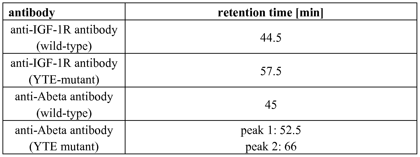


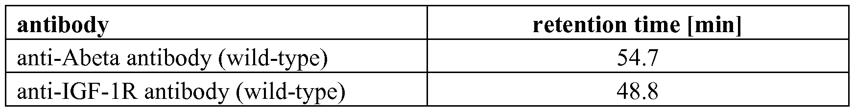

Claims
Priority Applications (15)
| Application Number | Priority Date | Filing Date | Title |
|---|---|---|---|
| PL13704599T PL2814587T3 (en) | 2012-02-15 | 2013-02-14 | Fc-receptor based affinity chromatography |
| BR112014018005-9A BR112014018005B1 (en) | 2012-02-15 | 2013-02-14 | USE OF A NON-COVALENT IMMOBILIZED COMPLEX |
| ES13704599.3T ES2676031T3 (en) | 2012-02-15 | 2013-02-14 | Affinity chromatography based on the Fc receptor |
| CA2860600A CA2860600C (en) | 2012-02-15 | 2013-02-14 | Fc-receptor based affinity chromatography |
| KR1020147022694A KR102069397B1 (en) | 2012-02-15 | 2013-02-14 | Fc-receptor based affinity chromatography |
| MX2014009622A MX360352B (en) | 2012-02-15 | 2013-02-14 | Fc-receptor based affinity chromatography. |
| JP2014557032A JP6152120B2 (en) | 2012-02-15 | 2013-02-14 | Affinity chromatography based on Fc receptors |
| EP13704599.3A EP2814587B1 (en) | 2012-02-15 | 2013-02-14 | Fc-receptor based affinity chromatography |
| CN201380009724.6A CN104125852B9 (en) | 2012-02-15 | 2013-02-14 | Fc-receptor based affinity chromatography |
| US14/378,808 US20150018241A1 (en) | 2012-02-15 | 2013-02-14 | Fc-receptor based affinity chromatography |
| SI201331082T SI2814587T1 (en) | 2012-02-15 | 2013-02-14 | Fc-receptor based affinity chromatography |
| RU2014136223A RU2624128C2 (en) | 2012-02-15 | 2013-02-14 | AFFINITY CHROMATOGRAPHY WITH APPLICATION OF Fc-RECEPTORS |
| HK15104045.0A HK1203435A1 (en) | 2012-02-15 | 2015-04-27 | Fc-receptor based affinity chromatography fc- |
| HRP20180966TT HRP20180966T1 (en) | 2012-02-15 | 2018-06-26 | Fc-receptor based affinity chromatography |
| US16/258,294 US11814409B2 (en) | 2012-02-15 | 2019-01-25 | Fc-receptor based affinity chromatography |
Applications Claiming Priority (2)
| Application Number | Priority Date | Filing Date | Title |
|---|---|---|---|
| EP12155630 | 2012-02-15 | ||
| EP12155630.2 | 2012-02-15 |
Related Child Applications (2)
| Application Number | Title | Priority Date | Filing Date |
|---|---|---|---|
| US14/378,808 A-371-Of-International US20150018241A1 (en) | 2012-02-15 | 2013-02-14 | Fc-receptor based affinity chromatography |
| US16/258,294 Continuation US11814409B2 (en) | 2012-02-15 | 2019-01-25 | Fc-receptor based affinity chromatography |
Publications (1)
| Publication Number | Publication Date |
|---|---|
| WO2013120929A1 true WO2013120929A1 (en) | 2013-08-22 |
Family
ID=47720509
Family Applications (1)
| Application Number | Title | Priority Date | Filing Date |
|---|---|---|---|
| PCT/EP2013/052932 WO2013120929A1 (en) | 2012-02-15 | 2013-02-14 | Fc-receptor based affinity chromatography |
Country Status (16)
| Country | Link |
|---|---|
| US (2) | US20150018241A1 (en) |
| EP (1) | EP2814587B1 (en) |
| JP (3) | JP6152120B2 (en) |
| KR (1) | KR102069397B1 (en) |
| CN (1) | CN104125852B9 (en) |
| BR (1) | BR112014018005B1 (en) |
| CA (2) | CA2860600C (en) |
| ES (1) | ES2676031T3 (en) |
| HK (1) | HK1203435A1 (en) |
| HR (1) | HRP20180966T1 (en) |
| MX (1) | MX360352B (en) |
| PL (1) | PL2814587T3 (en) |
| RU (1) | RU2624128C2 (en) |
| SI (1) | SI2814587T1 (en) |
| TR (1) | TR201808458T4 (en) |
| WO (1) | WO2013120929A1 (en) |
Cited By (68)
| Publication number | Priority date | Publication date | Assignee | Title |
|---|---|---|---|---|
| WO2015140126A1 (en) * | 2014-03-21 | 2015-09-24 | F. Hoffmann-La Roche Ag | In vitro prediction of in vivo half-life of antibodies |
| WO2017079637A1 (en) | 2015-11-04 | 2017-05-11 | Biogen Ma Inc. | Conjugation methods for modifying or immobilizing proteins |
| JP2017528132A (en) * | 2014-09-03 | 2017-09-28 | ベーリンガー・インゲルハイム・インターナショナル・ゲーエムベーハー | Compounds targeting IL-23A and TNF-alpha and uses thereof |
| EP3162895A4 (en) * | 2014-06-27 | 2018-02-14 | Tosoh Corporation | Improved fc-binding protein, method for producing said protein, antibody adsorbent using said protein, and method for separating antibody using said adsorbent |
| WO2018050878A1 (en) | 2016-09-19 | 2018-03-22 | F. Hoffmann-La Roche Ag | Complement factor based affinity chromatography |
| WO2018114748A1 (en) | 2016-12-20 | 2018-06-28 | F. Hoffmann-La Roche Ag | Combination therapy of anti-cd20/anti-cd3 bispecific antibodies and 4-1bb (cd137) agonists |
| WO2018184966A1 (en) | 2017-04-03 | 2018-10-11 | F. Hoffmann-La Roche Ag | Antibodies binding to steap-1 |
| WO2018185046A1 (en) | 2017-04-05 | 2018-10-11 | F. Hoffmann-La Roche Ag | Anti-lag3 antibodies |
| WO2018184964A1 (en) | 2017-04-03 | 2018-10-11 | F. Hoffmann-La Roche Ag | Immunoconjugates of an anti-pd-1 antibody with a mutant il-2 or with il-15 |
| WO2018184965A1 (en) | 2017-04-03 | 2018-10-11 | F. Hoffmann-La Roche Ag | Immunoconjugates of il-2 with an anti-pd-1 and tim-3 bispecific antibody |
| WO2018189220A1 (en) | 2017-04-13 | 2018-10-18 | F. Hoffmann-La Roche Ag | An interleukin-2 immunoconjugate, a cd40 agonist, and optionally a pd-1 axis binding antagonist for use in methods of treating cancer |
| WO2018197533A1 (en) | 2017-04-28 | 2018-11-01 | F. Hoffmann-La Roche Ag | Antibody selection method |
| WO2019122052A2 (en) | 2017-12-21 | 2019-06-27 | F. Hoffmann-La Roche Ag | Antibodies binding to hla-a2/wt1 |
| WO2019129677A1 (en) | 2017-12-29 | 2019-07-04 | F. Hoffmann-La Roche Ag | Anti-vegf antibodies and methods of use |
| WO2019154890A1 (en) | 2018-02-09 | 2019-08-15 | F. Hoffmann-La Roche Ag | Antibodies binding to gprc5d |
| WO2019175071A1 (en) | 2018-03-13 | 2019-09-19 | F. Hoffmann-La Roche Ag | Therapeutic combination of 4-1 bb agonists with anti-cd20 antibodies |
| WO2019191552A1 (en) | 2018-03-29 | 2019-10-03 | Genentech, Inc. | Modulating lactogenic activity in mammalian cells |
| WO2019202041A1 (en) | 2018-04-18 | 2019-10-24 | F. Hoffmann-La Roche Ag | Multispecific antibodies and use thereof |
| WO2019244901A1 (en) | 2018-06-20 | 2019-12-26 | 東ソー株式会社 | Method for separating antibody, and method for testing on disease |
| EP3608674A1 (en) | 2018-08-09 | 2020-02-12 | Regeneron Pharmaceuticals, Inc. | Methods for assessing binding affinity of an antibody variant to the neonatal fc receptor |
| WO2020084032A1 (en) | 2018-10-25 | 2020-04-30 | F. Hoffmann-La Roche Ag | Modification of antibody fcrn binding |
| US10683368B2 (en) | 2014-11-06 | 2020-06-16 | Hoffmann-La Roche Inc. | Fc-region variants with modified FcRn-binding and methods of use |
| WO2020127619A1 (en) | 2018-12-21 | 2020-06-25 | F. Hoffmann-La Roche Ag | Antibodies binding to cd3 |
| WO2020127873A1 (en) | 2018-12-21 | 2020-06-25 | F. Hoffmann-La Roche Ag | Antibody that binds to vegf and il-1beta and methods of use |
| WO2020200983A1 (en) | 2019-03-29 | 2020-10-08 | F. Hoffmann-La Roche Ag | Method for the generation of an fcrn expressing cell by targeted integration of multiple expression cassettes in a defined organization |
| US10844138B2 (en) | 2015-07-23 | 2020-11-24 | Boehringer Ingelheim International Gmbh | Compound targeting IL-23A and B-cell activating factor (BAFF) and uses thereof |
| WO2021001289A1 (en) | 2019-07-02 | 2021-01-07 | F. Hoffmann-La Roche Ag | Immunoconjugates comprising a mutant interleukin-2 and an anti-cd8 antibody |
| WO2021009146A1 (en) | 2019-07-15 | 2021-01-21 | F. Hoffmann-La Roche Ag | Antibodies binding to nkg2d |
| WO2021009047A1 (en) | 2019-07-12 | 2021-01-21 | F. Hoffmann-La Roche Ag | Antibodies which bind to cancer cells and target radionuclides to said cells |
| WO2021018925A1 (en) | 2019-07-31 | 2021-02-04 | F. Hoffmann-La Roche Ag | Antibodies binding to gprc5d |
| WO2021018859A2 (en) | 2019-07-31 | 2021-02-04 | F. Hoffmann-La Roche Ag | Antibodies binding to gprc5d |
| WO2021055577A2 (en) | 2019-09-18 | 2021-03-25 | Genentech, Inc. | Anti-klk7 antibodies, anti-klk5 antibodies, multispecific anti-klk5/klk7 antibodies, and methods of use |
| WO2021123173A1 (en) | 2019-12-20 | 2021-06-24 | F. Hoffmann-La Roche Ag | Il-37 fusion proteins and uses thereof |
| WO2021122875A1 (en) | 2019-12-18 | 2021-06-24 | F. Hoffmann-La Roche Ag | Antibodies binding to hla-a2/mage-a4 |
| WO2021195464A2 (en) | 2020-03-26 | 2021-09-30 | Genentech, Inc. | Modified mammalian cells |
| WO2021198034A1 (en) | 2020-03-30 | 2021-10-07 | F. Hoffmann-La Roche Ag | Antibody that binds to vegf and pdgf-b and methods of use |
| WO2021209402A2 (en) | 2020-04-15 | 2021-10-21 | F. Hoffmann-La Roche Ag | Immunoconjugates |
| WO2021249990A2 (en) | 2020-06-08 | 2021-12-16 | Hoffmann-La Roche Inc. | Anti-hbv antibodies and methods of use |
| WO2021255146A1 (en) | 2020-06-19 | 2021-12-23 | F. Hoffmann-La Roche Ag | Antibodies binding to cd3 and cea |
| WO2021255142A1 (en) | 2020-06-19 | 2021-12-23 | F. Hoffmann-La Roche Ag | Antibodies binding to cd3 |
| WO2021255143A1 (en) | 2020-06-19 | 2021-12-23 | F. Hoffmann-La Roche Ag | Antibodies binding to cd3 and folr1 |
| WO2021255155A1 (en) | 2020-06-19 | 2021-12-23 | F. Hoffmann-La Roche Ag | Antibodies binding to cd3 and cd19 |
| WO2021262783A1 (en) | 2020-06-24 | 2021-12-30 | Genentech, Inc. | Apoptosis resistant cell lines |
| WO2022008688A1 (en) | 2020-07-10 | 2022-01-13 | F. Hoffmann-La Roche Ag | Antibodies which bind to cancer cells and target radionuclides to said cells |
| WO2022016037A1 (en) | 2020-07-17 | 2022-01-20 | Genentech, Inc. | Anti-notch2 antibodies and methods of use |
| WO2022047222A2 (en) | 2020-08-28 | 2022-03-03 | Genentech, Inc. | Crispr/cas9 multiplex knockout of host cell proteins |
| WO2022049165A1 (en) | 2020-09-04 | 2022-03-10 | F. Hoffmann-La Roche Ag | Antibody that binds to vegf-a and ang2 and methods of use |
| US11286300B2 (en) | 2015-10-01 | 2022-03-29 | Hoffmann-La Roche Inc. | Humanized anti-human CD19 antibodies and methods of use |
| WO2022086957A1 (en) | 2020-10-20 | 2022-04-28 | Genentech, Inc. | Peg-conjugated anti-mertk antibodies and methods of use |
| WO2022129120A1 (en) | 2020-12-17 | 2022-06-23 | F. Hoffmann-La Roche Ag | Anti-hla-g antibodies and use thereof |
| WO2022148853A1 (en) | 2021-01-11 | 2022-07-14 | F. Hoffmann-La Roche Ag | Immunoconjugates |
| WO2022152701A1 (en) | 2021-01-13 | 2022-07-21 | F. Hoffmann-La Roche Ag | Combination therapy |
| WO2022152656A1 (en) | 2021-01-12 | 2022-07-21 | F. Hoffmann-La Roche Ag | Split antibodies which bind to cancer cells and target radionuclides to said cells |
| WO2022169872A1 (en) | 2021-02-03 | 2022-08-11 | Genentech, Inc. | Multispecific binding protein degrader platform and methods of use |
| WO2022192647A1 (en) | 2021-03-12 | 2022-09-15 | Genentech, Inc. | Anti-klk7 antibodies, anti-klk5 antibodies, multispecific anti-klk5/klk7 antibodies, and methods of use |
| WO2022219058A1 (en) | 2021-04-14 | 2022-10-20 | Anjarium Biosciences Ag | Fc-derived polypeptides |
| WO2022225880A1 (en) | 2021-04-19 | 2022-10-27 | Genentech, Inc. | Modified mammalian cells |
| WO2022246259A1 (en) | 2021-05-21 | 2022-11-24 | Genentech, Inc. | Modified cells for the production of a recombinant product of interest |
| WO2023288182A1 (en) | 2021-07-12 | 2023-01-19 | Genentech, Inc. | Structures for reducing antibody-lipase binding |
| WO2023288241A1 (en) | 2021-07-14 | 2023-01-19 | Genentech, Inc. | Anti-c-c motif chemokine receptor 8 (ccr8) antibodies and methods of use |
| WO2023001884A1 (en) | 2021-07-22 | 2023-01-26 | F. Hoffmann-La Roche Ag | Heterodimeric fc domain antibodies |
| WO2023012147A1 (en) | 2021-08-03 | 2023-02-09 | F. Hoffmann-La Roche Ag | Bispecific antibodies and methods of use |
| WO2023062048A1 (en) | 2021-10-14 | 2023-04-20 | F. Hoffmann-La Roche Ag | Alternative pd1-il7v immunoconjugates for the treatment of cancer |
| WO2023062050A1 (en) | 2021-10-14 | 2023-04-20 | F. Hoffmann-La Roche Ag | New interleukin-7 immunoconjugates |
| WO2023104938A1 (en) | 2021-12-10 | 2023-06-15 | F. Hoffmann-La Roche Ag | Antibodies binding to cd3 and plap |
| WO2023114829A1 (en) | 2021-12-15 | 2023-06-22 | Genentech, Inc. | Stabilized il-18 polypeptides and uses thereof |
| WO2023141445A1 (en) | 2022-01-19 | 2023-07-27 | Genentech, Inc. | Anti-notch2 antibodies and conjugates and methods of use |
| WO2023217933A1 (en) | 2022-05-11 | 2023-11-16 | F. Hoffmann-La Roche Ag | Antibody that binds to vegf-a and il6 and methods of use |
Families Citing this family (9)
| Publication number | Priority date | Publication date | Assignee | Title |
|---|---|---|---|---|
| SG10201605397PA (en) | 2011-07-01 | 2016-08-30 | Hoffmann La Roche | Method For Separation Of Monomeric Polypeptides From Aggregated Polypeptides |
| RU2727639C2 (en) * | 2014-01-15 | 2020-07-22 | Ф.Хоффманн-Ля Рош Аг | Variants of fc-region with modified ability to bind to fcrn and with preserved ability to bind with protein a |
| JP2018039735A (en) * | 2016-09-05 | 2018-03-15 | 東ソー株式会社 | Method for separating antibodies having differing complement dependent cytotoxic activities |
| GB201717446D0 (en) * | 2017-10-24 | 2017-12-06 | Evox Therapeutics Ltd | Affinity purification of engineering extracellular vesicles |
| JP6977536B2 (en) * | 2017-12-19 | 2021-12-08 | 東ソー株式会社 | Method for evaluating the degree of antibody denaturation using gel filtration chromatography |
| JP7159642B2 (en) * | 2018-06-26 | 2022-10-25 | 東ソー株式会社 | How to measure column retention for antibodies |
| CN109725159B (en) * | 2018-12-28 | 2021-10-08 | 江苏众红生物工程创药研究院有限公司 | Human beta2Quantitative detection test paper card of microglobulin and clinical application |
| JP2023552375A (en) * | 2020-12-06 | 2023-12-15 | エーエルエックス オンコロジー インコーポレイテッド | Multimers for reducing interference of drugs that bind to CD47 in serological assays |
| WO2023013618A1 (en) * | 2021-08-02 | 2023-02-09 | 国立研究開発法人国立長寿医療研究センター | Human astrovirus infection receptor and application of same |
Citations (107)
| Publication number | Priority date | Publication date | Assignee | Title |
|---|---|---|---|---|
| US4676980A (en) | 1985-09-23 | 1987-06-30 | The United States Of America As Represented By The Secretary Of The Department Of Health And Human Services | Target specific cross-linked heteroantibodies |
| US4737456A (en) | 1985-05-09 | 1988-04-12 | Syntex (U.S.A.) Inc. | Reducing interference in ligand-receptor binding assays |
| US4816567A (en) | 1983-04-08 | 1989-03-28 | Genentech, Inc. | Recombinant immunoglobin preparations |
| EP0404097A2 (en) | 1989-06-22 | 1990-12-27 | BEHRINGWERKE Aktiengesellschaft | Bispecific and oligospecific, mono- and oligovalent receptors, production and applications thereof |
| WO1993001161A1 (en) | 1991-07-11 | 1993-01-21 | Pfizer Limited | Process for preparing sertraline intermediates |
| US5208020A (en) | 1989-10-25 | 1993-05-04 | Immunogen Inc. | Cytotoxic agents comprising maytansinoids and their therapeutic use |
| WO1993008829A1 (en) | 1991-11-04 | 1993-05-13 | The Regents Of The University Of California | Compositions that mediate killing of hiv-infected cells |
| WO1993016185A2 (en) | 1992-02-06 | 1993-08-19 | Creative Biomolecules, Inc. | Biosynthetic binding protein for cancer marker |
| WO1994011026A2 (en) | 1992-11-13 | 1994-05-26 | Idec Pharmaceuticals Corporation | Therapeutic application of chimeric and radiolabeled antibodies to human b lymphocyte restricted differentiation antigen for treatment of b cell lymphoma |
| WO1994029351A2 (en) | 1993-06-16 | 1994-12-22 | Celltech Limited | Antibodies |
| US5500362A (en) | 1987-01-08 | 1996-03-19 | Xoma Corporation | Chimeric antibody with specificity to human B cell surface antigen |
| EP0425235B1 (en) | 1989-10-25 | 1996-09-25 | Immunogen Inc | Cytotoxic agents comprising maytansinoids and their therapeutic use |
| US5571894A (en) | 1991-02-05 | 1996-11-05 | Ciba-Geigy Corporation | Recombinant antibodies specific for a growth factor receptor |
| US5587458A (en) | 1991-10-07 | 1996-12-24 | Aronex Pharmaceuticals, Inc. | Anti-erbB-2 antibodies, combinations thereof, and therapeutic and diagnostic uses thereof |
| US5624821A (en) | 1987-03-18 | 1997-04-29 | Scotgen Biopharmaceuticals Incorporated | Antibodies with altered effector functions |
| US5635483A (en) | 1992-12-03 | 1997-06-03 | Arizona Board Of Regents Acting On Behalf Of Arizona State University | Tumor inhibiting tetrapeptide bearing modified phenethyl amides |
| US5648237A (en) | 1991-09-19 | 1997-07-15 | Genentech, Inc. | Expression of functional antibody fragments |
| US5658570A (en) | 1991-07-25 | 1997-08-19 | Idec Pharmaceuticals Corporation | Recombinant antibodies for human therapy |
| WO1997030087A1 (en) | 1996-02-16 | 1997-08-21 | Glaxo Group Limited | Preparation of glycosylated antibodies |
| US5712374A (en) | 1995-06-07 | 1998-01-27 | American Cyanamid Company | Method for the preparation of substantiallly monomeric calicheamicin derivative/carrier conjugates |
| US5714586A (en) | 1995-06-07 | 1998-02-03 | American Cyanamid Company | Methods for the preparation of monomeric calicheamicin derivative/carrier conjugates |
| US5731168A (en) | 1995-03-01 | 1998-03-24 | Genentech, Inc. | Method for making heteromultimeric polypeptides |
| US5739116A (en) | 1994-06-03 | 1998-04-14 | American Cyanamid Company | Enediyne derivatives useful for the synthesis of conjugates of methyltrithio antitumor agents |
| US5750373A (en) | 1990-12-03 | 1998-05-12 | Genentech, Inc. | Enrichment method for variant proteins having altered binding properties, M13 phagemids, and growth hormone variants |
| US5770429A (en) | 1990-08-29 | 1998-06-23 | Genpharm International, Inc. | Transgenic non-human animals capable of producing heterologous antibodies |
| US5770710A (en) | 1987-10-30 | 1998-06-23 | American Cyanamid Company | Antitumor and antibacterial substituted disulfide derivatives prepared from compounds possessing a methlytrithio group |
| US5770701A (en) | 1987-10-30 | 1998-06-23 | American Cyanamid Company | Process for preparing targeted forms of methyltrithio antitumor agents |
| US5780588A (en) | 1993-01-26 | 1998-07-14 | Arizona Board Of Regents | Elucidation and synthesis of selected pentapeptides |
| US5789199A (en) | 1994-11-03 | 1998-08-04 | Genentech, Inc. | Process for bacterial production of polypeptides |
| US5821337A (en) | 1991-06-14 | 1998-10-13 | Genentech, Inc. | Immunoglobulin variants |
| US5840523A (en) | 1995-03-01 | 1998-11-24 | Genetech, Inc. | Methods and compositions for secretion of heterologous polypeptides |
| WO1998058964A1 (en) | 1997-06-24 | 1998-12-30 | Genentech, Inc. | Methods and compositions for galactosylated glycoproteins |
| US5869046A (en) | 1995-04-14 | 1999-02-09 | Genentech, Inc. | Altered polypeptides with increased half-life |
| WO1999022764A1 (en) | 1997-10-31 | 1999-05-14 | Genentech, Inc. | Methods and compositions comprising glycoprotein glycoforms |
| US5959177A (en) | 1989-10-27 | 1999-09-28 | The Scripps Research Institute | Transgenic plants expressing assembled secretory antibodies |
| WO1999051642A1 (en) | 1998-04-02 | 1999-10-14 | Genentech, Inc. | Antibody variants and fragments thereof |
| US6040498A (en) | 1998-08-11 | 2000-03-21 | North Caroline State University | Genetically engineered duckweed |
| US6075181A (en) | 1990-01-12 | 2000-06-13 | Abgenix, Inc. | Human antibodies derived from immunized xenomice |
| WO2000061739A1 (en) | 1999-04-09 | 2000-10-19 | Kyowa Hakko Kogyo Co., Ltd. | Method for controlling the activity of immunologically functional molecule |
| US6150584A (en) | 1990-01-12 | 2000-11-21 | Abgenix, Inc. | Human antibodies derived from immunized xenomice |
| US6171586B1 (en) | 1997-06-13 | 2001-01-09 | Genentech, Inc. | Antibody formulation |
| US6194551B1 (en) | 1998-04-02 | 2001-02-27 | Genentech, Inc. | Polypeptide variants |
| WO2001029246A1 (en) | 1999-10-19 | 2001-04-26 | Kyowa Hakko Kogyo Co., Ltd. | Process for producing polypeptide |
| US6248516B1 (en) | 1988-11-11 | 2001-06-19 | Medical Research Council | Single domain ligands, receptors comprising said ligands methods for their production, and use of said ligands and receptors |
| US6267958B1 (en) | 1995-07-27 | 2001-07-31 | Genentech, Inc. | Protein formulation |
| WO2002031140A1 (en) | 2000-10-06 | 2002-04-18 | Kyowa Hakko Kogyo Co., Ltd. | Cells producing antibody compositions |
| US6420548B1 (en) | 1999-10-04 | 2002-07-16 | Medicago Inc. | Method for regulating transcription of foreign genes |
| US20020164328A1 (en) | 2000-10-06 | 2002-11-07 | Toyohide Shinkawa | Process for purifying antibody |
| WO2003011878A2 (en) | 2001-08-03 | 2003-02-13 | Glycart Biotechnology Ag | Antibody glycosylation variants having increased antibody-dependent cellular cytotoxicity |
| US20030115614A1 (en) | 2000-10-06 | 2003-06-19 | Yutaka Kanda | Antibody composition-producing cell |
| US6602684B1 (en) | 1998-04-20 | 2003-08-05 | Glycart Biotechnology Ag | Glycosylation engineering of antibodies for improving antibody-dependent cellular cytotoxicity |
| US20030157108A1 (en) | 2001-10-25 | 2003-08-21 | Genentech, Inc. | Glycoprotein compositions |
| US6630579B2 (en) | 1999-12-29 | 2003-10-07 | Immunogen Inc. | Cytotoxic agents comprising modified doxorubicins and daunorubicins and their therapeutic use |
| WO2003085107A1 (en) | 2002-04-09 | 2003-10-16 | Kyowa Hakko Kogyo Co., Ltd. | Cells with modified genome |
| WO2003084570A1 (en) | 2002-04-09 | 2003-10-16 | Kyowa Hakko Kogyo Co., Ltd. | DRUG CONTAINING ANTIBODY COMPOSITION APPROPRIATE FOR PATIENT SUFFERING FROM FcϜRIIIa POLYMORPHISM |
| WO2003085119A1 (en) | 2002-04-09 | 2003-10-16 | Kyowa Hakko Kogyo Co., Ltd. | METHOD OF ENHANCING ACTIVITY OF ANTIBODY COMPOSITION OF BINDING TO FcϜ RECEPTOR IIIa |
| US20040093621A1 (en) | 2001-12-25 | 2004-05-13 | Kyowa Hakko Kogyo Co., Ltd | Antibody composition which specifically binds to CD20 |
| US6737056B1 (en) | 1999-01-15 | 2004-05-18 | Genentech, Inc. | Polypeptide variants with altered effector function |
| US20040109865A1 (en) | 2002-04-09 | 2004-06-10 | Kyowa Hakko Kogyo Co., Ltd. | Antibody composition-containing medicament |
| US20040110282A1 (en) | 2002-04-09 | 2004-06-10 | Kyowa Hakko Kogyo Co., Ltd. | Cells in which activity of the protein involved in transportation of GDP-fucose is reduced or lost |
| US20040132140A1 (en) | 2002-04-09 | 2004-07-08 | Kyowa Hakko Kogyo Co., Ltd. | Production process for antibody composition |
| WO2004056312A2 (en) | 2002-12-16 | 2004-07-08 | Genentech, Inc. | Immunoglobulin variants and uses thereof |
| US20050014934A1 (en) | 2002-10-15 | 2005-01-20 | Hinton Paul R. | Alteration of FcRn binding affinities or serum half-lives of antibodies by mutagenesis |
| US20050079574A1 (en) | 2003-01-16 | 2005-04-14 | Genentech, Inc. | Synthetic antibody phage libraries |
| WO2005035778A1 (en) | 2003-10-09 | 2005-04-21 | Kyowa Hakko Kogyo Co., Ltd. | PROCESS FOR PRODUCING ANTIBODY COMPOSITION BY USING RNA INHIBITING THE FUNCTION OF α1,6-FUCOSYLTRANSFERASE |
| WO2005035586A1 (en) | 2003-10-08 | 2005-04-21 | Kyowa Hakko Kogyo Co., Ltd. | Fused protein composition |
| WO2005047327A2 (en) | 2003-11-12 | 2005-05-26 | Biogen Idec Ma Inc. | NEONATAL Fc RECEPTOR (FcRn)-BINDING POLYPEPTIDE VARIANTS, DIMERIC Fc BINDING PROTEINS AND METHODS RELATED THERETO |
| US20050119455A1 (en) | 2002-06-03 | 2005-06-02 | Genentech, Inc. | Synthetic antibody phage libraries |
| US20050123546A1 (en) | 2003-11-05 | 2005-06-09 | Glycart Biotechnology Ag | Antigen binding molecules with increased Fc receptor binding affinity and effector function |
| WO2005053742A1 (en) | 2003-12-04 | 2005-06-16 | Kyowa Hakko Kogyo Co., Ltd. | Medicine containing antibody composition |
| WO2005100402A1 (en) | 2004-04-13 | 2005-10-27 | F.Hoffmann-La Roche Ag | Anti-p-selectin antibodies |
| US20050260186A1 (en) | 2003-03-05 | 2005-11-24 | Halozyme, Inc. | Soluble glycosaminoglycanases and methods of preparing and using soluble glycosaminoglycanases |
| US20050266000A1 (en) | 2004-04-09 | 2005-12-01 | Genentech, Inc. | Variable domain library and uses |
| US6982321B2 (en) | 1986-03-27 | 2006-01-03 | Medical Research Council | Altered antibodies |
| US20060025576A1 (en) | 2000-04-11 | 2006-02-02 | Genentech, Inc. | Multivalent antibodies and uses therefor |
| WO2006031370A2 (en) | 2004-08-19 | 2006-03-23 | Genentech, Inc. | Polypeptide variants with altered effector function |
| WO2006029879A2 (en) | 2004-09-17 | 2006-03-23 | F.Hoffmann-La Roche Ag | Anti-ox40l antibodies |
| WO2006044908A2 (en) | 2004-10-20 | 2006-04-27 | Genentech, Inc. | Antibody formulation in histidine-acetate buffer |
| US7041870B2 (en) | 2000-11-30 | 2006-05-09 | Medarex, Inc. | Transgenic transchromosomal rodents for making human antibodies |
| US20060104968A1 (en) | 2003-03-05 | 2006-05-18 | Halozyme, Inc. | Soluble glycosaminoglycanases and methods of preparing and using soluble glycosaminogly ycanases |
| US7087409B2 (en) | 1997-12-05 | 2006-08-08 | The Scripps Research Institute | Humanization of murine antibody |
| US7125978B1 (en) | 1999-10-04 | 2006-10-24 | Medicago Inc. | Promoter for regulating expression of foreign genes |
| US7189826B2 (en) | 1997-11-24 | 2007-03-13 | Institute For Human Genetics And Biochemistry | Monoclonal human natural antibodies |
| US20070061900A1 (en) | 2000-10-31 | 2007-03-15 | Murphy Andrew J | Methods of modifying eukaryotic cells |
| US20070117126A1 (en) | 1999-12-15 | 2007-05-24 | Genentech, Inc. | Shotgun scanning |
| US20070160598A1 (en) | 2005-11-07 | 2007-07-12 | Dennis Mark S | Binding polypeptides with diversified and consensus vh/vl hypervariable sequences |
| US20070237764A1 (en) | 2005-12-02 | 2007-10-11 | Genentech, Inc. | Binding polypeptides with restricted diversity sequences |
| US20070292936A1 (en) | 2006-05-09 | 2007-12-20 | Genentech, Inc. | Binding polypeptides with optimized scaffolds |
| US20080069820A1 (en) | 2006-08-30 | 2008-03-20 | Genentech, Inc. | Multispecific antibodies |
| US7371826B2 (en) | 1999-01-15 | 2008-05-13 | Genentech, Inc. | Polypeptide variants with altered effector function |
| WO2008077546A1 (en) | 2006-12-22 | 2008-07-03 | F. Hoffmann-La Roche Ag | Antibodies against insulin-like growth factor i receptor and uses thereof |
| US20090002360A1 (en) | 2007-05-25 | 2009-01-01 | Innolux Display Corp. | Liquid crystal display device and method for driving same |
| US7498298B2 (en) | 2003-11-06 | 2009-03-03 | Seattle Genetics, Inc. | Monomethylvaline compounds capable of conjugation to ligands |
| WO2009041643A1 (en) | 2007-09-26 | 2009-04-02 | Chugai Seiyaku Kabushiki Kaisha | Method of modifying isoelectric point of antibody via amino acid substitution in cdr |
| US7521541B2 (en) | 2004-09-23 | 2009-04-21 | Genetech Inc. | Cysteine engineered antibodies and conjugates |
| US7527791B2 (en) | 2004-03-31 | 2009-05-05 | Genentech, Inc. | Humanized anti-TGF-beta antibodies |
| WO2009080254A1 (en) | 2007-12-21 | 2009-07-02 | F. Hoffmann-La Roche Ag | Bivalent, bispecific antibodies |
| WO2009080251A1 (en) | 2007-12-21 | 2009-07-02 | F. Hoffmann-La Roche Ag | Bivalent, bispecific antibodies |
| WO2009080252A1 (en) | 2007-12-21 | 2009-07-02 | F. Hoffmann-La Roche Ag | Bivalent, bispecific antibodies |
| WO2009080253A1 (en) | 2007-12-21 | 2009-07-02 | F. Hoffmann-La Roche Ag | Bivalent, bispecific antibodies |
| WO2009089004A1 (en) | 2008-01-07 | 2009-07-16 | Amgen Inc. | Method for making antibody fc-heterodimeric molecules using electrostatic steering effects |
| WO2010048313A2 (en) | 2008-10-22 | 2010-04-29 | Biogen Idec Ma Inc. | Recombinant fcrn and variants thereof for purification of fc-containing fusion proteins |
| WO2010112193A1 (en) | 2009-04-02 | 2010-10-07 | Roche Glycart Ag | Multispecific antibodies comprising full length antibodies and single chain fab fragments |
| WO2010115589A1 (en) | 2009-04-07 | 2010-10-14 | Roche Glycart Ag | Trivalent, bispecific antibodies |
| WO2010136172A1 (en) | 2009-05-27 | 2010-12-02 | F. Hoffmann-La Roche Ag | Tri- or tetraspecific antibodies |
| WO2010145792A1 (en) | 2009-06-16 | 2010-12-23 | F. Hoffmann-La Roche Ag | Bispecific antigen binding proteins |
| WO2010145793A1 (en) | 2009-06-18 | 2010-12-23 | F. Hoffmann-La Roche Ag | Bispecific, tetravalent antigen binding proteins |
Family Cites Families (27)
| Publication number | Priority date | Publication date | Assignee | Title |
|---|---|---|---|---|
| US4900660A (en) * | 1985-11-25 | 1990-02-13 | University Of Florida | Streptococcal fc rc |
| US5169936A (en) * | 1989-04-14 | 1992-12-08 | Biogen, Inc. | Protein purification on immobilized metal affinity resins effected by elution using a weak ligand |
| US5623053A (en) * | 1992-01-10 | 1997-04-22 | California Institute Of Technology | Soluble mammal-derived Fc receptor which binds at a pH ranging from about 5.5 to 6.5 and releases at a pH ranging from about 7.5 to 8.5 |
| US6096871A (en) | 1995-04-14 | 2000-08-01 | Genentech, Inc. | Polypeptides altered to contain an epitope from the Fc region of an IgG molecule for increased half-life |
| JP2002517185A (en) * | 1998-06-01 | 2002-06-18 | アジェンシス,インコーポレイテッド | Tumor antigens useful for diagnosis and treatment of prostate and colon cancer |
| CN1294148C (en) | 2001-04-11 | 2007-01-10 | 中国科学院遗传与发育生物学研究所 | Single-stranded cyctic trispecific antibody |
| CN101124245A (en) * | 2003-11-12 | 2008-02-13 | 比奥根艾迪克Ma公司 | Neonatal Fc receptor (FcRn)-binding polypeptide variants, dimeric Fc binding proteins and methods related thereto |
| EP1896582A4 (en) | 2005-05-09 | 2009-04-08 | Ono Pharmaceutical Co | Human monoclonal antibodies to programmed death 1(pd-1) and methods for treating cancer using anti-pd-1 antibodies alone or in combination with other immunotherapeutics |
| EP1878739A1 (en) * | 2006-07-14 | 2008-01-16 | LEK Pharmaceuticals D.D. | One step IMAC (MCAC) purification of proteins |
| AU2008218925A1 (en) * | 2007-02-20 | 2008-08-28 | Anaptysbio, Inc. | Somatic hypermutation systems |
| JP5522723B2 (en) | 2007-05-21 | 2014-06-18 | ノマディックバイオサイエンス株式会社 | Novel polypeptide, material for affinity chromatography, and method for separation and / or purification of immunoglobulin |
| ES2742268T3 (en) | 2007-12-26 | 2020-02-13 | Xencor Inc | Fc variants with altered FcRn binding |
| LT2708559T (en) | 2008-04-11 | 2018-06-11 | Chugai Seiyaku Kabushiki Kaisha | Antigen-binding molecule capable of binding to two or more antigen molecules repeatedly |
| US8268314B2 (en) | 2008-10-08 | 2012-09-18 | Hoffmann-La Roche Inc. | Bispecific anti-VEGF/anti-ANG-2 antibodies |
| CN102245208B (en) | 2008-10-14 | 2016-03-16 | 霍夫曼-拉罗奇有限公司 | immunoglobulin variants and uses thereof |
| WO2011106272A1 (en) * | 2010-02-23 | 2011-09-01 | Merck Sharp & Dohme Corp. | Novel binding assays useful in identifying antibodies with altered half-lives |
| TWI667257B (en) | 2010-03-30 | 2019-08-01 | 中外製藥股份有限公司 | Antibodies with modified affinity to fcrn that promote antigen clearance |
| US9163087B2 (en) | 2010-06-18 | 2015-10-20 | The Brigham And Women's Hospital, Inc. | Bi-specific antibodies against TIM-3 and PD-1 for immunotherapy in chronic immune conditions |
| HUE041335T2 (en) * | 2011-03-29 | 2019-05-28 | Roche Glycart Ag | Antibody fc variants |
| US10221210B2 (en) * | 2011-07-20 | 2019-03-05 | Zepteon, Incorporated | Polypeptide separation methods |
| EP2791169B1 (en) | 2011-12-16 | 2017-07-19 | Synthon Biopharmaceuticals B.V. | Compounds and methods for treating inflammatory diseases |
| LT2872534T (en) | 2012-07-13 | 2018-10-25 | Roche Glycart Ag | Bispecific anti-vegf/anti-ang-2 antibodies and their use in the treatment of ocular vascular diseases |
| JP6133444B2 (en) | 2013-02-26 | 2017-05-24 | ロシュ グリクアート アーゲー | Bispecific T cell activation antigen binding molecule |
| MX2016011982A (en) | 2014-03-21 | 2016-12-09 | Hoffmann La Roche | In vitro prediction of in vivo half-life of antibodies. |
| US20170267780A1 (en) | 2014-05-16 | 2017-09-21 | Medimmune, Llc | Molecules with altered neonate fc receptor binding having enhanced therapeutic and diagnostic properties |
| RU2714116C2 (en) | 2014-11-06 | 2020-02-11 | Ф. Хоффманн-Ля Рош Аг | VARIANTS OF Fc-DOMAIN WITH MODIFIED FcRn BINDING AND METHODS OF APPLICATION THEREOF |
| JP6288332B2 (en) * | 2017-03-06 | 2018-03-07 | 東洋紡株式会社 | Novel glucose dehydrogenase |
-
2013
- 2013-02-14 BR BR112014018005-9A patent/BR112014018005B1/en active IP Right Grant
- 2013-02-14 SI SI201331082T patent/SI2814587T1/en unknown
- 2013-02-14 ES ES13704599.3T patent/ES2676031T3/en active Active
- 2013-02-14 CA CA2860600A patent/CA2860600C/en active Active
- 2013-02-14 CN CN201380009724.6A patent/CN104125852B9/en active Active
- 2013-02-14 KR KR1020147022694A patent/KR102069397B1/en active IP Right Grant
- 2013-02-14 WO PCT/EP2013/052932 patent/WO2013120929A1/en active Application Filing
- 2013-02-14 CA CA3159061A patent/CA3159061A1/en active Pending
- 2013-02-14 JP JP2014557032A patent/JP6152120B2/en active Active
- 2013-02-14 RU RU2014136223A patent/RU2624128C2/en active
- 2013-02-14 US US14/378,808 patent/US20150018241A1/en not_active Abandoned
- 2013-02-14 TR TR2018/08458T patent/TR201808458T4/en unknown
- 2013-02-14 MX MX2014009622A patent/MX360352B/en active IP Right Grant
- 2013-02-14 PL PL13704599T patent/PL2814587T3/en unknown
- 2013-02-14 EP EP13704599.3A patent/EP2814587B1/en active Active
-
2015
- 2015-04-27 HK HK15104045.0A patent/HK1203435A1/en unknown
-
2017
- 2017-05-26 JP JP2017104130A patent/JP6360232B2/en active Active
-
2018
- 2018-06-21 JP JP2018117501A patent/JP6713020B2/en active Active
- 2018-06-26 HR HRP20180966TT patent/HRP20180966T1/en unknown
-
2019
- 2019-01-25 US US16/258,294 patent/US11814409B2/en active Active
Patent Citations (115)
| Publication number | Priority date | Publication date | Assignee | Title |
|---|---|---|---|---|
| US4816567A (en) | 1983-04-08 | 1989-03-28 | Genentech, Inc. | Recombinant immunoglobin preparations |
| US4737456A (en) | 1985-05-09 | 1988-04-12 | Syntex (U.S.A.) Inc. | Reducing interference in ligand-receptor binding assays |
| US4676980A (en) | 1985-09-23 | 1987-06-30 | The United States Of America As Represented By The Secretary Of The Department Of Health And Human Services | Target specific cross-linked heteroantibodies |
| US6982321B2 (en) | 1986-03-27 | 2006-01-03 | Medical Research Council | Altered antibodies |
| US5500362A (en) | 1987-01-08 | 1996-03-19 | Xoma Corporation | Chimeric antibody with specificity to human B cell surface antigen |
| US5648260A (en) | 1987-03-18 | 1997-07-15 | Scotgen Biopharmaceuticals Incorporated | DNA encoding antibodies with altered effector functions |
| US5624821A (en) | 1987-03-18 | 1997-04-29 | Scotgen Biopharmaceuticals Incorporated | Antibodies with altered effector functions |
| US5770701A (en) | 1987-10-30 | 1998-06-23 | American Cyanamid Company | Process for preparing targeted forms of methyltrithio antitumor agents |
| US5770710A (en) | 1987-10-30 | 1998-06-23 | American Cyanamid Company | Antitumor and antibacterial substituted disulfide derivatives prepared from compounds possessing a methlytrithio group |
| US6248516B1 (en) | 1988-11-11 | 2001-06-19 | Medical Research Council | Single domain ligands, receptors comprising said ligands methods for their production, and use of said ligands and receptors |
| EP0404097A2 (en) | 1989-06-22 | 1990-12-27 | BEHRINGWERKE Aktiengesellschaft | Bispecific and oligospecific, mono- and oligovalent receptors, production and applications thereof |
| EP0425235B1 (en) | 1989-10-25 | 1996-09-25 | Immunogen Inc | Cytotoxic agents comprising maytansinoids and their therapeutic use |
| US5416064A (en) | 1989-10-25 | 1995-05-16 | Immunogen, Inc. | Cytotoxic agents comprising maytansinoids and their therapeutic use |
| US5208020A (en) | 1989-10-25 | 1993-05-04 | Immunogen Inc. | Cytotoxic agents comprising maytansinoids and their therapeutic use |
| US6417429B1 (en) | 1989-10-27 | 2002-07-09 | The Scripps Research Institute | Transgenic plants expressing assembled secretory antibodies |
| US5959177A (en) | 1989-10-27 | 1999-09-28 | The Scripps Research Institute | Transgenic plants expressing assembled secretory antibodies |
| US6150584A (en) | 1990-01-12 | 2000-11-21 | Abgenix, Inc. | Human antibodies derived from immunized xenomice |
| US6075181A (en) | 1990-01-12 | 2000-06-13 | Abgenix, Inc. | Human antibodies derived from immunized xenomice |
| US5770429A (en) | 1990-08-29 | 1998-06-23 | Genpharm International, Inc. | Transgenic non-human animals capable of producing heterologous antibodies |
| US5750373A (en) | 1990-12-03 | 1998-05-12 | Genentech, Inc. | Enrichment method for variant proteins having altered binding properties, M13 phagemids, and growth hormone variants |
| US5571894A (en) | 1991-02-05 | 1996-11-05 | Ciba-Geigy Corporation | Recombinant antibodies specific for a growth factor receptor |
| US5821337A (en) | 1991-06-14 | 1998-10-13 | Genentech, Inc. | Immunoglobulin variants |
| WO1993001161A1 (en) | 1991-07-11 | 1993-01-21 | Pfizer Limited | Process for preparing sertraline intermediates |
| US5658570A (en) | 1991-07-25 | 1997-08-19 | Idec Pharmaceuticals Corporation | Recombinant antibodies for human therapy |
| US5648237A (en) | 1991-09-19 | 1997-07-15 | Genentech, Inc. | Expression of functional antibody fragments |
| US5587458A (en) | 1991-10-07 | 1996-12-24 | Aronex Pharmaceuticals, Inc. | Anti-erbB-2 antibodies, combinations thereof, and therapeutic and diagnostic uses thereof |
| WO1993008829A1 (en) | 1991-11-04 | 1993-05-13 | The Regents Of The University Of California | Compositions that mediate killing of hiv-infected cells |
| WO1993016185A2 (en) | 1992-02-06 | 1993-08-19 | Creative Biomolecules, Inc. | Biosynthetic binding protein for cancer marker |
| WO1994011026A2 (en) | 1992-11-13 | 1994-05-26 | Idec Pharmaceuticals Corporation | Therapeutic application of chimeric and radiolabeled antibodies to human b lymphocyte restricted differentiation antigen for treatment of b cell lymphoma |
| US5635483A (en) | 1992-12-03 | 1997-06-03 | Arizona Board Of Regents Acting On Behalf Of Arizona State University | Tumor inhibiting tetrapeptide bearing modified phenethyl amides |
| US5780588A (en) | 1993-01-26 | 1998-07-14 | Arizona Board Of Regents | Elucidation and synthesis of selected pentapeptides |
| WO1994029351A2 (en) | 1993-06-16 | 1994-12-22 | Celltech Limited | Antibodies |
| US5773001A (en) | 1994-06-03 | 1998-06-30 | American Cyanamid Company | Conjugates of methyltrithio antitumor agents and intermediates for their synthesis |
| US5877296A (en) | 1994-06-03 | 1999-03-02 | American Cyanamid Company | Process for preparing conjugates of methyltrithio antitumor agents |
| US5767285A (en) | 1994-06-03 | 1998-06-16 | American Cyanamid Company | Linkers useful for the synthesis of conjugates of methyltrithio antitumor agents |
| US5739116A (en) | 1994-06-03 | 1998-04-14 | American Cyanamid Company | Enediyne derivatives useful for the synthesis of conjugates of methyltrithio antitumor agents |
| US5789199A (en) | 1994-11-03 | 1998-08-04 | Genentech, Inc. | Process for bacterial production of polypeptides |
| US5731168A (en) | 1995-03-01 | 1998-03-24 | Genentech, Inc. | Method for making heteromultimeric polypeptides |
| US5840523A (en) | 1995-03-01 | 1998-11-24 | Genetech, Inc. | Methods and compositions for secretion of heterologous polypeptides |
| US5869046A (en) | 1995-04-14 | 1999-02-09 | Genentech, Inc. | Altered polypeptides with increased half-life |
| US5712374A (en) | 1995-06-07 | 1998-01-27 | American Cyanamid Company | Method for the preparation of substantiallly monomeric calicheamicin derivative/carrier conjugates |
| US5714586A (en) | 1995-06-07 | 1998-02-03 | American Cyanamid Company | Methods for the preparation of monomeric calicheamicin derivative/carrier conjugates |
| US6267958B1 (en) | 1995-07-27 | 2001-07-31 | Genentech, Inc. | Protein formulation |
| WO1997030087A1 (en) | 1996-02-16 | 1997-08-21 | Glaxo Group Limited | Preparation of glycosylated antibodies |
| US6171586B1 (en) | 1997-06-13 | 2001-01-09 | Genentech, Inc. | Antibody formulation |
| WO1998058964A1 (en) | 1997-06-24 | 1998-12-30 | Genentech, Inc. | Methods and compositions for galactosylated glycoproteins |
| WO1999022764A1 (en) | 1997-10-31 | 1999-05-14 | Genentech, Inc. | Methods and compositions comprising glycoprotein glycoforms |
| US7189826B2 (en) | 1997-11-24 | 2007-03-13 | Institute For Human Genetics And Biochemistry | Monoclonal human natural antibodies |
| US7087409B2 (en) | 1997-12-05 | 2006-08-08 | The Scripps Research Institute | Humanization of murine antibody |
| US6194551B1 (en) | 1998-04-02 | 2001-02-27 | Genentech, Inc. | Polypeptide variants |
| WO1999051642A1 (en) | 1998-04-02 | 1999-10-14 | Genentech, Inc. | Antibody variants and fragments thereof |
| US6602684B1 (en) | 1998-04-20 | 2003-08-05 | Glycart Biotechnology Ag | Glycosylation engineering of antibodies for improving antibody-dependent cellular cytotoxicity |
| US6040498A (en) | 1998-08-11 | 2000-03-21 | North Caroline State University | Genetically engineered duckweed |
| US7332581B2 (en) | 1999-01-15 | 2008-02-19 | Genentech, Inc. | Polypeptide variants with altered effector function |
| US6737056B1 (en) | 1999-01-15 | 2004-05-18 | Genentech, Inc. | Polypeptide variants with altered effector function |
| US7371826B2 (en) | 1999-01-15 | 2008-05-13 | Genentech, Inc. | Polypeptide variants with altered effector function |
| WO2000061739A1 (en) | 1999-04-09 | 2000-10-19 | Kyowa Hakko Kogyo Co., Ltd. | Method for controlling the activity of immunologically functional molecule |
| US6420548B1 (en) | 1999-10-04 | 2002-07-16 | Medicago Inc. | Method for regulating transcription of foreign genes |
| US7125978B1 (en) | 1999-10-04 | 2006-10-24 | Medicago Inc. | Promoter for regulating expression of foreign genes |
| WO2001029246A1 (en) | 1999-10-19 | 2001-04-26 | Kyowa Hakko Kogyo Co., Ltd. | Process for producing polypeptide |
| US20070117126A1 (en) | 1999-12-15 | 2007-05-24 | Genentech, Inc. | Shotgun scanning |
| US6630579B2 (en) | 1999-12-29 | 2003-10-07 | Immunogen Inc. | Cytotoxic agents comprising modified doxorubicins and daunorubicins and their therapeutic use |
| US20060025576A1 (en) | 2000-04-11 | 2006-02-02 | Genentech, Inc. | Multivalent antibodies and uses therefor |
| US20030115614A1 (en) | 2000-10-06 | 2003-06-19 | Yutaka Kanda | Antibody composition-producing cell |
| US20020164328A1 (en) | 2000-10-06 | 2002-11-07 | Toyohide Shinkawa | Process for purifying antibody |
| WO2002031140A1 (en) | 2000-10-06 | 2002-04-18 | Kyowa Hakko Kogyo Co., Ltd. | Cells producing antibody compositions |
| US20070061900A1 (en) | 2000-10-31 | 2007-03-15 | Murphy Andrew J | Methods of modifying eukaryotic cells |
| US7041870B2 (en) | 2000-11-30 | 2006-05-09 | Medarex, Inc. | Transgenic transchromosomal rodents for making human antibodies |
| WO2003011878A2 (en) | 2001-08-03 | 2003-02-13 | Glycart Biotechnology Ag | Antibody glycosylation variants having increased antibody-dependent cellular cytotoxicity |
| US20030157108A1 (en) | 2001-10-25 | 2003-08-21 | Genentech, Inc. | Glycoprotein compositions |
| US20040093621A1 (en) | 2001-12-25 | 2004-05-13 | Kyowa Hakko Kogyo Co., Ltd | Antibody composition which specifically binds to CD20 |
| US20040110704A1 (en) | 2002-04-09 | 2004-06-10 | Kyowa Hakko Kogyo Co., Ltd. | Cells of which genome is modified |
| WO2003085119A1 (en) | 2002-04-09 | 2003-10-16 | Kyowa Hakko Kogyo Co., Ltd. | METHOD OF ENHANCING ACTIVITY OF ANTIBODY COMPOSITION OF BINDING TO FcϜ RECEPTOR IIIa |
| WO2003085107A1 (en) | 2002-04-09 | 2003-10-16 | Kyowa Hakko Kogyo Co., Ltd. | Cells with modified genome |
| WO2003084570A1 (en) | 2002-04-09 | 2003-10-16 | Kyowa Hakko Kogyo Co., Ltd. | DRUG CONTAINING ANTIBODY COMPOSITION APPROPRIATE FOR PATIENT SUFFERING FROM FcϜRIIIa POLYMORPHISM |
| US20040109865A1 (en) | 2002-04-09 | 2004-06-10 | Kyowa Hakko Kogyo Co., Ltd. | Antibody composition-containing medicament |
| US20040110282A1 (en) | 2002-04-09 | 2004-06-10 | Kyowa Hakko Kogyo Co., Ltd. | Cells in which activity of the protein involved in transportation of GDP-fucose is reduced or lost |
| US20040132140A1 (en) | 2002-04-09 | 2004-07-08 | Kyowa Hakko Kogyo Co., Ltd. | Production process for antibody composition |
| US20050119455A1 (en) | 2002-06-03 | 2005-06-02 | Genentech, Inc. | Synthetic antibody phage libraries |
| US20050014934A1 (en) | 2002-10-15 | 2005-01-20 | Hinton Paul R. | Alteration of FcRn binding affinities or serum half-lives of antibodies by mutagenesis |
| WO2004056312A2 (en) | 2002-12-16 | 2004-07-08 | Genentech, Inc. | Immunoglobulin variants and uses thereof |
| US20050079574A1 (en) | 2003-01-16 | 2005-04-14 | Genentech, Inc. | Synthetic antibody phage libraries |
| US20050260186A1 (en) | 2003-03-05 | 2005-11-24 | Halozyme, Inc. | Soluble glycosaminoglycanases and methods of preparing and using soluble glycosaminoglycanases |
| US20060104968A1 (en) | 2003-03-05 | 2006-05-18 | Halozyme, Inc. | Soluble glycosaminoglycanases and methods of preparing and using soluble glycosaminogly ycanases |
| WO2005035586A1 (en) | 2003-10-08 | 2005-04-21 | Kyowa Hakko Kogyo Co., Ltd. | Fused protein composition |
| WO2005035778A1 (en) | 2003-10-09 | 2005-04-21 | Kyowa Hakko Kogyo Co., Ltd. | PROCESS FOR PRODUCING ANTIBODY COMPOSITION BY USING RNA INHIBITING THE FUNCTION OF α1,6-FUCOSYLTRANSFERASE |
| US20050123546A1 (en) | 2003-11-05 | 2005-06-09 | Glycart Biotechnology Ag | Antigen binding molecules with increased Fc receptor binding affinity and effector function |
| US7498298B2 (en) | 2003-11-06 | 2009-03-03 | Seattle Genetics, Inc. | Monomethylvaline compounds capable of conjugation to ligands |
| WO2005047327A2 (en) | 2003-11-12 | 2005-05-26 | Biogen Idec Ma Inc. | NEONATAL Fc RECEPTOR (FcRn)-BINDING POLYPEPTIDE VARIANTS, DIMERIC Fc BINDING PROTEINS AND METHODS RELATED THERETO |
| WO2005053742A1 (en) | 2003-12-04 | 2005-06-16 | Kyowa Hakko Kogyo Co., Ltd. | Medicine containing antibody composition |
| US7527791B2 (en) | 2004-03-31 | 2009-05-05 | Genentech, Inc. | Humanized anti-TGF-beta antibodies |
| US20050266000A1 (en) | 2004-04-09 | 2005-12-01 | Genentech, Inc. | Variable domain library and uses |
| WO2005100402A1 (en) | 2004-04-13 | 2005-10-27 | F.Hoffmann-La Roche Ag | Anti-p-selectin antibodies |
| WO2006031370A2 (en) | 2004-08-19 | 2006-03-23 | Genentech, Inc. | Polypeptide variants with altered effector function |
| WO2006029879A2 (en) | 2004-09-17 | 2006-03-23 | F.Hoffmann-La Roche Ag | Anti-ox40l antibodies |
| US7521541B2 (en) | 2004-09-23 | 2009-04-21 | Genetech Inc. | Cysteine engineered antibodies and conjugates |
| WO2006044908A2 (en) | 2004-10-20 | 2006-04-27 | Genentech, Inc. | Antibody formulation in histidine-acetate buffer |
| US20070160598A1 (en) | 2005-11-07 | 2007-07-12 | Dennis Mark S | Binding polypeptides with diversified and consensus vh/vl hypervariable sequences |
| US20070237764A1 (en) | 2005-12-02 | 2007-10-11 | Genentech, Inc. | Binding polypeptides with restricted diversity sequences |
| US20070292936A1 (en) | 2006-05-09 | 2007-12-20 | Genentech, Inc. | Binding polypeptides with optimized scaffolds |
| US20080069820A1 (en) | 2006-08-30 | 2008-03-20 | Genentech, Inc. | Multispecific antibodies |
| WO2008077546A1 (en) | 2006-12-22 | 2008-07-03 | F. Hoffmann-La Roche Ag | Antibodies against insulin-like growth factor i receptor and uses thereof |
| US20090002360A1 (en) | 2007-05-25 | 2009-01-01 | Innolux Display Corp. | Liquid crystal display device and method for driving same |
| WO2009041643A1 (en) | 2007-09-26 | 2009-04-02 | Chugai Seiyaku Kabushiki Kaisha | Method of modifying isoelectric point of antibody via amino acid substitution in cdr |
| WO2009080253A1 (en) | 2007-12-21 | 2009-07-02 | F. Hoffmann-La Roche Ag | Bivalent, bispecific antibodies |
| WO2009080251A1 (en) | 2007-12-21 | 2009-07-02 | F. Hoffmann-La Roche Ag | Bivalent, bispecific antibodies |
| WO2009080252A1 (en) | 2007-12-21 | 2009-07-02 | F. Hoffmann-La Roche Ag | Bivalent, bispecific antibodies |
| WO2009080254A1 (en) | 2007-12-21 | 2009-07-02 | F. Hoffmann-La Roche Ag | Bivalent, bispecific antibodies |
| WO2009089004A1 (en) | 2008-01-07 | 2009-07-16 | Amgen Inc. | Method for making antibody fc-heterodimeric molecules using electrostatic steering effects |
| WO2010048313A2 (en) | 2008-10-22 | 2010-04-29 | Biogen Idec Ma Inc. | Recombinant fcrn and variants thereof for purification of fc-containing fusion proteins |
| WO2010112193A1 (en) | 2009-04-02 | 2010-10-07 | Roche Glycart Ag | Multispecific antibodies comprising full length antibodies and single chain fab fragments |
| WO2010115589A1 (en) | 2009-04-07 | 2010-10-14 | Roche Glycart Ag | Trivalent, bispecific antibodies |
| WO2010136172A1 (en) | 2009-05-27 | 2010-12-02 | F. Hoffmann-La Roche Ag | Tri- or tetraspecific antibodies |
| WO2010145792A1 (en) | 2009-06-16 | 2010-12-23 | F. Hoffmann-La Roche Ag | Bispecific antigen binding proteins |
| WO2010145793A1 (en) | 2009-06-18 | 2010-12-23 | F. Hoffmann-La Roche Ag | Bispecific, tetravalent antigen binding proteins |
Non-Patent Citations (148)
| Title |
|---|
| "Remington's Pharmaceutical Sciences", 1980 |
| ADAMSKI, F.M. ET AL., MOL. IMMUNOL., vol. 37, 2000, pages 435 - 444 |
| AHOUSE, J.J. ET AL., J. IMMUNOL., vol. 151, 1993, pages 6076 - 6088 |
| AKILESH, S. ET AL., PROC. NATL. ACAD. SCI. USA, vol. 105, 2008, pages 967 - 972 |
| ALMAGRO, J.C.; FRANSSON, J., FRONT. BIOSCI., vol. 13, 2008, pages 1619 - 1633 |
| BACA, M. ET AL., J. BIOL. CHEM., vol. 272, 1997, pages 10678 - 10684 |
| BOERNER, P. ET AL., J. IMMUNOL., vol. 147, 1991, pages 86 - 95 |
| BRODEUR, B.R. ET AL.: "Monoclonal Antibody Production Techniques and Applications", 1987, MARCEL DEKKER, INC., pages: 51 - 63 |
| BRUGGEMANN, M. ET AL., J. EXP. MED., vol. 166, 1987, pages 1351 - 1361 |
| CARTER, P. ET AL., PROC. NATL. ACAD. SCI. USA, vol. 89, 1992, pages 4285 - 4289 |
| CHARI, R.V. ET AL., CANCER RES., vol. 52, 1992, pages 127 - 131 |
| CHARLTON, K.A.: "Methods in Molecular Biology", vol. 248, 2003, HUMANA PRESS, pages: 245 - 254 |
| CHOTHIA, C.; LESK, A.M., J. MOL. BIOL., vol. 196, 1987, pages 901 - 917 |
| CHOWDHURY, P.S., METHODS MOL. BIOL., vol. 207, 2008, pages 179 - 196 |
| CLACKSON, T. ET AL., NATURE, vol. 352, 1991, pages 624 - 628 |
| CLYNES, R. ET AL., PROC. NATL. ACAD. SCI. USA, vol. 95, 1998, pages 652 - 656 |
| CRAGG, M.S. ET AL., BLOOD, vol. 101, 2003, pages 1045 - 1052 |
| CRAGG, M.S.; M.J. GLENNIE, BLOOD, vol. 103, 2004, pages 2738 - 2743 |
| CUNNINGHAM, B.C.; WELLS, J.A., SCIENCE, vol. 244, 1989, pages 1081 - 1085 |
| DALL'ACQUA, W.F. ET AL., J. BIOL. CHEM., vol. 281, 2006, pages 23514 - 23524 |
| DALL'ACQUA, W.F. ET AL., J. IMMUNOL, vol. 169 |
| DALL'ACQUA, W.F. ET AL., METHODS, vol. 36, 2005, pages 43 - 60 |
| DATTA-MANNAN, A. ET AL., DRUG METAB. DISP., vol. 40, 2012, pages 1545 - 1555 |
| DICKINSON, B.L. ET AL., J. CLIN. INVEST., vol. 104, 1999, pages 903 - 911 |
| DUBOWCHIK, G.M. ET AL., BIOORG. & MED. CHEM. LETTERS, vol. 12, 2002, pages 1529 - 1532 |
| DUNCAN, A.R.; WINTER, G., NATURE, vol. 322, 1988, pages 738 - 740 |
| ELLMAN ET AL., METH. ENZYM., vol. 202, 1991, pages 301 - 336 |
| FELLOUSE, F.A., PROC. NATL. ACAD. SCI. USA, vol. 101, 2004, pages 12467 - 12472 |
| FIRAN, M. ET AL., INT. IMMUNOL, vol. 13, 2001, pages 993 |
| FIRAN, M. ET AL., INT. IMMUNOL., vol. 13, 2001, pages 993 |
| GAZZANO-SANTORO, H. ET AL., J. IMMUNOL. METHODS, vol. 202, 1996, pages 163 - 171 |
| GERNGROSS, T.U., NAT. BIOTECH., vol. 22, 2004, pages 1409 - 1414 |
| GHETIE, V. ET AL., NAT. BIOTECHNOL., vol. 15, 1997, pages 637 - 640 |
| GIOVANNI MAGISTRELLI ET AL: "Robust recombinant FcRn production in mammalian cells enabling oriented immobilization for IgG binding studies", JOURNAL OF IMMUNOLOGICAL METHODS, vol. 375, no. 1-2, 1 January 2012 (2012-01-01), pages 20 - 29, XP055032388, ISSN: 0022-1759, DOI: 10.1016/j.jim.2011.09.002 * |
| GRAHAM, F.L. ET AL., J. GEN VIROL., vol. 36, 1977, pages 59 - 74 |
| GRIFFITHS, A.D. ET AL., EMBO J., vol. 12, 1993, pages 725 - 734 |
| GRUBER, M. ET AL., J. IMMUNOL., vol. 152, 1994, pages 5368 - 5374 |
| GURBAXANI, B. ET AL., MOL. IMMUNOL., vol. 43, 2006, pages 1462 - 1473 |
| GURBAXANI, B., CLIN. IMMUNOL., vol. 122, 2007, pages 121 - 124 |
| GURBAXANI, B.M.; MORRISON, S.L., MOL. IMMUNOL., vol. 43, 2006, pages 1379 - 1389 |
| GUYER, R.L. ET AL., J. IMMUNOL., vol. 117, 1976, pages 587 - 593 |
| HELLSTROM, I. ET AL., PROC. NATL. ACAD. SCI. USA, vol. 82, 1985, pages 1499 - 1502 |
| HELLSTROM, I. ET AL., PROC. NATL. ACAD. SCI. USA, vol. 83, 1986, pages 7059 - 7063 |
| HERMANSON, G.T. ET AL.: "Bioconjugate Techniques", 1996, ACADEMIC PRESS |
| HINMAN, L.M. ET AL., CANCER RES., vol. 53, 1993, pages 3336 - 3342 |
| HINTON, P.R. ET AL., J. BIOL. CHEM., vol. 279, 2004, pages 6213 - 6216 |
| HINTON, P.R. ET AL., J. IMMUNOL., vol. 176, 2006, pages 346 - 356 |
| HOLLIGER, P. ET AL., PROC. NATL. ACAD. SCI. USA, vol. 90, 1993, pages 6444 - 6448 |
| HOOGENBOOM, H.R. ET AL., METHODS IN MOLECULAR BIOLOGY, vol. 178, 2002, pages 1 - 37 |
| HOOGENBOOM, H.R.; 2002 ET AL., METHODS IN MOLECULAR BIOLOGY, vol. 178, pages 1 - 37 |
| HOOGENBOOM, H.R.; WINTER, G., J. MOL. BIOL., vol. 227, 1992, pages 381 - 388 |
| HUBER A H ET AL: "Crystallization and Stoichiometry of Binding of a Complex between a Rat Intestinal Fc Receptor and Fc", JOURNAL OF MOLECULAR BIOLOGY, ACADEMIC PRESS, UNITED KINGDOM, vol. 230, no. 3, 5 April 1993 (1993-04-05), pages 1077 - 1083, XP024009407, ISSN: 0022-2836, [retrieved on 19930405], DOI: 10.1006/JMBI.1993.1220 * |
| HUBER, A.H. ET AL., J. MOL. BIOL., vol. 230, 1993, pages 1077 - 1083 |
| HUDSON, P.J. ET AL., NAT. MED., vol. 9, 2003, pages 129 - 134 |
| IDUSOGIE, E.E. ET AL., J. IMMUNOL., vol. 164, 2000, pages 4178 - 4184 |
| ISRAEL, E.J., IMMUNOLOGY, vol. 89, 1996, pages 573 - 578 |
| J. IMMUNOL. METH., 12 September 2001 (2001-09-12) |
| JEFFREY, S.C. ET AL., BIOORG. MED. CHEM. LETT., vol. 16, 2006, pages 358 - 362 |
| JOSIC D ET AL: "Analytical and preparative methods for purification of antibodies", FOOD TECHNOLOGY AND BIOTECHNOLOGY, SVEUCILISTE U ZAGREBU * PREHRAMBENO-BIOTEHNOLOSKI FAKULTET, CROATIA, vol. 39, no. 3, 1 January 2001 (2001-01-01), pages 215 - 226, XP002357256, ISSN: 1330-9862 * |
| JUNGHANS, R.P.; ANDERSON, C.L., PROC. NATL. ACAD. SCI. USA, vol. 93, 1996, pages 5512 - 5516 |
| KABAT, E.A. ET AL.: "Sequences of Proteins of Immunological Interest", 1991, NATIONAL INSTITUTES OF HEALTH, pages: 91 - 3242 |
| KABAT, E.A. ET AL.: "Sequences of Proteins of Immunological Interest", 1991, NIH PUBLICATION 91-3242 |
| KACSKOVICS, I. ET AL., DEV. COMP. IMMUNOL., vol. 30, 2006, pages 1203 - 1215 |
| KACSKOVICS, I. ET AL., J. IMMUNOL., vol. 164, 2000, pages 1889 - 1897 |
| KAM, N.W. ET AL., PROC. NATL. ACAD. SCI. USA, vol. 102, 2005, pages 11600 - 11605 |
| KANDA, Y. ET AL., BIOTECHNOL. BIOENG., vol. 94, 2006, pages 680 - 688 |
| KANDIL, E. ET AL., J. IMMUNOL., vol. 154, 1995, pages 5907 - 5918 |
| KASHMIRI, S.V. ET AL., METHODS, vol. 36, 2005, pages 25 - 34 |
| KIM, H. ET AL., INVEST. OPHTHALMOL. VIS. SCI., vol. 49, 2008, pages 2025 - 2029 |
| KIM, J.K. ET AL., EUR. J. IMMUNOL., vol. 24, 1994, pages 542 |
| KIM, J.K. ET AL., J. IMMUNOL., vol. 24, 1994, pages 2429 - 2434 |
| KIM, J.K. ET AL., J. IMMUNOL., vol. 29, 1999, pages 2819 |
| KINDT, T.J. ET AL.: "Kuby Immunology", 2007, W.H. FREEMAN AND CO., pages: 91 |
| KING, H.D. ET AL., J. MED. CHEM., vol. 45, 2002, pages 4336 - 4343 |
| KLIMKA, A. ET AL., BR. J. CANCER, vol. 83, 2000, pages 252 - 260 |
| KOHLER; MILSTEIN, NATURE, vol. 256, 1975, pages 495 - 497 |
| KOSTELNY, S.A. ET AL., J. IMMUNOL., vol. 148, 1992, pages 1547 - 1553 |
| KOZBOR, D., J. IMMUNOL., vol. 133, 1984, pages 3001 - 3005 |
| KRATZ, F. ET AL., CURR. MED. CHEM., vol. 13, 2006, pages 477 - 523 |
| LEE, C.V. ET AL., J. IMMUNOL. METHODS, vol. 284, 2004, pages 119 - 132 |
| LEE, C.V. ET AL., J. MOL. BIOL., vol. 340, 2004, pages 1073 - 1093 |
| LI, H. ET AL., NAT. BIOTECH., vol. 24, 2006, pages 210 - 215 |
| LI, J. ET AL., PROC. NATL. ACAD. SCI. USA, vol. 103, 2006, pages 3557 - 3562 |
| LODE, H.N. ET AL., CANCER RES., vol. 58, 1998, pages 2925 - 2928 |
| LONBERG, N., CURR. OPIN. IMMUNOL., vol. 20, 2008, pages 450 - 459 |
| LONBERG, N., NAT. BIOTECH., vol. 23, 2005, pages 1117 - 1125 |
| MARKS, J.D. ET AL., J. MOL. BIOL., vol. 222, 1992, pages 581 - 597 |
| MARKS, J.D.; BRADBURY, A., METHODS IN MOLECULAR BIOLOGY, vol. 248, 2003, pages 161 - 175 |
| MARTIN, C.R. ET AL., ANALYTICAL CHEMISTRY-NEWS & FEATURES, 1 May 1998 (1998-05-01), pages 322A - 327A |
| MARTIN, W.L.; BJORKMAN, P.J., BIOCHEMISTRY, vol. 38, 1999, pages 12639 - 12647 |
| MATHER, J.P ET AL., ANNALS N.Y. ACAD. SCI., vol. 383, 1982, pages 44 - 68 |
| MATHER, J.P., BIOL. REPROD., vol. 23, 1980, pages 243 - 252 |
| MAYER, B. ET AL., IMMUNOLOGY, vol. 107, 2002, pages 288 - 296 |
| MCCAFFERTY, J. ET AL., NATURE, vol. 348, 1990, pages 552 - 554 |
| MEDESAN, C. ET AL., EUR. J. IMMUNOL., vol. 26, 1996, pages 2533 |
| MILSTEIN, C.; CUELLO, A.C., NATURE, vol. 305, 1983, pages 537 - 540 |
| MORRISON, S.L. ET AL., PROC. NATL. ACAD. SCI. USA, vol. 81, 1984, pages 6851 - 6855 |
| NAGY, A. ET AL., PROC. NATL. ACAD. SCI. USA, vol. 97, 2000, pages 829 - 834 |
| NI, J., XIANDAI MIANYIXUE, vol. 26, 2006, pages 265 - 268 |
| NOREN ET AL., SCIENCE, vol. 244, 1989, pages 182 |
| OBER, R.J. ET AL., J. IMMUNOL., vol. 172, 2004, pages 2021 - 2029 |
| OBER, R.J. ET AL., PROC. NATL. ACAD. SCI. USA, vol. 101, 2004, pages 11076 - 11081 |
| OI ET AL., PROC. NATL. ACAD. SCI. USA, vol. 80, 1983, pages 6351 - 6355 |
| OKAZAKI, A. ET AL., J. MOL. BIOL., vol. 336, 2004, pages 1239 - 1249 |
| OSBOURN, J. ET AL., METHODS, vol. 36, 2005, pages 61 - 68 |
| PADLAN, E.A., MOL. IMMUNOL., vol. 28, 1991, pages 489 - 498 |
| PAN HAI ET AL: "Methionine oxidation in human IgG2 Fc decreases binding affinities to protein A and FcRn", PROTEIN SCIENCE, CAMBRIDGE UNIVERSITY PRESS, vol. 18, no. 2, 1 February 2009 (2009-02-01), pages 424 - 433, XP009143092, ISSN: 0961-8368, [retrieved on 20081229], DOI: 10.1002/PRO.45 * |
| PETKOVA, S.B. ET AL., INT. IMMUNOL., vol. 18, 2006, pages 1759 - 1769 |
| PLUECKTHUN, A.: "The Pharmacology of Monoclonal Antibodies", vol. 113, 1994, SPRINGER-VERLAG, pages: 269 - 315 |
| POLLASTRINI, J. ET AL., BIOCHEM., vol. 414, 2011, pages 88 - 98 |
| PORTOLANO, S. ET AL., J. IMMUNOL., vol. 150, 1993, pages 880 - 887 |
| PRABHAT, P. ET AL., PROC. NATL. ACAD. SCI. USA, vol. 104, 2007, pages 5889 - 5894 |
| PRESTA, L.G. ET AL., J. IMMUNOL., vol. 151, 1993, pages 2623 - 2632 |
| QUEEN, C. ET AL., PROC. NATL. ACAD. SCI. USA, vol. 86, 1989, pages 10029 - 10033 |
| RAGHAVAN, M. ET AL., PROC. NATL. ACAD. SCI. USA, vol. 92, 1995, pages 11200 - 11204 |
| RAVETCH, J.V.; KINET, J.P., ANNU. REV. IMMUNOL., vol. 9, 1991, pages 457 - 492 |
| RIECHMANN, I. ET AL., NATURE, vol. 332, 1988, pages 323 - 329 |
| RIPKA, J. ET AL., ARCH. BIOCHEM. BIOPHYS., vol. 249, 1986, pages 533 - 545 |
| ROOPENIAN DERRY C ET AL: "FcRn: the neonatal Fc receptor comes of age", NATURE REVIEWS. IMMUNOLOGY, NATURE PUBLISHING GROUP, GB, vol. 7, no. 9, 1 September 2007 (2007-09-01), pages 715 - 725, XP002510057, ISSN: 1474-1733, DOI: 10.1038/NRI2155 * |
| ROSOK, M.J. ET AL., J. BIOL. CHEM., vol. 271, 1996, pages 22611 - 22618 |
| SCHNULLE, P.M.; HURLEY, W.L., VET. IMMUNOL. IMMUNOPATHOL., vol. 91, 2003, pages 227 - 231 |
| SCIENCE, vol. 229, 1985, pages 81 - 83 |
| SHIELDS, R.L. ET AL., J. BIOL. CHEM., vol. 276, 2001, pages 6591 - 6604 |
| SIDHU, S.S. ET AL., J. MOL. BIOL., vol. 338, 2004, pages 299 - 310 |
| SIMISTER, N.E.; MOSTOV, K.E., NATURE, vol. 337, 1989, pages 184 - 187 |
| SIMS, M.J. ET AL., J. IMMUNOL., vol. 151, 1993, pages 2296 - 2308 |
| SPIEKERMAN, G.M. ET AL., J. EXP. MED., vol. 196, 2002, pages 303 - 310 |
| SPIEKERMANN, G.M. ET AL., J. EXP. MED., vol. 196, 2002, pages 303 - 310 |
| STORY, C.M. ET AL., J. EXP. MED., vol. 180, 1994, pages 2377 - 2381 |
| TORGOV, M.Y. ET AL., BIOCONJUG. CHEM., vol. 16, 2005, pages 717 - 721 |
| TRAUNECKER, A. ET AL., EMBO J., vol. 10, 1991, pages 3655 - 3659 |
| TUTT, A. ET AL., J. IMMUNOL., vol. 147, 1991, pages 60 - 69 |
| URLAUB, G. ET AL., PROC. NATL. ACAD. SCI. USA, vol. 77, 1980, pages 4216 - 4220 |
| VAN DIJK, M.A.; VAN DE WINKEL, J.G., CURR. OPIN. PHARMACOL., vol. 5, 2001, pages 368 - 374 |
| VAUGHN, D.E.; BJORKMAN, P.J., BIOCHEMISTRY, vol. 36, 1997, pages 9374 - 9380 |
| VITETTA, E.S. ET AL., SCIENCE, vol. 238, 1987, pages 1098 - 1104 |
| VOLLMERS, H.P.; BRANDLEIN, S., HISTOLOGY AND HISTOPATHOLOGY, vol. 20, 2005, pages 927 - 937 |
| VOLLMERS, H.P.; BRANDLEIN, S., METHODS AND FINDINGS IN EXPERIMENTAL AND CLINICAL PHARMACOLOGY, vol. 27, 2005, pages 185 - 191 |
| WANG, W. ET AL., DRUG METAB. DISP., vol. 39, 2011, pages 1469 - 1477 |
| WARD, E.S.; OBER, R.J., ADVANCES IN IMMUNOLOGY, vol. 103, 2009, pages 77 - 115 |
| WINTER, G. ET AL., ANN. REV. IMMUNOL., vol. 12, 1994, pages 433 - 455 |
| WRIGHT, A. A; MORRISON, S.L., TIBTECH, vol. 15, 1997, pages 26 - 32 |
| YAMANE-OHNUKI, N. ET AL., BIOTECH. BIOENG., vol. 87, 2004, pages 614 - 622 |
| YAZAKI, P.; WU, A.M.: "Methods in Molecular Biology", vol. 248, 2004, HUMANA PRESS, pages: 255 - 268 |
| YEUNG, Y.A. ET AL., J. IMMUNOL., vol. 182, 2009, pages 7663 - 7671 |
| YEUNG, Y.A. ET AL., J. IMMUNOL., vol. 182, 2009, pages 7667 - 7671 |
| ZALEVSKY, J. ET AL., NAT. BIOTECHNOL., vol. 28, 2010, pages 157 - 159 |
| ZHU, X. ET AL., J. IMMUNOL., vol. 166, 2001, pages 3266 - 3276 |
Cited By (89)
| Publication number | Priority date | Publication date | Assignee | Title |
|---|---|---|---|---|
| WO2015140126A1 (en) * | 2014-03-21 | 2015-09-24 | F. Hoffmann-La Roche Ag | In vitro prediction of in vivo half-life of antibodies |
| CN106103478A (en) * | 2014-03-21 | 2016-11-09 | 豪夫迈·罗氏有限公司 | The vitro prognosis of half life in antibody body |
| JP7032490B2 (en) | 2014-03-21 | 2022-03-08 | エフ.ホフマン-ラ ロシュ アーゲー | In vitro prediction of antibody half-life in vivo |
| JP2017517745A (en) * | 2014-03-21 | 2017-06-29 | エフ.ホフマン−ラ ロシュ アーゲーF. Hoffmann−La Roche Aktiengesellschaft | In vitro prediction of antibody in vivo half-life |
| JP2020183967A (en) * | 2014-03-21 | 2020-11-12 | エフ.ホフマン−ラ ロシュ アーゲーF. Hoffmann−La Roche Aktiengesellschaft | In vitro prediction of in vivo half-life of antibody |
| CN106103478B (en) * | 2014-03-21 | 2020-04-03 | 豪夫迈·罗氏有限公司 | In vitro prediction of in vivo half-life of antibodies |
| EP3162895A4 (en) * | 2014-06-27 | 2018-02-14 | Tosoh Corporation | Improved fc-binding protein, method for producing said protein, antibody adsorbent using said protein, and method for separating antibody using said adsorbent |
| US10815289B2 (en) | 2014-06-27 | 2020-10-27 | Tosoh Corporation | Fc-binding protein, method for producing said protein, antibody adsorbent using said protein, and method for separating antibody using said adsorbent |
| US10793629B2 (en) | 2014-09-03 | 2020-10-06 | Boehringer Ingelheim International Gmbh | Compound targeting IL-23A and TNF-alpha and uses thereof |
| US11680096B2 (en) | 2014-09-03 | 2023-06-20 | Boehringer Ingelheim International Gmbh | Compound targeting IL-23A and TNF-alpha and uses thereof |
| JP2017528132A (en) * | 2014-09-03 | 2017-09-28 | ベーリンガー・インゲルハイム・インターナショナル・ゲーエムベーハー | Compounds targeting IL-23A and TNF-alpha and uses thereof |
| US11440971B2 (en) | 2014-11-06 | 2022-09-13 | Hoffmann-La Roche Inc. | Fc-region variants with modified FcRn-binding and methods of use |
| US10683368B2 (en) | 2014-11-06 | 2020-06-16 | Hoffmann-La Roche Inc. | Fc-region variants with modified FcRn-binding and methods of use |
| US10844138B2 (en) | 2015-07-23 | 2020-11-24 | Boehringer Ingelheim International Gmbh | Compound targeting IL-23A and B-cell activating factor (BAFF) and uses thereof |
| US11884744B2 (en) | 2015-07-23 | 2024-01-30 | Boehringer Ingelheim International Gmbh | Compound targeting IL-23A and B-cell activating factor (BAFF) and uses thereof |
| US11286300B2 (en) | 2015-10-01 | 2022-03-29 | Hoffmann-La Roche Inc. | Humanized anti-human CD19 antibodies and methods of use |
| WO2017079637A1 (en) | 2015-11-04 | 2017-05-11 | Biogen Ma Inc. | Conjugation methods for modifying or immobilizing proteins |
| WO2018050878A1 (en) | 2016-09-19 | 2018-03-22 | F. Hoffmann-La Roche Ag | Complement factor based affinity chromatography |
| US11440942B2 (en) | 2016-09-19 | 2022-09-13 | Hoffmann-La Roche Inc. | Complement factor based affinity chromatography |
| WO2018114748A1 (en) | 2016-12-20 | 2018-06-28 | F. Hoffmann-La Roche Ag | Combination therapy of anti-cd20/anti-cd3 bispecific antibodies and 4-1bb (cd137) agonists |
| WO2018184964A1 (en) | 2017-04-03 | 2018-10-11 | F. Hoffmann-La Roche Ag | Immunoconjugates of an anti-pd-1 antibody with a mutant il-2 or with il-15 |
| WO2018184966A1 (en) | 2017-04-03 | 2018-10-11 | F. Hoffmann-La Roche Ag | Antibodies binding to steap-1 |
| US11180571B2 (en) | 2017-04-03 | 2021-11-23 | Hoffmann-La Roche Inc. | Antibodies binding to STEAP-1 |
| US11685790B2 (en) | 2017-04-03 | 2023-06-27 | Hoffmann-La Roche Inc. | Antibodies binding to STEAP-1 |
| WO2018184965A1 (en) | 2017-04-03 | 2018-10-11 | F. Hoffmann-La Roche Ag | Immunoconjugates of il-2 with an anti-pd-1 and tim-3 bispecific antibody |
| WO2018185046A1 (en) | 2017-04-05 | 2018-10-11 | F. Hoffmann-La Roche Ag | Anti-lag3 antibodies |
| WO2018189220A1 (en) | 2017-04-13 | 2018-10-18 | F. Hoffmann-La Roche Ag | An interleukin-2 immunoconjugate, a cd40 agonist, and optionally a pd-1 axis binding antagonist for use in methods of treating cancer |
| WO2018197533A1 (en) | 2017-04-28 | 2018-11-01 | F. Hoffmann-La Roche Ag | Antibody selection method |
| CN110573626A (en) * | 2017-04-28 | 2019-12-13 | 豪夫迈·罗氏有限公司 | Antibody selection method |
| US11668722B2 (en) | 2017-04-28 | 2023-06-06 | Hoffmann-La Roche Inc. | Antibody selection method |
| WO2019122052A2 (en) | 2017-12-21 | 2019-06-27 | F. Hoffmann-La Roche Ag | Antibodies binding to hla-a2/wt1 |
| US11192957B2 (en) | 2017-12-21 | 2021-12-07 | Hoffmann-La Roche Inc. | Antibodies binding to HLA-A2/WT1 |
| WO2019129677A1 (en) | 2017-12-29 | 2019-07-04 | F. Hoffmann-La Roche Ag | Anti-vegf antibodies and methods of use |
| WO2019154890A1 (en) | 2018-02-09 | 2019-08-15 | F. Hoffmann-La Roche Ag | Antibodies binding to gprc5d |
| WO2019175071A1 (en) | 2018-03-13 | 2019-09-19 | F. Hoffmann-La Roche Ag | Therapeutic combination of 4-1 bb agonists with anti-cd20 antibodies |
| WO2019191552A1 (en) | 2018-03-29 | 2019-10-03 | Genentech, Inc. | Modulating lactogenic activity in mammalian cells |
| WO2019202041A1 (en) | 2018-04-18 | 2019-10-24 | F. Hoffmann-La Roche Ag | Multispecific antibodies and use thereof |
| WO2019244901A1 (en) | 2018-06-20 | 2019-12-26 | 東ソー株式会社 | Method for separating antibody, and method for testing on disease |
| EP3608674A1 (en) | 2018-08-09 | 2020-02-12 | Regeneron Pharmaceuticals, Inc. | Methods for assessing binding affinity of an antibody variant to the neonatal fc receptor |
| CN112955240B (en) * | 2018-10-25 | 2022-09-16 | 豪夫迈·罗氏有限公司 | Modification of antibody FcRn binding |
| CN112955240A (en) * | 2018-10-25 | 2021-06-11 | 豪夫迈·罗氏有限公司 | Modification of antibody FcRn binding |
| WO2020084032A1 (en) | 2018-10-25 | 2020-04-30 | F. Hoffmann-La Roche Ag | Modification of antibody fcrn binding |
| WO2020127873A1 (en) | 2018-12-21 | 2020-06-25 | F. Hoffmann-La Roche Ag | Antibody that binds to vegf and il-1beta and methods of use |
| WO2020127619A1 (en) | 2018-12-21 | 2020-06-25 | F. Hoffmann-La Roche Ag | Antibodies binding to cd3 |
| JP7250950B2 (en) | 2019-03-29 | 2023-04-03 | エフ. ホフマン-ラ ロシュ アーゲー | Methods for generating FcRn-expressing cells by targeted integration of multiple expression cassettes of defined configuration |
| CN113661173A (en) * | 2019-03-29 | 2021-11-16 | 豪夫迈·罗氏有限公司 | Method for generating cells expressing FcRn by targeted integration of multiple expression cassettes in defined tissue format |
| JP2022526556A (en) * | 2019-03-29 | 2022-05-25 | エフ.ホフマン-ラ ロシュ アーゲー | A method for producing FcRn-expressing cells by target-oriented integration of multiple expression cassettes having a predetermined configuration. |
| WO2020200983A1 (en) | 2019-03-29 | 2020-10-08 | F. Hoffmann-La Roche Ag | Method for the generation of an fcrn expressing cell by targeted integration of multiple expression cassettes in a defined organization |
| WO2021001289A1 (en) | 2019-07-02 | 2021-01-07 | F. Hoffmann-La Roche Ag | Immunoconjugates comprising a mutant interleukin-2 and an anti-cd8 antibody |
| WO2021009047A1 (en) | 2019-07-12 | 2021-01-21 | F. Hoffmann-La Roche Ag | Antibodies which bind to cancer cells and target radionuclides to said cells |
| WO2021009146A1 (en) | 2019-07-15 | 2021-01-21 | F. Hoffmann-La Roche Ag | Antibodies binding to nkg2d |
| WO2021018925A1 (en) | 2019-07-31 | 2021-02-04 | F. Hoffmann-La Roche Ag | Antibodies binding to gprc5d |
| WO2021018859A2 (en) | 2019-07-31 | 2021-02-04 | F. Hoffmann-La Roche Ag | Antibodies binding to gprc5d |
| WO2021055577A2 (en) | 2019-09-18 | 2021-03-25 | Genentech, Inc. | Anti-klk7 antibodies, anti-klk5 antibodies, multispecific anti-klk5/klk7 antibodies, and methods of use |
| WO2021122875A1 (en) | 2019-12-18 | 2021-06-24 | F. Hoffmann-La Roche Ag | Antibodies binding to hla-a2/mage-a4 |
| WO2021123173A1 (en) | 2019-12-20 | 2021-06-24 | F. Hoffmann-La Roche Ag | Il-37 fusion proteins and uses thereof |
| WO2021195464A2 (en) | 2020-03-26 | 2021-09-30 | Genentech, Inc. | Modified mammalian cells |
| WO2021198034A1 (en) | 2020-03-30 | 2021-10-07 | F. Hoffmann-La Roche Ag | Antibody that binds to vegf and pdgf-b and methods of use |
| WO2021209402A2 (en) | 2020-04-15 | 2021-10-21 | F. Hoffmann-La Roche Ag | Immunoconjugates |
| WO2021249990A2 (en) | 2020-06-08 | 2021-12-16 | Hoffmann-La Roche Inc. | Anti-hbv antibodies and methods of use |
| WO2021255155A1 (en) | 2020-06-19 | 2021-12-23 | F. Hoffmann-La Roche Ag | Antibodies binding to cd3 and cd19 |
| WO2021255143A1 (en) | 2020-06-19 | 2021-12-23 | F. Hoffmann-La Roche Ag | Antibodies binding to cd3 and folr1 |
| WO2021255142A1 (en) | 2020-06-19 | 2021-12-23 | F. Hoffmann-La Roche Ag | Antibodies binding to cd3 |
| WO2021255146A1 (en) | 2020-06-19 | 2021-12-23 | F. Hoffmann-La Roche Ag | Antibodies binding to cd3 and cea |
| WO2021262783A1 (en) | 2020-06-24 | 2021-12-30 | Genentech, Inc. | Apoptosis resistant cell lines |
| WO2022008688A1 (en) | 2020-07-10 | 2022-01-13 | F. Hoffmann-La Roche Ag | Antibodies which bind to cancer cells and target radionuclides to said cells |
| WO2022016037A1 (en) | 2020-07-17 | 2022-01-20 | Genentech, Inc. | Anti-notch2 antibodies and methods of use |
| WO2022047222A2 (en) | 2020-08-28 | 2022-03-03 | Genentech, Inc. | Crispr/cas9 multiplex knockout of host cell proteins |
| WO2022049165A1 (en) | 2020-09-04 | 2022-03-10 | F. Hoffmann-La Roche Ag | Antibody that binds to vegf-a and ang2 and methods of use |
| WO2022086957A1 (en) | 2020-10-20 | 2022-04-28 | Genentech, Inc. | Peg-conjugated anti-mertk antibodies and methods of use |
| WO2022129120A1 (en) | 2020-12-17 | 2022-06-23 | F. Hoffmann-La Roche Ag | Anti-hla-g antibodies and use thereof |
| WO2022148853A1 (en) | 2021-01-11 | 2022-07-14 | F. Hoffmann-La Roche Ag | Immunoconjugates |
| WO2022152656A1 (en) | 2021-01-12 | 2022-07-21 | F. Hoffmann-La Roche Ag | Split antibodies which bind to cancer cells and target radionuclides to said cells |
| WO2022152701A1 (en) | 2021-01-13 | 2022-07-21 | F. Hoffmann-La Roche Ag | Combination therapy |
| WO2022169872A1 (en) | 2021-02-03 | 2022-08-11 | Genentech, Inc. | Multispecific binding protein degrader platform and methods of use |
| WO2022192647A1 (en) | 2021-03-12 | 2022-09-15 | Genentech, Inc. | Anti-klk7 antibodies, anti-klk5 antibodies, multispecific anti-klk5/klk7 antibodies, and methods of use |
| WO2022219058A1 (en) | 2021-04-14 | 2022-10-20 | Anjarium Biosciences Ag | Fc-derived polypeptides |
| WO2022225880A1 (en) | 2021-04-19 | 2022-10-27 | Genentech, Inc. | Modified mammalian cells |
| WO2022246259A1 (en) | 2021-05-21 | 2022-11-24 | Genentech, Inc. | Modified cells for the production of a recombinant product of interest |
| WO2023288182A1 (en) | 2021-07-12 | 2023-01-19 | Genentech, Inc. | Structures for reducing antibody-lipase binding |
| WO2023288241A1 (en) | 2021-07-14 | 2023-01-19 | Genentech, Inc. | Anti-c-c motif chemokine receptor 8 (ccr8) antibodies and methods of use |
| WO2023001884A1 (en) | 2021-07-22 | 2023-01-26 | F. Hoffmann-La Roche Ag | Heterodimeric fc domain antibodies |
| WO2023012147A1 (en) | 2021-08-03 | 2023-02-09 | F. Hoffmann-La Roche Ag | Bispecific antibodies and methods of use |
| WO2023062048A1 (en) | 2021-10-14 | 2023-04-20 | F. Hoffmann-La Roche Ag | Alternative pd1-il7v immunoconjugates for the treatment of cancer |
| WO2023062050A1 (en) | 2021-10-14 | 2023-04-20 | F. Hoffmann-La Roche Ag | New interleukin-7 immunoconjugates |
| WO2023104938A1 (en) | 2021-12-10 | 2023-06-15 | F. Hoffmann-La Roche Ag | Antibodies binding to cd3 and plap |
| WO2023114829A1 (en) | 2021-12-15 | 2023-06-22 | Genentech, Inc. | Stabilized il-18 polypeptides and uses thereof |
| WO2023141445A1 (en) | 2022-01-19 | 2023-07-27 | Genentech, Inc. | Anti-notch2 antibodies and conjugates and methods of use |
| WO2023217933A1 (en) | 2022-05-11 | 2023-11-16 | F. Hoffmann-La Roche Ag | Antibody that binds to vegf-a and il6 and methods of use |
Also Published As
| Publication number | Publication date |
|---|---|
| US20190276492A1 (en) | 2019-09-12 |
| JP6713020B2 (en) | 2020-06-24 |
| RU2014136223A (en) | 2016-04-10 |
| KR20140127251A (en) | 2014-11-03 |
| MX360352B (en) | 2018-10-30 |
| BR112014018005B1 (en) | 2021-06-29 |
| SI2814587T1 (en) | 2018-08-31 |
| CN104125852B (en) | 2017-03-22 |
| JP6152120B2 (en) | 2017-06-21 |
| CA2860600A1 (en) | 2013-08-22 |
| HRP20180966T1 (en) | 2018-08-10 |
| JP2015512034A (en) | 2015-04-23 |
| JP6360232B2 (en) | 2018-07-18 |
| CN104125852B9 (en) | 2017-05-17 |
| CA3159061A1 (en) | 2013-08-22 |
| US11814409B2 (en) | 2023-11-14 |
| CA2860600C (en) | 2022-07-26 |
| TR201808458T4 (en) | 2018-07-23 |
| JP2017207494A (en) | 2017-11-24 |
| BR112014018005A2 (en) | 2018-06-26 |
| RU2624128C2 (en) | 2017-06-30 |
| EP2814587A1 (en) | 2014-12-24 |
| MX2014009622A (en) | 2014-11-10 |
| US20150018241A1 (en) | 2015-01-15 |
| CN104125852A (en) | 2014-10-29 |
| HK1203435A1 (en) | 2015-10-30 |
| PL2814587T3 (en) | 2018-10-31 |
| EP2814587B1 (en) | 2018-05-02 |
| KR102069397B1 (en) | 2020-01-22 |
| ES2676031T3 (en) | 2018-07-16 |
| JP2018188442A (en) | 2018-11-29 |
Similar Documents
| Publication | Publication Date | Title |
|---|---|---|
| US11814409B2 (en) | Fc-receptor based affinity chromatography | |
| RU2714116C2 (en) | VARIANTS OF Fc-DOMAIN WITH MODIFIED FcRn BINDING AND METHODS OF APPLICATION THEREOF | |
| CN106103478B (en) | In vitro prediction of in vivo half-life of antibodies | |
| EP2992010A2 (en) | Fc-receptor binding modified asymmetric antibodies and methods of use | |
| JP2019507584A (en) | Anti-myostatin antibody and method of use | |
| EP3218400A1 (en) | Anti-pdgf-b antibodies and methods of use | |
| US20240124519A1 (en) | Fc-receptor based affinity chromatography | |
| EP3704150A1 (en) | The compbody - a multivalent target binder | |
| US11440942B2 (en) | Complement factor based affinity chromatography | |
| CA3213912A1 (en) | Anti-c1s antibody |
Legal Events
| Date | Code | Title | Description |
|---|---|---|---|
| 121 | Ep: the epo has been informed by wipo that ep was designated in this application |
Ref document number: 13704599 Country of ref document: EP Kind code of ref document: A1 |
|
| WWE | Wipo information: entry into national phase |
Ref document number: 2013704599 Country of ref document: EP |
|
| ENP | Entry into the national phase |
Ref document number: 2860600 Country of ref document: CA |
|
| WWE | Wipo information: entry into national phase |
Ref document number: MX/A/2014/009622 Country of ref document: MX |
|
| ENP | Entry into the national phase |
Ref document number: 20147022694 Country of ref document: KR Kind code of ref document: A |
|
| ENP | Entry into the national phase |
Ref document number: 2014557032 Country of ref document: JP Kind code of ref document: A |
|
| WWE | Wipo information: entry into national phase |
Ref document number: 14378808 Country of ref document: US |
|
| NENP | Non-entry into the national phase |
Ref country code: DE |
|
| WWE | Wipo information: entry into national phase |
Ref document number: 2014136223 Country of ref document: RU |
|
| REG | Reference to national code |
Ref country code: BR Ref legal event code: B01A Ref document number: 112014018005 Country of ref document: BR |
|
| REG | Reference to national code |
Ref country code: BR Ref legal event code: B01E Ref document number: 112014018005 Country of ref document: BR Free format text: COM BASE NA RESOLUCAO 81/2013, SOLICITA-SE A APRESENTACAO DA DECLARACAO REFERENTE AO CONTEUDO DE LISTAGEM DE SEQUENCIA. Ref country code: BR Ref legal event code: B01E Ref document number: 112014018005 Country of ref document: BR |
|
| ENP | Entry into the national phase |
Ref document number: 112014018005 Country of ref document: BR Kind code of ref document: A2 Effective date: 20140722 |